Improving Kinetic or Thermodynamic Stability of an Azoreductase by Directed Evolution
12
Improving Kinetic or Thermodynamic Stability of an Azoreductase by Directed Evolution Va ˆ nia Brissos 1 , Na ´ dia Gonc ¸alves 1 , Eduardo P. Melo 2 , Lı ´gia O. Martins 1 * 1 Instituto de Tecnologia Quı ´mica e Biolo ´ gica, Universidade Nova de Lisboa, Oeiras, Portugal, 2 Institute for Biotechnology and Bioengineering, Center for Molecular and Structural Biomedicine, Universidade do Algarve, Faro, Portugal Abstract Protein stability arises from a combination of factors which are often difficult to rationalise. Therefore its improvement is better addressed through directed evolution than by rational design approaches. In this study, five rounds of mutagenesis/ recombination followed by high-throughput screening (<10,000 clones) yielded the hit 1B6 showing a 300-fold higher half life at 50uC than that exhibited by the homodimeric wild type PpAzoR azoreductase from Pseudomonas putida MET94. The characterization using fluorescence, calorimetry and light scattering shows that 1B6 has a folded state slightly less stable than the wild type (with lower melting and optimal temperatures) but in contrast is more resistant to irreversible denaturation. The superior kinetic stability of 1B6 variant was therefore related to an increased resistance of the unfolded monomers to aggregation through the introduction of mutations that disturbed hydrophobic patches and increased the surface net charge of the protein. Variants 2A1 and 2A1-Y179H with increased thermodynamic stability (10 to 20uC higher melting temperature than wild type) were also examined showing the distinctive nature of mutations that lead to improved structural robustness: these occur in residues that are mostly involved in strengthening the solvent-exposed loops or the inter-dimer interactions of the folded state. Citation: Brissos V, Gonc ¸alves N, Melo EP, Martins LO (2014) Improving Kinetic or Thermodynamic Stability of an Azoreductase by Directed Evolution. PLoS ONE 9(1): e87209. doi:10.1371/journal.pone.0087209 Editor: Danilo Roccatano, Jacobs University Bremen, Germany Received October 22, 2013; Accepted December 20, 2013; Published January 27, 2014 Copyright: ß 2014 Brissos et al. This is an open-access article distributed under the terms of the Creative Commons Attribution License, which permits unrestricted use, distribution, and reproduction in any medium, provided the original author and source are credited. Funding: This work was funded by European Union grant BIORENEW, FP6-2004-NMP-NI-4/026456; Fundac ¸a ˜ o para a Cie ˆ ncia e Tecnologia, Portugal: PEst-OE/EQB/ LA0004/2011 and PTDC/QUI-BIQ/119677/2010. V.B. acknowledges a Post-doc fellowship (SFRH/BPD/46808/2008) from FCT, Portugal. The funders had no role in study design, data collection and analysis, decision to publish, or preparation of the manuscript. Competing Interests: Lı ´gia O. Martins (corresponding author) is presently an Academic Editor of PLOS ONE but this does not alter the authors’ adherence to all the PLOS ONE policies on sharing data and materials. * E-mail: [email protected] Introduction Thermal stability is relevant for biological function and molecular evolution of proteins. The thermal denaturation process of proteins is usually complex, but often for monomeric proteins can be simplified to the classical two step process: N«URD where N, U and D are the native, the reversible unfolded and the irreversible denatured enzyme. The first step involves unfolding of the polypeptide’s native structure. The unfolded protein may refold to the native conformation or in a second step undergo irreversible denaturation to permanent inactivation. This may result from protein aggregation, misfolding and covalent changes such as the deamidation of asparagine or glutamine residues and oxidation of cysteine or methionine residues [1]. Enzyme thermostability encompasses thermodynamic and kinetic stabilities [2,3]. Thermodynamic stability is defined by the enzymes’s free energy of stabilization (DG stab reflecting the difference between the free energies of the folded and the unfolded states of the protein) and by its melting temperature (T m , the temperature at which 50% of the protein is unfolded). Kinetic or long-term stability depends on the energy barrier to irreversible inactivation and is generally expressed as the enzyme’s half-life (t 1/2 ) at a defined temperature. Most frequently the two stabilities correlate since increasing the enzyme resistance to unfolding (higher T m ) also increases its resistance to inactivation (higher t 1/2 ); firstly, an increase in the stability of the native state leads to slower accumulation of the unfolded state and secondly, the unfolded state is usually the ground state leading to irreversible denatur- ation/inactivation. Directed evolution is considered to be the most powerful approach for improving the thermostability of proteins. In fact, comparative studies performed with hyperthermostable enzymes and their mesophilic counterparts have shown nearly superimpos- able three-dimensional structures suggesting that in nature extreme thermostability seems to be achieved by distributing different types of additional intramolecular interactions throughout the protein [4]. Moreover our understanding of these interactions is incomplete and often does not allow to reliably predicting how they combine to yield a more stable protein. Therefore, rational approaches such as site- directed mutagenesis shows often a limited efficiency and the random introduction of a small number of amino acid changes by error prone PCR or DNA shuffling emerges as the most appropriate methodology to improve protein stability. In recent years these methods have been fine-tuned and a different number of properties in various target enzymes have been successfully improved using directed evolution approaches [5–7]. Thermal stability is a critical property for many biotechnological applications of proteins as it implies longer life-times and frequently higher tolerance to the presence of organic co-solvents, extreme pH values and high salt concentration or pressures. Several examples of successfully evolved lipases, b-glucuronidases, ligninolytic oxidoreductases, xylanases, PLOS ONE | www.plosone.org 1 January 2014 | Volume 9 | Issue 1 | e87209
Transcript of Improving Kinetic or Thermodynamic Stability of an Azoreductase by Directed Evolution

Improving Kinetic or Thermodynamic Stability of anAzoreductase by Directed EvolutionVania Brissos1, Nadia Goncalves1, Eduardo P. Melo2, Lıgia O. Martins1*
1 Instituto de Tecnologia Quımica e Biologica, Universidade Nova de Lisboa, Oeiras, Portugal, 2 Institute for Biotechnology and Bioengineering, Center for Molecular and
Structural Biomedicine, Universidade do Algarve, Faro, Portugal
Abstract
Protein stability arises from a combination of factors which are often difficult to rationalise. Therefore its improvement isbetter addressed through directed evolution than by rational design approaches. In this study, five rounds of mutagenesis/recombination followed by high-throughput screening (<10,000 clones) yielded the hit 1B6 showing a 300-fold higher halflife at 50uC than that exhibited by the homodimeric wild type PpAzoR azoreductase from Pseudomonas putida MET94. Thecharacterization using fluorescence, calorimetry and light scattering shows that 1B6 has a folded state slightly less stablethan the wild type (with lower melting and optimal temperatures) but in contrast is more resistant to irreversibledenaturation. The superior kinetic stability of 1B6 variant was therefore related to an increased resistance of the unfoldedmonomers to aggregation through the introduction of mutations that disturbed hydrophobic patches and increased thesurface net charge of the protein. Variants 2A1 and 2A1-Y179H with increased thermodynamic stability (10 to 20uC highermelting temperature than wild type) were also examined showing the distinctive nature of mutations that lead to improvedstructural robustness: these occur in residues that are mostly involved in strengthening the solvent-exposed loops or theinter-dimer interactions of the folded state.
Citation: Brissos V, Goncalves N, Melo EP, Martins LO (2014) Improving Kinetic or Thermodynamic Stability of an Azoreductase by Directed Evolution. PLoSONE 9(1): e87209. doi:10.1371/journal.pone.0087209
Editor: Danilo Roccatano, Jacobs University Bremen, Germany
Received October 22, 2013; Accepted December 20, 2013; Published January 27, 2014
Copyright: � 2014 Brissos et al. This is an open-access article distributed under the terms of the Creative Commons Attribution License, which permitsunrestricted use, distribution, and reproduction in any medium, provided the original author and source are credited.
Funding: This work was funded by European Union grant BIORENEW, FP6-2004-NMP-NI-4/026456; Fundacao para a Ciencia e Tecnologia, Portugal: PEst-OE/EQB/LA0004/2011 and PTDC/QUI-BIQ/119677/2010. V.B. acknowledges a Post-doc fellowship (SFRH/BPD/46808/2008) from FCT, Portugal. The funders had no role instudy design, data collection and analysis, decision to publish, or preparation of the manuscript.
Competing Interests: Lıgia O. Martins (corresponding author) is presently an Academic Editor of PLOS ONE but this does not alter the authors’ adherence to allthe PLOS ONE policies on sharing data and materials.
* E-mail: [email protected]
Introduction
Thermal stability is relevant for biological function and
molecular evolution of proteins. The thermal denaturation process
of proteins is usually complex, but often for monomeric proteins
can be simplified to the classical two step process: N«URD
where N, U and D are the native, the reversible unfolded and the
irreversible denatured enzyme. The first step involves unfolding of
the polypeptide’s native structure. The unfolded protein may
refold to the native conformation or in a second step undergo
irreversible denaturation to permanent inactivation. This may
result from protein aggregation, misfolding and covalent changes
such as the deamidation of asparagine or glutamine residues and
oxidation of cysteine or methionine residues [1]. Enzyme
thermostability encompasses thermodynamic and kinetic stabilities
[2,3]. Thermodynamic stability is defined by the enzymes’s free
energy of stabilization (DGstab reflecting the difference between the
free energies of the folded and the unfolded states of the protein)
and by its melting temperature (Tm, the temperature at which
50% of the protein is unfolded). Kinetic or long-term stability
depends on the energy barrier to irreversible inactivation and is
generally expressed as the enzyme’s half-life (t1/2) at a defined
temperature. Most frequently the two stabilities correlate since
increasing the enzyme resistance to unfolding (higher Tm) also
increases its resistance to inactivation (higher t1/2); firstly, an
increase in the stability of the native state leads to slower
accumulation of the unfolded state and secondly, the unfolded
state is usually the ground state leading to irreversible denatur-
ation/inactivation.
Directed evolution is considered to be the most powerful
approach for improving the thermostability of proteins. In fact,
comparative studies performed with hyperthermostable enzymes
and their mesophilic counterparts have shown nearly superimpos-
able three-dimensional structures suggesting that in nature extreme
thermostability seems to be achieved by distributing different types
of additional intramolecular interactions throughout the protein [4].
Moreover our understanding of these interactions is incomplete and
often does not allow to reliably predicting how they combine to yield
a more stable protein. Therefore, rational approaches such as site-
directed mutagenesis shows often a limited efficiency and the
random introduction of a small number of amino acid changes by
error prone PCR or DNA shuffling emerges as the most appropriate
methodology to improve protein stability. In recent years these
methods have been fine-tuned and a different number of properties
in various target enzymes have been successfully improved using
directed evolution approaches [5–7]. Thermal stability is a critical
property for many biotechnological applications of proteins as it
implies longer life-times and frequently higher tolerance to the
presence of organic co-solvents, extreme pH values and high salt
concentration or pressures. Several examples of successfully evolved
lipases, b-glucuronidases, ligninolytic oxidoreductases, xylanases,
PLOS ONE | www.plosone.org 1 January 2014 | Volume 9 | Issue 1 | e87209

cytochrome P450 peroxygenases, phytases and glucose dehydroge-
nase have been reported [8–15].
Flavin-dependent azoredutases have been identified in a wide
range of synthetic dye decolourising bacteria including Escherichia
coli, Bacillus sp. SF, Bacillus sp. OY1–2, Pseudomonas aeruginosa,
Enterococcus feacalis, Salmonella typhimurium [16–20]. Enzymatic
bioreduction of synthetic azo dyes or nitroaromatics, prevalent
anthropogenic pollutants, are environmental friendly strategies for
bioremediation of e.g. dye-containing effluents from textile
industries [21–23]. These enzymes share some similarities with
regard to sequence, structure, and reaction mechanism with the
larger family of flavin-dependent quinone reductases such as,
Lot6p from Saccharomyces cerevisiae or the mammalian NQO1 [24].
Azoreductases are proposed to take part in the organism’s
enzymatic general detoxification systems; e.g. in the cellular
response to thiol-specific stress [25,26] or in the response to
oxidative stress [27,28]. These enzymes require two cycles of
NADPH-dependent reduction of FMN to FMNH2 for reducing
the azo substrate to two amines and the quinone substrate to a
hydroquinone. Pseudomonas putida MET94 is a bacteria that
degrades a wide range of structurally distinct azo dyes with high
efficiency and the azoreductase PpAzoR was shown to play a key
role in this process [29]. Its broad substrate specificity makes it
attractive for bioremediation processes [30] but its low kinetic
stability impairs exploitation of its full potential for environmen-
tally related applications.
In this study, we generated thermostable PpAzoR variants by
directed evolution. We investigated the molecular determinants
associated with the thermostability of 1B6 variant which reveals a
significantly increased kinetic stability when compared to the wild
type. Thermoresistant variants (2A1 and 2A1-Y179H) that
denature irreversibly similarly to the wild type but show an
increased stability of the native state were also characterized.
Based in the recently solved crystal structure of the homodimeric
wild type enzyme [31] we rationalize the molecular basis behind
the increased kinetic or thermodynamic stability of PpAzoR
enzyme.
Materials and Methods
Bacterial strains, plasmids and mediaEscherichia coli strain DH5a (Novagen) was used for routine
propagation and amplification of plasmid constructs. E. coli Tuner
(DE3, Novagen) and KRX (Promega) strains were used to express
the ppAzoR and variant genes cloned in pET-21a (+) plasmid
(Novagen). In the Tuner strain the target genes are under the
control of T7 promotor, induced by isopropyl b-D-1-thiogalacto-
pyranoside (IPTG) and in the KRX strain the genes are under the
control of the rhaPBAD promoter, induced by rhamnose. Luria-
Bertani medium (LB) and Terrific Broth medium (TB) were used
for the maintenance and growth of E. coli strains, supplemented
with appropriate antibiotics when needed.
Growth of recombinant strains and ppAzoroverexpression
The plasmid pLP-1 [29], containing the ppAzoR gene, was
transformed into E. coli Tuner (DE3, Novagen) or E. coli KRX
(Promega), producing LOM528 and LOM531 strains, respective-
ly. Single colonies were used to inoculate 20 mL of LB medium
supplemented with 100 mg/mL ampicillin, grown overnight at
37uC, 160 rpm. Fresh cultures were transferred to 100 mL of TB
medium supplemented with 100 mg/mL ampicillin, in order to
start the growth with an OD600 nm = 0.05. Cultures were
incubated at 30uC, 160 rpm and when OD600 nm<0.6, 100 mM
IPTG was added to LOM528, and 0.1% rhamnose to LOM531.
After 24 h of cultivation, cells were collected by centrifugation
(10,000 rpm, 15 min at 4uC). The cell pellets were suspended in
1.5 mL of 20 mM Tris-HCl buffer, pH 7.6, containing 5 mM
MgCl2, 1 U/mL of DNAse I, and 2 mL/mL of a mixture of
protease inhibitors: antipain and leupeptin. Cells were disrupted
by French Press (Thermo EFC) and then centrifuged at
18,000 rpm for 2 h at 4uC. The supernatants (cell crude extracts)
were collected and used to perform enzymatic assays. The protein
concentration was determined using the Bradford assay with
bovine serum albumin (BSA) as standard. SDS-PAGE electro-
phoresis was performed to visualize protein overproduction in
crude extracts.
Enzymatic assaysEnzymatic activities were measured using five quinone
substrates, anthraquinone-2-sulfonic acid (AQS), 1,4-benzoqui-
none (BZ), 1,2-dihydroxybenzene (catechol), 2-hydroxy-1,4-naph-
thoquinone (Lawsone), 1,2-naphthoquinone-4-sulfonate (NSA), at
50–100 mM, in the presence of 250 mM NADPH. Enzymatic
assays were performed at 30uC, in 100 mM sodium phosphate
buffer, pH 7. The reactions were initiated by the addition of crude
extracts and followed by monitoring the decrease in absorbance of
NADPH at 340 nm (e= 6,220 M21 cm21) on a Nicolet Evolution
300 spectrophotometer (Thermo Industries).
Random mutagenesis by error-prone PCR (ep-PCR) andmutant library construction
Variation in the ppAzoR gene was generated by using ep-PCR.
Primers 59-GGAGAGTCATATGAAACTGTTGC-39 (PpaF)
and 59-CAACCAAAGGATCCCTTGATCAGG-39 (PpaR) were
used for amplification. Nucleotides for restriction sites of NdeI and
BamHI are underlined. ep-PCR was carried out in 50 mL reaction
volumes containing 3 ng of DNA template (plasmid pLP-1, where
ppAzoR gene [29] and variants are cloned), 0.5 mM of primers,
200 mM of dNTPs, 7 mM MgCl2, Taq polymerase buffer, and
5 U of Taq polymerase (Fermentas). The effect of MnCl2 was
tested at 0.1–0.25 mM concentrations. After an initial denatur-
ation period of 10 min at 94uC, the following steps were repeated
for 30 cycles in a thermal cycler (MyCyclerTM thermocycler,
Biorad): 1 min at 94uC, 1 min at 55uC and 45 s at 72uC followed
by a final 10 min period at 72uC. The amplified products were
purified using GFX PCR DNA and Gel Band Purification kit (GE
Healthcare). The final PCR products were digested with NdeI/
BamHI (Fermentas) and cloned into pET-21a (+) (Novagen).
Ligations were performed with T4 DNA ligase (Fermentas) using a
1:8 vector to insert ratio. Reaction mixtures were incubated
overnight at room temperature, incubated at 65uC for 10 min,
and then used to transform electrocompetent E. coli KRX cells.
Recombination by DNA Shuffling and mutant libraryconstruction
DNA shuffling was performed as described previously
with some modifications [32,33]. Selected genes coding for the
ppAzoR variants were amplified by PCR using primers pET21D
(59-CTTCCCCATCGGTGATGTCGGCGATATAG-39) and
pET21R (59-CCAAGGGGTTATGCTAGTTATTGCTCAG-
39). A mixture containing 200 ng of each parental gene was
digested with 2.5 U/mL of DNase I in a 200 mM Tris-HCl buffer,
pH 7 with 80 mM MnCl2 for 15 min at 15uC in a thermocycler
(MyCyclerTM Thermal Cycler, Biorad). Digestion was stopped by
adding 6 mL of 0.5 M EDTA. The PCR reassembly was carried
out in a 20 mL reaction volume containing 3 mL of DNA
Protein Stability and Aggregation
PLOS ONE | www.plosone.org 2 January 2014 | Volume 9 | Issue 1 | e87209

fragments, 200 mM of dNTPs, NZYProof polymerase buffer and
2.5 U of NZYProof polymerase (NZYTech). After an initial
denaturation period of 3 min at 96uC, the following steps were
repeated for 45 cycles in a thermal cycler (MyCyclerTM
thermocycler, Biorad): 1 min at 94uC, 90 s at 59uC, 90 s at
56uC, 90 s at 53uC, 90 s at 50uC, 90 s at 47uC, 90 s at 44uC, 90 s
at 41uC, and 1 min + 5 s/cycle at 72uC followed by a final 10 min
period at 72uC. The PCR reassembly products were amplified by
PCR using primers PpaF (59-GGAGAGTCATATGAAACT-
GTTGC-39) and PpaR (59-CAACCAAAGGATCCCTTGAT-
CAGG-39). PCR was carried out in a 50 mL reaction volume
containing 1 mL of PCR reassembly products, 1 mM of primers,
200 mM of dNTPs, NZYProof polymerase buffer, and 2.5 U of
NZYProof polymerase (NZYTech). After an initial denaturation
period of 3 min at 94uC, the following steps were repeated for 20
cycles in a thermal cycler (MyCyclerTM thermocycler, Biorad):
30 s at 94uC, 1 min at 55uC, 90 s at 72uC followed by a final
10 min period at 72uC. The amplified products were purified
using GFX PCR DNA and Gel Band Purification kit (GE
Healthcare). The final PCR products were digested with NdeI/
BamHI (Fermentas) and cloned into pET-21a (+) (Novagen) as
described above. Ligation reaction mixtures were used to
transform electrocompetent E. coli KRX cells.
Site directed mutagenesisSingle amino acid substitutions were created using the QCM
protocol developed by Strategene. Plasmid containing the 2A1
variant gene was used as template and the forward 59-
CCCACGGCCTGGCCCATGGCCCGGAGCAG-39 and re-
verse 59-CCCACGGCCTGGCCCATGGCCCGGAGCAG-39
primers were used to generate the 2A1+Y179H variant. Plasmid
containing the 1B6 variant gene was used as a template and the
forward 59-GGCTGCCGATCCCATCCCCCACTTCTCCG-39
and reverse 59-CGGAGAAGTGGGGGATGGGATCGGCA-
GCC-39 primers were used to generate the mutant 1B6+A48P.
The presence of the desired mutations in the resulting plasmids
and the absence of unwanted mutations in other regions of the
insert were confirmed by DNA sequence analysis. PCR was
carried out in a 50 mL reaction volume containing 3 ng of DNA
template, 2 mM of primers, 200 mM of dNTPs, NZYProof
polymerase buffer, and 1.25 U of NZYProof polymerase (NZY-
Tech). After an initial denaturation period of 2 min at 95uC, the
following steps were repeated for 30 cycles in a thermal cycler
(MyCyclerTM thermocycler, Biorad): 1 min at 95uC, 1 min at
68uC, 7 min at 72uC followed by a final 10 min period at 72uC.
The amplified product was purified using GFX PCR DNA and
Gel Band Purification kit (GE Healthcare). The final PCR product
was used to transform electrocompetent E. coli KRX cells.
Overexpression of ppAzoR variants in E. coliFrom a fresh agar plate, individual colonies were randomly
picked and transferred to a 96 well-plate containing 200 mL of LB
medium supplemented with ampicillin (100 mg L21). In order to
avoid evaporation, only the interior wells were used for cell growth
while perimeter wells were filled with water and the plates were
sealed with a foil and a plastic cover, closed with parafilm. Four
wells in each plate were used to inoculate the parent strain of each
generation. Cultures were incubated at 30uC for 24 h, at 750 rpm
in a Titramax 1000 shaker (Heidolph). Twenty microliters of these
cultures were used to inoculate new 96 well-plates containing
180 mL TB medium supplemented with ampicillin (100 mg L21)
and incubated at 30uC for 3 h at 750 rpm. After this period 0.1%
rhamnose was added to induce gene expression and cells were
harvested by centrifugation (4,000 rpm for 20 min at 4uC) after
24 h of growth.
Cell disruption in 96-well platesThe 96 well-plates were submerged in liquid nitrogen and then
thawed at room temperature for 5 min. After 3 cycles of freeze
and thaw, cell pellets were resuspended in 100 mL of 20 mM Tris-
HCl buffer, pH 7.6. After cell disruption, plates were centrifuged
at 4,000 rpm for 30 min at 4uC and supernatants (cell crude
extracts) used for enzymatic activity measurements.
High-throughput screening for thermostabilityCell crude extracts (20 mL) were transferred into two replica 96
well-plates. One plate was assayed for initial activity (Ai) by adding
180 mL of 100 mM sodium phosphate buffer, pH 7 containing
100 mM AQS and 250 mM NADPH. The decrease of NADPH
absorption was followed during 5 min at 340 nm (e= 6,220 M21
cm21) on a Synergy 2 (Biotek) micro plate reader. The crude
extracts in the second plate were incubated at different temper-
atures for a defined time period, cooled on ice for 5 min,
incubated at room temperature for 5 min and afterwards assayed
for residual activity (Ar) using the same conditions indicated for
assaying the initial activity. Thermostability was assessed using the
ratio of the residual activity to the initial activity of the variant (v),
normalized to the parent type (p) – (Ar/Ai)v/(Ar/Ai)p. The variants
activity relative to the parent was calculated using the ratio of the
initial activity of the variant to the parent type (Aiv/Aip). Variants
exhibiting either the highest initial activity or the highest
thermostability were re-screened to rule out false positives.
Mutations were verified by DNA sequencing analysis using T7
terminator universal primers. In each generation, the variant with
the highest stability yet unchanged activity was chosen to be the
parent for the next generation and the initial and residual activity
was measured in tree different 96-well plates, ensuring the
reproducibility.
Production and purification of selected variantsPlasmids of ppAzoR variants were introduced into the host E. coli
Tuner (DE3, Novagen) and recombinant enzymes were produced
and purified as previously described [29]. Enzyme concentration
was estimated using the Abs455 nm value [29].
Kinetic analysisEnzymatic activities of purified variants were measured at 30uC,
in 100 mM sodium phosphate buffer, pH 7, with 100 mM AQS
and 250 mM NADPH as substrates. Reactions were initiated by
the addition of enzyme and followed by monitoring the decrease in
absorbance of NADPH at 340 nm (e= 6,220 M21 cm21) on a
Nicolet Evolution 300 spectrophotometer (Thermo Industries).
Optimal temperatures were determined between 23 and 65uC.
Enzyme stability assaysResidual activities were determined at 30uC after incubating
purified enzyme preparations at a range of temperatures (40–
85uC) for 1 h. Kinetic stability studies were performed as
described by Martins et al. [34]. In brief, the enzymes were
incubated at 50uC in 20 mM Tris-HCl buffer, pH 7.6 and, at
fixed time intervals, sample aliquots were withdrawn and tested for
activity at 30uC. Thermodynamic stability was assessed by steady-
state fluorescence measured with a Carry Eclipse spectrofluorim-
eter using excitation wavelengths of 280 nm and 296 nm and an
emission wavelength of 340 nm [35,36]. Samples containing
PpAzoR and its variants (Abs280 = 0.1) in 20 mM Tris–HCl buffer,
Protein Stability and Aggregation
PLOS ONE | www.plosone.org 3 January 2014 | Volume 9 | Issue 1 | e87209

pH 7.6 were placed onto a thermostatically controlled thermal
block and then heated at a rate of 1uC/min up to 100uC. For the
chemical stability studies, increased guanidinium hydrochloride
(GdnHCl) concentrations were used to induce protein unfolding,
monitored through a combination of fluorescence intensity and
emission maximum as previously described. The thermodynamic
stability of both enzymes was analyzed based on a two-state
process using the equations described in Durao et al [35] and
Fernandes et al [36]. Protein aggregation occurring upon thermal
unfolding was monitored by measuring static light scattering at
500 nm as excitation and emission wavelengths. Differential
scanning calorimetry (DSC) measurements were carried out by
Figure 1. Directed evolution landscape for the first generation mutant library. (A) Initial activity vs thermostability of 2214 clones screenedrelative to the wild type. (B) Re-screening of the best mutants identified. Stability was measured by the ratio of residual activity following incubationat 55uC for 60 min to initial activity.doi:10.1371/journal.pone.0087209.g001
Table 1. Summary of library screening conditions, amino acid substitutions accumulated in PpAzoR variants and initial activity andthermostability relative to their parents.
GenerationTemperature andincubation period Variants Mutations
Initial activityrelative to parent1)
Thermostabilityrelative toparent2)
1st 55uC, 60 min B1G6 Q192R 1.260.1 3.760.5
K7E3 Y179H 2.860.2 0.860.3
2nd 55uC, 90 min 16B7 Q192R, A46P, V159A 0.760.1 3.260.9
12B8 Q192R, Y179H 5.460.5 0.660.1
3rd 60uC, 45 min 23C10 Q192R, A46P, V159A, C129S 1.460.1 2.560.1
2A1 Q192R, A46P, V159A, A48P 0.560.2 2.660.4
19E4 Q192R, A46P, V159A, Y179H 3.260.2 1.460.1
4th 80uC, 60 min 13G10 Q192R, A46P, V159A, C129S, D7H, A178D 0.860.1 3.960.2
6F10 Q192R, A46P, V159A, C129S, N14D, L143Q 1.060.1 3.860.2
27E4 Q192R, A46P, V159A, C129S, L161M, L169P 0.960.1 3.160.2
23C5 Q192R, A46P, V159A, C129S, K74E, A88G 0.960.1 2.960.4
1C11 Q192R, A46P, V159A, C129S, E36D, L143Q 1.660.4 2.860.3
14D4 Q192R, A46P, V159A, C129S, A77T, N131D 1.060.1 2.560.2
32F5 Q192R, A46P, V159A, C129S, I6V, T79R, Y179H 2.560.2 1.460.2
23E4 Q192R, A46P, V159A, C129S, Y179H 2.160.2 0.960.2
5th 85uC, 150 min 1B6 Q192R, A46P, V159A, C129S, A178D, A88G, N131D, L143Q 1.860.1 2.260.2
2E4 Q192R, A46P, V159A, C129S, A178D, K74E, L143Q 1.76 0.01 2.260.2
2F11 Q192R, A46P, V159A, C129S, A178D, A31S, K74E, A88G, L143Q 1.960.1 2.260.4
6F11 Q192R, A46P, V159A, C129S, A178D, N131D, L143Q 1.760.1 2.360.2
3B9 Q192R, A46P, V159A, C129S, A178D, A77T, F98L, N131D 1.460.4 2.360.3
1)Ratio of the initial activity of the variant to the parent type (Aiv/Aip),2)Ratio of the residual activity to the initial activity of the variant (v), normalized to the parent type (p) – (Ar/Ai)v/(Ar/Ai)p
The parents for the next generations are in bold.doi:10.1371/journal.pone.0087209.t001
Protein Stability and Aggregation
PLOS ONE | www.plosone.org 4 January 2014 | Volume 9 | Issue 1 | e87209

VP-DSC from MicroCal at a scan rate of 1uC/min. The
experimental calorimetric trace was obtained with 0.2 mg/mL
of protein in 20 mM Tris-HCl buffer, pH 7.6. The data were
processed and fitted using OriginH software supplied by the DSC
manufacturer. The progress baseline-subtracted and concentra-
tion-normalized DSC curve was fitted to non-two-state transitions.
Results and Discussion
Validation of high-throughput screeningsThe measurement of enzymatic activity in crude extracts of E.
coli strains overexpressing ppAzoR was tested using AQS, BZ,
catechol, Lawsone and NSA as substrates. AQS was selected based
on the highest activity differences (40 times) in induced vs. non
induced strains. LOM531 (E. coli KRX) strain was selected for
further studies since 1) the differences in activity are 8 times higher
as compared with LOM528 (E. coli Tuner) and 2) KRX is a
cloning as well as an expressing strain, allowing avoiding
additional steps of plasmid transfer during the laboratory evolution
process. Coefficients of variance (CV = standard deviation/mean
x 100%) of <15% were achieved for final OD600 nm of cell
cultures as well as for total protein content and maximal rates of
enzymatic activity in cell crude extracts using six different 96-well
plates, ensuring the aimed reproducibility of the ppAzoR expression
system. For the set-up of the thermostability assays the temper-
ature and the time of incubation (55uC/60 min for the screening
of the 1st generation) were selected based on conditions that
resulted in a residual activity of about one-third of the initial
activity [37]; the thermostability assays showed a CV of <17%.
Directed evolution of PpAzoRAround 10,000 clones were examined in five rounds of
laboratory evolution for improved thermostability. The libraries
were constructed to attain 1–3 amino acid changes which
correspond roughly to 30 to 45% of the total number of clones
with less than 10% activity of wild type [38]. For this purpose
MnCl2 concentrations were varied and 0.2 mM MnCl2 was
selected. In the 1st generation a total of 2214 clones were screened
(Figure 1A) and 18 variants were identified with increased
thermostability or activity and re-screened to rule out false
positives (Figure 1B). The B1G6 variant with one amino acid
substitution (Q192R) showed around 3.5-fold higher thermosta-
bility while maintaining a similar enzymatic activity when
compared to the wild type and was selected as parent for the
2nd generation (Table 1 and Figure 2). The heat treatment was
adjusted to 55uC for 90 min and 2052 clones were screened and
hit 16B7 was identified showing a 3.2-fold higher thermostability
as compared with the parent. In the 3rd generation the heat
treatment was further adjusted to 60uC for 45 min and the
screening of 2160 clones resulted in the identification of two hits,
2A1 and 23C10, exhibiting similar (2.5-fold) improvements in
thermostability. The selection of the 23C10 as parent for the 4th
generation was based on its higher enzymatic activity in
comparison to variant 2A1. Both variants shared mutations
Q192R, A46P, V159A from the previous generations and while
23C10 acquired the additional mutation C129S, 2A1 acquired
mutation A48P. In the 4th generation the heat treatment was
further adjusted to 80uC for 60 min and the screening of 2160
clones resulted in 6 variants with additional 1–3 different new
mutations but showing similar thermostability (Table 1). Therefore
a recombination approach through DNA Shuffling using the 6
Figure 2. Lineage of PpAzoR variants generated in this study. Only non-synonymous mutations are shown. The mutants with higher stabilityare in white and the mutants with higher activity are in grey.doi:10.1371/journal.pone.0087209.g002
Protein Stability and Aggregation
PLOS ONE | www.plosone.org 5 January 2014 | Volume 9 | Issue 1 | e87209
variants and 2A1 from the previous generation was followed
(Figure 2). A total of 702 clones were screened with a heat
treatment adjusted to 85uC for 150 min and 1B6 was selected after
re-screening as the most thermostable variant (Table 1, Figure 3)
and was purified.
Characterization of 1B6, a variant resistant toaggregation
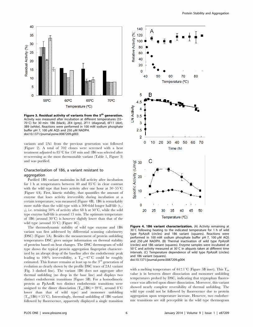
Purified 1B6 variant maintains its full activity after incubation
for 1 h at temperatures between 40 and 85uC in clear contrast
with the wild type that loses activity after one hour at 50–55uC(Figure 4A). First, kinetic stability, that quantifies the amount of
enzyme that loses activity irreversibly during incubation at a
certain temperature, was measured (Figure 4B). 1B6 is remarkably
more stable than the wild type with a 300-fold longer half-life (t1/
2), i.e. retaining 50% of activity after 68 h at 50uC, while the wild
type enzyme half-life is around 13 min. The optimum temperature
of 1B6 (around 30uC) is however slightly lower than that of the
wild type (around 35uC) (Figure 4C).
The thermodynamic stability of wild type enzyme and 1B6
variant was first addressed by differential scanning calorimetry
(DSC) (Figure 5A). Besides the measurement of protein unfolding
temperatures DSC gives unique information on thermal stability
of proteins based on heat changes. The DSC thermogram of wild
type shows the typical protein aggregation fingerprint character-
ized by an abrupt drop of the baseline after the endothermic peak
leading to 100% irreversibility, a Tm,47uC could be roughly
estimated. This feature remains at least up to the 3rd generation of
evolution as clearly shown by the profile DSC trace of 2A1 variant
(Fig. 5 dashed line). The variant 1B6 does not aggregate after
thermal unfolding (no drop in the base line) and displays two
distinct endothermic transitions (Figure 5B). For a homodimeric
protein as PpAzoR two distinct endothermic transitions were
assigned to the dimer dissociation (Tm(1B6) = 39uC, around 8uClower than that of wild type) and monomer unfolding
(Tm(1B6) = 55uC). Interestingly, thermal unfolding of 1B6 variant
followed by fluorescence, apparently displayed a single transition
with a melting temperature of 4461uC (Figure 5B inset). This Tm
value is in between dimer dissociation and monomer unfolding
temperatures probed by DSC, indicating that tryptophan fluores-
cence was affected upon dimer dissociation. Moreover, this variant
showed nearly complete reversibility of thermal unfolding. The
wild type could not be followed by fluorescence due to protein
aggregation upon temperature increase. However, two endother-
mic transitions are still perceptible in the wild type thermogram
Figure 3. Residual activity of variants from the 5th generation.Activity was measured after incubation at different temperatures (55–70uC) for 30 min: 1B6 (black), 2E4 (grey), 2F11 (diagonal), 6F11 (dot),3B9 (white). Reactions were performed in 100 mM sodium phosphatebuffer pH 7, 100 mM AQS and 250 mM NADPH.doi:10.1371/journal.pone.0087209.g003
Figure 4. 1B6 variant characterization. (A) Activity remaining at30uC following heating to the indicated temperature for 1 h of wildtype PpAzoR (circles) and 1B6 variant (squares). Reactions wereperformed in 100 mM sodium phosphate buffer pH 7, 100 mM AQSand 250 mM NADPH. (B) Thermal inactivation of wild type PpAzoR(circles) and 1B6 variant (squares). Enzyme samples were incubated at50uC and activity measured at 30uC in aliquots taken at different timeintervals. (C) Temperature dependence of wild type PpAzoR (circles)and 1B6 variant (squares).doi:10.1371/journal.pone.0087209.g004
Protein Stability and Aggregation
PLOS ONE | www.plosone.org 6 January 2014 | Volume 9 | Issue 1 | e87209

(Figure 5A). Nevertheless, protein aggregation occurring concom-
itantly with monomer unfolding abolished almost completely the
second transition. The inactivity of the monomer was confirmed
by enzymatic assays after its generation by size-exclusion
chromatography in the presence of 0.35 M GdnHCl (data not
shown).
Based on the data discussed above, thermal deactivation of
PpAzoR can be described by the following pathway
N2«2N«2URD (where N2 is the homodimer, N the folded
Figure 5. Differential scanning thermograms. (A) Wild type PpAzoR (thin line), 2A1 variant (dash line) and 1B6 variant (thick line). Arrowsindicate the optimal temperature for activity which occurs in the initial part of the endothermic peak. (B) 1B6 variant fitted by non-two-statetransitions (red line); Inset - Fraction of 1B6 variant unfolded (fUnf) by temperature at pH 7.6 as measured by fluorescence emission. The solid line isthe fit according to the equation fU = e(2DGu/RT)/(1+e (2DGu/RT)).doi:10.1371/journal.pone.0087209.g005
Protein Stability and Aggregation
PLOS ONE | www.plosone.org 7 January 2014 | Volume 9 | Issue 1 | e87209

monomer, U the unfolded monomer) that lead to the irreversible
aggregate state D in the wild type enzyme. The stabilization of
1B6 variant, leading to the large increase in the kinetic stability, is
achieved by preventing the aggregation of the unfolded monomers
(i.e. N2«2N«2U can be proposed for the studied T interval)). A
slight simultaneous decrease of the stability of the dimeric folded
state of the enzyme (lower optimal temperature (Topt) and lower
Tm of the first endothermic peak) as compared with the wild type
was also observed.
Identification and characterization of thermodynamicallyresistant variants
In order to find hit variants with increased thermodynamic
stability and explore further the stability pathways of PpAzoR, we
have measured the optimal temperature of several variants found
in the 1st, 2nd and 3rd generation (Figure 6). An upward shift in the
optimum temperature by approx. 10uC in the hit of the 1st
generation (Topt B1G6 = 40uC), 15uC in the hit of the 2nd
generation (Topt 16B7 = 45uC) and 20uC in one of the hits of the
3rd generation (Topt 2A1 = 50uC) was observed. It is now clear that
the similar thermal stabilities measured for 23C10 and 2A1
(Table 2) arise apparently from different phenomena: in 23C10
(Topt of 35–40uC) from the impeded aggregation of the unfolded
state and in 2A1 from the stabilization of the folded state.
The successive improvements in ‘‘thermostability’’ as indicated
by the increased optimal temperature of hits in the first
generations arises with a simultaneous decrease in activity
(Figure 6), revealing a trade-off between high activity and high
stability [39]. This is particularly evident in 2A1 variant which, in
spite, of the significant up-shift of 20uC of its Topt shows a 2-fold
decrease in the enzymatic activity when compared to the wild type
enzyme. In order to improve its activity the mutation Y179H was
introduced by site-directed mutagenesis since all variants showing
increased activity share this mutation (Figure 2 and Table 1).
Tyr179 is located in the surroundings of the substrate binding site
close to the FMN cofactor and it is likely that the smaller volume
of the histidine residue as compared to tyrosine may account for a
slight enlargement of the cavity, contributing to an increased
activity. Remarkably, the resulting variant 2A1-Y179H indeed
shows an 8-fold higher activity than 2A1 corresponding to a 4-fold
higher specific activity in relation to wild type (Figure 7A). A
compromise between high activity and high stability is also present
in 2A1-Y179H since it has a lower optimal temperature in relation
to variant 2A1; however it still shows a 10uC up-shift in the Topt in
relation to the wild type and 1B6 variant (Figure 7A). Aggregation
impeded probing the unfolding process by fluorescence in 2A1 and
2A1-Y179H variants but static light scattering confirmed that
unlike 1B6 variant, 2A1 variant as wild type aggregate at the
highest temperatures tested (Figure 7B). Furthermore, we observed
only one endothermic peak in the DSC thermogram, suggesting
the simultaneous occurrence of dimer dissociation and monomer
unfolding with a increased melting temperature of around 8uCover that of the wild type (Tm,55uC, Figure 5A). Overall, our
data reveal that although variant 2A1-Y179H unfolds irreversibly
similarly to the wild type, it shows a higher stabilization of the
dimeric folded state with a higher melting temperature (and thus
up-shifted Topt) and 100-fold increased half-life at 50uC (t1/
2 = 23 h; Figure 7C).
Characterization of chemical stability of variantsThe thermodynamic stability of the tertiary structure of
PpAzoR wild type and hit variants upon addition of GdnHCl
was assessed by fluorescence (Figure 8). The wavelengths at the
emission maxima reflect clearly the exposure of tryptophan
residues to the high polarity of water molecules at the surface of
the protein upon unfolding [40]. The wild type PpAzoR displays a
GdnHCl concentration of 1.1 M at the mid-point (where 50% of
the molecules are unfolded) and the native state is more stable than
the unfolded state by 4.2 kcal/mol at 25uC (Table 2). The
unfolding process can be accurately described according to a two-
state process with folded and unfolded states of the monomers
being the only ones that accumulate significantly. Hit variants
follow approximately the same trend upon increased GdnHCl
concentrations, however noteworthy 50% of the 1B6 molecules
unfolded at half of the GdnHCl concentrations required to unfold
the wild type. The native state of 1B6 is more stable than the
unfolded state by only 2.3 kcal/mol. This result shows that
mutations leading to the drastic reduction of the irreversible
aggregation result in an overall folded structure which is less stable
than the wild type enzyme, as also observed previously for thermal
inactivation by DSC (Figure 5). Variant 2A1 is clearly more stable
against chemically induced unfolding displaying the largest free-
energy difference between the folded and the unfolded state
(7.5 kcal/mol). This large free-energy gap may result from a more
restricted conformational freedom (lower entropy) of the unfolded
state due to the introduction of two proline residues (A46P and
particularly A48P which is not present in variant 1B6). Interest-
ingly the cooperativeness of the unfolding transition (m parameter)
also increases very significantly in the variant 2A1. This parameter
is a measure of the degree of solvent exposure for a given
Figure 6. Temperature dependence of PpAzoR wild type andvariants measured in crude cell extracts. Wild type PpAzoR(circles), B1G6 from 1st generation (squares), 16B7 from 2nd generation(triangles), 2A1 from 3rd generation (diamonds) and 23C10 from 3rd
generation (stars). Reactions were performed using crude extracts in100 mM sodium phosphate buffer, pH 7, in the presence of 100 mMAQS and 250 mM NADPH.doi:10.1371/journal.pone.0087209.g006
Table 2. Thermodynamic stability of the tertiary structure ofPpAzoR wild type, 2A1, 2A1-Y179H and 1B6 variants asassessed by fluorescence spectroscopy.
Wt 2A1 2A1-Y179H 1B6
DGwater
(kcal/mol)4.260.4 7.560.7 3.960.3 2.360.2
m (kcal/mol M) 3.360.2 6.960.6 3.960.3 4.260.4
[GdnHCl]50% (M) 1.160.2 1.360.2 1.060.3 0.660.1
doi:10.1371/journal.pone.0087209.t002
Protein Stability and Aggregation
PLOS ONE | www.plosone.org 8 January 2014 | Volume 9 | Issue 1 | e87209

transition; the higher value in 2A1 shows that the level of the
solvent exposure is larger than in the wild type and in the other
variants analysed. Since the conformational freedom of the
unfolded state may be restricted by the introduction of the two
prolines, the larger m value may be rationalized by the higher
degree of compactness of variant 2A1 in the folded state. These
two residues are located in the interface of the two monomers [31]
and the introduction of a proline in position 48 most probably
enhances the homodimer stabilization. The higher exposition to
the solvent (m value) observed in the variant 2A1 may then relate
to an almost simultaneous dimer dissociation and chemical
unfolding while in the other proteins the dissociation of the
homodimer occurs first followed by monomer unfolding in
accordance with DSC data (Figure 5A). The replacement of
Y179 by a histidine residue in this variant, despite the
improvement in activity, results in decreased free energy
differences and cooperativeness of transition (m) to values similar
to wild type.
Molecular details of mutations that improve stabilityPpAzoR (PDB code 4C0W) is a homodimer and its tertiary
structure adopts a flavodoxin-like fold characterized by a central
twisted five parallel b-sheet connected by a-helices, which flank
the sheet from the front and the back [31,41]. The arrangement of
the a-helices and b-stands is identical to structures of azoreduc-
tases from E. coli (PDB code 2Z98) [16] Pseudomonas aeruginosa (PDB
code 2V9C) [20], Enterococcus feacalis (PDB code 2HPV) [17] and
Salmonella typhimurium (PDB code 1T5B). Moreover, it contains the
conserved motif patterns of flavin-dependent azoreductases, i.e.
the sequence involved in the binding of FAD/FMN cofactors, the
sequence involved in the dimerisation of the two monomers of the
enzyme and the possible putative NAD(P)H binding motif [29,31].
All the mutations identified in both the aggregation or
thermodynamically-resistant hit variants are spread on or near
the surface of the protein (Figure 9), underscoring the importance
of protein surface for stability as found in most of the enzymes
showing improved thermostability [42,43]. In addition the
majority of mutations occur in loops in accordance with previous
studies that show that reduction of flexibility of surface loops
generates more stable proteins [44,45]. It is expected that the less
ordered regions, loops and turns, would be more accommodative
to the substitutions while disruption of packing in the core may
interfere with the folding of the protein.
Aggregation-resistant 1B6 hit shares mutations with thermody-
namically-resistant 2A1 variant introduced in the early generations
of directed evolution (Figure 9). The substitution Q192R from the
Figure 7. 2A1 and 2A1-Y179H variants characterization. (A)Temperature dependence of purified wild type PpAzoR (circles), 2A1(diamonds) and 2A1-Y179H (triangles) variants. Reactions were per-formed in 100 mM sodium phosphate buffer, pH 7, in the presence of100 mM AQS and 250 mM NADPH. (B) Light scattering of wild typePpAzoR (circles), 1B6 (squares) and 2A1-Y179H (triangles) variants. (C)Thermal inactivation of wild type PpAzoR (circles) and 2A1-Y179H(triangles) variant; enzyme samples were incubated at 50uC and activitymeasured at 30uC in aliquots taken at different time intervals.doi:10.1371/journal.pone.0087209.g007
Figure 8. Chemical stability. Unfolded fraction (fUnf) of wild typePpAzoR (circles) and 1B6 (squares), 2A1 (diamond) and 2A1-Y179H(triangles) variants by GdnHCl at pH 7.6 as measured by fluorescenceemission. The solid line is the fit according to the equation fU = e(2DGu/
RT)/(1+e (2DGu/RT)).doi:10.1371/journal.pone.0087209.g008
Protein Stability and Aggregation
PLOS ONE | www.plosone.org 9 January 2014 | Volume 9 | Issue 1 | e87209
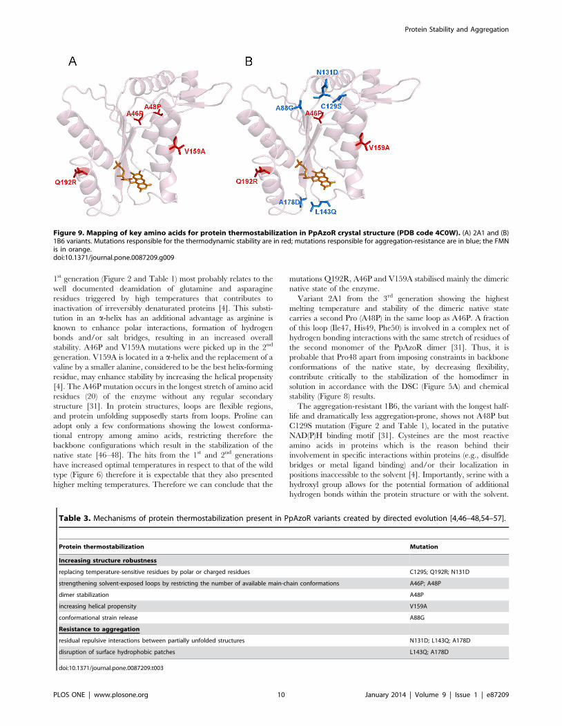
1st generation (Figure 2 and Table 1) most probably relates to the
well documented deamidation of glutamine and asparagine
residues triggered by high temperatures that contributes to
inactivation of irreversibly denaturated proteins [4]. This substi-
tution in an a-helix has an additional advantage as arginine is
known to enhance polar interactions, formation of hydrogen
bonds and/or salt bridges, resulting in an increased overall
stability. A46P and V159A mutations were picked up in the 2nd
generation. V159A is located in a a-helix and the replacement of a
valine by a smaller alanine, considered to be the best helix-forming
residue, may enhance stability by increasing the helical propensity
[4]. The A46P mutation occurs in the longest stretch of amino acid
residues (20) of the enzyme without any regular secondary
structure [31]. In protein structures, loops are flexible regions,
and protein unfolding supposedly starts from loops. Proline can
adopt only a few conformations showing the lowest conforma-
tional entropy among amino acids, restricting therefore the
backbone configurations which result in the stabilization of the
native state [46–48]. The hits from the 1st and 2nd generations
have increased optimal temperatures in respect to that of the wild
type (Figure 6) therefore it is expectable that they also presented
higher melting temperatures. Therefore we can conclude that the
mutations Q192R, A46P and V159A stabilised mainly the dimeric
native state of the enzyme.
Variant 2A1 from the 3rd generation showing the highest
melting temperature and stability of the dimeric native state
carries a second Pro (A48P) in the same loop as A46P. A fraction
of this loop (Ile47, His49, Phe50) is involved in a complex net of
hydrogen bonding interactions with the same stretch of residues of
the second monomer of the PpAzoR dimer [31]. Thus, it is
probable that Pro48 apart from imposing constraints in backbone
conformations of the native state, by decreasing flexibility,
contribute critically to the stabilization of the homodimer in
solution in accordance with the DSC (Figure 5A) and chemical
stability (Figure 8) results.
The aggregation-resistant 1B6, the variant with the longest half-
life and dramatically less aggregation-prone, shows not A48P but
C129S mutation (Figure 2 and Table 1), located in the putative
NAD(P)H binding motif [31]. Cysteines are the most reactive
amino acids in proteins which is the reason behind their
involvement in specific interactions within proteins (e.g., disulfide
bridges or metal ligand binding) and/or their localization in
positions inaccessible to the solvent [4]. Importantly, serine with a
hydroxyl group allows for the potential formation of additional
hydrogen bonds within the protein structure or with the solvent.
Figure 9. Mapping of key amino acids for protein thermostabilization in PpAzoR crystal structure (PDB code 4C0W). (A) 2A1 and (B)1B6 variants. Mutations responsible for the thermodynamic stability are in red; mutations responsible for aggregation-resistance are in blue; the FMNis in orange.doi:10.1371/journal.pone.0087209.g009
Table 3. Mechanisms of protein thermostabilization present in PpAzoR variants created by directed evolution [4,46–48,54–57].
Protein thermostabilization Mutation
Increasing structure robustness
replacing temperature-sensitive residues by polar or charged residues C129S; Q192R; N131D
strengthening solvent-exposed loops by restricting the number of available main-chain conformations A46P; A48P
dimer stabilization A48P
increasing helical propensity V159A
conformational strain release A88G
Resistance to aggregation
residual repulsive interactions between partially unfolded structures N131D; L143Q; A178D
disruption of surface hydrophobic patches L143Q; A178D
doi:10.1371/journal.pone.0087209.t003
Protein Stability and Aggregation
PLOS ONE | www.plosone.org 10 January 2014 | Volume 9 | Issue 1 | e87209

Interestingly, three out of four mutations introduced in the 4th
generation are substitutions to surface charged (N131D, A178D)
or polar (L143Q) residues. Notably, five out of the six hit variants
from the 4th generation have at least one mutation introducing a
charged residue and all variants from the 5th generation have 3 to
4 charged residues in a total of 7 to 8 mutations (Table 1). It has
been proposed that the presence of charged residues on the protein
surface may lead to residual repulsive interactions increasing the
solubility and preventing native state aggregation [49]. The
introduction of the positively charged residue R28 was identified
as the key determinant of aggregation resistance of the unfolded
state of human antibody variable domains created by directed
evolution [50] while A281E and V221D mutations inhibited
protein aggregation and facilitated refolding of the catalytic active
conformation of a Candida antartica lipase [51]. In the case of a
lipase from Bacillus subtilis the simultaneous improvement in
thermodynamic stability and non-aggregating properties was
achieved by the introduction of two proline residues that led to
a better anchoring of loops, disturbing the exposed hydrophobic
patches of 6B lipase and one charged residue, E134 [52].
Moreover, we observe here that mutations L143Q and A178D
in 1B6 variant are close to a hydrophobic patch present on the
protein surface in the surroundings of the FMN molecule,
potentially decreasing the association of exposed hydrophobic
patches and the formation of aggregate compatible structures.
Therefore the combination of the increased surface charge/
polarity with disturbance of hydrophobic patches even though
resulting in a decreased inter-dimer association, i.e. lower stability
of the native state, lead to the abolishment of the aggregational
pathway with a critical gain in the kinetic stability of the enzyme.
In an attempt to create an aggregation and thermodynamically-
resistant variant we have introduced by site-directed mutagenesis
the mutation A48P, present in the 2A1, into the 1B6 variant. In
contrast to the 1B6, the resulting variant 1B6-A48P aggregates
upon temperature raise while maintaining a similar Topt (data not
shown). These results suggest the absence of a synergistic
interaction between A48P mutation and the mutations present
in 1B6 variant, in accordance with our DNA-shuffling results. In
fact, none of the five variants selected in the 5th generation as a
result of the shuffling among variant 2A1 and variants of the 4th
generation, including A48P mutation (Table 1 and Figure 2). Most
probably the surface mutations of 1B6, which are not present in
2A1, and A48P comprise an opposite effect in the stability of the
native state of the variants since the endothermic transitions in the
DSC assigned to dimer dissociation occur at ,55uC for 2A1 and
at 39uC for 1B6 (i.e. 8uC higher and lower, respectively) than the
wild type Tm ,47uC.
Conclusions
Directed evolution is an effective and reliable approach to
engineer proteins contributing simultaneously to our understand-
ing of protein function and adaptation mechanisms. Two types of
protein stability (thermodynamic and kinetic) are of interest from a
fundamental and applied perspective. Increasing the thermody-
namic thermostability is the main issue when an enzyme is used
under denaturing conditions (i.e. high temperatures or organic
solvents) [2]. Industrial applications require active enzymes rather
than enzymes that are in a reversibly inactivated state. For other
enzymes, for example those that can be utilized for diagnostic
puroposes, to ensure a suitable shelf-life, it is often long term
stability that needs to be improved. Most frequently both stabilities
correlate since increasing the enzyme resistance to unfolding state
also increases its resistance to inactivation. Remarkably in the
present study we have identified a hit variant of PpAzoR, 1B6,
with increased resistance to inactivation by aggregation. This was
accomplished by drastically decreasing the rate of the irreversible
step of denaturation (the aggregation of the unfolded monomers)
upon thermal stress. Although variant 1B6 is less stable for thermal
or chemically induced unfolding when compared to the wild type,
lower amounts of the irreversibly denatured state accumulated in
the long-term stability assay. The identification of this hit
correlates well with the experimental screening strategy followed.
In order to select for mutations that improve the stability of the
catalytically competent native state a secondary screen strategy
was implemented based on activity measurements at increasing
temperatures i.e. by probing the dissociation of the dimer.
Importantly our work shows that the resistance of the unfolded
monomers to aggregation (resulting also in a destabilization of the
dimeric folded state) is achieved through a delicate disturbance of
hydrophobic patches and to a clearly increased surface net charge.
On the other hand, the increase of robustness of the native
structure is mainly an outcome of strengthening of solvent-exposed
loops and inter-dimer interactions (Table 3). Overall, this work
suggests that protein charge can be exploited to impart robust
resistance to protein aggregation with implications on de novo
protein design efforts, where unpredictable protein properties,
including aggregation, remain a significant challenge. From the
point of view of protein evolvability mutations that prevent the
aggregation of the unfolded state, increasing thus the level of
soluble protein, contribute to protein fitness [53], and therefore
variant 1B6 is potentially a good candidate to be evolved for new
functions of PpAzoR.
Acknowledgments
The authors would like to thank Dr. Isabel Bento and Dr. Ana Maria
Goncalves for helpful discussions on structural aspects of PpAzoR.
Author Contributions
Conceived and designed the experiments: LOM VB EPM. Performed the
experiments: VB NG. Analyzed the data: LOM VB EPM. Contributed
reagents/materials/analysis tools: LOM. Wrote the paper: LOM VB
EPM.
References
1. Volkin DB, Klibanov AM (1989) Minimizing protein inactivation In: Creight-
onTE, editors. Protein Function. A practical approach. Oxford: IRL press. pp.
1–24.
2. Bommarius AS, Paye MF (2013) Stabilizing biocatalysts. Chem Soc Rev 42:
6534–6565.
3. Sanchez-Ruiz JM (2010) Protein kinetic stability. Biophys Chem 148: 1–15.
4. Vieille C, Zeikus GJ (2001) Hyperthermophilic enzymes: sources, uses, and
molecular mechanisms for thermostability. Microbiol Mol Biol Rev 65: 1–43.
5. Cherry JR, Fidantsef AL (2003) Directed evolution of industrial enzymes: an
update. Curr Opin Biotechnol 14: 438–443.
6. Dalby PA (2011) Strategy and success for the directed evolution of enzymes.
Curr Opin Struct Biol 21: 473–480.
7. Romero PA, Arnold FH (2009) Exploring protein fitness landscapes by directed
evolution. Nat Rev Mol Cell Biol 10: 866–876.
8. Acharya P, Rajakumara E, Sankaranarayanan R, Rao NM (2004) Structural
basis of selection and thermostability of laboratory evolved Bacillus subtilis lipase.
J Mol Biol 341: 1271–1281.
9. Baik SH, Michel F, Aghajari N, Haser R, Harayama S (2005) Cooperative effect
of two surface amino acid mutations (Q252L and E170K) in glucose
dehydrogenase from Bacillus megaterium IWG3 on stabilization of its oligomeric
state. Appl Environ Microbiol 71: 3285–93.
10. Flores H, Ellington AD (2002) Increasing the thermal stability of an oligomeric
protein, beta-glucuronidase. J Mol Biol 315: 325–337.
Protein Stability and Aggregation
PLOS ONE | www.plosone.org 11 January 2014 | Volume 9 | Issue 1 | e87209

11. Garcia-Ruiz E, Mate D, Ballesteros A, Martinez AT, Alcalde M (2010) Evolving
thermostability in mutant libraries of ligninolytic oxidoreductases expressed inyeast. Microb Cell Fact 9: 17.
12. Miyazaki K, Takenouchi M, Kondo H, Noro N, Suzuki M, et al. (2006)
Thermal stabilization of Bacillus subtilis family-11 xylanase by directed evolution.J Biol Chem 281: 10236–10242.
13. Salazar O, Cirino PC, Arnold FH (2003) Thermostabilization of a cytochromep450 peroxygenase. Chembiochem 4: 891–893.
14. Shivange AV, Serwe A, Dennig A, Roccatano D, Haefner S, et al. (2012)
Directed evolution of a highly active Yersinia mollaretii phytase. Appl MicrobiolBiotechnol 95: 405–418.
15. Vazquez-Figueroa E, Yeh V, Broering JM, Chaparro-Riggers JF, BommariusAS (2008) Thermostable variants constructed via the structure-guided consensus
method also show increased stability in salts solutions and homogeneousaqueous-organic media. Protein Eng Des Sel 21: 673–80.
16. Ito K, Nakanishi M, Lee WC, Zhi Y, Sasaki H, et al. (2008) Expansion of
substrate specificity and catalytic mechanism of azoreductase by X-raycrystallography and site-directed mutagenesis. J Biol Chem 283: 13889–13896.
17. Liu ZJ, Chen H, Shaw N, Hopper SL, Chen L, et al. (2007) Crystal structure ofan aerobic FMN-dependent azoreductase (AzoA) from Enterococcus faecalis. Arch
Biochem Biophys 463: 68–77.
18. Maier J, Kandelbauer A, Erlacher A, Cavaco-Paulo A, Gubitz GM (2004) Anew alkali-thermostable azoreductase from Bacillus sp. strain SF. Appl Environ
Microbiol 70: 837–844.19. Suzuki Y, Yoda T, Ruhul A, Sugiura W (2001) Molecular cloning and
characterization of the gene coding for azoreductase from Bacillus sp. OY1-2isolated from soil. J Biol Chem 276: 9059–9065.
20. Wang CJ, Hagemeier C, Rahman N, Lowe E, Noble M, et al. (2007) Molecular
cloning, characterisation and ligand-bound structure of an azoreductase fromPseudomonas aeruginosa. J Mol Biol 373: 1213–1228.
21. Cervantes FJ, Dos Santos AB (2011) Reduction of azo dyes by anaerobicbacteria: microbiological and biochemical aspects. Rev Environ Sci Bio 10: 125–
137.
22. Dos Santos AB, Cervantes FJ, van Lier JB (2007) Review paper on currenttechnologies for decolourisation of textile wastewaters: perspectives for anaerobic
biotechnology. Bioresour Technol 98: 2369–2385.23. Forgacs E, Cserhati T, Oros G (2004) Removal of synthetic dyes from
wastewaters: a review. Environ Int 30: 953–971.24. Deller S, Macheroux P, Sollner S (2008) Flavin-dependent quinone reductases.
Cell Mol Life Sci 65: 141–160.
25. Leelakriangsak M, Huyen NT, Towe S, van Duy N, Becher D, et al. (2008)Regulation of quinone detoxification by the thiol stress sensing DUF24/MarR-
like repressor, YodB in Bacillus subtilis. Mol Microbiol 67: 1108–1124.26. Liu G, Zhou J, Fu QS, Wang J (2009) The Escherichia coli azoreductase AzoR is
involved in resistance to thiol-specific stress caused by electrophilic quinones.
J Bacteriol 191: 6394–6400.27. Sollner S, Deller S, Macheroux P, Palfey BA (2009) Mechanism of flavin
reduction and oxidation in the redox-sensing quinone reductase Lot6p fromSaccharomyces cerevisiae. Biochemistry 48: 8636–8643.
28. Sollner S, Nebauer R, Ehammer H, Prem A, Deller S, et al. (2007) Lot6p fromSaccharomyces cerevisiae is a FMN-dependent reductase with a potential role in
quinone detoxification. Febs J 274: 1328–1339.
29. Mendes S, Pereira L, Batista C, Martins LO (2011) Molecular determinants ofazo reduction activity in the strain Pseudomonas putida MET94. Appl Microbiol
Biotechnol 92: 393–405.30. Mendes S, Farinha A, Ramos CG, Leitao JH, Viegas CA, et al. (2011)
Synergistic action of azoreductase and laccase leads to maximal decolourization
and detoxification of model dye-containing wastewaters. Bioresour Technol 102:9852–9859.
31. Goncalves AMD, Mendes S, de Sanctis D, Martins LO, Bento I (2013) Thecrystal structure of Pseudomonas putida azoreductase: the active site revisited.
FEBS J, 28D: 6643–6657.
32. Joern JM (2003) DNA Shuffling. In: Arnold FH, Georgiou G, editors. Directedevolution library creation: methods and protocols. New Jersey: Humana Press,
Totowa. pp. 85–89.
33. Zhao H, Arnold FH (1997) Optimization of DNA shuffling for high fidelity
recombination. Nucleic Acids Res 25: 1307–1308.34. Martins LO, Soares CM, Pereira MM, Teixeira M, Costa T, et al. (2002)
Molecular and biochemical characterization of a highly stable bacterial laccase
that occurs as a structural component of the Bacillus subtilis endospore coat. J BiolChem 277: 18849–18859.
35. Durao P, Bento I, Fernandes AT, Melo EP, Lindley PF, et al. (2006)Perturbations of the T1 copper site in the CotA laccase from Bacillus subtilis:
structural, biochemical, enzymatic and stability studies. J Biol Inorg Chem 11:
514–526.36. Fernandes AT, Martins LO, Melo EP (2009) The hyperthermophilic nature of
the metallo-oxidase from Aquifex aeolicus. BBA-Proteins Proteom 1794: 75–83.37. Cirino PC, Georgescu R (2003) Screening for thermostability. In: Arnold FH,
Georgiou G, editors. Directed enzyme evolution: screening and selectionmethods. Totowa, New Jersey: Humana Press. pp. 117–125.
38. Salazar O, Sun S (2003) Evaluating a Screen and Analysis of Mutant Libraries.
In: Arnold FH, Georgiou G, editors. Directed enzyme evolution: screening andselection methods. Totowa, New Jersey: Humana Press. pp. 85–97.
39. Jaenicke R, Bohm G (1998) The stability of proteins in extreme environments.Curr Opin Struc Biol 8: 738–748.
40. Lakowicz JR (1999) Principles of Fluorescence Spectroscopy. New York: Kluwer
Academic/Plenum Publishers.41. Correia B, Chen Z, Mendes S, Martins LO, Bento I (2011) Crystallization and
preliminary X-ray diffraction analysis of the azoreductase PpAzoR fromPseudomonas putida MET94. Acta Crystallogr Sect F Struct Biol Cryst Commun
67: 121–123.42. Eijsink VGH, Gaseidnes S, Borchert TV, van den Burg B (2005) Directed
evolution of enzyme stability. Biomol Eng 22: 21–30.
43. Zhao HM, Arnold FH (1999) Directed evolution converts subtilisin E into afunctional equivalent of thermitase. Protein Eng 12: 47–53.
44. Arnold FH, Wintrode PL, Miyazaki K, Gershenson A (2001) How enzymesadapt: lessons from directed evolution. Trends Biochem Sci 26: 100–106.
45. Spiller B, Gershenson A, Arnold FH, Stevens RC (1999) A structural view of
evolutionary divergence. P Natl Acad Sci USA 96: 12305–12310.46. Matthews BW (1993) Structural and Genetic-Analysis of Protein Stability. Annu
Rev Biochem 62: 139–160.47. Watanabe K, Kitamura K, Suzuki Y (1996) Analysis of the critical sites for
protein thermostabilization by proline substitution in oligo-1,6-glucosidase fromBacillus coagulans ATCC 7050 and the evolutionary consideration of proline
residues. Appl Environ Microbiol 62: 2066–2073.
48. Watanabe K, Masuda T, Ohashi H, Mihara H, Suzuki Y (1994) MultipleProline Substitutions Cumulatively Thermostabilize Bacillus cereus Atcc7064
Oligo-1,6-Glucosidase - Irrefragable Proof Supporting the Proline Rule.Eur J Biochem 226: 277–283.
49. Lawrence MS, Phillips KJ, Liu DR (2007) Supercharging proteins can impart
unusual resilience. J Am Chem Soc 129: 10110–10112.50. Famm K, Hansen L, Christ D, Winter G (2008) Thermodynamically stable
aggregation-resistant antibody domains through directed evolution. J Mol Biol376: 926–931.
51. Zhang N, Suen WC, Windsor W, Xiao L, Madison V, et al. (2003) Improvingtolerance of Candida antarctica lipase B towards irreversible thermal inactivation
through directed evolution. Protein Eng 16: 599–605.
52. Kamal MZ, Ahmad S, Molugu TR, Vijayalakshmi A, Deshmukh MV, et al.(2011) In vitro evolved non-aggregating and thermostable lipase: structural and
thermodynamic investigation. J Mol Biol 413: 726–741.53. Tokuriki N, Tawfik DS (2009) Stability effects of mutations and protein
evolvability. Curr Opin Struct Biol 19: 596–604.
54. Eijsink VG, Bjork A, Gaseidnes S, Sirevag R, Synstad B, et al. (2004) Rationalengineering of enzyme stability. J Biotechnol 113: 105–120.
55. Fersht A (1999) Structure and mechanism in protein science: A guide to enzymecatalysis and protein folding. New York: W. H. Freeman and Company.
56. Lee B, Vasmatzis G (1997) Stabilization of protein structures. Curr Opin
Biotechnol 8: 423–428.57. van den Burg B, Eijsink VG (2002) Selection of mutations for increased protein
stability. Curr Opin Biotechnol 13: 333–337.
Protein Stability and Aggregation
PLOS ONE | www.plosone.org 12 January 2014 | Volume 9 | Issue 1 | e87209