Solubility and Structure of Calcium Silicate Hydrate - Civil ...
Upload
khangminh22Category
view
3download
0
1
FABAD J. Pharm. Sci., 46, 1, 1-12, 2021
Improvement in Aqueous Solubility of Cilnidipine by Amorphous Solid Dispersion, Its Formulation into Interpenetrating Polymer Network Microparticles and Optimization by Box-Behnken Design
Amit KUHIKAR* , Shagufta KHAN**° , Komal KHARABE*** , Dilesh SINGHAVI**** ,Girish DAHIKAR*****
RESEARCH ARTICLE
* ORCID: 0000-0001-7353-9814, Institute of Pharmaceutical Education and Research, Borgaon (Meghe) Wardha, Maharashtra, India. ** ORCID:0000-0002-2827-7939, Institute of Pharmaceutical Education and Research, Borgaon (Meghe) Wardha, Maharashtra, India. *** ORCID: 0000-0002-5237-6929, Institute of Pharmaceutical Education and Research, Borgaon (Meghe) Wardha, Maharashtra, India. **** ORCID: 0000-0002-2544-7226, Institute of Pharmaceutical Education and Research, Borgaon (Meghe) Wardha, Maharashtra, India. ***** ORCID: 0000-0002-2284-535X, Institute of Pharmaceutical Education and Research, Borgaon (Meghe) Wardha, Maharashtra, India.
°Corresponding Author: Shagufta Khan, Professor, Phone: (+91)7152-240284, Fax (+91)7152-241684; e-mail: [email protected]
Improvement in Aqueous Solubility of Cilnidipine by Amorphous Solid Dispersion, Its Formulation into Interpenetrating Polymer Network Microparticles and Optimization by Box-Behnken Design
SUMMARY
Cilnidipine(CPN), a Biopharmaceutics Classification System class II drug, has dissolution rate-limited bioavailability and a very short half-life (20.4 min). Thus, there is a need to improve the solubility and prolong the drug release so that the therapeutic concentration of CPN could be maintained for a prolonged time. Therefore, the present investigation was aimed to improve the solubility of CPN by preparing amorphous solid dispersion (ASD) and sustain its release by incorporating CPN loaded ASD (CPNASD) in interpenetrating polymer network (IPN) microparticles. ASD was prepared using Solutol HS 15 and Gelucire®50/13. Solutol HS 15 provided a better effect by increasing 84.09 folds solubility of CPNASD in water as compared to the free CPN, therefore it was used in the formulation of IPN microparticles. Characterization of ASD by differential scanning calorimetry (DSC) and X-ray diffraction (XRD) confirmed a decrease in the crystallinity of CPN. IPN microparticles loaded with CPNASD were prepared by varying chitosan concentrations, polyvinyl alcohol (PVA), and mass-ratio of chitosan:TPP and optimized by Box-Behnken Design. The constraints on the responses were maximum drug entrapment efficiency and sustained drug release with more than 80% drug release in 12 h. IPN microparticles with composition, chitosan 50mg, PVA 74.99mg (Volume of aqueous phase; 10 ml, Volume of organic phase; 50 ml) and chitosan:TPP 2.52 was the predicted optimized condition by the software and IPN with this composition provided high % entrapment efficiency (83.87±0.85) and sustained release (83.29±0.55) for 12 h. Solutol HS 15 was successful in providing a massive increase in solubility of CPN, and a uniform sustained release was achieved by loading CPNASD in IPN microparticles.
Key Words: Cilnidipine, Solid dispersion, Solutol HS 15, Interpenetrating polymer network microparticles, Chitosan, Polyvinyl alcohol.
Received: 15.05.2020Revised: 26.10.2020Accepted: 31.10.2020
Silnidipin’in Amorf Katı Dispersiyonu ile Sulu Çözünürlüğünün İyileştirilmesi, Polimer Ağ Mikropartikülleri ile İçiçe Geçerek Formülasyonu ve Box-Behnken Tasarımıyla Optimizasyonu
ÖZ
Bir Biopharmaceutics Classification System sınıf II ilacı olan Silnidipin (CPN), çözünme oranıyla sınırlı biyoyararlanıma ve çok kısa bir yarılanma ömrüne (20.4 dakika) sahiptir. O yüzden CPN’nin terapötik konsantrasyonunun uzun bir süre korunabilmesi için çözünürlüğün iyileştirilmesine ve ilaç salımının uzatılmasına ihtiyaç duyulmaktadır. Bu nedenle, halihazırdaki araştırma, amorf katı dispersiyon (ASD) hazırlayarak CPN’nin çözünürlüğünü iyileştirmeyi ve CPN yüklü ASD’yi (CPNASD) iç içe geçen polimer ağı (IPN) mikropartiküllerine dahil ederek salımını sürdürmeyi amaçlamaktadır. ASD, Solutol HS 15 ve Gelucire ®50/13 kullanılarak hazırlanmıştır. Solutol HS 15, CPNASD’nin suda çözünürlüğünü serbest CPN’ye kıyasla 84.09 kat artırarak daha iyi etki sağlamıştır, bu nedenle IPN mikropartiküllerinin formülasyonunda kullanılmıştır. ASD’nin diferansiyel taramalı kalorimetri (DSC) ve X-ışını kırınımı (XRD) ile karakterizasyonu CPN’nin kristalliğinde azalmayı doğrulamıştır. CPNASD ile yüklenmiş IPN mikropartikülleri, farklı konsantrasyonlarda kitosan, polivinil alkol (PVA) ve kitosan TPP kütle oranıyla hazırlanmıştır: ve Box-Behnken Tasarımıyla ile optimize edilmiştir. Tasarımda cevaplar, maksimum ilaç tutma etkinliği ve 12 saatte %80’den fazla ilaç salımından daha fazla sürekli ilaç salımı ile sınırlandırılmıştır. Yazılım tarafından öngörülen optimize edilmiş kitosan içeren IPN mikropartiküllerinin bileşimi: kitosan 50mg, PVA 74.99mg (Sulu faz hacmi; 10 ml, Organik faz hacmi; 50 ml) ve kitosan : TPP oranı 2.52’dir. Bu bileşimle IPN, yüksek % enkapsülasyon etkinliği (83.87 ± % 0.85) ve 12 saat boyunca sürekli salım (83.29 ± 0.55) sağlamıştır. Solutol HS 15, CPN’nin çözünürlüğünde büyük bir artış sağlamıştır ve CPNASD’nin IPN mikropartiküllerine yüklenmesiyle üniform bir sürekli salım sağlanmıştır.
Anahtar Kelimeler: Silnidipin, Katı dağılım, Solutol HS 15, İç içe geçen polimer ağ mikropartikülleri, Kitosan, Polivinil alkol.

2
Kuhikar, Khan, Kharabe, Singhavi, Dahikar
INTRODUCTIONDespite sound therapeutic effects, many drugs fail
clinically due to their low water solubility, stability, and bioavailability. There are various techniques for enhancing solubility of poorly water-soluble drugs such as particle size reduction, nanosuspension, use of surfactant, salt formation, pH adjustment, hydrotrophy, and solid dispersion (SD) (Sareen et al, 2012).
SD is one of the most effective approaches to improve the solubility and dissolution rate and hence bioavailability of the poorly water-soluble drugs. They release the drug molecularly in the intestinal fluid, generate a supersaturated solution from which the drug moves to the gut wall, permeate, and finally appear in the blood (Guns et al, 2011). Eloy et al, (2012) formulated SD of ursolic acid using Gelucire®50/13 to increase water solubility and bioavailability of ursolic acid. In the present investigation, for ASD, Solutol HS 15 and PEG-32-glycerylpalmitostearate (Gelucire 50/13) were used. Solutol HS 15 (macrogol 15 hydroxystearate, polyoxyl 15 hydroxystearate) is a nonionic solubilizer and emulsifying agent obtained by reacting 15 moles of ethylene oxide with 1 mole of 12 hydroxy stearic acid.(Ruchatz, 1998). Gelucire®50/13 (PEG-32-glycerylpalmitostearate) is water dispersible and reported as a dissolution enhancer and solubilizing agent. It can self emulsify on contact with aqueous media forming a fine dispersion. Its surface active power improves the solubility and wettability of drugs (Panigrahi et al, 2017).
CPN, a BCS class II drug, has dissolution rate-limited bioavailability, so improvement in solubility and dissolution will increase its bioavailability also (Mishra, 2007). CPN also has a short plasma half-life (20.4min), (Uneyama et al,1999) its conventional dosage forms are unable to control the release of the drug for a prolonged period. Currently, CPN is the most preferred calcium channel blocker among physicians because of its dual blocking characteristics of both L-type voltage-gated Ca2+ channels in vascular smooth muscle and N-type Ca2+ channels in sympathetic nerve terminals that supply blood vessels. CPN is thus considered a better antihypertensive drug than amlodipine (Lee et al, 2014). However, because of its
extremely short half-life, development an appropriate dosage form that can deliver it in a meaningful manner is highly desirable. Among the various oral controlled release drug delivery systems, IPN microparticles have demonstrated their capability to control the drug release for a prolonged time. There are several investigations on IPN microparticles which support their capability to control drug release. Banerjee et al, (2010) reported that the proper control in the amount of crosslinking agent allowed entrapment of as high as 72% diclofenac sodium and the release for an extended period. Solak, (2011) prepared naproxen loaded IPN microparticles of sodium alginate and polyvinyl alcohol for sustained release. As of now, there is no study involving the use of Solutol HS 15 to improve the solubility of CPN and loading CPNASD in IPN microparticles to achieve high drug loading and prolonged drug release. Therefore, the present investigation is aimed to improve the solubility of CPN by preparing ASD and sustain its release by incorporating CPN loaded ASD (CPNASD) in IPN microparticles containing chitosan and PVA.
MATERIALS AND METHODSMaterialsCPN (J. B. Chemicals and Pharmaceuticals Ltd.
Gujrat India), Gelucire®50/13 (Gattefosse India Pvt. Ltd.Mumbai India), Solutol HS 15 (BASF India Pvt, Ltd. Mumbai India), and chitosan (Nitta Gelatin India Pvt. Ltd. Kerala India) were the gift samples. All other chemicals were of analytical grade.
Phase-Solubility StudiesThe phase solubility study was performed
according to the method reported by Higuchi and Connors (Higuchi and Connors K A, 1965). Excess CPN was added to various concentrations of Gelucire®50/13 (0-1%w/v) or Solutol HS 15 (0-1%w/v) in water. The contents were stirred for 48 h at 37± 0.5℃ on a magnetic stirrer. After equilibrium, the samples were filtered and analyzed at 232 nm (UV 2401(PC), S.220V Shimadzu Corporation, Japan). The apparent complexation constant K 1:1 of CPN: Gelucire®50/13 and CPN: Solutol HS 15 was calculated from the slope and intercept of the phase solubility diagrams (Figure not shown), according to the following equation. (Higuchi and Connors, 1965);

3
FABAD J. Pharm. Sci., 46, 1, 1-12, 2021
:KSO Slope
Slope1 1
1=
-^ h (1)
Where K 1:1 = apparent complexation constant and S0 (intercept) = the solubility of CPN in the absence of the complexing agent.
Preparation of SDSD of CPN was prepared by the melting method
(Rajebahadur et al, 2006). Briefly, CPN was added to molten Gelucire®50/13(MP-50℃, Eloy et al, 2012) /Solutol HS 15(MP-25℃, Rajebahadur et al, 2006). The blend was heated 10℃ above the melting point of each carrier for 5 min with stirring. The molten mass was freeze-dried (−20℃ and 0.13 bar ) for 24 h, crushed and passed through a 500 μm sieve.
Saturation Solubility and In- vitro Dissolution of CPN and SD in Different Media
The saturation solubility of CPN and SD was determined in water, acid buffer pH 1.2, and phosphate buffer pH 6.8 at the ambient temperature (25±2℃). Excess amount of CPN/SD was placed in 20 ml medium in the screw-capped glass vial, stirred for 48 h on a magnetic stirrer at 50 strokes per min, the suspension was then filtered and analyzed spectrophotometrically at 232nm, 241nm, and 244nm for CPN in water, acid buffer, and phosphate buffer
respectively. The standard calibration curve of CPN in water and acid buffer pH 1.2 was linear from 1-10 µg/ml. Equations were Y = 0.1666 X - 0.0581; R2 0.989 and Y = 0.1653 X - 0.0851;R2 0.995 for water and acid buffer respectively. The calibration curve of CPN in phosphate buffer was linear in the concentration range of 1-5 µg/ml with R2 0.991 and equation Y = 0.1802 X - 0.1607. LOQ of CPN in water, acid buffer and phosphate buffer were 0.1, 0.3, and 0.03 µg/ml respectively. For analysis of intra-day and inter-day variability, a triplicate of curve was studied, and % RSD was calculated. Percent RSD was less than 5% in each case (Pinto et al, 2017). The dissolution was done in the 500 ml (Crist, 2009) of the same media as solubility using the USP type I apparatus at 50 rpm and 37±0.5℃. 10 mg CPN or SD equivalent to 10 mg were used for the study. A 3 ml sample was withdrawn at the predetermined time intervals and analyzed spectrophotometrically (Table1).
Drug Content of SDFifty milligram SD was dispersed in 50 ml
DMSO, stirred for 24 h at 25±0.5℃ on a magnetic stirrer at 50 strokes/min, filtered, and analyzed spectrophotometrically at 261 nm.
Table 1. Saturation solubility and dissolution of CPN, CPN- Gelucire®50/13SD and CPN- Solutol HS 15 SD in different media
Mean Saturation Solubility of CPN*(mg/ml) at 25 ± 0.5 0C Solubility Enhancement Ratio
Media Free drug Solid dispersion CPN : Gelucire®50/13 (1:1)
Solid dispersion CPN : Solutol HS 15 (1:1)
Solid dispersion CPN:Gelucire®50/13 (1:1)
Solid dispersion CPN : Solutol HS 15 (1:1)
Water 0.002±0.01 0.026±0.01 0.185±0.03 12.22 84.09Acid buffer pH 1.2 0.013±0.02 0.028±0.05 0.319±0.09 02.19 24.54
Phosphate buffer pH 6.8 0.040±0.02 0.088±0.02 0.579±0.06 02.20 14.47
*n=5±S.D
Solid State Characterization of SD by FTIR, DSC and XRD
The potassium bromide disc containing CPN, polymer, and their physical mixture were scanned in the range of 4000 to 400 cm-1 using an FTIR spectrophotometer (Model- 84005, Shimadzu Asia Pacific Pvt. Ltd. Singapore) (Figure 1A). For DSC, 5-10 mg samples sealed in an aluminum pan were scanned at a rate of 10℃/min between 30 to 300℃ (Model -DSC 60, Shimadzu, Tokyo, Japan). A nitrogen
purge (20ml/min) was maintained using an empty sealed pan as reference. Temperature and heat flow calibrations were performed using indium as standard (Figure 1B). X-ray diffraction measurements were done (Smartlab, Rigaku Corporation,Tokyo, Japan) with a copper anode operating under CuK radiation (1.5406 Å, 45kV, and 40mA). Patterns were obtained using a step width of 0.05°/s from 2° to 50° at room temperature on a 2θ scale (Figure 1C).

4
Kuhikar, Khan, Kharabe, Singhavi, Dahikar
Figure 1. A) FTIR of (a) CPN (b) CPN Gelucire®50/13SD. (c) CPN Solutol HS 15 SD (d) IPN microparticles (e) Physical mixture containing CPN, chitosan and PVA (f) Chitosan (g) PVA (h) Blank IPN microsphere B) DSC of (a)
CPN (b) PVA (c) Chitosan (d) CPN- Solutol HS 15 SD (e) Physical mixture containing CPN, chitosan and PVA (f) IPN microparticles g) Blank IPN microsphere C) XRD of (a) CPN (b) IPN microparticles (c) SD of CPN with Solutol HS 15
Preparation of IPN microparticles
SD with Solutol HS 15 gave substantially greater solubility and dissolution rate than Gelucire®50/13, therefore it was used for the preparation of microparticles. Microparticles were prepared by the
emulsion crosslinking method (Bulut et al, 2014). Briefly, to a mixture of chitosan solution (in 1% aq. acetic acid) and polyvinyl alcohol solution (in warm water), SD equivalent to CPN 30 % (w/w) of the

5
FABAD J. Pharm. Sci., 46, 1, 1-12, 2021
polymer was added and after that, the aqueous phase (10 ml) was added drop-wise via 22- gauge hypodermic needle into 50 ml light liquid paraffin containing 1% span 80 and homogenized at 3000 rpm for 20 min. To this, the crosslinker sodium tripolyphosphate solution(STPP) was added with homogenization at 3000 rpm for one hour. The microparticles were separated by centrifugation, washed several times with n-hexane to remove liquid paraffin, and freeze-dried (−20℃ and 0.13 bar for 24 h). Different batches
of IPN microparticles were prepared and optimized using Box Behnken Design (Drug design expert 11 software). The independent variables were the amount of chitosan (low 50 mg and high 100 mg), PVA (low 50 mg and high 100 mg), and chitosan : STPP (low 2.5 and high 3) while, the dependent variables were entrapment efficiency and cumulative percent drug release in 12 h(Table 2). For optimization, constraints on responses were maximum entrapment efficiency, and more than 80% cumulative drug release in 12 h.
Table 2. Composition of IPN microparticles according to Box Behnken Design using Drug Design Expert 11 software.
Run BatchFactor 1Chitosan
(mg)
Factor 2Polyvinyl alcohol
(mg)
Factor 3Chitosan: STPP % EE % Drug Release in 12 h
1 IPN 1 75 75 2.75 64.12±0.50 67.67±0.75
2 IPN 2 100 50 2.75 59.04±0.54 60.63±0.45
3 IPN 3 50 75 3 75.15±0.43 75.04±0.85
4 IPN 4 100 100 2.75 63.73±0.85 65.96±0.95
5 IPN 5 50 100 2.75 73.25±0.65 78.08±0.45
6 IPN 6 50 50 2.75 74.66±0.55 73.42±0.35
7 IPN 7 75 100 2.5 65.88±0.85 67.41±0.55
8 IPN 8 100 75 3 54.02±0.35 64.08±0.65
9 IPN 9 75 50 3 64.10±0.25 74.02±0.25
10 IPN 10 75 50 2.5 66.16±0.65 71.67±0.65
11 IPN 11 100 75 2.5 62.63±0.50 62.66±0.75
12 IPN 12 50 75 2.5 83.87±0.85 83.29±0.55
13 IPN 13 75 100 3 63.95±0.75 62.63±0.89
14 IPN1 75 75 2.75 64.12±0.25 67.67±0.98
15 IPN1 75 75 2.75 64.12±0.35 67.67±0.65
16 IPN1 75 75 2.75 64.12±0.35 67.67±0.75
17 IPN1 75 75 2.75 64.12±0.55 67.67±0.55
Volume of aqueous phase; 10 ml, Volume of organic phase; 50 ml n=3±S.D
Drug Loading Capacity, EE, Particle Size, Solid State Characteristics, Particle Morphology and Water Uptake of Microparticles
Hundred mg microparticles were crushed, soaked
in 100 ml of DMSO for 24 h and after filtration. analyzed at 261 nm. Drug loading capacity and entrapment efficiency (Table 2 and 3) were calculated using the following equation:
Drug Loading Capacity weight of microparticlesamount of drug entrapped in microparticles
100#= (2)
%Entrapment Efficiency Theoretical drug contentActual drug content
100#= (3)
A hundred particles were measured by the Imaging system (Metzger Optical Instrument, no. 666 Mathura, India), and the average was determined (Siepmann et al, 2005) (Table 3). To evaluate the
particle size distribution, polydispersity index (PI) (Table 3) was calculated using the equation;
.. .
PID
D D0 5
0 9 0 1=
-^^^hhh (4)

6
Kuhikar, Khan, Kharabe, Singhavi, Dahikar
Where, D (0.9), D (0.5), and D (0.1) corresponds to particle size immediately above 90%, 50%, and 10% of the sample (Viveksarathi and Kannan, 2015). Solid-state characteristics of microparticles were studied by FTIR, XRD, and DSC in the same way as CPN and SD. For morphology, microparticles were coated with platinum and visualized under a scanning electron
microscope (Model S-3700N, Hitachi, Japan) (Figure 2). For water uptake, microparticles were swollen in the acid buffer pH 1.2 for 2h and phosphate buffer pH 6.8 for 10 h, were blotted off with tissue paper to remove excess water and weighed (Reddy and Nagabhushnam 2017)(Table 3).
Percent water uptake Mass of dry microparticlesMass of swollen microparticles Mass of dry microparticles
100#=- (5)
Table 3. Drug content, Diameter and Water Uptake of IPN microparticles
Batches Drug Loading Capacitya(%)
Mean Diameterb (Micron)
Polydispersity Index
% Equilibrium water uptakea
Acid buffer pH 1.2 (For 2 h
Phosphate buffer pH 6.8 (For 10 h)
IPN 1 11.62±0.35 21.87±4.65 0.52 174.96±0.85 399.49±0.95
IPN 2 10.70±0.25 19.35±3.85 0.48 156.34±0.90 369.81±0.85
IPN 3 13.62±0.35 25.72±3.51 0.45 199.61±0.65 423.00±0.65
IPN 4 11.55±0.45 25.74±5.23 0.55 171.32±1.20 371.13±0.89
IPN 5 13.27±0.25 24.94±3.51 0.43 184.91±1.20 389.31±1.25
IPN 6 13.53±0.30 21.25±2.15 0.37 179.43±1.35 395.09±1.35
IPN 7 11.92±0.40 18.88±3.23 0.47 181.62±0.85 377.87±1.45
IPN 8 09.79±0.26 23.81±4.92 0.51 161.40±0.75 383.56±0.98
IPN 9 11.98±0.30 24.12±3.55 0.46 163.69±0.93 349.63±0.65
IPN 10 11.99±0.75 18.40±3.42 0.44 201.54±1.23 411.41±0.65
IPN 11 11.35±0.43 18.56±3.62 0.49 174.33±1.32 361.08±0.95
IPN 12 15.20±0.25 21.23±4.22 0.51 189.31±1.23 406.91±0.86
IPN 13 11.59±0.27 26.19±3.75 0.48 170.39±1.25 381.43±0.95
a.n=3 ± S. D, b.n=100 ± S. D
In-Vitro Drug Release Drug release was investigated initially for 2h in
the acid buffer pH 1.2 and then10 h in phosphate buffer pH 6.8 using USP Type I apparatus, at 100 rpm. Microparticles, equivalent to 10 mg of CPN were added in 500 ml dissolution medium at 37°C±0.5°C. Samples were withdrawn at a predetermined time and analyzed at 241nm for acid buffer and 244 nm for phosphate buffer, respectively (Figure 6). Drug release profiles were modeled with different kinetic equations like Higuchi, (Higuchi, 1963) first-order and zero-order (Costa et al, 2003) to know the kinetics of drug release and to elucidate the mechanism of drug release, Korsemayer Peppas (Peppas, 1985) equation was used.
Zero-order release equation:.Qt Ko t= (6)
First-order equation:.ln lnQt Q k t0 1= - (7)
Higuchi equation:
.Qt kH t 21= (8)
Qt is the amount of drug released in time t; Q0 is the initial amount of drug; and k0, k1, and kH are the rate constants of the zero-order, first-order, and Higuchi equations, respectively.
Korsemeyer Peppas equation:
MMt ktn
3 = (9)
Mt is the amount of drug released at time t, and M∞ is the amount released at time t = ∞. Thus Mt/M∞ is the fraction of drug released at time t, k is the kinetic constant, and n is the diffusion exponent.

7
FABAD J. Pharm. Sci., 46, 1, 1-12, 2021
Stability StudyStability study was carried out by storing the
microparticles (wrapped in aluminum foil) at 40±5°C and 75 ± 5% RH for 6 months. At an interval of 1 month, the microparticles were visually examined for any physical change, drug loading capacity, particle size, and in-vitro drug release.
RESULTS AND DISCUSSIONPhase Solubility, Saturation Soubility and In-
vitro Dissolution of CPN and SD The phase solubility curve of CPN in presence
of Gelucire®50/13 and Solutol HS 15 showed linear increase in CPN solubility with the concentration of Gelucire®50/13 (AL Type, y = 0.0011x + 0.002, R2= 0.986) and Solutol HS 15 (AL Type, y = 0.002x + 0.0015, R2= 0.995) (Figure not shown). K1:1 of CPN- Solutol HS 15 SD was found to be 2.72 mM, while that of CPN- Gelucire®50/13 was 1.11mM. Solutol HS 15 was more effective at increasing solubility of CPN (84.09 times solubility of free CPN in water) as compared to Gelucire®50/13 (Table 1). The improvement in solubility by Solutol HS 15 was statistically significant with p < 0.05 (two-tailed p= 0.0079, unpaired t-test-Mann- Whitney test). The dissolution of CPN was higher in both acid buffer pH 1.2 and phosphate buffer pH 6.8 from CPN Solutol HS 15 SD. At 4h, there was a 3.27 and 2.79 folds increase in CPN release from SD having Solutol HS 15 in acid and phosphate buffer, respectively, compared to the free CPN.
Solid State Characterization of SD by FTIR, DSC and XRD
FTIR of CPN revealed characteristics peaks at 1566.09 cm-1, 3288.40 cm-1 and 1697.24 cm-1 due to NC group stretching, aromatic stretching, and C=O group stretching respectively. (Figure 1A) The DSC of CPN displayed a sharp endothermic peak at 113.5°C due to its melting. However, the melting peak of the drug was broad with reduced intensity in SD (Figure 1B). XRD of CPN exhibited characteristic, intense
peaks at 2θ of 18.81°, 20.29°, and 24.08° that were absent in SD. (Figure 1C)
Drug Loading Capacity, EE, Particle size, Solid State Characteristics, Particle Morphology and Water Uptake of Microparticles
Drug loading capacity ranged from 9.79 ±0.026 to 15.20 ± 0.27 % while EE was between 54.02 ± 0.12 and 83.87 ± 0.29 %. EE was found dependent on the amount of chitosan, PVA, and chitosan: STPP. The polynomial equation correlating EE with independent variables is;
% EE= 60.30 - 8.44 * A +0 .3563 * B -2.67 * C (10)
Where A and B are the amount of chitosan, and PVA and C is the chitosan:STPP ratio.
The Model F-value of 20.06 and P < 0.0001implied that the model was significant. Effect of chitosan, PVA, and chitosan : STPP on EE is displayed by the response surface diagram (Figure 3Bi). The particle size of microparticles ranged from 18.40±3.42-26.19±3.75 µm and found dependent on the concentration of polymer and crosslinker. PI was found in the range of 0.37-0.55. FTIR spectrum of IPN microparticles showed no change in the chemical structure of CPN as there was no difference in the fingerprint region of the drug. (Figure1A). DSC of IPN microparticles showed a broad endothermic peak at a higher temperature (143.4°C) (Figure1B). The characteristic sharp peaks of CPN had disappeared in the XRD of microparticles (Figure1C). Scanning electron microscopic photographs revealed discrete microparticles with rough surfaces. The microparticles were slightly irregular in shape but mostly uniform in size. (Figure 2a and b). The percent equilibrium water uptake after 12 h was between 349.63±0.65 and 423.0±0.65%. The formulations containing a higher amount of PVA increased the equilibrium water uptake.

8
Kuhikar, Khan, Kharabe, Singhavi, Dahikar
A B
Figure 2. A) Scanning electron micrograph of IPN microparticles B) Image of particles taken by Metzger Optical Instrument at 100X magnification
In-Vitro Drug ReleaseThe cumulative release ranged from 60.63 ±0.04
to 83.29 ± 0.055 % in 12 h (Figure 3A). The variance in drug release was due to differences in the level of PVA /chitosan and chitosan: STPP in formulations. The polynomial equation relating percent drug release with independent variables was found as;
% Cumulative Drug Release=69.25 – 7.06 *A– 0.7075 *B– 1.16 *C (11)
The Model F-value of 11.51 and P<0.0006 indicated that the model was significant. Effect of chitosan, PVA, and chitosan: STPP on drug release is displayed by the response surface diagram (Figure 3Bii). With the increase in the level of PVA, CPN release increased. R2 was maximum for zero-order kinetics for all formulations (ranged from 0.96-0.99) and slope of Koresmeyer Peppas equation was ≥0.85-(ranged from 0.85 to 1.06).
Optimization by Desirability FunctionOptimization was done by desirability function.
CPNASD loaded microparticles prepared under optimized condition (chitosan 50 mg, PVA 74.99 mg (aqueous phase; 10 ml and organic phase; 50 ml) and chitosan:TPP2.52) displayed responses (% EE; 83.87 ±0.85 and % CPN release in 12 h; 83.29±0.55) very
close (low % bais) to predicted responses (% EE; 83.35 and % CPN release in 12 h; 83.05) confirming the reliability of the model.
DISCUSSIONSaturation Soubility and In-vitro Dissolution of
CPN and SD Due to high hydrophilicity, remarkable wetting
ability, and low CMC (0.005 % to 0.02%), Solutol HS 15 has excellent capability to form micelles and entrap drugs. Also, with more homologation series, it creates more hydrogen bonding than Gelucire®50/13, giving greater solubility and dissolution of CPN. High CPN release from SD could be due to loss of crystal structure of CPN in SD (Ádley et al, 2008). Rapid dissolution was because of the molecular dispersion of CPN in SD. Because of the molecular dispersion of CPN in SD, the greater surface area was exposed to the medium (Cid et al, 2019). An increase in the dissolution rate of CPN from SD compared to the plain drug can also be attributed to the amorphization and loss of crystal structure of the drug upon the formation of solid dispersion (Cid et al, 2019).

9
FABAD J. Pharm. Sci., 46, 1, 1-12, 2021
Figure 3. A) In-vitro drug release study of CPN from IPN microparticles (n=3,S.D<±1%)B)Contour and 3D surface graphs showing effect of chitosan, PVA and chitosan:TPP on (i)% entrapment efficiency (ii)%
cumulative drug release
Solid State Characterization of SD by FTIR, DSC and XRD
No change in the characteristic peaks of CPN was noted in the FTIR of SD, which signifies that there was a lack of interaction between CPN and carriers. The broadening of the endothermic peak of CPN in DSC confirms the loss of crystallinity of CPN in SD. The absence of characteristic sharp intense peaks of CPN in XRD further confirms the loss of crystallinity of CPN and it could be inferred that it is dispersed at the molecular level in the solid dispersion.
Drug Loading Capacity, EE, Particle Size, Solid State Characteristics, Morphology and Water Uptake of Microparticles
As the level of chitosan increased, dense
microparticles due to the high crosslinked density of polymer matrix were formed which enabled them to hold a high amount of drug (Sedyakina et al, 2018). At the intermediate level of PVA, EE was high because PVA helped maintain the viscosity and retain the drug, but higher PVA level had a negative impact on EE as very high viscosity reduced access of STPP to chitosan, causing poorly crosslinked, loose network with reduced capacity to hold the drug. STPP level helped to form a tightly networked structure to hold CPN, however with too high STPP (chitosan: STPP,2.5) EE decreased due to the constriction of microparticles and expulsion of drugs (Hou et al, 2015). Increase in the chitosan: STPP above 2.7 increased the particle size of microparticles because of the shortage of

10
Kuhikar, Khan, Kharabe, Singhavi, Dahikar
STPP for crosslinking chitosan, which produced a loose network, while at a lower ratio rigidization of chitosan resulted in shrinking of particles to lower size (Patel and Patel M, 2014). Because of the entrapment of CPN in the microparticles,there, could be hindered melting, so the endothermic peak shifted to a higher temperature in the DSC of microparticles. The broadening of the peak was due to a reduction in crystallization and entrapment of CPN at the molecular level in the microparticles (Sharma, 2010), which is corroborated by the absence of peaks in XRD of microparticles (Karavelidis et al, 2011).
Particles had a rough surface because of the crosslinking of chitosan and drug’s loading (Long et al, 2019). Particles were of uniform size as PI was near 0.5 or less. PI less than 0.5 is deemed homogeneous, while PI > 0.7 is considered to be in a very broad size range (Mudalige et al, 2019). Water uptake capacity increased with PVA because of its high hydrophilicity and excellent water absorption. At the high concentration of chitosan in the microparticles, the swelling was decreased due to the rigid, low water absorbing network formed at the higher chitosan concentration.
In-Vitro Drug ReleaseA high amount of chitosan caused reduced drug
diffusion due to the rigid microsphere network. With the increase in the level of PVA in the IPN microparticles, CPN release increased because of the hydrophilicity of PVA, greater absorption of water, and loose network of microparticles in the presence of a high amount of PVA. However, at the high level of STPP, rigid, compact microparticles were produced, which retarded the drug release (Kaushik et al, 2015). Drug release occurred by diffusion through the swollen matrix and chain relaxation at later time points due to waning crosslinks which aided sufficient drug release at the later time points too. As the slope of the Korsemayer Peppas equation was greater than 0.85 for each formulation batch, a case-II (zero-order) drug-release mechanism was dominant (Sarfaraz et al, 2010).
Stability StudyCPNASD loaded IPN microspheres prepared
under the optimized condition suggested by the experimental model was found to be stable with insignificant change (unpaired t-test, p<0.05) in drug loading capacity and percent drug release when stored under 45±5°C and 75±5% RH for 6 months (Table 4).
Table 4. Drug loading capacity, particle size, and in-vitro drug release of microparticles٭ placed at 40 ± 5°C and 75 ± 5% RH for 6 months stability testing
Time (Months) Drug Loading Capacitya (%) % EEa Mean Diameterb (Micron) % CPN release in 12 ha
0 15.20±0.25 83.87 ±0.85 21.23±4.22 83.29±0.55
1 14.95±0.12 82.50±0.65 21.45±4.50 83.57±0.75
2 15.28±0.23 84.32±1.25 21.35±5.15 83.45±0.95
3 15.45±0.09 85.26±0.45 21.85±5.0 84.15±1.5
4 14.80±0.06 81.67±0.35 21.75±4.55 83.95±1.35
5 15.12±0.17 83.44±0.95 21.50±4.85 83.75±0.75
6 14.98±0.27 83.67±1.5 21.80±4.9 84.05±1.8
a.n=3 ± S. D, b.n=100 ± S. D ٭Microparticles were prepared under optimized condition (chitosan 50 mg, PVA 74.99 mg (aqueous phase; 10 ml and organic phase; 50 ml) and chitosan:TPP2.52).
CONCLUSIONSolutol HS 15 was successful in improving the
solubility of CPN because of which unhindered drug release over a prolonged period was accomplished from the IPN microparticles. Thus CPNASD loaded IPN microparticles could address both the solubility and short biological half-life concern of CPN. As Solutol HS 15 retained CPN within the core of the micelle, there was low seepage of the drug during
microsphere formation due to the crosslinking of chitosan chains, which was the reason for high drug entrapment in microparticles. Else, the expulsion of the drug during crosslinking of polymer and microsphere formation is the common cause for low drug entrapment. Thus, the presented results provided a new possibility for the successful delivery of a highly therapeutically important drug candidate having poor solubility as well as short elimination half-life.

11
FABAD J. Pharm. Sci., 46, 1, 1-12, 2021
CONFLICT OF INTERESTThe authors declare that there are no conflicts of
interest.
AUTHOR CONTRIBUTION STATEMENT
Performed experiments (Kuhikar A.), mentor, hypothesis, design, manuscript preparation, framed discussion of the manuscript (Khan S.), writing manuscript, literature research (Kharabe K.), preparing figures, writing manuscript (Singhavi D.), interpretation of FTIR, discussion confirming formation of interpenetrating polymer network (Dahikar G.)
REFERENCESBanerjee, S., Chaurasia, G., Pal, D.K., Ghosh, A.K.,
Ghosh, A., Kaity, S. (2010) Investigation on cross-linking density for development of novel interpenetrating polymer network (IPN) based formulation. Journal of Scientific and Industrial Research, 69, 777-784.
Bulut, E. (2014). In-vitro evaluation of ibuprofen loaded microparticles prepared from novel chitosan-polyvinyl alcohol interpenetrating polymer network. Polymer Plastic Technology and Engineering, 53, 371-378.
Cid, A.G., Simonazzi, A., Palma, S.D., Bermudez J.M. (2019) Therapeutic Delivery, 10(6),363-381.
Costa, F.O., Sousa, J.J., Pais, A.A., Fornosinho, S.J. (2003) Comparison of dissolution profile of ibuprofen pellets. J. Control. Release, 89, 199-212.
Crist, G.B. (2009). Trends in small-Volume Dissolution Apparatus for Low-Dose Compounds. Dissolution Technologies, 16(1), 19-22.
El-Feky, G.S., El-Rafie, M.H., El-Sheikh, M.A., El-Naggar, M.E., Hebeish, A. (2015). Utilization of Crosslinked Starch Nanoparticles as a Carrier for Indomethacin and Acyclovir Drugs. Journal of Nanomedicine & Nanotechnology, 6(1), 1-8.
Eloy, J., Saraiva, J., Albuquerque, S., Marchetti, J. (2012). Solid Dispersion of Ursolic acid in Gelucire®50/13 : a strategy to enhance drug release and trypanocidal Activity. American Association of Pharmaceutical Scientists PharmSciTech, 13(4), 1436-1445.
Guns, S., Dereymaker, A., Kayaert, P., Mathot, V., Martens, J., Mooter, G. (2011). Comparison between hot-melt extrusion and spray-drying for manufacturing solid dispersions of the graft copolymer of ethylene glycol and vinylalcohol. Pharmaceutical Research, 28(3), 673-682.
Higuchi, T. (1963) Mechanism of sustained action medication theoretical analysis ofrate of release of solid drugs dispersed in solid matrices, Journal of Pharmaceutical Sciences, 52, 1145-1149.
Higuchi, T., Connors, K.A. (1965). Phase solubility techniques. Advanced Analytical Chemistry of Instrumentation, 4, 117-212.
Hou, D.Z., Gui, R.Y., Hu, S., Huang, Y., Feng, Z.Y., Ping, Q.N. (2015). Preparation and Characterization of Novel Drug-Inserted-Montmorillonite Chitosan Carriers for Ocular Drug Delivery. Advances in Nanoparticles, 4, 70- 84.
Karavelidis, V., Karavas, E., Giliopoulos, D. (2011). Papadimitrious S, Bikiaris D. Evaluating the effect of crystallinity in new biocompatible polyester nanocarriers on drug release behavior. International Journal of Nanomedicine, 6, 3021-3032.
Kaushik, A., Tiwari, A., Gaur, A. (2015). Role of excipients and polymeric advancements in preparation of floating drug delivery systems. International Journal of Pharmaceutical Investigation, 5(1).
Lee, J., Lee, H., Jang, K., Lim, K.S., Shin, D., Yu, K.S. (2014). Evaluation of the pharmacokinetic and pharmacodyanamic drug interactions between cilnidipine and valsartan, in healthy volunteers. Drug, Design, Development and Therapy, 8(8), 1781-8. doi:10.2147/DDDT.S68574.
Lima, A.A.N,, Sobrinho, J.L.S., Correa JR R.A.C., Rolim Neto, P.J. (2008). Alternative Technologies to Improve Solubility of Poorly Water Soluble Drugs. Latin American Journal of Pharmacy, 27(5), 789-97.
Long, J., Etxeberria, A E., Kornelsen, C., Nand, A., Ray, S., Bunt, C., Seyfoddin, A. (2019) Development of a long-term drug delivery system with levonorgestrel loaded chitosan microspheres embedded in poly (vinyl alcohol) hydrogel. ACS Applied Materials, 2(7), 2766–2779, DOI: 10.1021/acsabm.9b00190 .

12
Kuhikar, Khan, Kharabe, Singhavi, Dahikar
Mishra, R., Mir, S.R., Amin, S. (2007). Polymeric nanoparticles for improved bioavailability of cilnidipine. International Journal of Pharmacy and Pharmaceutical Sciences, 9(4), 129-139.
Mudalige, T., Qu, H., Haute, D.V., Ansar, S.M., Paredes, A., Ingle, T. (2019). Characterization of Nanomaterials: Tools and Challenges. Nanomaterials for food applications, chapter 11, 313-353.
Panigrahi, K.C., Patra, N., Jena, G.K., Ghose, D., Jena, J., Panda, S.K., Sahu, M., (2017). Gelucire®50/13: A versatile polymer for modified release drug delivery system. Future Journal of Pharmaceutical Sciences, 1-7.
Patel, K., Patel, M. (2014). Preparation and evaluation of chitosan microparticles containing nicorandil. International Journal of Pharmaceutical Investigation, 4(1), 32-37
Peppas, N.A. (1985). Analysis of fickian and non-fickian drug release from polymers. Pharm. Acta. Helv, 60, 110-111.
Pinto, I.C., Cerqueira-Coutinho, C., Freitas, Z.M.F., Santos, E.P., Carmo, F.A., Ricci Junior, E. (2017). Development and validation of an analytical method using High Performance Liquid Chromatography (HPLC) to determine ethyl butylacetylaminopropionate in topical repellent formulations. Brazilian Journal of Pharmaceutical Sciences, 53(2).
Rajebahadur, M., Zia, H., Nues, A. Lee, C. (2006). Mechanistic study of solubility enhancement of nifedipine using vitamin E TPGS and Solutol HS 15. Drug Delivery ,13, 201-206.
Reddy, K.V.M., Nagabhushnam, M.V. (2017). Process and parameters affecting drug release performance of prepared croos-linked alginate hydrogel beads for ezetimibe. International Journal of Pharmacy and Pharmaceutical Sciences, 9(2).
Ruchatz, F., Schuch, H., (1998). Physicochemical properties of Solutol HS 15 and its solublizates, BASF ExAct, 6-7.
Sareen, S., Mathew, G., Joseph, L. (2012). Improvement in solubility of poor water soluble drug by solid dispersion. International Journal of Pharmaceutical Sciences, 2(1), 12-17.
Sarfaraz, M.D., Hiremath, D., Choudhary, K.P.R. (2010). Formulation and characterization of rifampicin microcapsules. International Journal of Pharmaceutical Sciences, 72, 101-105.
Sedyakina, N.E., Feldman, N.B., Lutsenko, S.V., Avramenko, G.V. (2018). Fabrication and drug release behavior of ionically cross-linked chitosan microspheres. Mendeleev Communications, 28, 598–600.
Sharma, A., Jain, C.P. (2010). Preparation and Charecterization of solid dispersions of Cavedilol with PVP K30. Res Pharma Sci, 5(1), 49-56.
Siepmann, J., Elkharraz, K., Siepmann, F., Klose, D., (2005). How Autocatalysis Accelerates Drug Release from PLGA-Based microparticles: A Quantitative Treatment. Biomacromolecules, 6, 2312-2319.
Solak, E.K. (2011). Preparation and characterization of interpenetrating polymeric network microspheres for controlled delivery of naproxen. Journal of Biomaterials and Nanobiotechnology, 2, 445-453.
Uneyama, H., Uchida, H., Konda, T., Yoshimoto, R., Akaike, N. (1999). Selectivity of dihydropyridines for cardiac L-type and sympathetic N-type Ca2+ channels. European Journal of Pharmacology, 373(1), 93-100.
Viveksarathi, K., Kannan, K. (2015). Effect of the moist-heat sterilization on fabricated nanoscale solid lipid particles containing rasagiline mesylate. International Journal of Pharmaceutical Investigation , 5(2), 87–91.

13
FABAD J. Pharm. Sci., 46, 1, 13-22, 2021
Effects of Pycnogenol and Its Combinations with Cisplatin on Hepatocellular Carcinoma Cell Viability
Merve BECİT*,° , Sevtap AYDIN DİLSİZ** , and Nurşen BAŞARAN***
RESEARCH ARTICLE
* ORCİD: 0000-0002-8084-4419, Department of Pharmacology, Faculty of Pharmacy, Ataturk University, Erzurum, 25240, TURKEY** ORCİD: 0000-0002-6368-2745, Department of Pharmaceutical Toxicology, Faculty of Pharmacy, Hacettepe University, Ankara, 06100, TURKEY*** ORCİD: 0000-0001-8581-8933, Department of Pharmaceutical Toxicology, Faculty of Pharmacy, Hacettepe University, Ankara, 06100, TURKEY
° Corresponding Author: Merve BECİTPhone: +904422315241; Fax: +904422315201; e-mail: [email protected]
Effects of Pycnogenol and Its Combinations with Cisplatin on Hepatocellular Carcinoma Cell Viability
SUMMARY
Many challenges of hepatocellular carcinoma treatment, such as side effects and drug resistance, still remain. Therefore, new improvements with high pharmaceutical function and low toxicity are needed. Recently, the efficacy of the combination therapy of antineoplastic drugs with natural products like pycnogenol has garnered attention.Pycnogenol® is a standardized extract from the bark of Pinus pinaster and consists of phenolic compounds. Pycnogenol is considered complementary in the treatments of some cancer besides its good tolerability and high safety. This study aims to reveal the synergistic effects of pycnogenol combination with cisplatin and its relation to human hepatocellular carcinoma (HepG2) cell viability. Effects of single and combined treatment on cell viability were evaluated in Chinese hamster lung fibroblasts (V79) and HepG2 cells using MTT assay. In HepG2 cells, this combined treatment showed a more cytotoxic effect than single-dose groups. Pycnogenol increased the cytotoxicity of cisplatin at 500 μM for 24 h; at 250-500 μM for 48 h in V79 cells; and also, at 125-500 μM for 24 h; at 62.5-500 μM for 48 h in HepG2 cells (p<0.05). Our study is the first study to show that pycnogenol treatment with cisplatin has been combined in the HepG2 cell line. As a result, pycnogenol induced cisplatin cytotoxicity via combined treatment on HepG2 cells and exhibited a synergistic effect with cisplatin. In conclusion, pycnogenol may play a role in the chemotherapy of hepatocellular carcinoma; however, further studies are required to confirm their interactions with cisplatin.
Key Words: Pycnogenol, cisplatin, MTT, cancer, hepatocellular carcinoma, HepG2 cells.
Received: 18.05.2020Revised: 18.08.2020Accepted: 2.09.2020
Piknogenol ve Sisplatin ile Kombinasyonlarının Hepatoselüler Karsinom Hücre Canlılığı Üzerine Etkileri
ÖZ
Hepatoselüler karsinom tedavisinin yan etkiler ve ilaç direnci gibi birçok zorluğu devam etmektedir. Bu nedenle, farmasötik fonksiyonu yükseltecek ve toksisiteyi düşürecek yeni iyileştirmelere ihtiyaç vardır. Son zamanlarda, antineoplastik ilaçların piknogenol gibi doğal ürünlerle kombinasyonel tedavisinin etkinliği dikkat çekmektedir. Pycnogenol®, Pinus pinaster kabuğundan standartlaştırılmış bir ekstrakt olup fenolik bileşiklerden oluşur. Pycnogenolün, iyi tolere edilebilirliği ve yüksek güvenliği yanı sıra bazı kanserlerin tedavisinde de tamamlayıcı olduğu düşünülmektedir. Bu çalışmanın amacı, sisplatin ile piknogenol kombinasyonunun sinerjik etkilerini, bu etkilerin insan hepatosellüler karsinom (HepG2) hücre canlılığı ile nasıl bir ilişki olduğunu ortaya çıkarmaktır. Tek başlarına ve kombine olarak tedavinin hücre canlılığı üzerindeki etkileri, Çin hamsteri akciğer fibroblastlarında (V79) ve HepG2 hücrelerinde MTT kullanılarak değerlendirildi. HepG2 hücrelerindeki bu kombinasyon tedavi, tek doz gruplarından daha fazla sitotoksik etki gösterdi. Piknogenol, sisplatinin sitotoksisitesini V79 hücrelerinde 24 saatlik inkübasyonda 500 μM, 48 saatlik 250-500 μM konsantrasyonda ve ayrıca HepG2 hücrelerinde 24 saat için 125-500 μM, 48 saat için 62.5-500 μM’de anlamlı artırdı (p <0.05). Çalışmamız HepG2 hücre hattında piknogenolün sisplatin ile tedavisinin kombine edildiğini gösteren ilk çalışmadır. Sonuç olarak, Pycnogenol HepG2 hücreleri üzerinde kombine tedavi ile sisplatin sitotoksisitesini indükledi ve sisplatin ile sinerjistik bir etki gösterdi. Sonuç olarak, piknogenol, hepatosellüler karsinomun kemoterapisinde rol oynayabilir; bununla birlikte, sisplatin ile etkileşimlerini doğrulamak için daha ileri çalışmalara ihtiyaç vardır.
Anahtar Kelimeler: Piknogenol, sisplatin, MTT, kanser, hepatoselüler karsinoma, HepG2 hücresi.

14
Becit, Aydın Dilsiz, Başaran
INTRODUCTION
Hepatocellular carcinoma, the most frequently occurring liver neoplasm, is still characterized by an important mortality rate. Even if there have been significant developing new therapeutic alternatives in the treatment of cancer, many challenges, such as side effects and drug resistance, still remain (Grazie et al., 2017). In order to overcome these challenges, new improvements with high pharmaceutical function and low toxicity are needed. Recently, the efficacy of the combination of antineoplastic drugs with natural products like pycnogenol has garnered attention, however knowing how to develop the effect of the combination treatment is of significance (Belcaro et al., 2008; Florea & Büsselberg, 2011; D’Andrea, 2010)
As an effective chemotherapeutic agent, cisplatin has been widely used in the treatments of various malignancies. The cytotoxic properties of cisplatin have not been completely understood. Increasing evidence, however, indicates that cisplatin induces apoptosis through DNA cross-linking and leads to nuclear and mitochondrial DNA damage. Despite its effectiveness, cisplatin often causes significant dose-limiting toxicity, including ototoxicity, neurotoxicity, nephrotoxicity, and cardiotoxicity. Oxidative stress, mitochondrial dysfunction, nuclear and mitochondrial DNA damage, activation of apoptotic pathways, and induction of inflammation have been associated with cisplatin-induced toxicity (Dugbartey et al., 2016). It has been proposed new therapeutic strategies, including combination therapy with antioxidant agents, which could mitigate or prevent these toxicities and improve anticancer effects. However, current studies are inadequate (Cheng et al., 2018).
Pycnogenol® (Horphag Research Ltd) is a standardized extract from the bark of Pinus pinaster. The standardized extract consists of phenolic compounds, divided between monomers (catechin, epicatechin, and taxifolin), condensed flavonoids (classed as procyanidins/proanthocyanidins), and phenolic acids (cinnamic acids and other glycosides) (Rohdewald, 2005; Simpson et al., 2019). As numerous studies report that pycnogenol components are highly bioavailable, pycnogenol is now consumed throughout
the world as a nutritional supplement for various diseases in tablet or capsule forms in doses varying from 20 mg to 100 mg. Due to its multiple phenolic components, the most obvious feature of pycnogenol is its strong antioxidant activity (D’Andrea, 2010; Simpson et al., 2019). It has demonstrated good tolerability with few adverse effects, including gastrointestinal discomfort, dizziness, headache, and nausea. The adverse effects reported in the clinical trials were unrelated to the duration of use or dose of pycnogenol. Furthermore, pycnogenol has a high level of safety (Rohdewald, 2005; Simpson et al., 2019). Based on a recent update by the American Botanical Council of the Scientific and Clinical Monograph for Pycnogenol (The American Botanical Council, 2019), it is reported that an independent panel of toxicology experts has classified pycnogenol as generally recognized as safe (GRAS) based on clinical safety and preclinical toxicology data. Additionally, it is recommended that it should not be taken on children under 6 years of age and during the first 3 months of pregnancy due to limited study (Rohdewald, 2005; Simpson et al., 2019).
Many studies have suggested that it could possibly have anticancer activity in various human carcinoma cell lines by multiple mechanisms: prevention the reactive oxygen species formation, suppression of neoplastic transformation, augmented apoptotic activity (Yang et al., 2016). Up to now, the effect of pycnogenol against hepatocellular carcinoma cells has remained unknown. Also, there are not enough data about the interaction between antineoplastic drugs and pycnogenol. Furthermore, the underlying cellular and molecular processes on cancer are not clear. Therefore, new and advanced studies are needed to clarify the effects of pycnogenol and combinatorial therapy.
The purpose of this study was to evaluate the synergistic effects of cisplatin with the combination of pycnogenol, how this effect was related to cell viability in hepatocellular carcinoma cells. After the incubation with cisplatin and pycnogenol either alone or combination, the cell viability was evaluated in Chinese hamster lung fibroblasts (V79) and human hepatocellular carcinoma (HepG2) cells using

15
FABAD J. Pharm. Sci., 46, 1, 13-22, 2021
3-(4,5-dimethylthiazol-2-yl)-2,5-diphenyltetrazolium bromide (MTT) assay.
MATERIALS AND METHODS
Materials
The chemicals were purchased from the suppliers: cisplatin (Koçak Farma, Turkey); dimethyl sulfoxide (DMSO), Dulbecco’s modified Eagle’s medium (DMEM), ethanol, fetal bovine serum (FBS), MTT, penicillin-streptomycin, trypan blue, trypsin–EDTA, RPMI 1640 medium, Dulbecco’s phosphate-buffered saline (PBS) from Sigma (St. Louis, MO, USA); millipore filters from Millipore (Billerica, MA, USA), all other plastic materials from Corning (Corning Inc., NY, USA). Pycnogenol® was purchased from Horphag Research Ltd., (UK, Geneve, Switzerland). The quality of standardized pycnogenol extract is specified in the United States Pharmacopeia (USP). The stock solution of pycnogenol was freshly prepared in PBS and filtered with Millipore filters (0.20 μm). Negative control experiments were carried out with the culture medium containing only PBS (1%).
Cell culture
V79 and HepG2 cells were provided from the American Type Culture Collection (ATCC; Rockville, MD, USA). V79 cells were grown in RPMI-1640 medium, and HepG2 cells were grown in DMEM containing low glucose (1 g/L) and sodium pyruvate. Both types of media were supplemented with 10% FBS, 2mM L-glutamine, and 1% penicillin-streptomycin solution (10000 units of penicillin and 10 mg of streptomycin in 0.9 % NaCl) at 37°C in 5% CO2 incubators. The cells were subcultured in 75 cm2 cell culture flasks. The culture medium was changed every 3 days. The passage numbers used in our study for both cell lines were between passage 8 and 10.
Determination of cytotoxicity
The effects of pycnogenol and cisplatin and their combination on cell viability were determined by MTT assay (van Meerloo, 2011). According to the cell viability data, IC50 was estimated. The cytotoxic profiles of pycnogenol on the IC50 of cisplatin were evaluated in a wide range of doses in V79 cells and HepG2 cells. Cells were seeded in 96-well plates containing 200 μL medium at a density of 1 × 10⁴
cells/well and incubated to adhere to the plate for 24 h. The cells were treated with cisplatin (0.49-500 μM), pycnogenol (1.95-2000 μM), or the combination at the related culture medium for 24 h and 48 h. At the end of the incubation, 5 mg/mL MTT solution was added to each well and incubated for another 4 h at 37°C in the dark. Then the medium was discarded. The formazan crystals were dissolved in 200 μL of DMSO, and absorbance of each sample was detected at 570 nm using the microplate reader (SpectraMax M2, Molecular Devices Limited, Berkshire, UK). The percentage of cell viability was calculated using the formula: “Percentage of cell viability = (The absorbance of sample/ control) x 100” The cytotoxic concentration that killed cells by 50% (IC50) was determined from absorbance versus concentration curve.
Statistical analysis
All experiments were carried out in quadruplicate. The results were given as the mean ± standard deviation. The statistical analysis was performed with software programs “SPSS 10.5” (Statistical Package for the Social Sciences, Chicago, IL, USA). The distribution of the data was checked for normality using the Kolmogorov-Smirnov test. The means of data were compared by the one-way variance analysis test and post hoc analysis of group differences was performed by the least significant difference (LSD) test. A p values less than 0.05 were considered statistically significant.
RESULTS and DISCUSSION
Cytotoxic effects of pycnogenol and cisplatin in V79 cells and HepG2 cells
Cells were treated with the increasing concentrations of pycnogenol (1.95-2000 μM) and cisplatin (0.49-500 μM) alone for 25 h and 48 h. The results of pycnogenol and cisplatin cytotoxicity are given in Figure 1 and Figure 2, for V79 cells and HepG2 cells, respectively.

16
Becit, Aydın Dilsiz, Başaran
Figure 1. Cytotoxic effects of pycnogenol on the viability of V79 cells for 24 h (A) and 48 h (B). The values were
given as the mean ± standard deviation (n=4). *p < 0.05, compared to negative control (PBS).
Figure 2. Cytotoxic effects of cisplatin on the viability of V79 cells for 24 h (A) and 48 h (B). The values were
given as the mean ± standard deviation (n=4). *p < 0.05, compared to negative control (PBS).
In V79 cells, pycnogenol did not cause a significant cytotoxic effect at the concentrations of 1.95-250 μM and 1.95-62.5 μM when compared to the negative control for 24 h and 48 h incubation, respectively; however, the cell viabilities were significantly decreased above 500 μM and 125 μM of pycnogenol
for 24 h and 48 h incubation, respectively, in a dose-dependent manner (p < 0.05). The IC50 value of pycnogenol was found to be 670 μM and 119 μM for 24 h and 48 h, respectively (Figure 1A and Figure 1B) (Table 1). Cisplatin did not cause a significant cytotoxic effect at the concentrations of 0.49-7.81 μM and at the concentrations of 0.49-1.95 μM when compared to the negative control for 24 h and 48 h, respectively; however, the cell viabilities were significantly decreased above 15.62 μM and 3.91 μM of cisplatin for 24 h and 48 h incubation, respectively, in a dose-dependent manner (p < 0.05). The IC50 value of cisplatin was found to be 15.4 μM and 4.9 μM for 24 h and 48 h, respectively (Figure 2A and Figure 2B) (Table 1).
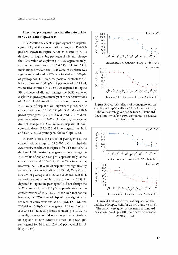
In HepG2 cells, pycnogenol did not cause a significant cytotoxic effect at the concentrations of 1.95-62.5 μM and 1.95-15.6 μM when compared to the negative control for 24 h and 48 h, respectively; however, the cell viabilities were significantly decreased above 125 μM and 31.25 μM of pycnogenol for 24 h and 48 h incubation, respectively, in a dose-dependent manner (p < 0.05). The IC50 value of pycnogenol was found to be 192 μM and 51.5 μM for 24 h and 48 h, respectively (Figure 3A and Figure 3B) (Table 1). Cisplatin did not cause a significant cytotoxic effect at the concentrations of 0.49-7.81 μM and at the concentrations of 0.49-3.91 μM when compared to the negative control for 24 h and 48 h, respectively; however, the cell viabilities were significantly decreased above 15.62 μM and 3.91 μM of cisplatin for 24 h and 48 h incubation, respectively, in a dose-dependent manner (p < 0.05). The IC50 value of cisplatin were found to be 25.5 μM and 7.7 μM for 24 h and 48 h, respectively (Figure 4A and Figure 4B) (Table 1).
Table 1. The IC50 values of pygnogenol and cisplatin in V79 and HepG2 cells
Pygnogenol V79 cells HepG2 cells
IC50 (24 h) 670 μM 192 μM
IC50 (48 h) 119 μM 51.5 μM
Cisplatin V79 cells HepG2 cells
IC50 (24 h) 15.4 μM 25.5 μM
IC50 (48 h) 4.9 μM 7.7 μM
17
FABAD J. Pharm. Sci., 46, 1, 13-22, 2021
Effects of pycnogenol on cisplatin cytotoxicity in V79 cells and HepG2 cells
In V79 cells, the effects of pycnogenol on cisplatin cytotoxicity at the concentrations range of 15.6-500 μM are shown in Figure 5, for 24 h and 48 h. As depicted in Figure 5A, pycnogenol did not change the IC50 value of cisplatin (15 μM, approximately) at the concentrations of 15.6-250 μM for 24 h incubation; however, the IC50 value of cisplatin was significantly reduced in V79 cells treated with 500 μM of pycnogenol (1,75 fold; vs. positive control) for 24 h incubation and 1000 μM (of pycnogenol (4,04 fold; vs. positive control) (p < 0.05). As depicted in Figure 5B, pycnogenol did not change the IC50 value of cisplatin (5 μM, approximately) at the concentrations of 15.6-62.5 μM for 48 h incubation; however, the IC50 value of cisplatin was significantly reduced at concentrations of 125 μM, 250 μM, 500 μM and 1000 μM of pycnogenol (2.26, 2.92, 6.96, and 12.43 fold; vs. positive control) (p < 0.05). As a result, pycnogenol did not change the IC50 value of cisplatin at non-cytotoxic doses (15.6-250 μM pycnogenol for 24 h and 15.6-62.5 μM pycnogenol for 48 h) (p> 0.05).
In HepG2 cells, the effects of pycnogenol at the concentrations range of 15.6-500 μM on cisplatin cytotoxicity are shown in Figure 6, for 24 h and 48 h. As depicted in Figure 6A, pycnogenol did not change the IC50 value of cisplatin (25 μM, approximately) at the concentrations of 15.6-62.5 μM for 24 h incubation; however, the IC50 value of cisplatin was significantly reduced at the concentration of 125 μM, 250 μM, and 500 μM of pycnogenol (1.32 and 2.50 and 4.38 fold; vs. positive control) for 24 h incubation (p < 0.05). As depicted in Figure 6B, pycnogenol did not change the IC50 value of cisplatin (10 μM, approximately) at the concentrations of 15.6-31.25 μM for 48 h incubation; however, the IC50 value of cisplatin was significantly reduced at concentrations of 62.5 μM, 125 μM, and 250 μM and 500 μM of pycnogenol (1.29 and 1.63 and 2.96 and 6.56 fold; vs. positive control) (p < 0.05). As a result, pycnogenol did not change the cytotoxicity of cisplatin at non-cytotoxic doses (15.6-62.5 μM pycnogenol for 24 h and 15.6 μM pycnogenol for 48 h) (p > 0.05).
Figure 3. Cytotoxic effects of pycnogenol on the viability of HepG2 cells for 24 h (A) and 48 h (B).
The values were given as the mean ± standard deviation (n=4). *p < 0.05, compared to negative
control (PBS).
Figure 4. Cytotoxic effects of cisplatin on the viability of HepG2 cells for 24 h (A) and 48 h (B).
The values were given as the mean ± standard deviation (n=4). *p < 0.05, compared to negative
control (PBS).

18
Becit, Aydın Dilsiz, Başaran
Figure 5. Effects of pycnogenol on the cisplatin cytotoxicity in V79 cells for 24 h (A) and 48 h (B). The values were
given as mean ± standard deviation (n=4). *p<0.05, compared to negative control (PBS); #p<0.05, compared to IC50 value of cisplatin (15 μM for 24 h treatment and 5 μM
for 48 h treatment). PYC: pycnogenol; CIS: cisplatin.
Figure 6. Effects of pycnogenol on the cisplatin cytotoxicity in HepG2 cells for 24 h (A) and 48 h (B). The values
were given as mean ± standard deviation (n=4). *p<0.05, compared to negative control (PBS); #p<0.05, compared to IC50 value cisplatin (25 μM for 24 h treatment and 10 μM
for 48 h treatment). PYC: pycnogenol; CIS: cisplatin.
DISCUSSION
Many challenges, such as side effects and drug resistance, still remain in the treatment of hepatocellular carcinoma (Grazie et al., 2017). In order to overcome these challenges, new improvements with high pharmaceutical function and low toxicity are needed. For this reason, the synergistic effects of the combination of antineoplastic drugs with natural products like pycnogenol, which has many different properties besides its good tolerability and high level of safety, has garnered attention. (Belcaro et al., 2008; Florea & Büsselberg, 2011; D’Andrea, 2010; Simpson et al., 2019).
Numerous reports have claimed that pycnogenol could possibly have anticancer activity in various human carcinoma cell lines, including mammary (MCF-7) (Huynh & Teel, 2000), promyeloid leukemia (HL- 60, K562 and U937), (Huang et al., 2005), ovarian (Buz’Zard & Lau, 2007), mucoepidermoid carcinoma (MC-3) (Yang et al., 2014), fibrosarcoma (HT1080) (Harati et al., 2015), oral squamous (HSC-3) (Yang et al., 2016) cancer cells. Harati et al. (2015) investigated the apoptotic effects of pycnogenol and its constituents on human fibrosarcoma cells (HT1080) and also its metabolites from healthy subjects who intake a single dose of 300 mg pycnogenol orally, using flow cytometric analysis and RNA microarray. It was reported that pycnogenol induced apoptosis in HT1080 cells. Therefore, the results provide experimental support for in vivo trials assessing the anticancer effect of pycnogenol. These studies have revealed that pycnogenol acts by multiple mechanisms: prevention the reactive oxygen species formation, suppression of neoplastic transformation, augmented apoptotic activity in a dose-dependent manner (Yang et al., 2016). Yang et al. (2016) also informed that pycnogenol at higher concentrations clearly produced reactive oxygen species, and it may be a pro-oxidant in human oral squamous carcinoma (HSC-3) cells. It also suggests that the pro-oxidant action of pycnogenol might be a critical mechanism of its anticancer potential. These studies may help understand the underlying mechanism of the synergistic cytotoxic effects of combination therapy on cell viability, at least in part. Despite a few studies,

19
FABAD J. Pharm. Sci., 46, 1, 13-22, 2021
little has been discovered relating to the effects of pycnogenol human cancer on cancer yet. Up to now, the effect of pycnogenol against hepatocellular carcinoma cells remains unknown. Considering the importance of oxidative stress in the pathophysiology of cancer, pycnogenol may be a potential candidate for chemoprevention or chemotherapy due to its strong antioxidant activity. It is suggested that a combination consisting of pycnogenol may increase antitumor effects and reduce the toxicity associated with oncologic treatment (Belcaro et al., 2008).
As an effective chemotherapeutic agent, cisplatin has been widely used in the clinical treatments of various malignancies. Increasing evidence indicates that cisplatin induces apoptosis through DNA cross-linking and leads to nuclear and mitochondrial DNA damage. Despite its effectiveness, cisplatin often causes significant dose-limiting toxicity, including ototoxicity, neurotoxicity, nephrotoxicity, and cardiotoxicity against normal cells and tissues. Oxidative stress, mitochondrial dysfunction, nuclear and mitochondrial DNA damage, activation of apoptotic pathways, and induction of inflammation is associated with cisplatin-induced toxicity (Dugbartey et al., 2016). It has been proposed new therapeutic strategies, including combination therapy with antioxidant agents, which could mitigate or prevent these toxicities and improve anticancer effects (Cheng et al., 2018).
The combination of cisplatin with pycnogenol, which targets ROS generation, inflammatory and apoptotic pathways in cisplatin toxicity may offer clinically meaningful (Dugbartey et al., 2016; Florea & Büsselberg, 2011). Numerous researches have reported the ameliorative and preventing effects of pycnogenol in cisplatin-induced toxicity including hepatotoxicity (Ko et al., 2014), ototoxicity (Eryılmaz et al., 2016), acute kidney injury (Lee et al., 2017), optic nerve injury (Icel et al., 2018). However, current studies are inadequate about the interaction between cisplatin and pycnogenol.
In our previous study, we reported that pycnogenol might increase the cisplatin cytotoxicity in human cervix cancer (HeLa) cells at its non-genotoxic doses, which suggests that pycnogenol might contribute to
the anticancer effect of cisplatin in cervical carcinoma (Becit et al., 2020).
In our current study, we aimed to investigate the combined synergistic effects of cisplatin with pycnogenol upon cell viability in HepG2 cells and offer a new approach for hepatocellular carcinoma treatment. After the incubation with cisplatin and pycnogenol either alone or combination, the cell viabilities were evaluated in normal and cancerous cell lines, V79 and HepG2 cells, respectively, using MTT assay.
We determined the effects of pycnogenol (1.95-2000 μM) and cisplatin (0.49-500 μM) on the viabilities of V79 cells and HepG2 cells, for 24 h and 48 h. The results showed that the cytotoxicity profiles of pycnogenol and cisplatin alone were different in terms of exposure time and dose. In V79 cells, the IC50 values of pycnogenol were found to be 670 μM and 119 μM; in HepG2 cells, the IC50 values of pycnogenol were found to be 192 μM and 51.5 μM, for 24h and 48h, respectively. Pycnogenol cytotoxicity (IC50 value) increased approximately ~5.6-fold in V79 cells, ~3.7-fold in HepG2 cells for 48 h when compared to 24 h incubation. Additionally, in HepG2 cells, the pycnogenol cytotoxicity was found to be ~3.5-fold and ~2.3-fold lower, for 24 h and 48 h, respectively, when compared to V79 cells. The IC50 values of cisplatin were found to be 15.4 μM and 4.9 μM for 24 h and 48 h, respectively, in V79 cells; the IC50 values of cisplatin were found to be 25.5 μM and 7.7 μM for 24 h and 48 h, respectively, in HepG2 cells. Cisplatin cytotoxicity increased approximately ~3.1-fold in V79 cells, ~3.3-fold in HepG2 cells for 48 h when compared to 24 h incubation.
The IC50 value of pycnogenol was observed differently in several studies listed. In a study, the IC50 values in HL-60, U937, and K562 were detected as 150 μg/ml (~516.8 μM), 40 μg/ml (~137.8 μM), and 100 μg/ml (~344.5 μM), respectively, for 24 h incubation, by propidium iodide exclusion method. Huang et al. (2005) also reported that pycnogenol inhibited cell proliferation, dose, and time-dependently. It seems that pycnogenol may be hopeful about the treatment and prevention of human leukemia. In another study, the IC50 value was determined to be 285 μg/ml (~982

20
Becit, Aydın Dilsiz, Başaran
μM) for 24 h incubation, in Chinese hamster ovary (CHO) cells, by NRU test (Taner et al., 2013). Yang et al. (2016) reported that the IC50 value was 20 μg/ml (~ 68.9 μM) in HSC-3 cells for 24 h using MTS assay. (Yang et al., 2016).
Similarly, there are studies reporting the IC50 value of cisplatin differently. The IC50 value of cisplatin in the selected human cancer cells was reported to be 14.87 μM and 77.89 μM in hepatocellular carcinoma (Hep-G2 and SK-HEP-1) cells, respectively; 97.20 μM and 85.66 μM in pancreatic cancer (MIA PaCa-2 and BxPC-3) cells, respectively; 54.07 μM and 96.38 μM in cervical cancer (HeLa and Caco-2) cells, respectively; for 24 h incubation, using MTT method (Nurcahyanthi et al., 2016).
The IC50 values obtained from the current studies on pycnogenol cytotoxicity seem to be inconsistent. It is thought that IC50 dose differences between our results and literature results may arise from the cytotoxicity method, exposure time, selected cells, and their properties. However, as shown by our teams and others, pycnogenol has gained increased interest in both the prevention and treatment of various cancers by multiple mechanisms.
After the determination of IC50 values, we investigated the effects of pycnogenol on cisplatin cytotoxicity in V79 cells and HepG2 cells. In V79 cells, pycnogenol significantly reduced the IC50 value of cisplatin (15 μM for 24 h treatment and 5 μM for 48 h treatment) above 500 μM for 24 h and at the concentrations of 125-1000 μM for 48 h(p<0.05), which indicates the synergistic cytotoxic effects of pycnogenol with cisplatin. In HepG2 cells, pycnogenol significantly reduced the IC50 value of cisplatin (25 μM for 24 h treatment and 10 μM for 48 h treatment) at the concentrations of 125-500 μM for 24 h, at the concentrations of 62.5-500 μM for 48 h incubation, respectively (p<0.05). Based on our findings, the combination of cisplatin with pycnogenol showed a synergistic effect by inhibiting cell viability in a time and dose manner. To our knowledge, this study reported for the first time that the combination showed to synergistic cytotoxic effect in HepG2 cells. Our results suggest pycnogenol is a potential
candidate in combination therapy with cisplatin in the treatment of hepatocellular carcinoma.
As consistently, according to a study that shows the synergistic anticancer effect of combination therapy with doxorubicin and grape seed extract in human breast carcinoma (MCF-7 and MDA-MB468) cells; these results suggest promising effects of the combination of antineoplastic drug and phenolic compound for breast cancer treatment (Sharma et al., 2004).
CONCLUSION
In the scope of this study, the combined treatment of cisplatin with pycnogenol in HepG2 cells showed anticancer effect more than single-dose groups. On the basis of our findings, we conclude that pycnogenol may be a promising candidate for chemoprevention or chemotherapy of hepatocellular carcinoma and may develop new therapeutic approaches. Further researches and studies are needed to determine the molecular mechanisms of pycnogenol in tumor cells, the appropriate dose, and treatment methods for this combination. In this regard, our study shows for the first time that combined treatment of cisplatin and pycnogenol in the HepG2 cell line. Hence, it can be a source by providing a basis for further researches.
CONFLICT OF INTEREST
The authors declare that there are no conflicts of interest.
AUTHOR CONTRIBUTION STATEMENT
Idea (Becit M., Aydın Dilsiz S., Başaran N.), design (Aydın Dilsiz S.), supervision (Becit M., Aydın Dilsiz S., Başaran N.), data collection and processing (Becit M., Aydın Dilsiz S.), analysis (Becit M., Aydın Dilsiz S., Başaran N.), data interpretation (Becit M., Aydın Dilsiz S., Başaran N.), literature review (Becit M., Aydın Dilsiz S.), writing article (Becit M., Aydın Dilsiz S., Başaran N.), critical review (Aydın Dilsiz S., Başaran N.), materials (Becit M., Aydın Dilsiz S.), funding (Aydın Dilsiz S.).
REFERENCES
Becit, M., & Aydın, S. (2020) An In Vitro Study on the Interactions of Pycnogenol® with Cisplatin in Human Cervical Cancer Cells. Turkish Journal of Pharmaceutical Sciences, 17(1), 1-6.

21
FABAD J. Pharm. Sci., 46, 1, 13-22, 2021
Belcaro, G., Cesarone, MR., Genovesi, D., Ledda, A., Vinciguerra, G., Ricci, A., & et al. (2008). Pycnogenol may alleviate adverse effects in oncologic treatment. Panminevra Med,50(3), 227-34.
Buz’Zard, AR., & Lau, BHS. (2007). Pycnogenol® reduces Talc-induced Neoplastic Transformation in Human Ovarian Cell Cultures. Phytotherapy Research, 21(6), 579-586.
Cheng, Y., Zhao, P., Wu, S., Yang, T., Chen, Y., Zhang, X., & et al. (2018). Cisplatin and Curcumin Co-loaded Nano-liposomes for the Treatment of Hepatocellular Carcinoma. International Journal of Pharmaceutics, 545(1-2), 261-273.
D’Andrea, G. (2010). Pycnogenol: A blend of procyanidins with multifaceted therapeutic applications? Fitoterapia, 81(7), 724-36.
Dugbartey, GJ., Peppone LJ. & Graaf, IAM.de. (2016). An integrative view of cisplatin-induced renal and cardiac toxicities: molecular mechanisms, current treatment challenges and potential protective measures. Toxicology, 371, 58-66. doi.org/10.1016/j.tox.2016.10.001
Eryilmaz, A., Eliyatkin, N., Demirci, B., Basal, Y., Kurt Omurlu, I., Gunel, C., & et al. (2016). Protective effect of Pycnogenol on cisplatin-induced ototoxicity in rats. Pharmaceutical Biology, 54(11), 2777-2781.
Florea, AM., & Busselberg D. (2011). Cisplatin as an anti-tumor drug: cellular mechanisms of activity, drug resistance and induced side effects. Cancers (Basel), 3(1), 1351-71.
Grazie, ML., Biagini, MR., Tarocchi, M., Polvani, S., & Galli, A. (2017). Chemotherapy for hepatocellular carcinoma: The present and the future. World J Hepatol, 9(21), 907-20.
Harati, K., Slodnik, P., Chromik, AM., Behr, B., Goertz, O., Hirsch, T., & et al. (2015). Pro-apoptotic effects of pycnogenol on HT1080 human fibrosarcoma cells. International Journal of Oncology, 46(4), 1629-36.
Huang, WW., Yang, JS., Lin, CF., Ho, WJ., & Lee, MR. (2005). Pycnogenol induces differentiation and apoptosis in human promyeloid leukemia HL-60 cells. Leukemia Research,29(6),685-92.
Huynh, H.T., & Teel, R.W. (2000), Selective induction of apoptosis in human mammary cancer cells (MCF- 7) by pycnogenol, Anticancer Res, 20 (4), 2417-20.
Icel, E., Uçak, T., Agcayazi, B., Karakurt, Y., Yilmaz, H., Keskin Çimen, F., & et al. (2018). Effects of Pycnogenol on cisplatin-induced optic nerve injury: an experimental study. Cutaneous and Ocular Toxicology, 37(4), 396-400.
Ko, JW., Lee, IC., Park, SH., Moon, C., Kang, SS., Kim, SH., & et al. (2014). Protective effects of pine bark extract against cisplatin-induced hepatotoxicity and oxidative stress in rats. Laboratory Animal Research, 30(4), 174-180.
Lee, IC., Ko, JW., Park, SH., Shin, NR., Shin, IS., Kim, YB., & et al. (2017). Ameliorative effects of pine bark extract on cisplatin-induced acute kidney injury in rats. Ren Fail. 39(1), 363-371.
van Meerloo, J., Kaspers, GJ., & Cloos, J. (2011). Cell sensitivity assays: the MTT assay. Methods in Molecular Biology, 731, 237-45.
Nurcahyanti, AD., & Wink, M. (2016). L-Canavanine potentiates the cytotoxicity of doxorubicin and cisplatin in arginine deprived human cancer cells. J-PEER | Purdue University Press Open Access Journals, 4:e1542.
Rohdewald, P. (2005). “Pycnogenol®, French maritime pine bark extract,” in Encyclopedia of dietary supplements. Eds. P. Coates, M. Blackman, G. Cragg, M. Levine, J. Moss, and J. White (New York: Marcel Dekker), 545–553. doi: 10.1201/b13959-57.
Simpson, T., Kure, C., & Stough, C. (2019). Assessing the Efficacy and Mechanisms of Pycnogenol® on Cognitive Aging From in Vitro Animal and Human Studies. Frontiers in Pharm, 10, 694.
Sharma, G., Tyagi, AK., Singh, RP., Chan, DC., & Agarwal, R. (2004). Synergistic anti-cancer effects of grape seed extract and conventional cytotoxic agent doxorubicin against human breast carcinoma cells. Breast Cancer Research and Treatment, 85(1), 1-12.

22
Becit, Aydın Dilsiz, Başaran
Taner, G., Aydin, S., Aytac, Z., Basaran, AA., & Basaran, N. (2013). Assessment of the cytotoxic, genotoxic and antigenotoxic potential of Pycnogenol in in vitro mammalian cells. Food and Chemical Toxicology, 61, 203-208.
Taner, G., Aydin, S., Bacanli, M., Sarigol, Z., Sahin, T., Basaran, AA., & et al. (2014) Modulating effects of pycnogenol on oxidative stress and DNA damage induced by sepsis in rats. Phytotherapy Research, 28(11), 1692-700.
The American Botanical Council (2019). Scientific and clinical monograph for Pycnogenol (French maritime pine bark extract) Pinus pinaster Aiton subsp. atlantica [Fam. Pinaceae].
Yang, IH., Shin JA., Cho, SD. (2014). Pycnogenol Induces Nuclear Translocation of Apoptosis-inducing Factor and Caspase-independent Apoptosis in MC-3 Human Mucoepidermoid Carcinoma Cell Line. Journal of Cancer Prevention, 19(4):265-72.
Yang, IH., Shin, JA., Kim, LH., Kwon, KH., & Cho, SD. (2016). The caspase 3-dependent apoptotic effect of pycnogenol in human oral squamous cell carcinoma HSC-3 cells. Journal of Clinical Biochemistry and Nutrition, 58(1), 40-7.

23
FABAD J. Pharm. Sci., 46, 1, 23-30, 2021
Bioactivities of A Major Compound from Arthrinium rasikravindrae An Endophytic Fungus of Coleus amboinicus Lour.
Puji ASTUTI°* , Dwi Koko PRATOKO** , Rollando ROLLANDO*** , Giri Wisnu NUGROHO**** , Subagus WAHYUONO***** , Triana HERTIANI****** , Arief NURROCHMAD*******
RESEARCH ARTICLE
* ORCID: 0000-0003-3316-6149, Pharmaceutical Biology Department, Faculty of Pharmacy, Universitas Gadjah Mada, Yogyakarta, Indonesia 55281** ORCID: 0000-0001-7262-4515, Faculty of Pharmacy, Universitas Jember, Jember, Indonesia*** ORCID: 0000-0001-6210-6247, Program of Pharmacy, Faculty of Science and Technology, Ma Chung University, Malang, Indonesia**** ORCID: 0000-0001-9086-3181, Faculty of Pharmacy, Universitas Gadjah Mada, Yogyakarta, Indonesia 55281***** ORCID: 0000-0002-1374-4506, Pharmaceutical Biology Department, Faculty of Pharmacy, Universitas Gadjah Mada, Yogyakarta, Indonesia 55281****** ORCID: 0000-0002-1756-2478, Pharmaceutical Biology Department, Faculty of Pharmacy, Universitas Gadjah Mada, Yogyakarta, Indonesia 55281******* ORCID: 0000-0001-7597-2574, Pharmacology and Clinical Pharmacy Department, Faculty of Pharmacy, Universitas Gadjah Mada, Yogyakarta, Indonesia
°Corresponding author: Puji AstutiPhone/Fax: +62-274-543120; e-mail: [email protected]
Bioactivities of A Major Compound from Arthrinium rasikravindrae An Endophytic Fungus of Coleus amboinicus Lour.
SUMMARY
Many studies reported the ability of endophytic fungi to produce various bioactive compounds having therapeutic values. An endophytic fungus identified as Arthrinium rasikravindrae was isolated from the stem of Coleus amboinicus Lour. This study examined cytotoxic and antimicrobial activities of a major compound isolated from ethyl acetate extract of the fungus fermentation broth. Cytotoxic activities were conducted using MTT assay against T47D, MCF-7, WiDr, 3T3, and Vero cells. IC50 values against Staphylococcus aureus and Escherichia coli were used as the parameters for determining antimicrobial activities. The isolated compound appeared as a single peak in HPLC chromatogram (98.55 %), displayed the highest cytotoxic activity on WiDr cells (IC50 35.03 ± 2.08 µg/mL) and antimicrobial activities against S. aureus (IC50 232.10 ± 1.20 µg/mL) and E. coli (243.59 ± 1.32 µg/mL). Analysis of the UV spectrum and TLC data generated by various detection reagents revealed that the compound was predicted as an N-containing substance having conjugated double bonds.
Key Words: Coleus amboinicus Lour., cytotoxicity, antimicrobial, Arthrinium rasikravindrae, endophyte, fungus.
Received: 29.06.2020Revised: 1.10.2020Accepted: 7.10.2020
Coleus amboinicus Lour’in Endofitik Mantarı Arthrinium rasikravindrae’den Elde Edilen Majör Bileşiğin Biyoaktiviteleri
ÖZ
Birçok çalışma, endofitik mantarların terapötik değerlere sahip çeşitli biyoaktif bileşikler üretme kabiliyetini bildirmiştir. Arthrinium rasikravindrae olarak tanımlanan bir endofitik mantar Coleus amboinicus Lour’in gövdesinden izole edilmiştir. Bu çalışmada, mantar fermantasyon suyunun etil asetat ekstresinden izole edilen ana bileşiğin sitotoksik ve antimikrobiyal aktiviteleri incelenmiştir. Sitotoksik aktivite testleri, T47D, MCF-7, WiDr, 3T3 ve Vero hücre hatlarına karşı MTT deneyi kullanılarak gerçekleştirilmiştir. Antimikrobiyal aktivitelerin belirlenmesinde Staphylococcus aureus ve Escherichia coli’ye karşı IC50 değerleri hesaplanmıştır. İzole edilen bileşik, HPLC kromatogramında (%98,55) tek bir pik vermiştir, WiDr hücreleri üzerinde en yüksek sitotoksik aktiviteye (IC50 35,03 ± 2,08 µg/mL) sahip olduğu belirlenmiş, S. aureus (IC50 232,10 ± 1,20 µg/mL) ve E. coli’ye (243,59 ± 1,32 µg/mL) karşı antimikrobiyal aktivite göstermiştir. Farklı reaktifler ile oluşturulan UV spektrumları ve İTK analizleri yorumlandığında bileşiğin, konjuge çift bağlara sahip N-içeren bir madde olduğu tahmin edilmektedir.
Anahtar Kelimeler: Coleus amboinicus Lour., sitotoksisite, antimikrobiyal, Arthrinium rasikravindrae, endofit, mantar.

24
Astuti, Pratoko, Rollando, Nugroho, Wahyuono, Hertiani, Nurrochmad
INTRODUCTION
Medicinal plants had been explored for many years for their potential sources of bioactive substances such as anti-microbial and anti-cancer agents (Khameneh et al., 2019; Swanepoel et al., 2019). Recently, many explored the potential of endophytic fungi inhabiting living plant tissue, including medicinal plants’ tissues, as a source of bioactive compounds (Hazalin et al., 2009; Ran et al., 2017; Kumari et al., 2018; Leylaie and Zafari, 2018; Senthil Kumar et al., 2019). Coleus amboinicus Lour is a medicinal herb traditionally used as lactagogoe among Batakneese in Indonesia (Damanik et al., 2006). The leaves’ aqueous extract of this plant had been shown to have anti-malarial activities in mice (Periyanayagam et al., 2008), while the essential oils were reported to have antimicrobial activities (Alankararao et al., 1991). The leaves also contained a high concentration of phenolic acids, rosmarinic acid, flavonoids, diterpenes, and linolenic acid (Yanza et al., 2018). Plant-derived phenolic compounds were reported to retard the growth of cancer in vitro, preclinical, and epidemiological studies (Anantharaju et al., 2016; Han et al., 2019). The ability of the fractions of leaves’ ethyl acetate extract of C. amboinicus containing flavonoid in inhibiting the growth of several cancer cell lines was reported (Hasibuan et al., 2019). The plant stem methanolic extract was testified to have antioxidant and antibacterial activities. It also showed antiplatelet aggregation and antiproliferative activities against several cancer cell lines (Bhatt et al., 2013). This study was aimed to explore the potential of an endophytic fungus residing in the stem of C. amboinicus in producing bioactive molecules as anti-microbial and cytotoxic agents against cancer.
MATERIALS AND METHODS
Materials
The pure culture of Arthrinium rasikravindrae was isolated from the stems of C. amboinicus (family Lamiaceae). The plant was collected from Medicinal Plant Garden, Faculty of Pharmacy, Universitas Gadjah Mada. The plant species identification was conducted (by Abdul Razaq Chasani, Ph.D.). A plant herbarium was deposited for future reference in the Pharmaceutical Biology Department, Faculty of Pharmacy, Universitas Gadjah Mada; reference number 014745. The culture was grown on Potato
Dextrose Agar (PDA) plates without antibiotics, maintained for routine culture, and stored in glycerol sock for culture collection at the Department of Pharmaceutical Biology, Faculty of Pharmacy Universitas Gadjah Mada. Potato Dextrose Agar (PDA), Dextrose, Nutrient Agar (NA), Mueller Hinton were purchased from Oxoid. Silica gel F254, Silica gel 60 PF254 containing gypsum, n-hexane, chloroform, ethyl acetate, methanol, acetic acid glacial, DMSO (dimethyl sulfoxide) (Merck). RPMI 1640, Fetal Bovine Serum, Sodium bicarbonate, L-glutamine, Penicillin-Streptomycin, Fungizone(Gibco). MTT (3-(4,5-Dimethylthiazol-2-yl)-2,5-diphenyltetrazolium bromide) (Sigma-Aldrich), HEPES (4-(2-hydroxyethyl)-1-piperazineethanesulfonic acid) (Invitrogen). Equipment: HPLC (Waters 2998), UV-Vis spectrophotometer (Shimadzu).
Isolation and Identification of Endophytic Fungus
Isolation of the endophytic fungus was conducted using the protocol used by Ding et al. (2010) with some modification (Ding et al., 2010). In brief, the stems were washed with running tap water and surface-sterilized using 70% ethanol for 1 minute. After immersion on 5% sodium hypochlorite for 3 minutes, the stems were drained and re-soaked in 70% ethanol for 30 seconds. Following three times washing with sterile distilled water, the stems were surface-dried onto sterile filter paper and cut 1 cm long aseptically. The surface-sterilized stems were placed onto PDA plates containing 30 μg/mL streptomycin. The plates were incubated at 25°C for 2 to 3 days. The hyphal tip of the endophytic fungus that comes out of the stem was moved into new PDA plates containing 30 μg/mL streptomycin and further incubated for 10 – 14 days. The pure culture was obtained by subculturing endophytic fungus on PDA plates without antibiotics. This culture was maintained for the Pharmaceutical Biology Department, Faculty of Pharmacy Universitas Gadjah Mada collection. Identification of endophytic fungus was conducted by PCR amplification using ITS Primer 4: 5`-- TCC TCC GCT TAT TGA TAT GC – 3`, and ITS Primer 5: 5`--GGA AGT AAA AGT CGT AAC AAG G –3`. PCR product was purified and precipitated using Polyethylene Glycol precipitation technique (Hiraishi et al., 1995), continued by cycle sequencing. The result was re-purified by the ethanol purification method. Sequencing was conducted using

25
FABAD J. Pharm. Sci., 46, 1, 23-30, 2021
an automated DNA sequencer (ABI PRISM 3130 Genetic Analyzer-Applied Biosystems). Trimming and assembling the sequenced data was performed using the BioEdit program (http://www.mbio.ncsu.edu/BioEdit/bioedit.htmL) followed by BLAST alignment using genomic data inDDBJ/ DNA Data Bank of Japan (http://blast.ddbj.nig.ac.jp/)or NCBI/ National Center for Biotechnology Information (http://www.ncbi.nlm.nih.gov/BLAST/) to analyze homology/ similarity.
Fermentation and Culture Medium Extraction
The endophytic fungus was inoculated on 500 mL culture vessels containing potato dextrose broth medium. The culture was incubated on a shaker incubator at 25°C for 14 days at 160 rpm. Mycelium was separated from the supernatant using What¬man filter paper, followed by centrifugation at 4000 rpm for 5 min to obtain mycelium free supernatant. The supernatant was extracted using ethyl acetate (ratio 1:1 v/v) three times to obtain ethyl acetate fractions. The solvent was evaporated, resulted in ethyl acetate extract. The components of the extract were compared with the ones of C. amboinicus stem ethyl acetate extract. The mycelia were dried and copped, followed by 24 h maceration using ethyl acetate. The solvent was evaporated to obtain mycelia extract.
Isolation of Bioactive Compound
Ethyl acetate extract was separated using column chromatography [stationary phase = silica gel 60 F254; mobile phase = gradient concentration of chloroform: ethyl acetate]. Fractions having similar TLC profiles were combined and tested for cytotoxic activities. Active fractions were further separated with preparative thin-layer chromatography [stationary phase = silica gel 60 PF254; mobile phase = n-hexane: chloroform: ethyl acetate (1:5:1 v/v). A major positive UV254 compound within the fraction was isolated and tested for its purity using TLC. A single peak on the HPLC profile indicated a pure compound. The predicted chemical group of the compound was established by analyzing TLC profiles and UV data.
Cytotoxic Activity
T47D (human breast cancer cells), MCF-7 (human breast cancer cells), WiDr (human colon cancer cells), 3T3 (mouse embryonic fibroblast cells), and Vero (African green monkey kidney epithelial) cells were cultured in RPMI 1640 containing 10% FBS, 1% penicillin/streptomycin and 1% fungizone. Cultures were cultivated in a CO2 incubator at a temperature of 37°C, 95% relative humidity, and 5% CO2. 5x10³ cells in 100 µL media were inoculated into 96 well plates and incubated for 48 hours to reach 70% – 80% confluent, followed by adding 100 µL of a series concentration of the isolated compound (final concentration of 6.25 – 200 µg/mL for testing on T47D, MCF7, and WiDR cells; 9.37 – 600 µg/mL for testing on 3T3, and Vero cells). Doxorubicin, a conventional chemotherapeutic agent, was used as the positive control, and DMSO was used as solvent control. Doxorubicin at the concentration ranges of 0.31 – 20.0 µg/mL were used for testing on T47D, MCF7, and WiDR cells, while the concentration ranges of 62.50 – 2000 µg/mL were used for testing on 3T3 and Vero cells. The plates were further incubated for 24 hours, followed by gentle washing with 1X PBS. After incubation with 100 µL of 0.5 mg/mL MTT, the plates were incubated for 4 hours at 37oC, and 100 µL of SDS 10% was added to end the reaction. Following overnight incubation, the absorbances were determined using a microplate reader (Biorad) at 595 nm. IC50 values were calculated based on the dose-response curve of cells treated with various concentrations of the isolated compounds.
Antimicrobial Testing
Testing for anti-microbial activity was conducted using a modified microdilution method (Muhammad et al., 2003; da Silva Filho et al., 2008) against Escherichia coli (ATCC 11229) and Staphylococcus aureus (ATCC 25923). Testing microorganisms with densities equal to 0.5 McFarland standard were diluted (1:10) with Mueller Hinton and transferred triplicate into a 96-well plate. Serially diluted samples were transferred into 96 well plates containing testing microorganisms to a final concentration of 2000 to 15.6 µg/mL. Controls of microbial growth, solvent, and media were included in the testing plate. Streptomycin was used as a positive control. The plates were incubated at 37o C overnight and read at 595 nm using a microplate reader (Biorad). IC50, the concentration that inhibited 50% of microbial

26
Astuti, Pratoko, Rollando, Nugroho, Wahyuono, Hertiani, Nurrochmad
growth, were determined from plotting percent growth and concentration of sample tested.
RESULTS AND DISCUSSION
The stem of Coleus amboinicus Lour was reported to contain antioxidant compounds. Its methanolic extract exhibited an antiproliferative effect against several cancer cell lines (Bhatt et al., 2013). The endophytic fungi isolated from C. amboinicus stem resulted in the finding of Arthrinium rasikravindrae, phylum Ascomycota. The same species were found in Brassica capestris as well as bamboo leaves. The same genus was found as phylloplane fungus in chili plants (Rana et al., 2017; Wang et al., 2018).
It has been observed that some endophytes produced bioactive metabolites that may be the same as those of the host plants (Strobel et al., 1996; Pu et al., 2013; Ran et al., 2017; Tan et al., 2018) or different (Gangadevi and Muthumary, 2008; Rajendran et al., 2013; Gill and Vasundhara, 2019). Based on preliminary detection using TLC and UV light, this study found that metabolites within ethyl acetate extract of A. rasikravindrae fermentation broth
were different from those in ethyl acetate extract of the stem of C. amboinicus. Various UV254 positive compounds were observed in broth extracts that were absent in the stem extracts (Figure 1). The major metabolites within broth extract, however, were similar to those within mycelia. The data indicated that the metabolites might be produced in response to their environment as well as their function as fungal protection (Tripathi and Joshi, 2019).
Bioassay-guided isolation of the bioactive compound within the broth ethyl acetate extract resulted in the finding of a compound which exhibited cytotoxic activity against T47D cells. Liquid chromatography analysis of this compound demonstrated a 98.55 % level of purity with a retention time of 3.739 (Figure 2). Further bioactivity screening against several other cancer cell lines showed cytotoxic activity with the lowest IC50 value of 35.03 ± 2.08 µg/mL against WiDR (Table 1). Antibacterial testing of the compound showed an IC50 value of 232.10 ± 1.20 against S. aureus and 243.59 ± 1.32 µg/mL against E. coli as compared to control streptomycin, which exhibited an IC50 value of 0.81 ± 0.02 µg/mL.
Figure 1. TLC profiles of ethyl acetate extract of fermentation broth of A. rasikravindrae endophytic fungus (a) and stem ethyl acetate extract (b) detected with visible light, UV254 nm and UV366 nm light (from left to the right). [stationary phase = silica gel F254; mobile phase = chloroform : ethyl acetate (7:1 v/v)] (A). TLC profiles of fermentation broth ethyl acetate extract (a) and isolated compound (b) detected with UV254 nm [stationary phase = silica gel F254; mobile phase = chloroform : ethyl acetate : acetic glacial acid (15 :5:1 v/v)
(B). TLC profiles of ethyl acetate extract from fermentation broth (a) and mycelia (b) detected with UV254 nm [stationary phase = silica gel F254; mobile phase = chloroform : ethyl acetate (7:1 v/v) (C). A. rasikravindrae
grown on a PDA plate (D).

27
FABAD J. Pharm. Sci., 46, 1, 23-30, 2021
Bio-screening activity indicated that this compound exhibited selective cytotoxicity with the lowest IC50 observed against colon cancer cell line WiDr, followed by breast cancer cell line T47D. Testing against another type of breast cancer cell line MCF7 showed less potency with IC50 above 100 µg/mL. T47D and MCF7 cell lines are both breast cancer cell lines in which they are different in p53 characteristic; T47D is a p53 missense mutant cell line, while MCF7 is a cell line that expresses p53 wild type (Lim et al., 2009). The IC50 value difference between the T47D cell line and MCF7 cell line showed the different mechanisms to induce cell death, which may be p53 independent. Besides, higher IC50 values were observed when the compound was tested on normal cells (3T3 and Vero cell lines), indicating the selectivity of this compound towards cancer cell lines. Antibacterial testing of the compound against S.aureus and E.coli exhibited IC50 values of almost 300 times higher than streptomycin control showing less sensitivity of these microorganisms towards tested compounds (Cos et al., 2006).
Preliminary characterization based on TLC profiles detected using spray reagents showed that the isolated compound positively reacted with all detection reagents except FeCl3 (Figure 2). The absence of colored spots upon FeCl3 detection showed that the compound did not have a phenol group. The positive reaction with 2,4-DNPH, indicating the presence of a carbonyl functional group of aldehydes or ketones (Krupadanam et al., 2001). While cerium (IV) sulfate functioned as a universal reagent for detecting organic compounds, orange-brown positive reaction with Dragendorff suggested that the compound may be a class of alkaloid organic chemical, heterocyclic nitrogen compound or that having quaternary amine. A violet-blue spot upon spraying with vanillin sulphuric acid suggested the presence of terpenoid. (Wagner and Bladt, 1996; Aniszewski, 2015). Ultraviolet spectrum (MeOH) showed ƛ maximum absorption at 222.9 nm (Figure 2) suggested that the compound may have a conjugated double bond system (Singh and Singh, 2018). A further experiment is needed to characterize
the chemical structure of the bioactive compound completely.
Figure 2. Liquid Chromatography profile of isolated compound. System: reverse phase chromatography.
Mobile phase: methanol 100%. Stationary phase: c18. Flow rate: 0.5 ml/minute. λ = 222.9 nm (A).
TLC profiles of isolated compound detected using various spray reagents vanillin-sulphuric acid (a), Cerium (IV) sulphate (b), 2,4-dinitrofenilhidrazin
(2,4-Dinitrophenylhydrazine) (c), FeCl3 (d), Dragendorff (e) (B). [stationary phase = silica gel
F254; mobile phase = chloroform : ethyl acetate : acetic glacial acid (3 :1:0.15 v/v). UV (MeOH) spectrum of
isolated compound (C).
Table 1. IC50 values of the compound against several cell lines. Data represent the mean of IC50 ± SD (n = 3).
Cell lines Compound DoxorubicinIC50 ± SD (µg/mL) IC50 ± SD (µg/mL)
T47DMCF7WiDR3T3Vero
45.87 ± 1.42117.93 ± 0.8735.03 ± 2.08411.97 ± 17.43141.67 ± 8.9
4.86 ± 0.210.95 ± 0.073.19 ± 0.27192.93 ± 4.98297.59 ± 10.1

28
Astuti, Pratoko, Rollando, Nugroho, Wahyuono, Hertiani, Nurrochmad
CONCLUSION
The present study reported the isolation and characterization of a major compound from an endophytic fungus A. rasikravindrae isolated from the stem of C. amboinicus. The compound exhibited moderate cytotoxicity to T47D and WiDr cancer cell lines but less active towards microorganisms tested.
CONFLICT OF INTEREST
The authors declare that there are no conflicts of interest.
ACKNOWLEDGMENTS
The authors thank the Indonesian Ministry of Research and Higher Education, Program PDUPT UGM no 2534/UN1.DITLIT/DIT-LIT/2019 for research funding. Data were partially obtained from Dwi Koko Pratoko’s Master Thesis.
AUTHOR CONTRIBUTION STATEMENT
Concept and experimental design, data acquisition, cytotoxicity study, writing and final approval of manuscript (Astuti P.), isolation of bioactive compound, cytotoxicity experiment, chromatography analysis, writing and final approval of manuscript (Pratoko D.K.), antimicrobial assay study, writing and final approval of manuscript (Rollando R.), TLC analysis, writing and final approval of manuscript (Nugroho G.W.), in isolation of bioactive compound, spectra analysis, writing and final approval of manuscript (Wahyuono S.), bioactivity and spectra analysis, writing and final approval of manuscript (Hertiani T., Nurrocmad A.).
REFERENCES
Alankararao, G.S.J.G., Baby, P., Prasad, Y.R. (1991). Leaf oil of Coleus amboinicus Lour: The in vitro antimicrobial studies. Perfumerie and Kosmetik, 72(11), 744-745.
Anantharaju, P.G., Gowda, P.C., Vimalambike, M.G., Madhunapantula, S.V. (2016). An overview on the role of dietary phenolics for the treatment of cancers. Nutrition Journal, 15(99), 1-16.
Aniszewski, T. (2015). Alkaloids: Chemistry, Biology, Ecology and Application. 2nd Ed. Amsterdam Oxford MA, Elsevier B.V.
Bhatt, P., Joseph, G.S., Negi, P.S., Varadaraj, M.C. (2013). Chemical composition and nutraceutical potential of Indian borage (Plectranthus amboinicus) stem extract. Journal of Chemistry, ID.320329, 1-7.
Cos, P., Vlietinck, A.J., Berghe, D.V., Maes, L. (2016). Anti-infective potential of natural products: How to develop a stronger in vitro ‘proof-of-concept’. Journal of Ethnopharmacology, 106, 290-301.
da Silva Filho, A.A., de Sousa, J.P., Soares, S., Furtado, N.A., Andrade e Silva, M.L., Cunha, W.R., Gregório, L.E., Nanayakkara, N.P., Bastos, J.K. (2008). Antimicrobial activity of the extract and isolated compounds from Baccharis dracunculifolia D. C. (Asteraceae). Zeitschrift für Naturforschung C, 63(1-2), 40-46.
Damanik, R., Wahlqvist, M.L., Wattanapenpaiboon, N. (2006). Lactagogue effects of Torbangun, a Bataknese traditional cuisine. Asia Pacific Journal of Clinical Nutrition, 15(2), 267-274.
Ding T., Jiang T., Zhou J., Xu L., Gao Z.M. (2010). Evaluation of antimicrobial activity of endophytic fungi from Camptotheca acuminata (Nyssaceae). Genetics and Molecular Research, 9(4),2104-2112.
Gangadevi, V., Muthumary, J. (2008). Isolation of Colletotrichum gloeosporioides, a novel endophytic taxol-producing fungus from the leaves of a medicinal plant, Justicia gendarussa. Mycologia Balcanica, 5, 1-4.
Gill, H., Vasundhara, M. (2019). Isolation of taxol producing endophytic fungus Alternaria brassicicola from non-taxus medicinal plant Terminalia arjuna. World Journal of Microbiology and Biotechnology, 35(5), 74.
Han, Y., Ma, L., Zhao, L., Feng, W., Zheng, X. (2019). Rosmarinic inhibits cell proliferation, invasion and migration via up-regulating miR-506 and suppressing MMP2/16 expression in pancreatic cancer. Biomedicine & Pharmacotherapy, 3(115), 108878.
Hasibuan, P.A.Z., Sitorus, P., Satria, D., Sibuea, R.D. (2019). Antioxidant properties and cytotoxic activity of ethyl acetate fraction of Plectranthus amboinicus (Lour.) Spreng. leaves on HeLa and T47D cell lines. Indonesian Journal of Cancer Chemoprevention, 10(1), 37-45.
29
FABAD J. Pharm. Sci., 46, 1, 23-30, 2021
Hazalin, N.A.M.N., Ramasamy, K., Lim, S.M., Wahab, I.A., Anthony, L.J., Cole, A.L.J., Majeed, A.B.A. (2009). Cytotoxic and antibacterial activities of endophytic fungi isolated from plants at the National Park, Pahang, Malaysia. BMC Complementary and Alternative Medicine, 9(46). 1-5
Hiraishi, A., Kamagata, Y., Nakamura, N. (1995). Polymerase chain reaction amplification and restriction fragment length polymorphism analysis of 16S rRNA genes from methanogens. Journal of Fermentation and Bioengineering, 79(6), 523-529.
Khameneh, B., Iranshahy, M., Soheili, V., Bazzaz, B.S.F. (2019). Review on plant antimicrobials: a mechanistic viewpoint. Antimicrobial Resistance & Infection Control, 8(118), 1-28.
Krupadanam G.L.D., Prasad D.V., Rao K.V., Reddy K.L.N., Sudhakar C. (2001). Analytical Chemistry. Hyderabad, Universities Press (India) Private Limited.
Kumari, M., Taritla, S., Sharma, A., Jayabaskaran, C. (2018). Antiproliferative and antioxidative bioactive compounds in extracts of marine-derived endophytic fungus Talaromyces purpureogenus. Frontiers in Microbiology, 9, 1-12.
Leylaie, S., Zafari, D. (2018). Antiproliferative and antimicrobial activities of secondary metabolites and phylogenetic study of endophytic Trichoderma species from Vinca plants. Frontiers in Microbiology, 9, 1-16.
Lim, L.Y., Vidnovic, N., Ellisen, L.W., Leong, C-O. (2009). Mutant p53 mediates survival of breast cancer cells. British Journal of Cancer, 101(9),1606-1612.
Muhammad, I., Li, X.C., Jacob, M.R., Tekwani, B.L., Dunbar, D.C., Ferreira, D. (2003). Antimicrobial and antiparasitic (+)-trans-hexahydrodibenzopyrans and analogues from Machaerium multiflorum. Journal of Natural Products, 66 (6), 804-809.
Periyanayagam, K., Nirmala Devi, K., Suseela, L., Uma, A., Ismail, M. (2008). In vivo antimalarial activity of leaves of Plectranthus amboinicus (lour) Spreng on Plasmodium berghei yoelii. Journal of Communicable Diseases, 40(2), 121.
Pu, X., Qu, X., Chen, F., Bao, J., Zhang, G., Luo, Y. (2013). Camptothecin-producing endophytic fungus Trichoderma atroviride LY357: isolation, identification, and fermentation conditions optimization for camptothecin production. Applied Microbiology and Biotechnology, 97(21), 9365-9375.
Rajendran, L., Rajagopal, K., Subbarayan, K., Ulagappan, K., Sampath, A., Karthik, G. (2013). Efficiency of fungal taxol on human liver carcinoma cell lines. American Journal of Research Communication, 1(6),112-121.
Ran, X., Zhang, G., Li, S., Wang, J. (2017). Characterization and antitumor activity of camptothecin from endophytic fungus Fusarium solani isolated from Camptotheca acuminate, African Health Sciences, 17(2), 566-574.
Rana, S., Singh, P.N., Gaikwad, S.B., Singh, S.K. (2017). Morphology, phylogeny and ex situ conservation of Arthrinium rasikravindrae (Apiosporaceae: Xylariales): a new record from India, KAVAKA, 49, 1-5.
Senthil Kumar, V., Kumaresan, S., Tamizh, M.M., Hairul Islam, M.I., Thirugnanasambantham, K. (2019). Anticancer potential of NF-κB targeting apoptotic molecule “flavipin” isolated from endophytic Chaetomium globosum. Phytomedicine, 61, 152830.
Singh, P.P., Singh, A. (2018). Organic Spectroscopy. New Delhi, Viva Books Private limited.
Strobel, G., Yang, X.S., Sears, J., Kramer, R., Sidhu, R.S., Hess. W.M. (1996). Taxol from Pestalotiopsis microspora, an endophytic fungus of Taxus wallachiana. Microbiology, 142, 435-440.
Swanepoel, B., Venables, L., Olaru, O.T., Nitulescu, G.M., van de Venter, M. (2019). In vitro antiproliferative activity and mechanism of action of Anemone nemorosa. International Journal of Molecular Sciences, 20(5), DOI: 10.3390/ijms20051217.
Tan, X., Zhou, Y., Zhou, X., Xia, X., Wei, Y., He, L., Tang, H., Yu, L. (2018). Diversity and bioactive potential of culturable fungal endophytes of Dysosma versipellis; A Rare Medicinal Plant Endemic to China. Scientific Reports, 8(5929),1-9.

30
Astuti, Pratoko, Rollando, Nugroho, Wahyuono, Hertiani, Nurrochmad
Tripathi, M., Joshi, Y. (2019). Endolichenic fungi: present and future trends., Singapore, Springer Nature Singapore Pte Ltd.
Wagner, H., Bladt, S. (1996). Plant drug analysis: a thin layer chromatography atlas. 2ndEd. Springer Berlin Heidelberg.
Wang, M., Tan, X., Liu, F., Lei, Cai, L. (2018). Eight new Arthrinium species from China. MycoKeys, 34, 1-24.
Yanza, Y.R., Szumacher-Strabel, M., Bryszak, M., Gao, M., Kolodziejski, P., Stochmal, A., Slusarczyk, S., Patra, A.K., Cieslak, A. (2018). Coleus amboinicus (Lour.) leaves as a modulator of ruminal methanogenesis and biohydrogenation in vitro. Journal of Animal Science, 96(11), 4868-4881.

31
FABAD J. Pharm. Sci., 46, 1, 31-42, 2021
Morphological Investigations on the Diagnostic Features of Six Hypericum Species of the Ukrainian Flora
Valentyna MINARCHENKO* , Oksana FUTORNA** , Vitalii PIDCHENKO***° , Iryna TYMCHENKO**** , Tetyana DVIRNA***** , Larysa MAKHYNIA******
RESEARCH ARTICLE
* ORCID: 0000-0002-5049-7620, M.G. Kholodny Institute of Botany, National Academy of Sciences of Ukraine 2 Tereshchenkivska Str., Kyiv 01004, Ukraine; O.O. Bogomolets National Medical University. 22 Pushkinska Str., Kyiv 01004, Ukraine** ORCID: 0000-0002-3713-6644, M.G. Kholodny Institute of Botany, National Academy of Sciences of Ukraine 2 Tereshchenkivska Str., Kyiv 01004, Ukraine *** ORCID: 0000-0003-0850-6666, O.O. Bogomolets National Medical University. 22 Pushkinska Str., Kyiv 01004, Ukraine**** ORCID: 0000-0001-7505-3164, M.G. Kholodny Institute of Botany, National Academy of Sciences of Ukraine 2 Tereshchenkivska Str., Kyiv 01004, Ukraine ***** ORCID: 0000-0002-9279-9766, M.G. Kholodny Institute of Botany, National Academy of Sciences of Ukraine 2 Tereshchenkivska Str., Kyiv 01004, Ukraine; O.O. Bogomolets National Medical University. 22 Pushkinska Str., Kyiv 01004, Ukraine****** ORCID: 0000-0002-8095-4255, O.O. Bogomolets National Medical University. 22 Pushkinska Str., Kyiv 01004, Ukraine
° Corresponding Author: Vitalii PidchenkoPhone: +380937670224; e-mail: [email protected]
Morphological Investigations on the Diagnostic Features of Six Hypericum Species of the Ukrainian Flora
SUMMARY
The results of comparative analysis of the main diagnostic features of the medicinal raw material of six species of the genus Hypericum of Ukraine are presented. The most important diagnostic features of H. alpigenum Kit, H. elegans Steph. ex Willd., H. hirsutum L., H. maculatum Crantz, H. montanum L., and H. perforatum L. are the localization, form, and color of secretory structures. Characteristics of the basic morphological features of leaves, sepals, petals, and stems of the studied species are provided. It is emphasized that a comprehensive analysis of the species-specific peculiarities of the main raw organs of species of the genus Hypericum allows us to determine their species affiliation clearly. The use of these features makes it impossible to intentionally falsify or incorrectly identify the raw material of a particular species of St. John’s worth during merchandising analysis.
Key Words: diagnostic features, Hypericum, medicinal raw materials, leaves, sepals, petals, stems, secretory structures, Ukraine
Received: 28.07.2020Revised: 23.10.2020Accepted: 27.10.2020
Ukrayna Florasının Altı Hypericum Türüne ait Tanısal Özellikleri Üzerine Morfolojik Araştırmalar
ÖZ
Ukrayna’daki Hypericum cinsine ait altı türün ilaç hammaddesi olarak kullanılan kısımlarının temel teşhis özelliklerinin karşılaştırmalı analiz sonuçları sunulmaktadır. H. alpigenum Kit, H. elegans Steph. ex Willd., H. hirsutum L., H. maculatum Crantz, H. montanum L. ve H. perforatum L türlerinin en önemli teşhis özellikleri salgı yapılarının yerleşimi, şekli ve rengidir. İncelenen türlerin yaprak, sepal, petal ve gövdelerinin temel morfolojik özellikleri verilmiştir. Hypericum droglarının türe özgü özelliklerinin kapsamlı bir analizi, türler arasındaki ilişkiyi açıkça belirlemeye imkan vermektedir. Ticari analiz sırasında bu özelliklerin kullanılması, belirli bir kantaron droğunun kasıtlı olarak değiştirilmesini veya yanlış teşhisini önleyebilecektir.
Anahtar Kelimeler: Teşhis özellikleri, Hypericum, ilaç hammaddesi, yaprak, sepal, petal, gövde, salgı yapıları, Ukrayna

32
Minarchenko, Futorna, Pidchenko, Tymchenko, Dvirna, Makhynia
IntroductIon
Species of the genus Hypericum are extensively studied in the world due to their great phytochemical potential, including petrodiantrons (hypericin, pseudohypericin and their derivatives), phloroglucinols (hyperforin and its derivatives), and flavonoids, which contribute to a wide range of therapeutic effects as an antidepressant, anticancer or antiviral, etc. (Cirak et al., 2016; Gitea et al., 2011; Lotocka, Osinska, 2010; Maggi et al., 2004; Nurk, Crockett, 2011). The concentration of these compounds in plant tissues of Hypericum is species-specific and differs between parts of plants (Minarchenko et al., 2020; Onelli et al., 2002; Perrone et al., 2013; Vieira da Silva et al., 2013; Zobayed et al., 2006; Kucharíková et al., 2016).
Special attention is paid to the study of diagnostic morphological and anatomical features of raw parts of species of the genus Hypericum, including secretory structures in which various biologically active substances are localized (Nurk, Blattner, 2010; Sagratini et al., 2008; Perrone et al., 2013; Lotocka, Osinska, 2010; Košuth et al., 2011; Zobayed et al., 2006; Soelberg et al., 2007; Piovan et al., 2004).
H. perforatum, which is included in the pharmacopeias of many countries, is the most studied and widely used. In Ukraine, according to the State Pharmacopeia for raw materials «Hyperici herba,» the use of raw materials of two types of Hypericum is officially allowed: H. perforatum and H. maculatum (the synonym is H. quadrangulum L.) (SPhU 1, 2). According to recent nomenclature data, H. quadrangulum is synonymous with H. tetrapterum Fries (Hassler, 2020), and in this context, it is considered to be synonymous with H. maculatum, however these are two distinct species that have apparent morphological differences.
According to the latest nomenclature (Mosyakin and Fedoronchuk, 1999), 12 species of the genus Hypericum are growing in Ukraine. H. elegans, H. hirsutum, H. montanum, and H. alpigenum are the most common and occur as admixtures to the raw materials of the two species of Hypericum mentioned above.
In the State Pharmacopeia of Ukraine, it is specified that the use of the whole or cut dried flowering tops of Hypericum shoots collected during flowering is allowed (SPhU 1, 2).
Most often, to illustrate the microscopic features of the Hypericum herb, a picture is used that shows only the microscopy of the leaf of some pieces of stems and petals, or fragments of tissue. Microphotographs give a more realistic picture of the diagnostic features at the species level and are widely represented in the scientific literature for certain species of Hypericum (Lotocka, Osinska, 2010).
In our work, we focus on the morphological and anatomical analysis of the main diagnostic characteristics of raw materials (petals, sepals, leaves, and stems) of six species of Hypericum, which may be useful for the authentication of medicinal plant raw materials and their differentiation by species.
MaterIal and Methods
The raw materials (the upper part of flowering shoots – «Herba») of six species of the genus Hypericum, which are widespread in Ukraine: H. alpigenum, Н. elegans, H. hirsutum, Н. maculatum, H. montanum, and H. perforatum were selected for research. Considering the fact, highlighted in the literature (see above) that the main diagnostic morphological characteristics of the raw materials of the studied species are the placement features of secretory structures in different organs of the plant, the main focus of this study is focused on the analysis of these characteristics in sepals, petals, and leaves. Important morpho-anatomical species-specific features of the raw organs of the studied species were also analyzed as an essential component for the identification of their species affiliation.
The research is based on own materials collected during many years of field research by authors in different Ukraine regions, with the additional use of herbarium specimens deposited in the National Herbarium of Ukraine (KW). For the morphological and anatomical analysis of the raw materials, at least ten samples of each species were examined from which fragments of flowers, leaves, and stems

33
FABAD J. Pharm. Sci., 46, 1, 31-42, 2021
of the upper part of the shoot of each specimen were selected; subsequently boiled for 2—5 min and prepared preparations for light microscopy. Microscopic examination was performed ten times for each organ from different parts of the sample. Observations and microphotographs were made using an Olympus CX23 light microscope, a Philip Harris stereomicroscope, and Levenhuk M1000 PLUS camera software.
results and dIscussIon
The studied species are characterized by common macromorphological features, such as the flower and stem structure, the structure of the leaf blade, leaf placement, etc. At the same time, each of them has different specific characteristics of the mentioned above organs.
Also, the most important diagnostic feature of raw materials of Hypericum species is the localization, shape, and color of secretory structures that concentrate valuable secondary metabolites.
Secondary metabolites of Hypericum species (so-called biomarkers) mainly belong to xanthones, petrodianthrones (hypericin and pseudohypericin), derivatives of phloroglucinol (hyperforin), biflavones (I3, II8-biapigenin, amentoflavone, amentoflavonoid), and flavonoid glycosides (rutin, hyperoxide, isoquercitrin, quercitrin, quercetin), which are localized and are often synthesized in a variety of secretory structures mainly of leaves, sepals and petals (Ciccarelli et al., 2007; Lotocka, Osinska, 2010; Vieira da Silva et al., 2013). The localization of secretory structures, their shape, and color of content have species-specific character and differ significantly in the tissues of different organs. Therefore, the level of biologically active substances in a particular tissue of Hypericum depends on the relative amount of secretory structures in the raw materials. Thus, the organ dependence of secondary metabolites is a characteristic feature of species of the genus Hypericum (Cirak et al., 2016). Most secretory structures in Hypericum species plants are concentrated in the perianth (sepals and petals) and leaves, so the quality of the raw material significantly depends on the proportion of these organs in the total
mass of raw materials.
The most studied in this aspect is H. perforatum (Ciccarelli et al., 2007; Ciccarelli et al., 2001b; Vieira da Silva et al., 2013 with references); however, there are fragmentary studies of other species, such as: H. tetrapterum (Gitea et al., 2018), H. elodes (Vieira da Silva et al., 2013), H. elegans (Lotocka, Osinska, 2010), etc. There is no uniform terminology for the names of secretory structures of Hypericum species. In most studies, they are divided into so-called «dark» (black nodules) and «yellow» (yellow glands, рaleglands, translucent glands or dots) and secretory canals (Gitea et al., 2018; Maggi et al., 2004; Nurk, Crockett, 2011), etc. These terms are mainly used to describe the internal secretory structures of leaves and other organs. As for the names of multicellular club-shaped (claviform) protuberances with products of metabolism on the edge of sepals, bracts, and petals, there are even more significant differences. In most works, they are referred to as «black nodules» sometimes – «рeduncular black nodules» or «glandular emergences» (Perrone et al., 2013 with references). Considering that these growths are morphologically different from the internal secretory structures, cells of various tissues are involved in their formation, and they differ from the basic structures of the organ in structure and function; we consider the most appropriate name “glandular emergents.”
The comparative description of the diagnostic features of the raw materials (leaves, petals, sepals, and stems) of the investigated species of Hypericum is given in the text below.
leaves
Leaves are entire, mostly sessile, differ in shape (from oblong-lanceolate in H. elegans and H. perforatum to ovate-lanceolate in H. alpigenum) and in size (Table 1, Figure 1 a-f). The surface of the leaf blade in most species is weakly cutinized and smooth, and only H. hirsutum is densely covered over the entire body with simple single-tricellic trichomes “huk” and papillae (Figure 1 g-i). H. montanum is noticeably cutinized and rough from numerous papillae (Table 1, Figure 1 j), especially on the veins (Figure 1 k).

34
Minarchenko, Futorna, Pidchenko, Tymchenko, Dvirna, Makhynia
table 1. Key diagnostic features leaves of species of Hypericum
Species name
Leaf shape Length – width of a plate, cm
Surface structure Venation Localization of dark nodules (glands)
Localization and shape of lightnodules (glands)
H. alpigenum
Ovate or ovate-lance-olate, with obtuse apex and greatest width in the middle
1,2–3 – 0,8–1,6
Clearly cutinized smooth
2-3 pairs of well-de-fined lateral veins, which branch out from the central at an acute angle
Clear, small, convex-rounded, densely located on the edge of the leaf blade and single on the surface in the upper half of the leaf
Almost without translucent glands on the surface
H. elegans Oblong-lanceolate or ovate-lanceolate to triangular-lanceolate with the greatest width at the base
1,5-2,5 (3,3) – 0,3 -1,2)
Slightly cutinized smooth
Three pairs of main lateral veins from the lower quarter of the middle vein, weakly expressed tertiary grid
Vaguely rounded, large, systematically along the edge of the leaf blade
Numerous small round translucent secretory structures over the entire surface
H. hirsutum From ovate-oblong to elliptical with an obtuse apex and the greatest width in the lower third of the leaf
1,7-5 – 1-2
Densely pubescent over the entire surface with simple single-tricellic trichomes “huk” and papillae
Three pairs of main lateral veins, which curve away from the lower half of the middle vein
Absent Numerous small round translucent secretory structures over the entire surface
H. maculatum
Wide-elliptical with the greatest width in the lower half of the leaf
1,5-4 – 1-2
Slightly cutinized smooth
Three pairs of main lateral veins from the base, well-defined tertiary grid
Rounded, rarely on the edge of the leaf blade
Small round translucent along the edge of the leaf blade, elongated channels along the veins
H. montanu m
Egg-shaped or ovate-oblong with the greatest width in the middle or slightly below the middle of the plate
2-6 – 0,5-3,5
Clearly cutinized, rough from numerous papillae on both sides
2-6 pairs of main lateral veins more pronounced in the lower half of the leaf blade
Clear, medium, distinctly rounded, densely located on the edge of the leaf blade
Small, rounded, rarely on the edge of the leaf blade
H. perforatum
Oval, oblong-ovate or oblong, obtuse, with a narrowed base and the greatest width in the lower third of the leaf
1-3 – 1,2-1,8
Slightly cutinized smooth
2 (3) pairs of well-de-fined lateral veins, which branch out from the central at an acute angle
Vaguely rounded, large on the edge of the leaf blade and single on the surface
Small, densely distributed over the entire surface of the leaf blade
On the leaf blade of all the studied species, the central vein and 2-3 pairs of lateral veins are clearly distinguished. The tertiary grid is well expressed only in H. maculatum (Figure 1 l).
Secretory structures of different types are found in the leaves of all studied species of Hypericum (Figure 2). Their localization, color, and shape in the complex are species-specific. Thus, in the leaves of H. elegans, H. hirsutum, and H. perforatum, there is a large number of small translucent rounded essential oil reservoirs on the entire surface (Figure 2 a-c). In contrast, in the leaves of H. maculatum and H. montanum they are rare in edges of a leaf blade (Figure 2 d-e), and are
almost absent in H. alpigenum (Figure 2 g-h). Small elongated secretory channels with yellow content along the veins were found in H. maculatum (Figure 2 f).
The leaves of almost all analyzed species, except H. hirsutum, are characterized by the presence of rounded secretory structures (nodules) with dark-colored metabolites (black nodules). They can be apparent (H. alpigenum, H. maculatum, and H. montanum) or indistinctly rounded (H. elegans and H. perforatum); small or large; placed densely (H. alpigenum and H. montanum) or sparsely; only on the edge of the plate, or in different parts of it (Table 1).

35
FABAD J. Pharm. Sci., 46, 1, 31-42, 2021
a b c
d e
f
g h і
j k l
Figure 1. Morphological characteristics of leaves of Hypericum species: a) H. alpigenum, × 10; b) H. elegans, × 10; c) H. hirsutum, × 10; d) H. maculatum, × 10; e) H. montanum, × 5; f) H. perforatum, × 20; g) pubescence of leaf H. montanum in apical part, × 20; h) pubescence of the leaf H. hirsutum in the central and basal part,
× 10; i) trichomes and papillae on the surface of the leaf H. hirsutum, × 100; j) H. montanum, × 20; k) papillae on the central vein of H. montanum; l) venation of H. maculatum leaf, × 10

36
Minarchenko, Futorna, Pidchenko, Tymchenko, Dvirna, Makhynia
а b c
de f
g h iFigure 2. Secretory structures of leaves of Hypericum species: a) H. elegans, × 40; b) H. hirsutum, × 40; c) H.
perforatum, × 10; d) H. maculatum, × 40; e) H. montanum, × 40; f); H. maculatum, × 100; g) H. alpigenum, × 10; h) H. alpigenum, × 20; i) H. montanum, × 40
sepals
Due to their small size, the sepals of Hypericum species in dry raw materials are usually preserved intact. Hence, their species-specific morphological features make it possible to identify the species of the raw material. The localization and character of secretory structures of sepals also play an important diagnostic role. Sepals of H. alpigenum are oval-lanceolate narrowly pointed to the apex. On edge, they have long, slightly club-shaped thickened at the top, secretory growths (emergences) (Figure 3 a, b), present in sepals in most of the studied species. In H. alpigenum they are the most numerous and most prolonged, differing in length and width at the base; branches of lateral veins approach them (Figure 3 b). Numerous small convex-oval or elongated, pointed at both ends, secretory structures of almost black color are clearly distinguished on the sepals` surface.
Sepals of H. maculatum differ from others by an oval-egg-shaped plate with a blunt apex (Figure 3 c,
d). Between the veins (often along the central vein), the large round dark glands with blurred edges and numerous small oval or elongated yellow color containers are visible (Figure 3 d).
H. hirsutum and H. montanum have the same sepals of linear-lanceolate or oblong-lanceolate shape with numerous glandular emergences along the edge (Figure 3 e-h). Despite the general peculiarity of the formation of these structures on the outside of the petal plate or sepals and the dark color of the secretion, they have some species-specific features, such as shape, size, and location. In particular, in H. hirsutum, the extended part of outgrowths has a rounded shape (Figure 3 e, f), while in H. montanum- cup-shaped (Figure 3 g, h). Also, emergences in H. hirsutum are evenly distributed along the edge of the sepals (Figure 3 e), including its apex, and in H. montanum, the apex is free (Figure 3 g). Both species have secretory channels along the veins with yellow products of metabolism (Figure 3 e-g). Only

37
FABAD J. Pharm. Sci., 46, 1, 31-42, 2021
in H. hirsutum, they are more pronounced than in H. montanum. Also, the surface of the sepals of H. hirsutum is densely pubescent with simple protruding trichomes with a broad base (Figure 3 e).
The sepals of H. elegans are characterized by the presence of 3 types of secretory structures: glandular emergences with a dark secretion in an oval thickening at the apex along the edge of the sepals, including the apex; numerous yellow oval containers between veins and single dark roundish-convex glands (Figure 3 i-j).
The sepals of H. perforatum have an oblong-lanceolate shape of the plate with an elongated narrow apex, which ends with a glandular growth with a dark content (Figure 3 i-l). A few large oval or elongated translucent secretory structures with yellow content are visible on the plate; single large dark glands may be present or absent. In H. perforatum, on the edge of the sepals, there are occasional single long, slightly club-shaped thickenings on the top, similar to H. alpigenum (Figure 3 k), and light glands at the base merge into continuous channels (Figure 3 l).
а b c
d e f
g h i
j k lFigure 3. Diagnostic characteristics of sepals of Hypericum species: a) general view of H. alpigenum, × 20; b) dark glands and emergences on the margin of the sepals H. alpigenum, × 40; c) sepals of H. maculatum, × 20;
d) dark and light secretory structures of H. maculatum × 40; e) glandular emergences and secretory canals of the sepals H. hirsutum, × 40; f) pubescence and secretory structures of H. hirsutum, × 100; g) sepals of H.
montanum, × 40; h) glandular emergences and secretory canals of H. montanum, × 40; i) emergences and yellow glands of the sepals H. elegans, × 100; j) secretory structures of the sepals H. elegans, × 40; k) sepals H.
perforatum, × 20; l) dark and translucent glands of H. perforatum, × 40

38
Minarchenko, Futorna, Pidchenko, Tymchenko, Dvirna, Makhynia
Petals
In all studied species, the petals are free, golden-yellow, unequal, oblong-elliptical, obliquely cut at the top (Figure 4). The main species-specific feature of the petals of the studied species is the location, shape, and color of secretory structures. The latter are least represented in the petals of H. montanum, in which individual oval or elongated yellow glands may be singly placed between the veins or absent, as well as dark glands (Figure 4 a, b). Spheroidal dark glands along the beveled edge of the petals are typical for H. elegans and H. perforatum (Figure 4 c, d). In H. elegans they may also be present along the edge of the
petals up to the bottom (Figure 4 e) and occasionally between the veins closer to the edge (Figure 4 f). In both species, oval or elongated secretory structures of yellow color are distinguished between the veins, which merge into channels in the center and base (Figure 4 e-g). Numerous dark oval-convex glands are present on the petals of H. alpigenum. Also, in this species, at the edges of the petals in the upper part, there are thin elongated head emergences with dark content, as in the sepals (Figure 4 h). Small headed emergences (one or more) with a dark secretion on the top of the petal are characteristic of H. hirsutum (Figure 4 i, k).
a b c
d e f
g h i
k l mFigure 4. Diagnostic characteristics of petals of Hypericum species: a) H. montanum, × 20; b) H. montanum, × 100; c) H. elegans, × 20; d) H. perforatum, × 20; e) dark and yellow glands of H. elegans, × 40; f) dark and yellow glands of H. elegans, × 100; g) H. perforatum secretory channels, × 100; h) H. alpigenum, × 20; i) H.
hirsutum, × 20; k) H. hirsutum, × 40; l) H. maculatum, × 20; m) H. maculatum, × 100

39
FABAD J. Pharm. Sci., 46, 1, 31-42, 2021
Between the veins, there are yellow glands of oval or elongated shape. Most dark glands are present on the petals of H. maculatum. They are much larger than in H. alpigenum; they have an oval or elongated-oval shape with pointed tips at the top of the petals; in the lower part, they often merge into continuous channels (Figure 4 l, m). Occasionally there are small elongated channels filled with yellow products of metabolism (Figure 4 m). The petals of the studied species of Hypericum also differ in size, but even petals of one type of inflorescence vary in size depending on the degree of flowers. Therefore, this feature is less critical in the identification of raw materials.
stems
Among the macromorphological characteristics of the stem of the studied species of Hypericum, the main difference is the severity and number of ribs at
the internodes. The stem of all studied species in the stage of secondary growth of round shape in cross-section with two small wings (ribs) in H. alpigenum, H. elegans, and H. perforatum (Figure 5 a-c); four pairs of different sizes in H. maculatum (Figure 5 d) or without them in H. hirsutum and H. montanum (Figure 5 e, f). Secretory structures are present mainly in the cortex at different depths and in the outer phloem (Figure 5 g). Differences in the anatomical structure of the stem of the analyzed species are less noticeable than morphological. Outside the stems, dark containers and channels are well defined along the ribs of H. maculatum and H. perforatum (Figure 5 h, i). It has been studied that in the stems of Hypericum species, there are no secretory structures in which essential oil accumulates, but tannins, alkaloids, lipids, and resins can accumulate here (Ciccarelli et al., 2001a; Lotocka, Osinska, 10).
a b c
d e f
g h i
Figure 5. Diagnostic characteristics of the stem of Hypericum species: a) H. alpigenum, × 40; b) H. elegans, × 40; c) H. perforatum, × 20; d) H. maculatum, × 40; e) H. hirsutum, × 40; f) H. montanum, × 40; g)
secretory structures of the stem of H. maculatum, × 400; h) dark glands along the ribs of H. maculatum, × 10; i) dark glands along the ribs of H. perforatum, × 10

40
Minarchenko, Futorna, Pidchenko, Tymchenko, Dvirna, Makhynia
conclusIons
It was found that the raw materials of H. alpigenum, H. elegans, H. hirsutum, H. maculatum, H. montanum, and H. perforatum have some common and specific features. Comprehensive analysis of species-specific characteristics of the main raw material organs (leaves, petals, sepals, and stems) of species of the genus Hypericum allows us to determine their species affiliation clearly. Targeted falsification or incorrect identification of a particular species of St. John’s wort during the microscopic analysis of raw materials is unlikely. Among the analyzed organs of Hypericum species, the species specificity is more pronounced in the secretory structures of the sepals.
conFlIct oF Interest
The authors declare no conflict of interest, financial or otherwise.
author contrIButIon stateMent
Developing hypothesis, experimenting, preparing the study text, reviewing the text, analysis and inter-pretation of the data, literature research (Minarchen-ko V.), experimenting, analysis and interpretation of the data, literature research (Futorna O.), preparing the study text, reviewing the text, analysis and inter-pretation of the data, literature research (Pidchenko V.), experimenting, analysis and interpretation of the data, literature research (Tymchenko I., Dvirna T.), preparing the study text, reviewing the text, literature research (Makhynia L.)
reFerences
Ciccarelli, D., Andreucci, A.C., Pagni, A.M. (2001a). Translucent glands and secretory canals in Hypericum perforatum L. (Hypericaceae): morphological, anatomical and histochemical studies during the course of ontogenesis. Annals of Botany, 88, 637–644. https://doi.org/10.1006/anbo.2001.1514
Ciccarelli, D., Andreucci, A.C., Pagni, A.M. (2001b). The “black nodules” of Hypericum perforatum L. subsp. perforatum: Morphological, anatomical, and histochemical studies during the course of ontogenesis. Israel Journal of Plant Sciences, 49(1), 33–40.
Ciccarelli, D., Pagni, A.M., Garbari, F. (2007). Le strutture secernenti in piante della flora Italiana. Informatore Botanico Italiano, 37, 960–961.
Cirak, C., Radusiene, J., Jakstas, V., Ivanauskas, L., Seyis, F., Yayla, F. (2016). Secondary metabolites of seven Hypericum species growing in Turkey. Pharmaceutical Biology, 54(10), 2244–2253. https://doi.org/10.3109/13880209.2016.1152277
Derzhavna Farmakopeya Ukrainy: v 3t. 2008. / Derzhavne pidpryyemstvo “Naukovo-ekspertnyi farmakopeynyit sentr”. 1-e vyd. Dopovnennya 2. Kharkiv: Derzhavne pidpryyemstvo “Naukovo-ekspertnyi farmakopeynyit sentr” p. 443. (Derzhavna Pharmacopoeia of Ukraine in 3T 2008. / State Enterprise “Science and Expert Pharmacopoeia Center”. 1st type. Expansion 2. Kharkiv: State Enterprise “Science and Expert Pharmacopoeia Center”. 443 s.) [Державна Фармакопея України в 3т.2008. / Державне підприємство “Науково-експертний фармакопейний центр”. 1-е вид. Доповнення 2. Харків: Державне підприємство “Науково-експертний фармакопейний центр”. 443 с.
Gitea, D., Şipoş, M., Tǎmaş, M., Pasca, M.B. (2011). Secretory structures at species of Hypericum genera from Bihor county, Romania. Note I. Vegetative organs. Farmacia, 59(3), 424–431.
Hassler, M. (2020). World Plants: Synonymic Checklists of the Vascular Plants of the World (version Nov 2018). In: Species 2000 & ITIS Catalogue of Life. Available at: http://www.catalogueof life.org/col/details/species/id/bb8f56146310817a4bfb4b1e7730bb5c Accessed 15 April 2020.
Košuth, J., Smelcerovic, A., Borsch, T., Zuehlke, S., Karppinen, K., Spiteller, M., Hohtola, A., Cellárová, E. (2011). The hyp-1 gene is not a limiting factor for hypericin biosynthesis in the genus Hypericum. Functional Plant Biology, 38, 35–43. https://doi.org/10.1071/FPv38n1toc

41
FABAD J. Pharm. Sci., 46, 1, 31-42, 2021
Kucharíková, A., Kimáková, K., Janfelt, C., Čellárová, E. (2016). Interspecific variation in localization of hypericins and phloroglucinols in the genus Hypericum as revealed by desorption electrospray ionization mass spectrometry imaging. Physiologia Plantarum, 157(1), 2–12.
Lotocka, B., Osinska, E. (2010). Shoot anatomy and secretory structures in Hypericum species (Hypericaceae). Botanical Journal of the Linnean Society, 163, 70–86. https://doi.org/10.1111/j.1095-8339.2010.01046.x
Maffi, L., Camoni, L., Baroni Fornasiero, R., Bianchi, A. (2008). Morphology and development of secretory structures in Hypericum perforatum and H. richeri. Nordic Journal of Botany, 23(4), 453–461.
Maggi, F., Ferretti, G., Pocceschi, N., Menghini, L., Ricciutelli, M. (2004). Morphological, histological and phytochemical investigation of the genus Hypericum of central Italy. Fitoterapia, 75, 702–711. https://doi.org/10.1016/j.fitote.2004.09.009
Minarchenko, V.M., Futorna, O.A., Tymchenko, I.A., Dvirna, T.S., Glushchenko, L.A., Pidchenko, V.T. (2020). Sekretorni structury vudiv rodu Hypericum L. In: Materials of the International Scientific and Practical Conference dedicated to the memory of Doctor of Chemistry, Professor Nina Pavlovna Maksyutina “PLANTA+. ACHIEVEMENTS AND PROSPECTS”, Kyiv: PALYVODA A.V. pp. 259-264. [Мінарченко В.М., Футорна О.А., Тимченко І.А., Двірна Т.С., Глущенко Л.А., Підченко В.Т. 2020. Секреторні структури видів родуHypericum L. В зб: Матеріали Міжнародної науково-практичної конференції, присвяченої пам’яті доктора хімічних наук, професора Ніни Павлівни Максютіної “PLANTA +. ДОСЯГНЕННЯ ТА ПЕРСПЕКТИВИ”. Київ: ПАЛИВОДА А. В. с. 259-264]. Available at: https://www.researchgate.net/publication/339457349 _SEKRETORNI_STRUKTURI_OCVITINI_VIDIV_RODU_Hypericum_L Accessed 14 April 2020.
Mosyakin, S.L., Fedoronchuk, М.М. (1999). Vascular plants of Ukraine: A nomenclatural checklist. Kyiv: Naukova dumka, 345.
Nurk, N.M., Blattner, F.R. (2010). Cladistic Analysis of morphological characters in Hypericum (Hypericaceae). Taxon, 59, 1495-1507. https://doi.org/10.1002/tax.595014
Nurk, N.M., Crockett, S.L. (2011). Morphological and phytochemical diversity among Hypericum species of the Mediterranean basin. Medicinal and Aromatic Plant Science and Biotechnology, 5 (Special Issue 1), 14–28.
Onelli, E., Rivetta, A., Giorgi, A., Bignami, M., Cocucci, M., Patrignani, G. (2002). Ultrastructural studies on the developing secretory nodules of Hypericum perforatum. New Phytologist, 197, 92–102. https://doi.org/10.1078/0367-2530-00019
Perrone, R., De Rosa, P., De Castro, O., Colombo, P. (2013). A further analysis of secretory structures of some taxa belonging to the genus Hypericum (Clusiaceae) in relation to the leaf vascular pattern. Turkish Journal of Botany, 37, 847–858. https://doi.org/10.3906/bot-1206-22
Piovan, A., Filippini, R., Caniato, R., Borsarini, A., Maleci, L.B., Cappelletti, E.M. (2004). Detection of hypericins in the “red glands” of Hypericum elodes by ESI-MS/MS2004. Phytochemistry, 65(4), 411-414.
Sagratini, G., Ricciutelli, M., Vittori, S., Öztürk, N., Öztürk, Y., Maggi, F. (2008). Phytochemical and antioxidant analysis of eight Hypericum taxa from Central Italy. Fitoterapia, 79, 210–213. https://doi.org/10.1016/j.fitote.2007.11.011
Soelberg, J., Jørgensen, L.B., Jäger, A.K. (2007). Hyperforin accumulates in the translucent glands of Hypericum perforatum. Annals of Botany, 99(6), 1097–1100.
Vieira da Silva, I., Nogueira, T., Ascensão, L. (2013). New reports on secretory structures of vegetative and floral organs of Hypericum elodes (Hypericaceae). Microscopy and Microanalysis, 21(Suppl 6), 56–57. https://doi.org/10.1017/s143192761401397x
42
Minarchenko, Futorna, Pidchenko, Tymchenko, Dvirna, Makhynia
Zobayed, S.M.A., Afreen, F., Goto, E, Kozai, T. (2006). Plant–environment interactions: accumulation of hypericin in dark glands of Hypericum perforatum. Annals of Botany, 98, 793–804. https://doi.org/10.1093/aob/mcl169

43
FABAD J. Pharm. Sci., 46, 1, 43-78, 2021
Yazıcılar ve Baskı Teknolojilerinin Farmasötik Alanda Kullanımı
Ece ÇOBANOĞLU* , Cem VARAN** , Erem BİLENSOY***º
Review ARticles
* ORCID: 0000-0002-4804-7495, Mersin Üniversitesi, Farmasötik Biyoteknoloji Anabilim Dalı, 33169 – Mersin ** ORCID: 0000-0002-9391-8691, Hacettepe Üniversitesi, Farmasötik Teknoloji Anabilim Dalı, 06100 - Ankara*** ORCID: 0000-0003-3911-6388, Hacettepe Üniversitesi, Farmasötik Teknoloji Anabilim Dalı, 06100 - Ankara
º Corresponding Author: Erem BilensoyTel: 0312 305 43 69; E-mail: [email protected]
Printers and Printing Technologies in the Pharmaceutical Field
SUMMARY
currently, the importance of personalized medicine and the widespread use of 3D production techniques in almost all industrial fields, pave the way for the preparation of personalized and customizable pharmaceutical dosage forms with 3D printers. New 3D production techniques are developed and their applicability to the pharmaceutical industry is being investigated day by day. This review is aimed to evaluate printing technologies, which will play an important role in future pharmaceutical manufacturing along with a detailed review of publications about 2D and 3D printing techniques. within the scope of the review, printing techniques were compared with each other from a pharmaceutical and biomedical perspective, and possible predictions about how an ideal production method were discussed by revealing the possible advantages and disadvantages of these innovative techniques.
Key Words: 2D Printer, 3D Printer, Drug, Personalized Medicine, Pharmaceutics, Printing technology
Received: 25.05.2020Revised: 26.08.2020Accepted: 3.09.2020
Yazıcılar ve Baskı Teknolojilerinin Farmasötik Alanda Kullanımı
ÖZ
Günümüzde kişiselleştirilmiş tıp uygulamalarının önem kazanması ve 3B’li üretim tekniklerinin neredeyse tüm endüstri alanlarında yaygınlaşması, kişiselleştirilebilir, doz esnekliği sağlayan ilaç formülasyonlarının 3B’lu yazıcılar ile hazırlanmasının önünü açmaktadır. Her geçen gün yeni bir 3B üretim tekniği gelişmekte ve ilaç endüstrisine uygulanabilirliği araştırılmaktadır. Geleceğin ilaç üretiminde yer alacak olan bu baskı teknolojilerinin detaylı olarak irdelenmesi amacı ile bu derleme son yıllarda hayatımıza girmiş 2B ve 3B baskı tekniklerine detaylı olarak yer vermenin yanı sıra bu teknolojilerin ilaç üretiminde kullanımına dahil güncel yayınlara da değinmektedir. Derleme kapsamında 3B baskı teknikleri birbirleri ile kıyaslanmış, olası üstünlük ve sakıncaları ortaya konularak ideal bir üretim yönteminin nasıl olabileceği hakkında ön görülere yer verilmiştir.
Anahtar kelimeler: 2B yazıcı, 3B yazıcı, Baskı teknolojisi, Farmasötikler, İlaç, Baskı teknolojisi

44
Çobanoğlu, Varan, Bilensoy
GİRİŞ
Gelişmiş ve gelişmekte olan ülkelerde yaşlı nüfu-sun artışı ile birlikte kronik hastalıklara sahip ve ru-tin olarak günde birden çok ilaç alması gereken hasta sayısı artmaktadır (Amanacharla ve Ponnaluri, 2015). Çoklu ilaç kullanımı, hasta uyuncunu azaltmakta, ayrıca hastanın ilaç dozlarını unutmasına zemin ha-zırlayarak ilaç tedavisinin etkinliğini azaltmaktadır. Özellikle geriatrik hastalar için akılcı ilaç uygulama-ları günümüzde oldukça önemlidir. Bu hastalara uy-gulanacak bireyselleştirilmiş tedaviler ve ilaçlar, hasta uyuncunu ve tedavinin etkinliğini olumlu yönde et-kileyecektir. Günümüzde ilaç tedavisi kısaca “tek ilaç ve tek doz herkese uyar” yaklaşımına dayanmaktadır. Ancak hastaların yaş, genetik yapı, çevresel faktörler ve fizyolojik şartlar gibi bireysel farklılıkları nedeniy-le, ilaç tedavisine gösterdikleri yanıtın farklılık göste-rebildiği bilinmektedir (Xie ve Frueh, 2005; Meyer ve ark.,2013). Günümüzde geleneksel üretim yöntemleri ile bireyselleştirilmiş ilaçların üretimi mümkün değil-dir. Bu nedenle bireyselleştirilmiş ilaçların geliştirile-bilmesi için ilaç endüstrisinin yeni üretim teknolojile-rine ihtiyacı vardır. 2 boyutlu (2B) ve 3 boyutlu (3B) baskı teknolojileri bireyselleştirilmiş ilaçların hızlı, ucuz, esnek ve pratik bir şekilde üretiminde umut va-detmektedir.
2B ve 3B yazıcıların hayatımıza girmesi ve yay-gınlaşması, birçok farklı sektördeki üretim yöntem-lerini etkilemekte ve sanayi üretiminin geleceğini şekillendirmektedir. Bu sistemlerin ilaç endüstrisine dahil olması ile bireyselleştirilmiş ilaçların üretimi de mümkün olmaktadır. Günümüzde bu sistemlerin oldukça hızlı bir şekilde gelişmesi ve yaygınlaşması terminolojik karışıklıkları da yanında getirmektedir. Bazı kaynaklarda 3B’li baskı teknolojisi “eklemeli üretim” (additive manufacturing) olarak da geçmek-tedir. Bu karışıklığın ortadan kaldırılması amacı ile, Uluslararası Standartlar Teşkilâtı (ISO) üç boyutlu baskı veya eklemeli üretim terimini, bir baskı kafası, püskürtme ucu veya başka bir yazıcı teknolojisi kul-lanılarak bir malzemenin biriktirilmesi yoluyla 3B’li nesnelerin imâl edilmesi olarak tanımlamıştır (ISO/
ASTM, 2015). Benzer şekilde İngiliz Standartları Ens-titüsü, 3B baskı ile eklemeli üretim terimlerini ben-zer olarak kabul etmektedir ve farklı çalışmalarda bu iki terimin birbiri yerine kullanıldığı görülmektedir. Standart terminoloji çağrılarına rağmen, Amerikan Makine Mühendisleri Birliği ise, “3B baskı” yerine “eklemeli üretim” terimini benimsemiş ve kullanmak-tadır. Farmasötik alanda “eklemeli üretim terimi bazı çalışmalarda tercih edilmekle birlikte, tablet, kaplama veya kapsül doldurma gibi ilave işlemleri veya katkı maddelerini ve eksipiyanları nitelendirebileceği dü-şünüldüğünden terminolojik karışıklıklara neden olabilmektedir (Norman ve ark., 2017; Dumitrescu ve ark., 2018). Bu derleme kapsamında “3B baskı” ter-minolojisi temel alınmıştır. Bunun yanı sıra literatür-de bazı yazarların, 3B baskılı katı oral dozaj şekli için “printlet” terimini de kullandığının belirtilmesinde yarar vardır (Trenfield ve ark., 2018).
3B baskı teknolojisi, geleneksel üretim teknikle-rinden farklı olan ve hızlı prototipleme konusunda ilgi çeken eklemeli bir üretim tekniğidir. 3B baskı teknolojisi; mühendislik, ürün tasarımı ve üretimde devrim niteliğinde yenilikçi bir teknoloji olarak haya-tımıza girmiş ve etkisini endüstrinin birçok alanında göstermektedir. Bu teknoloji ile, dijital 3B modeller-den 3B fiziksel nesnelerin hızlı bir şekilde üretimi mümkündür (Roopavath ve Kalaskar, 2017).
Bu teknolojinin en büyük üstünlüklerinden biri, bilgisayar destekli olarak karmaşık şekilleri ve geo-metrik yapıları üretmek için kullanılabiliyor olma-sıdır. Günümüzde sağlık alanında; medikal cihazlar, implantlar, doku rejenerasyonu, farmasötik dozaj şekilleri ve bireyselleştirilmiş tıpta 3B yazıcılar ile çalışılmaktadır (Shi ve ark., 2019). Ayrıca 3B yazıcı ile hazırlanmış bir ilaç formülasyonu olan Spritam®; Amerikan Gıda ve İlaç İdaresi (FDA) tarafından onaylanarak piyasaya sunulmuştur.
Günümüzde 2B ve 3B baskı teknolojileri hızlı bir şekilde gelişmekte ve her geçen gün farklı çalışma prensiplerine sahip yenilikçi yazıcılar piyasaya çık-maktadır. Özellikle 3B yazıcılar alanındaki bu hızlı

45
FABAD J. Pharm. Sci., 46, 1, 43-78, 2021
gelişim; bu yazıcıların farmasötik ürünler için kulla-nımının da önünü açmaktadır. Farklı çalışma pren-siplerine sahip olsalar da 2B ve 3B yazıcılar, temelde basılacak nesnenin bilgisayar destekli tasarımı (CAD) kullanılarak oluşturulması prensibine dayanır. CAD ile oluşturulan nesne 2B baskı teknolojileri ile bir yü-zey üzerine basılabileceği gibi 3B baskı teknolojileri ile katmanlar halinde 3 boyutlu bir nesneyi oluştura-cak şekilde de basılabilir (Şekil 1). Özellikle 3B yazı-cılar, tek bir malzemenin veya birden fazla malzeme-nin katman basılmasına olanak sağladığı gibi, her bir katmanın şeklinin düzenlenmesine ve kompleks bir 3 boyutlu yapının oluşmasına da imkân tanır (Roopa-vath ve Kalaskar, 2017).
Şekil 1. CAD kullanılarak hazırlanmış kapsül ta-sarımları (Goyanes ve ark.,2015c)’ten esinlenilerek
hazırlanmıştır.
Bu derleme, genel olarak 2B ve 3B baskı teknikleri hakkında kısa bir bilgi verdikten sonra bu teknolo-jilerin farmasötik uygulamaları ile ilgili çalışmalara odaklanacaktır.
1. Baskı Teknolojisinin Tarihçesi
3B baskının kökenleri, Stereolitografi (SLA)’nin Hull ve ark. tarafından icat edildiği yıl olan 1980’lere dayanmaktadır. Hull, bilgisayar modelli nesneleri ge-liştirmek için CAD teknolojisinin kullanılması fikri-ni ortaya atan ilk kişidir, ayrıca ilk 3B yazıcı patenti de kendisi tarafından 1986’da alınmıştır (U.S. Patent No. 4,575,330, 1986; Roopavath ve Kalaskar, 2017). Bu tarihten itibaren 3B yazıcılar için farklı teknikler ve farklı çalışma prensipleri geliştirilmeye başlanmış-tır. Bunlardan biri olan Seçici Lazer Sinterleme (SLS) teknolojisi; 1986 yılında Teksas Üniversitesi’nde li-sansüstü öğrencisi olan Deckard tarafından icat edil-miştir (U.S. Patent No. 4,863,538, 1989). Bilinen bir diğer 3B baskı teknolojisi olan ve bir bağlayıcı ile to-zun püskürtülerek ardışık katmanların oluşturulması
temeline dayanan ‘binder jetting’ veya bağlayıcı püs-kürtme (BJ) tekniği de Massachusetts Teknoloji Ens-titüsü’ndeki (MIT) Sachs grubu tarafından geliştiril-miştir (U.S. Patent No. 5,204,055, 1993).
3B yazıcıların yaygınlaşması ise Scott ve Lisa Crump’ın geliştirdiği, Eriyik Yığma Modelleme (FDM) adı verilen 3B baskı tekniğinin ortaya çıkması ile olmuştur (U.S. Patent No. 5,121,329, 1992). Günü-müzde en yaygın kullanılan 3B yazıcılar, bu çalışma prensibine dayanmaktadır. Bunun dışında farklı çalış-ma prensiplerini temel alan 3B yazıcılar da mevcut-tur. Örneğin Sanders ve arkadaşları, termoplastik po-limerlerin inkjet (mürekkep püskürtme) (IJ) baskısını temel alan ilk 3B yazıcıyı piyasaya sürmüştür (U.S. Patent No. 5,740,051, 1998). Baskı teknolojilerindeki yenilikler ile birlikte 3B yazıcılar ve çalışma prensiple-ri her geçen gün çeşitlenmekte ve yaygınlaşmaktadır.
2. 3B Baskı Teknolojisi
3B yazıcılar, CAD’e uyumlu olarak baskı mater-yalini katman basan ve böylece sınırsız çeşitlilikte geometriye sahip 3B nesneleri oluşturan cihazların genel adıdır. 3B yazıcıların ilaç üretimi için basit, dü-şük maliyetli ve güvenilir sistemler olacağı düşünül-mektedir. Bu yazıcılar ile hızlı ve kolay prototipleme yapılabilmekte, ayrıca kişiye özel esnek ve değiştiri-lebilir dozlarda ilaç basımı da mümkün olmaktadır. 3B yazıcılar, bir veya birden fazla etkin madde içeren ilaçların da hızlı ve yüksek hassasiyette üretilmesine olanak sağlamaktadır.
3B yazıcıların çalışma prensipleri birbirinden farklı olmasına karşın tüm 3B yazdırma işlemleri için CAD dosyasının oluşturulması, donanıma aktarılacak bir standart üçgen dili (STL) formatı, yazıcının ku-rulması, nesnenin uygun bir şekilde üretilmesi ve ek işlemler gibi genellikle ortak adımları içerir (Şekil 2).
3B baskı sistemleri temel üç bileşeni içermektedir;
1. Donanım: 3B yazıcı
2. Yazılım: CAD’in 3B yazıcıya iletilmesinden so-rumlu program ve uygun formattaki bilgisayar dosyası
3. İşlenecek Madde; 3B nesnenin oluşturulma-sında kullanılan hammadde (Roopavath ve Kalaskar, 2017)

46
Çobanoğlu, Varan, Bilensoy
Daha önce de bahsedildiği gibi günümüzde birbi-rinden çok farklı çalışma prensiplerine sahip 3B yazı-cılar mevcuttur. Bu farklı çalışma prensiplerine sahip 3B yazıcılar ile kullanılan materyalin ekstrüzyonu, toz tabakasını bağlayan sıvıların damlatılması, ışık
yardımıyla sıvı materyalin fotopolimerizasyonu veya toz katmanların lazer ışınları kullanılarak bağlanma-sı gibi farklı yaklaşımlar kullanılarak 3B nesnelerin oluşturulması mümkündür (Şekil 3).
Şekil 2. 3B yazdırma işleminin gerektirdiği ortak adımlar (Zema ve ark.,2017)’ten esinlenilerek hazırlanmıştır.
Şekil 3. Farklı çalışma prensiplerine sahip 3B baskı yöntemlerinin basitleştirilmiş gösterimia) ekstrüzyon ile, b) malzeme veya bağlayıcı püskürtülmesi ile, c) sıvı materyalin fotopolimerizasyonu ile,
d) toz katmanlarının lazer ışınları kullanılarak bağlanması ile baskı. (Dumitrescu ve ark., 2018)’den esinlenilerek hazırlanmıştır.
3B yazıcılarda kullanılan farklı çalışma prensip-lerini temelde 4 gruba ayırmak mümkündür. Şekil 3a’da gösterildiği gibi eritilmiş katı maddeler FDM yöntemi ile jeller ise yarı katı ekstrüzyon (semisolid extrusion) (SSE) yöntemi ile katı haldeki 3B ürünleri oluşturmak için ekstrüzyon temelli yöntemler olarak uygulanmaktadır. Şekil 3b’de gösterildiği gibi “Drop-on-Demand” (DoD) temeline dayanan yazıcılar ile damla damla püskürtülen malzeme kendiliğinden katılaşarak 3B nesneyi oluşturabileceği gibi BJ temeli ile çalışan bir yazıcı kullanılarak püskürtülen bağla-yıcı ile, toz tabakalar birbirine bağlanarak 3 boyutlu nesneler de oluşturabilir. Şekil 3c’de sıvı bir polimer havuzundan 3B nesneler fotopolimerizasyon ile elde edilmekte ve burada kullanılan ışık kaynağı lazer ise
SLA, projektör ise; Dijital Işık İşleme (DLP) yöntemi olarak adlandırılmaktadır. Şekil 3d’de ise ışık kaynağı olarak kullanılan lazer; toz katmanlarını eritip, bağ-layarak 3B nesneler oluşturmakta ve bu yöntem; SLS olarak adlandırılmaktadır (Dumitrescu ve ark., 2018).
3B yazıcıların ve çalışma prensiplerinin bu denli çeşitli olması ve her geçen gün sayılarının artması, 2009 yılında Amerikan Test ve Malzemeler Derneği (ATMD)’nin eklemeli üretim standartlarının belir-lenmesi için uluslararası bir komite oluşturmasına (Komite F42) ve tüm 3B baskı teknolojilerini fotopo-limerizasyon, toz yatağı füzyon (powder bed fusion), malzeme püskürtme (material jetting), malzeme eks-trüzyon (material extrusion), yönlendirilmiş enerji biriktirme (directed energy deposition), bağlayıcı

47
FABAD J. Pharm. Sci., 46, 1, 43-78, 2021
püskürtme (binder jetting), levha laminasyon (sheet lamination) olmak üzere yedi ana gruba kategorize etmesine neden olmuştur (ISO/ASTM, 2015)
Fotopolimerizasyon tekniği: Fotopolimer içeren bir sıvı havuzunda, bir ışık kaynağı kullanarak (lazer gibi) ve seçici bir şekilde polimerizasyonun gerçekleş-tirilmesi sonucu 3 boyutlu nesnelerin oluşturulma-sıdır. Bu tekniği kullanan teknolojilere örnek olarak; SLA, DLP ve CLIP (sürekli sıvı arayüz üretimi) tek-nolojileri verilebilir.
Toz Yatağı Füzyon (PBF) tekniği: Lazer veya bir ısı kaynağının uygulanmasıyla toz parçacıklarının bir-leştirilerek 3B nesnenin oluşturulmasını içeren seçici bir termal tekniktir. Bu tekniği temel alan teknoloji-lere SLS, MJF (çoklu püskürtmeli füzyon), doğrudan metal lazer sinterleme/seçici lazer eritme (DMLS/SLM) ve elektron demeti eritme (EBM) teknolojileri örnek olarak verilebilir.
Malzeme Püskürtme (MJ) tekniği: Sıvı malze-menin damlacıklar halinde bir yüzeyde seçici olarak biriktirildiği ve katılaştırılarak 3B nesnelerin oluştu-rulduğu bir tekniktir. Püskürtülen bu damlacıkların kendiliğinden katılaştığı DoD gibi teknolojiler olduğu gibi bir mor ötesi (UV) ışık kaynağı ile katılaştırıldı-ğı MJ veya bir ısı kaynağı kullanılarak katılaştırıldığı nanopartikül püskürtme (NPJ) gibi teknolojiler bu tekniğe örnek olarak verilebilir.
Malzeme Ekstrüzyon tekniği: Malzemenin yarı katı bir biçimde seçici olarak biriktirilmesi ile 3 bo-yutlu nesnelerin oluşturulması temeline dayanır. Bu teknolojiye örnek olarak FDM ve SSE teknolojisi ile
çalışan yazıcılar verilebilir (Awad ve ark., 2018).
Yönlendirilmiş Enerji Biriktirme tekniği: Lazer gibi doğrudan bir termal enerji ile toz parçacıkları-nın eritilmeleri ve birleştirilmelerini temel alan bir tekniktir. Bu tekniğin kullanıldığı yazıcılara örnek olarak; lazer tasarlanmış net şekillendirme (LENS) ve elektron demeti eklemeli üretim (EBAM) tekniği verilebilir.
Bağlayıcı Püskürtme tekniği: Bir sıvı bağlayıcı püskürtülerek katı toz parçacıklarının seçici olarak bağlanması ve bu sayede 3B nesnenin oluşturulması temeline dayanır.
Levha Laminasyon tekniği: 3B nesneleri üretmek için levha şeklinde maddelerin bağlanmasını temel alan bir tekniktir. Bu teknolojiye örnek olarak lami-ne nesne imalat (LOM) ve ultrasonik eklemeli üretim (UAM) teknikleri verilebilir.
Amerikan Test ve Malzemeler Derneği’nin orta-ya koyduğu bu sınıflandırma dışında literatürde 3B yazıcılarla ilgili birbirinden farklı sınıflandırmaları görmek de mümkündür. Bu sınıflandırmalar 3B yazı-cıların çalışma prensibi, yazıcının kullandığı materyal gibi farklı parametreleri göz önünde bulundurmakta-dır. 3B yazıcılar çalışma prensibine göre, ekstrüzyon temelli, mürekkep püskürtme temelli ve lazer temelli sistemler olmak üzere 3 ana başlık altında değerlendi-rilebilir (Şekil 4).
Bu derleme kapsamında bu 3 ana başlık altında sınıflandırılan 3B baskı teknolojileri detaylı olarak anlatılacak ve farmasötik alanda yapılan çalışmalar ile örneklendirilecektir.
Çalışma Prensibine göre 3B Baskı Teknolojileri
Ekstrüzyon Temelli Si s temler
Malzeme Ekstrüzyon
FDM
SSE
Lazer Temelli Sistemler
Yönlendirilmiş Enerji Biriktirme
LENS
EBAM
Toz Yatağı Füzyon
SLS
MJF
DMLS/SLM
EBM
Fotopolimerizasyon
SLA
DLP
CLIP
Mürekkep Püskürtme Temelli Sistemler
Bağlayıcı Püskürtme
Malzeme Püskürtme
DoD
MJ
NPJ
Şekil 4. 3B baskı teknolojilerinin çalışma prensibine göre 3 ana grupta sınıflandırılması. (Jamroz ve ark., 2017a)’dan esinlenilerek hazırlanmıştır.

48
Çobanoğlu, Varan, Bilensoy
3.1. Ekstrüzyon Temelli Sistemler3.1.1. Malzeme EkstrüzyonMalzeme ekstrüzyon yöntemi, en yaygın kulla-
nılan 3B baskı teknolojisidir. Bu çok yönlü yönteme olan ilgi farmasötik alanda da artmaktadır. Malzeme ekstrüzyon tekniği temelde; FDM ve SSE yazıcıları kapsamaktadır (Lamichhane ve ark., 2019). Malze-me ekstrüzyon tekniğinde, malzeme; robotik olarak çalıştırılan püskürtme uçlarından ekstrüde edilir. Toz yatağı gerektiren yöntemlerin aksine, ekstrüzyon yöntemleri sayesinde herhangi bir tabaka veya yü-zey üzerine baskı yapılabilmesi önemli bir üstünlük sağlamaktadır. Bununla birlikte, bir toz yatağının bu-lunmamasından dolayı, ekstrüde edilmiş nesnelerin yüzeyini düzleştirmek veya işlem sırasında oluşan yan ürünleri engellemek için ek işlemler gerekebilir. 3B baskı için; erimiş polimerler, macunlar, kolloidal süspansiyonlar, silikonlar ve diğer yarı-katı maddeler ekstrüzyona tabi tutulabilir (Norman ve ark., 2017).
Farklı çalışma prensiplerine sahip malzeme eks-trüzyon tipi 3B yazıcılar olmasına karşın, en yaygın kullanılanları FDM veya diğer bir adı ile erimiş fila-ment üretimi (FFF) tekniği ile çalışan 3B yazıcılardır. Diğer ekstrüzyon sistemlerinde kullanılan sıvı veya yarı katı baskı materyalleri yerine, FDM sistemlerin-de, termoplastik bir polimerden oluşan katı filament-ler kullanır. Bu filament bilgisayar kontrolündeki bir dişli sistemi ve sıvılaştırıcı yardımı ile ısıtılarak püs-kürtme uçlarından sıkılır yani ekstrüzyona tabi tu-tulur ve böylece istenen geometriye sahip nesnenin soğuyarak katman katman oluşturulması sağlanır (Dumitrescu ve ark., 2018)(Şekil 5).
Şekil 5. FDM yönteminin şematik gösterimi (Norman ve ark., 2017)’dan esinlenilerek hazırlanmıştır.
Bu teknik ilk kez 1992 yılında geliştirilmiş ve pa-tentlenmiştir (U.S. Patent No. 5,121,329, 1992). Gü-nümüzde farklı firmalar tarafından bu tekniğe göre geliştirilmiş ve ticari olarak satılan birçok yazıcı mev-cuttur (Goole ve Amighi, 2016). Bu teknik ile çalışan masaüstü yazıcılar oldukça ucuzdur, kullanımı kolay-dır ve çok yönlü çalışmalara olanak sağlamaktadır. FDM tekniğinin çok yönlü çalışmalara olanak sağ-laması ile farklı geometrilerde ilaç emdirilmiş cihaz-lar, ilaç taşıyıcı sistemler, kontrollü salım yapan ilaç taşıyıcılar ve hastaya spesifik tedavi edici sistemlerin yüksek tekrarlanabilirlik ve düşük maliyet ile üretil-mesini de mümkün kılmaktadır. Platformun sabit olması, püskürtme başlığının üç boyutta hareket ede-bilmesi, platformun yatay olarak hareket edebilmesi, püskürtme başlığının dikey olarak hareket edebilmesi veya platformun üç eksenin tümü boyunca hareket edebilmesi gibi farklı konfigürasyonlara sahip FDM tekniği ile çalışan cihazlar da günümüzde mevcuttur (Zema ve ark., 2017). Pahalı olmayan ekipmanların, nispeten uçucu olmayan ve aerosolize olmayan ham-maddelerin kullanımı ile FDM sistemleri, amatör kullanıcılara yönelik en popüler 3B baskı sistemle-ridir ve yaygın olarak kullanılmaktadır (Norman ve ark., 2017) . FDM teknolojisinin üstünlüklerinden biri de toz yataklı baskıya kıyasla daha yüksek rezo-lüsyona sahip olmasıdır. Bu durum, daha karmaşık yapı iskelelerinin oluşturulmasını ve daha iyi dozaj doğruluğu elde edilmesini sağlamaktadır (Goyanes ve ark., 2015b). FDM tekniğinde kullanılan başlıca po-limerler; polilaktik asit (PLA), polivinil alkol (PVA) ve akrilonitril bütadien stiren (ABS)’dir (Goyanes ve ark., 2016a; Trenfield ve ark., 2018). PLA ve PVA po-limerleri; farmasötik preparatların formülasyonunda da uzun yıllardır güvenle kullanılmaktadır (Goyanes ve ark.,2015b; Goyanes ve ark., 2016a). Kullanılan polimerlerin termal olarak stabil olmaları, uçucu ve aerosolize olmamaları farmasötik alanda kullanım açısından öne çıkan özelliklerindendir (Norman ve ark., 2017). Farmasötik alanda FDM teknolojisinin kullanılması için iki ana zorluk da söz konusudur. Bunlardan biri bu teknik için uygun filamentlerin sı-nırlı olması, ikincisi ise ekstrüzyon ve baskı sıcaklığı nedeniyle etkin maddenin termal stabilitesinin sağ-lanmasıdır (Lamichhane ve ark., 2019). Bu zorluklara

49
FABAD J. Pharm. Sci., 46, 1, 43-78, 2021
rağmen FDM teknolojisi farmasötik alanda en çok kullanan 3B baskı tekniğidir.
Bir diğer yöntem olan SSE literatürde basınç des-tekli mikro şırınga (pressure-assisted microsyringe) (PAM) olarak da yer almakta olup, farmasötik alana da uygulanabilirliği mevcuttur. SSE yöntemi, iste-nen bir 3B şekli oluşturmak için viskoz bir yarı-katı maddenin bir başlıktan veya şırıngadan sıkılmasına dayanmaktadır. Sıkma işleminin ardından katılaşma ve kurutma işlemi gerçekleşir (Şekil 6). Tüm bu işlem-ler oda sıcaklığında uygulanabileceğinden, çok çeşitli ilaçlar ve yardımcı maddeler bu sisteme uyum sağla-maktadır. Bu durum FDM teknolojisinde uygulanan ısıl işlemin sunduğu sakıncaların önüne geçmekte ve SSE yönteminin önemli bir üstünlüğü olarak görül-mektedir. Ayrıca, bu yöntem, mürekkep püskürtme temelli sistemlere kıyasla, tabletleri yeterli mekanik güce sahip olarak hazırlamak için nispeten basit bir yöntemdir (Cui ve ark., 2019b). 2013 yılında, SSE yöntemi; ilk kez kontrollü salım sağlayan iki katman-lı ve 3B tabletlerin geliştirilmesi için uygulanmıştır (Khaled ve ark., 2014).
Şekil 6: SSE tekniğinin tablet üretiminde şematik gösterimi (Wen ve ark., 2019) ’tan esinlenilerek hazırlanmıştır.
3.2. Lazer Temelli Sistemler
3.2.1. Fotopolimerizasyon
Fotopolimerizasyon tekniği, 3B bir nesne üretmek için ışığa duyarlı malzemenin şekillendirilmesi teme-line dayanır. Bu yöntemde; Işık yayan diyot (LED), UV veya lazer kaynağı kullanılarak sertleşebilen sıvı fotopolimerler (reçineler) kullanılır. Fotopolimerler, ışık kaynaklarına maruz kaldıklarında fiziksel veya kimyasal özelliklerini değiştiren ışığa duyarlı polime-
rik malzemelerdir. Ana ışık kaynağı genellikle, bir re-aksiyon başlatan ve fotopolimerlerin özelliklerini de-ğiştiren UV ışıktır. Sadece ışık kaynağına maruz kalan alan sertleşir, maruz kalmayan kısımlar ise sıvı olarak kalır. Bu tekniğin rezolüsyonunun yüksek olması, neredeyse kusursuz bir 3B baskının yapılabilmesini mümkün kılmaktadır (Pandey ve ark., 2014; Dumit-rescu ve ark., 2018) .
Fotopolimerizasyon temelli baskı teknolojilerin-den biri olan SLA teknolojisinde, ışık kaynağından yollanan ışık demeti, ışıkla sertleşebilen sıvı mono-mer çözeltisinin üzerine bilgisayar kontrolünde he-deflendirilir ve taranır. Taranan monomerler ışığa duyarlıdır ve ışığa maruz kalmaları ile birlikte çapraz bağlanma gerçekleşir. Işığa maruz kalan monomer-ler, istenen 2B enine kesitleri oluşturmak için katıla-şırken, ışığa maruz kalmayan monomerler, banyoda değişmeden sıvı halde kalır (Roopavath ve Kalaskar, 2017). Bu işlem tekrarlanarak katmanlar şeklinde 3B nesne oluşturulur.
SLA teknolojisi için aşağıdan-yukarıya (bottom-up) ve yukarıdan-aşağıya (top-down) olmak üzere iki temel yaklaşım söz konusudur. Aşağıdan-yuka-rıya yaklaşımında; ışık, fotopolimerin yüzeyini veya çok ince üst katmanını sertleştirir. Birinci katın katı-laşmasından sonra; platform, her kesitin kalınlığına bağlı olarak aşağı doğru hareket ettirilir, böylece sert-leştirilmiş polimerin üzerinde başka bir fotopolimer reçine katmanı ortaya çıkar ve ikinci katman oluştu-rulmuş olur, bu katman birinci katmana bağlanır. Bu işlem istenen tasarım tamamlanana kadar devam eder (Lamichhane ve ark., 2019) (Şekil 7a).
Yukarıdan-aşağıya yaklaşımında ise, üretim plat-formu yukarıdadır ve ışık kaynağı reçine tankının altındadır. Reçine tankı, ışığın reçineye geçmesini sağlayan şeffaf bir pencereye sahiptir. Başlangıçta, üretim platformu, sadece ince bir reçine tabakasını sertleştirerek reçine tankının tabanına indirilir. İlk katman oluşturulduktan sonra, platform her katma-nın kalınlığına göre yükseltilir. Daha sonra, başka bir taze reçine katmanı, yine ışık uygulanarak sertleştiri-lir, böylece ikinci katman oluşturulur (Lamichhane ve ark., 2019) (Şekil 7b). Her iki yaklaşımda da tasa-rım tamamlandıktan sonra, fazla reçineyi uzaklaştır-

50
Çobanoğlu, Varan, Bilensoy
mak için basılan ürün, alkol ile yıkanır. Ayrıca, basılı nesneyi güçlendirmek için bir UV fırın kullanılabilir (Chia ve ark., 1981; Melchels ve ark., 2010). SLA ya-zıcıları genellikle diğer teknikler ile çalışan 3B yazıcı-lardan daha yavaştır. Bunun nedeni lazer kaynağının, belli bir anda sadece küçük bir alanı şekillendirmesin-den kaynaklanmaktadır (Groth ve ark., 2014).
SLA teknolojisinin en büyük üstünlüklerinden biri; çok ince kesit katmanlarında çalışılabilmesidir. İnsanda kullanım için uygun ve güvenli bir reçine formülasyonunun hazırlanması, bu teknolojinin tıp
ve eczacılık alanındaki kullanımında da çok büyük bir rol oynayacaktır. Ayrıca eczacılık alanında bu reçi-nelerin kullanılabilmesi için FDA gibi sağlık regülas-yonu uygulayıcı kurumlar tarafından da onaylanmış polimerlerden oluşması gerekmektedir (Melchels ve ark., 2010). Reçine seçimi, ürünün fiziksel özellikle-ri (katılaşma oranı gibi) açısından da çok önemlidir. Ticari olarak mevcut olan stereolitografik reçinelere örnek olarak; Somos® 10122-WaterClear®, Somos® 14120–White, Somos® Nanotool™,SL-100 DMX™ ve-rilebilir (Lamichhane ve ark., 2019).
Şekil 7. SLA teknolojisinin şematik gösterimi, a) aşağıdan-yukarıya b) yukarıdan-aşağıya yaklaşım ile çalışan SLA teknolojisi.
(Lamichhane ve ark., 2019)’ dan esinlenilerek hazırlanılmıştır.
DLP teknolojisi, SLA teknolojisinin nispeten yeni geliştirilmiş bir türevidir. DLP teknolojisi, sıvı reçi-nelerin fotopolimerizasyonu için SLA teknolojisin-de kullanılan lazer yerine bir projektör ışığı kullanır. DLP projektörünün temeli, Texas Instruments firması tarafından geliştirilen bir çiptir. Dijital bir mikro ayna cihazı olarak bilinen bu çip, saniyede binlerce kez açık ve kapalı olmak üzere iki yöne hareket edebilen yüz binlerce minik ayna içermektedir (Groth ve ark., 2014). SLA ve DLP 3B baskı teknolojileri arasındaki en büyük fark, DLP baskısında kullanılan DLP pro-jektörünün tek bir seferde bütün bir 2B kesiti sertleş-tirmesidir. SLA baskıda lazerin tüm 2B kesit boyunca taraması gerektiğinden baskı süresi de uzamaktadır. Bu büyük fark nedeniyle, DLP 3B yazıcıların hızı, SLA 3B yazıcılara göre çok daha yüksektir. Bu neden-le DLP tekniği yüksek çözünürlüklü prototipleme için idealdir (Zeng ve ark., 2018; Kadry ve ark.,2019).
Yakın zamanda geliştirilen CLIP baskı teknolo-
jisi, geleneksel 3B baskı platformlarına kıyasla daha hızlı ve daha yüksek çözünürlükte baskı yapmayı sağlamaktadır. CLIP teknolojisinde, fotoaktif sıvı re-çine kullanılmaktadır. DLP projektörü, oksijen ve UV geçirgen bir pencereden fotoaktif sıvı reçine banyo-suna bir UV ışık düzenini sağlamak için kullanılır. Malzeme, UV ışığına maruz kaldığında, reçine için-deki foto başlatıcı; serbest radikalleri üretir ve foto-polimerizasyonu başlatır. CLIP teknolojisi ise oksi-jen akışını kontrol ederek şekillendirilmemiş reçine oluşturur. Şekillendirilmemiş ince reçine tabakasına ‘ölü bölge’ denir. Bununla birlikte, oksijen; reçineye geçirgen pencereden yayıldığında, düşük enerjili pe-roksit radikallerinin hızlı oluşumu sağlanır ve oluşan bu yapı, radikal bir temizleyici görevi görür. CLIP’e özgü olarak, oksijen ile inhibe edilmiş ölü bölge, ya-pının sürekli ve hızlı bir şekilde ilerlemesini sağlar ve SLA gibi katman-katman 3B baskı tekniklerinde gerekli olan tekrarlanan mekanik delaminasyon ve kaplama adımlarını ortadan kaldırır (Tumbleston

51
FABAD J. Pharm. Sci., 46, 1, 43-78, 2021
ve ark., 2015). CLIP teknolojisinin, SLA’ya göre bazı önemli üstünlüklerinin yanı sıra, bazı sakıncaları da mevcuttur. Sınırlı sayıda ticari olarak temin edilebilir fotopolimerize edilebilir reçinelerin olması, genellikle bu 3B baskı tekniklerinin önemli bir sakıncası olarak görülmektedir. Geleneksel fotopolimerize edilmiş reçinelerin mekanik özelliklerinin de genel olarak zayıf olduğu bilinmektedir, ancak UV ile şekillendi-rilebilir malzemelerde elde edilebilen özellikler iyi-leştirilmektedir. Bu yeni malzemeler, yasal onay ve klinik kullanımdan önce kapsamlı bir karakterizas-yon gerektirmektedir. Bu sınırlamalara rağmen, CLIP teknolojisinin sürekli üretim sunan yapısı, monolitik parçaları hızlı ve hassas bir şekilde üretebilmesi, kar-maşık ve fonksiyonel cihazların klinik bir ortamda imalatı için oldukça uygun bir 3B baskı platformu su-nar (Janusziewicz ve ark., 2016).
3.2.2 Toz Yatağı Füzyon Tekniği
Toz Yatağı Füzyon tekniği, sinterleme adı verilen bir işlem ile toz malzemenin katmanlarını kısmen eri-tir ve bağlamak için termal enerji (lazer veya elektron ışın kaynağı) kullanır, böylece 3B nesneleri oluşturur. Bu teknikte termal olarak aktive edilmiş kimyasal re-aksiyonlar, farklı türdeki tozları birbirine bağlamak için kullanılabilir (Dumitrescu ve ark., 2018).
Bu teknik ile çalışan SLS yazıcılarda, bilgisayar kontrolündeki lazer, toz kütlesinin belirli alanların-da tozu sinterleyerek (katılaştırarak) bir 3B nesneyi oluşturur (Roopavath ve Kalaskar, 2017). Bu teknolo-ji, 1986 yılında Teksas Üniversitesi’nde Carl Deckard tarafından geliştirilmiştir (U.S. Patent No. 4,863,538, 1989). Geliştirilen bu sistemde; bir toz tabakası, üre-tim platformunun üstüne eşit şekilde yayılır; bilgisa-yar kontrolündeki lazer bu tozu ısıtır ve 3B nesneyi oluşturur (Şekil 8). Bu hızlı prototipleme teknoloji-sinde, CO2 lazer kullanılır. Bu lazerler, katı ürünü elde edebilmek için tozu erime noktasına yakın bir derece-ye kadar ısıtacak yüksek enerjiyi sağlar.
SLS yönteminde herhangi bir çözücü kullanıl-madan yüksek çözünürlüklü 3B baskılar yapılabilir. Bununla birlikte, yüksek enerjili lazerler, kullanılan ilaçların ve polimerlerin bozulmasına neden olabilir (Fina ve ark., 2017). Gelecekte çeşitli dozaj şekilleri-nin formülasyonunda bu teknolojinin kullanılabil-mesi ve 3B ilaçların basılabilmesi için lazer ışığını ab-sorbe edebilen uygun polimerlerin sayısının artması gerekmektedir. SLS dışında PBF tekniği ile çalışan yazıcılara DMLS/SLM, EBM ve MJF de örnek olarak verilebilir. SLS dışındaki bu üretim yöntemleri yalnız-ca metal parçaların üretilmesinde kullanılmaktadır.
Şekil 8. SLS teknolojisinin şematik gösterimi (Verma ve Rai, 2013)’ten esinlenilerek hazırlanmıştır.
3.3. Mürekkep Püskürtme Temelli Sistemler
Inkjet (IJ) veya mürekkep püskürtme temelli sis-temler, bilgisayar kontrolünde bir püskürtme başlı-ğından püskürtülen damlacıkların bir substrat üze-rinde biriktirilmesi temeline dayanan, 2B ve 3B baskı teknolojileri için kullanılan ortak bir terimdir.
IJ çalışma prensibine göre, sürekli IJ baskı (CIJ) ve drop-on-demand (DoD) IJ baskı olarak temelde iki gruba ayrılabilir. DoD baskı teknolojisi de baskı ka-fasının tipine bağlı olarak, termal veya piezoelektrik IJ olarak ikiye ayrılır. Ayrıca damlacıkların püskürt-tüğü alt tabakanın özelliğine bağlı olarak; drop-on-drop (damla üzerine damla) ya da drop-on-solid (katı

52
Çobanoğlu, Varan, Bilensoy
üzerine damla) olmak üzere gruplandırılır (Şekil 9) (Goole ve Amighi, 2016; Lamicchane ve ark., 2019). Farmasötik teknoloji alanında drop-on-drop yöntemi 2B üretim için uygundur ancak, 3B üretim için çeşitli zorlukları içermektedir. Bu nedenle IJ alandaki çalış-maların çoğunda drop-on-solid yöntemi kullanılmak-tadır (Goole ve Amighi, 2016). IJ baskı teknolojisinde, mikrometre boyutlarında bağlayıcı sıvı damlacıkları oluşturabildiğinden yüksek çözünürlüklü 3B formü-lasyonların üretimi için oldukça uygundur. Bununla birlikte, bu yöntemin farmasötik ürünlerin üretimin-de verimli bir şekilde kullanılması için ele alınması gereken çeşitli zorluklar mevcuttur. Bu yöntemde kul-lanılan mürekkebin viskozitesi ve yüzey gerilimi çok önemlidir. Etkin maddenin, eksipiyanların özellikleri ayrıca etkin madde-eksipiyan kombinasyon oranı, mürekkebin nihai özelliklerini etkilemektedir. Bu ne-denle, mürekkep akışını istenen boyutta ve akışkan-lıkta damlacıklar ile optimize etmek için kapsamlı bir çalışma gereklidir. Ayrıca drop-on-solid tekniğinde; bağlayıcı sıvı, toz ıslanabilirliği, tozun içine damlacık-ların nüfuz etmesine ve bağlayıcı maddenin yayılma özelliğine bağlı olarak baskı kalitesi değişmektedir (Daly ve ark., 2015).
Mürekkep Püskürtme (IJ) Tekniği
Sürekli IJ baskı (CIJ) Drop-on-Demand (DoD) IJ baskı
Baskı Kafasına Göre
Termal IJ
Piezoelektrik IJ
Substrata Göre
Drop-on-drop
Drop-on-solid
Şekil 9. Inkjet baskının farklı tipleri (Lamicchane ve ark., 2019)’ten esinlenilerek hazırlanmıştır.
Günümüzde 3B IJ yazıcılar, püskürtülen madde-nin özelliğine bağlı olarak; 2 ana gruba ayrılmıştır. Eğer püskürtülen madde bir bağlayıcı ise BJ yazıcılar, eğer doğrudan üretilecek nesneyi oluşturacak malze-me püskürtülüyor ise MJ yazıcılar olarak sınıflandı-rılmaktadır.
3.3.1. Bağlayıcı Püskürtme Tekniği
BJ tekniği ilk olarak Sachs ve ark. tarafından MIT’de 1993 yılında keşfedilmiş ve patentlenmiştir (U.S. Patent No. 5,204,055, 1993)
Bu teknik sıvı veya yarı katıları kullanan diğer 3B baskı tekniklerinden farklı olarak, toz halindeki malzemeleri, Şekil 10’da gösterildiği gibi katman kat-man birleştirerek 3B nesnelerin oluşmasını sağlayan bir teknolojidir (Sadia ve ark., 2017). Bu yöntemde, FDM’de olduğu gibi ısı kaynağı kullanılmaz, bunun yerine toz tabakaları bağlamak için sıvı bağlayıcı ajanlar kullanılır. Ayrıca kullanılan bazı bağlayıcılar, 3B nesne oluşturulduktan sonra yapıdan çıkarılabilir; böylece bağlayıcı içermeyen nihai ürünler de elde edi-lebilir (Dumitrescu ve ark., 2018).
Bu teknoloji, farmasötik alanda bugüne kadar kul-lanılmış en başarılı 3B üretim tekniğidir ve bu teknik ile üretilmiş ruhsatlı bir ilaç formülasyonu piyasada mevcuttur. 2015 yılında, BJ tekniğini temel alan pa-tentli ZipDose® teknolojisi ile üretilmiş ve FDA ona-yı alarak Aprecia Pharmaceuticals şirketi tarafından piyasaya sunulmuş, bu ilk 3B baskılı tabletin adı Spritam®‘dır (U.S. Patent No. US 2014/0271862 A1, 2014). Piyasadaki bu başarılı örneğin ardından BJ tek-nolojisine olan ilgi artmış ve bu alandaki çalışmalar yaygınlaşmıştır.
BJ işlemi geleneksel mürekkep püskürtme temel-lerine dayanır ve beş ana adımı içermektedir.
1. Bağlayıcı (ve/veya ilaç) sıvı içeren bir yazıcı başlığı x-y düzlemi boyunca hareket eder ve bu sıvıyı toz yatağı üzerine püskürtür.
2. Toz yatağı, sıvı bağlayıcı tarafından ıslatılır, bu durum sertleşmeyi ve tabaka katılaşmasını sağlar.
3. Üretim platformu z ekseni boyunca aşağı doğ-ru hareket ederken, toz dağıtım platformu yu-karı doğru hareket eder.
4. Sıvı bağlayıcı ile bağlanmış katmanın üstüne bir silindir kullanılarak ince bir toz katmanı oluşturulur ve 3B ürünün üretilebilmesi için 1. basamaktan itibaren işlemler art arda tekrar-lanır.
5. Elde edilen 3B ürün, toz yatağından çıkarılır (Sadia ve ark., 2017) (Şekil 10).

53
FABAD J. Pharm. Sci., 46, 1, 43-78, 2021
BJ işleminde, nihai ürünün özelliklerinde etkili olabilecek çeşitli parametre ve değişkenler vardır. Bu faktörler arasında gözenek sayısı, materyalin kalınlı-ğı gibi fiziksel özellikler; bağlayıcının akış yeteneği, ıslanma davranışı, viskozitesi gibi malzemenin özel-likleri; baskı rezolüsyonu, baskı tabakasının kalınlığı gibi yazıcıdan kaynaklı özellikler ve basılı tabakanın kalınlığı, baskı doyma noktası, temizleme sıklığı, sert-leştirme, sinterleme süresi ve sıcaklığı gibi işlem para-metreleri sayılabilir (Ziaee ve Crane, 2019) .
3.3.1.1. ZipDose® Teknolojisi
Aprecia Pharmaceuticals şirketi, BJ tekniğine da-yanan ve oral dozaj şekillerinin toplu olarak üretil-mesini sağlayan bir üretim sistemi geliştirmiş, bunu ZipDose® teknolojisi olarak adlandırmıştır. Bu tek-nolojide, bağlayıcı sıvı ile toz parçacıkların birbirine yapışması sağlanarak katı katmanlar elde edilir. Bu işlem, katman katman bitmiş ürün elde edilene ka-dar tekrar edilir ve böylece 3B’li tabletler üretilir. Bu üretim işlemi, ilaç formülasyonu alanında, 3B baskıyı içeren ilk ve tek patenttir. Bu teknoloji sayesinde nihai dozaj şeklinin 1000 mg’a kadar yüksek dozlarda etkin madde içeren formülasyonların hazırlanması müm-kündür (Sadia ve ark., 2017).
3.3.1.2.Spiritam®: 3B Yazıcı ile Üretilmiş ve FDA Tarafından Onay Almış İlk İlaç
Levetirasetam, parsiyel nöbetleri olan hastalarda ek tedavi olarak kullanılan, ikinci kuşak bir antiepi-leptik ilaçtır. Piyasada levetirasetam formülasyonları oral çözelti, uzatılmış salım sağlayan tablet ve intra-venöz formda bulunmaktadır. Özellikle pediatrik ve
geriatrik hastalar ile psikiyatri hastalarının neredeyse üçte birinde ortaya çıkan yutma güçlüğü nedeni ile levetirasetam’ın oral piyasa formülasyonlarının hasta uyuncu düşüktür. Bu özel hasta grubu için dozlama, kullanım kolaylığı ve ilaç tolere edilebilirliği dikka-te alınarak geliştirilecek yeni formülasyonlara ihti-yaç vardır. Ağızda dağılan “orodispersible” tabletler (ODT’ler), 1990’ların başında, geleneksel tabletlerde ve kapsüllerde yaşanan yutma zorluklarını hafiflet-mek için geliştirilmiştir. Fakat, ODT’ler genellikle 10 ila 30 mg etkin madde yükleme kapasitesiyle sı-nırlıdır. Bu durumun üstesinden gelmek için, Aprecia Pharmaceuticals şirketi, ZipDose® teknolojisini kul-lanmış ve 1000 mg levetirasetam içerebilen gözenekli ve saniyeler içinde hızla dağılan oral dozaj şekillerini üreterek piyasaya sunmuştur. Böylece 3B yazıcılar kullanılarak yüksek dozlarda etkin madde içeren ve gözenekli yapısı sayesinde saniyeler içinde ağızda da-ğılabilen ODT geliştirilerek piyasaya sunulmuş ve Le-vetirasetam için hasta uyuncu artırılmıştır (Sadia ve ark.,2017).
3.3.2.Malzeme Püskürtme Tekniği
MJ tekniği, BJ tekniğinin aksine bir toz yatağına ihtiyaç duymadan 3B baskı yapılmasına olanak verir. Bu teknoloji kullanılarak yaygın olarak püskürtülen malzemeler arasında erimiş polimerler ve mumlar, UV ile sertleştirilebilir reçineler, çözeltiler, süspansiyonlar ve kompleks çok bileşenli sıvılar bulunur. Oluşturulan formülasyonların, püskürtme ve hızlı katılaşma için formüle edilmesi gerekir ve ürün geometrisi, damla düşüş yoluna, damla etkisine ve yüzey ıslanmasına bü-
Şekil 10. BJ yönteminin şematik gösterimi (Sadia ve ark.,2017)’ten esinlenilerek hazırlanmıştır.
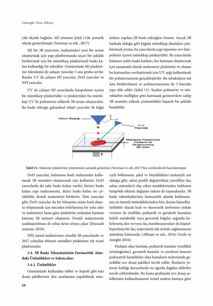
54
Çobanoğlu, Varan, Bilensoy
yük ölçüde bağlıdır. MJ yöntemi Şekil 11’de şematik olarak gösterilmiştir (Norman ve ark., 2017).
MJ bir 3B yazıcının, malzemeleri yeni bir nesne oluşturmak için yapı platformunda seçici bir şekilde biriktirmek için bir mürekkep püskürtmeli baskı ka-fası kullandığı bir tekniktir. Günümüzde MJ püskürt-me teknolojisi ile çalışan yazıcılar 3 ana gruba ayrılır. Bunlar UV ile çalışan MJ yazıcılar, DoD yazıcılar ve NPJ yazıcılar.
UV ile çalışan MJ yazıcılarda fotopolimer içeren bir mürekkep püskürtülür ve püskürtülen bu mürek-kep UV ile polimerize edilerek 3B nesne oluşturulur. Bu baskı tekniği; geleneksel inkjet yazıcılar ile kâğıt
üstüne yapılan 2B baskı tekniğine benzer. Ancak 2B baskıda olduğu gibi kâğıda mürekkep damlaları püs-kürtmek yerine, bu yazıcılarda yapı tepsisine sıvı foto-polimer içeren mürekkep püskürtülür. Bu yazıcılarda bulunan çoklu baskı kafaları, her katmanı oluşturmak için eşzamanlı olarak malzemeyi püskürtür ve oluşan bu katmanları sertleştirmek için UV ışığı kullanılarak bir polimerizasyon gerçekleştirilir. Bu tabakaların üst üste biriktirilmesi ve polimerizasyonu ile 3 boyutlu yapı elde edilir (Şekil 11). Seçilen polimerin ve mü-rekkebin özelliğine göre karmaşık geometrilere sahip 3B nesneler yüksek çözünürlükte başarılı bir şekilde basılabilir.
Şekil 11. Malzeme püskürtme yönteminin şematik gösterimi (Norman ve ark.,2017)’ten esinlenilerek hazırlanmıştır.
DoD yazıcılar, balmumu bazlı malzemeler kulla-narak 3B nesneleri oluşturmak için kullanılır. DoD yazıcılarda iki adet baskı kafası vardır; birinci baskı kafası yapı malzemesini, ikinci baskı kafası ise çö-zülebilir destek malzemeyi biriktirir. Tüm yazıcılar gibi, DoD yazıcılar da bir bileşenin enine kesit alanı-nı oluşturmak için önceden belirlenmiş bir yolu izler ve malzemeyi buna göre püskürtür ardından katman katman 3B nesneyi oluşturur. Destek malzemenin uzaklaştırılması ile nihai ürün ortaya çıkar (Dassault systems, 2018).
NPJ, metal endüstrisine yönelik 3B yazıcılardır ve 2017 yılından itibaren metalleri püskürten tek ticari platformdur.
3.4. 3B Baskı Teknolojisinin Farmasötik Alan-daki Üstünlükleri ve Sakıncaları
3.4.1. Üstünlükler
Günümüzde kullanılan tablet ve kapsül gibi katı dozaj şekillerinin doz ayarlaması yapabilmek ama-
cıyla bölünmesi, şekil ve büyüklükleri nedeniyle zor olduğu gibi, salım profili değiştirilmiş (modifiye ilaç salım sistemleri) ilaç etkin maddelerinden beklenen terapötik etkinin değişme riskini de taşımaktadır. 3B baskı teknolojilerinin farmasötik alanda kullanımı-nın en önemli üstünlüklerinden biri, dozun kişiselleş-tirilebilir olarak hızlı ve ekonomik üretimine imkân vermesi ile özellikle pediatrik ve geriatrik hastalara belirli metabolik veya genomik bilgiler ışığında be-lirlenmiş doz ve/veya ilaç kombinasyonları ile kişisel-leştirilmiş bir ilaç tedavisinin tek ürünle sağlanmasını mümkün kılmasıdır (Alhnan ve ark., 2016; Goole ve Amighi 2016).
Disfajisi olan hastalar, pediatrik hastalar (özellikle yenidoğanlar), geriatrik hastalar ve şizofreni benzeri psikiyatrik hastalıkları olan hastaların tedavisinde ge-nellikle sıvı dozaj şekilleri tercih edilir. Bunların ye-tersiz kaldığı durumlarda ise ağızda dağılan tabletler tercih edilmektedir. Bu hasta grubunda sıvı dozaj şe-killerinin kullanılmasının temel nedeni hastaya göre

55
FABAD J. Pharm. Sci., 46, 1, 43-78, 2021
doz ayarlamasının yapılabilmesidir (Breitkreutz ve Boos, 2006). 3B baskı teknolojisi ile, pediatrik hasta-nın kilosuna, hastalığın ilerleme seviyesine göre doz, büyüklük, şekil veya rengini hastaya göre kişiselleşti-rilebildiği katı dozaj şekillerini oluşturmak, böylece tedavinin başarısını arttırmak mümkündür (Aloma-ri ve ark., 2015). Bu teknoloji ile katı dozaj şeklinin genel boyutu değiştirilerek ya da aynı büyüklük içe-risinde farklı etkin madde kombinasyon oranları ile hazırlanarak hastaya uygun ilaçlar oluşturulabilir. Bu çok yönlü kişiselleştirme ve tekrarlanabilirlik, özellik-le dar terapötik indeksli etkin maddeler için, daha az yan etki ile ilaç tedavisinin daha etkin ve güvenli ol-masını da sağlayacaktır. 3B baskı teknolojileri ile ilaç fabrikalarının dışında, iyi imalat uygulamaları (GMP) koşullarına uygunluğunu belgeleyen eczaneler, hasta-neler ve askeri tesisler gibi yerlerde de özellikle stabi-litesi düşük ilaçların hızlı ve ekonomik bir şekilde ki-şiye özel üretilebilmesi mümkün olacaktır (Lepowsky ve Tasoglu, 2017; Norman ve ark., 2017).
3B baskı teknolojileri ile, etkin maddelerin çözü-nürlük ve salım özelliklerini de kontrol edebilmek ve değiştirebilmek mümkündür. Ayrıca, baskı sırasında oluşması potansiyel amorf etkin maddelerin bir sonu-cu olarak, zayıf çözünürlüğe sahip etkin maddelerin çözünürlüğü arttırılabilir. 3B ekstrüzyon temelli sis-temlerde 3B nesnenin gözenekli bir şekilde basılması ile yüzey alan artırılabilmekte ve amorf yapıların elde edilmesiyle de ilacın dağılma ve çözünme hızlarını kontrol edebilmek mümkündür. 3B baskı teknolojisi ile hızlandırılmış etkin madde salımı için, daha gev-şek ve gözenekli tablet formları basılarak, yüksek da-ğılma ve çözünme hızı sağlanabilir. (Goole ve Amighi, 2015)
Aynı anda birden fazla ilaç kullanan hastalar için; birden fazla püskürtme başlığına sahip yazıcılar ile birden fazla etkin maddeyi içeren ve bu etkin mad-delerin istenildiği oranda (dozda) basıldığı tablet formları oluşturulabildiği gibi farklı salım profillerine sahip birden fazla katmandan oluşan tek bir tablet de basılabilir. Bu durum, hasta uyuncunu artırarak teda-vinin başarısını doğrudan etkileyecektir. Sıkıştırılmış toz tabletlerin aksine, 3B baskı teknolojileri ile hazır-lanmış tabletlerin içerisinde etkin maddenin gerçek dağılımı ve konumlanması tam olarak bilinebilmekte
ve kontrol edilebilmektedir. Ayrıca 3B baskı yönte-miyle belirli eksipiyanlara (laktoz, sukroz) intoleran-sı olan hastalar için; bu eksipiyanlar formülasyonda daha düşük miktarlarda kullanılabilir veya formülas-yondan tamamen çıkarılabilir. 3B baskı teknolojileri-nin prototiplemeye olanak vermeleri ile üretim, ihti-yaca bağlı olarak hızlı bir şekilde modifiye edilebilir ve bu da bitmiş ürünlerin uzun süreli depolanma zo-runluğunu ortadan kaldırabilir (Dumitrescu ve ark., 2018).
Yeni bir ilacın piyasaya çıkmasının maliyeti yakla-şık 5 milyar $ olarak tahmin edilmektedir. Günümüz-de uygun ilaç adaylarının minimum maliyetle hızlı bir şekilde tespit edilmesini sağlayarak, ilaç gelişimini desteklemek için yenilikçi teknolojilere ihtiyaç duyul-maktadır. 3B baskı teknolojisiyle, az miktarda veya “bir kereye mahsus” formülasyonları (ve hatta ilaçla-rı) düşük maliyetli, verimli ve esnek bir şekilde üre-terek bu ihtiyaca katkı sağlanabilir. Bu teknoloji, ilaç geliştirme sürecini hızlandırabileceği gibi maliyetleri de büyük oranda düşürecektir (Awad ve ark., 2018). Çözünürlüğü yüksek 3B yazıcıların kullanılması ile son derece düşük miktarlarda etkin madde içeren (1-10 ng gibi) tabletlerin basılması bile mümkündür. Ayrıca ilaçlar küçük seriler halinde üretileceği için, geleneksel endüstriyel üretime kıyasla 3B baskı tekno-lojileri çok daha ekonomik olacaktır. Stabilitesi düşük olan etkin maddeler 3B yazıcılar ile uygulama anında basılabilecektir. Günümüzde bazı araştırmacılar uy-gulama anında etkin madde sentezini gerçekleştiren 3B baskı teknikleri üzerine araştırmalar yapmaktadır (Dumitrescu ve ark., 2018).
3.4.2. Sakıncaları
Her bir baskı tekniğinin kendi içinde farklı üstün-lükleri ve sakıncaları söz konusudur. Bununla birlikte günümüzde ilaçların 3B yazıcılar ile basılabilmesi için uygun materyallerin sınırlı olması 3B baskı teknoloj-lerinin farmasötik alanda kullanımını etkileyen en önemli parametredir (Alhnan ve ark., 2016)
Mürekkep püskürtme yöntemi ile çalışan 3B ya-zıcılar için püskürtme uçlarının ve baskı kafasının tı-kanmasını veya kurumasını engellemek için düzenli kontrol ve bakıma gerek duyulmaktadır. Baskı sıra-sında kullanılan çözücülerin nihai üründen uzaklaş-tırılması için sıcak hava, mikrodalga veya kızılötesi

56
Çobanoğlu, Varan, Bilensoy
ile kurutma gibi ilave üretim sonrası işlemlere ihtiyaç duyulmakta, bu da etkin maddenin stabilitesini et-kileyebilmektedir. Bazı 3B baskı tekniklerinde, baskı plakasının titreşiminden (bant lekesi), hızlı termal değişimlerden (biçimsel bozulma) ya da iç yapının çökmesinden dolayı farklı baskı kusurları ortaya çı-kabilmektedir. Ekstrüzyon temelli sistemlerde kulla-nılan PLA ve ABS filamentlerinden kaynaklanan ultra ince tozlar ve termal bozunmadan kaynaklı yabancı bileşikler gibi olası kirleticiler için üretim tesislerinde hava güvenliği sağlanması ve son ürünün saflaştırıl-ması gibi ek işlemler gerekebilmektedir (Norman ve ark., 2017; Trenfield ve ark., 2018)
3B baskı işlemi, eksipiyanlar için çoklu malzeme seçimi de dahil olmak üzere optimizasyona ve çok yönlülüğe ihtiyaç duymaktadır. Başarılı bir baskı için etkin madde ve yardımcı maddelerin yüzey gerilimi, viskozite, reolojik özellikler veya toz ıslanabilirliği gibi, fizikokimyasal özelliklerinin tam olarak bilinme-si de oldukça önemlidir. Bu nedenle gerekli iyi imalat uygulamalarına uygun bir süreç tasarlamak için mev-cut GMP yönetmelikleri dikkate alınmalıdır (Dumit-rescu ve ark., 2018).
3B baskı teknolojilerinin, üretim endüstrisinde benimsenmesinin de gelecekte bazı sakıncaları do-ğurması söz konusu olabilir. Örneğin, 3B baskı tek-nolojisi üretimde ihtiyaç duyulan iş gücünü azaltacağı için, üretimden insan gücünün çıkması ile çok sayıda düşük vasıflı işçisi bulunan ülkelerin ekonomisini de doğrudan etkileyecektir. Ayrıca, 3B baskı teknolojisi kullanarak, silah, zehir ve diğer tehlikeli maddelerin üretiminin kolaylaşabileceği nedeniyle, 3B yazıcıların kullanımının gelecekte hükümetler tarafından belirli kişilerle ve izinler doğrultusunda sınırlandırılması da söz konusu olabilecektir (Shahrubudin ve ark.,2019).
4. Baskı Teknolojilerinin Farmasötik Alandaki Uygulamaları
Günümüzde çok farklı çalışma prensipleri ile ça-lışan 3B yazıcılar olmasına karşın, ilaç araştırmaları konusunda en sık kullanılan 3B yazıcıların FDM, SSE, BJ, MJ, SLA, DLP, CLIP ve SLS olduğu söylenebilir (Trenfield ve ark., 2018). Bu yöntemler kullanılarak farmasötik alanda yapılan çalışmaların bir kısmı Tab-lo 1’de özetlenmiştir.

57
FABAD J. Pharm. Sci., 46, 1, 43-78, 2021Ta
blo
1. F
arm
asöt
ik a
land
a 3B
yaz
ıcıla
r ile
yap
ılan
çalış
mal
ara
örne
kler
3B B
askı
Te
knol
ojis
i3B
Yaz
ıcı
Türü
Doz
aj Ş
ekli
Etki
n M
adde
Mat
erya
lA
maç
Kay
nak
Ekst
rüzy
onTe
mel
li Si
stem
ler
FDM
veya
FFF
Toz K
arışı
mı
Ase
tam
inof
enSo
lupl
us®
Ase
tom
inof
en iç
eren
toz k
arışı
mın
ın
hazı
rlanm
ası.
Haz
ırlam
a sır
asın
da k
ulla
nıla
n ek
strü
der v
ida
konf
igür
asyo
nunu
n ve
etk
in
mad
deni
n pa
rtik
ül b
üyük
lüğü
nün
salım
hız
ına
etki
sinin
ince
lenm
esi
(Li v
e ar
k., 2
015)
Tabl
etPr
edni
zolo
nPV
AU
zatıl
mış
salım
sağl
ayan
tabl
etle
rin ü
retil
mes
i ve
bas
ılı h
acm
in k
ontr
ol e
dilm
esiy
le d
ozun
ko
ntro
l edi
lip e
dile
mey
eceğ
inin
ara
ştırı
lmas
ı(S
kow
yra
ve a
rk.,
2015
)
Tabl
etD
ompe
ridon
HPC
Dom
perid
onu
içer
en g
astr
ik y
üzen
ve
süre
kli
salım
sağl
ayan
tabl
etle
rin k
abuk
sayı
ları
ve d
olgu
yüz
dele
ri de
ğişt
irile
rek
uygu
lam
a sık
lığın
ın a
zaltı
lmas
ı, fiz
ibili
tesin
in in
cele
nmes
i, in
vitr
o ve
in v
ivo
özel
likle
rinin
değ
erle
ndiri
l-m
esi
(Cha
i ve
ark.
, 201
7)
Tabl
etM
etfo
rmin
, glim
epiri
dEu
drag
it® R
L PO
, PEG
40
0, T
EC, S
itrik
asit
m
onoh
idra
t, PL
A
Diy
abet
has
talığ
ının
teda
visin
de k
ulla
nılm
ak
üzer
e bi
rden
fazl
a et
kin
mad
de iç
eren
, glim
ep-
ridin
hız
lı sa
lım, m
etfo
rmin
in sü
rekl
i sal
ımın
ı sa
ğlay
an ta
blet
lerin
in te
k bi
r doz
aj şe
kli h
alin
de
hazı
rlanm
ası v
e fiz
ibili
tesin
in d
eğer
lend
irilm
esi
(Gio
umou
xouz
is ve
ark
., 20
18)
Tabl
etİz
onia
zid
HPC
, Hid
roks
ipro
pil
met
il se
lülo
z (H
PMC
), Po
lietil
en o
ksit,
Eudr
agit
RS P
O, E
ud-
ragi
t RL
PO,
Eudr
agit
L100
, PLA
Tübe
rkül
oz te
davi
sinde
kul
lanı
lan
izon
iazi
d iç
eren
ve
fark
lı po
limer
ler k
ulla
nıla
rak
basıl
mış
tabl
etle
rin, p
olim
er u
ygun
luğu
açı
sında
n de
ğerle
ndiri
lmes
i
(Öbl
om v
e ar
k., 2
019)
Tabl
etFl
ores
ein
sody
umPV
A
Flor
esei
n iç
eren
PVA
fila
men
tlerin
haz
ırlan
-m
ası,
ilaç y
üklü
tabl
etle
rin b
asılm
ası v
e ba
skı
para
met
rele
rinin
değ
iştiri
lmes
inin
nih
ai ta
blet
in
çözü
nme
kine
tiği ü
zerin
deki
etk
isini
n ar
aştı-
rılm
ası
(Goy
anes
ve
ark.
, 201
4)

58
Çobanoğlu, Varan, Bilensoy
Ekst
rüzy
onTe
mel
li Si
stem
ler
FDM
veya
FFF
Tabl
etİn
dom
etaz
inPE
G 6
000,
hip
rom
ello
z as
etat
süks
inat
Lezz
eti a
rttır
ılmış
ve p
edia
trik
kul
lanı
ma
yöne
-lik
ilaç
lar h
azırl
amak
için
“şek
er b
enze
ri” fo
r-m
ülas
yonl
arın
bas
ılmas
ı ve
basıl
an ta
blet
lerin
ka
rakt
eriz
asyo
nu
(Sco
utar
is ve
ark
., 20
18)
Tabl
etH
idro
klor
otiy
azid
PVA
, Man
nito
l, PL
AH
iper
tans
iyon
teda
visin
de k
ulla
nılm
ak ü
zere
üç
taba
kada
n ol
uşan
kon
trol
lü sa
lım sa
ğlay
an
tabl
etle
rin h
azırl
anm
ası v
e ka
rakt
eriz
asyo
nu(S
adia
ve
ark.
, 201
8)
Tabl
etG
lipiz
idPV
AD
iyab
et te
davi
sinde
kul
lanı
lan
glip
izid
içer
en
çift
taba
kalı
tabl
etin
bas
ılmas
ı, sa
lım p
rofil
inin
in
cele
nmes
i ve
salım
kin
etiğ
inin
ara
ştırı
lmas
ı(L
i ve
ark.
, 201
7)
Tabl
etK
alse
inPV
A, P
LAKo
mpo
zit t
able
tlerin
(PVA
/PVA
, PVA
/PLA
) ha
zırla
nmas
ı, ka
rakt
eriz
asyo
nu v
e sa
lım p
rofil
-le
rinin
ince
lenm
esi
(Tag
ami v
e ar
k., 2
018)
Tabl
etPa
rase
tam
ol
PLA
, HPC
LF,
HPC
EF,
HPM
C E
5, E
C N
14,
Solu
plus
, Eud
ragi
t L1
00
Fark
lı po
limer
ora
nlar
ı ile
ilaç
içer
en ta
blet
le-
rin h
azırl
anm
ası,
kara
kter
izas
yonu
, mek
anik
öz
ellik
lerin
in b
elirl
enm
esi v
e sa
lım p
rofil
lerin
in
değe
rlend
irilm
esi
(Zha
ng v
e ar
k., 2
017)
Tabl
etM
etro
nida
zol
PVA
, ABS
H. p
ylor
i ile
ilişk
ili p
eptik
ülse
rlerin
teda
visi
için
sabi
t ila
ç sal
ımı s
ağla
yabi
len
ilaç t
aşıy
ıcı
siste
min
gel
iştiri
lmes
i ve
sıfır
dere
celi
ilaç s
alım
ki
netiğ
i içi
n ba
skı p
aram
etre
lerin
in o
ptim
ize
edilm
esi
(Hua
nbut
ta v
e Sa
ngni
m, 2
019)
Tabl
etPr
amip
ekso
l dih
id-
rokl
orür
mon
ohid
rat
Eudr
agit
EPO
, PO
L-YO
X™
WSR
N1
ve
POLY
OX
™ W
SR N
80
Park
inso
n te
davi
sinde
kul
lanı
lan
Pram
ipek
sol
dihi
drok
lorü
r mon
ohid
rat i
çere
n ve
hız
lı sa
lım
sağl
ayan
tabl
etle
rin h
azırl
anm
ası v
e op
timiz
e ed
ilmes
i
(Gül
teki
n ve
ark
., 20
19)
Tabl
etRu
finam
id
Kolli
don®
VA
64,
So
lupl
us®,
HPM
C,
Gel
ucire
® 44/
14, G
elu-
cire
® 48/
16
3B b
askı
tekn
oloj
isi k
ulla
nıla
rak
nadi
r has
talık
ol
an L
enno
x-G
asta
ut S
endr
omun
da k
ulla
nı-
lan
rufin
amid
in çö
zünm
esin
in a
rtırı
lmas
ının
sa
ğlan
mas
ı
(Say
dam
ve
Takk
a, 2
020)
Kap
let
Teof
ilin
Eudr
agit
RL10
0, E
ud-
ragi
t RS1
00, H
idro
k-sip
ropi
l sel
üloz
(HPC
), Tr
ietil
sitr
at(T
EC)
Teof
ilini
n do
z esn
ekliğ
i sağ
laya
n hı
zlı v
e uz
atıl-
mış
salım
sağl
ayan
kap
letle
rinin
haz
ırlan
mas
ı ve
kara
kter
izas
yonu
(Pie
trza
k ve
ark
., 20
15)
Kap
let
Bakl
ofen
PVA
, Sor
bito
l
Pedi
atrik
kul
lanı
ma
yöne
lik m
odifi
ye sa
lım
sağl
ayan
min
ikap
letin
haz
ırlan
mas
ı, ka
rakt
eriz
e ed
ilmes
i ve
fakt
öriy
el ta
sarım
kul
lanı
lara
k fa
rklı
bask
ı par
amet
rele
rinin
ilaç
salım
ı üze
rinde
ki
etki
sinin
ara
ştırı
lmas
ı
(Pal
ekar
ve
ark.
, 201
9)

59
FABAD J. Pharm. Sci., 46, 1, 43-78, 2021
Ekst
rüzy
onTe
mel
li Si
stem
ler
FDM
veya
FFF
Kap
sül
Met
form
in H
Cl
PVA
, PLA
Sıvı
vey
a ka
tı ila
ç for
mül
asyo
nlar
ının
dol
umu-
nun
gerç
ekle
ştiri
lebi
lece
ği k
apsü
l kab
uğun
un
fark
lı ba
skı p
aram
etre
leri
uygu
lana
rak
elde
ed
ilebi
lirliğ
inin
ince
lenm
esi
(Sm
ith v
e ar
k., 2
018)
Liki
t Kap
sül
Teof
ilin,
dip
irida
mol
Eudr
agit
EPO
ve
RL
Liki
t kap
sül y
apım
ında
FD
M te
knol
ojisi
nden
ya
rarla
nıla
rak
hızl
ı ve
uzat
ılmış
salım
sağl
ayan
do
zaj ş
eklin
in el
de e
dilm
esi,
optim
izas
yonu
ve
kara
kter
izas
yonu
(Okw
uosa
ve
ark.
, 201
8)
Ağı
zda
Dağ
ılan
Film
A
ripip
razo
lPV
A
Ağı
zda
dağı
lan
film
lerin
haz
ırlan
mas
ı ve
stan
dart
solv
ent d
öküm
tekn
oloj
isi il
e ür
etile
n fil
mle
r ile
fizi
koki
mya
sal,
mek
anik
öze
llikl
er,
dağı
lma
zam
anı v
e sa
lım ö
zelli
kler
i açı
sında
n ka
rşıla
ştırı
lmas
ı
(Jam
róz v
e ar
k., 2
017b
)
Tran
sder
mal
ya
ma
Mon
telu
kast
sody
um
Kolli
phor
P18
8, K
olli-
don
VA64
F, Ko
llido
n 12
PF, P
olie
tilen
glik
ol
(PEG
), Po
lietil
en o
ksit
(PEO
)
Yeni
3B
bask
ı fila
men
tlerin
in g
elişt
irilm
esi v
e ka
rakt
eriz
e ed
ilmes
i, bu
nlar
ın d
oğru
dan
bir
amba
laj m
alze
mes
ine
basıl
mas
ı(A
zizo
ğlu
ve Ö
zer,
2020
)
Tıbb
i cih
azİn
dom
etaz
inEt
ilen
vini
l ase
tat
(EVA
)İn
dom
etaz
in iç
eren
tıbb
i cih
azla
rın 3
B ba
skısı
iç
in o
ptim
al ö
zelli
kler
inin
ince
lenm
esi
(Gen
ina
ve a
rk.,
2016
)
Tıbb
i cih
azPr
oges
tero
nPL
A, P
CL
Prog
este
ron
içer
en, f
arkl
ı şek
iller
e sa
hip,
uzat
ılmış
salım
sağl
ayan
rahi
m iç
i ar
açla
rın h
azırl
anm
ası v
e ka
rakt
eriz
asyo
nu(F
u ve
ark
., 20
18)

60
Çobanoğlu, Varan, Bilensoy
Ekst
rüzy
onTe
mel
li Si
stem
ler
SSE
Tabl
etG
uaya
fene
sin
HPM
C 2
910,
HPM
C
2208
, Car
bopo
l® 97
4P
NF,
Phar
mac
el® 1
02,
Prim
ojel
®
Bir t
abak
ası s
ürek
li sa
lım sa
ğlay
an v
e bi
r tab
a-ka
sı hı
zlı s
alım
sağl
ayan
çift
taba
kalı
tabl
etin
ha
zırla
nmas
ı ve
piya
sada
bul
unan
tabl
et fo
rmü-
lasy
onu
ile k
arşıl
aştır
ılmas
ı
(Kha
led
ve a
rk.,
2014
)
Tabl
etG
inkg
olid
HPM
C K
4M, H
PMC
E5
LV, m
ikro
krist
aliz
e se
lülo
z (M
CC
), La
ktoz
,
PVP
K30
Gin
kgol
id iç
eren
yüz
en ta
blet
lerin
haz
ırlan
mas
ı, ka
rakt
eriz
asyo
nu v
e in
vitr
o, in
viv
o sa
lım p
ro-
fille
rinin
ince
lenm
esi
(Wen
ve
ark.
, 201
9)
Tabl
etN
ifedi
pin,
glip
izid
ve
kapt
opril
HPM
C, s
odyu
m
klor
ür, D
-man
nito
l, kr
oska
rmel
oz so
dyum
, M
CC
, sod
yum
nişa
sta
glik
olat
, PV
P K
30,
trom
etam
in, s
elül
oz
aset
at, P
EG 6
000,
as
eton
Hip
erta
nsiy
on v
e di
yabe
t has
tala
rında
fark
lı ko
ntro
llü sa
lım (k
apto
pril
için
osm
otik
kon
trol
-lü
salım
, nife
dipi
n ve
glip
izid
için
süre
kli s
alım
) pr
ofili
ne sa
hip
etki
n m
adde
lerin
tek
bir t
able
tte
bire
ysel
ihtiy
aç iç
in h
azırl
anm
ası,
kara
kter
izas
-yo
nu v
e sa
lım p
rofil
lerin
in d
eğer
lend
irilm
esi
(Kha
led
ve a
rk.,
2015
b)
Tabl
etH
idro
klor
otiy
azid
, as
pirin
, pra
vast
atin
, at
enol
ol v
e ra
mip
ril
PVP,
lakt
oz,
D-m
anni
tol,
sod-
yum
nişa
sta
glik
olat
, H
PMC
, sel
üloz
ase
tat,
PEG
600
0
Kar
diyo
vask
üler
has
talık
ların
teda
visi
için
beş
fa
rklı
etki
n m
adde
içer
en v
e fa
rklı
salım
pro
fil-
lerin
e sa
hip
kom
plek
s bir
tabl
etin
haz
ırlan
mas
ı, ka
rakt
eriz
asyo
nu v
e in
vitr
o sa
lım p
rofil
lerin
in
ince
lenm
esi
(Kha
led
ve a
rk.,
2015
’a)
Tabl
etLe
vetir
aset
am
Sody
um k
arbo
ksim
etil
selü
loz,
Poliv
inilp
iroli-
don
K30
, Kro
skar
me-
loz s
odyu
m
Epile
psi t
edav
isind
e ku
llanı
lan
leve
tiras
etam
ın
hızl
ı sal
ım sa
ğlay
an ta
blet
inin
haz
ırlan
mas
ı içi
n SS
E te
knol
ojisi
nin
pota
nsiy
elin
in a
raşt
ırılm
ası
(Cui
ve
ark.
, 201
9a)
Tabl
etG
lipiz
idH
PMC
E15
, HPM
C
K10
0LV,
PV
P K
30,
MC
C P
H10
1, L
akto
z
Fark
lı isk
elet
yap
ıların
a sa
hip
glip
izid
tabl
etle
rin
hazı
rlanm
ası,
mek
anik
öze
llikl
erin
in d
eğer
-le
ndiri
lmes
i, ba
skı p
aram
etre
leri
ile il
aç sa
lım
prof
iller
i ara
sında
ki il
işkin
in a
raşt
ırılm
ası
(Cui
ve
ark.
, 201
9b)

61
FABAD J. Pharm. Sci., 46, 1, 43-78, 2021
Laze
r Te-
mel
li Si
s-te
mle
r
SLA
Tabl
etPa
rase
tam
olve
4-A
SA
Polie
tilen
glik
ol d
iakr
i-la
t (PE
GD
A),
PEG
300
, D
PPO
FDM
tekn
oloj
isiyl
e ba
sımı i
çin
çalış
mal
ar
yapı
lan
etki
n m
adde
lerin
SLA
tekn
oloj
isi k
ul-
lanı
lara
k m
odifi
ye sa
lım sa
ğlay
an ta
blet
lerin
in
hazı
rlanm
ası
(Wan
g ve
ark
., 20
16)
Tabl
et
Para
seta
mol
, Kaf
ein,
N
apro
ksen
, Klo
ram
fe-
niko
l, Pr
edni
zolo
n ve
A
spiri
n
PEG
DA
, PEG
300,
DPP
O
Çok
kat
man
lı ila
ç bas
kısı
için
ilk
kez S
LA
tekn
oloj
isini
n de
ğerle
ndiri
lmes
i(R
oble
s-M
artin
ez v
e ar
k., 2
019)
Hid
roje
lİb
upro
fen
PEG
DA
, PEG
300
, D
PPO
, Rib
oflav
in,
Trie
tano
lam
in
SLA
tekn
oloj
isini
n ila
ç yük
lü h
idro
jelle
rin
hazı
rlanm
asın
da k
ulla
nıla
bilir
liği a
çısın
dan
değe
rlend
irilm
esi
(Mar
tinez
ve
ark.
, 201
7)
Hid
roje
lA
skor
bik
asit
PEG
, pol
ietil
en g
likol
di
met
akril
at (P
EGD
-M
A),
Ribo
flavi
n,
Trie
tano
lam
in
Fark
lı bü
yükl
ük v
e şe
kille
re sa
hip
asko
rbik
asit
iç
eren
hid
roje
llerin
elde
edi
lmes
i, ka
rakt
eriz
as-
yonu
ve
salım
pro
fille
rinin
, sal
ım k
inet
ikle
riyle
be
rabe
r değ
erle
ndiri
lmes
i
(Kar
akur
t ve
ark
., 20
20)
Tıbb
i Cih
az
Salis
ilik
asit
Flex
Eco
PLA
, PC
L,
PEG
DA
Kişi
selle
ştiri
lebi
lir y
üz m
aske
lerin
in h
azırl
an-
mas
ı, re
zolü
syon
, ila
ç yük
lem
esi,
ilaç d
ifüzy
onu-
nun
karş
ılaşt
ırılm
ası
(Goy
anes
ve
ark.
, 201
6a)
DLP
Tabl
etTe
ofili
n
PEG
DA
400
, PEG
D-
MA
100
0, 2
-Hid
roks
i- 4′
-(2-
hidr
oksie
toks
i)-2-
met
ilpro
piof
enon
Min
imum
pol
imer
kon
sant
rasy
onun
a sa
hip
daya
nıkl
ı tab
letle
r üre
tmek
için
en
uygu
n ta
blet
fo
rmül
asyo
nunu
n el
de e
dilm
esi,
kara
kter
izas
yo-
nu v
e sa
lım p
rofil
lerin
in k
arşıl
aştır
ılmas
ı
(Kad
ry v
e ar
k., 2
019)
CLI
PTı
bbi C
ihaz
D
oset
akse
l ve
Dek
sa-
met
azon
ase
tat
PCL
ve P
EG te
mel
li po
limer
bile
şimle
ri
CLI
P te
knol
ojisi
nin
ilaç y
üklü
tıbb
i cih
azla
rın
tasa
rımın
a ve
imal
atın
a uy
gula
nmas
ı pot
ansiy
e-lin
in a
raşt
ırılm
ası
(Blo
omqu
ist v
e ar
k., 2
018)
SLS
Tabl
etPa
rase
tam
olKo
llico
at IR
, Eud
ragi
t L1
00-5
5, C
andu
rin
Gol
d Sh
een
İla
ç yük
lü o
ral d
ozaj
şeki
llerin
in h
azırl
anm
ası
için
SLS
bas
kı te
knol
ojisi
nin
uygu
nluğ
unun
ar
aştır
ılmas
ı(F
ina
ve a
rk.,
2017
)
Tabl
etPa
rase
tam
ol,
İbup
rofe
n
Kolli
coat
IR, E
til se
-lü
loz (
EC),
Can
durin
G
old
Shee
n
Fark
lı sa
lım ö
zelli
kler
ine
sahi
p, b
irden
çok
etki
n m
adde
içer
en, f
orm
ülas
yonl
ar ü
retil
mes
i içi
n SL
S te
kniğ
i’nin
ara
ştırı
lmas
ı(A
wad
ve
ark.
, 201
9)
Tabl
etPa
rase
tam
olH
PMC
, Kol
lidon
® VA
64
, Can
durin
® Gol
d Sh
een
Ağı
zda
dağı
lan
tabl
et fo
rmül
asyo
nlar
ının
haz
ır-la
nmas
ında
SLS
tekn
oloj
isini
n uy
gunl
uğun
un
araş
tırılm
ası
(Fin
a ve
ark
., 20
18)

62
Çobanoğlu, Varan, Bilensoy
Mür
ekke
p Pü
skür
tme
Tem
elli
Sist
emle
r
BJ
Tabl
etFe
nofib
rat
Balm
umu
Pete
k şe
klin
e be
nzer
tabl
etle
rin h
azırl
anm
ası,
pete
k bo
yutu
nun
ve y
üzey
ala
nını
n ila
ç sal
ım
prof
iller
inin
kon
trol
ünü
sağl
amak
için
mod
ifiye
ed
ilmes
i ve
kara
kter
izas
yonu
(Kyo
bula
ve
ark.
, 201
7)
Tabl
etPa
rase
tam
ol v
e al
iza-
rin sa
rısı
Kollo
idal
silik
on d
iok-
sit, m
anni
tol,
poliv
inil-
piro
lidon
K30
ve
lakt
oz
Öze
l bir
iç y
apıy
a sa
hip,
hız
lı da
ğıla
n ta
blet
fo
rmül
asyo
nunu
n ha
zırla
nmas
ı ve
kara
kter
i-za
syon
u(Y
u ve
ark
., 20
09)
Tabl
etK
lorf
enam
in m
alea
t ve
flor
esei
n
Avic
el P
H30
1, E
udra
git
E-10
0, R
LPO
, PV
P ve
Tw
een
20
Bask
ı par
amet
rele
ri de
ğişt
irile
rek
ilaç s
alım
pr
ofili
nin,
doz
aj k
ontr
olün
ün in
cele
nmes
i(K
astr
a ve
ark
., 20
00)
Tabl
etLe
vetir
aset
amM
CC
, glis
erin
, Tw
een
80, p
ovid
on, s
ukra
loz
Ağı
zda
hızl
ı dağ
ılan
yeni
bir
tabl
et fo
rmül
asyo
-nu
nun
hazı
rlanm
ası
(U.S
. Pat
ent N
o. U
S 20
14/0
2718
62 A
1, 2
014)
DoP
Ta
blet
5-flo
rour
asil
2-pi
rolid
inon
, CaS
O4
bazl
ı toz
kar
ışım
ı, So
lupl
us®,
PEG
5-flo
rour
asil
etki
n m
adde
sini i
çere
n ta
blet
lerin
D
oP te
knol
ojisi
kul
lanı
lara
k ba
sımın
ın u
ygun
-lu
ğunu
n de
ğerle
ndiri
lmes
i (S
hi v
e ar
k., 2
019)
4.1.
E
kstr
üzyo
n Te
mel
li Si
stem
lere
Örn
ekle
r

63
FABAD J. Pharm. Sci., 46, 1, 43-78, 2021
Ekstrüzyon temelli sistemlerden FDM teknolojisi farmasötik alanda en sık çalışılan 3B baskı teknolo-jisidir. FDM temelli yazıcıların kullanımının kolay olması ve piyasadaki diğer yazıcılara göre maliyetle-rinin düşük olması araştırmacıların bu yazıcılara olan ilgisini de artırmaktadır.
Bu alanda yapılan çalışmalara örnek olarak, Huan-butta ve ark. tarafından 2019 yılında yaptıkları bir ça-lışma verilebilir. Huanbutta ve ark. metronidazol’ün kontrollü salımını sağlayan yüzen tabletlerin hazır-lanması amacı ile kullanım kolaylığı, baskı maddele-rinin ekonomikliği ve ulaşılabilirliği nedeni ile FDM yazıcıları tercih etmiştir. Yaptıkları bu çalışmada, yü-zen tablet kaplaması veya tablet gövdesi polivinil al-kolden hazırlanmış ve tabletler silindir, küre ve koni şekillerinde tasarlanarak, basılmıştır. Sıfır derece ilaç salım kinetiği için baskı parametreleri, tablet şekli ve çekirdek tablet formülasyonları optimize edilmiştir. Silindirik yüzen tabletlerin, su yüzeyinde stabil bir şekilde yüzdüğü görülmüştür. 3B yazıcı ile hazırlanan bu tabletlerin hem 4 saatin üzerinde bir süre yüzdü-ğü gösterilmiş, hem de 8 saat sonra ilacın %88’inden fazlasını saldığı görülmüştür. Gözenek büyüklüğü 2.0 mm ve hava hacmi 132 mm3 olan yüzen tabletler, r2
değeri 0.966 olan sıfır dereceli ilaç salımı sağladığı da yaptıkları bu çalışmada gösterilmiştir (Huanbutta ve Sangnim, 2019). Li ve ark.ları sıcak eriyik ekstruder (HME) ve FDM temelli 3B yazıcı kullanarak glipizid içeren çift tabakalı PVA tabletler basılmıştır. Hazırla-nan bu çift tabakalı tabletler ile ilaç salım profili kont-rol edilebildiği gibi glipizidin 5 saatlik bir kontrollü salımı sağlanmıştır. Ayrıca Li ve arkadaşları hazırla-dıkları bu çift tabakalı tabletin dış tabakasının kalın-laştırılması ile bu salım profilinin uzatılabileceğini de göstermişlerdir (Li ve ark., 2017). Tagami ve ark. FDM temelli 3B yazıcı ile suda çözünebilen (örn; PVA) ve suda çözünmeyen (örn; PLA) polimerik filamentler kullanarak kalsein içeren tabletler basmışlardır. Yap-tıkları bu çalışma ile tabletlerin yüzey alanlarının veya PVA tabaka miktarının değiştirilmesi ile tabletlerdeki etkin madde salımının kontrol edilebileceği ve isteğe göre ayarlanabileceğini göstermişlerdir (Tagami ve
ark., 2018). Zhang ve ark.ları tarafından yapılan ben-zer bir çalışmada FDM temelli 3B yazıcı kullanılarak parasetamol içeren tabletler basılmıştır. Yapılan bu çalışmada farklı oranlarda bir dizi farmasötik polimer kullanılmış ve bu polimerler ile hazırlanan etkin mad-de içeren tabletlerin mekanik ve morfolojik yapıları incelenmiştir. Zhang ve ark.ları yaptıkları bu çalışma ile, FDM temelli 3B yazıcılar ve bunların filamentle-rinin hazırlanmasında kullanılan HME kullanılması ile gerektiğinde hazırlanabilecek ve kişiselleştirile-bilir dozda ilaç içeren tabletleri üretiminde yeni bir yöntem olabileceğini göstermişlerdir (Zhang ve ark., 2017). Gültekin ve ark.ları’nın yaptığı bir çalışmada ise Parkinson hastalığının tedavisine yönelik pra-mipeksol dihidroklorür monohidrat içeren ve hızlı salım sağlayan tabletler hazırlanmıştır. Yaptıkları bu çalışma ile tabletlerin kalınlığının ve iç boşluklarının değiştirilmesi ile 5 dakika gibi kısa bir sürede ilacın tamamını salabilen sistemlerin geliştirilebileceğini göstermişlerdir (Gültekin ve ark., 2019).
Nadir hastalıklara yönelik kullanılan yetim ilaçla-rın üretiminde de FDM temelli yazıcılar umut vadet-mektedir. Saydam ve ark.ları FDM temelli 3B yazıcı kullanarak nadir bir hastalık olan Lennox-Gastaut Sendromunun tedavisinde kullanılan ve suda çözün-meyen bir etkin madde olan rufinamidin çözünme hızının artırılabileceğini göstermişlerdir. Ayrıca yap-tıkları bu çalışmada tabletlerin şekil ve boyutlarının değiştirilmesi ile hastaya özgü dozda ilaç içeren tab-letlerin de hazırlanabileceğini göstermişlerdir (Say-dam ve Takka, 2020).
2018 yılında Smith ve ark. tarafından yapılan bir çalışmada ise FDM temelli 3B yazıcılar kullanılarak içi katı veya sıvı ilaç etkin maddesi ile doldurulabi-lecek kapsüller hazırlamışlardır. Kapsüllerin hazırlan-ması sırasında oluşturulacak gözenekli yapıların özel-liğine bağlı olarak da ilaç salımının kontrol edilebile-ceği basit bir üretim yönteminin ortaya konulduğunu göstermişlerdir (Smith ve ark.,2018). Okwuosa ve ark.larının yaptığı benzer bir yayında da FDM temelli 3B yazıcılar ile ilaç solüsyonu (teofilin) veya süspansiyo-nu (dipiridamol) içeren polimetilakrilat kılıfa sahip

64
Çobanoğlu, Varan, Bilensoy
kapsüller basılmıştır. Yaptıkları bu çalışma ile polime-tilakrilat kılıfın kalınlığının değiştirilmesi ile herhan-gi bir formülasyon parametresini değiştirmeksizin ilaç salım hızının değiştirilebileceğini göstermişlerdir. Yaptıkları bu çalışma sayesinde kapsüllerin hastaya özgü dozda ve salım profiline sahip olarak hazırla-nabileceğini de ortaya atmışlardır (Okwuosa ve ark., 2018).
FDM yazıcılar ilaç yüklü tıbbi cihazların hazırlan-ması için de umut vadetmektedir. Genina ve ark.ları FDM temelli 3B yazıcı kullanarak etilen vinil asetat yüklü ve şekli ile boyutu hastaya özel tasarlanabilecek rahim içi araç (RIA) hazırlamışlardır. Genina ve ark.ları HME kullanarak %5 ve %15 indometasin içeren filamentler hazırlamış, bu filamentleri 3B yazıcı ile RIA halinde basmış ve hazırladıkları bu prototipler ile 30 günün üstünde bir ilaç salım profili elde etmişler-dir (Genina ve ark., 2016). Benzer şekilde Fu ve ark.ları progesteronun kontrollü salımının sağlanması amacı ile FDM temelli 3B yazıcı kullanarak kişiselleş-tirilebilir “O”, “Y” ve “M” şeklinde RIA’lar hazırlamış-lardır. Hazırladıkları bu RIA’lar ile 7 günün üstünde bir progesteron salım profili elde etmişlerdir (Fu ve ark., 2018).
FDM temelli 3B yazıcılar ile transdermal uygula-malara yönelik ilaç içeren yamaların hazırlanması da mümkündür. Azizoğlu ve ark.ları HME ile monte-lukast sodium içeren filamentler hazırlamışlar ve bu filamentleri hastaya özgü doz ayarlaması yapabilecek şekilde FDM temelli 3B yazıcı ile transdermal yama-lar halinde doğrudan paketleme materyali üzerine basmışlardır (Azizoğlu ve Özer, 2020). Tablo 1’de de gösterildiği gibi 3B yazıcı ile hazırlanan tablet, kapsül, kaplet, film ve tıbbi cihaz gibi farmasötik ürünlerin büyük bir kısmı FDM temelli yazıcılar ile hazırlan-maktadır.
Literatürde FDM temeli 3B yazıcılar ile yapılan çalışmaların çokluğuna karşın, bu sistemlerin kulla-nımında da çeşitli sorunlar söz konusudur. Bilindiği gibi FDM temelli yazıcılarda baskı öncesi uygun fi-lamentin hazırlanması gerekmekte, bu amaçla HME
gibi cihazlar kullanılmakta ve bu işlem sırasında da polimerler ısıtılarak eritilmektedir. Bu ısı basamağı etkin maddenin bozulmasına neden olabilmekte ve bu yazıcıların farmasötik alanda kullanımını sınır-lamaktadır. Günümüzde, FDM temelli 3B yazıcılar ile yapılan çalışmanın önemli bir kısmı, 3B baskı-ya uygun filamentlerin etkin maddenin stabilitesini bozmadan oluşturulması için uygun eksipiyanların seçilmesi ve optimizasyonuna dayanmaktadır. Seçilen polimerlerin ilaç yükleme kapasitesindeki sınırlama-lar ise bir diğer önemli sorundur. Robles-Martinez ve ark. tarafından 2019 yılında yapılan bir çalışmada; FDM teknolojisinde kullanılan ısıtarak eritme basa-mağındaki olası sorunları ortadan kaldırmak amacı ile ısıtarak eritme işlemi gerçekleştirilmeksizin itra-kanazolun doğrudan toz ekstrüzyonunu yaparak 3B baskısını gerçekleştirmiştir. Robles-Martinez ve ark., yaptıkları bu çalışma ile ilaç etkin maddelerinin doğ-rudan toz ektrüzyonu ile 3B baskısının gerçekleştiri-lebileceğini ilk kez bildirmişlerdir. Bu yeni 3B baskı teknolojisinde, dört farklı HPC türevi kullanılarak, it-rakonazolün amorf katı dispersiyonlarını hazırlamış-lar ve printletler basılmıştır. Basılan tüm printletlerin mekanik ve fiziksel karakterizasyonları yapılmış ve oldukça dayanıklı oldukları görülmüştür. Ayrıca ya-pılan karakterizasyon çalışmalarında etkin maddenin baskı sonrası degrade olmadığını da göstermiştir. Bir diğer önemli bulguları ise printletlerin, ilacın kendi çözünürlüğünden daha yüksek çözünürlük ve sürekli ilaç salım özelliği gösterdiği şeklindedir. HPC-UL ile hazırlanan printletler, diğer HPC türevleri ile hazırla-nanlara kıyasla hızlı ilaç salımı göstermiştir. Bu yeni tek adımlı teknolojinin, amorf katı dispersiyonların hazırlanmasında devrim yaratabileceği ve ısıtarak eritme işlemine uygun olmayan etkin maddelerin 3B basımında alternatif bir yol olabileceği belirtilmiştir (Robles-Martinez ve ark., 2019).
Cui ve ark.ları tarafından yapılan çalışmada ise ilaç dozundaki bireysel değişkenliğin çok fazla ol-duğu levetirasetam model ilaç olarak kullanılmıştır. İlaç dozundaki bireysel değişkenliğin fazla olduğu durumlarda, dozu doğru bir şekilde ayarlamak için

65
FABAD J. Pharm. Sci., 46, 1, 43-78, 2021
güvenilir ve dinamik bir yöntem gerekliliği söz ko-nusudur. Cui ve ark.larının yaptıkları bu çalışmanın amacı, yüksek yükleme kapasitesine sahip hızlı sa-lım sağlayan ve dozu ayarlanabilen bir ilaç hazırlama yönteminin geliştirmesidir. Bu amaçla SSE temelli bir 3B’li yazıcı kullanılmıştır. Bu yöntem, hacmin ve tablet kütlesinin ayarlanması ile istenilen bireysel do-zun elde edilebilmesi amacıyla tercih edilmiştir. Bu kapsamda SSE teknolojisi ile hazırlanan tabletler, pü-rüzsüz bir yüzey ve düzenli bir şekil ve aynı zaman-da yeterli mekanik dayanıklılık sergilemiştir (Cui ve ark., 2019a). Bu çalışmaların yanı sıra literatürde SSE temelli yazıcılar kullanılarak glipizid (Cui ve ark., 2019b), levetirasetam (Cui ve ark., 2019a), ginkgolid (Wen ve ark., 2019), hidroklorotiyazid, aspirin, pra-vastatin, atenolol, ramipril (Khaled ve ark., 2015a), nifedipin, glipizid, kaptopril (Khaled ve ark., 2015b) ve guayafenesin (Khaled ve ark., 2014) içeren tablet formülasyonları da hazırlanmıştır.
4.2. Lazer Temelli Sistemlere Örnekler
Farmasötik teknoloji alanında lazer temelli sis-temlerden en çok çalışılan 3B yazıcı türü SLA tipi yazıcılardır. Tablo 1’de de görüldüğü gibi literatürde bu yazıcılar ile hazırlanmış tablet, hidrojel ve tıbbi ci-haz gibi farklı farmasötik ürünlere yönelik çalışmalar mevcuttur. Bu çalışmalara örnek olarak SLA yazıcılar kullanılarak Martinez ve ark.ları tarafından hazırla-nan ibuprofen yüklü hidrojeller verilebilir. Martinez ve ark.ları yaptıkları bu çalışmada SLA yazıcılar kul-lanarak hazırladıkları hidrojellerin çözünme profille-rini incelemişlerdir ve daha yüksek su içeriğine sahip hidrojellerin, ilacı daha hızlı saldığı buna bağlı olarak hidrojellerden ilacın salım hızlarının su içeriğine bağ-lı olduğunu göstermişlerdir. Yapılan bu çalışma ile SLA yazıcılar ile farklı su içeriğine sahip hidrojelle-rin kolaylıkla üretilebileceği bu sayede istenilen salım profiline sahip hidrojellerin hızlı bir şekilde elde edi-lebileceği gösterilmiştir (Martinez ve ark., 2017).
Günümüzde tablet hazırlamaya yönelik olarak SLA yazıcıların kullanıldığı çalışmalar da vardır. Wang ve ark.ları, model ilaç olarak belirledikleri asetaminofen
ve 4-aminosalisilik asit (4-ASA) içeren ve modifiye edilmiş salım sağlayan oral dozaj formlarının hazır-lanmasında SLA temelli 3B yazıcıları kullanmışlardır. Yaptıkları bu çalışmada tabletlerin hazırlanmasında monomer olarak PEGDA ve bir foto başlatıcı olarak difenil (2,4,6-trimetilbenzoil) fosfin oksit (DPPO) kullanmışlardır. Ayrıca baskı çözeltisine polietilen glikol 300 (PEG 300) ekleyerek farklı özelliklere sahip formülasyonlar da üretilmiştir. Bastıkları bu tabletle-rin ilaç yükleme kapasitelerini incelediklerinde pa-rasetamol ve 4-ASA için yükleme kapasitesi sırasıyla %5,69 ve %5,40 olduğunu bulmuşlardır. Wang ve ark.ları yaptıkları bu çalışma ile SLA 3B baskı teknoloji-sinin, spesifik uzatılmış salım profillerine sahip ilaç yüklü tabletlerin üretimine izin verdiğini göstermiştir (Wang ve ark., 2016).
2017 yılında yapılan başka bir çalışmada ise, mo-del ilaç olarak parasetamol kullanılmış SLS temelli bir yazıcı kullanılarak 3B tablet veya başka bir söy-lemle printletler basılmıştır. Bu tabletlerin basımında Kollicoat®IR ve Eudragit®L 100-55 polimerleri kul-lanılmış ve kullanılan polimerin türüne göre pH’ya bağımlı veya bağımsız salım yapabilen farklı parase-tamol yükleme etkinliğine sahip tabletlerin 3B yazıcı ile hazırlanabileceği gösterilmiştir. Bilindiği gibi SLS, lazer temelli bir teknolojidir ve 3B nesnenin oluştu-rulabilmesi için lazer ışını ve toz parçacıkları ara-sında bir etkileşime ihtiyaç duymaktadır. Bu amaçla Fina ve ark.ları yaptıkları bu çalışmada, günümüzde tabletlerin kaplanmasında kullanılan bir yardım-cı madde olan Candurin® gold sheen’in çeşitli kon-santrasyonlarını kullanmışlardır ve Candurin® gold sheen’in sinterleme işleminde lazer ışığını absorbe ederek farmasötik dozaj formlarının oluşturulmasını sağladığını göstermişlerdir. Bu çalışma ile SLS tekno-lojisinin farmasötik dozaj formlarının basılmasında kullanılabileceği de kanıtlamıştır. Fina ve ark. SLS baskısı sırasında kullanılacak malzemenin çalışılan dalga boyundaki lazer ışığını absorbe edebilmesi ge-rekliliği nedeni ile, başarılı bir baskı işlemi için poli-mer seçiminin çok önemli olduğunu vurgulamışlardır (Fina ve ark., 2017). Ayrıca aynı grubun 2018 yılında
66
Çobanoğlu, Varan, Bilensoy
yaptığı başka bir çalışmada, model ilaç olarak seçtik-leri parasetamol ve polimer olarak seçtikleri HPMC ve Kollidon®VA 64 ile birlikte Candurin® gold sheen kullanmışlar ve SLS temelli bir yazıcı kullanarak ODT üretiminin de yapılabileceğini göstermişlerdir (Fina ve ark., 2018).
SLA ve SLS dışında, DLP temelli 3B yazıcılar ile yapılan çalışmalar da literatürde bulunmaktadır. Kadry ve ark.’ları tarafından 2019 yılında yapılan bir çalışmada, fotoreaktif polimerler olarak; PEGDA ve PEGDMA ve model ilaç olarak teofilin kullanılarak, ilk kez DLP temelli bir 3B yazıcı kullanılarak tablet basılmıştır. Kadry ve ark., UV yoğunluğu ve UV’ye maruz kalma süresi, tabaka kalınlığı ve polimer kon-santrasyonu gibi baskı parametrelerini optimize ettik-ten sonra, delikli ve deliksiz olmak üzere farklı tablet-ler basmışlar ve bastıkları bu tabletlerde ilaç yükleme etkinliği, mekanik dayanıklılık, şişme, ağırlık değiş-kenliği, mikroskobik özellikleri, ilaç-polimer etkile-şimleri ve ilaç salım profilleri gibi bir dizi karakteri-zasyon çalışmasını gerçekleştirmişlerdir. Yaptıkları bu çalışma ile ilaç salım profilinin tabletlerde oluşturulan delik sayısına bağlı olarak ayarlanabileceğini ve delik sayısının artırılması ile daha hızlı bir ilaç salım profili elde edilebileceğini göstermişlerdir. Yapılan bu çalış-ma ile DLP temelli yazıcıların farklı salım profillerine sahip oral tabletlerin üretilmesi için oldukça uygun olduğu gösterilmiştir (Kadry ve ark., 2019).
4.3. Mürekkep Püskürtme Temelli Sistemlere Örnekler
Farmasötik ürünlerin hazırlanmasında mürekkep püskürtme temelli 2B ve 3B yazıcılar ile yapılmış ça-lışmalar literatürde mevcuttur. 2B yazıcılar ile yapılan çalışmalara örnek olarak 2013 yılında Genina ve ark., model etkin madde olarak seçtikleri loperamid ve ka-fein içeren mürekkep formülasyonlarını IJ yazıcı kul-lanarak bastıkları çalışma gösterilebilir. 2013 yılında yapılan bu çalışma ile model etkin maddelerin isteni-len ve ayarlanabilir dozda PET, HPC film ve yenilebi-lir kâğıt üstüne basılabildiği gösterilmiştir (Genina ve ark., 2013a). Aynı grup benzer bir çalışmada model
etkin madde olarak kullandıkları rasajilini de IJ yazıcı kullanarak film yüzeyi üstüne basmışlardır (Genina ve ark., 2013b). 2015 yılında gerçekleştirilen benzer bir çalışmada, Wickström ve ark., indometasin içeren mürekkepler hazırlayarak IJ yazıcı ile film yüzeyine basmışlardır. Yapılan bu çalışmada mürekkep for-mülasyonlarının içeriğinde etkin maddenin yanı sıra L-arjinin (ARG) ve polivinilpirolidon da kullanılmış ve farklı tabaka sayıları ve içeriklerde basılmış mürek-kep formülasyonlarının etkin maddenin salım profili üstüne etkisi incelenmiştir (Wickström ve ark., 2015).
Aynı grup tarafından 2017 yılında yapılan bir ça-lışmada ise IJ yazıcılar kullanılarak furosemid yüklü mezoporöz silika nanopartiküller, ağızda dağılabilen hidroksipropilmetil selüloz film üzerine basılmıştır (Wickström ve ark., 2017). 2017 yılında yapılan başka bir çalışmada ise, Varan ve ark.’ları doz ve baskı geo-metrisinin ayarlanabileceği ve kişiselleştirilmiş tıbba yönelik olarak kullanılabilecek bir ilaç formülasyonu geliştirmişlerdir. Yapılan bu çalışmada, HPV enfeksi-yonunun sebep olduğu rahim ağzı kanserinin lokal tedavisi için antikanser ilaç olan paklitaksel ve antivi-ral ilaç olan sidofovir kombinasyonunu içeren adezif film, IJ yazıcı kullanılarak basılmıştır. Suda çözünür-lüğü az olan paklitakselin çözünürlüğü siklodektrin inklüzyon kompleksi oluşturularak, sidofovir’in kont-rollü salımı ise polikaprolakton nanopartiküllerine enkapsüle edilerek sağlanmıştır. Paklitaksel ve sidofo-vir içeren iki farklı mürekkep formülasyonu hazırlan-mış ve hazırlanan bu mürekkep formülasyonları film yüzeyine 1, 5 ve 10 kat olmak üzere yan yana veya ayrı ayrı basılmış; böylece her iki ilaç için de farklı doz-larda etkin madde içeren film formülasyonları hızlıca hazırlanabilmiştir. Ayrıca kullanılan IJ yazıcı ile farklı geometrilerde ilaç içeren film formülasyonlarının ba-sılmasının mümkün olduğu gösterilmiştir. Basılan her bir film için bir dizi in vitro karakterizasyon çalışması gerçekleştirilmiş ve insan servikal adenokarsinoma hücrelerinde yapılan hücre kültürü çalışmalarında hazırlanan bu film formülasyonlarının etkili olduğu gösterilmiştir (Varan ve ark., 2017). Aynı grubun 2019 yılında yayınladığı diğer bir çalışmada ise baskıda

67
FABAD J. Pharm. Sci., 46, 1, 43-78, 2021
kullanılan polimerler ve baskı parametrelerinin film formülasyonlarının mekanik ve biyoadezif özellikleri-ne etkisi incelenmiştir. Yapılan bu çalışma ile polikap-rolakton ve siklodekstrin gibi makromoleküllerin ilaç çözünürlüğü ve kontrollü salım gibi etkin madde ile ilgili özellikleri etkilemesinin yanı sıra baskı sonrası nihai film formülasyonunun mekanik direnç, kalınlık ve bioadezif özellikleri gibi parametrelerini de olumlu yönde etkilediği gösterilmiştir. Bu nedenle baskı sıra-sında kullanılacak polimerlerin seçiminde basım ya-pılan substrat üzerindeki etkilerinin dikkate alınması gerektiği vurgulanmıştır (Varan ve ark., 2019).
3B IJ yazıcılar ile yapılan çalışmalara ise 2017 yılında Kyobula ve ark.’ları tarafından kontrollü ilaç yükleme ve salım profili sağlama amacıyla sıcak-eri-yik mürekkep püskürtmeli (hot-melt inkjet) 3B baskı teknolojisi kullanarak yaptıkları çalışma verilebilir. Yapılan bu çalışmada ilaç yüklü katı dozaj şekillerinin üretimi için FDA tarafından onaylı doğal bir madde olan balmumu ilaç taşıyıcı olarak, fenofibrat da model ilaç olarak kullanılmıştır ve tabletler petek yapısına benzer bir şekilde basılmıştır. Kullanılan bu 3B yazıcı sayesinde formülasyonda herhangi bir değişikliğe ge-rek kalmaksızın, basılacak tabletin şeklinde değişik-likler yapmak ve petek yapısındaki boşlukları kontrol etmek mümkündür. Tabletlerin farklı boyutlarda ba-sılması ile tablet yüzey alanı değiştirilebilmekte ve bu sayede salım profili de kontrol edilebilmektedir. Teo-rik ve ölçülen ilaç salım verileri değerlendirildiğinde yüzey alana ek olarak, peteklerin çapının ve malzeme ıslanabilirliğinin de ilaç salım profilinde etkili olduğu ve dozaj şekli tasarlanırken bu parametrelerin de göz önünde bulundurulması gerektiğini vurgulanmıştır (Kyobula ve ark.,2017). Shi ve ark.ları tarafından 2019 yılında yapılan bir çalışmada ise, antikanser bir etkin madde olan 5-fluorourasil ile yüklenmiş oral tabletler 10 mm ve 13 mm olmak üzere 2 farklı boyutta ba-sılmıştır. Bu çalışmada 3B baskı teknolojilerinden toz üzerine damlacık (DoP) tekniği kullanılmıştır ve toz taşıyıcı bileşimi olarak kalsiyum sülfat, vinil polimer ve karbonhidrat kullanılırken, bağlayıcı mürekkep olarak su gibi viskoziteye sahip 2-pirolidon kullanıl-
mıştır. Elde edilen tabletler, etkin madde içeren poli-merik çözeltiler ile kaplanmış ve bu sayede ilaç içeren tabletler elde edilmiştir. in vitro çözünme çalışmaları, tablet bileşimlerinin, boyutlarının ve kullanılan poli-merik çözelti bileşimlerinin, etkin maddenin tablet-lerden salınmasını doğrudan etkilediğini göstermiş-tir. Yapılan bu çalışma ile DoP temelli 3B yazıcıların, kişiselleştirilebilir dozaj şekillerini yüksek doğrulukta ve kalitede üretmek için kullanılabileceği sonucuna varılmıştır. Ayrıca bu yazıcıların, özellikle FDM te-melli 3B yazıcılarda kullanımı mümkün olmayan ve ısıl işleme dayanıksız etkin maddeleri içeren ürünle-rin hazırlanması için de uygun bir alternatif üretim tekniği sunduğu belirtilmiştir (Shi ve ark., 2019).
5. Baskı Teknolojilerinin İlaç Endüstrisi Açı-sından Karşılaştırılması
Her bir baskı teknolojisinin kendi içindeki üstün-lükleri ve sakıncaları göz önüne alınarak, hangi baskı yönteminin farmasötik üretime daha uygun olduğu farklı parametrelere bağlı olarak değişkenlik göster-mektedir. Unutulmamalıdır ki; ideal bir 3B baskı için tüm bu parametrelerin göz önünde bulundurulması gerekmektedir. Tablo 2’de gösterildiği gibi, bazı yazı-cılar yüksek maliyetleri nedeni ile henüz ilaç araştır-malarında tercih edilmemektedir. Fotopolimerizas-yon ve MJ yöntemleri yüksek çözünürlük ve yüksek hızda üretim sağlamaktayken, her iki yöntemin de ortak sakıncaları olarak yüksek maliyetleri ve reaksi-yona girmemiş monomerlerden kaynaklı olası toksi-site riskleri gösterilebilir. CLIP teknolojisi tablet üreti-minde oldukça hızlı bir yöntem olması sebebiyle ideal olarak gözükmekle beraber şu ana kadar literatürde bu alanda yapılmış çalışma sayısı oldukça sınırlıdır (Bloomquist ve ark., 2018). BJ teknolojileri, genel ola-rak güvenli (GRAS) yardımcı maddeler kullanırken, nihai ürünleri düşük mekanik özelliklere sahip olma-ları ile karakterizedir. Bu nedenle, BJ teknolojilerinin uygulamaları, güçlü mekanik özelliklerin bir gerekli-lik olmadığı belirli dozaj biçimleriyle sınırlı kalmak-tadır. FDM, işlem sırasındaki gerekli yüksek sıcaklık nedeniyle etkin maddenin bozunması riskini içinde bulundurmaktadır. SSE ise lazer ışını yokluğunda ve
68
Çobanoğlu, Varan, Bilensoy
düşük sıcaklıklarda çalışmakta ve FDM yönteminin içerdiği riskleri içermemekle birlikte, düşük çözünür-lük ve üretilen formülasyonun zayıf mekanik özellik-leri nedeni ile, yalnızca belirli dozaj şekillerinin üreti-mi ile sınırlı kalmıştır.
Hızlı ve ucuz üretime olanak sağlaması ve ekono-mikliği nedeni ile günümüzde 3B baskı teknolojile-rinden en sık çalışılan teknoloji, FDM olmuştur. Bu durum, yazıcıların düşük maliyetleri ve kolay buluna-bilirliği ile de doğrudan bağlantılıdır (Awad ve ark., 2018).
Tüm bu üstünlükler ve sakıncalar göz önüne alı-narak amaca uygun bir şekilde her bir çalışma için en uygun yazıcının seçilmesi 3B yazıcıların farmasö-tik endüstride kullanımının geleceği açısından kritik öneme sahiptir. Henüz bu alandaki çalışmaların ol-dukça sınırlı olması ve yazıcılar arasındaki karşılaş-tırmaların yeterince yapılmamış olması bu seçimin yapılabilirliğini de güçleştirmektedir. Bununla birlik-te literatürde sınırlı da olsa farklı üretim teknikleri-nin karşılaştırıldığı çalışmalar mevcuttur. Örneğin Goyanes ve ark.’larının yaptığı bir çalışmada, 3B ta-
rayıcı ve yazıcı kullanılarak akne tedavisine yönelik kişiselleştirilebilir burun maskeleri hazırlanmıştır. Bu amaçla hastaların burunları ticari bir 3B tarayıcıyla taranmış ve hastanın morfolojisine göre bireysel ilaçlı maske formüle etmişlerdir. SLA ve FDM teknolojisi kullanılarak geliştirdikleri kişiselleştirilmiş akne kar-şıtı burun maskesi ile her iki teknolojiyi karşılaştırma imkânı bulmuşlardır. FDM ile üretim için, ticari ola-rak üretilen FlexEcoPLATM ve polikaprolakton (PCL) filamentleri salisilik asit ile yüklenmiş, SLA ile üretim için ise salisilik asit, PEGDA ve polietilen glikol (PEG) içerisinde çözündürülmüştür. Yaptıkları bu çalışmada SLA baskısının, FDM’den daha yüksek çözünürlük ve daha yüksek ilaç yükleme kapasitesi (%1,9 a/a) ser-gilediğini göstermişleridir (Goyanes ve ark., 2016a). Farmasötik alanda baskı teknolojileri kullanılarak ya-pılan çalışmaların artması ile hangi baskı tekniğinin hangi dozaj şeklinin üretiminde daha avantajlı olduğu da zamanla görülecektir. Bununla birlikte Tablo 2’de farmasötik alanda kullanım potansiyeli olan 3B baskı teknolojilerinin özellikleri genel hatları ile verilmiştir.
Tablo 2. Farmasötik alanda kullanım potansiyeli

69
FABAD J. Pharm. Sci., 46, 1, 43-78, 2021
olan 3B baskı teknolojilerinin özellikleri (Awad ve ark., 2018)
3B Baskı Teknolojisi
3B Yazıcı Türü
Güç Kay-nağı
Çözünürlük (mm) Hız Materyal Maliyet
Aralığı Üstünlük Sakınca
EkstrüzyonTemelli
Sistemler
FDM Isı 0,1-0,3 ●● Termoplastik Polimer
₺₺₺₺
Ucuz, kullanım kolaylığı, çoklu
malzeme baskısıDüşük hız ve doğruluk
SSE Isı ve/veya basınç 0,4-0,8 ●● Jel ve Macun ₺
₺₺₺
Düşük sıcaklık, ucuz, kullanım kolaylığı, çoklu malzeme ile
baskı
Düşük çözünürlük, baskı sonrası ek işlem gerekliliği, düşük me-kanik özelliklere sahip
basılmış ürün
Lazer Temelli
Sistemler
SLS Lazer 0,1-0,12 ●●
Termoplastik Polimer, Metal ve Seramik
₺₺₺₺₺₺₺₺₺₺₺₺
Geri dönüştürüle-bilir malzeme ile
kullanıma uygunluk
Basılmış ürünün meka-nik özellikleri başlangıç malzemesinin partikül
büyüklüğüne bağlı olarak değişkenlik gösterir
MJF
Birleştirme Maddesi ve Kızılötesi
Işık
0,07-0,1 ●●● Termoplastik Polimer
₺₺₺₺₺₺₺₺₺₺₺₺₺₺₺
Çoklu malzeme ile baskı, yüksek işlevselliğe sahip
malzemelerin basıla-bilmesi, geri dönüş-türülebilir malzeme ile kullanılabilirlik
Kısıtlı baskı malzemesi ve baskı sonrası ek işlem
gerekliliği
DMLS/SLM Lazer 0,02-0,1 ●●● Metal ₺₺₺₺₺
₺₺₺₺₺₺₺₺
Yüksek çözünürlük ve işlevselliğe sahip
malzemelerin basılabilmesi
Yüksek enerji tüketimi ve düşük verimlilik
EBM Elektron Demeti 0,5-0,18 ●●●● Metal ₺₺₺₺₺₺₺₺
Yüksek mekanik dayanıklılık, hızlı,
verimli, düşük ma-liyetli
Baskıda kullanılacak ilet-ken malzemelerin sınırlı
olması ve çok yüksek sıcaklığa gereklilik
duyması
SLA Lazer 0,025-0,125 ●● Fotopolimer ₺₺₺₺₺₺₺₺₺₺
Yüksek kesinlik ve doğruluk
Toksisite potansiyeli, baskı sonrası ek işlem
gerekliliği, basılan materyalin mekanik
özelliklerinin zamanla zayıflaması
DLP Işık Kay-nağı 0,012-0,2 ●●● ₺₺
₺₺₺₺₺₺ Yüksek çözünürlük
Toksisite potansiyeli, bas-kı sonrası ek işlem gerek-liliği, basılan materyalin mekanik özelliklerinin zamanla zayıflaması, baskı işleminin piksel
boyutuyla sınırlandırıl-mış olması
CLIPIşık Kay-nağı ve Oksijen
0,05-0,1 ●●●● ₺₺₺₺₺₺₺₺₺₺₺
Yüksek hız ve ke-sinlik
Pahalı, toksisite potan-siyeli
Mürekkep Püskürtme
Temelli Sistemler
BJ - 0,089-0,12 ●●●●Polimer ve
Sıvı Bağlayıcı (yapıştırıcı)
₺₺₺₺₺₺₺₺₺₺₺
Çoklu malzeme ile baskı
Düşük mekanik özellik-lere sahip basılmış ürün, kısıtlı baskı malzemesi
DoD - 0,025-0,12 ●●● Wax ve Seramik ₺₺₺₺₺
Hızlı katılaşma, uygun maliyet ve
verimKısıtlı baskı malzemesi
MJ UV 0,016-0,032 ●●● Fotopolimer ₺₺₺₺₺₺₺₺₺₺₺₺₺
Çoklu malzeme ile baskı, yüksek doğ-ruluk, çevre dostu
üretim
Düşük mekanik özellik-lere sahip basılmış ürün,
yüksek maliyet
NPJ Isı 0,2 ●●● Metal ve Seramik ₺₺₺₺₺₺₺₺
Yüksek kesinlik, bas-kı sonrası ek işlem
gerektirmeme
Yüksek sıcaklık ile ça-lışma
(Hız; ● Yavaş, ●● Orta, ●●● Hızlı, ●●●● Çok Hızlı) (Maliyet; ₺ ~10.000, ₺₺ ~25.000, ₺₺₺ ~50.000, ₺₺₺₺ ~100.000, ₺₺₺₺₺ ~250.000, ₺₺₺₺₺₺ ~500.000, ₺₺₺₺₺₺₺ ~1.000.000, ₺₺₺₺₺₺₺₺ ~2.500.000)

70
Çobanoğlu, Varan, Bilensoy
6. Baskı Teknolojilerin Farmasötik Teknoloji-deki Geleceği
Günümüzde ilaç üretimi, “tek ilaç, tek doz herkese uyar” yaklaşımına dayanmaktadır ve bu yaklaşım doz esnekliği sağlayan ilaçların üretimine uygun değil-dir. Oysa her geçen gün kişiselleştirilmiş tıp ve buna bağlı olarak kişiselleştirilmiş tedavi protokollerinin önemi artmaktadır. Kişiselleştirilmiş tıp, hastaların farmakogenomik bulgularını, anatomik ve fizyolojik özelliklerini dikkate alarak üstün bir tedavi imkânı sağlar. Kişiselleştirilmiş tıbbın en önemli klinik yönü, hastanın ihtiyacına göre dozu ve/veya ilaç kombinas-yonunu kişiselleştirmektir. Bu sayede, hasta tedavi-si için ihtiyacı olan dozda ilacı alacak yüksek ya da düşük dozda ilaca maruz kalmayacağı için de buna bağlı yan etkiler ve olumsuzluklar yaşanmayacaktır. Günümüz ilaç üretimi düşünüldüğünde, kişiselleşti-rilmiş tıp uygulamalarının maruz kaldığı en önemli sorun kişiselleştirilebilir ilaç kombinasyonlarının ve dozlarının ticari olarak üretilememesidir. Bu sorunun ortadan kaldırılması için standart doz tablet üretimi yerine, her bir hastanın ihtiyacına göre hızlı ve etki-li bir şekilde kişiselleştirilebilecek dozda ilaç içeren tablet üretimini sağlayacak yeni üretim sistemlerinin geliştirilmesi gerekmektedir (Pietrzak ve ark., 2015).
Yetim ilaçların üretimi günümüz ilaç endüstrisi için ayrı bir sorundur. Nadir hastalıkların tedavisin-de kullanılan yetim ilaçlar düşük pazar payları, geliş-tirme ve üretim maliyetlerinin yüksekliği ve küçük seriler halinde üretilmeleri nedeni ile ilaç endüstri-sindeki üretimi oldukça sınırlıdır. Bu ilaçların üretim maliyetlerini düşürecek, hızlı ve düşük seliler halinde esnek üretime imkân tanıyacak yeni üretim sistemle-rinin geliştirilmesi oldukça önemlidir.
Bu derlemede de anlatıldığı gibi gerek yetim ilaç-ların üretimi gerekse doz esnekliği sağlayabilecek kişiselleştirilmiş tıbba yönelik katı dozaj şekillerinin üretimi için 2B ve 3B yazıcılar oldukça elverişli sis-temlerdir. Bununla birlikte kişiselleştirilmiş tıp ve 3B yazıcıların farmasötik alandaki kullanımı olduk-ça yeni yaklaşımlardır ve henüz bu konuda ideal bir sistem ve buna dair regülasyonlar geliştirilmemiştir.
Ancak farmasötik üretim açısından ideal bir yazıcı uygun maliyetli, kabul edilebilir güvenlik düzeyine, kaliteye ve stabiliteye sahip çok yönlü dozaj şekilleri üretebilen, düşük tüketim sağlayan ve uzmanlık ge-rektirmeden kullanılabilecek, kullanıcı dostu sistem-ler olmalıdır. 3B yazıcı teknolojilerin gün geçtikçe gelişmesi ve yeni yazıcıların piyasaya çıkması ile far-masötik üretim için ideal olarak nitelendirilebilecek bir baskı teknolojinin oluşturulmasına da her geçen gün daha çok yaklaşmaktayız.
İdeal 3B Yazıcı
Maaliyet• Ekipman• Zaman• İş gücü
Güvenlik• GMP/QC• Yardımcı
maddeler
Çok yönlülük• Dozaj şekli• Geometri
• Ham madde
Etkinlik• Ekipman boyutu
• Enerji yükü• Süreklilik
Kullanıcı dostu• Yazılım
• Ekipman• Taşınabilirlik
Stabilite• Etkin madde
• Yardımcı maddeler
Şekil 12. Kişiselleştirilmiş ilaç için ideal 3B yazıcının özellikleri (Trenfield ve ark., 2018)’den esinlenilerek
hazırlanmıştır.
İdeal bir yazıcının (Şekil 12) geliştirilmesinin yanı sıra ilaç üretimi açısından önemli bir diğer parametre ise, bu yazıcının klinik uygulamaya entegre edilebil-mesidir. Bunun için en büyük gerekliliklerden biri de; 3B yazıcı üreticileri, farmasötik alanda çalışan bilim insanları, yardımcı madde tedarikçileri ve regüla-tör kurumlardan oluşan multidisipliner bir takımın oluşturulması ve bu konunun bir bütün olarak ele alınmasıdır (Trenfield ve ark., 2018). Bu ekosistemin oluşturulması ve ideal yazıcıların ilaç üretiminde kul-lanılması ile yakın gelecekte her bir yazıcı birer ilaç fabrikası gibi çalışabilecek, bu sayede esnek dozlarda ihtiyaca uygun ilaçlar hızlı ve verimli bir şekilde ha-zırlanabilecektir. Bu yazıcıların hastane, eczane, sağ-lık ocağı ve diğer sağlık kurumlarında kullanılması ile ilaç endüstrisi tamamen farklı bir boyuta evrilerek gelişecek ve yeni iş kolları oluşacaktır.

71
FABAD J. Pharm. Sci., 46, 1, 43-78, 2021
SONUÇ
Tabletler ilk kez 1843 ve kapsüller ise ilk kez 1846 yılında patentlendiklerinden beri oral katı dozaj şekil-lerinin üretim stratejilerine hükmetmişlerdir. Bunun en önemli nedeni, bu dozaj şekillerinin her ikisinin de büyük ölçekli, seri üretiminin rahatlıkla gerçekleştiri-lebilmesidir. İlaçların seri üretimi; birim maliyetleri-nin düşmesini sağlar ancak bu durum ilaçların piya-sada sınırlı sayıda dozlarının olmasına neden olur ve doz esnekliğini engeller. Bu dozaj şekillerinde doz es-nekliğinin sağlanamaması, dar terapötik aralığa sahip ilaçlar için önemli bir sorundur. Yapılan çalışmalar, kişiselleştirilebilir ilaç stratejileri ile doz esnekliğinin sağlanması ve hastaya uygun dozdaki ilacın verilmesi ilaç tedavisinde ciddi başarıların elde edilebileceğini göstermektedir. Bu durum yakın zamanda ilaç en-düstrisinin yeni ilaç üretim yöntemlerine yönelmesini zorunlu kılacaktır.
İlaç endüstrisinde baskı teknolojilerinin kulla-nılması ile, tablet üretiminin ilaç fabrikaları yerine klinik, eczane ve hatta hastanın kendi evinde gerçek-leştirilmesi mümkün olabilir. Bunun ile birlikte, ilaç-ların geleneksel seri üretiminden her bir hastaya özel (terzi işi) kişiselleştirilmesi ve üretilmesi söz konusu olabilir. Kişiselleştirilmiş ilaçlar ve baskı teknoloji-lerinin bu ilaçların üretiminde kullanımı ilaç sektö-rünün geleceği açısından büyük bir öneme sahiptir. Bununla birlikte yeni yazıcı ve üretim teknolojilerinin geliştirilmesi, mürekkep formülasyonlarının ve baskı materyallerinin çeşitlendirilmesi, uygun kalite kont-rol ve karakterizasyon tekniklerinin oluşturulması ve bu alandaki regülasyonların hazırlanması ile baskı teknolojilerinin ilaç üretimindeki geleceği açısından kritik öneme sahiptir. Özellikle 3B yazıcılar, kişisel-leştirilmiş ilaçların üretiminin gerçekçi bir düzeyde gerçekleştirmenin en önemli unsurlarından biri ola-bilir. Karmaşık yapı ve formülasyonlar tek bir işlem-de hazırlanabilir ve bu nedenle 3B baskı için toplam maliyet ve zaman, konvansiyonel üretim işlemlerin-den daha da düşük olabilir. 3B baskı teknolojilerinin karmaşık formülasyonları uygun maliyetli ve zaman etkili bir şekilde geliştirebilmelerinin basitliği, ge-
leneksel üretim süreçlerinin karşılaştığı zorluklara ve imkansızlıklara bir çözüm sağlayabilir. Ayrıca 3B baskı teknolojisi, ilaç üretiminde prototip üretimini kolaylaştırmakta ve seri üretime de yol gösterebil-mektedir.
Bu derlemede de bahsedildiği gibi, baskı teknolo-jileri büyük bir çeşitlilik göstermekte ve bu çeşitlilik her geçen gün artmaktadır. Günümüzde bu denli faz-la 2B ve 3B yazıcı olmasına karşın farmasötik üretim açısından ideal denilebilecek bir yazıcı türü henüz yoktur. Bu nedenle, üretilecek ilaç ve dozaj şeklinin özelliklerine bağlı olarak her bir parametrenin dik-kate alınarak en uygun baskı teknolojisinin seçilmesi üretimin başarı ile gerçekleştirilmesinde kritik öneme sahiptir. Baskı teknolojileri kullanılarak yapılan ilaç araştırmalarının artması, baskı teknolojilerinin ilaç endüstrisinde kullanımı açısından yol gösterici olacak ve geleceklerini belirleyecektir.
KISALTMALARABS; Akrilonitril Bütadien Stiren ARG; ArjininASA; Amino Salisilik AsitATMD; Amerikan Test ve Malzemeler DerneğiBJ; Bağlayıcı PüskürtmeCAD; Bilgisayar Destekli TasarımCIJ; Sürekli Mürekkep BaskıCLIP; Sürekli Sıvı Arayüz ÜretimiDLP; Dijital Işık İşlemeDMLS; Doğrudan Metal Lazer SinterlemeDoD; Drop-On-DemandDPPO; Difenil (2,4,6-Trimetilbenzoil) Fosfin OksitEBAM; Elektron Demeti Eklemeli ÜretimEBM; Elektron Demeti EritmeEC; Etil SelülozEVA; Etilen Vinil Asetat FDA; Amerikan Gıda ve İlaç İdaresiFDM; Eriyik Yığma ModellemeFFF; Erimiş Filament ÜretimiGMP; İyi İmalat UygulamalarıGRAS; Genel Olarak GüvenliHME; Sıcak Eriyik EkstruderHPC; HidroksipropilselülozHPMC; HidroksipropilmetilselülozIJ; Mürekkep PüskürtmeISO; Uluslararası Standartlar Teşkilâtı LED; Işık Yayan DiyotLENS; Lazer Tasarlanmış Net ŞekillendirmeLGS; Lennox-Gastaut Sendromu

72
Çobanoğlu, Varan, Bilensoy
LOM; Lamine Nesne İmalatMCC; Mikrokristalize SelülozMIT; Massachusetts Teknoloji EnstitüsüMJF; Çoklu Püskürtmeli FüzyonNPJ; Nanopartikül Püskürtme ODT; Ağızda Dağılan Tablet PAM; Basınç Destekli Mikro ŞırıngaPBF; Toz Yatağı FüzyonPCL; PolikaprolaktonPEG; Polietilen GlikolPEGDA; Polietilen Glikol DiakrilatPEGDMA; Polietilen Glikol Dimetakrilat PEO; Polietilen OksitPLA; Polilaktik AsitPVA; Polivinil AlkolRIA; Rahim İçi AraçSLA; StereolitografiSLM; Seçici Lazer EritmeSLS; Seçici Lazer SinterlemeSSE; Yarı Katı Ekstrüzyon STL; Standart Üçgen DiliUAM; Ultrasonik Eklemeli ÜretimUV; Mor ÖtesiTEC; Trietil Sitrat2B; 2 Boyutlu3B; 3 Boyutlu
ÇIKAR ÇATIŞMASI
Yazarlar finansal veya başka bir yolla çıkar çatış-maları olmadığını beyan ederler.
YAZAR KATKI ORANI
Literatür araştırması ve çalışma metninin hazır-lanması (Çobanoğlu E.), literatür araştırması, verile-rin yorumlanması, metnin yazılması ve düzenlenmesi (Varan C.), çalışmanın koordinasyonu, hipotezlerin geliştirilmesi ve derleme tasarımı, literatür yorumu ve metin (Bilensoy E.)
KAYNAKLAR
Alhnan, M.A., Okwuosa, T.C., Sadia, M., Wan, K-W., Ahmed, W., Arafat, B. (2016). Emergence of 3D Printed Dosage Forms: Opportunities Opportuni-ties and Challenges. Pharmaceutical Research, 33, 1817- 1832. https://doi.org/10.1007/s11095-016-1933-1
Alomari, M., Mohamed, F. H., Basit, A. W., Gaisford, S. (2015). Personalised dosing: Printing a dose of one’s own medicine. International Journal of Phar-maceutics, 494(2), 568–577. https://doi:10.1016/j.ijpharm.2014.12.006
Amanacharla, M., Ponnaluri, R. R. (2015). Poly-pharmacy in Elderly Patients: A Review. Journal of Drug Delivery and Therapeutics, 5(2), 17–19. https://doi.org/10.22270/jddt.v5i2.1082
Awad, A., Trenfield, S. J., Goyanes, A., Gaisford, S., Basit, A. W. (2018). Reshaping drug develop-ment using 3D printing. Drug Discovery Today, 23(8), 1547–1555. https://doi.org/10.1016/j.dru-dis.2018.05.025
Azizoğlu, E., Özer, Ö. (2020). Fabrication of Monte-lukast sodium loaded filaments and 3D printing transdermal patches onto packaging material. In-ternational Journal of Pharmaceutics, 587, 119588. https://doi.org/10.1016/j.ijpharm.2020.119588
Bloomquist, C. J., Mecham, M. B., Paradzinsky, M. D., Janusziewicz, R., Warner, S. B., Luft, J. C., … DeSimone, J. M. (2018). Controlling release from 3D printed medical devices using CLIP and drug-loaded liquid resins. Journal of Controlled Release, 278, 9–23. https://doi.org/10.1016/j.jcon-rel.2018.03.026
Breitkreutz, J., Boos, J. (2006). Paediatric and geriatric drug delivery. Expert Opinion on Drug Delivery, 4(1), 37–45. https://doi:10.1517/17425247.4.1.37
Chai, X., Chai, H., Wang, X., Yang, J., Li, J., Zhao, Y., … Xiang, X. (2017). Fused deposition modeling (FDM) 3D printed tablets for intragastric floating delivery of domperidone. Scientific Reports, 7(1), 1–9. https://doi.org/10.1038/s41598-017-03097-x
Chia, H. N., Wu, B. M., Kodama, H. (1981). Recent advances in 3D printing of biomaterials. Review of Scientific Instruments, 52(1), 1–14. https://doi.org/10.1186/s13036-015-0001-4
Crump, S.S. (1992). Apparatus and method for cre-ating three-dimensional objects. U.S. Patent No. 5,121, 329.
Cui, M., Li, Y., Wang, S., Chai, Y., Lou, J., Chen, F., … Ding, P. (2019a). Exploration and preparation of a dose-flexible regulation system for levetiracetam

73
FABAD J. Pharm. Sci., 46, 1, 43-78, 2021
tablets via novel semi-solid extrusion three-di-mensional printing. Journal of Pharmaceutical Sci-ences, 108(2), 977–986. https://doi.org/10.1016/j.xphs.2018.10.001
Cui, M., Yang, Y., Jia, D., Li, P., Li, Q., Chen, F., … Ding, P. (2019b). Effect of novel internal structures on printability and drug release behavior of 3D prin-ted tablets. Journal of Drug Delivery Science and Technology, 49, 14–23. https://doi.org/10.1016/j.jddst.2018.10.037
Daly, R., Harrington, T. S., Martin, G. D., Hutchings, I. M. (2015). Inkjet printing for pharmaceutics - A review of research and manufacturing. Internati-onal Journal of Pharmaceutics, 494(2), 554–567. https://doi.org/10.1016/j.ijpharm.2015.03.017
Dassault systems. (2018). 3D printing - additive, https://make.3dexperience.3ds.com/processes/material-jetting, Erişim tarihi: 13.04.2020
Deckard, C. R. (1989). Method and apparatus for pro-ducing parts by selective sintering. U.S. Patent No. 4,863,538.
Dumitrescu, I.-B., Drăgănescu, D., Lupuliasa, D., Șaramet, G., Drăgoi, C. M., Nicolae, A. C., Pop, A. (2018). The age of pharmaceutical 3D printing. Technological and therapeutical implications of additive manufacturing. Farmacia, 66(3), 365–389. https://doi.org/10.31925/farmacia.2018.3.1
Fina, F., Goyanes, A., Gaisford, S., Basit, A. W. (2017). Selective laser sintering (SLS) 3D printing of me-dicines. International Journal of Pharmaceutics, 529(1–2), 285–293. https://doi.org/10.1016/j.ijp-harm.2017.06.082
Fina, F., Madla, C. M., Goyanes, A., Zhang, J., Gais-ford, S., Basit, A. W. (2018). Fabricating 3D printed orally disintegrating printlets using selective laser sintering. International Journal of Pharmaceutics, 541(1–2), 101–107. https://doi.org/10.1016/j.ijp-harm.2018.02.015
Fu, J., Yu, X., Jin, Y. (2018). 3D printing of vaginal rings
with personalized shapes for controlled release of progesterone. International Journal of Pharmace-utics, 539(1–2), 75–82. https://doi.org/10.1016/j.ijpharm.2018.01.036
Genina, N., Fors, D., Palo, M., Peltonen, J., Sandler, N. (2013a). Behavior of printable formulations of loperamide and caffeine on different substrates - Effect of print density in inkjet printing. Interna-tional Journal of Pharmaceutics, 453(2), 488–497. https://doi.org/10.1016/j.ijpharm.2013.06.003
Genina, N., Holländer, J., Jukarainen, H., Mäkilä, E., Salonen, J., Sandler, N. (2016). Ethylene vinyl ace-tate (EVA) as a new drug carrier for 3D printed medical drug delivery devices. European Journal of Pharmaceutical Sciences, 90, 53–63. https://doi.org/10.1016/j.ejps.2015.11.005
Genina, N., Janßen, E. M., Breitenbach, A., Breitk-reutz, J., Sandler, N. (2013b). Evaluation of dif-ferent substrates for inkjet printing of rasagiline mesylate. European Journal of Pharmaceutics and Biopharmaceutics, 85(3), 1075–1083. https://doi.org/10.1016/j.ejpb.2013.03.017
Gioumouxouzis, C. I., Baklavaridis, A., Katsame-nis, O. L., Markopoulou, C. K., Bouropoulos, N., Tzetzis, D., Fatouros, D. G. (2018). A 3D printed bilayer oral solid dosage form combining metfor-min for prolonged and glimepiride for immediate drug delivery. European Journal of Pharmaceutical Sciences, 120, 40–52. https://doi.org/10.1016/j.ejps.2018.04.020
Goole, J., Amighi, K. (2016). 3D printing in phar-maceutics: A new tool for designing customized drug delivery systems. International Journal of Pharmaceutics, 499(1–2), 376–394. https://doi.org/10.1016/j.ijpharm.2015.12.071
Goyanes, A., Buanz, A. B. M., Basit, A. W., Gaisford, S. (2014). Fused-filament 3D printing (3DP) for fabrication of tablets. International Journal of Pharmaceutics, 476(1), 88–92. https://doi.org/10.1016/j.ijpharm.2014.09.044

74
Çobanoğlu, Varan, Bilensoy
Goyanes, A., Buanz, A. B. M., Hatton, G. B., Gaisford, S., Basit, A. W. (2015a). 3D printing of modifi-ed-release aminosalicylate (4-ASA and 5-ASA) tablets. European Journal of Pharmaceutics and Biopharmaceutics, 89, 157–162. https://doi.org/10.1016/j.ejpb.2014.12.003
Goyanes, A., Chang, H., Sedough, D., Hatton, G. B., Wang, J., Buanz, A., … Basit, A. W. (2015b). Fabri-cation of controlled-release budesonide tablets via desktop (FDM) 3D printing. International Journal of Pharmaceutics, 496(2), 414–420. https://doi.org/10.1016/j.ijpharm.2015.10.039
Goyanes, A., Det-Amornrat, U., Wang, J., Basit, A. W., Gaisford, S. (2016a). 3D scanning and 3D printing as innovative technologies for fabricating personalized topical drug delivery systems. Jour-nal of Controlled Release, 234, 41–48. https://doi.org/10.1016/j.jconrel.2016.05.034
Goyanes, A., Kobayashi, M., Martínez-Pacheco, R., Gaisford, S., Basit, A. W. (2016b). Fused-filament 3D printing of drug products: Microstructure analysis and drug release characteristics of PVA-based caplets. International Journal of Pharmace-utics, 514(1), 290–295. https://doi.org/10.1016/j.ijpharm.2016.06.021
Goyanes, A., Wang, J., Buanz, A., Martínez-Pacheco, R., Telford, R., Gaisford, S., Basit, A. W. (2015c). 3D printing of medicines: engineering novel oral devices with unique design and drug re-lease characteristics. Molecular Pharmaceutics, 12(11), 4077–4084. https://doi.org/10.1021/acs.molpharmaceut.5b00510
Groth, C., Kravitz, N. D., Jones, P. E., Graham, J. W., Redmond, W. R. (2014). Three-dimensional prin-ting technology. Journal of Clinical Orthodontics : JCO, 48(8), 475–485.
Gültekin, H. E., Tort, S., Acartürk, F. (2019). An ef-fective technology for the development of ımme-diate release solid dosage forms containing low-dose drug: Fused deposition modeling 3D prin-ting. Pharmaceutical Research, 36(9). https://doi.org/10.1007/s11095-019-2655-y
Huanbutta, K., Sangnim, T. (2019). Design and de-velopment of zero-order drug release gastrore-tentive floating tablets fabricated by 3D printing technology. Journal of Drug Delivery Science and Technology, 52(April), 831–837. https://doi.org/10.1016/j.jddst.2019.06.004
Hull, C. W. (1986). Apparatus for production of three-dimensional objects by stereolithography. U.S. Pa-tent No. 4,575,330.
ISO/ASTM. (2015). Additive manufacturing- general psinciples- terminology, https://www.iso.org/obp/ui/#iso:std:iso-astm:52900:ed-1:v1:en, Erişim ta-rihi: 13.04.2020
Jacob, J., Coyle, N., West, T.G., Monkhouse, D. C., Surprenant, H. L., Jain, N. B. (2014). Rapid dis-perse dosage form containing levetiracetam. U.S. Patent No. US 2014/0271862 A1.
Jamróz, W., Kurek, M., Łyszczarz, E., Brniak, W., Jac-howicz, R. (2017a). Printing techniques: Recent developments in pharmaceutical technology. Acta Poloniae Pharmaceutica - Drug Research, 74(3), 753–763.
Jamróz, W., Kurek, M., Łyszczarz, E., Szafraniec, J., Knapik-Kowalczuk, J., Syrek, K., … Jachowicz, R. (2017b). 3D printed orodispersible films with Aripiprazole. International Journal of Pharmace-utics, 533(2), 413–420. https://doi.org/10.1016/j.ijpharm.2017.05.052
Janusziewicz, R., Tumbleston, J. R., Quintanilla, A. L., Mecham, S. J., DeSimone, J. M. (2016). Layer-less fabrication with continuous liquid interface production. Proceedings of the National Academy of Sciences of the United States of America. Nati-onal Academy of Sciences, 113(42), 11703-11708. https://doi.org/10.1073/pnas.1605271113
Kadry, H., Wadnap, S., Xu, C., Ahsan, F. (2019). Di-gital light processing (DLP) 3D-printing techno-logy and photoreactive polymers in fabrication of modified-release tablets. European Journal of Pharmaceutical Sciences, 135, 60–67. https://doi.org/10.1016/j.ejps.2019.05.008

75
FABAD J. Pharm. Sci., 46, 1, 43-78, 2021
Karakurt, I., Aydoğdu, A., Çıkrıkcı, S., Orozco, J., Lin, L. (2020). Stereolithography (SLA) 3D printing of ascorbic acid loaded hydrogels: A controlled release study. International Journal of Pharma-ceutics, 584, 1–9. https://doi.org/10.1016/j.ijp-harm.2020.119428
Katstra, W. E., Palazzolo, R. D., Rowe, C. W., Girit-lioglu, B., Teung, P., Cima, M. J. (2000). Oral dosage forms fabricated by Three Dimensio-nal Printing(TM). Journal of Controlled Relea-se, 66(1), 1–9. https://doi.org/10.1016/S0168-3659(99)00225-4
Khaled, S. A., Burley, J. C., Alexander, M. R., Roberts, C. J. (2014). Desktop 3D printing of controlled release pharmaceutical bilayer tablets. Internatio-nal Journal of Pharmaceutics, 461(1–2), 105–111. https://doi.org/10.1016/j.ijpharm.2013.11.021
Khaled, S. A., Burley, J. C., Alexander, M. R., Yang, J., Roberts, C. J. (2015a). 3D printing of five-in-one dose combination polypill with defined im-mediate and sustained release profiles. Journal of Controlled Release, 217, 308–314. https://doi.org/10.1016/j.jconrel.2015.09.028
Khaled, S. A., Burley, J. C., Alexander, M. R., Yang, J., Roberts, C. J. (2015b). 3D printing of tablets containing multiple drugs with defined release profiles. International Journal of Pharmaceutics, 494(2), 643–650. https://doi.org/10.1016/j.ijp-harm.2015.07.067
Kyobula, M., Adedeji, A., Alexander, M. R., Saleh, E., Wildman, R., Ashcroft, I., … Roberts, C. J. (2017). 3D inkjet printing of tablets exploiting bespoke complex geometries for controlled and tuneable drug release. Journal of Controlled Rele-ase, 261, 207–215. https://doi.org/10.1016/j.jcon-rel.2017.06.025
Lamichhane, S., Bashyal, S., Keum, T., Noh, G., Seo, J. E., Bastola, R., … Lee, S. (2019). Complex for-mulations, simple techniques: Can 3D printing technology be the Midas touch in pharmaceutical industry? Asian Journal of Pharmaceutical Sci-ences, 14(5), 465-479. https://doi.org/10.1016/j.ajps.2018.11.008
Lepowsky, E., Tasoglu, S. (2018). 3D printing for drug manufacturing: A perspective on the future of pharmaceuticals. International Journal of Biop-rinting, 4(1), 119. http://dx.doi.org/10.18063/IJB.v4i1.119
Li, M., Gogos, C. G., Ioannidis, N. (2015). Improving the API dissolution rate during pharmaceutical hot-melt extrusion I: Effect of the API particle size, and the co-rotating, twin-screw extruder screw configuration on the API dissolution rate. Interna-tional Journal of Pharmaceutics, 478(1), 103–112. https://doi.org/10.1016/j.ijpharm.2014.11.024
Li, Q., Wen, H., Jia, D., Guan, X., Pan, H., Yang, Y., … Pan, W. (2017). Preparation and investigation of controlled-release glipizide novel oral device with three-dimensional printing. International Jo-urnal of Pharmaceutics, 525(1), 5–11. https://doi.org/10.1016/j.ijpharm.2017.03.066
Martinez, P. R., Goyanes, A., Basit, A. W., Gaisford, S. (2017). Fabrication of drug-loaded hydrogels with stereolithographic 3D printing. International Jour-nal of Pharmaceutics, 532(1), 313–317. https://doi.org/10.1016/j.ijpharm.2017.09.003
Melchels, F. P. W., Feijen, J., Grijpma, D. W. (2010). A review on stereolithography and its applications in biomedical engineering. Biomaterials, 31(24), 6121–6130. https://doi.org/10.1016/j.biomateri-als.2010.04.050
Meyer, U. A., Zanger, U. M., Schwab, M. (2013). Omics and Drug Response. Annual Review of Pharmaco-logy and Toxicology, 53(1), 475–502. https://doi.org/10.1146/annurev-pharmtox-010510-100502
Norman, J., Madurawe, R. D., Moore, C. M. V., Khan, M. A., Khairuzzaman, A. (2017). A new chapter in pharmaceutical manufacturing: 3D-printed drug products. Advanced Drug Delivery Reviews, 108, 39–50. https://doi.org/10.1016/j.addr.2016.03.001
Öblom, H., Zhang, J., Pimparade, M., Speer, I., Pre-is, M., Repka, M., Sandler, N. (2019). 3D-printed ısoniazid tablets for the treatment and prevention of tuberculosis—personalized dosing and drug release. AAPS PharmSciTech, 20(2), 1–13. https://doi.org/10.1208/s12249-018-1233-7

76
Çobanoğlu, Varan, Bilensoy
Okwuosa, T. C., Soares, C., Gollwitzer, V., Habashy, R., Timmins, P., Alhnan, M. A. (2018). On de-mand manufacturing of patient-specific liquid capsules via co-ordinated 3D printing and liquid dispensing. European Journal of Pharmaceutical Sciences, 118, 134–143. https://doi.org/10.1016/j.ejps.2018.03.010
Palekar, S., Nukala, P. K., Mishra, S. M., Kipping, T., Patel, K. (2019). Application of 3D printing tech-nology and quality by design approach for deve-lopment of age-appropriate pediatric formulation of baclofen. International Journal of Pharmaceu-tics, 556, 106–116. https://doi.org/10.1016/j.ijp-harm.2018.11.062
Pandey, R. (2014). Photopolymers in 3D printing applications. Arcada.
Pietrzak, K., Isreb, A., Alhnan, M. A. (2015). A fle-xible-dose dispenser for immediate and extended release 3D printed tablets. European Journal of Pharmaceutics and Biopharmaceutics, 96, 380–387. https://doi.org/10.1016/j.ejpb.2015.07.027
Robles-Martinez, P., Xu, X., Trenfield, S. J., Awad, A., Goyanes, A., Telford, R., … Gaisford, S. (2019). 3D printing of a multi-layered polypill contai-ning six drugs using a novel stereolithographic method. Pharmaceutics, 11(6), 274. https://doi.org/10.3390/pharmaceutics11060274
Roopavath, U. K., Kalaskar, D. M. (2017). Introduc-tion to 3D printing in medicine. 3D Printing in Medicine, 1-20. https://doi.org/10.1016/B978-0-08-100717-4.00001-6
Sachs, E. M., Haggerty, J. S., Cima, M. J., Williams, P. A. (1993). Three-dimensional printing techniques. U.S. Patent No. 5,204,055.
Sadia, M., Alhnan, M. A., Ahmed, W., Jackson, M. J. (2017). 3D printing of pharmaceuticals. Micro and Nanomanufacturing, 2, 467–498. https://doi.org/10.1007/978-3-319-67132-1_16
Sadia, M., Isreb, A., Abbadi, I., Isreb, M., Aziz, D., Selo, A., … Alhnan, M. A. (2018). From ‘fixed dose combinations’ to ‘a dynamic dose combiner’: 3D printed bi-layer antihypertensive tablets. Euro-pean Journal of Pharmaceutical Sciences, 123, 484–494. https://doi.org/10.1016/j.ejps.2018.07.045
Sanders, R. C., Forsyth, J. L., Philbrook, K. F. (1998). 3-D model making. U.S. Patent No. 5,740,051.
Saydam, M., Takka, S. (2020). Improving the dissolu-tion of a water-insoluble orphan drug through a fused deposition modelling 3-Dimensional prin-ting technology approach. European Journal of Pharmaceutical Sciences, 152, 105426. https://doi.org/10.1016/j.ejps.2020.105426
Scoutaris, N., Ross, S. A., Douroumis, D. (2018). 3D printed “starmix” drug loaded dosage forms for paediatric applications. Pharmaceutical Research, 35(2), 1–11. https://doi.org/10.1007/s11095-017-2284-2
Shahrubudin, N., Lee, T. C., Ramlan, R. (2019). Scien-cedirect an overview on 3D printing technology : Technological , materials , and applications. Pro-cedia Manufacturing, 35, 1286–1296. https://doi.org/10.1016/j.promfg.2019.06.089
Shi, K., Tan, D. K., Nokhodchi, A., Maniruzzaman, M. (2019). Drop-on-powder 3D printing of tablets with an anti-cancer drug, 5-fluorouracil. Pharma-ceutics, 11(4), 1–10. https://doi.org/10.3390/phar-maceutics11040150
Skowyra, J., Pietrzak, K., Alhnan, M. A. (2015). Fab-rication of extended-release patient-tailored prednisolone tablets via fused deposition mo-delling (FDM) 3D printing. European Journal of Pharmaceutical Sciences, 68, 11–17. https://doi.org/10.1016/j.ejps.2014.11.009
Smith, D. M., Kapoor, Y., Klinzing, G. R., Procopio, A. T. (2018). Pharmaceutical 3D printing: De-sign and qualification of a single step print and fill capsule. International Journal of Pharmaceu-tics, 544(1), 21–30. https://doi.org/10.1016/j.ijp-harm.2018.03.056

77
FABAD J. Pharm. Sci., 46, 1, 43-78, 2021
Solanki, N. G., Tahsin, M., Shah, A. V., Serajuddin, A. T. M. (2018). Formulation of 3D printed tablet for rapid drug release by fused deposition mode-ling: screening polymers for drug release, drug-polymer miscibility and printability. Journal of Pharmaceutical Sciences, 107(1), 390–401. https://doi.org/10.1016/j.xphs.2017.10.021
Tagami, T., Nagata, N., Hayashi, N., Ogawa, E., Fukus-hige, K., Sakai, N., Ozeki, T. (2018). Defined drug release from 3D-printed composite tablets consis-ting of drug-loaded polyvinylalcohol and a water-soluble or water-insoluble polymer filler. Internati-onal Journal of Pharmaceutics, 543(1–2), 361–367. https://doi.org/10.1016/j.ijpharm.2018.03.057
Trenfield, S. J., Awad, A., Goyanes, A., Gaisford, S., Basit, A. W. (2018). 3D printing pharmaceuticals: Drug development to frontline care. Trends in Pharmacological Sciences, 39(5), 440–451. https://doi.org/10.1016/j.tips.2018.02.006
Tumbleston, J. R., Shirvanyants, D., Ermoshkin, N., Janusziewicz, R., Johnson, A. R., Kelly, D., … De-simone, J. M. (2015). Continuous liquid interface of 3D objects. Science, 347(6228), 1349–1352.
Varan, C., Şen, M., Sandler, N., Aktaş, Y., Bilensoy, E. (2019). Mechanical characterization and ex vivo evaluation of anticancer and antiviral drug printed bioadhesive film for the treatment of cer-vical cancer. European Journal of Pharmaceutical Sciences, 130, 114–123. https://doi.org/10.1016/j.ejps.2019.01.030
Varan, C., Wickström, H., Sandler, N., Aktaş, Y., Bi-lensoy, E. (2017). Inkjet printing of antiviral PCL nanoparticles and anticancer cyclodextrin inclu-sion complexes on bioadhesive film for cervical administration. International Journal of Pharma-ceutics, 531(2), 701–713. https://doi.org/10.1016/j.ijpharm.2017.04.036
Verma, A., Rai, R. (2013). Energy efficient modeling and optimization of additive manufacturing pro-cesses. In solid freeform fabrication symposium, Austin, TX (ss. 231-241)
Wang, J., Goyanes, A., Gaisford, S., Basit, A. W. (2016). Stereolithographic (SLA) 3D printing of oral mo-dified-release dosage forms. International Journal of Pharmaceutics, 503(1–2), 207–212. https://doi.org/10.1016/j.ijpharm.2016.03.016
Wen, H., He, B., Wang, H., Chen, F., Li, P., Cui, M., … Yang, X. (2019). Structure-based gastro-retentive and controlled-release drug delivery with novel 3D printing. AAPS PharmSciTech, 20(2), 1–12. https://doi.org/10.1208/s12249-018-1237-3
Wickström, H., Hilgert, E., Nyman, J. O., Desai, D., Karaman, D. Ş., De Beer, T., … Rosenholm, J. M. (2017). Inkjet printing of drug-loaded meso-porous silica nanoparticles—a platform for drug development. Molecules, 22(11), 1–20. https://doi.org/10.3390/molecules22112020
Wickström, H., Palo, M., Rijckaert, K., Kolakovic, R., Nyman, J. O., Määttänen, A., … Sandler, N. (2015). Improvement of dissolution rate of indo-methacin by inkjet printing. European Journal of Pharmaceutical Sciences, 75, 91–100. https://doi.org/10.1016/j.ejps.2015.03.009
Xie, H.-G., Frueh, F. W. (2005). Pharmacogeno-mics steps toward personalized medicine. Per-sonalized Medicine, 2(4), 325–337. https://doi.org/10.2217/17410541.2.4.325
Yu, D.-G., Shen, X.-X., Branford-White, C., Zhu, L.-M., White, K., Yang, X. L. (2009). Novel oral fast-disintegrating drug delivery devices with predefi-ned inner structure fabricated by Three-Dimensi-onal Printing. Journal of Pharmacy and Pharma-cology, 61(3), 323–329. https://doi.org/10.1211/jpp/61.03.0006

78
Çobanoğlu, Varan, Bilensoy
Zema, L., Melocchi, A., Maroni, A., Gazzaniga, A. (2017). Three-dimensional printing of medi-cinal products and the challenge of personali-zed therapy. Journal of Pharmaceutical Sciences, 106(7), 1697–1705. https://doi.org/10.1016/j.xphs.2017.03.021
Zeng, Y., Yan, Y., Yan, H., Liu, C., Li, P., Dong, P., … Chen, J. (2018). 3D printing of hydroxyapatite scaffolds with good mechanical and biocompatib-le properties by digital light processing. Journal of Materials Science, 53(9), 6291–6301. https://doi.org/10.1007/s10853-018-1992-2
Zhang, J., Feng, X., Patil, H., Tiwari, R. V., Repka, M. A. (2017). Coupling 3D printing with hot-melt extrusion to produce controlled-release tablets. International Journal of Pharmaceutics, 519(1–2), 186–197. https://doi.org/10.1016/j.ijp-harm.2016.12.049
Ziaee, M., Crane, N. B. (2019). Binder jetting: A review of process, materials, and methods. Ad-ditive Manufacturing, 28, 781–801. https://doi.org/10.1016/j.addma.2019.05.031

79
FABAD J. Pharm. Sci., 46, 1, 79-92, 2021
İyonlaştırıcı Radyasyon ve Onkolitik Virüsler ile Kombine Tedavinin Etkileri
Meliha EKİNCİ* , Derya İLEM-ÖZDEMİR**º
Review ARticles
* ORCID: 0000-0003-1319-3756, Ege Üniversitesi, Eczacılık Fakültesi, Radyofarmasi Anabilim Dalı, 35100 Bornova, İzmir, Türkiye.** ORCID: 0000-0002-1062-498X, Ege Üniversitesi, Eczacılık Fakültesi, Radyofarmasi Anabilim Dalı, 35100 Bornova, İzmir, Türkiye.
º Corresponding Author: Derya İLEM-ÖZDEMİRTel: 232 311 3282; e-mail: [email protected]
The Effects of Combination Therapy of Ionizing Radiation and Oncolytic Viruses
SUMMARY
cancer is the leading cause of death worldwide. treatment methods in cancer consist of radiation therapy, surgery, chemotherapy, immunotherapy and hormonal therapy. ionizing radiation therapy, which deprives cancer cells of its ability to reproduce, remains an important component of cancer treatment, with about 50% of all cancer patients receiving radiation therapy during the disease, and contributes to 40% of curative treatment for cancer. Due to the side effects of these routine cancer treatments, the need for new therapeutic strategies has increased. with the development of oncolytic viruses in the last 20 years, a new area called virotherapy has been created in the treatment of cancer. Oncolytic viruses are a new biological therapeutic group with a wide spectrum of anticancer activity, with low human toxicity. studies have shown that oncolytic viruses, which can be designed to selectively infect and / or multiply in cancer cells, have an increased antitumoral effect on tumor xenografts combined with ionizing radiation. in this review, treatment methods with ionizing radiation and oncolytic viruses are described and examples from current studies are presented.
Key Words: ionizing radiation, oncolytic virus, cancer, therapy, virotherapy, xenografts.
Received: 3.06.2020Revised: 27.08.2020Accepted: 18.09.2020
İyonlaştırıcı Radyasyon ve Onkolitik Virüsler ile Kombine Tedavinin Etkileri
ÖZ
Kanser, dünya çapında en önde gelen ölüm nedenidir. Kanserde tedavi yöntemleri, radyasyon tedavisi, cerrahi, kemoterapi, immünoterapi ve hormonal tedaviden oluşur. Kanser hücrelerini çoğalma yeteneğinden mahrum bırakan iyonlaştırıcı radyasyon tedavisi, hastalık süresince radyasyon tedavisi alan tüm kanser hastalarının yaklaşık %50’si ile kanser tedavisinin önemli bir bileşeni olmaya devam etmekte ve kanser için küratif tedavinin %40’ına katkıda bulunmaktadır. Rutinde uygulanan bu kanser tedavilerinin yan etkileri nedeniyle, yeni terapötik stratejilere duyulan ihtiyaç artmıştır. son 20 yılda onkolitik virüslerin gelişmesiyle kanser tedavisinde viroterapi olarak adlandırılan yeni bir alan yaratılmıştır. Onkolitik virüsler, insan toksisitesinin az olduğu, geniş bir antikanser aktivitesi spektrumuna sahip yeni bir biyolojik terapötik gruptur. Yapılan çalışmalarda, kanser hücrelerinde seçici olarak enfekte olacak ve / veya çoğalacak şekilde tasarlanabilen onkolitik virüslerin, tümör ksenograftları üzerinde iyonlaştırıcı radyasyon ile kombine halde kullanımlarının artmış antitümöral etki gösterdiği tespit edilmiştir. Bu derlemede iyonlaştırıcı radyasyon ve onkolitik virüslerle tedavi şekilleri anlatılmış ve güncel çalışmalardan örnekler sunulmuştur.
Anahtar kelimeler: İyonlaştırıcı radyasyon, onkolitik virüs, kanser, tedavi, viroterapi, ksenograft.

80
Ekinci, İlem-Özdemir
GİRİŞ
Kanser, dünya çapında en önde gelen ölüm ne-denidir. Kanserin tedavisinde radyasyon tedavisi, cerrahi, kemoterapi, immünoterapi ve hormonal te-davi gibi pek çok yaklaşım mevcuttur. Bu yaklaşım-lardan biri olan iyonize radyasyon tedavisi, şu anda glioblastom, servikal karsinom, pankreas karsinomu ve lokalize prostat kanseri dahil olmak üzere birçok malignitenin tedavisinde kullanılmaktadır (Spear et al., 2000). Tüm kanser hastalarının yaklaşık %50’sinin hastalıkları boyunca radyasyon tedavisi aldığı (Begg et al., 2011; Delaney et al., 2005) ve radyasyon tedavi-sinin, kombine küratif tedaviye %40 oranında katkıda bulunduğu tahmin edilmektedir (Barnett et al., 2009).
Son yıllarda, rutinde uygulanan kanser tedavile-rin yan etkileri nedeniyle, yeni terapötik stratejilere duyulan ihtiyaç artmıştır. Bu yaklaşımdan hareketle, koşullu olarak kopyalayan, normal hücrelerde değil, tümör hücrelerinde seçici olarak çoğalan onkolitik virüsler, kanser tedavisi için viroterapötik ajanlar ola-rak araştırılmaktadır. Yapılan viroterapi çalışmaları, virüslerin onkolitik aktivitesine duyarlı tümörlerde büyük umut vaat etmektedir.
Güncel çalışmalarda tümör ksenograftları üzerin-de onkolitik virüslerin tek ajanlar olarak veya birçok tümör tipi için standart olarak uygulanan, DNA’ya zarar veren bir ajan olan iyonize radyasyon gibi stan-dart tedavilerle kombine halde uygulanması değer-lendirilmektedir. Preklinik veriler, onkolitik virüs ve radyasyon tedavisi kombinasyonlarının umut verici olduğunu, in vitro ve in vivo çalışmalarda ek veya si-nerjistik antitümöral etki sağladığını göstermektedir (Touchefeu et al., 2011).
Bu derlemede, kanser tedavisinde kullanılan iyo-nize radyasyon türleri ve etki mekanizmalarından, onkolitik virüsler ile tedavi stratejilerinden bahsedi-lerek, tümör ksenograftları üzerinde iyonize radyas-yon ve onkolitik virüsler ile yapılan kombine tedavi örneklerine değinilmiştir.
İYONLAŞTIRICI RADYASYON İLE TEDAVİ
Kanser hücrelerini yok etmek için kullanılan fi-
ziksel bir ajan olan radyasyon, iyonlaştırıcı radyasyon olarak adlandırılır. İyonlaştırıcı radyasyon, iyonlar oluşturarak içinden geçtiği dokuların hücrelerinde enerji birikimine neden olur. Bu biriken enerji kanser hücrelerini öldürebilir veya kanser hücresini ölüme götüren genetik değişikliklere neden olabilir (Baskar et al., 2012).
Yüksek enerjili radyasyon, hücrelerin genetik ma-teryaline (deoksiribonükleik asit, DNA) zarar vererek hücrelerin daha fazla bölünme ve çoğalma yetenek-lerini engeller (Jackson & Bartek, 2009). Radyasyon hem normal hücrelere hem de kanser hücrelerine zarar verse de, radyasyon ile tedavinin amacı, sağlıklı hücrelerin ve kanser hücrelerine bitişik normal hücre-lerin radyasyon maruziyetini en aza indirerek, anor-mal kanser hücrelerindeki radyasyon dozunu en üst düzeye çıkarmaktır (Ekinci, 2018). Normal hücreler genellikle kendilerini kanser hücrelerinden daha hızlı bir şekilde onararak normal işlevsel durumlarını ko-ruyabilirler. Fakat kanser hücreleri, radyasyon tedavi-sinin neden olduğu hasarı onarmada normal hücreler kadar etkili değildir (Begg et al., 2011).
Radyasyon, kanserin neden olduğu semptomlar-dan kurtulmak için palyatif tedavinin etkili bir yön-temi olarak kullanımının yanı sıra tedavi amacıyla da kullanılabilir. Radyasyon ile tedavinin diğer endi-kasyonları arasında cerrahi, kemoterapi veya immü-noterapi gibi diğer tedavi yöntemleri ile kombinas-yon stratejileri bulunmaktadır. Onkolojik hastalarda ameliyattan önce uygulanan radyasyon (neoadjuvan tedavi) tümörün boyutunu küçültmeyi amaçlarken, ameliyattan sonra uygulanan radyasyon (adjuvan tedavi) geride kalmış olabilecek mikroskobik tümör hücrelerini yok etmeyi amaçlamaktadır. Tablo 1’de radyasyon ile tedavi edilen yaygın kanserlerin bir lis-tesi sunulmuştur (Baskar et al., 2012).
Kanserin bulunduğu yere radyasyonu iletmenin iki yolu vardır: Dış ışın radyasyonu ve iç radyasyon. Dış ışın radyasyonu uygulamasında vücudun dışın-dan yüksek enerjili ışınlar (fotonlar, protonlar veya parçacık radyasyonu) tümörün konumuna göre

81
FABAD J. Pharm. Sci., 46, 1, 79-92, 2021
yönlendirerek verilir. Bu durum, klinik ortamlarda uygulanan en yaygın yaklaşımdır. İç radyasyon veya brakiterapi uygulamasında ise radyoaktif kaynaklar vücudun içine uygulanarak doğrudan tümör bölgesi-ne verilir. Bu durum ise özellikle jinekolojik ve prostat malignitelerinin rutin tedavisinde ve kısa menzilli et-
kilerine dayanarak yeniden tedavinin belirtildiği kan-ser durumlarında kullanılır.
Radyasyon ile tedavinin amacı, normal dokuyu olabildiğince koruyarak tümöre daha fazla radyasyon dozu uygulamaktır. Kanser tedavisinde kullanılan iyonlaştırıcı radyasyon türleri iki çeşittir.
Tablo 1. Radyasyon ile tedavi edilebilen kanser örnekleri
Tek Başına Radyasyon Tedavisi ile Tedavi Edilebilen Erken Dönem Kanserler
Diğer Yöntemlerle Birlikte Radyasyon Tedavisi ile Tedavi Edilebilen Kanserler
Deri Kanserleri (Skuamöz ve Bazal Hücreli) Meme KanseriProstat Karsinomları Rektal ve Anal KarsinomlarAkciğer Karsinomları (Küçük Hücreli Dışı) Lokal İleri Serviks KarsinomlarıServiks Karsinomları Lokal Olarak Gelişmiş Baş ve Boyun KarsinomlarıLenfomalar (Hodgkin ve Düşük Dereceli Hodgkin Olmayanlar)
Lokal İlerlemiş Akciğer Karsinomları
Baş ve Boyun Karsinomları İleri LenfomalarMesane Karsinomları
Endometrial Karsinomlar
Santral Sinir Sistemi Tümörleri
Yumuşak Doku Sarkomları
Pediatrik Tümörler
İyonlaştırıcı Radyasyon Türleri
Foton Radyasyonları (X-Işınları ve Gama Işınları)
X-ışınları ve gama ışınları, çeşitli kanserlerin rad-yasyon ile tedavisinde rutin olarak kullanılan iyonlaş-tırıcı foton radyasyonlarıdır. X-ışınları, elektronları uyaran bir cihaz (katot ışını tüpleri ve doğrusal hızlan-dırıcılar) tarafından üretilirken, gama ışınları radyo-aktif maddelerin (örn. Kobalt-60, radyum ve sezyum) bozunmasından kaynaklanır. Bu ışınlar, düşük LET (doğrusal enerji transferi) elektromanyetik ışınları olarak kabul edilen ve kütlesiz enerji parçacıklarından oluşmuş seyrek iyonlaştırıcı radyasyonlardır.
Partikül Radyasyonları (Elektron, Proton ve Nötron Işınları)
Elektron ışınları, rutin radyasyon tedavisinde yay-gın olarak kullanılır ve dokulara derinlemesine nüfuz etmedikleri için vücut yüzeyine yakın tümörleri te-davi etmek için özellikle etkilidir. Dış ışın radyasyon
tedavisi daha ağır parçacıklarla gerçekleştirilir. Bunlar nötron jeneratörleri ve siklotronlar tarafından üreti-len nötronları; siklotronlar ve senkrotronlar tarafın-dan üretilen protonları ve senkrosiklotronlar ve senk-rotronlar tarafından üretilen ağır iyonları (helyum, karbon, azot, argon, neon) içerir.
Proton ışınları, kanseri tedavi etmek için kulla-nılan daha yeni bir partikül ışın radyasyonudur. Bu ışınlar, Bragg’in zirvesi olarak bilinen, dokulardaki benzersiz emilim profili nedeniyle daha iyi doz dağı-lımı sunarak ve yol boyunca sağlıklı dokulara verilen zararı en aza indirerek tümör bölgesinde maksimum yıkıcı enerjinin birikmesini sağlarlar. Bunlar, pediat-rik tümörlerde ve omurilik ve kafatası tabanı tümör-leri gibi kritik yapıların yakınında bulunan, maksimal normal doku korumasının çok önemli olduğu erişkin tümörlerde özel klinik kullanıma sahiptir (Laramore, 2009).

82
Ekinci, İlem-Özdemir
Nötron ışınları, proton ışınlarının bir hedefe sap-tırıldıktan sonra nötron jeneratörleri içinde üretilme-siyle oluşur. LET değerleri yüksektir ve fotonlardan daha fazla DNA hasarına neden olabilirler. Fakat, nötron partiküllerinin üretilmesindeki zorluklar ve bu tür arıtma tesislerinin inşası bu ışınların üretimini sınırlandırmaktadır.
Partikül radyasyonu, biyolojik etkinliği daha yük-sek fotonlardan daha yüksek LET değerine sahiptir. Bu nedenle, bu radyasyon türleri sarkomlar, renal hücreli karsinomlar, melanomlar ve glioblastomlar gibi radyasyona dirençli kanserler için daha etkili-dir (Schulz-Ertner & Tsujii, 2007). Bununla birlikte, partikül radyasyon tedavisi üretimi için gereken ekip-man, foton radyasyonlarından çok daha pahalıdır. Siklotronların azalan maliyetlerinin, gelecekte proton ışını tedavisinin daha geniş kullanımı ile sonuçlanma-sı öngörülmektedir (Ma et al., 2006).
İyonlaştırıcı Radyasyonun Biyolojik Etkileri
Radyasyonun biyolojik etkinliği, hedeflenen hüc-relerin veya dokuların LET değerine, toplam doza, fraksiyonlama oranına ve radyasyon duyarlılığına bağlıdır (Baskar, 2010; Hall, 2007). Düşük LET rad-yasyonu nispeten az miktarda enerji biriktirirken, yüksek LET radyasyonu hedeflenen alanlarda daha fazla enerji yayar. Radyasyon tümör hücresini öl-dürmeye yönelik olsa da tümörü çevreleyen kanserli olmayan normal dokuların radyasyondan zarar gör-mesi kaçınılmazdır. Bununla birlikte, radyasyon te-davisinin amacı normal sağlıklı hücrelere maruziyeti en aza indirirken tümör hücrelerine dozu maksimize etmektir (Emami et al., 1991).
Radyasyon tedavisi, kanser hücrelerini öldürmek için çeşitli şekillerde çalışır ve hücredeki radyasyonun biyolojik hedefi DNA’dır:
1. Radyasyonun doğrudan etkileri: Radyasyon doğrudan hücresel DNA ile etkileşime girerek hasara neden olabilir (Şekil 1A).
2. Radyasyonun dolaylı etkileri: Serbest radi-kallerin neden olduğu dolaylı DNA hasarı, hücrelerin
su bileşeninin iyonlaştırılması veya uyarılmasından kaynaklanır (Şekil 1B).
Radyasyon tedavisi kanser hücrelerini çeşitli me-kanizmalarla öldürebilir. Radyasyon tedavisinin te-mel amacı, kanser hücrelerinin çoğalma potansiyelle-rini engellemek ve sonunda kanser hücrelerini öldür-mektir. DNA’sı onarımın ötesinde hasar gören kanser hücreleri bölünmeyi bırakarak ölür. Çift zincirli DNA kopmaları, tek zincirli DNA kopmalarına göre onarı-lamaz özelliktedir.
Hücre Ölümünün Çeşitleri ve Özellikleri
Radyasyon tedavisi, çoğu antikanser tedavi gibi, farklı hücre ölümü türlerini indükleyerek terapötik etkiyi sağlar (Verheij, 2008). Radyasyon tedavisi kan-ser hücrelerini hemen öldürmez. Radyasyon tedavisi bittikten sonra kanser hücrelerinin ölmeye başlaması saatler, günler veya haftalar sonra başlar ve haftalar ya da aylarca da devam eder. Hücre ölümünün çeşitleri Şekil 2’de sunulmuştur.
Şekil 2. Hücre ölümü çeşitleri

83
FABAD J. Pharm. Sci., 46, 1, 79-92, 2021
Şekil 1. Radyasyonun Doğrudan (1A) ve Dolaylı (1B) Olarak Hücresel DNA Üzerine Etkisi
Apoptoz
Programlı hücre ölümü veya apoptoz, özellik-le kanser tedavisinde ve radyasyon tedavisinde yer alan önemli bir hücre ölümü mekanizmasıdır (Ver-heij & Bartelink, 2000). Apoptoz, hücre büzülmesi ve apoptotik cisimlerin oluşumu ile karakterizedir. Mi-tokondri, apoptotik hücre ölümünde önemli bir rol oynar (Fogg et al., 2011). Hücre zarının parçalanma-sı, genellikle nükleer marjlı ve DNA parçalanması ile yoğunlaştırılmış kromatin ile görülür. Genel olarak, apoptotik hücrelerin hücresel zarı bozulmadan kalır. Kanser hücrelerinde apoptozun indüksiyonu rad-yasyon tedavisinin etkinliğinde önemli bir rol oynar (Verheij, 2008).
Mitotik Hücre Ölümü veya Mitotik Felaket
Bu tip hücre ölümü, anormal mitoz (hücre bölün-mesi) sırasında veya sonrasında ortaya çıkar ve anor-mal nükleer morfoloji, çoklu çekirdekler ile dev hüc-relerin oluşumuna yol açan kromozomların yanlış ay-rılmasından kaynaklanır. Hücreler genellikle bir veya daha fazla mikronükleusa sahiptir (Vakifahmetoglu et al., 2008). Işınlamadan sonra, katı tümör hücresi ölü-münün çoğu ağırlıklı olarak anormal mitotik olayla-rın bir sonucu olarak ortaya çıkar (Cohen-Jonathan et al., 1999).
Yukarıdaki iki tip hücre ölümü, iyonlaştırıcı rad-yasyona bağlı hücre ölümünün büyük bir kısmını
oluşturmaktadır.
Nekroz
Hücreler, hücre zarının bozulması ile gözle gö-rülür bir şekilde şişerler. Hücreler vakuolizasyon, yoğunlaşmamış kromatin ve parçalanmış hücresel organellerin yanı sıra mitokondriyal şişlik ve plazma membran rüptürünü takiben hücre içi içerik kaybıyla atipik bir nükleer şekle sahip olur (Stenson & Ciorba, 2018). Radyasyon uygulamasından sonra hücrelerde nekroz daha az görülür, ancak kanser hücre çizgilerin-de veya dokularında görülebilir.
Hücre Yaşlanması
Hücre yaşlanması, hücre proliferatif kapasitesinin kalıcı kaybı durumunu ifade eder. Yaşlanan hücreler canlıdır, ancak bölünmezler. DNA sentezlemeyi dur-durur, artan bir granülite ile genişler ve düzleşirler. Radyasyon tedavisinin neden olduğu DNA hasarı şek-linde geniş hücresel stresi takiben yaşlanmanın kanser hücrelerinde meydana geldiği bildirilmiştir (Ronin-son, 2003; Schmitt, 2007) ve daha sonra esas olarak bu hücreler apoptoz süreci ile ölmektedir.
Otofaji
Otofaji, radyasyona tepki olarak kanser hücresinin bir ölüm şeklidir. Otofaji, hücrenin kendisini otofajik / lizozomal bölmeyi içeren sindirdiği genetik olarak düzenlenmiş bir programlanmış hücre ölüm şeklidir. Yoğunlaşmış nükleer kromatin ve ribozomlar gibi or-

84
Ekinci, İlem-Özdemir
ganelleri sekestre eden sitoplazmada çift membranlı vakuollerin oluşumu ile karakterizedir (Fukumoto et al., 2011).
Radyasyona bağlı hücre ölümünün farklı tiple-rinde çeşitli genlerin ve hücre içi yolakların yer aldığı bildirilmiştir. Apoptoz, ATM-p53-Bax-Sitokrom-c kaspaz yolağı (Schmitt, 2003) ile ilişkiliyken, mitotik felaket, p53-Kaspaz-Sitokrom-c kaskadı (Vakifahme-toglu et al., 2008) ile ilişkilidir. Nekrozda, TNF (alfa)-PARP-JNK-Kaspaz yolu (Brandsma et al., 2008) tutul-makta ve MYC-INK4A-ARF-p53-p21 yolu da hücre yaşlanmasına neden olmaktadır (Schmitt et al., 2002). Otofaji ile PI3K-Akt-mTOR kaskadının önemli oldu-ğu düşünülmektedir (Kondo et al., 2005). Bu yolla-rın çoğu radyasyona bağlı kanser hücresi ölümü için birbiriyle ilişkilidir. Bununla birlikte, radyasyondan kaynaklanan farklı kanser hücresi ölüm modundan sorumlu kesin mekanizma(lar) tam olarak aydınlatı-lamamıştır.
Son yıllarda, radyasyona maruz kaldıktan sonra hücre ölümünü belirlemede yer alan çeşitli moleküler yolaklar hakkındaki bilgiler hızla artmaktadır. Araş-tırma alanları arasında, DNA hasar yanıtı ve onarımı mekanizmalarının incelenmesi, tek veya fraksiyonlu radyasyona yanıt olarak hücre içi sinyalizasyon ve radyasyonun tümör mikroçevresi üzerindeki etkileri yer almaktadır. Genom dizilemesindeki ilerlemelerle birlikte tümör profili oluşturma, risk sınıflandırması yaklaşımları (Starmans et al., 2009) ile de daha birey-selleştirilmiş tedavi yaklaşımı sağlanırken, önümüz-deki on yıl içinde radyasyon tedavisinin daha doğru moleküler hedefli antikanser yaklaşımının uygulana-bileceği öngörülmektedir (Begg et al., 2011).
ONKOLİTİK VİRÜSLER İLE TEDAVİ
Virüslerin onkolitik ajanlar olarak kullanımı ta-rihsel bir kavram olmasına rağmen, tümör hücreleri-ni seçici olarak hedeflemek için genetik olarak mo-difiye edilmiş virüslerin kullanımı nispeten yeni bir kavramdır.
Onkolitik virüsler, insan toksisitesinin az olduğu, geniş bir antikanser aktivitesi spektrumuna sahip yeni
bir biyolojik terapötik gruptur. Tümörlerin kendine has fenotipi, birkaç gende meydana gelen çoklu mu-tasyonların doruk noktası olması nedeniyle, sonunda anormal sinyal yolaklarına yol açtığından, doğal veya işlenmiş onkolitik virüsler, çoğalmaları için bu hüc-resel sapma sinyallerinden faydalanarak spesifik ola-rak tümör hücrelerini hedef alırlar (Kelly & Russell, 2007).
Onkolitik virüslerin geliştirilmesiyle kanser teda-visinde yeni bir alan yaratılmıştır. Bu virüsler, kanser hücrelerinde seçici olarak enfekte olacak ve / veya ço-ğalacak şekilde tasarlanabilmektedir. Tedavide yaygın olarak kullanılan onkolitik virüsler, herpes simpleks virüsü (HSV), adenovirüs (AdV) ve reovirüsün mu-tant suşlarına dayanır. Hepsi tümörlerde seçici repli-kasyonun tanımlayıcı ortak yönleriyle birlikte tümör hücresi lizisini ve tümör içindeki dispersiyonu pay-laşmaktadır (Kelly & Russell, 2007; Wollmann et al., 2012).
Onkolojide, virüslerin terapötik ajanlar olarak kullanımı iki şekilde olmaktadır: gen terapisi ve on-kolitik viroterapi.
Gen Terapisi
Gen terapisinde ana odak noktası terapötik gen-lerdir ve virüs, bir gen sağlama aracı olarak önemli bir rol oynamaktadır. Gen terapisinde tipik olarak, replikasyon yetersizliği yüksek oranda zayıflatılmış viral vektörler kullanılmaktadır. Bu tedavinin başlı-ca özelliği, tümör seçici koşullu viral replikasyondur, bu durum da litik tümör hücresi yıkımına ve binlerce virüsün salınmasına neden olmaktadır. Yeni salınan virüsler, komşu tümör hücrelerini enfekte etmeye de-vam etmekte ve teorik olarak tümör boyunca bir virüs saldırı dalgasına neden olmaktadır. Giriş dozunun böyle bir şekilde çoğalması, başka herhangi bir tedavi biçiminde mevcut olmayan ve terapötik etkinin yerel olarak kendi kendine çoğalmasına yol açan benzersiz bir onkolitik viroterapi özelliğidir (Wollmann et al., 2012).
Viroterapi
Onkolitik amaçlar için kullanılan ilk laboratuvar

85
FABAD J. Pharm. Sci., 46, 1, 79-92, 2021
mühendisliği virüsü, 1991’de Martuza ve ark. tarafın-dan (Martuza et al., 1991) rapor edilen HSV mutan-tıdır ve bunu 1996’da tasarlanmış bir onkolitik AdV mutantı izlemektedir (Bischoff et al., 1996). Son yirmi yıl boyunca, çoğu kanser türü viroterapi ile deneysel olarak hedeflenmiştir ve böylelikle potansiyel onkoli-tik viral ajanların listesi 20’den fazla virüse çıkmıştır.
Virüslerin sınıflandırılması, viral ailelere, viral genomların ve yapıların özelliklerine dayanmaktadır. Viroterapide, onkolitik bir virüsün insanlarda pato-jenitesine göre yapılan bir sınıflandırmada genellikle virüsler üç gruptan birisi içerisinde yer almaktadır:
1) İnsan patojeni
2) İnsan zayıflatılmış aşısı
3) İnsan dışı patojen
Genel olarak, onkolitik virüs tedavisi için uygun bir insan patojen virüsünün oluşturulması, viral pa-tojenite faktörlerinin hedeflenmiş değişikliklerini ge-rektirmektedir. Genetiği değiştirilmiş bu değişiklikler, normal hücrelerde zayıflatılmış enfeksiyona yol açan ve onları seçici olarak devre dışı bırakan mutasyon-lara sahip bir virüs ile sonuçlanmaktadır. İnsan aşı suşlarına dayanan virüsler ise, tümör hedeflemesini arttırmak için uygulamadan önce genetik mühen-disliğini gerektirmektedir. Son olarak, insan olmayan bir patojen geçmişine sahip onkolitik virüs adayları, insan hastalığı ile ilişkili olmadıkları için insanlarda-ki enfeksiyonda üretken değillerdir. Tümör hücrele-rinde, enfeksiyöz olmayan virüslerin bu replikasyonu arttırılmış geçirgenlik ile sonuçlanmaktadır.
Viroterapi tanımlayıcı kavramı, tümör hücreleri-nin lizizi ile koşullu ve tümörle sınırlı bir viral repli-kasyondur. Bu seçici replikasyon, viral çoğaltma için tümör hücresi sapmalarından yararlanan doğal veya tasarlanmış mekanizmalara dayanmaktadır:
Hücre Yüzeyi Mekanizmaları
Viral reseptör aracılı bağlanma, virüs-hücre etki-leşiminin ilk adımıdır ve tümör seçici mekanizmalar için ana hedeftir. Bazı virüslerin hem doğal afinitele-ri hem de virüs tropizmi sayesinde, tümörlerde aşı-rı eksprese edilen reseptörlere hedeflendirilmeleri
sağlanmaktadır (Fueyo et al., 2003; Nakamura et al., 2005; Pasqualini et al., 1997).
Sitozolik Mekanizmalar
Sitoplazma, güçlü antiviral efektör elementleri içermektedir. Sitozolik bir yaşam döngüsüne sahip RNA virüslerinde, yaygın olarak görülen çift zincir-li RNA oluşumu, hücresel antiviral savunma meka-nizmalarının ve protein kinaz R (PKR) ve interferon yollarının aktivasyonuna neden olmaktadır. Aktive edilmiş PKR, protein sentezini inhibe ederek apopto-zu teşvik etmektedir (Stojdl et al., 2000; Wollmann et al., 2007).
Nükleer Mekanizmalar
Otonom parvovirüsler, başarılı bir replikasyon için konakçı hücrenin hücre bölünmesindeki S-fazına ihtiyaç duyarken (Rommelaere et al., 2010), timidin kinaz (TK) ve ribonükleotit redüktaz (RR) ile sustu-rulan HSV mutantlar, hücresel DNA sentezi aktif ol-madıkça yeni nükleotitleri sentezleyememektedirler (Parker et al., 2009).
AdV E1A ve E1B genleri, sırasıyla Rb ve p53 bağ-lanması yoluyla hücre döngüsünü aktive etmektedir. Arızalı p53 veya Rb fonksiyonlu tümörlerde, AdV E1 genlerinin dağıtılması, E1A veya E1B genleri olma-yan mutant AdV’lerin seçici olarak çoğalmasını sağ-lamaktadır (Jiang et al., 2009).
Hücre Dışı Mekanizmalar
HSV ve reovirüs gibi çeşitli virüslerin uygulan-masıyla, sinerjik olarak hareket eden ve tümörün azalmasına katkıda bulunan, doğuştan gelen ve uyar-lanabilir bağışıklık sisteminin tümör hedefleyici efek-törleri aktive edilebilmektedir (De Silva et al., 2010; Melcher et al., 2011).
Onkolitik viroterapinin başarısı birçok kritik fak-töre bağlı olmakla birlikte, optimal bir onkolitik virü-sün formülasyonu aşağıdaki özellikleri içerir:
• Tümör seçiciliğinin belirgin ve iyi tanımlan-mış mekanizması
• Düşük tümör dışı toksisite ile güçlü sitolitik potansiyel
• Sistemik uygulama potansiyeli

86
Ekinci, İlem-Özdemir
• Hızlı intratümöral yayılım ile hızlı replikasyon döngüsü
• Genetik mühendisliğine erişilebilirlik• Kolay üretim ve yüksek titreli stok üretim• İstenmeyen viral yayılımı kontrol etmek için
antiviral ajanların mevcudiyeti• Genetik stabilite• Önceden mevcut bağışıklığın olmaması• Antitümör bağışıklığın uyarılmasıOnkolitik viral araştırmanın ana hedefleri, viral
genomu modifiye ederek tümör seçiciliğini arttırmak ve onkolitik virüs tedavisini standart radyasyon ve ke-moterapi tedavisi ile birleştirmektir (Jorgensen et al., 2001).
İYONLAŞTIRICI RADYASYON VE ONKOLİ-TİK VİRÜSLER İLE KOMBİNE TEDAVİ
Yapılan preklinik çalışmalar sonucunda, kanser hücreleri üzerinde iyonlaştırıcı radyasyon ve onko-litik virüslerle yapılan kombine tedavinin, tedavi et-kinliğini arttırdığı görülmüştür. HSV bazlı onkolitik kombine tedavi, malign ksenograftlar için umut verici terapötik bir yaklaşımdır. Aşağıda bu konuda yapılan bazı çalışmalar derlenmiştir.
Advani ve ark., farelerdeki insan U-87 malign gli-oma ksenograftlarını, gama 34.5 geni içermeyen bir HSV-1 mutantı olan R3616 ile aşılayarak, iyonlaştı-rıcı radyasyona maruz bırakmışlardır (Advani et al., 1998). Uyguladıkları bu ikili tedavi, tümörlerin hac-minde veya toplam regresyonunda, tek başına radyas-yon veya aşı uygulamasına göre daha fazla azalmaya neden olmuştur. Araştırmacılar, kombine tedavinin önemli ölçüde artmış onkolitik etkilerinin, ışınlanmış tümör hücrelerinde, sadece virüs alan tümörlere kı-yasla 2-5 kat arttırılmış replikasyon ile ilişkili olduğu-nu vurgulamışlardır (Advani et al., 1998).
Chung ve ark., onkolojik ajan olarak HSV-1 ve HSV-2 rekombinant virüsü olan R7020 ile iyonlaştırı-cı radyasyon kombine tedavisini, Hep3B ve Huh7 ol-mak üzere iki hepatom hücre dizisi üzerinde çalışmış-lardır (Chung et al., 2002). Sadece R7020 ile yapılan çalışmalarda, R7020’nin Hep3B hücrelerinde Huh7
hücrelerine göre daha yüksek miktarda replikasyona uğradığı ve Hep3B hücrelerinde Huh7 hücrelerine göre tümör ksenograft regresyonuna aracılık etmede daha etkili olduğu sonuçlarına ulaşılmıştır. İyonlaştı-rıcı radyasyonun bu tedavi protokolüne eklenmesiyle tümör ksenograftlarında farklı sonuçlar elde edilmiş-tir. İyonlaştırıcı radyasyon, R7020’nin Hep3B ksenog-raftlarındaki replikasyonunu arttırmış ve iyonlaştırıcı radyasyon ve virüs kombinasyonu, ksenograft hacmi-nin R7020 veya tek başına radyasyondan daha fazla gerilemesine neden olmuştur. Fakat, iyonlaştırıcı rad-yasyonun, R7020’nin Huh7 ksenograftlarındaki repli-kasyonu üzerinde bir etkisi olmamıştır. Araştırmacı-lar bu sonuçlara dayanarak, iyonlaştırıcı radyasyon ile kombine halde R7020 gibi uygun bir HSV ajanının, bazı tümör ksenograftlarının ortadan kaldırılmasın-da oldukça etkili olabileceğini sonucuna varmışlardır (Chung et al., 2002).
Blank ve ark., ICP 34.5 proteininde mutasyonlar içeren HSV-1 rekombinat virüsleri olan 4009, 7020, 3616 ve G207 ile iyonlaştırıcı radyasyon kombine tedavisinin insan serviks kanseri üzerindeki etki-lerini değerlendirmişlerdir (Blank et al., 2002). İlk olarak HSV-1 mutantları ile yaptıkları çalışmalarda, bu mutantların in vitro insan serviks kanseri hücre hatlarında doza bağımlı şekilde anlamlı lizise neden oldukları görülmüştür. İn vivo ciddi kombine immün yetmezlikli (SCID) farelerde yapılan deneylerde ise, yerleşik subkutanöz C33a tümörlerinin G207 intra-tümöral tedavisi, tümör yükünü %50 oranında azalt-mıştır. Düşük dozda radyasyonun (1,5 veya 3 Gy) ve replikasyon seçici HSV mutant enjeksiyonunun kom-binasyon tedavisi, in vitro serviks kanseri hücrelerine karşı artmış antitümöral etki göstermiştir. Düşük doz radyasyon ile birleştirilen G207’nin in vivo etkisi, ati-mik farelerde Me180 ksenograftlarında incelenmiş ve yerleşik Me180 tümör nodüllerinin 3 Gy ile tedavisi ve ardından intratümöral G207 uygulaması, tümörün %42 oranında tamamen ortadan kaldırılmasıyla so-nuçlanmıştır. Sonuç olarak, tek ve çoklu intratümöral G207 enjeksiyonları, serviks kanserinin ksenojenik modellerinde tümör yükünü önemli ölçüde azaltmış

87
FABAD J. Pharm. Sci., 46, 1, 79-92, 2021
ve düşük doz radyasyonun eklenmesi bu etkiyi daha da güçlendirmiştir. Araştırmacılar, replikasyon seçi-ci HSV-1 mutantlarının serviks kanseri tedavisi için güçlü onkolitik ajanlar olabileceğini vurgulamışlardır (Blank et al., 2002).
Spear ve ark., terapötik radyasyon uygulamasının in vitro malign hücre hatları üzerinde viral RR -defek-tif HSV-1 virüsünün (hrR3) replikasyonunu ve tok-sisitesini artırmak amacıyla, PANC-1 pankreas karsi-nomu, U-87 glioblastoma ve CaSki servikal karsinom hücre hatları üzerinde değişken dozlarda iyonlaştırıcı radyasyon uygulamasının ardından, hücre hatları-nı hrR3 veya KOS (vahşi tip HSV-1 suşu) ile enfekte etmişlerdir (Spear et al., 2000). Hücrelerin sağkalımı 3- (4,5-dimetiltiyazol-2-il) -2,5-difenil tetrazolium bromür deneyi ve tripan mavisi dışlama sitometrisi kullanılarak ölçülmüştür. Tedaviden 72 saat sonra, 2 Gy’lik dozda radyasyon ile ışınlama sonucunda, enfekte olmamış hücrelerde hayatta kalma oranının %100’den %76’ya, KOS ile enfekte olmuş hücrelerde %61’den %48’e ve hrR3 ile enfekte olmuş PANC-1 pankreas karsinomu hücrelerinde %39’dan %27’ye düştüğü görülmüştür. Benzer sonuçlar U-87 gliob-lastoma ve CaSki servikal karsinom hücrelerinden de alınmıştır. hrR3 veya KOS ile enfekte PANC-1 hüc-relerinin mutlak hayatta kalması, tedaviden sonraki zamanın (24-72 saat) ve enfeksiyonun çokluğunun (MOI) (0.05-5.0) bir fonksiyonu olarak azalmıştır. Bu sonuçlara dayanarak araştırmacılar, iyonlaştırıcı rad-yasyon ve hrR3 veya KOS gibi HSV-1 virüsleri arasın-da tamamlayıcı toksisite olduğunu vurgulamışlardır (Spear et al., 2000).
Fonksiyonel p53 geni içermeyen ve replikasyona uygun E1B ile zayıflatılmış AdV’nin (ONYX-015) tümör hücrelerini etkili ve seçici bir şekilde yok ede-bildiği bilgisinden yararlanan Freytag ve ark., bu yak-laşımın hem etkinliğini hem de güvenliğini arttırmak amacıyla, sitozin deaminaz (CD) / HSV-1 timidin ki-naz (TK) füzyon geni içeren benzer bir AdV (FGR) oluşturarak çift intihar gen terapisi uygulamışlardır (Freytag et al., 1998). FGR virüsü, in vitro ONYX-015 virüsü ile aynı tümör hücresi özgüllüğünü ve replikas-
yon kinetiğini sergilemiştir. Aynı zamanda, hem CD / 5-FC hem de HSV-1 TK / GCV intihar gen sistemleri, virüsün tümör hücresine özgü sitopatik etkisini art-tırmış ve tümör hücrelerini radyasyona karşı belirgin bir şekilde duyarlı hale getirmiştir. Buna karşılık ne FGR virüsü ne de intihar gen sistemleri normal in-san hücreleri üzerinde önemli bir toksisite gösterme-miştir. Her iki intihar gen sistemi, viral replikasyonu etkili bir şekilde bastırmış ve böylece viral yayılımı durdurmak için bir güvenlik mekanizması sağlamış-tır. Bu sonuçlara dayanan araştırmacılar, viral terapi, intihar gen terapisi ve radyoterapinin üç yönlü yakla-şımının, tümör hücrelerini in vivo seçici olarak yok etmenin güvenli ve etkili bir yolu temsil ettiğini öne sürmüşlerdir (Freytag et al., 1998).
Rodriguez ve ark., prostat hücresine özgü bir AdV varyantı olan CV706’yı 5x10⁸ partikül/mm3 olacak şekilde tümöre uygulayarak çıplak fare ksenograftla-rında 6 hafta içinde yerleşik tümörleri ortadan kal-dırdığını göstermişlerdir (Rodriguez et al., 1997). Bu bilgiye dayanan Chen ve ark., yaptıkları çalışmalarla CV706 aracılı sitotoksisitenin aynı zamanda rad-yasyonla da sinerjik etkili olduğunu göstermişlerdir (Chen et al., 2001). In vitro olarak, CV706’ya rad-yasyon eklenmesi, insan prostat kanseri hücre hattı LNCaP’ye karşı sitotoksisitede sinerjik bir artışa ve prostat kanseri hücreleri için CV706 tabanlı sitopato-jenitede özgüllükte bir azalma olmadan virüs patlama boyutunda önemli bir artışa neden olmuştur. İn vivo olarak, insan prostat kanserinin prostata özgü antijeni (+) LNCaP ksenograftları CV706 (1x10⁷ partikül/tü-mör mm3), 10 Gy dozda lokal tümör radyasyonu veya her ikisi ile tedavi edilmiştir. CV706 veya radyasyon ile tedavi edilen grubun tümör hacimleri, tedaviden 6 hafta sonra başlangıç değerinin %97’si ve %120’si ola-rak bulunmuştur. Bununla birlikte, aynı CV706 dozu 24 saat sonra aynı radyasyon dozu ile takip edildiğin-de, CV706 ve radyasyonun tahmin edilen bir ilave etkisinden, tümör hacmi bu zaman noktasında taban çizgisinin %4’üne düşmüş ve 6,7 kat daha büyük bir antitümör aktivitesi üretmiştir. Tümörlerin histolo-jik analizleri, sadece CV706 veya sadece radyasyonla

88
Ekinci, İlem-Özdemir
karşılaştırıldığında, iki ajanla kombinasyon tedavisi-nin nekrozu %180 ve %690, apoptozu %330 ve %880 artırdığı ve kan damarı sayısının sırasıyla %1290 ve %600 azaldığı bulunmuştur. Önemli olarak, kom-bine tedaviden sonra sadece CV706 veya sadece radyasyon ile karşılaştırıldığında toksisitede bir artış gözlenmemiştir. Bu veriler CV706’nın prostat tümörlerinin in vivo radyo tepkisini arttırdığını ve lo-kalize prostat kanseri için radyasyonlu neoadjuvan bir ajan olarak CV706’nın klinik gelişimini desteklediğini göstermektedir (Chen et al., 2001).
Prostat spesifik bir onkolitik AdV olan CG7870, prostat kanserinin tedavisi için faz 1/2 klinik çalışma-larında halen değerlendirilmektedir. Dilley ve ark., etkin dozu azaltmak ve CG7870’in terapötik etkinli-ğini daha da arttırmak için yaptıkları bir çalışmada viroterapi ile radyasyon tedavisi kombinasyonunu araştırmışlardır (Dilley et al., 2005a). Çıplak farele-re, CG7870 (1 x 10⁷ partikül / tümör mm3) ve 10 Gy lokal radyasyon veya her ikisini uygulanarak LNCaP ksenograftlarında antitümöral etkinliği değerlendir-mişlerdir. İn vitro olarak, ikili ajan tedavisi, subopti-mal radyasyon ve virüs dozlarında sinerjistik olarak güçlendirilmiş potens ile sonuçlanmıştır. Işınlanmış hücrelerdeki virüs verimi, vektörün hedef hücre tip-leri için özgüllüğünü koruyarak, ışınlanmamış hücre-lerdeki verime göre artmıştır. İn vivo olarak, CG7870 tedavisi tek başına tümör büyümesini baskılamış ve tümör progresyon süresini uzatmıştır. Sadece CG7870 veya radyasyon ile tedavi edilen hücre grupların orta-lama tümör hacmi, tedaviden 39 gün sonra, sırasıyla taban çizgisinin %121’i ve %126’sı olarak bulunmuş-tur. Kombinasyon tedavi uygulanan grubunun orta-lama tümör hacmi, tedaviden 39 gün sonra başlangıç değerinin %34’ü olarak bulunmuştur. Hiçbir tedavi grubunda anlamlı bir vücut ağırlığı kaybı gözlenme-miştir. Tek başına her iki ajanla tedavi edilen gruba kıyasla kombinasyon tedavi uygulanan grupta, pros-tat spesifik antijenin (PSA) serum seviyesinde önemli bir düşüş olduğu saptanmıştır. Sadece CG7870 veya sadece radyasyon ile tedavi edilen farelerde, tedavi-nin 46. gününde serum PSA seviyeleri sırasıyla ta-
ban çizgisinin %26’sı ve %383’üne değişmiştir. Buna karşılık, CG7870 ve radyasyon ile tedavi edilen fa-relerde PSA seviyeleri, tedavinin 46. gününde taban çizgisinin %11’ine düşmüştür. Kombinasyon tedavi uygulanan gruptan alınan tümör kesitlerinin histolo-jik analizi sonucu, artmış nekroz ve daha fazla apop-totik hücre bulunduğu görülmüştür. CG7870’in rad-yasyon tedavisi ile kombinasyonu, her iki ajanın tek başına uygulanmasına kıyasla antitümöral etkinliğini önemli ölçüde arttırdığı bulunmuştur. Araştırmacılar, CG7870’in radyasyon ile kombinasyon halinde düşük dozlarda antitümöral etkinliği artırdığını ve ek yan et-kisi olmadığını sonucuna ulaşmışlardır (Dilley et al., 2005b).
SONUÇ
İyonlaştırıcı radyasyon tedavisi, kanser hastaları-nın yaşam kalitelerini iyileştirmeye devam eden yeni tedavi yöntemleri ve teknikleri tasarlamaya yönelik çabaları ile kanser tedavisi için önemli bir yöntem ol-maya devam etmektedir. Kanser tedavisinin iyileşti-rilmiş klinik sonuçlarıyla, radyasyon tedavisiyle ilişki-li toksisitelerin en aza indirilmesi de bu kapsamda bir öncelik haline gelmiştir. Son 20 yılda geliştirilen, viro-terapi olarak adlandırılan onkolitik virüslerin kanser tedavisinde kullanımının virüslere duyarlı tümörler-de büyük umut vaat ettiği görülmüştür. Onkolitik vi-rüs tedavisinin standart radyasyon tedavisi ile birleş-tirilmesiyle oluşan kombine tedavinin, in vitro ve in vivo çalışmalarda artmış antitümöral etki sağladığının görülmesiyle bu yeni alanda yapılan çalışmalar hızla artmıştır. Önümüzdeki yıllarda kanser tedavisinde kullanılan yöntemler arasında, bu kombine tedavinin de yer alacağını umut etmekteyiz.
ÇIKAR ÇATIŞMASI
Yazarlar finansal veya başka bir yolla çıkar çatış-maları olmadığını beyan ederler.
YAZAR KATKI ORANI
Hipotezin geliştirilmesi (İlem Ödemir D., Ekinci M), çalışma metninin hazırlanması (Ekinci M.), met-nin değerlendirilmesi ( İlem Ödemir D. ), literatür taraması ( Ekinci M. )

89
FABAD J. Pharm. Sci., 46, 1, 79-92, 2021
KAYNAKLAR
Advani, S. J., Sibley, G. S., Song, P. Y., Hallahan, D. E., Kataoka, Y., Roizman, B., & Weichselbaum, R. R. (1998). Enhancement of replication of genetically engineered herpes simplex viruses by ionizing ra-diation: A new paradigm for destruction of thera-peutically intractable tumors. Gene Therapy, 5(2), 160–165. https://doi.org/10.1038/sj.gt.3300546
Barnett, G. C., West, C. M. L., Dunning, A. M., Elliott, R. M., Coles, C. E., Pharoah, P. D. P., & Burnet, N. G. (2009). Normal tissue reactions to radiothe-rapy: Towards tailoring treatment dose by genoty-pe. Nature Reviews Cancer, 9(2), 134–142. https://doi.org/10.1038/nrc2587
Baskar, R. (2010). Emerging role of radiation induced bystander effects: Cell communications and car-cinogenesis. Genome Integrity, 1(13), 1–8. https://doi.org/10.1186/2041-9414-1-13
Baskar, R., Lee, K. A., Yeo, R., & Yeoh, K. W. (2012). Cancer and radiation therapy: Current advan-ces and future directions. International Journal of Medical Sciences, 9(3), 193–199. https://doi.org/10.7150/ijms.3635
Begg, A. C., Stewart, F. A., & Vens, C. (2011). Strate-gies to improve radiotherapy with targeted drugs. Nature Reviews Cancer, 11(4), 239–253. https://doi.org/10.1038/nrc3007
Bischoff, J. R., Kirn, D. H., Williams, A., Heise, C., Horn, S., Muna, M., Ng, L., Nye, J. A., Sampson-Johannes, A., Fattaey, A., & McCormick, F. (1996). An adenovirus mutant that replicates selectively in p53-deficient human tumor cells. Science, 274(5286), 373–376. https://doi.org/10.1126/sci-ence.274.5286.373
Blank, S. V., Rubin, S. C., Coukos, G., Amin, K. M., Al-belda, S. M., & Molnar-Kimber, K. L. (2002). Rep-lication-selective herpes simplex virus type 1 mu-tant therapy of cervical cancer is enhanced by low-dose radiation. Human Gene Therapy, 13(5), 627–639. https://doi.org/10.1089/10430340252837224
Brandsma, D., Stalpers, L., Taal, W., Sminia, P., & van den Bent, M. J. (2008). Clinical features, mechanisms, and management of pseudoprogression in malig-nant gliomas. The Lancet Oncology, 9(5), 453–461. https://doi.org/10.1016/S1470-2045(08)70125-6
Chen, Y., DeWeese, T., Dilley, J., Zhang, Y., Li, Y., Ra-mesh, N., Lee, J., Pennathur-Das, R., Radzyminski, J., Wypych, J., Brignetti, D., Scott, S., Stephens, J., Karpf, D. B., Henderson, D. R., & Yu, D. C. (2001). CV706, a prostate cancer-specific adenovirus va-riant, in combination with radiotherapy produces synergistic antitumor efficacy without increasing toxicity. Cancer Research, 61(14), 5453–5460.
Chung, S.-M., Advani, S., Bradley, J., Kataoka, Y., Vas-histha, K., Yan, S., Markert, J., Gillespie, G., Whit-ley, R., Roizman, B., & Weichselbaum, R. (2002). The use of a genetically engineered herpes simp-lex virus (R7020) with ionizing radiation for ex-perimental hepatoma. Gene Therapy, 9(1), 75–80. https://doi.org/10.1038/sj.gt.3301620
Cohen-Jonathan, E., Bernhard, E. J., & McKenna, W. G. (1999). How does radiation kill cells? Current Opinion in Chemical Biology, 3(1), 77–83. https://doi.org/10.1016/S1367-5931(99)80014-3
De Silva, N., Atkins, H., Kirn, D. H., Bell, J. C., & Bre-itbach, C. J. (2010). Double trouble for tumours: Exploiting the tumour microenvironment to en-hance anticancer effect of oncolytic viruses. Cyto-kine and Growth Factor Reviews, 21(2–3), 135–141. https://doi.org/10.1016/j.cytogfr.2010.02.007
Delaney, G., Jacob, S., Featherstone, C., & Barton, M. (2005). The role of radiotherapy in cancer treatment: Estimating optimal utilization from a review of evi-dence-based clinical guidelines. Cancer, 104(6), 1129–1137. https://doi.org/10.1002/cncr.21324
Dilley, J., Reddy, S., Ko, D., Nguyen, N., Rojas, G., Wor-king, P., & Yu, D. C. (2005a). Oncolytic adenovirus CG7870 in combination with radiation demons-trates synergistic enhancements of antitumor effi-cacy without loss of specificity. Cancer Gene The-rapy. https://doi.org/10.1038/sj.cgt.7700835

90
Ekinci, İlem-Özdemir
Dilley, J., Reddy, S., Ko, D., Nguyen, N., Rojas, G., Working, P., & Yu, D. C. (2005b). Oncolytic ade-novirus CG7870 in combination with radiation demonstrates synergistic enhancements of anti-tumor efficacy without loss of specificity. Can-cer Gene Therapy, 12(8), 715–722. https://doi.org/10.1038/sj.cgt.7700835
Ekinci, M. (2018). Teranostikler. İlaç Haber Aktüel, 50, 28–29.
Emami, B., Lyman, J., Brown, A., Cola, L., Goitein, M., Munzenrider, J. E., Shank, B., Solin, L. J., & Wes-son, M. (1991). Tolerance of normal tissue to the-rapeutic irradiation. International Journal of Radi-ation Oncology, Biology, Physics, 21(1), 109–122. https://doi.org/10.1016/0360-3016(91)90171-Y
Fogg, V. C., Lanning, N. J., & MacKeigan, J. P. (2011). Mitochondria in cancer: At the crossroads of life and death. Chinese Journal of Cancer, 30(8), 526–539. https://doi.org/10.5732/cjc.011.10018
Freytag, S. O., Rogulski, K. R., Paielli, D. L., Gilbert, J. D., & Kim, J. H. O. (1998). A novel three-pronged approach to kill cancer cells selectively: Concomi-tant viral, double suicide gene, and radiotherapy. Human Gene Therapy, 9(9), 1323–1333. https://doi.org/10.1089/hum.1998.9.9-1323
Fueyo, J., Alemany, R., Gomez-Manzano, C., Fuller, G. N., Khan, A., Conrad, C. A., Liu, T. J., Jiang, H., Lemoine, M. G., Suzuki, K., Sawaya, R., Cu-riel, D. T., Yung, W. K. A., & Lang, F. F. (2003). Preclinical characterization of the antiglioma ac-tivity of a tropism-enhanced adenovirus targeted to the retinoblastoma pathway. Journal of the Nati-onal Cancer Institute, 95(9), 652–660. https://doi.org/10.1093/jnci/95.9.652
Fukumoto, M., Kuwahara, Y., Oikawa, T., Ochiai, Y., Roudkenar, M. H., Fukamoto, M., Shimura, T., Ohtake, Y., Ohkubo, Y., Mori, S., & Uchiya, M. A. Y. (2011). Enhancement of autophagy is a poten-tial modality for tumors refractory to radiothe-rapy. Cell Death and Disease, 2, e177. https://doi.org/10.1038/cddis.2011.56
Hall, E. J. (2007). Cancer caused by x-rays-a random event? Lancet Oncology, 8(5), 369–370. https://doi.org/10.1016/S1470-2045(07)70113-4
Jackson, S. P., & Bartek, J. (2009). The DNA-damage response in human biology and disease. Nature, 461, 1071–1078. https://doi.org/10.1038/natu-re08467
Jiang, H., Gomez-Manzano, C., Lang, F., Alemany, R., & Fueyo, J. (2009). Oncolytic Adenovi-rus: Preclinical and Clinical Studies in Pati-ents with Human Malignant Gliomas. Cur-rent Gene Therapy, 9(5), 422–427. https://doi.org/10.2174/156652309789753356
Jorgensen, T. J., Katz, S., Wittmack, E. K., Varghese, S., Todo, T., Rabkin, S. D., & Martuza, R. L. (2001). Ionizing radiation does not alter the antitumor ac-tivity of herpes simplex virus vector G207 in sub-cutaneous tumor models of human and murine prostate cancer. Neoplasia, 3(5), 451–456. https://doi.org/10.1038/sj.neo.7900193
Kelly, E., & Russell, S. J. (2007). History of oncolytic viruses: Genesis to genetic engineering. Molecular Therapy, 15(4), 651–659. https://doi.org/10.1038/sj.mt.6300108
Kondo, Y., Kanzawa, T., Sawaya, R., & Kondo, S. (2005). The role of autophagy in cancer deve-lopment and response to therapy. Nature Revi-ews Cancer, 5, 726–734. https://doi.org/10.1038/nrc1692
Laramore, G. E. (2009). Role of particle radiotherapy in the management of head and neck cancer. Cur-rent Opinion in Oncology, 21(3), 224–231. https://doi.org/10.1097/CCO.0b013e328329b716
Ma, C. M. C., Maughan, R. L., & Orton, C. G. (2006). Within the next decade conventional cyclotrons for proton radiotherapy will become obsolete and replaced by far less expensive machines using compact laser systems for the acceleration of the protons. Medical Physics, 33(3), 571–573. https://doi.org/10.1118/1.2150220

91
FABAD J. Pharm. Sci., 46, 1, 79-92, 2021
Martuza, R. L., Malick, A., Markert, J. M., Ruffner, K. L., & Coen, D. M. (1991). Experimental therapy of human glioma by means of a genetically engi-neered virus mutant. Science, 252(5007), 854–856. https://doi.org/10.1126/science.1851332
Melcher, A., Parato, K., Rooney, C. M., & Bell, J. C. (2011). Thunder and lightning: Immunotherapy and oncolytic viruses collide. Molecular The-rapy, 19(6), 1008–1016. https://doi.org/10.1038/mt.2011.65
Nakamura, T., Peng, K. W., Harvey, M., Greiner, S., Lorimer, I. A. J., James, C. D., & Russell, S. J. (2005). Rescue and propagation of fully retargeted oncolytic measles viruses. Nature Biotechnology, 23(2), 209–214. https://doi.org/10.1038/nbt1060
Parker, J. N., Bauer, D. F., Cody, J. J., & Markert, J. M. (2009). Oncolytic Viral Therapy of Malignant Gli-oma. Neurotherapeutics, 6(3), 558–569. https://doi.org/10.1016/j.nurt.2009.04.011
Pasqualini, R., Koivunen, E., & Ruoslahti, E. (1997). αv Integrins as Receptors for Tumor Targeting by Circulating Ligands. Nature Biotechnology, 15(6), 542–546. https://doi.org/10.1038/nbt0697-542
Rodriguez, R., Schuur, E. R., Lim, H. Y., Henderson, G. A., Simons, J. W., & Henderson, D. R. (1997). Prostate attenuated replication competent adeno-virus (ARCA) CN706: A selective cytotoxic for prostate-specific antigen-positive prostate cancer cells. Cancer Research, 57(13), 2559–2563.
Rommelaere, J., Geletneky, K., Angelova, A. L., Da-effler, L., Dinsart, C., Kiprianova, I., Schlehofer, J. R., & Raykov, Z. (2010). Oncolytic parvoviru-ses as cancer therapeutics. Cytokine and Growth Factor Reviews, 21(2–3), 185–195. https://doi.org/10.1016/j.cytogfr.2010.02.011
Roninson, I. B. (2003). Tumor cell senescence in cancer treatment. Cancer Research, 63(11), 2705–2715.
Schmitt, C. A. (2003). Senescence, apoptosis and therapy - Cutting the lifelines of cancer. Natu-re Reviews Cancer, 3(4), 286–295. https://doi.org/10.1038/nrc1044
Schmitt, C. A. (2007). Cellular senescence and can-cer treatment. Biochimica et Biophysica Acta - Reviews on Cancer, 1775(1), 5–20. https://doi.org/10.1016/j.bbcan.2006.08.005
Schmitt, C. A., Fridman, J. S., Yang, M., Lee, S., Bara-nov, E., Hoffman, R. M., & Lowe, S. W. (2002). A senescence program controlled by p53 and p16IN-K4a contributes to the outcome of cancer therapy. Cell, 109(3), 335–346. https://doi.org/10.1016/S0092-8674(02)00734-1
Schulz-Ertner, D., & Tsujii, H. (2007). Particle radi-ation therapy using proton and heavier ion be-ams. Journal of Clinical Oncology, 25(8), 953–964. https://doi.org/10.1200/JCO.2006.09.7816
Spear, M. A., Sun, F., Eling, D. J., Gilpin, E., Kipps, T. J., Chiocca, E. A., & Bouvet, M. (2000). Cytotoxi-city, apoptosis and viral replication in tumor cells treated with oncolytic ribonucleotide reductase-defective herpes simplex type 1 virus (hrR3) com-bined with ionizing radiation. Cancer Gene The-rapy, 7(7), 1051–1059. https://doi.org/10.1038/sj.cgt.7700208
Starmans, M. H. W., Zips, D., Wouters, B. G., Ba-umann, M., & Lambin, P. (2009). The use of a comprehensive tumour xenograft dataset to vali-date gene signatures relevant for radiation respon-se. Radiotherapy and Oncology, 92(3), 417–422. https://doi.org/10.1016/j.radonc.2009.06.016
Stenson, W. F., & Ciorba, M. A. (2018). Cell Death. In Physiology of the Gastrointestinal Tract (Sixth Edit, pp. 221–234). Elsevier Inc. https://doi.org/10.1016/B978-0-12-809954-4.00009-8
Stojdl, D. F., Lichty, B., Knowles, S., Marius, R., At-kins, H., Sonenberg, N., & Bell, J. C. (2000). Exp-loiting tumor-specific defects in the interferon pathway with a previously unknown oncolytic virus. Nature Medicine, 6(7), 821–825. https://doi.org/10.1038/77558

92
Ekinci, İlem-Özdemir
Touchefeu, Y., Vassaux, G., & Harrington, K. J. (2011). Oncolytic viruses in radiation oncology. In Radi-otherapy and Oncology. https://doi.org/10.1016/j.radonc.2011.05.078
Vakifahmetoglu, H., Olsson, M., & Zhivotovsky, B. (2008). Death through a tragedy: Mitotic catast-rophe. Cell Death and Differentiation, 15(7), 1153–1162. https://doi.org/10.1038/cdd.2008.47
Verheij, M. (2008). Clinical biomarkers and imaging for radiotherapy-induced cell death. Cancer and Metastasis Reviews, 27(3), 471–480. https://doi.org/10.1007/s10555-008-9131-1
Verheij, M., & Bartelink, H. (2000). Radiation-indu-ced apoptosis. Cell and Tissue Research, 301(1), 133–142. https://doi.org/10.1007/s004410000188
Wollmann, G., Ozduman, K., & Van Den Pol, A. N. (2012). Oncolytic virus therapy for glioblastoma multiforme: Concepts and candidates. Cancer Journal, 18(1), 69–81. https://doi.org/10.1097/PPO.0b013e31824671c9
Wollmann, G., Robek, M. D., & Van Den Pol, A. N. (2007). Variable Deficiencies in the Interferon Response Enhance Susceptibility to Vesicular Sto-matitis Virus Oncolytic Actions in Glioblastoma Cells but Not in Normal Human Glial Cells. Jo-urnal of Virology, 81(3), 1479–1491. https://doi.org/10.1128/jvi.01861-06

93
FABAD J. Pharm. Sci., 46, 1, 93-104, 2021
Therapeutic Applications of Radiopharmaceuticals: An Overview
Erol AKGUN*, Emre OZGENC**º , Evren GUNDOGDU***
Review ARticles
** ORCID NO: 0000-0002-7586-8520, Department of Radiopharmacy, Faculty of Pharmacy, Ege University, Izmir, Turkey*** ORCID NO: 0000-0003-2111-101X, Department of Radiopharmacy, Faculty of Pharmacy, Ege University, Izmir, Turkey
º Corresponding Author: Emre ÖZGENÇ Phone: 232 311 3282; e-mail: [email protected]
Overview of Radiopharmaceuticals Used in Treatment
SUMMARY
Radiopharmaceuticals are radioactive medications (radioisotopes) and are composed of radionuclidic and pharmaceutical parts. Recently, the use of radiopharmaceuticals as diagnostic and therapeutic agents is increasing. several approaches have been employed to develop therapeutic radiopharmaceuticals. Therapeutic radiopharmaceuticals have essential roles in nuclear medicine administrations. today, various diseases such as thyroid cancer, metastatic bone cancer, neuroendocrine tumors, and myeloproliferative can be treated with radioimmunotherapy. These treatments provide convenience in multiple ways and can be advantageous compared to other treatment methods. in this review, current radiopharmaceuticals and their usage in different disease treatments are summarized by providing fine details. Also, the definition of theranostics is summed up. in conclusion, this review can be beneficial for scientists who work in this area.
Key Words: Radiopharmaceutical, treatment, Nuclear Medicine, Radionuclide, Radioimmunotherapy, Theranostics.
Received: 7.07.2020Revised: 20.09.2020Accepted: 27.10.2020
Tedavide Kullanılan Radyofarmasötiklere Genel Bakış
ÖZ
Radyofarmasötikler, radyonüklidik ve farmasötik kısımlardan oluşan radyoaktif ilaçlardır. son zamanlarda, radyofarmasötiklerin teşhis ve tedavideki kullanımı artmaktadır. terapötik radyofarmasötiklerin geliştirilmesi için çeşitli yaklaşımlar kullanılmıştır. terapötik radyofarmasötiklerin nükleer tıp uygulamalarında önemli rolleri vardır. Günümüzde radyoimmünoterapi ile; tiroid kanseri, metastatik kemik kanseri, nöroendokrin tümörler ve miyeloproliferatif gibi çeşitli hastalıklar tedavi edilebilmektedir. Bu tedaviler çeşitli şekillerde kolaylık sağlar ve diğer tedavi yöntemlerine kıyasla avantajlı olabilir. Bu derlemede, mevcut radyofarmasötikler ve çeşitli hastalıkların tedavisinde kullanımları özel ayrıntılar verilerek özetlenmiştir. Ayrıca, “teranostik” tanımı özetlenmiştir. sonuç olarak, bu derleme bu alanda çalışan bilim insanları için faydalı olabilir.
Anahtar kelimeler: Radyofarmasötik, tedavi, Nükleer tıp, Radyonüklit, Radyoimmunoterapi, teranostik.
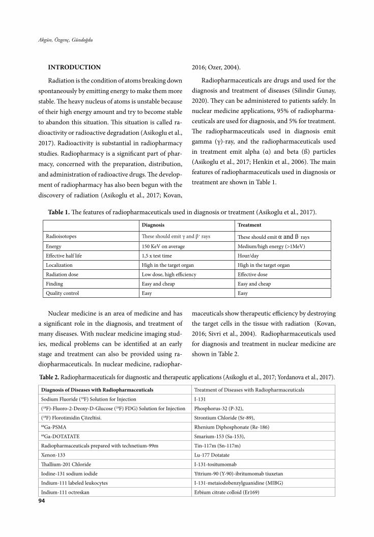
94
Akgün, Özgenç, Gündoğdu
INTRODUCTION
Radiation is the condition of atoms breaking down spontaneously by emitting energy to make them more stable. The heavy nucleus of atoms is unstable because of their high energy amount and try to become stable to abandon this situation. This situation is called ra-dioactivity or radioactive degradation (Asikoglu et al., 2017). Radioactivity is substantial in radiopharmacy studies. Radiopharmacy is a significant part of phar-macy, concerned with the preparation, distribution, and administration of radioactive drugs. The develop-ment of radiopharmacy has also been begun with the discovery of radiation (Asikoglu et al., 2017; Kovan,
2016; Ozer, 2004).
Radiopharmaceuticals are drugs and used for the diagnosis and treatment of diseases (Silindir Gunay, 2020). They can be administered to patients safely. In nuclear medicine applications, 95% of radiopharma-ceuticals are used for diagnosis, and 5% for treatment. The radiopharmaceuticals used in diagnosis emit gamma (γ)-ray, and the radiopharmaceuticals used in treatment emit alpha (α) and beta (ß) particles (Asikoglu et al., 2017; Henkin et al., 2006). The main features of radiopharmaceuticals used in diagnosis or treatment are shown in Table 1.
Table 1. The features of radiopharmaceuticals used in diagnosis or treatment (Asikoglu et al., 2017).
Diagnosis Treatment
Radioisotopes These should emit γ and β+ rays These should emit α and ß- rays
Energy 150 KeV on average Medium/high energy (>1MeV)
Effective half life 1,5 x test time Hour/day
Localization High in the target organ High in the target organ
Radiation dose Low dose, high efficiency Effective dose
Finding Easy and cheap Easy and cheap
Quality control Easy Easy
Nuclear medicine is an area of medicine and has a significant role in the diagnosis, and treatment of many diseases. With nuclear medicine imaging stud-ies, medical problems can be identified at an early stage and treatment can also be provided using ra-diopharmaceuticals. In nuclear medicine, radiophar-
maceuticals show therapeutic efficiency by destroying the target cells in the tissue with radiation (Kovan, 2016; Sivri et al., 2004). Radiopharmaceuticals used for diagnosis and treatment in nuclear medicine are shown in Table 2.
Table 2. Radiopharmaceuticals for diagnostic and therapeutic applications (Asikoglu et al., 2017; Yordanova et al., 2017).
Diagnosis of Diseases with Radiopharmaceuticals Treatment of Diseases with Radiopharmaceuticals
Sodium Fluoride (18F) Solution for Injection I-131
(18F)-Fluoro-2-Deoxy-D-Glucose (18F) FDG) Solution for Injection Phosphorus-32 (P-32),
(18F) Florotimidin Çözeltisi. Strontium Chloride (Sr-89),68Ga-PSMA Rhenium Diphosphonate (Re-186)68Ga-DOTATATE Smarium-153 (Sa-153),
Radiopharmaceuticals prepared with technetium-99m Tin-117m (Sn-117m)
Xenon-133 Lu-177 Dotatate
Thallium-201 Chloride I-131-tositumomab
Iodine-131 sodium iodide Yttrium-90 (Y-90)-ibritumomab tiuxetan
Indium-111 labeled leukocytes I-131-metaiodobenzylguanidine (MIBG)
Indium-111 octreskan Erbium citrate colloid (Er169)

95
FABAD J. Pharm. Sci., 46, 1, 93-104, 2021
1.1. Diagnosis of Diseases with Radiopharma-ceuticals
Radiopharmaceuticals that emit gamma rays are used in nuclear medicine to diagnose disease and determine the condition and function of specific tis-sue and organ (Gundogdu et al., 2018). There are two standard methods to diagnose the disease with radio-pharmaceuticals:
Non-image diagnostic applications: The radiophar-maceutical is reached to the desired organ/tissue, and radioactivity is counted from outside. The radioac-tivity calculation in the organ can be calculated with the Medical Internal Radiation Dose (MIRD), and the body surface area can be calculated using Monte Carlo methods (Kovan, 2016).
Image diagnostic applications: The radiopharma-ceutical is reached to desired organ/tissue and the or-gan/tissue is imaged by various imaging systems such as gamma cameras, single-photon emission computed tomography (SPECT), Positron Emission Tomogra-phy (PET), SPECT/CT, and PET/CT hybrid systems (Gundogdu et al., 2018).
1.2. Treatment of Diseases with Radiopharma-ceuticals
Radiopharmaceuticals that emit alpha and beta rays are administered in many nuclear medicine treat-ment studies. The radiopharmaceuticals are given to the patient and kept in the target tissue/organ. They destroy diseased tissue and provide the treatment. In this way, near/healthy tissues are exposed to dose as low as possible and targeted therapy occurs (Kovan, 2016; Sivri et al., 2004). The properties of the radionu-clides used for radionuclide therapy are given in Table 3. Elemental, metabolic, pharmaceutical agents, an-tibodies, bone imaging chelates, bioreducing agents, labeled cells, liposomes, microspheres, nanoparticles and niosomes are radiolabeled with radionuclides suitable for the treatment of diseases. These have some advantages:
• Theyarenon-invasivetreatment,
• They show medium and long-term low side ef-
fects,
• Theseagentsareusefulbecausetheycanbetarget-ed.
• The dose in normal tissue is low because theseagents can be absorbed in desired tissues (Sivri et al., 2004).
There are many radiopharmaceuticals that are used in the treatment of some diseases. The samples of radiopharmaceuticals are summarized below.
Classification of Treatments Using Radiopharma-ceutics:
1. Radioactive iodine therapy in thyroid diseases2. Radiopharmaceutical treatment in metastatic
bone pain3. Radionuclidic therapy in neuroendocrine tu-
mors4. Treatment with labeled antibody5. Intra-cavitary radiocolloid therapy6. Radiation synovectomy7. Radionuclidic therapy in myeloproliferative dis-
eases8. Intra-arterial radionuclide therapy by radiola-
beled microspheres 1.2.1 Iodine-131 Treatment in Thyroid Diseases
The treatment of benign and malignant diseases of the thyroid can be done with Iodine-131 (I-131). Radioiodine therapy has been used for a long time. In treating diseases such as hyperthyroidism, I-131 ra-dionuclide settles in the thyroid tissue. It destroys the follicle cells with the beta particles it emits and stops the growth and activities of the thyroid cells. In this case, it returns the functions of the overactive thyroid gland to normal (Mumtaz et al., 2009). In malignant diseases such as thyroid cancer, radioactive iodine therapy can also be applied to remove residual thyroid gland residues after thyroid surgery and treatment of the spread of thyroid cancers in the body. Since the dose of radioactive iodine preferred in the treatment of thyroid cancers is higher than the iodine dose in the treatment of hyperthyroidism, the patient should sleep in a room specially prepared for radioiodine

96
Akgün, Özgenç, Gündoğdu
treatment to prevent radiation to the environment. The drugs and diet used before the treatment should be adjusted within the framework of certain rules (Luster et al., 2008). The half-life of I-131 is eight days, and beta energy is 0.61 MeV. I-131 shows activity in the treatment of thyrotoxicosis, hyperthyroidism, thyroid cancer. It affects the formation of the thyroid hormone and creates atrophy in the tissue. Thus, the thyroid gland becomes smaller, and thyroid hormone level decreases. As a result, reduced cell proliferation occurs (Onsel, 1999; Adalet et al., 2012). The patients should be on a low iodine diet for at least a week and avoid iodized salt, milk, dairy products, eggs, fish, seafood, and red meats under the treatment of I-131. Tincture of iodine and similar antiseptic agents should not be used for two weeks after the treatment. If pa-tients have dentures, radioiodine should be drunk af-ter the removal of dentures. Food should not be eaten for an hour after the administration of I-131. Radioac-tive iodine therapy cannot be used in pregnant wom-en. I-131 biodistribution is performed by the urinary system, stomach, salivary glands. Saliva and urine flow can be increased by water, and I-131 level can be reduced. Gum and lemon slices should be chewed to prevent saliva accumulation (Sivri et al., 2004).
I-131 has some side effects: Radiation thyroiditis, neurological complications (edema and related neuro-logical complications), sialadenitis (painful, sensitive and impaired function of salivary glands, taste distur-bance and dry mouth), gastrointestinal complications, hematological complications. Late side effects include amenorrhea, testicular damage and decreased sperm, bone marrow suppression, and lung fibrosis (Handels-man et al., 1983; Onsel, 1999; Raymond et al., 1989)
1.2.2. Radiopharmaceutical Treatment in Meta-static Bone Pain
Pain is the most critical symptom in approximate-ly 70% of patients with advanced cancer, and inter-ventions against pain management are mostly inad-equate. With radiation therapy, bone pain caused by metastatic cancer can be controlled non-invasively, and functional status is improved. The emergence of
new painful metastases can also be prevented or de-layed (Liepe et al., 2005). Ideal radiopharmaceuticals used in pain palliative treatment should be retained explicitly in bone and quickly eliminated from soft tissues. The biological half-life must react with the re-tained bone and must be long enough to irradiate the surrounding tumor before a significant breakthrough occurs (Guerra Liberal et al., 2016). P-32, Sr-89, Re-186, Sa-153, Sn-117m are radiopharmaceuticals and used for palliative treatment of metastatic cancer pain. For example, Sr-89; is effective in patients with skel-etal metastases and more beneficial than drugs such as chemotherapeutic, hormones, and analgesics. The initial dose of therapeutic activity may vary depend-ing on the radionuclide used. Pain status may increase temporarily at the beginning of treatment, and bone marrow suppression may occur within 4-6 weeks af-ter radiopharmaceuticals administration. When ra-diopharmaceuticals are used in combination with chemotherapy or local radiotherapy, it increases the effectiveness of the treatment. (Sivri et al., 2004; Paes et al., 2010; Adalet et al., 2012; Lewin, 2018).
1.2.3. Radionuclidic therapy in neuro-endocrine tumors
Neuroendocrine tumors create complicated clin-ical pictures. These tumors are heterogeneous group tumors. Neuroendocrine tumors develop in the gas-trointestinal and lungs, but about 70% of these orig-inate from the gastroenteropancreatic system. Their incidence has been reported as 5.25 per 100,000 (Oz-kan, 2019). Neuroendocrine tumors can synthesize, store and secrete neuroamines and peptides. Func-tional neuroendocrine tumors or dysfunctional neu-roendocrine tumors, known as carcinoid syndrome, can lead to symptoms such as flushing, diarrhea, and right heart failure (Kaltsas et al., 2004). Clinical cases caused by functional neuroendocrine tumors have been treated with somatostatin analogs (SSA) for many years. However, with the progression of the disease, patient management becomes more complex, and the choice of alternative therapies is important for both survival and quality of life. (Kamp et al., 2013).

97
FABAD J. Pharm. Sci., 46, 1, 93-104, 2021
Neuroendocrine tumors, which are considered to be a slow disease, are very aggressive after they become metastatic, and especially liver metastases determine survival. Although surgical resection is recommended as a treatment option in these patient groups, many pa-tients are not suitable for this treatment method, and hepatic embolization methods are frequently used in this patient group. Besides, peptide receptor radionu-clide treatments have positive results in combination with systemic therapies used in patients with neuro-endocrine tumors, and serious toxic effects are not en-countered (Ozkan, 2019). Neuroendocrine cancers are cancers that are generally seen in the intestine, stom-ach, pancreas, and lung systems. Lu-177 dotatate and I-131-metaiodobenzylguanidine (MIBG) are current-ly used in the treatment of neuroendocrine cancers.
1.2.3.1. I-131-MIBG Treatment
I-131-MIBG is highly effective in pheochromo-cytoma and neuroblastoma. Also, I-131-MIBG is less effective in paraganglioma, carcinoid tumors, and medullary thyroid cancer. This treatment is not pre-ferred in pregnant patients. Renal functions and bone marrow reserve should be sufficient for the success of the treatment.
Radiation affects the cells by using ß-rays emitted from the I-131 MIBG being held by neighboring cells. Therefore, isolated cells are exposed to radiation less than macroscopic tumors and cell clumps. As a re-sult, smaller tumors are exposed to radiation less, and the chance of treatment is less (Grünwald et al., 2010; Adalet et al., 2012).
1.2.3.2. Lutetium-177 Dotatate Treatment
Lutetium-177 Dotatate is a prescribed radiophar-maceutical used in the treatment of gastroenteropan-creatic neuroendocrine tumors (GEP-NETs). This ra-diopharmaceutical concentrates on the tumor struc-ture and treats the cancer with beta rays. This form of treatment is applied 4 or 5 times with an average of two months’ periods. In this treatment, critical or-gans such as the kidney, bone marrow, and liver are exposed to some dosage. The radiation dose in the tar-
get organ is adjusted according to the maximum treat-ment dose of the treatment and the patient’s weight (Kovan, 2016).
1.2.4. Treatment with Labeled Antibody
Radio-labeled antibodies were first studied for tu-mor imaging, but with the use of F-18-FDG in pos-itron emission tomography, radio-labeled antibodies are no longer used for tumor detection. Later studies have shown that radiolabeled antibodies are useful in treating lymphoma (Barbet et al., 2009). Tositumom-ab, known as anti-B1, is a monoclonal antibody with an affinity for the CD20 antigen expressed on normal B-lymphocytes. CD20 is described in the majority of B cell lymphomas (Press et al., 2001). This antibody inhibits tumor growth in both animal models and humans (Buchsbaum et al., 1992). Tositumomab was radiolabeled with antibody I-131 (Bexxar), and B cell Non-Hodgkins was used for treatment and was ap-proved by the FDA. Tositumomab can inhibit tumor growth in animal models and humans, but it was not developed as a human-mouse chimeric antibody. The antibody is prepared from serum-free hybridoma su-pernatants. It is radiolabeled by the method of oxida-tion of radioactive iodine with iodogen. It is commer-cially available, and the I-131 radiolabelled tositum-omab solution contains additives (Providon, maltose, and ascorbic acid) to limit radiolysis (Cheson, 2003; Press et al., 2001). Another FDA-approved radiophar-maceutical used for B-cell Non-Hodgkins therapy is Yttrium-90-ibritumomab (Zevalin). Ibritumomab is a murine IgG1a kappa antibody and is used to treat lymphomas (Barbet et al., 2009). Yttrium-90 is a pure beta emitter, so problems caused by high-energy gam-ma rays emitted by iodine-131 do not arise. However, since Y-90 does not emit gamma rays, gamma imag-ing, and subsequent dosimetry calculations cannot be made. For this reason, indium-111 (In-111) is used with the Y-90 as it allows pre-treatment imaging for dosimetry evaluation and patient-specific dosing. In-111-ibritumomab tiuxetan and Y-90-ibritumomab tiuxetan are used to treat relapsed or refractory low-grade, follicular, or transformed B-cell NHL patients,

98
Akgün, Özgenç, Gündoğdu
including follicular patients resistant to rituximab (Witzig et al., 2002).
1.2.5. Intra-cavitary radiocolloid therapy
Intracavitary radiation therapy for brain tumors using beta-emitting radionuclides emerged 50 years ago. Phosphate-32 (P-32), Y-90, and Rhenium-186 (Re-186) are mostly used in cystic craniopharyngio-mas. These radionuclides are placed stereotactically, and since this method is effective, it is also a mini-mally invasive technique, so it is very beneficial for patient compliance. (Monaco et al., 2015). Treatment management of patients with craniopharyngioma should be aimed at preserving the quality of life and extending the life span and controlling the tumor. It is the first preferred method for patients when sur-gical procedures are possible. However, the need for removal of the maximal tumor in surgical operations causes potential surgical risks such as hypothalamic dysfunction, hypopituitarism and vision loss. It has been reported that craniopharyngiomas are generally involved in critical structures such as the optic appa-ratus, hypothalamus, and pituitary stalk. When com-plete resection is not possible, other treatment modal-ities are needed. (Niranjan et al., 2010). Intracavitary radiation therapy is one of these methods. Intracav-itary radiation therapy is performed using radionu-clides P-32, Y-90, and Re-186. Both P-32 and Y-90 are pure beta particle emitting isotopes made in colloidal suspensions, Re-186 having both beta and gamma emission. Also, it has also been reported that Re-186 preparations are less dense and could potentially pro-trude beyond the cyst wall. Since pure beta-emitting radionuclides do not emit gamma rays, they provide ease of use in terms of radioprotection for the patient environment. The short penetration of beta particles prevents the healthy cells of the patient from receiving unnecessary radiation. (Backlund et al., 1989; Julow et al., 1985). Ideally, the radionuclide is desired to be a pure high energy beta emitter. The efficiency of both beta and gamma-emitting radionuclides is limited by the gamma rays they emit. P-32 has lower beta energy than Y-90 and has a shorter penetration and a longer
half-life. The shorter penetration allows healthy cells to be exposed to less radioactivity in treatment. Be-sides, the fact that the radionuclide has a longer half-life enables tumor cells to be exposed to radioactivity for a longer time (Blackburn et al., 1999).
1.2.6. Radiation Synovectomy Treatment
Radiation synovectomy therapy is a form of treat-ment performed by intraarticular injection of be-ta-emitting radionuclides. Synovial cells are irradiated with beta radiation, reducing cell proliferation and causing cell destruction. In different types of arthri-tis and osteoarthritis, synovitis is the leading cause of pain and discomfort. Therefore, the destruction of synovial cells by the radiosynovectomy method is an option when surgical procedures cannot be used. (Karavida et al., 2010). Surgical synovectomy requires general anesthesia, reduces joint movement, requires a long hospital stay, and increases the risk of infec-tion. While surgical synovectomy is not preferred primarily, radiation synovectomy is preferred (Kavak-li et al., 2002; Polat, 2010). Diseases in which radia-tion synovectomy is indicated; rheumatoid arthritis, ankylosing spondylitis, psoriatic arthritis, synovitis, osteoarthritis, villonodular synovitis, and hemophil-ic synovitis caused by inflammation of the synovial membrane (Adalet et al., 2012; Schneider et al., 2005). The most widely used radionuclides Y-90, Re-186, Er-bium-169 (Er-169), and P-32 are the most commonly used in nuclear medicine to treat radiosynovectomy. According to the density of the synovial cells, the ra-dionuclide with the appropriate physical half-life is selected. Besides, the average tissue penetration of the emitted particulate radiation is an essential parame-ter for radionuclide selection. The particle size of the radionuclides used according to the common area to be treated is also essential. The biodegradability of the radionuclides injected into synovial cells, and the irreversible binding of the radionuclide to them are too crucial in the selection of radiopharmaceuticals for radionuclide treatment. The smaller the joint, the shorter the penetration of the emitted beta particles should be (Farahati et al., 2002).

99
FABAD J. Pharm. Sci., 46, 1, 93-104, 2021
Synovioortesis is a non-surgical method of sy-novectomy, applied by injection of a radioactive or chemical substance into the knuckle. However, radia-tion synovectomy is a good option that is much more effective than chemical synovectomy. The chemical synovectomy is an excruciating procedure, and that should be repeated frequently. Chemical synovecto-my method is preferred only in places where reach to radionuclide agents is insufficient (Molho et al., 1999; Haklar, 2005; Polat, 2010). The basic principle of radiation synovectomy is the injection of radiophar-maceuticals into the knuckle during a specific period. The source of enzymes that cause cartilage destruction and lead to knuckle pathology is secreted synovial tis-sue and fluid. For this reason, fibrosis of this region is the primary effect in treatment. Less joint damage is in line with good results. Radioisotope synovectomy can be preferred in treating chronic hypertrophic synovi-tis with recurrent hemarthrosis that non-response to hematological treatment, pigmented villonodular sy-novitis, osteoarthritis (Knapp et al., 1999; Sivri et al., 2004; Polat, 2010).
Sterile conditions should be provided during in-jection, and the physician should be sure that the in-jector is in the knuckle cavity. Generally, scope control is required during the application, but it is not needed for the knee knuckle (Rodriguez-Merchan et al., 2001; Polat, 2010; ).
1.2.7. Intra-arterial Radionuclide Therapy by Radiolabeled Microspheres
Hepatocellular carcinoma is the most common form of primary liver tumors. The liver is the most common organ of metastatic spread, and metastasis is seen in various forms (Ahmadzadehfar et al., 2010; Adalet et al., 2012; ). In cases where surgical interven-tions and local treatment methods cannot be applied, intra-arterial radiomicrocure treatment is preferred. It is based on the principle of elimination by giving a high radiation dose to the tumor. The basic princi-ple of this treatment is that tumor cells in the liver are predominantly based on feeding on the hepatic artery and healthy cells providing on the portal venous sys-
tem. Y-90 microspheres are selectively directed to tu-mor cells, so that tumor cells are exposed to high-dose radiation. However, side effects occur very little due to radiation in healthy liver and other tissues. Also, fac-tors that limit the treatment include absorption dose calculation and radiomicrocure distribution are not homogeneous (Adalet et al., 2012; Feryal, 2019). Y-90 is used in treatment, has a half-life of 64,1 hours, and as a result of its degradation, zirconium-90 (Zr-90) is formed with a 99,98% probability. An antineutrino is formed with a maximum energy beta-particle of 2,28 MeV. All dose of Y-90 is absorbed in the liver, and pa-tient isolation is not required for radioprotective pur-poses (Gulec et al., 2007; Uliel et al., 2012; Mehmet, 2017).
1.3. Radiopharmaceuticals in Theranostics Administration
Theranostics are mainly used in the treatment of cancerous and infected areas. Thanks to its diagnos-tic and therapeutic properties, theranostics reduce the side effects of treatments, and increase compliance and survival rates for patients. As a result of this ap-proach, molecular imaging is applied by combining the diagnostic and therapeutic methods with the diag-nostic agent, which has the same or similar chemical structure as the therapeutic. Predictions about the re-sponse to treatment are provided. With this method, diseases can be classified according to the molecular phenotype, the biodistribution of the molecule can be observed, and the response to treatment can be moni-tored (Durak, 2015; Kelkar et al., 2011; Lee, 2011).
The correlation between the diagnostic and ther-apeutic methods can be divided into three groups. These groups are:
-Diagnostic and therapeutic molecules are the same,
-Diagnostic and therapeutic molecules are similar,
-Diagnostic and therapeutic molecules are differ-ent, but the effect mechanism of them is similar
Same Diagnostic and Therapeutic Molecules
The best example for this group is the use of I-131.

100
Akgün, Özgenç, Gündoğdu
It can be used in diagnosis and therapy. Also, Indi-um-111 (In-111) Octreotide therapy and, In-111 Oc-treotide scintigraphy, I-131 MIBG therapy and I-131 MIBG scintigraphy, Lu-177 DOTA therapy, and Lu-177 DOTA scintigraphy are examples of this group (Durak, 2015; Srivastava, 2012).
Similar Diagnostic and Therapeutic Molecules
Examples for this group are imaging with Tech-netium-99m (Tc-99m)-MDP or Tc-99m-HDP, treat-ment with Re-186-HEDP, or Sm-153-EDTMP, and imaging with Ga-68-DOTA and treatment with Lu-177 or Y-90 DOTA peptides (Srivastava, 2012).
Due to the different electron configurations of radionuclides, chemical and biochemical features, molecular stability, and biological behavior of radio-pharmaceuticals can differ, but similar behavior is ex-pected for these molecules (Durak, 2015).
Different Diagnostic and Therapeutic Molecules, But Similar Effect Mechanism
The treatment with the Y-90 microsphere, imag-ing with Tc-99m MAA in liver cancer, can be given as the most common sample of this group. Although the molecules are entirely different from each other, the behavior is similar (Durak, 2015).
CONCLUSION
The selection of suitable radionuclides is essential in any therapeutic radiopharmaceutical development. Their nuclear emission characteristics, physical half-life, degradation properties effect in vivo pharmacokinet-ics, cost, and availability of the radiopharmaceuticals. Also, types of particle emission affect the distribution and pharmacokinetics of the radiopharmaceuticals. For therapeutic radiopharmaceuticals, radionuclides that degrade by α-particle, B-particle, and Auger-electron emission are commonly used in the preparation.
In this review article, information about the radio-pharmaceuticals used in the treatment is given. Today, a limited number of radiopharmaceuticals are routine-ly used in the treatment of many diseases. In the past ten years, clinical studies have been focused on radio-immunotherapy. Studies on radiolabeled monoclonal
antibodies and radiolabeled receptor binding agents are being conducted. Few studies have achieved their goal so far and are currently used in treatment. The best results have been achieved in treating lymphomas, and two FDA-approved radiopharmaceuticals are used in practice. Radiopharmaceutical science is an emerging research field that meets the clinical standards. They have an essential therapy index and high imaging abil-ity. The scientific community examines various radio-nuclides, pharmaceutical agents, radiopharmaceuticals, and evaluating in vivo performance of newly developed radiopharmacauticals. Thus, great efforts are being made to develop new radiopharmaceuticals to be con-verted into clinical research to treat many diseases. As detailed in this review, most of the current studies focus on the diagnostic and therapeutic applications of radio-pharmaceuticals. We believe that these developments will have positively affects on human life and lead to positive results in the diagnosis and therapy of patients.
CONFLICT OF INTEREST
The authors declare that there are no conflicts of interest.
AUTHOR CONTRIBUTION STATEMENT
Developing hypothesis (Gundogdu E.) prepar-ing the study text (Akgun E., Ozgenc E.) reviewing the text (Gundogdu E., Ozgenc E.) literature research (Akgun E., Ozgenc E.)
REFERENCES
Adalet, A. M., Unal, S. N., Turkmen, C. I. (2012). Nukleer tıp ders kitabı. Istanbul University Publi-cations, Istanbul.
Ahmadzadehfar, H., Biersack, H. J., Ezziddin, S. (2010). Radioembolization of Liver Tumors With Yttrium-90 Microspheres. Seminars in Nuclear Medicine, 40(2), 105–121. https://doi.org/10.1053/j.semnuclmed.2009.11.001
Asikoglu, M., Ilem Ozdemir, D., Atlihan Gundogdu, E. (2017). Radyofarmasi. Ege University Publications, Izmir.

101
FABAD J. Pharm. Sci., 46, 1, 93-104, 2021
Atlihan Gundogdu, E., Ozgenc, E., Ekinci, M., Ilem Ozdemir, D., Asikoglu, M. (2018). Radiophar-maceuticals Used in Imaging and Treatment in Nuclear Medicine. Journal of Literature Pharma-cy Sciences, 7(1), 24–34. https://doi.org/10.5336/pharmsci.2017-56434
Backlund, E. O., Axelsson, B., Bergstrand, C. G., Eriksson, A. L., Norén, G., Ribbesjö, E., Rähn, T., Schnell, P. O., Tallstedt, L., Sääf, M., Thorén, M. (1989). Treatment of craniopharyngiomas - the stereotactic approach in a ten to twenty-three years’ perspective - I. Surgical, radiological and ophthal-mological aspects. Acta Neurochirurgica, 99(1–2), 11–19. https://doi.org/10.1007/BF01407771
Blackburn, T. P. D., Doughty, D., Plowman, P. N. (1999). Stereotactic intracavitary therapy of recurrent cys-tic craniopharyngioma by instillation of 90yttrium. British Journal of Neurosurgery, 13(4), 359–365. https://doi.org/10.1080/02688699943457
Bozkurt, M. (2017). Kolanjiyokarsinom tanısı ile ka-raciğer metastazlarına y-90 mikroküre tedavisi ver-ilmiş hastalarda hepatik arter perfüzyon çalışması görüntüleri ile tedavi öncesi – tedavi sonrası bilgi-sayarlı tomografi ve fdg pet bt görüntüleri arasında-ki ilişkinin araştırılması. Hacettepe Universitesi Tıp Fakültesi Nükleer Tıp Anabilim Dalı, Uzmanlık Tezi. Ankara, 38-42.
Buchsbaum, D. J., Wahl, R. L., Normolle, D. P., Ka-minski, M. S. (1992). Therapy with Unlabeled and 131I-labeled Pan-B-Cell Monoclonal Antibodies in Nude Mice Bearing Raji Burkitt’s Lymphoma Xenografts. Cancer Research, 52(23), 6476-6481.
Cheson, B. D. (2003). Radioimmunotherapy of non-Hodgkin lymphomas. American Society of Hematology, 101(2), 391-398. https://doi.org/10.1182/blood-2002-06-1793
Durak, H. (2015). Personalized Therapy and Thera-nostic Approaches in Oncology. Nuclear Medicine Seminars, 1(2), 80–84. https://doi.org/10.4274/nts.2015.014
Farahati, J., Schulz, G., Wendler, J., Körber, C., Gel-ing, M., Kenn, W., Schmeider, P., Reidemeister, C., Reiners, C. (2002). Multivariate analysis of factors influencing the effect of radiosynovecto-my. NuklearMedizin, 41(2), 114–119. https://doi.org/10.1055/s-0038-1625643
Feryal, C. (2019). Development of a method for deter-mination of activity quantities of Y-90 microspheres to be administered in treatment of liver tumors by patient specific modeling and standardization of the method. Hacettepe University Publications.
Grünwald, F., Ezziddin, S. (2010). 131I-Metaiodo benzyl guanidine therapy of neuroblastoma and other neuroendocrine tumors. In Seminars in Nuclear Medicine, 40(2), 153–163. https://doi.org/10.1053/j.semnuclmed.2009.11.004
Guerra Liberal, F. D. C., Tavares, A. S., Tavares, R. S. (2016). Palliative treatment of metastatic bone pain with radiopharmaceuticals: A perspective be-yond Strontium-89 and Samarium-153. In Applied Radiation and Isotopes, 110, 87–99. https://doi.org/10.1016/j.apradiso.2016.01.003
Gulec, S. A., Siegel, J. A. (2007). Posttherapy radia-tion safety considerations in radiomicrosphere treatment with 90Y-microspheres. Journal of Nu-clear Medicine, 48(12), 2080–2086. https://doi.org/10.2967/jnumed.107.045443
Haklar, U. (2005). Diz Ekleminde Sinovektomi. TOT-BID (Türk Ortopedi ve Travmatoloji Birliği Dernegi Dergisi, 4(3–4), 108–117. http://dergi.totbid.org.tr/files/4_2/6.pdf
Handelsman, D. J., Turtle, J. R. (1983). Testicular dam-age after radioactive iodine (I‐131) therapy for thy-roıd cancer. Clinical Endocrinology, 18(5), 465–472. https://doi.org/10.1111/j.1365-2265.1983.tb02876.x
Henkin, R. E., Bova, D., Dillehay, G. L., Karesh, S. M., Halama, J. R., Wagner, R. H., Kim, E. E. (2006). Nuclear Medicine. The Journal of Nucle-ar Medicine, 48(5), 846. https://doi.org/10.2967/jnumed.107.040329

102
Akgün, Özgenç, Gündoğdu
Barbet, J., Bardiès, M., Bourgeois, M., Chatal, J. F., Chérel, M., Davodeau, Gestin, J. F. (2009). Radio-labeled Antibodies for Cancer Imaging and Ther-apy. In Radiolabeled Antibodies for Cancer Treat-ment, 907, 681–695.
Julow, J., Lányi, F., Hajda, M., Simkovics, M., Arany, I., Tóth, S., Pásztor, E. (1985). The radiotherapy of cystic craniopharyngioma with intracystic in-stallation of 90Y silicate colloid. Acta Neurochiru-rgica, 74(3–4), 94–99. https://doi.org/10.1007/BF01418795
Kaltsas, G. A., Besser, G. M., Grossman, A. B. (2004). The diagnosis and medical management of ad-vanced neuroendocrine tumors. In Endocrine Reviews, 25(3), 458–511. https://doi.org/10.1210/er.2003-0014
Kamp, K., Gumz, B., Feelders, R. A., Kwekkeboom, D. J., Kaltsas, G., Costa, F. P., De Herder, W. W. (2013). Safety and efficacy of everolimus in gastrointesti-nal and pancreatic neuroendocrine tumors after 177Lu-octreotate. Endocrine-Related Cancer, 20(6), 825–831. https://doi.org/10.1530/ERC-13-0254
Karavida, N., Notopoulos, A. (2010). Radiation Syn-ovectomy: An effective alternative treatment for inflamed small joints. Hippokratia, 14(1), 22–27. http://www.ncbi.nlm.nih.gov/pubmed/20411055. Erişim tarihi: 15 Şubat 2020.
Kavaklı, R., Aydogdu, S., Duman, Y., Taner, M., Memis, A., Capacı, K., Hepguler, A., Yılmaz Kara-pınar, D., Balkan, C. (2002). Musculoskeletal is-sues. Haemophilia, 8(4), 469–481. https://doi.org/10.1046/j.1365-2516.2002.t01-1-00001.x
Kelkar, S. S., Reineke, T. M. (2011). Theranostics: Combining Imaging and Therapy. Bioconju-gate Chemistry, 22(10), 1879–1903. https://doi.org/10.1021/bc200151q
Kovan, B. (2016). 177Lu İle Nöroendokrin Tümörler-inin Tedavisinde Radyasyon Dozimetrisi. Istanbul University Faculty of Science Institute Publications. http://katalog.istanbul.edu.tr/client/tr_TR/de-fault_tr/search/results?qu=Tümörler+--+Teda-vi.&ps=300. Erişim tarihi: 07 Haziran 2020.
Knapp, F. F., Beets, A. L., Pinkert, J., Kropp, J., Lin, W. Y., Wang, S. Y. (1999). Rhenium radioisotopes for therapeutic radiopharmaceutical development. Inter seminar on therapeutic applications of radio-pharmaceuticals (IAEA-SR-209).
Lee, D. Y., King, C. P. (2011). Molecular theranostics: a primer for the imaging professional. American Journal of Roentgenology, 197(2), 318–324. https://doi.org/doi:10.2214/AJR.11.6797
Lewin, B. (2018). Radiopharmaceuticals Metastron (Strontium-89) and Samarium-153 (Quadramet) for Metastatic Bone Pain. Aetna, 0361, 1-20.
Liepe, K., Runge, R., Kotzerke, J. (2005). The benefit of bone-seeking radiopharmaceuticals in the treat-ment of metastatic bone pain. Journal of Cancer Research and Clinical Oncology, 131(1), 60–66. https://doi.org/10.1007/s00432-004-0625-0
Luster, M., Clarke, S. E., Dietlein, M., Lassmann, M., Lind, P., Oyen, W. J. G., Tennvall, J., Bombardieri, E. (2008). Guidelines for radioiodine therapy of differentiated thyroid cancer. European Journal of Nuclear Medicine and Molecular Imaging, https://doi.org/10.1007/s00259-008-0883-1
Molho, P., Verrier, P., Stieltjes, N., Schacher, J. M., Ounnoughène, N., Vassilieff, D., Menkes, C. J., & Sultan, Y. (1999). A retrospective study on chemi-cal and radioactive synovectomy in severe haemo-philia patients with recurrent haemarthrosis. Hae-mophilia, 5(2), 115–123. https://doi.org/10.1046/j.1365-2516.1999.00287.x
Monaco, E. A., Tempel, Z., Niranjan, A., Lunsford, L. D. (2015). Intracavitary Therapy: Radioisotopes 32P, 90Y, and 186Re. In Craniopharyngiomas: Comprehensive Diagnosis, Treatment and Outcome, Elsevier Inc, 391–403. https://doi.org/10.1016/B978-0-12-416706-3.00024-6
Mumtaz, M., Lin, L. S., Hui, K. C., & Mohd Khir, A. S. (2009). Radioiodine I-131 for the therapy of graves’ disease. The Malaysian Journal of Medical Scienc-es, 16(1), 25–33. http://www.ncbi.nlm.nih.gov/pubmed/22589645. Erişim tarihi: 10 Ocak 2020.

103
FABAD J. Pharm. Sci., 46, 1, 93-104, 2021
Naki Sivri N., Ozer, Y. (2004). Radyonüklidik Tedavi. Meslek İçi Sürekli Eğitim Dergisi. 1, 1-10
Niranjan, A., Kano, H., Mathieu, D., Kondziolka, D., Flickinger, J. C., Lunsford, L. D. (2010). Radiosur-gery for craniopharyngioma. International Journal of Radiation Oncology Biology Physics, 78(1), 64–71. https://doi.org/10.1016/j.ijrobp.2009.07.1693
Onsel Ç. (1999). Tiroid Kanserlerinin Tanı ve Tedavi-si. İ.Ü. Cerrahpasa Tıp Fakültesi Sürekli Tıp Eğitimi Etkinlikleri. 1, 49-68
Ozer, Y. (2004). Radyofarmasi, Radyofarmasötiklerin Dünü, Bugünü ve Geleceği. Meslek İçi Sürekli Eği-tim Dergisi.
Özkan, Z. G. (2019). Radioembolization in the Treatment of Neuroendocrine Tumors. Nucle-ar Medicine Seminars, 5(2), 152–158. https://doi.org/10.4274/nts.galenos.2019.0020
Paes, F. M., Serafini, A. N. (2010). Systemic Metabol-ic Radiopharmaceutical Therapy in the Treatment of Metastatic Bone Pain. In Seminars in Nuclear Medicine, 40(2), 89–104. https://doi.org/10.1053/j.semnuclmed.2009.10.003
Polat Kelle, A. (2010). Radyonüklid sinovektominin kronik hemofilik sinovitis tedavisindeki önemi. Çukurova Üniversitesi Tıp Fakültesi Nükleer Ana-bilim Dalı. https://silo.tips/download/radyon-kld-snovektomnn-kronk-hemoflk-snovts-te-davsndek-nem. Erişim tarihi: 12 Ekim 2019.
Press, O. W., Leonard, J. P., Coiffier, B., Levy, R., Tim-merman, J. (2001). Immunotherapy of Non-Hod-gkin’s lymphomas. In Hematology the Education Program of the American Society of Hematolo-gy. American Society of Hematology. Education Program, 2001(1), 221–240. American Society of Hematology. https://doi.org/10.1182/asheduca-tion-2001.1.221
Raymond, J. P., Izembart, M., Marlıac, V., Dagousset, F., Merceron, R. E., Vulpillat, M., Vallué, G. (1989). Temporary Ovarian Failure in Thyroid Cancer
Patients after Thyroid Remnant Ablation with Ra-dioactive Iodine. The Journal of Clinical Endocri-nology & Metabolism, 69(1), 186–190. https://doi.org/10.1210/jcem-69-1-186
Rodriguez-Merchan, E. C., Goddard, N. J. (2001). The technique of synoviorthesis. Haemophil-ia, 7(2), 11–15. https://doi.org/10.1046/j.1365-2516.2001.00103.x
Schneider, P., Farahati, J. R. C. (2005). Radiosynovec-tomy in rheumatology, orthopedics, and hemo-philia. Journal of Nuclear Medicine, 46(1), 48–54.
Silindir Gunay, M. (2020). The Formulation of Meth-ylene Blue Encapsulated, Tc-99m Labeled Multi-functional Liposomes for Sentinel Lymph Node Imaging and Therapy. Turkish Journal of Pharma-ceutical Sciences, 17(4), 381-387.
Srivastava, S. C. (2012). Paving the way to person-alized medicine: Production of some promising theragnostic radionuclides at Brookhaven nation-al laboratory. In Seminars in Nuclear Medicine, 42(3), 151–163. https://doi.org/10.1053/j.semnu-clmed.2011.12.004
Uliel, L., Royal, H. D., Darcy, M. D., Zuckerman, D. A., Sharma, A., Saad, N. E. (2012). From the an-gio suite to the γ-camera: Vascular mapping and 99mTc-MAA hepatic perfusion imaging before liver radioembolization-A comprehensive pictorial re-view. Journal of Nuclear Medicine 53(11), 1736–1747. https://doi.org/10.2967/jnumed.112.105361
Witzig, T. E., Gordon, L. I., Cabanillas, F., Czuczman, M. S., Emmanouilides, C., Joyce, R., Pohlman, B. L., Bartlett, N. L., Wiseman, G. A., Padre, N., Gril-lo-López, A. J., Multani, P., White, C. A. (2002). Randomized controlled trial of yttrium-90-la-beled ibritumomab tiuxetan radioimmunothera-py versus rituximab immunotherapy for patients with relapsed or refractory low-grade, follicular, or transformed B-cell non-Hodgkin’s lymphoma. Journal of Clinical Oncology, 20(10), 2453–2463. https://doi.org/10.1200/JCO.2002.11.076

104
Akgün, Özgenç, Gündoğdu
Yeong, C. H., Cheng, M. Hua, K. H. (2014). Thera-peutic radionuclides in nuclear medicine: Cur-rent and future prospects. Journal of Zhejiang University: Science B, 15(10), 845–863. https://doi.org/10.1631/jzus.B1400131
Yordanova, A., Eppard, E., Kürpig, S., Bundschuh, R. A., Schönberger, S., Gonzalez-Carmona, M., Feld-mann, G., Ahmadzadehfar, H., Essler, M. (2017). Theranostics in nuclear medicine practice. Onco-Targets and Therapy, 10, 4821–4828. https://doi.org/10.2147/OTT.S140671.

105
FABAD J. Pharm. Sci., 46, 1, 105-118, 2021
Enhancing Skin Penetration: The Role of Microneedles
Bülent SAMANCI*º , Fatma Gülgün YENER** , İsmail Tuncer DEĞİM***
Review ARticles
* ORCID: 0000-0002-7198-5375, Pharmaceutical Technology Department, Faculty of Pharmacy, Dicle University, Diyarbakir, Turkey, ** ORCID: 0000-0002-7234-0034, Proffesor, Pharmaceutical Technology Department, Faculty of Pharmacy, Istanbul University, Istanbul, Turkey *** ORCID: 0000-0002-9329-4698, Proffesor, Pharmaceutical Technology Department, Faculty of Pharmacy, Biruni University, Istanbul, Turkey
º Corresponding Author: Bülent SAMANCIPhone: +904122411000-7531; e-mail: [email protected]
Enhancing Skin Penetration: The Role of Microneedles
SUMMARY
since transdermal delivery systems provide some important advantages over oral delivery systems and parenteral delivery systems, they have attracted the attention of researchers. Degradation of the drug in the gastrointestinal (Gi) system, irritation of the Gi system tract, and the first-pass effect of the drug are some of the disadvantages of oral administration, while the need for medical staff to administer it and creating phobia in the patient are among the disadvantages of parenteral administration. to overcome these drawbacks, researchers have developed formulations for the transdermal delivery of drugs. The most important handicap of transdermal drug administration is the stratum corneum layer (st. corneum), which forms the enormous barrier layer of the skin. some techniques have been developed to overcome this serious barrier problem of the skin. Microneedles are one of the physical methods to increase the penetration of therapeutic agents through the skin. Microneedles consist of needle arrays long enough to deliver the drug to the dermis layer and micron-sized enough to not reach the nerve cells and not cause pain. Microneedles can be classified into five different types as solid microneedles, dissolving microneedles, hollow microneedles, coated microneedles, and hydrogel microneedles according to the properties of the materials used in the fabrication and the mechanisms of release of the therapeutic agent. Microneedles can be used in the application of vaccines, proteins, nucleotides, drug delivery systems, cosmetic, and for diagnostic purposes. Although important technological developments have been experienced for microneedle in many areas such as drug delivery systems, disease diagnosis, and cosmetics in the last two decades, there are many working areas that need to be developed. especially in long-term treatments, studies should be done to develop them as smart devices.
Key Words: Drug Delivery, intradermal, Microfabricated device, Microneedle, skin Penetration, transdermal.
Received: 7.07.2020Revised: 26.08.2020Accepted: 18.09.2020
Cilt Penetrasyonunun Artırılması: Mikroiğnelerin Rolü
ÖZ
transdermal taşıyıcı sistemler, oral taşıyıc sistemler ve parenteral taşıyıcı sistemlere göre bazı önemli avantajlar sağladığından araştırmacıların dikkatini çekmiştir. İlacın gastrointestinal (Gi) sistemde bozunmaya uğraması, ilacın Gi sistem yolağında oluşturduğu tahriş ve ilacın ilk geçiş etkisine uğraması, oral uygulamanın bazı dezavantajlarındandır. İlacın parenteral uygulanması için bir tıbbi personele ihtiyaç duyulması ve parenteral uygulamada kullanılan iğnenin hastalarda korku yaratması parenteral uygulamanın dezavantajları arasındadır. Bu olumsuzlukların üstesinden gelmek için, araştırmacılar transdermal ilaç taşıyıcı sistemler geliştirdiler. transdermal ilaç uygulamasındaki en büyük engel, derinin muazzam bariyer tabakasını oluşturan stratum corneum (st. corneum) tabakasıdır. Derinin bu ciddi bariyer probleminin üstesinden gelmek için bazı teknikler geliştirilmiştir. Mikroiğneler, terapötik ajanların deriye nüfuz etmesini arttırmak için geliştirilen fiziksel yöntemlerden biridir. Mikroiğneler, ilacı dermis tabakasına iletecek kadar uzun ve sinir hücrelerine ulaşmayacak ve ağrıya neden olmayacak kadar mikron boyutlu iğne dizilerinden oluşmaktadır. Mikroiğneler, imalatta kullanılan malzemelerin özelliklerine ve terapötik maddenin salınım mekanizmalarına göre katı mikroiğneler, çözünebilen mikroiğneler, içi boş mikro iğneler, kaplı mikro iğneler ve hidrojel mikro iğneler olarak 5 farklı tipte sınıflandırılabilir. Mikroiğneler aşıların, proteinlerin, nükleotidlerin, ilaç taşıyıcı sistemlerinin, kozmetiklerin uygulanmasında ve teşhis amaçlı kullanılabilir. son yirmi yılda ilaç taşıyıcı sistemleri, hastalık teşhisi ve kozmetik gibi birçok alanda mikroiğneler için önemli teknolojik gelişmeler yaşanmış olsa da, geliştirilmesi gereken birçok çalışma alanı vardır. Özellikle uzun süreli tedavilerde, mikroiğneleri akıllı cihaz olarak geliştirmek amacıyla araştırmalar yapılmalıdır.
Anahtar kelimeler: İlaç taşıyıcı, intradermal Mikrofabrikasyon cihaz, Mikroiğne, Deri penetrasyonu, transdermal.

106
Samancı, Yener, Değim
IntroductIon
Although the transdermal administration of drugs for systemic effect is a new administration way, the drugs have been applied to the skins for years for both cosmetic and therapeutic purposes. Previously, they were administering drugs to the skin to act locally. Subsequently, studies have shown that drugs can pen-etrate through the skin and they can reach systemic circulation. Poor drug absorption or enzymatic deg-radation in the gastrointestinal tract or liver restricts the use of some drugs orally (W. Liu et al., 2016). Also, the administration of hypodermic needles is limited due to the pain and psychological distress (Subramo-ny, 2013). Such problems further increase the interest in the administration of drugs to the skin. Some ad-vantages of the application of drugs to the skin can be listed as follows (Zhou et al., 2018):
• To prevent the degradation of drugs in the diges-tive tract and the low absorption of food-related.
• Avoiding the first pass effect of liver
• Improving bioavailability
• Providing constant plasma concentration up to 7 days with the same patch
• Elimination of disorders related to hypodermic needle-related pain, fear, and infection
In spite of that, affections of drugs in creams, patches, solutions, and other traditional transdermal and intradermal dosage forms are limited because of the skin’s impressive barrier function to the transport of ingredients into the body (Y. Park, Kim, Chung, Sung, & Kim, 2016). The Stratum Corneum (St. Cor-neum) is the main barrier in the skin and the outer-most layer of the skin (Cortes et al., 2019). Restricted drug delivery through the skin is one of the disad-vantages of topical creams and transdermal patches (Waghule et al., 2019). Due to the drawbacks of hypo-dermic needles such as pain and fear, researchers have developed various methods to increase penetration through the skin. Chemical and natural penetration enhancers (J. Chen et al., 2016), iontophoresis (Dix-it, Bali, Baboota, Ahuja, & Ali, 2007), electroporation (Escobar-Chavez, Bonilla-Martinez, Villegas-Gonza-lez, & Revilla-Vazquez, 2009), ultrasound-mediated
systems (Polat, Hart, Langer, & Blankschtein, 2011), local hypobaric pressure (Inacio et al., 2016) and Mi-croneedles (MNs) (Ita, 2015b) increase the penetra-tion of molecules by creating tiny holes in the skin. For the development of effective and innovative skin penetration enhancing systems, the structure of the skin, its components, and the mechanisms through the skin should be well known.
General InformatIon
Skin’s structure and permeability
In an adult, an average weight of skin is about 9 kg, and an average surface area of 2 m2 makes the skin the largest organ (Degim, 2006). The skin completely covers the body and forms a flexible barrier between the organism and the external environment (Degim, 2006). In addition to being the organ of the sense of touch in our body (Tsakovska et al., 2017), adjusting body temperature is one of the functions of the skin (Sawasaki, Iwase, & Mano, 2001). We can list some of the functions of the skin as follows (Tsakovska et al., 2017);
• Providing protection against unwanted toxic sub-stances and pathogenic microorganisms
• Protects the body against mechanical forces
• Prevents body from losing water
• It has an immunological activity by creating an inflammatory response against foreign substances entering the skin.
Skin consists of three basic layers shown in Figure 1. (Lai-Cheong & McGrath, 2013).
• Epidermis
• Dermis
• Subcutis
The epidermis consists of a multilayered epithe-lium layer, interfollicular epidermis associated hair follicles, and sebaceous and sweat glands (Niemann & Watt, 2002). The epidermis consists of basic four layers. The deepest part of the epidermis is the basal layer where the keratinocytes are single layers. Kera-tinocytes differentiate towards the skin surface and form the St. Corneum layer at the outermost part of the skin (Lai-Cheong & McGrath, 2013).

107
FABAD J. Pharm. Sci., 46, 1, 105-118, 2021
The St. Corneum consists of a structure known as the “brick and mortar” model (Larraneta, McCrud-den, Courtenay, & Donnelly, 2016). Bricks consist of terminally differentiated keratinocyte consisting of keratin filaments and filaggrin. The mortar consists of intercellular lipids (Meckfessel & Brandt, 2014). The lipid structure consists of a combination of ceramide, cholesterol, and free fatty acids in a roughly 1: 1: 1 ra-tio (Lundborg et al., 2018). St. Corneum lipids prevent transepidermal water loss (TEWL) and form the main barrier function of the skin (Meckfessel & Brandt, 2014). A deformation in the St. Corneum structure causes TEWL, leading to dysfunction of the barrier function of the skin. There is increasing interest in drug delivery via the skin to alleviate the drawbacks of parenteral administration and oral administration of drugs. However, The St. Corneum provides the body with tremendous protection against external influ-ences and is a major obstacle to drug administration
to the skin. The structure of the skin in the area where the drug is administered and the physicochemical properties of the drug are important factors affecting the penetration of the drug through the skin (Kamble, Sadarani, Majumdar, & Bhullar, 2017). In order for a drug to penetrate easily through the skin, its parti-tion coefficient must be high (Log P range of 1-3), its molecular weight should be less than 500 Da, and its melting point should be less than 200 Co.(Prausnitz & Langer, 2008). The drug can reach the lower layers of the skin in different three pathways such as intra-cellular route, intercellular lipid route, and transap-pendageal route (Figure 2) (Marwah, Garg, Goyal, & Rath, 2016). To increase drug penetration through the skin, chemical methods such as chemical penetration enhancers, salt formation, ion pairs, and prodrugs, or physical methods such as iontophoresis, electropora-tion, sonophoresis, and MNs are applied (Marwah et al., 2016).
figure 1. Structure of skin (https://smart.servier.com/smart_image/skin/)
figure 2. Drug permeation pathways through skin (Marwah, Garg, Goyal, & Rath, 2016)

108
Samancı, Yener, Değim
Historical development and physical proper-ties of mns
The term MN was first mentioned in a research paper by author Robert Chambers in 1921 (Cham-bers, 1921). The first patent invention for the delivery of drugs by MN design was obtained in the United States in 1971 by Martin S Gerstel and Virgil A Place (Bhatnagar, Dave, & Venuganti, 2017; Gerstel & Place, 1976). The patent describing solid and hollow MNs. However, the definition of MN was first made in 1998 by Henry et al. (Henry, McAllister, Allen, & Prausnitz, 1999). In this study, it was shown that the penetration amount of calcein from excised human skin was four times higher with MN applications than passive topical application (Henry et al., 1999). The first invention of the coated MN was made in 1975 by Pistor Michel Louis Paul (Bhatnagar et al., 2017). The application of genetic materials through the skin with metallic solid MNs was first developed in 2001 by Alza Corporation (Bhatnagar et al., 2017). The first study on the use of MNs in the immune system was conducted in 2002 by Mikszta et al. with silicone MNs on mice (Mikszta et al., 2002). In an article published in 2003 by McAllister et al., It was demonstrated by a study on human cadaver skin where transdermal ap-plications of macromolecules and nanoparticles can be made with solid and hollow MNs (McAllister et al., 2003). Miyano et al. first tested ascorbate-2-glyco-
side-containing maltose MNs on healthy volunteers in 2005, and it was observed that soluble MNs were tolerated by the skin and that the skin exceeded the St. Corneum barrier (Miyano et al., 2005). Glass hollow MNs were first used in 2005 to detect glucose by using dermal interstitial fluid extraction (Wang, Cornwell, & Prausnitz, 2005). In 2005, Fernandes demonstrated its effectiveness on human skin as skin tightening and wrinkle remover for cosmetic purposes (Fernandes, 2005).
Figure 3 shows a chronological timeline of the events in MN development (Bhatnagar et al., 2017). Mark Praustnizt, one of the leading researchers in the field with MNs studies, defines MNs as third-genera-tion transdermal systems (Prausnitz & Langer, 2008). MNs make micro-size holes in the skin, allowing the drug to cross the skin’s St. Corneum barrier. MNs are produced using microfabrication technology in dif-ferent sizes (50-900 micrometers height, 2000 MNs/cm2, 1-25 micron thickness) and using different ma-terials such as silicon, metal, glass, polymer (Larra-neta, Lutton, Woolfson, & Donnelly, 2016). MNs are long enough to reach the dermis and short enough not to reach the skin nerve cells and blood vessels (Larraneta, Lutton, et al., 2016). There is no report that MNs cause skin infection and patients can apply MNs to the skin without the need for an additional applicator (Donnelly et al., 2014).
figure 3. Chronological timeline of MN development with important design and application milestones (Bhatnagar et al., 2017)

109
FABAD J. Pharm. Sci., 46, 1, 105-118, 2021
types of mns
According to the materials used in formulations and the different drug delivery systems, MNs can be classified into five different groups as solid MNs, dissolving MNs, coated MNs, and hollow MNs. Ar-rangements on the shapes and components of MNs
can provide targeted and controlled release of drugs to various tissues and organs (Duarah, Sharma, & Wen, 2019). The main function of MNs is to over-come the barrier role of the tissue mechanically and to ensure that the drug reaches the target area (Figure 4) (Rzhevskiy, Singh, Donnelly, & Anissimov, 2018).
figure 4. Types of MNs (Rzhevskiy, Singh, Donnelly, & Anissimov, 2018)
Solid mnsSolid MNs mechanically puncture the barrier
tissue of the skin to form micron-sized pores so that the skin is ready to be applied to topical preparations, transdermal patches. The microchannels formed on the skin are closed immediately after the MN patches are removed from the skin surface to prevent unde-sired toxic substances and pathogenic substances en-trance to the body (Gupta, Gill, Andrews, & Praus-nitz, 2011). In the fabrication of solid MNs, non-bio-degradable materials such as silicon, polymers such as polycarbonate, biodegradable polymers such as maltose, metals such as stainless steel, titanium, and
nickel, and a wide variety of materials are used (Duar-ah et al., 2019). Different factors such as the sharpness of the MN tips, the density of the MNs, the insertion force of the MNs affect the penetration of therapeu-tic agents through the skin (Duarah et al., 2019). A combination of solid MNs with a penetration enhanc-ing method such as iontophoresis has shown that to produce an increased effect on the penetration of some drugs through the skin (Donnelly, Raj Singh, & Woolfson, 2010). Solid MNs were first manufactured in 1998 from silicone (Henry et al., 1999). Narayanan et al. successfully manufactured long, sharp solid sili-con MNs with an average length of 158 μm and a base
110
Samancı, Yener, Değim
width of 110.5 μm using a tetramethylammonium hy-droxide etching process (S. P. Narayanan & Raghavan, 2017). In his next study, Narayanan manufactured 250 μm long, 52.8 μm base width, gold-coated silicon MNs with an aspect ratio of 4.73, a tip angle of 24.5°, and a diameter of 45 μm. In this study, mechanical strength and bioavailability of MNs were improved (S Pradeep Narayanan & Raghavan, 2019). Li et al. have successfully fabricated polylactic acid MNs with a length of 800 μm and 256 microns per cm2 to prove that biodegradable polymers have the mechanical strength to penetrate the St. Corneum layer and can be used in solid MN fabrication (Q. Y. Li, Zhang, Chen, Wang, & Guo, 2017).
Hollow mnsHollow MNs are in the form of an array of hypo-
dermic needles that are sized to micron sizes. There is an empty storage area for loading the therapeutic agent and a hole at the end of the needles for drug re-lease. Hollow MNs can be developed using a variety of materials such as silicon, metal, glass, ceramics, poly-mers, and carbohydrates (Duarah et al., 2019). There are hollow MNs developed from 30 commercially available hypodermic needles, each with a length of 300 μm and a diameter of 300 μm (Verbaan et al., 2008). Hollow MNs are mainly used for therapeutic agents such as large molecular weight proteins, vac-cines, and oligonucleotides (Ita, 2015a). The storage space inside the needle allows for a greater amount of therapeutic agent loading (Waghule et al., 2019). It is important that the flow rate through the holes at the tip of the needle has a constant value (Cheung, Han, & Das, 2014). The flow from the hollow MNs depends on the length of the MN and the hole diameter at the MN tip (Bodhale, Nisar, & Afzulpurkar, 2010). By in-creasing the hole size in the hollow MNs, the drug can accelerate the flow through these holes. However, this change reduces the sharpness and strength required for the MN to enter the skin. Sometimes, the nee-dles are covered with metal to increase the strength of the needle. However, this application can make the needles sharp (Ita, 2015a). In one study, Mishra et al. developed a hollow MN on a silicon substrate with a length of 500-600 μm and an outer diameter of 100 μm, with a flow rate of 0.93 μl s − 1 with a pressure difference of 2 K Pa at the inlet (Mishra, Maiti, & Bhattacharyya, 2018). Suzuki et al. fabricated MNs by three-dimensional laser lithography that mimicking mosquitoes, and exhibits a better penetration through the skin. In this method, the reduction in the number of MN makes both fabrication and drilling easier (Su-
zuki, Takahashi, & Aoyagi, 2018). Maaden et al. man-ufactured fused silica hollow MNs using hydrofluoric acid etching, which can inject a smaller amount of vaccine into the skin in order to eliminate the draw-backs of the administration of the vaccines with hy-podermic needles (van der Maaden et al., 2018). Chen et al. investigated the penetration of hydrophilic large molecular weight compounds such as calcein and bo-vine serum albümine (BSA) from pigskin with a com-bination of silicone hollow MN and sonophoresis. In this study, hollow MNs broke the St. Corneum barrier and allowed the drug to reach the lower layers of the skin, while ultrasound increased the diffusion of both in the epidermis and MNs (B. T. Chen, Wei, & Iliescu, 2010; Mishra et al., 2018).
dissolving mnsDissolving MNs are made of biodegradable mate-
rials. Polysaccharides such as carboxymethylcellulose, maltose, and sugar, biopolymers such as sodium hy-aluronate, chondroitin sulfate, and polymers such as polylactic acid (PLA), polyglycolic acid (PGA), poly-lactic-co-glycolic acid (PLGA), polyvinylpyrrolidone (PVP), poly (vinylpyrrolidone-methacrylic) acid (PVPMAA), and poly (methyl vinyl ether) -maleic anhydride) (PMVE-MA) are used to produce dissolv-ing MNs (Duarah et al., 2019). The therapeutic agents stored in the MN tips are released as a result of the dissolution of the biodegradable material in the skin after entering the skin. The structure and physico-chemical properties of the polymer affect the release of the drug from dissolving MNs. The biocompatible and biodegradable polymers used in the production of dissolving MNs make them more advantageous than other MN types (Duarah et al., 2019).
The use of bio-degrading substances prevents bio-harmful substances from entering the body and the accumulation of residues (J. H. Park, Allen, & Prausnitz, 2005). Since there is no medical waste left after dissolving MNs, the risk of infection is elimi-nated. Ease of administration with dissolving MNs increases patient compliance. Despite all these ad-vantages, drug loading and fabrication processes ad-versely affect either the stability and strength of the MNs or the stability of the drug (Duarah et al., 2019). Donnelly et al. prepared galactose MNs loaded with 5-aminolevulinic acid and (ALA) and BSA by melt-ing galactose powder at 160 °C by micro-molding technique (Donnelly, Morrow, Singh, et al., 2009). In this study, it is stated that temperature will cause both losses of active substance and degradation of the

111
FABAD J. Pharm. Sci., 46, 1, 105-118, 2021
active substance. In 2018, Pan and colleagues devel-oped dissolving MNs of STAT3 siRNA to increase the penetration of siRNA from the skin and used polyeth-yleneimine (PEI, 25kDa) as a carrier to increase the cellular uptake of siRNA (Pan et al., 2018). In vitro experiments have shown that the STAT3 siRNA PEI complex increases cellular uptake and transfection, thereby inhibiting the growth of tumor cells. In vivo experiments, dose-dependent inhibition of melano-ma growth by administration of STAT3 siRNA PEI complex with dissolving MNs was found. Du et al. developed hyaluronic acid-based methotrexate load-ed dissolving MNs to eliminate the drawbacks of oral and parenteral administration of methotrexate (Du et al., 2019). These dissolving MNs loaded with metho-trexate have been shown to be penetrated into mice skins with a thickness of the imiquimod-induced epi-dermis. Methotrexate-loaded MNs were found to be more effective in the treatment of psoriasis than oral dosing at the same dose and eliminate the risk of pos-sible dose-dependent side effects.
coated mnsCoated MNs are the type prepared by adhering
the therapeutic agent to the surface of the MN. The very small surface area of the MN limits the number of therapeutic agents to be loaded into the MN (Gill & Prausnitz, 2007b). The thickness of the coating layer affects the drug loading dose. The layer-by-layer tech-nique is used to increase the amount of therapeutic agent to be loaded onto the coated MN. In this meth-od, MNs are either immersed in high viscosity aque-ous solution or spray (Yang, Liu, Fu, & Song, 2019). Physicochemical properties, stability, and coating processes of MN materials are factors affecting the ef-fectiveness of MNs. It should be noted that there may be a loss of therapeutic agent from MN surfaces when MNs penetrate through the skin (Gill & Prausnitz, 2007a). Since antigens can easily target dendritic cells in the dermis and Langerhans cells in the epidermis, coated micro-needles are of particular interest for the transdermal administration of vaccines (Prausnitz, Mikszta, Cormier, & Andrianov, 2009). Jain et al. in-vestigated the application of 5-ALA to the skin with coated MNs, which were naturally converted by tissue cells into a photosensitizer called protoporphyrin IX (PPIX) (Jain, Lee, & Gill, 2016). In this study, 5- ALA coated MNs reached lower layers of skin (~ 480 μm) compared to the topical cream formulation (~ 150 μm). In vivo experiments showed that as small as 1.75 mg of 5-ALA coated MNs suppressed the growth of subcutaneous tumors while topical cream formula-
tion loaded with 5 mg of 5-ALA showed ineffective to suppress tumor growth and comparable to the un-treated group.
Hydrogel mnsOne of the characteristics of a drug should be
ease of administration. The ease of administration is among the advantages of the most innovative drugs. However, in solid MNs, first, the MN is applied to the skin, and then the topical formulation is applied to the area where MNs are removed. Since this application consists of two stages, it is not preferred. Donnelly and his colleagues developed hydrogel MNs to over-come this problem. Hydrogel MNs do not contain any drugs and swell by absorbing interstitial fluid after en-tering the skin (Donnelly et al., 2013).
Hydrophilic polymers are used to form hydrogel MNs that can absorb large amounts of water into their three-dimensional polymeric structure (Waghule et al., 2019). When these polymers penetrate into the skin, they swell in the presence of interstitial fluid to form channels between the capillary circulation and the drug-containing patch (Waghule et al., 2019). The drug is released by the swelling controlled release mechanism. Hydrogel MNs can be easily sterilized and removed from the skin without deterioration, making hydrogel MNs advantageous over other MNs (Donnelly et al., 2013). The most important advan-tage of hydrogel-forming MNs over dissolving MNs is that the loading dose is not very limited (Duarah et al., 2019). The storage of the therapeutic agent in a separate reservoir allows for greater and easier load-ing. Hydrogel MNs take advantage of the absorption of interstitial fluid into the body and can be used for research, diagnosis, and analysis (Tuan-Mahmood et al., 2013). Closure of internal wounds is clinically challenging due to air/fluid leakage, local ischemia, and deterioration of healing (Jeon et al., 2019). Re-cently, to overcome these drawbacks, Jeon and his colleagues developed a double-layer hydrogel MN, in-spired by swollen endoparasites that attach their tails to the host’s intestines (Jeon et al., 2019). The shell portion of these hydrogel MNs consists of swellable mussel adhesive protein (MAP), and the core portion consists of non-swollen silk fibroin. In ex vivo, hydro-gel-forming MNs (139.7 ± 14.1 mmHg) great wound closure capacity against lumen leaks comparable to suture (151.0 ± 23.3 mmHg). In vivo, excellent results were obtained for wet and/or dynamic external inter-nal tissues.
112
Samancı, Yener, Değim
application of mns Although the skin has a barrier property, it is
advantageous to administer the therapeutic agents, bioactive agents, and cosmetic products via the skin. MNs provide an increase in penetration through the skin for the administration of therapeutic agents through the skin. Nowadays, in addition to drug de-livery, research on MNs in diagnosis, cosmetic, and vaccine administration is being conducted.
drug deliverySince the first publication of MNs by Henry et al.,
various types of MNs have been developed and ap-plied with various therapeutic agents over the last 20 years. Since then, small molecules such as caffeine, lidocaine, metronidazole (Garland, Caffarel–Salva-dor, Migalska, Woolfson, & Donnelly, 2012), ibupro-fen sodium (J. W. Lee, Park, & Prausnitz, 2008; Mc-Crudden et al., 2014), sulfordamine B (J. W. Lee et al., 2008), 5-aminolevulinic acid (Donnelly, Morrow, McCarron, et al., 2009) and macromolecules such as insulin (Ito, Hirono, Fukushima, Sugioka, & Takada, 2012; S. Liu et al., 2012), BSA (Duarah et al., 2019), low molecular weight heparin (Gomaa et al., 2012), ovalbumin (Matsuo et al., 2012; Naito et al., 2012), leuprolide acetate (Ito, Murano, Hamasaki, Fukushi-ma, & Takada, 2011), erythropoietin (Ito, Yoshimit-su, Shiroyama, Sugioka, & Takada, 2006), and growth hormone (Jeong Woo Lee, Choi, Felner, & Prausnitz, 2011) have been investigated for MNs administration. Pre-treatment MNs have been also investigated for non-steroidal anti-inflammatory drugs (diclofenac, ibuprofen, ketoprofen, paracetamol) (Stahl, Wohlert, & Kietzmann, 2012), naltrexone (NTX), dyclonine (X. Li et al., 2010; Wermeling et al., 2008) and some photo-sensitizer [5-aminolevulinic acid, 5-amino-levulinic acid methyl ester and mesotetra (N-meth-yl-4) -pyridyl) porphine tetratosylate] (Donnelly, Morrow, McCarron, et al., 2009; Mikolajewska et al., 2010). Some researchers have used MNs combined with microparticles and nanoparticles systems to fur-ther facilitate penetration through the skin of MNs. Recently, Yu et al. have developed a “smart insulin patch” consisting of MNs containing insülin loaded sensitive glucose-sensitive vesicles at the tips (Yu et al., 2015). This vesicular consists of hypoxia sensitive hyaluronic acid conjugated with the hydrophobic compound that is biodegradable to the hydrophilic structure under hypoxic conditions (2-nitroimidaz-ole). During the enzymatic oxidation of glucose, a lo-cal hypoxia microenvironment occurs. The reduction
of the hyaluronic acid / 2-nitroimidazole conjugate can occur by leading to the separation of vesicles and insulin release. In the in vivo study, it was tested on ar-tificially induced rats with type-1 diabetes, and within a few hours, the glucose level was controlled by MNs.
Recently, Ronnander et al. have investigated the in vitro transdermal transmission of sumatriptan succi-nate using a combination of skin penetration enhanc-ing techniques such as dissolving polymeric MN and iontophoresis (Ronnander, Simon, & Koch, 2019). Skin penetration experiments were performed to evaluate the effect of formulation parameters on drug release from polyvinylpyrrolidone systems under the low electric current (≤500 μA / cm2). They prepared preparations of hydrophilic, positively charged mole-cules encapsulated in a water-soluble and biocompat-ible polymeric material. Using silver/silver chloride electrodes, currents of 100, 300, and 500 μA / cm2 were applied for 6 hours. The MN patch was com-posed of 600 needles with an area size of 0.785 cm2. Tests with diffusion cells and the skin of mini-Göt-tingen pigs showed that small decreases in polymer concentration led to negligible lag times and in the cumulative amount of drug permeated in 6 h (Q6h) and in the flux (Jss).
In a recent study, Ita and Abiandu investigated the influence of MN rollers on the permeation of potassi-um chloride across porcine skin in order to eliminate the negative effects such as delayed peak plasma con-centration in oral administration and pain swelling, trypanophobia and hyperkalemia in parenteral ad-ministration of potassium chloride (Abiandu & Ita, 2019). Permeation studies in vitro Franz diffusion cells showed a significant increase in the amount of potassium chloride transporting through the skin compared to passive diffusion (0.637 ± 0.02 mg / cm2 / h) with roller MNs (6.33 ± 18.70 mg / cm2 / h). For the treatment of hyperhidrosis, multiple injections of botulinum neurotoxin A (BoNT / A) into the palm with conventional hypodermic needles is a painful and frightening method, and Shim et al. designed a BoNT / A coated MN to address this drawback (Shim et al., 2019). Polylactic acid MN coated with BoNT / A formulations were successfully penetrated from thick skin in vitro experiments. Their stability was then tested at 4, 25, and 37 °C for 24 hours. BoNT-MN was more stable than liquid BoNT / A. In addition, in vivo study, the right paws of the mice were treated with BoNT-MNs, which showed a significant reduction in sweating in the right paws of the mice.

113
FABAD J. Pharm. Sci., 46, 1, 105-118, 2021
Recently, Donnelly et al. investigated the potential for transdermal administration of high-dose met-formin hydrochloride (metformin HCL) with hydro-gel MN patches (Migdadi et al., 2018). Patches (two layers) were assembled from a lyophilized drug reser-voir layer, with the MN layer made from an aqueous blend of 20% w/w poly (methyl vinyl ether-co-male-ic acid) crosslinked by esterification with 7.5% w/w poly (ethylene glycol) 10,000 Da. >90% of metformin was recovered from homogeneous drug reservoirs. They constantly penetrated MNs to Parafilm®, an ap-proved skin model. Permeation of metformin HCL across the dermatomed skin of neonatal pig in vitro was increased using MNs. The combined MN and metformin HCL reservoir patch (containing 75 mg or 50 mg metformin HCl, respectively) delivered 9.71 ± 2.22 mg and 10.04 ± 1.92 mg at 6 h, respec-tively, and 28.15 ± 2.37 mg and 23.25 ± 3.58 mg at 24 h, respectively. Compared with a control sys-tem used only drug reservoirs, 0.34 ± 0.39 mg and 0.85 ± 0.68 mg was delivered at 6 h, respectively, and 0.39 ± 0.39 mg and 1.01 ± 0.84 mg was delivered at 24 h, respectively. The in vivo rat study, metformin HCL plasma concentration was 0.62 ± 0.51 μg / mL at 1 h after MNs and increased to 3.76 ± 2.58 μg / mL at 3 h. The maximum concentration of 3.77 ± 2.09 μg / ml was determined at 24 h. Bioavailability of trans-dermal administration of Metformin was determined by micro-needle around 30%.
Bhatnagar et al. have developed dissolving MNs consisting of a composite of polyvinyl pyrrolidone and polyvinyl alcohol for the combined administra-tion of doxorubicin and docetaxel anti-cancer drugs (Bhatnagar, Bankar, Kulkarni, & Venuganti, 2019). In this study, maximum amounts of doxorubicin (533 ± 65 µg) and docetaxel (227 ± 23µg) loaded on a MN patch. Ex-vivo studies on excised mouse skin showed no delay in the administration of MNs and perme-ation of chemotherapeutics. MNs were dissolved in the excised skin within 1 hour. The efficacy of dissolv-ing MNs on 4T1 breast cancer cells was investigated in a xenograft Balb / c mouse model. Intratumoral injection of doxorubicin and doxorubicin + docetaxel showed a severe toxicity problem resulting in an ex-cessive decrease in body weight and 100% death after nine days and two doses. Compared with intratumor-al injection, doxorubicin and docetaxel using MN combined or alone have significantly reduced toxicity (100% survival after 16-days and 4-dose administra-tion). In addition, doxorubicin and docetaxel com-bined administration were found to be more effective
in inhibiting tumor growth than the administration of single molecules.
Lee and colleagues developed non-invasive hol-low MNs for local and extended-release of phenyl-ephrine from the sol-gel formulation in the treatment of intermittent fecal incontinence (H. Lee, Park, & Park, 2017). They integrated the hollow MN with the low-temperature system to maintain the low tem-perature of the sol formulation. They prepared var-ious sol-gel formulations using Pluronic F-127 (PF-127) and hydroxy-propyl-methyl-cellulose (HPMC). They observed gelling temperatures, flow properties, and diffusion retardation. They measured resting anal sphincter pressure in response to phenylephrine (PE) sol-gel formulation using an air-charged cath-eter. They evaluated the biocompatibility of the sol-gel PE formulation by observing the immunological response. In vivo study, the PF-127 25%, HPMC 1%, and PE formulation (PF25-HPMC1 PE) were inject-ed through the peri-anal skin of the rat, the highest pressure on the anal sphincter muscle occurred at 6-8 h, and anal pressure increased and lasted twice as long as with the phosphate-buffered saline (PBS)-PE formulation. After the PF25-HPMC1-PE formulation and the PBS-PE formulation were administered to the rat in vivo, no significant difference was observed be-tween the numbers of mast cells.table 1. A recent summary of active substance made with different types of MNs
active substance type of mn reference
Insulin Dissolving (Yu et al., 2015)
Sumatriptan Dissolving (Ronnander, Simon, & Koch, 2019)
Potassium chloride
Solid (Abiandu & Ita, 2019)
BoNT / A Coated (Shim et al., 2019)
Metformin HCL Hydrogel (Migdadi et al., 2018)
Doxorubicin and Docetaxel
Dissolving (Bhatnagar, Bankar, Kulkarni, & Venuganti, 2019)
Phenylephrine Hollow (H. Lee, Park, & Park, 2017).
concluSIon In this review, we have focused on the types of
MNs, which are one of the physical methods to in-crease penetration through the skin, and current re-search as drug delivery systems.
Since MNs are painless, easy, and reliable, espe-cially in elderly and pediatric patients, the drug can be improved as a drug delivery system. At first, only large

114
Samancı, Yener, Değim
molecules were applied with MNs, and nowadays, small molecular weight drugs with high therapeutic doses are loaded into MNs, and more effective and reliable treatment is provided. Although important technological developments have been experienced for MNs in many areas such as drug delivery systems, disease diagnosis, and cosmetics in the last two de-cades, there are many working areas that need to be developed. Especially, in long-term treatments, stud-ies should be done to develop them as smart devices.
acKnoWledGmentSNo financial support was received for this study. conflIct of IntereStThe authors declare that there are no conflict of
interest.autHor contrIButIon StatementIdea of the manuscript (Yener F. G.), literature re-
search, data arrangement (Samancı B.), corrections and revisions (Değim İ. T.).
referenceSAbiandu, I., & Ita, K. (2019). Transdermal delivery of
potassium chloride with solid microneedles. Jour-nal of Drug Delivery Science and Technology, 53. doi:UNSP 101216 10.1016/j.jddst.2019.101216
Bhatnagar, S., Bankar, N. G., Kulkarni, M. V., & Venu-ganti, V. V. K. (2019). Dissolvable microneedle patch containing doxorubicin and docetaxel is effective in 4T1 xenografted breast cancer mouse model. International Journal of Pharmaceutics, 556, 263-275. doi:10.1016/j.ijpharm.2018.12.022
Bhatnagar, S., Dave, K., & Venuganti, V. V. K. (2017). Microneedles in the clinic. Journal of Con-trolled Release, 260, 164-182. doi:10.1016/j.jcon-rel.2017.05.029
Bodhale, D. W., Nisar, A., & Afzulpurkar, N. (2010). Structural and microfluidic analysis of hollow side-open polymeric microneedles for transder-mal drug delivery applications. Microfluidics and Nanofluidics, 8(3), 373-392.
Chambers, R. (1921). Microdissection studies, III. Some problems in the maturation and fertilization of the echinoderm egg. The Biological Bulletin, 41(6), 318-350.
Chen, B. T., Wei, J. S., & Iliescu, C. (2010). Sonopho-retic enhanced microneedles array (SEMA)-Im-proving the efficiency of transdermal drug deliv-ery. Sensors and Actuators B-Chemical, 145(1), 54-60. doi:10.1016/j.snb.2009.11.013
Chen, J., Jiang, Q. D., Chai, Y. P., Zhang, H., Peng, P.,
& Yang, X. X. (2016). Natural terpenes as pene-tration enhancers for transdermal drug delivery. Molecules, 21(12), 1709. doi:ARTN 1709 10.3390/molecules21121709
Cheung, K., Han, T., & Das, D. B. (2014). Effect of force of microneedle insertion on the permeability of insulin in skin. Journal of Diabetes Science and Technology, 8(3), 444-452.
Cortes, H., Del Prado-Audelo, M. L., Urban-Morlan, Z., Alcala-Alcala, S., Gonzalez-Torres, M., Reyes-Her-nandez, O. D., . . . Leyva-Gomez, G. (2019). Phar-macological treatments for cutaneous manifesta-tions of inherited ichthyoses. Archives of Dermato-logical Research. doi:10.1007/s00403-019-01994-x
Degim, I. T. (2006). New tools and approaches for predicting skin permeability. Drug Discovery Today, 11(11-12), 517-523. doi:10.1016/j.dru-dis.2006.04.006
Dixit, N., Bali, V., Baboota, S., Ahuja, A., & Ali, J. (2007). Iontophoresis - an approach for controlled drug delivery: A review. Drug Discovery Today, 4(1), 1-10. doi:10.2174/1567201810704010001
Donnelly, R. F., Moffatt, K., Alkilani, A. Z., Vicen-te-Perez, E. M., Barry, J., McCrudden, M. T. C., & Woolfson, A. D. (2014). Hydrogel-forming microneedle arrays can be effectively inserted in skin by self-application: A pilot study centred on pharmacist intervention and a patient informa-tion leaflet. Pharmaceutical Research, 31(8), 1989-1999. doi:10.1007/s11095-014-1301-y
Donnelly, R. F., Morrow, D. I., McCarron, P. A., David Woolfson, A., Morrissey, A., Juzenas, P., . . . Moan, J. (2009). Microneedle arrays permit enhanced in-tradermal delivery of a preformed photosensitizer. Photochemistry and Photobiology, 85(1), 195-204. doi:10.1111/j.1751-1097.2008.00417.x
Donnelly, R. F., Morrow, D. I., Singh, T. R., Migalska, K., McCarron, P. A., O’Mahony, C., & Woolfson, A. D. (2009). Processing difficulties and instability of carbohydrate microneedle arrays. Drug Devel-opment and Industrial Pharmacy, 35(10), 1242-1254. doi:10.1080/03639040902882280
Donnelly, R. F., Raj Singh, T. R., & Woolfson, A. D. (2010). Microneedle-based drug de-livery systems: microfabrication, drug deliv-ery, and safety. Drug Delivery, 17(4), 187-207. doi:10.3109/10717541003667798
Donnelly, R. F., Singh, T. R., Alkilani, A. Z., McCrud-

115
FABAD J. Pharm. Sci., 46, 1, 105-118, 2021
den, M. T., O’Neill, S., O’Mahony, C., . . . Woolfson, A. D. (2013). Hydrogel-forming microneedle ar-rays exhibit antimicrobial properties: potential for enhanced patient safety. International Journal of Pharmaceutics, 451(1-2), 76-91. doi:10.1016/j.ijpharm.2013.04.045
Du, H., Liu, P., Zhu, J., Lan, J., Li, Y., Zhang, L., . . . Tao, J. (2019). Hyaluronic acid-based dissolving microneedle patch loaded with methotrexate for improved treatment of psoriasis. ACS Applied Materials & Interfaces, 11(46), 43588-43598. doi:10.1021/acsami.9b15668
Duarah, S., Sharma, M., & Wen, J. Y. (2019). Recent advances in microneedle-based drug delivery: Special emphasis on its use in paediatric popu-lation. European Journal of Pharmaceutics and Biopharmaceutics, 136, 48-69. doi:10.1016/j.ejpb.2019.01.005
Escobar-Chavez, J. J., Bonilla-Martinez, D., Ville-gas-Gonzalez, M. A., & Revilla-Vazquez, A. L. (2009). Electroporation as an efficient phys-ical enhancer for skin drug delivery. Journal of Clinical Pharmacology, 49(11), 1262-1283. doi:10.1177/0091270009344984
Fernandes, D. (2005). Minimally invasive percutane-ous collagen induction. Maxillofacial Surgery Clin-ics of North America, 17(1), 51-63, doi:10.1016/j.coms.2004.09.004
Garland, M. J., Caffarel–Salvador, E., Migalska, K., Woolfson, A. D., & Donnelly, R. F. (2012). Dis-solving polymeric microneedle arrays for electri-cally assisted transdermal drug delivery. Journal of Controlled Release, 159(1), 52-59.
Gerstel, M. S., & Place, V. A. (1976). Drug delivery device. In: Google Patents.
Gill, H. S., & Prausnitz, M. R. (2007a). Coated mi-croneedles for transdermal delivery. The Journal of Controlled Release, 117(2), 227-237. doi:10.1016/j.jconrel.2006.10.017
Gill, H. S., & Prausnitz, M. R. (2007b). Coating for-mulations for microneedles. Pharmaceutical Re-search, 24(7), 1369-1380. doi:10.1007/s11095-007-9286-4
Gomaa, Y. A., Garland, M. J., McInnes, F., El-Khord-agui, L. K., Wilson, C., & Donnelly, R. F. (2012). Laser-engineered dissolving microneedles for active transdermal delivery of nadroparin cal-cium. European Journal of Pharmaceutics and
Biopharmaceutics, 82(2), 299-307. doi:10.1016/j.ejpb.2012.07.008
Gupta, J., Gill, H. S., Andrews, S. N., & Prausnitz, M. R. (2011). Kinetics of skin resealing after insertion of microneedles in human subjects. The Journal of Controlled Release, 154(2), 148-155. doi:10.1016/j.jconrel.2011.05.021
Henry, S., McAllister, D. V., Allen, M. G., & Praus-nitz, M. R. (1999). Microfabricated microneedles: A novel approach to transdermal drug delivery. Journal of Pharmaceutical Sciences, 88(9), 948. doi:10.1021/js990783q
Inacio, R., Poland, S., Cai, X. J., Cleary, S. J., Ameer-Beg, S., Keeble, J., & Jones, S. A. (2016). The ap-plication of local hypobaric pressure - A novel means to enhance macromolecule entry into the skin. Journal of Controlled Release, 226, 66-76. doi:10.1016/j.jconrel.2016.01.052
Ita, K. (2015a). Transdermal Delivery of Drugs with Microneedles-Potential and Challenges. Phar-maceutics, 7(3), 90-105. doi:10.3390/pharmaceu-tics7030090
Ita, K. (2015b). Transdermal delivery of drugs with microneedles: Strategies and outcomes. Journal of Drug Delivery Science and Technology, 29, 16-23. doi:10.1016/j.jddst.2015.05.001
Ito, Y., Hirono, M., Fukushima, K., Sugioka, N., & Taka-da, K. (2012). Two-layered dissolving micronee-dles formulated with intermediate-acting insulin. International Journal of Pharmaceutics, 436(1-2), 387-393. doi:10.1016/j.ijpharm.2012.06.047
Ito, Y., Murano, H., Hamasaki, N., Fukushima, K., & Takada, K. (2011). Incidence of low bioavailability of leuprolide acetate after percutaneous adminis-tration to rats by dissolving microneedles. Inter-national Journal of Pharmaceutics, 407(1-2), 126-131. doi:10.1016/j.ijpharm.2011.01.039
Ito, Y., Yoshimitsu, J., Shiroyama, K., Sugioka, N., & Takada, K. (2006). Self-dissolving micronee-dles for the percutaneous absorption of EPO in mice. Journal of Drug Targeting, 14(5), 255-261. doi:10.1080/10611860600785080
Jain, A. K., Lee, C. H., & Gill, H. S. (2016). 5-Ami-nolevulinic acid coated microneedles for photo-dynamic therapy of skin tumors. The Journal of Controlled Release, 239, 72-81. doi:10.1016/j.jcon-rel.2016.08.015

116
Samancı, Yener, Değim
Jeon, E. Y., Lee, J., Kim, B. J., Joo, K. I., Kim, K. H., Lim, G., & Cha, H. J. (2019). Bio-inspired swellable hydrogel-forming double-layered adhesive mi-croneedle protein patch for regenerative inter-nal/external surgical closure. Biomaterials, 222, 119439. doi:10.1016/j.biomaterials.2019.119439
Kamble, P., Sadarani, B., Majumdar, A., & Bhullar, S. (2017). Nanofiber based drug delivery systems for skin: A promising therapeutic approach. Journal of Drug Delivery Science and Technology, 41, 124-133. doi:10.1016/j.jddst.2017.07.003
Lai-Cheong, J. E., & McGrath, J. A. (2013). Structure and function of skin, hair and nails. Medicine, 41(6), 317-320.
Larraneta, E., Lutton, R. E. M., Woolfson, A. D., & Donnelly, R. F. (2016). Microneedle arrays as transdermal and intradermal drug delivery sys-tems: Materials science, manufacture and com-mercial development. Materials Science & En-gineering R-Reports, 104, 1-32. doi:10.1016/j.mser.2016.03.001
Larraneta, E., McCrudden, M. T., Courtenay, A. J., & Donnelly, R. F. (2016). Microneedles: A new fron-tier in nanomedicine delivery. Pharm Res, 33(5), 1055-1073. doi:10.1007/s11095-016-1885-5
Lee, H., Park, J. H., & Park, J. H. (2017). Administra-tion of a sol-gel formulation of phenylephrine us-ing low-temperature hollow microneedle for treat-ment of intermittent fecal incontinence. Pharma-ceutical Research, 34(12), 2809-2816. doi:10.1007/s11095-017-2261-9
Lee, J. W., Choi, S. O., Felner, E. I., & Prausnitz, M. R. (2011). Dissolving microneedle patch for transdermal delivery of human growth hormone. Small, 7(4), 531-539.
Lee, J. W., Park, J. H., & Prausnitz, M. R. (2008). Dis-solving microneedles for transdermal drug deliv-ery. Biomaterials, 29(13), 2113-2124. doi:10.1016/j.biomaterials.2007.12.048
Li, Q. Y., Zhang, J. N., Chen, B. Z., Wang, Q. L., & Guo, X. D. (2017). A solid polymer microneedle patch pretreatment enhances the permeation of drug molecules into the skin. Rsc Advances, 7(25), 15408-15415. doi:10.1039/c6ra26759a
Li, X., Zhao, R., Qin, Z., Zhang, J., Zhai, S., Qiu, Y., . . . Thomas, S. H. (2010). Microneedle pretreatment improves efficacy of cutaneous topical anesthe-sia. The American Journal of Emergency Medicine, 28(2), 130-134. doi:10.1016/j.ajem.2008.10.001
Liu, S., Jin, M. N., Quan, Y. S., Kamiyama, F., Katsumi, H., Sakane, T., & Yamamoto, A. (2012). The devel-opment and characteristics of novel microneedle arrays fabricated from hyaluronic acid, and their application in the transdermal delivery of insu-lin. Journal of Controlled Release, 161(3), 933-941. doi:10.1016/j.jconrel.2012.05.030
Liu, W., Zhai, Y., Heng, X., Che, F. Y., Chen, W., Sun, D., & Zhai, G. (2016). Oral bioavailability of cur-cumin: problems and advancements. Journal of Drug Targeting, 24(8), 694-702. doi:10.3109/1061186X.2016.1157883
Lundborg, M., Narangifard, A., Wennberg, C. L., Lindahl, E., Daneholt, B., & Norlen, L. (2018). Hu-man skin barrier structure and function analyzed by cryo-EM and molecular dynamics simulation. Journal of Structural Biology, 203(2), 149-161. doi:10.1016/j.jsb.2018.04.005
Marwah, H., Garg, T., Goyal, A. K., & Rath, G. (2016). Permeation enhancer strategies in transdermal drug delivery. Drug Delivery, 23(2), 564-578.
Matsuo, K., Yokota, Y., Zhai, Y., Quan, Y. S., Kami-yama, F., Mukai, Y., . . . Nakagawa, S. (2012). A low-invasive and effective transcutaneous im-munization system using a novel dissolving mi-croneedle array for soluble and particulate anti-gens. Journal of Controlled Release, 161(1), 10-17. doi:10.1016/j.jconrel.2012.01.033
McAllister, D. V., Wang, P. M., Davis, S. P., Park, J. H., Canatella, P. J., Allen, M. G., & Prausnitz, M. R. (2003). Microfabricated needles for transdermal delivery of macromolecules and nanoparticles: fabrication methods and transport studies. Pro-ceedings of the National Academy of Sciences of the United States of America, 100(24), 13755-13760. doi:10.1073/pnas.2331316100
McCrudden, M. T., Alkilani, A. Z., McCrudden, C. M., McAlister, E., McCarthy, H. O., Woolfson, A. D., & Donnelly, R. F. (2014). Design and physicochem-ical characterisation of novel dissolving polymer-ic microneedle arrays for transdermal delivery of high dose, low molecular weight drugs. Journal of Controlled Release, 180, 71-80. doi:10.1016/j.jcon-rel.2014.02.007
Meckfessel, M. H., & Brandt, S. (2014). The struc-ture, function, and importance of ceramides in skin and their use as therapeutic agents in skin-care products. Journal of the American Academy of Dermatology, 71(1), 177-184. doi:10.1016/j.jaad.2014.01.891

117
FABAD J. Pharm. Sci., 46, 1, 105-118, 2021
Migdadi, E. M., Courtenay, A. J., Tekko, I. A., Mc-Crudden, M. T. C., Kearney, M. C., McAlister, E., . . . Donnelly, R. F. (2018). Hydrogel-form-ing microneedles enhance transdermal delivery of metformin hydrochloride. Journal of Con-trolled Release, 285, 142-151. doi:10.1016/j.jcon-rel.2018.07.009
Mikolajewska, P., Donnelly, R. F., Garland, M. J., Mor-row, D. I., Singh, T. R. R., Iani, V., . . . Juzeniene, A. (2010). Microneedle pre-treatment of human skin improves 5-aminolevulininc acid (ALA)-and 5-aminolevulinic acid methyl ester (MAL)-in-duced PpIX production for topical photodynam-ic therapy without increase in pain or erythema. Pharmaceutical Research, 27(10), 2213-2220.
Mikszta, J. A., Alarcon, J. B., Brittingham, J. M., Sut-ter, D. E., Pettis, R. J., & Harvey, N. G. (2002). Im-proved genetic immunization via micromechan-ical disruption of skin-barrier function and tar-geted epidermal delivery. Nature Medicine, 8(4), 415-419. doi:10.1038/nm0402-415
Mishra, R., Maiti, T. K., & Bhattacharyya, T. K. (2018). Development of SU-8 hollow microneedles on a silicon substrate with microfluidic interconnects for transdermal drug delivery. Journal of Microme-chanics and Microengineering, 28(10). doi:ARTN 105017 10.1088/1361-6439/aad301
Miyano, T., Tobinaga, Y., Kanno, T., Matsuzaki, Y., Takeda, H., Wakui, M., & Hanada, K. (2005). Sug-ar micro needles as transdermic drug delivery system. Biomedical Microdevices, 7(3), 185-188. doi:10.1007/s10544-005-3024-7
Naito, S., Ito, Y., Kiyohara, T., Kataoka, M., Ochiai, M., & Takada, K. (2012). Antigen-loaded dissolv-ing microneedle array as a novel tool for percu-taneous vaccination. Vaccine, 30(6), 1191-1197. doi:10.1016/j.vaccine.2011.11.111
Narayanan, S. P., & Raghavan, S. (2017). Solid sili-con microneedles for drug delivery applications. International Journal of Advanced Manufacturing Technology, 93(1-4), 407-422. doi:10.1007/s00170-016-9698-6
Narayanan, S. P., & Raghavan, S. (2019). Fabrication and characterization of gold-coated solid silicon microneedles with improved biocompatibility. The International Journal of Advanced Manufac-turing Technology, 104(9-12), 3327-3333.
Niemann, C., & Watt, F. M. (2002). Designer skin: lin-eage commitment in postnatal epidermis. Trends in Cell Biology, 12(4), 185-192. doi:10.1016/s0962-8924(02)02263-8
Pan, J., Ruan, W., Qin, M., Long, Y., Wan, T., Yu, K., . . . Xu, Y. (2018). Intradermal delivery of STAT3 siR-NA to treat melanoma via dissolving micronee-dles. Scientific Reports, 8(1), 1117. doi:10.1038/s41598-018-19463-2
Park, J. H., Allen, M. G., & Prausnitz, M. R. (2005). Biodegradable polymer microneedles: Fabri-cation, mechanics and transdermal drug deliv-ery. Journal of Controlled Release, 104(1), 51-66. doi:10.1016/j.jconrel.2005.02.002
Park, Y., Kim, K. S., Chung, M., Sung, J. H., & Kim, B. (2016). Fabrication and characterization of dissolving microneedle arrays for improving skin permeability of cosmetic ingredients. Journal of Industrial and Engineering Chemistry, 39, 121-126. doi:10.1016/j.jiec.2016.05.022
Polat, B. E., Hart, D., Langer, R., & Blankschtein, D. (2011). Ultrasound-mediated transdermal drug delivery: mechanisms, scope, and emerging trends. Journal of Controlled Release , 152(3), 330-348. doi:10.1016/j.jconrel.2011.01.006
Prausnitz, M. R., & Langer, R. (2008). Transdermal drug delivery. Nature Biotechnology, 26(11), 1261-1268. doi:10.1038/nbt.1504
Prausnitz, M. R., Mikszta, J. A., Cormier, M., & Andri-anov, A. K. (2009). Microneedle-based vaccines. Current Topics in Microbiology and Immunology, 333, 369-393. doi:10.1007/978-3-540-92165-3_18
Ronnander, J. P., Simon, L., & Koch, A. (2019). Trans-dermal delivery of sumatriptan succinate using iontophoresis and dissolving microneedles. Jour-nal of Pharmaceutical Sciences, 108(11), 3649-3656. doi:10.1016/j.xphs.2019.07.020
Rzhevskiy, A. S., Singh, T. R. R., Donnelly, R. F., & Anissimov, Y. G. (2018). Microneedles as the tech-nique of drug delivery enhancement in diverse organs and tissues. Journal of Controlled Release, 270, 184-202. doi:10.1016/j.jconrel.2017.11.048
Sawasaki, N., Iwase, S., & Mano, T. (2001). Effect of skin sympathetic response to local or systemic cold exposure on thermoregulatory functions in humans. Autonomic Neuroscience, 87(2-3), 274-281. doi:10.1016/S1566-0702(00)00253-8

118
Samancı, Yener, Değim
Shim, D. H., Nguyen, T. T., Park, P. G., Kim, M. J., Park, B. W., Jeong, H. R., . . . Lee, J. M. (2019). Development of botulinum toxin a-coated mi-croneedles for treating palmar hyperhidrosis. Mo-lecular Pharmacology. doi:10.1021/acs.molphar-maceut.9b00794
Stahl, J., Wohlert, M., & Kietzmann, M. (2012). Mi-croneedle pretreatment enhances the percutane-ous permeation of hydrophilic compounds with high melting points. BMC Pharmacology and Tox-icology , 13, 5. doi:10.1186/2050-6511-13-5
Subramony, J. A. (2013). Needle free parenteral drug delivery: Leveraging active transdermal technol-ogies for pediatric use. International Journal of Pharmaceutics, 455(1-2), 14-18. doi:10.1016/j.ij-pharm.2013.07.055
Suzuki, M., Takahashi, T., & Aoyagi, S. (2018). 3D la-ser lithographic fabrication of hollow microneedle mimicking mosquitos and its characterisation. International Journal of Nanotechnology, 15(1-3), 157-173.
Tsakovska, I., Pajeva, I., Al Sharif, M., Alov, P., Fiora-vanzo, E., Kovarich, S., . . . Mostrag-Szlichtyng, A. (2017). Quantitative structure-skin permeability relationships. Toxicology, 387, 27-42.
Tuan-Mahmood, T. M., McCrudden, M. T., Torrisi, B. M., McAlister, E., Garland, M. J., Singh, T. R., & Donnelly, R. F. (2013). Microneedles for intra-dermal and transdermal drug delivery. European Journal of Pharmaceutical Sciences, 50(5), 623-637. doi:10.1016/j.ejps.2013.05.005
van der Maaden, K., Heuts, J., Camps, M., Pontier, M., van Scheltinga, A. T., Jiskoot, W., . . . Bouwstra, J. (2018). Hollow microneedle-mediated micro-in-jections of a liposomal HPV E743–63 synthet-ic long peptide vaccine for efficient induction of cytotoxic and T-helper responses. Journal of Con-trolled Release, 269, 347-354.
Verbaan, F. J., Bal, S. M., van den Berg, D. J., Dijksman, J. A., van Hecke, M., Verpoorten, H., . . . Bouws-tra, J. A. (2008). Improved piercing of microneedle arrays in dermatomed human skin by an impact insertion method. Journal of Controlled Release, 128(1), 80-88. doi:10.1016/j.jconrel.2008.02.009
Waghule, T., Singhvi, G., Dubey, S. K., Pandey, M. M., Gupta, G., Singh, M., & Dua, K. (2019). Mi-croneedles: A smart approach and increasing potential for transdermal drug delivery system. Biomedicine & Pharmacotherapy , 109, 1249-1258. doi:10.1016/j.biopha.2018.10.078
Wang, P. M., Cornwell, M., & Prausnitz, M. R. (2005). Minimally invasive extraction of dermal intersti-tial fluid for glucose monitoring using micronee-dles. Diabetes Technology & Therapeutics, 7(1), 131-141.
Wermeling, D. P., Banks, S. L., Hudson, D. A., Gill, H. S., Gupta, J., Prausnitz, M. R., & Stinchcomb, A. L. (2008). Microneedles permit transdermal deliv-ery of a skin-impermeant medication to humans. Proceedings of the National Academy of Sciences of the United States of America, 105(6), 2058-2063. doi:10.1073/pnas.0710355105
Yang, J., Liu, X., Fu, Y., & Song, Y. (2019). Recent advances of microneedles for biomedical appli-cations: drug delivery and beyond. Acta Phar-maceutica Sinica B, 9(3), 469-483. doi:10.1016/j.apsb.2019.03.007
Yu, J. C., Zhang, Y. Q., Ye, Y. Q., DiSanto, R., Sun, W. J., Ranson, D., . . . Gu, Z. (2015). Microneedle-ar-ray patches loaded with hypoxia-sensitive vesicles provide fast glucose-responsive insulin delivery. Proceedings of the National Academy of Sciences of the United States of America, 112(27), 8260-8265. doi:10.1073/pnas.1505405112
Zhou, X., Hao, Y., Yuan, L., Pradhan, S., Shrestha, K., Pradhan, O., . . . Li, W. (2018). Nano-formulations for transdermal drug delivery: A review. Chinese Chemical Letters, 29(12), 1713-1724.
Copyright © 2022 FDOKUMEN