Generation of a vector suite for protein solubility screening
10
HAL Id: pasteur-03096116 https://hal-pasteur.archives-ouvertes.fr/pasteur-03096116 Submitted on 4 Jan 2021 HAL is a multi-disciplinary open access archive for the deposit and dissemination of sci- entific research documents, whether they are pub- lished or not. The documents may come from teaching and research institutions in France or abroad, or from public or private research centers. L’archive ouverte pluridisciplinaire HAL, est destinée au dépôt et à la diffusion de documents scientifiques de niveau recherche, publiés ou non, émanant des établissements d’enseignement et de recherche français ou étrangers, des laboratoires publics ou privés. Distributed under a Creative Commons Attribution| 4.0 International License Generation of a vector suite for protein solubility screening Agustín Correa, Claudia Ortega, Gonzalo Obal, Pedro Alzari, Renaud Vincentelli, Pablo Oppezzo To cite this version: Agustín Correa, Claudia Ortega, Gonzalo Obal, Pedro Alzari, Renaud Vincentelli, et al.. Generation of a vector suite for protein solubility screening. Frontiers in Microbiology, Frontiers Media, 2014, 5, pp.67. 10.3389/fmicb.2014.00067. pasteur-03096116
-
Upload
khangminh22 -
Category
Documents
-
view
1 -
download
0
Transcript of Generation of a vector suite for protein solubility screening
HAL Id: pasteur-03096116https://hal-pasteur.archives-ouvertes.fr/pasteur-03096116
Submitted on 4 Jan 2021
HAL is a multi-disciplinary open accessarchive for the deposit and dissemination of sci-entific research documents, whether they are pub-lished or not. The documents may come fromteaching and research institutions in France orabroad, or from public or private research centers.
L’archive ouverte pluridisciplinaire HAL, estdestinée au dépôt et à la diffusion de documentsscientifiques de niveau recherche, publiés ou non,émanant des établissements d’enseignement et derecherche français ou étrangers, des laboratoirespublics ou privés.
Distributed under a Creative Commons Attribution| 4.0 International License
Generation of a vector suite for protein solubilityscreening
Agustín Correa, Claudia Ortega, Gonzalo Obal, Pedro Alzari, RenaudVincentelli, Pablo Oppezzo
To cite this version:Agustín Correa, Claudia Ortega, Gonzalo Obal, Pedro Alzari, Renaud Vincentelli, et al.. Generationof a vector suite for protein solubility screening. Frontiers in Microbiology, Frontiers Media, 2014, 5,pp.67. �10.3389/fmicb.2014.00067�. �pasteur-03096116�
ORIGINAL RESEARCH ARTICLEpublished: 25 February 2014
doi: 10.3389/fmicb.2014.00067
Generation of a vector suite for protein solubility screeningAgustín Correa1, Claudia Ortega1, Gonzalo Obal 2 , Pedro Alzari 3 , Renaud Vincentelli 4 and
Pablo Oppezzo1*
1 Recombinant Protein Unit, Institut Pasteur de Montevideo, Montevideo, Uruguay2 Protein Biophysics Unit, Institut Pasteur de Montevideo, Montevideo, Uruguay3 Unité de Microbiologie Structurale, Institut Pasteur, Paris, France4 Centre National de la Recherche Scientifique, Aix-Marseille Université, CNRS UMR7257, AFMB, Marseille, France
Edited by:
Eduardo A. Ceccarelli, UniversidadNacional de Rosario, Argentina
Reviewed by:
Jun-Jie Zhang, Chinese Academy ofSciences, ChinaGrzegorz Wegrzyn, University ofGdansk, Poland
*Correspondence:
Pablo Oppezzo, Recombinant ProteinUnit, Institut Pasteur de Montevideo,Mataojo 2020, Montevideo 11400,Uruguaye-mail: [email protected]
Recombinant protein expression has become an invaluable tool for academic and biotech-nological projects. With the use of high-throughput screening technologies for solubleprotein production, uncountable target proteins have been produced in a soluble andhomogeneous state enabling the realization of further studies. Evaluation of hundredsconditions requires the use of high-throughput cloning and screening methods. Herewe describe a new versatile vector suite dedicated to the expression improvement ofrecombinant proteins (RP) with solubility problems. This vector suite allows the parallelcloning of the same PCR product into the 12 different expression vectors evaluating proteinexpression under different promoter strength, different fusion tags as well as differentsolubility enhancer proteins. Additionally, we propose the use of a new fusion protein whichappears to be a useful solubility enhancer. Above all we propose in this work an economicand useful vector suite to fast track the solubility of different RP. We also propose a newsolubility enhancer protein that can be included in the evaluation of the expression of RPthat are insoluble in classical expression conditions.
Keywords: recombinant proteins, solubility, expression, vector, cloning, high-throughput
INTRODUCTIONRecombinant protein production has become a routine practicein many laboratories from academic to industrial fields. Severalhosts are available for protein production among them, Escherichiacoli has been by far the most widely used. Some advantages ofthis host is the low cost, infrastructure of implementation, easyhandling, high yield production, and an ever increasing set oftools and genetic information useful for the expression of chal-lenging targets. Despite its importance and utility, recombinantproteins (RP) not always are produced in a soluble and homo-geneous state. For these “difficult to express” proteins, severalapproaches have been developed in order to overcome the prob-lems associated with insolubility. Some parameters that can affectprotein expression are: induction temperature, promoter strength,use of specific E. coli strains, co-expression of molecular chap-erones or biological partners and the use of different solubilityenhancer or fusion proteins (Correa and Oppezzo, 2011). In thelast decade, the advent of high-throughput screening methodshave facilitated the evaluation of hundreds of conditions gen-erated from the combination of the mentioned parameters inorder to find one that gives a soluble protein (Vincentelli et al.,2011; Vincentelli and Romier, 2013). However, to exploit all thesevariables it is necessary to have a method for cloning the targetgene in many different vectors in a fast and simple manner. Sev-eral techniques were recently generated to facilitate the cloningof target genes in a parallel way, in which the same insert canbe introduced into different expression vectors simultaneously.Among these methods are the Gateway technology [Invitrogen,(Esposito et al., 2009)], In-Fusion technology, [Clontech, (Berrow
et al., 2007)], Ligase Independent Cloning, (Aslanidis and deJong, 1990), and Restriction Free Cloning, [RF cloning, (Ungeret al., 2010)]. With these methodologies, the use of restrictionendonucleases is avoided, so no special sequence requirementsare necessary enabling the development of high-throughput tech-nologies for molecular cloning (Cabrita et al., 2006; Berrow et al.,2007; Curiel et al., 2010; Unger et al., 2010; Luna-Vargas et al.,2011).
In this work, we have modified two commonly used commercialvectors (pET32a and pQE80L, T7 and T5 promoters respectively)for E. coli protein expression. We generated 12 different vectorsintroducing the same sequence at the insertion site, and importantfeatures for protein purification like N-terminal (His)6 tag (Mur-phy and Doyle, 2005), TEV cleavage site, and C-terminal StrepTagII (Schmidt and Skerra, 2007), in order to set up a high-throughputcloning and purification protocol. The cloning strategy used forthe development of the vectors as well as for cloning the targetgenes on the entire suite is based in the “RF cloning methodology”(Unger et al., 2010). The data reported here, describe the appli-cation of an easy methodology to clone any target in 12 differentvectors with only two primers. In order to evaluate and find acondition for soluble protein expression, different promoters andsolubility enhancer fusion proteins were included in these vec-tors. Concerning protein solubility enhancers, the target gene canbe fused as a C-terminal partner with maltose binding protein(MBP; Kapust and Waugh, 1999), thioredoxin A (Trx; LaVallieet al., 2000), small ubiquitin-like modifier protein (SUMO; Mar-blestone et al., 2006), disulfide bond isomerase C (DsbC; Nozachet al., 2013), and Histag alone in a T5 or T7 promoter context.
www.frontiersin.org February 2014 | Volume 5 | Article 67 | 1
Correa et al. Vectors for protein solubility screening
Finally, we propose a new fusion protein which appears to bean efficient solubility enhancer for the RP with previous solubilityproblems and is included in the vector suite. This solubilityenhancer corresponds to a truncated construct of the endoglu-canase CelD (CelDnc) from Clostridium thermocellum. This isa thermostable protein, highly expressed in E. coli system andmore interestingly, this molecule maintains a full activity evenin the presence of 8M Urea implying a very high stability ofits native structure (Chaffotte et al., 1992). All these character-istics make CelDnc a good candidate to study the solubilityenhancing properties when fused a target protein. As a proofof concept, we fused to CelDnc the decaprenylphosphoryl-β-D-ribofuranose-2′-epimerase (DprE1) protein from Micobacteriumsmegmatis (Neres et al., 2012) a difficult protein to express inE. coli (<0.4 mg/l) and we successfully improved this expres-sion obtaining high yields of soluble and functional monomericprotein.
In summary, here we illustrate how to generate in any labora-tory an economic and useful vector suite to fast track the solubilityof different RP targets and we propose a new solubility enhancerprotein that can be included in the evaluation of the expression ofRP that are insoluble in classical expression conditions.
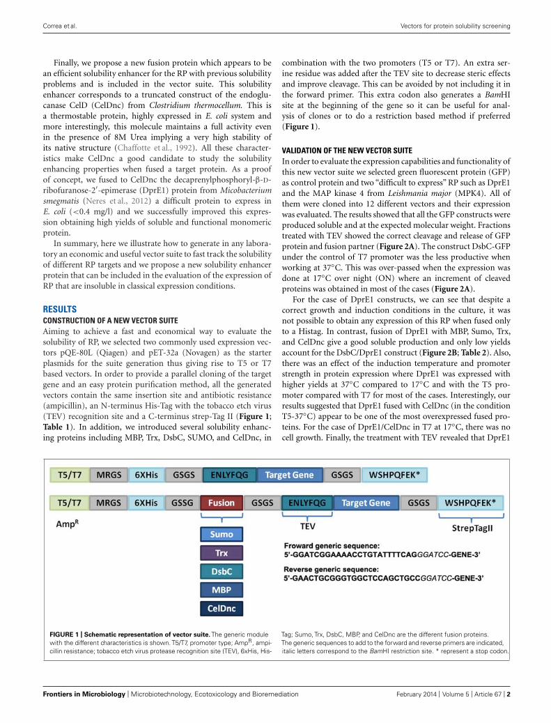
RESULTSCONSTRUCTION OF A NEW VECTOR SUITEAiming to achieve a fast and economical way to evaluate thesolubility of RP, we selected two commonly used expression vec-tors pQE-80L (Qiagen) and pET-32a (Novagen) as the starterplasmids for the suite generation thus giving rise to T5 or T7based vectors. In order to provide a parallel cloning of the targetgene and an easy protein purification method, all the generatedvectors contain the same insertion site and antibiotic resistance(ampicillin), an N-terminus His-Tag with the tobacco etch virus(TEV) recognition site and a C-terminus strep-Tag II (Figure 1;Table 1). In addition, we introduced several solubility enhanc-ing proteins including MBP, Trx, DsbC, SUMO, and CelDnc, in
combination with the two promoters (T5 or T7). An extra ser-ine residue was added after the TEV site to decrease steric effectsand improve cleavage. This can be avoided by not including it inthe forward primer. This extra codon also generates a BamHIsite at the beginning of the gene so it can be useful for anal-ysis of clones or to do a restriction based method if preferred(Figure 1).
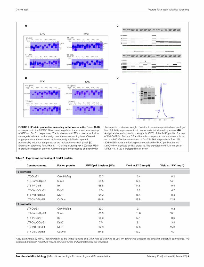
VALIDATION OF THE NEW VECTOR SUITEIn order to evaluate the expression capabilities and functionality ofthis new vector suite we selected green fluorescent protein (GFP)as control protein and two “difficult to express” RP such as DprE1and the MAP kinase 4 from Leishmania major (MPK4). All ofthem were cloned into 12 different vectors and their expressionwas evaluated. The results showed that all the GFP constructs wereproduced soluble and at the expected molecular weight. Fractionstreated with TEV showed the correct cleavage and release of GFPprotein and fusion partner (Figure 2A). The construct DsbC-GFPunder the control of T7 promoter was the less productive whenworking at 37◦C. This was over-passed when the expression wasdone at 17◦C over night (ON) where an increment of cleavedproteins was obtained in most of the cases (Figure 2A).
For the case of DprE1 constructs, we can see that despite acorrect growth and induction conditions in the culture, it wasnot possible to obtain any expression of this RP when fused onlyto a Histag. In contrast, fusion of DprE1 with MBP, Sumo, Trx,and CelDnc give a good soluble production and only low yieldsaccount for the DsbC/DprE1 construct (Figure 2B; Table 2). Also,there was an effect of the induction temperature and promoterstrength in protein expression where DprE1 was expressed withhigher yields at 37◦C compared to 17◦C and with the T5 pro-moter compared with T7 for most of the cases. Interestingly, ourresults suggested that DprE1 fused with CelDnc (in the conditionT5-37◦C) appear to be one of the most overexpressed fused pro-teins. For the case of DprE1/CelDnc in T7 at 17◦C, there was nocell growth. Finally, the treatment with TEV revealed that DprE1
FIGURE 1 | Schematic representation of vector suite. The generic modulewith the different characteristics is shown. T5/T7, promoter type; AmpR, ampi-cillin resistance; tobacco etch virus protease recognition site (TEV), 6xHis, His-
Tag; Sumo, Trx, DsbC, MBP, and CelDnc are the different fusion proteins.The generic sequences to add to the forward and reverse primers are indicated,italic letters correspond to the BamHI restriction site. * represent a stop codon.
Frontiers in Microbiology | Microbiotechnology, Ecotoxicology and Bioremediation February 2014 | Volume 5 | Article 67 | 2
Correa et al. Vectors for protein solubility screening
Table 1 | Primer list for vector generation, cloning, and sequencing.
Primer Sequence 5’–3’ Characteristics
CelDwtNFor GGATCGGAAAACCTGTATTTTCAGGGATCCATGACCATGATTACGAATTCCCGG Cloning of CelDwt
CelDwtCRev GCTGCAGGTCGACGCCAAGATCCTTTTTTATATTGGTAATTTCTCGATTACCCT
CelDtruncNFor GGATCGGAAAACCTGTATTTTCAGGGATCCTCGGGATTGATTGAGACCAAAGTG Cloning of CelDnc
CelDtruncCRev GCTGCAGGTCGACGCCAAGATCCTTTTTTAAGCAGAATTATAGTTGACAAATCCGG
QE3790For GGATCGGAAAACCTGTATTTTCAGGGATCCATGGGCGCGGTACCCTCACTGACG Cloning of Rv3790
QE3790Rev GCTGCAGGTCGACGCCAAGATCCTTTTTTAGAGCAGTTGCAGGCGCCTGGCCATG
CelDInsFor ATGAGAGGATCGCATCACCATCACCATCACGGATCTTCGGGATTGATTGAGACCAAAGTGTC Insertion of CelDnc as a fusion partner
CelDInsRev GGATCCCTGAAAATACAGGTTTTCCGATCCGCTACCAGCAGAATTATAGTTGACAAATC
strepCterFor TCCGACATGGCCAGGCGCCTGCAACTGCTCGGATCCGGCAGCTGGAGCCACCCGCAGTTC Insterion of a C-terminus strepTagII
strepCterRev TGGCTGCAGGTCGACGCCAAGATCCTTTTTTACTTTTCGAACTGCGGGTGGCTCCAGCTG
SumoFor CATCACCATCACCATCACGGATCTTCGGGAATGTCGGACTCAGAAGTCAATCAAG Insertion of Sumo as a fusion partner
SumoRev CTGAAAATACAGGTTTTCCGATCCGCTACCATACGTAGCACCACCAATCTGTTC
TrxFor CATCACCATCACCATCACGGATCTTCGGGAATGAGCGATAAAATTATTCACCTG Insertion of TrxA as a fusion partner
TrxRev CTGAAAATACAGGTTTTCCGATCCGCTACCGGCCAGGTTAGCGTCGAGGAACTC
MBPFor CATCACCATCACCATCACGGATCTTCGGGAATGAAAACTGAAGAAGGTAAACTG Insertion of MBP as a fusion partner
MBPRev CTGAAAATACAGGTTTTCCGATCCGCTACCATTAGTCTGCGCGTCTTTCAGGGC
DsbCFor CATCACCATCACCATCACGGATCTTCGGGAGATGACGCGGCAATTCAACAAACGTTAGCC Insertion of DsbC as a fusion partner
DsbCRev CTGAAAATACAGGTTTTCCGATCCGCTACCTTTACCGCTGGTCATTTTTTGGTGTTCGTC
T5T7For TTTGTTTAACTTTAAGAAGGAGATATACATATGAGAGGATCGCATCACCATCACCATCAC Transfer of the entire cassette to pET32a
vector
T5T7Rev CAGTGGTGGTGGTGGTGGTGCTCGAGTGCGGCTTGGCTGCAGGTCGACGC
GFPFor GGATCGGAAAACCTGTATTTTCAGGGATCCAGCAAAGGAGGAGAACTTTTC GFP cloning
GFPRev GAACTGCGGGTGGCTCCAGCTGCCGGATCCTCAAAGCTTTTTGTAGAGCTCATC
MPK4For GGATCGGAAAACCTGTATTTTCAGGGATCCATGGCTCAACTCGTCCCTTTAGC MPK4 cloning
MPK4Rev GAACTGCGGGTGGCTCCAGCTGCCGGATCCCTATTCGTTCAATTGTGAATGGG
MBPSeqFor CGCTGGCGCGAAAGCGGGTC MBP sequencing
CelDSeqFor GTGCCCTGGAGCAGTGCCGC CelD sequencing
RP fused with all these enhancers remains in a soluble state con-firming an important improvement expression after usage of thisvector suite (Figure 2B).
Concerning MPK4 our results showed that of the 12 constructsonly 2 gave a band at the expected molecular weight. These cor-respond to the construct pT7-DsbC-MPK4 and pT7-MBP-MPK4(Figure 2C). In both cases TEV protease was able to cleave thefusion but only in the pT7-DsbC-MPK4 constructs it was possibleto get a soluble protein after cleavage (Figure 2C, TEV treatmentsection). In order to confirm this result and validate our suitevector we proceed to perform a large scale purification with thisconstruct. Our results showed that after protein purification byIMAC it is possible to obtain the DsbC-MPK4 fusion in a solu-ble manner and with a yield of 6 mg/l (Figure 2D). Oligomericstate analysis of the DsbC-MPK4 fusion, revealed that the elutedpeak is maintained as a soluble decameric oligomer with an appar-ent molecular weight of around 650 kDa (Figure 2D). This resultwas verified by dynamic light scattering (data not shown). Despitethe fact, a great part of the MPK4 protein precipitate after TEV
treatment, an interesting and scalable amount of this proteinremains in a soluble form (Figure 2D).
Altogether these results, underline the importance of this newvector suite as an improved tool for the soluble expression ofDprE1 and MPK4 proteins and suggest that it can be very valuablefor the expression of other “difficult to express” RP.
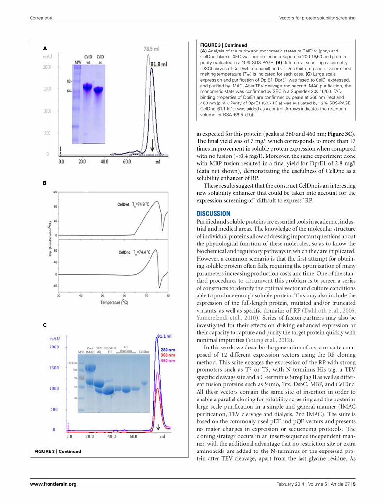
USE OF ENDOGLUCONASE D VARIANT (CelDnc) FOR THE SOLUBLEEXPRESSION OF DprE1 PROTEINAfter expressing the new construct (CelDnc), we found out that itis expressed at high yields (>400 mg/l) in a soluble monomeric andfunctional form which in turn maintains thermostable character-istics as the entire version (Figures 3A,B). So, we wondered if thisextreme solubility and stability could help in the production andfolding of other target proteins. In this regard, we fused CelDncto the N-terminus of the protein DprE1. The results showed thatthe fusion was successfully produced in a soluble manner and thatafter TEV treatment and gel filtration purification it remains sol-uble, monomeric and it was able to retain a FAD binding property
www.frontiersin.org February 2014 | Volume 5 | Article 67 | 3
Correa et al. Vectors for protein solubility screening
FIGURE 2 | Protein production screening in the vector suite. Panels (A,B)
corresponds to the E-PAGE 96 acrylamide gels for the expression screeningof GFP and DprE1, respectively. The incubation with TEV protease for fusioncleavage is indicated with a +sign over the corresponding lines. Cleavedtarget protein at the expected molecular weight (MW) is depicted.Additionally, induction temperatures are indicated over each panel. (C)
Expression screening for MPK4 at 17◦C using a Labchip GX II (Caliper, USA)microfluidic detection system. Arrows indicate the presence of a band with
the expected molecular weight. Construct names are provided over each gelline. Solubility improvement with vector suite is indicated by arrows. (D)
Analytical size exclusion chromatography (SEC) of the IMAC purified fractionof DsbC-MPK4. Peaks at 7.6 and 8.4 ml correspond to the exclusion volumeand the 600 kDa decameric form of DsbC-MPK4, respectively. The 12%SDS-PAGE shows the fusion protein obtained by IMAC purification andDsbC-MPK4 digested by TEV protease. The expected molecular weight ofMPK4 (41.7 kDa) is indicated by an arrow.
Table 2 | Expression screening of DprE1 protein.
Construct name Fusion protein MW DprE1 fusions (kDa) Yield at 37◦C (mg/l) Yield at 17◦C (mg/l)
T5 promoter
pT5-DprE1 Only HisTag 53.7 0.4 0.2
pT5-Sumo-DprE1 Sumo 65.5 12.3 14.1
pT5-Trx-DprE1 Trx 65.8 14.8 10.4
pT5-DsbC-DprE1 DsbC 77.4 6.2 4.7
pT5-MBP-DprE1 MBP 94.3 15.4 11.3
pT5-CelD-DprE1 CelDnc 114.8 19.5 12.8
T7 promoter
pT7-DprE1 Only HisTag 53.7 0.1 0.2
pT7-Sumo-DprE1 Sumo 65.5 11.6 10.1
pT7-Trx-DprE1 Trx 65.8 12.4 9.8
pT7-DsbC-DprE1 DsbC 77.4 8.1 3.9
pT7-MBP-DprE1 MBP 94.3 12.8 15.8
pT7-CelD-DprE1 CelDnc 114.8 19.2 ND
After purification by IMAC, concentration of the entire fusions and yield was determined at 280 nm taking into account the different extinction coefficients. Theexpected molecular weight as well as construct name and characteristics are indicated.
Frontiers in Microbiology | Microbiotechnology, Ecotoxicology and Bioremediation February 2014 | Volume 5 | Article 67 | 4
Correa et al. Vectors for protein solubility screening
FIGURE 3 | Continued
FIGURE 3 | Continued
(A) Analysis of the purity and monomeric states of CelDwt (gray) andCelDnc (black). SEC was performed in a Superdex 200 16/60 and proteinpurity evaluated in a 10% SDS-PAGE. (B) Differential scanning calorimetry(DSC) curves of CelDwt (top panel) and CelDnc (bottom panel). Determinedmelting temperature (T m) is indicated for each case. (C) Large scaleexpression and purification of DprE1. DprE1 was fused to CelD, expressed,and purified by IMAC. After TEV cleavage and second IMAC purification, themonomeric state was confirmed by SEC in a Superdex 200 16/60. FADbinding properties of DprE1 are confirmed by peaks at 360 nm (red) and460 nm (pink). Purity of DprE1 (53.7 kDa) was evaluated by 12% SDS-PAGE.CelDnc (61.1 kDa) was added as a control. Arrows indicates the retentionvolume for BSA (66.5 kDa).
as expected for this protein (peaks at 360 and 460 nm; Figure 3C).The final yield was of 7 mg/l which corresponds to more than 17times improvement in soluble protein expression when comparedwith no fusion (<0.4 mg/l). Moreover, the same experiment donewith MBP fusion resulted in a final yield for DprE1 of 2.8 mg/l(data not shown), demonstrating the usefulness of CelDnc as asolubility enhancer of RP.
These results suggest that the construct CelDnc is an interestingnew solubility enhancer that could be taken into account for theexpression screening of “difficult to express” RP.
DISCUSSIONPurified and soluble proteins are essential tools in academic, indus-trial and medical areas. The knowledge of the molecular structureof individual proteins allow addressing important questions aboutthe physiological function of these molecules, so as to know thebiochemical and regulatory pathways in which they are implicated.However, a common scenario is that the first attempt for obtain-ing soluble protein often fails, requiring the optimization of manyparameters increasing production costs and time. One of the stan-dard procedures to circumvent this problem is to screen a seriesof constructs to identify the optimal vector and culture conditionsable to produce enough soluble protein. This may also include theexpression of the full-length protein, mutated and/or truncatedvariants, as well as specific domains of RP (Dahlroth et al., 2006;Yumerefendi et al., 2010). Series of fusion partners may also beinvestigated for their effects on driving enhanced expression ortheir capacity to capture and purify the target protein quickly withminimal impurities (Young et al., 2012).
In this work, we describe the generation of a vector suite com-posed of 12 different expression vectors using the RF cloningmethod. This suite engages the expression of the RP with strongpromoters such as T7 or T5, with N-terminus His-tag, a TEVspecific cleavage site and a C-terminus StrepTag II as well as differ-ent fusion proteins such as Sumo, Trx, DsbC, MBP, and CelDnc.All these vectors contain the same site of insertion in order toenable a parallel cloning for solubility screening and the posteriorlarge scale purification in a simple and general manner (IMACpurification, TEV cleavage and dialysis, 2nd IMAC). The suite isbased on the commonly used pET and pQE vectors and presentsno major changes in expression or sequencing protocols. Thecloning strategy occurs in an insert-sequence independent man-ner, with the additional advantage that no restriction site or extraaminoacids are added to the N-terminus of the expressed pro-tein after TEV cleavage, apart from the last glycine residue. As
www.frontiersin.org February 2014 | Volume 5 | Article 67 | 5
Correa et al. Vectors for protein solubility screening
purification features we selected the use of the HisTag, becauseit has demonstrated to be very versatile, cheap and to workwell in small and large scale purifications (Schafer et al., 2002;Steen et al., 2006). Additionally, if the stop codon of the targetgene is omitted, an additional purification tag, the strepTag II isexpressed in the C-terminus of the target protein. This last canbe useful if degradation intermediates appear by coupling IMACpurification with StrepTacting purification only a product withan intact N- and C-terminus will be purified. Also the purifi-cation via the StrepTag II showed to be very useful for proteinsthat are expressed in low abundance where usually purificationby IMAC gives many contaminants from the host (Magnusdottiret al., 2009). Finally the TEV site was chosen for protein cleav-age as it has demonstrated to be very specific, work well at lowtemperatures and can be produced in the laboratory with highyield reducing production costs (van den Berg et al., 2006). More-over, it was shown that the last residue of the cleavage site (Gly)can be changed for all the other residues except for proline foran expense in cleavage efficiency, so if a protein with a native Nterminus is needed it can be taken into account (Kapust et al.,2002).
The suite was tested with GFP, and we found out that in allcases there were expression and cleavage with TEV demonstratingthat all the vectors worked well. By using this suite of vectors thehigh-throughput screening for soluble expression could be easilyachieved manually or automatically as it was demonstrated for theexpression of GFP, DprE1 and MPK4.
In order to challenge the vector suite proposed here we selectedtwo “difficult to express” RP like DprE1, and MPK4. For the firstprotein evaluated (DprE1) the vector suite demonstrated that theexpression protein improved when the target protein was fused toSumo, Trx, DsbC, MBP, and CelDnc solubility enhancer proteins.Among them the best results concerning solubility and quantitiesof stable protein was achieved when DprE1 was fused to CelDncand subsequently cleaved by TEV. In the second case, only two outof 12 conditions evaluated were able to express MPK4 in the sol-uble fraction and only one (pT7-DsbC-MPK4 construct) remainssoluble after TEV cleavage. Interestingly, high yield of this fusionconstruct remained as a decamer before TEV cleavage, so afterimproving purification protocols (like the use of strepTag II orion exchange chromatography), the entire fusion can be used forcrystallization screenings.
Despite the fact that, many fusion proteins were evaluated, itremains difficult to define a “universal fusion protein.” Differ-ent options are commercially available (MBP, GST, Trx, DsbC,NusA, etc), and several groups have found new proteins that canbe promising alternatives to obtain a soluble and homogeneousrecombinant protein (Chatterjee and Esposito, 2006; DelPropostoet al., 2009; Cheng et al., 2010; Song et al., 2011) by fusing the targetgene. In this work, we evaluated the use of a novel fusion protein,CelDnc that is thermostable (Tm: 71.4◦C) and is expressed in mas-sive amounts in E coli system. CelD is an endo-β-glucanase (EC3.2.1.4) from C. thermocellum and is part of the cellulose degrad-ing complex termed cellulosome composed of a large number ofindividual enzymes (Kataeva et al., 1997).
When this protein was evaluated as a solubility fusion enhancerfor DprE1 the results showed an increasing solubility performance
for this molecule compared with other classical fusion enhancerslike MBP. After expression and IMAC purification was done theCelDnc fusion was soluble in large amounts. Moreover, DprE1 wasstill soluble, monomeric and presented FAD binding propertieseven after the proteolitical removal of CelDnc demonstrating theutility of this fusion protein that can be taken into account whensolubility screening is performed.
In this work we propose a new vector suite and a new fusionenhancer molecule with chances to improve the solubility of dif-ferent RP. The vector suite proposed here allows the evaluation offive different fusion proteins or only the HisTag in combinationwith two different promoters, giving rise to 12 different constructsfor a single target gene. Altogether, our results suggest that thisexpression system could be an interesting tool to improve solubilityproblems of RP.
Moreover, the screening protocol can be further improved. Inthe present work we used Rosetta cells for the screening of RPproduction. Different E. coli strains can be evaluated in parallellike the use of strains for disulfide bond formation (Shuffle, NewEngalnd Biolabs), reduced mRNA degradation (BL21 Star, Invit-rogen) among others. Also, the co-expression of chaperones ormolecular partners can be included if they are in a vector compat-ible with a ColE1 replication origin. By the complementation ofsuch variables with the vector suite, a great number of conditionscan be screened, increasing the chances of finding the optimalcontext for target protein production.
It was shown that the sequence at the translation initiationregion (TIR) can have a detrimental effect in protein productiondue to the generation of secondary structures in the messengerRNA that can hamper the translation by the ribosome complex.In this regard a predictive method was developed for designingsynthetic ribosome binding sites (RBS) that can minimize theformation of secondary structures at RNA level, so increasingthe translation rate (Salis et al., 2009; Salis, 2011). Because thenucleotide sequence from +1 to +25 is the same in all vectors,a new RBS can be designed and introduced into the entire suiteincreasing translation rates.
Finally, despite the cloning of target genes into the suite wasvery efficient, false positives were found in some cases. Thiscan be improved, for example, if a toxic gene like the toxinCcdB of type II toxin-antitoxin system is added at the insertionsite.
Despite the fact that, more proteins should be tested in thisvector suite and that there is no magic formula able to ensure thesolubility of different proteins, this could be a useful and economicmodel to fast track the soluble expression of the RP.
MATERIALS AND METHODSGENERATION OF THE VECTOR SUITEFor the generation of the vector suite we used a modified version ofthe pQE80L (Qiagen) as the starter plasmid, that contained a TEVcleavage site after the Histag separated by a GSGS linker (pQE80L-TEV). In a first step we cloned the gene DprE1 into this vector andadded the different modules for the vector suite (linkers, strepTagand different fusion proteins) thus generating the T5 series. Thenthe entire constructs were cloned into the vector pET32a in orderto generate the T7 series.
Frontiers in Microbiology | Microbiotechnology, Ecotoxicology and Bioremediation February 2014 | Volume 5 | Article 67 | 6
Correa et al. Vectors for protein solubility screening
All PCR were done using Phusion polymerase (Finnzymes).For the amplification of the fragments (megaprimer generation)conditions were 30 s at 98◦C and 28 cycles of 98◦C for 10 s, 59◦Cfor 1 min and 72◦C for 1 min with a final extension step at 72◦Cfor 5 min and PCR products were purified by agarose gel. Thegenerated megaprimers contained 30 bp in both ends that overlapswith the insertion site in the destination vectors. The integrationinto the vectors was done by RF cloning (Unger et al., 2010) andthe RF reaction was as follows: 30 s at 98◦C and 30 cycles of 98◦Cfor 10 s, 60◦C for 1 min and 72◦C for 5 min with a final extensionstep at 72◦C for 7 min. For RF reactions 120 ng of megaprimersand 30 ng destination vector were used. 20 μl were digested with2 μl Fast Digest DpnI (Thermo) for 15 min at 37◦C in order toremove parental plasmid, and 5 μl were used to transform 50 μlof competent DH5α E. coli cells. Positive clones were confirmedby colony PCR by using Taq polymerase (Invitrogen) with thesame primers used for megaprimer generation. Colony PCR wasas follows, 95◦C for 3 min, 25 cycles of 95◦C for 30 s, 60◦C for 30 sand 72◦C for 2 min followed by a final extension step at 72◦C for5 min. Positive colonies were selected for plasmid extraction andconfirmed by sequencing.
The gene for DprE1 was amplified from M. smegmatis genomicDNA using the primers QE3790For and QE3790Rev for thegeneration of the megaprimer (Table 1). The product wascloned into the vector pQE80L-TEV by RF cloning to gener-ate the construct pDprE1. The genes coding for CelDwt orthe truncated version CelDnc (residues 32–577), were ampli-fied from the plasmid pCT603 (Chaffotte et al., 1992) with theprimers CelDwtNFor and CelDwtCRev for CelDwt and primersCelDtruncNFor and CelDtruncCRev for CelDnc (Table 1) andcloned by RF in the same vector to generate the constructs pCelDand pCelDnc. The construct pDprE1 was used for the inser-tion of CelDnc in the 5′ of DprE1 (between the HisTag andthe GSGS linker, Figure 1). CelDnc was amplified from thepCelDnc construct using primers CelDInsFor and CelDInsRev.The forward primer was designed also to add a GSSG linkerto separate the HisTag from the fusion partner generating theconstruct pCelD-DprE1. The generated constructs (pDprE1 andpCelD-DprE1) were then used to add the last module of the vec-tor, the C-terminal strepTag II. The strepTag II was inserted atthe C-terminus separated by a GSGS linker with primers strepC-terFor and strepCterRev (Table 1) for the generation of thevector pT5-DprE1 (HisTag alone) and pT5-CelD-DprE1 (CelDncfusion). The primers anneal each other, so they were used with-out addition of DNA for the generation of the megaprimer. Thegenerated pT5-CelD-DprE1 vector was then used for the inser-tion and replacement of CelDnc by other fusion partners. Inthis regard the primers SumoFor and SumoRev; TrxFor, andTrxRev; MBPFor and MBPRev and DsbCFor and DsbCRev wereused for the insertion of Sumo, TrxA, MBP, and DsbC, respec-tively, (Table 1). The genes were amplified from Saccharomycescerevisiae for Sumo, pET32a (Novagen) for TrxA, pMAL (NewEngland Biolabs) for MBP; and E. coli genome for DsbC. Bythis way, the T5 vector series was completed. All 6 vectors wereconfirmed by sequencing with the QEFor and QERev plasmidprimers. For the case of MBP and CelDnc constructs internalprimers were also used in order to cover the entire sequence.
The last step was to transfer the modules into a T7 context.To do this, we selected the pET32a (Novagen) as a destinationvectoramplifying the entire cassette from T5 series (from MRGS-HisTag up to the strepTag II for the different fusions) with theprimers T5T7For and T5T7Rev and replacing the expression cas-sette of the pET32a vector. The generated megaprimers wereused for the RF reactions. By this way the vector suite was com-pleted containing the gene DprE1 in all 12 vectors for expressionscreening.
CLONING OF GFP AND MPK4 INTO THE SUITE OF VECTORSLeishmania major MPK4 gene was amplified with primersMPK4For and MPK4Rev from a pGem vector containing the gene.GFP was amplified with primers GFPFor and GFPRev from a pETvector containing a GFP variant that is well expressed in E. coli(Waldo et al., 1999).
The 12 vectors were added to 12 different PCR tubes, and theamplified products were used as megaprimers for the RF reactionusing the HF buffer from Phusion polymerase. After digestion of20 μl PCR products with 2 μl DpnI, chemical competent cellswere transformed with 5 μl RF reaction in a PCR machine withthe following program: 30 min at 4◦C, 45 s at 42◦C, 3 min at 4◦C,addition of 100 μl of LB, 1 h at 37◦C, and plating of 100 μl inagar plates containing ampicillin. Four colonies for each constructwere selected and confirmed by colony PCR and sequenced. Afterthe analysis we found out that in most cases all were positive (orat least three of four were positive) giving a percentage of successof more than 80%.
EXPRESSION SCREENING OF GFP AND DprE1Chemocompetent Rosetta-pLysS cells were transformed with5 μl of purified plasmids as described above and then incu-bated in a shaker ON at 37◦C in 1 ml of LB with chloram-phenicol and ampicillin in a 96 × deep-well plate. 100 μl ofON culture were used to inoculate 4 ml of Terrific Broth in24 × deep-well plates by duplicate. Cultures were incubated at37◦C until D.O.600 reached 1.0–1.2. At that moment one platewas induced with 1 mM IPTG and left at 37◦C for 4 h. Theother 24 deep-well was incubated at 17◦C for 15 min to cool-ing it and then induced with 1 mM IPTG ON at the sametemperature. After induction time was reached, cells were har-vested, resuspended in 1 ml lysis buffer (50 mM Tris pH 8.0;300 mM NaCl, 10 mM imidazol, 0.5 mg/ml lysozyme) andfrozen at −80◦C. After thawing cells, 10 units of DNase I and10 μl of 2M MgSO4 were added and incubated with shakingfor 20 min at 20◦C. Then 200 μl of Nickel beads (Qiagen)equilibrated in binding buffer (50 mM Tris pH 8.0; 300 mMNaCl, 10 mM imidazol) were added to cell extracts and incu-bated for 15 min at 20◦C. Cell extracts were then transferredto a 96×-well filter plate assembled in a vacuum device, andbound protein was washed with 2 ml of binding buffer. Anadditional wash step was done with 2 ml of binding buffercontaining 50 mM imidazol. Elution was done with 160 μl ofelution buffer [50 mM Tris pH 8.0; 300 mM NaCl, 500 mMimidazol; for a detailed protocol, see (Saez and Vincentelli,2013)]. Eluates were divided in two groups for evaluation ofuncleaved protein and assessment of TEV cleavage ON at 18◦C.
www.frontiersin.org February 2014 | Volume 5 | Article 67 | 7
Correa et al. Vectors for protein solubility screening
Samples were then loaded into an E-PAGE 96 acrylamide gel(Invitrogen).
EXPRESSION SCREENING OF MPK4Expression screening and purification of MPK4 constructs wasmade in a similar way than for GFP and DprE1 but only 17◦C ofinduction was evaluated. Purification steps were the same but thepipeting scheme was done automatically by using a TECAN Free-dom EVO®200. Expression analysis was done also automaticallyby using a Labchip GX II (Caliper, USA) microfluidic detectionsystem.
LARGE SCALE EXPRESSION AND PURIFICATION OF DsbC-MPK4DsbC-MPK4 was expressed in Terrific Broth (TB) supplementedwith ampicillin and chloramphenicol and induction was done atD.O600: 1.2 ON at 17◦C with 1 mM IPTG. Pellets were resuspendedin lysis buffer and frozen at −80◦C. After thawing, the pellets weresonicated and centrifugated at 15.000 × g. Soluble fraction wasinjected in a 1 ml IMAC column (GE Healthcare) equilibrated inbinding buffer. Elution was done in a linear gradient of 5–100% Bin 10 column volumes (CV) with elution buffer. Purified proteinwas cleaved with TEV protease in a 1:30 protein:enzyme ratio anddialyzed against cleavage buffer (50 mM Tris pH 8.0; 150 mM NaCl,1 mM DTT) ON at 8◦C. Sample was filtered through 0.22 μm toremove precipitates, and analyzed by SDS-PAGE.
EXPRESSION AND PURIFICATION OF CelD AND CelDncProduction of CelD and CelDnc was done in M15pREP4 from theconstructs pCelD and pCelDnc, respectively, in 1 l 2YT supple-mented with ampicillin and kanamycin, and induced with 1 mMIPTG at D.O. 1.0 ON at 37◦C. IMAC was done like for the case ofDsbC-MPK4 but using a 5 ml column and only half of the solublefraction was used. TEV cleavage was done as before and desalted inorder to remove imidazole. The reaction was injected in a secondIMAC under same conditions as above and the flow through con-taining the cleaved protein was injected in a Superdex 200 16/60(GE Healthcare) equilibrated with buffer 40 mM Tris pH 7.7.
DSC ANALYSIS OF CelD AND CelDncDifferential scanning calorimetry (DSC) experiments were carriedout in PBS, in a VP-DSC instrument (Microcal, Northampton,MA, USA) and data analyzed with the software supplied with theequipment. The temperature was increased at 1◦C per minutefrom 30 to 80◦C, and proteins were added at concentration of1 mg/ml for CelD and CelDnc.
LARGE SCALE EXPRESSION AND PURIFICATION OF pT5-DprE1,pT5-CelD-DprE1 AND pT5-MBP-DprE1Induction of p5DprE1, p5CelDnc-DprE1 and p5MBP-DprE1 weredone in M15pREP4 with 1 mM IPTG in 1 l 2YT supplemented withampicillin (100 μg/ml), kanamycin (50 μg/ml) and 15 μM FAD atD.O.: 1.0–1.2 during 4 h at 37◦C. Cells were harvested, resuspendedin lysis buffer and frozen at −80◦C. After thawing the cells, werelysed and protein purified as before. Purified protein was cleavedwith TEV in a 1:30 ratio, and dialysed against cleavage buffer. Theproduct was then purified by a second IMAC and injected in aSuperdex 200 16/60 equilibrated with buffer 25 mM Tris pH 8.0;150 mM NaCl.
ACKNOWLEDGMENTSThis work was partially funded by FOCEM (MERCOSUR Struc-tural Convergence Fund), COF 03/11 and CYTED Program.Agustín Correa was supported by a doctoral program of the Agen-cia Nacional de Investigación e Innovación, Uruguay. We wish tothank Dr. Trajtemberg and Sofía Horjales from the Crystallogra-phy Unit (PXF) of the Institut Pasteur de Montevideo for giving theplasmid pQE80L-TEV and pGem-MPK4 and Mrs. Natalia Lópezfor helpful secretarial assistance.
REFERENCESAslanidis, C., and de Jong, P. J. (1990). Ligation-independent cloning of PCR
products (LIC-PCR). Nucleic Acids Res. 18, 6069–6074. doi: 10.1093/nar/18.20.6069
Berrow, N. S., Alderton, D., Sainsbury, S., Nettleship, J., Assenberg, R., Rahman,N., et al. (2007). A versatile ligation-independent cloning method suitable forhigh-throughput expression screening applications. Nucleic Acids Res. 35, e45.doi: 10.1093/nar/gkm047
Cabrita, L. D., Dai, W., and Bottomley, S. P. (2006). A family of E. coli expressionvectors for laboratory scale and high throughput soluble protein production.BMC Biotechnol. 6:12. doi: 10.1186/1472-6750-6–12
Chaffotte, A. F., Guillou, Y., and Goldberg, M. E. (1992). Inclusion bodies of thethermophilic endoglucanase D from Clostridium thermocellum are made of nativeenzyme that resists 8 M urea. Eur. J. Biochem. 205, 369–373. doi: 10.1111/j.1432-1033.1992.tb16789.x
Chatterjee, D. K., and Esposito, D. (2006). Enhanced soluble protein expres-sion using two new fusion tags. Protein Expr. Purif. 46, 122–129. doi:10.1016/j.pep.2005.07.028
Cheng, Y., Gu, J., Wang, H. G., Yu, S., Liu, Y. Q., Ning, Y. L., et al. (2010). EspAis a novel fusion partner for expression of foreign proteins in Escherichia coli.J. Biotechnol. 150, 380–388. doi: 10.1016/j.jbiotec.2010.09.940
Correa, A., and Oppezzo, P. (2011). Tuning different expression parameters toachieve soluble recombinant proteins in E. coli: advantages of high-throughputscreening. Biotechnol. J. 6, 715–730. doi: 10.1002/biot.201100025
Curiel, J. A., De Las Rivas, B., Mancheno, J. M., and Munoz, R. (2010). The pURIfamily of expression vectors: a versatile set of ligation independent cloning plas-mids for producing recombinant His-fusion proteins. Protein Expr. Purif. 76,44–53. doi: 10.1016/j.pep.2010.10.013
Dahlroth, S. L., Nordlund, P., and Cornvik, T. (2006). Colony filtration blottingfor screening soluble expression in Escherichia coli. Nat. Protoc. 1, 253–258. doi:10.1038/nprot.2006.39
DelProposto, J., Majmudar, C. Y., Smith, J. L., and Brown, W. C. (2009). Mocr:a novel fusion tag for enhancing solubility that is compatible with structuralbiology applications. Protein Expr. Purif. 63, 40–49. doi: 10.1016/j.pep.2008.08.011
Esposito, D., Garvey, L. A., and Chakiath, C. S. (2009). Gateway cloning forprotein expression. Methods Mol. Biol. 498, 31–54. doi: 10.1007/978-1-59745-196-3_3
Kapust, R. B., Tozser, J., Copeland, T. D., and Waugh, D. S. (2002). The P1’ specificityof tobacco etch virus protease. Biochem. Biophys. Res. Commun. 294, 949–955.doi: 10.1016/S0006-291X(02)00574-0
Kapust, R. B., and Waugh, D. S. (1999). Escherichia coli maltose-binding protein isuncommonly effective at promoting the solubility of polypeptides to which it isfused. Protein Sci. 8, 1668–1674. doi: 10.1110/ps.8.8.1668
Kataeva, I., Guglielmi, G., and Beguin, P. (1997). Interaction between Clostrid-ium thermocellum endoglucanase CelD and polypeptides derived from thecellulosome-integrating protein CipA: stoichiometry and cellulolytic activity ofthe complexes. Biochem. J. 326 (Pt 2), 617–624.
LaVallie, E. R., Lu, Z., Diblasio-Smith, E. A., Collins-Racie, L. A., and McCoy, J. M.(2000). Thioredoxin as a fusion partner for production of soluble recombinantproteins in Escherichia coli. Methods Enzymol. 326, 322–340. doi: 10.1016/S0076-6879(00)26063-1
Luna-Vargas, M. P., Christodoulou, E., Alfieri, A., Van Dijk, W. J., Stadnik, M.,Hibbert, R. G., et al. (2011). Enabling high-throughput ligation-independentcloning and protein expression for the family of ubiquitin specific proteases.J. Struct. Biol. 175, 113–119. doi: 10.1016/j.jsb.2011.03.017
Frontiers in Microbiology | Microbiotechnology, Ecotoxicology and Bioremediation February 2014 | Volume 5 | Article 67 | 8
Correa et al. Vectors for protein solubility screening
Magnusdottir, A., Johansson, I., Dahlgren, L. G., Nordlund, P., and Berglund,H. (2009). Enabling IMAC purification of low abundance recombinant pro-teins from E. coli lysates. Nat. Methods 6, 477–478. doi: 10.1038/nmeth0709-477
Marblestone, J. G., Edavettal, S. C., Lim, Y., Lim, P., Zuo, X., and Butt, T. R. (2006).Comparison of SUMO fusion technology with traditional gene fusion systems:enhanced expression and solubility with SUMO. Protein Sci. 15, 182–189. doi:10.1110/ps.051812706
Murphy, M. B., and Doyle, S. A. (2005). High-throughput purification ofhexahistidine-tagged proteins expressed in E. coli. Methods Mol. Biol. 310,123–130. doi: 10.1007/978-1-59259-948-6_9
Neres, J., Pojer, F., Molteni, E., Chiarelli, L. R., Dhar, N., Boy-Rottger, S.,et al. (2012). Structural basis for benzothiazinone-mediated killing of Mycobac-terium tuberculosis. Sci. Transl. Med. 4, 150ra121. doi: 10.1126/scitranslmed.3004395
Nozach, H., Fruchart-Gaillard, C., Fenaille, F., Beau, F., Ramos, O. H., Douzi, B.,et al. (2013). High throughput screening identifies disulfide isomerase DsbC as avery efficient partner for recombinant expression of small disulfide-rich proteinsin E. coli. Microb. Cell Fact. 12, 37. doi: 10.1186/1475-2859-12–37
Saez, N. J., and Vincentelli, R. (2013). High-throughput expression screening andpurification of recombinant proteins in E. coli. Methods Mol. Biol. 1091, 33–53.doi: 10.1007/978-1-62703-691-7_3
Salis, H. M. (2011). The ribosome binding site calculator. Methods Enzymol. 498,19–42. doi: 10.1016/B978-0-12-385120-8.00002–4
Salis, H. M., Mirsky, E. A., and Voigt, C. A. (2009). Automated design of syntheticribosome binding sites to control protein expression. Nat. Biotechnol. 27, 946–950. doi: 10.1038/nbt.1568
Schafer, F., Romer, U., Emmerlich, M., Blumer, J., Lubenow, H., and Steinert,K. (2002). Automated high-throughput purification of 6xHis-tagged proteins.J. Biomol. Tech. 13, 131–142.
Schmidt, T. G., and Skerra, A. (2007). The Strep-tag system for one-step purificationand high-affinity detection or capturing of proteins. Nat. Protoc. 2, 1528–1535.doi: 10.1038/nprot.2007.209
Song, J. A., Lee, D. S., Park, J. S., Han, K. Y., and Lee, J. (2011). A novelEscherichia coli solubility enhancer protein for fusion expression of aggregation-prone heterologous proteins. Enzyme Microb. Technol. 49, 124–130. doi:10.1016/j.enzmictec.2011.04.013
Steen, J., Uhlen, M., Hober, S., and Ottosson, J. (2006). High-throughput proteinpurification using an automated set-up for high-yield affinity chromatography.Protein Expr. Purif. 46, 173–178. doi: 10.1016/j.pep.2005.12.010
Unger, T., Jacobovitch, Y., Dantes, A., Bernheim, R., and Peleg, Y. (2010). Applica-tions of the restriction free (RF) cloning procedure for molecular manipulationsand protein expression. J. Struct. Biol. 172, 34–44. doi: 10.1016/j.jsb.2010.06.016
van den Berg, S., Lofdahl, P. A., Hard, T., and Berglund, H. (2006). Improvedsolubility of TEV protease by directed evolution. J. Biotechnol. 121, 291–298. doi:10.1016/j.jbiotec.2005.08.006
Vincentelli, R., Cimino, A., Geerlof, A., Kubo, A., Satou, Y., and Cambillau, C. (2011).High-throughput protein expression screening and purification in Escherichiacoli. Methods 55, 65–72. doi: 10.1016/j.ymeth.2011.08.010
Vincentelli, R., and Romier, C. (2013). Expression in Escherichia coli: becom-ing faster and more complex. Curr. Opin. Struct. Biol. 23, 326–334. doi:10.1016/j.sbi.2013.01.006
Waldo, G. S., Standish, B. M., Berendzen, J., and Terwilliger, T. C. (1999). Rapidprotein-folding assay using green fluorescent protein. Nat. Biotechnol. 17, 691–695. doi: 10.1038/10904
Young, C. L., Britton, Z. T., and Robinson, A. S. (2012). Recombinant proteinexpression and purification: a comprehensive review of affinity tags and microbialapplications. Biotechnol. J. 7, 620–634. doi: 10.1002/biot.201100155
Yumerefendi, H., Tarendeau, F., Mas, P. J., and Hart, D. J. (2010). ESPRIT:an automated, library-based method for mapping and soluble expression ofprotein domains from challenging targets. J. Struct. Biol. 172, 66–74. doi:10.1016/j.jsb.2010.02.021
Conflict of Interest Statement: The authors declare that the research was conductedin the absence of any commercial or financial relationships that could be construedas a potential conflict of interest.
Received: 21 December 2013; paper pending published: 13 January 2014; accepted: 05February 2014; published online: 25 February 2014.Citation: Correa A, Ortega C, Obal G, Alzari P, Vincentelli R and Oppezzo P (2014)Generation of a vector suite for protein solubility screening. Front. Microbiol. 5:67. doi:10.3389/fmicb.2014.00067This article was submitted to Microbiotechnology, Ecotoxicology and Bioremediation,a section of the journal Frontiers in Microbiology.Copyright © 2014 Correa, Ortega, Obal, Alzari, Vincentelli and Oppezzo. This is anopen-access article distributed under the terms of the Creative Commons AttributionLicense (CC BY). The use, distribution or reproduction in other forums is permitted,provided the original author(s) or licensor are credited and that the original publica-tion in this journal is cited, in accordance with accepted academic practice. No use,distribution or reproduction is permitted which does not comply with these terms.
www.frontiersin.org February 2014 | Volume 5 | Article 67 | 9