Importance of ROS and antioxidant system during the beneficial interactions of mitochondrial...
14
ORIGINAL ARTICLE Importance of ROS and antioxidant system during the beneficial interactions of mitochondrial metabolism with photosynthetic carbon assimilation Challabathula Dinakar • Vishwakarma Abhaypratap • Srinivasa Rao Yearla • Agepati S. Raghavendra • Kollipara Padmasree Received: 30 July 2009 / Accepted: 6 November 2009 / Published online: 27 November 2009 Ó Springer-Verlag 2009 Abstract The present study suggests the importance of reactive oxygen species (ROS) and antioxidant metabolites as biochemical signals during the beneficial interactions of mitochondrial metabolism with photosynthetic carbon assimilation at saturating light and optimal CO 2 . Changes in steady-state photosynthesis of pea mesophyll protoplasts monitored in the presence of antimycin A [AA, inhibitor of cytochrome oxidase (COX) pathway] and salicylhydroxa- mic acid [SHAM, inhibitor of alternative oxidase (AOX) pathway] were correlated with total cellular ROS and its scavenging system. Along with superoxide dismutase (SOD) and catalase (CAT), responses of enzymatic com- ponents—ascorbate peroxidase (APX), monodehydro- ascorbate reductase (MDAR), glutathione reductase (GR) and non-enzymatic redox components of ascorbate–gluta- thione (Asc–GSH) cycle, which play a significant role in scavenging cellular ROS, were examined in the presence of mitochondrial inhibitors. Both AA and SHAM caused marked reduction in photosynthetic carbon assimilation with concomitant rise in total cellular ROS. Restriction of electron transport through COX or AOX pathway had differential effect on ROS generating (SOD), ROS scav- enging (CAT and APX) and antioxidant (Asc and GSH) regenerating (MDAR and GR) enzymes. Further, restric- tion of mitochondrial electron transport decreased redox ratios of both Asc and GSH. However, while decrease in redox ratio of Asc was more prominent in the presence of SHAM in light compared with dark, decrease in redox ratio of GSH was similar in both dark and light. These results suggest that the maintenance of cellular ROS at optimal levels is a prerequisite to sustain high photosynthetic rates which in turn is regulated by respiratory capacities of COX and AOX pathways. Keywords Antioxidants Á Alternative oxidase Á Cytochrome oxidase Á Pea Á Photosynthesis Á Reactive oxygen species Abbreviations AA Antimycin A AOX Alternative oxidase APX Ascorbate peroxidase Asc Ascorbate reduced CAT Catalase COX Cytochrome oxidase DHA Dehydroascorbate GR Glutathione reductase GSH Glutathione (reduced) GSSG Glutathione (oxidized) MDAR Monodehydroascorbate reductase ROS Reactive oxygen species SOD Superoxide dismutase Introduction In photosynthetic tissues, mitochondria play an important role in benefiting chloroplastic photosynthesis by backing up carbon assimilation through cytochrome oxidase (COX) Electronic supplementary material The online version of this article (doi:10.1007/s00425-009-1067-3) contains supplementary material, which is available to authorized users. Ch. Dinakar Á V. Abhaypratap Á S. R. Yearla Á A. S. Raghavendra Á K. Padmasree (&) Department of Plant Sciences, School of Life Sciences, University of Hyderabad, Hyderabad 500046, India e-mail: [email protected] 123 Planta (2010) 231:461–474 DOI 10.1007/s00425-009-1067-3
-
Upload
independent -
Category
Documents
-
view
0 -
download
0
Transcript of Importance of ROS and antioxidant system during the beneficial interactions of mitochondrial...
ORIGINAL ARTICLE
Importance of ROS and antioxidant system during the beneficialinteractions of mitochondrial metabolism with photosyntheticcarbon assimilation
Challabathula Dinakar • Vishwakarma Abhaypratap •
Srinivasa Rao Yearla • Agepati S. Raghavendra •
Kollipara Padmasree
Received: 30 July 2009 / Accepted: 6 November 2009 / Published online: 27 November 2009
� Springer-Verlag 2009
Abstract The present study suggests the importance of
reactive oxygen species (ROS) and antioxidant metabolites
as biochemical signals during the beneficial interactions of
mitochondrial metabolism with photosynthetic carbon
assimilation at saturating light and optimal CO2. Changes
in steady-state photosynthesis of pea mesophyll protoplasts
monitored in the presence of antimycin A [AA, inhibitor of
cytochrome oxidase (COX) pathway] and salicylhydroxa-
mic acid [SHAM, inhibitor of alternative oxidase (AOX)
pathway] were correlated with total cellular ROS and its
scavenging system. Along with superoxide dismutase
(SOD) and catalase (CAT), responses of enzymatic com-
ponents—ascorbate peroxidase (APX), monodehydro-
ascorbate reductase (MDAR), glutathione reductase (GR)
and non-enzymatic redox components of ascorbate–gluta-
thione (Asc–GSH) cycle, which play a significant role in
scavenging cellular ROS, were examined in the presence of
mitochondrial inhibitors. Both AA and SHAM caused
marked reduction in photosynthetic carbon assimilation
with concomitant rise in total cellular ROS. Restriction of
electron transport through COX or AOX pathway had
differential effect on ROS generating (SOD), ROS scav-
enging (CAT and APX) and antioxidant (Asc and GSH)
regenerating (MDAR and GR) enzymes. Further, restric-
tion of mitochondrial electron transport decreased redox
ratios of both Asc and GSH. However, while decrease in
redox ratio of Asc was more prominent in the presence of
SHAM in light compared with dark, decrease in redox ratio
of GSH was similar in both dark and light. These results
suggest that the maintenance of cellular ROS at optimal
levels is a prerequisite to sustain high photosynthetic rates
which in turn is regulated by respiratory capacities of COX
and AOX pathways.
Keywords Antioxidants � Alternative oxidase �Cytochrome oxidase � Pea � Photosynthesis �Reactive oxygen species
Abbreviations
AA Antimycin A
AOX Alternative oxidase
APX Ascorbate peroxidase
Asc Ascorbate reduced
CAT Catalase
COX Cytochrome oxidase
DHA Dehydroascorbate
GR Glutathione reductase
GSH Glutathione (reduced)
GSSG Glutathione (oxidized)
MDAR Monodehydroascorbate reductase
ROS Reactive oxygen species
SOD Superoxide dismutase
Introduction
In photosynthetic tissues, mitochondria play an important
role in benefiting chloroplastic photosynthesis by backing
up carbon assimilation through cytochrome oxidase (COX)
Electronic supplementary material The online version of thisarticle (doi:10.1007/s00425-009-1067-3) contains supplementarymaterial, which is available to authorized users.
Ch. Dinakar � V. Abhaypratap � S. R. Yearla �A. S. Raghavendra � K. Padmasree (&)
Department of Plant Sciences, School of Life Sciences,
University of Hyderabad, Hyderabad 500046, India
e-mail: [email protected]
123
Planta (2010) 231:461–474
DOI 10.1007/s00425-009-1067-3
and alternative oxidase (AOX) pathways by different
mechanisms (Padmasree and Raghavendra 1999a, b,
2001a, b). The two terminal oxidases, COX and AOX,
associated with these two pathways are involved in
reducing molecular oxygen to H2O. The energy released
during the electron transport through COX pathway is
coupled to the synthesis of ATP. This function of
COX pathway is known to benefit sucrose biosynthesis
(Padmasree and Raghavendra 1999b). On the other hand,
though the passage of electrons from ubiquinone to AOX
pathway does not generate any ATP, it has a significant
function in preventing over-reduction of not only respira-
tory complexes but also electron transport carriers of
chloroplasts (Yoshida et al. 2006, 2007; Noguchi and
Yoshida 2008).
The existence and operation of respiration in light were
debated until past two decades (Graham 1980). However,
the significance of mitochondrial bioenergetic metabolism
in benefiting photosynthetic carbon assimilation was
revealed in several reviews (Raghavendra et al. 1994;
Kromer 1995; Hoefnagel et al. 1998; Gardestrom et al.
2002; Padmasree et al. 2002; Raghavendra and Padmasree
2003; Noctor et al. 2007; Noguchi and Yoshida 2008;
Nunes-Nesi et al. 2008). Usage of specific metabolic
inhibitors and generation of specific mutants/transgenic
plants which either under-express or over-express a specific
key component related to respiratory process clearly
established the importance of bioenergetic oxidative
metabolism over TCA cycle in optimizing photosynthesis
(Kromer et al. 1993; Padmasree and Raghavendra 1999a;
Carrari et al. 2003; Dutilleul et al. 2003; Fiorani et al. 2005;
Nunes-Nesi et al. 2005, 2008; Yoshida et al. 2006, 2007).
The different components of bioenergetic metabolism of
mitochondria, which include the activities of oxidative
phosphorylation, rotenone-sensitive complex I, rotenone-
insensitive external and internal NAD(P)H dehydrogen-
ases, antimycin A (AA)-sensitive COX pathway, salicyl-
hydroxamic acid (SHAM)-sensitive AOX pathway and
reactive oxygen species (ROS) activated uncoupling pro-
teins (UCP) were found to be essential for efficient func-
tioning of chloroplastic photosynthesis (Kromer et al.
1988; Igamberdiev et al. 1998; Padmasree and Raghaven-
dra 1999a; Møller 2001; Dutilleul et al. 2003; Sweetlove
et al. 2006; Yoshida et al. 2006, 2007).
Generation of ROS from redox reactions of chloroplasts
and mitochondria has been identified as an inevitable
process of aerobic metabolism (Møller 2001; Apel and Hirt
2004; Noctor 2006; Møller et al. 2007). Four major types
of ROS, singlet oxygen (1O2), superoxide (O�2 ), hydrogen
peroxide (H2O2) and hydroxyl radicals (OH-), are pro-
duced in green tissues during active photosynthesis. In
chloroplasts, PSII and PSI are the major sites for the pro-
duction of singlet oxygen and superoxide radicals (Apel
and Hirt 2004). In mitochondria, complex I, ubiquinone
and complex III of electron transport chain are the major
sites for the generation of superoxide radicals (Møller
2001; Navrot et al. 2007). In both these organelles, O�2radicals are immediately dismutated to H2O2, a less toxic
form of ROS, by superoxide dismutase (SOD). H2O2 is also
formed as a by-product of photorespiration during oxida-
tion of glycolate to glyoxylate in peroxisomes (del Rio
et al. 2006). Both O�2 and H2O2 participate in a fenton-type
reaction with free Cu and Fe ions available in the cell and
generate OH- radicals.
Traditionally, ROS were considered to be toxic
by-products of aerobic metabolism, which were disposed
of using antioxidants (Asada 1999; Apel and Hirt 2004;
Noctor et al. 2007). However, in recent years, it has
become apparent that plants actively produce ROS as sig-
naling molecules to control processes such as growth, cell
cycle, programmed cell death, abiotic stress responses,
pathogen defense, systemic signaling and development
(Mittler et al. 2004; Møller et al. 2007). The intensity,
duration and localization of different ROS signals are
determined by the interplay between the ROS production
and ROS scavenging pathways of the cell. AOX and UCP
of the mitochondrial electron transport were suggested to
be proactive enzymes for ROS avoidance in plants. The
suggested pathways for ROS scavenging along with cata-
lase (CAT) were (a) the water–water cycle, (b) the ascor-
bate–glutathione (Asc–GSH) cycle and (c) the glutathione
peroxidase (GPX) cycle. In all these pathways, SOD acts as
the first line of defense converting O�2 into H2O2. H2O2 is
subsequently detoxified by CAT, ascorbate peroxidase
(APX) and GPX. In contrast to CAT, APX and GPX
require an ascorbate (Asc) and/or a glutathione (GSH)
regenerating cycle, which use electrons from NAD(P)H.
The NAD(P)H-dependent pathways of ROS scavenging
mechanisms act either by reducing ROS directly or by
protecting or regenerating the oxidized proteins (Navrot
et al. 2007). Studies with knock out and antisense lines for
antioxidant enzymes revealed strong link between the ROS
and the processes such as growth, development, biotic and
abiotic stress responses. The intra-cellular localization of
ROS scavenging antioxidant system has been detected in
all the following compartments of the plant cells: chloro-
plasts (grana and stroma), peroxisomes, mitochondria and
cytosol using classical fractionation techniques, enzymatic
analysis, GFP chimeric proteins, gene expression and
proteomic approaches (Doulis et al. 1997; Chew et al.
2003; Mittler et al. 2004; Sundar et al. 2004; Palma et al.
2006, Navrot et al. 2007).
Direct role of ROS in signal transduction is ensured only
if ROS escape destruction by antioxidants or otherwise
consumed in a ROS cascade. Thus, the major low molec-
ular weight antioxidants Asc and GSH determine the
462 Planta (2010) 231:461–474
123
specificity of the signal. Along with ROS, Asc and GSH
also act as signal-transducing molecules that can either
signal independently or further transmit ROS signals. The
specific interplay between ROS and the Asc–GSH cycle
constituents generated compartment (chloroplastic, mito-
chondrial, cytosolic and peroxisomal) specific changes in
both the absolute concentrations of ROS and antioxidant
compounds, and thereby changes in redox ratio of Asc
(indicated as Asc/DHA) and GSH (indicated as GSH/
GSSG) during Botrytis cinerea infection in tomato leaves
(Kuzniak and Sklodowska 2005).
The information on the role of redox-related metabolites
such as malate (Mal), oxaloacetate (OAA), triose-P and
PGA in the biochemical cross-talk between chloroplasts
and mitochondria during active photosynthesis is well
established (Padmasree and Raghavendra 1999b; Noguchi
and Yoshida 2008). AOX was suggested as an essential
component in antioxidant defense mechanism for the
control of a balanced C/N metabolism (Watanabe et al.
2008). The experiments using transformed Arabidopsis
thaliana with modified AOX protein indicated that the
overall ability of leaves to produce and accumulate
ascorbic acid is dependent on the regulation of L-GalLDH
activity via the interaction of light and respiratory controls
(Bartoli et al. 2006).
The role of ROS and nitric oxide (NO) as signals has
been suggested in the biochemical cross-talk between the
metabolic compartments: chloroplasts and mitochondria
(Raghavendra and Padmasree 2003). Hence, in the present
study, an attempt was made to evaluate the importance of
ROS and antioxidant system, which includes antioxidant
enzymes and metabolites, during the beneficial interactions
between chloroplasts and mitochondria to optimize pho-
tosynthetic carbon assimilation. The use of mesophyll
protoplasts is an excellent model to examine the responses
of respiration, photosynthesis, ROS and antioxidant
enzymes/metabolites within short period of 10 min dura-
tion as they easily permeate exchange of O2, CO2 and
metabolic inhibitors: AA and SHAM (Padmasree and
Raghavendra 1999a; Strodtkotter et al. 2009).
Materials and methods
Plant material
Pea plants (Pisum sativum L. cv. Arkel; seeds obtained
from Pocha seeds, Pune, India) were grown outdoors under
natural photoperiod of approximately 12 h and average
daily temperatures of 30�C day/20�C night. The second
pair of fully unfolded leaves were picked from 8- to 10-
day-old plants and used for isolation of mesophyll
protoplasts.
Isolation of mesophyll protoplasts
The mesophyll protoplasts were isolated from leaf strips
devoid of lower epidermis by enzymatic digestion with 2%
(w/v) Cellulase Onozuka R-10 and 0.2% (w/v) Macero-
zyme R-10 (Yakult Honsha Co. Ltd., Nishinomiya, Japan),
under low light intensities of 50–100 lmol m-2 s-1. The
protoplasts were collected by filtration through 60 lm
nylon filter and purified by centrifugation at 100g for
5 min, thrice at 4�C. The protoplasts were finally stored on
ice in suspension medium (10 mM Hepes–KOH, pH 7.0,
0.4 M sorbitol, 10 mM CaCl2, 0.5 mM MgCl2) and chlo-
rophyll was estimated (Padmasree and Raghavendra
1999a).
Isolation of chloroplasts from mesophyll protoplasts
The protoplasts were ruptured by passing through a 2 ml
disposable syringe fitted with a 22.5 lm nylon filter. The
chloroplasts were isolated from broken protoplasts by
centrifuging at 250g for 2 min. The chloroplast pellet was
suspended in a medium containing 50 mM Hepes–KOH,
pH 7.6, 0.4 M sorbitol, 0.5 mM MgCl2, 1 mM MnCl2,
10 mM Na2-EDTA and 0.4% BSA.
Monitoring respiration and photosynthesis
The O2 uptake (respiration) and evolution (photosynthesis)
rates of the mesophyll protoplasts equivalent to 10 lg Chl
contained in reaction medium (0.4 M sorbitol, 1 mM
CaCl2, 1 mM MgCl2, 10 mM Hepes–KOH, pH 7.5) were
monitored polarographically at 25�C using a Clark-type
oxygen electrode system, controlled by HansaTech soft-
ware (King’s Lynn, Norfolk, UK). The respiratory rates
were measured during the dark period. The rates of pho-
tosynthesis were measured for 10 min after a brief dark
period (5 min) using a light source (1,000 lmol m-2 s-1)
provided by a 35 mm slide projector [xenophot (halogen)
lamp, 24 V/150 W]. NaHCO3 (1.0 mM) was added to the
reaction medium before dark treatment. Oxygen content in
the electrode chamber was precalibrated at 25�C with
air-saturated water using sodium dithionate. The rates of
photosynthesis in isolated chloroplasts were monitored
as described elsewhere (Padmasree and Raghavendra
1999a).
Treatment of mesophyll protoplasts or chloroplasts
with test compounds
Mitochondrial respiratory inhibitors, AA and SHAM, pro-
cured from Sigma (Sigma, St. Louis, MO, USA) were
dissolved in ethanol and added to the reaction medium to
give the required final concentrations, during the dark
Planta (2010) 231:461–474 463
123
period before the mesophyll protoplasts or chloroplasts
were exposed to normal light.
Total respiration, capacities of COX and AOX
pathways
Total respiration is the rate of O2 uptake in the absence of
any inhibitors. Once a steady respiratory rate was attained,
mitochondrial inhibitors were added directly to the samples
in the cuvette to measure the capacities of COX and AOX
pathways. As the adenylates determine the flux of electrons
through COX pathway, the capacity of COX pathway was
determined as the O2 uptake sensitive to 1 mM KCN in the
presence of both 1 lM CCCP (uncoupler) and SHAM
(10 mM). The capacity of AOX pathway was determined
as the O2 uptake sensitive to 10 mM SHAM in the presence
of 1 mM KCN (Vanlerberghe et al. 2002).
Detection of ROS
Reactive oxygen species levels in mesophyll protoplasts or
chloroplasts were measured using a fluorescent dye, 2,7-
dichlorofluorescein diacetate (H2DCF-DA). This non-polar
compound is converted to membrane-impermeant polar
derivative H2DCF by cellular esterases and is rapidly
oxidized to highly fluorescent DCF by intra-cellular H2O2
and other peroxides. Stocks of H2DCF-DA (1 mM) were
made in ethanol and stored in the dark at -80�C. The
mesophyll protoplasts or chloroplasts pre-incubated in
H2DCF-DA (1 mM) in dark for 30 min, after a series of
brief centrifugation steps of 100g were finally resuspended
in an aliquot of suspension medium, so as to dilute the
concentration of H2DCF-DA to 5 lM. DCF fluorescence of
the mesophyll protoplasts at 0 min and after incubation for
10 min in dark or light (1,000 lmol m-2 s-1) in the
absence and presence of mitochondrial electron transport
inhibitors was measured using Hitachi F-4010 fluorescence
spectrophotometer (Chiyoda-ku, Tokyo, Japan) with exci-
tation and emission wavelengths set at 488 and 525 nm,
respectively. DCF fluorescence of the chloroplasts was also
measured after illumination for 10 min in the presence of
AA (0.1 lM) and SHAM (0.5 mM). In some of the
experiments, a confocal microscope (TCSSP-2, AOBS 4
channel UV and visible; Leica, Heidelberg, Germany) was
used to observe the fluorescence of ROS from mesophyll
protoplasts of pea (excitation filter 488 nm, emission 500–
550 nm).
Western blotting
Reaction medium containing mesophyll protoplasts
equivalent to 10 lg Chl was withdrawn after exposure of
protoplasts to light for 10 min in the presence of different
concentrations of AA and SHAM. These samples were
snap frozen in liquid nitrogen after a brief centrifugation
step at 100g for \1 min. The frozen-pelleted protoplasts
were homogenized in 125 mM Tris–HCl (pH 6.8) con-
taining 5% (w/v) SDS and 1 mM PMSF. The homogenate
was centrifuged at 10,000g for 10 min and supernatant was
collected. To the supernatant, protein estimation was done
following the method of Lowry et al. (1951). SDS-PAGE
of mesophyll protoplast proteins was performed according
to Laemmli (1970) using 14 9 8 cm wide mini gels. In
each well, 8 lg protein was loaded. The proteins separated
on 12.5% SDS-PAGE were transferred electrophoretically
from the gel onto polyvinylidene difluoride (PVDF)
membranes (Towbin et al. 1979). The blots were probed
with anti-PsbA (anti-D1, 1:2,000) antibodies (Agrisera,
Vannas, Sweden) followed by 1:5,000 dilution of goat anti-
rabbit IgG alkaline phosphatase conjugate. Finally, the blot
was developed using nitro-blue-tetrazolium chloride and
5-bromo-4-chloro-3-indolyl phosphate as substrates.
Preparation of enzyme extract and assay of antioxidant
enzymes
Aliquots (600 ll) of reaction medium containing protop-
lasts equivalent to 100 lg Chl were withdrawn at 0 min
and after incubation for 10 min in dark or light
(1,000 lmol m-2 s-1) in the absence and presence of
AA and SHAM. These samples were snap frozen in
liquid nitrogen after a brief centrifugation step at 100g
for \1 min. Before enzymatic assays, the pelleted protop-
lasts were homogenized in 50 mM phosphate buffer pH 7.0
containing 1 mM PMSF. The homogenate was centrifuged
at 10,000g for 10 min and the supernatant was used for the
assay of CAT, APX, MDAR and GR. For SOD assay, the
pelleted protoplasts were homogenized in 50 mM phos-
phate buffer pH 7.8 containing 1 mM PMSF. Protein
concentration in the enzyme extracts was determined by
the method of Lowry et al. (1951) using defatted BSA as a
standard.
Superoxide dismutase (SOD, E.C. 1.15.1.1)
Superoxide dismutase activity was determined by the
method of Beauchamp and Fridovich (1971). The required
cocktail for the estimation of SOD activity was prepared
by mixing 27 ml of sodium phosphate buffer (pH 7.8),
1.5 ml of methionine (300 mg ml-1), 1 ml of NBT
(14.4 mg 10 ml-1), 0.75 ml of Triton-X 100 and 1.5 ml
of 2 mM EDTA. To 1 ml of this cocktail, 10 ll of
riboflavin (4.4 mg 100 ml-1) and enzyme extract con-
taining 50 lg of protein were added. The reaction mixture
taken in the cuvette was illuminated for 8 min using three
comptalaux bulbs (100 W, Philips India Ltd.). The
464 Planta (2010) 231:461–474
123
temperature was maintained at 25�C using a water bath. A
tube with enzyme extract kept in dark would serve as
blank, while the tube without extract but kept in light
would serve as control. Activity of SOD is the difference
in NBT reduction monitored at 560 nm in light with and
without enzyme extract. One unit of activity is the
amount of protein required to inhibit 50% initial reduction
of NBT under light.
Catalase (CAT, E.C. 1.11.1.6)
Catalase activity was measured spectrophotometrically by
following the oxidation of H2O2 at 240 nm according to the
method of Patterson et al. (1984). The reaction mixture
contained 50 mM sodium phosphate buffer (pH 7.0),
19 mM H2O2 and enzyme extract equivalent to 25 lg
protein in a final volume of 3 ml. De for H2O2 at 240 nm
was 43.6 mM-1 cm-1.
Ascorbate peroxidase (APX, E.C. 1.11.1.11)
Ascorbate peroxidase activity was examined by the method
of Nakano and Asada (1981). The reaction mixture for
measuring APX activity contained 50 mM sodium phos-
phate buffer (pH 7.0), 0.2 mM EDTA, 0.5 mM ascorbic
acid, 250 mM H2O2 and enzyme extract equivalent to
50 lg of protein. The activity was recorded as decrease in
absorbance at 290 nm for 1 min and the amount of
ascorbate oxidized was calculated from the extinction
coefficient of 2.8 mM-1 cm-1.
Glutathione reductase (GR, E.C. 1.6.4.2)
The activity of GR was determined by modifying the
method of Jiang and Zhang (2001). The reaction mixture
contained 25 mM sodium phosphate buffer pH 7.5,
10 mM GSSG, 3 mM MgCl2 and 1 mM NADPH in a
total volume of 2 ml. The reaction was started by addition
of enzyme extract containing 50 lg protein and GR
activity was monitored as NADPH oxidation
(e = 6.2 mM-1 cm-1) by monitoring the decrease in
absorbance at 340 nm.
Monodehydroascorbate reductase (MDAR, E.C.
1.6.5.4)
Monodehydroascorbate reductase activity was assayed by
monitoring NADPH oxidation (e = 6.2 mM-1 cm-1) at
340 nm (Drazkiewicz et al. 2003). The reaction mixture
(3 ml) contained 50 mM sodium phosphate buffer, pH
6.0, 2.5 mM ascorbic acid, 0.1 mM NADPH and
enzyme extract equivalent to 100 lg of protein. The
reaction was started by the addition of 4 units of
ascorbate oxidase.
Activity staining of SOD
Native polyacrylamide gel electrophoresis (PAGE) was
performed using the Laemmli (1970) buffer systems at 4�C
for SOD. Samples were mixed with 10% glycerol (v/v) and
0.025% bromophenol blue before loading onto the gels. In
each lane, enzyme extract containing 150 lg of protein was
loaded. The gels were run at constant current of 100 V at
4�C in Amersham electrophoresis apparatus (Fairfield, CT,
USA).
Isozymes of SOD were visualized according to Beau-
champ and Fridovich (1971) as modified by Rucinska et al.
(1999) in a 10% native gel with 10% glycerol in resolving
as well as stacking gel at 4�C. SOD activity in the native
gel was examined as the inhibition of NBT reduction by
superoxide ion radicals generated photochemically. After
electrophoresis, the gels were soaked in 2.45 mM NBT for
20 min, followed by incubation in a solution containing
50 mM potassium phosphate buffer (pH 7.8), 28 mM
TEMED, 2.4 lM riboflavin under light until the SOD
bands appear on a dark background. Different isoforms of
SOD were identified by selective inhibition with H2O2
and potassium cyanide following the method of Salin and
Bridges (1981). To inhibit the activity of Cu/Zn-SOD and
Fe-SOD, gels were stained in the above buffer containing
5 mM H2O2. Selective inhibition of Cu/Zn-SOD was
achieved by pre-incubating the gels in buffer containing
5 mM KCN. Densitometry of SOD bands was performed
using Image J software 1.37 V, National Institute of
Health, USA.
Estimation of ascorbate and dehydroascorbate
Ascorbate content was measured according to the method
of Foyer et al. (1983). Aliquots of mesophyll protoplasts
equivalent to 25 lg of chlorophyll in reaction medium
(600 ll) were withdrawn at 0 min and after incubation for
10 min in dark or light (1,000 lmol m-2 s-1) in the
absence and presence of mitochondrial inhibitors. The
reaction was stopped by adding ice-cold HClO4 (final
concentration 0.5 M) to the samples and the supernatant
was collected for the analysis of Asc and DHA after cen-
trifuging at 4�C for 10 min at 10,000g. The pH of the
supernatant was increased approximately to 5.6 by step-
wise addition of 1.25 M potassium carbonate. The pre-
cipitate formed was removed by centrifugation (10,000g,
6 min, 4�C) and supernatant was used for estimation of
Asc, DHA and total ascorbate according to the methods
described in Foyer et al. (1983).
Planta (2010) 231:461–474 465
123
Estimation of glutathione
After the treatment of mesophyll protoplasts with and
without mitochondrial inhibitors at 0 min and after incu-
bation for 10 min in dark or light (1,000 lmol m-2 s-1),
the reaction was stopped by adding 7% sulfosalicylic acid.
The samples were centrifuged at 4,500g for 10 min and the
supernatant was neutralized by the addition of 20 ll of
7.5 M triethanolamine. Total, oxidized, reduced glutathi-
one were determined spectrophotometrically at 412 nm by
the cycling method described by Griffith (1980). Total
glutathione was determined by monitoring the changes in
absorbance at 412 nm. The assay mixture (2 ml) contained
100 mM phosphate buffer (pH 7.5), 2 mM EDTA, 6.3 mM
5-51-dithiobis-(2-nitrobenzoic acid), 5 mM NADPH, 1 unit
of GR (from yeast, Boehringer Mannheim, Germany) and
the neutralized protoplast extract (100 ll). All values are
expressed as GSH (reduced form of glutathione) equiva-
lents, determined from a GSH standard curve. For the
estimation of GSSG (oxidized form of glutathione),
0.01 ml of 2-vinyl pyridine (2 V-P) was added to 0.5 ml of
neutralized extract, so as to mask GSH and GSSG was
extrapolated from a standard plot for GSSG. The solution
was stirred for 1 min and incubated for 1 h at 25�C.
Neutralized extraction medium served as a blank. Total
glutathione was determined by reference to a standard
curve of GSH and reduced glutathione was determined as
the difference between the total glutathione and the oxi-
dized form of glutathione.
Replications
The data presented are the average values of results (±SE)
from four experiments conducted on different days. The
differences between treatments were analyzed by one-way
ANOVA, Student–Newman–Keuls method of multiple
comparison analysis using SigmaStat 3.1 software (San
Jose, CA, USA).
Results
Relation between photosynthetic carbon assimilation
and ROS during restricted mitochondrial electron
transport
The effect of mitochondrial inhibitors AA (inhibitor of
COX pathway) or SHAM (inhibitor of AOX pathway) on
photosynthetic carbon assimilation was examined by
monitoring the changes in the steady-state rates of photo-
synthetic oxygen evolution in mesophyll protoplasts of pea
at optimal CO2 of 1.0 mM NaHCO3 and saturating light
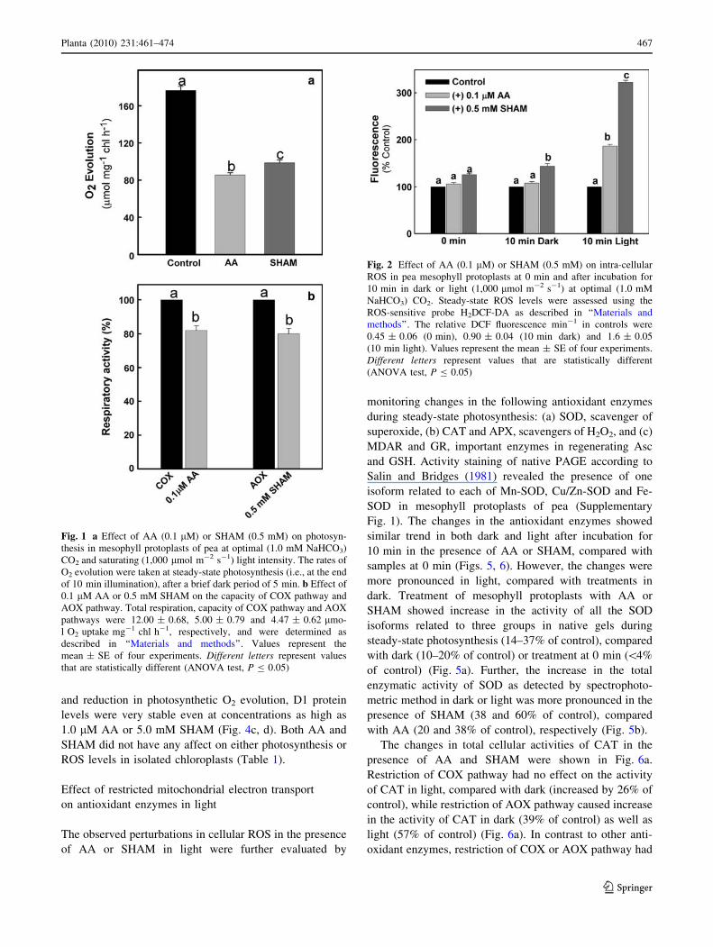
intensity of 1,000 lmol m-2 s-1. Both these compounds
remarkably decreased the rates of photosynthetic oxygen
evolution in mesophyll protoplasts by 50 and 44%,
respectively (Fig. 1a). In controls, the steady-state rates
(177 ± 7.7 lmol mg-1 chl h-1) of photosynthetic O2
evolution attained after a brief lag period of 3 min were
stable over a time period of at least 20–30 min in retaining
[90% of its activity (data not shown). However, so as to
avoid errors due to perturbations in the stability of meso-
phyll protoplasts at room temperature, all treatments in the
present study were restricted to a time period of 10 min
under light. At the chosen concentrations of AA (0.1 lM)
and SHAM (0.5 mM) where the effects on photosynthesis
were significant, the total respiratory capacity of COX and
AOX pathways of mesophyll protoplasts was affected only
marginally by 18 and 20%, respectively (Fig. 1b).
In contrast to the effects on photosynthesis, there was a
remarkable rise in intra-cellular ROS as measured by DCF
fluorescence when the electron transport through COX
pathway or AOX pathway is restricted under light. The
relative units of DCF fluorescence increased by 1.86- and
3.22-fold over control (without inhibitor) after illuminating
the mesophyll protoplasts for 10 min in the presence of AA
and SHAM, respectively (Fig. 2). The ROS levels also
increased under darkness, when the electron transport
through COX or AOX pathway is restricted using AA or
SHAM. However, the increase in ROS under darkness was
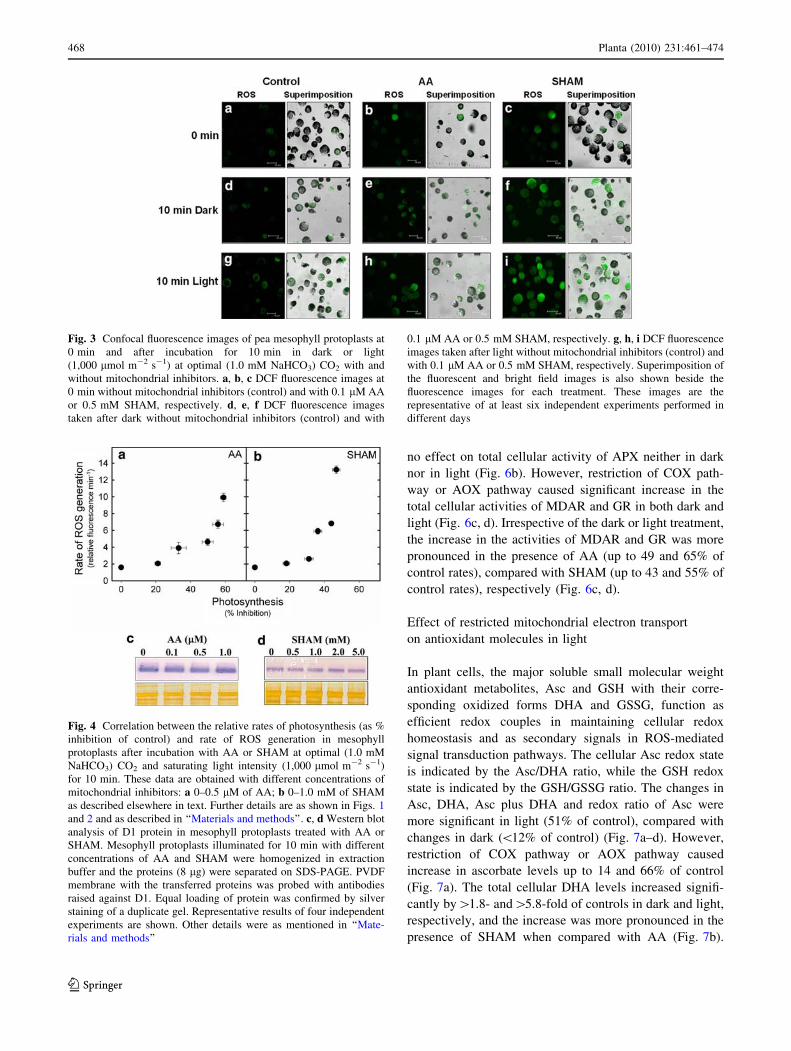
not as significant as in light. The pronounced increase in
the ROS levels of mesophyll protoplasts after illumination
in the presence of AA and SHAM was also further con-
firmed by confocal microscopic studies (Fig. 3).
To critically evaluate the relationship between photo-
synthesis and cellular ROS, the rise in DCF fluorescence
and the inhibition of photosynthesis were correlated in
mesophyll protoplasts at a wide range of concentrations of
AA (0–0.5 lM) and SHAM (0–1.0 mM) (Fig. 4a, b).
SHAM has esterase-like activity and can directly enhance
the DCF fluorescence (Hsiao and Bornman 1993). Similar
to SHAM, AA also enhanced the DCF fluorescence in
reaction medium used to illuminate mesophyll protoplasts.
Hence, in the present study, we have taken care to plot the
corrected values of DCF fluorescence (Supplementary
Table 1) obtained by the reaction of H2DCF with AA or
SHAM in the absence of protoplasts in Figs. 2, 4a, b. The
DCF fluorescence increased from 1.6 in control, i.e.,
without inhibitor to 9.9 and 13.2 in the presence of 0.5 lM
AA and 1.0 mM SHAM, respectively. On the other hand,
the photosynthesis decreased by 59 and 47% of control
rates in the presence of these inhibitors (Fig. 4a, b).
The changes in D1 protein, an important component of
PS II, were also examined in the presence of AA or SHAM
to understand if the reduction in photosynthetic oxygen
evolution or increase in cellular ROS caused any damage to
D1 protein. In spite of the rise in intra-cellular ROS levels
466 Planta (2010) 231:461–474
123
and reduction in photosynthetic O2 evolution, D1 protein
levels were very stable even at concentrations as high as
1.0 lM AA or 5.0 mM SHAM (Fig. 4c, d). Both AA and
SHAM did not have any affect on either photosynthesis or
ROS levels in isolated chloroplasts (Table 1).
Effect of restricted mitochondrial electron transport
on antioxidant enzymes in light
The observed perturbations in cellular ROS in the presence
of AA or SHAM in light were further evaluated by
monitoring changes in the following antioxidant enzymes
during steady-state photosynthesis: (a) SOD, scavenger of
superoxide, (b) CAT and APX, scavengers of H2O2, and (c)
MDAR and GR, important enzymes in regenerating Asc
and GSH. Activity staining of native PAGE according to
Salin and Bridges (1981) revealed the presence of one
isoform related to each of Mn-SOD, Cu/Zn-SOD and Fe-
SOD in mesophyll protoplasts of pea (Supplementary
Fig. 1). The changes in the antioxidant enzymes showed
similar trend in both dark and light after incubation for
10 min in the presence of AA or SHAM, compared with
samples at 0 min (Figs. 5, 6). However, the changes were
more pronounced in light, compared with treatments in
dark. Treatment of mesophyll protoplasts with AA or
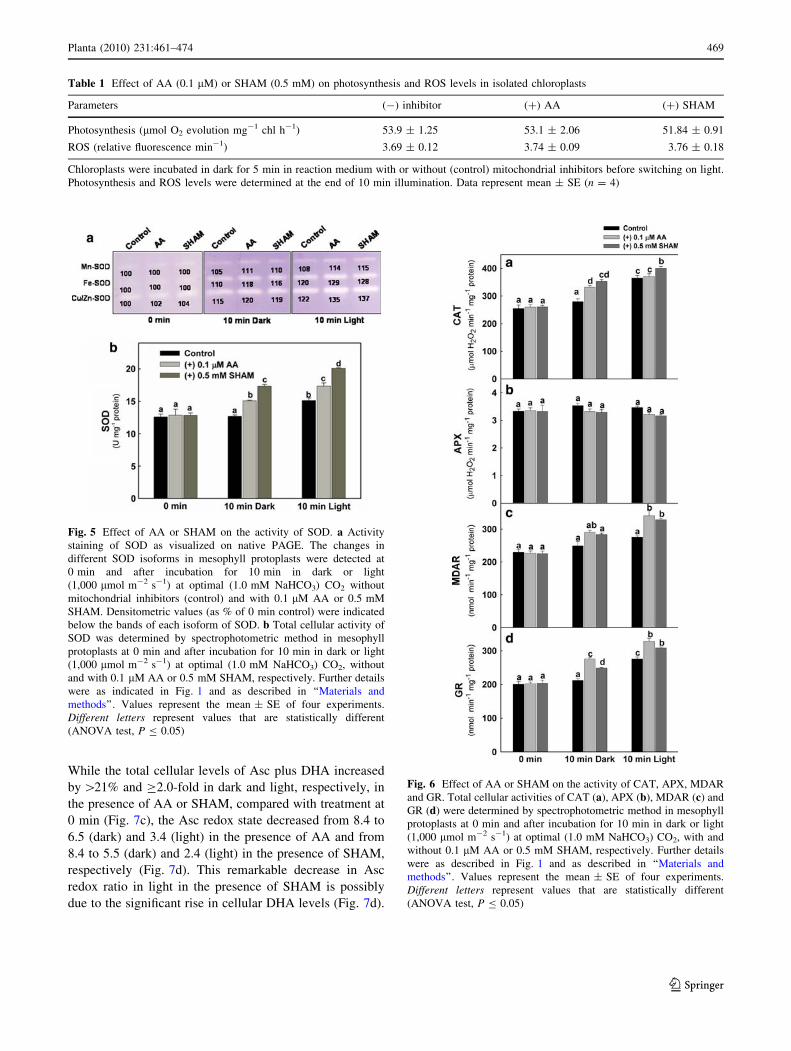
SHAM showed increase in the activity of all the SOD
isoforms related to three groups in native gels during
steady-state photosynthesis (14–37% of control), compared
with dark (10–20% of control) or treatment at 0 min (\4%
of control) (Fig. 5a). Further, the increase in the total
enzymatic activity of SOD as detected by spectrophoto-
metric method in dark or light was more pronounced in the
presence of SHAM (38 and 60% of control), compared
with AA (20 and 38% of control), respectively (Fig. 5b).
The changes in total cellular activities of CAT in the
presence of AA and SHAM were shown in Fig. 6a.
Restriction of COX pathway had no effect on the activity
of CAT in light, compared with dark (increased by 26% of
control), while restriction of AOX pathway caused increase
in the activity of CAT in dark (39% of control) as well as
light (57% of control) (Fig. 6a). In contrast to other anti-
oxidant enzymes, restriction of COX or AOX pathway had
Fig. 1 a Effect of AA (0.1 lM) or SHAM (0.5 mM) on photosyn-
thesis in mesophyll protoplasts of pea at optimal (1.0 mM NaHCO3)
CO2 and saturating (1,000 lmol m-2 s-1) light intensity. The rates of
O2 evolution were taken at steady-state photosynthesis (i.e., at the end
of 10 min illumination), after a brief dark period of 5 min. b Effect of
0.1 lM AA or 0.5 mM SHAM on the capacity of COX pathway and
AOX pathway. Total respiration, capacity of COX pathway and AOX
pathways were 12.00 ± 0.68, 5.00 ± 0.79 and 4.47 ± 0.62 lmo-
l O2 uptake mg-1 chl h-1, respectively, and were determined as
described in ‘‘Materials and methods’’. Values represent the
mean ± SE of four experiments. Different letters represent values
that are statistically different (ANOVA test, P B 0.05)
Fig. 2 Effect of AA (0.1 lM) or SHAM (0.5 mM) on intra-cellular
ROS in pea mesophyll protoplasts at 0 min and after incubation for
10 min in dark or light (1,000 lmol m-2 s-1) at optimal (1.0 mM
NaHCO3) CO2. Steady-state ROS levels were assessed using the
ROS-sensitive probe H2DCF-DA as described in ‘‘Materials and
methods’’. The relative DCF fluorescence min-1 in controls were
0.45 ± 0.06 (0 min), 0.90 ± 0.04 (10 min dark) and 1.6 ± 0.05
(10 min light). Values represent the mean ± SE of four experiments.
Different letters represent values that are statistically different
(ANOVA test, P B 0.05)
Planta (2010) 231:461–474 467
123
no effect on total cellular activity of APX neither in dark
nor in light (Fig. 6b). However, restriction of COX path-
way or AOX pathway caused significant increase in the
total cellular activities of MDAR and GR in both dark and
light (Fig. 6c, d). Irrespective of the dark or light treatment,
the increase in the activities of MDAR and GR was more
pronounced in the presence of AA (up to 49 and 65% of
control rates), compared with SHAM (up to 43 and 55% of
control rates), respectively (Fig. 6c, d).
Effect of restricted mitochondrial electron transport
on antioxidant molecules in light
In plant cells, the major soluble small molecular weight
antioxidant metabolites, Asc and GSH with their corre-
sponding oxidized forms DHA and GSSG, function as
efficient redox couples in maintaining cellular redox
homeostasis and as secondary signals in ROS-mediated
signal transduction pathways. The cellular Asc redox state
is indicated by the Asc/DHA ratio, while the GSH redox
state is indicated by the GSH/GSSG ratio. The changes in
Asc, DHA, Asc plus DHA and redox ratio of Asc were
more significant in light (51% of control), compared with
changes in dark (\12% of control) (Fig. 7a–d). However,
restriction of COX pathway or AOX pathway caused
increase in ascorbate levels up to 14 and 66% of control
(Fig. 7a). The total cellular DHA levels increased signifi-
cantly by[1.8- and[5.8-fold of controls in dark and light,
respectively, and the increase was more pronounced in the
presence of SHAM when compared with AA (Fig. 7b).
Fig. 3 Confocal fluorescence images of pea mesophyll protoplasts at
0 min and after incubation for 10 min in dark or light
(1,000 lmol m-2 s-1) at optimal (1.0 mM NaHCO3) CO2 with and
without mitochondrial inhibitors. a, b, c DCF fluorescence images at
0 min without mitochondrial inhibitors (control) and with 0.1 lM AA
or 0.5 mM SHAM, respectively. d, e, f DCF fluorescence images
taken after dark without mitochondrial inhibitors (control) and with
0.1 lM AA or 0.5 mM SHAM, respectively. g, h, i DCF fluorescence
images taken after light without mitochondrial inhibitors (control) and
with 0.1 lM AA or 0.5 mM SHAM, respectively. Superimposition of
the fluorescent and bright field images is also shown beside the
fluorescence images for each treatment. These images are the
representative of at least six independent experiments performed in
different days
Fig. 4 Correlation between the relative rates of photosynthesis (as %
inhibition of control) and rate of ROS generation in mesophyll
protoplasts after incubation with AA or SHAM at optimal (1.0 mM
NaHCO3) CO2 and saturating light intensity (1,000 lmol m-2 s-1)
for 10 min. These data are obtained with different concentrations of
mitochondrial inhibitors: a 0–0.5 lM of AA; b 0–1.0 mM of SHAM
as described elsewhere in text. Further details are as shown in Figs. 1
and 2 and as described in ‘‘Materials and methods’’. c, d Western blot
analysis of D1 protein in mesophyll protoplasts treated with AA or
SHAM. Mesophyll protoplasts illuminated for 10 min with different
concentrations of AA and SHAM were homogenized in extraction
buffer and the proteins (8 lg) were separated on SDS-PAGE. PVDF
membrane with the transferred proteins was probed with antibodies
raised against D1. Equal loading of protein was confirmed by silver
staining of a duplicate gel. Representative results of four independent
experiments are shown. Other details were as mentioned in ‘‘Mate-
rials and methods’’
468 Planta (2010) 231:461–474
123
While the total cellular levels of Asc plus DHA increased
by [21% and C2.0-fold in dark and light, respectively, in
the presence of AA or SHAM, compared with treatment at
0 min (Fig. 7c), the Asc redox state decreased from 8.4 to
6.5 (dark) and 3.4 (light) in the presence of AA and from
8.4 to 5.5 (dark) and 2.4 (light) in the presence of SHAM,
respectively (Fig. 7d). This remarkable decrease in Asc
redox ratio in light in the presence of SHAM is possibly
due to the significant rise in cellular DHA levels (Fig. 7d).
Table 1 Effect of AA (0.1 lM) or SHAM (0.5 mM) on photosynthesis and ROS levels in isolated chloroplasts
Parameters (-) inhibitor (?) AA (?) SHAM
Photosynthesis (lmol O2 evolution mg-1 chl h-1) 53.9 ± 1.25 53.1 ± 2.06 51.84 ± 0.91
ROS (relative fluorescence min-1) 3.69 ± 0.12 3.74 ± 0.09 3.76 ± 0.18
Chloroplasts were incubated in dark for 5 min in reaction medium with or without (control) mitochondrial inhibitors before switching on light.
Photosynthesis and ROS levels were determined at the end of 10 min illumination. Data represent mean ± SE (n = 4)
Fig. 5 Effect of AA or SHAM on the activity of SOD. a Activity
staining of SOD as visualized on native PAGE. The changes in
different SOD isoforms in mesophyll protoplasts were detected at
0 min and after incubation for 10 min in dark or light
(1,000 lmol m-2 s-1) at optimal (1.0 mM NaHCO3) CO2 without
mitochondrial inhibitors (control) and with 0.1 lM AA or 0.5 mM
SHAM. Densitometric values (as % of 0 min control) were indicated
below the bands of each isoform of SOD. b Total cellular activity of
SOD was determined by spectrophotometric method in mesophyll
protoplasts at 0 min and after incubation for 10 min in dark or light
(1,000 lmol m-2 s-1) at optimal (1.0 mM NaHCO3) CO2, without
and with 0.1 lM AA or 0.5 mM SHAM, respectively. Further details
were as indicated in Fig. 1 and as described in ‘‘Materials and
methods’’. Values represent the mean ± SE of four experiments.
Different letters represent values that are statistically different
(ANOVA test, P B 0.05)
Fig. 6 Effect of AA or SHAM on the activity of CAT, APX, MDAR
and GR. Total cellular activities of CAT (a), APX (b), MDAR (c) and
GR (d) were determined by spectrophotometric method in mesophyll
protoplasts at 0 min and after incubation for 10 min in dark or light
(1,000 lmol m-2 s-1) at optimal (1.0 mM NaHCO3) CO2, with and
without 0.1 lM AA or 0.5 mM SHAM, respectively. Further details
were as described in Fig. 1 and as described in ‘‘Materials and
methods’’. Values represent the mean ± SE of four experiments.
Different letters represent values that are statistically different
(ANOVA test, P B 0.05)
Planta (2010) 231:461–474 469
123
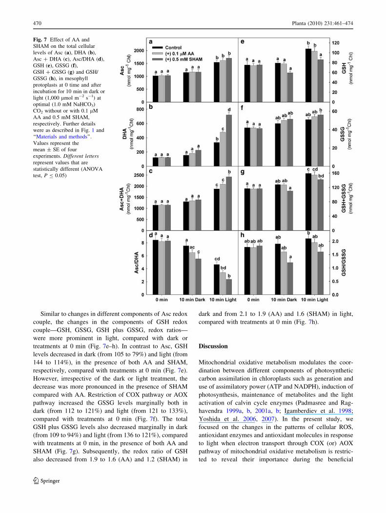
Similar to changes in different components of Asc redox
couple, the changes in the components of GSH redox
couple—GSH, GSSG, GSH plus GSSG, redox ratios—
were more prominent in light, compared with dark or
treatments at 0 min (Fig. 7e–h). In contrast to Asc, GSH
levels decreased in dark (from 105 to 79%) and light (from
144 to 114%), in the presence of both AA and SHAM,
respectively, compared with treatments at 0 min (Fig. 7e).
However, irrespective of the dark or light treatment, the
decrease was more pronounced in the presence of SHAM
compared with AA. Restriction of COX pathway or AOX
pathway increased the GSSG levels marginally both in
dark (from 112 to 121%) and light (from 121 to 133%),
compared with treatments at 0 min (Fig. 7f). The total
GSH plus GSSG levels also decreased marginally in dark
(from 109 to 94%) and light (from 136 to 121%), compared
with treatments at 0 min, in the presence of both AA and
SHAM (Fig. 7g). Subsequently, the redox ratio of GSH
also decreased from 1.9 to 1.6 (AA) and 1.2 (SHAM) in
dark and from 2.1 to 1.9 (AA) and 1.6 (SHAM) in light,
compared with treatments at 0 min (Fig. 7h).
Discussion
Mitochondrial oxidative metabolism modulates the coor-
dination between different components of photosynthetic
carbon assimilation in chloroplasts such as generation and
use of assimilatory power (ATP and NADPH), induction of
photosynthesis, maintenance of metabolites and the light
activation of calvin cycle enzymes (Padmasree and Rag-
havendra 1999a, b, 2001a, b; Igamberdiev et al. 1998;
Yoshida et al. 2006, 2007). In the present study, we
focused on the changes in the patterns of cellular ROS,
antioxidant enzymes and antioxidant molecules in response
to light when electron transport through COX (or) AOX
pathway of mitochondrial oxidative metabolism is restric-
ted to reveal their importance during the beneficial
Fig. 7 Effect of AA and
SHAM on the total cellular
levels of Asc (a), DHA (b),
Asc ? DHA (c), Asc/DHA (d),
GSH (e), GSSG (f),GSH ? GSSG (g) and GSH/
GSSG (h), in mesophyll
protoplasts at 0 time and after
incubation for 10 min in dark or
light (1,000 lmol m-2 s-1) at
optimal (1.0 mM NaHCO3)
CO2 without or with 0.1 lM
AA and 0.5 mM SHAM,
respectively. Further details
were as described in Fig. 1 and
‘‘Materials and methods’’.
Values represent the
mean ± SE of four
experiments. Different lettersrepresent values that are
statistically different (ANOVA
test, P B 0.05)
470 Planta (2010) 231:461–474
123
interactions of mitochondrial metabolism with photosyn-
thetic carbon assimilation.
Use of metabolic inhibitors to examine the interactions
between chloroplasts and mitochondria
In the present study, we used the metabolic inhibitors AA
and SHAM which are easily permeable through the plasma
membrane of protoplasts to restrict the flow of electrons
through the COX and AOX pathways, respectively. The
general perturbations caused to the metabolic system due to
the non-specific effects of both AA and SHAM were
minimized using these compounds at very low concentra-
tions. Further at the chosen concentrations of AA (0.1 lM)
and SHAM (0.5 mM), in spite of a marginal interference
(\20%) in the respiratory capacity of COX and AOX
pathways, there was a remarkable decrease (\50%) in the
rates of photosynthetic oxygen evolution and significant
increase (3.2-fold) in total cellular ROS of mesophyll
protoplasts in light (Figs. 1a, b, 2). However, neither AA
nor SHAM had any direct effect on the photosynthetic O2
evolution rates or ROS levels in isolated chloroplasts
(Table 1). Besides our own work with mesophyll protop-
lasts (Padmasree and Raghavendra 2001b), Yoshida et al.
(2006) also indicated that AA at 0.1 lM concentration did
not exert any large direct effects on cyclic electron trans-
port of photosynthesis in leaves. While examining in detail
the effects of 250 lM AA on photosynthesis, Takahashi
et al. (2009) suggested that the inhibition of photosynthesis
by AA is partly due to the interference of mitochondrial
respiration. In the present study, we also evaluated the
marginal interference in the respiratory capacity of COX
pathway by 0.1 lM AA with changes in ATP/ADP ratios
which were decreased by 30% in light when compared to
their ratios in the absence of AA (data not shown).
ROS play an important role during the beneficial
interactions between chloroplasts and mitochondria
to optimize photosynthetic carbon assimilation
The strong positive correlation between the rise in total
cellular levels of DCF fluorescence and inhibition of
NaHCO3-dependent O2 evolution at a wide range of con-
centrations of both AA and SHAM demonstrates the
importance of ROS during the beneficial interactions of
mitochondrial electron transport with photosynthetic car-
bon assimilation (Fig. 4a, b). Any restriction in mito-
chondrial electron transport is known to cause over-
reduction of respiratory complexes and generation of ROS
in mitochondria (Møller 2001; Foyer and Noctor 2003;
Navrot et al. 2007). Further, the following observations
made through fluorimetric and confocal microscopy in the
present study provided indirect evidences for the
contribution of both mitochondria and chloroplast in rising
the total cellular ROS in light: (a) pronounced increase in
total cellular ROS in light when compared to darkness
(Figs. 2, 3) and (b) negligible change in ROS levels in
isolated illuminated chloroplasts (Table 1). The relative
importance of AOX pathway over COX pathway in
maintaining cellular ROS was shown with A. thaliana
knock out mutants for AOX1a, which produced more ROS
when compared to wild-type plants when treated with AA
(Strodtkotter et al. 2009). H2DCF-DA was used to analyze
the changes in cellular H2O2 levels (Maxwell et al. 1999).
Hence, we attribute the changes observed in ROS in the
present study could be mostly due to H2O2 (Figs. 2, 3, 4a,
b). The role of ROS as signal during the beneficial inter-
actions between chloroplasts and mitochondria is further
strengthened by the insignificant changes observed in D1
protein, which is known to be vulnerable to degradation by
any increase in ROS levels (Fig. 4; Murata et al. 2007).
ROS mediates beneficial interactions between
chloroplasts and mitochondria through changes
in antioxidant enzymes
The perturbations in cellular ROS caused due to restriction
in mitochondrial electron transport under steady-state
photosynthesis were monitored by examining the changes
in the activities of the following antioxidant enzymes
which play a role in scavenging of ROS and regeneration
of antioxidant metabolites related to Asc–GSH cycle: (a)
SOD, (b) CAT, (c) APX, (d) MDAR and (e) GR (Noctor
and Foyer 1998; Chew et al. 2003; Mittler et al. 2004;
Amirsadeghi et al. 2006). The substantial increase in all the
isoforms of SOD (chloroplastic, mitochondrial, peroxi-
somal and cytosolic) in the presence of AA and SHAM
demonstrates the participation of antioxidant enzymes
during the beneficial interactions between mitochondria
and chloroplasts to optimize photosynthetic carbon assim-
ilation (Fig. 5a). While the changes in the activities of SOD
and CAT were more pronounced in the presence of SHAM,
AA caused a notable increase in the activities of MDAR
and GR during steady-state photosynthesis (Figs. 5b, 6a, c,
d). In spite of the suggested sensitivity of APX to H2O2, we
did not observe any such changes in its activity when
electron transport through COX or AOX pathway is
restricted in light (Fig. 6b; Miyake and Asada 1996).
ROS mediates beneficial interactions between
chloroplasts and mitochondria through changes
in antioxidant molecules
In plant cells, perturbations in the steady-state level of ROS
are also detected through changes in cellular redox couples
such as Asc/DHA and GSH/GSSG, which ultimately
Planta (2010) 231:461–474 471
123
require electrons from redox pairs such as NADPH/NADP
(Foyer and Noctor 2003, 2005; Noctor 2006; Navrot et al.
2007). In vtc mutants which are deficient in biosynthesis of
Asc, the sensitivity of leaf discs to undergo photoinhibition
was enhanced in the presence of AA and SHAM (T. Sai
Krishna et al., University of Hyderabad, personal com-
munication). The significant decrease in redox ratio of Asc
(indicated by Asc/DHA) in the presence of AA and SHAM
suggests the importance of both COX and AOX pathways
in maintaining cellular homeostasis buffered by Asc/DHA
redox couple in light (Fig. 7d).
The ratios of GSH/GSSG were found to vary to changes
in oxygen concentration in roots (6.1) and leaf cells (14.8)
(Skutnik and Rychter 2009). However, such high redox
ratios related to GSH were not observed in control samples
in the present study. This could be attributed to the varia-
tion in the experimental material used or variation in
technique used or variation in sample treatments applied to
monitor the changes in different components of GSH redox
couple. However, the observed decrease in the redox ratio
of GSH (indicated by GSH/GSSG) under steady-state
photosynthesis only in the presence of SHAM but not AA
suggests the importance of AOX pathway in maintaining
cellular homeostasis buffered by GSH/GSSG redox couple
(Fig. 7h). In CAT-deficient mutant (cat2) of Arabidopsis,
H2O2 induced accumulation of glutathione, indicating the
importance of glutathione in maintaining cellular redox
homeostasis (Queval et al. 2009). Unlike animal models, in
plant cells, though the information on the direct role of
reduced GSH in scavenging H2O2 is very limited, it plays a
role in detoxification of H2O2 along with Asc through Asc–
GSH cycle (Noctor and Foyer 1998). Nevertheless, several
plant genes that showed homology to mammalian phos-
pholipids hydroperoxide GSH peroxidase have been iso-
lated recently (Rouhier et al. 2008).
Concluding remarks
The observations from the present study clearly indicate
that any perturbation in the capacities of the COX or AOX
pathway of mitochondrial oxidative electron transport in
light leads to disturbances in the sustenance of photosyn-
thesis through modulation in ROS, antioxidant enzymes
and antioxidant molecules. However, it is intriguing to
understand how the restriction in electron transport through
COX pathway or AOX pathway of mitochondria in cellular
environment is simultaneously coordinated with decline in
chloroplastic photosynthesis through changes in ROS and
different components of antioxidant system. More detailed
studies involving transgenic and reverse genetics along
with the holistic approach of systems biology are required
to unravel the biochemical signals and molecular
mechanisms underlying the beneficial interactions between
chloroplasts and mitochondria to optimize photosynthetic
carbon assimilation.
Acknowledgments This work was supported by grants to K.P.S.
from Department of Science and Technology (No. SR/FTP/LS-226/
2000). The support of funds to the Department of Plant Sciences and
School of Life Sciences from DST-FIST level II (SR/FST/LSII-010/
2007) and UGC-CAS-I (F-5-8/2008, SAP-II) is gratefully acknowl-
edged. The authors are grateful to Dr. G. Padmaja, and CIL, UoHyd,
for their help in statistical analysis and confocal microscopic studies.
Ch.D. is a recipient of Senior Research Fellowship from Council of
Scientific and Industrial Research. We thank Prof. Greg Vanlerberghe
and Dr. T. Saradadevi for helpful discussion and suggestions related
to the manuscript.
References
Amirsadeghi S, Robson CA, McDonald AE, Vanlerberghe GC (2006)
Changes in plant mitochondrial electron transport alter cellular
levels of reactive oxygen species and susceptibility to cell death
signaling molecules. Plant Cell Physiol 47:1509–1519
Apel K, Hirt H (2004) Reactive oxygen species: metabolism,
oxidative stress, and signal transduction. Annu Rev Plant Biol
55:373–399
Asada K (1999) The water–water cycle in chloroplasts: scavenging of
active oxygen and dissipation of excess photons. Annu Rev Plant
Physiol Plant Mol Biol 50:601–639
Bartoli CG, Yu J, Gomez F, Fernandez L, McIntosh L, Foyer CH
(2006) Inter-relationships between light and respiration in the
control of ascorbic acid synthesis and accumulation in Arabid-opsis thaliana leaves. J Exp Bot 57:1621–1631
Beauchamp C, Fridovich I (1971) Superoxide dismutase: improved
assays and an assay applicable to acrylamide gels. Anal Biochem
44:276–287
Carrari F, Nunes-Nesi A, Gibon Y, Lytovchenko A, Ehlers-Loureiro
M, Fernie AR (2003) Reduced expression of aconitase results in
an enhanced rate of photosynthesis and marked shifts in carbon
partitioning in illuminated leaves of wild species tomato. Plant
Physiol 133:1322–1335
Chew O, Whelan J, Millar AH (2003) Molecular definition of the
ascorbate-glutathione cycle in Arabidopsis mitochondria reveals
dual targeting of antioxidant defenses in plants. J Biol Chem
278:46869–46877
del Rio LA, Sandalio LM, Corpas FJ, Palma JM, Barroso JB (2006)
Reactive oxygen species and reactive nitrogen species in
peroxisomes: production, scavenging and role in cell signaling.
Plant Physiol 141:330–335
Doulis AG, Debian N, Kingston-Smith AH, Foyer CH (1997)
Differential localization of antioxidants in maize leaves. Plant
Physiol 114:1031–1037
Drazkiewicz M, Skorzynska-Polit E, Krupa Z (2003) Response of
ascorbate-glutathione cycle to excess copper in Arabidopsisthaliana (L.). Plant Sci 164:195–202
Dutilleul C, Driscoll S, Cornic G, De Paepe R, Foyer CH, Noctor G
(2003) Functional mitochondrial complex I is required by tobacco
leaves for optimal photosynthetic performance in photorespiratory
conditions and during transients. Plant Physiol 131:264–275
Fiorani F, Umbach AL, Siedow JN (2005) The alternative oxidase of
plant mitochondria is involved in the acclimation of shoot
growth at low temperature. A study of Arabidopsis AOX1atransgenic plants. Plant Physiol 139:1795–1805
472 Planta (2010) 231:461–474
123
Foyer CH, Noctor G (2003) Redox sensing and signaling associated
with reactive oxygen in chloroplasts, peroxisomes and mito-
chondria. Physiol Plant 119:355–364
Foyer CH, Noctor G (2005) Oxidant and antioxidant signaling in
plants: a re-evaluation of the concept of oxidative stress in a
physiological context. Plant Cell Environ 28:1056–1071
Foyer CH, Rowell J, Walker D (1983) Measurement of the ascorbate
content of spinach leaf protoplasts and chloroplasts during
illumination. Planta 157:239–244
Gardestrom P, Igamberdiev AU, Raghavendra AS (2002) Mitochon-
drial functions in light. In: Foyer CH, Noctor G (eds)
Photosynthetic nitrogen assimilation and associated carbon and
respiratory metabolism. Kluwer Academic Publishers, The
Netherlands, pp 151–172
Graham D (1980) Effects of light on dark respiration. In: Davies DD
(ed) The biochemistry of plants: a comprehensive treatise.
Academic Press, New York, pp 525–579
Griffith OW (1980) Determination of glutathione and glutathione
disulfide using glutathione reductase and 2-vinyl-pyridine. Anal
Biochem 106:207–212
Hoefnagel MHN, Atkin OK, Wiskich JT (1998) Interdependence
between chloroplasts and mitochondria in the light and the dark.
Biochim Biophys Acta 1366:235–255
Hsiao KC, Bornman CH (1993) Salicylhydroxamic acid mimics
esterase-like action. J Exp Bot 44:1847–1849
Igamberdiev AU, Hurry V, Kromer S, Gardestrom P (1998) The role
of mitochondrial electron transport during photosynthetic induc-
tion. A study with barley (Hordeum vulgare) protoplasts
incubated with rotenone and oligomycin. Physiol Plant
104:431–439
Jiang M, Zhang J (2001) Effect of abscissic acid on active oxygen
species, antioxidative defense system and oxidative damage in
leaves of maize seedlings. Plant Cell Physiol 42:1265–1273
Kromer S (1995) Respiration during photosynthesis. Annu Rev Plant
Physiol Plant Mol Biol 46:45–70
Kromer S, Stitt M, Heldt HW (1988) Mitochondrial oxidative
phosphorylation participating in photosynthetic metabolism of
a leaf cell. FEBS Lett 226:352–356
Kromer S, Malmberg G, Gardestrom P (1993) Mitochondrial
contribution to photosynthetic metabolism. A study with barley
(Hordeum vulgare) leaf protoplasts at different light intensities
and CO2 concentrations. Plant Physiol 102:947–955
Kuzniak E, Sklodowska M (2005) Compartment-specific role of the
ascorbate-glutathione cycle in the response of tomato leaf cells
to Botrytis cinerea infection. J Exp Bot 56:921–933
Laemmli UK (1970) Cleavage of structural proteins during the
assembly of the head of bacteriophage T4. Nature 227:680–685
Lowry OH, Rosebrough NJ, Farr AL, Randall RJ (1951) Protein
measurement with the folin phenol reagent. J Biol Chem
193:265–275
Maxwell DP, Wang Y, McIntosh L (1999) The alternative oxidase
lowers mitochondrial reactive oxygen production in plant cells.
Proc Natl Acad Sci USA 96:8271–8276
Mittler R, Vanderauwera S, Gollery M, Breusegem FV (2004)
Reactive oxygen gene network of plants. Trends Plant Sci
9:490–498
Miyake C, Asada K (1996) Inactivation mechanism of ascorbate
peroxidase at low concentration of ascorbate; hydrogen peroxide
decomposes compound I of ascorbate peroxidase. Plant Cell
Physiol 37:423–430
Møller IM (2001) Plant mitochondria and oxidative stress: electron
transport, NADPH turnover, and metabolism of reactive oxygen
species. Annu Rev Plant Physiol Plant Mol Biol 52:561–591
Møller IM, Jensen PE, Hansson A (2007) Oxidative modifications to
cellular components in plants. Annu Rev Plant Biol 58:459–481
Murata N, Takahashi S, Nishiyama Y, Allakhverdiev SI (2007)
Photoinhibition of photosystem II under environmental stress.
Biochim Biophys Acta 1767:414–421
Nakano Y, Asada K (1981) Hydrogen peroxide is scavenged by
ascorbate-specific peroxidase in spinach chloroplasts. Plant Cell
Physiol 22:867–880
Navrot N, Rouhier N, Gelhaye E, Jacquot JE (2007) Reactive oxygen
species generation and antioxidant systems in plant mitochon-
dria. Physiol Plant 129:185–195
Noctor G (2006) Metabolic signaling in defence and stress: the central
roles of soluble redox couples. Plant Cell Environ 29:409–425
Noctor G, Foyer CH (1998) Ascorbate and glutathione: keeping
active oxygen under control. Annu Rev Plant Physiol Plant Mol
Biol 49:249–279
Noctor G, De Paepe R, Foyer CH (2007) Mitochondrial redox biology
and homeostasis in plants. Trends Plant Sci 12:125–134
Noguchi K, Yoshida K (2008) Interaction between photosynthesis and
respiration in illuminated leaves. Mitochondrion 8:87–99
Nunes-Nesi A, Carrari F, Lytovchenko A, Smith AMO, Loureiro ME,
Ratcliffe RG, Sweetlove LJ, Fernie AR (2005) Enhanced
photosynthetic performance and growth as a consequence of
decreasing mitochondrial malate dehydrogenase activity in
transgenic tomato plants. Plant Physiol 137:611–622
Nunes-Nesi A, Sulpice R, Gibon Y, Fernie AR (2008) The enigmatic
contribution of mitochondrial function in photosynthesis. J Exp
Bot 59:1675–1684
Padmasree K, Raghavendra AS (1999a) Importance of oxidative
electron transport over oxidative phosphorylation in optimizing
photosynthesis in mesophyll protoplasts of pea (Pisum sativumL.). Physiol Plant 105:546–553
Padmasree K, Raghavendra AS (1999b) Response of photosynthetic
carbon assimilation in mesophyll protoplasts to restriction on
mitochondrial oxidative metabolism: metabolites related to the
redox status and sucrose biosynthesis. Photosynth Res 62:231–
239
Padmasree K, Raghavendra AS (2001a) Consequence of restricted
mitochondrial oxidative metabolism on photosynthetic carbon
assimilation in mesophyll protoplasts: decrease in light activa-
tion for four chloroplastic enzymes. Physiol Plant 112:582–588
Padmasree K, Raghavendra AS (2001b) Restriction of mitochondrial
oxidative metabolism leads to suppression of photosynthetic
carbon assimilation but not of photochemical electron transport
in pea mesophyll protoplasts. Curr Sci 81:680–684
Padmasree K, Padmavathi L, Raghavendra AS (2002) Essentiality of
mitochondrial oxidative metabolism for photosynthesis: optimi-
zation of carbon assimilation and protection against photoinhi-
bition. Crit Rev Biochem Mol Biol 37:71–119
Palma JM, Jiminez A, Sandalio LM, Corpas FJ, Lundqvist M, Gomez
M, Sevilla F, del Rio LA (2006) Antioxidative enzymes from
chloroplasts, mitochondria, and peroxisomes during leaf senes-
cence of nodulated pea plants. J Exp Bot 57:1747–1758
Patterson BD, Payne LA, Chen Y, Graham D (1984) An inhibitor of
catalase induced by cold chilling-sensitive plants. Plant Physiol
76:1014–1018
Queval G, Thominet D, Vanacker H, Miginiac-Maslow M, Gakiere B,
Noctor G (2009) H2O2 activated up-regulation of glutathione in
Arabidopsis involves induction of genes encoding enzymes
involved in cysteine synthesis in the chloroplast. Mol Plant
2:344–356
Raghavendra AS, Padmasree K (2003) Beneficial interactions of
mitochondrial metabolism with photosynthetic carbon assimila-
tion. Trends Plant Sci 8:546–553
Raghavendra AS, Padmasree K, Saradadevi K (1994) Interdepen-
dence of photosynthesis and respiration in plant cells: interac-
tions between chloroplasts and mitochondria. Plant Sci 97:1–14
Planta (2010) 231:461–474 473
123
Rouhier N, Lemaire SD, Jacquot JP (2008) The role of glutathione in
photosynthetic organisms: emerging functions for glutaredoxins
and glutathionylation. Annu Rev Plant Biol 59:143–166
Rucinska R, Waplak S, Gwozdz E (1999) Free radical formation and
activity of antioxidant enzymes in lupin roots exposed to lead.
Plant Physiol Biochem 37:187–194
Salin ML, Bridges SM (1981) Absence of iron-containing superoxide
dismutase in mitochondria from mustard (Brassica campestris).
Biochem J 195:229–233
Skutnik M, Rychter AM (2009) Differential response of antioxidant
systems in leaves and roots of barley subjected to anoxia and
post-anoxia. J Plant Physiol 116:926–937
Strodtkotter I, Padmasree K, Dinakar CH, Speth B, Niazi PS, Wojtera
J, Voss I, Do PT, Nunes-Nesi A, Fernie AR, Linke V,
Raghavendra AS, Scheibe R (2009) Induction of the AOX1D
isoform of alternative oxidase in A. thaliana T-DNA insertion
lines lacking isoform AOX 1A is insufficient to optimize
photosynthesis when treated with AA. Mol Plant 2:284–297
Sundar D, Perianayaguy B, Reddy AR (2004) Localization of
antioxidant enzymes in the cellular compartments of sorghum
leaves. Plant Growth Regul 44:157–163
Sweetlove LJ, Lytovchenko A, Morgan M, Nunes-Nesi A, Taylor NL,
Baxter CJ, Eickmeier I, Fernie AR (2006) Mitochondrial
uncoupling protein is required for efficient photosynthesis. Proc
Natl Acad Sci USA 103:19587–19592
Takahashi S, Milward SE, Fan DY, Chow WS, Badger MR (2009)
How does cyclic electron flow alleviate photoinhibition in
Arabidopsis. Plant Physiol 149:1560–1567
Towbin H, Staehlin T, Gordon J (1979) Electrophoretic transfer of
proteins from polyacrylamide gels to nitrocellulose sheets.
Procedure and some applications. Proc Natl Acad Sci USA
76:4350–4354
Vanlerberghe GC, Robson CA, Yip JYH (2002) Induction of
mitochondrial alternative oxidase in response to a cell signal
pathway down-regulating the cytochrome pathway prevents
programmed cell death. Plant Physiol 129:1829–1842
Watanabe CK, Hachiya T, Terashima I, Noguchi K (2008) The lack
of alternative oxidase at low temperature leads to a disruption of
the balance in carbon and nitrogen metabolism, and to an up-
regulation of antioxidant defence systems in Arabidopsis thali-ana leaves. Plant Cell Environ 31:1190–1202
Yoshida K, Terashima I, Noguchi K (2006) Distinct roles of the
cytochrome pathway and alternative oxidase in leaf photosyn-
thesis. Plant Cell Physiol 47:22–31
Yoshida K, Terashima I, Noguchi K (2007) Up-regulation of
mitochondrial alternative oxidase concomitant with chloroplast
over-reduction by excess light. Plant Cell Physiol 48:606–614
474 Planta (2010) 231:461–474
123