Isolation and characterisation of dog uropathogenic Escherichia coli strains and their fimbriae
Upload
independentCategory
view
4download
0
ORIGINAL PAPER
Impairment of the biomechanical compliance of P pili: a novelmeans of inhibiting uropathogenic bacterial infections?
Jeanna E. Klinth • Jerome S. Pinkner •
Scott J. Hultgren • Fredrik Almqvist •
Bernt Eric Uhlin • Ove Axner
Received: 30 June 2011 / Revised: 25 November 2011 / Accepted: 6 December 2011 / Published online: 12 January 2012
� The Author(s) 2012. This article is published with open access at Springerlink.com
Abstract Gram-negative bacteria often initiate their col-
onization by use of extended attachment organelles, so
called pili. When exposed to force, the rod of helix-like pili
has been found to be highly extendable, mainly attributed
to uncoiling and recoiling of its quaternary structure. This
provides the bacteria with the ability to redistribute an
external force among a multitude of pili, which enables
them to withstand strong rinsing flows, which, in turn,
facilitates adherence and colonization processes critical
to virulence. Thus, pili fibers are possible targets for
novel antibacterial agents. By use of a substance that
compromises compliance of the pili, the ability of bacteria
to redistribute external forces can be impaired, so they will
no longer be able to resist strong urine flow and thus be
removed from the host. It is possible such a substance can
serve as an alternative to existing antibiotics in the future
or be a part of a multi-drug. In this work we investigated
whether it is possible to achieve this by targeting the
recoiling process. The test substance was purified PapD.
The effect of PapD on the compliance of P pili was
assessed at the single organelle level by use of force-
measuring optical tweezers. We showed that the recoiling
process, and thus the biomechanical compliance, in par-
ticular the recoiling process, can be impaired by the pres-
ence of PapD. This leads to a new concept in the search for
novel drug candidates combating uropathogenic bacterial
infections—‘‘coilicides’’, targeting the subunits of which
the pilus rod is composed.
Keywords Bacterial adhesion � Force-measuring optical
tweezers � Antimicrobial � Single organelle � Pili recoiling
Introduction
According to the World Health Organization, resistance to
antibiotics is the biggest challenge in drug development.
There is a need not for one new antibiotic substance but
several, so these can be combined and strengthen the human
position in the fight against bacterial resistance (Kaplan
and Laing 2004). This necessitates the identification and
addressing of new targets in bacteria. Bacterial pathogens
have a variety of mechanisms that contribute to their ability
to colonize and cause disease in human hosts (Staskawicz
et al. 2001). It is well known that the virulence of a microbe,
i.e. its ability to infect, is correlated with its ability to adhere
J. E. Klinth (&) � O. Axner
Department of Physics, Umea University,
901 87 Umea, Sweden
e-mail: [email protected]
J. E. Klinth � F. Almqvist � B. E. Uhlin � O. Axner
Umea Centre for Microbial Research (UCMR),
Umea University, 901 87 Umea, Sweden
J. E. Klinth � B. E. Uhlin
Department of Molecular Biology, Umea University,
901 87 Umea, Sweden
J. E. Klinth � B. E. Uhlin
Laboratory for Molecular Infection Medicine Sweden (MIMS),
Umea University, 901 87 Umea, Sweden
J. E. Klinth � O. Axner
Center for Biomedical Engineering and Physics (CMTF),
Umea University, 901 87 Umea, Sweden
J. S. Pinkner � S. J. Hultgren
Department of Molecular Microbiology, Washington University
School of Medicine, St. Louis, MO 63110, USA
F. Almqvist
Department of Chemistry, Umea University,
901 87 Umea, Sweden
123
Eur Biophys J (2012) 41:285–295
DOI 10.1007/s00249-011-0784-2

to tissue, and, in particular, to its capability to withstand
forces from rinsing flows (Ofek et al. 2003). Some bacteria
mediate adhesion by use of long attachment organelles, also
referred to as fimbriae or pili. P pili, a prototype helix-like
pili, are produced by uropathogenic Escherichia coli
(UPEC) strains and are predominantly associated with
kidney infection (pyelonephritis) (Sauer et al. 2000). The
rod of P pili consists of more than a thousand PapA sub-
units, assembled into a helical right-handed structure with
3.3 subunits per turn (Gong and Makowski 1992; Bullitt and
Makowski 1995), ending in a linear tip structure that
anchors the PapG adhesin at the distal end (Jacob-Dubuis-
son et al. 1993; Kuehn et al. 1992; Striker et al. 1994; Lee
et al. 2007; Lindberg et al. 1987). As is further alluded to
below, the ability of such attached bacteria to withstand
forces caused by rinsing flows is not only given by the
properties of the adhesin–receptor interactions, but is also
strongly affected by the biomechanical properties of these
organelles (Duncan et al. 2005).
It has been found that the compliance of helix-like pili
(i.e. their response to force), and thus their ability to assist
in the adhesion process, depends strongly on the quaternary
structure of the pilus. These pili have an intricate and
extraordinary force versus elongation response that consists
of a combination of a constant elongation force (in the so-
called region II), originating from sequential uncoiling of
the helix-like structure, and a sigmoidal pseudo-elastic
response (region III), caused by a conformational change of
the head-to-tail interaction between the subunits of pili that
takes place in a random order (Jass et al. 2004; Fallman
et al. 2005; Andersson et al. 2007, 2008; Bjornham et al.
2008; Axner et al. 2009). These extraordinary biome-
chanical properties, primarily those of region II, which
enable significant elongation of pili length [more than five
times the original length for P pili (Jass et al. 2004)] under
a constant force, and the fact that the uncoiling is fully
reversible (Fallman et al. 2005), have been regarded as
being of special importance to the ability of a piliated
bacterium to remain attached to the host tissue (despite
being exposed to a substantial external force) by redis-
tributing an external shear force among a multitude of pili
(Bjornham et al. 2008). Presumably, they are a major
reason why the ability of attached bacteria to withstand
forces caused by urine flow is strongly affected by the
properties of the pili shaft per se (Duncan et al. 2005). This
implies that the pilus rod and the tip-associated adhesin are
both important in the ability of bacteria to withstand strong
rinsing forces (Bjornham and Axner 2009).
During recent decades much work has been devoted to
understanding the adhesive binding mechanism. For
example, fimbrial tip-mediated and shear-dependent adhe-
sion (Le Trong et al. 2010) and a shear-enhanced adhesion
mechanism (Whitfield et al. 2010) have been demonstrated.
To prevent bacterial attachment, inhibitors targeting the
adhesion process, i.e. carbohydrate derived competitive
inhibitors for adhesin–receptor interactions, have been
developed (Wellens et al. 2008; Salminen et al. 2007;
Ohlsson et al. 2002; Krogfelt et al. 1990). In addition to
this ligand-based approach, several bicyclic 2-pyridones,
referred to as pilicides, have been rationally designed to
prevent pili formation. It has been suggested that pilicides
inhibit the chaperone–usher interaction, thus preventing
the essential event in which the N-terminus of the usher
(a membrane protein involved in the pili assembly) rec-
ognizes and binds to incoming chaperone–subunit com-
plexes (Pinkner et al. 2006). These pilicides can efficiently
inhibit pili biogenesis without affecting bacterial growth
(Aberg et al. 2006; Pinkner et al. 2006; Cegelski et al.
2009; Chorell et al. 2010). In contrast, so far, the pili rod
has not been used as a target for antibiotics.
In this work we investigated whether it is possible to
affect the biomechanical properties of helix-like pili by
using a substance that can bind to an elongated pilus at
the single organelle level, primarily compromising the
uncoiling—recoiling process. We show, by use of force-
measuring optical tweezers (FMOT), that the biomechan-
ical compliance of P pili can be impaired without breaking
the backbone of the pilus. Thus, the pili rod can be
regarded as a new target for novel drug candidates com-
bating uropathogenic bacterial infections.
Materials and methods
Culture of bacteria
The Escherichia coli (E. coli) strain used was HB101/
pPAP5, which is a clone carrying the wild type pap gene
cluster from UPEC strain J96 (Lindberg et al. 1984).
A typical HB101/pPAP5 piliated cell is shown in Fig. 1a.
Because it has previously been found that study of fewer pili
per bacterium enhances the ability to capture a single orga-
nelle for force measurements, a pilicide, a substituted ring-
fused 2-pyridone (BibC10, Fig. 1b), referred to as pilicide 1
by Aberg and Almqvist (2007), was used to chemically
manipulate the number of pili expressed per bacterial cell.
For pilicide concentrations below that required for total
inhibition of pili, the bacteria express fewer pili, in a con-
centration-dependent manner, although with the same bio-
mechanical properties as for untreated bacteria (Aberg et al.
2007). Bacteria were therefore cultured on trypticase soy
agar (TSA; Becton Dickinson, NJ, USA), supplemented with
50 lg/ml carbenicillin (Duchefa Biochemie, Limhamn,
Sweden) in the presence of 2.2 mM pilicide at 37�C over-
night. To remove pilicide possibly present on the surface of
the bacteria, the bacteria were washed three times by dilution
286 Eur Biophys J (2012) 41:285–295
123

(20,000-fold) with Dulbecco’s phosphate-buffered saline
(DPBS, 10 mM phosphate, 130 mM NaCl, pH 7.0, Sigma-
Aldrich, Schnelldorf, Germany) just before use in force
measurements. Expression of pili was confirmed by atomic-
force microscopy (AFM) imaging as described elsewhere
(Aberg and Almqvist 2007). A typical bacterium cultured on
a plate containing 2.2 mM pilicide 1 is shown in the AFM
micrograph in Fig. 1c.
PapD and control proteins
PapD and FimC, periplasmic chaperones from E. coli, were
expressed and purified as described elsewhere (Lindberg
et al. 1989). Bovine serum albumin (BSA) and insulin-like
growth factor binding protein-5 (IGFBP-5; both from
Sigma-Aldrich) were used as controls of the PapD specific
inhibitory effect. IGFBP-5 was purchased as a mixture with
BSA. These proteins were separated by spinning for
12 min at 14,0009g at room temperature using a Microcon
centrifugal filter device (Millipore, Solna, Sweden) in a
Hermle Z200 M/H centrifuge. The protein concentration
was determined spectrophotometrically at 280 nm. The
size and purity of proteins was confirmed by 12.5% sodium
dodecyl sulfate–polyacrylamide gel electrophoresis (SDS-
PAGE; acrylamide-bis (50:1); BioRad Laboratories, Her-
cules, CA, USA), stained by PageBlue protein staining
solution (Fermentas International, Helsingborg, Sweden).
Force-measurement procedure
Polymeric microspheres (9.7 lm, Duke Scientific, Palo
Alto, CA, USA) were immobilized by heating a droplet of
suspension at 60�C for 1 h on microscope cover slips (VWR,
Stockholm, Sweden) and functionalized with 0.01% poly-L-
lysine (Sigma-Aldrich, Stockholm, Sweden). On top of these
spheres, samples containing bacteria, PapD (1.4, 2.8 and
11.3 lM), or control proteins (2.8 lM), and 3.0-lm poly-
meric microspheres (Duke Scientific) were applied in a total
volume of 10 ll. A string of high-vacuum grease (Dow
Corning, Midland, MI, USA) around the sample and a second
cover slip were used to protect the sample against evapora-
tion. Force measurements were performed by use of optical
tweezers, as described elsewhere (Fallman et al. 2004; Jass
et al. 2004), at a constant temperature of 25�C. Briefly, a free-
floating bacterium was trapped at low power by use of a
focused laser beam and attached to a poly-L-lysine-coated
bead fixed on the cover slip. A small bead (3.0 lm) was then
captured and the trap was calibrated by use of the power
spectrum method in order to determine the stiffness of the
trap (Fallman et al. 2004). Thereafter, the trapped bead was
moved back and forth close to the bacterium until pili
attached to it. Data acquisition was started and the piezo-
stage automatically set in motion, at an elongation speed of
0.1 lm/s, in order to separate the bacterium from the small
bead. If several pili were initially attached to the trapped
bead, the separation was repeated so pili detached from the
bead until a single pilus remained.
Results and discussion
FMOT have previously been used for characterization of
the biomechanical properties of several different types of
pili (Andersson et al. 2006a; Castelain et al. 2009, 2010;
Axner et al. 2011), so the compliance of various types of
pili is well known. A typical force versus elongation curve
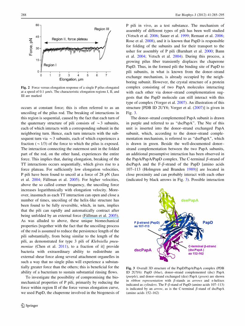
for P pili expressed by UPEC bacteria is shown in Fig. 2.
As was alluded to in the ‘‘Introduction’’, the data show that
a helix-like pilus has a force versus elongation response
that can be seen as composed of three regions (Andersson
et al. 2008; Jass et al. 2004; Bjornham et al. 2008; Axner
et al. 2009).
Region I is a short region, of little relevance to this
study, in which the entire pilus is elongated elastically with
no conformational change. In region II, elongation origi-
nating from sequential breaking of the turn-to-turn [TT;
previously referred to as layer-to-layer (Andersson et al.
2006b)] interactions of the quaternary structure of the pilus
Fig. 1 AFM micrographs of HB101/pPAP5 cells expressing P pili: a bacterium cultured under normal conditions, b chemical structure of
pilicide 1, c bacterium cultured on a plate containing 2.2 mM pilicide
Eur Biophys J (2012) 41:285–295 287
123
occurs at constant force; this is often referred to as an
uncoiling of the pilus rod. The breaking of interactions in
this region is sequential, caused by the fact that each turn of
the quaternary structure of pili consists of *3 subunits,
each of which interacts with a corresponding subunit in the
neighboring turn. Hence, each turn interacts with the sub-
sequent turn via *3 subunits, each of which experiences a
fraction (*1/3) of the force to which the pilus is exposed.
The interaction connecting the outermost unit in the folded
part of the rod, on the other hand, experiences the entire
force. This implies that, during elongation, breaking of the
TT interactions occurs sequentially, which gives rise to a
force plateau. For sufficiently low elongation velocities,
P pili have been found to uncoil at a force of 28 pN (Jass
et al. 2004; Fallman et al. 2005). For higher velocities,
above the so called corner frequency, the uncoiling force
increases logarithmically with elongation velocity. More-
over, inasmuch as each TT interaction can open and close a
number of times, uncoiling of the helix-like structure has
been found to be fully reversible, which, in turn, implies
that the pili can rapidly and automatically contract after
being unfolded by an external force (Fallman et al. 2005).
As was alluded to above, these unique biomechanical
properties [together with the fact that the uncoiling process
of the rod is assumed to reduce the persistence length of the
pili substantially, from being similar to the length of the
pili, as demonstrated for type 3 pili of Klebsiella pneu-
moniae (Chen et al. 2011), to a fraction of it] provide
bacteria with extraordinary ability to redistribute an
external shear force along several attachment organelles in
such a way that no single pilus will experience a substan-
tially greater force than the others; this is beneficial for the
ability of a bacterium to sustain substantial rinsing flows.
To investigate the possibility of compromising the bio-
mechanical properties of P pili, primarily by reducing the
force within region II of the force versus elongation curve,
we used PapD, the chaperone involved in the biogenesis of
P pili in vivo, as a test substance. The mechanism of
assembly of different types of pili has been well studied
(Vetsch et al. 2006; Sauer et al. 1999; Remaut et al. 2006;
Rose et al. 2008), and it is known that PapD is responsible
for folding of the subunits and for their transport to the
usher for assembly of P pili (Barnhart et al. 2000; Bann
et al. 2004; Vetsch et al. 2004). During this process, a
growing pilus fiber transiently displaces the chaperone
PapD. Thus, in the formed pili the binding site of PapD to
pili subunits, in what is known from the donor–strand
exchange mechanism, is already occupied by the neigh-
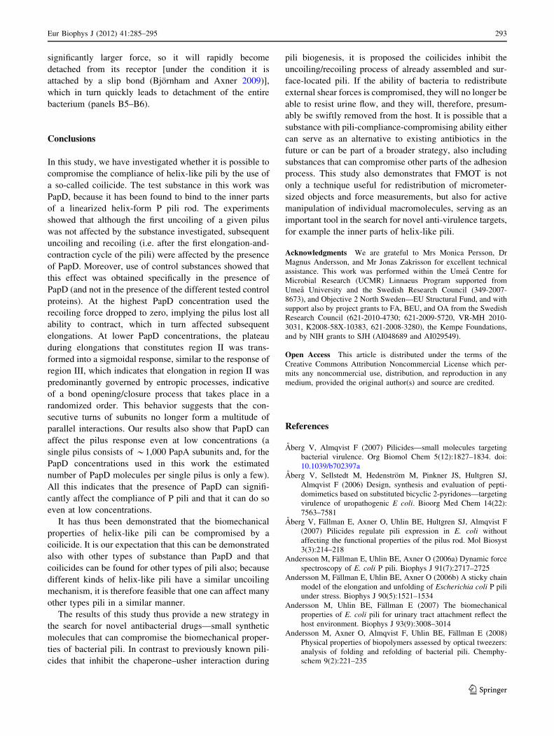
boring subunit. However, the crystal structure of a protein
complex consisting of two PapA molecules interacting
with each other via donor–strand complementation sug-
gests that the PapD molecule can still interact with this
type of complex (Verger et al. 2007). An illustration of this
structure [PDB ID 2UY6; Verger et al. (2007)] is given in
Fig. 3.
The donor–strand complemented PapA subunit is drawn
in purple and referred to as ‘‘dscPapA’’. The Nte of this
unit is inserted into the donor–strand exchanged PapA
subunit, which, according to the donor–strand comple-
mentation mechanism, is referred to as ‘‘dsePapA’’, which
is drawn in green. Beside the well-documented donor–
strand complementation between the two PapA subunits,
an additional presumptive interaction has been observed in
the PapA/PapA/PapD complex. The C-terminal b-strand of
dscPapA and the F-b-strand of the PapD [amino acids
107–113 (Holmgren and Branden 1989)] are located in
close proximity and can probably interact with each other
(indicated by black arrows in Fig. 3). Possible interaction
Fig. 2 Force versus elongation response of a single P pilus elongated
at a speed of 0.1 lm/s. The characteristic elongation regions I, II, and
III are marked
Fig. 3 Overall 3D structure of the PapD/PapA/PapA complex (PDB
ID 2UY6): PapD (blue), donor–strand complemented (dsc) PapA
(purple), and donor–strand exchanged (dce) PapA (green) are shown
in ribbon representation with b-stands as arrows and a-helixes
indicated as cylinders. The F-b-stand of PapD (amino acids 107–113)
is indicated by an arrow, as is the C-terminal b-stand of dscPapA
(amino acids 152–162)
288 Eur Biophys J (2012) 41:285–295
123

with PapA subunits in the pili rod led to the choice of PapD
as the test molecule for this proof-of-principle demonstra-
tion of compromise of the biomechanical properties of the
pili rod in this work.
As was mentioned above, and as is illustrated in Fig. 2,
helix-like pili have a unique and complex force versus
elongation response. Normally, a single pilus has a char-
acteristic force plateau in the force versus elongation
response (region II) that is flat and repeatable (which
originates from sequential uncoiling of the TT interactions
in the helix), whereas region III has a sigmoidal shape
(which originates from entropy-driven transitions of the
head-to-tail bonds opening and closing in random order)
(Jass et al. 2004; Fallman et al. 2005; Andersson et al.
2006b, 2007). During retraction, and for sufficiently low
elongation and retraction speeds, the response is fully
reversible [with the exception of a dip in the transition from
region III to II, which is considered to be caused by the
lack of a nucleation kernel for formation of the first turn of
the quaternary structure during contraction (Axner et al.
2006a, 2009; Lugmaier et al. 2008)].
To assess the effect of PapD on the compliance of the
pilus rod, force versus elongation data were collected from
eight series of seven replicate elongations and retractions
of P pili in the presence of different concentrations of PapD
and control proteins. It was found that in the presence of
PapD, the force versus elongation data and the contraction
response of P pili were altered markedly. First, the
recoiling forces of P pili were reduced. As can be seen in
Fig. 4, which shows a typical force versus contraction
response after the first elongation in the presence of PapD
at three different concentrations, the recoiling forces of P
pili at 1.4 lM (violet line) and 2.8 lM (green line) of PapD
were slightly lower than when no PapD was present (red
line) and contained a number of recurring dips.
When a higher concentration of PapD was used
(5.6 lM, black line, and 11.3 lM, blue line) the recoiling
force dropped drastically. At 11.3 lM PapD the force went
monotonically to zero with no characteristic plateau. At
5.6 lM PapD did not completely impair the recoiling of P
pili, but the recoiling force was lower than for additions of
1.4 and 2.8 lM PapD. Numerical analysis of the recoiling
process during the first half of the plateau in region II (from
2.7 to 1.2 lm in Fig. 4) resulted in recoiling forces of
29 ± 0.7, 25 ± 0.8, 23 ± 1.3, 17 ± 1.4, and 5.5 ± 2.2 pN
for 0, 1.4, 2.8, 5.6, and 11.3 lM PapD, respectively
(expressed as the mean ± standard error of the mean
(SEM), n = 8). This indicates that PapD significantly
affects the retraction properties of the rod, suggesting that
PapD interacts with PapA subunits in the uncoiled pili by
inhibiting the ability of the latter to form TT interactions,
thus preventing the pili from recoiling and contracting.
This implies that in the presence of the highest PapD
concentration the pilus lost virtually all of its compliance.
Also the uncoiling force for subsequent uncoilings was
found to be affected by the presence of PapD. For the
lowest PapD concentrations, the uncoiling force decreased
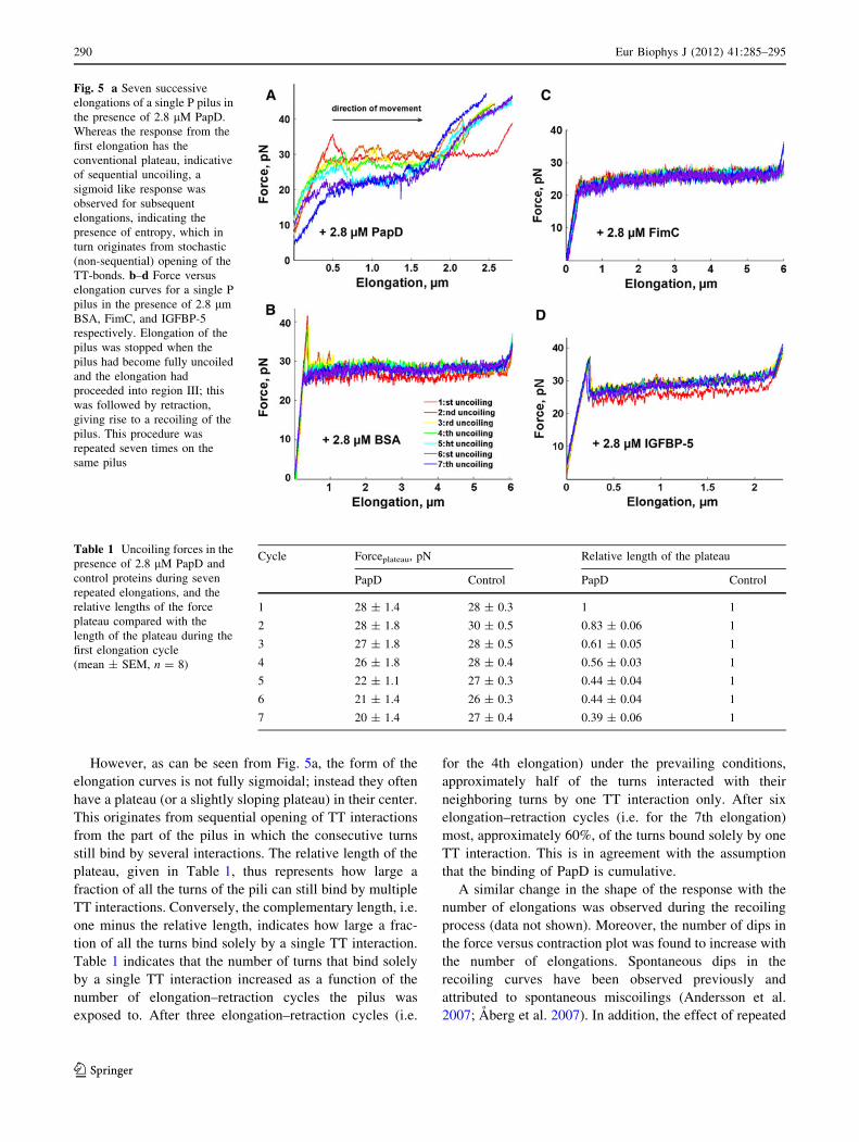
with the number of elongations. Figure 5a shows seven
repeated uncoilings of the same pilus in the presence of
2.8 lM PapD. In addition, from the characteristic appear-
ance (with a plateau) during the first uncoiling (red curve),
for repeated elongations, the uncoiling curve gradually
deteriorated; the plateau became shorter and the force
curve transformed to a sigmoidal shape, as is illustrated by
the blue curve (the seventh recoiling).
The results from analysis of the uncoiling force (the
force plateau) in the presence of PapD and control proteins
and the shortening of the plateau are summarized in
Table 1, in which each entry represents the average of
eight series of seven uncoiling cycles. Because pili length
can vary, the lengths of the force plateaus were normalized
to that of the first elongation in each set of measurements.
Any normalized length below unity thus indicates short-
ening of region II.
The sigmoidal shape signifies the occurrence of entropic
processes, which indicates that the TT interactions can open
and close in a random order. This suggests that consecutive
turns in the helical structure of the pilus rod no longer
interact with each other via multiple TT interactions per
turn, which is the underlying cause of the sequential
unfolding of the quaternary structure; it reveals that the
turns, instead, interact by a single TT interaction per turn,
which then enables opening of the helix-like coil in a random
order, caused by blocking of the TT interactions by PapD.
Fig. 4 Transition from region III to II during retraction of a single P
pilus in the presence of PapD at different concentrations. In samples
with no or a low concentration of PapD, contraction from an
elongation of approximately 2.7 lm takes place in region II,
indicating recoiling, whereas in the sample with 11.3 lM PapD, the
force decreases towards zero monotonically (which is reached for an
elongation of approximately 1.6 lm), indicating no recoiling
Eur Biophys J (2012) 41:285–295 289
123
However, as can be seen from Fig. 5a, the form of the
elongation curves is not fully sigmoidal; instead they often
have a plateau (or a slightly sloping plateau) in their center.
This originates from sequential opening of TT interactions
from the part of the pilus in which the consecutive turns
still bind by several interactions. The relative length of the
plateau, given in Table 1, thus represents how large a
fraction of all the turns of the pili can still bind by multiple
TT interactions. Conversely, the complementary length, i.e.
one minus the relative length, indicates how large a frac-
tion of all the turns bind solely by a single TT interaction.
Table 1 indicates that the number of turns that bind solely
by a single TT interaction increased as a function of the
number of elongation–retraction cycles the pilus was
exposed to. After three elongation–retraction cycles (i.e.
for the 4th elongation) under the prevailing conditions,
approximately half of the turns interacted with their
neighboring turns by one TT interaction only. After six
elongation–retraction cycles (i.e. for the 7th elongation)
most, approximately 60%, of the turns bound solely by one
TT interaction. This is in agreement with the assumption
that the binding of PapD is cumulative.
A similar change in the shape of the response with the
number of elongations was observed during the recoiling
process (data not shown). Moreover, the number of dips in
the force versus contraction plot was found to increase with
the number of elongations. Spontaneous dips in the
recoiling curves have been observed previously and
attributed to spontaneous miscoilings (Andersson et al.
2007; Aberg et al. 2007). In addition, the effect of repeated
Fig. 5 a Seven successive
elongations of a single P pilus in
the presence of 2.8 lM PapD.
Whereas the response from the
first elongation has the
conventional plateau, indicative
of sequential uncoiling, a
sigmoid like response was
observed for subsequent
elongations, indicating the
presence of entropy, which in
turn originates from stochastic
(non-sequential) opening of the
TT-bonds. b–d Force versus
elongation curves for a single P
pilus in the presence of 2.8 lm
BSA, FimC, and IGFBP-5
respectively. Elongation of the
pilus was stopped when the
pilus had become fully uncoiled
and the elongation had
proceeded into region III; this
was followed by retraction,
giving rise to a recoiling of the
pilus. This procedure was
repeated seven times on the
same pilus
Table 1 Uncoiling forces in the
presence of 2.8 lM PapD and
control proteins during seven
repeated elongations, and the
relative lengths of the force
plateau compared with the
length of the plateau during the
first elongation cycle
(mean ± SEM, n = 8)
Cycle Forceplateau, pN Relative length of the plateau
PapD Control PapD Control
1 28 ± 1.4 28 ± 0.3 1 1
2 28 ± 1.8 30 ± 0.5 0.83 ± 0.06 1
3 27 ± 1.8 28 ± 0.5 0.61 ± 0.05 1
4 26 ± 1.8 28 ± 0.4 0.56 ± 0.03 1
5 22 ± 1.1 27 ± 0.3 0.44 ± 0.04 1
6 21 ± 1.4 26 ± 0.3 0.44 ± 0.04 1
7 20 ± 1.4 27 ± 0.4 0.39 ± 0.06 1
290 Eur Biophys J (2012) 41:285–295
123

elongations for low PapD concentrations was comparable
that of a single elongation at a higher PapD concentration.
This implies that the effect of PapD increased cumulatively
with both the concentration of PapD and the number of
elongations (the latter is illustrated by the data presented in
Table 1).
Furthermore, it was found that PapD only affected lin-
earized (stretched or uncoiled) pili; the first uncoiling curve
in a series of elongations was always unaffected by its
presence, even after an hour of exposure of unstretched
(coiled) pili to PapD. The fact that PapD only bound to
PapA units when the rod had been uncoiled indicates that it
exclusively binds to the inner parts of the helix-like rod,
e.g. as was indicated by Fig. 3. In addition, the cumulative
effect indicates that the interaction between PapD and
uncoiled P pili is strong enough for PapD to remain bound
to the pili during repeated elongation and retraction cycles.
To verify the recoiling inhibiting effect of PapD, BSA,
IGFBP-5, and FimC were used as control substances. The
molecular weight of IGFBP-5 is similar to that of PapD but
with a secondary structure that is dissimilar to that of
PapD. BSA has another secondary structure and a molec-
ular weight that is almost twice that of PapD (Hung et al.
1999). The third control protein, FimC, is a chaperone that
is involved in the biogenesis of Type 1 pili in E. coli in the
same way as PapD mediates the biogenesis of P pili. FimC
has 50% amino acid sequence homology with PapD, and
the two proteins have similar 3D structures and molecular
weights (Jones et al. 1993).
As can be seen by comparison of a single pilus in the
presence and absence of 2.8 lM control proteins (Fig. 5b–d
vs. Fig. 2), it was found that subsequent elongation of P pili
was only very slightly affected by the presence of control
proteins. It was, moreover, found that, in the presence of
these control proteins, the force versus extension/contrac-
tion response of P pili remained virtually unchanged after
the first elongation—in a series of seven repeated elonga-
tions (Table 1), the number of dips in the recoiling curves
was found to be similar during the first and the last
extension cycles—indicating that the effect of control
proteins is weaker than that of PapD and not cumulative. A
much higher concentration of control proteins (11.3 lM)
resulted in detectable disturbances of the recoiling pro-
cess—occasional dips in the recoiling curves resulting in a
temporary reduction in uncoiling force. However, the
uncoiling force reverted to the normal level during repeated
elongations (data not shown).
A schematic illustration of the recoiling inhibitor
mechanism from a single bacterium that bind by two helix-
like pili is shown in Fig. 6. To mimic the effect of
mechanical stress of urine flow in vivo the six horizontal
panel pairs (1–6) represent a series of consecutive flow
conditions: no flow (panel pair 1), high flow (2), low flow (3),
and high flow (illustrated by three panel pairs, 4–6). The
high and low flow conditions represent cases where the
force to which the bacterium is exposed, Ftot, is slightly
larger than twice the uncoiling force (Fuc) and slightly
smaller than the uncoiling force, i.e. Ftot [ 2Fuc and
Ftot\Fuc, respectively. The low flow condition (3) can
even represent a case with no flow. The first and the second
columns of the panels represent pili in the absence and
presence of recoiling inhibitor, respectively.
In the absence of flow, the bacterium is initially attached
by both pili, neither of them elongated (panels A1 and B1).
When the bacterium is exposed to a shear flow, it will
rotate (as is indicated in the subsequent panels), so the
uppermost pili will be elongated the most. For high flow
conditions (panels A2 and B2), both pili will simulta-
neously elongate in region II. As long as they do so, the
two pili will take up the same force, half of the force to
which they are exposed, i.e. Fupper ¼ Flower ¼ Ftot=2, which
is a force that the specific adhesin–receptor interaction is
presumed to be able to withstand for a substantial time
(longer than the force exposure time), so the bacterium will
remain attached.
In the absence of a recoiling inhibitor (the first column),
when the flow decreases (panel A3) the two pili will retract
until both have contracted into region I. During subsequent
elongation (panels A4–A6), the two pili will again elongate
into region II in a manner similar to that in panel A2, i.e. by
sharing the force equally, i.e. Fupper ¼ Flower ¼ Ftot=2,
which is a force the specific adhesin–receptor interaction
can withstand for the duration of the force exposure, so the
bacterium will stay attached.
When a recoiling inhibitor is present (the second col-
umn), on the other hand, the compliance of the pili will be
compromised as a result of linearization of the pilus (panel
B2). However, there will be no indication of this during the
first elongation cycle (because the recoiling inhibitor only
attaches to the uncoiled part of the pilus); the reduced
compliance will affect the force response solely during
subsequent refolding, and more so for the pilus that has
been extended the most, which here is assumed to be the
upper one (because this has enabled binding of more of the
recoiling inhibitor). Under the simplified assumption that
the upper pilus loses most of its compliance, as was the
case for the highest concentrations of PapD in Fig. 4,
whereas the lower one is affected less, the upper pilus will
take significantly less force than the lower (in the limiting
case when it has lost all of its compliance, it will take no
force, whereas the lower one will take all, i.e. Fupper � 0
whereas Flower � Ftot) and so will retract less than the
lower pilus during the subsequent low flow exposure (panel
B3). Under the condition that Ftot\Fuc, the lower pilus
will retract all the way to region I. When the bacterium is
again exposed to a higher flow, the upper pilus will take
Eur Biophys J (2012) 41:285–295 291
123

virtually no force (because of its compromised compli-
ance), whereby the lower one will sustain virtually all of
the force (panel B4). This implies that the adhesin–receptor
bond of the lower pilus will be exposed to a significantly
larger force, close to Ftot ([2Fuc), which is a force at which
the adhesin–receptor bond will rapidly break (panel B5).
When this pilus has detached, the upper pilus will be
exposed to the entire force, and this also will rapidly
become detached (panel B6). Hence, in the presence of a
coilicide, the bacterium becomes detached from the host
tissue.
Taken together, our findings suggest that the presence of
a substance that can bind to the inner parts of an elongated
helix-like pilus as a ‘‘coilicide’’ should also affect the
attachment lifetime of piliated bacteria exposed to urinary
flows. As is schematically indicated in Fig. 6 (by the six
pairs of panels that illustrate different consecutive flow
conditions), for a bacterium that becomes attached to a host
surface with multiple pili (in this case illustrated by two
pili), a force that is slightly larger than twice the pili
uncoiling force will, in the absence of any coilicide
(depicted by the left column of panels), be distributed fairly
evenly among the two pili (exactly evenly as long as both
pili elongate slowly in region II), also for a multitude of
subsequent elongations and retraction cycles (A2–A4). As
has been discussed previously (Bjornham and Axner 2009),
this provides the bacteria with a capability to distribute an
external force among a mulitude of pili, which implies they
can withstand strong rinsing flows, which, in turn, facili-
tates adherence and colonization processes critical for
virulence. However, as can be seen by a comparison of the
two columns of Fig. 6, in which the second column illus-
trates the response of a bacterium in the presence of a
coilicide, the situation changes drastically when the bac-
terium is exposed to a coilicide. Although the first elon-
gation (panel B2) is identical to that without PapD present
(panel A2), subsequent recoiling will be compromised (as
is schematically indicated for one of the two pili in panel
B3). When a coilicide binds permanently to the inner parts
of the helix-like rod, subsequent elongation will be affec-
ted. Because one of the pili has lost its compliance, this
pilus can no longer withstand substantial force, the other
(less compromised, or non-compromised) pilus will be
exposed to most of the force (panel B4). This implies
that adhesin at the tip of this pilus will be exposed to a
Fig. 6 Schematic illustration of the recoiling inhibitor mechanism on
helix-like pili. A bacterium is attached to the host tissue by two
identical pili adhering by specific adhesin–receptor bonds. The helix-
like pili are depicted as springs, with their length approximately
representing their degree of elongation. The arrows indicate the force
each pilus sustains
c
292 Eur Biophys J (2012) 41:285–295
123
significantly larger force, so it will rapidly become
detached from its receptor [under the condition it is
attached by a slip bond (Bjornham and Axner 2009)],
which in turn quickly leads to detachment of the entire
bacterium (panels B5–B6).
Conclusions
In this study, we have investigated whether it is possible to
compromise the compliance of helix-like pili by the use of
a so-called coilicide. The test substance in this work was
PapD, because it has been found to bind to the inner parts
of a linearized helix-form P pili rod. The experiments
showed that although the first uncoiling of a given pilus
was not affected by the substance investigated, subsequent
uncoiling and recoiling (i.e. after the first elongation-and-
contraction cycle of the pili) were affected by the presence
of PapD. Moreover, use of control substances showed that
this effect was obtained specifically in the presence of
PapD (and not in the presence of the different tested control
proteins). At the highest PapD concentration used the
recoiling force dropped to zero, implying the pilus lost all
ability to contract, which in turn affected subsequent
elongations. At lower PapD concentrations, the plateau
during elongations that constitutes region II was trans-
formed into a sigmoidal response, similar to the response of
region III, which indicates that elongation in region II was
predominantly governed by entropic processes, indicative
of a bond opening/closure process that takes place in a
randomized order. This behavior suggests that the con-
secutive turns of subunits no longer form a multitude of
parallel interactions. Our results also show that PapD can
affect the pilus response even at low concentrations (a
single pilus consists of *1,000 PapA subunits and, for the
PapD concentrations used in this work the estimated
number of PapD molecules per single pilus is only a few).
All this indicates that the presence of PapD can signifi-
cantly affect the compliance of P pili and that it can do so
even at low concentrations.
It has thus been demonstrated that the biomechanical
properties of helix-like pili can be compromised by a
coilicide. It is our expectation that this can be demonstrated
also with other types of substance than PapD and that
coilicides can be found for other types of pili also; because
different kinds of helix-like pili have a similar uncoiling
mechanism, it is therefore feasible that one can affect many
other types pili in a similar manner.
The results of this study thus provide a new strategy in
the search for novel antibacterial drugs—small synthetic
molecules that can compromise the biomechanical proper-
ties of bacterial pili. In contrast to previously known pili-
cides that inhibit the chaperone–usher interaction during
pili biogenesis, it is proposed the coilicides inhibit the
uncoiling/recoiling process of already assembled and sur-
face-located pili. If the ability of bacteria to redistribute
external shear forces is compromised, they will no longer be
able to resist urine flow, and they will, therefore, presum-
ably be swiftly removed from the host. It is possible that a
substance with pili-compliance-compromising ability either
can serve as an alternative to existing antibiotics in the
future or can be part of a broader strategy, also including
substances that can compromise other parts of the adhesion
process. This study also demonstrates that FMOT is not
only a technique useful for redistribution of micrometer-
sized objects and force measurements, but also for active
manipulation of individual macromolecules, serving as an
important tool in the search for novel anti-virulence targets,
for example the inner parts of helix-like pili.
Acknowledgments We are grateful to Mrs Monica Persson, Dr
Magnus Andersson, and Mr Jonas Zakrisson for excellent technical
assistance. This work was performed within the Umea Centre for
Microbial Research (UCMR) Linnaeus Program supported from
Umea University and the Swedish Research Council (349-2007-
8673), and Objective 2 North Sweden—EU Structural Fund, and with
support also by project grants to FA, BEU, and OA from the Swedish
Research Council (621-2010-4730; 621-2009-5720, VR-MH 2010-
3031, K2008-58X-10383, 621-2008-3280), the Kempe Foundations,
and by NIH grants to SJH (AI048689 and AI029549).
Open Access This article is distributed under the terms of the
Creative Commons Attribution Noncommercial License which per-
mits any noncommercial use, distribution, and reproduction in any
medium, provided the original author(s) and source are credited.
References
Aberg V, Almqvist F (2007) Pilicides—small molecules targeting
bacterial virulence. Org Biomol Chem 5(12):1827–1834. doi:
10.1039/b702397a
Aberg V, Sellstedt M, Hedenstrom M, Pinkner JS, Hultgren SJ,
Almqvist F (2006) Design, synthesis and evaluation of pepti-
domimetics based on substituted bicyclic 2-pyridones—targeting
virulence of uropathogenic E coli. Bioorg Med Chem 14(22):
7563–7581
Aberg V, Fallman E, Axner O, Uhlin BE, Hultgren SJ, Almqvist F
(2007) Pilicides regulate pili expression in E. coli without
affecting the functional properties of the pilus rod. Mol Biosyst
3(3):214–218
Andersson M, Fallman E, Uhlin BE, Axner O (2006a) Dynamic force
spectroscopy of E. coli P pili. Biophys J 91(7):2717–2725
Andersson M, Fallman E, Uhlin BE, Axner O (2006b) A sticky chain
model of the elongation and unfolding of Escherichia coli P pili
under stress. Biophys J 90(5):1521–1534
Andersson M, Uhlin BE, Fallman E (2007) The biomechanical
properties of E. coli pili for urinary tract attachment reflect the
host environment. Biophys J 93(9):3008–3014
Andersson M, Axner O, Almqvist F, Uhlin BE, Fallman E (2008)
Physical properties of biopolymers assessed by optical tweezers:
analysis of folding and refolding of bacterial pili. Chemphy-
schem 9(2):221–235
Eur Biophys J (2012) 41:285–295 293
123

Axner O, Bjornham O, Castelain M, Koutris E, Schedin S, Fallman E,
Andersson M (2009) Unraveling the secrets of bacterial adhesion
organelles using single-molecule force spectroscopy. In: Grasl-
und A, Rigler R, Widengren J (eds) Single molecule spectros-
copy in chemistry, physics and biology—nobel symposium, vol
96. Chemical Physics, 1st edn. Springer, Berlin, pp 337–362.
doi:10.1007/978-3-642-02597-6
Axner O, Andersson M, Bjornham O, Castelain M, Klinth J, Koutris
E, Schedin S (2011) Assessing bacterial adhesion on an
individual adhesin and single pili level using optical tweezers.
In: Linke D, Goldman A (eds) Bacterial adhesion, vol 715.
Advances in experimental medicine and biology. Springer,
Netherlands, pp 301–313. doi:10.1007/978-94-007-0940-9_19
Bann JG, Pinkner JS, Frieden C, Hultgren SJ (2004) Catalysis of protein
folding by chaperones in pathogenic bacteria. Proc Natl Acad Sci
USA 101(50):17389–17393. doi:10.1073/pnas.0408072101
Barnhart MM, Pinkner JS, Soto GE, Sauer FG, Langermann S,
Waksman G, Frieden C, Hultgren SJ (2000) PapD-like chaper-
ones provide the missing information for folding of pilin
proteins. Proc Natl Acad Sci USA 97(14):7709–7714. doi:
10.1073/pnas.130183897130183897
Bjornham O, Axner O (2009) Multipili attachment of bacteria with
helixlike pili exposed to stress. J Chem Phys 130(23):235102
Bjornham O, Axner O, Andersson M (2008) Modeling of the
elongation and retraction of Escherichia coli P pili under strain
by Monte Carlo simulations. Eur Biophys J 37(4):381–391
Bullitt E, Makowski L (1995) Structural polymorphism of bacterial
adhesion pili. Nature 373(6510):164–167
Castelain M, Koutris E, Andersson M, Wiklund K, Bjornham O,
Schedin S, Axner O (2009) Characterization of the biomechan-
ical properties of T4 pili expressed by Streptococcus pneumo-niae—a comparison between helix-like and open coil-like pili.
Chemphyschem 10(9–10):1533–1540
Castelain M, Ehlers S, Klinth JE, Lindberg S, Andersson M, Uhlin
BE, Axner O (2010) Fast uncoiling kinetics of F1C pili
expressed by uropathogenic Escherichia coli are revealed on a
single pilus level using force-measuring optical tweezers.
Submited to Eur Biophys J: under final revision
Cegelski L, Pinkner JS, Hammer ND, Cusumano CK, Hung CS,
Chorell E, Aberg V, Walker JN, Seed PC, Almqvist F, Chapman
MR, Hultgren SJ (2009) Small-molecule inhibitors target
Escherichia coli amyloid biogenesis and biofilm formation.
Nat Chem Biol 5(12):913–919. doi:10.1038/nchembio.242
Chen FJ, Chan CH, Huang YJ, Liu KL, Peng HL, Chang HY, Liou
GG, Yew TR, Liu CH, Hsu KY, Hsu L (2011) Structural and
mechanical properties of Klebsiella pneumoniae type 3 fimbriae.
J Bacteriol 193(7):1718–1725. doi:10.1128/jb.01395-10
Chorell E, Pinkner JS, Phan G, Edvinsson S, Buelens F, Remaut H,
Waksman G, Hultgren SJ, Almqvist F (2010) Design and
synthesis of C-2 substituted thiazolo and dihydrothiazolo ring-
fused 2-pyridones: pilicides with increased antivirulence activ-
ity. J Med Chem 53(15):5690–5695. doi:10.1021/jm100470t
Duncan MJ, Mann EL, Cohen MS, Ofek I, Sharon N, Abraham SN
(2005) The distinct binding specificities exhibited by enterobac-
terial type 1 fimbriae are determined by their fimbrial shafts.
J Biol Chem 280(45):37707–37716
Fallman E, Schedin S, Jass J, Andersson M, Uhlin BE, Axner O
(2004) Optical tweezers based force measurement system for
quantitating binding interactions: system design and application
for the study of bacterial adhesion. Biosens Bioelectron 19(11):
1429–1437
Fallman E, Schedin S, Jass J, Uhlin BE, Axner O (2005) The
unfolding of the P pili quaternary structure by stretching is
reversible, not plastic. EMBO Rep 6(1):52–56
Gong M, Makowski L (1992) Helical structure of P pili from
Escherichia coli. Evidence from X-ray fiber diffraction and
scanning transmission electron microscopy. J Mol Biol 228(3):
735–742
Holmgren A, Branden CI (1989) Crystal-structure of chaperone
protein PapD reveals an immunoglobulin fold. Nature
342(6247):248–251
Hung DL, Pinkner JS, Knight SD, Hultgren SJ (1999) Structural basis
of chaperone self-capping in P pilus biogenesis. Proc Natl Acad
Sci USA 96(14):8178–8183
Jacob-Dubuisson F, Heuser J, Dodson K, Normark S, Hultgren S
(1993) Initiation of assembly and association of the structural
elements of a bacterial pilus depend on two specialized tip
proteins. EMBO J 12(3):837–847
Jass J, Schedin S, Fallman E, Ohlsson J, Nilsson UJ, Uhlin BE, Axner
O (2004) Physical properties of Escherichia coli P pili measured
by optical tweezers. Biophys J 87(6):4271–4283
Jones CH, Pinkner JS, Nicholes AV, Slonim LN, Abraham SN,
Hultgren SJ (1993) FimC is a periplasmic PapD-like chaperone
that directs assembly of type 1 pili in bacteria. Proc Natl Acad
Sci USA 90(18):8397–8401
Kaplan W, Laing R (2004) Priority medicines for Europe and the
world. WHO, Geneva. Available from: http://whqlibdoc.who.
int/hq/2004/WHO_EDM_PAR_2004.7.pdf
Krogfelt KA, Bergmans H, Klemm P (1990) Direct evidence that the
FimH protein is the mannose-specific adhesin of Escherichia colitype 1 fimbriae. Infect Immun 58(6):1995–1998
Kuehn MJ, Heuser J, Normark S, Hultgren SJ (1992) P pili in
uropathogenic E. coli are composite fibres with distinct fibrillar
adhesive tips. Nature 356(6366):252–255. doi:10.1038/356252a0
Le Trong I, Aprikian P, Kidd BA, Forero-Shelton M, Tchesnokova V,
Rajagopal P, Rodriguez V, Interlandi G, Klevit R, Vogel V,
Stenkamp RE, Sokurenko EV, Thomas WE (2010) Structural
basis for mechanical force regulation of the adhesin FimH via
finger trap-like beta sheet twisting. Cell 141(4):645–655. doi:
10.1016/j.cell.2010.03.038
Lee YM, Dodson KW, Hultgren SJ (2007) Adaptor function of PapF
depends on donor strand exchange in P-pilus biogenesis of
Escherichia coli. J Bacteriol 189(14):5276–5283. doi:10.1128/
JB.01648-06
Lindberg FP, Lund B, Normark S (1984) Genes of pyelonephritogenic
E. coli required for digalactoside-specific agglutination of human
cells. EMBO J 3(5):1167–1173
Lindberg F, Lund B, Johansson L, Normark S (1987) Localization of
the receptor-binding protein adhesin at the tip of the bacterial
pilus. Nature 328(6125):84–87. doi:10.1038/328084a0
Lindberg F, Tennent JM, Hultgren SJ, Lund B, Normark S (1989)
PapD, a periplasmic transport protein in P-pilus biogenesis.
J Bacteriol 171(11):6052–6058
Lugmaier RA, Schedin S, Kuhner F, Benoit M (2008) Dynamic
restacking of Escherichia coli P-pili. Eur Biophys J Biophys Lett
37(2):111–120
Ofek I, Hasty DL, Sharon N (2003) Anti-adhesion therapy of bacterial
diseases: prospects and problems. FEMS Immunol Med Micro-
biol 38(3):181–191
Ohlsson J, Jass J, Uhlin BE, Kihlberg J, Nilsson UJ (2002) Discovery
of potent inhibitors of PapG adhesins from uropathogenic
Escherichia coli through synthesis and evaluation of galabiose
derivatives. Chembiochem 3(8):772–779. doi:10.1002/1439-
7633(20020802)3:8\772:AID-CBIC772[3.0.CO;2-8
Pinkner JS, Remaut H, Buelens F, Miller E, Aberg V, Pemberton N,
Hedenstrom M, Larsson A, Seed P, Waksman G, Hultgren SJ,
Almqvist F (2006) Rationally designed small compounds inhibit
pilus biogenesis in uropathogenic bacteria. Proc Natl Acad Sci
USA 103(47):17897–17902
Remaut H, Rose RJ, Hannan TJ, Hultgren SJ, Radford SE, Ashcroft
AE, Waksman G (2006) Donor-strand exchange in chaperone-
assisted pilus assembly proceeds through a concerted beta strand
294 Eur Biophys J (2012) 41:285–295
123

displacement mechanism. Mol Cell 22(6):831–842. doi:10.1016/
j.molcel.2006.05.033
Rose RJ, Welsh TS, Waksman G, Ashcroft AE, Radford SE, Paci E
(2008) Donor-strand exchange in chaperone-assisted pilus
assembly revealed in atomic detail by molecular dynamics.
J Mol Biol 375(4):908–919
Salminen A, Loimaranta V, Joosten JA, Khan AS, Hacker J, Pieters
RJ, Finne J (2007) Inhibition of P-fimbriated Escherichia coliadhesion by multivalent galabiose derivatives studied by a live-
bacteria application of surface plasmon resonance. J Antimicrob
Chemother 60(3):495–501
Sauer FG, Futterer K, Pinkner JS, Dodson KW, Hultgren SJ,
Waksman G (1999) Structural basis of chaperone function and
pilus biogenesis. Science 285(5430):1058–1061
Sauer FG, Mulvey MA, Schilling JD, Martinez JJ, Hultgren SJ (2000)
Bacterial pili: molecular mechanisms of pathogenesis. Curr Opin
Microbiol 3(1):65–72
Staskawicz BJ, Mudgett MB, Dangl JL, Galan JE (2001) Common and
contrasting themes of plant and animal diseases. Science 292
(5525):2285–2289. doi:10.1126/science.1062013292/5525/2285
Striker R, Jacob-Dubuisson F, Freiden C, Hultgren SJ (1994) Stable
fiber-forming and nonfiber-forming chaperone-subunit com-
plexes in pilus biogenesis. J Biol Chem 269(16):12233–12239
Verger D, Bullitt E, Hultgren SJ, Waksman G (2007) Crystal structure
of the P pilus rod subunit PapA. PLoS Pathog 3(5):e73
Vetsch M, Puorger C, Spirig T, Grauschopf U, Weber-Ban EU,
Glockshuber R (2004) Pilus chaperones represent a new type of
protein-folding catalyst. Nature 431(7006):329–333. doi:10.1038/
nature02891nature02891
Vetsch M, Erilov D, Moliere N, Nishiyama M, Ignatov O, Glocksh-
uber R (2006) Mechanism of fibre assembly through the
chaperone-usher pathway. EMBO Rep 7(7):734–738. doi:10.1038/
sj.embor.7400722
Wellens A, Garofalo C, Nguyen H, Van Gerven N, Slattegard R,
Hernalsteens JP, Wyns L, Oscarson S, De Greve H, Hultgren S,
Bouckaert J (2008) Intervening with urinary tract infections
using anti-adhesives based on the crystal structure of the FimH-
oligomannose-3 complex. PLoS One 3(4):e2040. doi:10.1371/
journal.pone.0002040
Whitfield M, Ghose T, Thomas W (2010) Shear-stabilized rolling
behavior of E. coli examined with simulations. Biophys J
99(8):2470–2478. doi:10.1016/j.bpj.2010.08.045
Eur Biophys J (2012) 41:285–295 295
123
Copyright © 2022 FDOKUMEN