High-affinity neurotrophin receptors and ligands promote leukemogenesis

Development/Plasticity/Repair
Impaired Cerebellar Development and Function in MiceLacking CAPS2, a Protein Involved in Neurotrophin Release
Tetsushi Sadakata,1 Wataru Kakegawa,5 Akira Mizoguchi,6 Miwa Washida,1 Ritsuko Katoh-Semba,7 Fumihiro Shutoh,2
Takehito Okamoto,2 Hisako Nakashima,6 Kazushi Kimura,6 Mika Tanaka,3 Yukiko Sekine,1 Shigeyoshi Itohara,4
Michisuke Yuzaki,5 Soichi Nagao,2 and Teiichi Furuichi1
1Laboratory for Molecular Neurogenesis, 2Laboratory for Motor Learning Control, 3Research Resource Center, and 4Laboratory for Behavioral Genetics,RIKEN Brain Science Institute, Wako, Saitama 351-0198, Japan, 5Department of Physiology, School of Medicine, Keio University, Tokyo 160-8582, Japan,6Department of Anatomy, School of Medicine, Mie University, Tsu, Mie 514-8507, Japan, and 7Department of Perinatology, Institute for DevelopmentalResearch, Aichi Human Service Center, Kasugai, Aichi 480-0392, Japan
Ca 2�-dependent activator protein for secretion 2 (CAPS2/CADPS2) is a secretory granule-associated protein that is abundant at theparallel fiber terminals of granule cells in the mouse cerebellum and is involved in the release of neurotrophin-3 (NT-3) and brain-derivedneurotrophic factor (BDNF), both of which are required for cerebellar development. The human homolog gene on chromosome 7 islocated within susceptibility locus 1 of autism, a disease characterized by several cerebellar morphological abnormalities. Here we reportthat CAPS2 knock-out mice are deficient in the release of NT-3 and BDNF, and they consequently exhibit suppressed phosphorylation ofTrk receptors in the cerebellum; these mice exhibit pronounced impairments in cerebellar development and functions, including neu-ronal survival, differentiation and migration of postmitotic granule cells, dendritogenesis of Purkinje cells, lobulation between lobules VIand VII, structure and vesicular distribution of parallel fiber–Purkinje cell synapses, paired-pulse facilitation at parallel fiber–Purkinjecell synapses, rotarod motor coordination, and eye movement plasticity in optokinetic training. Increased granule cell death of theexternal granular layer was noted in lobules VI–VII and IX, in which high BDNF and NT-3 levels are specifically localized during cerebellardevelopment. Therefore, the deficiency of CAPS2 indicates that CAPS2-mediated neurotrophin release is indispensable for normalcerebellar development and functions, including neuronal differentiation and survival, morphogenesis, synaptic function, and motorleaning/control. The possible involvement of the CAPS2 gene in the cerebellar deficits of autistic patients is discussed.
Key words: CAPS2/CADPS2; CAPS1/CADPS1; neurotrophin; BDNF; NT-3; cerebellum
IntroductionCa 2�-dependent activator protein for secretion 2 (CAPS2/CADPS2) (Cisternas et al., 2003; Speidel et al., 2003; Sadakata etal., 2004, 2007a) is a paralog of CAPS1/CADPS1 that is essentialfor Ca 2�-triggered dense-core vesicle (DCV) exocytosis (Berwinet al., 1998; Tandon et al., 1998; Renden et al., 2001). CAPS par-ticipates in an ATP-dependent priming step as a phosphatidylinositol 4,5-bisphosphate-binding and DCV-binding protein inresponse to increases in intracellular Ca 2� (Grishanin et al.,2004), and it is also known to interact with the dopamine recep-tor (Binda et al., 2005). Study of CAPS1-deficient mice has sug-gested that CAPS1 is involved in the catecholamine loading ofDCVs in embryonic chromaffin cells (Speidel et al., 2005).CAPS2 is abundantly localized in the parallel fiber (PF) terminalsof cerebellar granule cells and associates with vesicles containing
two neurotrophins, brain-derived neurotrophic factor (BDNF)and neurotrophin-3 (NT-3), and a DCV marker, chromograninB (Sadakata et al., 2004, 2006). In cerebellar primary cultures, theoverexpression of exogenous CAPS2 augments the release ofthese neurotrophins, thus promoting cell survival (Sadakata etal., 2004).
Neurotrophins play an indispensable role in neuronal differ-entiation and survival (Bibel and Barde, 2000; Huang andReichardt, 2001; Lessmann et al., 2003; Segal, 2003), as well as insynaptic plasticity (Thoenen, 1995; Schinder and Poo, 2000; Lu,2003; Kovalchuk et al., 2004). Many reports have described theimportance of BDNF and NT-3 in the postnatal development ofthe cerebellum (Lindholm et al., 1997; Schwartz et al., 1997; Bateset al., 1999; Carter et al., 2002). In the mouse cerebellum, BDNFand NT-3 released by granule cells bind to their Trk receptors,TrkB and TrkC, respectively, on both postsynaptic Purkinje cells(PCs) and presynaptic granule cells, thereby leading to Trk sig-naling in an autocrine–paracrine and anterograde manner (Lind-holm et al., 1997). Mice genetically lacking either BDNF (Jones etal., 1994; Schwartz et al., 1997; Carter et al., 2002) or NT-3 (Bateset al., 1999) also showed the pivotal role of these two neurotro-phins in cerebellar development.
The human CAPS2/CADPS2 gene is located on chromosome
Received May 30, 2006; revised Jan. 23, 2007; accepted Jan. 27, 2007.This study was supported by grants-in-aid for Scientific Research from the Japanese Ministry of Education,
Culture, Sports, Science, and Technology (Grant 17700322), the Japan Society for the Promotion of Science, and theRIKEN Institute of Physical and Chemical Research.
Correspondence should be addressed to Teiichi Furuichi, Laboratory for Molecular Neurogenesis, RIKEN BrainScience Institute, 2-1 Hirosawa, Wako, Saitama 351-0198, Japan. E-mail: [email protected].
DOI:10.1523/JNEUROSCI.2279-06.2007Copyright © 2007 Society for Neuroscience 0270-6474/07/272472-11$15.00/0
2472 • The Journal of Neuroscience, March 7, 2007 • 27(10):2472–2482

7q31.32 (Cisternas et al., 2003) within autism susceptibility locus1 (AUTS1) (International Molecular Genetic Study of AutismConsortium, 2001). Autistic spectrum disorders are a severe neu-rodevelopmental disorders marked by profound disturbances insocial, communicative, and behavioral functioning (WorldHealth Organization, 1992; American Psychiatric Association,1994) with a prevalence of 3– 6/1000 and a male-to-female ratioof 3:1 (Muhle et al., 2004). A recent study by our group showedautistic-like cellular and behavioral phenotypes in CAPS2 knock-out mice and aberrant CAPS2 mRNA splicing in autistic patients(Sadakata et al., 2007b). It has been demonstrated that autisticpatients exhibit several abnormalities in cerebellar morphologyand function, including hypoplasia of the cerebellar lobules(Courchesne et al., 1988; Pierce and Courchesne, 2001) and im-paired eye movement (Takarae et al., 2004) and motor coordina-tion (Manjiviona and Prior, 1995). In this study, to elucidatewhether or not the loss of CAPS2 function in mice would lead toabnormal cerebellum-related phenotypes, we investigated thestructure, development, and function of the CAPS2 knock-outmouse cerebellum. Our results demonstrated the indispensablerole of CAPS2-mediated neurotrophin release in normal cerebel-lar development and function. In addition, to broaden our un-derstanding of the role of CAPS2 in the cerebellum, we discussedthese results in relation to the cerebellar abnormalities reportedin autistic patients.
Materials and MethodsCAPS2 knock-out mice. Recently generated CAPS2 knock-out mice(Sadakata et al., 2007b) were used in the present study. Briefly, a 12 kbgenomic fragment containing exon 1 of mouse CAPS2 from C57BL/6mice was used to construct the targeting vectors. For positive selection,the SmaI–SmaI fragment containing the full length of exon 1 was re-placed by the Pgk–neo gene cassette flanked by the loxP sites. For nega-tive selection, the diphtheria toxin A fragment gene cassette was added tothe 5� end of the targeting vector. After transfection of MS12 embryonicstem (ES) cells [C57BL/6 mouse embryonic stem cell line (Kawase et al.,1994)] by electroporation, targeted clones were screened for G418 resis-tance and analyzed by Southern blot analysis. Chimeric mice were gen-erated by injection of the targeted MS12 ES cells into BALB/c blastocystsand mated with wild-type C57BL/6J mice to obtain heterozygous mutantmice. All of the engineered animals studied were backcrossed ontoC57BL/6J for more than five generations.
Antibodies. Guinea pig polyclonal anti-CAPS2 antibody and rabbitpolyclonal anti-CAPS1 antibody (Sadakata et al., 2004) were used for theimmunohistochemical analysis (used at dilution of 1:10,000 and 1:5000,respectively). Rabbit anti-NT-3 (Katoh-Semba et al., 1996), anti-BDNF(Katoh-Semba et al., 1997), and anti-phosphorylated Trk antibodies(pTrk) (Tyr490; catalog #9141; Cell Signaling Technology, Beverly, MA)were used for the immunohistochemical analysis (used at dilution of1:150, 1:100, and 1:10, respectively). Mouse monoclonal anti-calbindinD-28K (C9848; Sigma, St. Louis, MO), anti-microtubule-associated pro-tein MAP2(2a � 2b) (M1406; Sigma), and anti-vesicular glutamatetransporter (VGLUT2) (MAB5504; Millipore, Temecula, CA) antibodieswere used for immunohistochemistry and immunocytochemistry(used at dilution of 1:500, 1:1000, 1:5000, and 1:4000, respectively).Alexa-conjugated anti-guinea pig, rabbit, and mouse antibodies (In-vitrogen, Carlsbad, CA) were used as secondary antibodies at a 1000-folddilution.
Immunohistochemistry. For BDNF and NT-3 immunohistochemistry,postnatal day 8 (P8) C57BL/6J mice were anesthetized with diethyl etherand transcardially perfused with PBS and then with Zamboni’s fixative[2% paraformaldehyde (PFA) in 0.1 M phosphate buffer (PB), pH 7.4,containing 0.2% picric acid]. The brains were dissected out, postfixed inZamboni’s fixative at 4°C for 5 h, and cryoprotected by immersion in15% sucrose in PBS overnight at 4°C. After embedding in Tissue-TekOCT compound (Sakura Finetechnical, Tokyo, Japan), the brains were
frozen and sectioned sagittally or coronally at a thickness of 14 �m witha cryostat (CM1850; Leica, Frankfurt, Germany) at �18°C. The sectionswere air dried for 1 h and rinsed in PBS three times. After blocking with5% bovine serum albumin (BSA) in PBS at room temperature for 1 h, thesections were reacted at 4°C overnight with the primary antibody in animmunoreaction buffer (2� PBS containing 0.3% Triton X-100 and 1%BSA), rinsed in PBS, and then reacted at room temperature for 1 h withthe secondary antibody in the immunoreaction buffer and rinsed in PBS.The immunoreacted sections were mounted with Vectashield (VectorLaboratories, Peterborough, UK) mounting medium. Immunohisto-chemical staining with the other antibodies was performed as reportedpreviously (Sadakata et al., 2004, 2006).
Terminal deoxynucleotidyl transferase-mediated biotinylated UTP nickend labeling assay. Apoptotic cells were counted with an In Situ CellDeath Detection kit, TMR red (Roche Molecular Systems, Alameda, CA),according to the instructions of the manufacturer, and 14-�m-thick cer-ebellar sections of P8 CAPS2�/� (n � 4) and wild-type littermates (n �4) were used for the assay.
Golgi staining. Golgi staining was performed by using an FD RapidGolgiStain kit (FD NeuroTechnologies, Ellicott City, MD) according tothe instructions of the manufacturer. The sections were frozen with liq-uid nitrogen, and 60-�m-thick sections of wild-type and CAPS2�/� cer-ebella (P8 and P28) were cut in a cryostat.
Electron microscopy. For electron microscopy of paraformaldehyde-fixed sections, wild-type and CAPS2�/� mice (P15 and P84) were deeplyanesthetized with ether and perfused for 15 min with freshly prepared 2%glutaraldehyde and 2% PFA in 0.1 M PB. Brains were removed by dissec-tion, soaked in the same fixative at 4°C for 4 h, and cut with a vibratomeinto several sections (�400 �m thick). Tissue blocks were then postfixedin 1% osmium tetroxide in 0.1 M PB, dehydrated through a graded seriesof ethanol solutions, and embedded in epoxy resin. Ultrathin sections oflobules IV, V, VI and VII were cut from both surfaces of vibratomesections. The ultrathin sections were stained with uranyl acetate and leadcitrate before examination with an electron microscope (JEM 1013 EX;Jeol, Tokyo, Japan). Cerebellar samples from three mice of each genotypewere used. A 100,000 �m 2 area from each animal was analyzed.
Cerebellar primary cultures. Cerebellar primary cultures were preparedbasically as described in a previous study (Sadakata et al., 2004). In brief,after rapid decapitation the P0 cerebella of wild-type or CAPS2�/� micewere dissected out, digested for 13 min at 37°C with 0.1% trypsin (Sigma)and 0.05% DNase I (Boehringer Mannheim, Indianapolis, IN) in Ca 2�/Mg 2�-free HBSS [HBSS-CaMg(�)] (Sigma), and washed with HBSS-CaMg(�). They were then triturated by repeated passage through a 1 mlplastic micropipette tip in HBSS-CaMg(�) containing 0.05% DNase Iand 12 mM MgSO4 and washed with a serum-free Eagle minimal essentialmedium-based chemical-conditioned medium supplemented with0.25% (w/v) glucose (Nacalai Tesque, Kyoto, Japan), 10 �g/ml insulin(Sigma), 0.1 nM L-thyroxine (Sigma), 0.1 mg/ml apotransferrin (Sigma),1 mg/ml BSA (Sigma), 2 mM L-glutamine (Nacalai Tesque), 1 �g/mlaprotinin (Sigma), 30 nM sodium selenite (Merck, Darmstadt, Ger-many), 100 U/ml penicillin (Banyu Pharmaceutical, Tokyo, Japan), and135 �g/ml streptomycin (Meiji Seika, Tokyo, Japan). The dissociatedcells were plated at 5 � 10 5 cells per glass coverslip (12 mm in diameter;Matsunami, Tokyo, Japan) coated with poly-L-lysine (Sigma) and thencultured in the medium at 37°C under a humidified 5% CO2 atmosphere.
Electrophysiology. Parasagittal cerebellar slices (250 �m thick) wereprepared from CAPS2�/� mice and wild-type littermates (P15–P21 andP43–P54) in accordance with the guidelines of our institution. Whole-cell voltage-clamp recordings were made from visually identified PCswith an Axopatch 200B amplifier (Molecular Devices, Palo Alto, CA),and the pClamp system (version 9.2; Molecular Devices) was used fordata acquisition and analysis. Patch pipettes were pulled from borosili-cate glass capillaries to achieve a resistance of 3–5 M� when filled with asolution containing the following (in mM): 65 K-gluconate, 65 Cs-methanesulfonate, 10 KCl, 1 MgCl2, 4 Na2ATP, 1 Na2GTP, 20 HEPES, 5sucrose, and 0.4 EGTA, pH 7.3 (295 mOsm/kg). The external Ringer’ssolution contained 125 mM NaCl, 2.5 mM KCl, 2 mM CaCl2, 1 mM MgCl2,26 mM NaHCO3, 1.25 mM NaH2PO4, 10 mM D-glucose, and 0.1 mM
picrotoxin (to inhibit GABAergic synapses) and was bubbled with 95%
Sadakata et al. • Cerebellar Abnormalities in CAPS2 Knock-Out Mice J. Neurosci., March 7, 2007 • 27(10):2472–2482 • 2473

O2 and 5% CO2 at room temperature (24°C). To evoke PF-EPSCs, astimulating electrode was placed in the molecular layer (ML) (pulsewidth, 20 –100 �s; �220 �A), and selective stimulation of PFs was con-firmed by the paired-pulse facilitation (PPF) of PF-EPSCs with a 50 msstimulation interval. The current traces were filtered at 1 kHz and digi-tized at 4 kHz.
Animals used for behavioral analyses. All experimental protocols wereapproved by the RIKEN Institutional Animal Care and Use Committee.The mice were housed on a 12 h light/dark cycle, with the dark cycleoccurring from 8:00 P.M. to 8:00 A.M. All mice used were littermatesfrom mated heterozygotes. The experimenters were blind to the geno-type in all behavioral tests.
Rotarod testing. Rotarod testing was performed as described previously(Lynch et al., 1975) using the Rota-Rod Treadmill for Mice 7600 (UgoBasile, Comerio, Italy). Briefly, the male mouse was required to runbackwards to maintain its position on top of a rod revolving at 24 rpm.Each time it fell, the mouse was put back on the rod until it had run for atotal of 3 min on the rod. Time was not counted while the mouse was offthe rod. The total number of falls was recorded.
Eye movement analysis. Six-month-old CAPS2�/� mice and wild-typelittermates were used for the eye movement analysis, as described previ-ously (Katoh et al., 1998). Under pentobarbital anesthesia (60 mg/kgbody weight; Nacalai Tesque) and aseptic conditions, a platform for headfixation was created on the mouse cranium with four small screws andone long bolt of synthetic resin. Two days after surgery, the mouse wasmounted on a turntable surrounded by a checkerboard pattern (checksize, 4°) cylindrical screen (64 � 70 cm, diameter � height), with its headfixed and the body loosely restrained in a plastic cylinder. Two types ofreflex eye movement were measured: the horizontal vestibulo-ocularreflex (HVOR) and the horizontal optokinetic response (HOKR). TheHVOR was tested with sinusoidal oscillations of the turntable (fre-quency, 0.11– 0.5 Hz; peak-to-peak amplitude, 10°) in the horizontalplane in the dark; the HOKR was tested with sinusoidal oscillations of thescreen by 10 –20° (peak-to-peak) at 0.11– 0.33 Hz in the light (maximumscreen velocity, 3.5–10.5°/s). More than 10 cycles of evoked eye move-ments free from artifacts attributable to blinks and saccades were aver-aged, and the mean amplitude and phase were calculated by a modifiedFourier analysis (Jastreboff, 1979). The gain of the eye movements wasdefined as the ratio of the peak-to-peak amplitude of the eye movementsto that of the turntable or screen oscillations. The phase was defined as 0°when the peak of the eye movement was opposite to the peak of theturntable oscillations in the HVOR and when the peak of the eye move-ment matched the screen oscillations in the HOKR. The adaptability ofthe HOKR was examined by exposing the mouse to 1 h of sustainedscreen oscillations by 15° at 0.17 Hz (maximum screen velocity, 7.9°/s) inthe light.
Statistical analysis. Statistical analysis was performed with Statcel soft-ware (OMS, Saitama, Japan). Comparisons of the data were assessed withStudent’s t test or the Mann–Whitney U test according with unequal orequal variance. A p value of �0.05 was considered statistically significant.
ResultsExpression of CAPS family proteins in the CAPS2�/�
mouse cerebellumWe used an immunohistochemical approach to examine CAPSfamily protein expression in the cerebella of a recently generatedstrain of CAPS2 knock-out mice (Sadakata et al., 2007b). In thecerebella of wild-type (CAPS2�/�) mice, CAPS2 immunoreactiv-ity was predominantly localized in the ML (at PF terminals syn-apsing on Purkinje cell spines) and, less densely, in the internalgranular layer (IGL) (Fig. 1A) as reported previously (Sadakata etal., 2004, 2006). In contrast, CAPS1 immunoreactivity was local-ized in a punctate pattern on the primary dendrites and soma ofPurkinje cells and was also observed near the glomeruli of the IGL(at P8 in Fig. 1C) (P28 in supplemental Fig. 1, available at www.jneurosci.org as supplemental material). Because the localizationof CAPS1 overlapped with that of VGLUT2, a marker for climb-
ing fiber (CF) terminals (supplemental Fig. 1, available at www.jneurosci.org as supplemental material), and because CAPS1 wasshown previously to be expressed in the inferior olive (Sadakataet al., 2006), CAPS1 is most likely localized in the CF terminalsthat synapse on the proximal dendrites of Purkinje cells, in con-trast to CAPS2 localization in the PF terminals. In the cerebella ofhomozygote (CAPS2�/�) mice, CAPS2 protein could not be de-tected (Fig. 1B), but CAPS1 protein was observed on the primarydendrites and soma of Purkinje cells (Fig. 1D) as seen in thecerebella of CAPS2�/� littermates (Fig. 1C). These observationsindicate that CAPS2 knock-out mice have completely lost theability to produce CAPS2 protein with no significant alteration inCAPS1 protein expression.
Aberrant cytoarchitecture in the CAPS2knock-out cerebellumCAPS2�/� mice did not exhibit any overt abnormalities of thegross brain anatomy, with the exception of the absence of theintercrural fissure between lobules VI and VII, which was anobvious deficit in the CAPS2�/� cerebellum during histological
Figure 1. Distribution of CAPS family proteins and aberrant lobulation in the CAPS2�/�
mouse cerebellum. A, B, To generate CAPS2 knock-out mice, the SmaI–SmaI fragment contain-ing full-length CAPS2 exon 1 was replaced by the Pgk–neo gene cassette (see Materials andMethods). Sagittal sections of P8 wild-type (A) and CAPS2�/� (B) cerebella were immunola-beled with an anti-CAPS2 antibody. Confocal fluorescence images were taken from thecerebellar cortex. C, D, Sagittal sections of P8 wild-type (C) and CAPS2�/� (D) cerebellawere immunolabeled with an anti-CAPS1 antibody. PCL, Purkinje cell layer. Scale bars, 20�m. E, F, Sagittal sections of P28 wild-type (E) and CAPS2�/� (F ) cerebella were stainedwith cresyl violet. Arrows point to the intercrural fissure that normally separates lobules VIand VII. Scale bars, 250 �m.
2474 • J. Neurosci., March 7, 2007 • 27(10):2472–2482 Sadakata et al. • Cerebellar Abnormalities in CAPS2 Knock-Out Mice

examination (Fig. 1F). However, the differentiation of cerebellarneurons was found to be severely impaired. Abnormal cell mor-phology was clearly observed in the CAPS2�/� cerebellum at P8.The dendritic arborization of CAPS2�/� Purkinje cells (Fig. 2B)immunostained with an anti-calbindin antibody was reduced toapproximately half the average length (Fig. 2G) of that of wild-type Purkinje cells (Fig. 2A). Moreover, cerebella impregnated byGolgi staining clearly exhibited a severe reduction in the extent ofthe spiny dendritic arborization of individual CAPS2�/� Pur-kinje cells (Fig. 2D) compared with that of wild-type Purkinjecells (Fig. 2C). At P28, the thickness of the ML was almost indis-tinguishable between wild-type and CAPS2�/� cerebella, andgross immunostaining patterns with an anti-calbindin antibodyappeared to be similar between the wild-type ML and theCAPS2�/� ML; however, the wild-type ML tended to show aslightly higher staining intensity and denser arborization patternthan the CAPS2�/� ML (Fig. 3A,D). Golgi staining also showed aless branched dendritic arborization pattern in many CAPS2�/�
Purkinje cells (Fig. 3G,H). These results suggest that the develop-mental defect in the Purkinje cells of CAPS2�/� mice affects thefine structure of dendritic arborization in adults.
Development of the cerebellar cortex includes dynamicchanges in the laminar architecture. The external granular layer(EGL), the site at which robust proliferation of granule cell pre-cursors occurs, thickens progressively until a peak is reached at
approximately the first postnatal week; theEGL then gradually thins as the prolifera-tion of these precursors declines, disap-pearing by the weaning stage, which oc-curs at approximately the third postnatalweek. However, the EGL in the CAPS2�/�
cerebellum (Fig. 2F) was still thick (aver-age length, �22 �m) (Fig. 2H) at P17,when it was barely detectable in the wild-type cerebellum (Fig. 2E). This differencewas observed throughout the wholeCAPS2�/� cerebellum (supplemental Fig.2, available at www.jneurosci.org as sup-plemental material). However, by P28, theEGL was no longer detectable in theCAPS2�/� cerebellum (Fig. 1F).
Next, we analyzed the fine structure ofthe PF terminals, in which CAPS2 proteinprimarily associates with the vesicularstructures (Sadakata et al., 2004). In thewild-type cerebellum, the vesicular struc-tures within the PF terminals connectingPurkinje cell spines were concentratednear the active zones and were fully dis-tributed over the extrasynaptic area at P15(Fig. 4A), whereas in the CAPS2�/� cere-bellum, the vesicles around the activezones appeared to be located normally,but the vesicles around extrasynaptic siteswere considerably reduced in number(Fig. 4B). The terminal buttons ofCAPS2�/� PFs also appeared to be larger(Fig. 4B, inset) than those of wild-type PFs(Fig. 4A, inset). At 12 postnatal weeks, themorphology of PF–PC synapses becameindistinguishable between wild-type andCAPS2�/� cerebella, except in lobules VIand VII. Interestingly, a morphological
abnormality of PF–PC synapses in lobules VI and VII ofCAPS2�/� cerebellum remained obvious even in the adult. Spe-cifically, there was an increased diameter of presynapses (Fig.4H) and an increased number of divided postsynaptic densities(PSDs) [the ratio of spines containing divided PSDs to the totalnumber spines examined was 0.217 (n � 60) in CAPS2�/� micevs 0.086 (n � 58) in wild-type mice] (Fig. 4 I) [number of PSDsper spine, mean SEM, 1.27 0.07 (n � 60) in CAPS2�/� micevs 1.09 0.04 (n � 58) in wild-type mice; p � 0.01 by F test] (Fig.4 J).
CAPS2 knock-out impairs neurotrophin secretionTo determine whether or not the loss of CAPS2 affects neurotro-phin secretion, we analyzed the NT-3 immunostaining pattern ofcerebellar primary cultures using a confocal microscope. In thewild-type cultures at 8 d in vitro (DIV), strong NT-3 immunore-activity was observed in granule cell neurites surrounding Pur-kinje cells stained for calbindin (Fig. 5A), and the immunoreac-tivity was also detected in the soma of Purkinje cells, which do notexpress NT-3; these results indicated the possible internalizationof secreted NT-3 after it was bound to Trk receptors (Lindholm etal., 1997). In contrast, NT-3 immunoreactivity merged with cal-bindin immunoreactivity was markedly reduced in the soma aswell as in the dendrites of CAPS2�/� Purkinje cells (Fig. 5B).Similarly, both NT-3 and BDNF immunoreactivity was dimin-
Figure 2. Impaired dendritic arborization of Purkinje cells and delayed granule cell migration in CAPS2�/� mouse cerebellum.A, B, Sagittal sections of P8 wild-type (A) and CAPS2�/� (B) cerebella were immunolabeled with an anti-calbindin antibody.Fluorescence images near the bottom of the primary fissure. Scale bars, 50 �m. C, D, Dendritic arbors of P8 wild-type (C) andCAPS2�/� (D) Purkinje cells in lobule V were visualized by Golgi staining. Scale bars, 10 �m. E, F, Sagittal sections of P17wild-type (E) and CAPS2�/� (F ) cerebella were stained with cresyl violet. Images of the cerebellar cortex layer in lobule VI. TheEGL is indicated by a square bracket. Scale bars, 25 �m. G, Statistical analysis of Purkinje cell dendrites. The length of the dendriteswas measured at four sites of primary fissure on four sections prepared from different animals (P8). H, Statistical analysis of EGLthickness. The thickness of the EGL was measured at three sites of lobule VI on three sections prepared from different animals(P17). The error bars indicate the SD. **p � 0.01 by Student’s t test.
Sadakata et al. • Cerebellar Abnormalities in CAPS2 Knock-Out Mice J. Neurosci., March 7, 2007 • 27(10):2472–2482 • 2475

ished in the Purkinje cells in theCAPS2�/� cerebellar cortex at P8 (supple-mental Fig. 3, available at www.jneurosci.org as supplemental material).
Next, we measured NT-3 secretion incerebellar cultures. Because of the very lowlevels of NT-3 expressed in cerebellar cul-tures, it has remained very difficult to eval-uate subtle changes in extracellular NT-3levels before and after high KCl stimula-tion in naive cultures without CAPS2overexpression (Sadakata et al., 2004). Wetherefore used a highly sensitive enzymeimmunoassay system specific for NT-3and measured the levels of NT-3 that hadbeen spontaneously secreted and accumu-lated in the culture medium after an 8 dculture period (at DIV8) (Katoh-Semba etal., 2000). The results revealed that the lossof CAPS2 led to a reduction in the sponta-neous secretion of NT-3 into the culturemedium: the NT-3 level in the CAPS2�/�
culture medium was only �36% of thelevel in the wild-type culture medium(Fig. 5C), but there was no significant dif-ference between the CAPS2�/� and wild-type cultures in terms of the total amountof NT-3 in the cell lysates (Fig. 5D). Thesefindings indicated that NT-3 secretion isimpaired in the CAPS2�/� cerebellum.Because of the low level of BDNF expres-sion (the BDNF protein content of the celllysates was only �10% of the NT-3 con-tent) in the present study, no secretedBDNF was detected, even in the wild-typeculture media, at DIV7, DIV14, or DIV21.
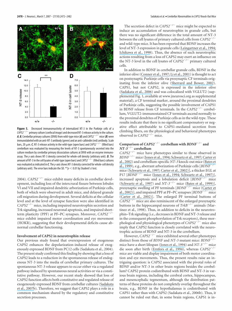
CAPS2 knock-out impairs Trk signalingSecreted neurotrophins bind and activatetheir Trk receptors on target cells, leadingto the autophosphorylation of Trk recep-tors. We used an anti-pTrk antibody to investigate whether or notthe loss of CAPS2 affects the cellular distribution of pTrk in theCAPS2�/� cerebellum (Fig. 6). In the P8 wild-type cerebellum,intense pTrk immunoreactivity was localized around the upperregion of the ML, just beneath the EGL, in which the fine tips ofthe extending dendrites of calbindin-positive Purkinje cells andthe PF terminals extending from granule cells are located (Fig.6A–C). However, this characteristic pTrk immunoreactivity inthe upper ML was considerably reduced in the CAPS2�/� cere-bellum (Fig. 6D–F). These findings suggested that the loss ofCAPS2 resulted in a deficit in neurotrophin-induced Trk activa-tion in target cells.
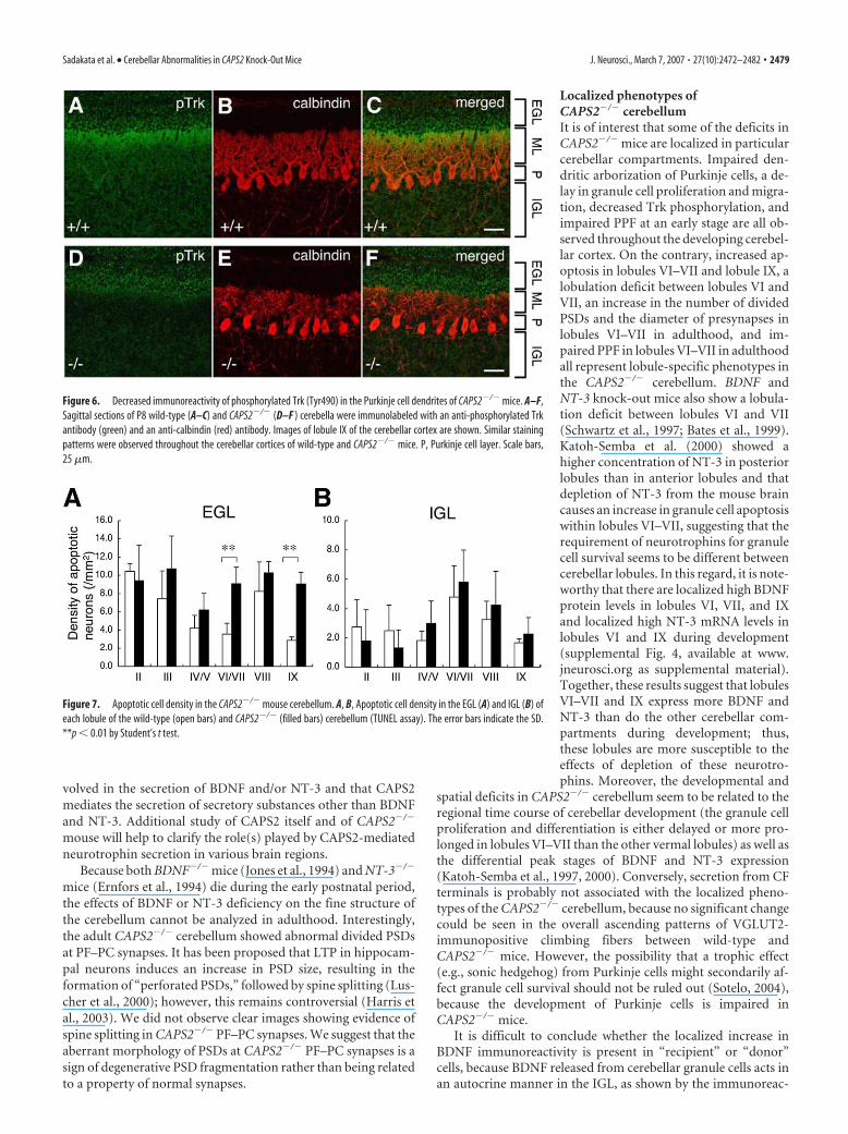
CAPS2 knock-out causes increased apoptosis ofcerebellar neuronsBecause the overexpression of exogenous CAPS2 promotes thesurvival of cerebellar neurons (Sadakata et al., 2004), we investi-gated apoptosis in the CAPS2�/� cerebellum. The density of ap-optotic cells visualized by terminal deoxynucleotidyl transferase-mediated biotinylated UTP nick end labeling (TUNEL) assay inthe EGL and IGL of each lobule of the cerebellar vermis at P8 wascompared in CAPS2�/� and wild-type littermates (Fig. 7A,B). In
the CAPS2�/� cerebellum, more apoptotic cells were observed inthe EGL than in that of the wild-type cerebellum (Fig. 7A), but nosuch difference was observed in the IGL (Fig. 7B). Interestingly,the increased cell death in the EGL of the CAPS2�/� cerebellumwas localized in lobules VI–VII and lobule IX. These findingsindicated that CAPS2 is required for the survival of granule cellsin the EGL, especially in lobules VI–VII and lobule IX, at P8,when CAPS2 expression is upregulated (Sadakata et al., 2004).
CAPS2 knock-out impairs the short-term plasticity ofPF–PC synapsesBecause CAPS2 knock-out leads to aberrant cytoarchitecture inPF–PC synapses, as described above (Fig. 4B), we investigated thesynaptic function of PF–PC synapses. However, little differencewas found to exist between wild-type and CAPS2�/� mice interms of the peak amplitudes of PF-EPSCs (Fig. 8A), thus indi-cating that the basic synaptic transmission functions inCAPS2�/� mice are unimpaired.
PPF of the PF-EPSC (defined as the ratio of the amplitude ofthe second EPSC to that of the first EPSC) is thought to reflectchanges in presynaptic function and is considered to be a kind ofshort-term synaptic plasticity (Atluri and Regehr, 1998). There-
Figure 3. Impaired dendritic arborization of Purkinje cells in the cerebella of P28 CAPS2�/� mice. A–F, Sagittal sections of P28wild-type (A–C) and CAPS2�/� (D–F ) mouse cerebella were immunolabeled with an anti-calbindin antibody (green) and ananti-VGLUT2 (red) antibody. Images of lobule VIII of the cerebellar cortex. Scale bars, 20 �m. G, H, The dendritic arbors of P28wild-type (G) and CAPS2�/� (H ) Purkinje cells were visualized by Golgi staining. Scale bars, 10 �m.
2476 • J. Neurosci., March 7, 2007 • 27(10):2472–2482 Sadakata et al. • Cerebellar Abnormalities in CAPS2 Knock-Out Mice

fore, we performed PPF analysis at PF–PC synapses in the ante-rior lobe [lobules II–V (Fig. 8B)], central lobe [lobules VI–VII(Fig. 8C)], and posterior lobe [lobule IX (Fig. 8D)]. The resultsrevealed that, for brief interstimulus intervals (�100 ms), thedegree of PPF at PF–PC synapses was markedly lower inCAPS2�/� mice than in wild-type mice. Impairment of this pre-synaptic function was observed in all three lobes examined, be-tween P15 and P21 (Fig. 8B–D, left). Conversely, in CAPS2�/�
mice between the ages of P43 and P54, PPF was elicited almostnormally in lobules II–V (Fig. 8B, right) and lobule IX (Fig. 8D,right); however, it remained impaired in lobules VI–VII (Fig. 8C,right). These results suggested a vulnerability of lobules VI–VII toa loss of CAPS2 function. No significant differences were ob-served between CAPS2�/� mice and wild-type littermates interms of cerebellar long-term potentiation (LTP) or long-termdepression at the PF–PC synapses (data not shown).
CAPS2 knock-out mice exhibit abnormal behaviorTo assess the cerebellar function of CAPS2�/� mice, we testedtheir coordinated motor performance on a rotarod treadmill.
The average number of falls within a 3 minperiod at 24 rpm was measured. At P28,CAPS2�/� mice showed low to moderaterotarod performance compared with thatof their wild-type littermates [2.4 0.36falling times in wild-type mice (n � 16) vs5.6 1.12 falling times in CADPS2�/�
mice (n � 14), mean SEM; p � 0.01, byStudent’s t test]; however, it was of interestthat this impairment tended to be relievedat P56 [1.92 0.70 falling times in wild-type mice (n � 12) vs 3.50 1.34 fallingtimes in CADPS2�/� mice (n � 10),mean SEM]. Different animals wereused between P28 and P56. These findingssuggest that CAPS2�/� mice have an im-pairment of their motor coordinationability that is with advancing age of theanimal capable of recovery by an un-known mechanism that appears to com-pensate for the loss of CAPS2.
An interesting feature of the CAPS2�/�
cerebellum is that characteristic deficitsconverge on particular lobules: i.e., abnor-mal lobulation, increased cell death andimpaired PPF in adulthood in lobules VI–VII, and increased cell death in lobule IX.It is noteworthy that these three lobuleshave been shown to be associated with thecontrol of various eye movements (Butt-ner and Fuhry, 1995; Heinen and Keller,1996). We therefore investigated thevestibulo-ocular reflex and optokinetic re-sponse in CAPS2�/� mice. There were nosignificant differences between the wild-type and CAPS2�/� mice in gains of theHVOR (Fig. 9A) or HOKR (Fig. 9C), andthe phases of the HVOR were also almostthe same in the CAPS2�/� mice and theirwild-type littermates (Fig. 9B). However,the phases of the HOKR were significantlydelayed in the CAPS2�/� mice comparedwith those of the wild-type mice (Fig. 9D).These findings indicated that the velocity
of visually evoked eye movements in CAPS2�/� mice tends to lagbehind the velocity of the movement of the visual surroundings,whereas no such lag is observed in wild-type mice.
Optokinetic training by sustained exposure to screen oscilla-tions in the presence of a sufficient amount of retinal slip isknown to induce cerebellum-dependent adaptation in theHOKR gain (Katoh et al., 1998). Optokinetic training for 1 h bysustained exposure to 15°, 0.17 Hz screen oscillations did inducea substantial increase in the HOKR gain in the wild-type mice,but the same training induced no significant change in the HOKRgain in the CAPS2�/� mice (Fig. 9E). Together, these observa-tions demonstrate that the loss of CAPS2 led to an eye movementdisorder characterized by impaired HOKR gain and plasticity.
DiscussionBy targeted gene disruption, we obtained in vivo evidence of theimportance of CAPS2 in cerebellar development and function.CAPS2 is a secretory granule-associated protein that is highlyexpressed in the cerebellum (Speidel et al., 2003; Sadakata et al.,
Figure 4. Aberrantly distributed and fewer vesicles within enlarged PF terminals (P15) and an increased number of dividedPSDs (P84) in the CAPS2�/� mouse cerebellum. A, B, Electron micrographs of a PF–PC synapse in the P15 wild-type cerebellum(A) and another in the P15 CAPS2�/� cerebellum (B). The insets show higher magnifications. Asterisks represent presynapticterminals. Scale bars: 500 nm; insets, 200 nm. C–G, Electron micrographs of a PF–PC synapse in the P84 wild-type cerebellum (C,E) and another in the P84 CAPS2�/� cerebellum (D, F, G). Images of lobules IV–V (C, D) and lobules VI–VII (E–G) of the cerebellarcortex. Arrows indicate the positions of perforations. Scale bars: 250 nm. H, Diameter of presynapses. The error bars indicate theSEM. **p � 0.01 by Student’s t test. I, The ratio of spines containing divided PSDs to the total spines examined. Multiple PSDs inone synapse is defined as a divided PSD. J, Number of PSDs per spine. The error bars indicate the SEM. **p � 0.01 by F test. Numberof synapses examined (n) is indicated.
Sadakata et al. • Cerebellar Abnormalities in CAPS2 Knock-Out Mice J. Neurosci., March 7, 2007 • 27(10):2472–2482 • 2477
2004). CAPS2�/� mice exhibit many deficits in cerebellar devel-opment, including loss of the intercrural fissure between lobulesVI and VII and impaired dendritic arborization of Purkinje cells,both of which were observed in adult mice, and delayed granulecell migration during development. Several deficits at the cellularlevel and at the level of synapse function were also identified inCAPS2�/� mice, including impaired neurotrophin secretion andTrk signaling, increased neuronal apoptosis, and impaired short-term plasticity (PPF) at PF–PC synapses. Moreover, CAPS2�/�
mice exhibit impaired motor coordination and eye movement(HOKR), suggesting that their developmental deficits cause ab-normal cerebellar functioning.
Involvement of CAPS2 in neurotrophin releaseOur previous study found that overexpression of exogenousCAPS2 enhances the depolarization-induced release of exog-enously expressed BDNF from PC12 cells (Sadakata et al., 2004).The present study confirmed this finding by showing that a loss ofCAPS2 leads to a reduction in the spontaneous release of endog-enous NT-3 into the media of cerebellar primary cultures. Thespontaneous NT-3 release appears to occur either via a regulatedpathway induced by spontaneous neural activities or via a consti-tutive pathway. However, our recent study showed that loss ofCAPS2 function affects both constitutive and regulated release ofexogenously expressed BDNF from cerebellar cultures (Sadakataet al., 2007b). Therefore, we suggest that CAPS2 plays a role in acommon mechanism shared by the regulatory and constitutivesecretion processes.
The secretion defect in CAPS2�/� mice might be expected toinduce an accumulation of neurotrophin in granule cells, butthere was no significant difference in the total amount of NT-3between the cell lysates of primary cultured cells from CAPS2�/�
and wild-type mice. It has been reported that BDNF increases thelevel of NT-3 expression in granule cells (Leingartner et al., 1994;Ichikawa et al., 1998). Thus, the absence of such neurotrophicactions resulting from a loss of CAPS2 may exert an influence onthe NT-3 level in the cell lysates of CAPS2�/� primary culturedcells.
In addition to BDNF in cerebellar granule cells, BDNF in theinferior olive (Conner et al., 1997; Li et al., 2001) is thought to acton postsynaptic Purkinje cells via presynaptic CF terminals orig-inating from the inferior olive (Sherrard and Bower, 2002).CAPS1, but not CAPS2, is expressed in the inferior olive(Sadakata et al., 2006) and was colocalized with VGLUT2 (sup-plemental Fig. 1, available at www.jneurosci.org as supplementalmaterial), a CF terminal marker, around the proximal dendritesof Purkinje cells, suggesting the possible involvement of CAPS1in BDNF release from CF terminals. In the CAPS2�/� cerebel-lum, VGLUT2-immunostained CF terminals ascend normally tothe proximal dendrites of Purkinje cells as in the wild-type. Theseresults indicate that there is no significant compensatory or neg-ative effect attributable to CAPS1-mediated secretion fromclimbing fibers, on the physiological and behavioral phenotypesobserved in CAPS2�/� mice.
Comparison of CAPS2�/� cerebellum with BDNF�/� andNT-3�/� cerebellumCAPS2�/� mice have phenotypes similar to those observed inBDNF�/� mice (Jones et al., 1994; Schwartz et al., 1997; Carter etal., 2002) and cerebellum-specific NT-3 knock-out mice (Bates etal., 1999), e.g., aberrant arborization of Purkinje cells [BDNF�/�
mice (Schwartz et al., 1997; Carter et al., 2002)], a thicker EGL atP17 [BDNF�/� mice (Jones et al., 1994; Schwartz et al., 1997)],increased apoptosis and a lobulation deficit [BDNF�/� mice(Schwartz et al., 1997) and NT-3�/� mice (Bates et al., 1999)],presynaptic swelling of PF terminals [BDNF�/� mice (Carter etal., 2002)], and impaired PPF at PF–PC synapses [BDNF�/� mice(Carter et al., 2002)]. The enlarged PF terminal buttons ofCAPS2�/� mice are also reminiscent of the enlarged presynapticbuttons in the hippocampal neurons of TrkB�/� animals (Mar-tinez et al., 1998). Thus, in addition to deficits in the neurotro-phin–Trk signaling (i.e., decreases in BDNF and NT-3 release andin the consequent phosphorylation of Trk receptors), these mor-phological and physiological phenotypes of CAPS2�/� mice alsoimply that CAPS2 function is closely correlated with the neuro-trophic actions of BDNF and NT-3 in the cerebellum.
However, CAPS2�/� mice exhibited several other phenotypesdistinct from those of BDNF and NT-3 mutant mice: BDNF�/�
mice have a short lifespan (Jones et al., 1994) and NT-3�/� micedie soon after birth (Ernfors et al., 1994), whereas CAPS2�/�
mice are viable and display impairment of both motor coordina-tion and eye movements. Thus, the present results raise an in-triguing question: is CAPS2 associated with the pivotal roles ofBDNF and/or NT-3 in other brain regions besides the cerebel-lum? CAPS2 protein codistributed with BDNF and NT-3 in var-ious brain regions, including the cerebral cortex, hippocampus,and mesencephalic tegmentum, although the distribution pat-terns of these proteins do not completely overlap throughout thebrain, e.g., BDNF in the hypothalamus is codistributed withCAPS1 rather than with CAPS2 (Sadakata et al., 2006b). Thus, itcannot be ruled out that, in some brain regions, CAPS1 is in-
Figure 5. Decreased immunoreactivity of internalized NT-3 in the Purkinje cells of aCAPS2�/� primary culture (confocal image) and decreased NT-3 release activity in the culture.A, B, Cerebellar primary cultures (DIV8) from wild-type mice (A) and CAPS2�/� mice (B) wereimmunolabeled with an anti-NT-3 antibody (green) and an anti-calbindin (red) antibody. Scalebars, 20 �m. C, NT-3 release activity in the wild-type (open bars) and CAPS2�/� (filled bars)cerebellum was evaluated by measuring the levels of NT-3 spontaneously secreted into theculture medium by cerebellar primary dissociation cultures at DIV8 with an enzyme immuno-assay. The y-axis shows NT-3 density corrected for whole-cell density (arbitrary unit). D, Theamount of NT-3 in the cell lysates of wild-type (open bars) and CAPS2�/� (filled bars) cultureswas evaluated as indicated in C. The y-axis shows NT-3 density corrected for whole-cell density(arbitrary unit). The error bars indicate the SD. **p � 0.01 by Student’s t test.
2478 • J. Neurosci., March 7, 2007 • 27(10):2472–2482 Sadakata et al. • Cerebellar Abnormalities in CAPS2 Knock-Out Mice
volved in the secretion of BDNF and/or NT-3 and that CAPS2mediates the secretion of secretory substances other than BDNFand NT-3. Additional study of CAPS2 itself and of CAPS2�/�
mouse will help to clarify the role(s) played by CAPS2-mediatedneurotrophin secretion in various brain regions.
Because both BDNF�/� mice (Jones et al., 1994) and NT-3�/�
mice (Ernfors et al., 1994) die during the early postnatal period,the effects of BDNF or NT-3 deficiency on the fine structure ofthe cerebellum cannot be analyzed in adulthood. Interestingly,the adult CAPS2�/� cerebellum showed abnormal divided PSDsat PF–PC synapses. It has been proposed that LTP in hippocam-pal neurons induces an increase in PSD size, resulting in theformation of “perforated PSDs,” followed by spine splitting (Lus-cher et al., 2000); however, this remains controversial (Harris etal., 2003). We did not observe clear images showing evidence ofspine splitting in CAPS2�/� PF–PC synapses. We suggest that theaberrant morphology of PSDs at CAPS2�/� PF–PC synapses is asign of degenerative PSD fragmentation rather than being relatedto a property of normal synapses.
Localized phenotypes ofCAPS2�/� cerebellumIt is of interest that some of the deficits inCAPS2�/� mice are localized in particularcerebellar compartments. Impaired den-dritic arborization of Purkinje cells, a de-lay in granule cell proliferation and migra-tion, decreased Trk phosphorylation, andimpaired PPF at an early stage are all ob-served throughout the developing cerebel-lar cortex. On the contrary, increased ap-optosis in lobules VI–VII and lobule IX, alobulation deficit between lobules VI andVII, an increase in the number of dividedPSDs and the diameter of presynapses inlobules VI–VII in adulthood, and im-paired PPF in lobules VI–VII in adulthoodall represent lobule-specific phenotypes inthe CAPS2�/� cerebellum. BDNF andNT-3 knock-out mice also show a lobula-tion deficit between lobules VI and VII(Schwartz et al., 1997; Bates et al., 1999).Katoh-Semba et al. (2000) showed ahigher concentration of NT-3 in posteriorlobules than in anterior lobules and thatdepletion of NT-3 from the mouse braincauses an increase in granule cell apoptosiswithin lobules VI–VII, suggesting that therequirement of neurotrophins for granulecell survival seems to be different betweencerebellar lobules. In this regard, it is note-worthy that there are localized high BDNFprotein levels in lobules VI, VII, and IXand localized high NT-3 mRNA levels inlobules VI and IX during development(supplemental Fig. 4, available at www.jneurosci.org as supplemental material).Together, these results suggest that lobulesVI–VII and IX express more BDNF andNT-3 than do the other cerebellar com-partments during development; thus,these lobules are more susceptible to theeffects of depletion of these neurotro-phins. Moreover, the developmental and
spatial deficits in CAPS2�/� cerebellum seem to be related to theregional time course of cerebellar development (the granule cellproliferation and differentiation is either delayed or more pro-longed in lobules VI–VII than the other vermal lobules) as well asthe differential peak stages of BDNF and NT-3 expression(Katoh-Semba et al., 1997, 2000). Conversely, secretion from CFterminals is probably not associated with the localized pheno-types of the CAPS2�/� cerebellum, because no significant changecould be seen in the overall ascending patterns of VGLUT2-immunopositive climbing fibers between wild-type andCAPS2�/� mice. However, the possibility that a trophic effect(e.g., sonic hedgehog) from Purkinje cells might secondarily af-fect granule cell survival should not be ruled out (Sotelo, 2004),because the development of Purkinje cells is impaired inCAPS2�/� mice.
It is difficult to conclude whether the localized increase inBDNF immunoreactivity is present in “recipient” or “donor”cells, because BDNF released from cerebellar granule cells acts inan autocrine manner in the IGL, as shown by the immunoreac-
Figure 6. Decreased immunoreactivity of phosphorylated Trk (Tyr490) in the Purkinje cell dendrites of CAPS2�/� mice. A–F,Sagittal sections of P8 wild-type (A–C) and CAPS2�/� (D–F ) cerebella were immunolabeled with an anti-phosphorylated Trkantibody (green) and an anti-calbindin (red) antibody. Images of lobule IX of the cerebellar cortex are shown. Similar stainingpatterns were observed throughout the cerebellar cortices of wild-type and CAPS2�/� mice. P, Purkinje cell layer. Scale bars,25 �m.
Figure 7. Apoptotic cell density in the CAPS2�/� mouse cerebellum. A, B, Apoptotic cell density in the EGL (A) and IGL (B) ofeach lobule of the wild-type (open bars) and CAPS2�/� (filled bars) cerebellum (TUNEL assay). The error bars indicate the SD.**p � 0.01 by Student’s t test.
Sadakata et al. • Cerebellar Abnormalities in CAPS2 Knock-Out Mice J. Neurosci., March 7, 2007 • 27(10):2472–2482 • 2479

tivity of phosphorylated Trk (Schwartz etal., 1997). The localized increase in BDNFimmunoreactivity in lobules VI–VII andIX was observed in both the wild-type andCAPS2�/� cerebellum (data not shown).Therefore, it is suggested that localized in-crease in the immunoreactivity for BDNFin the IGL is probably attributable to anincrease in BDNF levels in donor granulecells.
At the physiological and behaviorallevels, it is of interest that decreased PPFwas observed only in lobules VI–VII dur-ing adulthood. Lobules VI, VII, and IX areknown to be associated with the control ofvarious eye movements (Buttner andFuhry, 1995; Heinen and Keller, 1996). Inprimates, lesions in lobules VI–VII causesaccadic dysmetria and smooth pursuitdeficits (Buttner and Fuhry, 1995), and le-sions in lobule IX of the vermis (uvula)lead to optokinetic and smooth pursuitdeficits (Heinen and Keller, 1996). There-fore, the impaired control of eye move-ments in CAPS2�/� mice is most likely at-tributable to deficits in these cerebellarcompartments.
CAPS2 gene disruption andautistic phenotypesA recent study by our group showed thatCAPS2 knock-out mice have autistic-likecellular and behavioral phenotypes.Moreover, we found in blood samplesfrom some autistic patients an aberrant al-ternative splicing variant of CAPS2 mRNAthat lacks a specific exon encoding the dy-nactin 1-binding domain, which is re-quired for proper axonal transport(Sadakata et al., 2007b). Autism is charac-terized by cerebellar morphological ab-normalities (Bauman and Kemper, 1985;Courchesne et al., 1988). In this report, wefocused on the cerebellar phenotypes ofCAPS2 knock-out mice and found thatCAPS2�/� mice show the cerebellar phe-notypes described above. Some of thesephenotypes are similar with those ob-served in autistic patients. Abnormal re-duction in lobules VI and VII of the cere-bellar vermis is a characteristicmorphological deficit reported in thebrains of autistic patients (Courchesne etal., 1988), and hypoplasia of these lobuleswas suggested to be related to the abnor-mal exploratory behavior of autism (Pierce and Courchesne,2001). Deficits in pursuit eye movement (Takarae et al., 2004)and motor coordination (Manjiviona and Prior, 1995) have alsobeen reported in autistic spectrum disorders. Moreover, de-creased PPF (Moretti et al., 2006) was shown in a mouse model ofRett syndrome (Chen et al., 2001; Guy et al., 2001), which isconsidered to be an autistic spectrum disorder (World Health
Organization, 1992; American Psychiatric Association, 1994).Together, our findings are also suggestive of an involvement ofthe CAPS2 gene, which is located within the AUTS1, in the cere-bellar deficits of autistic patients.
In conclusion, the present CAPS2�/� mouse study indicatedthat CAPS2-mediated neurotrophin release is indispensable forcerebellum-related cellular, morphological, physiological, and
Figure 8. Impairment of short-term synaptic plasticity at PF–PC synapses in the CAPS2�/� mouse cerebellum. A, Plotsshowing the relationship between the PF-EPSC amplitude and stimulus intensity applied to PFs in CAPS2�/� mice (filled circles;n �16) and wild-type littermates (open circles; n �18), at P15–P21. Insets show representative EPSC traces evoked by PF stimuliof different intensities (0 –220 �A, 20 �A intervals). B–D, The mean paired-pulse ratios recorded from each lobule of P15–P21(left column) and P43–P54 (right column) cerebellar slices were plotted against various interstimulus intervals. Recordings fromlobules II–V, VI–VII, and IX are shown in B, C, and D, respectively. Insets show representative traces obtained from CAPS2�/� miceand wild-type littermates. CAPS2�/� mice exhibited a lower PPF ratio than their wild-type littermates at interstimulus intervalsof �100 ms in all lobules at P15–P21 and in lobules VI–VII only at P43–P54. The error bars indicate the SEM. *p � 0.05; **p �0.01; ***p � 0.001 by the Mann–Whitney U test.
2480 • J. Neurosci., March 7, 2007 • 27(10):2472–2482 Sadakata et al. • Cerebellar Abnormalities in CAPS2 Knock-Out Mice

behavioral phenotypes, some of which appear to be related to thecerebellar deficits observed in autistic patients. Thus, it appearsthat this disturbance in CAPS2-mediated neurotrophic releasecould be at least in part responsible for the onset of autisticdisorder.
ReferencesAmerican Psychiatric Association (1994) Diagnostic and statistical manual
of mental disorders: DSM-IV. Washington, DC: American PsychiatricAssociation.
Atluri PP, Regehr WG (1998) Delayed release of neurotransmitter from cer-ebellar granule cells. J Neurosci 18:8214 – 8227.
Bates B, Rios M, Trumpp A, Chen C, Fan G, Bishop JM, Jaenisch R (1999)Neurotrophin-3 is required for proper cerebellar development. Nat Neu-rosci 2:115–117.
Bauman M, Kemper TL (1985) Histoanatomic observations of the brain inearly infantile autism. Neurology 35:866 – 874.
Berwin B, Floor E, Martin TF (1998) CAPS (mammalian UNC-31) proteinlocalizes to membranes involved in dense-core vesicle exocytosis. Neuron21:137–145.
Bibel M, Barde YA (2000) Neurotrophins: key regulators of cell fate and cellshape in the vertebrate nervous system. Genes Dev 14:2919 –2937.
Binda AV, Kabbani N, Levenson R (2005) Regulation of dense core vesiclerelease from PC12 cells by interaction between the D2 dopamine receptorand calcium-dependent activator protein for secretion (CAPS). BiochemPharmacol 69:1451–1461.
Buttner U, Fuhry L (1995) Eye movements. Curr Opin Neurol 8:77– 82.Carter AR, Chen C, Schwartz PM, Segal RA (2002) Brain-derived neurotro-
phic factor modulates cerebellar plasticity and synaptic ultrastructure.J Neurosci 22:1316 –1327.
Chen RZ, Akbarian S, Tudor M, Jaenisch R (2001) Deficiency of methyl-CpG binding protein-2 in CNS neurons results in a Rett-like phenotype inmice. Nat Genet 27:327–331.
Cisternas FA, Vincent JB, Scherer SW, Ray PN (2003) Cloning and charac-terization of human CADPS and CADPS2, new members of the Ca 2�-dependent activator for secretion protein family. Genomics 81:279 –291.
Conner JM, Lauterborn JC, Yan Q, Gall CM, Varon S (1997) Distribution ofbrain-derived neurotrophic factor (BDNF) protein and mRNA in the
normal adult rat CNS: evidence for anterograde axonal transport. J Neu-rosci 17:2295–2313.
Courchesne E, Yeung-Courchesne R, Press GA, Hesselink JR, Jernigan TL(1988) Hypoplasia of cerebellar vermal lobules VI and VII in autism.N Engl J Med 318:1349 –1354.
Ernfors P, Lee KF, Kucera J, Jaenisch R (1994) Lack of neurotrophin-3 leadsto deficiencies in the peripheral nervous system and loss of limb propri-oceptive afferents. Cell 77:503–512.
Grishanin RN, Kowalchyk JA, Klenchin VA, Ann K, Earles CA, Chapman ER,Gerona RR, Martin TF (2004) CAPS acts at a prefusion step in dense-core vesicle exocytosis as a PIP2 binding protein. Neuron 43:551–562.
Guy J, Hendrich B, Holmes M, Martin JE, Bird A (2001) A mouse Mecp2-null mutation causes neurological symptoms that mimic Rett syndrome.Nat Genet 27:322–326.
Harris KM, Fiala JC, Ostroff L (2003) Structural changes at dendritic spinesynapses during long-term potentiation. Philos Trans R Soc Lond B BiolSci 358:745–748.
Heinen SJ, Keller EL (1996) The function of the cerebellar uvula in monkeyduring optokinetic and pursuit eye movements: single-unit responses andlesion effects. Exp Brain Res 110:1–14.
Huang EJ, Reichardt LF (2001) Neurotrophins: roles in neuronal develop-ment and function. Annu Rev Neurosci 24:677–736.
Ichikawa D, Tabuchi A, Taoka A, Tsuchiya T, Tsuda M (1998) Attenuationof cell death mediated by membrane depolarization different from that byexogenous BDNF in cultured mouse cerebellar granule cells. Brain ResMol Brain Res 56:218 –226.
International Molecular Genetic Study of Autism Consortium (2001) Fur-ther characterization of the autism susceptibility locus AUTS1 on chro-mosome 7q. Hum Mol Genet 10:973–982.
Jastreboff PW (1979) Evaluation and statistical judgement of neural re-sponses to sinusoidal stimulation in cases with superimposed drift andnoise. Biol Cybern 33:113–120.
Jones KR, Farinas I, Backus C, Reichardt LF (1994) Targeted disruption ofthe BDNF gene perturbs brain and sensory neuron development but notmotor neuron development. Cell 76:989 –999.
Katoh A, Kitazawa H, Itohara S, Nagao S (1998) Dynamic characteristicsand adaptability of mouse vestibulo-ocular and optokinetic response eyemovements and the role of the flocculo-olivary system revealed by chem-ical lesions. Proc Natl Acad Sci USA 95:7705–7710.
Katoh-Semba R, Kaisho Y, Shintani A, Nagahama M, Kato K (1996) Tissuedistribution and immunocytochemical localization of neurotrophin-3 inthe brain and peripheral tissues of rats. J Neurochem 66:330 –337.
Katoh-Semba R, Takeuchi IK, Semba R, Kato K (1997) Distribution ofbrain-derived neurotrophic factor in rats and its changes with develop-ment in the brain. J Neurochem 69:34 – 42.
Katoh-Semba R, Takeuchi IK, Semba R, Kato K (2000) Neurotrophin-3controls proliferation of granular precursors as well as survival of maturegranule neurons in the developing rat cerebellum. J Neurochem74:1923–1930.
Kawase E, Suemori H, Takahashi N, Okazaki K, Hashimoto K, Nakatsuji N(1994) Strain difference in establishment of mouse embryonic stem (ES)cell lines. Int J Dev Biol 38:385–390.
Kovalchuk Y, Holthoff K, Konnerth A (2004) Neurotrophin action on arapid timescale. Curr Opin Neurobiol 14:558 –563.
Leingartner A, Heisenberg CP, Kolbeck R, Thoenen H, Lindholm D (1994)Brain-derived neurotrophic factor increases neurotrophin-3 expressionin cerebellar granule neurons. J Biol Chem 269:828 – 830.
Lessmann V, Gottmann K, Malcangio M (2003) Neurotrophin secretion:current facts and future prospects. Prog Neurobiol 69:341–374.
Li YX, Tokuyama W, Okuno H, Miyashita Y, Hashimoto T (2001) Differ-ential induction of brain-derived neurotrophic factor mRNA in rat infe-rior olive subregions following unilateral labyrinthectomy. Neuroscience106:385–394.
Lindholm D, Hamner S, Zirrgiebel U (1997) Neurotrophins and cerebellardevelopment. Perspect Dev Neurobiol 5:83–94.
Lu B (2003) BDNF and activity-dependent synaptic modulation. LearnMem 10:86 –98.
Luscher C, Nicoll RA, Malenka RC, Muller D (2000) Synaptic plasticity anddynamic modulation of the postsynaptic membrane. Nat Neurosci3:545–550.
Lynch A, Smart JL, Dobbing J (1975) Motor co-ordination and cerebellarsize in adult rats undernourished in early life. Brain Res 83:249 –259.
Figure 9. Impairments in the eye movement plasticity of CAPS2�/� mice. A, B, HVOR. Gain(A) and phase (B) of 6-month-old CAPS2�/� mice (filled bars; n �8) and wild-type littermates(open bars; n � 8). C, D, HOKR. Gain (C) and phase (D) of 6-month-old CAPS2�/� mice (filledbars; n � 9) and wild-type littermates (open bars; n � 9). The HOKR phase lags of theCAPS2�/� mice were longer than those of the wild-type mice at all screen velocities ( p�0.01,two-way ANOVA). E, Optokinetic training was performed by exposing the mice to 1 h of sus-tained sinusoidal screen oscillations by 15° (peak-to-peak) at 0.17 Hz. The mean HOKR gainsbefore (open bars) and after (gray bars) training of the CAPS2�/� (n � 9) and wild-type (n �8) mice are shown. The error bars indicate the SEM. **p � 0.01 by Student’s t test.
Sadakata et al. • Cerebellar Abnormalities in CAPS2 Knock-Out Mice J. Neurosci., March 7, 2007 • 27(10):2472–2482 • 2481

Manjiviona J, Prior M (1995) Comparison of Asperger syndrome and high-functioning autistic children on a test of motor impairment. J Autism DevDisord 25:23–39.
Martinez A, Alcantara S, Borrell V, Del Rio JA, Blasi J, Otal R, Campos N,Boronat A, Barbacid M, Silos-Santiago I, Soriano E (1998) TrkB andTrkC signaling are required for maturation and synaptogenesis of hip-pocampal connections. J Neurosci 18:7336 –7350.
Moretti P, Levenson JM, Battaglia F, Atkinson R, Teague R, Antalffy B, Arm-strong D, Arancio O, Sweatt JD, Zoghbi HY (2006) Learning and mem-ory and synaptic plasticity are impaired in a mouse model of Rett syn-drome. J Neurosci 26:319 –327.
Muhle R, Trentacoste SV, Rapin I (2004) The genetics of autism. Pediatrics113:e472– e486.
Pierce K, Courchesne E (2001) Evidence for a cerebellar role in reducedexploration and stereotyped behavior in autism. Biol Psychiatry49:655– 664.
Renden R, Berwin B, Davis W, Ann K, Chin CT, Kreber R, Ganetzky B, MartinTF, Broadie K (2001) Drosophila CAPS is an essential gene that regulatesdense-core vesicle release and synaptic vesicle fusion. Neuron31:421– 437.
Sadakata T, Mizoguchi A, Sato Y, Katoh-Semba R, Fukuda M, Mikoshiba K,Furuichi T (2004) The secretory granule-associated protein CAPS2 reg-ulates neurotrophin release and cell survival. J Neurosci 24:43–52.
Sadakata T, Itakura M, Kozaki S, Sekine Y, Takahashi M, Furuichi T (2006)Differential distributions of the Ca 2�-dependent activator protein forsecretion family proteins (CAPS2 and CAPS1) in the mouse brain.J Comp Neurol 495:735–753.
Sadakata T, Washida M, Morita N, Furuichi T (2007a) Tissue distributionof Ca 2�-dependent activator protein for secretion family membersCAPS1 and CAPS2 in mice. J Histochem Cytochem, in press.
Sadakata T, Washida M, Iwayama Y, Shoji S, Sato Y, Ohkura T, Katoh-SembaR, Nakajima M, Sekine Y, Tanaka M, Nakamura K, Iwata Y, Tsuchiya KJ,Mori N, Detera-Wadleigh SD, Ichikawa H, Itohara S, Yoshikawa T, Fu-
ruichi T (2007b) Autistic-like phenotypes in CADPS2/CAPS2 knockoutmice and aberrant CADPS2 splicing in autistic patients. J Clin Invest, inpress.
Schinder AF, Poo M (2000) The neurotrophin hypothesis for synaptic plas-ticity. Trends Neurosci 23:639 – 645.
Schwartz PM, Borghesani PR, Levy RL, Pomeroy SL, Segal RA (1997) Ab-normal cerebellar development and foliation in BDNF �/� mice reveals arole for neurotrophins in CNS patterning. Neuron 19:269 –281.
Segal RA (2003) Selectivity in neurotrophin signaling: theme and variations.Annu Rev Neurosci 26:299 –330.
Sherrard RM, Bower AJ (2002) Climbing fiber development: do neurotro-phins have a part to play? Cerebellum 1:265–275.
Sotelo C (2004) Cellular and genetic regulation of the development of thecerebellar system. Prog Neurobiol 72:295–339.
Speidel D, Varoqueaux F, Enk C, Nojiri M, Grishanin RN, Martin TF, Hof-mann K, Brose N, Reim K (2003) A family of Ca 2�-dependent activatorproteins for secretion: comparative analysis of structure, expression, lo-calization, and function. J Biol Chem 278:52802–52809.
Speidel D, Bruederle CE, Enk C, Voets T, Varoqueaux F, Reim K, Becherer U,Fornai F, Ruggieri S, Holighaus Y, Weihe E, Bruns D, Brose N, Rettig J(2005) CAPS1 regulates catecholamine loading of large dense-core vesi-cles. Neuron 46:75– 88.
Takarae Y, Minshew NJ, Luna B, Krisky CM, Sweeney JA (2004) Pursuit eyemovement deficits in autism. Brain 127:2584 –2594.
Tandon A, Bannykh S, Kowalchyk JA, Banerjee A, Martin TF, Balch WE(1998) Differential regulation of exocytosis by calcium and CAPS insemi-intact synaptosomes. Neuron 21:147–154.
Thoenen H (1995) Neurotrophins and neuronal plasticity. Science270:593–598.
World Health Organization (1992) The ICD-10 classification of mental andbehavioural disorders: clinical descriptions and diagnostic guidelines. Ge-neva: World Health Organization.
2482 • J. Neurosci., March 7, 2007 • 27(10):2472–2482 Sadakata et al. • Cerebellar Abnormalities in CAPS2 Knock-Out Mice
Copyright © 2022 FDOKUMEN