Guanylin and uroguanylin induce natriuresis in mice lacking guanylyl cyclase-C receptor
14
Kidney International, Vol. 65 (2004), pp. 40–53 Guanylin and uroguanylin induce natriuresis in mice lacking guanylyl cyclase-C receptor STEPHEN L. CARRITHERS,COBERN E. OTT,MICHAEL J. HILL,BRETT R. J OHNSON,WEIYAN CAI, J ASON J. CHANG,RAJESH G. SHAH,CONGMEI SUN,ELIZABETH A. MANN,MANASSES C. F ONTELES, LEONARD R. F ORTE,BRIAN A. J ACKSON,RALPH A. GIANNELLA, and RICHARD N. GREENBERG Department of Internal Medicine, Division of Infectious Diseases, Lexington VA Medical Center and University of Kentucky, Lexington, Kentucky; Department of Physiology, University of Kentucky, Lexington, Kentucky; Department of Biological Chemistry & Molecular Pharmacology, Harvard Medical School, Harvard University, Boston, Massachusetts; Division of Digestive Diseases, University of Cincinnati and Cincinnati VA Medical Center, Cincinnati, Ohio; Clinical Research Unit of Federal University of Cear ´ a and Cear ´ a State University, Fortaleza, Brazil; Department of Pharmacology, Missouri University and Truman Memorial Veterans Hospital, Columbia, Missouri Guanylin and uroguanylin induce natriuresis in mice lacking guanylyl cyclase-C receptor. Background. Guanylin (GN) and uroguanylin (UGN) are in- testinally derived peptide hormones that are similar in structure and activity to the diarrhea-causing Escherichia coli heat-stable enterotoxins (STa). These secretagogues have been shown to af- fect fluid, Na + ,K + , and Cl − transport in both the intestine and kidney, presumably by intracellular cyclic guanosine monophos- phate (cGMP)-dependent signal transduction. However, the in vivo consequences of GN, UGN, and STa on renal func- tion and their mechanism of action have yet to be rigorously tested. Methods. We hypothesized that intravenous administration of GN, UGN, or STa would cause an increase in natriuresis in wild-type mice via cGMP and guanylyl cyclase-C (GC-C, Gucy2c), the only known receptor for these peptide-hormones, and that the peptide-induced natriuresis would be blunted in genetically altered mice devoid of GC-C receptors (GC-C (−/−) null). Results. In wild-type mice using a modified renal clearance model, GN, UGN, and STa elicited significant natriuresis, kali- uresis, and diuresis as well as increased urinary cGMP levels in a time- and dose-dependent fashion. Absolute and fractional uri- nary sodium excretion levels were greatest ∼40 minutes follow- ing a bolus infusion with pharmacologic doses of these peptides. Unexpectedly, GC-C (−/−) null mice also responded to the GN peptides similarly to that observed in wild-type mice. Glomeru- lar filtration rate (GFR), blood pressure, and plasma cGMP in the mice (wild-type or GC-C (−/−) null) did not significantly vary between the vehicle- and peptide-treatment groups. The effects of UGN may also influence long-term renal function due to down-regulation of the Na + /K + ATPase c -subunit and Key words: kidney, cGMP, guanylyl cyclase, renal clearance, sodium, DD-PCR. Received for publication January 21, 2003 and in revised form April 4, 2003, and July 7, 2003 Accepted for publication August 18, 2003 C 2004 by the International Society of Nephrology the Cl − channel ClC-K2 by 60% and 75%, respectively, as as- sessed by differential display polymerase chain reaction (PCR) (DD-PCR) and Northern blot analysis of kidney mRNA from mice treated with UGN. Conclusion. GN, UGN, and STa act on the mouse kidney, in part, through a cGMP-dependent, GC-C–independent mecha- nism, causing significant natriuresis by renal tubular processes. UGN may have further long-term effects on the kidney by al- tering the expression of such transport-associated proteins as Na + /K + ATPase and ClC-K2. The kidney plays a critical role in the maintenance of extracellular fluid volume, and as a consequence, blood pressure, by regulating total body NaCl content [1, 2]. Fundamentally, alterations in NaCl content (either through changes in intake or through inappropriate ex- trarenal loss) lead to adjustments in urinary NaCl excre- tion that are designed to ultimately help restore normal levels. It is well established in both humans and in exper- imental animals that orally administered NaCl loads can be excreted more rapidly than equivalent salt loads ad- ministered intravenously [3–7]. These observations sup- port a concept that NaCl intake may be monitored by the intestine, and that an increased delivery results in the generation of a signal to the kidney that begins to adjust sodium excretion to match intake prior to the absorption of sufficient NaCl to cause significant changes in extra- cellular fluid volume [8, 9]. For example, urinary sodium excretion is significantly elevated in the 4-hour period im- mediately after an oral salt load in rats, rabbits, and hu- mans [3, 4, 6, 7, 10, 11]. In sharp contrast, an intravenous salt load of the same amount causes a much smaller in- crease in sodium excretion over the same time period. Thus, a putative “intestinal natriuretic factor” could link the intestine with the kidney as a means of regulating urinary salt excretion to match dietary salt intake. 40
Transcript of Guanylin and uroguanylin induce natriuresis in mice lacking guanylyl cyclase-C receptor

Kidney International, Vol. 65 (2004), pp. 40–53
Guanylin and uroguanylin induce natriuresis in mice lackingguanylyl cyclase-C receptor
STEPHEN L. CARRITHERS, COBERN E. OTT, MICHAEL J. HILL, BRETT R. JOHNSON, WEIYAN CAI,JASON J. CHANG, RAJESH G. SHAH, CONGMEI SUN, ELIZABETH A. MANN, MANASSES C. FONTELES,LEONARD R. FORTE, BRIAN A. JACKSON, RALPH A. GIANNELLA, and RICHARD N. GREENBERG
Department of Internal Medicine, Division of Infectious Diseases, Lexington VA Medical Center and University of Kentucky,Lexington, Kentucky; Department of Physiology, University of Kentucky, Lexington, Kentucky; Department of BiologicalChemistry & Molecular Pharmacology, Harvard Medical School, Harvard University, Boston, Massachusetts; Division of DigestiveDiseases, University of Cincinnati and Cincinnati VA Medical Center, Cincinnati, Ohio; Clinical Research Unit of Federal Universityof Ceara and Ceara State University, Fortaleza, Brazil; Department of Pharmacology, Missouri University and Truman MemorialVeterans Hospital, Columbia, Missouri
Guanylin and uroguanylin induce natriuresis in mice lackingguanylyl cyclase-C receptor.
Background. Guanylin (GN) and uroguanylin (UGN) are in-testinally derived peptide hormones that are similar in structureand activity to the diarrhea-causing Escherichia coli heat-stableenterotoxins (STa). These secretagogues have been shown to af-fect fluid, Na+, K+, and Cl− transport in both the intestine andkidney, presumably by intracellular cyclic guanosine monophos-phate (cGMP)-dependent signal transduction. However, thein vivo consequences of GN, UGN, and STa on renal func-tion and their mechanism of action have yet to be rigorouslytested.
Methods. We hypothesized that intravenous administrationof GN, UGN, or STa would cause an increase in natriuresisin wild-type mice via cGMP and guanylyl cyclase-C (GC-C,Gucy2c), the only known receptor for these peptide-hormones,and that the peptide-induced natriuresis would be blunted ingenetically altered mice devoid of GC-C receptors (GC-C(−/−)
null).Results. In wild-type mice using a modified renal clearance
model, GN, UGN, and STa elicited significant natriuresis, kali-uresis, and diuresis as well as increased urinary cGMP levels in atime- and dose-dependent fashion. Absolute and fractional uri-nary sodium excretion levels were greatest ∼40 minutes follow-ing a bolus infusion with pharmacologic doses of these peptides.Unexpectedly, GC-C(−/−) null mice also responded to the GNpeptides similarly to that observed in wild-type mice. Glomeru-lar filtration rate (GFR), blood pressure, and plasma cGMPin the mice (wild-type or GC-C(−/−) null) did not significantlyvary between the vehicle- and peptide-treatment groups. Theeffects of UGN may also influence long-term renal functiondue to down-regulation of the Na+/K+ ATPase c -subunit and
Key words: kidney, cGMP, guanylyl cyclase, renal clearance, sodium,DD-PCR.
Received for publication January 21, 2003and in revised form April 4, 2003, and July 7, 2003Accepted for publication August 18, 2003
C© 2004 by the International Society of Nephrology
the Cl− channel ClC-K2 by 60% and 75%, respectively, as as-sessed by differential display polymerase chain reaction (PCR)(DD-PCR) and Northern blot analysis of kidney mRNA frommice treated with UGN.
Conclusion. GN, UGN, and STa act on the mouse kidney, inpart, through a cGMP-dependent, GC-C–independent mecha-nism, causing significant natriuresis by renal tubular processes.UGN may have further long-term effects on the kidney by al-tering the expression of such transport-associated proteins asNa+/K+ ATPase and ClC-K2.
The kidney plays a critical role in the maintenanceof extracellular fluid volume, and as a consequence,blood pressure, by regulating total body NaCl content[1, 2]. Fundamentally, alterations in NaCl content (eitherthrough changes in intake or through inappropriate ex-trarenal loss) lead to adjustments in urinary NaCl excre-tion that are designed to ultimately help restore normallevels. It is well established in both humans and in exper-imental animals that orally administered NaCl loads canbe excreted more rapidly than equivalent salt loads ad-ministered intravenously [3–7]. These observations sup-port a concept that NaCl intake may be monitored bythe intestine, and that an increased delivery results in thegeneration of a signal to the kidney that begins to adjustsodium excretion to match intake prior to the absorptionof sufficient NaCl to cause significant changes in extra-cellular fluid volume [8, 9]. For example, urinary sodiumexcretion is significantly elevated in the 4-hour period im-mediately after an oral salt load in rats, rabbits, and hu-mans [3, 4, 6, 7, 10, 11]. In sharp contrast, an intravenoussalt load of the same amount causes a much smaller in-crease in sodium excretion over the same time period.Thus, a putative “intestinal natriuretic factor” could linkthe intestine with the kidney as a means of regulatingurinary salt excretion to match dietary salt intake.
40

Carrithers et al: Natriuresis by guanylin peptides in mice 41
Studies from this and other laboratories suggest thattwo low-molecular-weight peptides, guanylin (GN) anduroguanylin (UGN), may be critical components ofthis enterorenal axis [9, 12–14]. In the gastrointesti-nal tract, GN and UGN bind to and activate guanylylcyclase-C (GC-C) [15], an apical membrane receptor thathas an intrinsic guanylyl cyclase catalytic domain [16].Mechanistically, receptor activation enhances intracellu-lar guanosine-3′,5′-cyclic monophosphate (cGMP) syn-thesis, which in turn, stimulates intestinal electrogenicchloride and bicarbonate secretion via the cystic fibrosistransmembrane conductance regulator (CFTR) chloridechannel and inhibits electroneutral sodium absorption [9,15, 17]. Although GN was initially isolated from jejunumand UGN from urine, these peptides have been foundthroughout the intestine where they are released fromspecific epithelial cells into both the lumen and the sys-temic circulation [9, 18–21]. GN and UGN are structurallysimilar to the heat-stable enterotoxin (STa) peptides se-creted by pathogenic strains of enteric bacteria[9, 22].These toxins virtually act as superagonists of GC-C andtrigger nonphysiologic movement of electrolytes withinthe intestinal lumen [23].
Recent studies from this laboratory have shown thatGN and UGN can also affect renal function. Intravenousadministration of exogenous GN and UGN induce sig-nificant natriuresis in mice [12, 24]. In addition, studiesin the isolated perfused rat kidney have shown that bothpeptides are natriuretic in a time- and dose-dependentfashion [14, 25]. Coincident with these solute excretorypatterns, urinary cGMP excretion also increased afterpeptide administration. GN, UGN, and STa have alsobeen shown to elevate cGMP in organ-cultured kidneyslices and proximal convoluted tubule cells in vitro [26–29]. Consistent with these functional responses, previousstudies have reported 125I-GN [30] and 125I-ST binding[27, 31], and GC-C mRNA expression in heteroge-neous kidney preparations [32–34]. Employing semi-quantitative reverse transcription-polymerase chainreaction (RT-PCR), we assessed the distribution of renalGN/UGN receptor mRNA in specific microdissected seg-ments of the rat nephron [32]. These results suggest thatGN/UGN/STa may affect transport in multiple nephronsegments that are involved in renal tubular handling ofNaCl since receptor mRNA expression is relatively highin the collecting tubules, the proximal convoluted tubule,and the medullary thick ascending limb. Lower levels ofreceptor mRNA were observed in the cortical thick as-cending limb, distal convoluted tubes, and glomeruli.
While the mechanisms that regulate the renal effectsof GN and UGN are unclear, a role for sodium intake hasbeen implicated. For example, the UGN concentration inhuman urine and plasma is increased during a high saltintake [34]. Additionally, perfusion of the small intestine[35] and colon [36] in situ with hypertonic NaCl solutions
markedly increased GN (and to a lesser degree UGN)secretion into the lumen, while perfusion with similar os-motic concentrations of mannitol exhibited much lowereffects [21, 37]. However, in a whole animal model of os-motic diarrhea (using a lactose-based diet), GN and UGNmRNA and protein expression levels were significantlyincreased suggesting that these genes are responsive tothe increased intraluminal hypertonicity inherent to themodel [21]. Furthermore, using HT29-18-N2 colonic cells,hypertonic solutions (of NaCl, lactose, or mannitol) werefound to significantly increase GN and UGN expressionand secretion from the apical and basolateral surfaces ofthe cell [18]. Conversely, low NaCl intake down-regulatesGN mRNA and peptide expression in the distal colon[38]. Recent studies from this laboratory have further ex-amined the effects of altered NaCl intake on GN andUGN mRNA expression in the four principal regionsof the rat intestine—duodenum, jejunum, ileum, and thecolon—and have shown that both peptides are affectedby salt diet in the rat intestine [39]. More important, ex-pression patterns could be affected within 4 hours of anintragastric administration of NaCl, suggesting that a pu-tative signaling system can respond rapidly to alteredNaCl intake.
Although these data are compelling and support a hy-pothesis that the renal effects of intestinally derived GNand UGN are involved in the maintenance of salt and wa-ter homeostasis, no detailed analysis of the renal actionand mechanism of natriuresis by these peptides have beenrigorously evaluated. Therefore, the principal aim of thepresent study was to elucidate the renal effects and themechanism(s) of GN and UGN action in vivo employing amodified mouse clearance model. In addition, to addressthe renal mechanism by which these peptides elicit natri-uresis, we evaluated how this system acts in GC-C(−/−)
null (Gucy2c −/−) mice. Here, we show that intravenousadministration of GN and UGN increase absolute andfractional sodium excretion in wild-type and GC-C(−/−)
null mice in a rapid, time- and dose-dependent fashion. Ithas been assumed that the renal effects of GN/UGN aremediated by peptide binding to GC-C [9, 32, 40]. How-ever, data in this report would suggest the presence ofan alternate renal GN/UGN/STa receptor. Therefore, asecond goal of the present study was to begin to address,using a molecular approach, the presence of this GC-Cindependent mechanism of renal GN/UGN action.
EXPERIMENTAL PROCEDURES
Mouse models
Mice lacking the GC-C receptor were generated as pre-viously described [41]. Embryonic stem cells (E14TG2Afrom strain 129/Sv), selected for a targeted deletionof Exon 1 of Gucy2c, the gene encoding GC-C, weremicroinjected into Black Swiss blastocysts. Germline
42 Carrithers et al: Natriuresis by guanylin peptides in mice
chimeric offspring were identified and bred with BlackSwiss mice. The resulting heterozygous littermates (hy-brids of 129/Sv and Black Swiss) were bred to produce ho-mozygous Gucy2c−/− (GC-C(−/−) null) and Gucy2c+/+
(wild-type) mice. The GC-C(−/−) null and wild-type miceused in these experiments are the progeny of theseF2 mice. In addition, NIH Institute of Cancer Re-search/Harlan Sprague-Dawley (ICR-HSD) mice werealso used as wild-type controls.
Suckling mouse intestinal fluid assay
In order to assess the potency and bioactivity of GN,UGN, and STa, the peptides were tested for their ability toinduce intestinal fluid accumulation in newborn mice, asdescribed previously [12, 42]. ICR-HSD suckling mice, 2to 4 days old (2.1 ± 1.0 g), were dosed orally with 0.1 mL oftest solution. The injections contained vehicle (1 mmol/LHEPES and 0.1% Evans blue dye) alone or the peptidesdissolved in vehicle (except that GN injections containedthe protease inhibitors chymostatin (0.1 mmol) and apro-tinin (0.67 U) [24, 25]). After administration of vehicle orpeptide agonists, the mice were kept at room tempera-ture for 3 hours. The mice were then killed, intestinaland body weights measured, and a ratio of the intestinalweight to remaining body weight was calculated. A ratioof 0.0875 represents one mouse unit of activity, indicatingsignificant fluid accumulation in the intestine. The secre-tion activity (one unit) of GN (containing aprotinin andchymostatin) was 142.9 ± 5.3 ng/mouse; UGN was 41.5 ±1.7 ng/mouse; and for STa, 3.75 ± 0.2 ng/mouse.
Stability of GN, UGN, and STa in mouse serum
Approximately 4 units of peptide activity (GN, 550 ng;UGN, 150 ng; STa, 15 ng) were incubated in 100 lL ofmouse serum for 120 minutes at 37◦C under 5% CO2.Samples were mixed with an equal volume of vehicle(1 mmol/L HEPES and 0.1% Evans blue dye) and ad-ministered to suckling mice. The fluid accumulation in theintestine was assessed for each peptide-containing sam-ple containing the mouse serum or vehicle alone (con-trol). The mean ± SEM from four to five mice/group wasused in determining representative fluid accumulationand compared against that of the control group (peptide-induced group without serum). Mouse plasma alone didnot significantly increase the fluid volume in the sucklingmouse intestine.
In vitro cGMP accumulation assay
The bioactivity of each peptide cGMP agonist (GN,UGN, and STa) was tested in an in vitro tissue culture-based cGMP accumulation assay as previously de-scribed using T84 cells [43–45]. Briefly, T84 cells, grownas monolayers in 24-well plates, were washed twicewith 1 mL serum-free Dulbecco’s modified Eagle’s
medium (DMEM)-F12 media (Gibco-Life Technologies,Gaithersburg, MD, USA). The sample (0.2 mL) waslayered onto the cells and incubated for 40 minutesat 37◦C. The media were removed, and the cells werewashed twice with serum-free media. Cells were lysedand peptide-induced cGMP was measured using a spe-cific nonradioactive enzyme-linked immunosorbent as-say (ELISA) (Amersham-Pharmacia, Piscataway, NJ,USA), and protein was measured (Bio-Rad, Hercules,CA, USA) using bovine serum albumin (BSA) as stan-dard. The bioactivity, which is represented as pmol cGMPproduced/min/mL, was a direct measure of the amountof GC-C stimulating peptide present in a particularsample. Sensitivity for this bioassay approaches 1 pmolcGMP/well/mg protein, which is similar to the sensitivityfound previously [43, 44].
Mouse clearance-renal function assay
Six-week old male ICR/HSD mice (Harlan) (colonybred), weighing 21.5 ± 0.7 g, were used in a modifiedmouse clearance model where the bladder was catheter-ized and/or the urethra sealed prior to vehicle or pep-tide administration [12, 24]. Each mouse was exposedto methoxyflurane in a desicator or 5% halothane in O2
using a mouse ventilation system until the mouse was un-conscious (approximately 1 to 3 minutes). At this time,the abdominal region was shaved and a 25-gauge nee-dle was inserted through the skin to the visible bladder.Urine was aspirated (and saved) and the bladder emp-tied. The urethra was then cannulated by polyethylene(PE-10) tubing and/or sealed shut with surgical glue(Vet Bond (methyl 2-cyanoacrylate) (Midwest Veteri-nary Supply; Madison, WI, USA) and the mouse wasplaced in a restrainer to limit excessive movement. Fiftymicroliters of test solution [0.1% Evans blue dye in iso-tonic saline (vehicle) with or without peptide agonist] wasinjected into the tail vein. A subset of mice, which did notreceive a tail vein injection, was also included for timedcontrols. After the indicated time (0 to 120 minutes), theanimal was again exposed to methoxyflurane, the blad-der contents emptied, and the urine volume recorded.A mouse was used for each time point (e.g., 20, 40, 60,80, and 120 minutes) (N = 6–8 mice per time point).The mouse was sacrificed by open chest pneumothoraxat which point a blood sample was drawn and the kidneyswere removed for nephron segment isolation and/or renalzone RNA isolation. The distal colon was also re-moved for RNA isolation. In a separate set of exper-iments, the blood pressure and heart rate were alsomeasured. Urine and plasma sodium, potassium, cGMP,cyclic adenosine monophosphate (cAMP), creatinine,and osmolality were measured. Sodium and potassiumwere determined by flame photometry (Instrumenta-tion Laboratory Autocal Flame Photometer Model 643,

Carrithers et al: Natriuresis by guanylin peptides in mice 43
Lexington, MA, USA) and osmolality was measured byan osmometer (Wescor 5100B Vapor Pressure Osmome-ter, Logan, UT, USA). cGMP and cAMP were measuredby nonradioactive ELISA (Amersham-Pharmacia). Uri-nary and plasma creatinine was measured by a spec-trophotometric assay (Sigma Chemical Co., St. Louis,MO, USA). Glomerular filtration rate (GFR) was de-termined as creatinine clearance. The mice were not al-lowed to eat 24 hours prior to the experiment, althoughthey were allowed to drink ad libitum. Their diet hadconsisted of 18% crude protein, 9% crude fat, 4% crudefiber, and 11% crude moisture containing 0.6% NaCl(final net Na+ concentration of 0.2%) (Prolab Rat,Mouse, Hamster 2000).
Messenger RNA differential display PCR
Differential display was performed on 100 ng of nor-malized DNase-treated whole kidney RNA as describedpreviously [12]. Specific amplified products that showeda difference in the display pattern between the UGN-treated and timed vehicle-treated control mice were se-lected for subcloning. Luminescent paint dots were usedto align the autoradiogram and gel, and only the singlemost intense center band of a multiple band series was ex-cised from the dried gel. This excised band with attachedWhatman paper was rehydrated in 100 lL of water andeluted at room temperature for 10 minutes followed by100◦C for 15 minutes. After cooling, the paper and gelpieces were removed and DNA was precipitated, washedin ethanol, and resuspended in H2O. The band was ampli-fied by PCR again, and an aliquot was analyzed on a 1.5%agarose gel to confirm amplification of the cDNA band ofinterest. The product was ligated into the pSTBlue-1 mul-tipurpose cloning vector for sequencing, which was per-formed by the University of Kentucky Sequencing CoreFacility.
RNA extraction and Northern blot analysis
Total RNA was extracted from whole kidney, renal cor-tex, and colon with Trizol. The concentration and purityof the isolated RNA was assessed by ultraviolet spec-trophotometry. For generation of the molecular probe,total RNA (0.1 lg) from the renal cortex from GC-C(−/−) null mice was treated with RNase-free DNaseto remove genomic DNA, and was subjected to reversetranscription(RT)-PCR using thermal cycling conditionsand primers as described previously [32]. Primers forPCR directed against mouse intestinal/renal GC-C wereselected to cross an intron-exon junction [32, 40, 41]. Thetarget band of expected size (∼400 bp) was gel-isolated,followed by subcloning into the pSTBlue-1 multipurposecloning vector (Novagen, EMD Biosciences, San Diego,CA, USA). Sequencing was performed by the Univer-sity of Kentucky Sequencing Core Facility. The 400 bp
product obtained from the GC-C(−/−) null mouse kid-ney cortex was 89% similar to the extracellular region ofmouse intestinal GC-C.
Approximately 25 lg of total RNA extracted fromcolonic cells lining the intestinal lumen and kidney cortexwas run on a denaturing 1.2% agarose formaldehyde gel(2.2 mol/L formaldehyde) and transferred to a positively-charged nylon membrane (ICN, Costa Mesa, CA, USA)for Northern analysis. Labeled probes (which includemurine cDNA fragments from the extracellular do-main of GC-C [40, 41], Na+/K+-ATPase c -subunit [46],and ClC-K2 chloride channel[47]) were used during hy-bridization employing ExpressHyb solution (Clontech,Palo Alto, CA, USA) following manufacturers’ protocol.Membranes were washed two times with 2 × standardsodium citrate (SSC)/1% sodium dodecyl sulfate (SDS)solution (SSC = 150 mmol/L NaCl, 15 mmol/L sodiumcitrate, pH 7.0) at 68◦C followed by two washes with0.1 × SSC/0.05% SDS at 68◦C. One final membrane washof 2 × SSC was performed at room temperature priorto autoradiography and phosphoimaging. Equal loadingof RNA was shown by subsequent hybridization withb-actin. For visual purposes, blots were developed fol-lowing 3, 7, and 14 days exposure to film.
Statistical analysis
Results are presented as the mean ± standard errorof the mean (SEM). The number (N) for each experi-ment indicates the number of animals used in each ex-periment or data point. Using the Student paired t test,we assessed the statistical significance of the data witheach effect compared to its own timed control. Specificdifferences between groups and time points were also de-termined by one-way analysis of variance (ANOVA) forunpaired groups with post hoc Newman-Keuls multiplerange test or paired ANOVA with post hoc Tukey test.Significance was taken as P < 0.05.
RESULTS
In vitro stability studies of GN, UGN, and STa
Peptide stability studies were performed prior to per-forming clearance experiments to determine the stabilityof GN, UGN, and STa in mouse serum. Previously, wehave shown in both the suckling mouse and the isolatedperfused rat kidney that the three-dimensional struc-ture and multiple disulfide bonds of UGN and STa pro-vide protection from enzymatic degradation under bothin vivo and in vitro conditions [14, 24, 25, 48]. How-ever, GN possesses a chymotrypsin-sensitive bond withinits primary 15 amino acid structure [9]. Studies withchymotrypsin inhibitors with GN confer protection andincrease the activity of this peptide in vivo [24, 25]. There-fore, the present study tested and compared the stability

44 Carrithers et al: Natriuresis by guanylin peptides in mice
Table 1. Guanylin-induced changes in renal function in mice
Time minutesDose lg/kg
Guanylin body weight 20 40 60 80 120
Urinary flow lL/minVehiclea,b 0.00 2.93 ± 0.29 2.31 ± 0.20 1.73 ± 0.29 1.62 ± 0.14 1.41 ± 0.231 unit 6.8 ± 0.1 4.05 ± 0.63 4.35 ± 0.75c 1.98 ± 0.34 1.64 ± 0.21 1.47 ± 0.123 units 19.6 ± 0.2 4.96 ± 0.65c 5.01 ± 0.10c 2.78 ± 0.33c 2.18 ± 0.12 2.08 ± 0.2610 units 64.4 ± 0.6 6.40 ± 0.89c 6.60 ± 0.61c 4.47 ± 0.55c 3.41 ± 0.32c 2.42 ± 0.33c
30 units 195.4 ± 2.7 4.55 ± 1.00c 4.28 ± 0.35c 3.94 ± 0.25c 3.05 ± 0.74c 2.35 ± 0.11c
Glomerular filtration rate lL/minVehicle 0.00 175.6 ± 65.4 170.7 ± 79.5 164.1 ± 36.3 156.9 ± 46.7 141.1 ± 62.11 unit 6.8 ± 0.1 185.4 ± 45.5 172.3 ± 63.9 160.5 ± 59.4 155.4 ± 41.0 149.1 ± 55.53 units 19.6 ± 0.2 176.9 ± 42.0 165.2 ± 45.9 155.4 ± 49.5 145.8 ± 39.4 138.6 ± 48.710 units 64.4 ± 0.6 170.5 ± 51.7 155.8 ± 53.6 142.8 ± 43.8 132.5 ± 29.7 120.6 ± 37.830 units 195.4 ± 2.7 161.4 ± 55.3 149.1 ± 39.7 136.1 ± 56.1 125.1 ± 33.3 121.5 ± 30.4
UNaV lEq/minVehicle 0.00 0.475 ± 0.121 0.403 ± 0.073 0.354 ± 0.065 0.319 ± 0.136 0.254 ± 0.1191 unit 6.8 ± 0.1 0.675 ± 0.141 0.710 ± 0.107 0.570 ± 0.104 0.385 ± 0.078 0.263 ± 0.0463 units 19.6 ± 0.2 1.017 ± 0.118c 0.812 ± 0.100c 0.610 ± 0.064c 0.446 ± 0.034 0.366 ± 0.08010 units 64.4 ± 0.6 1.397 ± 0.117c 1.035 ± 0.094c 0.880 ± 0.087c 0.595 ± 0.029c 0.363 ± 0.04030 units 195.4 ± 2.7 1.113 ± 0.171c 0.951 ± 0.106c 0.900 ± 0.077c 0.719 ± 0.113c 0.457 ± 0.035c
UKV lEq/minVehicle 0.00 0.268 ± 0.073 0.249 ± 0.020 0.238 ± 0.034 0.212 ± 0.027 0.172 ± 0.0291 unit 6.8 ± 0.1 0.368 ± 0.155 0.303 ± 0.066 0.298 ± 0.024 0.254 ± 0.043 0.176 ± 0.0183 units 19.6 ± 0.2 0.500 ± 0.094c 0.458 ± 0.049c 0.468 ± 0.089c 0.293 ± 0.082c 0.209 ± 0.04410 units 64.4 ± 0.6 0.609 ± 0.133c 0.564 ± 0.112c 0.705 ± 0.200c 0.473 ± 0.141c 0.245 ± 0.022c
30 units 195.4 ± 2.7 0.731 ± 0.162c 0.725 ± 0.134c 0.690 ± 0.125c 0.524 ± 0.118c 0.288 ± 0.031c
Osmolality mOsm/kgVehicle 0.00 963.8 ± 128.3 991.1 ± 61.2 1012.6 ± 74.2 1081.7 ± 54.4 1093.8 ± 72.71 unit 6.8 ± 0.1 1237.0 ± 98.5 972.5 ± 85.5 1070.3 ± 134.7 1072.0 ± 95.5 871.0 ± 119.93 units 19.6 ± 0.2 836.8 ± 73.8 976.5 ± 60.2 1178.4 ± 113.3 1076.0 ± 80.2 933.8 ± 32.710 units 64.4 ± 0.6 919.1 ± 69.4 1184.3 ± 85.6 1188.3 ± 260.0 1084.2 ± 115.6 1155.9 ± 127.330 units 195.4 ± 2.7 1311.4 ± 152.6 1280.9 ± 116.9 945.2 ± 160.6 1320.3 ± 126.5 1353.8 ± 153.6
Abbreviations are: UNaV, sodium excretion; UKV, potassium excretion.aTimed vehicle-controls represent the value obtained from mice treated (intravenous bolus) simultaneously with an equal volume of vehicle without peptide. Baseline
controls (preinjected control values) were obtained from a 30 lL aliquot of blood and the urine (from the bladder) 30 minutes prior to the intravenous administrationof vehicle or peptide. Plasma and urinary sodium, potassium, osmolality, and creatinine were obtained prior to and after each time point from the timed vehicle-controland peptide-treated mice. Baseline controls were: urinary flow, 1.89 ± 0.16 lL/min; glomerular filtration rate, 188.1 ± 50.3 lL/min; UNaV, 0.694 ± 0.099 lEq/min;UKV, 0.223 ± 0.043 lEq/min; osmolality, 950.1 ± 60.3 mOsm/kg.
bValues are means ± standard error of the mean (N = 6-8 mice/group).cP ≤ 0.05 compared to the respective timed-vehicle control group.
of GN (in the presence of chymotrypsin inhibitors), UGN,and STa in mouse serum. Known amounts of each bioac-tive peptide (which were previously assessed by the suck-ling mouse assay) were added to 100 lL of mouse serum ina culture tube and incubated at 37◦C at pH 7.4 under 5%CO2 atmosphere for 120 minutes. Following incubation,50 lL of the test samples were combined with an equalvolume of vehicle and administered to suckling mice. GN,UGN, and STa all retained >95% activity over 2 hours inmouse serum.
Intravenous administration of GN, UGN, and STa in mice
To address the question whether GN and UGN sig-nificantly affect renal function in vivo, experiments weredesigned to measure the urine output, sodium and potas-sium excretion, urinary cyclic nucleotide levels, and urineosmolality after intravenous injections of the agonist-peptides. Employing the modified mouse renal clearanceassay developed in this laboratory [12, 24], we demon-strated that both GN and UGN cause an increase in urinevolume and total urinary sodium and potassium excretion
in a time- and dose-dependent fashion when compared totimed-vehicle control-injected mice (Tables 1 and 2). Theurine osmolalities did not significantly vary between thecontrol and treated groups nor were they significantly al-tered with respect to the time of assay (0 to 120 minutes)or dose of peptide (0 to 30 units). The most profoundincrease in absolute urinary sodium and potassium ex-cretion was observed within 40 minutes after intravenouspeptide injection, regardless of the dose of GN or UGNadministered to the mice. The level of natriuresis by GNand UGN appear to decrease gradually after 40 minutesfollowing peptide injection. For example, 40 minutespostinjection of 30 units of GN in mice increased thesodium excretion (UNaV) by 2.36-fold over the timed-vehicle controls (0.403 ± 0.073 to 0.951 ± 0.106 lEq/min)and UGN (30 units, 40-min post-injection) increasedsodium excretion by 4.20-fold (0.403 ± 0.073 to 1.694 ±0.231 lEq/min). After 120 min, GN (30 units) increasedthe sodium excretion by only 1.80-fold over the timed-vehicle controls (0.254 ± 0.119 lEq/min to 0.457 ± 0.035lEq/min) and UGN (30 units, 120 minutes postinjection)

Carrithers et al: Natriuresis by guanylin peptides in mice 45
Table 2. Uroguanylin-induced changes in renal function in mice
Time minutesDose lg/kg
Uroguanylin body weight 20 40 60 80 120
Urinary flow lL/minVehiclea,b 0.00 2.93 ± 0.29 2.31 ± 0.20 1.73 ± 0.29 1.62 ± 0.14 1.41 ± 0.231 unit 1.9 ± 0.1 3.35 ± 0.35 3.45 ± 0.30 2.71 ± 0.37 2.43 ± 0.25 2.03 ± 0.323 units 5.6 ± 0.1 3.71 ± 0.64 3.81 ± 0.35 3.02 ± 0.31c 2.63 ± 0.15c 2.57 ± 0.38c
10 units 18.8 ± 0.3 5.50 ± 0.33c 5.80 ± 0.95c 3.58 ± 0.35c 3.09 ± 0.11c 2.98 ± 0.20c
30 units 56.3 ± 0.7 8.69 ± 1.21c 8.80 ± 0.97c 4.81 ± 0.33c 4.33 ± 0.73c 2.90 ± 0.27c
Glomerular filtration rate lL/minVehicle 0.00 175.6 ± 65.4 170.7 ± 79.5 164.1 ± 36.3 156.9 ± 46.7 141.1 ± 62.11 unit 1.9 ± 0.1 181.2 ± 33.9 172.5 ± 59.7 170.2 ± 47.9 149.8 ± 20.1 140.5 ± 23.33 units 5.6 ± 0.1 170.5 ± 30.0 164.8 ± 42.6 150.1 ± 28.4 135.2 ± 24.6 131.7 ± 42.010 units 18.8 ± 0.3 169.3 ± 26.3 158.5 ± 31.4 135.4 ± 27.9 125.5 ± 28.5 110.1 ± 19.830 units 56.3 ± 0.7 159.1 ± 30.1 147.8 ± 25.3 122.4 ± 20.7 120.7 ± 22.2 109.8 ± 16.7
UNaV lEq/minVehicle 0.00 0.475 ± 0.121 0.403 ± 0.073 0.354 ± 0.065 0.319 ± 0.136 0.254 ± 0.1191 unit 1.9 ± 0.1 1.176 ± 0.127c 0.809 ± 0.091c 0.640 ± 0.103c 0.482 ± 0.044 0.287 ± 0.0393 units 5.6 ± 0.1 1.308 ± 0.145c 0.959 ± 0.078c 0.779 ± 0.069c 0.611 ± 0.085c 0.490 ± 0.042c
10 units 18.8 ± 0.3 1.684 ± 0.248c 1.421 ± 0.116c 1.112 ± 0.109c 0.841 ± 0.056c 0.637 ± 0.039c
30 units 56.3 ± 0.7 2.097 ± 0.359c 1.694 ± 0.231c 1.491 ± 0.070c 0.859 ± 0.128c 0.587 ± 0.079c
UKV lEq/minVehicle 0.00 0.268 ± 0.073 0.249 ± 0.020 0.238 ± 0.034 0.212 ± 0.027 0.172 ± 0.0291 unit 1.9 ± 0.1 0.505 ± 0.063c 0.421 ± 0.039 0.322 ± 0.046 0.224 ± 0.033 0.233 ± 0.0383 units 5.6 ± 0.1 0.597 ± 0.104c 0.598 ± 0.081c 0.425 ± 0.075c 0.258 ± 0.040 0.253 ± 0.03910 units 18.8 ± 0.3 0.683 ± 0.116c 0.610 ± 0.096c 0.390 ± 0.025c 0.324 ± 0.051c 0.289 ± 0.023c
30 units 56.3 ± 0.7 0.718 ± 0.097c 0.617 ± 0.067c 0.394 ± 0.049c 0.297 ± 0.041c 0.238 ± 0.007c
Osmolality mOsm/kgVehicle 0.00 963.8 ± 128.3 991.1 ± 61.2 1012.6 ± 74.2 1081.7 ± 54.4 1093.8 ± 72.71 unit 1.9 ± 0.1 1060.5 ± 115.0 935.4 ± 58.6 879.5 ± 30.4 869.8 ± 154.7 916.5 ± 123.63 units 5.6 ± 0.1 1222.3 ± 191.8 1014.1 ± 76.9 1056.3 ± 154.2 1002.8 ± 184.3 822.2 ± 73.310 units 18.8 ± 0.3 1249.7 ± 199.5 1052.3 ± 55.5 1204.7 ± 129.0 1212.6 ± 127.4 902.2 ± 56.130 units 56.3 ± 0.7 886.4 ± 44.0 934.6 ± 52.2 1022.2 ± 159.7 876.7 ± 71.6 1093.2 ± 82.0
Abbreviation are: UNaV, sodium excretion; UKV, potassium excretion.aTimed vehicle- and baseline- (preinjected) control values were determined as described in Table 1. Plasma and urinary sodium, potassium, osmolality, and creatinine
were obtained prior to and after each time point (0, 20, 40, 60, 80, and 120 minutes) from the timed vehicle-control and peptide-treated groups. Baseline (preinjected)controls were: urinary flow, 1.89 ± 0.16 lL/min; glomerular filtration rate, 188.1 ± 50.3 lL/min; UNaV, 0.694 ± 0.099 lEq/min; UKV, 0.223 ± 0.043 lEq/min; osmolality,950.1 ± 60.3 mOsm/kg.
bValues are means ± standard error of the mean (N = 6-8 mice/group).cP ≤ 0.05 compared to the respective timed-vehicle control group.
increased by only 2.31-fold (0.254 ± 0.119 lEq/min to0.587 ± 0.079 lEq/min). Thus, the natriuretic effects ofGN and UGN are transient in vivo.
UGN induced greater sodium excretion than GN ateach time point of the assay. However, increased rate ofpotassium excretion (UKV) was similar for both peptides.As shown in Table 3, STa elicited a greater natriuresis thanGN or UGN; potassium excretion by STa was similar tothat of GN and UGN. Although sodium and potassiumexcretion increased following GN, UGN, and STa admin-istration, GFR decreased in a time-dependent fashion(P > 0.05). This trend suggests the contribution of a tubu-lar, rather than a hemodynamic, mechanism for sodiumexcretion by the peptides. Plasma levels of the cyclic nu-cleotides cGMP and cAMP were also not significantlyaffected by peptide administration.
Exogenous intravenous administration of GN, UGN,or STa in mice significantly increased fractional sodiumexcretion (FENa) and urinary cGMP (FEcGMP) (Fig. 1).Fractional excretion of sodium and potassium by GN,UGN, and STa significantly increased as a function oftime compared to the timed-vehicle control mice. Maxi-
mal fractional sodium excretion was observed at approx-imately 40 minutes postinjection while fractional sodiumexcretion decreased toward the endpoint of the assay(120 minutes). Elevated urinary cGMP levels are coin-cident with the increased peptide-induced sodium excre-tion (Fig. 1, inset). Indeed, all three peptides caused anelevation of fractional cGMP excretion compared to con-trol mice.
Neither blood pressure nor heart rate were affected byintravenous peptide administration in the mice. For ex-ample, the mean blood pressure in control mice (prein-jection vs. vehicle-treated) after 60 minutes was 84.9 ±10.8 mm Hg and 87.5 ± 12.8 mm Hg (N = 3), respectively,and the heart rate slightly increased from 353 ± 15 bpm to408 ± 42 bpm (N= 3, P > 0.05). With a 10 unit (18.8 lg/kg body weight) UGN bolus injection, the mean bloodpressure after 60 minutes was (preinjection vs. peptide-treated) 85.1 ± 3.3 mm Hg and 90.7 ± 3.2 mm Hg (N=4), respectively, and the heart rate increased from 418 ±19 bpm to 465 ± 20 bpm (N = 4, P > 0.05). Intravenousadministration of GN and STa also showed no significantchanges in heart rate or mean blood pressure.

46 Carrithers et al: Natriuresis by guanylin peptides in mice
Table 3. Heat-stable enterotoxins (STa)-induced changes in renal function in mice
Time minutesDose lg/kg
STa body weight 20 40 60 80 120
Urinary flow lL/minVehiclea,b 0.00 2.93 ± 0.29 2.31 ± 0.20 1.73 ± 0.29 1.62 ± 0.14 1.41 ± 0.231 unit 0.18 ± 0.01 3.13 ± 0.24 3.20 ± 0.26c 2.58 ± 0.08c 2.42 ± 0.11c 1.61 ± 0.213 units 0.54 ± 0.01 5.25 ± 0.60c 5.31 ± 0.38c 3.45 ± 0.47c 3.35 ± 0.17c 1.87 ± 0.1410 units 1.72 ± 0.03 6.45 ± 0.45c 6.62 ± 0.58c 3.82 ± 0.60c 3.86 ± 0.31c 2.01 ± 0.1930 units 5.24 ± 0.08 7.50 ± 0.81c 7.60 ± 0.77c 3.98 ± 0.55c 3.62 ± 0.28c 2.11 ± 0.22c
Glomerular filtration rate lL/minVehicle 0.00 175.6 ± 65.4 170.7 ± 79.5 164.1 ± 36.3 156.9 ± 46.7 141.1 ± 62.11 unit 0.18 ± 0.01 176.2 ± 52.8 169.4 ± 71.3 165.2 ± 69.1 149.5 ± 54.8 138.5 ± 51.83 units 0.54 ± 0.01 177.8 ± 61.0 158.7 ± 55.7 139.8 ± 48.3 133.1 ± 35.6 132.4 ± 57.210 units 1.72 ± 0.03 164.3 ± 48.2 147.4 ± 54.9 129.5 ± 42.9 125.5 ± 29.7 121.8 ± 30.130 units 5.24 ± 0.08 161.2 ± 40.7 139.9 ± 33.5 121.3 ± 36.6 111.5 ± 21.4 110.8 ± 28.4
UNaV lEq/minVehicle 0.00 0.475 ± 0.121 0.403 ± 0.073 0.354 ± 0.065 0.319 ± 0.136 0.254 ± 0.1191 unit 0.18 ± 0.01 0.620 ± 0.080 0.540 ± 0.036 0.481 ± 0.042 0.419 ± 0.032 0.292 ± 0.0253 units 0.54 ± 0.01 1.135 ± 0.260c 1.002 ± 0.128c 0.890 ± 0.116c 0.773 ± 0.065c 0.382 ± 0.04910 units 1.72 ± 0.03 1.402 ± 0.333c 1.212 ± 0.140c 1.059 ± 0.289c 0.885 ± 0.101c 0.489 ± 0.09330 units 5.24 ± 0.08 1.734 ± 0.409c 1.510 ± 0.410c 1.349 ± 0.145c 1.010 ± 0.042c 0.553 ± 0.087c
UKV lEq/minVehicle 0.00 0.268 ± 0.073 0.249 ± 0.020 0.238 ± 0.034 0.212 ± 0.027 0.172 ± 0.0291 unit 0.18 ± 0.01 0.392 ± 0.068 0.351 ± 0.036 0.299 ± 0.035 0.288 ± 0.055 0.182 ± 0.0273 units 0.54 ± 0.01 0.456 ± 0.057c 0.401 ± 0.049c 0.341 ± 0.121 0.310 ± 0.058 0.208 ± 0.04010 units 1.72 ± 0.03 0.616 ± 0.161c 0.511 ± 0.075c 0.397 ± 0.061c 0.312 ± 0.072c 0.232 ± 0.025c
30 units 5.24 ± 0.08 0.703 ± 0.136c 0.586 ± 0.110c 0.412 ± 0.096c 0.326 ± 0.070c 0.195 ± 0.032Osmolality mOsm/kg
Vehicle 0.00 963.8 ± 128.3 991.1 ± 61.2 1012.6 ± 74.2 1081.7 ± 54.4 1093.8 ± 72.71 unit 0.18 ± 0.01 1254.3 ± 100.8 1243.3 ± 71.1 1303.3 ± 87.9 1099.1 ± 95.6 1074.0 ± 31.13 units 0.54 ± 0.01 853.9 ± 115.4 1083.2 ± 83.3 996.6 ± 75.3 955.8 ± 68.6 1258.5 ± 178.310 units 1.72 ± 0.03 1200.2 ± 119.2 997.8 ± 62.9 1023.1 ± 110.8 1043.4 ± 117.2 920.2 ± 116.230 units 5.24 ± 0.08 258.4 ± 99.2 1156.4 ± 62.5 931.8 ± 73.7 1291.3 ± 81.7 1019.1 ± 101.6
Abbreviations are: UNaV, sodium excretion; UKV, potassium excretion.aTimed vehicle- and baseline- (preinjected) control values were determined as described in Table 1. Plasma and urinary sodium, potassium, osmolality, and creatinine
were obtained prior to and after each time point (0, 20, 40, 60, 80, and 120 minutes) from the timed vehicle-control and peptide-treated groups. Baseline (preinjected)controls were: urinary flow, 1.89 ± 0.16 lL/min; glomerular filtration rate, 188.1 ± 50.3 lL/min; UNaV, 0.694 ± 0.099 lEq/min; UKV, 0.223 ± 0.043 lEq/min; osmolality,950.1 ± 60.3 mOsm/kg.
bValues are means ± standard error of the mean (N = 6 to 8 mice/group).c P ≤ 0.05 compared to the respective timed-vehicle control group.
Pre-injectionPost-injection
Vehicle GN UGN STa
Frac
tiona
l cG
MP
excr
etio
n, %
Vehicle
GNUGNSTa
5
4
3
2
1
0 20 40 60 80 100 120Time, minutes
Fra
ctio
nal N
a+ e
xcre
tion,
%
4
3
2
1
0
Fig. 1. Fractional sodium excretion andurinary cyclic guanosine monophosphate(cGMP) concentration following the intra-venous administration of guanylin (GN),uroguanylin (UGN), and heat-stable enter-toxin (STa) in mice. Mice were treated with10 units of peptide (GN, 64.4 lg/kg; UGN,18.8 lg/kg; STa, 1.72 lg/kg) or vehicle as de-scribed in Tables 1 to 3. Fractional Na+ ex-cretion (%) is shown as a function of timefor GN, UGN, STa, and vehicle. Inset: Uri-nary cGMP levels (expressed as fractionalcGMP excretion, FEcGMP) were assessed at60 minutes postpeptide injection. Values aremeans ± standard error of the mean (N =6-8 mice/group per time point) and each pointrepresents the total fractional excretion forthat specific time point (i.e., six to eight micewere used to determine the FENa at 20 min-utes, six to eight were used to determine theFENa at 40 minutes, and so on). ∗P < 0.05 com-pared to the respective timed-vehicle controlgroup.

Carrithers et al: Natriuresis by guanylin peptides in mice 47
− + − + − + − + − + − +− + − +M − + − + M − + − + − + − + − + − +− + − +M − + − + M kb
2.0
1.2
0.8
0.4
0.2
0.1
Set 1 Set 2 Set 3 Set 4 Set 5 Set 1 Set 2 Set 3 Set 4 Set 5
1 2 3 4Con
t-1
UGN-1
Cont-2
UGN-2
ClC-K2
Na+/K+ ATPase γ-subunit
Na+/K+ ATPaseγ-subunit
β-Actin
18s rRNA
140
120
100
80
60
40
20
0
Tra
nscr
ipt/β
-act
in, %
ClC-K2
P < 0.05P < 0.05
Vehicle-control
Vehicle-control
UGN-treated
UGN-treated
C
D
A B
Fig. 2. Molecular changes in kidney by intravenous uroguanylin (UGN) treatment. Differential display-polymerase chain reaction (DD-PCR) andNorthern analysis were performed using kidney RNA from mice treated (bolus 50 lL injection via tail-vein) with vehicle or UGN (18.8 lg/kg bodyweight). (A and B) The DD-PCR profile from 10 arbitrary primer sets using DNase-treated kidney RNA from four separate mice, two controlvehicle-treated mice (−), and two UGN-treated mice (+). Arrows identify two specific amplified products (A, Set 4) and (B, Set 1) that were excisedfrom the gel, reamplified, subcloned, and sequenced. M is the size markers in kilobases (kb). (C) Representative Northern blot of kidney RNAfrom mice treated with vehicle (Cont-1, Cont-2) or UGN (UGN-1, UGN-2). cDNA probes directed against the mouse ClC-K2 chloride channelprotein, Na+/K+ ATPase c -subunit, and b-actin were used during hybridization. Arrows indicate the transcripts for each product. The 18s rRNAstaining represents equal loading of the samples. (D, Relative expression, as assessed by Northern analyses, of the ClC-K2 chloride channel mRNAand the Na+/K+ ATPase c -subunit mRNA normalized to b-actin expression (N = 3).
To further evaluate the renal effects of UGN, we exam-ined changes in the molecular expression pattern of thekidney induced by UGN after intravenous administrationby differential display-PCR (DD-PCR) (Fig. 2). Controlmice (N = 2) were treated with an intravenous injec-tion of vehicle only, whereas the experimental mice (N =2) were treated with bioactive UGN. UGN- and vehicle-treated mice responded in a similar fashion to that shownin Table 2. Multiple amplified products were both up- anddown-regulated compared to the control group (Fig. 2).
The arrows in Figure 2A and B are two PCR-amplifiedproducts that were further investigated. Translation fromthe sequence of the ∼1.0 kb cDNA amplification prod-uct (Fig. 2A) corresponded to the ClC-K2 chloride chan-nel protein and, as previously shown [12], the sequencefrom the ∼1.3 kb cDNA product (Fig. 2B) translated intothe Na+/K+ ATPase c -subunit. To confirm that both ofthese mRNA species were truly down-regulated in mousekidney 60 minutes following intravenous UGN treat-ment, we performed Northern analysis on whole kidney
48 Carrithers et al: Natriuresis by guanylin peptides in mice
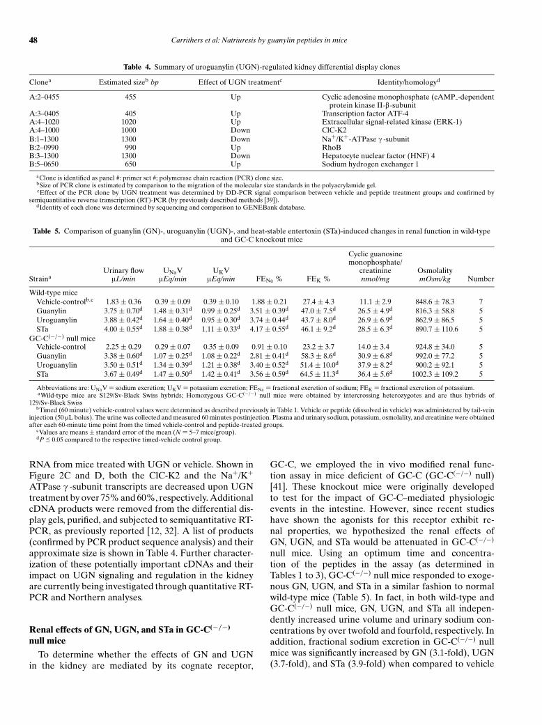
Table 4. Summary of uroguanylin (UGN)-regulated kidney differential display clones
Clonea Estimated sizeb bp Effect of UGN treatmentc Identity/homologyd
A:2–0455 455 Up Cyclic adenosine monophosphate (cAMP -dependentprotein kinase II-b-subunit
A:3–0405 405 Up Transcription factor ATF-4A:4–1020 1020 Up Extracellular signal-related kinase (ERK-1)A:4–1000 1000 Down ClC-K2B:1–1300 1300 Down Na+/K+-ATPase c -subunitB:2–0990 990 Up RhoBB:3–1300 1300 Down Hepatocyte nuclear factor (HNF) 4B:5–0650 650 Up Sodium hydrogen exchanger 1
aClone is identified as panel #: primer set #; polymerase chain reaction (PCR) clone size.bSize of PCR clone is estimated by comparison to the migration of the molecular size standards in the polyacrylamide gel.cEffect of the PCR clone by UGN treatment was determined by DD-PCR signal comparison between vehicle and peptide treatment groups and confirmed by
semiquantitative reverse transcription (RT)-PCR (by previously described methods [39]).dIdentity of each clone was determined by sequencing and comparison to GENEBank database.
Table 5. Comparison of guanylin (GN)-, uroguanylin (UGN)-, and heat-stable entertoxin (STa)-induced changes in renal function in wild-typeand GC-C knockout mice
Cyclic guanosinemonophosphate/
Urinary flow UNaV UKV creatinine OsmolalityStraina lL/min lEq/min lEq/min FENa % FEK % nmol/mg mOsm/kg Number
Wild-type miceVehicle-controlb,c 1.83 ± 0.36 0.39 ± 0.09 0.39 ± 0.10 1.88 ± 0.21 27.4 ± 4.3 11.1 ± 2.9 848.6 ± 78.3 7Guanylin 3.75 ± 0.70d 1.48 ± 0.31d 0.99 ± 0.25d 3.51 ± 0.39d 47.0 ± 7.5d 26.5 ± 4.9d 816.3 ± 58.8 5Uroguanylin 3.88 ± 0.42d 1.64 ± 0.40d 0.95 ± 0.30d 3.74 ± 0.44d 43.7 ± 8.0d 26.9 ± 6.9d 862.9 ± 86.5 5STa 4.00 ± 0.55d 1.88 ± 0.38d 1.11 ± 0.33d 4.17 ± 0.55d 46.1 ± 9.2d 28.5 ± 6.3d 890.7 ± 110.6 5
GC-C(−/−) null miceVehicle-control 2.25 ± 0.29 0.29 ± 0.07 0.35 ± 0.09 0.91 ± 0.10 23.2 ± 3.7 14.0 ± 3.4 924.8 ± 34.0 5Guanylin 3.38 ± 0.60d 1.07 ± 0.25d 1.08 ± 0.22d 2.81 ± 0.41d 58.3 ± 8.6d 30.9 ± 6.8d 992.0 ± 77.2 5Uroguanylin 3.50 ± 0.51d 1.34 ± 0.39d 1.21 ± 0.38d 3.40 ± 0.52d 51.4 ± 10.0d 37.9 ± 8.2d 900.2 ± 92.1 5STa 3.67 ± 0.49d 1.47 ± 0.50d 1.42 ± 0.41d 3.56 ± 0.59d 64.5 ± 11.3d 36.4 ± 5.6d 1002.3 ± 109.2 5
Abbreviations are: UNaV = sodium excretion; UKV = potassium excretion; FENa = fractional excretion of sodium; FEK = fractional excretion of potassium.aWild-type mice are S129/Sv-Black Swiss hybrids; Homozygous GC-C(−/−) null mice were obtained by intercrossing heterozygotes and are thus hybrids of
129/Sv-Black SwissbTimed (60 minute) vehicle-control values were determined as described previously in Table 1. Vehicle or peptide (dissolved in vehicle) was administered by tail-vein
injection (50 lL bolus). The urine was collected and measured 60 minutes postinjection. Plasma and urinary sodium, potassium, osmolality, and creatinine were obtainedafter each 60-minute time point from the timed vehicle-control and peptide-treated groups.
cValues are means ± standard error of the mean (N = 5–7 mice/group).dP ≤ 0.05 compared to the respective timed-vehicle control group.
RNA from mice treated with UGN or vehicle. Shown inFigure 2C and D, both the ClC-K2 and the Na+/K+
ATPase c -subunit transcripts are decreased upon UGNtreatment by over 75% and 60%, respectively. AdditionalcDNA products were removed from the differential dis-play gels, purified, and subjected to semiquantitative RT-PCR, as previously reported [12, 32]. A list of products(confirmed by PCR product sequence analysis) and theirapproximate size is shown in Table 4. Further character-ization of these potentially important cDNAs and theirimpact on UGN signaling and regulation in the kidneyare currently being investigated through quantitative RT-PCR and Northern analyses.
Renal effects of GN, UGN, and STa in GC-C(−/−)
null mice
To determine whether the effects of GN and UGNin the kidney are mediated by its cognate receptor,
GC-C, we employed the in vivo modified renal func-tion assay in mice deficient of GC-C (GC-C(−/−) null)[41]. These knockout mice were originally developedto test for the impact of GC-C–mediated physiologicevents in the intestine. However, since recent studieshave shown the agonists for this receptor exhibit re-nal properties, we hypothesized the renal effects ofGN, UGN, and STa would be attenuated in GC-C(−/−)
null mice. Using an optimum time and concentra-tion of the peptides in the assay (as determined inTables 1 to 3), GC-C(−/−) null mice responded to exoge-nous GN, UGN, and STa in a similar fashion to normalwild-type mice (Table 5). In fact, in both wild-type andGC-C(−/−) null mice, GN, UGN, and STa all indepen-dently increased urine volume and urinary sodium con-centrations by over twofold and fourfold, respectively. Inaddition, fractional sodium excretion in GC-C(−/−) nullmice was significantly increased by GN (3.1-fold), UGN(3.7-fold), and STa (3.9-fold) when compared to vehicle

Carrithers et al: Natriuresis by guanylin peptides in mice 49
28s
18s
+/+ −/− +/+ −/− +/+ −/− +/+ −/− +/+ −/− +/+ −/−Colon Kidney Colon Kidney Colon Kidney
GC-C
β-Actin
GN/UGN-receptork
A B C
Fig. 3. Northern blot analysis of wild-type and GC-C(−/−) null mouse RNA from colon and kidney cortex. (A) Hybridization of RNA fromwild-type (+/+) and GC-C(−/−) null (−/−) mouse colon and kidney cortex with a mouse GC-C cDNA probe (Assn ID #BC034064, IMAGEclone 4208530) reveals the absence of the 3.8 kb GC-C transcript from the GC-C(−/−) null mouse. (B) Hybridization using the renal 400 bppolymerase chain reaction (PCR) product from the GC-C(−/−) null mouse kidney indicates the presence of a 3.8 kb transcript containing a guanylin(GN)/uroguanylin (UGN) binding site region in wild-type mouse colon and kidney cortex (probably GC-C), and a 2.5 kb transcript in wild-typeand GC-C(−/−) null mouse kidney cortex (GN/UGN-receptork). (C) b-actin hybridization to the same blot.
control-treated mice, which were similar to the peptide-induced responses observed in wild-type mice. Further-more, elevations in urinary cGMP were associated withincreased sodium excretion. In wild-type mice, the pep-tides increased the urinary levels of cGMP by approxi-mately 2.5-fold. In GC-C(−/−) null mice, urinary cGMPwas increased over the control-group by 2.2-fold (GN),2.7-fold (UGN), and 2.6-fold (STa). Potassium excretionwas also significantly increased by 2.5- to 2.7-fold in wild-type and GC-C(−/−) null mice by all three peptides. Theosmolalities in the wild-type or knockout mice did not sig-nificantly differ between the vehicle- or peptide-treatedgroups. Thus, in mice, the effect of GN, UGN, and STa aremediated, in part, by a GC-C independent mechanism.
Northern analysis of wild-type and GC-C(−/−)
null mouse RNA
Using colon and renal cortex RNA from wild-type andGC-C(−/−) null mice, Northern analysis was performedusing two different molecular probes directed againstthe GN/UGN receptor: (1) the intracellular catalytic do-main of wild-type mouse GC-C (Assn ID #BC034064),and (2) a 400 bp PCR product homologous to the GC-C ligand-binding homology domain from the GC-C(−/−)
null mouse kidney. The signal at approximately 3.8 kbdemonstrates the presence of GC-C in the colon of wild-type mice (Fig. 3A). Also, as previously shown, this 3.8 kbGC-C transcript is absent in colonic RNA isolated fromGC-C(−/−) null mice [41]. Similarly, the message for GC-C is absent in kidney RNA from GC-C(−/−) null mice.When using the same blot probed with the 400 bp PCRGC-C–like product from GC-C(−/−) null mice, a signalat 3.8 kb in the wild-type colon and kidney is observed(Fig. 3B). However, an alternative, smaller signal of∼2.5 kb is also observed in the wild-type and GC-C(−/−)
null mouse kidney. This smaller transcript is absent inRNA isolated from colonic tissue.
DISCUSSION
Considerable evidence exists suggesting that GN andUGN play an important role in salt and water regulationin the kidney [12, 14, 32–34, 37, 39, 49–51]. This reportdescribes that exogenous GN, UGN, and their bacterialmimic peptide STa, administered intravenously, not onlyincrease sodium and potassium excretion in vivo, but thatthe renal effects are time- and dose-dependent. The mostsignificant increase in sodium and potassium excretionwas observed approximately 40 minutes following intra-venous administration of each peptide. In addition, thetime-dependent increase in fractional sodium excretionby these peptides was associated with a significant in-crease in urinary cGMP and a decrease in GFR, suggest-ing that GN, UGN, and STa elicit their effects in a renaltubular, rather than a hemodynamic, mechanism.
Current data are consistent with the concept that GNand UGN can affect renal function by binding to GC-Creceptors located on several nephron segments [32, 34,37, 52–54]. However, data from this report suggests thatGN and UGN are able to elicit natriuretic and kaliureticeffects in a GC-C–independent mechanism (Table 5).One possible explanation for these results is that alter-native GN/UGN/STa receptors exist in the kidney. Inwild-type mice, the physiologic effects of these peptidesmay be due to a combination of both GC-C and the al-ternative receptor(s). This concept is supported by ourpreliminary molecular studies demonstrating the pres-ence of two distinct mRNA transcripts (3.8 and 2.3 kb)using GC-C–like molecular probes (Fig. 3). In addition,GC-C–independent cellular and physiologic responsesto GN, UGN, and STa have been described in the kid-ney [37, 50]. For example, a UGN-induced mechanismwas found to regulate ion conductances (and mediateelectrogenic electrolyte transport) specifically within iso-lated collecting duct cells from GC-C−/− null mice [50].The precedence of an alternate receptor for natriuretic

50 Carrithers et al: Natriuresis by guanylin peptides in mice
peptides, however, is not unusual. Atrial natriureticpeptide binds to a guanylate cyclase-coupled receptor,guanylate cyclase-A (GC-A), and a truncated clearancereceptor (NPC-R) that is devoid of the intracellular pro-tein kinase-like and guanylate cyclase domains[55]. NPC-R functions to decrease the bioavailability of secretednatriuretic peptides (ANP, BNP, CNP) in neural, renal,and vascular tissue and/or modulate cAMP-driven sec-ond messenger cascades [56]. In fact, agonist binding toNPC-R in endothelial cells inhibits cAMP production viaa Gi protein-coupled signaling system. Thus, an alternatereceptor for GN and/or UGN may exist in the kidney thatalso regulates renal sodium and chloride transport.
Based on the existing literature, at least two potentialconfounds should be taken into consideration. First, ithas yet to be established where functional GC-C or al-ternate GN/UGN receptors are located within specificnephron segments. Our previous data would suggest thatprimary sites of action would include the proximal con-voluted tubule, medullary thick ascending limb, and thecortical collecting tubules as targets for GN, UGN, andSTa[32]. Thus, further detailed segmental analysis em-ploying in vivo micropunture and in vitro microperfusionare necessary to identify specific nephron segments con-taining functional GN/UGN/STa sites of action. Second,it is quite possible that GN, UGN, and STa may act bymodulating the effects of other ligands on renal tubulartransport. For example, studies with ANP showed thatit has no effect on basal proximal tubule fluid reabsorp-tion, but attenuates angiotensin II stimulated transport[57]. In this regard, Santos et al [58] have made the some-what paradoxic observation that UGN can attenuate theANP-induced increase in fractional sodium and potas-sium excretion in the isolated perfused rat kidney.
Under normal physiologic conditions, GN and UGNcirculate in the bloodstream [34, 53, 54, 59–64]. The pri-mary source of these peptides has been proposed to be theintestine [9, 63, 65]. Two separate groups have recentlyfound that these peptides are secreted basolaterally aswell as from the apical/luminal surface of intestinal cells[18, 63]. The expression and/or secretion of intestinal GNand UGN is affected in vivo by acute salt intake, and invitro, by hypertonic solutions [18, 37, 39, 53, 59, 66]. Infact, our laboratory has recently shown that rats on a lowsalt diet have lower circulating levels of GN and UGNthan rats on a normal- or high-salt diet [abstract; Green-berg RN et al, FASEB J 17:A1074, 2003]. In contrast,plasma concentrations of UGN by Fukae et al [49], in ratson a low- and high-salt diet did not significantly differ, al-though the urinary excretion of UGN was significantlyhigher in rats on the high-salt diet compared to those ona low-salt diet. Furthermore, UGN mRNA expression inthe kidney, as well as urinary sodium and cGMP excre-tion, significantly correlated with the increased urinaryUGN levels.
The response to salt intake and the natriuretic activ-ity of these intestinal peptides has led to the hypothe-sis that GN and UGN may participate in an enterorenalcontrol system. In this system, changes in dietary NaClintake lead to the appropriate changes in intestinal andplasma levels of GN and UGN, which in turn, alter renalsodium excretion. The idea of an intestinal natriureticfactor(s) is supported by the fact that the natriuretic re-sponse of the kidney to an increased sodium load oc-curs faster when the load is delivered orally than whenthe same load is delivered intravenously [7–9]. There arewell-established precedents for such feed-forward con-trol systems within the gastrointestinal tract. For exam-ple, glucose-dependent insulinotropic peptide is releasedfrom the intestine in response to ingestion of a meal and,in turn, stimulates the pancreatic release of insulin priorto increases in plasma glucose concentration [67]. The in-tent of this report was to rigorously evaluate the renaleffects of GN and UGN in a modified mouse clearancemodel that lends itself to addressing questions concerningthe physiologic role of these peptides and their postulatedendocrine role in sodium regulation. Our studies suggesta renal role for GN and UGN. Significant responses (i.e.,increased sodium and potassium excretion) induced bythese peptides are observed within 20 minutes and arecontinued until the end point of the assay (120 minutes).It is interesting to note that UGN induces a greater frac-tional sodium excretion than GN, which may be due to theinherent differences in primary and secondary structureof these peptides [9, 14, 24].
We compared the activity of GN, UGN, and STa follow-ing a 2-hour incubation of the peptides in mouse plasma.These stability studies were performed prior to injectingthe peptides into mice. GN, UGN, and STa appear to bestable in plasma, with ∼95% or more of the peptide ac-tivity being retained after incubation (data not shown).The results for STa are similar to that observed in humans[68]. These data suggest that all three peptides should re-tain appropriate biologic activity in the modified mouseclearance studies, and thus, bind to and activate renalGN/UGN/STa receptors.
Associated with the natriuretic response induced byUGN in mice were changes in the mRNA levels of aseries of kidney-specific transport-associated proteins.One of the putative gene products isolated by DD-PCRcoded for the c -subunit of the Na+/K+ ATPase, whichconstitutes one of the major sodium pumps in the kid-ney. Our results suggest that intravenous UGN treat-ment induces a down-regulation of the Na+/K+ ATPasec -subunit mRNA and quite possibly, the protein. Thec -subunit itself is a small, type I membrane protein lo-calized on the basolateral membranes of epithelial cellsand is expressed in the medullary thick ascending limb,distal convoluted tubules, and the inner medullary col-lecting duct [46]. It specifically modulates the activity of

Carrithers et al: Natriuresis by guanylin peptides in mice 51
the Na+/K+ ATPase, which maintains the membrane po-tential, the Na+-gradient, and the transepithelial voltagedifference that energize transcellular, Na+-coupled andparacellular ion transport [69]. The function and regu-lation of the c -subunit is quite complex. However, re-ports have suggested that it is a nephron/tissue-specificregulator of the Na+/K+ ATPase, modulating the affin-ity of sodium, potassium, and ATP [69, 70]. Thus, down-regulation of c -subunit mRNA could, in turn, decreasethe driving force for the reabsorption of sodium in specificnephron segments in a long-term fashion.
Exogenous UGN administration also affected thekidney-specific chloride channel ClC-K2. This partic-ular chloride channel plays an important role in thetransepithelial reabsorption of chloride ions and is mostabundantly expressed along the basolateral surface ofthe medullary thick ascending limb, cortical collectingtubules, and the distal tubules [71, 72]. The physiologicsignificance of ClC-K2 in the kidney has not yet beencompletely elucidated. However, previous reports sug-gest that it serves as a route for transcellular chloridetransport (i.e., an exit for chloride ions in the basolateralplasma membrane of ClC-K2–containing nephron seg-ments)[73]. In fact, loss-of-function mutations of ClC-K2lead to a loss of chloride ions from the body and have beenimplicated in Bartter’s syndrome [71]. Down-regulationof this channel protein (e.g., by UGN) may, in turn, leadto the decrease of chloride reabsorption and increase uri-nary levels of sodium and chloride.
Clearly, it should be noted that gene expression datadoes not necessarily indicate changes in protein expres-sion. Also, UGN may cause the changes in Na+/K+
ATPase c -subunit and ClC-K2, but this observation doesnot establish cause and effect. To define the role ofthese proteins in GN/UGN-induced natriuresis, studiesare planned with the current model using natriuretic pep-tides other than GN, UGN, and STa in wild-type and GC-C(−/−) null mice. If the transcripts that encode Na+/K+
ATPase c -subunit and ClC-K2 do not change upon in-travenous administration of nonguanylin peptides, onewould assume that GN, UGN, and STa play a direct rolein the decrease of the molecular expression of these pep-tides, and hence, potentially influence long-term renalfunction. The same experiment performed in GC-C(−/−)
null mice could also be used to determine the role ofand control for the alternate UGN receptor on Na+/K+
ATPase and ClC-K2. If these genes are not down-regulated in GC-C(−/−) null mice upon UGN treatment,then the argument that Na+/K+ ATPase c -subunit and/orClC-K2 are unrelated to changes in electrolyte excretionis stronger. However, as shown in Tables 1 to 3 and Fig-ure 1, natriuresis occurs in a rapid and transient fashion(less than 90 minutes). Physiologic changes (i.e., natriure-sis) due to modifications in gene and protein expressionhave not been reported within the timeframe observed
for GN and UGN action (40 minutes). Thus, the decreasein molecular expression Na+/K+ ATPase c -subunit orClC-K2 mRNA may not contribute to the rapid-onsetand transient natriuresis induced by GN and UGN. Asecondary natriuretic response in mice may occur hoursafter the initial exposure of the nephron to GN and UGN.Further experiments are currently being performed toexamine the long-term effects of GN and UGN in thewild-type and GC-C(−/−) null mouse kidney.
Sodium excretion by the kidney needs to be pre-cisely regulated. Even small increases in sodium intakein the absence of compensatory changes in the rate oftubular sodium reabsorption would rapidly lead to life-threatening salt retention [74, 75]. Daily fluctuations insodium intake would necessitate, at most, a change ofonly a few percent in tubular reabsorption [1, 2, 74]. GNand UGN may provide the body with such a mechanism,whereby ingested salt regulates the expression and secre-tion of these peptides from intestinal cells into the circula-tion, and in turn, affects tubular sodium reabsorption viaGN/UGN receptors lining the nephron. We describe inthis report that even low levels of these intestinal natri-uretic peptides affect renal sodium excretion in a tran-sient, but time-dependent fashion. Further studies areobviously necessary to clarify and quantify the effect of al-terations in and mechanisms of NaCl intake on GN/UGNsynthesis and secretion, and the resultant changes in re-nal transport. Molecular studies characterizing the novelGN/UGN receptor are also warranted and are currentlyunderway in our laboratory. However, taken together,our studies suggest that GN and UGN may act on themouse kidney causing significant natriuresis by an alter-nate GN/UGN/STa receptor, GC-C–independent mech-anism via renal tubular processes. Long-term renal effectsby UGN may also affect sodium transport by inhibitingthe activity and/or down-regulating the expression of spe-cific sodium and chloride channels, such as Na+/K+ AT-Pase and ClC-K2.
ACKNOWLEDGMENTS
The authors would like to dedicate this report to the memory ofCongmei Sun; we were blessed with the time she spent with us and wewill miss her.
This work was supported by the Office of Research and Devel-opment, Medical Research Service, Department of Veterans Affairs,Lexington, Kentucky (R.N.G., B.A.J., and S.L.C.), Cincinnati, Ohio(R.A.G. and L.E.M.), and Columbia, Missouri (L.R.F.); the AmericanCancer Society (S.L.C.); and the National (B.A.J.) and Ohio Affiliateof the American Heart Association (C.E.O.).
Reprint requests to Stephen L. Carrithers, Ph.D., University of Ken-tucky, Division of Infectious Diseases, VA Medical Center, Research Ser-vice 151-CDD, 1101 VA Drive, VAMC D-309, Lexington, KY 40506.E-mail: [email protected]
REFERENCES
1. WEINBERGER MH: Hypertension: Pathophysiology, Diagnosis, andManagement. New York, Raven, 1990

52 Carrithers et al: Natriuresis by guanylin peptides in mice
2. GARBERS DL, DUBOIS SK: The molecular basis of hypertension.Annu Rev Biochem 68:127–155, 1999
3. CAREY RM, SMITH JR, ORTT EM: Gastrointestinal control of sodiumexcretion in sodium-depleted rabbits. Am J Physiol 230:1504–1508,1976
4. CAREY RM: Evidence for a splanchnic sodium input monitor reg-ulating renal sodium excretion in man: Lack of dependence uponaldosterone. Circulation Res 43:19–23, 1978
5. LUNDGREN O, HANSSON GC: Is there an intestinal natriuretic factor?Acta Physiol Scandinavia 141:19–25, 1991
6. MU JY, HANSSON GC, LUNDGREN O: The intestinal tract and thepathophysiology of arterial hypertension: An experimental studyon Dahl rats. Acta Physiol Scandinavia 155:137–146, 1995
7. MU JY, HANSSON GC, LUNDGREN O: Renal sodium excretion afteroral or intravenous sodium loading in sodium-deprived normoten-sive and spontaneously hypertensive rats. Acta Physiol Scandinavia153:169–177, 1995
8. MU JY, HANSSON GC, LUNDGREN O: The small intestine, salt intakeand arterial hypertension. Blood Pressure 4:77–79, 1995
9. FORTE LR, FAN X, HAMRA FK: Salt and water homeostasis:Uroguanylin is a circulating peptide hormone with natriuretic ac-tivity. Am J Kidney Dis 28:296–304, 1996
10. LENNANE RJ, CAREY RM, GOODWIN TJ, PEART WS: A comparisonof natriuresis after oral and intravenous sodium loading in sodium-depleted man: Evidence for a gastrointestinal or portal monitor ofsodium intake. Clin Sci Mol Med 49:437–440, 1975
11. LENNANE RJ, PEART WS, CAREY RM, SHAW J: A comparison ofnatriuresis after oral and intravenous sodium loading in sodium-depleted rabbits: Evidence for a gastrointestinal or portal monitorof sodium intake. Clin Sci Mol Med 49:433–437, 1975
12. CARRITHERS SL, HILL MJ, JOHNSON BR, et al: Renal effects ofuroguanylin and guanylin in vivo. Braz J Med Biol Res 32:1337–1344, 1999
13. FORTE LR, LONDON RM, FREEMAN RH, KRAUSE WJ: Guanylinpeptides: Renal actions mediated by cyclic GMP. Am J Physiol278:F180–F191, 2000
14. FONTELES MC, GREENBERG RN, MONTEIRO HS, et al: Natriureticand kaliuretic activities of guanylin and uroguanylin in the isolatedperfused rat kidney. Am J Physiol 275:F191–F197, 1998
15. FORTE LR, LONDON RM, KRAUSE WJ, FREEMAN RH: Mechanismsof guanylin action via cyclic GMP in the kidney. Annu Rev Physiol62:673–695, 2000
16. GARBERS DL: Guanylyl cyclase receptors and their ligands. AdvSecond Messenger Phosphoprotein Res 28:91–95, 1993
17. VAANDRAGER AB: Structure and function of the heat-stable en-terotoxin receptor/guanylyl cyclase C. Mol Cell Biochem 230:73–83,2002
18. STEINBRECHER KA, RUDOLPH JA, LUO G, COHEN MB: Coordinateupregulation of guanylin and uroguanylin expression by hypertonic-ity in HT29-18-N2 cells. Am J Physiol 283:C1729–C1737, 2002
19. MARTIN S, ADERMANN K, FORSSMANN WG, KUHN M: Regulated, side-directed secretion of proguanylin from isolated rat colonic mucosa.Endocrinology 140:5022–5029, 1999
20. KUHN M, ADERMANN K, JAHNE J, et al: Segmental differences in theeffects of guanylin and Escherichia coli heat-stable enterotoxin onCl− secretion in human gut. J Physiol 479:433–440, 1994
21. STEINBRECHER KA, MANN EA, GIANNELLA RA, COHEN MB: In-creases in guanylin and uroguanylin in a mouse model of osmoticdiarrhea are guanylate cyclase C-independent. Gastroenterology121:1191–1202, 2001
22. FIELD M, GRAF LH, LAIRD WJ, SMITH PL: Heat-stable enterotoxinof E. coli: In vitro effects of guanylate cyclase activity, cyclic GMPconcentration, and ion transport in small intestine. Proc Natl AcadSci USA 75:2800–2804, 1978
23. CARPICK BW, GARIEPY J: The Escherichia coli heat-stable entero-toxin is a long-lived superagonist of guanylin. Infect Immun 61:4710–4715, 1993
24. GREENBERG RN, HILL M, CRYTZER J, et al: Comparison of effects ofuroguanylin, guanylin, and Escherichia coli heat-stable enterotoxinSTa in mouse intestine and kidney: Evidence that uroguanylin is anintestinal natriuretic hormone. J Inv Med 45:276–283, 1997
25. SANTOS-NETO MS, CARRITHERS SL, MONTEIRO HSA, et al: Guanylinand its lysine-containing analogue in the isolated perfused rat
kidney: Interaction with chymotrypsin inhibitor. Pharm Toxicol92:114–120, 2003
26. FORTE LR, KRAUSE WJ, FREEMAN RH: Guanylin bioactivity in hu-man intestinal and opossum kidney cells. Adv Second MessengerPhosphoprotein Res 28:133–138, 1993
27. FORTE LR, KRAUSE WJ, FREEMAN RH: Escherichia coli enterotoxinreceptors: Localization in opossum kidney, intestine, and testis. AmJ Physiol 257:F874–F881, 1989
28. FORTE LR, KRAUSE WJ, FREEMAN RH: Receptors and cGMP sig-naling mechanisms for E. coli enterotoxin in opossum kidney. AmJ Physiol 255:F1040–F1046, 1988
29. KRAUSE WJ, LONDON RM, FREEMAN RH, FORTE LR: The guanylinand uroguanylin peptide hormones and their receptors. Acta Anat(Basel) 160:213–231, 1997
30. FURUYA S, NARUSE S, ANDO E, et al: Effect and distribution of in-travenously injected 125I-guanylin in rat kidney examined by high-resolution light microscopic radioautography. Anat Embryol (Berl)196:185–193, 1997
31. KRAUSE WJ, FREEMAN RH, FORTE LR: Autoradiographic demon-stration of specific binding sites for E. coli enterotoxin in various ep-ithelia of the North American opossum. Cellular Tissue Res 260:387–394, 1990
32. CARRITHERS SL, TAYLOR B, CAI WY, et al: Guanylyl cyclase-C re-ceptor mRNA distribution along the rat nephron. Regulatory Pep95:65–74, 2000
33. LONDON RM, EBER SL, VISWESWARIAH SS, et al: Structure and ac-tivity of OK-GC: A kidney receptor guanylate cyclase activated byguanylin peptides. Am J Physiol 276:F882–F891, 1999
34. KINOSHITA H, FUJIMOTO S, NAKAZATO M, et al: Urine and plasma lev-els of uroguanylin and its molecular forms in renal diseases. KidneyInt 52:1028–1034, 1997
35. KITA T, KITAMURA K, SAKATA J, ETO T: Marked increase of guanylinsecretion in response to salt loading in the rat small intestine. Am JPhysiol 277:G960–G966, 1999
36. MORO F, LEVENEZ F, NEMOZ-GAILLARD E, et al: Release of guanylinimmunoreactivity from the isolated vascularly perfused rat colon.Endocrinology 141:2594–2599, 2000
37. POTTHAST R, EHLER E, SCHEVING LA, et al: High salt intake increasesuroguanylin expression in mouse kidney. Endocrinology 142:3087–3097, 2001
38. LI Z, KNOWLES JW, GOYEAU D, et al: Low salt intake down-regulatesthe guanylin signaling pathway in rat distal colon. Gastroenterology111:1714–1721, 1996
39. CARRITHERS SL, JACKSON BA, CAI WY, et al: Site-specific effects ofdietary salt intake on guanylin and uroguanylin mRNA expressionin rat intestine. Regulatory Pep 107:87–95, 2002
40. SCHULZ S, GREEN CK, YUEN PS, GARBERS DL: Guanylyl cyclase Cis a heat-stable enterotoxin receptor. Cell 63:941–948, 1990
41. MANN EA, JUMP ML, WU J, et al: Mice lacking the guanylyl cyclase Creceptor are resistant to STa-induced intestinal secretion. BiochemBiophys Res Commun 239:463–466, 1997
42. GIANNELLA RA: Suckling mouse model for detection of heat-stableEscherichia coli enterotoxin: characteristics of the model. Inf Im-mun 12:95–99, 1976
43. CARRITHERS SL, EBER SL, FORTE LR, GREENBERG RN: Increasedurinary excretion of uroguanylin in patients with congestive heartfailure. Am J Physiol 278:H538–H547, 2000
44. HAMRA FK, EBER SL, CHIN DT, et al: Regulation of intestinaluroguanylin and guanylin receptor-mediated responses by mucosalacidity. Proc Natl Acad Sci USA 94:2705–2710, 1997
45. CURRIE MG, FOK KF, KATO J, et al: Guanylin: an endogenous activa-tor of intestinal guanylate cyclase. Proc Natl Acad Sci USA 89:947–951, 1992
46. MERCER RW, BEIMESDERFER D, BLISS DP, Jr, et al: Molecular cloningand immunological characterization of the c -polypeptide, a smallprotein associated with the Na+, K+-ATPase. J Cell Biol 121:579–586, 1993
47. TAKEUCHI Y, UCHIDA S, MARUMO F, SASAKI S: Cloning, tissue dis-tribution, and intrarenal localization of ClC chloride channels inhuman kidney. Kidney Int 48:1497–1503, 1995
48. FONTELES MC, MONTEIRO HS, SOARES AM, et al: The lysine-1 ana-log of guanylin induces intestinal secretion and natriuresis in theisolated perfused kidney. Braz J Med Biol Res 29:267–271, 1996

Carrithers et al: Natriuresis by guanylin peptides in mice 53
49. FUKAE H, KINOSHITA H, FUJIMOTO S, et al: Changes in urinary levelsand renal expression of uroguanylin on low or high salt diets in rats.Nephron 92:373–378, 2002
50. SINDICE A, BASOGLU C, CERCI A, et al: Guanylin, uroguanylin, andheat-stable enterotoxin activate guanylate cyclase C and/or a per-tussis toxin-sensitive G protein in human proximal tubule cells. JBiol Chem 277:17758–17764, 2002
51. FAN X, WANG Y, LONDON RM, et al: Signaling pathways for guanylinand uroguanylin in the digestive, renal, central nervous, reproduc-tive, and lymphoid systems. Endocrinology 138:4636–4648, 1997
52. KINOSHITA H, FUJIMOTO S, YOKOTA N, et al: Uroguanylin acts as a na-triuretic factor via guanylate cyclase C. J Am Soc Nephrol 8:A1863–A1863, 1997
53. KINOSHITA H, FUJIMOTO S, FUKAE H, et al: Plasma and urine levelsof uroguanylin, a new natriuretic peptide, in nephrotic syndrome.Nephron 81:160–164, 1999
54. NAKAZATO M, YAMAGUCHI H, KINOSHITA H, et al: Identification ofbiologically active and inactive human uroguanylins in plasma andurine and their increases in renal insufficiency. Biochem BiophysRes Commun 220:586–593, 1996
55. HEMPLE A, NOLL T, BACH C, et al: Atrial natriuretic peptide clear-ance receptor participates in modulating endothelial permeability.Am J Physiol 275:H1818–H1825, 1998
56. HERMAN JP, DOLGAS CM, MARCINEK R, LANGUB MCJ: Expressionand glucocorticoid regulation of natriuretic peptide clearance re-ceptor (NPR-C) mRNA in rat brain and choride plexus. J ChemNeuroanat 11:257–265, 1996
57. EITLE E, HIRANYACHATTADA S, WANG H, HARRIS PJ: Inhibition ofproximal tubular fluid absorption by nitric oxide and atrial natri-uretic peptide in rat kidney. Am J Physiol 274:C1075–C1080, 1998
58. SANTOS NETO MS, CARVALHO AF, FORTE LR, FONTELES MC: Rela-tionship between the actions of atrial natriuretic peptide (ANP),guanylin and uroguanylin on the isolated kidney. Braz J Med BiolRes 32:1015–1019, 1999
59. KINOSHITA H, NAKAZATO M, YAMAGUCHI H, et al: Increased plasmaguanylin levels in patients with impaired renal function. ClinNephrol 47:28–32, 1997
60. FUKAE H, KINOSHITA H, FUJIMOTO S, et al: Plasma concentration ofuroguanylin in patients on maintenance dialysis therapy. Nephron84:206–210, 2000
61. KUHN M, RAIDA M, ADERMANN K, et al: The circulating bioactiveform of human guanylin is a high molecular weight peptide (10.3kDa). FEBS Lett 318:205–209, 1993
62. NAKAZATO M, YAMAGUCHI H, SHIOMI K, et al: Identification of 10-kDa proguanylin as a major guanylin molecule in human intestineand plasma and its increase in renal insufficiency. Biochem BiophysRes Commun 205:1966–1975, 1994
63. KUHN M, KULAKSIZ H, CETIN Y, et al: Circulating and tissue guanylinimmunoreactivity in intestinal secretory diarrhoea. Eur J Clin Invest25:899–905, 1995
64. DATE Y, NAKAZATO M, YAMAGUCHI H, et al: Tissue distribution andplasma concentration of human guanylin. Intern Med 35:171–175,1996
65. LI ZP, PERKINS AG, PETERS MF, et al: Purification, cDNA sequence,and tissue distribution of rat uroguanylin. Regulatory Pep 68:45–56,1997
66. STEINBRECHER KA, MANN EA, GIANNELLA RA, COHEN MB: A hy-pertonic diet increases mouse guanylin and uroguanylin levels viaa guanylate cyclase C-independent mechanism. Gastroenterology120:3671, 2001
67. BROWN JC, OTTE SC: GIP and the entero-insular axis. Clin En-docrinol Metab 8:365–377, 1981
68. GALI H, SIECKMAN GL, HOFFMAN TJ, et al: Chemical synthesis of Es-cherichia coli ST(h) analogues by regioselective disulfide bond for-mation: biological evaluation of an (111)In-DOTA-Phe(19)-ST(h)analogue for specific targeting of human colon cancers. BioconjugChem 13:224–231, 2002
69. MEIJ IC, KOENDERINK JB, VAN BOKHOVEN H, et al: Dominant iso-lated renal magnesium loss is caused by misrouting of the Na+,K+-ATPase c -subunit. Nature Genetics 26:265–266, 2000
70. ARYSTARKHOVA E, DONNET C, ASINOVSKI NK, SWEADNER KJ: Dif-ferential regulation of renal Na+, K+-ATPase by splice variants ofthe c-subunit. J Biol Chem 277:10162–10172, 2002
71. UCHIDA S: In vivo role of CLC chloride channels in the kidney. AmJ Physiol 279:F802–F808, 2000
72. YOSHIKAWA M, UCHIDA S, YAMAUCHI A, et al: Localization of rat ClC-K2 chloride channel mRNA in the kidney. Am J Physiol 45:F552–F558, 1999
73. WOLF K, CASTROP H, RIEGGER GAJ, et al: Differential gene reg-ulation of renal salt entry pathways by salt load in the distalnephron of the rat. Pflugers Archives-Eur J Physiol 442:498–504,2001
74. JACKSON BA, OTT CE: Renal System, Madison, CT, Fence CreekPublishing LLC, 1999
75. DIBONA GF, KOPP UC: Neural control of renal function. PhysiolRev 77:75–197, 1997