Impaired Autonomic Regulation of Resistance Arteries in Mice With Low Vascular Endothelial Growth...
37
Schiffers, Hermann Rohrer, Annemie Van der Linden, Jo G.R. De Mey and Peter Carmeliet Lambrechts, Ger M.J. Janssen, Gregorio E. Fazzi, Patrik Verstreken, Jody Haigh, Paul M. Mazzone, Greet Vanhoutte, Stefan Vinckier, Katarzyna Miskiewicz, Koen Poesen, Diether Erik Storkebaum, Carmen Ruiz de Almodovar, Merlijn Meens, Serena Zacchigna, Massimiliano Endothelial Growth Factor or Upon Vascular Endothelial Growth Factor Trap Delivery Impaired Autonomic Regulation of Resistance Arteries in Mice With Low Vascular Print ISSN: 0009-7322. Online ISSN: 1524-4539 Copyright © 2010 American Heart Association, Inc. All rights reserved. is published by the American Heart Association, 7272 Greenville Avenue, Dallas, TX 75231 Circulation doi: 10.1161/CIRCULATIONAHA.109.929364 2010;122:273-281; originally published online July 6, 2010; Circulation. http://circ.ahajournals.org/content/122/3/273 World Wide Web at: The online version of this article, along with updated information and services, is located on the http://circ.ahajournals.org/content/suppl/2010/07/02/CIRCULATIONAHA.109.929364.DC1.html Data Supplement (unedited) at: http://circ.ahajournals.org//subscriptions/ is online at: Circulation Information about subscribing to Subscriptions: http://www.lww.com/reprints Information about reprints can be found online at: Reprints: document. Permissions and Rights Question and Answer this process is available in the click Request Permissions in the middle column of the Web page under Services. Further information about Office. Once the online version of the published article for which permission is being requested is located, can be obtained via RightsLink, a service of the Copyright Clearance Center, not the Editorial Circulation in Requests for permissions to reproduce figures, tables, or portions of articles originally published Permissions: at UNIVERSITAIR ZIEKENHUIS BIOMED on March 7, 2013 http://circ.ahajournals.org/ Downloaded from
-
Upload
independent -
Category
Documents
-
view
0 -
download
0
Transcript of Impaired Autonomic Regulation of Resistance Arteries in Mice With Low Vascular Endothelial Growth...

Schiffers, Hermann Rohrer, Annemie Van der Linden, Jo G.R. De Mey and Peter CarmelietLambrechts, Ger M.J. Janssen, Gregorio E. Fazzi, Patrik Verstreken, Jody Haigh, Paul M.Mazzone, Greet Vanhoutte, Stefan Vinckier, Katarzyna Miskiewicz, Koen Poesen, Diether
Erik Storkebaum, Carmen Ruiz de Almodovar, Merlijn Meens, Serena Zacchigna, MassimilianoEndothelial Growth Factor or Upon Vascular Endothelial Growth Factor Trap Delivery
Impaired Autonomic Regulation of Resistance Arteries in Mice With Low Vascular
Print ISSN: 0009-7322. Online ISSN: 1524-4539 Copyright © 2010 American Heart Association, Inc. All rights reserved.
is published by the American Heart Association, 7272 Greenville Avenue, Dallas, TX 75231Circulation doi: 10.1161/CIRCULATIONAHA.109.9293642010;122:273-281; originally published online July 6, 2010;Circulation.
http://circ.ahajournals.org/content/122/3/273World Wide Web at:
The online version of this article, along with updated information and services, is located on the
http://circ.ahajournals.org/content/suppl/2010/07/02/CIRCULATIONAHA.109.929364.DC1.htmlData Supplement (unedited) at:
http://circ.ahajournals.org//subscriptions/
is online at: Circulation Information about subscribing to Subscriptions:
http://www.lww.com/reprints Information about reprints can be found online at: Reprints:
document. Permissions and Rights Question and Answer this process is available in the
click Request Permissions in the middle column of the Web page under Services. Further information aboutOffice. Once the online version of the published article for which permission is being requested is located,
can be obtained via RightsLink, a service of the Copyright Clearance Center, not the EditorialCirculationin Requests for permissions to reproduce figures, tables, or portions of articles originally publishedPermissions:
at UNIVERSITAIR ZIEKENHUIS BIOMED on March 7, 2013http://circ.ahajournals.org/Downloaded from

Molecular Cardiology
Impaired Autonomic Regulation of Resistance Arteries inMice With Low Vascular Endothelial Growth Factor or
Upon Vascular Endothelial Growth Factor Trap DeliveryErik Storkebaum, PhD*; Carmen Ruiz de Almodovar, PhD*; Merlijn Meens, MS;
Serena Zacchigna, PhD; Massimiliano Mazzone, PhD; Greet Vanhoutte, PhD;Stefan Vinckier, PhD; Katarzyna Miskiewicz, PhD; Koen Poesen, PhD; Diether Lambrechts, PhD;
Ger M.J. Janssen, BSc; Gregorio E. Fazzi, BSc; Patrik Verstreken, PhD; Jody Haigh, PhD;Paul M. Schiffers, PhD; Hermann Rohrer, PhD; Annemie Van der Linden, PhD;
Jo G.R. De Mey, PhD; Peter Carmeliet, MD, PhD
Background—Control of peripheral resistance arteries by autonomic nerves is essential for the regulation of blood flow. Thesignals responsible for the maintenance of vascular neuroeffector mechanisms in the adult, however, remain largely unknown.
Methods and Results—Here, we report that VEGF�/� mice with low vascular endothelial growth factor (VEGF) levelssuffer defects in the regulation of resistance arteries. These defects are due to dysfunction and structural remodeling ofthe neuroeffector junction, the equivalent of a synapse between autonomic nerve endings and vascular smooth musclecells, and to an impaired contractile smooth muscle cell phenotype. Notably, short-term delivery of a VEGF inhibitorto healthy mice also resulted in functional and structural defects of neuroeffector junctions.
Conclusions—These findings uncover a novel role for VEGF in the maintenance of arterial neuroeffector function and may helpus better understand how VEGF inhibitors cause vascular regulation defects in cancer patients. (Circulation. 2010;122:273-281.)
Key Words: arteries � muscle, smooth � nervous system � vascular endothelial growth factor � vasoconstriction
Several cues have been identified that control the devel-opment of the sympathetic nervous system,1 but the
molecular mechanisms underlying the maintenance of auto-nomic innervation of peripheral resistance arteries in adult-hood remain largely unknown. Periarterial nerves do notpenetrate the medial vascular smooth muscle cell (SMC)layer but are confined to the media adventitia border. Thevaricose terminal portions of these nerve fibers releasetransmitter en passage,2 and autonomic neuroeffector junc-tions (NEJs) in resistance arteries are “atypical” in that theycontain prejunctional membrane thickenings with synapticvesicles but lack specialized postjunctional structures.2
Clinical Perspective on p 281
By releasing neurotransmitters, autonomic nerves control theblood supply to organs. For instance, autonomic control ofresistance arteries mediates thermoregulation in the skin andredistribution of flow from internal organs to the brain in stress
conditions.3,4 Dysfunction of the periarterial autonomic nervouscontrol can lead to hypertension, Raynaud phenomenon, andorthostatic hypotension.5 Impaired vascular regulation has alsobeen implicated in the hand-foot syndrome in cancer patientsreceiving vascular endothelial growth factor (VEGF) inhibitors.6
This syndrome is characterized by dysesthesia, erythema, andtingling of extremities, which may progress to burning pain withdryness, cracking, and ulceration of the skin.7,8 The molecularbasis of this syndrome remains largely enigmatic.
VEGF, a key regulator of angiogenesis in health and disease,9
also has neurotrophic effects. VEGF stimulates axon outgrowthand survival of neurons in vitro,10 and low VEGF levels inVEGF�/� mice, engineered to lack the hypoxia-response elementin the VEGF promoter, cause adult-onset motoneuron degener-ation, reminiscent of the human motoneuron degenerative dis-order amyotrophic lateral sclerosis.11 However, surprisinglylittle is known about the effects of VEGF on peripheral auto-nomic nerves and, in particular, about its possible role in the
Received December 4, 2009; accepted May 24, 2010.From the Vesalius Research Center, K.U. Leuven and VIB, Leuven, Belgium (E.S., C.R.d.A., S.Z., M. Massone, S.V., K.P., D.L., P.C.); Department
of Pharmacology and Toxicology, CARIM, Maastricht University, Maastricht, the Netherlands (M. Meens, G.M.J.J., G.E.F., P.M.S., J.G.R.D.M.);Bio-Imaging lab, UA, Antwerp, Belgium (G.V., A.V.d.L.); Center for Human Genetics, K.U. Leuven and Department of Molecular and DevelopmentalGenetics, VIB, Leuven, Belgium (K.M., P.V.); Vascular Cell Biology Unit, VIB and Department for Molecular Biomedical Research, Ghent University,Gent, Belgium (J.H.); and Max Planck Institute for Brain Research, Frankfurt, Germany (H.R.).
*Drs Storkebaum and Ruiz de Almodovar contributed equally to this work.The online-only Data Supplement is available with this article at http://circ.ahajournals.org/cgi/content/full/CIRCULATIONAHA.109.929364/DC1.Correspondence to P. Carmeliet, MD, PhD, Vesalius Research Center (VRC), VIB, K.U. Leuven, Campus Gasthuisberg, Herestraat 49, B-3000,
Leuven, Belgium. E-mail [email protected]© 2010 American Heart Association, Inc.
Circulation is available at http://circ.ahajournals.org DOI: 10.1161/CIRCULATIONAHA.109.929364
273 at UNIVERSITAIR ZIEKENHUIS BIOMED on March 7, 2013http://circ.ahajournals.org/Downloaded from
autonomic innervation of resistance arteries in vivo. In vitrostudies show that VEGF stimulates axon outgrowth and survivalof superior cervical ganglia12,13 and induces growth cone spread-ing of cultured postganglionic neurons.14 Furthermore, VEGFpromotes the reinnervation of denervated femoral arteries invivo.14 However, a role for VEGF in the maintenance of theperivascular autonomic nerve plexus in vivo has not beenaddressed. Here, we assessed whether VEGF might modulatethe autonomic nervous control of resistance arteries.
MethodsTransgenic Mice and Soluble Flk1 Gene TransferVEGF�/�, VEGF-LacZ, and Flt1-LacZ mice were described previ-ously.11,15,16 Expression of the soluble extracellular domain of Flk1(sFlk1) in the circulation was obtained by hydroporating mice with 2.5mL Ringer solution (150 mmol/L NaCl, 4 mmol/L KCl, 1 mmol/LCaCl2) containing 50 �g per mouse of a vector encoding sFlk1 or anempty vector as control.17 After 5 days, plasma protein levels of sFlk1were determined with a soluble VEGF receptor-2 (VEGFR-2)–specificimmunoassay (R&D Systems, Minneapolis, Minn).
Histology and ImmunohistochemistryPerfusion-fixed arteries were embedded in paraffin, and sectionswere stained with the following antibodies: polyclonal rabbit anti-smoothelin antibody (1:100; a gift from G. van Eys, MaastrichtUniversity), anti-desmin antibody (1:50; Cappel, Durham, NC), andanti–SM myosin heavy chain (MHC) antibody (1:400; ab683,Abcam, Cambridge, UK). Procedures used for whole-mount stainingof arteries, analysis of skin vascular density, LacZ enzymaticstaining, alkaline phosphatase staining, glyoxylic acid staining, andnoradrenaline content determination are described in the Methodssection of the online-only Data Supplement.
Isometric Wire MyographyIsometric wire myography was performed as described previously.18
Detailed procedures can be found in the Methods section of theonline-only Data Supplement.
Quantitative Real-Time Polymerase ChainReaction ExperimentsMessenger RNA transcript levels were quantified and normalizedrelative to the expression level of �-actin or GAPDH with the use ofpremade primers and probes (TaqMan Gene Expression Assays, Ap-plied Biosystems, Foster City, Calif). Relative gene expression in VEGF�/�
versus control arteries was calculated with the 2���CT method.
Transmission Electron MicroscopyStandard procedures were used for transmission electron micros-copy. Detailed procedures can be found in the Methods section of theonline-only Data Supplement.
Thermoregulation ExperimentsA rectal probe connected with a digital thermometer (PhysitempInstruments) was used to measure body temperature in an ambienttemperature of 21°C on 3 consecutive days with 3 measurements perday; the average temperature was recorded as baseline. Duringexposure to cold stress (8°C for VEGF�/� mice, 4°C for sFlk1 mice),rectal temperature was measured at half-hour intervals for 4.5 hours.
Blood Flow MeasurementsDetails on these and other methods used in this study can be found inMethods section of the online-only Data Supplement.
StatisticsFor comparison of 2 genotypes or treatment groups, the unpairedStudent t test was used. When data were not normally distributed, the
Mann–Whitney test was used; medians and interquartile ranges arereported. For the evaluation of body temperature evolution duringcold stress experiments, repeated-measures ANOVA was used. Forstatistical analysis of concentration-response curves, ANOVA wasused with Bonferroni correction for multiple testing. A limitation ofour study is that, because of the limited availability of transgenicmice, sample size in our experiments is often small, increasing therisk for a type I error.
ResultsExpression of VEGF and VEGFRsFor VEGF to regulate the function of resistance arteries, VEGFand VEGFRs should be expressed in these arteries. We thereforedetermined the spatial expression pattern of VEGF and VEGFRs(VEGFR-1/Flt1 and VEGFR-2/Flk1) in mesenteric and saphe-nous arteries. In VEGF-LacZ mice, which express nuclear LacZunder the control of the endogenous VEGF promotor,15 X-galstaining revealed VEGF expression by medial SMCs (Figure1A) and by neurons in celiac and superior cervical ganglia(Figure 1B and not shown), implying that VEGF may also bereleased from nerve terminals. X-gal staining of arterial crosssections of Flt1-LacZ mice, which express cytosolic LacZ underthe control of the endogenous Flt1 promotor,16 revealed Flt1expression in medial SMCs (Figure 1C). This expression patternof Flt1 was confirmed by a VEGF-B186-alkaline phosphatase(AP) fusion protein, which selectively binds to Flt1 (Figure 1D).Staining of whole-mount arteries with a VEGF-AP fusionprotein, which binds to Flk1, Flt1, and neuropilin, revealedexpression of VEGFRs also on periarterial nerves (Figure 1E),and immunostaining for Flt1 or Flk1 labeled perivascular nervefibers and endings (Figure 1F and 1G). Thus, VEGF andVEGFRs are expressed in resistance arteries and periarterialnerves. We therefore tested whether VEGF might play a role invascular regulation of peripheral resistance arteries.
To explore whether VEGF affected vascular regulation, weused VEGF�/� mice, which express reduced VEGF levels.11
ELISA confirmed that VEGF protein levels were reduced inVEGF�/� arteries and ganglia (note I in the online-only DataSupplement), and immunostaining and quantitative polymer-ase chain reaction revealed that VEGF expression was re-duced in arterial SM and postganglionic neurons (Figure Iand note I of the online-only Data Supplement). BecauseVEGF�/� mice develop motoneuron degeneration beyond 5 to7 months of age,11 we used only presymptomatic VEGF�/�
mice (�3 months of age) for all experiments.
Impaired Vasoconstrictor Responses of VEGF�/�
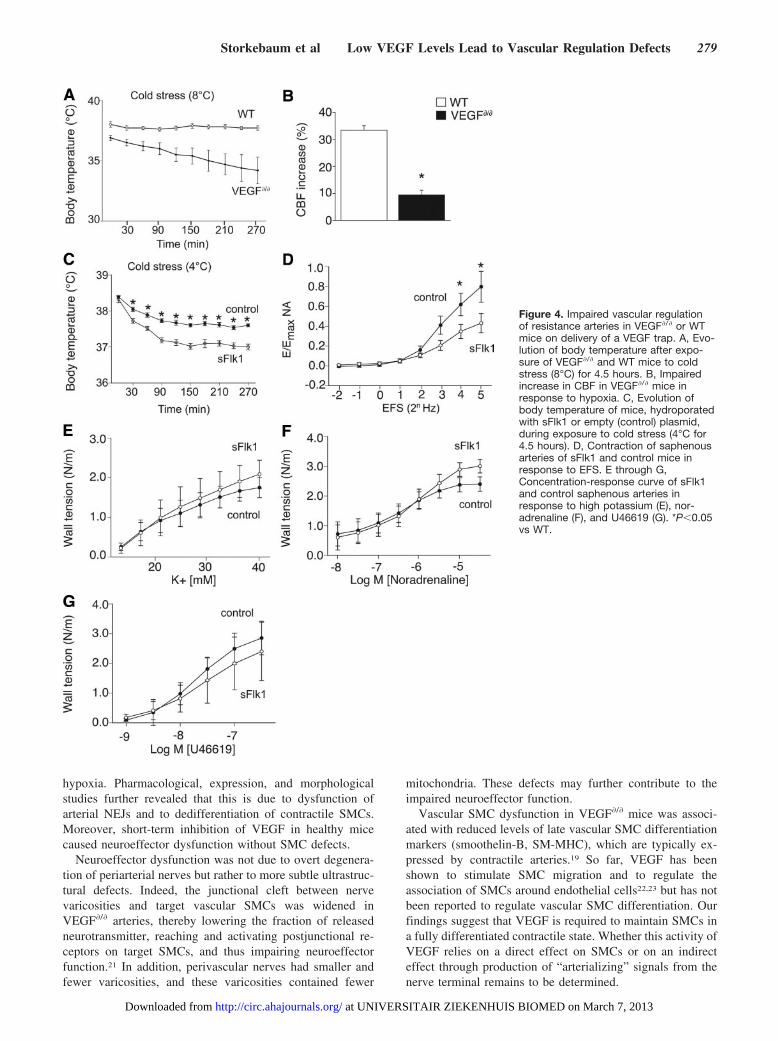
Resistance Arteries Ex VivoWe then explored by isometric wire myography ex vivo whetherthe reduced VEGF levels in VEGF�/� mice impaired vascularregulation of isolated small muscular arteries (mesenteric andsaphenous arteries) and large elastic arteries (common carotidartery).18 To study whether VEGF modulates vasoregulatoryresponses of SMCs, we induced vasoconstriction by exposingarteries to stimuli that have direct effects on SMCs throughdistinct signal transduction pathways, ie, high potassium (K�),angiotensin II, noradrenaline, or the thromboxane A2 analogU46619. Because the contractile response of an artery dependson the intrinsic SMC properties but also on the thickness of theSMC layer (and because VEGF�/� arteries were generally
274 Circulation July 20, 2010
at UNIVERSITAIR ZIEKENHUIS BIOMED on March 7, 2013http://circ.ahajournals.org/Downloaded from

smaller; Table I of the online-only Data Supplement), we notonly measured active wall tension (N/m), defined as the forcegenerated by the artery divided by the arterial segment length,but also calculated active wall stress (N/m2), a parameter ofintrinsic SMC function, by correcting active wall tension formedia thickness.
Compared with wild-type (WT) mice, contractile responsesof resistance arteries to high K�, angiotensin II, noradrena-line, or U46619 were reduced in VEGF�/� mice, whereas thesensitivity to these stimuli was not modified (Figure 2A, 2C,and 2D, plus Table II and Figure I of the online-only DataSupplement). Both wall tension and wall stress were reducedby �50% in VEGF�/� mice (Table II of the online-only DataSupplement). The fact that arterial contractility was impairedregardless of the type of vasoconstrictor stimulus indicatesthat SMCs were generally dysfunctional rather than having aselective defect in a particular signaling pathway. In addition,because the contractile responses were also impaired in thepresence of the nitric oxide synthase inhibitor nitro-L-arginine,the defect was not attributable to abnormal production of nitricoxide (not shown). Notably, the contractile response of theelastic carotid arteries was normal (Table II and Figure IA and
ID of the online-only Data Supplement and Figure 2B), indicat-ing that vascular regulation by densely innervated musculararteries, but not by sparsely innervated elastic arteries, isimpaired in VEGF�/� mice. Vascular SMC dysfunction wasnot accompanied by endothelial cell dysfunction becauseendothelium-dependent relaxation induced by acetylcho-line was normal in resistance arteries of VEGF�/� mice(Figure II of the online-only Data Supplement).
Dedifferentiation of Vascular SMCs in VEGF�/�
Resistance ArteriesTo characterize SMC dysfunction in VEGF�/� arteries inmore detail, we measured by quantitative polymerase chainreaction the expression of “early” and “late” SMC differen-tiation markers, which are important for establishing SMCcontractile properties in a developmentally sequential order.19
At 3 weeks of age, ie, when the autonomic innervation ofresistance arteries was just established,20 expression of early(nonmuscle MHC, desmin, and calponin) and late (SM MHCand smoothelin-B) SMC differentiation markers was compa-rable in WT and VEGF�/� saphenous arteries (Table 1). Incontrast, by 3 months of age, expression of early SMC
Figure 1. Expression pattern of VEGFand VEGFRs in peripheral resistancearteries. A and B, X-gal staining onsaphenous arteries (A) and celiac ganglia(B) of VEGF-LacZ mice revealed VEGFexpression in medial SMCs (A) (arrows,nuclear LacZ staining) and postgangli-onic neurons (B). C, X-gal staining onsaphenous arteries of Flt1-LacZ micerevealed Flt1 expression in medialSMCs. D, Expression of Flt1 in medialSMCs was also confirmed with VEGF-B186-AP fusion protein. E, Staining ofwhole-mount arteries with VEGF164-APfusion protein revealed expression ofVEGFRs on periarterial nerves (arrows).F and G, Immunostaining for Flk1 (F) andFlt1 (G) on whole-mount arteriesrevealed expression of Flk1 and Flt1 inperiarterial nerves (arrows). Scalebar�50 �m.
Storkebaum et al Low VEGF Levels Lead to Vascular Regulation Defects 275
at UNIVERSITAIR ZIEKENHUIS BIOMED on March 7, 2013http://circ.ahajournals.org/Downloaded from

markers was normal, but expression of the late SMC markerswas reduced in VEGF�/� saphenous arteries (Table 1).
These results were confirmed by immunostaining (FigureIII of the online-only Data Supplement). No differences werefound in the expression of SMC differentiation markers incommon carotid arteries (Table 1). The reduced expression of
late SMC differentiation markers in VEGF�/� arteries canexplain their generally impaired contractile properties. Thisdefect appeared to be specific for the contractile machinerybecause expression of postjunctional neurotransmitter recep-tors (�1-adrenoreceptors, purinergic receptor P2X1, andNPY1 receptor) and neurotrophic factors (nerve growth fac-tor, artemin, and neurotrophin-3) was not significantly alteredin VEGF�/� arteries (not shown). Thus, SMCs in innervatedresistance arteries differentiate normally during postnataldevelopment but later dedifferentiate in VEGF�/� mice.
Neuroeffector Dysfunction in VEGF�/� ArteriesBecause vascular regulation was impaired in densely innervated,but not in sparsely innervated, arteries in VEGF�/� mice, sug-gesting a possible dysfunction of perivascular nerves, we studiedneuroeffector function by ex vivo myography. In mesentericarteries of WT mice, electric field stimulation (EFS) or admin-istration of the indirectly acting sympathomimetic tyramineinduced a strong vasoconstrictor response (Figure 3A and 3B).In contrast, the contractile response was severely impaired inVEGF�/� arteries (Figure 3A and 3B). To exclude that theimpaired response to these neurogenic stimuli in VEGF�/� micereflected only intrinsic SMC (and not neuronal) defects, weexpressed the responses as a fraction of the maximal response toexogenously applied noradrenaline. Even after normalization,the contractile response remained lower in VEGF�/� mice(Figure 3C and 3D and Table 2), indicating that sympatheticneuroeffector regulation was dysfunctional.
Because high K� induces vasoconstriction via effects onSMCs and nerve terminals, the neuronal contribution to thisresponse can be evaluated by measuring the relaxationinduced by blocking postjunctional �-adrenoreceptors byphentolamine. The phentolamine-induced relaxation of thecontractile response to K� was smaller in VEGF�/� arteries(Table 2), suggesting that less noradrenaline reached andactivated postjunctional �-adrenoreceptors. Overall, besidesgeneral SMC hyporesponsiveness, control of vasomotor toneby sympathetic nerves was defective in VEGF�/� arteries.Vasorelaxation by afferent sensory-motor nerves18 was alsoimpaired in VEGF�/� arteries (Figure 3E), indicating thatdysfunction of neuroeffector mechanisms involves both sym-pathetic and sensory-motor nerves.
Cellular Mechanisms of Neuroeffector Dysfunctionin VEGF�/� MiceWe then sought to explore the underlying mechanisms of theperivascular nerve dysfunction. Whole-mount staining for ty-rosine hydroxylase, noradrenaline, or calcitonin-gene relatedpeptide revealed that the neuroeffector dysfunction was notattributable to a reduced density of the periarterial nervousplexus (Figure IV of the online-only Data Supplement). Addi-tionally, the noradrenaline content of VEGF�/� resistance arterieswas not different from that of controls (Table III of theonline-only Data Supplement). We further analyzed by myogra-phy the effect of cocaine on the arterial response to exogenousnoradrenaline to assess how efficiently noradrenaline reuptakeby nerve terminals in situ occurred. Indeed, if noradrenalineuptake is important, blockade of this process by cocaine shouldleave more neurotransmitter available to activate postjunctional
Figure 2. Impaired vascular regulation of peripheral resistancearteries in VEGF�/� mice. A and B, Typical recording of contractileresponses to increasing concentrations of the vasoconstrictorU46619 in mesenteric resistance arteries (A) and carotid arteries (B)of WT and VEGF�/� mice. C and D, Concentration-response curveof WT and VEGF�/� saphenous arteries in response to noradrena-line (C) and U46619 (D). *P�0.05 vs WT.
Table 1. Relative Expression Levels of SMCDifferentiation Markers
Age Artery Target Gene WT VEGF�/�
3 wk SA Nonmuscle MHC 100�14.1 78.7�19.8
Desmin 100�14.7 71.4�13.1
Calponin 100�13.5 63.8�18.3
SM-MHC 100�14.1 78.6�19.8
Smoothelin 100�19.3 118.4�35.7
3 wk cCA Nonmuscle MHC 100�35.4 67.4�26.9
Desmin 100�47.3 156.5�55.2
Calponin 100�44.1 108.7�59.8
SM-MHC 100�35.6 67.3�26.7
Smoothelin 100�37.0 63.0�22.3
3 mo SA Nonmuscle MHC 100�16.3 109.1�19.7
Desmin 100�17.9 84.6�11.7
Calponin 100�8.8 75.8�12.4
SM-MHC 100�18.5 51.0�9.4*
Smoothelin 100�14.2 49.2�9.8*
3 mo cCA Desmin 100�8.8 143.5�26.4
Calponin 100�10.1 86.5�23.2
SM-MHC 100�14.6 117.6�27.2
Smoothelin 100�45.2 96.0�29.5
SA indicates saphenous artery; cCA, common carotid artery. Relativeexpression levels as percentage of WT�SEM are shown.
*P�0.05 versus WT.
276 Circulation July 20, 2010
at UNIVERSITAIR ZIEKENHUIS BIOMED on March 7, 2013http://circ.ahajournals.org/Downloaded from

receptors at the NEJ. Notably, cocaine enhanced the sensitivityof VEGF�/� arteries to noradrenaline, but much less than that ofWT arteries (Table 2). Hyporesponsiveness to cocaine in thisexperimental condition might reflect impaired noradrenalineuptake per se but also could be due to a wider junctional cleftbetween nerve varicosities and SMCs because local neurotrans-mitter concentrations can be influenced more readily by uptakeprocesses in narrow junctional clefts. To discriminate betweenthese possibilities, we measured the neuronal uptake of nor-adrenaline directly. Notably, no differences in noradrenaline
uptake between VEGF�/� and WT arteries were found (note II inthe online-only Data Supplement).
We therefore explored whether the junctional cleft betweenperivascular nerve varicosities and target vascular SMCs mightbe wider. To identify structural abnormalities of VEGF�/� NEJs,transmission electron microscopy was used (Table 3 and Figure3F and 3G). Morphometric quantification revealed that thedistance between varicosities and the nearest SMC was larger inVEGF�/� arteries (Table 3). Furthermore, varicosities weresmaller and contained fewer mitochondria, and axon bundlescontained fewer varicosities in VEGF�/� arteries, whereas thedensity of transmitter storage vesicles was not altered (Table 3).Thus, neuroeffector dysfunction in VEGF�/� arteries was accom-panied by structural changes in NEJs in 3-month-old mice.
To assess whether these structural changes occurred duringdevelopment or only after formation of the adult periarterialnervous network, we repeated the ultrastructural analysis onarteries of 3-week-old mice in which the periarterial nerveplexus had just matured.20 Even at this stage, the averagedistance between nerve varicosities and SMCs was increased inVEGF�/� arteries, whereas the varicosity area, number of mito-chondria per varicosity, and number of varicosities per nervebundle were reduced in VEGF�/� arteries (Table 3). Thus, lowVEGF levels in VEGF�/� mice perturb normal development ofarterial NEJs.
Figure 3. Neuroeffector dysfunction in VEGF�/� arteries. A and B, Active wall tension in response to EFS (A) and tyramine (B) of WT andVEGF�/� arteries. C and D, Even when normalized to the maximal response to noradrenaline (NA), contractile responses to EFS (C) andtyramine (D) were reduced in VEGF�/� arteries. E, To monitor sensory motor nerve function, we recorded the relaxing responses to cap-saicin (0.1 and 1.0 �mol/L), which selectively stimulates neurotransmitter release from sensory-motor nerve endings, in mesentericresistance arteries that were first contracted by U46619. Capsaicin caused a substantial vasorelaxation in WT arteries, but its relaxingactivity was markedly impaired in VEGF�/� mice (E). The sensitivity of VEGF�/� arteries to exogenous CGRP was not reduced (notshown), indicating that the vasorelaxation defect was attributable to sensory-motor nerve dysfunction. F and G, Representative trans-mission electron microscopy pictures of NEJs in VEGF�/� (G) and control (F) arteries illustrating wider junctional clefts and fewer mito-chondria per varicosity in VEGF�/� arteries. Dotted lines delineate axon bundles (blue), a varicosity (white), and SMCs (red). The junc-tional cleft width is indicated by an arrow. c Indicates collagen; el, external elastic lamina; m, mitochondria; and sm, SMC. Scalebar�0.5 �m. *P�0.05 vs WT.
Table 2. Sympathetic Neuroeffector Function inSaphenous Arteries
Index Unit WT VEGF�/�
EFS Emax/Emax-(NA) 0.947�0.108 0.479�0.129*
Tyramine Emax/Emax-(NA) 0.712�0.036 0.326�0.089*
Cocaine �pD2 NA 0.490�0.077 0.100�0.230*
Phentolamine % Relaxation ofcontractile response to 125
mmol/L K�
13.8�1.7 6.4�6.3*
NA indicates exogenously applied NA; Emax, maximal response; pD2, �log MEC50 (EC50 is concentration resulting in 50% of Emax); and �pD2, increase insensitivity resulting from blockade of neuronal uptake. The data representmean�SEM of 5 measurements.
*P�0.05 versus WT.
Storkebaum et al Low VEGF Levels Lead to Vascular Regulation Defects 277
at UNIVERSITAIR ZIEKENHUIS BIOMED on March 7, 2013http://circ.ahajournals.org/Downloaded from

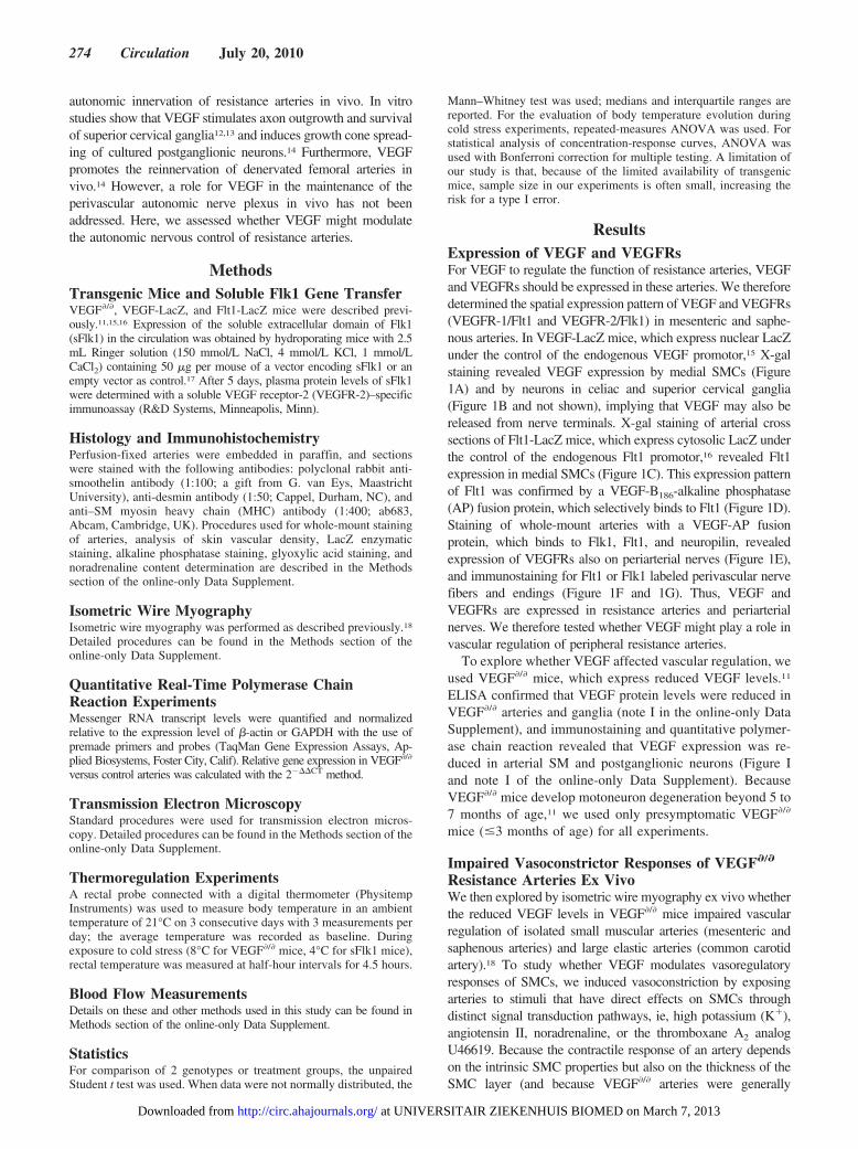
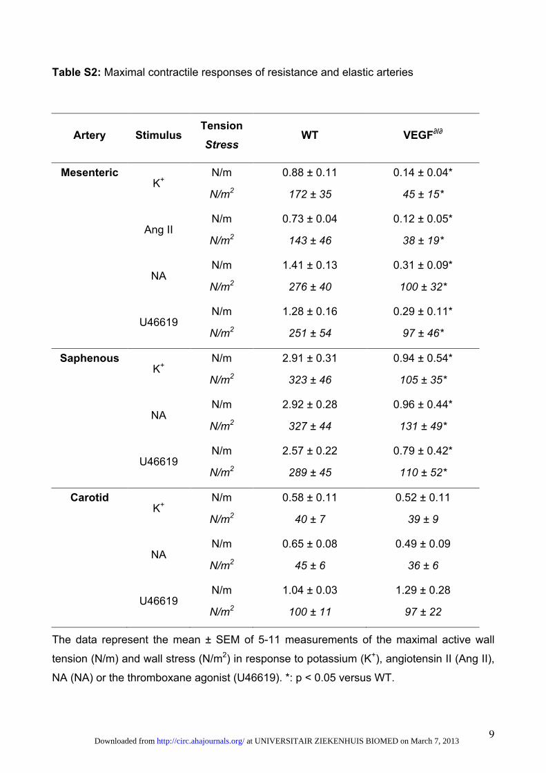
Impaired Vascular Regulation of ResistanceArteries in VEGF�/� Mice In VivoTo assess whether the aforementioned ex vivo changes inneuroeffector function also impaired autonomic nervous con-trol of peripheral resistance arteries in vivo, we exploredwhether thermoregulation, known to rely on autonomic ner-vous control of resistance arteries in the skin,3 was impairedin VEGF�/� mice. In baseline conditions, the core bodytemperature was 38.2�0.1°C in adult WT mice but only37.2�0.2°C in VEGF�/� mice (n�8; P�0.005). When ex-posed to cold stress (8°C), VEGF�/� mice were unable tomaintain their body temperature and progressively cooleddown to 34.2�1.1°C after 4.5 hours (n�8; P�0.002; Figure4A). This thermoregulatory defect was not attributable togenotypic differences in blood pressure or heart rate (TableIV of the online-only Data Supplement), thyroid function(note III of the online-only Data Supplement), cutaneousvascular density (percent of surface area covered by vascu-lature: 29.3�1.4 in WT mice versus 29.7�1.0 in VEGF�/�
mice; n�3; P�0.83), skeletal muscle dysfunction, or brownfat tissue mass (not shown).
To further evaluate whether the ex vivo peripheral arterialdefects were also relevant in vivo, we analyzed whetherVEGF�/� mice were capable of increasing cerebral blood flow(CBF) in conditions of general oxygen shortage. Indeed,when oxygen delivery to the brain becomes limiting, neuro-genic constriction of resistance arteries in visceral organsensures that brain perfusion is increased at the expense ofperipheral organs.4 We therefore exposed anesthetized venti-lated mice to a brief pulse of 8% oxygen (200 seconds) andmeasured changes in CBF using continuous arterial spin
labeling. As expected, after the hypoxic challenge, CBF wasincreased in WT mice by 33% (Figure 4B). In contrast, theCBF in VEGF�/� mice was increased by only 9% (n�4;P�0.01; Figure 4B). Thus, the ex vivo documented defects invascular regulation of peripheral resistance arteries have invivo physiological consequences.
Inhibition of VEGF Impairs Vascular Regulationin WT MiceWe then assessed whether the functional and structural defectsof arterial NEJs could also be induced on short-term inhibition ofVEGF in adult WT mice because this would provide insight intowhether VEGF might be involved in the dynamic regulation ofthese junctions. We therefore hydroporated WT mice with aplasmid encoding sFlk1, which traps VEGF, or an emptyplasmid as control. This strategy yields high plasma levels of thisVEGF inhibitor for several weeks.17 Five days after sFlk1 genetransfer, plasma sFlk1 levels ranged from 500 to 4000 ng/mL,sufficient to inhibit VEGF-driven tumor growth.17 Notably,ELISA measurements, using mesenteric and saphenous arteriesafter saline perfusion to remove intravascular sFlk1, revealedthat sFlk1 diffused into the arterial wall (note IV of theonline-only Data Supplement).
To evaluate the functional consequences of short-termVEGF blockade, we analyzed whether sFlk1 gene transfer inWT mice (sFlk1 mice) induced the same phenotypic defectsas observed in VEGF�/� mice. Already 10 days after sFlk1gene transfer, sFlk1 mice could not maintain their bodytemperature on exposure to cold stress (4°C during 4.5 hours;n�9 to 12; P�0.001; Figure 4C). In addition, the response ofisolated saphenous arteries to EFS was impaired in sFlk1mice, indicating that NEJs were dysfunctional (maximalcontractile response expressed as percent of maximal re-sponse to noradrenaline: 43.0�9.8% in sFlk1 mice versus79.8�15.6% in controls; n�7; P�0.0008; Figure 4D). How-ever, different from VEGF�/� mice, contractile SMC re-sponses to high potassium, noradrenaline, or U46619 (Figure4E through 4G) and expression of SMC markers (Table V ofthe online-only Data Supplement) were not altered in resis-tance arteries of sFlk1 mice. Control of vascular regulation byendothelial cells was also normal in sFlk1 mice (Figure V ofthe online-only Data Supplement). Ultrastructural analysis ofmesenteric resistance arteries revealed an increase in thedistance between neuronal varicosities and SMCs by �75%in sFlk1 arteries, as well as a reduction in the number ofvaricosities per axon bundle by �30% (Table VI of theonline-only Data Supplement). The area of the varicositiesand the number of mitochondria per varicosity were notsignificantly affected by sFlk1 (Table VI of the online-onlyData Supplement). Together, these results show that evenshort-term treatment of adult mice with a VEGF inhibitor wassufficient to induce functional and structural changes inarterial NEJs without affecting SMC function.
DiscussionThe present study provides genetic evidence that subnormalVEGF levels in VEGF�/� mice impair vascular regulation ofresistance arteries, thereby hindering thermoregulation and acompensatory increase in cerebral perfusion in response to
Table 3. Ultrastructural Parameters of Perivascular NEJs
Age Artery Parameter WT VEGF�/�
3 mo Mesenteric Junctional cleft width 1.93�0.12 3.30�0.32*
No. ofmitochondria/varicosity
2.74�0.37 1.43�0.13*
Varicosity area 0.83�0.11 0.36�0.048*
No. of varicosities/axonbundle
transmitter vesicle density
2.78�0.22 2.12�0.14*
137�20 134�27
3 mo Saphenous Junctional cleft width 1.49�0.045 1.90�0.17*
No. ofmitochondria/varicosity
2.17�0.24 1.51�0.16*
Varicosity area 0.55�0.083 0.26�0.024*
No. of varicosities/axonbundle
3.15�0.19 2.36�0.14*
3 wk Mesenteric Junctional cleft width 0.82�0.065 1.14�0.029*
No. ofmitochondria/varicosity
3.10�0.45 1.94�0.24*
Varicosity area 0.65�0.080 0.32�0.042*
No. of varicosities/axonbundle
4.57�0.46 3.29�0.23*
The data represent mean�SEM of measurements of the distance betweennerve varicosities and SMCs (junctional cleft width in �m), number ofmitochondria per varicosity, varicosity area (�m2), number of varicosities peraxon bundle, and transmitter storage vesicle density (�m2).
*P�0.05 vs WT.
278 Circulation July 20, 2010
at UNIVERSITAIR ZIEKENHUIS BIOMED on March 7, 2013http://circ.ahajournals.org/Downloaded from
hypoxia. Pharmacological, expression, and morphologicalstudies further revealed that this is due to dysfunction ofarterial NEJs and to dedifferentiation of contractile SMCs.Moreover, short-term inhibition of VEGF in healthy micecaused neuroeffector dysfunction without SMC defects.
Neuroeffector dysfunction was not due to overt degenera-tion of periarterial nerves but rather to more subtle ultrastruc-tural defects. Indeed, the junctional cleft between nervevaricosities and target vascular SMCs was widened inVEGF�/� arteries, thereby lowering the fraction of releasedneurotransmitter, reaching and activating postjunctional re-ceptors on target SMCs, and thus impairing neuroeffectorfunction.21 In addition, perivascular nerves had smaller andfewer varicosities, and these varicosities contained fewer
mitochondria. These defects may further contribute to theimpaired neuroeffector function.
Vascular SMC dysfunction in VEGF�/� mice was associ-ated with reduced levels of late vascular SMC differentiationmarkers (smoothelin-B, SM-MHC), which are typically ex-pressed by contractile arteries.19 So far, VEGF has beenshown to stimulate SMC migration and to regulate theassociation of SMCs around endothelial cells22,23 but has notbeen reported to regulate vascular SMC differentiation. Ourfindings suggest that VEGF is required to maintain SMCs ina fully differentiated contractile state. Whether this activity ofVEGF relies on a direct effect on SMCs or on an indirecteffect through production of “arterializing” signals from thenerve terminal remains to be determined.
Figure 4. Impaired vascular regulationof resistance arteries in VEGF�/� or WTmice on delivery of a VEGF trap. A, Evo-lution of body temperature after expo-sure of VEGF�/� and WT mice to coldstress (8°C) for 4.5 hours. B, Impairedincrease in CBF in VEGF�/� mice inresponse to hypoxia. C, Evolution ofbody temperature of mice, hydroporatedwith sFlk1 or empty (control) plasmid,during exposure to cold stress (4°C for4.5 hours). D, Contraction of saphenousarteries of sFlk1 and control mice inresponse to EFS. E through G,Concentration-response curve of sFlk1and control saphenous arteries inresponse to high potassium (E), nor-adrenaline (F), and U46619 (G). *P�0.05vs WT.
Storkebaum et al Low VEGF Levels Lead to Vascular Regulation Defects 279
at UNIVERSITAIR ZIEKENHUIS BIOMED on March 7, 2013http://circ.ahajournals.org/Downloaded from

Several mechanistic models could explain the impairedvascular regulation of VEGF�/� arteries (Figure 5). Onemodel, which we favor at present, postulates that reducedrelease of VEGF by SMCs causes functional and structuraldefects of the NEJ through direct effects on the nerveterminal, which would secondarily cause SMC dedifferenti-ation (“prejunctional” mechanism). Arguments in favor ofthis model are that VEGF is expressed by vascular SMCs,that VEGF levels are reduced in VEGF�/� arteries, and thatVEGF receptors are expressed by periarterial nerves. Aprimary neuronal defect is also suggested by findings thatneuroeffector defects are already present by 3 weeks of age,whereas SMC differentiation is still normal. Furthermore,sFlk1 gene transfer in adult WT mice caused structural andfunctional neuroeffector changes without inducing SMC al-terations. Previous studies documented that cultured sympa-thetic neurons express VEGFRs and respond to VEGF byenhanced axon outgrowth and survival,12 but so far, VEGFhas not been reported to induce structural changes in prejunc-tional nerve terminals. Because autonomic nerve endings areknown to release SMC-trophic factors,24 a dysfunction ofthese nerve terminals may secondarily lead to SMC dediffer-entiation. Indeed, activation of �1-adrenoreceptors by nor-adrenaline stimulates SMC proliferation,25 and sympatheticinnervation increases vascular SMC contractile protein ex-pression,24 whereas sympathectomy results in a switch from acontractile to synthetic SMC phenotype.26
An alternative nonexclusive model postulates that reducedrelease of VEGF from nerve endings and/or vascular SMCscould lead to SMC dedifferentiation, which would then second-arily cause defects in neuroeffector structure and function(“postjunctional” mechanism). Indeed, vascular SMCs areknown to promote survival of cultured postganglionic sympa-thetic neurons.27 This model seems less likely because SMCdifferentiation is normal in young VEGF�/� mice and adult sFlk1mice in which neuroeffector defects are already present. Fur-thermore, quantitative polymerase chain reaction analysis re-vealed that the expression of nerve growth factor, artemin, and
neurotrophin-3 (known to have plasticity effects on perivascularautonomic nerves28) was not reduced in VEGF�/� arteries. Thus,we have little evidence to support a model whereby prejunc-tional nerve terminals, through paracrine release of VEGF,would ensure their own performance by inducing retrograderelease of neurotrophic cues by target SMCs. We are thereforeinclined to propose that VEGF may, at least in part, regulate theremodeling of the NEJ through direct neurotrophic effects on theprejunctional nerve terminals.
Short-term administration of the VEGF inhibitor sFlk1 tohealthy adult mice was sufficient to cause vascular regulationdefects and perivascular nerve dysfunction, indicating a role forVEGF in the maintenance of NEJs. An intriguing question iswhether the observed functional and structural remodeling of theNEJs reflects a role for VEGF in regulating the “plasticity” ofthe perivascular autonomic nervous system and whether asimilar role for VEGF in regulating “synaptic plasticity” exists inthe central nervous system. Reports that VEGF stimulateslong-term potentiation in the hippocampus would be consistentwith such a model.29 These findings also raise the intriguing butunanswered question of whether impaired vascular regulationmight explain, at least in part, the hand-foot syndrome in thetreatment of cancer patients with VEGF (receptor) inhibitors.6
Indeed, impaired vascular regulation has been proposed tocontribute to the hand-foot syndrome because dilated bloodvessels are commonly found in affected tissues,8 and decreasingblood flow by cooling hands and feet lowers the incidence of thehand-foot syndrome.30 Overall, our genetic and pharmacologicalstudies uncover a previously unrecognized role of VEGF in theproper functioning and dynamic remodeling of perivascularautonomic NEJs.
AcknowledgmentsWe thank G. van Eys for providing the smoothelin antibody. We alsothank H. Bellens, A. Bouche, N. Dai, M. Deprez, M. De Mol, K. Feyen,B. Hermans, S. Jansen, L. Kieckens, A. Manderveld, M. Nijs, W.Martens, L. Notebaert, V. Uytterhoeven, M. Vanbrabant, A. VanDen Broeck, B. Vanwetswinkel, E. Weltens, and S. Wyns for theircontributions.
Figure 5. Role of VEGF in the mainte-nance of arterial neuroeffector function.A, In normal conditions, VEGF isreleased from SMCs and periarterialnerve endings and acts on nerve termi-nals to maintain NEJs (1). Another non-exclusive possibility is that VEGF primar-ily maintains SMCs in a differentiatedstate, which secondarily preserves NEJstructure and function (2). B, In VEGF�/�
mice, low VEGF levels cause functionaland structural defects of NEJs and SMCdedifferentiation.
280 Circulation July 20, 2010
at UNIVERSITAIR ZIEKENHUIS BIOMED on March 7, 2013http://circ.ahajournals.org/Downloaded from

Sources of FundingDrs Storkebaum and Ruiz de Almodovar are postdoctoral fellows ofFonds voor Wetenschappelijk Onderzoek (FWO)–Flanders. This workwas supported by Muscular Dystrophy Association grant 3751, FWOgrant G.0113.02, long-term structural funding from Methusalem fund-ing by the Flemish Government, European Union grant QLK6-CT-2000-5303, Geconcerteerde Onderzoeksacties grant GOA2006/11, agrant from the Transnational University of Limburg, Top InstitutePharma grant TIP2_108, a European Vascular Genomics Networkgrant, European Commission FP6-projects DiMI (LSHB-CT-2005-512146) and EMIL (LSHC-CT-2004-503569), and an Agentschap voorInnovatie door Wetenschap en Technologie SBO grant.
DisclosuresNone.
References1. Glebova NO, Ginty DD. Growth and survival signals controlling sympathetic
nervous system development. Annu Rev Neurosci. 2005;28:191–222.2. Burnstock G. Non-synaptic transmission at autonomic neuroeffector
junctions. Neurochem Int. 2008;52:14–25.3. Romanovsky AA. Thermoregulation: some concepts have changed: func-
tional architecture of the thermoregulatory system. Am J Physiol RegulIntegr Comp Physiol. 2007;292:R37–R46.
4. Kuwahira I, Gonzalez NC, Heisler N, Piiper J. Changes in regional bloodflow distribution and oxygen supply during hypoxia in conscious rats.J Appl Physiol. 1993;74:211–214.
5. Goldstein DS, Robertson D, Esler M, Straus SE, Eisenhofer G. Dysau-tonomias: clinical disorders of the autonomic nervous system. Ann InternMed. 2002;137:753–763.
6. Verheul HM, Pinedo HM. Possible molecular mechanisms involved inthe toxicity of angiogenesis inhibition. Nat Rev Cancer. 2007;7:475–485.
7. Webster-Gandy JD, How C, Harrold K. Palmar-plantar erythrodyses-thesia (PPE): a literature review with commentary on experience in acancer centre. Eur J Oncol Nurs. 2007;11:238–246.
8. Lassere Y, Hoff P. Management of hand-foot syndrome in patients treatedwith capecitabine (Xeloda). Eur J Oncol Nurs. 2004;8(suppl 1):S31–S40.
9. Carmeliet P. Angiogenesis in life, disease and medicine. Nature. 2005;438:932–936.
10. Storkebaum E, Lambrechts D, Carmeliet P. VEGF: once regarded as aspecific angiogenic factor, now implicated in neuroprotection. Bioessays.2004;26:943–954.
11. Oosthuyse B, Moons L, Storkebaum E, Beck H, Nuyens D, BrusselmansK, Dorpe JV, Hellings P, Gorselink M, Heymans S, Theilmeier G,Dewerchin M, Laudenbach V, Vermylen P, Raat H, Acker T, VleminckxV, Bosch LV, Cashman N, Fujisawa H, Drost MR, Sciot R, BruyninckxF, Hicklin DJ, Ince C, Gressens P, Lupu F, Plate KH, Robberecht W,Herbert JM, Collen D, Carmeliet P. Deletion of the hypoxia-responseelement in the vascular endothelial growth factor promoter causes motorneuron degeneration. Nat Genet. 2001;28:131–138.
12. Sondell M, Lundborg G, Kanje M. Vascular endothelial growth factor hasneurotrophic activity and stimulates axonal outgrowth, enhancing cell
survival and Schwann cell proliferation in the peripheral nervous system.J Neurosci. 1999;19:5731–5740.
13. Long JB, Jay SM, Segal SS, Madri JA. VEGF-A and Semaphorin3A:modulators of vascular sympathetic innervation. Dev Biol. 2009;334:119–132.
14. Marko SB, Damon DH. VEGF promotes vascular sympathetic inner-vation. Am J Physiol Heart Circ Physiol. 2008;294:H2646–H2652.
15. Miquerol L, Gertsenstein M, Harpal K, Rossant J, Nagy A. Multipledevelopmental roles of VEGF suggested by a LacZ-tagged allele. DevBiol. 1999;212:307–322.
16. Fong GH, Klingensmith J, Wood CR, Rossant J, Breitman ML. Regu-lation of flt-1 expression during mouse embryogenesis suggests a role inthe establishment of vascular endothelium. Dev Dyn. 1996;207:1–10.
17. Fischer C, Jonckx B, Mazzone M, Zacchigna S, Loges S, Pattarini L,Chorianopoulos E, Liesenborghs L, Koch M, De Mol M, Autiero M,Wyns S, Plaisance S, Moons L, van Rooijen N, Giacca M, Stassen JM,Dewerchin M, Collen D, Carmeliet P. Anti-PlGF inhibits growth ofVEGF(R)-inhibitor-resistant tumors without affecting healthy vessels.Cell. 2007;131:463–475.
18. De Mey JG, Megens R, Fazzi GE. Functional antagonism between en-dogenous neuropeptide Y and calcitonin gene-related peptide in mes-enteric resistance arteries. J Pharmacol Exp Ther. 2008;324:930–937.
19. Owens GK. Regulation of differentiation of vascular smooth muscle cells.Physiol Rev. 1995;75:487–517.
20. Luff SE. Development of neuromuscular junctions on small mesentericarteries of the rat. J Neurocytol. 1999;28:47–62.
21. Bennett MR. Autonomic neuromuscular transmission at a varicosity.Prog Neurobiol. 1996;50:505–532.
22. Wang H, Keiser JA. Vascular endothelial growth factor upregulates theexpression of matrix metalloproteinases in vascular smooth muscle cells:role of flt-1. Circ Res. 1998;83:832–840.
23. Greenberg JI, Shields DJ, Barillas SG, Acevedo LM, Murphy E, HuangJ, Scheppke L, Stockmann C, Johnson RS, Angle N, Cheresh DA. A rolefor VEGF as a negative regulator of pericyte function and vessel matu-ration. Nature. 2008;456:809–813.
24. Damon DH. Sympathetic innervation promotes vascular smooth muscledifferentiation. Am J Physiol Heart Circ Physiol. 2005;288:H2785–H2791.
25. van Kleef EM, Smits JF, De Mey JG, Cleutjens JP, Lombardi DM,Schwartz SM, Daemen MJ. Alpha 1-adrenoreceptor blockade reduces theangiotensin II-induced vascular smooth muscle cell DNA synthesis in therat thoracic aorta and carotid artery. Circ Res. 1992;70:1122–1127.
26. Kacem K, Seylaz J, Issertial O, Aubineau P. Chemical sympathectomyfavours vimentin expression in arterial smooth muscle cells of young rats.J Auton Nerv Syst. 1995;53:57–68.
27. Damon DH. NGF-independent survival of postganglionic sympatheticneurons in neuronal-vascular smooth muscle cocultures. Am J PhysiolHeart Circ Physiol. 2001;280:H1722–H728.
28. Cowen T, Gavazzi I. Plasticity in adult and ageing sympathetic neurons.Prog Neurobiol. 1998;54:249–288.
29. Cao L, Jiao X, Zuzga DS, Liu Y, Fong DM, Young D, During MJ. VEGFlinks hippocampal activity with neurogenesis, learning and memory. NatGenet. 2004;36:827–835.
30. Zimmerman GC, Keeling JH, Lowry M, Medina J, Von Hoff DD, BurrisHA. Prevention of docetaxel-induced erythrodysesthesia with local hypo-thermia. J Natl Cancer Inst. 1994;86:557–558.
CLINICAL PERSPECTIVEVascular endothelial growth factor (VEGF) is a key angiogenic factor, and blocking of VEGF signaling in cancer patients inhibitsangiogenesis, thereby preventing tumor progression. One of the side effects of anti-VEGF therapy is the hand-foot syndrome,which is characterized by dilated blood vessels in hands and feet, leading to dysesthesia, erythema, and tingling of extremities,which may progress to burning pain with dryness, cracking, and ulceration of the skin. Here, we show that knock-in mice withreduced VEGF levels display abnormal regulation of resistance arteries, resulting in defects in thermoregulation andredistribution of blood flow in response to hypoxia. The abnormal vascular regulation was attributed to reduced vascular smoothmuscle cell contractility associated with reduced expression of contractile proteins, as well as to dysfunction of neuroeffectorjunctions as a result of their abnormal structural development. Furthermore, short-term treatment of wild-type mice with a VEGFtrap also induced thermoregulation defects and neuroeffector dysfunction with structural remodeling of neuroeffector junctions,indicating that VEGF is necessary for maintenance of the structural and functional integrity of adult neuroeffector junctions.These findings may provide mechanistic insights into the cause of the hand-foot syndrome.
Storkebaum et al Low VEGF Levels Lead to Vascular Regulation Defects 281
at UNIVERSITAIR ZIEKENHUIS BIOMED on March 7, 2013http://circ.ahajournals.org/Downloaded from

1
SUPPLEMENTAL MATERIAL
SUPPLEMENTAL METHODS
Histology and immunohistochemistry: For whole mount staining of arteries, first order
mesenteric and saphenous arteries were dissected and fixed in 80% MeOH/20% DMSO
(TH staining) or in 1%PFA (VEGFR stainings) at 4°C overnight. TH staining: after
rehydration and blocking with 2% TBS-BSA, vessels were incubated with a rabbit anti-TH
antibody (Chemicon; 1:100) overnight. Finally, incubation with an Alexa488-labeled goat
anti-rabbit IgG (1/200) was performed. VEGFRs staining: arteries where permeabilized
with 0.05% Triton X-100 in PBS, blocked with 5% goat pre-immune serum and incubated
overnight at 4ºC with antibodies against Flt1 (5µg/ml, SC-316) or Flk1 (5µg/ml, SC-504).
Finally, 1h incubation with Alexa-labeled secondary antibodies was performed. Vessels
were mounted on glass slides and 3D images were obtained with a Zeiss LSM510
confocal laser scanning microscope and analyzed with Zeiss KS300 software. For analysis
of skin vascular density, ears from 1 month old mice were dissected as described 1. The
ears were fixed in ice cold Dent's fixative overnight and blocked with 2% TBS-BSA. For
whole mount double immunostaining the tissues were incubated with anti-SM alpha-actin
(Dako, 1/500) and anti-CD31 (Pharmingen, 1/500) followed by incubation with Alexa-
labeled secondary antibodies and flat-mounting.
LacZ enzymatic staining was performed on 20 µm transverse cryo-sections of
unfixed saphenous and mesenteric arteries. After fixation in acetone for 20 min at -20ºC,
slides were washed in washing buffer (0.1% Sodium deoxycholate, 0.2% Nonidet P40 and
2mM MgCl2 in PBS) and incubated overnight at 30ºC in X-gal staining solution (2mM
at UNIVERSITAIR ZIEKENHUIS BIOMED on March 7, 2013http://circ.ahajournals.org/Downloaded from

2
MgCl2, 5 mM K4Fe(CN)6, 5 mM K3Fe(CN)6 and 1mg/ml X-gal in PBS). Afterwards slides
were washed in PBS, counterstained with nuclear fast red and mounted with mowiol
(Sigma). For alkaline phosphatase (AP) binding and staining, VEGF-B186-AP and VEGF164-
AP (VEGF-AP) fusion proteins were made by cloning VEGF-B186 or VEGF164 cDNA in a
pAPtag-5 vector (GenHunter). Fusion proteins were then expressed in HEK-293 cells as
secreted protein, and the conditioned medium was used directly as a highly sensitive
affinity agent. AP activity was tested via an AP-activity assay and the AP binding assay
was performed as previously described 2. Imaging analysis was performed on a Zeiss
Axioplan2 connected to a 3CCD video camera (DXC-93OP, Sony), and KS300 software
(Zeiss, Germany). For the anaylsis of SMC differentiation markers and VEGF expression
on saphenous and mesenteric arteries, arteries were dissected, fixed in 1%PFA (4ºC,
overnight), cryo-protected and cryo-sectioned. Immunostainings were performed with the
following antibodies: anti-smoothelin (1:100, gift of G. van Eys, Maastricht University), anti-
SM myosin HC (1:400, ab683, Abcam), anti-desmin (1:50, 10519, Cappel) and anti-VEGF
(1/50, SC152G), followed by 2h incubation with Alexa-labeled secondary antibodies. For
measurement of relative fluorescence intensity, arteries from VEGF∂/∂ and control mice
were harvested and stained simultaneously, and images were acquired in random order in
a single session. Fluorescence intensity was measured using Zeiss KS300 software with
subtraction of background fluorescence.
Noradrenaline content: Saphenous and mesenteric resistance artery segments, kidney
and heart were solubilized in 0.5 M acetic acid (15 minutes, 100°C). Noradrenaline content
of the extract was measured by high-performance liquid chromatography and fluorescent
detection, after which the tissues were solubilized in 1N KOH (24 hours, room
at UNIVERSITAIR ZIEKENHUIS BIOMED on March 7, 2013http://circ.ahajournals.org/Downloaded from

3
temperature) and DNA content was determined. Noradrenaline levels were expressed
relative to DNA content.
Murine VEGF ELISA: For quantification of mVEGF protein expression, the Mouse VEGF
Quantikine ELISA Kit (R&D Systems) was used. mVEGF concentrations were normalized
to total protein concentration, which was measured using BCA assay (Pierce).
Isometric wire myography: Two-millimeter segments of isolated first order mesenteric,
saphenous and common carotid arteries were mounted (steel wires, diameter 40 µm) in a
myograph organ bath (model 610, Danish Myotechnology by J.P. Trading, Denmark) for
isometric force measurements. Organ baths were filled with a Krebs-Ringer bicarbonate
buffer, maintained at 37°C, and aerated with 95% O2 and 5% CO2. Each individual arterial
segment was distended to a diameter at which a maximal contractile response to 10 µM
NA could be obtained. To evaluate SMC function, the following stimuli were used: (i)
concentration-response curve for increasing concentrations of extracellular K+ (in
exchange for Na+), (ii) concentration-response curve for angiotensin II (1-30 nM) during
mild depolarization (16.5-25 mM K+), (iii) concentration-response curve for NA (0.01-10
µM), (iv) concentration-response curve for the thromboxane A2-analogue U46619 (1-100
nM), (v) concentration-response curve for U46619 in the continuous presence of the NO-
synthase inhibitor nitro-L-arginine (NLA, 30µM). Endothelium-dependent relaxing
responses to acetylcholine were evaluated during contractions induced by 40 mM K+, 10
µM NA and 100 nM U46619 and the relaxing effects of the NO-donor substance Sodium
nitroprusside (SNP, 0.01 to 10 µM) were studied during contraction induced by U46619, in
the presence of the NO-synthase inhibitor NLA. To evaluate perivascular sympathetic
nerve function, the following stimuli were used: (i) response to increasing frequencies
at UNIVERSITAIR ZIEKENHUIS BIOMED on March 7, 2013http://circ.ahajournals.org/Downloaded from

4
(0.25-32 Hz) of electrical field stimulation (EFS) delivered by two platinum electrodes
placed along the isolated arterial segment, (ii) concentration-response curve for increasing
concentrations (1-100 µM) of tyramine, (iii) effect of cocaine on the arterial sensitivity to
increasing concentrations (0.01-10 µM) of exogenous NA, (iv) addition of the alpha-
adrenoreceptor antagonist phentolamine (1 µM) on top of a maximal contractile response
to 125 mM K+. To evaluate perivascular sensory-motor nerve function, mesenteric arteries
were induced to contract by U46619 (0.1 µM), and the relaxing effect of either capsaicin
(0.1 and 1.0 µM, a compound that stimulates the release of endogenous CGRP from
sensory-motor nerves 3, 4) or exogenous CGRP (0.1 to 300 nM) was evaluated. At the end
of the experiments, vessel segments were fixed in the organ chamber in phosphate-
buffered formaldehyde (4%, pH 7.4, 37°C, 30 minutes) for subsequent morphometric
analysis.
Glyoxylic acid staining: Glyoxylic acid was used to visualize and quantify perivascular
sympathetic nerve fibers in whole mount preparations of isolated first order mesenteric and
saphenous arteries. Vessels were mounted and stretched in an organ bath (see above)
and incubated in 2% glyoxylic acid and 10% sucrose in phosphate buffer for 10 minutes at
room temperature. Segments were then air dried (90 sec), stretched at 100°C for 4
minutes and mounted on glass slides. Two-photon laser scanning microscopy was used to
generate a stack of images from one side of the compressed vessels3, 4. The percentage
of the area occupied by fluorescent signal was calculated.
Noradrenaline reuptake experiments: Mesenteric and saphenous arteries were
dissected and the length of the arterial segments was determined. Pre-incubation,
incubation and wash steps were done in Krebs Ringer bicarbonate solution containing
EGTA (1 mM) and glutathione (10 mM) (=KRB+ buffer), either in the absence or the
at UNIVERSITAIR ZIEKENHUIS BIOMED on March 7, 2013http://circ.ahajournals.org/Downloaded from

5
presence of 3 µM cocaine, an established inhibitor of NA neuronal uptake. After pre-
incubation (37°C, 30 min), 0.1 µM levo-[ring-2,5,6-3H]-NA (52.6 Ci/mmol, Perkin Elmer)
was added to the KRB+ buffer (incubation at 37°C for 60 min). During the wash step (37°C,
60 min), arteries were transferred repeatedly (10 times) to fresh KRB+. For extraction of
the 3H-NA, arteries were transferred to 1 ml of 0.5 M acetic acid containing 10 mM
glutathione, and heated at 94°C during 15 minutes. Subsequently, the artery was
recovered and digested in 100 µl 1N KOH during 48 hours, followed by DNA content
determination. The extract was used for liquid scintillation spectrometry. Liquid scintillation
results (in CPM) were normalized to vessel segment length or to DNA content, and the
difference between findings obtained in the absence and presence of cocaine was used as
a measure of NA reuptake capacity.
Transmission electron microscopy: Mice were anesthetized, perfused with 4% PFA and
2.5% glutaraldehyde in Na-cacodylate buffer (0.1 M), pH 7.3 for 15 minutes. Two
saphenous and three second order mesenteric resistance arteries were dissected from
each mouse and fixed overnight in 4%PFA and 2.5% glutaraldehyde in Na-cacodylate
buffer (0.1 M). The next day two rinses (2 x 30 min) with Na-cacodylate buffer (0.1 M) were
followed by postfixation with 2% osmiumtetroxide in Na-cacodylate buffer (0.1 M) for 2h at
room temperature. Following dehydration in a graded ethanol series (30-50-70%, each
step 2 x 15 min) the arteries were block stained with 1% uranylacetate in 70% ethanol for
30 min at room temperature. After the last steps of the dehydration (96-100%, each step 2
x 15 min) the impregnation of the tissue with freshly prepared Agar 100 (EPON 812,
Medium) was initiated by means of a graded propylene oxide – Agar 100 series (100%
propylene oxide, 2 x 20 min – propylene oxide 1:1 Agar 100, 1 hour – propylene oxide 1:2
Agar 100, overnight in a dessicator, room temperature). The next day the arteries were
at UNIVERSITAIR ZIEKENHUIS BIOMED on March 7, 2013http://circ.ahajournals.org/Downloaded from

6
transferred to freshly prepared Agar 100 and placed in a dessicator for 6 to 8 hours at
room temperature before they were orientated in molds and put in an oven at 60° C for 48
hours for the polymerization of the resin. Semi thin (0.5-1 µm) sections were made and
stained with 1% toluidine blue in 1% borax for light microscopic observations. Ultra thin
sections (70 nm) were placed on copper grids (400 mesh) and contrasted with 4% uranyl
acetate for 10 min and lead citrate for 5 min. Ultrastructural observations were made with a
JEOL JEM 2100 at 200 kV at the KULeuven Electron Microscopy Core Facility of the
Department of Human Genetics, Leuven, Belgium. Light microscopic and ultrastructural
observations were quantitatively analyzed using Zeiss KS 300 3.0 software.
For morphometric analysis, high power electron micrographs were made of neuro-effector
junctions in randomly selected single sections and used to measure (i) the shortest
distance between nerve varicosities and the nearest vascular SMCs, as described 5 (ii) the
average number of mitochondria per varicosity, (iii) the surface area of varicosities, (iv) the
number of varicosities per axon bundle, and (v) the density of transmitter storage vesicles
(number per varicosity/surface area varicosity), as described 6. Image acquisition and
morphometric analysis of electron micrographs were performed by an investigator blinded
to the mouse genotype.
Blood flow measurements: Magnetic resonance (MR) experiments were performed on a
7T horizontal bore magnet and 8-cm aperture self-shielded gradients with a maximum
strength of 0.1 T/m (Oxford Instrument, UK) equipped with a MRRS console (MRRS, UK).
Mice were anesthetized using 5% isoflurane for induction and a maintenance dose of
0.4%-0.8% isoflurane in a mixture of O2:N2O (3:7) at a flow rate of 600 ml/min. Refined
monitoring systems recorded body temperature (Kent, UK), breaths per minute and end-
at UNIVERSITAIR ZIEKENHUIS BIOMED on March 7, 2013http://circ.ahajournals.org/Downloaded from

7
tidal CO2 (Capstar-100, Linton Instruments, UK), which were, respectively, 37.0 ± 0.2°C,
180 ± 20 and 3.5 ± 0.5%.
Arterial Spin Labeling: Continuous Arterial Spin Labeling (ASL) was used to study the
response of CBF to hypoxia (200s at 8%O2). Single slice EPI-GE paired images were
acquired alternately: one without arterial labelling (control image) and the other with arterial
labelling. For each set of CBF measurements, 150 pairs of images were acquired and the
first 4 pairs were discarded. All mice were spontaneously breathing and during ASL
acquisition, ventilation parameters were set to hypoxic conditions by switching from 30%
O2 to 8% O2. One trial per mouse consisted of 80 ASL measurements during normoxia,
followed by 40 measurements during hypoxia after which recovery was allowed during 30
measurements. Arterial spin labelling signal changes were analysed using home written
programs in IDL (Boulder, UK), which provide regional analyses on a pixel per pixel base.
The most straightforward approach of "simple subtraction" was used, which means direct
pairwise subtraction between temporally adjacent label and control acquisitions. Analysis
of the percent changes reveals whether the hypoxia induced CBF increase is present
without the need to calculate the absolute CBF in ml/g/min.
Purification and culture of SCG and CG neurons: SCGs and CGs from WT or VEGF∂/∂
P4 pups were dissected and placed in 0.25% trypsin-EDTA (Invitrogen) for 30min at 37ºC,
washed twice with Hank’s buffered salt solution (HBSS) containing Ca2+ and Mg2+
(Invitrogen) and then placed in 1x collagenase in HBSS with Ca2+ and Mg2+ for 30min and
37ºC. Ganglia were then dissociated by trituration. Neurons were finally resuspended and
cultured in Neurobasal medium (Gibco) containing B27 (Gibco) on poly-L and laminin
coated coverslips for 48h.
at UNIVERSITAIR ZIEKENHUIS BIOMED on March 7, 2013http://circ.ahajournals.org/Downloaded from

8
Table S1: Structural vessel parameters
Artery Parameter WT VEGF∂/∂
Mesenteric Lumen diameter 166 ± 11 146 ± 17
Media area 2,260 ± 193 1,480 ± 258*
Media thickness 5.1 ± 0.6 3.1 ± 0.2*
Saphenous Lumen diameter 221 ± 6 159 ± 6*
Media area 6,385 ± 460 3,720 ± 610*
Media thickness 8.7 ± 0.5 8.9 ± 1.0
Carotid Lumen diameter 385 ± 20 356 ± 20
Media area 18,400 ± 700 15,400 ± 1,000
Media thickness 14.8 ± 0.4 13.3 ± 0.6
The data represent the mean ± SEM of 5-11 measurements of the diameter of the lumen
(µm), cross-sectional area of the media (µm2), and thickness of the media (µm). *: p < 0.05
versus WT.
at UNIVERSITAIR ZIEKENHUIS BIOMED on March 7, 2013http://circ.ahajournals.org/Downloaded from
9
Table S2: Maximal contractile responses of resistance and elastic arteries
Artery Stimulus Tension Stress
WT VEGF∂/∂
Mesenteric K+
N/m
N/m2
0.88 ± 0.11
172 ± 35
0.14 ± 0.04*
45 ± 15*
Ang II
N/m
N/m2
0.73 ± 0.04
143 ± 46
0.12 ± 0.05*
38 ± 19*
NA
N/m
N/m2
1.41 ± 0.13
276 ± 40
0.31 ± 0.09*
100 ± 32*
U46619
N/m
N/m2
1.28 ± 0.16
251 ± 54
0.29 ± 0.11*
97 ± 46*
Saphenous K+
N/m
N/m2
2.91 ± 0.31
323 ± 46
0.94 ± 0.54*
105 ± 35*
NA
N/m
N/m2
2.92 ± 0.28
327 ± 44
0.96 ± 0.44*
131 ± 49*
U46619
N/m
N/m2
2.57 ± 0.22
289 ± 45
0.79 ± 0.42*
110 ± 52*
Carotid K+
N/m
N/m2
0.58 ± 0.11
40 ± 7
0.52 ± 0.11
39 ± 9
NA
N/m
N/m2
0.65 ± 0.08
45 ± 6
0.49 ± 0.09
36 ± 6
U46619
N/m
N/m2
1.04 ± 0.03
100 ± 11
1.29 ± 0.28
97 ± 22
The data represent the mean ± SEM of 5-11 measurements of the maximal active wall
tension (N/m) and wall stress (N/m2) in response to potassium (K+), angiotensin II (Ang II),
NA (NA) or the thromboxane agonist (U46619). *: p < 0.05 versus WT.
at UNIVERSITAIR ZIEKENHUIS BIOMED on March 7, 2013http://circ.ahajournals.org/Downloaded from

10
Table S3: Tissue NA content
Parameter Tissue WT VEGF∂/∂
heart 447 ± 69 455 ± 41
kidney 86.6 ± 7.3 128.2 ± 17.1
mesenteric artery 2,970 ± 821 4,840 ± 472
NA content
(pg NA/µg DNA)
saphenous artery 2,720 ± 690 1,890 ± 147
The values represent the mean ± SEM of 6 measurements of the noradrenaline (NA)
content of 3 month old mice.
at UNIVERSITAIR ZIEKENHUIS BIOMED on March 7, 2013http://circ.ahajournals.org/Downloaded from

11
Table S4: Blood pressure and heart rate of VEGF∂/∂ and WT control mice.
Parameter WT VEGF∂/∂
systolic BP (mmHg) 94.6 ± 9.8 90.6 ± 4.0
diastolic BP (mmHg) 59.7 ± 9.2 68.1 ± 4.4
MAP (mmHg) 75.7 ± 9.9 78.9 ± 4.5
heart rate (bpm) 500 ± 25 491 ± 30
The values represent the mean ± SEM of 5 measurements. BP: blood pressure; MAP:
mean arterial blood pressure; bpm: beats per minute. At first sight, one might expect that
an impairment in vasoconstrictor response, as observed in VEGF∂/∂ mice, would
automatically result in hypotension. However, the regulation of blood pressure is a
complex process, involving the interplay of numerous molecules. Several gene-inactivation
studies in mice indicate that loss of even key players of vascular regulation fail to
significantly affect the blood pressure in baseline conditions, presumably because of
compensatory up- or downregulation of additional vasoactive substances 7-13. Alternatively,
the defect in vascular regulation in the VEGF∂/∂ mice may be too subtle to influence
homeostasis in baseline conditions, but sufficient to be relevant in stress conditions.
at UNIVERSITAIR ZIEKENHUIS BIOMED on March 7, 2013http://circ.ahajournals.org/Downloaded from

- 12 -
12
Table S5: Relative expression levels of smooth muscle cell differentiation markers in sFlk1 and control mice.
Artery target gene Control sFlk1 p-value
SM myosin heavy chain 100 ± 9.6 114.3 ± 13.7 0.40 Saphenous artery smoothelin 100 ± 7.9 105.8 ± 9.8 0.65 SM myosin heavy chain 100 ± 13.7 132.9 ± 30.3 0.33 Mesenteric artery smoothelin 100 ± 12.5 81.4 ± 11.7 0.30 SM myosin heavy chain 100 ± 70.3 106.7 ± 48.2 0.94 Carotid arteryA smoothelin 100 ± 70.0 154.9 ± 69.0 0.58
Relative expression levels as percentage of WT ± SEM are shown. A Common carotid arteries were used.
at UNIVERSITAIR ZIEKENHUIS BIOMED on March 7, 2013http://circ.ahajournals.org/Downloaded from

- 13 -
13
TableS6: Ultrastructural parameters of perivascular neuro-effector junctions of sFlk1 and control treated mice.
Artery Parameter Control sFlk1
junctional cleft width 0.85 ± 0.02 1.48 ± 0.04*
number of mitochondria/varicosity 2.62 ± 0.29 2.75 ± 0.30
varicosity area 0.87 ± 0.09 0.87 ± 0.11 Mesenteric
number of varicosities/axon bundle 3.45 ± 0.24 2.67 ± 0.24*
The data represent the mean ± SEM of measurements of the distance between nerve varicosities and
SMCs (junctional cleft width in µm), the number of mitochondria per varicosity, varicosity area (µm2) and
number of varicosities per axon bundle. *: p < 0.05
at UNIVERSITAIR ZIEKENHUIS BIOMED on March 7, 2013http://circ.ahajournals.org/Downloaded from

14
SUPPLEMENTAL NOTES
Note S1: VEGF protein levels in peripheral resistance arteries: pg VEGF/mg total protein:
for saphenous arteries: 65.8±11.5 in WT mice versus 43.5±5.8 in VEGF∂/∂ mice; n=8;
p=0.03; for mesenteric arteries: 126.3±11.3 in WT mice versus 94.8±8.2 in VEGF∂/∂ mice;
n=8; p=0.04. VEGF protein levels in superior cervical ganglia: pg VEGF/mg total protein:
medians (interquartile range): 96.6 (82.1-102.8) in WT mice versus 68.5 (67.1-75.0) in
VEGF∂/∂ mice; n=8-10; Mann Whitney p-value=0.002. To ensure that these ELISA
measurements reflected reduced VEGF expression in postganglionic neurons, ganglia
were dissociated and neurons were cultured for 48h, followed by RNA isolation.
Quantitative real-time PCR revealed reduced VEGF expression in VEGF∂/∂ neurons
(relative VEGF expression: medians (interquartile range): 2.05 (1.30-2.65) in VEGF∂/∂
neurons versus 3.10 (2.35-3.35) in WT neurons, n=10-13; Mann Whitney p-value=0.015).
Note S2: To analyze whether noradrenaline (NA) reuptake by perivascular nerves was
normal, mesenteric and saphenous arteries of 2 month old VEGF∂/∂ mice were dissected
and incubated with 3H-labeled NA in the presence or absence of the NA reuptake inhibitor
cocaine. The NA reuptake capacity was then calculated as the difference between the
condition with and without cocaine (see Methods for details). This approach failed to reveal
defects in NA reuptake in mesenteric arteries of VEGF∂/∂ mice (NA reuptake in cpm/mg
DNA: 431 ± 104 in VEGF∂/∂ mice versus 485 ± 98 in WT mice; n=10, p=0.72), as well as in
saphenous arteries (NA reuptake in cpm/mg DNA: 407 ± 113 in VEGF∂/∂ mice versus 419
± 89 in WT mice; n=6, p=0.93). Thus, alteration of NA reuptake by perivascular nerves
could not explain the autonomic defects in VEGF∂/∂ mice.
at UNIVERSITAIR ZIEKENHUIS BIOMED on March 7, 2013http://circ.ahajournals.org/Downloaded from
15
Note S3: Thyroid function was evaluated by measuring plasma T3 and T4 hormone levels.
No differences were found between WT and VEGF∂/∂ mice (T3 levels in ng/dl: 58.3 ± 0.88
versus 55.3 ± 2.7 respectively in WT and VEGF∂/∂ mice, n=3, p=0.35; T4 levels in µg/dl:
4.63 ± 0.71 versus 3.03 ± 0.29 respectively in WT and VEGF∂/∂ mice, n=3, p=0.11).
Note S4: µg Flk1/mg protein: in mesenteric arteries: 0.24±0.08 for control versus
0.84±0.07 for sFlk1, n=5, p=0.0005; in saphenous arteries: 0.67±0.11 for control versus
3.21±1.08 for sFlk1, n=5, p=0.04.
at UNIVERSITAIR ZIEKENHUIS BIOMED on March 7, 2013http://circ.ahajournals.org/Downloaded from

Wal
l ten
sion
(N
/m)
Log M [Noradrenaline]
D
-8.0 -6.0-7.0 -5.0
0.0
0.4
0.8
Wal
l ten
sion
(N
/m)
E
-8.0 -7.0 -6.0
0.0
1.0
2.0
-5.0
Wal
l ten
sion
(N
/m)
F
-8.0 -6.0-7.0 -5.0
0.0
1.0
2.0
3.0
Carotid Artery Mesenteric Artery Saphenous ArteryWT
VEGF∂/∂
WT
VEGF∂/∂
WT
VEGF∂/∂
Wal
l str
ess
(N/m
2 )
G
-8.0 -6.0-7.0 -5.0 -8.0 -7.0 -6.0 -5.0 -8.0 -6.0-7.0 -5.0
WT
VEGF∂/∂
WT
VEGF∂/∂
WT
VEGF∂/∂
Log M [Noradrenaline]
Log M [Noradrenaline] Log M [Noradrenaline]
0
100
200
300
400
-100
0
100
200
300
400
-100
0
100
200
300
400
-100
Wal
l str
ess
(N/m
2 )
Wal
l str
ess
(N/m
2 )
0.2
0.6
Log M [Noradrenaline]
Log M [Noradrenaline]
H I
* *
**
* *
**
* ** *
Figure S1
A B CWT VEGF∂/∂
50
75
100
125
0VE
GF
exp
ress
ion
(%)
*
WTVEGF∂/∂
25
at UNIVERSITAIR ZIEKENHUIS BIOMED on March 7, 2013http://circ.ahajournals.org/Downloaded from

Figure S2
A
0
Rel
axat
ion
(%)
phenylephrine + acetylcholine
20
40
60
100
80
-8 -7 -6 -5
C
0
Rel
axat
ion
(%)
potassium + acetylcholine
20
40
60
100
80
-8 -7 -6 -5
D
0R
elax
atio
n (%
)
potassium + NLA + acetylcholine
20
40
60
100
80
-8 -7 -6 -5
B
0
Rel
axat
ion
(%)
phenylephrine + NLA + acetylcholine
20
40
60
100
80
-8 -7 -6 -5
VEGF∂/∂
WT
VEGF∂/∂
WT
VEGF∂/∂
WTVEGF∂/∂
WT
logM [Ach] logM [Ach]
logM [Ach]logM [Ach]
at UNIVERSITAIR ZIEKENHUIS BIOMED on March 7, 2013http://circ.ahajournals.org/Downloaded from

ECA
B D F
smoothelin SM myosin HC desmin
WT
VE
GF
∂/∂
G
Smoothelin SM-MHC desmin0
50
100
150
WTVEGF∂/∂
* *
rela
tive
fluor
esce
nce
inte
nsity
(%
)
Figure S3
at UNIVERSITAIR ZIEKENHUIS BIOMED on March 7, 2013http://circ.ahajournals.org/Downloaded from

Figure S4
C D
A B
WT VEGF∂/∂
TH
CG
RP
E F
G H
0
WTVEGF∂/∂
WTVEGF∂/∂
20
0
50
0
40
0Ner
ve fi
ber
dens
ity (
%)
Nev
e fib
er d
ensi
ty (
%)
20
25 10
20
40
Nev
e fib
er d
ensi
ty (
%)
WTVEGF∂/∂
WTVEGF∂/∂
Ner
ve fi
ber
dens
ity (
%)
Mesenteric artery (TH) Mesenteric artery (GA)
Saphenous artery (GA) Mesenteric artery (CGRP)
at UNIVERSITAIR ZIEKENHUIS BIOMED on March 7, 2013http://circ.ahajournals.org/Downloaded from

Figure S5
A B
C D
0
ControlsFlk1
ControlsFlk1
ControlsFlk1
ControlsFlk130
0
70
0
60
0
50
Rel
axat
ion
(%)
[K+] + acetylcholine Noradrenaline + acetylcholineR
elax
atio
n (%
)
Rel
axat
ion
(%)
Rel
axat
ion(
%)
Phenylephrine + acetylcholine U46619 + acetylcholine
25 30
35 15
at UNIVERSITAIR ZIEKENHUIS BIOMED on March 7, 2013http://circ.ahajournals.org/Downloaded from
16
SUPPLEMENTAL FIGURE LEGENDS
Figure S1: Reduced VEGF expression and vascular SMC dysfunction in
peripheral resistance arteries of VEGF∂/∂ mice. A-B, Representative pictures of
VEGF immunostaining on mesenteric arteries from WT (A) and VEGF∂/∂ mice (B)
showing a reduced VEGF staining intensity in VEGF∂/∂ arteries compared to WT.
Scale bar: 5µm. C, Relative intensity of VEGF immunostaining in the SMC layer
of mesenteric arteries, displayed as % of WT (* P=0.029, N=4). D-F,
Concentration response curveof carotid arteries (D), mesenteric arteries (E) and
saphenous arteries (F) of WT and VEGF∂/∂ mice in response to NA.
Vasoconstriction is significantly reduced in mesenteric and saphenous resistance
arteries of VEGF∂/∂ mice, but normal in carotid arteries. G-I, Even when the size
of the vessels was taken into account (active wall stress), VEGF∂/∂ mesenteric
and saphenous arteries exhibited a marked reduction in contractility, whereas
contractility of carotid arteries was normal. Note that in WT mice active wall
stress was markedly larger in the muscular mesenteric and saphenous arteries
than in the elastic carotid artery, and that this regional difference was largely
abolished in VEGF∂/∂ mice. *: p < 0.05 versus WT.
Figure S2: Normal endothelial function in VEGF∂/∂ resistance arteries. To analyze
the role of the endothelium in vascular regulation, we studied the response to
acetylcholine (Ach), which relaxes arterial smooth muscle by inducing the release
of endothelium-derived factors. In isolated mesenteric arteries, relaxing
responses to increasing concentrations of acetylcholine were recorded during
contraction induced by 20 µM phenylephrine (A,B) or high potassium (40 mM K+)
(C,D), in the absence (A,C) or the presence (B,D) of the NO-synthase inhibitor
at UNIVERSITAIR ZIEKENHUIS BIOMED on March 7, 2013http://circ.ahajournals.org/Downloaded from
17
nitro-L-arginine (NLA, 30 µM). Regardless of the vasoconstrictor, a dose range of
0.01 to 10 µM Ach induced comparable relaxations in VEGF∂/∂ and control
arteries, indicating that vascular regulation by ECs was normal in VEGF∂/∂ mice.
Figure S3: Reduced expression of late vascular SMC differentiation markers in
VEGF∂/∂ arteries. A-F, Representative pictures of immunostaining for smoothelin
(A, B), smooth muscle myosin heavy chain (C, D) and desmin (E, F) on
saphenous arteries of WT (upper panels: A, C, E) and VEGF∂/∂ mice (lower
panels: B, D, F). Staining intensity is reduced in VEGF∂/∂ arteries for smoothelin
and smooth muscle myosin heavy chain, but not for desmin. Scale bar: 50µm. G,
Relative intensity of immunostaining in the tunica media of saphenous arteries,
displayed as % of WT. *: p < 0.05 versus WT.
Figure S4: Neuro-effector dysfunction cannot be attributed to reduced density of
peri-arterial nerves. To examine whether the reduced response of muscular
arteries to neurogenic stimuli in VEGF∂/∂ mice was due to a reduced density of
sympathetic innervation (caused by a developmental defect at 3 weeks of age, or
by nerve degeneration at the age of 3, 6 and 12 months), we stained whole-
mount mesenteric arteries for tyrosine hydroxylase (TH), a marker of the
perivascular sympathetic plexus. Light microscopic analysis of these whole
mount stained vessels only allows one to visualize axon bundles of the
autonomic plexus, but not the more subtle ultrastructural changes that can be
observed by EM. (i) At 3 WEEKS OF AGE (i.e. after the sympathetic plexus is
formed), a dense network of strongly stained interconnecting fibers was
detectable over the entire surface of these vessels in both genotypes.
Quantification of the TH-immunoreactive area as a percentage of the total vessel
area revealed that the TH+ nervous plexus covered a similar vessel area in both
at UNIVERSITAIR ZIEKENHUIS BIOMED on March 7, 2013http://circ.ahajournals.org/Downloaded from

18
genotypes at 3 weeks of age (30 ± 2% in WT mice versus 28 ± 2% in VEGF∂/∂
mice; n=6-11, p=0.45; Figure S4A,B,E; scale bar: 25 µm), indicating that
development of the perivascular adrenergic plexus was normal. (ii) By 3 MONTHS
OF AGE, when vasoregulator control by perivascular nerves was dysfunctional in
VEGF∂/∂ mice, the TH+ nervous plexus comprised 21 ± 1% in WT mice versus 27
± 2% in VEGF∂/∂ mice (n=6-9, p=0.07). Similar findings were observed when
analyzing the density of nerve fibers by staining with glyoxylic acid (Figure
S4F,G). We also stained transverse sections of saphenous arteries for TH.
Again, quantification revealed no genotypic differences in the density of
perivascular nerves (number of perivascular nerves per section, respectively, for
WT and VEGF∂/∂: 25.1 ± 4.6 versus 19.6 ± 2.6, n=10-14, p=0.75). Likewise,
sensory-motor nerve dysfunction could not be attributed to nerve degeneration as
the density of CGRP-immunoreactive nerve fibers in whole mount mesenteric
artery preparations did not significantly differ between WT and VEGF∂/∂ (15.1 ±
1.4% in WT mice versus 14.7 ± 1.7% in VEGF∂/∂ mice; n=8, p=0.35; Figure
S4C,D,H; scale bar: 25µm). (iii) Also, BY 6 MONTHS OF AGE, when VEGF∂/∂ mice
exhibit the first symptoms of motor neuron degeneration, no perivascular
sympathetic nerve degeneration could be observed, as the TH+ nervous plexus
comprised 28 ± 3% in WT mice and 29 ± 3% in VEGF∂/∂ mice (n=5-8, p=0.77).
(iv) Even IN AGED, 1 YEAR OLD VEGF∂/∂ mice, the sympathetic nervous plexus was
intact (24 ± 3% in WT mice versus 28 ± 3% in VEGF∂/∂ mice: n=6, p=0.21). Thus,
perivascular nerve terminals are dysfunctional in VEGF∂/∂ mice, but do not show
obvious signs of nerve degeneration, at least not at the light-microscopical level.
Figure S5: Reduction of VEGF plasma levels in WT mice by sFlk1 does not
impair endothelial cell function. A-D, Vasodilator response to acetylcholine (10
at UNIVERSITAIR ZIEKENHUIS BIOMED on March 7, 2013http://circ.ahajournals.org/Downloaded from
19
µM) during contraction induced by high potassium (40 mM) (A), NA (10 µM) (B),
phenylephrine (10 µM) (C) and U46619 (100 nM) (D).
at UNIVERSITAIR ZIEKENHUIS BIOMED on March 7, 2013http://circ.ahajournals.org/Downloaded from

20
SUPPLEMENTAL REFERENCES
1. Makinen T, Adams RH, Bailey J, Lu Q, Ziemiecki A, Alitalo K, Klein R,
Wilkinson GA. PDZ interaction site in ephrinB2 is required for the remodeling
of lymphatic vasculature. Genes Dev. 2005;19:397-410.
2. Flanagan JG, Cheng HJ, Feldheim DA, Hattori M, Lu Q, Vanderhaeghen P.
Alkaline phosphatase fusions of ligands or receptors as in situ probes for
staining of cells, tissues, and embryos. Methods Enzymol. 2000;327:19-35.
3. De Mey JG, Megens R, Fazzi GE. Functional antagonism between
endogenous neuropeptide Y and calcitonin gene-related peptide in
mesenteric resistance arteries. J Pharmacol Exp Ther. 2008;324:930-937.
4. Meens MJ, Fazzi GE, van Zandvoort MA, De Mey JG. Calcitonin gene-
related peptide selectively relaxes contractile responses to endothelin-1 in rat
mesenteric resistance arteries. J Pharmacol Exp Ther. 2009;331:87-95.
5. Luff SE, Young SB, McLachlan EM. Hyperinnervation of mesenteric arteries
in spontaneously hypertensive rats by sympathetic but not primary afferent
axons. J Vasc Res. 2005;42:348-358.
6. Verstreken P, Ohyama T, Haueter C, Habets RL, Lin YQ, Swan LE, Ly CV,
Venken KJ, De Camilli P, Bellen HJ. Tweek, an evolutionarily conserved
protein, is required for synaptic vesicle recycling. Neuron. 2009;63:203-215.
7. Altman JD, Trendelenburg AU, MacMillan L, Bernstein D, Limbird L, Starke
K, Kobilka BK, Hein L. Abnormal regulation of the sympathetic nervous
system in alpha2A-adrenergic receptor knockout mice. Mol Pharmacol.
1999;56:154-161.
at UNIVERSITAIR ZIEKENHUIS BIOMED on March 7, 2013http://circ.ahajournals.org/Downloaded from

21
8. Chruscinski AJ, Rohrer DK, Schauble E, Desai KH, Bernstein D, Kobilka BK.
Targeted disruption of the beta2 adrenergic receptor gene. J Biol Chem.
1999;274:16694-16700.
9. Hardouin SN, Richmond KN, Zimmerman A, Hamilton SE, Feigl EO,
Nathanson NM. Altered cardiovascular responses in mice lacking the M(1)
muscarinic acetylcholine receptor. J Pharmacol Exp Ther. 2002;301:129-137.
10. Henrion D, Terzi F, Matrougui K, Duriez M, Boulanger CM, Colucci-Guyon E,
Babinet C, Briand P, Friedlander G, Poitevin P, Levy BI. Impaired flow-
induced dilation in mesenteric resistance arteries from mice lacking vimentin.
J Clin Invest. 1997;100:2909-2914.
11. Domenga V, Fardoux P, Lacombe P, Monet M, Maciazek J, Krebs LT,
Klonjkowski B, Berrou E, Mericskay M, Li Z, Tournier-Lasserve E, Gridley T,
Joutel A. Notch3 is required for arterial identity and maturation of vascular
smooth muscle cells. Genes Dev. 2004;18:2730-2735.
12. Besnard S, Bakouche J, Lemaigre-Dubreuil Y, Mariani J, Tedgui A, Henrion
D. Smooth muscle dysfunction in resistance arteries of the staggerer mouse,
a mutant of the nuclear receptor RORalpha. Circ Res. 2002;90:820-825.
13. Rensen SS, Niessen PM, van Deursen JM, Janssen BJ, Heijman E,
Hermeling E, Meens M, Lie N, Gijbels MJ, Strijkers GJ, Doevendans PA,
Hofker MH, De Mey JG, van Eys GJ. Smoothelin-B deficiency results in
reduced arterial contractility, hypertension, and cardiac hypertrophy in mice.
Circulation. 2008;118:828-836.
at UNIVERSITAIR ZIEKENHUIS BIOMED on March 7, 2013http://circ.ahajournals.org/Downloaded from

22
at UNIVERSITAIR ZIEKENHUIS BIOMED on March 7, 2013http://circ.ahajournals.org/Downloaded from