Immunosuppressive activity of capsaicinoids: capsiate derived from sweet peppers inhibits NF-κB...
11
0014-2980/02/0606-1753$17.50+ .50/0 © WILEY-VCH Verlag GmbH, D-69451 Weinheim, 2002 Immunosuppressive activity of capsaicinoids: capsiate derived from sweet peppers inhibits NF- B activation and is a potent antiinflammatory compound in vivo Roc´ ıo Sancho 1 , Concepci ´ on Lucena 1 , Antonio Macho 1 , Marco A. Calzado 1 , Magdalena Blanco-Molina 1 , Alberto Minassi 2 , Giovanni Appendino 2 and Eduardo Mu ˜ noz 1 1 Departamento de Biolog´ ıa Celular, Fisiolog´ ıa e Inmunolog´ ıa, Universidad de C ´ ordoba, C´ ordoba, Spain 2 DiSCAFF, Novara, Italy Capsiate and its dihydroderivatives are the major capsaicinoids of sweet pepper. These new capsaicinoids do not activate the vanilloid receptor type 1 (VR1) but they share with capsai- cin (CPS) some biological activities mediated in a VR1-independent fashion. In this study we show that CPS and nordihydrocapsiate (CPT) inhibit early and late events in T cell activation, including CD69, CD25 and ICAM-1 cell surface expression, progression to the S phase of the cell cycle and proliferation in response to TCR and CD28 co-engagement. Moreover, both CPS and CPT inhibit NF- B activation in response to different agents including TNF- . CPS itself does not affect the DNA-binding ability of NF- B but it prevents I B kinase activa- tion and I B degradation in a dose-dependent manner, without inhibiting the activation of the mitogen-activated protein kinases, p38, extracellular regulated kinase and c-Jun N- terminal protein kinase. Moreover, intraperitoneal pretreatment with CPT prevented mice from lethal septic shock induced by lipopolysaccharide. In a second model of inflammation CPT pretreatment greatly reduced the extensive damage in the glandular epithelium observed in the bowel of DSS-treated mice. Taken together, these results suggest that CPT and related synthetic analogues target specific pathways involved in inflammation, and hold considerable potential for dietary health benefits as well as for pharmaceutical development. Key words: Capsiate / T cell / NF- B / Septic shock / Inflammatory bowel disease Received 3/12/01 Revised 21/3/02 Accepted 4/4/02 [I 22676] The first two authors contributed equally to this study. Abbreviations: CPS: Capsaicin CPT: Nordihydrocapsi- ate DSS: Dextran sodium sulfate ERK: Extracellular regu- lated kinase IBD: Inflammatory bowel disease IKC: IB kinase complex IKK: I B kinase JNK: c-Jun N-terminal protein kinase VR1: Vanilloid receptor type 1 1 Introduction Capsaicin (8-methyl-N-vanillyl-6-nonenamide, CPS), the pungent ingredient in a wide variety of red peppers of the genus Capsicum, and other related compounds are col- lectively referred to as vanilloids. CPS and other pungent vanilloids activate a cell surface receptor called vanilloid receptor type 1 (VR1) which is expressed mainly in noci- ceptive neurons. VR1 is a cation channel which is acti- vated by pungent vanilloid compounds, extracellular protons or noxious heat and play a central role in neuro- genic inflammation (reviewed in [1]). CPS, and possibly other vanilloids as well, are endowed with a pleiotropic pattern of biological activities, some of which are mediated by the activation of cellular targets different from VR1. For instance, CPS has been shown to have immunomodulatory effects, as indicated by its abil- ity to modulate lymphocyte proliferation and immuno- globulin production [2, 3], regulate the expression of substance P and its receptor in monocytes [4], and in- hibits the rise of cytosolic Ca 2+ induced by platelet- activating factor in monocytes cell lines [5]. This vanilloid is also an inhibitory quinone analogue, which can inhibit the NADH-oxidase system and induce apoptosis by a VR1-independent pathway only in tumoral cells [6, 7]. CPS has also been shown to induce cell growth and prostaglandin synthesis in synoviocytes [8], and to inhibit the activation of the transcription factor NF- B [9]. Eur. J. Immunol. 2002. 32: 1753–1763 Capsiate inhibits NF- B 1753
-
Upload
independent -
Category
Documents
-
view
0 -
download
0
Transcript of Immunosuppressive activity of capsaicinoids: capsiate derived from sweet peppers inhibits NF-κB...

0014-2980/02/0606-1753$17.50+.50/0© WILEY-VCH Verlag GmbH, D-69451 Weinheim, 2002
Immunosuppressive activity of capsaicinoids:capsiate derived from sweet peppers inhibitsNF- ‹ B activation and is a potent antiinflammatorycompound in vivo
Rocıo Sancho1, Concepcion Lucena1, Antonio Macho1, Marco A. Calzado1,Magdalena Blanco-Molina1, Alberto Minassi2, Giovanni Appendino2 and EduardoMunoz1
1 Departamento de Biologıa Celular, Fisiologıa e Inmunologıa, Universidad de Cordoba,Cordoba, Spain
2 DiSCAFF, Novara, Italy
Capsiate and its dihydroderivatives are the major capsaicinoids of sweet pepper. These newcapsaicinoids do not activate the vanilloid receptor type 1 (VR1) but they share with capsai-cin (CPS) some biological activities mediated in a VR1-independent fashion. In this study weshow that CPS and nordihydrocapsiate (CPT) inhibit early and late events in T cell activation,including CD69, CD25 and ICAM-1 cell surface expression, progression to the S phase ofthe cell cycle and proliferation in response to TCR and CD28 co-engagement. Moreover,both CPS and CPT inhibit NF- ‹ B activation in response to different agents including TNF- § .CPS itself does not affect the DNA-binding ability of NF- ‹ B but it prevents I ‹ B kinase activa-tion and I ‹ B § degradation in a dose-dependent manner, without inhibiting the activation ofthe mitogen-activated protein kinases, p38, extracellular regulated kinase and c-Jun N-terminal protein kinase. Moreover, intraperitoneal pretreatment with CPT prevented micefrom lethal septic shock induced by lipopolysaccharide. In a second model of inflammationCPT pretreatment greatly reduced the extensive damage in the glandular epitheliumobserved in the bowel of DSS-treated mice. Taken together, these results suggest that CPTand related synthetic analogues target specific pathways involved in inflammation, and holdconsiderable potential for dietary health benefits as well as for pharmaceutical development.
Key words: Capsiate / T cell / NF- ‹ B / Septic shock / Inflammatory bowel disease
Received 3/12/01Revised 21/3/02Accepted 4/4/02
[I 22676]
The first two authors contributed equally to this study.
Abbreviations: CPS: Capsaicin CPT: Nordihydrocapsi-ate DSS: Dextran sodium sulfate ERK: Extracellular regu-lated kinase IBD: Inflammatory bowel disease IKC: I ‹ Bkinase complex IKK: I ‹ B kinase JNK: c-Jun N-terminalprotein kinase VR1: Vanilloid receptor type 1
1 Introduction
Capsaicin (8-methyl-N-vanillyl-6-nonenamide, CPS), thepungent ingredient in a wide variety of red peppers of thegenus Capsicum, and other related compounds are col-lectively referred to as vanilloids. CPS and other pungentvanilloids activate a cell surface receptor called vanilloidreceptor type 1 (VR1) which is expressed mainly in noci-ceptive neurons. VR1 is a cation channel which is acti-vated by pungent vanilloid compounds, extracellular
protons or noxious heat and play a central role in neuro-genic inflammation (reviewed in [1]).
CPS, and possibly other vanilloids as well, are endowedwith a pleiotropic pattern of biological activities, some ofwhich are mediated by the activation of cellular targetsdifferent from VR1. For instance, CPS has been shown tohave immunomodulatory effects, as indicated by its abil-ity to modulate lymphocyte proliferation and immuno-globulin production [2, 3], regulate the expression ofsubstance P and its receptor in monocytes [4], and in-hibits the rise of cytosolic Ca2+ induced by platelet-activating factor in monocytes cell lines [5]. This vanilloidis also an inhibitory quinone analogue, which can inhibitthe NADH-oxidase system and induce apoptosis by aVR1-independent pathway only in tumoral cells [6, 7].CPS has also been shown to induce cell growth andprostaglandin synthesis in synoviocytes [8], and to inhibitthe activation of the transcription factor NF- ‹ B [9].
Eur. J. Immunol. 2002. 32: 1753–1763 Capsiate inhibits NF- ‹ B 1753

Fig. 1. Homology in the chemical structures of capsai-cin, E-capsiate, Z-capsiate, dihydrocapsiate and nor-dihydrocapsiate.
Considerable amounts of CPS-like compounds havealso been reported in sweet pepper (Capsicum annumL.) [10], but the lack of pungency of these capsaicin ana-logues has long made them unattractive targets forresearch. Recent studies have shown that hot pepperand sweet pepper contain similar compounds, whosestructural hallmark is the presence of a vanillyl corebound to a branched fatty acid. The remarkable differ-ence between the sensory properties of these plants issolely due to the way the vanillyl and the acyl moieties ofthis basic structural motif are linked, via amide bond inhot pepper (CPS) and via ester bond in sweet pepper(capsiate) [11].
The transcription factor NF- ‹ B is one of the key regula-tors of genes involved in the immune response and in theinflammatory response (reviewed in [12]). NF- ‹ B is aninducible transcription factor comprised of homo- andheterodimers of p50, p65, p52, relB and c-rel subunitsthat interact with a family of inhibitory I ‹ B proteins, ofwhich I ‹ B § is the best characterized. In most cell typesthese proteins sequester NF- ‹ B in the cytoplasm bymasking their nuclear localization sequence. Stimulationof cells with a variety of physiological or pathogenicstimuli leads to phosphorylation, ubiquitination, and thesubsequent degradation of I ‹ B § proteins. The degrada-tion of I ‹ B results in the translocation of NF- ‹ B from thecytoplasm to the nucleus. The phosphorylation of I ‹ B § ismediated by I ‹ B kinases (IKK) [13], which are formed bya high-molecular complex (IKC) containing at least twokinase subunits, IKK § and IKK g , and the associatedmodulatory protein NEMO/IKK + . The activation of IKK bydifferent stimuli requires distinct signaling proteins, suchas the mitogen-activated protein kinase kinase kinase(MAPKKK) family members, NIK [14], MEKK-1 [15],TBK1/NAK [16, 17], TAK1 kinase [18], and AKT/TPKB[19]. Moreover, in T cells the Cot kinase and the proteinkinase C D have been now implicated in IKK activation bythe CD3/CD28 pathway [20, 21].
NF- ‹ B is highly activated at sites of inflammation indiverse diseases (reviewed in [22]), where it regulates thetranscription of proinflammatory cytokines, chemokines,cytokine receptor, adhesion molecules and key enzymesin the inflammatory process, such as COX-2 and iNOs[23]. For instance, in septic shock the involvement ofproinflammatory cytokines such as TNF- § and IL-1 hasbeen well documented [24, 25], and in inflammatorybowel disease there is evidence of NF- ‹ B activation inlamina propia macrophages and production of proin-flammatory cytokines in the isolated lamina propia lym-phocytes [26, 27]. Therefore, the pathways leading to theactivation of NF- ‹ B are frequent targets for a variety ofanti-inflammatory drugs [28, 29].
Alternative medicine to treat inflammatory diseasesis becoming an increasingly attractive therapeutic ap-proach that is mainly based on ethnobotanical knowl-edge. Indeed, several natural compounds derived frommedicinal herbs have been shown to target the NF- ‹ Bpathway, and may help to explain its mechanism ofaction [30–32]. In this sense the use of food derivativeshas the advantage of being relative nontoxic, and it rep-resents an interesting and safe source of new leads fordrug development. Our results demonstrate that capsi-ate blocks TCR-mediated T cell proliferation and inhibitsthe signal leading to IKK activation that causes inhibitionof I ‹ B § degradation and NF- ‹ B activation. Subse-quently, we demonstrated efficacy of capsiate in modu-lating immune responses in vivo.
2 Results
2.1 Inhibition of T cell proliferation bycapsaicinoids
The capacity of different vanilloid analogues to inhibitT cell proliferation and function has been investigated.The chemical structures of the capsaicinoids used inthis study are portrayed in Fig. 1: capsaicin (CPS);E-capsiate; Z-capsiate; dihydrocapsiate; and nor-dihydrocapsiate (CPT). The amide bond of CPS (derivedfrom hot peppers) is replaced in capsiates (derived fromsweet peppers) by an ester bond.
Since CPS has been shown to exert some biologicalfunctions in a VR1-independent way [4–9], we studied
1754 R. Sancho et al. Eur. J. Immunol. 2002. 32: 1753–1763

Fig. 2. Effect of capsaicinoids in T cell activation events. (A) Purified human T lymphocytes stimulated either with anti-CD3 (1 ? g/ml) (upper panel) or anti-CD3 plus anti-CD28 (1 ? g/ml) (lower panel) were pretreated with the indicated concentration of eithercapsaicin or capsiate. Results are expressed as means ± SD cpm of triplicate measures (top) or as the percentage of proliferation(bottom). (B) Effect of capsiate on OKT3-induced cell surface expression of activation markers. Human T cells were preincubatedwith capsiate (100 ? M) followed by anti-CD3 stimulation and collected at different times for CD69, CD25 and ICAM-1 expression.The results are representative of three independent experiments.
the effect of capsaicinoids on several T cell activationevents. Cell treatment with CPS and CPT inhibited in aconcentration-dependent manner both anti-CD3 andanti-CD3 plus anti-CD28-induced T cell proliferation(Fig. 2A). Next, the effect of CPT on the cell surfaceexpression of the activation markers CD25, CD69 andICAM-1 was studied in OKT3-stimulated primary T cells.In Fig. 2B it is shown that CPT at 100 ? M greatly inhibitsthe cell surface expression of these antigens. As a con-sequence of cell activation, primary T cells progress tothe S-phase and G2/M phases of the cell cycle, and weevaluated the effect of capsaicinoids on the cell-cycleprogression in OKT3-stimulated T cells. OKT3-treatment(1 ? g/ml) induced the progression of the cell cycle withapproximately 30% of the stained cells in the S or G2/Mphases, while both capsaicinoids (100 ? M) almostcompletely prevented the entry of the cells in the S-phase of the cell cycle (Fig. 3) Interestingly, no significantdifferences were found in the percentage of hypodiploidcells (sub G0/G1) between OKT3- and OKT3 pluscapsaicinoids-stimulated cells. These results indicatethat at the doses used neither CPS nor CPT inducedcytotoxicity or apoptosis in primary T cells.
2.2 Transcriptional activation of NF- k B isinhibited by capsaicinoids
To explore whether or not CPT inhibit NF- ‹ B-dependenttranscriptional activity we used the cloned 5.1 cell linethat contains the luciferase gene driven by HIV-1-LTRpromoter, which is responsive to TNF- § through the NF-‹ B pathway. The cells were preincubated with increasing
doses of either CPS or CPT and then stimulated withTNF- § for 6 h. After the indicated time, cells were lysed,and reporter activity was measured. As shown in Fig. 4Aboth capsaicinoids were equally potent in inhibiting TNF-§ -dependent HIV-1 LTR gene transcription. The IC50 for
both compounds was approximately 50 ? M. Next, wewere interested in studying the biological activity of CPTanalogues in which the alkyl chain was slightly modified(Fig. 1). Among them, E-capsiate and dihydrocapsiateare natural compounds and Z-capsiate and nor-dihydrocapsiate (CPT) are synthetics. All the analogueswere tested at 100 ? M and found to be inhibitors in the5.1 cells assay but to a different extent, CPT being themore potent NF- ‹ B inhibitor when compared to CPS(Fig. 4B). To further confirm that inhibition of HIV-1-LTRwas mediated through the NF- ‹ B sites located in thispromoter, we preincubated 57A cells with increasingdoses of CPT and stimulated with PMA to activate NF-
Eur. J. Immunol. 2002. 32: 1753–1763 Capsiate inhibits NF- ‹ B 1755

Fig. 3. Effect of capsaicinoids in the progression of cellcycle. Purified human T lymphocytes were stimulated withOKT3 (1 ? g/ml) in the presence or absence of either CPS orCPT (100 ? M) for 3 days. The percentage of cells enteringthe S and G2/M phases of the cell cycle are indicated. Theresults are representative of four independent experiments.
Fig. 4. Capsaicinoids inhibit NF- ‹ B-dependent transcriptional activities. (A) CPS and CPT inhibit HIV-1-LTR transactivation in 5.1cells. The luciferase activity was measured and expressed as the transactivation index. Values are means ± SD of six indepen-dent experiments. (B) Effect of CPT analogues on TNF- § -induced HIV-LTR transactivation. 5.1 cells were preincubated with thedifferent CPT analogues and stimulated with TNF- § . Values are means ± SD of three independent experiments. (C) CPT inhibitsthe transactivation induced by NF- ‹ B. 57A cells were preincubated with the indicated concentrations of CPT, before stimulationwith PMA. The results represent the means ± SD of three independent experiments.
‹ B. There was a dose-response dependent inhibition ofluciferase expression in 57A cells (Fig. 4C).
To assess the effect of CPT on NF- ‹ B DNA binding activ-ity, an electrophoretic mobility shift assay (EMSA) wasperformed on nuclear extracts of Jurkat cells that hadbeen preincubated with increasing doses of CPT andstimulated with PMA (50 ng/ml) and PHA (2 ? g/ml) for1 h. As shown in Fig. 5A, the DNA binding activity wasgreatly reduced in the CPT-treated cells when comparedwith untreated cells. In contrast, the pre-treatment withthis compound had no effect on the DNA binding activityof an unrelated transcription factor Sp1 (complexes I andII) (Fig. 5A). The specificity of the binding was studied bysupershift and cold competition experiments, and weidentified that the heterodimer p50/p65 is the main com-plex activated by PMA and PHA in Jurkat cells (Fig. 5B).It is known that several agents, including natural prod-ucts, inhibit either nuclear translocation of p65 or thebinding of NF- ‹ B to DNA [32]. To evaluate the role ofCPT in the binding of NF- ‹ B to DNA the nuclear extractsfrom activated cells were incubated with increased con-centrations of CPT prior to the addition of the radiola-beled NF- ‹ B specific probe. It is shown in Fig. 5C thatCPT did not prevent NF- ‹ B from binding to DNA.
1756 R. Sancho et al. Eur. J. Immunol. 2002. 32: 1753–1763

Fig. 5. Effect of CPT on NF- ‹ B-DNA binding activities in Jur-kat cells. (A) The cells were preincubated with CPT for 30 minprior to stimulation with PHA plus PMA. Nuclear extractswere subjected to EMSA analyses. (B) Characterization NF-‹ B DNA complexes. NF- ‹ B binding of proteins from stimu-
lated cells was competed with a 50-fold molar excess of coldoligonucleotides NF- ‹ B, AP-1 or Sp-1. The same proteinextract was preincubated either with anti-p50, anti-p65 orpre-immune serum (PIS) before the addition of the radiola-beled NF- ‹ B probe. (C) Effect of CPT on the binding of NF-‹ B to DNA. Nuclear extract prepared from stimulated Jurkat
cells was incubated with the indicated concentrations of CPTand then analyzed for NF- ‹ B binding by EMSA.
Fig. 6. Effect of CPT on PHA plus PMA-induced IKK activation and I ‹ B § degradation. Jurkat cells were incubated with CPT for30 min and then treated with PHA and PMA for the indicated times and then tested for NF- ‹ B binding (A) and for I ‹ B § degrada-tion and § -tubulin expression (B), and for IKK activity (C). Results are representative of two independent experiments.
2.3 CPT inhibits I k B > degradation and IKKactivation
To identify molecular target for the CPT effects on theNF- ‹ B activation pathway, we stimulated Jurkat cellswith PHA and PMA for different times in the presence orabsence of CPT (100 ? M) and nuclear and cytoplasmicfractions analyzed for NF- ‹ B DNA binding activity byEMSA and for studying the steady-state level of I ‹ B § byWestern blots, respectively. The kinetic experimentsrevealed a clear increment in the binding of NF- ‹ B toDNA after 15 min of stimulation that was maintainedthrough the time of stimulation, the increased DNA bind-ing paralleled with degradation of I ‹ B § that was clearafter 15 min of PHA plus PMA stimulation and recoveredafter 60 min of stimulation. Interestingly, both the DNAbinding of NF- ‹ B and the I ‹ B § degradation were pre-vented by the presence of CPT (100 ? M), which did notaffect the steady-state levels of § -tubulin (Fig. 6A and B).As degradation of I ‹ B proteins was shown to occur afterIKK activation [12], we tested whether the prevention ofI ‹ B § degradation is due to an impaired kinase activity ofthe IKC. The endogenous IKC was isolated by immuno-precipitation with an anti-IKK + Ab, and its activity wasanalyzed by immune complex kinase assays usingrecombinant I ‹ B § protein as substrate. PHA plus PMAstimulation in Jurkat cells induced an approximately five-fold increase in IKK activity compared with unstimulatedcells, and this activity was inhibited by CPT in a dose-dependent manner (Fig. 6C).
Eur. J. Immunol. 2002. 32: 1753–1763 Capsiate inhibits NF- ‹ B 1757
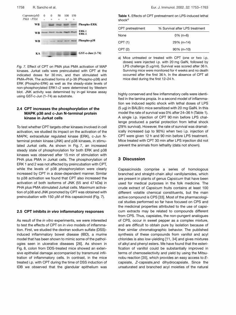
Fig. 7. Effect of CPT on PMA plus PMA activation of MAPkinases. Jurkat cells were preincubated with CPT at theindicated doses for 30 min, and then stimulated withPMA+PHA. The activated forms of p-38 (Phospho-p38) andERK (Phospho-ERK) as well as the steady-state levels ofnon-phosphorylated ERK1+2 were determined by Westernblot. JNK activity was determined by in-gel kinase assayusing GST-c-Jun (1–74) as substrate.
2.4 CPT increases the phosphorylation of theMAPK p38 and c-Jun N-terminal proteinkinase in Jurkat cells
To test whether CPT targets other kinases involved in cellactivation, we studied its impact on the activation of theMAPK; extracellular regulated kinase (ERK), c-Jun N-terminal protein kinase (JNK) and p38 kinases, in stimu-lated Jurkat cells. As shown in Fig. 7, an increasedsteady state of phosphorylation for both ERK and p38kinases was observed after 15 min of stimulation withPHA plus PMA in Jurkat cells. The phosphorylation ofERK 1 and 2 was not affected by preincubation with CPT,while the levels of p38 phosphorylation were clearlyincreased by CPT in a dose-dependent manner. Similarto p38 activation we found that CPT also increased theactivation of both isoforms of JNK (55 and 47 kDa) inPHA plus PMA stimulated Jurkat cells. Maximum activa-tion of p38 and JNK promoted by CPT was obtained withpreincubation with 150 ? M of this capsaicinoid (Fig. 7).
2.5 CPT inhibits in vivo inflammatory responses
As result of the in vitro experiments, we were interestedto test the effects of CPT on in vivo models of inflamma-tion. First, we studied the dextran sodium sulfate (DSS)-induced inflammatory bowel disease (IBD), a murinemodel that has been shown to mimic some of the pathol-ogies seen in ulcerative diseases [26]. As shown inFig. 8, colon from DDS-treated mice showed an exten-sive epithelial damage accompanied by transmural infil-tration of inflammatory cells. In contrast, in the micetreated i.p. with CPT during the time of DSS induction ofIDB we observed that the glandular epithelium was
Table 1. Effects of CPT pretreatment on LPS-induced lethalshocka)
CPT pretreatment % Survival after LPS treatment
None 0% (n=6)
CPT (1) 29% (n=14)
CPT (2) 90% (n=10)
a) Mice untreated or treated with CPT (one or two i.p.doses) were injected i.p. with 20 mg GalN, followed byLPS challenge (5 ? g/ml). Survival was scored after 36 h.Surviving mice were monitored for 4 weeks and no deathoccurred after the first 36 h. In the absence of CPT allmice died during the first 12–24 h.
highly conserved and few inflammatory cells were identi-fied in the lamina propia. In a second model of inflamma-tion we induced septic shock with lethal doses of LPS(5 ? g) in BALB/c mice sensitized with 20 mg GalN. In thismodel the rate of survival was 0% after 24–36 h (Table 1).A single i.p. injection of CPT 90 min before LPS chal-lenge produced a partial protection from lethal shock(29% survival). However, the rate of survival was dramat-ically increased (up to 90%) when two i.p. injection ofCPT were given 12 h and 90 min before LPS treatment.Mice treated with CPT 30 min after LPS injection did notprevent the animals from lethality (data not shown).
3 Discussion
Capsaicinoids comprise a series of homologousbranched and straight-chain alkyl vanillylamides, whichare present in plants of genus Capsicum that have beenused for medical purposes in the folk medicine. Thecrude extract of Capsicum fruits contains at least 100different volatile chemical constituents, but the mainactive compound is CPS [33]. Most of the pharmacologi-cal studies performed so far have focused on CPS andthe medicinal properties attributed to the use of capsi-cum extracts may be related to compounds differentfrom CPS. Thus, capsiates, the non-pungent analoguesof CPS, occur in sweet pepper as a complex mixture,and are difficult to obtain pure by isolation because oftheir similar chromatographic behavior. The publishedsynthesis of these compounds from vanillol and acylchlorides is also low-yielding [11, 34] and gives mixturesof alkyl and phenyl esters. We have found that the esteri-fication of vanillol could be substantially improved interms of chemoselectivity and yield by using the Mitsu-nobu reaction [35], which provides an easy access to E-capsiate, Z-capsiate,and dihydrocapsiate. Since theunsaturated and branched acyl moieties of the natural
1758 R. Sancho et al. Eur. J. Immunol. 2002. 32: 1753–1763

Fig. 8. CPT is efficacious in a mouse DSS-induced IBD model. The panels show sections of colon from control and from micereceiving 5% DSS treated i.p. with either CPT (DSS + CPT) or vehicle oil (DSS). The disruption of mucosal ephitelium, the loss ofglandular cripts and the infiltrate of inflammatory cells are clearly observed in DSS mice (DSS). In the colon section of DSS plusCPT-treated mice the glandular epithelium is almost intact and few inflammatory cells are present in the lamina propia.
capsiates are available only through a multi-step synthe-sis, a simplified version, CPT, of the natural products wasalso prepared and used through the entire study.
CPT inhibits NF- ‹ B activation by a variety of stimuli, sug-gesting that it interferes with a common step of the acti-vation of this pathway. In considering possible sites ofaction for CPT inhibition of NF- ‹ B activation, MEKK1and downstream kinases seemed likely candidates,since these kinases have been shown to participate insignaling through the TNF- § receptor and through theCD3/CD28 pathway [20, 21]. It is interesting that in ourhands CPT induced the activation of both p38 and JNKkinases, confirming previous results obtained with othervanilloids [6]. The activation of these kinases could bemediated by the prooxidant effects induced by capsaici-noids, similar to what has been shown for non-steroidealanti-inflammatory drugs in other cell system [36].
In addition to their role in cytokine signaling and cell sur-vival, NF- ‹ B proteins have been also implicated in regu-lating cell-cycle progression, since NF- ‹ B activity isinduced as cell pass from G0 into the G1 phase of the cellcycle [37]. Recently it has been shown that NF- ‹ B regu-lates transcriptionally the expression of Cyclin D1 gene
[38], and the cyclin D1 protein interacts with cyclin-dependent kinases, which phosphorylate and inactivatethe retinoblastoma tumor suppressor [39]. Thus, it ispossible that CPT and CPS inhibit the progression of thecell cycle to phase G1 in activated T cells by targetingNF- ‹ B and preventing the expression of the cyclin D1.
Neurogenic inflammation is a unique type of inflamma-tion induced by the release of neuropeptides/hormonesby afferent neurons into the space of peripheral nerveterminal. This type of inflammation may account for theabdominal pain in patients with Crohn’s disease, inwhich the expression of VR1 is greatly increased incolonic nerve fibres [40]. Upon VR1 activation thesecolonic nerve cells may release neurohormones thatinclude substance P, tachykinin, calcitonin gene-relatedpeptide and platelet-activating factors (PAF) [41]. Sub-stance P can play an important role in the process of Tcell infiltration, since it can up-regulate the expression ofadhesion molecules in endothelial cells [42] through anNF- ‹ B-dependent pathway [43], and also activates Tcells [44]. Whether CPS or CPT inhibits substance P-signaling pathways leading to NF- ‹ B activation is aninteresting point that remains to be investigated. PAF isanother important modulator of the neurogenic inflam-
Eur. J. Immunol. 2002. 32: 1753–1763 Capsiate inhibits NF- ‹ B 1759

mation that activates the phospholipase C pathway inneutrophils and macrophages [45]. So far, the anti-inflammatory effect of CPS was believed to be based onthe VR1-dependent desensitization of afferent neuronsthat secreted the inflammatory neurohormones. Interest-ingly, CPS may interfere with the inflammatory responseinduced by PAF in a VR1-independent fashion [5], a bio-logical activity that could also be shared by CPT. Thus,the anti-inflammatory activity of CPT would be extendedto biological activities other than NF- ‹ B inhibition.
Finally, an important point for CPT is its lack of toxicitywhen administered i.p., we used in the IDB model a dailydoses of CPT (240 mg/kg) that represent about 30-foldthe LD50 reported for CPS in mice (7.56 mg/kg) [46].Development of new anti-inflammatory drugs will requirepharmacokinetic and toxicity studies followed by clinicalverification of in vivo activity. Based on our results, cap-sacinoids devoid of VR1 activity could serve as leadcompounds for the development of novel, potent anti-inflammatory drugs for the treatment of inflammatorydisorders.
4 Materials and methods
4.1 Cell lines and reagents
Jurkat cells (ATCC, Rockville, MD) were maintained in expo-nential growth in RPMI 1640 (Bio-Whittaker, VerViers, Bel-gium) supplemented with 10% heat-inactivated FCS, 2 mML-glutamine, 1 mM Hepes and antibiotics (Gibco, Paisley,Scotland). The 5.1 clone (obtained from Dr. N. Isräel, InstitutPasteur, Paris, France) line is a Jurkat-derived clone stablytransfected with a plasmid containing the luciferase genedriven by the HIV-LTR promoter and was maintained in com-plete RPMI medium. The 57A cells (obtained from Dr. R. T.Hay, University of St. Andrews, St. Andrews, Scotland) is aHeLa-derived clone that contains NF- ‹ B-dependent lucifer-ase stably integrated into the genome [47], and were main-tained in DMEM medium (Bio-Whittaker) supplemented asthe RPMI 1640. The stably transfected cell lines were main-tained in the presence of G418 (100 ? g/ml). [ + -32P]ATP(3,000 Ci/mmol) was purchased from ICN (Costa Mesa, CA).The anti-I ‹ B § mAb 10B was a gift from R. T. Hay (St. And-rews, Scotland), the anti-phospho-p38 (9211S) Ab was fromNEB (Schwalbach, Germany), the anti- phospho-ERK(V8031) was from Promega (Mannheim, Germany), the anti-ERK 1+2 Ab was from Zymed (CA), the mAb anti-tubulin waspurchased from Sigma Chemical (St. Louis, MO) and therabbit policlonal anti-IKK- + (FL-419) was from Santa CruzBiotechnology (San Diego, CA). OKT3 (anti-CD3) was fromATCC and purified from tissue culture supernatant by pro-tein A, and the CD28.2 mAb (anti-CD28) was from BD Phar-mingen (San Diego, CA). All other reagents were fromSigma. The synthesis of E-capsiate, Z-capsiate, dihydro-
capsiate and nor-dihydrocapsiate will be published else-where.
4.2 Isolation of human peripheral mononuclear cellsand T cell proliferation assays
Human PBMC, from healthy adult volunteer donors, wereisolated by centrifugation of venous blood on Ficoll-Hypaque density gradients (Pharmacia LKB Biotechnology,Piscataway, NJ). T cells were purified by nylon wool column,and 5×10⁴ cells were cultured in triplicate in 96-well round-bottom microtiter plates (Costar, Cambrigde, MA) in 200 ? lof complete medium. The wells were previously coated withOKT3 (1 ? g/ml) or a combination or OKT3 plus anti-CD28(1 ? g/ml) in the presence or the absence either of CPS orCPT. The cultures were carried out for 3 days and pulsedwith 0.5 ? Ci [3H]dThd/well for the last 12 h of culture. Radio-activity incorporated into DNA was measured by liquid scin-tillation counting. For cell-cycle analyses, purified T cells(106) were stimulated with OKT3 (1 ? g/ml) in 24-well plates ina total volume of 2 ml of complete medium for 72 h.
4.3 Cytofluorimetric analyses of cell surface antigenand cell cycle
Purified T cells were stimulated for the indicated times withOKT3 in the presence or not of capsaicinoids, and surfaceexpression of the IL-2R (48 h), CD69 (24 h) or ICAM-1 (72 h)antigens were analyzed by flow cytometry using specificmAb. Cell surface fluorescence was measured in an EPIC XLAnalyser (Coulter, Hialeah, FL). For DNA profile analyses,cells were washed in PBS, fixed in ethanol (70%, for 24 h at4°C), followed by RNA digestion (RNase-A, 50 U/ml) andpropidium iodide (PI, 20 ? g/ml) staining, and analyzed bycytofluorimetry. Ten thousand gated events were collectedper sample and the percentage of cells in every phase of thecell cycle was determined. The frequency of cells havingundergone chromatinolysis was calculated by determiningthe sub G0/G1 fraction.
4.4 Luciferase assays
To determine NF- ‹ B dependent transcription, 57A or 5.1cells were preincubated with the capsaicinoids as indicatedfollowed by stimulation with TNF- § (2 ng/ml) or PMA (50 ng/ml) for 6 h. Then, the cells were lysed in 25 mM Tris-phosphate pH 7.8, 8 mM MgCl2, 1 mM DTT, 1% Triton X-100, and 7% glycerol. Luciferase activity was measuredusing an Autolumat LB 953 (EG&G Berthold, USA) followingthe instructions of the luciferase assay kit (Promega, Madi-son, WI) and protein concentration was measured by theBradford method. The background obtained with the lysisbuffer was subtracted in each experimental value and thespecific transactivation expressed as a fold induction. All theexperiments were repeated at least three times.
1760 R. Sancho et al. Eur. J. Immunol. 2002. 32: 1753–1763

4.5 Western blots and JNK assay
Jurkat cells (1×10⁶ cells/ml) were stimulated with PHA (2 ? g/ml) plus PMA (50 ng/ml) in the presence or absence of CPTfor the indicated period of times. Cells were then washedwith PBS and resuspended in 50 ? l of lysis buffer (20 mMHepes pH 8.0, 10 mM KCl, 0.15 mM EGTA, 0.15 mM EDTA,0.5 mM Na3VO4, 5 mM NaFl, 1 mM DTT, 1 ? g/ml leupeptin,0.5 ? g/ml pepstatin, 0.5 ? g/ml apronitin, and 1 mM PMSF)containing 0.5% NP-40. After 5 min on ice, cytoplasmic pro-teins were obtained by centrifugation and protein concen-tration determined by a Bradford assay (Bio-Rad, Rich-mond, CA). Protein (30 ? g) was boiled in Laemmli buffer andelectrophoresed by SDS-PAGE using 10% gels. Separatedproteins were transferred to nitrocellulose membranes (0.5 Aat 100 V; 4°C) for 1 h. The blots were blocked in TBS solu-tion containing 0.1% Tween 20 and 5% nonfat dry milk over-night at 4°C, and immunodetection of specific proteins wascarried out with primary antibodies (anti-I ‹ B § , anti- § -tubuline, anti-phospho-ERK, anti-phospho-p38 or anti-ERK1+2) using an ECL system (Amersham, GB). In-gel JNKassay in Jurkat cells (2×10⁶) were performed as previouslydescribed [6].
4.6 IKK kinase assay
Cells were lysed in NP-40 lysis buffer [30] for 15 min at 4°C,and after centrifugation for 10 min at 13,000 rpm, the super-natant was incubated with 25 ? l protein A/G Sepharose andincubated for 2 h on a spinning wheel. After centrifugation,the supernatants were incubated with 2 ? g of anti-IKK- +antibody and 25 ? l protein A/G Sepharose and incubated for2–4 h on a spinning wheel at 4°C. The precipitate waswashed three times in cold lysis buffer and three times incold kinase buffer (20 mM Hepes/KOH pH 7,4, 25 mM g -glycerophosphate, 2 mM DTT, 20 mM MgCl2). The kinaseassay was performed in a final volume of 20 ? l kinase buffercontaining 40 ? M ATP and 5 ? Ci [ + -32P]ATP and 2 ? g of thepurified substrate proteins GST-I ‹ B § (1–54). After incubationfor 20 min at 30°C, the reaction was stopped by the additionof 5× SDS loading buffer. After separation by SDS-PAGE thegel was fixed, dried and quantified using a PhosphorImager.
4.7 Isolation of nuclear extracts and mobility shiftassays
Jurkat cells were cultured at 2×10⁶/ml and stimulated withthe agonists in complete medium as indicated. Cells werethen washed twice with cold PBS and proteins from nuclearextracts isolated as previously described [48]. For the EMSAassay, the consensus oligonucleotide probes NF- ‹ B andSP1 site (Promega) were end-labeled with [ + -32P]ATP. EMSAwere performed with 3 ? g of nuclear extract [48], and whenindicated, 0.5 ? l of rabbit anti-p50, anti-p65 or pre-immuneserum was added to the standard reaction before the addi-tion of the radiolabeled probe. For cold competition, a 100-
fold excess of the double-stranded oligonucleotide compet-itor was added to the binding reaction.
4.8 Animal studies
For the septic model, BALB/c mice were administered withD-galactosamine (GalN) and 30 min later injected i.p. withLPS (5 ? g) (serotype 026:B26, Lot 69H4022). Treated ani-mals were injected i.p. with CPT (6 mg vehiculized in 200 ? lof olive oil) either with one dose 90 min or with two doses12 h and 90 min before LPS challenge. Control animalsreceived the same volume of vehicle. The mortality wasmeasured at 24–36 h. For the DSS study, Swiss-Webstermice were administered 5% DSS in drinking water from day0 to day 6 and injected i.p. with CPT (6 mg vehiculized as forseptic shock experiments) from day 1 to day 6. On day 10the mice were sacrificed, and colon removed and preparedfor histomorphological studies.
Acknowledgements: The authors wish to thank Dr. NicoleIsräel (Institute Pasteur, Paris, France) for the 5.1 cells, Dr.Alain Isräel (Institute Pasteur) for the anti-p50 and anti-p65antisera, Dr. R.T. Hay (St. Andrews, Scotland) for the mAb10B and the 57A cells, and Ms. Carmen Cabrero-Doncel forassistance with the manuscript. This work was supported byMCyT grant Bio 2000-1091-C01 to E. M. and by the EUgrant QLK3-CT-2000-00463.
References
1 Szallasi, A., Vanilloid receptors ligands: hopes and realities forthe future. Drugs Aging 2001. 18: 561–573.
2 Nilsson, G., Alving, K. and Ahlstedt, S., Effects on immuneresponses in rats after neuromanipulation with capsaicin. Int. J.Immunopharmacol. 1991. 13: 21–26.
3 Eglezos, A., Andrews, P. V., Boyd, R. L. and Helme, R. D.,Effects of capsaicin treatment on immunoglobulin secretion inthe rat: further evidence for involvement of tachykinin-containingafferent nerves. J. Neuroimmunol. 1990. 26: 131–138.
4 Ho, W. Z., Lai, J. P., Zhu, X. H., Uvaydova, M. and Douglas, S.D., Human monocytes and macrophages express substance Pand neurokinin-1 receptor. J. Immunol. 1997. 159: 5654–5660.
5 Choi, S. Y., Ha, H. and Kim, K. T., Capsaicin inhibits platelet-activating factor-induced cytosolic Ca2+ rise and superoxide pro-duction. J. Immunol. 2000. 165: 3992–3998.
6 Macho, A., Blazquez, M. V., Navas, P. and Munoz, E., Inductionof apoptosis by vanilloid compounds does not require de novogene transcription and AP-1. Cell Growth Differ. 1998. 9:277–286.
7 Morre, D. J., Chueh, P.-J. and Morre, D .M., Capsaicin inhibitspreferentially the NADH oxidase and growth of transformed cellsin culture. Proc. Natl. Acad. Sci. USA 1995. 92: 1831–1835.
8 Matucci-Cerinic, M., Marabini, S., Jantsch, S., Cagnoni, M.and Partsch, G., Effects of capsaicin on the metabolism of rheu-
Eur. J. Immunol. 2002. 32: 1753–1763 Capsiate inhibits NF- ‹ B 1761

matoid arthritis synoviocytes in vitro. Ann. Rheum. Dis. 1990. 49:598–602.
9 Singh, S., Natarajan, K. and Aggarwal, B. B., Capsaicin (8-methyl-N-vanillyl-6-nonenamide) is a potent inhibitor of nucleartranscription factor-kappa B activation by diverse agents.J. Immunol. 1996. 157: 4412–4420.
10 Yazawa, S., Suetome, N., Okamoto, K. and Namiki, T., Contentof capsaicinoids and capsaicinoid-like substances in fruit of pep-per (Capsicum annuum L.) hybrids made with “CH-19 Sweet” asa parent. J. Jpn. Soc. Hortic. Sci. 1989. 58: 601–607.
11 Kobata, K., Sutoh, K., Todo, T., Yazawa, S., Iwai, K. and Wata-nabe, T., Nordihydrocapsiate, a new capsaicinoid from the fruitsof a nonpungent pepper, Capsicum annuum. J. Natural Prod.1999. 62: 335–336.
12 Karin, M. and Ben Neriah, Y. Phosphorylation meets ubiquitina-tion: the control of NF- ‹ B activity. Annu. Rev. Immunol. 2000. 18:621–663.
13 Woronicz, J. D., Gao, X., Cao, Z., Rothe, M. and Goeddel, D.V., I ‹ B kinase- g : NF- ‹ B activation and complex formation withI ‹ B kinase- § and NIK. Science 1997. 278: 866–869.
14 Ling, L., Cao, Z. and Goeddel, D. V., NF-kappaB-inducingkinase activates IKK-alpha by phosphorylation of Ser-176. Proc.Natl. Acad. Sci. USA 1998. 95: 3792–3797.
15 Karin, M., Mitogen-activated protein kinase cascades as regula-tors of stress responses. Ann. NY Acad. Sci. 1998. 851: 139–146.
16 Pomerantz, J. L. and Baltimore, D., NF-kappaB activation by asignaling complex containing TRAF2, TANK and TBK1, a novelIKK-related kinase. EMBO J. 1999. 18: 6694–6704.
17 Tojima, Y., Fujimoto, A., Delhase, M., Chen, Y., Hatakeyama,S., Nakayama, K., Kaneko, Y., Nimura, Y., Motoyama, N.,Ikeda, K., Karin, M. and Nakanishi, M., NAK is an IkappaBkinase-activating kinase. Nature 2000. 404: 778–782.
18 Ninomiya-Tsuji, J., Kishimoto, K., Hiyama, A., Inoue, J., Cao,Z. and Matsumoto, K., The kinase TAK1 can activate the NIK-IkappaB as well as the MAP kinase cascade in the IL-1 signalingpathway. Nature 1999. 398: 252–256.
19 Ozes, O. N., Mayo, L. D., Gustin, J. A., Pfeffer, S. R., Pfeffer, L.M. and Donner, D. B., NF-kappaB activation by tumour necrosisfactor requires the Akt serine-threonine kinase. Nature 1999. 401:82–85.
20 Lin, X., Cunningham, E. T., Mu, Y., Geleziunas, R. and Greene,W. C., The proto-oncogene Cot kinase participates in CD3/CD28induction of NF-kappaB acting through the NF-kappaB-inducingkinase and IkappaB kinases. Immunity 1999. 10: 271–280.
21 Khoshnan, A., Bae, D., Tindell, C. A. and Nel, A. E., The physi-cal association of protein kinase C theta with a lipid raft-associated inhibitor of kappa B factor kinase (IKK) complex playsa role in the activation of the NF-kappa B cascade by TCR andCD28. J. Immunol. 2000. 165: 6933–6940.
22 Tak, P. P. and Firestein, G. S., NF-kappaB: a key role in inflam-matory diseases. J. Clin. Invest. 2001. 107: 7–11.
23 Ghosh, S., May, M. J. and Kopp, E. B., NF-kappa B and Rel pro-teins: evolutionarily conserved mediators of immune responses.Annu. Rev. Immunol. 1998. 16: 225–260.
24 Tracey, K. J., Fong, Y., Hesse, D. G., Manogue, K. R., Lee, A.T., Kuo, G. C., Lowry, S. F. and Cerami, A., Anti-cachectin/TNFmonoclonal antibodies prevent septic shock during lethal bacte-raemia. Nature 1987. 330: 662–664.
25 Parrillo, J. E., Pathogenetic mechanisms of septic shock. N.Engl. J. Med. 1993. 328: 1471–1477.
26 Neurath, M. F., Fuss, I., Schurmann, G., Pettersson, S.,Arnold, K., Muller Lobeck, H., Strober, W., Herfarth, C. andBuschenfelde, K. H., Cytokine gene transcription by NF-kappaB family members in patients with inflammatory bowel disease.Ann. NY Acad. Sci. 1998. 859: 149–159.
27 Ellis, R. D., Goodlad, J. R., Limb, G. A., Powell, J. J., Thomp-son, R. P. and Punchard, N. A., Activation of nuclear factorkappa B in Crohn’s disease. Inflamm. Res. 1998. 47: 440–445.
28 Dumont, A., Hehner, S. P., Schmitz, M. L., Gustafsson, J. A.,Liden, J., Okret, S., van der Saag, P. T., Wissink, S., van derBurg, B., Herrlich, P., Haegeman, G., De Bosscher, K. andFiers, W., Cross-talk between steroids and NF-kappa B: whatlanguage? Trends Biochem. Sci. 1998. 23: 233–235.
29 Yin, M. J., Yamamoto, Y. and Gaynor, R. B., The anti-inflammatory agents aspirin and salicylate inhibit the activity ofI(kappa)B kinase-beta. Nature 1998. 396: 77–80.
30 Hehner, S. P., Hofmann, T. G., Droge, W. and Schmitz, M. L.,The antiinflammatory sesquiterpene lactone parthenolide inhibitsNF-kappa B by targeting the I kappa B kinase complex. J. Immu-nol. 1999. 163: 5617–5623.
31 Haridas, V., Higuchi, M., Jayatilake, G. S., Bailey, D., Mujoo,K., Blake, M. E., Arntzen, C. J. and Gutterman, J. U., Avicins:triterpenoid saponins from Acacia victoriae (Bentham) induceapoptosis by mitochondrial perturbation. Proc. Natl. Acad. Sci.USA 2001. 98: 5821–5826.
32 Natarajan, K., Singh, S., Burke, T. R., Grunberger, D. andAggarwal, B. B., Caffeic acid phenethyl ester is a potent andspecific inhibitor of activation of nuclear transcription factor NF-kappa B. Proc. Natl. Acad. Sci. USA 1996. 93: 9090–9095.
33 Cordell, G. A. and Araujo, O. E., Capsaicin: identification,nomenclature, and pharmacotherapy. Ann. Pharmacother. 1993.27: 330–336.
34 Kobata, K., Todo, T., Yazawa, S., Iwai, K. and Watanabe, T.,Novel capsaicinoid-like substances, capsiate and dihydrocapsi-ate, from the fruits of a nonpungent cultivar, CH-19 sweet, ofpepper (Capsicum annuum L.). J. Agr. Food Chem. 1998.1695–1697.
35 Mitsunobu, O., The use of diethyl azodicarboxylate and TPP insynthesis and transformation of natural products. Synthesis1981. 1–28.
36 Lennon, A. M., Ramauge, M. and Pierre, M., Role of redox sta-tus on the activation of mitogen-activated protein kinase cas-cades by NSAIDs. Biochem. Pharmacol. 2002. 63: 163–170.
37 Baldwin, A. S., Azizkhan, J. C., Jensen, D. E., Beg, A. A. andCoodly, L. R. Induction of NF-kappa B DNA-binding activity dur-ing the G0-to-G1 transition in mouse fibroblasts. Mol. Cell Biol.1991. 11: 4943–4951.
38 Joyce, D., Bouzahzah, B., Fu, M., Albanese, C., D’Amico, M.,Steer, J., Klein, J. U., Lee, R. J., Segall, J. E., Westwick, J. K.,Der, C. J. and Pestell, R. G., Integration of Rac-dependent regu-lation of cyclin D1 transcription through a nuclear factor-kappaB-dependent pathway. J. Biol. Chem. 1999. 274: 25245–25249.
39 Weinberg, R.A., The retinoblastoma protein and cell cycle con-trol. Cell 1995. 81: 323–330.
40 Yiangou, Y., Facer, P., Dyer, N. H., Chan, C. L., Knowles, C.,Williams, N. S. and Anand, P., Vanilloid receptor 1 immunoreac-tivity in inflamed human bowel. Lancet 2001. 357: 1338–1339.
41 Baron, R., Capsaicin and nociception: from basic mechanisms tonovel drugs. Lancet 2000. 356: 785–787.
42 Quinlan, K. L., Song, I. S., Naik, S. M., Letran, E. L., Olerud, J.E., Bunnett, N. W., Armstrong, C. A., Caughman, S. W. andAnsel, J. C., VCAM-1 expression on human dermal microvascu-
1762 R. Sancho et al. Eur. J. Immunol. 2002. 32: 1753–1763

lar endothelial cells is directly and specifically up-regulated bysubstance P. J. Immunol. 1999. 162: 1656–1661.
43 Quinlan, K. L., Naik, S. M., Cannon, G., Armstrong, C. A., Bun-nett, N. W., Ansel, J. C. and Caughman, S. W. Substance Pactivates coincident NF-AT- and NF-kappa B-dependent adhe-sion molecule gene expression in microvascular endothelial cellsthrough intracellular calcium mobilization. J. Immunol. 1999. 163:5656–5665.
44 Payan, D., The role of neuropeptides in inflammation. In Gallin,J., Goldstein, I. and Snyderman, R. (Eds.) Inflammation: basicprinciples and clinical correlates. Raven Press, New York 1992,2nd edn., p 177.
45 Elzi, D. J., Hiester, A. A. and Silliman, C. C., Receptor-mediatedcalcium entry is required for maximal effects of platelet activatingfactor primed responses in human neutrophils. Biochem. Bio-phys. Res. Commun. 1997. 240: 763–765.
46 Monsereenusorn, Y., Kongsamut, S. and Pezalla, P. D.,Capsaicin-a literature survey. Crit. Rev. Toxicol. 1982. 10:321–339.
47 Rodriguez, M. S., Thompson, J., Hay, R. T. and Dargemont,C., Nuclear retention of IkappaBalpha protects it from signal-induced degradation and inhibits nuclear factor kappaB tran-scriptional activation. J. Biol. Chem. 1999. 274: 9108–9115.
48 Munoz, E., Courtois, G., Veschambre, P., Jalinot, P. and Israel,A., Tax induces nuclear translocation of NF-kappa B through dis-sociation of cytoplasmic complexes containing p105 or p100 butdoes not induce degradation of I kappa B alpha/MAD3. J. Virol.1994. 68: 8035–8044.
Correspondence: Eduardo Munoz, Departamento de Bio-logıa Celular, Fisiologıa e Inmunologıa. Facultad de Medi-cina. Avda. de Menendez Pidal s/n, E-14004 Cordoba,Spaine-mail: fi1muble — uco.es
Eur. J. Immunol. 2002. 32: 1753–1763 Capsiate inhibits NF- ‹ B 1763