Recent Topics on The Mechanisms of Immunosuppressive ...
27
Int. J. Mol. Sci. 2019, 20, 3210; doi:10.3390/ijms20133210 www.mdpi.com/journal/ijms Review Recent Topics on The Mechanisms of Immunosuppressive Therapy-Related Neurotoxicities Wei Zhang 1 , Nobuaki Egashira 1,2, * and Satohiro Masuda 1,2 1 Department of Clinical Pharmacology and Biopharmaceutics, Graduate School of Pharmaceutical Sciences, Kyushu University, Fukuoka 812-8582, Japan 2 Department of Pharmacy, Kyushu University Hospital, Fukuoka 812-8582, Japan * Correspondence: [email protected], Tel.: 81-92-642-5920 Received: 29 May 2019; Accepted: 28 June 2019; Published: 29 June 2019 Abstract: Although transplantation procedures have been developed for patients with end-stage hepatic insufficiency or other diseases, allograft rejection still threatens patient health and lifespan. Over the last few decades, the emergence of immunosuppressive agents such as calcineurin inhibitors (CNIs) and mammalian target of rapamycin (mTOR) inhibitors have strikingly increased graft survival. Unfortunately, immunosuppressive agent-related neurotoxicity commonly occurs in clinical practice, with the majority of neurotoxicity cases caused by CNIs. The possible mechanisms through which CNIs cause neurotoxicity include increasing the permeability or injury of the blood– brain barrier, alterations of mitochondrial function, and alterations in the electrophysiological state. Other immunosuppressants can also induce neuropsychiatric complications. For example, mTOR inhibitors induce seizures, mycophenolate mofetil induces depression and headaches, methotrexate affects the central nervous system, the mouse monoclonal immunoglobulin G2 antibody (used against the cluster of differentiation 3) also induces headaches, and patients using corticosteroids usually experience cognitive alteration. Therapeutic drug monitoring, individual therapy based on pharmacogenetics, and early recognition of symptoms help reduce neurotoxic events considerably. Once neurotoxicity occurs, a reduction in the drug dosage, switching to other immunosuppressants, combination therapy with drugs used to treat the neuropsychiatric manifestation, or blood purification therapy have proven to be effective against neurotoxicity. In this review, we summarize recent topics on the mechanisms of immunosuppressive drug-related neurotoxicity. In addition, information about the neuroprotective effects of several immunosuppressants is also discussed. Keywords: alloimmune response; immunosuppressants; calcineurin inhibitors; corticosteroids; mTOR inhibitors; neurotoxicity; neuroprotective effects 1. Introduction The first kidney transplant, performed by Murray et al. in 1954 [1], heralded a new age for patients with terminal hepatic insufficiency, end-stage renal diseases, and other severe diseases. However, the one-year survival rate of transplant patients was only 35% in the 1960s and 1970s and did not significantly increase until the development of ciclosporin A (cyclosporine, CsA) and tacrolimus (FK506) [2]. Strikingly, the rapid development of drugs to induce and maintain immunosuppression, such as antibodies and anti-metabolic drugs, has helped to increase graft and one-year patient survival to more than 90% in recent years [3]. Based on pharmacological mechanisms, immunosuppressive agents can be divided into six categories: calcineurin inhibitors (CNIs), mammalian target of rapamycin (mTOR) inhibitors, cell cycle inhibitors, corticosteroids, monoclonal and polyclonal antibodies, and other newly developed drugs [4]. Although many benefits have been realized, postoperative complications remain unsolved and influence the quality of life and long-term survival rates of transplant patients [5]. Among all
-
Upload
khangminh22 -
Category
Documents
-
view
3 -
download
0
Transcript of Recent Topics on The Mechanisms of Immunosuppressive ...

Int. J. Mol. Sci. 2019, 20, 3210; doi:10.3390/ijms20133210 www.mdpi.com/journal/ijms
Review
Recent Topics on The Mechanisms of Immunosuppressive Therapy-Related Neurotoxicities
Wei Zhang 1, Nobuaki Egashira 1,2,* and Satohiro Masuda 1,2
1 Department of Clinical Pharmacology and Biopharmaceutics, Graduate School of Pharmaceutical Sciences,
Kyushu University, Fukuoka 812-8582, Japan 2 Department of Pharmacy, Kyushu University Hospital, Fukuoka 812-8582, Japan
* Correspondence: [email protected], Tel.: 81-92-642-5920
Received: 29 May 2019; Accepted: 28 June 2019; Published: 29 June 2019
Abstract: Although transplantation procedures have been developed for patients with end-stage
hepatic insufficiency or other diseases, allograft rejection still threatens patient health and lifespan.
Over the last few decades, the emergence of immunosuppressive agents such as calcineurin
inhibitors (CNIs) and mammalian target of rapamycin (mTOR) inhibitors have strikingly increased
graft survival. Unfortunately, immunosuppressive agent-related neurotoxicity commonly occurs in
clinical practice, with the majority of neurotoxicity cases caused by CNIs. The possible mechanisms
through which CNIs cause neurotoxicity include increasing the permeability or injury of the blood–
brain barrier, alterations of mitochondrial function, and alterations in the electrophysiological state.
Other immunosuppressants can also induce neuropsychiatric complications. For example, mTOR
inhibitors induce seizures, mycophenolate mofetil induces depression and headaches, methotrexate
affects the central nervous system, the mouse monoclonal immunoglobulin G2 antibody (used
against the cluster of differentiation 3) also induces headaches, and patients using corticosteroids
usually experience cognitive alteration. Therapeutic drug monitoring, individual therapy based on
pharmacogenetics, and early recognition of symptoms help reduce neurotoxic events considerably.
Once neurotoxicity occurs, a reduction in the drug dosage, switching to other immunosuppressants,
combination therapy with drugs used to treat the neuropsychiatric manifestation, or blood
purification therapy have proven to be effective against neurotoxicity. In this review, we summarize
recent topics on the mechanisms of immunosuppressive drug-related neurotoxicity. In addition,
information about the neuroprotective effects of several immunosuppressants is also discussed.
Keywords: alloimmune response; immunosuppressants; calcineurin inhibitors; corticosteroids;
mTOR inhibitors; neurotoxicity; neuroprotective effects
1. Introduction
The first kidney transplant, performed by Murray et al. in 1954 [1], heralded a new age for
patients with terminal hepatic insufficiency, end-stage renal diseases, and other severe diseases.
However, the one-year survival rate of transplant patients was only 35% in the 1960s and 1970s and
did not significantly increase until the development of ciclosporin A (cyclosporine, CsA) and
tacrolimus (FK506) [2]. Strikingly, the rapid development of drugs to induce and maintain
immunosuppression, such as antibodies and anti-metabolic drugs, has helped to increase graft and
one-year patient survival to more than 90% in recent years [3]. Based on pharmacological
mechanisms, immunosuppressive agents can be divided into six categories: calcineurin inhibitors
(CNIs), mammalian target of rapamycin (mTOR) inhibitors, cell cycle inhibitors, corticosteroids,
monoclonal and polyclonal antibodies, and other newly developed drugs [4].
Although many benefits have been realized, postoperative complications remain unsolved and
influence the quality of life and long-term survival rates of transplant patients [5]. Among all

Int. J. Mol. Sci. 2019, 20, 3210 2 of 27
postoperative complications, neurological problems are frequent, both in the immediate operation
period and for many years after transplantation; they are associated with a poor prognosis and
significant morbidity [6,7]. For example, van de Beek and colleagues [8] reported that the rate of
perioperative neurological complications was associated with one-year mortality and rose from 19%
to 30% in the past 10 years, as shown in a retrospective cohort study. Furthermore, the risk of
neurological complications was shown to be 81% in patients during 18 years of follow-up. Common
complications seen with all types of transplantation include alterations of consciousness, seizures,
encephalopathy, and cerebrovascular events [9–12]. The etiologies of neurological complications are
diverse, including immunosuppressant-related neurotoxicity [13,14], infections [15], metabolic
disorders, hemorrhages [9], and primitive diseases prior to the transplant. Neurotoxicity induced by
immunosuppressive agents has remained a severe problem in clinical practice because they degrade
the quality of life for patients. For example, CNIs may induce mild symptoms, such as tremors, or
severe symptoms, such as seizures, central pontine myelinolysis (CPM), and cortical blindness.
Treatment with a mouse monoclonal immunoglobulin G2 antibody to the cluster of differentiation 3
(muromonab-CD3, trade name: Orthoclone OKT3® ) is associated with headaches and aseptic
meningitis. These clinical features and risk factors are well understood. However, the specific
mechanisms of immunosuppressant-related neurotoxicity, and its predictive factors, remain obscure.
Over the last few decades, several attempts have been made to elucidate the pathogenesis of
immunosuppressant-related neurotoxicity and to recognize its heralding symptoms. In this article,
we focus on the clinical features, risk factors, pathological mechanisms, and the management of
neurotoxicity induced by immunosuppressive agents.
2. Alloimmune Response
Once cells, tissues, or organs are transplanted between a donor and a genetically non-identical
recipient (allograft transplantation), many cells, including T cells, B cells, and macrophages, are
activated and participate in immune events that can initiate an alloimmune response and, finally,
induce allograft rejection.
2.1. Allorecognition
As shown in Figure 1a, allorecognition is initiated by two pathways: (1) activated T cells with
direct alloreactivity interact with major histocompatibility complex (MHC) molecule–peptide
complexes on donor antigen presenting cells (APCs) and induce donor cell apoptosis through cellular
rejection [16], and (2) donor peptides bound to self-derived MHC molecule peptide complexes
processed by recipient APCs are recognized by recipient T cells and then cause allograft destruction
[17]. Nowadays, a distinct pathway, semi-direct allorecognition has been studied in the context of
transplantation.
.

Int. J. Mol. Sci. 2019, 20, 3210 3 of 27
Figure 1. T cells, B cells, and macrophages initiate alloimmune responses and induce allograft
rejection after transplantation. (a) Allorecognition can be initiated by direct or indirect pathways; (b)
Three signals participate in the activation of T cells; (c) Two signal processes are involved in the
activation of B cells. APC, antigen-presenting cell; BCR, B-cell receptor; DSA, donor specific antibody;
IKK, inhibitor of NF-κB kinase; IL-2, interleukin-2; NO, nitric oxide; MAP, mitogen-activated protein;
MHC, major histocompatibility complex; PI-3K, phosphatidylinositol 3-kinase; TCR, T-cell receptor;
Tfh, T follicular helper; TNF-α, tumor necrosis factor alpha.
2.2. T-cell Activation
T cells can be activated by three types of signals (Figure 1b). First, there is the T-cell receptor-
CD3 (TCR-CD3) complex on CD4+ T cells, which delivers cognate antigens and forms T-cell receptor-
major histocompatibility complex allopeptides that activate a series of biochemical reactions. Second,
T cells that have received an initial signal activation are activated by the interaction between CD80
or CD86 in APCs and CD28 molecules in T cells, finally generating a co-stimulatory signal, thereby
initiating immunological activation. Several signaling pathways, including the calcineurin pathway,
renin–angiotensin system (RAS)/mitogen activated protein (MAP) kinase pathway, and nuclear
factor kappa B (NFκB), inhibitor of NFκB kinase (IKK) pathway have been reported to participate in
these two activation processes. The third type of activation signal involves the binding of interleukin-
2 (IL-2) with the gamma chain of its receptor to initiate T cell proliferation, DNA synthesis, and cell
division through the activation of mTOR pathways [18].
2.3. B-cell Activation
Two signal processes account for the activation of B cells [19], as described in detail in Figure 1c.
The first activation signal occurs when macrophages in the subcapsular sinus capture the cognate
antigens, and then, these antigens on macrophages bind with the surface B cell receptors (BCRs) of

Int. J. Mol. Sci. 2019, 20, 3210 4 of 27
native B cells, forming an immunological synapse [20]. Signaling BCR microclusters are involved in
the process of moving antigens into the endosomal compartments of B cells and in the expression of
a series of factors that play an important role in regulating downstream signaling pathways,
including calcineurin and mTOR pathways [19,21]. The antigens are then processed enzymatically,
internalized, and ultimately selected to present on the surface of the B cell associated with MHC II
molecules [22].
Native B cells are activated with the help of follicular helper T (Tfh) cells, which initiate cell co-
stimulation interactions that produce cytokines [23–25]. The activated B cells migrate into the
germinal center where some of them differentiate into memory B cells or plasmablasts. Plasmablasts
further differentiate into long-lived plasma cells on bone marrow, which can secrete high-affinity
donor-specific antibodies that participate in antibody-mediated rejection [26,27].
3. Classification of Immunosuppressants
The functions of T and B lymphocytes in the process of rejection have become gradually
understood, and the immunosuppressive regime has been optimized as a result of many
experimental and clinical studies. Immunosuppressants are classified according to their mechanisms
of action, as shown in Table 1 [3,4,18,28–34]. CNIs inhibit the activity of a calcium-dependent
phosphatase named calcineurin, thereby impeding the transduction of the nuclear factor of activated
T cells (NFAT) and the production of cytokines, such as IL-2, tumor necrosis factor-alpha (TNF-α),
and interferon-gamma (IFN-γ). mTOR inhibitors suppress the translation of mRNA-encoding
proteins, T cell proliferation, and cytokine production. Antimetabolites inhibit the synthesis of purine
by diverse mechanisms, such as inhibiting inosine-5′-monophosphate dehydrogenase (IMPDH) or
incorporating 6-mercaptopurine into newly synthesized DNA to block purine synthesis enzymes.
Corticosteroids, in combination with CNIs and antimetabolites, are used as the cornerstones of
immunosuppressive regimens. Their immunosuppressive mechanism is diverse and may relate to
interference with intracellular transcription factors and the signaling pathways of several surface
receptors. Monoclonal and polyclonal antibodies may interact with cell surface antigens, such as CD3,
CD20, and CD25. Immunosuppressants that are currently being developed include antibodies,
FK778, Janus kinase (JAK) inhibitors, fingolimod, and blinatumomab. Among these
immunosuppressive agents, those that can cause neuropsychiatric complications are CNIs, mTOR
inhibitors, mycophenolate mofetil, corticosteroids, and some monoclonal antibodies, such as OKT3,
belatacept, and blinatumomab.

Int. J. Mol. Sci. 2019, 20, 3210 5 of 27
Table 1. The details of various immunosuppressive agents shown according to their classification.
Corticosteroids
Generic Name Prednisone; Prednisolone; Methylprednisolone; Dexamethasone
Trade Name Prelone® , Orapred® , Millipred® , Orapred ODT® ; Prednisol® , Pred Forte® , Pred Mild® , Omnipred® ; Medrol® , Medrol Dosepak® ,
MethylPREDNISolone Dose Pack® , Solu-Medrol® ; Decadron® , Dexamethasone Intensol® , Dexasone® , Hexadrol®
Mechanism of
Action
The mechanisms of action are diverse and include interference with intracellular transcription factors and signaling pathways of
several surface receptors, including the T cell antigen receptor and downstream kinases, thereby blocking the transcription of cytokine
genes and inhibiting cytokine production by T cells and macrophages [32].
Role in
Therapy
Maintenance; high doses of corticosteroids (>1 mg/kg), used for induction therapy in transplantation; treatment of acute cellular
rejection and AMR [3,32].
Adverse Effects
Hypertension, hyperlipidemia, glucose intolerance, malignancy, Cushingoid features, sleep disturbances, mood changes, impaired
wound healing, osteoporosis, psychosis, photosensitivity, acne hirsutism, avascular necrosis, weight gain, fluid retention, increased
appetite, menstrual irregularities, growth inhibition, GI disturbance, cataracts, infection [3].
Monitoring
Parameters Glucose, blood pressure, fasting lipid panel, weight, DEXA scan, eye exam, intensive organ function monitoring [31].
Other
Information
Their role in the maintenance of immunosuppression is under investigation because of severe side effects during long-term use, but an
immunosuppressive strategy without steroids could be only tried in low immunological risk transplant recipients; it also seems that
treatment of steroids 1 h prior to ATG preoperatively may minimize CRS [3,4].
Purine synthesis inhibitors
Generic Name Azathioprine; Mycophenolate mofetil; Mycophenolate sodium; Cyclophosphamide
Trade Name Imuran® ; Cellcept® ; Myfortic® ; Cytoxan® , Neosar® , Endoxan®
Mechanism of
Action
Two distinct mechanisms participate in the inhibition of de novo DNA synthesis block cell division and then block cell division. AZA
is a prodrug for 6-mercaptopurine, Mycophenolate mofetil is a prodrug of MPA and Mycophenolate sodium is an enteric-coated
formulation of MPA. AZA blocks purine synthesis enzymes by incorporating into newly synthetized DNA and, finally, impedes DNA
and RNA synthesis [31]. MPA selectively and noncompetitively inhibits a key enzyme in the de novo synthesis of purine named
IMPDH and thus, inhibits proliferation of T and B lymphocyte [32].
Role in
Therapy Maintenance
Adverse Effects AZA: Hepatotoxicity, bone marrow suppression, malignancies (high dosages), macrocytic anemia, GI disturbance, alopecia,
pancreatitis, infections [31];

Int. J. Mol. Sci. 2019, 20, 3210 6 of 27
MMF and Mycophenolate sodium: Dyslipidemia, DM, infections, bone marrow suppression, GI symptoms and anemia are common,
while nephrotoxicity, neurotoxicity, and hepatotoxicity are uncommon [18,30];
CP: Low blood count, alopecia, GI symptoms, poor appetite, discoloration of the skin or nails.
Monitoring
Parameters
AZA: CBC, LFT, amylase, lipase, TPMT enzyme level;
MMF and Mycophenolate sodium: CBC, REMS;
CP: CBC, LFT, KFT [31].
Other
Information
Newer trials have shown that AZA and MMF have similar efficacy. Low or absent TPMT activity is associated with increased AZA-
associated myelosuppression. MPA is associated with pregnancy loss and congenital malformations when used during pregnancy.
MPA may be of special interest in preventing the rise of DSA titers in pre-sensitized recipients. Patients with renal dysfunction need
dosage adjustment when using MPA [28,31].
CP is associated with pregnancy loss and congenital malformations when used during pregnancy.
CNIs
Generic Name Tacrolimus; Cyclosporine
Trade Name Prograf® , Graceptor® , Advagraf® , Envarsus XR® , Astagraf XL® ; Neoral® , Gengraf® , Sandimmune®
Mechanism of
Action
CNIs block signal transduction by activated NFAT through two distinct mechanisms. Tacrolimus binds to FKBP12 while CsA in
combination of cyclophilin inhibits calcineurin-mediated dephosphorylation of NFAT, ultimately preventing cytokine transduction
including IL-2 and IFNγ and T cell activation. In humoral immune response, CNIs interfere with T helper signals rather than targeting
B cell directly [32].
Role in
Therapy Maintenance
Adverse Effects
Often dose- and concentration- dependent, nephrotoxicity, infections, hyperkalemia, hypomagnesemia, hyperuricemia, cholelithiasis,
GI symptoms, malignancy; tacrolimus > CsA: insulin-dependent diabetes mellitus, neurotoxicity; CsA > tacrolimus: hypertension,
hypercholesterolemia, hyperlipidemia; CsA only: gingival hyperplasia, hirsutism; tacrolimus only: alopecia [3,4,31].
Monitoring
Parameters Trough levels, serum creatinine, potassium, magnesium, uric acid [31]
Other
Information
Tacrolimus seems more effective than CsA-based immunosuppressive regimens, so tacrolimus-based immunosuppression usually
used as a first-line therapy after transplantation. Tacrolimus is metabolized by CYP3A and has potential drug interactions.
Neurotoxicity more likely occurs in liver transplant patients with low serum cholesterol levels. Patients with hepatic dysfunction or
advanced age have high risk of drug interactions after CSA [3,4,18,31].
mTOR inhibitors
Generic Name Sirolimus (Rapamycin); Everolimus;
Trade Name Rapamune® ; Certican® , Zortress®

Int. J. Mol. Sci. 2019, 20, 3210 7 of 27
Mechanism of
Action
These drugs in combination of FKBP12 inhibit mTOR and impede the translation of mRNA-encoding proteins which are necessary to
the cell cycle, thus reducing IL-2-mediated T cell proliferation and cytokine production. In contrast to CNIs, they seem to do not
influence the early phase of T-cell activation [31,32].
Role in
Therapy Maintenance
Adverse Effects Dyslipidemia, mucositis, edema, proteinuria, wound-related reactions, mouth ulcers, bone pain, diarrhea, pneumonitis, venous
thromboembolism, infections, low blood count [3]
Monitoring
Parameters Trough levels, fasting lipid panel, CBC, LFT [31]
Other
Information
Only sirolimus is reported to have direct inhibitory effects on the proliferation of B cells and their differentiation into plasma cells [32].
An mTOR inhibitor–based regimen is under investigation for low risk of nephrotoxicity or neurotoxicity when used alone [3].
Monoclonal antibodies
Generic Name Muromonab-CD3; Rituximab; Basiliximab; Daclizumab; Alemtuzumab; Eculizumab
Trade Name Orthoclone OKT3® ; Rituxan® ; Simulect® ; Zinbryta® ; Campath® , Lemtrada® ; Soliris®
Mechanism of
Action
Muromonab-CD3: first monoclonal antibody approved for use in solid-organ transplantation, direct against the CD3 marker on all
mature human T cells [30].
Rituximab: a murine/human chimeric monoclonal antibody directly targets the CD20 surface marker on B cells [33].
Basiliximab: a murine/human chimeric monoclonal antibody competitively inhibits CD25 complex, the alpha subunit of the IL-2
receptor which present only on activated and non-resting T cell, thereby inhibiting T cell proliferation [34].
Daclizumab: a humanized monoclonal antibody similar to Basiliximab, has high specificity and affinity against CD25 complex [34].
Alemtuzumab: a recombinant DNA-derived, humanized anti-CD52 monoclonal antibody targets T and B lymphocytes, NK cells,
monocytes, and macrophages, finally leading to rapid and powerful depletion of T and B lymphocytes, and monocytes [31].
Eculizumab: a humanized monoclonal antibody binds to complement C5 with high affinity and blocks complement cascade by
preventing the formation of the terminal membrane attack complex [34].
Role in
Therapy
Muromonab-CD3: withdrawn
Rituximab: Desensitization, treatment of AMR, and for cases of PTLD [30,31]
Basiliximab, Daclizumab: Induction
Alemtuzumab: Induction, treatment of AMR and steroid-resistant rejection [31]
Eculizumab: Desensitization, treatment of AMR [3]
Adverse Effects
Muromonab-CD3: Serious CRS
Rituximab: Bone marrow suppression, infusion-related events [3]
Basiliximab: Rare; infections, bone marrow suppression, hypersensitivity reactions [3]
Daclizumab: GI disturbance, rare lymphoproliferative disorders and malignancies [33]
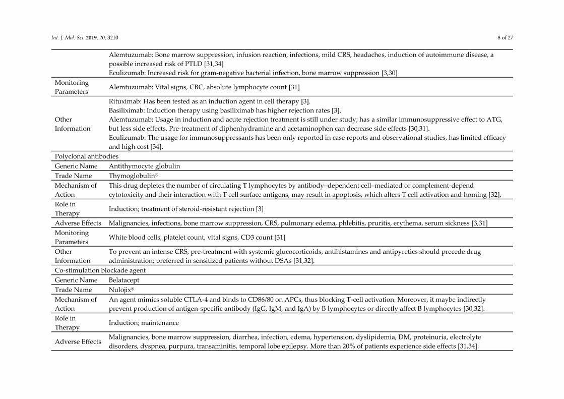
Int. J. Mol. Sci. 2019, 20, 3210 8 of 27
Alemtuzumab: Bone marrow suppression, infusion reaction, infections, mild CRS, headaches, induction of autoimmune disease, a
possible increased risk of PTLD [31,34]
Eculizumab: Increased risk for gram-negative bacterial infection, bone marrow suppression [3,30]
Monitoring
Parameters Alemtuzumab: Vital signs, CBC, absolute lymphocyte count [31]
Other
Information
Rituximab: Has been tested as an induction agent in cell therapy [3].
Basiliximab: Induction therapy using basiliximab has higher rejection rates [3].
Alemtuzumab: Usage in induction and acute rejection treatment is still under study; has a similar immunosuppressive effect to ATG,
but less side effects. Pre-treatment of diphenhydramine and acetaminophen can decrease side effects [30,31].
Eculizumab: The usage for immunosuppressants has been only reported in case reports and observational studies, has limited efficacy
and high cost [34].
Polyclonal antibodies
Generic Name Antithymocyte globulin
Trade Name Thymoglobulin®
Mechanism of
Action
This drug depletes the number of circulating T lymphocytes by antibody–dependent cell–mediated or complement-depend
cytotoxicity and their interaction with T cell surface antigens, may result in apoptosis, which alters T cell activation and homing [32].
Role in
Therapy Induction; treatment of steroid-resistant rejection [3]
Adverse Effects Malignancies, infections, bone marrow suppression, CRS, pulmonary edema, phlebitis, pruritis, erythema, serum sickness [3,31]
Monitoring
Parameters White blood cells, platelet count, vital signs, CD3 count [31]
Other
Information
To prevent an intense CRS, pre-treatment with systemic glucocorticoids, antihistamines and antipyretics should precede drug
administration; preferred in sensitized patients without DSAs [31,32].
Co-stimulation blockade agent
Generic Name Belatacept
Trade Name Nulojix®
Mechanism of
Action
An agent mimics soluble CTLA-4 and binds to CD86/80 on APCs, thus blocking T-cell activation. Moreover, it maybe indirectly
prevent production of antigen-specific antibody (IgG, IgM, and IgA) by B lymphocytes or directly affect B lymphocytes [30,32].
Role in
Therapy Induction; maintenance
Adverse Effects Malignancies, bone marrow suppression, diarrhea, infection, edema, hypertension, dyslipidemia, DM, proteinuria, electrolyte
disorders, dyspnea, purpura, transaminitis, temporal lobe epilepsy. More than 20% of patients experience side effects [31,34].
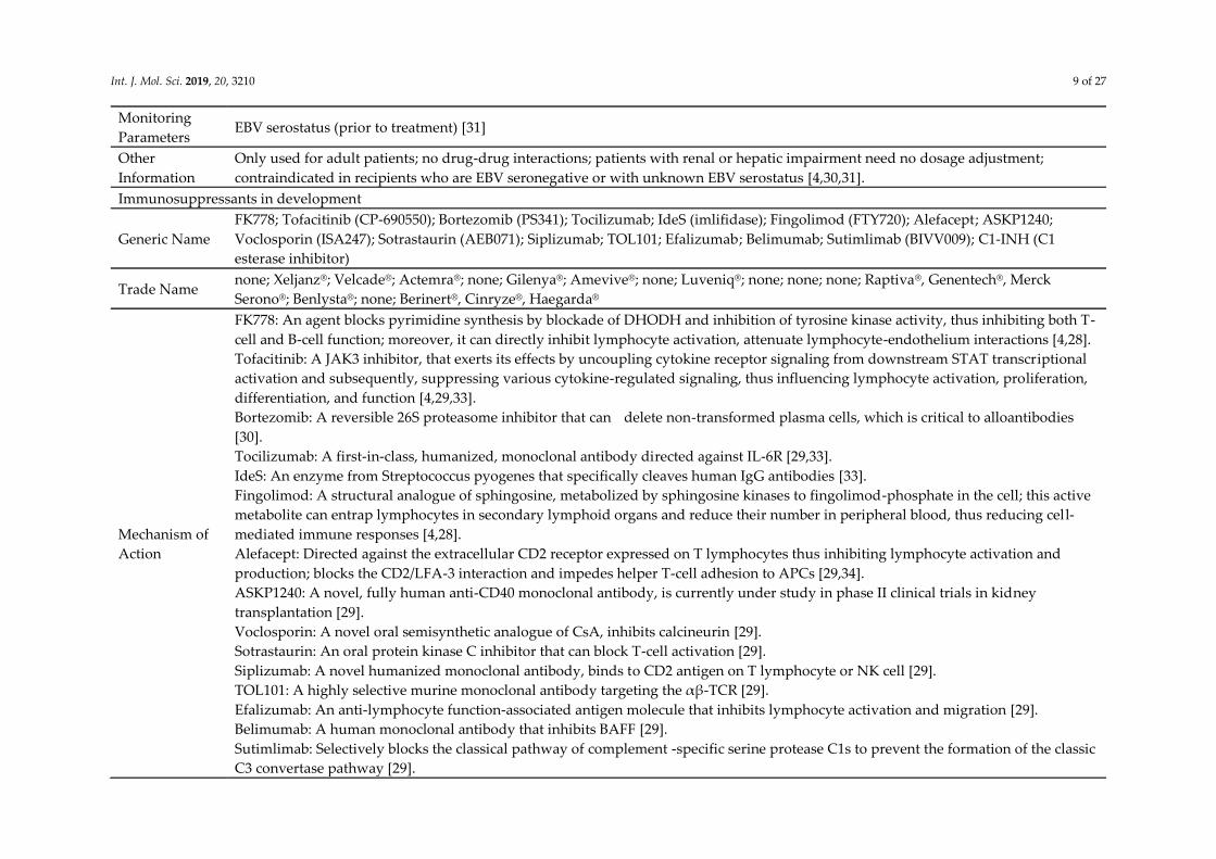
Int. J. Mol. Sci. 2019, 20, 3210 9 of 27
Monitoring
Parameters EBV serostatus (prior to treatment) [31]
Other
Information
Only used for adult patients; no drug-drug interactions; patients with renal or hepatic impairment need no dosage adjustment;
contraindicated in recipients who are EBV seronegative or with unknown EBV serostatus [4,30,31].
Immunosuppressants in development
Generic Name
FK778; Tofacitinib (CP-690550); Bortezomib (PS341); Tocilizumab; IdeS (imlifidase); Fingolimod (FTY720); Alefacept; ASKP1240;
Voclosporin (ISA247); Sotrastaurin (AEB071); Siplizumab; TOL101; Efalizumab; Belimumab; Sutimlimab (BIVV009); C1-INH (C1
esterase inhibitor)
Trade Name none; Xeljanz® ; Velcade® ; Actemra® ; none; Gilenya® ; Amevive® ; none; Luveniq® ; none; none; none; Raptiva® , Genentech® , Merck
Serono® ; Benlysta® ; none; Berinert® , Cinryze® , Haegarda®
Mechanism of
Action
FK778: An agent blocks pyrimidine synthesis by blockade of DHODH and inhibition of tyrosine kinase activity, thus inhibiting both T-
cell and B-cell function; moreover, it can directly inhibit lymphocyte activation, attenuate lymphocyte-endothelium interactions [4,28].
Tofacitinib: A JAK3 inhibitor, that exerts its effects by uncoupling cytokine receptor signaling from downstream STAT transcriptional
activation and subsequently, suppressing various cytokine-regulated signaling, thus influencing lymphocyte activation, proliferation,
differentiation, and function [4,29,33].
Bortezomib: A reversible 26S proteasome inhibitor that can delete non-transformed plasma cells, which is critical to alloantibodies
[30].
Tocilizumab: A first-in-class, humanized, monoclonal antibody directed against IL-6R [29,33].
IdeS: An enzyme from Streptococcus pyogenes that specifically cleaves human IgG antibodies [33].
Fingolimod: A structural analogue of sphingosine, metabolized by sphingosine kinases to fingolimod-phosphate in the cell; this active
metabolite can entrap lymphocytes in secondary lymphoid organs and reduce their number in peripheral blood, thus reducing cell-
mediated immune responses [4,28].
Alefacept: Directed against the extracellular CD2 receptor expressed on T lymphocytes thus inhibiting lymphocyte activation and
production; blocks the CD2/LFA-3 interaction and impedes helper T-cell adhesion to APCs [29,34].
ASKP1240: A novel, fully human anti-CD40 monoclonal antibody, is currently under study in phase II clinical trials in kidney
transplantation [29].
Voclosporin: A novel oral semisynthetic analogue of CsA, inhibits calcineurin [29].
Sotrastaurin: An oral protein kinase C inhibitor that can block T-cell activation [29].
Siplizumab: A novel humanized monoclonal antibody, binds to CD2 antigen on T lymphocyte or NK cell [29].
TOL101: A highly selective murine monoclonal antibody targeting the αβ-TCR [29].
Efalizumab: An anti-lymphocyte function-associated antigen molecule that inhibits lymphocyte activation and migration [29].
Belimumab: A human monoclonal antibody that inhibits BAFF [29].
Sutimlimab: Selectively blocks the classical pathway of complement -specific serine protease C1s to prevent the formation of the classic
C3 convertase pathway [29].
Int. J. Mol. Sci. 2019, 20, 3210 10 of 27
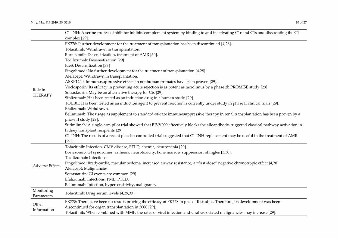
C1-INH: A serine-protease inhibitor inhibits complement system by binding to and inactivating C1r and C1s and dissociating the C1
complex [29].
Role in
THERAPY
FK778: Further development for the treatment of transplantation has been discontinued [4,28].
Tofacitinib: Withdrawn in transplantation.
Bortezomib: Desensitization, treatment of AMR [30].
Tocilizumab: Desensitization [29]
IdeS: Desensitization [33]
Fingolimod: No further development for the treatment of transplantation [4,28].
Alefacept: Withdrawn in transplantation.
ASKP1240: Immunosuppressive effects in nonhuman primates have been proven [29].
Voclosporin: Its efficacy in preventing acute rejection is as potent as tacrolimus by a phase 2b PROMISE study [29].
Sotrastaurin: May be an alternative therapy for Cis [29].
Siplizumab: Has been tested as an induction drug in a human study [29].
TOL101: Has been tested as an induction agent to prevent rejection is currently under study in phase II clinical trials [29].
Efalizumab: Withdrawn.
Belimumab: The usage as supplement to standard-of-care immunosuppressive therapy in renal transplantation has been proven by a
phase II study [29].
Sutimlimab: A single-arm pilot trial showed that BIVV009 effectively blocks the alloantibody-triggered classical pathway activation in
kidney transplant recipients [29].
C1-INH: The results of a recent placebo-controlled trial suggested that C1-INH replacement may be useful in the treatment of AMR
[29].
Adverse Effects
Tofacitinib: Infection, CMV disease, PTLD, anemia, neutropenia [29].
Bortezomib: GI syndromes, asthenia, neurotoxicity, bone marrow suppression, shingles [3,30].
Tocilizumab: Infections.
Fingolimod: Bradycardia, macular oedema, increased airway resistance, a “first-dose” negative chronotropic effect [4,28].
Alefacept: Malignancies.
Sotrastaurin: GI events are common [29].
Efalizumab: Infections, PML, PTLD.
Belimumab: Infection, hypersensitivity, malignancy.
Monitoring
Parameters Tofacitinib: Drug serum levels [4,29,33].
Other
Information
FK778: There have been no results proving the efficacy of FK778 in phase III studies. Therefore, its development was been
discontinued for organ transplantation in 2006 [29].
Tofacitinib: When combined with MMF, the rates of viral infection and viral-associated malignancies may increase [29].

Int. J. Mol. Sci. 2019, 20, 3210 11 of 27
Bortezomib: Small, non-randomised trials suggest efficacy in AMR; may decrease AMR in highly sensitised individuals [30].
IdeS: IdeS has been proven to effectively reduce anti-HLA antibody levels in highly sensitized patients by a phase II study; clinical
trials in sensitized kidney patients are ongoing [29].
Fingolimod: It is now approved for use in MS, but its mechanism is still unknown.
Alefacept: Its use for the prevention of graft-versus host disease is under investigation.
ASKP1240: Further clinical III studies are needed [29].
Voclosporin: Low-dose voclosporin may reduce incidence of new-onset diabetes after transplantation [29].
Sotrastaurin: High-dose sotrastaurin may be associated with faster heart rates [29].
Sutimlimab: Undergoing phase clinical III trail [29].
C1-INH: Further studies are needed to confirm the safety and efficacy of C1-INH in the treatment of AMR [29].
Abbreviations: AMR, antibody-mediated rejection; APC, antigen-presenting cell; ATG, anti-thymocyte globulin; AZA, azathioprine; BAFF, B-cell activating factor;
CBC, complete blood count; CD, cluster of differentiation; CNIs, calcineurin Inhibitors; CP, cyclophosphamide; CRS, cytokine release syndrome; CsA, cyclosporine;
CTLA4, cytotoxic T lymphocyte–associated antigen 4; CYP3A4, cytochrome P3A4; C1-INH, C1 esterase inhibitor; DEXA, dual-energy X-ray absorptiometry;
DHODH, dihydroorotic acid dehydrogenase; DM, diabetes mellitus; DSA, donor-specific antibodies; EBV, Epstein-Barr virus; FKBP, FK506-binding protein; GI,
gastrointestinal; HUS/TMA, hemolytic uremic syndrome/thrombotic microangiopathy; Ides, immunoglobulin G-degrading enzyme derived from Streptococcus
pyogenes; IFN, interferon; IL-2, interleukin-2; IL-6R, IL-6 receptor; IMPDH, inosine-5’-monophosphate dehydrogenase; JAK, janus kinase; KFT, kidney function
test; LFT, liver function test; MHC, major histocompatibility complex; MMF, mycophenolate mofetil; MPA, mycophenolic acid; MS, multiple sclerosis; mTOR,
mammalian target of rapamycin; muromonab-CD3, mouse monoclonal immunoglobulin G2 antibody to cluster of differentiation 3; NFAT, nuclear factor of
activated T-cells; PML, progressive multifocal leukoencephalopathy; PTLD, post-transplant lymphoproliferative disorder; REMS, pregnancy test in women of
childbearing age; STAT, signal transducers and activators of transcription; TPMT, thiopurine methyltransferase; TCR, T cell receptor.

Int. J. Mol. Sci. 2019, 20, 3210 12 of 27
4. Clinical Features Induced by Different Immunosuppressants
4.1. CNIs
Neurotoxicity induced by CNIs occurs at three distinct time points after transplantation: early,
intermediate, and late. Most patients who use tacrolimus intravenously develop neurotoxicity on the
first day after transplantation [35]. Patients who develop neurotoxicity in the intermediate or late
stage demonstrate only short or intermediate survival times [36].
There are various neurological complications of CNIs, which can involve both the central
nervous system (CNS) and the peripheral nervous system [37]. Mild neurological manifestations
related to CNI toxicity are common and include tremors, insomnia, nightmares, sleep disturbances,
headaches, vertigo, mood disturbances, and paresthesia (electric shock-like pain and severe itching)
[38,39]. Serious adverse neurological effects have been relatively rarely observed and include
seizures, speech disorders, cortical blindness, coma, encephalopathy, central pontine/extrapontine
myelinolysis, and neuromuscular complications [14]. Tacrolimus treatment has a significantly higher
incidence of neurological syndromes than CsA treatment in solid organ transplantation recipients
[40,41].
Tremor is the most pronounced neurological complication associated with CNI toxicity and a
fine tremor of the upper limbs can help diagnose neurological complications at early stages [13].
Tremor is significantly more common in patients treated with tacrolimus than in those treated with
CsA. In a more recent trial, less than 20% of patients treated with CsA experienced tremors, while
tacrolimus-related neurotoxic events occurred in up to 40% of patients [42]. In general, tremors
involved both upper and lower limbs, with some patients even experiencing tremors in the
head/facial muscles. Tremors exclusively involving the trunk, lower limbs, or the craniofacial area
are rare in the clinic [42]. Considering that the main goal of immunosuppressive therapy is to increase
the survival rates of transplant recipients, and tremors appear to be isolated with cerebellar or
neuropathic involvement, this symptom tends to be ignored when its severity is not significant and
does not influence the patient’s quality of life.
Seizures are common in transplant recipients undergoing CNI therapy, occurring in up to 27%
of organ transplant patients [43]. Although seizures frequently occur with posterior reversible
encephalopathy syndrome (PRES), the new-onset of seizures is not indicative of a poor prognosis,
because most patients do well and do not require long-term antiepileptic therapy [44]. In patients
with seizures, generalized tonic-clonic and occipital lobe seizures are usually observed [45]. Simple
or complex partial seizures represent a localized process that may be reflected by focal
electroencephalogram (EEG) abnormalities, whereas seizures that occur secondary to posterior
reversible encephalopathy syndrome (PRES) frequently show short single grand mal episodes with
variable theta/delta slowing [44]. Seizures associated with CNI neurotoxicity frequently originate
from occipital regions [46]. PRES is a serious complication associated with immunosuppressive
therapy after transplantation [47]. It is a neurotoxicity characterized by headaches, confusion, nausea
and vomiting, altered mental status, visual disturbances, intracranial hemorrhage, altered sensorium,
and occasionally, a focal neurological deficit [45,48–51]. In most cases, immunosuppression-
associated leukoencephalopathy occurs within the first three months after transplantation and it is
usually associated with intravenous treatment methods [52]. PRES appears to be significantly more
common in hematopoietic or liver transplantation than in other transplantations [53].
The cranial computed tomography (CT) finding is insensitive in detecting PRES and often shows
no abnormalities, while magnetic resonance imaging (MRI) has been proven to be the most sensitive
imaging test. Vasogenic edema, which is a symptom of PRES, can be easily identified. Radiologists
can reliably differentiate these changes from cytotoxic edema using diffusion weighted image (DWI)
and apparent diffusion coefficient (ADC) maps. Moreover, the extent of abnormal T2-weighted signal
intensities and DWI signal intensities correlate well with prognosis [47]. PRES predominantly affects
the posterior cerebrum and the cerebral white matter, causing focal reversible vasogenic edematous
changes in the specific posterior regions of the parietal and occipital lobes, which can lead to
irreversible cytotoxic edema in some cases [44,54]. Grey and white matter lesions can be observed by

Int. J. Mol. Sci. 2019, 20, 3210 13 of 27
MRI in fluid attenuated IR (FLAIR) and T2-weighted sequences, and deeper structures, such as the
basal ganglia, brain stem, and deep white matter tracts, may be also affected [44,55]. Cytotoxic edema
and hemorrhage are uncommon findings in these patients [44]. Typically, the characteristic of PRES
is bilateral symmetric patterns of edema, usually including diffuse white matter hyperintensity with
a parieto-occipital predilection [56,57]. If PRES is not diagnosed at an early stage, cerebral ischemia
and massive infarction may result in an increase in morbidity and mortality [47]. Hypertension is
another important symptom of PRES and, therefore, when immunosuppressants need to be
continued in the clinic, blood pressure should be effectively monitored and controlled.
CPM is one of the most detrimental neurological complications after organ transplantation and
the mortality due to this neurotoxicity is more than 50% [58]. The incidence of CPM is more common
in liver transplantation and in patients treated with CsA than in patients treated with tacrolimus [59–
61]. MRI features include hyperintense lesions in the center of the pons on T2 images. Rapamycin is
recommended as a replacement for CNIs, because it is rarely associated with CPM. However,
rapamycin is unstable and requires frequent monitoring of blood concentrations when used in
clinical practice.
The number of case reports related to catatonic symptoms and akinetic mutism induced by CNI
administration after organ transplantation have increased in recent years [62,63]. Even when used to
treat psoriasis, CNIs have been shown to exacerbate the symptoms of paranoid schizophrenia, and
then disappear a few days after discontinuation of the CNI treatment [64].
In addition to CNIs, other immunosuppressants may also manifest neuropsychiatric
complications, although neurotoxicity reports are rarer for these drugs than for CNIs [37].
Mycophenolate mofetil rarely induces depression and headaches. However, seizures were frequently
observed in several reports on neurological complications during rapamycin therapy. The main
neurological complication of muromonab-CD3 treatment is headache, whereas patients treated with
corticosteroids may experience anxiety, insomnia, mood disorders, psychotic episodes, and cognitive
symptoms [65,66].
4.2. Antimetabolites
Methotrexate (MTX) can induce CNS toxicity that presents in the form of encephalopathy,
myelopathy, or meningitis [67]. Neurological symptoms are caused by MTX are usually classified
into acute, subacute, or chronic neurotoxicity. Patients who experience subacute neurotoxicity
usually recover completely and spontaneously within a week, and, therefore, subsequent MTX
treatment is safe for most patients [68]. Neurological symptoms induced by mycophenolate mofetil
are rare and mild, manifesting as depression and headaches.
4.3. Corticosteroids
Neurological side effects occur in approximately 3–4% of patients who use corticosteroids [69].
Corticosteroid-induced neuropsychiatric symptoms include mood changes, behavioral disorders,
and cognitive symptoms that typically manifest during the first few weeks of therapy [66]. Peripheral
toxicity occurs after long-term use, usually in the form of neuromyopathy, with muscular weakness
affecting the proximal and lower extremities [70]. Steroid dementia syndrome appears to be rare [71],
and these symptoms may not recover completely even after the cessation of treatment [72]. Epidural
lipomatosis can also induce radiculopathy due to spinal compression [73].
The adjustment or discontinuation of corticosteroids may improve some of these adverse
neurological symptoms. If the psychiatric symptoms are serious, short regimens of low-dose
psychotropic agents are often required (e.g., haloperidol, olanzapine, quetiapine, or risperidone).
4.4. Monoclonal Antibodies
Polyclonal and monoclonal antibodies are usually used to induce immunosuppression and treat
graft rejection [72]. With the exception of OKT3 and belatacept, biologic agents show low incidences
of adverse neurological effects. The neurotoxicities induced by OKT3 range from headaches and fever

Int. J. Mol. Sci. 2019, 20, 3210 14 of 27
to confusion, aseptic meningitis, cerebral edema, encephalopathy, seizures, hemiparesis, nuchal
rigidity, and myoclonic activity [37,74]. Furthermore, treatment with CsA after OKT3 results in an
additive or synergistic adverse effect on neurological complications [75]. In general, pathological
changes can be detected by a head MRI, and neurological abnormalities resolve after the cessation of
the OKT3 treatment [75,76]. However, cytokine release syndrome in patients treated with OKT3 is so
serious that it limits the usage of this agent. Blinatumomab, a novel recombinant murine protein, is
used for the treatment of Philadelphia chromosome–negative, relapsed or refractory precursor acute
lymphoblastic leukemia. There are a variety of neurological symptoms induced by blinatumomab
treatment, such as somnolence, confusion, dizziness, tremor, seizure, encephalopathy, speech
disorders, and loss of consciousness, which all appear to be more common in patients over 65 years
of age [77].
Conditions that increase the neurotoxicity of immunosuppressant agents include pre-existing
mental disorders [78], hypertension [79], electrolyte disorders including hyper and hyponatremia
and hypomagnesemia [37], dysmetabolic alterations, such as hyperglycemia [37], infections that
impair the function of the blood–brain barrier (BBB), hypocholesterolemia, which increases the
uptake of immunosuppressant drugs in the brain [80], polymorphisms of the adenosine triphosphate
(ATP)-binding cassette transporter B1 (ABCB1) gene and cytochrome pigment (CYP) gene, which
decrease immunosuppressant efflux or elimination [81,82], drug interactions [82,83], a prolonged
surgical period [84], and low liver function or acute liver failure [85].
5. Mechanisms of Neurotoxicity Induced by Different Immunosuppressants
5.1. CNIs
The biochemical basis of CNI-induced neurotoxicity remains unclear. It appears that high drug
concentrations in the blood are correlated with neurological symptoms, but they can also occur in
patients with concentrations within the therapeutic range [13,14,86]. Although both CNIs used as
immunosuppressants are lipophilic, with CsA being more lipophilic than tacrolimus, they do not
easily pass through the BBB [87,88]. One possible hypothesis is that tacrolimus and CsA increase the
permeability of the BBB by inducing apoptosis and nitric oxide (NO) production and inhibiting P-
glycoprotein (P-gp) function, which leads to further accumulation of drugs in the brain, extravasation
of proteins and fluid into the interstitium, and impaired BBB function. An investigation of the effects
of tacrolimus and CsA on mouse brain capillary endothelial cells (MBEC4) found that drug-treated
cells experienced 1) loss of junctions with neighboring cells and detachment from the substratum, 2)
chromatin condensation and fragmentation, and 3) DNA fragmentation [89]. The two drugs induced
dose-independent apoptosis of the brain capillary endothelial cells, with similar effects between CsA
and tacrolimus [89]. Dohgu et al. [90] reported that CsA increases NO production in brain endothelial
and astroglial cells, which then participate in the impairment of BBB function. The expression of P-
glycoprotein decreases with high concentrations of CNIs, leading to the inhibition of the efflux
process and an enhancement of permeability. This may partly explain the mechanism of CNI-induced
encephalopathy [91]. It should be mentioned that the drug concentrations in the above-mentioned
studies were considerably higher than in clinical doses, and further investigation is required to
determine whether normal brain capillary endothelial cells are impaired. In fact, a recent study using
an in vitro BBB model, consisting of a co-culture of bovine brain capillary endothelial cells (ECs) and
neonatal rat glial cells, showed that repeated exposure to 1 μM CsA, found in human plasma, had no
toxic effect on BBB integrity [92]. This result was confirmed by a kinetics study, in which intracellular
CsA uptake and permeability across the BBB were minimal [93]. It is also important to stress that,
despite no cell damage, some key neurotransmitters, factors metabolically linked to
neurotransmitters, or energy metabolism related to electrical activity that are altered at this
concentration range may be responsible for the neurological disorders induced by CsA or other CNIs
[94].
An alternative hypothesis is that alterations in mitochondrial function induced by CNIs
contribute to neurotoxicity. A study in human umbilical endothelial cells showed that tacrolimus

Int. J. Mol. Sci. 2019, 20, 3210 15 of 27
significantly compromised respiratory chain (RC)-complexes II and III and the mitochondrial marker
enzyme, citrate synthase (CS), thus indicating a partially impaired mitochondrial function [95].
Furthermore, a similar analysis found that tacrolimus decreases oxygen consumption in human cell
lines and causes a slight reduction in the synthesis of mitochondrial DNA-encoded proteins [96].
These studies suggest that the direct inhibition of the electron transport chain by CNIs, rather than
effects on mitochondrial density or electron transport chain (ETC) quantity, are responsible for
impaired mitochondrial function. This conclusion is contrary to an early study reporting that
tacrolimus inhibits both complex III, where reactive oxygen species (ROS) are generated and complex
V, where adenosine triphosphate (ATP) is depleted by ATPase activation [97]. There are two existing
studies where one use glioma cells and another one uses glial cells demonstrated that tacrolimus can
increase the production of ROS and decrease the antioxidant status [98,99], indicating that
mitochondrial function may be impaired by tacrolimus treatment.
There is also evidence that the complex of CNIs and immunophilins may be associated with
neurotoxicity. Calcineurin is expressed in several areas of the brain, including the cerebral cortex,
striatum, substantia nigra, cerebellum, and hippocampus, where it regulates the dephosphorylation
of Ca2+ channels, activity of the N-methyl-D-aspartate (NMDA) receptor, ryanodine receptor, the
inositol trisphosphate (IP3) receptor, and even memory and synaptic plasticity [100–102]. These
neurotoxic effects may depend on immune dysregulation in the nervous system, due to the
pharmacologic effects of the CNI-immunophilin complex [91,103,104]. The maximal inhibitory effect
of tacrolimus on calcineurin is approximately 60% (while CsA is more effective at inhibiting
calcineurin) [105]. However, tacrolimus has no pharmacological effect on FK506-binding protein
(FKBP) 1A (FKBP12)-null mice [106]. These findings suggest that the FK506-FKBP complex has some
unknown molecular mechanisms besides its calcineurin inhibitory effect. The level of FKBP12
expression is 10–50-fold higher in the brain than in the immune system [107,108]. Tacrolimus-induced
toxicity is consistent in organs with high FKBP levels, such as the brain and kidneys. Moreover, once
tacrolimus enters the brain, it is eliminated slowly by binding to FKBP [109]. In an in vitro model,
CsA inhibits calcineurin in the brain, even at concentrations as low as 200 nM, in a relatively short
time frame. This inhibitory effect is sustained during drug administration [93]. With the exception of
calcineurin inhibition by the tacrolimus-FKBP complex, the exact mechanism of neurotoxicity is not
completely understood. Research on calcineurin inhibition-induced depressive-like behavior in a
prefrontal cortex model raises the possibility that the blockade of the mTOR signaling pathway
accounts for the neurological disorders [110]. In support of this, another study showed that receptor-
associated FKBP12 participated in the intracellular mTOR activation pathway, which is well known
for its critical roles in the integration of neuronal activity and synaptic inputs in multiple
physiological and pathological processes [111,112]. These experimental findings are in agreement
with a clinical study showing that tacrolimus induces a higher incidence of neurotoxicity than CsA
[113,114].
Vasoconstriction or vascular injury [115] may also be involved in the mechanism of CNI-induced
neurotoxicity. Tacrolimus may be associated with blood vessel contraction. Moreover, some
investigators have suggested that, in addition to vasoconstriction caused by tacrolimus, the high
infiltration pressure of the tacrolimus dissolution liquid may also affect neurotoxicity. However,
although this hypothesis is interesting, it does not explain the phenomenon where patients
experience CNI-induced neurologic disorders, even though their blood pressure is maintained within
the normal range throughout hospitalization [63,116].
Other proposed mechanisms of CNI-induced neurotoxicity include a possible modulation of
excitability properties, causing nerve membrane depolarization [117] and alterations in electrical
activity [94,118], suppression of brain-derived neurotrophic factor (BDNF) and its receptor (tyrosine
kinase receptor B (TrkB)), mRNA and protein expression in the hippocampus and midbrain [119],
reduction of Ca2+ accumulation in the endoplasmic reticulum (ER) by intracellular accumulated
tacrolimus[120], and significant intracellular CNI uptake, thus increasing the toxicity of other drugs
administered at the same time [93]. The metabolites of CNIs may also be neurotoxic, even though
they are usually not assessed in clinical practice.
Int. J. Mol. Sci. 2019, 20, 3210 16 of 27
5.2. Antimetabolites
Several biochemical pathways, including a decreased S-adenosylmethionine/S-
Adenosylhomocysteine (SAM/SAH) ratio, elevated levels of homocysteine, and elevated levels of
adenosine and direct toxic effects on neurons and astrocytes, may be the causes of MTX-related
neurotoxicity [68].
5.3. Corticosteroids
Corticosteroids are often used in combination with other immunosuppressive agents. They
cause neurological complications through two mechanisms that involve direct and indirect toxic
effects on CNS biochemistry and electrophysiology. These include glutamate excess and
neurotrophin mobilization [121] or elevated blood pressure and the vulnerability of the vasculature
through the regulation of the renin-angiotensin system. Further, there are some reports suggesting
that corticosteroids can make certain hippocampal and prefrontal cortical cells more vulnerable to
other exogenous agents [71].
5.4. Monoclonal Antibodies
The mechanisms of muromonab-CD3- and belatacept-related neurotoxicity have rarely been
reported. It has been postulated that cerebral complications are related to the OKT3-mediated release
of cytokines [75]. This hypothesis may explain the cases of aseptic meningitis. However, it cannot
explain why neurological symptoms persist after cytokine levels return to the baseline. Other studies
have suggested that circulating lymphocytes and cells of the nervous system share some of the same
surface antigens, such that OKT3 combines with cell surface antigens to facilitate OKT3 antibodies
crossing the BBB [76]. Cytokine release syndrome induced by blinatumomab may be responsible for
some of the adverse neurological effects, but further studies are warranted to clarify the precise
mechanism of blinatumomab-induced neurotoxicity [77].
6. Management
Immunosuppressants, particularly CNIs, can induce neurotoxicity in solid organ
transplantation cases. The management of blood concentrations of the drugs by therapeutic drug
monitoring, individual therapy based on pharmacogenetics, and the early recognition of symptoms
using electrophysiological and imaging strategies, may help avoid neurotoxicity [82,122,123].
However, even with these measures in place, the incidence of neurotoxic symptoms remains high (3–
32%) [79,124,125]. Once neurotoxicity occurs, reducing the dosage of the drug, switching from
tacrolimus to CsA or vice versa, or using an alternative immunosuppressant agent, such as
mycophenolate mofetil, have proven to be effective approaches to reverse this neurotoxicity
[63,126,127]. A study comparing the effects of tacrolimus and rapamycin on bioelectrical activity and
evoked field excitatory postsynaptic potential (fEPSP) in the CA1 area of hippocampal tissues has
also suggested that rapamycin could replace CNIs in the event of seizures [128]. However, in some
cases, switching between tacrolimus and CsA has not been effective at improving neurotoxicity.
Moreover, the continued administration of CNIs, in combination with drugs that treat,
neuropsychiatric manifestations may be considered the best approach, given that considering that
switching immunosuppressants may elevate the risk of graft rejection in some patients and reducing
the CNI dosage does not always improve the symptoms. For example, olanzapine, coupled with the
continued use of tacrolimus, has been shown to resolve manic episodes [129]. Olanzapine has also
been considered for the treatment of catatonic mutism after liver transplantation [130].
Benzodiazepines can improve catatonia, especially akinetic–hypokinetic catatonic syndromes
[40,131]. The neurotoxicity associated with CNIs is strongly correlated with the intracerebral
concentration of the drugs [132]. Sakamoto et al. showed that the continuous administration of
tacrolimus is more advantageous than intermittent administration to reduce neurotoxicity in rats
[133]. In addition, tacrolimus-induced neurotoxicity and nephrotoxicity can be ameliorated, while
maintaining its immunosuppressive effects, by treating rats in the dark phase [134]. According to a

Int. J. Mol. Sci. 2019, 20, 3210 17 of 27
case report, red-blood cell exchange improved the clinical status of a 60-year-old woman with severe
neurological impairment due to tacrolimus overexposure. Hence, red-blood cell exchange may be an
effective therapy to reduce tacrolimus neurotoxicity [135]. This blood purification therapy has also
been shown to be effective in ifosfamide-induced severe concurrent neurotoxicity and nephrotoxicity
[136].
7. Neuroprotective Effects
7.1. CNIs
The immunosuppressants—tacrolimus and CsA—have neuroprotective effects in animal
models of focal and global cerebral ischemia [137–139], portacaval anastomosis and
hyperammonemia [140], intracerebroventricular streptozotocin-induced neurotoxicity [141], and
temporal lobe epilepsy (the turnover of tacrolimus is much faster in rats than in humans) [142].
When wild-type mice are treated with tacrolimus for one week, their neocortices show longer
total dendritic arbors and more complex branching further away from the cell body, compared to
untreated animals [143]. There is some experimental data to indicate that the neuroprotective effects
induced by tacrolimus and CsA may be related to calcineurin inhibition, NFκB activation [144],
downregulation of proinflammatory/cytotoxic cytokines [145], decreased NO synthetase-mediated
NO production [137,140,146], inhibition of Ca2+ release by both the ER and mitochondria, as well as
mitochondrial permeability transition (mPT) (CsA) [147], deceased apoptosis and c-jun protein
expression in neurons [148], calcineurin-independent mechanisms [149], activation of pro-survival
pathways by BDNF and its receptor, tropomyosin receptor kinase A (TrkA) [150], and excitotoxic
neuronal death [151].
However, although the neuroprotective functions of tacrolimus have been demonstrated in
various nerve injury models, these functions have been challenged by models of inherited peripheral
myelinopathies treated with tacrolimus. For example, tacrolimus exacerbates neurological
abnormalities, including demyelination and dysmyelination-associated axon loss in inherited
de/dysmyelination mice, while the peripheral nerves of wild-type mice do not show any neurotoxic
symptoms after treatment with tacrolimus [152].
Interestingly, a recent study found that tacrolimus and CsA treatment had no better long-term
effects than treatment with the vehicle alone (cremophor and ethanol mixture). Moreover, the drug-
treated group showed even more significant decreases in brain weight. Therefore, Setkowicz and
Guzik [153] concluded that the neuroprotective effects observed in rat brains injured mechanically at
the early developmental stages may result from the influence of the vehicle alone. Further studies
and more investigations are needed to clarify the potential neuroprotective effects and mechanisms
of CNIs.
7.2. mTOR Inhibitors
mTOR is associated with the pathogenesis of neurological, cognitive, and psychiatric disorders,
such as epilepsy, stroke, traumatic brain injury, parkinsonism, spinal cord injury, and Alzheimer’s
disease [154]. In a mouse model of epilepsy induced by knocking-out the protein phosphatase and
tensin homolog (PTEN), mTOR activity increases in neurons. Therefore, reducing mTOR activity may
effectively suppress epileptogenesis and alleviate the symptoms of this disease [155]. The role of
mTOR in cerebral ischemia has also been reported in some rodent experiments. Some studies have
shown that the mTOR pathway has neurotoxic effects, while others have reported the opposite
finding that the mTOR pathway has neuroprotective effects. Some reports have suggested that
suppressing the pharmacological effects of the mTOR pathway can regulate autophagy and result in
neuroprotection, whereas other reports have suggested that the neurotoxicity of mTOR inhibitors is
related to the promotion of autophagic processes, long-term activation of Akt, and activation of S6
kinase 1 (S6K1) occurring in brain cells after a stroke [154,156–158]. The study by Chen and co-
workers [159] may explain the paradoxical effects of the mTOR inhibitor, rapamycin. In that study,
rapamycin was shown to cause a paradoxical, but transient, increase in mTOR pathway activation in

Int. J. Mol. Sci. 2019, 20, 3210 18 of 27
a kainite injection model, and in normal rats, by increasing the phosphorylation of S6. These results
suggest that the effects of rapamycin on mTOR are related to the type or period of stimuli and the
dose administered [160].
8. Conclusions
Neurological disorders are common after solid organ transplantation. The reasons for these
neurotoxicities are multifactorial, ranging from the effects of immunosuppressive agents to pre-
transplantation disease. In this article, we discussed the neurological complications resulting from
immunosuppressive therapy in five categories: the process of alloimmune responses, the
classification of immunosuppressive agents, their clinical features, their mechanisms, and their
clinical management. Interestingly, some studies have shown that these immunosuppressive agents
may have neuroprotective effects. However, two recent studies have reported contradictory findings,
suggesting that further studies are required to clarify the potential neuroprotective effects of
immunosuppressive agents.
Acknowledgments: This work was supported in part by a Grant-in-Aid for Scientific Research (KAKENHI) from
the Ministry of Education, Science, Culture, Sports and Technology of Japan (MEXT, grant numbers: 17K08953
to Nobuaki Egashira and 18H02588 to Satohiro Masuda). We would like to thank Editage (www.editage.jp) for
English language editing.
Conflicts of Interest: The authors declare no conflict of interest.
Abbreviations
ABCB1 ATP-binding cassette transporter B1
ADC Apparent diffusion coefficient
APC Antigen presenting cell
ATP Adenosine triphosphate
BBB Blood-brain barrier
BCR B-cell receptor
BDNF Brain-derived neurotrophic factor
CD Cluster of differentiation
CNI Calcineurin inhibitor
CNS Central nervous system
CPM Central pontine myelinolysis
CS Citrate synthase
CsA Cyclosporin A
CT Computed tomography
CYP Cytochrome pigment
DWI Diffusion weighted image
EC Endothelial cell
EEG Electro-encephalogram
ER Endoplasmic reticulum
ETC Electron transport chain
fEPSP Field excitatory postsynaptic potentials
FKBP FK506-binding protein
FLAIR Fluid-attenuated IR
IFN-γ Interferon-gamma
IL-2 Interleukin-2
IMPDH Inosine-5′-monophosphate dehydrogenase
IP3 Inositol trisphosphate
JAK Janus kinase
MAP Mitogen activated protein
MBEC4 Mouse brain capillary endothelial cells
MHC Major histocompatibility complex
mPT Mitochondrial permeability transition
MRI Magnetic resonance imaging

Int. J. Mol. Sci. 2019, 20, 3210 19 of 27
mTOR Mammalian target of rapamycin
MTX Methotrexate
muromonab-CD3 Mouse monoclonal immunoglobulin G2 antibody to cluster of differentiation 3
NFAT Nuclear factor of activated T cells
NFκB Nuclear factor kappa B
NMDA N-methyl-D-aspartate
NO Nitric oxide
P-gp P-glycoprotein
PRES Posterior reversible encephalopathy syndrome
PTEN Phosphatase and tensin homolog
RAS Renin–angiotensin system
RC Respiratory chain
ROS Reactive oxygen species
SAH S-adenosylhomocysteine
SAM S-adenosylmethionine
S6K1 S6 kinase 1
TCR T-cell receptor
Tfh T follicular helper
TNF-α Tumor necrosis factor-alpha
TrkA Tropomyosin receptor kinase A
TrkB Tyrosine kinase receptor B
References
1. Murray, J.E.; Merrill, J.P.; Harrison, J.H. Renal homotransplantation in identical twins. 1955. J Am Soc
Nephrol 2001, 12, 201–204.
2. Tolou-Ghamari, Z. Nephro and neurotoxicity of calcineurin inhibitors and mechanisms of rejections: A
review on tacrolimus and cyclosporin in organ transplantation. J. Nephropathol 2012, 1, 23–30.
doi:10.5812/jnp.6.
3. van Sandwijk, M.S.; Bemelman, F.J.; Ten Berge, I.J. Immunosuppressive drugs after solid organ
transplantation. Neth. J. Med. 2013, 71, 281–289.
4. Coelho, T.; Tredger, M.; Dhawan, A. Current status of immunosuppressive agents for solid organ
transplantation in children. Pediatr. Transplant. 2012, 16, 106–122. Available online: doi:10.1111/j.1399-
3046.2012.01644.x.
5. Mazariegos, G.V.; Molmenti, E.P.; Kramer, D.J. Early complications after orthotopic liver transplantation.
Surg Clin. North. Am. 1999, 79, 109–129.
6. Pruitt, A.A. Neurologic Complications of Transplantation. Continuum (Minneap Minn) 2017, 23, 802–821.
Available online: doi:10.1212/con.0000000000000473.
7. Pizzi, M.; Ng, L. Neurologic Complications of Solid Organ Transplantation. Neurol. Clin. 2017, 35, 809–823.
Available online: doi:10.1016/j.ncl.2017.06.013.
8. van de Beek, D.; Kremers, W.; Daly, R.C.; Edwards, B.S.; Clavell, A.L.; McGregor, C.G.; Wijdicks, E.F. Effect
of neurologic complications on outcome after heart transplant. Arch. Neurol. 2008, 65, 226–231. Available
online: doi:10.1001/archneurol.2007.52.
9. Ardizzone, G.; Arrigo, A.; Schellino, M.M.; Stratta, C.; Valzan, S.; Skurzak, S.; Andruetto, P.; Panio, A.;
Ballaris, M.A.; Lavezzo, B., et al. Neurological complications of liver cirrhosis and orthotopic liver
transplant. Transplant. Proc. 2006, 38, 789–792. Available online: doi:10.1016/j.transproceed.2006.01.039.
10. Ponticelli, C.; Campise, M.R. Neurological complications in kidney transplant recipients. J. Nephrol. 2005,
18, 521–528.
11. Ocal, R.; Kibaroglu, S.; Derle, E.; Tanoglu, C.; Camkiran, A.; Pirat, A.; Can, U.; Sezgin, A. Neurologic
Complications After Cardiac Transplant. Exp. Clin. Transplant. 2016, 10.6002/ect.2016.0127. Available
online: doi:10.6002/ect.2016.0127.
12. Dowling, M.R.; Li, S.; Dey, B.R.; McAfee, S.L.; Hock, H.R.; Spitzer, T.R.; Chen, Y.B.; Ballen, K.K. Neurologic
complications after allogeneic hematopoietic stem cell transplantation: Risk factors and impact. Bone
Marrow Transplant. 2018, 53, 199–206. Available online: doi:10.1038/bmt.2017.239.
13. Wijdicks, E.F. Neurotoxicity of immunosuppressive drugs. Liver Transpl. 2001, 7, 937–942. Available online:
doi:10.1053/jlts.2001.27475.

Int. J. Mol. Sci. 2019, 20, 3210 20 of 27
14. Bechstein, W.O. Neurotoxicity of calcineurin inhibitors: Impact and clinical management. Transpl. Int. 2000,
13, 313–326.
15. Dhar, R.; Human, T. Central nervous system complications after transplantation. Neurol. Clin. 2011, 29, 943–
972. Available online: doi:10.1016/j.ncl.2011.07.002.
16. Boardman, D.A.; Jacob, J.; Smyth, L.A.; Lombardi, G.; Lechler, R.I. What Is Direct Allorecognition? Curr.
Transplant. Rep. 2016, 3, 275–283. Available online: doi:10.1007/s40472-016-0115-8.
17. Gokmen, M.R.; Lombardi, G.; Lechler, R.I. The importance of the indirect pathway of allorecognition in
clinical transplantation. Curr. Opin. Immunol. 2008, 20, 568–574. Available online:
doi:10.1016/j.coi.2008.06.009.
18. Lim, M.A.; Kohli, J.; Bloom, R.D. Immunosuppression for kidney transplantation: Where are we now and
where are we going? Transplant. Rev. (Orlando) 2017, 31, 10–17. Available online:
doi:10.1016/j.trre.2016.10.006.
19. Thaunat, O.; Granja, A.G.; Barral, P.; Filby, A.; Montaner, B.; Collinson, L.; Martinez-Martin, N.; Harwood,
N.E.; Bruckbauer, A.; Batista, F.D. Asymmetric segregation of polarized antigen on B cell division shapes
presentation capacity. Science 2012, 335, 475–479. Available online: doi:10.1126/science.1214100.
20. Harwood, N.E.; Batista, F.D. Early events in B cell activation. Annu Rev. Immunol. 2010, 28, 185–210.
Available online: doi:10.1146/annurev-immunol-030409-101216.
21. Schnyder, T.; Castello, A.; Feest, C.; Harwood, N.E.; Oellerich, T.; Urlaub, H.; Engelke, M.; Wienands, J.;
Bruckbauer, A.; Batista, F.D. B cell receptor-mediated antigen gathering requires ubiquitin ligase Cbl and
adaptors Grb2 and Dok-3 to recruit dynein to the signaling microcluster. Immunity 2011, 34, 905–918.
Available online: doi:10.1016/j.immuni.2011.06.001.
22. Lanzavecchia, A. Antigen-specific interaction between T and B cells. Nature 1985, 314, 537–539.
23. Crotty, S. A brief history of T cell help to B cells. Nat. Rev. Immunol. 2015, 15, 185–189. Available online:
doi:10.1038/nri3803.
24. Chen, C.C.; Koenig, A.; Saison, C.; Dahdal, S.; Rigault, G.; Barba, T.; Taillardet, M.; Chartoire, D.; Ovize, M.;
Morelon, E., et al. CD4+ T Cell Help Is Mandatory for Naive and Memory Donor-Specific Antibody
Responses: Impact of Therapeutic Immunosuppression. Front. Immunol. 2018, 9, 275. Available online:
doi:10.3389/fimmu.2018.00275.
25. Okada, T.; Miller, M.J.; Parker, I.; Krummel, M.F.; Neighbors, M.; Hartley, S.B.; O’Garra, A.; Cahalan, M.D.;
Cyster, J.G. Antigen-engaged B cells undergo chemotaxis toward the T zone and form motile conjugates
with helper T cells. PLoS Biol. 2005, 3, e150. Available online: doi:10.1371/journal.pbio.0030150.
26. Victora, G.D. SnapShot: The germinal center reaction. Cell 2014, 159, 700–700.e701. Available online:
doi:10.1016/j.cell.2014.10.012.
27. Sicard, A.; Phares, T.W.; Yu, H.; Fan, R.; Baldwin, W.M., 3rd; Fairchild, R.L.; Valujskikh, A. The spleen is
the major source of antidonor antibody-secreting cells in murine heart allograft recipients. Am. J. Transplant.
2012, 12, 1708–1719. Available online: doi:10.1111/j.1600-6143.2012.04009.x.
28. Taylor, A.L.; Watson, C.J.; Bradley, J.A. Immunosuppressive agents in solid organ transplantation:
Mechanisms of action and therapeutic efficacy. Crit. Rev. Oncol. Hematol. 2005, 56, 23–46. Available online:
doi:10.1016/j.critrevonc.2005.03.012.
29. Shin, H.S.; Grgic, I.; Chandraker, A. Novel Targets of Immunosuppression in Transplantation. Clin. Lab.
Med. 2019, 39, 157–169. Available online: doi:10.1016/j.cll.2018.10.008.
30. Holt, C.D. Overview of Immunosuppressive Therapy in Solid Organ Transplantation. Anesthesiol. Clin.
2017, 35, 365–380. Available online: doi:10.1016/j.anclin.2017.04.001.
31. McDermott, J.K.; Girgis, R.E. Individualizing immunosuppression in lung transplantation. Glob. Cardiol.
Sci. Pract. 2018, 2018, 5. Available online: doi:10.21542/gcsp.2018.5.
32. Thaunat, O.; Koenig, A.; Leibler, C.; Grimbert, P. Effect of Immunosuppressive Drugs on Humoral
Allosensitization after Kidney Transplant. J. Am. Soc. Nephrol. 2016, 27, 1890–1900. Available online:
doi:10.1681/asn.2015070781.
33. Furiasse, N.; Kobashigawa, J.A. Immunosuppression and adult heart transplantation: Emerging therapies
and opportunities. Expert Rev. Cardiovasc. Ther. 2017, 15, 59–69. Available online:
doi:10.1080/14779072.2017.1267565.
34. Nguyen, C.; Shapiro, R. New immunosuppressive agents in pediatric transplantation. Clinics (Sao Paulo)
2014, 69 Suppl 1, 8-16.

Int. J. Mol. Sci. 2019, 20, 3210 21 of 27
35. Mueller, A.R.; Platz, K.P.; Bechstein, W.O.; Schattenfroh, N.; Stoltenburg-Didinger, G.; Blumhardt, G.;
Christe, W.; Neuhaus, P. Neurotoxicity after orthotopic liver transplantation. A comparison between
cyclosporine and FK506. Transplantation 1994, 58, 155–170.
36. Bartynski, W.S.; Zeigler, Z.R.; Shadduck, R.K.; Lister, J. Pretransplantation conditioning influence on the
occurrence of cyclosporine or FK-506 neurotoxicity in allogeneic bone marrow transplantation. AJNR Am.
J. Neuroradiol. 2004, 25, 261–269.
37. Campagna, F.; Biancardi, A.; Cillo, U.; Gatta, A.; Amodio, P. Neurocognitive-neurological complications of
liver transplantation: A review. Metab. Brain Dis. 2010, 25, 115–124. Available online: doi:10.1007/s11011-
010-9183-0.
38. Fujii, N.; Ikeda, K.; Koyama, M.; Aoyama, K.; Masunari, T.; Kondo, E.; Matsuzaki, T.; Mizobuchi, S.; Hiraki,
A.; Teshima, T., et al. Calcineurin inhibitor-induced irreversible neuropathic pain after allogeneic
hematopoietic stem cell transplantation. Int. J. Hematol. 2006, 83, 459–461. Available online:
doi:10.1532/ijh97.05154.
39. Gmitterova, K.; Minar, M.; Zigrai, M.; Kosutzka, Z.; Kusnirova, A.; Valkovic, P. Tacrolimus-induced
parkinsonism in a patient after liver transplantation - case report. BMC Neurol. 2018, 18, 44. Available
online: doi:10.1186/s12883-018-1052-1.
40. Chopra, A.; Das, P.; Rai, A.; Kuppuswamy, P.S.; Li, X.; Huston, J.; Philbrick, K.; Sola, C. Catatonia as a
manifestation of tacrolimus-induced neurotoxicity in organ transplant patients: A case series. Gen. Hosp.
Psychiatry 2012, 34, e209–e211. Available online: doi:10.1016/j.genhosppsych.2011.08.008.
41. Scheel, A.K.; Blaschke, S.; Schettler, V.; Mayer, C.; Muller, G.A.; Bittermann, H.J.; Grunewald, R.W. Severe
neurotoxicity of tacrolimus (FK506) after renal transplantation: Two case reports. Transplant. Proc. 2001, 33,
3693–3694.
42. Erro, R.; Bacchin, R.; Magrinelli, F.; Tomei, P.; Geroin, C.; Squintani, G.; Lupo, A.; Zaza, G.; Tinazzi, M.
Tremor induced by Calcineurin inhibitor immunosuppression: A single-centre observational study in
kidney transplanted patients. J. Neurol. 2018, 265, 1676–1683. Available online: doi:10.1007/s00415-018-8904-
x.
43. Zivkovic, S.A.; Abdel-Hamid, H. Neurologic manifestations of transplant complications. Neurol. Clin. 2010,
28, 235–251. Available online: doi:10.1016/j.ncl.2009.09.011.
44. Hayes, D., Jr.; Adler, B.; Turner, T.L.; Mansour, H.M. Alternative tacrolimus and sirolimus regimen
associated with rapid resolution of posterior reversible encephalopathy syndrome after lung
transplantation. Pediatr. Neurol. 2014, 50, 272–275. Available online: doi:10.1016/j.pediatrneurol.2013.11.006.
45. Kiemeneij, I.M.; de Leeuw, F.E.; Ramos, L.M.; van Gijn, J. Acute headache as a presenting symptom of
tacrolimus encephalopathy. J. Neurol Neurosurg Psychiatry 2003, 74, 1126–1127.
46. Steg, R.E.; Kessinger, A.; Wszolek, Z.K. Cortical blindness and seizures in a patient receiving FK506 after
bone marrow transplantation. Bone Marrow Transplant. 1999, 23, 959–962. Available online:
doi:10.1038/sj.bmt.1701732.
47. Hodnett, P.; Coyle, J.; O’Regan, K.; Maher, M.M.; Fanning, N. PRES (posterior reversible encephalopathy
syndrome), a rare complication of tacrolimus therapy. Emerg. Radiol. 2009, 16, 493–496. Available online:
doi:10.1007/s10140-008-0782-6.
48. Lee, V.H.; Wijdicks, E.F.; Manno, E.M.; Rabinstein, A.A. Clinical spectrum of reversible posterior
leukoencephalopathy syndrome. Arch. Neurol. 2008, 65, 205–210. Available online:
doi:10.1001/archneurol.2007.46.
49. Wu, Q.; Marescaux, C.; Wolff, V.; Jeung, M.Y.; Kessler, R.; Lauer, V.; Chen, Y. Tacrolimus-associated
posterior reversible encephalopathy syndrome after solid organ transplantation. Eur. Neurol. 2010, 64, 169–
177. Available online: doi:10.1159/000319032.
50. Bartynski, W.S.; Tan, H.P.; Boardman, J.F.; Shapiro, R.; Marsh, J.W. Posterior reversible encephalopathy
syndrome after solid organ transplantation. AJNR Am. J. Neuroradiol. 2008, 29, 924–930. Available online:
doi:10.3174/ajnr.A0960.
51. Cruz, R.J., Jr.; DiMartini, A.; Akhavanheidari, M.; Iacovoni, N.; Boardman, J.F.; Donaldson, J.; Humar, A.;
Bartynski, W.S. Posterior reversible encephalopathy syndrome in liver transplant patients: Clinical
presentation, risk factors and initial management. Am. J. Transplant. 2012, 12, 2228–2236. Available online:
doi:10.1111/j.1600-6143.2012.04048.x.

Int. J. Mol. Sci. 2019, 20, 3210 22 of 27
52. Alexander, S.; David, V.G.; Varughese, S.; Tamilarasi, V.; Jacob, C.K. Posterior reversible encephalopathy
syndrome in a renal allograft recipient: A complication of immunosuppression? Indian J. Nephrol 2013, 23,
137–139. Available online: doi:10.4103/0971-4065.109439.
53. Singh, N.; Bonham, A.; Fukui, M. Immunosuppressive-associated leukoencephalopathy in organ
transplant recipients. Transplantation 2000, 69, 467–472.
54. Covarrubias, D.J.; Luetmer, P.H.; Campeau, N.G. Posterior reversible encephalopathy syndrome:
Prognostic utility of quantitative diffusion-weighted MR images. AJNR Am. J. Neuroradiol 2002, 23, 1038–
1048.
55. Hinchey, J.; Chaves, C.; Appignani, B.; Breen, J.; Pao, L.; Wang, A.; Pessin, M.S.; Lamy, C.; Mas, J.L.; Caplan,
L.R. A reversible posterior leukoencephalopathy syndrome. N Engl J. Med. 1996, 334, 494–500. Available
online: doi:10.1056/nejm199602223340803.
56. Bianco, F.; Fattapposta, F.; Locuratolo, N.; Pierallini, A.; Rossi, M.; Ruberto, F.; Bozzao, L. Reversible
diffusion MRI abnormalities and transient mutism after liver transplantation. Neurology 2004, 62, 981–983.
57. Bartynski, W.S.; Zeigler, Z.; Spearman, M.P.; Lin, L.; Shadduck, R.K.; Lister, J. Etiology of cortical and white
matter lesions in cyclosporin-A and FK-506 neurotoxicity. AJNR Am. J. Neuroradiol 2001, 22, 1901–1914.
58. Kumar, S.; Fowler, M.; Gonzalez-Toledo, E.; Jaffe, S.L. Central pontine myelinolysis, an update. Neurol Res.
2006, 28, 360–366. Available online: doi:10.1179/016164106 × 110346.
59. Lampl, C.; Yazdi, K. Central pontine myelinolysis. Eur Neurol 2002, 47, 3–10. Available online:
doi:10.1159/000047939.
60. de Groen, P.C.; Aksamit, A.J.; Rakela, J.; Forbes, G.S.; Krom, R.A. Central nervous system toxicity after liver
transplantation. The role of cyclosporine and cholesterol. N Engl J. Med. 1987, 317, 861–866. Available
online: doi:10.1056/nejm198710013171404.
61. Truwit, C.L.; Denaro, C.P.; Lake, J.R.; DeMarco, T. MR imaging of reversible cyclosporin A-induced
neurotoxicity. AJNR Am. J. Neuroradiol 1991, 12, 651–659.
62. Kusztal, M.; Piotrowski, P.; Mazanowska, O.; Misiak, B.; Kantorska-Janiec, M.; Boratynska, M.; Klinger, M.;
Kiejna, A. Catatonic episode after kidney transplantation. Gen. Hosp. Psychiatry 2014, 36, e363–e365.
Available online: doi:10.1016/j.genhosppsych.2014.01.001.
63. Sierra-Hidalgo, F.; Martinez-Salio, A.; Moreno-Garcia, S.; de Pablo-Fernandez, E.; Correas-Callero, E.; Ruiz-
Morales, J. Akinetic mutism induced by tacrolimus. Clin. Neuropharmacol 2009, 32, 293–294. Available
online: doi:10.1097/WNF.0b013e3181a77fab.
64. Di Nuzzo, S.; Zanni, M.; De Panfilis, G. Exacerbation of paranoid schizophrenia in a psoriatic patient after
treatment with cyclosporine A, but not with etanercept. J. Drugs Dermatol 2007, 6, 1046–1047.
65. Draper, H.M. Depressive disorder associated with mycophenolate mofetil. Pharmacotherapy 2008, 28, 136–
139. Available online: doi:10.1592/phco.28.1.136.
66. Brown, E.S. Effects of glucocorticoids on mood, memory, and the hippocampus. Treatment and preventive
therapy. Ann. N Y Acad Sci 2009, 1179, 41–55. Available online: doi:10.1111/j.1749-6632.2009.04981.x.
67. Tzachanis, D.; Haider, M.; Papazisis, G. A Case of Subacute Encephalopathy Developing After Treatment
With Clofarabine and Methotrexate That Resolved With Corticosteroids. Am. J. Ther 2016, 23, e937–e940.
Available online: doi:10.1097/mjt.0000000000000091.
68. Vezmar, S.; Becker, A.; Bode, U.; Jaehde, U. Biochemical and clinical aspects of methotrexate neurotoxicity.
Chemotherapy 2003, 49, 92–104. Available online: doi:10.1159/000069773.
69. Amodio, P.; Biancardi, A.; Montagnese, S.; Angeli, P.; Iannizzi, P.; Cillo, U.; D’Amico, D.; Gatta, A.
Neurological complications after orthotopic liver transplantation. Dig. Liver Dis 2007, 39, 740–747. Available
online: doi:10.1016/j.dld.2007.05.004.
70. Hochberg, F.H.; Miller, D.C. Primary central nervous system lymphoma. J. Neurosurg 1988, 68, 835–853.
Available online: doi:10.3171/jns.1988.68.6.0835.
71. Wolkowitz, O.M.; Lupien, S.J.; Bigler, E.; Levin, R.B.; Canick, J. The “steroid dementia syndrome”: An
unrecognized complication of glucocorticoid treatment. Ann. N Y Acad Sci 2004, 1032, 191–194. Available
online: doi:10.1196/annals.1314.018.
72. Anghel, D.; Tanasescu, R.; Campeanu, A.; Lupescu, I.; Podda, G.; Bajenaru, O. Neurotoxicity of
immunosuppressive therapies in organ transplantation. Maedica (Buchar) 2013, 8, 170–175.
73. Fessler, R.G.; Johnson, D.L.; Brown, F.D.; Erickson, R.K.; Reid, S.A.; Kranzler, L. Epidural lipomatosis in
steroid-treated patients. Spine (Phila Pa 1976) 1992, 17, 183–188.

Int. J. Mol. Sci. 2019, 20, 3210 23 of 27
74. Richards, J.M.; Vogelzang, N.J.; Bluestone, J.A. Neurotoxicity after treatment with muromonab-CD3. N
Engl J. Med. 1990, 323, 487–488. Available online: doi:10.1056/nejm199008163230715.
75. Thaisetthawatkul, P.; Weinstock, A.; Kerr, S.L.; Cohen, M.E. Muromonab-CD3-induced neurotoxicity:
Report of two siblings, one of whom had subsequent cyclosporin-induced neurotoxicity. J. Child. Neurol
2001, 16, 825–831. Available online: doi:10.1177/08830738010160110801.
76. Pittock, S.J.; Rabinstein, A.A.; Edwards, B.S.; Wijdicks, E.F. OKT3 neurotoxicity presenting as akinetic
mutism. Transplantation 2003, 75, 1058–1060. Available online: doi:10.1097/01.tp.0000057830.67416.ce.
77. Newman, M.J.; Benani, D.J. A review of blinatumomab, a novel immunotherapy. J. Oncol Pharm Pract 2016,
22, 639–645. Available online: doi:10.1177/1078155215618770.
78. Wu, S.Y.; Chen, T.W.; Feng, A.C.; Fan, H.L.; Hsieh, C.B.; Chung, K.P. Comprehensive risk assessment for
early neurologic complications after liver transplantation. World J. Gastroenterol 2016, 22, 5548–5557.
Available online: doi:10.3748/wjg.v22.i24.5548.
79. Erer, B.; Polchi, P.; Lucarelli, G.; Angelucci, E.; Baronciani, D.; Galimberti, M.; Giardini, C.; Gaziev, D.;
Maiello, A. CsA-associated neurotoxicity and ineffective prophylaxis with clonazepam in patients
transplanted for thalassemia major: Analysis of risk factors. Bone Marrow Transplant. 1996, 18, 157–162.
80. Craven, J.L. Cyclosporine-associated organic mental disorders in liver transplant recipients. Psychosomatics
1991, 32, 94–102. Available online: doi:10.1016/s0033-3182(91)72117-8.
81. Yanagimachi, M.; Naruto, T.; Tanoshima, R.; Kato, H.; Yokosuka, T.; Kajiwara, R.; Fujii, H.; Tanaka, F.;
Goto, H.; Yagihashi, T., et al. Influence of CYP3A5 and ABCB1 gene polymorphisms on calcineurin
inhibitor-related neurotoxicity after hematopoietic stem cell transplantation. Clin. Transplant. 2010, 24, 855–
861. Available online: doi:10.1111/j.1399-0012.2009.01181.x.
82. Yamauchi, A.; Ieiri, I.; Kataoka, Y.; Tanabe, M.; Nishizaki, T.; Oishi, R.; Higuchi, S.; Otsubo, K.; Sugimachi,
K. Neurotoxicity induced by tacrolimus after liver transplantation: Relation to genetic polymorphisms of
the ABCB1 (MDR1) gene. Transplantation 2002, 74, 571–572.
83. Cheung, K.K.; Senior, P.A. Tacrolimus toxicity in islet transplantation due to interaction with macrolides.
Clin. Diabetes Endocrinol 2016, 2, 2. Available online: doi:10.1186/s40842-016-0019-7.
84. Balderramo, D.; Prieto, J.; Cardenas, A.; Navasa, M. Hepatic encephalopathy and post-transplant
hyponatremia predict early calcineurin inhibitor-induced neurotoxicity after liver transplantation. Transpl
Int 2011, 24, 812–819. Available online: doi:10.1111/j.1432-2277.2011.01280.x.
85. DiMartini, A.; Fontes, P.; Dew, M.A.; Lotrich, F.E.; de Vera, M. Age, model for end-stage liver disease score,
and organ functioning predict posttransplant tacrolimus neurotoxicity. Liver Transpl 2008, 14, 815–822.
Available online: doi:10.1002/lt.21427.
86. Bottiger, Y.; Brattstrom, C.; Tyden, G.; Sawe, J.; Groth, C.G. Tacrolimus whole blood concentrations
correlate closely to side-effects in renal transplant recipients. Br. J. Clin. Pharmacol 1999, 48, 445–448.
87. Lauerma, A.I.; Surber, C.; Maibach, H.I. Absorption of topical tacrolimus (FK506) in vitro through human
skin: Comparison with cyclosporin A. Skin Pharmacol 1997, 10, 230–234.
88. Tanaka, K.; Hirai, M.; Tanigawara, Y.; Yasuhara, M.; Hori, R.; Ueda, K.; Inui, K. Effect of cyclosporin
analogues and FK506 on transcellular transport of daunorubicin and vinblastine via P-glycoprotein. Pharm
Res. 1996, 13, 1073–1077.
89. Kochi, S.; Takanaga, H.; Matsuo, H.; Ohtani, H.; Naito, M.; Tsuruo, T.; Sawada, Y. Induction of apoptosis
in mouse brain capillary endothelial cells by cyclosporin A and tacrolimus. Life Sci 2000, 66, 2255–2260.
90. Dohgu, S.; Yamauchi, A.; Nakagawa, S.; Takata, F.; Kai, M.; Egawa, T.; Naito, M.; Tsuruo, T.; Sawada, Y.;
Niwa, M., et al. Nitric oxide mediates cyclosporine-induced impairment of the blood-brain barrier in
cocultures of mouse brain endothelial cells and rat astrocytes. Eur J. Pharmacol 2004, 505, 51–59. Available
online: doi:10.1016/j.ejphar.2004.10.027.
91. Kochi, S.; Takanaga, H.; Matsuo, H.; Naito, M.; Tsuruo, T.; Sawada, Y. Effect of cyclosporin A or tacrolimus
on the function of blood-brain barrier cells. Eur J. Pharmacol 1999, 372, 287–295.
92. Fabulas-da Costa, A.; Aijjou, R.; Hachani, J.; Landry, C.; Cecchelli, R.; Culot, M. In vitro blood-brain barrier
model adapted to repeated-dose toxicological screening. Toxicol In Vitro 2013, 27, 1944–1953. Available
online: doi:10.1016/j.tiv.2013.06.026.
93. Bellwon, P.; Culot, M.; Wilmes, A.; Schmidt, T.; Zurich, M.G.; Schultz, L.; Schmal, O.; Gramowski-Voss, A.;
Weiss, D.G.; Jennings, P., et al. Cyclosporine A kinetics in brain cell cultures and its potential of crossing
the blood-brain barrier. Toxicol In Vitro 2015, 30, 166–175. Available online: doi:10.1016/j.tiv.2015.01.003.

Int. J. Mol. Sci. 2019, 20, 3210 24 of 27
94. Schultz, L.; Zurich, M.G.; Culot, M.; da Costa, A.; Landry, C.; Bellwon, P.; Kristl, T.; Hormann, K.; Ruzek,
S.; Aiche, S., et al. Evaluation of drug-induced neurotoxicity based on metabolomics, proteomics and
electrical activity measurements in complementary CNS in vitro models. Toxicol In Vitro 2015, 30, 138–165.
Available online: doi:10.1016/j.tiv.2015.05.016.
95. Illsinger, S.; Goken, C.; Brockmann, M.; Thiemann, I.; Bednarczyk, J.; Schmidt, K.H.; Lucke, T.; Hoy, L.;
Janzen, N.; Das, A. Effect of tacrolimus on energy metabolism in human umbilical endothelial cells. Ann.
Transplant. 2011, 16, 68–75.
96. Palacin, M.; Coto, E.; Llobet, L.; Pacheu-Grau, D.; Montoya, J.; Ruiz-Pesini, E. FK506 affects mitochondrial
protein synthesis and oxygen consumption in human cells. Cell Biol Toxicol 2013, 29, 407–414. Available
online: doi:10.1007/s10565-013-9263-0.
97. Zini, R.; Simon, N.; Morin, C.; Thiault, L.; Tillement, J.P. Tacrolimus decreases in vitro oxidative
phosphorylation of mitochondria from rat forebrain. Life Sci 1998, 63, 357–368.
98. Jin, K.B.; Choi, H.J.; Kim, H.T.; Hwang, E.A.; Suh, S.I.; Han, S.Y.; Nam, S.I.; Park, S.B.; Kim, H.C.; Ha, E.Y.,
et al. The production of reactive oxygen species in tacrolimus-treated glial cells. Transplant. Proc. 2008, 40,
2680–2681. Available online: doi:10.1016/j.transproceed.2008.08.033.
99. Jin, K.B.; Hwang, E.A.; Han, S.Y.; Park, S.B.; Kim, H.C.; Ha, E.Y.; Suh, S.I.; Mun, K.C. Effects of tacrolimus
on antioxidant status and oxidative stress in glioma cells. Transplant. Proc. 2008, 40, 2740–2741. Available
online: doi:10.1016/j.transproceed.2008.08.006.
100. Asai, A.; Qiu, J.; Narita, Y.; Chi, S.; Saito, N.; Shinoura, N.; Hamada, H.; Kuchino, Y.; Kirino, T. High level
calcineurin activity predisposes neuronal cells to apoptosis. J. Biol Chem 1999, 274, 34450–34458.
101. Phillis, J.W.; Diaz, F.G.; O’Regan, M.H.; Pilitsis, J.G. Effects of immunosuppressants, calcineurin inhibition,
and blockade of endoplasmic reticulum calcium channels on free fatty acid efflux from the
ischemic/reperfused rat cerebral cortex. Brain Res. 2002, 957, 12–24.
102. Baumgartel, K.; Mansuy, I.M. Neural functions of calcineurin in synaptic plasticity and memory. Learn.
Mem 2012, 19, 375–384. Available online: doi:10.1101/lm.027201.112.
103. Dumont, F.J. FK506, an immunosuppressant targeting calcineurin function. Curr Med. Chem 2000, 7, 731–
748.
104. De Weerdt, A.; Claeys, K.G.; De Jonghe, P.; Ysebaert, D.; Chapelle, T.; Roeyen, G.; Jorens, P.G. Tacrolimus-
related polyneuropathy: Case report and review of the literature. Clin. Neurol Neurosurg 2008, 110, 291–294.
Available online: doi:10.1016/j.clineuro.2007.10.014.
105. Masuda, S.; Inui, K. An up-date review on individualized dosage adjustment of calcineurin inhibitors in
organ transplant patients. Pharmacol Ther 2006, 112, 184–198. Available online:
doi:10.1016/j.pharmthera.2006.04.006.
106. Xu, X.; Su, B.; Barndt, R.J.; Chen, H.; Xin, H.; Yan, G.; Chen, L.; Cheng, D.; Heitman, J.; Zhuang, Y., et al.
FKBP12 is the only FK506 binding protein mediating T-cell inhibition by the immunosuppressant FK506.
Transplantation 2002, 73, 1835–1838.
107. Lyons, W.E.; Steiner, J.P.; Snyder, S.H.; Dawson, T.M. Neuronal regeneration enhances the expression of
the immunophilin FKBP-12. J. Neurosci 1995, 15, 2985–2994.
108. Steiner, J.P.; Dawson, T.M.; Fotuhi, M.; Glatt, C.E.; Snowman, A.M.; Cohen, N.; Snyder, S.H. High brain
densities of the immunophilin FKBP colocalized with calcineurin. Nature 1992, 358, 584–587. Available
online: doi:10.1038/358584a0.
109. Yokogawa, K.; Takahashi, M.; Tamai, I.; Konishi, H.; Nomura, M.; Moritani, S.; Miyamoto, K.; Tsuji, A. P-
glycoprotein-dependent disposition kinetics of tacrolimus: Studies in mdr1a knockout mice. Pharm Res.
1999, 16, 1213–1218.
110. Yu, J.J.; Zhang, Y.; Wang, Y.; Wen, Z.Y.; Liu, X.H.; Qin, J.; Yang, J.L. Inhibition of calcineurin in the
prefrontal cortex induced depressive-like behavior through mTOR signaling pathway. Psychopharmacology
(Berl) 2013, 225, 361–372. Available online: doi:10.1007/s00213-012-2823-9.
111. Wang, Y.; Ge, Y.H.; Wang, Y.X.; Liu, T.; Law, P.Y.; Loh, H.H.; Chen, H.Z.; Qiu, Y. Modulation of mTOR
Activity by mu-Opioid Receptor is Dependent upon the Association of Receptor and FK506-Binding
Protein 12. CNS Neurosci Ther 2015, 21, 591–598. Available online: doi:10.1111/cns.12409.
112. Nishimoto, T. Upstream and downstream of ran GTPase. Biol Chem 2000, 381, 397–405. Available online:
doi:10.1515/bc.2000.052.
113. Veroux, P.; Veroux, M.; Puliatti, C.; Morale, W.; Cappello, D.; Valvo, M.; Macarone, M. Tacrolimus-induced
neurotoxicity in kidney transplant recipients. Transplant. Proc. 2002, 34, 3188–3190.

Int. J. Mol. Sci. 2019, 20, 3210 25 of 27
114. Henry, M.L. Cyclosporine and tacrolimus (FK506): A comparison of efficacy and safety profiles. Clin.
Transplant. 1999, 13, 209–220.
115. Ayas, M.; Al-Jefri, A.; Al-Seraihi, A. In cyclosporine induced neurotoxicity, is tacrolimus an appropriate
substitute or is it out of the frying pan and into the fire? Pediatr Blood Cancer 2008, 50, 426; author reply 427.
Available online: doi:10.1002/pbc.21211.
116. Chang, G.Y.; Saadi, A.; Schmahmann, J.D. Pearls & Oy-sters: Tacrolimus neurotoxicity presenting as an
isolated brainstem lesion. Neurology 2016, 87, 1423. Available online: doi:10.1212/wnl.0000000000003196.
117. Arnold, R.; Pussell, B.A.; Pianta, T.J.; Lin, C.S.; Kiernan, M.C.; Krishnan, A.V. Association between
calcineurin inhibitor treatment and peripheral nerve dysfunction in renal transplant recipients. Am. J.
Transplant. 2013, 13, 2426–2432. Available online: doi:10.1111/ajt.12324.
118. Serkova, N.J.; Christians, U.; Benet, L.Z. Biochemical mechanisms of cyclosporine neurotoxicity. Mol. Interv
2004, 4, 97–107. Available online: doi:10.1124/mi.4.2.7.
119. Chen, C.C.; Hsu, L.W.; Huang, L.T.; Huang, T.L. Chronic administration of cyclosporine A changes
expression of BDNF and TrkB in rat hippocampus and midbrain. Neurochem Res. 2010, 35, 1098–1104.
Available online: doi:10.1007/s11064-010-0160-0.
120. Sapolsky, R.M. Stress, Glucocorticoids, and Damage to the Nervous System: The Current State of
Confusion. Stress 1996, 1, 1–19.
121. Venkataramanan, R.; Swaminathan, A.; Prasad, T.; Jain, A.; Zuckerman, S.; Warty, V.; McMichael, J.; Lever,
J.; Burckart, G.; Starzl, T. Clinical pharmacokinetics of tacrolimus. Clin. Pharmacokinet 1995, 29, 404–430.
Available online: doi:10.2165/00003088-199529060-00003.
122. Noe, A.; Cappelli, B.; Biffi, A.; Chiesa, R.; Frugnoli, I.; Biral, E.; Finizio, V.; Baldoli, C.; Vezzulli, P.;
Minicucci, F., et al. High incidence of severe cyclosporine neurotoxicity in children affected by
haemoglobinopaties undergoing myeloablative haematopoietic stem cell transplantation: Early diagnosis
and prompt intervention ameliorates neurological outcome. Ital. J. Pediatr 2010, 36, 14. Available online:
doi:10.1186/1824-7288-36-14.
123. Forgacs, B.; Merhav, H.J.; Lappin, J.; Mieles, L. Successful conversion to rapamycin for calcineurin inhibitor-
related neurotoxicity following liver transplantation. Transplant. Proc. 2005, 37, 1912–1914. Available online:
doi:10.1016/j.transproceed.2005.02.101.
124. Chohan, R.; Vij, R.; Adkins, D.; Blum, W.; Brown, R.; Tomasson, M.; Devine, S.; Graubert, T.; Goodnough,
L.T.; DiPersio, J.F., et al. Long-term outcomes of allogeneic stem cell transplant recipients after calcineurin
inhibitor-induced neurotoxicity. Br. J. Haematol 2003, 123, 110–113.
125. Kaczmarek, I.; Schmauss, D.; Sodian, R.; Beiras-Fernandez, A.; Oberhoffer, M.; Daebritz, S.; Schoenberg,
S.O.; Reichart, B. Late-onset tacrolimus-associated cerebellar atrophia in a heart transplant recipient. J. Heart
Lung Transplant. 2007, 26, 89–92. Available online: doi:10.1016/j.healun.2006.10.008.
126. Froud, T.; Baidal, D.A.; Ponte, G.; Ferreira, J.V.; Ricordi, C.; Alejandro, R. Resolution of neurotoxicity and
beta-cell toxicity in an islet transplant recipient following substitution of tacrolimus with MMF. Cell
Transplant. 2006, 15, 613–620.
127. Daoud, D.; Scheld, H.H.; Speckmann, E.J.; Gorji, A. Rapamycin: Brain excitability studied in vitro. Epilepsia
2007, 48, 834–836. Available online: doi:10.1111/j.1528-1167.2006.00976.x.
128. Bourgeois, J.A.; Hategan, A. Immunosuppressant-associated neurotoxicity responding to olanzapine. Case
Rep. Psychiatry 2014, 2014, 250472. Available online: doi:10.1155/2014/250472.
129. O’Donnell, M.M.; Williams, J.P.; Weinrieb, R.; Denysenko, L. Catatonic mutism after liver transplant
rapidly reversed with lorazepam. In Gen Hosp Psychiatry, United States, 2007; Vol. 29, pp. 280-281.
130. Ungvari, G.S.; Chiu, H.F.; Chow, L.Y.; Lau, B.S.; Tang, W.K. Lorazepam for chronic catatonia: A
randomized, double-blind, placebo-controlled cross-over study. Psychopharmacology (Berl) 1999, 142, 393–
398.
131. Sakamoto, Y.; Makuuchi, M.; Harihara, Y.; Imamura, H.; Sato, H. Correlation between neurotoxic events
and intracerebral concentration of tacrolimus in rats. Biol Pharm Bull. 2000, 23, 1008–1010.
132. Sakamoto, Y.; Makuuchi, M.; Harihara, Y.; Imamura, H.; Sato, H. Higher intracerebral concentration of
tacrolimus after intermittent than continuous administration to rats. Liver Transpl 2001, 7, 1071–1076.
Available online: doi:10.1053/jlts.2001.28964.
133. Yamauchi, A.; Oishi, R.; Kataoka, Y. Tacrolimus-induced neurotoxicity and nephrotoxicity is ameliorated
by administration in the dark phase in rats. Cell Mol. Neurobiol 2004, 24, 695–704.

Int. J. Mol. Sci. 2019, 20, 3210 26 of 27
134. Spallanzani, V.; Bindi, L.; Bianco, I.; Precisi, A.; DeSimone, P.; Mazzoni, A.; Biancofiore, G. Red blood cell
exchange as an approach for treating a case of severe tacrolimus overexposure. Transfus Apher Sci 2017, 56,
238–240. Available online: doi:10.1016/j.transci.2017.01.004.
135. Nishimura, H.; Enokida, H.; Nagano, S.; Yokouchi, M.; Hayami, H.; Komiya, S.; Nakagawa, M. Effects of
blood purification therapy on a patient with ifosfamide-induced neurotoxicity and acute kidney injury. J.
Artif Organs 2014, 17, 110–113. Available online: doi:10.1007/s10047-013-0733-1.
136. Drake, M.; Friberg, H.; Boris-Moller, F.; Sakata, K.; Wieloch, T. The immunosuppressant FK506 ameliorates
ischaemic damage in the rat brain. Acta Physiol Scand 1996, 158, 155–159. Available online:
doi:10.1046/j.1365-201X.1996.535298000.x.
137. Sharkey, J.; Butcher, S.P. Immunophilins mediate the neuroprotective effects of FK506 in focal cerebral
ischaemia. Nature 1994, 371, 336–339. Available online: doi:10.1038/371336a0.
138. Macleod, M.R.; Butcher, S.P. Nitric-oxide-synthase-mediated cyclic guanosine monophosphate production
in neonatal rat cerebellar prisms is resistant to calcineurin inhibition. Neurosci Lett 2002, 322, 41–44.
139. Dethloff, T.; Hansen, B.A.; Larsen, F.S. Tacrolimus ameliorates cerebral vasodilatation and intracranial
hypertension in the rat with portacaval anastomosis and hyperammonemia. Liver Transpl 2004, 10, 922–927.
Available online: doi:10.1002/lt.20141.
140. Arora, R.B.; Kumar, K.; Deshmukh, R.R. FK506 attenuates intracerebroventricular streptozotocin-induced
neurotoxicity in rats. Behav Pharmacol 2013, 24, 580–589. Available online:
doi:10.1097/FBP.0b013e32836546db.
141. Nishimura, T.; Imai, H.; Minabe, Y.; Sawa, A.; Kato, N. Beneficial effects of FK506 for experimental temporal
lobe epilepsy. Neurosci Res. 2006, 56, 386–390. Available online: doi:10.1016/j.neures.2006.08.006.
142. Spires-Jones, T.L.; Kay, K.; Matsouka, R.; Rozkalne, A.; Betensky, R.A.; Hyman, B.T. Calcineurin inhibition
with systemic FK506 treatment increases dendritic branching and dendritic spine density in healthy adult
mouse brain. Neurosci Lett 2011, 487, 260–263. Available online: doi:10.1016/j.neulet.2010.10.033.
143. Nakamura-Yanagidaira, T.; Takahashi, Y.; Sano, K.; Murata, T.; Hayashi, T. Development of spontaneous
neuropathy in NF-kappaBp50-deficient mice by calcineurin-signal involving impaired NF-kappaB
activation. Mol. Vis. 2011, 17, 2157–2170.
144. Zawadzka, M.; Kaminska, B. Immunosuppressant FK506 affects multiple signaling pathways and
modulates gene expression in astrocytes. Mol. Cell Neurosci 2003, 22, 202–209.
145. Wakita, H.; Tomimoto, H.; Akiguchi, I.; Kimura, J. Dose-dependent, protective effect of FK506 against white
matter changes in the rat brain after chronic cerebral ischemia. Brain Res. 1998, 792, 105–113.
146. Bultynck, G.; De Smet, P.; Weidema, A.F.; Ver Heyen, M.; Maes, K.; Callewaert, G.; Missiaen, L.; Parys, J.B.;
De Smedt, H. Effects of the immunosuppressant FK506 on intracellular Ca2+ release and Ca2+
accumulation mechanisms. J. Physiol 2000, 525 Pt. 3, 681-693.
147. Hansson, M.J.; Persson, T.; Friberg, H.; Keep, M.F.; Rees, A.; Wieloch, T.; Elmer, E. Powerful cyclosporin
inhibition of calcium-induced permeability transition in brain mitochondria. Brain Res. 2003, 960, 99–111.
148. Yardin, C.; Terro, F.; Lesort, M.; Esclaire, F.; Hugon, J. FK506 antagonizes apoptosis and c-jun protein
expression in neuronal cultures. Neuroreport 1998, 9, 2077–2080.
149. Koike, K.; Hashimoto, K.; Fukami, G.; Okamura, N.; Zhang, L.; Ohgake, S.; Koizumi, H.; Matsuzawa, D.;
Kawamura, N.; Shimizu, E., et al. The immunophilin ligand FK506 protects against methamphetamine-
induced dopaminergic neurotoxicity in mouse striatum. Neuropharmacology 2005, 48, 391–397. Available
online: doi:10.1016/j.neuropharm.2004.10.015.
150. Kaminska, B.; Gaweda-Walerych, K.; Zawadzka, M. Molecular mechanisms of neuroprotective action of
immunosuppressants--facts and hypotheses. J. Cell Mol. Med. 2004, 8, 45–58.
151. Santos, J.B.; Schauwecker, P.E. Protection provided by cyclosporin A against excitotoxic neuronal death is
genotype dependent. Epilepsia 2003, 44, 995–1002.
152. Ip, C.W.; Kroner, A.; Kohl, B.; Wessig, C.; Martini, R. Tacrolimus (FK506) causes disease aggravation in
models for inherited peripheral myelinopathies. Neurobiol Dis 2009, 33, 207–212. Available online:
doi:10.1016/j.nbd.2008.10.008.
153. Setkowicz, Z.; Guzik, R. Injections of vehicle, but not cyclosporin A or tacrolimus (FK506), afford
neuroprotection following injury in the developing rat brain. Acta Neurobiol Exp. (Wars) 2007, 67, 399–409.
154. Pignataro, G.; Capone, D.; Polichetti, G.; Vinciguerra, A.; Gentile, A.; Di Renzo, G.; Annunziato, L.
Neuroprotective, immunosuppressant and antineoplastic properties of mTOR inhibitors: Current and

Int. J. Mol. Sci. 2019, 20, 3210 27 of 27
emerging therapeutic options. Curr Opin Pharmacol 2011, 11, 378–394. Available online:
doi:10.1016/j.coph.2011.05.003.
155. Pitkanen, A. Therapeutic approaches to epileptogenesis--hope on the horizon. Epilepsia 2010, 51 Suppl 3, 2-
17. Available online: doi:10.1111/j.1528-1167.2010.02602.x.
156. Carloni, S.; Mazzoni, E.; Cimino, M.; De Simoni, M.G.; Perego, C.; Scopa, C.; Balduini, W. Simvastatin
reduces caspase-3 activation and inflammatory markers induced by hypoxia-ischemia in the newborn rat.
Neurobiol Dis 2006, 21, 119–126. Available online: doi:10.1016/j.nbd.2005.06.014.
157. Wang, C.Y.; Kim, H.H.; Hiroi, Y.; Sawada, N.; Salomone, S.; Benjamin, L.E.; Walsh, K.; Moskowitz, M.A.;
Liao, J.K. Obesity increases vascular senescence and susceptibility to ischemic injury through chronic
activation of Akt and mTOR. Sci Signal. 2009, 2, ra11. Available online: doi:10.1126/scisignal.2000143.
158. Park, J.H.; Lee, J.E.; Shin, I.C.; Koh, H.C. Autophagy regulates chlorpyrifos-induced apoptosis in SH-SY5Y
cells. Toxicol Appl Pharmacol 2013, 268, 55–67. Available online: doi:10.1016/j.taap.2013.01.013.
159. Chen, L.; Hu, L.; Dong, J.Y.; Ye, Q.; Hua, N.; Wong, M.; Zeng, L.H. Rapamycin has paradoxical effects on
S6 phosphorylation in rats with and without seizures. Epilepsia 2012, 53, 2026–2033. Available online:
doi:10.1111/epi.12013.
160. Saliba, S.W.; Vieira, E.L.; Santos, R.P.; Candelario-Jalil, E.; Fiebich, B.L.; Vieira, L.B.; Teixeira, A.L.; de
Oliveira, A.C. Neuroprotective effects of intrastriatal injection of rapamycin in a mouse model of
excitotoxicity induced by quinolinic acid. J. Neuroinflammation 2017, 14, 25. Available online:
doi:10.1186/s12974-017-0793-x.
© 2019 by the authors. Licensee MDPI, Basel, Switzerland. This article is an open access
article distributed under the terms and conditions of the Creative Commons
Attribution (CC BY) license (http://creativecommons.org/licenses/by/4.0/).