Immunosensor Technology - Principles and Applications - De ...
11
Neue Methoden und Analyseverfahren Redaktion: P. B. Luppa Immunosensor Technology - Principles and Applications Immunosensor-Technologie - Grundlagen und Anwendungen P. B. Luppa Summary: Immunosensors are affinity ligand-based biosensor solid-state devices in which the immuno- chemical reaction is coupled to a transducer. The fun- damental basis of all immunosensors is the specificity of the molecular recognition of antigens by antibodies to form a stable complex. This is similar to im- munoassny methodology. Immunosensors can be cate- gorized based on the detection principle applied. The main developments are electrochemical, optical, and microgravimetric immunosensors. In contrast to im- munoassay, modern transducer technology enables the label-free detection and quantification of the immune complex. The analysis of trace substances in environ- mental science, pharmaceutical and food industries is a challenge since many of these applications demand a continuous monitoring mode. The use of immunosen- sors in these applications is most appropriate. Similar- ly, a series of clinical problems may be solved by con- tinuous monitoring of certain analytes. Hence, clinical chemists should take advantage of immunosensors in clinical diagnostics. Therefore, the aim of this review is to portray the recent developments in the im- munosensor field and their potential impacts in clinical diagnostics. Keywords: Immunosensors; biosensors; point-of- care-testing; biospecific interaction analysis; aptamers; molecular imprinting. Zusammenfassung: Affinitätssensoren sind Biosen- soren, die auf der Affinität von Liganden zu komple- mentären Rezeptor-Strukturen beruhen und in denen die Komplexbildungsreaktion an einen Transducer gekoppelt ist. Bei Verwendung von Antikörper- Molekülen als Liganden-bindende Strukturen spricht man von Immunosensoren. Basis aller Immunosen- soren ist daher die Spezifität der molekularen Erken- nung von Antigenen durch Antikörper. Dies ist eine wichtige, gemeinsame Eigenschaft mit der Immunoas- say-Methodologie. Immunosensoren können aufgrund des jeweiligen De- tektionsprinzips in verschiedene Klassen eingeteilt wer- den. Die hauptsächlichen Entwicklungen sind elektro- chemische, opto-elektronische oder mikrogravimetrische Immunosensor-Analysatoren. Im Gegensatz zur Immun- Korrespondenzadresse: Peter B. Luppa, Institut für Klinische Chemie und Pathobiochemie, Klinikum rechts der Isar der Technischen Universität München, Ismaninger Str. 22, 81675 Mu- nich, Germany. Fax: +49 89 4140 4875, E-mail address: [email protected] oassay-Technik erlaubt die moderne Transducer-Techno- logie ein markierungsfreies Delektieren und Quantifizie- ren des Immunkomplexes. Dies ist im Gegensatz zur Im- munoassay-Technologie ein großer Vorteil. Eine Reihe von klinisch-diagnostischen Fragestel- lungen und verschiedener Überwachungsaufgaben im Bereich des Drug-Monitoring kann durch ein kon- tinuierliches Echtzeit-Messen von bestimmten Ana- lyten gelöst werden. Für derartige Analysen eignen sich besonders Immunosensoren, der Klinische Chemiker sollte daher die Vorzüge dieser Technik für die klinische Diagnostik berücksichtigen. Die Über- sicht soll vor allem die neuesten Entwicklungen auf dem Gebiet der Immunosensoren erläutern und die po- tentiellen Anwendungen im medizinischen Labor und als Point-of-Care-Test-Systeme darstellen. Schlüsselwörter: Immunosensoren; Biosensoren; Point-of-care-testing; biospezifische Interaktionsana- lyse; Aptamere; Molecular imprinting. 1. Introduction The specificity of the molecular recognition of anti- gens by antibodies to form a stable complex is the basis of both the analytical immunoassay in solution and the immunosensor on solid-state interfaces. The fundamental concept .of these analytical methods as ligand assays is based on the observation of the prod- uct(s) of the ligand-binding reaction between the target analyte and a highly specific binding reagent [1]. The. development of immunoassay technology is a successful story, especially for the clinical laboratory, and still continues to be a vibrant area of research [2]. Further development and automation will expand the possibilities of immunoassay analysis in the clinical sciences. Besides this, new areas for trace analyses using immunoassay 4 were defined in the last decade: the environmental analysis of trace substances and quality control in the food industry. Since these appli- cations also need a continuous monitoring mode, the idea of an immunosensor as a continously working heterogeneous immunassay system, covering these features, was conceived. The immunosensor is now considered as a major development in the immuno- chemical field. Despite an overwhelming number of articles is this area, there are only a few commercial applications of immunosensors in clinical diagnostics. The reasons are, in part, unresolved fundamental ques- tions relating to immobilization, orientation and spe- 388 J Lab Med 2001; 25 (9/10): 388-398 © 2001 Blackwell Wissenschafts-Verlag, Berlin
-
Upload
khangminh22 -
Category
Documents
-
view
1 -
download
0
Transcript of Immunosensor Technology - Principles and Applications - De ...
Neue Methoden und Analyseverfahren Redaktion: P. B. Luppa
Immunosensor Technology - Principles and ApplicationsImmunosensor-Technologie - Grundlagen und Anwendungen
P. B. Luppa
Summary: Immunosensors are affinity ligand-basedbiosensor solid-state devices in which the immuno-chemical reaction is coupled to a transducer. The fun-damental basis of all immunosensors is the specificityof the molecular recognition of antigens by antibodiesto form a stable complex. This is similar to im-munoassny methodology. Immunosensors can be cate-gorized based on the detection principle applied. Themain developments are electrochemical, optical, andmicrogravimetric immunosensors. In contrast to im-munoassay, modern transducer technology enables thelabel-free detection and quantification of the immunecomplex. The analysis of trace substances in environ-mental science, pharmaceutical and food industries isa challenge since many of these applications demand acontinuous monitoring mode. The use of immunosen-sors in these applications is most appropriate. Similar-ly, a series of clinical problems may be solved by con-tinuous monitoring of certain analytes. Hence, clinicalchemists should take advantage of immunosensors inclinical diagnostics. Therefore, the aim of this reviewis to portray the recent developments in the im-munosensor field and their potential impacts in clinicaldiagnostics.
Keywords: Immunosensors; biosensors; point-of-care-testing; biospecific interaction analysis; aptamers;molecular imprinting.
Zusammenfassung: Affinitätssensoren sind Biosen-soren, die auf der Affinität von Liganden zu komple-mentären Rezeptor-Strukturen beruhen und in denendie Komplexbildungsreaktion an einen Transducergekoppelt ist. Bei Verwendung von Antikörper-Molekülen als Liganden-bindende Strukturen sprichtman von Immunosensoren. Basis aller Immunosen-soren ist daher die Spezifität der molekularen Erken-nung von Antigenen durch Antikörper. Dies ist einewichtige, gemeinsame Eigenschaft mit der Immunoas-say-Methodologie.
Immunosensoren können aufgrund des jeweiligen De-tektionsprinzips in verschiedene Klassen eingeteilt wer-den. Die hauptsächlichen Entwicklungen sind elektro-chemische, opto-elektronische oder mikrogravimetrischeImmunosensor-Analysatoren. Im Gegensatz zur Immun-
Korrespondenzadresse: Peter B. Luppa, Institut für KlinischeChemie und Pathobiochemie, Klinikum rechts der Isar derTechnischen Universität München, Ismaninger Str. 22, 81675 Mu-nich, Germany. Fax: +49 89 4140 4875, E-mail address:[email protected]
oassay-Technik erlaubt die moderne Transducer-Techno-logie ein markierungsfreies Delektieren und Quantifizie-ren des Immunkomplexes. Dies ist im Gegensatz zur Im-munoassay-Technologie ein großer Vorteil.
Eine Reihe von klinisch-diagnostischen Fragestel-lungen und verschiedener Überwachungsaufgaben imBereich des Drug-Monitoring kann durch ein kon-tinuierliches Echtzeit-Messen von bestimmten Ana-lyten gelöst werden. Für derartige Analysen eignensich besonders Immunosensoren, der KlinischeChemiker sollte daher die Vorzüge dieser Technik fürdie klinische Diagnostik berücksichtigen. Die Über-sicht soll vor allem die neuesten Entwicklungen aufdem Gebiet der Immunosensoren erläutern und die po-tentiellen Anwendungen im medizinischen Labor undals Point-of-Care-Test-Systeme darstellen.
Schlüsselwörter: Immunosensoren; Biosensoren;Point-of-care-testing; biospezifische Interaktionsana-lyse; Aptamere; Molecular imprinting.
1. IntroductionThe specificity of the molecular recognition of anti-gens by antibodies to form a stable complex is thebasis of both the analytical immunoassay in solutionand the immunosensor on solid-state interfaces. Thefundamental concept .of these analytical methods asligand assays is based on the observation of the prod-uct(s) of the ligand-binding reaction between the targetanalyte and a highly specific binding reagent [1].
The. development of immunoassay technology is asuccessful story, especially for the clinical laboratory,and still continues to be a vibrant area of research [2].Further development and automation will expand thepossibilities of immunoassay analysis in the clinicalsciences. Besides this, new areas for trace analysesusing immunoassay 4were defined in the last decade:the environmental analysis of trace substances andquality control in the food industry. Since these appli-cations also need a continuous monitoring mode, theidea of an immunosensor as a continously workingheterogeneous immunassay system, covering thesefeatures, was conceived. The immunosensor is nowconsidered as a major development in the immuno-chemical field. Despite an overwhelming number ofarticles is this area, there are only a few commercialapplications of immunosensors in clinical diagnostics.The reasons are, in part, unresolved fundamental ques-tions relating to immobilization, orientation and spe-
388 J Lab Med 2001; 25 (9/10): 388-398 © 2001 Blackwell Wissenschafts-Verlag, Berlin
P. B. Luppa
cific properties of the antibodies or antibody-relatedreagents on the transducer surface. But also the yetunanswered question, which clinical applications maybenefit from ininiunosensor devices in the routinemedical laboratory, is a key issue. Only if there iscommon consensus on the clinical utility of this newtechnique, can the gap between the high expectationsof the developer and reality be closed. Designers ofimmunosensor devices must be aware of the specialneeds and general demands of laboratory medicinefrom new analytical techniques:
A new analyzer has to allow for a simple and"rugged*' analysis of analytes. Measurements have tobe performed precisely and accurately, even underemergency circumstances. The analyzer must be fullyautomated and capable of performing rapid measure-ments with turnaround times of far less than one hour.In addition, determination of an analyte should prefer-ably be possible without sample pretreatment in sam-ple matrices such as serum, plasma, urine or cere-brospinal fluid. All parameters, determined with a newanalyzer, must meet the following criteria, which aredefined in various guidelines: low imprecision, smalllot-to-lot variations, high analytical sensitivity, opti-mum analytical specificity and accuracy with long cal-ibration stability, and low interferences by drugs ornormal and pathological sample components.
Due to the tremendously growing variety of devel-opments, this review is not intended to be comprehen-sive. Hence, the main focus will be the description andassessment of reported clinical applications of im-munosensors. For a more profound understanding werefer to several excellent reviews in the last five yearson technical aspects [3-9] and the application of im-munosensors [10-14] in various fields.
2. Immunosensor technology2.1 DefinitionsA biosensor is an analytical device that integrates abiological element on a solid-state surface, enabling areversible biospeciflc interaction with the analyte, anda signal transducer. There are two different types ofbiosensors: biocatalytic and bioaffinity-based biosen-
sors. The biocatalyuc biosensor uses mainly enzymesas the biological compound, catalyzing a signalingbiochemical reaction. The bioaffinity-based biosensor,designed to monitor the binding event itself, uses spe-cific binding proteins, lectins, receptors, nucleic acids,membranes, whole cells, antibodies or antibody-relat-ed substances for biomolecular recognition. If antibod-ies or antibody fragments are applied as biological el-ement the device is called immunosensor.
Compared to conventional analytical instruments,biosensors are characterized by an integrated structureof these two components. Many devices are connectedwith a flow-through cell, enabling a flow-injectionanalysis (F1A) mode of operation. Biosensors combinehigh analytical specificity with the processing powerof modern electronics to achieve highly sensitive de-tection systems. Thus, they are extensively used as di-agnostic tools in point-of-care testing. Probably themost successful commercialization of biosensors is thenear patient measurement of capillary glucose usingvarious hand-held systems with disposable reagentcartidges [15].
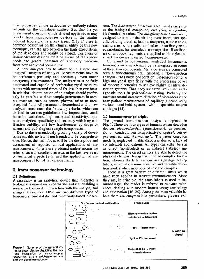
2.2 Immunosensor principlesThe general immunosensor design is depicted inFig. 1. There are four types of immunosensor detectiondevices: electrochemical (potentiometric, amperomet-ric or conductometric/capacitative), optical, micro-gravimetric, and thermometric. The latter detectionmode is neglected in this discourse due to a lack ofconsiderable applications. All types can either be runas direct (nonlabeled) or as indirect (labeled) im-munosensors. The direct sensors are able to detect thephysical changes during the immune complex forma-tion, whereas the latter sensors use signal-generatinglabels, which allow more sensitive and versatile detec-tion modes when incorporated into the complex.
There is a great variety of different labels whichhave been applied in indirect immunosensors. Sincethey are, in principle, the same labels as used in im-munoassays, the reader is referred to relevant refer-ences, dealing with modern immunoassay technologyand automation [16-25]. Among the most valuable la-bels there are enzymes like peroxidase, glucose oxi-
Figure 1 Scheme of the general im-munosensor design depicting the inti-mate integration of immunologicalrecognition at the solid-state surfaceand the signal transduction
Surface-attached antibodies
¼°Ï÷~· · · ·ï ï
Ïï°á-.·.
Transducer
Electrochemical activesubstance -» Electrode
Heat -, Thermistor
Light -* Photon count
Mass change -» Piezo-electric device
Electricalsignal
J Lab Med 2001; 25 (9/10): 388-398 389
Neue Methoden und Analyseverfahren
dase, alkaline phosphatase (aP), catalase or lucifcrase,clectroactivc compounds like ferrocene or In24 salts,and a scries of fluorescent labels (rhodamine, fluorcs-eein. Cy5. ruthenium di-irninc complexes, phosphores-cent poiphyrin dyes etc). In particular, laser-inducedfluo-rometric resonance energy transfer between twofluorophorcs offers methodological advantages f26|and can be extended to fiberoptic sensing (see chapter2.2.2) devices [27].
Although indirect immunosensors are highly sensi-tive due to the analytical characteristics of the labelapplied, the concept of a direct sensor device is stillfascinating and represents a true alternative develop-ment to immunoassay systems. Its potential simplicityholds multiple advantages, making immunosensorsprogressive and future directed.
2.2.7 Electrochemical sensorsPoteniiometric immunosensors. The Nernst equation[28] provides the fundamental principle of all poten-tiometric transducers. According to this equation, po-tential changes are logarithmically proportional to thespecific ion activity. Potentiometric transducer elec-trodes, capable of measuring surface potential alter-ations at near-zero current flow, are being constructedby applying the following methodologies:
Transmembrane potential. This transducer principleis based on the accumulation of a potential across asensing membrane. Ion-selective electrodes (ISE) useion-selective membranes which generate a charge sep-aration between the sample and the sensor surface.Analogously, antigen or antibody immobilized on themembrane binds the corresponding compound fromthe solution at the solid-state surface and change thetransmembrane potential.
Electrode potential. This transducer is similar to thetransmembrane potential sensor. An electrode by itself,however, is the surface for the immunocomplex build-ing, changing the electrode potential in relation to theconcentration of the analyte.
Field effect transistor (FET). The FET is a semi-conductor device used for monitoring charges at thesurface of an electrode, which have been built up onits metal gate between the so-called source and drainelectrodes. The surface potential varies with the ana-lyte concentration. The integration of an ISE with FETis realized in the ISFET. This technique can also beapplied to immunosensors.
An advantage of potentiometric sensors is the sim-plicity of operation, which can be used for automation,and the small size of the solid-state FET sensors. Allpotentio-metric methods, however, are still sufferingfrom major problems of sensitivity (being inferior toamperometric transducers), nonspecific effects ofbinding or signaling (influences from other ions pre-sent in the sample). Especially the signal-to-noise ratiocauses analytical problems which are difficult to cir-cumvent [10, 13]. Thus, a trend away from these tech-niques has been observed in the last years. Only theISFET may be seen as a candidate for ultrasensitive
clinical immunosensor applications, in particular whenthe novel concept of differential ISFET-based mea-surement of the zeta potential is used: the streamingpotential is a potential difference in flow direction,caused by the flow of excess ions resulting from alocal distortion of the charge balance. The zeta poten-tial - directly correlated to the streaming potential -reflects the potential changes in the diffuse outer layerat the solid-liquid interface. It efficiently reacts to pro-tein accumulations onto surfaces and thus is suitablefor detecting immunocomplex reactions [29].
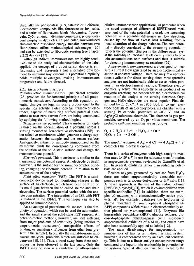
Amperometric immunosensors are designed to mea-sure a current flow generated by an electrochemical re-action at constant voltage. There are only few applica-tions available for direct sensing since most (protein)analytes are not intrinsically able to act as redox part-ners in an electrochemical reaction. Therefore electro-chemically active labels (directly or as products of anenzymic reaction) are needed for the electrochemicalreaction of the analyte at the sensing electrode. Oxy-gen and H2O2 electrodes are most popular. First de-scribed by L. C. Clark in 1956 [30], an oxygen elec-trode consists of an electrolyte-bearing chamber with asensing Pt cathode, polarized at -0.7 V, and anAg/AgCl reference electrode. The chamber is gas-per-meable, covered by an O2-per-vious membrane. Theprobable cathodic reactions are as follows:
02 + 2 H20 + 2 e- -> H20, + 2 OH'H202 + 2 e- -> 2 OH'.
The anodal reaction: 4 Ag + 4 Cl~ —*completes the electrical circuit.
4 AgCl + 4
There is a series of enzymes with high catalytic reac-tion rates (>103 s~l) in use for substrate transformationin amperometric systems, reviewed by Ghindilis et al.[6]. In general, oxidizing rather than reducing poten-tials are applied.
Besides oxygen, generated by catalase from H2O2,there are other amperometrically detectable com-pounds such as ferrocene derivatives or In2+ salts [11].A novel approach is the use of the redox polymer[PVP-Os(bipyridyl)2Cl], which is co-immobilized withspecific antibodies [31]. In addition, there are exam-ples of enzymes^ with electrochemically active prod-ucts. aP, for example, catalyzes the hydrolysis ofphenyl phosphate or p-aminophenyl phosphate (4-APP) compounds which result in electrochemically ac-tive phenol or p-aminophenol [11]. Enzymes likehorseradish peroxidase (HRP), glucose oxidase, glu-cose-6-phosphate dehydrogenase (with subsequentamperometrical oxidation of NADH) and others havealso been successfully applied as labels [12].
The main disadvantage for amperometric im-munosensors of having an indirect sensing system,however, is compensated for by an excellent sensitivi-ty. This is due to a linear analyte concentration rangecompared to a logarithmic relationship in potentiomet-ric systems. Special attention must be directed to the
390 J Lab Med 2001; 25 (9/10): 388-398
R B.Luppa
system inherent transport rate limitations for redoxpartners on the electrode surface.
Conductometric and capacitive immunosensorsmeasure the alteration of the electrical conductivity ina solution at constant voltage, caused by biochemical(enzymatic)' reactions which specifically generate orconsume ions. The capacitance changes are measuredusing an electrochemical system in which the bioac-tive element is immobilized onto a pair of noble metal(mostly Au or Pt) electrodes. There are only few clin-ical applications available yet, due to the high ionicstrength of biological matrices making it difficult torecord the relatively small net conductivity changescaused by the signaling reaction (e.g. see [32]). To cir-cumvent this problem, an iori-channel conductance im-munosensor, mimicking biological sensory functions,was recently described [33]. Basis of this technique isthe fact that the conductance of a population of mole-cular ion-channels, built of tethered gramicidin A andaligned across a lipid bilayer membrane, is changed bythe antibody-antigen binding event. Different applica-tions using various antibodies linked to the ion-chan-nel complex are given. Another approach is the mea-surement of changes of the surface conductivity. Yag-iuda et al. [34] developed a conductometric im-munosensor for the determination of methampheta-mine (MA) in urine. Anti-MA antibodies were immo-bilized onto the surface of a pair of platinum elec-trodes. The immunocomplex formation caused a de-crease in the conductivity between the electrodes.
2.2.2 Optical immunosensorsOptical immunosensors are most popular for bioanaly-sis and today's largest group of transducers. This isdue to the advantages of applying visible radiationcompared to other transducer techniques. Additionalbenefits are the non-destructive operation mode andthe rapid signal generation and reading. In particular,the introduction of fiber bundle optics as optical wave-guides and sophisticated -optoelectronics offers in-creased versatility of these analytical devices for clin-ical applications [4, 10, 11, 13]. .
Changes in adsorption,™fluorescence, luminescence,scatter or refractive index occur when light is reflect-ed at sensing surfaces. This information is the basis foroptical sensor techniques. Detectors are usually photo-diodes or photomultipliers.
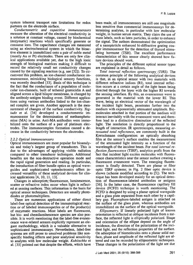
There are numerous applications of either directlabel-free optical detection of the immunological reac-tion or of labeled immunospe'cies or of the product(s)of enzymatic reactions. Most labels are fluorescent,but bio- and chemilumihescence species are also pos-sible. It is worth mentioning that the label-free evanes-cence wave-related sensors explicitly represent .an ele-gant methodology, which is a valuable alternative tosophisticated immunoassays. Nevertheless, label-freesystems are still prone to unsolved problems like non-specific binding effects and poor analytical sensitivityto analytes with low molecular weight. Kubitschko etal. [35] pointed out that despite the efforts, which have
been made, all immunosensors are still one magnitudeless sensitive than commercial immunoassays for de-termining analytes, in particular with low molecularweight, in human serum matrix. They claim the use ofmass labels, such as latex particles, in order to enhancethe signal. The authors demonstrated the optimizationof a nanoparticle enhanced bi-diffraclive grating cou-pler immunosensor for the detection of thyroid stimu-lating hormone (TSH). The excellent performancecharacteristics of this sensor clearly showed how fu-ture devices should work.
The principles of the different optical sensor typesare explained in detail as follows.
Total internal reflection spectroscopy (TIRS). Thecommon principle of the following analytical devicesis that, in an optical sensor with two materials withdifferent refractive indices (RI), total internal reflec-tion occurs at a certain angle of the light beam beingdirected through the layer with the higher RI towardsthe sensing interface. By this, an evanescence wave isgenerated in the material with the lower RI. Thiswave, being an electrical vector of the wavelength ofthe incident light beam, penetrates further into themedium with exponentially attenuated amplitude. Bio-molecules attached in that portion of the medium willinteract inevitably with the evanescent wave and there-fore lead to a distinctive diminution of the reflectedlight. The resolution is directly proportional to thelength of interaction. IR spectroscopes, measuring at-tenuated total reflectance, are commonly built in theKretschmann configuration: an optically absorbingfilm at the sensor's surface enables the measurementof the attenuated light intensity as a function of thewavelength of the incident beam. For total internal re-
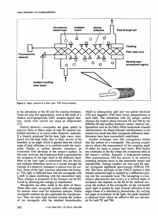
. flection fluorescence (TIRF), analytics profit from thefact that incident light excites molecules with fluores-cence characteristics near the sensor surface creating afluorescent evanescent wave. The emerging fluores-cence is finally detected. There are planar or fiberoptic TIRF systems; in Fig. 2 fiber optic device isshown (scheme modified according to [5]). The tech-nique has been developed mainly for an optical detec-tion of fluorescence-labeled antibodies or antigens[36]. In the latter case, the fluorescence capillary filldevice (FCFD) technique is worth mentioning. TheFCFD is designed by using a planar optical waveguideand a glass plate separated from each other by a capil-lary gap. Fluorophore-labeled antigen is attached onthe surface of the glass plate, whereas antibodies areimmobilized on the surface of the optical waveguide.
Ellipsometiy. If linearly polarized light of knownorientation is reflected at oblique incidence from a sur-face, the reflected light is elliptically polarized. Shapeand orientation of the ellipse depend on the angle ofincidence, the direction of the polarization of the inci-dent light, and the reflection properties of the surface.On adsorption of biomolecules onto a planar solid sur-face, phase and amplitude of the reflected light are al-tered and can be recorded by ellipsometric techniques.These changes in the polarization of the light are due
J Lab Med 2001; 25 (9/10): 388-398 391
Neue Methoden und Analyseverfahren
tFluorescence- Unlabeled Specificlabeled träger analyte antibodies Flow through cell
Fluorescencedetector
Cladding
Fiber optic core
Sensing layer(evanescent region)
Incident excitinglaser beam
Figure 2 Basic scheme of a fiber optic TIRF immunosensor
to the alterations of the RI and the coating thickness.There are only few applications, such as the study of acholera toxin-ganglioside GM1 receptor-ligand reac-tion, which were carried out using an ellipsometer[37].
Optical dielectric waveguides are glass, quartz orpolymer films or fibers made of high RI material em-bedded between or in lower index dielectric materials.If a linearly polarized He-Ne laser light wave, intro-duced into the high index film or fiber, arrives at theboundary at an angle which is greater than the criticalangle of total reflection, it is confined inside the wave-guide. Similar to surface plasmon resonance, anevanescent field develops at the sensor's surface. Inthis case, however, the evanescent field is generated bythe excitation of the light itself in the dielectric layer.Most of the laser light is transmitted into the device,and multiple reflections occur as it travels through themedium if a bioactive substance is placed over the sur-face. Some of the light, however, penetrates the biolay-er. This light is reflected back into the waveguide witha shift in phase interfering with the transmitted light.Thus, changes in properties of the biolayer can be fol-lowed by detecting the changes in interference.
Waveguides are often made in the form of fibers.These fiber-optic waveguide systems offer advantagesfor sensors when used for hazardous analysis. Planarwaveguide systems are also applicable for interferome-ters. They use laser light directed towards the surfaceof the waveguide with the attached biomolecules,
which is subsequently split into two partial electrical(TE) and magnetic (TM) field waves, perpendicular toeach other. The interaction with the sample surfacechanges the relative phase between TE and TM by thedifferent RI and surface thickness values. Various con-figurations such as the Fabry-Perot monomode channelinterferometer^ the Mach-Zehnder interferometer or therelated two-mode thin-film waveguide difference inter-ferometer have been successfully established.
Another technique uses thin corrugations etchedinto the surface of a waveguide. This grating couplerdevice allows the measurement of the coupling angleof either the input or output laser beam. Both beamsare correlated to the RI within the evanescent field atthe sensor's surface. Recently, a long-period gratingfiber immunosensor [38] has proven to be sensitive(enabling analyses down to the nanomolar range) andreproducible. Grating couplers are also used for opti-cal waveguide lightmpde spectroscopy (OWLS) [39,40]. The basic principle of the OWLS method is thatlinearly polarized light is coupled by a diffraction grat-ing into the waveguide layer. The incoupling is a res-onance phenomenon that occurs at a defined angle ofincidence that depends on the RI of the medium cov-ering the surface of the-waveguide. In the waveguidelayer, light is guided by total internal reflection to theedges where it is detected by photodiodes. By varyingthe. angle of incidence of the light, the mode spectrumis obtained from which the effective RIs are calculat-ed'for both TE and TM.
392 J Lab Med 2001; 25 (9/10): 388-398
P. B. Luppa
evanescentwave
Metal film(surface ̂plasmonelectrons)
Rln1 » n2 7
ÈÊÈ2 È1>èñ È1 = resonant angle
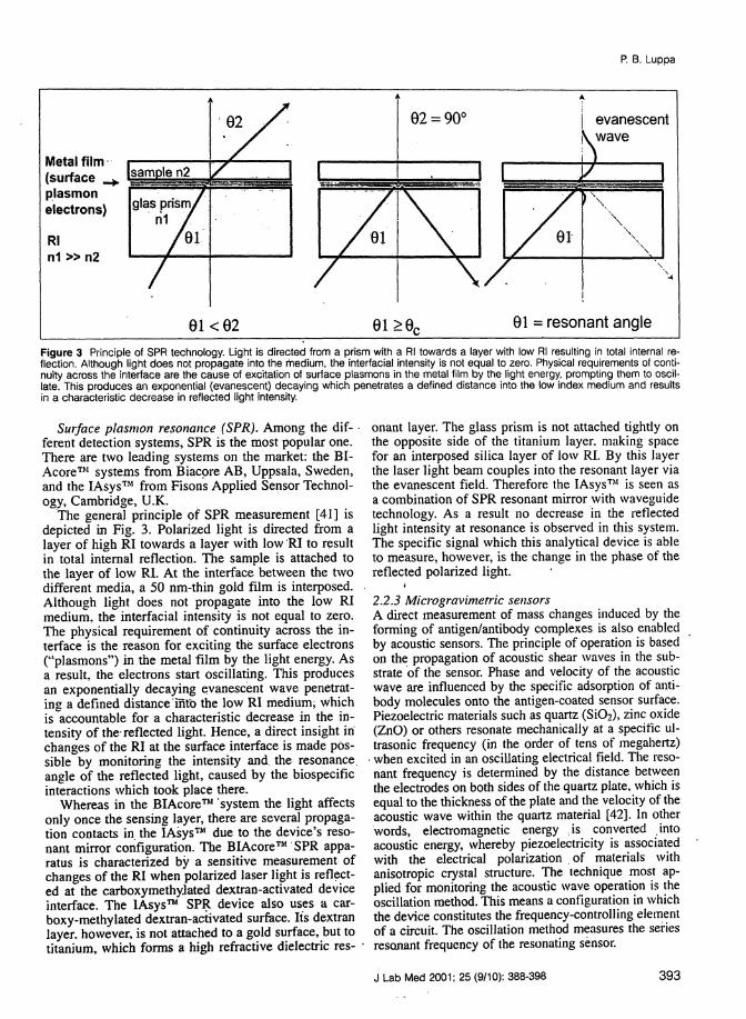
Figure 3 Principle of SPR technology. Light is directed from a prism with a Rl towards a layer with low Rl resulting in total internal re-flection. Although light does not propagate into the medium, the interfacial intensity is not equal to zero. Physical requirements of conti-nuity across the interface are the cause of excitation of surface plasmons in the metal film by the light energy, prompting them to oscil-late. This produces an exponential (evanescent) decaying which penetrates a defined distance into the low index medium and resultsin a characteristic decrease in reflected light intensity.
Surface plasmon resonance (SPR). Among the dif-ferent detection systems, SPR is the most popular one.There are two leading systems on the market: the BI-Acore™ systems from Biacpre AB, Uppsala, Sweden,and the lAsys™ from Fisons Applied Sensor Technol-ogy, Cambridge, U.K.
The general principle of SPR measurement [41] isdepicted in Fig. 3. Polarized light is directed from alayer of high Rl towards a layer with low Rl to resultin total internal reflection. The sample is attached tothe layer of low Rl. At the interface between the twodifferent media, a 50 nm-thin gold film is interposed.Although light does not propagate into the low Rlmedium, the interfacial intensity is not equal to zero.The physical requirement of continuity across the in-terface is the reason for exciting the surface electrons("plasmons") in the metal film by the light energy. Asa result, the electrons start oscillating. This producesan exponentially decaying evanescent wave penetrat-ing a defined distance into the low Rl medium, whichis accountable for a characteristic decrease in the in-tensity of the-reflected light. Hence, a direct insight inchanges of the Rl at the surface interface is made pos-sible by monitoring the intensity and the resonance,angle of the reflected light, caused by the biospecificinteractions which took place there.
Whereas in the BIAcore™ 'system the light affectsonly once the sensing layer, there are several propaga-tion contacts in the lAsys™ due to the device's reso-nant mirror configuration. The BIAcore™ SPR appa-ratus is characterized by a sensitive measurement ofchanges of the Rl when polarized laser light is reflect-ed at the carboxymethyjated dextran-activated deviceinterface. The lAsys™ SPR device also uses a car-boxy-methylated dextran-activated surface. Its dextranlayer, however, is not attached to a gold surface, but totitanium, which forms a high refractive dielectric res- ·
onant layer. The glass prism is not attached tightly onthe opposite side of the titanium layer, making spacefor an interposed silica layer of low Rl. By this layerthe laser light beam couples into the resonant layer viathe evanescent field. Therefore the lAsys™ is seen asa combination of SPR resonant mirror with waveguidetechnology. As a result no decrease in the reflectedlight intensity at resonance is observed in this system.The specific signal which this analytical device is ableto measure, however, is the change in the phase of thereflected polarized light.
2.2.3 M lew gravimetric sensorsA direct measurement of mass changes induced by theforming of antigen/antibody complexes is also enabledby acoustic sensors. The principle of operation is basedon the propagation of acoustic shear waves in the sub-strate of the sensor. Phase and velocity of the acousticwave are influenced by the specific adsorption of anti-body molecules onto the antigen-coated sensor surface.Piezoelectric materials such as quartz (SiO2), zinc oxide(ZnO) or others resonate mechanically at a specific ul-trasonic frequency (in the order of tens of megahertz)when excited in an oscillating electrical field. The reso-nant frequency is determined by the distance betweenthe electrodes on both sides of the quartz plate, which isequal to the thickness of the plate and the velocity of theacoustic wave within the quartz material [42]. In otherwords, electromagnetic energy is converted intoacoustic energy, whereby piezoelectricity is associatedwith the electrical polarization of materials withanisotropic crystal structure. The technique most ap-plied for monitoring the acoustic wave operation is theoscillation method. This means a configuration in whichthe device constitutes the frequency-controlling elementof a circuit. The oscillation method measures the seriesresojiant frequency of the resonating sensor.
J Lab Med 2001; 25 (9/10): 388-398 393
Neue Methoden und Analyseverfahren
The microgravimetric sensor devices are dividedinto quartz crystal microbalancc (QCM) devices ap-plying a ihickncss-shcar mode (TSM) and devices ap-plying a surface acoustic wave (SAW) detection prin-ciple. These sensors have reached considerable techni-cal sophistication. A profound review on the technolo-gy of acoustic wave sensors was recently given byGiv/cet al. [7]. Janshoffst al. |43] moreover pointedout that for various bioanalytic applications piczoim-munosensors may be valuable alternatives to opticalsensors.
In a fundamental work, Saiterbrey [44] showed forthe gaseous phase that a weight change of the quartzsurface is precisely determined by a shift in the res-onating frequency. For a TSM device he developed thefollowing equation:
Am = - F()2) AF
with FO. resonant frequency; A, area of coating; , density; , shearmodulus of the quartz.
The equation can be interpreted that surface-addedlayers will extend the wavelength of the acoustic wavewithin the quartz device. In other words, the attenua-tion of the acoustic energy, visible as negative fre-quency shift, is directly related to the mass increase ofthe quartz' surface.
Although theoretically unproven and contradictedby several authors, the Sauerbrey equation was also in-voked to explain the utility of TSM sensor devices inliquid media. By exposing only one side of the crystalto the liquid surface, it was able to generate frequencyresponses to mass changes in liquid media comparablein sensitivity to gas phase depositions of ultra-thinfilms. The different theoretical models, describing mi-crogravimetric measurements, however, have to beadapted to several wave damping factors such as ener-gy dissipation by viscous action of the medium, by en-ergy storage or by elastic deformation in the sensingfilm [7].
The TSM sensor consists of an AT-cut piezoelectriccrystal disc, most commonly of quartz because of itschemical stability in biological fluids and resistance toextreme temperatures. The disc is attached to twometal electrodes on opposite sides for the applicationof the oscillating electric field. The TSM is run in arange of 5-20 MHz. Advantages are, besides thechemical inertness, the low cost of the devices and thereliable quality of the mass-produced quartz discs.Major drawbacks of the system are the insensitivityfor analytes with a· molecular weight <1000 Da, and -as seen in all label -free immunosensor systems - non-specific binding interferences. These are hard to dis-tinguish from authentic binding events due to the factthat no reference line can be placed in the sensor de-vice. For a SH-APM device, however, Dahint et .al.[45] showed that by appropriately selecting the devicefrequency these spurious responses can be suppressed.This sensor is applicable for measurements in humanserum matrix.
One of the first applications of TSM technologywas an immunosensor for human immunodeficiencyvirus (HIV) serology f46J. This sensor was realized byimmobilizing recombinant viral peptides on the sur-face of the transducer and by detecting anti-HIV anti-bodies directly in human sera.
SAW sensors use thick ST-cut quartz discs and in-terdigitated metal electrode arrays that generateacoustic Rayleigh waves in both directions from theinterdigital electrodes, their transmission being attenu-ated by surface-attached biomolecules. The oscillationfrequency of a SAW sensor ranges from 30 to 500MHz. The operation of SAW immunosensors with bi-ological samples is compromised by the fact that thesurface wave is considerably attenuated in the liquidphase. Thus, the domain of this technique is most like-ly restricted to gas phase operations.
2.3 Alternative analyte-binding compounds for im-munosensorsAptamers are single-stranded DNA or RNA oligonu-cleotide sequences with the capacity to recognize var-ious target molecules with high affinity and specifici-ty. These ligand-binding oligonucleotides mimic prop-erties of antibodies in a variety of diagnostic formats.They are folded into unique overall shapes to form in-tricate binding furrows for the target structure [47].Aptamers are identified by an in vitro selectionprocess known as systematic evolution of ligands byexponential enrichment (SELEX) [47, 48]. Aptamersmay have advantages over antibodies due to the easeof depositing them on sensing surfaces and due to thehighly reproducible synthetic approach in any quanti-ties, albeit the affinity constants are consistently lowerthan those of antibodies and the- stability of these com-pounds is still questionable, in particular for diagnos-tic applications in complex biological matrices. Theaptamer-based schemes are still in their infancy and itis expected that modified nuclease-resistant RNA (andDNA) aptamers will soon be available for a variety oftherapeutic and diagnostic formats [49].
Lipocalins constitute a family of proteins for stor-age or transport of hydrophobic and/or chemically sen-sitive organic compounds. The retinol-binding proteinis an example in human physiology. Schlehuber et al.[50] demonstrated that the bilin-binding protein, amember of the lipocalin family and originating fromthe butterfly Pieris, brassicae, can be structurally re-shaped in ord'er to specifically complex potential anti-gens such as digoxigenin, which was given as an ex-ample.
These binding proteins share a conserved ß-barreLwhich is made of eight antiparallel ß-strands, windingaround a central, core. At the wider end of the conicalstructure these strands -are connected in a pairwisemanner by four loops that form the ligand-binding site.The lipocalin scaffold can be employed for the con-struction of so-called "anticalins", which provide apromising alternative to recombinant antibody frag-ments. This is made by subjecting various amino acid
394 J Lab Med 2001; 25 (9/10): 388-398
P. B. Luppa
residues, distributed across the four loops, to targetedrandom mutagenesis. It remains to be shown that thisclass of proteins is applicable in diagnostic assays andin immunosensors. Critical points are the stability, themagnitude of the affinity constants and the versatilityfor being crafted against the huge variety of ligands.
Molecular imprinting is a technique that is based onthe preparation of polymeric sorbents with selectivitypre-determined for a particular substance, or group, orstructural analogues [51, 52]. Functional and cross-linking monomers of plastic materials such asmethacrylics and styrenes are allowed to interact witha templating ligand to create low-energy interactions.Subsequently, polymerization is induced. During thisprocess, the molecule of interest is entrapped withinthe polymer either by a non-covalent, self-assemblingapproach, or by a reversible, covalent approach. After,stopping the polymerization, the template molecule iswashed out. The resultant imprint of the template ismaintained in the rigid polymer and possesses a steric(size, shape) and chemical (special arrangement ofcomplementary functionality) memory for the tem-plate. The molecularly imprinted polymer (MIP) isable to bind the template (= analyte) with a specificitysimilar to that of the antigen-antibody interaction.
MIP's have already been employed as non-biologi-cal 'alternatives to antibodies in competitive bindingassays. A series of applications for analytes such as cy-closporin A, atrazine, cqrtisol, 17ß-estradiol, theo-phylline, diazepam, morphine, and S-propranolol were"recently reviewed by Andersson [53], suggesting thatmolecular imprinting is a promising technique for im-munosensors.
3. Survey of clinical applications ofimmunosensor systemsApplications, dealing with "real" clinical samples, arestill rare. Most of the articles describing the use of im-munosensors in this field only have exemplary charac-ter. Detailed data and evaluation protocols on perfor-mance characteristics as well as comparisons with es-tablished methods for "particular parameters are missring in most cases. The analytical potential of this noveltechnique, however, is evident.
3.1 Applications using label-free detectionThe formation of specific antigen-antibody complexesleads to changes in electrochemical, optical or otherphysical properties at the surface of the immunosensorflow-through cell. The detection of these changes isthe basis of label-free immunosensors. Albeit it has tobe emphasized that, for all devices with label-free de-tection mode, non-specific binding phenomenona are acrucial point. It is impossible to distinguish the signalof an immunoreaction of a target molecule from thatof a nonspecific adsorption of other sample compo-nents to the transducer if no target-specific label isused. Thus, all nonspecific effects have to be mini-mized prior to a meaningful clinical application.
3.1.1 Electrochemical systemsAn interesting application is the determination ofhuman chorionic gonadotrophin (hCG) by an ampero-metric immunosensor, described by Clietcuti et al.[54]. The authors used Nafion, a polyanionic perfluo-rosulfonated ionomer to immobilize anti-hCG mono-clonal antibodies onto a glassy carbon electrode. AlsoSantandreu et al. [55] analysed ß-hCG using an am-perometric sensor device based on a conducting im-munocomposite. Kelly et al. [56] described an im-munosensor for lactate dehydrogenase (LOH) isoen-zyme 1 in serum. The LDH-1 activity was measuredamperometrically by the oxi-dation of NADH at theelectrode surface. Reproducibility as well as selectivi-ty of the sensor, however, are still insufficient for clin-ical use. Liu et al. [57] used a similar approach withNäfion and a protein A coupled polyaniline on an am-perometric sensor surface. A prototype amperometricimmunosensor for rapid detection of plasma heart-typefatty acid-binding protein (H-FABP) as early markerfor myocardial damage, already tested under routineconditions, was presented by Key et al. [58, 59]. Theauthors used a monoclonal anti-FABP antibody, linkedto aP, the enzyme catalyzing the hydrolysis of 4-APPto the electrochemically active phenol derivative.Mensel et al. [60] applied a heterogeneous ampero-metric immunosensor for the determination ofapolipoprotein E in serum. The authors used a site-di-rected attachment of monoclonal anti-apo E antibodiesto a disposable hydrazide-functionalized membranesurface. Bound analyte was then detected by use of apolyclonal antibody, labeled with aP, in conjunctionwith 4-APP as substrate for the carbon working elec-trode. The reliability pf the disposable low-cost sensorchip recommends this technology for medical use [60].
3.7.2 Optical systemsKroger ei al. [61] demonstrated a sensitive determina-tion of the H-FABP by use of a direct optical im-munosensor. As already described in chapter 2.2.2.,Kubiischko et al. [35] applied a nanoparticle enhancedbi-diffractive grating coupler immunosensor [62] forthe detection of TSH in serum. The excellent sensitiv-ity of this sensor is highly competitive to convention-al immunoassay systems. Another clinical applicationfield was entered by Nicholson et al. '[63]. By use of aresonant mirror SPR sensor, the antibody responses tocancer vaccination in patient sera was monitored. Theaffinity maturation of anti-idiotype tumor antibodiesafter vaccination of patients with advanced ovariancancer could be followed by determining the kineticsof the antibody response, thus enabling an objectivecomparison of different immunization schedules. Thedetection of human ß2-microglobulin (in buffer solu-tions, not in serum samples) by use of a grating cou-pler sensor was described by Brynda et al. [64]. Theauthors applied a three-dimensional antibody networkon the surface of the Ta2O5 grating coupler. A drug-monitoring application using the BlAcore™ systemwa$ presented by Keating et al. [65]. To monitor the
J Lab Med 2001; 25 (9/10): 388-398 395
Neue Methoden und Analyseverlahren
amicoagulatory coumarin derivative 7-hydroxy-coumarhi (7-OHC), a 7-OHC-conjugatc with bovineserum albumin (BSA) was immobilized onto a car-boxymethylatcd dexiran sensor chip. Serum samplescontaining the analyte were premixed with a polyclon-ul anti-7-OHC antibody and injected over the surface.Excess antibody, bound to the immobilized conjugate,then generates a binding response, inversely propor-lionaf lo the 7-OHC concentration.
3.1.3 Piezoelectric systemsLit et al. [66] described a protein -coated piezoelec-tric biosensor for the direct determination of humanIgG. The special feature of this device is·the favorablereusability of the surface due to an indirect immobi-lization of protein A onto a poly-L-lysine-modifiedelectrode surface by using a hydrophilic matrix of ox-idized dextran as linker. Another analyte for a piezo-electric sensor analysis was IgE, as described by Sit etal. [67]. The sensor surface was coated with an anti-IgE layer cross-linked via thioctic acid to form a high-ly ordered SAM. The device was able to detect IgE inthe range of 5-300 lU/ml with good precision and maybe regenerated five times without appreciable loss ofactivity. It also showed an excellent correlation to theCAP total IgE immunoassay from Pharmacia. Chu etal. [68] analyzed IgM in serum by use of a QCM withsurface-attached goat anti-lgM antibodies. The immo-bilization was accomplished by a CNBr-activatedcopolymer coating procedure. IgM could be detectedin the range of 5-93 //g/ml; the measurements were insatisfactory agreement with an established immunodif-fusion procedure. Finally, a QCM system was also ap-plied for the determinations of insulin [69] and corti-sol [70] in buffer solutions and of IgA in saliva [71].
3.2 Applications using signal-generating labels3.2J Electrochemical systemsCook [72] used an amperometric immunosensor cou-pled to microdialysis, enabling an in vivo measure-ment of Y-amino-4-butyric acid (GABA) in sheep so-matosensoric cortex. The specific antibody is fixed tothe platinum working electrode, the signal being gen-erated in a competitive design by a HRP-labeledGABA tracer. Cook presented also the real-time mea-surement of cortisol and corticosterone in animalsusing a similar antibody-based sensor [73]. A flow dis-placement column-immunosensor for serum cortisolusing HRP-labeled cortisol was established by Kapteinet al. [74]. A pH-FET immunosensor for measuring 0.2-interferon (INF) in·cultivated broth was described bySergeyeva et al. [75]. IFN was immobilized at the gateof a pH-FET and brought into reaction with an anti-IFN antibody, linked to ß-lactamase. After addition ofsubstrate, a local change in pH at the transducer's sur-face produced an electrochemical signal, pro-portionalto the conjugate concentration. The potentiometricsensor had a measuring range from 10 to 100 j/g/ml.An amperometric sensor for hCG in whole blood wasde-monstrated by Duan and Meyerhoff [76]. Gold-
coated microporous electrodes and surface-bound cap-ture antibodies, binding hCG in a sandwich complextogether with aP-labeled anti-hCG antibodies, wereemployed. The sensor performed separation-free in thematrix due to the fact that the surface-bound aP-activ-ity is resolved from excess aP-labeled antibodies in thesolution by introducing the 4-APP enzyme substratefrom the reverse side of the membrane electrode. Theenzymic reaction and the following oxidation of theformed p-aminophenol are in immediate proximity tothe electrode surface.
3.2.2 Optical systemsAstles and Miller [771 used'a fiber-optic immunosen-sor for analysing free phenytoin in plasma. The ana-lytical signal depended on the degree of energy trans-fer from B-phycoerythrin-labeled phenytoin to TexasRed-labeled anti-phenytoin antibodies. The immunore-action therefore was signaled by fluorescence quench-ing. This attempt allows continuous measurementswithout a separation step. PSA in whole blood was an-alyzed with good performance characteristics byDaniels et al. [78], applying allophycocyanin as fluo-rophore in an FCFD system. A fiber optic TIRF systemwas used by Mahoney et al. [5] for the measurement ofhCG in serum. The authors applied a secondary anti-hCG antibody, linked to allophycocyanin. Schult et al.[79] also detected hCG in serum by use of a dispos-able evanescent field fluorescence excitation chip witha remarkable analytical sensitivity. The label in thesandwich assay was a Cy5-labeled second anti-hCGantibody. This technique brings the immunosensornear to the goal of a routine medical diagnostic appli-cation. A competitive SPR-based immunosensor formorphine, using a morphine-BSA conjugate as tracer,immobilized at the chip surface, is described by Sakaiet al. [80]. Whether the system is able to measure thedrug in serum and urine was not tested. D-dimer mea-surement was performed by Rowe et al. [81] as part ofthe simultaneous detection of clinical samples using afluorescence-based optical immunosensor. A patternedarray of capture antibodies was coated on a planarwaveguide. Bound analytes were subsequently quanti-fied by fluorescently labeled detection antibodies, thespecific pattern being recognized by a CCD camera. Aself-contained fiber-optic device for measuring myo-globin was developed by Hanbury et al. [82]. CascadeBlue-labeled antibodies, entrapped with polyacry-lamide gel at the distal face of the fiber, reacted withthe protein analyte. The heme group of myoglobinthen quenched the dye fluorescence, the signal deriv-ing from this energy transfer.
3.2.3 Piezoelectric systemsWang et al. [83] coupled a gravimetric immunosensorwith a novel mass-amplifying label. By this, the Sen-sor became very sensitive for the detection of nonpen-etrating glycoprotein antigen (NPGP), a breast cancertumor marker. The competitive configuration of thesensor was as follows: a NPGP fusion protein antigen
396 J Lab Med 2001; 25 (9/10): 388-398
P. B. Luppa
was coated to the SiC>2 surface, then the first antibody,linked to colloidal gold, was incubated, its bindingratio depending on the presence of NPGP-as analyte.In a second step, the device surface was exposed to asilver developer solution, in which the gold colloidsserved as silver deposition nucleation sites*. Unfortu-nately, the analytical characteristics of the FPW sensor\yere not evaluated using real serum samples.
4. Future perspectives for clinicalapplicationsThere is a great opportunity for introducing im-munosensor devices, since clinical diagnostics repre-sent a huge, well-established and important analyticalfield. Hence sophisticated immunosensor systems maybe successful in the clinical laboratory. Worth-whilefuture goals for immunosensors are in my opinion mi-crodialysis-based continuous analyses in the fields ofblood coagulation, therapeutic drug-monitoring (in-cluding area-under-curve measurements), cytokine andcardiac markers in intensive-care units, as well aspoint-of-care systems for fertility and other endocrino-logical monitoring, type I-allergy and other immuno-logical analyses for patients in ambulatory settings.
In conclusion, the technical developments of bothimmunoassays and immunosensors are ongoing efforts.In particular, optical, electrochemical and microgravi-metric immunosensors have proven to be potent and fu-ture-directed analytical tools. Their sensitivities arecomparable to immunoassay methods. This can be con-cluded from selected applications, cited as references inthis review. Advantages, such as cheap mass productionof disposable chips, the possibility for real-time mea-surements even in vivo (due to miniaturization) and theoption for multianalyte analysis, will probably bringthis technique into the realm of practicability for diag-nostics. But the way to that goal is still stony and un-certain. Meanwhile, sophisticated immunoassays arestill indispensable for the clinical chemist. It is conceiv-able that in clinical diagnostics immunsensors will beable to occupy only an-analytical niche position for on-line monitoring purposes in the near future. This is incontrast to the situation in environmental analysis, bio-process control and research applications concerningbiomolecular interaction analysis, where the novel tech-nique will surely play a major role. To support this de-velopment, more attention should be focused on im-proving the specific and regenerable bioaffinity inter-face: this includes, among other issues, specific bindingprotein engineering, molecular imprinting and opti-mized solid-state surface chemistry. On the other side,laboratory medicine is called upon to define more strin-gently the clinical chemistry standards of evaluation ofnew methods and the possible application fields, such aspoint-of-care-testing. where this potent novel techniquemay be meaningfully applied. This will help to focus onareas where the commercialization will probably besuccessful.
References1. Ekins RR Immunoassay and other ligand assays: from isotopesto luminescence. J Clin Ligand Assay 1999;22:61-77.2. Lowe CR. Analytical biotechnology. Curr Opinion Biotechnol1996;7:l-3.3. Aiza\ya M. Immunosensors. Bioprocess Technol 1991; 15:249-66.4. Gizeli E, Lowe CR. Immunosensors. Curr Opin Biotechnol1996:7:66-71..5. Mahoney WC, Luderer AA, Brier RA, Lin JN. Real-time im-munodiagnostics employing optical immunobiosensors. In: ChanDW (ed), Immunoassay auto-mation. An updated guide to systems,1996. Academic Press, San Diego.6. Ghindilis AL. Atanasov P, Wilkins M, Wilkins E. Immunosen-sors: electrochemical sensing and other engineering approachesBiosens Bioelectron 1998:13:113-31.7. »aviE BA. Hayward GL, Thompson M. Acustic waves and thestudy of bio-chemical macromolecules and cells at the sensor-liquidinterface. Analyst 1999; 124:1405-20.8. O'Sullivan CK, Vaughan R, Guilbault GG. Piezoelectric im-munosensors - theory and applications. Anal Lett 1999;32:2353-77.9. Mullett W, Lai EP, Yeung JiM. Surface plasmon resonance-based immunoassays. Methods 2000;22:77-91.10. Morgan CL, Newman DJ, Price CP. Immunosensors: technolo-gy and opportunities in laboratory medicine. Clin Chem1996;42:193-209.11. Aizawa M. Immunosensors for clinical analysis. Adv ClinChem 1994:31:247-75.12. McNeil CJ, Athey D, Renneberg R. Immunosensors for clinicaldiagnostics. EXS 1997;81:17-25.13. Pearson JE, Gill A, Vadgama P. Analytical aspects of biosen-sors. Ann Clin Biochem 2000:37:119-45.14. Rogers KR. Principles of affinity-based biosensors. MolBiotechnol 2000; 14:109-29.15. Turner APF, Chen B, Piletsky SA. In vitro diagnostics in dia-betes: Meeting the challenge. Clin Chem 1999:45:1596-601.16. Sokoll LJ, Chan DW. Choosing an automated immunoassaysystem. In: Wild D, (ed). The Immunoassay Handbook, second ed.New York: Nature Publishing Group, 2001: 261-265.17. Sokoll LJ, Chan DW. Clinical analyzers. Immunoassay. AnalChem 1999;71:356R-362R.18. Anonymous. Automated immunoassay analyzers. CAP Today2000:14:33-64.19. Gajda A, Hawthorn M. Laboratory technology. Simultaneousmultiple analyte testing: the next 'revolution' in lab testing? Ad-vance/Laboratory 1999;8:69-70.20. Ekins RP. Ligand assays: from electrophoresis to miniaturizedmicroarrays. Clin Chem 1998;44:2015-30.21. Mendoza LG, McQuary P, Mongan A, Gangadharan R, BrignacS, Eggers M. High-throughput microarray-based enzyme-linkedimmunosorbent assay (ELISA). Biotechniques 1999;27:782-6.22. Felder RA, Kost GJ. Automation. Part 1. Modular stepwise au-tomation and the future of diagnostic testing. MLO Med Lab Obs1998:30(4):22-24, 26-27.23. Kricka LJ. Miniaturization of analytical systems. Clin Chem1998;44:2008-14.24. Bock JL. The new era of automated immunoassay. Am J ClinPathol 2000:113:628-646.25. Li DJ, Sokoll LJ, Chan DW. Automated chemiluminescent im-munoassay analy-zers. J Clin Ligand Assay 1998:21:377-85.26. Oswald B, Lehmann F. Simon L, Terpetschnig E, Wolfbeis OS.Red laser-induced fluorescence energy transfer in an immunosys-tem. Anal Biochem 2000;280:272-277.27. Wolfbeis OS. Fiber-optic chemical sensors and biosensors.Anal Chern 2000;72:81R-9R.28. Buerk DC. Biosensors. Theory and applications. 1993, Tech-nomic Publishing Co Inc, Lancaster. PA.29. Koch S, Woias P, Meixner LK, Drost S, Wolf H. Protein detec-tion with a novel ISFET-based zeta potential analyzer. Biosens Bio-electron 1999; 14:413-21.30. Clark LC. US Patent #2,913,386; 1956.31. Lopez MA, Ortega F, Domingucz E, Katakis I. Electrochemicalimmunosensor for the detection of atrazinc. J Mol Recognit1998;11:178-81.32. Berney HC, Alderman J. Lane WA, Collins JK. Development ofa capacitive immunoscnsor: a comparison of monoclonal and poly-clonal capture antibodies as the primary layer. J Mol Recognit1998;! 1:175-7.
J Lab Med 2001; 25 (9/10): 388-398 397
Neue Methoden und Analyseverfahren
33. Cornell , Braach-Maksvytis VLB, King LG et al. A biosen-sor that uses ion-channel switches. Nature 1997:387:580-3.34. Yagiuda K, Hcrnmi A, I to S ci al. Development of a conduc-tivity-based immu-nosensor for sensitive detection of methamphet-aminc (stimulant drug) in human urine. Biosens Bioclcctron1996:11:703-7.35. Kubitschko S, Spinkc J, Bruckner T, Pohl S. Oranth N. Sensi-t ivi ty enhancement of optical immunosensors with nanoparticlcs.Anal Bioehcm 1997:253:112-22.36. Domcnici C , Schironc A, Celcbrc M , Ahluwalia A, DC RossiD. Development of a T1RF immunoscnsor: modelling the equilibri-um behaviour of a competitive system. Biosens Bioelectron1995;lO:37l-378.37. Stcnberg M, Nygren H. A rcccptor-ligand reaction studied by anovel analytical tool - the isoscope ellipsometcr. Anal Biocheml982;127:183-92.38. DcLisa MR Zhang Z, Shiloach M et al. Evanescent wave long-period fiber bragg grating as an immobilized antibody biosensor.Anal Chem 2000;72:2895-900.39. Ticfcnthaler K. Integrated optical couplers as chemical wave
f uide sensors. In: Turner APF (ed), Advances in biosensors. JAIrcss, London 1992: 261-89.
40. Lukosz W. Integrated optical chemical and direct biochemicalsensors. Sens Actuators B 1995;29:37-50.41. Liedberg B, Nylander C, Lundström I. Biosensing with surfaceplasmon resonance - how it all started. Biosens Bioelectron1995:10:i-ix.42. Aberl F, Kösslinger C, Wolf H. In: Reischl U (ed), Methods inmolecular medicine, vol. 13. Molecular diagnostics of infectiousdiseases. Humana Press, Totawa. 1997: 519-27.43. Janshoff A, Galla HJ, Steinern C. Piezoelectric mass-sensingdevices as biosensors - an alternative to optical biosensors? AngewChem Int Ed Engl 2000;39:4004-4032.44. Sauerbrey G. Verwendung von Schwingquarzen zur Wägungdünner Schichten und zur Mikrowägung (Application of quartz os-cillators for weighing of thin films and for microweighing). Z Phys1959; 155:206-22.45. Dahint R. Bender F, Morhard F. Operation of acoustic platemode immuno-sensors in complex biological media. Anal Chem1999;71:3150-6.46. Aberl K Wolf H, Kösslinger C, Drost S, Woias P? Koch S. HIVsexology using piezoelectric immunosensors. Sens Actuators B,1994:18-19:271-5.47. Jayasena SD. Aptamers: An emerging class of molecules thatrival antibodies in diagnostics. Clin Chem 1999;45:1628-50.48. Tuerk C, Gold L. Systematic evolution of ligands by exponen-tial enrichment: RNA ligands to bacteriophage T4 DNA poly-merase. Science 1990;249:505-10.49. Hesselberth J, Robertson MP, Jhaveri S, Ellington AD. In vitroselection of nucleic acids for diagnostic applications. Rev MolBiotechnol 2000;74:15-25.50. Schlehuber S, Beste G, Skerra A. A novel type of receptor pro-tein, based on the lipocalin scaffold, with specificity for digoxi-genin. J Mol Biol 2000;297:1105-20.51. Ansell RJ, Ramström O, Mosbach K. Towards artificial antibod-ies prepared by molecular imprinting. Clin Chem 1996:42:1506-12.52. Sellergren B, Andersson LI. Application of imprinted syntheticpolymers in binding assay development. Methods 2000;22:92-106.53. Andersson LI. Molecular imprinting: developments and appli-cations in the ana-lytical chemistry field. J Chromatogr B2000;745:3-13.54. Chetcuti AF, Wong DK, Stuart MC. An indirect perfluorosul-fonated ionomer-coated electrochemical immunosensor for the de-tection of the protein human chorionic gonadotrophin. Anal Chem1999;71:4088-94.55. Santandreu M, Alegfet S, Fäbregas E. Determination of ß-HCGusing amperometric immunosensors based on a conducting im-munocomposite. Anal Chim Acta 1999;396:181-8.56. Kelly S, Compagnone D, Guilbault G. Amperometric im-munosensor for lactate dehydrogenase LD-1. Biosens Bioelectron1998; 13:173-9.57. Liu CH, Liao KT, Huang HJ. Amperometric immunosensorsbased on protein a coupled polyaniline-perfluorosulfonatedionomer composite electrodes. Anal Chem 2000;72:2925-9.58. Key G, Schreiber A, Feldbrugge R, Glatz JF, Spener F. An im-munosensor for rapid estimation of the early heart infarction-mark-er FABP. Acta Anaesthesiol Scand Suppl,1997;ll 1:289-92.
59. Key G, Schreiber A, Feldbrugge R et al. Multicenter evaluationof an ampcro-mctric immunosensor for plasma fatty acid-bindingprotein: an early marker for acute myocardial infarction. ClinBiochcm. 1999:32:229-31.60. Meusel M, Renneberg R, Spener F, Schmilz G. Development ofa heterogeneous amperometric immunosensor for the determinationof apolipoprotein E in serum. Biosens Bioelectron 1995:10:577-86.61. Kroger D, Katerkamp A, Renneberg R, Cammann K. Surfaceinvestigations on the development of a direct optical immunosen-sor. Biosens Bioelectron 1998:13:1141-7.62. Spinkc J. Oranth N, Fattinger C, Koller H, Mangold C,Voegelin D. The bi-diffractive grating coupler: application to im-munosensing. Sens Actuators B 1997:38-39:256-60.63. Nicholson S, Gallop JL, Law P, Thomas H, George AJT. Mon-itoring antibody responses to cancer vaccination with a resonantmirror biosensor. Lancet 1999:353:808.64. Brynda E, Houska M. Brandenburg A, Wikerstal A, Skvor J.The detection of human fVmicroglobulin by grating coupler im-munosensor with three dimensional antibody networks. BiosensBioelectron 1999; 14:363-8.65. Keating GJ, Quinn JG, 'Kennedy R. Immunoassay for the de-termiation of 7-hydroxycoumarin in serum using "real-time"biosensor analysis. Anal Letters 1999:32:2163-2176.66. Lu HC, Chen HM, Lin YS, Lin JW. A reusable and specificprotein -coated piezoelectric biosensor for flow .injection im-munoassay. Biotechnol Prog 2000:16:116-24.
. 67. Su X, Chew FT, Li SF. Self-assembled monolayer-based piezo-electric crystal immunosensor for the quantification of total humanimmunoglobulin E. Anal Biochem 1999;273:66-72.68. Chu X, Lin ZH, Shen GL, Yu RQ. Piezoelectric imrnunosensorfor the detection of immunoglobulin M. Analyst 1995:120:2829-32.69. Suri CR, Jain PK, Mishra GC. Development of piezoelectriccrystal based microgravimetric immunoassay for determination ofinsulin concentration. J Biotechnol 1995;39:27-34.70. Attili BS, Suleiman AA. *A piezoelectric immunosensor for thedetection of cortisol. Anal Lett 1995;28:2149-2159.71. Tajima I, Asami O, Sugiura E. Monitor of antibodies in humansaliva using a piezoelectric quartz crystal biosensor Anal ChimActa 1998;365:147-9.72. Cook CJ. Monitoring on-line of extracellular gamma-ami no-4-butyric acid using microdialysis coupled to immunosensor analysis.J Neurosci Methods 1998:82:145-50.73. Cook CJ. Real-time measurements of corticosteroids in con-scious animals using an antibody-based electrode. Nat Biotechnol1997;15:467-71.74. Kaptein WA, Zwaagstra JJ, Venema K, Ruiters MHJ. Korf J.Analysis of cortisol with a flow displacement immunoassay. SensActuators B 1997;45:63-9. .75. Sergeyeva TA, Soldatkin AP, Rachkov AE, Tereschenko MLPiletsky SA, El'skaya AV. ß-Lactamase label-based potentiometricbiosensor for cc-2 interferon detection. Anal Chim Acta,1999:390:73-81.76. Duan C, Meyerhoff ME. Separation-free sandwich enzyme im-munoassay using microporous gold electrodes and self-assembledmonolayer/immobilized capture antibodies. Anal Chem 1994;66:1369-1377.77. Astles JR, Miller WG. Measurement of free phenytoin in bloodwith a self-contained fiber-optic immunosensor. Anal Chem1994;66:1675-82.78. Daniels PB, Fletcher JE, O'Neill PM. Stafford CG, Bacarese-Hamilton T, Robinson GA. A comparison of three fluorophores foruse in an optical biosensor, for the measurement of prostate-specif-ic antigen in whole blood. Sens Actuators B, 1995:26-27:447-451.79. Schult K, Katerkamp A, Trau D, Grawe F, Cammann K, MeuselM. Disposable optical sensor chip for medical diagnostics: newways in bioanalysis. Anal Chem 1999;71:5430-5.80. Sakai G, Ogata K, Uda T, Miura N, Yamazoe N. A surface plas-mon resonance-based imrnunosensor for highly sensitive detectionof morphine. Sens Actuators B 1998:49:5-12.81. Rowe CA, Scruggs SB, Feldstein MJ, Golden JP, Ligler FS. Anarray immuno-sensor for simultaneous detection of clinical ana-lytes. Anal Chem 1999;71:433-9.82. Hanbury CM, Miller WG, Harris RB. Fiber-optic immunosen-sor for measurement of myoglobin. Clin Chem 1997;43:2128-36.83. Wang AW, Kiwan R, White RM, Ceriani. RL. Silicon-based ul-trasonic immuno-assay for detection of breast cancer antigens. SensActuators B, 1998:49:13-21.
398 J Lab Med 2001; 25 (9/10): 388-398