Geometrically Repatterned Immunological Synapses Uncover Formation Mechanisms
Upload
independentCategory
view
5download
0
Disease Markers 31 (2011) 47–54 47DOI 10.3233/DMA-2011-0803IOS Press
Immunological functions of oxidized humanimmunoglobulin G in type 1 diabetesmellitus: Its potential role in diabetic smokersas a biomarker of elevated oxidative stress
Zafar Rasheeda,∗, Hani A. Al-Shobailib, Abdullateef A. Alzolibanib, Muhammad Ismail Khane,Tariq Ayubd, Mohammed Imran Khane and Naila Rasheeda
aDepartment of Medical Biochemistry, College of Medicine, Qassim University, Buraidah, KSAbDepartment of Dermatology, College of Medicine, Qassim University, Buraidah, KSAcDepartment of Physiology, College of Medicine, Qassim University, Buraidah, KSAdDepartment of Biochemistry, College of Applied Medical Sciences, Qassim University, Buraidah, KSAeInstitute of Genetics, School of Biology, Queens Medical Center, Medical School, Nottingham, UK
Abstract. The role of oxidized immunoglobulin G in type 1 diabetic smokers has been investigated in the present study. HumanimmunoglobulinG (IgG)wasmodified by reactive oxygen species (ROS). The binding characteristics of circulating autoantibodiesin type 1 diabetes patients against native and modified IgG were assessed by direct binding ELISA. High degree of specific bindingby 68.5% of patients sera towards ROS-modified IgG was observed in comparison to its native analogue (p < 0.05). In addition,diabetic smokers (n = 28) were examined and the results were compared with diabetic non-smokers (n = 26). Circulatingantibodies of diabetic smokers showed substantially stronger binding to modified IgG as compared with the antibodies presentin diabetic non-smokers (p < 0.05). Normal human sera (n = 53) showed negligible binding with either antigen. Competitiveinhibition ELISA reiterates the direct binding results. The increase in total serum protein carbonyl levels in the diabetic smokerswas largely due to an increase in oxidized IgG. Diabetic smokers showed substantially higher carbonyl contents in sera as well asin purified IgG as compared with sera and IgG of diabetic non-smokers. Collectively, the oxidation of plasma proteins, especiallyIgG, might enhance oxidative stress in type 1 diabetes smokers.
Keywords: Type 1 diabetic smokers, oxidized IgG, autoantibodies, reactive oxygen species
1. Introduction
Diabetes mellitus (DM) and its associated secondarycomplications are the leading cause of deaths world-wide [1]. The number of diabetes patients has beenincreased by exposure to environmental risk factors in-cluding cigarette smoking [2]. The prevention of dia-betes is a serious worldwide challenge, but it is clear
∗Corresponding author: Dr. Zafar Rasheed, Department of Med-ical Biochemistry, College of Medicine, Qassim University, P.O.BOX 6655, Buraidah-51452, KSA. Tel.: +96 531269227; E-mail:[email protected].
that the successful prevention of diabetes will dependon the reduction of risk factors along with better meth-ods of screening and early detection. Type 1 diabetesis an autoimmune disease resulting from the selectivedestruction of pancreatic β cells. Its incidence in dif-ferent ethnic groups is extremely variable, suggestingthat the involvement of genetic as well as environmen-tal elements [3]. In type 1 diabetes, multiple autoanti-bodies against pancreatic β cell antigens may arise andpersist even after the disease becomes clinically overt.In humans, glutamic acid decarboxylase (GAD), IA-2and insulin would be the most important targets againstwhich autoantibodies as well as autoreactive T cells
ISSN 0278-0240/11/$27.50 2011 – IOS Press and the authors. All rights reserved

48 Z. Rasheed et al. / Oxidized IgG in diabetic smokers
are directed [3]. The precise mechanism of β-cellsdestruction leading to diabetes remains unclear [4].
Oxidative stress plays a central role in the onset ofdiabetes as well as in diabetes associated complica-tions [1,2,5]. Various studies have shown that diabetesis associated with increased formation of free radicalsand decrease in antioxidant potential. This leads tooxidative damage of cell components such as proteins,lipids and nucleic acid [5]. Increased oxidative stressin diabetic patients leads to protein oxidation [5]. Theconversion of protein to protein carbonyl (PCO) deriva-tives may occurs via direct oxidation by ROS, with theeventual formation of oxidized amino acids [6].
ImmunoglobulinG is amajor protein of serumwhosefunction lies in the specific interactions with and clear-ance of antigens. In diabetes and rheumatoid arthritispatients, IgG dysfunctions have been reported; it be-haves not only as an antibody, but also as a putativeantigen for diabetes and rheumatoid arthritis autoanti-bodies [7,8]. It has been well documented that IgG isquite vulnerable to ROS [7–10]. Therefore, IgG is con-tinuously exposed to oxidative stress, so that alterationsof the conformation and function of IgG could occur,resulting in modification of its biological properties.
It is well documented that oxidative damage of pro-tein and DNA presents unique neo-epitopes,which maybe one of the factors in antigen-driven autoimmune re-sponse in autoimmune diseases [11,12]. Recently, wehave demonstrated that after modification with ROS,proteins became highly immunogenic and oxidative by-products with proteins could lead to neoantigens whichwould initiate autoimmunity in various diseases includ-ing diabetes [10,13–19]. In view of this, the presentstudy was based upon a hypothesis that oxidative by-products, hydroxyl radical damage IgG, help to initiateautoimmunity in type 1 diabetes patients. To test thishypothesis, IgG was purified from normal human seraby affinity chromatography. Affinity purified IgG wasmodified with hydroxyl radicals (.OH). Type 1 diabetespatients having smoking or non-smoking history wereevaluated for presence of circulating autoantibodies di-rected against native and .OH-modified IgG.
2. Materials and methods
2.1. Study subjects
Bloodwas collected fromhuman voluntary donors oftype 1 diabetes mellitus (n = 54, male = 43, female =11, aged 44 ± 15). Fifty two percent diabetic male
having smoking history, whereas twenty seven percentwere diabetic non-smokers. The control samples werecollected from healthy subjects (n = 53, male = 38,female = 15, aged 45 ± 10.2). Forty seven percent ofnormal human were cigarette smokers, whereas twentyfour percent were non-smokers. All studied femaleswhether diabetic or normal were non-smokers. Theaverage ( ± SD) of post prandial blood sugar levelfor 54 diabetic patients was 378 ± 26 mg/dl, while itwas 108 ± 7.5 mg/dl for 53 healthy subjects (withoutfamily history of diabetes). The glycated hemoglobin(HbA1C) was 8.1 ± 0.8 for patients, whereas 4.75 ±0.4 for normal subjects. The complete demographic de-tails of study subjects have been summarized in Table 1.Glutamic acid decarboxylase antibodies (GADA), in-sulin antibodies (IAA), anti-tyrosine phosphatase anti-bodies (IA-2A) were found in 42%, 71% and 36% ofthe cases, respectively. Immunopositivity for at leastone of these autoantibodies was found in all patients.All other information on demographics and cumulativeclinical and laboratory manifestations over the courseof disease was obtained by both chart review and dis-cussion with the patient and his/her family members.The informed consent was obtained from each patientor from patients’ family members.
All sera were decomplemented by heating at 56◦Cfor 30 min and stored in aliquots at −20◦C.
2.2. Immunoglobulin G purification
IgG from normal human sera was isolated by affin-ity chromatography on Protein A-Sepharose CL-4Bcolumn as previously described [11]. Briefly, serum(0.3 ml) diluted with equal volume of PBS, pH 7.4 wasapplied to the column (12 mm × 45 mm) equilibratedwith the same buffer. The flow through was reloadedonto the column 2–3 times. Unbound proteins wereremoved by extensive washing with PBS, pH 7.4. Thebound IgG was eluted with 0.58% acetic acid in 0.85%sodium chloride and neutralized with 1.0 ml of 1.0 MTris-HCl, pH 8.5, 3ml fractionswere collected and readat 251 and 278 nm. The IgG concentration was de-termined considering 1.38 OD278 = 1.0 mg IgG/ml.The isolated IgG was dialyzed against PBS, pH 7.4 andstored at−20◦C. The homogeneity of isolated IgG waschecked by polyacrylamide gel electrophoresis.
2.3. Modification of human IgG
Human IgG was modified in PBS (10 mM sodiumphosphate buffer containing 150 mM NaCl, pH 7.4) by
Z. Rasheed et al. / Oxidized IgG in diabetic smokers 49
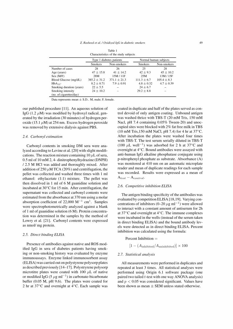
Table 1Characteristics of the study subjects
Type 1 diabetes patients Normal human subjectsSmokers Non-smokers Smokers Non-smokers
Number of cases 28 26 25 28Age (years) 47 ± 15.8 41 ± 14.2 42 ± 9.3 45 ± 10.2Sex (M/F) 28M 15M / 11F 25M 13M / 15FBlood Glucose (mg/dL) 385.2 ± 31.2 371.1 ± 21.3 111.3 ± 6.7 105.4 ± 8.3HbA1C 8.2 ± 0.71 7.9 ± 0.91 4.8 ± 0.32 4.7 ± 0.39Smoking duration (years) 22 ± 5.5 – 24 ± 6.7 –Smoking intensity 24 ± 10.2 – 29.2 ± 8.8 –(no. of cigarettes/day)
Data represents mean ± S.D.. M, male; F, female.
our published procedure [11]. An aqueous solution ofIgG (1.2 μM) was modified by hydroxyl radical, gen-erated by the irradiation (30 minutes) of hydrogen per-oxide (15.1 μM) at 254 nm. Excess hydrogen peroxidewas removed by extensive dialysis against PBS.
2.4. Carbonyl estimation
Carbonyl contents in smoking DM sera were ana-lyzed according to Levine et al. [20] with slight modifi-cations. The reaction mixture containing 10 μL of sera,0.5 ml of 10 mM 2, 4- dinitrophenylhydrazine (DNPH)/ 2.5 M HCl was added and thoroughly mixed. Afteraddition of 250 μM TCA (20%) and centrifugation, thepellet was collected and washed three times with 1 mlethanol: ethylacetate (1:1) mixture. The pellet wasthen dissolved in 1 ml of 6 M guanidine solution andincubated at 30◦C for 15 min. After centrifugation, thesupernatant was collected and carbonyl contents wereestimated from the absorbance at 370 nm using a molarabsorption coefficient of 22,000 M−1 cm1. Sampleswere spectrophotometrically analyzed against a blankof 1 ml of guanidine solution (6 M). Protein concentra-tion was determined in the samples by the method ofLowry et al. [21]. Carbonyl contents were expressedas nmol/ mg protein.
2.5. Direct binding ELISA
Presence of antibodies against native and ROS mod-ified IgG in sera of diabetes patients having smok-ing or non-smoking history was evaluated by enzymeimmunoassays. Enzyme linked immunosorbent assay(ELISA)was carried out on polystyrenepolysorpplatesas described previously [14–17]. Polystryrene polysorpmicrotitre plates were coated with 100 μL of nativeor modified IgG (5 μg ml−1) in carbonate-bicarbonatebuffer (0.05 M, pH 9.6). The plates were coated for2 hr at 37◦C and overnight at 4◦C. Each sample was
coated in duplicate and half of the plates served as con-trol devoid of only antigen coating. Unbound antigenwas washed thrice with TBS-T (20 mM Tris, 150 mMNacl, pH 7.4 containing 0.05% Tween-20) and unoc-cupied sites were blocked with 2% fat free milk in TBS(10 mM Tris,150 mM NaCl, pH 7.4) for 4 hr at 37◦C.After incubation the plates were washed four timeswith TBS-T. The test serum serially diluted in TBS-T(100 μL well−1) was adsorbed for 2 h at 37◦C andovernight at 4◦C. Bound antibodies were assayed withanti-human IgG alkaline phosphatase conjugate usingp-nitrophenyl phosphate as substrate. Absorbance (A)was monitored at 410 nm on an automatic microplatereader and mean of duplicate readings for each samplewas recorded. Results were expressed as a mean ofAtest − Acontrol.
2.6. Competitive inhibition ELISA
The antigen binding specificity of the antibodies wasevaluated by competition ELISA [18,19]. Varying con-centrations of inhibitors (0–20 μg ml−1) were allowedto interact with a constant amount of antiserum for 2hat 37◦C and overnight at 4◦C. The immune complexeswere incubated in the wells (instead of the serum takenin direct binding ELISA) and the bound antibody lev-els were detected as in direct binding ELISA. Percentinhibition was calculated using the formula:
Percent Inhibition =
[1 − (Ainhibited/Auninhibited)] × 100
2.7. Statistical analysis
All measurements were performed in duplicates andrepeated at least 3 times. All statistical analyses wereperformed using Origin 6.1 software package (onepaired two tailed t-test with one way ANOVA analysis)and p < 0.05 was considered significant. Values havebeen shown as mean ± SEM unless stated otherwise.
50 Z. Rasheed et al. / Oxidized IgG in diabetic smokers
Table 2Competitive inhibition of antibodies from type 1 diabetes smok-ing and non-smoking patients by native and ROS-modified im-munoglobulin G
Type of sera Number of Maximum inhibition (%)#
patients Native IgG ROS-IgG
DM smokers 20 22.1 ± 1.9a 54.2 ± 1.2b
DM non-smokers 17 21.1 ± 1.1a 40.3 ± 3.3c
NH smokers 25 20.4 ± 1.2a 22.2 ± 7.5a
NH non-smokers 28 19.2 ± 2.1a 20.7 ± 6.5a
Results are representative (mean ± SEM) of triplicate experi-ments and differ without a common letter P < 0.05. Microtitreplates were coated with ROS-IgG (5 μg/ml). #Maximum per-cent inhibition at 20 μg/ml of inhibitor concentration. DM: type1 diabetes mellitus; NH: normal human; IgG: immunoglobulinG.
3. Results
3.1. Characterization and immunization of native andROS-modified human IgG
Our earlier reports showed alterations in IgG fol-lowing exposure to the hydroxyl radical, generated bythe UV-irradiation of hydrogen peroxide [11]. Loss ofsecondary structures, hypochromicity at 280 nm, lossof tryptophan fluorescence intensity, increase in pro-tein carbonyl contents were observed in human IgGfollowing hydroxyl treatment.
Previously, we also demonstrated that ROS-IgG wasa potent immunogen in rabbits that induced high-titre(> 1:12,800) antibodies, whereas native human IgGshowed a titre < 1:6400 [11]. Preimmune serum serv-ing as a negative control did not show appreciable bind-ing with the respective immunogens under identicalexperimental conditions.
3.2. Recognition of ROS-modified IgG by diabeticsmokers autoantibodies
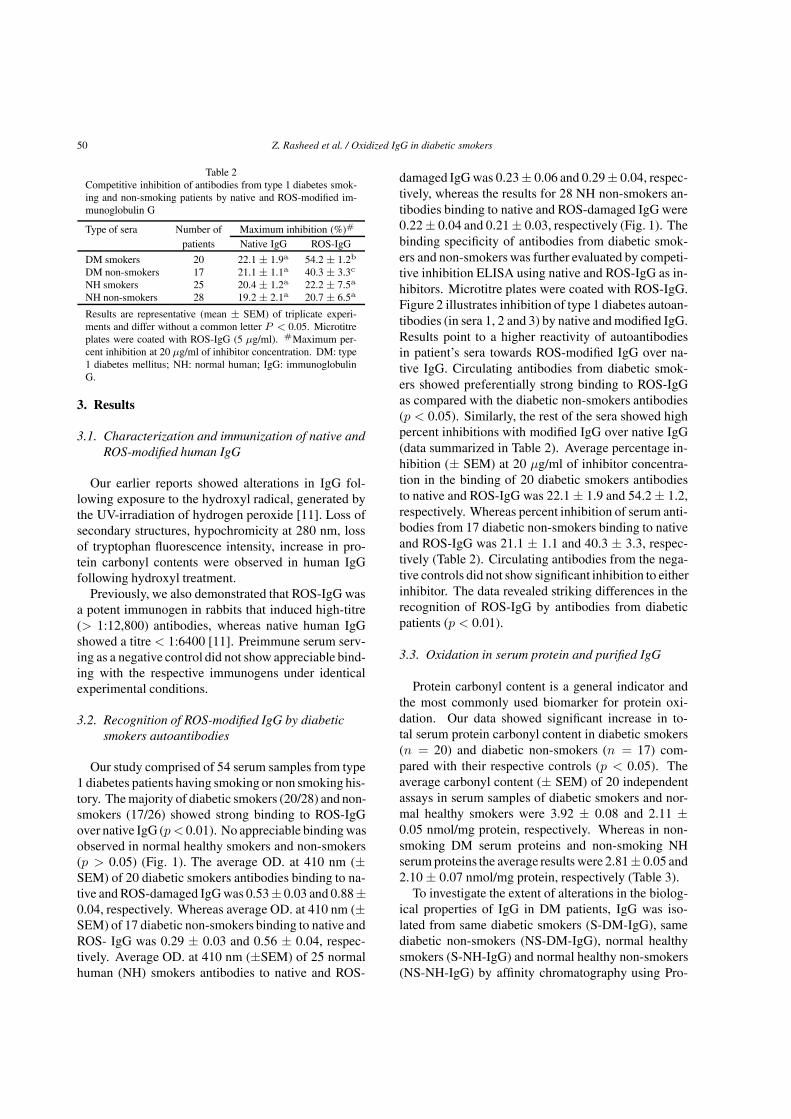
Our study comprised of 54 serum samples from type1 diabetes patients having smoking or non smoking his-tory. The majority of diabetic smokers (20/28) and non-smokers (17/26) showed strong binding to ROS-IgGover native IgG (p < 0.01). No appreciable bindingwasobserved in normal healthy smokers and non-smokers(p > 0.05) (Fig. 1). The average OD. at 410 nm (±SEM) of 20 diabetic smokers antibodies binding to na-tive and ROS-damaged IgG was 0.53± 0.03 and 0.88±0.04, respectively. Whereas average OD. at 410 nm (±SEM) of 17 diabetic non-smokers binding to native andROS- IgG was 0.29 ± 0.03 and 0.56 ± 0.04, respec-tively. Average OD. at 410 nm (±SEM) of 25 normalhuman (NH) smokers antibodies to native and ROS-
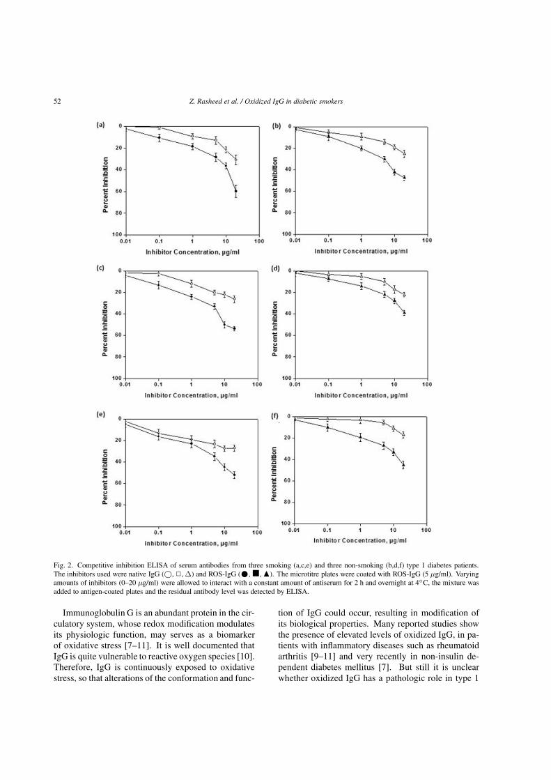
damaged IgG was 0.23± 0.06 and 0.29± 0.04, respec-tively, whereas the results for 28 NH non-smokers an-tibodies binding to native and ROS-damaged IgG were0.22± 0.04 and 0.21± 0.03, respectively (Fig. 1). Thebinding specificity of antibodies from diabetic smok-ers and non-smokers was further evaluated by competi-tive inhibition ELISA using native and ROS-IgG as in-hibitors. Microtitre plates were coated with ROS-IgG.Figure 2 illustrates inhibition of type 1 diabetes autoan-tibodies (in sera 1, 2 and 3) by native and modified IgG.Results point to a higher reactivity of autoantibodiesin patient’s sera towards ROS-modified IgG over na-tive IgG. Circulating antibodies from diabetic smok-ers showed preferentially strong binding to ROS-IgGas compared with the diabetic non-smokers antibodies(p < 0.05). Similarly, the rest of the sera showed highpercent inhibitions with modified IgG over native IgG(data summarized in Table 2). Average percentage in-hibition (± SEM) at 20 μg/ml of inhibitor concentra-tion in the binding of 20 diabetic smokers antibodiesto native and ROS-IgG was 22.1 ± 1.9 and 54.2 ± 1.2,respectively. Whereas percent inhibition of serum anti-bodies from 17 diabetic non-smokers binding to nativeand ROS-IgG was 21.1 ± 1.1 and 40.3 ± 3.3, respec-tively (Table 2). Circulating antibodies from the nega-tive controls did not show significant inhibition to eitherinhibitor. The data revealed striking differences in therecognition of ROS-IgG by antibodies from diabeticpatients (p < 0.01).
3.3. Oxidation in serum protein and purified IgG
Protein carbonyl content is a general indicator andthe most commonly used biomarker for protein oxi-dation. Our data showed significant increase in to-tal serum protein carbonyl content in diabetic smokers(n = 20) and diabetic non-smokers (n = 17) com-pared with their respective controls (p < 0.05). Theaverage carbonyl content (± SEM) of 20 independentassays in serum samples of diabetic smokers and nor-mal healthy smokers were 3.92 ± 0.08 and 2.11 ±0.05 nmol/mg protein, respectively. Whereas in non-smoking DM serum proteins and non-smoking NHserumproteins the average results were 2.81± 0.05 and2.10 ± 0.07 nmol/mg protein, respectively (Table 3).
To investigate the extent of alterations in the biolog-ical properties of IgG in DM patients, IgG was iso-lated from same diabetic smokers (S-DM-IgG), samediabetic non-smokers (NS-DM-IgG), normal healthysmokers (S-NH-IgG) and normal healthy non-smokers(NS-NH-IgG) by affinity chromatography using Pro-

Z. Rasheed et al. / Oxidized IgG in diabetic smokers 51
Table 3Carbonyl contents in serum proteins and in purified immunoglobulin G fromsmoking and non-smoking type 1 diabetes patients
Study subjects Number of Carbonyl contents (nmol/mg protein)samples Serum proteins Purified IgG
Smokers DMS 20 3.92 ± 0.08a 2.84 ± 0.04b
Non-smokers DMS 17 2.81 ± 0.07b 1.99 ± 0.06b
Smokers NHS 25 2.11 ± 0.05b 1.62 ± 0.04c
Non-smokers NHS 28 2.10 ± 0.07b 1.51 ± 0.06c
Results are representative (mean ± SEM) of triplicate experiments withtype 1 diabetes mellitus serum (DMS) samples obtained from age and sexmatched normal human serum (NHS) and differ without a common letterp < 0.05.
Fig. 1. Direct binding ELISA of serum samples (1:100 diluted) from smoking type 1 diabetes patients (smokers DM), non-smoking DMpatients (non-smokers DM), healthy smoking subjects (smokers NS) and healthy non-smoking subjects (non-smokers NS) to native IgG andROS-modified IgG. The microtitre plates were individually coated with native IgG and ROS-IgG (5 μg/ml). The number of smoking DM,non-smoking DM, smoking NS and non-smoking NS samples were 20, 17, 25 and 28, respectively. Results are representative (mean ± SEM) oftriplicate experiments and differ without a common letter P < 0.05.
tein A-sepharose CL-4B column. Polyacrylamide gelelectrophoresis of affinity purified IgG showed a singlehomogenous band (data not shown). Average carbonylcontents (± SEM) of their respective independent as-says of S-DM-IgG and S-NH-IgG were 2.84 ± 0.04and 1.62 ± 0.04 nmol/mg protein, respectively (P <0.05), whereas in NS-DM-IgG and NS-NH-IgG the re-sults were 1.99 ± 0.06 and 1.51 ± 0.06 nmol/mg pro-tein, respectively (p < 0.05). Importantly, p < 0.05indicated a significant difference in carbonyl content inthe S-DM-IgG and NS-DM-IgG groups (Table 3).
4. Discussion
Despite the power of modern molecular approach-es and persisting investigative efforts, diabetes mellitus
remains an enigmatic disorder or the agent (or agents)triggering this disease remains to be identified [22].Oxidative stress plays a central role in the onset ofdiabetes as well as in diabetes associated complica-tions [1]. Various studies have shown that diabetesis associated with increased formation of free radicalsand decrease in antioxidant potential. This leads tooxidative damage of cell components such as proteins,lipids and nucleic acid in both type 1 and type 2 dia-betes [2]. The role of oxidative protein damage in thepathogenesis of the diabetic state is being investigatedextensively [5,7,16]. Considerable evidence indicatesthat the maintenance of protein redox status is of fun-damental importance for cell function, whereas struc-tural changes in protein are considered to be amongthe molecular mechanisms leading to diabetes and itsassociated complications [3].
52 Z. Rasheed et al. / Oxidized IgG in diabetic smokers
Fig. 2. Competitive inhibition ELISA of serum antibodies from three smoking (a,c,e) and three non-smoking (b,d,f) type 1 diabetes patients.The inhibitors used were native IgG (©, �, Δ) and ROS-IgG (•, �, �). The microtitre plates were coated with ROS-IgG (5 μg/ml). Varyingamounts of inhibitors (0–20 μg/ml) were allowed to interact with a constant amount of antiserum for 2 h and overnight at 4◦C, the mixture wasadded to antigen-coated plates and the residual antibody level was detected by ELISA.
Immunoglobulin G is an abundant protein in the cir-culatory system, whose redox modification modulatesits physiologic function, may serves as a biomarkerof oxidative stress [7–11]. It is well documented thatIgG is quite vulnerable to reactive oxygen species [10].Therefore, IgG is continuously exposed to oxidativestress, so that alterations of the conformation and func-
tion of IgG could occur, resulting in modification ofits biological properties. Many reported studies showthe presence of elevated levels of oxidized IgG, in pa-tients with inflammatory diseases such as rheumatoidarthritis [9–11] and very recently in non-insulin de-pendent diabetes mellitus [7]. But still it is unclearwhether oxidized IgG has a pathologic role in type 1
Z. Rasheed et al. / Oxidized IgG in diabetic smokers 53
diabetes patients. In agreement with previous studiesthe present work also found oxidatively altered IgG inpatients with type 1 diabetes mellitus. It may reflecta physiological response directed towards the cleaningof oxidative proteins from the circulation, rather than atrue autoimmune response with clinical consequences.
We and others have reported the presence of elevatedlevels of oxidized protein products (advancedoxidationprotein products) such as oxidized plasma protein in pa-tients with various diseases [9–26]. Furthermore, ROSmodified IgG is highly immunogenic in rabbits, and theantigenic specificity of affinity purified anti-ROS-IgGantibodies and anti-IgG antibodies suggest that inducedantibodies are immunogen-specific. The substantiallyenhanced immunogenicity of ROS-IgG in comparisonto native IgG may be due to the generation of potentialneo-epitopes against which antibodies are raised [11].In the present study, type 1 diabetic smokers and non-smokers were screened for the presence of autoanti-bodies reactive to native and ROS-modified IgG usinga direct-binding ELISA. Results revealed that circulat-ing antibodies in diabetes serum samples showed pref-erentially high binding to ROS-IgG, compared with itsnative analogue (p < 0.05), whereas control samplesdid not show appreciable binding (p > 0.05). Cir-culating diabetes antibodies also showed some bind-ing to native IgG, this may be due to the generationof cryptic epitopes, which might be generated duringisolation. Competitive inhibition ELISA reiterates thedirect binding results indicated that the ROS-modifiedIgG was an effective inhibitor showing substantial dif-ference in the recognition of damaged IgG over nativeIgG (p < 0.05). Importantly, our notable novel datashowed that circulating antibodies from smokers dia-betes patients showed better recognition of ROS-IgGas compared with the circulating antibodies from dia-betic non-smokers (p < 0.05). As the abundant plas-ma protein, IgG is known to play a major role as anantigen clearance, but under oxidative stress IgG dys-functions have been reported [7,9–11]. In this con-text, the characterization of the oxidation status of IgGwould provide not only useful information about theredox state of the human body but also alterations inthe conformation and function of IgG that may result inmodification of its biological properties. The oxidationof a protein typically results in an increase in carbonylcontent [20,27]. This increase is due to the oxidationof lys, arg, pro and other amino acid residues [20,27].In short, protein carbonyl groups are the biomarkers ofoxidative stress [27,28]. Our data show that carbonylcontent in serum of diabetic smokers and diabetic non-
smokers was high as compared with their respectivesmokers or non-smokers controls. Importantly, serumcarbonyl content of diabetic smokers was significantlyhigher as compared to diabetic non-smokers (p < 0.05).In addition, the purified IgGs from diabetic smokersshowed significantly higher carbonyl content as com-pared with the IgGs from normal healthy smokers (p <0.05). Similar results were obtained in diabetic non-smokers and their respective controls. Our data pointout that higher carbonyl content was found in diabet-ic smokers compared with diabetic non-smokers (p <0.05). These results indicated that IgG is continuallyexposed to oxidative stress, which results in alterationsin its biological properties, could results in conforma-tional changes, these changes may be one of the fac-tors for the presentation of unique epitopes for the in-duction of autoantibodies in type 1 diabetes patients.Our data supported the view that cigarette smoking in-creases the oxidative stress in diabetes patients. Thissuggests that diabetes patients with increased oxidativestress showed enhanced oxidative modification of plas-ma proteins and IgG is likely to be extensively dam-aged. The present report demonstrates the structuralperturbation in IgG due to oxidative stress, renderingit immunogenic. The neo-epitopes generated, whichmay be one of the responsible factors for the inductionof autoantibodies in patients with type 1 diabetes. Thismight play a significant role in the etiology of diabetesand might represents a new biomarkers especially forcigarette smoking type 1 diabetes patients.
5. Conclusions
The present article is the first report that demon-strates the presence of ROS-induced IgG damage intype 1 diabetes patients. We conclude that IgG aftermodificationwith ROS presents unique epitopes for theinduction of autoantibodies in type 1 diabetes with spe-cial preference to those patients having cigarette smok-ing history. Thus, ROS-modified IgG might representsa novel therapeutic targets for diabetic smokers.
Competing interests
The authors declare that they have no competinginterests.
Acknowledgements
Financial support from College of Medicine, QassimUniversity is gratefully acknowledged.
54 Z. Rasheed et al. / Oxidized IgG in diabetic smokers
References
[1] M. Phillips, R.N. Cataneo, T. Cheerma and J. Greenberg, In-creased breath biomarkers of oxidative stress in diabetes mel-litus, Clin Chim Acta 344 (2004), 189–194.
[2] B. Eliasson, Cigarette smoking and diabetes, Prog CardiovascDis 45 (2003), 405–413.
[3] C.E. Taplin and J.M. Barker, Autoantibodies in type 1 diabetes,Autoimmunity 41 (2008), 11–18.
[4] E. Kawasaki, N. Abiru and K. Eguchi, Prevention of type 1diabetes: from the view point of β cell damage, Diabetes ResClin Pract 66 (2004), S27–S32.
[5] A. Telci, U. Cakatay, S. Salman, I. Satman and A. Sivas, Ox-idative protein damage in early stage type 1 diabetic patients,Diabetes Res Clin Pract 50 (2000), 213–223.
[6] G. Carrard, A.L. Bulteau, I. Petropoulos and B. Friguet, Im-pairment of proteasome structure and function in aging, Int JBiochem Cell Biol 34 (2002), 1461–1474.
[7] T. Tripathi and Z. Rasheed, Oxidative by-product, hydrox-yl radical, damaged immunoglobulin G in patients with non-insulin dependent diabetes mellitus, Bratisl Lek Listy 111(2010), 477–484.
[8] C. Chou, Binding of rheumatoid and lupus synovial fluids andsera-derived human IgG rheumatoid factor to degalactosylatedIgG, Arch Med Res 33 (2002), 541–544.
[9] M. Uesugi, K. Yoshida and H.E. Jasin, Inflammatory proper-ties of IgG modified by oxygen radicals and peroxynitrite, JImmunol 165 (2000), 6532–6537.
[10] H.A. Kleinveld, W. Slulter, A.M. Boonman, A.J. Swaak, C.E.Hack and J.F. Koster, Differential stimulation by oxygen-free-radical-altered immunoglobulin G of the production of super-oxide and hydrogen peroxide by human polymorphonuclearleucocytes, Clin Sci 80 (1991), 385–391.
[11] Z. Rasheed, Hydroxyl radical damaged Immunoglobulin G inpatients with rheumatoid arthritis: Biochemical and immuno-logical studies. Clin Biochem 2 (2008), 1–13.
[12] R.H. Scofield, B.T. Kurien, S. Granick et al., Modificationof lupus-associated 60-kDA Ro protein with the lipid oxida-tion product 4-hydroxy-2-nonenal increases antigenicity andfacilitates epitopes spreading, Free Radic Biol Med 38 (2005),719–728.
[13] B.T. Kurien and R.H. Scofield, Autoimmunity and oxidativelymodified autoantigens, Autoimmun Rev 7 (2008), 567–573.
[14] Z. Rasheed, R. Ahmad, N. Rasheed et al., Reactive oxygenspecies damaged hemoglobin presents unique epitopes fortype 1 diabetes autoantibodies, International J Bio Chem 2(2008), 1–13.
[15] Z. Rasheed, M.W.A. Khan and R. Ali, Hydroxyl radical mod-ification of human serum albumin generated cross reactiveantibodies, Autoimmunity 39 (2006), 479–488.
[16] Z. Rasheed and R. Ali, Reactive oxygen species damagedhuman serum albumin in patients with type 1 diabetes mellitus:Biochemical and immunological studies, Life Sci 79 (2006),2320–2328.
[17] Z. Rasheed, R. Ahmad, N. Rasheed et al., Reactive oxygenspecies damaged human serum albumin in patients with hepa-tocellular carcinoma, J Exp Clin Cancer Res 26 (2007), 515–524.
[18] Z. Rasheed, R. Ahmad, N. Rasheed et al., Enhanced recogni-tion of reactive oxygen species damaged human serum albu-min by circulating systemic lupus erythematosus autoantibod-ies, Autoimmunity 40 (2007), 512–520.
[19] Z. Rasheed, R. Ahmad and R. Ali, Structure and immunolog-ical function of oxidized albumin in lung cancer: its poten-tial role as a biomarker of elevated oxidative stress, British JBiomed Sci 66 (2009), 76–73.
[20] R.L. Levine, J. Williams, E.R. Stadtman et al., Carbonyl assaysfor determination of oxidatively modified proteins, MethodsEnzymol 233 (1994), 346–357.
[21] O.H. Lowry, N.J. Rosenberg, A.L. Farr AL et al., Proteinmeasurement with the folin phenol reagent, J Biol Chem 193(1951), 265–275.
[22] K. Vehik and D. Dabelea, The changing epidemiology of type1 diabetes: why is it going through the roof? Diabetes MetabRes 27 (2011), 3–13.
[23] R. Ahmad, Z. Rasheed andH. Ahsan, Biochemical and cellulartoxicology of peroxynitrite: implications in cell death andautoimmune phenomenon, Immunopharm Immunotoxicology31 (2009), 388–396.
[24] Z. Rasheed, L. Kumar, S. Abbas et al., Advanced glycationend-products damaged IgG, a target for circulating autoanti-bodies in patients with type 1 diabetes mellitus, Open Glycosci2 (2009), 1–8.
[25] K. Hensley and R.A. Floyd, Reactive oxygen species andprotein oxidation in aging: a look back, a look ahead. ArchBiochem Biophys 397 (2002), 377–383.
[26] B.J. Tabner, S. Turnbull, O. El-Agnaf et al., Production of re-active oxygen species from aggregating proteins implicated inAlzheimer’s disease, Parkinson’s disease and other neurode-generative diseases, Curr Top Med Chem 1 (2001), 507–517.
[27] I. Dalle-Donne, R. Rossi, D. Giustarini, A. Milzani and R.Colombo, Protein carbonyl groups as biomarkers of oxidativestress, Clin Chim Acta 329 (2003), 23–38.
[28] B. Halliwell and J.M. Gutteridge, The antioxidants of humanextracellular fluids, Arch Biochem Biophys 280 (1990), 1–8.

Submit your manuscripts athttp://www.hindawi.com
Stem CellsInternational
Hindawi Publishing Corporationhttp://www.hindawi.com Volume 2014
Hindawi Publishing Corporationhttp://www.hindawi.com Volume 2014
MEDIATORSINFLAMMATION
of
Hindawi Publishing Corporationhttp://www.hindawi.com Volume 2014
Behavioural Neurology
EndocrinologyInternational Journal of
Hindawi Publishing Corporationhttp://www.hindawi.com Volume 2014
Hindawi Publishing Corporationhttp://www.hindawi.com Volume 2014
Disease Markers
Hindawi Publishing Corporationhttp://www.hindawi.com Volume 2014
BioMed Research International
OncologyJournal of
Hindawi Publishing Corporationhttp://www.hindawi.com Volume 2014
Hindawi Publishing Corporationhttp://www.hindawi.com Volume 2014
Oxidative Medicine and Cellular Longevity
Hindawi Publishing Corporationhttp://www.hindawi.com Volume 2014
PPAR Research
The Scientific World JournalHindawi Publishing Corporation http://www.hindawi.com Volume 2014
Immunology ResearchHindawi Publishing Corporationhttp://www.hindawi.com Volume 2014
Journal of
ObesityJournal of
Hindawi Publishing Corporationhttp://www.hindawi.com Volume 2014
Hindawi Publishing Corporationhttp://www.hindawi.com Volume 2014
Computational and Mathematical Methods in Medicine
OphthalmologyJournal of
Hindawi Publishing Corporationhttp://www.hindawi.com Volume 2014
Diabetes ResearchJournal of
Hindawi Publishing Corporationhttp://www.hindawi.com Volume 2014
Hindawi Publishing Corporationhttp://www.hindawi.com Volume 2014
Research and TreatmentAIDS
Hindawi Publishing Corporationhttp://www.hindawi.com Volume 2014
Gastroenterology Research and Practice
Hindawi Publishing Corporationhttp://www.hindawi.com Volume 2014
Parkinson’s Disease
Evidence-Based Complementary and Alternative Medicine
Volume 2014Hindawi Publishing Corporationhttp://www.hindawi.com
Copyright © 2022 FDOKUMEN