Non-singular boundary integral methods for fluid mechanics applications
Upload
khangminh22Category
view
0download
0
Online chapter: Immunological methods and applications / 1
ONLINE CHAPTER
Immunological methods and applications
1
Just to Recap …Now that the reader is familiar with all of the major elements of the innate and adaptive immune systems, it is instructive to consider how these elements can be manipulated in vitro and in vivo, either in the quest for further knowledge of immunity, or for the development of reagents (primarily antibody-based) that can be used for a huge number of applications. It is also very useful to know how immunologists go about the task of discovering the knowledge we have gamely tried to summarize within these pages. The methods and applications described in this chapter will hopefully explain how, in practical terms, one would go about measuring cytokine production by individual T-cell subsets, or antibody production by B-cells, or apoptosis induced by NK cells, and so on.
Key Topics
Making antibodies to order 2
Purification of antigens and antibodies by affinity chromatography 8
Modulation of biological activity by antibodies 8
Immunodetection of antigen in cells and tissues 10
Detection and quantitation of antigen by antibody 17
Epitope mapping 22
Estimation of antibody 23
Detection of immune complex formation 28
Isolation of leukocyte subpopulations 29
Gene expression analysis 32
Assessment of functional activity 33
Manipulation of the immune system in animal models 39
Genetic engineering of cells and model organisms 40
Gene therapy in humans 46
Roitt’s Essential Immunology, Thirteenth Edition. Peter J. Delves, Seamus J. Martin, Dennis R. Burton, and Ivan M. Roitt.© 2017 John Wiley & Sons Ltd. Published 2017 by John Wiley & Sons Ltd. Companion website www.roitt.com
Originally published in Roitt’s Essential Immunology, Twelfth Edition as Chapter 6.
2 / Fundamentals of Immunology
Making antibodies to order
In addition to being quite handy for protecting our bodies from harmful infectious agents, antibodies are also incredibly useful and exquisitely specific reagents for detecting and quantitating other proteins, as well as many other substances. Antibodies have, quite literally, numerous practical applications; ranging from the purification of proteins using antibodybased affinity columns, the detection of circulating hormones in blood or urine samples for clinical diagnosis, the exploration of expression and subcellular localization of proteins, as immunotherapeutics in cancer therapy and as antidotes for snake and spider bites. Indeed, to the research scientist, a world without antibodies is very hard to contemplate as these molecules are used daily as highly specific and reliable probes for virtually every protein under the sun, in a multitude of contexts. We shall now take a look, in practical terms, at how these wonderfully adaptable proteins can be produced in the laboratory.
Generation of polyclonal antibodies
Although antibodies can be raised against practically any organic substance, some molecules elicit antibody responses much more readily than others. Proteins usually make excellent immunogens (i.e. substances that can elicit an immune response), although the immune response will typically be concentrated against small regions within the protein (called epitopes or antigenic determinants) spanning approximately five to eight amino acids. As we discussed earlier (see Chapter 5), an epitope represents the minimal structure required for recognition by antibody and a relatively large molecule, such as a protein, will usually contain multiple epitopes. Thus, injection of the average antigen into an animal will almost always
elicit the production of a mixture of antibodies that are directed against different epitopes within the antigen. It is also quite possible that some of the antibodies within this mixture may be directed towards epitopes that are also found in other antigens. Such antibodies are said to be crossreactive against the other antigen to which they also bind. Small organic molecules are typically poor immunogens when injected on their own; the immune system appears unable to recognize these structures efficiently. Notwithstanding this, immunologists have found that such molecules can be made visible to the immune system by covalent coupling to a carrier protein, such as bovine serum albumin (BSA), which is intrinsically immunogenic. Such small molecules are called haptens (see Figure 5.6).
To generate an antibody against a protein of interest, the standard approach is to inject small samples of the protein (in the microgram range) into an animal such as a rabbit. However, administration of antigen alone is rarely sufficient to provoke a robust immune response, even if the antigen is composed of a high proportion of nonself determinants; coadministration of an adjuvant is required (Figure 1). While it is not entirely clear exactly how adjuvants work, one important role they peform is to activate dendritics cells (DCs) and other antigen-presenting cells (APCs) at the site of antigen delivery. Recall from Chapter 1 that activation of APCs dramatically enhances their ability to provide the costimulatory signals that are required for efficient T and Bcell activation upon encounter with antigen. The reader will recall that pathogenassociated molecular patterns (PAMPs) are typically required to trigger maturation of dendritic cells and trigger their movement to secondary lymphoid tissues for the purposes of presenting nonself antigens to Tcells, which in turn provides Tcelldependent help to Bcells for class switching, affinity maturation and optimum antibody production. Potent
IntroductionThis chapter is structured such that we will progress from molecular- to cell-based techniques, and then on to whole animal-based approaches. We will initially consider how antibodies can be generated and purified, how they can in turn be used to purify their specific antigen from complex mixtures of antigen, how they can be used to functionally mimick natural ligands for the purposes of stimulating cell function, or conversely, inhibiting specific functions, as well as many other applications. We will then take a look at a series of variations on the theme of using antibody to detect antigen in cells, in tissues, in fluids as well as solid supports (such as protein arrays) and we will also explore how the discrete regions within an antigen that are recognized by antibodies, or T-cell receptors (i.e. their specific epitopes), can be mapped. The second half of the chapter is devoted to cell-based methods that are used to assess the functionality and interactions between cells of the immune system. We will discuss how cells of the immune system can be isolated, phenotyped, functionally assessed, and genetically manipulated both in vitro as well as in vivo. We will then look at some of the animal models that are commonly used by immunologists and how genetically engineered animals can be produced.
Many of the procedures we discuss in this chapter have been painstakingly developed and refined by several generations of immunologists and range from the straightforward to the highly complex. It is sobering to be reminded that, when we make statements such as “an antibody was generated,” the work involved typically takes many months, if not years. Similarly, while “gene X was knocked out in the mouse” rolls off the tongue in a mere two seconds, you can take it from us that the procedure itself occupied someone for a couple of years.
Online chapter: Immunological methods and applications / 3
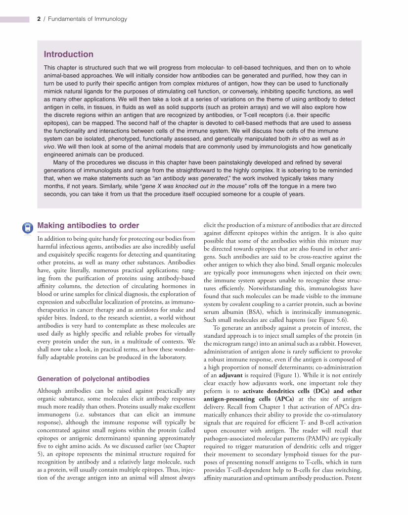
2.12), the antigen is therefore injected several times over a period of 12 weeks or so. During this time, the concentration of antibodies (what is usually referred to as antibody titer) directed against the immunogen will increase (Figure 1). All going well, we will now have an antiserum that is enriched with antibodies against our protein of interest and this can be used as a probe in many different contexts; to localize an antigen within a cell, to quantify it within a mixture of other antigens, to neutralize its biological activity, and many other applications (these are elaborated upon later in this chapter).
It is important to remind ourselves here that antisera generated in this way will also contain considerable amounts of other antibodies (directed against a variety of determinants) that the animal happens to have made in the recent past. These antibodies will usually be of a significantly lower titer than those directed against the antigen we have repeatedly used for immunization, but they can cause problems and may need to be removed from our antiserum for several applications. Fortunately, this can be achieved by affinity purification where specific antigen is immobilized on a solid support and used to “fish” out its specific antibody from a crude mixture of antibodies (see Figure 6).
Because many antigens contain several distinct epitopes, antisera generated by injection of antigen will typically contain a mixture of antibodies directed against different antigenic determinants on the molecule. Some of these antibodies will bind to the antigen with high avidity, some will not, some will only recognize the native form of the antigen, while others will still recognize the antigen following denaturation to eliminate tertiary structure. Such antisera are said to be polyclonal as they contain a mixture of antibodies that are predominantly, although not exclusively, directed against the immunogen to which they were raised.
The monoclonal antibody revolution
First in rodents
A fantastic technological breakthrough was achieved by Georges Köhler and César Milstein who devised a technique for the production of “immortal” clones of cells making single antibody specificities by fusing normal antibodyforming cells with an appropriate Bcell tumor line. Normal untransformed Bcells cannot be grown in culture for long periods of time and quickly die off unless immortalized. This truly paradigmshifting method enables individual Bcell clones to be grown in tissue culture and expanded to a point where enormous quantities of antigenspecific antibody can be produced. These socalled “hybridomas” are selected out in a tissue culture medium that fails to support growth of the parental cell types and, by successive dilutions or by plating out, single clones can be established (Figure 2). These clones can be grown up in the ascitic form in mice when quite prodigious titers of monoclo-nal antibody can be attained, but bearing in mind the imperative to avoid using animals wherever feasible, propagation in largescale culture is to be preferred. Remember that, even in a good antiserum, over 90% of the Ig molecules have little or
adjuvants are usually crude preparations of bacterial extracts that contain mixtures of Tolllike receptor (TLR) ligands such as LPS or peptidoglycan. In essence, most adjuvants are mixtures of PAMPs, which activate DCs and other cells of the innate immune system through their pattern recognition receptors (PRRs). Because DCs are incapable of providing essential costimulatory signals to Tcells unless activated through their PRRs, antigens that lack intrinsic PRRbinding activity will fail to activate DCs and therefore fail to elicit potent immune responses on their own.
As we outlined in Chapter 2, because a single dose of antigen usually elicits a relatively modest response (see Figure
105
104
103
102
101
100
0
Seru
m a
ntib
ody
titer
Weeks
1 2 3 4 5 6 7 8 9 10 11
Initialchallengewith Ag
Booster 1
Booster 2
Booster 3
Booster 4
12
Antigen alone
Antigen plus adjuvant
0
Seru
m a
ntib
ody
titer
Weeks
1 2 3 4 5 6 7 8 9 10 11
Initialchallengewith Ag
Booster 1
Booster 2
Booster 3
Booster 4
12
105
104
103
102
101
100
Figure 1. Production of polyclonal antibodies.
Repeated immunization with antigen (Ag) plus adjuvant is required to generate efficient antibody responses as immunization with antigen alone is usually ineffective. Polyclonal antisera are generated by immunizing, with a combination of antigen plus adjuvant, several times over a 12-week period. Serum antibody titre (i.e. the highest dilution giving a positive test) frequently increases after each successive boost with antigen.
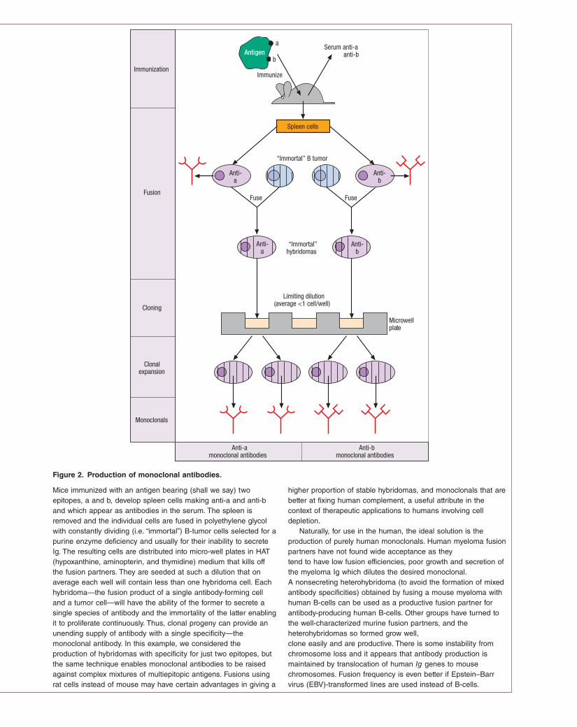
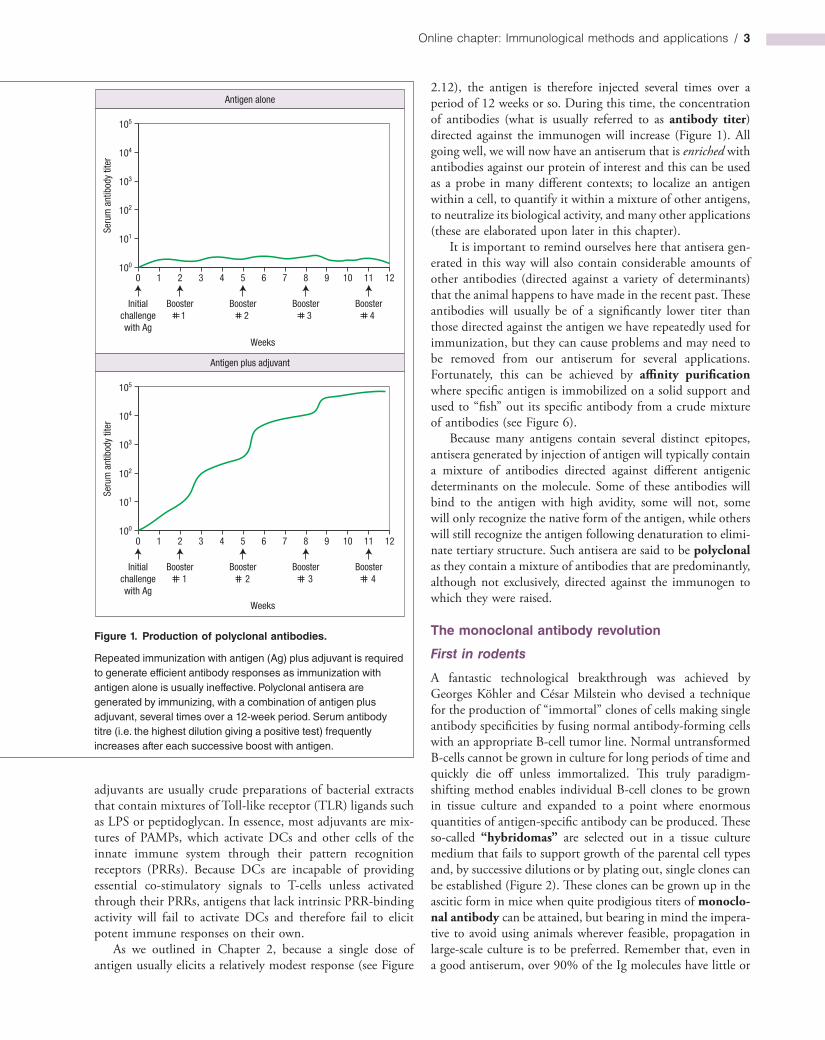
Figure 2. Production of monoclonal antibodies.
Fuse
Antigena
b
Immunize
Serum anti-aanti-b
Spleen cells
“Immortal” B tumor
Anti-a
Anti-b
Fuse
Anti-a
Anti-b
“Immortal”hybridomas
Limiting dilution(average <1 cell/well)
Microwellplate
Anti-amonoclonal antibodies
Anti-bmonoclonal antibodies
Immunization
Fusion
Cloning
Clonalexpansion
Monoclonals
Mice immunized with an antigen bearing (shall we say) two epitopes, a and b, develop spleen cells making anti-a and anti-b and which appear as antibodies in the serum. The spleen is removed and the individual cells are fused in polyethylene glycol with constantly dividing (i.e. “immortal”) B-tumor cells selected for a purine enzyme deficiency and usually for their inability to secrete Ig. The resulting cells are distributed into micro-well plates in HAT (hypoxanthine, aminopterin, and thymidine) medium that kills off the fusion partners. They are seeded at such a dilution that on average each well will contain less than one hybridoma cell. Each hybridoma—the fusion product of a single antibody-forming cell and a tumor cell—will have the ability of the former to secrete a single species of antibody and the immortality of the latter enabling it to proliferate continuously. Thus, clonal progeny can provide an unending supply of antibody with a single specificity—the monoclonal antibody. In this example, we considered the production of hybridomas with specificity for just two epitopes, but the same technique enables monoclonal antibodies to be raised against complex mixtures of multiepitopic antigens. Fusions using rat cells instead of mouse may have certain advantages in giving a
higher proportion of stable hybridomas, and monoclonals that are better at fixing human complement, a useful attribute in the context of therapeutic applications to humans involving cell depletion.
Naturally, for use in the human, the ideal solution is the production of purely human monoclonals. Human myeloma fusion partners have not found wide acceptance as they tend to have low fusion efficiencies, poor growth and secretion of the myeloma Ig which dilutes the desired monoclonal. A nonsecreting heterohybridoma (to avoid the formation of mixed antibody specificities) obtained by fusing a mouse myeloma with human B-cells can be used as a productive fusion partner for antibody-producing human B-cells. Other groups have turned to the well-characterized murine fusion partners, and the heterohybridomas so formed grow well, clone easily and are productive. There is some instability from chromosome loss and it appears that antibody production is maintained by translocation of human Ig genes to mouse chromosomes. Fusion frequency is even better if Epstein–Barr virus (EBV)-transformed lines are used instead of B-cells.

Online chapter: Immunological methods and applications / 5
no avidity for the antigen, and the “specific antibodies” themselves represent a whole spectrum of molecules with different avidities directed against different determinants on the antigen. What a contrast is provided by monoclonal antibodies, where all the molecules produced by a given hybridoma are identical: they have the same Ig class and allotype, the same variable region, structure, idiotype, affinity and specificity for a given epitope.
The large amount of nonspecific, relative to antigenspecific, Ig in a polyclonal antiserum means that background binding to antigen in any given immunological test may be uncomfortably high. This problem is greatly reduced with a monoclonal antibody preparation, as all the antibody is antigenspecific, thus giving a much superior “signal : noise” ratio. By being directed towards single epitopes on the antigen, monoclonal antibodies frequently show high specificity in terms of their low crossreactivity with other antigens.
An outstanding advantage of the monoclonal antibody as a reagent is that it provides a single standard material for all laboratories throughout the world to use in an unending supply if the immortality and purity of the cell line are nurtured; antisera raised in different animals, on the other hand, may be as different from each other as chalk and cheese. The monoclonal approach again shows a clean pair of heels relative to conventional strategies in the production of antibodies specific for individual components in a complex mixture of antigens. The uses of monoclonal antibodies are truly legion and include: immunoassay, diagnosis of malignancies, tissue typing, serotyping of microorganisms, the separation of individual cell types with specific surface markers (e.g. lymphocyte subpopulations), therapeutic neutralization of inflammatory cytokines and “magic bullet” therapy with cytotoxic agents coupled to antitumorspecific antibody—these and many other areas have been transformed by hybridoma technology.
Catalytic antibodies
An especially interesting development with tremendous potential is the recognition that a monoclonal antibody to a stable analog of the transition state of a given reaction can act as an enzyme (“abzyme”) in catalyzing that reaction. The possibility of generating enzymes to order promises a very attractive future, and some exceedingly adroit chemical maneuvers have already extended the range of reactions that can be catalyzed in this way. A recent demonstration of sequencespecific peptide cleavage with an antibody that incorporates a metal complex cofactor has raised the pulse rate of the cognoscenti, as this is an energetically difficult reaction that has an enormous range of applications. Another innovative approach is to immunize with an antigen that is so highly reactive that a chemical reaction occurs in the antibody combining site. This recruits antibodies that are not only complementary to the active chemical, but are also likely to have some enzymic power over the immunogen–substrate complex. Thus, using this strategy, an antibody with exceptionally broad substrate specificity for efficient catalysis of
aldol and retroaldol reactions was obtained. A key feature of this antibody is a reactive lysine buried within a hydrophobic pocket in the binding site. The antibody remains catalytically active for several weeks following i.v. injection into mice and has therapeutic potential for a version of antibodydirected enzyme prodrug therapy, here with the enzyme component being a catalytic antibody.
Large combinatorial antibody libraries created by random association between pools of heavy and light chains and expressed on bacteriophages (see below) can be screened for catalytic antibodies by using the substrate in a solidphase state. Cleavage by the catalytic antibody leaves a solidphase product that can now be identified by a double antibody system using antibodies specific for the product as distinct from the substrate.
An area of great interest is the presence of catalytic autoantibodies in certain groups of patients, with hydrolytic antibodies against vasoactive intestinal peptide, DNA and thyroglobulin having been described. Catalytic antibodies capable of factor VIII hydrolysis have also recently been discovered in hemophiliacs given this clotting factor, the antibodies preventing the coagulation function of the factor VIII.
Human monoclonals can be made
While scientists were quick to realize that monoclonal antibodies would make powerful and highly specific therapeutic agents, particularly for the treatment of cancer, this proved to be rather more difficult than originally anticipated. Mouse monoclonals injected into human subjects for therapeutic purposes are frightfully immunogenic and the human antimouse antibodies (HAMA in the trade) so formed are a wretched nuisance, accelerating clearance of the monoclonal from the blood and possibly causing hypersensitivity reactions; they also prevent the mouse antibody from reaching its target and, in some cases, block its binding to antigen. In some circumstances it is conceivable that a mouse monoclonal taken up by a tumor cell could be processed and become the MHClinked target of cytotoxic Tcells or help to boost the response to a weakly immunogenic antigen on the tumor cell surface. In general, however, logic points to removal of the xenogeneic (foreign) portions of the monoclonal antibody and their replacement by human Ig structures using recombinant DNA technology. Chimeric constructs, in which the VH and VL mouse domains are spliced onto human CH and CL genes (Figure 3a), are far less immunogenic in humans.
A more refined approach is to graft the six complementarity determining regions (CDRs) of a high affinity rodent monoclonal onto a completely human Ig framework without loss of specific reactivity (Figure 3b). This is not a trivial exercise, however, and the objective of fusing human Bcells to make hybridomas is still appealing, taking into account not only the gross reduction in immunogenicity, but also the fact that, within a species, antibodies can be made to subtle differences such as major histocompatibility complex (MHC)
6 / Fundamentals of Immunology
RAT CDRs
a Chimeric antibody
Mouse
Human
VL
CL
VH
CH
Grafted CDRsb
HumanFramework
polymorphic molecules and tumorassociated antigens on other individuals. In contrast, xenogeneic responses are more directed to immunodominant structures common to most subjects, making the production of variantspecific antibodies more difficult. Notwithstanding the difficulties in finding good fusion partners, large numbers of human monoclonals have been established. A further restriction arises because the peripheral blood Bcells, which are the only Bcells readily available in the human, are not normally regarded as a good source of antibodyforming cells.
Immortalized Epstein–Barr virustransformed Bcell lines have also been used as a source of human monoclonal antibodies. Although these often produce relatively low affinity IgM antibodies, some useful higheraffinity IgG antibodies can occasionally be obtained. The cell lines frequently lose their ability to secrete antibody if cultured for long periods of time, although they can sometimes be rescued by fusion with a myeloma cell line to produce hybridomas, or the genes can be isolated and used to produce a recombinant antibody.
A radically different approach involves the production of transgenic xenomouse strains in which megabasesized unrearranged human Ig H and κ light chain loci have been introduced into mice whose endogenous murine Ig genes have been inactivated. Immunization of these mice yields highaffinity (10−10–10−11 M) human antibodies that can then be isolated using hybridoma or recombinant approaches. Potent antiinflammatory (antiIL8) and antitumor (antiepidermal growth factor receptor) therapeutic agents have already been obtained using such mice.
There is still a snag in that even human antibodies can provoke antiidiotype responses; these may have to be circumvented by using engineered antibodies bearing different idiotypes for subsequent injections. Even more desirable would be if the prospective recipients could be first made tolerant to the
idiotype, perhaps by coadministering the therapeutic antibody together with a nondepleting antiCD4.
Despite the difficulties involved, a battery of humanized monoclonals has now been approved for therapeutic use. These include: antiIL2 (kidney transplant rejection), antiVEGF (colorectal cancer), antiTNFα (rheumatoid arthritis), antiCD11a (psoriasis), antiCD52 (Bcell chronic lymphocytic leukaemia), antiCD33 (acute myelogenous leukaemia), antiHER2 (a subset of metastatic breast cancers) and several others (cf. Table 16.2). Many more are currently in the clinical trial pipeline and are likely to become routinely used in clinical practice in the coming years.
Engineering antibodies
There are other ways around the problems associated with the production of human monoclonals that exploit the wiles of modern molecular biology. Reference has already been made to the “humanizing” of rodent antibodies (Figure 3), but an important new strategy based upon bacteriophage expression and selection has achieved a prominent position. In essence, mRNA from primed human Bcells is converted to cDNA (complementary DNA) and the antibody genes, or fragments therefrom, expanded by the polymerase chain reaction (PCR). Single constructs are then made in which the light and heavy chain genes are allowed to combine randomly in tandem with the gene encoding bacteriophage coat protein III (pIII) (Figure 4). This combinatorial library containing most random pairings of heavy and light chain genes encodes a huge repertoire of antibodies (or their fragments) expressed as fusion proteins with pIII on the bacteriophage surface. The extremely high number of phages produced by E. coli infection can now be panned on solidphase antigen to select those bearing the highest affinity antibodies attached to their surface (Figure 4). Because the genes that encode these highest affinity antibodies are already present within the selected phage, they can readily be cloned and the antibody expressed in bulk. It should be recognized that this selection procedure has an enormous advantage over techniques that employ screening because the number of phages that can be examined is several logs higher.
Combinatorial libraries have also been established using mRNA from unimmunized human donors. VH, Vk and Vl genes are expanded by PCR and randomly recombined to form singlechain Fv (scFv) constructs (Figure 5a) fused to phage pIII. Soluble fragments binding to a variety of antigens have been obtained. Of special interest are those that are autoantibodies to molecules with therapeutic potential such as CD4 and tumor necrosis factorα (TNFα); lymphocytes expressing such autoantibodies could not be obtained by normal immunization as they would probably be tolerized, but the random recombination of VH and VL can produce entirely new specificities under conditions in vitro where tolerance mechanisms do not operate.
Although a “testtube” operation, this approach to the generation of specific antibodies does resemble the affinity maturation of the immune response in vivo in the sense that antigen
Figure 3. Genetically engineering rodent antibody specificities into the human.
(a) Chimeric antibody with mouse variable regions fused to human Ig constant regions. (b) “Humanized” rat monoclonal in which gene segments coding for all six complementarity determining regions (CDRs) are grafted on to a human Ig framework.

Figure 4. Selection of antibody genes from a combinatorial library.
Stop codon
Solid-phase Ag
B-cellsfrom
immunizeddonor
IgG mRNA cDNA PCRPool HCH1
&light chain
genes
Constructcombinatorial
library
Express inbacteriophageand expand inp ΙΙΙ
Fab
Select bypanningon Ag
Clone gene &produce Ab
L L VHCH1
p ΙΙΙ
V
E.coli
VL CL
Figure 5. Other engineered antibodies.
GlutamateAlanine
b Original antibody Mutant with increased affinity
Arginine
Phenylalanine
+ +
–
Ab Ag Ab Agϕ
ϕ
Immunotoxind
Toxin
F(ab’) 2
ca Single chain FV fragment
N
C
N
C
VH VL
VL
VH
Bispecific 'diabody'
N
NC
C
VH1VH2
VL1
VL2
VH
(a) A single gene encoding VH and VL joined by a sequence of suitable length gives rise to a single-chain Fv (scFv) antigen-binding fragment. (b) By site-specific mutagenesis of residues in or adjacent to the complementarity determining region (CDR), it is possible to increase the affinity of the antibody. (c) Two scFv constructs expressed simultaneously will associate to form a
coat protein gene as shown. These were incorporated into phagemids such as pHEN1 and expanded in E. coli. After infection with helper phage, the recombinant phages bearing the highest affinity were selected by rounds of panning on solid-phase antigen so that the genes encoding the Fab fragments could be cloned. Ab, antibody; Ag, antigen; L, bacterial leader sequence.
“diabody” with two specificities. These bispecific antibodies have a number of uses. Note that such a bispecific antibody (Ab) directed to two different epitopes on the same antigen (Ag) will have a much higher affinity due to the “bonus effect” of cooperation between the two binding sites (cf. p. 122). (d) Potential “magic bullets” can be constructed by fusing the gene for a toxin (e.g. ricin) to the Fab.
B-cells from an immunized donor (in one important experiment, human memory peripheral blood cells were boosted with tetanus toxoid antigen after transfer to SCID mice; Duchosal M.A. et al. (1992) Nature 355, 258) are used for the extraction of IgG mRNA and the light chain (VLCL) and VHCH1 genes (encoding Fab) randomly combined in constructs fused to the bacteriophage pIII

8 / Fundamentals of Immunology
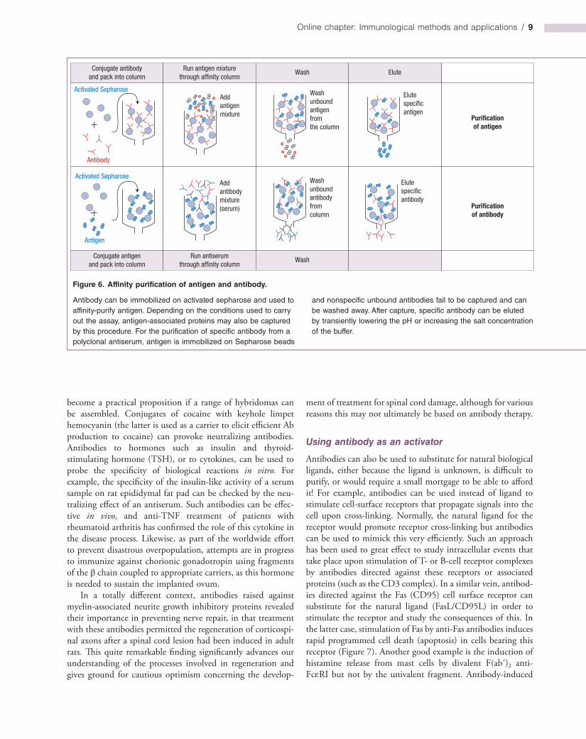
Purification of antigens and antibodies by affinity chromatography
The principle is simple and very widely applied. Antigen or antibody is bound through its free amino groups to cyanogen bromideactivated Sepharose particles or some other solid support. Immobilized antibody, for example, can be used to extract the corresponding antigen out of solution, in which it is present as one component of a complex mixture, by absorption to its surface. The uninteresting garbage is washed away and the required ligand released from the affinity absorbent by disruption of the antigen–antibody bonds by changing the pH or adding chaotropic ions such as thiocyanate (Figure 6). This technique can be used to identify the antigen to which an antibody binds where this is not known; in the case of an autoantibody for example. A very similar approach can also be used to identify binding partners for an antigen; such molecules will usually stay attached to the antigen if the immunopurification procedure is carried out under gentle conditions. Many of the proteins that participate in Tcell receptor (TCR) signal transduction, for instance, were initially identified by using antibodies directed against known TCR signalling components to pull out these components from complex protein mixtures, along with their bindingpartners. Isolated llama heavy chain (VHH) fragments are proving to be valuable for repeated cycles of antigen purification because of their resistance to denaturation by repeated cycles of exposure to low pH.
In a similar manner, an antigen immunosorbent can be used to absorb out an antibody from a mixture whence it can be purified by elution (Figure 6). This is especially useful where an antiserum displays high levels of nonspecific reactivity against other antigens rendering it unusable. Affinitypurification of such an antiserum, by means of the antigen that was used to generate it, can often dramatically improve its specificity.
Modulation of biological activity by antibodies
To detect antibody
A number of biological reactions can be inhibited by addition of specific antibody. Thus the agglutination of red cells by interaction of influenza virus with receptors on the erythrocyte surface can be blocked by antiviral antibodies and this forms the basis for their serological detection. A test for antibodies to salmonella H antigen present on the flagella depends upon their ability to inhibit the motility of the bacteria in vitro. Likewise, mycoplasma antibodies can be demonstrated by their inhibitory effect on the metabolism of the organisms in culture.
Using antibody as an inhibitor
The successful treatment of cases of drug overdose with the Fab fragment of specific antibodies has been described and may
is the determining factor in selecting out the highest affinity responders.
In order to increase the affinities of antibodies produced by these techniques, antigen can be used to select higher affinity mutants produced by random mutagenesis or even more effectively by sitedirected replacements at mutational hotspots (Figure 5b), again mimicking the natural immune response that involves random mutation and antigen selection. Affinity has also been improved by gene “shuffling” in which a VH gene encoding a reasonable affinity antibody is randomly combined with a pool of VL genes and subjected to antigen selection. The process can be further extended by mixing the VL from this combination with a pool of VH genes. It has also proved possible to shuffle individual CDRs between variable regions of moderate affinity antibodies obtained by panning on antigen, thereby creating antibodies of high affinity from relatively small libraries. The isolation of highaffinity, llama, heavychain antibody VHH fragments from immunized animals represents yet another approach.
Other novel antibodies have been created. In one construct, two scFv fragments associate to form an antibody with two different specificities (Figure 5c). Another consists of a single heavy chain variable region domain (DAB) whose affinity can be surprisingly high—of the order of 20 nM. If it were possible to overcome the “stickiness” of these miniantibodies, their small size could be exploited for tissue penetration. The design of potential “magic bullets” for immunotherapy can be based on fusion of a toxin (e.g. ricin) to an antibody Fab (Figure 5d).
Fields of antibodies
Not only can the genes for a monoclonal antibody be expressed in bulk in the milk of lactating animals but plants can also be exploited for this purpose. Socalled “plantibodies” have been expressed in bananas, potatoes and tobacco plants. One can imagine a hightech farmer drawing the attention of a bemused visitor to one field growing antitetanus toxoid, another antimeningococcal polysaccharide, and so on. Multifunctional plants might be quite profitable with, say, the root being harvested as a food crop and the leaves expressing some desirable gene product. At this rate there may not be much left for science fiction authors to write about!
Drugs can be based on the CDRs of minibodies
Millions of minibodies composed of a segment of the VH region containing three βstrands and the H1 and H2 hypervariable loops were generated by randomization of the CDRs and expressed on the bacteriophage pIII coat protein. By panning the library on functionally important ligandbinding sites, such as hormone receptors, useful lead candidates for drug design programs can be identified and their affinity improved by loop optimization, loop shuffling and further selection.
Online chapter: Immunological methods and applications / 9
ment of treatment for spinal cord damage, although for various reasons this may not ultimately be based on antibody therapy.
Using antibody as an activator
Antibodies can also be used to substitute for natural biological ligands, either because the ligand is unknown, is difficult to purify, or would require a small mortgage to be able to afford it! For example, antibodies can be used instead of ligand to stimulate cellsurface receptors that propagate signals into the cell upon crosslinking. Normally, the natural ligand for the receptor would promote receptor crosslinking but antibodies can be used to mimick this very efficiently. Such an approach has been used to great effect to study intracellular events that take place upon stimulation of T or Bcell receptor complexes by antibodies directed against these receptors or associated proteins (such as the CD3 complex). In a similar vein, antibodies directed against the Fas (CD95) cell surface receptor can substitute for the natural ligand (FasL/CD95L) in order to stimulate the receptor and study the consequences of this. In the latter case, stimulation of Fas by antiFas antibodies induces rapid programmed cell death (apoptosis) in cells bearing this receptor (Figure 7). Another good example is the induction of histamine release from mast cells by divalent F(ab′)2 antiFcεRI but not by the univalent fragment. Antibodyinduced
become a practical proposition if a range of hybridomas can be assembled. Conjugates of cocaine with keyhole limpet hemocyanin (the latter is used as a carrier to elicit efficient Ab production to cocaine) can provoke neutralizing antibodies. Antibodies to hormones such as insulin and thyroidstimulating hormone (TSH), or to cytokines, can be used to probe the specificity of biological reactions in vitro. For example, the specificity of the insulinlike activity of a serum sample on rat epididymal fat pad can be checked by the neutralizing effect of an antiserum. Such antibodies can be effective in vivo, and antiTNF treatment of patients with rheumatoid arthritis has confirmed the role of this cytokine in the disease process. Likewise, as part of the worldwide effort to prevent disastrous overpopulation, attempts are in progress to immunize against chorionic gonadotropin using fragments of the β chain coupled to appropriate carriers, as this hormone is needed to sustain the implanted ovum.
In a totally different context, antibodies raised against myelinassociated neurite growth inhibitory proteins revealed their importance in preventing nerve repair, in that treatment with these antibodies permitted the regeneration of corticospinal axons after a spinal cord lesion had been induced in adult rats. This quite remarkable finding significantly advances our understanding of the processes involved in regeneration and gives ground for cautious optimism concerning the develop
Figure 6. Affinity purification of antigen and antibody.
Antibody can be immobilized on activated sepharose and used to affinity-purify antigen. Depending on the conditions used to carry out the assay, antigen-associated proteins may also be captured by this procedure. For the purification of specific antibody from a polyclonal antiserum, antigen is immobilized on Sepharose beads
and nonspecific unbound antibodies fail to be captured and can be washed away. After capture, specific antibody can be eluted by transiently lowering the pH or increasing the salt concentration of the buffer.
Conjugate antibodyand pack into column
Activated SepharoseAddantigenmixture
Addantibodymixture(serum)
Washunboundantigenfromthe column
Washunboundantibodyfromcolumn
Elutespecificantibody
Elutespecificantigen
Activated Sepharose
Antigen
Antibody
Conjugate antigenand pack into column
Run antigen mixturethrough affinity column
Run antiserumthrough affinity column
Wash Elute
Wash Elute
Purificationof antigen
Purificationof antibody

10 / Fundamentals of Immunology
source (typically UV light). Looked at another way, the method can also be used for the detection of antibodies directed against antigens already known to be present in a given tissue section or cell preparation. Before applying the antibody to the cell or tissue preparation, samples require fixation and permeabilization in order to preserve cellular structures and to permit free passage of antibody across the plasma membrane. There are two general ways in which the test is carried out.
Direct test with labeled antibody
The antibody to the tissue antigen is directly conjugated with the fluorochrome and applied to the sample (Figure 8a). Binding of the antibody to the antigen is betrayed by that part of the cell becoming fluorescent when illuminated using UV light. For example, suppose we wished to show the distribution
activation can be used to study the signal transduction cascade downstream of receptor engagement by ligand, even where the ligand has not yet been identified.
Immunodetection of antigen in cells and tissues
Immunofluorescence microscopy
Antibodies can be used as highly sensitive probes to explore the subcellular localization of a protein (or other antigenic determinant) within a cell or a tissue. Because fluorescent dyes such as fluorescein and rhodamine can be coupled to antibodies without destroying their specificity, the conjugates can combine with antigen present in a tissue section and be visualized using a microscope equipped with an appropriate light
Figure 7. Antibody-induced receptor activation.
Untreated anti-Fas
Transformed Jurkat T-cells were either left untreated, or were treated with anti-Fas IgM antibody for 4 hours. Cross-linking of the Fas (CD95) receptor with antibody activates the receptor and results in a signal transduction cascade that culminates in activation of a series of cysteine proteases, called caspases, that provoke apoptosis in the stimulated cell. Apoptotic cells exhibit
plasma membrane blebbing and collapse of the cell into small fragments or vesicles termed “apoptotic bodies.” Similar effects are also seen when the natural ligand, FasL, is used instead of anti-Fas antibody. (Kindly provided by Dr. Colin Adrain, Dept. of Genetics, Trinity College, Dublin, Ireland.)
Figure 8. The basis of fluorescence antibody tests for identification of tissue antigens or their antibodies.
= fluorescein labeled.
Fluorescein
Slide
Antigen
Tissuesection
a
Exciting light
Unlabeledantibody
Fluorescein-labeled
antibody
Fluorescein-labeled
anti-immunoglobulin
Antigen Antibody inplasma cell
cbDirect test Indirect test Sandwich test
Online chapter: Immunological methods and applications / 11
Indirect test with labeled secondary antibody
In this doublelayer technique, which is the most commonly adopted approach, the unlabeled antibody (the primary antibody) is applied directly to the tissue and visualized by treatment with a fluorochromeconjugated antiimmunoglobulin serum (the secondary antibody; Figure 8b). Anti immunoglobulin antisera are widely available conjugated to different fluorochromes.
This technique has several advantages. In the first place the fluorescence is brighter than with the direct test as several fluorescent antiimmunoglobulins bind on to each of the antibody molecules present in the first layer (Figure 8b). Second, even
of a thyroid autoantigen reacting with the autoantibodies present in the serum of a patient with Hashimoto’s disease, a type of thyroid autoimmunity. We would isolate IgG from the patient’s serum, conjugate it with fluorescein, and apply it to a section of human thyroid on a slide. When viewed in the fluorescence microscope we would see that the cytoplasm of the follicular epithelial cells was brightly stained (cf. Figure 17.1a).
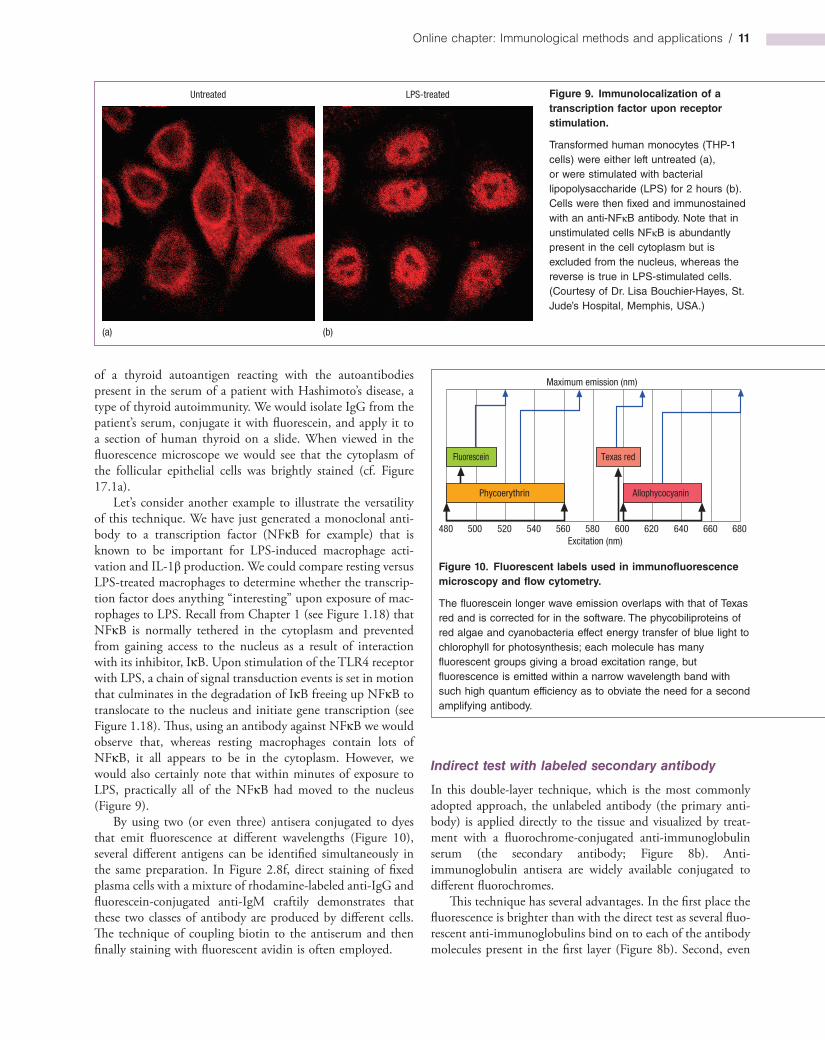
Let’s consider another example to illustrate the versatility of this technique. We have just generated a monoclonal antibody to a transcription factor (NFκB for example) that is known to be important for LPSinduced macrophage activation and IL1β production. We could compare resting versus LPStreated macrophages to determine whether the transcription factor does anything “interesting” upon exposure of macrophages to LPS. Recall from Chapter 1 (see Figure 1.18) that NFκB is normally tethered in the cytoplasm and prevented from gaining access to the nucleus as a result of interaction with its inhibitor, IκB. Upon stimulation of the TLR4 receptor with LPS, a chain of signal transduction events is set in motion that culminates in the degradation of IκB freeing up NFκB to translocate to the nucleus and initiate gene transcription (see Figure 1.18). Thus, using an antibody against NFκB we would observe that, whereas resting macrophages contain lots of NFκB, it all appears to be in the cytoplasm. However, we would also certainly note that within minutes of exposure to LPS, practically all of the NFκB had moved to the nucleus (Figure 9).
By using two (or even three) antisera conjugated to dyes that emit fluorescence at different wavelengths (Figure 10), several different antigens can be identified simultaneously in the same preparation. In Figure 2.8f, direct staining of fixed plasma cells with a mixture of rhodaminelabeled antiIgG and fluoresceinconjugated antiIgM craftily demonstrates that these two classes of antibody are produced by different cells. The technique of coupling biotin to the antiserum and then finally staining with fluorescent avidin is often employed.
Figure 9. Immunolocalization of a transcription factor upon receptor stimulation.
Transformed human monocytes (THP-1 cells) were either left untreated (a), or were stimulated with bacterial lipopolysaccharide (LPS) for 2 hours (b). Cells were then fixed and immunostained with an anti-NFκB antibody. Note that in unstimulated cells NFκB is abundantly present in the cell cytoplasm but is excluded from the nucleus, whereas the reverse is true in LPS-stimulated cells. (Courtesy of Dr. Lisa Bouchier-Hayes, St. Jude’s Hospital, Memphis, USA.)
Untreated
(a) (b)
LPS-treated
Figure 10. Fluorescent labels used in immunofluorescence microscopy and flow cytometry.
The fluorescein longer wave emission overlaps with that of Texas red and is corrected for in the software. The phycobiliproteins of red algae and cyanobacteria effect energy transfer of blue light to chlorophyll for photosynthesis; each molecule has many fluorescent groups giving a broad excitation range, but fluorescence is emitted within a narrow wavelength band with such high quantum efficiency as to obviate the need for a second amplifying antibody.
Fluorescein
Maximum emission (nm)
480 680500 520 540 560 580 600 620 640 660Excitation (nm)
Phycoerythrin Allophycocyanin
Texas red
12 / Fundamentals of Immunology
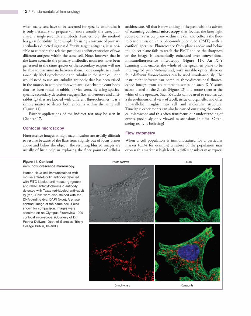
Figure 11. Confocal immunofluorescence microscopy.
Human HeLa cell immunostained with mouse anti-b-tubulin antibody detected with FITC-labeled anti-mouse Ig (green) and rabbit anti-cytochrome c antibody detected with Texas red-labeled anti-rabbit Ig (red). Cells were also stained with the DNA-binding dye, DAPI (blue). A phase contrast image of the same cell is also shown for comparison. Images were acquired on an Olympus Fluoroview 1000 confocal microscope. (Courtesy of Dr. Petrina Delivani, Dept. of Genetics, Trinity College Dublin, Ireland.)
Phase contrast
CompositeCytochrome c
Tubulin
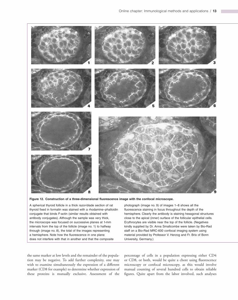
architecture. All that is now a thing of the past, with the advent of scanning confocal microscopy that focuses the laser light source on a narrow plane within the cell and collects the fluorescence emission in a photomultiplier tube (PMT) with a confocal aperture. Fluorescence from planes above and below the object plane fails to reach the PMT and so the sharpness of the image is dramatically enhanced over conventional immunofluorescence microscopy (Figure 11). An X–Y scanning unit enables the whole of the specimen plane to be interrogated quantitatively and, with suitable optics, three or four different fluorochromes can be used simultaneously. The instrument software can compute threedimensional fluorescence images from an automatic series of such X–Y scans accumulated in the Z axis (Figure 12) and rotate them at the whim of the operator. Such Zstacks can be used to reconstruct a threedimensional view of a cell, tissue or organelle, and offer unparalleled insights into cell and molecular structure. Timelapse experiments can also be carried out using the confocal microscope and this often transforms our understanding of events previously only viewed as snapshots in time. Often, seeing really is believing!
Flow cytometry
When a cell population is immunostained for a particular marker (CD4 for example) a subset of the population may express this marker at high levels, a different subset may express
when many sera have to be screened for specific antibodies it is only necessary to prepare (or, more usually the case, purchase) a single secondary antibody. Furthermore, the method has great flexibility. For example, by using a mixture of primary antibodies directed against different target antigens, it is possible to compare the relative positions and/or expression of two different antigens within the same cell. Note, however, that in the latter scenario the primary antibodies must not have been generated in the same species or the secondary reagent will not be able to discriminate between them. For example, to simultaneously label cytochrome c and tubulin in the same cell, one would need to use antitubulin antibody that has been raised in the mouse, in combination with anticytochrome c antibody that has been raised in rabbit, or vice versa. By using speciesspecific secondary detection reagents (i.e. antimouse and antirabbit Ig) that are labeled with different fluorochromes, it is a simple matter to detect both proteins within the same cell (Figure 11).
Further applications of the indirect test may be seen in Chapter 17.
Confocal microscopy
Fluorescence images at high magnification are usually difficult to resolve because of the flare from slightly out of focus planes above and below the object. The resulting blurred images are usually of little help in exploring the finer points of cellular
Online chapter: Immunological methods and applications / 13
Figure 12. Construction of a three-dimensional fluorescence image with the confocal microscope.
A spherical thyroid follicle in a thick razor-blade section of rat thyroid fixed in formalin was stained with a rhodamine–phalloidin conjugate that binds F-actin (similar results obtained with antibody conjugates). Although the sample was very thick, the microscope was focused on successive planes at 1-mm intervals from the top of the follicle (image no. 1) to halfway through (image no. 8), the total of the images representing a hemisphere. Note how the fluorescence in one plane does not interfere with that in another and that the composite
photograph (image no. 9) of images 1–8 shows all the fluorescence staining in focus throughout the depth of the hemisphere. Clearly the antibody is staining hexagonal structures close to the apical (inner) surface of the follicular epithelial cells. Erythrocytes are visible near the top of the follicle. (Negatives kindly supplied by Dr. Anna Smallcombe were taken by Bio-Rad staff on a Bio-Rad MRC-600 confocal imaging system using material provided by Professor V. Herzog and Fr. Brix of Bonn University, Germany.)
the same marker at low levels and the remainder of the population may be negative. To add further complexity, one may wish to examine simultaneously the expression of a different marker (CD8 for example) to determine whether expression of these proteins is mutually exclusive. Assessment of the
percentage of cells in a population expressing either CD4 or CD8, or both, would be quite a chore using fluorescence microscopy or confocal microscopy, as this would involve manual counting of several hundred cells to obtain reliable figures. Quite apart from the labor involved, such analyses
14 / Fundamentals of Immunology
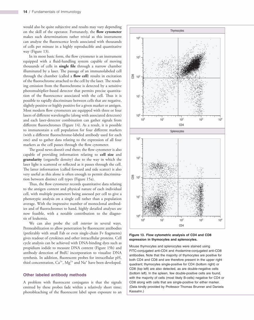
would also be quite subjective and results may vary depending on the skill of the operator. Fortunately, the flow cytometer makes such determinations rather trivial as this instrument can analyze the fluorescence levels associated with thousands of cells per minute in a highly reproducible and quantitative way (Figure 13).
In its most basic form, the flow cytometer is an instrument equipped with a fluidhandling system capable of moving thousands of cells in single file through a narrow chamber illuminated by a laser. The passage of an immunolabeled cell through the chamber (called a flow cell) results in excitation of the fluorochrome attached to the cell by the laser. The resulting emission from the fluorochrome is detected by a sensitive photomultiplierbased detector that permits precise quantitation of the fluorescence associated with the cell. Thus it is possible to rapidly discriminate between cells that are negative, slightly positive or highly positive for a given marker or antigen. Most modern flow cytometers are equipped with three or four lasers of different wavelengths (along with associated detectors) and each laserdetector combination can gather signals from different fluorochromes (Figure 14). As a result, it is possible to immunostain a cell population for four different markers (with a different fluorochromelabeled antibody used for each one) and to gather data relating to the expression of all four markers as the cell passes through the flow cytometer.
The good news doesn’t end there; the flow cytometer is also capable of providing information relating to cell size and granularity (organelle density) due to the way in which the laser light is scattered or reflected as it passes through the cell. The latter information (called forward and side scatter) is also very useful as this alone is often enough to permit discrimination between distinct cell types (Figure 15a).
Thus, the flow cytometer records quantitative data relating to the antigen content and physical nature of each individual cell, with multiple parameters being assessed per cell to give a phenotypic analysis on a single cell rather than a population average. With the impressive number of monoclonal antibodies and of fluorochromes to hand, highly detailed analyses are now feasible, with a notable contribution to the diagnosis of leukemia.
We can also probe the cell interior in several ways. Permeabilization to allow penetration by fluorescent antibodies (preferably with small Fab or even singlechain Fv fragments) gives readout of cytokines and other intracellular proteins. Cell cycle analysis can be achieved with DNAbinding dyes such as propidium iodide to measure DNA content (Figure 15b) and antibody detection of BrdU incorporation to visualize DNA synthesis. In addition, fluorescent probes for intracellular pH, thiol concentration, Ca2+, Mg2+ and Na+ have been developed.
Other labeled antibody methods
A problem with fluorescent conjugates is that the signals emitted by these probes fade within a relatively short time; photobleaching of the fluorescent label upon exposure to an
Figure 13. Flow cytometric analysis of CD4 and CD8 expression in thymocytes and splenocytes.
Mouse thymocytes and splenocytes were stained using FITC-conjugated anti-CD4 and rhodamine-conjugated anti-CD8 antibodies. Note that the majority of thymocytes are positive for both CD4 and CD8 and are therefore present in the upper right quadrant; thymocytes single-positive for CD4 (bottom right) or CD8 (top left) are also detected, as are double-negative cells (bottom left). In the spleen, few double-positive cells are found, with the majority of cells (most likely B-cells) negative for CD4 or CD8 along with cells that are single-positive for either marker. (Data kindly provided by Professor Thomas Brunner and Daniela Kassahn.)
Thymocytes
Splenocytes
104
104
103
103
102
102
101
101100
100
CD4
CD8
104
104
103
103
102
102
101
101100
100
CD4
CD8
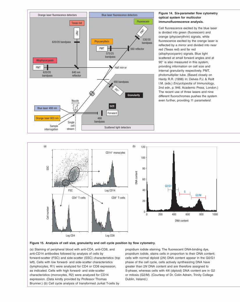
Figure 14. Six-parameter flow cytometry optical system for multicolor immunofluorescence analysis.
Cell fluorescence excited by the blue laser is divided into green (fluorescein) and orange (phycoerythrin) signals, while fluorescence excited by the orange laser is reflected by a mirror and divided into near red (Texas red) and far red (allophycocyanin) signals. Blue light scattered at small forward angles and at 90° is also measured in this system, providing information on cell size and internal granularity respectively. PMT, photomultiplier tube. (Based closely on Hardy R.R. (1998) In: Delves P.J. & Roitt I.M. (eds.) Encyclopedia of Immunology, 2nd edn, p. 946. Academic Press, London.) The recent use of three lasers and nine different fluorochromes pushes the system even further, providing 11 parameters!
Orange laser fluorescence detectors
Texas red
Allophycocyanin
PMT
PMT
620/20 bandpass
620/20bandpass
640 nmreflector
Fluorescein
Phycoerythrin
PMT
PMT
560 reflector
530/30bandpass
575/25bandpass
Scattered light detectors
Granularity
90°
Half mirr or
488 bandpass
488bandpass
Forward
SIZEBlue laser 488 nm
Nozzle
Orange laser 603 nm
Sampleinterrogation
Blue laser fluorescence detectors
Singlecell
stream
Figure 15. Analysis of cell size, granularity and cell cycle position by flow cytometry.
SSC
Cell
num
ber
FSC
R2
R1
R2
R1
Log CD14
Log CD4 Log CD8
CD14+ monocytes
CD4+ T-cells CD8+ T-cells
(a)
DNA
cont
ent
DNA content
0 200 400 600 800 10000
20
40
60
80
100
120
S
G0/G1
G2/M
(b)
(a) Staining of peripheral blood with anti-CD4, anti-CD8, and anti-CD14 antibodies followed by analysis of cells by forward-scatter (FSC) and side-scatter (SSC) characteristics (top left). Cells with low forward- and side-scatter characteristics (lymphocytes, R1) were analyzed for CD4 or CD8 expression, as indicated. Cells with high forward- and side-scatter characteristics (monocytes, R2) were analyzed for CD14 expression. (Data kindly provided by Professor Thomas Brunner.) (b) Cell cycle analysis of transformed Jurkat T-cells by
propidium iodide staining. The fluorescent DNA-binding dye, propidium iodide, stains cells in proportion to their DNA content; cells with normal diploid (2N) DNA content appear in the G0/G1 phase of the cell cycle, cells actively synthesizing DNA have greater than 2N DNA content and are therefore assigned to S-phase, whereas cells with 4N (diploid) DNA content are in G2 or mitosis (G2/M). (Courtesy of Dr. Colin Adrain, Trinity College Dublin, Ireland.)

16 / Fundamentals of Immunology
Detection and quantitation of antigen by antibody
Immunoassay of antigen by ELISA
The ability to establish the concentration of an analyte (i.e. a substance to be measured) through fractional occupancy of its specific binding reagent is a feature of any ligandbinding system (see Milestone 1), but because antibodies can be raised to virtually any structure, its application is most versatile in immunoassay.
Large analytes, such as protein hormones, are usually estimated by a noncompetitive twosite assay in which the original ligand binder and the labeled detection reagent are both antibodies (see Figure M1.1). By using monoclonal antibodies directed to two different epitopes on the same analyte, the system has greater power to discriminate between two related analytes; if the fractional crossreactivity of the first antibody
excitation source (such as UV light) can also occur. In practice, this is not a problem so long as the labeled sample is analyzed in a timely fashion. However, enzymes such as alkaline phosphatase or horseradish peroxidase can be coupled to antibodies and then visualized by conventional histochemical methods under the light microscope (Figure 16). Such stains are relatively stable and are particularly useful for staining tissue sections as opposed to cell suspensions.
Colloidal gold bound to antibody is being widely used as an electrondense immunolabel by electron microscopists. At least three different antibodies can be applied to the same section by labeling them with gold particles of different size (cf. Figure 7.26). A new ultrasmall probe consisting of Fab′ fragments linked to undecagold clusters allows more accurate spatial localization of antigens and its small size enables it to mark sites that are inaccessible to the larger immunolabels. However, clear visualization requires a highresolution scanning transmission electron microscope.
Figure 16. Immunohistochemical analysis of human tonsil follicle centers.
Anti-CD3
Anti-CD68
Anti-CD3 (brown)Anti-CD20Anti-CD20 (red)
Anti-CD21H & E
Human tonsil preparations were stained either with the histochemical stain hematoxylin and eosin (H&E), or were immunostained with antibodies against CD21 (complement receptor 2, expressed on follicular dendritic cells and B-cells),
CD68 (expressed on macrophages), CD3 (T-cells), CD20 (B-cells), or a combination of anti-CD3 and anti-CD20, as shown. (Images kindly provided by Dr. Andreas Kappeler, University of Bern, Switzerland.)
Online chapter: Immunological methods and applications / 17
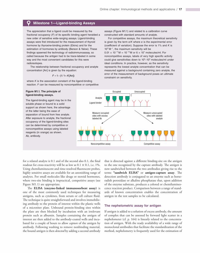
Milestone 1—Ligand-binding Assays
The appreciation that a ligand could be measured by the fractional occupancy (F) of its specific binding agent heralded a new order of sensitive wide-ranging assays. Ligand-binding assays were first introduced for the measurement of thyroid hormone by thyroxine-binding protein (Ekins) and for the estimation of hormones by antibody (Berson & Yalow). These findings spawned the technology of radioimmunoassay, so called because the antigen had to be trace-labeled in some way and the most convenient candidates for this were radioisotopes.
The relationship between fractional occupancy and analyte concentration [An] is given by the equation:
= − +1 (1/ 1 [An])F K
where K is the association constant of the ligand-binding reaction. F can be measured by noncompetitive or competitive
assays (Figure M1.1) and related to a calibration curve constructed with standard amounts of analyte.
For competitive assays, the maximum theoretical sensitivity is given by the term e/K where e is the experimental error (coefficient of variation). Suppose the error is 1% and K is 1011 M−1, the maximum sensitivity will be 0.01 × 10⁻¹¹ M = 10−13 M or 6 × 10⁷ molecules/ml. For noncompetitive assays, labels of very high specific activity could give sensitivities down to 102–103 molecules/ml under ideal conditions. In practice, however, as the sensitivity represents the lowest analyte concentration that can be measured against a background containing zero analyte, the error of the measurement of background poses an ultimate constraint on sensitivity.
Figure M1.1. The principle of ligand-binding assays.
The ligand-binding agent may be in the soluble phase or bound to a solid support as shown here, the advantage of the latter being the ease of separation of bound from free analyte. After exposure to analyte, the fractional occupancy of the ligand-binding sites can be determined by competitive or noncompetitive assays using labeled reagents (in orange) as shown. Ab, antibody.
Occupied Unoccupied
Analyte
Ligand binder
Noncompetitive assay Competitive assay
Measure occupiedsites with excesslabeled Ab
Measure unoccupiedsites with labeled
analyte
for a related analyte is 0.1 and of the second also 0.1, the final readout for crossreactivity will be as low as 0.1 × 0.1, i.e. 1%. Using chemiluminescent and timeresolved fluorescent probes, highly sensitive assays are available for an astonishing range of analytes. For small molecules like drugs or steroid hormones, where twosite binding is impractical, competitive assays (see Figure M1.1) are appropriate.
The ELISA (enzyme-linked immunosorbent assay) is one of the most commonly used techniques for measuring antigens, such as cytokines, from serum or cell culture fluid. The technique is quite straightforward and involves immobilizing antibody to the protein of interest within the plastic wells of a microtiter plate. Unbound proteinbinding sites within the plate are then blocked by incubation with an irrelevant protein such as albumin. Samples containing the antigen of interest are then added to the antibodycoated wells and incubated for a couple of hours to allow capture of the antigen by antibody. Following washing to remove nonbinding material, the bound antigen is then detected by adding a second antibody
that is directed against a different bindingsite on the antigen to the one recognized by the capture antibody. The antigen is now sandwiched between the two antibodies giving rise to the terms “sandwich ELISA” or antigen-capture assay. The detection antibody is conjugated to an enzyme such as horseradish peroxidase or alkaline phosphatase that, upon addition of the enzyme substrate, produces a colored or chemiluminescence reaction product. Comparison between a range of standards of known concentration enables the concentration of antigen in the test samples to be calculated.
The nephelometric assay for antigen
If antigen is added to a solution of excess antibody, the amount of complex that can be assessed by forward light scatter in a nephelometer (cf. p. 164) is linearly related to the concentration of antigen. With the ready availability of a wide range of monoclonal antibodies that facilitate the standardization of the method, nephelometry is frequently used for the estimation of

18 / Fundamentals of Immunology
Figure 17. Rate nephelometry.
(a) (b) (c) (d)
Peak
rate
of s
catte
r
Antigen concentration
Rate
[d(s
catte
r sig
nal)/
dt]
Peak rate
Time
Scat
ter s
igna
l
1
2
3
Time
Light
450–550 nm
Scattered wavefront
Antigen–antibodyaggregate
Turbidimetry
Nephelometry
(a) On addition of antiserum, small antigen–antibody aggregates form (cf. Figure 24) that scatter incident light filtered to give a wavelength band of 450–550 nm. For nephelometry, the light scattered at a forward angle of 70° or so is measured. (b) After addition of the sample (1) and then the antibody (2), the rate at which the aggregates form (3) is determined from the scatter
signal. (c) The software in the instrument then computes the maximum rate of light scatter, which is related to the antigen concentration as shown in (d). (Copied from the operating manual for the “Array” rate reaction automated immunonephelometer with permission from Beckman Coulter Ltd.)
immunoglobulins, C3, C4, haptoglobin, ceruloplasmin and Creactive protein in those favored laboratories that can sport the appropriate equipment. Very small samples down in the range 1–10 µl can be analyzed. Turbidity of the sample can be a problem; blanks lacking antibody can be deducted but a more satisfactory solution is to follow the rate of forma-tion of complexes that is proportional to antigen concentration as this obviates the need for a separate blank (Figure 17). Because soluble complexes begin to be formed in antigen excess, it is important to ensure that the value for antigen was obtained in antibody excess by running a further control in which additional antigen is included.
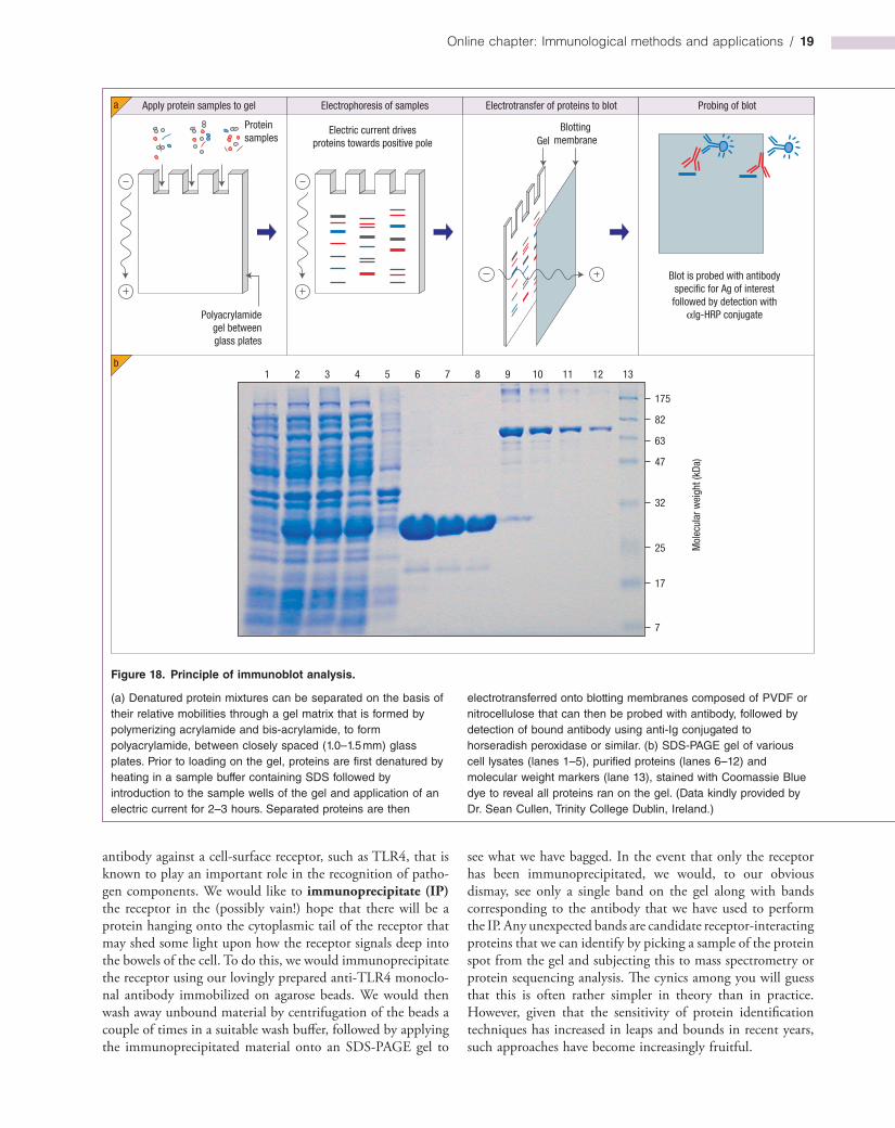
Immunoblotting (western blotting)
This widely adopted technique can be used to determine the relative molecular mass of a protein and to explore its behavior within a complex mixture of other proteins. Issues relating to whether the protein of interest is upregulated, downregulated, cleaved, phosphorylated, glycosylated or ubiquitinated in response to a particular stimulus can be addressed by immunoblot analysis. This involves first running a mixture of proteins through a gel matrix that is formed by polymerization of acrylamide and bisacrylamide between a pair of glass plates. Polyacrylamide gel electrophoresis (PAGE) of proteins is typically carried out using protein mixtures that have been denatured by heating in the presence of a detergent, sodium dodecylsulfate (SDS). SDS is a negatively charged molecule that becomes covalently coupled to proteins along their length upon exposure to heat; apart from denaturing the protein, this also imparts a negative charge in proportion to its length. Upon introduction of the protein sample to the gel and the application of a vertical electric field from the top of the gel to the bottom, proteins are repelled from the negative pole (the cathode) and
migrate towards the positive pole (the anode). Due to the molecular sieving effect of the gel matrix, proteins within the mixture become resolved into discrete zones (bands) with the smallest proteins moving furthest through the gel (Figure 18).
In order to probe the electrophoretically separated protein mixture with antibody to identify the protein of interest, it is necessary to allow the antibody access to the proteins within the gel. Because antibodies are relatively large proteins they cannot readily penetrate the gel matrix; the solution to this problem is to “blot” the gel onto a positively charged membrane that traps the charged proteins and immobilizes them on the surface of the membrane (Figure 18). This is achieved by again applying an electric field to the gel to drive the proteins horizontally out of the gel onto the blotting membrane; polyvinylidene difluoride (PVDF) and nitrocellulosebased membranes are typically used for this purpose. The blot can then be probed with either polyclonal or monoclonal antibodies directed against the protein of interest. Binding of antibody is detected using horseradish peroxidaseconjugated antiIg secondary antibodies, followed by application of a suitable enzyme substrate (Figure 19).
Obviously, such a procedure will not work with antigens that are irreversibly denatured by this detergent, and it is best to use polyclonal antisera for blotting to increase the chance of including antibodies to whichever epitopes do survive the denaturation procedure; a surprising number do.
Immunoprecipitation of antigen complexes
Antibodies immobilized on a solid support, such as agarose beads, can be used to purify an antigen from a complex mixture of other antigens to explore the nature of the antigen and the proteins to which it binds (Figure 20). To illustrate this approach, let us imagine that we have generated a monoclonal
Online chapter: Immunological methods and applications / 19
Apply protein samples to gel
Proteinsamples
Electric current drivesproteins towards positive pole
1 2 3 4 5 6 7 8 9 10 11 12 13
175
Mol
ecul
ar w
eigh
t (kD
a)
Gel
Polyacrylamidegel betweenglass plates
Electrophoresis of samples
Blottingmembrane
Blot is probed with antibodyspecific for Ag of interestfollowed by detection with
αlg-HRP conjugate
Electrotransfer of proteins to blot Probing of blot
82
63
47
32
25
17
7
a
b
Figure 18. Principle of immunoblot analysis.
(a) Denatured protein mixtures can be separated on the basis of their relative mobilities through a gel matrix that is formed by polymerizing acrylamide and bis-acrylamide, to form polyacrylamide, between closely spaced (1.0–1.5 mm) glass plates. Prior to loading on the gel, proteins are first denatured by heating in a sample buffer containing SDS followed by introduction to the sample wells of the gel and application of an electric current for 2–3 hours. Separated proteins are then
electrotransferred onto blotting membranes composed of PVDF or nitrocellulose that can then be probed with antibody, followed by detection of bound antibody using anti-Ig conjugated to horseradish peroxidase or similar. (b) SDS-PAGE gel of various cell lysates (lanes 1–5), purified proteins (lanes 6–12) and molecular weight markers (lane 13), stained with Coomassie Blue dye to reveal all proteins ran on the gel. (Data kindly provided by Dr. Sean Cullen, Trinity College Dublin, Ireland.)
antibody against a cellsurface receptor, such as TLR4, that is known to play an important role in the recognition of pathogen components. We would like to immunoprecipitate (IP) the receptor in the (possibly vain!) hope that there will be a protein hanging onto the cytoplasmic tail of the receptor that may shed some light upon how the receptor signals deep into the bowels of the cell. To do this, we would immunoprecipitate the receptor using our lovingly prepared antiTLR4 monoclonal antibody immobilized on agarose beads. We would then wash away unbound material by centrifugation of the beads a couple of times in a suitable wash buffer, followed by applying the immunoprecipitated material onto an SDSPAGE gel to
see what we have bagged. In the event that only the receptor has been immunoprecipitated, we would, to our obvious dismay, see only a single band on the gel along with bands corresponding to the antibody that we have used to perform the IP. Any unexpected bands are candidate receptorinteracting proteins that we can identify by picking a sample of the protein spot from the gel and subjecting this to mass spectrometry or protein sequencing analysis. The cynics among you will guess that this is often rather simpler in theory than in practice. However, given that the sensitivity of protein identification techniques has increased in leaps and bounds in recent years, such approaches have become increasingly fruitful.
20 / Fundamentals of Immunology
Immunoprecipitation can also be used to test whether protein A binds to protein B by coexpressing these proteins within the same cell, followed by immunoprecipitation of protein A (or protein B) using a suitable antibody and running on an SDSPAGE gel. Following transfer of the gel to a membrane support by Western blotting, the membrane can now be probed with antibodies directed against protein B to see whether it has coimmunoprecipitated with protein A. The latter technique is undoubtedly the most widely used form of the IP method and has been employed to great effect in the study of protein–protein interactions.
Chromatin immunoprecipitation (ChIP) assays
This interesting modification of the standard immunoprecipitation assay can analyse the repertoire of gene promoter sequences (or other regions within DNA) a transcription factor or other DNAbinding protein is bound to under a particular experimental condition. For example, if we would like to explore the range of gene promoters that NFκB binds to under unstimulated versus stimulated (e.g. LPStreatment) conditions we could perform a ChIP experiment. Here’s how the method works. Cells are incubated for a defined period of time in the presence or absence of a stimulus (LPS in this example), followed by brief treatment with a chemical crosslinking agent (e.g. formaldehyde) to ensure that any transcription factor bound to a promoter will remain bound under the conditions of the assay. After chemical crosslinking of protein–DNA complexes, cells are then lysed and the mixture is sonicated to shear very high molecular weight DNA into smaller more manageable fragments. Then an antibody specific to our transcription factor of interest (i.e. NFκB in this instance) is used to immunoprecipitate the protein from the mixture. The clever bit is that, due to the crosslinking step, our transcription factor will be bound to the regions of DNA (i.e. the promoters to which it was bound) it was actively transcribing when we ended the experiment. We can then carry out a PCR reaction on the immunoprecipitated samples using primers specific for genes we think might be regulated by the transcription factor of interest. If our transcription factor has bound to a promoter region of a gene, then the corresponding DNA fragment will be amplified when we carry out the PCR assay on that sample. We can then compare the amount of DNA amplified under the control versus treated conditions to determine whether the treatment (i.e. LPS in this instance) has enhanced binding of our transcription factor to specific gene promoters.
ChIP on Chip assays
In a further modification of the standard ChIP assay, as described above, a ChIP on Chip (DNA microarray) assay can be carried out. This permits a more global analysis of the DNA fragments immunoprecipitated with a particular transcription factor, where, rather then looking for specific gene sequences by PCR, we can more objectively look at all of the immunoprecipitated DNA fragments through hybridization with a
Figure 19. Immunoblot analysis.
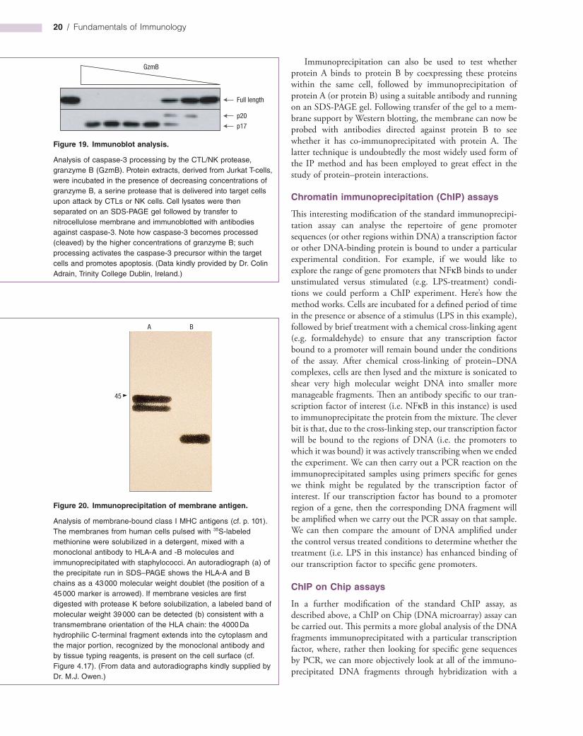
Analysis of caspase-3 processing by the CTL/NK protease, granzyme B (GzmB). Protein extracts, derived from Jurkat T-cells, were incubated in the presence of decreasing concentrations of granzyme B, a serine protease that is delivered into target cells upon attack by CTLs or NK cells. Cell lysates were then separated on an SDS-PAGE gel followed by transfer to nitrocellulose membrane and immunoblotted with antibodies against caspase-3. Note how caspase-3 becomes processed (cleaved) by the higher concentrations of granzyme B; such processing activates the caspase-3 precursor within the target cells and promotes apoptosis. (Data kindly provided by Dr. Colin Adrain, Trinity College Dublin, Ireland.)
Full length
GzmB
p20
p17
Figure 20. Immunoprecipitation of membrane antigen.
Analysis of membrane-bound class I MHC antigens (cf. p. 101). The membranes from human cells pulsed with 35S-labeled methionine were solubilized in a detergent, mixed with a monoclonal antibody to HLA-A and -B molecules and immunoprecipitated with staphylococci. An autoradiograph (a) of the precipitate run in SDS–PAGE shows the HLA-A and B chains as a 43 000 molecular weight doublet (the position of a 45 000 marker is arrowed). If membrane vesicles are first digested with protease K before solubilization, a labeled band of molecular weight 39 000 can be detected (b) consistent with a transmembrane orientation of the HLA chain: the 4000 Da hydrophilic C-terminal fragment extends into the cytoplasm and the major portion, recognized by the monoclonal antibody and by tissue typing reagents, is present on the cell surface (cf. Figure 4.17). (From data and autoradiographs kindly supplied by Dr. M.J. Owen.)
A B
45

Online chapter: Immunological methods and applications / 21
Figure 21. Serum profiling by protein microarray analysis.
Protein arrays, consisting of thousands of proteins of known identity arrayed in a specific order, can be probed with a sample of a patient’s serum to determine the range of proteins to which there are antibodies present. Bound antibodies can be detected with appropriate anti-Ig secondary antibody that leads directly to the identity of the proteins within the positive spots.
Proteins of known identityarrayed onto glass support
Incubate with patient antiserum
Incubate with αlg-conjugateto detect binding of primaryantiserum
Add substrateProteins that bindantibody are revealed
DNA microarray chip that carries sequences from a huge variety of genes. Such arrays can carry gene fragments, arrayed in specific spot locations on a solid support (called a chip), that represent the whole genome, or can be more restricted and carry gene fragments representative of all cytokines, or chemokines, and so on. This type of assay involves carrying out a standard ChIP experiment, as described above, followed by applying the DNA fragments so captured onto a DNA chip. This allows identification all the DNA sequences bound to the protein of interest under the experimental condition.
Protein and antibody microarrays
With the ready availability of cDNA copies of essentially all human genes, it is now possible to express virtually any human protein “offtheshelf ” and to purify it to homogeneity using simple molecular tricks. This is also true for proteincoding genes from several species of yeast, many bacteria, the fruitfly and other organisms. Using largescale protein expression approaches, arrays of proteins have been produced that contain thousands of independent proteins, or protein fragments, arranged on glass slides as discrete microdots. On such arrays, each microdot contains a single protein and the identity of this protein is known from its position within the array. Such arrays can be probed with an antiserum, or monoclonal antibody, to determine the spectrum of proteins to which the antibody reacts. Thus, it may be possible in the near future to determine the full spectrum of autoantibodies (and their relative titer) present in a patient sample in a single step. One has only to crossreference the spots “lighting up” after incubation with antibody with a list that specifies the identity of the protein that has been placed within those particular spots (Figure 21). Such arrays are likely to be useful for the identification of novel autoantigens, for the rapid diagnosis and classification of autoimmune conditions, and may also be useful for monitoring disease progression. Additional applications include the rapid determination of proteins that crossreact against monoclonal or polyclonal antibodies. In practice, smaller arrays focused on particular disease states are used for diagnostic purposes.
Similar to protein arrays, antibodies can also be arrayed as discrete spots on glass slides or other solid supports. Such arrays can be used to capture multiple antigens from the same sample simultaneously. Thus, for example, an anticytokine antibody array can be used to detect the presence of multiple cytokines within the same sample.
Epitope mapping
T-cell epitopes
Where the primary sequence of the whole protein is known, the identification of Tcell epitopes is comparatively straightforward. As these epitopes are linear in nature, multipin solidphase synthesis can be employed to generate a series of overlapping peptides, 8–9mers for cytotoxic Tcells and usually 10–14mers for Thelpers (Figure 22), and their ability
to react with antigenspecific Tcell lines or clones can be deciphered to characterize the active epitopes.
Dissecting out Tcell epitopes where the antigen has not been characterized is a more daunting task. Randomized peptide libraries can be produced but strategies need to be devised in order to keep these within manageable numbers. Information from the accumulated data deposited in various databanks can be used to identify key anchor residues and libraries constructed that maintain the relevant amino acids at
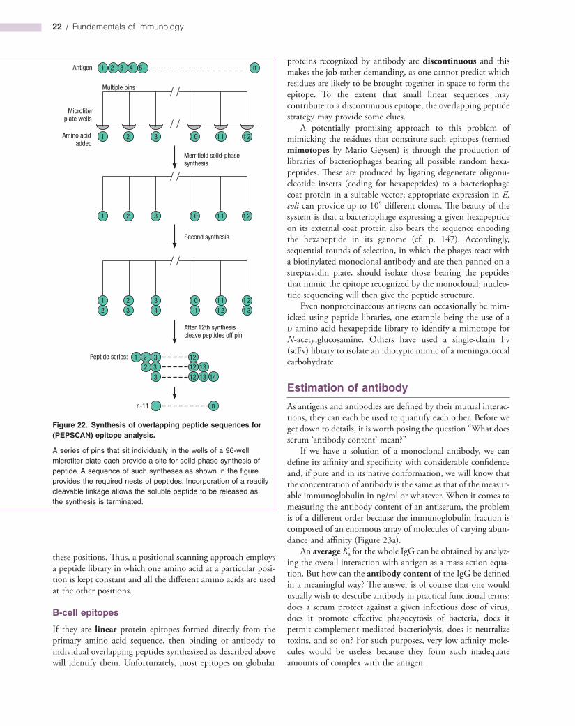
22 / Fundamentals of Immunology
proteins recognized by antibody are discontinuous and this makes the job rather demanding, as one cannot predict which residues are likely to be brought together in space to form the epitope. To the extent that small linear sequences may contribute to a discontinuous epitope, the overlapping peptide strategy may provide some clues.
A potentially promising approach to this problem of mimicking the residues that constitute such epitopes (termed mimotopes by Mario Geysen) is through the production of libraries of bacteriophages bearing all possible random hexapeptides. These are produced by ligating degenerate oligonucleotide inserts (coding for hexapeptides) to a bacteriophage coat protein in a suitable vector; appropriate expression in E. coli can provide up to 109 different clones. The beauty of the system is that a bacteriophage expressing a given hexapeptide on its external coat protein also bears the sequence encoding the hexapeptide in its genome (cf. p. 147). Accordingly, sequential rounds of selection, in which the phages react with a biotinylated monoclonal antibody and are then panned on a streptavidin plate, should isolate those bearing the peptides that mimic the epitope recognized by the monoclonal; nucleotide sequencing will then give the peptide structure.
Even nonproteinaceous antigens can occasionally be mimicked using peptide libraries, one example being the use of a Damino acid hexapeptide library to identify a mimotope for Nacetylglucosamine. Others have used a singlechain Fv (scFv) library to isolate an idiotypic mimic of a meningococcal carbohydrate.
Estimation of antibody
As antigens and antibodies are defined by their mutual interactions, they can each be used to quantify each other. Before we get down to details, it is worth posing the question “What does serum ‘antibody content’ mean?”
If we have a solution of a monoclonal antibody, we can define its affinity and specificity with considerable confidence and, if pure and in its native conformation, we will know that the concentration of antibody is the same as that of the measurable immunoglobulin in ng/ml or whatever. When it comes to measuring the antibody content of an antiserum, the problem is of a different order because the immunoglobulin fraction is composed of an enormous array of molecules of varying abundance and affinity (Figure 23a).
An average Ka for the whole IgG can be obtained by analyzing the overall interaction with antigen as a mass action equation. But how can the antibody content of the IgG be defined in a meaningful way? The answer is of course that one would usually wish to describe antibody in practical functional terms: does a serum protect against a given infectious dose of virus, does it promote effective phagocytosis of bacteria, does it permit complementmediated bacteriolysis, does it neutralize toxins, and so on? For such purposes, very low affinity molecules would be useless because they form such inadequate amounts of complex with the antigen.
these positions. Thus, a positional scanning approach employs a peptide library in which one amino acid at a particular position is kept constant and all the different amino acids are used at the other positions.
B-cell epitopes
If they are linear protein epitopes formed directly from the primary amino acid sequence, then binding of antibody to individual overlapping peptides synthesized as described above will identify them. Unfortunately, most epitopes on globular
Figure 22. Synthesis of overlapping peptide sequences for (PEPSCAN) epitope analysis.
A series of pins that sit individually in the wells of a 96-well microtiter plate each provide a site for solid-phase synthesis of peptide. A sequence of such syntheses as shown in the figure provides the required nests of peptides. Incorporation of a readily cleavable linkage allows the soluble peptide to be released as the synthesis is terminated.
1 2 3 1 0 1 1 1 2
1 2 3 4 5
1 2 3 1 0 1 1 1 2
1 2 3 1 0 1 22 3 4 1 1 1
1 12 1 3
n-11 n
1 2 32 3
3
1212
12
13
13 14
n
Multiple pins
Microtiterplate wells
Amino acidadded
Merrifield solid-phasesynthesis
Second synthesis
After 12th synthesiscleave peptides off pin
Peptide series:
Antigen
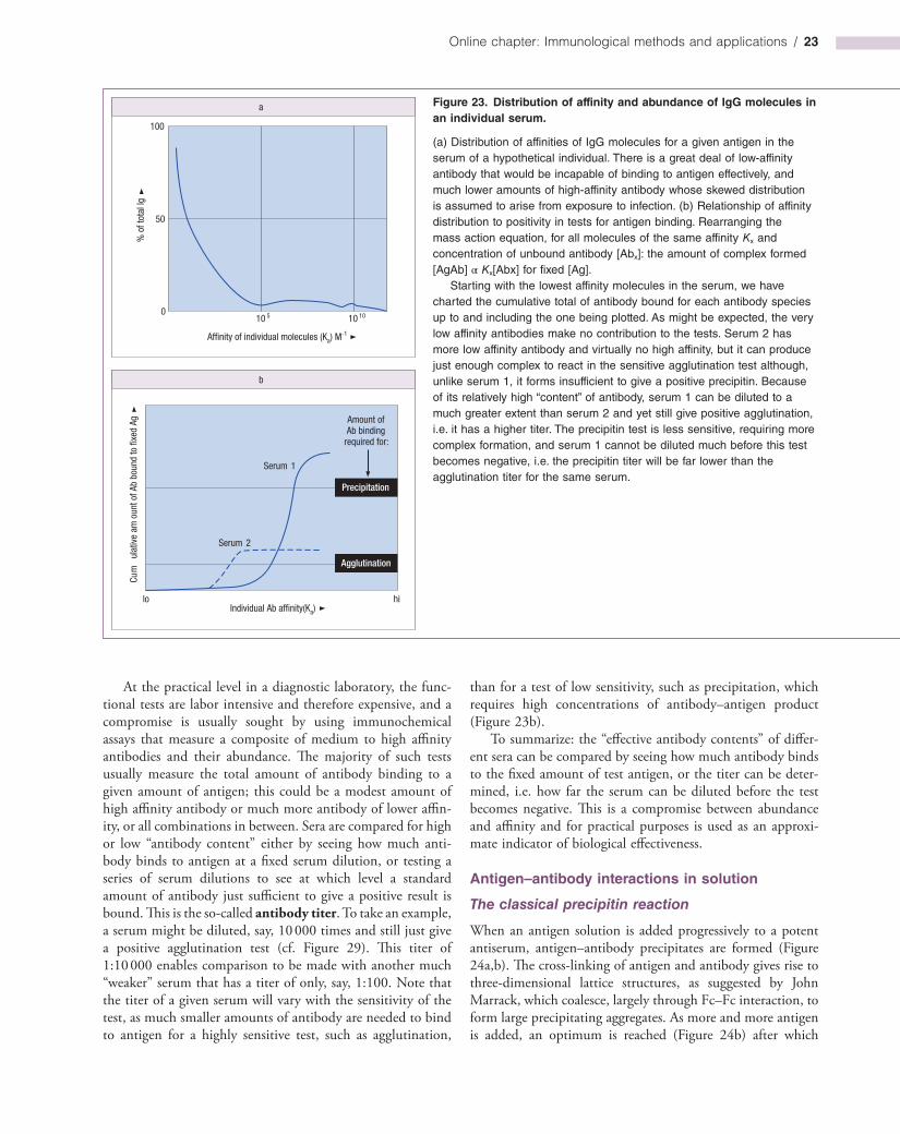
Online chapter: Immunological methods and applications / 23
than for a test of low sensitivity, such as precipitation, which requires high concentrations of antibody–antigen product (Figure 23b).
To summarize: the “effective antibody contents” of different sera can be compared by seeing how much antibody binds to the fixed amount of test antigen, or the titer can be determined, i.e. how far the serum can be diluted before the test becomes negative. This is a compromise between abundance and affinity and for practical purposes is used as an approximate indicator of biological effectiveness.
Antigen–antibody interactions in solution
The classical precipitin reaction
When an antigen solution is added progressively to a potent antiserum, antigen–antibody precipitates are formed (Figure 24a,b). The crosslinking of antigen and antibody gives rise to threedimensional lattice structures, as suggested by John Marrack, which coalesce, largely through Fc–Fc interaction, to form large precipitating aggregates. As more and more antigen is added, an optimum is reached (Figure 24b) after which
100
50
010 5 10 10
Affinity of individual molecules (Ka) M-1
a
% o
f tot
al Ig
b
hilo
Amount ofAb binding
required for:
Serum 1
Serum 2
Individual Ab affinity(Ka)
Cum
ulat
ive
am o
unt o
f Ab
boun
d to
fixe
d Ag
Agglutination
Precipitation
At the practical level in a diagnostic laboratory, the functional tests are labor intensive and therefore expensive, and a compromise is usually sought by using immunochemical assays that measure a composite of medium to high affinity antibodies and their abundance. The majority of such tests usually measure the total amount of antibody binding to a given amount of antigen; this could be a modest amount of high affinity antibody or much more antibody of lower affinity, or all combinations in between. Sera are compared for high or low “antibody content” either by seeing how much antibody binds to antigen at a fixed serum dilution, or testing a series of serum dilutions to see at which level a standard amount of antibody just sufficient to give a positive result is bound. This is the socalled antibody titer. To take an example, a serum might be diluted, say, 10 000 times and still just give a positive agglutination test (cf. Figure 29). This titer of 1:10 000 enables comparison to be made with another much “weaker” serum that has a titer of only, say, 1:100. Note that the titer of a given serum will vary with the sensitivity of the test, as much smaller amounts of antibody are needed to bind to antigen for a highly sensitive test, such as agglutination,
Figure 23. Distribution of affinity and abundance of IgG molecules in an individual serum.
(a) Distribution of affinities of IgG molecules for a given antigen in the serum of a hypothetical individual. There is a great deal of low-affinity antibody that would be incapable of binding to antigen effectively, and much lower amounts of high-affinity antibody whose skewed distribution is assumed to arise from exposure to infection. (b) Relationship of affinity distribution to positivity in tests for antigen binding. Rearranging the mass action equation, for all molecules of the same affinity Kx and concentration of unbound antibody [Abx]: the amount of complex formed [AgAb] α Kx[Abx] for fixed [Ag].
Starting with the lowest affinity molecules in the serum, we have charted the cumulative total of antibody bound for each antibody species up to and including the one being plotted. As might be expected, the very low affinity antibodies make no contribution to the tests. Serum 2 has more low affinity antibody and virtually no high affinity, but it can produce just enough complex to react in the sensitive agglutination test although, unlike serum 1, it forms insufficient to give a positive precipitin. Because of its relatively high “content” of antibody, serum 1 can be diluted to a much greater extent than serum 2 and yet still give positive agglutination, i.e. it has a higher titer. The precipitin test is less sensitive, requiring more complex formation, and serum 1 cannot be diluted much before this test becomes negative, i.e. the precipitin titer will be far lower than the agglutination titer for the same serum.

24 / Fundamentals of Immunology
Nonprecipitating antibodies can be detected by nephelometry
The small aggregates formed when dilute solutions of antigen and antibody are mixed create a cloudiness or turbidity that can be measured by forward angle scattering of an incident light source (nephelometry). Greater sensitivity can be obtained by using monochromatic light from a laser and by adding polyethylene glycol to the solution so that aggregate size is increased. In practice, nephelometry is applied more to the detection of antigen than antibody (cf. Figure 17).
Complexes formed by nonprecipitating antibodies can be precipitated
The relative antigenbinding capacity of an antiserum that forms soluble complexes can be estimated using radiolabeled antigen. The complex can be brought out of solution either by changing its solubility or by adding an antiimmunoglobulin reagent as in Figure 25.
Measurement of antibody affinity
As discussed in earlier chapters (cf. p. 118), the binding strength of antibody for antigen is measured in terms of the association constant (Ka) or its reciprocal, the dissociation constant (Kd), governing the reversible interaction between them and defined by the mass action equation at equilibrium:
=K[AgAb complex][free Ag][free Ab]a
With small haptens, equilibrium dialysis can be employed to measure Ka, but usually one is dealing with larger antigens and other techniques must be used. One approach is to add increasing amounts of radiolabeled antigen to a fixed amount of antibody, and then separate the free from bound antibody by precipitating the soluble complex as described above (e.g. by an antiimmunoglobulin). The reciprocal of the bound, i.e. complexed, antibody concentration can be plotted against the reciprocal of the free antigen concentration, so allowing the affinity constant to be calculated (Figure 26a). For an antiserum this will give an affinity constant representing an average of the heterogeneous antibody components and a measure of the effective number of antigenbinding sites operative at the highest levels of antigen used.
Various types of ELISA have been developed that provide a measure of antibody affinity. In one system the antibody is allowed to first bind to its antigen, and then a chaotropic agent such as thiocyanate is added in increasing concentration in order to disrupt the antibody binding; the higher the affinity of the antibody, the more agent that is required to reduce the binding. Another type of ELISA for measuring affinity is the indirect competitive system devised by Friguet and associates (Figure 26b). A constant amount of antibody is incubated with
consistently less precipitate is formed. At this stage the supernatant can be shown to contain soluble complexes of antigen (Ag) and antibody (Ab), many of composition Ag4Ab3, Ag3Ab2 and Ag2Ab (Figure 24c). In extreme antigen excess (Figure 24c), ultracentrifugal analysis reveals the complexes to be mainly of the form Ag2Ab, a result directly attributable to the two combining sites (divalence) of the IgG antibody molecule (cf. electron microscope study, Figure 3.10).
Serums frequently contain up to 10% of nonprecipitating antibodies that are effectively monovalent because of the asymmetric presence of oligosaccharide on one antigenbinding arm of the antibody molecule that stereochemically blocks the combining site. Also, frank precipitates are only observed when antigen, and particularly antibody, is present in fairly hefty concentrations. Thus, when complexes are formed that do not precipitate spontaneously, more devious methods must be applied to detect and estimate the antibody level.
Figure 24. Diagrammatic representation of complexes formed between a hypothetical tetravalent antigen ( ) and bivalent antibody ( ) mixed in different proportions. In practice, the antigen valencies are unlikely to lie in the same plane or to be formed by identical determinants as suggested in the figure. (a) In extreme antibody excess, the antigen valencies are saturated and the molar ratio Ab:Ag approximates to the valency of the antigen. (b) At equivalence, most of the antigen and antibody combines to form large lattices that aggregate to produce typical immune precipitates. (c) In extreme antigen excess, where the two valencies of each antibody molecule become rapidly saturated, the complex Ag2Ab tends to predominate. (d) A monovalent hapten binds but is unable to cross-link antibody molecules.
Antigen excess Monovalent hapten
Antibody excess Equivalencea b
Precipitating complexes
Soluble complexes
dc Antigen excess Monovalent hapten
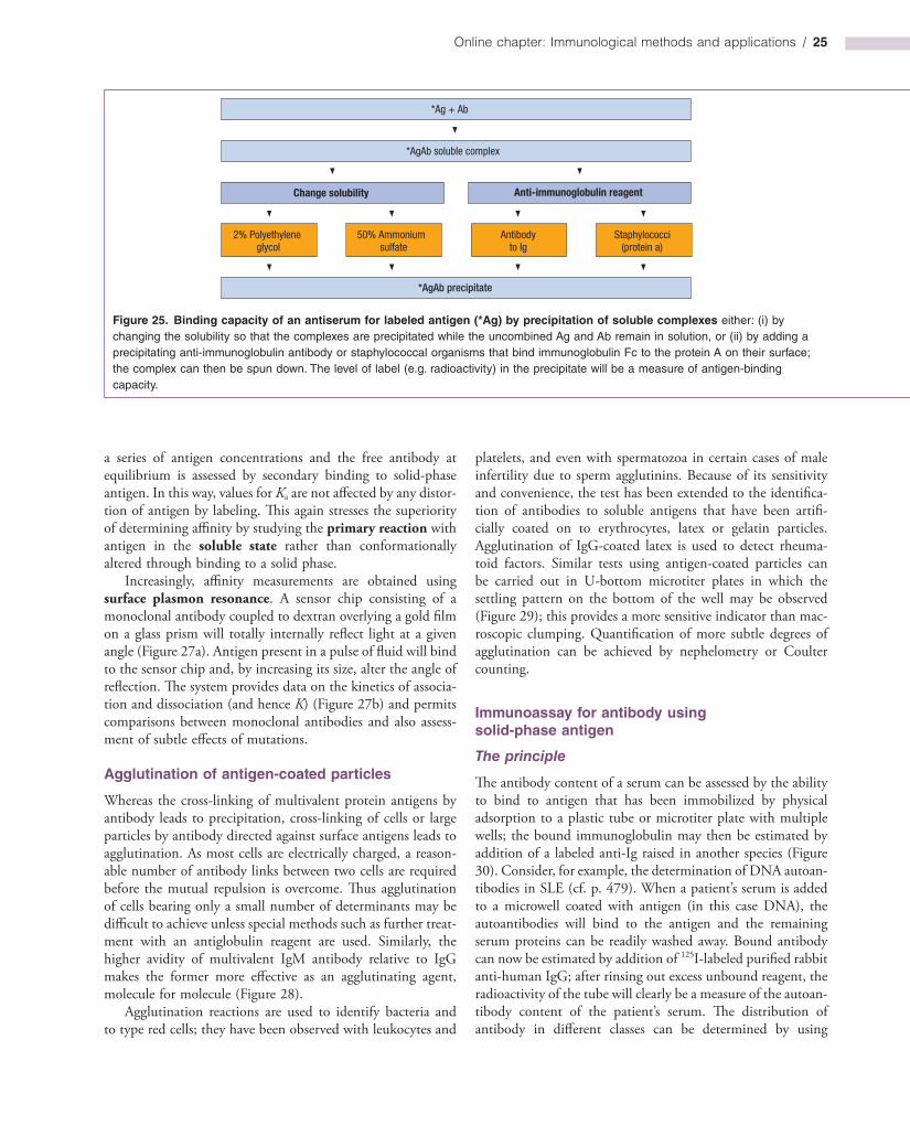
Online chapter: Immunological methods and applications / 25
Figure 25. Binding capacity of an antiserum for labeled antigen (*Ag) by precipitation of soluble complexes either: (i) by changing the solubility so that the complexes are precipitated while the uncombined Ag and Ab remain in solution, or (ii) by adding a precipitating anti-immunoglobulin antibody or staphylococcal organisms that bind immunoglobulin Fc to the protein A on their surface; the complex can then be spun down. The level of label (e.g. radioactivity) in the precipitate will be a measure of antigen-binding capacity.
*Ag + Ab
*AgAb soluble complex
Change solubility
*AgAb precipitate
Anti-immunoglobulin reagent
2% Polyethyleneglycol
50% Ammoniumsulfate
Antibodyto Ig
Staphylococci(protein a)
a series of antigen concentrations and the free antibody at equilibrium is assessed by secondary binding to solidphase antigen. In this way, values for Ka are not affected by any distortion of antigen by labeling. This again stresses the superiority of determining affinity by studying the primary reaction with antigen in the soluble state rather than conformationally altered through binding to a solid phase.
Increasingly, affinity measurements are obtained using surface plasmon resonance. A sensor chip consisting of a monoclonal antibody coupled to dextran overlying a gold film on a glass prism will totally internally reflect light at a given angle (Figure 27a). Antigen present in a pulse of fluid will bind to the sensor chip and, by increasing its size, alter the angle of reflection. The system provides data on the kinetics of association and dissociation (and hence K) (Figure 27b) and permits comparisons between monoclonal antibodies and also assessment of subtle effects of mutations.
Agglutination of antigen-coated particles
Whereas the crosslinking of multivalent protein antigens by antibody leads to precipitation, crosslinking of cells or large particles by antibody directed against surface antigens leads to agglutination. As most cells are electrically charged, a reasonable number of antibody links between two cells are required before the mutual repulsion is overcome. Thus agglutination of cells bearing only a small number of determinants may be difficult to achieve unless special methods such as further treatment with an antiglobulin reagent are used. Similarly, the higher avidity of multivalent IgM antibody relative to IgG makes the former more effective as an agglutinating agent, molecule for molecule (Figure 28).
Agglutination reactions are used to identify bacteria and to type red cells; they have been observed with leukocytes and
platelets, and even with spermatozoa in certain cases of male infertility due to sperm agglutinins. Because of its sensitivity and convenience, the test has been extended to the identification of antibodies to soluble antigens that have been artificially coated on to erythrocytes, latex or gelatin particles. Agglutination of IgGcoated latex is used to detect rheumatoid factors. Similar tests using antigencoated particles can be carried out in Ubottom microtiter plates in which the settling pattern on the bottom of the well may be observed (Figure 29); this provides a more sensitive indicator than macroscopic clumping. Quantification of more subtle degrees of agglutination can be achieved by nephelometry or Coulter counting.
Immunoassay for antibody using solid-phase antigen
The principle
The antibody content of a serum can be assessed by the ability to bind to antigen that has been immobilized by physical adsorption to a plastic tube or microtiter plate with multiple wells; the bound immunoglobulin may then be estimated by addition of a labeled antiIg raised in another species (Figure 30). Consider, for example, the determination of DNA autoantibodies in SLE (cf. p. 479). When a patient’s serum is added to a microwell coated with antigen (in this case DNA), the autoantibodies will bind to the antigen and the remaining serum proteins can be readily washed away. Bound antibody can now be estimated by addition of 125Ilabeled purified rabbit antihuman IgG; after rinsing out excess unbound reagent, the radioactivity of the tube will clearly be a measure of the autoantibody content of the patient’s serum. The distribution of antibody in different classes can be determined by using
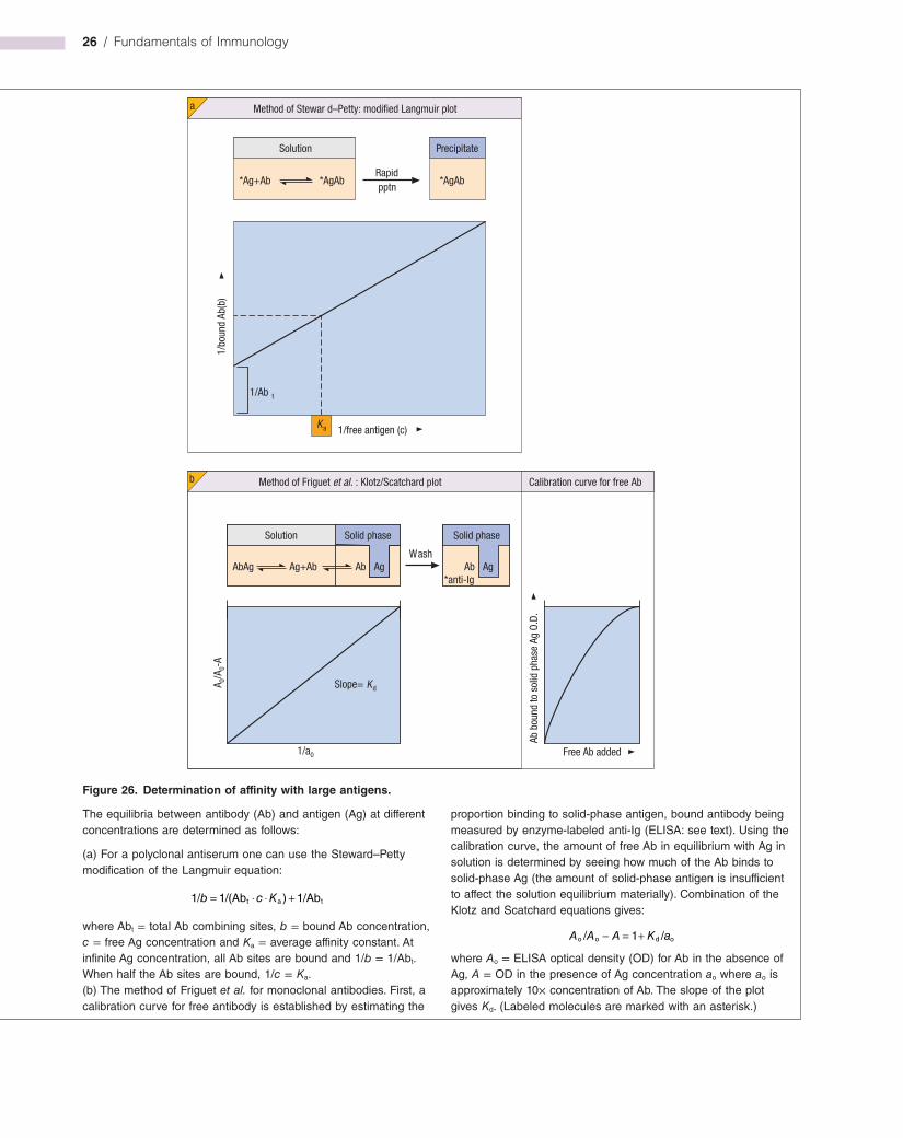
26 / Fundamentals of Immunology
Figure 26. Determination of affinity with large antigens.
Method of Stewar d–Petty: modified Langmuir plot
1/Ab t
Slope= d
Method of Friguet et al. : Klotz/Scatchard plot
Wash
K
a
Solution Precipitate
*Ag+Ab *AgAbRapidpptn
*AgAb
Ka 1/free antigen (c)
1/bo
und
Ab(b
)
Solution Solid phase
AgAbAbAg Ag+Ab
Solid phase
AgAb*anti-Ig
Calibration curve for free Ab
1/a0
A 0/A 0-
A
Free Ab added
Ab b
ound
to s
olid
pha
se A
g O.
D.
b
The equilibria between antibody (Ab) and antigen (Ag) at different concentrations are determined as follows:
(a) For a polyclonal antiserum one can use the Steward–Petty modification of the Langmuir equation:
= ⋅ ⋅ +1/ 1/(Ab ) 1/Abt a tb c K
where Abt = total Ab combining sites, b = bound Ab concentration, c = free Ag concentration and Ka = average affinity constant. At infinite Ag concentration, all Ab sites are bound and 1/b = 1/Abt. When half the Ab sites are bound, 1/c = Ka.(b) The method of Friguet et al. for monoclonal antibodies. First, a calibration curve for free antibody is established by estimating the
proportion binding to solid-phase antigen, bound antibody being measured by enzyme-labeled anti-Ig (ELISA: see text). Using the calibration curve, the amount of free Ab in equilibrium with Ag in solution is determined by seeing how much of the Ab binds to solid-phase Ag (the amount of solid-phase antigen is insufficient to affect the solution equilibrium materially). Combination of the Klotz and Scatchard equations gives:
− = +/ 1 /o o d oA A A K a
where Ao = ELISA optical density (OD) for Ab in the absence of Ag, A = OD in the presence of Ag concentration ao where ao is approximately 10× concentration of Ab. The slope of the plot gives Kd. (Labeled molecules are marked with an asterisk.)
Online chapter: Immunological methods and applications / 27
A wide variety of labels available
Whilst providing extremely good sensitivity, radiolabels have a number of disadvantages, including loss of sensitivity during storage due to radioactive decay, the deterioration of the labeled reagent through radiation damage, and the precautions needed to minimize human exposure to radioactivity. Therefore, other types of label are often employed in immunoassays.
ELISA. Enzymes that give a colored soluble reaction product are currently the most commonly used labels, with horseradish peroxidase (HRP) and calf intestine alkaline phosphatase (AP) being by far the most popular. Aspergillus niger glucose oxidase, soy bean urease and Escherichia coli βgalactosidase provide further alternatives. One clever ploy for amplifying the phosphatase reaction is to use nicotinamide adenine dinucleotide phosphate (NADP) as a substrate to generate NAD that now acts as a coenzyme for a second enzyme system.
Other labels. Enzymelabeled streptococcal protein G or staphylococcal protein A will bind to IgG. Conjugation with the vitamin biotin is frequently used as this can readily be detected by its reaction with enzymelinked avidin or strep tavidin (the latter gives lower background binding), both of which bind with ferocious specificity and affinity (K = 1015 M−1).
specific antisera. Take the radioallergosorbent test (RAST) for IgE antibodies in allergic patients. The allergen (e.g. pollen extract) is covalently coupled to an immunoabsorbent, in this case a paper disk, which is then treated with patient’s serum. The amount of specific IgE bound to the paper can now be estimated by the addition of labeled antiIgE.
Figure 27. Surface plasmon resonance.
Flow
Sensor chip
Gold/Dextran
Prism
Light source
b
Detector
a
Variable angleof reflection
Ag
Ab
400
300
200
100
0200 600 1000 1400 1800
Time (s)
Resp
onse
(RU)
mAb A
mAb B
mAb C
(a) The principle: as antigen (Ag) binds to the antibody-coated sensor chip it alters the angle of reflection. (b) This signals the rates of association during the antigen pulse and dissociation. In this example, the same antigen was injected over three immobilized monoclonal antibodies (mAbs). The arrows point to the beginning and end of the antigen injection, which is followed by buffer flow. Note the differences between the antibodies in the
Figure 28. Mechanism of agglutination of antigen-coated particles by antibody cross-linking to form large macroscopic aggregates. If red cells are used, several cross-links are needed to overcome the electrical charge at the cell surface. IgM is superior to IgG as an agglutinator because of its multivalent binding and because the charged cells are further apart.
Antigen
Red cell
IgG IgM
association and dissociation rates. (Data kindly provided by Dr. R. Karlsson, Biacore AB, and reproduced from Panayotou G. (1998) Surface plasmon resonance. In: Delves P.J. & Roitt I.M. (eds.) Encyclopedia of Immunology, 2nd edn. Academic Press, with permission.) The system can be used with antigen immobilized on the sensor chip and antibody in the fluid phase, or can be applied to any other single ligand-binding assay.
28 / Fundamentals of Immunology
Figure 29. Red cell hemagglutination test for thyroglobulin autoantibodies. Thyroglobulin-coated cells were added to dilutions of patients’ serums. Uncoated cells were added to a 1 : 10 dilution of serum as a control. In a positive reaction, the cells settle as a carpet over the bottom of the cup. Because of the “V”-shaped cross-section of these cups, in negative reactions the cells fall into the base of the “V,” forming a small, easily recognizable button. The reciprocal of the highest serum dilution giving an unequivocally positive reaction is termed the titer. The titers reading from left to right are: 640, 20, >5120, neg, 40, 320, neg, >5120. The control for serum no. 46 was slightly positive and this serum should be tested again after absorption with uncoated cells.
Control
Serum no.
5120
2560
1280
640
320
160
80
40
20
10
Seru
m d
ilutio
n
46 18 58 21 26 11 70 52
Chemiluminescence systems based on the HRPcatalyzed enhanced luminol reaction, where light from the oxidized luminol substrate is intensified and the signal duration increased by the use of an enhancing reagent, provide increased sensitivity and dynamic range. Special mention should be made of timeresolved fluorescence assays based upon chelates of rare earths such as europium 3+, although these have a more important role in antigen assays.
Detection of immune complex formation
Many techniques for detecting circulating complexes have been described and because of variations in the size, complementfixing ability and Ig class of different complexes, it is useful to apply more than one method. Two fairly robust methods for general use are:
1 Precipitation of complexed IgG from serum at concentrations of polyethylene glycol that do not bring down significant amounts of IgG monomer, followed by estimation of IgG in the precipitate by single radial immunodiffusion (SRID) or laser nephelometry; and
2 Binding of C3bcontaining complexes to beads coated with bovine conglutinin (cf. p. 20) and estimation of the bound Ig with enzymelabeled antiIg.
Other techniques include: (i) estimation of the binding of 125IC1q to complexes by coprecipitation with polyethylene glycol, (ii) inhibition by complexes of rheumatoid factorinduced aggregation of IgGcoated particles, and (iii) detection with radiolabeled antiIg of serum complexes capable of binding to the C3b (and to a lesser extent the Fc) receptors on the Raji cell line. Sera from patients with immune complex disease often form a cryoprecipitate when allowed to stand at 4°C. Measurement of serum C3 and its conversion product C3c is sometimes useful.
Tissuebound complexes are usually visualized by the immunofluorescence staining of biopsies with conjugated antiimmunoglobulins and antiC3 (cf. Figure 14.19).
Isolation of leukocyte subpopulations
Because of the complexity of the interactions between cells of the immune system, it is often wellnigh impossible to sort out who is doing what to whom unless one adopts a reductionist approach by purifying specific cell populations to study in isolation. Clearly, this approach also has its pitfalls as purified cell populations often behave differently in vitro to the way they do in vivo. However, the combination of in vitro and in vivo approaches has been very powerful and each has its place in the immunologist’s armory. A number of techniques are routinely employed to enrich immune cell populations to varying degrees of purity. Most of these rely upon unique characteristics of particular cell populations ranging from their size, ability to adhere to plastic, or expression of a particular cell surface antigen. Antibodies to particular CD markers are especially useful for isolating specific populations of leukocytes when used in conjunction with a range of clever panning methods, as we shall see below.
Bulk techniques
Separation based on physical parameters
Separation of cells on the basis of their differential sedimenta-tion rate, which roughly correlates with cell size, can be carried out by centrifugation through a density gradient. Cells can be increased in mass by selectively binding particles such as red cells to their surface, the most notable example being the rosettes formed when sheep erythrocytes bind to the CD2 marker present on human Tcells.
Buoyant density is another useful parameter. Centrifugation of whole blood over isotonic Ficoll–Hypaque (sodium met
Online chapter: Immunological methods and applications / 29
rizoate) of density 1.077g/ml leaves the mononuclear cells (lymphocytes, monocytes and natural killer (NK) cells) floating in a band at the interface, while the erythrocytes and polymorphonuclear leukocytes, being denser, travel right down to the base of the tube (Figure 31). Adherence to plastic surfaces largely removes phagocytic cells, while passage down nylonwool columns greatly enriches lymphocyte populations for Tcells at the expense of Bcells.
Separation exploiting biological parameters
Actively phagocytic cells that take up small iron particles can be manipulated by a magnet deployed externally. Lymphocytes that divide in response to a polyclonal activator (see p. 217), or specific antigen, can be eliminated by allowing them to incorporate 5bromodeoxyuridine (BrdU); this renders them susceptible to the lethal effect of UV irradiation.
Selection by antibody
Several methods are available for the selection of cells specifically coated with antibody, some of which are illustrated in Figure 32. Addition of complement or antiIg toxin conjugates will eliminate such populations. Magnetic beads coated with antiIg form clusters with antibodycoated cells that can be readily separated from uncoated cells. Another useful bulk selection technique is to pan antibodycoated cells on antiIg adsorbed to a surface. One variation on this theme used to isolate bone marrow stem cells with antiCD34 is to coat the cells with biotinylated antibody and select with an avidin column or avidin magnetic beads. Cocktails of antibodies coated onto beads are used in cell separation columns for the depletion of specific populations leading to, for example, enriched CD4+CD45RA− or CD4+CD45RO− lymphocytes.
Figure 30. Solid-phase immunoassay for antibody.
Measurelabel
Plastic tube
wash wash wash
Add Ag Add patient's serum Add labeled anti-Ig
To reduce nonspecific binding of IgG to the solid phase after adsorption of the first reagent, it is usual to add an irrelevant protein, such as dried skimmed milk powder or bovine serum albumin, to block any free sites on the plastic. Note that the conformation of a protein often alters on binding to plastic, e.g. a
monoclonal antibody that distinguishes between the apo and holo forms of cytochrome c in solution combines equally well with both proteins on the solid phase. Covalent coupling to carboxy-derivatized plastic or capture of the antigen (Ag) substrate by solid-phase antibody can sometimes lessen this effect.
Figure 31. Separation of leukocytes by density gradient centrifugation.
Whole blood is carefully layered onto Ficoll–Hypaque or similar medium of known density, followed by centrifugation at 800 g for 30 min. This results in the sedimentation of erythrocytes and granulocytes to the bottom of the centrifuge tube. A peripheral blood mononuclear cell “buffy coat” consisting mainly of T- and B-lymphocytes, NK cells and monocytes is found at the interface between the two layers.
Wholeblood
Platelets
‘Buffycoat’
FicollHypaque
CentrifugationPeripheralbloodmononuclearcells(PBMCs)
Erythrocytesand granulocytes
Cell selection by FACS
Cells coated with fluorescent antibody can be separated by fluorescenceactivated cell sorting (FACS) as described in Milestone 2 and Figure 33 (see more indepth discussion under “Flow cytometry,” p. 152). The technique is relatively simple but the technology required to achieve it is highly sophisticated. Cells are typically stained with antibodies against particular cell surface markers (such as CD4 or CD19) and cells that are positive or negative for this marker are sorted into different collection tubes by the instrument.
Enrichment of antigen-specific populations
Selective expansion of antigenspecific Tcells by repeated stimulation with antigen and presenting cells in culture, usually
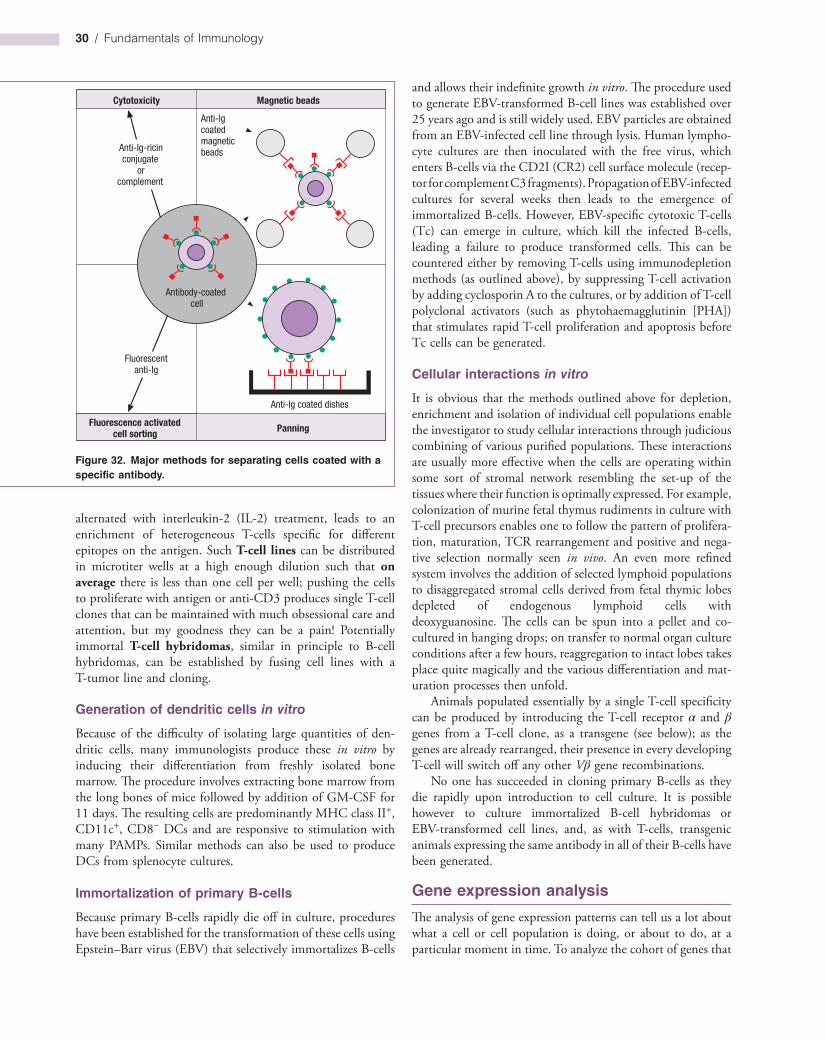
30 / Fundamentals of Immunology
and allows their indefinite growth in vitro. The procedure used to generate EBVtransformed Bcell lines was established over 25 years ago and is still widely used. EBV particles are obtained from an EBVinfected cell line through lysis. Human lymphocyte cultures are then inoculated with the free virus, which enters Bcells via the CD2I (CR2) cell surface molecule (receptor for complement C3 fragments). Propagation of EBVinfected cultures for several weeks then leads to the emergence of immortalized Bcells. However, EBVspecific cytotoxic Tcells (Tc) can emerge in culture, which kill the infected Bcells, leading a failure to produce transformed cells. This can be countered either by removing Tcells using immunodepletion methods (as outlined above), by suppressing Tcell activation by adding cyclosporin A to the cultures, or by addition of Tcell polyclonal activators (such as phytohaemagglutinin [PHA]) that stimulates rapid Tcell proliferation and apoptosis before Tc cells can be generated.
Cellular interactions in vitro
It is obvious that the methods outlined above for depletion, enrichment and isolation of individual cell populations enable the investigator to study cellular interactions through judicious combining of various purified populations. These interactions are usually more effective when the cells are operating within some sort of stromal network resembling the setup of the tissues where their function is optimally expressed. For example, colonization of murine fetal thymus rudiments in culture with Tcell precursors enables one to follow the pattern of proliferation, maturation, TCR rearrangement and positive and negative selection normally seen in vivo. An even more refined system involves the addition of selected lymphoid populations to disaggregated stromal cells derived from fetal thymic lobes depleted of endogenous lymphoid cells with deoxyguanosine. The cells can be spun into a pellet and cocultured in hanging drops; on transfer to normal organ culture conditions after a few hours, reaggregation to intact lobes takes place quite magically and the various differentiation and maturation processes then unfold.
Animals populated essentially by a single Tcell specificity can be produced by introducing the Tcell receptor α and β genes from a Tcell clone, as a transgene (see below); as the genes are already rearranged, their presence in every developing Tcell will switch off any other Vβ gene recombinations.
No one has succeeded in cloning primary Bcells as they die rapidly upon introduction to cell culture. It is possible however to culture immortalized Bcell hybridomas or EBVtransformed cell lines, and, as with Tcells, transgenic animals expressing the same antibody in all of their Bcells have been generated.
Gene expression analysis
The analysis of gene expression patterns can tell us a lot about what a cell or cell population is doing, or about to do, at a particular moment in time. To analyze the cohort of genes that
alternated with interleukin2 (IL2) treatment, leads to an enrichment of heterogeneous Tcells specific for different epitopes on the antigen. Such T-cell lines can be distributed in microtiter wells at a high enough dilution such that on average there is less than one cell per well; pushing the cells to proliferate with antigen or antiCD3 produces single Tcell clones that can be maintained with much obsessional care and attention, but my goodness they can be a pain! Potentially immortal T-cell hybridomas, similar in principle to Bcell hybridomas, can be established by fusing cell lines with a Ttumor line and cloning.
Generation of dendritic cells in vitro
Because of the difficulty of isolating large quantities of dendritic cells, many immunologists produce these in vitro by inducing their differentiation from freshly isolated bone marrow. The procedure involves extracting bone marrow from the long bones of mice followed by addition of GMCSF for 11 days. The resulting cells are predominantly MHC class II+, CD11c+, CD8− DCs and are responsive to stimulation with many PAMPs. Similar methods can also be used to produce DCs from splenocyte cultures.
Immortalization of primary B-cells
Because primary Bcells rapidly die off in culture, procedures have been established for the transformation of these cells using Epstein–Barr virus (EBV) that selectively immortalizes Bcells
Figure 32. Major methods for separating cells coated with a specific antibody.
Cytotoxicity Magnetic beads
Panning
Anti-Ig coated dishes
Anti-Igcoatedmagneticbeads
Fluorescentanti-Ig
Anti-Ig-ricinconjugate
orcomplement
Antibody-coatedcell
Fluorescence activatedcell sorting

Online chapter: Immunological methods and applications / 31
hybridized to the genes on the microarray. This provides a quantitative comparison of expression for every gene present on the chip. By accumulating such data it is possible to build up a complete picture of which genes are expressed in which cells (Figure 34). One area in which this technology is being rapidly deployed is in the analysis of differences in gene expression between a tumor cell and its normal counterpart, thereby illuminating possible targets for therapeutic intervention.
All that glitters is not gold however and it is certainly true to say that DNA microarrays are not a solution to all our problems. Background is a troublesome feature of this type of approach and often threatens to drown out interesting data in a cacophony of experimental noise. Well controlled experimental setups are a must for largescale microarray approaches, otherwise any gene expression differences observed could well be due to slamming the tissue culture incubator door rather than the intended stimulus. The term “garbage in, garbage out” comes to mind in these situations.
are expressed by a cell population, either at a steadystate level or in response to a particular stimulus, messenger RNA (mRNA) is extracted and is analyzed by a method that enables genes of interest to be detected. mRNA can be analyzed by northern blot, in which a single gene probe is hybridized to the mRNA sample, or by reverse transcriptase (RT)primed PCR in which genes of interest can be amplified by initially making a cDNA copy using RT followed by gene amplification by means of specific primers that are complementary to the sequence of interest. While northern blotting and RTPCR can give information concerning more than one transcript, this usually requires significant amounts of mRNA and is relatively slow.
The development of microarray technologies now permits the simultaneous measurement of expression of thousands of genes in a single experiment. Oligonucleotides or cDNA fragments are robotically spotted onto a gene chip and cDNA generated from, for example, Tcell mRNA is labeled and
Figure 33. Separation of activated peripheral blood memory T-cells (CD45RO positive) from naive T-cells (CD45RO negative; but positive for the CD45RA isoform) in the FACS after staining the surface of the living cells in the cold with a fluorescent monoclonal antibody to the CD45RO (see p. 256). The unsorted cells showed two peaks (a); cells with fluorescence intensity lower than the arbitrary gate were separated from those with higher intensity giving (b) negative (CD45RA) and (c) positive (CD45RO) populations, which were each tested for their proliferative response to a mixture of two anti-CD2 monoclonals (OKT11 and GT2) in the presence of 10% antigen-presenting cells (d). 3H-Thymidine was added after 3 days and the cells counted after 15 h. Clearly the memory cell population proliferated, whereas the naive population did not. (Data kindly provided by D. Wallace and R. Hicks.)
Positive sortedNegative sortedUnsorted
Mitogenic response
Coun
ts/m
in
Medium
Mitogenic anti-CD2s
CD45RO activated memory cellsCD45RA naive cells
10 4
c
10 0 10 1 10 2 10 3 10 4
No. o
f cel
ls
Anti-CD45RO relative fluorescence
b
10 0 10 1 10 2 10 3 10 4
No. o
f cel
ls
Anti-CD45RO relative fluorescence
a
10 0 10 1 10 2 10 3
No. o
f cel
ls
Anti-CD45RO relative fluorescence
Gate
d
10 000
20 000
30 000
40 000
50 000
60 000
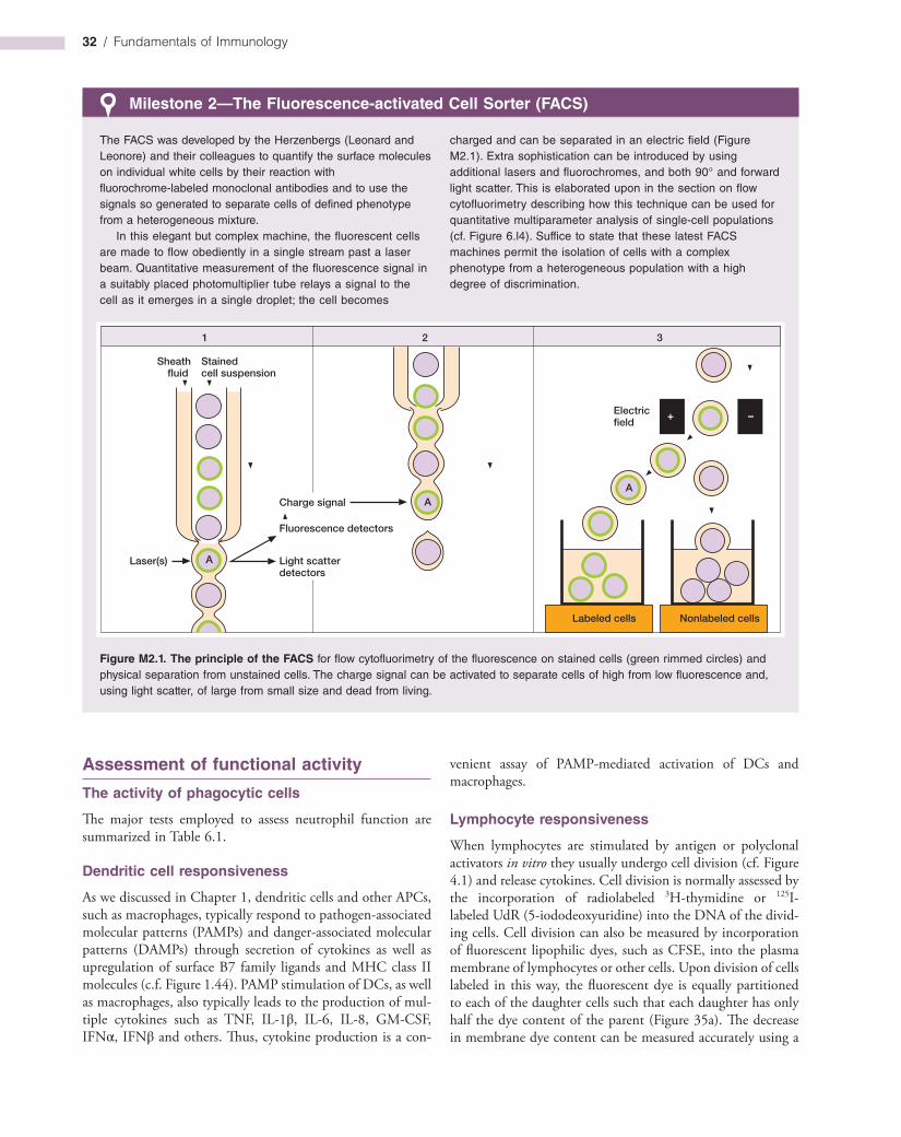
32 / Fundamentals of Immunology
Milestone 2—The Fluorescence-activated Cell Sorter (FACS)
The FACS was developed by the Herzenbergs (Leonard and Leonore) and their colleagues to quantify the surface molecules on individual white cells by their reaction with fluorochrome-labeled monoclonal antibodies and to use the signals so generated to separate cells of defined phenotype from a heterogeneous mixture.
In this elegant but complex machine, the fluorescent cells are made to flow obediently in a single stream past a laser beam. Quantitative measurement of the fluorescence signal in a suitably placed photomultiplier tube relays a signal to the cell as it emerges in a single droplet; the cell becomes
charged and can be separated in an electric field (Figure M2.1). Extra sophistication can be introduced by using additional lasers and fluorochromes, and both 90° and forward light scatter. This is elaborated upon in the section on flow cytofluorimetry describing how this technique can be used for quantitative multiparameter analysis of single-cell populations (cf. Figure 6.l4). Suffice to state that these latest FACS machines permit the isolation of cells with a complex phenotype from a heterogeneous population with a high degree of discrimination.
Figure M2.1. The principle of the FACS for flow cytofluorimetry of the fluorescence on stained cells (green rimmed circles) and physical separation from unstained cells. The charge signal can be activated to separate cells of high from low fluorescence and, using light scatter, of large from small size and dead from living.
Assessment of functional activity
The activity of phagocytic cells
The major tests employed to assess neutrophil function are summarized in Table 6.1.
Dendritic cell responsiveness
As we discussed in Chapter 1, dendritic cells and other APCs, such as macrophages, typically respond to pathogenassociated molecular patterns (PAMPs) and dangerassociated molecular patterns (DAMPs) through secretion of cytokines as well as upregulation of surface B7 family ligands and MHC class II molecules (c.f. Figure 1.44). PAMP stimulation of DCs, as well as macrophages, also typically leads to the production of multiple cytokines such as TNF, IL1β, IL6, IL8, GMCSF, IFNα, IFNβ and others. Thus, cytokine production is a con
venient assay of PAMPmediated activation of DCs and macrophages.
Lymphocyte responsiveness
When lymphocytes are stimulated by antigen or polyclonal activators in vitro they usually undergo cell division (cf. Figure 4.1) and release cytokines. Cell division is normally assessed by the incorporation of radiolabeled 3Hthymidine or 125I labeled UdR (5iododeoxyuridine) into the DNA of the dividing cells. Cell division can also be measured by incorporation of fluorescent lipophilic dyes, such as CFSE, into the plasma membrane of lymphocytes or other cells. Upon division of cells labeled in this way, the fluorescent dye is equally partitioned to each of the daughter cells such that each daughter has only half the dye content of the parent (Figure 35a). The decrease in membrane dye content can be measured accurately using a
Electricfield
+
Sheathfluid
Stainedcell suspension
Laser(s)
Charge signal
Fluorescence detectors
Light scatterdetectors
1 2 3
AA
A
Labeled cells Nonlabeled cells
–
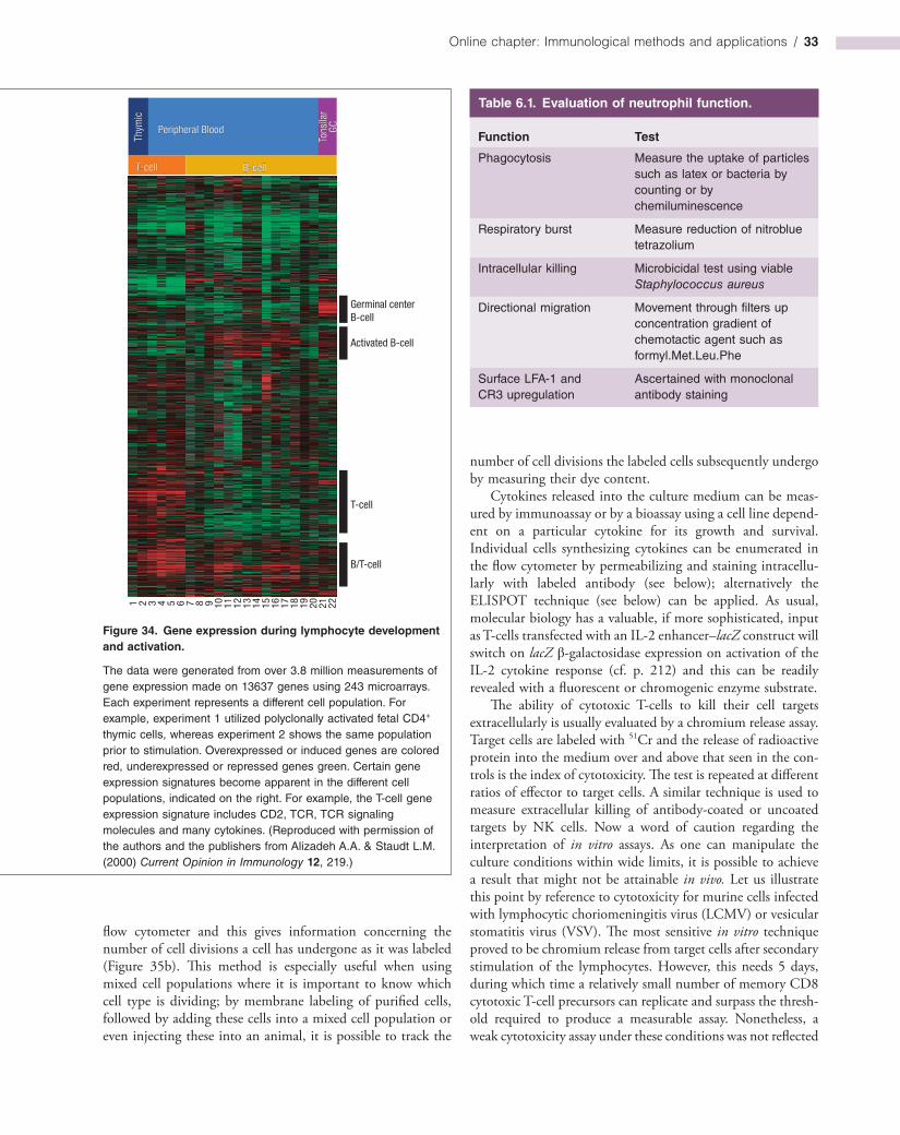
Online chapter: Immunological methods and applications / 33
number of cell divisions the labeled cells subsequently undergo by measuring their dye content.
Cytokines released into the culture medium can be measured by immunoassay or by a bioassay using a cell line dependent on a particular cytokine for its growth and survival. Individual cells synthesizing cytokines can be enumerated in the flow cytometer by permeabilizing and staining intracellularly with labeled antibody (see below); alternatively the ELISPOT technique (see below) can be applied. As usual, molecular biology has a valuable, if more sophisticated, input as Tcells transfected with an IL2 enhancer–lacZ construct will switch on lacZ βgalactosidase expression on activation of the IL2 cytokine response (cf. p. 212) and this can be readily revealed with a fluorescent or chromogenic enzyme substrate.
The ability of cytotoxic Tcells to kill their cell targets extracellularly is usually evaluated by a chromium release assay. Target cells are labeled with 51Cr and the release of radioactive protein into the medium over and above that seen in the controls is the index of cytotoxicity. The test is repeated at different ratios of effector to target cells. A similar technique is used to measure extracellular killing of antibodycoated or uncoated targets by NK cells. Now a word of caution regarding the interpretation of in vitro assays. As one can manipulate the culture conditions within wide limits, it is possible to achieve a result that might not be attainable in vivo. Let us illustrate this point by reference to cytotoxicity for murine cells infected with lymphocytic choriomeningitis virus (LCMV) or vesicular stomatitis virus (VSV). The most sensitive in vitro technique proved to be chromium release from target cells after secondary stimulation of the lymphocytes. However, this needs 5 days, during which time a relatively small number of memory CD8 cytotoxic Tcell precursors can replicate and surpass the threshold required to produce a measurable assay. Nonetheless, a weak cytotoxicity assay under these conditions was not reflected
flow cytometer and this gives information concerning the number of cell divisions a cell has undergone as it was labeled (Figure 35b). This method is especially useful when using mixed cell populations where it is important to know which cell type is dividing; by membrane labeling of purified cells, followed by adding these cells into a mixed cell population or even injecting these into an animal, it is possible to track the
Figure 34. Gene expression during lymphocyte development and activation.
The data were generated from over 3.8 million measurements of gene expression made on 13637 genes using 243 microarrays. Each experiment represents a different cell population. For example, experiment 1 utilized polyclonally activated fetal CD4+ thymic cells, whereas experiment 2 shows the same population prior to stimulation. Overexpressed or induced genes are colored red, underexpressed or repressed genes green. Certain gene expression signatures become apparent in the different cell populations, indicated on the right. For example, the T-cell gene expression signature includes CD2, TCR, TCR signaling molecules and many cytokines. (Reproduced with permission of the authors and the publishers from Alizadeh A.A. & Staudt L.M. (2000) Current Opinion in Immunology 12, 219.)
T-cell B-cell
Thym
icPeripheral Blood
Tons
ilar
GC
1 2 3 4 5 6 7 8 9 10 11 12 13 14 15 16 17 18 19 20 21 22
Germinal centerB-cell
Activated B-cell
T-cell
B/T-cell
Table 6.1. Evaluation of neutrophil function.
Function Test
Phagocytosis Measure the uptake of particles such as latex or bacteria by counting or by chemiluminescence
Respiratory burst Measure reduction of nitroblue tetrazolium
Intracellular killing Microbicidal test using viable Staphylococcus aureus
Directional migration Movement through filters up concentration gradient of chemotactic agent such as formyl.Met.Leu.Phe
Surface LFA-1 and CR3 upregulation
Ascertained with monoclonal antibody staining
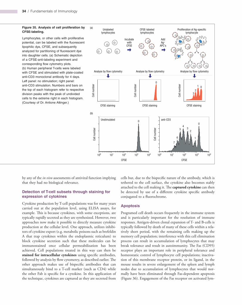
34 / Fundamentals of Immunology
cells but, due to the bispecific nature of the antibody, which is tethered to the cell surface, the cytokine also becomes stably attached to the cell making it. The captured cytokine can then be detected by use of a different cytokine specific antibody conjugated to a fluorochrome.
Apoptosis
Programed cell death occurs frequently in the immune system and is particularly important for the resolution of immune responses. Antigendriven clonal expansion of T and Bcells is typically followed by death of many of these cells within a relatively short period, with the remaining cells making up the memory cell population; interference with this cell elimination process can result in accumulation of lymphocytes that may break tolerance and result in autoimmunity. The Fas (CD95) receptor plays an important role in peripheral tolerance and homeostatic control of lymphocyte cell populations; inactivation of this membrane receptor protein, or its ligand, in the mouse results in severe enlargement of the spleen and lymph nodes due to accumulation of lymphocytes that would normally have been eliminated through Fasdependent apoptosis (Figure 36). Engagement of the Fas receptor on activated lym
Figure 35. Analysis of cell proliferation by CFSE-labeling.
Lymphocytes, or other cells with proliferative potential, can be labeled with the fluorescent lipophilic dye, CFSE, and subsequently analyzed for partitioning of fluorescent dye into daughter cells. (a) Schematic depiction of a CFSE-anti-labeling experiment and corresponding flow cytometry plots. (b) Human peripheral T-cells were labeled with CFSE and stimulated with plate-coated anti-CD3 monoclonal antibody for 4 days. Left panel: no stimulation; right panel: anti-CD3 stimulation. Numbers and bars on the top of each histogram refer to respective division peaks with the peak of undivided cells to the extreme right in each histogram. (Courtesy of Dr. Antione Attinger.)
Unlabeledlymphocytes
Analyse by flow cytometry
CFSE-labeledlymphocytes
Proliferation of Ag-specificlymphocyte
IncubatewithCFSE
CFSE staining
Cell
num
ber
AddAg+
APC’s
Analyse by flow cytometry
CFSE staining
Cell
num
ber
Analyse by flow cytometry
CFSE staining
Cell
num
ber0
01
23
104103102101100
CFSE
104103102101100
CFSE
Unstimulated anti-CD3
Cell
num
ber
Cell
num
ber
(a)
(b)
00
1
23
45
6
7
by any of the in vivo assessments of antiviral function implying that they had no biological relevance.
Detection of T-cell subsets through staining for expression of cytokines
Cytokine production by Tcell populations was for many years carried out at the population level, using ELISA assays, for example. This is because cytokines, with some exceptions, are typically rapidly secreted as they are synthesized. However, two approaches now make it possible to directly measure cytokine production at the cellular level. One approach, utilizes inhibitors of cytokine export (e.g. metabolic poisons such as brefeldin A that trap cytokines within the endoplasmic reticulum) to block cytokine secretion such that these molecules can be immunostained once cellular permeabilization has been achieved. Cell populations treated in this way can then be stained for intracellular cytokines using specific antibodies, followed by analysis by flow cytometry, as described earlier. The other approach makes use of bispecific antibodies that can simultaneously bind to a Tcell marker (such as CD4) while the other Fab is specific for a cytokine. In this application of the technique, cytokines are captured as they are secreted from

Online chapter: Immunological methods and applications / 35
phospholipid that is normally confined to the inner leaflet of the plasma membrane, becomes exposed on the outer leaflet during apoptosis. This can be readily detected using fluorescently labeled annexin V, a PSbinding protein; apoptotic cells display markedly enhanced binding of annexin V relative to healthy cells (Figure 37).
Other assays take advantage of the fact that extensive DNA fragmentation is also a common feature of apoptosis and this can be assessed by agarose gel electrophoresis of DNA extracted from apoptotic cells or the TUNEL (TdTmediated dUTP (deoxyuridine triphosphate) nick end labeling) assay; the latter assay utilizes the enzyme terminal deoxynucleotidyl transferase (TdT) to add biotinylated nucleotides to the 3′ ends of DNA fragments and this can then be detected using fluorescently labeled streptavidin. Several members of the caspase family of cysteine proteases become activated during apoptosis and this can be assessed by immunoblot analysis (Figure 19) or by using labeled synthetic substrate peptides that can be cleaved by active caspases.
Precursor frequency
The magnitude of lymphocyte responses in culture is closely related to the number of antigenspecific lymphocytes capable of responding. Because of the clonality of the responses, it is possible to estimate the frequency of these antigenspecific precursors by limiting dilution analysis. In essence, the method depends upon the fact that, if one takes several replicate aliquots of a given cell suspension that would be expected to contain on average one precursor per aliquot, then Poisson distribution analysis shows that 37% of the aliquots will contain no precursor cells (through the randomness of the sampling). Thus, if aliquots are made from a series of dilutions of a cell suspension and incubated under conditions that allow the precursors to mature and be recognized through some amplification scheme, the dilution at which 37% of the aliquots give negative responses will be known to contain an average of one precursor cell per aliquot, and one can therefore calculate the precursor frequency in the original cell suspension. An example is shown in some detail in Figure 38.
phocytes normally results in rapid induction of apoptosis in these cells (Figure 7). Cytotoxic Tcells also eliminate target cells by inducing apoptosis through a variety of strategies. Apoptosis is also important in shaping the T and Bcell repertoires; negative selection of both lymphocyte populations involves triggering apoptosis.
A variety of approaches can be used to measure apoptosis, ranging from morphological assessment (see Figure 7) or by exploiting biochemical alterations to the cell that occurs during this process. One of the most widely used assays for apoptosis takes advantage of the fact that phosphatidylserine (PS), a
Figure 36. Gross enlargement of spleen and lymph nodes from Fas “knockout” mice.
Lymph nodes and spleen from wild type versus Fas knockout mice are compared. Both organs are increased approximately 20-fold in size in the knockout due to accumulation of excess T- and B-cells due to a failure of peripheral deletion in these animals. (Kindly provided by Professor Shigekazu Nagata and adapted from Adachi M. et al., 1995 Nature Genetics 11, 294, with permission.)
Wild-type
Fas knockout
Lymphnodes Spleen
Figure 37. Analysis of apoptosis by Annexin V-labeling.
Phosphatidylserine (PS) is externalized on the outer leaflet of the plasma membrane during apoptosis and this can be readily detected using the PS-binding protein, Annexin V. (a) Untreated human T-lymphoblastoid cells; and (b) apoptotic T-lymphoblastoid cells were stained with FITC-conjugated annexin V. (Data kindly provided by Dr. Gabriela Brumatti.)
104103102101100
FL1-H
104103102101100
FL1-H
Annexin V FITCAnnexin V FITC
Coun
ts
Coun
ts
(a) (b)

36 / Fundamentals of Immunology
which the TCR binds, and mixed with fluorescently labeled streptavidin, which not only binds biotin with a very high affinity but also has a valency of four with respect to the biotin—hence the formation of tetramers.
Numerous adaptations of this technology are appearing. For example, incubation of tetramers bound to their cognate TCR leads to internalization at 37°C; by tagging them with a toxin individual Tlymphocytes of a single specificity can be eliminated. Another approach is to use the FACS to directly sort stained cells into an ELISPOT microtiter plate in which cytokine secretion is measured, providing a functional analysis of the cells.
Enumeration of antibody-forming cells
The immunofluorescence sandwich test
This is a doublelayer procedure designed to visualize specific intracellular antibody. If, for example, we wished to see how many cells in a preparation of lymphoid tissue were synthesizing antibody to pneumococcus polysaccharide, we would first fix the cells with ethanol to prevent the antibody being washed away during the test, and then treat with a solution of the
It has been argued that limiting dilution analysis often underestimates the true precursor frequency. This is likely because cells generally do not survive very well when cultured in isolation (i.e. as a single cell per well) because most cells, with few exceptions, require signals from other cells to survive. Martin Raff showed that in the absence of such signals cells typically undergo apoptosis. An accurate measure of the percentage of lymphocytes bearing a specific antigen receptor can be obtained by flow cytometry of cells stained with labeled antigen. In the case of Bcells this is fairly straightforward, given that their antigen receptors recognize native antigen. However, it is only recently that technical finesse, in the form of peptide–MHC tetramers, has brought this technique to Tcells (Figure 39). This approach overcomes the problem of the relatively weak intrinsic affinity of TCR for peptide–MHC by presenting a tagged peptide–MHC as a multivalent tetramer, thereby exploiting the bonus effect of multivalency (cf. p. 122). Peptide–MHC complexes are produced by permitting recombinant MHC molecules to refold with the appropriate synthetic peptide. The recombinant MHC molecules are biotinylated on a special carboxyterminal extension, that ensures that the biotin is incorporated at a distance from the site to
Figure 38. Limiting dilution analysis of cytotoxic T-cell precursor frequency in spleen cells from a BALB/c mouse stimulated with irradiated C57BL/6 spleen cells as antigen. BALB/c splenic responder cells were set up in 24 replicates at each concentration tested together with antigen and an excess of T-helper factors. The generation of cytotoxicity in each well was looked for by adding 51Cr-labeled tumor cells (EL-4) of the C57BL/6 haplotype; cytotoxicity was then revealed by measuring the release of soluble 51Cr-labeled intracellular material into the medium. (a) The points show the percentage of specific lysis of individual wells. The dashed line indicates three standard deviations above the medium release control, and each point above that line is counted as positive for cytotoxicity. (b) The data replotted in terms of the percentage of negative wells at each concentration of responder cells over the range in which the data titrated (5 × 10⁻³/well to 0.625 × 10⁻³/well). The dashed line is drawn at 37% negative wells and this intersects the regression line to give a precursor (Tcp) frequency of 1 in 2327 responder cells. The regression line has an r2 value of 1.00 in this experiment. (Reproduced with permission from Simpson E. & Chandler P. (1986) In: Weir D.M. (ed.) Handbook of Experimental Immunology, Figure 68.2. Blackwell Scientific Publications, Oxford.)
40 20 10No. of responder cells per well x10–3
No. of responder cells per well x 10–3
2.5 1.25 0.625 0.3125
010203040506070
0102030405060
-10
a
Spec
ific
lysi
s (%
)
10
20
40
60
80
1004321
b
Nega
tive
wel
ls (%
)
No. of responder cells per well x10–3
5 5
5

Online chapter: Immunological methods and applications / 37
Figure 39. Peptide–MHC tetramer.
A single fluorochrome-labeled peptide–MHC complex (top right) has only a low affinity for the TCR and therefore provides a very insensitive probe for its cognate receptor. However, by biotinylating (•) the MHC molecules and then mixing them with streptavidin, which has a valency of four with respect to biotin binding, a tetrameric complex is formed which has a much higher functional affinity (avidity) when used as a probe for the specific TCRs on the T-cell surface.
Tc
Labeledpeptide–MHC
High avidityof peptide–MHCtetramer
Labeledstreptavidin
Figure 40. Jerne plaque technique for enumerating antibody-forming cells (Cunningham modification).
Complementa
Secreted antibody coats surrounding red cells Coated erythrocytes are lysed by complement toform plaque with antibody-forming cell at center
Ab
b
(a) The direct technique for cells synthesizing IgM hemolysin is shown. The indirect technique for visualizing cells producing IgG hemolysins requires the addition of anti-IgG to the system. The difference between the plaques obtained by direct and indirect methods gives the number of “IgG” plaques. The reverse plaque assay enumerates total Ig-producing cells by capturing secreted Ig on red cells coated with anti-Ig. Multiple plaque
polysaccharide antigen. After washing, a fluoresceinlabeled antibody to the polysaccharide would then be added to locate those cells that had specifically bound the antigen.
The name of the test derives from the fact that antigen is sandwiched between the antibody present in the cell substrate and that added as the second layer (Figure 8c).
Plaque techniques
Antibodysecreting cells can be counted by diluting them in an environment in which the antibody formed by each individual cell produces a readily observable effect. In one technique, developed from the original method of Niels Jerne and Albert Nordin, the cells from an animal immunized with sheep erythrocytes are suspended together with an excess of sheep red cells and complement within a shallow chamber formed between two microscope slides. On incubation, the antibodyforming cells release their immunoglobulin that coats the surrounding erythrocytes. The complement will then cause lysis of the coated cells and a plaque clear of red cells will be seen around each antibodyforming cell (Figure 40). Direct plaques obtained in this way largely reveal IgM producers as this antibody has a high hemolytic efficiency. To demonstrate IgG synthesizing cells it is necessary to increase the complement binding of the erythrocyte–IgG antibody complex by adding a rabbit antiIgG serum; the “indirect plaques” thus developed can be used to enumerate cells making antibodies in different immunoglobulin subclasses, provided that the appropriate rabbit antisera are available. The method can be extended by coating an antigen such as pneumococcus
assays can be carried out by a modification using microtiter plates. (b) Photograph of plaques that show as circular dark areas (some of which are arrowed) under dark-ground illumination. They vary in size depending upon the antibody affinity and the rate of secretion by the antibody-forming cell. (Courtesy of C. Shapland, P. Hutchings and Professor D. Male.)
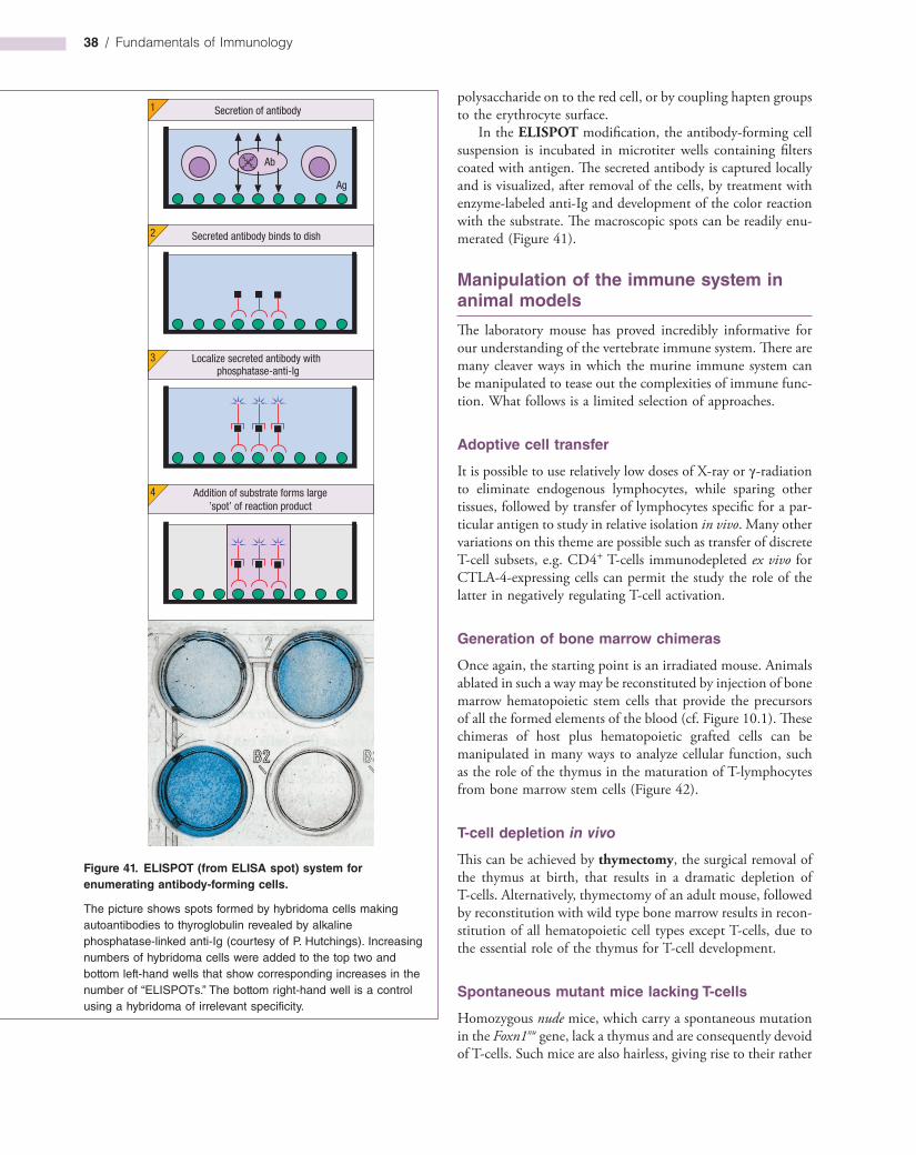
38 / Fundamentals of Immunology
polysaccharide on to the red cell, or by coupling hapten groups to the erythrocyte surface.
In the ELISPOT modification, the antibodyforming cell suspension is incubated in microtiter wells containing filters coated with antigen. The secreted antibody is captured locally and is visualized, after removal of the cells, by treatment with enzymelabeled antiIg and development of the color reaction with the substrate. The macroscopic spots can be readily enumerated (Figure 41).
Manipulation of the immune system in animal models
The laboratory mouse has proved incredibly informative for our understanding of the vertebrate immune system. There are many cleaver ways in which the murine immune system can be manipulated to tease out the complexities of immune function. What follows is a limited selection of approaches.
Adoptive cell transfer
It is possible to use relatively low doses of Xray or γradiation to eliminate endogenous lymphocytes, while sparing other tissues, followed by transfer of lymphocytes specific for a particular antigen to study in relative isolation in vivo. Many other variations on this theme are possible such as transfer of discrete Tcell subsets, e.g. CD4+ Tcells immunodepleted ex vivo for CTLA4expressing cells can permit the study the role of the latter in negatively regulating Tcell activation.
Generation of bone marrow chimeras
Once again, the starting point is an irradiated mouse. Animals ablated in such a way may be reconstituted by injection of bone marrow hematopoietic stem cells that provide the precursors of all the formed elements of the blood (cf. Figure 10.1). These chimeras of host plus hematopoietic grafted cells can be manipulated in many ways to analyze cellular function, such as the role of the thymus in the maturation of Tlymphocytes from bone marrow stem cells (Figure 42).
T-cell depletion in vivo
This can be achieved by thymectomy, the surgical removal of the thymus at birth, that results in a dramatic depletion of Tcells. Alternatively, thymectomy of an adult mouse, followed by reconstitution with wild type bone marrow results in reconstitution of all hematopoietic cell types except Tcells, due to the essential role of the thymus for Tcell development.
Spontaneous mutant mice lacking T-cells
Homozygous nude mice, which carry a spontaneous mutation in the Foxn1nu gene, lack a thymus and are consequently devoid of Tcells. Such mice are also hairless, giving rise to their rather
Figure 41. ELISPOT (from ELISA spot) system for enumerating antibody-forming cells.
The picture shows spots formed by hybridoma cells making autoantibodies to thyroglobulin revealed by alkaline phosphatase-linked anti-Ig (courtesy of P. Hutchings). Increasing numbers of hybridoma cells were added to the top two and bottom left-hand wells that show corresponding increases in the number of “ELISPOTs.” The bottom right-hand well is a control using a hybridoma of irrelevant specificity.
Secreted antibody binds to dish2
Localize secreted antibody withphosphatase-anti-Ig
3
Addition of substrate forms large’spot’ of reaction product
4
Ab
Ag
Secretion of antibody1
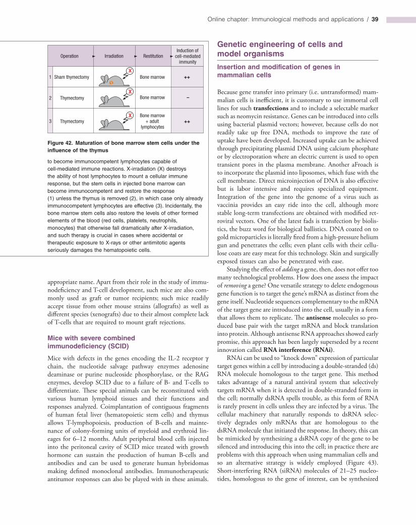
Online chapter: Immunological methods and applications / 39
Figure 42. Maturation of bone marrow stem cells under the influence of the thymus
to become immunocompetent lymphocytes capable of cell-mediated immune reactions. X-irradiation (X) destroys the ability of host lymphocytes to mount a cellular immune response, but the stem cells in injected bone marrow can become immunocompetent and restore the response (1) unless the thymus is removed (2), in which case only already immunocompetent lymphocytes are effective (3). Incidentally, the bone marrow stem cells also restore the levels of other formed elements of the blood (red cells, platelets, neutrophils, monocytes) that otherwise fall dramatically after X-irradiation, and such therapy is crucial in cases where accidental or therapeutic exposure to X-rays or other antimitotic agents seriously damages the hematopoietic cells.
Operation Irradiation RestitutionInduction of
cell-mediatedimmunity
Sham thymectomy
Thymectomy
Thymectomy
1
2
3
Bone marrow
Bone marrow
Bone marrow+ adult
lymphocytes
++
–
++
Genetic engineering of cells and model organisms
Insertion and modification of genes in mammalian cells
Because gene transfer into primary (i.e. untransformed) mammalian cells is inefficient, it is customary to use immortal cell lines for such transfections and to include a selectable marker such as neomycin resistance. Genes can be introduced into cells using bacterial plasmid vectors; however, because cells do not readily take up free DNA, methods to improve the rate of uptake have been developed. Increased uptake can be achieved through precipitating plasmid DNA using calcium phosphate or by electroporation where an electric current is used to open transient pores in the plasma membrane. Another aFroach is to incorporate the plasmid into liposomes, which fuse with the cell membrane. Direct microinjection of DNA is also effective but is labor intensive and requires specialized equipment. Integration of the gene into the genome of a virus such as vaccinia provides an easy ride into the cell, although more stable longterm transfections are obtained with modified retroviral vectors. One of the latest fads is transfection by biolistics, the buzz word for biological ballistics. DNA coated on to gold microparticles is literally fired from a highpressure helium gun and penetrates the cells; even plant cells with their cellulose coats are easy meat for this technology. Skin and surgically exposed tissues can also be penetrated with ease.
Studying the effect of adding a gene, then, does not offer too many technological problems. How does one assess the impact of removing a gene? One versatile strategy to delete endogenous gene function is to target the gene’s mRNA as distinct from the gene itself. Nucleotide sequences complementary to the mRNA of the target gene are introduced into the cell, usually in a form that allows them to replicate. The antisense molecules so produced base pair with the target mRNA and block translation into protein. Although antisense RNA approaches showed early promise, this approach has been largely superseded by a recent innovation called RNA interference (RNAi).
RNAi can be used to “knock down” expression of particular target genes within a cell by introducing a doublestranded (ds) RNA molecule homologous to the target gene. This method takes advantage of a natural antiviral system that selectively targets mRNA when it is detected in doublestranded form in the cell; normally dsRNA spells trouble, as this form of RNA is rarely present in cells unless they are infected by a virus. The cellular machinery that naturally responds to dsRNA selectively degrades only mRNAs that are homologous to the dsRNA molecule that initiated the response. In theory, this can be mimicked by synthesizing a dsRNA copy of the gene to be silenced and introducing this into the cell; in practice there are problems with this approach when using mammalian cells and so an alternative strategy is widely employed (Figure 43). Shortinterfering RNA (siRNA) molecules of 21–25 nucleotides, homologous to the gene of interest, can be synthesized
appropriate name. Apart from their role in the study of immunodeficiency and Tcell development, such mice are also commonly used as graft or tumor recipients; such mice readily accept tissue from other mouse strains (allografts) as well as different species (xenografts) due to their almost complete lack of Tcells that are required to mount graft rejections.
Mice with severe combined immunodeficiency (SCID)
Mice with defects in the genes encoding the IL2 receptor γ chain, the nucleotide salvage pathway enzymes adenosine deaminase or purine nucleoside phosphorylase, or the RAG enzymes, develop SCID due to a failure of B and Tcells to differentiate. These special animals can be reconstituted with various human lymphoid tissues and their functions and responses analyzed. Coimplantation of contiguous fragments of human fetal liver (hematopoietic stem cells) and thymus allows Tlymphopoiesis, production of Bcells and maintenance of colonyforming units of myeloid and erythroid lineages for 6–12 months. Adult peripheral blood cells injected into the peritoneal cavity of SCID mice treated with growth hormone can sustain the production of human Bcells and antibodies and can be used to generate human hybridomas making defined monoclonal antibodies. Immunotherapeutic antitumor responses can also be played with in these animals.
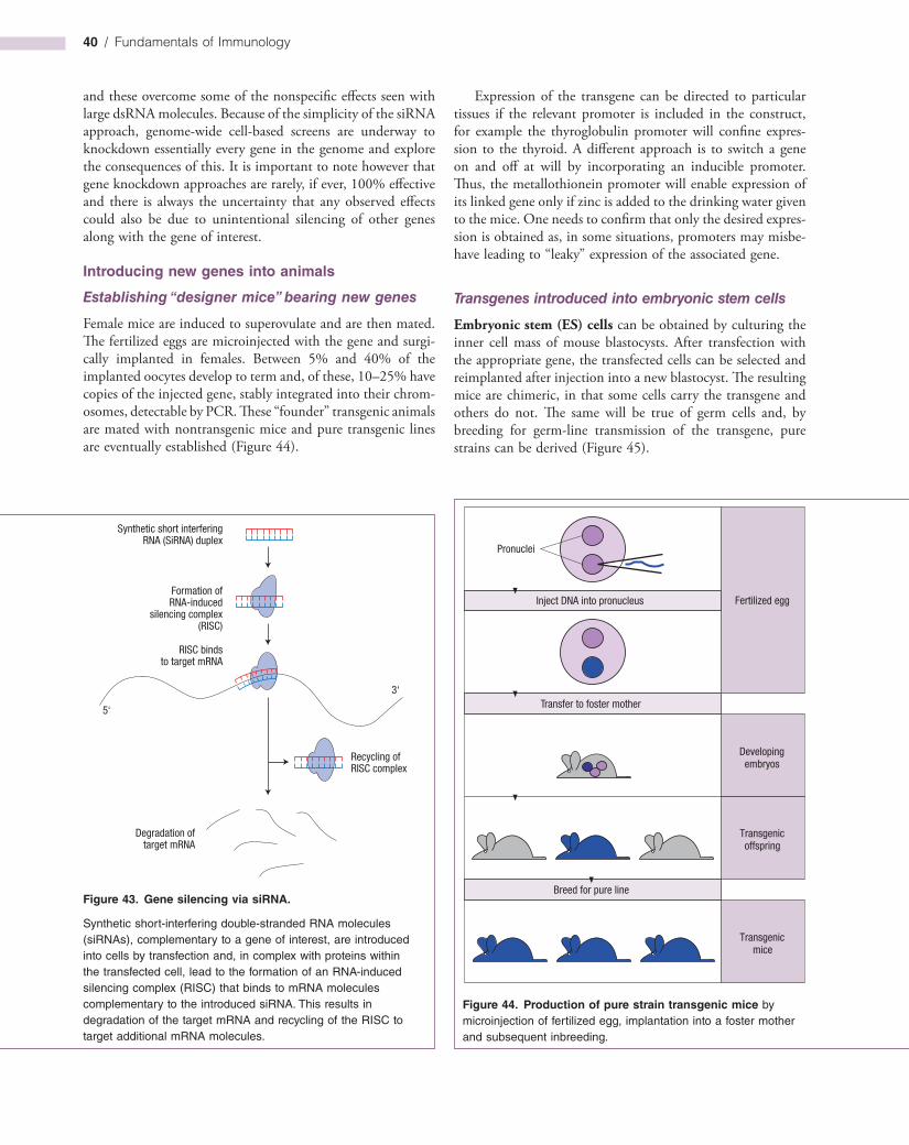
40 / Fundamentals of Immunology
Expression of the transgene can be directed to particular tissues if the relevant promoter is included in the construct, for example the thyroglobulin promoter will confine expression to the thyroid. A different approach is to switch a gene on and off at will by incorporating an inducible promoter. Thus, the metallothionein promoter will enable expression of its linked gene only if zinc is added to the drinking water given to the mice. One needs to confirm that only the desired expression is obtained as, in some situations, promoters may misbehave leading to “leaky” expression of the associated gene.
Transgenes introduced into embryonic stem cells
Embryonic stem (ES) cells can be obtained by culturing the inner cell mass of mouse blastocysts. After transfection with the appropriate gene, the transfected cells can be selected and reimplanted after injection into a new blastocyst. The resulting mice are chimeric, in that some cells carry the transgene and others do not. The same will be true of germ cells and, by breeding for germline transmission of the transgene, pure strains can be derived (Figure 45).
and these overcome some of the nonspecific effects seen with large dsRNA molecules. Because of the simplicity of the siRNA approach, genomewide cellbased screens are underway to knockdown essentially every gene in the genome and explore the consequences of this. It is important to note however that gene knockdown approaches are rarely, if ever, 100% effective and there is always the uncertainty that any observed effects could also be due to unintentional silencing of other genes along with the gene of interest.
Introducing new genes into animals
Establishing “designer mice” bearing new genes
Female mice are induced to superovulate and are then mated. The fertilized eggs are microinjected with the gene and surgically implanted in females. Between 5% and 40% of the implanted oocytes develop to term and, of these, 10–25% have copies of the injected gene, stably integrated into their chromosomes, detectable by PCR. These “founder” transgenic animals are mated with nontransgenic mice and pure transgenic lines are eventually established (Figure 44).
Figure 44. Production of pure strain transgenic mice by microinjection of fertilized egg, implantation into a foster mother and subsequent inbreeding.
Fertilized egg
Developingembryos
Transgenicoffspring
Transgenicmice
Pronuclei
Inject DNA into pronucleus
Transfer to foster mother
Breed for pure line
Synthetic short interferingRNA (SiRNA) duplex
Formation ofRNA-induced
silencing complex(RISC)
Recycling ofRISC complex
Degradation oftarget mRNA
RISC bindsto target mRNA
3‘
5‘
Figure 43. Gene silencing via siRNA.
Synthetic short-interfering double-stranded RNA molecules (siRNAs), complementary to a gene of interest, are introduced into cells by transfection and, in complex with proteins within the transfected cell, lead to the formation of an RNA-induced silencing complex (RISC) that binds to mRNA molecules complementary to the introduced siRNA. This results in degradation of the target mRNA and recycling of the RISC to target additional mRNA molecules.
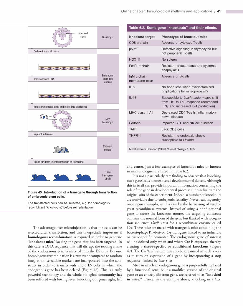
Online chapter: Immunological methods and applications / 41
Figure 45. Introduction of a transgene through transfection of embryonic stem cells.
The transfected cells can be selected, e.g. for homologous recombinant “knockouts,” before reimplantation.
‘Pure’transgenic
strain
Transfect with DNA
Chimericmouse
Newblastocyst
Embryonicstem cellculture
BlastocystInner cell
mass
Culture inner cell mass
Select transfected cells and inject into blastocyst
Implant in female
Breed for germ line transmission of transgene
Table 6.2. Some gene “knockouts” and their effects.
Knockout target Phenotype of knockout mice
CD8 α-chain Absence of cytotoxic T-cells
p59fynT Defective signaling in thymocytes but not peripheral T-cells
HOX 11 No spleen
FcεRI α-chain Resistant to cutaneous and systemic anaphylaxis
IgM μ-chain membrane exon
Absence of B-cells
IL-6 No bone loss when ovariectomized (implications for osteoporosis?)
IL-18 Susceptible to Leishmania major; shift from Th1 to Th2 response (decreased IFNγ and increased IL-4 production)
MHC class II Aβ Decreased CD4 T-cells; inflammatory bowel disease
Perforin Impaired CTL and NK cell function
TAP1 Lack CD8 cells
TNFR-1 Resistant to endotoxic shock; susceptible to Listeria
Modified from Brandon (1995) Current Biology 5, 625.
The advantage over microinjection is that the cells can be selected after transfection, and this is especially important if homologous recombination is required in order to generate “knockout mice” lacking the gene that has been targeted. In this case, a DNA sequence that will disrupt the reading frame of the endogenous gene is inserted into the ES cells. Because homologous recombination is a rare event compared to random integration, selectable markers are incorporated into the construct in order to transfer only those ES cells in which the endogenous gene has been deleted (Figure 46). This is a truly powerful technology and the whole biological community has been suffused with boxing fever, knocking out genes right, left
and center. Just a few examples of knockout mice of interest to immunologists are listed in Table 6.2.
It is not a particularly rare finding to observe that knocking out a gene leads to unexpected developmental defects. Although this in itself can provide important information concerning the role of the gene in developmental processes, it can frustrate the original aim of the experiment. Indeed, a number of knockouts are nonviable due to embryonic lethality. Never fear, ingenuity once again triumphs, in this case by the harnessing of viral or yeast recombinase systems. Instead of using a nonfunctional gene to create the knockout mouse, the targeting construct contains the normal form of the gene but flanked with recognition sequences (loxP sites) for a recombinase enzyme called Cre. These mice are mated with transgenic mice containing the bacteriophage P1derived Cre transgene linked to an inducible or tissuespecific promoter. The endogenous gene of interest will be deleted only when and where Cre is expressed thereby creating a tissue-specific or conditional knockout (Figure 47). The Cre/loxP system can also be organized in such a way as to turn on expression of a gene by incorporating a stop sequence flanked by loxP sites.
Mice in which an endogenous gene is purposefully replaced by a functional gene, be it a modified version of the original gene or an entirely different gene, are referred to as “knocked in mice.” Hence, in the example above, knocking in a loxP

42 / Fundamentals of Immunology
Figure 47. Conditional knockout.
RAg-1 B7.2
tk B7.2 neo R
B7.2 neo R
B7.2 neo R
Targetingsequence
Endogenousgene
Modified butfunctionalendogenousgene
B-cell-specificknockout ofB7.2
Plasmid
Chromosome
Chromosome
Chromosome
Deleted
Recombinase
Ig-Promoter Cre Ig-Enhancer
B-cell-specifictranscription factors
Genedeletion
Homologousrecombination
LoxP LoxP
The endogenous gene that is under study (here B7.2) is homologously replaced in ES cells with an identical gene, as in Figure 46, but here flanked by loxP sequences (brown boxes) and with the neoR gene incorporated in a nondisruptive manner purely for selection purposes. Nonhomologous recombinants will contain the tk gene and are eliminated using ganciclovir. Transgenic animals are then generated from ES cells that are resistant to G418. If homozygous B7.2–loxP
Targetingsequencecontainingnonfunctionalgene
Endogenousnormal genein ES cells
Knocked outgene in ES cells
Plasmid
Chromosome
Chromosome
RAg-1 RAG-1
tk 5’ neo R 3’
5’ neo R
RAG-1
Homologousrecombination
3’ RAG-1
Figure 46. Gene disruption by homologous recombination with plasmid DNA containing a copy of the gene of interest (in this example RAG-1) into which a sequence specifying neomycin resistance (neoR) has been inserted in such a way as to destroy the RAG-1 reading frame between the 5′ and 3′ ends of the gene. Embryonic stem (ES) cells in which the targeting sequence has been incorporated into the chromosomal DNA by homologous recombination will be resistant to the neomycin analog G418. Stem cells in which nonhomologous recombination into chromosomal DNA has occurred would additionally incorporate the thymidine kinase (tk) gene that can be used to destroy such cells by culturing them in the presence of ganciclovir, leaving only ES cells in which homologous recombination has been achieved. These are then used to create a knockout mouse.
transgenics are mated with mice that contain a transgene for the Cre recombinase under the control of specific regulatory elements, only those cells in which the promoter is active will produce the Cre enzyme necessary to delete the sequence flanked by loxP. The example given would represent an experiment aimed at investigating the effect of specifically knocking out B7.2 in B-cells whilst maintaining its expression in, for example, dendritic cells.
Online chapter: Immunological methods and applications / 43
Summar
yMaking antibodies to order■ Polyclonal antisera can be generated by repeated
immunization with antigen.■ Polyclonal antibodies recognize a mixture of
determinants on the antigen.■ Adjuvants are required for efficient immune responses
to antigen.■ Immortal hybridoma cell lines making monoclonal
antibodies provide powerful immunological reagents and insights into the immune response. Applications include enumeration of lymphocyte subpopulations, cell depletion, immunoassay, cancer diagnosis and imaging, purification of antigen from complex mixtures, and recently the use of monoclonals as artificial enzymes (catalytic antibodies).
■ Genetically engineered human antibody fragments can be derived by expanding the VH and VL genes from unimmunized, but preferably immunized, donors and expressing them as completely randomized combinatorial libraries on the surface of bacteriophage. Phages bearing the highest affinity antibodies are selected by panning on antigens and the antibody genes can then be cloned from the isolated viruses.
■ Single-chain Fv (scFv) fragments encoded by linked VH and VL genes and even single heavy chain domains can be created.
■ The human anti-mouse antibody (HAMA) response is a significant obstacle to use of mouse monoclonal antibodies for therapeutic purposes.
■ The HAMA response against mouse monoclonal antibodies can be reduced by producing chimeric antibodies with mouse variable regions and human constant regions or, better still, using humanized antibodies in which all the mouse sequences except for the CDRs are replaced by human sequences.
■ Humanized antibodies are now in clinical use for the treatment of a variety of conditions such as rheumatoid arthritis and B-cell lymphoma.
■ Transgenic mice bearing human Ig genes can be immunized. The mice produce high affinity fully human antibodies.
■ Recombinant antibodies can be expressed on a large scale in plants.
■ Combinatorial libraries of diabodies containing the H1 and H2 VH CDR may be used to develop new drugs.
Purification of antigen and antibody by affinity chromatography■ Insoluble immunoabsorbents prepared by coupling
antibody to Sepharose can be used to affinity-purify antigens from complex mixtures and reciprocally to purify antibodies.
■ Affinity chromatography can also be used to co- purify proteins that serve as binding partners of antigens.
Modulation of biological activity■ Antibodies can be detected by inhibition of biological
functions such as viral infectivity or bacterial growth.
flanked gene leads eventually to a knocked out gene in a selected cell type.
Gene therapy in humans
We seem to be catching up with science fiction and are in the early stages of being able to correct genetic misfortune by the introduction of “good” genes. For example, one form of severe combined immunodeficiency (SCID) is due to a mutation in the γc gene that encodes a subunit of the cytokine receptors for IL2, 4, 7, 9, 15 and 21. Correction of this defect in children has been achieved by in vitro transfer of the normal gene into CD34+ bone marrow stem cells using a vector derived from a Moloney retrovirus, a convincing proof of principle for human gene therapy.
Major problems yet to be overcome concern both the effi-ciency of delivery of replacement genes as well as targeting of the genedelivery vector to the desired cell population. Where it is possible to remove the target cell population and treat ex vivo the risk of mistargeting to other tissues is diminished but
not entirely eliminated. In situations where the target tissue cannot be removed for treatment, the efficiency of gene delivery can be poor. Other risks include insertion of the replacement gene at random chromosomal sites; insertion into a tumor suppressor gene for example would be highly undesirable and may lead to tumor development. Some gene delivery vectors such as adenoassociated virus (AAV) insert at predictable chromosomal locations and seem the way forward in this regard. This still leaves the problem of efficient gene delivery in vivo. Viruses represent the most efficient gene delivery vehicles, being perfectly adapted to the task of invading human tissues and inserting their genomes. Thus it is not surprising that the most promising gene delivery vectors are currently assembled around modified forms of adenovirus, AAV, and lentiviruses such as HIV. Ironically, the immune system turns out to be one of the biggest obstacles to efficient gene delivery due to robust immune responses against these viral vectors. However, some viruses (such as AAV) provoke only modest or ineffective immune responses that can be exploited, in this instance at least, to our benefit.
44 / Fundamentals of Immunology
■ Inhibition of biological function by known antibodies helps to define the role of the antigen, be it a hormone or cytokine for example, in complex responses in vivo and in vitro.
■ Activation of biological function by receptor-stimulating or receptor-cross-linking antibodies can substitute for natural ligand and can be used to explore biological function in vitro or in vivo.
Immunodetection of antigen in cells and tissues■ Antibodies can be used as highly specific probes to
detect the presence of antigen in a tissue and to explore the subcellular localization of antigen. Antigens can be localized if stained by fluorescent antibodies and viewed in a fluorescence microscope.
■ Fixation and permeabilization of cells permits entry of antibodies and allows intracellular antigens to be detected.
■ Confocal microscopy scans a very thin plane at high magnification and provides quantitative data on extremely sharp images of the antigen-containing structures that can also be examined in three dimensions.
■ Antibodies can either be labeled directly or visualized by a secondary antibody, a labeled anti-Ig.
■ Different fluorescent labels can be conjugated to secondary antibodies enabling simultaneous detection of several different antigens in the same cell.
■ Flow cytometry is a highly quantitative means of detecting fluorescence associated with immunolabeled or dye-labeled cells and thousands of cells per minute can be analyzed by such instruments.
■ In a flow cytometer single cells in individual droplets are interrogated by one or more lasers and quantitative data using different fluorescent labels can be logged, giving a complex phenotypic analysis of each cell in a heterogeneous mixture. In addition, forward scatter of the laser light defines cell size and 90° scatter, cell granularity.
■ Fluorescent antibodies or their fragments can also be used for staining intracellular antigens in permeabilized cells. Intracellular probes for pH, Ca2+, Mg2+, Na+, thiols and DNA content are also available.
■ Antibodies can be enzyme-labeled for histochemical definition of antigens at the light microscope level, or coupled with different-sized colloidal gold particles for ultrastructural visualization in the electron microscope.
Detection and quantitation of antigen by antibody■ Exceedingly low concentrations of antigens can be
measured by immunoassay techniques that depend
upon the relationship between Ag concentration and fractional occupancy of the binding antibody. Occupied sites are measured with a high specific activity second antibody directed to a different epitope; alternatively, unoccupied sites can be estimated by labeled Ag.
■ Antigens can be separated on the basis of molecular mass upon electrophoresis through polyacrylamide gels. Antigens separated in this way can be blotted onto PVDF or nitrocellulose membranes and their presence detected by probing with suitable antibodies.
■ Antigens and antigen-associated molecules can be immunoprecipitated using antibodies that recognize the antigen in its native form.
■ Higher concentrations of antigens are frequently estimated by nephelometry.
■ Protein microarrays, containing thousands of proteins immobilized on a solid support, can be probed with antibody for the simultaneous screening of many antigens. Similarly, antibody microarrays can be used to screen for the presence of multiple antigens in a single sample.
Epitope mapping■ Overlapping nests of peptides derived from the linear
sequence of a protein can map T-cell epitopes and the linear elements of B-cell epitopes. Bacteriophages encoding all possible hexapeptides on their surface have provided some limited success in identifying discontinuous B-cell determinants.
Estimation of antibody■ The antibody content of a polyclonal antiserum is
defined entirely in operational terms by the nature of the assay employed.
■ Nonprecipitating antibodies can be measured by laser nephelometry or by salt or anti-Ig coprecipitation with radioactive antigen.
■ Affinity is measured by a variety of methods including surface plasmon resonance, which gives a measure of both the on- and off-rates.
■ Antibodies can also be detected by macroscopic agglutination of antigen-coated particles, and by one of the most important methods, ELISA, a two-stage procedure in which antibody bound to solid-phase antigen is detected by an enzyme-linked anti-Ig.
Isolation of leukocyte subpopulations■ Cells can be separated on the basis of physical
characteristics such as size, buoyant density and adhesiveness.

Online chapter: Immunological methods and applications / 45
■ Phagocytic cells can be separated by a magnet after taking up iron particles, and cells that divide in response to a specific stimulus, e.g. antigen, can be eliminated by ultraviolet light after incorporation of 5-bromodeoxyuridine.
■ Antibody-coated cells can be eliminated by complement-mediated cytotoxicity or anti-Ig–ricin conjugates; they can be isolated by panning on solid-phase anti-Ig or by cluster formation with magnetic beads bearing anti-Ig on their surface.
■ Smaller numbers of cells can be fractionated by coating with a fluorescent monoclonal antibody and separating them from nonfluorescent cells in the FACS.
■ Antigen-specific T-cells can be enriched as lines or clones by driving them with antigen; fusion to appropriate T-cell tumor lines yields immortal antigen-specific T-cell hybridomas.
Gene expression analysis■ mRNA expression can be analyzed by northern
blotting or RT-PCR.■ A complete picture of cellular gene expression is now
attainable by hybridization to microarray chips.
Assessment of functional activity■ Lymphocyte responses to antigen are monitored by
proliferation and/or cytokine release. Proliferation can be measured by uptake of 3H-labeled thymidine or by CFSE-labeling.
■ Individual cells secreting cytokines can be identified by the ELISPOT technique in which the secreted product is captured by a solid-phase antibody and then stained with a second labeled antibody.
■ Extracellular killing by cytotoxic T-cells, and NK cells, can be measured by the release of radioactive 51Cr from prelabeled target cells.
■ Apoptosis can be measured by assessment of annexin V binding, which detects the externalization of phosphatidylserine on the outer leaflet of the plasma membrane of dying cells.
■ The precursor frequency of effector T-cells can be measured by staining the cells with peptide–MHC tetramers or by limiting dilution analysis.
■ Antibody-forming cells can be enumerated, either by an immunofluorescence sandwich test or by plaque techniques in which the antibody secreted by the cells causes complement-mediated lysis of adjacent red cells, or is captured by solid-phase antigen in an ELISPOT assay.
■ Functional activity can be assessed by cellular reconstitution experiments in which leukocyte sets and selected lymphoid tissue can be transplanted into unresponsive hosts such as X-irradiated recipients or SCID mice. Defined cell populations can also be separated and selectively recombined in vitro.
■ Antibodies can be used to probe cellular function by cross-linking cell surface components or by selective destruction of particular intracellular sites by laser irradiation of chromophore-conjugated specific antibodies that localize to the target area by penetrating permeabilized cells.
Genetic engineering of cells■ Genes can be inserted into mammalian cells
by transfection using calcium phosphate precipitates, electroporation, liposomes and microinjection.
■ Genes can also be taken into a cell after incorporation into vaccinia or retroviruses.
■ Endogenous gene function can be inhibited by antisense RNA, RNA interference, short-interfering RNA or by homologous recombination with a disrupted gene.
■ Transgenic mice bearing an entirely new gene introduced into the fertilized egg by microinjection of DNA can be established as inbred lines.
■ Genes can be introduced into embryonic stem cells; these modified stem cells are injected back into a blastocyst and can develop into founder mice from which pure transgenic animals can be bred. One very important application of this technique involves the disruption of a targeted gene in the embryonic stem cell by homologous recombination, producing “knockout” mice lacking a specific gene. Conditional knockouts employ recombinase systems such as Cre/loxP in order to control the deletion either temporally or in a tissue-specific manner.
■ “Knock in” mice have a specified endogenous gene homologously replaced with either a variant of that gene or an entirely different gene.
■ Human gene therapy promises an exciting future but has to overcome major obstacles concerning safe and effective delivery of therapeutic genes. Delivery of genes by vectors based on retroviruses or adeno-related virus is under intensive investigation.
■ Robust immune responses to many viral vectors reduce their utility as gene delivery vehicles.

46 / Fundamentals of Immunology
FuRTHER READING
Alkan S.S. (2004) Monoclonal antibodies: the story of a discovery that revolutionized science and medicine. Nature Reviews Immunology 4, 153–156.
Altman J.D. & Davis M.M. (2003) MHC–peptide tetramers to visualize antigenspecific T cells. Current Protocols in Immunology Chapter 17, Unit 17.3, John Wiley & Sons.
Ausubel F.M., Brent R., Kingston R.E., Moore D.D., Seidman J.G., Smith J.A. & Struhl K. (eds.) (2010) Current Protocols in Molecular Biology. John Wiley, New York.
Bonifacino J.S., Dasso M., Harford J.B., Lippincott Schwartz J. & Yamada K.M. (eds.) (2010) Current Protocols in Cell Biology. John Wiley, New York.
Brandtzaeg P. (1998) The increasing power of immunohistochemistry and immunocytochemistry. Journal of Immunological Methods 216, 49–67.
Brannigan J.A. & Wilkinson A.J. (2002) Protein engineering 20 years on. Nature Reviews Molecular and Cell Biology 3, 964–970.
Carter L.L. & Swain S.L. (1997) Single cytokine analysis of cytokine production. Current Opinion in Immunology 9, 177–182.
CavazzanaCalvo S. et al. (2000) Gene therapy of human severe combined immunodeficiency (SCID)XI disease. Science 288, 669–672.
Chatenoud L. (2003) CD3specific antibodyinduced active tolerance: from bench to bedside. Nature Reviews Immunology 3, 123–132.
Chowdhury P.S. & Pastan I. (1999) Improving antibody affinity by mimicking somatic hypermutation in vitro. Nature Biotechnology 17, 568–572.
Coligan J.E., Bierer B.E., Margulies D.H., Shevach E.M. & Strober W. (eds.) (2010) Current Protocols in Immunology. John Wiley, New York.
Collas P. (2009) The stateoftheart of chromatin immunoprecipitation. Methods Mol Biol. 567, 1–25.
Delves P.J. (1997) Antibody Production. J. Wiley & Sons, Chichester.
Fishwild D.M. et al. (1996) Highavidity human IgGk monoclonal antibodies from a novel strain of minilocus transgenic mice. Nature Biotechnology 14, 845–851.
Friguet B., Chafotte A.F., DjavadiOhaniance L. & Goldberg M.E.J. (1985) Measurements of the true affinity constant in solution of antigen–antibody complexes by enzymelinked immunosorbent assay. Journal of Immunological Methods 77, 305–319.
George A.J., Lee L. & Pitzalis C. (2003) Isolating ligands specific for human vasculature using in vivo phage selection. Trends in Biotechnology 5, 199–203.
Green L.L. (1999) Antibody engineering via genetic engineering of the mouse: xenomouse strains are a vehicle for the facile generation of therapeutic human monoclonal antibodies. Journal of Immunological Methods 231, 11–23.
Huppi K., Martin S.E. & Caplen N.J. (2005) Defining and assaying RNAi in mammalian cells. Molecular Cell 17, 1–10.
LacroixDesmazes S. et al. (1999) Catalytic activity of antibodies against factor VIII in patients with hemophilia A. Nature Medicine 5, 1044–1047.
Lefkovits I. & Waldmann H. (1999) Limiting Dilution Analysis of Cells of the Immune System, 2nd edn. Oxford University Press, Oxford.
Letsch A. & Scheibenbogen C. (2003) Quantification and characterization of specific Tcells by antigenspecific cytokine production using ELISPOT assay or intracellular cytokine staining. Methods 31, 143–149.
Liu M. et al. (2004) Genebased vaccines and immunotherapeutics. Proceedings of the National Academy of Sciences USA 101, Suppl. 2, 14567–14571.
Martin S.J. Ed. (2008) Apoptosis. Methods 44, 197–285.Malik V.S. & Lillehoj E.P. (1994) Antibody Techniques.
Academic Press, London. [Laboratory manual of pertinent techniques for production and use of monoclonal antibodies for the nonimmunologist.]
McGuinness B.T. et al. (1996) Phage diabody repertoires for selection of large numbers of bispecific antibody fragments. Nature Biotechnology 14, 1149–1154.
Monroe R.J. et al. (1999) RAG2:GFP knockin mice reveal novel aspects of RAG2 expression in primary and peripheral lymphoid tissues. Immunity 11, 201–212.
Mosier D.E. (ed.) (1996) Humanizing the mouse. Seminars in Immunology 8, 185–268.
Ogg G.S. & McMichael A.J. (1998) HLA–peptide tetrameric complexes. Current Opinion in Immunology 10, 393–396.
Pinilla C. et al. (1999) Exploring immunological specificity using synthetic peptide combinatorial libraries. Current Opinion in Immunology 11, 193–202.
Robinson J.P. & Babcock G.F. (eds.) (1998) Phagocyte Function: A Guide for Research and Clinical Evaluation. WileyLiss, New York.

Online chapter: Immunological methods and applications / 47
Sambrook J. & Russell D.W. (2001) Molecular Cloning: A Laboratory Manual, 3rd edn. Cold Spring Harbor Laboratory Press, New York.
Shabat D., Rader C., List B., Lerner R.A. & Barbas C.F., III (1999) Multiple event activation of a generic prodrug trigger by antibody catalysis. Proceedings of the National Academy of Sciences USA 96, 6925–6930.
Storch W.B. (2000) Immunofluorescence in Clinical Immunology: A Primer and Atlas. Birkhäuser Verlag AG, Basel.
Vaughan T.J. et al. (1996) Human antibodies with subnanomolar affinities isolated from a large nonimmunized phage display library. Nature Biotechnology 14, 309–314.
Weir D.M. et al. (eds.) (1996) Handbook of Experimental Immunology, 5th edn. Blackwell Scientific Publications, Oxford.
Zola H. (1999) Monoclonal Antibodies. Bios Scientific Publishers, Oxford.
Now visit www.roitt.com to test yourself on this chapter.
Copyright © 2022 FDOKUMEN