Periodontal Disease, Dental Caries and Hypersensitivity : A millennial view
Immunoglobulin Free Light Chains Are Increased inHypersensitivity Pneumonitis and Idiopathic PulmonaryFibrosisTom Groot Kormelink1, Annie Pardo2, Karen Knipping1,3, Ivette Buendıa-Roldan4, Carolina Garcıa-de-
Alba4, Bart R. Blokhuis1, Moises Selman4, Frank A. Redegeld1*
1 Division of Pharmacology and Pathophysiology, Utrecht Institute for Pharmaceutical Sciences, Faculty of Science, Utrecht University, Utrecht, The Netherlands, 2 Faculty
of Sciences, Universidad Nacional Autonoma de Mexico, Mexico, Mexico, 3 Danone Research - Centre for Specialised Nutrition, Wageningen, The Netherlands, 4 Instituto
Nacional de Enfermedades Respiratorias ‘‘Ismael Cosıo Villegas’’, Mexico DF, Mexico
Abstract
Background: Idiopathic pulmonary fibrosis (IPF), a devastating lung disorder of unknown aetiology, and chronichypersensitivity pneumonitis (HP), a disease provoked by an immunopathologic reaction to inhaled antigens, are twocommon interstitial lung diseases with uncertain pathogenic mechanisms. Previously, we have shown in other upper andlower airway diseases that immunoglobulin free light chains (FLCs) are increased and may be involved in initiating a localinflammation. In this study we explored if such a mechanism may also apply to HP and IPF.
Methods: In this study we examined the presence of FLC in serum and BAL fluid from 21 IPF and 22 HP patients andcontrols. IgG, IgE and tryptase concentrations were measured in BAL fluid only. The presence of FLCs, plasma cells, B cellsand mast cells in lung tissue of 3 HP and 3 IPF patients and 1 control was analyzed using immunohistochemistry.
Results: FLC concentrations in serum and BAL fluid were increased in IPF and HP patients as compared to control subjects.IgG concentrations were only increased in HP patients, whereas IgE concentrations were comparable to controls in bothpatient groups. FLC-positive cells, B cells, plasma cells, and large numbers of activated mast cells were all detected in thelungs of HP and IPF patients, not in control lung.
Conclusion: These results show that FLC concentrations are increased in serum and BAL fluid of IPF and HP patients andthat FLCs are present within affected lung tissue. This suggests that FLCs may be involved in mediating pathology in bothdiseases.
Citation: Groot Kormelink T, Pardo A, Knipping K, Buendıa-Roldan I, Garcıa-de-Alba C, et al. (2011) Immunoglobulin Free Light Chains Are Increased inHypersensitivity Pneumonitis and Idiopathic Pulmonary Fibrosis. PLoS ONE 6(9): e25392. doi:10.1371/journal.pone.0025392
Editor: Pranela Rameshwar, University of Medicine and Dentistry of New Jersey, United States of America
Received August 11, 2011; Accepted September 2, 2011; Published September 28, 2011
Copyright: � 2011 Groot Kormelink et al. This is an open-access article distributed under the terms of the Creative Commons Attribution License, which permitsunrestricted use, distribution, and reproduction in any medium, provided the original author and source are credited.
Funding: The authors have no support or funding to report.
Competing Interests: Karen Knipping (KK) is employed by Danone Research - Centre for Specialised Nutrition. This does not alter our adherence to all the PLoSONE policies on sharing data and materials.
* E-mail: [email protected]
Introduction
Interstitial lung diseases (ILD) comprise a diverse group of
disorders affecting the lung parenchyma that are classified together
because they share similar clinical, radiographic, and physiologic
features [1]. Two frequent and complex ILD are idiopathic
pulmonary fibrosis (IPF) and hypersensitivity pneumonitis (HP).
IPF is a chronic fibrosing interstitial pneumonia of unknown
aetiology limited to the lungs and associated with the histopath-
ologic pattern of usual interstitial pneumonia (UIP) [2]. It is
characterized by alveolar epithelial cell injury and activation,
expansion of the fibroblast/myofibroblasts population forming the
so called fibroblastic foci and the exaggerated accumulation of
extracellular matrix [3,4]. The disease is usually progressive and
does not have effective therapy [5]. Hypersensitivity pneumonitis
consists of a group of lung disorders resulting from exposure to a
wide variety of organic particles causing an immunopathological
reaction of the lungs in susceptible individuals [6]. One of the most
frequent aetiologies of HP is the inhalation of bird-derived proteins
that provoke the so-called pigeon breeders’ disease (PBD). The
clinical behavior is heterogeneous and may present as acute, sub-
acute or chronic forms, often with overlap between these inter-
related categories [7]. Importantly, patients with chronic HP may
evolve to interstitial fibrosis, and in advanced stage may be very
difficult to distinguish from IPF/UIP [8,9]. Strong evidence
indicates that sub-acute and chronic HP is primarily a T-cell
mediated hypersensitivity [10]. Less is known about B lymphocyte
involvement, although some participation is suggested by the
antibody response to inhaled antigens resulting in high titers of
circulating specific antibodies and the presence of plasma cells in
the bronchoalveolar lavage mainly in sub-acute cases [11,12].
Mast cell involvement in ILD pathology is uncertain but it is
shown that increased numbers of mast cells are present in
bronchoalveolar lavage (BAL) fluid of both IPF and HP patients
PLoS ONE | www.plosone.org 1 September 2011 | Volume 6 | Issue 9 | e25392
[11,13–17]. Moreover, these mast cells show activated phenotypes,
the mast cell products histamine and tryptase are detectable in
BAL fluid, and mast cell counts in lung biopsies positively correlate
with the degree of fibrosis [15,18]. Interestingly, mast cells can be
rich sources of profibrotic cytokines, growth factors and proteases
which are known to modulate the fibrotic process like transform-
ing growth factor-b (TGF-b), IL-1, IL-4, IL-13, tumor necrosis
factor-a (TNF-a), chymase, and tryptase [14,19–21]. Further-
more, mast cells can produce a plethora of mediators involved in
the recruitment and activation of other inflammatory cell types like
lymphocytes and monocytes.
Previously we have shown that immunoglobulin free light chains
(FLCs) can mediate antigen-specific mast cell activation [22]. FLC
concentrations are increased in different immune disorders in
which mast cells appear to play a prominent function like
rheumatoid arthritis, inflammatory bowel disease, and multiple
sclerosis, and some respiratory disorders like asthma and rhinitis
[23–26]. The aim of this study was to investigate FLC expression
in IPF and HP patients, and relate these findings to immunoglob-
ulin concentrations, inflammatory cells present in affected lungs,
and pulmonary function tests. Furthermore, the number of mast
cells and its activation state was analyzed in both patient groups
and compared to controls.
Methods
Study populationBlood and BAL samples were obtained from 21 patients with
IPF and 22 patients with chronic HP induced by exposure to avian
antigens (pigeon breeders’ disease). None of the patients had been
treated with corticosteroids or immunosuppressive drugs at the
time of the study. As controls, blood samples and BAL fluids were
achieved from 11 and 4 healthy individuals respectively. The study
was approved by the Bioethics committee at the National Institute
of Respiratory Diseases, and informed consent was obtained from
all subjects.
Diagnosis of IPF was performed according to the American
Thoracic Society/European Respiratory Society consensus [27].
Open lung biopsy was performed in 46% of the patients and all of
them showed typical microscopic findings of usual interstitial
pneumonia [28]. In the absence of biopsy, patients had to fulfil the
criteria of the ATS/ERS international consensus, including a
confident HRCT scan [29]. Diagnosis of chronic HP was obtained
as described elsewhere [10] and based on the following criteria: a)
history of pigeon exposure and positive serum antibodies against
avian antigens; b) clinical, radiological, and functional features of
an ILD with $6 months of symptoms; c) .30% lymphocytes in
BAL fluid; and d) lung histology compatible with HP.
Bronchoalveolar lavageBAL was performed through flexible fiberoptic bronchoscopy
under local anaesthesia as previously described [30]. Briefly,
200 ml of normal saline was instilled in 50-ml aliquots, with an
average recovery of 60%–70%. The recovered BAL fluid was
centrifuged at 250 g for 10 min at 4uC. The cell pellet was
resuspended in 1 ml of phosphate buffered saline (PBS) and an
aliquot was used to evaluate the total number of cells. Other
aliquots were fixed in carbowax, stained with hematoxylin and
eosin, and used for differential cell count. Supernatants were kept
at 270uC until use.
Total FLC, IgE, IgG, and tryptase measurementTotal serum or BAL fluid kappa (k) and lambda (l) FLC
concentrations were determined using an ELISA adapted from
Groot Kormelink et al [23]. In brief, plates were coated (o/n; 4uC)
with goat anti-mouse IgG (M4280, Sigma, Zwijndrecht, The
Netherlands) and subsequently blocked (1 hour; RT) and incubated
with mouse anti- human kappa (k) or lambda (l) Ig-FLC MAb’s
(obtained from Dr. A. Solomon, Tennessee, US). After incubation
with different dilutions of samples and standards (Binding Site,
Birmingham, UK), plates were incubated with HRP-labelled goat
F(ab9)2-anti human kappa or lambda immunoglobulin light chain
antibodies (AHI1804 and AHI1904, respectively, Biosource, USA).
TMB was used as a substrate. Per sample, at least three data points
within the range of the standard curve were used to estimate the FLC
concentration. For measurements of total IgE, total IgG and tryptase
in BAL fluid the ImmunoCAP 100H system (Phadia AB, Uppsala,
Sweden) was used. For total IgG, BAL samples were pre-diluted 100
times in specific IgA/IgG sample diluent. All tests were performed
according to the manufacturer’s instructions. Total IgE concentra-
tions are expressed in kU/L, total IgG concentrations in mgA/L and
tryptase concentrations in mg/L. Total IgE antibody concentrations
$0.35 kU/L, total IgG antibody concentrations $0.07 mgA/L,
and tryptase concentrations $1 ug/L were considered positive.
ImmunohistochemistryImmunohistochemistry was performed on 4 mm thick formalin-
fixed, paraffin embedded lung tissue sections from 5 IPF, 5 HP
subjects and 3 controls. In short, serial tissue sections were subjected
to heat induced antigen retrieval for 25 minutes (S1700, Dakocy-
tomation, Haverlee, Belgium), blocked with PBS-T/3%BSA/3%
normal goat serum for 1 hour, and incubated (over night) with the
following primary antibodies diluted in blocking buffer: mouse-anti-
human kappa FLC (Fk-C8) and mouse-anti-human lambda FLC
(Fl-G9) (both obtained from Dr. A. Solomon, Tennessee, USA),
mouse-anti-human CD138 (clone MI15, Dakocytomation, Haver-
lee, Belgium), rabbit-anti-human CD20 (clone BV11, Abcam,Cam-
bridge, UK), and biotin-conjugated mouse-anti-human tryptase
(G3361, Promega, Leiden, The Netherlands). Tissues were
subsequently incubated with Alexa Fluor 568 goat-anti-mouse
IgG, Alexa Fluor 488 goat-anti-rabbit IgG, or streptavidin Alexa
Fluor 568 conjugate (all Invitrogen, Breda, The Netherlands) for
1 hour. Nuclei were counterstained with diamidino-phenylindole
(DAPI, Invitrogen, Breda, The Netherlands). For mast cell number
quantification, a mean number of mast cells per patient was
calculated by averaging the number of mast cells counted in seven
randomly taken pictures (2006magnification) per tissue. Sections
were viewed with an Eclipse TE2000-U inverted microscope
(Nikon) or a Zeiss LSM-510 confocal microscope. Images were
analyzed using NIS elements BR 2.3 (Nikon) or Zen 2007 (Zeiss)
software, respectively.
Statistical analysisFLC, IgG and IgE concentrations in serum and BAL fluid were
compared using a One-Way ANOVA. Subsequently, a post-hoc
Dunn’s multiple comparison test was performed on all patients
groups. Correlations between all serum and BAL fluid parameters
were determined by Spearman correlation coefficient. Tissue mast
cell numbers were analyzed using a One-Way ANOVA followed
by a bonferroni multiple comparison test. P Values were
considered significant when p,0.05. All analyses were performed
using GraphPad Prism, version 4.03.
Results
Demographic and clinical featuresDemographic and clinical features of the analyzed patient
groups are shown in table 1. All patients exhibited clinical,
Free Light Chains Are Increased in HP and IPF
PLoS ONE | www.plosone.org 2 September 2011 | Volume 6 | Issue 9 | e25392
radiologic, and functional evidence of ILD, with variable degrees
of dyspnoea, decreased lung volumes, and hypoxemia at rest that
worsened during exercise. Although the analyzed patient groups
show considerable differences in age FLC concentrations are not
much influenced by age in adults [31–33]. Furthermore, there is a
difference in ratio of smokers/non-smokers between the groups
but the 2 smoking controls had intermediate serum concentra-
tions, suggesting that smoking does not significantly influence
serum FLC.
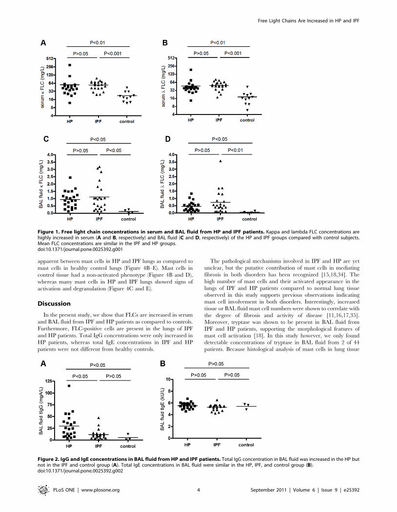
Increased serum and BAL fluid FLC concentrations in IPFand HP patients
In serum and BAL fluid, FLC concentrations are significantly
increased in HP and IPF patients as compared to healthy controls.
Mean (6SEM) k-FLC serum concentrations were 55.96
12.32 mg/L in HP, 54.764.04 mg/L in IPF, and 22.162.93
mg/L in control subjects (Figure 1A). For l-FLC serum concen-
trations were 46.867.29 mg/L in HP, 46.663.44 mg/L in IPF and
18.363.02 mg/L in the control group (Figure 1B). Mean (6SEM)
FLC concentrations in BAL fluid were 0.9460.12, 1.1160.20,
0.1260.060 mg/L (k-FLC) and 0.4560.07, 0.7360.17, 0.066
0.03 mg/L (l-FLC) in the HP, IPF and control group, respectively
(Figure 1C and D). FLC concentrations in BAL fluid showed no
significant correlation with those in serum.
Total IgG, IgE and tryptase concentrations in BAL fluidfrom HP and IPF patients
Both IgG and IgE antibodies were detectable in BAL fluid from
HP and IPF patients and control subjects. The mean (6SEM) total
IgG concentration in HP patients (29.865.58 mgA/L) was
significantly higher than that in IPF patients (11.662.28 mgA/
L) and control subjects (5.363.70 mgA/L) (both p,0.05). Total
IgG concentrations in BAL fluid from IPF patients and controls
did not differ significantly (Figure 2A). Total IgE concentrations
were similar in all groups (Figure 2B). Tryptase was only
detectable in 2 patients, one HP patient (9.27 mg/L) and one
IPF patient (2.27 mg/L) (data not shown).
Correlations between BAL fluid humoral factors andclinical parameters
In the HP and IPF patient groups we investigated the relationship
between FLC and BAL fluid IgG concentrations and inflammatory
cell numbers, and the clinical parameters as described in table 1. For
FLC, a positive correlation was found between l-FLC and
DLCO% in the HP patient group (p = 0.0021; r = 0.62). A positive
correlation was also found between total IgG concentrations and the
number of BAL fluid lymphocytes in HP (p = 0.006; r = 0.57) and
IPF (p = 0.037; r = 0.46) patients. A significant positive correlation
between IgG and kappa and lambda FLC was found in IPF patients
(IgG vs k-FLC: p = 0.009; r = 0.55; IgG vs l-FLC: p = 0.045;
r = 0.44).
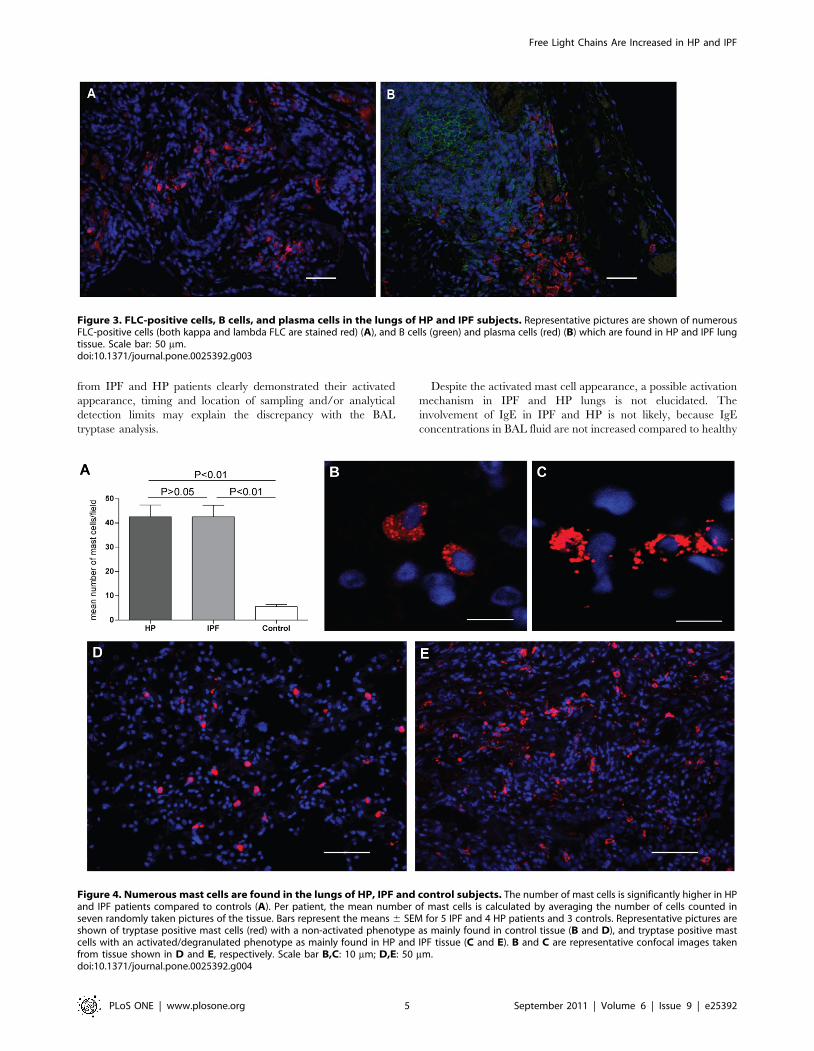
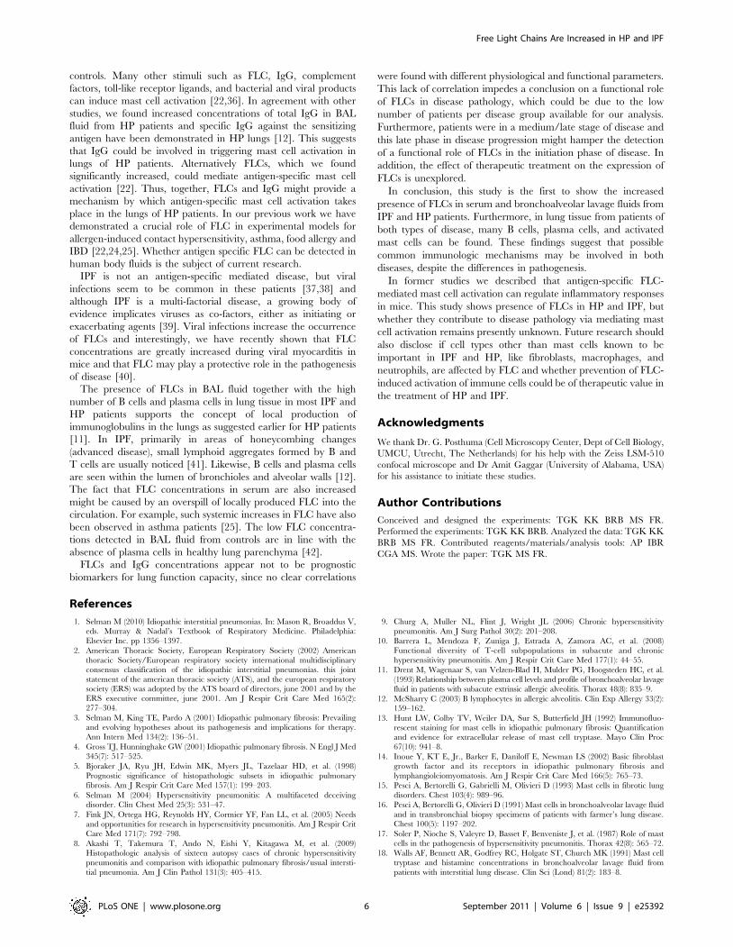
FLC-positive cells, B cells, plasma cells and mast cells inlung tissue from HP, IPF and control subjects
Immunohistochemical analysis of lung tissue from all HP and
IPF patients revealed similar staining patterns for FLC, CD20 (B-
cells), CD138 (plasma cells), and tryptase (mast cells). Many FLC-
positive cells were detected throughout the tissue as isolated cells or
in small groups (Figure 3A). Numerous B cells and plasma cells
were detectable, either as single cells or clustered in groups and in
vicinity of each other (Figure 3B). B cells, and plasma cells were
not detectable in control lung tissues, and FLC-positive cells were
here only detected scarcely. Tryptase positive cells were present
throughout all lung tissues, including control tissue. However, the
number and density of mast cells was lower in the control tissue
(Figure 4A) and morphological differences were dramatically
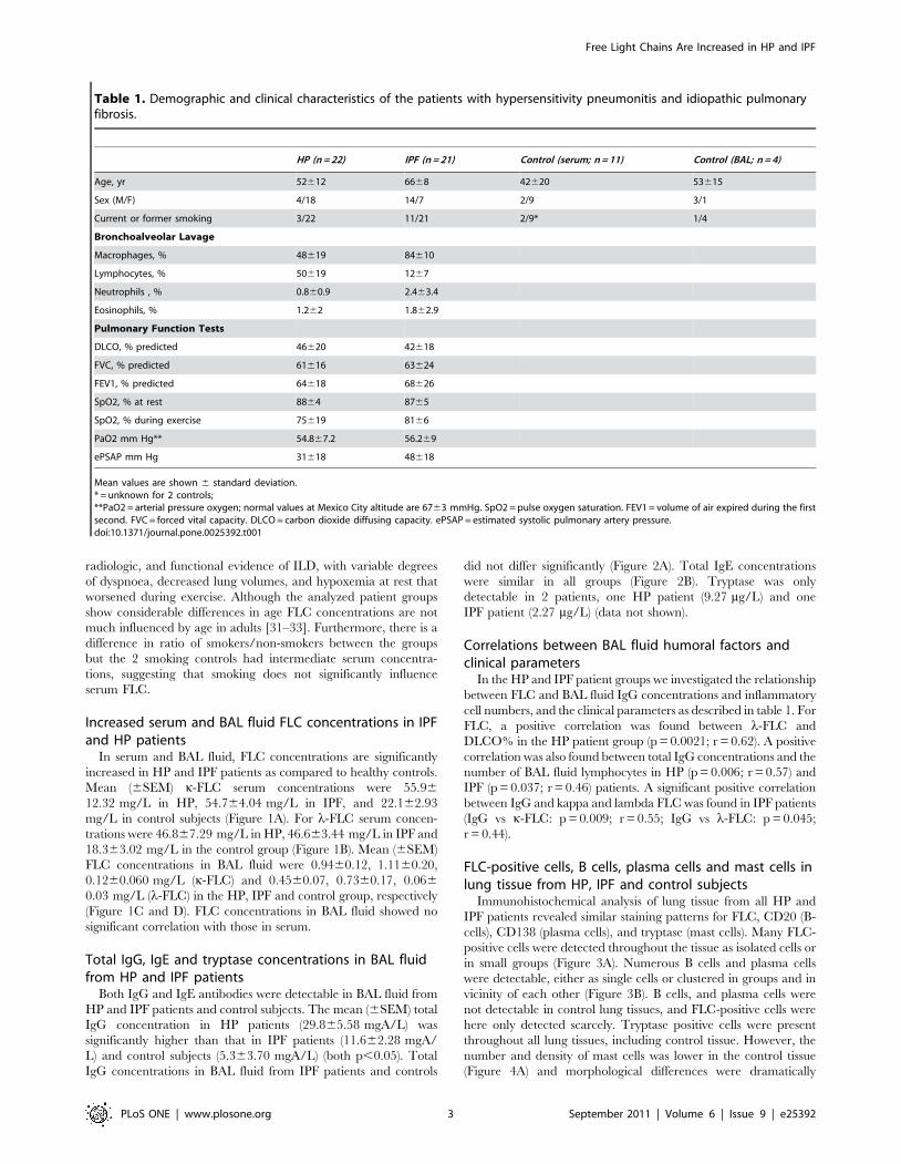
Table 1. Demographic and clinical characteristics of the patients with hypersensitivity pneumonitis and idiopathic pulmonaryfibrosis.
HP (n = 22) IPF (n = 21) Control (serum; n = 11) Control (BAL; n = 4)
Age, yr 52612 6668 42620 53615
Sex (M/F) 4/18 14/7 2/9 3/1
Current or former smoking 3/22 11/21 2/9* 1/4
Bronchoalveolar Lavage
Macrophages, % 48619 84610
Lymphocytes, % 50619 1267
Neutrophils , % 0.860.9 2.463.4
Eosinophils, % 1.262 1.862.9
Pulmonary Function Tests
DLCO, % predicted 46620 42618
FVC, % predicted 61616 63624
FEV1, % predicted 64618 68626
SpO2, % at rest 8864 8765
SpO2, % during exercise 75619 8166
PaO2 mm Hg** 54.867.2 56.269
ePSAP mm Hg 31618 48618
Mean values are shown 6 standard deviation.* = unknown for 2 controls;**PaO2 = arterial pressure oxygen; normal values at Mexico City altitude are 6763 mmHg. SpO2 = pulse oxygen saturation. FEV1 = volume of air expired during the firstsecond. FVC = forced vital capacity. DLCO = carbon dioxide diffusing capacity. ePSAP = estimated systolic pulmonary artery pressure.doi:10.1371/journal.pone.0025392.t001
Free Light Chains Are Increased in HP and IPF
PLoS ONE | www.plosone.org 3 September 2011 | Volume 6 | Issue 9 | e25392
apparent between mast cells in HP and IPF lungs as compared to
mast cells in healthy control lungs (Figure 4B–E). Mast cells in
control tissue had a non-activated phenotype (Figure 4B and D),
whereas many mast cells in HP and IPF lungs showed signs of
activation and degranulation (Figure 4C and E).
Discussion
In the present study, we show that FLCs are increased in serum
and BAL fluid from IPF and HP patients as compared to controls.
Furthermore, FLC-positive cells are present in the lungs of IPF
and HP patients. Total IgG concentrations were only increased in
HP patients, whereas total IgE concentrations in IPF and HP
patients were not different from healthy controls.
The pathological mechanisms involved in IPF and HP are yet
unclear, but the putative contribution of mast cells in mediating
fibrosis in both disorders has been recognized [15,18,34]. The
high number of mast cells and their activated appearance in the
lungs of IPF and HP patients compared to normal lung tissue
observed in this study supports previous observations indicating
mast cell involvement in both disorders. Interestingly, increased
tissue or BAL fluid mast cell numbers were shown to correlate with
the degree of fibrosis and activity of disease [11,16,17,35].
Moreover, tryptase was shown to be present in BAL fluid from
IPF and HP patients, supporting the morphological features of
mast cell activation [18]. In this study however, we only found
detectable concentrations of tryptase in BAL fluid from 2 of 44
patients. Because histological analysis of mast cells in lung tissue
Figure 1. Free light chain concentrations in serum and BAL fluid from HP and IPF patients. Kappa and lambda FLC concentrations arehighly increased in serum (A and B, respectively) and BAL fluid (C and D, respectively) of the HP and IPF groups compared with control subjects.Mean FLC concentrations are similar in the IPF and HP groups.doi:10.1371/journal.pone.0025392.g001
Figure 2. IgG and IgE concentrations in BAL fluid from HP and IPF patients. Total IgG concentration in BAL fluid was increased in the HP butnot in the IPF and control group (A). Total IgE concentrations in BAL fluid were similar in the HP, IPF, and control group (B).doi:10.1371/journal.pone.0025392.g002
Free Light Chains Are Increased in HP and IPF
PLoS ONE | www.plosone.org 4 September 2011 | Volume 6 | Issue 9 | e25392
from IPF and HP patients clearly demonstrated their activated
appearance, timing and location of sampling and/or analytical
detection limits may explain the discrepancy with the BAL
tryptase analysis.
Despite the activated mast cell appearance, a possible activation
mechanism in IPF and HP lungs is not elucidated. The
involvement of IgE in IPF and HP is not likely, because IgE
concentrations in BAL fluid are not increased compared to healthy
Figure 3. FLC-positive cells, B cells, and plasma cells in the lungs of HP and IPF subjects. Representative pictures are shown of numerousFLC-positive cells (both kappa and lambda FLC are stained red) (A), and B cells (green) and plasma cells (red) (B) which are found in HP and IPF lungtissue. Scale bar: 50 mm.doi:10.1371/journal.pone.0025392.g003
Figure 4. Numerous mast cells are found in the lungs of HP, IPF and control subjects. The number of mast cells is significantly higher in HPand IPF patients compared to controls (A). Per patient, the mean number of mast cells is calculated by averaging the number of cells counted inseven randomly taken pictures of the tissue. Bars represent the means 6 SEM for 5 IPF and 4 HP patients and 3 controls. Representative pictures areshown of tryptase positive mast cells (red) with a non-activated phenotype as mainly found in control tissue (B and D), and tryptase positive mastcells with an activated/degranulated phenotype as mainly found in HP and IPF tissue (C and E). B and C are representative confocal images takenfrom tissue shown in D and E, respectively. Scale bar B,C: 10 mm; D,E: 50 mm.doi:10.1371/journal.pone.0025392.g004
Free Light Chains Are Increased in HP and IPF
PLoS ONE | www.plosone.org 5 September 2011 | Volume 6 | Issue 9 | e25392
controls. Many other stimuli such as FLC, IgG, complement
factors, toll-like receptor ligands, and bacterial and viral products
can induce mast cell activation [22,36]. In agreement with other
studies, we found increased concentrations of total IgG in BAL
fluid from HP patients and specific IgG against the sensitizing
antigen have been demonstrated in HP lungs [12]. This suggests
that IgG could be involved in triggering mast cell activation in
lungs of HP patients. Alternatively FLCs, which we found
significantly increased, could mediate antigen-specific mast cell
activation [22]. Thus, together, FLCs and IgG might provide a
mechanism by which antigen-specific mast cell activation takes
place in the lungs of HP patients. In our previous work we have
demonstrated a crucial role of FLC in experimental models for
allergen-induced contact hypersensitivity, asthma, food allergy and
IBD [22,24,25]. Whether antigen specific FLC can be detected in
human body fluids is the subject of current research.
IPF is not an antigen-specific mediated disease, but viral
infections seem to be common in these patients [37,38] and
although IPF is a multi-factorial disease, a growing body of
evidence implicates viruses as co-factors, either as initiating or
exacerbating agents [39]. Viral infections increase the occurrence
of FLCs and interestingly, we have recently shown that FLC
concentrations are greatly increased during viral myocarditis in
mice and that FLC may play a protective role in the pathogenesis
of disease [40].
The presence of FLCs in BAL fluid together with the high
number of B cells and plasma cells in lung tissue in most IPF and
HP patients supports the concept of local production of
immunoglobulins in the lungs as suggested earlier for HP patients
[11]. In IPF, primarily in areas of honeycombing changes
(advanced disease), small lymphoid aggregates formed by B and
T cells are usually noticed [41]. Likewise, B cells and plasma cells
are seen within the lumen of bronchioles and alveolar walls [12].
The fact that FLC concentrations in serum are also increased
might be caused by an overspill of locally produced FLC into the
circulation. For example, such systemic increases in FLC have also
been observed in asthma patients [25]. The low FLC concentra-
tions detected in BAL fluid from controls are in line with the
absence of plasma cells in healthy lung parenchyma [42].
FLCs and IgG concentrations appear not to be prognostic
biomarkers for lung function capacity, since no clear correlations
were found with different physiological and functional parameters.
This lack of correlation impedes a conclusion on a functional role
of FLCs in disease pathology, which could be due to the low
number of patients per disease group available for our analysis.
Furthermore, patients were in a medium/late stage of disease and
this late phase in disease progression might hamper the detection
of a functional role of FLCs in the initiation phase of disease. In
addition, the effect of therapeutic treatment on the expression of
FLCs is unexplored.
In conclusion, this study is the first to show the increased
presence of FLCs in serum and bronchoalveolar lavage fluids from
IPF and HP patients. Furthermore, in lung tissue from patients of
both types of disease, many B cells, plasma cells, and activated
mast cells can be found. These findings suggest that possible
common immunologic mechanisms may be involved in both
diseases, despite the differences in pathogenesis.
In former studies we described that antigen-specific FLC-
mediated mast cell activation can regulate inflammatory responses
in mice. This study shows presence of FLCs in HP and IPF, but
whether they contribute to disease pathology via mediating mast
cell activation remains presently unknown. Future research should
also disclose if cell types other than mast cells known to be
important in IPF and HP, like fibroblasts, macrophages, and
neutrophils, are affected by FLC and whether prevention of FLC-
induced activation of immune cells could be of therapeutic value in
the treatment of HP and IPF.
Acknowledgments
We thank Dr. G. Posthuma (Cell Microscopy Center, Dept of Cell Biology,
UMCU, Utrecht, The Netherlands) for his help with the Zeiss LSM-510
confocal microscope and Dr Amit Gaggar (University of Alabama, USA)
for his assistance to initiate these studies.
Author Contributions
Conceived and designed the experiments: TGK KK BRB MS FR.
Performed the experiments: TGK KK BRB. Analyzed the data: TGK KK
BRB MS FR. Contributed reagents/materials/analysis tools: AP IBR
CGA MS. Wrote the paper: TGK MS FR.
References
1. Selman M (2010) Idiopathic interstitial pneumonias. In: Mason R, Broaddus V,eds. Murray & Nadal’s Textbook of Respiratory Medicine. Philadelphia:
Elsevier Inc. pp 1356–1397.
2. American Thoracic Society, European Respiratory Society (2002) American
thoracic Society/European respiratory society international multidisciplinaryconsensus classification of the idiopathic interstitial pneumonias. this joint
statement of the american thoracic society (ATS), and the european respiratorysociety (ERS) was adopted by the ATS board of directors, june 2001 and by the
ERS executive committee, june 2001. Am J Respir Crit Care Med 165(2):277–304.
3. Selman M, King TE, Pardo A (2001) Idiopathic pulmonary fibrosis: Prevailingand evolving hypotheses about its pathogenesis and implications for therapy.
Ann Intern Med 134(2): 136–51.
4. Gross TJ, Hunninghake GW (2001) Idiopathic pulmonary fibrosis. N Engl J Med
345(7): 517–525.
5. Bjoraker JA, Ryu JH, Edwin MK, Myers JL, Tazelaar HD, et al. (1998)
Prognostic significance of histopathologic subsets in idiopathic pulmonaryfibrosis. Am J Respir Crit Care Med 157(1): 199–203.
6. Selman M (2004) Hypersensitivity pneumonitis: A multifaceted deceivingdisorder. Clin Chest Med 25(3): 531–47.
7. Fink JN, Ortega HG, Reynolds HY, Cormier YF, Fan LL, et al. (2005) Needs
and opportunities for research in hypersensitivity pneumonitis. Am J Respir Crit
Care Med 171(7): 792–798.
8. Akashi T, Takemura T, Ando N, Eishi Y, Kitagawa M, et al. (2009)Histopathologic analysis of sixteen autopsy cases of chronic hypersensitivity
pneumonitis and comparison with idiopathic pulmonary fibrosis/usual intersti-
tial pneumonia. Am J Clin Pathol 131(3): 405–415.
9. Churg A, Muller NL, Flint J, Wright JL (2006) Chronic hypersensitivity
pneumonitis. Am J Surg Pathol 30(2): 201–208.
10. Barrera L, Mendoza F, Zuniga J, Estrada A, Zamora AC, et al. (2008)Functional diversity of T-cell subpopulations in subacute and chronic
hypersensitivity pneumonitis. Am J Respir Crit Care Med 177(1): 44–55.
11. Drent M, Wagenaar S, van Velzen-Blad H, Mulder PG, Hoogsteden HC, et al.
(1993) Relationship between plasma cell levels and profile of bronchoalveolar lavage
fluid in patients with subacute extrinsic allergic alveolitis. Thorax 48(8): 835–9.
12. McSharry C (2003) B lymphocytes in allergic alveolitis. Clin Exp Allergy 33(2):159–162.
13. Hunt LW, Colby TV, Weiler DA, Sur S, Butterfield JH (1992) Immunofluo-rescent staining for mast cells in idiopathic pulmonary fibrosis: Quantification
and evidence for extracellular release of mast cell tryptase. Mayo Clin Proc67(10): 941–8.
14. Inoue Y, KT E, Jr., Barker E, Daniloff E, Newman LS (2002) Basic fibroblastgrowth factor and its receptors in idiopathic pulmonary fibrosis and
lymphangioleiomyomatosis. Am J Respir Crit Care Med 166(5): 765–73.
15. Pesci A, Bertorelli G, Gabrielli M, Olivieri D (1993) Mast cells in fibrotic lung
disorders. Chest 103(4): 989–96.
16. Pesci A, Bertorelli G, Olivieri D (1991) Mast cells in bronchoalveolar lavage fluid
and in transbronchial biopsy specimens of patients with farmer’s lung disease.Chest 100(5): 1197–202.
17. Soler P, Nioche S, Valeyre D, Basset F, Benveniste J, et al. (1987) Role of mastcells in the pathogenesis of hypersensitivity pneumonitis. Thorax 42(8): 565–72.
18. Walls AF, Bennett AR, Godfrey RC, Holgate ST, Church MK (1991) Mast cell
tryptase and histamine concentrations in bronchoalveolar lavage fluid from
patients with interstitial lung disease. Clin Sci (Lond) 81(2): 183–8.
Free Light Chains Are Increased in HP and IPF
PLoS ONE | www.plosone.org 6 September 2011 | Volume 6 | Issue 9 | e25392
19. Akers IA, Parsons M, Hill MR, Hollenberg MD, Sanjar S, et al. (2000) Mast cell
tryptase stimulates human lung fibroblast proliferation via protease-activatedreceptor-2. Am J Physiol Lung Cell Mol Physiol 278(1): L193–201.
20. Araya J, Nishimura SL (2010) Fibrogenic reactions in lung disease. Annu Rev
Pathol 5: 77–98.21. Inoue Y, KT E, Jr., Tinkle SS, Dockstader K, Newman LS (1996) Human mast
cell basic fibroblast growth factor in pulmonary fibrotic disorders. Am J Pathol149(6): 2037–54.
22. Redegeld FA, van der Heijden MW, Kool M, Heijdra BM, Garssen J, et al.
(2002) Immunoglobulin-free light chains elicit immediate hypersensitivity-likeresponses. Nat Med 8(7): 694–701.
23. Groot Kormelink T, Tekstra J, Thurlings RM, Boumans MH, Vos K, et al.(2010) Decrease in immunoglobulin free light chains in patients with rheumatoid
arthritis upon rituximab (anti-CD20) treatment correlates with decrease indisease activity. Ann Rheum Dis 69: 2137–2144.
24. Rijnierse A, Redegeld FA, Blokhuis BR, Van der Heijden MW, Te Velde AA,
et al. (2010) Ig-free light chains play a crucial role in murine mast cell-dependentcolitis and are associated with human inflammatory bowel diseases. J Immunol
185(1): 653–9.25. Kraneveld AD, Kool M, van Houwelingen AH, Roholl P, Solomon A, et al.
(2005) Elicitation of allergic asthma by immunoglobulin free light chains. Proc
Natl Acad Sci U S A 102(5): 1578–83.26. Powe DG, Groot Kormelink T, Sisson M, Blokhuis BJ, Kramer MF, et al. (2010)
Evidence for the involvement of free light chain immunoglobulins in allergic andnonallergic rhinitis. J Allergy Clin Immunol 125(1): 139–45 e1–3.
27. [Anonymous] (2000) American Thoracic Society. idiopathic pulmonary fibrosis:Diagnosis and treatment. International consensus statement: American Thoracic
Society (ATS), and the European Respiratory Society (ERS). Am J Respir Crit
Care Med 161(2 Pt 1): 646–664.28. Katzenstein AL, Mukhopadhyay S, Myers JL (2008) Diagnosis of usual
interstitial pneumonia and distinction from other fibrosing interstitial lungdiseases. Hum Pathol 39(9): 1275–1294.
29. Gotway MB, Freemer MM, King TE, Jr. (2007) Challenges in pulmonary
fibrosis. 1: Use of high resolution CT scanning of the lung for the evaluation ofpatients with idiopathic interstitial pneumonias. Thorax 62(6): 546–553.
30. Pardo A, Gibson K, Cisneros J, Richards TJ, Yang Y, et al. (2005) Up-regulationand profibrotic role of osteopontin in human idiopathic pulmonary fibrosis.
PLoS Med 2(9): e251.
31. Cohen G, Rudnicki M, Schmaldienst S, Horl WH (2002) Effect of dialysis on
serum/plasma levels of free immunoglobulin light chains in end-stage renal
disease patients. Nephrol Dial Transplant 17(5): 879–883.
32. Solling K (1977) Normal values for free light chains in serum different age
groups. Scand J Clin Lab Invest 37(1): 21–5.
33. Wolff F, Thiry C, Willems D (2007) Assessment of the analytical performance
and the sensitivity of serum free light chains immunoassay in patients with
monoclonal gammopathy. Clin Biochem 40(5–6): 351–354. 10.1016/j.clinbio-
chem.2006.11.011.
34. Ishida T, Matsui Y, Matsumura Y, Fujimori N, Furutani M (1995)
Bronchoalveolar mast cells in summer-type hypersensitivity pneumonitis:
Increase in numbers and ultrastructural evidence of degranulation. Intern
Med 34(5): 357–63.
35. Bjermer L, Engstrom-Laurent A, Lundgren R, Rosenhall L, Hallgren R (1988)
Bronchoalveolar mastocytosis in farmer’s lung is related to the disease activity.
Arch Intern Med 148(6): 1362–1365.
36. Galli SJ, Tsai M (2008) Mast cells: Versatile regulators of inflammation, tissue
remodeling, host defense and homeostasis. J Dermatol Sci 49(1): 7–19.
37. Stewart JP, Egan JJ, Ross AJ, Kelly BG, Lok SS, et al. (1999) The detection of
epstein-barr virus DNA in lung tissue from patients with idiopathic pulmonary
fibrosis. Am J Respir Crit Care Med 159(4 Pt 1): 1336–1341.
38. Tang YW, Johnson JE, Browning PJ, Cruz-Gervis RA, Davis A, et al. (2003)
Herpesvirus DNA is consistently detected in lungs of patients with idiopathic
pulmonary fibrosis. J Clin Microbiol 41(6): 2633–2640.
39. Vannella KM, Moore BB (2008) Viruses as co-factors for the initiation or
exacerbation of lung fibrosis. Fibrogenesis Tissue Repair 1(1): 2.
40. Matsumori A, Shimada M, Jie X, Higuchi H, Groot Kormelink T, et al. (2010)
Effects of free immunoglobulin light chains on viral myocarditis. Circ Res 106(9):
1533–40.
41. Rangel-Moreno J, Hartson L, Navarro C, Gaxiola M, Selman M, et al. (2006)
Inducible bronchus-associated lymphoid tissue (iBALT) in patients with
pulmonary complications of rheumatoid arthritis. J Clin Invest 116(12):
3183–3194.
42. Holt PG, Strickland DH, Wikstrom ME, Jahnsen FL (2008) Regulation of
immunological homeostasis in the respiratory tract. Nat Rev Immunol 8(2):
142–152.
Free Light Chains Are Increased in HP and IPF
PLoS ONE | www.plosone.org 7 September 2011 | Volume 6 | Issue 9 | e25392
Copyright © 2022 FDOKUMEN