Immunization of mice with gamma-irradiated Brucella neotomae and its recombinant strains induces...
22
Immunization of mice with gamma-irradiated Brucella neotomae and its recombinant strains induces protection against virulent B. abortus, B. melitensis, and B. suis challenge Dina Moustafa 1 , Virendra K. Garg 1 , Neeta Jain 2 , Nammalwar Sriranganathan 2 , and Ramesh Vemulapalli 1,* 1 Department of Comparative Pathobiology, School of Veterinary Medicine, Purdue University, West Lafayette, IN 47907, USA 2 Department of Biomedical Sciences and Pathobiology, VA-MD Regional College of Veterinary Medicine, Virginia Tech, VA 24061, USA Abstract Human brucellosis, a zoonotic disease of major public health concern in several developing countries, is primarily caused by Brucella abortus, B. melitensis, and B. suis. No brucellosis vaccine is available for human use. The aim of this study was to determine if B. neotomae, a bacterium not known to cause disease in any host, can be used for developing brucellosis vaccines. B. neotomae and its recombinant strains overexpressing superoxide dismutase and a 26 kDa periplasmic protein were rendered non-replicative through exposure to gamma-radiation and used as vaccines in a murine brucellosis model. All three vaccines induced antigen-specific antibody and T cell responses. The vaccinated mice showed significant resistance against challenge with virulent B. abortus 2308, B. melitensis 16M, and B. suis 1330. These results demonstrate that the avirulent B. neotomae is a promising platform for developing a safe and effective vaccine for human brucellosis. Keywords Brucellosis; B. neotomae; gamma radiation; vaccine; broad protection Introduction Brucellosis is a zoonotic disease caused by members of the genus Brucella, which are Gram- negative, facultatively intracellular bacteria [1]. There are six-well recognized (B. abortus, B. melitensis, B. suis, B. canis, B. ovis, and B. neotomae) and four recently added (B. ceti, B. pennipedialis, B. microti, and B. inopinata) species in genus Brucella. In domestic and wild mammals brucellosis often results in abortions and infertility. In humans, brucellosis manifests itself as a chronic infection with undulant fever and general malaise; other clinical signs vary depending on the affected organ systems [2]. Humans usually acquire the © 2010 Elsevier Ltd. All rights reserved. * Corresponding author at: Veterinary Pathology Building, 725 Harrison Street, West Lafayette, IN 47907, United States. Tel.: +17654947560; fax: +17654949830. [email protected] (R. Vemulapalli). Publisher's Disclaimer: This is a PDF file of an unedited manuscript that has been accepted for publication. As a service to our customers we are providing this early version of the manuscript. The manuscript will undergo copyediting, typesetting, and review of the resulting proof before it is published in its final citable form. Please note that during the production process errors may be discovered which could affect the content, and all legal disclaimers that apply to the journal pertain. NIH Public Access Author Manuscript Vaccine. Author manuscript; available in PMC 2012 January 17. Published in final edited form as: Vaccine. 2011 January 17; 29(4): 784–794. doi:10.1016/j.vaccine.2010.11.018. NIH-PA Author Manuscript NIH-PA Author Manuscript NIH-PA Author Manuscript
Transcript of Immunization of mice with gamma-irradiated Brucella neotomae and its recombinant strains induces...

Immunization of mice with gamma-irradiated Brucella neotomaeand its recombinant strains induces protection against virulentB. abortus, B. melitensis, and B. suis challenge
Dina Moustafa1, Virendra K. Garg1, Neeta Jain2, Nammalwar Sriranganathan2, and RameshVemulapalli1,*
1Department of Comparative Pathobiology, School of Veterinary Medicine, Purdue University,West Lafayette, IN 47907, USA2Department of Biomedical Sciences and Pathobiology, VA-MD Regional College of VeterinaryMedicine, Virginia Tech, VA 24061, USA
AbstractHuman brucellosis, a zoonotic disease of major public health concern in several developingcountries, is primarily caused by Brucella abortus, B. melitensis, and B. suis. No brucellosisvaccine is available for human use. The aim of this study was to determine if B. neotomae, abacterium not known to cause disease in any host, can be used for developing brucellosis vaccines.B. neotomae and its recombinant strains overexpressing superoxide dismutase and a 26 kDaperiplasmic protein were rendered non-replicative through exposure to gamma-radiation and usedas vaccines in a murine brucellosis model. All three vaccines induced antigen-specific antibodyand T cell responses. The vaccinated mice showed significant resistance against challenge withvirulent B. abortus 2308, B. melitensis 16M, and B. suis 1330. These results demonstrate that theavirulent B. neotomae is a promising platform for developing a safe and effective vaccine forhuman brucellosis.
KeywordsBrucellosis; B. neotomae; gamma radiation; vaccine; broad protection
IntroductionBrucellosis is a zoonotic disease caused by members of the genus Brucella, which are Gram-negative, facultatively intracellular bacteria [1]. There are six-well recognized (B. abortus,B. melitensis, B. suis, B. canis, B. ovis, and B. neotomae) and four recently added (B. ceti, B.pennipedialis, B. microti, and B. inopinata) species in genus Brucella. In domestic and wildmammals brucellosis often results in abortions and infertility. In humans, brucellosismanifests itself as a chronic infection with undulant fever and general malaise; other clinicalsigns vary depending on the affected organ systems [2]. Humans usually acquire the
© 2010 Elsevier Ltd. All rights reserved.*Corresponding author at: Veterinary Pathology Building, 725 Harrison Street, West Lafayette, IN 47907, United States. Tel.:+17654947560; fax: +17654949830. [email protected] (R. Vemulapalli).Publisher's Disclaimer: This is a PDF file of an unedited manuscript that has been accepted for publication. As a service to ourcustomers we are providing this early version of the manuscript. The manuscript will undergo copyediting, typesetting, and review ofthe resulting proof before it is published in its final citable form. Please note that during the production process errors may bediscovered which could affect the content, and all legal disclaimers that apply to the journal pertain.
NIH Public AccessAuthor ManuscriptVaccine. Author manuscript; available in PMC 2012 January 17.
Published in final edited form as:Vaccine. 2011 January 17; 29(4): 784–794. doi:10.1016/j.vaccine.2010.11.018.
NIH
-PA Author Manuscript
NIH
-PA Author Manuscript
NIH
-PA Author Manuscript

infection by consuming contaminated dairy or meat products or by coming in contact withthe infected animal tissues and secretions [3]. Ingestion, inhalation, and contamination ofconjunctiva or broken skin by the infected animal products are the common modes by whichhumans are infected. In several developing countries, brucellosis is an important publichealth concern [4]. Of the six well-recognized species of Brucella, B. melitensis, B. suis andB. abortus are highly virulent to humans [2]. These 3 Brucella species are consideredpotential bioterror agents and they belong to NIAID Category B priority pathogens list. Atpresent there is no vaccine available for human brucellosis. Cell-mediated immunity (CMI)plays a central role in acquired resistance against brucellosis, and antibodies to the Opolysaccharide (O antigen) of the lipopolysaccharide (LPS) participate in providingenhanced resistance against infections by B. abortus, B. melitensis and B. suis [5].Attenuated, live Brucella strains such as B. abortus RB51 and 19, and B. melitensis Rev1 arebeing used as vaccines to control brucellosis in domestic animals [6]. However, thesevaccines are not suitable for humans since they can cause disease even in individuals withhealthy immune system [7-8].
B. neotomae was isolated in 1957 from desert wood rats of the western U.S. [9]. No disease,either in human or other animal species, has ever been attributed to B. neotomae. Unlike thevirulent Brucella spp., B. neotomae does not establish a chronic infection in immuno-competent mouse models of brucellosis (Moustafa and Vemulapalli, unpublishedobservation). The overall goal of this study was to examine the feasibility of developing asafe and effective vaccine for human brucellosis using B. neotomae. We used exposure to aminimum dose of gamma-radiation as a means to increase the vaccine safety by eliminatingthe bacteria’s ability to replicate but retain the metabolic activity. We also tested ifoverexpression of Brucella Cu-Zn superoxide dismutase (SOD) and a 26 kDa periplasmicprotein (Bp26) in B. neotomae would enhance its vaccine efficacy. Our results demonstratethat mice immunized with gamma-irradiated B. neotomae develop significant protectiveimmunity against challenge with virulent B. abortus 2308, B. melitensis 16M, and B. suis1330.
1. Materials and methods2.1. Bacteria
B. neotomae strain 5K33 was purchased from the American Type Culture Collection,Manassas, Va. Virulent strains B. melitensis 16M, B. abortus 2308, and B. suis 1330 werefrom the culture collection at Virginia Tech, Blacksburg, VA. Escherichia coli strain DH5α(Invitrogen, Carlsbad, CA) was used for producing the necessary plasmid constructs and forrecombinant protein production. The recombinant B. neotomae/Bp26 and B. neotomae/SODwere generated by transforming B. neotomae with recombinant plasmids pBB4Bp26 andpBB4SOD, respectively. The pBB4Bp26 plasmid was constructed by cloning the geneencoding the 26 kDa periplasmic protein along with its promoter sequences into the Kpn Iand Xho I restriction sites of pBBR1MCS-4 [10]. The gene sequences were first PCRamplified from the genomic DNA of B. abortus RB51 using a custom-designed primer-pair(forward primer: 5’-aaggtaccacccgaaagaaagccgggata-3’; reverse primer: 5’-aactcgagcagatcgaaacgcgctctaat-3’) and the resulting 1.2 kb DNA fragment was digestedwith Kpn I and Xho I enzymes and cloned into the same sites of pBBR1MCS-4. Thenucleotide sequence integrity of the cloned fragment was confirmed by sequence analysis. A1.1 kb fragment containing the B. abortus sodC gene and its promoter sequence was excisedfrom pBS/SOD [11] with ClaI restriction enzyme digestion and subcloned intopBBR1MCS-4 to generate pBB4SOD. B. neotomae was transformed with pBB4Bp26 andpBB4SOD by electroporation following the previously described procedures [12].
Moustafa et al. Page 2
Vaccine. Author manuscript; available in PMC 2012 January 17.
NIH
-PA Author Manuscript
NIH
-PA Author Manuscript
NIH
-PA Author Manuscript

All bacteria were grown in tryptic soy broth (TSB) or tryptic soy agar (TSA) at 37°C.Bacteria harboring the plasmids were grown in the presence of 100 μg/ml concentration ofampicillin. Colony forming units (CFU) of Brucella strains were determined by plating 10-fold serial dilutions of the cultures on TSA. All experiments with B. neotomae wereperformed in a Biosafety level (BSL)-2 facility using BSL-3 practices. All experiments withvirulent Brucella were performed in a BLS-3 facility approved for the select agents work.
2.2. Vaccine preparationB. neotomae, B. neotomae/Bp26, and B. neotomae/SOD were grown in TSB or TSB withampicillin to mid log phase, and aliquots containing 5×10¹⁰- 1×10¹¹ CFU/ml were stored at-80°C until use. Two to three weeks before immunization, aliquots of the vaccines wereexposed to 350 krads of gamma irradiation using a 60Co source irradiator (Gammacell 220irraditor). The inability of the irradiated bacteria to replicate was confirmed plating on TSAand incubating for at least 7 days. The irradiated bacteria were stored at 4°C until use forimmunization.
2.3. Determination of bacterial metabolic activityMetabolic activity of the gamma irradiated bacteria was assayed using Alamar blue(BioSource International, Camarillo, CA) and LIVE/DEAD ® BacLight Bacterial viabilityKit for microscopy (Molecular Probes, Eugene, OR). Alamar blue is a redox indicator andits color changes from blue to pink in response to chemical reduction. Alamar blue isreduced by FMNH2, FADH2, NADH, and NADPH, which are present in metabolicallyactive cells. Alamar blue is extensively used for monitoring proliferation and viability ofvarious eukaryotic and prokaryotic cells. For the Alamar blue assay, irradiated samples werewashed in saline solution and resuspended in TSB to the original concentration. Ninetymicroliters of the irradiated suspension were mixed with 10 μl of Alamar blue and incubatedat 37°C for 1 hour, and the change in color from blue to pink was monitored as describedpreviously [13]. The manufacturer’s instructions were followed while using the LIVE/DEAD Baclight kit which utilizes a mixture of the green-fluorescent SYTO® 9 and the red-fluorescent propidium iodide to stain the bacterial nucleic acids. Following staining with themixture, the live bacteria with intact cell membranes fluoresce green, whereas the deadbacteria with damaged membranes fluoresce red. The stained bacteria were observed usingNikon Eclipse–E400 microscope equipped with a 480/530 and 450/580 bandpass filter sets.
2.4. Quantification of irradiated bacteria in mice spleens by real-time PCRThe length of persistence of the irradiated B. neotomae in spleens of the vaccinated micewas determined by quantifying the bacterial DNA using RT-PCR. Two groups of 12 femaleBALB/c mice (6 weeks of age) each were inoculated intraperitonially with 108 CFU-equivalent of either heat-killed (65°C for 1 hour) or gamma-irradiated B. neotomae. Threemice from each group were euthanized at 3 hours, one, three and five days post inoculation,and their spleens were collected aseptically. Total DNA was extracted from the collectedspleens using DNeasy® Blood and Tissue Kit (Qiagen Inc., Valencia, CA) according to themanufacturer’s protocol. The amount of Brucella DNA present in the samples wasdetermined using a previously described real-time PCR that amplifies a 178 bp region ofIS711 element using primers IS421-F (5′-cgctcgcgcggtggat-3’) and IS511-R (5’-cttgaagcttgcggacagtcacc-3’) and measures the amplified product using a TaqMan probe(Cy5-acgaccaagctgcatgctgttgtcgatg-BHQ2) [14]. DNA extracted from differentconcentrations of the irradiated B. neotomae suspension (100 to 108 CFU-equivalent/ml) wasused to construct the standard curve. After logarithmic conversion, the concentration of eachdilution series was plotted against the cycle number at which the fluorescent signal increasedabove a threshold value (Ct value). The regression equation derived from the standard curvewas used to calculate the concentration of B. neotomae present in the spleens. The PCR
Moustafa et al. Page 3
Vaccine. Author manuscript; available in PMC 2012 January 17.
NIH
-PA Author Manuscript
NIH
-PA Author Manuscript
NIH
-PA Author Manuscript

reactions were performed in a Stratagene MX3000P thermocycler and the data wereanalyzed using MxPro QPCR software (Stratagene, La Jolla, CA). All samples andstandards were assayed in duplicates.
2.5. Mice immunizationsFemale BALB/c mice of 4 to 6 weeks of age were vaccinated by two intraperitonealinoculations, at day 0 and day 14, with 1×10⁸ CFU-equivalent of the irradiated B. neotomaeand its recombinants B. neotomae/Bp26 and B. neotomae/SOD. As a negative control, onegroup of mice was injected with saline alone. Mice were bled by puncturing the retro-orbitalplexus under anesthesia at two weeks post inoculation (p.i.) (prior to the boosterimmunization), and at 6 weeks p.i. (4 weeks after the booster). The serum was separatedfrom the clotted blood and stored at -20°C until use for detection of antigen-specificantibodies by enzyme-linked immunosorbent assay (ELISA). At 6 weeks p.i., all the micewere euthanized by CO2 asphyxiation followed by cervical dislocation, spleens werecollected aseptically and used for determining the antigen-specific T cell immune responsesby measuring cytokine production upon in vitro stimulation with specific antigens.
2.6. Preparation of antigens2.6.1. B. neotomae crude extract—Late log phase culture of B. neotomae wascentrifuged at 6,000 × g for 10 min and the pelleted bacteria were washed three times withsterile distilled water and resuspended in 0.5% sodium dodecyl sulfate (SDS). The bacteriawere lysed by shaking gently for 2 hours at room temperature and then sonicating for 10 minon ice. The unlysed bacteria were removed by centrifugation at 8,000 × g for 10 min. Theclear supernatants were collected, and the protein concentration was determined by theBradford method [15]. Aliquots of the antigen extract were stored at −80°C until use forELISA.
2.6.2. LPS extraction—Total LPS was extracted from live B. neotomae by butanol-waterprocedure as previously described [16-17]. Briefly, live B. neotomae organisms wereharvested by centrifugation and 10g of wet pellet was resuspended in 0.85% NaCl at aconcentration of 0.25 g wet weight/ml, and thoroughly dispersed by mixing with a magneticstirrer at 4°C. An equal volume of water saturated butanol was added with constant mixingfor 15 min at 4°C. After centrifugation at 35,000 × g for 20 min, the aqueous phase wascollected and the insoluble precipitate was further extracted with ½ initial volume of thesaline solution. The combined aqueous extracts were centrifuged in order to remove anytraces of insoluble materials. LPS was precipitated using 4 volumes of methanol, and theprecipitate was dissolved in 0.1M Tris buffer (pH 8) containing 2% SDS and 2%mercaptoethanol. The mixture was heated for 5 min at 100°C followed by 90 min incubationwith proteinase K at 60°C. LPS was precipitated by methanol, followed by two washes withcold methanol, and dissolved in water. In order to confirm the presence of any traces ofproteins in the extracted LPS, SDS-PAGE gel followed by silver staining was performed toascertain the quality of the LPS preparation. The extracted LPS was used for ELISA.
2.6.3. Bp26 purification—The Bp26 protein of B. abortus was expressed in E. coli byusing expression vector pMalC2 (New England Biolabs Inc.), and the purification was doneaccording to manufacturer’s suggested procedure. Using pMalC2 vector, BP26 protein wasexpressed as a fusion protein with MBP at the amino terminus so that the recombinantprotein can be purified by affinity chromatography with amylose resin. The concentration ofthe purified protein was determined by the Bradford method. Aliquots of the protein werestored at −80°C until use for ELISA or in vitro stimulation of splenocytes. The purifiedprotein was also used to raise antigen-specific antibodies by hyper-immunizing mice usingalum as the adjuvant.
Moustafa et al. Page 4
Vaccine. Author manuscript; available in PMC 2012 January 17.
NIH
-PA Author Manuscript
NIH
-PA Author Manuscript
NIH
-PA Author Manuscript

2.6.4. Purification of SOD—Brucella SOD was expressed in E. coli DH5α and purifiedaccording to the method previously described [18]. Briefly, SOD was extracted from the E.coli cells using 10 mM phosphate buffer (pH 7.6) containing 0.1% Triton X-100. Cu/ZnSOD was purified using an equilibrated anion-exchange column (HiTrapQ; PharmaciaBiotech). By applying the extract to the column, all of the proteins except SOD were boundto the resin. SOD present in the flow-through was collected, absorbed with polymyxin Bbeads (Affi-Prep polymyxin support; Bio-Rad Laboratories, Hercules, CA) to remove theLPS, and dialyzed extensively against phosphate-buffered saline (PBS). The concentrationof the purified protein was determined by the Bradford method. Aliquots of the protein werestored at −80°C until use for ELISA or in vitro stimulation of splenocytes.
2.7. SDS-PAGE and Western blottingTo confirm the overexpression of SOD and Bp26 in the recombinant B. neotomae strains,SDS-PAGE and Western blot analyses were performed as previously described [19]. Ascontrols, antigen extracts of B. neotomae, the purified recombinant MBP-Bp26 fusionprotein, and the purified SOD were used. For Western blotting, goat anti-B. abortus SODsera [20], and mouse anti-MBP-Bp26 sera produced in this study were used as the primaryantibodies.
2.8. Indirect ELISALevels of serum immunoglobulin G (total IgG), as well as IgG1 and IgG2a isotypes withspecificity to Bp26, SOD and B. neotomae LPS and crude lysate were determined byindirect ELISA [19]. The levels of serum IgG2b, IgG3 and IgM against smooth LPS werealso determined. The antigens were diluted in carbonate buffer, pH 9.6, 1 in 10 for B.neotomae LPS and to 10 μg/ml of protein concentration for MBP-Bp26, SOD and crudelysate. The wells of polystyrene plates (Nunc-Immunoplate with maxisorp surface) werecoated with the diluted antigens (100μl/well). Following overnight incubation at 4°C, plateswere washed four times in wash buffer (Tris-buffered saline at pH 7.4, 0.05 % Tween 20)and blocked with 2% bovine serum albumin (BSA) in Tris-buffered saline. After 1hourincubation at 37°C, mouse sera with appropriate dilution in blocking buffer were added tothe wells (50 μl/well). Each serum sample was tested in duplicate wells; the plates wereincubated for 4 hours at room temperature and washed four times. Horseradish peroxidase–labeled anti-mouse isotype specific conjugates (Southern Biotechnology Associates Inc,Birmingham, Alabama) were added (50 μl/well) at an appropriate dilution. After 1 hourincubation at room temperature, the plates were washed four times. A 100 μl of substratesolution (TMB Microwell peroxidase substrate; Kirkegaard& Perry Laboratories,Gaithersburg, MD) was applied to each well. After 20 min incubation at room temperature,the enzyme reaction was stopped by adding 100 μl of stop solution (0.185 M sulfuric acid),and the absorbance at 450 nm was recorded using microplate reader (Molecular devices,Sunnyvale, CA).
2.9. Splenocyte culture and cytokine quantificationsSplenocytes from the vaccinated mice (four mice per group) were obtained as previouslydescribed [19], and were cultured in triplicates in 96-well flat-bottomed culture plates(5×10⁵ cells /well) in the presence of different stimulants: 10 μg/ml of SOD, 30 μg/ml ofMBP-Bp26, and 107 CFU-equivalent of gamma-irradiated B. neotomae. Cells with plainmedium and cells stimulated with 2.5 μg/ml of concanavalin (ConA) were used as controls.The splenocytes were cultured for 5 days, their supernatants were collected and theconcentration of selected cytokines was determined using Bio-RAD Bio-Plex Pro™ Mousecytokine Th1/Th2 Assay according to the manufacturer instructions. The following
Moustafa et al. Page 5
Vaccine. Author manuscript; available in PMC 2012 January 17.
NIH
-PA Author Manuscript
NIH
-PA Author Manuscript
NIH
-PA Author Manuscript
cytokines were tested in the collected supernatants: IL-2, GM-CSF, IFN-γ, TNF-α, IL-4,IL-5, IL-10, IL-12p70.
2.10. Flow cytometry analysis for IFN-γ secreting antigen-specific CD8+ and CD4+ T cellsIntracellular staining for IFN-γ was performed as previously described with somemodifications [21]. After euthanizing the mice, spleens were collected from the vaccinatedmice (4 mice in each group) and single cell suspensions were prepared from spleens. UsingACK solution, erythrocytes were lysed and the splenocytes were cultured in 96 well flat-bottomed plates (106 cells /well) with or without specific antigen as described above forsplenocyte cultures. After 8 hours of incubation at 37°C in a humid incubator with 5% CO2,brefeldin A (Golgistop; Pharmingen) was added to the culture medium and incubated foranother 8 hours. Cells from each treatment were suspended in PBS containing 1% BSA and0.2% sodium azide (FACS buffer) and stained for surface markers CD8 and CD4 byincubating for 30 minutes at 4°C with appropriately diluted FITC conjugated anti-mouseCD8 antibody (BD Pharmigen, clone53-6.7) and APC-conjugated anti-mouse CD4 antibody(BD Pharmigen, clone L3T4-RM 4.5). After three washes with FACS buffer to removeunbound antibodies, the cells were subjected to intracellular IFN-γ staining with PE-conjugated rat anti-mouse IFN-γ antibody using the Cytofix/Cytoperm kit (Pharmingen)according to the manufacturer’s instructions. Cell stained with PE-conjugated rat IgG1antibody served as the isotype control. The cells were acquired on BD FACS Canto II™Flow cytometer (BD Biosciences, CA, USA). The data were analyzed using BDFACSDIVA version 6 software (BD Biosciences, CA, USA) and the proportion of CD4+
and CD8+ T cells that secreted IFN-γ was determined.
2.11. Protection experimentsGroups of 15 mice each were vaccinated by intraperitoneal inoculation with 108 CFU-equivalent of irradiated B. neotomae, B. neotomae/Bp26 and B. neotomae/SOD. A group ofmice injected with saline alone served as the control. Two weeks post inoculation the micewere given a booster immunization using the same route and dose. Six weeks after the initialvaccination, 5 mice from each group were challenged by intraperitoneal inoculation with 3 ×10⁴ CFU/mouse of B. melitensis 16M, B. abortus 2308, or B.suis 1330. Two weeks afterchallenge, the mice were euthanized and the bacterial burden in their spleens wasdetermined as previously described [22].
2.12. Statistical analysesAbsorbance values of ELISA, concentrations of cytokines, and the flow cytometry data ofIFN-γ positive T cells were analyzed for differences among the groups by performinganalysis of variance with post-hoc Bonferroni for pair-wise comparison using SPSS version17.0 (SPSS inc, an IBM company, USA). For protection study, one-tailed t-test modified forunequal variances between groups was performed to compare the log transformed bacterialloads in spleens of mice from each vaccinated group with the respective saline controlgroup. P values of ≤0.05 were considered significant.
2. Results3.1. Overexpression of SOD and Bp26 in B. neotomae
Overexpression of SOD and Bp26 proteins in B. neotomae/SOD and B. neotomae/Bp26 wasdetected by SDS-PAGE (Fig. 1A and 1C), which was further confirmed by Western blotanalysis using the antigen-specific antibodies (Fig.1B and 1D). Compared to B. neotomae,antigen extracts of B. neotomae/Bp26 contained an overexpressed protein of 26 kDa in sizewhich was recognized by the mouse anti-MBP-Bp26 serum (Fig. 1A and Fig. 1B).
Moustafa et al. Page 6
Vaccine. Author manuscript; available in PMC 2012 January 17.
NIH
-PA Author Manuscript
NIH
-PA Author Manuscript
NIH
-PA Author Manuscript

Similarly, B. neotomae/SOD contained an overexpressed protein of 16 kDa in size whichreacted with the goat anti-SOD antibodies (Fig. 1C and 1D). In accordance with the previousreports [23], no detectable level of SOD expression was present in B. neotomae (Fig. 1D).
Stable overexpression of both Bp26 and SOD in the respective recombinant B. neotomaestrains was consistently detected in all subcultures or gamma-irradiated preparations (datanot shown).
2.2. Gamma irradiated B. neotomae: replication, viability and metabolic activityAliquots of B. neotomae were exposed to different doses gamma radiation and the CFUpresent in each aliquot were determined by culturing on TSA plates. As shown in Fig. 2A,gradual decrease in CFU of B. neotomae was detected with increased exposure to gammaradiation. A total loss of replicative ability of the bacteria was observed at a minimum doseof 300 kilorads (Fig 2A).
When stained with both green-fluorescent SYTO 9 and red-fluorescent propidium iodide,gamma-irradiated B. neotomae, similar to the live bacteria, displayed strong greenfluorescence and weak red fluorescence, indicating the undamaged cell membranes (Fig2B). As expected, the heat-killed bacteria showed strong red fluorescent reflecting thedamaged cell membranes (Fig 2B).
Gamma irradiation did not impair the metabolic activity of the bacteria, as indicated by theability of irradiated and live B. neotomae to change the color of Alamar blue dye from blueto pink (data not shown). In contrast, heat-killed B. neotomae failed to cause the colorchange in the Alamar Blue dye (data not shown).
3.3. Persistence of gamma-irradiated B. neotomae in mice spleensPersistence of the irradiated bacteria in spleens of inoculated mice was determined byextracting DNA from the tissues and performing a quantitative real-time PCR specific toBrucella. As shown in Table 1, compared to the heat-killed bacteria, gamma-irradiated B.neotomae was present in higher numbers in the spleens at 3 hours, 24 hours and 3 days post-inoculation. By day 5 post-inoculation, no bacteria were detected in the spleens of miceinoculated with either of the bacterial preparations.
3.4. Induction of specific antibody responsesPresence of antibodies specific to B. neotomae total antigens, Bp26, SOD and LPS in serumof the mice vaccinated with the irradiated vaccines was determined by ELISA. Serumsamples collected at 2 and 6 weeks after the initial immunizations were analyzed incomparison with the samples from saline inoculated group. Mice vaccinated with B.neotomae and its recombinants B. neotomae/Bp26 and B. neotomae/SOD developedsignificantly higher levels of IgG specific to the total antigen of B. neotomae at 2 and 6weeks post vaccination than did mice in the saline inoculated group. Assays with IgG1 andIgG2a specific conjugates revealed that antibodies of both isotypes were present insignificantly higher levels than in saline inoculated mice (data not shown).
At 2-weeks post vaccination, only mice vaccinated with B. neotomae/Bp26, developedsignificantly higher levels of Bp26-specific IgG than saline inoculated mice (Fig. 3).However, by week 6, all vaccinated groups showed production of significant levels of Bp26-specific antibodies. Both IgG1 and IgG2a isotypes specific to Bp26 were detected in thevaccinated mice. As expected, the level of Bp26-specific antibodies in mice vaccinated withB. neotomae/Bp26 was higher compared to that detected in mice vaccinated with B.neotomae or B. neotomae/SOD (Fig. 3).
Moustafa et al. Page 7
Vaccine. Author manuscript; available in PMC 2012 January 17.
NIH
-PA Author Manuscript
NIH
-PA Author Manuscript
NIH
-PA Author Manuscript

Significant levels of IgG specific to SOD were detected in serum of all vaccinated mice(Fig. 3). The induced antigen-specific antibodies were of both IgG1 and IgG2a isotypes. Nosignificant differences were detected in the levels of SOD-specific antibodies among thedifferent vaccine groups (Fig. 3).
Serum of all vaccinated mice contained significant levels of IgG and IgM to B. neotomaeLPS (Fig. 4). The IgG and IgM levels were higher at 6 weeks than at 2 weeks. The producedLPS-specific antibodies included IgG1, IgG2a, IgG2b, and IgG3 isotypes (Fig. 4). Therewas no difference in the lgG1 levels between the 2- and 6-week serum samples. In contrast,the levels of IgG2a, IgG2b, and IgG3 were higher in the 6-week samples (Fig. 4).
Antibody levels were also measured in ELISA titers by endpoint titration of the pooledserum samples of each vaccinated group (Supplementary Tables 1-3). Differences inantibody titers between the time-points or groups mirrored that of the antibody levels basedon ELISA absorbance values presented in Figs. 3 and 4.
3.5 Antigen-specific cellular immune responsesSpecific CMI responses of the vaccinated mice at 6 weeks post initial immunization weredetermined by quantification of a panel of Th1/Th2 cytokines secreted by the splenocytesand the number of IFN-γ secreting CD4+ and CD8+ T cells upon in vitro stimulation withirradiated B. neotomae, MBP-Bp26 and SOD.
When stimulated with gamma-irradiated B. neotomae, splenocytes from all vaccinated mice,but not the saline inoculated ones, secreted significantly higher levels of IFN-γ, IL-12p70,IL-5 and IL-10 compared to the unstimulated controls (Fig 5). The concentrations of IL-4 inall culture supernatants were low (< 10 pg/ml) and were not significantly different from thecorresponding unstimulated controls (Fig. 5).
Stimulation with MBP-Bp26 and SOD resulted in the secretion of significantly higheramounts of IL-12p70 and IL-10 by splenocytes from all vaccinated mice compared to theunstimulated controls; the concentrations of IFN-γ and IL-5 were low and variable amongthe different vaccine groups (Fig. 5).
Splenocytes from all vaccinated, but not saline inoculated, mice secreted significantly higheramounts of TNF-α upon stimulation with irradiated B. neotomae (Fig. 5).
Stimulation with irradiated B. neotomae, MBP-Bp26 and SOD induced secretion of similarhigh levels of GM-CSF from splenocytes of all vaccinated, but not saline inoculated, mice(data not shown). In contrast, splenocytes from all groups secreted similar low levels of IL-2upon in vitro stimulation with all the antigens (data not shown). Mitogen stimulation withconA resulted in the secretion of similarly high levels of all the cytokines from splenocytesof all groups (data not shown).
Using flow cytometry analysis of splenocytes stimulated in vitro with irradiated B.neotomae, significantly higher proportions of IFN-γ-secreting CD4+ (1.5-2.3%) and CD8+(0.9-1.8%) T cells were detected in all vaccinated mice compared to the saline inoculatedones (Fig. 6). In vitro stimulation with MBP-Bp26 and SOD resulted in detection of lowproportion of IFN-γ-secreting CD4+ and CD8+ T cells in splenocytes from vaccinated mice.
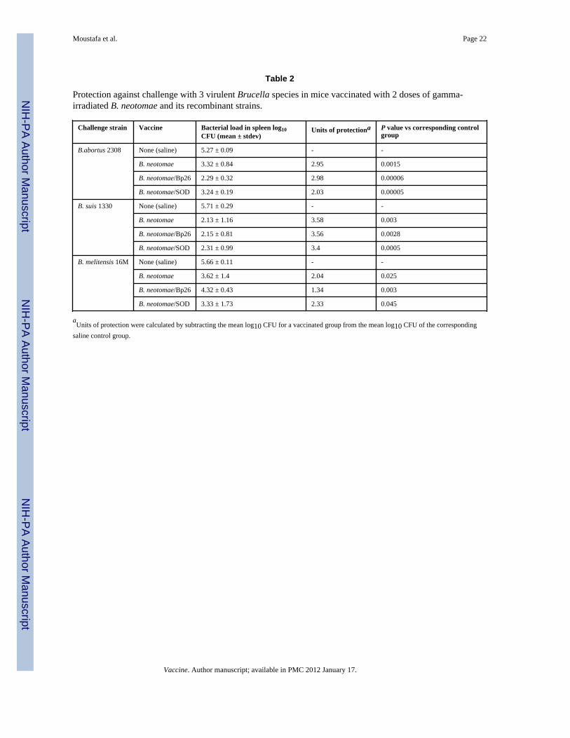
3.6. Protection against challenge with virulent Brucella sppCompared to the saline-inoculated controls, vaccination of mice with 2 doses of irradiated B.neotomae, B. neotomae/Bp26, or B. neotomae/SOD prior to challenge with virulent B.abortus 2308, B. suis 1330 or B. melitensis 16M significantly reduced the number of virulent
Moustafa et al. Page 8
Vaccine. Author manuscript; available in PMC 2012 January 17.
NIH
-PA Author Manuscript
NIH
-PA Author Manuscript
NIH
-PA Author Manuscript
brucellae in the spleens 2 weeks after challenge (Table 2). The vaccinated mice containedapproximately 2-3 logs, 3.5 logs, and 1.3-2.3 logs lower B. abortus 2308, B. suis 1330, andB. melitensis 16M, respectively (Table 2). For each challenge strain, there were nostatistically significant differences in the spleen bacterial loads among the different vaccinegroups.
4. DiscussionIn this study, we demonstrated that vaccination of mice with gamma-irradiated B. neotomaeresults in the development of protective immunity against virulent B. abortus, B. suis, and B.melitensis. The vaccine potential of B. neotomae was first suggested in 1963 by Stoenner forcontrolling swine brucellosis [24]. However, to the best of authors’ knowledge, there are nopublished reports of examining the usefulness of B. neotomae as a brucellosis vaccine.Though B. neotomae is not known to be a pathogen, safety concerns may preclude its use asa live vaccine, especially in humans. Therefore, we used exposure to a minimum dose ofgamma radiation as a means to abolish the ability of B. neotomae to replicate. The use ofionizing radiation has been used in the development of vaccines for preventing infectiousdiseases of animals and humans that are caused by different viruses, bacteria, and parasites[13,25-29]. However, a recently developed strategy specifically uses exposure to a minimumdose of radiation, which is sufficient to abolish replication of the organism, for developingsafer vaccines for diseases caused by intracellular pathogens [13,28-29]. Similar to thepreviously reported findings for B. abortus RB51 and B. melitensis Rev1 [13,29], B.neotomae exposed to a minimum of 300-350 krads of radiation lost its ability to replicate onnutrient rich TSA medium. The irradiated B. neotomae, however, remained metabolicallyactive as demonstrated by the bacteria’s ability to reduce Alamar blue dye and prevent thepenetration of propidium iodide through the cell membranes (Fig. 2). By retaining themetabolic activity, gamma irradiated bacteria can mimic the actual host cell infection of thelive bacteria [28-29]. In addition, the bacteria exposed to a minimum dose of radiation retaintheir de novo protein synthesis capabilities [13,29]. This feature in case of intracellularbacterial pathogens can lead to elicitation of CMI responses to antigens that are expressedwhen the bacteria are inside the host cells. Our PCR analysis suggests that the irradiatedbacteria persisted at higher numbers and for longer time than the heat-killed bacteria in thespleens of the inoculated mice. Though we have not compared the immune responsesinduced by the irradiated and heat-killed B. neotomae, similar previous studies with B.abortus RB51 and B. melitensis showed that the irradiated bacteria are better at inducingantigen-specific and protective immune responses [13,29].
All three irradiated B. neotomae vaccines used in this study conferred similar levels ofprotection against each of the virulent Brucella spp (Table 1). Overexpression of aprotective antigen in the live vaccine strain was previously used as a strategy to induceenhanced protective immunity against brucellosis and tuberculosis in mouse models[18,30-31]. The selection of SOD and Bp26 for overexpression in B. neotomae was based onthe previous findings that these are protective antigens of B. abortus and B. melitensis,respectively [20,32]. Moreover, when used as a live vaccine, overexpression of SOD in B.abortus RB51 (strain RB51SOD) led to induction of increased SOD-specific immuneresponses and enhancement of its protective efficacy against B. abortus challenge in BALB/c mice [18]. Unexpectedly, in this study, we did not detect significantly enhanced SOD-specific antibody and T-cell immune responses in mice vaccinated with the irradiated B.neotomae/SOD. Though the irradiated B. neotomae/SOD vaccine, unlike the live RB51SODvaccine, cannot replicate, we expected that there would be a significantly enhanced SOD-specific immune responses following the booster immunization. This lack of enhancedSOD-specific immune responses suggests that the amount of SOD present in the irradiatedvaccine dose was not sufficiently high enough or that the other immunodominant antigens of
Moustafa et al. Page 9
Vaccine. Author manuscript; available in PMC 2012 January 17.
NIH
-PA Author Manuscript
NIH
-PA Author Manuscript
NIH
-PA Author Manuscript
B. neotomae affected the immunogenic potential of SOD. It is also possible that theoverexpression of SOD did not occur under in vivo conditions. In case of Bp26, micevaccinated with the irradiated B. neotomae/Bp26 developed increased levels of antibodiesand more numbers of IFN-γ-secreting CD8+ T cells specific to Bp26. Nevertheless, theseincreased Bp26-specific immune responses did not translate to enhanced protection againstthe challenge with any of the 3 virulent Brucella spp. (Table 1). If overexpression of someother protective proteins can lead to enhancement of the vaccine efficacy remains to betested.
In general, a Th1 type of immune responses is considered desirable for protection againstintracellular bacterial infections, such as brucellosis. However, some published reportsdocument that the acquired resistance against brucellosis can occur even in presence of amix of Th1 and Th2 immunity [33-34]. The presence of antigen-specific IgG1 and IgG2aantibodies in serum of the vaccinated mice suggest that the irradiated B. neotomae vaccinesinduced a mix of Th1 and Th2 immune responses. However, based on cytokine secretion bythe antigen-specific splenocytes, it appears that the induced Th1 response was moreprominent, because of the significantly higher concentration of IFN-γ in supernatants ofcultures stimulated with irradiated B. neotomae; the concentrations of Th2 cytokines IL-4and IL-5 were marginally higher, compared to the spontaneous release cell controls (Fig. 5).The presence of increased concentration of IL-12p70 and TNF-α in the antigen-stimulatedculture supernatants also suggests the development of Th1 type effector cells. Although Tcells are not a major source for IL-12, a key facilitator of Th1differentiation during animmune response, antigen-specific Th1 cells can positively affect IL-12 secretion byantigen-presenting cells through contact-dependent manner [35].
All three B. neotomae vaccines induced the development of antigen-specific IL-10-secretinglymphocytes (Fig. 5). IL-10 is usually considered to be an anti-inflammatory cytokine whichparticipates in reducing the adverse effects resulting from the excessive production ofproinflammatory cytokines such as IFN-γ [36]. Previous research demonstrated thatinhibition of IL-10 activity leads to increased clearance of B. abortus from infected mice[37]. However, there appears to be no correlation between IL-10 secretion by the vaccine-induced antigen-specific T cells and resistance to virulent Brucella challenge. For example,studies with some experimental Brucella vaccines, such as certain deletion mutants of B.abortus and B. melitensis, and plasmid DNA and recombinant protein of Omp31, inductionof IL-10 secreting antigen-specific lymphocytes had no negative effect on protection tochallenge infection [38-39]. Recently, it has been shown that IL-10 can also promoteinflammation during active inflammatory responses [36]. Therefore, it is possible thatvaccine-induced IL-10 secretion may play a beneficial role in mediating protection againstvirulent Brucella challenge.
Both T cells and anti-smooth LPS antibodies play a role in mediating protection againstbrucellosis [40-41]. Antigen-specific T cells that secrete IFN-γ are primarily responsible forthe acquired CMI against virulent Brucella infection [41]. Passive transfer experimentsshowed that antibodies to the O-antigen can mediate protection [42-47]. Anti-O antibodiesof isotypes IgM, IgG2a, IgGb and IgG3 are effective at affording protection againstbrucellosis in mice [45-47]. All three irradiated B. neotomae vaccines induced LPS-specificantibodies of these isotypes, which increased upon booster immunization (Fig. 4). Usually,IgM is mainly produced during the primary immune response by short-lived plasma cells,thereafter, following the isotype switching in germinal centers, long-lived plasma cellsdevelop which produce antibodies of other isotypes and the production of IgM decreases.Therefore, it is unclear why the levels of IgM remained elevated even after 4 weeks after thebooster immunization. Whether this feature is specific to B. neotomae remains to bedetermined.
Moustafa et al. Page 10
Vaccine. Author manuscript; available in PMC 2012 January 17.
NIH
-PA Author Manuscript
NIH
-PA Author Manuscript
NIH
-PA Author Manuscript

Immunization of mice with any of the three gamma-irradiated B. neotomae vaccinesconferred a significant level of protection against virulent B. abortus, B. suis, and B.melitensis challenges. Based on the units of protection afforded by the three vaccines, thelevel of resistance against B. abortus and B. suis appeared to be better than that against B.melitensis challenge (Table 2). One contributing factor for this difference could be theresistance of B. melitensis to complement-mediated lysis. Both smooth and rough B.melitensis are more resistant to complement-mediated killing than B. abortus with similarphenotypes [48-49]. Another factor could be the variation in epitope dominance in the Oantigen of Brucella smooth LPS between B. melitensis and B. abortus or B. suis. Twoepitopes, designated C (for common among all Brucella smooth LPS) and C/Y (for commonbetween smooth LPS of Brucella and Yersinia enterocolitica O:9), are present in smoothLPS from all Brucella species [50]. Two additional epitopes, designated A (for abortus) andM (for melitensis), are identified in the O-antigen portion of the smooth LPS [51-52].Different smooth Brucella strains possess varying proportions of A and M epitopes. The O-antigen of Brucella smooth LPS is a homopolymer of 4.6-dideoxy-4-formamido-α-D-mannopyranosyl units. These units are linked in α-1,2 in A-dominant smooth Brucellastrains, but every fifth residue is linked in α-1,3 in M-dominant strains. Antibodies to thedominant epitope of the O-antigen are more effective in mediating protection [50]. B.neotomae, B. abortus 2308, and B. suis 1330 are A-dominant strains, while B. melitensis16M is a M-dominant strain [50]. Although we did not identify the epitope specificity of theanti-smooth LPS antibodies produced by the vaccinated mice, it is highly likely that theirradiated B. neotomae vaccines induced more antibodies to specific to A than M epitope. Inaddition, antigen specificity of the T cell responses could also have affected the level ofprotection conferred by the vaccines against B. melitensis challenge. Enhancement of T cellresponses through strategies such as microencapsulation might increase the protectiveefficacy of the irradiated B. neotomae vaccines [53].
In the previous studies with the irradiated B. abortus RB51 and B. melitensis Rev1, a singledose of 109 CFU equivalent per mouse was used for immunization [13,29]. However, in ourpreliminary experiments, we observed that the mice inoculated with 109 CFU equivalent ofirradiated B. neotomae exhibited clinical signs consistent with the vaccine-induced sicknessduring the first 3-5 days after immunization. On the contrary, mice inoculated with 108 CFUequivalent of irradiated B. neotomae did not show any signs of distress. Therefore, anempirical regimen of 2 doses of 108 CFU equivalent of the irradiated bacteria each at 2-weeks apart was used for the studies. Further studies are warranted to determine theminimum effective dose of the vaccine and the duration of the protective immune status inthe vaccinated animals. In addition, examining the potential of the irradiated B. neotomae asan oral vaccine would be very pertinent for human application.
In conclusion, the results suggest that gamma-irradiated B. neotomae can be used fordeveloping an effective vaccine against brucellosis caused by B. abortus, B. suis and B.melitensis. The non-pathogenic feature of B. neotomae along with the inability of theirradiated bacteria to replicate in the host makes it a safer alternative to the other liveattenuated vaccine candidates for human brucellosis.
Supplementary MaterialRefer to Web version on PubMed Central for supplementary material.
AcknowledgmentsThis work was supported by Public Health Service grant AI065667-01A2 from the National Institute of Allergy andInfectious Diseases. We thank Lynn Guptill and Harm HogenEsch for critical review of the manuscript and KayCarlson for help with the mouse protection studies.
Moustafa et al. Page 11
Vaccine. Author manuscript; available in PMC 2012 January 17.
NIH
-PA Author Manuscript
NIH
-PA Author Manuscript
NIH
-PA Author Manuscript

References1. Corbel MJ. Brucellosis: an overview. Emerg Infect Dis. 1997; 3(2):213–21. [PubMed: 9204307]2. Franco MP, Mulder M, Gilman RH, Smits HL. Human brucellosis. Lancet Infect Dis. 2007; 7(12):
775–86. [PubMed: 18045560]3. Cutler SJ, Whatmore AM, Commander NJ. Brucellosis - new aspects of an old disease. J Appl
Microbiol. 2005; 98(6):1270–81. [PubMed: 15916641]4. Pappas G, Papadimitriou P, Akritidis N, Christou L, Tsianos EV. The new global map of human
brucellosis. Lancet Infect Dis. 2006; 6(2):91–9. [PubMed: 16439329]5. Ficht TA, Kahl-McDonagh MM, Arenas-Gamboa AM, Rice-Ficht AC. Brucellosis: The case for
live, attenuated vaccines. Vaccine. 2009; 27:D40–D3. [PubMed: 19837284]6. Schurig GG, Sriranganathan N, Corbel MJ. Brucellosis vaccines: past, present and future. Vet
Microbiol. 2002; 90(1-4):479–96. [PubMed: 12414166]7. Perkins SD, Smither SJ, Atkins HS. Towards a Brucella vaccine for humans. FEMS Microbiol Rev.
2010; 34(3):379–394.8. Blasco JM, Díaz R. Brucella melitensis Rev-1 vaccine as a cause of human brucellosis. The Lancet.
1993; 342(8874):805.9. Stoenner HG, Lackman DB. A new species of Brucella isolated from the desert wood rat, Neotoma
lepida Thomas. Am J Vet Res. 1957; 18(69):947–51. [PubMed: 13470254]10. Kovach ME, Elzer PH, Hill DS, Robertson GT, Farris MA, Roop RM 2nd, et al. Four new
derivatives of the broad-host-range cloning vector pBBR1MCS, carrying different antibiotic-resistance cassettes. Gene. 166(1):175–6. [PubMed: 8529885]
11. Vemulapalli R, He Y, Boyle SM, Sriranganathan N, Schurig GG. Brucella abortus strain RB51 asa vector for heterologous protein expression and induction of specific Th1 type immune responses.Infect Immun. 2000; 68(6):3290–6. [PubMed: 10816476]
12. McQuiston JR, Schurig GG, Sriranganathan N, Boyle SM. Transformation of Brucella specieswith suicide and broad host-range plasmids. Methods Mol Biol. 1995; 47:143–8. [PubMed:7550728]
13. Sanakkayala N, Sokolovska A, Gulani J, HogenEsch H, Sriranganathan N, Boyle SM, et al.Induction of antigen-specific Th1-type immune responses by gamma-irradiated recombinantBrucella abortus RB51. Clin Diagn Lab Immunol. 2005; 12(12):1429–36. [PubMed: 16339067]
14. Bounaadja L, Albert D, Chenais B, Henault S, Zygmunt MS, Poliak S, et al. Real-time PCR foridentification of Brucella spp.: a comparative study of IS711, bcsp31 and per target genes. VetMicrobiol. 2009; 137(1-2):156–64. [PubMed: 19200666]
15. Bradford MM. A rapid and sensitive method for the quantitation of microgram quantities of proteinutilizing the principle of protein-dye binding. Anal Biochem. 1976; 72:248–54. [PubMed: 942051]
16. Morrison DC, Leive L. Fractions of Lipopolysaccharide from Escherichia coli O111-B4 preparedby two extraction procedures. J Biol Chem. 1975; 250(8):2911–9. [PubMed: 804483]
17. Phillips M, Pugh GW, Deyoe BL. Chemical and protective properties of Brucellalipopolysaccharide obtained by butanol extraction. Am J Vet Res. 1989; 50(3):311–7. [PubMed:2494911]
18. Vemulapalli R, He Y, Cravero S, Sriranganathan N, Boyle SM, Schurig GG. Overexpression ofprotective antigen as a novel approach to enhance vaccine efficacy of Brucella abortus strainRB51. Infect Immun. 2000; 68(6):3286–9. [PubMed: 10816475]
19. Vemulapalli R, Duncan AJ, Boyle SM, Sriranganathan N, Toth TE, Schurig GG. Cloning andsequencing of yajC and secD homologs of Brucella abortus and demonstration of immuneresponses to YajC in mice vaccinated with B. abortus RB51. Infect Immun. 1998; 66(12):5684–91. [PubMed: 9826342]
20. Onate AA, Vemulapalli R, Andrews E, Schurig GG, Boyle S, Folch H. Vaccination with liveEscherichia coli expressing Brucella abortus Cu/Zn superoxide dismutase protects mice againstvirulent B. abortus. Infect Immun. 1999; 67(2):986–8. [PubMed: 9916121]
21. Murali-Krishna K, Altman JD, Suresh M, Sourdive DJD, Zajac AJ, Miller J, Ahmed R. Countingantigen-specific CD8 T cells: a reevaluation of bystander activation during viral infection.Immunity. 1998; 8(2):177–87. [PubMed: 9491999]
Moustafa et al. Page 12
Vaccine. Author manuscript; available in PMC 2012 January 17.
NIH
-PA Author Manuscript
NIH
-PA Author Manuscript
NIH
-PA Author Manuscript

22. Schurig GG, Roop RM, Bagchi T, Boyle S, Buhrman D, Sriranganathan N. Biological propertiesof RB51; a stable rough strain of Brucella abortus. Vet Microbiol. 199; 28(2):171–88. [PubMed:1908158]
23. Bricker BJ, Tabatabai LB, Judge BA, Deyoe BL, Mayfield JE. Cloning, expression, andoccurrence of the Brucella Cu-Zn superoxide dismutase. Infect Immun. 1990; 58(9):2935–9.[PubMed: 2201639]
24. Stoenner HG. The behavior of Brucella neotomae and Brucella suis in reciprocal superinfectionexperiments in mice and guinea pigs. Am J Vet Res. 1963; 24:376–80. [PubMed: 13984251]
25. Hubbert WT, Miller JN. Studies on immunity in experimental leptospirosis: the immunogenicity ofLeptospira icterohemorrhagiae attenuated by gamma-irradiation. J Immunol. 1965; 95(4):759–64.[PubMed: 5841058]
26. Jarrett WF, Jennings FW, Mc IW, Mulligan W, Urquhart GM. Irradiated helminth larvae invaccination. Proc R Soc Med. 1958; 51(9):743–4. [PubMed: 13591294]
27. Martin SS, Bakken RR, Lind CM, Garcia P, Jenkins E, Glass PJ, et al. Comparison of theimmunological responses and efficacy of gamma-irradiated V3526 vaccine formulations againstsubcutaneous and aerosol challenge with Venezuelan equine encephalitis virus subtype IAB.Vaccine. 2010; 28(4):1031–40. [PubMed: 19914193]
28. Datta SK, Okamoto S, Hayashi T, Shin SS, Mihajlov I, Fermin A, et al. Vaccination with irradiatedListeria induces protective T cell immunity. Immunity. 2006; 25(1):143–52. [PubMed: 16860763]
29. Magnani DM, Harms JS, Durward MA, Splitter GA. Nondividing but metabolically active gamma-irradiated Brucella melitensis is protective against virulent B. melitensis challenge in mice. InfectImmun. 2009; 77(11):5181–9. [PubMed: 19703982]
30. Dhar N, Rao V, Tyagi AK. Recombinant BCG approach for development of vaccines: cloning andexpression of immunodominant antigens of M. tuberculosis. Fems Microbiol Lett. 2000; 190(2):309–16. [PubMed: 11034297]
31. Rao V, Dhar N, Tyagi AK. Modulation of host immune responses by overexpression ofimmunodominant antigens of Mycobacterium tuberculosis in bacille Calmette-Guérin. Scand JImmunol. 2003; 58(4):449–61. [PubMed: 14507310]
32. Yang X, Hudson M, Walters N, Bargatze RF, Pascual DW. Selection of protective epitopes ofBrucella melitensis by DNA vaccination. Infect Immun. 2005; 73(11):7297–303. [PubMed:16239526]
33. Velikovsky CA, Goldbaum FA, Cassataro J, Estein S, Bowden RA, Bruno L, et al. Brucellalumazine synthase elicits a mixed Th1-Th2 immune response and reduces infection in micechallenged with Brucella abortus 544 independently of the adjuvant formulation used. InfectImmun. 2003; 71(10):5750–5. [PubMed: 14500496]
34. Delpino MV, Estein SM, Fossati CA, Baldi PC, Cassataro. Vaccination with Brucella recombinantDnaK and SurA proteins induces protection against Brucella abortus infection in BALB/c mice.Vaccine. 2007; 25(37-38):6721–9. [PubMed: 17686554]
35. Ria F, Penna G, Adorini L. Th1 cells induce and Th2 inhibit antigen-dependent IL-12 secretion bydendritic cells. Eur J Immunol. 1998; 28(6):2003–16. [PubMed: 9645382]
36. Sharif MN, Tassiulas I, Hu Y, Mecklenbrauker I, Tarakhovsky A, Ivashkiv LB. IFN-alpha primingresults in a gain of proinflammatory function by IL-10: implications for systemic lupuserythematosus pathogenesis. J Immunol. 2004; 172(10):6476–81. [PubMed: 15128840]
37. Fernandes DM, Baldwin CL. Interleukin-10 downregulates protective immunity to Brucellaabortus. Infect Immun. 1995; 63(3):1130–3. [PubMed: 7868238]
38. Cassataro J, Velikovsky CA, Bruno L, Estein SM, de la Barrera S, Bowden R, et al. Improvedimmunogenicity of a vaccination regimen combining a DNA vaccine encoding Brucella melitensisouter membrane protein 31 (Omp31) and recombinant Omp31 boosting. Clin Vaccine Immunol.2007; 14(7):869–74. [PubMed: 17428946]
39. Kahl-McDonagh MM, Ficht TA. Evaluation of protection afforded by Brucella abortus andBrucella melitensis unmarked deletion mutants exhibiting different rates of clearance in BALB/cmice. Infect Immun. 2006; 74(7):4048–57. [PubMed: 16790778]
Moustafa et al. Page 13
Vaccine. Author manuscript; available in PMC 2012 January 17.
NIH
-PA Author Manuscript
NIH
-PA Author Manuscript
NIH
-PA Author Manuscript

40. Gonzalez D, Grillo MJ, De Miguel MJ, Ali T, Arce-Gorvel V, Delrue RM, et al. Brucellosisvaccines: assessment of Brucella melitensis lipopolysacharide rough mutants defective in core andO-polysaccharide synthesis and export. PLoS One. 2008; 3(7):e2760. [PubMed: 18648644]
41. Yingst S, Hoover DL. T cell immunity to brucellosis. Crit Rev Microbiol. 2003; 29(4):313–31.[PubMed: 14636042]
42. Limet J, Plommet AM, Dubray G, Plommet M. Immunity conferred upon mice by anti-LPSmonoclonal antibodies in murine brucellosis. Ann Inst Pasteur Immunol. 1987; 138(3):417–24.
43. Araya LN, Elzer PH, Rowe GE, Enright FM, Winter AJ. Temporal development of protective cell-mediated and humoral immunity in BALB/c mice infected with Brucella abortus. J Immunol.1989; 143(10):3330–3337. [PubMed: 2509555]
44. Montaraz JA, Winter AJ, Hunter DM, Sowa BA, Wu AM, Adams LG. Protection against Brucellaabortus in mice with O-polysaccharide-specific monoclonal antibodies. Infect Immun. 1986;51(3):961–3. [PubMed: 3005173]
45. Elzer PH, Jacobson RH, Jones SM, Nielsen KH, Douglas JT, Winter AJ. Antibody-mediatedprotection against Brucella abortus in BALB/c mice at successive periods after infection: variationbetween virulent strain 2308 and attenuated vaccine strain 19. Immunology. 1994; 82(4):651–8.[PubMed: 7835931]
46. Elzer PH, Jacobson RH, Nielsen KH, Douglas JT, Winter AJ. BALB/c mice infected with Brucellaabortus express protracted polyclonal responses of both IgG2a and IgG3 isotypes. Immunol Lett.1994; 42(3):145–50. [PubMed: 7890314]
47. Cloeckaert A, Jacques I, de Wergifosse P, Dubray G, Limet JN. Protection against Brucellamelitensis or Brucella abortus in mice with immunoglobulin G (IgG), IgA, and IgM monoclonalantibodies specific for a common epitope shared by the Brucella A and M smoothlipopolysaccharides. Infect Immun. 1992; 60(1):312–5. [PubMed: 1370276]
48. Fernandez-Prada CM, Nikolich M, Vemulapalli R, Sriranganathan N, Boyle SM, Schurig GG, etal. Deletion of wboA enhances activation of the lectin pathway of complement in Brucella abortusand Brucella melitensis. Infect Immun. 2001; 69(7):4407–16. [PubMed: 11401980]
49. Fernandez-Prada CM, Zelazowska EB, Nikolich M, Hadfield TL, Roop RM 2, Robertson GL, etal. Interaction between Brucella melitensis and human phagocytes: bacterial surface O-polysaccharide inhibits phagocytosis, bacterial killing, and subsequent host cell apoptosis. InfectImmun. 2003; 71(4):2110–9. [PubMed: 12654833]
50. Cloeckaert A, Weynants V, Godfroid J, Verger JM, Grayon M, Zygmunt MS. O-Polysaccharideepitopic heterogeneity at the surface of Brucella spp. studied by enzyme-linked immunosorbentassay and flow cytometry. Clin Diagn Lab Immunol. 1998; 5(6):862–70. [PubMed: 9801349]
51. Caroff M, Bundle DR, Perry MB, Cherwonogrodzky JW, Duncan JR. Antigenic S-typelipopolysaccharide of Brucella abortus 1119-3. Infect Immun. 1984; 46(2):384–8. [PubMed:6437981]
52. Bundle DR, Cherwonogrodzky JW, Perry MB. Structural elucidation of the Brucella melitensis Mantigen by high-resolution NMR at 500 MHz. Biochemistry. 1987; 26(26):8717–26. [PubMed:3442684]
53. Arenas-Gamboa AM, Ficht TA, Kahl-McDonagh MM, Rice-Ficht AC. Immunization with a singledose of a microencapsulated Brucella melitensis mutant enhances protection against wild-typechallenge. Infect Immun. 2008; 76(6):2448–55. [PubMed: 18362129]
Moustafa et al. Page 14
Vaccine. Author manuscript; available in PMC 2012 January 17.
NIH
-PA Author Manuscript
NIH
-PA Author Manuscript
NIH
-PA Author Manuscript
Figure 1.Detection of expression of Bp26 and SOD proteins in B. neotomae/Bp26 and B. neotomae/SOD by SDS-PAGE and Western blot analyses. The whole antigen of B. neotomae and B.neotomae/Bp26 and B. neotomae/SOD were separated by 12.5 % SDS-PAGE and eitherstained with Commassie brilliant blue (A and C) or analyzed by Western blotting withmono-specific serum to MBP-Bp26 (B) or SOD (D). MBP-Bp26k and SOD are proteinspurified from overexpressing E. coli. The arrow heads indicate the reacting protein. LaneMW, molecular weight marker in kilodaltons (kDa).
Moustafa et al. Page 15
Vaccine. Author manuscript; available in PMC 2012 January 17.
NIH
-PA Author Manuscript
NIH
-PA Author Manuscript
NIH
-PA Author Manuscript

Figure 2.Effect of gamma-irradiation on the viability of B. neotomae. Aliquots of B. neotomae wereexposed to different doses of gamma radiation, the viable bacteria in each aliquots weredetermined by plating 10-fold dilutions on TSA plates (A), and staining with live/deadBacLight kit for microscopy (B). As a control, an aliquot each of live and heat-killedbacteria was included for microscopy.
Moustafa et al. Page 16
Vaccine. Author manuscript; available in PMC 2012 January 17.
NIH
-PA Author Manuscript
NIH
-PA Author Manuscript
NIH
-PA Author Manuscript

Figure 3.ELISA detection of Bp26- and SOD-specific IgG, IgG1, and IgG2a antibodies in serum ofmice vaccinated with gamma irradiated B. neotomae, B. neotomae/Bp26, and B. neotomae/SOD, or inoculated with saline. Serum samples were collected at 2 and 6 weeks after initialvaccination, and were diluted 1 in 200 and assayed for the presence of total antigen specificantibodies. Results are shown as mean ± standard deviation (n=4) of absorbance at 450 nmof the color developed.* Significantly different from the corresponding saline group at week 2 (P<0.05).** Significantly different from the corresponding saline group at week 6 (P<0.05).ff Week 6 is significantly different from week 2 within each vaccine group (P<0.05).
Moustafa et al. Page 17
Vaccine. Author manuscript; available in PMC 2012 January 17.
NIH
-PA Author Manuscript
NIH
-PA Author Manuscript
NIH
-PA Author Manuscript

Figure 4.ELISA detection of B. neotomae LPS specific antibodies IgG, IgG1, and IgG2a antibodiesin serum of mice vaccinated with gamma irradiated B. neotomae, B. neotomae/Bp26, and B.neotomae/SOD, or inoculated with saline. Serum samples were collected at 2 and 6 weeksafter initial vaccination, and were diluted 1 in 200 and assayed for the presence of totalantigen specific antibodies. Results are shown as mean ± standard deviation (n=4) ofabsorbance at 450 nm of the color developed.* Significantly different from the corresponding saline group at week 2 (P<0.05).** Significantly different from the corresponding saline group at week 6 (P<0.05).ff Week 6 is significantly different from week 2 within each vaccine group (P<0.05).
Moustafa et al. Page 18
Vaccine. Author manuscript; available in PMC 2012 January 17.
NIH
-PA Author Manuscript
NIH
-PA Author Manuscript
NIH
-PA Author Manuscript

Figure 5.Production of specific cytokines by splenocytes of BALB/c mice vaccinated with gammairradiated B. neotomae and its recombinants after in vitro stimulation with irradiated B.neotomae (IRR-B.neotomae), MBP-Bp26 (Bp26), or SOD. Values are means ± standarddeviation (n=4).* Significantly different from the corresponding unstimulated control with B. neotomaestimulation (P<0.05)** Significantly different from the corresponding unstimulated control with MBP-Bp26stimulation (P<0.05)*** Significantly different from the corresponding unstimulated control with SODstimulation (P<0.05)
Moustafa et al. Page 19
Vaccine. Author manuscript; available in PMC 2012 January 17.
NIH
-PA Author Manuscript
NIH
-PA Author Manuscript
NIH
-PA Author Manuscript

Figure 6.Flow cytometric analysis showing the percentage of interferon-γ secreting CD4+ and CD8+T cells in the spleens of BALB/c mice immunized with gamma irradiated B. neotomae andits recombinants following in vitro stimulation with: (A) gamma irradiated B. neotomae, (B)SOD,(C) MBP-Bp26.
Moustafa et al. Page 20
Vaccine. Author manuscript; available in PMC 2012 January 17.
NIH
-PA Author Manuscript
NIH
-PA Author Manuscript
NIH
-PA Author Manuscript

NIH
-PA Author Manuscript
NIH
-PA Author Manuscript
NIH
-PA Author Manuscript
Moustafa et al. Page 21
Table 1
Persistence of gamma-irradiated and heat-killed B. neotomae in mouse spleens as detected by the real-timequantitative PCR.
Post-inoculation time log 10 CFU equivalent/spleen (mean ± stdev)
Irradiated B. neotomae Heat-killed B. neotomae
3 hours 5.04±0.01 4.76±0.13
24 hours 4.19±0.02 1.8±0.07
3 days 3.34±0.02 0.21±0.15
5 days - -
Vaccine. Author manuscript; available in PMC 2012 January 17.
NIH
-PA Author Manuscript
NIH
-PA Author Manuscript
NIH
-PA Author Manuscript
Moustafa et al. Page 22
Table 2
Protection against challenge with 3 virulent Brucella species in mice vaccinated with 2 doses of gamma-irradiated B. neotomae and its recombinant strains.
Challenge strain Vaccine Bacterial load in spleen log10CFU (mean ± stdev)
Units of protectiona P value vs corresponding controlgroup
B.abortus 2308 None (saline) 5.27 ± 0.09 - -
B. neotomae 3.32 ± 0.84 2.95 0.0015
B. neotomae/Bp26 2.29 ± 0.32 2.98 0.00006
B. neotomae/SOD 3.24 ± 0.19 2.03 0.00005
B. suis 1330 None (saline) 5.71 ± 0.29 - -
B. neotomae 2.13 ± 1.16 3.58 0.003
B. neotomae/Bp26 2.15 ± 0.81 3.56 0.0028
B. neotomae/SOD 2.31 ± 0.99 3.4 0.0005
B. melitensis 16M None (saline) 5.66 ± 0.11 - -
B. neotomae 3.62 ± 1.4 2.04 0.025
B. neotomae/Bp26 4.32 ± 0.43 1.34 0.003
B. neotomae/SOD 3.33 ± 1.73 2.33 0.045
aUnits of protection were calculated by subtracting the mean log10 CFU for a vaccinated group from the mean log10 CFU of the corresponding
saline control group.
Vaccine. Author manuscript; available in PMC 2012 January 17.