Whole-brain irradiation increases NREM sleep and hypothalamic expression of IL-1β in rats
Upload
independentCategory
view
0download
0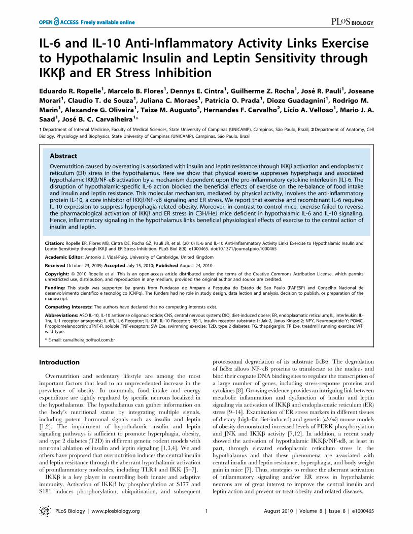
IL-6 and IL-10 Anti-Inflammatory Activity Links Exerciseto Hypothalamic Insulin and Leptin Sensitivity throughIKKb and ER Stress InhibitionEduardo R. Ropelle1, Marcelo B. Flores1, Dennys E. Cintra1, Guilherme Z. Rocha1, Jose R. Pauli1, Joseane
Morari1, Claudio T. de Souza1, Juliana C. Moraes1, Patrıcia O. Prada1, Dioze Guadagnini1, Rodrigo M.
Marin1, Alexandre G. Oliveira1, Taize M. Augusto2, Hernandes F. Carvalho2, Lıcio A. Velloso1, Mario J. A.
Saad1, Jose B. C. Carvalheira1*
1 Department of Internal Medicine, Faculty of Medical Sciences, State University of Campinas (UNICAMP), Campinas, Sao Paulo, Brazil, 2 Department of Anatomy, Cell
Biology, Physiology and Biophysics, State University of Campinas (UNICAMP), Campinas, Sao Paulo, Brazil
Abstract
Overnutrition caused by overeating is associated with insulin and leptin resistance through IKKb activation and endoplasmicreticulum (ER) stress in the hypothalamus. Here we show that physical exercise suppresses hyperphagia and associatedhypothalamic IKKb/NF-kB activation by a mechanism dependent upon the pro-inflammatory cytokine interleukin (IL)-6. Thedisruption of hypothalamic-specific IL-6 action blocked the beneficial effects of exercise on the re-balance of food intakeand insulin and leptin resistance. This molecular mechanism, mediated by physical activity, involves the anti-inflammatoryprotein IL-10, a core inhibitor of IKKb/NF-kB signaling and ER stress. We report that exercise and recombinant IL-6 requiresIL-10 expression to suppress hyperphagia-related obesity. Moreover, in contrast to control mice, exercise failed to reversethe pharmacological activation of IKKb and ER stress in C3H/HeJ mice deficient in hypothalamic IL-6 and IL-10 signaling.Hence, inflammatory signaling in the hypothalamus links beneficial physiological effects of exercise to the central action ofinsulin and leptin.
Citation: Ropelle ER, Flores MB, Cintra DE, Rocha GZ, Pauli JR, et al. (2010) IL-6 and IL-10 Anti-Inflammatory Activity Links Exercise to Hypothalamic Insulin andLeptin Sensitivity through IKKb and ER Stress Inhibition. PLoS Biol 8(8): e1000465. doi:10.1371/journal.pbio.1000465
Academic Editor: Antonio J. Vidal-Puig, University of Cambridge, United Kingdom
Received October 23, 2009; Accepted July 15, 2010; Published August 24, 2010
Copyright: � 2010 Ropelle et al. This is an open-access article distributed under the terms of the Creative Commons Attribution License, which permitsunrestricted use, distribution, and reproduction in any medium, provided the original author and source are credited.
Funding: This study was supported by grants from Fundacao de Amparo a Pesquisa do Estado de Sao Paulo (FAPESP) and Conselho Nacional dedesenvolvimento cientıfico e tecnologico (CNPq). The funders had no role in study design, data lection and analysis, decision to publish, or preparation of themanuscript.
Competing Interests: The authors have declared that no competing interests exist.
Abbreviations: ASO IL-10, IL-10 antisense oligonucleotide; CNS, central nervous system; DIO, diet-induced obese; ER, endoplasmatic reticulum; IL, interleukin; IL-1ra, IL-1 receptor antagonist; IL-6R, IL-6 Receptor; IL-10R, IL-10 Receptor; IRS-1, insulin receptor substrate-1; Jak-2, Janus Kinase-2; NPY, Neuropeptide-Y; POMC,Proopiomelanocortin; sTNF-R, soluble TNF-receptors; SW Exe, swimming exercise; T2D, type 2 diabetes; TG, thapsigargin; TR Exe, treadmill running exercise; WT,wild type.
* E-mail: [email protected]
Introduction
Overnutrition and sedentary lifestyle are among the most
important factors that lead to an unprecedented increase in the
prevalence of obesity. In mammals, food intake and energy
expenditure are tightly regulated by specific neurons localized in
the hypothalamus. The hypothalamus can gather information on
the body’s nutritional status by integrating multiple signals,
including potent hormonal signals such as insulin and leptin
[1,2]. The impairment of hypothalamic insulin and leptin
signaling pathways is sufficient to promote hyperphagia, obesity,
and type 2 diabetes (T2D) in different genetic rodent models with
neuronal ablation of insulin and leptin signaling [1,3,4]. We and
others have proposed that overnutrition induces the central insulin
and leptin resistance through the aberrant hypothalamic activation
of proinflammatory molecules, including TLR4 and IKK [5–7].
IKKb is a key player in controlling both innate and adaptive
immunity. Activation of IKKb by phosphorylation at S177 and
S181 induces phosphorylation, ubiquitination, and subsequent
proteosomal degradation of its substrate IkBa. The degradation
of IkBa allows NF-kB proteins to translocate to the nucleus and
bind their cognate DNA binding sites to regulate the transcription of
a large number of genes, including stress-response proteins and
cytokines [8]. Growing evidence provides an intriguing link between
metabolic inflammation and dysfunction of insulin and leptin
signaling via activation of IKKb and endoplasmatic reticulum (ER)
stress [9–14]. Examination of ER stress markers in different tissues
of dietary (high-fat diet-induced) and genetic (ob/ob) mouse models
of obesity demonstrated increased levels of PERK phosphorylation
and JNK and IKKb activity [7,12]. In addition, a recent study
showed the activation of hypothalamic IKKb/NF-kB, at least in
part, through elevated endoplasmic reticulum stress in the
hypothalamus and that these phenomena are associated with
central insulin and leptin resistance, hyperphagia, and body weight
gain in mice [7]. Thus, strategies to reduce the aberrant activation
of inflammatory signaling and/or ER stress in hypothalamic
neurons are of great interest to improve the central insulin and
leptin action and prevent or treat obesity and related diseases.
PLoS Biology | www.plosbiology.org 1 August 2010 | Volume 8 | Issue 8 | e1000465
Physical activity is considered a cornerstone of the treatment for
obesity. Exercise has long been reported to reduce body weight
and visceral adiposity, increasing the energy expenditure and
improving glycaemic control in overweight or T2D patients
[15,16]. Since the discovery of interleukin (IL)-6 releases from
contracting skeletal muscle, accumulating evidence indicates that
exercise induces metabolic changes in other organs, such as the
liver, the adipose tissue, and hypothalamus, in an IL-6 dependent
manner. IL-6 is most often classified as a pro-inflammatory
cytokine, although consistent data also demonstrate that IL-6 has
an anti-inflammatory effect and may negatively regulate the
inflammation of acute phase response by increasing IL-10, IL-1
receptor antagonist (IL-1ra), and soluble TNF-receptors (sTNF-R)
[17]. Moreover, IL-6 appears to play a central role in the
regulation of appetite, energy expenditure, and body composition
[18,19]. However, the effects of physical activity in the metabolic
regulatory pathways in the central nervous system (CNS) remain
unexplored. Thus, we hypothesized that exercise could exert its
effects in the CNS by modulating the specific hypothalamic
neurons responsible for the control of food consumption. In the
present study, we investigated the effect of the anti-inflammatory
response, mediated by IL-6, on hypothalamic IKKb activation
and ER stress, central insulin and leptin sensitivity, and food intake
in diet-induced rats after physical activity.
Results
Exercise Suppresses Hyperphagia Mediated byOvernutrition
It has been demonstrated that physical activity may contribute
to the energy balance by increasing energy expenditure. Although
the energy expenditure aspects of such exercise may contribute to
the effects of weight loss, the effect of exercise on the control of
energy intake remains unclear. To evaluate the impact of physical
activity on food consumption, we measured the 12-h total energy
intake in lean and diet-induced obese (DIO) rats after one bout of
swimming (SW Exe) and treadmill running (TR Exe) exercise.
Neither of the exercise protocols changed the energy intake in lean
animals; however, exercise suppressed the hyperphagic response,
mediated by chronic overnutrition, restoring the energy intake to
the levels of lean animals (Figure 1A). To assess whether the effects
of exercise on food intake are dependent on the neuropeptides
modulation, we performed a real time PCR assay to determine the
mRNA levels of Neuropeptide-Y (NPY) and Proopiomelanocortin
(POMC). After 9 h of fasting, we found that chronic overnutrition
increased NPY mRNA and reduced POMC mRNA levels, while
physical activity restored the NPY (Figure 1B) and POMC mRNA
levels (Figure 1C) in obese animals; on the other hand, exercise did
not change the NPY and POMC mRNA levels in lean rats
(Figure 1B and C).
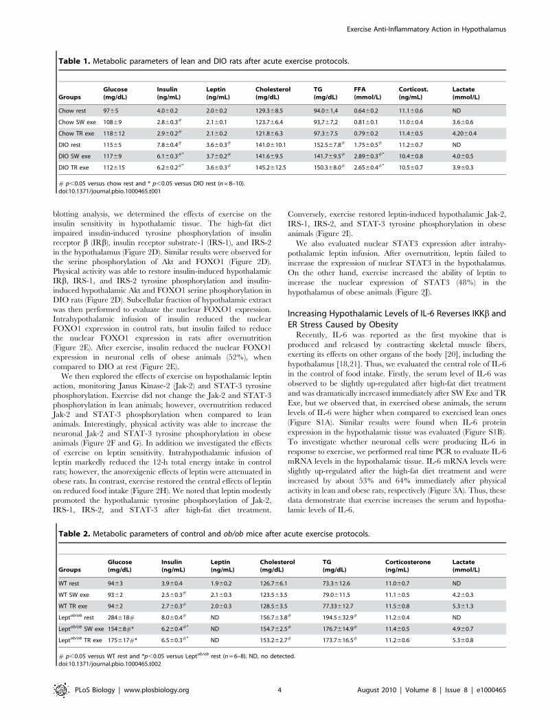
Chronic overnutrition increased body weight, epididymal fat
(Figure 1D and E), serum insulin, leptin, triglycerides, and free
fatty acid levels (Table 1), compared to age-matched controls. No
significant variations were found in body weight, epididymal fat
serum leptin, triglycerides, and urinary corticosterone levels
between exercised and obese animals under resting conditions
(Figure 1D, E and Table 1). The insulin levels were lower in both
lean and obese rats after the exercise protocols (Table 1) and
exercise increased the free fatty acid in obese animals (Table 1). To
determine whether lean and obese rodents were swimming or
running in the same fashion, we evaluated lactate production
every 15 min during the SW Exe and TR Exe. We did not find
any difference in the lactate production between lean and obese
rats. Table 1 depicts the final values obtained in this test. These
results reinforce the negative relationship between body weight
change and stress related with the appetite-suppressive actions
mediated by exercise.
To extend our hypothesis, we investigated food intake in leptin-
deficient mice (ob/ob) after physical activity. Acute SW Exe and
TR Exe did not change the food intake in wild type (WT) mice,
however the food consumption was reduced in ob/ob mice
(Figure 1F). After 9 h of fasting, we found that NPY mRNA was
increased and POMC mRNA levels were reduced in ob/ob mice,
while physical activity restored the NPY (Figure 1G) and POMC
mRNA levels (Figure 1H) in obese animals; on the other hand,
exercise did not change the NPY and POMC mRNA levels in
control mice (Figure 1G and H). Exercise did not change the total
body weight and epididymal fat pad weight in WT and ob/ob mice
(Figure 1I and J). In addition, we observed that the exercise
protocols did not change the triglycerides and free fatty acid levels
but reduced the insulin levels in WT and ob/ob mice (Table 2). The
lactate production was similar between lean and obese mice
during the respective exercise protocols (Table 2). These exercise
protocols did not evoke any significant stressful effect in these
animals, as demonstrated by urinary corticosterone levels (Table 2).
Thus, our data demonstrate that exercise modulates hypothalamic
neuropeptides (NPY and POMC) and suppresses food intake in
obese, but not in lean, rodents without changing the adipose tissue
content and corticosterone levels.
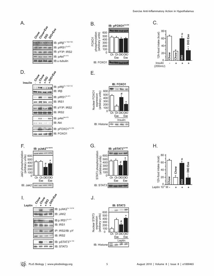
Exercise Restores Insulin and Leptin Sensitivity in theHypothalamus
Next, we evaluated whether exercise modulates insulin signaling
in the hypothalamus. Western blot analysis revealed that IRb,
IRS-1, IRS-2, Akt, and FOXO1 phosphorylation were similar
between the groups (Figure 2A and B). Although exercise did not
change the basal levels of insulin signaling, we next performed
intrahypothalamic insulin (200 mU) or its vehicle injection to
evaluated food intake and insulin sensitivity after the SW Exe
protocol. Overnutrition markedly reduced the ability of intrahy-
pothalamic insulin infusion to reduce food intake, when compared
to chow-fed animals; however, exercise restored the central effects
of insulin on reduced food intake (Figure 2C). Using Western
Author Summary
The hypothalamus is a brain region that gathers informa-tion on the body’s nutritional status and governs therelease of multiple metabolic signaling molecules such asinsulin and leptin to maintain homeostasis. Overeating andobesity are associated with insulin and leptin resistance inthe hypothalamus, and recent studies provide an intrigu-ing link between inflammation and dysfunction ofhypothalamic insulin and leptin signaling through activa-tion of IKKb, a key player in immune response, andendoplasmic reticulum (ER) stress. This means thatstrategies to reduce the aberrant activation of inflamma-tory signaling in the hypothalamus are of great interest toimprove the central insulin and leptin action and preventor treat related metabolic diseases. Using a combination ofpharmacological, genetic, and physiological approaches,our study indicates that physical activity reorganizes theset point of nutritional balance through anti-inflammatorysignaling mediated by interleukin (IL)-6 and IL-10 in thehypothalamus of rodents. Hence, IL-6 and IL-10 areimportant physiological contributors to the central insulinand leptin action mediated by exercise, linking it tohypothalamic ER stress and inflammation.
Exercise Anti-Inflammatory Action in Hypothalamus
PLoS Biology | www.plosbiology.org 2 August 2010 | Volume 8 | Issue 8 | e1000465
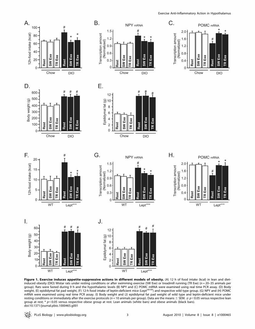
Figure 1. Exercise induces appetite-suppressive actions in different models of obesity. (A) 12 h of food intake (kcal) in lean and diet-induced obesity (DIO) Wistar rats under resting conditions or after swimming exercise (SW Exe) or treadmill running (TR Exe) (n = 20–35 animals pergroup). Rats were fasted during 9 h and the hypothalamic levels (B) NPY and (C) POMC mRNA were examined using real time PCR assay. (D) Bodyweight, (E) epididymal fat pad weight, (F) 12-h food intake of leptin-deficient mice (Leptob/ob) and respective wild type group. (G) NPY and (H) POMCmRNA were examined using real time PCR assay. (I) Body weight and (J) epididymal fat pad weight of wild type and leptin-deficient mice underresting conditions or immediately after the exercise protocols (n = 10 animals per group). Data are the means 6 SEM. # p,0.05 versus respective leangroup at rest; * p,0.05 versus respective obese group at rest. Lean animals (white bars) and obese animals (black bars).doi:10.1371/journal.pbio.1000465.g001
Exercise Anti-Inflammatory Action in Hypothalamus
PLoS Biology | www.plosbiology.org 3 August 2010 | Volume 8 | Issue 8 | e1000465
blotting analysis, we determined the effects of exercise on the
insulin sensitivity in hypothalamic tissue. The high-fat diet
impaired insulin-induced tyrosine phosphorylation of insulin
receptor b (IRb), insulin receptor substrate-1 (IRS-1), and IRS-2
in the hypothalamus (Figure 2D). Similar results were observed for
the serine phosphorylation of Akt and FOXO1 (Figure 2D).
Physical activity was able to restore insulin-induced hypothalamic
IRb, IRS-1, and IRS-2 tyrosine phosphorylation and insulin-
induced hypothalamic Akt and FOXO1 serine phosphorylation in
DIO rats (Figure 2D). Subcellular fraction of hypothalamic extract
was then performed to evaluate the nuclear FOXO1 expression.
Intrahypothalamic infusion of insulin reduced the nuclear
FOXO1 expression in control rats, but insulin failed to reduce
the nuclear FOXO1 expression in rats after overnutrition
(Figure 2E). After exercise, insulin reduced the nuclear FOXO1
expression in neuronal cells of obese animals (52%), when
compared to DIO at rest (Figure 2E).
We then explored the effects of exercise on hypothalamic leptin
action, monitoring Janus Kinase-2 (Jak-2) and STAT-3 tyrosine
phosphorylation. Exercise did not change the Jak-2 and STAT-3
phosphorylation in lean animals; however, overnutrition reduced
Jak-2 and STAT-3 phosphorylation when compared to lean
animals. Interestingly, physical activity was able to increase the
neuronal Jak-2 and STAT-3 tyrosine phosphorylation in obese
animals (Figure 2F and G). In addition we investigated the effects
of exercise on leptin sensitivity. Intrahypothalamic infusion of
leptin markedly reduced the 12-h total energy intake in control
rats; however, the anorexigenic effects of leptin were attenuated in
obese rats. In contrast, exercise restored the central effects of leptin
on reduced food intake (Figure 2H). We noted that leptin modestly
promoted the hypothalamic tyrosine phosphorylation of Jak-2,
IRS-1, IRS-2, and STAT-3 after high-fat diet treatment.
Conversely, exercise restored leptin-induced hypothalamic Jak-2,
IRS-1, IRS-2, and STAT-3 tyrosine phosphorylation in obese
animals (Figure 2I).
We also evaluated nuclear STAT3 expression after intrahy-
pothalamic leptin infusion. After overnutrition, leptin failed to
increase the expression of nuclear STAT3 in the hypothalamus.
On the other hand, exercise increased the ability of leptin to
increase the nuclear expression of STAT3 (48%) in the
hypothalamus of obese animals (Figure 2J).
Increasing Hypothalamic Levels of IL-6 Reverses IKKb andER Stress Caused by Obesity
Recently, IL-6 was reported as the first myokine that is
produced and released by contracting skeletal muscle fibers,
exerting its effects on other organs of the body [20], including the
hypothalamus [18,21]. Thus, we evaluated the central role of IL-6
in the control of food intake. Firstly, the serum level of IL-6 was
observed to be slightly up-regulated after high-fat diet treatment
and was dramatically increased immediately after SW Exe and TR
Exe, but we observed that, in exercised obese animals, the serum
levels of IL-6 were higher when compared to exercised lean ones
(Figure S1A). Similar results were found when IL-6 protein
expression in the hypothalamic tissue was evaluated (Figure S1B).
To investigate whether neuronal cells were producing IL-6 in
response to exercise, we performed real time PCR to evaluate IL-6
mRNA levels in the hypothalamic tissue. IL-6 mRNA levels were
slightly up-regulated after the high-fat diet treatment and were
increased by about 53% and 64% immediately after physical
activity in lean and obese rats, respectively (Figure 3A). Thus, these
data demonstrate that exercise increases the serum and hypotha-
lamic levels of IL-6.
Table 1. Metabolic parameters of lean and DIO rats after acute exercise protocols.
GroupsGlucose(mg/dL)
Insulin(ng/mL)
Leptin(ng/mL)
Cholesterol(mg/dL)
TG(mg/dL)
FFA(mmol/L)
Corticost.(ng/mL)
Lactate(mmol/L)
Chow rest 9765 4.060.2 2.060.2 129.368.5 94.061,4 0.6460.2 11.160.6 ND
Chow SW exe 10869 2.860.3# 2.160.1 123.766.4 93,767,2 0.8160.1 11.060.4 3.660.6
Chow TR exe 118612 2.960.2# 2.160.2 121.866.3 97.367.5 0.7960.2 11.460.5 4.2060.4
DIO rest 11565 7.860.4# 3.660.3# 141.0610.1 152.567.8# 1.7560.5# 11.260.7 ND
DIO SW exe 11769 6.160.3#* 3.760.2# 141.669.5 141.769.5# 2.8960.3#* 10.460.8 4.060.5
DIO TR exe 112615 6.260.2#* 3.660.3# 145.2612.5 150.368.0# 2.6560.4#* 10.560.7 3.960.3
# p,0.05 versus chow rest and * p,0.05 versus DIO rest (n = 8–10).doi:10.1371/journal.pbio.1000465.t001
Table 2. Metabolic parameters of control and ob/ob mice after acute exercise protocols.
GroupsGlucose(mg/dL)
Insulin(ng/mL)
Leptin(ng/mL)
Cholesterol(mg/dL)
TG(mg/dL)
Corticosterone(ng/mL)
Lactate(mmol/L)
WT rest 9463 3.960.4 1.960.2 126.766.1 73.3612.6 11.060.7 ND
WT SW exe 9362 2.560.3# 2.160.3 123.563.5 79.0611.5 11.160.5 4.260.3
WT TR exe 9462 2.760.3# 2.060.3 128.563.5 77.33612.7 11.560.8 5.361.3
Leptob/ob rest 284618# 8.060.4# ND 156.763.8# 194.5632.9# 11.260.4 ND
Leptob/ob SW exe 15468#* 6.260.4#* ND 154.762.5# 176.7614.9# 11.460.5 4.960.7
Leptob/ob TR exe 175617#* 6.560.3#* ND 153.262.7# 173.7616.5# 11.260.6 5.360.8
# p,0.05 versus WT rest and *p,0.05 versus Leptob/ob rest (n = 6–8). ND, no detected.doi:10.1371/journal.pbio.1000465.t002
Exercise Anti-Inflammatory Action in Hypothalamus
PLoS Biology | www.plosbiology.org 4 August 2010 | Volume 8 | Issue 8 | e1000465
Exercise Anti-Inflammatory Action in Hypothalamus
PLoS Biology | www.plosbiology.org 5 August 2010 | Volume 8 | Issue 8 | e1000465
Next, we sought to determine whether exercise requires IL-6 to
mediate the anti-hyperphagic response. First we showed that the
infusion of recombinant IL-6 into the third ventricle of obese
animals under resting conditions reduced the food intake in a
dose-dependent manner (Figure 3B) and restored the anorexigenic
effects of insulin and leptin (Figure S2A and B). Although we used
recombinant IL-6 to mimic the effects of exercise, in obese rats,
the dose of recombinant IL-6 used (200 ng) is relatively high and
this pharmacological approach does not reflect the same
physiological conditions observed after exercise. Thus, we
hypothesized that if exercise requires hypothalamic IL-6 activity
to reduce food intake, inhibiting the hypothalamic effects of this
cytokine, under physiological conditions, should diminish the
appetite suppressive action mediated by exercise. To address this
hypothesis, we developed an experimental strategy aimed at
antagonizing the central action of IL-6 in the presence of a
systemic elevation in plasma IL-6 concentration after physical
activity. For this, we injected an anti-IL-6 antibody into the third-
hypothalamic ventricle in obese animals at 15 min before the
exercise protocol. Interestingly, pretreatment with anti-IL-6
antibody blocked the anorexigenic effects of insulin and leptin in
exercised DIO rats (Figure 3C and D).
We then explored the mechanism by which IL-6 improves
insulin and leptin signaling in the hypothalamus, evaluating the
pro-inflammatory pathway. Firstly, we demonstrated that acute
exercise did not change the expression or activity of the proteins
involved in inflammatory signaling and in an ER stress in the
hypothalamus of lean rats, when compared to control animals at
rest (Figure 3E). However, high-fat diet consumption induced the
aberrant activation of the NF-kB pathway components in the
hypothalamic tissue, increasing the TLR4 expression, IKKb serine
phosphorylation, and the IkBa degradation (Figure 3F–H). We
also monitored PERK phosphorylation and CHOP protein
expression in the hypothalamus to evaluate ER stress. High-fat
diet also activated ER stress, increasing PERK phosphorylation
and CHOP protein expression in the hypothalamus (Figure 3I and
J). In addition, high-fat diet increased IRS-1 serine 307
phosphorylation (Figure 3K). Neither acute exercise nor the single
injection of recombinant IL-6 was able to reduce the TLR4
expression in the hypothalamic tissue of obese animals (Figure 3F).
On the other hand, exercise and the intrahypothalamic injection
of recombinant IL-6, in obese rats at rest, markedly reduced the
hypothalamic IKKb serine phosphorylation (,60%) and prevent-
ed IkBa degradation in obese animals (Figure 3G and H). The
recombinant IL-6 injection and exercise reduced PERK phos-
phorylation by about 60% and CHOP protein expression by
about 45% (Figure 3I and J) and IRS-1 serine phosphorylation by
about 60% (Figure 3K) in the hypothalamic tissue of hyperphagic
animals. In addition, recombinant IL-6 and exercise restored
insulin-induced Akt and leptin-induced and STAT-3 phosphory-
lation in the hypothalamus of obese animals (Figure S3A and B).
Interestingly, our results show that the intrahypothalamic injection
of anti-IL-6 antibody before the exercise protocol attenuated the
ability of exercise to reduce the IKKb/IkBa pathway, ER stress,
and IRS1 serine phosphorylation in the hypothalamus (Figure 3G–
K). The pretreatment with anti-IL6 antibody also blocked insulin-
induced Akt and leptin-induced and STAT-3 phosphorylation,
mediated by exercise in the hypothalamus of obese animals (Figure
S3A and B).
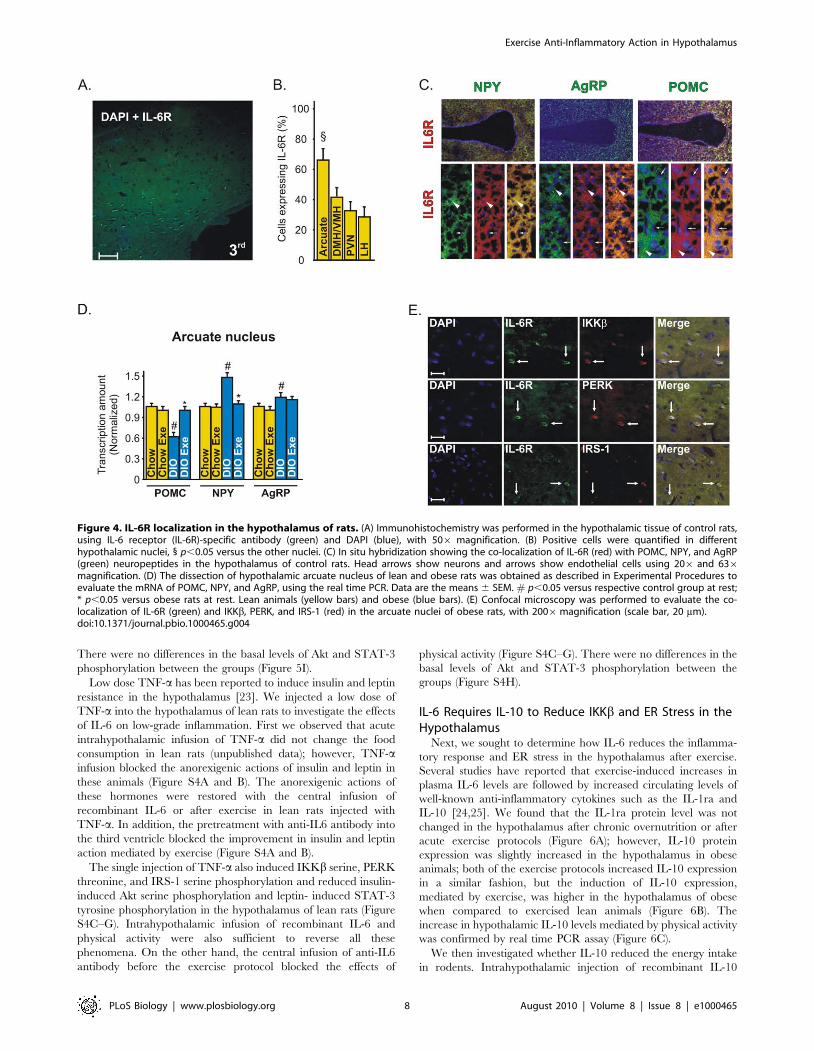
Immunohistochemistry with an anti-IL-6 Receptor (IL-6R)-
specific antibody showed that IL-6R is expressed in a majority of
neurons in the arcuate nucleus (Figure 4A). These data were
confirmed when we quantified the positive cells in arcuate (Arc),
dorsomedial and ventromedial (DMH/VMH), paraventricular
(PVN), and lateral (LH) nuclei of hypothalamus (Figure 4B). The
in situ hybridization experiment revealed that IL-6R is expressed
in both anorexigenic and orexigenic neurons of rats (Figure 4C).
Since IL-6R is expressed in a majority of neurons in the arcuate
nucleus, we dissected this specific hypothalamic region to evaluate
the modulation of the neuropeptides in response to exercise in lean
and obese rats. We found that exercise did not change the POMC,
NPY, and AgRP mRNA in the arcuate nucleus of lean rats but
increased the POMC and reduced the NPY mRNA levels in the
arcuate nucleus of obese animals (Figure 4D).
Double-staining confocal microscopy showed that most neurons
expressing IL-6R in the arcuate nucleus were shown to possess
IKKb, PERK, and IRS-1 in obese rats, showing a possible
interaction between these molecules (Figure 4E).
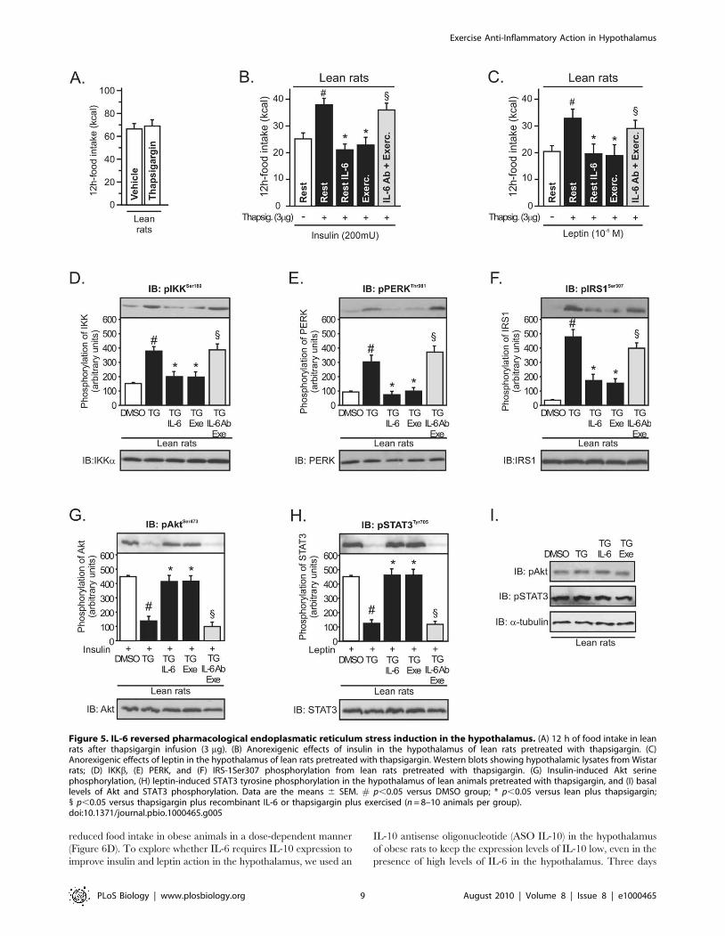
Pharmacological Activation of IKKb and ER Stress IsSuppressed by IL-6
To further support data indicating that IL-6 may modulate ER
stress, we performed an acute intrahypothalamic injection of an
ER stress inducer, thapsigargin (TG), in lean rats. Acute
intrahypothalamic infusion of thapsigargin did not change food
intake in lean animals by itself (Figure 5A). However, our results
revealed that intrahypothalamic infusion of thapsigargin blocked
the anorexigenic effects mediated by insulin and leptin in lean rats
and that the injection of recombinant IL-6 and exercise restored
the suppressive appetite action of insulin and leptin (Figure 5B and
C). In addition, the infusion of anti-IL6 antibody blocked the
improvement in insulin and leptin action mediated by exercise
(Figure 5B and C).
In accordance with previous studies [7,14,22], we observed that
thapsigargin markedly activated inflammatory signaling and ER
stress in lean rats, as reflected by increased levels of hypothalamic
IKKb and PERK phosphorylation, respectively (Figure 5D and
E), and induced central insulin and leptin resistance, increasing
IRS-1 serine phosphorylation (Figure 5F) and reducing insulin-
induced Akt serine phosphorylation and leptin-induced STAT-3
tyrosine phosphorylation (Figure 5G and H). Intrahypothalamic
infusion of recombinant IL-6 and physical activity were sufficient
to reverse all these phenomena (Figure 5D–H). Conversely, the
infusion of intrahypothalamic anti-IL6 antibody before exercise
protocol blocked these effects mediated by exercise (Figure 5D–H).
Figure 2. Hypothalamic insulin and leptin signaling after exercise. Western blots showing hypothalamic lysates from Wistar rats; (A)Hypothalamic IRb, IRS-1, IRS-2, and Akt phosphorylation, (B) Hypothalamic Foxo1 phosphorylation. (C) 12-h food intake (kcal) after intrahypothalamicinfusion of insulin in lean and diet-induced obesity (DIO) Wistar rats under resting conditions or after exercise (n = 6–8 animals per group). Westernblots of five independent experiments showing hypothalamic lysates from Wistar rats; (D) Insulin-induced IRb, IRS-1, IRS-2, Akt, and Foxo1phosphorylation in the hypothalamus. (E) Subcellular fractionation was performed to evaluate the nuclear Foxo1 expression in the hypothalamus oflean and obese rats at 30 min after insulin infusion. (F) Hypothalamic Jak-2 and (G) STAT-3 tyrosine phosphorylation. (H) 12-h food intake (kcal) afterintrahypothalamic infusion of leptin (n = 6–8 animals per group). Western blots showing hypothalamic lysates from Wistar rats; (I) Leptin-inducedJak2, IRS-1, IRS-2, and STAT3 tyrosine phosphorylation in the hypothalamus. (J) Subcellular fractionation was performed to evaluate the nuclear STAT3expression in the hypothalamic cells of lean and obese rats 30 min after leptin infusion. Data are the means 6 SEM. # p,0.05 versus respective leangroup at rest; * p,0.05 versus obese group at rest. Lean animals (white bars) and obese animals (black bars).doi:10.1371/journal.pbio.1000465.g002
Exercise Anti-Inflammatory Action in Hypothalamus
PLoS Biology | www.plosbiology.org 6 August 2010 | Volume 8 | Issue 8 | e1000465

Figure 3. Anti-hyperphagic response mediated by IL-6. (A) IL-6 mRNA in the hypothalamus of lean or diet-induced obesity (DIO) rats underresting conditions and lean obese rats immediately after the swimming exercise (SW Exe) or treadmill running (TR Exe). (B) 12 h of food intake inobese rats under resting conditions following intrahypothalamic infusion of different doses of recombinant IL-6. Counter-regulatory effects of anti-IL-6 antibody on food intake in exercised obese rats after (C) insulin or (D) leptin infusion. Western blots of five independent experiments showinghypothalamic lysates from Wistar rats; (E) Expression and activity of protein involved in the inflammatory signaling or ER stress in control animals atrest condition or after acute exercise (F) TLR4 expression, (G) IKKb phosphorylation, (H) IkBa expression, (I) PERK phosphorylation, (J) CHOPexpression, and (K) IRS-1Ser307 phosphorylation from lean, obese, obese injected with recombinant IL-6, obese after exercise, and obese pretreatedwith anti-IL-6 antibody before the exercise protocol. Data are the means 6 SEM. # p,0.05 versus lean group; * p,0.05 versus obese group at rest;¥ p,0.05 versus respective exercised control rats; ** p,0.01 versus stimulated obese group at rest; 1 p,0.05 versus obese group injected withrecombinant IL-6 and exercised obese rats (n = 8–10 animals per group). Swimming Exercise (SW Exe) or Treadmill Running (TR Exe). Lean animals(white bars) and obese animals (black bars).doi:10.1371/journal.pbio.1000465.g003
Exercise Anti-Inflammatory Action in Hypothalamus
PLoS Biology | www.plosbiology.org 7 August 2010 | Volume 8 | Issue 8 | e1000465
There were no differences in the basal levels of Akt and STAT-3
phosphorylation between the groups (Figure 5I).
Low dose TNF-a has been reported to induce insulin and leptin
resistance in the hypothalamus [23]. We injected a low dose of
TNF-a into the hypothalamus of lean rats to investigate the effects
of IL-6 on low-grade inflammation. First we observed that acute
intrahypothalamic infusion of TNF-a did not change the food
consumption in lean rats (unpublished data); however, TNF-ainfusion blocked the anorexigenic actions of insulin and leptin in
these animals (Figure S4A and B). The anorexigenic actions of
these hormones were restored with the central infusion of
recombinant IL-6 or after exercise in lean rats injected with
TNF-a. In addition, the pretreatment with anti-IL6 antibody into
the third ventricle blocked the improvement in insulin and leptin
action mediated by exercise (Figure S4A and B).
The single injection of TNF-a also induced IKKb serine, PERK
threonine, and IRS-1 serine phosphorylation and reduced insulin-
induced Akt serine phosphorylation and leptin- induced STAT-3
tyrosine phosphorylation in the hypothalamus of lean rats (Figure
S4C–G). Intrahypothalamic infusion of recombinant IL-6 and
physical activity were also sufficient to reverse all these
phenomena. On the other hand, the central infusion of anti-IL6
antibody before the exercise protocol blocked the effects of
physical activity (Figure S4C–G). There were no differences in the
basal levels of Akt and STAT-3 phosphorylation between the
groups (Figure S4H).
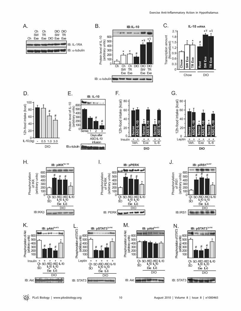
IL-6 Requires IL-10 to Reduce IKKb and ER Stress in theHypothalamus
Next, we sought to determine how IL-6 reduces the inflamma-
tory response and ER stress in the hypothalamus after exercise.
Several studies have reported that exercise-induced increases in
plasma IL-6 levels are followed by increased circulating levels of
well-known anti-inflammatory cytokines such as the IL-1ra and
IL-10 [24,25]. We found that the IL-1ra protein level was not
changed in the hypothalamus after chronic overnutrition or after
acute exercise protocols (Figure 6A); however, IL-10 protein
expression was slightly increased in the hypothalamus in obese
animals; both of the exercise protocols increased IL-10 expression
in a similar fashion, but the induction of IL-10 expression,
mediated by exercise, was higher in the hypothalamus of obese
when compared to exercised lean animals (Figure 6B). The
increase in hypothalamic IL-10 levels mediated by physical activity
was confirmed by real time PCR assay (Figure 6C).
We then investigated whether IL-10 reduced the energy intake
in rodents. Intrahypothalamic injection of recombinant IL-10
Figure 4. IL-6R localization in the hypothalamus of rats. (A) Immunohistochemistry was performed in the hypothalamic tissue of control rats,using IL-6 receptor (IL-6R)-specific antibody (green) and DAPI (blue), with 506 magnification. (B) Positive cells were quantified in differenthypothalamic nuclei, 1 p,0.05 versus the other nuclei. (C) In situ hybridization showing the co-localization of IL-6R (red) with POMC, NPY, and AgRP(green) neuropeptides in the hypothalamus of control rats. Head arrows show neurons and arrows show endothelial cells using 206 and 636magnification. (D) The dissection of hypothalamic arcuate nucleus of lean and obese rats was obtained as described in Experimental Procedures toevaluate the mRNA of POMC, NPY, and AgRP, using the real time PCR. Data are the means 6 SEM. # p,0.05 versus respective control group at rest;* p,0.05 versus obese rats at rest. Lean animals (yellow bars) and obese (blue bars). (E) Confocal microscopy was performed to evaluate the co-localization of IL-6R (green) and IKKb, PERK, and IRS-1 (red) in the arcuate nuclei of obese rats, with 2006magnification (scale bar, 20 mm).doi:10.1371/journal.pbio.1000465.g004
Exercise Anti-Inflammatory Action in Hypothalamus
PLoS Biology | www.plosbiology.org 8 August 2010 | Volume 8 | Issue 8 | e1000465
reduced food intake in obese animals in a dose-dependent manner
(Figure 6D). To explore whether IL-6 requires IL-10 expression to
improve insulin and leptin action in the hypothalamus, we used an
IL-10 antisense oligonucleotide (ASO IL-10) in the hypothalamus
of obese rats to keep the expression levels of IL-10 low, even in the
presence of high levels of IL-6 in the hypothalamus. Three days
Figure 5. IL-6 reversed pharmacological endoplasmatic reticulum stress induction in the hypothalamus. (A) 12 h of food intake in leanrats after thapsigargin infusion (3 mg). (B) Anorexigenic effects of insulin in the hypothalamus of lean rats pretreated with thapsigargin. (C)Anorexigenic effects of leptin in the hypothalamus of lean rats pretreated with thapsigargin. Western blots showing hypothalamic lysates from Wistarrats; (D) IKKb, (E) PERK, and (F) IRS-1Ser307 phosphorylation from lean rats pretreated with thapsigargin. (G) Insulin-induced Akt serinephosphorylation, (H) leptin-induced STAT3 tyrosine phosphorylation in the hypothalamus of lean animals pretreated with thapsigargin, and (I) basallevels of Akt and STAT3 phosphorylation. Data are the means 6 SEM. # p,0.05 versus DMSO group; * p,0.05 versus lean plus thapsigargin;1 p,0.05 versus thapsigargin plus recombinant IL-6 or thapsigargin plus exercised (n = 8–10 animals per group).doi:10.1371/journal.pbio.1000465.g005
Exercise Anti-Inflammatory Action in Hypothalamus
PLoS Biology | www.plosbiology.org 9 August 2010 | Volume 8 | Issue 8 | e1000465
Exercise Anti-Inflammatory Action in Hypothalamus
PLoS Biology | www.plosbiology.org 10 August 2010 | Volume 8 | Issue 8 | e1000465
after ASO IL-10 treatment, IL-10 protein expression was reduced
by about 75% in the hypothalamus of obese animals (Figure 6E).
Thereafter, exercise and recombinant IL-6 infusion failed to
improve the anorexigenic effects of insulin and leptin in obese
animals treated with ASO IL-10 (Figure 6F and G).
IL-10 is a pleiotropic cytokine that controls inflammatory
processes by suppressing the production of proinflammatory
cytokines and blocking IKK/NF-kB signaling and ER stress
[26,27]. Thus, we investigated whether exercise and IL-6 requires
IL-10 expression to reduce IKKb activation and ER stress in the
hypothalamus of obese animals. As demonstrated above, recom-
binant IL-6 infusion and exercise reduced IKKb, PERK, and
IRS-1Ser307 phosphorylation (Figure 3G, I, and K) and restored
insulin and leptin signaling in the hypothalamus of obese animals
(Figure S3), but the intrahypothalamic IL-10 ASO treatment
abolished all these parameters mediated by recombinant IL-6 and
exercise (Figure 6H–L). Conversely, the injection of recombinant
IL-10 in the hypothalamus of obese animals at rest markedly
reduced IKKb, PERK, and IRS-1Ser307 phosphorylation and
increased insulin-induced Akt and leptin-induced STAT-3 phos-
phorylation in the hypothalamic tissue of obese rats (Figure 6H–L).
There were no differences in the basal levels of Akt (Figure 6M).
However, STAT3 tyrosine phosphorylation was reduced in the
hypothalamus of obese rats, but neither exercise nor IL-6
intrahypothalamic injection was able to increase the STAT-3
phosphorylation after IL-10 ASO treatment (Figure 6N).
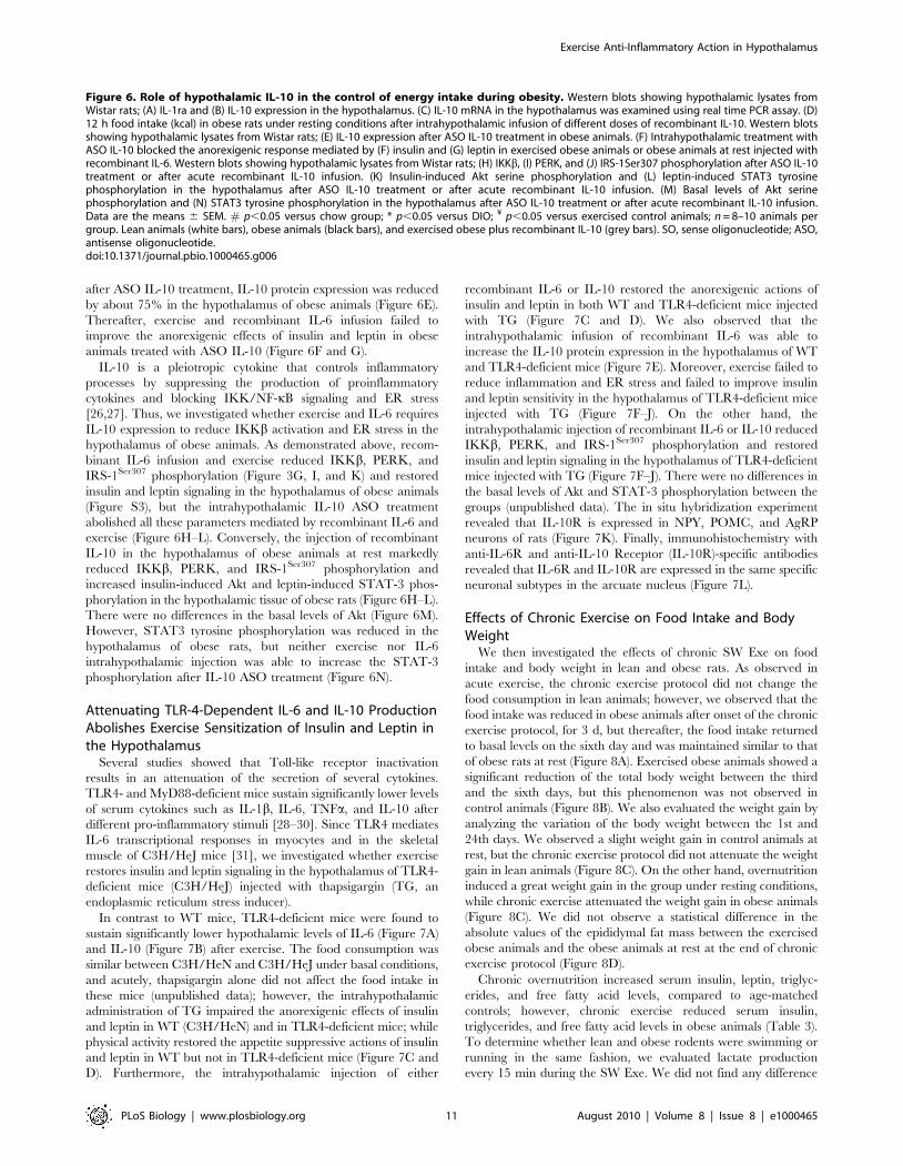
Attenuating TLR-4-Dependent IL-6 and IL-10 ProductionAbolishes Exercise Sensitization of Insulin and Leptin inthe Hypothalamus
Several studies showed that Toll-like receptor inactivation
results in an attenuation of the secretion of several cytokines.
TLR4- and MyD88-deficient mice sustain significantly lower levels
of serum cytokines such as IL-1b, IL-6, TNFa, and IL-10 after
different pro-inflammatory stimuli [28–30]. Since TLR4 mediates
IL-6 transcriptional responses in myocytes and in the skeletal
muscle of C3H/HeJ mice [31], we investigated whether exercise
restores insulin and leptin signaling in the hypothalamus of TLR4-
deficient mice (C3H/HeJ) injected with thapsigargin (TG, an
endoplasmic reticulum stress inducer).
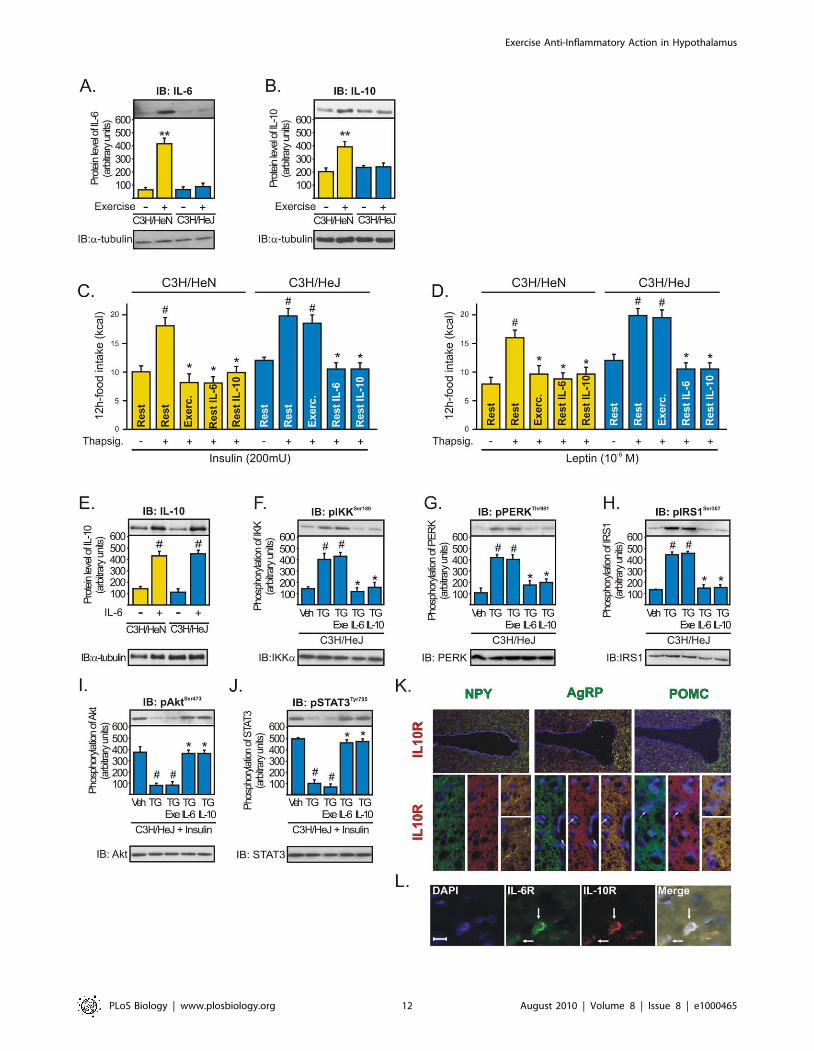
In contrast to WT mice, TLR4-deficient mice were found to
sustain significantly lower hypothalamic levels of IL-6 (Figure 7A)
and IL-10 (Figure 7B) after exercise. The food consumption was
similar between C3H/HeN and C3H/HeJ under basal conditions,
and acutely, thapsigargin alone did not affect the food intake in
these mice (unpublished data); however, the intrahypothalamic
administration of TG impaired the anorexigenic effects of insulin
and leptin in WT (C3H/HeN) and in TLR4-deficient mice; while
physical activity restored the appetite suppressive actions of insulin
and leptin in WT but not in TLR4-deficient mice (Figure 7C and
D). Furthermore, the intrahypothalamic injection of either
recombinant IL-6 or IL-10 restored the anorexigenic actions of
insulin and leptin in both WT and TLR4-deficient mice injected
with TG (Figure 7C and D). We also observed that the
intrahypothalamic infusion of recombinant IL-6 was able to
increase the IL-10 protein expression in the hypothalamus of WT
and TLR4-deficient mice (Figure 7E). Moreover, exercise failed to
reduce inflammation and ER stress and failed to improve insulin
and leptin sensitivity in the hypothalamus of TLR4-deficient mice
injected with TG (Figure 7F–J). On the other hand, the
intrahypothalamic injection of recombinant IL-6 or IL-10 reduced
IKKb, PERK, and IRS-1Ser307 phosphorylation and restored
insulin and leptin signaling in the hypothalamus of TLR4-deficient
mice injected with TG (Figure 7F–J). There were no differences in
the basal levels of Akt and STAT-3 phosphorylation between the
groups (unpublished data). The in situ hybridization experiment
revealed that IL-10R is expressed in NPY, POMC, and AgRP
neurons of rats (Figure 7K). Finally, immunohistochemistry with
anti-IL-6R and anti-IL-10 Receptor (IL-10R)-specific antibodies
revealed that IL-6R and IL-10R are expressed in the same specific
neuronal subtypes in the arcuate nucleus (Figure 7L).
Effects of Chronic Exercise on Food Intake and BodyWeight
We then investigated the effects of chronic SW Exe on food
intake and body weight in lean and obese rats. As observed in
acute exercise, the chronic exercise protocol did not change the
food consumption in lean animals; however, we observed that the
food intake was reduced in obese animals after onset of the chronic
exercise protocol, for 3 d, but thereafter, the food intake returned
to basal levels on the sixth day and was maintained similar to that
of obese rats at rest (Figure 8A). Exercised obese animals showed a
significant reduction of the total body weight between the third
and the sixth days, but this phenomenon was not observed in
control animals (Figure 8B). We also evaluated the weight gain by
analyzing the variation of the body weight between the 1st and
24th days. We observed a slight weight gain in control animals at
rest, but the chronic exercise protocol did not attenuate the weight
gain in lean animals (Figure 8C). On the other hand, overnutrition
induced a great weight gain in the group under resting conditions,
while chronic exercise attenuated the weight gain in obese animals
(Figure 8C). We did not observe a statistical difference in the
absolute values of the epididymal fat mass between the exercised
obese animals and the obese animals at rest at the end of chronic
exercise protocol (Figure 8D).
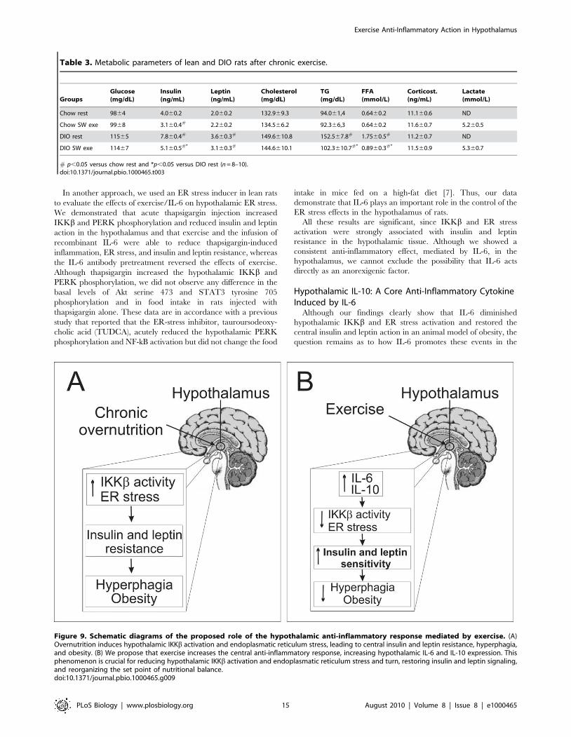
Chronic overnutrition increased serum insulin, leptin, triglyc-
erides, and free fatty acid levels, compared to age-matched
controls; however, chronic exercise reduced serum insulin,
triglycerides, and free fatty acid levels in obese animals (Table 3).
To determine whether lean and obese rodents were swimming or
running in the same fashion, we evaluated lactate production
every 15 min during the SW Exe. We did not find any difference
Figure 6. Role of hypothalamic IL-10 in the control of energy intake during obesity. Western blots showing hypothalamic lysates fromWistar rats; (A) IL-1ra and (B) IL-10 expression in the hypothalamus. (C) IL-10 mRNA in the hypothalamus was examined using real time PCR assay. (D)12 h food intake (kcal) in obese rats under resting conditions after intrahypothalamic infusion of different doses of recombinant IL-10. Western blotsshowing hypothalamic lysates from Wistar rats; (E) IL-10 expression after ASO IL-10 treatment in obese animals. (F) Intrahypothalamic treatment withASO IL-10 blocked the anorexigenic response mediated by (F) insulin and (G) leptin in exercised obese animals or obese animals at rest injected withrecombinant IL-6. Western blots showing hypothalamic lysates from Wistar rats; (H) IKKb, (I) PERK, and (J) IRS-1Ser307 phosphorylation after ASO IL-10treatment or after acute recombinant IL-10 infusion. (K) Insulin-induced Akt serine phosphorylation and (L) leptin-induced STAT3 tyrosinephosphorylation in the hypothalamus after ASO IL-10 treatment or after acute recombinant IL-10 infusion. (M) Basal levels of Akt serinephosphorylation and (N) STAT3 tyrosine phosphorylation in the hypothalamus after ASO IL-10 treatment or after acute recombinant IL-10 infusion.Data are the means 6 SEM. # p,0.05 versus chow group; * p,0.05 versus DIO; ¥ p,0.05 versus exercised control animals; n = 8–10 animals pergroup. Lean animals (white bars), obese animals (black bars), and exercised obese plus recombinant IL-10 (grey bars). SO, sense oligonucleotide; ASO,antisense oligonucleotide.doi:10.1371/journal.pbio.1000465.g006
Exercise Anti-Inflammatory Action in Hypothalamus
PLoS Biology | www.plosbiology.org 11 August 2010 | Volume 8 | Issue 8 | e1000465
Exercise Anti-Inflammatory Action in Hypothalamus
PLoS Biology | www.plosbiology.org 12 August 2010 | Volume 8 | Issue 8 | e1000465
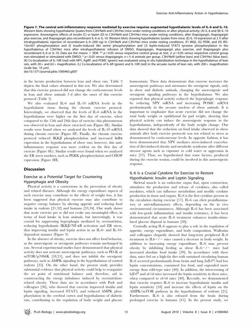
in the lactate production between lean and obese rats. Table 3
depicts the final values obtained in this test. We also determined
that this exercise protocol did not change the corticosterone levels
in lean and obese animals 3 d after the onset of this exercise
protocol (Table 3).
We also evaluated IL-6 and IL-10 mRNA levels in the
hypothalamic tissue during the chronic exercise protocol.
Interestingly, we observed that the levels of IL-6 mRNA in the
hypothalamus were higher on the first day of exercise, when
compared to the 15th and 24th days of exercise; this phenomenon
was observed in lean and obese exercised rats (Figure 8E). Similar
results were found when we analyzed the levels of IL-10 mRNA
during chronic exercise (Figure 8F). Finally, the chronic exercise
protocol reduced IKKb phosphorylation and increased IkBaexpression in the hypothalamus of obese rats; however, this anti-
inflammatory response was more evident on the first day of
exercise (Figure 8G). Similar results were found when we analyzed
the ER stress markers, such as PERK phosphorylation and CHOP
expression (Figure 8H).
Discussion
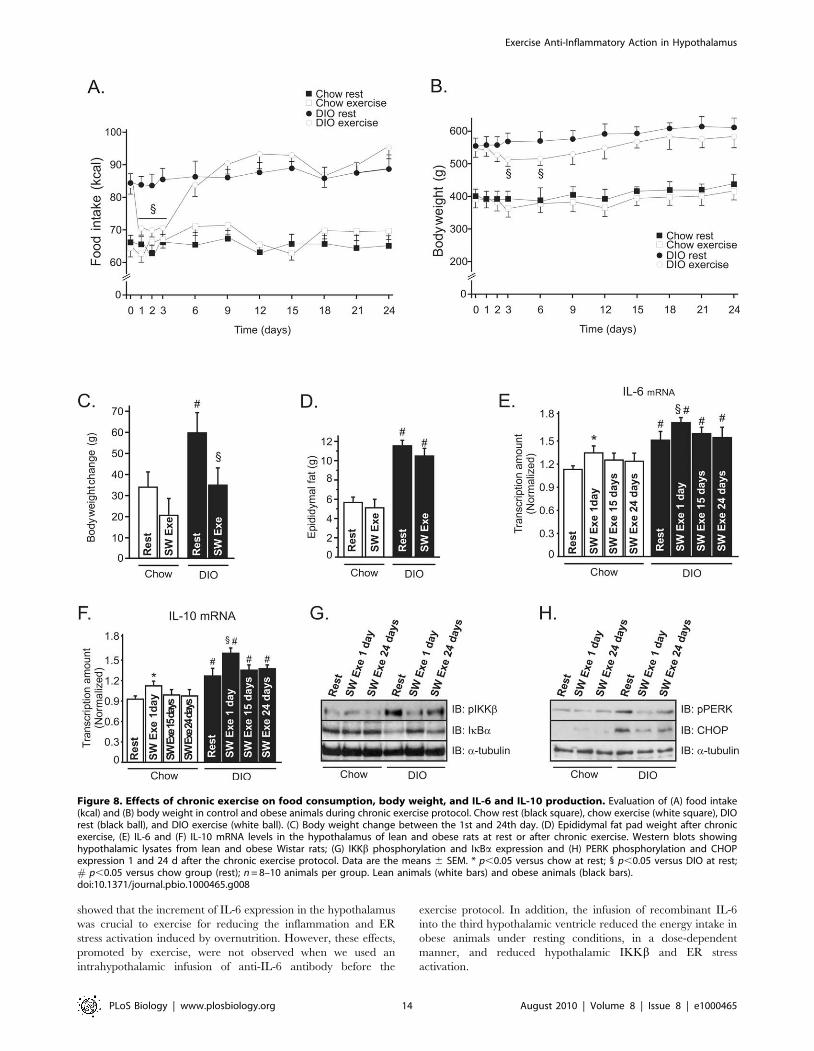
Exercise as a Potential Target for CounteringHyperphagia and Obesity
Physical activity is a cornerstone in the prevention of obesity
and related diseases. Although the energy expenditure aspects of
such exercise may contribute to the effects of weight loss, it has
been suggested that physical exercise may also contribute to
negative energy balance by altering appetite and reducing food
intake in rodents [21,32] and humans [33,34]. Our study shows
that acute exercise per se did not evoke any meaningful effect, in
terms of food intake in lean animals, but interestingly, it was
crucial for suppressing hyperphagia mediated by overnutrition,
reducing hypothalamic IKKb/NF-kB activation and ER stress,
thus improving insulin and leptin action in an IL-6- and IL-10-
dependent manner (Figure 9).
In the absence of obesity, exercise does not affect food behavior,
as the anorexigenic or orexigenic pathways remain unchanged in
rats. Several experimental studies have demonstrated that physical
activity does not activate anorexigenic pathways, such as PI3-K or
mTOR/p70S6K [18,21], and does not inhibit the orexigenic
pathways, such as AMPK signaling in the hypothalamus of control
rodents [35]. On the other hand, the present study provides
substantial evidence that physical activity could help to reorganize
the set point of nutritional balance and, therefore, aid in
counteracting the energy imbalance induced by overnutrition-
related obesity. These data are in accordance with Park and
colleagues [36], who showed that exercise improved insulin and
leptin signaling, increased STAT3, and reduced AMPK phos-
phorylation in the cerebral cortex and hypothalamus of diabetic
rats, contributing to the regulation of body weight and glucose
homeostasis. These data demonstrate that exercise increases the
anorexigenic pathways and attenuates the orexigenic signals, only
in obese and diabetic animals, changing the anorexigenic and
orexigenic signaling pathways in the hypothalamus. We also
reported that physical activity reduced the hyperphagic response
by reducing NPY mRNA and increasing POMC mRNA
predominantly in the arcuate nucleus of obese animals. It is
important to emphasize that acute exercise did not change the
total body weight or epididymal fat pad weight, showing that
physical activity can induce the anorexigenic response in the
hypothalamus, independently of the body weight change. Our
data showed that the reduction on food intake observed in obese
animals after both exercise protocols was not related to stress as
demonstrated by costicosterone levels. In opposite fashion, it has
been demonstrated that NPY mediates stress-induced exacerba-
tion of diet-induced obesity and metabolic syndrome after different
stressor agents such as exposure to cold water or aggression in
mice [37]. Thus, we hypothesized that some factors, produced
during the exercise session, could be involved in this anorexigenic
response.
IL-6 Is a Crucial Cytokine for Exercise to RestoreHypothalamic Insulin and Leptin Signaling
Skeletal muscle is an endocrine organ that, upon contraction,
stimulates the production and release of cytokines, also called
myokines, which can influence metabolism and modify cytokine
production in tissue and organs. IL-6 is the first cytokine present in
the circulation during exercise [17]. IL-6 can elicit proinflamma-
tory or anti-inflammatory effects, depending on the in vivo
environmental circumstances. Although IL-6 has been associated
with low-grade inflammation and insulin resistance, it has been
demonstrated that acute IL-6 treatment enhances insulin-stimu-
lated glucose disposal in humans [38].
Centrally acting IL-6 appears to play a role in the regulation of
appetite, energy expenditure, and body composition. Wallenius
and colleagues elegantly showed that long-term peripheral IL-6
treatment to IL62/2 mice caused a decrease in body weight. In
addition to increasing energy expenditure, IL-6 may prevent
obesity by inhibiting feeding as obese IL-62/2 mice had
increased absolute food intake [39]. In accordance with these
data, mice fed on a high-fat diet with sustained circulating human
IL-6 secreted predominantly from brain and lung (hIL6tg) had low
leptin concentrations, consumed less food, and expended more
energy than wild-type mice [40]. In addition, the intercrossing of
hIL6tg and ob/ob mice increased the leptin sensitivity in these mice,
when compared to ob/ob mice [40]. Recently, we demonstrated
that exercise requires IL-6 to increase hypothalamic insulin and
leptin sensitivity [18] and increase the effects of leptin on the
AMPK/mTOR pathway in the hypothalamus of rodents [21].
Furthermore, IL-6 is also released from the brain during
prolonged exercise in humans [41]. In the present study, we
Figure 7. The central anti-inflammatory response mediated by exercise requires augmented hypothalamic levels of IL-6 and IL-10.Western blots showing hypothalamic lysates from C3H/NeN and C3H/HeJ mice under resting conditions or after physical activity; (A) IL-6 and (B) IL-10expression. Anorexigenic effects of insulin (C) or leptin (D) in C3H/NeN and C3H/HeJ mice under resting conditions, after thapsigargin, thapsigarginplus exercise, and thapsigargin plus recombinant IL-6 or IL-10. Western blots showing hypothalamic lysates from mice; (E) IL-10 expression at 2 h afterintrahypothalamic injection of recombinant IL-6 (200 ng) in C3H/NeN and C3H/HeJ mice under resting conditions. (F) IKKb, (G) PERK, and (H) IRS-1Ser307 phosphorylation and (I) Insulin-induced Akt serine phosphorylation and (J) leptin-induced STAT3 tyrosine phosphorylation in thehypothalamus of C3H/HeJ mice after intrahypothalamic infusion of DMSO, thapsigargin, thapsigargin plus exercise, and thapsigargin plusrecombinant IL-6 or IL-10. Data are the means 6 SEM. ** p,0.05 versus respective control group at rest; # p,0.05 versus respective control groupnon-stimulated or stimulated with DMSO; * p,0.05 versus thapsigargin; n = 5–6 animals per group. C3H/NeN (yellow bars) and C3H/HeJ (blue bars).(K) Co-localization of IL-10R (red) with NPY, AgRP, and POMC (green) was evaluated using in situ hybridization technique in the hypothalamus of leanrats, with 206and 636magnification. (L) Co-localization of IL-6R (green) and IL-10R (red) in the arcuate nuclei of lean rats, with 2006magnification(scale bar, 10 mm).doi:10.1371/journal.pbio.1000465.g007
Exercise Anti-Inflammatory Action in Hypothalamus
PLoS Biology | www.plosbiology.org 13 August 2010 | Volume 8 | Issue 8 | e1000465
showed that the increment of IL-6 expression in the hypothalamus
was crucial to exercise for reducing the inflammation and ER
stress activation induced by overnutrition. However, these effects,
promoted by exercise, were not observed when we used an
intrahypothalamic infusion of anti-IL-6 antibody before the
exercise protocol. In addition, the infusion of recombinant IL-6
into the third hypothalamic ventricle reduced the energy intake in
obese animals under resting conditions, in a dose-dependent
manner, and reduced hypothalamic IKKb and ER stress
activation.
Figure 8. Effects of chronic exercise on food consumption, body weight, and IL-6 and IL-10 production. Evaluation of (A) food intake(kcal) and (B) body weight in control and obese animals during chronic exercise protocol. Chow rest (black square), chow exercise (white square), DIOrest (black ball), and DIO exercise (white ball). (C) Body weight change between the 1st and 24th day. (D) Epididymal fat pad weight after chronicexercise, (E) IL-6 and (F) IL-10 mRNA levels in the hypothalamus of lean and obese rats at rest or after chronic exercise. Western blots showinghypothalamic lysates from lean and obese Wistar rats; (G) IKKb phosphorylation and IkBa expression and (H) PERK phosphorylation and CHOPexpression 1 and 24 d after the chronic exercise protocol. Data are the means 6 SEM. * p,0.05 versus chow at rest; 1 p,0.05 versus DIO at rest;# p,0.05 versus chow group (rest); n = 8–10 animals per group. Lean animals (white bars) and obese animals (black bars).doi:10.1371/journal.pbio.1000465.g008
Exercise Anti-Inflammatory Action in Hypothalamus
PLoS Biology | www.plosbiology.org 14 August 2010 | Volume 8 | Issue 8 | e1000465
In another approach, we used an ER stress inducer in lean rats
to evaluate the effects of exercise/IL-6 on hypothalamic ER stress.
We demonstrated that acute thapsigargin injection increased
IKKb and PERK phosphorylation and reduced insulin and leptin
action in the hypothalamus and that exercise and the infusion of
recombinant IL-6 were able to reduce thapsigargin-induced
inflammation, ER stress, and insulin and leptin resistance, whereas
the IL-6 antibody pretreatment reversed the effects of exercise.
Although thapsigargin increased the hypothalamic IKKb and
PERK phosphorylation, we did not observe any difference in the
basal levels of Akt serine 473 and STAT3 tyrosine 705
phosphorylation and in food intake in rats injected with
thapsigargin alone. These data are in accordance with a previous
study that reported that the ER-stress inhibitor, tauroursodeoxy-
cholic acid (TUDCA), acutely reduced the hypothalamic PERK
phosphorylation and NF-kB activation but did not change the food
intake in mice fed on a high-fat diet [7]. Thus, our data
demonstrate that IL-6 plays an important role in the control of the
ER stress effects in the hypothalamus of rats.
All these results are significant, since IKKb and ER stress
activation were strongly associated with insulin and leptin
resistance in the hypothalamic tissue. Although we showed a
consistent anti-inflammatory effect, mediated by IL-6, in the
hypothalamus, we cannot exclude the possibility that IL-6 acts
directly as an anorexigenic factor.
Hypothalamic IL-10: A Core Anti-Inflammatory CytokineInduced by IL-6
Although our findings clearly show that IL-6 diminished
hypothalamic IKKb and ER stress activation and restored the
central insulin and leptin action in an animal model of obesity, the
question remains as to how IL-6 promotes these events in the
Table 3. Metabolic parameters of lean and DIO rats after chronic exercise.
GroupsGlucose(mg/dL)
Insulin(ng/mL)
Leptin(ng/mL)
Cholesterol(mg/dL)
TG(mg/dL)
FFA(mmol/L)
Corticost.(ng/mL)
Lactate(mmol/L)
Chow rest 9864 4.060.2 2.060.2 132.969.3 94.061,4 0.6460.2 11.160.6 ND
Chow SW exe 9968 3.160.4# 2.260.2 134.566.2 92.366,3 0.6460.2 11.660.7 5.260.5
DIO rest 11565 7.860.4# 3.660.3# 149.6610.8 152.567.8# 1.7560.5# 11.260.7 ND
DIO SW exe 11467 5.160.5#* 3.160.3# 144.6610.1 102.3610.7#* 0.8960.3#* 11.560.9 5.360.7
# p,0.05 versus chow rest and *p,0.05 versus DIO rest (n = 8–10).doi:10.1371/journal.pbio.1000465.t003
Figure 9. Schematic diagrams of the proposed role of the hypothalamic anti-inflammatory response mediated by exercise. (A)Overnutrition induces hypothalamic IKKb activation and endoplasmatic reticulum stress, leading to central insulin and leptin resistance, hyperphagia,and obesity. (B) We propose that exercise increases the central anti-inflammatory response, increasing hypothalamic IL-6 and IL-10 expression. Thisphenomenon is crucial for reducing hypothalamic IKKb activation and endoplasmatic reticulum stress and turn, restoring insulin and leptin signaling,and reorganizing the set point of nutritional balance.doi:10.1371/journal.pbio.1000465.g009
Exercise Anti-Inflammatory Action in Hypothalamus
PLoS Biology | www.plosbiology.org 15 August 2010 | Volume 8 | Issue 8 | e1000465
hypothalamus. Following exercise, the high circulating levels of IL-
6 are followed by an increase in two anti-inflammatory molecules,
IL-1ra and IL-10 [25]. Therefore, IL-6 induces an anti-
inflammatory environment by inducing the production of IL-1ra
and IL-10. In our study, we found that exercise increased the
hypothalamic levels of IL-10 but did not change IL-1ra expression
in this tissue. Thus, we showed that the anti-inflammatory
response mediated by IL-6 involves the increase of IL-10
expression in the hypothalamus.
IL-10 is an important immunoregulatory cytokine with multiple
biological effects. In the cytoplasm, it has been demonstrated that
IL-10 blocks NF-kB activity at two levels: suppressing IKK activity
and NF-kB DNA binding activity [26]. Moreover, IL-10 reduced
ER stress in intestinal eptithelial cells, whereas IL-102/2 mice
demonstrated that the expression of the ER stress response protein
grp-78/BiP was increased in intestinal eptithelial cells under
conditions of chronic inflammation [27].
In the CNS, the anti-inflammatory role of IL-10 has been
extensively studied in experimental autoimmune encephalomyeli-
tis, an animal model of human multiple sclerosis. The increase in
IL-10 expression in the CNS during recovery from brain
inflammation and the inability of IL-10 null mice to recover from
acute CNS inflammation suggests that the presence of IL-10
within this target organ is required for disease remission [42,43].
However, the role of hypothalamic IL-10 in the control of low-
grade inflammation generated during obesity was unknown. Here,
we discovered that intrahypothalamic infusion of recombinant IL-
10 blocked IKK/NF-kB signaling and ER stress and restored Akt
and STAT3 phosphorylation, promoting a re-balance in the
energy intake in obese animals. On the other hand, the selective
decrease in IL-10 expression in discrete hypothalamic nuclei of
obese animals mediated by ASO treatment blunted the effects of
both exercise and the intrahypothalamic infusion of recombinant
IL-6 in the restoration of central insulin and leptin actions. In
addition, we demonstrated that in mice that sustained significantly
lower hypothalamic levels of IL-6 and IL-10 after exercise (C3H/
HeJ), there was no reduction in pharmacological ER stress
activation, in contrast to WT mice. These data are intriguing as
IL-10 represents an important cytokine that may reduce both
inflammation and ER stress in the hypothalamus. Thus, the
modulation of hypothalamic IL-10 expression could be considered
the direct target of exercise/IL-6 and constitutes a promising
alternative to reduce hypothalamic inflammation and ER stress
related to obesity.
The decrease in food intake induced by IL-10 in obese rats is
not in accordance with the effects observed in IL-10 KO. It has
been reported that mice with combined deficiency of leptin and
IL-10 gain less body weight than mice lacking leptin only [44].
However, these discrepancies may be a consequence of method-
ological differences related to physiological versus genetic
approaches and acute versus chronic situation investigated, and
most important it may be consequence of IL-10 effects in the
regulation of energy expenditure, likewise observed in mice lacking
TNF-a receptor [45]; thus, the role of IL-10 in the control of food
intake and energy expenditure deserves further exploration.
The long-term reversal effects on body composition, mediated
by exercise alone, are controversial. It should be acknowledged
that it is often difficult to find long-term reversal effects on body fat
in both experimental animals and humans by exercise alone
without restrained diet [46]. In the chronic experiments, we
observed that the obese animals lost weight during the same period
in which a reduction in food intake was observed. After this
period, no significant difference was observed in the body weight
of exercised animals, although the obese animals presented a
significant improvement in metabolic parameters after the chronic
exercise protocol.
Since IKKb/NF-kB inhibition in the CNS represents a
potential target therapy to combat obesity and most anti-
inflammatory therapies have limited direct effects on IKKb/NF-
kB and a limited capacity for concentration in the CNS, our study
provides substantial evidence that physical activity could help to
reorganize the set point of nutritional balance and therefore aid in
counteracting the energy imbalance induced by overnutrition
through the anti-inflammatory response in hypothalamic neurons.
Hence, IL-6 and IL-10 are important physiological contributors to
the central insulin and leptin action mediated by physical activity,
linking it to hypothalamic ER stress and inflammation.
Materials and Methods
Antibodies and ChemicalsProtein A-Sepharose 6 MB and Nitrocellulose paper (Hybond
ECL, 0.45 mm) were from Amersham Pharmacia Biotech United
Kingdom Ltd. (Buckinghamshire, United Kingdom). Ketamin was
from Parke-Davis (Sao Paulo, SP, Brazil) and diazepam and
thiopethal were from Cristalia (Itapira, SP, Brazil). Anti-phospho-
JAK2 (rabbit polyclonal, AB3805) antibody was from Upstate
Biotechnology (Charlottesville, VA, USA). Anti-JAK2 (rabbit
polyclonal, SC-278), anti-STAT3 (rabbit polyclonal, SC-483),
anti-phospho-IRb (rabbit polyclonal, SC-25103), anti-IRb (rabbit
polyclonal, SC-711), anti-phospho-IRS-1 (rabbit polyclonal, SC-
17199), anti-IRS-1 (rabbit polyclonal, SC-559), anti-IRS-2 (rabbit
polyclonal, SC-1556), anti-phosphotyrosine (mouse monoclonal,
SC-508), anti-Foxo1 (rabbit polyclonal, SC-11350), anti-IL-1ra
(goat polyclonal, SC-8481), anti-TNF-a (rabbit polyclonal, SC-
8301), anti-IKKb (goat polyclonal, SC-34673), anti-PERK (rabbit
polyclonal, SC-13073), anti-phospho-PERK (rabbit polyclonal,
SC-32577), anti-CHOP (GADD 153) (rabbit polyclonal, SC-575),
anti-IL-10 (goat polyclonal, SC-1783), and anti-IL-6 (rabbit
polyclonal, SC-7920) antibodies were from Santa Cruz Biotech-
nology, Inc. Anti-phospho-STAT3 (rabbit polyclonal, #9131),
anti-phospho-Akt (rabbit polyclonal, #9271), anti-phospho-Foxo1
(rabbit polyclonal, #9461), anti-beta tubulin (rabbit polyclonal,
#2146), anti-phospho-IKKa/b (rabbit polyclonal, #2687), anti-
IkBa (rabbit polyclonal, #9242), anti-TLR4 (rabbit polyclonal,
#2219), anti-phospho-IRS-1 307 (rabbit polyclonal, #2381), and
anti-Akt (rabbit polyclonal, #9272) were from Cell Signalling
Technology (Beverly, MA, USA). Leptin, thapsigargin, and
recombinant IL-6 and -10 were from Calbiochem (San Diego,
CA, USA). Routine reagents were purchased from Sigma
Chemical Co. (St. Louis, MO) unless otherwise specified.
Serum Insulin, Leptin, and IL-6 QuantificationBlood was collected from the cava vein 15 min after the exercise
protocols. Plasma was separated by centrifugation (1,100 g) for
15 min at 4 uC and stored at 280 uC until assay. RIA was
employed to measure serum insulin. Leptin and IL-6 concentra-
tions were determined using a commercially available Enzyme
Linked Immunosorbent Assay (ELISA) kit (Crystal Chem Inc.,
Chicago, IL). Blood lactate was measured using Accutrend Plus
equipment (Roche); sample blood was obtained from the tails
every 15 min during the exercise protocols. Serum cholesterol and
triglycerides were measured in control and exercised animals after
8 h of fasting using Accutrend Plus equipment (Roche). Serum free
fatty acids (FFA) levels were analyzed in rats using the NEFA-kit-U
(Wako Chemical GmBH, Neuss, Germany).
Corticosterone levels were determined using urine samples
obtained from rats and mice using specific metabolic cage during
Exercise Anti-Inflammatory Action in Hypothalamus
PLoS Biology | www.plosbiology.org 16 August 2010 | Volume 8 | Issue 8 | e1000465
24 h after the exercise protocols. The corticosterone level was
determined using an EIA kit from Cayman chemical (Ann Arbor, MI).
AnimalsMale 4-wk-old Wistar rats were obtained from the University of
Campinas Breeding Center. The investigation was approved by
the ethics committee and followed the University guidelines for the
use of animals in experimental studies and experiments conform to
the Guide for the Care and Use of Laboratory Animals, published by the
U.S. National Institutes of Health (NIH publication no. 85-23
revised 1996). The animals were maintained on 12h:12h artificial
light-dark cycles and housed in individual cages. Rats were
randomly divided into two groups: control, fed on standard rodent
chow (3,948 kcal.Kg21), and DIO, fed a fat-rich chow
(5,358 kcal.Kg21) ad libitum for 3 mo. This diet composition has
been previously used [47].
Male (10-wk-old) ob/ob mice and their respective controls
C57BL/6J background were obtained from The Jackson Labora-
tory and provided by the University of Sao Paulo. The mice were
bred under specific pathogen-free conditions at the Central
Breeding Center of University of Campinas.
Male C3H/HeJ (10-wk-old) mice and their respective controls
C3H/HeN were obtained from The Jackson Laboratory and
provided by the University of Sao Paulo. The mice were bred
under specific pathogen-free conditions at the Central Breeding
Center of the University of Campinas.
Intracerebroventricular CannulationThe animals were stereotaxically instrumented under intraper-
itoneal injection of a mix of ketamin (10 mg) and diazepam
(0.07 mg) (0.2 ml/100 g body weight) with a chronic 26-gauge
stainless steel indwelling guide cannula aseptically placed into the
third ventricle at the midline coordinates of 0.5 mm posterior to
the bregma and 8.5 mm below the surface of the skull of rats and
1.8 mm posterior to the bregma and 5.0 mm below the surface of
the skull of mice.
Exercise ProtocolsAnimals were acclimated to swimming for 2 d (10 min per day).
Water temperature was maintained at 34–35 uC. Rats performed
two 3-h exercise bouts, separated by one 45-min rest period. The
rats swam in groups of three in plastic barrels of 45 cm in diameter
that were filled to a depth of 50 cm. This protocol was conducted
between 11:00 a.m. and 6:00 p.m., as previously described [48],
and mice performed four 30-min exercise bouts, separated by one
5-min rest period. The mice swam in groups of four in plastic
barrels of 40 cm in diameter that were filled to a depth of 20 cm.
This protocol was conducted between 3:00 p.m. and 6:00 p.m.
Both exercise protocols finished at 6:00 p.m. for evaluation of food
intake and analysis of hypothalamic tissue.
The chronic exercise protocol consisted of daily swimming
sessions (1 h/d, 5 d/wk, for 4 wk) with an overload (2.0% of the
body weight). The hypothalamic tissues and the metabolic
parameter were evaluated 36 h after the last exercise session.
Rats also performed a single bout of treadmill (Insight LTDA -
Ribeirao Preto, SP) running (60 min, speed of 10–15 m/min at a
5% incline) and mice performed a single bout of treadmill running
(90 min, speed of 7–10 m/min at a 5% incline).
Intracerebroventricular TreatmentsRats or mice were deprived of food for 2 h with free access to
water and received 3 ml of bolus injection into the third ventricle,
as follows:
Insulin and leptin treatments. Animals received
intrahypothalamic infusion of vehicle, insulin (200 mU), or leptin
(1026 M) at 6:00 p.m. to evaluate the food intake or insulin and
leptin signaling. Food intake was determined by measuring the
difference between the weight of chow given and the weight of
chow at the end of a 12-h period.
Recombinant IL-6 and IL-10 treatments. Animals
received intrahypothalamic infusion of vehicle, or recombinant
IL-6 (50, 100, or 200 ng) or recombinant IL-10 (0.5, 1.0, or
3.0 ng) at 6:00 p.m. to evaluate the food intake. For Western blot
analysis, we injected recombinant IL-6 or IL-10 2 h after DMSO
or thapsigargin into the third ventricle and the hypothalamus was
excised 2 h later.
Thapsigargin treatments. Animals received intrahypotha-
lamic infusion of vehicle, or thapsigargin (3.0 mg). To evaluate the
energy intake and for Western blot analysis, thapsigargin was
infused 40 min before the exercise protocol and 2 h before the
recombinant IL-6 infusion. Immediately after exercise or 2 h after
IL-6 infusion, animals received intrahypothalamic infusion of
insulin (200 mU) or leptin (1026 M).
IL-6 neutralizing antibody. Animals were randomly
selected for treatment with saline, rabbit pre-immune serum
(RPIS) or rabbit antiserum against IL-6 (IL-6 Ab) in different
doses. IL-6 Ab was injected into the third ventricle of the rats
15 min before the exercise protocol.
ASO IL-10 treatments. Phosphorthioate-modified sense and
antisense oligonucleotides (produced by Invitrogen Corp.,
Carlsbad, CA, USA) were diluted to final concentration of
1 nmol/ml in dilution buffer containing 10 mmol/l Tris–HCl
and 1.0 mmol/l EDTA. The oligonucleotides were designed
according to the Mus musculus IL-10 sequence deposited at the
NIH-NCBI (http://www.ncbi.nlm.nih.gov/entrez) under the
designation NM 010548 and were composed of 59-GCC AGT
CAG TAA GAG CAG-39 (sense) and 59-TGA GAT CTG CAA
TGC A-39 (antisense). Obese Wistar rats were injected into the
third ventricle with two daily doses of 3 ml of dilution buffer
containing, or not, sense (Sense IL-10) or antisense
oligonucleotides (ASO IL-10) for 3 d. For Western blotting
analysis, after ASO IL-10 treatment, obese animals were
submitted to the exercise protocol or intrahypothalamic infusion
of recombinant IL-6. In some experiments, the rats also received
intrahypothalamic infusion of insulin (200 mU) or leptin (1026 M)
for the determination of food intake and Akt and STAT3
phosphorylation.
Recombinant of TNF-a treatments. Animals received
intrahypothalamic infusion of vehicle, or TNF-a (10212). To
evaluate the energy intake and for Western blotting analysis, TNF-
a was infused 40 min before the exercise protocol and 2 h before
the recombinant IL-6 infusion. Immediately after exercise or 2 h
after IL-6 infusion, animals received intrahypothalamic infusion of
insulin (200 mU) or leptin (1026 M).
Food Intake DeterminationIntrahypothalamic infusions were performed between 5:00 and
6:00 p.m. Thereafter standard chow or high-fat diet was given and
food intake was determined by measuring the difference between
the weight of chow given and the weight of chow at the end of a
12-h period. Similar studies were carried out in animals after
exercise.
Western Blot AnalysisAfter exercise and/or i.c.v. treatments, the animals were
anaesthetized, and the hypothalamus was quickly removed,
minced coarsely, and homogenized immediately in a freshly
Exercise Anti-Inflammatory Action in Hypothalamus
PLoS Biology | www.plosbiology.org 17 August 2010 | Volume 8 | Issue 8 | e1000465
prepared ice-cold buffer (1% Triton X-100, 100 mmol/l Tris
pH 7.4, 100 mmol/l sodium pyrophosphate, 100 mmol/l sodium
fluoride, 10 mmol/l EDTA, 10 mmol/l sodium vanadate,
2 mmol/l phenyl methylsulphonyl fluoride, and 0.1 mg aprotinin)
suitable for preserving phosphorylation states of enzymes, and
Western blot was performed, as previously described [1].
Nuclear ExtractFoxo1 and STAT-3 nuclear expression were obtained as
described [49]. Fragments of hypothalamic tissue from untreated
rats or rats treated with insulin or leptin were obtained 30 min
after insulin or leptin infusion and were minced and homogenized
in 2 vol. of STE buffer (0.32 M sucrose, 20 mM Tris–HCl
(pH 7.4), 2 mM EDTA, 1 mM DTT, 100 mM sodium fluoride,
100 mM sodium pyrophosphate, 10 mM sodium orthovanadate,
1 mM PMSF, and 0.1 mg aprotinin/ml) at 4 uC with a Polytron
homogenizer. The homogenates were centrifuged (1,0006g,
25 min, 4 uC) to obtain pellets. The pellet was washed once and
suspended in STE buffer (nuclear fraction). The nuclear fraction
was solubilized in Triton buffer [1% (v/v) Triton X-100/150 mM
NaCl/10 mM Tris/HCl (pH 7.4)/1 mM EGTA/1 mM EDTA/
0.2 mM sodium orthovanadate/20 mM leupeptin A/0.2 mM
PMSF/50 mM NaF/0.4 nM microcystin LR]. The fraction was
centrifuged (15,000 g, 30 min, 4 uC), and the supernatant (nuclear
extract) was stored at 280 uC.
Confocal MicroscopyParaformaldehyde-fixed hypothalami were sectioned (5 mm).
The sections were obtained from the hypothalami of six rats per
group in the same localization (antero-posterior = 21.78 from
bregma) and used in regular single- or double-immunofluores-
cence staining using DAPI, anti-IL6 receptor alpha (rabbit IgG,
SC-13947), anti-IL-10 receptor (rabbit IgG, SC-987), anti-IKKb(goat IgG, SC-34673), anti-PERK (rabbit IgG, SC-32577), anti-
POMC (rabbit IgG, FL-267), and rabbit anti-IRS-1 (rabbit IgG,
SC-559) (1:200; Santa Cruz Biotechnology) antibodies. After
incubation with the primary antibody, sections were washed and
incubated with specific biotinylated anti-rabbit or anti-goat
secondary antibodies (1:150 dilution) for 2 h at room temperature,
followed by incubation with Streptoavidin reagent (containing
avidin-conjugated peroxidase) and color reaction using the DAB
substrate kit (Vector Laboratories, Burlingame, CA, USA),
according to recommendations of the manufacturer. Analysis
and photodocumentation of results were performed using a LSM
510 laser confocal microscope (Zeiss, Jena, Germany). The
anatomical correlations were made according to the landmarks
given in a stereotaxic atlas [50]. The frequency of positive cells was
determined in 100 randomly counted cells using Analysis software
(Version 2.4).
mRNA Isolation and Real Time PCRHypothalamic total RNA was extracted using Trizol reagent (Life
Technologies, Gaithersburg, MD, USA), according to the manufac-
turer’s recommendations. Total RNA was rendered genomic DNA
free by digestion with Rnase-free Dnase (RQ1, Promega, Madison,
WI, USA). Rats were deprived of food for 9 h after for real time
PCR analysis. Real time PCR and mRNA isolation were performed
using a commercial kit, as follows: IL-6: Rn00561420_m1
IL-10: Rn00563409_m1, POMC: Rn00595020_m1, NPY:
Rn00561681_m1, AgRP: Rn01431703_g1, GAPD, #4352338E,
for rat and RPS-29 (NCBI: NM012876), sense: 59-AGGCAA-
GATGGGTCACCAGC-39, antisense: 59-AGTCGAATCATCC-
ATTCAGGTCfG-39.
Dissection of the Arcuate NucleusAfter 9 h of fasting, rats were killed by decapitation and
hypothalamic nuclei were quickly dissected and homogenized in
Trizol reagent (Life Technologies, Gaithersburg, MD, USA),
according to the manufacturer’s recommendations. Later on, each
region of the hypothalamus was dissected from 1 mm thick sagittal
sections of fresh brain. Arcuate nucleus was dissected from the first
sections from the midline of the brain. Coordinates for the arcuate
nucleus is ventral part of the medial hypothalamus with anterior
and dorsal margin and posterior margin (border with mammilary
body).
In Situ Double mRNA HybridizationFor mRNA localization all solutions and materials utilized were
RNAse free. The probes were determined and designed using the
program Gene Runner 3.05 (Hastings Software, Inc., USA)
according to mRNA sequences in NCBI: POMC (NM_139326.2),
NPY (NM_012614.1), AgRP (XM_574228.2), IL6ra (NM_017020.1),
and IL10ra (AJ_305049.1). Two probes were synthesized for each
mRNA and were 59-end labeled with Alexa Fluor 488 or 546 by
Invitrogen Life Technologies (Carlsbad CA, USA). See details in the
supplemental data (Table S1). Frozen sections were air dried for
30 min at 37 uC, fixed using cold acetone for 10 min, and washed
twice in PBS for 5 min and twice in 26SSC for 2 min. The sections
were incubated with Proteinase K (20 mg/mL) for 10 min at room
temperature and then washed twice for 5 min with 26 SSC. The
sections were incubated in 0.1 M triethanolamine pH 8 (TEA
Buffer) for 10 min and then with 0.25% acetic anhydride in TEA
buffer for 10 min under magnetic stirring and then washed with 26SSC. The pre-hybridization solution was composed by 50%
formamide, 56SSC, Denhardt’s solution (16 final concentration),
and completed with DEPC-treated water. The sections were pre-
hybridized for 4 h without the probe at 50 uC in humidified
chamber with 50% formamide in SSC. The probe mix (including
two probes for each mRNA; i.e., IL6ra or IL10ra with POMC,
AgRP, or NPY) was composed (for each tissue section) of 20 mL of
pre-hybridization solution plus 500 mg/mL of torula RNA, 500 mg/
mL of salmon sperm DNA, and 50 ng of riboprobe mix (anti-sense
or sense). The mixture was placed over the sections and incubated at
52 uC overnight in a humidified chamber. After 18 h hybridization,
the sections were washed four times with 46SSC buffer for 10 and
5 min in PBS. The sections were visualized in Zeiss 510 confocal
microscope.
Statistical AnalysisAll numeric results are expressed as the means 6 SEM of the
indicated number of experiments. The results of blots are
presented as direct comparisons of bands or spots in autoradio-
graphs and quantified by optical densitometry (Scion Image).
Statistical analysis was performed by employing the ANOVA test
with Bonferroni post test. Significance was established at the p,0.05
level.
Supporting Information
Figure S1 Serum levels and hypothalamic expression ofIL-6. (A) Serum levels of IL-6 and (B) protein expression of IL-6 in
the hypothalamic tissue from lean and obese rats under rest
condition or after exercise. Data are the means 6 SEM. # p,0.05
versus respective control at rest; * p,0.05 versus respective lean
plus exercise; 1 p,0.05 versus control at rest, n = 8 animals per
group.
Found at: doi:10.1371/journal.pbio.1000465.s001 (1.22 MB
DOC)
Exercise Anti-Inflammatory Action in Hypothalamus
PLoS Biology | www.plosbiology.org 18 August 2010 | Volume 8 | Issue 8 | e1000465
Figure S2 Effects of IL-6 on leptin and insulin action.Intrahypothalamic infusion of recombinant IL-6 improves the
anorexigenic effects of insulin (A) or leptin (B) in obese Wistar rats.
Data are the means 6 SEM. * p,0.05 versus obese non-
stimulated; ** p,0.01 versus obese stimulated with insulin or
leptin alone, n = 6–8 animals per group.
Found at: doi:10.1371/journal.pbio.1000465.s002 (0.88 MB TIF)
Figure S3 IL-6 improves insulin and leptin signaling.Western blots of five independent experiments showing hypotha-
lamic lysates from Wistar rats; (A) Insulin-induced Akt serine
phosphorylation and (B) leptin-induce STAT3 tyrosine phosphor-
ylation in lean, obese, obese plus recombinant IL-6, obese plus
exercise, and exercise obese pretreated with anti-IL-6 antibody
before the exercise protocol. Data are the means 6 SEM. #p,0.05 versus lean group; * p,0.05 versus obese group at rest; 1p,0.01 versus exercised obese group; n = 6–8 animals per group.
Found at: doi:10.1371/journal.pbio.1000465.s003 (1.43 MB TIF)
Figure S4 IL-6 suppresses TNF-a induced insulin andleptin resistance. Anorexigenic effects of insulin (A) and leptin
(B) in the hypothalamus of lean rats injected with TNF-a, TNF-aplus IL-6, TNF-a plus exercise, and TNF-a in exercised lean
animals pretreated with anti-IL-6 antibody before the exercise
protocol. Western blots showing hypothalamic lysates from Wistar
rats; (C) IKKb, (D) PERK, (E) IRS-1Ser307, (F) insulin-induced
Akt serine phosphorylation, and (G) leptin-induced STAT3
tyrosine phosphorylation and (H) basal levels of Akt and STAT3
phosphorylation in the hypothalamus of lean animals injected with
TNF-a, TNF-a plus IL-6, TNF-a plus exercise, and TNF-a in
exercised lean animals pretreated with anti-IL-6 antibody before
the exercise protocol pretreated with TNF-a. Data are the means
6 SEM. # p,0.05 versus DMSO group; * p,0.05 versus lean
plus TNF-a; 1 p,0.05 versus TNF-a plus recombinant IL-6 or
TNF-a plus exercised; n = 6–8 animals per group.
Found at: doi:10.1371/journal.pbio.1000465.s004 (2.44 MB TIF)
Table S1 mRNA and probes sequences used in doublemRNA hybridization. The probes were determined and
designed according to mRNA sequences in NCBI: POMC
(NM_139326.2), NPY (NM_012614.1), AgRP (XM_574228.2),
IL6ra (NM_017020.1), and IL10ra (AJ_305049.1). Two probes
were synthesized for each mRNA and were 59-end labeled with
Alexa Fluor 488 or 546.
Found at: doi:10.1371/journal.pbio.1000465.s005 (0.03 MB
DOC)
Acknowledgments
We thank Mr. Luiz Janeri and Ms. Janine Sabino for the technical
assistance and Nicola Conran for the English language editing.
Author Contributions
The author(s) have made the following declarations about their
contributions: Conceived and designed the experiments: ERR JBC.
Performed the experiments: ERR MBF DEC GZR JRP JM CTDS JCM
POP DG RMM AGO TMA HFC. Analyzed the data: ERR DEC LAV
MJS JBC. Wrote the paper: ERR JBC.
References
1. Carvalheira JB, Ribeiro EB, Araujo EP, Guimaraes RB, Telles MM, et al. (2003)
Selective impairment of insulin signalling in the hypothalamus of obese Zucker
rats. Diabetologia 46: 1629–1640.
2. Niswender KD, Morton GJ, Stearns WH, Rhodes CJ, Myers MG Jr, et al. (2001)
Intracellular signalling. Key enzyme in leptin-induced anorexia. Nature 413:
794–795.
3. Bruning JC, Gautam D, Burks DJ, Gillette J, Schubert M, et al. (2000) Role of
brain insulin receptor in control of body weight and reproduction. Science 289:
2122–2125.
4. El-Haschimi K, Pierroz DD, Hileman SM, Bjorbaek C, Flier JS (2000) Two
defects contribute to hypothalamic leptin resistance in mice with diet-induced
obesity. J Clin Invest 105: 1827–1832.
5. De Souza CT, Araujo EP, Bordin S, Ashimine R, Zollner RL, et al. (2005)
Consumption of a fat-rich diet activates a proinflammatory response and induces
insulin resistance in the hypothalamus. Endocrinology 146: 4192–4199.
6. Milanski M, Degasperi G, Coope A, Morari J, Denis R, et al. (2009) Saturated
fatty acids produce an inflammatory response predominantly through the
activation of TLR4 signaling in hypothalamus: implications for the pathogenesis
of obesity. J Neurosci 29: 359–370.
7. Zhang X, Zhang G, Zhang H, Karin M, Bai H, et al. (2008) Hypothalamic
IKKbeta/NF-kappaB and ER stress link overnutrition to energy imbalance and
obesity. Cell 135: 61–73.
8. Hayden MS, Ghosh S (2008) Shared principles in NF-kappaB signaling. Cell
132: 344–362.
9. Hosoi T, Sasaki M, Miyahara T, Hashimoto C, Matsuo S, et al. (2008)
Endoplasmic reticulum stress induces leptin resistance. Mol Pharmacol 74:
1610–1619.
10. Martinez de Morentin PB, Varela L, Ferno J, Nogueiras R, Dieguez C, et al.
(2010) Hypothalamic lipotoxicity and the metabolic syndrome. Biochim Biophys
Acta 1801: 350–361.
11. Ozcan L, Ergin AS, Lu A, Chung J, Sarkar S, et al. (2009) Endoplasmic
reticulum stress plays a central role in development of leptin resistance. Cell
Metab 9: 35–51.
12. Ozcan U, Cao Q, Yilmaz E, Lee AH, Iwakoshi NN, et al. (2004) Endoplasmic
reticulum stress links obesity, insulin action, and type 2 diabetes. Science 306:
457–461.
13. Tsiotra PC, Tsigos C (2006) Stress, the endoplasmic reticulum, and insulin
resistance. Ann N Y Acad Sci 1083: 63–76.
14. Won JC, Jang PG, Namkoong C, Koh EH, Kim SK, et al. (2009) Central
administration of an endoplasmic reticulum stress inducer inhibits the
anorexigenic effects of leptin and insulin. Obesity (Silver Spring) 17: 1861–1865.
15. Misra A, Alappan NK, Vikram NK, Goel K, Gupta N, et al. (2008) Effect of
supervised progressive resistance-exercise training protocol on insulin sensitivity,
glycemia, lipids, and body composition in Asian Indians with type 2 diabetes.Diabetes Care 31: 1282–1287.
16. Tuomilehto J, Lindstrom J, Eriksson JG, Valle TT, Hamalainen H, et al. (2001)Prevention of type 2 diabetes mellitus by changes in lifestyle among subjects with
impaired glucose tolerance. N Engl J Med 344: 1343–1350.
17. Petersen AM, Pedersen BK (2005) The anti-inflammatory effect of exercise.
J Appl Physiol 98: 1154–1162.
18. Flores MB, Fernandes MF, Ropelle ER, Faria MC, Ueno M, et al. (2006)Exercise improves insulin and leptin sensitivity in hypothalamus of Wistar rats.
Diabetes 55: 2554–2561.
19. Wallenius V, Wallenius K, Ahren B, Rudling M, Carlsten H, et al. (2002)
Interleukin-6-deficient mice develop mature-onset obesity. Nat Med 8: 75–79.
20. Pedersen BK, Steensberg A, Schjerling P (2001) Muscle-derived interleukin-6:
possible biological effects. J Physiol 536: 329–337.
21. Ropelle ER, Fernandes MF, Flores MB, Ueno M, Rocco S, et al. (2008) Central
exercise action increases the AMPK and mTOR response to leptin. PLoS ONE3: e3856. doi:10.1371/journal.pone.0003856.
22. Hu P, Han Z, Couvillon AD, Kaufman RJ, Exton JH (2006) Autocrine tumor
necrosis factor alpha links endoplasmic reticulum stress to the membrane death
receptor pathway through IRE1alpha-mediated NF-kappaB activation anddown-regulation of TRAF2 expression. Mol Cell Biol 26: 3071–3084.
23. Moraes JC, Amaral ME, Picardi PK, Calegari VC, Romanatto T, et al. (2006)
Inducible-NOS but not neuronal-NOS participate in the acute effect of TNF-
alpha on hypothalamic insulin-dependent inhibition of food intake. FEBS Lett580: 4625–4631.
24. Pedersen BK, Fischer CP (2007) Physiological roles of muscle-derivedinterleukin-6 in response to exercise. Curr Opin Clin Nutr Metab Care 10:
265–271.
25. Steensberg A, Fischer CP, Keller C, Moller K, Pedersen BK (2003) IL-6
enhances plasma IL-1ra, IL-10, and cortisol in humans. Am J PhysiolEndocrinol Metab 285: E433–E437.
26. Schottelius AJ, Mayo MW, Sartor RB, Baldwin AS Jr (1999) Interleukin-10
signaling blocks inhibitor of kappaB kinase activity and nuclear factor kappaB
DNA binding. J Biol Chem 274: 31868–31874.
27. Shkoda A, Ruiz PA, Daniel H, Kim SC, Rogler G, et al. (2007) Interleukin-10blocked endoplasmic reticulum stress in intestinal epithelial cells: impact on
chronic inflammation. Gastroenterology 132: 190–207.
28. Bruns B, Maass D, Barber R, Horton J, Carlson D (2008) Alterations in the
cardiac inflammatory response to burn trauma in mice lacking a functional Toll-like receptor 4 gene. Shock 30: 740–746.
29. Kobbe P, Kaczorowski DJ, Vodovotz Y, Tzioupis CH, Mollen KP, et al. (2008)Local exposure of bone components to injured soft tissue induces Toll-like
receptor 4-dependent systemic inflammation with acute lung injury. Shock 30:686–691.
Exercise Anti-Inflammatory Action in Hypothalamus
PLoS Biology | www.plosbiology.org 19 August 2010 | Volume 8 | Issue 8 | e1000465
30. Naugler WE, Sakurai T, Kim S, Maeda S, Kim K, et al. (2007) Gender disparity
in liver cancer due to sex differences in MyD88-dependent IL-6 production.Science 317: 121–124.
31. Frost RA, Nystrom GJ, Lang CH (2006) Multiple Toll-like receptor ligands
induce an IL-6 transcriptional response in skeletal myocytes. Am J Physiol RegulIntegr Comp Physiol 290: R773–R784.
32. Patterson CM, Bouret SG, Dunn-Meynell AA, Levin BE (2009) Three weeks ofpostweaning exercise in DIO rats produces prolonged increases in central leptin
sensitivity and signaling. Am J Physiol Regul Integr Comp Physiol 296:
R537–R548.33. Martins C, Robertson MD, Morgan LM (2008) Effects of exercise and restrained
eating behaviour on appetite control. Proc Nutr Soc 67: 28–41.34. Martins C, Truby H, Morgan LM (2007) Short-term appetite control in
response to a 6-week exercise programme in sedentary volunteers. Br J Nutr 98:834–842.
35. Andersson U, Treebak JT, Nielsen JN, Smith KL, Abbott CR, et al. (2005)
Exercise in rats does not alter hypothalamic AMP-activated protein kinaseactivity. Biochem Biophys Res Commun 329: 719–725.
36. Park S, Jang JS, Jun DW, Hong SM (2005) Exercise enhances insulin and leptinsignaling in the cerebral cortex and hypothalamus during dexamethasone-
induced stress in diabetic rats. Neuroendocrinology 82: 282–293.
37. Kuo LE, Kitlinska JB, Tilan JU, Li L, Baker SB, et al. (2007) Neuropeptide Yacts directly in the periphery on fat tissue and mediates stress-induced obesity
and metabolic syndrome. Nat Med 13: 803–811.38. Carey AL, Steinberg GR, Macaulay SL, Thomas WG, Holmes AG, et al. (2006)
Interleukin-6 increases insulin-stimulated glucose disposal in humans andglucose uptake and fatty acid oxidation in vitro via AMP-activated protein
kinase. Diabetes 55: 2688–2697.
39. Wallenius K, Wallenius V, Sunter D, Dickson SL, Jansson JO (2002)Intracerebroventricular interleukin-6 treatment decreases body fat in rats.
Biochem Biophys Res Commun 293: 560–565.40. Sadagurski M, Norquay L, Farhang J, D’Aquino K, Copps K, et al. (2010)
Human IL6 enhances leptin action in mice. Diabetologia 53: 525–535.
41. Nybo L, Nielsen B, Pedersen BK, Moller K, Secher NH (2002) Interleukin-6
release from the human brain during prolonged exercise. J Physiol 542:991–995.
42. Kennedy MK, Torrance DS, Picha KS, Mohler KM (1992) Analysis of cytokine
mRNA expression in the central nervous system of mice with experimentalautoimmune encephalomyelitis reveals that IL-10 mRNA expression correlates
with recovery. J Immunol 149: 2496–2505.43. Samoilova EB, Horton JL, Chen Y (1998) Acceleration of experimental
autoimmune encephalomyelitis in interleukin-10-deficient mice: roles of
interleukin-10 in disease progression and recovery. Cell Immunol 188: 118–124.44. Siegmund B, Sennello JA, Lehr HA, Batra A, Fedke I, et al. (2004) Development
of intestinal inflammation in double IL-10- and leptin-deficient mice. J LeukocBiol 76: 782–786.
45. Romanatto T, Roman EA, Arruda AP, Denis RG, Solon C, et al. (2009)Deletion of tumor necrosis factor-alpha receptor 1 (TNFR1) protects against
diet-induced obesity by means of increased thermogenesis. J Biol Chem 284:
36213–36222.46. Sodlerlund A, Fischer A, Johansson T (2009) Physical activity, diet and
behaviour modification in the treatment of overweight and obese adults: asystematic review. Perspect Public Health 129: 132–142.
47. Pauli JR, Ropelle ER, Cintra DE, Carvalho-Filho MA, Moraes JC, et al. (2008)
Acute physical exercise reverses S-nitrosation of the insulin receptor, insulinreceptor substrate 1 and protein kinase B/Akt in diet-induced obese Wistar rats.
J Physiol 586: 659–671.48. Ropelle ER, Pauli JR, Prada PO, de Souza CT, Picardi PK, et al. (2006)
Reversal of diet-induced insulin resistance with a single bout of exercise in therat: the role of PTP1B and IRS-1 serine phosphorylation. J Physiol 577:
997–1007.
49. Ropelle ER, Pauli JR, Prada P, Cintra DE, Rocha GZ, et al. (2009) Inhibition ofhypothalamic Foxo1 expression reduced food intake in diet-induced obesity rats.
J Physiol 587: 2341–2351.50. Mai JK, Assheuer J, Paxinos G (1997) Atlas of the human brain. San Diego;
London: Academic. viii, 336 p.
Exercise Anti-Inflammatory Action in Hypothalamus
PLoS Biology | www.plosbiology.org 20 August 2010 | Volume 8 | Issue 8 | e1000465
Copyright © 2022 FDOKUMEN