Carbonylation of myosin heavy chains in rat heart during diabetes
Upload
independentCategory
view
2download
0
BioMed CentralJournal of Biomedical Science
ss
Open AcceResearchIdentification of non-muscle myosin heavy chain as a substrate for Cdk5 and tool for drug screeningAnne Jämsä*†1,2, Karin Agerman†1, Ann-Cathrin Radesäter1, Jan Ottervald1, Jonas Malmström1, Gösta Hiller1, Gang Liu1 and Mervi Vasänge1Address: 1AstraZeneca R&D, Forskargatan 20, Building 212, S-151 85 Södertälje, Sweden and 2Karolinska Institutet, NVS, KI-ADRC, Novum, 5th floor, S-141 57 Huddinge, Sweden
Email: Anne Jämsä* - [email protected]; Karin Agerman - [email protected]; Ann-Cathrin Radesäter - [email protected]; Jan Ottervald - [email protected]; Jonas Malmström - [email protected]; Gösta Hiller - [email protected]; Gang Liu - [email protected]; Mervi Vasänge - [email protected]
* Corresponding author †Equal contributors
AbstractBackground: Deregulated activation of cyclin-dependent kinase-5 (Cdk5) is implicated inneurodegenerative disorders such as Alzheimer's disease. One of the restricting factors fordeveloping specific Cdk5 inhibitors is the lack of reproducible and well-characterized cellular invitro assay systems.
Methods: HEK293 cells were transfected with Cdk5 and its activator p25 as a starting point foran assay to screen for Cdk5 kinase inhibitors. To identify suitable substrates for Cdk5 we utilizedan antibody that recognizes phospho serine in a consensus motif for Cdk substrates.
Results: Western blot analysis of transfected cells detected a 200 kDa band that was identified, bymass spectrometry, as non-muscle myosin heavy chain, type B (NMHC-B). Phosphorylation ofNMHC-B was evident only in cells that were double transfected with Cdk5/p25 and was dose-dependently inhibited by Roscovitine and other Cdk5 inhibitors. Cdk5 was found to phosphorylateNMHC-B also in the human neuroblastoma SH-SY5Y cell line.
Conclusion: A novel Cdk5 substrate NMHC-B was identified in this study. A cellular assay forscreening of Cdk5 inhibitors was established using NMHC-B phosphorylation as a read-out inCdk5/p25 transfected HEK293 cells. A novel Cdk5 inhibitor was also pharmacologicallycharacterized in this assay system.
BackgroundCyclin-dependent kinase-5 (Cdk5) is a member of the cyc-lin-dependent kinase (Cdk) family of serine/threoninekinases [1]. Unlike other Cdk's, Cdk5 is not regulated bycyclins and is not involved in cell cycle control. The activ-ity of Cdk5 is regulated by its binding to neuron-specificactivator proteins, p35 and p39, [2,3] and by phosphor-
ylation [4]. Although Cdk5 is widely expressed, its kinaseactivity is detected primarily in the nervous system,mainly because highest expression of its activators isrestricted to post-mitotic neurons [5].
Although Cdk5 activity is necessary for many physiologi-cal functions and development of the nervous system,
Published: 17 June 2009
Journal of Biomedical Science 2009, 16:55 doi:10.1186/1423-0127-16-55
Received: 7 May 2009Accepted: 17 June 2009
This article is available from: http://www.jbiomedsci.com/content/16/1/55
© 2009 Jämsä et al; licensee BioMed Central Ltd. This is an Open Access article distributed under the terms of the Creative Commons Attribution License (http://creativecommons.org/licenses/by/2.0), which permits unrestricted use, distribution, and reproduction in any medium, provided the original work is properly cited.
Page 1 of 11(page number not for citation purposes)
The cost of publication in Journal of Biomedical Scienceis bourne by the National Science Council, Taiwan.
Journal of Biomedical Science 2009, 16:55 http://www.jbiomedsci.com/content/16/1/55
deregulated Cdk5 activity is neurotoxic and has beenlinked to neurodegenerative diseases such as Alzheimer'sdisease (AD). Conversion of p35 to p25 by the calciumactivated protease calpain, is thought to cause deregula-tion of Cdk5 activity in AD brain [6,7]. The dimeric Cdk5/p25 has been shown to possess prolonged enzymaticactivity and potentially alter its cellular localization andsubstrate specificity of the kinase [6,7]. In AD brain, Cdk5is thought to hyperphosphorylate tau protein and thuscontribute to the formation of neurofibrillary tangles, oneof the two major pathological hallmarks of this disease [6-8]. Deregulation of Cdk5 also occurs in other neurodegen-erative disorders such as Parkinson's disease [9] andamyotrophic lateral sclerosis [10]. Cdk5 is also implicatedin ischemic cell death [11] and contextual fear [12].Although Cdk5 is crucial for learning and memory, pro-longed activity is detrimental and impairs these processes[13-15].
Taken together, data supporting the role of Cdk5 in differ-ent pathways connected to pathological processes in thecentral nervous system is convincing thus making it apotentially important target for drug research. Further-more, availability of specific and selective Cdk5 inhibitorswould enable even more detailed studies on its patholog-ical and biological roles. One of the restricting factors foridentifying specific Cdk5 inhibitors is the lack of a repro-ducible and well-characterized cellular assay system. Oneof the major reasons is the almost exclusive localization ofthe active Cdk5/p35(p25) complex to cells of neuronalorigin, which makes it difficult to find easy-to-handle celllines for assay purposes.
We previously investigated retinoic acid and brain-derivedneurotrophic factor (RA-BDNF) differentiated SH-SY5Ycells in an attempt to establish a cellular system to studyCdk5 involvement in tau phosphorylation. However, inbasal conditions the involvement of Cdk5 in tau phos-phorylation is minor [16] and also in stimulated cellsincreases in tau phosphorylation are very moderate orobscured by the involvement of other kinases [17]. There-fore, we proceeded to investigate HEK293 cells transfectedwith Cdk5/p25 to identify alternative substrates with arobust phosphorylation signal that would enable charac-terization of enzyme inhibitors.
We report the establishment of a new cellular screeningsystem, which enables pharmacological characterizationof specific Cdk5 inhibitors. In the course of the study, wealso identified non-muscle myosin heavy chain, type B(NMHC-B), as a substrate for Cdk5.
Materials and methodsCell cultures, transfections and treatmentsHEK293 cellsHuman embryonic kidney 293 (HEK293) cells weregrown in Dulbecco's Modified Eagle Medium (D-MEM,InVitrogen, Sweden) with 4.5 g/l glucose, 2 mMglutamine and 110 mg/l sodium pyruvate. The mediumwas supplemented with 1% non-essential amino acids(InVitrogen, Sweden) and 10% heat-inactivated Fetal CalfSerum (FCS, HyClone, Logan, Utah, USA). For transfec-tion experiments, the cells were plated at a density of 2.0× 10⁵ cells/cm2 in 6-well culture dishes (Corning, Lowell,MA, USA). Day 1 after plating, the cells were transfectedwith equal amount of p25 plasmid (pAPC227, MolecularPharmacology, AstraZeneca R&D, Södertälje, Sweden)and Cdk5 plasmid (pAPC226, Molecular Pharmacology,AstraZeneca R&D, Södertälje, Sweden), 1.5 g each. Lipo-fectamine™2000 (InVitrogen, Sweden) was used as atransfection reagent. Lipofectamine™2000 (7.5 l/trans-fection) was first diluted in cell culture medium withoutFCS and incubated for 5 min at RT. The plasmid DNAdiluted in medium was then combined with Lipo-fectamine and incubated for further 20 min at RT. Thecomplexes were added to the cells and the transfectionwas carried out for 24 hours. Treatment with Cdk5 inhib-itors was carried out during the last 4 hours of transfec-tion.
The p25 and Cdk5 genes were cloned into mammalianexpression vectors, pcDNA3 and pcDNA3.1(-) (MolecularPharmacology, AstraZeneca R&D, Södertälje, Sweden),respectively and the expression was under the control ofCMV promoter. Cdk5 inhibitors used in this study wereRoscovitine (Sigma, Sweden), 7-ethyl-4-[(4-fluorophe-nyl) amino]-3,5,7-triaza bicyclo [4.4.0.] deca-1,3,5,9-tetraen-8-one from Warner Lambert company (WL com-pound)(Medicinal Chemistry, AstraZeneca R&D,Södertälje, Sweden) [18,19] and 4-(6-chlorobenzothia-zol-2-yl)thiophene-2-sulfonamide (AZ com-pound)(Medicinal Chemistry, AstraZeneca R&D,Södertälje, Sweden) [20]. Stock solutions of Cdk5 inhibi-tors were prepared in dimethyl-sulfoxide (DMSO). Allcultures including the control cells received equalamounts of DMSO, the final concentration being 0.158%.
SH-SY5Y cellsSH-SY5Y cells were grown in medium with equal amountof Minimum Essential Medium (MEM, InVitrogen, Swe-den) and Nutrient Mixture Ham's F-12 (InVitrogen, Swe-den), supplemented with 1% non-essential amino acidsand with heat-inactivated FCS. Cells were plated at a den-sity of 4.0 × 10⁴ cells/cm2 in 6-well culture dishes usingcell medium with 10% FCS. For differentiation, the cellswere treated 6 days with 10 M all-trans-retinoic acid (RA,Sigma, Sweden) in the medium containing 1% FCS. In
Page 2 of 11(page number not for citation purposes)

Journal of Biomedical Science 2009, 16:55 http://www.jbiomedsci.com/content/16/1/55
some experiments the cells were left undifferentiated andthese cells were cultured in medium containing 10% FCS.SH-SY5Y cells have high endogenous levels of Cdk5 andtherefore only the p25 plasmid (pAPC227) was trans-fected to the cells. Transfections were carried out in thesame manner as for HEK293 cells except that 10 l ofLipofectamine™2000 was used for each transfection with8 g p25 plasmid.
Western blotHEK293 cells were lysed in buffer containing 10 mM Tris-HCl pH 7.2, 150 mM NaCl, 2 mM EDTA, 50 mM NaF, 1mM Na3O4V, 0.5% NP-40 and 1 complete protease inhib-itor cocktail tablet (Roche Diagnostics Scandinavia AB,Sweden)/10 ml buffer. Lysis buffer for SH-SY5Y cells con-tained 50 mM Tris-HCl pH 8.0, 150 mM NaCl, 1 mMEDTA, 10 mM NaF, 1 mM Na3VO4,1% Triton X-100 and1 complete protease inhibitor cocktail tablet/10 ml buffer.Cell lysates were centrifuged at 14 000 rpm (Eppendorf5417R, Germany) for 15 min. The protein content in thesupernatants was measured using BCA Protein Assay kit(Pierce Biotechnology, Rockford, IL, USA). Samples con-taining 40–50 g protein were resolved in 10%NuPage®Bis-Tris gels (Invitrogen, Sweden) and the pro-teins were transfered to Hybond nitrocellulose mem-branes (Amersham Biosciences, Sweden). Membraneswere blocked in PBS with 0.05% Tween 20 containing 5%nonfat dry milk for 1 hour at room temperature (RT). Pri-mary antibodies were diluted in either 5% BSA or 5% milkand incubations were carried out at 4°C over night. Pri-mary antibodies were used at the following dilutions:Cdk5 (C-8; Santa Cruz Biotechnology Inc., Santa Cruz,California, USA) 1:2000, p35 (C-19; Santa Cruz Biotech-nology Inc., Santa Cruz, California, USA) 1:2000, pSerCdk substrate (Cell Signaling Technology Inc., Danvers,Massachuesetts, USA) 1:800 and non-muscle heavy chainmyosin [3H2] (abcam, UK) 1:1000. Horseradish-peroxi-dase (HRP) conjugated secondary antibodies (AmershamBiosciences, Sweden) were incubated 1 hour at RT in 5%milk at the dilution of 1:5000 for anti-rabbit and 1:10 000for anti-mouse antibody. Blots were developed using theenhanced chemiluminescence (ECL) Western blottingdetection system (Amersham Biosciences, Sweden).When needed, membranes were stripped with RestoreWestern blot stripping buffer (Pierce Biotechnology,Rockford, IL, USA) for 30 min at 50°C. Average density ofthe bands was measured in Fluor-S™MultiImager (Bio-Rad Laboratories AB, Sweden) by using Quantity Onesoftware. The inhibition curves were analysed by non-lin-ear regression using Graph Pad Prism.
Immunoprecipitation200 g of protein in 100 l lysis buffer was precleared for1 hour at 4°C with 10 l protein A/G Plus agarose beads(Santa Cruz Biotechnology Inc., Santa Cruz, California,
USA). The samples were centrifuged at 14 000 rpm(Eppendorf 5417R) for 1 min and the supernatants weretransferred to new tubes. 10 l of pSer Cdk substrate anti-body was added to the supernatants and incubated rotat-ing over night at 4°C, 20 l protein A/G Plus agarosebeads was added and incubated further for 1 hour. Thesamples were centrifuged at 6000 rpm (Eppendorf5417R) for 2 min. The supernatants were discarded andthe pellets washed three times in 150 l lysis buffer. Theimmunoprecipitates were then diluted in lysis buffer andprocessed for Western blot analysis.
Mass spectrometry and sequence analysisProtein spots of interest were excised from gels using aspot cutter robot (Bio-Rad, Hercules, CA, USA) and trans-ferred to 96-well plates. The gel plugs were transferred toeppendorf tubes approximately 10 pieces in each tube.Sypro Ruby® (Molecular Probes, the Netherlands)-stainedgel pieces were manually treaded as follows; wash with100 l ddH2O three times for 15 min, followed by washwith 25 mM ammoniumbicarbonate/acetonitril (1:1 v/v)three times for 15 min. Thereafter acetonitril was added tocover the gel pieces to shrink them down. The gel pieceswere rehydrated by incubating with 50 l 25 mM ammo-niumbicarbonate for 5 min, adding acetonitril and incu-bating for another 15 min. Thereafter all liquid wasremoved and the gel pieces were dried completely invacuo. The samples were placed on ice and a freshly pre-pared and chilled digestion solution containing 10 ng/lof trypsin (Promega, Madison, WI, USA) in 25 mMammoniumbicarbonate was added to cover the gel pieces.Incubation on ice for 10 min and in RT for 30 min wascarried out and more buffer was added if necessary. Thesamples were then placed on a heater at 37°C over night.One-hour incubation with equal amount of 1% formicacid stopped the reaction. To concentrate and desalt thesamples ZipTip (Millipore, Billerica, Massachuesetts,USA) was used according to manufacturers descriptions.Samples were then washed twice with 1% formic acid andeluted with 2 l of matrix solution -cyano-4-hydroxycin-mic acid (Waters Corporation, Milford, Massachusetts,USA) direct onto 96-position MALDI target plate (WatersCorporation, Milford, Massachusetts, USA) and crystals ofmatrix and peptide were formed. After MALDI-ToF(Waters Corporation, Milford, Massachusetts, USA) wasused to attain the mass of each peptide and the resultingpeaklist was imported into the search engineers ProteinLynx Global Server 2 (PLGS2, Waters Corporation, Mil-ford, Massachusetts, USA) and MASCOT (Matrix SciencesLtd, UK). Several databases were selected as informationsources.
Scintillation Proximity AssayThe assay experiments were carried out in duplicate with10 different concentrations of the inhibitors in clear-bot-
Page 3 of 11(page number not for citation purposes)
Journal of Biomedical Science 2009, 16:55 http://www.jbiomedsci.com/content/16/1/55
tom 384-well microtiter plates (Item No 3706, Corning,Lowell, Massachusetts, USA). Recombinant human Cdk5/p25 (Biotech Laboratory, AstraZeneca R&D, Södertälje,Sweden) was added at a final concentration of 3.3 nM inan assay buffer containing 23.3 mM HEPES, pH 7.35, 0.2mM EDTA, 12.5 mM KCl, 10 mM -glycerophosphate,0.02% -mercaptoethanol, 0.007% Brij 35, 0.8% glyceroland 0.05% BSA. After incubation for 15 min the reactionwas initiated by the addition of 2.95 M (final concentra-tion) of a biotinylated peptide substrate, Biotin-Ala-Lys-Lys-Pro-Lys-Thr-Pro-Lys-Lys-Ala-Lys-Lys-Leu-OH(Bachem, Switzerland), 0.07 Ci [33P]ATP (Amersham,UK), 2 M unlabelled ATP and 10 mM MgCl2 in an finalassay volume of 21 l. After incubation for 40 min at RT,each reaction was terminated by the addition of 30 l stopsolution containing 24 mM EDTA, 2.2 mM ATP and 0.225mg streptavidine coated SPA beads (Amersham, UK). Themicrotiter plates were centrifuged for 2 minutes at 200 gand the radioactivity was determined in a liquid scintilla-tion counter (1450 MicroBeta Trilux, Wallac, Finland).The inhibition curves were analysed by non-linear regres-sion using Graph Pad Prism.
ResultsTransfection of HEK293 cells with Cdk5/p25 reveals one phosphoprotein that can be inhibited with RoscovitineTo identify novel Cdk5 substrates suitable for characteris-ing enzyme inhibitors, we transfected HEK293 cells withCdk5 and p25 to get an active Cdk5/p25 complex. Celllysates from transfected cells were analyzed with Westernblotting using an antibody that recognizes phospho serinein a consensus motif for Cdk substrates.
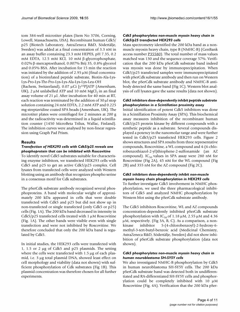
The pSerCdk substrate antibody recognized several phos-phoproteins. A band with molecular weight of approxi-mately 200 kDa appeared in cells that were doubletransfected with Cdk5 and p25 but did not show up innon-transfected or single transfected (only Cdk5 or p25)cells (Fig. 1A). The 200 kDa band decreased in intensity inCdk5/p25 transfected cells treated with 1 M Roscovitine(Fig. 1A). The other bands were visible even with singletransfection and were not inhibited by Roscovitine. Wetherefore concluded that only the 200 kDa band is regu-lated by Cdk5.
In initial studies, the HEK293 cells were transfected with1, 1.5 or 2 g of Cdk5 and p25 plasmids. The settingwhere the cells were transfected with 1.5 g of each plas-mid, i.e. 3 g total plasmid DNA, showed least effect oncell morphology and viability (data not shown) with suf-ficient phosphorylation of Cdk substrates (Fig 1B). Thisplasmid concentration was therefore chosen for all furtherexperiments.
Cdk5 phosphorylates non-muscle myosin heavy chain in Cdk5/p25 transfected HEK293 cellsMass spectrometry identified the 200 kDa band as a non-muscle myosin heavy chain, type B (NMHC-B) (GenBankaccess number P35580). The total number of mass valuesmatched was 130 and the sequence coverage 57%. Verifi-cation that the 200 kDa pSerCdk substrate band indeedwas myosin was done by immunoprecipitation. WhenCdk5/p25 transfected samples were immunoprecipitatedwith pSerCdk substrate antibody and then run on Westernblot, the pSerCdk substrate antibody and NMHC-B anti-body detected the same band (Fig 1C). Western blot anal-ysis of cell lysates gave the same results (data not shown).
Cdk5 inhibitors dose-dependently inhibit peptide substrate phosphorylation in a Scintillation proximity assayInitial identification of potent Cdk5 inhibitors was donein a Scintillation Proximity Assay (SPA). This biochemicalassay measures inhibition of the recombinant humanCdk5/p25 protein kinase by different compounds with asynthetic peptide as a substrate. Several compounds dis-played a potency in the nanomolar range and were furthertested in Cdk5/p25 transfected HEK293 cells. Figure 2shows structures and SPA results from three representativecompounds, Roscovitine, a WL compound and 4-(6-chlo-robenzothiazol-2-yl)thiophene-2-sulfonamide (an AZcompound). IC50 values in SPA assay were 280 nM forRoscovitine (Fig 2A), 45 nM for the WL compound (Fig2B) and 355 nM for the AZ compound (Fig 2C).
Cdk5 inhibitors dose-dependently inhibit non-muscle myosin heavy chain phosphorylation in HEK293 cellsTo further investigate Cdk5 involvement in NMHC phos-phorylation, we used the three pharmacological inhibi-tors of Cdk5 and analyzed NMHC phosphorylation byWestern blot using the pSerCdk substrate antibody.
The Cdk5 inhibitors Roscovitine, WL and AZ compoundsconcentration-dependently inhibited pSerCdk substratephosphorylation with IC50 of 1.18 M, 2.55 M and 4.36M, respectively. (Fig 3A, B, C). As a comparison, a non-kinase inhibitor 5-(4-chlorobenzoyl)-2-hydroxy-6-methyl-3-tert-butyl-benzoic acid (Medicinal Chemistry,AstraZeneca R&D, Södertälje, Sweden) did not show inhi-bition of pSerCdk substrate phosphorylation (data notshown).
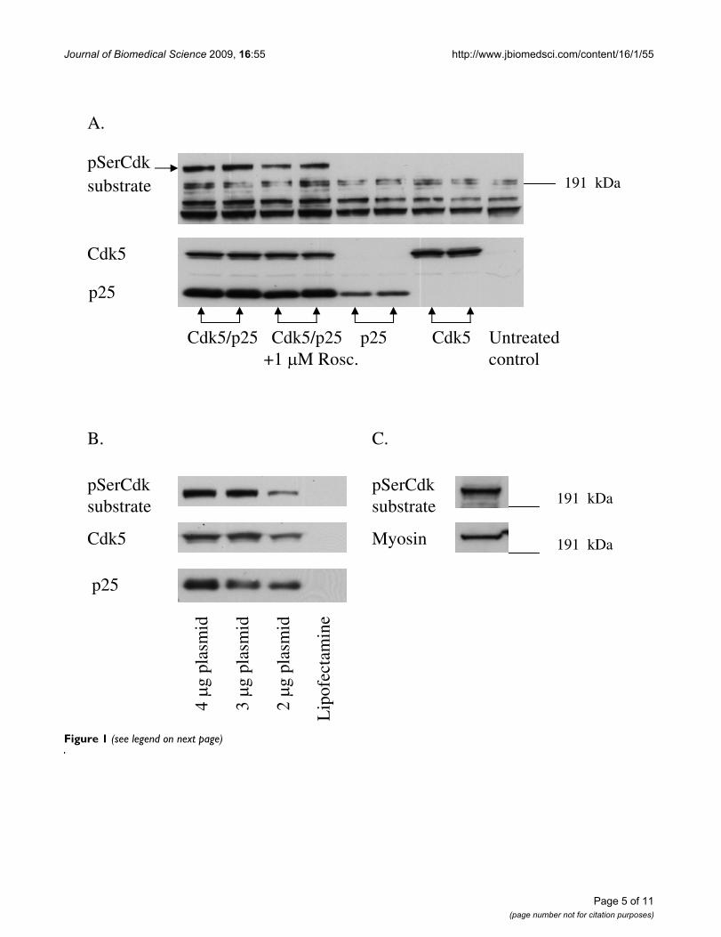
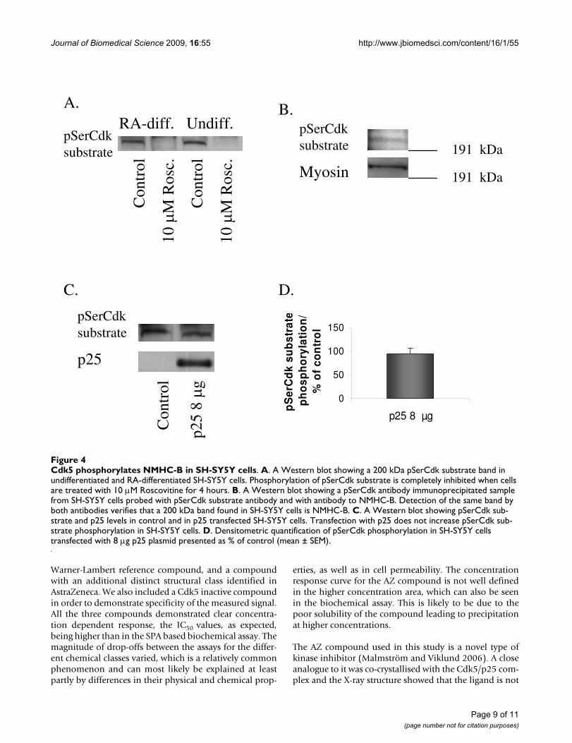
Cdk5 phosphorylates non-muscle myosin heavy chain in human neuroblastoma SH-SY5Y cellsWe also investigated NMHC-B phosphorylation by Cdk5in human neuroblastoma SH-SY5Y cells. The 200 kDapSerCdk substrate band was detected both in undifferen-tiated and RA-differentiated SH-SY5Y cells and phosphor-ylation could be completely inhibited with 10 MRoscovitine (Fig. 4A). Verification that the 200 kDa pSer-
Page 4 of 11(page number not for citation purposes)
Journal of Biomedical Science 2009, 16:55 http://www.jbiomedsci.com/content/16/1/55
Figure 1 (see legend on next page)
pSerCdksubstrate
Cdk5
p25
A.
191 kDa
B.
4 g
plas
mid
3 g
plas
mid
2 g
plas
mid
Lip
ofec
tam
ine
pSerCdk substrate
Cdk5
p25
C.
pSerCdk substrate
Myosin
191 kDa
191 kDa
Cdk5/p25 Cdk5/p25 p25 Cdk5 Untreated +1 M Rosc. control
Page 5 of 11(page number not for citation purposes)

Journal of Biomedical Science 2009, 16:55 http://www.jbiomedsci.com/content/16/1/55
Cdk band detected in SH-SY5Y cells was in fact NMHC-Bwas done by immunoprecipitation. When samples fromundifferentiated SH-SY5Y cells were immunoprecipitatedwith pSerCdk antibody and then run on Western blot,pSerCdk antibody and NMHC-B antibody detected thesame band (Fig. 4B). pSerCdk substrate phosphorylationdid not increase in intensity in p25 transfected cells, eitherin RA-differentiated (Fig. 4C, D) or undifferentiated (datanot shown).
DiscussionAs Cdk5 kinase activity is almost exclusively restricted toneuronal post-mitotic cells, it has been challenging toidentify both a specific and robust cellular assay to enablepharmacological characterization of enzyme inhibitors.In an attempt to identify a suitable system, we set up totransfect HEK293 cells, with both the Cdk5 enzyme andits activator p25.
Previously, a wide variety of substrate proteins have beenidentified for Cdk5, which reflects its role in diverse func-tions. For instance, Cdk5 is involved in regulation ofcytoskeletal dynamics by phosphorylating neurofilamentsand microtubule-associated proteins tau and MAP1B [21-23]. Cdk5 participates in synaptic function and neuro-transmission by phosphorylating many synaptic proteins[24]. In addition, Cdk5 has an important function in thedevelopment of the central nervous system regulatingneuronal migration [25] and axon guidance [26].
As Cdk5 phosphorylates serines and threonines immedi-ately upstream of a proline residue [27], an antibody thatrecognizes phospho serine in this consensus motif forCdk substrate, was utilized in our study. As a result, a non-muscle myosin heavy chain, type B (NMHC-B) was iden-tified as a Cdk5 substrate. Myosins constitute a large fam-ily of actin-based motor proteins [28] and non-musclemyosins are composed of two heavy chains and two pairsof light chains. In contrast to muscle myosins, which formstable myofibrils, the non-muscle myosin constitutes partof the actinomyosin cytoskeleton. Both actin and myosinin non-muscle cells can be rapidly assembled or disassem-
bled in response to extracellular signals to allow cell shapechanges needed for movement, cell division or secretion[29,30]. Functional activities of myosin are regulated bylight chain and heavy chain phosphorylation [29].
In vertebrates, NMHC are phosphorylated by kinases suchas Protein Kinase C, Casein kinase II and Ca2+/calmodu-lin-dependent protein kinase [31,32]. Phosphorylation ofNMHC by Cdk5 has not been demonstrated previouslyalthough a link between Cdk5 and non-muscle myosinwas recently discovered. Ledee et al. [33] showed a specificinteraction between Cdk5 activator p39 and non-musclemyosin essential light chain.
To test the physiological relevance of our finding, NMHC-B phosphorylation by Cdk5 was investigated in a secondcell line, a human neuroblastoma SH-SY5Y. HEK293 cellsonly express Cdk5 at low levels and they lack endogenousCdk5 activity, whereas SH-SY5Y cells express both Cdk5and its activator p35 and exhibit basal Cdk5 activity [17].NMHC-B phosphorylation by Cdk5 was also detected inSH-SY5Y cells, both in undifferentiated and RA-differenti-ated ones. However, when p25 was transfected to thecells, no increase in phosphorylation of NMHC-B wasobserved in contrast to transfected HEK293 cells wherephosphorylation was dose-dependent on plasmid con-centration. This could be due to the fact that NMHC-B isalready maximally phosphorylated by endogenous Cdk5kinase activity.
In drug discovery research, it is customary that the first fil-tering assay used in compound characterization is a fast,readily reproducible, miniaturizable and in most casesbiochemical assay. The primary purpose for this system isto serve as a means of ranking the compounds accordingto potency. Thereafter, the chemically most interestingand potent compounds need to be further characterizedin more complex cellular environment. For Cdk5/p25 wedescribe here a HEK293 cell system with quantification ofNMHC-B phosphorylation as read-out. The compoundstested in the present setting represent different chemicalclasses, with the known Cdk inhibitor, Roscovitine, a
Transfection of Cdk5/p25 into HEK293 cells identifies NMHC-B as a substrate for Cdk5Figure 1 (see previous page)Transfection of Cdk5/p25 into HEK293 cells identifies NMHC-B as a substrate for Cdk5. A. Western blot showing pSerCdk substrate, Cdk5 and p25 levels in cells transfected with Cdk5/p25, Cdk5/p25 transfected cells treated with 1 M Ros-covitine, cells transfected only with p25 or Cdk5 and untransfected cells. A 200 kDa band appeared only in the Cdk5/p25 dou-ble transfected cells. This band decreased in intensity when treated with 1 M Roscovitine and did not show up in single transfected or untransfected cells. B. Western blot showing pSerCdk substrate, Cdk5 and p25 levels in cells transfected with 2, 1.5 or 1 g of each plasmid and in cells treated only with transfection reagent, Lipofectamine. The cells transfected with 1.5 g of Cdk5 and p25 plasmid, i.e.3 g total plasmid DNA, showed sufficient phosphorylation of Cdk substrates and least effect on cell morphology and viability (data not shown). C. Western blot showing a pSerCdk antibody immunoprecipitated sample from Cdk5/p25 transfected HEK293 cells probed with pSerCdk substrate antibody and with antibody to NMHC-B. Detection of the same band by both antibodies verifies that the 200 kDa band is NMHC-B.
Page 6 of 11(page number not for citation purposes)

Journal of Biomedical Science 2009, 16:55 http://www.jbiomedsci.com/content/16/1/55
Page 7 of 11(page number not for citation purposes)
Cdk5 inhibitors dose-dependently inhibit peptide substrate phosphorylation in a SPA assayFigure 2Cdk5 inhibitors dose-dependently inhibit peptide substrate phosphorylation in a SPA assay. A. Roscovitine dose-dependently inhibits substrate phosphorylation with IC50 280 nM. B. WL compound dose-dependently inhibits substrate phos-phorylation with IC50 45 nM. C. AZ compound dose-dependently inhibits substrate phosphorylation with IC50 355 nM. Struc-tures of these compounds are shown next to their respective inhibition curves.
A.
B.
C.
Roscovitine
WL compound
AZ compound
-9-8-7-6-5-4-25
0
25
50
75
100
125
Log (M)
% in
hib
itio
n o
fsu
bst
rate
ph
osp
ho
ryla
tio
n
-8-7-6-5-4-25
0
25
50
75
100
125
Log (M)
% in
hib
itio
n o
fsu
bst
rate
ph
osp
ho
ryla
tio
n
-9-8-7-6-5-4-3-25
0
25
50
75
100
125
Log (M)
% in
hib
itio
n o
fsu
bst
rate
ph
osp
ho
ryla
tio
n
N
N N O
CH3
NH
F
N
SS
S
O
O
NH2
Cl
N
N N
N
CH3
CH3
NH
NH
OH
CH3
IC50 280 nM
IC50 45 nM
IC50 355 nM

Journal of Biomedical Science 2009, 16:55 http://www.jbiomedsci.com/content/16/1/55
Page 8 of 11(page number not for citation purposes)
Cdk5 inhibitors dose-dependently inhibit NMHC-B phosphorylation in Cdk5/p25 transfected HEK293 cellsFigure 3Cdk5 inhibitors dose-dependently inhibit NMHC-B phosphorylation in Cdk5/p25 transfected HEK293 cells. A. Roscovitine dose-dependently inhibits pSerCdk phosphorylation with IC50 1.18 M. B. WL compound dose-dependently inhib-its pSerCdk phosphorylation with IC502.55 M. C. AZ compound dose-dependently inhibits pSerCdk phosphorylation with IC50 4.36 M. A Western blot of pSerCdk substrate in control and in cells treated with different concentrations of inhibitor is shown next to their respective inhibition curves.
-8-7-6-5-4-25
0
25
50
75
100
125
Log (M)
% in
hib
itio
n o
f p
Ser
Cd
k su
bst
rateA
B
C
WL compound
-8-7-6-5-4
-25
0
25
50
75
100
Log (M)
% in
hib
itio
n o
f p
Ser
Cd
k su
bst
rate
AZ compound
Roscovitine
Con
trol
3.16
nM
31.6
nM
316
nM
1 M
3.16
M
6.32
M
15.8
M
Con
trol
31.6
nM
316
nM
1 M
3.16
M
6.32
M
15.8
M
Con
trol
3.16
nM
31.6
nM
316
nM
1 M
3.16
M
6.32
M
15.8
M
-7-6-5-4-25
0
25
50
75
100
125
Log (M)
% in
hib
itio
n o
f p
Ser
Cd
k su
bst
rate
IC50 1.18 M
IC50 2.55 M
IC50 4.36 M
Journal of Biomedical Science 2009, 16:55 http://www.jbiomedsci.com/content/16/1/55
Warner-Lambert reference compound, and a compoundwith an additional distinct structural class identified inAstraZeneca. We also included a Cdk5 inactive compoundin order to demonstrate specificity of the measured signal.All the three compounds demonstrated clear concentra-tion dependent response, the IC50 values, as expected,being higher than in the SPA based biochemical assay. Themagnitude of drop-offs between the assays for the differ-ent chemical classes varied, which is a relatively commonphenomenon and can most likely be explained at leastpartly by differences in their physical and chemical prop-
erties, as well as in cell permeability. The concentrationresponse curve for the AZ compound is not well definedin the higher concentration area, which can also be seenin the biochemical assay. This is likely to be due to thepoor solubility of the compound leading to precipitationat higher concentrations.
The AZ compound used in this study is a novel type ofkinase inhibitor (Malmström and Viklund 2006). A closeanalogue to it was co-crystallised with the Cdk5/p25 com-plex and the X-ray structure showed that the ligand is not
Cdk5 phosphorylates NMHC-B in SH-SY5Y cellsFigure 4Cdk5 phosphorylates NMHC-B in SH-SY5Y cells. A. A Western blot showing a 200 kDa pSerCdk substrate band in undifferentiated and RA-differentiated SH-SY5Y cells. Phosphorylation of pSerCdk substrate is completely inhibited when cells are treated with 10 M Roscovitine for 4 hours. B. A Western blot showing a pSerCdk antibody immunoprecipitated sample from SH-SY5Y cells probed with pSerCdk substrate antibody and with antibody to NMHC-B. Detection of the same band by both antibodies verifies that a 200 kDa band found in SH-SY5Y cells is NMHC-B. C. A Western blot showing pSerCdk sub-strate and p25 levels in control and in p25 transfected SH-SY5Y cells. Transfection with p25 does not increase pSerCdk sub-strate phosphorylation in SH-SY5Y cells. D. Densitometric quantification of pSerCdk phosphorylation in SH-SY5Y cells transfected with 8 g p25 plasmid presented as % of control (mean ± SEM).
RA-diff. Undiff.pSerCdk substrate
Con
trol
10
M R
osc.
Co n
trol
10
M R
osc.
A. B.pSerCdk substrate
Myosin
191 kDa
191 kDa
C.
pSerCdk substrate
p25
Con
trol
p25
8 g
D.
0
50
100
150
p25 8 μg
pS
erC
dk
sub
stra
tep
ho
sph
ory
lati
on
/%
of
con
tro
l
Page 9 of 11(page number not for citation purposes)

Journal of Biomedical Science 2009, 16:55 http://www.jbiomedsci.com/content/16/1/55
directly bound to the backbone (Glu 81 and Cys83) in theATP site of the kinase as is the usual case. Instead, a watermolecule was found to form a bridging interactionbetween the ligand and the hinge backbone (Malmströmet al., unpublished results). The AZ compound is assumedto bind in a similar fashion, although X-ray crystallogra-phy would be required to confirm this.
Phosphorylation of NMHC-B was evident only in Cdk5/p25 double transfected cells indicating that only Cdk5phosphorylates this substrate in HEK293 cells. Cdk5inhibitors concentration-dependently inhibited NMHC-Bphosphorylation and at higher concentrations phosphor-ylation was totally blocked. Described assay with NMHC-B phosphorylation as a read-out is thus very specific andsensitive. However, a limitation of the system at present isits relatively low throughput caused by the Western blotbased quantification of the phosphorylation signal. Anattempt to solve this could for instance be use of Imageanalysis in a high content screening setting such asImageXpress.
ConclusionA novel Cdk5 substrate, NMHC-B, was identified in thisstudy. Using phosphorylation of this substrate as a read-out, we developed a specific and sensitive cellular assaythat can be used for validation of compounds designed toinhibit Cdk5. A novel Cdk5 inhibitor was also pharmaco-logically characterized in this assay system.
AbbreviationsAD: Alzheimer's disease; Cdk5: cyclin-dependent kinase;HEK293: human embryonic kidney 293; NMHC-B: non-muscle myosin heavy chain, type B; RA: retinoic acid; SPA:Scintillation proximity assay.
Competing interestsThe authors declare that they have no competing interests.
Authors' contributionsAJ carried out experiments in SH-SY5Y cells, participatedin design of the study and drafted the manuscript. KA,ACH and AJ all participated in experiments with HEK293cells. KA also participated in the design of the study andedited the manuscript. JO carried out the sequence analy-sis using mass spectrometry and protein identificationusing Mascot server. GH carried out SPA assays. JM wasresponsible for the design and synthesis of the novel Cdk5kinase inhibitor used in the study. GL designed the plas-mids used in transfection experiments and participated inthe initial transfection experiments in HEK293 cells. MVorganized the study, participated in study design andcoordination and revised the manuscript. All authors readand approved the final manuscript.
AcknowledgementsThis study was partly supported by research grants from Stiftelsen Demen-tia. We want to thank Rolf Olsson for help with figures and Richard Cow-burn for critical reading of the manuscript.
References1. Meyerson M, Enders GH, Wu CL, Su LK, Gorka C, Nelson C, Harlow
E, Tsai LH: A family of human CDC2-related protein kinases.EMBO J 1992, 11:2909-2917.
2. Tsai LH, Delalle I, Caviness VS Jr, Chae T, Harlow E: p35 is a neural-specific regulatory subunit of cyclin-dependent kinase 5.Nature 1994, 371:419-423.
3. Tang D, Yeung J, Lee KY, Matsushita M, Matsui H, Tomizawa K,Hatase O, Wang JH: An isoform of the neuronal cyclin-depend-ent kinase 5 (cdk5) activator. J Biol Chem 1995,270:26897-26903.
4. Sharma P, Sharma M, Amin ND, Albers RW, Pant HC: Regulationof cyclin-dependent kinase 5 catalytic activity by phosphor-ylation. Proc Natl Acad Sci 1999, 96:11156-11160.
5. Zheng M, Leung CL, Liem RK: Region-specific expression of cyc-lin-dependent kinase 5 (cdk5) and its activators, p35 and p39,in the developing and adult rat central nervous system. J Neu-robiol 1998, 35:141-159.
6. Patrick GN, Zukerberg L, Nikolic M, de la Monte S, Dikkes P, Tsai LH:Conversion of p35 to p25 deregulates Cdk5 activity and pro-motes neurodegeneration. Nature 1999, 402:615-622.
7. Tseng HC, Zhou Y, Shen Y, Tsai LH: A survey of Cdk5 activatorp35 and p25 levels in Alzheimer's disease brains. FEBS Lett2002, 523:58-62.
8. Lee KY, Clark AW, Rosales JL, Chapman K, Fung T, Johnston RN:Elevated neuronal Cdc2-like kinase activity in the Alzheimerdisease brain. Neurosci Res 1999, 34:21-29.
9. Smith PD, Crocker SJ, Jackson-Levis V, Jordan-Sciutto KL, Hayley S,Mount MP, O'Hare MJ, Callaghan S, Slack RS, Przedborski S, AnismanH, Park DS: Cyclin-dependent kinase 5 is a mediator ofdopaminergic neuron loss in a mouse model of Parkinson'sdisease. Proc Natl Acad Sci 2003, 100:13650-13655.
10. Nguyen MD, Lariviére RC, Julien J-P: Deregulation of Cdk5 in amouse model of ALS: toxicity alleviated by perikaryal neuro-filament inclusions. Neuron 2001, 30:135-147.
11. Wang J, Liu SH, Fu YP, Wang JH, Lu YM: Cdk5 activation induceshippocampal CA1 cell death by directly phosphorylatingNMDA receptors. Nat Neurosci 2003, 6:1039-1047.
12. Sananbenesi F, Fischer A, Wang X, Schrick C, Neve R, Radulovic J,Tsai LH: A hippocampal Cdk5 pathway regulates extinction ofcontextual fear. Nat Neurosci 2007, 10:1012-1019.
13. Fischer A, Sananbenesi F, Schrick C, Spiess J, Radulovic J: Cyclin-dependent kinase 5 is required for associative learning. J Neu-rosci 2002, 22:3700-3707.
14. Ohshima T, Ogura H, Tomizawa K, Hayashi K, Suzuki H, Saito T,Kamei H, Nishi A, Bibb JA, Hisanaga S, Matsui H, Mikoshiba K:Impairment of hippocampal long-term depression anddefective spatial learning and memory in p35 mice. J Neuro-chem 2005, 94:917-925.
15. Fischer A, Sananbenesi F, Pang PT, Lu B, Tsai LH: Opposing roles oftransient and prolonged expression of p25 in synaptic plastic-ity and hippocampus-dependent memory. Neuron 2005,48:825-838.
16. Jämsä A, Hasslund K, Cowburn RF, Bäckström A, Vasänge M: Theretinoic acid and brain-derived neurotrophic factor differen-tiated SH-SY5Y cell line as a model for Alzheimer's disease-like tau phosphorylation. Biochem Biophys Res Commun 2004,319:993-1000.
17. Jämsä A, Bäckström A, Gustafsson E, Dehvari N, Hiller G, CowburnRF, Vasänge M: Glutamate treatment and p25 transfectionincrease Cdk5 mediated tau phosphorylation in SH-SY5Ycells. Biochem Biophys Res Commun 2006, 345:324-331.
18. Barvian M, Boschelli DH, Cossrow J, Dobrusin E, Fattaey A, Fritsch A,Fry D, Harvey P, Keller P, Garrett M, La F, Leopold W, McNamara D,Quin M, Trumpp-Kallmeyer S, Toogood P, Wu Z, Zhang E:Pyrido[2,3-d]pyrimidin-7-one inhibitors of cyclin-dependentkinases. J Med Chem 2000, 43:4606-4616.
19. Booth RJ, Chatterjee A, Malone TC: WO 01/55148. .20. Malmström J, Viklund J: WO 2006/004507. .
Page 10 of 11(page number not for citation purposes)

Journal of Biomedical Science 2009, 16:55 http://www.jbiomedsci.com/content/16/1/55
Publish with BioMed Central and every scientist can read your work free of charge
"BioMed Central will be the most significant development for disseminating the results of biomedical research in our lifetime."
Sir Paul Nurse, Cancer Research UK
Your research papers will be:
available free of charge to the entire biomedical community
peer reviewed and published immediately upon acceptance
cited in PubMed and archived on PubMed Central
yours — you keep the copyright
Submit your manuscript here:http://www.biomedcentral.com/info/publishing_adv.asp
BioMedcentral
21. Grant P, Sharma P, Pant HC: Cyclin-dependent protein kinase 5(Cdk5) and the regulation of neurofilament metabolism. EurJ Biochem 2001, 268:1534-1546.
22. Baumann K, Mandelkow EM, Biernat J, Piwnica-Worms H, Man-delkow E: Abnormal Alzheimer-like phosphorylation of Tau-protein by cyclin-dependent kinases cdk2 and Cdk5. FEBS Lett1993, 336:417-424.
23. Paglini G, Pigino G, Kunda P, Morfini G, Maccioni R, Quiroga S, Fur-reira A, Caceres A: Evidence for the participation of the neu-ron-specific Cdk5 activator p35 during laminin-enhancedaxonal growth. J Neurosci 1998, 18:9858-9869.
24. Cheung ZH, Fu AKY, Ip NY: Synaptic roles of Cdk5: Implica-tions in higher cognitive functions and neurodegenerativediseases. Neuron 2006, 50:13-18.
25. Ohshima T, Ward JM, Huh CG, Longenecker G, Veeranna , Pant HC,Brady RO, Martin LJ, Kulkarni AB: Targeted disruption of thecyclin-dependent kinase 5 gene results in abnormal cortico-genesis, neuronal pathology and perinatal death. Proc NatlAcad Sci 1996, 93:11173-11178.
26. Kwon YT, Tsai L-H, Crandall JE: Callosal axon guidance defectsin p35(-/-) mice. J Comp Neurol 1999, 415:218-229.
27. Songyang Z, Lu KP, Kwon YT, Tsai LH, Filhol O, Cochet C, BrickeyDA, Soderling TR, Bartleson C, Graves DJ, DeMaggio AJ, HoekstraMF, Blenis J, Hunter T, Cantley LC: A structural basis for sub-strate specificities of protein Ser/Thr kinases: primarysequence preference of casein kinases I and II, NIMA, phos-phorylase kinase, calmodulin-dependent kinase II, CDK5,and ERK1. Mol Cell Biol 1996, 16:6486-6493.
28. Sellers JR: Myosins: a diverse superfamily. Biochim Biophys Acta2000, 1496:3-22.
29. Bresnick AR: Molecular mechanisms of nonmuscle myosin-IIregulation. Curr Opin Cell Biol 1999, 11:26-33.
30. Spudich A: Myosin reorganization in activated RBL cells cor-relates temporally with stimulated secretion. Cell MotilCytoskel 1994, 29:345-353.
31. Murakami M, Chauhan VPS, Elzinga M: Two nonmuscle myosin IIheavy chain isoforms expressed in rabbit brains: filamentforming properties, the effects of phosphorylation by Pro-tein kinase C and Casein kinase II, and location of the phos-phorylation sites. Biochemistry 1998, 37:1989-2003.
32. Tanaka E, Fukunaga K, Yamamoto H, Iwasa T, Miyamoto E: Regula-tion of the actin-activated Mg-ATPase of brain myosin viaphosphorylation by the brain Ca2+, calmodulin-dependentprotein kinases. J Neurochem 1986, 47:254-262.
33. Ledee DR, Tripathi BK, Zelenka PS: The CDK5 activator, p39,binds specifically to myosin essential light chain. Biochem Bio-phys Res Commun 2007, 354:1034-1039.
Page 11 of 11(page number not for citation purposes)
Copyright © 2022 FDOKUMEN