Identification of a Recurrent Microdeletion at 17q23.1q23.2 Flanked by Segmental Duplications...
8
REPORT Identification of a Recurrent Microdeletion at 17q23.1q23.2 Flanked by Segmental Duplications Associated with Heart Defects and Limb Abnormalities Blake C. Ballif, 1, * Aaron Theisen, 1 Jill A. Rosenfeld, 1 Ryan N. Traylor, 1 Julie Gastier-Foster, 2,3,4 Devon Lamb Thrush, 2,4 Caroline Astbury, 2,4 Dennis Bartholomew, 4,5 Kim L. McBride, 4,6 Robert E. Pyatt, 2,3 Kate Shane, 4,5 Wendy E. Smith, 7 Valerie Banks, 7 William B. Gallentine, 8 Pamela Brock, 9 M. Katharine Rudd, 10 Margaret P. Adam, 10 Julia A. Keene, 10 John A. Phillips III, 11 Jean P. Pfotenhauer, 12 Gordon C. Gowans, 9 Pawel Stankiewicz, 13,14 Bassem A. Bejjani, 1 and Lisa G. Shaffer 1 Segmental duplications, which comprise ~5%–10% of the human genome, are known to mediate medically relevant deletions, duplications, and inversions through nonallelic homologous recombination (NAHR) and have been suggested to be hot spots in chromosome evolution and human genomic instability. We report seven individuals with microdeletions at 17q23.1q23.2, identified by microarray-based comparative genomic hybridization (aCGH). Six of the seven deletions are ~2.2 Mb in size and flanked by large segmental duplications of >98% sequence identity and in the same orientation. One of the deletions is ~2.8 Mb in size and is flanked on the distal side by a segmental duplication, whereas the proximal breakpoint falls between segmental duplications. These character- istics suggest that NAHR mediated six out of seven of these rearrangements. These individuals have common features, including mild to moderate developmental delay (particularly speech delay), microcephaly, postnatal growth retardation, heart defects, and hand, foot, and limb abnormalities. Although all individuals had at least mild dysmorphic facial features, there was no characteristic constellation of features that would elicit clinical suspicion of a specific disorder. The identification of common clinical features suggests that micro- deletions at 17q23.1q23.2 constitute a novel syndrome. Furthermore, the inclusion in the minimal deletion region of TBX2 and TBX4, transcription factors belonging to a family of genes implicated in a variety of developmental pathways including those of heart and limb, suggests that these genes may play an important role in the phenotype of this emerging syndrome. Segmental duplications comprise ~5%–10% of the human genome. 1 Misalignment of segmental duplications during meiosis can mediate genomic instability by nonallelic homologous recombination (NAHR). Depending on the orientation of the segmental duplications, NAHR can generate microdeletions, microduplications, and inver- sions of the intervening genomic sequence. 2–7 Chromo- somal rearrangements associated with segmental duplica- tions include microdeletions at 3q29 (MIM 609425) and their reciprocal microduplications (MIM 611936); 8 Williams syndrome (MIM 194050) and deletions at 7q11.23; Angelman (MIM 105830) and Prader-Willi syn- dromes (MIM 176270) and maternally and paternally derived deletions, respectively, of 15q11-q13; microdele- tions at 16p11.2p12.2 and their reciprocal microduplica- tions; 9 Smith-Magenis syndrome (MIM 182290) and deletions at 17p11.2; duplication at 17p12 in Charcot- Marie-Tooth disease type 1A (MIM 604563); and the recip- rocal deletion at 17p12 resulting in hereditary neuropathy with liability to pressure palsies (HNPP [MIM 162500]) (reviewed in 4 ). Recently, a recurrent microdeletion at 17q21.3 mediated by segmental duplications was identi- fied by screening individuals with idiopathic mental retardation and congenital anomalies with a microarray targeting potential segmental duplication-rich rearrange- ment ‘‘hot spots’’ in the genome, 5–7 suggesting that other such rearrangements may yet be identified. Recent evidence suggests another segmental duplication ‘‘hub’’ on chromosome 17 at 17q23. Breakpoint analysis of a paracentric inversion that occurred in the human/chim- panzee/gorilla ancestor revealed that the distal breakpoint maps to the region syntenic to human 17q23, which suggests that the presence of this duplicon mediated the inversion in the Homo sapiens/Pan troglodytes/Gorilla gorilla ancestor. 10 Furthermore, at least two insertions and dele- tions and one translocation have occurred within the past six million years since the divergence of chimpanzees and humans, indicating that these regions have continued to be subject to more-recent local rearrangements. Further data have demonstrated the structural complexity of the 1 Signature Genomic Laboratories, Spokane, WA 99207, USA; 2 Department of Laboratory Medicine, Nationwide Children’s Hospital, Columbus, OH 43209, USA; 3 Department of Pathology, The Ohio State University, Columbus, OH 43209, USA; 4 Department of Pediatrics, The Ohio State University, Columbus, OH 43209, USA; 5 Section of Genetics, Nationwide Children’s Hospital, Columbus, OH 43209, USA; 6 Center for Molecular and Human Genetics, The Research Institute at Nationwide Children’s Hospital, Columbus, OH 43209, USA; 7 Department of Pediatrics, Maine Pediatric Specialty Group, Portland, ME 04102, USA; 8 Department of Pediatrics, Duke University Medical Center, Durham, NC 27710, USA; 9 Weisskopf Child Evaluation Center, University of Louisville, Louisville, KY 40202, USA; 10 Department of Human Genetics, Emory University School of Medicine, Atlanta, GA 30322, USA; 11 Department of Pediatrics, Vanderbilt University Medical Center, Nashville, TN 37232, USA; 12 Division of Genetics and Genomic Medicine, Vanderbilt University Medical Center, Nashville, TN 37232, USA; 13 Department of Human and Molecular Genetics, Baylor College of Medicine, Houston, TX 77030, USA; 14 Department of Medical Genetics, Institute of Mother and Child, 01-211 Warsaw, Poland *Correspondence: [email protected] DOI 10.1016/j.ajhg.2010.01.038. ª2010 by The American Society of Human Genetics. All rights reserved. 454 The American Journal of Human Genetics 86, 454–461, March 12, 2010
Transcript of Identification of a Recurrent Microdeletion at 17q23.1q23.2 Flanked by Segmental Duplications...

REPORT
Identification of a Recurrent Microdeletionat 17q23.1q23.2 Flanked by Segmental DuplicationsAssociated with Heart Defects and Limb Abnormalities
Blake C. Ballif,1,* Aaron Theisen,1 Jill A. Rosenfeld,1 Ryan N. Traylor,1 Julie Gastier-Foster,2,3,4
Devon Lamb Thrush,2,4 Caroline Astbury,2,4 Dennis Bartholomew,4,5 Kim L. McBride,4,6
Robert E. Pyatt,2,3 Kate Shane,4,5 Wendy E. Smith,7 Valerie Banks,7 William B. Gallentine,8
Pamela Brock,9 M. Katharine Rudd,10 Margaret P. Adam,10 Julia A. Keene,10 John A. Phillips III,11
Jean P. Pfotenhauer,12 Gordon C. Gowans,9 Pawel Stankiewicz,13,14 Bassem A. Bejjani,1
and Lisa G. Shaffer1
Segmental duplications, which comprise ~5%–10% of the human genome, are known to mediate medically relevant deletions,
duplications, and inversions through nonallelic homologous recombination (NAHR) and have been suggested to be hot spots in
chromosome evolution and human genomic instability. We report seven individuals with microdeletions at 17q23.1q23.2, identified
by microarray-based comparative genomic hybridization (aCGH). Six of the seven deletions are ~2.2 Mb in size and flanked by large
segmental duplications of >98% sequence identity and in the same orientation. One of the deletions is ~2.8 Mb in size and is flanked
on the distal side by a segmental duplication, whereas the proximal breakpoint falls between segmental duplications. These character-
istics suggest that NAHR mediated six out of seven of these rearrangements. These individuals have common features, including mild to
moderate developmental delay (particularly speech delay), microcephaly, postnatal growth retardation, heart defects, and hand, foot,
and limb abnormalities. Although all individuals had at least mild dysmorphic facial features, there was no characteristic constellation
of features that would elicit clinical suspicion of a specific disorder. The identification of common clinical features suggests that micro-
deletions at 17q23.1q23.2 constitute a novel syndrome. Furthermore, the inclusion in the minimal deletion region of TBX2 and TBX4,
transcription factors belonging to a family of genes implicated in a variety of developmental pathways including those of heart and limb,
suggests that these genes may play an important role in the phenotype of this emerging syndrome.
Segmental duplications comprise ~5%–10% of the human
genome.1 Misalignment of segmental duplications during
meiosis can mediate genomic instability by nonallelic
homologous recombination (NAHR). Depending on the
orientation of the segmental duplications, NAHR can
generate microdeletions, microduplications, and inver-
sions of the intervening genomic sequence.2–7 Chromo-
somal rearrangements associated with segmental duplica-
tions include microdeletions at 3q29 (MIM 609425)
and their reciprocal microduplications (MIM 611936);8
Williams syndrome (MIM 194050) and deletions at
7q11.23; Angelman (MIM 105830) and Prader-Willi syn-
dromes (MIM 176270) and maternally and paternally
derived deletions, respectively, of 15q11-q13; microdele-
tions at 16p11.2p12.2 and their reciprocal microduplica-
tions;9 Smith-Magenis syndrome (MIM 182290) and
deletions at 17p11.2; duplication at 17p12 in Charcot-
Marie-Tooth disease type 1A (MIM 604563); and the recip-
rocal deletion at 17p12 resulting in hereditary neuropathy
with liability to pressure palsies (HNPP [MIM 162500])
1Signature Genomic Laboratories, Spokane, WA 99207, USA; 2Department of La
USA; 3Department of Pathology, The Ohio State University, Columbus, OH 43
OH 43209, USA; 5Section of Genetics, Nationwide Children’s Hospital, Colu
Research Institute at Nationwide Children’s Hospital, Columbus, OH 43209, U
ME 04102, USA; 8Department of Pediatrics, Duke University Medical Center,
of Louisville, Louisville, KY 40202, USA; 10Department of Human Genetics, Em
of Pediatrics, Vanderbilt University Medical Center, Nashville, TN 37232, U
Medical Center, Nashville, TN 37232, USA; 13Department of Human and M14Department of Medical Genetics, Institute of Mother and Child, 01-211 Wa
*Correspondence: [email protected]
DOI 10.1016/j.ajhg.2010.01.038. ª2010 by The American Society of Human
454 The American Journal of Human Genetics 86, 454–461, March 1
(reviewed in 4). Recently, a recurrent microdeletion at
17q21.3 mediated by segmental duplications was identi-
fied by screening individuals with idiopathic mental
retardation and congenital anomalies with a microarray
targeting potential segmental duplication-rich rearrange-
ment ‘‘hot spots’’ in the genome,5–7 suggesting that other
such rearrangements may yet be identified.
Recent evidence suggests another segmental duplication
‘‘hub’’ on chromosome 17 at 17q23. Breakpoint analysis of
a paracentric inversion that occurred in the human/chim-
panzee/gorilla ancestor revealed that the distal breakpoint
maps to the region syntenic to human 17q23, which
suggests that the presence of this duplicon mediated the
inversion in the Homo sapiens/Pan troglodytes/Gorilla gorilla
ancestor.10 Furthermore, at least two insertions and dele-
tions and one translocation have occurred within the
past six million years since the divergence of chimpanzees
and humans, indicating that these regions have continued
to be subject to more-recent local rearrangements. Further
data have demonstrated the structural complexity of the
boratory Medicine, Nationwide Children’s Hospital, Columbus, OH 43209,
209, USA; 4Department of Pediatrics, The Ohio State University, Columbus,
mbus, OH 43209, USA; 6Center for Molecular and Human Genetics, The
SA; 7Department of Pediatrics, Maine Pediatric Specialty Group, Portland,
Durham, NC 27710, USA; 9Weisskopf Child Evaluation Center, University
ory University School of Medicine, Atlanta, GA 30322, USA; 11Department
SA; 12Division of Genetics and Genomic Medicine, Vanderbilt University
olecular Genetics, Baylor College of Medicine, Houston, TX 77030, USA;
rsaw, Poland
Genetics. All rights reserved.
2, 2010

A B
17q22 17q23.1 17q23.2
55 Mb 56 Mb 57 Mb 58 Mb
~2.2 Mb
~2.8 Mb
TB
X2
TB
X4
CA
4
US
P32
AP
PB
P2
BC
AS
3
NA
CA
2
BR
IP1
INT
S2
PP
M1D
ME
D13
TL
K2
YP
EL
2
DH
X40
CL
TC
TM
EM
49
PT
RH
2
TU
BD
1
RP
S6K
B1
TE
X14
RA
D51C
TR
IM37
MR
C2
Patient 1
Patient 2
Patient 3
Patient 4
Patient 5
Patient 6
Seg Dups
Patient 7
A B
Figure 1. Summary of Microarray Anal-ysis of Individuals with Copy NumberImbalances at 17q23.1q23.2At the top of the figure is a partial idiogramshowing chromosome bands 17q22-17q23.2 with genomic coordinates corre-sponding to the hg18 build of the humangenome. Orange boxes represent seg-mental duplications (seg dups), with pairedsegmental duplications indicated by blueboxes. The location and orientation ofthe 18 kb inverted duplication and the15 kb direct duplication contained withinthe paired segmental duplications are indi-cated by arrows marked A and B, respec-tively. The complex segmental duplicationstructure of this region has been simplifiedfor illustrative purposes. Diagrams showthe deletions in seven individuals with mi-crodeletions at 17q23.1q23.2. Six of theseven deletions were refined with a high-density, 244K oligonucleotide microarray(patients 1–4, 6, and 7). A 105K oligonucle-otide microarray was used for patient 5.Vertical dashed lines indicate the 2.2 and2.8 Mb deletion sizes in the individuals.Blue boxes represent all known genesannotated in OMIM in the deletion region.OMIM genes TBX2 and TBX4 are shown asyellow boxes.
region, which has been shown to be polymorphic in the
human population and associated with a multiple sclerosis
susceptibility locus on 17q22-q24.11,12
Here we report the clinical and molecular characteriza-
tion of seven individuals with microdeletions at 17q23.
1q23.2. Patients 1–3 in this study were ascertained by Sig-
nature Genomic Laboratories. Patients 4 and 5 were
ascertained by Nationwide Children’s Hospital. Patient
6 was ascertained by Emory University School of Medicine.
Patient 7 was ascertained by Baylor College of Medicine.
Informed consent was obtained from patients 1, 4, 6, and
7 to publish photographs with consent forms approved
by the Institutional Review Board of respective institutions.
All seven individuals with microdeletions at 17q23 were
initially identified by microarray-based comparative
genomic hybridization (aCGH) with various microarray
platforms, one of which, patient 6, was previously reported
(Figure 1).13 Whole-genome bacterial artificial chromosome
The American Journal of Human G
(BAC)-based microarray analysis was
originally performed on DNA from
patients 1 and 2 with a >4600 clone
custom microarray as previously
described.14 Oligonucleotide-based
microarray analysis was originally
performed on DNA from patients 3–5
with a custom 105K-feature whole-
genome microarray (Agilent Technolo-
gies), as previously described.14 The
deletion in patient 6 was identified
with a custom 44K oligonucleotide-
based microarray (Agilent Technologies), as previously
described.13,15 The deletion in patient 7 was originally iden-
tified with a custom-designed 105K-feature whole-genome
oligonucleotide microarray (Agilent Technologies), as previ-
ously described.16 In addition, all patients for whom DNA
was available (1–4, 6, and 7) were reanalyzed at higher
resolution with an off-the-shelf 244K-feature whole-
genome microarray (Agilent Technologies), as previously
described.14 One individual, patient2, hada 2.8 Mb deletion
(chromosome 17: 54.8–57.6 Mb); the remaining six individ-
uals had 2.2 Mb deletions (chromosome 17: 55.4–57.6 Mb).
All seven deletions were confirmed and visualized by
fluorescence in situ hybridization (FISH) with BAC clones,
as previously described (Figure 2).17 Parental FISH testing
in five of the seven cases confirmed an apparently de
novo origin (patients 2 and 4–7). The deletions in patients
1–3 and 5 were confirmed by FISH with BAC probe RP11-
289K16 from 17q23.1. For case 4, BAC RP11-119J7 was
enetics 86, 454–461, March 12, 2010 455

Figure 2. Representative FISH Image Showing Deletion at17q23 in Patient 2BAC clone RP11-289K16 from 17q23.1 is labeled in red, and chro-mosome 17 centromere probe D17Z1 is labeled in green asa control. The absence of a red signal on one homolog (arrow)indicates deletion at 17q23.1.
used to confirm the deletion. The deletion in patient 6 was
confirmed by FISH with BAC RP11-436E15, as previously
described,13,15 and the deletion in patient 7 was confirmed
by FISH with BAC clone RP11-615P24. Microsatellite
marker analysis with the Identifiler kit (Applied Biosys-
tems) showed correct paternity in patient 6, confirming
the de novo origin of the deletion (Rudd et al.13). All other
parental samples were unavailable.
Computational analysis of the 17q23.1q23.2 region
with the hg18 build of the UCSC genome browser18 and
the hg17 build of the Human Genome Segmental Duplica-
tion Database identified a complex arrangement of
segmental duplications, some of which directly flank the
2.2 Mb deletion breakpoints and the distal 2.8 Mb break-
point (Figure 1). These flanking segmental duplications
are ~100 kb in size and have a complex evolutionary struc-
ture10 that includes an ~15 kb segment of >98% sequence
identity present in the same orientation. In addition, there
is an ~18 kb segment of >94% sequence identity that is
inverted in orientation with respect to its duplication
partner. The 2.2 Mb deletions are flanked by the homolo-
gous segmental duplications present in the same orienta-
tion. Highly identical (>98% sequence identity) segmental
duplications in direct orientation flanking the deletion
breakpoints are consistent with NAHR-mediated rear-
rangements (Figure 1). Although six of seven deletions
were flanked by segmental duplications, the proximal
breakpoint of the 2.8 Mb deletion falls between two
segmental duplications (Figure 1). Atypical breakpoints
have been reported for other recurrent rearrangements
mediated by segmental duplications: for example, some
of the rarer rearrangements of 17p11.2 associated with
Smith-Magenis syndrome do not have breakpoints flanked
by the typical paired segmental duplications and are not
associated with known genomic architectural features,19
and some of the breakpoints in the recently identified
16p11.2p12.2 microdeletion syndrome are not flanked
by segmental duplications.9
Clinical characterization of the seven individuals with
microdeletions at 17q23.1q23.2 revealed multiple com-
mon clinical features (Table 1; see also Supplemental
456 The American Journal of Human Genetics 86, 454–461, March 1
Discussion available online). All individuals had mild to
moderate developmental delay. A majority had low birth
weight (5 of 7); microcephaly or relative microcephaly
(5 of 7), including one with overall small growth parame-
ters; postnatal growth retardation (5 of 7); heart defect(s)
(6 of 7), in most cases either patent ductus arteriosus or
atrial septal defect (ASD); hand and foot anomalies
including long, thin fingers and toes (7 of 7); and musculo-
skeletal abnormalities of varying severity (4 of 7). Features
identified in more than one individual include aggressive
behavior (2 of 7) and hearing loss (2 of 7). Although five
of seven individuals had eye anomalies such as chalazion,
stellate pattern of the irises, retinopathy of prematurity,
and esotropia, the anomalies affect different components
of the eye and are unlikely to be related; therefore, these
anomalies are likely incidental to the 17q23.1q23.2 micro-
deletion phenotype. Although all individuals had at least
mild dysmorphic facial features, there was no character-
istic constellation of features that would elicit clinical
suspicion of a specific disorder (Figure 3). Nonetheless,
the common clinical features in these individuals together
with the common size and gene content of the deleted
regions suggest that deletion at 17q23.1q23.2 is causative
of the individuals’ phenotypes and may constitute a novel
microdeletion syndrome.
From November 2007 to October 2009, Signature
Genomic Laboratories tested 19,912 patients with whole-
genome microarray platforms that had coverage over
17q23.1q23.2 and identified three microdeletions of
17q23.1q23.2, for a frequency of 0.015% among its patient
population. By comparison, during this same period, the
laboratory identified 23 Smith-Magenis syndrome (SMS)
deletions (0.12% of patient population), which has a
frequency in the general population of ~1 in 15,000,20 sug-
gesting that the population frequency of 17q23.1q23.2
microdeletions may be ~1 in 115,000. This may be an over-
estimate of frequency because some cases of SMS are diag-
nosed by other methods, and therefore not all individuals
with these syndromes will have aCGH testing, whereas
17q23.1q23.2 microdeletions would not be expected to
be diagnosed by other methods that require clinical suspi-
cion of a specific disorder.
Because the phenotype of the individual with the larger
2.8 Mb deletion does not appear to differ from that of the
individuals with the smaller 2.2 Mb deletion, the critical
region for this syndrome likely lies inside this 2.2 Mb
region. Of the 11 OMIM and RefSeq genes within the 2.2
Mb smallest region of overlap (Figure 1), at least two,
TBX2 (MIM 600747) and TBX4 (MIM 601719), are strong
candidates that might play a role in some of the features
of individuals with this microdeletion.
TBX2 and TBX4 belong to an ancient family of genes
present in divergent multicellular organisms, such as
sponges and humans, that encode transcription factors
characterized by a strongly conserved, sequence-specific
DNA-binding domain (or T-box domain). More specifi-
cally, TBX2 and TBX4 are members of two closely related
2, 2010

Table 1. Clinical Features in Subjects with Microdeletions at 17q23.2
Patient Number 1 2a 3 4 5 6 7
Age 3 yrs 5 yrs, 1 mo. 4 yrs 16 yrs, 6 mo. 8 mo. 4 yrs 1 yr, 10 mo.
Sex Female Female Male Female Female Female Female
Low birth weight(%25th percentile)
þ þ � þ þ þ �
Postnatal growthretardation
þ � � þ þ þ þ
Microcephaly(<5th percentile)
þ þ þ (relative) � þb � þ
Mild to moderatedevelopmentaldelays
þ þ þ þ þ þ þ
Heart defects PDA ASD secundum Pulmonaryhypertension
PDA withpulmonaryhypertension
PDA; bicuspidaortic valve
None; noechocardiography
ASD secundumwith pulmonaryhypertension
Musculoskeletal Tibial torsion Abnormallylaterallypositionedossificationcenters of thepatella, delayedfor age
Underossifiedfemoral heads;hips deeply seatedin acetabulae;mottled femoralepiphyses andmetaphyses; smalland abnormallower-limbepiphyses
Short staturewith leg lengthdiscrepancy;hypoplasia ofpatellae and tibialepiphyses; shallowacetabulae; rightside short femoralneck with coxavara and magna;scoliosis
NR None Scoliosis; limitedextension ofknees andelbows
Hearing loss þ � � þ � � �
Eyes Chalazion Left sidedesotropia
Normal Bilateralesotropia
Retinopathyof prematurity
Stellate patternof irises
Normal
Facial features High eyebrows;right epicanthalfold
Hypertelorism;flattened nasalbridge andmidface; simpleright ear
Plagiocephaly;bilateral innerepicanthal folds;bulbous, bifidnose; posteriorlyrotated,prominent ears;long eyelashes
Prominenceof forehead;moderate dentalcrowdingrequiringorthodonticappliances
Box-shapedcranium; openanteriorfontanelle;prominentforehead; wide-seteyes, small nosewith bulbous nasaltip; microstomia;small ears;recessed jaw
Mild frontalbossing; roundednasal tip; widespace betweenfront two teeth
Mildmicrognathia;long eyelashes;protuberant ears;small mouth;long neck
Hands and feet Thin fingers; deep,grooved spacebetween firstand second toes;familial 2–3syndactyly
Long, thinfingers andtoes
Long, thin fingersand toes; secondtoes longer thangreat toes
Long, thin fingersand toes; digitalclubbing; bilateralpes planus;slightly shortenedfoot rays
Long fingersand toes
Long fingersand toes;overlapping toes
Congenitalcontractures; longfingers and toes;overlapping toes 4and 5; pes planus;fifth fingerclinodactyly
Other Behavioralabnormalities;intention tremors;asthma
NR Tethered cord;sacral dimples;shawl scrotum
Mild increasedDTR at knees andankles; migraines
Cutis aplasia atbirth; centralhypotonia;brisk DTRs
Behavioralabnormalities
NR
The following abbreviations are used: þ, feature present; �, feature absent; PDA, patent ductus arteriosus; ASD, atrial septal defect; DTR, deep tendon reflexes;NR, none reported.a Patient 2 has an atypical ~2.8 Mb deletion. All other patients described in this table have ~2.2 Mb deletions.b All growth parameters were at the fifth percentile.
T-box subfamilies, TBX2/3 and TBX4/5. Interestingly, at
least 17 evolutionary rearrangements, including gene
cluster insertions and deletions, one inversion, and one
local translocation, have occurred within the region since
the divergence of humans and chickens ~300 million years
ago. Despite the instability of the region in which the gene
The Ameri
pair resides, TBX2 and TBX4 (as well as TBX3 and TBX5)
have remained flanked by several loci present in all
amniotes, BCAS33 and BRIP1 (MIM 605822) (THRAP2
[MIM 608711] and RBM19 flank TBX3 [MIM 601621]
and TBX5 [MIM 601620]).21 The high conservation of
these gene clusters indicates a functional significance.
can Journal of Human Genetics 86, 454–461, March 12, 2010 457

Figure 3. Facial Features of Individuals with Microdeletion at17q23.1q23.2(A) Patient 1 at 3 years. Note high eyebrows and right epicanthalfold.(B) Patient 6 at 4 years. Note mild frontal bossing and roundednasal tip.(C and D) Patient 7 at 22 months. Note long eyelashes, protu-berant ears, small mouth, and mild micrognathia.(E and F) Patient 4 at 16 years. With the exception of someprominence of the forehead, this patient was not noted to bedysmorphic.
Alternatively, the presence of large segmental duplications
in the region of 17q23 may predispose the region to rear-
rangements that conserve the linkage of these genomic
segments.
Targeted gene deletions in mouse have demonstrated
that T-box genes play numerous roles in development;
consequently, disruptions of many of these genes have
been associated with human disease. Mutations in the
coding sequence of TBX1 (MIM 602054), which maps
within the DiGeorge syndrome (DGS) region (MIM
188400) on chromosome 22q11.2,22 have been identified
in individuals with sporadic DGS/conotruncal anomaly
face syndrome/velocardiofacial syndrome (VCFS),23–25
which has a characteristic clinical feature of outflow tract
heart defects. In addition, heterozygous mutations in
TBX3 cause ulnar-mammary syndrome (UMS [MIM
181450]),26,27 which has a characteristic feature of poste-
rior upper-limb abnormalities; heterozygous loss-of-
function mutations of TBX4 in 15 individuals from five
families and one sporadic patient were found to cause
458 The American Journal of Human Genetics 86, 454–461, March 1
the autosomal-dominant Scott-Taor or small patella
syndrome (SPS [MIM 147891]);28 heterozygous mutations
in TBX5 cause anterior limb abnormalities and ASD in
Holt-Oram syndrome (MIM 142900);27 and mutations of
TBX20 (MIM 606061) have been associated with congen-
ital heart disease and cardiac developmental anomalies,
including defects in septation, chamber growth, and
valvulogenesis and cardiomyopathy.29
The TBX2/3 and TBX4/5 subfamilies of genes are espe-
cially interesting as haploinsufficiency candidates in mi-
crodeletions at 17q23 because of their role in the evolution
of novel vertebrate structures, including mouse limbs.30
Tbx2 is expressed in the anterior and posterior margins
of forelimbs and hindlimbs,31–33 and loss-of function and
misexpression studies suggest a role for Tbx2 in the antero-
posterior patterning of digits.34 All individuals reported
here had long fingers and toes, and some had additional
digital anomalies (Table 1); patient 4, for example, had
slightly shortened fourth and fifth rays of both feet
(Figure 4). The presence of subtle digital anomalies in indi-
viduals with microdeletions at 17q23.1q23.2 suggests that
haploinsufficiency of TBX2 plays a role in the phenotype.
Expression studies in chicken and mice show limb-
specific expression of Tbx4 in forelimbs and hindlimbs,
which suggests that Tbx4 plays a role in lower-limb devel-
opment.32,35–37 Although recent studies in mice have sug-
gested that Tbx4 expression does not confer limb-specific
morphology to the limb that subsequently develops,38,39
it is clear that the gene plays a vital role in the maintenance
of limb outgrowth.
The association between mutations of TBX4 and SPS
further suggests that the gene plays a role in the musculo-
skeletal phenotypes of the patients reported here. SPS is
better characterized as ischiopatellar dysplasia because
clinical diagnosis includes essential features in addition
to patellar aplasia or hypoplasia.40 These essential addi-
tional features are absent, delayed, or irregular ossification
of the ischiopubic junctions and/or the infra-acetabular
axe-cut notches.28 Individuals with SPS and known TBX4
mutations may also have femur and foot anomalies.28 In
contrast, some sporadic cases reported that predate TBX4
mutation analysis had, in addition to dysplasia of the
lower limbs and pelvis, dysmorphic facial features and
digital anomalies.40–43 Thus, these sporadic cases may
have TBX4 mutations or, based on the features in addition
to essential SPS features, may actually represent deletions
at 17q23.1q23.2. Likewise, the musculoskeletal abnormal-
ities identified in patients 3 and 4 of this study are reminis-
cent of, but not specific to, SPS (Figure 5). For example,
patient 4 has lower limb abnormalities similar to patients
with TBX4 mutations28 including small patellae, infra-
acetabular axe-cut notches, abnormal femoral heads, lesser
trochanter hypoplasia, and pes planus. In contrast, patient
4 has shortened rather than elongated femoral neck and
no ischiopubic ossification abnormality. It is unknown
whether the absence of SPS-like musculoskeletal abnormal-
ities in the other individuals in this study is because
2, 2010

Figure 4. Hand and Foot Abnormalitiesin Individuals with Microdeletion at17q23.1q23.2(A) Feet of patient 1. In this individual, 2–3syndactyly is familial and likely indepen-dent of this syndrome.(B and C) Right foot and hands of patient 4.Note the long, thin fingers and toes, withthe second toe longer than the great toe.(D) X-rays of feet of patient 3. The seconddigit of both feet is elongated compared tothe great toe, and multiple bipartitegrowth plates are seen throughout themetatarsal heads.(E) X-rays of feet of patient 4. There isslight shortening of the fourth and fifthrays bilaterally.
detailed skeletal surveys were not performed, although
patient 6 was specifically noted to lack any patellar, pelvic,
or foot anomalies characteristic of SPS.13 Therefore, dele-
tions of TBX4 may show incomplete penetrance for SPS.
Concerning the dysmorphic features, there does not
seem to be a common constellation of features among
the SPS patients in the literature and our patients,
although this does not rule out the possibility of the
previously reported sporadic cases being deletions at
17q23.1q23.2.
not completely covered by the acetabulum, with the lateral third uncovered. There is minihead is approximately 15% uncovered by the bony acetabulum. The femoral head is normthe infra-acetabulae (arrows).
The American Journal of Human G
Interestingly, it has been suggested
that an ancestral function of Tbx4/
Tbx5 in chordates involved heart cell
specification and that, during the
evolution of vertebrates, the genes
were co-opted to play a role in the initiation of limb
outgrowth.21 Furthermore, studies of Tbx2 in the devel-
oping mouse heart show that Tbx2 is expressed in areas
complementary to that of Anf, expression of which is
specific to formation of the ventricular and atrial cham-
bers; Tbx2 also cooperatively functions with Nkx2.5 on
the Anf promoter to repress Anf activity.44 The presence
of ‘‘heart-hand’’ syndromes in human, including Holt-
Oram syndrome, the characteristic features of which are
thumb anomalies and ASD, provide further evidence that
Figure 5. X-rays Showing Musculoskel-etal Findings Reminiscent of Mutationsof TBX4 and Small Patella Syndrome(A–C) Patellar X-rays for patient 2. Noteabnormally laterally positioned ossifica-tion centers of the patella, which is de-layed for age. The patella is normallylocated on the sunrise view, suggestingintermittent subluxation.(D–F) X-rays for patient 4.(D) There is lateral subluxation of thepatella bilaterally. The patellae are small.The medial tibial plateau is mildly hypo-plastic, with mild relative overgrowth ofthe femoral condyle medially.(E) The lateral portion of the distal tibialepiphysis is hypoplastic bilaterally. Thereis a tilting of the talar dome.(F) The anteroposterior pelvis showsmildly shallow right acetabulum withslight widening of the supra-acetabulariliac bone. There is coxa magna and a shortfemoral neck on the right with varusdeformity, with the greater trochanterslightly higher than the superior rightfemoral head. The right femoral head is
mal superior subluxation. The left femoralal in contour. There are axe-cut notches of
enetics 86, 454–461, March 12, 2010 459
T-box genes play dual roles in limb outgrowth and heart
specification. Such a mechanism may also explain the
presence of heart and limb anomalies in individuals with
microdeletions at 17q23.1q23.2.
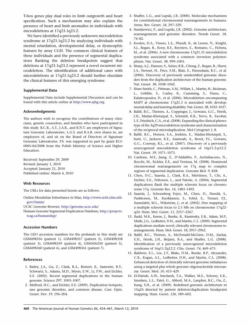
We have identified a previously unknown microdeletion
syndrome at 17q23.1q23.2 by analyzing individuals with
mental retardation, developmental delay, or dysmorphic
features by array CGH. The common clinical features of
these individuals and the presence of segmental duplica-
tions flanking the deletion breakpoints suggest that
deletions at 17q23.1q23.2 represent a novel recurrent mi-
crodeletion. The identification of additional cases with
microdeletions at 17q23.1q23.2 should further elucidate
the clinical features of this emerging syndrome.
Supplemental Data
Supplemental Data include Supplemental Discussion and can be
found with this article online at http://www.ajhg.org.
Acknowledgments
The authors wish to recognize the contributions of many clini-
cians, genetic counselors, and families who have participated in
this study. B.C.B., A.T., J.A.R., and R.N.T. are employees of Signa-
ture Genomic Laboratories. L.G.S. and B.A.B. own shares in, are
employees of, and sit on the Board of Directors of Signature
Genomic Laboratories. P.S. was supported in part by grant R13-
0005-04/2008 from the Polish Ministry of Science and Higher
Education.
Received: September 29, 2009
Revised: January 1, 2010
Accepted: January 21, 2010
Published online: March 4, 2010
Web Resources
The URLs for data presented herein are as follows:
Online Mendelian Inheritance in Man, http://www.ncbi.nlm.nih.
gov/Omim/
UCSC Genome Browser, http://genome.ucsc.edu/
Human Genome Segmental Duplication Database, http://projects.
tcag.ca/humandup/
Accession Numbers
The GEO accession numbers for the probands in this study are
GSM498356 (patient 1), GSM498357 (patient 2), GSM498358
(patient 3), GSM498359 (patient 4), GSM506250 (patient 5),
GSM498360 (patient 6), and GSM498361 (patient 7).
References
1. Bailey, J.A., Gu, Z., Clark, R.A., Reinert, K., Samonte, R.V.,
Schwartz, S., Adams, M.D., Myers, E.W., Li, P.W., and Eichler,
E.E. (2002). Recent segmental duplications in the human
genome. Science 297, 1003–1007.
2. Mefford, H.C., and Eichler, E.E. (2009). Duplication hotspots,
rare genomic disorders, and common disease. Curr. Opin.
Genet. Dev. 19, 196–204.
460 The American Journal of Human Genetics 86, 454–461, March 1
3. Shaffer, L.G., and Lupski, J.R. (2000). Molecular mechanisms
for constitutional chromosomal rearrangements in humans.
Annu. Rev. Genet. 34, 297–329.
4. Stankiewicz, P., and Lupski, J.R. (2002). Genome architecture,
rearrangements and genomic disorders. Trends Genet. 18,
74–82.
5. Koolen, D.A., Vissers, L.E., Pfundt, R., de Leeuw, N., Knight,
S.J., Regan, R., Kooy, R.F., Reyniers, E., Romano, C., Fichera,
M., et al. (2006). A new chromosome 17q21.31 microdeletion
syndrome associated with a common inversion polymor-
phism. Nat. Genet. 38, 999–1001.
6. Sharp, A.J., Hansen, S., Selzer, R.R., Cheng, Z., Regan, R., Hurst,
J.A., Stewart, H., Price, S.M., Blair, E., Hennekam, R.C., et al.
(2006). Discovery of previously unidentified genomic disor-
ders from the duplication architecture of the human genome.
Nat. Genet. 38, 1038–1042.
7. Shaw-Smith, C., Pittman, A.M., Willatt, L., Martin, H., Rickman,
L., Gribble, S., Curley, R., Cumming, S., Dunn, C.,
Kalaitzopoulos, D., et al. (2006). Microdeletion encompassing
MAPT at chromosome 17q21.3 is associated with develop-
mental delay and learningdisability. Nat.Genet. 38, 1032–1037.
8. Ballif, B.C., Theisen, A., Coppinger, J., Gowans, G.C., Hersh,
J.H., Madan-Khetarpal, S., Schmidt, K.R., Tervo, R., Escobar,
L.F., Friedrich, C.A., et al. (2008). Expanding the clinical pheno-
type of the 3q29 microdeletion syndrome and characterization
of the reciprocal microduplication. Mol Cytogenet 1, 8.
9. Ballif, B.C., Hornor, S.A., Jenkins, E., Madan-Khetarpal, S.,
Surti, U., Jackson, K.E., Asamoah, A., Brock, P.L., Gowans,
G.C., Conway, R.L., et al. (2007). Discovery of a previously
unrecognized microdeletion syndrome of 16p11.2-p12.2.
Nat. Genet. 39, 1071–1073.
10. Cardone, M.F., Jiang, Z., D’Addabbo, P., Archidiacono, N.,
Rocchi, M., Eichler, E.E., and Ventura, M. (2008). Hominoid
chromosomal rearrangements on 17q map to complex
regions of segmental duplication. Genome Biol. 9, R28.
11. Chen, D.C., Saarela, J., Clark, R.A., Miettinen, T., Chi, A.,
Eichler, E.E., Peltonen, L., and Palotie, A. (2004). Segmental
duplications flank the multiple sclerosis locus on chromo-
some 17q. Genome Res. 14, 1483–1492.
12. Saarela, J., Schoenberg Fejzo, M., Chen, D., Finnila, S.,
Parkkonen, M., Kuokkanen, S., Sobel, E., Tienari, P.J.,
Sumelahti, M.L., Wikstrom, J., et al. (2002). Fine mapping of
a multiple sclerosis locus to 2.5 Mb on chromosome 17q22-
q24. Hum. Mol. Genet. 11, 2257–2267.
13. Rudd, M.K., Keene, J., Bunke, B., Kaminsky, E.B., Adam, M.P.,
Mulle, J.G., Ledbetter, D.H., and Martin, C.L. (2009). Segmental
duplications mediate novel, clinically relevant chromosome re-
arrangements. Hum. Mol. Genet. 18, 2957–2962.
14. Ballif, B.C., Theisen, A., McDonald-McGinn, D.M., Zackai,
E.H., Hersh, J.H., Bejjani, B.A., and Shaffer, L.G. (2008).
Identification of a previously unrecognized microdeletion
syndrome of 16q11.2q12.2. Clin. Genet. 74, 469–475.
15. Baldwin, E.L., Lee, J.Y., Blake, D.M., Bunke, B.P., Alexander,
C.R., Kogan, A.L., Ledbetter, D.H., and Martin, C.L. (2008).
Enhanced detection of clinically relevant genomic imbalances
using a targeted plus whole genome oligonucleotide microar-
ray. Genet. Med. 10, 415–429.
16. El-Hattab, A.W., Smolarek, T.A., Walker, M.E., Schorry, E.K.,
Immken, L.L., Patel, G., Abbott, M.A., Lanpher, B.C., Ou, Z.,
Kang, S.H., et al. (2009). Redefined genomic architecture in
15q24 directed by patient deletion/duplication breakpoint
mapping. Hum. Genet. 126, 589–602.
2, 2010

17. Shaffer, L.G., McCaskill, C., Han, J.Y., Choo, K.H., Cutillo,
D.M., Donnenfeld, A.E., Weiss, L., and Van Dyke, D.L.
(1994). Molecular characterization of de novo secondary
trisomy 13. Am. J. Hum. Genet. 55, 968–974.
18. Bailey, J.A., Yavor, A.M., Massa, H.F., Trask, B.J., and Eichler,
E.E. (2001). Segmental duplications: Organization and impact
within the current human genome project assembly. Genome
Res. 11, 1005–1017.
19. Stankiewicz, P., Shaw, C.J., Dapper, J.D., Wakui, K., Shaffer,
L.G., Withers, M., Elizondo, L., Park, S.S., and Lupski, J.R.
(2003). Genome architecture catalyzes nonrecurrent chromo-
somal rearrangements. Am. J. Hum. Genet. 72, 1101–1116.
20. Elsea, S.H., and Girirajan, S. (2008). Smith-Magenis syndrome.
Eur. J. Hum. Genet. 16, 412–421.
21. Horton, A.C., Mahadevan, N.R., Minguillon, C., Osoegawa, K.,
Rokhsar, D.S., Ruvinsky, I., de Jong, P.J., Logan, M.P., and
Gibson-Brown, J.J. (2008). Conservation of linkage and
evolution of developmental function within the Tbx2/3/4/5
subfamily of T-box genes: Implications for the origin of
vertebrate limbs. Dev. Genes Evol. 218, 613–628.
22. Chieffo, C., Garvey, N., Gong, W., Roe, B., Zhang, G., Silver, L.,
Emanuel, B.S., and Budarf, M.L. (1997). Isolation and charac-
terization of a gene from the DiGeorge chromosomal region
homologous to the mouse Tbx1 gene. Genomics 43, 267–277.
23. Paylor, R., Glaser, B., Mupo, A., Ataliotis, P., Spencer, C.,
Sobotka, A., Sparks, C., Choi, C.H., Oghalai, J., Curran, S.,
et al. (2006). Tbx1 haploinsufficiency is linked to behavioral
disorders in mice and humans: Implications for 22q11 dele-
tion syndrome. Proc. Natl. Acad. Sci. USA 103, 7729–7734.
24. Zweier, C., Sticht, H., Aydin-Yaylagul, I., Campbell, C.E., and
Rauch, A. (2007). Human TBX1 missense mutations cause
gain of function resulting in the same phenotype as 22q11.2
deletions. Am. J. Hum. Genet. 80, 510–517.
25. Yagi, H., Furutani, Y., Hamada, H., Sasaki, T., Asakawa, S.,
Minoshima, S., Ichida, F., Joo, K., Kimura, M., Imamura, S.,
et al. (2003). Role of TBX1 in human del22q11.2 syndrome.
Lancet 362, 1366–1373.
26. Li, Q.Y., Newbury-Ecob, R.A., Terrett, J.A., Wilson, D.I., Curtis,
A.R., Yi, C.H., Gebuhr, T., Bullen, P.J., Robson, S.C., Strachan,
T., et al. (1997). Holt-Oram syndrome is caused by mutations
in TBX5, a member of the Brachyury (T) gene family. Nat.
Genet. 15, 21–29.
27. Bamshad, M., Lin, R.C., Law, D.J., Watkins, W.C., Krakowiak,
P.A., Moore, M.E., Franceschini, P., Lala, R., Holmes, L.B.,
Gebuhr, T.C., et al. (1997). Mutations in human TBX3 alter
limb, apocrine and genital development in ulnar-mammary
syndrome. Nat. Genet. 16, 311–315.
28. Bongers, E.M., Duijf, P.H., van Beersum, S.E., Schoots, J., Van
Kampen, A., Burckhardt, A., Hamel, B.C., Losan, F., Hoefsloot,
L.H., Yntema, H.G., et al. (2004). Mutations in the human
TBX4 gene cause small patella syndrome. Am. J. Hum. Genet.
74, 1239–1248.
29. Kirk, E.P., Sunde, M., Costa, M.W., Rankin, S.A., Wolstein, O.,
Castro, M.L., Butler, T.L., Hyun, C., Guo, G., Otway, R., et al.
(2007). Mutations in cardiac T-box factor gene TBX20 are
associated with diverse cardiac pathologies, including defects
of septation and valvulogenesis and cardiomyopathy. Am. J.
Hum. Genet. 81, 280–291.
The Ameri
30. Ruvinsky, I., and Gibson-Brown, J.J. (2000). Genetic and
developmental bases of serial homology in vertebrate limb
evolution. Development 127, 5233–5244.
31. Gibson-Brown, J.J., Agulnik, S.I., Silver, L.M., and Papaioan-
nou, V.E. (1998). Expression of T-box genes Tbx2-Tbx5 during
chick organogenesis. Mech. Dev. 74, 165–169.
32. Gibson-Brown, J.J., Agulnik, S.I., Silver, L.M., Niswander, L.,
and Papaioannou, V.E. (1998). Involvement of T-box genes
Tbx2-Tbx5 in vertebrate limb specification and development.
Development 125, 2499–2509.
33. Gibson-Brown, J.J., Agulnik, S.I., Chapman, D.L., Alexiou, M.,
Garvey, N., Silver, L.M., and Papaioannou, V.E. (1996).
Evidence of a role for T-box genes in the evolution of limb
morphogenesis and the specification of forelimb/hindlimb
identity. Mech. Dev. 56, 93–101.
34. Suzuki, T., Takeuchi, J., Koshiba-Takeuchi, K., and Ogura, T.
(2004). Tbx genes specify posterior digit identity through
Shh and BMP signaling. Dev. Cell 6, 43–53.
35. Rodriguez-Esteban, C., Tsukui, T., Yonei, S., Magallon, J.,
Tamura, K., and Izpisua Belmonte, J.C. (1999). The T-box
genes Tbx4 and Tbx5 regulate limb outgrowth and identity.
Nature 398, 814–818.
36. Takeuchi, J.K., Koshiba-Takeuchi, K., Matsumoto, K., Vogel-
Hopker, A., Naitoh-Matsuo, M., Ogura, K., Takahashi, N.,
Yasuda, K., and Ogura, T. (1999). Tbx5 and Tbx4 genes deter-
mine the wing/leg identity of limb buds. Nature 398, 810–
814.
37. Isaac, A., Rodriguez-Esteban, C., Ryan, A., Altabef, M., Tsukui,
T., Patel, K., Tickle, C., and Izpisua-Belmonte, J.C. (1998). Tbx
genes and limb identity in chick embryo development. Devel-
opment 125, 1867–1875.
38. Minguillon, C., Del Buono, J., and Logan, M.P. (2005). Tbx5
and Tbx4 are not sufficient to determine limb-specific
morphologies but have common roles in initiating limb
outgrowth. Dev. Cell 8, 75–84.
39. Naiche, L.A., and Papaioannou, V.E. (2007). Tbx4 is not
required for hindlimb identity or post-bud hindlimb
outgrowth. Development 134, 93–103.
40. Bongers, E.M., Van Bokhoven, H., Van Thienen, M.N.,
Kooyman, M.A., Van Beersum, S.E., Boetes, C., Knoers, N.V.,
and Hamel, B.C. (2001). The small patella syndrome: Descrip-
tion of five cases from three families and examination of
possible allelism with familial patella aplasia-hypoplasia and
nail-patella syndrome. J. Med. Genet. 38, 209–214.
41. Kozlowski, K., and Nelson, J. (1995). Small patella syndrome.
Am. J. Med. Genet. 57, 558–561.
42. Azouz, E.M., and Kozlowski, K. (1997). Small patella
syndrome: A bone dysplasia to recognize and differentiate
from the nail-patella syndrome. Pediatr. Radiol. 27, 432–435.
43. Habboub, H.K., and Thneibat, W.A. (1997). Ischio-pubic-
patellar hypoplasia: Is it a new syndrome? Pediatr. Radiol.
27, 430–431.
44. Habets, P.E., Moorman, A.F., Clout, D.E., van Roon, M.A.,
Lingbeek, M., van Lohuizen, M., Campione, M., and Christof-
fels, V.M. (2002). Cooperative action of Tbx2 and Nkx2.5
inhibits ANF expression in the atrioventricular canal: Implica-
tions for cardiac chamber formation. Genes Dev. 16, 1234–
1246.
can Journal of Human Genetics 86, 454–461, March 12, 2010 461