Residual coronary reserve identifies segmental viability in patients with wall motion abnormalities
9
342 JACC Vol. 26, No. 2 August 1995:342-50 Residual Coronary Reserve Identifies Segmental Viability in Patients With Wall Motion Abnormalities PAOLO MARZULLO, MD, FESC, FACC, OBERDAN PARODI, MD, GIANMARIO SAMBUCETI, MD, ASSUERO GIORGETTI, MD, EUGENIO PICANO, MD, FESC, ALESSIA GIMELLI, MD, PIERO SALVADORI, PttD, ANTONIO L'ABBATE, MD, FESC, FACC Pisa, Italy Objectives. The aim of this study was to determine whether the presence of residual coronary reserve can in itself identify viable ~%~q~ents. Bac~ound. Experimental data suggest that despite hypoper- fusion at rest, viable myocardium may exhibit persistence of coronary reserve. Preliminary observations in patients show that in basally dyssynergic areas, a residual vasodilator capability is present despite h~opeffusion at rest and that a flow-mediated increase in regional wall motion identifies residual viability. Methods. Fourteen patients with evidence of previous myocar- dial infarction, infarct-related single-vessel coronary artery dis- ease and impaired regional ven~rieularfunction at rest underwent positron emission viability imaging by fluorine.18 deoxyglucose. In addition, blood flow at rest and vasodilator capability were regionally evaluated in all patients by means of nitrogen-13 ammonia. Results. Of a total of 252 segments, 133 were dyssynergie at rest. Of these 133 segments, 60 (group 1) showed normal meta- bolic activily and only mild reduction in myocardial blood flow. The other 73 segments showed a marked reduction in flow;, of these, 25 (group 2, viable) had persistent metabolic activity, whereas 48 (group 3, necrotic) did not. Despite similar levels of hypoperfusion at rest, group 2 segments showed a preserved coronary reserve that was virtually absent in necrotic segments (2.6 --. 1.3 vs. 1.3 ± 0.5, p < 0.01). This value was similar to that observed in viable group I segments (2.5 ± 1.6, p = NS). Conclusions. In addition to characterizing myocardium at risk, imaging of coronary flow at baseline and after dipyridamole by positron emission tomography provides helpful information on myocardial viability that may integrate the "static" viability information obtained with the baseline flow/metabolic approach. (J Am CoU Cordio11995;26:342-50) In recent years, the development of positron emission tomog- raphy as a means of noninvasive quantitatien of myocardial blood flow in humans has paralleled the increasing use of coronary leserve as an important clinical index in the charac- terization of patients with ischernic heart disease (1,2). A maintained pharmacologic coronary reserve in regions with h)9operfusion at rest supplied by both native and collateral circulation was recently demonstrated by positron emission tomography in patients without previous myocardial infarction (3,4). The presence of such a vasodilating capacity despite a va~able degree of rest hypoperfusion is in accordance with the results of animal studies (5-7) in which acute as well as prolonged lsehemia did not completely abolish coronary re- serve. The clinical meaning of a persistent vasodiiator reserve in pat ients with previous myocardial infarction and wall motion From the CNR Institute of Clinical Physiology, Pisa, Italy. This study was support.~d in part by CNR-targeted project "Preventkan and Control Disease Factors,' subproject "Control of Cardiovascular Disease" from the National Research Council, Rome, Italy and by the Ministero Pubblica Istruzione, Rome, Italy. It was presented in part at the 43rd Annual Scientific Session of the AmerieamCollege of Cardiology, Atlanta, Georgia: March 1994. Manuscript received December 1, 1994; revised manu~ript received March 3, 1995, accepted March 9, 1995. Ad.fressfor correspondencE: Dr. Paolo Marzullo, CNR Institute of Clinical Physiokgy, Via Savi 8, 56100 Pisa, Italy. ©1995 by the American College of Cardiology abnormalities at rest is still unknown. In fact, these patients are usually the target of viability protocols that do not require the administration of either adenosine or dipyridamole. In these patients, viability information derives mostly from the assess- ment of myocardial flow distribution with nitrogen-13 (N-13) ammonia and metabolic activity with fluorine-18 (F-18) deoxy- glucose (8). Only a few investigators (9-11) specifically ad- dressed the quantitative evaluation of baseline coronary blood flow, which was reduced both in segments with and without persistent metabolic activity. Among these, Gewirtz et al. (11) recently reported that in patients with previous myocardial infarction, myocardial viability is unlikely when basal regional myocardial blood flow is <25 ml/min per 100 g or when dyskinesia is present. We (12) recently demonstrated that dipyridamole..induced improvement of contractile function in dyssynergic segments at rest reliably identifies tissue viability when these findings are compared with recovery of function after revascularization. The potential mechanism--increased flow in viable but not in necrotic areas--cannot be fully investigated with echocardiog- raphy. The quantitation of flow during dipyridamole adminis- tration might help to understand the meaning of maintained pharmacologic coronary reserve in patients with previous myocaidial infarction and its role in the potential recovery of function after revasculadzation. 0735-1097/95/$9.50 0735-1097(95)00164-Y
Transcript of Residual coronary reserve identifies segmental viability in patients with wall motion abnormalities
342 JACC Vol. 26, No. 2 August 1995:342-50
Residual Coronary Reserve Identifies Segmental Viability in Patients With Wall Motion Abnormalities
PAOLO MARZULLO, MD, FESC, FACC, O B E R D A N PARODI, MD,
GIANMARIO SAMBUCETI, MD, ASSUERO GIORGETTI , MD, E U G E N I O PICANO, MD, FESC,
ALESSIA GIMELLI, MD, PIERO SALVADORI, PttD, ANTONIO L'ABBATE, MD, FESC, FACC
Pisa, Italy
Objectives. The aim of this study was to determine whether the presence of residual coronary reserve can in itself identify viable ~%~q~ents.
Bac~ound. Experimental data suggest that despite hypoper- fusion at rest, viable myocardium may exhibit persistence of coronary reserve. Preliminary observations in patients show that in basally dyssynergic areas, a residual vasodilator capability is present despite h~opeffusion at rest and that a flow-mediated increase in regional wall motion identifies residual viability.
Methods. Fourteen patients with evidence of previous myocar- dial infarction, infarct-related single-vessel coronary artery dis- ease and impaired regional ven~rieular function at rest underwent positron emission viability imaging by fluorine.18 deoxyglucose. In addition, blood flow at rest and vasodilator capability were regionally evaluated in all patients by means of nitrogen-13 ammonia.
Results. Of a total of 252 segments, 133 were dyssynergie at rest. Of these 133 segments, 60 (group 1) showed normal meta- bolic activily and only mild reduction in myocardial blood flow. The other 73 segments showed a marked reduction in flow;, of these, 25 (group 2, viable) had persistent metabolic activity, whereas 48 (group 3, necrotic) did not. Despite similar levels of hypoperfusion at rest, group 2 segments showed a preserved coronary reserve that was virtually absent in necrotic segments (2.6 --. 1.3 vs. 1.3 ± 0.5, p < 0.01). This value was similar to that observed in viable group I segments (2.5 ± 1.6, p = NS).
Conclusions. In addition to characterizing myocardium at risk, imaging of coronary flow at baseline and after dipyridamole by positron emission tomography provides helpful information on myocardial viability that may integrate the "static" viability information obtained with the baseline flow/metabolic approach.
(J Am CoU Cordio11995;26:342-50)
In recent years, the development of positron emission tomog- raphy as a means of noninvasive quantitatien of myocardial blood flow in humans has paralleled the increasing use of coronary leserve as an important clinical index in the charac- terization of patients with ischernic heart disease (1,2). A maintained pharmacologic coronary reserve in regions with h)9operfusion at rest supplied by both native and collateral circulation was recently demonstrated by positron emission tomography in patients without previous myocardial infarction (3,4). The presence of such a vasodilating capacity despite a va~able degree of rest hypoperfusion is in accordance with the results of animal studies (5-7) in which acute as well as prolonged lsehemia did not completely abolish coronary re- serve. The clinical meaning of a persistent vasodiiator reserve in pat ients with previous myocardial infarction and wall motion
From the CNR Institute of Clinical Physiology, Pisa, Italy. This study was support.~d in part by CNR-targeted project "Preventkan and Control Disease Factors,' subproject "Control of Cardiovascular Disease" from the National Research Council, Rome, Italy and by the Ministero Pubblica Istruzione, Rome, Italy. It was presented in part at the 43rd Annual Scientific Session of the Amerieam College of Cardiology, Atlanta, Georgia: March 1994.
Manuscript received December 1, 1994; revised manu~ript received March 3, 1995, accepted March 9, 1995.
Ad.f ressfor correspondencE: Dr. Paolo Marzullo, CNR Institute of Clinical Physiokgy, Via Savi 8, 56100 Pisa, Italy.
©1995 by the American College of Cardiology
abnormalities at rest is still unknown. In fact, these patients are usually the target of viability protocols that do not require the administration of either adenosine or dipyridamole. In these patients, viability information derives mostly from the assess- ment of myocardial flow distribution with nitrogen-13 (N-13) ammonia and metabolic activity with fluorine-18 (F-18) deoxy- glucose (8). Only a few investigators (9-11) specifically ad- dressed the quantitative evaluation of baseline coronary blood flow, which was reduced both in segments with and without persistent metabolic activity. Among these, Gewirtz et al. (11) recently reported that in patients with previous myocardial infarction, myocardial viability is unlikely when basal regional myocardial blood flow is <25 ml/min per 100 g or when dyskinesia is present.
We (12) recently demonstrated that dipyridamole..induced improvement of contractile function in dyssynergic segments at rest reliably identifies tissue viability when these findings are compared with recovery of function after revascularization. The potential mechanism--increased flow in viable but not in necrotic areas--cannot be fully investigated with echocardiog- raphy. The quantitation of flow during dipyridamole adminis- tration might help to understand the meaning of maintained pharmacologic coronary reserve in patients with previous myocaidial infarction and its role in the potential recovery of function after revasculadzation.
0735-1097/95/$9.50 0735-1097(95)00164-Y
.IACC Vol. 26, No. 2 MARZULLO El" AL. 343 August 1995:342-50 MYOCARDIAL VIABILITY AND CORONARY RESERVE
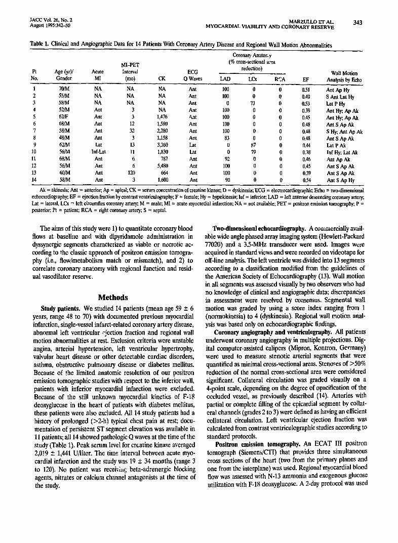
Table 1. Clinical and Angiographie Data for 14 Patients With Coronary Artery Disease and Regional Wall Motion Abnormalities
Coronary Anator,.,y MI-PET (% cross-sectional a:*.a
Pt Age (yr)/ Acute Interval ECG reduction) Wall Motion No. Gender MI (mo) CK O Waves LAD LCx RCA EF Analysis by Echo
1 70/M NA NA NA Ant 100 0 0 0.51 Ant Ap Hy 2 55/M NA NA NA Ant 100 0 0 0.40 S Ant Lot Hy 3 58/M NA NA NA Ant 0 73 0 0.53 Lot P Hy 4 52/M Ant 3 NA Ant 100 0 0 0.39 Ant Hy; Ap Ak 5 62/F Ant 3 1,476 ~lt 100 0 0 0.45 Ant Hy; Ap Ak 6 68/M Ant 12 1,580 Ant 100 0 0 0.48 Ant SAp Ak 7 58/M Ant 32 2,280 Ant 100 0 0 0.48 SHy;, Ant Ap Ak 8 48/M Ant 3 1,158 Ant 83 0 0 0.48 Ant S Ap Ak 9 62/M Lot 13 3,360 Lot 0 87 0 ~1.44 Lat P Ak
10 56/M lnf-!.at 11 1,830 l,at 0 79 0 0.38 lnf Hy; Lat Ak l l 68/M Ant 6 787 Ant 92 0 0 0.46 Ant Ap Ak 12 56/M Ant 6 5,480 Ant 100 0 0 0.45 Ant SAp Ak 13 60/M Ant 120 664 Ant 100 0 0 0.39 Ant S Ap Ak 14 58/M Ant 3 1,600 Ant 90 0 0 0.54 Ant SAp Hy
Ak = akinesia; Ant = anterior; Ap = apical; CK = serum concentration of creatine kinase; D = dyskinesia; ECG = electrocardiographic; Echo = two-dimensional echocardiography; EF = ejection fraction by contrast ventrienlography; F = female; Hy = hypokinesia; lnf = inferior; LAD = left anterior descending coronary artery; Lat = lateral, LCx --: left circumflex coronary artery; M = male; MI = acute myocardial infarction; NA = not available; PET = positron emission tomography; P = posterior; Pt = patient; RCA = right coronary artery; S = septal.
The aims of this study were 1) to quantitate coronary blood flows at baseline and with dipyridamole administration in dyssynergic segments characterized as viable or necrotic ac- cording to the classic approach of positron emission tomogra- phy (i.e., flow/metabolism match or mismatch), and 2) to correlate coronary anatomy with regional function and resid- ual vasodilator reserve.
Methods
Study patients. We studied 14 patients (mean age 59 _+ 6 years, range 48 to 70) with documented previous myocardial infarction, single-vessel infarct-related coronary artery disease, abnormal left ventricular .?jection fraction and regional wall motion abnormalities at rest. Exclusion criteria were unstable angina, arterial hypertension, left ventricular hypertrophy, valvular heart disease or other detectable cardiac disorders, asthma, obstructive pulmouary disease or diabetes mellitus. Because of ,*he limited anatomic resolution of our positron emission tomographic studies with respect to the inferior wall, patients with inferior myocardial infarction were excluded. Because of the still unknown myocardial kinetics of F-18 deoxyglucose in the heart of patients with diabetes mellitus, these patients were also excluded. All 14 study patients had a history of prolonged (>2-h) typical chest pain at rest; docu- mentation of persistent ST segmer~t elevation was available in 11 patients; all 14 showed pathologic Q waves at the time of the study (Table 1). Peak serum level for creatine kinase averaged 2,0J9 + 1,441 U/liter. The time interval between acute myo- cardial infarction and the study was 19 _+ 34 months (range 3 to 120). No patient was receM,g beta-adrenergic blocking agents, nitrates or calcium channel antagonists at the time of the study.
Two-dimensional echocardiography. A coaiaiercially avail- able wide angle phased array imaging system (Hewlett-Packard 77020) and a 3.5-MHz transducer were used. Images wcfe acquired in standard views and were recorded on videotape for off-line analysis. The left ventricle was divided into 13 segments according to a classification modified from the guidelines of the American Society of Eehocardiography (13). Wall motion in all segments was assessed visually by two observers who had no knowledge of clinical and angiographic data; discrepancies in assessment were resolved by consensus. Segmental wall motion was graded by using a score index ranging from 1 (normokinesia) to 4 (dyskinesia). Regional wall motion anal- ysis was based only on echocardiographic fiadings.
Coronary angiography and ventriculography. All patients underwent coronary angiography in multiple projections. Dig- ital computer-assisted calipers (Mipron, Kontron, Germany) were used to measure stenotic arterial segments that were quantified as minimal cross-sectional areas. Steno~es of >50% reduction of the normal cross-sectional area were considered significant. Collateral circulation was graded visually on a 4-point scale, depending on the degree of opacification of the occluded vessel, as previously described (14). Arteries with partial or complete filling of the epicardial segment by collat- eral channels (grades 2 to 3) were defined as having an efficient collateral circulation. Left ventricular ejection fraction was calculated from contrast ventficulographic studies according to standard protocols.
Positron emission tomography. An ECAT 111 positron tomograph (Siemens/CTl) that provides three simultaneous cross sections of the heart (two from the primary planes and one from the interplane) was used. Regional myocardial blood flow was assessed with N-13 ammonia and exogenous glucose utilization with F-18 deoxyglucose. A 2-day protocol was used
344 MARZULLO ET AL. JACC Vol. 26, No. 2 MYOCARDIAL VIABILITY AND CORONARY RESERVE August 1995:342-50
in which rest and dipyridamole N-I 3 ammonia and rest F-18 deoxyglucose studies were performed in random sequence in all patients. N-13 ammonia (15 to 20 mCi) was infused as a slow bolus over 10 to 20 s. Dynamic tomographic data acqui- sition started simultaneously, as previously described (15). After acquisition of the baseline study, a period of 50 min was allowed for the physical decay of N-13 ammonia. Thereafter, dipyridamole (0.56 mf,/kg body weight in 4 rain) was infused intravenously under continuous electrocardiographic (ECG) monitoring. The second injection of N-13 ammonia was started 2 to 3 rain after the end of dipyridamole infusion and the dynamic tomographic data acquisition followed the same pro- tocol used in the baseline study. No substances con',air_ing methylxanthines were ingested in the 12 h before the stady. To antagonize the effects of dipyridamole, aminophylline (120 to 240 rag) was injected 3 min after the dipyridamole N-13 ammonia injection in all patients.
For the metabolic study all patients fasted overnight and undelwent oral glucose loading (50 g) 30 rain before F..18 deoxyglucose injection. F-18 deoxyglucose (7 to 10 mCi) was injected intravenously and 60 rain was allowed for myocardial uptake. Cross-sectional imaging was performed at the same spatial levels used for N-13 ammonia by aligning a felt pen marker on the patient's chest with the laser beam of the tomograph. Each study was corrected for photon attenuation by using its own transmission scan. In control segments, count rates were, respectively, 17 +__ 10 and 7.6 +_ 4 counts/s per pixel for N-13 ammonia and ~'-18 deoxyglucose.
Image analysis. In each patieat the two planes that better represented the left ventricular walls were analyzed. Each tomographic plane was divided into three anatomic segments: septal, anterior and posterolateral. Each segment was further divided into three parts; consequently, regional analysis was performed on a total of 252 segments, with each segment scored as normal, hypokinetic or akinetic/dyskinetic according to wall motion observed in the corresponding main anatomic region (i.e., septal, anterior, lateral or posterior). The mass of segments ranged from 0.6 to 1 g. Images were analyzed by using circumferential profiles. The program normalized the recovered F-18 deo~glucose and N-13 ammonia counts within each myocardial segment to the maximal activity. To avoid underesti:nation of tracer uptake in dyssynergic segments, regional ~:ounts in these regions were also normalized to the maximal uptake detected in control areas with normal wall motion, not correlated to the diseased vessel and remote from the infarct zone. Segmental activity was then expressed as percent of tnaximal myocardial activity. A group of segments with normal wall motion by echocardiography, normal feeding coronary a:tery and remote from the infarct zone served as reference :or control segmental N-13 ammonia and F-18 deoxygluct,se uptake. Infarct areas were defined as those showing ia two or more conti~uous segments concordant reduction in both normalized N-13 ammonia and F-18 deoxy- glucose c,3rmts by >2 SD below the control values. Those segments it, which the positive difference of FAg deoxTglucose minus N-13 ammonia-normalized uptake exceeded 2 SD of
the control difference in two or more contiguous segments were classified as viable, as previously described (8).
Absolute myocardial blood flow was computed according to a method previously validated in our laboratory (16). Briefly, a small region of interest was drawn within the left ventricular cavity, and the time-activity curve inside this region was plotted and corrected for decay and dead time loss. Regional myocar- dial blood flow (rMBF) times N-13 ammonia myocardial extraction was calculated according to the equation: rMBF = Cm x 60/fCb(t) ×d t , where Cm and Cb are N-13 ammonia concentrations in the myocardium measured in the last frame and il~ the arterial blood at each time (t), respectively. The initial portion of the curve Cb(t) was fitted by a gamma-variate function and its integral was used as input function. The abso;ute values of regional myocardial blood flow were cor- rected for the loss in linearity between N-13 ammonia uptake and blood flow by using a previously described algorithm (16) and expressed as ml/min per 100 g after correction for tissue gravity (1.08 g/min). Coronary reserve was calculated as the ratio between dipyridamole and baseline coronary blood flow. In a group of normal subjects studied in our laboratory (3) with the same technique, coronary flow reserve was 3.64 +_ 1.76.
Statistical analysis. Data are presented as mean value _+ SD. For comparison of differences, appropriate t tests for independent or paired samples were employed. The chi-square analysis was performed to determine the significance in rate of occurrence. For all comparisons, a p value < 0.05 was consid- ered significant.
Resu l t s
Clinical and angiographic data. The mean left ventricular ejection fraction at rest was 0.43 +_ 0.06 (range 0.29 to 0.54). The average reduction in the coronary cross-sectional area was 93 +_ 9%. The left anterior descending coronary artery was the single stenotic vessel in 11 patients and the left circumflex artery in the remaining 3. Eight patients showed 100% occlu- sion of the infarct-related coronary artery with an efficient collateral circulation in two. On the basis of echocardiography, 14 segments were hypokinetic and 21 akinetie (Table 1). No patient had inferior infarction according to inclusion criteria. In the 11 patients with an anterior infarction by ECG, regional function was abnormal in the anterior, septal or apical seg- ments of the left ventricle. In the two patients with a lateral infarction, the lateral wall was always involved, Only one patient showed Q waves in the anterior wall but had a normal left anterior descending coronary artery associated with pos- terolateral hypokinesia and significant stenosis of the left circumflex coronary artery. Thus, all patients showed a strict anatomic correspondence between stenotic vessel and dysfunc- tioning segment.
Electrocardiographic and hemodynami~ :esponse to dipyr- ~damole. Infusion of 0.56 mg/kg of dipyridamole over 4 rain was completed in all patients. Typical chest pain developed in eight patients and diagnostic (>1.5 ram) ST segment depres- sion in seven. Atypical chest pain developed in two patients,
JACC Vol. 26, No. 2 MARZULLO ET A L 345 August 1995:342-50 MYOCARDIAL VIABILITY AND CORONARY RESERVE
250
~00
150
~t f~ too
50
~---~.-..*,..-~,
T
Basel ine
T
L e l
, ~ s ~
Dipyridamole
S.O
4 . 5
4 . 0
3 . S (D
3 . 0
~ 2 . 5
0 2 . 0 J.t G
1 . 5
1 .0
1].5
0 . 0
: H~
T T
I I / S d
/ I J ,
/ ' O ,
/ 1 1 , S S J ,
' I O J ,
S ~ S ,
r O O ~ ,
' S / J ' S a J
and one patient reported nausea. No arrhythmias were de- tected by continuous ECG monitoring during dipyridamole infusion. Baseline systolic blood pressure and heart rate aver- aged 138 _ 17 mm Hg and 66 - 13 beats/rain, respectiw'. After dipyridamole infusion, systolic blood pressure was 142 +, 22 mm Hg (p = NS) and heart rate increased to 88 +_ 11 beats/min (p < 0.05). Mean blood pressure did not change significantly from the baseline value (100 +_ 13 mm Hg vs. 104 +, 11 mm Hg at baseline, p = NS). Rate-pressure product increased from 9,382 beats/rain x mm Hg at rest to 12,633 beats/miu x mm Hg after dipyridamole infusion (p < 0.05).
Characterization of viable and necrotic segments by meta- bolic/flow mismatch. On the basis of echocardiographic crite- ria, 119 tomographic segments were classified as having normal and 133 as having abnormal wall motion. Baseline control coronary blood flow, obtained in normal contracting segments not contiguous and not related to the infarct artery, was 100 ± 24 ml/min per 100 g. This value was not different from that we obtained (3) in a control group of normal subjects at rest. According to this distribution, the lower limit for normal regional blood flow at rest was set at 52 ml/min per 100 g (mean - 2 SD). Coronary flow reserve in these segments averaged 2.76 +-. 72. These segments also provided the control regional percent value and positive difference between F-18 deoxyglucose and N-13 ammonia uptake.
One hundred thirty-three segments were dyssynergic at rest. Of these, 60 (group i) showed a baseline regional blood flow that was still within control limits. In these segments, the average F-18 deoxyglucose uptake was 81 +_ 13% and was higher than the lower control limit (62%) in all segments. As described before, each of the 73 hypoperfused segments with flGw <52 ml/min per 100 g was considered viable or necrotic
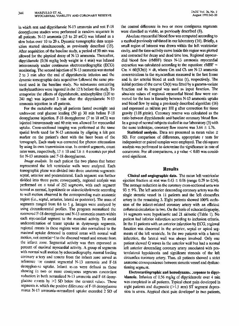
Figure 1. Bar graphs show the differences in blood flow at baseline conditions and after dipyridamole administration in segments of groups 1, 2 and 3 with wall motion abnormalities at rest• Baseline flow is similarly reduced in segments in groups 2 and 3 and, after dipyrid- amole, increases significantly only in viable segments of groups 1 and 2. Despite similar baseline flows, only viable segments show a main- tained coronary reserve that is not appreciable in group 3 segments with evidence of myocardial necrosis. *p < 0.01.
according to the presence or absence of metabolic/flow mis- match. Twenty-five segments (group 2) were classified as viable and 48 (group 3) as necrotic. Regional F-18 deoxyglueose uptake was 83 +-- 10% and 43 +_ 12% of the peak in group 2 and group 3, respectively (p < 0.01). When segments were normal- ized relative to the control zone, no segment from groups 1 and 3 showed significant changes. Only six segments, all from group 2, showed a minimal increase in F-18 deoxyglucose uptake. However, average F-18 deoxyglucose uptake increased to 86 - 9% of the peak, and this value was not statistically higher than that obtained by using the first type of normalization (p = NS).
Baseline and dipyridamole coronary blood flow in dyssyn- ergic segments, The 60 dyssynergic segments of group 1 had a mean baseline coronary blood flow of 69 +_ 14 ml/min per 100 g, which increased to 164 +- 70 ml/min per 100 g after dipyridamole administration. Coronary reserve was 2.5 +- 1.6 (Fig. 1). Although segments of groups 2 and 3 showed similar rest coronary blood flow (42 +_ 12vs. 39 +_ 27 ml/min per 100 g, p = NS), dipyridamole flow was significantly higher iu seg- ments of group 2 (107 +_ 50 vs. 49 +- 18 ml/min per 100 g, p < 0.01). Coronary reserve averaged 2.6 -+ 1.3 and 1.3 +_ 0.5 in segments of group 2 and group 3, respectively (p < 0.01) (Fig. 1). Despite the obvious differences in baseline coronary blood
346 MARZULLO ET AL. JACC Vol. 26, No. 2 MYOCARDIAL VIABILITY AND CORONARY RESERVE August 1995:342-50
Baseline Dipyridamole
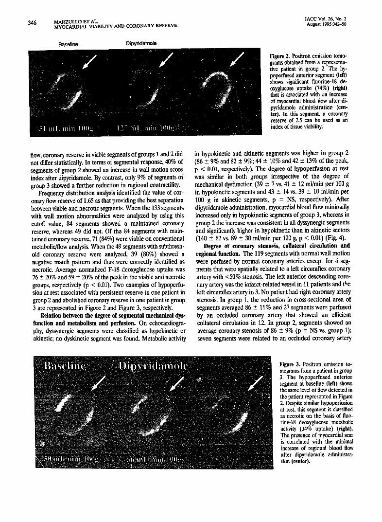
Hgure 2. Positron emission tomo- grams obtained from a representa- tive patient in group 2. The hy- poperfused anterior segment (left) shows significant fluorine-18 de- oxyglucose uptake (74%) (right) that is associated with an increase of myocardial blood flow after di- pyridamole administration (cen- ter). In this segment, a coronary reserve of 2.5 can be used as an index of tissue viability.
flow, coronary reserve in viable segments of groups 1 and 2 did not differ statistically. In terms of segmental response, 40% of segments of group 2 showed an increase in wall motion score index after dipyridamole. By contrast, only 9% of segments of group 3 showed a further reduction in regional contractility.
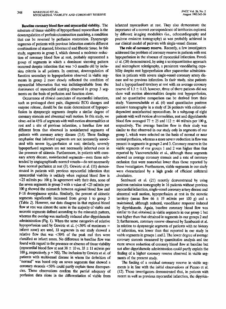
Frequency distlibution analysis identified the value of cor- onary flow reserve of 1.65 as that providing the best separation between viable and necrotic segments. When the 133 segments with wall motion abnormalities were analyzed by using this cutoff value, 84 segments showe6 a maintained coronary reserve, whereas 49 did not. Of the 84 segments with main- tained coronary reserve, 71 (84%) were viable on conventional metabolic/flow analysis. When the 49 segments with subthresh- old coronary reserve were analyzed, 39 (80%) showed a negative match pattern and thus were correctly identified as necrotic. Average normalized F-18 deoxyglucose uptake was 76 +_ 20% and 59 _ 20% of the peak in the viable and necrotic groups, respectively (p < 0.01). Two examples of hypoperfu- sion at rest associated with persistent reserve in one patient in group 2 and abolished coronary reserve in one patient in group 3 are represented in Figure 2 and Figure 3, respectively.
Relation between the degree of segmental mechanical dys- function and metabolism and perfusion. On echocardiogra- phy, dyssynergic segments were classified as hypokinetic or akinetic; no dyskinetie segment was found. Metabolic activity
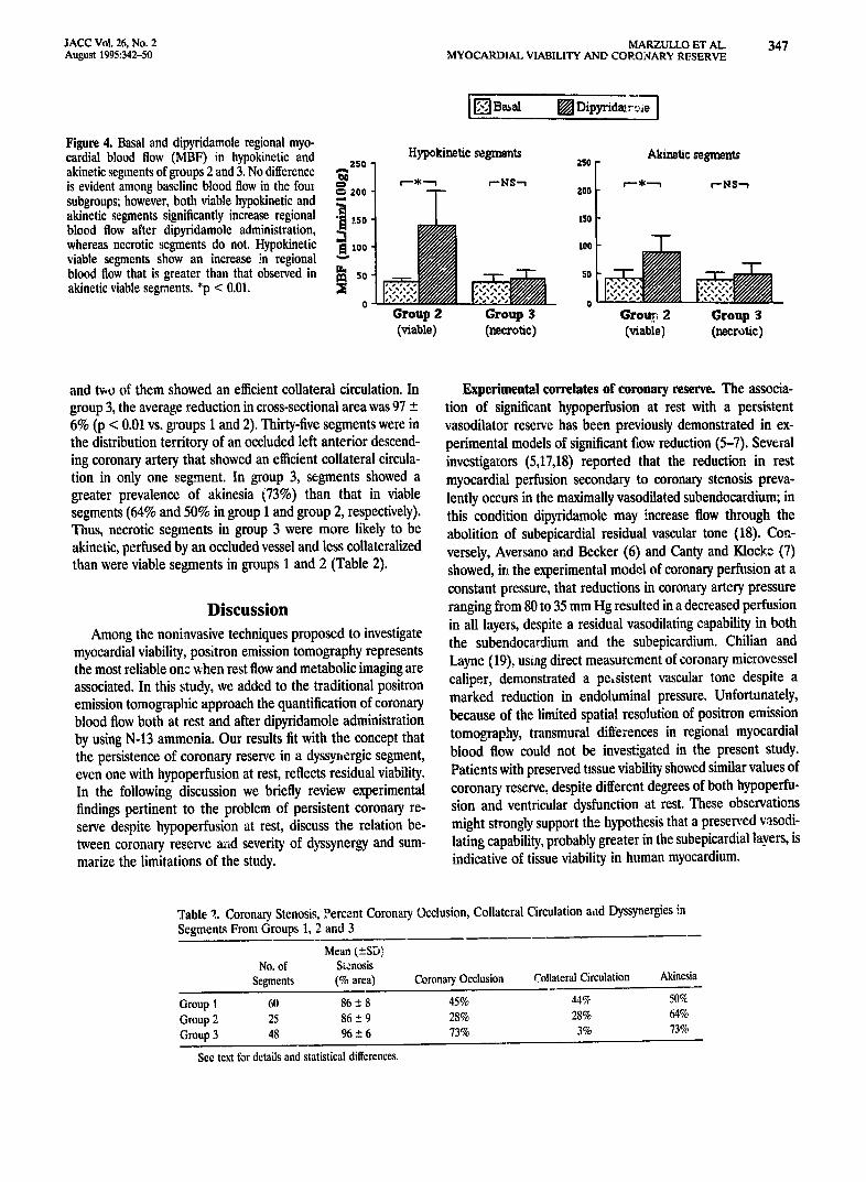
in hypokinetic and akinetic segments was higher in group 2 (86 -+ 9% and 82 - 9%; 44 _ 10% and 42 +_. 13% of the peak, p < 0.01, respectively). The degree of hypoperfusion at rest was similar in both groups irrespective of the degree of mechanical dysfunction (39 _ 7 vs. 41 -+ 12 ml/min per 100 g in hypokinetic segments and 43 - 14 vs. 39 + 10 ml/min per 100 g in akinetic segments, p = NS, respectively). After dipyridamole administration, myocardial blood flow minimally increased only in hypokinetic segments of group 3, whereas in group 2 the increase was consistent in all dyssynergic segments and significantly higher in hypokinetic than in akinetic sectors (140 +-- 62 vs. 89 +_ 30 ml/min per 100 g, p < 0.01) (Fig. 4).
Degree of coronary stenosis, collateral circulation and regional function. The 119 segments with normal wall motion were perfused by normal coronary arteries except for 6 seg- ments that were spatially related to a left circumflex coronary artery with <50% stenosis. The left anterior descending coro- nary artery was the infarct-related vessel in 11 patients and the left circumflex artery in 3. No patient had right coronary artery stenosis. In group 1, the reduction in cross-sectional area of segments averaged 86 +- 11% and 27 segments were perfused by an occluded coronary artery that showed an efficient collateral circulation in 12. In group 2, segment~ showed an average coronary stenosis of 86 _+ 9% (p = NS vs. group 1); seven segments were related to an occluded coronary artery
................ , . . . . . . ,
!i!i i!!
........ i . . . . . . .
i
N
Figure 3. Positron emission to- mograms from a patient in group 3. The hypoperfused anterior segment at baseline (leR) shows the same level of flow detected in the patient represented in Figure 2. Despite similar hypoperfusion at rest, this segment is classified as necrotic on the basis of fluo- rine-18 deoxyglucose metabolic activity. (3,l% uptake) (right). The presence of myocardial scar is correlated with the minimal increase of regional blood flow after dipyridamole administra- tion (center).
JACC Vol. 26, No. 2 MARZULLO El" At.. 347 August 1995:342-50 MYOCARDIAL VIABILITY AND CORONARY RESERVE
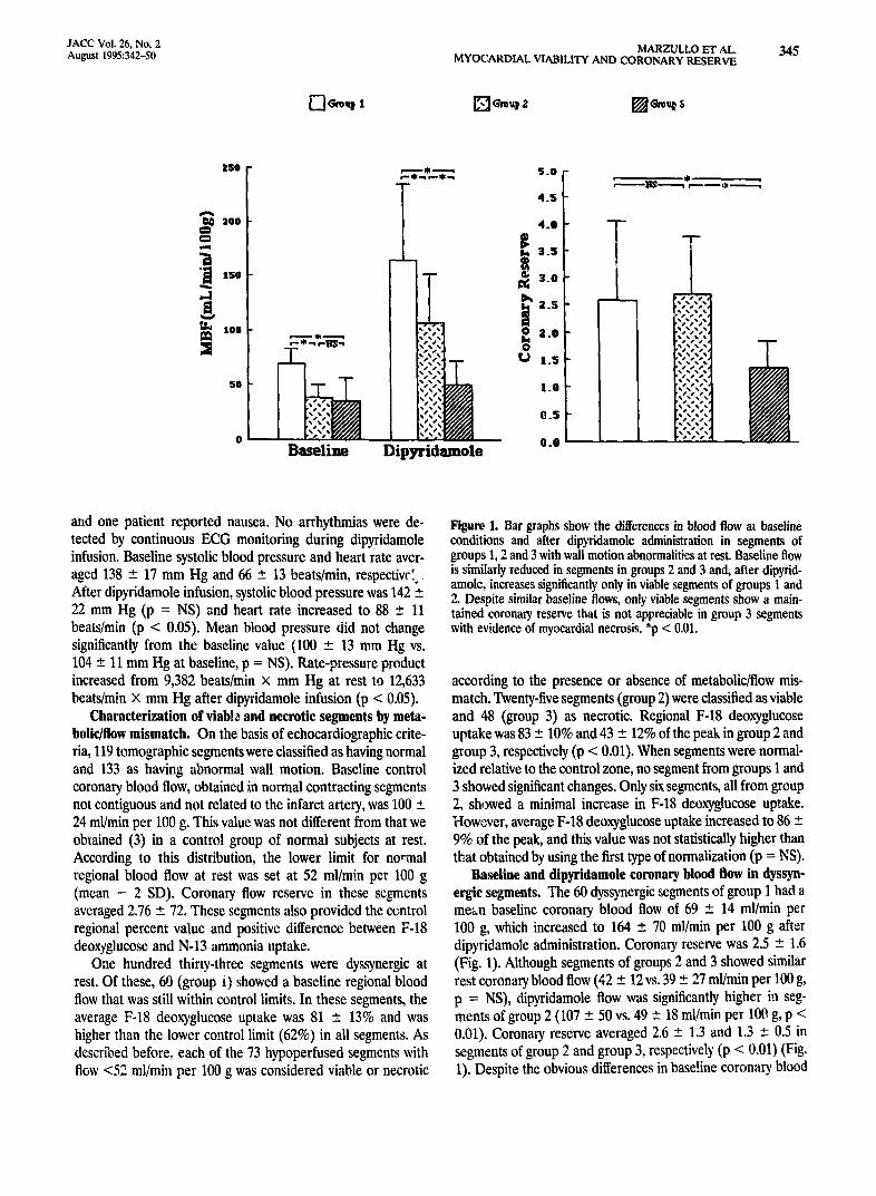
Figure 4. Basal and dipyridamole regional myo- cardial blood flow (MBF) in hypokinctic and 250 akinetic segments of groups 2 and 3. No difference ,-- is cvident among baseline blood flow in the four subgroups; however, both viable hypokinetic and ~ aoo akinetic segments significantly increase regional "~ blood flow after dipyridamole administration, '~..I. tso- whereas necrotic segments do not. Hypokinetic "~ 1oo viable segments show an increase in regional ~' blood flow that is greater than that observed in ~ z0 akinetic viable segments. *p < 0.01. ~[
0
[ [ ] Basal [ ] D i p y r i d a a ~
Hypokinetic segments
¢--~----n
Group 2 (viable)
Akinetie segments 2so F
r'NS'~ / r'-~-'.~ c-NS"~ 200 [ 150 r ,ool
Group $ Grou? 2 Group 3 (necrotic) (viable) (r~rotic)
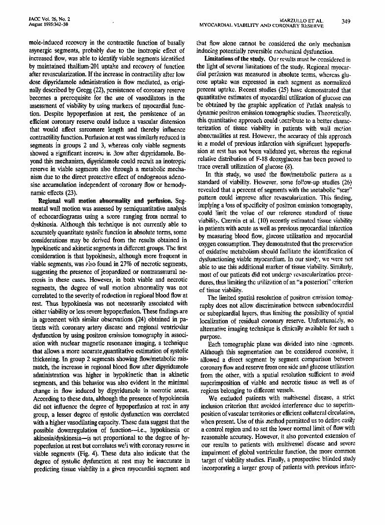
and two of them showed an efficient collateral circulation. In group 3, the average reduction in cross-sectional area was 97 _+ 6% (p < 0.01 vs. groups 1 and 2). Thirty-five segments were in the distribution territory of an occluded left anterior descend- ing coronary artery' that showed an efficient collateral circula- tion in only one segment. In group 3, segments showed a greater prevalence of akinesia (73%) than that in viable segments (64% and 50% in group 1 and group 2, respectively). Thus, necrotic segments in group 3 were more likely to be akinetic, perfused by an occluded vessel and less collateralized than were viable segments in groups 1 and 2 (Table 2).
Discussion Among the noninvasive techniques proposed to investigate
myocardial viability, positron emission tomography represents the most reliable onz when rest flow and metabolic imaging are associated. In this study, we added to the traditional positron emission tomograpbic approach the quantification of coronary blood flow both at rest and after dipyridamole administration by using N-13 ammonia. Our results fit with the concept that the persistence of coronary reserve in a dyssynergic segment, even one with hypoperfusion at rest, reflects residual viability. In the following discussion we briefly review experimental findings pertinent to the problem of persistent coronary re- serve despite hypopeffusion at rest, discuss the relation be- tween coronary reserve and severity of dyssynergy and sum- marize the limitations of the study.
Experimental correlates of coronary reserve. The associa- tion of significant hypoperfusion at rest with a persistent vasodilator reserve has been previously demonstrated in ex- perimemal models of significant flow reduction (5-7). Several investigators (5,17,18) reported that the reduction in rest myocardial perfusion secondary to coronary stenosis preva- lently occurs in the maximally vasodilated subendocardium; in this condition dipyfidamole may increase flow through the abolition of subepicardial residual vascular tone (18). Con- versely, Aversano and Becker (6) and Canty and Klocke (7) showed, in the experimental model of coronary perfnsion at a constant pressure, that reductions in coronary artery pressure ranging from 80 to 35 mm Hg resulted in a decreased perfusion in all layers, despite a residual vasodilating capability in both the subendocardium and the subepicardium. Chilian and Layne (19), using direct measurement of coronary microvessel caliper, demonstrated a p c , sistent vascular tone despite a marked reduction in endoluminal pressure. Unfortunately, because of the limited spatial resolution of positron emission tomography, transmural differences in regional myocardial biood flow could not be investigated in the present study. Patients with preserved tissue viability showed similar values of coronary reserve, despite different degrees of both bypoperfu- sion and ventricular dysfunction at rest. These observations might strongly support the hypothesis that a preserved vasodi- lating capability, probably greater in the subepicardial !avers, is indicative of tissue viability in human myocardium.
Table 2. Coronary Stenosis, Percent Coronary Occlusion, Collateral Circulation a,ld Dyssynergies in Segments From Groups 1, 2 and 3
Mean (+-SD) No. of Stenosis
Segments (% area) Coronary Occlusion Collateral Circulation Akinesia
Group 1 60 86 -- 8 45% 44% 50% Group 2 25 86 -- 9 28% 28% 64% Group 3 48 96 + 6 73% 3% 73%
See text for details and statistical differences.
348 MARZULLO ET AL. JACC Vol. 26, No. 2 MYOCARDIAL VIABILITY AND CORONARY RESERVE August 1995:342-50
Baseline coronary blood flow and myocardial viability. The substrate of tissue viability of hypoperfused myocardium is the downregulation of perfusion/contraction matching, a condition that can be reversed by perfusion restoration. Dyssynergic segments of patients with previous infarction contain different combinations of stunned, hibernated and fibrotic tissue. In this study, segments in group 1, which showed a moderate reduc- tion of coronary blood flow at rest, probably represented a group of segments in which a dominant stunning pattern occurred despite infarction that was >3 months old by inclu- siou criteria in all patients. In contrast, downregulation of function secondary to hypopeffusion observed in viable seg- ments in group 2 more closely reflected the condition of myocardial hibernation that was indistinguishable from the dominance of myocardial scarri~,g observed in group 3 seg- ments on the basis of perfusion and function alone.
Occurrence of clinical correlates of myocardial infarction, such as prolonged chest pain, diagnostic ECG changes and enzyme release, should be the main determinant of hypoper- fusion in dyssynergic segments despite a similar degree of coronary stenosis and abnormal wall motion. In this study, we obse: 4ed in 45% of segments with wall motion abnormalities at rest and a site of previous infarction, a baseli~e flow not different from that observed in noninfarctcd segments of patients with coronary artery disease (3,4). These findings emphasize that infarcted segments are not necessarily associ- ated with severe hypoperfusion at rest; similarly, severely hypoperfused segments are not necessarily infarcted even in the presence of akinesia. Furthermore, in patients with coro- nary artery disease, noninfarcted segments--even those sub- tended by angiographicallv norme.l vessels--do not necessarily have normal perfusion at rest (3). Gewirtz et al. (11) demon- strated in patients with previous myocardial infarction that myocardial viability is unlikely when regional blood flow is <25 ml/min pe~' 100 g. In agreement with their data, none of the seven segments in group 3 with a value of <25 ml/min per 100 g showed the mismatch between regional blood flow and F-18 deoxyglucose uptake. Similarly, the percent of akinetic segments significantly increased from group 1 to group 3 (Table 2). However, our data disagree in that regional blood flow at rest was almost the same in the majority of viable and necrotic segments defined according to the mismatch pattern, whereas the overlap was markedly reduced after dipyridamole administration (Fig. 1). When the same categories of relative hypoperfusion used by Gewirtz et al. (<50% of maximum = infarct zone) are used, 33 segments in our study showed a relative flow that was <50% of the peak and thtls were classified as infarct zones. No difference in baseline flow was found with regard to the presence or absence of tissue viability (myocardial blood flow at rest 38 _ 10 vs. 35 +_ 11 ml/min per 100 g, respectively, p = NS). The inclusion by Gewirtz et al. of patients with multivessel disease in whom the definition of "normal" was based only on seven segments that showed a coronary stenosis <50% could partly explain these discrepan- cies. These observations confirm the partial adequacy of perfusion data alone in the differentiation of viable from
infarcted myocardium at rest. They also demonstrate the importance of a correct correspondence of territories explored by different imaging modalities (i.e., echocardiography and positron emission tomography) as was probably achieved in our clinical model of patients with single-vessel disease.
The role of coronary reserve. Recently, a few investigators addressed the problem of coronary reserve in patients with rest hypoperfusion in the absence of myocardial infarction. Parodi ct al. (20) demonstrated, by using a semiquantitative approach and microsphere scintigraphy, a persistent vasodilating capa- bility despite rest hypoperfusion after papaverine administra- tion in patients with severe single-vessel coronary artery dis- ease and no previous infarction. In their study, nine patients had a hypoperfused territory at rest with an average coronary reserve of 1.3 + 0.13; however, three of these patients did not show wall motion abnormalities despite rest hypoperfusion, and no quantitative comparison can be derived from their study. Vanoverschelde et al. (4) used quantitative positron emission tomography in a study of 26 patients with collateral- dependent noninfarcted myocardium. In a subgroup of eight patients with wall motion abnormalities, rest and dipyridamole blood flow averaged 77 +_ 25 and 112 _ 44 ml/min per 100 g, respectively. The average baseline flow in their study was similar to that observed in our study only in segments of our group I, which were selected on the basis of normal or near normal perfusion, whereas a more severe, reduction in flow was present in segments in groups 2 and 3. Coronary reserve in the viable segments of our groulrs 1 and 2 was higher than that reported by Vanoverschelde et al., although these segments showed an average coronary stenosis and a rate of coronary occlusion that were somewhat lower than those reported by those investigators. Furthermore, viable segments in our study were characterized by a high grade of efficient collateral circulation.
Sambuceti et al. (21) recently demonstrated by using positron emission tomography in 16 patients without previous myocardial infarction, single-vessel coronary artery disease and abnormal wall motion, hypoperfusion at rest in the stenotic territory (mean flow 66 __ 19 ml/min per 100 g) and a maintained, although reduced, vasodilator response induced by dipyridamole. Again, baseline coronary blood flow was similar to that obtained in viable segments in our group 1 but was higher than that obtained in segments in our groups 2 and 3; furthermore, coronary reserve observed by Sambuceti et al. in relation to dyssynergic segments of patients with no history of infarction, was lower than that reported in our study in viable segments in groups 1 and 2. The lower degree of average coronary stenosis measured by quantitative analysis and the more severe reduction of coronary blood flow at baseline but not after dipyridamole administration could partly explain the finding of a higher coronary reserve observed in viable seg- ments of the present study.
The finding of a residual coronary reserve in viable seg- ments is in line with the initial observations of Picano et al. (12). Those investigators demonstrated that, in patients with recent as well as previous myocardial infarction, the dipyrida-
JACC Vol. 26, No. 2 MARZULLO ET AL. 349 August 1995:342-50 MYOCARDIAL VIABILITY AND CORONARY RESERVE
mole-induced recovery in the contractile function of basally asynergic segments, probably due to the inotropic effect of increased flow, was able to identify viable segments identified by maintained thallium-201 uptake and recovery of function after revascularization. If the increase in contractility after low dose dipyridamole administration is flow mediated, as origi- nally described by Gregg (22), persistence of coronary reserve becomes a prerequisite for the use of vasodilators in the assessment of viability by using markers of myocardial func- tion. Despite hypoperfusion at rest, the persistence of an efficient coronary reserve could induce a vascular distension that would affect sarcomere length and thereby influence contractility function. Perfusion at rest was similarly reduced in segments in groups 2 and 3, whereas only viable segments showed a significant increas~ ir~ ~tow after dlpyridamole. Be- yond this mechanism, dipyridamole could recruit an inotropic reserve in viable segments also through a metabolic mecha- nism due to the direct protective effect of endogenous adeno- sine accumulation independent of coronary flow or hemody- namic effects (23).
Regional wall motion abnormality and perfusion. Seg- mental wall motion was assessed by semiquantitative analysis of eehocardiograms using a st:ore ranging from normal to dyskinesia. Although this technique is not currently able to accurately quantitate systolic function in absolute terms, some considerations may be derived from the results obtained in hypokinetic and akinetic segme,ats in different groups. The first consideration is that hypokinesia, although more frequent in viable segments, was ~iso found in 27% of necrotic segments, suggesting the presence of jeopardized or nontransmural ne- crosis in these cases. However, in both viable and necrotic segments, the degree of wall motion abnormality was not correlated to the severity of reduction in regional blood flow at rest. Thus hypokinesia was not necessarily associated with either viability or less severe hypoperfusion. These findings are in agreement with similar observations (24) obtained in pa- tients with coronary artery disease and regional ventric, llar dysfunction by using positron emission tomography in associ- ation with nuclear magnetic resonance imaging, a technique that allows a more accurate,quantitative estimation of systolic thickening. In group 2 segments showing flow/metabolic mis- match, the increase in regional blood flow after dipyridamole administration was higher in hypokinetic than in akinetic segments, and this behavior was also evident in the minimal change in flow induced by dipyridamole in necrotic areas. According to these data, although the presence of hypokinesia did not influence the degree of hypoperfusion at rest in any group, a lesser degree of systolic dysfunction was correlated with a higher vasodilating capacity. These data suggest that the possible downregulation of function--i.e., hypokinesia or akinesia/dyskinesia--is not proportional to the degree of hy- poperfusion at rest but correlates wdi with coronary reserve in viable segments (Fig. 4). These data also indicate that the degree of systolic dysfunction at rest may be inaccurate in predicting tissue viability in a given myocardial segment and
that flow alone cannot be considered the only mechanism inducing potentially reversible mechanical dysfunction.
Limitations of the study. Our results must be considered in the light of several limitations of the study. Regional myocar- dial per/usion was measured in absolute terms, whereas glu- cose uptake was expressed in each segment as normalized percent uptake. Recent studies (25) have demonstrated that quantitative estimates of myocardial utilization of glucose can be obtained by the graphic application of Patlak analysis to dynamic positron emission tomographic studies. Theoretically, this quantitative approach could contribute to a better charac- terization of tissue viability in patients with wall motion abno~'rnalities at rest. However, the accuracy of this approach in a model of previous infarction with significant hypoperfu- sion at rest has not been validated yet, whereas the regional relative distribution of F-18 deoxyglucose has been proved to trace overall utilization of glucose (8).
In this study, we used the flow/metabolic pattern as a standard of viability. However, some fo!!ow-up studies (26) revealed that a percent of segments with the metabolic "scar" pattern could improve after revascularization. This finding, implying a loss of specificity of positron emission tomography, could limit the value of our reference standard of tissue viability. Czernin et al. (10) recently estimated tissue viability in patients with acute as well as previous myocardial infarction by measuring blood flow, glucose utilization and myocardial oxygen consumption. They demonstrated that the preservation of oxidative metabolism should facilitate the identification of dysfunctioning viable myoeardium. In our stud;:, we were not able to use this additional marker of tissue viability. Similarly, most of our patients did not undergo re~ascularizatior, proce- dures, thus limiting the utilization of an "a posteriori" criterion of tissue viability.
The limited spatial resolution of positron emission tomog- raphy does not allow discrimination between subendocardial or subepieardial layers, thus limiting the possibility of spatial localization of residual coronary reserve. Unfortunately, no alternative imaging technique is clinically available for such a purpose.
Each tomographie plane was dMded into nine :,egments. Although this segmentation can be considered excessive, it allowed a direct segment by segment comparison between coronary flow and reserve from one side and glucose utilization from the other, with a spatial resolution sufficient to avoid superimposition of viable and necrotic tissue as well as of regions belonging to different vessels.
We excluded patients with multivessel disease, a strict inclusion criterion that avoided interference due to superim- position of vascular territories or efficient collateral circulation, when present. Use of this method permitted us to define easily a control region and to set the lower normal limit of flow with reasonable accuracy. However, it also prevented extension of our results to patients with multivessel disease and severe impairment of global ventricular function, the more common target of viability studies. Finally, a prospective blinded study incorporating a larger group of patients with previous infarc-
JACC Vol. 26, No. 2 350 MARZULLO ET AL. MYOCARDIAL VIABILITY AND CORONARY RESERVE August 1995:342-50
tion undergoing coronary revascularization, and thus providing an independent standard for viability, would be required to corroborate our initial findings.
Clinical implications and conclusions. The data derived from this study indicate that the persistence of a measurable coronary reserve is a reliable indicator of viable myocardium in infarcted territories. Quantitative evaluation of coronary flog' at baseline and after dipyridamole infusion makes it possible to perform the whole study in 60 to 90 min, thus decreasing artifacts due to patient movement or repositioning, the radia- tion dose to the patient and the cost of the procedure. Omission of an F-18 deoxyglucose study decreases the total scanning duration and the comparison of different tomo- graphic sequences required to assess a flow/metabolic pattern. In patients with coronary artery disease, the evaluation of coronary reserve allows the estimation of myocardium at risk in segments with normal wall motion at rest and perfusion by stenotic coronary arteries. In addition to characterization of myocardium at risk, baseline/dipyridamole flow imaging by positron emission tomography provides helpful information on myocardial viability that may integrate the "static" viability information obtained by the baseline flow/metabolic approach.
In conclusion, in patients with previous myocardial infarc- tion, coronary reserve in segments considered viable on the basis of preserved F-18 deoxyglucose uptake was significantly higher than that in segments with a concomitant reduction of both F-18 deoxyglucose uptake and perfusion. Although fur- ther studies are required to confirm these findings, coro~'tary reserve seems a reliable marker of tissue viability.
This study summarizes the work of many colleagues. Coronary angiographic and wall motion studies were based on the resuRs obtained by our colleagues in the hemodynamic and echocardiographic groups. Radiopharmaceuticals were pre- pared and checked by the cyclotron staff. We thank Ilaria Cirri for typing the manuscript and Antonio Casefii, PhD for suggestions that improved the quality of this report. We are also indebted to Ginevra Ouarratesi for her masterful bibliographic research.
References 1. Gould KL, Lipscomb K, Hamilton GW. Physiologic basis for assessing
critical coronary stenosis. Instantaneous flow response and regional distri- bution duri~i,~ coronary hyperemia as measures of coronary flow reserve. Am J Cardiol 1974;33:87-94.
2. Schelbert HR, Czerinin J. Noninvasive quantification of regional myocardial blood flow: Assessment of myocardial perfusion reserve and collateral circulation. J Nncl Med 1990;31:271-3.
3. Sambuceti G, Parndi O, Marcassa C, et al. Alteration in regulation of myocardial blood flow in one-vessel coronary artery disease determined by positron emission tomography. Am J Cardiol 1993;72:538-43.
4. Vanoverschelde JLJ, Wijns W, Depr6 C, et al. Mechanisms of chronic regional postischemie dysfunction in humans. New insights from the study on noninfarcted collaternl-dependent myocardium. Circulation 1993;87:1513- 23.
5. Gould LK, Lipscomb IL Calvert C. Compensatory changes of the distal coronary vascular bed during progressive coronary constriction. Circulation 1975;51:1085-94.
6. Aversano T, Becker LC. Persistence of coronary vasodilator reserve despite functionally significant flow reduction. Am J Physiol 1985;248:H403-11.
7. Canty JM, Klocke FJ. Reduced regional myocardial perfusion in the presence of pharmacologic vasodilator reserve. Circulation 1985;71:370-7.
8. Marshall RC, Tillisch JH, Phelps ME, et al. Identification and differentiation of resting myocardial ischemia and infarction in man with positron computed tomography, 18F-labeled fluorodeoxyglucose and N-13 ammonia. Circula- tion 1983;67:766-78.
9. Yamamoto Y, De Silva R, Rhodes CG, et al. A new strategy for the assessment of viable myocardium and regional myocardial blood flow using tSO-water and dynamic positron emission tomography. Circulation 1992;86: 167-78.
10. Czernin J, Porcnta G, Brunken R, et al. Regional blood flow, oxidative metabolism and glucose utilization in patients with recent myocardial infarction. Circulation 1993;88:884-95.
11. Gewirtz H, Fischman AJ, Abraham S, et al. Positron emission tomogrnphic measurements of absolute regional myocardial blood flow permits identifi- cation of nonviable myocardiuam in patients with chronic myocardial infarction. J Am Coil Cardiol 1994;23:851-9.
12. Picauo E, Marzullo P, Gigli G, et al. Identification of viable myocardium by dipyridamole-induced improvement in regional left ventricular function assessed by echocardiography in myocardial infarction and comparison with thallium scintigraphy at rest. Am J Cardiol 1902;70:703-10.
13. SchiUc; NB, Shah PM, Crawford M, et al. American Society of Echocardi- ography Committee on Standards: Subcommi~.tee on Quantitation of Two- Dimensional Echocardiograms. Recommendations for quantitation of the left ventricle by two dimensional cchocardiography. J Am Soc Echocardiogr 1989;2:358-67.
14. Rentrop KP, Cohen M, Blanke H, et aL Changes in collateral channel filling immediately after controlled eor3nary artery occlusion by angiop~asty bal- loon in human subjects. J Am (2oll Cardiol 1985;5:587-92.
15. Vaalburg W, Kamphius JAA, Beerling-van der Molen HB, Reiff.~r~ S, R,.'jskamp A, Woldring MG. An improved method for the cyclotron produc- tion of 13Nlabeled ammonia. J Nucl IVied 1985;26:316-8.
16. Bellina CR, Parodi O, Camici P, ¢t al. Simultaneous in vitro and in ;ivo validation of 13Nammonia for the assessment of regional myocardial blood flov, . J Nud Med 1990;31:1335-43.
17. Fung AV, Gallagher KP, Buda AJ. The physiologic basis of dobutamine as compared with dipyridamole stress interventions in the assessment of critical coronary ste~osis. Circulation 1987;76:943-51.
18. Gallagher KP, Folts JP, Shebuski ILl, Rankin JHG, Rowe GG. Subepicardial vasodilator reserve in the presence of critical coronary stenosis in dogs. Am J Cardiol 1980;46:67-73.
19. Chilian WM, Layne SM. Coronary microvascular responses to reductions in perfusion pressure. Evidence for a persistent arteriolar vasomotor tone during coronary hypoperfusion. Circ Res 1990;66:1227-38.
20. Parodi O, Sambuceti S, Roghi A, et al. Residual coronary reserve despite decreased resting blood flow in patients with critical coronary lesions. A study by technetium-99m human albumin microsphere~ myocardial scintig- raphy. Circulation 1993;87:330-44.
21. Sambuceti G, Parodi O, blarzullo P, et al. Regional myocardial blood flow in stable angina pectoris associated with isolated significant narrowing of either the left anterior descending coronary or left circumflex coronary artery. Am J Cardiol 1993;72:990-4.
22. Gregg DE. Effect of coronary perfusion pressure or coronary flow on oxygen usage of the myocardium. Circ Res 1963;13:497-500.
23. Ely SW, Berne RM. Protective effects of adenosine in myocardial ischemia. Circulation 1992;85:893-904.
24. Perrone-Filardi P, Bacharach SL, Dilsizian V, et al. Metabolic evidence of viable myocardium in regions with reduced wall thickness and absent wall thickening in g'atients with chronic ischemic left ventricular dysfunction. J Am Coil Cardiol 1992;20:161-8.
25. Gambhir SS, Schwaiger M, Huang SC, et al. A simple noninvasive quanti- fication method for measuring myocardial glucose utilization in humans employing positron emission tomography and F-18 deoxyglucose. J Nucl Med 1989;30:359-66.
26. Tamaki N, Yonekura Y, Yamashita K, et al. Value of rest-stress myocardial positron emission tomography using nitrogen-13 ammonia for the preoper- ative pr¢diction of reversible asynergy. J Nucl Med 1989;30:1302-10.