Identification of a plant isoflavonoid that causes biliary atresia
12
BILIARY ATRESIA Identification of a plant isoflavonoid that causes biliary atresia Kristin Lorent, 1 * Weilong Gong, 1 * Kyung A. Koo, 2 * Orith Waisbourd-Zinman, 3,4 Sara Karjoo, 3 Xiao Zhao, 1 Ian Sealy, 5 Ross N. Kettleborough, 5 Derek L. Stemple, 5 Peter A. Windsor, 6 Stephen J. Whittaker, 7 John R. Porter, 2 Rebecca G. Wells, 1,8† Michael Pack 1,9† Biliary atresia (BA) is a rapidly progressive and destructive fibrotic disorder of unknown etiology affecting the ex- trahepatic biliary tree of neonates. Epidemiological studies suggest that an environmental factor, such as a virus or toxin, is the cause of the disease, although none have been definitively established. Several naturally occurring outbreaks of BA in Australian livestock have been associated with the ingestion of unusual plants by pregnant animals during drought conditions. We used a biliary secretion assay in zebrafish to isolate a previously undescribed isoflavonoid, biliatresone, from Dysphania species implicated in a recent BA outbreak. This compound caused se- lective destruction of the extrahepatic, but not intrahepatic, biliary system of larval zebrafish. A mutation that enhanced biliatresone toxicity mapped to a region of the zebrafish genome that has conserved synteny with an established human BA susceptibility locus. The toxin also caused loss of cilia in neonatal mouse extrahepatic chol- angiocytes in culture and disrupted cell polarity and monolayer integrity in cholangiocyte spheroids. Together, these findings provide direct evidence that BA could be initiated by perinatal exposure to an environmental toxin. INTRODUCTION Biliary atresia (BA) is a disorder affecting neonates that is characterized by rapidly progressive fibrotic damage to the extrahepatic biliary tree. Its etiology is currently unknown. Although BA is a rare disorder (with an incidence ranging from 1:8000 to 1:15,000 live births), it is the most common indication for liver transplant in the pediatric population (1). Patients present as neonates with obstructive jaundice and develop rapidly progressive biliary fibrosis despite surgical correction of the extrahepatic atresia. Many infants have liver cirrhosis at the time of di- agnosis. About 10% of BA patients have associated developmental ab- normalities, such as situs inversus and polysplenia, although in most the biliary disease is an isolated finding. The timing of biliary injury in iso- lated BA is not known; however, a recent study suggests that the injury could occur prenatally (2). Neither form of BA displays Mendelian in- heritance, and most twin studies have shown nonconcordance, thus arguing against a single genetic determinant. Genome-wide association studies (GWAS) have led to identification of potential BA susceptibility loci on several different chromosomes, and a recent study suggested an association between BA risk and specific mitochondrial DNA haplo- groups (3–6). Unfortunately, none of the affected genes within these re- gions have been identified. Nonrandom clustering of BA cases in space and time suggests an environmental cause, either infectious or toxic (7–9). Supporting the former, infection of neonatal (up to 72 hours old) mice with rhesus rotavirus (RRV) yields a BA-like syndrome that recapitulates many features of human BA, particularly the immunological events asso- ciated with duct atresia. There is considerable overlap in the immune cell populations surrounding portal tracts and the extrahepatic ducts in RRV-infected mice and BA patients; however, no definitive evidence of rotavirus infection has been found in human BA (10, 11). Perinatal infection with cytomegalovirus (CMV) has been associated with some cases of isolated BA (11), although it remains controversial whether CMV infection is causative or acquired secondarily. A role for reovirus has also been suggested, but the supporting data are inconclusive (12). Naturally occurring outbreaks of BA occurred in herds of sheep and, in one case, cows of mixed genetic stock in New South Wales, Australia, in 1964, 1988, 2007, and 2013, affecting 14 to 100% of newborn animals but sparing their mothers and other adult animals (13, 14). In each of these years, there was severe drought, and animals, including pregnant dams, were grazed on lands that are normally under water. Common to the flora of these atypical pastures were chenopods in the genus Dysphania, suggesting a toxic cause of the BA. The 2007 drought persisted through 2008, enabling us to harvest Dysphania species plants from a pasture implicated in the 2007 episode. In an effort to identify biliary toxins, we carried out sequential fractionation of the plants, guided by a zebrafish bioassay. We report here the isolation of a previ- ously uncharacterized toxin that causes selective atresia of the extra- hepatic biliary tree in zebrafish. Supporting studies in zebrafish and mouse biliary cells suggest potential links between toxin-induced biliary injury and human BA. RESULTS Isolation and biochemical characterization of biliatresone We harvested 3 kg of Dysphania species plants in 2008. A single plant analyzed at the Royal Botanic Gardens in Melbourne was identified as Dysphania littoralis, although it is likely that we harvested a mixed population of D. littoralis and Dysphania glomulifera ssp. glomulifera 1 Department of Medicine, Perelman School of Medicine, University of Pennsylvania, Phil- adelphia, PA 19104, USA. 2 Department of Biological Sciences, University of the Sciences, Philadelphia, PA 19104, USA. 3 Division of Pediatric Gastroenterology, Hepatology, and Nutrition, The Children’s Hospital of Philadelphia, Philadelphia, PA 19104, USA. 4 Sackler Faculty of Medicine, Tel-Aviv University, Tel-Aviv, Israel. 5 Wellcome Trust Sanger Institute, Wellcome Trust Genome Campus, Hinxton, Cambridge CB10 1SA, UK. 6 Faculty of Veterinary Science, University of Sydney, Camden, New South Wales 2570, Australia. 7 Hume Livestock Health and Pest Authority, Albury, New South Wales 2640, Australia. 8 Department of Pathology and Laboratory Medicine, Perelman School of Medicine, University of Pennsylvania, Philadelphia, PA 19104, USA. 9 Cell and Developmental Biology, Perelman School of Medicine, University of Pennsylvania, Philadelphia, PA 19104, USA. *These authors contributed equally to this work. †Corresponding author. E-mail: [email protected] (R.G.W.); mpack@mail. med.upenn.edu (M.P.) RESEARCH ARTICLE www.ScienceTranslationalMedicine.org 6 May 2015 Vol 7 Issue 286 286ra67 1 on May 6, 2015 stm.sciencemag.org Downloaded from
-
Upload
universityofsydney -
Category
Documents
-
view
2 -
download
0
Transcript of Identification of a plant isoflavonoid that causes biliary atresia
R E S EARCH ART I C L E
B I L I ARY ATRES IA
Identification of a plant isoflavonoid that causesbiliary atresiaKristin Lorent,1* Weilong Gong,1* Kyung A. Koo,2* Orith Waisbourd-Zinman,3,4 Sara Karjoo,3
Xiao Zhao,1 Ian Sealy,5 Ross N. Kettleborough,5 Derek L. Stemple,5 Peter A. Windsor,6
Stephen J. Whittaker,7 John R. Porter,2 Rebecca G. Wells,1,8† Michael Pack1,9†
6, 2
015
Biliary atresia (BA) is a rapidly progressive and destructive fibrotic disorder of unknown etiology affecting the ex-trahepatic biliary tree of neonates. Epidemiological studies suggest that an environmental factor, such as a virus ortoxin, is the cause of the disease, although none have been definitively established. Several naturally occurringoutbreaks of BA in Australian livestock have been associated with the ingestion of unusual plants by pregnantanimals duringdrought conditions.Weusedabiliary secretion assay in zebrafish to isolate apreviously undescribedisoflavonoid, biliatresone, from Dysphania species implicated in a recent BA outbreak. This compound caused se-lective destruction of the extrahepatic, but not intrahepatic, biliary system of larval zebrafish. A mutation thatenhanced biliatresone toxicity mapped to a region of the zebrafish genome that has conserved synteny with anestablished human BA susceptibility locus. The toxin also caused loss of cilia in neonatal mouse extrahepatic chol-angiocytes in culture and disrupted cell polarity and monolayer integrity in cholangiocyte spheroids. Together,these findings provide direct evidence that BA could be initiated by perinatal exposure to an environmental toxin.
ay
on M
stm
.sci
ence
mag
.org
Dow
nloa
ded
from
INTRODUCTION
Biliary atresia (BA) is a disorder affecting neonates that is characterizedby rapidly progressive fibrotic damage to the extrahepatic biliary tree. Itsetiology is currently unknown. Although BA is a rare disorder (with anincidence ranging from 1:8000 to 1:15,000 live births), it is the mostcommon indication for liver transplant in the pediatric population(1). Patients present as neonates with obstructive jaundice and developrapidly progressive biliary fibrosis despite surgical correction of theextrahepatic atresia. Many infants have liver cirrhosis at the time of di-agnosis. About 10% of BA patients have associated developmental ab-normalities, such as situs inversus and polysplenia, although inmost thebiliary disease is an isolated finding. The timing of biliary injury in iso-lated BA is not known; however, a recent study suggests that the injurycould occur prenatally (2). Neither form of BA displays Mendelian in-heritance, and most twin studies have shown nonconcordance, thusarguing against a single genetic determinant. Genome-wide associationstudies (GWAS) have led to identification of potential BA susceptibilityloci on several different chromosomes, and a recent study suggested anassociation between BA risk and specific mitochondrial DNA haplo-groups (3–6). Unfortunately, none of the affected genes within these re-gions have been identified.
Nonrandom clustering of BA cases in space and time suggests anenvironmental cause, either infectious or toxic (7–9). Supporting the
1Department of Medicine, Perelman School of Medicine, University of Pennsylvania, Phil-adelphia, PA 19104, USA. 2Department of Biological Sciences, University of the Sciences,Philadelphia, PA 19104, USA. 3Division of Pediatric Gastroenterology, Hepatology, andNutrition, The Children’s Hospital of Philadelphia, Philadelphia, PA 19104, USA. 4SacklerFaculty of Medicine, Tel-Aviv University, Tel-Aviv, Israel. 5Wellcome Trust Sanger Institute,Wellcome Trust Genome Campus, Hinxton, Cambridge CB10 1SA, UK. 6Faculty ofVeterinary Science, University of Sydney, Camden, New South Wales 2570, Australia.7Hume Livestock Health and Pest Authority, Albury, New South Wales 2640, Australia.8Department of Pathology and Laboratory Medicine, Perelman School of Medicine,University of Pennsylvania, Philadelphia, PA 19104, USA. 9Cell andDevelopmental Biology,Perelman School of Medicine, University of Pennsylvania, Philadelphia, PA 19104, USA.*These authors contributed equally to this work.†Corresponding author. E-mail: [email protected] (R.G.W.); [email protected] (M.P.)
www.Sc
former, infection of neonatal (up to 72 hours old) mice with rhesusrotavirus (RRV) yields a BA-like syndrome that recapitulates manyfeatures of human BA, particularly the immunological events asso-ciated with duct atresia. There is considerable overlap in the immunecell populations surrounding portal tracts and the extrahepatic ductsin RRV-infected mice and BA patients; however, no definitive evidenceof rotavirus infection has been found in human BA (10, 11). Perinatalinfection with cytomegalovirus (CMV) has been associated with somecases of isolatedBA (11), although it remains controversial whetherCMVinfection is causative or acquired secondarily. A role for reovirus hasalso been suggested, but the supporting data are inconclusive (12).
Naturally occurring outbreaks of BA occurred in herds of sheepand, in one case, cows of mixed genetic stock in New South Wales,Australia, in 1964, 1988, 2007, and 2013, affecting 14 to 100%of newbornanimals but sparing their mothers and other adult animals (13, 14). Ineach of these years, there was severe drought, and animals, includingpregnant dams, were grazed on lands that are normally under water.Common to the flora of these atypical pastures were chenopods in thegenus Dysphania, suggesting a toxic cause of the BA. The 2007 droughtpersisted through 2008, enabling us to harvestDysphania species plantsfrom a pasture implicated in the 2007 episode. In an effort to identifybiliary toxins, we carried out sequential fractionation of the plants,guided by a zebrafish bioassay. We report here the isolation of a previ-ously uncharacterized toxin that causes selective atresia of the extra-hepatic biliary tree in zebrafish. Supporting studies in zebrafish andmouse biliary cells suggest potential links between toxin-induced biliaryinjury and human BA.
RESULTS
Isolation and biochemical characterization of biliatresoneWeharvested 3 kg ofDysphania species plants in 2008. A single plantanalyzed at the Royal Botanic Gardens in Melbourne was identifiedasDysphania littoralis, although it is likely that we harvested a mixedpopulation of D. littoralis and Dysphania glomulifera ssp. glomulifera
ienceTranslationalMedicine.org 6 May 2015 Vol 7 Issue 286 286ra67 1

R E S EARCH ART I C L E
, 201
5
because these species often grow together and can be differentiated onlymicroscopically. Plants were frozen immediately after collection andshipped to theUnited States. A portion of the plantmaterial was initiallyextracted with methylene chloride/methanol (1:1, v/v). The resultingfractionwas partitioned to petroleumether andmethanol, with aqueousextraction of the remaining plant biomass, yielding three crude fractionscontaining compounds over a wide range of polarities.
We exposed zebrafish larvae at 5 days postfertilization (dpf )(when the biliary system is fully formed) to the initial three plant frac-tions (30 mg/ml) for 72 hours. Larvae were then allowed to ingest thefluorescent lipid reporter PED-6 (a quenched phospholipid) andBodipy-C5 and Bodipy-C16 (short- and long-chain fatty acids, respec-tively) from their aqueous medium (15). Exposure of larvae to themethanolic fraction (30 mg/ml) resulted inmarkedly reduced fluores-cence in the gallbladders and intestines in all of the larvae survivingtreatment (n= 25 surviving larvae of 50 treated; as indicated in Fig. 1, Aand B). In control experiments, neither this fraction nor purified toxinprevented fish from ingesting the lipid reporter from the aqueous me-dium (fig. S1). Together, these findings pointed to the presence of abiliary-specific toxin in the methanolic fraction.
www.Sc
To purify the toxin, we performed 10 rounds of reiterative frac-tionation using the zebrafish bioassay (95 individual fractions, eachtested in triplicate; fig. S2). Ultimately, we isolated and structurallycharacterized four pure compounds (Fig. 1C) from a complex activefraction and determined that a single compound, the previously un-characterized isochalcone, biliatresone (4-methoxy-seco-betavulgarin),exhibited biliary toxicity in the zebrafish bioassay, whereas the otherthree compounds were inactive. The active toxin had a yield of 1.84%of the dry weight of the plant. The three compounds without biliary orother toxicity in the zebrafish assay were all structurally similar andlikely a part of the same biosynthetic pathway that includes biliatre-sone. Two are previously uncharacterized compounds, an isoflavanoneand a pterocarpan (Fig. 1C, compounds 3 and 4), but one was the pre-viously described isoflavonoid, betavulgarin (Fig. 1C, compound 2),which is found in medicinal herbs and plants, including Beta vulgaris,a species with several edible varieties, including chard, table beet, andsugar beet (16, 17). Screening of other subfractions with the zebrafishassay showed that these fractions caused cardiac defects and/or lethality,suggesting that there are likely additional toxic compounds but with-out the specific biliary effects reported here. Purified biliatresone had
on
May
6st
m.s
cien
cem
ag.o
rgD
ownl
oade
d fr
om
Fig. 1. Biliatresone identification. (A and B) Lateral fluorescent image of alive 8 dpf wild-type (wt) control larva (A) and a larva treatedwith biliatresone
jections of the gallbladder and extrahepatic bile ducts of 8 dpf immuno-stained control (D) and toxin-treated (E to G) larvae. Insets show thin (1 mm)
(B) for 48 hours beginning at 5 dpf (1.0 mg/ml). Both larvae were soaked inBodipy-C16 for 4 hours before imaging. Gallbladder fluorescence (arrow) isabsent, and intestinal fluorescence (arrowheads) is markedly reduced inthe toxin-treated larva. (C) Chemical structures of biliatresone and relatedisoflavones noted in the text. Arrow points to site of C-ring cleavage duringpossible metabolism of betavulgarin to biliatresone. (D toG) Confocal pro-
confocal sections of gallbladders. Increased doses of the toxin caused pro-gressive changes in morphology of the gallbladder and extrahepatic ductswith preservation of the intrahepatic ducts. The variation seen in intra-hepatic duct morphology is within normal limits. g, gallbladder; cd, cysticduct; cbd, common bile duct; ihd, intrahepatic bile ducts; hd, hepatic duct.Scale bars, 100 mm (A and B); 20 mm (D to G).
ienceTranslationalMedicine.org 6 May 2015 Vol 7 Issue 286 286ra67 2

R E S EARCH ART I C L E
on
May
6, 2
015
stm
.sci
ence
mag
.org
Dow
nloa
ded
from
comparable effects on the excretion of short- and long-chain fatty acidreporters, which are absorbed through different mechanisms, furtherarguing that it blocked bile secretion, rather than absorption of the lipidreporters (fig. S3).
Selective extrahepatic biliary toxicity of biliatresoneTo characterize the effects of biliatresone on extrahepatic biliary mor-phology, we immunostained larvae treated with escalating doses ofthe toxin using a monoclonal antibody that recognizes zebrafish bil-iary and gallbladder epithelial cells, and then we analyzed them usingconfocal microscopy. Larvae treated at 5 dpf with low doses of bilia-tresone [0.0625 mg/ml (0.2 mM) and 0.125 mg/ml (0.4 mM)] showedonly subtle gallbladder defects (fig. S4), whereas larvae treated withhigher doses had pronounced morphological defects of the gallbladderand extrahepatic ducts (Fig. 1, D toG, and fig. S4). Confocalmicroscopyallowed observation of altered morphology of the gallbladder epithe-lium in 5 dpf larvae treated with the toxin (0.25 mg/ml, 0.8 mM) (fig. S4);the gallbladder was severely dysmorphic, and the extrahepatic bile ductswere difficult to recognize in larvae treated with higher doses of toxin[0.5 mg/ml (1.6 mM) and 1.0 mg/ml (3.2 mM); Fig. 1, D to G, fig. S4, andmovie S1 for untreated control larva and movie S2 for biliatresone-treated larva]. In general, gallbladder epithelial cells appeared moresensitive to lower doses of the toxin than did epithelial cells lining theextrahepatic ducts (fig. S4). Similar dose responsiveness to biliatresonewas detected using a biliary secretion screening assay to measure gall-bladder fluorescence (fig. S5).
The intrahepatic ductal system, as visualized with confocalmicros-copy, was preserved in all treated larvae (Fig. 1, D to G, figs. S4 and S6,and movies S1 and S2), thus indicating that the toxin specifically tar-geted cholangiocytes within the extrahepatic ducts and gallbladder.Larvae that were exposed to biliatresone beginning at 6 hours postfer-tilization (hpf) and 24 hpf (as opposed to 5 dpf) had normalmorphologyat 5 dpf, other than morphological changes typical of a developmentaldelay (fig. S7). Cardiac and gut looping were not affected by these earliertreatments, thus indicating that the toxin did not alter organ chirality(n = 4 groups of 24 larvae treated with biliatresone at doses of 0.0625,0.125, 0.25, and 0.5 mg/ml from 6 to 36 hpf, all with normal cardiac andgut chirality). Identical extrahepatic bile duct effects of the toxin wereseen in older larvae (treatment initiated at 8 and 13 dpf) as in 5 dpf lar-vae, albeit at lower doses, suggesting greater sensitivity to the toxin atthese developmental stages (fig. S4, H and I versus C and D). Larvaeexposed to doses of biliatresone that cause severe gallbladder and ex-trahepatic duct defects at 5 dpf (1 mg/ml for 72 hours) did not survivelonger than 12 dpf, even when the toxin was removed from the aqueousmedium at 8 dpf. A limited quantity of purified toxin has prevented usfrom characterizing the activity of biliatresone in adult fish or mamma-lian models.
Histological analyses confirmed that biliatresone is an extrahepaticbiliary toxin. All toxin-treated larvae with altered extrahepatic mor-phology on confocal microscopy had severe morphological defectsof the extrahepatic bile duct and gallbladder in histological sections(Fig. 2, A and B; n = 10 larvae examined). Histological sections throughthe liver showed no change in the appearance of hepatocytes, sinusoidalendothelia, or intestinal epithelia in biliatresone-treated larvae (Fig. 2,A to D, and fig. S8). Because of their small size, the intrahepatic bileducts are not visible in histological sections (18); however, their mor-phology was normal in the confocal projections of immunostainedlarvae (Fig. 1, figs. S4 and S6, and movie S1). There was no obvious
www.Sc
effect of biliatresone on hepatic synthetic function, as evidenced bynormal liver fluorescence in transgenic larvae that express red fluores-cent protein (RFP) under the control of a liver fatty acid binding proteinpromoter fragment [Tg(lfabp:RFP)] (Fig. 2, E to H).
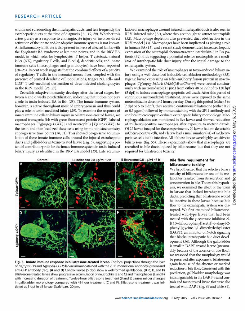
Innate immune cell response to biliatresoneIn some cases of human BA, a prominent inflammatory cell infiltrateconsisting of T helper and cytotoxic T cells andmacrophages is detected
Fig. 2. Biliatresone tissue specificity. (A and B) Histological cross sectionsshowing severemorphological defects of the gallbladder (asterisks) and cys-
tic duct (arrows) in an 8 dpf biliatresone–treated larva (B) compared with acontrol larva (A). (C and D) Normal histological appearance of hepatocytesand liver sinusoids in an 8dpf toxin-treated larva (D) comparedwith a controllarva (C). Sinusoids (containing nucleated red blood cells; arrows) are theprincipal vascular channel in the liver of zebrafish larvae; thus, branches ofthe portal vein and artery are not seen in these histological sections. Intra-hepatic bile ducts are too small to be seen. (E to H) Bright-field images oflive 8 dpf control (E) and toxin-treated (F) Tg(lfabp-RFP) larvae with corre-sponding fluorescent images of the liver (G and H). Arrows, liver (E and F).Scale bars, 10 mm (A to D); 200 mm (E to H).ienceTranslationalMedicine.org 6 May 2015 Vol 7 Issue 286 286ra67 3
R E S EARCH ART I C L E
on
May
6, 2
015
cem
ag.o
rg
within and surrounding the intrahepatic ducts, and less frequently theextrahepatic ducts at the time of diagnosis (11, 19, 20). Whether thisarises purely as a response to cholangiocyte injury or involves directactivation of the innate and/or adaptive immune systems is not known.An inflammatory infiltrate is also present in livers of affected lambswiththe Dysphania BA syndrome at late time points, and in the RRV BAmodel, in which roles for lymphocytes [T helper, T cytotoxic, naturalkiller (NK), regulatory T cells, and B cells], dendritic cells, and innateimmune cells (macrophages and granulocytes) have been reported(20–25). Recent work suggests that the combined effects of a paucityof regulatory T cells in the neonatal mouse liver, coupled with thepresence of primed dendritic cell populations, trigger NK cell– andCD8+ T cell–mediated destruction of virus-infected cholangiocytesin the RRV model (26, 27).
Zebrafish adaptive immunity develops after the larval stages, be-tween 4 and 6 weeks postfertilization, indicating that it does not playa role in toxin-induced BA in fish (28). The innate immune system,however, is active throughout most of embryogenesis and thus couldplay a role in toxin-mediated injury (29). To examine the response ofinnate immune cells to biliary injury in biliatresone-treated larvae, weexposed transgenic fish with green fluorescent protein (GFP)–labeledmacrophages [Tg(mpeg-1:GFP)] and neutrophils [Tg(mpx:GFP)] tothe toxin and then localized these cells using immunohistochemistryat progressive time points (30, 31). This showed progressive accumu-lation of these innate immune cells around the injured extrahepaticducts and gallbladder in toxin-treated larvae (Fig. 3), suggesting a po-tential contributory role for the innate immune system in toxin-inducedbiliary injury as identified in the RRV BA model (19). Late accumu-
www.Sc
lation ofmacrophages around injured intrahepatic ducts is also seen inRRV-infectedmice (11), where they are thought to attract neutrophils(32). Macrophage depletion also prevented duct obstruction in theRRVmodel (33). Macrophages have been implicated as playing a rolein human BA (11), and a recent study demonstrated increased hepaticexpression of the neutrophil chemoattractant interleukin-8 in BA pa-tients (34), thus suggesting a potential role for neutrophils as a medi-ator of intrahepatic bile duct injury after the initial damage to theextrahepatic system.
We examined the role of macrophages in toxin-induced biliary in-jury using a well-described inducible cell ablation methodology (35).Bigenic larvae expressing an NfsB-mCherry fusion protein in macro-phages [Tg(mpeg-1:Gal4; UAS:NfsB-mCherry)] were treated continu-ously with metronidazole (5 mM) from either 48 or 72 hpf to 120 hpf(5 dpf) to induce macrophage apoptotic cell death. After this period ofcontinuous metronidazole treatment, larvae were exposed to the samemetronidazole dose for 2 hours per day. During this period (either 5 to7 dpf or 5 to 8 dpf), they received continuous biliatresone (either 0.25or 0.5 mg/ml) followed by immunostaining with the 2F11 antibody andconfocal microscopy to evaluate extrahepatic biliary morphology. Mac-rophage ablation was monitored in live larvae and showed reductionof mCherry-positive macrophages after exposure to metronidazole.Of 27 larvae imaged for these experiments, 20 larvae had no detectablemCherry-positive cells, and 7 larvae had a small number (<4) ofmCherry-positive cells in the intestine. All of these larvae were highly sensitive tobiliatresone (fig. S6). These experiments show that macrophages arerecruited to bile ducts injured by biliatresone, but that they are notrequired for biliatresone toxicity.
ienceTranslationalMedicine.org
stm
.sci
enD
ownl
oade
d fr
om
Bile flow requirement forbiliatresone toxicityWehypothesized that the selective biliarytoxicity of biliatresone or one of its me-tabolites resulted from its secretion andconcentration in bile. To test this hypoth-esis, we examined the effect of the toxinin larvae that lacked intrahepatic bileducts, predicting that biliatresone wouldbe inactive in these larvae because bileflow to the extrahepatic system was dis-rupted. We first examined biliatresone-treated wild-type larvae that had beentreated with the g-secretase inhibitor N-[(3,5-difluorophenyl)acetyl]-L-alanyl-2-phenyl]glycine-1,1-dimethylethyl ester(DAPT), an inhibitor of Notch signalingthat blocks intrahepatic bile duct devel-opment (36). Although the gallbladderis small in DAPT-treated larvae (presum-ably because of the absence of bile flow),we reasoned that the morphology wouldbe preserved after exposure to biliatresone,again because of the absence or markedreductionof bile flow.Consistentwith thisprediction, gallbladder morphology wasindistinguishable in the DAPT-treated con-trols and toxin-treated larvae that were alsotreated with DAPT (fig. S9 and table S1).
Fig. 3. Innate immune response in biliatresone-treated larvae. Confocal projections through the liverof Tg(mpx:GFP) and Tg(mpeg-1:GFP) larvae immunostainedwith the 2F11monoclonal antibody (green) and
anti-GFP antibody (red). (A and D) Control larvae (5 dpf) show a well-formed gallbladder. (B, C, E, and F)Biliatresone-treated larvae show progressive accumulation of neutrophils (B and C) andmacrophages (E and F)with increasing duration of treatment. Twelve-hour biliatresone treatment (B and E) causesmilder changesin gallbladder morphology compared with 48-hour treatment (C and F). Biliatresone treatment was ini-tiated at 5 dpf in all larvae. Scale bars, 20 mm.6 May 2015 Vol 7 Issue 286 286ra67 4

R E S EARCH ART I C L E
ay 6
, 201
5
We next examined the effect of biliatresone in larvae that lack in-trahepatic bile ducts because of a genetic mutation. Themutant exam-ined, ductbend, was identified in an independent chemical mutagenesisscreen formutations that disrupt biliary development. ductbend exhibitsa pattern ofMendelian recessive inheritance over five generations andis lethal by 10 to 12 dpf. Confocal microscopy of ductbend larvae im-munostained with the 2F11 antibody showed few, if any, intrahepaticducts, but a small gallbladder and patent extrahepatic ducts (Fig. 4).Other phenotypic defects include delayed branchial arch developmentand intestinal and liver hypoplasia (fig. S10), thus indicating that theaffected gene playsmultiple developmental roles. Biliatresonewas pre-dicted to be inactive in homozygous ductbend larvae because bile flowto the extrahepatic system is severely reduced (as inDAPT-treatedwild-type larvae). Unexpectedly, gallbladder and extrahepatic duct mor-phology was disrupted in the toxin-treatedmutant larvae compared withuntreated mutant larvae, and the degree of injury in many appearedgreater than in toxin-treated wild-type larvae (Fig. 4, A and B versusC and D, and fig. S11).
Together, the data from DAPT-treated and ductbend larvae arguethat the selective biliary toxicity of biliatresone arises as a result of itssecretion and concentration in bile, but that genetic modifiers can over-ride this by sensitizing extrahepatic duct and gallbladder epithelial cells
www.Sc
to very low levels of the toxin present in either bile or extracellular fluid.In ductbend, biliatresone could conceivably enter extracellular fluideither from the vasculature or directly from the aqueous medium inwhich mutant larvae are reared.
Conserved synteny between ductbend and human BAsusceptibility lociTo gain insight into the mechanism underlying the sensitization ofductbend mutants to biliatresone, we carried out whole-exome se-quencing of mutant larvae and their wild-type siblings (n = 100 larvaein each pool). In the mutant pool, we identified a region of homozy-gosity located on chromosome 22 (nucleotides 27,387,718 to 27,758,148)that was confirmed with genetic mapping (discussed below). Analysesof gene synteny in this region showed that it was homologous to a re-gion of human chromosome 10 (10q25.1; formerly 10q24.2) that wasidentified as a BA susceptibility locus in a GWAS of Han Chinese pa-tients (Fig. 4E) (3). This associationwas confirmed in three independentstudies of Chinese, Thai, and Caucasian BA patients (37–39). Neithercoding sequence mutations nor consistent changes in expression levelshave been found in BA patients for the two genes adjacent to the 10q25.1markers (XPNPEP-1, a prolyl aminopeptidase, and ADD3, which en-codes g-adducin, a cytoskeletal F-actin capping protein). Expression
on
Mst
m.s
cien
cem
ag.o
rgD
ownl
oade
d fr
om
Fig. 4. Zebrafish ductbend sensitization. (A to D) Confocal projectionsthrough the liver and extrahepatic biliary systemof control and toxin-treated
extrahepatic system is more pronounced in the treated ductbend larva com-pared with wt (C). wt and ductbend larvae were treated with biliatresone
wtandductbend (dtb) larvae. Insets showthin sections through thegallbladder(A and B) or gallbladder and extrahepatic duct (C and D). Compared to thecontrol larvae, extrahepatic BA (A) is evident in the treated wt larva (B), be-cause the three principal components of the extrahepatic system (gallbladder,common bile duct, and cystic duct) distal to the common hepatic duct (chd)cannot be identified. (C) The control ductbend larva lacks intrahepatic bileducts and has a hypoplastic gallbladder. (D) Toxin-induced damage to the
(1 mg/ml) from 5 to 7 dpf. (E) Simplified genetic synteny map of the ductbendlocus indicating the position of genes relative to their human orthologs at theBA susceptibility loci (16p13.3 and 10q25.1). Numbers refer to chromosomal lo-cation in Mb. Blue asterisks, positions of SNPs in BA susceptibility loci; red aster-isk, position of SNP 27434434, ~1 centimorgan (cM) (0.6Mb) from the ductbendlocus. ihd, intrahepatic bile ducts; g, gallbladder; cbd, commonbile duct; cd, cys-tic duct; chd, common hepatic duct; pd, pancreatic duct. Scale bars, 20 mm.
ienceTranslationalMedicine.org 6 May 2015 Vol 7 Issue 286 286ra67 5

R E S EARCH ART I C L E
on
May
6, 2
015
stm
.sci
ence
mag
.org
Dow
nloa
ded
from
of add3 and xpnpep-1 orthologs was comparable in ductbend mutantand wild-type larvae (fig. S12). Coding sequence mutations were notseen in either gene in the exome sequencing.
The critical region surrounding the ductbend locus on zebrafishchromosome 22 also has conserved synteny with genes that map tohuman chromosome 16 (16p13.3; Fig. 4E). A singleton SNP (single-nucleotide polymorphism) within this region (rs17139085), subse-quently shown to be adjacent to the RBFOX1 gene, had the mostsignificant disease association in the human BA-GWAS study (3).No further analysis of this SNP was performed in this or subsequentstudies. The zebrafish RBFOX1 ortholog does not map to chromo-some 22, but the orthologs of several adjacent genes do. Three of thesegenes that are in close proximity to each other (<0.3 Mb) had mis-sense coding sequencemutations in ductbend: slx4, a scaffold proteinthat plays a role in DNA maintenance and repair (40); glis2, a zincfinger transcription factor that is mutated in patients with recessivenephronophthisis, a heritable cause of kidney failure (41); and adcy9,an adenylate cyclase (42). None of themutations were predicted to altergene function, and genetic mapping experiments placed slx4 ~1 cM(>0.6 Mb) from the ductbend locus (six recombinants at SNP 27434434in 658meioses; Fig. 4E), thus arguing that neither it, glis2, nor adcy9 isthe responsible gene.
Although our current data are not sufficient tomolecularly charac-terize the ductbend gene, its proximity to human BA susceptibility lociorthologs (10q25.1 and 16p13.3 genomic regions) does support the ideathat a common mechanism underlies susceptibility to biliary injury intheDysphania BA syndrome and human BA. The pleiotropic develop-mental defects present in homozygous ductbendmutants argue that inhumans, BA susceptibility would likely be imparted by hypomorphicrather than null alleles of the ductbend homolog.
Effect of biliatresone on mammalian cholangiocytesWe previously demonstrated that extrahepatic cholangiocyte primarycilia are decreased in both human BA and the mouse RRV BAmodel(43, 44). For this reason, we examined cilia in primary neonatal mouseextrahepatic cholangiocytes treated with either biliatresone or thenontoxic isoflavanone-3 that copurifies with biliatresone. Cells treatedwith biliatresone, but not with isoflavanone-3, had a reduction inprimary cilia as determined by staining with antibodies against acety-lated a-tubulin (fig. S13, A to C and J). Immunostaining of these cellsfor cellular tubulin demonstrated a dose-dependent decrease in visiblemicrotubules (figs. S13, D to F, and S14), suggesting that biliatresone,but not isoflavanone-3, decreased microtubule stability. Mouse hepato-cytes lack primary cilia; however, primary cultures of neonatal mousehepatocytes treated with biliatresone showed minimal changes in cell-ular tubulin staining when treated with biliatresone at 3 mg/ml (fig. S13,G to I) and only mild disruption of cellular tubulin when treated withthe highest dose of biliatresone (5 mg/ml; fig. S15, A and B). Primaryadult hepatocytes also showed only mild changes in organization ofcellular tubulin after treatment with biliatresone even at doses as highas 5 mg/ml (fig. S15, C and D).
Human BA and the BA-like syndrome seen in Sox17 haploinsuffi-cient mice are both characterized by loss of integrity of the epithelialmonolayer lining the extrahepatic ducts (45). Disruption of the gall-bladder epithelium is also an early finding in zebrafish larvae exposedto biliatresone (Figs. 1 and 2 and fig. S4). We therefore examined theeffect of biliatresone on epithelial integrity in a three-dimensionalmam-malian cell culture system in which cholangiocytes polarize and form
www.Sc
spheroid monolayers with distinct lumens. Treatment with isoflavanone-3 had no effect (fig. S16), but treatment with biliatresone caused loss ofepithelial monolayer integrity with lumen closure and disruption ofapical-basal polarity, as shownby staining for F-actin and the b1 integrinsubunit (Fig. 5A). Although closed spheroids were observed even in un-treated or vehicle-treated cells, there was a dose-dependent increase inpartial and complete lumen closure, with loss of the monolayer, afterbiliatresone treatment (Fig. 5B). In contrast, treatmentwith betavulgarin,which hadno effect on zebrafish larvae, did not disrupt polarity or resultin loss of the monolayer or lumen closure in cholangiocyte spheroids,but cells were consistently more columnar and spheroid lumens weresmaller than for vehicle-treated cells (Fig. 5A and fig. S17).
DISCUSSION
The etiology of BA is unknown, although there is evidence implicat-ing an environmental exposure—either infectious or toxic—in geneti-cally susceptible individuals.We report here the activities of the naturallyoccurring isochalcone, biliatresone, which we have isolated and iden-tified; it is the first toxin known to cause selective destruction of theextrahepatic biliary tree. Zebrafish larvae exposed to biliatresone de-velop a pattern of extrahepatic biliary damage that strongly resemblesthe pathology observed in BA in humans. These findings suggest thatexposure to structurally related compounds in food or from other envi-ronmental sources could be an important triggering event in human BA.
The chemical structure of biliatresone has not been previously re-ported, and biliatresone is the only structurally characterized plant iso-chalconewith an exomethylene ketonemoiety. Although the syntheticpathway used by Dysphania plants to produce biliatresone is notknown, structural analyses suggest that biliatresone might result fromthe ring opening of one of the copurifying compounds, either betavul-garin or the previously uncharacterized isoflavanone-3. Alternatively,biliatresone may serve as a precursor to the other compounds weisolated. A Clostridium species found in the human intestinal micro-biota cleaves isoflavonoids found in widely consumed soy products toproduce a metabolite with the same exomethylene ketone structure asbiliatresone (46–48). This raises the intriguing possibility that humanintestinal bacteria could produce toxins resembling biliatresone throughmodification of nontoxic precursors, such as betavulgarin, which isfound in common foods, including sugar beet, table beet, and chard(16, 17). It is conceivable that biliatresone itself or a structurally relatedtoxic compound is found in plants that are normally consumed byhumans.
The zebrafish andmammalian cell data presented here suggest thatbiliatresone can alter the integrity of the cholangiocyte monolayer inthe gallbladder and extrahepatic ducts. Over time in vivo, this couldresult in duct obstruction, which is a defining feature of human BA andis consistently observed in animal models of the disease, including themouse RRV and Sox17 haploinsufficiencymodels and epidemic BA inlivestock (45, 49, 50). Although we have not defined a mechanism to ac-count for the effect of biliatresone on cholangiocyte monolayer integrity,the spheroid data demonstrate that biliatresone disrupts apical-basolateralpolarity. On the basis of our cell culture experiments that demonstratea biliatresone-induced destabilization of cellular tubulin, we speculatethat this could occur through an effect on microtubules, which play animportant role in lumen formation andmaintenance of polarity in severaltypes of epithelial cells, including cholangiocytes (51, 52).
ienceTranslationalMedicine.org 6 May 2015 Vol 7 Issue 286 286ra67 6

R E S EARCH ART I C L E
on
May
6, 2
015
stm
.sci
ence
mag
.org
Dow
nloa
ded
from
The effect of biliatresone on cholangiocyte primary cilia also sug-gests that disruption of planar cell polarity (PCP) proteinsmay be partof the toxin’smechanism of action on cholangiocytes. PCP signaling isrequired for the assembly andmaintenance of primary andmotile cilia(53, 54) and has been shown to play an important role in apical-basalpolarity, in part through an effect on microtubules (55). PCP signaling
www.ScienceTranslationalMedicine.org
has been linked to biliary defects in de-veloping zebrafish (56). Although extra-hepatic bile duct cilia are decreased inhuman BA and the mouse RRV modelof BA (44), neither toxin-treated zebrafishnor Sox17 haploinsufficient mice dem-onstrated obvious abnormalities in ductalcilia; however, this may reflect redundancyof vertebrate polarity pathways. Regardless,our data support an effect of biliatresoneon cholangiocyte polarity and monolayerintegrity and suggest that understandinghow the toxin disrupts thesemayyield im-portantmechanistic insights intohumanBA.
The zebrafish model of biliary injurywe describe here will also aid in under-standing both the pathogenesis and clin-ical course of humanBA. The zebrafish isa powerful platform for genetic and chem-ical modifier screens and has the poten-tial to generate clinically relevant insightsinto the toxin’s mechanism of action. Asan example, our experiments showing in-activity of biliatresone in DAPT-treatedlarvae argue that its selectivity for chol-angiocytes is in part related to its secretioninto bile. Persistent activity of biliatresonein larvae in whichmacrophages were ab-lated suggests that these innate immunecells do not play an important role in theDysphania BA model, unlike the RRVmodel. Neutrophil-mediated injury couldcontribute to biliatresone-mediated bili-ary toxicity, and thiswill need to be testedwhen transgenic lines for ablation of thiscell type are available.
Our discovery of ductbend, its sensi-tization to biliatresone, and its geneticmapping to a conserved BA susceptibilitylocus argue that the molecular characteri-zation of this mutant will generate insightinto genetic factors influencing BA. Indeed,although it is conceivable that the proxi-mity of the ductbend gene to the zebrafish10q25.1 and 16p13.3 orthologs occurredby chance, this is statistically highly un-likely, and we believe that this is morelikely to be the result of its position withina large, ancestral gene cluster. Coexpres-sion of genes within such clusters is, inmany instances, thought to arise frompositive selection rather than leaky ex-
pression (57, 58). On this basis, we speculate that a subset of the geneswithin the ductbend cluster is required for biliary homeostasis. It isthus conceivable that variants of several genes within this cluster, andnot just the ductbend homolog, could alter BA susceptibility in humans.
Syntenic relationships within large gene clusters often change dur-ing evolution (58, 59), and the cluster spanning the ductbend locus was
Fig. 5. Biliatresone-induced mouse cholangiocyte injury. (A) Confocal sections through cholangiocytespheroids treated with vehicle, betavulgarin (2 mg/ml), or biliatresone (2 mg/ml) for 24 hours and stained with
antibodies against F-actin or the b1-integrin subunit, or with DAPI. The biliatresone-treated spheroid showsdisruption of the spheroid lumen and abnormal cholangiocyte polarity, as evidenced by altered distributionof F-actin and by cholangiocyte stratification. Most betavulgarin-treated spheroids demonstrated decreasedlumen size, but there was no loss of polarity or disruption of the monolayer (see fig. S17). Representativeimages from five independent experiments are shown. DAPI, 4′,6-diamidino-2-phenylindole. (B) Dose-response experiment showing progressive increase in the percentage of abnormal biliatresone-treatedspheroids beginning at 0.5 mg/ml. Scale bars, 7 mm. n = 48 to 89 spheroids counted per condition.6 May 2015 Vol 7 Issue 286 286ra67 7

R E S EARCH ART I C L E
on
May
6, 2
015
ag.o
rg
disrupted during the amphibian-to-reptile evolutionary transition(www.ensembl.org). Should there be co-regulated expression of thegenes within the 10q25.1 and 16p13.3 human BA susceptibility loci, thismust occur in trans. Although this most likely involves the action of ashared transcription factor, it is conceivable that a single regulatoryelement (an enhancer) modulates the expression of genes that arenow positioned on different chromosomes (60–62). Molecular charac-terization of ductbend will help us address questions related to theorganization and interaction of these and other BA susceptibility loci.
In summary, we have shown that a previously uncharacterized plantisochalcone, biliatresone, exerts a specific, localized destructive effecton the extrahepatic biliary system in a zebrafish model. These data,combined with data showing disruption of cellular polarity and lumenintegrity in a mouse cholangiocyte spheroid model, lead us to concludethat biliatresone is highly likely to be the toxic cause of the BA syn-drome in the Australian livestock. Whether exposure to biliatresoneor a related environmental agent can be directly linked to human BAis unknown, and understanding its relevance to this human diseasewill require further study. A detailed study of all of the manifestationsof BA, including the intrahepatic biliary injury that uniformly occursafter the initial extrahepatic injury in BA, will require additional ani-malmodels: a chronic injurymodel in the zebrafish system and a rodentmodel, which will require access to increased quantities of biliatresone.Nonetheless, the in vitro and in vivo injury models we have developedshould prove useful in understanding both the mechanism of the in-citing event in BA and biliary injury in general, and in identifying simi-lar toxic compounds to which humans are exposed.
stm
.sci
ence
mD
ownl
oade
d fr
om
MATERIALS AND METHODS
Study designThis study was designed to test the effect ofDysphania plant extracts usinga screen for biliary secretion in live zebrafish larvae. The in vivo screen is asurrogate for patency of the extrahepatic biliary system and biliary anato-my. The effects of the toxin were confirmed withmorphological studiesof the biliary system.All experiments were performed aminimumof sixtimes with a minimum of 50 individual larvae. The duration of thetreatments (endpoints) was chosen empirically on the basis of the effectof the toxin in the screening assay. The screening and confirmatory assayswere performed by four investigators. Confirmatory assays performedin zebrafish and cultured cells were evaluated by blinded investigators.
Plant materialAmixed sample ofD. glomulifera ssp. glomulifera (Nees) Paul G.WilsonandD.LittoralisR.Br. (Chenopodiaceae) (~2.8 kg)was collected from theexposed flats of the Burrinjuck Dam, along theHumeWeir, near Albury,New South Wales, Australia (latitude −36.003°, longitude 147.139°) inMarch 2008. The plants in the collection were identified at the RoyalBotanic Gardens, Melbourne, Australia; vouchers for the populationare housed at theNationalHerbariumofVictoria (Melbourne,Australia).The plants were frozen until shipment (−18°C) and shipped on dryice to the authors’ laboratories in April 2009 under a plant importpermit from Animal Plant Health and Inspection Service, U.S. De-partment of Agriculture (permit no. P37-08-00668, 21 May 2008).Individual plants were removed from the frozen material, and voucherspecimens were deposited in the herbarium of the University of theSciences in Philadelphia. The plants were kept frozen at −80°C until
www.Sc
analysis. A brief scheme describing plant extraction and fractionation,with purification of biliatresone, is described in fig. S2.
Zebrafish bioassay and analysesZebrafish embryos and larvae reared in 24- and 96-well plates wereexposed to lyophilized plant extracts resuspended in anhydrousDMSO for 24 to 72 hours (final concentration, 1% DMSO in embryomedium). The concentrations of extracts varied with the subfraction.For the original subfractions, concentrations of 5 to 50 mg/ml weretested. The final subfractionswere tested at 0.0625 to 1.0 mg/ml. Embryomedium with extract was exchanged daily. After treatment, larvaewere soaked in either PED-6 phospholipid (Invitrogen), the long-chainfatty acid Bodipy-C16 (Invitrogen), or the short-chain fatty acid Bodipy-C5 (Invitrogen) for 4 hours as previously described (63). Gallbladderand intestinal fluorescence in the treated and control larvae wereexaminedwith a stereomicroscope (OlympusMVX-10).Quantificationof fluorescence was performed with ImageJ software to calculate cor-rected total fluorescence over the region of the gallbladder (CTCF inImageJ). Larvae were processed for histology, immunostaining, andconfocal microscopy as previously described (36). All assays were per-formed in triplicate with at least 25 to 50 treated larvae and an equalnumber of control larvae. Control experiments to assess the effect onswallowing behavior were performed for each subfraction and for ex-periments using purified biliatresone as described (64). Larvae wereincubated in 50 mMDAPTorN-[N-(3,5-difluorophenacetyl)-L-alanyl]-S-phenylglycine t-butyl ester (Sigma-Aldrich) (stock solution of 100mMin DMSO) as previously described (36).
After treatments, larvae were fixed in 4% paraformaldehyde and im-munostained with mouse monoclonal antibody 2F11 (gift from J. Lewis)(65, 66) or processed for histology as previously described (36, 67).
ductbend mutants were recovered in an N-ethyl-N-nitrosoureamutagenesis screen formutations that disrupt themetabolism or biliarysecretion of fluorescent lipids [as described in (15)]. ductbend larvae at5 dpf have pleiotropic defects, most notably delayed branchial archdevelopment and digestive organ hypoplasia. The mutant larvae lackintrahepatic bile ducts, and their gallbladder and extrahepatic ductsare patent but smaller than in wild-type siblings. The mutation is re-cessive lethal by 10 to 12 dpf.
Exome sequencing of ductbend mutantsGenomic DNA was extracted from pools of 100 5-dpf ductbend andsibling larvae with TRIzol (Sigma-Aldrich) and processed for whole-exome sequencing as described (68).Mutationmappingwas performedusing the MMAPPR protocol (69).
Genetic mapping of the ductbend locusA mapping cross was generated by crossing adult fish heterozygousfor the ductbendmutation with wild-type fish with theWik background.F2mutant ductbend larvae (n = 329) were examined at 5 dpf for the pres-ence of an SNPwithin the slx4 coding region that was identified by exomesequencing (A to G conversion at nucleotide 27434434). SNP mappingof individual mutant larvae was performed via PCR with allele-specificprimers generated with dCAPS Finder 2.0 (http://helix.wustl.edu/dcaps/dcaps.html). The forward and reverse primers to amplify the wild-typeslx4 allele were ttgccatagtaacagtcgaggcgtggcc and agtgtgtgtgaagccttg-taatgttgtt. Hpa II digest of the wild-type PCR product generated 148-,87-, and 26-bp (base pair) fragments, whereas themutant fragment gave148- and 113-bp fragments.
ienceTranslationalMedicine.org 6 May 2015 Vol 7 Issue 286 286ra67 8

R E S EARCH ART I C L E
on
May
6, 2
015
stm
.sci
ence
mag
.org
Dow
nloa
ded
from
Mammalian cellsThe small cholangiocyte cell line used for spheroid culture was as de-scribed (70). Neonatal extrahepatic cholangiocytes were isolated byoutgrowth from 0- to 3-day-old BALB/c mouse pups. Briefly, ducts weredissected away from stroma and surrounding cells and then embeddedand cultured in a thick collagen gel (2mg/ml) for 3 to 4 weeks. Cells weresplit and cultured on thin collagen–coated (1 mg/ml) dishes for an ad-ditional 2 weeks before use (71). Mouse neonatal and adult hepatocyteswere isolated as described; cells were cultured for 48 hours after isolationbefore being treated with toxin (72). Cells from an entire litter of pups(at least 10) were used for each individual experiment with neonatalhepatocytes or cholangiocytes.
Toxin treatment and immunostainingCells were treated with vehicle (DMSO), biliatresone, betavulgarin, orisoflavanone-3 at doses ranging from 0.5 to 5 mg/ml for 24 hours. Bili-atresone was toxic to cholangiocytes (but not hepatocytes) at dosesgreater than 2 mg/ml. Cells were fixed and stained with antibodies againstacetylateda-tubulin (1:5000; Sigma), as amarkerof cilia, andcellular tubulin(1:500; Sigma) andwith the nuclear stainDAPI. Immunostainingwithantibodies against K19 (1:10, Developmental Studies HybridomaBank) for cholangiocytes and HNF4-a (1:1000; Abcam) for hepato-cytes confirmed that primary cells were at >95% purity. To determinethe percentage of cells with cilia, immunostained cilia visible in a givenhigh-power field were counted by a blinded observer (R.G.W.) andexpressed as a percentage of the total number of cells, as determinedby DAPI nuclear staining.
Spheroid cultureCells were plated in a collagen-Matrigel mixture (collagen, 4 mg/ml;Matrigel, 3.7 mg/ml; rat tail collagen I and growth factor–reduced,phenol red–freeMatrigel; BDBiosciences) in cell culture inserts (Millicell;Millipore) and incubated for 8 days. Media were changed every 2 to3 days. On day 7, cells were treated with vehicle (DMSO), biliatresone,or betavulgarin at 2 mg/ml. Twenty-four hours after treatment, the cellswere fixed with 4% ethanol and stained for F-actin (1:1000; phalloidin–tetramethylrhodamine B isothiocyanate; Santa Cruz Biotechnology)and with an antibody against the integrin b1 subunit (1:100; Abcam).For quantification of the effect of biliatresone, spheroids were stainedfor F-actin, and multiple confocal images were acquired. Lumens in thespheroids were assessed by a blinded observer (O.W.-Z.) as being open,closed (no lumen visible), or partially open or narrowed (small visiblelumen). Only spheroids imaged at a level with a clearly visible midsectionwere counted.
StatisticsStatistical significancewas calculated by either the one-tailed Student’st test or one-way analysis of variance.
SUPPLEMENTARY MATERIALS
www.sciencetranslationalmedicine.org/cgi/content/full/7/286/286ra67/DC1Fig. S1. Swallowing function of biliatresone-treated larvae.Fig. S2. Scheme for biliatresone isolation.Fig. S3. Biliatresone-mediated biliary secretion defect.Fig. S4. Biliatresone dose response in zebrafish.Fig. S5. Biliatresone biliary secretion dose response.Fig. S6. Macrophages not required for biliatresone toxicity.Fig. S7. Biliatresone treatment during embryogenesis.Fig. S8. No effect of biliatresone on digestive epithelia.
www.Sc
Fig. S9. Bile flow requirement for biliatresone toxicity.Fig. S10. Zebrafish ductbend mutants.Fig. S11. ductbend biliatresone sensitivity.Fig. S12. ductbend candidate gene expression.Fig. S13. Biliatresone-induced defects of cholangiocyte cilia and microtubules.Fig. S14. Dose-dependent loss of cholangiocyte microtubules with biliatresone treatment.Fig. S15. Effects of biliatresone on hepatocytes.Fig. S16. Lack of effect of isoflavanone-3 on mammalian cholangiocyte spheroids.Fig. S17. Effects of betavulgarin on mammalian cholangiocyte spheroids.Table S1. Biliatresone toxicity in DAPT-treated larvae.Movie S1. Biliary anatomy of a wild-type larva.Movie S2. Biliary anatomy of a toxin-treated wild-type larva.
REFERENCES AND NOTES
1. B. A. Haber, P. Russo, Biliary atresia. Gastroenterol. Clin. North Am. 32, 891–911 (2003).2. K. Zhou, N. Lin, Y. Xiao, Y. Wang, J. Wen, G.-M. Zou, X. Gu, W. Cai, Elevated bile acids in
newborns with biliary atresia (BA). PLOS One 7, e49270 (2012).3. M.-M. Garcia-Barcelo, M.-Y. Yeung, X.-P. Miao, C. S.-M. Tang, G. Cheng, M.-T. So, E. S.-W. Ngan,
V. C.-H. Lui, Y. Chen, X.-L. Liu, K.-J. W. S. Hui, L. Li, W.-H. Guo, X.-B. Sun, J.-F. Tou, K.-W. Chan,X.-Z. Wu, Y.-Q. Song, D. Chan, K. Cheung, P. H.-Y. Chung, K. K.-Y. Wong, P.-C. Sham, S. S. Cherny,P. K.-H. Tam, Genome-wide association study identifies a susceptibility locus for biliaryatresia on 10q24.2. Hum. Mol. Genet. 19, 2917–2925 (2010).
4. M. Rurarz, P. Czubkowski, K. Chrzanowska, J. Cielecka-Kuszyk, A. Marczak, D. Kaminska,J. Pawłowska, Biliary atresia in children with aberrations involving chromosome 11q. J. Pediatr.Gastroenterol. Nutr. 58, e26–e29 (2014).
5. M. Leyva-Vega, J. Gerfen, B. D. Thiel, D. Jurkiewicz, E. B. Rand, J. Pawlowska, D. Kaminska,P. Russo, X. Gai, I. D. Krantz, B. M. Kamath, H. Hakonarson, B. A. Haber, N. B. Spinner,Genomic alterations in biliary atresia suggest region of potential disease susceptibilityin 2q37.3. Am. J. Med. Genet. A 152A, 886–895 (2010).
6. M.-M. Tiao, C.-W. Liou, L.-T. Huang, P.-W. Wang, T.-K. Lin, J.-B. Chen, Y.-M. Chou, Y.-H. Huang,H.-Y. Lin, C.-L. Chen, J.-H. Chuang, Associations of mitochondrial haplogroups B4 and E withbiliary atresia and differential susceptibility to hydrophobic bile acid. PLOS Genet. 9,e1003696 (2013).
7. J. A. Bezerra, Potential etiologies of biliary atresia. Pediatr. Transplant. 9, 646–651 (2005).8. M. Riepenhoff-Talty, K. Schaekel, H. F. Clark, W. Mueller, I. Uhnoo, T. Rossi, J. Fisher, P. L. Ogra,
Group A rotaviruses produce extrahepatic biliary obstruction in orally inoculated newbornmice. Pediatr. Res. 33, 394–399 (1993).
9. M.-M. Tiao, S.-S. Tsai, H.-W. Kuo, C.-L. Chen, C.-Y. Yang, Epidemiological features of bil-iary atresia in Taiwan, a national study 1996–2003. J. Gastroenterol. Hepatol. 23, 62–66(2008).
10. S. Rauschenfels, M. Krassmann, A. N. Al-Masri, W. Verhagen, J. Leonhardt, J. F. Kuebler,C. Petersen, Incidence of hepatotropic viruses in biliary atresia. Eur. J. Pediatr. 168,469–476 (2009).
11. C. L. Mack, A. G. Feldman, R. J. Sokol, Clues to the etiology of bile duct injury in biliaryatresia. Semin. Liver Dis. 32, 307–316 (2012).
12. T. Saito, K. Shinozaki, T. Matsunaga, T. Ogawa, T. Etoh, T. Muramatsu, K. Kawamura, H. Yoshida,N. Ohnuma, H. Shirasawa, Lack of evidence for reovirus infection in tissues from patients withbiliary atresia and congenital dilatation of the bile duct. J. Hepatol. 40, 203–211 (2004).
13. P. Harper, J. W. Plant, D. B. Unger, Congenital biliary atresia and jaundice in lambs andcalves. Aust. Vet. J. 67, 18–22 (1990).
14. S. Robson, Congenital biliary atresia and jaundice in lambs, in New South Wales AnimalHealth Surveillance (NSW Department of Primary Industries, Sydney, New South Wales,Australia, 2007), vol. 2, pp. 2–4.
15. S. A. Farber, M. Pack, S. Y. Ho, I. D. Johnson, D. S. Wagner, R. Dosch, M. C. Mullins, H. S. Hendrickson,E. K. Hendrickson, M. E. Halpern, Genetic analysis of digestive physiology using fluorescentphospholipid reporters. Science 292, 1385–1388 (2001).
16. C. A. Elliger, J. M. Halloin, Phenolics induced in Beta vulgaris by Rhizoctonia solani infection.Phytochemistry 37, 691–693 (1994).
17. J. Geigert, F. R. Stermitz, G. Johnson, D. D. Maag, Two phytoalexins from sugarbeet (Betavulgaris) leaves. Tetrahedron 29, 2703–2706 (1973).
18. K. Lorent, S.-Y. Yeo, T. Oda, S. Chandrasekharappa, A. Chitnis, R. P. Matthews, M. Pack,Inhibition of Jagged-mediated Notch signaling disrupts zebrafish biliary development and gen-erates multi-organ defects compatible with an Alagille syndrome phenocopy. Development131, 5753–5766 (2004).
19. A. G. Feldman, C. L. Mack, Biliary atresia: Cellular dynamics and immune dysregulation.Semin. Pediatr. Surg. 21, 192–200 (2012).
20. K. Bessho, J. A. Bezerra, Biliary atresia: Will blocking inflammation tame the disease? Annu.Rev. Med. 62, 171–185 (2011).
ienceTranslationalMedicine.org 6 May 2015 Vol 7 Issue 286 286ra67 9

R E S EARCH ART I C L E
on
May
6, 2
015
stm
.sci
ence
mag
.org
Dow
nloa
ded
from
21. P. Shivakumar, G. Sabla, S. Mohanty, M. McNeal, R. Ward, K. Stringer, C. Caldwell, C. Chougnet,J. A. Bezerra, Effector role of neonatal hepatic CD8+ lymphocytes in epithelial injury andautoimmunity in experimental biliary atresia. Gastroenterology 133, 268–277 (2007).
22. P. Shivakumar, G. E. Sabla, P. Whitington, C. A. Chougnet, J. A. Bezerra, Neonatal NK cellstarget the mouse duct epithelium via Nkg2d and drive tissue-specific injury in experimentalbiliary atresia. J. Clin. Invest. 119, 2281–2290 (2009).
23. A. G. Feldman, R. M. Tucker, E. K. Fenner, R. Pelanda, C. L. Mack, B cell deficient mice areprotected from biliary obstruction in the rotavirus-induced mouse model of biliary atresia.PLOS One 8, e73644 (2013).
24. C. S. Lages, J. Simmons, C. A. Chougnet, A. G. Miethke, Regulatory T cells control the CD8adaptive immune response at the time of ductal obstruction in experimental biliary atresia.Hepatology 56, 219–227 (2012).
25. R. M. Tucker, A. G. Feldman, E. K. Fenner, C. L. Mack, Regulatory T cells inhibit Th1 cell-mediated bile duct injury in murine biliary atresia. J. Hepatol. 59, 790–796 (2013).
26. Y. Qiu, J. Yang, W. Wang, W. Zhao, F. Peng, Y. Xiang, G. Chen, T. Chen, C. Chai, S. Zheng,D. J. Watkins, J. Feng, HMGB1-promoted and TLR2/4-dependent NK cell maturation andactivation take part in rotavirus-induced murine biliary atresia. PLOS Pathog. 10, e1004011(2014).
27. V. Saxena, P. Shivakumar, G. Sabla, R. Mourya, C. Chougnet, J. A. Bezerra, Dendritic cellsregulate natural killer cell activation and epithelial injury in experimental biliary atresia.Sci. Transl. Med. 3, 102ra94 (2011).
28. S. H. Lam, H. L. Chua, Z. Gong, T. J. Lam, Y. M. Sin, Development and maturation of theimmune system in zebrafish, Danio rerio: A gene expression profiling, in situ hybridizationand immunological study. Dev. Comp. Immunol. 28, 9–28 (2004).
29. B. Novoa, A. Figueras, Zebrafish: Model for the study of inflammation and the innate im-mune response to infectious diseases. Adv. Exp. Med. Biol. 946, 253–275 (2012).
30. F. Ellett, L. Pase, J. W. Hayman, A. Andrianopoulos, G. J. Lieschke, mpeg1 promoter transgenesdirect macrophage-lineage expression in zebrafish. Blood 117, e49–e56 (2011).
31. S. A. Renshaw, C. A. Loynes, D. M. I. Trushell, S. Elworthy, P. W. Ingham, M. K. B. Whyte, Atransgenic zebrafish model of neutrophilic inflammation. Blood 108, 3976–3978 (2006).
32. S. K. Mohanty, C. A. P. Ivantes, R. Mourya, C. Pacheco, J. A. Bezerra, Macrophages are targetedby rotavirus in experimental biliary atresia and induce neutrophil chemotaxis by Mip2/Cxcl2.Pediatr. Res. 67, 345–351 (2010).
33. J. Li, K. Bessho, P. Shivakumar, R. Mourya, S. K. Mohanty, J. L. Dos Santos, I. K. Miura, G. Porta,J. A. Bezerra, Th2 signals induce epithelial injury in mice and are compatible with the biliaryatresia phenotype. J. Clin. Invest. 121, 4244–4256 (2011).
34. K. Bessho, R. Mourya, P. Shivakumar, S. Walters, J. C. Magee, M. Rao, A. G. Jegga, J. A. Bezerra,Gene expression signature for biliary atresia and a role for interleukin-8 in pathogenesis ofexperimental disease. Hepatology 60, 211–223 (2014).
35. D. T. White, J. S. Mumm, The nitroreductase system of inducible targeted ablation facilitatescell-specific regenerative studies in zebrafish. Methods 62, 232–240 (2013).
36. K. Lorent, J. C. Moore, A. F. Siekmann, N. Lawson, M. Pack, Reiterative use of the notchsignal during zebrafish intrahepatic biliary development. Dev. Dyn. 239, 855–864 (2010).
37. S. Kaewkiattiyot, S. Honsawek, P. Vejchapipat, V. Chongsrisawat, Y. Poovorawan, Associationof X-prolyl aminopeptidase 1 rs17095355 polymorphism with biliary atresia in Thai children.Hepatol. Res. 41, 1249–1252 (2011).
38. E. A. Tsai, C. M. Grochowski, K. M. Loomes, K. Bessho, H. Hakonarson, J. A. Bezerra, P. A. Russo,B. A. Haber, N. B. Spinner, M. Devoto, Replication of a GWAS signal in a Caucasian populationimplicates ADD3 in susceptibility to biliary atresia. Hum. Genet. 133, 235–243 (2014).
39. G. Cheng, C. S.-M. Tang, E. H.-M. Wong, W. W.-C. Cheng, M.-T. So, X. Miao, R. Zhang, L. Cui,X. Liu, E. S.-W. Ngan, V. C.-H. Lui, P. H.-Y. Chung, I. H.-Y. Chan, J. Liu, W. Zhong, H. Xia, J. Yu,X. Qiu, X.-Z. Wu, B. Wang, X. Dong, J. Tou, L. Huang, B. Yi, H. Ren, E. K.-W. Chan, K. Ye,P. F. O’Reilly, K. K.-Y. Wong, P.-C. Sham, S. S. Cherny, P. K.-H. Tam, M.-M. Garcia-Barcelo, Com-mon genetic variants regulating ADD3 gene expression alter biliary atresia risk. J. Hepatol.59, 1285–1291 (2013).
40. Y. Kim, G. S. Spitz, U. Veturi, F. P. Lach, A. D. Auerbach, A. Smogorzewska, Regulation ofmultiple DNA repair pathways by the Fanconi anemia protein SLX4. Blood 121, 54–63(2013).
41. M. Attanasio, N. H. Uhlenhaut, V. H. Sousa, J. F. O’Toole, E. Otto, K. Anlag, C. Klugmann,A.-C. Treier, J. Helou, J. A. Sayer, D. Seelow, G. Nürnberg, C. Becker, A. E. Chudley, P. Nürnberg,F. Hildebrandt, M. Treier, Loss of GLIS2 causes nephronophthisis in humans and mice byincreased apoptosis and fibrosis. Nat. Genet. 39, 1018–1024 (2007).
42. B. M. Hacker, J. E. Tomlinson, G. A. Wayman, R. Sultana, G. Chan, E. Villacres, C. Disteche,D. R. Storm, Cloning, chromosomal mapping, and regulatory properties of the humantype 9 adenylyl cyclase (ADCY9). Genomics 50, 97–104 (1998).
43. A. S. Chu, P. A. Russo, R. G. Wells, Cholangiocyte cilia are abnormal in syndromic and non-syndromic biliary atresia. Mod. Pathol. 25, 751–757 (2012).
44. S. Karjoo, N. J. Hand, L. Loarca, P. A. Russo, J. R. Friedman, R. G. Wells, Extrahepatic cholangio-cyte cilia are abnormal in biliary atresia. J. Pediatr. Gastroenterol. Nutr. 57, 96–101 (2013).
45. M. Uemura, A. Ozawa, T. Nagata, K. Kurasawa, N. Tsunekawa, I. Nobuhisa, T. Taga, K. Hara,A. Kudo, H. Kawakami, Y. Saijoh, M. Kurohmaru, M. Kanai-Azuma, Y. Kanai, Sox17 haploin-
www.Sci
sufficiency results in perinatal biliary atresia and hepatitis in C57BL/6 background mice.Development 140, 639–648 (2013).
46. H. G. Hur, R. Beger, T. Heinze, J. Lay, J. Freeman, J. Dore, F. Rafii, Isolation of an anaerobicintestinal bacterium capable of cleaving the C-ring of the isoflavonoid daidzein. Arch.Microbiol. 178, 8–12 (2002).
47. G. E. Kelly, C. Nelson, M. A. Waring, G. E. Joannou, A. Y. Reeder, Metabolites of dietary (soya)isoflavones in human urine. Clin. Chim. Acta 223, 9–22 (1993).
48. G. E. Joannou, G. E. Kelly, A. Y. Reeder, M. Waring, C. Nelson, A urinary profile study ofdietary phytoestrogens. The identification and mode of metabolism of new isoflavonoids.J. Steroid Biochem. Mol. Biol. 54, 167–184 (1995).
49. N. E. M. Gibelli, U. Tannuri, E. S. de Mello, C. J. Rodrigues, Bile duct ligation in neonatal rats:Is it a valid experimental model for biliary atresia studies? Pediatr. Transplant. 13, 81–87(2009).
50. J. Thomson, On congenital obliteration of the bile-ducts. Edinb. Med. J. 37, 523–531 (1892).51. N. Akhtar, C. H. Streuli, An integrin–ILK–microtubule network orients cell polarity and lumen
formation in glandular epithelium. Nat. Cell Biol. 15, 17–27 (2013).52. N. Tanimizu, Y. Kikkawa, T. Mitaka, A. Miyajima, a1- and a5-containing laminins regulate
the development of bile ducts via b1 integrin signals. J. Biol. Chem. 287, 28586–28597(2012).
53. Y. Zilber, S. Babayeva, J. H. Seo, J. J. Liu, S. Mootin, E. Torban, The PCP effector Fuzzycontrols cilial assembly and signaling by recruiting Rab8 and Dishevelled to the primarycilium. Mol. Biol. Cell 24, 555–565 (2013).
54. R. S. Gray, P. B. Abitua, B. J. Wlodarczyk, H. L. Szabo-Rogers, O. Blanchard, I. Lee, G. S. Weiss,K. J. Liu, E. M. Marcotte, J. B. Wallingford, R. H. Finnell, The planar cell polarity effector Fuz isessential for targeted membrane trafficking, ciliogenesis and mouse embryonic develop-ment. Nat. Cell Biol. 11, 1225–1232 (2009).
55. H. Tao, K.-I. Inoue, H. Kiyonari, A. G. Bassuk, J. D. Axelrod, H. Sasaki, S. Aizawa, N. Ueno,Nuclear localization of Prickle2 is required to establish cell polarity during early mouseembryogenesis. Dev. Biol. 364, 138–148 (2012).
56. S. Cui, L. M. Capecci, R. P. Matthews, Disruption of planar cell polarity activity leads todevelopmental biliary defects. Dev. Biol. 351, 229–241 (2011).
57. L. D. Hurst, C. Pál, M. J. Lercher, The evolutionary dynamics of eukaryotic gene order. Nat. Rev.Genet. 5, 299–310 (2004).
58. G.-Z. Wang, W.-H. Chen, M. J. Lercher, Coexpression of linked gene pairs persists long aftertheir separation. Genome Biol. Evol. 3, 565–570 (2011).
59. F. Al-Shahrour, P. Minguez, T. Marqués-Bonet, E. Gazave, A. Navarro, J. Dopazo, Selectionupon genome architecture: Conservation of functional neighborhoods with changinggenes. PLOS Comput. Biol. 6, e1000953 (2010).
60. M. R. Branco, A. Pombo, Intermingling of chromosome territories in interphase sug-gests role in translocations and transcription-dependent associations. PLOS Biol. 4, e138(2006).
61. N. Harmston, B. Lenhard, Chromatin and epigenetic features of long-range gene regulation.Nucleic Acids Res. 41, 7185–7199 (2013).
62. J. R. Bateman, J. E. Johnson, M. N. Locke, Comparing enhancer action in cis and in trans.Genetics 191, 1143–1155 (2012).
63. J. D. Clifton, E. Lucumi, M. C. Myers, A. Napper, K. Hama, S. A. Farber, A. B. Smith III, D. M. Huryn,S. L. Diamond, M. Pack, Identification of novel inhibitors of dietary lipid absorption usingzebrafish. PLOS One 5, e12386–e12388 (2010).
64. J. Abrams, G. Davuluri, C. Seiler, M. Pack, Smooth muscle caldesmon modulates peristalsisin the wild type and non-innervated zebrafish intestine. Neurogastroenterol. Motil. 24, 288–299(2012).
65. C. Crosnier, N. Vargesson, S. Gschmeissner, L. Ariza-McNaughton, A. Morrison, J. Lewis,Delta-Notch signalling controls commitment to a secretory fate in the zebrafish intestine.Development 132, 1093–1104 (2005).
66. D. Zhang, V. S. Golubkov, W. Han, R. G. Correa, Y. Zhou, S. Lee, A. Y. Strongin, P. Duc, S. Dong,Identification of Annexin A4 as a hepatopancreas factor involved in liver cell survival. Dev. Biol.395, 96–110 (2014).
67. M. Pack, L. Solnica-Krezel, J. Malicki, S. C. Neuhauss, A. F. Schier, D. L. Stemple, W. Driever,M. C. Fishman, Mutations affecting development of zebrafish digestive organs. Development123, 321–328 (1996).
68. R. N. W. Kettleborough, E. M. Busch-Nentwich, S. A. Harvey, C. M. Dooley, E. de Bruijn,F. van Eeden, I. Sealy, R. J. White, C. Herd, I. J. Nijman, F. Fényes, S. Mehroke, C. Scahill, R. Gibbons,N. Wali, S. Carruthers, A. Hall, J. Yen, E. Cuppen, D. L. Stemple, A systematic genome-wideanalysis of zebrafish protein-coding gene function. Nature 496, 494–497 (2013).
69. J. T. Hill, B. L. Demarest, B. W. Bisgrove, B. Gorsi, Y.-C. Su, H. J. Yost, MMAPPR: Mutationmapping analysis pipeline for pooled RNA-seq. Genome Res. 23, 687–697 (2013).
70. Y. Ueno, G. Alpini, K. Yahagi, N. Kanno, Y. Moritoki, K. Fukushima, S. Glaser, G. LeSage,T. Shimosegawa, Evaluation of differential gene expression by microarray analysis insmall and large cholangiocytes isolated from normal mice. Liver Int. 23, 449–459 (2003).
71. S. Karjoo, R. G. Wells, Isolation of neonatal extrahepatic cholangiocytes. J. Vis. Exp.e51621 (2014).
enceTranslationalMedicine.org 6 May 2015 Vol 7 Issue 286 286ra67 10

R E S EARCH ART I C L E
72. M. Perepelyuk, M. Terajima, A.Y. Wang, P. C. Georges, P. A. Janmey, M. Yamauchi, R. G. Wells,Hepatic stellate cells and portal fibroblasts are the major cellular sources of collagens and lysyloxidases in normal liver and early after injury. Am. J. Physiol. Gastrointest. Liver Physiol. 304,G605–G614 (2013).
Acknowledgments: We are grateful to M. Muthumani for assistance with zebrafish husbandry,C. Dang for assistance with spheroid studies, M. Perepelyuk for assistance with hepatocyte iso-lation, and the University of Pennsylvania Department of Cell and Developmental Biology Im-aging Core for assistance with microscopy. We thank J. Lewis for the gift of mouse monoclonalantibody 2F11. We acknowledge the late Surrey Jacobs for identification of Dysphania speciesplants. Funding: This work was supported in part by the NIH/National Institute of Diabetes andDigestive and Kidney Center for Molecular Studies in Digestive and Liver Diseases (P30DK050306)and its core facilities. Funding for J.R.P., R.G.W., and M.P. was from the NIH (R01DK092111) andthe Fred and Suzanne Biesecker Pediatric Liver Center at The Children’s Hospital of Philadelphia.D.L.S. was supported by theWellcome Trust (grant number 098051). Author contributions: P.A.W.
www.Sci
and S.J.W. helped procure Dysphania plants and diagnosed biliary atresia in livestock. K.L., W.G.,K.A.K., O.W.-Z., S.K., X.Z., I.S., R.N.K., D.L.S., J.R.P., R.G.W., andM.P. performed experiments. K.L., K.A.K.,J.R.P., R.G.W., and M.P. wrote the manuscript. Competing interests: The authors declare thatthey have no competing interests.Data andmaterials availability: Synthetic biliatresone willbe provided to academic researchers when available.
Submitted 26 October 2014Accepted 3 March 2015Published 6 May 201510.1126/scitranslmed.aaa1652
Citation: K. Lorent, W. Gong, K. A. Koo, O. Waisbourd-Zinman, S. Karjoo, X. Zhao, I. Sealy,R. N. Kettleborough, D. L. Stemple, P. A. Windsor, S. J. Whittaker, J. R. Porter, R. G. Wells,M. Pack, Identification of a plant isoflavonoid that causes biliary atresia. Sci. Transl. Med. 7,286ra67 (2015).
enceTranslationalMedicine.org 6 May 2015 Vol 7 Issue 286 286ra67 11
on
May
6, 2
015
stm
.sci
ence
mag
.org
Dow
nloa
ded
from

DOI: 10.1126/scitranslmed.aaa1652, 286ra67 (2015);7 Sci Transl Med
et al.Kristin LorentIdentification of a plant isoflavonoid that causes biliary atresia
Editor's Summary
contribute to the development of biliary atresia in people as well.mammalian cells. Although these plants are not eaten by humans, the findings suggest that plant toxins may
like changes in the fish as well as in−zebrafish model and identified a compound that caused biliary atresia plants growing on land that is normally covered by water. The researchers tested extracts from these plants in a
this disease may be a plant toxin. During a period of drought, the pregnant ewes' diet included unusual species of . used a naturally occurring outbreak of biliary atresia in Australian lambs to discover that one cause ofet alLorent
Biliary atresia is a childhood disease of unknown etiology, which causes rapid destruction of the biliary tree.
Fishing for a toxin
http://stm.sciencemag.org/content/7/286/286ra67.full.htmlcan be found at:
and other services, including high-resolution figures,A complete electronic version of this article
http://stm.sciencemag.org/content/suppl/2015/05/04/7.286.286ra67.DC1.html can be found in the online version of this article at: Supplementary Material
http://stm.sciencemag.org/content/scitransmed/3/102/102ra94.full.html can be found online at:Related Resources for this article
http://www.sciencemag.org/about/permissions.dtl in whole or in part can be found at: article
permission to reproduce this of this article or about obtaining reprintsInformation about obtaining
is a registered trademark of AAAS. Science Translational Medicinerights reserved. The title NW, Washington, DC 20005. Copyright 2015 by the American Association for the Advancement of Science; alllast week in December, by the American Association for the Advancement of Science, 1200 New York Avenue
(print ISSN 1946-6234; online ISSN 1946-6242) is published weekly, except theScience Translational Medicine
on
May
6, 2
015
stm
.sci
ence
mag
.org
Dow
nloa
ded
from