Hypertrophic Scar Cells Fail to Undergo a Form of Apoptosis Specific to Contractile Collagen—The...
11
See related Commentary on page v Hypertrophic Scar Cells Fail to Undergo a Form of Apoptosis Specific to Contractile Collagen—The Role of Tissue Transglutaminase Claire Linge, Janette Richardson, Charlotte Vigor, Elisabeth Clayton, Bushan Hardas,w and Kerstin J. Rolfe The RAFT Institute of Plastic Surgery, Mount Vernon Hospital, Northwood, Middlesex, UK; wProcyon Biopharma Inc. Dorval, Quebec, Canada Failure of apoptosis has been postulated to cause the hypercellularity and thus excess scar-tissue formation of hypertrophic scars (HTS). Here, we have examined the susceptibility of fibroblasts derived from normal or HTS to apoptosis induced during collagen-gel contraction, a wound-healing model. Normal scar (NS) fibroblasts under- went significant apoptosis (440% total) in contractile collagen, whereas apoptosis was not detected in HTS cells. This inability was specific to apoptosis induced by contractile collagen because apoptosis could be induced using diverse modalities. Since chronic fibrotic tissue is known to be excessively cross-linked, we next examined whether collagen matrix that had been conditioned by HTS fibroblasts became refractory to enzymatic breakdown and indeed, found that it is resistant to breakdown by both collagenase D and matrix metalloproteinase-2. Newly formed extracellular matrix is stabilized by the enzyme, tissue transglutaminase, which we demonstrated to be overexpressed by HTS fibroblasts in vivo and in vitro. Reducing tissue transglutaminase activity in collagen gels containing HTS fibroblasts permitted induction of apoptosis on gel contraction, whereas increasing enzymic ac- tivity in NS cell-containing gels completely abrogated collagen-contraction-induced-apoptosis. Together, these observations show that HTS fibroblasts exhibit resistance to a specific form of apoptosis elicited by contraction of collagen gels, and that this phenomenon is dependent on excess activity of cell surface tissue transglutaminase. Key words: apoptosis/cicatrix/collagen/transglutaminase II/wound healing J Invest Dermatol 125:72 –82, 2005 Hypertrophic scars (HTS) are severe excessive scars that are characterized histologically by an abundance of collagenous scar tissue and hypercellularity (mainly activated fibroblasts known as myofibroblasts), although their exact etiology re- mains unclear. During normal wound healing, the end of the fibroproliferative phase is heralded by the disappearance of myofibroblasts via apoptosis (Desmouliere et al, 1995). In pathological scars, these cells persist, sometimes for many years, which feasibly explains the excessive production of scar tissue that is the prime feature of these scars. Failure of apoptosis in HTS has long been hypothesized but evidence is controversial and often contradictory. The bcl-2 proto-oncogene, whose protein product protects cells from apoptosis, has been demonstrated to be strongly ex- pressed in vivo by fibroblasts within HTS and keloid tissue (Teofoli et al, 1999; Moulin et al, 2004), whereas the death receptor ligand, fas, has been shown to be decreased in HTS tissue and derived fibroblast cultures (Wassermann et al, 1998). In addition, Chodon et al (2000) reported that although keloid-derived fibroblasts were refractory to Fas- mediated and staurosporine-induced apoptosis, HTS cells were not. Furthermore, it has been reported that expression of caspase-3 is higher in hypertrophic and keloids scar tis- sue than in comparable age flat normal scars (NS), sug- gestive of increased levels of apoptosis in the pathological tissue (Akasaka et al, 2000). Indeed, this group went on to demonstrate significantly higher levels of apoptosis in HTS tissue compared with that of normal (flat) scars (Akasaka et al, 2001). This discrepancy between different sources of evidence is undoubtedly because of differences in exper- imental procedures, such as the maturity of the scar tissue used, whether tissue or cultured cells were examined, and the methods utilized to induce apoptosis, some of which bear no relation to the physiological processes going on within the maturing wound. The exact triggers of apoptosis during wound healing remain unknown, although recent work has indicated a po- tential role for contractile fibrillar collagen (Fluck et al, 1998; Grinnell et al, 1999). Here, dermal fibroblasts seeded into contracting but not anchored collagen gels nor contracting fibrin gels were seen to undergo apoptosis. Nevertheless, the exact triggers of apoptosis under these experimental circumstances are also undefined. The major differences between the two gel types (anchored vs contractile) are Abbreviations: CrIA, collagen contraction/remodelling-induced apoptosis; 14-DAB,1,4-diaminobutane; 3D, three-dimensional; ECM, extracellular matrix; FCS, fetal calf serum; HTS, hypertro- phic scar; MMP, matrix metalloproteinase; NS, normal scar; PBS, phosphate-buffered saline; RT, room temperature; SDS-PAGE, so- dium dodecylsulfate-polyacrylamide gel electrophoresis, tTgase, tissue transglutaminase Copyright r 2005 by The Society for Investigative Dermatology, Inc. 72
Transcript of Hypertrophic Scar Cells Fail to Undergo a Form of Apoptosis Specific to Contractile Collagen—The...

See related Commentary on page v
Hypertrophic Scar Cells Fail to Undergo a Form of ApoptosisSpecific to Contractile Collagen—The Role of TissueTransglutaminase
Claire Linge,� Janette Richardson,� Charlotte Vigor,� Elisabeth Clayton,� Bushan Hardas,wand Kerstin J. Rolfe��The RAFT Institute of Plastic Surgery, Mount Vernon Hospital, Northwood, Middlesex, UK; wProcyon Biopharma Inc. Dorval, Quebec, Canada
Failure of apoptosis has been postulated to cause the hypercellularity and thus excess scar-tissue formation of
hypertrophic scars (HTS). Here, we have examined the susceptibility of fibroblasts derived from normal or HTS to
apoptosis induced during collagen-gel contraction, a wound-healing model. Normal scar (NS) fibroblasts under-
went significant apoptosis (440% total) in contractile collagen, whereas apoptosis was not detected in HTS cells.
This inability was specific to apoptosis induced by contractile collagen because apoptosis could be induced using
diverse modalities. Since chronic fibrotic tissue is known to be excessively cross-linked, we next examined
whether collagen matrix that had been conditioned by HTS fibroblasts became refractory to enzymatic breakdown
and indeed, found that it is resistant to breakdown by both collagenase D and matrix metalloproteinase-2. Newly
formed extracellular matrix is stabilized by the enzyme, tissue transglutaminase, which we demonstrated to be
overexpressed by HTS fibroblasts in vivo and in vitro. Reducing tissue transglutaminase activity in collagen gels
containing HTS fibroblasts permitted induction of apoptosis on gel contraction, whereas increasing enzymic ac-
tivity in NS cell-containing gels completely abrogated collagen-contraction-induced-apoptosis. Together, these
observations show that HTS fibroblasts exhibit resistance to a specific form of apoptosis elicited by contraction of
collagen gels, and that this phenomenon is dependent on excess activity of cell surface tissue transglutaminase.
Key words: apoptosis/cicatrix/collagen/transglutaminase II/wound healingJ Invest Dermatol 125:72 –82, 2005
Hypertrophic scars (HTS) are severe excessive scars that arecharacterized histologically by an abundance of collagenousscar tissue and hypercellularity (mainly activated fibroblastsknown as myofibroblasts), although their exact etiology re-mains unclear. During normal wound healing, the end of thefibroproliferative phase is heralded by the disappearance ofmyofibroblasts via apoptosis (Desmouliere et al, 1995). Inpathological scars, these cells persist, sometimes for manyyears, which feasibly explains the excessive production ofscar tissue that is the prime feature of these scars.
Failure of apoptosis in HTS has long been hypothesizedbut evidence is controversial and often contradictory. Thebcl-2 proto-oncogene, whose protein product protects cellsfrom apoptosis, has been demonstrated to be strongly ex-pressed in vivo by fibroblasts within HTS and keloid tissue(Teofoli et al, 1999; Moulin et al, 2004), whereas the deathreceptor ligand, fas, has been shown to be decreased inHTS tissue and derived fibroblast cultures (Wassermann
et al, 1998). In addition, Chodon et al (2000) reported thatalthough keloid-derived fibroblasts were refractory to Fas-mediated and staurosporine-induced apoptosis, HTS cellswere not. Furthermore, it has been reported that expressionof caspase-3 is higher in hypertrophic and keloids scar tis-sue than in comparable age flat normal scars (NS), sug-gestive of increased levels of apoptosis in the pathologicaltissue (Akasaka et al, 2000). Indeed, this group went on todemonstrate significantly higher levels of apoptosis in HTStissue compared with that of normal (flat) scars (Akasakaet al, 2001). This discrepancy between different sources ofevidence is undoubtedly because of differences in exper-imental procedures, such as the maturity of the scar tissueused, whether tissue or cultured cells were examined, andthe methods utilized to induce apoptosis, some of whichbear no relation to the physiological processes going onwithin the maturing wound.
The exact triggers of apoptosis during wound healingremain unknown, although recent work has indicated a po-tential role for contractile fibrillar collagen (Fluck et al, 1998;Grinnell et al, 1999). Here, dermal fibroblasts seeded intocontracting but not anchored collagen gels nor contractingfibrin gels were seen to undergo apoptosis. Nevertheless,the exact triggers of apoptosis under these experimentalcircumstances are also undefined. The major differencesbetween the two gel types (anchored vs contractile) are
Abbreviations: CrIA, collagen contraction/remodelling-inducedapoptosis; 14-DAB,1,4-diaminobutane; 3D, three-dimensional;ECM, extracellular matrix; FCS, fetal calf serum; HTS, hypertro-phic scar; MMP, matrix metalloproteinase; NS, normal scar; PBS,phosphate-buffered saline; RT, room temperature; SDS-PAGE, so-dium dodecylsulfate-polyacrylamide gel electrophoresis, tTgase,tissue transglutaminase
Copyright r 2005 by The Society for Investigative Dermatology, Inc.
72

regarding mechanical loading (tension), cell cycle status (con-tinued cycling vs cycle arrest (Nakagawa et al, 1989a, b; Ni-shiyama et al, 1989; Kono et al, 1990)), and also their apparentability to remodel extracellular matrix (ECM) proteins, withanchored gels being reported to exhibit continued collagensynthesis, whereas this is reduced in contractile gels togetherwith a concomitant increase in matrix metalloproteinase(MMP) activity (higher MMP expression and lower tissue in-hibitor of metalloproteinase expression) (Nusgens et al, 1984;Unemori and Werb, 1986; Paye et al, 1987; Fukamizu andGrinnell, 1990). This latter observation is particularly intriguinggiven the recent findings of Buckley et al (1999), who deter-mined that small soluble peptides containing the RGD (argine,glucine, aspartic acid) amino acid motif (and therefore poten-tial breakdown products of collagen/ECM remodelling) caninduce apoptosis of fibroblasts via integrin-independent in-ternalization and direct activation of pro-caspase 3.
We therefore examined whether HTS cells would besusceptible to a contractile collagen-induced, and poten-tially wound healing-related, form of apoptosis. We furtherinvestigated whether the differences in apoptosis abilityunder these specific circumstances were related to chang-es in biochemical modification of collagen, since chronicfibrosis is linked with overmodification of the ECM in boththe liver and skin (Ricard-Blum et al, 1993, 1996, 1998; Hi-rota et al, 2003) and an increased isopeptide cross-linkingof ECM has been reported in HTS tissue (Dolynchuk, 1996).Overmodification of ECM proteins can dramatically alter thebehavior of cells inhabiting the matrix through changes inmechanical properties, changes in the availability of a va-riety of biologically active substances by binding to theECM or the availability of specific binding motifs within thematrix molecules themselves, and finally altering the break-down and remodelling of the matrix. This study then wenton to determine the role of the enzyme, tissue transgluta-minase, which is thought to be responsible for the stab-ilization of ECM (Ricard-Blum et al, 1996; Grenard et al,2001). Overexpression of this enzyme has been found inmany fibrotic conditions, liver fibrosis (Grenard et al, 2001),Dupuytrens disease (Dolynchuk and Pettigrew, 1991), andhypertrophic scarring, along with overexpression of one ofits major ligand groups, the collagen III-related peptides(Bowness et al, 1987a, b).
We report that whereas NS fibroblasts simulate dermalfibroblasts in their ability to undergo collagen contraction-induced apoptosis, HTS cells appear refractive to this spe-cific form of apoptosis. Further, the ability of cells to under-go this form of apoptosis is linked with changes in thebiochemical modification of collagen (resulting in alteredenzymatic breakdown products) and the activity of cell sur-face tissue transglutaminase (tTgase). These results sug-gest an integral role for tTgase in the pathogenesis ofhypertrophic scarring and also indicate a potential mech-anism by which inhibitors of tissue transglutaminase mightbe effective as anti-scarring therapeutic agents.
Results
HTS fibroblasts fail to undergo apoptosis in responseto collagen contraction/remodelling cues In 1998, Fluck
et al published the finding that fibroblasts derived from nor-mal skin dermis, when seeded into three-dimensional- (3D-)collagen gels and allowed to contract and therefore remodelthose gels, were induced to undergo apoptosis. We thereforedetermined whether fibroblasts derived from different scartypes would be equally susceptible to such a phenomenon.Since the degree to which the cells proliferate in collagengels differs between anchored and contractile gels, we tit-rated the amount of serum used to supplement the mediumdown to that which kept the cells quiescent but healthy(1%—not shown). All further experimentation was performedin this minimal growth medium in order to ensure that anydifferences seen in cell number were as a result of cell deathand not different proliferation rates. NS cells seeded intocollagen gels in minimal medium, when allowed to contractthe gels, underwent significant cell death ( � 35%–45% de-pending on cell strain, po0.001), reaching a maximum atapproximately 3–4 d (Fig 1A). This drop in viable cell numberwas not seen in gels that remained anchored. In contrast,cells derived from HTS failed to undergo cell death undereither circumstance. The results shown are representative ofthose obtained for all NS (n¼ 8) and HTS (n¼6) fibroblaststrains tested. The typical appearance of cells within the gelsafter 4 d is shown in Fig 1B–E: both NS cells (B) and HTScells (D) in anchored collagen gels are seen to be distributedrandomly, being mainly bipolar but with occasional poly-dendritic cells present. A large proportion of the NS cells incontractile collagen gels (C) are rounded up (black arrows),exhibiting dense compacted chromatin typical of that seen inapoptotic cells; the remaining cells appear multistellate andoccasionally bipolar. In contrast, very few HTS cells in con-tractile collagen gels were rounded up (E—black arrow), be-ing mainly multistellate in appearance (white arrow), with theextensive network of dendrite-like structures often extendingoutside the plane of focus. A terminal deoxynucleotidyltransferase-mediated dUTP nick end-labelling techniqueconfirmed that the cell death seen was as a result ofapoptosis. A typical example of this is shown in Fig 2: (A)demonstrating end-labelled nuclei of NS fibroblasts harvest-ed from a 4-d-old contractile gel, whereas (B) shows theunlabelled nuclei typical of HTS cells harvested from a 4-d-old contractile gel. Interestingly, the ability of either cell typeto contract collagen gels appears equivalent, with both NSand HTS cells contracting the gels by approximately 55% ofthe original gel area by 4 d (Fig 3).
Apoptosis defect specific to that induced by collagen-gel contraction To determine whether this inability of HTScells to undergo apoptosis is a general phenomenon orspecific to this particular induction signal, we examinedwhether HTS cells were sensitive to a variety of differentmethods of inducing apoptosis, including ethanol, do-xorubicin, and camptothecin. The mean cell death forn¼3 different cell strains of each scar-tissue type are plot-ted in Fig 4D (ethanol) and 4E (doxorubicin). HTS fibroblastsdemonstrated response curves identical (p values46) to NScells irrespective of the apoptotic stimuli tested. Cells treat-ed with 10% ethanol in serum-free medium (SFM) showconsiderable rounding up and death (Fig 4B), which steadilyincreased over time until few cells remained alive after 8 h.Treatment with 1 mg per mL of either doxorubicin (Fig 4C) or
APOPTOSIS FAILURE IN HYPERTROPHIC SCAR 73125 : 1 JULY 2005
camptothecin (not shown) in normal growth medium (con-taining 10% serum) resulted in cell death that steadily in-creased over time, reaching a maximum at 24 h (Fig 4E). Inall cases, cell death was attributed to apoptosis by nickend-labelling (not shown).
Gels inhabited by HTS cells are harder to break downcompared with gels inhabited by NS cells Under thequiescent conditions in which we performed our collagen-contraction experiments, the major differences betweenanchored versus contractile collagen gels that might influ-ence the induction of apoptosis are either mechanical (al-
tering cellular stress/tension) or related to collagenbreakdown/synthesis. Either of these putative mechanismscould be affected by changes in the post-translationalmodification of newly laid down collagen, and there is in-creasing evidence of biochemical overmodification of ECMin conditions linked with fibrosis. We therefore examinedwhether cells derived from the different scars differed intheir ability to modify the collagen gel.
Changes in modification of the collagen by the two celltypes were initially examined using collagenase D digestion.Sodium dodecylsulfate-polyacrylamide gel electrophore-sis (SDS-PAGE) analysis of digestion products showedlittle significant difference in protein bands over 50 kD insize. Typically, the majority of differences were seen inbreakdown products of less than 50 kD (Fig 5). In gelsconditioned for 3-d the only obvious difference is that theNS cell-gel product of approximately 10 kD (arrow, lane 1) isabsent in the HTS cell-gel products (lane 2). After 7 d, thedifferences become more marked, with many bands eithernot present, reduced in density, or appearing to exhibit analtered mobility in HTS cell gels (arrows, lane 3 vs lane 4). Aplot of the 7-d samples clearly shows the disappearance orreduction of bands at approximately 48, 39, 35, 33, 21, 20,19, and 10 kD. These results appear to suggest modificat-
Figure 1Collagen contraction/remodelling-induced cell death. Fibroblastsderived from either normal scars (NS) or hypertrophic scars (HTS) wereseeded into collagen gels and incubated for 3 d, after which time theywere either maintained anchored or released and allowed to contractthe gel for a 4-d period. At daily time points, the cells were harvestedand the number of apoptotic cells was assessed through viable stainingand nick end-labelling of apoptotic nuclei. Typical results are presentedin (A), which shows a graph of the percentage of cells undergoingapoptosis versus time: NS cells in anchored gels (open circles), NS cellsin contractile gels (closed circles), HTS cells in anchored gels (opentriangles), and HTS cells in contractile gels (closed triangles). Each pointrepresents the mean of triplicate gels, with the error bars being thestandard deviation from the mean. Micrographs B–E illustrate the typ-ical cellular morphology seen in each type of gel (formal saline fixed andhematoxylin stained): (B) NS, anchored; (C) NS, contractile; (D) HTS,anchored; (E) HTS, contractile. Black arrows indicate rounded-up dyingcells and the white arrow indicates live fibroblasts within the HTS cell-containing contractile gel. Scale bar¼50 mm.
Figure2Confirmation of apoptosis. Typical terminal deoxynucleotidyl transf-erase end-labelling of apoptotic nuclei (green fluorescence) with red(propidium iodide) counterstaining of cells harvested from collagen gelsthat had been allowed to contract for 4 d: normal scar cells (A) orhypertrophic scar cells (B). Scale bar¼25 mm.
Figure3Degree of collagen contraction. Typical examples of collagen gelsthat had been allowed to contract for 4 d that contained either normalscar (NS) (A) or hypertrophic scar (HTS) (B) cells. (C) shows the meanpercentage contraction of triplicate gels calculated as the mean per-cent reduction in diameter of the gels from the original (measured as anaverage of five random diameters of each gel) and the results of astandard Student’s t test.
74 LINGE ET AL THE JOURNAL OF INVESTIGATIVE DERMATOLOGY

ion of the collagen gel by HTS cells in such a way as todramatically change the products of collagenase digestion.
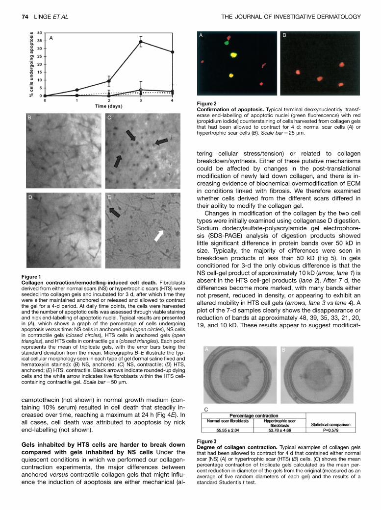
We therefore went on to examine the susceptibility ofsuch gels to MMP-2, an enzyme responsible for the pro-duction of smaller breakdown products during collagen re-modelling and active during cutaneous wound healing. Theproteolytic products of MMP-2-digested cell-conditionedcollagen were then analyzed via SDS-PAGE. The typicalappearances of the different collagen chains are seen inundigested acid-soluble collagen samples from both NSand HTS cell-conditioned gels (Fig 6A, lanes 1 and 2, re-spectively); a1 ( � 130 kD), a2 ( � 120 kD), and b (270—300kD), with the g chains being just discernible at the top of thegel. For the MMP-2-digested collagen samples, both the aand b chains of the NS cell-conditioned gel have beencompletely digested, whereas the a chain band ( � 130 kD)of HTS cell-conditioned collagen is still quite obvious (Fig6B, lanes 1 and 2). The digested NS sample does show astrong band of higher mobility at approximately 100 kD thatmay represent a breakdown product of the collagen diges-tion. Both gel types exhibited large amounts of breakdownproducts of less than 37 kD, but their banding patterns weretoo indistinct to determine any reproducible differences be-tween the two gels (not shown).
Tissue transglutaminase is overexpressed by HTS cellsin vivo and in 3D-collagen gels but not in monolayerculture One of the enzymes known to be responsible forbiochemical cross-linking and thus stabilization of ECM istissue transglutaminase. Immunohistochemical staining oftissue samples taken from the scars from which the cellcultures were derived corroborated the previous report (Do-lynchuk, 1996) that mature HTS exhibit high expression oftissue transglutaminase on fibroblastic cells within the scarmatrix, whereas NS of a similar age do not (Fig 7A, B). Todetermine whether if this was also the case in cultured HTScells, compared with NS cells immunohistochemical stain-ing was attempted on monolayer cultures, but with littlesuccess, with tissue transglutaminase being barely detect-able in either type of scar fibroblasts cultured on plastic.When these cells are cultured in 3D-collagen gels, however,tTgase expression is detectible in both cell types (Fig 7C,
Figure 4Chemically induced apoptosis. Micrographs illustrating hypertrophicscar (HTS) cell death induced by standard inducers of cellularapoptosis. (A) Appearance of cells in normal medium, (B) induction ofapoptosis after 7 h in 10% ethanol, and (C) apoptosis of cells after 24 htreatment with 1 mg per mL doxorubicin. Scale bar¼ 100 mm. (D, E)Graphical representation of cell death over time induced by 10% et-hanol and 1 mg per mL doxorubicin, respectively. Data plotted are themean of n¼ 4 cell strains of either normal scar (NS) (circles) or HTS(triangles), with error bars being the standard deviation from the mean.
Figure5Collagenase D digestion of cell-conditioned collagen. Collagen gelsseeded with fibroblasts derived from either normal scar (NS) or hype-rtrophic scar (HTS) were cultured for the whole time course of thecollagen contraction-induced apoptosis experiment (7 d). Gels wereharvested either at day 3 (the point at which the gels are normally freedto allow contraction) or day 7 (i.e. 4 d after contraction began and thepoint at which apoptosis is usually maximal). The gels were digestedusing collagenase D and the digestion products were analyzed on 12%sodium dodecylsulfate-polyacrylamide gel electrophoresis gel undernon-reducing conditions. (A) A typical gel: Lanes 1 (day 3 specimen)and 3 (day 7) are NS cell-conditioned gels; lanes 2 (day 3) and 4 (day 7)are HTS cell-conditioned gels. The arrows indicate bands that havealtered between NS and HTS lanes of the same time point. (B) A plot ofthe density of lane 3 (dashed line) and 4 (solid line) versus molecularweight. The arrows indicate major changes in banding patterns be-tween the NS and HTS cell-conditioned gels.
APOPTOSIS FAILURE IN HYPERTROPHIC SCAR 75125 : 1 JULY 2005
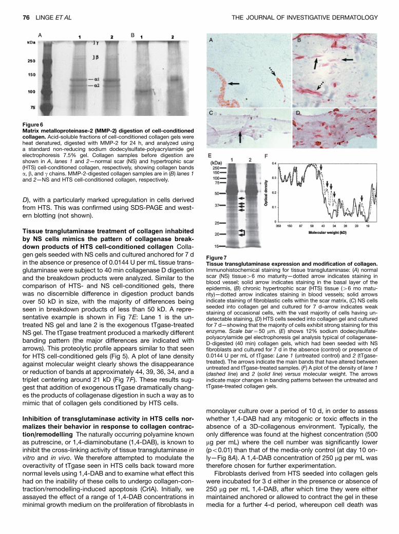
D), with a particularly marked upregulation in cells derivedfrom HTS. This was confirmed using SDS-PAGE and west-ern blotting (not shown).
Tissue tranglutaminase treatment of collagen inhabitedby NS cells mimics the pattern of collagenase break-down products of HTS cell-conditioned collagen Colla-gen gels seeded with NS cells and cultured anchored for 7 din the absence or presence of 0.0144 U per mL tissue trans-glutaminase were subject to 40 min collagenase D digestionand the breakdown products were analyzed. Similar to thecomparison of HTS- and NS cell-conditioned gels, therewas no discernible difference in digestion product bandsover 50 kD in size, with the majority of differences beingseen in breakdown products of less than 50 kD. A repre-sentative example is shown in Fig 7E: Lane 1 is the un-treated NS gel and lane 2 is the exogenous tTgase-treatedNS gel. The tTgase treatment produced a markedly differentbanding pattern (the major differences are indicated witharrows). This proteolytic profile appears similar to that seenfor HTS cell-conditioned gels (Fig 5). A plot of lane densityagainst molecular weight clearly shows the disappearanceor reduction of bands at approximately 44, 39, 36, 34, and atriplet centering around 21 kD (Fig 7F). These results sug-gest that addition of exogenous tTgase dramatically chang-es the products of collagenase digestion in such a way as tomimic that of collagen gels conditioned by HTS cells.
Inhibition of transglutaminase activity in HTS cells nor-malizes their behavior in response to collagen contrac-tion/remodelling The naturally occurring polyamine knownas putrescine, or 1,4-diaminobutane (1,4-DAB), is known toinhibit the cross-linking activity of tissue transglutaminase invitro and in vivo. We therefore attempted to modulate theoveractivity of tTgase seen in HTS cells back toward morenormal levels using 1,4-DAB and to examine what effect thishad on the inability of these cells to undergo collagen-con-traction/remodelling-induced apoptosis (CrIA). Initially, weassayed the effect of a range of 1,4-DAB concentrations inminimal growth medium on the proliferation of fibroblasts in
monolayer culture over a period of 10 d, in order to assesswhether 1,4-DAB had any mitogenic or toxic effects in theabsence of a 3D-collagenous environment. Typically, theonly difference was found at the highest concentration (500mg per mL) where the cell number was significantly lower(po0.01) than that of the media-only control (at day 10 on-ly—Fig 8A). A 1,4-DAB concentration of 250 mg per mL wastherefore chosen for further experimentation.
Fibroblasts derived from HTS seeded into collagen gelswere incubated for 3 d either in the presence or absence of250 mg per mL 1,4-DAB, after which time they were eithermaintained anchored or allowed to contract the gel in thesemedia for a further 4-d period, whereupon cell death was
Figure 6Matrix metalloproteinase-2 (MMP-2) digestion of cell-conditionedcollagen. Acid-soluble fractions of cell-conditioned collagen gels wereheat denatured, digested with MMP-2 for 24 h, and analyzed usinga standard non-reducing sodium dodecylsulfate-polyacrylamide gelelectrophoresis 7.5% gel. Collagen samples before digestion areshown in A, lanes 1 and 2—normal scar (NS) and hypertrophic scar(HTS) cell-conditioned collagen, respectively, showing collagen bandsa, b, and g chains. MMP-2-digested collagen samples are in (B) lanes 1and 2—NS and HTS cell-conditioned collagen, respectively.
Figure7Tissue transglutaminase expression and modification of collagen.Immunohistochemical staining for tissue transglutaminase: (A) normalscar (NS) tissue46 mo maturity—dotted arrow indicates staining inblood vessel; solid arrow indicates staining in the basal layer of theepidermis, (B) chronic hypertrophic scar (HTS) tissue (46 mo matu-rity)—dotted arrow indicates staining in blood vessels; solid arrowsindicate staining of fibroblastic cells within the scar matrix, (C) NS cellsseeded into collagen gel and cultured for 7 d–arrow indicates weakstaining of occasional cells, with the vast majority of cells having un-detectable staining, (D) HTS cells seeded into collagen gel and culturedfor 7 d—showing that the majority of cells exhibit strong staining for thisenzyme. Scale bar¼50 mm. (E) shows 12% sodium dodecylsulfate-polyacrylamide gel electrophoresis gel analysis typical of collagenase-D-digested (40 min) collagen gels, which had been seeded with NSfibroblasts and cultured for 7 d in the absence (control) or presence of0.0144 U per mL of tTgase: Lane 1 (untreated control) and 2 (tTgase-treated). The arrows indicate the main bands that have altered betweenuntreated and tTgase-treated samples. (F) A plot of the density of lane 1(dashed line) and 2 (solid line) versus molecular weight. The arrowsindicate major changes in banding patterns between the untreated andtTgase-treated collagen gels.
76 LINGE ET AL THE JOURNAL OF INVESTIGATIVE DERMATOLOGY

assessed. The experiments were repeated at least threetimes with n¼3 cell lines. Typical results plotted in Fig 8Bshow that addition of 1,4-DAB to the medium during thecourse of the experiment had a profound effect on the abil-ity of collagen contraction to induce apoptosis (po0.001) ofHTS cells, with the level of death ( � 47% Fig 8B) beingsimilar to that seen with NS cells (Fig 1A).
Treatment of collagen gels with exogenous transgluta-minase inhibits apoptosis of NS cells in response tocollagen contraction/remodelling Polyamines are pleio-tropic molecules that could potentially affect cell death inmany different ways other than solely through the inhibitionof the action of tTgase. We therefore used an alternativeapproach and examined whether an increase in tTgase ac-tivity in collagen gels inhabited by NS fibroblasts would en-gender resistance to CrIA in these ‘‘normal’’ cells. Again, wefirst determined whether tTgase had any effect (either mi-togenic or toxic) on the cells in the absence of a 3D-col-lagenous environment, by performing growth curves usingmonolayer cultures of NS fibroblasts. The titration curve ateach time point is plotted in Fig 9A and shows that none ofthe concentrations of tTgase used significantly affected the
growth or death of these cells. A tTgase concentration of0.0144 U per mL was used in all further experimentation andis of the same order of concentration as that used by Murthyet al (1991) to cross-link fibrinogen.
Fibroblasts derived from NS seeded into collagen gelswere incubated for 3 d either in the presence or absence of0.0144 U per mL tissue transglutaminase, after which timethey were either maintained anchored or allowed to contractthe gel in these media for a further 4-d period, after whichcell death was assessed. Experiments were repeated atleast three times with n¼3 cell lines. Typical results plottedin Fig 9B show that, in contrast to the untreated gels(po0.001), NS cells in contractile collagen gels in the pres-ence of added exogenous tissue transglutaminase did notundergo apoptosis (p40.8).
Discussion
Recently, a role for fibrillar collagen has been indicated inthe induction of apoptosis of fibroblasts (Fluck et al, 1998;Grinnell et al, 1999), thus establishing an experimental stim-ulus of apoptosis with relevance to the healing wound.
Figure 8Inhibition of tissue transglutaminase activity in hypertrophic scar(HTS) cell-containing collagen gels. The effects of 1,4-diaminobu-tane (1,4-DAB) on the cell number of fibroblasts cultured in monolayerin minimal growth medium were titrated over a 10-d period (A). � in-dicates the concentration at which the cell number was significantly(po0.01) lower than that of the media-only control (at day 10 only). HTSfibroblasts seeded into collagen gels were incubated for 3 d either inthe presence or absence of 250 mg per mL 1,4-DAB, after which time,they were either maintained anchored or allowed to contract the gel inthese media for a further 4-d period. At day 4 (post-release), the cellswere harvested from the gels using collagenase D and the number ofapoptotic cells was assessed through viable staining and nick end-labelling of apoptotic nuclei. Typical results are presented in (B), whichshows a graph of the percentage of cells undergoing apoptosis: an-chored gels (solid bars) and contractile gels (striped bars). Each pointrepresents the mean of triplicate gels, with the error bars being thestandard error of the mean (�po0.001).
Figure9Treatment of normal scar (NS) cell-containing collagen gels withtissue transglutaminase. The effects of added exogenous tissuetransglutaminase on the cell number of fibroblasts cultured in mono-layer in minimal growth medium were titrated over a 10-d period—nosignificant difference was seen in the cell number over the whole tit-ration range tested. NS fibroblasts seeded into collagen gels were in-cubated for 3 d either in the presence or absence of 0.0144 U per mLtissue transglutaminase, after which time, they were either maintainedanchored or allowed to contract the gel in these media for a further 4-dperiod. At day 4 (post-release), the cells were harvested from the gelsusing collagenase D and the number of apoptotic cells assessedthrough viable staining and nick end-labelling of apoptotic nuclei. Typ-ical results are presented in (B), which shows a graph of the percentageof cells undergoing apoptosis: anchored gels (solid bars) and contrac-tile gels (striped bars). Each point represents the mean of triplicate gels,with the error bars being the standard error of the mean. (�po0.001).
APOPTOSIS FAILURE IN HYPERTROPHIC SCAR 77125 : 1 JULY 2005

Considering the long-hypothesized possibility that thehypercellular nature (and potentially the whole pathology)of hypertrophic scarring is caused by a failure of apoptosis,we therefore investigated whether a reduced susceptibilityof fibroblasts to this particular form of apoptosis might un-derlie the pathology of this condition.
We have demonstrated that cell lines derived fromchronic (46-mo old) HTS are resistant to this CrIA, where-as fibroblasts derived from NS of similar maturity mimicdermal fibroblasts and are susceptible to CrIA. These find-ings highlight distinct patterns of cellular behavior exhibitedby these cells indicating intrinsic cellular differences ratherthan purely environmental cues in the pathology of hype-rtrophic scarring. Furthermore, we have shown that a gen-eral defect in the machinery of apoptosis is not indicated,but rather something more particular to the specific form ofapoptosis induction going on within a contractile 3D-colla-gen structure. These findings are not only in accordancewith the hypothesis that the hypercellular characteristic ofhypertrophic scarring is because of a failure of cellularapoptosis but also supports the proposition that collagen isinvolved in the mechanism that switches off the fibropro-liferative phase of wound healing.
We further postulated that this phenomenon of apoptoticresistance might be caused by the matrix of HTS beingovermodified in some way, either via intracellular post-translational modification that goes on during biosynthesisor through extracellular biochemical modification, sincechronic fibrotic tissue (both skin and liver) is known to beexcessively cross-linked (Ricard-Blum et al, 1993, 1996,1998; Dolynchuk, 1996; Hirota et al, 2003). Overmodifica-tion of ECM would considerably change its biochemicalproperties and how cells interact with it, potentially affectingthe mechanical properties of the matrix and thus the ten-sional load put upon the inhabiting cells. Excessive cross-linking of ECM might also result in hidden or uncoveredcellular binding sites within the matrix proteins themselves,or make available or sequester growth factors or the like.Furthermore, this type of modification would affect thequality of enzymatic cleavage, potentially making the matrixmore refractive to enzymatic breakdown.
We demonstrated that HTS cells biochemically modifycollagen in such a way as to change the products of pro-teolytic cleavage that result from digestion with a strongcollagenase (Collagenase D). In addition, HTS cell modifi-cation of collagen causes resistance to enzymatic cleavageby MMP-2, a collagenase highly active during cutaneouswound healing. These results are analogous to recent find-ings in a model of chronic dermatitis induced in rabbit ears(Hirota et al, 2003) and are of particular interest consideringwork by Buckley et al (1999), who implicated potentialbreakdown products of the ECM in the initiation ofapoptosis in both lymphocytic cells and fibroblasts. Smallsoluble peptides containing the RGD amino acid motif werereported to directly induce apoptosis through an integrin-independent mechanism by direct binding to and activationof pro-caspase-3 as apposed to the alternative mechanismof inducing apoptosis (termed anoikis) through blocking in-tegrin-mediated cell-matrix contact (Frisch and Ruoslahti,1997). It is possible that this overmodification and thusstabilization of collagen by HTS cells might reduce the pro-
duction of small soluble RGD-containing peptides that oc-curs on remodelling, thereby removing a potential trigger forapoptosis.
The stabilization of newly formed ECM is thought to belargely accomplished through the actions of tissue trans-glutaminase (or transglutaminase II) (Ricard-Blum et al,1996; Grenard et al, 2001). The activity of this enzyme ismarkedly increased during wound healing, substantiallyeffecting both the breaking strength of wounds and thesolubility of ECM proteins (Bowness et al, 1987b, 1988;Dolynchuk et al, 1994; Haroon et al, 1999). Tissue trans-glutaminase cross-links proteins through the formation ofisopeptide bonds between reactive g-glutaminyl groups incertain specific proteins and the e-amino group of lysine inother proteins. Some of the components of early granulationtissue, type III collagen along with its aminopropeptides,have been shown to be particularly good substrates fortransglutaminase (Bowness et al, 1987a, b), and elevatedlevels of these have been detected in both HTS (Weber et al,1978) and related fibrotic conditions such as Dupuytrensdisease (Brickley-Parsons et al, 1981). Since the affectedfascia of Dupuytrens disease is also known to have in-creased transglutaminase levels (Dolynchuk et al, 1991),and increased extracellular activity of this enzyme has beendetected in chronic HTS tissue (Dolynchuk, 1996), it seemsfeasible that the transglutaminase cross-linking of ECMmight be expected to be excessive in these conditions.
We have not only demonstrated high expression levels oftissue transglutaminase in chronic HTS tissue, corroborat-ing the previous report (Dolynchuk, 1996), but also in HTScells in vitro, but only when cultured within 3D-collagen gelsand not in monolayer culture. This apparent matrix depend-ence of the expression, stabilization or extracellular local-ization of tissue transglutaminase is in contrast to a recentreport comparing high tTgase-expressing pre-senescent,senescent, and immortalized dermal fibroblasts, which re-ported no dependence on culture environment (Stephenset al, 2004), and thus requires further investigation. In sup-port of the theory that the overexpression of cell surfacetTgase might explain the change in resistance to enzymaticdigestion, treatment of NS cell-seeded collagen gels withexogenous tTgase was shown to alter the products of pro-teolytic breakdown in such a way as to mimic that of HTScell-seeded gels. We went on to manipulate the activity oftissue transglutaminase in cell-containing collagen gels andfound that inhibition of tTgase activity in collagen gels con-taining HTS cells resulted in the normalization of their be-havior; allowing induction of apoptosis on gel contraction,whereas boosting tTgase activity in gels containing NS cellscompletely abrogated CrIA. These findings establish a rolefor tTgase in the protection from CrIA and suggest that theprolonged overexpression of this enzyme by HTS fibro-blasts may be the basis of their pathology.
A therapeutic use of 1,4-DAB as an inhibitor of trans-glutaminase activity in HTS has been considered previously.A phase II clinical study was set up in human patients withHTS to primarily determine safety but also the efficacy of atopical cream containing 0.8% wt/vol 1,4-DAB (FibrostatProcyon Biopharma, Inc., Dorval, Canada) (Dolynchuk et al,1996). The trial was set up as a double-blind crossoverstudy over two successive 4-wk periods, which showed
78 LINGE ET AL THE JOURNAL OF INVESTIGATIVE DERMATOLOGY

that Fibrostats treatment resulted in a significant improve-ment in scar appearance irrespective of the order given.Further work used HPLC analysis to quantitate the e (g-glutamyl) lysine cross-linking of ECM proteins and foundthat these were significantly reduced in Fibrostats-treatedHTS tissue compared with vehicle-only control-treated scar(Dolynchuk, 1996), thus supporting a possible therapeuticrole for topical 1,4-DAB in the treatment of HTS. Our resultssuggest that the mechanism of therapeutic action could bevia allowing the HTS fibroblasts to finally undergo apopto-sis, thus reducing hypercellularity of the scars and speedingtheir regression.
How tissue transglutaminase activity might contributetoward the sensitivity of a cell to the specific form ofapoptosis induced by collagen contraction is a questionthat still needs addressing. This enzyme is a highly multi-functional molecule exhibiting a plethora of new and diversebioactivities, a comprehensive review of which, includingthe cell structure-stabilizing role of intracellular transgluta-minase during apoptosis, is given by Griffin et al (2002).Indeed, tTgase is unique in that it has multiple enzymaticfunctions. Apart from its cross-linking activity, it has longbeen known that tTgase also directly binds guanosinetriphosphate (GTP)/guanosine diphosphate and undergoesa GTPase cycle, thus allowing this enzyme to function di-rectly in signal transduction (Nakaoka et al, 1994; Mian et al,1995). In addition, tTgase has also recently been reported tohave intrinsic kinase activity, phosphorylating insulin-likegrowth factor binding protein-3 (Mishra and Murphy, 2004).Each of any of these three distinct enzymatic activities ex-hibited by tTgase could affect apoptosis in multiple differentways. Our findings that 1,4-DAB, which specifically inhibitsthe cross-linking activity of extracellular tTgase, and addedexogenous tTgase both affect susceptibility to CrIA how-ever, suggests that this phenomenon is strictly affected bythe cross-linking activity of extracellular (presumably cellmembrane-bound) tTgase and not through this enzyme’salternative functions within the cell. Interestingly, CrIA mustnot therefore involve fibroblast spreading, motility, nor theirability to form stress fibers or focal adhesions, since all ofthese cellular functions have recently been found to be de-pendent on the G-protein function of tTgase rather than itscross-linking ability (Stephens et al, 2004). Our observationof equivalent contraction by HTS and NS cell-inhabitedcollagen gels would appear to suggest that contractionis not affected by overactivity of tTgase and that CrIA doesnot necessarily follow contraction. Whether contraction inhigh tTgase activity gels differs in a wholly qualitative wayhowever is unknown.
Since cell surface transglutaminase is well known for itsstabilization of ECM and its ability to reduce the solubility ordegradability of these proteins, it thus has the potential toreduce the production of small RGD-containing peptidesthat might play a role in the induction of apoptosis as men-tioned previously. Alternative roles for this particularenzymatic function, however, are being uncovered, whichalso have the potential to affect cell survival. Whether theseplay a role during collagen contraction is currently underinvestigation. Transglutaminase has been implicated in theactivation of transforming growth factor-b1 (TGF-b1) viacross-linking of latent TGF-b-binding protein-1 (Nunes et al,
1997; Verderio et al, 1999). TGF-b-mediated apoptosis hasbeen implicated in the formation and homeostasis of manydifferent tissues including the immune system (particularlyB- and T-cell homeostasis), liver, prostate, uterus, breast,nervous system (Schuster and Krieglstein, 2002). More re-cently, the importance of TGF-b-induced apoptosis hasbeen demonstrated during tissue formation and remodellingthat takes place on cutaneous wound healing. Here, using aTGF-b/Scid double-knockout mouse to remove any exac-erbated inflammatory phase found in TGF-b1-only knockoutmice, a substantial delay in each of the major phases ofwound healing was found. This included a delayed and re-duced onset of apoptosis localized in the granulation tissue,normally found occurring just beneath the advancing edgeof migrating epithelium (Crowe et al, 2000). It is possibletherefore that perturbation of the TGF-b system or its re-lated co-factors by prolonging or overexpressing tTgaseduring the contraction phase of wound healing may deter-mine whether apoptosis will take place. Furthermore, irre-spective of any putative effects on apoptosis, the potentialinvolvement of tTgase in activation of such an importantprofibrotic protein as TGF-b combined with the overex-pression of this enzyme by HTS has obvious implications forthe pathology of this disease.
Finally, a number of ECM proteins are known to be boundand cross-linked by tTgase; among those molecules withthe highest binding affinity is fibronectin (Gaudry et al, 1999;Akimov et al, 2000), a molecule known to give survival sig-nals to cells via integrin interactions. In recent years, Grif-fin’s group has found that fibronectin-bound tTgasesupports cell adhesion in its own capacity and in an inte-grin-independent manner, which inhibits anoikis (Verderioet al, 2003). They reported that cells bound to the fibronec-tin/tTgase complex did not undergo anoikis when treatedwith the small soluble RGD-containing peptides that causeanoikis of cells bound to fibronectin. The role of fibronectinin tTgase-related protection of HTS cells against CrIA iscurrently under investigation.
The matter of exactly how tTgase is involved in thepathogenesis of pathological scarring is still unresolved butclearly merits further study. Our results do suggest that thecontinued development of inhibitors of tTgase as therapeu-tic anti-scarring agents may be a worthy endeavor and thatthe application of these might be particularly effective as aprophylactic measure in the weeks following successfulwound closure. The exact mechanisms via which apoptosisis induced in these gels also remains uncertain, whetherthey be through changes in mechanical forces, interactionsof cell receptors with ECM molecules, interactions of ECMwith other effectors of cellular behavior, or via ECM remod-elling. What is clear is that this failing of pathological cells inthis particular respect not only has clinical and therapeuticimplications but also may be a useful tool by which to un-ravel the exact mechanics of apoptosis induction undernormal wound healing circumstances.
Methods
Cell culture Fibroblast cultures were initiated from explants of re-dundant scar tissue from a variety of body sites left over fromelective surgical procedures. All patients gave informed consent;
APOPTOSIS FAILURE IN HYPERTROPHIC SCAR 79125 : 1 JULY 2005

the study has been approved by the West Herts Hospitals TrustEthics Committee and has been conducted according to the Dec-laration of Helsinki principles. Cultures were initiated from eight NS(3:5 ratio of male to female; mean age 26.5 y, range 8–85; scarmaturity46 mo) and six HTS–(4:2 ratio of male to female; meanage 18.3 y, range 6–51; scar maturity46 mo). Cells were main-tained in Dulbecco’s modified Eagles medium (DMEM) supple-mented with 10% fetal calf serum (FCS), L-glutamine, penicillin/streptamycin. Cultures were initiated in the following manner: thecentral area of obvious scar tissue was dissected for culture in-itiation to avoid contaminating cultures with surrounding normalstroma. The selected tissue was finely minced with iris scissorsand placed onto the culture flask surface. Normal growth mediumwas then gently added. Fibroblasts were seen to rapidly grow outfrom the tissue and cover the plastic surface area; these adherentcells were harvested by trypsinization and passaged. Cells of up topassage number 6 were used for experimentation.
3D-collagen gels Collagen was prepared from rat tail tendonsby a modification of the extraction method of Bornstein (1958).Hydrated collagen gels were prepared by buffering the acidic col-lagen solution by adding 2% (vol/vol) of 11% NaHCO3 and 1.4%1 M N-2-hydroxyethylpiperazine-N-2-ethane-sulfonic acid (pH 8)on ice and neutralizing the pH by adding 1 M NaOH dropwise. Thismixture was further supplemented with 10 � minimal essentialmedium (Gibco Invitrogen, Ltd., Paisley, UK), and finally, cells wereadded to give a concentration of 2 � 105 cells per mL. This cell-containing collagenous mixture was then plated into six-well plates(1.5 mL per well) and allowed to set at 371C over several hours. Aminimal growth medium (DMEM containing 1% FCS) was thenadded to the gels and they were cultured for 3 d before furtherexperimentation. After this time, the media were refreshed and the‘‘contractile’’ gels were freed from the plastic surface using theblunt end of a scalpel and allowed to float freely in the medium. The‘‘anchored’’ gels were maintained attached to the plastic surface.Both gel types were further cultured for 4 d when they were har-vested for photography and further analysis.
Gel solubilization and cell recovery Collagen gels were washedtwice in phosphate-buffered saline (PBS) and the gel was solubilizedby incubation at 371C with 1 mL per gel collagenase D (0.5 mg permL; Roche Diagnostics, Ltd, Lewes, UK) in PBS containing 5 mg permL bovine serum albumin. After approximately 30–40 min, when allthe gel had solubilized the released cells were harvested by cen-trifugation and standard viability staining (trypan blue) was per-formed or cells were air-dried onto glass slides for further analysis.
Terminal deoxynucleotidyl transferase-mediated dUTP nickend-labelling of apoptotic cells Identification of apoptotic cellswas achieved using an adapted protocol of the APO-BRDU kit (BDPharmingen, San Diego, California). Briefly, cell suspensions onglass slides were fixed for 10 min in ice-cold methanol and air-dried. DNA-labelling solution (made up as directed by kit; contain-ing Br-dUTP, TdT enzyme and reaction buffer) was added to theslides and incubated for 1 h at 371C. The slides were then washedtwice with rinse buffer (kit component) and a diluted antibody so-lution of fluorescein-labelled anti-BrdU-added and incubated in thedark for 30 min at room temperature (RT). Finally, a propidiumiodide/Rnase solution (kit component) was added to the slide andincubated for a further 30 min in the dark at RT. Slides were thenmounted in an anti-fade mountant and visualized using fluores-cence microscopy.
Chemical induction of apoptosis Attempts were made to induceapoptosis of the cells using a variety of alternative methods. Et-hanol toxicity was used to induce apoptosis by placing the cellsinto 10% (vol/vol) ethanol in SFM. Cells were observed micro-scopically over an 8 h period for signs of toxicity (rounding up anddetaching from surface), and viable cell counts were performed.Two cytotoxic drugs were also used to induce apoptosis: do-xorubicin and camptothecin. Both were used over a range of con-
centration levels (not shown—optimum apoptosis was achievedat 1 mg per mL), and the cell death was quantified over a 2-d timecourse. Any death seen was confirmed as occurring via apoptosisusing nick end-labelling.
Extraction and purification of cell-conditioned collagen An-chored collagen gels were set up containing either NS cells or HTScells and cultured over the same time course used for theapoptosis assays. Gels were harvested at days 3 and 7 for col-lagen extraction or digestion. Extraction of the acid-soluble and -insoluble components was performed as follows: gels were loos-ened and washed twice (with inversion for 10 min each) with a10 � volume of PBS containing 0.1 M ethylenediaminetetraaceticacid (EDTA) and 10 mM phenyl–methylsulfonyl fluoride. Four likegels were pooled for each cell type and drained of wash buffer.Three milliliters of 0.5 M acetic acid was then added and the gelswere left at 41C for 3 d to extract the acid-soluble collagen. Thesamples were then centrifuged at 14,000 � g (30 min at 41C) andseparated into acid-soluble (the supernatant) and acid-insoluble(the pellet) portions. These were stored frozen until further use.Alternatively, for direct digestion with a collagenase preparationthat had high collagenase but low tryptic activity, two like gels werewashed twice with PBS containing calcium and magnesium, andthen placed in 0.5 mg per mL collagenase D in PBS (þMgþCa)and incubated at 371C for 40 min (10 min over that required tocompletely digest the gel—as judged visually). The digested gelsolution was immediately centrifuged (4000 � g for 5 min) to re-move cellular debris and stored frozen until protein analysis bySDS-PAGE.
Collagen digestion with MMP-2 Acid-soluble collagen prepara-tions were neutralized by adding approximately 2:1 vol:vol of 1 MTris-HCl (pH 8.8), heat denatured at 601C for 15 min, and then 20mL of these preps (at approximately 1 mg per mL collagen) weredigested in the presence of 1 mM CaCl2 with 10 mL of pre-activatedMMP-2 (100 mU per mL—R&D Systems Abingdon, UK) at 371C for24 h. The digested collagen was analyzed using SDS-PAGE.
Immunohistochemistry All tissue samples were processed rou-tinely, embedded in wax, and 4 mm sections were taken for stain-ing. Antigen retrieval was performed by microwaving slides in10 mM Tris EDTA (pH 9.0) buffer at full power (800 W) for 15 min,and incubating for a further 15 min in the hot buffer. The slides werethen immediately washed in cold tapwater for 2–3 min and thentTBS (2 drops Tween-20 in 500 mL of Tris-buffered saline (TBS)). Allfurther washes used tTBS. Four drops of streptavidin blocking so-lution (Streptavidin Biotin Blocking Kit, Vector Labs, Burlingame,California) were added to 1 mL each of normal horse serum block-ing reagent (Vector Labs). This was then diluted 1:5 in Chemmateantibody diluent (DakoCytomation, Ltd., Ely, UK) and 100 mL wasapplied to each slide and incubated at RT for 30 min. This was thenreplaced with primary antibody, anti-transglutaminase antibody(Transglutaminase II Ab-3, Neomarkers, Lab Vision, California) di-luted 1:200 in chemmate diluent containing four drops of biotinblock (Vector kit) per mL of antibody mix. The slides were thenincubated either for 1 h at RT or overnight at 41C. After washingthoroughly with tTBS, the slides were further incubated for 30 minat RT with a 1:100 dilution of biotinylated horse anti-mouse anti-body (Vector Labs) followed by an incubation with a 1:200 dilutionof streptavidin alkaline phosphatase (Vector Labs). Antibody bind-ing was visualized using the Vector phosphatase substrate kit(Red), made up as per kit instructions plus one drop of levamisoleblock for endogenous alkaline phosphatase activity (Vector Labs)per 5 mL of substrate solution. Slides were counterstained withHarris’s hematoxylin, dehydrated, cleared, and mounted.
Electrophoresis Collagenase D-digested fractions were subject-ed to SDS-PAGE on 10% or 12% gels and under non-reducingconditions. MMP-2-digested collagen was subjected to SDS-PAGE on 7.5% gels under non-reducing conditions. Total protein
80 LINGE ET AL THE JOURNAL OF INVESTIGATIVE DERMATOLOGY

was visualized using Coomassie Brilliant Blue R250 followed by Sil-ver staining (ProteoSilver2 Silver stain kit—Sigma, Gillingham, UK).
Colorimetric analysis of cell number A colorimetric assay of cellnumber was used that is based on cellular uptake and staining withcrystal violet dye. Briefly, 96-well plates were seeded with cells inminimal medium and the cells were allowed to attach overnight. Thefollowing day, the media were changed to the test media (a range ofseven different concentrations for each active in triplicate along witha triplicate set of minimal media alone for each active tested) and themedia were refreshed twice weekly as necessary. A ‘‘time zero’’ setof triplicates was harvested for assay at this time point to give astarting point reading by which to judge later ones. Replica plateswere set up in this way for each time point (4, 7, and 11 d). At eachtime point, the cells were fixed and stained (0.5% crystal violet, 5%formal saline, 50% ethanol, 0.85% NaCl) for 10 min at RT andwashed 3 � with PBS. The dye was then eluted using solvent (33%acetic acid) and the optical density (OD) was read at 540 nm using aplate-reading spectrophotometer. The OD was corrected for back-ground, and the data were then plotted as a titration curve.
Manipulation of tissue transglutaminase activity in 3D-colla-gen gels Inhibition of endogenous tissue transglutaminase activitywas achieved by adding various amounts of the polyamine, put-rescine or 1,4-DAB (provided by Procyon Biopharma Inc., Dorval,Canada), to the culture medium. 1,4-DAB is a naturally occurringpolyamine that binds to the acyl-enzyme intermediate and spe-cifically inhibits the intended protein substrate from cross-linking,forming an amine adduct instead. Transglutaminase activity wasincreased in gels by adding exogenous tissue transglutaminase(Sigma) to the culture medium.
This work was funded by The Restoration of Appearance and FunctionTrust (registered charity No. 299811), The Garfield Weston Foundation,The Alan & Babette Sainsbury Charitable Fund, The Childwick Trust,and The Alan Gaynor Memorial Fellowship. The authors are grateful toProfessor Roy Sanders for his support and advice, to Dr Julian Dye fordiscussions, and to Procyon Biopharma for producing and supplying1,4-DAB.
DOI: 10.1111/j.0022-202X.2005.23771.x
Manuscript received November 29, 2004; revised February 14, 2005;accepted March 1, 2005
Address correspondence to: Dr C. Linge, The RAFT Institute, MountVernon Hospital, Northwood, Middlesex HA6 2RN, UK. Email: [email protected]
References
Akasaka Y, Fujita K, Ishikawa Y, et al: Detection of apoptosis in keloids and a
comparative study on apoptosis between keloids, hypertrophic scars,
normal healed flat scars, and dermatofibroma. Wound Repair Regen
9:501–506, 2001
Akasaka Y, Ishikawa Y, Ono I, et al: Enhanced expression of caspase-3 in hype-
rtrophic scars and keloid: Induction of caspase-3 and apoptosis in keloid
fibroblasts in vitro. Lab Invest 80:345–357, 2000
Akimov SS, Krylov D, Fleischman LF, Belkin AM: Tissue transglutaminase is an
integrin-binding adhesion coreceptor for fibronectin. J Cell Biol 148:
825–838, 2000
Bornstein MB: Reconstituted rattail collagen used as substrate for tissue cultures
on coverslips in Maximow slides and roller tubes. Lab Invest 7:134–137,
1958
Bowness JM, Folk JE, Timpl R: Identification of a substrate site for liver trans-
glutaminase on the aminopropeptide of type III collagen. J Biol Chem
262:1022–1024, 1987a
Bowness JM, Henteleff H, Dolynchuk KN: Components of increased labelling
with putrescine and fucose during healing of skin wounds. Connect Tis-
sue Res 16:57–70, 1987b
Bowness JM, Tarr AH, Wong T: Increased transglutaminase activity during skin
wound healing in rats. Biochim Biophys Acta 967:234–240, 1988
Brickley-Parsons D, Glimcher MJ, Smith RJ, Albin R, Adams JP: Biochemical
changes in the collagen of the palmar fascia in patients with Dupuytren’s
disease. J Bone Joint Surg Am 63:787–797, 1981
Buckley CD, Pilling D, Henriquez NV: RGD peptides induce apoptosis by direct
caspase-3 activation [see comments]. Nature 397:534–539, 1999
Chodon T, Sugihara T, Igawa HH, Funayama E, Furukawa H: Keloid-derived fib-
roblasts are refractory to Fas-mediated apoptosis and neutralization of
autocrine transforming growth factor-beta1 can abrogate this resistance.
Am J Pathol 157:1661–1669, 2000
Crowe MJ, Doetschman T, Greenhalgh DG: Delayed wound healing in immuno-
deficient TGF-beta 1 knockout mice. J Invest Dermatol 115:3–11, 2000
Desmouliere A, Redard M, Darby I, Gabbiani G: Apoptosis mediates the decrease
in cellularity during the transition between granulation tissue and scar. Am
J Pathol 146:56–66, 1995
Dolynchuk KN: Inhibition of tissue transglutaminase and epsilon (gamma–glut-
amyl) lysine cross-linking in human hypertrophic scar. Wound Repair Re-
gen 4:16–20, 1996
Dolynchuk KN, Bendor-Samuel R, Bowness JM: Effect of putrescine on tissue
transglutaminase activity in wounds: Decreased breaking strength and
increased matrix fucoprotein solubility. Plast Reconstr Surg 93:567–573,
1994
Dolynchuk KN, Pettigrew NM: Transglutaminase levels in Dupuytren’s disease.
J Hand Surg [Am] 16:787–790, 1991
Dolynchuk KN, Ziesmann M, Serletti JM: Topical putrescine (Fibrostat) in treat-
ment of hypertrophic scars: Phase II study. Plast Reconstr Surg 97:
117–123, 1996 (discussion 124–115)
Fluck J, Querfeld C, Cremer A, Niland S, Krieg T, Sollberg S: Normal human
primary fibroblasts undergo apoptosis in three-dimensional contractile
collagen gels. J Invest Dermatol 110:153–157, 1998
Frisch SM, Ruoslahti E: Integrins and anoikis. Curr Opin Cell Biol 9:701–706, 1997
Fukamizu H, Grinnell F: Spatial organization of extracellular matrix and fibroblast
activity: Effects of serum, transforming growth factor beta, and fibronec-
tin. Exp Cell Res 190:276–282, 1990
Gaudry CA, Verderio E, Jones RA, Smith C, Griffin M: Tissue transglutaminase is
an important player at the surface of human endothelial cells: Evidence
for its externalization and its colocalization with the beta(1) integrin. Exp
Cell Res 252:104–113, 1999
Grenard P, Bresson-Hadni S, El Alaoui S, Chevallier M, Vuitton DA, Ricard-Blum
S: Transglutaminase-mediated cross-linking is involved in the stabili-
zation of extracellular matrix in human liver fibrosis. J Hepatol 35:
367–375, 2001
Griffin M, Casadio R, Bergamini CM: Transglutaminases: Nature’s biological
glues. Biochem J 368:377–396, 2002
Grinnell F, Zhu M, Carlson MA, Abrams JM: Release of mechanical tension trig-
gers apoptosis of human fibroblasts in a model of regressing granulation
tissue. Exp Cell Res 248:608–619, 1999
Haroon ZA, Hettasch JM, Lai TS, Dewhirst MW, Greenberg CS: Tissue trans-
glutaminase is expressed, active, and directly involved in rat dermal
wound healing and angiogenesis. Faseb J 13:1787–1795, 1999
Hirota A, Ebihara T, Kusubata M, et al: Collagen of chronically inflamed skin is
over-modified and upregulates secretion of matrix metalloproteinase 2
and matrix-degrading enzymes by endothelial cells and fibroblasts. J In-
vest Dermatol 121:1317–1325, 2003
Kono T, Tanii T, Furukawa M, et al: Cell cycle analysis of human dermal fibroblasts
cultured on or in hydrated type I collagen lattices. Arch Dermatol Res
282:258–262, 1990
Mian S, el Alaoui S, Lawry J, Gentile V, Davies PJ, Griffin M: The importance of
the GTP-binding protein tissue transglutaminase in the regulation of cell
cycle progression. FEBS Lett 370:27–31, 1995
Mishra S, Murphy LJ: Tissue transglutaminase has intrinsic kinase activity: Iden-
tification of transglutaminase 2 as an insulin-like growth factor-binding
protein-3 kinase. J Biol Chem 279:23863–23868, 2004
Moulin V, Larochelle S, Langlois C, Thibault I, Lopez-Valle CA, Roy M: Normal
skin wound and hypertrophic scar myofibroblasts have differential re-
sponses to apoptotic inductors. J Cell Physiol 198:350–358, 2004
Murthy SN, Wilson J, Guy SL, Lorand L: Intramolecular crosslinking of mon-
omeric fibrinogen by tissue transglutaminase. Proc Natl Acad Sci USA
88:10601–10604, 1991
Nakagawa S, Pawelek P, Grinnell F: Extracellular matrix organization modulates
fibroblast growth and growth factor responsiveness. Exp Cell Res 182:
572–582, 1989a
Nakagawa S, Pawelek P, Grinnell F: Long-term culture of fibroblasts in contracted
collagen gels: Effects on cell growth and biosynthetic activity. J Invest
Dermatol 93:792–798, 1989b
Nakaoka H, Perez DM, Baek KJ, et al: Gh: A GTP-binding protein with trans-
glutaminase activity and receptor signaling function. Science 264:
1593–1596, 1994
APOPTOSIS FAILURE IN HYPERTROPHIC SCAR 81125 : 1 JULY 2005

Nishiyama T, Tsunenaga M, Nakayama Y, Adachi E, Hayashi T: Growth rate of
human fibroblasts is repressed by the culture within reconstituted col-
lagen matrix but not by the culture on the matrix. Matrix 9:193–199, 1989
Nunes I, Gleizes PE, Metz CN, Rifkin DB: Latent transforming growth factor-beta
binding protein domains involved in activation and transglutaminase-de-
pendent cross-linking of latent transforming growth factor-beta. J Cell
Biol 136:1151–1163, 1997
Nusgens B, Merrill C, Lapiere C, Bell E: Collagen biosynthesis by cells in a tissue
equivalent matrix in vitro. Coll Relat Res 4:351–363, 1984
Paye M, Nusgens BV, Lapiere CM: Modulation of cellular biosynthetic activity in
the retracting collagen lattice. Eur J Cell Biol 45:44–50, 1987
Ricard-Blum S, Bresson-Hadni S, Guerret S, et al: Mechanism of collagen net-
work stabilization in human irreversible granulomatous liver fibrosis. Gas-
troenterology 111:172–182, 1996
Ricard-Blum S, Esterre P, Grimaud JA: Collagen cross-linking by pyridinoline
occurs in non-reversible skin fibrosis. Cell Mol Biol (Noisy-le-grand) 39:
723–727, 1993
Ricard-Blum S, Hartmann DJ, Esterre P: Monitoring of extracellular matrix metab-
olism and cross-linking in tissue, serum and urine of patients with chromo-
blastomycosis, a chronic skin fibrosis. Eur J Clin Invest 28:748–754, 1998
Schuster N, Krieglstein K: Mechanisms of TGF-beta-mediated apoptosis. Cell
Tissue Res 307:1–14, 2002
Stephens P, Grenard P, Aeschlimann P, et al: Crosslinking and G-protein functions
of transglutaminase 2 contribute differentially to fibroblast wound healing
responses. J Cell Sci 117:3389–3403, 2004
Teofoli P, Barduagni S, Ribuffo M, Campanella A, De Pita O, Puddu P: Expression
of Bcl-2, p53, c-jun and c-fos protooncogenes in keloids and hypertro-
phic scars. J Dermatol Sci 22:31–37, 1999
Unemori EN, Werb Z: Reorganization of polymerized actin: A possible trigger for
induction of procollagenase in fibroblasts cultured in and on collagen
gels. J Cell Biol 103:1021–1031, 1986
Verderio E, Gaudry C, Gross S, Smith C, Downes S, Griffin M: Regulation of cell
surface tissue transglutaminase: Effects on matrix storage of latent
transforming growth factor-beta binding protein-1. J Histochem Cytoc-
hem 47:1417–1432, 1999
Verderio EA, Telci D, Okoye A, Melino G, Griffin M: A novel RGD-independent
cel adhesion pathway mediated by fibronectin-bound tissue trans-
glutaminase rescues cells from anoikis. J Biol Chem 278:42604–42614,
2003
Wassermann RJ, Polo M, Smith P, Wang X, Ko F, Robson MC: Differential pro-
duction of apoptosis-modulating proteins in patients with hypertrophic
burn scar. J Surg Res 75:74–80, 1998
Weber L, Meigel WN, Spier W: Collagen polymorphism in pathologic human
scars. Arch Dermatol Res 261:63–71, 1978
82 LINGE ET AL THE JOURNAL OF INVESTIGATIVE DERMATOLOGY