Minimal-Scar Handlift: A New Surgical Approach
11
Minimal-Scar Handlift: A New Surgical Approach Markus Handle, MD, Luiz M. Bonfatti-Ribeiro, MD, Bárbara H. Barcaro-Machado, MD, Ivo Pitanguy, MD Downloaded from https://academic.oup.com/asj/article/31/8/953/233027 by guest on 10 July 2022
-
Upload
khangminh22 -
Category
Documents
-
view
2 -
download
0
Transcript of Minimal-Scar Handlift: A New Surgical Approach
Minimal-Scar Handlift: A New Surgical Approach
Markus Handle, MD, Luiz M. Bonfatti-Ribeiro, MD, Bárbara H. Barcaro-Machado, MD, Ivo
Pitanguy, MD
Dow
nloaded from https://academ
ic.oup.com/asj/article/31/8/953/233027 by guest on 10 July 2022
Body Contouring
Aesthetic Surgery Journal31(8) 953 –962© 2011 The American Society for Aesthetic Plastic Surgery, Inc.Reprints and permission: http://www .sagepub.com/journalsPermissions.navDOI: 10.1177/1090820X11424279www.aestheticsurgeryjournal.com
In addition to its great functional importance, the hand has become aesthetically significant. Volume in the hand, similar to volume in the face, is a sign of youth; therefore, more patients are presenting for hand rejuvenation. Prevailing skepticism about the surgical approaches asso-ciated with handlifting is fueled by the visibility of scars on the surface of the hand with existing techniques.1,2 For this reason, minimally-invasive techniques addressing skin texture, age marks, prominent veins, and tissue loss dominate the current cosmetic market.3-14 However, none of these techniques offer fully-satisfying results, since they do not treat excess skin on the aged hand.
A youthful hand is characterized by its smooth surface, its skin texture and elasticity, and its subcutaneous soft tis-sue content. These features combine with a slender appear-ance, more defined interdigital creases, a strong bony skeleton, and defined muscle elements to lend it a “spor-tive” appearance.15-17 Any procedure aimed at restoring
these elements in an aged hand must address excess skin; rebuilding a truly-tight dorsum in the hand cannot be achieved with minimally-invasive methods.
Major limitations with existing surgical techniques for treating the hand include the high degree of necessary exposure and the functional importance of the appendage. Because of the hand’s prominence, any aesthetic or func-tional changes are immediately perceptible. To address these concerns, we developed a minimal-scar handlift, which yields a scar of more limited size located in a
Minimal-Scar Handlift: A New Surgical Approach
Markus Handle, MD; Luiz M. Bonfatti-Ribeiro, MD; Bárbara H. Barcaro-Machado, MD; and Ivo Pitanguy, MD
AbstractBackground: Removal of excess skin from the aging hand can cause scarring in one of the body’s most visible areas, which is highly undesirable for patients. A minimal-scar approach to tightening this skin, in conjunction with the rejuvenating effects of minimally-invasive procedures, is therefore needed.Objectives: The authors describe a new technique for limiting scar size and visibility by locating the incision in a unique position on the ulnar side of the dorsum of the hand.Methods: Eleven patients were treated with the authors’ method between March and September 2009. Both hands were treated for each patient, but these procedures occurred separately, at an interval of two to four months. The surgical approach included skin flap advancement and rotation, and the procedure took place under local anesthesia and sedation. The resultant scar was S-shaped. Changes in postoperative stress ratio were visualized.Results: Patients reported being highly satisfied with this procedure with regard to scar size, quality, and location. No major complications were observed, such as infection, flap necrosis, and nerve damage. All minor complications were treated conservatively. Patients with Fitzpatrick skin types I-III profited from less scar visibility in their outcomes. All patients experienced quick recovery with minimal downtime, independent of skin type.Conclusions: The minimal-scar handlift technique is an effective surgical approach to rejuvenating the hand and can be implemented concurrently with minimally-invasive techniques for volume restoration. The complication rate is low, and patient/surgeon satisfaction with outcomes is high.
Keywordshandlift, minimal scar, rejuvenation, dorsum
Accepted for publication May 13, 2011.
From the Department of Plastic Surgery, Pontifical Catholic University of Rio de Janeiro, and the Carlos Chagas Postgraduate Medical Institute, Ivo Pitanguy Institute, Rio de Janeiro, Brazil.
Corresponding Author:Dr. Markus Handle, Weite Gasse 6, CH-8001 Zurich, Switzerland. E-mail: [email protected].
IN
TERN
ATIONAL CONTRIBUTION
Dow
nloaded from https://academ
ic.oup.com/asj/article/31/8/953/233027 by guest on 10 July 2022
954 Aesthetic Surgery Journal 31(8)
unique position distal to the bony prominence of the caput ulnae, where it is nearly invisible in the regular ulnar-tilted hand-arm position.
MethodsPatientsThis pilot study was conducted between March 2009 and September 2009 with patients who presented to the Depart-ment of Plastic and Reconstructive Surgery, Santa Casa de Misericordia General Hospital, Rio de Janeiro and were treated by two of the authors (MH, LMB-R). The study was approved by the local ethics committee, and informed con-sent was obtained from all patients.
The cohort included 11 women (22 hands) with an average age of 57.8 years (range, 44-69 years). The degree of excess skin was assessed preoperatively through a pinch test; patients deemed to have remarkable skin excess of at least 1.5 cm in the dorsum of the hand were selected for further examination. Only patients who had undergone no previous treatments to the hand and who had neither scars nor limitations in range of motion were then enrolled in the study. Preoperatively, all patients underwent physical examination and blood testing to exclude chronic and sys-temic diseases, deformations of the hand, skin diseases, or circulatory problems (such as Raynaud phenomenon or other vaso-occlusive diseases).
Technique
Four patients were smokers. They were asked to suspend nicotine for a minimum of four, weeks before the surgery, but only two of the four, followed these instructions. Each patient’s hands were treated in two separate procedures; the nondominant hand was treated first to facilitate post-operative mobility and scar treatment. The dominant hand followed at an interval of two to four months. All patients underwent the same standardized protocol, including the surgical sequence described later, 10 days of wrist immo-bilization, and then postoperative follow-up at four days, 10 days, three weeks, and every three months, to a maxi-mum of 20 months. Scar treatment included a compres-sion garment that patients were instructed to wear for two months. They were also instructed to apply ultraviolet protection until scar maturation was completed.
This outpatient procedure was performed under intra-venous sedation and local anesthesia. The dorsal branches of the ulna and radial nerve were numbed, and the inci-sion line was infiltrated with lidocaine 1% and epineph-rine diluted to 1:400,000. The area of the flap was not infiltrated, to preserve objective assessment of all ana-tomical structures and superficial wrinkles during surgery. To avoid overcorrection, which could lead to tension on the suture line and subsequent limitations in the patient’s range of motion, the wrist was placed into functional posi-tion. Another pinch test was then administered, which rendered an artificial wrinkle without pulling force. The
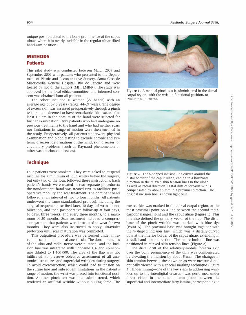
excess skin was marked in the dorsal carpal region, at the most proximal point on a line between the second meta-carpophalangeal joint and the caput ulnae (Figure 1). This line also defined the primary vector of the flap. The distal base of the pinch wrinkle was marked with blue dye (Point A). The proximal base was brought together with the S-shaped incision line, which was a distally-curved bow at the inferior border of the caput ulnae, extending in a radial and ulnar direction. The entire incision line was positioned in relaxed skin tension lines (Figure 2).
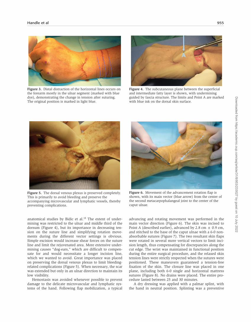
The distal drift of the relatively-mobile forearm skin over the bony prominence of the ulna was compensated by elevating the incision by about 5 mm. The changes in skin tension between these two areas were measured and optically viewed with a special marking technique (Figure 3). Undermining—one of the key steps to addressing wrin-kles up to the interdigital creases—was performed under direct vision in the subcutaneous plane between the superficial and intermediate fatty lamina, corresponding to
Figure 1. A manual pinch test is administered in the dorsal carpal region, with the wrist in functional position, to evaluate skin excess.
Figure 2. The S-shaped incision line curves around the distal border of the caput ulnae, ending in a horizontal direction in the relaxed skin tension lines in the ulnar as well as radial direction. Distal drift of forearm skin is compensated by about 5 mm in a proximal direction. The original incision line is shown light blue.
Dow
nloaded from https://academ
ic.oup.com/asj/article/31/8/953/233027 by guest on 10 July 2022
Handle et al 955
anatomical studies by Bidic et al.18 The extent of under-mining was restricted to the ulnar and middle third of the dorsum (Figure 4), but its importance in decreasing ten-sion on the suture line and simplifying rotation move-ments during the different vector settings is obvious. Simple excision would increase shear forces on the suture line and limit the rejuvenated area. More extensive under-mining causes “dog-ears,” which are difficult to compen-sate for and would necessitate a longer incision line, which we wanted to avoid. Great importance was placed on preserving the dorsal venous plexus to limit bleeding-related complications (Figure 5). When necessary, the scar was extended but only in an ulnar direction to maintain its low visibility.
Hemostasis was avoided whenever possible to prevent damage to the delicate microvascular and lymphatic sys-tems of the hand. Following flap mobilization, a typical
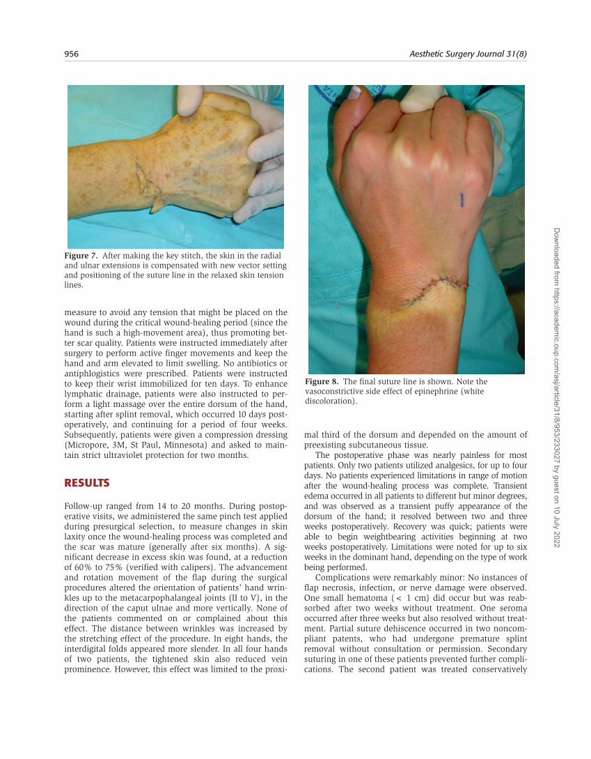
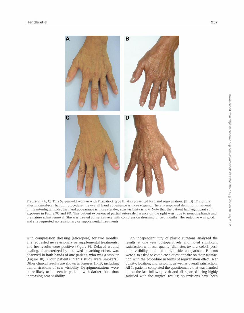
advancing and rotating movement was performed in the main vector direction (Figure 6). The skin was incised to Point A (described earlier), advanced by 2.8 cm ± 0.9 cm, and stitched to the base of the caput ulnae with a 6-0 non-absorbable sutures (Figure 7). The two resultant skin flaps were rotated in several more vertical vectors to limit inci-sion length, thus compensating for discrepancies along the cut edge. The wrist was maintained in functional position during the entire surgical procedure, and the relaxed skin tension lines were strictly respected when the sutures were positioned. These maneuvers guaranteed a tension-free fixation of the skin. The closure line was placed in one plane, including both 6-0 single and horizontal mattress sutures (Figure 8). No drains were placed. The entire pro-cedure lasted between 25 and 30 minutes.
A dry dressing was applied with a palmar splint, with the hand in neutral position. Splinting was a preventive
Figure 3. Distal distraction of the horizontal lines occurs on the forearm mostly in the ulnar segment (marked with blue dye), demonstrating the change in tension after suturing. The original position is marked in light blue.
Figure 4. The subcutaneous plane between the superficial and intermediate fatty layer is shown, with undermining guided by fascia structure. The limits and Point A are marked with blue ink on the dorsal skin surface.
Figure 5. The dorsal venous plexus is preserved completely. This is primarily to avoid bleeding and preserve the accompanying microvascular and lymphatic vessels, thereby preventing complications.
Figure 6. Movement of the advancement rotation flap is shown, with its main vector (blue arrow) from the center of the second metacarpophalangeal joint to the center of the caput ulnae.
Dow
nloaded from https://academ
ic.oup.com/asj/article/31/8/953/233027 by guest on 10 July 2022
956 Aesthetic Surgery Journal 31(8)
measure to avoid any tension that might be placed on the wound during the critical wound-healing period (since the hand is such a high-movement area), thus promoting bet-ter scar quality. Patients were instructed immediately after surgery to perform active finger movements and keep the hand and arm elevated to limit swelling. No antibiotics or antiphlogistics were prescribed. Patients were instructed to keep their wrist immobilized for ten days. To enhance lymphatic drainage, patients were also instructed to per-form a light massage over the entire dorsum of the hand, starting after splint removal, which occurred 10 days post-operatively, and continuing for a period of four weeks. Subsequently, patients were given a compression dressing (Micropore, 3M, St Paul, Minnesota) and asked to main-tain strict ultraviolet protection for two months.
Results
Follow-up ranged from 14 to 20 months. During postop-erative visits, we administered the same pinch test applied during presurgical selection, to measure changes in skin laxity once the wound-healing process was completed and the scar was mature (generally after six months). A sig-nificant decrease in excess skin was found, at a reduction of 60% to 75% (verified with calipers). The advancement and rotation movement of the flap during the surgical procedures altered the orientation of patients’ hand wrin-kles up to the metacarpophalangeal joints (II to V), in the direction of the caput ulnae and more vertically. None of the patients commented on or complained about this effect. The distance between wrinkles was increased by the stretching effect of the procedure. In eight hands, the interdigital folds appeared more slender. In all four hands of two patients, the tightened skin also reduced vein prominence. However, this effect was limited to the proxi-
mal third of the dorsum and depended on the amount of preexisting subcutaneous tissue.
The postoperative phase was nearly painless for most patients. Only two patients utilized analgesics, for up to four days. No patients experienced limitations in range of motion after the wound-healing process was complete. Transient edema occurred in all patients to different but minor degrees, and was observed as a transient puffy appearance of the dorsum of the hand; it resolved between two and three weeks postoperatively. Recovery was quick; patients were able to begin weightbearing activities beginning at two weeks postoperatively. Limitations were noted for up to six weeks in the dominant hand, depending on the type of work being performed.
Complications were remarkably minor: No instances of flap necrosis, infection, or nerve damage were observed. One small hematoma (< 1 cm) did occur but was reab-sorbed after two weeks without treatment. One seroma occurred after three weeks but also resolved without treat-ment. Partial suture dehiscence occurred in two noncom-pliant patents, who had undergone premature splint removal without consultation or permission. Secondary suturing in one of these patients prevented further compli-cations. The second patient was treated conservatively
Figure 8. The final suture line is shown. Note the vasoconstrictive side effect of epinephrine (white discoloration).
Figure 7. After making the key stitch, the skin in the radial and ulnar extensions is compensated with new vector setting and positioning of the suture line in the relaxed skin tension lines.
Dow
nloaded from https://academ
ic.oup.com/asj/article/31/8/953/233027 by guest on 10 July 2022
Handle et al 957
with compression dressing (Micropore) for two months. She requested no revisionary or supplemental treatments, and her results were positive (Figure 9). Delayed wound healing, characterized by a slowed bleaching effect, was observed in both hands of one patient, who was a smoker (Figure 10). (Four patients in this study were smokers.) Other clinical results are shown in Figures 11-13, including demonstrations of scar visibility. Dyspigmentations were more likely to be seen in patients with darker skin, thus increasing scar visibility.
An independent jury of plastic surgeons analyzed the results at one year postoperatively and noted significant satisfaction with scar quality (diameter, texture, color), posi-tion, visibility, and left-to-right-side comparison. Patients were also asked to complete a questionnaire on their satisfac-tion with the procedure in terms of rejuvenation effect, scar quality, location, and visibility, as well as overall satisfaction. All 11 patients completed the questionnaire that was handed out at the last follow-up visit and all reported being highly satisfied with the surgical results; no revisions have been
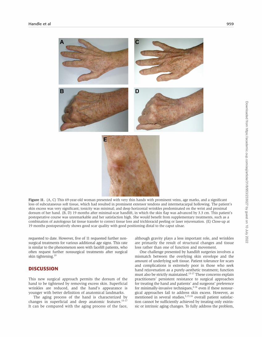
Figure 9. (A, C) This 53-year-old woman with Fitzpatrick type III skin presented for hand rejuvenation. (B, D) 17 months after minimal-scar handlift procedure, the overall hand appearance is more elegant. There is improved definition in several of the interdigital folds; the hand appearance is more slender; scar visibility is low. Note that the patient had significant sun exposure in Figure 9C and 9D. This patient experienced partial suture dehiscence on the right wrist due to noncompliance and premature splint removal. She was treated conservatively with compression dressing for two months. Her outcome was good, and she requested no revisionary or supplemental treatments.
Dow
nloaded from https://academ
ic.oup.com/asj/article/31/8/953/233027 by guest on 10 July 2022

958 Aesthetic Surgery Journal 31(8)
Figure 10. (A, C) This 62-year-old woman presented with skin excess, advanced decline of skin tonicity, preexisting age marks, prominent veins, and visible subcutaneous tissue loss. (B, D) 20 months after minimal-scar handlift, this patient’s results demonstrate scar quality and its near invisibility in the hand’s regular position. She experienced delayed wound healing, characterized by a slowed bleaching effect, likely because she was a smoker without consequences on the final results. The patient also demonstrates postoperative wrinkle reduction over the proximal two-thirds of the dorsum with tightening of the skin; the wrinkles in the forehand have also changed to a more vertical direction. The caput ulnae is more prominent and the anatomical structures are more visible. (E, F) Close-up demonstration of the patient’s left and right hands at 20 months postoperatively.
Dow
nloaded from https://academ
ic.oup.com/asj/article/31/8/953/233027 by guest on 10 July 2022
Handle et al 959
requested to date. However, five of 11 requested further non-surgical treatments for various additional age signs. This rate is similar to the phenomenon seen with facelift patients, who often request further nonsurgical treatments after surgical skin tightening.19
discussion
This new surgical approach permits the dorsum of the hand to be tightened by removing excess skin. Superficial wrinkles are reduced, and the hand’s appearance is younger with better definition of anatomical landmarks.
The aging process of the hand is characterized by changes in superficial and deep anatomic features.15-17 It can be compared with the aging process of the face,
although gravity plays a less important role, and wrinkles are primarily the result of structural changes and tissue loss rather than one of function and movement.
One challenge presented by handlift surgeries involves a mismatch between the overlying skin envelope and the amount of underlying soft tissue. Patient tolerance for scars and complications is extremely poor in those who seek hand rejuvenation as a purely-aesthetic treatment; function must also be strictly maintained.15-17 These concerns explain practitioners’ persistent resistance to surgical approaches for treating the hand and patients’ and surgeons’ preference for minimally-invasive techniques,3-14 even if these nonsur-gical approaches fail to address skin excess. However, as mentioned in several studies,3,15,16 overall patient satisfac-tion cannot be sufficiently achieved by treating only extrin-sic or intrinsic aging changes. To fully address the problem,
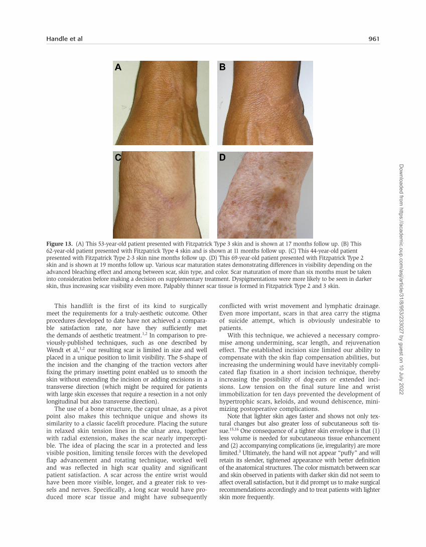
Figure 11. (A, C) This 69-year-old woman presented with very thin hands with prominent veins, age marks, and a significant loss of subcutaneous soft tissue, which had resulted in prominent extensor tendons and intermetacarpal hollowing. The patient’s skin excess was very significant; tonicity was minimal; and deep horizontal wrinkles predominated on the wrist and proximal dorsum of her hand. (B, D) 19 months after minimal-scar handlift, in which the skin flap was advanced by 3.3 cm. This patient’s postoperative course was unremarkable and her satisfaction high. She would benefit from supplementary treatments, such as a combination of autologous fat tissue transfer to correct tissue loss and trichloracid peeling or laser rejuvenation. (E) Close-up at 19 months postoperatively shows good scar quality with good positioning distal to the caput ulnae.
Dow
nloaded from https://academ
ic.oup.com/asj/article/31/8/953/233027 by guest on 10 July 2022

960 Aesthetic Surgery Journal 31(8)
patients seem willing to undergo surgical procedures for hand rejuvenation, as long as the scars have a low degree of visibility (similar to patient attitudes about facelifting procedures). Our development of this minimal-scar handlift for treating skin excess was based on this experience. The limited scar visibility and low complication rate opens the door for broader indications.
As in the face, the complexity of aging requires an inter-disciplinary approach. Different techniques are available for the treatment of volumetric tissue loss, dominant veins, or skin changes.3-14 The technique presented here
is clearly intended to be complemented by additional non surgical treatments. For longevity reasons, we favor autologous material (fat) transfer to restore soft tissue loss, in combination with trichloracid peeling or laser rejuvenation treatments to improve skin texture and reduce age marks.3,13 However, concurrent treatment is avoided, to avoid even temporarily compromising to the skin’s delicate circulation.3 Furthermore, to limit stress in the postoperative period, we recommend treating each hand in a separate procedure and treating the nondominant hand first (which, in the case of all our patients, was the left hand).
Figure 12. (A, C) This 44-year-old woman presented with advanced loss of skin tensile force comparable with what is seen in aged women as a result of massive weight loss. (B, D) 15 months after minimal-scar handlift, in which the skin flap was advanced by 3.4 cm. Even in this relatively-young patient, the rejuvenating effect was visible, and the patient was satisfied. As a positive side effect, tissue tightening compressed the veins and reduced their appearance. The postoperative appearance of the dorsum of the hand was tighter with fewer wrinkles.
Dow
nloaded from https://academ
ic.oup.com/asj/article/31/8/953/233027 by guest on 10 July 2022
Handle et al 961
This handlift is the first of its kind to surgically meet the requirements for a truly-aesthetic outcome. Other procedures developed to date have not achieved a compara-ble satisfaction rate, nor have they sufficiently met the demands of aesthetic treatment.1,2 In comparison to pre-viously-published techniques, such as one described by Wendt et al,1,2 our resulting scar is limited in size and well placed in a unique position to limit visibility. The S-shape of the incision and the changing of the traction vectors after fixing the primary insetting point enabled us to smooth the skin without extending the incision or adding excisions in a transverse direction (which might be required for patients with large skin excesses that require a resection in a not only longitudinal but also transverse direction).
The use of a bone structure, the caput ulnae, as a pivot point also makes this technique unique and shows its similarity to a classic facelift procedure. Placing the suture in relaxed skin tension lines in the ulnar area, together with radial extension, makes the scar nearly impercepti-ble. The idea of placing the scar in a protected and less visible position, limiting tensile forces with the developed flap advancement and rotating technique, worked well and was reflected in high scar quality and significant patient satisfaction. A scar across the entire wrist would have been more visible, longer, and a greater risk to ves-sels and nerves. Specifically, a long scar would have pro-duced more scar tissue and might have subsequently
conflicted with wrist movement and lymphatic drainage. Even more important, scars in that area carry the stigma of suicide attempt, which is obviously undesirable to patients.
With this technique, we achieved a necessary compro-mise among undermining, scar length, and rejuvenation effect. The established incision size limited our ability to compensate with the skin flap compensation abilities, but increasing the undermining would have inevitably compli-cated flap fixation in a short incision technique, thereby increasing the possibility of dog-ears or extended inci-sions. Low tension on the final suture line and wrist immobilization for ten days prevented the development of hypertrophic scars, keloids, and wound dehiscence, mini-mizing postoperative complications.
Note that lighter skin ages faster and shows not only tex-tural changes but also greater loss of subcutaneous soft tis-sue.15,16 One consequence of a tighter skin envelope is that (1) less volume is needed for subcutaneous tissue enhancement and (2) accompanying complications (ie, irregularity) are more limited.3 Ultimately, the hand will not appear “puffy” and will retain its slender, tightened appearance with better definition of the anatomical structures. The color mismatch between scar and skin observed in patients with darker skin did not seem to affect overall satisfaction, but it did prompt us to make surgical recommendations accordingly and to treat patients with lighter skin more frequently.
Figure 13. (A) This 53-year-old patient presented with Fitzpatrick Type 3 skin and is shown at 17 months follow up. (B) This 62-year-old patient presented with Fitzpatrick Type 4 skin and is shown at 11 months follow up. (C) This 44-year-old patient presented with Fitzpatrick Type 2-3 skin nine months follow up. (D) This 69-year-old patient presented with Fitzpatrick Type 2 skin and is shown at 19 months follow up. Various scar maturation states demonstrating differences in visibility depending on the advanced bleaching effect and among between scar, skin type, and color. Scar maturation of more than six months must be taken into consideration before making a decision on supplementary treatment. Dyspigmentations were more likely to be seen in darker skin, thus increasing scar visibility even more. Palpably thinner scar tissue is formed in Fitzpatrick Type 2 and 3 skin.
Dow
nloaded from https://academ
ic.oup.com/asj/article/31/8/953/233027 by guest on 10 July 2022
962 Aesthetic Surgery Journal 31(8)
We disagree with Coleman that tightening the skin increases vein and tendon visibility.3 Our results showed vein visibility to be stable and even improved in a few cases where the slight compressive effect reduced vein visibility. We do, of course, agree that this effect would be more impressive with a combined volume substitution. In fact, each of our patients would have benefited from addi-tional nonsurgical volume enhancement. Stated simply, our technique is one where the aging hand can be surgi-cally treated in a manner similar to that of the aging face, with a combination of treatments.
conclusions
Our minimal-scar handlift technique is a moderately-inva-sive, cost-effective procedure for rejuvenating the aging hand. We specifically recommend this surgical approach for Caucasian patients older than 50 years who present with significant skin excess and no nutritional tissue deficits. In patients with darker skin, this technique should be utilized only if the patient is informed of and accepts the likelihood of greater scar visibility. In patients with subcutaneous tis-sue loss, age marks, or dominant veins, accompanying treatments can (and should) follow the procedure.
Acknowledgment
Preliminary data from this report were presented in part at the annual meeting of the American Society for Aesthetic Plastic Surgery, Washington, DC, April 2010.
disclosures
The authors declared no potential conflicts of interest with respect to the research, authorship, and publication of this article.
Funding
The authors received no financial support for the research, authorship, and publication of this article.
ReFeRences 1. Wendt JR. Distal, dorsal superior extremity plasty. Plast
Reconstr Surg 2000;106:210. 2. Wendt JR. A great time for pioneers of superior extremity
aesthetic surgery. Plast Reconstr Surg 2004;113:2237.
3. Coleman SR. Hand rejuvenation with structural fat grafting. Plast Reconstr Surg 2002;110:1731-1744.
4. Man J, Rao J, Goldman M. A double-blind, comparative study of nonanimal-stabilized hyaluronic acid versus human collagen for tissue augmentation of the dorsal hands. Dermatol Surg 2008;34:1026-1031.
5. Goldman A, Prati C, Rossato F. Hand rejuvenation using intense pulsed light. J Cutan Med Surg 2008;12:107-113.
6. Edelson KL. Hand recontouring with calcium hydroxyl-apatite (Radiesse). J Cosmet Dermatol 2009;8:44-51.
7. Sadick NS, Anderson D, Werschler WP. Addressing vol-ume loss in hand rejuvenation: a report of clinical experi-ence. J Cosmet Laser Ther 2008;10:237-241.
8. Shamma AR, Guy RJ. Laser ablation of unwanted hand veins. Plast Reconstr Surg 2007;120:2017-2024.
9. Alster TS, Konda S. Plasma skin resurfacing for regenera-tion of neck, chest, and hands: investigation of a novel device. Dermatol Surg 2007;33:1315-1321.
10. Aboudib Júnior JH, de Castro CC, Gradel J. Hand reju-venescence by fat filling. Ann Plast Surg 1992;28:559-564.
11. Abergel RP, David LM. Aging hands: A technique of hand rejuvenation by laser resurfacing and autologous fat transfer. J Dermatol Surg Oncol 1989;15:725.
12. Lee BJ. The role of sclerotherapy in abnormal varicose hand veins. Plast Reconstr Surg 2000;106:227.
13. Teimourian B, Adham MN. Rejuvenation of the hand: fat injection combined with TCA peel. Aesthet Surg J 2000;20:70.
14. Jimenez G, Spencer JM. Erbium:YAG laser resurfacing of the hands, arms, and neck. Dermatol Surg 1999;25:831.
15. Jakubietz RG, Kloss DF, Gruenert JG, Jakubietz MG. The ageing hand: a study to evaluate the chronological age-ing process of the hand. J Plast Reconstr Aesthet Surg 2008;61:681-686.
16. Jakubietz RG, Jakubietz MG, Kloss D, Gruenert JG. Defining the basic aesthetics of the hand. Aesthetic Plast Surg 2005;29:546-551.
17. Bains RD, Thorpe H, Southern S. Hand aging: patients’ opinions. Plast Reconstr Surg 2006;117:2212-2218.
18. Bidic SM, Hatef DA, Rohrich RJ. Dorsal hand anatomy relevant to volumetric rejuvenation. Plast Reconstr Surg 2010;126:163.
19. Carruthers JDA, Glogau RG, Blitzer A, Facial Aesthetics Consensus Group Faculty. Advances in facial rejuvena-tion: botulinum toxin type A, hyaluronic acid dermal fillers, and combination therapies—consensus recom-mendations. Plast Reconstr Surg 2008;121(5):5S-30S.
Dow
nloaded from https://academ
ic.oup.com/asj/article/31/8/953/233027 by guest on 10 July 2022