Hypertension Mediterranean-Style Diet Effect on the Structural Properties 2009
11
Manuel Conde, Ramon Estruch and Valentina Ruiz-Gutiérrez Francisca Barceló, Javier S. Perona, Jesús Prades, Sérgio S. Funari, Enrique Gomez-Gracia, Membrane of Hypertensive Patients : The Prevencion con Dieta Mediterranea Study Mediterranean-Style Diet Effect on the Structural Properties of the Erythrocyte Cell Print ISSN: 0194-911X. Online ISSN: 1524-4563 Copyright © 2009 American Heart Association, Inc. All rights reserved. is published by the American Heart Association, 7272 Greenville Avenue, Dallas, TX 75231 Hypertension doi: 10.1161/HYPERTENSIONAHA.109.137471 2009;54:1143-1150; originally published online October 5, 2009; Hypertension. http://hyper.ahajournals.org/content/54/5/1143 World Wide Web at: The online version of this article, along with updated information and services, is located on the http://hyper.ahajournals.org/content/suppl/2009/10/05/HYPERTENSIONAHA.109.137471.DC1.html Data Supplement (unedited) at: http://hyper.ahajournals.org//subscriptions/ is online at: Hypertension Information about subscribing to Subscriptions: http://www.lww.com/reprints Information about reprints can be found online at: Reprints: document. Permissions and Rights Question and Answer this process is available in the click Request Permissions in the middle column of the Web page under Services. Further information about Office. Once the online version of the published article for which permission is being requested is located, can be obtained via RightsLink, a service of the Copyright Clearance Center, not the Editorial Hypertension in Requests for permissions to reproduce figures, tables, or portions of articles originally published Permissions: by guest on May 2, 2013 http://hyper.ahajournals.org/ Downloaded from
Transcript of Hypertension Mediterranean-Style Diet Effect on the Structural Properties 2009
Manuel Conde, Ramon Estruch and Valentina Ruiz-GutiérrezFrancisca Barceló, Javier S. Perona, Jesús Prades, Sérgio S. Funari, Enrique Gomez-Gracia,Membrane of Hypertensive Patients : The Prevencion con Dieta Mediterranea StudyMediterranean-Style Diet Effect on the Structural Properties of the Erythrocyte Cell
Print ISSN: 0194-911X. Online ISSN: 1524-4563 Copyright © 2009 American Heart Association, Inc. All rights reserved.
is published by the American Heart Association, 7272 Greenville Avenue, Dallas, TX 75231Hypertension doi: 10.1161/HYPERTENSIONAHA.109.1374712009;54:1143-1150; originally published online October 5, 2009;Hypertension.
http://hyper.ahajournals.org/content/54/5/1143World Wide Web at:
The online version of this article, along with updated information and services, is located on the
http://hyper.ahajournals.org/content/suppl/2009/10/05/HYPERTENSIONAHA.109.137471.DC1.htmlData Supplement (unedited) at:
http://hyper.ahajournals.org//subscriptions/
is online at: Hypertension Information about subscribing to Subscriptions:
http://www.lww.com/reprints Information about reprints can be found online at: Reprints:
document. Permissions and Rights Question and Answer this process is available in the
click Request Permissions in the middle column of the Web page under Services. Further information aboutOffice. Once the online version of the published article for which permission is being requested is located,
can be obtained via RightsLink, a service of the Copyright Clearance Center, not the EditorialHypertensionin Requests for permissions to reproduce figures, tables, or portions of articles originally publishedPermissions:
by guest on May 2, 2013http://hyper.ahajournals.org/Downloaded from
Diet/Electrolytes
Mediterranean-Style Diet Effect on the Structural Propertiesof the Erythrocyte Cell Membrane of Hypertensive Patients
The Prevencion con Dieta Mediterranea Study
Francisca Barcelo, Javier S. Perona, Jesus Prades, Sergio S. Funari, Enrique Gomez-Gracia,Manuel Conde, Ramon Estruch, Valentina Ruiz-Gutierrez
Abstract—A currently ongoing randomized trial has revealed that the Mediterranean diet, rich in virgin olive oil or nuts,
reduces systolic blood pressure in high-risk cardiovascular patients. Here, we present a structural substudy to assess the
effect of a Mediterranean-style diet supplemented with nuts or virgin olive oil on erythrocyte membrane properties in
36 hypertensive participants after 1 year of intervention. Erythrocyte membrane lipid composition, structural properties
of reconstituted erythrocyte membranes, and serum concentrations of inflammatory markers are reported. After the
intervention, the membrane cholesterol content decreased, whereas that of phospholipids increased in all of the dietary
groups; the diminishing cholesterol:phospholipid ratio could be associated with an increase in the membrane fluidity.
Moreover, reconstituted membranes from the nuts and virgin olive oil groups showed a higher propensity to form a
nonlamellar inverted hexagonal phase structure that was related to an increase in phosphatidylethanolamine lipid class.
These data suggest that the Mediterranean-style diet affects the lipid metabolism that is altered in hypertensive patients,
influencing the structural membrane properties. The erythrocyte membrane modulation described provides insight in the
structural bases underlying the beneficial effect of a Mediterranean-style diet in hypertensive subjects. (Hypertension.
2009;54:1143-1150.)
Key Words: Mediterranean diet � lipids � membrane structure � cardiovascular disease � hypertension
Cardiovascular disease constitutes the main cause of death
in industrialized countries,1 and hypertension is one of
the main modifiable cardiovascular risk factors, especially in
the elderly.2 Healthy diet and lifestyle constitute the first steps
in the guidelines for management of hypertension.3 In this
context, the type and amount of dietary lipids influence the
lipid composition of cell membranes4,5 and modulate the
interactions with proteins involved in the regulation of blood
pressure.6 Thus, the changes in membrane properties induced
by dietary lipids may have important consequences on the
blood pressure regulation.
The Mediterranean-style diet (MD) is characterized by a
high consumption of virgin olive oil (VOO) and nuts, which
are rich natural sources of oleic (18:1; n-9) and �-linolenic
(18:3; n-3) acids, respectively. The Prevencion con Dieta
Mediterranea (PREDIMED) Study is a large-scale, random-
ized trial aimed at assessing the effects of a MD enriched with
VOO or nuts on primary prevention of cardiovascular disease
in patients at high risk for coronary heart disease. The results
of the 3-month intervention on the first 772 patients entering
the study showed that, compared with a low-fat diet, the MD
rich in VOO or nuts reduced systolic blood pressure and
serum total cholesterol and triglyceride concentrations and
increased serum high-density lipoprotein cholesterol concen-
tration.7 Although there is evidence indicating that dietary
lipids can have a positive effect on cardiovascular risk
factors, the mechanisms and effects on the molecular and
structural bases underlying the physiological process are
largely unknown.
Several studies support the involvement of plasma mem-
brane properties in the modulation of membrane protein
activities and cell physiology. The structural properties and
function of cell membranes appear to be modified in hyper-
tensive humans and animal models of hypertension.8–10
Changes in membrane lipid composition of hypertensive
subjects have been associated with alterations in the trans-
membrane fluxes of Na� and K�, including Na�-Li� coun-
tertransport, which is a marker of essential hypertension,10,11
Received June 9, 2009; first decision June 24, 2009; revision accepted September 2, 2009.From the Departamento de Biología Fundamental y Ciencias de la Salut (F.B., J.P.), University of the Balearic Islands, Palma de Mallorca, Spain;
Nutrition and Lipid Metabolism (J.S.P., V.R.-G.), Instituto de la Grasa, Consejo Superior de Investigaciones Científicas, Sevilla, Spain; HamburgerSynchrotronstrahlungslabor (S.S.F.), Hamburg, Germany; Nutricion y Salud Publica (E.G.-G.), Facultad de Medicina, Universidad de Malaga, Malaga,Spain; Departamento de Medicina Preventiva (M.C.), Hospitales Universitarios Virgen del Rocío, Sevilla, Spain; Departmento de Medicina Interna (R.E.),Hospital Clinic, Institut d’Investigacions Biomediques August Pi Sunyer, Barcelona, Spain; Centro de Investigación Biomédica en Red Fisiopatologia dela Obesidad y la Nutricion (R.E.), Instituto de Salud Carlos III, Madrid, Spain.
Correspondence to Francisca Barcelo, Departamento de Biología Fundamental y Ciencias de la Salut, University of the Balearic Islands, E-07122 Palmade Mallorca, Spain. E-mail [email protected]
© 2009 American Heart Association, Inc.
Hypertension is available at http://hyper.ahajournals.org DOI: 10.1161/HYPERTENSIONAHA.109.137471
1143 by guest on May 2, 2013http://hyper.ahajournals.org/Downloaded from
and in cell signaling proteins that participate in the control of
blood pressure.12 On the other hand, it has been reported that
dietary lipids have an effect on membrane lipid composition
and cell signaling proteins.4,5,12 Considering that changes in
the dietary lipid composition yield to variations in the
biophysical properties of the plasma membrane, it is likely
that cellular functional changes could result from alterations
in the structure of the lipid membrane properties influenced
by the diet. Thus, the changes in membrane properties
induced by dietary lipids may have important consequences
on blood pressure regulation.
Dietary habit could play a role as an environmental factor,
altering some targeted molecular functions in the cell and,
through them, influencing cardiovascular risk factors. In fact,
the MD has been associated with changes in membrane
structure and function. Consumption of olive oil–rich diets
increases the concentration of oleic acid in plasma membrane
lipids of different rat and human cells, with beneficial
consequences on membrane functionality.13–16 In contrast,
very little is currently known regarding the effects of nuts,
another key ingredient of the MD, on membrane lipid
composition and structure.
The present study was undertaken to examine the structural
basis underlying the effect of the MD on the cardiovascular
system, in parallel with the PREDIMED Study that is
currently in progress.7 With this aim, we conducted a struc-
tural substudy to assess the effect of the MD supplemented
with nuts or VOO on the erythrocyte membrane properties in
a group of participants recruited from the parent study after 1
year of intervention. This is the first time that membrane
structural analyses are included in an intervention study with
an MD.
Materials and Methods
MaterialsHEPES was obtained from Sigma Chemical Co. Lipid standards, choles-terol, 1,2-dipalmitoyl-sn-glycero-3-phosphatidylethanolamine, 1,2-diacyl-sn-glycero-3-phosphocholine, 1,2-diacyl-sn-glycero-3-phospho-L-serine,N-acyl-4-sphingenyl-1-O-phosphorylcholine, and lysophosphatidylcho-line were purchased from Sigma-Aldrich. Solvents used for lipidextraction and high-performance liquid chromatography–grade solventswere from Romil. The high-performance liquid chromatography col-umn was purchased from Merck.
Methods
Study DesignThe PREDIMED Study is a large, parallel-group, multicenter,randomized, controlled, 5-year trial,7 for which the aim is to assessthe effects of the MD on the primary prevention of cardiovasculardisease (http://www.predimed.org). Nearly 7500 high-risk partici-pants have been divided into 3 intervention groups, and each groupreceives a specific diet, one third an MD enriched with VOO, anotherthird an MD enriched with mixed nuts, and the remaining third alow-fat diet. The present study reports the first-year effects of thesedietary interventions on the structural membrane properties of theerythrocyte plasma membranes from 36 hypertensive subjects par-ticipating in the PREDIMED Study.
SubjectsThe first 36 hypertensive participants entering in the PREDIMEDStudy from 2 nodes (Sevilla and Malaga) were divided into 3 groupsand assigned to the following interventions: an MD enriched withVOO (MD�VOO group), an MD enriched with nuts (MD�nuts
group), or a low-fat diet (LF group). Each group consisted of 12
subjects to ensure adequate sample size to conduct the X-ray
diffraction study, as well as to obtain sufficient statistical signifi-
cance. Blood pressure was measured, and blood samples were
collected from all of the subjects before the dietary intervention
(baseline) and after 1 year of intervention with the corresponding
diet, as described in Estruch et al.7 All of the protocols used in this
study followed the principles of the Declaration of Helsinki and were
approved by the Institutional Committee of Human Research (Hos-
pital Universitario Virgen del Rocío, Sevilla, Spain). All of the
procedures followed were in accordance with institutional guide-
lines, and the subjects gave their informed consent to participate in
the study.
Dietary InterventionParticipants in the PREDIMED Study were given a written recom-
mendation for a traditional MD and 3-month allotments of free VOO
(1 L/wk) or mixed nuts (30 g/d, as 15.0 g of walnuts, 7.5 g of
hazelnuts, and 7.5 g of almonds). A 137-item food-validated fre-
quency questionnaire and a 14-item questionnaire, an extension of a
questionnaire designed to assess the degree of adherence to the
traditional MD, were used.7 Please see the online Data Supplement
(at http://hyper.ahajournals.org) for detailed information.
Serum Inflammatory MarkersFasting blood samples were obtained at baseline and after subjects
had received the dietary intervention for 1 year, kept at 4°C for
during transportation from the hospital to the laboratory (�1 hour)
and then stored at �80°C until required for biochemical analyses.
Measurements of high-sensitivity C-reactive protein (CRP), interleu-
kin (IL) 6, E-selectin, and P-selectin were taken. Serum concentra-
tions of high-sensitivity CRP were analyzed by particle-enhanced
immunonephelometry. Serum IL-6, E-selectin, and P-selectin were
measured in duplicate using standard ELISA.
Erythrocyte Model Membrane PreparationErythrocyte membranes were isolated as described previously.16
Briefly, blood samples were collected in heparinized tubes and
centrifuged at 1750g and 4°C for 10 minutes. The erythrocyte pellets
were washed twice with 110 mmol/L of MgCl2. Erythrocyte mem-
branes from the 12 participants of each group were mixed and used
to reconstitute model membranes with a lipid composition represen-
tative of the 3 (MD�VOO, MD�nuts, and LF) groups of patients.
Total lipids of each erythrocyte membrane group were extracted with
chloroform:methanol (2:1, vol/vol), as described previously.17 Mul-
tilamellar lipid vesicles, 15% (wt/wt) with total lipid extracts, were
prepared in 10 mmol/L of HEPES, 100 mmol/L of NaCl, and
1 mmol/L of EDTA (pH 7.4; HEPES buffer).12 Lipid mixtures were
hydrated, thoroughly homogenized with a pestle-type minihomoge-
nizer (Sigma), and vortexed until a homogeneous mixture was
obtained. Then, the suspensions were submitted to 5 temperature
cycles (heated up to 70°C and cooled down to 4°C). Samples for
X-ray scattering experiments were stored at �80°C under argon and
allowed to equilibrate at 4°C for 48 hours before measurements were
taken.
Lipid Composition AnalysesLipid and phospholipid classes were separated by high-performance
liquid chromatography in a single chromatogram following a mod-
ification of the method by Perona et al.18 Briefly, triplicates of the
lipid extracts were dissolved in chloroform:methanol (2:1, vol/vol),
passed through 0.2-�m filters, and subsequently analyzed by liquid
chromatography (2695 Alliance, Waters Co.) using a LiChrospher
column (250.0�4.6 mm, 5-�m particle size) and an evaporative
light-scattering detector (Waters 2420, Waters Co). A ternary gradi-
ent of hexane, 2-propanol, and methanol was applied with a flow rate
of 0.8 mL/min. Commercially purchased lipid standards were used to
identify and quantify the lipid classes. The amounts of cholesterol
and phospholipids were quantified using calibration curves from
lipid standards. The quantification was based on regression analyses
of curves with correlation coefficients �0.999.
1144 Hypertension November 2009
by guest on May 2, 2013http://hyper.ahajournals.org/Downloaded from
Fatty acid methyl esters were analyzed by gas chromatography
using a Hewlett-Packard 5890 series II gas chromatograph equipped
with a flame ionization detector (Hewlett-Packard Co) and a Supel-
cowax 10 capillary silica column (60 m and 0.25 mm ID; Sulpelco
Co). Fatty acid methyl esters were identified by comparison of their
retention times against those of standards and quantified by internal
standardization (tricosanoic methyl ester, 23:0) using peak area
integration.19
X-Ray Diffraction StudiesSmall- and wide-angle synchrotron radiation X-ray scattering anal-
yses were conducted using standard procedures on the Soft Con-
densed Matter beamline A2 of Hamburger Synchrotronstrahlungsla-
bor at the Deutsches Elektronen Synchrotron. The data collection
conditions were as described previously.12 Samples were heated
from 10°C to 70°C. To work in quasiequilibrium conditions, the
systems were allowed to equilibrate for 15 minutes at each temper-
ature before measurements. Then, they were kept at the highest
temperature for 15 minutes and finally cooled down to the lowest
temperature at the same scan rate. Positions of the observed peaks
were converted into distances, d, after calibration with the standards
rat tendon tail and poly-(ethylene terephthalate) for the small- and
wide-angle synchrotron radiation X-ray scattering analysis regions,
respectively. Interplanar distances, dhkl, were calculated according to
the following equation:
(1) s�l/dhkl��2 sin ��/�,
where s is the scattering vector, 2� is the scattering angle, � (0.150
nm) is the X-ray wavelength, and h, k, and l are the Miller indices of
the scattering planes.
Statistical AnalysisData are reported as the meanSD unless otherwise stated. Vari-
ables were examined for normality and skewness (Kolmogorov and
Levene tests). We transformed values with a skewed distribution
(CRP, IL-6, E-selectin, and P-selectin) to their natural logarithm for
analyses. Differences within and between groups were analyzed
using the 1-factor ANOVA analysis and the paired t test, when
indicated. Values were considered significantly different when
P�0.05. Analyses were performed using SPSS software version 14.0
(SPSS Inc).
Results
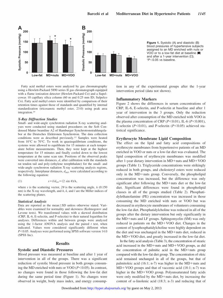
Systolic and Diastolic PressuresBlood pressure was measured at baseline and after 1 year of
intervention in all of the groups. There was a significant
reduction of systolic blood pressure in both groups consum-
ing the MD enriched with nuts or VOO (P�0.05). In contrast,
no changes were found in those following the low-fat diet
during the same period time (Figure 1). No changes were
observed in weight, body mass index, and energy consump-
tion in any of the experimental groups after the 1-year
intervention period (data not shown).
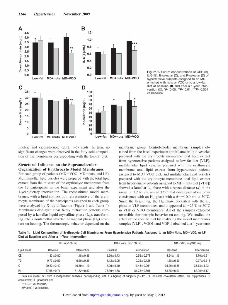
Inflammatory MarkersFigure 2 shows the differences in serum concentrations of
CRP, IL-6, E-selectin, and P-selectin at baseline and after 1
year of intervention in the 3 groups. Only the reduction
observed after consumption of the MD enriched with VOO in
the plasma concentration of CRP (P�0.01), IL-6 (P�0.001),
E-selectin (P�0.01), and P-selectin (P�0.05) achieved sta-
tistical significance.
Erythrocyte Membrane Lipid CompositionThe effect on the lipid and fatty acid compositions of
erythrocyte membranes from hypertensive patients of an MD
enriched in VOO or nuts or a low-fat diet was evaluated. The
lipid composition of erythrocyte membranes was modified
after 1-year dietary intervention in MD�nuts and MD�VOO
groups (Table 1). Triglyceride concentrations were strikingly
reduced in both groups, and cholesteryl esters were reduced
only in the MD�nuts group. Conversely, the phospholipid
concentration was increased, but the difference was only
significant after following the MD�nuts diet or the low-fat
diet. Significant differences were found in phospholipid
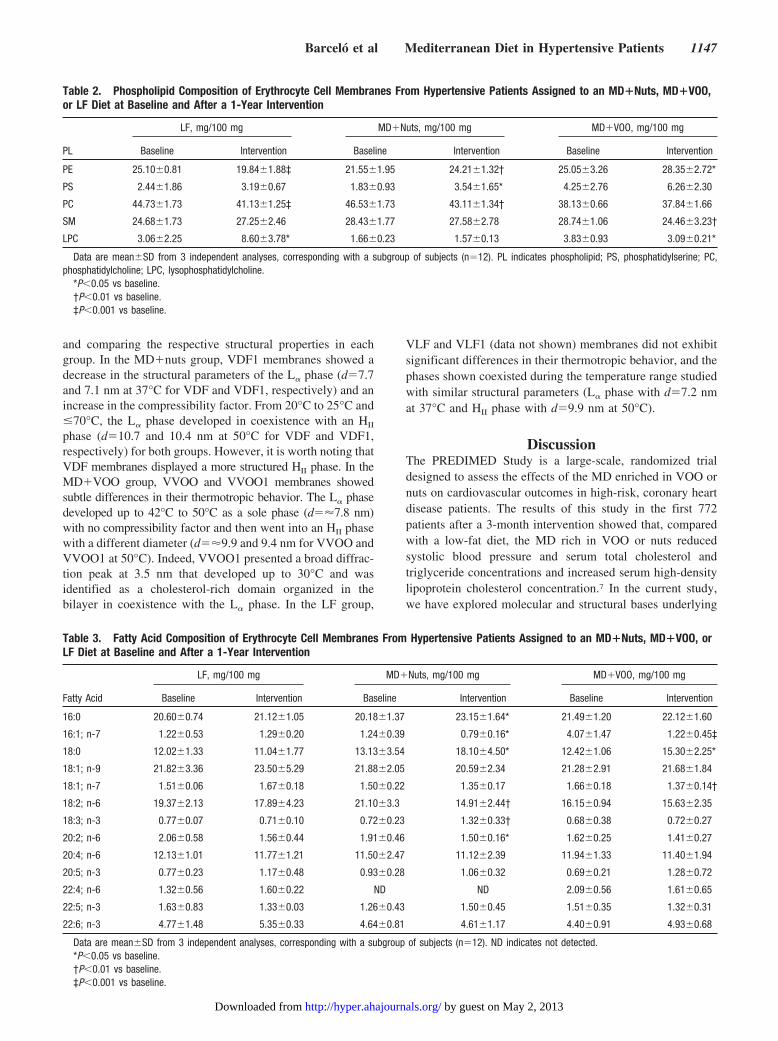
classes in all of the groups studied (Table 2). Phosphati-
dylethanolamine (PE) concentration was increased in those
consuming the MD enriched with nuts or VOO but was
decreased in erythrocyte membranes of volunteers consuming
the low-fat diet. Phosphatidylcholine was reduced in all of the
groups after the dietary intervention but only significantly in
the MD�nuts and LF groups. Sphingomyelin (SM) was only
reduced in patients on the MD�VOO diet. Changes in the
content of lysophosphatidylcholine were highly dependent on
the diet and was unchanged in the MD�nuts diet, reduced in
the MD�VOO diet, and greatly increased in the low-fat diet.
In the fatty acid analysis (Table 3), the concentration of stearic
acid increased in the MD�nuts and MD�VOO groups, as did
the concentration of palmitic acid in the MD�nuts group
compared with the low-fat diet group. The concentration of oleic
acid remained unchanged in all of the groups, but that of
palmitoleic acid (16:1; n-7) was higher in the MD�nuts and
MD�VOO groups and that of vaccenic acid (18:1; n-7) was
higher in the MD�VOO group. Polyunsaturated fatty acids
were only modified by the MD�nuts diet, by increasing the
content of �-linolenic acid (18:3; n-3) and reducing that of
A B
**
Systo
lic P
ressu
re (
mm
Hg
)
Dia
sto
lic P
ressu
re (
mm
Hg
)
Low-fat MD+nuts MD+VOO
200
150
100
50
0
200
150
100
50
0Low-fat MD+nuts MD+VOO
Figure 1. Systolic (A) and diastolic (B)blood pressures of hypertensive subjectsassigned to an MD enriched with nuts orVOO or to a low-fat diet at baseline (f)and after a 1-year intervention (u).*P�0.05 vs baseline.
Barcelo et al Mediterranean Diet in Hypertensive Patients 1145
by guest on May 2, 2013http://hyper.ahajournals.org/Downloaded from
linoleic and eicosadienoic (20:2; n-6) acids. In turn, no
significant changes were observed in the fatty acid composi-
tion of the membranes corresponding with the low-fat diet.
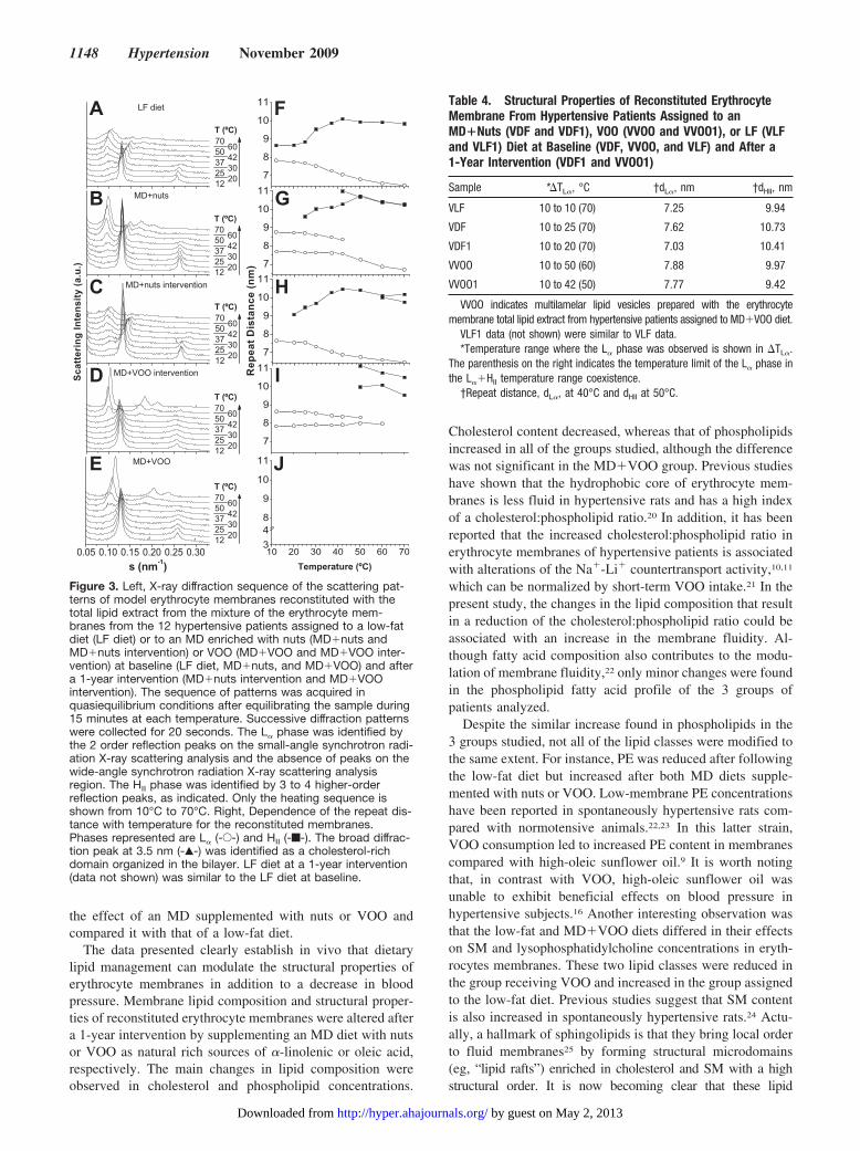
Structural Influence on the SupramolecularOrganization of Erythrocyte Model MembranesFor each group of patients (MD�VOO, MD�nuts, and LF),
Multilamellar lipid vesicles were prepared with the total lipid
extract from the mixture of the erythrocyte membranes from
the 12 participants in the basal experiment and after the
1-year dietary intervention. The reconstituted model mem-
branes, with a lipid composition representative of the eryth-
rocyte membrane of the participants assigned to each group,
were analyzed by X-ray diffraction (Figure 3 and Table 4).
Membranes displayed clear X-ray diffraction patterns com-
posed by a lamellar liquid crystalline phase (L�), transform-
ing into a nonlamellar inverted hexagonal phase (HII) struc-
ture on heating. The thermotropic behavior depended on the
membrane group. Control-model membrane samples ob-
tained from the basal experiment (multilamelar lipid vesicles
prepared with the erythrocyte membrane total lipid extract
from hypertensive patients assigned to low-fat diet [VLF],
multilamelar lipid vesicles prepared with the erythrocyte
membrane total lipid extract from hypertensive patients
assigned to MD�VOO diet, and multilamelar lipid vesicles
prepared with the erythrocyte membrane total lipid extract
from hypertensive patients assigned to MD�nuts diet [VDF])
showed a lamellar L� phase with a repeat distance (d) in the
range of 7.2 to 7.8 nm at 37°C that developed alone or in
coexistence with an HII phase with a d�10.0 nm at 50°C.
Since the beginning, the HII phase coexisted with the L�
phase in VLF membranes, and it appeared at 25°C or 50°C
in VDF or VOO membranes. All of the samples exhibited
reversible thermotropic behavior on cooling. We studied the
effect of the specific diet by analyzing the model membranes
samples (VLF1, VOO1, and VDF1) obtained at a 1-year term
Table 1. Lipid Composition of Erythrocyte Cell Membranes From Hypertensive Patients Assigned to an MD�Nuts, MD�VOO, or LF
Diet at Baseline and After a 1-Year Intervention
Lipid Class
LF, mg/100 mg MD�Nuts, mg/100 mg MD�VOO, mg/100 mg
Baseline Intervention Baseline Intervention Baseline Intervention
CE 1.330.80 1.180.36 3.500.73 0.550.57† 4.541.15 3.700.51
TG 0.770.52 0.600.29 1.120.05 0.250.12† 1.880.50 0.970.21†
C 20.222.40 16.391.10* 18.990.10 17.480.09* 35.205.38 33.134.92
PL 77.680.71 81.820.57* 76.391.88 81.730.59† 58.386.93 62.205.17
Data are meanSD from 3 independent analyses, corresponding with a subgroup of subjects (n�12). CE indicates cholesterol esters; TG, triglycerides; C,
cholesterol; PL, phospholipids.
*P�0.01 vs baseline.
†P�0.001 vs baseline.
Low-fat MD+nuts MD+VOO
C-r
eacti
ve p
rote
in (
mg
/L)
E-s
ele
cti
ne (
mg
/L)
IL-6
(p
g/m
L)
P-s
ele
cti
ne (
mg
/L)
Low-fat MD+nuts MD+VOO
Low-fat MD+nuts MD+VOO Low-fat MD+nuts MD+VOO
4.5
4.0
3.5
3.0
2.5
2.0
1.5
1.0
0.5
0.0
1.2
1.0
0.8
0.6
0.4
0.2
0.0
100
90
80
70
60
50
40
30
20
10
0
*****
*
35
30
25
20
15
10
5
0
**
A B
C D
Figure 2. Serum concentrations of CRP (A),IL-6 (B), E-selectin (C), and P-selectin (D) ofhypertensive subjects assigned to an MDenriched with nuts or VOO or to a low-fatdiet at baseline (f) and after a 1-year inter-vention (u). *P�0.05; **P�0.01; ***P�0.001vs baseline.
1146 Hypertension November 2009
by guest on May 2, 2013http://hyper.ahajournals.org/Downloaded from
and comparing the respective structural properties in each
group. In the MD�nuts group, VDF1 membranes showed a
decrease in the structural parameters of the L� phase (d�7.7
and 7.1 nm at 37°C for VDF and VDF1, respectively) and an
increase in the compressibility factor. From 20°C to 25°C and
�70°C, the L� phase developed in coexistence with an HII
phase (d�10.7 and 10.4 nm at 50°C for VDF and VDF1,
respectively) for both groups. However, it is worth noting that
VDF membranes displayed a more structured HII phase. In the
MD�VOO group, VVOO and VVOO1 membranes showed
subtle differences in their thermotropic behavior. The L� phase
developed up to 42°C to 50°C as a sole phase (d�7.8 nm)
with no compressibility factor and then went into an HII phase
with a different diameter (d�9.9 and 9.4 nm for VVOO and
VVOO1 at 50°C). Indeed, VVOO1 presented a broad diffrac-
tion peak at 3.5 nm that developed up to 30°C and was
identified as a cholesterol-rich domain organized in the
bilayer in coexistence with the L� phase. In the LF group,
VLF and VLF1 (data not shown) membranes did not exhibit
significant differences in their thermotropic behavior, and the
phases shown coexisted during the temperature range studied
with similar structural parameters (L� phase with d�7.2 nm
at 37°C and HII phase with d�9.9 nm at 50°C).
DiscussionThe PREDIMED Study is a large-scale, randomized trial
designed to assess the effects of the MD enriched in VOO or
nuts on cardiovascular outcomes in high-risk, coronary heart
disease patients. The results of this study in the first 772
patients after a 3-month intervention showed that, compared
with a low-fat diet, the MD rich in VOO or nuts reduced
systolic blood pressure and serum total cholesterol and
triglyceride concentrations and increased serum high-density
lipoprotein cholesterol concentration.7 In the current study,
we have explored molecular and structural bases underlying
Table 2. Phospholipid Composition of Erythrocyte Cell Membranes From Hypertensive Patients Assigned to an MD�Nuts, MD�VOO,
or LF Diet at Baseline and After a 1-Year Intervention
PL
LF, mg/100 mg MD�Nuts, mg/100 mg MD�VOO, mg/100 mg
Baseline Intervention Baseline Intervention Baseline Intervention
PE 25.100.81 19.841.88‡ 21.551.95 24.211.32† 25.053.26 28.352.72*
PS 2.441.86 3.190.67 1.830.93 3.541.65* 4.252.76 6.262.30
PC 44.731.73 41.131.25‡ 46.531.73 43.111.34† 38.130.66 37.841.66
SM 24.681.73 27.252.46 28.431.77 27.582.78 28.741.06 24.463.23†
LPC 3.062.25 8.603.78* 1.660.23 1.570.13 3.830.93 3.090.21*
Data are meanSD from 3 independent analyses, corresponding with a subgroup of subjects (n�12). PL indicates phospholipid; PS, phosphatidylserine; PC,
phosphatidylcholine; LPC, lysophosphatidylcholine.
*P�0.05 vs baseline.
†P�0.01 vs baseline.
‡P�0.001 vs baseline.
Table 3. Fatty Acid Composition of Erythrocyte Cell Membranes From Hypertensive Patients Assigned to an MD�Nuts, MD�VOO, or
LF Diet at Baseline and After a 1-Year Intervention
Fatty Acid
LF, mg/100 mg MD�Nuts, mg/100 mg MD�VOO, mg/100 mg
Baseline Intervention Baseline Intervention Baseline Intervention
16:0 20.600.74 21.121.05 20.181.37 23.151.64* 21.491.20 22.121.60
16:1; n-7 1.220.53 1.290.20 1.240.39 0.790.16* 4.071.47 1.220.45‡
18:0 12.021.33 11.041.77 13.133.54 18.104.50* 12.421.06 15.302.25*
18:1; n-9 21.823.36 23.505.29 21.882.05 20.592.34 21.282.91 21.681.84
18:1; n-7 1.510.06 1.670.18 1.500.22 1.350.17 1.660.18 1.370.14†
18:2; n-6 19.372.13 17.894.23 21.103.3 14.912.44† 16.150.94 15.632.35
18:3; n-3 0.770.07 0.710.10 0.720.23 1.320.33† 0.680.38 0.720.27
20:2; n-6 2.060.58 1.560.44 1.910.46 1.500.16* 1.620.25 1.410.27
20:4; n-6 12.131.01 11.771.21 11.502.47 11.122.39 11.941.33 11.401.94
20:5; n-3 0.770.23 1.170.48 0.930.28 1.060.32 0.690.21 1.280.72
22:4; n-6 1.320.56 1.600.22 ND ND 2.090.56 1.610.65
22:5; n-3 1.630.83 1.330.03 1.260.43 1.500.45 1.510.35 1.320.31
22:6; n-3 4.771.48 5.350.33 4.640.81 4.611.17 4.400.91 4.930.68
Data are meanSD from 3 independent analyses, corresponding with a subgroup of subjects (n�12). ND indicates not detected.
*P�0.05 vs baseline.
†P�0.01 vs baseline.
‡P�0.001 vs baseline.
Barcelo et al Mediterranean Diet in Hypertensive Patients 1147
by guest on May 2, 2013http://hyper.ahajournals.org/Downloaded from
the effect of an MD supplemented with nuts or VOO and
compared it with that of a low-fat diet.
The data presented clearly establish in vivo that dietary
lipid management can modulate the structural properties of
erythrocyte membranes in addition to a decrease in blood
pressure. Membrane lipid composition and structural proper-
ties of reconstituted erythrocyte membranes were altered after
a 1-year intervention by supplementing an MD diet with nuts
or VOO as natural rich sources of �-linolenic or oleic acid,
respectively. The main changes in lipid composition were
observed in cholesterol and phospholipid concentrations.
Cholesterol content decreased, whereas that of phospholipids
increased in all of the groups studied, although the difference
was not significant in the MD�VOO group. Previous studies
have shown that the hydrophobic core of erythrocyte mem-
branes is less fluid in hypertensive rats and has a high index
of a cholesterol:phospholipid ratio.20 In addition, it has been
reported that the increased cholesterol:phospholipid ratio in
erythrocyte membranes of hypertensive patients is associated
with alterations of the Na�-Li� countertransport activity,10,11
which can be normalized by short-term VOO intake.21 In the
present study, the changes in the lipid composition that result
in a reduction of the cholesterol:phospholipid ratio could be
associated with an increase in the membrane fluidity. Al-
though fatty acid composition also contributes to the modu-
lation of membrane fluidity,22 only minor changes were found
in the phospholipid fatty acid profile of the 3 groups of
patients analyzed.
Despite the similar increase found in phospholipids in the
3 groups studied, not all of the lipid classes were modified to
the same extent. For instance, PE was reduced after following
the low-fat diet but increased after both MD diets supple-
mented with nuts or VOO. Low-membrane PE concentrations
have been reported in spontaneously hypertensive rats com-
pared with normotensive animals.22,23 In this latter strain,
VOO consumption led to increased PE content in membranes
compared with high-oleic sunflower oil.9 It is worth noting
that, in contrast with VOO, high-oleic sunflower oil was
unable to exhibit beneficial effects on blood pressure in
hypertensive subjects.16 Another interesting observation was
that the low-fat and MD�VOO diets differed in their effects
on SM and lysophosphatidylcholine concentrations in eryth-
rocytes membranes. These two lipid classes were reduced in
the group receiving VOO and increased in the group assigned
to the low-fat diet. Previous studies suggest that SM content
is also increased in spontaneously hypertensive rats.24 Actu-
ally, a hallmark of sphingolipids is that they bring local order
to fluid membranes25 by forming structural microdomains
(eg, “lipid rafts”) enriched in cholesterol and SM with a high
structural order. It is now becoming clear that these lipid
7
8
9
10
11
10 20 30 40 50 60 703
4
8
9
10
11
7
8
9
10
11
7
8
9
10
11
7
8
9
10
11
Re
pe
at
Dis
tan
ce
(n
m)
60
42
30
20
MD+VOO
MD+VOO intervention
MD+nuts intervention
MD+nuts
0.05 0.10 0.15 0.20 0.25 0.30
Sc
att
eri
ng
In
ten
sit
y (
a.u
.)
LF diet
70
50
37
25
12
60
42
30
20
70
50
37
25
12
60
42
30
20
70
50
37
25
12
60
42
30
20
70
50
37
25
12
60
42
30
20
70
50
37
25
12
A F
B G
C H
D I
E J
Figure 3. Left, X-ray diffraction sequence of the scattering pat-terns of model erythrocyte membranes reconstituted with thetotal lipid extract from the mixture of the erythrocyte mem-branes from the 12 hypertensive patients assigned to a low-fatdiet (LF diet) or to an MD enriched with nuts (MD�nuts andMD�nuts intervention) or VOO (MD�VOO and MD�VOO inter-vention) at baseline (LF diet, MD�nuts, and MD�VOO) and aftera 1-year intervention (MD�nuts intervention and MD�VOOintervention). The sequence of patterns was acquired inquasiequilibrium conditions after equilibrating the sample during15 minutes at each temperature. Successive diffraction patternswere collected for 20 seconds. The L� phase was identified bythe 2 order reflection peaks on the small-angle synchrotron radi-ation X-ray scattering analysis and the absence of peaks on thewide-angle synchrotron radiation X-ray scattering analysisregion. The HII phase was identified by 3 to 4 higher-orderreflection peaks, as indicated. Only the heating sequence isshown from 10°C to 70°C. Right, Dependence of the repeat dis-tance with temperature for the reconstituted membranes.Phases represented are L� (-E-) and HII (-f-). The broad diffrac-tion peak at 3.5 nm (-Œ-) was identified as a cholesterol-richdomain organized in the bilayer. LF diet at a 1-year intervention(data not shown) was similar to the LF diet at baseline.
Table 4. Structural Properties of Reconstituted Erythrocyte
Membrane From Hypertensive Patients Assigned to an
MD�Nuts (VDF and VDF1), VOO (VVOO and VVOO1), or LF (VLF
and VLF1) Diet at Baseline (VDF, VVOO, and VLF) and After a
1-Year Intervention (VDF1 and VVOO1)
Sample *�TL�, °C †dL�, nm †dHII, nm
VLF 10 to 10 (70) 7.25 9.94
VDF 10 to 25 (70) 7.62 10.73
VDF1 10 to 20 (70) 7.03 10.41
VVOO 10 to 50 (60) 7.88 9.97
VVOO1 10 to 42 (50) 7.77 9.42
VVOO indicates multilamelar lipid vesicles prepared with the erythrocyte
membrane total lipid extract from hypertensive patients assigned to MD�VOO diet.
VLF1 data (not shown) were similar to VLF data.
*Temperature range where the L� phase was observed is shown in �TL�.
The parenthesis on the right indicates the temperature limit of the L� phase in
the L��HII temperature range coexistence.
†Repeat distance, dL�, at 40°C and dHII at 50°C.
1148 Hypertension November 2009
by guest on May 2, 2013http://hyper.ahajournals.org/Downloaded from
microdomains play a role in the cell signaling.26 Some proteins
(eg, G proteins) that participate in cell signal transduction and
are involved in the physiological process of the control of blood
pressure have been associated with lipid rafts.27 Interestingly, a
growing body of data indicates that multiple signal transduction
events in the heart occur via plasma membrane receptors located
in signaling microdomains.28
Changes in the lipid composition because of the diet style
were associated with subtle differences in the structural
properties of the reconstituted membranes from erythrocytes.
Reconstituted membranes from the MD�nuts and
MD�VOO groups after the 1-year intervention showed a
higher propensity to form nonlamellar HII structures that
correlated with an increase in PE lipid class observed in their
respective lipid composition. In model membranes, an exper-
imental correlation between an HII-phase propensity and an
increase in G-protein localization or protein kinase C activity
has been shown,29,30 demonstrating the influence of the
membrane structure on cell signaling proteins that participate
in the control of blood pressure. Thus, membrane structural
changes induced by the MD diet style may have a cellular
functional implication.
On the other hand, when serum inflammatory markers
were analyzed, a reduction in CRP, IL-6, E-selectin, and
P-selectin concentrations was observed after both MD inter-
ventions, although only the differences observed in the
MD�VOO group achieved statistical significance. Leuko-
cytes and thrombocytes have been causally related to athero-
genesis and vascular thrombosis occlusion. However, more
recently, an increased appreciation has been noticed for
erythrocyte as a cell involved in atherosclerotic plaque
destabilization.31 Recently, Tziakas et al32 have shown that
IL-8 is increased in the membrane of circulating erythrocyte
in patients with acute coronary syndrome. The results of our
study also show a possible link between changes in erythro-
cyte membrane properties and serum inflammatory markers
after an MD intervention, especially when this diet is supple-
mented with VOO.
PerspectivesCardiovascular disease has a multifactorial etiology. Genetic
and environmental factors apparently form the basis for
structural membrane properties and function. Considering the
in vivo approach of this study, the dietary fat management
constitutes an external factor able to reduce the blood
pressure and serum inflammatory markers and modulate the
structural erythrocyte membrane properties. Adjustment in
the lipid composition and structural properties of erythrocyte
membranes attributed to MD diets supplemented with nuts or
VOO is most probably related to changes in the physico-
chemical properties of the lipid microenvironment of mem-
brane proteins. The complexity of biological membranes
makes it difficult to assign specific changes in membrane
structure to membrane-dependent functions (eg, the function
of membrane proteins that participate in cell signaling). The
alterations in the structural blood cell properties reported
could reflect changes in other cell types related to the control
of blood pressure and could account for the statistically
significant reductions in blood pressure observed in those
groups of participants in the PREDIMED Study.
AcknowledgmentsWe thank Fundacion Patrimonio Comunal Olivarero and HojiblancaSA, the California Walnut Commission, Borges SA, and MorellaNuts SA, who generously donated the VOO, walnuts, almonds, andhazelnuts, respectively, used in the study. We are also grateful to DrRosana Cabello-Moruno and Antonio Martin Rodriguez for theircontribution and excellent technical support.
Sources of FundingThis work was funded by SOS-Cuetara SA and supported by grantsAGL2008-02285/ALI (Comisión Interministerial de Ciencia y Tec-nología), RD06/0045/0002 (Fondo Investigacion Sanitaria), andII-05-051EC from Deutsches Elektronen Synchrotron; HamburgerSynchrotronstrahlungslabor; and by the IHP-Contract HPRI-CT-2001-00140 of the European Commission.
Centro de Investigación Biomédica en Red Fisiopatologia de laObesidad y la Nutricion is an initiative of the Instituto de SaludCarlos III, Ministerio de Ciencia e Innovacion (Spain).
DisclosuresNone.
References1. Whelton PK. Epidemiology of hypertension. Lancet. 1994;344:101–106.
2. Jousilahti P, Toumilehto J, Vartiainen E, Korhonen HJ, Pitkaniemi J,
Nissinen A, Puska P. Importance of risk factor clustering in coronary
heart disease mortality and incidence in eastern Finland. J Cardiovasc
Risk. 1995;2:63–70.
3. Mancia G, De Backer G, Dominiczak A, Cifkova R, Fagard R, Germano
G, Grassi G, Heagerty AM, Kjeldsen SE, Laurent S, Narkiewicz K,
Ruilope L, Rynkiewicz A, Schmieder RE, Boudier HA, Zanchetti A; for
the ESH-ESC Task Force on the Management of Arterial Hypertension.
Guidelines for the management of arterial hypertension: the Task Force
for the Management of Arterial Hypertension of the European Society of
Hypertension (ESH) and of the European Society of Cardiology (ESC).
J Hypertens. 2007;25:1105–1187.
4. Vicario IM, Malkova D, Lund EK, Jonson IT. Olive oil supplementation
in healthy adults: effects in cell membrane fatty acid composition and
platelet function. Ann Nutr Metab. 1998;42:160–169.
5. Perona JS, Canizares J, Montero E, Sanchez-Dominguez JM, Ruiz-
Gutierrez V. Plasma lipid modifications in elderly people after admin-
istration of two virgin oils of the same variety (Olea europea var.
hojiblanca) with different triacylglycerol composition. Br J Nutr.
2003;89:819 – 826.
6. Teres S, Barcelo-Coblijn G, Benet M, Alvarez R, Bressani R, Halver JE,
Escriba PV. Oleic acid content is responsible for the reduction in blood
pressure induced by olive oil. Proc Natl Acad Sci U S A. 2008;105:
13811–13816.
7. Estruch R, Martinez-Gonzalez MA, Corella D, Salas-Salvado J, Ruiz-Gutierrez
V, Covas MI, Fiol M, Gomez-Gracia E, Lopez-Sabater MC, Vinyoles E, Aros
F, Conde M, Lahoz C, Lapetra J, Saez G, Ros E. Effects of a
Mediterranean-style diet on cardiovascular risk factors: a randomized trial.
Ann Intern Med. 2006;145:1–11.
8. Zicha J, Kunes J, Devynck MA. Abnormalities of membrane function and
lipid metabolism in hypertension: a review. Am J Hypertens. 1999;12:
315–331.
9. Vazquez CM, Mate A, Angeles de la Hermosa M, Planas JM, Ruiz-
Gutierrez V. Abnormalities in lipid composition of brush-border mem-
branes isolated from renal cortex of spontaneously hypertensive rats.
Am J Hypertens. 2001;14:578–584.
10. Carr P, Taub NA, Watts GF, Poston L. Human lymphocyte sodium-
hydrogen exchange: the influences of lipids, membrane fluidity, and
insulin. Hypertension. 1993;21:344–352.
11. Villar J, Montilla C, Muniz-Grijalvo O, Muriana FG, Stiefel P, Ruiz-
Gutierrez V, Carneado J. Erythrocyte Na(�)-Li� countertransport in
essential hypertension: correlation with membrane lipids levels.
J Hypertens. 1996;14:969–973.
12. Prades J, Alemany R, Perona JS, Funari SS, Vogler O, Ruiz-Gutierrez V,
Escriba PV, Barcelo F. Effects of 2-hydroxyoleic acid on the structural
Barcelo et al Mediterranean Diet in Hypertensive Patients 1149
by guest on May 2, 2013http://hyper.ahajournals.org/Downloaded from
properties of biological and model plasma membranes. Mol Membr Biol.
2008;25:46–57.
13. Pagnan A, Corrocher R, Ambrosio GB, Ferrari S, Guarini P, Piccolo D,
Opportuno A, Bassi A, Olivieri O, Baggio G. Effects of an olive-oil-rich
diet on erythrocyte membrane lipid composition and cation transport
systems. Clin Sci (Lond). 1989;76:87–93.
14. Vazquez CM, Zanetti R, Santa-María C, Ruíz-Gutierrez V. Effects of two
highly monounsaturated oils on lipid composition and enzyme activities
in rat jejunum. Biosci Rep. 2000;20:355–368.
15. Perona JS, Vogler O, Sanchez-Domínguez JM, Montero E, Escriba PV,
Ruiz-Gutierrez V. Consumption of virgin olive oil influences membrane
lipid composition and regulates intracellular signaling in elderly adults
with type 2 diabetes mellitus. J Gerontol A Biol Sci Med Sci. 2007;62:
256–263.
16. Ruiz-Gutierrez V, Muriana FJ, Guerrero A, Cert AM, Villar J. Plasma
lipids, erythrocyte membrane lipids and blood pressure of hypertensive
women after ingestion of dietary oleic acid from two different sources.
J Hypertens. 1996;14:1483–1490.
17. Folch J, Lees M, Sloane Stanley GH. A simple method for the isolation
and purification of total lipides from animal tissues. J Biol Chem. 1957;
226:497–509.
18. Perona JS, Ruiz-Gutierrez V. Quantification of major lipid classes in human
triacylglycerol-rich lipoproteins by high-performance liquid chromatography
with evaporative light-scattering detection. J Sep Sci. 2004;27:653–659.
19. Ruiz-Gutierrez V, Montero E, Villar J. Determination of fatty acid and
triacylglycerol composition of human adipose tissue. J Chromatogr.
1992;581:171–178.
20. Kunes J, Zicha J, Devynck MA. Erythrocyte membrane microviscosity
and blood pressure in rats with salt-induced and spontaneous hyper-
tension. J Hypertens. 1994;12:229–234.
21. Muriana FJ, Villar J, Ruíz-Gutierrez V. Intake of olive oil can modulate
the transbilayer movement of human erythrocyte membrane cholesterol.
Cell Mol Life Sci. 1997;53:496–500.
22. Vazquez CM, Zanetti R, Ruiz-Gutierrez V. Lipid composition and
fluidity in the jejunal brush-border membrane of spontaneously hyper-
tensive rats: effects on activities of membrane-bound proteins. Biosci
Rep. 1996;16:217–226.
23. Okamoto H, Kawaguchi H, Minami M, Saito H, Yasuda H. Lipid alter-
ations in renal membrane of stoke-prone spontaneously hypertensive rats.
Hypertension. 1989;13:456–462.
24. Dorrance AM, Graham D, Webb RC, Fraser R, Dominiczak A. Increased
membrane sphingomyelin and arachidonic acid in stroke-prone sponta-
neously hypertensive rats. Am J Hypertens. 2001;14:1149–1153.
25. Ramstedt B, Slotte JP. Sphingolipids and the formation of sterol-enriched
ordered membrane domains. Biochim Biophys Acta. 2006;1758:
1945–1956.
26. Simons K, Toomre D. Lipid rafts and signal transduction. Nat Rev Mol
Cell Biol. 2000;1:31–39.
27. Chini B, Parenti M. G-protein coupled receptors in lipid rafts and
caveolae: how, when and why do they go there? J Mol Endocrinol.
2004;32:325–338.
28. Insel PA, Head BP, Ostrom RS, Patel HH, Swaney JS, Tang CM, Roth
DM. Caveolae and lipid rafts: G protein-coupled receptor signaling
microdomains in cardiac myocytes. Ann N Y Acad Sci. 2005;1047:
166–172.
29. Slater SJ, Kelly MB, Taddeo FJ, Ho C, Rubin E, Stubbs CD. The
modulation of protein kinase C activity by membrane lipid bilayer
structure. J Biol Chem. 1994;269:4866–4871.
30. Kinnunen PKJ. On the molecular-level mechanism of peripheral protein-
membrane interactions induced by lipids forming inverted nonlamellar
phases. Chem Phys Lipids. 1996;81:151–166.
31. Pasterkamp G, Daemen M. Circulating cells: the biofactory for markers
of atherosclerotic disease. Eur Heart J. 2008;29:2701–2702.
32. Tziakas D, Chalikias GK, Tentes IK, Stakos D, Chatzikyrirkou SV,
Mitrousi K, Kortsaris AX, Karki JC, Bouloudas H. Interleukin-8 is
increased in the membrane of circulating erythrocytes in patients with
acute coronary syndrome. Eur Heart J. 2008;29:2713–2722.
1150 Hypertension November 2009
by guest on May 2, 2013http://hyper.ahajournals.org/Downloaded from
Online Supplement for
MEDITERRANEAN-STYLE DIET EFFECT ON THE STRUCTURAL PROPERTIES OF ERYTHROCYTE CELL MEMBRANE OF HYPERTENSIVE PATIENTS: THE
PREDIMED STUDY
FRANCISCA BARCELÓ1, JAVIER S. PERONA2, JESÚS PRADES1, SÉRGIO S. FUNARI3,
ENRIQUE GOMEZ-GRACIA4, MANUEL CONDE5, RAMON ESTRUCH6 & VALENTINA RUIZ-GUTIÉRREZ2 1Departamento de Biología Fundamental y C.S., University of the Balearic Islands, E-07122 Palma de Mallorca, Spain. 2Nutrition and Lipid Metabolism. Instituto de la Grasa, Consejo Superior de Investigaciones Científicas (CSIC), E-41012, Sevilla, Spain. 3HASYLAB, Notkestrasse 85, D-22603 Hamburg. 4Nutrición y salud pública. Facultad de Medicina. Universidad de Málaga 5Departamento de Medicina Preventiva. Hospitales Universitarios Virgen del Rocío, Sevilla. 6Departmento de Medicina Interna, Hospital Clinic, Institut d’Investigacions Biomèdiques August Pi Sunyer (IDIBAPS), Barcelona; and CIBER Fisiopatologia de la Obesidad y la Nutrición (CIBEROBN, CB06/03), Instituto de Salud Carlos III, Spain. Corresponding author: Francisca Barceló, Departamento de Biología Fundamental y C.S., University of the Balearic Islands, E-07122 Palma de Mallorca, Spain. Tel.: +34971173149; Fax: +43971173184; E-mail: [email protected]
by guest on May 2, 2013http://hyper.ahajournals.org/Downloaded from
Expanded Materials and Methods Dietary Intervention The baseline examination included assessment of standard cardiovascular disease factors, medications and socio-demographic factors. A 137-item food validated frequency questionnaire and a 14-item questionnaire, an extension of a questionnaire designed to assess the degree of adherence to the traditional MD was used (1). On the basis of the baseline 14-item questionnaire each participant was given personalized dietary advice by a dietitian during a 30-minute session. Participants allocated to a low-fat diet were advised to reduce all types of fat and were given written recommendations according to the American Heart Association guidelines. Participants in the MD groups received instructions directed to upscale the 14-item score, including the use of VOO for cooking and dressing, increased consumption of vegetables, nuts, and fish products, consumption of white meat instead of red or processed meat, preparation of home-made sauce by simmering tomato, garlic, onion, and aromatic herbs with VOO to dress vegetables pasta, rice, and other dishes and for alcohol drinkers, to follow a moderate pattern of red wine consumption. No energy restrictions were suggested for any intervention group. Participants in the MD groups were given 3-month allotments of free VOO (1 L/week) or mixed nuts (30 g/day, as 15 g walnuts, 7.5 g hazelnuts and 7.5 g almonds). All participants had free access to their dietitian throughout the study. The fatty acid and minor components composition of the VOO and nuts employed in the study was published elsewhere (1). Biological assessment of the intervention compliance was performed by measuring tyrosol and hydroxytyrosol levels in urine by GC-MS to assess the compliance of the MD rich in VOO group and -linolenic (18:3, n-3) acid in serum by GC as a biomarker of compliance of the MD rich in nuts (1). References 1. Estruch R, Martinez-Gonzalez MA, Corella D, Salas-Salvado J, Ruiz-Gutierrez V, Covas MI, Fiol M, Gomez-Gracia E, Lopez-Sabater MC, Vinyoles E, Aros F, Conde M, Lahoz C, Lapetra J, Saez G, Ros E. Effects of a Mediterranean-style diet on cardiovascular risk factors: a randomized trial. Ann Intern Med. 2006;145:1-11.
by guest on May 2, 2013http://hyper.ahajournals.org/Downloaded from