Human extracellular vesicles and correlation with two clinical ...
Upload
independentCategory
view
2download
0
Materials Today � Volume 16, Number 10 �October 2013 RESEARCH
Hybrid polymer/lipid vesicles: state of theart and future perspectives R
ESEARCH:Review
J-F. Le Meins1,2,*, C. Schatz1,2, S. Lecomma
ndoux1,2 and O. Sandre1,21Univ. Bordeaux, LCPO, UMR 5629, F-33600 Pessac, France2CNRS, LCPO, UMR 5629, F-33600 Pessac, France
Hybrid vesicles resulting from the combined self-assembly of both amphiphilic copolymers and lipids
have attracted particular interest from chemists and (bio)physicists over the last five years. Such
assemblies may be viewed as an advanced vesicular structure compared to their liposome and
polymersome forerunners as the best characteristics from the two different systems can be integrated in a
new, single vesicle. To afford such a design, the different parameters controlling both self-assembly and
membrane structure must be tuned. This highlight aims to present a comprehensive overview of the
fundamental aspects related to these structures, and discuss emerging developments and future
applications in this field of research.
IntroductionLiposomes are the archetype of synthetic vesicular structures
obtained through a self-assembly mechanism, where amphiphilic
molecules such as lipids, e.g. phospholipids are simply transferred
into water. Liposomes have been extensively studied and used
since the 1970s as nano- and microreactors, cell membrane models
or drug delivery systems [1]. It was demonstrated in the late 1990s
that amphiphilic block copolymers were also able to self-assemble
into vesicular morphologies, and were therefore named polymer-
somes in reference to their lipid analogs [2,3]. This discovery had a
great scientific impact on the self-assembly research field and
polymersomes rapidly appeared as an interesting alternative to
liposomes which possess a poor modular chemical functionality
and in some cases (e.g. osmotic shocks) suffer from relatively weak
stability. The larger molar masses of polymer chains over lipid tails
and the versatility of chemical functions that can be integrated in
polymer structures make polymersomes attractive to significantly
improve and modulate membrane bulk properties (toughness,
permeability) and surface functionality of vesicles [4]. In particu-
lar, polymersomes can be used as model tools to better understand
biological events where the physical properties of membrane are of
prime importance (bending elasticity, spontaneous curvature. . .)
like the cell plasmic membrane adhesion, fusion or fission [5].
However, the cell biomimetic character of polymer vesicles is
*Corresponding author:. Le Meins, J-F. ([email protected])
1369-7021/06/$ - see front matter � 2013 Elsevier Ltd. All rights reserved. http://dx.doi.org/10.1016/j.mattod
rather limited compared to liposomes as block copolymers are
generally synthetically made, while phospholipids are mostly
natural components of the cell membrane. In addition, the high
mechanical stability and low permeability of polymersomes can be
viewed as both positive factors and as limiting features in some
applications where a controlled diffusion of species through the
membrane is required. To tackle this challenge, stimuli-responsive
polymersomes have been designed, where the membrane desta-
bilization and the concomitant release of molecules are obtained
in defined conditions [6–9]. However, this approach requires a
careful synthetic design of the block copolymer structure and is
often limited to usage in specific conditions. While different
approaches were proposed to modulate the membrane properties
of polymersomes [4], a very promising method has recently
emerged to overcome intrinsic limitations of both polymersomes
and liposomes. The proposed methodology consists of designing
mixed vesicles from both copolymers and lipids (Fig. 1). The main
expected benefit of such hybrid structures is the fine tuning of the
membrane physical properties. Besides, depending on how lipid
and polymer chains are distributed within the membrane, one can
expect an improved control of (bio)functionalization of the vesi-
cles’ surface, which is of interest in many applications such as drug
delivery at a specific target (e.g. a site of inflammation or tumor
environment). Beyond the challenging aspects of formulation, the
development of such hybrid vesicles allows addressing many
questions about the formation of a stable membrane:
.2013.09.002 397

RESEARCH Materials Today � Volume 16, Number 10 �October 2013
[(Figure_1)TD$FIG]
FIGURE 1
Overview of the different hybrid vesicular structures that can be obtained according to the molar composition (polymer/lipid molar ratio) andthermodynamic phase of the phospholipid. Sketches on the top lines illustrate the different cases obtained by study of the epi- or confocal fluorescence
microscopy images. All these hybrid vesicles were formulated with polydimethylsiloxane-graft-poly(ethylene oxide) and dipalmitoylphosphatidylcholine
(DPPC) in the gel state or 1-palmitoyl-2-oleoyl-sn-glycero-3-phosphocholine (POPC) in the fluid state at room temperature [10].
RESEARCH:Review
- H
39
ow intimately the molecular components need to be mixed,
and what is the influence of variations of polymer structure and
size (molecular weight)?
- I
s it possible to get control over membrane structure andcomposition, meaning a homogeneous distribution of both
components or the formation of heterogeneous domains?
- W
hat is the long-term stability of such hybrid vesicularassemblies?
Condition of formation of hybrid vesiclesA crucial parameter controlling the formation of stable hybrid
vesicles is the discrepancy of chemical composition and size of
hydrophobic segments between polymers and lipids. In addition,
the thermodynamic incompatibility, due to both entropic and
enthalpic differences between the hydrophobic blocks, may also
drive a phase separation and the subsequent formation of lipo-
somes and polymersomes separately. In order to compare these
fundamental expectations with real systems, we will first review
the hybrid vesicles reported so far in the literature. On the polymer
side, amphiphilic block copolymers studied to elaborate hybrid
vesicles were based on poly(dimethyl siloxane) (PDMS) [10,11],
poly(isobutylene) (PIB) [12,13] or poly(butadiene) (PBut) [14–17]
as hydrophobic blocks, and poly(ethylene oxide) (PEO) or
poly(2-methyloxazoline) (PMOXA) as hydrophilic blocks. All
these polymer blocks exhibit a low glass transition temperature
(Tg), allowing dynamic exchange of the chains and leading to the
equilibrium structure of the membrane when it forms. Concerning
the choice of lipids, most studies were performed with phospha-
tidylethanolamine [11,15] or phosphatidylcholine [10,12–14,
16,17] head-groups with either saturated or unsaturated tails,
8
which are both major constituents of biological membranes.
The copolymers and lipids used are summarized in Table 1. The
solubility parameters (d) of hydrocarbon moieties in phospholi-
pids and hydrophobic polymer blocks are relatively close, that is,
d = 9.1 cal1/2/cm3/2 for the fatty acid tail in lipids and d = 7.3 cal1/2/
cm3/2, 7.7 cal1/2/cm3/2 and 8.32 cal1/2/cm3/2 respectively for
PDMS, PIB and PBut blocks [18–20]. These relatively close values
suggest that the chemical compatibility between the components
is indeed a parameter of uppermost importance to enable the
formation of hybrid vesicles even though the lateral phase separa-
tion of components inside the membrane still can occur for other
reasons, as it will be commented in the following.
The effect of composition, namely the polymer/lipid molar ratio,
on the formation of hybrid vesicles was rarely investigated in a
systematic manner. Based on calorimetric measurements and fluor-
escence self-quenching analysis, Ruysschaert et al. suggested that
Egg-Phosphatidylethanolamine (EPE) lipids distribute homoge-
neously in hybrid vesicles obtained with PMOXA1.8k-b-PDMS5.4k-
b-PMOXA1.8k terpolymer whatever the molar composition [11]. In
another study with vesicle-forming diblock copolymers, it was
demonstrated that a minimum of 65 mol% of PBut46-b-PEO30 block
copolymer was required to form hybrid vesicles with 1-palmitoyl-2-
oleoyl-sn-glycero-3-phosphocholine (POPC) [16]. Considering sev-
eral other different results [11–13,15–17], it appears obvious that the
parameter governing the production of such hybrid polymer/lipid
vesicles are not trivial and that further investigations are needed to
get both a better understanding and a certain predictability.
Another difficulty arises from the fact that the molar composition
of lipidand polymer in the final hybrid vesicles can bedifferent from
the starting composition, as evidenced by fluorescence microscopy

Materials Today � Volume 16, Number 10 �October 2013 RESEARCH
TABLE 1
The different copolymers and lipids used up to now to formulate hybrid vesicles and short comments about the membrane structureobserved (homogeneous or heterogeneous distribution of the components, at the optical scale).
Authors Copolymers Lipids (state at room temperature) Membrane structure/domains
Fournier et al. [11] PMOXA1.8k-b-PDMS5.4k-b-PMOXA1.8k Mixture of egg-phosphatidylethanolamine/
egg-phosphatidylcholine (2/1 mol/mol) (fluid)
Homogeneous
(Mn ¼ 9000 g=mol) Dipalmitoylphosphatidylcholine (DPPC) (gel, L0b)
Tsourkas
et al. [14,15]PBut46-b-PEO30 (Mn ¼ 3800g=mol) 1-Palmitoyl-2-oleoyl-sn-glycero phosphocholine
(POPC) (fluid)
Homogeneous
PBut22-b-PEO13 (Mn ¼ 1800g=mol) Hydrogenated soy phosphatidylcholine (HSPC) (gel)
1,2-Distearoyl-sn-glycero phosphoethanolamine-
N-poly(ethylene glycol) (DSPE-PEG) (fluid)
Le Meins, Sandreet al. [10]
PEG12-g-PDMS22-g-PEG12 POPC (fluid), DPPC (gel, L0b) Homogeneous or heterogeneousdepending on polymer/lipid ratio
and lipid state (Fig. 1)Mv ¼ 3000 g=mol
Vanderlick, Beales
et al. [16,17]
A: PBut46-b-PEO30
(Mn ¼ 3800 g=mol)
POPC (fluid), DPPC (gel, L0b), cholesterol A � 70%: homogeneous
1,2-Di-lauryoyl-sn-glycerophosphocholine
(DLPC) (fluid)
70 � A � 30: no vesicles obtained
2-Ditridecanoyl-sn-glycerophosphocholine
(DC13PC) (fluid)
A � 30% majority of liposomes
Micrometric domains obtained
via further crosslinking
Bacia, Binderet al. [12,13,21]
A: PIB87-b-PEO17 DPPC (gel, L0b) A � 28%: heterogeneous
(Mn ¼ 5350 g=mol) A > 28% homogeneous
B: PIB37-b-PEO48 B < 40% vesicles with ‘‘holes’’
(Mn ¼ 3970 g=mol)
Pispas et al. [22] PEO113-b-PCL12 DPPC (gel, L0b) No information available
Mw ¼ 7500 g=mol
RESEARCH:Review
experiments on giant vesicles [10,13]. Such an issue highlights the
uppermost importance of controlling the formulation process and
the need to find specific protocols.
Structure of the vesicle membraneThe control of the membrane structure is a major feature brought
about by hybrid vesicles. A large variety of levels of structure,
ranging from a homogeneous distribution of both components to
polymer- or lipid-rich micro-domains were obtained. A complete
review on phase separation in model lipid bilayers whose reading
can be helpful to get a general view of the parameters governing
the formation of domains, has been published in 2003 by Binder
and colleagues [23]. In the case of hybrid polymer/lipid vesicles,
relevant parameters seem to be the physical state of lipids, which
depends on their main chain transition temperature (from gel
state at T < Tm to fluid, liquid-crystalline state at T > Tm), and the
composition of the polymer/lipid mixture. Hybrid vesicles can be
compared to some extent to mixed liposomes made of lipids
having different melting temperatures Tm [10,12,17]. For instance,
the mixing of DPPC (Tm = 41 8C) with POPC (Tm = �2 8C) at room
temperature gives rise to the formation of lipid domains within the
membrane as a result of demixing of the two components. In case
of polymer/lipid hybrid vesicles, similar results were observed
provided that the initial amount of lipid was high enough. For
example, the formation of lipid domains in a copolymer-rich
membrane was described with lipids of high Tm such as hydro-
genated soy phosphatidylcholine (HSPC) [15] or DPPC (1,2-dipal-
mitoyl-sn-glycero-3-phosphocholine) [10] for a lipid content
higher than 20 mol%. Conversely, the formation of membranes
presenting a homogeneous distribution of components, at least at
the micrometric scale seemed to be favored when the lipid was
used above its main chain transition temperature [11,12,16,17]. In
addition to the gel or fluid state of the lipid used, the size mismatch
between polymer and lipid chains also plays an important role on
the membrane structure. The usual membrane (bilayer) thickness
is indeed 3–5 nm for liposomes and varies from 5 to 50 nm for
polymersomes (either bilayer for diblock or graft copolymers or
monolayer for triblock). In case of a large size gap, the formation of
a lipid domain would imply a high line tension at the lipid/
polymer boundaries arising from the thickness mismatch between
the domain and the surrounding polymer membrane with a
concomitant exposure of hydrophobic polymer segments to
water. To reduce this exposure and the resulting energetic cost
of the boundary edges, the two opposite plausible scenarios are (i)
conformational adaptation through elastic deformation of the
polymer chains at the boundary (see Fig. 2a) at an entropic cost
for the polymer chains (but globally reducing the line tension), or
(ii) coalescence into domains of larger area. It is worth noticing
that scenario (i) is analogous to elastic deformation of the mem-
brane at the domain boundary in phase coexisting lipid mem-
branes [24].
Figure 2a shows that conformational adaptation of the polymer
implies a collapse of the hydrophobic polymer chains near the
lipidic interface, opposing entropic elasticity of chains (reduction
of the total number of conformations) and the natural stretching
of polymer chains self-assembled as flat membrane [4]. Therefore it
399

RESEARCH Materials Today � Volume 16, Number 10 �October 2013
[(Figure_2)TD$FIG]
FIGURE 2
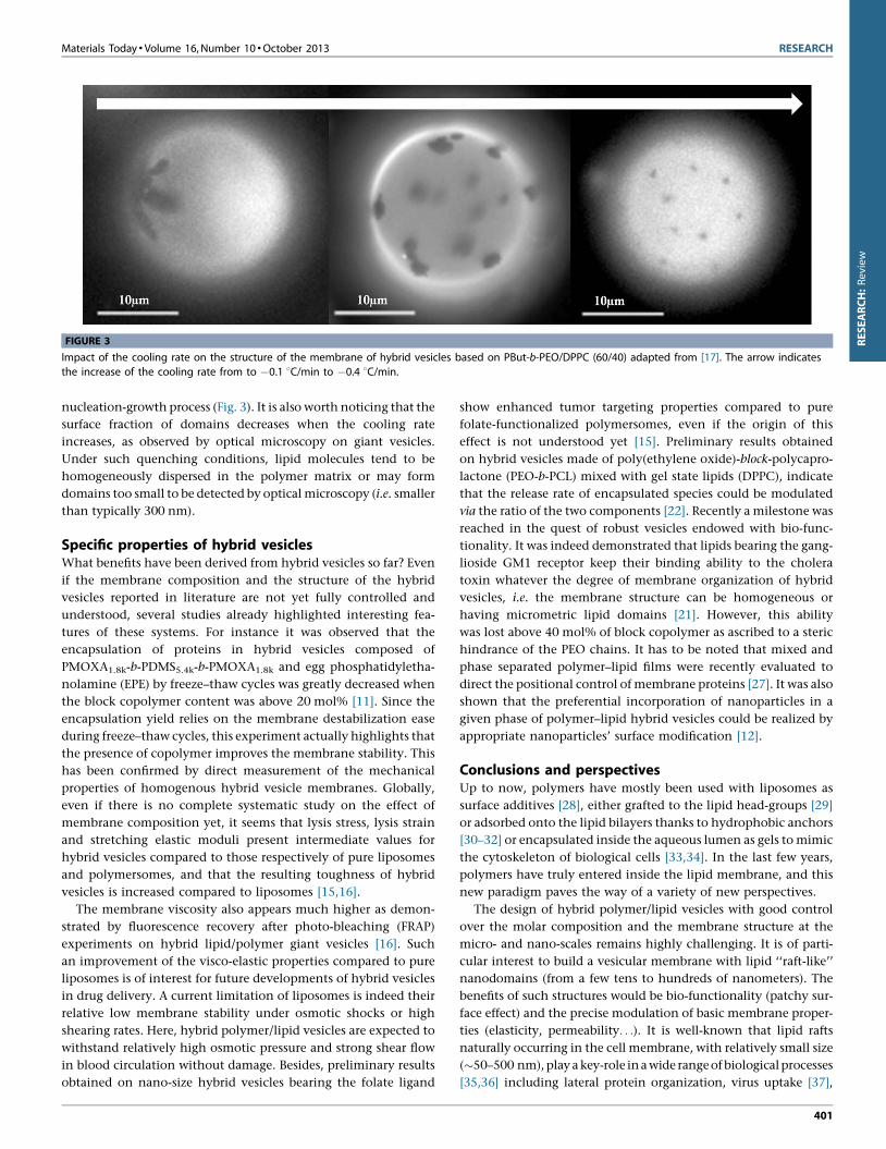
Impact of the cooling rate on the structure of the membrane of hybrid vesicles based on PBut-b-PEO/DPPC (60/40) adapted from [17]. The arrow indicates
the increases of the cooling rate from to �0.1 8C/min to �0.4 8C/min.
RESEARCH:Review
is clear that the molar mass (or chain length) and rigidity of the
hydrophobic polymer backbone plays a major role. If this adapta-
tion cannot be achieved, then the domain formation is unlikely
(spontaneously nucleated domains eventually collapse) and a
homogeneous mixture of the components is expected (Fig. 2b).
In a recent study [10], we evidenced that the use of PDMS-g-PEO
copolymers, leading to vesicles with a membrane thickness close
to that of liposomes (�5 nm) [25], helps the spontaneous forma-
tion of micrometric lipid domains in hybrid vesicles in a large
composition range (from 50% and up to 80% mol polymer) when
the lipid is in the gel state (DPPC). When associated with fluid
lipids (�50% mol of 1-palmitoyl-2-oleoyl-sn-glycero-3-phospho-
choline (POPC)), PDMS-g-PEO spontaneously form vesicles with
micrometric lipid domains that progressively evolve into sepa-
rated liposomes and polymersomes through a budding and fission
process [10]. This phenomenon of total demixing of a hybrid
vesicle into two daughter vesicles can be viewed as a ‘‘poor-man
model’’ of the cellular division.
Furthermore, in most of the studies reported so far, the copo-
lymers used form membranes thicker than 8 nm, and the sponta-
neous formation of domains (at least at the optical microscope
scale) was reported as a rare event occurring only for narrow
composition ranges. For instance in one of these studies [13],
giant unilamellar vesicles (GUV) presenting micrometric domains
were spontaneously obtained using DPPC and PIB87-b-PEO17, but
only in a very narrow composition range (20–28 mol% polymer).
The large hydrophobic block in that case limits the conforma-
tional adaptation at the polymer–lipid boundary. On the contrary,
the large hydrophobic thickness plays in favor of a single-phase
membrane for a large composition range (all polymer contents
larger than 30 mol%) as sketched in Fig. 2b. But a decrease of the
hydrophobic block length (to enable domains) must be accom-
panied by a concomitant reduction of the hydrophilic block
length; otherwise the hydrophilic-to-hydrophobic volume ratio
becomes too high to form stable (unperforated) membranes. This
is the most likely explanation for hole-defects observed in vesicles
400
formulated with high DPPC content (�60%) and block copolymer
with a shorter hydrophobic block and a longer hydrophilic one
(PIB37-b-PEO48), stabilizing bilayers edges with a local micellar
structure.
Among the different polymer/lipid hybrid systems, there is a
special interest in the formation of polymer-rich vesicles with stable
lipid-rich domains of controlled size, with regard to different kinds
of applications in drug delivery, bio-targeting or biophysical funda-
mental studies, such as the modeling of nanoparticles/membrane
interactions and cellular internalization (artificial endocytosis).
When lipids are used in the fluid state, only homogeneous hybrid
vesicles (at the micrometric scale) can be obtained. Additional
components are then needed to generate a stable phase-separated
membrane. For instance, micrometric domains were generated in
hybrid vesicles by reacting streptavidin (in solution) with biotiny-
lated head-groups of the lipids, the protein with multivalent bind-
ing sites ‘‘working as a zipper’’ to gather the lipid together in
monodomains. However, such a protein coating limits the further
biofunctionalization of the domains [16]. Cholesterol, which is
well-known to promote compaction of fluid phase lipids and the
lateral phase separation into ‘‘raft-like’’ domains within liposomes
for certain lipid compositions [26] induces the same effects when
added to lipid/polymer mixtures. Hence, hybrid vesicles with round
shaped micrometric domains were obtained with various lipids of
low Tm (DLPC, POPC, DC13PC) complemented with cholesterol, the
domain size varying with the exact polymer/lipid/cholesterol com-
position [17].
Moreover, it has been shown that working at temperature where
lipids are in gel state favors the formation of lipid domains in the
polymer vesicle membrane. Nam et al. proposed a method which
consists in forming hybrid vesicles from PBut-b-PEO and DPPC
above the lipid melting temperature and then cooling the system
below the melting point at a controlled cooling rate [17]. Such an
approach offers the possibility to control the number and the
size of lipid domains. When the cooling is fast, the domains
become smaller and with a large number, as expected for a typical
Materials Today � Volume 16, Number 10 �October 2013 RESEARCH
[(Figure_3)TD$FIG]
FIGURE 3
Impact of the cooling rate on the structure of the membrane of hybrid vesicles based on PBut-b-PEO/DPPC (60/40) adapted from [17]. The arrow indicates
the increase of the cooling rate from to �0.1 8C/min to �0.4 8C/min.
RESEARCH:Review
nucleation-growth process (Fig. 3). It is also worth noticing that the
surface fraction of domains decreases when the cooling rate
increases, as observed by optical microscopy on giant vesicles.
Under such quenching conditions, lipid molecules tend to be
homogeneously dispersed in the polymer matrix or may form
domains too small to be detected by optical microscopy (i.e. smaller
than typically 300 nm).
Specific properties of hybrid vesiclesWhat benefits have been derived from hybrid vesicles so far? Even
if the membrane composition and the structure of the hybrid
vesicles reported in literature are not yet fully controlled and
understood, several studies already highlighted interesting fea-
tures of these systems. For instance it was observed that the
encapsulation of proteins in hybrid vesicles composed of
PMOXA1.8k-b-PDMS5.4k-b-PMOXA1.8k and egg phosphatidyletha-
nolamine (EPE) by freeze–thaw cycles was greatly decreased when
the block copolymer content was above 20 mol% [11]. Since the
encapsulation yield relies on the membrane destabilization ease
during freeze–thaw cycles, this experiment actually highlights that
the presence of copolymer improves the membrane stability. This
has been confirmed by direct measurement of the mechanical
properties of homogenous hybrid vesicle membranes. Globally,
even if there is no complete systematic study on the effect of
membrane composition yet, it seems that lysis stress, lysis strain
and stretching elastic moduli present intermediate values for
hybrid vesicles compared to those respectively of pure liposomes
and polymersomes, and that the resulting toughness of hybrid
vesicles is increased compared to liposomes [15,16].
The membrane viscosity also appears much higher as demon-
strated by fluorescence recovery after photo-bleaching (FRAP)
experiments on hybrid lipid/polymer giant vesicles [16]. Such
an improvement of the visco-elastic properties compared to pure
liposomes is of interest for future developments of hybrid vesicles
in drug delivery. A current limitation of liposomes is indeed their
relative low membrane stability under osmotic shocks or high
shearing rates. Here, hybrid polymer/lipid vesicles are expected to
withstand relatively high osmotic pressure and strong shear flow
in blood circulation without damage. Besides, preliminary results
obtained on nano-size hybrid vesicles bearing the folate ligand
show enhanced tumor targeting properties compared to pure
folate-functionalized polymersomes, even if the origin of this
effect is not understood yet [15]. Preliminary results obtained
on hybrid vesicles made of poly(ethylene oxide)-block-polycapro-
lactone (PEO-b-PCL) mixed with gel state lipids (DPPC), indicate
that the release rate of encapsulated species could be modulated
via the ratio of the two components [22]. Recently a milestone was
reached in the quest of robust vesicles endowed with bio-func-
tionality. It was indeed demonstrated that lipids bearing the gang-
lioside GM1 receptor keep their binding ability to the cholera
toxin whatever the degree of membrane organization of hybrid
vesicles, i.e. the membrane structure can be homogeneous or
having micrometric lipid domains [21]. However, this ability
was lost above 40 mol% of block copolymer as ascribed to a steric
hindrance of the PEO chains. It has to be noted that mixed and
phase separated polymer–lipid films were recently evaluated to
direct the positional control of membrane proteins [27]. It was also
shown that the preferential incorporation of nanoparticles in a
given phase of polymer–lipid hybrid vesicles could be realized by
appropriate nanoparticles’ surface modification [12].
Conclusions and perspectivesUp to now, polymers have mostly been used with liposomes as
surface additives [28], either grafted to the lipid head-groups [29]
or adsorbed onto the lipid bilayers thanks to hydrophobic anchors
[30–32] or encapsulated inside the aqueous lumen as gels to mimic
the cytoskeleton of biological cells [33,34]. In the last few years,
polymers have truly entered inside the lipid membrane, and this
new paradigm paves the way of a variety of new perspectives.
The design of hybrid polymer/lipid vesicles with good control
over the molar composition and the membrane structure at the
micro- and nano-scales remains highly challenging. It is of parti-
cular interest to build a vesicular membrane with lipid ‘‘raft-like’’
nanodomains (from a few tens to hundreds of nanometers). The
benefits of such structures would be bio-functionality (patchy sur-
face effect) and the precise modulation of basic membrane proper-
ties (elasticity, permeability. . .). It is well-known that lipid rafts
naturally occurring in the cell membrane, with relatively small size
(�50–500 nm),play a key-role ina wide rangeof biologicalprocesses
[35,36] including lateral protein organization, virus uptake [37],
401

RESEARCH Materials Today � Volume 16, Number 10 �October 2013
RESEARCH:Review
signaling, trafficking [38,39] or membrane tension regulation [40].
The biophysical mechanism that maintains small lipid domains in
equilibrium and opposes their ripening expected from thermody-
namic consideration is not yet completely understood. Experimen-
tal and theoretical developments (until now conducted exclusively
on liposomes) have shown that a subtle balance between several
parameters governs the domain size. One of prime importance is the
energy cost by the domain to create a boundary with the surround
(line tension I in pico Newton), which arises from the thickness
mismatch between the domain and the surrounding membrane: I
opposes domain nucleation and favors domain coalescence (once
nucleationsize is reached) tominimize the boundary length [24,41].
This is balanced by other mechanisms such as entropic trap [42]
stabilizing nanodomains, the elastic interaction between dimpled
domains due todeformationof the surroundingmembrane [43], the
long range electrostatic dipolar interaction [44] and the natural
vesicle curvature [43,45,46]. All these aspects have been discussed in
recent reviews focusing on the stability of lipid domains [47], and
phase separation in biological membranes [48]. There are currently
only few experimental examples of lipidic nanodomains (i.e. below
the optical microscopy resolution) in model liposomes [49–53] and
their existence has not yet been proven on hybrid polymer/lipid
vesicles. It is believed that block copolymer molecules of low Tg play
a role inadjusting the line tensionIat the lipid–polymerboundaries
because of their flexibility and of their molar mass tunability during
the synthesis.
Finally, an emphasis should be given about the control of the
membrane structure through physico-chemical pathways such as
playing with entropic parameters (polymer/lipid adaptation at the
boundaries), interactions (chemical nature of the polymer blocks),
incorporation of additional reactants and environmental para-
meters (temperature, pH. . .), and lipid gel phase structures which
may play a role on the domain morphologies of polymer/lipid
vesicles, as they do for mixed lipid vesicles [54]. It is also important
to remember that the membrane curvature can greatly influence
the membrane structure. Therefore a series of experiments can give
various results depending on the size of the vesicles. In other
words, the results may differ significantly between studies on
vesicles of nanometric size, also called large unilamellar vesicles
(LUVs), and micrometric size giant vesicles (GUVs).
A better understanding of the hybrid thermodynamics and the
addition of specific functionalities (selectivepermeability, magnetic
properties by incorporating magnetic nanoparticles, membrane
disruption with stimuli-responsive polymer block or lipids, incor-
poration of channelproteins. . .) should lead to the creation of nano-
and micro-structures that are very useful in drug delivery (as pre-
liminary illustrated in [15,22]) and as templating agents [14], or for
the development of compartmentalized chemistry and biochemis-
try (bio-mimicry and synthetic biology) and as fundamental sys-
tems to understand complex biological behaviors in cells.
AcknowledgmentsThe authors gratefully acknowledge the P2M Program from the
ESF, the ANR for financial support, and Colin Bonduelle for
artwork support.
402
References
[1] L. John Wiley & Sons, Giant Vesicles: Perspectives in Molecular Chemistry, Wiley,
2000.
[2] B.M. Discher, et al. Science 284 (5417) (1999) 1143–1146.
[3] D.E. Discher, A. Eisenberg, Science 297 (5583) (2002) 967–973.
[4] J.F. Le Meins, O. Sandre, S. Lecommandoux, Eur. Phys. J. E. 34 (2) (2011) 14.
[5] X. Zhang, et al. J. Polym. Sci. A: Polym. Chem. 50 (12) (2012) 2293–2318.
[6] J.S. Lee, J. Feijen, J. Control. Release 161 (2) (2012) 473–483.
[7] H. De Oliveira, J. Thevenot, S. Lecommandoux, WIREs: Nanomed. Nanobiotech-
nol. 4 (5) (2012) 525–546.
[8] J. Du, R.K. O’Reilly, Soft Matter 5 (19) (2009) 3544–3561.
[9] M.-H. Li, P. Keller, Soft Matter 5 (5) (2009) 927–937.
[10] M. Chemin, et al. Soft Matter 8 (2012) 2867–2874.
[11] T. Ruysschaert, et al. J. Am. Chem. Soc. 127 (17) (2005) 6242–6247.
[12] A. Olubummo, et al. ACS Nano 6 (10) (2012) 8713–8727.
[13] M. Schulz, et al. Soft Matter 7 (18) (2011) 8100–8110.
[14] Z. Cheng, A. Tsourkas, Langmuir 24 (15) (2008) 8169–8173.
[15] Z. Cheng, et al. Bioconjugate Chem. 22 (10) (2011) 2021–2029.
[16] J. Nam, P.A. Beales, T.K. Vanderlick, Langmuir 27 (1) (2011) 1–6.
[17] J. Nam, T.K. Vanderlick, P.A. Beales, Soft Matter 8 (2012) 7982–7988.
[18] J.W. King, in: T.M. Kuo, H.W. Gardner (Eds.), Supercritical Fluid Technology
for Lipid Extraction, Fractionation and Reactions, Marcel Dekker, 2002 , pp.
663–687.
[19] M. Roth, J. Polym. Sci. B 28 (13) (1990) 2719.
[20] J. Brandrup, et al., Polymer Handbook, 4th edition, John Wiley & Sons, 2005.
[21] M. Schulz, et al. Angew. Chem. Int. Ed. Engl. 52 (6) (2013) 1829–1833.
[22] N. Pippa, et al. Soft Matter 9 (15) (2013) 4073–4082.
[23] W.H. Binder, V. Barragan, F.M. Menger, Angew. Chem. Int. Ed. 42 (47) (2003)
5802–5827.
[24] P.I. Kuzmin, et al. Biophys. J. 88 (2) (2005) 1120–1133.
[25] Z. Lin, et al. Langmuir 10 (4) (1994) 1008–1011.
[26] L. Bagatolli, P.B.S. Kumar, Soft Matter 5 (17) (2009) 3234–3248.
[27] J. Thoma, et al. Chem. Commun. 48 (70) (2012) 8811–8813.
[28] M. Schulz, A. Olubummo, W.H. Binder, Soft Matter 8 (18) (2012) 4849–4864.
[29] D. Needham, T.J. McIntosh, D.D. Lasic, Biochim. Biophys. Acta 1108 (1) (1992)
40–48.
[30] C. Ladaviere, et al. J. Colloid Interface Sci. 241 (1) (2001) 178–187.
[31] J.L. Popot, et al. in: D.C. Rees, K.A. Dill, J.R. Williamson (Eds.), Amphipols From A
to Z, 2011, 379–408.
[32] J.L. Popot, et al. Cell. Mol. Life Sci. 60 (8) (2003) 1559–1574.
[33] C. Campillo, B. Pepin-Donat, A. Viallat, Soft Matter 3 (11) (2007) 1421–1427.
[34] A. Viallat, J. Dalous, M. Abkarian, Biophys. J. 86 (4) (2004) 2179–2187.
[35] K. Simons, E. Ikonen, Nature 387 (6633) (1997) 569–572.
[36] P. Sens, M.S. Turner, Phys. Rev. E: Stat. Nonlin. Soft Matter Phys. 73 (3 Pt 1) (2006)
031918.
[37] N. Chazal, D. Gerlier, Microbiol. Mol. Biol. Rev. 67 (2) (2003) 226–237, table of
contents.
[38] G. van Meer, H. Sprong, Curr. Opin. Cell Biol. 16 (4) (2004) 373–378.
[39] J.B. Helms, C. Zurzolo, Traffic 5 (4) (2004) 247–254.
[40] D. Raucher, M.P. Sheetz, Biophys. J. 77 (4) (1999) 1992–2002.
[41] A.J. Garcia-Saez, S. Chiantia, P. Schwille, J. Biol. Chem. 282 (2007) 33537–
33544.
[42] V.A. Frolov, et al. Biophys. J. 91 (1) (2006) 189–205.
[43] T.S. Ursell, W.S. Klug, R. Phillips, Proc. Natl. Acad. Sci. U.S.A. 106 (32) (2009)
13301–13306.
[44] A. Travesset, J. Chem. Phys. 125 (8) (2006) 084905.
[45] S. Semrau, et al. Biophys. J. 96 (12) (2009) 4906–4915.
[46] J. Hu, T. Weikl, R. Lipowsky, Soft Matter 7 (13) (2011) 6092–6102.
[47] A.J. Garcia-Saez, P. Schwille, FEBS Lett. 584 (2010) 1653–1658.
[48] E.L. Elson, et al. in: D.C. Rees, K.A. Dill, J.R. Williamson (Eds.), Phase Separation
in Biological Membranes: Integration of Theory and Experiment, 2010, 207–
226.
[49] R.F. de Almeida, et al. J. Mol. Biol. 346 (4) (2005) 1109–1120.
[50] F.A. Heberle, et al. J. Am. Chem. Soc. 135 (18) (2013) 6853–6859.
[51] K. Suga, H. Umakoshi, Langmuir 29 (15) (2013) 4830–4838.
[52] A.C. Brown, K.B. Towles, S.P. Wrenn, Langmuir 23 (22) (2007) 11180–11187.
[53] L.M.S. Loura, F. Fernandes, M. Prieto, Eur. Biophys. J. Biophys. Lett. 39 (4) (2010)
589–607.
[54] V.D. Gordon, et al. J. Phys.: Condens. Matter 18 (32) (2006) L415–L420.
Copyright © 2022 FDOKUMEN