Hyaluronan modulates accumulation of hypoxia-inducible factor-1 alpha, inducible nitric oxide...
13
RESEARCH ARTICLE Open Access Hyaluronan modulates accumulation of hypoxia- inducible factor-1 alpha, inducible nitric oxide synthase, and matrix metalloproteinase-3 in the synovium of rat adjuvant-induced arthritis model Li-Wei Chou 1,2,3† , John Wang 4,5† , Pei-Lin Chang 1 and Yueh-Ling Hsieh 1* Abstract Introduction: Hypoxia is a feature of the inflamed synovium in rheumatoid arthritis (RA). Intra-articular injection of hyaluronan (HA) may be considered a potential way to treat RA. However, the exact molecular mechanism of HA on decreased cellular responses to hypoxic environment is unclear. The present study has been designed to use the adjuvant-induced arthritis model to examine the effects of HA on the changes of immunohistochemical expressions of hypoxia-inducible factor-1alpha (HIF-1alpha), inducible nitric oxide synthase (iNOS), and matrix metalloproteinase-3 (MMP3) in the synovial tissues at the early phase of arthritic inflammation. Methods: Monoarthritis was induced in adult male Sprague-Dawley (250-300 g) via intraarticular injection of complete Freund’s adjuvant (CFA) into the tibiotarsal joint. The CFA-induction arthritis animals were divided into three groups: treatment (intraarticular injection of HA), placebo (intraarticular injection of saline) and controls (no treatments). Functional evaluations of edema and pain behavior, histology, and HIF-1alpha, iNOS, and MMP3 immunohistochemistry were performed before, after the first injection, three injections, and on the follow-up injection of the treatments. Results: Intra-articular injection of HA also significantly suppressed the mechanical allodynia (p < 0.001) and overexpressions of HIF-1alpha (p < 0.001), iNOS (p = 0.004) and MMP3 (p < 0.001) immunoreactivity in synovium. Conclusions: This study demonstrated that early intervention of HA is an effective protection against accumulation of inflammation-induced HIF-1alpha, iNOS, and MMP3 to limit erosive damage in CFA-induced model of arthritis. Introduction A hypoxic microenvironment is a hallmark of the inflamed synovium and its importance in the pathogen- esis of rheumatoid arthritis (RA) has been documented [1-4]. In human and animal arthritis models, the impor- tance of hypoxia for the development and persistence of RA has been demonstrated [1,5]. Previous studies have demonstrated the hypoxic nature of the synovium of patients with RA and the constitutive expression of hypoxia-inducible factor-1-alpha (HIF-1a), a key regulator of hypoxia transcriptional response. In RA joints hypoxia has been shown to express increased amounts of HIF-1a and HIF-1 target genes in synovial lining cells and articular chondrocytes under hypoxic conditions, which aggravate joint inflammation [6,7]. Previous studies also demonstrated that hypoxia takes place in the synovium at the pre-arthritic stage or early stage of the disease and has a close spatial relationship and positive severity correlation with synovitis [8]. Therefore, HIF-1a is identified as a key player in the pathogenesis of RA and a potential therapeutic target in RA development. Nitric oxide (NO) synthesized from arginine by nitric oxide synthases (NOS) is an important chemical media- tor of inflammation. The inducible isoform of NOS * Correspondence: [email protected] † Contributed equally 1 Department of Physical Therapy, Graduate Institute of Rehabilitation Science, China Medical University, 91 Hsueh-Shih Road, Taichung, Taiwan 40202, Republic of China Full list of author information is available at the end of the article Chou et al. Arthritis Research & Therapy 2011, 13:R90 http://arthritis-research.com/content/13/3/R90 © 2011 Chou et al.; licensee BioMed Central Ltd. This is an open access article distributed under the terms of the Creative Commons Attribution License (http://creativecommons.org/licenses/by/2.0), which permits unrestricted use, distribution, and reproduction in any medium, provided the original work is properly cited.
Transcript of Hyaluronan modulates accumulation of hypoxia-inducible factor-1 alpha, inducible nitric oxide...

RESEARCH ARTICLE Open Access
Hyaluronan modulates accumulation of hypoxia-inducible factor-1 alpha, inducible nitric oxidesynthase, and matrix metalloproteinase-3 in thesynovium of rat adjuvant-induced arthritis modelLi-Wei Chou1,2,3†, John Wang4,5†, Pei-Lin Chang1 and Yueh-Ling Hsieh1*
Abstract
Introduction: Hypoxia is a feature of the inflamed synovium in rheumatoid arthritis (RA). Intra-articular injection ofhyaluronan (HA) may be considered a potential way to treat RA. However, the exact molecular mechanism of HAon decreased cellular responses to hypoxic environment is unclear. The present study has been designed to usethe adjuvant-induced arthritis model to examine the effects of HA on the changes of immunohistochemicalexpressions of hypoxia-inducible factor-1alpha (HIF-1alpha), inducible nitric oxide synthase (iNOS), and matrixmetalloproteinase-3 (MMP3) in the synovial tissues at the early phase of arthritic inflammation.
Methods: Monoarthritis was induced in adult male Sprague-Dawley (250-300 g) via intraarticular injection ofcomplete Freund’s adjuvant (CFA) into the tibiotarsal joint. The CFA-induction arthritis animals were divided intothree groups: treatment (intraarticular injection of HA), placebo (intraarticular injection of saline) and controls (notreatments). Functional evaluations of edema and pain behavior, histology, and HIF-1alpha, iNOS, and MMP3immunohistochemistry were performed before, after the first injection, three injections, and on the follow-upinjection of the treatments.
Results: Intra-articular injection of HA also significantly suppressed the mechanical allodynia (p < 0.001) andoverexpressions of HIF-1alpha (p < 0.001), iNOS (p = 0.004) and MMP3 (p < 0.001) immunoreactivity in synovium.
Conclusions: This study demonstrated that early intervention of HA is an effective protection against accumulationof inflammation-induced HIF-1alpha, iNOS, and MMP3 to limit erosive damage in CFA-induced model of arthritis.
IntroductionA hypoxic microenvironment is a hallmark of theinflamed synovium and its importance in the pathogen-esis of rheumatoid arthritis (RA) has been documented[1-4]. In human and animal arthritis models, the impor-tance of hypoxia for the development and persistence ofRA has been demonstrated [1,5]. Previous studies havedemonstrated the hypoxic nature of the synovium ofpatients with RA and the constitutive expression ofhypoxia-inducible factor-1-alpha (HIF-1a), a key
regulator of hypoxia transcriptional response. In RAjoints hypoxia has been shown to express increasedamounts of HIF-1a and HIF-1 target genes in synoviallining cells and articular chondrocytes under hypoxicconditions, which aggravate joint inflammation [6,7].Previous studies also demonstrated that hypoxia takesplace in the synovium at the pre-arthritic stage or earlystage of the disease and has a close spatial relationshipand positive severity correlation with synovitis [8].Therefore, HIF-1a is identified as a key player in thepathogenesis of RA and a potential therapeutic target inRA development.Nitric oxide (NO) synthesized from arginine by nitric
oxide synthases (NOS) is an important chemical media-tor of inflammation. The inducible isoform of NOS
* Correspondence: [email protected]† Contributed equally1Department of Physical Therapy, Graduate Institute of RehabilitationScience, China Medical University, 91 Hsueh-Shih Road, Taichung, Taiwan40202, Republic of ChinaFull list of author information is available at the end of the article
Chou et al. Arthritis Research & Therapy 2011, 13:R90http://arthritis-research.com/content/13/3/R90
© 2011 Chou et al.; licensee BioMed Central Ltd. This is an open access article distributed under the terms of the Creative CommonsAttribution License (http://creativecommons.org/licenses/by/2.0), which permits unrestricted use, distribution, and reproduction inany medium, provided the original work is properly cited.
(iNOS) is primarily responsible for producing largeamounts of NO and its overexpression has beenlinked to the progressive inflammation and tissuedestruction observed in hypoxic experimental arthritis[9,10] and human rheumatoid synovium [11,12].Matrix metalloproteinases (MMPs), the most impor-tant matrix-degrading enzymes in RA, act as keymediators of the resorption of cartilage, bone, syno-vial fluid, and adjacent soft tissue, and this resorptionoccurs as part of the pathological destruction of jointtissue [13]. Among dozens of MMPs, MMP3 (strome-lysin 1) has been reported to be the major enzymeproduced by fibroblasts and macrophage-like cells inthe synovium, and the level of MMP3 has beenreported to be significantly higher in synovial fluidsfrom patients with RA [14-16]. Under the inflamma-tory conditions of RA, the levels of HIF-1a, iNOS,and MMP3 are significantly higher in synovial fluidsin previous studies and thus are implicated in thepathogenesis of RA. Expressions of iNOS and MMP3are probably regulated by HIF-1a in the cellularresponse to hypoxic and inflammatory environments[11,17,18]. Therefore, inhibition or downregulation ofthese molecules (or both) may exert anti-hypoxic andanti-inflammatory effects.Hyaluronan (HA) is a polymer of disaccharides and
has a high capacity for holding water and possesses highviscoelasticity [11]. The intra-articular supplementationof HA can replace synovial fluid, which has lost its vis-coelastic properties. HA has been widely used for thetreatment of osteoarthritis (OA) [19]. HA not only is alubricating agent but its exogenous administration cansuppress the expression of inflammatory cytokines,MMPs, and free oxygen radicals to reduce inflammationin a post-laminectomy rat model [20] and patients withRA [21]. Therefore, it has been expected that the intra-articular injection of HA is more efficacious in treatingRA, which principally characterizes articular synovitis[21,22]. However, for RA joint treatment, the clinicaluse of HA is still rare because its immunoregulatoryaction is still debatable.
Complete Freund’s adjuvant (CFA)-induced arthritisshares some characteristics of RA. This model mirrorsmuch of the pathology of RA, including hyperplasia ofthe synovial tissues, inflammatory infiltration of thejoints, and destruction of bone and cartilage in the syno-vial joint [23]. The present study has been designed touse the adjuvant-induced arthritis model to examine theeffects of HA on the changes of immunohistochemicalexpressions of HIF-1a, iNOS, and MMP3 in the synovialtissues in the early phase of arthritic inflammation. Wehypothesize that the addition of HA will alleviateinflammatory nociception and impede the accumulationof arthritis-induced HIF-1a, iNOS, and MMP3 produc-tion in the early phase of the experimental arthriticinflammatory joint. This hypothesis, if correct, will offerat least a partial explanation for the efficacy of topicalHA application in the subsequent inhibition of hypoxicinflammation in this preclinical model.
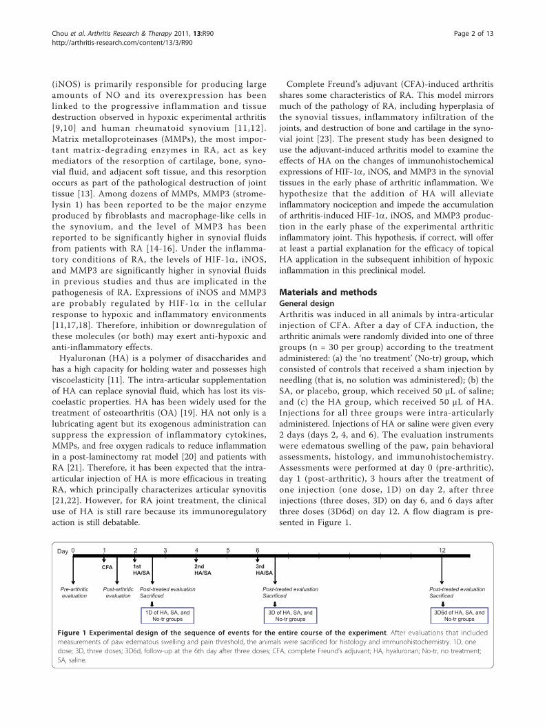
Materials and methodsGeneral designArthritis was induced in all animals by intra-articularinjection of CFA. After a day of CFA induction, thearthritic animals were randomly divided into one of threegroups (n = 30 per group) according to the treatmentadministered: (a) the ‘no treatment’ (No-tr) group, whichconsisted of controls that received a sham injection byneedling (that is, no solution was administered); (b) theSA, or placebo, group, which received 50 μL of saline;and (c) the HA group, which received 50 μL of HA.Injections for all three groups were intra-articularlyadministered. Injections of HA or saline were given every2 days (days 2, 4, and 6). The evaluation instrumentswere edematous swelling of the paw, pain behavioralassessments, histology, and immunohistochemistry.Assessments were performed at day 0 (pre-arthritic),day 1 (post-arthritic), 3 hours after the treatment ofone injection (one dose, 1D) on day 2, after threeinjections (three doses, 3D) on day 6, and 6 days afterthree doses (3D6d) on day 12. A flow diagram is pre-sented in Figure 1.
Day 0 1 2 3 4 5 6 12
Post-treated evaluationSacrificed
Pre-arthriticevaluation
CFA 2ndHA/SA
3rdHA/SA
1stHA/SA
Post-arthriticevaluation
Post-treated evaluationSacrificed
Post-treated evaluationSacrificed
1D of HA, SA, and No-tr groups
3D6d of HA, SA, and No-tr groups
3D of HA, SA, and No-tr groups
Figure 1 Experimental design of the sequence of events for the entire course of the experiment. After evaluations that includedmeasurements of paw edematous swelling and pain threshold, the animals were sacrificed for histology and immunohistochemistry. 1D, onedose; 3D, three doses; 3D6d, follow-up at the 6th day after three doses; CFA, complete Freund’s adjuvant; HA, hyaluronan; No-tr, no treatment;SA, saline.
Chou et al. Arthritis Research & Therapy 2011, 13:R90http://arthritis-research.com/content/13/3/R90
Page 2 of 13

Animal preparationNinety adult male Sprague-Dawley (SD; purchased fromBioLASCO Taiwan Co., Ltd., Taiwan, Republic ofChina) rats weighing 250 to 300 g were kept in theLaboratory Animal Center of China Medical University.An effort was made to minimize discomfort and toreduce the number of animals used. All animal experi-ments were conducted with the approval of the AnimalCare and Use Committee of China Medical Universityin accordance with the Guidelines for AnimalExperimentation.
Induction of monoarthritisMonoarthritis was induced by an injection of CFA intothe unilateral ankle articular cavity. The rats were brieflyanesthetized with 4% isoflurane (AERRANE; BaxterHealthcare Corporation, San Juan, Puerto Rico). A 28-gauge needle was vertically inserted distally into thearticular cavity from the gap between the tibiofibularand tarsus bone. CFA with a volume of 50 μL (10 mg ofmycobacterium, F5881; Sigma-Aldrich, St. Louis, MO,USA) was then injected. The monoarthritic animalswere placed separately in clear acrylic containers (10.5-inch width × 19-inch diameter × 8-inch height), andfree movement was allowed for at least 24 hours to letthe animals adjust to these conditions before any experi-mentation was performed.
Ultrasound-guided hyaluronan injectionWhile the animals were under brief isoflurane anesthe-sia, ultrasound (Terason t3000™ Ultrasound System;Terason Division, Teratech Corporation, Burlington,MA, USA)-guided injection was performed on the lat-eral side of the tibiotarsal joint, and the transducer inthe sagittal plane showed the distal end of the tibia andproximal part of the tarsus in the image plane. Needleinsertion was performed perpendicularly to the transdu-cer. HA injection (molecular weight of 1.2 to 1.4 × 10⁶
Da; Ostenil®, 10 mg/mL sodium hyaluronate; TRB Che-medica AG, München, Germany) was documented byrecording an image clip during injection with the needletip in the image plane.
Pain threshold assessmentThe pain thresholds were determined by nociceptivethresholds to mechanical stimulation. The test consistedof evoking a hind paw flexion reflex with a handheldforce transducer (electronic von Frey anesthesiometer;IITC Inc., Woodland Hills, CA, USA) adapted with a 0.5mm2 polypropylene tip. In a quiet room, the rats wereplaced in acrylic cages (32 × 22 × 27 cm high) with awire grid floor for 15 to 30 minutes of habituation priorto testing. The polypropylene tip was applied perpendi-cularly to the central area of the hind paw with
sufficient force to bend the filaments into an ‘S’ shapefor 3 to 4 seconds. The test consisted of poking a hindpaw to provoke a flexion reflex followed by a clearflinch response after paw withdrawal. Testing wasinitiated with the filament corresponding to 20 log offorce (g). The filaments were applied with a gradualincrease in pressure until a withdrawal reflex responsewas finally detected from the animal. The response tothis filament is defined if a series of weaker or strongerfilaments would be tested. The weakest filament able toelicit a response was taken to be the paw withdrawalthreshold (g). The intensity of the pressure wasrecorded, and the final value for the response wasobtained by averaging five measurements.
Measurement of edematous swelling of the pawThe extent of peripheral swelling was assessed by mea-suring the circumference of the paw at intact and CFA-injected sites with a flexible tape. The paw circumfer-ence was obtained by averaging three measurements.The difference in the ankle circumference between theinitial value (pre-arthritic data) and that at each timepoint after injection is expressed as change (percentage)= 100% × [(post-arthritic circumference)-(pre-arthriticcircumference)]/(pre-arthritic circumference). All assess-ments, including paw withdrawal and swelling measure-ments, were performed with the assessor blinded withrespect to treatment.
Histology and immunohistochemistryAnimals were killed by anesthetic overdose after treat-ments of 1D (n = 10 for each group), 3D (n = 10 foreach group), and 3D6d (n = 10 for each group) on days2, 6, and 12. Hind ankles were collected for histologicaland immunohistological analysis. The formalin-fixed,paraffin-embedded joint tissues (including synovium andcartilage tissues) were cut at a thickness of 5 μm for his-tology and immunohistochemistry. Histological confir-mation of the arthritic pathology was performed withhematoxylin and eosin-stained sections. Sections weredeparaffinized in 200 mL of Trilogy (Cell Marque Cor-poration, Rocklin, CA, USA) and incubated with 3%H2O2 in methanol for 20 minutes at room temperature.Subsequently, sections were treated with proteinase K(Sigma-Aldrich) at 0.1 mg/mL for 20 minutes at roomtemperature to unmask epitopes and this was followedby phosphate-buffered saline (PBS) rinse. Sections wereincubated with blocking buffer (Power Block™; Bio-genex, Fremont, CA, USA) for 2 hours at room tem-perature followed by incubation overnight at 4°C withthe mouse monoclonal antibody anti-HIF-1a (diluted1:100; Thermo, Fremont, CA, USA) and with the follow-ing rabbit polyclonal antibodies: anti-iNOS (diluted1:200; Thermo) and anti-MMP3 (diluted 1:200;
Chou et al. Arthritis Research & Therapy 2011, 13:R90http://arthritis-research.com/content/13/3/R90
Page 3 of 13

Abbiotec, San Diego, CA, USA). After three washes withPBS containing 0.05% Tween-20 for 10 minutes, sec-tions were incubated with biotinylated anti-rabbit andanti-mouse immunoglobulins (Jackson ImmunoResearchLaboratories, Inc., West Grove, PA, USA) followed by a30-minute peroxidase-conjugated streptavidin incuba-tion (Jackson ImmunoResearch Laboratories, Inc.). Sec-tions were incubated with 3,3’-diaminobenzidine(Biogenex), dehydrated, and cover-slipped with Per-mount (Sigma-Aldrich, St. Louis, MO, USA). Negativecontrols were performed by substituting the primaryantibody with non-immune serum.The histopathology of synovium was analyzed by the
non-parametric scoring system described by Smith andcolleagues [24]. The scores ranged from 0 to 3 for eachof the tissue criteria, including intimal hyperplasia, lym-phocytic infiltration, subintimal fibrosis, and vascularity.The higher aggregate score was considered to reflectincreased pathological changes. Five randomly selectedsections were scored and repeated two times for statisti-cal analysis. Quantitative analysis of immunostainingswas carried out by light microscopy in synovial tissuelining the joint cavity and synovial tissue attached to thecartilage. The number of HIF-1a, iNOS, and MMP3immunoreactive cells was counted among at least fivealternate sections in the more representative fields byusing a microscope. Positive nuclei and cytoplasm stain-ing cells for HIF-1a, iNOS, and MMP3 were counted inhigh-power fields (× 200 magnification) that containedsynovial lining cells. The area sizes of high-power fieldswere calculated by using a stage micrometer (with 100gradations of 0.01 mm each) when viewed using a × 200objective. Ten fields of each slide were counted for allsamples and repeated three times for statistical analysis.Results were expressed as the proportion (percentage) oflabeled cells per square millimeter of synovium. For sta-tistical analysis, the mean value obtained from therepeated counts was used. All of the scoring and quanti-tative analyses were assessed by two independent obser-vers who were blinded to the origin of the sections toavoid bias from interobserver variability.
Statistical analysisThe differences of value in each assessment betweenpre- and post-arthritic evaluations were analyzed by Stu-dent t test. The differences among the HA, SA, and No-tr groups on each dosage (1D, 3D, and 3D6d) were ana-lyzed using analysis of variance and later were analyzedfurther by a Bonferroni post hoc method. Similar statisti-cal analysis methods were used to test the differencesamong dosages in each group. Non-parametric data(histological synovial scoring) were analyzed using theKruskal-Wallis test for multiple groups and Mann-Whit-ney U tests for between-group comparisons. The
Pearson correlation test was applied to study the corre-lations between pain withdrawal threshold and expres-sions of immunoreactivities. A P value of less than 0.05was considered statistically significant. All data wereanalyzed using SPSS version 10.0 for Windows (SPSSInc., Chicago, IL, USA).
ResultsEffect of hyaluronan on complete Freund’s adjuvant-induced edemaThe serial alterations of the percentage of edema (mean± standard error of the mean, or SEM) throughout thewhole experiment for each group are shown in Figure2A. After a day of CFA induction, all animals developedsevere monoarthritis in the injected paw. There were nosignificant differences in the non-injected intact paw oncircumference among pre- and post-arthritic and post-treatment conditions for each group (P >0.05, data notshown). The edema of the CFA-injected paw graduallyincreased, reaching a maximal swelling of 65.51%,whereas there were significant differences in edemabetween pre- and post-arthritic conditions (P <0.001).After treatment, the significant time-dependent differ-
ences in edema development were observed in eachgroup (HA group: P <0.001; SA group: P <0.001; andNo-tr group: P <0.001). However, there was no differ-ence in the edema of arthritic paws among the HA, SA,and No-tr groups after treatments of 1D (P = 0.22), 3D(P = 0.41), and 3D6d (P = 0.31). Therefore, intra-articu-lar injections of HA, regardless of different dosages for1D, 3D, and 3D6d, did not ameliorate joint swellingcompared with either the SA or the No-tr group.
Effect of hyaluronan on complete Freund’s adjuvant-induced inflammatory mechanical nociceptionThe serial alterations of the paw withdrawal threshold(mean ± SEM) throughout the whole experiment foreach group are shown in Figure 2B. The mean thresholdwas 25.07 ± 4.68 g at pre-arthritic conditions. However,after CFA induction, it decreased to 9.32 ± 3.16 g.There was a significant difference with pre-arthritic con-ditions (P <0.001).The significant differences in paw withdrawal thresh-
old were shown among the HA, SA, and No-tr groupsafter treatment of 1D (P = 0.008), 3D (P <0.001), and3D6d (P <0.001). A significantly lower threshold existedafter treatment of 1D, 3D, and 3D6d in the SA and No-tr groups compared with those in the HA group (HAversus SA, P = 0.04; HA versus No-tr, P = 0.01 for 1D;HA versus SA, P < 0.001; HA versus No-tr, P <0.001 for3D; HA versus SA, P <0.001; HA versus No-tr, P =0.001 for 3D6d). The analysis also showed that therewas a significantly lower threshold in the No-tr groupcompared with the SA group after treatment of 3D (P =
Chou et al. Arthritis Research & Therapy 2011, 13:R90http://arthritis-research.com/content/13/3/R90
Page 4 of 13
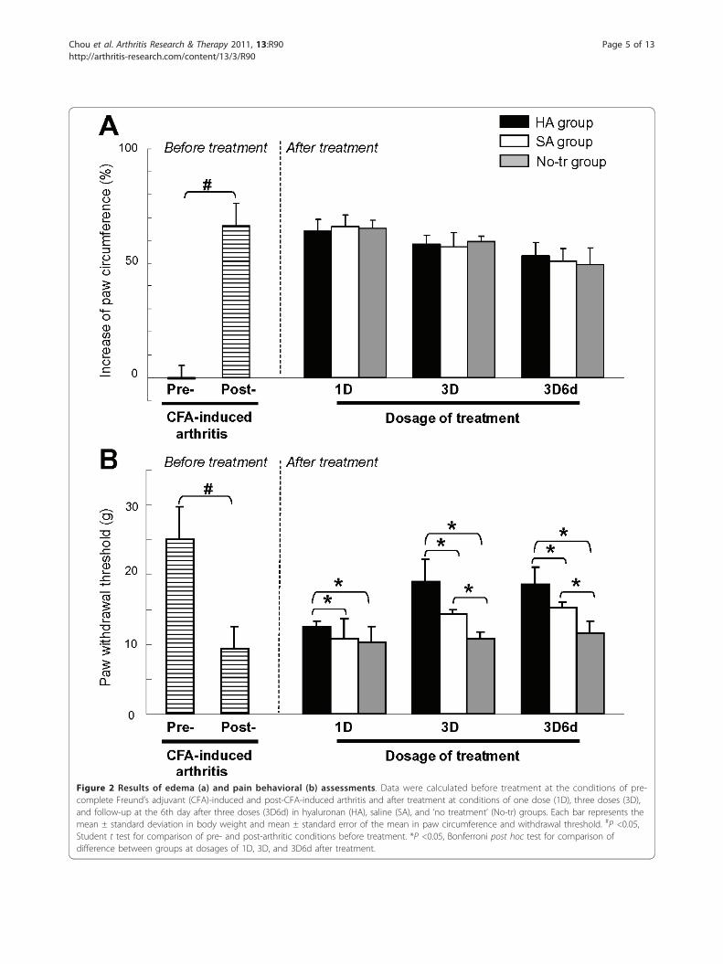
Figure 2 Results of edema (a) and pain behavioral (b) assessments. Data were calculated before treatment at the conditions of pre-complete Freund’s adjuvant (CFA)-induced and post-CFA-induced arthritis and after treatment at conditions of one dose (1D), three doses (3D),and follow-up at the 6th day after three doses (3D6d) in hyaluronan (HA), saline (SA), and ‘no treatment’ (No-tr) groups. Each bar represents themean ± standard deviation in body weight and mean ± standard error of the mean in paw circumference and withdrawal threshold. #P <0.05,Student t test for comparison of pre- and post-arthritic conditions before treatment. *P <0.05, Bonferroni post hoc test for comparison ofdifference between groups at dosages of 1D, 3D, and 3D6d after treatment.
Chou et al. Arthritis Research & Therapy 2011, 13:R90http://arthritis-research.com/content/13/3/R90
Page 5 of 13
0.03) and 3D6d (P = 0.01). However, no significant dif-ference was observed between these groups after treat-ment of 1D (P = 1.0).There were significant differences among three
dosages in the HA group (P <0.001) but not in the SA(P = 0.84) and No-tr (P = 0.56) groups. After HA treat-ment, the paw withdrawal threshold showed a signifi-cant increase in 3D and 3D6d treatments comparedwith 1D treatment (1D versus 3D, P <0.001; 1D versus3D6d, P <0.001). However, no difference was observedbetween the 3D and 3D6d of HA treatments (P = 0.05).
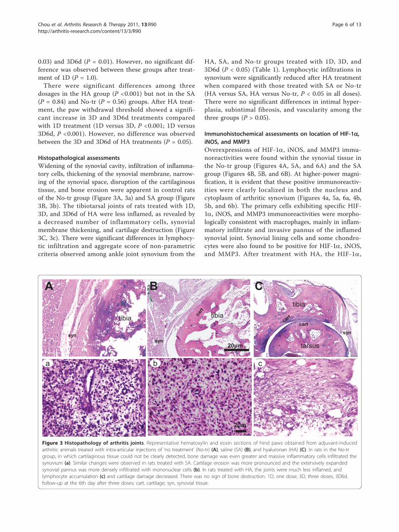
Histopathological assessmentsWidening of the synovial cavity, infiltration of inflamma-tory cells, thickening of the synovial membrane, narrow-ing of the synovial space, disruption of the cartilaginoustissue, and bone erosion were apparent in control ratsof the No-tr group (Figure 3A, 3a) and SA group (Figure3B, 3b). The tibiotarsal joints of rats treated with 1D,3D, and 3D6d of HA were less inflamed, as revealed bya decreased number of inflammatory cells, synovialmembrane thickening, and cartilage destruction (Figure3C, 3c). There were significant differences in lymphocy-tic infiltration and aggregate score of non-parametriccriteria observed among ankle joint synovium from the
HA, SA, and No-tr groups treated with 1D, 3D, and3D6d (P < 0.05) (Table 1). Lymphocytic infiltrations insynovium were significantly reduced after HA treatmentwhen compared with those treated with SA or No-tr(HA versus SA, HA versus No-tr, P < 0.05 in all doses).There were no significant differences in intimal hyper-plasia, subintimal fibrosis, and vascularity among thethree groups (P > 0.05).
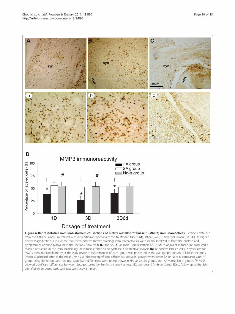
Immunohistochemical assessments on location of HIF-1a,iNOS, and MMP3Overexpressions of HIF-1a, iNOS, and MMP3 immu-noreactivities were found within the synovial tissue inthe No-tr group (Figures 4A, 5A, and 6A) and the SAgroup (Figures 4B, 5B, and 6B). At higher-power magni-fication, it is evident that these positive immunoreactiv-ities were clearly localized in both the nucleus andcytoplasm of arthritic synovium (Figures 4a, 5a, 6a, 4b,5b, and 6b). The primary cells exhibiting specific HIF-1a, iNOS, and MMP3 immunoreactivities were morpho-logically consistent with macrophages, mainly in inflam-matory infiltrate and invasive pannus of the inflamedsynovial joint. Synovial lining cells and some chondro-cytes were also found to be positive for HIF-1a, iNOS,and MMP3. After treatment with HA, the HIF-1a,
tarsus
tibia
synsyn
tibia
A B C
syn
tibia
a b c
Figure 3 Histopathology of arthritis joints. Representative hematoxylin and eosin sections of hind paws obtained from adjuvant-inducedarthritic animals treated with intra-articular injections of ‘no treatment’ (No-tr) (A), saline (SA) (B), and hyaluronan (HA) (C). In rats in the No-trgroup, in which cartilaginous tissue could not be clearly detected, bone damage was even greater and massive inflammatory cells infiltrated thesynovium (a). Similar changes were observed in rats treated with SA. Cartilage erosion was more pronounced and the extensively expandedsynovial pannus was more densely infiltrated with mononuclear cells (b). In rats treated with HA, the joints were much less inflamed, andlymphocyte accumulation (c) and cartilage damage decreased. There was no sign of bone destruction. 1D, one dose; 3D, three doses; 3D6d,follow-up at the 6th day after three doses; cart, cartilage; syn, synovial tissue.
Chou et al. Arthritis Research & Therapy 2011, 13:R90http://arthritis-research.com/content/13/3/R90
Page 6 of 13
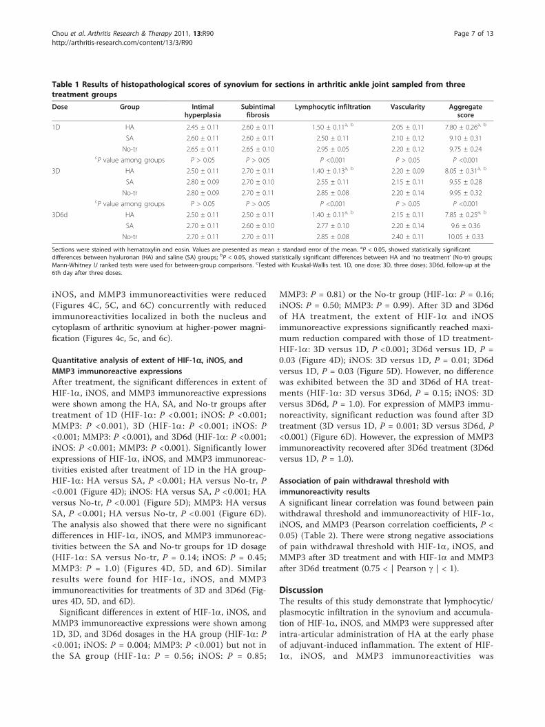
iNOS, and MMP3 immunoreactivities were reduced(Figures 4C, 5C, and 6C) concurrently with reducedimmunoreactivities localized in both the nucleus andcytoplasm of arthritic synovium at higher-power magni-fication (Figures 4c, 5c, and 6c).
Quantitative analysis of extent of HIF-1a, iNOS, andMMP3 immunoreactive expressionsAfter treatment, the significant differences in extent ofHIF-1a, iNOS, and MMP3 immunoreactive expressionswere shown among the HA, SA, and No-tr groups aftertreatment of 1D (HIF-1a: P <0.001; iNOS: P <0.001;MMP3: P <0.001), 3D (HIF-1a: P <0.001; iNOS: P<0.001; MMP3: P <0.001), and 3D6d (HIF-1a: P <0.001;iNOS: P <0.001; MMP3: P <0.001). Significantly lowerexpressions of HIF-1a, iNOS, and MMP3 immunoreac-tivities existed after treatment of 1D in the HA group-HIF-1a: HA versus SA, P <0.001; HA versus No-tr, P<0.001 (Figure 4D); iNOS: HA versus SA, P <0.001; HAversus No-tr, P <0.001 (Figure 5D); MMP3: HA versusSA, P <0.001; HA versus No-tr, P <0.001 (Figure 6D).The analysis also showed that there were no significantdifferences in HIF-1a, iNOS, and MMP3 immunoreac-tivities between the SA and No-tr groups for 1D dosage(HIF-1a: SA versus No-tr, P = 0.14; iNOS: P = 0.45;MMP3: P = 1.0) (Figures 4D, 5D, and 6D). Similarresults were found for HIF-1a, iNOS, and MMP3immunoreactivities for treatments of 3D and 3D6d (Fig-ures 4D, 5D, and 6D).Significant differences in extent of HIF-1a, iNOS, and
MMP3 immunoreactive expressions were shown among1D, 3D, and 3D6d dosages in the HA group (HIF-1a: P<0.001; iNOS: P = 0.004; MMP3: P <0.001) but not inthe SA group (HIF-1a: P = 0.56; iNOS: P = 0.85;
MMP3: P = 0.81) or the No-tr group (HIF-1a: P = 0.16;iNOS: P = 0.50; MMP3: P = 0.99). After 3D and 3D6dof HA treatment, the extent of HIF-1a and iNOSimmunoreactive expressions significantly reached maxi-mum reduction compared with those of 1D treatment-HIF-1a: 3D versus 1D, P <0.001; 3D6d versus 1D, P =0.03 (Figure 4D); iNOS: 3D versus 1D, P = 0.01; 3D6dversus 1D, P = 0.03 (Figure 5D). However, no differencewas exhibited between the 3D and 3D6d of HA treat-ments (HIF-1a: 3D versus 3D6d, P = 0.15; iNOS: 3Dversus 3D6d, P = 1.0). For expression of MMP3 immu-noreactivity, significant reduction was found after 3Dtreatment (3D versus 1D, P = 0.001; 3D versus 3D6d, P<0.001) (Figure 6D). However, the expression of MMP3immunoreactivity recovered after 3D6d treatment (3D6dversus 1D, P = 1.0).
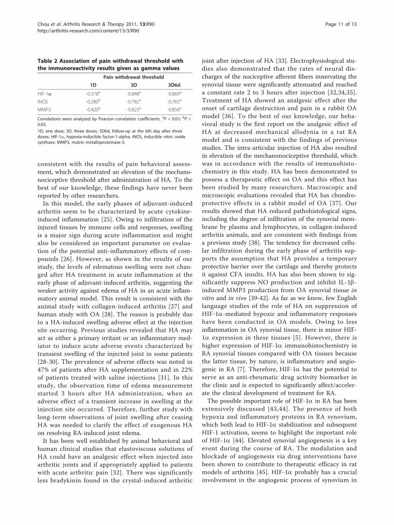
Association of pain withdrawal threshold withimmunoreactivity resultsA significant linear correlation was found between painwithdrawal threshold and immunoreactivity of HIF-1a,iNOS, and MMP3 (Pearson correlation coefficients, P <0.05) (Table 2). There were strong negative associationsof pain withdrawal threshold with HIF-1a, iNOS, andMMP3 after 3D treatment and with HIF-1a and MMP3after 3D6d treatment (0.75 < | Pearson g | < 1).
DiscussionThe results of this study demonstrate that lymphocytic/plasmocytic infiltration in the synovium and accumula-tion of HIF-1a, iNOS, and MMP3 were suppressed afterintra-articular administration of HA at the early phaseof adjuvant-induced inflammation. The extent of HIF-1a, iNOS, and MMP3 immunoreactivities was
Table 1 Results of histopathological scores of synovium for sections in arthritic ankle joint sampled from threetreatment groups
Dose Group Intimalhyperplasia
Subintimalfibrosis
Lymphocytic infiltration Vascularity Aggregatescore
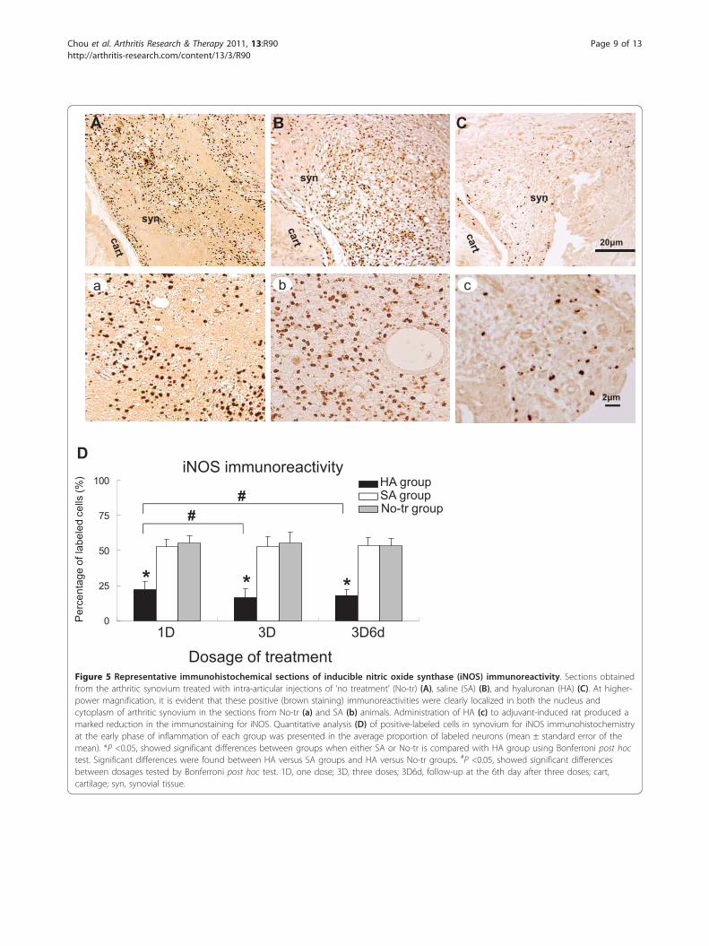
1D HA 2.45 ± 0.11 2.60 ± 0.11 1.50 ± 0.11a, b 2.05 ± 0.11 7.80 ± 0.26a, b
SA 2.60 ± 0.11 2.60 ± 0.11 2.50 ± 0.11 2.10 ± 0.12 9.10 ± 0.31
No-tr 2.65 ± 0.11 2.65 ± 0.10 2.95 ± 0.05 2.20 ± 0.12 9.75 ± 0.24cP value among groups P > 0.05 P > 0.05 P <0.001 P > 0.05 P <0.001
3D HA 2.50 ± 0.11 2.70 ± 0.11 1.40 ± 0.13a, b 2.20 ± 0.09 8.05 ± 0.31a, b
SA 2.80 ± 0.09 2.70 ± 0.10 2.55 ± 0.11 2.15 ± 0.11 9.55 ± 0.28
No-tr 2.80 ± 0.09 2.70 ± 0.11 2.85 ± 0.08 2.20 ± 0.14 9.95 ± 0.32cP value among groups P > 0.05 P > 0.05 P <0.001 P > 0.05 P <0.001
3D6d HA 2.50 ± 0.11 2.50 ± 0.11 1.40 ± 0.11a, b 2.15 ± 0.11 7.85 ± 0.25a, b
SA 2.70 ± 0.11 2.60 ± 0.10 2.77 ± 0.10 2.20 ± 0.14 9.6 ± 0.36
No-tr 2.70 ± 0.11 2.70 ± 0.11 2.85 ± 0.08 2.40 ± 0.11 10.05 ± 0.33
Sections were stained with hematoxylin and eosin. Values are presented as mean ± standard error of the mean. aP < 0.05, showed statistically significantdifferences between hyaluronan (HA) and saline (SA) groups; bP < 0.05, showed statistically significant differences between HA and ‘no treatment’ (No-tr) groups;Mann-Whitney U ranked tests were used for between-group comparisons. cTested with Kruskal-Wallis test. 1D, one dose; 3D, three doses; 3D6d, follow-up at the6th day after three doses.
Chou et al. Arthritis Research & Therapy 2011, 13:R90http://arthritis-research.com/content/13/3/R90
Page 7 of 13

0
25
50
75
100
D
Per
cent
age
ofla
bele
dce
lls(%
)
1D 3D 3D6d
HIF-
Dosage of treatment
** *
##
A B C
a b c
HA groupSA groupNo-tr group
synsyn syn
Figure 4 Representative immunohistochemical sections of hypoxia-inducible factor-1-alpha (HIF-1a) immunoreactivity. Sectionsobtained from the arthritic synovium treated with intra-articular injections of ‘no treatment’ (No-tr) (A), saline (SA) (B), and hyaluronan (HA) (C).At higher-power magnification, it is evident that these positive (brown staining) immunoreactivities were clearly localized in both the nucleusand cytoplasm of arthritic synovium in the sections from No-tr (a) and SA (b) animals. Administration of HA (c) to adjuvant-induced ratproduced a marked reduction in the immunostaining for HIF-1a. Quantitative analysis (D) of positive-labeled cells in synovium for HIF-1aimmunohistochemistry at the early phase of inflammation of each group was presented in the average proportion of labeled neurons (mean ±standard error of the mean). *P <0.05, showed significant differences between groups when either SA or No-tr is compared with HA group usingBonferroni post hoc test. Significant differences were found between HA versus SA groups and HA versus No-tr groups. #P <0.05, showedsignificant differences between dosages tested by Bonferroni post hoc test. 1D, one dose; 3D, three doses; 3D6d, follow-up at the 6th day afterthree doses; cart, cartilage; syn, synovial tissue.
Chou et al. Arthritis Research & Therapy 2011, 13:R90http://arthritis-research.com/content/13/3/R90
Page 8 of 13
0
25
50
75
100
D
Per
cent
age
ofla
bele
dce
lls(%
)
1D 3D 3D6d
iNOS immunoreactivity
Dosage of treatment
* * *
##
syn
syn
A B C
a b c
syn
HA groupSA groupNo-tr group
Figure 5 Representative immunohistochemical sections of inducible nitric oxide synthase (iNOS) immunoreactivity. Sections obtainedfrom the arthritic synovium treated with intra-articular injections of ‘no treatment’ (No-tr) (A), saline (SA) (B), and hyaluronan (HA) (C). At higher-power magnification, it is evident that these positive (brown staining) immunoreactivities were clearly localized in both the nucleus andcytoplasm of arthritic synovium in the sections from No-tr (a) and SA (b) animals. Administration of HA (c) to adjuvant-induced rat produced amarked reduction in the immunostaining for iNOS. Quantitative analysis (D) of positive-labeled cells in synovium for iNOS immunohistochemistryat the early phase of inflammation of each group was presented in the average proportion of labeled neurons (mean ± standard error of themean). *P <0.05, showed significant differences between groups when either SA or No-tr is compared with HA group using Bonferroni post hoctest. Significant differences were found between HA versus SA groups and HA versus No-tr groups. #P <0.05, showed significant differencesbetween dosages tested by Bonferroni post hoc test. 1D, one dose; 3D, three doses; 3D6d, follow-up at the 6th day after three doses; cart,cartilage; syn, synovial tissue.
Chou et al. Arthritis Research & Therapy 2011, 13:R90http://arthritis-research.com/content/13/3/R90
Page 9 of 13
A B C
syn synsyn
a b c
20 m
2 m
D
ells
(%)
MMP3 immunoreactivity100 HA group
SA groupNo tr group
ge o
f lab
eled
ce
50
75
**
*
# # No-tr group
Per
cent
ag
0
25
1D 3D 3D6d1D 3D 3D6d
Dosage of treatmentFigure 6 Representative immunohistochemical sections of matrix metalloproteinase-3 (MMP3) immunoreactivity. Sections obtainedfrom the arthritic synovium treated with intra-articular injections of ‘no treatment’ (No-tr) (A), saline (SA) (B), and hyaluronan (HA) (C). At higher-power magnification, it is evident that these positive (brown staining) immunoreactivities were clearly localized in both the nucleus andcytoplasm of arthritic synovium in the sections from No-tr (a) and SA (b) animals. Administration of HA (c) to adjuvant-induced rat produced amarked reduction in the immunostaining for inducible nitric oxide synthase. Quantitative analysis (D) of positive-labeled cells in synovium forMMP3 immunohistochemistry at the early phase of inflammation of each group was presented in the average proportion of labeled neurons(mean ± standard error of the mean). *P <0.05, showed significant differences between groups when either SA or No-tr is compared with HAgroup using Bonferroni post hoc test. Significant differences were found between HA versus SA groups and HA versus No-tr groups. #P <0.05,showed significant differences between dosages tested by Bonferroni post hoc test. 1D, one dose; 3D, three doses; 3D6d, follow-up at the 6thday after three doses; cart, cartilage; syn, synovial tissue.
Chou et al. Arthritis Research & Therapy 2011, 13:R90http://arthritis-research.com/content/13/3/R90
Page 10 of 13
consistent with the results of pain behavioral assess-ment, which demonstrated an elevation of the mechano-nociceptive threshold after administration of HA. To thebest of our knowledge, these findings have never beenreported by other researchers.In this model, the early phases of adjuvant-induced
arthritis seem to be characterized by acute cytokine-induced inflammation [25]. Owing to infiltration of theinjured tissues by immune cells and responses, swellingis a major sign during acute inflammation and mightalso be considered an important parameter on evalua-tion of the potential anti-inflammatory effects of com-pounds [26]. However, as shown in the results of ourstudy, the levels of edematous swelling were not chan-ged after HA treatment in acute inflammation at theearly phase of adjuvant-induced arthritis, suggesting theweaker activity against edema of HA in an acute inflam-matory animal model. This result is consistent with theanimal study with collagen-induced arthritis [27] andhuman study with OA [28]. The reason is probably dueto a HA-induced swelling adverse effect at the injectionsite occurring. Previous studies revealed that HA mayact as either a primary irritant or an inflammatory med-iator to induce acute adverse events characterized bytransient swelling of the injected joint in some patients[28-30]. The prevalence of adverse effects was noted in47% of patients after HA supplementation and in 22%of patients treated with saline injections [31]. In thisstudy, the observation time of edema measurementstarted 3 hours after HA administration, when anadverse effect of a transient increase in swelling at theinjection site occurred. Therefore, further study withlong-term observations of joint swelling after ceasingHA was needed to clarify the effect of exogenous HAon resolving RA-induced joint edema.It has been well established by animal behavioral and
human clinical studies that elastoviscous solutions ofHA could have an analgesic effect when injected intoarthritic joints and if appropriately applied to patientswith acute arthritic pain [32]. There was significantlyless bradykinin found in the crystal-induced arthritic
joint after injection of HA [33]. Electrophysiological stu-dies also demonstrated that the rates of neural dis-charges of the nociceptive afferent fibers innervating thesynovial tissue were significantly attenuated and reacheda constant rate 2 to 3 hours after injection [32,34,35].Treatment of HA showed an analgesic effect after theonset of cartilage destruction and pain in a rabbit OAmodel [36]. To the best of our knowledge, our beha-vioral study is the first report on the analgesic effect ofHA at decreased mechanical allodynia in a rat RAmodel and is consistent with the findings of previousstudies. The intra-articular injection of HA also resultedin elevation of the mechanonociceptive threshold, whichwas in accordance with the results of immunohisto-chemistry in this study. HA has been demonstrated topossess a therapeutic effect on OA and this effect hasbeen studied by many researchers. Macroscopic andmicroscopic evaluations revealed that HA has chondro-protective effects in a rabbit model of OA [37]. Ourresults showed that HA reduced pathohistological signs,including the degree of infiltration of the synovial mem-brane by plasma and lymphocytes, in collagen-inducedarthritis animals, and are consistent with findings froma previous study [38]. The tendency for decreased cellu-lar infiltration during the early phase of arthritis sup-ports the assumption that HA provides a temporaryprotective barrier over the cartilage and thereby protectsit against CFA insults. HA has also been shown to sig-nificantly suppress NO production and inhibit IL-1b-induced MMP3 production from OA synovial tissue invitro and in vivo [39-42]. As far as we know, few Englishlanguage studies of the role of HA on suppression ofHIF-1a-mediated hypoxic and inflammatory responseshave been conducted in OA models. Owing to lessinflammation in OA synovial tissue, there is minor HIF-1a expression in these tissues [5]. However, there ishigher expression of HIF-1a immunohistochemistry inRA synovial tissues compared with OA tissues becausethe latter tissue, by nature, is inflammatory and angio-genic in RA [7]. Therefore, HIF-1a has the potential toserve as an anti-rheumatic drug activity biomarker inthe clinic and is expected to significantly affect/acceler-ate the clinical development of treatment for RA.The possible important role of HIF-1a in RA has been
extensively discussed [43,44]. The presence of bothhypoxia and inflammatory proteins in RA synovium,which both lead to HIF-1a stabilization and subsequentHIF-1 activation, seems to highlight the important roleof HIF-1a [44]. Elevated synovial angiogenesis is a keyevent during the course of RA. The modulation andblockade of angiogenesis via drug interventions havebeen shown to contribute to therapeutic efficacy in ratmodels of arthritis [45]. HIF-1a probably has a crucialinvolvement in the angiogenic process of synovium in
Table 2 Association of pain withdrawal threshold withthe immunoreactivity results given as gamma values
Pain withdrawal threshold
1D 3D 3D6d
HIF-1a -0.378a -0.848a -0.869a
iNOS -0.280b -0.782a -0.765a
MMP3 -0.420a -0.823a -0.856a
Correlations were analyzed by Pearson correlation coefficients. aP < 0.01; bP <0.05.
1D, one dose; 3D, three doses; 3D6d, follow-up at the 6th day after threedoses; HIF-1a, hypoxia-inducible factor-1-alpha; iNOS, inducible nitric oxidesynthase; MMP3, matrix metalloproteinase-3.
Chou et al. Arthritis Research & Therapy 2011, 13:R90http://arthritis-research.com/content/13/3/R90
Page 11 of 13
RA by regulation of its target gene, vascular endothelialgrowth factor (VEGF) [43]. Inhibition of HIF-1a proteinexpression and VEGF production by SMP-114, a dis-ease-modifying anti-rheumatic drug, has been shown tobe of therapeutic benefit in RA [46]. Oral administrationof the inhibitor of heat shock protein 90 (Hsp90), whichhas been shown to potently reduce HIF-1a-related sig-naling and VEGF production, has also been found todecrease inflammation and cartilage damage in in vivomodels of RA [47]. Therefore, suppression of HIF-1amay have key implications in the development of noveltherapeutic strategies revolutionizing the treatment ofRA. Results showed that HA suppressed the adjuvant-induced overexpression of iNOS and MMP3, and this isconsistent with findings from previous studies. To thebest of our knowledge, our study is the first to reportthat HA suppresses HIF-1a. This study revealed thereduction of accumulation of HIF-1a expression in thesynovium of an adjuvant-induced RA model after intra-articular HA administration. The suppressive effects onaccumulation of inflammation-induced HIF-1a, iNOS,and MMP3 expressions in the synovium may beinvolved in the therapeutic mechanism of HA interven-tion used in the treatment of RA. Further molecular stu-dies on expressions of VEGF will be needed to fullysupport the issue of anti-angiogenic effects of HA.
ConclusionsSuppression of HIF-1a may be one of the major tar-gets of the therapeutic approach in RA. This studydemonstrated that early intervention of HA is an effec-tive protection against accumulation of inflammation-induced HIF-1a, iNOS, and MMP3 and might limitthe erosive joint damage of arthritis. The findings sug-gest that modulation of HIF-1a as a ‘master switch’may be used as a therapeutic target in the anti-inflam-matory treatment of RA.
Abbreviations1D: one dose; 3D: three doses; 3D6d: follow-up at the sixth day after threedoses; CFA: complete Freund’s adjuvant; HA: hyaluronan; HIF-1α: hypoxia-inducible factor-1-alpha; iNOS: inducible nitric oxide synthase; MMP: matrixmetalloproteinase; NO: nitric oxide; NOS: nitric oxide synthases; No-tr: notreatment; OA: osteoarthritis; PBS: phosphate-buffered saline; RA: rheumatoidarthritis; SA: saline; SEM: standard error of the mean; VEGF: vascularendothelial growth factor.
AcknowledgementsThe authors gratefully acknowledge the technical expertise of Shih-ChungChen in counting immunohistochemical-labeled cells and Pin-Wen Tu inrecording data of the animals’ functional evaluations. This work wassupported by a grant from China Medical University and Hospital (grantsCMU-98-S-10 to Y-LH and DMR-96-073 to L-WC).
Author details1Department of Physical Therapy, Graduate Institute of RehabilitationScience, China Medical University, 91 Hsueh-Shih Road, Taichung, Taiwan40202, Republic of China. 2Department of Physical Medicine and
Rehabilitation, China Medical University Hospital, 2 Yuh-Der Road, Taichung,Taiwan 40447, Republic of China. 3School of Chinese Medicine, ChinaMedical University, 91 Hsueh-Shih Road, Taichung, Taiwan 40202, Republic ofChina. 4Department of Pathology and Laboratory Medicine, TaichungVeterans General Hospital, 160, Sec. 3, Chung-Kang Road, Taichung, Taiwan40705, Republic of China. 5Institute of Biomedical Nutrition, HungkuangUniversity, 34 Chung-Chie Road, Taichung, Taiwan 40443, Republic of China.
Authors’ contributionsL-WC conceived of the study, participated in data analysis, and drafted themanuscript. JW participated in the histopathology and scored theimmunohistology. P-LC participated in the establishment of the animalmodel, immunohistology, and animals’ functional evaluations. Y-LHconceived of the study, performed the statistical analysis, and drafted themanuscript. All authors read and approved the final manuscript.
Competing interestsThe authors declare that they have no competing interests.
Received: 10 February 2011 Revised: 20 March 2011Accepted: 16 June 2011 Published: 16 June 2011
References1. Peters CL, Morris CJ, Mapp PI, Blake DR, Lewis CE, Winrow VR: The
transcription factors hypoxia-inducible factor 1alpha and Ets-1 colocalizein the hypoxic synovium of inflamed joints in adjuvant-induced arthritis.Arthritis Rheum 2004, 50:291-296.
2. Boyd HK, Lappin TR, Bell AL: Evidence for impaired erythropoietinresponse to anaemia in rheumatoid disease. Br J Rheumatol 1991,30:255-259.
3. Oliver KM, Taylor CT, Cummins EP: Hypoxia. Regulation of NFkappaBsignalling during inflammation: the role of hydroxylases. Arthritis Res Ther2009, 11:215.
4. Paleolog EM: Angiogenesis in rheumatoid arthritis. Arthritis Res 2002,4(Suppl 3):S81-S90.
5. Brouwer E, Gouw AS, Posthumus MD, van Leeuwen MA, Boerboom AL,Bijzet J, Bos R, Limburg PC, Kallenberg CG, Westra J: Hypoxia induciblefactor-1-alpha (HIF-1alpha) is related to both angiogenesis andinflammation in rheumatoid arthritis. Clin Exp Rheumatol 2009, 27:945-951.
6. Giatromanolaki A, Sivridis E, Maltezos E, Athanassou N, Papazoglou D,Gatter KC, Harris AL, Koukourakis MI: Upregulated hypoxia induciblefactor-1alpha and -2alpha pathway in rheumatoid arthritis andosteoarthritis. Arthritis Res Ther 2003, 5:R193-201.
7. Hollander AP, Corke KP, Freemont AJ, Lewis CE: Expression of hypoxia-inducible factor 1alpha by macrophages in the rheumatoid synovium:implications for targeting of therapeutic genes to the inflamed joint.Arthritis Rheum 2001, 44:1540-1544.
8. Jeon CH, Ahn JK, Chai JY, Kim HJ, Bae EK, Park SH, Cho EY, Cha HS, Ahn KS,Koh EM: Hypoxia appears at pre-arthritic stage and shows co-localizationwith early synovial inflammation in collagen induced arthritis. Clin ExpRheumatol 2008, 26:646-648.
9. Oyanagui Y: Nitric oxide and superoxide radical are involved in bothinitiation and development of adjuvant arthritis in rats. Life Sci 1994, 54:PL285-289.
10. Hashimoto K, Fukuda K, Yamazaki K, Yamamoto N, Matsushita T,Hayakawa S, Munakata H, Hamanishi C: Hypoxia-induced hyaluronansynthesis by articular chondrocytes: the role of nitric oxide. Inflamm Res2006, 55:72-77.
11. Ahn JK, Koh EM, Cha HS, Lee YS, Kim J, Bae EK, Ahn KS: Role of hypoxia-inducible factor-1alpha in hypoxia-induced expressions of IL-8, MMP-1and MMP-3 in rheumatoid fibroblast-like synoviocytes. Rheumatology(Oxford) 2008, 47:834-839.
12. Farrell AJ, Blake DR, Palmer RM, Moncada S: Increased concentrations ofnitrite in synovial fluid and serum samples suggest increased nitricoxide synthesis in rheumatic diseases. Ann Rheum Dis 1992, 51:1219-1222.
13. Cawston T: Matrix metalloproteinases and TIMPs: properties andimplications for the rheumatic diseases. Mol Med Today 1998, 4:130-137.
14. Tetlow LC, Wolley DE: Comparative immuno-localization studies ofcollagenase-1 and collagenase 3 production in the rheumatoid lesion,and by human chondrocytes and synoviocytes in vitro. Br J Rheumatol1998, 37:64-70.
Chou et al. Arthritis Research & Therapy 2011, 13:R90http://arthritis-research.com/content/13/3/R90
Page 12 of 13

15. Yoshihara Y, Nakamura H, Obata K, Yamada H, Hayakawa T, Fujikawa K,Okada Y: Matrix metalloproteinases and tissue inhibitors ofmetalloproteinases in synovial fluids from patients with rheumatoidarthritis or osteoarthritis. Ann Rheum Dis 2000, 59:455-461.
16. Keyszer G, Lambiri I, Nagel R, Keysser C, Keysser M, Gromnica-Ihle E, Franz J,Burmester GR, Jung K: Circulating levels of matrix metalloproteinasesMMP-3 and MMP-1, tissue inhibitor of metalloproteinases 1 (TIMP-1),and MMP-1/TIMP-1 complex in rheumatic disease. Correlation withclinical activity of rheumatoid arthritis versus other surrogate markers. JRheumatol 1999, 26:251-258.
17. Fraisl P, Aragones J, Carmeliet P: Inhibition of oxygen sensors as atherapeutic strategy for ischaemic and inflammatory disease. Nat RevDrug Discov 2009, 8:139-152.
18. Chen W, Ostrowski RP, Obenaus A, Zhang JH: Prodeath or prosurvival: twofacets of hypoxia inducible factor-1 in perinatal brain injury. Exp Neurol2009, 216:7-15.
19. Miltner O, Schneider U, Siebert CH, Niedhart C, Niethard FU: Efficacy ofintraarticular hyaluronic acid in patients with osteoarthritis-a prospectiveclinical trial. Osteoarthritis Cartilage 2002, 10:680-686.
20. Schimizzi AL, Massie JB, Murphy M, Perry A, Kim CW, Garfin SR, Akeson WH:High-molecular-weight hyaluronan inhibits macrophage proliferationand cytokine release in the early wound of a preclinicalpostlaminectomy rat model. Spine J 2006, 6:550-556.
21. Saito S, Momohara S, Taniguchi A, Yamanaka H: The intra-articular efficacyof hyaluronate injections in the treatment of rheumatoid arthritis. ModRheumatol 2009, 19:643-651.
22. Oliveira PG, Brenol CV, Edelweiss MI, Brenol JC, Petronilho F, Roesler R, Dal-Pizzol F, Schwartsmann G, Xavier RM: Effects of an antagonist of thebombesin/gastrin-releasing peptide receptor on complete Freund’sadjuvant-induced arthritis in rats. Peptides 2008, 29:1726-1731.
23. Brauer R, Kittlick PD, Thoss K, Henzgen S: Different immunologicalmechanisms contribute to cartilage destruction in antigen-inducedarthritis. Exp Toxicol Pathol 1994, 46:383-388.
24. Smith MM, Cake MA, Ghosh P, Schiavinato A, Read RA, Little CB: Significantsynovial pathology in a meniscectomy model of osteoarthritis:modification by intra-articular hyaluronan therapy. Rheumatology (Oxford)2008, 47:1172-1178.
25. Ferraccioli G, Bracci-Laudiero L, Alivernini S, Gremese E, Tolusso B, DeBenedetti F: Interleukin-1beta and Interleukin-6 in arthritis animalmodels. Roles in the early phase of transition from the acute to chronicinflammation and relevance for human rheumatoid arthritis. Mol Med2010, 16:552-557.
26. Morris CJ: Carrageenan-induced paw edema in the rat and mouse.Methods Mol Biol 2003, 225:115-121.
27. Campo GM, Avenoso A, Campo S, Ferlazzo AM, Altavilla D, Calatroni A:Efficacy of treatment with glycosaminoglycans on experimentalcollagen-induced arthritis in rats. Arthritis Res Ther 2003, 5:R122-131.
28. Petrella RJ: Hyaluronic acid for the treatment of knee osteoarthritis: long-term outcomes from a naturalistic primary care experience. Am J PhysMed Rehabil 2005, 84:278-283; quiz 284, 293.
29. Henderson EB, Smith EC, Pegley F, Blake DR: Intra-articular injections of750 kD hyaluronan in the treatment of osteoarthritis: a randomisedsingle centre double-blind placebo-controlled trial of 91 patientsdemonstrating lack of efficacy. Ann Rheum Dis 1994, 53:529-534.
30. Puttick MP, Wade JP, Chalmers A, Connell DG, Rangno KK: Acute localreactions after intraarticular hylan for osteoarthritis of the knee. JRheumatol 1995, 22:1311-1314.
31. Hamburger MI, Lakhanpal S, Mooar PA, Oster D: Intra-articularhyaluronans: a review of product-specific safety profiles. Semin ArthritisRheum 2003, 32:296-309.
32. Balazs EA: Analgesic effect of elastoviscous hyaluronan solutions and thetreatment of arthritic pain. Cells Tissues Organs 2003, 174:49-62.
33. Aihara S, Murakami N, Ishii R, Kariya K, Azuma Y, Hamada K, Umemoto J,Maeda S: Effects of sodium hyaluronate on the nociceptive response ofrats with experimentally induced arthritis. Nippon Yakurigaku Zasshi 1992,100:359-365.
34. Pozo MA, Balazs EA, Belmonte C: Reduction of sensory responses topassive movements of inflamed knee joints by hylan, a hyaluronanderivative. Exp Brain Res 1997, 116:3-9.
35. Gomis A, Pawlak M, Balazs EA, Schmidt RF, Belmonte C: Effects of differentmolecular weight elastoviscous hyaluronan solutions on articularnociceptive afferents. Arthritis Rheum 2004, 50:314-326.
36. Hashizume M, Koike N, Yoshida H, Suzuki M, Mihara M: High molecularweight hyaluronic acid relieved joint pain and prevented theprogression of cartilage degeneration in a rabbit osteoarthritis modelafter onset of arthritis. Mod Rheumatol 2010, 20:432-438.
37. Ozkan FU, Ozkan K, Ramadan S, Guven Z: Chondroprotective effect of N-acetylglucosamine and hyaluronate in early stages of osteoarthritis-anexperimental study in rabbits. Bull NYU Hosp Jt Dis 2009, 67:352-357.
38. Roth A, Mollenhauer J, Wagner A, Fuhrmann R, Straub A, Venbrocks RA,Petrow P, Bräuer R, Schubert H, Ozegowski J, Peschel G, Müller PJ,Kinne RW: Intra-articular injections of high-molecular-weight hyaluronicacid have biphasic effects on joint inflammation and destruction in ratantigen-induced arthritis. Arthritis Res Ther 2005, 7:R677-686.
39. Tung JT, Venta PJ, Caron JP: Inducible nitric oxide expression in equinearticular chondrocytes: effects of antiinflammatory compounds.Osteoarthritis Cartilage 2002, 10:5-12.
40. Takahashi K, Hashimoto S, Kubo T, Hirasawa Y, Lotz M, Amiel D: Hyaluronansuppressed nitric oxide production in the meniscus and synovium ofrabbit osteoarthritis model. J Orthop Res 2001, 19:500-503.
41. Lee YT, Shao HJ, Wang JH, Liu HC, Hou SM, Young TH: Hyaluronic acidmodulates gene expression of connective tissue growth factor (CTGF),transforming growth factor-beta1 (TGF-beta1), and vascular endothelialgrowth factor (VEGF) in human fibroblast-like synovial cells fromadvanced-stage osteoarthritis in vitro. J Orthop Res 2010, 28:492-496.
42. Waddell DD, Kolomytkin OV, Dunn S, Marino AA: Hyaluronan suppressesIL-1beta-induced metalloproteinase activity from synovial tissue. ClinOrthop Relat Res 2007, 465:241-248.
43. Gaber T, Dziurla R, Tripmacher R, Burmester GR, Buttgereit F: Hypoxiainducible factor (HIF) in rheumatology: low O2! See what HIF can do!Ann Rheum Dis 2005, 64:971-980.
44. Muz B, Khan MN, Kiriakidis S, Paleolog EM: Hypoxia. The role of hypoxiaand HIF-dependent signalling events in rheumatoid arthritis. Arthritis ResTher 2009, 11:201.
45. Issekutz AC, Sapru K: Modulation of adjuvant arthritis in the rat by 2-methoxyestradiol: an effect independent of an anti-angiogenic action.Int Immunopharmacol 2008, 8:708-716.
46. Westra J, Brouwer E, van Roosmalen IA, Doornbos-van der Meer B, vanLeeuwen MA, Posthumus MD, Kallenberg CG: Expression and regulation ofHIF-1alpha in macrophages under inflammatory conditions; significantreduction of VEGF by CaMKII inhibitor. BMC Musculoskelet Disord 2010,11:61.
47. Rice JW, Veal JM, Fadden RP, Barabasz AF, Partridge JM, Barta TE,Dubois LG, Huang KH, Mabbett SR, Silinski MA, Steed PM, Hall SE: Smallmolecule inhibitors of Hsp90 potently affect inflammatory diseasepathways and exhibit activity in models of rheumatoid arthritis. ArthritisRheum 2008, 58:3765-3775.
doi:10.1186/ar3365Cite this article as: Chou et al.: Hyaluronan modulates accumulation ofhypoxia-inducible factor-1 alpha, inducible nitric oxide synthase, andmatrix metalloproteinase-3 in the synovium of rat adjuvant-inducedarthritis model. Arthritis Research & Therapy 2011 13:R90.
Submit your next manuscript to BioMed Centraland take full advantage of:
• Convenient online submission
• Thorough peer review
• No space constraints or color figure charges
• Immediate publication on acceptance
• Inclusion in PubMed, CAS, Scopus and Google Scholar
• Research which is freely available for redistribution
Submit your manuscript at www.biomedcentral.com/submit
Chou et al. Arthritis Research & Therapy 2011, 13:R90http://arthritis-research.com/content/13/3/R90
Page 13 of 13