The Role of the Major Histocompatibility Complex and the ...
Upload
independentCategory
view
0download
0
10.1128/JVI.75.11.5197-5204.2001.
2001, 75(11):5197. DOI:J. Virol. Danny J. Schust and Hidde L. PloeghBenjamin E. Gewurz, Evelyn W. Wang, Domenico Tortorella,
MannerComplex Class I in a Locus-Specific Association with Major HistocompatibilityReticulum-Lumenal Domain Dictates Human Cytomegalovirus US2 Endoplasmic
http://jvi.asm.org/content/75/11/5197Updated information and services can be found at:
These include:
REFERENCEShttp://jvi.asm.org/content/75/11/5197#ref-list-1at:
This article cites 36 articles, 17 of which can be accessed free
CONTENT ALERTS more»articles cite this article),
Receive: RSS Feeds, eTOCs, free email alerts (when new
http://journals.asm.org/site/misc/reprints.xhtmlInformation about commercial reprint orders: http://journals.asm.org/site/subscriptions/To subscribe to to another ASM Journal go to:
on March 4, 2014 by guest
http://jvi.asm.org/
Dow
nloaded from
on March 4, 2014 by guest
http://jvi.asm.org/
Dow
nloaded from
JOURNAL OF VIROLOGY,0022-538X/01/$04.0010 DOI: 10.1128/JVI.75.11.5197–5204.2001
June 2001, p. 5197–5204 Vol. 75, No. 11
Copyright © 2001, American Society for Microbiology. All Rights Reserved.
Human Cytomegalovirus US2 Endoplasmic Reticulum-LumenalDomain Dictates Association with Major Histocompatibility
Complex Class I in a Locus-Specific MannerBENJAMIN E. GEWURZ,1 EVELYN W. WANG,1 DOMENICO TORTORELLA,1
DANNY J. SCHUST,2 AND HIDDE L. PLOEGH1*
Department of Pathology1 and The Fearing Laboratory and The Division of Reproductive Endocrinology and Fertility,Department of Obstetrics, Gynecology, and Reproductive Biology, Brigham and Women’s Hospital,2
Harvard Medical School, Boston, Massachusetts 02115
Received 24 January 2001/Accepted 12 March 2001
The human cytomegalovirus-encoded US2 glycoprotein targets endoplasmic reticulum-resident major his-tocompatibility complex (MHC) class I heavy chains for rapid degradation by the proteasome. We demonstratethat the endoplasmic reticulum-lumenal domain of US2 allows tight interaction with class I molecules encodedby the HLA-A locus. Recombinant soluble US2 binds properly folded, peptide-containing recombinant HLA-A2molecules in a peptide sequence-independent manner, consistent with US2’s ability to broadly downregulateclass I molecules. The physicochemical properties of the US2/MHC class I complex suggest a 1:1 stoichiometry.These results demonstrate that US2 does not require additional cellular proteins to specifically interact withsoluble class I molecules. Binding of US2 does not significantly alter the conformation of class I molecules, asa soluble T-cell receptor can simultaneously recognize class I molecules associated with US2. The lumenaldomain of US2 can differentiate between the products of distinct class I loci, as US2 binds several HLA-A locusproducts while being unable to bind recombinant HLA-B7, HLA-B27, HLA-Cw4, or HLA-E. We did not observeinteraction between soluble US2 and either recombinant HLA-DR1 or recombinant HLA-DM. The substratespecificity of US2 may help explain the presence in human cytomegalovirus of multiple strategies for down-regulation of MHC class I molecules.
The human cytomegalovirus (HCMV) is a ubiquitous beta-herpesvirus that causes persistent infection following primaryexposure. Infectious HCMV is secreted for extended periodsfrom numerous sites upon primary infection, and in commonwith other known human herpesviruses, HCMV establisheslatency (4). HCMV can reactivate from latent infection ofmyeloid lineage cells years after acute infection, even in a fullyprimed immunocompetent host (28). This lifestyle requiresHCMV to disarm several components of the immune re-sponse, in particular the major histocompatibility complex(MHC) class I antigen presentation pathway.
MHC class I molecules are heterotrimeric complexes com-posed of a polymorphic heavy chain, the invariant b2-micro-globulin light chain (b2m), and an antigenic peptide (34). Pro-teasomal cleavage of cellular and microbial cytosolic proteinsyields peptides, some of which are delivered to the endoplas-mic reticulum (ER) by the transporter associated with antigenprocessing and presentation (37). Within the ER, chaperonesfacilitate peptide loading of empty class I molecules (6). Uponreceipt of peptide, class I molecules travel to the cell surface topresent the cargo to CD81 T cells (13).
Although CD81 cytotoxic T lymphocytes respond to pep-tides derived from several HCMV immediate-early virus geneproducts, it has been difficult to find cytotoxic T lymphocytesspecific for the more abundant and diverse set of proteins
expressed later in the virus life cycle (3, 26). The concertedaction of a series of glycoproteins encoded by HCMV uniqueshort (US) genes disrupts surface expression of class I mole-cules and likely contributes to this absence of CD81 T-cellrecognition (17). Indeed, HCMV prevents the production anddisplay of antigenic peptides by several seemingly independentmechanisms (32). The US3 glycoprotein, whose ER-lumenaldomain shares approximately 20% sequence identity with US2,retains class I molecules in the ER during the immediate-earlyperiod of virus infection (1, 18). The US3 ER-lumenal andtransmembrane domains are each required for association withclass I molecules (20). During the early period of virus infec-tion, the ER-resident glycoproteins US2 and US11 indepen-dently bind to newly synthesized MHC class I molecules in theER and redirect the class I heavy chains to the cytosol, via theSec61 translocon. Within the cytosol, the class I heavy chainsare rapidly deglycosylated and degraded by the proteasome(35, 36). The dislocation reaction is sensitive to changes inredox conditions, and the cytoplasmic tail of the class I heavychain is required for degradation (30, 33). However, the mo-lecular details of how either US2 or US11 targets class I fordestruction remain obscure.
A growing number of viruses are known to encode factorsthat downregulate the surface expression of class I moleculesposttranslationally, including human immunodeficiency virus(HIV), Kaposi’s sarcoma-associated herpesvirus (KSHV), ad-enovirus, murine cytomegalovirus, and murine gammaherpes-virus 68 (29, 32). Several of these virus gene products selec-tively target HLA-A and HLA-B locus products, including US2and US11, KSHV K5, and HIV Nef. The locus specificity of
* Corresponding author. Mailing address: Department of Pathology,Harvard Medical School, Building D2, Room 137, 200 LongwoodAve., Boston, MA 02115. Phone: (617) 432-4776. Fax: (617) 432-4775.E-mail: [email protected].
5197
on March 4, 2014 by guest
http://jvi.asm.org/
Dow
nloaded from
the KSHV K5 product derives largely from interactions withinthe membrane (15), while HIV Nef relies on polymorphisms inthe cytoplasmic tail of class I molecules to selectively interferewith class I presentation (5).
While HCMV interferes with transcription of MHC class IImolecules (24), it has further been suggested that US2 mighttarget MHC class II HLA-DR and HLA-DM for degradation(31). HLA-DM facilitates peptide loading of class II mole-cules, which present peptides derived from extracellular pro-teins to CD41 T cells (16). Notwithstanding their similar fold-ing patterns, the considerable sequence disparity between classI molecules and the DR or DM MHC class II complexes raisesthe question of how US2 can engage this rather divergent setof glycoproteins.
US2 is a 199-amino-acid membrane protein, with an ER-lumenal portion, a predicted single transmembrane domain,and a short cytoplasmic tail. The absence of a cleavable signalsequence at the N terminus is unusual for a glycoprotein thatotherwise resembles a typical type I membrane protein. Theregions of US2 that are responsible for class I degradation arenot characterized, and we have so far failed to detect signifi-cant homologies between US2 and non-HCMV-encodedpolypeptides. To better understand US2 function, we havereconstituted the interaction between recombinant forms ofUS2 and MHC class I. We show that the US2 ER-lumenaldomain specifically interacts with class I molecules and enablesa delineation of its class I binding preferences.
MATERIALS AND METHODS
Bacterial strains and constructs. US2 amino-terminal and carboxy-terminaldeletion mutants were constructed by PCR with Pfu DNA polymerase (Strat-agene) using a US2 cDNA template. The 59 oligonucleotide primer GGGAATTCCATATGGGTCCCTTGATCCGCCTGCC and the 39 primer CCGCTCGAGTTAATCCACTCGCAGTTCGGGGACGC were used to amplify US215–140.PCR products were doubly digested using NdeI and XhoI (New England Biolabs)and ligated into the pET27 expression vector (Novagen). Full-length and addi-tional deletion mutant constructs of US2 were also cloned into pET27 followingPCR from a cDNA template.
The expression construct for soluble HLA-A2 class I heavy chain (amino acidsG1 to E275) was constructed by PCR with Pfu DNA polymerase using a cDNAtemplate. The 59 oligonucleotide primer TGGGCTCTCACTCCATGAGGTATTTC and the 39 primer CCGCTCGAGTTACTCCCATCTCAGGGTGAGGGGCT were used to amplify the heavy chain. The XhoI-digested PCR productswere ligated into pET27, which had been cut with NdeI, blunted with KlenowDNA polymerase (New England Biolabs), and digested with XhoI. The HLA-Eclass I heavy chain (amino acids G1 to E275) was similarly amplified and clonedinto pET27 using the 59 primer GGAATTCCATATGGGCTCCCACTCCTTGAAGTATTTCC and the 39 primer CCGCTCGAGTCACTTCCATCTCAGGGTGACGGGCTC. All constructs obtained from PCR amplification were se-quenced to verify the absence of mutations.
BL21(DE3)plysS strains carrying expression constructs for either HLA-A68,HLA-B7, or HLA-B27 heavy chains or b2m were obtained from the laboratoryof Don C. Wiley, Harvard University, Cambridge, Mass. Inclusion bodies of US2constructs, HLA-A2, and HLA-E were produced in BL21(DE3).
Protein expression and inclusion body purification. Single bacterial coloniescontaining either US2, HLA-A2, or HLA-E heavy chain constructs were grownat 37°C in Luria-Bertani medium containing 30 mg of kanamycin sulfate/ml. Onehundred micrograms of ampicillin per milliliter was added to cultures expressingHLA-A68, HLA-B7, HLA-B27, HLA-B51, or HLA-Cw4 heavy chains or b2-microglobulin. Cultures were induced at an optical density at 600 nm of 0.6 with1 mM isopropyl-b-D-thiogalactopyranoside (IPTG). Inclusion bodies were puri-fied as described previously (9). Inclusion bodies were dissolved in 8 M urea–25mM 2-(N-morpholino)ethanesulfonic acid (MES; pH 6.0)–10 mM EDTA–1 mMdithiothreitol (DTT) (20 mM DTT was added to US2 inclusion body resuspen-sions). HLA-A2–ELAGIGILTV and HLA-A2–ALGIGILTV peptide complexes
were generous gifts from Olivier Michielin, Strasbourg, France. HLA-DM andHLA-DR1 were generous gifts from Don Wiley.
Renaturation by dilution. A 7.15-mg quantity of US2 denatured in 8 Murea–25 mM MES (pH 6.5)–10 mM sodium EDTA–20 mM DTT was added to7.5 ml of injection buffer (3 M guanidine HCl, 10 mM sodium acetate, pH 4.2).The US2 mixture was then injected through a 27-gauge needle into 500 ml ofrapidly stirred 10°C refolding buffer (100 mM Tris HCl [pH 8.3], 2 mM EDTA,400 mM L-arginine HCl, 5 mM reduced glutathione, 0.5 mM oxidized glutathi-one) at a final protein concentration of 1 mM. Phenylmethylsulfonyl fluoride (0.1mM) in isopropanol was added immediately prior to addition of US2. Refoldingreaction mixtures were incubated at .10°C. US2 (7.15 mg/liter) was added twicemore at 6- to 12-h intervals to the refolding mixture, and the refolding reactionmixture was incubated for an additional 24 h.
Class I molecules were refolded as described previously (7, 9). HLA-A2 wasrefolded with either the human T-cell leukemia virus (HTLV) Tax peptideLLFGYPVYV, the HIV reverse transcriptase (polymerase [Pol]) peptideILKEPVHGV, or the hepatitis B virus peptide FLPSDFFPSV; HLA-A*6801was refolded with the influenza A virus matrix protein epitope KTGGPIYKR;HLA-B*0702 was refolded with nonamer peptide APRTVALTA; HLA-B27 wasrefolded with peptide GRIDKPILK; HLA-Cw4 was refolded using QYDDAVYKL; and HLA-E was refolded with the HLA-B8 leader epitope VMAPRTVLL.
Protein purification. US2 refolding reaction mixtures were concentrated witha pressurized stirred cell (Amicon) across membranes with a 10,000-molecular-weight cutoff and further concentrated to a volume of 200 to 500 ml using aCentricon 10 concentrator (Amicon). The resultant protein was separated by fastprotein liquid chromatography on Superdex 200 or Superdex 75 gel filtrationcolumns (Pharmacia) in 10 mM Tris (pH 8.0)–150 mM NaCl. Typical refoldingyields for US215–140 ranged from 2 to 5%, although the yield could be increasedsomewhat by refolding US2 in the presence of purified HLA-A2. SolubleUS215–140 aggregates at room temperature in the absence of class I moleculesand was kept at 4 to 10°C at all times.
Class I refolding reaction mixtures were dialyzed twice for 12 h against 10 mMTris (pH 8.0)–1 mM EDTA (molecular weight cutoff 5 6,000 to 8,000). Thedialyzed protein was concentrated with a DEAE cellulose (DE-52) anion-ex-change column packed with 20 g of refold per liter. The partially purified class Imolecules were further concentrated by a Centricon 10 concentrator and sepa-rated from the remaining contaminants by Superdex 200 fast protein liquidchromatography gel filtration in 10 mM Tris (pH 8.0)–150 mM NaCl.
Native gel band shift assay. Samples were incubated in a native gel loadingbuffer (final concentrations, 250 mM Tris [pH 8.8], 10% glycerol) to a finalvolume of 40 ml. Gels of 1.5 mm in thickness and containing either 8 or 12%polyacrylamide, without a stacking gel, were run at 4°C using 25 mM Tris–190mM glycine running buffer. Proteins were visualized with Coomassie brilliantblue R-250.
Superdex gel filtration elution assay. HLA-A2 (300 mg), US215–140 (190 mg),or HLA-A2 and US2 (300 and 190 mg, respectively) were incubated for 15 minand loaded onto an analytical Superdex 75 gel filtration column in a 100-mlvolume. HLA-B7 (360 mg), US215–140 (580 mg), or HLA-B7 and US215–140 (360and 580 mg, respectively) were incubated for 15 min and loaded onto Superdex75 in a 200-ml volume. HLA-B27 (360 mg), US215–140 (580 mg), or HLA-B27 andUS215–140 (360 and 580 mg, respectively) were likewise incubated for 15 min andloaded onto Superdex 75 in a 200-ml volume. All columns were run in 10 mMTris (pH 8.0)–150 mM NaCl at 4°C.
RESULTS
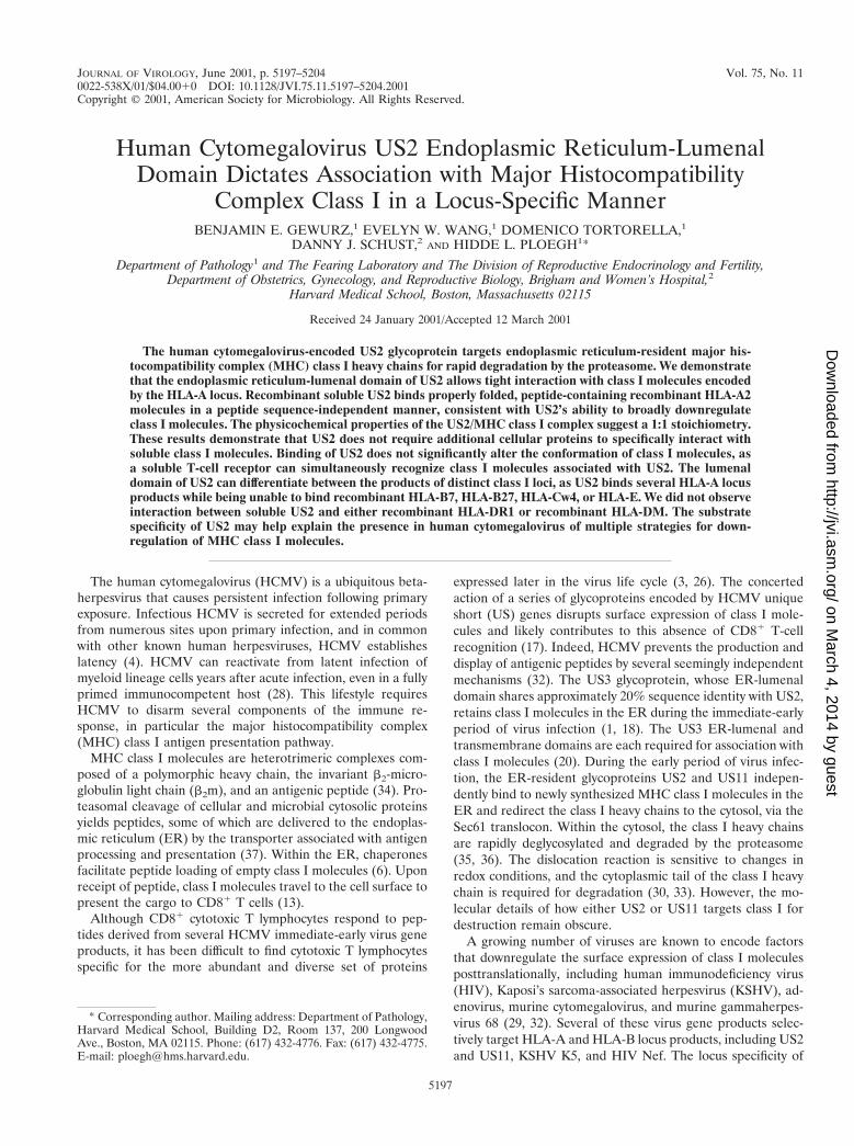
Production of soluble US2. US2 expression constructs thatincluded the predicted transmembrane coding region (residues162 to 185) failed to yield significant levels of protein in Esch-erichia coli (Fig. 1). Since preliminary studies suggested thatUS2 mutants comprising residues 1 to 150 retain the ability tobind to class I in vivo, efforts to produce recombinant US2therefore focused on the ER-lumenal segment. The US2amino terminus is significantly less hydrophobic than are ca-nonical signal sequences (22) and fails to be cleaved uponinsertion into the ER (unpublished data). Initial attempts toproduce soluble US2 were therefore carried out with mutantspossessing a native amino terminus (Fig. 1). E. coli transfor-mants containing expression constructs for US21–150 and
5198 GEWURZ ET AL. J. VIROL.
on March 4, 2014 by guest
http://jvi.asm.org/
Dow
nloaded from
US21–140 produced high levels of inclusion body protein. How-ever, attempts to refold the urea-denatured inclusion bodiesresulted in insoluble protein aggregates, which consistentlyeluted in the void volume upon size exclusion chromatography.
Additional US2 expression constructs utilize a methionine atresidue 15 to initiate translation (Fig. 1). Both US215–150 andUS215–140 expression plasmids yielded inclusion bodies at.100 mg of bacteria per liter. Only refolding of US215–140
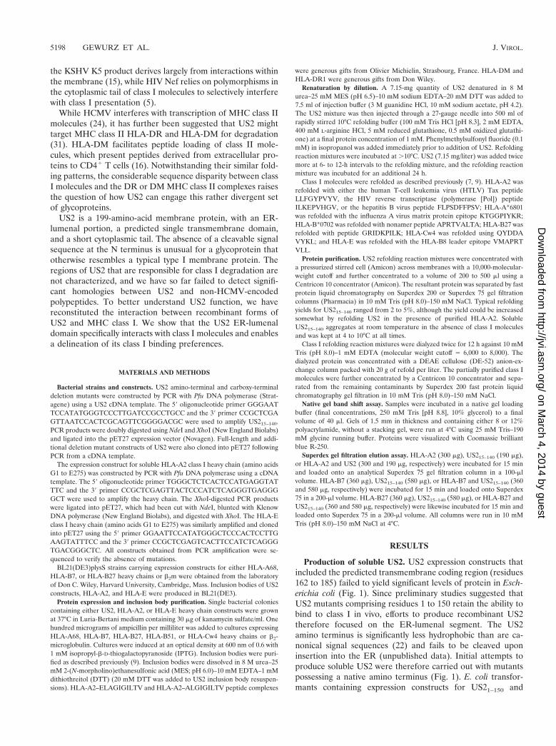
yielded soluble monodisperse protein, as shown by size ex-clusion chromatography. The elution profile suggests thatUS215–140 exists as a monomer in solution (Fig. 2).
US215–140 associates with class I molecules in vitro. To ex-amine whether soluble US215–140 retains the ability to interactwith soluble MHC class I, the HLA-A2 heavy chain lacking itstransmembrane and cytoplasmic tail segments (G1 to E275)and b2m (Met1 to Met99) were refolded in the presence of theHTLV-1 Tax peptide LLFGYPVYV. Incubation of solubleUS215–140 with purified HLA-A2 results in the formation of acomplex of a Stokes’ radius distinct from either HLA-A2 orUS215–140 alone (Fig. 2B). The magnitude of the shift in elu-tion position of the peak suggests a 1:1 stoichiometry of theUS2/HLA-A2 complex (Fig. 2B). Preliminary analytical ultra-centrifugation studies were also most consistent with a 1:1stoichiometry (unpublished data). Further increasing the con-centration of soluble US2 did not appear to alter the stoichi-ometry of the complex.
The strength of interaction between US215–140 and solubleHLA-A2/Tax is exemplified by the ability to remain associatedduring sequential gel filtration and ion-exchange chromatog-raphy (Fig. 2C). Thus, the US2 ER-lumenal domain alone issufficient to mediate tight binding to HLA-A2 in the absence ofother cellular or viral proteins. Although both US2 and MHCclass I contain a single N-linked glycan in mammalian cells,glycosylation is not essential for interaction between the twoproteins, as the bacterially produced subunits lack N-linkedglycans.
US2 recognizes class I molecules independently of peptidesequence. The ability of HLA-A2 to engage in complex for-mation with US2 is conveniently monitored by native gelelectrophoresis. The binding characteristics and hence electro-phoretic mobilities for several receptors that interact with classI molecules are influenced by the sequence of the MHC pep-
FIG. 1. Regions of US2 used for expression in E. coli. A schematicdiagram of the US2 primary structure is shown at the top, with thesingle US2 disulfide bond and predicted transmembrane domain indi-cated. The series of US2 deletion mutants constructed for expressionin E. coli are displayed beneath. The extent to which the polypeptidescan be isolated from E. coli and refolded to yield soluble material isindicated to the right of each US2 mutant. NA, not applicable.
FIG. 2. The US2 ER-lumenal domain is sufficient for association with class I molecules. (A) Reducing sodium dodecyl sulfate-polyacrylamidegel electrophoresis of A21–275 (lane 1), b2m1–99 (lane 2), and US215–140 (lane 3) inclusion body material (10 mg each). The proteins were visualizedby using Coomassie brilliant blue R-250 (Sigma). Numbers at left are molecular masses in kilodaltons. (B) Superimposed Superdex 75 gel filtrationchromatograms of soluble US215–140, HLA-A2/Tax, and HLA-A2/Tax plus US215–140. The magnitude of the shift between the peak of theHLA-A2/Tax/US215/140 complex and free HLA-A2/Tax indicates 1:1 complex stoichiometry. The elution positions of molecular weight standardsare shown above the chromatogram. (C) The HLA-A2/Tax/US215–140 complex associates with sufficient affinity to be further purified by Mono Qanion-exchange chromatography. Protein from the peak fraction was separated by nonreducing sodium dodecyl sulfate-polyacrylamide gelelectrophoresis and visualized by silver staining.
VOL. 75, 2001 US2 ER-LUMINAL DOMAIN DIRECTS MHC CLASS I ASSOCIATION 5199
on March 4, 2014 by guest
http://jvi.asm.org/
Dow
nloaded from

tide cargo. We examined the ability of US2 to bind to HLA-A2complexes containing different peptides of defined sequence,produced from recombinant subunits and synthetic peptides.As determined by native gel shift, US215–140 associates withfive distinct HLA-A2–peptide complexes containing eitherHTLV Tax residues 11 to 19, HIV type 1 reverse transcriptaseresidues 309 to 317, hepatitis B virus nucleocapsid residues 18to 27, melanoma peptide variant MART-1 27 to 35 (A2L), andvariant MART-1 27 to 35 (A3L) (Fig. 3). The presence ofUS215–140 produced a complex of lower mobility with severaladditional HLA-A2–peptide complexes (unpublished data).Differences in electrophoretic mobilities among the US2/HLA-A2/peptide complexes result from peptide sequence vari-ation (Fig. 3). Thus, US2 binds peptide-loaded class I mole-cules in a peptide sequence-independent fashion. Peptideremains associated with class I molecules upon US2 binding, asshown by incubation of the complex at 37°C, which allowedrecovery of properly folded class I molecules (unpublisheddata). Empty class I molecules are not stable at 37°C andwould have dissociated upon heating (2).
MHC class I heavy chains require b2m and peptide to refoldin vitro (9). We examined whether the presence of US215–140
facilitated the refolding of class I heavy chains in the absenceof other class I components. Folding of class I heavy chains orassociation between free heavy chains and US2 was not evidentin the presence of Tax peptide and refolded US215–140 (un-published data). In the absence of peptide and ER chaperones,soluble class I heavy chains have a short half-life in solution,despite high concentrations of US215–140 and b2m. All in all,our data suggest that US2 prefers to interact with a properlyfolded class I molecule in a 1:1 stoichiometry.
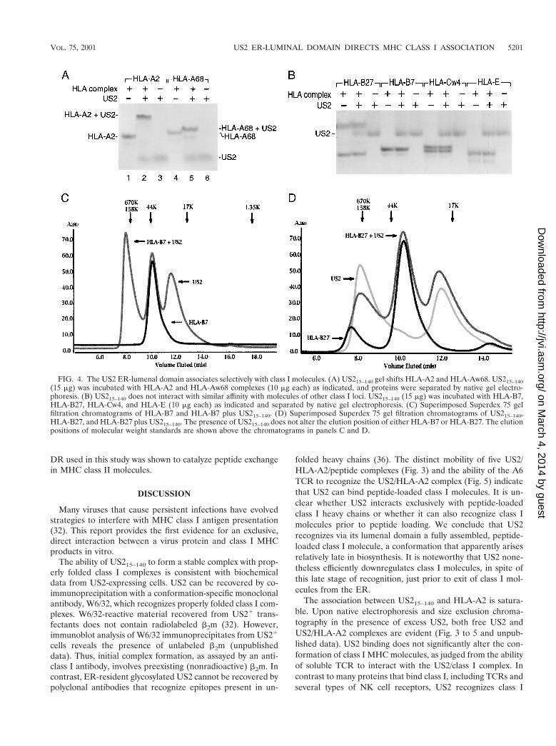
US215–140 maintains locus-specific binding. US2 demon-strates locus specificity in its ability to downregulate class Iheavy chains in mammalian cells. While both HLA-A andHLA-B locus products are susceptible to attack in cells ex-pressing US2, HLA-C, HLA-E, and HLA-G complexes resistUS2-mediated degradation (8, 27). To examine the bindingcharacteristics of US215–140, a series of class I complexes wererefolded from bacterial inclusion bodies in the presence of the
appropriate peptide ligands. Purified US215–140 was incubatedwith either HLA-A2, HLA-Aw68, HLA-B7, or HLA-B27. As-sociation between class I molecules and US2 was assessed bynative gel electrophoresis. US215–140 forms complexes withboth HLA-A locus products (Fig. 4A), but an interaction witheither HLA-B7 or HLA-B27 could not be detected at US2concentrations that readily gel shift HLA-A2 (Fig. 4B). Fur-ther elevation of the US2 concentration by a factor of 10 didnot result in either HLA-B7 or HLA-B27 gel shifts (unpub-lished data). Furthermore, neither HLA-Cw4 nor HLA-Eshowed any signs of interaction with soluble US2 under con-ditions where the HLA-A locus products readily bind (Fig.4B). Elevating the US2 concentration by 10-fold over the min-imal requirement to effectuate a quantitative gel shift forHLA-A2 did not produce a class I gel shift. US2 does nottarget H-2 Kb for degradation (21), and US215–140 likewisedoes not alter the mobility of soluble Kb in native gel electro-phoresis (unpublished data).
Not only did we fail to detect such interaction by native gelelectrophoresis, but also gel filtration chromatography failed toshow evidence for an interaction between either HLA-B7 orHLA-B27 and US215–140 despite a fivefold molar excess ofsoluble US2 (Fig. 4C and D).
US215–140 and TCR can simultaneously interact with MHCclass I. It is not known whether the conformation of class Imolecules changes upon association with US2, nor is it clearexactly where US2 interacts with MHC class I products. Toexamine this question in vitro, we asked whether US215–140 andT-cell receptor (TCR) can recognize the same class I complex.Purified A6 TCR, which is specific for the HLA-A2/Tax com-plex (10), was incubated with HLA-A2/Tax and US215–140.Complex assembly was monitored by band shift in native gelelectrophoresis. A protein complex of distinct mobility is gen-erated when HLA-A2, US215–140, and A6 TCR are incubatedtogether (Fig. 5). The HLA-A2/US215–140/TCR complexformed regardless of the order of addition. Thus, the bindingof US215–140 to class I molecules does not apparently inducemajor conformational changes in the class I complex in solu-tion, as the A6 TCR maintains the ability to associate withUS2/class I complexes. The formation of this novel complexfurther suggests that US2 and TCR interact with class I mol-ecules at distinct and nonoverlapping binding sites.
US215–140 does not interact with HLA-DR or HLA-DM. Al-though US2 was originally shown to downregulate expressionof class I products, US2 has been suggested to also target fordestruction components of the MHC class II antigen presen-tation pathway, HLA-DRa and HLA-DMa. To determinewhether US215–140 can associate with complexes in the class IIpathway, recombinant soluble HLA-DR1 and HLA-DM wereincubated with US215–140 and complex formation was moni-tored by native gel electrophoresis. As shown in Fig. 6,US215–140 does not alter the mobility of either DR1 or DMcomplexes in native gel electrophoresis. No gel shift was ob-served for DR1 or DM even in the presence of US2 concen-trations 10-fold higher than the amount required to quantita-tively bind HLA-A2 complexes. The mobility of a second DRallele was likewise not altered in the presence of US215–140.The preparation of DR used is known to produce a gel shiftupon complexation with the TCR under conditions essentiallyidentical to those used here (unpublished data). The batch of
FIG. 3. US215–140 binds to five distinct HLA-A2/peptide com-plexes. US215–140 (15 mg) gel shifts HLA-A2 complexes (7.5 mg) con-taining the following peptides: group 1, LLFGYPVYV; group 2,ILKEPVHGV; group 3, ELAGIGILTV; group 4, ALGIGILTV;group 5, FLPSDFFPSV.
5200 GEWURZ ET AL. J. VIROL.
on March 4, 2014 by guest
http://jvi.asm.org/
Dow
nloaded from
DR used in this study was shown to catalyze peptide exchangein MHC class II molecules.
DISCUSSION
Many viruses that cause persistent infections have evolvedstrategies to interfere with MHC class I antigen presentation(32). This report provides the first evidence for an exclusive,direct interaction between a virus protein and class I MHCproducts in vitro.
The ability of US215–140 to form a stable complex with prop-erly folded class I complexes is consistent with biochemicaldata from US2-expressing cells. US2 can be recovered by co-immunoprecipitation with a conformation-specific monoclonalantibody, W6/32, which recognizes properly folded class I com-plexes. W6/32-reactive material recovered from US21 trans-fectants does not contain radiolabeled b2m (32). However,immunoblot analysis of W6/32 immunoprecipitates from US21
cells reveals the presence of unlabeled b2m (unpublisheddata). Thus, initial complex formation, as assayed by an anti-class I antibody, involves preexisting (nonradioactive) b2m. Incontrast, ER-resident glycosylated US2 cannot be recovered bypolyclonal antibodies that recognize epitopes present in un-
folded heavy chains (36). The distinct mobility of five US2/HLA-A2/peptide complexes (Fig. 3) and the ability of the A6TCR to recognize the US2/HLA-A2 complex (Fig. 5) indicatethat US2 can bind peptide-loaded class I molecules. It is un-clear whether US2 interacts exclusively with peptide-loadedclass I heavy chains or whether it can also recognize class Imolecules prior to peptide loading. We conclude that US2recognizes via its lumenal domain a fully assembled, peptide-loaded class I molecule, a conformation that apparently arisesrelatively late in biosynthesis. It is noteworthy that US2 none-theless efficiently downregulates class I molecules, in spite ofthis late stage of recognition, just prior to exit of class I mol-ecules from the ER.
The association between US215–140 and HLA-A2 is satura-ble. Upon native electrophoresis and size exclusion chroma-tography in the presence of excess US2, both free US2 andUS2/HLA-A2 complexes are evident (Fig. 3 to 5 and unpub-lished data). US2 binding does not significantly alter the con-formation of class I MHC molecules, as judged from the abilityof soluble TCR to interact with the US2/class I complex. Incontrast to many proteins that bind class I, including TCRs andseveral types of NK cell receptors, US2 recognizes class I
FIG. 4. The US2 ER-lumenal domain associates selectively with class I molecules. (A) US215–140 gel shifts HLA-A2 and HLA-Aw68. US215–140(15 mg) was incubated with HLA-A2 and HLA-Aw68 complexes (10 mg each) as indicated, and proteins were separated by native gel electro-phoresis. (B) US215–140 does not interact with similar affinity with molecules of other class I loci. US215–140 (15 mg) was incubated with HLA-B7,HLA-B27, HLA-Cw4, and HLA-E (10 mg each) as indicated and separated by native gel electrophoresis. (C) Superimposed Superdex 75 gelfiltration chromatograms of HLA-B7 and HLA-B7 plus US215–140. (D) Superimposed Superdex 75 gel filtration chromatograms of US215–140,HLA-B27, and HLA-B27 plus US215–140. The presence of US215–140 does not alter the elution position of either HLA-B7 or HLA-B27. The elutionpositions of molecular weight standards are shown above the chromatograms in panels C and D.
VOL. 75, 2001 US2 ER-LUMINAL DOMAIN DIRECTS MHC CLASS I ASSOCIATION 5201
on March 4, 2014 by guest
http://jvi.asm.org/
Dow
nloaded from

molecules regardless of the sequence of the peptide cargo,consistent with the ability of US2 to bind class I molecules ata site distinct from that seen by the TCR. US215–140 interactswith apparently similar affinities with a series of HLA-A2 com-plexes that possess distinct peptides. While most immune sys-tem receptors for class I, including the TCR, several NK re-ceptors, and CD8, are able to form homodimers orheterodimers, recombinant US2 is a monomer in solution andbinds to class I molecules with a 1:1 stoichiometry. Studies withcell-permeable chemical cross-linkers have likewise failed toreveal dimerization of US2 in cellular transfectants (unpub-lished data).
In transfectants that express either US2 or US11, all HLA-Aand HLA-B class I molecules appear susceptible to degrada-tion, as judged from the reduction in their surface expressionand from the accelerated degradation of the bulk of newlysynthesized class I molecules (references 35 and 36 and un-published observations). In other words, there is no evidenceto suggest the selective resistance of the HLA-A or HLA-Blocus products to the activity of US2 or US11. Surprisingly, ourdata show that the luminal domain of US2 is sufficient to allowstable complex formation with HLA-A2 and HLA-A68,whereas neither HLA-B7 nor HLA-B27 appears capable ofdoing so. If the luminal domain of US2 were the sole region ofthe molecule responsible for recognition of class I heavychains, we would predict resistance of these HLA-B locusalleles to US2-mediated degradation, and this is not the case.Interestingly, the association between US3 and class I in vivorequires both the US3 ER-lumenal and transmembrane do-mains (20), and the recombinant US3 ER-lumenal domaindoes gel shift MHC class I in vitro (unpublished data). Theseobservations suggest that perhaps the US2 and class I trans-membrane segments may also interact to account for US2’sability to target HLA-B locus products for degradation in livingcells.
The downregulation of class I molecules is a precarious actfor viruses, as NK cells lyse cells that lose MHC class I expres-sion (25). However, the known human NK receptors for classI focus primarily on the HLA-C and HLA-E locus products. Byselectively downregulating HLA-A and HLA-B class I mole-
cules, viruses can diminish antigen presentation to CD81 Tlymphocytes while limiting NK cell activation (27). The datapresented in this paper suggest that the US2 ER-lumenal do-main plays an important role in allowing US2 to distinguishbetween MHC class I locus products. In contrast, HIV Nef andKHSV K5 focus on the class I transmembrane and tail regionsfor distinguishing between class I locus products (5, 15). Thesesegments contain an abundance of locus-specific residues (11).Most of the polymorphic residues in class I are concentrated inregions that contact peptide or TCR, and US2 does not com-pete with TCR for class I association. It is all the more unusualthat US2 can use the polymorphisms in the class I lumenaldomain to distinguish between class I molecules. Unlike US2and US11, HCMV US3 does not demonstrate locus-specificinteraction with class I (19).
It seems paradoxical that HCMV should encode two pro-teins, US2 and US11, with apparently redundant functions. Asstrong selection pressures constantly maintain compact virusgenomes, it is a fair assumption that expression of both US2and US11 benefits HCMV. Murine cytomegalovirus likewiseencodes multiple proteins dedicated to class I downregulation,although without apparent homology to their HCMV paral-ogues and availing themselves of cell biologically distinctmechanisms to accomplish their goal (14). We have suggestedthat the presence of both US2 and US11 may ensure thatHCMV will be able to counter the high degree of class Ipolymorphism (21). US11 associates with free class I heavychains in coimmunoprecipitation experiments in cellular trans-fectants, which indicates recognition by US11 of a motif thatarises earlier in class I assembly. Perhaps US2 and US11 safe-guard against the escape of class I molecules from the ER byfocusing on different stages of the class I biosynthetic pathway.Further structural and mechanistic analyses should provide anexplanation for why HCMV employs both proteins.
US215–140 does not bind HLA-DR and HLA-DM with suf-ficient affinity to produce a shift on native gel electrophoresis.In contrast, it has previously been reported that US2 associateswith HLA-DR and HLA-DM complexes and subsequently tar-
FIG. 5. US215–140 and a TCR can simultaneously interact with thesame HLA-A2 molecules. US215–140 (15 mg), HLA-A2/Tax (17 mg),and soluble A6 TCR (6.6 mg) were incubated as indicated, and pro-teins were separated by native gel electrophoresis. TCR alone is shownby the empty arrowhead, HLA-A2/Tax/TCR complex is shown by theasterisk, and HLA-A2/Tax/US215–140/TCR complex is shown by thefilled arrowhead.
FIG. 6. US215–140 does not form high-affinity complexes with eitherHLA-DR or HLA-DM. US215–140 (15 mg) was incubated withHLA-DR (5 mg) or HLA-DM (10 mg) as indicated, and proteins wereseparated by native gel electrophoresis.
5202 GEWURZ ET AL. J. VIROL.
on March 4, 2014 by guest
http://jvi.asm.org/
Dow
nloaded from

gets the DRa and DMa subunits for degradation (31). Severalexplanations could explain the discrepancy between these find-ings. It is possible that distinct regions of US2 mediate inter-action with MHC class I and class II molecules. Should this bethe case, we predict that the US2 transmembrane and/or cy-toplasmic tail would contribute significantly to association withHLA-DM and HLA-DR complexes, as we suggest to be thecase for the interaction of US2 with HLA-B locus products.Alternatively, it remains possible that another factor is re-quired to stabilize the interaction between US2 and DR orDM. Finally, US2 may bind with higher affinity to class Imolecules than to either DR or DM molecules. Consequently,it is plausible that US2 can be induced to associate withHLA-DR and HLA-DM when overexpressed in the ER.
The mechanism by which US2 targets class I heavy chains fordislocation remains unclear. We have hypothesized that US2subverts a poorly understood homeostatic “quality control”process. The quality control machinery in the ER recognizesand removes misfolded proteins from the ER. We now suggestthat association between the ER-luminal domains of US2 andclass I initiates dislocation of class I heavy chains. However,interaction between US2 and class I does not appear to signif-icantly alter the conformation of class I molecules. The detailsof the events that connect formation of a tight complex be-tween US2 and class I in the ER and the appearance of classI heavy chains in the cytosol remain uncertain. Further, thestability of the US2/HLA-A2 complex and the absence of ob-vious conformational alterations raise important questionsabout the details of MHC I dislocation. Is the complex disas-sembled prior to discharge into the cytosol, and if so, whichproteins assist in this unfolding? Alternatively, it is conceivablethat the entire US2-class I complex is dislocated via the Sec61channel, whose diameter is reported to range from 20 to 60 Å(12, 23). The data reported here conclusively demonstrate thelumenal domain’s importance for binding and call attention tothe proposed transmembrane segment and cytoplasmic tail asessential for the dislocation reaction.
ACKNOWLEDGMENTS
We thank Rachelle Gaudet, Joydeep Mitra, and members of thePloegh lab for assistance.
This work was supported by a grant from the National Institutes ofHealth (5R37-AI33456). B.E.G. is a predoctoral fellow of the HowardHughes Medical Institute. E.W.W. is supported by a National CancerInstitute Fellowship in Cancer Biology. D.T. is a Charles A. King Trustpostdoctoral fellow. D.J.S. is supported by a Women’s ReproductiveHealth Research Training Grant from the National Institutes ofHealth (K12-HD01255).
REFERENCES
1. Ahn, K., A. Angulo, P. Ghazal, P. A. Peterson, Y. Yang, and K. Fruh. 1996.Human cytomegalovirus inhibits antigen presentation by a sequential mul-tistep process. Proc. Natl. Acad. Sci. USA 93:10990–10995.
2. Baas, E. J., H. M. van Santen, M. J. Kleijmeer, H. J. Geuze, P. J. Peters, andH. L. Ploegh. 1992. Peptide-induced stabilization and intracellular localiza-tion of empty HLA class I complexes. J. Exp. Med. 176:147–156.
3. Boppana, S. B., and W. J. Britt. 1996. Recognition of human cytomegalovi-rus gene products by HCMV-specific cytotoxic T cells. Virology 222:293–296.
4. Britt, W. J., and C. A. Alford (ed.). 1996. Cytomegalovirus, 3rd ed., vol. 2.Lippincott-Raven Publishers, Philadelphia, Pa.
5. Cohen, G. B., R. T. Gandhi, D. M. Davis, O. Mandelboim, B. K. Chen, J. L.Strominger, and D. Baltimore. 1999. The selective downregulation of class Imajor histocompatibility complex proteins by HIV-1 protects HIV-infectedcells from NK cells. Immunity 10:661–671.
6. Cresswell, P., N. Bangia, T. Dick, and G. Diedrich. 1999. The nature of the
MHC class I peptide loading complex. Immunol. Rev. 172:21–28.7. Fan, Q. R., D. N. Garboczi, C. C. Winter, N. Wagtmann, E. O. Long, and
D. C. Wiley. 1996. Direct binding of a soluble natural killer cell inhibitoryreceptor to a soluble human leukocyte antigen-Cw4 class I major histocom-patibility complex molecule. Proc. Natl. Acad. Sci. USA 93:7178–7183.
8. Furman, H. M., H. L. Ploegh, and D. J. Schust. 2000. Can viruses help us tounderstand and classify the MHC class I molecules at the maternal-fetalinterface? Hum. Immunol. 61:1169–1176.
9. Garboczi, D. N., D. T. Hung, and D. C. Wiley. 1992. HLA-A2-peptidecomplexes: refolding and crystallization of molecules expressed in Esche-richia coli and complexed with single antigenic peptides. Proc. Natl. Acad.Sci. USA 89:3429–3433.
10. Garboczi, D. N., U. Utz, P. Ghosh, A. Seth, J. Kim, E. A. VanTienhoven,W. E. Biddison, and D. C. Wiley. 1996. Assembly, specific binding, andcrystallization of a human TCR-alphabeta with an antigenic Tax peptidefrom human T lymphotropic virus type 1 and the class I MHC moleculeHLA-A2. J. Immunol. 157:5403–5410.
11. Gussow, D., R. S. Rein, I. Meijer, W. de Hoog, G. H. Seemann, F. M.Hochstenbach, and H. L. Ploegh. 1987. Isolation, expression, and the pri-mary structure of HLA-Cw1 and HLA-Cw2 genes: evolutionary aspects.Immunogenetics 25:313–322.
12. Hamman, B. D., J. C. Chen, E. E. Johnson, and A. E. Johnson. 1997. Theaqueous pore through the translocon has a diameter of 40–60 A duringcotranslational protein translocation at the ER membrane. Cell 89:535–544.
13. Heemels, T. M., and H. Ploegh. 1995. Generation, translocation, and pre-sentation of MHC class I-restricted peptides. Annu. Rev. Biochem. 64:463–491.
14. Hengel, H., U. Reusch, A. Gutermann, H. Ziegler, S. Jonjic, P. Lucin, andU. H. Koszinowski. 1999. Cytomegaloviral control of MHC class I functionin the mouse. Immunol. Rev. 168:167–176.
15. Ishido, S., C. Wang, B. S. Lee, G. B. Cohen, and J. U. Jung. 2000. Down-regulation of major histocompatibility complex class I molecules by Kaposi’ssarcoma-associated herpesvirus K3 and K5 proteins. J. Virol. 74:5300–5309.
16. Jensen, P. E., D. A. Weber, W. P. Thayer, X. Chen, and C. T. Dao. 1999.HLA-DM and the MHC class II antigen presentation pathway. Immunol.Res. 20:195–205.
17. Jones, T. R., L. K. Hanson, L. Sun, J. S. Slater, R. M. Stenberg, and A. E.Campbell. 1995. Multiple independent loci within the human cytomegalovi-rus unique short region down-regulate expression of major histocompatibil-ity complex class I heavy chains. J. Virol. 69:4830–4841.
18. Jones, T. R., E. J. Wiertz, L. Sun, K. N. Fish, J. A. Nelson, and H. L. Ploegh.1996. Human cytomegalovirus US3 impairs transport and maturation ofmajor histocompatibility complex class I heavy chains. Proc. Natl. Acad. Sci.USA 93:11327–11333.
19. Jun, Y., E. Kim, M. Jin, H. C. Sung, H. Han, D. E. Geraghty, and K. Ahn.2000. Human cytomegalovirus gene products US3 and US6 down-regulatetrophoblast class I MHC molecules. J. Immunol. 164:805–811.
20. Lee, S., J. Yoon, B. Park, Y. Jun, M. Jin, H. C. Sung, I. H. Kim, S. Kang, E. J.Choi, B. Y. Ahn, and K. Ahn. 2000. Structural and functional dissection ofhuman cytomegalovirus US3 in binding major histocompatibility complexclass I molecules. J. Virol. 74:11262–11269.
21. Machold, R. P., E. J. Wiertz, T. R. Jones, and H. L. Ploegh. 1997. TheHCMV gene products US11 and US2 differ in their ability to attack allelicforms of murine major histocompatibility complex (MHC) class I heavychains. J. Exp. Med. 185:363–366.
22. Martoglio, B., and B. Dobberstein. 1998. Signal sequences: more than justgreasy peptides. Trends Cell Biol. 8:410–415.
23. Menetret, J., A. Neuhof, D. G. Morgan, K. Plath, M. Radermacher, T. A.Rapoport, and C. W. Akey. 2000. The structure of ribosome-channel com-plexes engaged in protein translocation. Mol. Cell 6:1219–1232.
24. Miller, D. M., B. M. Rahill, J. M. Boss, M. D. Lairmore, J. E. Durbin, J. W.Waldman, and D. D. Sedmak. 1998. Human cytomegalovirus inhibits majorhistocompatibility complex class II expression by disruption of the Jak/Statpathway. J. Exp. Med. 187:675–683.
25. Ravetch, J. V., and L. L. Lanier. 2000. Immune inhibitory receptors. Science290:84–89.
26. Reddehase, M. J. 2000. The immunogenicity of human and murine cyto-megaloviruses. Curr. Opin. Immunol. 12:390–396.
27. Schust, D. J., D. Tortorella, J. Seebach, C. Phan, and H. L. Ploegh. 1998.Trophoblast class I major histocompatibility complex (MHC) products areresistant to rapid degradation imposed by the human cytomegalovirus(HCMV) gene products US2 and US11. J. Exp. Med. 188:497–503.
28. Soderberg-Naucler, C., K. N. Fish, and J. A. Nelson. 1997. Reactivation oflatent human cytomegalovirus by allogeneic stimulation of blood cells fromhealthy donors. Cell 91:119–126.
29. Stevenson, P. G., S. Efstathiou, P. C. Doherty, and P. J. Lehner. 2000.Inhibition of MHC class I-restricted antigen presentation by gamma 2-her-pesviruses. Proc. Natl. Acad. Sci. USA 97:8455–8460.
30. Story, C. M., M. H. Furman, and H. L. Ploegh. 1999. The cytosolic tail ofclass I MHC heavy chain is required for its dislocation by the human cyto-megalovirus US2 and US11 gene products. Proc. Natl. Acad. Sci. USA96:8516–8521.
VOL. 75, 2001 US2 ER-LUMINAL DOMAIN DIRECTS MHC CLASS I ASSOCIATION 5203
on March 4, 2014 by guest
http://jvi.asm.org/
Dow
nloaded from
31. Tomazin, R., J. Boname, N. R. Hegde, D. M. Lewinsohn, Y. Altschuler, T. R.Jones, P. Cresswell, J. A. Nelson, S. R. Riddell, and D. C. Johnson. 1999.Cytomegalovirus US2 destroys two components of the MHC class II path-way, preventing recognition by CD41 T cells. Nat. Med. 5:1039–1043.
32. Tortorella, D., B. E. Gewurz, M. H. Furman, D. J. Schust, and H. L. Ploegh.2000. Viral subversion of the immune system. Annu. Rev. Immunol. 18:861–926.
33. Tortorella, D., C. M. Story, J. B. Huppa, E. J. Wiertz, T. R. Jones, I. Bacik,J. R. Bennink, J. W. Yewdell, and H. L. Ploegh. 1998. Dislocation of type Imembrane proteins from the ER to the cytosol is sensitive to changes inredox potential. J. Cell Biol. 142:365–376.
34. Townsend, A., C. Ohlen, J. Bastin, H. G. Ljunggren, L. Foster, and K. Karre.
1989. Association of class I major histocompatibility heavy and light chainsinduced by viral peptides. Nature 340:443–448.
35. Wiertz, E. J., T. R. Jones, L. Sun, M. Bogyo, H. J. Geuze, and H. L. Ploegh.1996. The human cytomegalovirus US11 gene product dislocates MHC classI heavy chains from the endoplasmic reticulum to the cytosol. Cell 84:769–779.
36. Wiertz, E. J., D. Tortorella, M. Bogyo, J. Yu, W. Mothes, T. R. Jones, T. A.Rapoport, and H. L. Ploegh. 1996. Sec61-mediated transfer of a membraneprotein from the endoplasmic reticulum to the proteasome for destruction.Nature 384:432–438.
37. Yewdell, J. W., and J. R. Bennink. 2001. Cut and trim: generating MHC classI peptide ligands. Curr. Opin. Immunol. 13:13–18.
5204 GEWURZ ET AL. J. VIROL.
on March 4, 2014 by guest
http://jvi.asm.org/
Dow
nloaded from
Copyright © 2022 FDOKUMEN