HPTLC 2017 Book of abstracts - Justus-Liebig-Universität ...
219
HPTLC 2017 International Symposium for High-Performance Thin-Layer Chromatography Book of abstracts Berlin, 4 – 8 July 2017
-
Upload
khangminh22 -
Category
Documents
-
view
1 -
download
0
Transcript of HPTLC 2017 Book of abstracts - Justus-Liebig-Universität ...

HPTLC 2017
International Symposium for
High-Performance
Thin-Layer Chromatography
Book of abstracts
Berlin, 4 – 8 July 2017

Some statistics
176 Abstract submissions
59 Lectures
111 Posters
210 Participants from 30 countries
32 % Germany
20 % India
8 % Switzerland
7 % France
6 % Poland
3 % USA
2 % Romania, China, Slovenia, Italy, Austria…
1 % The Netherlands, Spain, Australia, Guatemala,
Hungary, Lithuania, Serbia and UK…
0.5 % Algeria, Burma, Chile, Malaysia, Mexico, Nigeria,
Russia, Iran, Thailand, Korea, Taiwan…
Especially thanks to the countries missed to mention!
Profession
55 % Academics
24 % Industry
21 % Students
Revised and edited by Prof. Dr. Gertrud Morlock
Justus Liebig University Giessen, Germany, 22nd June 2017, updated 9th July 2017

Welcome to Berlin!
The Scientific Committee and Organization Committee of HPTLC 2017 welcome you to Berlin, the beautiful capital of Germany. A number of world renowned experts in HPTLC and an amazing number of young researchers have gathered (22 of them compete for the Young Researcher Award). Together, they build a striking platform for learning and sharing of knowledge and the latest research in HPTLC. The sympo-sium will present a diverse scientific program with 59 lectures, including 2 tutorials, 111 poster presentations, panel discussion with manufacturers and an active social program. Three short courses are offered as a pre-program.
The scientific program may be an activator for questions and discussions. The ex-change of experiences and ideas is essential for the advancement of HPTLC. Hy-phenations in HPTLC, for example with mass spectrometry or bioassays, are more and more recognized as powerful tools and increase the general interest in HPTLC. Analysts learn that HPTLC is a suitable tool to solve their analytical tasks. Miniatur-ized systems and a streamlined data treatment are part of our challenging future. We will enjoy the views on HPTLC from 30 different nations represented in Berlin. The many facets will trigger inspiration for new applications and needs. The flexibil-ity of the method is unlimited - be sure, the only barrier is our brain. Get excited by each other and new ideas will be born.
We wish that you become a multiplicator when you are back. Improve the teaching and training in HPTLC at your site. Transfer the understanding for the potential and power of HPTLC into your group. Make a difference! This efficient method deserves its space in the training of the next generation of scientists. It is necessary to advo-cate for the best analytical solution, in which decision-making process, HPTLC is of-ten not seen as a powerful option. Knowledge needs to be spread!
On behalf of HPTLC 2017, Gertrud Morlock and Colin Poole

The series of international HPTLC Symposia
2-4 July 2014, Lyon
(22nd) International Symposium for High-Performance Thin-Layer Chromatography, HPTLC 2014
6-8 July 2011, Basel (21st) International Symposium for High-Performance Thin-Layer Chromatography, HPTLC 2011
11-13 June 2008, Helsinki (20th) International Symposium for Thin-Layer Chromatography, HPTLC 2008
9-11 October 2006, Berlin (19th) International Symposium for Thin-Layer Chromatography, HPTLC 2006
29-31 May 2005, Siofok (18th) Planar Chromatography 2005
23-25 May 2004, Visegrad (17th) Planar Chromatography 2004
15-18 October 2003, Lyon (16th) International Symposium for TLC
21-23 June 2003, Budapest (15th) Planar Chromatography 2003 (in honor of Professor Tyihak)
4-6 October 2002, Novo mesto (14th) Planar Chromatography Today 2002
11-13 May 2002, Keszthely (13th) Planar Chromatography 2002 (in honor of Doctor Geiss)
23-25 June 2001, Lillafüred (12th) Planar Chromatography 2001
11-13 May 2000, Lillafüred (11th) Planar Chromatography 2000 (in honor of Professor Kaiser)
16-19 May 1998, Visegrad 10th International Symposium on Instrumental Planar Chromatography (60 years TLC + 10 years JPC)
9-11 April 1997, Interlaken 9th International Symposium on Instrumental Planar Chromatography
5-7 April 1995, Interlaken 8th International Symposium on Instrumental Planar Chromatography
23-26 March 1993, Brighton 7th International Symposium on Instrumental Planar Chromatography
23-26 April 1991, Interlaken Sixth International Symposium on Instrumental Planar Chromatography
21-24 February 1989, Brighton (Fifth) International Symposium on Instrumental High Performance Thin-Layer Chromatography
22–25 September 1987, Selvino Fourth International Symposium on Instrumental High Performance Thin-Layer Chromatography
17–19 April 1985, Würzburg Third International Symposium on Instrumental High-Performance Thin-Layer Chromatography
2-6 May 1982, Interlaken Second International Symposium on Instrumental High-Performance Thin-Layer Chromatography
18-21 May 1980, Bad Dürkheim First International Symposium on Instrumentalized High-Performance Thin-Layer Chromatography
Local Organization Committee
Prof. Dr. Lothar Kroh, Germany
Pierre Bernard-Savary, France
Dr. Konstantinos Natsias, Germany
Prof. Dr. Gertrud Morlock, Germany

International Scientific Committee
Co-Chair: Prof. Dr. Colin Poole, USA and Prof. Dr. Gertrud Morlock, Germany
Pierre Bernard-Savary, France
Dr. Vicente Cebolla, Spain
Prof. Dr. Wanchai De-Eknamkul, Thailand
Prof. Dr. Imre Klebovich, Hungary
Prof. Dr. Teresa Kowalska, Poland
Prof. Dr. Lothar Kroh, Germany
Prof. Dr. Ilkka Ojanperä, Finland
Dr. Eike Reich, Switzerland
Prof. Dr. Joseph Sherma, USA
Prof. Dr. Mario Vega, Chile
Dr. Irena Vovk, Slovenia
Prof. Dr. Zheng Tao Wang, China

Scientific Honorary Board
Prof. Dr. Rudolf Kaiser, Germany
Dr. Dieter Jänchen, Switzerland
Dr. Fred Rabel, USA
Prof. Dr. Victor Berezkin, Russian Federation
Prof. Dr. David Nurok, USA
Prof. Dr. Xie Peishan, China
Prof. Dr. Siegfried Ebel, Germany
The Scientific Community deeply recognizes the lifetime achievement*
of the Honorary Board Members and those who passed away
for planar chromatography!
Thank you for being the best example and an amazing source of inspiration!
*Attached only in the electronic version at www.hptlc.com
CHROMart by Drs. Karla & Herbert Halpaap

In honor of distinguished scientists we have lost since HPTLC 2014
Dr. Friedrich Geiss, Italy 25.02.1932 - 14.02.2015
• Defined terms, created understanding and turned the trial and error approach in-to a scientific and sound methodology
• Author of Fundamentals of Thin Layer Chromatography (Planar Chromatography) 1987, Russian version 1989, German version 1972, Japanese version 1980
• Invention of the Vario KS Chamber with his team, whose successor is frequently in use for optimizations
• Awardee of the Tswett Medal in 2002
• Author of numerous papers and books in the area of chemistry and of several books related to societal and political matters
Dr. P. D. Sethi, India 18.11.1936 - 26.09.2015
• India’s first Government Scientist who realized the potential of HPTLC in 1985; Former Director of Central Indian Pharmacopeia Lab and Central Drug Testing Lab
• Set up QTLC method for analysis of birth control pills in 1987 (10000 samples/a)
• Member of several Government Committees for pharmaceutical analysis and herbal product standardization
• A primary force to adopt fingerprinting for herbal medicines as a first choice in India
• 13 books on pharmaceutical analysis including three volumes on multi drug for-mulation analysis by TLC/HPTLC methods and one on Content Uniformity Testing by HPTLC

Prof. Dr. Edward Soczewiński, Poland 04.09.1928 - 12.12.2016
• Author of 333 chapters of books and papers as well as of many patents (e. g., for production of cadmium oxide, chelidonine, protopine and chambers)
• Research on molecular model of retention in normal phase systems: Soczewiński equation, Anal. Chem. 1968
• Coauthor of Soczewiński-Wachtmeister equation used in QSAR investigations
• Editorial Board Member of chromatographic journals
• Awardee of many prizes and distinctions, e. g., Officers and Chevalier’s of the Polonia Restituta Order, Golden Cross of Merit of Poland, Tswett Medal and doctor honoris causa of Medical Academy of Lublin
Prof. Dr. Ernő Tyihák, Hungary 29.01.1933 - 13.02.2017
• Author of more than 200 papers and 25 patents, mainly on OPLC
• Inventor of ultramicro chamber in 1971
• Co-inventor of pressurized ultramicro chamber and OPLC, J. Chromatogr. 174 (1979) 75
• Invention of BioArena a complex bioautographic system and the discovery of the role of formaldehyde in the biological systems, Chem. Anal. (Warsaw) 48 (2003) 543
• Editorial Board Member of Journal of Planar Chromatography and Honorary Board Member of Hungarian Society for Separation Sciences
• Awardee of many prizes and distinctions, e. g., Győző Bruckner, Károly Than and Hormesis Awards, Honorary Professor at the Szeged University, Golden Diploma of Budapest Technical University

Committee for Poster Prizes and Young Researcher Award
Chair: Prof. Dr. Rudolf Kaiser, Germany
Prof. Dr. Sznezana Agatonovic-Kustrin, Malaysia
Prof. Dr. Danica Agbaba, Serbia
Dr. Vicente Cebolla, Spain
Prof. Dr. Wanchai De-Eknamkul, Thailand
Prof. Dr. Tadeusz Dzido, Poland
Prof. Dr. Imre Klebovich, Hungary
Prof. Dr. Teresa Kowalska, Poland
Prof. Dr. Lothar Kroh, Germany
Prof. Dr. Matthew Linford, USA
Dr. Agnes Moricz, Hungary
Prof. Dr. Susan Olesik, USA
Prof. Dr. Colin Poole, USA
Prof. Dr. Wolfgang Schwack, Germany
Prof. Dr. Navin Sheth, India
Prof. Dr. Jentaie Shiea, Taiwan
Prof. Dr. Robert Verpoorte, The Netherlands
Dr. Irena Vovk, Slovenia

Announcements
A ChromART for welcoming you!
You successfully struggled to be here.
In the congress hotel Maritim proArte free internet access is linked to your room
number. If there is need for attendees not accommodated in this hotel, a free daily
voucher can be obtained upon request at the registration desk.
The world energy still in repair… Though the latest version of the book of abstracts
is available on www.hptlc.com, it was printed on recycling paper. Attendees, who
do not need the printed version, please hand it back to the symposium registration
desk. Thank you!
You face a tight scientific schedule and with discipline, we may stay in time. Friday
evening is free. On Saturday, you can recover during the social program. On agree-
ment, lectures will be linked at www.hptlc.com. We would like to link your poster on
the symposium homepage, too. Please email a low-resolution pdf-file (just named
according to the poster number 1 to 111) to [email protected]. Thank you in advance!
We arranged several prizes to support and encourage the young generation in
HPTLC. We are a very lucky scientific community - we have a great future, as we
have the best young researchers in the world!

Dieter Jänchen Award for the Young Researcher
We have the great honor to award an outstanding young researcher.
Thanks to the CAMAG Foundation, the honor is accompanied by 3000 Euro
which is intended to support a research stay leading to a publication.
This award is celebrated on Friday 7th July 2017 before the closure.
Young Researcher Award dedicated to Helmut Jork
We have the great honor to award an outstanding young researcher.
The honor of this price is accompanied by 2000 Euro
which is intended to support a research stay leading to a publication.
This award is celebrated on Friday 7th July 2017 before the closure.

Young Researcher Award dedicated to Friedrich Geiss
We have the great honor to award one outstanding young researcher.
The honor of this price is accompanied by 1000 Euro
which is intended to support a research stay leading to a publication.
This award is celebrated on Friday 7th July 2017 before the closure.
Poster Prizes
All posters presented compete for the Poster Prizes,
i. e. 3 bronze, 2 silver and 1 gold.
Thanks to CRC Press and Elsevier, the honor of the Poster Prize
is accompanied by the latest HPTLC book.
The 6 prizes are awarded during the symposium dinner
on Thursday 6th July 2017.

Short courses
Location
Technical University of Berlin (Technische Universität Berlin), Institute of Food
Technology and Food Chemistry (Institut für Lebensmitteltechnologie und Le-
bensmittelchemie), Prof. Dr. Lothar Kroh, Gustav-Meyer Allee 25, 13355 Berlin
Tuesday, 4th July 2017
14:00 - 17:00
1. HPTLC-MS for characterization of compounds
2. Lipids’ characterization and quantification
3. Analysis of botanicals in compliance with USP <203> and PhEur 2.8.25
At 13:45, we meet at the Gustav-Meyer Allee 25, department of Prof. Dr. Lothar
Kroh (3. floor). From the Maritim proArte Hotel, it takes ca. 25 min. Please organize
yourself to be at this meeting point in time. It is recommended to take the
S2 (direction Buch) to the stop Humboldthain and walk ca. 6 min to the institute.

Symposium schedule
Location
Maritim proArte Hotel, Friedrichstraße 151, 10117 Berlin
Coffee breaks of 30 min start always at 10:30 and 15:00
Wednesday, 5th July 2017
08:00 Registration - mounting of posters 1-51
09:00 Opening
09:30-12:00 Oral session 1 and Tutorial 1
12:00 Poster session 1-51 with lunch
13:30-17:00 Oral sessions 2 and 3
17:00 Poster session 1-51
18:00 Panel discussion with manufacturers
19:00 Welcome reception
20:00 End – switch of posters
Thursday, 6th July 2017
09:00-12:30 Oral sessions 4 and 5
12:30 Lunch with Poster session 52-111
13:30-17:00 Oral sessions 6 and 7
17:00 Poster session 52-111
19:00 Symposium dinner with Poster Awards
21:00 End
Friday, 7th July 2017
09:00-12:30 Oral sessions 8 and 9 as well as Tutorial 2
12:30 Lunch
13:30-15:15 Oral session 10
15:30 Young Researcher Awards
15:45 Closure
16:00 End - demounting of posters

Social program
Saturday, 8th July 2017
12:15-16:15 Sightseeing boat tour ending with the well-known
“Berliner Currywurst” at the restaurant “Ständige Vertretung”
Register for this tour during symposium registration on Wednesday. At 12:15, we
meet at the Anlegestelle Friedrichstraße/Reichstagufer to start this boat tour.
Please organize yourself to be at this meeting point in time.

16:30-18:00 Visit of the German Federal Parliament
Only participants, who did register online for this event, can join this guided tour!
We meet at 16:30 at the central visitor entrance (Scheidemann Strasse) at the west-
ern portal of the Reichstag building (Deutscher Bundestag, www.bundestag.de).
Please organize yourself to be at this meeting point in time.
For security reasons, large pieces of luggage may not be taken into the building. No
storage facilities are available on site. Please note that during the whole visit a valid
passport or identification card must be carried along, as your proof of identity is re-
quired for entering. You will also be checked with metal detectors, while smaller bags,
coats and other items will be subjected to an X-ray examination.

Oral and Poster Presentations
Maritim proArte Hotel, Berlin
WED 5th
08:00 Registration - mounting of posters 1-51
Opening
09:00 KROH Germany Surprise and Welcome
in bold: Invited Speaker or Scientific Committee Member in italics: Young Scientist
Session 1: Miniaturization - Chair: Poole/Morlock
09:30 O-1 OLESIK USA Ion-exchange separation of biomolecules using UTLC
09:45 O-2 LINFORD USA Manufacturable microfabrication of patterned UTLC plates
10:00 O-3 SCHULZ Germany Particulate silica gel layers - new developments and perspectives
10:15 O-4 FICHOU Germany Office Chromatography
10:30 Coffee break (30 min)
Tutorial 1
11:00 O-5 VERPOORTE NL Publishing a world-class paper in HPTLC
12:00 Session of posters 1-51 with lunch

Session 2: Strong features of HPTLC - Chair: Agatonovic-Kustrin/Verpoorte
13:30 O-6 VOVK Slovenia Multidimensional planar chromatography coupled to mass spectrometry - unbeatable in the analysis of crude plant extracts
13:45 O-7 YÜCE Germany Targeted combinatorial on-plate synthesis as new tool for structure elucidation
14:00 O-8 OBER-LERCHNER Austria Bridging the analytical gap - comprehensive analysis
of cellooligosaccharides by HPTLC 14:15 O-9 REICH Switzer-
land Can HPTLC help solve the quality problems of botan-ical dietary supplements?
14:30 O-10 STIEFEL Germany Application of hyphenated HPTLC in food, commodi-ty and cosmetics analysis
14:45 O-11 CAÑIGU-ERAL
Spain HPTLC for herbal drugs and herbal drug prepara-tions in the European Pharmacopoeia
15:00 Coffee break (30 min)
Session 3: Effect-directed detection - Chair: De-Eknamkul/Schwack
15:30 O-12 MORICZ Hungary Layer chromatography hyphenations assisted screening, characterization and isolation of bioactive plant components
15:45 O-13 CHOMA Poland Quantitative effect-directed analysis based on TLC/HPTLC-direct bioautography
16:00 O-14 JAMSHIDI- AIDJI Germany Bioquantification of natural antibiotics by direct bio-
autography linked to mass spectrometry
16:15 O-15 SCHOEN-BORN
Switzer-land
Patterns of estrogenic activity in treated wastewater - a study from Switzerland
16:30 O-16 SPANGEN-BERG Germany 2D-thin layer chromatography of 17α-ethinylestra-
diol on RP-18 W plate, detected by YES-test
16:45 O-17 WEINS Germany Effect directed analysis, a new challenge according the upcoming enhancement of European Water Framework Directive 2000/60/EC in 2019
17:00 Session of posters 1-51
18:00 Panel discussion with manufacturers - Chair: Bernard-Savary
19:00 Welcome reception
20:00 End and switch of posters

THU 6th
in bold: Invited Speaker or Scientific Committee Member in italics: Young Scientist
Session 4: Fundamentals - Chair: Choma/Spangenberg
9:00 O-18 KOWAL-SKA
Poland Performance of chiral TLC in physico-chemical studies
9.15 O-19 DZIDO Poland New approach to development of planar chromato-grams
9:30 O-20 KLEBO-VICH
Hungary Radiochromatographic methods in drug metabolism research
9:45 O-21 HALKA Poland Reversed phase gradient thin-layer chromatography with one void volume of the mobile phase: advan-tages, pitfalls and prospects for the future
10:00 O-22 GAJOS Poland Micropreparative orthogonal pressurized planar electrochromatography of solutes showing the same electrophoretic mobility
10:15 O-23 POLAK Poland Separation of the optical isomers with pressurized planar electrochromatography
10:30 Coffee break (30 min)
Session 5: Elution head-based HPTLC-MS - Chair: Cañigueral/Shiea
11:00 O-24 CEBOLLA Spain HPTLC-ESI-MS/MS for identifying neutral lipids, sphingolipids and phospholipids in complex samples
11:15 O-25 ILANGO India Hyphenated HPTLC-AMD-ESI/MS for quantification of major protoberberines in herbal extracts and polyherbal formulations
11:30 O-26 MERKERT UK TLC-MS coupling taken seriously: expression CMS and Plate Express
11:45 O-27 VAN BEELEN
France Bringing the power of mass detection to scientists using TLC combined with the ACQUITY QDa detector
12:00 O-28 GRIESIN-GER
Germany Unexpected products of the HOCl-induced oxidation of oleic acid: a study using HPTLC-ESI-MS
12:15 O-29 GUPTA India HPTLC-MS based method development for cardio-vascular disease controlling compounds containing plant Coleus forskolii as per US chapter 203
substituted by LIHUA China Application of TLC/HPTLC in identification of Chinese crude drugs for ChP
12:30 Session of posters 52-111 with lunch (1h)

Session 6: Desorption-based HPTLC-MS and structure elucidating techniques -
Chair: Ansari/Linford
13:30 O-30 SHIEA Taiwan TLC combined with flame-induced atmospheric pres-sure chemical ionization mass spectrometry (FAP-CI/MS) for volatile and semi-volatile comp. analysis
13:45 O-31 HÄBE Germany Automated desorption- and elution-based HPTLC-MS
14:00 O-32 FEREY France TLC-UV and TLC-MALDI-TOF/MS: an efficient tool for enzyme characterization and screening of bioactive substrates
14:15 O-33 CHEN China HPTLC + SERS > HPLC + MS 14:30 O-34 LONGIE-
RAS France TLC and TLC-Raman as an effective tool for polymer
additives deformulation 14:45 O-35 AZAD-
NIYA Germany HPTLC-EDA-HRMS and PLC-NMR to reveal co-eluting
isomers of bioactive zones
15:00 Coffee break (30 min)
Session 7: Botanicals and traditional medicines – Chair: Moricz/Cebolla
15:30 O-36 DE-EK-NAMKUL
Thailand HPTLC detection of steroid 5α-reductase activity from a non-radioactive cell-based assay
15:45 O-37 FADEL France Antioxidants in structured vegetable oils: chemical identification via HPTLC
16:00 O-38 BALLERT Germany Multidimensional chromatography (HPLC-HPTLC) for identification of antifungal substances in Rheum root extracts
16:15 O-39 BELETE Ethiopia HPTLC assay of thymoquinone in black seed and black seed oil (Nigella sativa Linn) and identification of thymoquinone conversion with UV/Vis
16:30 O-40 ANSARI India Development and validation of a HPTLC method for simultaneous estimation of flavonoids and phenolics in Carica papaya leaf juice
16:45 O-41 AHMAD India HPTLC studies on single drugs and compound for-mulations of the Indian system of medicine
17:00 O-42 SHRIVA-STAVA
India Chemical signature and multiple marker analysis of Avipattikar Churna: an Ayurvedic multicomponent formulation for quality assessment
17:00 Session of posters 52-111
19:00-21:00 Symposium dinner and poster award ceremony

FRI 7th
in bold: Invited Speaker or Scientific Committee Member in italics: Young Scientist
Session 8: Pharmaceutical analysis – Chair: Sheth/Vovk
09:00 O-43 GAWANDE India Preparative isolation and characterization of degra-dation products of cefixime and azithromycin
09:15 O-44 THORAT India Development and validation of a HPTLC method for determination of methotrexate in human serum
09:30 O-45 THUMAR India Extractable and leachable study of phthalates in pharmaceutical products by TLC versus LC-MS/MS
Tutorial 2
09:45 O-46 POOLE USA What every chromatographer should know about solvents
10:30 Coffee break (30 min)
Session 9: Food and dietary supplements – Chair: Olesik/Kroh
11:00 O-47 SCHWACK Germany Determination of total glucosinolates in Brassica crops
11:15 O-48 SOSTARIC Australia Authentication of honeys of different floral origins 11:30 O-49 BEITLICH Germany TLC screening for the authentication of New Zealand
Manuka honey 11:45 O-50 OELLIG Germany Screening methods for ergots by HPTLC-FLD/MS 12:00 O-51 DO Switzer-
land Rapid HPTLC screening and quantification of adul-teration with synthetic drugs in dietary supplements
12:15 O-52 CHATUR-VEDI
India Antioxidant compound production at different germi-nating stages of cow pea (Vigna articulata L.) seeds varieties and aspartic protease gene expression
12:30 Lunch (1 h)

Session 10: Data treatment - Chair: Klebovich/Dzido
13:30 O-53 AGATO-NOVIC-KUSTRIN
Malaysia Evaluation of polyphenolic fingerprints and antioxi-dant profiles of Victorian marine algae with HPTLC and multivariate analysis
13:45 O-54 RISTIVO-JEVIC
Serbia Hyphenation of planar chromatography with chemometrics
14:00 O-55 MAQUIN France Software associated with TLC/HPTLC-MS coupling 14:15 O-56 BÖHM-
DORFER Austria Clustering analysis of colored wheat varieties by an-
thocyanin patterns 14:30 O-57 MALI-
NOWS-KA
Poland Biological activity is one of the most important pro-perties of substances, not only used as drugs, but also as food additives, cosmetics, dietary supplements etc., depending on permeability through biological barrier
14:45 O-58 ROUSSEL France Modern HPTLC method validation, application of prediction and tolerance intervals to dextrine pro-files of enzymatic digestion of starch and baking products
15:00 O-59 VAN OORDT
Switzer-land
Comparison of different derivatization techniques including the Derivatizer
Closure with coffee
15:30 KAISER Germany Young Researcher Awards
15:45 POOLE USA Highlights
Demounting of posters
Thank is owed to all the presenters!

List of poster presentations
Poster group 1: Strong feature of HPTLC
P-1 Novel micro-fabricated TLC plates P-2 Application of artificial neural network to planar chromatography data P-3 Open-source developments for Office Chromatography P-4 rTLC: Open source software for multivariate analysis of HPTLC data P-5 Glass breakage-free TLC/HPTLC dipping chambers P-6 HPTLC as a tool to investigate chemical communication in fungi P-7 Quantitative analysis of monosaccharides from lignocellulosic material P-8 New approach of HPTLC for identification of auxins in frost resistant plants P-9 Assessment of sterol and steroid content in human breast adipose tissue P-10 Separation of pigment formulations by HPTLC/AMD P-11 HPTLC-aptastaining - Innovative protein detection system for HPTLC
Poster group 2: Effect-directed detection
P-12 Bioprofiling of cosmetics with focus on coumarin analysis P-13 Determination of bioactive compounds in vanilla and its products P-14 Bioactive compounds found in ginger and ginger-containing food via
HPTLC-UV/Vis/FLD-EDA-HRMS P-15 Fingerprinting and bioprofiling of anti-TB medicinal plants by an effect-
directed HPTLC method P-16 Analysis of anti-diabetic compounds in herbal extracts via HPTLC-enzyme
inhibition assay P-17 Effect-directed analysis of Agrocybe cylindracea bioactive compounds P-18 Direct bioautography with subsequent DART-MS P-19 Semi-quantitative comparison of acetylcholinesterase inhibition in effect-
directed analysis with HPTLC P-20 Selective two-dimensional effect-directed analysis with TLC P-21 Modern direct bioautography for fast screening and characterization of
active compounds in plant extracts used in cosmetics P-22 In vinum veritas: Estrogen-effective compounds discovered in wine by
HPTLC-pYES P-23 Investigation of plant protection products for endocrine effects by direct
bioautography P-24 pYES with the substrate RGP for the detection of estrogen active com-
pounds in sewage P-25 HPTLC coupled estrogenic activity assessment of the phytoestrogens
genistein and biochanin A in nutraceutical red clover (Trifolium pratense L.) formulations

P-26 Estrogenic substances in treated wastewater used for crop irrigation in
Cyprus - Preliminary results using the planar-YES bioassay P-27 HPTLC-hyphenated bioautography for antidiabetic and antioxidant metabo-
lites from Butea monosperma P-28 Antimicrobial activity of effective antimicrobial compounds in extracts from
strawberry leaves by TLC P-29 HPTLC as a method for quick assessment of bile salts deconjugation activity
by Pediococcus acidilactici LAB6 and Lactobacillus plantarum LAB12
Poster group 3: Analysis of food/feed/cosmetics
P-30 Quantification of phospholipids using HPTLC and primuline-induced fluores-cence detection by intensity changes
P-31 Anthocyanin profiles of colored wheat crosses via HPTLC P-32 In-process quality control of wine by (micro) planar chromatography P-33 Fast screening of veterinary drugs in food of animal origin via pSPE-HRMS P-34 Planar solid phase extraction-gas chromatography mass spectrometry for
the determination of sterol oxidation products in cosmetics P-35 Optimization of HPTLC and HPTLC-MS methods for analysis of flavonoids
and phenolic acids P-36 Quantification of α- and β-acids in hops by TLC and HPLC P-37 The advantages of HPTLC based on USP TLC methods for the analysis of
black pepper, turmeric and ginger P-38 Development and validation of an HPTLC-densitometry method for simulta-
neous quantitation of boric acid and calcium fructoborate in dietary sup-plements and foodstuffs
P-39 HPTLC-densitometry method for nicotinamide riboside analysis in bulk and nutraceutical formulations
P-40 Lysergic acid amide screening for the total ergot alkaloids in rye by HPTLC-FLD P-41 Comparison of fluorescent derivatization reagents and development of a
simple quantitation strategy for lipid analysis by HPTLC-FLD P-42 Screening for MOSH and MOAH in food packaging by pSPE-UV/FLD-GC-MS P-43 Phenolic acids contribution to the total antioxidant activities in in mango
pulp and peel P-44 Simultaneous estimation of five markers from medicinally important man-
groves, Avicennia marina and Sonneratia apetala P-45 System suitability testing in HPTLC methods for herbal drugs in the Ph. Eur. -
Highly reproducible identity testing in raspberry leaf monograph using standardized stationary phases and automated equipment
P-46 Chemical analysis and evaluation of antioxidant and antiglycation properties of under-investigated plants from the Auvergne region (France)
P-47
Quantification of cypermethrin in shampoo by HPTLC
P-48 HPTLC fingerprint profile analysis of cocoa proanthocyanidins depending on origin and genotype
P-49 Curcumin contents in Myanmar species P-50 In situ hydrolysis of glycosylated flavonoids from leaves and fruits of caigua
on HPTLC silica gel plates P-51 HPTLC-MS and HPTLC-DPPH methods for characterization and assessment
of antioxidant properties of flavonoids from fresh leaves and fruits of caigua
Poster group 4: Fundamentals and theoretical aspects
P-52 Performance of the HPTLC systems with controlled velocity of the mobile phase P-53 The influence of metallic impurities on the distortions of HPTLC chromatograms
and retention of basic/amphoteric compounds P-54 Comparison of retention of DNS amino acids in TLC and pressurized planar
electrochromatography systems with silica gel P-55 A sample preparation with semi-automatic TLC for quantitative analysis with
HPLC and HPLC/MS techniques P-56 A non-chromatographic use of HPTLC instrumentation P-57 There's plenty of room at the top - increased sample throughput by quantita-
tive à côté calibration
Poster group 5: Coupling techniques
P-58 Analysis of sterols and steroids using HPTLC-MS: influence of ionization parameters
P-59 Miniaturized single quadrupole mass detector for HPTLC-MS P-60 Suzuki reaction monitoring using TLC-MS P-61 Beyond HPLC-MS: Profiling of high molecular weight impurities during drug
synthesis P-62 Using 2D-HPTLC-MALDI-TOF MS for a first screening approach of plant
extracts P-63 Analysis of glycosaminoglycan oligosaccharides by combined HPTLC-MALDI-
TOF MS: reduced silica gel thickness leads to improved spectral qualities and reduced side reactions
P-64 Determination of UV filter in sun cream using TLC-MS P-65 Streamlined structure elucidation of unknowns in formulations P-66 Automated hyphenation of HPTLC to DART-MS and ESI-MS P-67 Ambient ionization in the proximity of the mass spectrometer P-68 HPTLC-EDA-HRMS and PLC-NMR spectroscopy for structural elucidation of
active compounds in Salvia miltiorrhiza P-69 Unexpected products of the HOCl-induced oxidation of oleic acid: a study
using HPTLC-ESI MS
P-70 Application of normal and reversed phase TLC in the analysis of lipid oxida-tion products
P-71 TLC/HPTLC-MS with or without other chromatographic detectors (DAD-UV; ELSD)
P-72 Tips and tricks for TLC-MS
Poster group 6: Analysis of botanicals
P-73 Determination of rosmarinic acid in Melissa officinalis leaves, derived extracts and plant food supplements by HPTLC
P-74 Straightforward process for the identification and isolation of natural prod-ucts using TLC and preparative chromatography
P-75 HPTLC quantification of rhein from the rhizomes of Sansevieria roxburghiana P-76 HPTLC: An Important analytical method for the standardisation of herbal
extracts P-77 Detection and quantification of some chemical compositions of Thymus
daenensis and Thymus lancifolius by HPTLC P-78 TLC as tool for the analysis of resins of Liquidambar styraciflua P-79 Determination of flavanones in the buds of some species and hybrids of
Populus P-80 Quantitative analysis of ledol and alloaromadendrene by HPTLC with densi-
tometric detection in Rhododendron tomentosum (Ledum palustre) plants and in vitro cultures
P-81 HPTLC fingerprinting of six Lagochilus species from Uzbekistan P-82 HPTLC method for quantification of lawsone in micrpropagated Lawsonia
inermis L. P-83 α-Amylase inhibition and antioxidant activity of Myrmecodia platytyrea (ant
plant) P-84 Comparative standardization study for determination of reserpine in Rauwol-
fia serpentina homoeopathic mother tinctures manufactured by different pharmaceutical industries using HPTLC as check for quality control
P-85 Quantification of curcumin and eugenol marketed formulations and method validation by using HPTLC
P-86 QSSR analysis based on TLC data of selected antipsychotics and their impurities P-87 Trees – tracking effects of environmental micro-pollutants P-88 Development of validated HPTLC method for the estimation of eugenol in
marketed ayurvedic medicine for application on gums and teeth P-89 16-O-Methylcafestol as marker for Robusta admixture in Coffea arabica by
HPTLC-FLD P-90 Differentiation of the origin of caffeine products (botanical vs. chemical), and
estimation of the caffeine level by HPTLC P-91 Fingerprint of an Astragalus mongholicus extract by HPTLC and LC-MS and
quantification of formononetin with densitometric HPTLC

Poster group 7: Toxicological analysis
P-92 Comparison of HPTLC-MS methods on silica gel and diol plates for determina-tion of proanthocyanidins in Japanese knotweed
P-93 Application of TLC to ecotoxicological study with the Steatoda grossa spider web model
P-94 Screening for phenethylamines in pre-workout supplements P-95 Identification of Cannabis sativa strains and determination of the THC and THC
acid content by HPTLC P-96 Development and validation of an HPTLC method for simultaneous estimation
of rifampicin, isoniazide and pyrazinamide in human serum P-97 Role of HPTLC in analysis of depressants P-98 Extraction, isolation and detection of ethambutol in blood using HPTLC plate P-99 HPTLC as a tool for the detection and separation of three structurally related
organophosphorus pesticides of forensic importance on NP-TLC and RP-TLC layers
P-100 Rapid detection of pesticides of forensic importance by HPTLC P-101 Comparison of two techniques for urine screening of cannabis: Immunoassay
(EMIT) versus GC-MS
Poster group 8: Pharmaceutical analysis
P-102 Phytochemical and pharmacological evaluation of Rhododendron arboreum: An ethnomedicinal plant from Himalayas
P-103 Quantitative determination of topiramate in human breast milk by HPTLC P-104 Extraction and identification of paracetamol in biological material such as
tissue, blood and urine using TLC P-105 Analysis of penicillin and tetracyclic antibiotics of Indian pharmaceutical com-
panies in whole blood by planar chromatography P-106 Stress degradation behaviour of adapalene by a validated HPTLC method and
characterization of its degradation product by LC-MS/MS P-107 Validation of an HPTLC method for the determination of zidovudine during in
vitro permeation studies P-108 Development and validation of an HPTLC-UV method to determine paroxetine
hydrochloride from in vitro skin permeation P-109 Application of a validated HPTLC method for content uniformity test of hydro-
chlorothiazide, amlodipine besylate and olmesartan in tablet dosage form and its comparison with LC
P-110 Validated HPTLC method and content uniformity test for analysis of rosuvas-tatin and aspirin in tablet dosage form and its comparison with LC
P-111 Estimation of genotoxic impurity of quetiapine by HPTLC method

O-1
Ion-exchange separation of biomolecules using UTLC
OLESIK S [email protected]
Olesik S1, Wang Y1 1The Ohio State University, Columbus, USA
Ion exchange chromatography is used for the separation of inorganic cations and anions and a broad range of important organic ionic compounds, especially in bioanalytical chemistry. This presentation will illustrate the development of UTLC plates using electrospun Nafion-Polyacrylonitrile (PAN) nanofibers. The sulfonate groups on Nafion provide the cation exchange sites. PAN was used to facilitate the nanofiber formation in the electrospinning process. Optimization of electrospinning parameters and separation conditions using factorial designs and response surface methodology will be described. Next a range of useful applications of these newly developed UTLC plates will be highlighted such as the separation of amino acids, peptides, and proteins. Finally, these devices provided high efficiency, selectivity and chemical stability for these applications. These capabilities will be illustrated in this talk.

O-2
Manufacturable microfabrication of patterned UTLC plates
LINFORD MR [email protected]
Linford MR1, Chatterjee S1, Major GH1 1Brigham Young University, Provo, UT, USA
In previous publications we described the microfabrication of TLC plates based on carbon nanotube scaffolds [1-3]. These plates showed high resolution separations, very fast development times, and a high degree of robustness. We believe that based on economies of scale these plates should be commercially viable. Nevertheless, because of the multiple microfabrication/vacuum deposition steps involved in their production, it has been challenging to convince a manufacturer to take the necessary risk to develop them. Accordingly, we have embarked on a new fabrication scheme that involves only one microfabrication step and no vacuum depositions. In this talk we will describe progress towards this goal. We will discuss this process, show material characterization at each step, and show the initial chromatograms generated by this approach.
[1] J. Chrom. A. 2015, 1404, 115–123 [2] J. Planar Chromatogr. 2014, 27, 151–156 [3] J. Chrom. A. 2012, 1257, 195–203

O-3
Particulate silica gel layers - new developments and perspectives
SCHULZ M [email protected]
Schulz M1, Burholt M1, Griesinger H1, Oberle M1 1Merck KGaA, Darmstadt, Germany
The first standardized silica gel materials for TLC according to Stahl were available in 1958 followed by the start of industrial production of TLC plates in 1966 when Merck introduced the first pre-coated TLC plates onto the market. Another milestone was the introduction of pre-coated HPTLC layers with increased performance in 1975. Together with the development of precise instrumentation, HPTLC was born. Here we would like to present new developments of particulate layers. A comparison of performance parameters is presented as well as a pathway for miniaturization.

O-4
Office Chromatography
FICHOU D [email protected]
Fichou D1, Morlock GE1 1Justus Liebig University Giessen, Institute of Nutritional Science, Chair of Food
Science, Interdisciplinary Research Center (IFZ), Giessen, Germany
Office Chromatography is a concept in which all steps of the planar chromato-graphy are performed by a single, all-in-one, miniaturized device. Using open-source technology and especially 3D printing, a prototype of such a device was developed. A slurry doser had been designed to print silica gel layers, opening new avenues for tailor-made layers regarding the nature and shape of the stationary phase. Sample application was performed by a thermal inkjet print-head, the use of the open-source InkShield board enabled full control of the application and was compatible with aqueous and methanolic solutions. The same print-head was used to apply an hydromethanolic mobile phase on a RP18-W phase for the separation of water soluble dyes. A home-made software was developed to control the apparatus. Deployed on a raspberry pi acting as server, it allowed control of the device from an internet browser on any computer on the local network, removing the need for software installation. The remaining steps of the analytical pipeline, i. e. derivatization and documentation are under development and once ready, will make the Office Chromatography concept complete.

O-5 → Tutorial 1
Publishing a world-class paper in HPTLC
VERPOORTE R [email protected]
Natural Products Laboratory, IBL, Leiden University, Leiden, The Netherlands
Science is based on communication. To communicate our experimental results to our colleagues, to evaluate the research in our field in a review, to review papers for journals, to present posters or lectures, to write project proposals, to write reports, to give interviews, to...... Each of these requires a different style, based on the audience you are addressing: your colleagues who know very well the field, reviewers of your proposals, students, users of your research, the general public, etc. Writing is an important tool for a scientist, but if I ask you "do you like to write", what is your honest answer? At least you will have a good feeling once your paper is published, so all reason to take the challenge of writing a paper.
How to get a world-class paper? The answer is simple: the basis is to plan your experiments in a proper way. If you have a clear objective and translate that to a design of experiments with the right controls and number of replicates to allow a proper statistical analysis you have the basis for a world class paper. The introduction of your paper should give the background to the final objective of your study and insight in the design of your experiments.
The results are the core of any experimental paper; they are facts that are used in a discussion that relates the results to the objective of your study. To show the results figures are easiest, any colleague will be able to understand the meaning of the results independent of language. So before writing a word, make illustrations that visualize your results. With that material it will be easy to write the discussion of your paper. The materials and methods describe strict protocols that you used, rather easy to write, just all details so others can do the experiments again.
Do not expect to write the perfect paper in the first round, you will need many versions before coming to the final one. Ask colleagues to read your work, to hear if they read what you thought you wrote. Write, write, write.....

O-6
Multidimensional planar chromatography coupled to mass spectrometry -
unbeatable in the analysis of crude plant extracts
VOVK I [email protected]
Vovk I1, Glavnik V1, Albreht A1, Kranjc E1 1Department of Food Chemistry, National Institute of Chemistry, Ljubljana, Slovenia
We will present planar chromatography (from one dimensional to multidimen-sional) combined with different detections (UV, Vis, fluorescence, MS, etc. before and after derivatization) as an unbeatable technique in the analyses of crude plant extracts in spite of complex matrices and the lack of reference standards. The de-veloped HPTLC methods based on different combinations of stationary phases (sili-ca gel, RP-18, diol, cellulose), developing solvents and detection techniques (image analysis, densitometry, MS) enabled analyses of crude extracts of Japanese knot-weed (Fallopia japonica Houtt.) and Chinese lantern (Physalis alkekengi L.). The in-fluence of the sorbent, pre-developing and developing solvents on ion suppression in HPTLC-MS and HPTLC-MS2 analyses was minimized during method development.
The developed HPTLC silica gel-densitometry method enabled the separation of structurally similar physalins in Chinese lantern crude extracts, although only one physalin standard is available. This method also provides an alternative selectivity, better sensitivity and higher resolution for some physalins compared to the pub-lished (U)HPLC methods. An innovative simultaneous hyphenation of HPTLC with two different mass analyzers enabled a reliable and straightforward non-targeted characterization of physalins and the determination of their types in Chinese lan-tern crude extracts.
Proanthocyanidins (up to decamers) from knotweed crude extracts were separated according to raising molecular masses using silica gel and diol sorbents, and suc-cessfully identified by HPTLC-MSn, although only standards of monomers and some dimers are available. We also tested the potential of multidimensional planar chromatography (MPC) on two different sorbents and MPC-MS before and after post-chromatographic derivatization (DMACA [1]) in fast analysis of proantho-cyanidins in knotweed crude extracts.
[1] V. Glavnik, B. Simonovska, I. Vovk, J. Chromatogr. A 1216 (2009) 4485-4491

O-7
Targeted combinatorial on-plate synthesis as new tool for structure elucidation
YÜCE I [email protected]
Yüce I1, Morlock GE1 1Justus Liebig University Giessen, Institute of Nutritional Science, Chair of Food
Science, Interdisciplinary Research Center (IFZ), Giessen, Germany
Targeted in situ synthesis on a chromatographic layer supported a fast reaction, if compared to reactions in solutions as conventionally performed in organic chemistry. Through synthesis on a porous surface, a substantial reduction of solvent consumption can be reached, which supported environmentally friendly reactions. Combinatorial surface reactions were shown as new tool for a fast structure elucidation of impurities and contaminants in pharmaceutical formu-lations, when standard compounds are not commercially available.
This strategy was demonstrated for the targeted synthesis of impurities occurring in different pharmaceutical products. The impurities and reagents for synthesis were automatically applied as overspotted bands (reaction zone) on the HPTLC layer. If required, heating the layer accelerated the reaction and the products formed in the reaction zone were purified by a subsequent chromatographic separation. The product zone of interest was online transferred via an elution-head based interface into the high-resolution mass spectrometer for structural characterization. Thus, the whole process from synthesis via reaction control and structure elucidation was carried out on the same layer.
As proof of concept, the formulations and the combinatorial synthesis were analyzed in parallel in vials and on the surface (HPTLC layer). Quantitatively evalua-ted via videodensitometry, on-surface synthesis provided the same yields of impurities in minutes versus a full day in conventional synthesis. This new green chemistry workflow was much faster, cheaper as well as much more economically and environmentally friendly, if compared to reactions in solution. All these advantages made surface synthesis on a chromatographic layer an efficient new tool for impurity research in formulations and for the quality control of chemical mixtures in industry.

O-8
Bridging the analytical gap - comprehensive analysis of cellooligosaccharides
by HPTLC
OBERLERCHNER J [email protected]
Oberlerchner J1, Böhmdorfer S1, Rosenau T1, Potthast A1 1BOKU, University of Natural Resources and Life Sciences, Department of
Chemistry, Division of Chemistry of Renewable Resources, Vienna, Austria
New analytical tools are required in biomass processing to evaluate both raw material composition and the effects of processing. The focus of the pulp and paper industry - one of the existing bio-refineries - is currently shifting towards complete utilization of the biomass feedstock, commercializing not only cellulosic pulp but also isolating compounds from highly heterogeneous process liquors. Cellooligosaccharides (COS) with a degree of polymerization (DP) of 2 to 20 are inevitably formed during cellulose manufacturing and are an appreciable, but currently unused byproduct. Chemical analysis of carbohydrate oligomers is a particular challenge, and the few suitable methods (anion exchange and size exclusion chromatography) struggle with the high salt and matrix load and extreme pH of process samples.
We therefore developed a normal phase (NP)-HPTLC method to quantitate COS in biomass product streams. This method can separate acetylated COS with a DP of 1 to 15 in a single development and determine the absolute quantity of each component by scanning densitometry. Even anomeric products of equal molecular weight were distinctly separated. Monodisperse standards were prepared by acetolysis followed by preparative HPLC, and the structure of each standard was confirmed by 2D-NMR and MALDI-ToF-MS. Additionally, the identity and purity of each standard component after separation was approved by MALDI-ToF-MS directly off the plate. For routine use, standard mixtures were prepared in the form of an "oligomer ladder", following the concept of protein standard ladders.
HPTLC excelled at the analysis of these hardly accessible and separable compounds, with high stability even in the cases of complex matrices, contaminated specimens, or industrial samples, with a much shorter analysis time per sample in comparison to NP-HPLC. The qualitative analysis was verified by MALDI-ToF-MS, while the preparation of individual standard allowed the quantification of each individual COS.
O-9
Can HPTLC help solve the quality problems of botanical dietary supplements?
REICH E [email protected]
Reich E1, Frommenwiler D1 1CAMAG, Muttenz, Switzerland
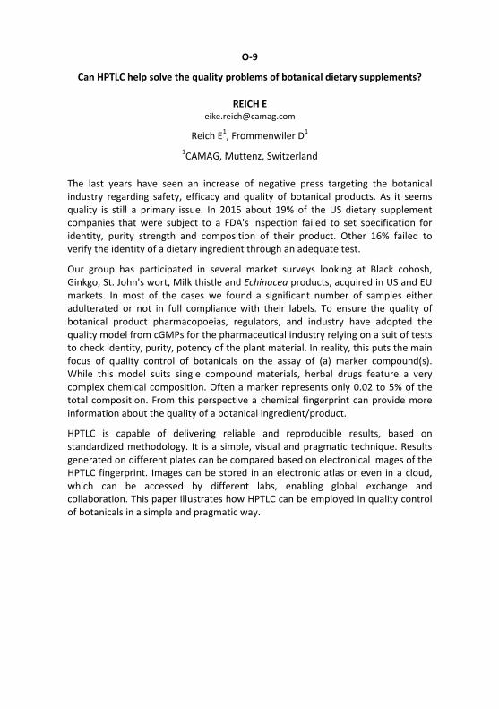
The last years have seen an increase of negative press targeting the botanical industry regarding safety, efficacy and quality of botanical products. As it seems quality is still a primary issue. In 2015 about 19% of the US dietary supplement companies that were subject to a FDA's inspection failed to set specification for identity, purity strength and composition of their product. Other 16% failed to verify the identity of a dietary ingredient through an adequate test.
Our group has participated in several market surveys looking at Black cohosh, Ginkgo, St. John's wort, Milk thistle and Echinacea products, acquired in US and EU markets. In most of the cases we found a significant number of samples either adulterated or not in full compliance with their labels. To ensure the quality of botanical product pharmacopoeias, regulators, and industry have adopted the quality model from cGMPs for the pharmaceutical industry relying on a suit of tests to check identity, purity, potency of the plant material. In reality, this puts the main focus of quality control of botanicals on the assay of (a) marker compound(s). While this model suits single compound materials, herbal drugs feature a very complex chemical composition. Often a marker represents only 0.02 to 5% of the total composition. From this perspective a chemical fingerprint can provide more information about the quality of a botanical ingredient/product.
HPTLC is capable of delivering reliable and reproducible results, based on standardized methodology. It is a simple, visual and pragmatic technique. Results generated on different plates can be compared based on electronical images of the HPTLC fingerprint. Images can be stored in an electronic atlas or even in a cloud, which can be accessed by different labs, enabling global exchange and collaboration. This paper illustrates how HPTLC can be employed in quality control of botanicals in a simple and pragmatic way.
O-10
Application of hyphenated HPTLC in food, commodity and cosmetics analysis
STIEFEL C [email protected]
Stiefel C1, Morlock GE1 1Justus Liebig University Giessen, Institute of Nutritional Science, Chair of Food
Science, Interdisciplinary Research Center (IFZ), Giessen, Germany
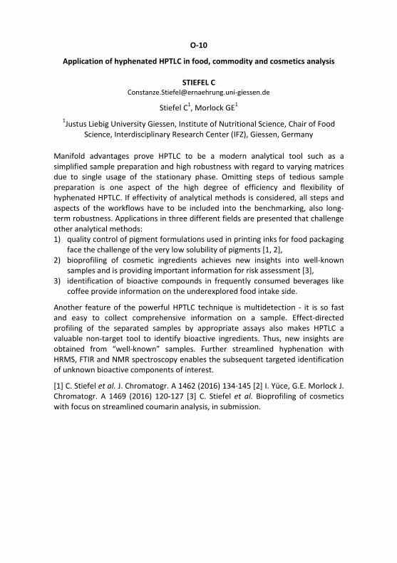
Manifold advantages prove HPTLC to be a modern analytical tool such as a simplified sample preparation and high robustness with regard to varying matrices due to single usage of the stationary phase. Omitting steps of tedious sample preparation is one aspect of the high degree of efficiency and flexibility of hyphenated HPTLC. If effectivity of analytical methods is considered, all steps and aspects of the workflows have to be included into the benchmarking, also long-term robustness. Applications in three different fields are presented that challenge other analytical methods: 1) quality control of pigment formulations used in printing inks for food packaging
face the challenge of the very low solubility of pigments [1, 2], 2) bioprofiling of cosmetic ingredients achieves new insights into well-known
samples and is providing important information for risk assessment [3], 3) identification of bioactive compounds in frequently consumed beverages like
coffee provide information on the underexplored food intake side.
Another feature of the powerful HPTLC technique is multidetection - it is so fast and easy to collect comprehensive information on a sample. Effect-directed profiling of the separated samples by appropriate assays also makes HPTLC a valuable non-target tool to identify bioactive ingredients. Thus, new insights are obtained from “well-known” samples. Further streamlined hyphenation with HRMS, FTIR and NMR spectroscopy enables the subsequent targeted identification of unknown bioactive components of interest.
[1] C. Stiefel et al. J. Chromatogr. A 1462 (2016) 134-145 [2] I. Yüce, G.E. Morlock J. Chromatogr. A 1469 (2016) 120-127 [3] C. Stiefel et al. Bioprofiling of cosmetics with focus on streamlined coumarin analysis, in submission.

O-11
HPTLC for herbal drugs and herbal drug preparations in the
European Pharmacopoeia
CAÑIGUERAL S [email protected]
Unitat de Farmacologia, Farmacognòsia i Terapèutica, Facultat de Farmàcia i Ciències de l'Alimentació, Universitat de Barcelona, Barcelona, Spain
Herbal medicinal products contain herbal drugs (HD) or herbal preparations (HP) (extracts, essential oils, etc.) as active pharmaceutical ingredients (API). Since they have a high chemical complexity and they are considered the API in its entirety, the traditional approach of assaying a selected marker has a limited significance for the level of quality and it is under discussion. A more holistic approach, considering a wider range of constituents would be suitable. In this context, the chromatographic profiling of HD and HP appears as an essential tool to stablish its quality.
Analysis by TLC, and especially HPTLC, is essential for identification and detection of adulterations and falsifications, and can also be very useful in the quantitative assessment of herbal API as well as for stability studies. Nevertheless, TLC analysis may present problems, mainly due to the inherent variability of the plant material and to the lack of reproducibility inter- and intra-laboratory, as well as to difficulties for describing and interpreting the results.
The introduction of HPTLC with a detailed description of the method, comprising a system suitability test, allows a better control of chromatographic conditions and a higher reproducibility of the results. Concerning the description of the chromato-grams, it needs to take into account the position, colour, and intensity of the zones. The use of intensity markers allows producing better descriptions and, together with colour pictures of chromatograms of several batches, can help the user with the interpretation of the descriptions. All this provisions have been taken in account by the European Pharmacopoeia in the preparation of the new general chapter (2.8.25) on HPTLC analysis of HD and HP, which have been published in the 9th edition [1].
[1] EDQM (2016) High Performance Thin-layer Chromatography of herbal drugs and herbal drug preparations. European Pharmacopoeia, 9th Edition. Council of Europe, Strasbourg, France.

O-12
Layer chromatography hyphenations assisted screening, characterization and
isolation of bioactive plant components
MÓRICZ Á [email protected]
Plant Protection Institute, Centre for Agricultural Research, Hungarian Academy of Sciences, Budapest, Hungary
The efficient treatment of various human, animal and plant diseases demands new and more effective chemicals possibly without side effect. Effect-directed approaches allow to obtain bioactive components from complex matrices, e. g. natural products in an expedient, less expensive way. Such procedures can be speeded up by the use of a high throughput, relatively rapid and reliable bioactivity test as the biomonitoring tool. Planar layer chromatography coupled with bioactivity tests fulfils these requirements; what is more it provides other benefits, like in contrast with the commonly used bioactivity assays it gives information also about the chromatographic behaviour of the individual components. Such non-targeted screening can be performed parallel with more samples and/or for more activities.
The layer chromatographic base ensures the chance of the subsequent highly targeted characterization of the compounds in the active zones by means of chemical reagents and the combination with elution- or desorption-based mass spectrometry. This characterization procedure applying HRMS and MS/MS may lead to the identification of the components. However, in many cases the chemical structure remains unclear or ambiguous and only the isolation and NMR measurement can lead to the final identification. The isolation procedures usually comprise preparative-scale fractionation and purification steps. The HPTLC system can be adopted to a flash chromatographic fractionation and the compounds in the active fractions, can be purified by preparative HPLC possibly utilizing an orthogonal chromatographic system. In this lecture, examples and workflows with the above mentioned steps will be presented [1-3].
[1] Móricz ÁM, Ott PG, Häbe TT, Darcsi A, Böszörményi A, Alberti Á, Krüzselyi D, Csontos P, Béni S, Morlock GE Anal. Chem. 88 (2016) 8202-8209 [2] Móricz ÁM et al. Effect-directed discovery of antibacterial compounds from Onopordum acanthium L. leaf, in preparation [3] Móricz ÁM, Ott PG, Morlock GE, in preparation.
O-13
Quantitative effect-directed analysis based on TLC/HPTLC-direct bioautography
CHOMA I [email protected]
Chromatographic Method Dept., M. Curie-Skłodowska University, Lublin, Poland
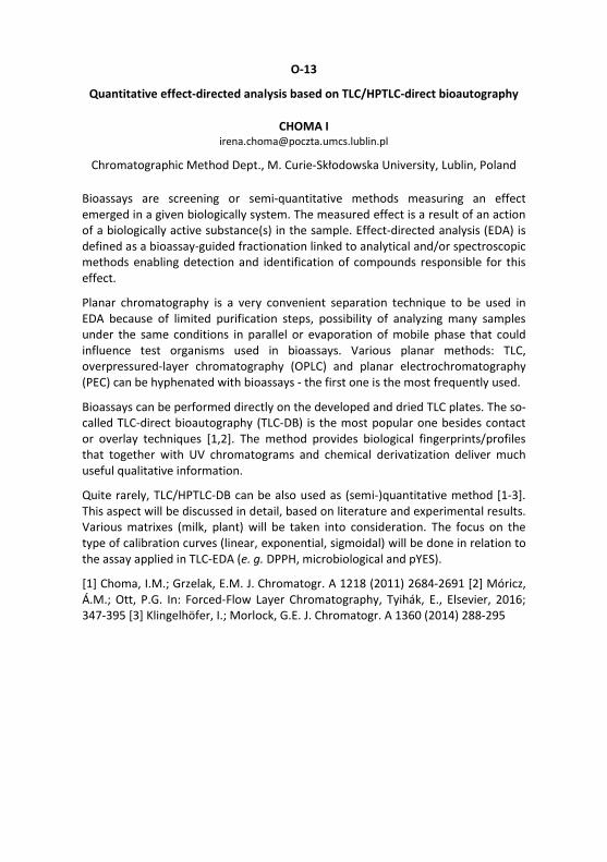
Bioassays are screening or semi-quantitative methods measuring an effect emerged in a given biologically system. The measured effect is a result of an action of a biologically active substance(s) in the sample. Effect-directed analysis (EDA) is defined as a bioassay-guided fractionation linked to analytical and/or spectroscopic methods enabling detection and identification of compounds responsible for this effect.
Planar chromatography is a very convenient separation technique to be used in EDA because of limited purification steps, possibility of analyzing many samples under the same conditions in parallel or evaporation of mobile phase that could influence test organisms used in bioassays. Various planar methods: TLC, overpressured-layer chromatography (OPLC) and planar electrochromatography (PEC) can be hyphenated with bioassays - the first one is the most frequently used.
Bioassays can be performed directly on the developed and dried TLC plates. The so-called TLC-direct bioautography (TLC-DB) is the most popular one besides contact or overlay techniques [1,2]. The method provides biological fingerprints/profiles that together with UV chromatograms and chemical derivatization deliver much useful qualitative information.
Quite rarely, TLC/HPTLC-DB can be also used as (semi-)quantitative method [1-3]. This aspect will be discussed in detail, based on literature and experimental results. Various matrixes (milk, plant) will be taken into consideration. The focus on the type of calibration curves (linear, exponential, sigmoidal) will be done in relation to the assay applied in TLC-EDA (e. g. DPPH, microbiological and pYES).
[1] Choma, I.M.; Grzelak, E.M. J. Chromatogr. A 1218 (2011) 2684-2691 [2] Móricz, Á.M.; Ott, P.G. In: Forced-Flow Layer Chromatography, Tyihák, E., Elsevier, 2016; 347-395 [3] Klingelhöfer, I.; Morlock, G.E. J. Chromatogr. A 1360 (2014) 288-295

O-14
Bioquantification of natural antibiotics by direct bioautography linked to
mass spectrometry
JAMSHIDI-AIDJI M [email protected]
Jamshidi-Aidji M1, Morlock GE1 1Justus Liebig University Giessen, Institute of Nutritional Science, Chair of Food
Science, Interdisciplinary Research Center (IFZ), Giessen, Germany
Antibiotic resistance is a current challenge of public health and pharmaceutical industry. Hyphenated planar chromatography (HPTLC-UV/Vis/FLD-EDA-HRMS) proved to be a well-suited, high-throughput bioanalytical tool for such challenges able to directly link of effective zones [1, 2]. The sample preparation was kept simple to let the sample extract as native as possible. The Bacillus subtilis bioassay was directly applied in the chromatogram (bioautogram) to demonstrate the streamlined strategy from screening, characterization and identification to bioquantification of natural antibiotics in root extracts of Salvia miltiorrhiza.
Thus, the antimicrobial activity of the Salvia miltiorrhiza root extract was characterized via chromatographic, spectroscopic and HRMS data. Inverse densito-metric measurement was employed for bioquantification. The importance of two unknown antibiotics was specified via bioequivalency calculation. As a reference, cryptotanshinone was used. The overall antimicrobial result obtained was referred to the activity of two synthetic antibiotics, ciprofloxacin and marbofloxacin. These calculations were performed on the same plate. It clearly showed that natural antibiotics are of similar importance as synthetic antibiotics.
This strategy can be installed without much microbiological effort in every analytical laboratory using regular instrumentation. Depending on the selected effect, any type of bacteria can be applied on the HPTLC plate. Especially, the application of pathogenic bacteria will be of high relevance in combination with HRMS/NMR/FTIR and bioquantification. The potential of planar chromatography for a streamlined structure elucidation was reported recently [3]. The demonstra-ted power of reliable, quantitative bioprofilings accelerate the discovery of new antibiotics from natural sources and may also explain effects observed or draw the attention to new aspects. In every case, very exciting!
[1] Jamshidi-Aidji, M., Morlock, G.E. J. Chromatogr. A 1420 (2015) 110-118 [2] Jamshidi-Aidji, M., Morlock, G.E Anal. Chem. 88 (2016) 10979−10986 [3] Yüce, I., Morlock, G.E. J. Chromatogr. A 1469 (2016) 120-127

O-15
Patterns of estrogenic activity in treated wastewater - a study from Switzerland
SCHOENBORN A [email protected]
Schoenborn A1, Mainetti T1, Grimmer A1
1Zurich University of Applied Science, Waedenswil, Switzerland
In a 2016 study, 35 wastewater samples (24-hour composite samples) were taken from 19 different sewage treatment plants (STP) in the Swiss Canton of Zurich . The unfiltered samples were concentrated 138-fold by using liquid-liquid extraction, and analyzed on estrogenic activity (EA) patterns, using the current state of the planar-YES bioassay on normal-phase HPTLC plates. The aim of the study was to explore the variety of EA-patterns in treated wastewater and take a first step towards their typification.
A variety of sample-specific EA-patterns were found: 16 out of 19 STPs had at least one EA-band. Up to 6 different EA-bands were found in some samples. Four general EA-pattern types were distinguished. Most of the EA-bands were assigned to 17-beta-estradiol (E2), 17-alpha-ethinylestradiol (EE2) and estrone (E1), based on known hRF values. In addition, four EA-bands of unknown origin were differentiated in STP-outlets. The highest single activity was 1.6 ng/l EEQ, the highest sum of all activities was 2.6 ng/l EEQ, which is several times above the proposed AA-EQS for E2 of 0.4 ng/l.
EA-pattern were found to vary from day to day, but the reasons for these variations are unknown. They are most likely due to catchment-specific differences in the wastewater sources. We were also able to show that the planar-YES can be used to assess the effectiveness the fourth treatment stage of STPs.
By using a newly developed overlay technique for the yeast cells, we were able to achieve a consistently low LOD of the planar-YES (0.1-0.2 pg/band). The planar-YES is a relatively simple, inexpensive and powerful tool for screening unknown water samples on estrogenic activity and get some clues about its possible origin. We see the planar-YES as a complementary tool for monitoring STP-outlets in the future.

O-16
2D-thin layer chromatography of 17α-ethinylestradiol on RP-18 W plate,
detected by YES-test
SPANGENBERG B [email protected]
Spangenberg B1, Witos I1, Milz B1 1University of Applied Sciences Offenburg, Offenburg, Germany
17α-Ethinylestradiol (EE2) is an important biological active substance, which is the most commonly used estrogen for oral contraceptive pills world wide. Is EE2 taken, it is excreted in the urine and ends up unaltered in wastewater treatment plant effluents, where it can act as an estrogen disruptor in fish and other marine creatures. Even at very low concentrations it was shown that the compound affects reproduction and development in wildlife by mimicking its natural analogue 17ß-estradiol. Thus, EE2is a very interesting compound for TLC analysis. We present an example of 2D-TLC separation using RP-18 W plates as a mixed plate for a normal and reverse phase separation of estrogenic compounds as for EE2. Although RP-18 W plates have not been used so often for 2D-TLC separations, this plate type shows a large potential. Using this type of TLC-plate, we are able to baseline separate estrone (E1), 17ß-estradiol (E2), estriol (E3), EE2, diethylstilbestrol (DES), the mycotoxin zearalenone (ZEA) and the xenoestrogen bisphenol A (BPA). The presented 2D-TLC separation method can be used to quantify EE2 in an effect-directed analysis using the yeast strain Saccharomyces cerevisiae BJ3505. The test strain contains the estrogen receptor. Its activation by estrogen active compounds is measured by inducting the reporter gene lacZ, which encodes the enzyme ß-galactosidase. This enzyme activity is determined directly on TLC plate by using the fluorescent substrate 4-methylumbelliferyl ß-D-galactopyranoside. The LOD of EE2 was calculated to 31 pg, the LOQ to 70 pg per spot.
2D-separation of E1, E2, EE2, E3, BPA, ZEA and DES after YES-test

O-17
Effect directed analysis, a new challenge according the upcoming enhancement of
European Water Framework Directive 2000/60/EC in 2019 - Planar biotests, a tool
to detect emerging contaminants in environmental samples
WEINS C [email protected]
Weins C1, Buchinger S2 1EDA Effect Directed Analysis: Environment, Food, Pharmacy, Saarbrücken,
Germany, 2Federal Institute of Hydrology, Koblenz, Germany
The European Water Framework Directive 2000/60/EC was established aming to restore Europe's waters and a potential template for future environmental. However, fifteen years since it was adopted, the WFD has not delivered its main objectives of non-deterioration of water status and the achievement of good status for all EU waters 47% of EU surface waters not reaching the good ecological status in 2015.
The first step was to establish by way of Decision 2455/2001/EC a first list of priority substances. These substances were selected from amongst those presenting a significant risk to or via the aquatic environment. But the single substance analysis and elimination of priority hazardous substances seems not efficient enough to restore the natural biodiversity. Too many so called ermerging contaminants, compounds previously not considered or known to be significant to groundwater show effects to the biocenosis in our river basins. So the the large number of emerging contaminants poses a challenge for regulatory agencies [1].
This talk will show that planar biotests using HPTLC plates (p-biotests) such as p-YES test and analogous prodedures do not necessarily rely on standards. Using p-biotests the smallest traces of these substances (e. g. in the ng- to pg- range) can be detected. Profiles of contaminated water sources can be visualized with substances which have been proved to possess biologically hazardous effective properties in the aquatic environment.
[1] N. Voulvoulis et al. Science of the Total Environment 575 (2017) 358-366

Panel discussion
Chair: BERNARD-SAVARY
Manufacturers
1. VAN BEELEN E, Strategic Technologies Development Manager Europe & India, Waters Corporation, [email protected]
2. MERKERT C, Field Marketing Specialist Central Europe, Advion, UK, [email protected]
3. MUSSELMAN B, CEO, IonSense, USA, [email protected] 4. SCHULZ C, Head of Instrumental Analytics R&D, Merck, Germany,
[email protected] 5. WYSS M, CEO, CAMAG, Switzerland, [email protected]
Create actively the future of HPTLC!
HPTLC is an emerging field with impact due to its unique advantages. Discuss with manufacturers and opinion leaders the progress in the field – pros and cons are welcome...
- Novel layer structures: Electrospun? Monolithic? Nanostructured? Fused core particles? HILIC layers? SEC layers?
- Improvements in the layer performance: Separation power? Reproducibility? - Instrumental developments: Novel hardware? Software improvements?
Miniaturized all-in-one system? - Further hyphenations: Elution head-based HPTLC-MS with fully automated
positioning on zones of interest? FTIR? FT-SERS? Coupling with column chromatography or supercritical fluid chromatography? Imaging MS?
- Detection tools: Image quality? Quantitative evaluation based on the image? - Quantitative HPTLC: Validation? Significant numbers for precision values?
Improved software tools, e. g. for integration of peaks (tangent peaks) or fixing the baseline (impacted by negative peaks)?
- Future bioassays: Genetically modified microorganisms? Bioluminescent micro-organisms?
- New derivatization reactions for compound classes difficult to detect like organic acids: Selectivity? Detectability?
- Support: Need for books and training courses? Online CCBS database search? How to improve the research paper quality? How to train reviewers?
O-18
Performance of chiral TLC in physico-chemical studies
KOWALSKA T [email protected]
Sajewicz M1, Kowalska T1 1Institute of Chemistry, University of Silesia, Katowice, Poland
Basic task of each chromatographic technique is separation of a complex mixture of compounds, and identification and quantification of individual components. Certain chromatographic techniques can also be employed as physicochemical tools, which is an added value of a technique.
In this talk, potential of the chiral TLC is shown in a discovery and studying oscillatory reactions, i. e., in demonstrating spontaneous oscillatory chiral con-version and spontaneous oscillatory condensation of the low molecular weight carboxylic acids running in the parallel.
Discovery of a new class of the oscillatory reactions was first reported in 2005 [1]. Then further investigations have been carried out for over a decade now, which employed the chiral TLC and a number of auxiliary analytical techniques. The most up to the date reports are given in papers [2, 3]. This research would not have been possible without an outstanding enantioresolution performance of the chiral TLC, largely developed and well documented by Bhushan and Martens [4]. Some of the results obtained have relevance for such issues as homochirality and biogenesis, oriented toward an evolutionary perspective.
[1] M. Sajewicz et al. Acta Chromatogr. 15 (2005) 131-149 [2] A. Maciejowska et al. J. Chromatogr. Sci. 54 (2016) 1301-1309 [3] A. Godziek et al. Israel Journal of Chemistry 56 (2016) 1057-1066 [4] R. Bhushan, J. Martens, Amino Acids, HNB, New York, 2010.
O-19
New approach to development of planar chromatograms
DZIDO T [email protected]
Hałka-Grysińska A1, Skop K1, Gorzkoska M1, Klimek-Turek A1, Dzido T1 1Medical University of Lublin, Lublin, Poland
The conventional chromatogram development is usually performed in vertical and horizontal chambers, which can stand for a part of more or less sophisticated equipment. In these chambers/equipment the mobile phase is driven into movement through adsorbent layer by capillary action. Then, the migration distance Zf of the mobile phase front in the adsorbent layer is dependent on the time t according to the equation:
Zf2 = κ ∙ t, were κ is constant.
It means the longer migration distance of the mobile phase front leads to slower mobile phase velocity on/in the adsorbent layer. In practice, a chromatographer has very restricted possibility of influence on the velocity of the mobile phase - it can be achieved by change of solvent type (adjusting its viscosity), stationary phase type, particle diameter of the adsorbent and temperature. The question arises if it is possible to perform conventional chromatogram development with controlled mobile phase velocity? At the first glance it does not seem to be the simple task. However, when the solvent is delivered to the chromatographic plate with controlled velocity lower than rate of the mobile phase absorption by the adsorbent layer then this effect will be achieved. We have designed an equipment for feeding the adsorbent layer with controlled solvent velocity and performed experiments to verify this approach for conventional chromatogram development. Preliminary results lead to the main observations: - conventional chromatogram development can be performed with constant linear
velocity of the mobile phase according to the equation: Zf = k ∙ t, - the mobile phase velocity can be easily adjusted to the value, which determines
minimum plate height, i. e. maximum performance of a chromatographic system, - controlled velocity of the mobile phase is especially advantageous for gradient
elution in reversed phase planar chromatography, - the designed equipment can be easily automated.

O-20
Radiochromatographic methods in drug metabolism research
KLEBOVICH I [email protected]
The presentation sums up the possible novel tools of the radiochromatography, radio-bioanalytics in the preclinical and clinical pharmacokinetic and drug metabolism research. The essential pharmacokinetic and drug metabolism information of different species (mouse, rat, dog, rabbit and human) have a profound contribution for the final drug registration process. The high sensitivity (pg/mL, fg/mL, at/mL) and highly selective hyphenated techniques (LC/Triple Quad-Jet Stream-ESI-MS and GC/MS-MS, etc.) required for the quantitative pharmaco-kinetic, metabolite kinetic studies, which had replaced the conventional methods of detections such as GC, HPLC and HPTLC.
Nowadays in the course of drug development the radioactive isotopes (beta and gamma single and/or double source) labelled (3H, 14C, 99Tc, 125,131I) pharmaco-kinetic/metabolite kinetics studies combined with the new generation of triple-quad MS techniques (GC, LC, OPLC) are essential. A number of related case studies will be presented. The former high quality off line HPTLC-Imaging Techniques (DAR, PIT) and the new generation of off line HPTLC or whole body autoradiography Imaging Techniques (MALDI Imaging, PET) in animal and human studies will also be presented.
The lecture will focus on the HPTLC and OPLC techniques with different types of radio-detection possibilities in drug research, in comparison to other techniques. A complex multi-step process will be illustrated from separation, purification, isolation to structure elucidation (GC-MS, LC-MS/MS, LC-NMR) of minor, (subminor) and major metabolites derived from animal and human biological matrices. The addition of the above systems to the off-line and on-line separation and radioactivity detection possibilities of HPTLC-, OPLC-DAR/PIT, OPLC-RD, HPTLC-DAR-MS and GC-RD, HPLC-RD and the combined multi hyphenated techniques, on line OPLC-DAD-RD-MS/MS as well as on line LC-DAD-RD-MS/MS resulted in a new, flexible and rapid high-performance complex solution in the metabolism research.
O-21
Reversed phase gradient thin-layer chromatography with one void volume of
the mobile phase: advantages, pitfalls and prospects for the future
HALKA A [email protected]
Hałka-Grysińska A1, Gwarda R1, Klimek-Turek A1, Chomicki A1, Dzido T1
1Medical University of Lublin, Lublin, Poland
Gradient high performance thin-layer chromatography, HPTLC, has a huge, potential, in the screening analysis due to unquestionable advantages of planar techniques. However, implementation of gradient reverse phase HPTLC is troublesome primarily due to excess flow of eluent to the surface of the adsorbent layer. As a result nowadays gradient HPTLC is performed almost exclusively in normal phase (e.g. using AMD 2 chamber from Camag [1]).
Therefore, our research group has been looking for alternative solutions for gradient mode in thin-layer chromatography. In 2012 horizontal developing chamber for stepwise gradient elution in reversed phase system with one void volume of the mobile phase was reported [2]. Another devices were reported in 2016 [3] and 2017 [4]. However, in those devices, there were significant problems in obtaining the optimized profile of the mobile phase flow in the adsorbent layer (there was a excessive mobile phase flow to the surface of the adsorbent layer) and consequently separated solute zones were distorted.
Finally a completely new prototype device for thin-layer gradient elution was developed, which delivers the eluent directly onto the surface of the adsorbent layer of chromatographic plates at a controlled velocity. In this way it prevents the excess of eluent to enter the plate, and thereby eliminates the above effect. Directly feeding the adsorbent layer with eluent also enables to perform convenient continuous gradient elution using one void volume of the mobile phase practically without the so-called gradient delay.
[1] Burger K., Fresenius Z., Anal. Chem. 318 (1984) 228-233. [2] Markowski W., Wróblewski K., Dzido T.H., J. Planar Chromatogr. 25 (2012) 3, 200 - 207. [3] Hałka-Grysińska A., Dzido T.H., Sitarczyk E., Klimek-Turek A., Chomicki A., J. Liq. Chromatogr. & R.T. 39 ( 2016) 257-263. [4] Hałka-Grysińska A., Gwarda R.Ł., Pawełek K., Baj T., Dzido T.H., J. Planar Chromatogr. 30 (2017) 2, 113-120.

O-22
Micropreparative orthogonal pressurized planar electrochromatography of
solutes showing the same electrophoretic mobility
GAJOS R [email protected]
Gajos R1, Chomicki A1, Hoda P1, Polak B1, Dzido T1 1Medical University of Lublin, Lublin, Poland
Orthogonal pressurized planar electrochromatography, OPPEC, is a two-dimensio-nal separation technique in which mixture components are separated by a chromatographic effect in one direction of the OPPEC system and simultaneously they are deflected in an electric field, which is perpendicular applied to the direction of chromatographic separation. Such separation system can be constantly fed with the sample solution and the separated components can be collected at the outlet of this system constantly. This approach can be conveniently used, when the solutes show different electrophoretic mobility. However, it is also possible to separate substances with the same electrophoretic mobility eg. stereoisomers or enantiomers, what will be presented.
At the conference we would like to give an introduction into the preliminary principles of preparative separation in the OPPEC technique in the case, when two key components characterised by the same electrophoretic mobility (eg. stereoisomers) are the subject of separation. The advantages of this new approach over the column chromatography with regard to the separation of complex mixtures, when the separation system is periodically fed with the sample solution, will be discussed. Additionally, the preliminary rules of optimization indicating the possibility of almost continuous and alternating collection of the key substances will be presented.

O-23
Separation of the optical isomers with pressurized planar electrochromatography
POLAK B [email protected]
Polak B1, Hałka-Grysińska A1, Klimek- Turek A1, Chomicki A1, Dzido T1 1Department of Physical Chemistry Medical University of Lublin, Lublin, Poland
Disparities in the physical properties of constitutional isomers or diastereomers promote their differentiation using many commonly available techniques of separation. Contrary to the substance types mentioned, separation of enantiomers is very inconvenient. Transforming enantiomers into diastereomers facilitates their separation. This transformation may take place during the separation process (direct mode) or precede it (indirect method). The former mode involves homochiral separatory factors (chiral selectors) bonded to the adsorbent (chiral stationary phase, CSP) or being the component of the eluent (chiral mobile phase additive, CMPA). The latter mode also applies chiral selectors, which react with enantiomer molecules and in this way form diastereomeric derivatives of different properties.
The separation of isomers is carried out with various chromatographic techniques such as HPLC and TLC as well electromigration ones (capillary electrophoresis, CE, and capillary electrochromatography, CEC). Since 2006 the pressurized planar electrochromatography (PPEC) has been included into the set of the techniques for isomer separation. In PPEC system a solute migration results from chromatographic (analyte partition between stationary phase and eluent) and electrophoretic (solute electrophoretic mobility) effects. The PPEC is characterised by short time of experiment, high separation efficiency and different selectivity in comparison to TLC. Various variables e.g. qualitative and quantitative mobile phase composition, stationary phase type, temperature and polarization voltage applied to the chromatographic plate influence isomer migrations and in consequence their separation.
The aim of this presentation is to show how these variables/parameters effect isomer separation in PPEC and TLC systems and to compare the results obtained by both techniques.
O-24
HPTLC-ESI-MS/MS for identifying neutral lipids, sphingolipids and phospholipids
in complex samples
CEBOLLA V [email protected]
Cebolla V1, Lapieza M1, Jarne C1, Saviron M2, Orduna J3, Membrado L1, Morlock GE5, Jungas C6, Garriga R4, Vela J4
1CSIC, 2CEQMA-CSIC, 3CSIC-ICMA and 4University of Zaragoza, Zaragoza, Spain,
4Justus Liebig University Giessen, Chair of Food Science, Giessen, Germany, 5CEA-CNRS-Aix Marseille University, Marseille, France
HPTLC is well-adapted to providing lipid-class separations, as an assistant technique of HPLC. From this perspective, it can complement traditional LC-mass spectro-metric approaches in a unique way. As the zones of interest on the plate can selectively be transferred to the ESI-MS instrument via an elution head-based interface, a rapid, relevant structural information about molecular species within each lipid classes from complex samples can be obtained.
Separation using AMD2 provides lipid-classes as narrow peaks enough to ensure a direct elution and transfer of the plate zones to obtain both composition profiles of each class by ESI-MS, and identification of individual lipids and molecular species belonging to each separated lipid class by MS/MS (MSn) and HRMS. The respective sodium adducts of the above-mentioned lipid classes were fragmented in the positive ion mode using an ion-trap technology. The sodium remained the charge of their fragment ions, thus being useful for their structural identification by MS/MS (MSn) through further fragmentation.
This work exemplarily focuses on profiling and identification of neutral lipids (NLs), sphingolipids (SLs) and phospholipids (PLs) in three analytical cases:
(1) mono- (MAGs) and diacylglycerides (DAGs) in positive ion mode and fatty acids (FAs) in negative ion mode as impurities in a fatty acid methyl ester-based biodiesel,
(2) molecular species of neutral sphingolipids (SL), such as sphingomyelins (SMs) and globotriaosylceramides (Gb3), in human plasma,
(3) phosphatidylcholines (PCs), phosphatidylethanolamines (PEs), phosphatidyl-glycerols (PGs) and cardiolipines (CLs) associated to membrane proteins of photosynthetic purple bacteria.

O-25
Hyphenated HPTLC-AMD-ESI/MS for quantification of major protoberberines in
herbal extracts and polyherbal formulations
ILANGO K [email protected]
Ilango K1, Kasthuri Bai N1, Mohan Kumar R1 1Pharmacognosy and Phytochemistry Division, Interdisciplinary Institute of Indian System of Medicine, SRM University, Kattankulathur, Tamil Nadu, Chennai, India
Plants that contain protoberberine alkaloids are reported to be used as anti-diabetic, anti-hepatitis, anti-inflammatory, anti-tumor, anti-oxidant, hepatoprotec-tive, cardiotonic, immuno-modulatory and neuro-protective in Indian System of Medicine (ISM). HPTLC with automated multiple development (AMD) was successfully applied for the simultaneous quantification of three protoberberine alkaloids namely berberine, berbamine and palmatine in Berberis aristata, Berberis vulgaris (Berberidaceae), Tinospora cordifolia (Menispermaceae) and three marketed polyherbal formulations.
Major protoberberines were separated using a 3-step AMD gradient based on ethylacetate, methanol and methanol with 10 % formic acid, with a total migration distance of 70 mm in 40 min. Detection was carried out at 366 nm. On each plate, protoberberine standard mixture, plant extracts and formulations were applied in triplicate, together with 11 sample bands of 7 mm. The method was linear in the range of 30-90 ng/band for the protoberberine standard mixture. The separation was achieved with hRF 70 ± 2 for berberine, hRF 48 ± 3 for berbamine and hRF 57 ± 2 for palamatine. Identification was done by densitometry and HPTLC-ESI-MS, after online elution with 100 % methanol at a flow rate of 0.5 mL/min using a elution head-based TLC-MS interface.
Mass spectra were obtained in the positive ion mode at m/z 336 for berberine, m/z 608 for berbamine and m/z 352 for palmatine. HPTLC-ESI-HRMS spectra confirmed the presence of three protoberberines in all plant extracts and polyherbal formulations. The developed method was found to be rapid and simple with a clear separation and target-oriented confirmation giving contamination-free, highly sensitive mass spectra of major protoberberines.
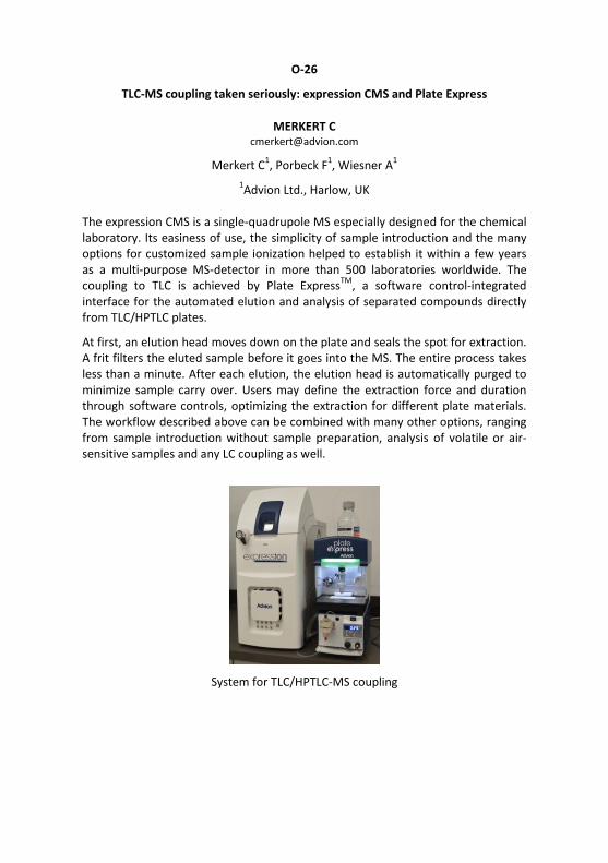
O-26
TLC-MS coupling taken seriously: expression CMS and Plate Express
MERKERT C [email protected]
Merkert C1, Porbeck F1, Wiesner A1 1Advion Ltd., Harlow, UK
The expression CMS is a single-quadrupole MS especially designed for the chemical laboratory. Its easiness of use, the simplicity of sample introduction and the many options for customized sample ionization helped to establish it within a few years as a multi-purpose MS-detector in more than 500 laboratories worldwide. The coupling to TLC is achieved by Plate ExpressTM, a software control-integrated interface for the automated elution and analysis of separated compounds directly from TLC/HPTLC plates.
At first, an elution head moves down on the plate and seals the spot for extraction. A frit filters the eluted sample before it goes into the MS. The entire process takes less than a minute. After each elution, the elution head is automatically purged to minimize sample carry over. Users may define the extraction force and duration through software controls, optimizing the extraction for different plate materials. The workflow described above can be combined with many other options, ranging from sample introduction without sample preparation, analysis of volatile or air-sensitive samples and any LC coupling as well.
System for TLC/HPTLC-MS coupling

O-27
Bringing the power of mass detection to scientists using TLC combined with the
ACQUITY QDa detector
VAN BEELEN E [email protected]
Waters S.A.S., Saint-Quentin En Yvelines Cedex, France
TLC is still extensively used in analytical laboratories due to its fast separation and simplicity. Adding mass spectrometry to identify TLC bands can be an advantage, but MS is seen as complex and expensive versus TLC. The ACQUITY® QDa® detector is specifically aimed at chromatographers with no tune page, reduced number of parameters and a pre-optimized source. So it is a perfect fit with the TLC-MS Interface of CAMAG. It allows TLC-separated samples to be analyzed directly by mass spectrometry, adding identification capability with reduced sample prepara-tion.
In this presentation, the ease of use of this system is highlighted and how it brings powerful mass detection capabilities into the TLC/HPTLC-based laboratory. The emphasis will be placed on example applications provided in a variety of markets.
Small mass spectrometer for TLC/HPTLC-MS coupling

O-28
Unexpected products of the HOCl-induced oxidation of oleic acid: a study using
HPTLC-ESI-MS
GRIESINGER H [email protected]
Griesinger H1, Schröter J2, Schulz M1, Süß R2, Schiller J2 1Merck KGaA, Darmstadt, Germany, 2University of Leipzig, Faculty of Medicine,
Institute of Medical Physics and Biophysics, Leipzig, Germany
Reactive oxygen species are of particular relevance in the pathogenesis of inflammatory diseases. Under inflammatory conditions, hypochlorous acid (HOCl), generated via the enzyme myeloperoxidase, reacts with the double bond in the fatty acyl residues of (phospho)lipids under formation of a chlorohydrin (CH) as the main product. However, the oxidation of free fatty acids by HOCl has been investigated less detailed. Using oleic acid (OA), the simplest unsaturated fatty acid, as a model system, we investigated the product pattern of the reaction between OA and HOCl by a combination of HPTLC and different ESI MS methods.
The fatty acids were incubated separately with HOCl (360 min, 37°C, shaking) and afterwards extracted (chloroform - methanol 1:1 plus 0.05 % BHT). The organic layer was either directly investigated by ESI-IT MS or separated on HPTLC plates silica gel 60 F254 MS-grade using chloroform - ethanol - water - triethylamine 6:7:1:7 followed by further analyses with an ESI-QTOF MS after extraction using a TLC-MS interface.
The reaction between POPC and HOCl results in the formation of two CH isomers as the only products. In contrast, the reaction of OA and HOCl does not exclusively result in the formation of CH (isomers) but of dimeric and trimeric products, that could be monitored by direct infusion ESI-IT MS. After HPTLC eight different spots could be identified and characterized directly by ESI-QTOF MS. Dimers and trimers were detected from different spots, i. e. with different hRF-values, while the CH was just detected as a single spot. The dimer formation can be explained by an intermolecular ether formation, but the generation of the trimer can be exclusively explained if the carboxyl group is involved (i. e. esters are also generated). Therefore, the reaction between OA and HOCl was additionally performed in the presence of decanoic acid (DA) and leads surprisingly to an ester.

O-29
HPTLC-MS based method development for cardiovascular disease controlling
compounds containing plant Coleus forskolii as per US chapter 203
PRAKASH GUPTA A [email protected]
Prakash Gupta A1, Awasthi P2, Gandhi S2, Bedi Y2, Gupta S2, Singh D1 1QC & QA, Indian Institute of Integrative Medicine, (IIIM-CSIR), Jammu, India,
2Plant Biotechnology Division, Indian Institute of Intergrative Medicine, (IIIM-CSIR), Jammu, India
Coleus forskohlii is an important indigenous medicinal plant of India. The plant extract has been used in traditional Ayurvedic medicine for curing various disorders. The extract is a mixture of several bioactive compounds including the diterpenoid forskolin. Forskolin is reported to be useful in the treatment of congestive heart failure, glaucoma, asthma and certain type of cancers. Ultrasonication was used for the extraction of the samples at 45°C for 40 min in three different solvents. In comparison to ethyl acetate and chloroform, acetonitrile was found to be the best solvent for extraction of forskolin, 9-deoxy-forskolin, 1,9-dideoxy-forskolin and deacetyl forskolin. An HPTLC-MS method was developed for separation of the important cardiovascular compound forskolin from other compounds with benzene - ethyl acetate 17:3. Two different types of plates were investigated, whereby HPTLC plates LiChrospher Si 60 F254 s were preferred to HPTLC plates Si 60 F254 s. The developed method will be used for quality control of raw materials and finished products.
HPTLC separation of four bioactive compounds of Coleus forskolii
Substituted by the next lecture!

O-29
Application of TLC/HPTLC in identification of Chinese crude drugs for ChP
LIHUA G [email protected]
Lihua G1 1Shanghai University of TCM Shanghai, China
TLC/HPTLC, as one of the most conventional chromatographic techniques, has been used for identification of TCMs. Not only chemical reference substances (CRS), but also reference crude drugs (authenticated crude drugs) have been introduced to provide the whole image of the tested herbs. As one of the series reference books of Chinese Pharmacopoeia, "Color Atlas of Thin-Layer Chromatography Identifi-cation for TCM of Pharmacopoeia of P.R. China 1990 Edition" was published serving as guidance for TLC identification of TCMs for the first time.
Later on, "TLC Atlas of Chinese Crude Drugs in Pharmacopoeia of the People's Republic of China" (2009 edition) provided 228 TLC identifications of Chinese crude drugs which covered about 2/5 of the crude drug monographs recorded in ChP 2005 edition. As a continuation of the TLC atlas, we are undertaking the TLC/HPTLC Chromatograms of 100 Chinese crude drugs recorded in ChP 2015 edition. Here I gave a brief introduction to this new version of TLC/HPTLC atlas of Chinese crude drugs to be published at the end of this year.

O-30
TLC combined with flame-induced atmospheric pressure chemical ionization mass
spectrometry (FAPCI/MS) for volatile and semi-volatile compound analysis
SHIEA J [email protected]
Shiea J1, Cheng S1 1National Sun Yat-Sen University, Kaohsiung, Taiwan
The combustion of hydrocarbons in a flame produces reactive primary ion species such as alkali metal ions (H3O+, CHO+, and CnHm
+). The flame has thus been employed as a tool to ionize organic compounds through ion-molecule reactions (IMRs) in the so-called flame-induced atmospheric pressure chemical ionization (FAPCI). In addition, the high temperature generated in a flame allows the thermal desorption of small compounds on sample surfaces, a phenomenon which is termed desorption FAPCI. Currently, DFAPCI/MS has been utilized to analyze drug molecules, explosives, pesticides, and plasticizers on the surface of a drug tablet, stainless steel plate, and glass rod. In this study, we coupled TLC to FAPCI/MS to analyze volatile and semi-volatile compounds.
A small oxyacetylene flame generated through a stainless steel tube (O.D. 0.72 mm, I.D. 0.15 mm) was directed toward separated TLC plates with an incident angle between 30-60° for analyte desorption and ionization. In another setup, a laser beam was used to desorb analytes from sample spots on TLC plates via irradiation; the desorbed analytes were ionized by FAPCI for MS detection. Volatile and semi-volatile compounds such as caffeine, drugs, plant extracts, and hydrocarbons were separated on TLC plates for DFAPCI/MS analysis. Since flame-based thermal decomposition of analytes was well controlled and avoided, intact analyte ions were detected without/with minimal fragment ions. Protonated analyte ions (M+H)+ of lidocaine and triethylamine were detected, while, radical cations (M+) of ferrocene and (M+O-3H)+ ions of normal alkanes were also characterized.

O-31
Automated desorption- and elution-based HPTLC-MS HÄBE TT
[email protected] Häbe TT1, Morlock GE1
1Justus Liebig University Giessen, Institute of Nutritional Science, Chair of Food Science, Interdisciplinary Research Center (IFZ), Giessen, Germany
Though hyphenation of HPTLC with MS has been explored for more than a decade, automatization is not available yet. For two commercially available coupling tech-niques, automated positioning systems were developed to access the whole HPTLC plate and to link previous steps of the workflow to the MS acquisition.
Substantial modifications were performed on the Direct Analysis in Real Time (DART) interface to improve the desorption-based hyphenation HPTLC-DART-MS. A plate carrier was equipped with a vertical stabilizer to demonstrate automated scanning and its progress in performance and flexibility. The source mounting base and housing were modified to enlarge the sampling area. Exact positioning of the HPTLC plate as well as a modified source cap and transfer tube were crucial for an enhanced performance at reduced mass signal variations [1, 2]. Surface scanning by a longer transfer tube system (7.5 - 30.0 cm between DART interface and MS inlet) was explored as well. Image-based alignment of the scan tracks was enabled with a new user interface to generate a flexible track batch for scanning the whole plate at one go. Thus, desorption, ionization and transfer of analytes out of a planar sub-strate were substantially improved. Scanning DART-MS was used for the character-ization of bioactive compounds in Tanacetum vulgare L. and Solidago virgaurea L., and the benefits of this technique were demonstrated in combination with multi-variate data analysis for classification of propolis [3-5].
The same user interface was used to generate a spot-wise batch of target coordi-nates for automated elution head-based HPTLC-MS utilizing a modified TLC-MS in-terface. A positioning system moved the plate (up to 20×10 cm) to position one target coordinate after the other beneath the elution head. The whole elution and purging process was electronically controlled and allowed acquiring mass spectra from selected target zones on the plate in one batch at one go.
[1] T.T. Häbe, G.E. Morlock, Rapid Commun. Mass Spectrom. 29 (2015) 474 [2] T.T. Häbe, G.E. Morlock, Rapid Commun. Mass Spectrom. 30 (2016) 321 [3] A. Moricz et al. J. Chromatogr. A 1422 (2015) 310 [4] A. Moricz et al. Anal. Chem. 88 (2016) 8202 [5] T. Chasset et al. J. Chromatogr. A 1465 (2016) 197

O-32
TLC-UV and TLC-MALDI-TOF/MS: an efficient tool for enzyme characterization and
screening of bioactive substrates
FEREY J [email protected]
Ferey J1, Da Silva D1, Lafite P1, Daniellou R1, Maunit B1 1Univ.Orleans, CNRS, ICOA, UMR 7311, Orléans, France
This work shows the TLC potential in the fields of enzyme characterization and substrate screening in complex matrices. Invertase is a glycoside hydrolase that catalyzes the hydrolysis of sucrose producing fructose and glucose. This enzyme was chosen as a model to validate our developments.
A first part of our work focused on the demonstration of the advantages of TLC-UV densitometry to determine kinetic parameters of invertase [1]. After separation of 10 sucrose reaction mixtures on an impregnated silica gel plate, a quantification of each specific product was performed. Hydrolysis of sucrose was monitored by either glucose or fructose accumulation with kinetic parameters (KM, Vmax) respectively equal to 63.09±7.590 mM; 0.037±0.00094 mM/min and 83.01±14.39 mM; 0.031±0.0021 mM/min. The hydrolysis of three alternative substrates (raffinose, stachyose and inulin) was also determined after a specific monosaccharide product measurement. This TLC methodology, compared with colorimetric studies, enables an increase of specificity and sensibility.
The second part of our work concerned the development of a TLC-MALDI-TOF MS coupling for the screening of invertase substrates in five plant extracts. A differential approach, between blank and enzymatic reaction, was performed by TLC-UV to highlight substrates and products of reaction. Identification of spots of interest was performed by TLC-MALDI-TOF MS. The advantages of this coupling allowed characterizing two substrates (di- and trisaccharide) and different products in three plant extracts.
All these methodologies showed the TLC as a leading-edge technique for characterization of enzyme and bioactive constituents in complex matrices and increase fields of investigation for other enzyme studies.
[1] J. Ferey et al. J. Chrom. A, 1477 (2016) 108-113.

O-33
HPTLC + SERS > HPLC + MS
CHEN Y [email protected]
Chen Y1, Xu X1, Xie Z1 1The State Key Laboratory of Food Science and Technology, School of Food Science
and Technology, Collaborative Innovation Centre of Food Safety and Quality Control in Jiangsu Province, Jiangnan University, Wuxi, China
Among the detections hyphenated to HPTLC, SERS was particularly attractive. SERS essentially mirrored the advantages of normal Raman spectroscopy, offering characteristic fingerprints unique to analyte structures. This was extremely suitable for identifying targeted spots based on unambiguous molecular information. This work presented a highly selective SERS method mediated by HPTLC, which was tailored for identification and quantitation of histamine in-situ derivatized with fluram. SERS was performed jointly using silver nanoparticle and NaCl. The latter dramatically suppressed the masking effect caused by excessive fluram, offering clear baseline and intensive Raman fingerprints specific to the analyte. Under optimized conditions, the usability of this method was validated by identifying the structural fingerprints of both targeted and unknown compounds in fish samples. Besides, the quantitative results of this method agreed with those by an official HPLC method. Showing remarkable cost-efficiency and user-friendliness, this facile HPTLC-SERS method was indeed screening-oriented and may be more attractive than HPLC-MS to controlling laboratories of limited resources.
Chromatographic separation and SERS confirmation of analytes in fish
O-34
TLC and TLC-Raman as an effective tool for polymer additives deformulation
LONGIERAS N [email protected]
PeakExpert, Tours, France
Antioxidants, UV stabilizers, lubricants, fatty acids, surfactants, waxes are some examples of typical additives used in polymers to help long term ageing or polymer processing. In the daily laboratory work, additives analysis is difficult due to polymer and oligomer interferences. In this study, we demonstrated that TLC techniques are an effective tool to obtain qualitative and semi-quantitative data on polymer formulation composition.
TLC separation occurs mainly on silica gel phases using various mobile phases plus additives. Day to day hRF reproducibility is evaluated. Derivatization techniques allow an array of both universal and specific detections. The derivatization results emphasized a novel vapour iodine methodology allowing quick and semi-quantitative results within minutes.
Further compound identification was obtained by analysing the underivatized silica gel layer directly under a 780 nm Raman microspectrophotometer. Spectra obtained in this way matched our solid state database and allowed spectral recognition. TLC results were compared to those obtained through RP-HPLC, high resolution oligomeric GPC, LC-FTIR and NMR.

O-35
HPTLC-EDA-HRMS and PLC-NMR to reveal co-eluting isomers of bioactive zones
AZADNIYA E [email protected]
Azadniya E1, Morlock GE1 1Justus Liebig University Giessen, Institute of Nutritional Science, Chair of Food
Science, Interdisciplinary Research Center (IFZ), Giessen, Germany
Separation of structural isomers is a common challenge among all chromatographic methods. Due to the same sum formula, HRMS and occasionally also MSn cannot differentiate them. Apart from structural similarity, they have different protons in chemically unlike environments causing different chemical shifts in 1H-NMR spec-tra. Hence, HPTLC-EDA-HRMS followed by PLC-NMR is an effective sequence to re-veal coelution of bioactive structural isomers. For two examples, PLC-1H-NMR spec-troscopy was demonstrated after scratching off the band and dissolving the residu-al in CD3OD.
1) An unknown multi-potent zone was found active against Bacillus subtilis, Alii-vibrio fischeri and acetylcholinesterase (bio)assays in a Salvia miltiorrhiza Bunge extract. HRMS results led to 4 potential candidates and among them, 1,2-dihydrotanshinone (1,2-DHTI) and methylenetanshinquinone (MTQ) contained vinylic protons that were not present in others. Thus, their discrimination was possible via different chemical shifts of the vinylic protons at C-3 in 1,2-DHTI and at C-18 in MTQ. The 1H-NMR spectra revealed their co-elution in the ratio 2:1 by two singlet signals for MTQ (5.10 ppm and 5.58 ppm) and one multiplet signal for 1,2-DHTI (6.08 ppm).
2) An unknown multi-potent zone was found active against B. subtilis, α-glucosi-dase and tyrosinase (bio)assays in dry extracts of different plants and apple peels. HRMS and derivatization results showed that the active zone could be ursolic acid (UA) and oleanolic acid (OA). The methyl group positioning (geminal or vicinal) was the only structural difference in UA versus OA. Thus, identification by 1H-NMR spectroscopy was possible though the different chemical shift of the allylic H-18 resonating at 2.2 ppm and 2.8 ppm for UA and OA, respectively. Due to molar equivalence of proton signals in NMR spectroscopy [1], qNMR analysis was possible via maleic acid used as internal standard. qHPTLC after derivatiza-tion confirmed the qNMR results.
[1] Pauli et al. J. Nat. Prod. 68 (2005) 133-149

O-36
HPTLC detection of steroid 5α-reductase activity from a non-radioactive
cell-based assay
DE-EKNAMKUL W [email protected]
Faculty of Pharmacautical Sciences, Chulalongkorn University, Bangkok, Thailand
Steroid 5α-reductase (5α-R), an oxidoreductase family enzyme, plays an important role in steroid metabolism. lt catalyzes the conversion of testosterone (T) to 5α-dihydrotestosterone (DHT). Ovarexpression of 5α-R has been known to affect the balance between T and DHT causing androgenic disorders, such as prostete cancer, hirsutism and androgenic alopecia. Androgenic alopecia (AGA) is a major type of scalp hair lass caused by the over-production of DHT, a potent androgen functioning in dermal papilla cells located in the hair follicles.
Therefore, 5α-R has been considered as an important target for searching potential inhibitors as anti-AGAdrugs. ln this study, an HPTLC method was developed for sensitive detection of the formation of the enzyme 5α-R product, DHT, after compounds were subjected to a new non-radioactive cell-based assay. A silica gel plate applied with assayed samples was developed using toluene - acetone 4:1, dipped in 42.5% phosphoric acid, heated at 120°C, and detected at 366 nm. This HPTLC method has led us to find natural inhibitors of 5α-R for anti-AGA applications, both in form of natural extract (Avicennia marina) and pure compound (avicequinone C).
HPTLC detection of steroid 5α-reductase activity

O-37
Antioxidants in structured vegetable oils: chemical identification via HPTLC
FADEL O [email protected]
Fadel O1, Girard L1, Gomes Rodrigues D1, Bauduin P1, Le Goff X1, Rossignol-Castera A2, L'Hermitte A1
1Institut de Chimie Séparative de Marcoule, UMR 5257, CEA, CNRS, Université de Montpellier, ENSCM, Bagnols sur Cèze, France, 2OLEOS SA/Hallstar,
Mauguio, France
To increase the value of plant biodiversity in respect with environment and consumers, OLEOS elaborated and patented an Oleo-Eco-Extraction (OEE) process allowing obtaining a new generation of cosmetic ingredients enriched in active species such as antioxidants. It consists of a combination of microwaves and ultrasounds favoring the transfer of these species from the plant to a liquid phase composed of vegetable oils. The OEE is a primary application for eco-ingredients more bio-available and easier to formulate in emulsion. A common laboratory called Vect'Oleo was created between OLEOS and ICSM to develop a new generation of Oleoactives by structuring the solvent.
Different techniques have been used to understand the correlation between the antioxidants solubilization degree (Electron Paramagnetic Resonance) with the supramolecular structuration of the oil (Small Angle X-Ray Scattering). Using model systems based on jojoba wax or medium chain triglycerides we pointed out that the micellization of a bio-sourced extractant within the oils improves the solubilization of four polyphenols from olive leaves (and a fortiori the extraction yield) [1].
To go further in the determination of the solubilized antioxidants, we developed a method to separate polar antioxidants from the oily matrix, without pre-treatment, using the most suitable technique: HPTLC. Thanks to successive migrations with appropriate solvents, it was possible to separate the four polyphenols with high resolution. By coupling this technique with mass spectrometry, we succeeded in identifying each antioxidant and determining, as a function of their chemical structure, their solubilization efficiency in vegetable oils.
[1] O. Fadel et al., Colloids and Surfaces B: Biointerfaces 154 (2017) 279-286

O-38
Multidimensional chromatography (HPLC-HPTLC) for identification of antifungal
substances in Rheum root extracts
BALLERT S [email protected]
Ballert S1, Schiller E1, Gillmeister M1, Kabrodt K1, Schellenberg I1 1Anhalt University of Applied Sciences, Institute of Bioanalytical Sciences, Bernburg,
Germany
Phytopathogenic fungi resistant to various fungicides are responsible for major losses of harvest of up to 25% and have become a serious issue for modern agriculture. The research group of the Institute of Bioanalytical Sciences (IBAS) focuses on the antifungal activity of Rheum root extracts and aims for identification of the active substances contained in these extracts.
Antifungal tests performed with Rheum root extracts showed a high bioactive potential. However, the compound composition of polyphenols varies between the different rhubarb species as well as the pathogenic fungi they affect. Therefore a variety of methods is used for qualitative and quantitative analysis of these substances and to determine the structure-activity-relation. Due to the quantity of polyphenols per extract a single chromatography proved to be not sufficient to separate the compounds, so a multidimensional method was developed. In a first step 32 fractions were obtained using a reversed phase HPLC column in preparative scale. The fractions were then separated with HPTLC.
The Twin-Trough Chamber, Automated Development Chamber and Automated Multiple Development System were used to optimize the separation. Two-dimensional TLC was applied with similar solvents to prove that substances were stable during development, as well as non-similar solvents for the two dimensions to check that the separation itself was successful with no substances sharing their hRF-value. Different stationary phases were used to increase efficiency. The TLC plates were analyzed with the TLC Visualizer and also scanned at 280 nm (non-derivatized). Finally, the plates were derivatized comparing the immersion device to the new derivatizer with automated spraying technology. Detailed results will be shown in the presentation. All these steps combined lead to a separation suitable for substance identification with mass spectrometry using the TLC-MS Interface or possibly NMR analysis after a preparative approach.

O-39
HPTLC assay of thymoquinone in black seed and black seed oil (Nigella sativa
Linn) and identification of thymoquinone conversion with UV/Vis
BELETE Y [email protected]
Belete Y1, Dagne E1 1African Laboratory for Natural Products (ALNAP), Department of Chemistry, Addis
Ababa University, Addis Ababa, Ethiopia, Addis Ababa, Ethiopia
A reliable and simplified quantitative HPTLC method for the determination of the bioactive constituent of the commercially available black seed and black seed oil using a scanning densitometer is described. The identification of thymoquinone obtained from 80% aq. methanol extract of the seed is confirmed by NMR. The solvent system n-hexane and dichloromethane 1:1 was used and the chromato-gram was evaluated at 254 nm. The thymoquinone content of freshly pressed black seed oil was 1.3%, while that of the seed was 1%. Furthermore, UV-Vis measure-ment was significantly used for swift relative comparison of thymoquinone levels in black seed oils and to follow the transformation of thymoquinone when the oil is exposed to sunlight.
O-40
Development and validation of a HPTLC method for simultaneous estimation of
flavonoids and phenolics in Carica papaya leaf juice
ANSARI S [email protected]
Ansari S1, Anjum V1, Ahmad S1 1Bioactive Natural Product Laboratory Department of Pharmacognosy and
Phytochemistry, Faculty of Pharmacy, JamiaHamdard, New Delhi, India
A sensitive, fast, and reproducible HPTLC method has been developed for simultaneous analysis of myricetin, caffeic acid, trans-ferulic acid and kaempferol from papaya leaf juice, using TLC aluminium foils silica gel G 60 F254. Among the different combinations of mobile phases used, best separation was achieved with toluene - ethyl acetate - formic acid 5:4:1. Densitometric measurement was performed at 320 nm for analysis of myricetin, caffeic acid, trans-ferulic acid and kaempferol. The hRF values of myricetin, caffeic acid, trans-ferulic acid and kaempferol was found to be 39 ± 1, 44 ± 2, 50 ± 1 and 55 ± 1, respectively. The method was validated for specificity, precision (intraday and interday), accuracy, and robustness.
The developed simultaneous estimation method was found linear in a 1:25 wide range of concentration (50-1250 ng/zone) with good regression coefficients (0.99 ± 0.001). LOD was <18 ng/spot, whereas LOQ was <57 ng/spot. Accuracy of the method was checked by a recovery study of three different levels with the average percentage recovery in the range of 97-103%, respectively. C. papaya leaf juice was found to contain myricetin, caffeic acid, trans-ferulic acid and kaempferol at 280.2 ± 6.0 µg/g, 370.2 ± 6.3 µg/g, 1110.9 ± 3.0 µg/g and 160.5 ± 2.5 µg/g, respectively. The present method is being reported for the first time and can be used for routine quality control and quantification of these marker compounds in various plant samples, extracts and market formulations.

O-41
HPTLC studies on single drugs and compound formulations of the
Indian system of medicine
AHMAD S [email protected]
Bioactive Natural Product Laboratory Department of Pharmacognosy and Phytochemistry, Faculty of Pharmacy, JamiaHamdard, New Delhi, India
The global acceptability of herbal drugs and botanicals is the major concern in absence of their proper quality control analysis. The quality control analysis of herbal drugs involves various parameters including chromatographic and spectroscopic techniques. Nowadays chromatographic hyphenated techniques are very efficiently used in quality control of herbal drugs and botanicals to analyze different markers responsible for ensuring correct identity as well as to establish the exact mechanism of their action. The HPTLC being an easy, economic, less time consuming and matrix-friendly technique has played a larger role in quality control of herbal drugs and botanicals, but proper and exact active metabolite-based development of analytical methods is essential.
Since Indian traditional medicines are composed of different types of drugs and formulations (including solid, liquid, semi-solid, sugar-based, mineral- and animal-derived drugs as single, poly-herbal or herbo-mineral drug etc.), the understanding of metabolite-based chromatography is crucial. Sample preparation, selection of development solvent well as visualization and detection are very critical steps in the development of suitable HPTLC methods. In our laboratory, we have developed fingerprinting and quantification methods for various Ayurvedic and Unani drugs and formulations, which are available in different dosage forms.
Currently, hyphenated techniques based on HPTLC and MS have been used to analyze targeted and untargeted metabolites and may solve the issues of quality control of traditional herbal drugs and botanicals in future.

O-42
Chemical signature and multiple marker analysis of Avipattikar Churna: an
Ayurvedic multicomponent formulation for quality assessment
SHRIVASTAVA N [email protected]
Shrivastava N1, Chaudhari H2, Anandijiwala S2 1B.V. Patel Pharmaceutical Education and Research Development (PERD) Centre,
Ahmedabad, India, 2National Institute of Pharmaceutical Education and Research, Ahmedabad, India
Avipattikar Churna is a powdered formulation of 14 ingredients in oral dosage. It is used in Ayurvedic medicines for digestive impairment, constipation, hyperacidity, piles retention of urine and for metabolic disorders (API2008). In the present study, a multiple marker-based HPTLC analysis was carried out to establish the quality of the formulation.
All the 14 ingredients were procured individually from reliable market sources. After the authentication of all the ingredients by conventional methods, the formulation was prepared as per Ayurvedic Pharmacopoeia of India 2008. Two other commercially available Avipattikar Churna were procured from the local market and a comparative study was also performed. Fingerprint profiles of all the ingredients along with in-house and commercial formulations were developed for quality assessment. Simultaneous analysis of multiple markers, like gallic acid, elagic acid, piperine, embellin, shogaol and eugenol, was performed. The results showed the presence of all the ingredients in the three formulations tested. However, quantitative analysis of individual markers showed different contents, which can be due to the different quality of raw materials used for the preparation of the formulation, but also due to differences in the storage and processing of raw materials as well as in the preparation of the formulation. With this study HPTLC turned out be a sensitive method to assess the quality of raw materials as well as of final products.

O-43
Preparative isolation and characterization of degradation products of cefixime
and azithromycin
GAWANDE V [email protected]
Gawande V1, Bothara KG1
1Sinhgad Institute of Pharmacy, Narhe, Pune, India
Stability indicating HPTLC method for simultaneous estimation of cefixime and azithromycin was developed and validated. Both drugs were subjected to different stress conditions recommended by International Conference on Harmonization (ICH) guideline Q1A (R2). Forced degradation was carried out for hydrolytic, oxidative, photolytic and thermal degradation conditions. Cefixime was susceptible for degradation under all stress conditions showing major four degradation products (CI-IV). However, azithromycin showed only one prominent degradation product (AI) under acid hydrolysis. TLC aluminum foils silica gel 60 F254 were used with a mixture of ethyl acetate – methanol – acetone – toluene - ammonia 1:5:7:0.5:0.5. The detection wavelength of 235 nm was used for CEFI and CI-IV. AZI and AI were detected by post-chromatographic derivatization via spraying with 20 % ethanolic sulfuric acid, followed by heating at 100°C for 5min.
Four degradation products of CEFI and one degradation product of AZI was isolated by preparative TLC and subjected to MS/MS studies for characterization. Based on the fragmentation patterns obtained, structures of DP-I [C14H15N5O5S2], DP-II [C9H10N2O3S], DP-III [C13H15N5O4S2], DP IV [C5H5N3O2S] and A-I [C30H57NO9] were confirmed. The developed method was found to be specific, precise, accurate and stability indicating. It can be used to determine percent assay of the title drugs as well as monitoring presence of degradation products.
O-44
Development and validation of a HPTLC method for determination of
methotrexate in human serum
THORAT S [email protected]
Thorat S1, Tajne M1 1Department of Pharmaceutical Sciences, Rashrasant Tukadoji Maharaj Nagpur
University, Maharashtra, India
Numerous factors such as low therapeutic index, long duration of therapy, large inter individual variability in systemic exposure, co-administration of anticancer drugs, the relationships between exposure and toxicity for methotrexate (MTX) could justify its therapeutic drug monitoring (TDM). To support TDM, a selective and precise HPTLC method was developed and validated for the determination of MTX in human serum. After protein precipitation with methanol, MTX and etravirine (internal standard) were separated using toluene - methanol 7:3. Quantification was performed at 261 nm and validation experiments were carried out as per guidelines of USFDA and the EMA.
Calibration curves for HPTLC were linear over the range of 100-1000 ng/band. Inter- and intra-day precisions were <5 % for all cases. The limits of detection and the quantification were 50 and 153 ng/band, resepctively. Recoveries from serum were >89 % for all cases. This method was successfully applied to the determination of the MTX in the serum of 2 patients with cancer receiving MTX intrathecally and could be useful for TDM in routine clinical practice. It was concluded that TDM of MTX can be done with HPTLC with sufficient accuracy, speed and cost effectiveness.
O-45
Extractable and leachable study of phthalates in pharmaceutical products by
TLC versus LC-MS/MS
THUMAR K [email protected]
Thumar K1, Vasoya R1, Raval M1, Sheth N2 1Saurashtra University, Rajkot, India, 2Gujarat Technological University,
Ahmedabad, India
A simple and rapid method for simultaneous estimation of four phthalates, namely dimethyl phthalates (DMP), diethyl phthalates (DEP), dibutyl phthalates (DBP) and di-(2-ethylhexyl) phthalates (DEHP) in pharmaceutical products by HPTLC was developed. The plastic pharmaceutical packaging material and content were extracted with n-hexane by liquid-liquid extraction. The filtered organic liquid was applied and the chromatographic separation was carried out on TLC aluminium foils silica gel 60 with n-hexane - ethyl acetate 9:1. Densitometric detection was performed at 240 nm.
The linear regression of the calibration plots in the same concentration range of 100 - 1400 ng/band showed good correlation coefficients of r = 0.9980, 0.9981, 0.9965 and 0.9962 for DMP, DEP, DBP and DEHP, respectively. The method was validated with respect to linearity, accuracy, precision and specificity. The several different pharmaceutical products were processed for the extractable and leachable study using the new method, which is shown to be the suitable for routine use for quality control and regulatory purpose. The proposed HPTLC method was compared with inhouse developed LC-MS/MS method in several parameters and HPTLC turns out to be the method of choice for a fast and economical analysis.

O-46 →Tutorial 2
What every chromatographer should know about solvents
POOLE C [email protected]
Wayne State University, Detroit, USA
For chromatographic applications there are three important properties of solvents which define their general suitability: (1) bulk physical properties, (2) solvent strength and (3) solvent selectivity. Bulk physical properties are well-known or easily determined. Solvent strength and selectivity are more difficult. Solvent strength is a system property and depends on properties of the stationary phase. A new method is proposed for solvent strength and selectivity classification based on the five system constants of the solvation parameter model for transfer of neutral compounds from the gas phase to solvent and hierarchical cluster analysis for organizing solvents into selectivity groups. This method resulted in a general classification of solvents into seven selectivity groups with four solvents (2,2,2-trifluoroethanol, N,N-dimethylformaide, dimethyl sulfoxide, and water) behaving independently.
A new model utilizing solvent strength theory and the solvation parameter model provides a solvent strength scale for inorganic oxide adsorbents. The solvation parameter model can be applied to reversed-phase systems without modification. The general approach allows room temperature ionic liquids to be compared to conventional solvents. The classification scheme provides a logical approach for solvent selection as the first step in method development and allows the simulation of separation systems for optimization.
A similar approach was used to classify aqueous and totally organic biphasic partition systems for sample preparation. This classification provides a logical basis for the identification of systems for sample preparation and for the simulation of extractions for target compounds
O-47
Determination of total glucosinolates in Brassica crops
SCHWACK W [email protected]
University of Hohenheim, Institute of Food Chemistry, Stuttgart, Germany
Glucosinolates are a group of secondary plant metabolites, consisting of more than 100 species and mainly occuring in the order of brassicales like exemplarily in mustard, cabbage or rocket. Glucosinolates and their degradation products have been shown to be antibacterial, antiviral, antimycotic, and anticarcinogenic compounds, why they are used in phytotherapy and why the respective foods are termed functional.
The analysis of glucosinolates typically is performed by HPLC-MS methods, when the exact composition is determined. However, simply the sum of glucosinolates is of great interest in many cases like to determine their fate during food processing or to determine the quality and stability of herbal drugs. Therefore, an HPTLC method was developed, enabling the rapid, selective and low-cost determination of total glucosinolates in a plant sample.
According to a solid phase extraction (SPE) cleanup on amino cartridges published in literature [1] the concept of planar SPE (pSPE) [2] on amino plates was applied. The extraction of glucosinolates from the plant sample was performed with hot methanol (70 %) [3]. For pSPE, the plate was first developed with methanol - water 9:1 up to a migration distance of 70 mm, when co-extractives, especially sugars migrated, while the glucosinolates are retarded at the start zone. After drying, the plate was developed with tetrabutylammonium acetate dissolved in ethanol up to a migration distance of 20 mm, when glucosinolates migrated to the solvent front. Using the aniline diphenylamine sugar reagent for post-chromatographic derivatization, glucosinolates became detectable and were quantified by absorption measurement at 400 nm. Sinigrin was used as calibration standard, resulting in limits of quantitation of about 0.5 nmol/zone.
[1] A.M. Ares et al. J. Chromatogr. B 1000 (2015) 49-56 [2] C. Oellig, W. Schwack, J. Chromatogr. A 1218 (2011) 6540-6547 [3] T.R.I. Cataldi et al. Rapid Commun. Mass Spectrom. 21 (2007) 2374–2388

O-48
Authentication of honeys of different floral origins
SOSTARIC T [email protected]; [email protected]
Locher C1, Neumann J2, Sostaric T3 1University of Western Australia, School of Medicine and Pharmacology, Perth,
Australia, 2University of Western Australia, School of Medicine and Pharmacology, Perth, Australia, 3Chromatech Scientific, Perth, Australia
The publication explores the HPTLC fingerprinting of non-sugar constituents for the authentication of honeys using highly antibacterial Jarrah (Eucalyptus marginata) and Marri (Corymbia calophylla) honeys sourced from Western Australia, different Leptospermum-derived Manuka honeys, and a typical table honey from an undisclosed floral source as test samples. As is demonstrated in this study, using HPTLC fingerprinting, it is possible to define differences in botanical origin as the honey fingerprints exhibit a unique profile of bands (i.e., hRF values and color) and peak profiles (i.e., hRF values, peak intensity values and peak intensity ratios) that differ distinctly from each other.
The identification of patterns of common bands among honeys derived from the same floral source as authentication tool is possible. Further, slight differences among honeys from the same botanical origin might be due to age, processing, or regional factors. The HPTLC analysis of two differently aged Jarrah honeys of the same supplier indicates also that future closer investigation of intraspecies differences might assist in developing HPTLC-supported quality control tools.
Chromatogram at 366 nm of various Jarrah honey samples

O-49
TLC screening for the authentication of New Zealand Manuka honey
BEITLICH N [email protected]
Beitlich N1, Ispiryan L1, Weigel M1, Speer K1 1Food Chemistry, Dresden, Germany
New Zealand manuka honey is well known for its unique antibacterial activity which is caused by high amounts of methylglyoxal and by further so far unknown factors. Therefore, manuka honey is in great demand. This has led to more socalled manuka honey being sold on the market (10,000 t) than actually produced (1,700 t). In order to stop the ongoing fraud of this expensive honey, the New Zealand Government requires robust and practicable methods to define the monofloral manuka honey within the "Manuka Honey Sciences Programme" [1]. Hereby, a quick and easy screening method is of special interest to differentiate manuka honey i. a. from the pollen-identical and inactive kanuka honey. Within the last years, 1H NMR has increasingly been used as a screening tool in food analysis. However, for honey analysis, this expensive method requires an additional cleanup step to remove the large quantities of sugar and, furthermore, an enrichment step for the detection of the secondary plant metabolites as minor compounds, which have successfully been proved for honey differentiation in the last years.
TLC is a good alternative tool. Therefore, a TLC screening method was developed and validated for manuka honey according to the recently presented cornflower honey authentication TLC-method [2]. Using the specific fluorescent properties of defined marker compounds, it is possible to differentiate the unique manuka honey from the pollen-identical kanuka honey. Therefore, the TLC screening method is the first and an important part of our HAHSUS method (Manuka Honey Authen-tication by HS-SPME-GC/MS and UHPLC-PDA-MS/MS combined with Statistics) [3]. The possibilities and limits of the TLC screening method for New Zealand manuka honey will be presented.
[1] New Zealand Government. Interim Labelling Guide for Manuka Honey. July, 2014 [2] Beitlich et al. Poster, 7th RAFA Prague, 2015 [3] Beitlich et al. J. Agric. Food Chem. 64, (2016) 8886-8891

O-50
Screening methods for ergots by HPTLC-FLD/MS
OELLIG C [email protected]
University of Hohenheim, Institute of Food Chemistry, Stuttgart, Germany
Ergots (Secale cornutum) are the overwintering form of the parasitic fungus Claviceps purpurea and are mainly growing on cereals, especially on rye. The infestation with Secale cornutum is a serious problem because the ergots are responsible for toxicological effects caused by ergot alkaloids and produced by the ergot fungus. Despite the infestation of rye grain with Secale cornutum, at present there are no maximum limits established for ergot alkaloids in grain and grain-based food in the European Union. However, limits for the total ergot alkaloid content should be defined in 2017.
In this respect, the determination of ergot alkaloids as the sum is a meaningful new approach. Therefore, the planar solid phase extraction concept was applied for a rapid ergot alkaloid screening in rye, based on HPTLC. After extraction with acetonitrile/ammonium acetate buffer and liquid-liquid partition in toluene, determination was performed in a single target zone after chromatographic concentration. For selective and sensitive detection, the native fluorescence was used. HPTLC-MS offers the identification and the determination of the quantitative ratio in a single mass spectrum [1].
Apart from the toxic ergot alkaloids (~0.08% of the ergot mass), ergot lipids (30%) are useful chemical markers for Secale cornutum impurities in cereal with ricinoleic acid as the key component (10%) [2]. Therefore, a sensitive screening for the determination of ricinoleic acid in rye by HPTLC-FLD was developed. After lipid extraction, transesterification and SPE clean-up on Ag-ion cartridges, the ricinoleic acid methyl ester was selectively derivatized with 2-naphthoyl chloride and separated by HPTLC. Quantitation was possible far below the maximum level of 0.05% Secale cornutum [3].
[1] Oellig, C., Melde,T., J. Chromatogr. A 1441 (2016) 126-133 [2] Franzmann, C., Wachter, J., Dittmer, N., Humpf, H.-U., J. Agric. Food Chem. 58 (2010) 4223-4229 [3] Oellig, C., J. Agric. Food Chem. 64 (2016) 8246-8253

O-51
Rapid HPTLC screening and quantification of adulteration with synthetic drugs in
dietary supplements
DO T [email protected]
Do T1, Tare D2, Reich E1 1CAMAG Laboratory, Muttenz, Schweiz, 2Kantonsschule Wettingen Gymnasium
Fachmittelschule, Wettingen, Schweiz
Many dietary supplements are labelled as natural products but many of them are tainted with active substances of prescription drugs. Previous investigations on weight loss and lifestyle products have reported adulteration of herbal products with synthetic drugs such as sibutramine, phenolphtalein, or with phospho-diesterase type 5 inhibitors regardless of the purpose for which the dietary supplements are marketed. Other undeclared substances used as anti-depressants or stimulants were also found.
In order to detect adulterants without being selective to their purposes, a general screening method was developed. The focus laid on a new mobile phase which allows the separation of nine known adulterants in finished products: sibutramine, phenolphthalein, fluoxetine, sildenafil, vardenafil, tadalafil, caffeine, acetamino-phen and theophylline. The developed screening method is suitable for a sensitive detection (ng-range). The suitability of the proposed method was successfully verified by screening 15 commercial products, which were already tested by other techniques. Of those, 12 products were tested positive for at least one not declared component and results correlated well.
HPTLC profile of reference substances scanned at 225 nm

O-52
Antioxidant compound production at different germinating stages of cow pea
(Vigna articulata L.) seeds varieties and aspartic protease gene expression
CHATURVEDI P [email protected]
Chaturvedi P1, Surve S2, Mukherjee S1, Chowdhary A1 1Haffkine Institute for Tranining, Research and Testing, Mumbai, India,
2DY Patil University, Navi Mumbai, India
Legumes including cowpea have been widely grown and their seeds are used as human and animal food to provide calories and protein. As food, cowpea seeds are eaten in different forms; they could be boiled, parched, fried, roasted, mixed with sauce or stewed and consumed directly. Its seeds are consumed in different forms as they provide important vitamins, phyto-nutrients including antioxidants besides carbohydrates, minerals and trace elements. In addition, it is a cheap source of high quality protein in the diets of millions in developing, who cannot afford costly animal protein for balanced nutrition.
The present study describes the probable role of Aspartic Protease gene expression in kaempferol production in germinating cow pea seeds .The various germinating stages of two different varieties of Vigna articulata L. namely Pusa Kolum and Konkan Sadabahar seeds were used as the experimental material. The drought resistance nature of Aspartic protease stimulate the production of kaempferol, which is a well-known antioxidant compound. The production of kaempferol was decreased with the germination time, which has been also seen in gene expression of Aspartic protease in Konkan Sadabahar, which supported the principle of production of the secondary metabolite at the time of stress. The kaempferol content was analyzed by HPTLC spectral studies, whereas gene expression were done by molecular methods. This kind of study is not well documented in Vigna articulata.

O-53
Evaluation of polyphenolic fingerprints and antioxidant profiles of Victorian
marine algae with HPTLC and multivariate analysis
AGATONOVIC-KUSTRIN S [email protected]
Agatonovic-Kustrin S1, Morton DW2 1Faculty of Pharmacy, Universiti Teknologi MARA, Selangor, Malaysia, 2La Trobe
Institute for Molecular Sciences, La Trobe University, Bendigo, Australia
Current evidence strongly supports the contribution of phenolic compounds present in the diet, in the prevention of cardiovascular diseases and cancers. While the antioxidant benefits associated with the consumption of various terrestrial plants has long been accepted, the health benefits of consuming marine algae has not been widely recognized in Western counties.
A rapid and simple HPTLC method has been developed and validated to screen for antioxidant activity in algal samples. 16 algal species were collected from local beaches in Victoria, Australia. Fucoxathin, one of the most abundant marine carotenoids was quantified directly from the HPTLC plates before derivatization, while derivatization with 2,2-diphenyl-1-picrylhydrazyl (DPPH•) or ferric chloride (FeCl3) was used to analyze antioxidants in marine algae, based on their ability to scavenge the non-biological stable free radical DPPH• or to chelate iron ions.
Principal component analysis of algal extract chromatographic fingerprints classified algae species into 5 groups according to their chemical/antioxidant profiles. The investigated brown algae samples were found to be rich in non- and moderately-polar compounds with phenolic compounds possessing antioxidant activity. Most of the phenolic iron chelators were also shown to have free radical scavenging activity. Strong positive and significant correlations between total phenolic content and DPPH• radical scavenging activity, showed that phenolic compounds, including flavonoids, are the main contributors of antioxidant activity in these species. The results suggest that certain brown algae possess significantly higher antioxidant potential when compared to red or green algae and could be considered for future applications in medicine, dietary supplements, cosmetics or the food industry. Cystophora monilifera extract was found to have the highest antioxidant concentration, followed by Zonaria angustata, Cystophora pectinate, Codium fragile, and Cystophora pectinata. Fucoxanthin was found mainly in the brown algae species.
The methods developed could be used for the bioassay-guided isolation of unknown natural antioxidants and their subsequent identification, if combined with spectroscopic identification techniques.

O-54
Hyphenation of planar chromatography with chemometrics
RISTIVOJEVIC P [email protected]
Ristivojevic P1, Milojkovic-Opsenica D2, Morlock GE3 1Innovation Center of the Faculty of Chemistry, University of Belgrade, Belgrade,
Serbia, 2Faculty of Chemistry, University of Belgrade, Belgrade, Serbia, 3Justus Liebig University Giessen, Chair of Food Science, Giessen, Germany
Planar chromatography is a commonly used technique for the quality control of herbal, food and pharmaceutical products due to its many advantages. Colourful image-like HPTLC chromatograms are excellent input data for image and multi-variate analysis. To date, different chemometric methods were applied for classification of different food and natural products such as propolis, food hydrocolloids, wines, beers and plant resins. Also, the influence of different pre-processing methods such as denoising/smoothing, warping, normalization and scaling/centering were investigated to improve multivariate models. Three different input variables such as grey intensities of pixels, peak area and mean values of peaks obtained using different image analysis procedures and softwares were studied in detail and compared to obtained best classification models. Furthermore, partial least squares’ regression was applied to evaluate the antimicrobial activity of propolis using HPTLC phenolic fingerprints and minimal antimicrobial activity to indicate peaks, i. e. compounds potentially responsible for antibacterial activity.
The combination of planar chromatography with chemometrics was proven to be an accurate and reliable tool for the extraction of even more analytical information, such as similarity/dissimilarity between samples, identification of characteristic marker compounds as well as prediction of bioactive compounds.

O-55
Software associated with TLC/HPTLC-MS coupling
MAQUIN F [email protected]
Maquin F1, Bernard-Savary P2
1Sanofi Centre de Recherche, Vitry sur Seine, France, 2Chromacim, Pommiers la Placette, France
Software is a key success for mass spectrometry in analytical laboratories. For more than 30 years,analytical softwares have been offered for direct MS (low or high resolution), LC-MS, MALDI-TOF MS and more recently UPLC-MS.
Though TLC-MS interfaces were launched, no specific software has been developed with a TLC-MS configuration. Researchers generally use MS and LC-MS software; therefore limitations appear which are always the consequence of the unadequation.
As several MS manufacturers are now interested in the emerging market of HPTLC-MS, it is a good time to discuss TLC-MS software. In this contribution, we will describe some guidelines for this new analytical evolution.

O-56
Clustering analysis of colored wheat varieties by anthocyanin patterns
BÖHMDORFER S [email protected]
Böhmdorfer S1, Oberlerchner J1, Rosenau T1, Grausgruber H2 1Department of Chemistry, Division of Chemistry of Renewable Resources, and
2Department of Crop Sciences, Division of Plant Breeding, BOKU, University of Natural Resources and Life Sciences, Vienna, Austria
Anthocyanins are pigments in many plant species. In wheat, anthocyanins in the aleurone layer or pericarp cause purple or blue seeds, respectively. Besides their antioxidative capacity, anthocyanins can have other beneficial health effects. A HPTLC method was developed to analyze the anthocyanin patterns of colored wheat varieties. Sample preparation was simplified to extraction and filtration, exploiting the matrix resistance of TLC. The anthocyanin content was determined on-plate with the novel concept of à côté calibration.
Anthocyanin patterns were used to verify the visual scoring of the grain color. Assignment to grain color classes was done by both visual inspection of the developed plates and multivariate analysis of densitometric data. Quality of the visual assignment improved with operator experience. For multivariate analysis, preprocessing the raw densitometric data was necessary to eliminate redundant information. Principal component analysis grouped the tested germplasm into the three phenotypes 'blue', 'purple' and 'deep purple' (blue aleurone + purple pericarp).
Anthocyanins patterns of different wheat varieties

O-57
Biological activity is one of the most important properties of substances, not only
used as drugs, but also as food additives, cosmetics, dietary supplements etc.,
depending on permeability through biological barrier
MALINOWSKA I [email protected]
Malinowska I1, Studzinski M1, Wronka A1, Malinowski H2 1Faculty of Chemistry, Maria Curie-Skłodowska University, Lublin, Poland,
2Laboratory of High Energy Phisics, Joint Institute of Nuclear Researches, Dubna
Biological activity is one of the most important property of the substances, not only used as drugs but also food additives, cosmetics, dietary supplements etc. Biological activity of the substances depend on: permeability through biological barrier such as cell membranes, interactions with blood serum protein, receptors.
For this investigations usually different HPLC systems are used, but TLC method can be used as well. In the presentation descriptors of biological activity of substances determination by HPTLC and HPLC are compared. Basing on obtained results, QRAR models for HPTLC and HPLC were formulated to compare their usability for bioactivity prediction.
A large group among biologically active compounds exists in complex form. That form often is better assimilated by organism than "single" form. But in solutions complexes can change or add some ligands and can exist in given pH in different form - the information about the changes can be monitored using TLC and TLC in electric field. In the case of coordination compounds there are no mathematical model of QSAR for prediction some descriptors of biological activity.
Another problem is connected with development of new therapies and diagnostics techniques involving application of strong magnetic field. In this case, drugs and tissues in organism are exposed on magnetic field thousands times higher than the earth's one. This creates the need for determination chromatographic descriptors of biological activity in such extreme conditions. Due to the possibilities of modification of TLC equipment to work in magnetic field thin-layer techniques may also bring useful information in this matter.
On the basis of the presented data, TLC can be successfully used for biological activity investigation similar to HPLC, but TLC offers greater possibilities.

O-58
Modern HPTLC methods validation, application of prediction and tolerance
intervals to dextrine profiles of enzymatic digestion of starch and baking products
ROUSSEL J [email protected]
Baeyens G2, Lejeune M2, Roussel J1 1Analytical Development and Validation, Macon, France,
2Lesaffre International, Marcq en Baroeul, France
Classical assay methods validation planning usually includes assessment of specificity, precision, linearity and accuracy. Unfortunately, HPTLC is known to often require polynomial calibration functions. Moreover, separating precision and accuracy studies makes it impossible to simultaneously assess trueness and precision, which is, by definition, accuracy.
In order to avoid these pitfalls, the use of an equilibrated validation planning has already been described for pharmaceutical industry and is included in some standards for foodstuff and environmental analysis. This validation planning is perfectly suited for assessment of the calibration function by means of back calculated concentrations statistical analysis, whatever is the chosen calibration functio, and of accuracy with the estimation of the so-called prediction tolerance intervals and/or content tolerance intervals. Moreover, it has been demonstrated that measurement uncertainty can be directly obtained from prediction intervals information.
Such a validation protocol was applied to a HPTLC assay method for dextrin profiles, from glucose to maltoheptose, obtained after enzymatic degradation by different amylases. This method was designed to differentiate the amylases by their action on starch and baking product, despite their similar activity values.
Using these method validation data and results, we explain how the use of back calculated concentration can drive to an efficient calibration function assessment. We also demonstrate that the estimation of prediction intervals, such as the beta expectation tolerance interval, and of content tolerance intervals, such as the beta-gamma content tolerance interval, give information on the intrinsic method performance and measurement uncertainty.
To conclude, the HPTLC assay methods validation can be done in an easy and convenient way, using a very reasonable number of plates, as far as intermediate precision conditions are met during this process.

O-59
Comparison of different derivatization techniques including the Derivatizer
VAN OORDT T [email protected]
Van Oordt T1, Do T1, Richerdt N1, Reich E1, Wyss M1 1CAMAG, Muttenz, Schweiz
In planar chromatography derivatization is performed in an off line mode - either prior to or after the separation step. The convenience and flexibility of such derivatization is a very strong advantage of TLC/HPTLC. Numerous specific or nonspecific derivatization reagents are available and, depending on the selected reagent transfer technique, their recipes can also differ. The majority of the reagents are used in solution. Those are either sprayed onto the plate, or used for immersion. Manual spraying is known to be less homogeneous and less reproducible than dipping. It produces noxious fumes, but on the positive side it is very flexible and consumes less solvent.
CAMAG has developed an automated spraying device, termed CAMAG Derivatizer, which sets a new standard for the transfer of reagents onto TLC/HPTLC plates by employing a unique "micro droplet" spraying technology. The device ensures homogeneous and reproducible application of all common reagents. Moreover, it offers further advantages compared to manual spraying by keeping hazardous fumes confined and absorbed. Moreover, manually spraying typically requires between 4 and 10 mL for a 20 x 10 cm plate. With the Derivatizer, reagent transfer is accomplished with only 2 mL of reagent. Compared to dipping, where the plate is immersed into 200 mL reagent, just 3 mL are needed. This paper qualitatively and quantitatively compares 3 different techniques of reagent transfer: manual spraying, dipping, and automated spraying.

P-1
Novel micro-fabricated TLC plates
LINFORD M [email protected]
Chatterjee S1, Major G1, Linford M1 1Brigham Young University, Provo, United States
While the market for TLC is mature, it continues to play an important role in organic chemistry, the pharmaceutical industry, nutraceuticals, and the food industry. However, commercial TLC plates have seen little changes in design for the last decades, i. e., relatively few innovations have made it to market. Here we describe a new type of TLC plate fabricated using conventional photolithographic techniques. We believe that this new design and process will be easier to manufacture than our carbon nanotube-based materials. This new plate shows a high degree of spatial uniformity and little mechanical stresses within its micro-features. These plates show good resolution in separations and faster speeds than conventional TLC plates. The plates and their structures have been characterized by scanning electron microscopy, atomic force microscopy, and X-ray photoelectron spectroscopy.

P-2
Application of artificial neural network to planar chromatography data
FICHOU D [email protected]
Fichou D1, Morlock GE1 1Justus Liebig University Giessen, Institute of Nutritional Science, Chair of Food
Science, Interdisciplinary Research Center (IFZ), Giessen, Germany
Planar chromatography has a unique specificity compared to other chromatogra-phic techniques, i. e. the image-like data format, in which each pixel has quantitative and qualitative properties that corresponds to the molecular reality in the physical word. This results in a tremendous amount of data points that allows the use of high-level machine learning algorithms like artificial neural network. Restricted Boltzmann Machine was used on planar chromatograms for denoising and classification. For both, no other preprocessing than a normalization between 0 and 1 was needed. The denoising task took patches of pixels as input; when crossing the network, the noise was removed and only the signal remained. For the classification task, several layers of this neural network were stacked together to analyze verticals bands of pixels. The last layer of this network discriminated the two classes of the dataset with an accuracy of 85 %, if compared to human decisions. Working with artificial neural networks in planar chromatography offered novel oppurtunities for data evaluation. The decisions made by artificial intellegence are of a similar performance as those made by the scientist. For ongoingly increasing data sets, these tools can perfectly cope with new challenges.
P-3
Open-source developments for Office Chromatography
FICHOU D [email protected]
Fichou D1, Häbe T1, Morlock GE1 1Justus Liebig University Giessen, Institute of Nutritional Science, Chair of Food
Science, Interdisciplinary Research Center (IFZ), Giessen, Germany
The Office Chromatography concept combines all relevant steps for online miniaturized planar chromatography by a single device [1, 2]. 3D printing of silica gel layers was recently demonstrated to be integrable into this concept [3]. This success outlined the potential of a 3D printing environment in planar chromatogra-phy and opened new avenues and new perspectives for tailor-made plates. Inspired by the do-it-yourself maker society, the 3D printing of thin silica gel layers was realized using open-source packages to encourage re-use as well as improvements and to stimulate the users to contribute to this emerging technology. All modifications of hard- and software for 3D-print of planar separation media were released open-source. After investigation of the optimal parameters for layer print, planar chromatogra-phic separations were successfully demonstrated on these printed layers. Printing a 0.2-mm layer on a 10 × 10 cm format took less than 5 min, at running costs less than 0.25 Euro. Printed plane layers were compared with printed channeled layers. Therefore, 40 channels were printed on a 10 × 10 cm format for the separation of 40 samples in parallel, at running costs below 0.04 Euro. The printing process of such a channeled plate took only 2 min. New perspectives for tailor-made plates were opened with regard to layer materials, their combinations, gradient plates, different layer shapes and patterns. A streamlined open-source-based software for image evaluation of planar chromatograms, termed rTLC, was recently developed [4]. The integration of printing of sample solutions and mobile phase is in progress. Its combination with mass spectrometry (MS) [5, 6] and bioassays [7-9] in the near future will proof its potential for high-throughput microscale effect-directed analyses on a open-source-basis. We love to DIY!
[1] G. Morlock, J. Chromatogr. A 1382 (2015) 87 [2] B. Degg, LCGC The column, November 2015, 8 [3] D. Fichou, G. Morlock, Anal. Chem. 89 (2017) 2116 [4] D. Fichou, P. Ristivojevic, G. Morlock, Anal. Chem. 88 (2016) 12494 [5] G. Morlock, W. Schwack, TrAC 29 (2010) 1157 [6] T. Häbe, G. Morlock, Rapid Commun. Mass Spectrom. 30 (2016) 321 [7] G. Morlock, ACS Symposium Series 1185 (2013) 101 [8] G. Morlock, I. Klingelhöfer, Anal. Chem. 86 (2014) 8289 [9] M. Jamshidi-Aidji, G. Morlock, Anal. Chem. 88 (2016) 10979

P-4
rTLC: Open source software for multivariate analysis of HPTLC data
FICHOU D [email protected]
Fichou D1, Ristivojevic P2, Morlock GE1 1Justus Liebig University Giessen, Institute of Nutritional Science, Chair of Food
Science, Interdisciplinary Research Center (IFZ), Giessen, Germany
HPTLC is especially suited for multivariate data analysis (MVDA) [1], however, chro-matogram analysis has been time consuming and has required several software so far. To meet this need, a powerful, all in one, open-source software for image pro-cessing and MVDA of HPTLC data was developed [2]. Written in the R programming language [3], it used the shiny package [4] to create an HTML-user interface and the caret package for machine learning [5].
With numerous visualization tools and a user-friendly interface, the analysis was substantially fastened. What needed a day in the past [1], was achieved by rTLC in 3 min. The possibility to use several preprocessing algorithms mitigated experi-mental variations, essential for MVDA. Both unsupervised and supervised statistics were supported. German propolis samples were analyzed to demonstrate its capa-bilities. To encourage reuse and improvement, the software was released open-source [6].
[1] G.E. Morlock, P. Ristivojević, E.S. Chernetsova, J. Chromatogr. A 1328 (2014) 104 [2] D. Fichou, P. Ristivojevic, G.E. Morlock, Anal. Chem. 88 (2016) 12494 [3] www.R-project.org [4] wwww.CRAN.R-project.org/package=shiny [5] M. Kuhn, J. Stat. Softw. 28 (2008) 1 [6] https://github.com/DimitriF

P-5
Glass breakage-free TLC/HPTLC dipping chambers
SCHWACK W [email protected]
University of Hohenheim, Institute of Food Chemistry, Stuttgart, Germany
Dip tanks for the HPTLC plate immersion device are manufactured from glass, why they must be handled carefully to prevent breakage. Breakage is especially angry, as the glass tanks are of high price. Therefore, dipping chambers made from both polypropylene (PP) and polyoxy-methylene (POM) were developed, break-proof and compatible with a wide range of dipping reagents. PP is stable towards acids and bases and most organic solvents except aromatic and aliphatic hydrocarbons, last of which, however, can be used with POM, but POM does not tolerate strong acids and bases. Additionally, the dipping chambers were equipped with a lid combined with a suitable gasket, which allows storage in a refrigerator. Thus, the most frequently dipping reagents are immediately available, each filled in a separate tank, ready for use and avoiding filling and refilling the tank before and after use. These practical dipping chambers have proven to be highly suited in routine use at two universities for a year.
Newly designed polypropylene dipping chamber

P-6
HPTLC as a tool to investigate chemical communication in fungi
BÖHMDORFER S [email protected]
Stappler E1, Bazafkan H1, Beier S1, Oberlerchner J2, Böhmdorfer S2, Schmoll M1 1AIT Austrian Institute of Technology, Department Health and Environment,
Bioresources, Vienna, Austria, 1BOKU, University of Natural Resources and Life Sciences, Department of Chemistry, Division of Chemistry of Renewable Resources,
Vienna, Austria
Sexual reproduction is a crucial process in fungi and contributes to adjustment and competition in a given habitat and predominantly happens in light in Trichoderma reesei. We therefore investigated the signaling processes associated with partner recognition and mating in T. reesei. HPTLC was used to study secondary metabolites secreted into the medium in presence or absence of a potential mating partners on the same plate. The patterns of secreted metabolites clearly changed when a partner on the plate was sensed. T. reesei thus recognizes a mating partner on the plate and reacts with altered secretion of chemicals, indicating a yet unknown chemical language. Further analyses showed that the two regulators VEL1 and SUB1 are involved in regulation of this language and that the photoreceptor ENV1 acts in coordination with both factors. We also showed that secretion of secondary metabolites is light dependent and influenced by the substrate and growth conditions used for cultivation. In summary, the function of VEL1 in regulation of secondary metabolism and mating is likely targeted at triggering chemical communication upon sexual development.
Secondary metabolites as isolated from plates upon growth alone or with a mating partner on the plate (sensing). Derivatization with anisaldehyde - sulfuric acid reagent and detection at 254 nm (control C is medium without fungal culture)

P-7
Quantitative analysis of monosaccharides from lignocellulosic material
BÖHMDORFER S [email protected]
Oberlerchner J1, Böhmdorfer S1, Rosenau T1, Potthast A1 1BOKU, University of Natural Resources and Life Sciences, Department of
Chemistry, Division of Chemistry of Renewable Resources, Vienna, Austria
Renewable resources are pivotal for the currently emerging bioeconomy concepts. Carbohydrates play a key role here, due to their abundance and versatile applications, ranging from polymers for material use to fermentation feedstocks. The analysis of monosaccharides in industrial biomass streams is critical for process development and control. However, biomass samples often contain a high load of an obstructive matrix with variable composition, interfering with sample derivatization, might even effectively ruin separation columns or demand extensive sample preparation.
We established an HPTLC method for the separation and quantification of the five major monosaccharides from biorefinery samples, being glucose, xylose, arabinose, mannose and galactose. The only required sample preparation prior to HPTLC analysis was dilution combined with an approximate neutralization. The single use of the stationary phase in HPTLC prevents an accumulation of matrix compounds, permitting a direct sample analysis. Salts and polymeric matrix compounds, mostly lignin and hemicelluloses, do not interfere since they stay at the application zone, whereas apolar compounds are separated due to their weak retention. The five monosaccharides were quantitatively analyzed.
The most crucial step was impregnation of the plates with buffer and adhering to a strict impregnation protocol which was carefully revised. Two developments were necessary to separate the monosaccharides, especially the aldohexoses. The method showed good accuracy, specificity and selectivity. LOD and LOQ were in the ng-range. All calibration curves showed R2 values above 0.99. In addition to the quantification of the five main monosaccharides, the method allows also a qualitative analysis of glucoronic acid, galacturonic acid, furfural, HMF, cellobiose, and rhamnose. Detecting these compounds is most valuable for screening biomass-derived samples with uncertain composition and for monitoring lignocellulose hydrolysates.

P-8
New approach of HPTLC for identification of auxins in frost resistant plants
JANKOVSKA-BORTKEVIČ E
Jankovska-Bortkevič E1, Mockevičiūtė R1, Jankauskienė J1, Jurkonienė S1, Gavelienė V1 1Nature Research Centre, Vilnius, Lithuania
Cold stress is an issue that seriously impacts crop production. Plant responses to abiotic stress are coordinated by arrays of growth and development programs, which involve a variety of biochemical and physiological mechanisms that allow them to adapt to the adverse conditions throughout the whole life cycle. In most cases, plants respond to environmental stresses by changing the levels of endogenous phytohormones. Phytohormone indole-3-acetic acid (IAA) is suggested to be involved to the response to abiotic stress in plants. Howewer, little is known about the changes of endogenous IAA levels and content in response to cold stress in frost resistant crops. The goal of this research was to study the changes of endogenous IAA levels and content in buds of two winter oilseed rape cultivars with different frost resistance. IAA methanolic extracts were filtrated, cleaned from phenolic compounds and concentrated in a vacuum. The ether and ethyl acetate fractions were used to analyze free IAA, IAA-esters and IAA-amides complexes. Preliminary identification of indole compounds was performed using TLC. HPTLC as a new method for identification of different forms of auxins will be reported. Chemically synthesized IAA and different IAA metabolites (five IAA-amides, one IAA-ester and three IAA catabolites) were used as standards to evaluate the indole composition. Our results showed the decrease of free IAA (physiologically active form) and increase of IAA conjugates (IAA-esters and IAA-amides) amounts in oilseed rape buds during cold acclimation in late fall. This tendency was the same in both investigated winter oilseed rape cultivars. However, the amounts of IAA and its conjugates differed depending on the growth and cold acclimation period.

P-9
Assessment of sterol and steroid content in human breast adipose tissue
GOUPILLE C [email protected]
Pinault M1, Chartier A2, Guimaraes C1, Knysz S1, Chas M1, Chevalier S1, Ouldamer L1, Frank P1, Goupille C1
1N2C Universite de Tours, Faculte de Medecine, INSERM U1069, Tours, France, 2ICOA Universite D' Orleans, CNRS UMR 7311, Orleans, France
In addition to triacylglycerides, adipose tissue can store sterols such as cholesterol, phytosterols or sterols derivatives. Moreover, the adipose tissue is now recognized for its ability to synthetize steroids. The aim of this study was to develop protocols using HPTLC and GC/GS-MS methods to quantify sterols and steroids in breast adipose tissue. For free cholesterol (Chol) and cholesteryl ester (Chol ester), total lipids extracts were analyzed by HPTLC. After application, separation, derivatization and densitometry, Chol was directly quantified. To analyze sterols and steroids, total lipids were first saponified, then acetylated using ethanoyl chloride to allow sterols and steroids separation by GC. Identification of the separated compounds was performed by GC-MS using specific standards. Whereas free Chol and Chol ester standards were easily separated by HPTLC, excess of triacylglycerides of adipose tissue did not permit a separation of Chol ester from triacylglycerides and a prior saponification of the lipid extract was required. In adipose tissue, free Chol represented 2.33 [1.32-4.45] μg/mg of total lipids (median value [min-max], n=20) and Chol ester 0.06 [0-1.87] μg/mg. Although some of the oxysterols and phytosterols could be separated, their polarities were too close or their levels too low to be quantified by HPTLC. By contrast, more than twenty acetylated sterol/steroid standards were well separated by GC. Our first results showed that several sterols, oxysterols and steroids were detected.
With about 0.2% of total lipids, free Chol was the major sterol form in breast adipose tissue. First results using GC and GC-MS showed the potential of these methods to separate more than twenty standards compounds and identify several sterols and steroids in breast adipose tissues, and some components remained to be identified.

P-10
Separation of pigment formulations by HPTLC/AMD
STIEFEL C [email protected]
Stiefel C1, Morlock GE1 1Justus Liebig University Giessen, Institute of Nutritional Science, Chair of Food
Science, Interdisciplinary Research Center (IFZ), Giessen, Germany
Food packaging is designed in such a way that an optimal protection of the respective filling against external influences is guaranteed. Furthermore, a large proportion of the packaging is printed to provide the consumer with the necessary mandatory information and to promote the sale of the product through an appealing design. In order to achieve consistently good printing results and to prevent an unwanted migration of ink constituents into the foodstuffs and thus to guarantee the faultless quality and safety of the packaging, regular quality controls of the materials used are necessary. Via automated multiple development (AMD2 system), the newly developed 9-step gradient allowed the separation and characterization of 124 different pigment formulations used in practice [1]. The simple and fast HPTLC/AMD2 method offered a high resolution and was suitable for the separation of different soluble pigment components, i. e. color components, additives and coating materials, in very complex, matrix-rich samples. The outcome of multi-detection allowed a first grouping of the detected components into different substance classes and the direct comparison of different commercially available pigment batches. As a result, considerably different compositions were evident which explained the challenges in the packaging printing routine. Hyphenation of HPTLC with MS, NMR or FTIR further allowed the assignment of single unknown pigment components, which can be partly responsible for the occurrence of quality differences [2]. This sensitive and selective analytical method can be used as a part of routine analysis in quality control to check the consistent composition of incoming pigment batches, to monitor internal production processes, and to ensure an impeccable print image and safe products.
[1] Stiefel, C., Dietzel, S., Endress, M., Morlock, G.E. J. Chromatogr. A 1462 (2016) 134 [2] Yüce, I., Morlock G.E. J. Chromatogr. A 1469 (2016) 120.

P-11
HPTLC-aptastaining - Innovative protein detection system for HPTLC
KALLINICH C [email protected]
Morschheuser L1, Wessels H1, Pille C1, Fischer J1, Hünniger T1, Kallinich C1, Fischer M1, Paschke-Kratzin A1, Rohn S1
1Institute for Food Chemistry, Hamburg School of Food Science, University of Hamburg, Germany
Protein analysis using HPTLC is not commonly used, but might complement traditional electrophoretic and mass spectrometric approaches in a very unique way. As various detection protocols and possibilities for hyphenation are available, HPTLC protein analysis is a promising alternative for e. g., investigating PTMs. This study exemplarily focused on the investigation of lysozyme, as a model enzyme, but also with certain relevancy in food technology.
Commonly, detecting and quantifying proteins on HPTLC plates is performed by using fluorescamine or ninhydrin as staining reagents. Gaining a more specific way, this study aimed at developing a detection methodology for HPTLC separated proteins using aptamers instead. Indeed, also the application of antibodies possesses a high affinity and specificity, but the number of antigens is limited due to a certain degree of toxicity. In this case, aptamers show promising characteristics to complement or even replace the immunological analysis of proteins. Aptamers are short single-stranded RNA or DNA oligonucleotides of up to 100 nucleobases. Due to their affinity and specificity towards a wide range of targets, an aptamer-based staining procedure on HPTLC (HPTLC-aptastaining, HPTLC-AS) enables manifold analytical possibilities. In a recent study, aptamers with an affinity towards lysozyme were identified exhibiting promising dissociation constants for a low nanomolar range. Wthin the present study, the applicability of HPTLC-AS was proven for the very first time. Moreover, HPTLC-AS is applicable on different stationary phase materials and can be used for semi-quantitative estimation of protein concentrations.

P-12
Bioprofiling of cosmetics with focus on coumarin analysis
STIEFEL C [email protected]
Stiefel C1, Morlock GE1 1Justus Liebig University Giessen, Institute of Nutritional Science, Chair of Food
Science, Interdisciplinary Research Center (IFZ), Giessen, Germany
Coumarin is an ubiquitary fragrance compound in food but also cosmetics due to its odor fixing properties and sweet, warm, vanilla-like scent. Beside possible concerns regarding its allergy potential, coumarin is also under recurrent discussion regarding its hepatoxic properties. For the legally required safety assessment of cosmetic products, it is necessary to estimate the regular intake of coumarin via cosmetics, especially regarding combined product usage. Facing the wide-spread daily use of cosmetics and the limited information which can be obtained by target analysis, hyphenation of sensitive determination methods with effect-directed profiling of cosmetics with regard to active ingredients opens new avenues for a comprehensive characterization of important substances in products of daily use. The developed simple and rapid method took advantage of alkaline post-chromatographic derivatization, which led to an opening of the lactone ring of coumarin and resulted in an intensive fluorescence. This allowed a sensitive and selective determination and quantification of coumarin in various cosmetic products down to 1.3 mg/kg, even for very matrix-rich samples despite minimalism in sample preparation [1]. Accordingly, the method is also suitable to screen a high number of cosmetic products within a short time. This can help to generate a more valid database regarding the coumarin content of various cosmetic products and their contribution to the overall exposure and may be used for further risk assessment, especially when multiple product usage is considered.
Additional coupling with appropriate assays generated further information on the biological activity of the cosmetic ingredients and allowed a first estimation of their properties. Hyphenation of HPTLC with MS supported the assignment of active components to link possible effects to specific ingredients.
[1] Stiefel, C., Schubert, T., Morlock, G.E. (2017) Bioprofiling of cosmetics with focus on streamlined coumarin analysis, in submission.

P-13
Determination of bioactive compounds in vanilla and its products
BUSSO M [email protected]
Tomeba S1, Sighicelli A1, Busso M1, Morlock GE1 1Justus Liebig University Giessen, Institute of Nutritional Science, Chair of Food
Science, Interdisciplinary Research Center (IFZ), Giessen, Germany
In recent years, market prices of vanilla raised exorbitantly and falsification is just a question of time. Many studies were performed on volatile compounds of vanilla which contribute to its complex aroma used as natural flavouring in foods, beverages, confectioneries, perfumes and pharmaceuticals. Though HPTLC methods were developed for determination of vanillin and related flavor compounds in natural vanilla extracts and vanilla-flavored foods [1], a comprehensive bioprofiling was found to be exciting to evaluate the wide range of different qualities of vanilla on the one hand obtained from the supermarket and on the other hand from producers of high-priced supreme qualities. Thus, a HPTLC-UV/Vis/FLD-EDA-MS method was developed which focused on the quality and effect-directed profiling of vanilla. Quantitative biological and bio-chemical profiles of 32 vanilla fruits and products gave relevant information on the array of active ingredients and allowed the assessment of respective health-promoting activities, e. g., using the 2,2-diphenyl-1-picrylhydrazyl radical reagent as well as the Aliivibrio fischeri, Bacillus subtilis, acetylcholinesterase and tyrosinase assays. In contrary to sum parameter assays and targeted analysis, direct bioautography allowed the assignment of single bioactive compounds and the assessment of their different profiles. The bioprofiles directly linked to the content of vanilla added to the food product and differed to those containing vanillin. Bioactive compounds of interest were eluted using an elution head-based interface and further characterized via HPTLC-ESI-HRMS.
[1] M.T. Belay, C.F. Poole Chromatographia 37 (1993) 365

P-14
Bioactive compounds found in ginger and ginger-containing food via
HPTLC-UV/Vis/FLD-EDA-HRMS
MORLOCK GE [email protected]
Krüger S1, Bergin A1, Morlock GE1 1Justus Liebig University Giessen, Institute of Nutritional Science, Chair of Food
Science, Interdisciplinary Research Center (IFZ), Giessen, Germany
Food safety and health protection are important pillars of German food law. Especially for superfood with assigned health-promoting activities, verification of health claims and quality control are indispensable. Often only the bioactive effects of marker compounds are well-investigated, while perceived minor, less important compounds and their contribution are not-known or not considered, thus ignoring relevant information for an overall picture. HPTLC hyphenated with UV/Vis/FLD detection and effect-directed analysis (HPTLC-UV/Vis/FLD-EDA) allows a fast and quantitative bioprofiling of a high number of samples. For 17 different samples of ginger (Zingiber officinale), which is widely used as spice, food ingredient, dietary supplement and traditional medicine, as well as ginger-containing food products, fingerprints of intrinsic bioactive compounds were created and characterized via HPTLC-ESI-(HR)MS [1]. Next to the well-known bioactive representatives of the oleoresin fraction, [6]-gingerol and [6]-shogaol, further compounds with strong radical-scavenging, acetylcholinesterase-inhibiting and antimicrobial activities against A. fischeri were assigned to be [8]- and [10]-gingerol via their sodium adducts of the monomer [M+Na]+ and dimer [2M+Na]+ as well as their deprotonated molecule [M-H]-. HPTLC-EDA linked to single bioactive compounds and showed advantages compared to sum parameter assays or target analysis of selected compounds. The product quality, e. g., influenced by natural variances, storage and food processing, can easily be determined and assessed via their distinct quantitative biological and biochemical profiles. The simultaneous information on single effective components as well as on their content or contribution to the overall bioactivity gave new insight in the quality of ginger and ginger-containing foods.
[1] Krüger, S.; Bergin, A.; Morlock, G.E.; Analysis of ginger (Zingiber officinale) and ginger products concerning their bioactive compounds via high-performance thin-layer chromatography and mass spectrometry, in submission.

P-15
Fingerprinting and bioprofiling of anti-TB medicinal plants by an effect-directed
HPTLC method
JAMSHIDI-AIDJI M [email protected]
Jamshidi-Aidji M1, Odumosu P2, Morlock GE1
1Justus Liebig University Giessen, Institute of Nutritional Science, Chair of Food Science, Interdisciplinary Research Center (IFZ), Giessen, Germany, 2University of
Jos, Department of Pharmaceutical & Medicinal Chemistry, Jos, Nigeria
In the conventional strategy for detection of bioactive substances in samples, com-pounds separated in a chromatographic column were split according to their reten-tion times and transferred or spotted onto a microtiter plate for the biological or biochemical assay. Correlating chromatographic results with microtiter plate readouts recorded is challenging. In contrast, the planar chromatogram is directly subjected to the biological/biochemical/chemical assays and therefore, the correla-tion of chromatogram and bioautogram, which are just two images taken from the same plate, is much easier [1].
Since plants-based-medicines are used as very common anti-TB drugs, a stream-lined method supports ensuring the efficiency and stability of commercial products [2]. In this study, hyphenated HPTLC-UV/FLD/Vis-EDA-HRMS was used for finger-printing of polar and nonpolar bioactive compounds of eight extracts of anti-TB medicinal plants collected from seven different families (Annonaceae, Euphorbia-ceae, Moraceae, Rubiaceae, Olacaceae, and Combretaceae). Hyphenation of HPTLC with microchemical detections, (bio)assays and HRMS via a TLC-MS interface ena-bled the fingerprinting and non-targeted detection of bioactive compounds from plant extracts and their highly targeted characterization.
For universal derivatization, the anisaldehyde sulfuric acid reagent was used. Selec-tive derivatizations were performed via the Neu’s and 2,4-dinitrophenylhydrazine reagents that detected specific functional groups of phytochemical interest. Effect-directed analyses were carried out using the Bacillus subtilis, Aliivibrio fischeri and acetylcholinesterase (bio)assays. One major bioactive zone, observed in all availa-ble (bio)assays, was identified via HRMS to be ursolic acid, however, only discov-ered in Pavetta crassipes (Rubiaceae). Further, chlorogenic acid was discovered and identified as antimicrobial in Ximenia Americana (Olacaceae).
[1] M. Jamshidi-Aidji, G.E. Morlock, Anal. Chem. 88 (2016) 10979 [2] M.C.Y. Fomogne-Fodjo et al. J. Ethnopharmacol. 155 (2014) 123

P-16
Analysis of anti-diabetic compounds in herbal extracts via HPTLC-enzyme
inhibition assay
JAMSHIDI-AIDJI M [email protected]
Jamshidi-Aidji M1, Morlock GE1 1Justus Liebig University Giessen, Institute of Nutritional Science, Chair of Food
Science, Interdisciplinary Research Center (IFZ), Giessen, Germany
Non-insulin-dependent diabetes mellitus (diabetes type 2) is characterized by car-bohydrate disorder and hyperglycemia. Based on their mechanism, the drugs pre-scribed for diabetes type 2 divided into agents improving the cell responsiveness to glucose and enzymatic inhibitors, especially of α-glucosidase and α-amylase [1]. The inhibitors of these enzymes prevent the digestion of carbohydrate; hence, re-ducing the blood sugar. Botanicals have been a promising source for the develop-ment of new anti-diabetic medicines. For instance, in the case of metformin, the most common oral glucose-lowering drug is developed from Galega officianalis. Furthermore, herbal extracts with hypoglycemic property would be included in the diet of patients [1-3].
HPTLC-enzyme inhibition, followed by HRMS allows a straight-forward and power-ful visual interpretation, and at the same time a high-throughput [4, 5]. Here, the α-glucosidase inhibitors of 15 herbal extracts were analyzed. The ethanolic herbal ex-tracts were applied and separated on the HPTLC plate. The chromatogram was im-mersed in the enzyme solution, incubated and immersed in the substrate solution. The single inhibiting compounds were detected after a third immersion in Fast Blue Salt B solution. Zones of interest were eluted into HRMS using a TLC-MS interface. As a proof of this efficient dereplication tool, kaempferol, myricetin, gallic acid, quercetin, chlorogenic acid, rosmarinic acid and ursolic acid were identified in the samples as α-glucosidase inhibitors, and confirmed by comparison to reference standards. Caffeic acid, β-sitosterol and coumaric acid were additionally identified via HRMS, however, did not show any inhibition.
[1] S.E. Kahn et al. Lancet 383 (2014) 1068 [2] W. Singh L., J. Med. Plants Res. 5 (2011) 667 [3] A. Rezaei et al. J. Ethnopharmacol. 175 (2015) 567 [4] S. Hage, G.E. Morlock, J. Chromatogr. A 1490 (2017) 201 [5] M. Jamshidi-Aidji, G.E. Morlock, Anal. Chem. 88 (2016) 10979
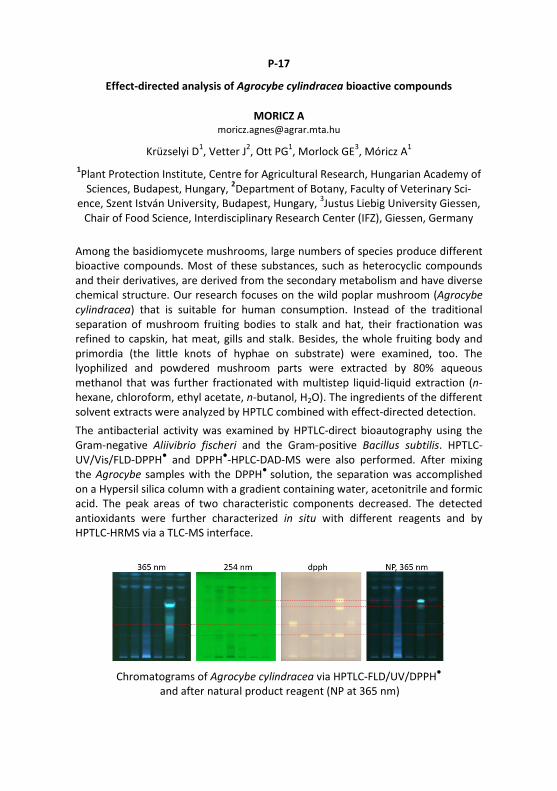
P-17
Effect-directed analysis of Agrocybe cylindracea bioactive compounds
MORICZ A [email protected]
Krüzselyi D1, Vetter J2, Ott PG1, Morlock GE3, Móricz A1 1Plant Protection Institute, Centre for Agricultural Research, Hungarian Academy of
Sciences, Budapest, Hungary, 2Department of Botany, Faculty of Veterinary Sci-ence, Szent István University, Budapest, Hungary, 3Justus Liebig University Giessen,
Chair of Food Science, Interdisciplinary Research Center (IFZ), Giessen, Germany
Among the basidiomycete mushrooms, large numbers of species produce different bioactive compounds. Most of these substances, such as heterocyclic compounds and their derivatives, are derived from the secondary metabolism and have diverse chemical structure. Our research focuses on the wild poplar mushroom (Agrocybe cylindracea) that is suitable for human consumption. Instead of the traditional separation of mushroom fruiting bodies to stalk and hat, their fractionation was refined to capskin, hat meat, gills and stalk. Besides, the whole fruiting body and primordia (the little knots of hyphae on substrate) were examined, too. The lyophilized and powdered mushroom parts were extracted by 80% aqueous methanol that was further fractionated with multistep liquid-liquid extraction (n-hexane, chloroform, ethyl acetate, n-butanol, H2O). The ingredients of the different solvent extracts were analyzed by HPTLC combined with effect-directed detection. The antibacterial activity was examined by HPTLC-direct bioautography using the Gram-negative Aliivibrio fischeri and the Gram-positive Bacillus subtilis. HPTLC-UV/Vis/FLD-DPPH● and DPPH●-HPLC-DAD-MS were also performed. After mixing the Agrocybe samples with the DPPH● solution, the separation was accomplished on a Hypersil silica column with a gradient containing water, acetonitrile and formic acid. The peak areas of two characteristic components decreased. The detected antioxidants were further characterized in situ with different reagents and by HPTLC-HRMS via a TLC-MS interface.
Chromatograms of Agrocybe cylindracea via HPTLC-FLD/UV/DPPH●
and after natural product reagent (NP at 365 nm)
P-18
Direct bioautography with subsequent DART-MS
HÄBE TT [email protected]
Häbe TT1, Jamshidi-Aidji M1, Macho J1, Morlock GE1 1Justus Liebig University Giessen, Institute of Nutritional Science, Chair of Food
Science, Interdisciplinary Research Center (IFZ), Giessen, Germany
The benefits of the hyphenation of direct bioautography (DB) with Direct Analysis in Real Time mass spectrometry (DART-MS) were illustrated on the example of four parabens. These are used individually or combined as preservatives in specific food, tobacco, pharmaceuticals and cosmetic products due to their antimicrobial and an-tifungal properties [1]. Due to their lipophilic molecular structure, parabens are able to diffuse through the dermal barrier, metabolized in organisms [2] and asso-ciated with undesirable side effect like allergies and hormonal activity. Monitoring upper limits or prohibition of individual parabens in consumer products is challeng-ing due to the wide range of sample matrices.
The matrix-discriminating desorption-based DART-MS method was applied imme-diately after DB in presence of all the bioassay media [3-5], using a modified HPTLC-DART-MS interface with improved surface scanning performance [6]. The overall DART-MS performance and detectability of the parabens were investigated in de-pendency of the applied Bacillus subtilis [3] and Aliivibrio fischeri bioassays [4] for the detection of antimicrobials and planar yeast estrogen screen (pYES) for estro-gen-effective compounds [5]. Quantification of parabens in two hand cremes in the ng/band-range showed mean deviations of 4.6% after DB on normal and reversed phase layers. This streamlined technique showed a reduced MS background, if compared to DB coupled to MS via electrospray ionization. The direct hyphenation DB-DART-MS was successfully established for simultaneous detection of bioactivity and qualitative/quantitative MS analysis out of complex sample matrices at one go.
[1] M.G. Soni et al. Food Chem. Toxicol. 2002, 40, 1335 [2] German Federal Institute for Risk Assessment (BfR), Stellungnahme Nr. 009/2011 2011, 1 [3] M. Jamshidi-Aidji, G.E. Morlock, J. Chromatogr. A 2015, 1420, 110 [4] S. Krüger et al. J. Chromatogr. A 2013, 1289, 105 [5] I. Klingelhöfer, G.E. Morlock, J. Chromatogr. A 2014, 1360, 288 [6] T.T. Häbe, G.E. Morlock, Rapid Commun. Mass Spectrom. 2016, 30, 321

P-19
Semi-quantitative comparison of acetylcholinesterase inhibition in effect-directed
analysis with HPTLC
WEISS S [email protected]
Weiss S1, Schulz W1, Winzenbacher R1 1Zweckverband Landeswasserversorgung, Langenau, Germany
Modern analytical test methods increasingly detect anthropogenic organic substances and their transformation products in water samples. The presence of these compounds might pose a risk to the aquatic environment. To determine a possible toxicological risk, samples are tested using various bioassays in combination with HPTLC, including the acetylcholinesterase (AChE) inhibition test.
Based on the central role of AChE in the transmission of signals in the synaptic gap, substances that inhibit this enzyme are considered to be potentially neurotoxic. The most important AChE-inhibiting substances include organophosphates and carbamates.
In the AChE assay, the effect of substances is determined by enzyme activity. The comparison of the potency of the action of substances is compared via the inhibition constant ki, which is calculated from the enzyme activity and the concentration of the inhibitor. However, the inhibiting substances are often unknown in environmental samples and their concentration is therefore unknown.
In our developed approach for semi-quantitative evaluation, the sample is applied in different volumes onto the HPTLC plate. After separation the AChE assay is applied onto the plate. By tracking the inhibition over time, the enzyme activity of the inhibition zones is determined for the individual application volumes. With the help of different application volumes, it is possible to calculate the application volume, which is required to achieve an enzyme activity of 50% for the inhibition bands. Since the volume is inversely proportional to the concentration, the reciprocal value of the determined volume is indicated. This value is called reciprocal iso-activity volume (RIAV). The RIAV allows a semi-quantitative comparison of inhibiting bands of a sample as well as between samples. The RIAV makes it possible to compare processes, for example, of a waste water treatment plant with the help of the effect pattern of separated sample.

P-20
Selective two-dimensional effect-directed analysis with TLC
STÜTZ L [email protected]
Stütz L1, Weiss S1, Schulz W1, Schwack W2, Winzenbacher R1 1Laboratory for Operation Control and Research, Zweckverband
Landeswasserversorgung, Langenau, Germany, 2Institute of Food Chemistry, University Hohenheim, Stuttgart, Germany
There are thousands of organic trace substances in the environment which are not fully characterized so that the evaluation of their relevance to the ecosystem is difficult. Effect-directed analysis (EDA) is a suitable tool for the assessment of effects in a sample to in-vitro bioassays and hence collects information about the relevance of substances [1]. HPTLC thereby proved to be a good method for fractionation, since the layer is solvent-free after separation and an in vitro bioassay can be applied directly [2]. But the main disadvantage of HPTLC is the lower peak capacity compared to HPLC.
For improvement of peak capacity by HPTLC, a one-dimensional (1D) gradient development already proved to be a good strategy [3]. For environmental samples, the peak capacity of such gradient development is often insufficient, so that a two-dimensional (2D) separation strategy was developed in order to increase the peak capacity and to facilitate identification of effective compounds. Thus only effective zones were selected in the first dimension and transferred to the second dimension through elution head-based extraction. Three 2D approaches were developed and validated. One approach, where the retention factors of the first dimension were taken to adjust the eluents for the second dimension achieved best results in terms of peak capacity and orthogonality. Thus, peak capacity could be increased by the factor 6.8 compared to 1D gradient development.
By investigation of spiked surface water with the 2D HPTLC/acetylcholinesterase inhibition assay, some substances could already be assigned to zones with neurotoxic effects. The knowledge about the substance´s effects enables the assessment of their relevance to the environment.
[1] M. König, et al. Environ. Pollut. 220, Part B (2017) 1220 [2] G. Morlock, W. Schwack, J. Chromatogr. A 1217 (2010) 6600 [3] C. Stiefel, S. Dietzel, M. Endress, G.E. Morlock, J. Chromatogr. A 1462 (2016) 134

P-21
Modern direct bioautography for fast screening and characterization of active
compounds in plant extracts used in cosmetics
OBERLE M [email protected]
Oberle M1, Lewits P1, Engemann J1, Klingelhöfer I2, Morlock GE2 1Merck KGaA, Darmstadt, Germany, 2Justus Liebig University Giessen, Institute of Nutritional Science, Chair of Food Science, Interdisciplinary Research Center (IFZ),
Giessen, Germany
HPTLC can ideally be combined with biological, biochemical and chemical assays without much effort [1]. The direct link to effective compounds is achieved in the same chromatographic separation bed, so an assignment to bioactive compounds can easily be made. Sophisticated technical equipment for the bioassay coupling is not necessary. The sample extract is applied as natural as possible and the assay can comprehensively detect the whole sample extract. Even such bioactive compounds left at the starting zone are detectable - a clear benefit in contrast to column techniques.
For direct bioautography, new concepts of using water-wettable reversed layers have been introduced recently [2-3]. On such layers, the bioassay medium showed no elution power and thus the analytes remained detectable as sharp bands, even after hours of incubation. So the quality of the bioautograms improved substantially. Such an effect-directed screening can be used to obtain target-orientated results rapidly (bioprofiling) and allows the evaluation of the plant's fingerprint with regard to stability, degradation and adulteration. Such a hyphenated screening concept like HPTLC-UV/Vis/FLD-(bio)assay-MS is shown for different plant extracts.
[1] Morlock G. ACS Syposium Series 2013, 1185, 101-121 [2] Morlock GE, Klingelhöfer I. Anal. Chem. 2014, 86, 8289-8295 [3] Klingelhöfer I, Morlock GE. J. Chromatogr. A 2014, 1360, 288−295.

P-22
In vinum veritas: Estrogen-effective compounds discovered in wine by
HPTLC-pYES
KLINGELHÖFER I [email protected]
Klingelhöfer I1, Morlock GE1 1Justus Liebig University Giessen, Institute of Nutritional Science, Chair of Food
Science, Interdisciplinary Research Center (IFZ),Giessen, Germany
Estrogen-effective compounds such as phytoestrogens are ubiquitous in our food. They can bind to the human estrogen receptors α/β and thereby activate or inhibit the endocrine system of humans, which results in controlling and regulating essen-tial functions of metabolism, growth and development. The coupling of the planar Yeast Estrogen Screen (pYES) with HPTLC is an efficient detection method for es-trogen-effective compounds. This direct bioautographic method allows extensive screening and bioprofiling of food with only a small sample preparation and with-out prior selection of the target analyte [1].
30 commercially available wine samples were examined. These included 16 red wines, 10 white wines and 4 rosé wines from North and South America, Australia and Europe. A liquid-liquid extraction of the wine samples was carried out, fol-lowed by evaporation to dryness and dissolution to yield a final enrichment factor of 80. After direct bioautography, eight different estrogen-effective compounds were detected by their blue fluorescence at 366 nm.
It was examined whether the known phytoestrogens apigenin, genistein, kaempfe-rol or naringenin were detectable in the wine samples as bioactive zones. For this purpose, the wine samples were spiked or the standard substances were over-sprayed on the sample zones to generate overlapping bands [2]. None of these standard substances showed the same hRF value as the previously detected bioac-tive zones in the wine. The advantage of the non-target method is that it detects all bioactive compounds present in the complex sample without preselection of ana-lytes. Currently, these eight unknown substances binding to the human estrogen receptor are characterized by mass spectrometry and nuclear magnetic resonance spectroscopy.
[1] I. Klingelhöfer, G.E. Morlock, J. Chromatogr. A 2014, 1360, 288 [2] G. E. Morlock, I. Klingelhöfer, Anal. Chem. 2014, 86, 8289

P-23
Investigation of plant protection products for endocrine effects by
direct bioautography
KLINGELHÖFER I [email protected]
Klingelhöfer I1, Morlock GE1 1Justus Liebig University Giessen, Institute of Nutritional Science, Chair of Food
Science, Interdisciplinary Research Center (IFZ), Giessen, Germany
A number of substances from agriculture, industry as well as secondary plant com-pounds have the potential to act as endocrine disrupters. One group of these en-docrine disruptor components (EDC) are plant protection products (PPPs). These compounds can affect the endocrine system of mammalians due to their binding to human estrogen receptors (hER α or β). For the detection of this EDC a direct bio-autography (DB) is used, which combines the planar Yeast Estrogen Screen (pYES) with HPTLC [1].
For the first time, a screening of various PPPs agents for estrogenic activity was car-ried out using HPTLC-pYES on water-wettable HPTLC plates RP-18 W. Out of 60 in-vestigated substances, ten active PPPs (carbaryl, chlorpyrifos, cypermethrin, cypro-dinil, fenhexamide, fludioxonil, pendimethalin, phorate, picoxystrobin, and mercap-todimethur) showed ERα-mediated estrogen-like effects. A dose-dependent effect was determined for six of them. Furthermore, selected foods were tested for resi-dues of the estrogen-like PPP active substances approved in the EU.
For example for grapes, tomatoes and white wine, cyprodinil, fenhexamide and fludioxonil were admitted. The foods were crushed and extracted with n-hexane - diethyl ether. After direct bioautography, bioactive zones could be found in all food extracts. In white table grapes and Rivaner white wine (from a private production) fenhexamid was detected and verified. Cyprodinil and fludioxonil could not be found in any of the investigated samples. The bioactive zones in the tomatoes and red table grape extracts could not be assigned to any of the investigated PPPs. This developed direct bioautography method proved to be suited for residue analysis, i. e. for the detection of estrogen-like PPPs in complex food matrix at the trace level.
[1] I. Klingelhöfer, G.E. Morlock, J. Chromatogr. A 2014, 1360, 288
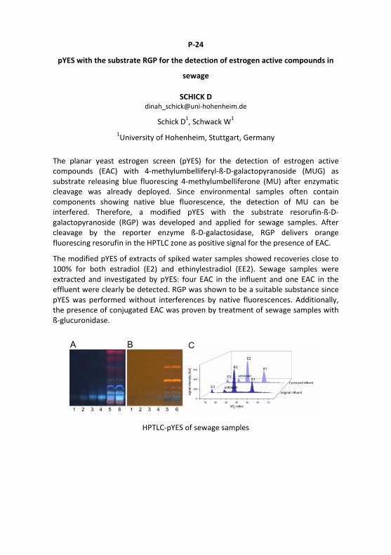
P-24
pYES with the substrate RGP for the detection of estrogen active compounds in
sewage
SCHICK D [email protected]
Schick D1, Schwack W1 1University of Hohenheim, Stuttgart, Germany
The planar yeast estrogen screen (pYES) for the detection of estrogen active compounds (EAC) with 4-methylumbelliferyl-ß-D-galactopyranoside (MUG) as substrate releasing blue fluorescing 4-methylumbelliferone (MU) after enzymatic cleavage was already deployed. Since environmental samples often contain components showing native blue fluorescence, the detection of MU can be interfered. Therefore, a modified pYES with the substrate resorufin-ß-D-galactopyranoside (RGP) was developed and applied for sewage samples. After cleavage by the reporter enzyme ß-D-galactosidase, RGP delivers orange fluorescing resorufin in the HPTLC zone as positive signal for the presence of EAC.
The modified pYES of extracts of spiked water samples showed recoveries close to 100% for both estradiol (E2) and ethinylestradiol (EE2). Sewage samples were extracted and investigated by pYES: four EAC in the influent and one EAC in the effluent were clearly be detected. RGP was shown to be a suitable substance since pYES was performed without interferences by native fluorescences. Additionally, the presence of conjugated EAC was proven by treatment of sewage samples with ß-glucuronidase.
HPTLC-pYES of sewage samples

P-25
HPTLC coupled estrogenic activity assessment of the phytoestrogens genistein
and biochanin A in nutraceutical red clover (Trifolium pratense L.) formulations
BRÄM S [email protected]
Bräm S1, Schönborn A2, Wolfram E1 1Zurich University of Applied Sciences, Institute of Chemistry and Biotechnology,
Phytopharmacy and Natural Products, Wädenswil, Switzerland, 2Zurich University of Applied Sciences, Ecotoxicology Research Group, Institute of Natural Resource
Sciences, Wädenswil, Switzerland
Plant-derived estrogens, known as phytoestrogens, are present in numerous dietary supplements and marketed as natural alternatives to estrogen replacement therapies [1]. Moreover, they are discussed to be indicated as complementary therapy for different types of cancers. Genistein, a naturally occurring phyto-estrogen from soy bean and red clover acts as a potent anticancer agent. However, in some types of breast cancer, the intake of genistein may be contraindicated, since the isoflavone may promote cancer cell proliferation [1]. The rapid planar-YES assay designed for low concentrations of target endocrine disruptors in environmental samples [2] has been adapted for the screening of phytoestrogenic compounds in ready-to use dietary red clover (Trifolium pratense L.) supplements. In HPTLC separated extracts of red clover raw material, pronounced fluorescent zones of genistein and biochanin A indicated estrogenic activity. The half maximal effective concentration (EC50), measured by quantitative intensity evaluation of the zones of genistein and biochanin A in different concentrations was 2.1 and 3.3 ng/band, respectively. Moreover, the same compounds were identified in separated samples from commercially available red clover capsules and tablets. Excipients originating from tablet and capsules matrices such as microcrystalline cellulose or polyvinyl pyrrolidone did not interfere with the method. Daidzein only showed weak estrogenic activity, notably that a specific intestinal microbe in human is needed to bioconvert daidzein to its active metabolite equol that was not tested in our study [1]. The planar-YES assay can be used as a screening tool for phytoestrogens, however a limitation is given by the extrapolation of the results to effects in human cells, since they might differ in respect to cellular substance uptake. [1) Patisaul HB, Jefferson W. Front. Neuroendocrinol. 2010, 31: 400-419 [2] Köster O, Schubert-Ullrich P, Schönborn A, Schulz W. Aqua & Gas 2016, 12: 32-41

P-26
Estrogenic substances in treated wastewater used for crop irrigation in Cyprus -
Preliminary results using the planar-YES bioassay
MAINETTI T [email protected]
Krayer P1, Beretsou V2, Mainetti T1, Fatta-Kassinos D2, Schoenborn A1 1Institute of Natural Resources Science, Zurich University of Applied Science,
Waedenswil, Switzerland, 2Department of Civil and Environmental Engineering and Nireas-International Water Research Center, School of Engineering, University of
Cyprus, Nicosia, Cyprus
Cyprus is a semi-arid Mediterranean country with mild winters and generally dry summers. Increasing water scarcity during summer has led to the use of for irrigation with treated wastewater (TWW) since many years now. Research on its quality has been conducted at NIREAS-International Water Research Center at Cyprus. A major focus was the analysis and characterization of influent and effluent wastewater samples on micropollutants, including estrogenic compounds, which are frequently detected worldwide at sewage treatment plants (STPs) [1-3]. Estrogenic compounds can potentially interact with the endocrine system of humans and animals. The aim of this preliminary study was to characterize the estrogenicity profiles of inlet and outlet waters collected at two STPs located in Cyprus. Influents and effluents of the two STPs were sampled (24-h composite samples), extracted and concentrated 100x by using solid phase extraction (SPE). Triplicates of the extracted samples were analyzed with the planar-Yeast Estrogen Screen (planar-YES) and with ultra-performance liquid chromatography-tandem mass spectrometry (UPLC-MS/MS) in parallel. At the time of abstract submission, the measurements and quantification were not completed. First results show that all samples taken at the two STPs contain estrogenic activity (EA). In the 2 influent samples 6-8 different strong EA-signals were detected, based on distinct Rf-values. The 2 effluents contained 3-4 EA-signals whose Rf-values can be assigned to 17α-ethinylestradiol (EE2), 17β-estradiol (E2), estrone (E1) and a fourth unknown substance. The presence of these EA-signals was unambiguously clear in the planar-YES, while EE2, E2 and E1 were below the method detection limit (MDL) in the parallel UPLC-MS/MS analysis. This shows that the planar-YES allows gaining additional relevant information on STP samples and thus complements instrumental analysis.
[1] Ferrer and Thurman, 2012 [2] Boleda et al., 2013, [3] Dasenaki and Thomaidis, 2015
P-27
HPTLC-hyphenated bioautography for antidiabetic and antioxidant metabolites
from Butea monosperma
KHAN W [email protected]
Khan W1, Azhar M2, Singh P1, Ahmad S1 1Faculty of Pharmacy, Jamia Hamdard, New Delhi, India, 2Delhi Institute of
Pharmaceutical Sciences and Research, New Delhi, India
HPTLC fingerprinting resulted in the separation of metabolites and bioautography. It can provide the fast screening of a large number of metabolites for bioactivity, namely, α-glucosidase enzyme inhibition, yeast cell uptake and in the target-directed isolation and characterization of bioactive metabolites from the mixture. HPTLC based quantitative analysis of fatty acid in a blood sample of diabetic mice followed by conformation through GC-MS resulted in identification in their expression which can be used further for the intervention of herbal medicine in diabetes. Aqueous extract of Butea monosperma was hydrolyzed and extracted with ethyl acetate, which was separated on HPTLC plate using toluene - ethyl acetate 5:4. After migration of the samples, the HPTLC plate was sprayed with α-glucosidase solution in sodium acetate buffer and incubated at room temperature for 60 min followed by spraying of solutions of 2-naphthyl-α-glucopyranoside and fast blue salt. Enzyme inhibitors were visualised as white spots on the HPTLC plates. Similarly, the antioxidant activity of migrated metabolites was identified by superimposing the chromatograms of extracts obtained with the HPTLC-DPPH● assay. Butrin, butin and quercetin are the major metabolites identified as an antidiabetic molecule. The level of fatty acid in serum sample was analyzed through HPTLC followed by GC-MS. HPTLC separation of fatty acid was carried out using hexane - ethyl acetate 1:1, followed by densitometric evaluation. Myristic acid, stearic acid, palmitic acid and oleic acid are the major fatty acid that had significantly changed their expression in diabetic mice. The bioautographic study resulted in the identification of antidiabetic and antioxidant molecules of B. monosperma flower and this model can be used for other plants as well.
P-28
Antimicrobial activity of effective antimicrobial compounds in extracts from
strawberry leaves by TLC
KWACK Y [email protected]
Kwack Y1, Kim D2 1Research Institute of Agriculture and Life Sciences, Seoul National University,
Seoul, Korea, 2Department of Plant Science, Seoul National University, Seoul, Korea
Strawberry plants include their own phytoanticipins. 'Seolhyang' strawberry cultivar also contained ellagic and gallic acid, E,E-α-farnesene, and trans-caryophyllene. In addition, contents of the secondary compounds in non-edible parts such as leaves, petioles, and green fruits are higher than those in red fruits [1]. Some studies supported that non-edible parts of vegetables are available because they contain higher contents of secondary metabolites than edible parts. Previous researches, however, have studied antimicrobial activities of secondary metabolites in red fruits of strawberry plants but a few studies have reported the activities of the metabolites in non-edible parts using various extraction methods.
We tested antimicrobial activity of parts of strawberry extracted by methanol, acetone, dichloromethane, and hexane against Colletotrichum coccodes (KACC No. 40802), Glomerella cingulata (KACC No. 40300), Fusarium oxysporum f. sp. lycopersici (KACC No. 40043), Phytophthora cactorum (KACC No. 40183), Phytophthora capsici (KACC No. 40177), and Rhizoctonia solani (KACC No. 40115).
The selected method was TLC combined with microbiological detection, called direct bioautogrphy. The extracts were developed with ethyl acetate – methanol - water 8:1:1. Antimicrobial activity was observed as white inhibition zones on a red background. Active compounds in the white inhibition zones were scraped off and extracted for GC-MS analysis.
[1] D.S. Kim et al. J. Hort. Sci. Technol. 31, 2013, 224-230

P-29
HPTLC as a method for quick assessment of bile salts deconjugation activity by
Pediococcus acidilactici LAB6 and Lactobacillus plantarum LAB12
SNEZANA AGATONOVIC K [email protected]
Rohawi S1, Lim S1, Agatonovic-Kustrin S1, Ramasamy K1 1Faculty of Pharmacy, University Teknologi MARA (UiTM), 42300, Bandar Puncak
Alam, Malaysia, 1Collaborative Drug Discovery Research (CDDR) Group, Pharmaceutical and Life Science Community of Research, Universiti Teknologi
MARA (UiTM), 40450, Shah Alam, Malaysia
Probiotics are being recognised as being able to assist lowering cholesterol levels in the body. Bile salt hydrolase (BSH)-producing probiotics lower cholesterol through deconjugation and co-precipitation of bile salts. However, probiotic-induced deconjugation of a wide range of bile salts is dependent on the bacterial strains. Our previous work used HPTLC to screen 12 lactic acid bacteria (LAB) with probiotic characteristics for their hypocholesterolaemic properties. Pediococcus acidilactici (LAB6) and Lactobacillus plantarum (LAB12) were found to exhibit excellent cholesterol lowering effects both in the presence or absence of bile salts. The present study went on to assess the bile salt deconjugation activity of LAB6 and LAB12 against primary and secondary bile salts using the HPTLC. LAB6 and LAB12 were cultured in media containing 1 mmol/L of either sodium based-conjugated primary or secondary bile salts, i. e., glycocholate (GC), glycodeoxycholate (GDC), glycochenodeoxycholate (GCDC), taurocholate (TC), taurodeoxycholate (TDC) and taurochenodeoxycholate (TCDC), for 24 h. Cell free supernatant was collected, and then bile salts were separated from the fermented media. Direct quantification of bile salts was performed using HPTLC. It was found that both LAB6 and LAB12 were able to deconjugate most of the primary and secondary bile salts that were present, with the bile salt hydrolase activity higher against glyco- than tauro-conjugated bile salts. Glyco-conjugated bile salts are associated with cholestasis. LAB12 was found to be the most effective strain, inhibiting glyco- and tauro-conjugated bile salts by 100% and >66%, respectively.
P-30
Quantification of phospholipids using HPTLC and primuline-induced fluorescence
detection by intensity changes
VELA J [email protected]
Vela J1, Membrado L2, Cebolla V2, Lapieza M2, Bernard-Savary P3 1University of Zaragoza, Zaragoza, Spain, 2CSIC, Instituto de Carboquimica, Zarago-
za, Spain, 2Chromacim, Pommiers-la-Placette, France
Primuline dye has been used for fluorescent enhancement of lipids for a long time. Although the responses of the phospholipids (PLs) in primuline are not as high as those of neutral lipids because their polar groups quench primuline emission to a certain level, HPTLC with induced fluorescence using primuline may be adequate for individual or group quantitative analysis of phospholipids if working conditions are properly selected. The influence of structural parameters of biologically significant phosphatidylcholines (PCs), phosphatidylethanolamines (PEs), phospha-tidylglycerols (PGs), and cardiolipins (CLs) were studied with regard to primuline fluorescent response. Thus, the influence of fatty acid (FA)-chain lengths and chain nature (saturated/unsaturated) were studied using lyso, saturated, unsaturated and natural PL standards, whose chain distributions are known. Therefore, calibration techniques for an individual or group analysis of the corresponding PL or PL family were developed, which were mostly based either on external standard or standard addition. Synthetic mixtures of known composition were used at various complexity levels. Before starting detection optimization, the repeatability of the application and densitometry system, and then the repeatability of densitometry was evaluated, before and after chromatography. Therefore, the influence of slit dimension, scanning speed, data resolution, sensitivity (with special attention to automatic and manual), with different applied band sizes on the signal was evaluated. The evolution of signals in function of time was also monitored. The raw data from winCATS software was compared to a manual integration, with the integration parameters fixed by the analyst. Moreover, the mechanism of the generated fluorescence and the possible used of reversed-phase plates were discussed.
P-31
Anthocyanin profiles of colored wheat crosses via HPTLC
MORLOCK GE [email protected]
Krüger S1, Morlock GE1 1Justus Liebig University Giessen, Institute of Nutritional Science, Chair of Food
Science, Interdisciplinary Research Center (IFZ), Giessen, Germany
The food industry shows a growing interest in functional food with assigned health-promoting properties such as radical-scavenging or antimicrobial effects. Antho-cyanins naturally found in fruits, vegetables, cereals (like colored wheat) and flower petals have been described to have radical-scavenging activities as well as a positive influence on stress-related and chronic diseases.
In 2013, Jaafar et al. [1] showed that it was possible to increase the total antho-cyanin content in colored wheat by combining wheat grains of blue aleurone and purple pericarp genotypes. The matrix of the colored wheat extracts was highly challenging the analysis on silica gel layers. In this study, a separation was deve-loped for the same colored wheat samples. The best separation was achieved on reversed phases (on HPTLC plates silica gel 60 RP-18 W F254 s with water - n-propanol - formic acid 18:8:1.2) and on amino phases (on HPTLC plates silica gel 60 NH2 F254 s with ethyl acetate - 2-butanone - water - formic acid 14:6:3:2), whereby the amino phase separation was preferred. Thus, HPTLC-UV/Vis fingerprints were achieved allowing a straight-forward and clear visual differentiation of the genotypes.
Hyphenation with mass spectrometry (HPTLC-ESI+-MS) was exemplarily shown on two representatives for the blue aleurone and purple pericarp genotypes. This allowed further characterization and differentiation of the anthocyanins profiles obtained. HPTLC proved to be a streamlined method for the challenging analysis of colored wheat samples. [2]
[1] S.N.S. Jaafar, J. Baron, S. Siebenhandl-Ehn, T. Rosenau, S. Böhmdorfer, H. Graus-gruber, Plant Breed. 132 (2013) 546-552 [2] S. Krüger, G.E. Morlock, Anthocyanin fingerprints of colored wheat via HPTLC-UV/Vis-ESI-MS, in submission.

P-32
In-process quality control of wine by (micro) planar chromatography
Kaiser R [email protected]
Kirchert S1, Morlock GE1, Kaiser R2 1Justus Liebig University Giessen, Institute of Nutritional Science, Chair of Food
Science, Interdisciplinary Research Center (IFZ), Giessen, Germany, 2Institute for Chromatography (IFS), Bad Dürkheim, Germany
Four HPTLC methods were developed for in-process quality control of glycerol, gluconic acid, amino acids and sugars in 20 German wines. All developed methods were capable of analyzing up to 19 wine samples in parallel within 1 to 2.5 h for monitoring of the wine processing. For a simple sample preparation on site, the wine samples were only diluted with methanol.
The quantitative amino acid method was suited for comparison, profiling and differentiation of wine varieties. Some amino acids are very similar in their chromatographic behaviour, and an additional confirmation with mass spectro-metry was recommended. The developed gluconic acid method was used as a threshold method for screening of Botrytis cinerea infection. This method was also able to detect other organic acids like malic, tartaric and citric acid. The developed glycerol method targeted on the grape must fermentation control (spontaneous versus regular) as well as the detection of fraud (e. g., glycerol adulteration). The analysis of saccharides was also performed for the 20 wine samples. The evaluation of the results obtained by all these methods allowed the fermentation control, malolactic fermentation control and monitoring of the grapes overall health status.
For a broad on site analysis, simple and cost-efficient analysis system are advanta-geous for routine use in wine processing. Hence, the method transfer to circular micro planar chromatography (µ-PLC) was studied for these methods. µ-PLC works in a low-cost environment (€ 4000 investment costs) and its potential as inexpen-sive alternative for small wineries and distributors was highlighted.

P-33
Fast screening of veterinary drugs in food of animal origin via pSPE-HRMS
Anweiler L [email protected]
Anweiler L1, Knapp S1, Nitsch M1, Morlock GE1 1Justus Liebig University Giessen, Institute of Nutritional Science, Chair of Food
Science, Interdisciplinary Research Center (IFZ), Giessen, Germany
Animal pharmaceuticals are used to treat diseases in animals intended as food source. Their residues found in food through an improper usage are hot topics in food safety, public health and environmental fields. Their trace analysis in con-cerned food is an important field of research. The present methods are based on a complex and time consuming cleanup, followed by HPLC-MS/MS.
Hence, the concept and potential of sample preparation and chromatography on the same plate [1, 2] for many samples in parallel was studied in this field. A planar solid phase extraction (pSPE)-HRMS method was developed to detect 31 animal pharmaceuticals in 4 different typical animal food matrices. The food extracts were applied as rectangle on HPTLC plates silica 60 F254 MS grade, prewashed by chroma-tography with acetone and ammonia (25%). The focusing of the 31 pharmaceuticals into the top application region of the four matrices was achieved with i-propanol. Then, the animal pharmaceuticals were front-eluted with acetone and ammonia (25%), followed by direct elution via a TLC-MS interface into the ESI-HRMS. The full automatization of the TLC-MS interface for sequential elution of the target zones was established in parallel [3] and will fasten and ease this step of the workflow.
All on-plate sample preparation steps were simultaneously and automatically per-formed for 13 animal food extracts in parallel. This makes it fast and cost-effective, as time-consuming manual preparation steps for each sample were avoided. A benchmarking of the new pSPE-HRMS approach and the conventional methodology is presented to underline the benefits achieved.
[1] Morlock, G., Schwack, W. J. Planar Chromatogr. 20 (2007) 399-406 [2] Morlock, G., Vega, M. J. Planar Chromatogr. 20 (2007) 411-417 [3] Häbe, T., Morlock, G., Open-source-based automatization of an elution head-based interface for HPTLC-MS, in preparation
Acknowledgement: For delivery of standards and samples, thank is owed to Dr. Günther Kempe, Landesuntersuchungsanstalt für das Gesundheits- und Veterinär-wesen (LUA), Chemnitz, Germany.
P-34
Planar solid phase extraction-gas chromatography mass spectrometry for the
determination of sterol oxidation products in cosmetics
SCHRACK S [email protected]
Schrack S1, Hohl C1, Schwack W2 1State Laboratory of the Canton Basel-City, Basel, Switzerland, 2Institute of Food
Chemistry, University of Hohenheim, Stuttgart, Germany
Sterol oxidation products (SOPs) have been linked to several adverse health effects. Identifying dietary uptake sources has therefore been a key issue for these compounds. Other potential sources for human uptake such as cosmetics, how-ever, have not been evaluated. As our first study on cosmetics containing lanolin, a widely used ingredient made of sheep wool fat, revealed high cholesterol oxidation products levels, we devised a further method based on planar solid phase extraction-gas chromatography-mass spectrometry (SPE-GC-MS) for the determina-tion of additional SOPs, suspected to occur in lanolin containing cosmetics, which also was less susceptible to matrix interferences. [1, 2]
SOPs were first separated from more non-polar and polar matrix constituents by normal phase thinlayer chromatography and then focussed into one target zone. Zone extraction was performed with the TLC-MS interface, followed by GC-MS analysis. pSPE showed to be effective for cleaning up cosmetic samples as sample extracts were free of interferences. Besides the cholesterol oxidation products mentioned in our first study, we reported for the first time the occurrence of oxidised congeners of lanosterol and dihydrolanolsterol in lanolin containing cosmetics. [1, 2]
[1] Schrack, S., Hohl, C. and Schwack, W., J. Chromatogr. A 2016, 1473, 10-18 [2] Schrack, S. et al. Int. J. Cos. Sci. 2015, 38, 1-7
P-35
Optimization of HPTLC and HPTLC-MS methods for analysis of flavonoids and
phenolic acids
JUG U [email protected]
Jug U1, Glavnik V1, Vovk I1 1Department of Food Chemistry, National Institute of Chemistry, Ljubljana, Slovenia
Flavonoids and phenolic acids are one of the most important groups of secondary metabolites and bioactive compounds present in plants and fungus. The effect of dietary phenolics is currently of great interest due to their antioxidative, anti-aging and anticarcinogenic activities. They have important role in various human diseases. It would be useful to gain a better insight in distribution of these compounds and consequently learn how to increase the amount of their dietary intake.
The main topic of our work was development of HPTLC and HPTLC-MS methods for separate analyses of flavonoids and phenolic acids, for which numerous standards were used to gain methods applicable to different samples. During the optimization of a developing solvent, different solvent mixtures were tested. We also tested the adequacy of different stationary phases, effect of pre-development, deactivation by water vapor and chamber saturation. A very strong influence of saturation was observed. To avoid the problems in HPTLC-MS analyses due to the high background signals in the MS spectra, we optimized a pre-development step and MS conditions. The applicability of the developed methods was tested in analysis of different samples, e.g. propolis, tea and coffee extracts, which represent rich sources of compounds of interest.

P-36
Quantification of α- and β-acids in hops by TLC and HPLC
BURHOLT M [email protected]
Burholt M1, Engemann J1, Pilakowski V1, Oberle M1 1Merck KGaA, Darmstadt, Germany
Hops are a very important base material for the production of beer. Especially, the substance Lupulin, a yellowish powder isolated from the hop cones, is responsible for the bitterness and unique taste of beer. That's why the amount of bitter acids in hops is very important for breweries. Lupulin contains different bitter acids [1], α- (humulone) and β- (lupulone) acids, which vary in different types of hop. Caused by the content of bitter acids, hops are divided in aromatic hops (< 10 % of α-acids) and bitter hops (> 10 % α-acids) [2]. In general, the α- and β-acids are also divided into five homologues.
The investigation of different hop types has shown that the amount of α-acid depends, on the hop category, as well as on the region. It could also be determined that the β-acid is not related to these criteria and is quite similar in all hop samples, regardless of whether the HPLC or TLC method was used. The amount of α-acid, but not the β-acid, depends on the kind of hop (bitter or aromatic) and the region. For comparison, both methods, TLC and HPLC, were used to quantify α- and β-acids. Both methods have advantages for the analytical workflow and can be easily combined.
[1] A. Forster et al. (2012) Hopfen - Vom Anbau bis zum Bier, Carl, Nürnberg [2] B. Engelhard, A. Lutz, E. Seigner (2011) Hopfen für alle Biere der Welt.

P-37
The advantages of HPTLC based on USP TLC methods for the analysis of black
pepper, turmeric and ginger
BURHOLT M [email protected]
Burholt M1, Pilakowski V1, Oberle M1 1Merck KGaA, Darmstadt, Germany
As in regulated pharmaceutical analysis, nowadays, various United States Pharma-copeia (USP) methods are available for quality testing for food, beverage, and especially dietary supplements. Most of them are based on a qualitative analysis using TLC followed by an additional compositional HPLC analysis.
The reason for this, could be due to described dated TLC methods as the analytical tool. Often, the chosen TLC method is a preparative one (PLC) or a standard TLC method without accurate sample application and result detection. In this poster, we would like to compare the quality and validity of three USP methods using different types of thin layer chromatography.
Using HPTLC methods instead of PLC/TLC methods allows direct quantification of target and marker compounds, fingerprinting, as well as the direct testing on avoidable, forbidden substances in one single step. Testing by multiple additional methods and time-consuming sample preparation can be avoided.
As examples, we have chosen extracts from black pepper, turmeric and ginger. All three samples are extracts and also used as dietary supplements. USP monograph methods are available and the executed chromatographic testing follows the corresponding USP monograph.

P-38
Development and validation of an HPTLC-densitometry method for simultaneous
quantitation of boric acid and fructoborates in dietary supplements and
foodstuffs
BITA A [email protected]
Biţă A1, Mogoşanu G1, Bejenaru L1, Neamţu J2, Hunter J3, Nemzer B3, Bejenaru C4,
Croitoru O5 , Rău G6, Scorei R7 1Department of Pharmacognosy & Phytotherapy, 2Department of Physics,
4Department of Vegetal & Animal Biology, 5Department of Drug Control,
6Department of Organic Chemistry, all Faculty of Pharmacy, University of Medicine and Pharmacy of Craiova, Romania, 3VDF FutureCeuticals, Momence, USA,
7BioBoron Research Institute, Craiova, Romania
The bis-fructose, a naturally occurring boro-carbohydrate ester of boric acid (BA), was previously reported to be found in the human diet in herbs, fruits, seeds, honey and some foodstuffs. Commercially produced calcium fructoborate is a patented nature-identical analogue of the abovementioned plant mineral complex that is used as nutritional supplement with potential support for healthy bone, joint and cardiovascular systems.
To our knowledge, the simultaneous quantitation of BA and fructoborate (FB) has not been reported until now. In this respect, we developed and validated a new, simple, fast, specific and accurate HPTLC-densitometry method for the routine analysis and quantitation of BA and CFB in dietary supplements and foodstuffs. Chromatographic separations were performed on HPTLC plates silica gel G 60 F254, pre-washed by complete development with chloroform-methanol 1:1, and then dried for 30 min at 105 °C. Standard and sample solutions were thereafter applied to the plates using the Linomat V. The plates were developed for approximately 30 min with 2-propanol - water 4:1 (pH 7.5) in a vapor-equilibrated twin trough chamber at room temperature.
The HPTLC plates were sprayed with 0.1% ethanolic chlorogenic acid solution and subsequently dried for 1 min. BA and CFB were adequately separated with hRF values of 83 ± 1 and 59 ± 1, respectively. The densitometric evaluation was performed at 365 nm using the TLC Scanner 3 and winCATS software. This method was validated for specificity, linearity and range, precision, accuracy, limit of detection, limit of quantification, ruggedness and robustness according to the International Conference on Harmonization Guidelines.

P-39
HPTLC-densitometry method for nicotinamide riboside analysis in bulk and
nutraceutical formulations
BITA A [email protected]
Biţă A1, Rău G2, Mogoşanu G1, Neamţu J3, Scorei R4 1Department of Pharmacognosy & Phytotherapy, 2Department of Organic
Chemistry, 3Department of Physics, all Faculty of Pharmacy, University of Medicine and Pharmacy of Craiova, Romania, 4BioBoron Research Institute, Craiova, Romania
Nicotinamide riboside (NR), a natural compound from cow milk (ca. 0.8 mg/L) and unfiltered beer (lower amounts, still unquantified), is a scientifically proved dietary supplement in a world of unproved nutraceuticals. NR is considered as vitamin B3 of new generation, a biochemical precursor of NAD+, ensuring a healthy life and preventing aging. NR has various beneficial effects on the human body, such as: stimulation of fatty acid oxidation and mitochondrial activity, increasing resistance to the negative consequences of high-fat diet, protection against oxidative stress, prevention of peripheral neuropathy, blocking of mechanical stress caused by axonal degeneration and degenerative processes in the muscle. As a food supplement, NR was recently approved (2012) on the North American market, improving the physical performance of athletes. To the extent of our knowledge, until now, quantification of NR was made using only modern, complex and expensive chromatographic techniques (HPLC, LC-MS, strong anion exchange HPLC and matrix-assisted laser desorption mass spectrometry). In this regard, we propose a new, simple, selective, accurate and low cost HPTLC-densitometry method for the routine analysis of NR in bulk and dietary supplements dosage forms. The chromatographic separation was performed on HPTLC plates silica gel G 60 F254 with ethanol - 1 M aqueous ammonium acetate solution 7:3 adjusted to pH 5.0 with hydrochloric acid. NR was adequately separated at hRF 70 ± 1. The densitometric assessment was performed at 254 nm. The linear regression analysis data for the calibration plots showed good linear relationship. The method was validated for precision, accuracy, robustness and ruggedness, according with the International Conference on Harmonization Guidelines.

P-40
Lysergic acid amide screening for the total ergot alkaloids in rye by HPTLC-FLD
OELLIG C [email protected]
University of Hohenheim, Institute of Food Chemistry, Stuttgart, Germany
Ergot alkaloids are produced by parasitic fungi from the Claviceps genus, and are known to be responsible for toxicological effects in mammals. The alkaloids are located in the overwintering body (Secale cornutum) of Claviceps purpurea. The ergot fungus grows on cereals, mainly on rye, and thus the contamination of rye grain with Secale cornutum and ergot alkaloids is a serious problem. There are no maximum limits established for ergot alkaloids, but the European Union plans to define limits for the sum of ergot alkaloids in mid-2017 [1]. Since for monitoring only the sum of ergot alkaloids is relevant and not the quantity of individual species, selective and sensitive determination of the total ergot alkaloids is a meaningful and very useful strategy. Therefore, a rapid screening method for the common lysergic acid moiety of all alkaloids was developed for rye flours by means of HPTLC-FLD. After extraction with acetonitrile/ammonium acetate buffer and liquid-liquid partition in toluene according to the method of Oellig and Melde [2], ergot alkaloids were selectively transformed with a mixture of methanol and Superhydride solution. Released lysergic acid amide (LSA) from peptide ergot alkaloids and unaffected ergometrine were separated on HPTLC silica gel, and the enhanced native fluorescence was scanned for selective determination of the total ergot alkaloids. For calibration, the LSA standard was simply obtained from ergocristine transformed to LSA under identical conditions. With LOD and LOQ far below the currently applied quality criterion limit for alkaloids in rye and near-100% recoveries, a reliable and interference-free screening for the total ergot alkaloids was guaranteed [3].
[1] European Commission, Off. J. Eur. Union L283 (2015) 3-5 [2] Oellig, C., Melde,T., J. Chromatogr. A 1441 (2016) 126-133 [3] Oellig, C., in preparation

P-41
Comparison of fluorescent derivatization reagents and development of a simple
quantitation strategy for lipid analysis by HPTLC-FLD
BRÄNDLE K [email protected]
Brändle K1, Oellig C1 1University of Hohenheim, Institute of Food Chemistry, Stuttgart, Germany
For many years, HPTLC analysis is widely used for lipid analysis in different application areas. For sensitive and selective detection several derivatization reagents can be applied. Therefore, a comparison of different reagents for fluorescent detection is a useful study. The performance and suitability of the derivatization reagents were compared by means of triacylglycerides, 1,2- and 1,3-diacylglycerides, monoacylglycerides and free fatty acids (C12-C18). Primuline, berberine and coralyne were compared using optimal conditions for the reagent concentration and the detection settings of the TLC Scanner 4 (excitation wavelength and edge filter). In addition, differences between post- or prä-chromatographic derivatization were studied. Evaluation was based on signal-to-noise ratios (250 ng/zone, n=3) and by correlation coefficients of the analytical response (100-1000 ng/zone, n=3). Prä-chromatographic derivatization with primuline (250 mg/L in methanol including 0.1% acetic acid) delivered highest sensitivity and sharpest standard zones for all lipid classes on HPTLC plates LiChrospher silica gel 60 F254 with a twofold development with diethyl ether (first development) and pentane - n-hexane - diethyl ether 27:10:13 (second development).

P-42
Screening for MOSH and MOAH in food packaging by pSPE-UV/FLD-GC-MS
WAGNER M [email protected]
Wagner M1, Oellig C1 1University of Hohenheim, Institute of Food Chemistry, Stuttgart, Germany
Recently, the occurrence of mineral oil hydrocarbons (MOSH and MOAH) in food and food packaging has become a topic of interest. The current analytical method uses on-line LC-GC-FID and is well-developed and highly automated[1,2], however, the required instrumental set-up is very expensive and not available in each laboratory. Additionally, the evaluation of the received data is a very challenging task.
Therefore, pSPE, which uses the fully-automated HPTLC devices, was used to analyze MOSH and MOAH on planar thin-layers in a very easy way. The method is less time and solvent consuming compared to the mentioned LC-GC-FID approach and requires a minimal instrumental set-up. In a twofold development, MOSH and MOAH were separated and focused into two sharp zones per track. Quantitation was carried out by densitometric scanning, with aromatic compounds detected by UV absorption, while aliphatic compounds were detected by the fluorescence signal on an HPTLC plate pre-impregnated with a fluorescent probe. Additionally, MOSH and MOAH zones were eluted by the TLC-MS Interface into autosampler vials and analyzed by GC to detect not only MOSH and MOAH humps, but also to identify marker substances specific for the mineral oil origin and for recycled fiber material.
Analysis of different types of paper and cardboard packages showed that most of the matrix components were efficiently separated by pSPE, clearly visible by fluorescent zones near the application position. In the subsequent GC run, pristane and phytane as marker substances for the mineral oil origin and 2,6-diisopropylnaphthalene as a marker for recycled fiber material[2] could clearly be detected.
[1] M. Biedermann, K. Grob, J. Chromatogr. A (2012), 1255, 56-75 [2] M. Bieder-mann, K. Grob, J. Chromatogr. A (2012), 1255, 56-75 [2] M. Biedermann, K. Grob, J. Chromatogr. A (2012), 1255, 56-75 [2] M. Biedermann, K. Grob, J. Chromatogr. A (2012), 1255, 76-99
P-43
Phenolic acids contribution to the total antioxidant activities in in mango pulp
and peel
MORTON D [email protected]
Morton D1, Agatonovic-Kustrin S2 1La Trobe Institute for Molecular Sciences, La Trobe University, Bendigo, Australia,
2Faculty of Pharmacy, Universiti Teknologi MARA, Selangor, Malaysia
A simple, rapid and reproducible HPTLC method to analyse phenolic acids content and quantify antioxidant activities in selected mango varieties was developed and validated. Antioxidant activities in terms of free-radical scavenging activity and metal-chelating ability in pulp and peel samples were compared. Contributions of common phenolic acids (chlorogenic, caffeic and gallic acid), to the total antioxidant activity in mango samples was assessed. Polyphenolic content in mango pulp and peel was highly correlated with chlorogenic and gallic acid concentrations (R = 0.90 and 0.91 respectively). Free radical scavenging activity in the peel samples was related to the gallic and chlorogenic acid content (R = 0.83) but was not dependent on the caffeic acid content. Free radical scavenging activity in the pulp samples could not be predicted from the chlorogenic and gallic acid content (R = 0.25) indicating that pulp contains other bioactive compounds, such as carotenoids, vitamins and different polyphenolic phytochemicals that are more powerfull free radical scavengers. Since peel is richer in chlorogenic acid, while pulp has a richer content of gallic acid, it seems that gallic acid does not contribute significantly to free radical scavenging activity. Total phenolic content in the mango fruit was not correlated with free radical scavenging activity, supporting the idea that the total phenolics of different species of fruits have different degrees of contributions to their free radical scavenging activity and this relationship cannot be generalised.

P-44
Simultaneous estimation of five markers from medicinally important mangroves,
Avicennia marina and Sonneratia apetala
JOSHI M [email protected]
Joshi M1, Shailajan S1 1Ramnarain Ruia College, Matunga, Mumbai, India
Avicennia marina (AM) and Sonneratia apetala (SA) are common mangroves found around estuarine coasts of India whose therapeutic potential has not been studied. But investigations so far have led to the discovery of several novel compounds with prospective medicinal value. These plants have been reported to possess various triterpenoids like betulin, lupeol etc. Both these plants have been used by locals as bush medicine for a range of ailments like treatment of hepatitis. But scientific data on its pharmacognosy and pharmacology is scarce.
Hence, in the current research work, quality of both the plant materials was evaluated in terms of proximate parameters, qualitative and quantitative phyto-chemical analysis and chromatographic fingerprinting. Further, a simultaneous HPLTC method was designed for the estimation of five markers namely ursolic acid, betulinic acid, betulin, ß-sitosterol and lupeol from leaves of these mangroves. The method was unique as the plate was developed twice in the same mobile phase to increase the resolution and enable betulin and betulinic acid to resolve separately. The method was validated as per ICH guidelines.
The method was also used for optimization of extraction solvent based on total marker content. Ethyl acetate was finalized as the extraction solvent. The content of all these markers was estimated from the plant collected from various coastal regions of India. It was observed that AM collected from Airoli, Mumbai, India had the highest content of markers (8.64 ± 0.435 mg/g), whereas SA from BKC, Mumbai, India showed maximum content (8.14 ± 0.148 mg/g). Lupeol was found to be absent in Sonneratia apetala.
These findings provide a concrete baseline data for the quality assurance and standardization of these plants. This data can be compiled into a monograph. And as all the analysed biomarkers are reported hepatoprotectants, the data can be used to justify and confirm their traditional use against liver disorders.

P-45
System suitability testing in HPTLC methods for herbal drugs in the Ph. Eur. -
Highly reproducible identity testing in raspberry leaf monograph using
standardized stationary phases and automated equipment
BRÄM S [email protected]
Wolfram E1, Oberle M2, Reich E3, Bräm S1, Peter S1, Annen S1, Meier B1 1Zürcher Hochschule für angewandte Wissenschaften (ZHAW), Fachgruppe
Phytopharmazie und Naturstoffe, Wädenswil, Switzerland, 2Merck KgaA, Life Science, Applied SMI, Analytical R&D, Darmstadt, Germany, 3CAMAG Laboratory,
Muttenz, Switzerland
TLC has been the standard methodology in the European Pharmacopeia (Ph. Eur.) for identity testing of pharmaceutical ingredients since its first edition 1974 [1]. Today, it is especially the standard method for fingerprint identity testing in herbal drugs and preparations monographs using derivatization steps for the detection of specific compound classes. The current 9th Edition includes even a separate general chapter "2.8.25": HPTLC of herbal drugs and herbal drug preparations [2]. The differences between conventional TLC and HPTLC are the particle size and quality of the stationary phase as well as the use of automated equipment, which standardizes the critical chromatography parameters such as temperature, humidity, solvent saturation, sample application, developing distance, and derivatization conditions. Compared to other analytical methods (e. g., GC, HPLC), where a system suitability test (SST) for a given system or method to ensure the performance of a system is done once, the situation is different for HPTLC. Each new plate must be regarded as a "new" chromatographic system [3]. In addition, not all labs use neither the same equipment nor the same provider of HPTLC layers. Therefore, the Ph. Eur. incorporates now an SST based on reference substances with similar Rf values. Intensity markers help to describe the intensity of separated zones in a semi-quantitative way. Using the example of our optimized method for identification of Raspberry leaf compliant with the new standard methodology of Ph. Eur., our method development results show DO's and DON'Ts for achieving compliant method performance. As a conclusion, the Ph. Eur. HPTLC identity testing gives optimal results under standardized conditions by using high quality HPTLC layers and highly automated equipment. Based on an SST the quality of each HPTLC run can be established and documented.
[1] Hahn-Deinstrop (2007) [2] EDQM. Ph Eur. 9.0 (2017) [3] Reich and Schibli (2006)

P-46
Chemical analysis and evaluation of antioxidant and antiglycation properties of
under-investigated plants from the Auvergne region (France)
GAINCHE M [email protected]
Gainche M1, Cholet J2, Fraisse D2, Felgines C2, Consortium PlantinAuv, Chalard P1, Ripoche I1, Senejoux F2
1Université Clermont Auvergne - Sigma Clermont, Clermont-Ferrand, France, 2Université Clermont Auvergne, INRA, UNH, Clermont-Ferrand, France
It is now well established that plant kingdom is capable of producing a considerable diversity of secondary metabolites with major medicinal interest. With a flora comprising more than 2.000 plant species, the Auvergne region represents a notable hotspot of biodiversity and chemodiversity. In this context, the PlantinAuv project was settled to perform biological and chemical evaluations of under-investigated medicinal plants from the area, with the aim of providing scientific data to support the development of innovative herbal products for nutraceutical and cosmetic applications.
Chemical analyses were performed on methanolic extracts of selected local plants. Total phenolic and total flavonoid contents were respectively estimated by Folin-Ciocalteu and aluminum chloride methods. Considering the major role of oxidative and carbonyl stress in the development of a multitude of age-related degenerative disorders, the present work was also aimed at assessing the antioxidant and antiglycative activities of the investigated extracts. Screening of in vitro antioxidant properties were performed by mean of several spectrophotometric methods including 2,2-diphenyl-1-picrylhydrazyl (DPPH●) and nitric oxide radical scavenging assays as well as Oxygen Radical Absorbance Capacity (ORAC), iron chelation and xanthine oxidase inhibition assays. In addition, the inhibitory effect on Advandced Glycation End-products (AGEs) formation was measured using a fluorometric assay with D-ribose as a source of dicarbonyl species.
The present study demonstrates for the first time the potent radical scavenging and/or antiglycation properties of some selected plant extracts. Further chemical investigations will be set up to identify the compounds responsible for these beneficial activities. Taken together, these results indicate that several plants from the Auvergne area can be regarded as promising nutraceutical and cosmetic resources to prevent oxidative and carbonyl stress-related diseases.

P-47
Quantification of cypermethrin in shampoo by HPTLC
MONZÓN O [email protected]
Leiva B1, Monzón M1, Monzón O1
1LASER S.A., Guatemala
The outbreak of human head lice in rural areas of Guatemala is a recurrent situation. An effective treatment is the use of over-the-counter available shampoo containing cypermethrin, a household insecticide among the pyrethroids group. The cypermethrin content must be assured, due to its overdose toxicity as an endocrine disruptor in mammals. In preliminary tests GC separated the cypermethrin isomers into individual peaks. Shampoo labeling does not differentiate between the isomers content; therefore we have developed a cost-efficient and reliable HPTLC method, in which all isomers exhibit an identical hRF value. A standard five point calibration curve was prepared (r2= 0.9797). Homogenized shampoo samples were dissolved in methanol, sonicated, filtered and then bandwise applied (Linomat 5) on HPTLC plates silica gel 60 F254. Development in a pre-saturated Twin Through chamber with ethyl acetate – n-hexane 4:1 and absorption measurement at 254 nm (TLC Scanner 3) resulted in 3 mg/mL cypermethrin in shampoo. This value is in accordance with the labels claim and the local regulations. This rapid HPTLC method aids to ensure the safety of the shampoo users.
Cypermethrin quantification

P-48
HPTLC fingerprint profile analysis of cocoa proanthocyanidins depending on
origin and genotype
PEDAN V [email protected]
Pedan V1, Weber C1, Tiên D2, Reich E2, Rohn S3 1Zurich University of Applied Sciences, Zurich, Switzerland, 2CAMAG Laboratory, Muttenz, Switzerland, 3University of Hamburg, Hamburg School of Food Science,
Institute of Food Chemistry, Hamburg, Germany
Cocoa beans count as one of the most important fruits for bioactive phytochemicals such as alkaloids, anthocyanins, as well as monomeric and oligomeric flavan-3-ols. For describing the influence of different countries of origins on the polyphenolic composition and especially, the content of oligomeric proanthocyanidins, the development of a fast HPTLC method is described herein. It is suitable for a fast quantitative analysis of bioactive compounds present in cocoa beans depending on genotype and origin.
The best separation for a fingerprint consisting of eight phenolic compounds as markers was achieved on HPTLC plates silica gel 60 F254 with ethyl formiate – toluene - formic acid - water 30:1.5:4:3. Evaluation was done using scanning densitometry. Staining with Fast Blue Salt B enabled visualization and further quantitative evaluation. Compounds of interest were confirmed by eluting zones using a TLC-MS interface. LODs were ≤100 ng/zone for all compounds.
In the present study, 31 cocoa samples from different geographical origins, mainly from Central and South America were tested. Anthocyanins can be considered as general indicator for determining the degree of cocoa fermentation. Furthermore, depending on the low caffeine concentration and an intermediate theobromine/ caffeine level, the analyzed cocoa beans were confirmed as a mix of Criollo and Trinitario samples.

P-49
Curcumin contents in Myanmar species
PHYU PHYU K [email protected]
Phyu Phyu K1
Phyu Phyu K1, Khin Tar Yar Myint1, Khine Khine Lwin1, Myo Nanda Aung1, Nu Nu Htwe2, Kyaw Zin Thant1
1Yangon, Myanmar, 2Mandalay, Myanmar
In Myanmar, rhizomes of five different species of genus Curcuma are widely used in variety of ailments in traditional medicine. The collected five species were matched to the taxonomy of Curcuma longa Linn., Curcuma comosa Roxb, Curcuma petiolota Roxb., Curcuma caesia Roxb. and Curcuma amada Roxb. (Zingiberaceace). The constituents in ethanol extracts of five Curcuma species were alkaloids, terpenoids, flavonoids, phenols, tannins, saponins, carbohydrate and reducing sugar, but glycoside and amino acid were not present in Curcuma comosa and Curcuma petiolata. TLC of ethanol extracts of five Curcuma species and standard curcumanoid (curcumin, demoethoxycurcumin, bisdemethoxycurcumin) under UV 365 nm. Curcumin (hRF 68) was present in four Curcuma species and it was not present in Curcuma amada. UV spectrum of curcumin compound was taken in acidified ethanol and the maximum absorption was 426 nm. The IR spectra of curcumin from four curcuma species showed bands at 3365.2 cm-1, 2924.18 cm-1, 2854.74 cm-1, 1635.69 cm-1, 1589.4 cm-1, 1519.96 cm-1 and 1419.66 cm-1. Quantitative determination of active curcumin was performed in the absorbance mode at 425 nm. The analysis data of the calibration plots showed good linear relationship with R2 = 0.994 and the recovery ranged 89 % to 104 %. Among the five Curcuma species, mean curcumin concentrations were 240.0 ± 0.04 mg/100 g in Curcuma longa, 39.2 ± 0.01 mg/100 g in Curcuma comosa, 5.0 ± 0.00 mg/100 g in Curcuma petiolata and 1.57 ± 0.00 mg/100 g in Curcuma Caesia Roxb. Curcumin component was not found in Curcuma amada. There were variations in curcumin content in the rhizome of five species. The curcumin content of various ethnomedicinal significance was highest in Curcuma longa Linn. collected from the central plains. Therefore, this species is beneficial to use in traditional medicine formulations owing to its therapeutic potential.

P-50
In situ hydrolysis of glycosylated flavonoids from leaves and fruits of caigua
on HPTLC silica gel plates
ORSINI F [email protected]
Orsini F1,2,3, Vovk I2, Glavnik V2, Jug U2, De Gara L1, Nicoletti I3, Corradini D3
1Campus Bio-Medico, University of Rome, Italy, 2Department of Food Chemistry,
National Institute of Chemistry, Ljubljana, Slovenia, 3National Research Council, In-stitute of Chemical Methodologies, Montelibretti, Rome, Italy
Cyclanthera pedata (L.) Scrad., commonly known as caigua, is an ancient Peruvian edible plant belonging to the Cucurbitaceae family. The folk medicine reports the use of this plant to treat type-2 diabetes and to control hypertension and blood LDL-cholesterol level. These health beneficial effects have been related to the oc-currence of certain secondary metabolites in leaves and fruits of the plant. In par-ticular, hypoglycaemic properties have been recently related to the content of gly-cosylated flavonoids.
The aim of our work was to develop in situ hydrolysis of O-glycosylated flavonoids on HPTLC silica gel plate in twin-trough chamber to support further characteriza-tion of glycosylated flavonoids in caigua’s leaves and fruits by HPTLC image anal-yses, densitometry and HPTLC-ESI-MS. After in situ hydrolysis by exposure to va-pours of concentrated hydrochloric acid, applied standards and sample test solu-tions were scratched from the plate and extracted in methanol. The obtained hy-drolysed sample test solutions were analysed with HPTLC methods for flavonoids and sugars.
Additionally, HPTLC-ESI-MS analyses were performed to identify flavonoid agly-cones. During the development and optimisation of hydrolysis procedure we took into consideration also the stability of flavonoids and sugars on the silica gel sorbent and the efficiency of extraction procedure by HPTLC image analyses and HPTLC densitometry before and after derivatization (NST for flavonoids, DAP for sugars). The hydrolysis procedure is simple, takes into account miniaturisation (low sample and solvent consumption), and does not need any instrument or special glassware.

P-51
HPTLC-MS and HPTLC-DPPH methods for characterization and assessment of an-
tioxidant properties of flavonoids from fresh leaves and fruits of caigua
ORSINI F [email protected]
Orsini F1,2,3, Vovk I2, Glavnik V2, Jug U2, De Gara L1, Nicoletti I3, Corradini D3
1Campus Bio-Medico, University of Rome, Italy, 2Department of Food Chemistry,
National Institute of Chemistry, Ljubljana, Slovenia, 3National Research Council, In-stitute of Chemical Methodologies, Montelibretti, Rome, Italy
Cyclanthera pedata (L.) Schrad., known as caigua, is an edible plant belonging to the Cucurbitaceae family and native to South America, which is also used for thera-peutic purposes. The local folk medicine recommends the daily intake of fruits and leaves of caigua for the treatment of several diseases, e. g. diabetes, high blood pressure and LDL-cholesterol. Recent scientific studies relate the anti-hyperglycaemic properties of this plant to the high content of phenolic compounds, the most abundant class of secondary metabolites, and in particular to the glyco-sylated flavonoids’ subclass.
Until now HPLC has been the technique of choice for the qualitative and quantita-tive analysis of flavonoids from caigua extracts. Nevertheless, TLC is the technique of choice for the initial examination of plant extracts before HPLC analysis, because of well-known advantages, such as short separation times, amenable to detection reagents, and possibility of running several samples simultaneously. In our work an HPTLC-ESI-MS method for screening and identification of glycosylated flavonoids extracted from fruits and leaves of C. pedata was developed. An HPTLC-DPPH method was performed as well, in order to assess the antioxidant properties of these compounds. The results of these investigations are reported and discussed.

P-52
Performance of the HPTLC systems with controlled velocity of the mobile phase
SKOP K [email protected]
Skop K1, Hałka-Grysińska A1, Gorzkowska M1, Dzido T1 1Medical University of Lublin, Lublin, Poland
In the conventional mode of chromatogram development the mobile phase is supplied to the chromatographic plate by direct contact with the adsorbent layer and then eluent velocity is fully controlled by capillary forces. Under such conditions chromatogram can't be developed with optimal mobile phase velocity, because too low and too high mobile phase velocity decreases performance of any chromatographic system. To investigate the effect of the mobile phase velocity on performance of the HPTLC systems we performed chromatogram development using prototype device, which can deliver the eluent onto the adsorbent layer of the chromatographic plate at controlled velocity. In the experiments the mobile phase velocity applied was close and considerably lower than that observed during conventional chromatogram development. Under such conditions we measured kinetic performance, reflected by plate height, of the chromatographic systems at different eluent velocities and for different migration distances of the mobile phase front.
The results revealed that performance of the planar chromatography systems with both conventional development and controlled eluent velocity are similar if distance of solvent front migration is longer than 4 cm. However, the later mode of chromatogram development enables to obtain higher performance when chromatogram development proceeds on shorter distance, e. g. 2 or 3 cm. Such feature is advantageous for microseparation.

P-53
The influence of metallic impurities on the distortions of HPTLC chromatograms
and retention of basic/amphoteric compounds
GWARDA R [email protected]
Gwarda R1, Szwerc W2, Aletańska-Kozak M3, Klimek-Turek A1, Dzido T1 1Department of Physical Chemistry, Chair of Chemistry, 2Department of Analytical Chemistry, Chair of Chemistry, 3Chair and Department of Synthesis and Chemical Technology of Pharmaceutical Substances, all Faculty of Pharmacy with Medical
Analytics Division, Medical University of Lublin, Lublin, Poland
Our previous results shown, that some specific chromatogram distortion had occurred, when HPTLC silica gel and silica based chromatographic plates in systems with mobile phase containing ion-pairing acids had been used. The problem had been concerned with various separation systems. Here we explain, that it results from significant amount of metallic impurities in the commercially available adsorbents. Our results prove that these impurities strongly affect the adsorbent activity and retention of basic or amphoteric compounds (such as peptides), what is reflected in lowering of quality of the separations obtained.
Standard methods of plates washing is ineffective in removing metal cations. Washing of the adsorbent with a solution containing an acid significantly reduces the amount of these impurities, resulting in reduction/elimination of chromato-gram distortions mentioned. Unfortunately, this process/operation does not eliminate the heterogeneity of adsorbent surface activity and causes that lower separation quality of some solutes still remains. So the removal of metal ions from the adsorbent may be also disadvantageous. Avoidance of use of strong ion-pairing acids may not be desirable in such cases as well. So production of high-purity silica of homogenous activity seems to be the crucial issue and the most reliable solution of the problem described.

P-54
Comparison of retention of DNS amino acids in TLC and pressurized planar
electrochromatography systems with silica gel
CHOMICKI A [email protected]
Chomicki A1, Klimek-Turek A1, Dzido T1 1Department of Physical Chemistry, Medical University of Lublin, Lublin, Poland
Isolation and separation of amino acids is the current problem especially in biomedical analysis. Electrophoresis, HPLC, ion exchange chromatography (IEC) and TLC techniques are commonly used for this purpose but it is still difficult to obtain good resolution of all 20 primary protein amino acids. Thus, derivatized molecules of amino acids are used to improve both the selectivity and sensitivity of analytical method. Completely new, constantly developed technique is pressurized planar electrochromatography (PPEC). This technique has a lot of advantages in comparison to other separation methods so it can be considered as the method of choice.
Pressurized planar electrochromatography is a separation technique in which movement of the mobile phase relative to the stationary phase is driven by applied electric field (electroosmotic effect). PPEC has the advantages of TLC and additionally is characterized by high separation efficiency, short analysis time, and unique separation selectivity. High efficiency of PPEC is comparable to that of HPLC. Separation process by PPEC can be performed for very short time, much shorter than that by conventional TLC. Flow velocity of the mobile phase in PPEC systems is not dependent on chromatographic plate length. Another important advantage of PPEC is different selectivity in comparison to TLC, especially when the substances to be separated undergo dissociation in the mobile phase. In the poster we will present the results of the DNS amino acid separation by TLC and PPEC in normal phase system.

P-55
A sample preparation with semi-automatic TLC for quantitative analysis with
HPLC and HPLC/MS techniques
KLIMEK-TUREK A [email protected]
Klimek-Turek A1, Skop K1, Hałka-Grysińska A1, Chomicki A1, Dzido T1 1Medical University of Lublin, Department of Physical Chemistry, Lublin, Poland
The semi-automatic method of sample preparation followed by instrumental analysis will be presented. The sample preparation is an essential part of instrumental analysis. The majority of samples encountered in a laboratory practice is not in a form to be directly introduced/injected into the analytical instruments. The sample preparation process is intended to provide a representative, reproducible, and homogenous solution that is suitable for direct injection into an HPLC and/or MS instruments. The primary goal of sample preparation is to isolate one or several target analytes from the other components of the sample mixture (matrix). A sample pretreatment procedure should provide quantitative recovery of analytes, involve a minimum number of steps, and be automatic or at least semi-automatic. We propose a new mode of semi-automatic chromatogram development for sample preparation based on methodology previously described [1]. In our opinion the proposed method meets the requirements mentioned and is characterized by higher reproducibility and accuracy than classical TLC/MS quantitative analysis and shows greater linearity compared to TLC densitometry methods. The application of semi-automatic planar chromatogram development to sample preparation for quantitative analysis with HPLC or LC/MS makes the analytical procedure reproducible, convenient and economical.
[1] A. Klimek-Turek, M. Sikora, M. Rybicki, T.H. Dzido, J. Chromatogr. A 1436 (2016) 19-27

P-56
A non-chromatographic use of HPTLC instrumentation
CEBOLLA V [email protected]
Romero E1, Jarne C1, Cebolla V1, Galbán J2, Membrado L1 1CSIC, Instituto de Carboquimica, Zaragoza, Spain,
2University of Zaragoza, Zaragoza, Spain
Development of an array of fluorophores for screening their interaction with analytes in a high-throughput format, using a silica gel-based combinatorial platform is presented here. This system may take advantage of the combined response of its components to create specific pattern responses or fingerprints for analyte discrimination.
This is based on a previous paper which demonstrated that practically almost all molecules (potential analytes) produce a response in the emission of fluorophores (either increases or quenchings) when interacting with them in a non-covalent way (either non-specific or specific interactions, respectively) at room temperature.
An array was prepared using 49 commercial or freshly-synthesized fluorophores with different chemical properties. They were immobilized by spray-on spotting (with ATS4) on HPTLC plates silica gel 60 through non-covalent adsorption. Fluorescence measurements of sprayed bands were then performed at five different wavelengths using scanning densitometry. Subsequent spray overspotting of a target analyte on fluorophore bands was performed, and further fluorescence measurements recorded. Cholesterol and sphingomyelin, which are not fluorescent molecules and have poor spectroscopic properties, were used here as examples of analytes.
Parameters affecting repeatability of the procedure were studied concerning sample application, scanning densitometry, intra- and inter-plate runs, as well as the effect of time on fluorophore signal. The resulting 254 nm signals were treated by chemometric techniques, applying different univariate and multivariate calibration models. A profile per analyte was obtained. Different applications of profiles as analytical signal will be discussed.

P-57
There's plenty of room at the top - increased sample throughput by
quantitative à côté calibration
BÖHMDORFER S [email protected]
Oberlerchner J1, Rosenau T1, Potthast A1, Böhmdorfer S1 1Department of Chemistry, Division of Chemistry of Renewable Resources, BOKU, University of Natural Resources and Life Sciences, Vienna, Austria
In quantitative HPTLC, several lanes of every plate must be sacrificed for calibration with concentration standards. This reduces the number of lanes available for samples and therefore sample throughput. If all lanes were made available for samples, analysis time per sample could be greatly reduced.
To achieve a quantitative calibration while maximizing the number of samples per plate, we applied the standards not in the usual place, but instead besides (à côté) in the upper area of the HPTLC plate that would not be wetted during chromatographic development. The standards would not be subjected to separation but could still be derivatized together with the separation lanes. Calibration curves based on these à côté standards were then used for quantification.
With our concept of à côté calibration, each plate is used most efficiently, reducing analysis time per sample by 20-30%, depending on the number of standards required. Also more standards can be applied, leading to better defined calibration curves.

P-58
Analysis of sterols and steroids using HPTLC-MS: influence of ionization
parameters
DA SILVA D [email protected]
Da Silva D1, Fougere L1, Guimares C2, Goupille C2, Maunit B1, Chevalier S2, Pinault M2 1ICOA, Institut de Chimie Organique et Analytique, Université d'Orléans, Orléans,
France, 2N2C, Nutrition, Croissance et Cancer, INSERM U1069, Université de Tours, Faculté de Médecine, Tours, France
The potential of MALDI, ESI or APCI-MS for the direct analysis of TLC plates has been demonstrated by several research groups. These techniques have been used for analysis of variety of small (polar or non-polar) molecules. The main goal of this work is to develop a method involving a direct coupling of these different mass spectrometers (MALDI-Time-of-flight, ESI/APCI-Triple Quadrupole) to HPTLC for characterization of nonderivatized sterol and steroid standards.
In this work, we have evaluated the influence of these HPTLC-MALDI, -ESI and -APCI ionization methods on sterol and steroid standard compounds (cholesterol, campesterol, desmosterol, stigmasterol, cortisone, estradiol, testosterone, progesterone, etc.). The HPTLC separation has been realized according to the conditions routinely used in N2C group for the sterols and steroids screening from tissues. HPTLC plates are analyzed thanks to TLC-adapted target for MALDI-TOFMS and with the TLC-MS interface for TQMS. Positive and negative ionization modes were carried out on mass spectrometers.
Although the APCI ionization produced one common type of rearrangement (the alcohols can lose a molecule of water) for each standards, the high sensitivity and ability to ionize sterol compounds avoiding significant fragmentation (e. g. detection of stigmasterol m/z 395 corresponding to [M-H2O + H]+ ion) were obtained by this coupling in positive mode in comparison to ESI and MALDI. For the MALDI ionization, the automated analysis of the entire TLC plate, the non-destruction of spots, and ability to ionize steroid compounds are major advantages of this TLC-MS coupling.
This optimized TLC-MS coupling proves its interest through fast characterization of sterol and steroid in complex extracts. Although mass is a powerful tool, a good separation is necessary to have a richer representation (isomer like 22-OH-cholesterol and 24-OH-cholesterol) of mixture standards or extract.

P-59
Miniaturized single quadrupole mass detector for HPTLC-MS
OELLIG C [email protected]
Oellig C1, Schwack W1 1University of Hohenheim, Institute of Food Chemistry, Stuttgart, Germany
Today, HPTLC-MS analysis is a common tool for the identification of unknown substances and increasingly in use for quantitative analysis. A miniaturized and easy to handle single quadrupole mass detector with an integrated splitter and make-up pump and without an external vacuum pump was tested for HPTLC-MS analysis. An elution head-based interface was used to couple HPTLC online to the mass selective detector. The suitability of the mass detector for direct HPTLC-MS analysis was exemplarily tested by means of caffeine and Sudan Orange G. Mass selective detection was carried out in the positive (caffeine) and negative (Sudan Orange G) ionization mode, and full scan spectra and also signals in the selected ion monitoring mode were recorded. In advance, UV/Vis detection with TLC Scanner 4 was performed to check HPTLC performance and to verify in the end the quality of the received mass data. Performance of the mass detector was evaluated by correlation coefficients of the analytical response (caffeine, 50-500 ng/zone; Sudan Orange G 25-500 ng/zone, n=3), and repeatability (caffeine, 50 and 300 ng/zone, n=4). The very compact and easy to handle mass detector offers considerably good performance parameters enabling reliable quantitative analysis by mass-to-charge signal intensities of substance zones after HPTLC analysis. Considering the very simple instrumental set up and the miniaturized design the system is very suitable for TLC-MS analysis in especially smaller laboratories.

P-60
Suzuki reaction monitoring using TLC-MS
GRIESINGER H [email protected]
Griesinger H1, Merkert C2, Porbeck F2, Schulz M1 1Merck KGaA, Darmstadt, Germany, 2Advion Ltd., Harlow, United Kingdom
TLC is used in many organic synthetic and medicinal laboratories because it is a simple, cost-effective technique that provides chemists with critical information about their synthetic reactions. Structural characterization of the analytes by TLC is not possible by optical methods such as UV. Typically, characterization is performed by GC/MS or LC/MS using sample preparation techniques which involves scraping the TLC spot of interest, extraction using suitable solvents, concentration and then reconstitution in MS appropriate solvents. This poster presents an online TLC-ESI-MS technique to provide compound structural information without off-line sample preparation after TLC separation. A representative Suzuki reaction for the synthesis of 4-aminobiphenyl was performed. A 2 μL aliquot of the crude reaction mixture from the stirred round-bottom flask was spotted onto the TLC plate. Toluene was used for the chromatographic development. The separated spots were observed on the TLC plate under UV light at 254 nm. A solvent composed of 0.1% formic in methanol was used for the elution of the analytes from the TLC plate. The eluted analytes were directed to MS for acquisition of the corresponding mass spectra for the reactants and products. To conclude, the elution-head based TLC-MS offers a simple, fast technique to monitor a Suzuki reaction for the synthesis of 4-aminobiphenyl. The system allows the synthetic chemist to accurately monitor the reaction in real-time by evaluating the mass spectra for structural information (i.e., relative intensity of reactants versus product) directly from the TLC plate.

P-61
Beyond HPLC-MS: Profiling of high molecular weight impurities during
drug synthesis
MICHALSKI A [email protected]
Bauer H1, Matheis K1, Griesinger H1, Michalski A1
1Merck KGaA, Darmstadt, Germany
Synthesis routes for Active Pharmaceutical Ingredients (APIs) consist of multiple chemical steps that must be monitored carefully. Routine HPLC analysis facilitates separation of small molecules and provides selectivity. This allows to control each critical step for efficiency and that no unintended by-products are formed. For impurity profiling, MS provides identification of synthesis products and is inevitable. Importantly, this analytical workflow assumes that all compounds elute from the chromatographic column and are thus accessible to further analysis. Here, we demonstrate a TLC-MS based approach to establish an efficient cross-check for standard chromatographic conditions. During the synthesis route of a novel penta-peptide (API), we found that certain impurities do not elute from the HPLC column and cannot be detected due to their high molecular weight and lipophilic characteristics. In HPTLC analysis (HPTLC plate silica gel 60 RP-18 F254 s MS-grade), the critical impurities remained at the starting line of the plate and were fully accessible to visualization and identification by HRMS. As opposed to mere HPLC-MS analysis, the approach allowed sound analysis of the origin and nature of oligomeric impurities.

P-62
Using 2D-HPTLC-MALDI-TOF-MS for a first screening approach of plant extracts
OBERLE M [email protected]
Oberle M1, Behrend K2, Lewits P1 1Merck KGaA, Darmstadt, Germany, 2University Hamburg, HAMBURG SCHOOL OF
FOOD SCIENCE, Hamburg, Germany
2D TLC development in combination with matrix-assisted laser desorption/-ionization time of flight mass spectrometry (MALDI-TOF-MS) combines the advantage of a 2D development high-performance chromatographic separation with a HRMS method [1]. The combination of these two powerful methods is very helpful for fast and nearly complete characterization. Plant extracts are difficult to separate, due to their high number of single compounds [2, 3]. Different solvent gradients (stepwise) with different polarities in the 2D chromatography lead to better separation results compared to one-dimensional chromatography and gain various helpful information for the design of following clean-up processes. Nevertheless, not fully separated clusters of substances can be analyzed by MALDI-TOF-MS, which makes it possible to separate substances within the mass spectrometric measurement. An intuitive visualization of the results makes HPTLC- MALDI-MS coupling a useful method for the analysis of complex plant extract raw materials and new products. [1] Fuchs B, Süß R, Nimptsch A, Schiller J. Chromatographia 2009, 69: 95-105 [2] González B, Vogel H, Razmilic I, Wolfram E. Ind Crops Prod 2015, 76: 158-165 [3] Hawryl MA, Waksmundzka-Hajnos M. Chromatographia 2013, 76: 1347-1352

P-63
Analysis of glycosaminoglycan oligosaccharides by combined HPTLC-MALDI-TOF
MS: reduced silica gel thickness leads to improved spectral qualities and reduced
side reactions
LEMMNITZER K
Lemmnitzer K1, Griesinger H2, Süß R1, Matheis K2, Schulz M2 1University of Leipzig, Medical Faculty, Institute of Medical Physics and Biophysics,
Leipzig, Germany, 2Merck KGaA, Darmstadt, Germany
This work is dedicated to the TLC/MS analysis of oligosaccharides derived from the native polysaccharides chondroitin sulfate and hyaluronan by enzymatic digestion. Using unmodified silica gel, highly polar solvent systems are mandatory to induce a reasonable migration of these polar carbohydrates. Since formic acid is normally one major constituent of the mobile phase, this may lead to chemical modification (formylation) of the carbohydrates.
We show here that a reduced silica gel thickness enhances the spectral quality and particularly improves the achievable signal-to-noise ratio in the MALDI-TOF mass spectra. Additionally, unwanted formylation of the GAG oligosaccharides can be also minimized if MS-grade HPTLC plates are used mainly because the chromatographic run is accomplished a bit faster (time difference about 10 min) at these conditions and the thinner TLC plate soaks smaller quantities of the mobile phase. We conclude that TLC plates with a reduced silica gel layer thickness are very useful because the achievable signal intensities can be improved and unwanted side reactions during the chromatographic separation can be minimized.
TLC of two different chondroitin sulfate disaccharides (left) and negative ion MALDI-TOF MS obtained directly from the TLC plates (right) with 9AA
as matrix (signals of interest in bold and formylated ions in italics).

P-64
Determination of UV filter in sun cream using TLC-MS
FERSE F [email protected]
Oberle M1, Falk F2, Schulz M1 1Merck KGaA Frankfurter Str. 250, Darmstadt, Germany,
2Waters GmbH Helfman-Park 10, Eschborn, Germany
The analytical verification of cosmetic products can be a challenging task. Formulations like creams, balms or lotions are complex matrices with respect to chromatography, so low-level sample preparation and a direct analysis method is appreciated. TLC-MS is such an analytical technique. The determination of the UV filter Eusolex® 9020 in sun cream is presented as an example. After chromatographic separation, the analyte is eluted with an elution head-based interface, transferred and finally detected by MS.

P-65
Streamlined structure elucidation of unknowns in formulations
YÜCE I [email protected]
Yüce I1, Morlock GE1 1Justus Liebig University Giessen, Institute of Nutritional Science, Chair of Food
Science, Interdisciplinary Research Center (IFZ), Giessen, Germany
A fast quality control is important for ink manufacturers to guarantee a constant level of their production grade. For this purpose, a fast identification workflow for unknown compounds in their chemical formulations was required. Traditionally, structure elucidating techniques like column-based separations were relatively time-consuming; especially, the low solubility of pigment formulations makes the separation difficult. Contrary to column-based techniques, planar chromatography can deal with pigment particles. NMR and FTIR analyses were performed with only one preparative plate and the recording of HRMS and MS/MS spectra as well as UV/VIS/FLD spectra with another HPTLC plate. The spectroscopic and spectro-metric information were necessary to identify an unknown compound in pigment Red 57:1 to be 3-hydroxy-2-naphtoic acid. How was this streamlined structure elucidation achieved on only 2 plates? A gradient separation was made with an automated multiple development system, which allowed a smooth method transfer between the analytical and preparative layers. Automated gradient development improved the sharpness of the preparative layer bands. The required amount of the target compound for NMR measurements was obtained by a well-chosen solvent extraction, raising the amount of the zone of interest to the maximal capacity and eliminating the interfering polar matrix at the start zone. Thus, the analyte yield for NMR was increased nine-fold. Such a dedicated selection of the extraction solvent and subsequent preparative gradient development turned out to be a highly efficient workflow for structure elucidation by NMR.

P-66
Automated hyphenation of HPTLC to DART-MS and ESI-MS
HÄBE TT [email protected]
Häbe TT1, Fichou D1, Morlock GE1 1Justus Liebig University Giessen, Institute of Nutritional Science, Chair of Food
Science, Interdisciplinary Research Center (IFZ), Giessen, Germany
Direct Analysis in Real Time (DART) MS was successfully hyphenated with HPTLC by substantial modifications on the SVP3DS interface (IonSense) [1]. These aimed on the homogeneous plate movement during scanning to reduced signal variations. Enhanced detection performance was established by modified source cap and transfer tube geometries. These smaller source caps with graded inner diameter provided a close distance to the sampling surface. Short transfer tubes with angled ending reduced the ambient air gap and increased detectability. This improved de-sorption, ionization and capturing of analytes out of planar substrates was limited to plate strips of a maximum width of 2 cm [2]. The usability of this technique was proven by quantitative detection of parabens [2], characterization of bioactive compounds [3-4], and profiling of propolis samples [5]. For practical use of this hyphenation, the scanning of whole HPTLC plates was cru-cial. The SVP3DS was modified as discrete device with optimized source base and transfer tube fixation to enlarge the sampling area. A 10×10 cm plate carrier was mounted and equipped with a suitable vertical stabilizer and the whole interface was enclosed in a housing to avoid ambient influences. The flexibility of surface scanning was further expanded by using ceramic transfer tube segments towards the HPTLC plate and the Vapur interface. The middle section was variably bridged to enable ion transmission at longer distances (7.5-30.0 cm). The influence of dif-ferent tubing length and materials was investigated and best performance was found for the shortest tubing made of brass. The movement of the plate carrier was controlled with an open source controller and a custom user interface for im-age-based scan track alignment and track batch-based scanning of multiple tracks on the whole plate at one go. The same user interface and controller were used to control a modified TLC-MS In-terface 2 (CAMAG) for HPTLC-ESI-MS, equipped with plate positioning system and elution control. The plate positioning was linked to the image-based user interface to generate a spot-wise batch of target coordinates to be positioned under the elu-tion head. The automated elution-based HPTLC-ESI-MS process was than executed for multiple target zones on one plate of up to 20×10 cm. [1] T.T. Häbe, G.E. Morlock, Rapid Commun. Mass Spectrom. 2015, 29, 474 [2] T.T. Häbe, G.E. Morlock, Rapid Commun. Mass Spectrom. 2016, 30, 321 [3] A. Moricz et al. J. Chromatogr. A 2015, 1422, 310 [4] A. Moricz et al., Anal. Chem. 2016, 88, 8202. [5] T. Chasset et al. J. Chromatogr. A 2016, 1465, 197

P-67
Ambient ionization in the proximity of the mass spectrometer
Musselman, Brian [email protected]
Musselman B1, Goguen R1, Feraco T1 and Tice J1 1IonSense, Saugus, Massacheusetts, USA
Ambient mass spectrometry is often made difficult by the design of modern instrumentation which has been fabricated to minimize distance between ionization and ion introduction. Thus investigators using DART for direct desorption of ions from TLC plates have resorted to either cutting plates to 1 cm width, or reconfiguring the source to operate at angles to the plates.
Recently, the use of DART with large format HPTLC plates was reported in RCMS. Those results from the Giessen group led to a collaboration determined to implement those improvements for a commercial system. In order to faciliate the rapid interrogation of those plates in confined environments we have implemented a tether-based DART source capable of producing ions approximately 1 m from the MS-API inlet with good ion transmission so that the ambient ionization source might be housed in a fume hood or otherwise enclosed space away from the MS instrument. The device facilitates implementation of ambient ionization.

P-68
HPTLC-EDA-HRMS and PLC-NMR spectroscopy for structural elucidation of active compounds in Salvia miltiorrhiza
AZADNIYA E [email protected]
Azadniya E1, Morlock GE1 1Justus Liebig University Giessen, Institute of Nutritional Science, Chair of Food
Science, Interdisciplinary Research Center (IFZ), Giessen, Germany
A streamlined HPTLC method linked to (bio)assays, HRMS and NMR spectroscopy was developed for determination and identification of active constitutes of dried root of Salvia miltiorrhiza Bunge (Danshen), used for treatment of cerebrovascular, Parkinson's and Alzheimer's diseases. Its bioactive components include polar phenolic acids and nonpolar diterpenoid quinones. Compared to a methanol extract, the polar compounds were extracted with water, followed by an acidified ethyl acetate partition, whereas nonpolar compounds were extracted with ethanol and a mixture with dichloromethane. A two-step HPTLC method for the polar and the nonpolar compounds was developed considering the acid percentage of mobile phase due to the subsequent Bacillus subtilis or Aliivibrio fischeri bioassays. The first development (for the polar compounds) was performed with a 5-component mixture up to 45 mm, whereas the second development (for the nonpolar compounds) used a 3-component mixture up to 85 mm. For bioprofiling, the chromatograms were hyphenated to B. subtilis, A. fischeri, acetylcholinesterase (AChE) and 2,2-diphenyl-1-picrylhydrazyl (DPPH●) assays. Five known and some unknown AChE inhibitors were discovered in the both polar and the nonpolar parts. Four phenolic compounds in the polar extract exhibited free radical scavenging properties in the DPPH● assay. Two known and some unknown nonpolar compounds were found active against B. subtilis and A. fischeri. Bioactive zones were characterized by hRF comparison and HRMS spectra. NMR sample preparation was performed via a fast preparative layer chromatography using the conditions of the second development. Structure elucidation of the unknown bioactive zone by NMR spectroscopy, identified the active band as co-eluting 1,2-dihydrotanshinone and methylenetanshinquinone in the ratio 2:1.
P-69
Unexpected products of the HOCl-induced oxidation of oleic acid: a study using
HPTLC-ESI MS
SCHRÖTER J [email protected]
Schröter J1, Griesinger H2, Reuß E2, Süß R1, Schiller J1 1Institute for Medical Physics and Biophysics, Faculty of Medicine, Leipig University,
Leipzig, Germany, 2Merk KGaA, Darmstadt, Germany
Under inflammatory conditions, hypochlorous acid (HOCl), a reactive oxygen species generated via the enzyme myeloperoxidase, adds to the double bond in the fatty acyl residues of (phospho)lipids under formation of a chlorohydrin (CH) as the main product. However, the oxidation of free fatty acids by HOCl has been investigated less detailed. Using oleic acid (OA) as a model system, we investigated the product pattern after the reaction of OA and HOCl by HPTLC-ESI MS. The substances were incubated with HOCl (360 min, 37°C) and extracted (chloroform and methanol 1:1 plus 0.05% butylated hydroxytoluene). The organic layer was investigated by ESI-IT or ESI-QTOF MS after HPTLC on silica gel 60 F254 MS-grade plates using chloroform - ethanol -water - triethylamine 6:7:1:7 as mobile phase. The reaction of POPC and HOCl leads to CH isomers as the only products. In contrast, the reaction of OA and HOCl does not exclusively result in the formation of CH (isomers) but dimeric and trimeric products were also generated. After HPTLC eight different spots could be identified and characterized by ESI-QTOF MS. Dimers and trimers were detected from different spots, i.e. with different Rf-values, while the CH was just detected as a single spot. The dimer formation can be explained by an intermolecular ether formation, but the generation of the trimer can be exclusively explained if the carboxyl group is involved, i.e. if esters are also generated. Therefore, the reaction between OA and HOCl was additionally performed in the presence of decanoic acid (DA) and leads surprisingly to an ester corresponding to a dimer of OA and DA.
P-70
Application of normal and reversed phase TLC in the analysis of lipid oxidation
products
ENGEL K [email protected]
Engel K1, Schiller J1 1University of Leipzig, Institute for Medical Physics and Biophysics, Leipzig,
Germany
Traditionally, liquid chromatography mass spectrometry (LC-MS) is the chromatographic method of choice if oxidized lipids are investigated. However, this method has some significant disadvantages: the sample is loaded onto a reusable column which may contain impurities from a previous run. This may lead to the wrong identification of products and affect the pattern of oxidized lipids. Furthermore a huge amount of solvent is needed for the analysis of one sample. Before the column can be used for the next sample it needs to be regenerated and purified which is a quite time-consuming step. All these disadvantages of LC-MS can be overcome by the analysis of lipid oxidation products by thin layer chromatography. TLC is a fast method that (in direct combination with ESI MS) only requires minimal amounts of solvent.
Here, products of NaMnO4 oxidation of POPC (PC 16:0/18:1) and PLPC (PC 16:0/18:2) were separated by normal phase (NP) and reversed phase (RP) TLC and subsequently analyzed by ESI-IT MS using a TLC extractor. The comparison of both TLC variants revealed not only a different band pattern but also a different separation capacity of oxidation products. Whereas NP-TLC gives a good separation of PC and Lyso-PC but an insufficient resolution of all other oxidation products, the primary oxidation products are much better separated by the RP approach. Additionally aldehydes as secondary oxidation products can only be detected by RP-TLC and are obviously further oxidized to the respective carboxylic acid after contact with the acidic surface of silica-coated NP plates. Our results indicate a new application of RP-TLC.

P-71
TLC/HPTLC-MS with or without other chromatographic detectors (DAD-UV, ELSD)
MAQUIN F [email protected]
Maquin F1, Pennanec R2, de Nailly V3 1Sanofi Centre de Recherches, Vitry sur Seine, France,
2SEDERE, Alfortville Cedex, France, 3BCP Instruments, Oullins, France
In this communication, we will discuss the association of the TLC-MS Interface with DAD-UV and ELSD. Split or splitless configurations will be presented associated with TLC-DAD-MS, TLC-ELSD-MS and TLC-DAD-ELSD-MS. Specific advantages of the TLC-MS-ELSD will be demonstrated from our experience of TLC-MS coupling (>3 years). This configuration is definitively the best choice. In addition, analytical parameters and practical aspects, such as tubings, split and acquisition parameters, will be discussed.
P-72
Tips and tricks for TLC-MS
OBERLE M [email protected]
Oberle M1, Altmaier S1, Griesinger H1, Schulz M1 1Merck KGaA, Instrumental Analytics R&D, Darmstadt, Germany
The performance of TLC-MS as a coupling technique has improved tremendously in recent years regarding applicability and sensitivity. The two most important prerequisites in TLC-MS analyses are the use of purest solvent and additive quality available as well as the establishment of a workflow avoiding contamination of setup and sample during processing [1-3]. This is, because any impurity or contamination will negatively affect sensitivity (signal-to-noise ratio) and hence increase the limit of detection: By increasing background noise, by signal suppression or adduct formation as well as by increasing the complexity of a mass spectrum or chromatogram (formation of ghost peaks). Plates, solvents and additives (buffers, acids, bases, salts) have to be of MS-grade quality. Especially organic solvents can leach contaminants out of container surfaces (e. g., stabilizers or plasticizers). Standard clear glass is dissolved by ultrapure water and silica and alkali is released, and mainly the latter ions form adducts with analytes. Other sources of contamination are solvent containers cleaned in a dishwasher, unsuitable (HP)TLC plates and tubing, contaminated system filters or frits as well as solvent compositions leading to microbial growth or buffer precipitation. These issues not only compromise analyses, complicate data interpretation and add the risk of repeating experiments, but also decrease system life time and maintenance intervals of analytical instruments. In this poster handling pitfalls in a typical TLC-MS workflow will be discussed. Aforementioned problems will be described in detail and tips and tricks will be provided in order to enable or maintain high-quality and high-sensitivity TLC-MS analyses. [1] B.O. Keller, Interferences and contaminants encountered in modern mass spectrometry, Anal. Chim. Acta, 627, 71-81, 2008. [2] G. Morlock, J. Liq. Chromatogr. Relat. Technol. 37 (2014) 2892–2914. [3] G. Morlock, HPTLC-MS for Analysis of Small Molecules, Chapter 49, in Mike Lee (Ed.): Applied Mass Spectroscopy, John Wiley and Sons, Hoboken, NJ, 2012.

P-73
Determination of rosmarinic acid in Melissa officinalis leaves, derived extracts
and plant food supplements by HPTLC
CHIZZOLA R [email protected]
Institute of Animal Nutrition and Functional Plant Compounds, University of Veterinary Medicine Vienna, Vienna, Austria
Rosmarinc acid (RA) is a major phenolic compound in lemon balm with antioxidative and antiviral properties. Here, a rapid HPTLC method followed by densitometry is proposed to determine RA in the plant drug as well in derived products as extracts and food supplements. The finely powdered samples (0.1 g) were extracted with methanol (10 mL) in the ultrasonic bath for 30 min. The stationary phase was a HPTLC plate silica gel F254, 10 x 20 cm. 8 µL of the samples were applied in 3 mm bands, spaced by 5 mm onto the plate using a Linomat IV. For the calibration four concentrations were prepared resulting in 0.2, 0.4, 0.8 and 1.6 µg RA/spot. The plates were developed at room temperature in twin-trough chambers with 10 mL formic acid – acetone - dichloromethane 1.7:5:17. RA was in most cases well separated from other compounds at rF 0.41-0.45. For the densitometric evaluation a Shimadzu CS-9000 dual wavelenght spot scanner operated in reflectance mode at 340 nm, beam size 0.4 X 0.4 mm, beam swing width 3 mm. RA concentration was plotted against peak area to obtain a quadratic calibration curve including the origin. For each plate an individual calibration curve was generated. A simple validation was carried out using three products and lemon balm leaves. The test samples were applied 6 times onto a plate. In most cases the %RSD was below 5%. For intermediate precision, each of the three test samples was measured 6 times on three different days. For one sample the %RSD was below 10%, for the two others between 10 and 20%. The accuracy was tested by standard addition at three concentrations tested 6 times. Recoveries were between 90 and 117%. Lemon balm leaves contained roughly between 10 and 40 mg/g RA. The highest content of RA could be found in dried extracts with levels greater than 200 mg/g (on dried extract basis). Amongst the products present in form of capsules, pills or tablets and that contained also other plant compounds, the RA content varied widely.
P-74
Straightforward process for the identification and isolation of natural products
using TLC and preparative chromatography
OBERLE M [email protected]
Oberle M1, Sixt M2, Uhlenbrock L2, Strube J2, Schulte M1 1Merck KGaA, Darmstadt, Germany, 2Technical University Clausthal, Institute for
Separation and Process Technology, Clausthal-Zellerfeld, Germany
TLC as well as Preparative Chromatography on silica gel sorbents are workhorses in natural product identification and isolation. Nevertheless, the process develop-ment using both techniques in combination is often based on trial and error and not examined in a systematical way. Only a few publications deal with the selection of chromatographic conditions and process development [1]. We will present general rules for the development of straightforward process design, starting with the appropriate solvent selection, the transfer of separations from the TLC plate to process chromatography columns and the further optimisation of loading and throughput on the preparative HPLC-system. The importance of using the same silica ge lselectivities will be shown as well as the use of TLC experiments to identify highly absorptive and labile compounds, which would be creating problems in the preparative separations.
[1] Weber, P., Hamburger, M., Schafroth, N., Pottereat, O., Fitoterapia 2011; 82: 155-161.

P-75
HPTLC quantification of rhein from the rhizomes of Sansevieria roxburghiana
SHREEDHARA C [email protected]
Shreedhara C1, Maheshwari R1, Lobo R1 1Manipal College of Pharmaeutical Sciences, Manipali, India, 1Manipal College of
Pharmaeutical Sciences, Manipal, India, 1Manipal College of Pharmaeutical Sciences, Manipal, India
Sansevieria roxburghiana Schult & Schult f. belonging to family Asparagaceae is an ornamental plant, commonly known as Indian bow string hemp. HPTLC is a method used selectively and routinely for the identification and detection of phytoconstituents. The present study is aimed to validate a precise and accurate HPTLC method for the estimation of rhein from the rhizomes of S. roxburghiana. Sample application on HPTLC plates silica gel 60 F254 was carried out as 6 mm bands using the Linomat 5 and separation of the constituent was achieved with n-hexane - ethyl acetate - formic acid 5:4:0.2 using the twin trough chamber. The plate was documented using the Reprostar 3 and scanned at 254 nm using the TLC Scanner 3. Ethyl acetate extract of S. roxburghiana showed the presence of rhein at the hRF value of 74 and the content was found to be 0.67% w/w. HPTLC was found to be an appropriate and accurate technique to validate the presence of rhein from S. roxburghiana ethyl acetate extract.
Presence of marker compound rhein in ethyl acetate fraction of methanol extract of S. roxburghiana at 254 nm

P-76
HPTLC: An Important analytical method for the standardisation of herbal extracts
JAIN P [email protected]
Jain P1, Shukla V1 1IIMT College of Pharmacy, Greater Noida, India
The standardisation of herbal extracts is moving a step ahead towards an integrative and comprehensive direction, in order to deal with the complex nature of herbal extracts. HPTLC is one of the sophisticated instrumental analytical technique for qualitaive and quantitative analysis of herbal extract. The use of modern apparatus such as video scanners, densitometers, new chromatograhic chambers, more effective elution techniques, high-resolution sorbents with selected particle size or chemically modified surface, the possibility of combining with other instrumental methods and development of computer programms for method optimization, all make HPTLC an important alternative method to HPLC or GC. HPTLC remains the most flexible, reliable and cost-efficient separation technique idelly suited for the analysis of botanicals and herbal extracts. High throughput analysis using HPTLC is being possible due to flexible and reliable nature of this most efficient analytical technique.
P-77
Detection and quantification of some chemical compositions of Thymus
daenensis and Thymus lancifolius by HPTLC
MOEIN M [email protected]
Jaberi S1, Moein M2, Jafari A3, Karami F4 1Department of Chemistry, Yasouj University, Iran, 2Medical Plants Processing
Research Center and Department of Pharmacognosy School of Pharmacy, Shiraz University of Medical Sciences, Iran, 3Department of Biology, Yasouj University,
Iran, 4Central Research Laboratory, School of Medicine, Shiraz University of Medical Sciences, Iran
Thymus species are very important plants in herbal therapy which belong to Lamiaceae family. Eighteen Thymus species has been reported in flora Iranica and six of them are endemic. Most medicinal properties of Thyme species are related to their essential oils specially thymol, phenolic acids, flavonoids and tannins. HPTLC is a method that used not only for qualitatively, but also quantitatively the marker of crude drugs and products. In this study, HPTLC was developed for determination of rosmarinic and caffeic acid in Thymus lancifolius (T.l.) and two species of Thymus daenensis (T.d.). Toluene - ethyl acetate - formic acid 340:115:47 was selected as mobile phase for rosmarinic acid detection while ethyl acetate – methanol - formic acid - water 85:8:2:5 was achieved for caffeic acid. hRF values for rosmarinic acid and caffeic acid were 11 ± 2 and 73 ± 2, respectively. Linear equations were y = 7.18 x + 602.97 for rosmarinic acid and y = 9.64 x - 297.03 for caffeic acid. The amount of rosmarinic acid was 10.54 ± 0.12 mg/g in T.d.1, 7.85 ± 0.02 mg/g in T.d. 2 and 0.46 ± 0.01 mg/g in T.l. The content of caffeic acid in T.d.1, T.d.2 and T.l. were 0.78 ± 0.01 mg/g, 0.13 ± 0.01 mg/g and 0.26 ± 0.01 mg/g, respectively. T. d. was divided into two subspecies, i. e. subsp. daenensis and subsp. lancifolius. Recently, both subspecies promoted to two species. Our study on the essential oils and differences in the content of rosmarinic acid and caffeic acid may be considered as a marker to identify these species.

P-78
TLC as tool for the analysis of resins of Liquidambar styraciflua
SOTO-HERNANDEZ S [email protected]
Rasgado-Bonilla R1, Soto-Hernandez S1, Conde-Martínez C1, Vibrans V1, Cibrian-Tovar D2
1Colegio de Postgraduados, Texcoco, Mexico, 2Universidad Autonoma Chapingo, Texcoco, Mexico
The resins of Liquidambar styraciflua is a non timber forest product with high commercial value because it contains aromatic compounds of interest to the pharmaceutical and cosmetic industries. The resins contains volatile and non volatile compounds as well as phenolics. The composition varies within the same species or environmental conditions, growth stage, anatomical site or in response to herbivore attack. Today just a few studies described the variation in the chemical composition of the resins in response to the seasonality and only a few methods and phytochemical techniques for the anaysis of the resins.
In this research were adapted some techniques used in the analysis of balsams,gums and oleo-resins for the analysis fo resins of Liquidambar, and the chromatographic profile focused on the identification of terpenes in the essential oil present in the resins.The samples came from four diffent sites of the humid and temperate forest of the state of Hidalgo, Mexico and besides, the dry and humid season during the collection were taken into account.
The results of the derivatization with the vanillin-sulphuric acid reagent showed that the resins came from the dry season had higher variability and amount of compouds, whereas the resins came from the humid season and same site showed similarity in colour and amount of compounds. Some of the compounds associated with the resins of L. styraciflua were cinnamic and benzoic acids, both found in the dry season of only one site. Cinnamoyl cinnamate was found both in dry and humid seasons in three of the four sites studied. They were confirmed by GC-MS. The result indicated that the season of the year is a factor that can affect in the variability od the compounds present in the resins. In this case, the dry season could favour the synthesis of larger amounts of compounds compared to the humid season, and the TLC as analytical tool was valuable to understand this pattern.

P-79
Determination of flavanones in the buds of some species and hybrids of Populus
KRAUZE-BARANOWSKA M [email protected]
Pobłocka-Olech Loretta L1, Migas P1, Krauze-Baranowska M1 1Department of Pharmacognosy, Medical University of Gdańsk, Gdańsk, Poland
Populus buds are herbal remedies possessing anti-inflammatory, antioxidant and hepatoprotective properties. Flavonoids are the main groups of compounds that can play role in pharmacological effects of poplar buds. The aim of the work was to develop simple protocol for the qualitative and quantitative evaluation of flavonoids in poplar buds using TLC method. Methanol extracts of buds were analyzed on silica gel plates with a use of n-hexane - ethyl acetate - formic acid 60:40:1.3 mixture as the mobile phase with reference to separated 17 flavonoid aglycones belonging to different groups, namely flavones, flavonols, flavanones and flavononols. Quantification was performed for two characteristic for poplars flavanones (pinocembrin and pinostrobin) using video-densitometric and densitometric evaluations. Pinocembrin and pinostrobin were revealed in the majority of analyzed poplar buds. Pinostrobin was identified in the buds of P. candicans, P. canadensis Marilandica, P. canadensis Robusta and P. simonii for the first time. The contents of pinocembrin and pinostrobin were in the range 0.3-2.0 g/100 g and 0.2-2.5 g/100 g, respectively. Finally, the flavonoid composition of buds from 11 species and hybrids of Populus was recognized and compared. The optimized and validated TLC method enables the qualitative and quantitative characterization of poplar buds.

P-80
Quantitative analysis of ledol and alloaromadendrene by HPTLC with
densitometric detection in Rhododendron tomentosum (Ledum palustre) plants
and in vitro cultures
JESIONEK A [email protected]
Jesionek A1, Wlodarska P1, Bucinski A2, Luczkiewicz M1 1Department of Pharmacognosy, Medical University of Gdansk, Gdansk, Poland,
2Department of Biopharmacy, Ludwik Rydygier Collegium Medicum in Bydgoszcz, Nicolaus Copernicus University in Torun, Bydgoszcz, Poland
Rhododendron tomentosum Harmaja (formerly Ledum palustre L.), the aromatic bog plant from northern Europe, Asia and North America, has been traditionally used to treat respiratory and rheumatic diseases due to the analgesic, anti-inflammatory and antimicrobial properties. Its scientifically proven biological activity is mainly related to the volatile fraction content [1]. However, in Poland R. tomentosum is an endangered species. As a result of the difficulties with the plant material collection from the natural habitat, in vitro cultures were established as the alternative source of the valuable essential oil [2]. HPTLC analysis of the R. tomentosum essential oils was conducted not only for a recognition of the qualitative and quantitative chemical profile of the volatile fraction from the ground plants, but mainly for screening of in vitro cultures for the terpenoids biosynthesis. The HPTLC method with densitometric detection was optimized in terms of stationary and mobile phase (on silica gel with n-hexan - ethyl acetate 9:1), chromatogram development conditions and visualization reagent (anisaldehyde-sulfuric acid reagent for the comprehensive profile of volatile fractions and vanillin-orthophosphoric acid reagent for the selective analysis of aromadendrane-type sesquiterpenes). The validation of the developed method was performed for the determination of the content of ledol and alloaromadendrene, the main volatiles. The results were compared with GC/MS data. The proposed HPTLC screening test allows to determine the quality and toxicity of the ground plant material as well as to control quickly and effectively the correctness of the various stages of the biotechnological process. Acknowledgement: This study was financially supported by National Science Center Poland (decision no. UMO-2014/15/N/NZ7/03027). [1] Dampc A., Luczkiewicz M. Fitoterapia 2013, 85, 130-143. [2] Jesionek A. et al. Acta Biol. Cracov Ser. Bot. 2016, 58, 29-43.

P-81
HPTLC fingerprinting of six Lagochilus species from Uzbekistan
OBERLERCHNER J [email protected]
Mamadalieva N1, Oberlerchner J2, Akramov D1, Bacher M2, Akhmedov A3, Fakhrutdinova M4, Azimova S1, Böhmdorfer S2, Rosenau T2
1Academy of Sciences, Institute of the Chemistry of Plant Substances, Tashkent, Uzbekistan, 2BOKU, University of Natural Resources and Life Sciences, Department
of Chemistry, Chemistry of Renewable Resources, Vienna, Austria, 3Samarkand State University, Samarkand, Uzbekistan, 4National University of Uzbekistan,
Faculty of Biology, Tashkent, Uzbekistan
The genus Lagochilus (Lamiaceae) encompasses of 44 species of which 33 grow in Central Asia. About 18 species of Lagochilus are found in Uzbekistan. In Uzbek traditional medicine, people use L. inebrians - a psychoactive plant - to prepare tea for its unique sedative and intoxicating properties most likely caused by the diterpenoid lagochilin. Pharmacological studies indicate that this diterpenoid also has hemostatic effects. We developed two HPTLC fingerprinting methods with different mobile phases to identify six species of Lagochilus growing in Uzbekistan by their methanol extracts, namely L. acutilobus, L. gypsaceus, L. inebrians, L. olgae, L. proskorjakovii and L. vvedenskyi. The same method was used to quantify the lagochilin content in methanol extracts of Lagochilus aerial parts and to confirm its stability. Further, our investigation indicates that Lagochilus genus is a rich source of diterpens, flavonoids and iridoids.
Fingerprints of Lagochilus Species with two different eluents

P-82
HPTLC method for quantification of lawsone in micrpropagated
Lawsonia inermis L.
MOHARANA A [email protected]
Moharana A1, Rout K2, Barik D1, Naik S1 1Department of Botany, Ravenshaw University, Cuttack, India,
2Department of Chemistry, Siddhi Binayak Science Degree College, Angul, India
Nowadays, plant based drugs are gaining importance because they are considered as safe and eco friendly. However, unsustainable harvesting of plants for making drugs is posing a great threat for the medicinal plants. In order to overcome such difficulties, tissue culture techniques have been proved practically for regeneration of such plants that can be used for pharmaceutical purposes instead of natural plants. Keeping in view, an efficient micropropagation protocol has been developed to regenerate Lawsonia inermis L. However, assessment of secondary metabolite including lawsone is very imperative prior to recommend the micropropagated plant for its commercial uses. Here, a simple, sensitive and precised HPTLC method has been developed for analysis of lawsone in plant parts of micropropagated vis-à-vis natural plant of L. inermis. Separation of the components was achieved on HPTLC plates with benzene - ethyl acetate - acetic acid 7.5:2.5:0.1. Densitometric scanning was performed before derivatization of the plate in absorption/reflection mode and lawsone was quantification at its maximum absorbance of wavelength of 275 nm. Linearity was obtained in the range of 50 to 350 ng/spot with r2=0.9999, indicating a good correlation between concentrations versus peak area. LOD and LOQ of the method were found to be 16 and 50 ng/spot respectively. The recovery ranged from 97.6 to 98.8% with an average of 98.6% proves the excellent accuracy of the method. The developed method was found to be highly sensitive and the mobile phase enables outstanding separation of lawsone from other components present in the mixture. ICH guideline was followed for validation of the HPTLC method. The lawsone content of the leaves of the micropropagated plant was found two fold higher than the natural plant. Therefore, in vitro plant regeneration protocol can serve as an alternate platform for obtaining large amount of lawsone.

P-83
α-Amylase inhibition and antioxidant activity of Myrmecodia platytyrea
(ant plant)
AGATONOVIC-KUSTRIN S [email protected]
Agatonovic-Kustrin S1, Morton D2, Adam A1, Mizaton H1, Zakaria H1 1University Teknologi Mara, Selangor Malaysia
The steady increase of diabetes is becoming a major burden to the health care systems around the world. As diabetic complications results from the oxidative stress, an antioxidant therapy along with anti-diabetic drugs is recommended. Myrmecodia platytyrea (ant plant), is highly valued as a traditional medicine in West Papua. It is used to treat diabetes, as substances produced by ants in the plant can lower blood sugar levels. One of these substances is stigmasterol. A TLC-bioautography method was used to analyse antioxidant activities and antihyperglycemic effects in methanol, ethanol, dichloromethane (DCM) and ethyl acetate (EA) extracts of the plant. Antioxidant activity was measured with a direct DPPH● assay, while α-amylase inhibitory activity was evaluated using a starch test with an iodine indicator. Starch produces a dark-blue color on the TLC plate in the presence of iodine, and a halo (blue) zone around the bands indicates α-amylase inhibitory activity. Stigmasterol was observed, after derivatisation with the anisaldehyde reagent, as a purple coloured band under visible light. The highest antioxidant activity was observed in the ethanol extract (due to the presence of polar polyphenolic antioxidants), while the DCM extract did not show any antioxidant activity but had significant α-amylase inhibitory activity. The highest α-amylase inhibitory activity was found in the EA and DCM extracts. This was related to the presence of stigmasterol and not polyphenolic acids and flavonoids present in the extract.
P-84
Comparative standardization study for determination of reserpine in Rauwolfia
serpentina homoeopathic mother tinctures manufactured by different
pharmaceutical industries using HPTLC as a check for quality control
DWIVEDI B [email protected]
Dwivedi B1, Manchanda R2, Khurana A2
1DDPR,CRIH Noida, India, 2Central Council for Research in Homoeopathy, Delhi, India
Rauwolfia serpentina (L.) Benth. ex Kurz (Apocynaceae) (Indian snakeroot), popularly known as Sarpagandha (Sanskrit), is used for the treatment of insanity, fever, snake bites, anxiety and in neuropsychiatric conditions. The antihypertensive actions of reserpine are a result of its ability to deplete catecholamines (amongst other monoamine neurotransmitters) from peripheral sympathetic nerve endings which are normally involved in controlling heart rate, force of cardiac contraction and peripheral vascular resistance. The objective of this study is to perform comparative study of reserpine content in R. serpentina homoeopathic mother tinctures manufactured by different pharmaceutical industries and in-house mother tinctures applying HPTLC to facilitate the use of correct species with their correct geographical conditions. Five samples of mother tinctures were used for the study, in-house mother tinctures (labelled D and E) of R. serpentina shows a higher content of reserpine than the marketed samples (labelled A, B and C). It may be concluded that mother tinctures prepared by authentic plants showed the excess amount of reserpine rather than that of mother tinctures procured from the market by HPTLC analysis. It may be concluded that mother tinctures prepared by authentic plants showed the excess amount of reserpine rather than that of mother tinctures procured from the market, the analysis was also performed by HPLC and the result were almost the same. This HPTLC method was useful in terms of stability and repeatability.

P-85
Quantification of curcumin and eugenol marketed formulations and method
validation by using HPTLC
MINIYAR P [email protected]
Miniyar P1, Lokhande R1, Gawande V1, Mahajan A2 1STES's Sinhgad Institute of Pharmacy, Pune, India,
2Goa College of Pharmacy, Panjim, India
An efficient, sensitive and precise HPTLC method has been developed and validated for quantitative estimation of curcumin in several marketed sample of turmeric powder and determination and qualitative estimation of eugenol in muscle and joint relaxant herbal oil. The HPTLC separation was performed on HPTLC aluminium foils silica gel 60 F254, 0.2 mm layer thickness. For curcumin, toluene – methanol - ammonia 7:3: 0.1 was used as mobile phase. Curcumin at hRF 54 was quantified by its absorbance maximum at 430 nm. The limits of detection and quantification were found to be 4 ng and 13 ng/spot for curcumin and 29 and 89 ng/spot for euegnol. The response for curcumin was linear in the range of 0.1 - 0.6 µg/spot with a correlation coefficient of 0.9988. For eugenol, the development was carried out with toluene - ethyl acetate 9.3:0.7 followed by densitometric determination at 230 nm. The hRF value was found to be 85. Linearity was given in the concentration range of 2 - 6 ng/spot. The linear regression data for calibration plots showed a good correlation with r2 = 0.9944. The proposed method is useful as accurate, simple, cost effective and sensitive for quantitative estimation of curcumin and eugenol.
Densitogram of a curcumin formulation

P-86
QSSR analysis based on TLC data of selected antipsychotics and their impurities
AGBABA D [email protected]
Oljačić S1, Arsić A1, Obradović D1, Nikolić K1, Agbaba D1 1University of Belgrade-Faculty of Pharmacy, Belgrade, Serbia
In this study, sixteen compounds (including aripiprazole, ziprasidone and their impurities) were characterized in terms of the retention parameter RM0 using the reversed-phase TLC (RP-TLC). Based on the RM0 retention data obtained for the two examined chromatographic systems (methanol-water-ammonia/RP-18 and ethanol-water-ammonia/RP-18) and calculated molecular descriptors of the examined compounds, quantitative structure-retention relationship (QSRR) models were developed, using the stepwise multiple linear regression (MLR) and the partial least squares regression (PLS) methodologies. The analyzed antipsychotics and their impurities were fully optimized using the Hartree-Fock/3-21G method, while the Dragon program was applied for calculation of the constitutional, physicochemical, thermodynamic, and electronic properties of the optimized compounds. Predictive performances of the developed MLR- and PLS-QSRR models were tested using cross-validation and the external test set prediction. A comparison between the obtained statistical results revealed that PLS-QSRR model (R2: 0.848, Q2: 0.791, R2pred: 0.792) is more reliable for the methanol-water-ammonia/RP-18 chromatographic system, while the developed MLR-QSRR model (R2: 0.991, Q2: 0.977, R2pred: 0.811) shows higher predictive power for the ethanol-water-ammonia/RP-18 chromatographic system. High degree of structural diversity of the analyzed impurities provides a wide applicability domain of the selected QSRR models for a reliable prediction of the retention behavior of the new potential impurities or metabolites of aripiprazole and ziprasidone.

P-87
Trees – tracking effects of environmental micro-pollutants
RIEGRAF C [email protected]
Riegraf C1, Moscovici L2, Shakibay D2, Reifferscheid G1, Buchinger S1 1Federal Institute of Hydrology, Koblenz,
2Hebrew University, Institute of Life Science, Jerusalem, Israel
The detection and assessment of antropogenic micropollutants in the aquatic environment is an important factor for environmental and human health. Organic micro-pollutants may enter the environment via diverse pathways and sources, including agricultural and urban pesticides, landfill leachates, and wastes and wastewater disposal; in recent years, another source of growing concern is the use and disposal of various consumer goods and drugs. The project "TREES" (TRacking Effects of Environmental organic micro-pollutants in the Subsurface) aims to develop an innovative technological platform for the monitoring of organic micro-pollutants in the subsurface based on the assessment of their biological effects. We propose a combination of planar chromatography, mass spectrometry and whole cell biosensors for multidimensional detection and tracking of organic micro-pollutants and their transformation products after extraction and pre-concentration from the water phase. By the developed system it will be possible to superimpose adverse biological effects information at various molecular levels (e. g. basic toxicity, genotoxicity, endocrine disruption) on top of state-of-the-art chemical analysis data, generating a combined chemical and toxicological fingerprint of the sample. A main goal of our study is to design yeast strains which could be used in a single assay for parallel detection of different molecular endpoints by using diverse fluorescence reporters for several endocrine receptors. By coupling this biological assay with HPTLC, a standard method for EDC separation, a wide variety of compounds could be screened simultaneously.

P-88
Development of validated HPTLC method for the estimation of eugenol in
marketed ayurvedic medicine for application on gums and teeth
LOKHANDE P [email protected]
Bhavan's College, Munshi Nagar, Andheri-west, Mumbai, India
Eugenol, 4-allyl-2-methoxyphenol, is an important phytochemical bioactive compound of ayurvedic and other marketed herbal formulations. It shows anti-inflammatory, anti-bacterial and anti-tubercular activity, antioxidant property and which is also widely used topically as a dental analgesic. Thus, it is a suitable bioactive biomarker to establish the quality of commercial drug and its formulation. The aim was to develop and validate an efficient and effective and a simple HPTLC method for the quantitative estimation of eugenol in the ayurvedic medicine for application on gums and teeth. TLC aluminium foils silica gel 60 F254 were used. The development was carried out in the twin trough chamber saturated with mobile phase tolune - ethyl acetate 9.3:0.7. The hRF value was found to be 35. The linear regression analysis data for the calibration plots showed good linear relationship with r2 = 0.9970 in the concentration range 200-1600 ng/mL. According to the ICH guideline the method was validated for accuracy, precision, specificity. The proposed method is accurate, precise, reproducible, and can be adopted for routine analysis of eugenol from ayurvedic medicine for application on gums and teeth.
[1] www.sciencedirect.com/science/article/pii/S097469431200014X [2] http://aka demiai.com/doi/abs/10.1556/JPC.20.2007.2.12 [3] Patel Rashmin, Patel Mrunali, Dubey Nitin, Dubey Nidhi and Patel Bharat, Journal of Food and Drug Analysis 20 (2012) 794-804

P-89
16-O-Methylcafestol as marker for Robusta admixture in Coffea arabica by
HPTLC-FLD
RADOVANOVIC J [email protected]
Radovanovic J1, Oellig C1 1University of Hohenheim, Institute of Food Chemistry, Stuttgart, Germany
Coffea arabica (Arabica) and Coffea canephora var. Robusta (Robusta) are the most important coffee species worldwide. The diterpene 16-O-methylcafestol is a useful marker for Robusta because it is solely present in Robusta coffee (average amount 1.7 g/kg) [1]. In the coffee beans, the component is nearly completely esterified with fatty acids [2]. Today, determination of 16-O-methylcafestol is usually performed according to DIN 10779. Soxhlet extraction of the coffee oil is followed by saponification, liquid-liquid partition in t-butyl methyl ether, and the analysis of the unsaponifiable matter by HPLC-UV, which lasts a minimum of one day. Therefore, the aim of the present study was to develop a selective and sensitive screening method for the determination of 16-O-methylcafestol in roasted coffee beans by HPTLC-FLD and omitting time consuming sample preparation steps. After direct saponification, liquid-liquid partition in petroleum ether and SPE clean-up on aminopropyl cartridges, the free 16-O-methylcafestol was acylated with 2-naphthoyl chloride for sensitive detection and separated by HPTLC. For calibration, a 16-O-methylcafestol standard was acylated under identical conditions. The fluorescence was enhanced by dipping in n-hexane/paraffin and was scanned at UV 244/>320 nm, which allowed quantitation of Robusta coffee admixture to Arabica coffee below 1%. [1] K. Speer, I. Kölling-Speer, Braz. J. Plant Physiol. 18 (2006) 201. [2] K. Speer, Z. Lebensm. Unters. Forsch. 189 (1989) 326.

P-90
Differentiation of the origin of caffeine products (botanical versus chemical), and
estimation of the caffeine level by HPTLC
SANTI I [email protected]
Santi I1, Do T2, Reich E2 1Université de Genève, Centre Médical Universitaire, Genève, Schweiz,
2CAMAG Laboratory, Muttenz, Schweiz
Caffeine is an alkaloid used for ages throughout the world in food, beverages, medications, or dietary supplements. It is found naturally in botanicals such as coffee beans, cacao beans, cola nuts, guarana berries, or tea leaves. Nowadays, more and more products are made with caffeine rich extracts from natural sources or by simply adding caffeine. In order to prevent confusion about the origin of a product containing caffeine, a specific method is needed for the determination of the botanical or chemical origin of the caffeine. Caffeine is known to be a central nervous system stimulant and can temporarily increase blood pressure and heart rate. That is why all the countries in the European Union require packaged drinks (except tea and coffee) with more than 150 mg/L caffeine content to be labelled "high caffeine content", followed by the actual caffeine content expressed in mg/100 mL. The United States Department of Agriculture publishes food composition data that include levels of caffeine in foods. In New Zealand, products containing added caffeine, guarana or guarana extract require a statement on the label. Until now there is no regulation for labelling the caffeine content of dietary supplements [1, 2]. In anticipation of possible future legislation, one HPTLC method has been developed for both, the determination of the botanical origin of the caffeine extract, and the detection of added caffeine in cases of adulteration. After identification of the botanical origin, the verification of the caffeine content is necessary. In addition to the sample preparation for identification, the standard and samples are only diluted to fit the linear range for caffeine. In the absence of specific label information the proposed method can be used to estimate the caffeine content of a product and it provides a limit test when specific label information is present.
[1] K. W. Andrews et al. Anal. Bioanal. Chem., 2007, 231-239 [2] M. A. Heckman et al. J. Food Sci., 2010, 77-87

P-91
Fingerprint of an Astragalus mongholicus extract by HPTLC and LC-MS and
quantification of formononetin with densitometric HPTLC
BERTHOMIER L [email protected]
Berthomier L2, Bardot V1, Dubourdeaux M1, Guinobert I1, Cotte C1, Ripoche I2, Chalard P2
2Groupe Pileje, Paris, France, 1SIGMA Clermont, Clermont-Ferrand, France
Astragalus mongholicus is a plant belonging to the family of Fabaceae used in Traditional Chinese Medicine for its well-known anti-oxidant and anti-inflammatory effects. Astragalus has a long-standing use as a tonic and as a booster of the immune system. It is also recommended for its great properties against immune disorders and for aging prevention. The objective of this study was to determine the global phytochemical composition of a standardized liquid extract from fresh Astragalus mongholicus roots obtained with the patented process Phytostandard (marketed under the name EPS Astragalus) using a phased analytical approach combining different technologies: HPLC, LC-MS, LC-MS² and HPTLC. HPTLC analyses highlighted that the native plant's fingerprint was preserved and that the patented process used for the extraction had the capacity to concentrate metabolites. The extract from fresh plant had the same profile as the raw material without denaturation. Amino acids, polysaccharides, saponins and flavonoids were highly present in the extract. Formononetin, a molecule known to be responsible for the plant's antioxidant activitie notably, was quantified by using a new densitometric HPTLC method; similar results were observed by HPLC. HPLC coupled with MS confirmed the HPTLC results. Major compounds present in the extract were: amino acids, saponins, flavonoids and polysaccharides. Altogether, these analyses showed that the patented extraction process used allows to obtain an extract of fresh Astragalus mongholicus roots in which the majority of metabolites found in the native plant are preserved and concentrated.

P-92
Comparison of HPTLC-MS methods on silica gel and diol plates for determination
of proanthocyanidins in Japanese knotweed
GLAVNIK V [email protected]
Glavnik V1, Vovk I1
1Department of Food Chemistry, National Institute of Chemistry, Ljubljana, Slovenia
Japanese knotweed (Fallopia japonica Houtt.), a traditional medicinal plant in Japan and China, is one of the 100 world’s most invasive alien species and represents a huge ecological problem in Europe. It is well-known source of resveratrol, but it is less known as a good source of proanthocyanidins [1].
The goal of this study was to investigate advantages and disadvantages of HPTLC-MS methods on different stationary phases for determination of proanthocyanidins in Japanese knotweed rhizomes according to their raising molecular masses. Ex-traction of proanthocyanidins was made by 70% aqueous acetone. HPTLC-MS anal-yses were performed on HPTLC silica gel 60, silica gel MS grade [1] and diol plates using different developing solvents, while detection was performed after derivati-zation with 4-dimethylaminocinnaldehyde reagent [2].
The main advantages of HPTLC methods on the diol plate in comparison to meth-ods on the silica gel or silica gel MS-grade plate are: (1) even when acid is present in a developing solvent, no pre-development of the plate for MS analysis is needed to avoid proanthocyanidins’ ion suppression, (2) proanthocyanidins are separated according to raising degree of polymerization and not according to raising molecu-lar masses as previously published HPTLC methods on silica gel.
Acknowledgement: The authors acknowledge the financial support from the Slove-nian Research Agency (research core funding No. P1-0005). The authors want to express their most sincere gratitude to Andreja Starc for the generous help during the experimental work and acknowledge Dr. Petra Lewits from Merck KGaA, Ger-many for giving diol and silica gel 60 MS-grade plates.
[1] V. Glavnik, I. Vovk, A. Albreht, J. Chromatogr. A 1482 (2017) 97-108. [2] V. Glavnik, B. Simonovska, J. Chromatogr. A 1216 (2009) 4485-4491.

P-93
Application of TLC to ecotoxicological study with the Steatoda grossa spider web
model
KOWALSKA [email protected]
Michalik M1, Surmacka M1, Stalmach M2, Wilczek G2, Kowalska T1, Sajewicz M1 1Department of General Chemistry and Chromatography, Institute of Chemistry,
2Department of Animal Physiology and Ecotoxicology, Faculty of Biology and Envi-ronmental Protection, both University of Silesia, Katowice, Poland
Spider web is among the strongest biological materials and it is noteworthy that in proportion to the spider’s weight, it is more durable than steel or Kevlar. The purpose of our study was the amino acid analysis of Steatoda grossa (Theridiidae) spider silk. Natural spider silk has unique properties such, as stretchability, durability or biocompatibility [1]. The main components of the thread are proteins, so it is important to know an exact chemical composition to better understand the properties of the thread. Knowledge of composition and structure of the spider web could help devise biocompatible coatings or microcapsules in which active substances of drugs could be contained. Spider webs make an interesting research subject, also in the ecotoxicological context. Until now, it has not been specified whether and to what extent metals ingested with food alter the processes proceeding in the silk glands and if such changes could consequently influence chemical properties of the spun web threads. In this study, a simple food chain model: medium with cadmium → Drosophila hydei flies → females of the synanthropic Steatoda grossa spider, was used to investigate whether and to what extent metal, ingested with food, alters the amino acid composition of the spun web threads produced by the examined species. The spider silk was preliminarily subjected to the acidic hydrolysis by hydrochloric acid at 110 °C for 20 h to obtain the monomeric amino acids. Their separation on TLC plates silica gel plates with acetone - butanol - acetic acid - water 7:7:2:4 took 4 h. The plates were visualized with 0.5% ninhydrin solution. The following amino acids were identified: alanine, glycine, glutamic acid, serine, cysteine, methionine, proline, threonine, isoleucine, arginine, leucine, phenylalanine, histidine and aspartic acid. Quantification of amino acid was performed by densitometry and the adequate results were presented.
[1] A. Florczak et al. Postępy Hig. Med. Dośw. 65 (2011) 377-388

P-94
Screening for phenethylamines in pre-workout supplements
REICH E [email protected]
Broszat M1, Rickli A2, Grill M3, Liechti M2, Reich E1 1CAMAG, Muttenz, Switzerland, 2Clinical Pharmacology and Toxicology, University
Hospital Basel, Basel, Switzerland, 3Lipomed AG, Arlesheim, Switzerland
Pre-workout supplements (boosters) are poorly regulated products and available to consumers for increasing focus and energy when attempting to successfully complete challenging workouts. Generally, a mixture of caffeine, creatine, amino acids, vitamins, and plant-extracts are contained in these products according to their labels. Some providers "upgrade" their body building products with largely unknown amounts of pharmacological stimulants and/or other uncontrolled or prohibited chemical compounds. The use of such products may be in conflict with doping regulations and pose a health risk for the users [1-3]. We selected several pre-workout supplements for establishing a screening method. The samples (capsules) are extracted by liquid-liquid extraction. An internal standard is added to correct the differences in the extraction yields. Separation is performed on HPTLC plates with a mixture of ethyl acetate, methanol and NH3 (35%). The phenethylamines are quantified by scanning densitometry at 205 nm. For postchromatographic derivatization the plate is immersed into Fluorescamine reagent (0.05% in acetone). This leads to a ten-fold lower detection limit and allows the detection of dimethylamines in addition. For confirmation target zones are directly eluted with the TLC-MS-Interface 2 into an ESI-MS and detected in positive ionization mode. The developed screening method is suitable for a sensitive detection (down to the lower ng-range) of phenethylamines and dimethylamines in pre-workout boosters. Several samples can be analyzed in parallel at low costs which could help to assess "unregulated" products on the supplement market. All tested products contained the claimed compounds, confirmed by HPTLC-MS, but none of them specified the amount of each substance on the label.
[1] Lee J. et al. Forensic Toxicol. (2014) 32:148-153. [2] Foley S. et al. Digestive Diseases and Sciences. (2014) 59:3117-3121. [3] Pawar R.S., Grundel E. Drug Test. Anal (2016) DOI 10.1002/dta.1980.

P-95
Identification of Cannabis sativa strains and determination of the THC and
THC acid content by HPTLC
REICH E [email protected]
Ceniviva E1, Broszat M1, Nichols J2, Gütlein J-P3, Schibano D3, Grill M4, Thouminot C5, Maciuk A6, Reich E1
1CAMAG, Muttenz, Switzerland, 1CAMAG Scientific, Wilmington, USA, 3AiFame,
Wald-Schoenengrund, Switzerland, 4Lipomed, Arlesheim, Switzerland, 5Faculté de Pharmacie, Université Paris-Sud, Châtenay-Malabry, France, 6Fédération Nationale
des Producteurs de Chanvre, Le Mans, France
Throughout history, Cannabis sativa has widely been used for a wide range of purposes, from production of textile fibers to relief of pain. Cannabis resin (hashish) has been in disrepute because of its intoxicating effect and it is prohibited in most countries. Currently, discussions about the legalization of cannabis for medicinal use have started worldwide. Some countries allow cannabis for the treatment of various diseases such as multiple sclerosis, cancer, epilepsy, etc. The effect is based on the cannabinoids, of which cannabidiol (CBD), Δ9-tetrahydrocannabinol (THC), and cannabinol (CBN) are studied best. Through years of research, different genetic strains of C. sativa have been developed, in which the content of cannabinoids varies according to the intended usage. In Europe industrial hemp may not contain more than 0.2% of THC whereas in US and Switzerland the limit is set to 1%. Medical cannabis can contain varying amounts of THC, CBD and other cannabinoids used for different medical applications. For illicit use as a drug, there are varieties with a high content of THC. In response to the increasing interest on the use of C. sativa in industry and health sciences, we have developed a fast, simple, and reproducible HPTLC method for identification of C. sativa strains. Separation is performed on HPTLC plates with a mixture of heptane, diethyl ether and formic acid as mobile phase. Cannabinoids are quantified by scanning densitometry at 210 and 285 nm. Postchromatographic derivatization with Fast blue salt B reagent is performed with the CAMAG Derivatizer. The described method can be used as limit test for THC and/or as an assay of the different cannabinoids. After derivatization the HPTLC-fingerprint offers a rapid and convenient way to assess and differentiate various cannabis materials, including genetic type. Color differences (e. g. THC red-pinkish and THCV orange) can help to distinguish co-eluting cannabinoids. Confirmation can rapidly be achieved by HPTLC-MS.

P-96
Development and validation of an HPTLC method for simultaneous estimation of
rifampicin, isoniazide and pyrazinamide in human serum
CHIKHALE R [email protected]
Chikhale R1, Thorat S2, Tajne M2, Yadav M1 1Faculty of Pharmacy, Maharaja Sayajirao University of Baroda, Vadodra, Gujrat,
India, 2Department of Pharmaceutical Sciences, Rashrasant Tukadoji Maharaj Nagpur University, Maharashtra, Nagpur, India
Tuberculosis has again emerged as a threatening disease with the number of cases increasing in developing and undeveloped countries. WHO recommended the use of rifampicin, isoniazide and pyrazinamide as first line anti-Tb drugs. These drugs cause several side effects that justify their therapeutic drug monitoring (TDM). To support TDM, a selective and precise HPTLC method was developed and validated for the simultaneous determination of rifampicin, isoniazide and pyrazinamide in human serum. The developed method was also compared for accuracy, speed and cost effectiveness.
Protein precipitation was carried out with methanol and the analytes were separated using toluene - methanol 13:7. Quantification was performed at 242 nm and validation experiments were carried out as per guidelines of USFDA and the EMA. Calibration curves for HPTLC were linear over the range of 40-100, 50-150, 200-500 ng/band for rifampicin, isoniazide and pyrazinamide, respectively. The limits of detection and quantification were 0.65 and 1.95, 0.37 and 1.13, 0.59 and 1.70 µg/mL for rifampicin, isoniazide and pyrazinamide, respectively. Recoveries from serum were >97%, 99% and 95% for rifampicin, isoniazide and pyrazinamide, respectively. This method was successfully applied to the determination of rifampicin, isoniazide and pyrazinamide in the human serum and could be useful for TDM in routine clinical practice. It was concluded that it can be done with sufficient accuracy, speed and cost effectiveness using HPTLC.

P-97
Role of HPTLC in analysis of depressants
GIRI N [email protected]
GIRI N1, Jaiswal A2, Kumar S3 1Department of Chemistry, Shivaji College (University of Delhi) Raja Garden, New
Delhi, India, 2Department of Forensic Medicine and Toxicology, All India Institute of Medical Sciences, New Delhi, India, 3LNJN NICFS, New Delhi, India
Depressants are drugs that slow down the functions of the central nervous system (CNS). These drugs are used to reduce anxiety and insomnia without drowsiness. The depressants cause relaxed feeling if used in small quantity but cause unconsciousness, vomiting and even death if taken in high quantity. It affects concentration and coordination of a person by slowing down his/ her ability to respond in unexpected situations. These drugs are also attributed for their physiological and psychological effects, eventually in large dose it become lethal. HPTLC plays a promising role to separate, purify and detect the depressants. The HPTLC method is rapid, reproducible, repeatable and very economical as compared to other spectroscopic and spectrometric techniques. The different physical and chemical features of some very often used depressants are discussed.

P-98
Extraction, isolation and detection of ethambutol in blood using HPTLC plate
JAISWAL AK [email protected]
Jaiswal AK1, Giri N2, Yadav S3, Mahto H4, Gupta S1 1All India Institute of Medical science, New Delhi, India, 2Shivaji College (University
of Delhi), New Delhi, India, 3National Institute of Criminology and Forensic Sciences, New Delhi, India, 4Boston College for Professional Studies, Gwalior, India
Ethambutol is 1,2-diamino alcohol, being a medication it is primarily used to treat tuberculosis. It plays an important role in antitubercular agent inhibiting the synthesis of spermidine in myobacteria and also inhibit transfer of mycolic acids into cell wall of tubercle bacillus. The route of administration is oral and is easily available in market. This is readily absorbed by gastrointestinal tract and 50% excreted unchanged by urine.Being a powerful chemotherapeutic agent it is specifically effective against actively growing microorganism of myobacterium and tuberculosis. Its analysis is usually done from biological samples such as blood and urine which are the main sample of choice in management and medicolegal cases. There are several techniques such as GC, HPLC, GC-MS and LC-MS for its analysis. These techniques are not only costly but also time consuming and require more sophisticated instruments handling. An attempt has been made to develop a new HPTLC method for analysis of ethambutol in blood. Ethambutol was extracted from blood using liquid-liquid extraction method. For chromatographic separation various binary solvent systems were used. The chromatogram was detected under UV light, followed by spraying with chromogenic reagents such as the ninhydrin reagent and iodine fumes, which successfully increased the sensitivity without dispensing with the simplicity of the method. The present method is simple, non destructive, reproducible and repeatable, which can easily be performed in any laboratory.

P-99
HPTLC as a tool for the detection and separation of three structurally related
organophosphorus pesticides of forensic importance on NP-TLC and RP-TLC layers
SANGANALMATH P [email protected]
Sanganalmath P1, Kuruba S2 1Toxicology Division, Forensic Science Laboratory, Kalaburagi, India, 2Department of Biochemistry, Gulbarga University, Kalaburagi, India
Considerable numbers of poisoning cases were reported owing to the ingestion of three heterocyclic organophosphorus pesticides (OPPs) chlorpyrifos (C), quinalphos (Q) and triazophos (T). Rapid and specific methods are required to separate and detect the presence of these structurally related pesticides. HPTLC coupled with densitometry is simple, rapid and inexpensive method that can be used in any laboratory. In this study, HPTLC method was employed for the detection, separation and stability of pesticides on normal and reversed phase TLC layers. NP-HPTLC on silica gel 60 F254 and RP-HPTLC on silica gel 60 RP-18 W F254 were used to evaluate the retention (RF and RM) and separation data (ΔRF, RFα, α, RS). Optimum separation of pesticides has been achieved on NP-layer with n-hexane - acetone 9:1 and on RP-layer using acetonitrile - water 4:1 as mobile phases. hRF values of pesticides increased with increasing acetone content in NP-HPTLC and decreasing water content in RP-HPTLC. Under the chromatographic conditions used, in regard to changes in the mobile phase composition, C adsorbed most strongly on RP-layer. A linear relationship between RM values and mobile phase composition was established. Densitometric detection was performed at respective λmax of pesticide. Peak resolution (RS) was greater than 1.25 for all compound pairs (C-Q, Q-T, and C-T). Other mobile phases such as chloroform, dichloromethane, toluene and dichloromethane-cyclohexane 7:3 can be used alternatively on the NP-layer. Pesticide samples are stable on the plate and in solution at room temperature and as well as at 40 °C. UV spectra of the OPPs were apparently unaffected by changes in pH. No significant chromatographic interference was noticed from other OPPs of forensic relevance. As compared to RP-HPTLC, the system of NP-HPTLC proved to be rapid, simple, cost-effective and highly suited for the simultaneous analysis of C, Q and T.

P-100
Rapid detection of pesticides of forensic importance by HPTLC
BAMMANAHALLI S [email protected]
Bammanahalli S1, Sanganalmath P1 1Toxicology Division, Forensic Science Laboratory, Kalaburagi, India
Pesticides are being used extensively worldwide for pest control pose a substantial hazard to both environment and human health. They are common agents of suicidal and accidental poisoning, because of their potential toxicity, ready availability and easy accessibility. Rapid identification of casual pesticide would be very useful to both clinician and forensic toxicologist. For this purpose, a simple, rapid and cost-effective HPTLC method was developed for the detection of various pesticides of forensic relevance. A total of 22 pesticides (anilofos, ethion, ketazin, phenthoate, phosalone, carbaryl, thiodicarb, etofenprox, acetamiprid, thiacloprid, thiamethoxam, atrazine, metalaxyl, propiconazole, difenoconazole, fipronil, triadimefon, chlorantraniliprole, cyantraniliprole, imazethapyr, thiophanate-methyl and emamectin benzoate) have been selected due to their market presence, extensive application for pest control and reported cases of lethal intoxication. Separation of pesticides was achieved on HPTLC aluminium foils silica gel 60 F254 using n-hexane - acetone 9:1 and 8:2 as well as chloroform - acetone 9:1 and 8:2. Preliminary data such as hRF, detection wavelength and in situ UV spectrum was recorded for each pesticide and the analytical data gathered was stored in a HPTLC library. The developed HPTLC data was successfully applied to general unknown screening for identifying casual pesticide in forensic cases. The method is highly suited to toxicological analysis for solving suicidal, accidental and homicidal poisoning cases, in which the poisoning source is uncertain.

P-101
Comparison of two techniques for urine screening of cannabis:
Immunoassay (EMIT) versus GC-MS
DJAFER R [email protected]
Djafer R1, Yamoun A1, Tehami S1, Belmahi M2 1Service of Toxicology, University Hospital Centre - Ibn Sina, Annaba, Algeria,
2Service of Toxicology, University Hospital Centre - Ben Badis, Constantine, Algeria
The consumption of cannabis is a scourge of society whose psychic effects dominate its toxicity. A reliable toxicological analysis with an effective technique is essential for a rapid and correct diagnosis of drug addiction, particularly in dependent youth. The aim of this study is to compare the performances of immunoassays (EMIT) versus GC-MS used for urinary screening of ∆9-carboxy-tétrahydrocannabinol (THC-COOH). 42 urine samples from young addicts who were followed by the psychiatry center were screened for THC-COOH by EMIT and GC-MS. With regard to the screening for THC-COOH by EMIT, the urine analysis of the 42 samples revealed 12 positive (29%) for the THC-COOH metabolite and 30 were negative (71%); absence of the metabolite in the urine. With regard to GC-MS, positive results were given for a concentration > 15 ng/mL according to the threshold of urinary THC-COOH set by SAMSHA for chromatographic techniques. Urinary analysis of the 42 samples revealed 13 positive (31%) and 29 negative (69%) cases. The comparative study between the two techniques showed 5 false positives (12%) and 6 false negatives (14%).

P-102
Phytochemical and pharmacological evaluation of Rhododendron arboreum:
An ethnomedicinal plant from Himalayas
SHAILAJAN S [email protected]
Shailajan S1, Swar G1, Menon S1 1Ramnarain Ruia College, Matunga, Mumbai, India
Rhododendron arboreum Smith. (Ericaceae), the national flower of Nepal, is an evergreen Himalayan tree with ethno-medicinal importance. The tree bears beautiful red flowers having traditional as well as high therapeutic value. Fresh flowers are used in the treatment of hill diarrhoea, mental retardation, external wounds etc. The popular rhodojuice or sherbet (juice of the flowers) is used in the treatment of menstrual disorders is also a source of forest based income for the locals. Scientific research has reported the flowers to possess anti-inflammatory, anti-hyperglycemic, hepatoprotective properties. But, standardization and scientific establishment of its quality control parameters has not been carried out. Hence, the current study deals with the detailed pharmacognostic evaluation of Rhododendron arboreum flowers. Evaluation of powder has been carried out to determine macro/micro morphological profiles. An HPTLC method has been developed for the simultaneous estimation of ursolic acid, β-sitosterol and lupeol from flowers. The method was applied to estimate the content of these markers from samples collected from Assam, Garhwal and Shillong and from two herbal formulations. The total content of markers was found to be highest in the flowers collected from Assam (9.034 ± 0.127 mg/g). Further, in order to standardize the ethanolic extract, HPTLC methods were developed for the estimation of phytoestrogens namely quercetin, kaempferol, β-sitosterol and luteolin. All the methods were validated as per ICH guidelines. The flowers were observed to be rich in all these phytomarkers. Based on the results of HPTLC, safety potential was evaluated. Estrogenic potential of the flowers was evaluated in ovariectomized female rats. Effect on estrogen and progesterone, LDH, G6PDH and glycogen content along with the tissue histology were observed. The results of the study support the traditional claim that the flowers may serve as estrogenic agents.

P-103
Quantitative determination of topiramate in human breast milk by HPTLC
MENNICKENT S [email protected]
Mennickent S1, Cifuentes C1, de Diego M1 1University of Concepción, Concepción, Chile
Topiramate (TPM) is an anticonvulsant drug indicated for the treatment and control of partial seizures and severe tonic-clonic seizures in adults. FDA suggest avoid the use of TPM in pregnant woman, motherships, and in her babies, because the reports of risk oral birth defects, and baby side effects, respectively. However, sometimes is more difficult to change the antiepileptic drug to some patients, including pregnant women and motherships, increasing the risks for their babies.
A HPTLC method for quantification of TPM in human breast milk was developed using liquid -liquid extraction with n-hexane and methanol as extraction solvents, fluorescence activation with ninhidrine (1% ethanolic solution) and chlorpromazine as internal standard. HPTLC separation was performed on HPTLC plates silica gel F 254 using toluene - ethanol 5:2 as mobile phase. Densitometric detection was done at 326 nm.
Linear calibration curves in the range of 0.30 to 50.00 µg/mL showed correlation coefficient of 0.991. The intra-assay and inter-assay precision (%RSD) were in the range of 3.0-3.1% (n=3) and 1.8-4.1% (n=9), respectively. The limit of detection was 0.24 µg/mL, and the limit of quantification was 0.30 µg/mL. Accuracy was proven via the recovery between 101.7% and 109.5%, with a %RSD not higher than 0.4%. TPM is well resolved from others antiepileptic drugs and from the internal standard (R = 5.2).
In conclusion, the method is precise, accurate, reproducible and selective for the analysis of TPM in human breast milk. The most significant advantage of the present HPTLC method is this allows the quantitation of TPM in human breast milk with the aim of predict the drug concentration in the baby blood using a relationship between these levels. Of this way, it is not necessary to obtain blood from the baby to quantify the drug levels.

P-104
Extraction and identification of paracetamol in biological material such as tissue,
blood and urine using TLC
MILLO T [email protected]
AIIMS New Delhi, Department of Zoology, Kurukshetra University, Haryana, India
Paracetamol is a typical antipyretic-analgesic drug with poor anti-inflammatory action. It is one of the most commonly used 'over the counter' (OTC) analgesic. It is mainly used in the treatment of headache, mild migraine, musculoskeletal pain, fever, common cold, osteoarthritis etc. It is taken orally and by intravenously injection. It is considered as one of the safest analgesics without significant side effect and drug reactions. Its common side effects are vomiting and nausea and its serious side effects include acute liver failure and kidney failure. Since paracetamol can be easily obtained from medical stores without any prescription by the physician. It increases the risk of its misuse for suicide or accidental intake by children. Hence, its analysis in biological samples becomes important for its management as well as for medico-legal reporting. Routinely, HPLC, GC, GC-MS are used for analysis of paracetamol. These techniques are not only costly but also require more sophisticated instruments which are not available in each laboratory. An attempt has been made to develop a new method for analysis of paracetamol in biological samples namely blood and urine using TLC. Paracetamol was extracted from blood and urine and viscera sample using liquid-liquid extraction. Developed plates were documented under UV light followed by spraying with of chromogenic reagents which successfully increased the sensitivity without dispensing with the simplicity of the method. The method developed is a simple, rapid, inexpensive, non-destructive and reproducible, which can be performed in any laboratory easily.
P-105
Analysis of penicillin and tetracyclic antibiotics of Indian pharmaceutical
companies in whole blood by planar chromatography
KRISHNA S [email protected]
Krishna S1, Jaiswal A1, Parida PS2, Priya A3, Millo T1 1All India Institute of Medical Sciences, New Delhi, India, 2Orissa University of Agriculture and Technology, Bhubaneswar, India, 3Vinoba Bhave University,
Hazaribag, India
The aim of study was to develop a method for isolation, detection, and qualitative estimation of complex drugs (antibiotics) from pharmaceutical preparation and their extraction from whole blood. A simple, rapid and precise method for qualitative estimation of penicillin and tetracyclic antibiotics using planar chromatography in whole blood is described. The extraction of drugs (augmentin, ofloxacin, azithromycin, metronidazole and cefixime) from whole blood was done by liquid-liquid extraction. The chromatographic separation of drugs was achieved on TLC aluminium foils silica gel G 60 F254. The resolution of adjacent spots was achieved by mixtures of different mobile phases, whereby toluene - ethyl acetate 4:1 yielded the best chromatographic separation. Spectrophotometric evaluation of the separated compounds was done by UV after visualization at 254 nm.

P-106
Stress degradation behaviour of adapalene by a validated HPTLC method and
characterization of its degradation product by LC-MS/MS
SHETH N [email protected]
Sheth N1, Tilva K2, Thumar K3 1Gujarat Technological University, Ahmedabad, India, 2Saurashtra University,
Rajkot, India, 3Saurashtra University, Rajkot, India
A highly sensitive, simple, accurate and precise HPTLC method was developed and validated for quantitative determination of adapalene (ADP) in presence of their degradation products. The method employed on TLC foils silica gel 60 F254 using toluene - acetone 1:1, which gives compact spots of ADP at hRF 58 ± 3. The densitometric measurement of ADP bands was carried out at 317 nm in fluorescence mode. The method was validated over a range of 10 - 100 ng/band. The linear regression data for the calibration plot showed good coefficients of determination (r2 > 0.9981 ± 0.0006). The performance of the method was validated for precision, accuracy and robustness. The limits of detection and quantification were 0.4 ng/band and 1.2 ng/band, respectively. ADP was subjected to the ICH prescribed acidic, basic, oxidative, photolytic and thermal stress conditions. The drug degraded under acidic, basic and oxidation condition with well-resolved degradation products. The proposed HPTLC method was utilized to investigate and characterize the degradation products of ADP. These degradation products were isolated and analysed by mass spectroscopy to elucidate structure of degradation products and to predict the degradation pathway.
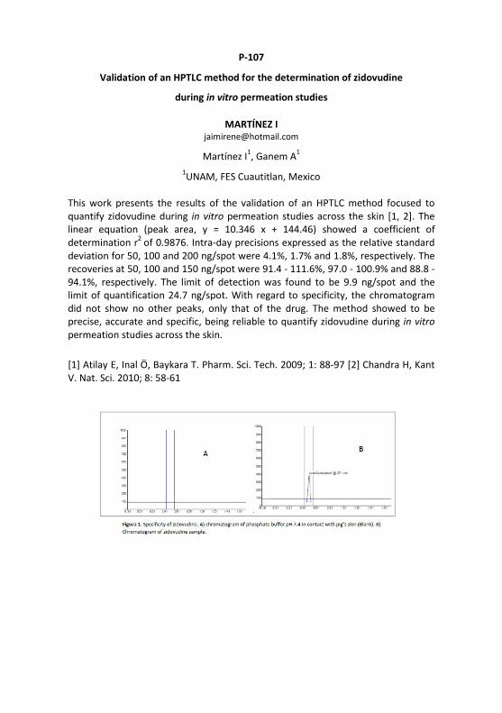
P-107
Validation of an HPTLC method for the determination of zidovudine
during in vitro permeation studies
MARTÍNEZ I [email protected]
Martínez I1, Ganem A1 1UNAM, FES Cuautitlan, Mexico
This work presents the results of the validation of an HPTLC method focused to quantify zidovudine during in vitro permeation studies across the skin [1, 2]. The linear equation (peak area, y = 10.346 x + 144.46) showed a coefficient of determination r2 of 0.9876. Intra-day precisions expressed as the relative standard deviation for 50, 100 and 200 ng/spot were 4.1%, 1.7% and 1.8%, respectively. The recoveries at 50, 100 and 150 ng/spot were 91.4 - 111.6%, 97.0 - 100.9% and 88.8 - 94.1%, respectively. The limit of detection was found to be 9.9 ng/spot and the limit of quantification 24.7 ng/spot. With regard to specificity, the chromatogram did not show no other peaks, only that of the drug. The method showed to be precise, accurate and specific, being reliable to quantify zidovudine during in vitro permeation studies across the skin.
[1] Atilay E, Inal Ö, Baykara T. Pharm. Sci. Tech. 2009; 1: 88-97 [2] Chandra H, Kant V. Nat. Sci. 2010; 8: 58-61

P-108
Development and validation of an HPTLC-UV method to determine paroxetine
hydrochloride from in vitro skin permeation
BROSZAT M [email protected]
González-Iglesias L1, Broszat M2, Ganem-Rondero A1 1Facultad de Estudios Superiores Cuautitlán, UNAM, Cuautitlán Izcalli, México,
2CAMAG, Muttenz, Switzerland
Paroxetine hydrochloride (PAX) is a potent and selective serotonin reuptake inhibitor (SSRI) with currently approved indications for the treatment of depression, obsessive-compulsive disorder, panic disorder and social phobia. However, its oral bioavailability is limited due to first-pass metabolism [1]. Transdermal delivery avoids hepatic first-pass metabolism and degradation. Elimination of this first-pass effect allows the amount of drug administered to be lower, resulting in the reduction of adverse effects [2]. A new, simple, precise, and accurate HPTLC method was developed to quantify the in vitro transdermal permeation of paroxetine hydrochloride and the amount of drug within the skin. Separation was performed on HPTLC plate silica gel 60 F254 with n-butanol - glacial acetic acid - water 7.8:2.0:0.2. PAX was quantified by scanning densitometry at 294 nm. The drug could be baseline-separated from the skin components and surfactants at a hRF value of 42 ± 2. The validation of the method was accomplished according to the ICH guidelines (specificity, linearity, accuracy, precision, limit of detection and limit of quantification and robustness). The accuracy and reliability of the method was assessed by evaluation of the linearity (linear working range between 200 to 800 ng/band, correlation coefficient 0.9939). The limit of detection and limit of quantification were found to be 35 ng/band and 107 ng/band, respectively. The proposed method is suitable for the quantification of PAX in permeation studies and allows the determination of the best doses for the formulation.
Acknowledgment: The authors acknowledge a grant from PAPIIT/UNAM IN216016, PIAPI 1641 and CONACYT 335993.
[1] Germann D, Ma G, Han F, Tikhomirova A, Profiles of Drug Substances, Excipients and Related Methodology, Academic Press, Vol. 38, 367-406, 2013 [2] Kogan A, Garti N. Adv. Colloid Interface Sci. 123-126, 369-385, 2006

P-109
Application of a validated HPTLC method for content uniformity test of
hydrochlorothiazide, amlodipine besylate and olmesartan in tablet dosage form
and its comparison with LC
THUMAR K [email protected]
Thumar K1, Doshi N2, Sheth A3, Patel C3, Sheth N3 1Saurashtra University, Rajkot, India, 2Hemchandrachrya North Gujarat University,
Patan, India, 3Gujarat Technological University, Ahmedabad, India
The present work represent development and validation of a HPTLC method for the simultaneous estimation of hydrochlorothiazide (HCTZ), amlodipine besylate (AMD) and olmesartan medoxomil (OLM) in bulk drug and marketed formulation. The determination was carried on HPTLC plates silica gel 60 G F254 using chloroform - toluene - methanol - glacial acetic acid 5:3:2:0.1. The hRF value of HCTZ, AMD and OLM was found to be 28 ± 2, 41 ± 2, and 81 ± 1, respectively. The detection was carried out by densitometry at 254 nm. HCTZ, AMD and OLM showed a good linear response in the concentration range of 125 - 1000 ng/band, 50 - 400 ng/band and 200 - 1600 ng/band with correlation coefficients 0.9997, 0.9995 and 0.9996 respectively. The method was validated in terms of accuracy, precision, specificity and robustness and found satisfactory. The proposed method was successfully applied to determine the all three contents of ten individual units of five marketed formulations and complied with specification of content uniformity test used in pharmaceutical. The proposed HPTLC method was compared with published LC method in several parameters and HPTLC turns out to be the method of choice for a fast and economical analysis.

P-110
Validated HPTLC method and content uniformity test for analysis of rosuvastatin
and aspirin in tablet dosage form and its comparison with LC
THUMAR K [email protected]
Thumar K1, Ramanuj P1, Sheth N2 1Saurashtra University, Rajkot, India,
2Gujarat Technological University, Ahmedabad, India
The present work represent development and validation of a HPTLC method for the simultaneous estimation of rosuvastatin (ROS) and aspirin (ASP) in bulk drug and marketed formulation. The determination was carried on HPTLC plates silica gel 60 G F254 with ethyl acetate - toluene - acetone - glacial acetic acid 4:4:2:0.1. The hRF values of ASP and ROS were found to be 33 ± 2 and 68 ± 2, respectively. The detection was carried out by densitometry at 254 nm. ROS and ASP showed a good linear response in the concentration range of 40 - 100 ng/band and 300 - 750 ng/band with correlation coefficients 0.9992 and 0.9965, respectively. The method was validated in terms of accuracy, precision, specificity and robustness and found satisfactory. The proposed method was successfully applied to determine the both contents of ten individual units of five marketed formulations and complied with specification of content uniformity test used in pharmaceutical. The proposed HPTLC method was compared with published LC method in several parameters and HPTLC turns out to be the method of choice for a fast and economical analysis.
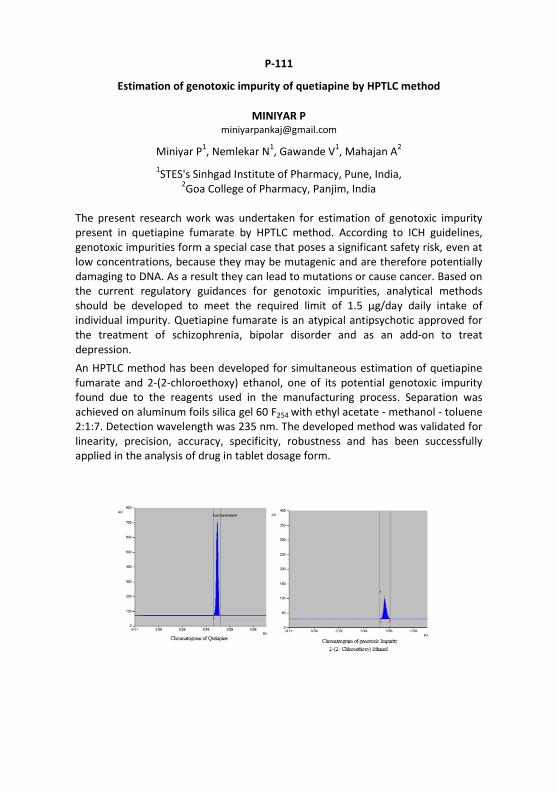
P-111
Estimation of genotoxic impurity of quetiapine by HPTLC method
MINIYAR P [email protected]
Miniyar P1, Nemlekar N1, Gawande V1, Mahajan A2 1STES's Sinhgad Institute of Pharmacy, Pune, India,
2Goa College of Pharmacy, Panjim, India
The present research work was undertaken for estimation of genotoxic impurity present in quetiapine fumarate by HPTLC method. According to ICH guidelines, genotoxic impurities form a special case that poses a significant safety risk, even at low concentrations, because they may be mutagenic and are therefore potentially damaging to DNA. As a result they can lead to mutations or cause cancer. Based on the current regulatory guidances for genotoxic impurities, analytical methods should be developed to meet the required limit of 1.5 µg/day daily intake of individual impurity. Quetiapine fumarate is an atypical antipsychotic approved for the treatment of schizophrenia, bipolar disorder and as an add-on to treat depression. An HPTLC method has been developed for simultaneous estimation of quetiapine fumarate and 2-(2-chloroethoxy) ethanol, one of its potential genotoxic impurity found due to the reagents used in the manufacturing process. Separation was achieved on aluminum foils silica gel 60 F254 with ethyl acetate - methanol - toluene 2:1:7. Detection wavelength was 235 nm. The developed method was validated for linearity, precision, accuracy, specificity, robustness and has been successfully applied in the analysis of drug in tablet dosage form.
Thanks to Separation Science,
a working group of the German Chemical Society,
which provided two travel grants for PhD students and postdocs!
Note for German speaking attendees
For attending HPTLC 2017, you get 25 ZFL points!

CHROMart by Drs. Karla & Herbert Halpaap
“Messieurs, c'est les microbes
qui auront le dernier mot."
Louis Pasteur

Thanks to

Attachment
The Scientific Community deeply recognizes the
lifetime achievement of the Honorary Board
Members for planar chromatography!

Prof. Dr. Rudolf Kaiser, Germany • HPTLC has been developed at his Institute for Chromato-
graphy in 1976 together with Ute Hezel, J. Blome, H. Halpaap, D. Jaenchen and J. Ripphahn; documented in his book HPTLC, Elsevier, 1977, ed. with A. Zlatkis
• Pioneer of planar chromatography → book on Planar Chromatography, 1986, ISBN 3-7785-0780-X
• Micro-PLC site: www.planar-chromatography-by-kaiser.com • Started the first International Symposium HPTLC in Bad
Dürkheim in 1980 and founded the Journal of Planar Chromatography in 1988
• Inspiring research among his many papers − Circular reversed phase planar chromatogram of synthetic fatty acid
mixtures, in his dissertation, University Leipzig, 1954 − Direkte automatische Kopplung DC an GC, Z. anal. Chem. 205 (1964) 205 − TLC in direct coupling with GC and MS, Chemistry in Britain 5 (1969) 54 − Book Einführung in die Hochdruck-PLC, 1987, ISBN 3-7785-1563-2
The inspiring researcher and thinker ahead!
12.02.1930 [email protected]

Prof. Dr. Siegfried Ebel, Germany • Strong supporter of quantitative TLC/HPTLC • Programming software for TLC/HPTLC automatization and
contributing to the progress in instrumental TLC/HPTLC • Creating calibration functions and providing fundamentals
of quantitative TLC/HPTLC in many research paper • Dedicated research in pharmaceutical analysis • Member in many committees like the German and
European Pharmacopoeia committee and the advisory board of the Federal Institute for Drugs and Medical Devices in Germany
The distinghuised software programmer!
03.02.1934 [email protected]

Dr. Dieter Jänchen, Switzerland
• Founder of CAMAG in 1958, which rapidly developed into the world leader of instrumental planar chromatography
• Mentor and coordinator of the development of state-of-the-art instruments and software for planar chromato-graphy
• Creator of CAMAG Bibliography Service with its literature database
• Missionary activities all over the world for the promotion of modern planar chromatography
• In the Scientific and Organizing Committee of the international HPTLC symposia until 1998
The father of modern instrumentation!
06.06.1927 [email protected]

Prof. Dr. David Nurok, USA • Published initial reports on pressurized planar electro-
chromatography (PPEC) and planar electrochromatography (PEC) in the reversed phase mode
• Founding member of InChromatics LLC, a company dedicated to promoting PPEC
• Demonstrated together with Frantisek Svec the use of polymeric monoliths for the rapid separation of oligopeptides by PPEC
• Developed together with Robert Kleyle statistical methods for predicting separation quality in TLC
• Published several reports on the optimization of 2D TLC
The originator of Pressurized Planar Electrochromatography!
01.11.1938 [email protected]

Prof. Dr. Victor Grigor’evich Berezkin, Russian Federation • Chief Investigator of Institute for Petrochemical Synthesis of
the Russian Academy of Sciences • Laureate of the State Award of the Russian Federation,
Honored Personality of Science of the Russian Federation, Honored Petrochemist of the USSR
• Books on TLC, e. g., What is Chromatography?, The discovery of TLC and Quantitative TLC
• Papers on TLC on plates with closed sorption layer – the new variant of planar chromatography
• Member of Advisory Boards like - Journal of Analytical Chemistry (Russian journal) - Journal of Chromatography A - Journal of High Resolution Chromatography
The prominent voice for TLC in Russia!
18.04.1931 [email protected]

The exceptionally distinguished author!
Prof. Dr. Joseph Sherma, USA • Biennial reviews of the field of planar chromatography
since 1970 • Co-guest edited with Bernard Fried yearly special issues
on TLC since 1999 • Editor and Co-editor of 70 books and U.S. government
agency manuals in the areas of analytical chemistry and chromatography; many books are on TLC
• 30 invited book chapters • More than 800 published original research papers and
review papers, the majority of which have been on planar chromatography topics
• Received the 1995 American Chemical Society Award for Research at an Undergraduate College (290 publications with 175 different students as coauthors)
n. a. 1932 [email protected]

Prof. Dr. Teresa Kowalska, Poland • Co-worker of the late Prof. Emanuel Gil-Av (Weizmann
Institute of Science, Rehovot, Israel), the father of chiral separations by chromatographic techniques
• Co-editor of the book Thin-Layer Chromatography in Chiral Separations and Analysis, CRC Press, NY, USA, 2007
• Co-editor of three other monographs on practical applica-tions of TLC/HPTLC (latest one: Planar Chromatography – Mass Spectrometry, CRC Press, 2015)
• Co-author of >20 research papers on the applications of TLC/HPLC to the enantioresolution as well as supervisor of 3 doctorates focusing on enantioresolution by TLC/HPTLC
• One of discoverers with aid of TLC/HPTLC of a new class of the oscillating reactions and promoter of the applications of TLC/HPLC to solving difficult physico-chemical problems
The First Lady of chiral TLC/HPTLC!
19.07.1946 [email protected]

Dr. Fred Rabel, USA • Trained over 5000 scientists on the use and optimization
of TLC/HPTLC through training and short courses • A dozen articles and chapters in textbooks on TLC/HPTLC
applications, sorbents and layers • Assisted in commercializing TLC/HPTLC sorbent and plate
production at two American companies • Continues to promote TLC/HPTLC in every HPLC
talk/training and reminds people of its advantages and utility even in this age of laboratory automation
• The International community viewing this slide should remember that, unfortunately, the USA market has generally not brought into the advances in TLC/HPTLC plate and instrument technology.
The outstanding American trainer!
29.05.1938 [email protected]

In honor of whom we lost since HPTLC 2014 in Lyon
Dr. Friedrich Geiss
Dr. P. D. Sethi
Prof. Dr. Edward Soczewiński
Prof. Dr. Ernő Tyihák

25.02.1932 - 14.02.2015
The pope of understanding!
Dr. Friedrich (Fritz) Geiss, Italy
• Defined terms, created understanding and turned the trial and error approach into a scientific and sound methodology
• Author of our scientific bible Fundamentals of Thin Layer Chromatography (Planar Chromatography), Hüthig 1987, Russian version 1989, German version 1972, Japanese version 1980
• Invention of the Vario KS Chamber with his team, whose successor is frequently in use for optimizations
• Awardee of the Tswett Medal in 2002 • Author of numerous papers and books in the area of
chemistry and of several books related to societal and political matters

Dr. P. D. Sethi, India • India’s first Government Scientist who realized the
potential of HPTLC in 1985; Former Director of Central Indian Pharmacopeia Lab and Central Drug Testing Lab
• Set up QTLC method for analysis of birth control pills in 1987 and analyzed 10 000 samples per year
• Member of several Government Committees for pharmaceutical analysis and herbal product standardization
• A primary force to adopt fingerprinting for herbal medicines as a first choice in India
• 13 books on pharmaceutical analysis including three volumes on multi drug formulation analysis by TLC/HPTLC methods and one on Content Uniformity Testing by HPTLC
India’s top pharmaceutical hands-on analyst!
18.11.1936 - 26.09.2015

Prof. Dr. Edward Soczewiński, Poland • Author of 333 chapters of books and papers as well
as of many patents (e. g. for production of cadmium oxide, chelidonine, protopine, and chambers)
• Research on molecular model of retention in normal phase systems: Soczewiński equation, Anal. Chem. 41 (1969) 179
• Coauthor of Soczewiński-Wachtmeister equation used in QSAR investigations
• Editorial Board Member of chromatographic journals • Awardee of many prizes and distinctions, e. g. Officers and
Chevalier’s of the Polonia Restituta Order, Golden Cross of Merit of Poland, Tswett Medal, doctor honoris causa of Medical Academy of Lublin and many others from the State and Ministry of Health and Social Welfare of Poland
The creator of fundamentals in retention!
04.09.1928 - 12.12.2016

Prof. Dr. Ernő Tyihák, Hungary • Author of >200 papers and 25 patents, mainly on OPLC • Inventor of ultramicro chamber in 1971 • Co-inventor of pressurized ultramicro chamber and
OPLC, J. Chromatogr. 174 (1979) 75 • Invention of BioArena a complex bioautographic system
and the discovery of the role of formaldehyde in the biological systems, Chem. Anal. (Warsaw) 48 (2003) 543
• Editorial Board Member of Journal of Planar Chromatography and Honorary Board Member of Hungarian Society for Separation Sciences
• Awardee of many prizes and distinctions, e. g. Győző Bruckner, Károly Than and Hormesis Awards, Honorary Professor at the Szeged University, Golden Diploma of Budapest Technical University
The main inventor of OPLC!
29.01.1933 - 13.02.2017

79 years of planar chromatography in memoriam
Prof. Dr. N.A. Izmailov Dr. Maria S. Shreiber Prof. Dr. Egon Stahl Prof. Dr. Helmut Jork 1907–1961 1904–1992 1924–1986 1934–1994
Prof. Dr. Ludimir Kraus Prof. Dr. Werner Funk Dr. Klaus Burger Prof. Dr. Szabolcs Nyiredy 1923–1994 1944–1996 1942–2005 1950–2006

Please recommend distinghuised, retired TLC/HPTLC researcher
Thank you in advance!