dissertation - Publikationsserver der Universität Regensburg
356
Synthesis and pharmacological characterization of dibenzodiazepinone-type muscarinic M 2 -receptor antagonists conjugated to fluorescent dyes or small peptides DISSERTATION ZUR ERLANGUNG DES DOKTORGRADES DER NATURWISSENSCHAFTEN (DR. RER. NAT.) AN DER FAKULTÄT FÜR CHEMIE UND PHARMAZIE DER UNIVERSITÄT REGENSBURG vorgelegt von Corinna G. Weinhart (geboren: Gruber) aus Fensterbach, 2020
-
Upload
khangminh22 -
Category
Documents
-
view
0 -
download
0
Transcript of dissertation - Publikationsserver der Universität Regensburg

Synthesis and pharmacological characterization of
dibenzodiazepinone-type muscarinic M2-receptor
antagonists conjugated to fluorescent dyes or
small peptides
DISSERTATION
ZUR ERLANGUNG DES DOKTORGRADES DER NATURWISSENSCHAFTEN (DR. RER. NAT.) AN DER FAKULTÄT FÜR CHEMIE UND PHARMAZIE
DER UNIVERSITÄT REGENSBURG
vorgelegt von Corinna G. Weinhart
(geboren: Gruber)
aus Fensterbach, 2020


I
Die vorliegende Arbeit entstand in der Zeit von Januar 2017 bis Oktober 2020 unter der
Leitung von Herrn Dr. Max Keller am Institut für Pharmazie der Naturwissenschaftlichen
Fakultät IV – Chemie und Pharmazie – der Universität Regensburg.
Promotionsgesuch wurde eingereicht am: Oktober 2020
Tag der mündlichen Prüfung:
Vorsitzender des Prüfungsausschusses: Prof. Dr. Sigurd Elz
Erstgutachter: PD Dr. Max Keller
Zweitgutachter: Prof. Dr. Pierre Koch
Drittprüfer: Prof. Dr. Frank-Michael Matysik

II
ACKNOWLEDGMENTS
First and foremost, I would like to thank Prof. Dr. Armin Buschauer, who died far too early, for giving me the opportunity to work in his research group on this versatile and interesting project.
Another very important person for the realization of this thesis was Dr. Max Keller, who supervised and accompanied this whole project. I would like to express my deepest gratitude to him for his continuous, excellent scientific support and input. Especially, I am very grateful that he continuously offered an open door, to discuss difficulties and ideas occurring during the work with his patient and constructive manner.
I want to express my sincere gratitude to Susanne Bollwein, Brigitte Wenzl and Maria Beer-Krön, for excellent technical assistance, extremely patient performances of assays and for cultivation of the needed cells. Furthermore, I want to thank Peter Richthammer for helping me to solve several technical problems.
Moreover, I want to thank Dr. David Wifling for the induced-fit docking studies in Chapter 2 and for the valuable ideas. I also want to thank Maximilian Schmidt and Eduard Neu as well as their supervisors Timothy Clark and Peter Gmeiner for the performances of the latest induced-fit docking studies for Chapter 2.
For the association to the research training group 1910 “Medicinal Chemistry of Selective GPCR Ligands” and for their financial support I want to thank the Deutsche Forschungsgemeinschaft (DFG). I am very grateful for the organisation of numerous interesting events and critical helpful scientific discussions during our retreats.
Furthermore, I want to thank all employees of the analytical department of the University of Regensburg for the measurement of numerous NMR and mass spectra. Especially, I want to express my gratitude to Fritz Kastner and Josef Kiermaier for many helpful discussions and excellent assistance, as well as several amusing moments.
Beyond that, I thank all colleagues of the Buschauer- and Elz- group for their help, support and ideas and the excellent and amusing working atmosphere. Especially, I want to thank my lab colleague Lisa Schindler for introducing me in the peptide

III
synthesis, and for many amusing, as well as constructive and helpful conversations. Moreover I want to express my gratitude to Carina Höring for the development of the mini-Gi recruitment functional assay for the M2R and for her patient assistance doing my first functional assays, as well as for the generation and cultivation of HEK-cells expressing NlucN-mini-Gαi and M2R-NlucC constructs. I want to thank Silvia Heinrich for offering friendly assistance for organizational, administrative and bureaucratic matters.
Zu guter Letzt möchte ich meiner Familie für ihre uneingeschränkte Unterstützung danken. Der größte Dank gebührt jedoch meinem Michael. Für seine Liebe, aufmunternden Worte, immerwährende Unterstützung und für jeden einzelnen Moment, den wir miteinander verbringen.
Regensburg, 22.10.2020

IV
PUBLICATIONS, PRESENTATIONS
AND PROFESSIONAL TRAINING
Peer- reviewed journal articles (published or submitted prior the submission of this thesis)
A. Pegoli*, D. Wifling*, C.G. Gruber*, X. She, H. Hubner, G. Bernhardt, P. Gmeiner, M. Keller, Conjugation of short peptides to dibenzodiazepinone-type muscarinic acetylcholine receptor ligands determines M2R selectivity, J. Med. Chem., 62 (2019) 5358-5369. *equally contributed X. She, A. Pegoli, C.G. Gruber, D. Wifling, J. Carpenter, H. Hübner, J. Wan, G. Bernhardt, P. Gmeiner, N.D. Holliday, M. Keller, Red-emitting dibenzodiazepinone derivatives as fluorescent dualsteric probes for the muscarinic acetylcholine M2 receptor, J. Med. Chem., 63 (2020) 4133-4154. C.G. Gruber, A. Pegoli, C. Müller, L. Grätz, X. She, M. Keller, Differently fluorescence-labelled dibenzodiazepinone-type muscarinic acetylcholine receptor ligands with high M2R affinity, RSC Med. Chem., 11 (2020) 823-832. L. Grätz, T. Laasfeldb, A. Allikaltb, C. G. Gruber, A. Pegoli, M.-J. Tahk, M.-L. Tsernant, M. Keller, A. Rinken, BRET- and fluorescence anisotropy-based assays for on-line monitoring of ligand binding to muscarinic M2 receptors, Biochim. Biophys. Acta, Mol Cell Res., 2020, submitted C. G. Weinhart, D. Wifling, M. F. Schmidt, E. Neu, C. Höring, T. Clark, P. Gmeiner and M. Keller., Dibenzodiazepinone-type muscarinic receptor antagonists conjugated to basic peptides: impact of the linker moiety and unnatural amino acids on M2R selectivity, Eur. J. Med. Chem., 2020, submitted

V
Poster presentations
C. Gruber, A. Pegoli, D. Wifling, G. Bernhardt, M. Keller, Conjugation of dibenzodiazepinone-type muscarinic acetylcholine receptor ligands to short peptides as a new strategy for the development of highly selective M2R ligands, 8th Austrian Peptide Symposium, December 2018, Salzburg, Austria C. Gruber, A. Pegoli, D. Wifling, G. Bernhardt, M. Keller, Conjugation of dibenzodiazepinone-type muscarinic acetylcholine receptor ligands to short peptides as a new strategy for the development of highly selective M2R ligands, 9th International Summer School "Medicinal Chemistry", September 2018, Regensburg, Germany C. Gruber, A. Pegoli, D. Wifling, M. Keller, Conjugation of dibenzodiazepinone-type muscarinic acetylcholine receptor ligands to short peptides as a new strategy for the development of highly selective M2R ligands, Frontiers in Medicinal Chemisty, March 2020, Freiburg, Germany (cancelled due to Corona pandemic)
Professional training Since January 2017 member of the research training group 1910 “Medicinal Chemistry of Selective GPCR Ligands” funded by the Deutsche Forschungsgemeinschaft

Content
VI
CHAPTER 1 General introduction .................................................................................. 1
1.1. G-Protein coupled receptors ................................................................................ 2
1.1.1. GPCRs: Overview and classification ............................................................ 2
1.1.2. G-protein-dependent signaling ..................................................................... 3
1.2. Muscarinic acetylcholine receptors ...................................................................... 4
1.2.1. General aspects, expression and physiological functions ............................. 4
1.2.2. MR crystal structures and the dualsteric/bitopic ligand approach ................. 6
1.3. Subtype preferring or selective MR ligands ......................................................... 8
1.3.1. General aspects ........................................................................................... 8
1.3.2. M1 receptor preferring ligands ...................................................................... 9
1.3.3. M2 receptor preferring ligands .................................................................... 10
1.3.4. M3 receptor preferring ligands .................................................................... 12
1.3.5. M4 receptor preferring ligands .................................................................... 13
1.3.6. M5 receptor preferring ligands .................................................................... 14
1.4. Scope and objectives ........................................................................................ 15
1.5. References ........................................................................................................ 17
CHAPTER 2 Dibenzodiazepinone-type muscarinic receptor antagonists conjugated to basic peptides: impact of the linker moiety and unnatural amino acids on M2R selectivity ....................................................................................................................... 27
2.1. Introduction ....................................................................................................... 29
2.2. Results and Discussion ..................................................................................... 31
2.2.1. Chemistry ................................................................................................... 31
2.2.2. Radioligand competition binding studies with [3H]NMS ............................... 36
2.2.3. Mini-Gsi protein recruitment assay .............................................................. 43
2.2.4. Induced-fit docking ..................................................................................... 44
2.3. Conclusion ........................................................................................................ 46
2.4. Experimental section ......................................................................................... 47
2.4.1. General experimental conditions ................................................................ 47
2.4.2. Compound characterization........................................................................ 49

Content
VII
2.4.3. Solid-phase peptide synthesis (SPPS)........................................................50
2.4.4. General Procedure for the Synthesis of Compounds 53-60, 63-65, 73-82, 85-
99, 101-126, 130, 131, 134, 135, 137-139 ................................................................50
2.4.5. Experimental synthetic protocols and analytical data of compounds 9, 10, 13,
17-19, 23-25, 28, 31, 33, 35, 37, 38, 40, 43, 53-60, 63-65, 73-82, 85-99, 101-126, 130,
131, 134, 135, 137-139 .............................................................................................51
2.4.6. Cell culture ............................................................................................... 113
2.4.7. Radioligand competition binding assay ..................................................... 113
2.4.8. Mini-Gsi protein recruitment assay ............................................................. 114
2.4.9. Induced-fit Docking ................................................................................... 116
2.5. Supplementary Information .............................................................................. 117
2.6. References ...................................................................................................... 130
CHAPTER 3 Further DIBA-peptide conjugates and stability studies in human plasma ...................................................................................................................................... 135
3.1. Results and Discussion ................................................................................... 137
3.1.1. Chemistry ................................................................................................. 137
3.1.2. Radioligand competition binding studies ................................................... 137
3.1.3. Stability studies in human plasma ............................................................. 139
3.2. Experimental section ....................................................................................... 140
3.2.1. General experimental conditions............................................................... 140
3.2.2. Compound characterization ...................................................................... 141
3.2.3. Screening for pan-assay interference compounds (PAINS). ..................... 141
3.2.4. Solid-phase peptide synthesis (SPPS)...................................................... 142
3.2.5. General procedure for the synthesis of compounds 144-147 .................... 142
3.2.6. Experimental synthetic protocols and analytical data of compounds 144-149
................................................................................................................. 142
3.2.7. Cell culture ............................................................................................... 146
3.2.8. Radioligand competition binding ............................................................... 146
3.2.9. Investigation of the stability of 3, 4 and 146 in human plasma. ................. 146
3.2.10. Data processing .................................................................................... 148
3.3. References ...................................................................................................... 149

Content
VIII
CHAPTER 4 Fluorescently labeled dibenzodiazepionone-type muscarinic M2R ligands ......................................................................................................................... 151
4.1. Introduction ..................................................................................................... 153
4.2. Results and Discussion ................................................................................... 154
4.2.1. Chemistry ................................................................................................. 154
4.2.2. Radioligand competition binding studies with [3H]NMS ...................... 156
4.2.3. Fluorescence properties ........................................................................... 158
4.2.4. Flow cytometric M2R saturation binding studies ....................................... 158
4.3. Conclusion ...................................................................................................... 160
4.4. Experimental section ....................................................................................... 160
4.4.1. General experimental conditions .............................................................. 160
4.4.2. Compounds characterization .................................................................... 161
4.4.3. Experimental synthetic protocols and analytical data ............................... 162
4.4.4. Investigation of chemical stability ............................................................. 165
4.4.5. Cell culture ............................................................................................... 166
4.4.6. Determination of excitation and emission spectra ..................................... 166
4.4.7. Radioligand competition binding assay .................................................... 166
4.4.8. Flow cytometric saturation binding experiments ....................................... 167
4.4.9. Data processing ....................................................................................... 167
4.5. Supplementary Information ............................................................................. 169
4.6. References ...................................................................................................... 172
Summary ...................................................................................................................... 177
Appendix ...................................................................................................................... 181
1H and 13C-NMR spectra of compounds 6, 23-25, 28, 31, 35, 40, 43, 53-60, 63-65, 73-82,
85-99, 101-126, 134, 135 and 1H-NMR spectra of compounds 137-139 (Chapter 2) . 182
RP-HPLC chromatograms of compounds 6, 13, 23-25, 31, 35, 40, 43 53-60,63-65, 73-
82, 85-99, 101-126, 134 and 135 (Chapter 2) ............................................................ 295
1H and 13C-NMR spectra of compounds 144-149 (Chapter 3) .................................... 320
RP-HPLC chromatograms of compounds 144-149 (Chapter 3) ................................. 329
1H and spectra of compounds 164-169 (Chapter 4) ................................................... 332

Content
IX
RP-HPLC chromatograms of compounds 164-169 (Chapter 4) .................................. 336
Abbreviations ............................................................................................................. 339
Overview of bold compound numerals and lab codes ................................................. 342
Eidesstattliche Erklärung ............................................................................................ 344


1
CHAPTER 1
General introduction

Chapter 1
2
1.1. G-Protein coupled receptors
1.1.1. GPCRs: Overview and classification
G-protein coupled receptors (GPCRs), also referred to as seven transmembrane (7TM)
receptors, are versatile proteins constituting the largest and most intensively studied family
of membrane receptors. As they are responsible for transducing signals across plasma
membranes, GPCRs are essential nodes of communication between the intra- and
extracellular environment.1, 2 Hence, GPCRs are substantially involved in the regulation of
physiological processes, and represent attractive and important drug targets.3-7 GPCRs are
encoded by approximately 800 genes in humans and consist of a single polypeptide chain,
which contains seven hydrophobic alpha helices spanning the plasma membrane.8, 9 The
N-terminus is located extracellularly and the C-terminus intracellularly. Consequently,
GPCRs exhibit three extracellular and three intracellular loops, located between the
extracellular ends and the intracellular ends, respectively, of adjacent transmembrane
domains.10, 11 In humans, about 450 GPCRs have sensory function (olfaction, taste, light
perception, pheromone signaling) and ca. 350 GPCRs are non-sensory receptors,
transmitting an extracellular stimulus into the cell by binding of an (endogenous) ligand,
acting as an agonist.12 GPCRs bind a tremendous variety of ligands, covering small
molecules such as biogenic amines (e.g. acetylcholine), peptides (e.g. neuropeptide Y),
nucleotides and Ca2+ ions, as well as proteins (e.g. the 28 kDA CXCR6 chemokine receptor
agonist CXCL16), pheromones and exogenous ligands such as fragrances or flavours
(Figure 1.1).12, 13
Based on sequence homologies and
functional roles, the vertebrate
GPCRs can be classified into five
families: rhodopsin-like receptors
(class A), secretin receptor-like
GPCRs (class B), class C GPCRs
(comprising metabotropic glutamate
receptors, γ-aminobutyric acid
receptors and Ca2+-sensing
receptors), adhesion GPCRs and the
family of frizzled proteins.12 With ca.
430 sensory and 300 non-sensory
receptors, class A comprises most
GPCRs.12 Approximately 80 non-
Figure 1.1. Schematic illustration of a generic GPCR and a heterotrimeric G-protein. GPCRs exhibit seven transmembrane helices (7TM) with an extracellular N-terminal and an intracellular C-terminal domain. Reception of an extracellular stimulus (e.g. photons, ions, odorants, pheromones, hormones, neurotransmitters) induces conformational changes in the receptor that mediates the activation of heterotrimeric G-proteins. G-proteins, in turn, interact with a various effectors, controlling intracellular messengers. (modified from literature11)

General introduction
3
sensory receptors of class A are still orphans, i.e. an endogenous ligand has not been
identified.12, 14
As GPCRs are involved in the regulation of numerous physiological functions (e.g. smooth
muscle contraction, pain modulation, cognition, cell proliferation, heart function, etc.),
dysfunctions of GPCRs are associated with a multitude of different diseases, such as
asthma, diabetes type 2, schizophrenia (SZ), Alzheimer’s disease (AD) or various
autoimmune diseases.15 This, and the fact that GPCRs are easily druggable, account for
their predominating role among biological targets of approved drugs, hence, to date, drugs
that target GPCRs account for approx. 27% of the global market share of therapeutic
drugs.15
1.1.2. G-protein-dependent signaling
The guanine nucleotide-binding proteins (G-Proteins) are heterotrimeric proteins, consisting
of an α-subunit, exhibiting GTPase activity, a β-subunit and a γ-subunit.16 In its inactive,
GDP-bound form, the α-subunit is tightly connected to the β/γ-heterodimer. Typically, the
active conformational state of a GPCR is induced by binding of an agonist to the orthosteric
binging site of the receptor. Consequently, the G-protein complex associates with the
intracellular site of the receptor via the GDP bound α-subunit, resulting in the release of
GDP and the rapid binding of GTP instead. This, in turn, results in the dissociation of the
GPCR/Gαβγ complex into the GPCR, the α-subunit and the βγ-complex.11, 17 Both
components of the G-protein are then capable of activating different downstream effectors.
In its GTP bound state, the α subunit is “switched” into its active form, which can associate
with its respective downstream effector proteins, including adenylyl cyclases (ACs),
phospholipase C-β or ion channels.11, 16, 18 Effector proteins, addressed by the β/γ-subunit,
are, e.g., ACs, GPCR kinases (GRKs) or ion channels.19 Upon hydrolysis of GTP to GDP
by the intrinsic GTPase activity of the α-subunit, the latter is “switched off”, i.e. turns back
to the inactive state resulting in the re-association of the α- and β/γ-subunits, which can then
undergo a new activation cycle.20 As various isoforms of α-, β- and γ-subunits exist, multiple
heterotrimeric G-proteins can be constituted, exhibiting distinct effector interaction
profiles.11, 21, 22 Based on their sequence homology and function, Gα-subunits were
subdivided into four groups (αs, αi/o, αq/11 and α12/13).13, 17, 20, 22 Gαs is responsible for the
stimulation of ACs, leading to an increase of intracellular cAMP (3’-5’-cylclic adenosine
monopohsopate) levels and consequently to an activation of proteinkinase A (PKA),16, 22
whereas its counterpart, Gαi/o, leads to an inhibition of the enzymatic activity of ACs and
thus in decreased intracellular cAMP formation (Figure 1.2).22, 23 A third major Gα subtype
Chapter 1
4
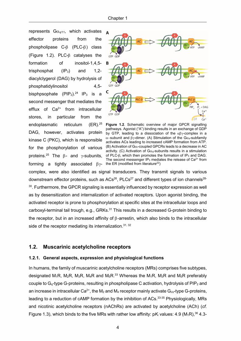
represents Gαq/11, which activates
effector proteins from the
phospholipase C-β (PLC-β) class
(Figure 1.2). PLC-β catalyses the
formation of inositol-1,4,5-
trisphosphat (IP3) and 1,2-
diacylclygerol (DAG) by hydrolysis of
phosphatidylinositol 4,5-
bisphpsophate (PIP2).24 IP3 is a
second messenger that mediates the
efflux of Ca2+ from intracellular
stores, in particular from the
endoplasmatic reticulum (ER).25
DAG, however, activates protein
kinase C (PKC), which is responsible
for the phosphorylation of various
proteins.25 The β- and γ-subunits,
forming a tightly associated βγ-
complex, were also identified as signal transducers. They transmit signals to various
downstream effector proteins, such as ACs26, PLCs27 and different types of ion channels28-
30. Furthermore, the GPCR signaling is essentially influenced by receptor expression as well
as by desensitization and internalization of activated receptors. Upon agonist binding, the
activated receptor is prone to phosphorylation at specific sites at the intracellular loops and
carboxyl-terminal tail trough, e.g., GRKs.31 This results in a decreased G-protein binding to
the receptor, but in an increased affinity of β-arrestin, which also binds to the intracellular
side of the receptor mediating its internalization.31, 32
1.2. Muscarinic acetylcholine receptors
1.2.1. General aspects, expression and physiological functions
In humans, the family of muscarinic acetylcholine receptors (MRs) comprises five subtypes,
designated M1R, M2R, M3R, M4R and M5R.33 Whereas the M1R, M3R and M5R preferably
couple to Gq-type G-proteins, resulting in phospholipase C activation, hydrolysis of PIP2 and
an increase in intracellular Ca2+, the M2 and M4 receptor mainly activate Gi/o-type G-proteins,
leading to a reduction of cAMP formation by the inhibition of ACs.33-35 Physiologically, MRs
and nicotinic acetylcholine receptors (nAChRs) are activated by acetylcholine (ACh) (cf.
Figure 1.3), which binds to the five MRs with rather low affinity: pKi values: 4.9 (M1R),36 4.3-
Figure 1.2. Schematic overview of major GPCR signalling pathways. Agonist (“A”) binding results in an exchange of GDP by GTP, leading to a dissociation of the αβγ-complex in a α−subunit and βγ-dimer. (A) Stimulation of the Gαs-subfamily activates ACs leading to increased cAMP formation from ATP. (B) Activation of Gαi-coupled GPCRs leads to a decrease in AC activity. (C) Activation of Gαq-subunits results in a stimulation of PLC-β, which then promotes the formation of IP3 and DAG. The second messenger IP3 mediates the release of Ca2+ from the ER (modified from literature22)

General introduction
5
6.5 (M2R),37-39 4.5-5.4 (M3R)36-38, 4.5-5.6 (M4R),36-38 6.1 (M5R)37. The neurotransmitter ACh
is formed by choline acetyl transferase from acetyl-Coenzyme A and choline, and degraded
by acetylcholinesterase (Figure 1.3).
Figure 1.3. Schematic illustration of the enzymatic formation and degradation of acetylcholine (ACh). Ach acts as agonist at muscarinic receptors (GPCRs) and nicotinic receptors (ligand-gated cation channels).
Initially, several naturally occurring ligands addressing the muscarinic acetylcholine
receptors were described, including the agonists muscarine (a toxin from the mushroom
Aminita muscaria; the family of MRs was named after this toxin) and pilocarpine (therapeutic
agent for glaucoma from the Pilocarpus genus belonging to the family Rutacea) as well as
antagonists such as atropine (from Atropa belladonna).
Muscarinic receptors are widely distributed in peripheral organs and in the central nervous
system (CNS). In the CNS, MRs exert neuromodulatory functions. In the periphery, they are
a key component of the parasympathetic nervous system, regulating the function of various
organs and glands (cf. Table 1.1). Moreover, MRs were reported to be involved in the
regulation of cell proliferation and to be expressed on human immune cells, suggesting a
modulatory role of MRs in inflammatory and immune response.35, 40
Consequently, MRs have emerged as attractive and important drug targets for the
treatments of numerous diseases such as chronic obstructive pulmonary disease (COPD),
asthma, abdominal spasms, overactive bladder, glaucoma and neurological disorders, such
as AD, Parkinson’s disease (PD), drug addiction, depression or schizophrenia (SZ).35, 41-47
It should be mentioned, that the neuromodulatory effects mediated by muscarinic receptors
have not been unequivocally assigned to the individual subtypes due to a lack of highly
subtype selective MR agonists and antagonists, which are needed as pharmacological
tools, as well as co-expression of several subtypes in the same brain regions. For this
reason, the list of MR modulated physiological functions, presented in Table 1.1, is
incomplete.
acetyl-CoA HON+
choline O-acetyl-transferase (EC 2.3.1.6)
ON
O
acetylcholine (ACh)
acetylcholin-esterase (EC 3.1.1.7)
HON
OH
O+

Chapter 1
6
1.2.2. MR crystal structures and the dualsteric/bitopic ligand approach
Among the five muscarinic acetylcholine receptor subtypes, the orthosteric (acetylcholine)
binding pocket is highly conserved. Thus, the development of highly subtype-selective MR
ligands has been very challenging and potent and selective therapeutics without side effects
(attributed to actions at undesired MR subtypes) are still an unfulfilled need. Over the past
Table 1.1. Expression and physiological functions of muscarinic acetylcholine receptors.a
Muscarinic receptor
Tissue expression (human)b Physiological and therapeutic relevance (the latter indicated by ‘’)
M1 o CNS: e.g. cerebral cortex, striatum, basal ganglia, hypothalamus, limbic system (e.g. amygdala and hippocampus), putamen
o periphery: e.g. eye (iris), lung, prostate, urinary bladder, vasculature
o modulation of attention, cognition and learning processes AD, dementia, SZ
o mediation of bronchoconstriction o mediation of gastric acid secretion
gastric ulcers (M1R antagonists)
M2 o CNS: widely distributed, e.g. brainstem, cerebral cortex, hypothalamus, hippocampus and thalamus
o periphery: e.g. heart, lung, GI tract, eye (ciliary body), prostate, submandibular gland, ureter, urinary bladder
o reduction of heart rate and cardiac contractility
o modulation of smooth muscle contraction glaucoma
o autoreceptor function in the central and peripheral nervous system (modulation of ACh release) AD, SZ (M2R antagonists)
M3 o CNS: e.g. hippocampus, hypothalamus
o periphery: e.g. blood vessels, eye (ciliary body, iris), GI tract, lung, pancreatic gland, prostate, retina, salivary gland, testis, ureter, urinary bladder
o stimulation of saliva, gastric acid and insulin secretion dry mouth syndrome
o mediation of smooth muscle contraction (trachea, bronchi, ileum, iris sphincter, urinary bladder, vasculature) COPD, overactive bladder, irritable bowel syndrome
M4 o CNS: e.g. striatum, amygdala, neocortex, hippocampus, hypothalamus, putamen, spinal cord
o periphery: e.g. autonomic ganglia, eye (ciliary body, iris), urinary bladder
o modulation of ACh release o modulation of dopamine release
SZ, PD
M5 o CNS: e.g. substantia nigra pars compacta hippocampus, hypothalamus, ventral tegmental area
o periphery: e.g. eye (ciliary body), ureter, urinary bladder, vasculature
o modulation of striatal dopamine release drug addiction
o mediation of vasorelaxation
aInformation were taken from the following sources: Cortes et al.,48 Levey et al.,49 Levey et al.,50 Langmead et al.,51 Zhang et al.,52 Nietgen et al.,53 Tyagi et al.,54 Wess,55 Wess et al.,47 Andersson,34 Bolbecker et al.,41 Bubser et al.,56 Buels et al.,57 Eglen,35 Ehlert et al.,58 Harvey,59 Mitchelson,60 McDonald et al.,61 Sellers et al.,62 Nishtala et al.,63 Clader et al.,64 Lebois et al.,65 Raffa.. bExpression analysis was based on the identification of mRNA, on autoradiography using radiolabeled receptor ligands or on immunocytochemistry using antibodies. cGiven are well understood physiological functions. Note: MRs are involved in the regulation of further physiological processes, such as thermoregulation, nociception and circadian rhythm, but an unambiguous subtype-specific elucidation of these mechanisms has not been achieved to date.

General introduction
7
few years, X-ray or cryo-EM crystal structures for all five subtypes of human muscarinic
acetylcholine receptors were reported (Table 1.2).
These crystal structures confirmed the high structural similarity of MRs within the 7-TM
bundle harbouring the orthosteric binding site, a phenomenon that had earlier been
concluded from the high sequence homology among MRs within the transmembrane
domains and from poorly pronounced selectivity profiles of orthosteric MR ligands. On top
of the orthosteric binding site, i.e. near the receptor surface, MRs exhibit, as many other
GPCRs, another pocket, referred to as the receptor vestibule. In the case of MRs, this well-
shaped vestibule is often called the common allosteric site, which is less conserved
compared to the orthosteric binding pocket.73, 76 Therefore, the development of allosteric
MR ligands or modulators has been seen as a promising approach to overcome the lack of
highly selective MR ligands. Indeed, this gave rise to use MRs as model receptors to study
allosterism at GPCRs.46, 77-81 Binding of allosteric modulators induces a change in receptor
conformation, resulting in a modulation of the binding affinity (and eventually also potency
and efficacy) of orthosteric ligands.82
In 1976 gallamine was identified as the first negative allosteric modulator of muscarinic
acetylcholine receptors, inhibiting the action of acetylcholine and carbachol.83 Since then, a
wide variety of allosteric MR modulators with moderate to high MR subtype selectivity has
been discovered, most of them interacting with the receptor within the common allosteric
site.76 The major challenge regarding the development of allosteric MR ligands seems to be
high receptor affinity as most described allosteric MR ligands exhibit rather low receptor
affinity, with dissociation constants > 0.1 µM.12 This issue can be addressed by designing
Table 1.2. Crystal or cryo-EM structures of muscarinic acetylcholine receptors.
MR subtype Activation state Bound ligand Ref.
M1R inactive tiotropium (antagonist) a active iperoxo (agonist) (cryo-EM structure) b M2R inactive QNB (antagonist) c inactive NMS (antagonist) d inactive AF-DX 384 (antagonist) d active iperoxo (agonist) e
active iperoxo (agonist) and allosteric modulator LY2119620 e
active iperoxo (agonist) (cryo-EM structure) b M3R inactive BS46 (antagonist) f inactive tiotropium (antagonist) g,h inactive NMS (antagonist) h M4R inactive tiotropium (antagonist) a M5R inactive tiotropium (antagonist, inverse agonist) i References are as follows: (a) Thal et al.;67 (b) Maeda et al.;68 (c) Haga et al.;69 (d) Suno et al.;70 (e) Kruse et al.;71 (f) Liu et al.;72 (g) Kruse et al.;73 (h) Thorsen et al.;74 (i) Vuckovic et al.75

Chapter 1
8
dualsteric (bitopic) ligands, which occupy the orthosteric site and (in part) also the less
conserved common allosteric site, a strategy called the dualsteric ligand approach.46, 78, 84-
88 Interactions within the orthosteric pocket should contribute to high receptor affinity and
interactions within the allosteric site are anticipated to mediate subtype selectivity.
1.3. Subtype preferring or selective MR ligands
1.3.1. General aspects
A multitude of therapeutic drugs addressing MRs have been identified over the past
decades, but many of these ligands show no or only low subtype selectivity, causing
adverse effects.56, 57, 62 Interestingly, the lack of subtype selective MR ligands seems to give
justification for calling ligands “selective”, which would have been declared as “subtype
preferring” or “non-selective” outside the muscarinic receptor field. For illustration, two
examples are given in the following:
(1) In many scientific reports pirenzepine is called an M1R selective antagonist and has
been used as such in pharmacological studies. However, with pKi values of 8.0 (M1), 6.3
(M2), 6.8 (M3), 7.0 (M4), 6.9 (M5), corresponding to only 10-fold higher M1R affinity compared
to the M4R and M5R, one should question if M1Rs can be selectively blocked by this
compound (cf. Figure 1.4).62
(2) The so-called M2R selective antagonist AF-DX 384 exhibits the following selectivity
profile (pKi values): 7.51 (M1), 8.22 (M2), 7.18 (M3), 8.0 (M4), 6.27 (M5).89 It is obvious that
AF-DX 384 does not discriminate between the M2 and M4 muscarinic receptor subtypes.
Furthermore, the M2R affinity is only 5-fold and 11-fold higher compared to the M1R and
M3R affinity, respectively.
Several MR ligands, mainly representing peripherally acting MR antagonists
(antimuscarinics), are in clinical use, e.g. ipratropium (Berodual®) and tiotropium (Spiriva®)
to treat asthma and COPD, or butylscopolamine (Buscopan®) to treat abdominal spasms.
The lack of highly selective MR orthosteric ligands demands alternative approaches to
enhance subtype selectivity in order to reduce side effects. Therefore, there is a need for
highly subtype selective MR agonists and antagonists in order to improve established
clinical applications as well as to pave the way to new therapeutic approaches.
In the following, for each MR subtype, the most selective reported MR ligands are briefly
discussed.

General introduction
9
1.3.2. M1 receptor preferring ligands
In 2019, spiropiperidine 1 (SPP1) was developed and presented as a potent partial M1R
orthosteric agonist with selectivity according to results from functional assays.12, 90 The
selectivity of the brain penetrant M1R ligand SPP1 was assessed in vitro in functional assays
in relevant tissues (rat atrium (M2R), rat ileum (M3R)), where SPP1 was devoid of agonistic
effects, but showed antagonistic effects by blocking the antagonist carbachol in these
tissues.90 However, in terms of MR binding, SPP1 only slightly prefers the M1R over the M2,
M4 and M5 receptor (cf. Figure 1.4):90
Figure 1.4. Chemical structures of M1R preferring ligands (A = agonist; Ant = antagonist; AM = allosteric modulator) reported in literature. References are as follows: (a) Broad et al.;90 (b) Bolden et al.;91 (c) Sellers et al.;62 (d) Dörje et al.,89 note: only binding data (pKi) of the (R)-enantiomere are shown; (e) Sheffler et al.;92 (f) Lebois et al.;93 *reported Ki or Kd values were converted to pKi or pKd values; note: in the case of AMs, the determination of binding data by competition binding with labeled orthosteric ligands is not feasible.
Prior to this, the development of selective orthosteric M1R ligands had been unsuccessful.
Therefore, the design of M1R selective ligands has focused on targeting a less conserved
allosteric binding site or aiming at a bitopic binding mechanism at the M1R. This yielded
ligands with selectivity according to results of functional assays, however, binding data were
not provided in these studies.93-95 The most promising compound of this series was
VU0364572 (cf. Figure 1.4), a bitopic M1R agonist, with almost no functional activity at the
other MR subtypes (EC50 (M1R) = 110 nM, EC50 (M2-M5R) > 30 µM), however, radioligand
competition binding data were again not provided.93
Several non-selective MR agonists, including cevimeline, milameline, sabcomeline,
talsaclidine and xanomeline were developed for the treatment of AD, in which the
Chapter 1
10
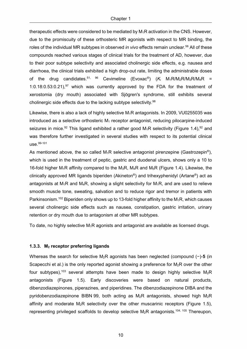
therapeutic effects were considered to be mediated by M1R activation in the CNS. However,
due to the promiscuity of these orthosteric MR agonists with respect to MR binding, the
roles of the individual MR subtypes in observed in vivo effects remain unclear.56 All of these
compounds reached various stages of clinical trials for the treatment of AD, however, due
to their poor subtype selectivity and associated cholinergic side effects, e.g. nausea and
diarrhoea, the clinical trials exhibited a high drop-out rate, limiting the administrable doses
of the drug candidates.51, 96 Cevimeline (Evoxac®) (Ki M1R/M2R/M3R/M4R =
1:0.18:0.53:0.21),97 which was currently approved by the FDA for the treatment of
xerostomia (dry mouth) associated with Sjögren's syndrome, still exhibits several
cholinergic side effects due to the lacking subtype selectivity.98
Likewise, there is also a lack of highly selective M1R antagonists. In 2009, VU0255035 was
introduced as a selective orthosteric M1 receptor antagonist, reducing pilocarpine-induced
seizures in mice.92 This ligand exhibited a rather good M1R selectivity (Figure 1.4),92 and
was therefore further investigated in several studies with respect to its potential clinical
use.99-101
As mentioned above, the so called M1R selective antagonist pirenzepine (Gastrozepin®),
which is used in the treatment of peptic, gastric and duodenal ulcers, shows only a 10 to
16-fold higher M1R affinity compared to the M3R, M4R and M5R (Figure 1.4). Likewise, the
clinically approved MR ligands biperiden (Akineton®) and trihexyphenidyl (Artane®) act as
antagonists at M1R and M4R, showing a slight selectivity for M1R, and are used to relieve
smooth muscle tone, sweating, salvation and to reduce rigor and tremor in patients with
Parkinsonism.102 Biperiden only shows up to 13-fold higher affinity to the M1R, which causes
several cholinergic side effects such as nausea, constipation, gastric irritation, urinary
retention or dry mouth due to antagonism at other MR subtypes.
To date, no highly selective M1R agonists and antagonist are available as licensed drugs.
1.3.3. M2 receptor preferring ligands
Whereas the search for selective M2R agonists has been neglected (compound (−)-5 (in
Scapecchi et al.) is the only reported agonist showing a preference for M2R over the other
four subtypes),103 several attempts have been made to design highly selective M2R
antagonists (Figure 1.5). Early discoveries were based on natural products,
dibenzodiazepinones, piperazines, and piperidines. The dibenzodiazepinone DIBA and the
pyridobenzodiazepinone BIBN 99, both acting as M2R antagonists, showed high M2R
affinity and moderate M2R selectivity over the other muscarinic receptors (Figure 1.5),
representing privileged scaffolds to develop selective M2R antagonists.104, 105 Thereupon,

General introduction
11
several compounds derived from DIBA or BIBN 99 were developed and characterized, but
only few ligands showed a slightly improved M2R selectivity, for instance BIBN 140.106-111
Himbacine, an alkaloid isolated from the bark of Australian magnolias, exhibits high M2R
affinity (pKi = 8.00) and modest M2R selectivity (cf. Figure 1.5), rendering himbacine a
promising starting point in AD research (mode of action: selective antagonism at M2
Figure 1.5. Chemical structures of M2R preferring ligands (A = agonist; Ant = antagonist) reported in literature. References are as follows: (a) Scapecchi et al.,103 (b) Clader et al.,112 note: Ki values for MR subtypes M1, M3, M4 and M5 were not provided; (c) Wang et al.,113 note: Ki values for MR subtypes M1, M3, M4 and M5 were not provided; (d) Doods et al.,104 (e) Doods et al.,114 (f) Gitler et al.,105 (g) Dörje et al.,89 (h) Lachowicz et al.,115 (i) Lachowicz et al.,116 (j) Pegoli et al.;117 n.a. not analyzed, *reported Ki or Kd values were converted to pKi or pKd values.
autoreceptors in the CNS).89 However, the development of M2R selective antagonists,
derived from himbacine, failed.
SCH 57790 was the first compound of a new class of piperazine-based M2R preferring
compounds, presented in 2001.115 The incorporation of a terminal piperidine moiety resulted
in the discovery of SCH 72788, which exhibited an improved M2R selectivity over M1R, M3R
and M5R (cf. Figure 1.5).116 Another structural modulation of SCH 57790 led to the

Chapter 1
12
piperidinylpiperidine analogue SCH 211803, showing an enhanced M2R selectivity, which
had entered phase 1 clinical trials.113, 118 Based on SCH 211803, several compounds were
developed and characterized, i.e. compound 30 (in Wang et al.)113 and 21 (in Clader et
al.)112, showing the best M2R selectivity profiles in this series.
Using the dualsteric ligand approach, a series of dibenzodiazepinone derivatives,
containing a short peptide, was developed, where the peptide moiety, supposed to interact
with the receptor via allosteric sites, was shown to modulate MR subtype selectivity.117 UR-
AP148 (3) exhibited the highest M2R selectivity in this series (Figure 1.5) and is further
discussed in chapter 2.
As antagonism at presynaptic M2Rs represents an alternative approach to increase
cholinergic transmission by increased ACh levels in patients with, e.g. AD or SZ, but
selective M2R ligands are not yet available as licensed drugs, there is still a need for highly
selective M2R antagonists.
1.3.4. M3 receptor preferring ligands
Although potent anticholinergics for the treatment of, e.g. COPD or overactive bladder
(OAB), are being therapeutically used, most have little subtype selectivity. Oxybutynin
(Dridase®), darifenacin (Emselex®) and solifenacin (Vesikur®), used for the treatment of
OAB, are the only licensed drugs, exhibiting a certain M3R selectivity.
The M3R antagonist J-104129 shows a high degree of selectivity for the M3R over M2R
(Figure 1.6).119 However, the bronchodilating activity of J-104129, when applied orally in
COPD patients, was inferior to the dilating effect achieved by inhalation of the rather
unselective compound ipratropium bromide, and additional side effects like dry mouth were
more pronounced in the J-104129 treatment group of a 6-week phase II study comprising
412 COPD patients.120 This study demonstrated, that high M3R over M2R selectivity of an
oral antimuscarinic agent is not enough to improve the therapeutic effect in patients with
COPD compared with inhaled non-selective antimuscarinics.121 The experimental ligand
ABH423 (Figure 1.6) derived from the non-selective antagonist 3-quinuclidinylbenzilate
(QNB) showed 91-fold selectivity over the M2R and was further modified in various
positions, yielding ligands exhibiting 46- to 68-fold M3R over M2R selectivity (data not
shown).122
The tertiary amine (±)-17 (in Del Bello et al.), derived from 1,4-dioxane, preferred M3R by
approx. 2-fold over M1,4,5R and by approx. 8-fold over M2R and proved to be effective in
reducing the contraction of rat urinary bladder (Figure 1.6)123 Moreover, highly selective M3R
agonists have not been described as well. Bethanechol (Myocholine-Glenwood®), which
stimulates contraction of the bladder and expulsion of urine, is the only M3R agonist showing
a slight selectivity for M3R (Figure 1.6).38

General introduction
13
Figure 1.6. Chemical structures of M3R preferring ligands (A = agonist; Ant = antagonist; AM = allosteric modulator) reported in literature. References are as follows: (a) Tanaka et al.,124 (b) Scapecchi et al.,103 (c) Fischer et al.,122 note: Ki values for MR subtypes M4 and M5 were not provided; (d) Jakubik et al.,38 note: Ki value for M5R was not provided; (e) Abrams et al.,125 (f) Mitsuya et al.,119 (g) Ikeda et al.;126 n.a. not analyzed; *reported Ki values were converted to pKi values. note: in the case of AMs, the determination of binding data by competition binding with labeled orthosteric ligands is not feasible.
In 2020, the positive allosteric modulator (PAM) 9 (in Tanaka et al.) was discovered (for
structure see Figure 1.6), showing moderate to high subtype selectivity over the other MR
subtypes, which was assessed by the Carbachol (CCh) dependent Ca2+ increase in
functional assays.124 When used at a concentration of 10 µM, compound 9 (in Tanaka et
al.) caused a 269-fold decrease in the EC50 of CCh (M3R activation), whereas the decrease
in the EC50 of CCh was lower in the case of other MR subtypes (M1R: 2.3-fold, M2R: 1.3-
fold, M4R: 1.0-fold, M5R: 94-fold).124
1.3.5. M4 receptor preferring ligands
To date, no orthosteric selective M4R agonists, which might be useful for treating positive
symptoms (e.g. hallucinations, delusions) of SZ associated with hyperdopaminergia,127-129
have been discovered. However, activation of the muscarinic M4 receptor can also be
mediated by allosteric ligands. The positive allosteric modulator (PAM) VU10010
represented a breakthrough when described in 2008 (Figure 1.7).130 When used at a
concentration of 10 µM, VU10010 induced a 47-fold potentiation of M4R ACh potency,
whereas the response to ACh in cell lines expressing each of the other MR subtypes was
unaffected.130

Chapter 1
14
Figure 1.7. Chemical structures of M4R preferring ligands (Ant = antagonist; AM = allosteric modulator) reported in literature. References are as follows: (a) Böhme et al.131 (b) Shirey et al.,130 (c) Brady et al.;132 note: in the case of AMs, the determination of binding data by competition binding with labeled orthosteric ligands is not feasible.
Subsequent optimization approaches led to the discovery of VU0152100, which induced a
dose-dependent shift in ACh potency (maximal increase over control: 70-fold).132
On the other hand, also the development of selective M4R antagonists, which were
suggested to restore the dopamine acetylcholine balance in patients with PD, has been
challenging.133 To date, only PD-102807, PD-0298029 and a few other benzoxazine
analogues were described (Figure 1.7).131 The former is still used in scientific research for
studying the effects of the different MR subtypes in the brain and in the periphery.134-136 PD-
0298029 exhibited poor bioavailability and rapid metabolism in animal studies, which limits
its use to in vitro studies.137
In summary, selective M4R ligands, representing drug candidates, e.g. for the treatment of
AD, SZ or PD, are not available to date.
1.3.6. M5 receptor preferring ligands
To date, no selective M5R agonists have been discovered, however, a series of M5R positive
allosteric modulators was described (Figure 1.8). The first PAM for the M5R, ML129, was
reported in 2009, provoking higher potentiation of ACh induced intracellular Ca2+-
mobilization at the M5R compared to the M1R-M4R (EC50 values of ACh in the presence of
ML129: M1R-M4R: >30 µM, M5R: 1.16 µM).138 Only a few years later, ML326 was discovered
as the first sub-micromolar, selective M5R PAM (EC50 values of ACh in the presence of
ML326: M1R-M4R: >30 µM, M5R: 410 nM) (Figure 1.8).139

General introduction
15
Figure 1.8 Chemical structures of the positive allosteric M5R modulators ML129 and ML326 and the M5R antagonist ML381 (Ant = antagonist; AM = allosteric modulator). References are as follows: (a) Bridges et al.,138 (b) Gentry et al.,139 (c) Gentry et al.;140 note: in the case of AMs, the determination of binding data by competition binding with labeled orthosteric ligands is not feasible.
Shortly thereafter, in 2014, the orthosteric M5R antagonist ML381 was reported (Figure 1.8).
According to the results from functional studies, i.e. the inhibition of ACh induced Ca2+
response, this compound displayed M5R selective antagonism (IC50 (M1R) = >10 µM;
IC50 (M2R-M4R) = >30 µM; IC50 (M5R) = 0.45 µM). However, MR binding data of ML381 were
only reported for the M5R (Figure 1.8).140
1.4. Scope and objectives
Muscarinic acetylcholine receptors (MRs) are widely distributed in the central and peripheral
nervous system, being (potential) targets for the treatment of various diseases, such as
Alzheimer’s disease (AD), Parkinson’s disease (PD), schizophrenia (SZ), overactive
bladder (OAB) or chronic obstructive pulmonary disease (COPD). Due to the high
conservation of the orthosteric binding pocket within the family of MRs, the development of
highly selective MR ligands has been extremely challenging, and to date, no MR ligands,
which are selective for one of the five subtypes, are available as licensed drugs. Highly
subtype-selective MR ligands are needed as new therapeutics with lower side effects, but
also as molecular tools required, e.g. for pharmacological studies. As the vestibule
(common allosteric binding site) of MRs is less conserved than the orthosteric binding
pocket, the dualsteric ligand approach, i.e. the design of ligands interacting with both, the
orthosteric and the allosteric binding site, has been considered useful for the development
of highly subtype selective MR ligands,84-88 This approach benefits from recently reported
MR crystal structures, which are available for all MR subtypes.67-75
Lately, conjugation of small peptides to the M2R preferring dibenzodiazepinone-type MR
antagonist DIBA (referred to as DIBA-peptide conjugates) was shown to considerably
influence M2R selectivity.117 The introduction of basic amino acids into the peptide moiety,

Chapter 1
16
which is supposed to interact with the receptor within the common allosteric site or at the
surface of the receptor, was advantageous with respect to improved M2R selectivity
compared to non-peptidic tricyclic MR antagonists.117
For the synthesis of the recently reported series of DIBA-peptide conjugates, only
proteinogenic amino acids and only two different linkers were used.117 In this work, the novel
approach had to be further explored by using a broad variety of linkers, connecting the DIBA
pharmacophore and the peptide moiety, and by incorporating unnatural amino acids into
the peptide moiety. The design of the presented DIBA-peptide conjugates, presented in this
work, had to be supported by induced-fit docking studies. MR affinities of the new DIBA-
peptide conjugates had to be determined for the subtypes M1, M2 and M4 in radioligand
competition binding assays at intact CHO cells stably expressing the respective receptors.
For selected compounds, showing high M2R selectivity over the M1 and M4 receptor, binding
to the M3 and M5 receptor had to be also investigated, resulting in complete selectivity
profiles. In order to study the mode of action at the M2R (anticipated to be antagonistic
activity), a few DIBA-peptide conjugates, exhibiting high M2R selectivity, had to be
investigated in a functional M2R assay. Moreover, selected DIBA-peptide conjugates had to
be investigated in terms of stability in human plasma/PBS.
Finally, a series of fluorescently labeled dibenzodiazepinone-type MR ligands had to be
prepared using various fluorescent dyes (5-TAMRA, λex/λem ≈ 547/576 nm; BODIPY
630/650, λex/λem ≈ 625/640 nm; pyridinium dye Py-1, λex/λem ≈ 611/665 nm and pyridinium
dye Py-5, λex/λem ≈ 465/732 nm). M1-M5 receptor affinities of the fluorescent probes had to
be determined by radioligand competition binding and their applicability for fluorescence-
based methods had to be explored by performing flow cytometric saturation binding
experiments at CHO-hM2R cells.

General introduction
17
1.5. References
1. D. M. Rosenbaum, S. G. Rasmussen and B. K. Kobilka, The structure and function of G-protein-coupled receptors, Nature, 2009, 459, 356-363.
2. M. I. Simon, M. P. Strathmann and N. Gautam, Diversity of G-proteins in signal transduction, Science, 1991, 252, 802-808.
3. D. Hilger, M. Masureel and B. K. Kobilka, Structure and dynamics of GPCR signaling complexes, Nat. Struct. Mol. Biol., 2018, 25, 4-12.
4. P. A. Insel, C. M. Tang, I. Hahntow and M. C. Michel, Impact of GPCRs in clinical medicine: monogenic diseases, genetic variants and drug targets, Biochim. Biophys. Acta, 2007, 1768, 994-1005.
5. J. P. Overington, B. Al-Lazikani and A. L. Hopkins, How many drug targets are there?, Nat. Rev. Drug Discovery, 2006, 5, 993-996.
6. M. Rask-Andersen, S. Masuram and H. B. Schioth, The druggable genome: Evaluation of drug targets in clinical trials suggests major shifts in molecular class and indication, Annu. Rev. Pharmacol. Toxicol., 2014, 54, 9-26.
7. J. M. Freudenberg, I. Dunham, P. Sanseau and D. K. Rajpal, Uncovering new disease indications for G-protein coupled receptors and their endogenous ligands, BMC Bioinf., 2018, 19, 345.
8. R. Fredriksson and H. B. Schioth, The repertoire of G-protein-coupled receptors in fully sequenced genomes, Mol. Pharmacol., 2005, 67, 1414-1425.
9. C. D. Hanlon and D. J. Andrew, Outside-in signaling--a brief review of GPCR signaling with a focus on the Drosophila GPCR family, J. Cell Sci., 2015, 128, 3533-3542.
10. M. C. Peeters, G. J. van Westen, Q. Li and I. J. AP, Importance of the extracellular loops in G protein-coupled receptors for ligand recognition and receptor activation, Trends Pharmacol. Sci., 2011, 32, 35-42.
11. T. M. Bridges and C. W. Lindsley, G-protein-coupled receptors: From classical modes of modulation to allosteric mechanisms, ACS Chem. Biol., 2008, 3, 530-541.
12. S. P. H. Alexander, A. Christopoulos, A. P. Davenport, E. Kelly, A. Mathie, J. A. Peters, E. L. Veale, J. F. Armstrong, E. Faccenda, S. D. Harding, A. J. Pawson, J. L. Sharman, C. Southan, J. A. Davies and C. Collaborators, The concise guide to pharmacology 2019/20: G protein-coupled receptors, Br. J. Pharmacol., 2019, 176 Suppl 1, S21-S141.
13. K. Kristiansen, Molecular mechanisms of ligand binding, signaling, and regulation within the superfamily of G-protein-coupled receptors: molecular modeling and mutagenesis approaches to receptor structure and function, Pharmacol. Ther., 2004, 103, 21-80.
14. G. Pandy-Szekeres, C. Munk, T. M. Tsonkov, S. Mordalski, K. Harpsoe, A. S. Hauser, A. J. Bojarski and D. E. Gloriam, GPCRdb in 2018: adding GPCR structure models and ligands, Nucleic Acids Res., 2018, 46, D440-D446.
15. A. S. Hauser, M. M. Attwood, M. Rask-Andersen, H. B. Schioth and D. E. Gloriam, Trends in GPCR drug discovery: new agents, targets and indications, Nat. Rev. Drug Discovery, 2017, 16, 829-842.
16. J. R. Hepler and A. G. Gilman, G proteins, Trends Biochem. Sci, 1992, 17, 383-387. 17. V. Syrovatkina, K. O. Alegre, R. Dey and X. Y. Huang, Regulation, Signaling, and
Physiological Functions of G-Proteins, J. Mol. Biol., 2016, 428, 3850-3868. 18. A. G. Gilman, G proteins: transducers of receptor-generated signals, Annu. Rev.
Biochem., 1987, 56, 615-649. 19. S. M. Khan, R. Sleno, S. Gora, P. Zylbergold, J. P. Laverdure, J. C. Labbe, G. J.
Miller and T. E. Hebert, The expanding roles of Gbetagamma subunits in G protein-coupled receptor signaling and drug action, Pharmacol. Rev., 2013, 65, 545-577.
20. T. M. Cabrera-Vera, J. Vanhauwe, T. O. Thomas, M. Medkova, A. Preininger, M. R. Mazzoni and H. E. Hamm, Insights into G protein structure, function, and regulation, Endocr. Rev., 2003, 24, 765-781.

Chapter 1
18
21. Z. Cheng, D. Garvin, A. Paguio, P. Stecha, K. Wood and F. Fan, Luciferase reporter assay system for deciphering GPCR pathways, Curr. Chem. Genomics, 2010, 4, 84-91.
22. S. Offermanns, G-proteins as transducers in transmembrane signalling, Prog. Biophys. Mol. Biol., 2003, 83, 101-130.
23. R. Taussig, J. A. Iniguez-Lluhi and A. G. Gilman, Inhibition of adenylyl cyclase by Giα, Science, 1993, 261, 218-221.
24. A. V. Smrcka, J. R. Hepler, K. O. Brown and P. C. Sternweis, Regulation of polyphosphoinositide-specific phospholipase C activity by purified Gq, Science, 1991, 251, 804-807.
25. K. Mikoshiba, IP3 receptor/Ca2+ channel: from discovery to new signaling concepts, J. Neurochem., 2007, 102, 1426-1446.
26. R. Taussig, W. J. Tang, J. R. Hepler and A. G. Gilman, Distinct patterns of bidirectional regulation of mammalian adenylyl cyclases, J. Biol. Chem., 1994, 269, 6093-6100.
27. J. L. Boyer, G. L. Waldo, T. Evans, J. K. Northup, C. P. Downes and T. K. Harden, Modification of tetrafluoroaluminate ion and receptor-stimulated phospholipase C activity by G-protein βγ subunits, J. Biol. Chem., 1989, 264, 13917-13922.
28. S. Herlitze, D. E. Garcia, K. Mackie, B. Hille, T. Scheuer and W. A. Catterall, Modulation of Ca2+ channels by G-protein βγ subunits, Nature, 1996, 380, 258-262.
29. S. R. Ikeda, Voltage-dependent modulation of N-type calcium channels by G-protein βγ subunits, Nature, 1996, 380, 255-258.
30. D. E. Logothetis, Y. Kurachi, J. Galper, E. J. Neer and D. E. Clapham, The βγ subunits of GTP-binding proteins activate the muscarinic potassium channel in heart, Nature, 1987, 325, 321-326.
31. R. T. Premont and R. R. Gainetdinov, Physiological roles of G protein-coupled receptor kinases and arrestins, Annu. Rev. Physiol., 2007, 69, 511-534.
32. C. A. Moore, S. K. Milano and J. L. Benovic, Regulation of receptor trafficking by GRKs and arrestins, Annu. Rev. Physiol., 2007, 69, 451-482.
33. M. P. Caulfield and N. J. M. Birdsall, International Union of Pharmacology. XVII. Classification of muscarinic acetylcholine receptors, Pharmacol. Rev., 1998, 50, 279-290.
34. K.-E. Andersson, Muscarinic acetylcholine receptors in the urinary tract, Handb. Exp. Pharmacol., 2011, 202, 319-344.
35. R. M. Eglen, Overview of muscarinic receptor subtypes, Handb. Exp. Pharmacol., 2012, 208, 3-28.
36. L. S and B. N. J, Detection, quantitation, and verification of allosteric interactions of agents with labeled and unlabeled ligands at G protein-coupled receptors: interactions of strychnine and acetylcholine at muscarinic receptors Mol. Pharmacol., 1995, 48, 362-378.
37. K. Cheng, S. Khurana, Y. Chen, R. H. Kennedy, P. Zimniak and J. P. Raufman, Lithocholylcholine, a bile acid/acetylcholine hybrid, is a muscarinic receptor antagonist, J. Pharmacol. Exp. Ther., 2002, 303, 29-35.
38. J. Jakubik, L. Bacakova, E. E. El-Fakahany and S. Tucek, Positive cooperativity of acetylcholine and other agonists with allosteric ligands on muscarinic acetylcholine receptors, Mol. Pharmacol., 1997, 52, 172-179.
39. I. Kovacs, H. I. Yamamura, S. L. Waite, E. V. Varga and W. R. Roeske, Pharmacological comparison of the cloned human and rat M2 muscarinic receptor genes expressed in the murine fibroblast (B82) cell line, J. Pharmacol. Exp. Ther., 1998, 284, 500-507.
40. N. G. Verbout and D. B. Jacoby, Muscarinic receptor agonists and antagonists: effects on inflammation and immunity, Handb. Exp. Pharmacol., 2012, 208, 403-427.
41. A. R. Bolbecker and A. Shekhar, Muscarinic agonists and antagonists in schizophrenia: recent therapeutic advances and future directions, Handb. Exp. Pharmacol., 2012, 208, 167-190.

General introduction
19
42. K. J. Broadley and D. R. Kelly, Muscarinic Receptor Agonists and Antagonists, Molecules, 2001, 6, 142-193.
43. R. M. Eglen, Muscarinic Receptor Subtype Pharmacology and Physiology, Prog. Med. Chem., 2005, 43, 105-136.
44. D. Erskine, J. P. Taylor, G. Bakker, A. J. H. Brown, T. Tasker and P. J. Nathan, Cholinergic muscarinic M1 and M4 receptors as therapeutic targets for cognitive, behavioural, and psychological symptoms in psychiatric and neurological disorders, Drug Discovery Today, 2019, 24, 2307-2314.
45. D. J. Foster, D. L. Choi, P. J. Conn and J. M. Rook, Activation of M1 and M4 muscarinic receptors as potential treatments for Alzheimer's disease and schizophrenia, Neuropsychiatr. Dis. Treat., 2014, 10, 183-191.
46. A. C. Kruse, B. K. Kobilka, D. Gautam, P. M. Sexton, A. Christopoulos and J. Wess, Muscarinic acetylcholine receptors: novel opportunities for drug development, Nat. Rev. Drug Discov., 2014, 13, 549-560.
47. J. Wess, R. M. Eglen and D. Gautam, Muscarinic acetylcholine receptors: mutant mice provide new insights for drug development, Nat. Rev. Drug Discov., 2007, 6, 721-733.
48. R. Cortes, A. Probst, H. J. Tobler and J. M. Palacios, Muscarinic cholinergic receptor subtypes in the human brain. II. Quantitative autoradiographic studies, Brain Res., 1986, 362, 239-253.
49. A. I. Levey, Immunological localization of m1-m5 muscarinic acetylcholine receptors in peripheral tissues and brain, Life Sci., 1993, 52, 441-448.
50. A. I. Levey, C. A. Kitt, W. F. Simonds, D. L. Price and M. R. Brann, Identification and localization of muscarinic acetylcholine receptor proteins in brain with subtype-specific antibodies, J. Neurosci., 1991, 11, 3218-3226.
51. C. J. Langmead, J. Watson and C. Reavill, Muscarinic acetylcholine receptors as CNS drug targets, Pharmacol. Ther., 2008, 117, 232-243.
52. X. Zhang, M. R. Hernandez, H. Yang and K. Erickson, Expression of muscarinic receptor subtype mRNA in the human ciliary muscle, Invest. Ophthalmol. Vis. Sci., 1995, 36, 1645-1657.
53. G. W. Nietgen, J. Schmidt, L. Hesse, C. W. Honemann and M. E. Durieux, Muscarinic receptor functioning and distribution in the eye: molecular basis and implications for clinical diagnosis and therapy, Eye, 1999, 13, 285-300.
54. S. Tyagi, P. Tyagi, S. Van-le, N. Yoshimura, M. B. Chancellor and F. de Miguel, Qualitative and quantitative expression profile of muscarinic receptors in human urothelium and detrusor, J. Urol., 2006, 176, 1673-1678.
55. J. Wess, Muscarinic acetylcholine receptors, Handb. Contemp. Neuropharmacol., 2007, 1, 147-192.
56. M. Bubser, N. Byun, M. R. Wood and C. K. Jones, Muscarinic receptor pharmacology and circuitry for the modulation of cognition, Handb. Exp. Pharmacol., 2012, 208, 121-166.
57. K. S. Buels and A. D. Fryer, Muscarinic receptor antagonists: effects on pulmonary function, Handb. Exp. Pharmacol., 2012, 208, 317-341.
58. F. J. Ehlert, K. J. Pak and M. T. Griffin, Muscarinic agonists and antagonists: effects on gastrointestinal function, Handb. Exp. Pharmacol., 2012, 208, 343-374.
59. R. D. Harvey, Muscarinic receptor agonists and antagonists: effects on cardiovascular function, Handb. Exp. Pharmacol., 2012, 208, 299-316.
60. F. Mitchelson, Muscarinic receptor agonists and antagonists: effects on ocular function, Handb. Exp. Pharmacol., 2012, 208, 263-298.
61. A. J. McDonald and F. Mascagni, Neuronal localization of m1 muscarinic receptor immunoreactivity in the rat basolateral amygdala, Brain Struct. Funct., 2010, 215, 37-48.
62. D. J. Sellers and R. Chess-Williams, Muscarinic agonists and antagonists: effects on the urinary bladder, Handb. Exp. Pharmacol., 2012, 208, 375-400.
63. P. S. Nishtala, M. S. Salahudeen and S. N. Hilmer, Anticholinergics: theoretical and clinical overview, Expert Opin. Drug Saf., 2016, 15, 753-768.

Chapter 1
20
64. J. W. Clader and Y. Wang, Muscarinic receptor agonists and antagonists in the treatment of Alzheimer's disease, Curr. Pharm. Des., 2005, 11, 3353-3361.
65. E. P. Lebois, C. Thorn, J. R. Edgerton, M. Popiolek and S. Xi, Muscarinic receptor subtype distribution in the central nervous system and relevance to aging and Alzheimer's disease, Neuropharmacology, 2018, 136, 362-373.
66. R. B. Raffa, The M5 muscarinic receptor as possible target for treatment of drug abuse, J. Clin. Pharm. Ther., 2009, 34, 623-629.
67. D. M. Thal, B. Sun, D. Feng, V. Nawaratne, K. Leach, C. C. Felder, M. G. Bures, D. A. Evans, W. I. Weis, P. Bachhawat, T. S. Kobilka, P. M. Sexton, B. K. Kobilka and A. Christopoulos, Crystal structures of the M1 and M4 muscarinic acetylcholine receptors, Nature, 2016, 531, 335-340.
68. S. Maeda, Q. Qu, M. J. Robertson, G. Skiniotis and B. K. Kobilka, Structures of the M1 and M2 muscarinic acetylcholine receptor/G-protein complexes, Science, 2019, 364, 552-5557.
69. K. Haga, A. C. Kruse, H. Asada, T. Yurugi-Kobayashi, M. Shiroishi, C. Zhang, W. I. Weis, T. Okada, B. K. Kobilka, T. Haga and T. Kobayashi, Structure of the human M2 muscarinic acetylcholine receptor bound to an antagonist, Nature, 2012, 482, 547-551.
70. R. Suno, S. Lee, S. Maeda, S. Yasuda, K. Yamashita, K. Hirata, S. Horita, M. S. Tawaramoto, H. Tsujimoto, T. Murata, M. Kinoshita, M. Yamamoto, B. K. Kobilka, N. Vaidehi, S. Iwata and T. Kobayashi, Structural insights into the subtype-selective antagonist binding to the M2 muscarinic receptor, Nat. Chem. Biol., 2018, 14, 1150-1158.
71. A. C. Kruse, A. M. Ring, A. Manglik, J. Hu, K. Hu, K. Eitel, H. Hubner, E. Pardon, C. Valant, P. M. Sexton, A. Christopoulos, C. C. Felder, P. Gmeiner, J. Steyaert, W. I. Weis, K. C. Garcia, J. Wess and B. K. Kobilka, Activation and allosteric modulation of a muscarinic acetylcholine receptor, Nature, 2013, 504, 101-106.
72. H. Liu, J. Hofmann, I. Fish, B. Schaake, K. Eitel, A. Bartuschat, J. Kaindl, H. Rampp, A. Banerjee, H. Huebner, M. J. Clark, S. G. Vincent, J. T. Fisher, M. R. Heinrich, K. Hirata, X. Liu, R. K. Sunahara, B. K. Shoichet, B. K. Kobilka and P. Gmeiner, Structure-guided development of selective M3 muscarinic acetylcholine receptor antagonists, Proc. Natl. Acad. Sci. U. S. A., 2018, 115, 12046-12050.
73. A. C. Kruse, J. Hu, A. C. Pan, D. H. Arlow, D. M. Rosenbaum, E. Rosemond, H. F. Green, T. Liu, P. S. Chae, R. O. Dror, D. E. Shaw, W. I. Weis, J. Wess and B. K. Kobilka, Structure and dynamics of the M3 muscarinic acetylcholine receptor, Nature, 2012, 482, 552-556.
74. T. S. Thorsen, R. Matt, W. I. Weis and B. K. Kobilka, Modified T4 Lysozyme Fusion Proteins Facilitate G Protein-Coupled Receptor Crystallogenesis, Structure, 2014, 22, 1657-1664.
75. Z. Vuckovic, P. R. Gentry, A. E. Berizzi, K. Hirata, S. Varghese, G. Thompson, E. T. van der Westhuizen, W. A. C. Burger, R. Rahmani, C. Valant, C. J. Langmead, C. W. Lindsley, J. B. Baell, A. B. Tobin, P. M. Sexton, A. Christopoulos and D. M. Thal, Crystal structure of the M5 muscarinic acetylcholine receptor, Proc. Natl. Acad. Sci. U. S. A., 2019, 116, 26001-26007.
76. K. Leach, J. Simms, P. M. Sexton and A. Christopoulos, Structure-function studies of muscarinic acetylcholine receptors, Handb. Exp. Pharmacol., 2012, 208, 29-48.
77. N. J. M. Birdsall, S. Lazareno, A. Popham and J. Saldanha, Multiple allosteric sites on muscarinic receptors, Life Sci., 2001, 68, 2517-2524.
78. N. J. M. Birdsall and S. Lazareno, Allosterism at muscarinic receptors: ligands and mechanisms, Mini-Rev. Med. Chem., 2005, 5, 523-543.
79. J. Wess, Allosteric binding sites on muscarinic acetylcholine receptors, Mol. Pharmacol., 2005, 68, 1506-1509.
80. P. J. Conn, A. Christopoulos and C. W. Lindsley, Allosteric modulators of GPCRs: a novel approach for the treatment of CNS disorders, Nat. Rev. Drug Discovery, 2009, 8, 41-54.

General introduction
21
81. P. J. Conn, C. K. Jones and C. W. Lindsley, Subtype-selective allosteric modulators of muscarinic receptors for the treatment of CNS disorders, Trends Pharmacol. Sci., 2009, 30, 148-155.
82. J. Jakubík and E. E. El-Fakahany, Allosteric Modulation of Muscarinic Receptors. In Muscarinic Receptor: From Structure to Animal Models, Springer Science + Business Media LLC: New York, NY, USA, Humana Press, 2016.
83. A. L. Clark and F. Mitchelson, The inhibitory effect of gallamine on muscarinic receptors, Br. J. Pharmacol., 1976, 58, 323-331.
84. J. Antony, K. Kellershohn, M. Mohr-Andra, A. Kebig, S. Prilla, M. Muth, E. Heller, T. Disingrini, C. Dallanoce, S. Bertoni, J. Schrobang, C. Trankle, E. Kostenis, A. Christopoulos, H. D. Holtje, E. Barocelli, M. De Amici, U. Holzgrabe and K. Mohr, Dualsteric GPCR targeting: a novel route to binding and signaling pathway selectivity, FASEB J., 2009, 23, 442-450.
85. M. De Amici, C. Dallanoce, U. Holzgrabe, C. Trankle and K. Mohr, Allosteric ligands for G protein-coupled receptors: a novel strategy with attractive therapeutic opportunities, Med. Res. Rev., 2010, 30, 463-549.
86. C. Valant, J. R. Lane, P. M. Sexton and A. Christopoulos, The best of both worlds? Bitopic orthosteric/allosteric ligands of g protein-coupled receptors, Annu. Rev. Pharmacol. Toxicol., 2012, 52, 153-178.
87. A. Christopoulos, Advances in G protein-coupled receptor allostery: from function to structure, Mol. Pharmacol., 2014, 86, 463-478.
88. J. R. Lane, P. M. Sexton and A. Christopoulos, Bridging the gap: bitopic ligands of G-protein-coupled receptors, Trends Pharmacol. Sci., 2013, 34, 59-66.
89. F. Doerje, J. Wess, G. Lambrecht, R. Tacke, E. Mutschler and M. R. Brann, Antagonist binding profiles of five cloned human muscarinic receptor subtypes, J. Pharmacol. Exp. Ther., 1991, 256, 727-733.
90. L. M. Broad, H. E. Sanger, A. J. Mogg, E. M. Colvin, R. Zwart, D. A. Evans, F. Pasqui, E. Sher, G. N. Wishart, V. N. Barth, C. C. Felder and P. J. Goldsmith, Identification and pharmacological profile of SPP1, a potent, functionally selective and brain penetrant agonist at muscarinic M1 receptors, Br. J. Pharmacol., 2019, 176, 110-126.
91. C. Bolden, B. Cusack and E. Richelson, Antagonism by antimuscarinic and neuroleptic compounds at the five cloned human muscarinic cholinergic receptors expressed in Chinese hamster ovary cells, J. Pharmacol. Exp. Ther., 1992, 260, 576-580.
92. D. J. Sheffler, R. Williams, T. M. Bridges, Z. Xiang, A. S. Kane, N. E. Byun, S. Jadhav, M. M. Mock, F. Zheng, L. M. Lewis, C. K. Jones, C. M. Niswender, C. D. Weaver, C. W. Lindsley and P. J. Conn, A novel selective muscarinic acetylcholine receptor subtype 1 antagonist reduces seizures without impairing hippocampus-dependent learning, Mol. Pharmacol., 2009, 76, 356-368.
93. E. P. Lebois, G. J. Digby, D. J. Sheffler, B. J. Melancon, J. C. Tarr, H. P. Cho, N. R. Miller, R. Morrison, T. M. Bridges, Z. Xiang, J. S. Daniels, M. R. Wood, P. J. Conn and C. W. Lindsley, Development of a highly selective, orally bioavailable and CNS penetrant M1 agonist derived from the MLPCN probe ML071, Biorg. Med. Chem. Lett., 2011, 21, 6451-6455.
94. E. P. Lebois, T. M. Bridges, L. M. Lewis, E. S. Dawson, A. S. Kane, Z. Xiang, S. B. Jadhav, H. Yin, J. P. Kennedy, J. Meiler, C. M. Niswender, C. K. Jones, P. J. Conn, C. D. Weaver and C. W. Lindsley, Discovery and characterization of novel subtype-selective allosteric agonists for the investigation of M(1) receptor function in the central nervous system, ACS Chem. Neurosci., 2010, 1, 104-121.
95. E. P. Lebois, J. B. Trimper, C. Hu, A. I. Levey and J. R. Manns, Effects of Selective M1 Muscarinic Receptor Activation on Hippocampal Spatial Representations and Neuronal Oscillations, ACS Chem. Neurosci., 2016, 7, 1393-1405.
96. N. R. Mirza, D. Peters and R. G. Sparks, Xanomeline and the antipsychotic potential of muscarinic receptor subtype selective agonists, CNS Drug Reviews, 2003, 9, 159-186.

Chapter 1
22
97. J. M. Loudon, S. M. Bromidge, F. Brown, M. S. G. Clark, J. P. Hatcher, J. Hawkins, G. J. Riley, G. Noy and B. S. Orlek, SB 202026: a novel muscarinic partial agonist with functional selectivity for M1 receptors, J. Pharmacol. Exp. Ther., 1997, 283, 1059-1068.
98. R. I. Fox, Y. Konttinen and A. Fisher, Use of muscarinic agonists in the treatment of Sjogren's syndrome, Clin. Immunol., 2001, 101, 249-263.
99. A. G. Nair, L. R. V. Castro, M. El Khoury, V. Gorgievski, B. Giros, E. T. Tzavara, J. Hellgren-Kotaleski and P. Vincent, The high efficacy of muscarinic M4 receptor in D1 medium spiny neurons reverses striatal hyperdopaminergia, Neuropharmacology, 2019, 146, 74-83.
100. M. A. Rahman, N. Tanaka, M. Nuruzzaman, S. DebNath and S. Kawahara, Blockade of the M1 muscarinic acetylcholine receptors impairs eyeblink serial feature-positive discrimination learning in mice, PLoS One, 2020, 15, e0237451.
101. S. L. Miller, V. Aroniadou-Anderjaska, V. I. Pidoplichko, T. H. Figueiredo, J. P. Apland, J. K. Krishnan and M. F. Braga, The M1 Muscarinic Receptor Antagonist VU0255035 Delays the Development of Status Epilepticus after Organophosphate Exposure and Prevents Hyperexcitability in the Basolateral Amygdala, J. Pharmacol. Exp. Ther., 2017, 360, 23-32.
102. J. Kamin, S. Manwani and D. Hughes, Emergency psychiatry: extrapyramidal side effects in the psychiatric emergency service Psychiatr. Serv., 2000, 51, 287-289.
103. S. Scapecchi, R. Matucci, C. Bellucci, M. Buccioni, S. Dei, L. Guandalini, C. Martelli, D. Manetti, E. Martini, G. Marucci, M. Nesi, M. N. Romanelli, E. Teodori and F. Gualtieri, Highly Chiral Muscarinic Ligands: The Discovery of (2S,2'R,3'S,5'R)-1-Methyl-2-(2-methyl-1,3-oxathiolan-5-yl)pyrrolidine 3-sulfoxide Methyl Iodide, a Potent, Functionally Selective, M2 Partial Agonist, J. Med. Chem., 2006, 49, 1925-1931.
104. H. Doods, M. Entzeroth, H. Ziegler, G. Schiavi, W. Engel, G. Mihm, K. Rudolf and W. Eberlein, Characterization of BIBN 99: a lipophilic and selective muscarinic M2 receptor antagonist, Eur. J. Pharmacol., 1993, 242, 23-30.
105. M. S. Gitler, R. C. Reba, V. I. Cohen, W. J. Rzeszotarski and J. Baumgold, A novel m2-selective muscarinic antagonist: binding characteristics and autoradiographic distribution in rat brain, Brain Res., 1992, 582, 253-260.
106. V. I. Cohen, J. Baumgold, B. Jin, R. De la Cruz, W. J. Rzeszotarski and R. C. Reba, Synthesis and structure-activity relationship of some 5-[[[(dialkylamino) alkyl]-1-piperidinyl] acetyl]-10, 11-dihydro-5H-dibenzo [b, e][1, 4] diazepin-11-ones as M2-selective antimuscarinics, J. Med. Chem., 1993, 36, 162-165.
107. V. I. Cohen, B. Jin, M. S. Gitler, R. A. de la Cruz, S. F. Boulay, V. K. Sood, B. R. Zeeberg and R. C. Reba, Novel potent and m2-selective antimuscarinic compounds which penetrate the blood-brain barrier, Eur. J. Med. Chem., 1995, 30, 61-69.
108. T. Watanabe, A. Kakefuda, I. Kinoyama, K. Takizawa, S. Hirano, H. Shibata and I. Yanagisawa, Synthesis of novel succinamide derivatives having a 5,11-dihydro-6H-pyrido[2,3-b][1,4]benzodiazepin-6-one skeleton as potent and selective M2 muscarinic receptor antagonists. II, Chem. Pharm. Bull. (Tokyo), 1997, 45, 1458-1469.
109. T. Watanabe, I. Kinoyama, A. Kakefuda, T. Okazaki, K. Takizawa, S. Hirano, H. Shibata and I. Yanagisawa, Synthesis of novel succinamide derivatives having the 5,11-dihydro-6H-pyrido[2,3-b][1,4]benzodiazepin-6-one skeleton as potent and selective M2 muscarinic receptor antagonists. I, Chem. Pharm. Bull. (Tokyo), 1997, 45, 996-1007.
110. T. Watanabe, A. Kakefuda, A. Tanaka, K. Takizawa, S. Hirano, H. Shibata, Y. Yamagiwa and I. Yanagisawa, Synthesis and biological evaluation of phenylacetyl derivatives having low central nervous system permeability as potent and selective M2 muscarinic receptor antagonists, Chem. Pharm. Bull. (Tokyo), 1998, 46, 53-68.
111. T. Watanabe, I. Kinoyama, K. Takizawa, S. Hirano and T. Shibanuma, Synthesis and biological evaluation of 1,2,3,4-tetrahydroisoquinoline derivatives as potent and

General introduction
23
selective M2 muscarinic receptor antagonists, Chem. Pharm. Bull. (Tokyo), 1999, 47, 672-677.
112. J. W. Clader, W. Billard, H. Binch, L.-Y. Chen, G. Crosby, R. A. Duffy, J. Ford, J. A. Kozlowski, J. E. Lachowicz, S. Li, C. Liu, S. W. McCombie, S. Vice, G. Zhou and W. J. Greenlee, Muscarinic M2 antagonists: anthranilamide derivatives with exceptional selectivity and in vivo activity, Biorg. Med. Chem., 2004, 12, 319-326.
113. Y. Wang, S. Chackalamannil, Z. Hu, B. A. McKittrick, W. Greenlee, V. Ruperto, R. A. Duffy and J. E. Lachowicz, Sulfide analogues as potent and selective M2 muscarinic receptor antagonists, Biorg. Med. Chem. Lett., 2002, 12, 1087-1091.
114. H. N. Doods, R. Quirion, G. Mihm, W. Engel, K. Rudolf, M. Entzeroth, G. B. Schiavi, H. Ladinsky, W. D. Bechtel and H. A. Ensinger, Therapeutic potential of CNS-active M2 antagonists: novel structures and pharmacology, Life Sci., 1993, 52, 497-503.
115. J. E. Lachowicz, D. Lowe, R. A. Duffy, V. Ruperto, L. A. Taylor, H. Guzik, J. Brown, J. G. Berger, M. Tice, R. McQuade, J. Kozlowski, J. Clader, C. D. Strader and N. Murgolo, SCH 57790: a novel M2 receptor selective antagonist, Life Sci., 1999, 64, 535-539.
116. J. E. Lachowicz, R. A. Duffy, V. Ruperto, J. Kozlowski, G. Zhou, J. Clader, W. Billard, H. Binch, III, G. Crosby, M. Cohen-Williams, C. D. Strader and V. Coffin, Facilitation of acetylcholine release and improvement in cognition by a selective M2 muscarinic antagonist, SCH 72788, Life Sci., 2001, 68, 2585-2592.
117. A. Pegoli, D. Wifling, C. G. Gruber, X. She, H. Hubner, G. Bernhardt, P. Gmeiner and M. Keller, Conjugation of Short Peptides to Dibenzodiazepinone-Type Muscarinic Acetylcholine Receptor Ligands Determines M2R Selectivity, J. Med. Chem., 2019, 62, 5358-5369.
118. T. Asberom, W. Billard, H. Binch, J. W. Clader, K. Cox, G. Crosby, R. A. Duffy, J. Ford, W. Greenlee, H. Guzik, J. A. Kozlowski, J. E. Lachowicz, S. Li, C. Liu, D. Lowe, S. McCombie, V. B. Ruperto, C. Strader, L. A. Taylor, S. Vice, H. Zhao and G. Zhou, presented in part at the ACS National Meeting, San Diego, CA, United States, April 1-5, 2001.
119. M. Mitsuya, T. Mase, Y. Tsuchiya, K. Kawakami, H. Hattori, K. Kobayashi, Y. Ogino, T. Fujikawa, A. Satoh, T. Kimura, K. Noguchi, N. Ohtake and K. Tomimoto, J-104129, a novel muscarinic M3 receptor antagonist with high selectivity for M3 over M2 receptors, Biorg. Med. Chem., 1999, 7, 2555-2567.
120. S. Lu, D. D. Parekh, O. Kuznetsova, S. A. Green, C. A. Tozzi and T. F. Reiss, An oral selective M3 cholinergic receptor antagonist in COPD, Eur. Respir. J., 2006, 28, 772-780.
121. I. Peretto, P. Petrillo and B. P. Imbimbo, Medicinal chemistry and therapeutic potential of muscarinic M3 antagonists, Med. Res. Rev., 2009, 29, 867-902.
122. O. Fischer, J. Hofmann, H. Rampp, J. Kaindl, G. Pratsch, A. Bartuschat, R. V. Taudte, M. F. Fromm, H. Hubner, P. Gmeiner and M. R. Heinrich, Regiospecific Introduction of Halogens on the 2-Aminobiphenyl Subunit Leading to Highly Potent and Selective M3 Muscarinic Acetylcholine Receptor Antagonists and Weak Inverse Agonists, J. Med. Chem., 2020, 63, 4349-4369.
123. F. Del Bello, E. Barocelli, S. Bertoni, A. Bonifazi, M. Camalli, G. Campi, M. Giannella, R. Matucci, M. Nesi, M. Pigini, W. Quaglia and A. Piergentili, 1,4-dioxane, a suitable scaffold for the development of novel M(3) muscarinic receptor antagonists, J. Med. Chem., 2012, 55, 1783-1787.
124. H. Tanaka, K. Negoro, T. Koike, I. Tsukamoto, K. Yokoyama, J. Maeda, Y. Inagaki, Y. Shimoshige, K. Ino, K. Ishizu and T. Takahashi, Discovery and structure-activity relationships study of positive allosteric modulators of the M3 muscarinic acetylcholine receptor, Biorg. Med. Chem., 2020, 28, 115531.
125. P. Abrams, K. E. Andersson, J. J. Buccafusco, C. Chapple, W. C. de Groat, A. D. Fryer, G. Kay, A. Laties, N. M. Nathanson, P. J. Pasricha and A. J. Wein, Muscarinic receptors: their distribution and function in body systems, and the implications for treating overactive bladder, Br. J. Pharmacol., 2006, 148, 565-578.

Chapter 1
24
126. K. Ikeda, S. Kobayashi, M. Suzuki, K. Miyata, M. Takeuchi, T. Yamada and K. Honda, M(3) receptor antagonism by the novel antimuscarinic agent solifenacin in the urinary bladder and salivary gland, Naunyn-Schmiedeberg's Arch. Pharmacol., 2002, 366, 97-103.
127. F. P. Bymaster, C. C. Felder, E. Tzavara, G. G. Nomikos, D. O. Calligaro and D. L. McKinzie, Muscarinic mechanisms of antipsychotic atypicality, Prog. Neuropsychopharmacol. Biol. Psychiatry, 2003, 27, 1125-1143.
128. F. P. Bymaster, D. L. McKinzie, C. C. Felder and J. Wess, Use of M1-M5 Muscarinic Receptor Knockout Mice as Novel Tools to Delineate the Physiological Roles of the Muscarinic Cholinergic System, Neurochem. Res., 2003, 28, 437-442.
129. C. C. Felder, F. P. Bymaster, J. Ward and N. DeLapp, Therapeutic opportunities for muscarinic receptors in the central nervous system, J. Med. Chem., 2000, 43, 4333-4353.
130. J. K. Shirey, Z. Xiang, D. Orton, A. E. Brady, K. A. Johnson, R. Williams, J. E. Ayala, A. L. Rodriguez, J. Wess, D. Weaver, C. M. Niswender and P. J. Conn, An allosteric potentiator of M4 mAChR modulates hippocampal synaptic transmission, Nat. Chem. Biol., 2008, 4, 42-50.
131. T. Boehme, C. Augelli-Szafran, H. Hallak, T. Pugsley, K. Serpa and R. Schwarz, Synthesis and pharmacology of benzoxazines as highly selective antagonists at M(4) muscarinic receptors, J. Med. Chem., 2002, 45, 3094-3102.
132. A. E. Brady, C. K. Jones, T. M. Bridges, J. P. Kennedy, A. D. Thompson, J. U. Heiman, M. L. Breininger, P. R. Gentry, H. Yin, S. B. Jadhav, J. K. Shirey, P. J. Conn and C. W. Lindsley, Centrally active allosteric potentiators of the M4 muscarinic acetylcholine receptor reverse amphetamine-induced hyperlocomotor activity in rats, J. Pharmacol. Exp. Ther., 2008, 327, 941-953.
133. J. N. Joyce, Differential response of striatal dopamine and muscarinic cholinergic receptor subtypes to the loss of dopamine. III. Results in Parkinson's disease cases, Brain Res., 1993, 600, 156-160.
134. V. A. Pujol Lereis, F. J. Hita, M. D. Gobbi, M. G. Verdi, M. C. Rodriguez and R. P. Rothlin, Pharmacological characterization of muscarinic receptor subtypes mediating vasoconstriction of human umbilical vein, Br. J. Pharmacol., 2006, 147, 516-523.
135. J. Myslivecek, M. Klein, M. Novakova and J. Ricny, The detection of the non-M2 muscarinic receptor subtype in the rat heart atria and ventricles, Naunyn-Schmiedeberg's Arch. Pharmacol., 2008, 378, 103-116.
136. B. K. Lau and C. W. Vaughan, Muscarinic modulation of synaptic transmission via endocannabinoid signalling in the rat midbrain periaqueductal gray, Mol. Pharmacol., 2008, 74, 1392-1398.
137. T. M. Boehme, C. E. Augelli-Szafran, H. Hallak and R. D. Schwarz, Analogs of M4 selective synthetic muscarinic receptor antagonists: synthesis, binding and pharmacokinetic properties, Med. Chem. Res., 2002, 11, 423-433.
138. T. M. Bridges, J. E. Marlo, C. M. Niswender, C. K. Jones, S. B. Jadhav, P. R. Gentry, H. C. Plumley, C. D. Weaver, P. J. Conn and C. W. Lindsley, Discovery of the first highly M5-preferring muscarinic acetylcholine receptor ligand, an M5 positive allosteric modulator derived from a series of 5-trifluoromethoxy N-benzyl isatins, J. Med. Chem., 2009, 52, 3445-3448.
139. P. R. Gentry, T. M. Bridges, A. Lamsal, P. N. Vinson, E. Smith, P. Chase, P. S. Hodder, J. L. Engers, C. M. Niswender, J. Scott Daniels, P. Jeffrey Conn, M. R. Wood and C. W. Lindsley, Discovery of ML326: The first sub-micromolar, selective M5 PAM, Biorg. Med. Chem. Lett., 2013, 23, 2996-3000.
140. P. R. Gentry, M. Kokubo, T. M. Bridges, H. P. Cho, E. Smith, P. Chase, P. S. Hodder, T. J. Utley, A. Rajapakse, F. Byers, C. M. Niswender, R. D. Morrison, J. S. Daniels, M. R. Wood, P. J. Conn and C. W. Lindsley, Discovery, synthesis and characterization of a highly muscarinic acetylcholine receptor (mAChR)-selective M5-orthosteric antagonist, VU0488130 (ML381): a novel molecular probe, ChemMedChem, 2014, 9, 1677-1682.

25

26

27
CHAPTER 2
Dibenzodiazepinone-type muscarinic receptor
antagonists conjugated to basic peptides: impact of the
linker moiety and unnatural amino acids on M2R
selectivity
Chapter 2
28
Prior to the submission of this thesis, this chapter was published in cooperation with
partners:
Corinna G. Weinhart, David Wifling, Maximilian F. Schmidt, Eduard Neu, Carina Hörng,
Timothy Clark, Peter Gmeiner and Max Keller. Dibenzodiazepinone-type muscarinic
receptor antagonists conjugated to basic peptides: impact of the linker moiety and unnatural
amino acids on M2R selectivity. Eur. J. Med. Chem., 2020, submitted. Reproduced with
permission from Eur. J. Med. Chem.
Author contributions:
C.G.W. synthesized the ligands. C.G.W performed radiochemical binding and functional
experiments and analysed the data. D.W., M.F.S and E.N. performed induced fit docking
studies. C.H. generated utilized plasmids and HEK293T cells expressing NlucN-mini-Gsi
and M2R-NlucC constructs. M.K. initiated the project and supervised the research. C.G.W.
and M.K. wrote the manuscript. All authors have given approval to the final version of the
manuscript.
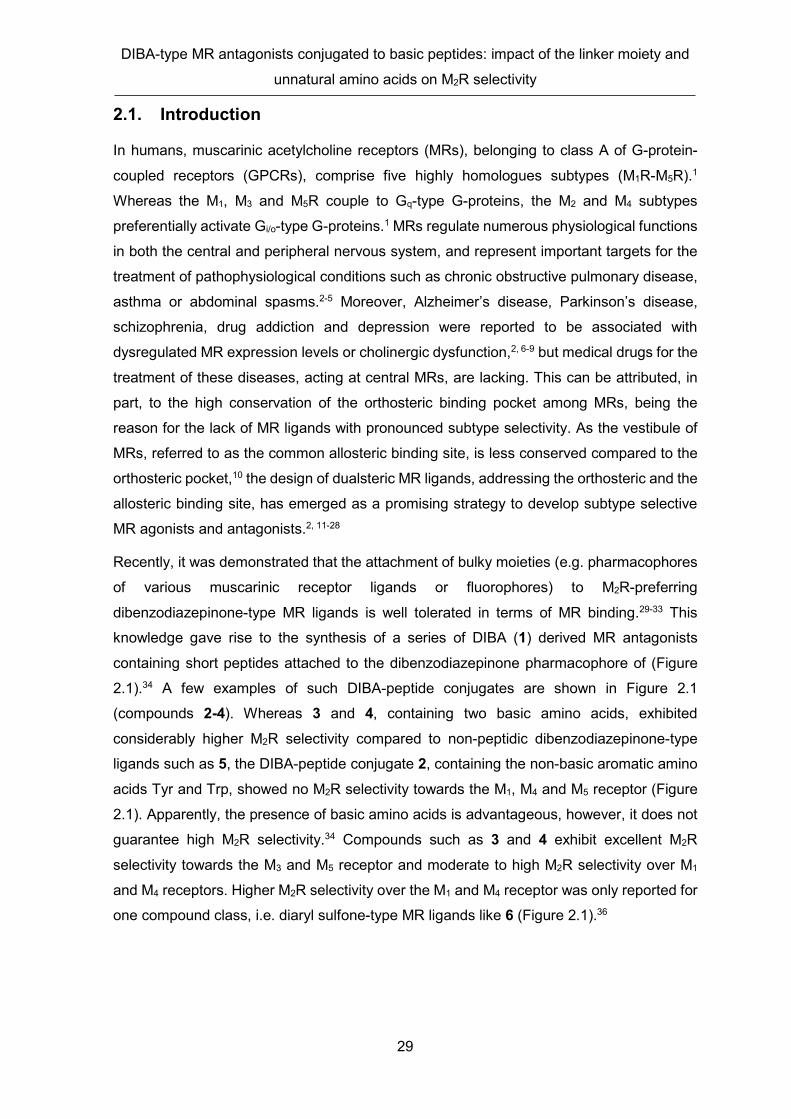
DIBA-type MR antagonists conjugated to basic peptides: impact of the linker moiety and
unnatural amino acids on M2R selectivity
29
2.1. Introduction
In humans, muscarinic acetylcholine receptors (MRs), belonging to class A of G-protein-
coupled receptors (GPCRs), comprise five highly homologues subtypes (M1R-M5R).1
Whereas the M1, M3 and M5R couple to Gq-type G-proteins, the M2 and M4 subtypes
preferentially activate Gi/o-type G-proteins.1 MRs regulate numerous physiological functions
in both the central and peripheral nervous system, and represent important targets for the
treatment of pathophysiological conditions such as chronic obstructive pulmonary disease,
asthma or abdominal spasms.2-5 Moreover, Alzheimer’s disease, Parkinson’s disease,
schizophrenia, drug addiction and depression were reported to be associated with
dysregulated MR expression levels or cholinergic dysfunction,2, 6-9 but medical drugs for the
treatment of these diseases, acting at central MRs, are lacking. This can be attributed, in
part, to the high conservation of the orthosteric binding pocket among MRs, being the
reason for the lack of MR ligands with pronounced subtype selectivity. As the vestibule of
MRs, referred to as the common allosteric binding site, is less conserved compared to the
orthosteric pocket,10 the design of dualsteric MR ligands, addressing the orthosteric and the
allosteric binding site, has emerged as a promising strategy to develop subtype selective
MR agonists and antagonists.2, 11-28
Recently, it was demonstrated that the attachment of bulky moieties (e.g. pharmacophores
of various muscarinic receptor ligands or fluorophores) to M2R-preferring
dibenzodiazepinone-type MR ligands is well tolerated in terms of MR binding.29-33 This
knowledge gave rise to the synthesis of a series of DIBA (1) derived MR antagonists
containing short peptides attached to the dibenzodiazepinone pharmacophore of (Figure
2.1).34 A few examples of such DIBA-peptide conjugates are shown in Figure 2.1
(compounds 2-4). Whereas 3 and 4, containing two basic amino acids, exhibited
considerably higher M2R selectivity compared to non-peptidic dibenzodiazepinone-type
ligands such as 5, the DIBA-peptide conjugate 2, containing the non-basic aromatic amino
acids Tyr and Trp, showed no M2R selectivity towards the M1, M4 and M5 receptor (Figure
2.1). Apparently, the presence of basic amino acids is advantageous, however, it does not
guarantee high M2R selectivity.34 Compounds such as 3 and 4 exhibit excellent M2R
selectivity towards the M3 and M5 receptor and moderate to high M2R selectivity over M1
and M4 receptors. Higher M2R selectivity over the M1 and M4 receptor was only reported for
one compound class, i.e. diaryl sulfone-type MR ligands like 6 (Figure 2.1).36

Chapter 2
30
Figure 2.1. Structures and MR binding data of reported tricyclic MR antagonists (compounds 1-5) and a diarylsulfone-type M2R-selective ligand (6). References in the table are as follows: (a) Gitler et. al. (reported Ki values were converted to pKi values)35 (b) Pegoli et. al.34 (c) Keller et. al. (data, reported as pIC50, were reanalyzed to obtain pKi values)30 (d) Clader et. al. (the reported Ki value (M2R) of 6 was converted to a pKi value; note: IC50, pIC50; Ki, or pKi values for the MR subtypes M1, M3, M4, and M5 were not provided; instead only the ratios Ki(M1,3,4,5R)/Ki(M2R) were given).36
The recently reported DIBA-peptide conjugates contained only natural amino acids and
merely two types of linkers were used to connect the DIBA pharmacophore with the peptide.
In the present study, we describe a new series of DIBA-peptide conjugates all containing at
least one basic amino acid. In order to investigate the impact of the linker moiety on M2R
affinity and selectivity, twelve different linkers were used for the preparation of the DIBA-
peptide conjugates. Moreover, unnatural amino acids such as D-configured amino acids or
arginine homologues with a shortened side chain were used for compound synthesis
(Figure 2.2). We also synthesized the highly M2R selective diaryl sulfone 636 (cf. Figure 2.1)
and included this compound in MR binding studies. Besides the determination of M1R-M5R
affinities, selected compounds were investigated with respect to M2R antagonism in a mini-

DIBA-type MR antagonists conjugated to basic peptides: impact of the linker moiety and
unnatural amino acids on M2R selectivity
31
G-protein recruitment assay. Finally, induced-fit docking was performed to gain insights into
possible ligand binding modes and putative ligand-receptor interactions.
Figure 2.2. Rationale of the present study. Agb: shorter Arginine homologue with an ethylene instead of a trimethylene moiety in the side chain.
2.2. Results and Discussion
2.2.1. Chemistry
For the synthesis of the DIBA-peptide conjugates, we prepared a set of nine amine-
functionalized precursor compounds containing the same dibenzodiazepinone
pharmacophore, but different linker moieties (13, 23-25, 31, 35, 40 and 43) or no linker
moiety (28) (syntheses outlined in Scheme 2.1). In addition, three previously described
dibenzodiazepinone-type amine precursors (52, 62, 72; cf. Scheme 2.2) were used.30, 33.
The synthesis of derivative 13 started with N-alkylation of piperidine 7 using the chlorinated
dibenzodiazepinone derivative 837 to give alcohol 9, which was converted to chloride 10
using thionylchloride (Scheme 2.1). Nucleophilic substitution of chlorine in 10 using
piperazine derivative 1133 gave the Boc-protected intermediate 12, which was treated with
TFA to afford precursor 13. Amine precursors 23-25, containing an acylated piperazine
moiety, were prepared through N-alkylation of the commercially available piperidinyl
carboxylic acids 14-16 by chloride 8, giving intermediates 17-19, which were used to acylate
piperazine 11 using EDC × HCl/HOBt as coupling reagent. Subsequent removal of the Boc
groups in the intermediates 20-22 gave the precursor compounds 23-25 (Scheme 2.1).
Likewise, N-alkylation of piperidine derivative 26 using chloride 8, followed by Boc-
deprotection, afforded amine precursor 28, which was also used to prepare precursor
compounds 31 and 35. For the synthesis of 31, 28 was acylated with N-Boc protected
glycine to give 29, followed by Boc-deprotection of 29 yielding precursor 31. For the
synthesis of 35, 28 was acylated using chloroacetyl chloride (32) to afford chloride 33, which
was used to alkylate piperazine 11, yielding the Boc-protected intermediate 34. Treatment

Chapter 2
32
of the latter with TFA gave amine precursor 35. The synthesis of the longer homologue of
35 (compound 40) started with the N-alkylation of 11 using 3-chloropropionic acid
benzylester (36) to give 37. Debenzylation of 37 by hydrogenolysis, using 10% Pd/C and
methanol as solvent, afforded carboxylic acid 38. Amidation of 38 with amine 28 yielded the
Boc-protected intermediate 39, which was converted to amine precursor 40 by treatment
with TFA. Finally, nucleophilic substitution of bromine in compound 4133 with amine 26 gave
intermediate 42, which was Boc-deprotected to obtain precursor compound 43 (Scheme
2.1).
Scheme 2.1. Synthesis of the amine precursors 13, 23, 24, 25, 28, 31, 35, 40 and 43. Reagents and conditions: (a) K2CO3, acetonitrile, 85 °C (microwave), 90 min, 97% (9), 67% (34), 96% (37), 99% (42); (b) SOCl2, pyridine, CH2Cl2, reflux, overnight, 99%; (c) NaI, K2CO3, acetone, reflux, 72 h, 82%; (d) CH2Cl2/TFA/H2O 4:1:0.1 v/v/v, rt, 3 h, 67% (13), 87% (28); (e) K2CO3, DMF, 85 °C (microwave), 90 min, 36% (17), 65% (18), 46% (19), 99% (27); (f) EDC × HCl, HOBt, DIPEA, DMF, 55 °C, overnight, 77 % (20), 54% (21), 52% (22), 70% (39); (g) TFA/H2O 95:5 v/v, rt, 3 h; 49% (23), 42% (24), 56% (25), 48%, (31), 53% (35), 61% (40), 89% (43); (h) EDC × HCl, HOBt, DIPEA, DMF, 5 °C, overnight, 60%; (i) DIPEA, DMF, rt, 2 h, 88%; (j) H2, 10% Pd/C, MeOH, rt, 90 m in, 90%.

DIBA-type MR antagonists conjugated to basic peptides: impact of the linker moiety and
unnatural amino acids on M2R selectivity
33
The N-terminally acetylated peptides 44-51, 61, 68-71, 83, 84 and 100 were synthesized
on a chlorotrityl resin according to the Fmoc strategy using a previously described
procedure.34 For the coupling of the peptides to the amine-functionalized
dibenzodiazepinone derivatives via their C-termini, EDC × HCl and HOBt were used as
coupling reagents, excessive base was avoided and reactions were performed at 5 °C. As
reported recently, under these conditions epimerization can be largely precluded.34
Peptide conjugation to amine precursors 13, 23-25, 28, 31, 35, 40, 43, 52, 62 or 72, followed
by cleavage of side chain protecting groups using TFA/H2O 95:5, afforded the DIBA-peptide
conjugates 53-60, 63-65, 73-82, 87-99, 101-109, 112-126 (Schemes 2.2 and 2.3). Coupling
of Boc-D-Arg(Pbf)-OH (66) or Boc-Arg(Pbf)-OH (67) to amine precursors 13, 43 or 72,
followed by deprotection, gave compounds 73, 74, 85, 86, 110 and 111.
Conjugation of the N-terminally Fmoc-protected tripeptide 127 to amine precursors 13 and
72, followed by side-chain deprotection using 95% aqueous TFA, yielded compounds 130
and 131 (Scheme 2.4). Intramolecular amide bond formation between the carboxylic acid
group of the aspartic acid side chain and the amine group of the ornithine side chain in 130
and 131 was performed using PyBOP/HOBt as coupling reagents, resulting in the
macrocyclic compounds 132 and 133. Subsequent Fmoc-deprotection of 132 and 133 using
diethylamine afforded the cyclic DIBA-peptide conjugates 134 and 135. Amidation of the
peptides 68, 70 and 83 with propylamine (136) gave the reference compounds 137-139 (Scheme 2.4). We also synthesized the previously described highly M2R selective MR
ligand 6 (structure shown in Figure 2.1) according to the reported procedure.36

Chapter 2
34
Scheme 2.2. Synthesis of the DIBA-peptide conjugates 53-60, 63-65, 73-82, 86-106. Reagents and conditions: (a) (1) EDC × HCl, HOBt, DMF, 5 °C, overnight; (2) TFA/H2O 95:5 v/v, rt, 3 h. Agb: shorter Arginine homologue with two methylene groups.

DIBA-type MR antagonists conjugated to basic peptides: impact of the linker moiety and
unnatural amino acids on M2R selectivity
35
Scheme 2.3. Synthesis of the DIBA-peptide conjugates 107-126. Reagents and conditions: (a) (1) EDC × HCl, HOBt, DMF, 5 °C, overnight; (2) TFA/H2O 95:5 v/v, rt, 3 h. For the structure of A see Scheme 2.1 or 2.2.
Scheme 2.4. Synthesis of the cyclic DIBA-peptide conjugates 134 and 135 and the amidated tripeptides 137-139. Reagents and conditions: (a) (1) EDC × HCl, HOBt, DMF, 5 °C, overnight; (2) TFA/H2O 95:5 v/v, rt, 3 h; (b) HOBt, DIPEA, PyBOP, DMF, rt, 5h; (c) DEA, DMF, rt, 3h. For the structure of A see Scheme 2.1 or 2.2

Chapter 2
36
2.2.2. Radioligand competition binding studies with [3H]NMS
MR binding affinities were determined by radioligand competition binding at CHO cells
stably transfected with the human M1, M2, M3, M4 or M5 receptor using [3H]NMS as labeled
ligand. M1, M2 and M4 receptor affinities were determined for all target compounds as well
as amine precursors 13, 25, 31, 35, 40 and 43 because M2R selectivity over the M1 and M4
receptor is most critical for the studied compound class. For the peptides 137-139,
representing reference compounds, only binding to the M2R was investigated. M3 and M5
receptor affinities were additionally determined for selected compounds, showing either
high M2R selectivity compared to the M1 and M4 receptor (87, 89, 91, 92, 101, 103-105,
107-109, 119, 122) or only a low or no M2R preference over the M1 and M4 receptor (63,
85, 88, 93, 95, 102, 116, 120, 121, 125) (Table 2.1). Radioligand displacement curves,
obtained from competition binding studies, are shown in Figure 2.3 (89, 95, 103-105, 109)
or Figures S1-S9 (2.5. Supplementary Information) (13, 25, 31, 35, 40, 43, 53-60, 63-65,
67, 74-82, 85-88, 90-94, 96-99, 101, 102, 106-108, 110-126, 134, 135, 137-139). Among
the studied amine precursors (13, 25, 31, 35, 40 and 43), compound 43, containing a 4-
aminopiperidine moiety attached to the “proximal” piperidine ring in partial structure A (for
structure of A see Figure 2.1, Scheme 2.2 or Table 2.1), showed the highest M2R affinity
(pKi: 9.73) (Table 2.1), and precursor 35, containing an acylated 4-aminopiperidine moiety
instead of the piperidine ring in partial structure A, exhibited the lowest M2R affinity (pKi:
5.61). Whereas precursor compound 31 showed no M2R selectivity compared to the M1 and
M4 receptor, the other amine precursors (13, 25, 35, 40 and 43) displayed a low to moderate
M2R preference over these MR subtypes (Table 2.1).

DIBA-type MR antagonists conjugated to basic peptides: impact of the linker moiety and
unnatural amino acids on M2R selectivity
37
Table 2.1. M1-M5 receptor binding data (pKi values) of compounds 6, 13, 25, 31, 35, 40, 43, 53-60, 63-65, 73-82 and 134.
pKia
M2R selectivity
Ki(M1,3,4,5R)/Ki(M2R)
Linker Structure M1R M2R M3R M4R M5R M1 M3 M4 M5
6 n.a. see Figure 2.1 6.49 ± 0.06
8.97 ± 0.05
6.17 ± 0.04
7.04 ± 0.07
7.14 ± 0.05 300 620 84 68
13 7.35 ± 0.21
8.45 ± 0.05 - 7.73 ±
0.17 - 16 - 5.9 -
25 6.14 ± 0.09
6.81 ± 0.09 - 5.82 ±
0.06 - 4.6 - 9.6 -
31 5.53 ± 0.10
5.94 ± 0.03 - 5.96 ±
0.08 - 2.4 - 0.87 -
35 n.a. 4.32 ± 0.09
5.61 ± 0.04 - 4.59 ±
0.01 - 20 - 10 -
40
6.44 ± 0.07
7.63 ± 0.03 - 6.44 ±
0.05 16 - 16 -
43 8.59 ±
0.18 9.73 ± 0.06 - 8.60 ±
0.06 - 16 - 14 -
53 Ac-Arg-Ala-Gly-Arg-NH-L1 8.19 ± 0.05
8.64 ± 0.06 - 7.80 ±
0.13 - 2.8 - 7.2 -
54 Ac-Arg-Ala-Arg-NH-L1 7.67 ± 0.11
8.14 ± 0.04 - 7.70 ±
0.04 - 3.1 - 2.8 -
55 Ac-Arg-Ala-Gly-NH-L1 7.29 ± 0.06
7.83 ± 0.10 - 7.42 ±
0.16 - 3.3 - 2.7 -
56 Ac-Arg-Ala-D-Arg-NH-L1 7.28 ± 0.03
7.88 ± 0.10 - 6.45 ±
0.04 - 3.8 - 26 -
57 Ac-Arg-Ala-NH-L1 7.33 ± 0.09
8.61 ± 0.08 - 7.61 ±
0.07 - 19 . 10 -
58 Ac-Orn-Arg-NH-L1 7.17 ± 0.07
8.53 ± 0.01 - 6.85 ±
0.05 - 24 - 49 -
59 Ac-D-Orn-Arg-NH-L1 7.48 ± 0.06
8.09 ± 0.08 - 6.85 ±
0.03 - 4.0 - 17 -
60 Ac-Orn-Agb-NH-L1 7.41 ± 0.09
8.72 ± 0.11 - 7.16 ±
0.01 - 20 - 35 -
63
Ac-Lys-Arg-NH-L2 5.72 ±
0.01 7.13 ± 0.26 <5 5.98 ± <5 27 >160 16 >170
64 Ac-Orn-Arg-NH-L2 6.12 ± 0.05
7.34 ± 0.04 - 5.83 ±
0.06 -
17 - 33 -
65 Ac-D-Orn-Arg-NH-L2 5.84 ± 0.24
6.72 ± 0.11 - 5.67 ±
0.02 - 7.3 - 11 -
73 H-D-Arg-NH-L3 9.16 ± 0.16
9.64 ± 0.05 - 8.74 ±
0.18 - 3.4 - 9.2 -
74 H-L-Arg-NH-L3 8.87 ± 0.05
9.99 ± 0.09 - 9.10 ±
0.09 - 12 - 7.5 -
75 Ac-Arg-Ala-Arg-NH-L3 9.32 ± 0.08
9.72 ± 0.04 - 9.18 ±
0.05 - 2.6 - 3.4 -
76 Ac-Arg-Ala-Gly-Arg-NH-L3 9.04 ± 0.05
9.68 ± 0.07 - 9.14 ±
0.13 - 4.3 - 3.6 -
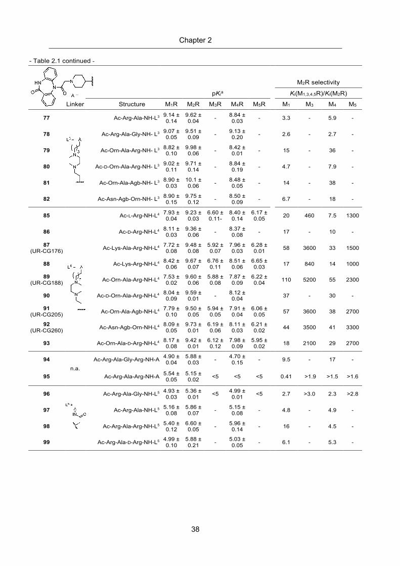
Chapter 2
38
- Table 2.1 continued -
pKia
M2R selectivity
Ki(M1,3,4,5R)/Ki(M2R)
Linker Structure M1R M2R M3R M4R M5R M1 M3 M4 M5
77 Ac-Arg-Ala-NH-L3 9.14 ±
0.14 9.62 ± 0.04 - 8.84 ±
0.03 - 3.3 - 5.9 -
78 Ac-Arg-Ala-Gly-NH- L3 9.07 ± 0.05
9.51 ± 0.09 - 9.13 ±
0.20 - 2.6 - 2.7 -
79 Ac-Orn-Ala-Arg-NH- L3 8.82 ± 0.10
9.98 ± 0.06 - 8.42 ±
0.01 - 15 - 36 -
80 Ac-D-Orn-Ala-Arg-NH- L3 9.02 ± 0.11
9.71 ± 0.14 - 8.84 ±
0.19 - 4.7 - 7.9 -
81 Ac-Orn-Ala-Agb-NH- L3 8.90 ± 0.03
10.1 ± 0.06 - 8.48 ±
0.05 - 14 - 38 -
82 Ac-Asn-Agb-Orn-NH- L3 8.90 ± 0.15
9.75 ± 0.12 - 8.50 ±
0.09 - 6.7 - 18 -
85 Ac-L-Arg-NH-L4 7.93 ± 0.04
9.23 ± 0.03
6.60 ± 0.11-
8.40 ± 0.14
6.17 ± 0.05 20 460 7.5 1300
86 Ac-D-Arg-NH-L4 8.11 ± 0.03
9.36 ± 0.06 - 8.37 ±
0.08 - 17 - 10 -
87 (UR-CG176)
Ac-Lys-Ala-Arg-NH-L4 7.72 ± 0.08
9.48 ± 0.08
5.92 ± 0.07
7.96 ± 0.03
6.28 ± 0.01 58 3600 33 1500
88 Ac-Lys-Arg-NH-L4 8.42 ± 0.06
9.67 ± 0.07
6.76 ± 0.11
8.51 ± 0.06
6.65 ± 0.03 17 840 14 1000
89 (UR-CG188) Ac-Orn-Ala-Arg-NH-L4 7.53 ±
0.02 9.60 ± 0.06
5.88 ± 0.08
7.87 ± 0.09
6.22 ± 0.04 110 5200 55 2300
90 Ac-D-Orn-Ala-Arg-NH-L4 8.04 ± 0.09
9.59 ± 0.01 - 8.12 ±
0.04 37 - 30 -
91 (UR-CG205) Ac-Orn-Ala-Agb-NH-L4 7.79 ±
0.10 9.50 ± 0.05
5.94 ± 0.05
7.91 ± 0.04
6.06 ± 0.05 57 3600 38 2700
92 (UR-CG260) Ac-Asn-Agb-Orn-NH-L4 8.09 ±
0.05 9.73 ± 0.01
6.19 ± 0.06
8.11 ± 0.03
6.21 ± 0.02 44 3500 41 3300
93 Ac-Orn-Ala-D-Arg-NH-L4 8.17 ± 0.08
9.42 ± 0.01
6.12 ± 0.12
7.98 ± 0.09
5.95 ± 0.02 18 2100 29 2700
94 n.a.
Ac-Arg-Ala-Gly-Arg-NH-A 4.90 ± 0.04
5.88 ± 0.03 - 4.70 ±
0.15 - 9.5 - 17 -
95 Ac-Arg-Ala-Arg-NH-A 5.54 ± 0.05
5.15 ± 0.02 <5 <5 <5
0.41 >1.9 >1.5 >1.6
96 Ac-Arg-Ala-Gly-NH-L5 4.93 ± 0.03
5.36 ± 0.01 <5 4.99 ±
0.01 <5 2.7 >3.0 2.3 >2.8
97 Ac-Arg-Ala-NH-L5 5.16 ± 0.08
5.86 ± 0.07 - 5.15 ±
0.08 - 4.8 - 4.9 -
98 Ac-Arg-Ala-Arg-NH-L5 5.40 ± 0.12
6.60 ± 0.05 - 5.96 ±
0.14 - 16 - 4.5 -
99 Ac-Arg-Ala-D-Arg-NH-L5 4.99 ± 0.10
5.88 ± 0.21 - 5.03 ±
0.05 - 6.1 - 5.3 -

DIBA-type MR antagonists conjugated to basic peptides: impact of the linker moiety and
unnatural amino acids on M2R selectivity
39
- Table 2.1 continued -
pKia
M2R selectivity
Ki(M1,3,4,5R)/Ki(M2R)
Linker Structure M1R M2R M3R M4R M5R M1 M3 M4 M5 101
(UR-CG096) Ac-Lys-Ala-Arg-NH-L6 5.27 ±
0.04 7.05 ± 0.01 <5 5.37 ±
0.08 5.04 ± 0.04
62 >130 51 120
102 Ac-Lys-Arg-NH-L6 6.81 ± 0.07
7.67 ± 0.02 <5 6.51 ±
0.02 <5 7.5 >630 14 >520
103 (UR-CG191) Ac-Orn-Ala-Arg-NH-L6 5.22 ±
0.07 7.20 ± 0.12 <5 5.49 ±
0.04 <5 90 >160 47 >160
104 (UR-CG197) Ac-Orn-Ala-Agb-NH-L6 5.09 ±
0.03 7.09 ± 0.02 <5 5.34 ±
0.08 <5 100 >140 58 >140
105 (UR-CG276) Ac-Orn-Ala-D-Arg-NH-L6 <5 7.12 ±
0.08 <5 5.33 ± 0.05 <5 >140 >160 59 >140
106 Ac-Orn-Ala-D-Agb-NH-L6 5.70 ± 0.05
7.36 ± 0.01 - 5.90 ±
0.02 - 47 - 29 -
107 (UR-CG220)
Ac-Lys-Ala-Arg-NH-L7 7.22 ± 0.04
9.03 ± 0.03
4.98 ± 0.03
7.39 ± 0.02
5.24 ± 0.03 66 11000 44 6300
108 (UR-CG221) Ac-Orn-Ala-Agb-NH-L7 7.21 ±
0.09 8.93 ± 0.13
5.02 ± 0.05
7.23 ± 0.06
5.29 ± 0.05 50 7600 47 4000
109 (UR-CG239) Ac-Orn-Ala-Arg-NH-L7 7.27 ±
0.12 9.08 ± 0.03
5.07 ± 0.16
7.40 ± 0.03
5.31 ± 0.07 70 11000 48 6000
110 H-L-Arg-NH-L8 8.41 ± 0.02
9.53 ± 0.07 - 8.54±
0.07 - 13 - 10 -
111 H-D-Arg-NH-L8 9.03 ± 0.11
9.85 ± 0.10 - 8.69 ±
0.02 - 6.7 - 14 -
112 Ac-Lys-Ala-Arg-NH-L8 8.94 ± 0.17
9.54 ± 0.04 - 8.39 ±
0.02 - 4.7 - 14 -
113 Ac-Orn-Ala-Arg-NH-L8 8.70 ± 0.06
9.69 ± 0.14 - 8.46 ±
0.02 - 7.1 - 12 -
114 Ac-Orn-Ala-Agb-NH-L8 8.89 ± 0.13
9.82 ± 0.10 - 8.57 ±
0.11 - 8.6 - 18 -
115 Ac-Arg-Ala-Gly-NH-L9 5.76 ± 0.06
6.98 ± 0.06 - 5.71 ±
0.13 - 16 - 19 -
116 Ac-Lys-Ala-Arg-NH-L9 5.21 ± 0.13
6.61 ± 0.02 <5 5.20 ±
0.05 5.06 ± 0.01 28 >55 26 35
117 Ac-Orn-Ala-Arg-NH-L9 5.10 ± 0.12
6.49 ± 0.05 - 5.29 ±
0.10 - 26 - 16 -
118 Ac-Orn-Ala-Agb-NH-L9 5.42 ± 0.02
6.49 ± 0.05 - 5.02 ±
0.03 - 12 - 29 -
119 Ac-Lys-Ala-Arg-NH-L10 6.08 ± 0.09
7.39 ± 0.10 <5 5.53 ±
0.04 <5 20 >260 68 >240
120 Ac-Orn-Ala-Arg-NH-L10 6.29 ± 0.15
6.84 ± 0.06 <5 5.60 ±
0.06 5.32 ± 0.06 4.0 >91 17 33
121 Ac-Orn-Ala-Agb-NH-L10 6.27 ± 0.13
7.28 ± 0.05 <5 5.53 ±
0.06 <5 11 >240 56 >210
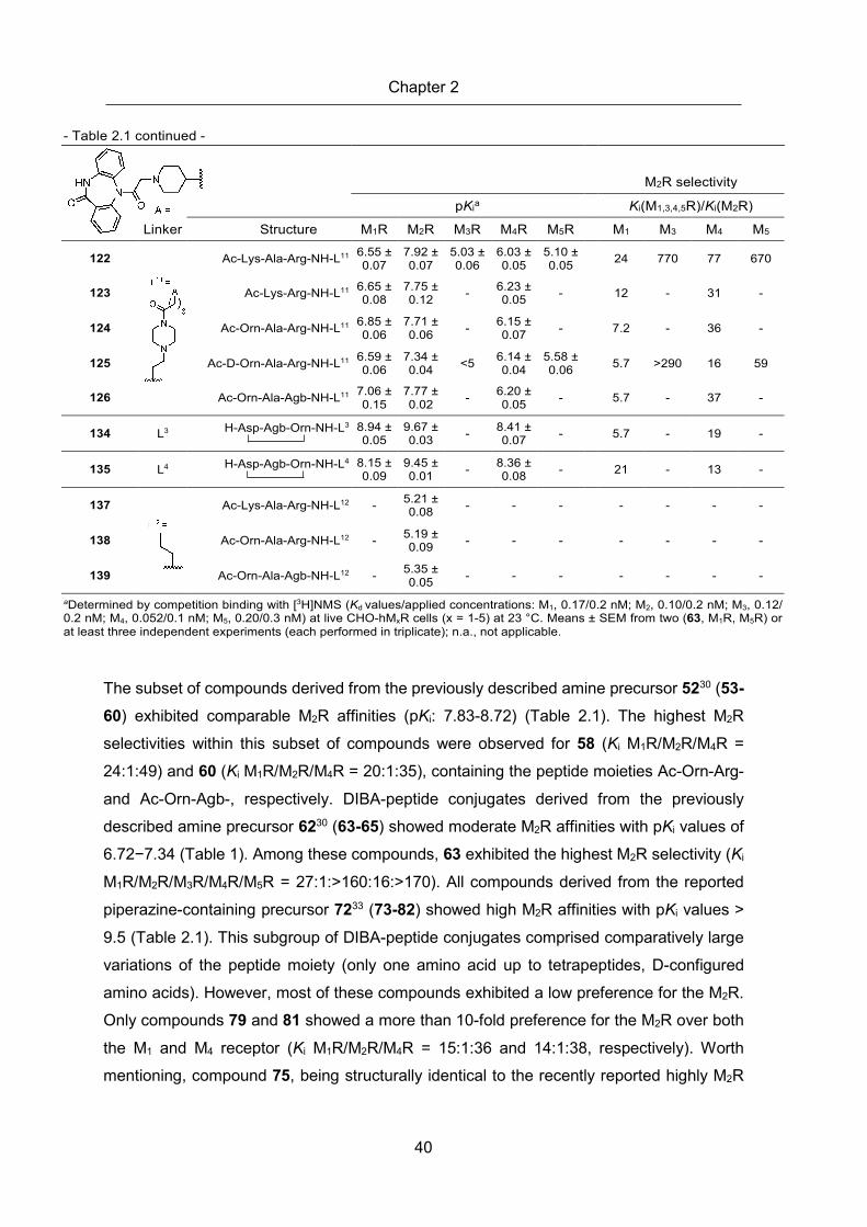
Chapter 2
40
The subset of compounds derived from the previously described amine precursor 5230 (53-60) exhibited comparable M2R affinities (pKi: 7.83-8.72) (Table 2.1). The highest M2R
selectivities within this subset of compounds were observed for 58 (Ki M1R/M2R/M4R =
24:1:49) and 60 (Ki M1R/M2R/M4R = 20:1:35), containing the peptide moieties Ac-Orn-Arg-
and Ac-Orn-Agb-, respectively. DIBA-peptide conjugates derived from the previously
described amine precursor 6230 (63-65) showed moderate M2R affinities with pKi values of
6.72−7.34 (Table 1). Among these compounds, 63 exhibited the highest M2R selectivity (Ki
M1R/M2R/M3R/M4R/M5R = 27:1:>160:16:>170). All compounds derived from the reported
piperazine-containing precursor 7233 (73-82) showed high M2R affinities with pKi values >
9.5 (Table 2.1). This subgroup of DIBA-peptide conjugates comprised comparatively large
variations of the peptide moiety (only one amino acid up to tetrapeptides, D-configured
amino acids). However, most of these compounds exhibited a low preference for the M2R.
Only compounds 79 and 81 showed a more than 10-fold preference for the M2R over both
the M1 and M4 receptor (Ki M1R/M2R/M4R = 15:1:36 and 14:1:38, respectively). Worth
mentioning, compound 75, being structurally identical to the recently reported highly M2R
- Table 2.1 continued -
M2R selectivity
pKia Ki(M1,3,4,5R)/Ki(M2R)
Linker Structure M1R M2R M3R M4R M5R M1 M3 M4 M5
122 Ac-Lys-Ala-Arg-NH-L11 6.55 ± 0.07
7.92 ± 0.07
5.03 ± 0.06
6.03 ± 0.05
5.10 ± 0.05 24 770 77 670
123 Ac-Lys-Arg-NH-L11 6.65 ± 0.08
7.75 ± 0.12 - 6.23 ±
0.05 - 12 - 31 -
124 Ac-Orn-Ala-Arg-NH-L11 6.85 ± 0.06
7.71 ± 0.06 - 6.15 ±
0.07 - 7.2 - 36 -
125 Ac-D-Orn-Ala-Arg-NH-L11 6.59 ± 0.06
7.34 ± 0.04 <5 6.14 ±
0.04 5.58 ± 0.06 5.7 >290 16 59
126 Ac-Orn-Ala-Agb-NH-L11 7.06 ± 0.15
7.77 ± 0.02 - 6.20 ±
0.05 - 5.7 - 37 -
134 L3 H-Asp-Agb-Orn-NH-L3 8.94 ± 0.05
9.67 ± 0.03 - 8.41 ±
0.07 - 5.7 - 19 -
135 L4 H-Asp-Agb-Orn-NH-L4 8.15 ± 0.09
9.45 ± 0.01 - 8.36 ±
0.08 - 21 - 13 -
137 Ac-Lys-Ala-Arg-NH-L12 - 5.21 ± 0.08 - - - - - - -
138 Ac-Orn-Ala-Arg-NH-L12 - 5.19 ± 0.09 - - - - - - -
139 Ac-Orn-Ala-Agb-NH-L12 - 5.35 ± 0.05 - - - - - - -
aDetermined by competition binding with [3H]NMS (Kd values/applied concentrations: M1, 0.17/0.2 nM; M2, 0.10/0.2 nM; M3, 0.12/ 0.2 nM; M4, 0.052/0.1 nM; M5, 0.20/0.3 nM) at live CHO-hMxR cells (x = 1-5) at 23 °C. Means ± SEM from two (63, M1R, M5R) or at least three independent experiments (each performed in triplicate); n.a., not applicable.

DIBA-type MR antagonists conjugated to basic peptides: impact of the linker moiety and
unnatural amino acids on M2R selectivity
41
selective ligand 4 (cf. Figure 2.1) except for the N-terminal amino acid (Arg in 75 vs. Lys in
4), displayed, in contrast to 4, no M2R selectivity over the M1 and M4 receptor. This
demonstrated that the type of basic amino acid in the peptide moiety can have high impact
on M2R selectivity.
The DIBA-peptide conjugates 85-93, derived from the shorter homologue of 72, i.e. amine
precursor 13, showed also high M2R affinities (pKi: 9.23-9.73). Several members of this
subset of compounds (87, 89, 91 and 92) displayed pronounced M2R selectivity over the
M1R and M4R, and excellent M2R selectivity over the M3 and M5 receptor. Compound 89
(UR-CG188), containing the peptide moiety Ac-Orn-Ala-Arg-, exhibited the most
pronounced M2R selectivity (Ki M1R/M2R/M3R/M4R/M5R = 110:1:5200:55:2300) (Table 2.1).
Compounds 87, 89 and 91 are also closely related to the recently reported DIBA-peptide
conjugate 4 (cf. Figure 2.1), which was prepared from amine precursor 72, the higher
homologue of precursor 13. Interestingly, compound 92, containing, compared to 87, 89
and 91, a markedly different peptide moiety (Ac-Asn-Agb-Orn-), showed a MR selectivity
profile comparable to that of 91 (Table 2.1). Compounds 94-99, containing no (94, 95) or a
very short (96-99) linker, showed low M2R affinities (pKi: 5.15-6.60) and a low preference
for the M2R compared to the M1 and M4 receptor. The subset of compounds derived from
precursor 35 (101-106), containing an amide group and a bisalkylated piperazine ring,
exhibited moderate M2R affinity (pKi: 7.05-7.67), but well pronounced M2R selectivity
(except for 102 containing no Ala in contrast to 101 and 103-106) (Table 2.1). The DIBA-
peptide conjugates 107-109, which were derived from amine precursor 40, representing the
higher homologue of precursor 35, showed higher M2R affinities (pKi: 8.93-9.08) compared
to compounds derived from 35, and displayed also high M2R selectivity (e.g. 109: Ki
M1R/M2R/M3R/M4R/M5R = 70:1:11,000:48:6000). As compounds 107-109 represent the
higher homologues of 101, 104 and 103, respectively (identical peptide moieties, linker
length elongated by one methylene group), these data demonstrated that this type of linker
is favorable for high M2R selectivity and the length of the linker is critical with respect to MR
affinity (Table 2.1). Compounds 110-114, derived from precursor 43, containing an N-
alkylated piperidine ring in addition to the N-alkylated piperidine ring of substructure A,
showed high M2R affinities (pKi: 9.53-9.85), but low M2R selectivity over the M1 and M4
receptor (Table 2.1). The DIBA-peptide conjugates 115-126, which were prepared from the
homologues amine precursors 23 (used for 115-118), 24 (used for 119-121) and 25 (used
for 122-126), containing an acylated piperazine ring and varying in length (one (23), two
(24) or three (25) methylene groups in between the acylated piperazine ring and the
piperidine ring), showed moderate to high M2R affinity (pKi: 6.49-7.92).

Chapter 2
42
Figure 2.3. Radioligand displacement curves obtained from competition binding experiments with [3H]NMS (0.2 nM (M1R, M2R, M3R), 0.1 nM (M4R) or 0.3 nM (M5R) and compounds 89, 103-105, 109 and 95 at intact CHO hMxR cells (x = 1-5). M3R and M5R data of 95, 103-105 and M4R data of 95 could not be fitted. Data present mean values ± SEM from at least three independent experiments (each performed in triplicate).
M2R binding increased with linker length and 115-126 showed low or moderate M2R
selectivity over the subtypes M1 and M4 (Table 2.1). Compounds 134 and 135, containing a
cyclic peptide moiety, showed high M2R affinities, but poor M2R selectivity. Worth
mentioning, the linear counterpart of 135, compound 92, showed high M2R affinity and high
M2R selectivity (Ki M1R/M2R/M3R/M4R/M5R = 44:1:3500:41:3300) (Table 2.1). In order to
demonstrate that M2R binding of the presented DIBA-peptide conjugates is mainly mediated
by the DIBA pharmacophore, the peptides 137-139, which are present in the M2R selective
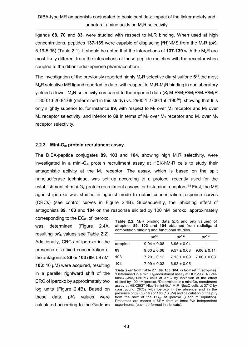
DIBA-type MR antagonists conjugated to basic peptides: impact of the linker moiety and
unnatural amino acids on M2R selectivity
43
ligands 68, 70 and 83, were studied with respect to M2R binding. When used at high
concentrations, peptides 137-139 were capable of displacing [3H]NMS from the M2R (pKi:
5.19-5.35) (Table 2.1). It should be noted that the interactions of 137-139 with the M2R are
most likely different from the interactions of these peptide moieties with the receptor when
coupled to the dibenzodiazepinone pharmacophore.
The investigation of the previously reported highly M2R selective diaryl sulfone 636,the most
M2R selective MR ligand reported to date, with respect to M1R-M5R binding in our laboratory
yielded a lower M2R selectivity compared to the reported data (Ki M1R/M2R/M3R/M4R/M5R
= 300:1:620:84:68 (determined in this study) vs. 2900:1:2700:150:19036), showing that 6 is
only slightly superior to, for instance 89, with respect to M2 over M1 receptor and M2 over
M4 receptor selectivity, and inferior to 89 in terms of M2 over M3 receptor and M2 over M5
receptor selectivity.
2.2.3. Mini-Gsi protein recruitment assay
The DIBA-peptide conjugates 89, 103 and 104, showing high M2R selectivity, were
investigated in a mini-Gsi protein recruitment assay at HEK-hM2R cells to study their
antagonistic activity at the M2 receptor. The assay, which is based on the split
nanoluciferase technique, was set up according to a protocol recently used for the
establishment of mini-Gsi protein recruitment assays for histamine receptors.38 First, the MR
agonist iperoxo was studied in agonist mode to obtain concentration response curves
(CRCs) (see control curves in Figure 2.4B). Subsequently, the inhibiting effect of
antagonists 89, 103 and 104 on the response elicited by 100 nM iperoxo, approximately
corresponding to the EC80 of iperoxo,
was determined (Figure 2.4A,
resulting pKb values see Table 2.2).
Additionally, CRCs of iperoxo in the
presence of a fixed concentration of
the antagonists 89 or 103 (89: 58 nM,
103: 16 µM) were acquired, resulting
in a parallel rightward shift of the
CRC of iperoxo by approximately two
log units (Figure 2.4B). Based on
these data, pKb values were
calculated according to the Gaddum
Table 2.2. M2R binding data (pKi and pKb values) of atropine, 89, 103 and 104 obtained from radioligand competition binding and functional studies.
pKia pKbb pKbc
atropine 9.04 ± 0.08 8.95 ± 0.04 -
89 9.60 ± 0.06 9.57 ± 0.08 9.06 ± 0.11
103 7.20 ± 0.12 7.13 ± 0.09 7.00 ± 0.08
104 7.09 ± 0.02 6.93 ± 0.05 - aData taken from Table 2.1 (89, 103, 104) or from ref.32 (atropine). bDetermined in a mini Gsi-recruitment assay at HEK293T NlucN-mini-Gsi/hM2R-NlucC cells at 37°C by inhibition of the effect elicited by 100 nM iperoxo. cDetermined in a mini Gαi-recruitment assay at HEK293T NlucN-mini-Gsi/hM2R-NlucC cells at 37°C by constructing CRCs with iperoxo in the absence and in the presence of 89 (58 nM) or 103 (16 µM) and calculation of the pKb from the shift of the EC50 of iperoxo (Gaddum equation). Presented are means ± SEM from at least five independent experiments (each performed in triplicate).

Chapter 2
44
equation39 (Table 2.2). The pKb values obtained for 89, 103 and 104 by inhibition of the
response elicited by 100 nM iperoxo (Figure 2.4A) were in excellent agreement with their
pKi values determined in the radioligand competition binding assay (Table 2.2). Likewise,
the pKb values of 89 and 103, derived from the rightward-shifted CRC of iperoxo (Figure
2.4B), were in good accordance with the pKi values. Representative time-courses of the
bioluminescence signals obtained from the mini-Gsi recruitment assays are shown in Figure
S10 (2.5 Supplementary Information).
Figure 2.4. M2R antagonism of compounds 89, 103 and 104 investigated in a mini-Gsi protein recruitment assay using HEK293T NlucN-mini-Gsi/hM2R-NlucC cells. (A) Concentration-dependent inhibition of the mini-Gαi recruitment to the M2R elicited by the superagonist iperoxo (100 nM, equals EC80) by the MR antagonists atropine (structure see Figure S10, 2.5. Supplementary Information), 89, 103 and 104; for pKb-values see Table 2.2. (B) Effects of 89 (58 nM) and 103 (16 µM) on the concentration-response-curve (CRC) of iperoxo. The concentrations of 89 and 103 corresponded to 20-fold their IC50 obtained from experimental data shown in (A). The measuring periods were 45 min (103) or 90 min (89) upon addition of iperoxo. In the presence of 89 and 103, the CRC of iperoxo was rightward shifted (for pKb-values see Table 2.2). The pEC50 values of iperoxo were 7.68 ± 0.044 (mean ± SEM, n = 5) (45 min) and 7.63 ± 0.074 (mean ± SEM, n = 5) (90 min). Data represent mean values ± SEM from at least three independent experiments (each performed in triplicate).
2.2.4. Induced-fit docking
The design of the DIBA-peptide conjugates, presented in this work, was supported by
induced-fit docking studies using the respective tool implemented in Schrödinger LLC.
Among the prepared M2R ligands, compounds 60, 73, 74, 81, 85-87, 94, 96-99, 101, 110,
111, 116, 119, 122 and 135 had been docked to the hM1R (PDB-ID: 5CXV40), hM2R (PDB-
ID: 3UON41) and hM4R (PDB-ID: 5DSG40) before they were synthesized. Docking to the M3
and M5 receptor was not performed as the vast majority of reported dibenzodiazepinone-
type MR ligands exhibit high M2R selectivity compared to these two subtypes. For
assessment of the docking results by visual inspection, binding poses corresponding to
lowest or second lowest XP GlideScores, were used (low score values are supposed to
correlate with a high free energy of binding and thus with high binding affinity). It should be
noted that, for a given compound, the predictive value of the XP GlideScores with respect

DIBA-type MR antagonists conjugated to basic peptides: impact of the linker moiety and
unnatural amino acids on M2R selectivity
45
to M2R over M1R and M2R over M4R selectivity was low. i.e. the score ratios did not correlate
with M2 over M1 and M2 over M4 receptor selectivity. In addition to the aforementioned
compounds, which were synthesized and pharmacologically characterized, further 118
virtual DIBA-peptide conjugates were studied by induced-fit docking to the hM1, hM2 and
hM4 receptor. The SMILES strings of these compounds, which were not synthesized, are
listed in Table S1 (2.5. Supplementary Information).
The DIBA peptide conjugates 103 and 109, differing only in linker length by one methylene
group, were studied by induced-fit docking at all MR subtypes after their pharmacological
characterization for the following reasons: both compounds exhibited high M2R selectivity,
but 109, representing the higher homologue of 103, showed approximately 100-fold higher
M2R affinity (Table 2.1). Docking to the M2R was performed using the recently reported
crystal structure of the M2R bound to the pyridobenzodiazepinone derivative AF-DX 384
(PDB-ID: 5ZKB42). According to the lowest-score docking pose, the piperidinylacetyl-
dibenzodiazepinone pharmacophore (in the following referred to as headgroup) was
localized within the orthosteric binding pocket below the tyrosine lid with nearly identical
headgroup orientations for both ligands (Figure 2.5, upper panel). Consequently, the
peptide moieties were oriented towards the receptor surface (Figure 2.5B and 2.5C). When
comparing the linkers in 103 and 109, the shorter homologue (103) showed rather a
stretched conformation, whereas the more flexible linker in 109 was markedly bent (Figure
2.5B and 2.5C, 2.5. Supplementary Figure S12), most likely influencing the orientation of
the peptide moieties, which appeared to be different (Figure 2.5, lower panel, 2.5.
Supplementary Figure S12). This was consistent with the experimental data, which revealed
that the length and flexibility of the linkers in 103 and 109 are critical with respect to M2R
binding (100-fold lower M2R affinity of 103 compared to 109). Besides various polar
interactions (H-bonding) between the headgroup of the ligands with the receptor (Figure
2.5A and 2.5D), the distances between the tyr residues of the tyrosine lid and the piperidine
ring of the ligand headgroup were compatible with π-cation interactions (not indicated in
Figure 2.5). Additionally, also π-cation interactions between the tyrosine lid and the
piperazine moieties of the ligands might contribute to receptor binding.
According to the induced-fit docking results, both 103 and 109 address two Glu residues in
the extracellular loop 2 (ECL2) of the M2R via basic amino acids of their peptide moieties
(103: Arg, 109: Arg and Orn; Figure 2.5E and 2.5H). Interestingly, these two Glu residues
are only present in the ECL2 of the M2R (2.5. Supplementary Figure S13). This indicated
that the high M2R selectivity of dualsterically binding DIBA-peptide conjugates such as 103
and 109 can be attributed, in part, to differences in the occurrence of acidic and basic amino

Chapter 2
46
acids at the surface of MRs (2.5. Supplementary Figures S13 and S14). It should be noted
that the head group of 103 and 109 was also located in the orthosteric binding site of the
M1R and M3R-M5R according to “low-score” induced-fit docking results obtained for these
MR subtypes (2.5. Supplementary Figure S14).
Figure 2.5. Docking poses of 103 (A, B, E, F) and 109 (C, D, G, H) in the M2 inactive X-Ray structure (PDB-ID 5ZKB) obtained by induced-fit docking. The lowest scored (best ranked) docking poses are shown. The upper panel shows the orientation and polar interactions of the headgroup (piperidinylacetyl-dibenzodiazepinone moiety) in the orthosteric binding pocket (A, D), and provides a comparison of the ligand docking poses with the coordinates of AF-DX 384 (red sticks, structure see 2.5. Supplementary Figure S15) from the X-Ray structure (B, C). The lower panel shows the polar interactions observed between the terminal tripeptide of the ligands and amino acids located in ECL2 of the M2R (E, H), and contains a top view on the receptor vestibule (F, G). The orientation of the Tyr residues of the tyrosin lid (Tyr104, Tyr403, Tyr426) and its proximity to the ligands is visualized in A, F, G, and D. Distances, indicated in A, D, E and H, are given in Angström.
2.3. Conclusion
Conjugation of short peptides, containing at least one basic amino acid, to
dibenzodiazepinone-type MR antagonists via different linkers gave DIBA-peptide
conjugates with low to high M2R affinity and low to high M2R selectivity. It was demonstrated,
that the type and the length of the linker strongly determines M2R selectivity, a question,
which was not addressed in the previous study.34 Moreover, the introduction of, e.g. D-
configured amino acids or homologues of proteinogenic amino acids (Orn vs. Lys, Agb vs.
Arg) had rather little impact on M2R selectivity. As all highly M2R selective ligands, identified
in this study, contain a basic piperazine moiety, putatively interacting with aromatic amino
acid residues of the M2R (cation-π interactions), the incorporation of a basic group, such as
a piperazine ring, in the right distance to the tricyclic dibenzodiazepinone core, is
advantageous in terms of high M2R affinity, and, in combination with an appropriate basic

DIBA-type MR antagonists conjugated to basic peptides: impact of the linker moiety and
unnatural amino acids on M2R selectivity
47
peptide, also with respect to high M2R selectivity. The presented M2R selective ligands
might serve as lead structures to develop new non-peptidic highly selective M2R ligands
with reduced basicity. Furthermore, the present study can support the design of labeled tool
compounds such as radiolabeled and fluorescent M2R ligands with high M2R selectivity.
2.4. Experimental section
2.4.1. General experimental conditions
Chemicals for synthesis were purchased from Acros Organics (Geel, Belgium), Iris Biotech
(Marktredwitz, Germany), Alfa Aesar (Karlsruhe, Germany), Merck (Darmstadt, Germany),
Sigma-Aldrich (Taufkirchen, Germany), Carbolution (St. Ingbert, Germany), or TCI Europe
(Zwijndrecht, Belgium). Compound 7 was purchased from Ark Pharm (Arlington Heights,
USA), compound 14 and 26 were purchased from abcr (Karlsruhe, Germany), compound
15 was obtained from Enamine (Kiev, Ukraine), compound 16 was purchased from Sigma
Aldrich and compound 36 was obtained from Combi-Blocks (San Diego, USA).
Dulbecco's modified Eagle's medium (DMEM) was purchased from Sigma-Aldrich and
Leibovitz’ L-15 medium (L-15) from Fisher Scientific (Nidderau, Germany). FBS,
trypsin/EDTA and geneticin (G418) were from Merck Biochrom (Darmstadt, Germany).
Puromycin was from InvivoGen (Toulouse, France), NlucC, NlucN and furimazine were
obtained from Promega (Mannheim, Germany). The pcDNA3.1 vector was obtained from
Thermo Scientific (Nidderau, Germany).
[3H]NMS (specific activity = 80 Ci/mmol, respectively 75 C/mmol) was purchased from
American Radiolabeled Chemicals Inc. (St. Louis, MO) via Hartman Analytics
(Braunschweig, Germany) or from Novandi Chemistry AB (Södertälje, Sweden).
Acetonitrile for HPLC (gradient grade) was obtained from Merck (Darmstadt, Germany) or
SigmaAldrich. DMF, atropine, EDC × HCl, DIPEA, HFIP, iperoxo, Boc-anthranilic acid and
TFA were purchased from Sigma Aldrich. CH2Cl2, HOBt, DMF for peptide synthesis and
NMP for peptide synthesis were obtained from Acros Organics/Fisher Scientific (Nidderau,
Germany). HBTU and piperidine were from Iris Biotech. Boc-D-Arg(Pbf)-OH, Fmoc-Ala-OH,
Fmoc-Arg(Pbf)-OH, Fmoc-Asp(tBu)-OH, Fmoc-Gly-OH, Fmoc-Lys(Boc)-OH, Fmoc-
Asn(Trt)-OH, acetic anhydride, and solid supports for SPPS (H-Ala-, H-Arg(Pbf)-, H-Gly-
and H-Lys(Boc)-2-ClTrt PS resin) were purchased from Merck. Fmoc-Orn(Boc)-OH and
Boc-D -Arg(Pbf)-OH were purchased from Carbolution Chemicals (St. Ingbert, Germany).
Fmoc- D -Orn(Boc)-OH, Fmoc-L-Agb(Pbf,Boc)-OH, Fmoc- D -Agb(Pbf,Boc)-OH, Fmoc- D -

Chapter 2
48
Arg(Pbf)-OH, 2-chlorotritylchloride resin (100-200 mesh, 1.60 mmol Cl/g resin) and PyBOP
were purchased from Iris Biotech. Boc-L-Arg(Pbf)-OH was purchased from Sigma-Aldrich
(Munich, Germany). Deuterated solvents were purchased from Deutero (Kastellaun,
Germany).
The syntheses of compounds 52,30 41,33 62,30 and 7233 were described elsewhere.
Compounds 6,36 8,37 and 11 33 were prepared according to described procedures.
If moisture-free conditions were required, reactions were performed in dried glassware
under inert atmosphere (argon). Reactions were monitored by TLC using aluminum plates
coated with silica gel (Macherey Nagel, pre-coated TLC sheets ALUGRAM® Xtra SIL
G/UV254, Düren, Germany). Spots were detected by UV light (254 nm or 366 nm), by staining
with a 1% solution of potassium permanganate in 0.1% aq NaOH or using a 0.3% solution
of ninhydrine in n-butanol (for amines). Flash chromatography was performed in glass
columns on silica gel (Merck silica gel 60, 40-63 μm) or using the flash system Agilent 971-
FP Flash purification workstation from Agilent Technologies, equipped with an UV detector
(200-400 nm), a FC fraction collector and SuperFlash cartridges (Santa Clara, USA).
Microwave assisted reactions were performed with an Initiator 2.0 synthesizer (Biotage,
Uppsala, Sweden). Millipore water was used throughout for the preparation of stock
solutions and HPLC eluents. Polypropylene reaction vessels (1.5 or 2 mL) with screw cap
(Süd-Laborbedarf, Gauting, Germany) were used for small scale reactions and for the
preparation and storage of stock solutions. Nylon membrane filters (0.2 µm, Phenomenex,
Aschaffenburg, Germany) were used for filtrations of the injection solutions for preparative
HPLC and reactions mixtures. Temperature-controlled reactions in 1.5-mL reaction vessels
were performed using a Thermocell Mixing Block MB-102 from Bioer (Hangzhou, China).
NMR spectra were recorded on a Bruker Avance 400 (7.05 T, 1H: 400.3 MHz, 13C: 100.7
MHz) or a Bruker Avance III HD 600 equipped with a cryogenic probe (14.1 T 1H: 600.3
MHz, 13C: 150.9 MHz) (Bruker, Karlsruhe, Germany). Abbreviations for the multiplicities of
the signals are s (singlet), d (doublet), t (triplet), m (multiplet) and brs (broad singlet). High-
resolution mass spectrometry (HRMS) analysis was performed on an Agilent 6540 UHD
Accurate-Mass Q-TOF LC/MS system (Agilent Technologies, Santa Clara, CA) using an
ESI source. Except for 30 and 63, a Prep 150 LC system from Waters (Eschborn, Germany),
consisting of a binary gradient module, a 2489 UV/visible detector, and a Waters fraction
collector III, was used as preparative HPLC. Compounds 30 and 63 were purified using a
preparative HPLC system from Knauer (Berlin, Germany) consisting of two K-1800 pumps
and a K-2001 detector. For the purification of compounds 13, 17, 19, 23-25, 27, 28, 30, 33-

DIBA-type MR antagonists conjugated to basic peptides: impact of the linker moiety and
unnatural amino acids on M2R selectivity
49
35, 53-60, 63, 73-81, 85-91, 94-99, 101-104, 110-112, 115-117, 119 and 122-125, a
Kinetex-XB C18 (5 μm, 250 mm × 21 mm; Phenomenex, Aschaffenburg, Germany) was
used as stationary phase at a flow rate of 20 mL/min, and for the purification of compounds
64, 65, 71, 82, 92, 93, 105-109, 113, 114, 118, 121, 126, 128-135 and 137-139 a YMC-
Actus Triart C18 (5 µm, 150 × 20 mm, Dinslaken, Germany) served as stationary phase at
a flow rate of 20 mL/min.
Mixtures of 0.1% aq TFA and acetonitrile were used as mobile phase. The detection
wavelength was set to 220 nm throughout. The solvent of collected fractions was removed
by lyophilization using a Scanvac freeze drying apparatus (Labogene, Allerød, Denmark)
equipped with a RZ 6 rotary vane vacuum pump (Vacuubrand, Wertheim, Germany).
Analytical HPLC analysis was performed with a system from Agilent Technologies
composed of a 1290 Infinity binary pump equipped with a degasser, a 1290 Infinity
autosampler, a 1290 Infinity thermostated column compartment, a 1260 Infinity diode array
detector, and a 1260 Infinity fluorescence detector. A Kinetex-XB C18 (2.6 μm, 100 × 3 mm;
Phenomenex) was used as stationary phase at a flow rate of 0.6 mL/min. Mixtures of 0.04%
aq TFA (A) and acetonitrile (B) were used as mobile phase. The following linear gradient
was applied for compounds 13, 23-25, 31, 35, 40, 43, 53-60, 63-65, 73-82, 85-99, 101-126, 134 and 135: 0-12 min, A/B 90:10 to 70:30; 12-16 min, 70:30 to 5:95; 16-20 min, 5:95. For
6 the following linear gradient was applied: 0-12 min, A/B 90:10 to 40:60; 12-16 min, 40:60
to 5:95; 16-20 min, 5:95. For all analytical HPLC runs the oven temperature was set to 25
°C, the injection volume was 20 μL and detection was performed at 220 nm. Compound
concentrations of the sample solutions were between 40 and 120 μM. The stock solutions
(final concentration: 10 mM) of the final compounds were prepared in DMSO and were
stored at -78°C. Retention (capacity) factors were calculated from retention times (tR)
according to k = (tR – t0)/t0 (t0 = dead time).
2.4.2. Compound characterization
Target compounds 6, 13, 23-25, 31, 35, 40, 43, 53-60, 63-65, 73-82, 85-99, 101-126, 134 and 135 were characterized by 1H-NMR, 13C-NMR and 2D-NMR (1H-COSY, HSQC, HMBC)
spectroscopy, HRMS and RP-HPLC analysis. HPLC purities amounted to ≥ 96% (220 nm)
(chromatograms shown in Appendix).
Annotation concerning the 1H- and 13C-NMR spectra of 53-60, 63-65, 73-82, 85-99, 101-126, 134, 135: due to a slow rotation about the exocyclic amide group of the

Chapter 2
50
dibenzodiazepinone moiety on the NMR time scale, two isomers (ratios provided in the
experimental protocols) were evident in the 1H- and 13C-NMR spectra.
Annotation concerning the 1H-NMR spectra (solvent: DMSO-d6) of 53-60, 63-65, 73-82, 85-99, 101-126, 134, 135: in order to integrate signals interfering with the broad water signal
at ca 3.5 ppm, additional 1H-NMR spectra were recorded in DMSO-d6/D2O (4:1 v/v) (spectra
shown in the 2.5. Supplementary Information).
Analytical data of compound 6 are shown in the Supplementary Information.
2.4.3. Solid-phase peptide synthesis (SPPS)
SPPS was performed according to a previously described procedure.34 Peptides 44-51, 61, 68-71, 83, 84, 100 and 127 were used in the next step without purification. The chemical
identities and purities of 44-51, 61, 68-71, 83, 84, 100 and 127 were assessed by LC-HRMS
(data not shown). Peptide 127 was synthesized according to the before mentioned
procedure, but without Fmoc-deprotection after the last elongation step. In the case of
peptides 47, 70, 84 and 100, the 2-chlorotrityl chloride resin was loaded with the Fmoc-
protected C-terminal amino acid of these peptides. For this purpose, the resin was swollen
for 1 h at room temperature in CH2Cl2. After removing the solvent, the protected amino acid
(3-fold excess) and DIPEA (5equiv.) in CH2Cl2 was added. The vessel was shaken at 35 °C
overnight and washed with CH2Cl2 (2 ×). The capping solution (CH2Cl2/MeOH/DIPEA
10:3:0.5 v/v/v) was added to the resin and the suspension was shaken for 1 h at room
temperature. The resin was washed with CH2Cl2 (2 ×) and DMF/NMP (8:2 v/v) (2 ×). After
Fmoc-deprotection with 20% piperidine in DMF/NMP (8:2) at room temperature (2 × 10
min), followed by washing with DMF/NMP (8:2) (5 ×), the following coupling steps, required
for the synthesis of 47, 70, 84 and 100, were performed according to the reported
procedure.34
2.4.4. General Procedure for the Synthesis of Compounds 53-60, 63-65, 73-82, 85-99, 101-126, 130, 131, 134, 135, 137-139
The coupling of the side chain protected peptides or amino acids 44-51, 61, 66-71, 83, 84, 100 or 127 to amines 13, 23-25, 28, 31, 35, 40, 43, 52, 62 or 72 was performed in 1.5 or 2
mL polypropylene reaction vessels with screw cap. HOBt (1.2 equiv.) and EDC× HCl (1.2
equiv.) were added to a cooled (5 °C) solution of the respective side chain-protected peptide
(1-1.2 equiv.) in DMF (0.2−0.4 mL). In the case of compounds derived from amine

DIBA-type MR antagonists conjugated to basic peptides: impact of the linker moiety and
unnatural amino acids on M2R selectivity
51
precursors 13, 35, 40 or 72, a solution of the respective amine precursor
(tetrakis(hydrotrifluoroacetate), 1 equiv.) and DIPEA (1.5-2 equiv.) in DMF (0.1 mL) was
immediately added and shaking was continued at 5 °C overnight. For the synthesis of
compounds derived from amine precursors 23, 24, 25 or 43, a solution of the respective
amine precursor (tris(hydrotrifluoroacetate), 1 equiv.) and DIPEA (1.5 equiv.) in DMF (0.1
mL) was immediately added and shaking was continued at 5 °C overnight. In the case of
compounds derived from amine precursors 28 or 31, a solution of the respective amine
precursor (bis(hydrotrifluoroacetate), 1 equiv.) and DIPEA (0.5 and 1 equiv., respectively)
in DMF (0.1 mL) was immediately added and shaking was continued at 5 °C overnight. For
the synthesis of compounds derived from propylamine 136, a solution of propylamine (3 eq)
and DIPEA (0.5 eq) in DMF (0.1 mL) was added and shaking was continued at 5 °C
overnight. For the synthesis of compounds derived from 52 or 62 a solution of the respective
amine (free base, 1 equiv) in DMF (0.1 mL) was immediately added, and stirring was
continued at 5 °C overnight. 1% aq. TFA (19-64 µL, depending on the amount of DIPEA
used in the reaction) was added, and the mixture was subjected to preparative HPLC to
isolate the protected intermediates. After lyophilisation, these compounds were dissolved
in TFA/H2O (95:5 v/v) and the resulting mixtures were stirred at rt for 3 h. CH2Cl2 (ca. 20
mL) was added, and the volatiles were removed by evaporation. This step was repeated
once. The final products were purified by preparative HPLC (except for peptides 137-139).
2.4.5. Experimental synthetic protocols and analytical data of compounds 9, 10, 13, 17-19, 23-25, 28, 31, 33, 35, 37, 38, 40, 43, 53-60, 63-65, 73-82, 85-99, 101-126, 130, 131, 134, 135, 137-139
5-(2-(4-(3-Hydroxypropyl)piperidin-1-yl)acetyl)-5,10-dihydro-11H-dibenzo[b,e][1,4] diazepin-11-one (9)
Compound 7 (394 mg, 2.74 mmol) and compound 8 (253 mg, 0.88 mmol) were suspended
in acetonitrile (4.5 mL) followed by the addition of freshly grained potassium carbonate (495
mg, 3.53 mmol). The mixture was stirred at 85 °C under microwave irradiation for 90 min
and then cooled to room temperature. Remaining solids were separated by filtration and
washed with CH2Cl2 (2 × 5 mL). The solvent of the combined filtrate and washings was
removed under reduced pressure and the residue was dissolved in CH2Cl2 (40 mL) followed
by washing with water (2 × 40 mL) and brine (40 mL). The organic phase was dried over
Na2SO4 and the volatiles were removed in vacuo. The residue was subjected to flash
chromatography (gradient: 0-30 min: CH2Cl2/MeOH 100:0 to 90:10) to yield 9 as a pale
Chapter 2
52
brown oil (338 mg, 97%). Rf = 0.4 (CH2Cl2/MeOH 9:1 v/v). Ratio of configurational isomers
evident in the NMR spectrum: ca 1.5:1. 1H-NMR (400.3 MHz, MeOH-d4): δ (ppm) 1.06-1.32
(m, 5H), 1.45-1.64 (m, 4H), 1.88-2.00 (m, 2H), 2.46-2.58 (m, 0.4H), 2.59-2.69 (m, 0.6H),
2.82 (t, J 11 Hz, 1H), 2.99-3.10 (m, 0.6H), 3.10-3.26 (m, 1.4H), 3.05 (t, J 6.8 Hz, 2H), 7.20-
7.29 (m, 2H), 7.30-7.43 (m, 1H), 7.43-7.61 (m, 3H), 7.62-7.70 (m, 1H), 7.83-7.94 (m, 1H). 13C-NMR (100.7 MHz, MeOH-d4): δ (ppm) 29.29, 29.42, 31.54, 32.23, 34.93, 35.58, 53.55,
61.66, 61.77, 121.60, 125.20, 125.54, 127.55, 128.06, 128.49, 129.15, 130.63, 132.88,
134.60, 135.59, 142.50. HRMS (ESI): m/z [M+H]+ calcd. for [C23H28N3O3]+: 394.2125, found:
394.2146. C23H27N3O3 (393.49).
5-(2-(4-(3-Chloropropyl)piperidin-1-yl)acetyl)-5,10-dihydro-11H-dibenzo[b,e][1,4] diazepin-11-one (10)
Alcohol 9 (114 mg, 0.290 mmol) was dissolved in CH2Cl2 (1.5 mL) followed by the addition
of pyridine (47.0 µL, 0.580 mmol) and thionyl chloride (210 µL, 2.90 mmol). The mixture
was refluxed under an atmosphere of argon overnight. After cooling to room temperature,
the mixture was quenched by the addition of MeOH. The solvent was removed under
reduced pressure and the residue was subjected to flash chromatography (gradient: 0-30
min: CH2Cl2/MeOH 100:0 to 90:10) to yield 10 as a pale brown oil (118 mg, 99%). Rf = 0.5
(CH2Cl2/MeOH 9:1 v/v). Ratio of configurational isomers evident in the NMR spectrum: ca
1.5:1.1H-NMR (400.3 MHz, MeOH-d4): δ (ppm) 1.37-1.64 (m, 5H), 1.71-1.83 (m, 2H), 1.92
(t, J 14 Hz, 2H), 2.89-3.11 (m, 2H), 3.37-3.46 (m, 2H), 3.56 (t, J 6.5 Hz, 2H), 3.62-3.82 (m,
2H), 4.33-4.47 (m, 1H), 7.24-7.32 (m, 0.8 H), 7.32-7.41 (m, 1.6H), 7.44-7.49 (m, 0.6H), 7.49-
7.57 (m, 1.6H), 7.59-7.65 (m, 0.4H), 7.65-7.73 (m, 1.6 H), 7.73-7.79 (m, 0.4H), 7.90 (d, J
7.9 Hz, 0.6 H), 7.97 (d, J 7.9 Hz, 0.4 H). 13C-NMR (100.7 MHz, MeOH-d4): δ (ppm) 28.86,
29.33, 32.31, 32.40, 44.25, 53.26, 53.58, 56.55, 121.67, 122.24, 125.44, 126.13, 126.57,
127.17, 127.58, 128.13, 128.68, 129.11, 129.52, 129.74, 130.26, 130.57, 130.93, 131.56,
132.14, 133.17, 133.61, 134.06, 134.35, 135.62, 139.76, 141.36, 163.88, 164.29, 167.21,
167.44. HRMS (ESI): m/z [M+H]+ calcd. for [C23H27ClN3O2]+: 412.1786, found: 412.1799.
C23H26ClN3O2 (411.93).

DIBA-type MR antagonists conjugated to basic peptides: impact of the linker moiety and
unnatural amino acids on M2R selectivity
53
5-(2-(4-(3-(4-(2-Aminoethyl)piperazin-1-yl)propyl)piperidin-1-yl)acetyl)-5,10-dihydro-11H-dibenzo[b,e][1,4]diazepin-11-one tetrakis(hydrotrifluoroacetate) (13)
Chloride 10 (425 mg, 1.03 mmol) was suspended in acetone (7 mL). Sodium iodide (1.086
g, 7.24 mmol), amine 11 (273 mg, 1.19 mmol) and potassium carbonate (714 mg, 5.17
mmol) were added and the mixture was refluxed for 72 h. The solvent was evaporated, and
the residue was taken up in CH2Cl2 (10 mL). Insoluble material was separated by filtration
and washed with CH2Cl2 (2 × 3 mL). The filtrate and washings were combined, and the
solvent was removed under reduced pressure. The residue was subjected to flash
chromatography (gradient: 0-25 min: CH2Cl2/MeOH 100:0 to 80:20) yielding the Boc-
protected intermediate 12 as a pale yellow solid (511 mg, 82%. HRMS (ESI): m/z [M+H]+
calcd. for [C34H49N6O4]+: 605.3810, found: 605.3819), which was dissolved in
CH2Cl2/TFA/H2O 4:1:0.1 (v/v/v 3 mL). The solution was stirred at room temperature for 3 h
followed by evaporation of the volatiles. Purification of the product by preparative HPLC
(gradient: 0-30 min: 0.1% aq. TFA/acetonitrile 85:15 to 66:34, tR = 8 min) yielded compound
13 as a white solid (317 mg, 32%). Ratio of configurational isomers evident in the NMR
spectra: ca 1.5:1. 1H-NMR (600.3 MHz, DMSO-d6): δ (ppm) 1.20 (s, 2H), 1.33-1-50 (m, 3H),
1.59-1.67 (m, 2H), 1.72-1.86 (m, 2H), 2.35-2.46 (m, 2H), 2.59 (t, J 6.2 Hz, 2H), 2.88-3.07
(m, 10H), 3.32-3.38 (m, 1H), 3.40-3.50 (m, 2H), 3.60 (t, J 10 Hz, 1H), 3.78 (d, J 17 Hz,
0.6H), 3.92 (d, J 17 Hz, 0.4H), 4.36-4.50 (m, 1H), 7.22-7.32 (m, 1.6H), 7.32-7.39 (m, 1.2H),
7.40-7.49 (m, 1.2H), 7.50-7.55 (m, 1.4H), 7.60 (t, J 7.5 Hz, 0.4H), 7.68-7.72 (m, 1 H), 7.72-
7.77 (m, 1H), 7.77-7.80 (m, 0.4H), 7.80-7.91 (m, 4.2H), 9.68 (s, 1H), 10.2 (brs, 1H), 10.7 (s,
0.4H), 10.8 (s, 0.6H). 13C-NMR (150.9 MHz, DMSO-d6): δ (ppm) 20.37, 28.48, 31.97, 32.37,
35.58, 48.89, 50.80, 52.48, 52.61, 53.09, 53.13, 55.35, 55.89, 116.60 (q, J 297 Hz, TFA),
121.88, 122.31, 124.89, 125.46, 126.76, 127.26, 127.71, 128.26, 128.69, 128.94, 129.55,
129.71, 129.99, 130.38, 130.93, 131.53, 131.63, 133.02, 133.10, 133.76, 134.69, 135.78,
139.46, 140.99, 158.39 (q, J 33 Hz, carbonyl group of TFA), 163.68, 164.17, 165.74,
166.03. HRMS (ESI): m/z [M+H]+ calcd. for [C29H41N6O2]+: 505.3286, found: 505.3285. RP-
HPLC (220 nm): 99% (tR = 4.3 min, k = 4.7). C29H40N6O2 . C8H4F12O8 (504.68 + 456.06).
2-(1-(2-Oxo-2-(11-oxo-10,11-dihydro-5H-dibenzo[b,e][1,4]diazepin-5-yl)ethyl) piperidin-4-yl)acetic acid hydrotrifluoroacetate (17)
Compound 14 (354 mg, 2.50 mmol) and compound 8 (205 mg, 0.715 mmol) were dissolved
in DMF (4.5 mL) followed by the addition of freshly grained potassium carbonate (523 mg,
3.79 mmol). The mixture was stirred at 85 °C under microwave irradiation for 90 min and
Chapter 2
54
subsequently cooled to room temperature. Insoluble material was separated by filtration
and washed with CH2Cl2 (2 × 3 mL). The combined filtrate and washings were concentrated
under reduced pressure yielding a pale yellow residue. Purification by preparative HPLC
(gradient: 0-30 min: 0.1% aq. TFA/acetonitrile 85:15 to 66:34, tR = 11 min) yielded
compound 17 as a pale yellow solid (132 mg, 36%). Ratio of configurational isomers evident
in the NMR spectra: ca 1.5:1. 1H-NMR (400.3 MHz, MeOH-d4): δ (ppm) 1.47-1.67 (m, 2H),
1.94-2.07 (m, 3H), 2.23-2.36 (m, 2H), 2.98 (t, J 12 Hz, 1H), 3.09 (t, J 12 Hz, 1H), 3.40-3.51
(m, 1H), 3.69-3.87 (m, 2H), 4.42 (t, J 16 Hz, 1H), 7.23-7.43 (m, 2.4H), 7.46-7.57 (m, 2.2H),
7.60-7.72 (m, 2H), 7.73-7.80 (m, 0.4H), 7.89-7.93 (m, 0.6H), 7.95-8.00 (m, 0.4H). 13C-NMR
(100.7 MHz, MeOH-d4: δ (ppm) 28.63, 33.66, 36.91, 39.10, 43.69, 49.48, 52.23, 53.38,
53.71, 54.88, 122.25, 126.11, 126.49, 127.08, 127.49, 128.07, 128.71, 129.18, 129.51,
129.87, 120.33, 130.97, 131.64, 133.18, 134.05, 139.60, 167.54, 173.70. HRMS (ESI): m/z
[M+H]+ calcd. for [C22H24N3O4]+: 394.1761, found: 394.1769. C22H23N3O4 . C2HF3O2 (393.44
+ 114.02).
3-(1-(2-Oxo-2-(11-oxo-10,11-dihydro-5H-dibenzo[b,e][1,4]diazepin-5-yl)ethyl)piperidin-4-yl)propanoic acid hydrotrifluoroacetate (18)
Compound 18 was prepared from 8 (100 mg, 0.349 mmol) and 15 (207 mg, 1.07 mmol)
according to the procedure for the synthesis of 17. Potassium carbonate: 226 mg, 1.64
mmol. Purification by preparative HPLC (gradient: 0-30 min: 0.1% aq. TFA/acetonitrile
85:15 to 66:34, tR = 14 min) yielded compound 18 as a pale yellow solid (118 mg, 65%).
Ratio of configurational isomers evident in the NMR spectra: ca 1.5:1. 1H-NMR (400.3 MHz,
MeOH-d4): δ (ppm) 1.41-1.68 (m, 5H), 1.87-2.02 (m, 2H), 2.34 (t, J 7.1 Hz, 2H), 2.93 (t, J
11 Hz, 1H), 3.04 (t, J 12 Hz, 1H), 3.39-3.51 (m, 1H), 3.69-3.85 (m, 2H), 4.42 (t, J 16 HZ,
1H), 7.23-7.43 (m, 2.4H), 7.44-7.58 (m, 2.2H), 7.60-7.79 (m, 2.4H), 7.91 (d, J 7.4 Hz, 0.6H),
7.97 (d, J 7.7 Hz, 0.4H). 13C-NMR (100.7 MHz, MeOH-d4): δ (ppm) 28.82, 30.41, 32.60,
36.14, 53.47, 53.84, 56.66, 119.79, 121.69, 122.25, 125.52, 126.12, 126.47, 127.08,
127.49, 128.05, 128,73, 129.82, 130.32, 130.66, 130.97, 131.63, 132.01, 133.18, 134.05,
134.39, 135.68, 141.30, 167.42, 175.59. HRMS (ESI): m/z [M+H]+ calcd. for [C23H26N3O4]+:
408.1918, found: 408.1920. C23H25N3O4 . C2HF3O2 (407.47 + 114.02).

DIBA-type MR antagonists conjugated to basic peptides: impact of the linker moiety and
unnatural amino acids on M2R selectivity
55
4-(1-(2-Oxo-2-(11-oxo-10,11-dihydro-5H-dibenzo[b,e][1,4]diazepin-5-yl)ethyl) piperidin-4-yl)butanoic acid hydrotrifluoroacetate (19)
Compound 19 was prepared from 8 (264 mg, 0.921 mmol) and 16 (574 mg, 2.76 mmol)
according to the procedure for the synthesis of 17. Potassium carbonate: 635 mg, 4.60
mmol. Purification by preparative HPLC (gradient: 0-30 min: 0.1% aq. TFA/acetonitrile
85:15 to 66:34, tR = 12 min) yielded compound 19 as a white solid (225 mg, 46%). Ratio of
configurational isomers evident in the NMR spectra: ca 1.5:1. 1H-NMR (400.3 MHz, MeOH-
d4): δ (ppm) 1.27-1.38 (m, 2H), 1.39-1.70 (m, 5H), 1.86-2.01 (m, 2H), 2.29 (t, J 7.2 Hz, 2H),
2.93 (t, J 11 Hz, 1H), 3.04 (t, J 11 Hz, 1H), 3.39-3.50 (m, 1H), 3.67-3.85 (m, 2H), 4.41 (t, J
16 Hz, 1H), 7.22-7.42 (m, 2.4H), 7.45-7.57 (m, 2.2H), 7.60-7.72 (m, 2H), 7.73-7.79 (m,
0.4H), 7.91 (d, J 7.9 Hz, 0.6H), 7.98 (d, J 7.4 Hz, 0.4H). 13C-NMR (100.7 MHz, MeOH-d4):
δ (ppm) 21.48, 28.99, 32.89, 33.27, 34.70, 53.58, 53.94, 56.66, 121.69, 122.25, 125.46,
126.12, 126.47, 127.09, 127.49, 128.06, 128.72, 129.16, 129.49, 129.83, 130.65, 130.97,
131.63, 132.03, 133.18, 134.06, 135.67, 139.63, 141.28, 164.05, 167.43, 175.85. HRMS
(ESI): m/z [M+H]+ calcd. for [C24H28N3O4]+: 422.2074, found: 422.2080. C24H27N3O4 .
C2HF3O2 (421.50 + 114.02).
5-(2-(4-(2-(4-(2-Aminoethyl)piperazin-1-yl)-2-oxoethyl)piperidin-1-yl)acetyl)-5,10-dihydro-11H-dibenzo[b,e][1,4]diazepin-11-one tris(hydrotrifluoroacetate) (23)
The coupling of piperazine derivative 11 to the carboxylic acid 17 was performed in a 2 mL
polypropylene reaction vessel with screw cap. HOBt (43.4 mg, 0.283 mmol) and EDC × HCl
(54.3 mg, 0.283 mmol) were added to a solution of the acid 17 (92.8 mg, 0.236 mmol) and
compound 11 (65.0 mg, 0.283 mmol) in DMF (0.5 mL). DIPEA (100 µL, 0.590 mmol) was
immediately added and the solution was shaken at 55 °C overnight. 10% aq TFA (455 µL)
was added and the solution was subjected to preparative HPLC (gradient: 0-30 min: 0.1%
aq. TFA/acetonitrile 85:15 to 57:43; tR = 11 min) to isolate the Boc-protected intermediate
20 as a pale yellow solid (131 mg, 0.157 mmol, 77%. HRMS (ESI): m/z [M+H]+ calcd. for
[C33H45N6O5]+: 605.3446, found: 605.3455), which was dissolved in TFA/H2O (95:5 v/v)
followed by stirring of the mixture at room temperature for 3 h. CH2Cl2 (20 mL) was added
and the volatiles were evaporated under reduced pressure. This process was repeated
once. The product was purified by preparative HPLC (gradient: 0-30 min: 0.1% aq.
TFA/acetonitrile 85:15 to 76:24, tR = 7 min) yielding amine precursor 23 as a white solid
(130 mg, 49%). Ratio of configurational isomers evident in the NMR spectra: ca 1.5:1. 1H-
NMR (600.3 MHz, DMSO-d6): δ (ppm) 1.37-1.55 (m, 2H), 1.75-1.94 (m, 3H), 2.23-2.36 (m,
Chapter 2
56
2H), 2.79-3.11 (m, 7H), 3.14 (s, 3H), 3.31-3.38 (m, 1H), 3.51-3.79 (m, 5.6H), 3.91 (d, J 16.4
Hz, 0.4H), 4.26-4.40 (m, 1H), 7.22-7.31 (m, 1.4H), 7.33-7.38 (m, 1H), 7.42-7.49 (m, 1H),
7.49-7.54 (m, 1.2H), 7.60 (t, J 7.6 Hz, 0.4H), 7.69 (t, J 7.7 Hz, 0.6H), 7.72-7.76 (m, 1H),
7.76-7.80 (m, 0.4H), 7.82 (d, J 8.3 Hz, 0.6H), 7.89 (d, J 7.8 Hz, 0.4H), 8.11 (brs, 3H), 9.70
(brs, 1H), 10.7 (s, 0.4H), 10.8 (s, 0.6H) (note: one exchangeable proton could not be found). 13C-NMR (150.9 MHz, DMSO-d6): δ (ppm) 28.63, 29.93, 34.20, 37.56, 42.92, 51.57, 51.84,
52.47, 52.62, 53.10, 55.91, 116.85 (q, J 298 Hz, TFA), 119.81, 121.88, 122.34, 124.88,
125.50, 126.77, 127.72, 128.27, 128.70, 128.95, 129.57, 129.72, 130.01, 130.39, 130.94,
131.55, 131.68, 133.03, 133.11, 133.79, 134.70, 135.78, 139.48, 141.01, 158.50 (q, J 33
Hz, carbonyl group of TFA), 163.68, 164.18, 165.75, 166.04, 169.19. HRMS (ESI): m/z
[M+H]+ calcd. for [C28H37N6O3]+: 505.2922, found: 505.2919. RP-HPLC (220 nm): 99% (tR =
4.2 min, k = 4.5). C28H36N6O3 . C6H3F9O6 (504.64 + 342.06).
5-(2-(4-(3-(4-(2-Aminoethyl)piperazin-1-yl)-3-oxopropyl)piperidin-1-yl)acetyl)-5,10-dihydro-11H-dibenzo[b,e][1,4]diazepin-11-one tris(hydrotrifluoroacetate) (24)
Compound 24 was prepared from 11 (14.7 mg, 0.0643 mmol) and 18 (21.8 mg, 0.0536
mmol) according to the procedure for the synthesis of 23. HOBt: 9.80 mg, 0.0643 mmol,
EDC × HCl: 12.3 mg, 0.0643 mmol. The Boc-protected intermediate 21 (purified by
preparative HPLC; gradient: 0-30 min: 0.1% aq. TFA/acetonitrile 85:15 to 57:43, tR = 13
min) was obtained as a white solid (24.4 mg, 0.0288 mmol, 54%. HRMS (ESI): m/z [M+H]+
calcd. for [C34H47N6O5]+: 619.3602, found: 619.3608). After Boc-deprotection the final amine
precursor (24) was purified by preparative HPLC (gradient: 0-30 min: 0.1% aq.
TFA/acetonitrile 85:15 to 76:24, tR = 10 min) yielding 24 as a white solid (30 mg, 42%). Ratio
of configurational isomers evident in the NMR spectra: ca 1.5:1. 1H-NMR (600.3 MHz,
DMSO-d6): δ (ppm) 1.29-1.52 (m, 5H), 1.72-1.92 (m, 2H), 2.34 (t, J 7.0 Hz, 2H), 2.72-3.07
(m, 7H), 3.13 (s, 3H), 3.27 (d, J 11 Hz, 1H), 3.46-3.84 (m, 6H), 4.25-4.37 (m, 1H), 7.22-7.31
(m, 1.4H), 7.32-7.39 (m, 1H), 7.42-7.49 (m, 1H), 7.50-7.55 (m, 1.2H), 7.60 (t, J 7.5 Hz,
0.4H), 7.68-7.79 (m, 2H), 7.82 (d, J 8.1 Hz, 0.6H), 7.88 (d, J 7.8 Hz, 0.4H), 8.01 (brs, 3H),
9.58 (brs, 1H), 10.74 (s, 0.4H), 10.79 (s, 0.6H) (note: one exchangeable proton could not
be found). 13C-NMR (150.9 MHz, DMSO-d6): δ (ppm) 28.59, 29.04, 30.51, 32.36, 34.23,
42.91, 51.61, 51.91, 52.69, 52.55, 53.16, 55.92, 115.79 (TFA), 117.77 (TFA), 121.88,
122.30, 124.88, 125.47, 126.74, 127.25, 127.70, 128.25, 128.70, 128.25, 128.70, 128.96,
129.56, 129.69, 130.00, 130.37, 130.93, 131.53, 131.61, 133.03, 133.10, 133.76, 134.67,
135.76, 139.44, 140.98, 158.27 (q, J 33 Hz, carbonyl group of TFA), 163.70, 164.17,
165.74, 166.03, 170.55. HRMS (ESI): m/z [M+H]+ calcd. for [C29H39N6O3]+: 519.3078, found:

DIBA-type MR antagonists conjugated to basic peptides: impact of the linker moiety and
unnatural amino acids on M2R selectivity
57
519.3085. RP-HPLC (220 nm): 99% (tR = 4.5 min, k = 4.9). C29H38N6O3 . C6H3F9O6 (518.66
+ 342.06).
5-(2-(4-(4-(4-(2-Aminoethyl)piperazin-1-yl)-4-oxobutyl)piperidin-1-yl)acetyl)-5,10-dihydro-11H-dibenzo[b,e][1,4]diazepin-11-one tris(hydrotrifluoroacetate) (25)
Compound 25 was prepared from 11 (16 mg, 0.068 mmol) and 19 (24 mg, 0.057 mmol)
according to the procedure for the synthesis of 23. HOBt: 11 mg, 0.068 mmol. EDC × HCl:
13 mg, 0.068 mmol. The Boc-protected intermediate 22 (purified by preparative HPLC;
gradient: 0-30 min: 0.1% aq. TFA/acetonitrile 85:15 to 57:43, tR = 13 min) was obtained as
a white solid (26 mg, 0.030 mmol, 52%). After Boc-deprotection the final amine precursor
(25) was purified by preparative HPLC (gradient: 0-30 min: 0.1% aq. TFA/acetonitrile 85:15
to 76:24, tR = 9 min) yielding 25 as a white solid (20 mg, 56%. HRMS (ESI): m/z [M+H]+
calcd. for [C35H49N6O5]+: 633.3759, found: 633.3763). Ratio of configurational isomers
evident in the NMR spectra: ca 1.5:1. 1H-NMR (600.3 MHz, DMSO-d6): δ (ppm) 1.19 (s,
2H), 1.29-1.53 (m, 5H), 1.69-1.87 (m, 2H), 2.30 (t, J 7.4 Hz, 2H), 2.75-3.24 (m, 10H), 3.33
(d, J 10 Hz, 2H), 3.50-3.93 (m, 5H), 4.36-4.46 (m, 1H), 7.21-7.30 (m, 1.4H), 7.32-7.37 (m,
1H), 7.41-7.48 (m, 1H), 7.48-7.53 (m, 1.2H), 7.58 (t, J 7.6 Hz, 0.4H), 7.66-7.70 (m, 0.6H),
7.71-7.79 (m, 1.4H), 7.81 (d, J 7.5 Hz, 0.6H), 7.87 (d, J 7.9 Hz, 0.4H), 8.12 (brs, 3H), 9.65
(brs, 1H), 10.73 (s, 0.4H), 10.79 (s, 0.6H). 13C-NMR (150.9 MHz, DMSO-d6): δ (ppm) 21.52,
28.67, 31.93, 32.71, 34.12, 34.92, 42.82, 51.56, 51.86, 52.63, 52.76, 53.09, 53.22, 55.92,
116.88 (q, J 298 Hz, TFA), 124.88, 125.48, 126.78, 127.28, 127.73, 128.27, 128.68, 128.93,
129.55, 129.73, 129.99, 130.39, 130.93, 131.53, 131.67, 133.03, 133.13, 133.77,
134.70,.135.78, 139.48, 141.03, 158.49 (q, J 33 Hz, carbonyl group of TFA), 163.70,
164.19, 165.75, 166.04, 170.65. HRMS (ESI): m/z [M+H]+ calcd. for [C30H41N6O3]+:
533.3235, found: 533.3236. RP-HPLC (220 nm): 99% (tR = 4.5 min, k = 4.9). C30H40N6O3 .
C6H3F9O6 (532.69 + 342.06).
5-(2-(4-Aminopiperidin-1-yl)acetyl)-5,10-dihydro-11H-dibenzo[b,e][1,4]diazepin-11-on bis(hydrotrifluoroacetate) (28)
Compound 8 (314 mg, 1.10 mmol) and compound 26 (592 mg, 2.96 mmol) were suspended
in DMF (4.0 mL) followed by the addition of freshly grained potassium carbonate (726 mg,
5.26 mmol). The mixture was stirred at 85 °C under microwave irradiation for 90 min and
subsequently cooled to room temperature. Insoluble material was separated by filtration
and washed with CH2Cl2 (2 × 3 mL). The combined filtrate and washings were concentrated

Chapter 2
58
under reduced pressure yielding a brown residue, which was subjected to preparative HPLC
(gradient: 0-30 min: 0.1% aq. TFA/acetonitrile 85:15 to 57:43, tR = 15 min) yielding the Boc-
protected intermediate 27 as a white solid (522 mg, 99%. HRMS (ESI): m/z [M+H]+ calcd.
for [C25H31N4O4]+: 451.2340, found: 451.2344). The latter was dissolved in TFA/CH2Cl2/H2O
(1:4:0.1) and the mixture was stirred at room temperature for 3 h. CH2Cl2 (20 mL) was added
and the volatiles were evaporated under reduced pressure. This was repeated once. The
residue was dissolved in H2O and subjected to lyophilization, yielding compound 28 (543
mg, 87%) as a pale yellow solid. Rf = 0.1 (CH2Cl2/MeOH 9:1 v/v). Ratio of configurational
isomers evident in the NMR spectra: ca 1.5:1. 1H-NMR (600.3 MHz, DMSO-d6): δ (ppm)
1.66-1.92 (m, 2H), 1.92-2.13 (m, 2H), 2.97-3.22 (m, 2H), 3.39 (s, 1H), 3.54-3.95 (m, 2H),
4.36 (s, 2H), 7.20-7.30 (m, 1.4H), 7.31-7.38 (m, 1H), 7.40-7.48 (m, 1H), 7.48-7.56 (m, 1.2H),
7.58 (t, J 7.6 Hz, 0.4H), 7.65-7.84 (m, 2.6H), 7.87 (d, J 7.6 Hz, 0.4H), 8.19 (s, 3H), 10.04
(brs, 1H), 10.72 (s, 0.4H), 10.78 (s, 0.6H). 13C-NMR (150.9 MHz, DMSO-d6): δ (ppm) 27.06,
45.15, 50.67, 51.18, 55.82, 116.82 (q, J 300 Hz, TFA), 121.84, 122.27, 124.84, 125.42,
126.66, 127.23, 127.65, 128.24, 128.68, 128.92, 129.50, 129.68, 129.97, 130.38, 130.92,
131.50, 133.00, 133.70, 134.68, 135.75, 140.99, 158.20 (q, J 32 Hz, carbonyl group of
TFA), 165.75, 166.02. HRMS (ESI): m/z [M+H]+ calcd. for [C20H23N4O2]+: 351.1816, found:
351.1841. RP-HPLC (220 nm): 88% (tR = 4.3 min, k = 4.7). C20H22N4O2 . C4H2F6O4 (350.42
+ 228.04).
2-Amino-N-(1-(2-oxo-2-(11-oxo-10,11-dihydro-5H-dibenzo[b,e][1,4]diazepin-5-yl)ethyl)piperidin-4-yl)acetamide bis(hydrotrifluoroacetate) (31)
The synthesis of 31 was performed in a 2 mL polypropylene reaction vessel with screw cap.
HOBt (34 mg, 0.225 mmol) and EDC × HCl (43 mg, 0.225 mmol) were added to a solution
of compound 28 (65 mg, 0.112 mmol) and 29 (39 mg, 0.225 mmol) in DMF (0.5 mL). DIPEA
(49 µL, 0.281 mmol) was immediately added and the vessel was shaken at 5 °C overnight.
10% aq TFA (216 µL, ca. 0.3 mmol) was added. The mixture was subjected to preparative
HPLC (gradient: 0-25 min: 0.1% aq. TFA/acetonitrile 85:15 to 57:43, tR = 14 min) to isolate
the Boc-protected intermediate 30 (42 mg, 60%. HRMS (ESI): m/z [M+H]+ calcd. for
[C27H34N5O5]+: 508.2554, found: 508.2563). Compound 30 (57 mg, 0.0918 mmol) was
dissolved in TFA/H2O (95:5) and the mixture was stirred at room temperature for 3 h. CH2Cl2
(20 mL) was added and the volatiles were evaporated under reduced pressure. This was
repeated once. The residue was dissolved in H2O and subjected to lyophilization, yielding
compound 31 as a white solid (47 mg, 48%). Ratio of configurational isomers evident in the
NMR spectra: ca 1.5:1. 1H-NMR (400.3 MHz, MeOH-d4): δ (ppm) 1.76-1.96 (m, 2H), 2.06-

DIBA-type MR antagonists conjugated to basic peptides: impact of the linker moiety and
unnatural amino acids on M2R selectivity
59
2.22 (m, 2H), 3.06-3.28 (m, 2H), 3.45-3.59 (m, 1H), 3.66 (s, 2H), 3.71-3.89 (m, 2H), 3.89-
4.01 (m, 1H), 4.47 (t, J 17 Hz, 1H), 7.22-7.43 (m, 2.4H), 7.45-7.57 (m, 2.2H), 7.61-7.73 (m,
2H), 7.60 (t, J 7.8 Hz, 0.4H), 7.91 (d, J 7.8 Hz, 0.6H), 7.98 (d, J 7.5 Hz, 0.4H). 13C-NMR
(100.7 MHz, MeOH-d4): δ (ppm) 27.06, 28.37, 40.05, 122.25,125.44, 126.12, 127.08,
127.47, 128.05, 128.73, 129.18, 129.48, 130.34, 130.97, 131.65, 131.96, 133.18,139.57,
165.56. HRMS (ESI): m/z [M+H]+ calcd. for [C22H26N5O3]+: 408.2030, found: 408.2036. RP-
HPLC (220 nm): 99% (tR = 4.4 min, k = 4.8). C22H25N5O3 . C4H2F6O4 (407.47 + 228.04).
2-Chloro-N-(1-(2-oxo-2-(11-oxo-10,11-dihydro-5H-dibenzo[b,e][1,4]diazepin-5-yl)ethyl)piperidin-4-yl)acetamide (hydrotrifluoroacetate) (33)
The synthesis of 33 was performed in a round bottom flask. DIPEA (110 µL, 0.657 mmol)
was added to a solution of compound 28 (152 mg, 0.263 mmol) and 32 (89 mg, 0.789 mmol)
in DMF (5 mL) and the resulting solution was stirred at room temperature for 2 h. The
product was purified by preparative HPLC (gradient: 0-30 min: 0.1% aq. TFA/acetonitrile
85:15 to 66:34, tR = 11 min) yielding compound 33 as a pale yellow solid (98 mg, 88%).
Ratio of configurational isomers evident in the NMR spectra: ca 1.5:1. 1H-NMR (400.3 MHz,
MeOH-d4): δ (ppm) 1.76-1.96 (m, 2H), 2.05-2.22 (m, 2H), 3.01-3.28 (m, 2H), 3.41-3.58 (m,
1H), 3.71-3.98 (m, 3H), 4.01 (s, 2H), 4.46 (t, J 16 Hz, 1H), 7.24-7.42 (m, 2.4H), 7.45-7.57
(m, 2.2H), 7.60-7.73 (m, 2H), 7.73-7.80 (m, 0.4H), 7.91 (d, J 8.2 Hz, 0.6H), 7.98 d, J 7.7 Hz,
0.4H). 13C-NMR (100.7 MHz, MeOH-d4): δ (ppm) 28.25, 121.69, 122.26, 125.44, 126.12,
126.45, 127.08, 127.47, 128.05, 128.73, 129.17, 129.48, 129.84, 130.35, 130.60, 130.97,
133.18, 133.50, 134.06, 134.35, 135.68, 141.24, 163.48, 163.93, 167.41, 167.69. HRMS
(ESI): m/z [M+H]+ calcd. for [C22H24ClN4O3]+: 427.1531, found: 427.1540. C22H23ClN4O3 .
C2HF3O2 (426.90 + 114.02).
2-(4-(2-Aminoethyl)piperazin-1-yl)-N-(1-(2-oxo-2-(11-oxo-10,11-dihydro-5H-dibenzo[b,e][1,4]diazepin-5-yl)ethyl)piperidin-4-yl)acetamide tetrakis(hydrotrifluoroacetate) (35)
Compound 28 (61.8 mg, 0.145 mmol), 11 (56.4 mg, 0.246 mmol) and potassium carbonate
(100 mg, 0.723 mmol) were suspended in acetonitrile (4 mL) and the mixture was stirred at
85 °C under microwave irradiation for 90 min. After cooling to room temperature, insoluble
material was separated by filtration and washed with acetonitrile (2 × 10 mL) and CH2Cl2 (2
× 5 mL). The combined filtrate and washings were concentrated under reduced pressure
yielding the pale brown residue, which was subjected to preparative HPLC (gradient: 0-30

Chapter 2
60
min: 0.1% aq. TFA/acetonitrile 85:15 to 57:43, tR = 11 min) yielding the Boc-protected
intermediate 34 (82 mg, 67%. HRMS (ESI): m/z [M+H]+ calcd. for [C33H46N7O5]+: 620.3555,
found: 620.3568). 34 (76 mg, 0.122 mmol) was dissolved in TFA/H2O (95:5). The mixture
was stirred at room temperature for 3 h, CH2Cl2 (20 mL) was added and the volatiles were
evaporated under reduced pressure. This was repeated once. The residue was subjected
to preparative HPLC (gradient: 0-30 min: 85:15 to 66:34, tR = 7 min) to give compound 35
as a white solid (64 mg, 53%). Ratio of configurational isomers evident in the NMR spectra:
ca 1.5:1. 1H-NMR (600.3 MHz, MeOH-d4): δ (ppm) 1.79-1.96 (m, 2H), 2.06-2.22 (m, 2H),
2.74 (t, J 5.8 Hz, 2.6H), 2.84 (brs, 3.2H), 3.09 ( t, J 5.6 Hz, 2.6H), 3.15-3.28 (m, 1.6H), 3.39
(brs, 3.6H, interfering with solvent residual peak), 3.46-3.58 (m, 1.4H), 3.71-3.88 (m, 2H),
3.34 (s, 3H), 4.47 (t, J 17 Hz, 1H), 7.23-7.41 (m, 2.4H), 7.46-7.55 (m, 2.2H), 7.61-7.72 (m,
2H), 7.73-7.79 (m, 0.4H), 7.90 (d, J 8.2 Hz, 0.6H), 7.97 (d, J 7.8 Hz, 0.4H). 13C-NMR (100.7
MHz, MeOH-d4): δ (ppm) 37.19, 50.38, 53.78, 54.62, 57.84, 123.07, 123.66, 126.84,
127.53, 128.48, 128.86, 129.45, 130.12, 130.57, 130.87, 131.23, 131.73, 131.97, 132.35,
133.03, 133.35, 134.58, 135.48, 137.04, 140.99, 142.65, 165.15, 165.32, 168.82. HRMS
(ESI): m/z [M+H]+ calcd. for [C28H38N7O3]+: 520.3031, found: 520.3032. RP-HPLC (220 nm):
97% (tR = 4.2 min, k = 4.5). C28H37N7O3 . C8H4F12O8 (519.65 + 456.08).
Benzyl 3-(4-(2-((tert-butoxycarbonyl)amino)ethyl)piperazin-1-yl)propanoate (37)
Compound 11 (173 mg, 0.754 mmol), 36 (165 mg, 0.830 mmol) and freshly grained
potassium carbonate (521 mg, 3.77 mmol) were suspended in acetonitrile (5 mL) and the
mixture was stirred at 85 °C under microwave irradiation for 90 min, and then cooled to
room temperature. Insoluble material was removed by filtration and washed with acetonitrile
(2 × 10 mL) and CH2Cl2 (2 × 5 mL). The filtrate and washings were combined and
concentrated under reduced pressure yielding a brown residue, which was purified by flash
chromatography (gradient: 0-20 min: CH2Cl2/MeOH 95:5 to 85:15) to afford 37 as a pale
brown solid (283 mg, 96%). Rf = 0.4 (CH2Cl2/MeOH 9:1 v/v). 1H-NMR (400.3 MHz, MeOH-
d4): δ (ppm) 1.43 (s, 9H), 2.41-2.61 (m, 12H), 2.70 (t, J 6.9 Hz, 2H), 3.18 (t, J 6.8 Hz, 2H),
5.13 (s, 2H), 7.28-7.39 (m, 5H). 13C-NMR (100.7 MHz, MeOH-d4): δ (ppm) 27.36, 31.06,
52.15, 52.20, 52.43, 53.41, 57.24, 65.92, 78.67, 126.59, 126.86, 127.81, 127.91, 127.95,
128.13, 136.21, 156.96, 172.28. HRMS (ESI): m/z [M+H]+ calcd. for [C21H34N3O4]+:
392.2544, found 392.2556. C21H33N3O4 (391.51).

DIBA-type MR antagonists conjugated to basic peptides: impact of the linker moiety and
unnatural amino acids on M2R selectivity
61
3-(4-(2-((Tert-butoxycarbonyl)amino)ethyl)piperazin-1-yl)propanoic acid (38)
Compound 37 (285 mg, 0.773 mmol) was dissolved in MeOH (7 mL) in a two-necked round
bottom flask and 10 % Pd/C (60 mg) was added. The mixture was stirred under an
atmosphere of hydrogen at room temperature for 90 min. The catalyst was removed by
centrifugation and additional filtration of the supernatant through a 0.2 µm nylon syringe
filter. The catalyst was washed with MeOH (2 × 5 mL). The combined washings and filtrate
were concentrated under reduced pressure and the residue was suspended in H2O and
subjected to lyophilzation to give compound 38 as a pale yellow solid (197 mg, 90%), which
was used without further purification. Rf = 0.1 (CH2Cl2/MeOH 9:1 v/v). 1H-NMR (400.3 MHz,
MeOH-d4): δ (ppm) 1.44 (s, 9H), 2.47-2.59 (m, 4H), 2.66-2.82 (m, 4H), 3.09-3.25 (m, 8H). 13C-NMR (100.7 MHz, MeOH-d4): δ (ppm) 27.34, 29.81, 36.94, 50.44, 51.26, 53.56, 56.50,
78.73, 176.23. HRMS (ESI): m/z [M+H]+ calcd. for [C14H28N3O4]+: 302.2074, found
302.2084. C14H27N3O4 (301.39).
3-(4-(2-Aminoethyl)piperazin-1-yl)-N-(1-(2-oxo-2-(11-oxo-10,11-dihydro-5H-dibenzo[b,e][1,4]diazepin-5-yl)ethyl)piperidin-4-yl)propanamide tetrakis(hydrotrifluoroacetate) (40)
The synthesis of 40 was performed in 2 mL polypropylene reaction vessels with screw cap.
HOBt (22 mg, 0.083 mmol) and EDC × HCl (16 mg, 0.083mmol) were added to a solution
of compound 38 (25 mg, 0.083 mmol) and 28 (40 mg, 0.069 mmol) in DMF (0.4mL). DIPEA
(30 µL, 0.173 mmol) was immediately added and the solution was shaken at 55 °C
overnight. 10% aq TFA (170 µL, ca. 0.2 mmol) was added and the product was purified by
preparative HPLC (gradient: 0-30 min: 0.1% aq. TFA/acetonitrile 85:15 to 57:43, tR = 13
min) yielding the Boc-protected intermediate 39 as a pale yellow solid (31 mg, 70%).
Compound 39 (112 mg, 0.177 mmol. HRMS (ESI): m/z [M+H]+ calcd. for [C34H48N7O5]+:
634.3711, found: 634.3720) was dissolved in TFA/H2O (95:5) and the mixture was stirred
at room temperature for 3 h. CH2Cl2 (20 mL) was added and the volatiles were evaporated
under reduced pressure. This was repeated once. The residue was purified by preparative
HPLC (gradient: 0-30 min: 0.1% aq. TFA/acetonitrile 85:15 to 66:34, tR = 7min) to give
compound 40 as a white solid (107 mg, 61%). Ratio of configurational isomers evident in
the NMR spectra: ca 1.5:1. 1H-NMR (600.3 MHz, DMSO-d6): δ (ppm) 1.62-1.79 (m, 2H),
1.83-1.90 (m, 1H), 1.90-2.01 (m, 1H), 2.30-2.48 (m, 2H), 2.54-2-65 (m, 4H), 2.86-2.99 (m,
5H), 3.01-3.20 (m, 2H), 3.29 (t, J 6.7 Hz, 3H), 3.33-3.54 (m, 3H), 3.65 (d, J 44 Hz, 2H), 3.78
(d, J 16 Hz, 0.6H), 3.92 (d, J 16 Hz, 0.4H), 4.30-4.42 (m, 1H), 7.20-7.31 (m, 1.6H), 7.33-

Chapter 2
62
7.38 (m, 1H), 7.42-7.49 (m, 1H), 7.50-7.55 (m, 1.4H), 7.60 (t, J 7.7 Hz, 0.4H), 7.66-7.76 (m,
1.8H), 7.77-7.91 (m, 4.8H), 8.34 (d, J 7.2 Hz, 1H), 9.92 (brs, 1H, interfering with signal at
10.12 ppm), 10.12 (brs, 1H, interfering with signal at 9.92 ppm), 10.74 (s, 0.4H), 10.80 (s,
0.6H)13C-NMR (150.9 MHz, DMSO-d6): δ (ppm) 28.47, 29.43, 35.60, 43.91, 48.90, 51.04,
51.62, 52.14, 53.11, 55.73, 116.86 (q, J 299 Hz, TFA), 121.89, 122.34, 124.88, 125.48,
126.74, 127.27, 127.70, 128.27, 128.71, 128.97, 129.58, 129.72, 130.03, 130.41, 130.95,
131.56, 131.64, 133.04, 133.40, 134.71, 135.80, 139.48, 140.01, 158.40 (q, J 32 Hz,
carbonyl group of TFA), 164.16, 165.75, 166.04, 168.30. HRMS (ESI): m/z [M+H]+ calcd.
for [C29H40N7O3]+: 534.3187, found 534.3197. RP-HPLC (220 nm): 99% (tR = 4.0 min, k =
4.3). C29H39N7O3 . C8H4F12O8 (533.68 + 456.08).
5-(2-(4-(4-(4-aminopiperidin-1-yl)butyl)piperidin-1-yl)acetyl)-5,10-dihydro-11H-dibenzo[b,e][1,4]diazepin-11-one tris(hydrotrifluoroacetate) (43)
Compound 41 (202 mg, 0.430 mmol) and compound 26 (232 mg, 1.16 mmol) were
suspended in acetonitrile (4 mL) and potassium carbonate (297 mg, 2.15 mmol) was added.
The mixture was stirred at 85 °C under microwave irradiation for 90 min and cooled to room
temperature. Insoluble material was separated by filtration and washed with acetonitrile (2
× 4 mL). The combined filtrate and washings were concentrated under reduced pressure
yielding a pale yellow residue. Purification by flash chromatography (gradient: 0-20 min:
CH2Cl2/MeOH 95:5 to 85:15) gave the Boc-protected intermediate 42 as a white solid (235
mg, 99%. HRMS (ESI): m/z [M+H]+ calcd. for [C34H48N5O4]+: 590.3701, found: 590.3711).
Rf = 0.3 (CH2Cl2/MeOH 9:1 v/v). Compound 42 (235 mg, 0.398 mmol) was dissolved in
CH2Cl2/TFA/H2O 4:1:0.1 (v/v/v 3 mL). The solution was stirred at room temperature for 3 h
followed by evaporation of the volatiles. CH2Cl2 (20 mL) was added and the volatiles were
evaporated under reduced pressure. This was repeated once. The product was purified by
preparative HPLC (gradient: 0-30 min: 0.1% aq. TFA/acetonitrile 85:15 to 66:34, tR = 7 min)
yielding compound 43 as a white solid (294 mg, 89%). Ratio of configurational isomers
evident in the NMR spectra: ca 1.5:1. 1H-NMR (600.3 MHz, DMSO-d6): δ (ppm) 1.11-1.50
(m, 7H), 1.54-1.64 (m, 2H), 1.66-1.91 (m, 4H), 1.93-2.15 (m, 2H), 2.84-3.09 (m, 6H), 3.22-
3.31 (m, 2H), 3.49 (d, J 12 Hz, 2H), 3.58 (d, J 11 Hz, 1H), 3.67 (d, J 17 Hz, 0.6H), 3.77 (d,
J 17 Hz, 0.4H), 4.27-4.38 (m, 1H), 7.20-7.30 (m, 1.4H), 7.32-7.38 (m, 1H), 7.41-7.48 (m,
1H), 7.48-7.54 (m, 1.2H), 7.59 (t, J 7.7 Hz, 0.4H), 7.67-7.75 (1.6H), 7.76-7.80 (m, 0.4H),
7.82 d, J 7.6 Hz, 0.6H), 7.88 (d, J 7.8 Hz, 0.4H), 8.33 (s, 3H), 9.70 (brs, 1H), 10.15 (brs,
1H), 10.74 (s, 0.4H), 10.80 (s, 0.6H). 13C-NMR (150.9 MHz, DMSO-d6): δ (ppm) 23.05,
23.37, 27.20, 28.67, 32.69, 34.69, 45.13, 49.80, 52.73, 53.17, 55.69, 55.94, 117.10 (q, J

DIBA-type MR antagonists conjugated to basic peptides: impact of the linker moiety and
unnatural amino acids on M2R selectivity
63
299 Hz, TFA), 121.89, 122.32, 124.88, 125.48, 126.78, 127.30, 127.73, 128.30,128.69,
128.94, 129.55, 129.75, 129.94, 130.41, 130.94, 131.52, 131.71, 133.03, 133.16, 133.76,
134.72, 135.80, 141.06, 158.47 (q, J 31 Hz, carbonyl group of TFA), 165.77, 166.05. HRMS
(ESI): m/z [M+H]+ calcd. for [C29H40N5O2]+: 490.3177, found 490.3184. RP-HPLC (220 nm):
99% (tR = 4.5 min, k = 4.9). C29H39N5O2 . C6H3F9O6 (489.66 + 342.06).
(S)-2-Acetamido-5-guanidino-N-((S)-1-((2-(((S)-5-guanidino-1-oxo-1-((4-(1-(2-oxo-2-(11-oxo-10,11-dihydro-5H-dibenzo[b,e][1,4]diazepin-5-yl)ethyl)piperidin-4-yl)butyl)amino)pentan-2-yl)amino)-2-oxoethyl)amino)-1-oxopropan-2-yl)pentanamide tris(hydrotrifluoroacetate) (53)
Compound 53 was prepared according to the general procedure. Purification by preparative
HPLC (gradient (protected intermediate: 0-30 min: 0.1% aq TFA/acetonitrile 85:15 to 28:72,
tR = 22 min; gradient (product): 0-30 min: 0.1% aq TFA/acetonitrile 85:15 to -66:34, tR = 11
min) yielded 53 as a white fluffy solid (11 mg, 0.0085 mmol, 41%). Ratio of configurational
isomers evident in the NMR spectra: ca 1.5:1. 1H-NMR (600.3 MHz, DMSO-d6): δ (ppm)
1.13-1-26 (m, 7H), 1.32-1.54 (m, 11H), 1.62-1.70 (m, 2H), 1.70-1.83 (m, 2H), 1.85 (s, 3H),
2.85-2.97 (m, 1H), 2.97-3.13 (m, 7H), 3.26 (d, J 11 Hz, 1H), 3.58 (d, J 11 Hz, 1H), 3.63-3.72
(m, 2.6H), 3.77 (d, J 17 Hz, 0.4H), 4.09-4.20 (m, 3H), 4.29 (d, J 17 Hz, 0.6H), 4.34 (d, J 17
Hz, 0.4H), 7.05 (brs, 4.4H), 7.22-7.31 (m, 2.6H), 7.32-7.39 (m, 2H), 7.39-7.49 (m, 2H), 7.49-
7.51 (m, 1.4H), 7.59 (t, J 7.5 Hz, 0.6H), 7.65-7.79 (m, 4H), 7.82 (d, J 8.1 Hz, 0.6H), 7.88 (d,
J 7.9 Hz, 0.4H), 7.90-7.94 (m, 2H), 8.04-8.10 (m, 2H), 8.14 (t, J 5.7 Hz, 1H), 9.57 (brs, 1H),
10.73 (s, 0.4H), 10.78 (s, 0.6H). 13C-NMR (150.9 MHz, DMSO-d6): δ (ppm) 18.04, 22.49,
23.26, 24.99, 25.07, 28.73, 29.04, 29.11, 29.30, 32.77, 34.94, 38.45, 41.99, 48.29, 52.12,
52.67, 52.80, 53.25, 55.94, 115.97 (TFA), 117.94 (TFA), 121.90, 122.32, 124.90, 125.48,
126.77, 127.27, 127.71, 128.27, 128.72, 129.58, 129.71, 130.02, 130.38, 130.95, 131.56,
131.63, 133.06, 134.69, 135.78, 139.46, 141.00, 156.80 (2 × guanidinium group), 158.49
(q, J 32 Hz, carbonyl group of TFA), 165.75, 166.05, 168.55, 169.58, 170.89, 171.34,
172.55. HRMS (ESI): m/z [M+H]+ calcd. for [C43H65N14O7]+: 889.5155, found 889.5155. RP-
HPLC (220 nm): 99% (tR = 5.7 min, k = 6.5). C43H64N14O7 . C6H3F9O6 (889.08 + 342.06).

Chapter 2
64
(S)-2-Acetamido-5-guanidino-N-((S)-1-(((S)-5-guanidino-1-oxo-1-((4-(1-(2-oxo-2-(11-oxo-10,11-dihydro-5H-dibenzo[b,e][1,4]diazepin-5-yl)ethyl)piperidin-4-yl)butyl)amino)pentan-2-yl)amino)-1-oxopropan-2-yl)pentanamid tris(hydrotrifluoroacetate) (54)
Compound 54 was prepared according to the general procedure. Purification by preparative
HPLC (gradient (protected intermediate: 0-30 min: 0.1% aq TFA/acetonitrile 85:15 to 28:72,
tR = 23 min; gradient (product): 0-30 min: 0.1% aq TFA/acetonitrile 85:15 to -66:34, tR = 11
min) yielded 54 as a white fluffy solid (17 mg, 0.014 mmol, 40%). Ratio of configurational
isomers evident in the NMR spectra: ca 1.5:1. 1H-NMR (600.3 MHz, DMSO-d6): δ (ppm)
1.23-1.25 (m, 7H), 1.30-1.55 (m, 11H), 1.61-1.69 (m, 2H), 1.70-1.82 (m, 2H), 1.86 (s, 3H),
2.86-2.96 (m, 1H), 2.96-3.14 (m, 7H), 3.26 (d, J 11 Hz, 1H), 3.57 (d, J 11 Hz, 1H), 3.67 (d,
J 17 Hz, 0.6H), 3.77 (d, J 17 Hz, 0.4H), 4.11-4.26 (m, 3H), 4.34-4.48 (m, 1H), 7.07 (brs,
4H), 7.22-7.31 (m, 2.8H), 7.32-7.41 (m, 2.6H), 7.41-7.48 (m, 1.6H), 7.49-7.55 (m, 1.6H),
7.59 (t, J 7.5 Hz, 0.4H), 7.68-7.90 (m, 7H), 8.05 (d, J 7.1 Hz, 1H), 8.09 (d, J 7.7 Hz, 1H),
9.58 (s, 1H), 10.73 (s, 0.4H), 10.78 (s, 0.6H). 13C-NMR (150.9 MHz, DMSO-d6): δ (ppm)
17.88, 22.49, 23.21, 25.01, 25.06, 28.74, 28.90, 29.10, 29.20, 32.77, 34.94, 38.39, 40.34,
40.43, 48.37, 52.25, 52.36, 52.68, 52.80, 53.24, 55.96, 116.16 (TFA), 118.15 (TFA), 121.90,
125.48, 126.76, 127.28, 127.72, 128.26, 128.72, 129.72, 130.01, 130.38, 130.95, 131.54,
133.05, 133.76, 134.70, 135.78, 156.82 (guanidinium group), 158.52 (q, J 31 Hz, carbonyl
group of TFA), 165.77, 166.06, 169.76, 170.93, 171.44, 171.98. HRMS (ESI): m/z [M+H]+
calcd. for [C41H62N13O6]+: 832.4941, found 832.4943. RP-HPLC (220 nm): 99% (tR = 5.7
min, k = 6.5). C41H61N13O6 . C6H3F9O6 (832.02 + 342.06).
(S)-2-Acetamido-5-guanidino-N-((S)-1-oxo-1-((2-oxo-2-((4-(1-(2-oxo-2-(11-oxo-10,11-dihydro-5H-dibenzo[b,e][1,4]diazepin-5-yl)ethyl)piperidin-4-yl)butyl)amino)ethyl)amino)propan-2-yl)pentanamide bis(hydrotrifluoroacetate) (55)
Compound 55 was prepared according to the general procedure. Purification by preparative
HPLC (gradient (protected intermediate: 0-30 min: 0.1% aq TFA/acetonitrile 85:15 to 28:72,
tR = 17 min; gradient (product): 0-30 min: 0.1% aq TFA/acetonitrile 85:15 to -66:34, tR = 11
min) yielded 55 as a white fluffy solid (22 mg, 0.023 mmol, 57%). Ratio of configurational
isomers evident in the NMR spectra: ca 1.5:1. 1H-NMR (600.3 MHz, DMSO-d6): δ (ppm)
1.11-1.26 (m, 7H), 1.30-1.55 (m, 8H), 1.62-1.69 (m, 1H), 1.71-1.77 (m, 1H), 1.77-1.83 (m,
1H), 1.85 (s, 3H), 2.86-2.97 (m, 1H), 2.99-3.13 (m, 5H), 3.33 (d, J 11 Hz, 1H), 3.58-3.68 (m,
3H), 3.77 (d, J 17 Hz, 0.6H), 3.91 (d, J 17 Hz, 0.4H), 4.19-4.27 (m, 2H), 4.34-4.47 (m, 1H),

DIBA-type MR antagonists conjugated to basic peptides: impact of the linker moiety and
unnatural amino acids on M2R selectivity
65
7.02 (brs, 2H), 7.21-7.30 (m, 2H), 7.31-7.39 (m, 2H), 7.42-7.49 (m, 1.2H), 7.49-7.54 (m,
1.4H), 7.60 (t, J 7.4 Hz, 0.4H), 7.63-7.80 (m, 4H), 7.82 (d, J 8.0 Hz, 0.6H), 7.88 (d, J 7.9 Hz,
0.4H), 8.03-8.16 (m, 3H), 9.56 (s, 1H), 10.73 (s, 0.4H), 10.78 (s, 0.6H). 13C-NMR (150.9
MHz, DMSO-d6): δ (ppm) 17.79, 22.49, 23.22, 24.94, 28.71, 28.99, 29.12, 32.75, 34.93,
38.40, 40.43, 42.05, 48.48, 52.16, 52.66, 52.80, 53.26, 55.95, 115.89 (TFA), 117.87 (TFA),
121.89, 122.32, 124.90, 125.49, 126.77, 127.27, 127.73, 128.27, 128.72, 128.97, 129.58,
129.71, 130.02, 130.38, 130.95, 131.56, 131.63, 133.05, 133.78, 134.69, 135.78, 139.45,
140.99, 156.79 (guanidinium group), 158.20 (carbonyl group of TFA), 158.42 (carbonyl
group of TFA), 164.19, 165.75, 166.05, 168.37, 169.58, 171.50, 172.35. HRMS (ESI): m/z
[M+H]+ calcd. for [C37H53N10O6]+: 733.4144, found 733.4151. RP-HPLC (220 nm): 99% (tR
= 6.2 min, k = 7.2). C37H52N10O6 . C4H2F6O4 (732.89 + 228.04).
(S)-2-Acetamido-5-guanidino-N-((S)-1-(((R)-5-guanidino-1-oxo-1-((4-(1-(2-oxo-2-(11-oxo-10,11-dihydro-5H-dibenzo[b,e][1,4]diazepin-5-yl)ethyl)piperidin-4-yl)butyl)amino)pentan-2-yl)amino)-1-oxopropan-2-yl)pentanamide tris((hydrotrifluoroacetate) (56)
Compound 56 was prepared according to the general procedure. Purification by preparative
HPLC (gradient (protected intermediate: 0-30 min: 0.1% aq TFA/acetonitrile 85:15 to 28:72,
tR = 23 min; gradient (product): 0-30 min: 0.1% aq TFA/acetonitrile 85:15 to -66:34, tR = 11
min) yielded 56 as a white fluffy solid (10 mg, 0.0087 mmol, 49%). Ratio of configurational
isomers evident in the NMR spectra: ca 1.5:1. 1H-NMR (600.3 MHz, DMSO-d6): δ (ppm)
1.13-1.25 (m, 7H), 1.31-1.54 (m, 11H), 1.58-1.82 (m, 4H), 1.86 (s, 3H), 2.86-2.96 (m, 1H),
2.96-3.13 (m, 7H), 3.26 (d, J 12 Hz, 1H), 3.57 (d, J 12 Hz, 1H), 3.66 (d, J 17 Hz, 0.6H), 3.76
(d, J 17 Hz, 0.4H), 4.11-4.16 (m, 1H), 4.17-4.27 (m, 2H), 4.34-4.67 (m, 1H), 7.02 (brs, 4H),
7.22-7.31 (m, 3.2H), 7.32-7.41 (m, 2.4H), 7.41-7.49 (m, 1.6H), 7.49-7.55 (m, 1.4H), 7.59 (t,
J 7.4 Hz, 0.4H), 7.64-7.78 (m, 4H), 7.78-7.84 (m, 1.6H), 7.88 (dd, J 7.8, 1.1 Hz, 0.4H), 8.01
(d, J 8.2 Hz, 1H), 8.07-8.14 (m, 2H), 9.55 (s, 1H), 10.73 (s, 0.4H), 10.78 (s, 0.6H). 13C-NMR
(150.9 MHz, DMSO-d6): δ (ppm) 17.92, 22.48, 23.23, 24.96, 25.15, 28.72, 28.85, 29.11,
32.77, 34.94, 38.46, 40.33, 40.40, 48.46, 52.20, 52.32, 52.80, 53.26, 55.95, 115.93 (TFA),
117.90 (TFA), 121.89, 122.32, 124.90, 125.48, 127.26, 127.71, 128.25, 128.73, 129.71,
130.38, 130.95, 131.56, 133.05, 134.69, 135.77, 140.98, 156.78 (2 × guanidinium group),
158.36 (carbonyl group of TFA), 158.58 (carbonyl group of TFA), 165.75, 166.05, 169.77,
170.88, 171.51, 172.11. HRMS (ESI): m/z [M+2H]2+ calcd. for [C41H63N13O6]2+: 416.7507,
found 416.7516. RP-HPLC (220 nm): 99% (tR = 5.6 min, k = 6.4). C41H61N13O6 . C6H3F9O6
(832.02 + 342.06).

Chapter 2
66
(S)-2-Acetamido-5-guanidino-N-((S)-1-oxo-1-((4-(1-(2-oxo-2-(11-oxo-10,11-dihydro-5H-dibenzo[b,e][1,4]diazepin-5-yl)ethyl)piperidin-4-yl)butyl)amino)propan-2-yl)pentanamide bis(hydrotrifluoroacetate) (57)
Compound 57 was prepared according to the general procedure. Purification by preparative
HPLC (gradient (protected intermediate: 0-30 min: 0.1% aq TFA/acetonitrile 85:15 to 28:72,
tR = 17 min; gradient (product): 0-30 min: 0.1% aq TFA/acetonitrile 85:15 to -66:34, tR = 12
min) yielded 57 as a white fluffy solid (18 mg, 0.019 mmol, 56%). Ratio of configurational
isomers evident in the NMR spectra: ca 1.5:1. 1H-NMR (600.3 MHz, DMSO-d6): δ (ppm)
1.14-1.26 (m, 7H), 1.31-1.53 (m, 8H), 1.58-1.69 (m, 1H), 1.69-1.77 (m, 1H), 1.77-1.82 (m,
1H), 1.85 (s, 3H), 2.87-3.14 (m, 6H), 3.33 (d, J 11 Hz, 1H), 3.54-3.64 (m, 1H), 3.77 (d, J 17
Hz, 0.6H), 3.91 (d, J 17 Hz, 0.4H), 4.15-4.25 (m, 2H), 4.34-4.46 (m, 1H), 7.05 (brs, 2H),
7.22-7.41 (m, 4H), 7.41-7.56 (m, 2.6H), 7.60 (t, J 7.5 Hz, 0.4H), 7.67-7.80 (m, 4H), 7.82 (d,
J 8.1 Hz, 0.6H), 7.88 (d, J 8.1 Hz, 0.4H), 7.94 (d, J 7.5 Hz, 1H), 8.06 (d, J 7.7 Hz, 1H), 9.58
(s, 1H), 10.74 (s, 0.4H), 10.79 (s, 0.6H). 13C-NMR (150.9 MHz, DMSO-d6): δ (ppm) 18.37,
22.52, 23.13, 24.98, 28.71, 29.01, 32.74, 34.91, 38.35, 40.42, 48.17, 52.28, 52.66, 52.80,
53.25, 55.95, 115.84 (TFA), 117.81 (TFA), 121.90, 122.32, 124.90, 125.49, 126.78, 127.27,
127.73, 128.27,128.72, 128.97, 129.58, 129.72, 130.02, 130.28, 130.95, 131.55, 131.64,
133.05, 133.78, 134.70, 135.78, 139.46, 141.00, 156.82 (guanidinium group), 158.38 (q, J
33 Hz, carbonyl group of TFA), 163.70, 164.20, 165.75, 166.05, 169.62, 171.05, 171.81.
HRMS (ESI): m/z [M+H]+ calcd. for [C35H50N9O5]+: 676.3929, found 676.3939. RP-HPLC
(220 nm): 99% (tR = 6.4 min, k = 7.4). C35H49N9O5 . C4H2F6O4 (675.84 + 228.04).
(S)-2-Acetamido-5-amino-N-((S)-5-guanidino-1-oxo-1-((4-(1-(2-oxo-2-(11-oxo-10,11-dihydro-5H-dibenzo[b,e][1,4]diazepin-5-yl)ethyl)piperidin-4-yl)butyl)amino)pentan-2-yl)pentanamide tris(hydrotrifluoroacetate) (58)
Compound 58 was prepared according to the general procedure. Purification by preparative
HPLC (gradient (protected intermediate: 0-30 min: 0.1% aq TFA/acetonitrile 85:15 to 28:72,
tR = 20 min; gradient (product): 0-30 min: 0.1% aq TFA/acetonitrile 85:15 to -66:34, tR = 10
min) yielded 58 as a white fluffy solid (22 mg, 0.021 mmol, 44%). Ratio of configurational
isomers evident in the NMR spectra: ca 1.5:1. 1H-NMR (600.3 MHz, DMSO-d6): δ (ppm)
1.13-1.26 (m, 4H), 1.31-1.58 (m, 11H), 1.61-1.70 (m, 2H), 1.71-1.83 (m, 2H), 1.86 (s, 3H),
2.70-2.82 (m, 2H), 2.87-3.13 (m, 6H), 3.29-3.37 (m, 1H), 3.56-3.63 (m, 1H), 3.76 (d, J 17
Hz, 0.6H), 3.90 (d, J 16 Hz, 0.4H), 4.16-4.21 (m, 1H), 4.22-4.28 (m, 1H), 4.36-4.47 (m, 1H),
7.08 (brs, 2H), 7.22-7.31 (2H), 7.31-7.40 (m, 2H), 7.42-7.49 (m, 1.2H), 7.49-7.55 (m, 1.4H),
DIBA-type MR antagonists conjugated to basic peptides: impact of the linker moiety and
unnatural amino acids on M2R selectivity
67
7.60 (t, J 7.6 Hz, 0.4H), 7.68-7.83 (m, 6.6H), 7.86-7.91 (m, 1.4H), 7.97 (d, J 8.0 Hz, 1H),
8.09 (d, J 7.9 Hz, 1H), 9.58 (s, 1H), 10.73 (s, 0.4H), 10.79 (s, 0.6H). 13C-NMR (150.9 MHz,
DMSO-d6): δ (ppm) 22.49, 23.26, 23.64, 25.10, 28.72, 29.10, 29.40, 32.77, 34.95, 38.45,
40.33, 51.91, 52.10, 52.80, 53.25, 55.95, 116.88 (q, J 300 Hz, TFA), 121.90, 122.32,
124.90, 125.49, 126.76, 127.27, 127.72, 128.26, 128.73, 128.98, 129.59, 129.71, 130.03,
130.38, 130.96, 131.56, 131.63, 133.05, 133.78, 134.69, 135.78, 141.00, 156.82
(guanidinium group), 158.38 (q, J 32 Hz, carbonyl group of TFA), 164.19, 165.76, 166.05,
169.65, 170.89, 171.41. HRMS (ESI): m/z [M+H]+ calcd. for [C37H55N10O5]+: 719.4351, found
719.4357. RP-HPLC (220 nm): 97% (tR = 5.0 min, k = 5.6). C37H54N10O5 . C6H3F9O6 (718.90
+ 342.06).
(R)-2-Acetamido-5-amino-N-((S)-5-guanidino-1-oxo-1-((4-(1-(2-oxo-2-(11-oxo-10,11-dihydro-5H-dibenzo[b,e][1,4]diazepin-5-yl)ethyl)piperidin-4-yl)butyl)amino)pentan-2-yl)pentanamide tris(hydrotrifluoroacetate) (59)
Compound 59 was prepared according to the general procedure. Purification by preparative
HPLC (gradient (protected intermediate: 0-30 min: 0.1% aq TFA/acetonitrile 85:15 to 28:72,
tR = 20 min; gradient (product): 0-30 min: 0.1% aq TFA/acetonitrile 85:15 to -66:34, tR = 10
min) yielded 59 as a white fluffy solid (20 mg, 0.019 mmol, 40%). Ratio of configurational
isomers evident in the NMR spectra: ca 1.5:1. 1H-NMR (600.3 MHz, DMSO-d6): δ (ppm)
1.12-1.26 (m, 4H), 1.29-1.59 (m, 11H), 1.59-1.66 (m, 1H), 1.66-1.76 (m, 2H), 1.76-1.83 (m,
1H), 1.88 (s, 3H), 2.72-2.85 (m, 2H), 2.85-2.96 (m, 1H), 2.96-3.12 (m, 5H), 3.29-3.36 (m,
1H), 3.55-3.62 (m, 1H), 3.76 (d, J 17 Hz, 0.6H), 3.90 (d, J 17 Hz, 0.4H), 4.14-4.18 (m, 1H),
4.22-4.26 (m, 1H), 4.36-4.46 (m, 1H), 7.13 (brs, 2H), 7.22-7.32 (m, 2H), 7.33-7.39 (m, 1.6H),
7.39-7.49 (m, 1.4H), 7.49-7.56 (m, 1.6H), 7.60 (t, J 7.5 Hz, 0.4H), 7.67-7.90 (m, 8H), 8.15
(d, J 7.7 Hz, 1H), 8.22 (d, J 8.3 Hz, 1H), 9.58 (s, 1H), 10.74 (s, 0.4H), 10.79 (s, 0.6H). 13C-
NMR (150.9 MHz, DMSO-d6): δ (ppm) 22.42, 23.23, 23.59, 25.15, 28.69, 29.04, 29.12,
32.76, 34.96, 38.40, 38.49, 40.34, 52.15, 52.68, 52.81, 53.26, 55.95, 116.79 (q, J 298 Hz,
TFA), 121.90, 122.33, 124.91, 125.49, 126.78, 127.28, 127.73, 128.27, 128.73, 128.98,
129.58, 129.72, 130.02, 130.39, 130.96, 131.56, 131.64, 133.06, 133.11, 133.78, 134.70,
135.78, 139.46, 141.01, 156.87 (guanidinium group), 158.45 (q, J 32 Hz, carbonyl group of
TFA), 163.70, 164.19, 165.76, 166.06, 169.76, 170.86, 171.59. HRMS (ESI): m/z [M+H]+
calcd. for [C37H55N10O5]+: 719.4351, found 719.4361. RP-HPLC (220 nm): 98% (tR = 5.2
min, k = 9.0). C37H54N10O5 . C6H3F9O6 (718.90 + 342.06).

Chapter 2
68
(S)-2-Acetamido-5-amino-N-((S)-4-guanidino-1-oxo-1-((4-(1-(2-oxo-2-(11-oxo-10,11-dihydro-5H-dibenzo[b,e][1,4]diazepin-5-yl)ethyl)piperidin-4-yl)butyl)amino)butan-2-yl)pentanamidetris(hydrotrifluoroacetate) (60)
Compound 60 was prepared according to the general procedure. Purification by preparative
HPLC (gradient (protected intermediate: 0-30 min: 0.1% aq TFA/acetonitrile 85:15 to 28:72,
tR = 20 min; gradient (product): 0-30 min: 0.1% aq TFA/acetonitrile 85:15 to -66:34, tR = 9
min) yielded 60 as a white fluffy solid (15 mg, 0.014 mmol, 48%). Ratio of configurational
isomers evident in the NMR spectra: ca 1.5:1. 1H-NMR (600.3 MHz, DMSO-d6): δ (ppm)
1.12-1.26 (m, 4H), 1.29-1.46 (m, 5H), 1.50-1.63 (m, 3H), 1.65-1.76 (m, 3H), 1.76-1.83 (m,
1H), 1.83-1.93 (m, 4H), 2.72-2.83 (m, 2H), 2.85-2.96 (m, 1H), 2.96-3.15 (m, 5H), 3.29-3.36
(m, 1H), 3.58 (d, J 11 Hz, 1H), 3.76 (d, J 17 Hz, 0.6H), 3.90 (d, J 17 Hz, 0.4H), 4.15-4.26
(m, 2H), 4.35-4.48 (m, 1H), 7.32 (brs, 4H, interfering with following signal), 7.33-7.41 (m,
2H), 7.41-7.49 (m, 1H), 7.49-7.57 (m, 1.6H), 7.60 (t, J 7.6 Hz, 0.4H), 7.67-7.92 (m, 8H),
8.12 (d, J 8.1 Hz, 1H), 8.17 (d, J 7.5 Hz, 1H), 9.60 (s, 1H), 10.73 (s, 0.4H), 10.79 (s, 0.6H). 13C-NMR (150.9 MHz, DMSO-d6): δ (ppm) 22.50, 23.23, 23.66, 28.48, 28.73, 29.05, 31.53,
32.75, 34.96, 37.89, 38.48, 50.30, 52.29, 52.66, 52.80, 53.24, 55.95, 116.98 (q, J 299 Hz,
TFA), 121.90, 122.33, 124.91, 125.49, 126.77, 127.27, 128.27, 128.73, 129.72, 130.02,
130.39, 130.95, 131.55, 131.64, 133.06, 133.11, 133.78, 134.70, 135.78, 139.45, 141.00,
156.92 (guanidinium group), 158.47 (q, J 32 Hz, carbonyl group of TFA) 163.68, 164.20,
165.76, 166.06, 169.98, 170.65, 171.69. HRMS (ESI): m/z [M+H]+ calcd. for [C36H53N10O5]+:
705.4195, found 705.4197. RP-HPLC (220 nm): 97% (tR = 4.9 min, k = 5.4). C36H53N10O5 .
C6H3F9O6 (704.88 + 342.06).
(S)-2-Acetamido-6-amino-N-((S)-5-guanidino-1-oxo-1-((3-(1-(2-oxo-2-(11-oxo-10,11-dihydro-5H-dibenzo[b,e][1,4]diazepin-5-yl)ethyl)piperidin-4-yl)propyl)amino)pentan-2-yl)hexanamide tris(hydrotrifluoroacetate) (63)
Compound 63 was prepared according to the general procedure. Purification by preparative
HPLC (gradient (protected intermediate: 0-30 min: 0.1% aq TFA/acetonitrile 85:15 to 28:72,
tR = 18 min; gradient (product): 0-30 min: 0.1% aq TFA/acetonitrile 85:15 to -66:34, tR = 8
min) yielded 63 as a white fluffy solid (14 mg, 0.013 mmol, 47%). Ratio of configurational
isomers evident in the NMR spectra: ca 1.5:1. 1H-NMR (600.3 MHz, DMSO-d6): δ (ppm)
1.16 (s, 2H), 1.24-1.54 (m, 13H), 1.57-1.69 (m, 2H), 1.69-1.81 (m, 2H), 1.85 (s, 3H), 2.71-
2.79 (m, 2H), 2.89-3.11 (m, 6H), 3.22-3.30 (m, 1H, interfering with H2O peak), 3.59 (s, 1H),
3.76 (d, J 15 Hz, 0.6H), 3.90 (d, J 14 Hz, 0.4H), 4.11-4.22 (m, 2H), 4.33-4.50 (m, 1H), 7.07

DIBA-type MR antagonists conjugated to basic peptides: impact of the linker moiety and
unnatural amino acids on M2R selectivity
69
(brs, 2H); 7.22-7.40 (m, 4H), 7.40-7.54 (m, 3H), 7.59 (t, J 7.4 Hz, 0.4H), 7.68-7.83 (m, 6.6H),
7.85-7.89 (m, 1H), 7.92 (d, J 8.0 Hz, 1H), 8.05 (d, J 7.7 Hz, 1H), 9.59 (s, 1H), 10.73 (s,
0.4H), 10.78 (s, 0.6H). 13C-NMR (150.9 MHz, DMSO-d6): δ (ppm) 22.41, 22.51, 25.13,
25.89, 56.62, 28.70, 29.23, 31.11, 32.45, 38.53, 38.71, 40.32, 52.09, 52.65, 53.20, 55.91,
63.94, 116.23 (TFA), 118.23 (TFA), 121.89, 122.30, 124.90, 125.48, 126.73, 127.28,
127.71, 128.72, 129.72, 130.39, 130.95, 131.59, 133.05, 134.70, 135.77, 141.00, 156.81
(guanidinium group), 158.34 (q, J 31 Hz, carbonyl group of TFA), 166.07, 169.69, 171.02,
171.72. HRMS (ESI): m/z [M+H]+ calcd. for [C37H55N10O5]+: 719.4351, found 719.4366. RP-
HPLC (220 nm): 99% (tR = 4.7 min, k = 5.2). C37H54N10O5 . C6H3F9O6 (718.90 + 342.06).
(S)-2-Acetamido-5-amino-N-((S)-5-guanidino-1-oxo-1-((3-(1-(2-oxo-2-(11-oxo-10,11-dihydro-5H-dibenzo[b,e][1,4]diazepin-5-yl)ethyl)piperidin-4-yl)propyl)amino)pentan-2-yl)pentanamide tris(hydrotrifluoroacetate) (64)
Compound 64 was prepared according to the general procedure. Purification by preparative
HPLC (gradient (protected intermediate: 0-30 min: 0.1% aq TFA/acetonitrile 85:15 to 28:72,
tR = 20 min; gradient (product): 0-30 min: 0.1% aq TFA/acetonitrile 85:15 to -66:34, tR = 10
min) yielded 64 as a white fluffy solid (21 mg, 0.020 mmol, 36%). Ratio of configurational
isomers evident in the NMR spectra: ca 1.5:1. 1H-NMR (600.3 MHz, DMSO-d6): δ (ppm)
1.11-1.21 (m, 2H), 1.33-1.60 (m, 11H), 1.61-1.69 (m, 2H), 1.69-1.83 (m, 2H), 1.86 (s, 3H),
2-72-2.82 (m, 2H), 2.87-3.13 (m, 6H), 3.30-3.37 (m, 1H), 3.55-3-63 (m, 1H), 3.76 (d, J 16
Hz, 0.6H), 3.91 (d, J 16 Hz, 0.4H), 4.15-4.21 (m, 1H), 4.21-4.28 (m, 1H), 4.36-4.48 (m, 1H),
7.09 (brs, 2H), 7.22-7.31 (m, 2H), 7.32-7.41 (m, 1.6H), 7.41-7.49 (m, 1.4H), 7.49-7.57 (m,
1.6H), 7.60 (t, J 7.5 Hz, 0.4H), 7.68-7.84 (m, 6.6H), 7.86-7.92 (m, 1.4H), 7.98 (d, J 8.1 Hz,
1H), 8.10 (d, J 7.9 Hz, 1H), 9.60 (s, 1H), 10.73 (s, 0.4H), 10.79 (s, 0.6H). 13C-NMR (150.9
MHz, DMSO-d6): δ (ppm) 22.49, 23.65, 25.11, 25.90, 28.71, 29.36, 32.44, 38.44, 38.53,
40.32, 51.92, 52.12, 52.61, 52.75, 53.21, 55.94, 116.75 (q, J 297 Hz, TFA), 124.90, 125.48,
126.76, 127.26, 127.71, 128.26, 128.72, 128.98, 129.59, 129.71, 130.02, 130.38, 130.95,
131.56, 131.63, 133.05, 133.78, 134.69, 135.78, 139.45, 140.99, 156.82 (guanidinium
group), 158.40 (q, J 32 Hz, carbonyl group of TFA), 163.69, 164.18, 165.75, 166.05, 169.66,
170.93, 171.43. HRMS (ESI): m/z [M+H]+ calcd. for [C36H53N10O5]+: 705.4195, found
705.4206. RP-HPLC (220 nm): 99% (tR = 4.4 min, k = 4.8). C36H52N10O5 . C6H3F9O6 (704.88
+ 342.06).

Chapter 2
70
(R)-2-Acetamido-5-amino-N-((S)-5-((diaminomethylene)amino)-1-oxo-1-((3-(1-(2-oxo-2-(11-oxo-10,11-dihydro-5H-dibenzo[b,e][1,4]diazepin-5-yl)ethyl)piperidin-4-yl)propyl)amino)pentan-2-yl)pentanamide tris(hydrotrifluoroacetate) (65)
Compound 65 was prepared according to the general procedure. Purification by preparative
HPLC (gradient (protected intermediate: 0-30 min: 0.1% aq TFA/acetonitrile 85:15 to 28:72,
tR = 20 min; gradient (product): 0-30 min: 0.1% aq TFA/acetonitrile 85:15 to -66:34, tR = 10
min) yielded 65 as a white fluffy solid (22 mg, 0.021 mmol, 46%). Ratio of configurational
isomers evident in the NMR spectra: ca 1.5:1. 1H-NMR (600.3 MHz, DMSO-d6): δ (ppm)
1.10-1.20 (m, 2H), 1.31-1.60 (m, 11H), 1.60-1.66 (m, 1H), 1.67-1.82 (m, 3H), 1.85 (s, 3H),
2.72-2.83 (m, 2H), 2.87-2.95 (m, 1H), 2.96-3.12 (m, 5H), 3.28-3.37 (m, 1H), 3.55-3.64 (m,
1H), 3.76 (d, J 17 Hz, 0.6H), 3.91 (d, J 17 Hz, 0.4H), 4.13-4.18 (m, 1H), 4.22-4.26 (m, 1H),
4.36-4.47 (m, 1H), 7.12 (brs, 2H), 7.22-7.32 (m, 2H), 7.32-7.40 (m, 1.6H), 7.41-7.48 (m,
1.4H), 7.48-7.55 (m, 1.6H), 7.60 (t, J 7.5 Hz, 0.4H), 7.67-7.86 (m, 6.6H), 7.86-7.91 (m,
1.4H), 8.15 (d, J 7.5 Hz, 1H), 8.23 (d, J 8.2 Hz, 1H), 9.60 (s, 1H), 10.73 (s, 0.4H), 10.79 (s,
0.6H). 13C-NMR (150.9 MHz, DMSO-d6): δ (ppm) 22.41, 23.59, 25.15, 25.84, 28.67, 29.06,
32.40, 38.39, 38.57, 40.33, 52.16, 52.62, 52.76, 53.21, 55.95, 116.99 (q, J 298 Hz, TFA),
121.89, 122.32, 124.89, 125.48, 126.76, 127.26, 127.72, 128.26, 128.72, 128.97, 129.56,
129.71, 130.01, 130.38, 130.95, 131.54, 131.63, 133.04, 133.10, 133.78, 134.69, 135.78,
139.45, 141.00, 156.86 (guanidinium group), 158.42 (q, J 32 Hz, carbonyl group of TFA),
164.18, 165.75, 166.04, 169.76, 170.91, 171.59. HRMS (ESI): m/z [M+H]+ calcd. for
[C36H53N10O5]+: 705.4195, found 705.4206. RP-HPLC (220 nm): 99% (tR = 4.5 min, k = 4.9).
C36H52N10O5 . C6H3F9O6 (704.88 + 342.06).
(R)-2-Amino-5-guanidino-N-(2-(4-(4-(1-(2-oxo-2-(11-oxo-10,11-dihydro-5H-dibenzo[b,e][1,4]diazepin-5-yl)ethyl)piperidin-4-yl)butyl)piperazin-1-yl)ethyl)pentanamide pentakis(hydrotrifluoroacetate) (73)
Compound 73 was prepared according to the general procedure. Purification by preparative
HPLC (gradient (protected intermediate: 0-30 min: 0.1% aq TFA/acetonitrile 85:15 to 28:72,
tR = 17 min; gradient (product): 0-30 min: 0.1% aq TFA/acetonitrile 90:10-76:24, tR = 17 min)
yielded 73 as a white fluffy solid (10 mg, 0.0079 mmol, 46%). Ratio of configurational
isomers evident in the NMR spectra: ca 1.5:1. 1H-NMR (600.3 MHz, DMSO-d6): δ (ppm)
1.16-1.28 (m, 4H), 1.32-1.45 (m, 3H), 1.45-1.52 (m, 2H), 1.54-1.61 (m, 2H), 1.65-1.71 (m,
2H), 1.71-1.75 (m, 1H), 1.75-1.82 (m, 1H), 2.52-2.61 (m, 2H, interfering with the 13C satellite
of the solvent residual peak), 2.77-3.04 (m, 7H), 3.04-3.18 (m, 4H), 3.18-3.53 (m, 6H), 3.54-
DIBA-type MR antagonists conjugated to basic peptides: impact of the linker moiety and
unnatural amino acids on M2R selectivity
71
3.62 (m, 1H), 3.70-3.80 (m, 1.6H), 3.90 (d, J 16 Hz, 0.4H), 4.34-4.46 (m, 1H), 7.08-7.30 (m,
4H), 7.30-7.39 (m, 2H), 7.40-7.48 (m, 2H), 7.48-7.55 (m, 1.6H), 7.59 (t, J 7.5 Hz, 0.4H),
7.67-7.77 (m, 2H), 7.81 (d, J 8.2 Hz, 0.6H), 7.88 (d, J 7.8 Hz, 0.4H), 7.91-7.97 (m, 1H), 8.20
(s, 3H), 8.55 (s. 1H), 9.58 (s, 1H), 10.06 (brs, 1H), 10.72 (s, 0.4H), 10.77 (s, 0.6H). 13C-
NMR (150.9 MHz, DMSO-d6): δ (ppm) 23.05, 23.47, 24.13, 28.24, 28.62, 32.74, 34.75,
49.20, 50.54, 51.91, 52.58, 52.73, 55.34, 55.93, 116.56 (q, J 297 Hz, TFA), 121.89, 122.31,
124.89, 125.47, 127.26, 127.71, 128.26, 128.72, 128.98, 129.70, 130.02, 130.37, 130.95,
131.65, 131.61, 133.04, 133.77, 134.69, 135.78, 139.44, 140.98, 156.89 (guanidinium
group), 158.45 (q, J 33 Hz, carbonyl group of TFA), 164.18, 165.74, 166.04, 168.44. HRMS
(ESI): m/z [M+H]+ calcd. for [C36H55N10O3]+: 675.4453, found 675.4457. RP-HPLC (220 nm):
99% (tR = 4.3 min, k = 4.7). C36H54N10O3 . C10H5F15O5 (674.90 + 570.10).
(S)-2-Amino-5-guanidino-N-(2-(4-(4-(1-(2-oxo-2-(11-oxo-10,11-dihydro-5H-dibenzo[b,e][1,4]diazepin-5-yl)ethyl)piperidin-4-yl)butyl)piperazin-1-yl)ethyl)pentanamide pentakis(hydrotrifluoroacetate) (74)
Compound 74 was prepared according to the general procedure. Purification by preparative
HPLC (gradient (protected intermediate: 0-30 min: 0.1% aq TFA/acetonitrile 85:15 to 28:72,
tR = 17 min; gradient (product): 0-30 min: 0.1% aq TFA/acetonitrile 85:15-66:34, tR = 10 min)
yielded 74 as a white fluffy solid (10 mg, 0.0083 mmol, 39%). Ratio of configurational
isomers evident in the NMR spectra: ca 1.5:1. Ratio of configurational isomers evident in
the NMR spectra: ca 1.5:1. 1H-NMR (600.3 MHz, DMSO-d6): δ (ppm) 1.15-1.30 (m, 4H),
1.30-1.44 (m, 3H), 1.46-1.52 (m, 2H), 1.55-1.62 (m, 2H), 1.65-1.72 (m, 2H), 1.72-1.77 (m,
1H), 1.77-1.83 (m, 1H), 2.5-2.65 (m, 2H, interfering with the 13C satellite of the solvent
residual peak), 2.86-2.96 (m, 2H), 2.96-3.07 (m, 4H), 3.11 (q, J 6.4 Hz, 4H), 3.22-3.31 (m,
1.6H), 3.31-3.39 (m, 2.4H), 3.40-3.53 (m, 1.6H), 3.56-3.64 (m, 1.4H), 3.69-3.82 (m, 2.6H),
3.91 (d, J 17 Hz, 0.4H), 4.36-4.47 (m, 1H), 7.11-7.31 (m, 4H), 7.31-7.40 (m, 2H), 7.40-7.49
(m, 2H), 7.49-7.57 (m, 1.6H), 7.60 (t, J 7.5 Hz, 0.4H), 7.67-7.79 (m, 2H), 7.82 (d, J 8.1 Hz,
0.6H), 7.88 (d, J 7.9 Hz, 0.4H), 7.96 (s, 1H), 8.23 (s, 3H), 8.59 (s, 1H), 9.61 (s, 1H), 10.73
(s, 0.4H), 10.76 (s, 0.6H) (note: one exchangeable proton could not be found). 13C-NMR
(150.9 MHz, DMSO-d6): δ (ppm) 23.04, 23.45, 24.14, 28.24, 28.64, 32.74, 34.75, 35.51,
49.15, 50.35, 51.93, 52.60, 52.74, 53.19, 55.28, 55.93, 116.61 (q, J 296 Hz, TFA), 121.90,
122.33, 125.49, 126.78, 127.27,127.73, 128.27, 128.73, 128.99, 129.59, 129.72, 130.03,
130.39, 130.96, 131.63, 133.05, 133.78, 134.70, 135.79, 139.46, 141.00, 156.93
(guanidinium group), 158.48 (carbonyl group of TFA),158.69 (carbonyl group of TFA),
163.70, 164.19, 165.76, 166.05, 168.49. HRMS (ESI): m/z [M+H]+ calcd. for [C36H55N10O3]+:

Chapter 2
72
675.4453, found 675.4453. RP-HPLC (220 nm): 99% (tR = 4.5 min, k = 4.9). C36H54N10O3 .
C10H5F15O5 (674.90 + 570.10).
(S)-2-Acetamido-5-guanidino-N-((S)-1-(((S)-5-guanidino-1-oxo-1-((2-(4-(4-(1-(2-oxo-2-(11-oxo-10,11-dihydro-5H-dibenzo[b,e][1,4]diazepin-5-yl)ethyl)piperidin-4-yl)butyl)piperazin-1-yl)ethyl)amino)pentan-2-yl)amino)-1-oxopropan-2-yl)pentanamide pentakis(hydrotrifluoroacetate) (75)
Compound 75 was prepared according to the general procedure. Purification by preparative
HPLC (gradient (protected intermediate: 0-30 min: 0.1% aq TFA/acetonitrile 85:15 to 28:72,
tR = 19 min; gradient (product): 0-30 min: 0.1% aq TFA/acetonitrile 85:15-66:34, tR = 11 min)
yielded 75 as a white fluffy solid (5.9 mg, 0.0039 mmol, 18%). Ratio of configurational
isomers evident in the NMR spectra: ca 1.5:1. 1H-NMR (600.3 MHz, DMSO-d6): δ (ppm)
1.12-1.28 (m, 7H), 1.29-1.60 (m, 11H), 1.60-1.69 (m, 2H), 1.69-1.76 (m, 1H), 1.76-1.81 (m,
1H), 1.85 (s, 3H), 2.55-2.69 (m, 2H, interfering with the 13C satellite of the solvent residual
peak), 2.82-3.03 (m, 6H), 3.03-3.14 (m, 6H), 3.14-3.36 (m, 5H), 3.36-3.50 (m, 2H), 3.57-
3.61 (m, 1H), 3.76 (d, J 16 Hz, 0.6H), 3.90 (d, J 16 Hz, 0.4H), 4.11-4.16 (m, 1H), 4.16-4.20
(m, 1H), 4.22-4.26 (m, 1H), 4.35-4.45 (1H), 7.08 (brs, 4H), 7.21-7.31 (m, 3H), 7.31-7.39 (m,
2.4H), 7.39-7.48 (m, 2.2H), 7.48-7.56 (m, 1.6H), 7.59 (t, J 7.6 Hz, 0.4H), 7.66-7.85 (m, 5H),
7.85-7.98 (m, 2.4H), 8.02-8.06 (m, 1H), 8.09 (d, J 7.5 Hz, 1H), 9.59 (s, 1H), 10.72 (s, 0.4H),
10.77 (s, 0.6H) (note: one exchangeable proton could not be found). 13C-NMR (150.9 MHz,
DMSO-d6): δ (ppm) 17.84, 22.49, 23.06, 23.53, 25.01, 28.64, 28.87, 28.97, 32.74, 34.76,
40.37, 40.43, 48.33, 49.23, 50.17, 52.35, 52.41, 52.74, 53.19, 55.34, 55.94, 115.74 (TFA),
117.70 (TFA), 121.91, 122.33, 124.91, 125.49, 126.76, 127.27, 127.73, 128.27, 128.74,
129.03, 129.58, 129.72, 130.04, 130.39, 130.96, 131.63, 133.06, 133.79, 134.70, 135.78,
139.45, 140.99, 156.85 (2 × guanidinium group), 158.51 (carbonyl group of TFA), 158.86
(carbonyl group of TFA), 164.19, 169.82, 171.41, 171.46, 172.15. HRMS (ESI): m/z [M+H]+
calcd. for [C47H74N15O6]+: 944.5941, found 944.5933. RP-HPLC (220 nm): 99% (tR = 4.7
min, k = 5.2). C47H73N15O6 . C10H5F15O5 (944.20 + 570.10).
DIBA-type MR antagonists conjugated to basic peptides: impact of the linker moiety and
unnatural amino acids on M2R selectivity
73
(S)-2-Acetamido-5-guanidino-N-((S)-1-((2-(((S)-5-guanidino-1-oxo-1-((2-(4-(4-(1-(2-oxo-2-(11-oxo-10,11-dihydro-5H-dibenzo[b,e][1,4]diazepin-5-yl)ethyl)piperidin-4-yl)butyl)piperazin-1-yl)ethyl)amino)pentan-2-yl)amino)-2-oxoethyl)amino)-1-oxopropan-2-yl)pentanamide pentakis(hydrotrifluoroacetate) (76)
Compound 76 was prepared according to the general procedure. Purification by preparative
HPLC (gradient (protected intermediate: 0-30 min: 0.1% aq TFA/acetonitrile 85:15 to 28:72,
tR = 19 min; gradient (product): 0-30 min: 0.1% aq TFA/acetonitrile 85:15-66:34, tR = 11 min)
yielded 76 as a white fluffy solid (5.4 mg, 0.0034 mmol, 15%). Ratio of configurational
isomers evident in the NMR spectra: ca 1.5:1. 1H-NMR (600.3 MHz, DMSO-d6): δ (ppm)
1.16-1.29 (m, 7H), 1.31-1.53 (m, 9H), 1.53-1.60 (m, 2H), 1.61-1.71 (m, 2H), 1.71-1.77 (m,
1H), 1.77-1.83 (m, 1H), 1.86 (s, 3H), 2.57-2.64 (m, 2H, interfering with the 13C satellite of
the solvent residual peak), 2.85-3.04 (m, 6H), 3.04-3.15 (m, 6H), 3.19-3.40 (m, 7H), 3.56-
3.62 (m, 1H), 3.70-3.79 (m, 2.6H), 3.92 (d, J 17 Hz, 0.4H), 4.18-4.27 (m, 3H), 4.37-4.46 (m,
1H), 7.05 (brs, 4H), 7.22-7.32 (m, 3H), 7.32-7.42 (m, 2.4H), 7.42-7.49 (m, 2.0H), 7.50-7.56
(m, 1.6H), 7.60 (t, J 7.6 Hz, 0.4H), 7.63-7.80 (m, 4.6H), 7.82 (d, J 8.3 Hz, 0.6H), 7.89 (d, J
7.7 Hz, 0.4H), 7.96-8.10 (m, 4H), 8.13 (t, J 5.8 Hz, 1H), 9.58 (s, 1H), 10.73 (s, 0.4H), 10.78
(s, 0.6H) (note: one exchangeable proton could not be found). 13C-NMR (150.9 MHz,
DMSO-d6): δ (ppm) 18.06, 22.50, 23.07, 28.65, 29.00, 32.74, 34.78, 40.35, 40.43, 41.93,
48.28, 49.26, 50.31, 52.15, 52.23, 52.75, 55.42, 55.95, 115.70 (TFA), 117.66 (TFA), 121.90,
122.33, 125.48, 127.26, 127.79, 128.70, 129.71, 130.06, 130.96, 131.56, 132.29, 133.07,
133.78, 134.75, 135.78, 156.78 (2 × guanidinium group), 158.33 (broad signal, carbonyl
group of TFA), 165.75, 166.05, 168.70, 169.62, 171.34, 171.38, 172.56. HRMS (ESI): m/z
[M+H]+ calcd. for [C49H77N16O7]+: 1001.6156, found 1001.6135. RP-HPLC (220 nm): 99%
(tR = 4.7 min, k = 5.2). C49H76N16O7 . C10H5F15O5 (1001.25 + 570.10).
(S)-2-acetamido-5-guanidino-N-((S)-1-oxo-1-((2-(4-(4-(1-(2-oxo-2-(11-oxo-10,11-dihydro-5H-dibenzo[b,e][1,4]diazepin-5-yl)ethyl)piperidin-4-yl)butyl)piperazin-1-yl)ethyl)amino)propan-2-yl)pentanamide tetrakis(hydrotrifluoroacetate) (77)
Compound 77 was prepared according to the general procedure. Purification by preparative
HPLC (gradient (protected intermediate: 0-30 min: 0.1% aq TFA/acetonitrile 85:15 to 28:72,
tR = 13 min; gradient (product): 0-30 min: 0.1% aq TFA/acetonitrile 85:15-66:34, tR = 9 min)
yielded 77 as a white fluffy solid (20 mg, 0.016 mmol, 51%). Ratio of configurational isomers
evident in the NMR spectra: ca 1.5:1. 1H-NMR (600.3 MHz, DMSO-d6): δ (ppm) 1.16-1.30
(m, 7H), 1.34-1.52 (m, 6H), 1.54-1.60 (m, 2H), 1.62-1.68 (m, 1H), 1.71-1.77 (m, 1H), 1.77-

Chapter 2
74
1.84 (m, 1H), 1.86 (s, 3H), 2.73 (s, 3H), 2.86-3.16 (m, 9H), 3.14-3.44 (m, 7H), 3.56-3.63 (m,
1H), 3.77 (d, J 17 Hz, 0.6H), 3.91 (d, J 17 Hz, 0.4H), 4.16-4.23 (m, 2H), 4.37-4.46 (m, 1H),
7.06 (brs, 2H), 7.22-7.31 (m, 2.4H), 7.32-7.40 (m, 1.6H), 7.42-7.49 (m, 1.6H), 7.49-7.56 (m,
1.6H), 7.60 (t, J 7.5 Hz, 0.4H), 7.68-7.80 (m, 3.4H), 7.82 (d, J 8.1 Hz, 0.6H), 7.89 (d, J 7.9
Hz, 0.4H), 7.90-7.96 (m, 1H), 8.01 (d, J 7.2 Hz, 1H), 8.08 (d, J 7.5 Hz, 1H), 9.60 (s, 1H),
10.73 (s, 0.4H), 10.79 (s, 0.6H) (note: one exchangeable proton could not be found). 13C-
NMR (150.9 MHz, DMSO-d6): δ (ppm) 18.02, 22.54, 23.05, 23.54, 25.01, 28.64, 28.97,
32.72, 34.75, 35.09, 40.42, 48.28, 49.11, 49.78, 52.31, 52.61, 52.75, 53.20, 55.34, 55.94,
116.63 (q, J 298 Hz, TFA), 124.90, 125.49, 126.77, 127.27, 127.72, 128.27, 128.73, 128.98,
129.59, 129.72, 130.03, 130.39, 130.96, 131.57, 131.63, 133.06, 133.10, 133.79, 134.70,
135.78, 139.45, 140.99, 156.82 (guanidinium group), 158.51 (q, J 33 Hz, carbonyl group of
TFA), 163.69, 164.19, 165.75, 166.05, 169.75, 171.24. HRMS (ESI): m/z [M+H]+ calcd. for
[C41H62N11O5]+: 788.4930, found 788.4931. RP-HPLC (220 nm): 97% (tR = 4.9 min, k = 5.4).
C41H61N11O5 . C8H4F12O8 (788.01 + 456.08).
(S)-2-Acetamido-5-guanidino-N-((S)-1-oxo-1-((2-oxo-2-((2-(4-(4-(1-(2-oxo-2-(11-oxo-10,11-dihydro-5H-dibenzo[b,e][1,4]diazepin-5-yl)ethyl)piperidin-4-yl)butyl)piperazin-1-yl)ethyl)amino)ethyl)amino)propan-2-yl)pentanamide tetrakis(hydrotrifluoroacetate) (78)
Compound 78 was prepared according to the general procedure. Purification by preparative
HPLC (gradient (protected intermediate: 0-30 min: 0.1% aq TFA/acetonitrile 85:15 to 28:72,
tR = 13 min; gradient (product): 0-30 min: 0.1% aq TFA/acetonitrile 85:15-66:34, tR = 9 min)
yielded 78 as a white fluffy solid (26 mg, 0.020 mmol, 62%). Ratio of configurational isomers
evident in the NMR spectra: ca 1.5:1. 1H-NMR (600.3 MHz, DMSO-d6): δ (ppm) 1.45-1.30
(m, 7H), 1.32-1.40 (m, 2H), 1.41-1.52 (m, 4H), 1.55-1.61 (m, 2H), 1.62-1.69 (m, 1H), 1.71-
1.77 (m, 1H), 1.77-1.85 (m, 1H), 1.86 (s, 3H), 2.64-2.83 (m, 3H), 2.88-3.05 (m, 5H), 3.05-
3.15 (m, 3H), 3.22-3.37 (m, 5H), 3.38-3.53 (m, 2H), 3.55-3.74 (m, 4H), 3.77 (d, J 17 Hz,
0.6H), 3.91 (d, J 17 Hz, 0.4H), 4.21-4.26 (m, 2H), 4.36-4.46 (m, 1H), 7.03 (brs, 2H), 7.21-
7.31 (m, 2.4H), 7.31-7.41 (m, 1.6H), 7.40-7.49 (m, 1.6H), 7.49-7.54 (m, 1.6H), 7.60 (t, J 7.5
Hz, 0.4H), 7.66-7.80 (m, 3.4H), 7.82 (d, J 8.1 Hz, 0.6H), 7.85-7.91 (m, 1.4H), 8.07 (d, J 7.7
Hz, 1H), 8.11 (d, J 6.9 Hz, 1H), 8.15 (t, J 6.0 Hz, 1H), 9.59 (s, 1H), 10.73 (s, 0.4H), 10.78
(s, 0.6H) (note: one exchangeable proton could not be found). 13C-NMR (150.9 MHz,
DMSO-d6): δ (ppm) 17.84, 22.73, 23.03, 23.53, 24.96, 28.62, 28.98, 32.70, 34.74, 35.01,
40.41, 42.05, 48.37, 49.04, 49.84, 52.14, 42.59, 52.72, 53.17, 55.27, 55.91, 116.65 (q, J
297 Hz, TFA), 121.88, 122.31, 124.88, 125.47, 127.25, 127.70, 128.24, 128.96, 129.57,

DIBA-type MR antagonists conjugated to basic peptides: impact of the linker moiety and
unnatural amino acids on M2R selectivity
75
129.69, 130.01, 130.37, 130.94, 131.55, 131.60, 133.04, 133.76, 134.67, 135.76, 139.43,
140.97, 156.78 (guanidinium group), 158.45 (q, J 33 Hz, carbonyl group of TFA), 163.68,
164.17, 165.74, 166.03, 169.01, 169.59, 171.48, 172.44. HRMS (ESI): m/z [M+H]+ calcd.
for [C43H65N13O6]+: 845.5145, found 845.5148. RP-HPLC (220 nm): 99% (tR = 4.8 min, k =
5.3). C43H64N13O6 . C8H4F12O8 (845.06 + 456.08).
(S)-2-Acetamido-5-amino-N-((S)-1-(((S)-5-guanidino-1-oxo-1-((2-(4-(4-(1-(2-oxo-2-(11-oxo-10,11-dihydro-5H-dibenzo[b,e][1,4]diazepin-5-yl)ethyl)piperidin-4-yl)butyl)piperazin-1-yl)ethyl)amino)pentan-2-yl)amino)-1-oxopropan-2-yl)pentanamide pentakis(hydrotrifluoroacetate) (79)
Compound 79 was prepared according to the general procedure. Purification by preparative
HPLC (gradient (protected intermediate: 0-30 min: 0.1% aq TFA/acetonitrile 85:15 to 28:72,
tR = 16 min; gradient (product): 0-30 min: 0.1% aq TFA/acetonitrile 85:15-66:34, tR = 9 min)
yielded 79 as a white fluffy solid (21 mg, 0.014 mmol, 49%). Ratio of configurational isomers
evident in the NMR spectra: ca 1.5:1. 1H-NMR (600.3 MHz, DMSO-d6): δ (ppm) 1.17-1-29
(m, 7H), 1.32-1.61 (m, 11H), 1.61-1.70 (m, 2H), 1.71-1.77 (m, 1H), 1.77-1.83 (m, 1H), 1.63
(s, 3H), 2.57-2.70 (m, 2H, interfering with the 13C satellite of the solvent residual peak),2.72-
2.83 (m, 3H), 2.83-3.05 (m, 6H), 3.05-3.13 (m, 3H), 3.13-3.56 (m, 7H), 3.56-3.64 (m, 1H),
3.77 (d, J 17 Hz, 0.6H), 3.91 (d, J 17 Hz, 0.4H), 4.12-4.19 (m, 1H), 4.21-4.28 (m, 2H), 4.35-
4.47 (m, 1H), 7.10 (brs, 2H), 7.22-7.32 (m, 2.4H), 7.32-7.41 (m, 1.8H), 7.41-7.49 (m, 1.4H),
7.49-7.58 (m, 1.6H), 7.60 (t, J 7.4 Hz, 0.4H), 7.63-7.85 (m, 6.8H), 7.86-8.01 (m, 2.6H), 8.04-
8.15 (m, 2H), 9.58 (s, 1H), 10.73 (s, 0.4H), 10.78 (s, 0.6H) (note: one exchangeable proton
could not be found). 13C-NMR (150.9 MHz, DMSO-d6): δ (ppm) 17.92, 22.51, 23.05, 23.61,
28.64, 28.74, 29.08, 32.73, 34.76, 35.29, 38.49, 40.35, 48.32, 49.17, 50.18, 51.88, 52.25,
52.74, 53.18, 55.40, 55.93, 115.90 (TFA), 117.79 (TFA), 121.90, 122.32, 124.90, 125.48,
126.77, 127.27, 127.72, 128.26, 128.73, 129.71, 130.02, 130.38, 130.96, 131.56, 131.62,
133.05, 133.79, 134.69, 135.78, 139.45, 140.99, 156.87 (guanidinium group), 158.46 (q, J
33 Hz, carbonyl group of TFA), 164.19, 165.75, 166.05, 169.68, 171.35, 171.41, 173.10. HRMS (ESI): m/z [M+H]+ calcd. for [C46H72N13O6]+: 902.5723, found 902.5715. RP-HPLC
(220 nm): 99% (tR = 4.3 min, k = 4.7). C46H71N13O6 . C10H5F15O10 (902.16 + 570.10).

Chapter 2
76
(R)-2-Acetamido-5-amino-N-((S)-1-(((S)-5-guanidino-1-oxo-1-((2-(4-(4-(1-(2-oxo-2-(11-oxo-10,11-dihydro-5H-dibenzo[b,e][1,4]diazepin-5-yl)ethyl)piperidin-4-yl)butyl)piperazin-1-yl)ethyl)amino)pentan-2-yl)amino)-1-oxopropan-2-yl)pentanamide pentakis(hydrotrifluoroacetate) (80)
Compound 80 was prepared according to the general procedure. Purification by preparative
HPLC (gradient (protected intermediate: 0-30 min: 0.1% aq TFA/acetonitrile 85:15 to 28:72,
tR = 17 min; gradient (product): 0-30 min: 0.1% aq TFA/acetonitrile 85:15-66:34, tR = 9 min)
yielded 80 as a white fluffy solid (17 mg, 0.012 mmol, 39%). Ratio of configurational isomers
evident in the NMR spectra: ca 1.5:1. 1H-NMR (600.3 MHz, DMSO-d6): δ (ppm) 1.13-1.30
(m, 7H); 1.30-1.61 (m, 11H), 1.61-1.66 (m, 1H), 1.67-1.82 (m, 3H); 1.84 (s, 3H), 2.59-2.70
(m, 2H, interfering with the 13C satellite of the solvent residual peak), 2.71-2.82 (m, 3H),
2.82-3.13 (m, 9H), 3.13-3.52 (m, 7H), 3.55-3.62 (m, 1H), 3.76 (d, J 17 Hz, 0.6H), 3.90 (d, J
17 Hz, 0.4H), 4.10-4.16 (m, 1H), 4.18 (m, 2H), 4.35-4.46 (m, 1H), 7.09 (brs, 2H), 7.19-7.30
(m, 2.4H), 7.30-7.39 (m, 1.8H), 7.40-7.48 (m, 1.4H), 7.48-7.56 (m, 1.6H), 7.59 (t, J 7.6 Hz,
0.4H), 7.66-7.86 (m, 7.8H), 7.88 (d, J 7.6 Hz, 0.6H), 7.91 (d, J 7.7 Hz, 1H), 8.16-8.22 (m,
1H), 8.37-8.44 (m, 1H), 9.59 (s, 1H), 10.72 (s, 0.4H), 10.77 (s, 0.6H) (note: one
exchangeable proton could not be found). 13C-NMR (150.9 MHz, DMSO-d6): δ (ppm) 17.87,
22.35, 23.06, 23.55, 25.16, 28.61, 28.76, 32.73, 34.77, 38.45, 40.36, 48.45, 49.25, 50.31,
52.14, 52.40, 52.73, 53.19, 55.36, 55.93, 115.72 (TFA), 117.68 (TFA), 121.90, 122.32,
124.90, 125.48, 127.26, 127.71, 128.25, 128.73, 129.71, 130.02, 130.38, 130.96, 131.61,
133.05, 134.69, 135.78, 139.45, 140.98, 156.82 (guanidinium group), 158.39 (q, J 33 Hz,
carbonyl group of TFA), 164.19, 166.05, 169.94, 171.34, 171.81, 172.12. HRMS (ESI): m/z
[M+H]+ calcd. for [C46H72N13O6]+: 902.5723, found 902.5727. RP-HPLC (220 nm): 99% (tR
= 4.3 min, k = 4.7). C46H71N13O6 . C10H5F15O10 (902.16 + 570.10).
(S)-2-Acetamido-5-amino-N-((S)-1-(((S)-4-guanidino-1-oxo-1-((2-(4-(4-(1-(2-oxo-2-(11-oxo-10,11-dihydro-5H-dibenzo[b,e][1,4]diazepin-5-yl)ethyl)piperidin-4-yl)butyl)piperazin-1-yl)ethyl)amino)butan-2-yl)amino)-1-oxopropan-2-yl)pentanamide pentakis(hydrotrifluoroacetate) (81)
Compound 81 was prepared according to the general procedure. Purification by preparative
HPLC (gradient (protected intermediate: 0-30 min: 0.1% aq TFA/acetonitrile 85:15 to 28:72,
tR = 20 min; gradient (product): 0-30 min: 0.1% aq TFA/acetonitrile 85:15-66:34, tR = 9 min)
yielded 81 as a white fluffy solid (23 mg, 0.016 mmol, 51%). Ratio of configurational isomers
evident in the NMR spectra: ca 1.5:1. 1H-NMR (600.3 MHz, DMSO-d6): δ (ppm) 1.15-1.29

DIBA-type MR antagonists conjugated to basic peptides: impact of the linker moiety and
unnatural amino acids on M2R selectivity
77
(m, 7H), 1.29-1.47 (m, 3H), 1.48-1.62 (m, 5H), 1.64-1.77 (m, 3H), 1.77-1.83 (m, 1H), 1.83-
1.93 (m, 4H), 2.60-2.70 (m, 2H, interfering with the 13C satellite of the solvent residual peak),
2.72-2.84 (m, 3H), 2.84-3.05 (m, 6H), 3.05-3.54 (m, 10H), 3.57-3.65 (m, 1H), 3.77 (d, J 17
Hz, 0.6H), 3.91 (d, J 17 Hz, 0.4H), 4.18-4.26 (m, 3H), 4.37-4.46 (m, 1H), 7.15-7.31 (m,
3.4H), 7.31-7.42 (m, 2.0H), 7.40-7.49 (m, 2H), 7.49-7.57 (m, 1.6H), 7.60 (t, J 7.6 Hz, 0.4H),
7.67-7-85 (m, 7.2H), 7.88 (d, J 7.8 Hz, 0.4H), 7.92-7.99 (m, 1H), 8.04 (d, J 8.0 Hz, 1H), 8.13
(d, J 7.7 Hz, 1H), 8.18 (d, J 7.0 Hz, 1H), 9.61 (s, 1H), 10.73 (s, 0.4H), 10.79 (s, 0.6H) (note:
one exchangeable proton could not be found). 13C-NMR (150.9 MHz, DMSO-d6): δ (ppm)
17.58, 22.49, 23.05, 23.51, 23.62, 28.65, 31.35, 32.73, 34.76, 35.32, 37.73, 38.49, 48.57,
49.18, 50.10, 50.39, 52.75, 53.19, 55.26, 55.93, 116.70 (q, J 298 Hz, TFA), 121.90, 122.33,
124.91, 125.49, 126.77, 127.27, 128.27, 128.73, 128.99, 129.72, 130.03, 130.39, 130.96,
131.56, 131.63, 133.06, 133.78, 139.46, 141.00, 156.93 (guanidinium group), 158.54 (q, J
33 Hz, carbonyl group of TFA), 163.72, 164.19, 165.76, 166.05, 169.81, 171.11, 171.64,
172.38. HRMS (ESI): m/z [M+H]+ calcd. for [C45H70N13O6]+: 888.5567, found 888.5577. RP-
HPLC (220 nm): 99% (tR = 3.9 min, k = 4.1). C45H69N13O6 . C10H5F15O10 (888.13 + 570.10).
(S)-2-Acetamido-N1-((S)-1-(((S)-5-amino-1-oxo-1-((2-(4-(4-(1-(2-oxo-2-(11-oxo-10,11-dihydro-5H-dibenzo[b,e][1,4]diazepin-5-yl)ethyl)piperidin-4-yl)butyl)piperazin-1-yl)ethyl)amino)pentan-2-yl)amino)-4-guanidino-1-oxobutan-2-yl)succinimide pentakis(hydrotrifluoroacetate) (82)
Compound 82 was prepared according to the general procedure. Purification by preparative
HPLC (gradient (protected intermediate: 0-30 min: 0.1% aq TFA/acetonitrile 85:15 to 28:72,
tR = 26 min; gradient (product): 0-30 min: 0.1% aq TFA/acetonitrile 85:15-66:34, tR = 11 min)
yielded 82 as a white fluffy solid (14 mg, 0.0090 mmol, 30%). Ratio of configurational
isomers evident in the NMR spectra: ca 1.5:1. 1H-NMR (600.3 MHz, DMSO-d6): δ (ppm)
1.16-1.30 (m, 4H), 1.30-1.47(m, 3H), 1.47-1.64 (m, 5H), 1.69-1.83 (m, 4H), 1.86 (s, 3H),
1.90-1.98 (m, 1H), 2.46-2.48 (m, 1H, interfering with solvent residual peak), 2.53-2.57 (m,
1H, interfering with solvent residual peak), 2.71-2-83 (m, 3H), 2.83-3.05 (m, 6H), 3.05-3.29
(m, 7H), 3.29-3.57 (m, 5H), 3.59-3.63 (m, 1H), 3.77 (d, J 17 Hz, 0.6H), 3.91 (d, J 17 Hz,
0.4H), 4.12-4.17 (m, 1H), 4.22-4.26 (m, 1H), 4.37-4.43 (m, 1H), 4.44-4.50 (m, 1H), 7.02 (s,
1H), 7.05-7.31 (m, 4.6H), 7.32-7.40 (m, 1.8H), 7.41-7.49 (m, 1.4H), 7.49-7.57 (m, 2.4H),
7.60 (t, J 7.5 Hz, 0.4H), 7.65-7.87 (m, 8H), 7.89 (d, J 7.9 Hz, 0.4H), 8.01 (d, J 7.9 Hz, 1H),
8.19-8.26 (m, 2H), 9.58 (s, 1H, interfering with the following signal), 9.92 (brs, 1H), 10.73
(s, 0.4H), 10.78 (s, 0.6H). 13C-NMR (150.9 MHz, DMSO-d6): δ (ppm) 22.50, 23.08, 23.65,
28.41, 28.64, 30.92, 32.74, 35.55, 34.77, 36.76, 37.71, 38.44, 49.37, 50.06, 50.49, 50.62,

Chapter 2
78
52.31, 52.73, 53.18, 55.47, 55.94, 115.82 (TFA), 117.78 (TFA), 122.31, 124.87, 125.48,
127.26, 127.71, 127.76, 129.70, 130.94, 131.62, 133.05, 134.67, 135.78, 141.01, 156.85
(guanidinium group), 158.21 (carbonyl group of TFA), 158.45 (carbonyl group of TFA),
165.76, 165.77, 166.04, 169.02, 169.87, 171.00, 171.24, 171.77, 171.89. HRMS (ESI): m/z
[M+H]+ calcd. for [C46H71N14O7]+: 931.5625, found 931.5624. RP-HPLC (220 nm): 99% (tR
= 4.3 min, k = 4.7). C46H70N14O7 . C10H5F15O10 (931.16 + 570.10).
(S)-2-Amino-5-guanidino-N-(2-(4-(3-(1-(2-oxo-2-(11-oxo-10,11-dihydro-5H-dibenzo[b,e][1,4]diazepin-5-yl)ethyl)piperidin-4-yl)propyl)piperazin-1-yl)ethyl) pentanamide pentakis(hydrotrifluoroacetate) (85)
Compound 85 was prepared according to the general procedure. Purification by preparative
HPLC (gradient (protected intermediate: 0-30 min: 0.1% aq TFA/acetonitrile 85:15 to 28:72,
tR = 14 min; gradient (product): 0-30 min: 0.1% aq TFA/acetonitrile 85:15-66:34, tR = 10 min)
yielded 85 as a white fluffy solid (14 mg, 0.011 mmol, 44%). Ratio of configurational isomers
evident in the NMR spectra: ca 1.5:1. 1H-NMR (600.3 MHz, DMSO-d6): δ (ppm) 1.14-1.22
(m, 2H), 1.34-1.52 (m, 5H), 1.56-1.65 (m, 2H), 1.67-1.85 (m, 4H), 2.57-2.67 (m, 2H,
interfering with the 13C satellite of the solvent residual peak), 2.81-3.07 (m, 7H), 3.07-3.19
(m, 4H), 3.20-3.53 (m, 6H), 3.57-3.66 (m, 1H), 3.71-3.81 (m, 1.6H), 3.92 (d, J 17 Hz, 0.4H),
4.37-4.47 (m, 1H), 7.08-7.32 (m, 3.4H), 7.32-7.41 (m, 2H), 7.41-7.49 (m, 2.2H), 7.49-7.57
(m, 1.6H), 7.60 (t, J 7.5 Hz, 0.4H), 7.64-7.80 (m, 2.4H), 7.82 (d, J 8.2 Hz, 0.6H), 7.89 (d, J
8.0 Hz, 0.4H), 7.97-8.02 (m, 1H), 8.23 (s, 3H), 8.56-8.62 (m, 1H), 9.64 (s, 1H), 10.74 (s,
0.4H), 10.79 (s, 0.6H) (note: one exchangeable proton could not be found). 13C-NMR (150.9
MHz, DMSO-d6): δ (ppm) 1.13, 20.53, 24.11, 28.22, 28.50, 32.01, 32.38, 35.49, 49.15,
50.32, 51.91, 52.50, 52.64, 53.08, 55.24, 55.44, 55.92, 116.64 (q, J 297 Hz, TFA), 124.88,
125.47, 126.76, 127.26, 127.71, 128.26, 128.71, 128.96, 129.57, 129,71, 130.01, 130.38,
130.94, 131.55, 131.61, 133.04, 133.10, 133.77, 134.69, 135.77, 139.44, 140.99, 156.93
(guanidinium group), 158.59 (q, J 33 Hz, carbonyl group of TFA), 163.68, 164.17, 165.74,
166.04, 168.48. HRMS (ESI): m/z [M+H]+ calcd. for [C35H53N10O3]+: 661.4297, found
661.4306. RP-HPLC (220 nm): 98% (tR = 4.3 min, k = 4.7). C35H52N10O3 . C10H5F15O10
(660.87 + 570.10).

DIBA-type MR antagonists conjugated to basic peptides: impact of the linker moiety and
unnatural amino acids on M2R selectivity
79
(R)-2-Amino-5-guanidino-N-(2-(4-(3-(1-(2-oxo-2-(11-oxo-10,11-dihydro-5H-dibenzo[b,e][1,4]diazepin-5-yl)ethyl)piperidin-4-yl)propyl)piperazin-1-yl)ethyl) pentanamide pentakis(hydrotrifluoroacetate) (86)
Compound 86 was prepared according to the general procedure. Purification by preparative
HPLC (gradient (protected intermediate: 0-30 min: 0.1% aq TFA/acetonitrile 85:15 to 28:72,
tR = 14 min; gradient (product): 0-30 min: 0.1% aq TFA/acetonitrile 90:10-76:24, tR = 18 min)
yielded 86 as a white fluffy solid (13 mg, 0.011 mmol, 47%). Ratio of configurational isomers
evident in the NMR spectra: ca 1.5:1. 1H-NMR (600.3 MHz, DMSO-d6): δ (ppm) 1.15-1.23
(m, 2H), 1.32-1.52 (m, 5H), 1.56-1.65 (m, 2H), 1.66-1.72 (m, 2H), 1.73-1.78 (m, 1H), 1.78-
1.85 (m, 1H), 2.56-2.72 (m, 2H, interfering with the 13C satellite of the solvent residual peak),
2.84-3.07 (m, 7H), 3.11 (q, J 6.6 Hz, 4H), 3.22-3.54 (m, 6H), 3.57-3.66 (m, 1H), 3.71-3.82
(m, 1.6H), 3.91 (d, J 17 Hz, 0.4H), 4.36-4.48 (m, 1H), 7.03-7.30 (m, 3.4H), 7.30-7.39 (m,
2H), 7.39-7.50 (m, 2.2H), 7.50-7.58 (m, 1.6H), 7.60 (t, J 7.5 Hz, 0.4H), 7.65-7.80 (m, 2.4H),
7.82 (d, J 8.0 Hz, 0.6H), 7.89 (d, J 7.9 Hz, 0.4H), 7.96 (t, J 5.8 Hz, 1H), 8.17-8.27 (m, 3H),
8.55-8.63 (m, 1H), 9.61 (s, 1H), 10.74 (s, 0.4H), 10.79 (s. 0.6H) (note: one exchangeable
proton could not be found). 13C-NMR (150.9 MHz, DMSO-d6): δ (ppm) 1.14, 20.56, 24.13,
28.23, 28.50, 32.02, 32.40, 35.48, 49.14, 50.29, 51.92, 52.64, 53.09, 55.21, 55.43, 55.92,
116.46 (q, J 296 Hz, TFA), 121.89, 122.32, 124.89, 125.47, 126.77, 127.26, 127.72, 128.26,
128.72, 128.97, 129.58, 129.71, 130.01, 130.38, 130.95, 131.60, 133.05, 134.69, 135.78,
139.44, 140.98, 156.90 (guanidinium group), 158.52 (q, J 34 Hz, carbonyl group of TFA),
163.70. 164.17, 165.75, 166.04, 168.50. HRMS (ESI): m/z [M+H]+ calcd. for [C35H53N10O3]+:
661.4297, found 661.4299. RP-HPLC (220 nm): 98% (tR = 4.1 min, k = 4.4). C35H52N10O3 .
C10H5F15O10 (660.87 + 570.10).
(S)-2-Acetamido-6-amino-N-((S)-1-(((S)-5-guanidino-1-oxo-1-((2-(4-(3-(1-(2-oxo-2-(11-oxo-10,11-dihydro-5H-dibenzo[b,e][1,4]diazepin-5-yl)ethyl)piperidin-4-yl)propyl) piperazin-1-yl)ethyl)amino)pentan-2-yl)amino)-1-oxopropan-2-yl)hexanamide pentakis(hydrotrifluoroacetate) (87)
Compound 87 was prepared according to the general procedure. Purification by preparative
HPLC (gradient (protected intermediate: 0-30 min: 0.1% aq TFA/acetonitrile 85:15 to 28:72,
tR = 14 min; gradient (product): 0-30 min: 0.1% aq TFA/acetonitrile 85:15-66:34, tR = 9 min)
yielded 87 as a white fluffy solid (17 mg, 0.011 mmol, 46%). Ratio of configurational isomers
evident in the NMR spectra: ca 1.5:1. 1H-NMR (600.3 MHz, DMSO-d6): δ (ppm) 1.14-1.25
(m, 5H), 1.25-1.56 (m, 11H), 1.56-1.69 (m, 4H), 1.72-1.84 (m, 2H), 1.85 (s, 3H), 2.54-2.71
Chapter 2
80
(m, 2H, interfering with the 13C satellite of the solvent residual peak), 2.72-2.83 (m, 3H),
2.83-3.05 (m, 6H), 3.05-3.16 (m, 4H), 3.16-3.57 (m, 6H), 3.57-3.66 (m, 1H), 3.77 (d, J 17
Hz, 0.6H), 3.92 (d, J 17 Hz, 0.4H), 4.11-4.19 (m, 2H), 4.17 (q, J 7.1 Hz, 1H), 4.37-4.47 (m,
1H), 7.13 (brs, 2H), 7.22-7.31 (m, 2.2H), 7.32-7.40 (m, 1.8H), 7.40-7.49 (m, 1.6H), 7.49-
7.56 (m, 1.6H), 7.60 (t, J 7.5 Hz, 0.4H), 7.67-7.86 (m, 7H), 7.87-7.92 (m, 1.4H), 7.92-7.99
(m, 1H), 8.02-8.10 (m, 2H), 9.63 (s, 1H), 10.74 (s, 0.4H), 10.79 (s, 0.6H) (note: one
exchangeable proton could not be found). 13C-NMR (150.9 MHz, DMSO-d6): δ (ppm) 17.82.
20.65, 22.38, 22.51, 25.00, 26.66, 28.52, 29.01, 31.14, 32.06, 32.41, 35.28, 38.69, 40.35,
48.32, 49.20, 50.15, 52.27, 25.58, 52.65, 53.10, 55.31, 55.92, 116.71 (q, J 297 Hz, TFA),
121.89, 122.32, 124.90, 125.48, 126.78, 126.48, 127.27, 127.72, 128.27, 128.72, 128.98,
129.58, 129.71, 130.02, 130.39, 130.95, 131.56, 131.62, 133.05, 133.10, 133.78, 134.70,
135.79, 156.86 (guanidinium group), 158.52 (q, J 33 Hz, carbonyl group of TFA), 163.72,
164.19, 165.75, 166.05, 169.71. HRMS (ESI): m/z [M+H]+ calcd. for [C46H72N13O6]+:
902.5723, found 902.5725. RP-HPLC (220 nm): 98% (tR = 4.1 min, k = 4.4). C46H71N13O6 .
C10H5F15O10 (902.16 + 570.10).
(S)-2-Acetamido-6-amino-N-((S)-5-guanidino-1-oxo-1-((2-(4-(3-(1-(2-oxo-2-(11-oxo-10,11-dihydro-5H-dibenzo[b,e][1,4]diazepin-5-yl)ethyl)piperidin-4-yl)propyl) piperazin-1-yl)ethyl)amino)pentan-2-yl)hexanamide pentakis(hydrotrifluoroacetate) (88)
Compound 88 was prepared according to the general procedure. Purification by preparative
HPLC (gradient (protected intermediate: 0-30 min: 0.1% aq TFA/acetonitrile 85:15 to 28:72,
tR = 16 min; gradient (product): 0-30 min: 0.1% aq TFA/acetonitrile 85:15-66:34, tR = 9 min)
yielded 88 as a white fluffy solid (17 mg, 0.012 mmol, 47%). Ratio of configurational isomers
evident in the NMR spectra: ca 1.5:1. 1H-NMR (600.3 MHz, DMSO-d6): δ (ppm) 1.15-1.23
(m, 2H), 1.23-1.55 (m, 11H), 1.55-1.72 (m, 4H), 1.72-1.78 (m, 1H), 1.78-1.84 (m, 1H), 1.86
(s, 3H), 2.58-2.69 (m, 2H, interfering with the 13C satellite of the solvent residual peak), 2.70-
2.82 (m, 3H), 2.85-3.05 (m, 6H), 3.08 (q, J 6.5 Hz, 3H), 3.17-3.57 (m, 7H), 3.57-3.68 (m,
1H), 3.78 (d, J 17 Hz, 0.6H), 3.92 (d, J 17 Hz, 0.4H), 4.15-4.20 (m, 2H), 4.37-4.48 (m, 1H),
7.12 (brs, 2H), 7.22-7.32 (m, 2.2H), 7.32-7.40 (m, 1.6H), 7.40-7.49 (m, 1.6H), 7.49-7.57 (m,
1.6H), 7.59 (t, J 7.5 Hz, 0.4H), 7.68-7.87 (m, 7.2H), 7.89 (d, J 7.9 Hz, 0.4H), 7.94-8.04 (m,
2H), 8.08 (d, J 7.6 Hz, 1H), 9.62 (s, 1H), 10.74 (s, 0.4H), 10.79 (s, 0.6H) (note: one
exchangeable proton could not be found). 13C-NMR (150.9 MHz, DMSO-d6): δ (ppm) 20.62,
22.44, 22.51, 25.06, 26.64, 28.52, 29.04, 31.08, 32.05, 32.42, 35.25, 38.70, 49.25, 50.14,
52.15, 52.52, 52.66, 53.10, 55.31, 55.46, 55.92, 116.63 (q, J 297 Hz, TFA),124.93, 125.49,

DIBA-type MR antagonists conjugated to basic peptides: impact of the linker moiety and
unnatural amino acids on M2R selectivity
81
126.77, 127.28, 127.72, 128.27, 128.72, 128.98, 129.59, 129.72, 130.02, 130.39, 130.96,
131.56, 131.62, 133.05, 133.10, 133.78, 134.70, 135.79, 139.45, 141.00, 156.85
(guanidinium group), 158.52 (q, J 33 Hz, carbonyl group of TFA), 163.70, 164.18, 165.76,
166.05, 169.76, 171.45, 171.85. HRMS (ESI): m/z [M+H]+ calcd. for [C43H67N12O5]+:
831.5352, found 831.5355. RP-HPLC (220 nm): 99% (tR = 4.1 min, k = 4.4). C43H66N12O5 .
C10H5F15O10 (831.08 + 570.10).
(S)-2-Acetamido-5-amino-N-((S)-1-(((S)-5-guanidino-1-oxo-1-((2-(4-(3-(1-(2-oxo-2-(11-oxo-10,11-dihydro-5H-dibenzo[b,e][1,4]diazepin-5-yl)ethyl)piperidin-4-yl)propyl)piperazin-1-yl)ethyl)amino)pentan-2-yl)amino)-1-oxopropan-2-yl)pentanamide pentakis(hydrotrifluoroacetate) (89)
Compound 89 was prepared according to the general procedure. Purification by preparative
HPLC (gradient (protected intermediate: 0-30 min: 0.1% aq TFA/acetonitrile 85:15 to 28:72,
tR = 16 min; gradient (product): 0-30 min: 0.1% aq TFA/acetonitrile 85:15-66:34, tR = 9 min)
yielded 89 as a white fluffy solid (16 mg, 0.011 mmol, 42%). Ratio of configurational isomers
evident in the NMR spectra: ca 1.5:1. 1H-NMR (600.3 MHz, DMSO-d6): δ (ppm) 1.14-1.26
(m, 5H), 1.35-1.71 (m, 13H), 1.72-1.79 (m, 1H), 1.79-1.85 (m, 1H), 1.86 (s, 3H), 2.54-2.65
(m, 2H, interfering with the 13C satellite of the solvent residual peak), 2.72-2.85 (m, 3H),
2.85-3.06 (m, 6H), 3.08 (q, J 6.5 Hz, 3H), 3.15-3.50 (m, 7H), 3.57-3.64 (m, 1H), 3.78 (d, J
17 Hz, 0.6H), 3.91 (d, J 17 Hz, 0.4H), 4.13-4.19 (m, 1H), 4.21-4.28 (m, 2H), 4.36-4.47 (m,
1H), 7.11 (brs, 2H), 7.22-7.31 (m, 2.4H), 7.31-7.39 (m, 1.6H), 7.41-7.49 (m, 1.6H), 7.49-
7.56 (m, 1.6H), 7.60 (t, J 7.5 Hz, 0.4H), 7.66-7.86 (m, 7H), 7.86-7.93 (m, 1.4H), 7.95 (s, 1H),
8.06-8.15 (m, 2H), 9.61 (s, 1H), 10.73 (s, 0.4H), 10.79 (s, 0.6H) (note: one exchangeable
proton could not be found). 13C-NMR (150.9 MHz, DMSO-d6): δ (ppm) 17.94, 22.51, 23.61,
24.99, 28.52, 28.74, 29.10, 32.08, 32.42, 38.49, 48.31, 49.30, 51.87, 52.24, 52.54, 52.67,
53.10, 53.73, 55.94, 113.83 (TFA), 115.80 (TFA), 117.77 (TFA), 121.92, 122.32, 124.93,
125.50, 126.79, 127.27, 127.72, 128.28, 128.75, 129.71, 130.02, 130.40, 130.98, 131.58,
133.05, 133.78, 134.69, 135.78, 156.84 (guanidinium group), 158.33 (q, J 33 Hz, carbonyl
group of TFA), 165.76, 166.05, 169.67, 171.34, 172.08. HRMS (ESI): m/z [M+H]+ calcd. for
[C45H70N13O6]+: 888.5567, found 888.5569. RP-HPLC (220 nm): 99% (tR = 4.0 min, k = 4.3).
C45H69N13O6 . C10H5F15O10 (888.13 + 570.10).
Chapter 2
82
(R)-2-Acetamido-5-amino-N-((S)-1-(((S)-5-guanidino-1-oxo-1-((2-(4-(3-(1-(2-oxo-2-(11-oxo-10,11-dihydro-5H-dibenzo[b,e][1,4]diazepin-5-yl)ethyl)piperidin-4-yl)propyl)piperazin-1-yl)ethyl)amino)pentan-2-yl)amino)-1-oxopropan-2-yl)pentanamide pentakis(hydrotrifluoroacetate) (90)
Compound 90 was prepared according to the general procedure. Purification by preparative
HPLC (gradient (protected intermediate: 0-30 min: 0.1% aq TFA/acetonitrile 85:15 to 28:72,
tR = 16 min; gradient (product): 0-30 min: 0.1% aq TFA/acetonitrile 85:15-66:34, tR = 9 min)
yielded 90 as a white fluffy solid (18 mg, 0.012 mmol, 41%). Ratio of configurational isomers
evident in the NMR spectra: ca 1.5:1. 1H-NMR (600.3 MHz, DMSO-d6): δ (ppm) 1.15-1.26
(m, 5H), 1.35-1.67 (m, 12H), 1.69-1.78 (m, 2H), 1.78-1.83 (m, 1H), 1.85 (s, 3H), 2.55-2.69
(m, 2H, interfering with the 13C satellite of the solvent residual peak), 2.74-2.83 (m, 3H),
2.86-3.05 (m, 6H), 3.08 (q, J 6.5 Hz, 3H), 3.14-3.53 (m, 7H), 3.56-3.64 (m, 1H), 3.77 (d, J
17 Hz, 0.6H), 3.92 (d, J 17 Hz, 0.4H), 4.12-4.16 (m, 1H), 4.20-4.27 (m, 2H), 4.36-4.47 (m,
1H), 7.12 (brs, 2H), 7.22-7.32 (m, 2.2H), 7.32-7.40 (m, 1.6H), 7.40-7.49 (m, 1.6H), 7.49-
7.56 (m, 1.6H), 7.60 (t, J 7.5 Hz, 0.4H), 7.67-7.86 (m, 8.2H), 7.89 (d, J 7.7 Hz, 0.4H), 7.92
(d, J 8.0 Hz, 1H), 8.20 (d, J 7.1 Hz, 1H), 8.42 (d, J 7.1 Hz, 1H), 9.62 (s, 1H), 10.73 (s, 0.4H),
10.79 (s, 0.6H) (note: one exchangeable proton could not be found). 13C-NMR (150.9 MHz,
DMSO-d6): δ (ppm) 17.85, 20.65, 22.35, 23.56, 25.17, 28.52, 28.60, 28.74, 32.06, 32.41,
35.34, 38.45, 48.48, 49.22, 50.19, 52.17, 52.43, 52.66, 53.11, 55.29, 55.49, 55.94, 116.72
(q, J 297 Hz, TFA), 122.33, 124.90, 125.49, 126.77, 127.28, 127.73, 128.27, 128.99,
129.72, 130.03, 130.39, 130.96, 131.57, 131.63, 133.06, 134.70, 135.79, 139.46, 141.00,
135.78, 156.86 (guanidinium group), 158.49 (q, J 33 Hz, carbonyl group of TFA), 164.19,
165.76, 166.06, 171.38, 171.83, 172.15. HRMS (ESI): m/z [M+H]+ calcd. for [C45H70N13O6]+:
888.5567, found 888.5568. RP-HPLC (220 nm): 98% (tR = 4.0 min, k = 4.3). C45H69N13O6 .
C10H5F15O10 (888.13 + 570.10).
(S)-2-Acetamido-5-amino-N-((S)-1-(((S)-4-guanidino-1-oxo-1-((2-(4-(3-(1-(2-oxo-2-(11-oxo-10,11-dihydro-5H-dibenzo[b,e][1,4]diazepin-5-yl)ethyl)piperidin-4-yl)propyl)piperazin-1-yl)ethyl)amino)butan-2-yl)amino)-1-oxopropan-2-yl)pentanamide pentakis(hydrotrifluoroacetate) (91)
Compound 91 was prepared according to the general procedure. Purification by preparative
HPLC (gradient (protected intermediate: 0-30 min: 0.1% aq TFA/acetonitrile 85:15 to 28:72,
tR = 20 min; gradient (product): 0-30 min: 0.1% aq TFA/acetonitrile 85:15-66:34, tR = 9 min)
yielded 91 as a white fluffy solid (14 mg, 0.010 mmol, 36%). Ratio of configurational isomers

DIBA-type MR antagonists conjugated to basic peptides: impact of the linker moiety and
unnatural amino acids on M2R selectivity
83
evident in the NMR spectra: ca 1.5:1. 1H-NMR (600.3 MHz, DMSO-d6): δ (ppm) 1.15-1.21
(m, 2H), 1.21-1.27 (m, 3H), 1.33-1.47 (m, 3H), 1.48-1.63 (m, 5H), 1.64-1.78 (m, 3H), 1.78-
1.84 (m, 1H), 1.85-1.91 (m, 4H), 2.57-2.67 (m, 2H, interfering with the 13C satellite of the
solvent residual peak), 2.73-2.82 (m, 2H), 2.83-3.25 (m, 12H), 3.25-3.54 (m, 5H), 3.56-3.64
(m, 1H), 3.77 (d, J 17 Hz, 0.6H), 3.92 (d, J 17Hz, 0.4H), 4.18-4.25 (m, 3H), 4.37-4.47 (m,
1H), 7.02-7.32 (m, 4H), 7.32-7.41 (m, 2H), 7.41-7.50 (m, 1.6H), 7.50-7.57 (m, 1.6H), 7.60
(t, J 7.4 Hz, 0.4H), 7.68-7.84 (m, 7H), 7.89 (d, J 7.7 Hz, 0.4H), 7.90-7.96 (m, 1H), 8.03 (d,
J 7.9 Hz, 1H), 8.12 (d, J 7.0 Hz, 1H), 8.18 (d, J 7.8 Hz, 1H), 9.60 (s, 1H), 10.73 (s, 0.4H),
10.79 (s, 0.6H) (note: one exchangeable proton could not be found). 13C-NMR (150.9 MHz,
DMSO-d6): δ (ppm) 17.59, 20.64, 22.49, 23.62, 28.52, 28.64, 31.36, 32.07, 32.42, 37.73,
38.49, 48.55, 49.32, 50.25, 50.37, 51.96, 52.65, 53.12, 55.32, 55.92, 116.76 (q, J 298 Hz,
TFA), 122.90, 125.49, 127.27, 127.72, 128.26, 128.73, 128.98, 129.60, 129.71, 130.02,
130.38, 130.96, 131.61, 133.06, 134.69, 135.78, 140.98, 156.90 (guanidinium group),
158.46 (q, J 33 Hz, carbonyl group of TFA), 164.19, 165.75, 166.05, 169.80, 171.06,
171.62, 172.36. HRMS (ESI): m/z [M+H]+ calcd. for [C44H68N13O6]+: 874.5410, found
874.5403. RP-HPLC (220 nm): 97% (tR = 3.6 min, k = 3.7). C44H67N13O6 . C10H5F15O10
(874.11 + 570.10).
(S)-2-Acetamido-N1-((S)-1-(((S)-5-amino-1-oxo-1-((2-(4-(3-(1-(2-oxo-2-(11-oxo-10,11-dihydro-5H-dibenzo[b,e][1,4]diazepin-5-yl)ethyl)piperidin-4-yl)propyl)piperazin-1-yl)ethyl)amino)pentan-2-yl)amino)-4-((diaminomethylene)amino)-1-oxobutan-2-yl)succinimide pentakis(hydrotrifluoroacetate) (92)
Compound 92 was prepared according to the general procedure. Purification by preparative
HPLC (gradient (protected intermediate: 0-30 min: 0.1% aq TFA/acetonitrile 85:15 to 28:72,
tR = 26 min; gradient (product): 0-30 min: 0.1% aq TFA/acetonitrile 85:15-66:34, tR = 10 min)
yielded 92 as a white fluffy solid (11 mg, 0.0075 mmol, 27%). Ratio of configurational
isomers evident in the NMR spectra: ca 1.5:1. 1H-NMR (600.3 MHz, DMSO-d6): δ (ppm)
1.11-1.25 (m, 2H), 1.31-1.47 (m, 3 H), 1.47-1.64 (m, 5H), 1.69-1.84 (m, 4H), 1.86 (s, 3H),
1.90-1.99 (m, 1H), 2.52-2.59 (m, 2H, interfering with the solvent residual peak), 2.71-2.81
(m, 3H), 2.82-3.06 (m, 7H), 3.06-3.30 (m, 7H), 3.30-3.51 (m, 4H), 3.56-3.64 (m, 1H), 3.77
(d, J 17 Hz, 0.6H), 3.91 (d, J 17 Hz, 0.4H), 4.12-4.17 (m, 1H), 4.21-4.26 (m, 1H), 4.36-4.44
(m, 1H), 4.45-4.50 (m, 1H), 7.02 (s, 1H), 7.04-7.21 (m, 2H), 7.21-7.32 (m, 2.6H), 7.32-7.41
(m, 2H), 7.41-7.49 (m, 1.4H), 7.49-7.57 (m, 2.4H), 7.60 (t, J 7.4 Hz, 0.4H), 7.66-7.86 (m,
7.8H), 7.89 (d, J 7.5 Hz, 0.4H), 8.01 (d, J 7.8 Hz, 1H), 8.18-8.26 (m, 2H), 9.59 (s, 1H), 10.73
(s, 0.4H), 10.79 (s, 0.6H) (note: one exchangeable proton could not be found). 13C-NMR

Chapter 2
84
(150.9 MHz, DMSO-d6): δ (ppm) 22.50, 23.65, 28.40, 28.53, 30.92, 32.09, 32.42, 36.75,
37.71, 38.44, 49.42, 50.06, 50.62, 52.31, 53.12, 55.46, 55.94, 115.84 (TFA), 117.81 (TFA),
121.91, 122.32, 124.82, 125.47, 127.26, 127.52, 127.71, 128.26, 128.72, 129.59, 129.70,
130.04, 130.36, 130.95, 131.57, 133.05, 134.68, 135.77, 136.19, 140.97, 156.84
(guanidinium group), 158.34 (q, J 32 Hz, carbonyl group of TFA), 165.77, 166.04, 169.87,
171.00, 171.24, 171.77, 171.89. HRMS (ESI): m/z [M+H]+ calcd. for [C45H69N14O7]+:
917.5468, found 917.5463. RP-HPLC (220 nm): 98% (tR = 4.1 min, k = 4.4). C45H68N14O7 .
C10H5F15O10 (917.13 + 570.10).
(S)-2-Acetamido-5-amino-N-((S)-1-(((R)-5-guanidino-1-oxo-1-((2-(4-(3-(1-(2-oxo-2-(11-oxo-10,11-dihydro-5H-dibenzo[b,e][1,4]diazepin-5-yl)ethyl)piperidin-4-yl)propyl)piperazin-1-yl)ethyl)amino)pentan-2-yl)amino)-1-oxopropan-2-yl)pentanamide pentakis(hydrotrifluoroacetate) (93)
Compound 93 was prepared according to the general procedure. Purification by preparative
HPLC (gradient (protected intermediate: 0-30 min: 0.1% aq TFA/acetonitrile 85:15 to 28:72,
tR = 16 min; gradient (product): 0-30 min: 0.1% aq TFA/acetonitrile 85:15-66:34, tR = 11 min)
yielded 93 as a white fluffy solid (19 mg, 0.013 mmol, 47%). Ratio of configurational isomers
evident in the NMR spectra: ca 1.5:1. 1H-NMR (600.3 MHz, DMSO-d6): δ (ppm) 1.13-1.26
(m, 5H), 1.33-1.63 (m, 11H), 1.64-1.78 (m, 3H), 1.78-1.84 (m, 1H), 1.86 (s, 3H), 2.55-2.66
(m, 2H, interfering with the 13C satellite of the solvent residual peak), 2.73-2.82 (m, 2H),
2.84-3.05 (m, 6H), 3.05-3.17 (m, 4H), 3.18-3.38 (m, 5H), 3.38-3.53 (m, 2H), 3.57-3.64 (m,
1H), 3.78 (d, J 17 Hz, 0.6H), 3.92 (d, J 17 Hz, 0.4H), 4.16-4.28 (m, 3H), 4.37-4.48 (m, 1H),
7.09 (brs, 2H), 7.23-7.32 (m, 2.4H), 7.32-7.40 (m, 1.6H), 7.40-7.49 (m, 1.6H), 7.50-7.56 (m,
1.6H), 7.60 (t, J 7.5 Hz, 0.4H), 7.59-7.83 (m, 7H), 7.89 (d, J 7.9 Hz, 0.4H), 7.93-7.99 (m,
1H), 7.08 (d, J 8.1 Hz, 1H), 8.11 (d, J 7.4 Hz, 2H), 9.60 (s, 1H), 10.73 (s, 0.4H), 10.79 (s,
0.6H) (note: one exchangeable proton could not be found). 13C-NMR (150.9 MHz, DMSO-
d6): δ (ppm) 18.10, 22.51, 23.60, 25.09, 28.51, 28.71, 29.05, 32.08, 32.42, 38.47, 40.33,
48.39, 49.27, 51.91, 52.15, 52.65, 53.10, 55.40, 55.93, 113.84 (TFA), 115.90 (TFA), 117.77
(TFA), 121.90, 122.32, 124.90, 125.48, 127.26, 127.72, 128.26, 128.73, 129.71, 130.02,
130.38, 130.96, 131.60, 133.05, 133.78, 134.69, 135.78, 139.46, 140.98, 156.82
(guanidinium group), 158.42 (q, J 33 Hz, carbonyl group of TFA), 163.68, 164.18, 165.75,
166.05, 169.71, 171.32, 171.41, 172.13. HRMS (ESI): m/z [M+H]+ calcd. for [C45H70N13O6]+:
888.5567, found 917.5463. RP-HPLC (220 nm): 99% (tR = 4.1 min, k = 4.4). C45H69N13O6 .
C10H5F15O10 (888.13 + 570.10).

DIBA-type MR antagonists conjugated to basic peptides: impact of the linker moiety and
unnatural amino acids on M2R selectivity
85
(S)-2-Acetamido-5-guanidino-N-((S)-1-((2-(((S)-5-guanidino-1-oxo-1-((1-(2-oxo-2-(11-oxo-10,11-dihydro-5H-dibenzo[b,e][1,4]diazepin-5-yl)ethyl)piperidin-4-yl)amino)pentan-2-yl)amino)-2-oxoethyl)amino)-1-oxopropan-2-yl)pentanamide tris(hydrotrifluoroacetate) (94)
Compound 94 was prepared according to the general procedure. Purification by preparative
HPLC (gradient (protected intermediate: 0-30 min: 0.1% aq TFA/acetonitrile 85:15 to 28:72,
tR = 21 min; gradient (product): 0-30 min: 0.1% aq TFA/acetonitrile 85:15-66:34, tR = 10 min)
yielded 94 as a white fluffy solid (8.8 mg, 0.0075 mmol, 29%). Ratio of configurational
isomers evident in the NMR spectra: ca 1.5:1. 1H-NMR (600.3 MHz, DMSO-d6): δ (ppm)
1.22 (d, J 7.1 Hz, 3H), 1.36-1.54 (m, 6H), 1.59-1.78 (m, 4H), 1.79-1.85 (m, 1H, interfering
with the following signal), 1.86 (s, 3H), 1.88-1.98 (m, 1H), 2.99-3.19 (m, 6H), 3.32-3.43 (m,
1H), 3.58-3.81 (m, 4.6H), 3.93 (d, J 15 Hz, 0.4H), 4.17-4.29 (m, 3H), 4.36-4.49 (m, 1H),
7.01 (brs, 4H), 7.22-7.32 (m, 3H), 7.32-7.41 (m, 2.6H), 7.43-7.49 (m, 1.4H), 7.49-7.55 (m,
1.4H), 7.58-7.67 (m, 1.4H), 7.68-7.80 (m, 3.2H), 7.82 (d, J 8.0 Hz, 0.6H), 7.89 (d, J 7.8 Hz,
0.4H), 7.93 (d, J 7.8 Hz, 1H), 8.05 (d, J 7.5 Hz, 2H), 8.09-8.17 (m, 2H), 9.80 (s, 1H), 10.73
(s, 0.4H), 10.79 (s, 0.6H). 13C-NMR (150.9 MHz, DMSO-d6): δ (ppm) 18.08, 22.49, 25.00,
28.48, 29.01, 29.42, 40.36, 40.42, 41.92, 43.84, 48.26, 52.10, 115.88 (TFA), 117.85 (TFA),
121.89, 122.32, 124.88, 125.47, 127.24, 127.68, 128.24, 128.73, 128.98, 129.59, 129.68,
130.06, 130.39, 130.97, 131.57, 133.05, 133.7., 134.68, 135.78, 140.95, 156.75 (2 ×
guanidinium group), 158.42 (q, J 33 Hz, carbonyl group of TFA), 165.23, 165.74, 166.03,
168.51, 169.59, 170.75, 171.31, 172.56. HRMS (ESI): m/z [M+H]+ calcd. for [C39H57N14O7]+:
833.4529, found 833.4530. RP-HPLC (220 nm): 99% (tR = 5.7 min, k = 6.4). C39H56N14O7 .
C6H3F9O6 (832.97 + 342.06).
(S)-2-Acetamido-5-guanidino-N-((S)-1-(((S)-5-guanidino-1-oxo-1-((1-(2-oxo-2-(11-oxo-10,11-dihydro-5H-dibenzo[b,e][1,4]diazepin-5-yl)ethyl)piperidin-4-yl)amino)pentan-2-yl)amino)-1-oxopropan-2-yl)pentanamide tris(hydrotrifluoroacetate) (95)
Compound 95 was prepared according to the general procedure. Purification by preparative
HPLC (gradient (protected intermediate: 0-30 min: 0.1% aq TFA/acetonitrile 85:15 to 28:72,
tR = 22 min; gradient (product): 0-30 min: 0.1% aq TFA/acetonitrile 85:15-66:34, tR = 8 min)
yielded 95 as a white fluffy solid (11 mg, 0.0098 mmol, 32%). Ratio of configurational
isomers evident in the NMR spectra: ca 1.5:1. 1H-NMR (600.3 MHz, DMSO-d6): δ (ppm)
1.20 (d, J 7.1 Hz, 3H),1.37-1.55 (m, 6H), 1.58-1.78 (m, 4H), 1.79-1.84 (m, 1H, interfering
with the following signal), 1.86 (s, 3H), 1.88-1.95 (m, 1H), 2.99-3.18 (m, 6H), 3.32-3.40 (m,

Chapter 2
86
1H), 3.74-3.96 (m, 3H), 4.09-4.27 (m, 3H), 4.33-4.52 (m, 1H), 7.07 (brs, 4H), 7.22-7.32 (m,
2.6H), 7.32-7.41 (m, 2.4H), 7.42-7.49 (m, 1.6H), 7.49-7.56 (m, 1.6H), 7.60 (t, J 7.6 Hz,
0.4H), 7.65-7.80 (m, 4H), 7.82 (d, J 7.9 Hz, 1H), 7.89 (d, J 7.7 Hz, 1.4H),7.95-8.18 (m, 3H),
9.81 (s, 1H), 10.73 (s, 0.4H), 10.79 (s, 0.6H). 13C-NMR (150.9 MHz, DMSO-d6): δ (ppm)
17.89, 22.50, 25.12, 28.47, 28.88, 29.16, 40.41, 43.78, 48.26, 51.74, 52.26, 52.42, 55.78,
116.05 (TFA), 118.03 (TFA), 121.88, 122.32, 124.87, 125.46, 126.71, 127.24, 127.67,
128.26, 218.72, 128.97, 129.70, 130.02, 130.39, 130.95, 131.58, 133.04, 133.76, 134.70,
135.78, 140.98, 156.80 (2 × guanidinium group), 158.50 (carbonyl group of TFA), 158.71
(carbonyl group of TFA), 165.75, 169.79, 170.80, 171.42, 172.01. HRMS (ESI): m/z [M+H]+
calcd. for [C37H54N13O6]+: 776.4315, found 776.4316. RP-HPLC (220 nm): 99% (tR = 4.6
min, k = 5.0). C37H53N13O6 . C6H3F9O6 (775.92 + 342.06).
(S)-2-Acetamido-5-guanidino-N-((S)-1-oxo-1-((2-oxo-2-((2-oxo-2-((1-(2-oxo-2-(11-oxo-10,11-dihydro-5H-dibenzo[b,e][1,4]diazepin-5-yl)ethyl)piperidin-4-yl)amino)ethyl) amino)ethyl)amino)propan-2-yl)pentanamide bis(hydrotrifluoroacetate) (96)
Compound 96 was prepared according to the general procedure. Purification by preparative
HPLC (gradient (protected intermediate: 0-30 min: 0.1% aq TFA/acetonitrile 85:15 to 28:72,
tR = 14 min; gradient (product): 0-30 min: 0.1% aq TFA/acetonitrile 85:15-66:34, tR = 12 min)
yielded 96 as a white fluffy solid (11 mg, 0.0098 mmol, 32%). Ratio of configurational
isomers evident in the NMR spectra: ca 1.5:1. 1H-NMR (600.3 MHz, DMSO-d6): δ (ppm)
1.22 (d, J 7.1 Hz, 3H), 1.41-1.53 (m, 3H), 1.62-1.77 (m, 3H), 1.85 (s, 4H, interfering with the
following signal), 1.89-1.97 (m, 1H), 3.00-3.18 (m, 4H), 3.57-3.81 (m, 7H), 3.83-4.03 (m,
1H), 4.18-4.30 (m, 2H), 4.31-4.50 (m, 1H), 6.95 (brs, 2H), 7.21-7.32 (m, 2H), 7.32-7.41 (m,
1.8H), 7.42-7.49 (m, 1.2H), 7.49-7.55 (m, 1.4H), 7.55-7.65 (m, 1.6H), 7.66-7.79 (m, 2H),
7.82 (d, J 7.9 Hz, 1H), 7.87-7.95 (m, 1H), 7.99-8.04 (m, 1H), 8.06 (d, J 7.8 Hz, 1H), 8.09 (d,
J 6.9 Hz, 1H),8.14-8.22 (m, 1H), 9.77 (s, 1H), 10.73 (s, 0.4H), 10.79 (s, 0.6H). 13C-NMR
(150.9 MHz, DMSO-d6): δ (ppm) 17.94, 22.49, 24.97, 28.56, 29.07, 40.42, 41.84, 42.07,
48.27, 51.76, 52.05, 116.23 (TFA), 118.18 (TFA), 121.87, 122.31, 124.86, 125.45, 127.25,
127.67, 128.29, 128.72, 129.69, 130.39, 130.94, 131.56, 133.04, 134.68, 135.77, 156.73
(guanidinium group), 158.11 (q, J 32 Hz, carbonyl group of TFA), 165.74, 166.04, 168.22,
169.56, 171.35, 172.63. HRMS (ESI): m/z [M+H]+ calcd. for [C35H48N11O7]+: 734.3733, found
734.3730. RP-HPLC (220 nm): 98% (tR = 4.8 min, k = 5.3). C35H47N11O7 . C4H2F6O4 (733.83
+ 228.04).

DIBA-type MR antagonists conjugated to basic peptides: impact of the linker moiety and
unnatural amino acids on M2R selectivity
87
(S)-2-Acetamido-5-guanidino-N-((S)-1-oxo-1-((2-oxo-2-((1-(2-oxo-2-(11-oxo-10,11-dihydro-5H-dibenzo[b,e][1,4]diazepin-5-yl)ethyl)piperidin-4-yl)amino)ethyl) amino)propan-2-yl)pentanamid bis(hydrotrifluoroacetate) (97)
Compound 97 was prepared according to the general procedure. Purification by preparative
HPLC (gradient (protected intermediate: 0-30 min: 0.1% aq TFA/acetonitrile 85:15 to 28:72,
tR = 14 min; gradient (product): 0-30 min: 0.1% aq TFA/acetonitrile 85:15-66:34, tR = 12 min)
yielded 97 as a white fluffy solid (8.3 mg, 0.0092 mmol, 27%). Ratio of configurational
isomers evident in the NMR spectra: ca 1.5:1. 1H-NMR (600.3 MHz, DMSO-d6): δ (ppm)
1.21 (d, J 7.1 Hz, 3H), 1.41-1.54 (m, 3H), 1.60-1.79 (m, 3H), 1.85 (s, 4H, interfering with the
following signal), 1.90-1.98 (m, 1H), 2.99-3.18 (m, 4H), 3.26-3.34 (m, 1H), 3.55-3.74 (m,
4H), 3.79 (d, J 17 Hz, 0.6H), 3.93 (d, J 17 Hz, 0.4H), 4.18-4.29 (m, 2H), 4.32-4.50 (m, 1H),
6.95 (brs, 2H), 7.20-7.32 (m, 2.6H), 7.32-7.42 (m, 1.6H), 7.42-7.50 (m, 1.2H), 7.50-7.57 (m,
1.2H), 7.57-7.66 (m,1.4H), 7.66-7.80 (m, 2H), 7.82 (d, J 8.0 Hz, 0.6H), 7.84-7.94 (m, 1.4H),
7.99-8.13 (m, 3H), 9.76 (s, 1H), 10.74 (s, 0.4H), 10.78 (s, 0.6H). 13C-NMR (150.9 MHz,
DMSO-d6): δ (ppm) 17.98, 22.53, 25.00, 28.56, 29.03, 40.46, 41.88, 43.91, 48.40, 51.81,
52.17, 55.78, 116.00 (TFA), 117.98 (TFA), 119.96 (TFA), 121.92, 122.37, 124.94, 125.54,
126.75, 127.28, 127.71, 128.29, 128.79, 129.04, 129.72, 130.41, 130.99, 131.61, 133.10,
134.70, 135.80, 141.00, 156.76 (guanidinium group), 158.26 (q, J 32 Hz, carbonyl group of
TFA), 165.80, 166.09, 168.29, 169.65, 171.47, 172.41. HRMS (ESI): m/z [M+H]+ calcd. for
[C33H45N10O6]+: 677.3518, found 677.3528. RP-HPLC (220 nm): 97% (tR = 4.9 min, k = 5.4).
C33H44N10O6 . C4H2F6O4 (767.78 + 228.04).
(S)-2-Acetamido-5-guanidino-N-((S)-1-(((S)-5-guanidino-1-oxo-1-((2-oxo-2-((1-(2-oxo-2-(11-oxo-10,11-dihydro-5H-dibenzo[b,e][1,4]diazepin-5-yl)ethyl)piperidin-4-yl)amino)ethyl)amino)pentan-2-yl)amino)-1-oxopropan-2-yl)pentanamide tris(hydrotrifluoroacetate) (98)
Compound 98 was prepared according to the general procedure. Purification by preparative
HPLC (gradient (protected intermediate: 0-30 min: 0.1% aq TFA/acetonitrile 85:15 to 28:72,
tR = 19 min; gradient (product): 0-30 min: 0.1% aq TFA/acetonitrile 85:15-66:34, tR = 11 min)
yielded 98 as a white fluffy solid (8.9 mg, 0.0075 mmol, 23%). Ratio of configurational
isomers evident in the NMR spectra: ca 1.5:1. 1H-NMR (600.3 MHz, DMSO-d6): δ (ppm)
1.22 (d, J 7.1 Hz, 3H), 1.41-1.59 (m, 6H), 1.60-1.76 (m, 4H), 1.86 (s, 4H), 1.89-1.98 (m,
1H), 2.98-3.19 (m, 6H), 3.33-3.42 (m, 1H), 3.61-3.73 (m, 4H), 3.84-3.99 (m, 1H), 4.14-4.32
(m, 3H), 4.32-4.53 (m, 1H), 7.02 (brs, 4H), 7.21-7.31 (m, 3H), 7.31-7.41 (m, 2.4H), 7.41-

Chapter 2
88
7.49 (m, 1.6H), 7.49-7.56 (m, 1.6H), 7.60 (t, J 7.5 Hz, 0.4H), 7.63-7.80 (m, 4H), 7.82 (d, J
8.1, Hz, 0.6H), 7.89 (d, J 7.5 Hz, 0.4H), 7.94 (s, 1H), 7.99 (d, J 7.4 Hz, 1H), 8.01-8.08 (m,
2H), 8.09 (d, J 7.7 Hz, 1H), 9.79 (s, 1H), 10.73 (s, 0.4H), 10.79 (s, 0.6H). 13C-NMR (150.9
MHz, DMSO-d6): δ (ppm) 17.86, 22.49, 24.89, 25.00, 28.44, 28.90, 40.38, 40.41, 41.77,
48.26, 52.19, 52.27, 52.35, 116.08 (TFA), 118.07 (TFA), 121.88, 122.32, 124.88, 125.46,
126.71, 127.25, 127.67, 128.26, 128.72, 129.69, 130.04, 130.39, 130.96, 131.56, 133.04,
134.49, 135.78, 156.79 (2 × guanidinium group),158.25 (carbonyl group of TFA), 158.51
(carbonyl group of TFA), 165.77, 166.04, 168.09, 169.09, 169.76, 171.37, 171.46, 172.34.
HRMS (ESI): m/z [M+2H]2+ calcd. for [C39H58N14O7]2+: 417.2301, found 417.2308. RP-HPLC
(220 nm): 98% (tR = 4.6 min, k = 5.1). C39H56N14O7 . C6H3F9O6 (832.97 + 342.06).
(S)-2-acetamido-5-guanidino-N-((S)-1-(((R)-5-guanidino-1-oxo-1-((2-oxo-2-((1-(2-oxo-2-(11-oxo-10,11-dihydro-5H-dibenzo[b,e][1,4]diazepin-5-yl)ethyl)piperidin-4-yl)amino)ethyl)amino)pentan-2-yl)amino)-1-oxopropan-2-yl)pentanamide tris(hydrotrifluoroacetate) (99)
Compound 99 was prepared according to the general procedure. Purification by preparative
HPLC (gradient (protected intermediate: 0-30 min: 0.1% aq TFA/acetonitrile 85:15 to 28:72,
tR = 19 min; gradient (product): 0-30 min: 0.1% aq TFA/acetonitrile 85:15-66:34, tR = 11 min)
yielded 99 as a white fluffy solid (7.4 mg, 0.0063 mmol, 20%). Ratio of configurational
isomers evident in the NMR spectra: ca 1.5:1. 1H-NMR (600.3 MHz, DMSO-d6): δ (ppm)
1.22 (d, J 6.8 Hz, 3H), 1.38-1.58 (m, 6H), 1.58-1.78 (m, 4H), 1.87 (s, 4H, interfering with the
following signal), 1.89-1.99 (m, 1H), 2.99-3.16 (m, 6H), 3.30-3.39 (m, 1H), 3.62-3.83 (m,
4.6H), 3.87-3.97 (m, 0.4H), 4.16-4.24 (m, 2H), 4.26-4.33 (m, 1H), 4.34-4.51 (m, 1H), 6.99
(brs, 4.6H), 7.21-7.31 (m, 2.4H), 7.31-7.42 (m, 2.8H), 7.42-7.49 (m, 1.2H), 7.50-7.57 (m,
1.6H), 7.60 (t, J 7.4 Hz, 0.4H), 7.63-7.79 (m, 4H), 7.82 (d, J 8.3, Hz, 0.8H), 7.89 (d, J 7.4
Hz, 1.2H), 8.06 (d, J 7.0 Hz, 2H), 8.10-8.18 (m, 2H), 9.78 (s, 1H), 10.73 (s, 0.4H), 10.79 (s,
0.6H). 13C-NMR (150.9 MHz, DMSO-d6): δ (ppm) 18.09, 22.50, 24.99, 28.84, 28.97, 40.35,
40.39, 41.81, 48.28, 52.18, 52.32, 52.41, 116.09 (TFA), 118.07 (TFA), 121.87, 122.31,
124.46, 124.86, 125.44, 127.24, 127.69, 128.22, 128.73, 129.06, 129.58, 129.68, 130.04,
130.40, 130.95, 131.59, 133.04, 135.78, 156.75 (2 × guanidinium group), 158.25 (carbonyl
group of TFA), 158.46 (carbonyl group of TFA),165.75, 166.03, 168.12, 169.98, 171.40,
171.50, 172.43. HRMS (ESI): m/z [M+H]+ calcd. for [C39H57N14O7]+: 833.4529, found
833.4525. RP-HPLC (220 nm): 98% (tR = 4.6 min, k = 5.1). C39H56N14O7 . C6H3F9O6 (832.97
+ 342.06).

DIBA-type MR antagonists conjugated to basic peptides: impact of the linker moiety and
unnatural amino acids on M2R selectivity
89
(S)-2-Acetamido-6-amino-N-((S)-1-(((S)-5-guanidino-1-oxo-1-((2-(4-(2-oxo-2-((1-(2-oxo-2-(11-oxo-10,11-dihydro-5H-dibenzo[b,e][1,4]diazepin-5-yl)ethyl)piperidin-4-yl)amino)ethyl)piperazin-1-yl)ethyl)amino)pentan-2-yl)amino)-1-oxopropan-2-yl)hexanamide pentakis(hydrotrifluoroacetate) (101)
Compound 101 was prepared according to the general procedure. Purification by
preparative HPLC (gradient (protected intermediate: 0-30 min: 0.1% aq TFA/acetonitrile
85:15 to 28:72, tR = 15 min; gradient (product): 0-30 min: 0.1% aq TFA/acetonitrile 85:15-
66:34, tR = 11 min) yielded 101 as a white fluffy solid (6.6 mg, 0.0045 mmol, 19%). Ratio of
configurational isomers evident in the NMR spectra: ca 1.5:1. 1H-NMR (600.3 MHz, DMSO-
d6): δ (ppm) 1.21 (d, J 7.1 Hz, 3H), 1.24-1.38 (m, 2H), 1.39-1.56 (m, 6H), 1.59-1.79 (m, 4H),
1.86 (s, 4H), 1.90-1.98 (m, 1H), 2.72-2.80 (m, 3H), 2.87-3.19 (m, 11H), 3.26-3.44 (m, 7H),
3.59-3.67 (m, 1H), 3.75-3.79 (m, 1.6H), 3.92 (d, J 17 Hz, 0.4H), 4.11-4.20 (m, 2H), 4.24 (q,
J 7.1 Hz, 1H), 4.34-4.49 (m, 1H), 7.06 (brs, 2H), 7.21-7.32 (m, 2.4H), 7.32-7.41 (m, 1.6H),
7.42-7.49 (m, 1.4H), 7.49-7.58 (m, 1.6H), 7.60 (t, J 7.5 Hz, 0.4H), 7.64-7.81 (m, 6.4H), 7.82
(d, J 8.2, Hz, 0.6H), 7.89 (d, J 7.7 Hz, 0.4H), 7.96 (d, J 7.4 Hz, 1H), 8.02-8.12 (m, 3.2H,
interfering with the following signal), 8.13-8.31 (m, 1H), 9.81 (s, 1H), 10.73 (s, 0.4H), 10.79
(s, 0.6H) (note: one exchangeable proton could not be found). 13C-NMR (150.9 MHz,
DMSO-d6): δ (ppm) 17.79, 22.39, 22.52, 25.00, 26.66, 28.44, 28.78, 31.11, 38.70, 40.36,
43.81, 48.28, 49.67, 50.49, 52.42, 52.59, 113.86 (TFA), 115.85 (TFA), 117.85 (TFA),
122.36, 124.07, 124.90, 125.47, 127.26, 127.70, 128.29, 128.78, 129.69, 130.05, 130.97,
131.60, 133.06, 133.78, 134.69, 135.78, 138.75, 140.97, 156.79 (guanidinium group),
158.27 (q, J 32 Hz, carbonyl group of TFA), 164.32, 164.78, 165.79, 166.04, 169.76,
171.74, 171.79, 172.66. HRMS (ESI): m/z [M+2H]2+ calcd. for [C45H70N14O7]2+: 459.2770,
found 459.2775. RP-HPLC (220 nm): 98% (tR = 4.2 min, k = 4.5). C45H68N14O7 . C10H5F15O10
(917.13 + 570.10).
(S)-2-Acetamido-6-amino-N-((S)-5-guanidino-1-oxo-1-((2-(4-(2-oxo-2-((1-(2-oxo-2-(11-oxo-10,11-dihydro-5H-dibenzo[b,e][1,4]diazepin-5-yl)ethyl)piperidin-4-yl)amino)ethyl)piperazin-1-yl)ethyl)amino)pentan-2-yl)hexanamide pentakis(hydrotrifluoroacetate) (102)
Compound 102 was prepared according to the general procedure. Purification by
preparative HPLC (gradient (protected intermediate: 0-30 min: 0.1% aq TFA/acetonitrile
85:15 to 28:72, tR = 17 min; gradient (product): 0-30 min: 0.1% aq TFA/acetonitrile 85:15-
66:34, tR = 8 min) yielded 102 as a white fluffy solid (16 mg, 0.011 mmol, 41%). Ratio of
Chapter 2
90
configurational isomers evident in the NMR spectra: ca 1.5:1. 1H-NMR (600.3 MHz, DMSO-
d6): δ (ppm) 1.20-1.37 (m, 2H), 1.37-1.57 (m, 6H), 1.58-1.79 (m, 4H), 1.86 (s, 3H), 1.91-
1.97 (m, 2H), 2.72-2.80 (m, 2H), 2.81-3.18 (m, 11H), 3.19-3.49 (m, 8H), 3.58-3.67 (m, 1H),
3.67-3.74 (m, 1H), 3.79 (d, J 16 Hz, 0.6H), 3.93 (d, J 16 Hz, 0.4H), 4.13-4.19 (m, 2H), 4.37-
4.47 (m, 1H), 7.11 (brs, 2H), 7.22-7.32 (m, 2.2H), 7.32-7.39 (m, 1.8H), 7.43-7.49 (m, 1.6H),
7.49-7.55 (m, 1.6H), 7.60 (t, J 7.5 Hz, 0.4H), 7.67-7.86 (m, 7H), 7.89 (d, J 7.8, Hz, 0.4H),
8.02 (d, J 7.8 Hz, 1H), 8.09 (d, J 7.5 Hz, 1H), 8.11-8.17 (m, 1H), 8.26 (s, 1H), 9.85 (s, 1H),
10.74 (s, 0.4H), 10.79 (s, 0.6H) (note: one exchangeable proton could not be found). 13C-
NMR (150.9 MHz, DMSO-d6): δ (ppm) 22.44, 22.49, 25.07, 26.63, 28.42, 28.77, 31.01,
38.68, 40.32, 43.83, 49.74, 50.40, 52.28, 52.71, 54.77, 55.72, 116.66 (q, J 298 Hz, TFA),
121.88, 122.33, 124.87, 125.47, 127.24, 127.67, 128.23, 128.73, 128.97, 129.59, 129.69,
130.03, 130.38, 130.95, 131.57, 133.05, 133.77, 134.67, 135.78, 139.42, 156.82
(guanidinium group), 158.43 (q, J 33 Hz, carbonyl group of TFA), 163.64, 164.13, 165.73,
166.03, 169.85, 171.72, 171.98. HRMS (ESI): m/z [M+H]+ calcd. for [C42H64N13O6]+:
846.5097, found 846.5091. RP-HPLC (220 nm): 99% (tR = 3.9 min, k = 4.1). C42H63N13O6 . C10H5F15O10 (846.05 + 570.10).
(S)-2-Acetamido-5-amino-N-((S)-1-(((S)-5-guanidino-1-oxo-1-((2-(4-(2-oxo-2-((1-(2-oxo-2-(11-oxo-10,11-dihydro-5H-dibenzo[b,e][1,4]diazepin-5-yl)ethyl)piperidin-4-yl)amino)ethyl)piperazin-1-yl)ethyl)amino)pentan-2-yl)amino)-1-oxopropan-2-yl)pentanamide pentakis(hydrotrifluoroacetate) (103)
Compound 103 was prepared according to the general procedure. Purification by
preparative HPLC (gradient (protected intermediate: 0-30 min: 0.1% aq TFA/acetonitrile
85:15 to 28:72, tR = 16 min; gradient (product): 0-30 min: 0.1% aq TFA/acetonitrile 85:15-
66:34, tR = 8 min) yielded 103 as a white fluffy solid (24 mg, 0.017 mmol, 49%). Ratio of
configurational isomers evident in the NMR spectra: ca 1.5:1. 1H-NMR (600.3 MHz, DMSO-
d6): δ (ppm) 1.22 (d, J 7.2 Hz, 3H), 1.39-1.60 (m, 6H), 1.63-1.81 (m, 4H), 1.86 (s, 3H), 1.90-
1.97 (m, 2H), 2.72-2.83 (m, 3H), 2.84-3.24 (m, 12H), 3.24-3.44 (m, 6H), 3.59-3.66 (m, 1H),
3.68-3.75 (m, 1H), 3.78 (d, J 17 Hz, 0.6H), 3.92 (d, J 17 Hz, 0.4H), 4.10-4.16 (m, 1H), 4.20-
4.28 (m, 2H), 4.37-4.48 (m, 1H), 7.13 (brs, 2H), 7.22-7.32 (m, 2H), 7.32-7.40 (m, 2H), 7.42-
7.50 (m, 1.6H), 7.50-7.56 (m, 1.6H), 7.60 (t, J 7.5 Hz, 0.4H), 7.68-7.76 (m, 1.8H), 7.76-7.86
(m, 5.2H), 7.89 (d, J 7.8, Hz, 0.4H), 7.97 (d, J 7.6 Hz, 1H), 8.08-8.20 (m, 3H), 8.27 (brs,
1H), 9.87 (s, 1H), 10.74 (s, 0.4H), 10.79 (s, 0.6H) (note: one exchangeable proton could not
be found). 13C-NMR (150.9 MHz, DMSO-d6): δ (ppm) 17.86, 22.52, 23.62, 25.02, 28.43,
28.74, 28.83, 38.49, 40.35, 43.83, 48.34, 49.72, 50.43, 51.93, 52.15, 52.43, 54.82, 55.76,

DIBA-type MR antagonists conjugated to basic peptides: impact of the linker moiety and
unnatural amino acids on M2R selectivity
91
116.71 (q, J 298 Hz, TFA), 124.90, 125.50, 126.73, 127.26, 127.70, 128.26, 128.74, 128.99,
129.62, 129.71, 130.05, 130.41, 130.97, 131.60, 133.06, 133.80, 134.70, 135.80, 139.44,
140.98, 156.88 (guanidinium group), 158.50 (q, J 33 Hz, carbonyl group of TFA), 164.14,
164.75, 166.05, 169.75, 171.42, 171.74, 172.28. HRMS (ESI): m/z [M+H]+ calcd. for
[C44H67N14O7]+: 903.5312, found 903.5315. RP-HPLC (220 nm): 99% (tR = 3.9 min, k = 4.1).
C44H66N14O7 . C10H5F15O10 (903.10 + 570.10).
(S)-2-Acetamido-5-amino-N-((S)-1-(((S)-4-((diaminomethylene)amino)-1-oxo-1-((2-(4-(2-oxo-2-((1-(2-oxo-2-(11-oxo-10,11-dihydro-5H-dibenzo[b,e][1,4]diazepin-5-yl)ethyl)piperidin-4-yl)amino)ethyl)piperazin-1-yl)ethyl)amino)butan-2-yl)amino)-1-oxopropan-2-yl)pentanamid pentakis(hydrotrifluoroacetate) (104)
Compound 104 was prepared according to the general procedure. Purification by
preparative HPLC (gradient (protected intermediate: 0-30 min: 0.1% aq TFA/acetonitrile
85:15 to 28:72, tR = 20 min; gradient (product): 0-30 min: 0.1% aq TFA/acetonitrile 85:15-
66:34, tR = 8 min) yielded 104 as a white fluffy solid (25 mg, 0.017 mmol, 64%). Ratio of
configurational isomers evident in the NMR spectra: ca 1.5:1. 1H-NMR (600.3 MHz, DMSO-
d6): δ (ppm) 1.24 (d, J 7.1 Hz, 3H), 1.48-1.63 (m, 3H), 1.63-1.81 (m, 4H), 1.86 (s, 3H), 1.88-
1.99 (m, 3H), 2.69-2.85 (m, 4H), 2.85-3.26 (m, 12H), 3.27-3.47 (m, 5H), 3.59-3.67 (m, 1H),
3.68-3.74 (m, 1H), 3.78 (d, J 17 Hz, 0.6H), 3.92 (d, J 17 Hz, 0.4H), 4.16-4.25 (m, 3H), 4.37-
4.48 (m, 1H), 7.05-7.32 (m, 3.6H), 7.32-7.41 (m, 2.2H), 7.42-7.49 (m, 1.6H), 7.49-7.57 (m,
1.6H), 7.60 (t, J 7.8 Hz, 0.4H), 7.68-7.86 (m, 7H), 7.89 (d, J 7.7Hz, 0.4H), 8.03-8.33 (m,
5.2H), 9.85 (s, 1H), 10.74 (s, 0.4H), 10.79 (s, 0.6H) (note: one exchangeable proton could
not be found). 13C-NMR (150.9 MHz, DMSO-d6): δ (ppm) 22,46, 22.52, 26.65, 28.44, 28.79,
31.03, 38.70, 40.34, 43.85, 49.76, 50.42, 52.30, 52.73, 54.79, 55.74, 116.75 (q, J 298 Hz,
TFA), 121.90, 122.35, 124.89, 125.49, 126.75, 127.26, 127.69, 128.26, 128.99, 129.62,
129.71, 130.06, 130.40, 130.97, 131.59, 133.07, 133.79, 134.70, 135.80, 139.44, 140.97,
156.84 (guanidinium group), 158.47 (q, J 33 Hz, carbonyl group of TFA), 164.15, 166.75,
166.05, 169.87, 171.74, 172.00. HRMS (ESI): m/z [M+H]+ calcd. for [C43H65N14O7]+:
889.5155, found 889.5162. RP-HPLC (220 nm): 99% (tR = 3.8 min, k = 4.0). C43H64N14O7 . C10H5F15O10 (889.08 + 570.10).

Chapter 2
92
(S)-2-Acetamido-5-amino-N-((S)-1-(((R)-5-guanidino-1-oxo-1-((2-(4-(2-oxo-2-((1-(2-oxo-2-(11-oxo-10,11-dihydro-5H-dibenzo[b,e][1,4]diazepin-5-yl)ethyl)piperidin-4-yl)amino)ethyl)piperazin-1-yl)ethyl)amino)pentan-2-yl)amino)-1-oxopropan-2-yl)pentanamide pentakis(hydrotrifluoroacetate) (105)
Compound 105 was prepared according to the general procedure. Purification by
preparative HPLC (gradient (protected intermediate: 0-30 min: 0.1% aq TFA/acetonitrile
85:15 to 28:72, tR = 16 min; gradient (product): 0-30 min: 0.1% aq TFA/acetonitrile 85:15-
66:34, tR = 9 min) yielded 105 as a white fluffy solid (18 mg, 0.012 mmol, 45%). Ratio of
configurational isomers evident in the NMR spectra: ca 1.5:1. 1H-NMR (600.3 MHz, DMSO-
d6): δ (ppm) 1.22 (d, J 7.2 Hz, 3H), 1.37-1.44 (m, 1H), 1.44-1.61 (m, 5H), 1.64-1.79 (m, 4H),
1.87 (s, 3H), 1.88-2.02 (m, 2H), 2.72-2.83 (m, 4H), 2.87-3.19 (m, 10H), 3.23-3.51 (m, 7H),
3.57-3.66 (m, 1H), 3.66-3.76 (m, 1H), 3.78 (d, J 16 Hz, 0.6H), 3.93 (d, J 16 Hz, 0.4H), 4.13-
4.19 (m, 1H), 4.20-4.28 (m, 2H), 4.36-4.49 (m, 1H), 7.11 (brs, 2H), 7.22-7.32 (m, 2.4H),
7.32-7.41 (m, 1.6H), 7.41-7.49 (m, 1.6H), 7.49-7.56 (m, 1.6H), 7.60 (t, J 7.6 Hz, 0.4H), 7.67-
7.87 (m, 7H), 7.89 (d, J 7.9, Hz, 0.4H), 8.07-8.18 (m, 4H), 8.25 (brs, 1H), 9.85 (s, 1H), 10.74
(s, 0.4H), 10.79 (s, 0.6H) (note: one exchangeable proton could not be found). 13C-NMR
(150.9 MHz, DMSO-d6): δ (ppm) 18.02, 22.51, 23.61, 23.61, 25.12, 28.44, 28.70, 28.80,
38.47, 40.33, 43.81, 48.45, 51.95, 52.45, 54.73, 55.75, 116.73 (q, J 299 Hz, TFA), 121.89.,
122.33, 124.89, 125.48, 126.73, 127.25, 127.68, 128.25, 128.73, 128.98, 129.59, 129.70,
130.04, 130.40, 131.59, 133.05, 133.79, 134.69, 135.79, 140.97, 156.84 (guanidinium
group), 158.43 (q, J 32 Hz, carbonyl group of TFA), 165.74, 166.04, 169.78, 171.50, 171.67,
172.27. HRMS (ESI): m/z [M+H]+ calcd. for [C44H67N14O7]+: 903.5312, found 903.5324. RP-
HPLC (220 nm): 99% (tR = 4.0 min, k = 4.2). C44H66N14O7 . C10H5F15O10 (903.10 + 570.10).
(S)-2-Acetamido-5-amino-N-((S)-1-(((R)-4-((diaminomethylene)amino)-1-oxo-1-((2-(4-(2-oxo-2-((1-(2-oxo-2-(11-oxo-10,11-dihydro-5H-dibenzo[b,e][1,4]diazepin-5-yl)ethyl)piperidin-4-yl)amino)ethyl)piperazin-1-yl)ethyl)amino)butan-2-yl)amino)-1-oxopropan-2-yl)pentanamide pentakis(hydrotrifluoroacetate) (106)
Compound 106 was prepared according to the general procedure. Purification by
preparative HPLC (gradient (protected intermediate: 0-30 min: 0.1% aq TFA/acetonitrile
85:15 to 28:72, tR = 20 min; gradient (product): 0-30 min: 0.1% aq TFA/acetonitrile 85:15-
66:34, tR = 10 min) yielded 106 as a white fluffy solid (23 mg, 0.016 mmol, 52%). Ratio of
configurational isomers evident in the NMR spectra: ca 1.5:1. 1H-NMR (600.3 MHz, DMSO-
d6): δ (ppm) 1.24 (d, J 7.0 Hz, 3H), 1.49-1.62 (m, 3H), 1.64-1.79 (m, 4H), 1.84-1.90 (m, 4H,

DIBA-type MR antagonists conjugated to basic peptides: impact of the linker moiety and
unnatural amino acids on M2R selectivity
93
interfering with the following signal), 1.90-1.99 (m, 2H), 2.73-2.83 (m, 3H), 2.84-3.21 (m,
11H), 3.21-3.50 (m, 7H), 3.58-3.66 (m, 1H), 3.67-3.75 (m, 1H), 3.79 (d, J 16 Hz, 0.6H), 3.93
(d, J 16 Hz, 0.4H), 4.15-4.25 (m, 3H), 4.34-4.51 (m, 1H), 6.99-7.31 (m, 4H), 7.31-7.40 (m,
2H), 7.41-7.50 (m, 1.6H), 7.50-7.56 (m, 1.6H), 7.60 (t, J 7.5 Hz, 0.4H), 7.68-7.87 (m, 7H),
7.89 (d, J 7.8 Hz, 0.4H), 8.00-8.11 (m, 1H), 8.12-8.21 (m, 2.6H), 8.23 (d, J 6.6 Hz, 1.4H),
9.83 (s, 1H), 10.73 (s, 0.4H), 10.79 (s, 0.6H) (note: one exchangeable proton could not be
found). 13C-NMR (150.9 MHz, DMSO-d6): δ (ppm) 17.63, 22.50, 23.62, 28.46, 28.58, 30.78,
37.80, 38.46, 43.83, 48.77, 49.77, 50.32, 52.13, 55.75, 116.88 (q, J 299 Hz, TFA), 121.89,
122.33,124.87, 125.48, 126.72, 127.25, 127.68, 128.27, 128.73, 129.01, 129.70, 130.04,
130.40, 130.96, 131.58, 133.05, 133.77, 134.68, 135.78, 140.97, 156.91 (guanidinium
group), 158.40 (q, J 32 Hz, carbonyl group of TFA), 165.72, 166.04, 169.99, 171.42, 171.89,
172.44. HRMS (ESI): m/z [M+H]+ calcd. for [C43H65N14O7]+: 889.5155, found 889.5164. RP-
HPLC (220 nm): 99% (tR = 3.9 min, k = 4.1). C43H64N14O7 . C10H5F15O10 (889.08 + 570.10).
(S)-2-Acetamido-6-amino-N-((S)-1-(((S)-5-guanidino-1-oxo-1-((2-(4-(3-oxo-3-((1-(2-oxo-2-(11-oxo-10,11-dihydro-5H-dibenzo[b,e][1,4]diazepin-5-yl)ethyl)piperidin-4-yl)amino)propyl)piperazin-1-yl)ethyl)amino)pentan-2-yl)amino)-1-oxopropan-2-yl)hexanamide pentakis(hydrotrifluoroacetate) (107)
Compound 107 was prepared according to the general procedure. Purification by
preparative HPLC (gradient (protected intermediate: 0-30 min: 0.1% aq TFA/acetonitrile
85:15 to 28:72, tR = 16 min; gradient (product): 0-30 min: 0.1% aq TFA/acetonitrile 85:15-
66:34, tR = 8 min) yielded 107 as a white fluffy solid (28 mg, 0.018 mmol, 48%). Ratio of
configurational isomers evident in the NMR spectra: ca 1.5:1. 1H-NMR (600.3 MHz, DMSO-
d6): δ (ppm) 1.21 (d, J 7.1 Hz, 3H), 1.25-1.38 (m, 2H), 1.39-1.55 (m, 7H), 1.59-1.75 (m, 4H),
1.85 (s, 3H, interfering with the following signal), 1.90-1.97 (m, 1H), 2.59-2.84 (m, 6H,
interfering with the 13C satellite of the solvent residual peak), 2.90-3.11 (m, 7H), 3.11-3.54
(m, 10H), 3.56-3.73 (m, 2H), 3.79 (d, J 17 Hz, 0.6H), 3.93 (d, J 17 Hz, 0.4H), 4.11-4.20 (m,
2H), 4.24 (q, J 7.1 Hz, 1H), 4.36-4.50 (m, 1H), 7.12 (brs, 2H), 7.22-7.31 (m, 2.2H), 7.33-
7.40 (m, 1.6H), 7.41-7.50 (m, 1.6H), 7.50-7.56 (m, 1.6H), 7.60 (t, J 7.4 Hz, 0.4H), 7.68-7.84
(m, 7.2H), 7.87-7.93 (m, 1.4H), 7.97 (s, 1H), 8.04-8.10 (m, 2H), 8.25 (s, 1H), 9.84 (s, 1H),
10.74 (s, 0.4H), 10.79 (s, 0.6H) (note: one exchangeable proton could not be found). 13C-
NMR (150.9 MHz, DMSO-d6): δ (ppm) 17.81, 22.38, 22.51, 25.01, 26.66, 28.52, 28.96,
31.14, 38.69, 40.34, 43.83, 48.30, 49.50, 50.18, 51.73, 52.18, 52.30, 52.58, 55.16, 55.72,
116.73 (q, J 299 Hz, TFA), 121.89, 122.33, 124.88, 125.48, 126.74, 127.25, 127.68, 128.25,
128.72, 128.98, 129.60, 129.69, 130.03, 130.39, 130.96, 131.59, 133.04, 133.78, 134.69,

Chapter 2
94
135.79, 136.28, 139.44, 140.96, 156.84 (guanidinium group), 158.47 (q, J 33 Hz, carbonyl
group of TFA), 165.73, 166.03, 169.72, 171.45, 171.73, 172.22. HRMS (ESI): m/z [M+H]+
calcd. for [C46H71N14O7]+: 931.5625, found 931.5639. RP-HPLC (220 nm): 99% (tR = 4.0
min, k = 4.3). C46H70N14O7 . C10H5F15O10 (931.16 + 570.10).
(S)-2-Acetamido-5-amino-N-((S)-1-(((S)-4-guanidino-1-oxo-1-((2-(4-(3-oxo-3-((1-(2-oxo-2-(11-oxo-10,11-dihydro-5H-dibenzo[b,e][1,4]diazepin-5-yl)ethyl)piperidin-4-yl)amino)propyl)piperazin-1-yl)ethyl)amino)butan-2-yl)amino)-1-oxopropan-2-yl)pentanamide pentakis(hydrotrifluoroacetate) (108)
Compound 108 was prepared according to the general procedure. Purification by
preparative HPLC (gradient (protected intermediate: 0-30 min: 0.1% aq TFA/acetonitrile
85:15 to 28:72, tR = 20 min; gradient (product): 0-30 min: 0.1% aq TFA/acetonitrile 85:15-
66:34, tR = 9 min) yielded 108 as a white fluffy solid (16 mg, 0.011 mmol, 52%). Ratio of
configurational isomers evident in the NMR spectra: ca 1.5:1. 1H-NMR (600.3 MHz, DMSO-
d6): δ (ppm) 1.24 (d, J 7.2 Hz, 3H), 1.48-1.62 (m, 3H), 1.63-1.77 (m, 4H), 1.83-1.88 (m, 4H,
interfering with the following signal), 1.88-1.99 (m, 2H), 2.55-2.89 (m, 7H, interfering with
the 13C satellite of the solvent residual peak), 2.89-3.21 (m, 10H), 3.21-3.58 (m, 6H), 3.58-
3.73 (m, 2H), 3.79 (d, J 16 Hz, 0.6H), 3.93 (d, J 16 Hz, 0.4H), 4.14-4.30 (m, 3H), 4.34-4.50
(m, 1H), 7.03-7.23 (m, 1H), 7.23-7.32 (m, 2.6H), 7.32-7.42 (m, 2.2H), 7.41-7.50 (m, 1.6H),
7.50-7.56 (m, 1.6H), 7.60 (t, J 7.6 Hz, 0.4H), 7.66-7.87 (m, 7.2H), 7.89 (d, J 7.8 Hz, 0.4H),
7.98 (s, 1H), 8.05 (d, J 7.9 Hz, 1H), 8.13 (d, J 7.8 Hz, 1H), 8.19 (d, J 6.6 Hz, 1H), 8.22-8.30
(m, 1H), 9.84 (s, 1H), 10.74 (s, 0.4H), 10.79 (s, 0.6H) (note: one exchangeable proton could
not be found). 13C-NMR (150.9 MHz, DMSO-d6): δ (ppm) 17.56, 22.49, 23.62, 28.50, 28.65,
31.30, 35.09, 37.72, 38.48, 43.83, 48.56, 49.53, 50.18, 50.39, 51.73, 51.97, 52.16, 55.11,
55.72, 116.62 (q, J 298 Hz, TFA), 121.89, 122.33, 124.88, 125.48, 126.72, 127.25, 127.69,
128.25, 128.73, 128.98, 129.59, 129.70, 130.04, 130.39, 130.96, 131.59, 133.04, 133.78,
134.69, 135.79, 139.44, 140.96, 156.91 (guanidinium group), 158.51 (q, J 33 Hz, carbonyl
group of TFA), 163.68, 164.14, 165.73, 166.03, 169.81, 171.15, 171.65, 172.40. HRMS
(ESI): m/z [M+H]+ calcd. for [C44H67N14O7]+: 903.5312, found 903.5324. RP-HPLC (220 nm):
98% (tR = 4.0 min, k = 4.3). C44H66N14O7 . C10H5F15O10 (903.10 + 570.10).

DIBA-type MR antagonists conjugated to basic peptides: impact of the linker moiety and
unnatural amino acids on M2R selectivity
95
(S)-2-Acetamido-5-amino-N-((S)-1-(((S)-5-guanidino-1-oxo-1-((2-(4-(3-oxo-3-((1-(2-oxo-2-(11-oxo-10,11-dihydro-5H-dibenzo[b,e][1,4]diazepin-5-yl)ethyl)piperidin-4-yl)amino)propyl)piperazin-1-yl)ethyl)amino)pentan-2-yl)amino)-1-oxopropan-2-yl)pentanamide pentakis(hydrotrifluoroacetate) (109)
Compound 109 was prepared according to the general procedure. Purification by
preparative HPLC (gradient (protected intermediate: 0-30 min: 0.1% aq TFA/acetonitrile
85:15 to 28:72, tR = 15 min; gradient (product): 0-30 min: 0.1% aq TFA/acetonitrile 85:15-
66:34, tR = 8 min) yielded 109 as a white fluffy solid (26 mg, 0.018 mmol, 52%). Ratio of
configurational isomers evident in the NMR spectra: ca 1.5:1. 1H-NMR (600.3 MHz, DMSO-
d6): δ (ppm) 1.22 (d, J 7.1 Hz, 3H), 1.38-1.61 (m, 6H), 1.61-1.77 (m, 4H), 1.86 (s, 3H,
interfering with the following signal), 1.88-1.98 (m, 2H), 2.59-2.96 (m, 8H, interfering with
the 13C satellite of the solvent residual peak), 2.99-3.21 (m, 9H), 3.21-3.35 (m, 4H), 3.35-
3.57 (m, 2H), 3.58-3.73 (m, 2H), 3.79 (d, J 17 Hz, 0.6H), 3.93 (d, J 16 Hz, 0.4H), 4.12-4.19
(m, 1H), 4.20-4.28 (m, 2H), 4.37-4.48 (m, 1H), 7.13 (brs, 2H), 7.22-7.32 (m, 2.2H), 7.32-
7.39 (m, 1.6H), 7.40-7.49 (m, 1.6H), 7.49-7.57 (m, 1.6H), 7.60 (t, J 7.6 Hz, 0.4H), 7.66-7.86
(m, 7.2H), 7.89 (d, J 7.8, Hz, 0.4H), 7.93 (d, J 7.8 Hz, 1H), 8.02 (s, 1H), 8.08-8.14 (m, 2H),
8.19-8.29 (m, 1H), 9.85 (s, 1H), 10.74 (s, 0.4H), 10.79 (s, 0.6H). 13C-NMR (150.9 MHz,
DMSO-d6): δ (ppm) 17.91, 22.51, 23.61, 25.01, 28.46, 28.74, 29.01, 34.98, 38.49, 40.35,
43.84, 48.32, 49.45, 50.13, 51.89, 52.18, 52.30, 55.10, 55.72, 116.68 (q, J 298 Hz, TFA),
125.48, 126.72, 127.25, 127.69, 128.25, 128.73, 128.99, 129.59, 129.70, 130.04, 130.39,
130.96, 131.60, 133.05, 134.69, 135.79, 139.45, 140.97, 156.86 (guanidinium group),
158.51 (q, J 33 Hz, carbonyl group of TFA), 164.16, 165.74, 166.03, 169.70, 171.37,
171.49, 172.15. HRMS (ESI): m/z [M+H]+ calcd. for [C45H69N14O7]+: 917.5468, found
917.5483. RP-HPLC (220 nm): 98% (tR = 3.7 min, k = 3.9). C45H68N14O7 . C10H5F15O10
(917.13 + 570.10).
(S)-2-amino-5-guanidino-N-(1-(4-(1-(2-oxo-2-(11-oxo-10,11-dihydro-5H-dibenzo[b,e][1,4]diazepin-5-yl)ethyl)piperidin-4-yl)butyl)piperidin-4-yl)pentanamide tetrakis(hydrotrifluoroacetate) (110)
Compound 110 was prepared according to the general procedure. Purification by
preparative HPLC (gradient (protected intermediate: 0-30 min: 0.1% aq TFA/acetonitrile
85:15 to 28:72, tR = 15 min; gradient (product): 0-30 min: 0.1% aq TFA/acetonitrile 85:15-
66:34, tR = 8 min) yielded 110 as a white fluffy solid (28 mg, 0.026 mmol, 73%). Ratio of
configurational isomers evident in the NMR spectra: ca 1.5:1. 1H-NMR (600.3 MHz, DMSO-
Chapter 2
96
d6): δ (ppm) 1.17-1.29 (m, 4H), 1.33-1.51 (m, 5H), 1.57-1.63 (m, 2H), 1.64-1.83 (m, 6H),
1.95 (t, J 14 Hz, 2H), 2.90-3.05 (m, 5H), 3.06-3.17 (m, 3H), 3.29-3.39 (m, 1.4H), 3.48 (t, J
12 Hz, 1.6H), 3.56-3.63 (m, 1H), 3.69-3.73 (m, 1H), 3.76-3.91 (m, 1.6H), 4.01-4.08 (m,
0.4H), 4.35-4.47 (m, 1H), 7.05-7.29 (m, 3.4H), 7.29-7.40 (m, 2H), 7.41-7.49 (m, 1.6H), 7.49-
7.56 (m, 1.6H), 7.60 (t, J 7.6 Hz, 0.4H), 7.67-7.79 (m, 2H), 7.82 (d, J 8.2, Hz, 0.6H), 7.88
(d, J 7.7 Hz, 0.4H), 7.92-8.03 (m, 1H), 8.23 (s, 3H), 8.68 (d, J 7.3 Hz, 1H), 9.61 (s, 1H), 9.95
(s, 1H), 10.73 (s, 0.4H), 10.79 (s, 0.6H). 13C-NMR (150.9 MHz, DMSO-d6): δ (ppm) 16.71,
23.08, 23.46, 24.14, 28.26, 28.62, 32.71, 34.74, 44.08, 47.28, 50.54, 50.59, 51.91, 52.60,
52.73, 53.16, 53.53, 55.61, 55.92, 116.06 (TFA), 118.05 (TFA), 121.88, 122.31, 124.88,
125.46, 126.77, 127.27, 127.71, 128.71, 128.98, 129.55, 129.71, 130.39, 131.54, 133.03,
134.69, 135.77, 141.00, 156.90 (guanidinium group), 158.50 (brs, carbonyl group of TFA),
164.15, 165.73, 166.04, 167.84. HRMS (ESI): m/z [M+H]+ calcd. for [C35H52N9O2]+:
646.4188, found 646.4181. RP-HPLC (220 nm): 99% (tR = 4.0 min, k = 4.2). C35H51N9O2 . C8H4F12O8 (645.85 + 456.08).
(R)-2-amino-5-guanidino-N-(1-(4-(1-(2-oxo-2-(11-oxo-10,11-dihydro-5H-dibenzo[b,e][1,4]diazepin-5-yl)ethyl)piperidin-4-yl)butyl)piperidin-4-yl)pentanamide tetrakis(hydrotrifluoroacetate) (111)
Compound 111 was prepared according to the general procedure. Purification by
preparative HPLC (gradient (protected intermediate: 0-30 min: 0.1% aq TFA/acetonitrile
85:15 to 28:72, tR = 17 min; gradient (product): 0-30 min: 0.1% aq TFA/acetonitrile 85:15-
66:34, tR = 10 min) yielded 111 as a white fluffy solid (32 mg, 0.029 mmol, 77%). Ratio of
configurational isomers evident in the NMR spectra: ca 1.5:1. 1H-NMR (600.3 MHz, DMSO-
d6): δ (ppm) 1.15-1.30 (m, 4H), 1.32-1.52 (m, 5H), 1.56-1.85 (m, 8H), 1.95 (t, J 14 Hz, 2H),
2.87-3.05 (m, 5.4H), 3.08-3.17 (m, 2.6H), 3.29-3.40 (m, 1H), 3.48 (t, J 13 Hz, 1.6H), 3.56-
3.63 (m, 1H), 3.68-3.73 (m, 1H), 3.77 (d, J 16 Hz, 0.6H), 3.80-3.88 (m, 1H), 3.91 (d, J 17
Hz, 0.4H), 4.02-4.08 (m, 0.4H), 4.33-4.48 (m, 1H), 7.03-7.31 (m, 3.4H), 7.31-7.41 (m, 2H),
7.41-7.49 (m, 1.6H), 7.49-7.56 (m, 1.6H), 7.60 (t, J 7.4 Hz, 0.4H), 7.68-7.80 (m, 2H), 7.82
(d, J 8.0, Hz, 0.6H), 7.88 (d, J 7.9 Hz, 0.4H), 7.91-8.04 (m, 1H), 8.23 (s, 3H), 8.64-8.71 (m,
1H), 9.61 (s, 1H), 9.94 (s, 1H), 10.73 (s, 0.4H), 10.79 (s, 0.6H). 13C-NMR (150.9 MHz,
DMSO-d6): δ (ppm) 23.08, 23.47, 24.14, 28.26, 28.63, 32.71, 34.74, 44.07, 50.55, 50.59,
51.91, 52.58, 52.72, 53.17, 55.60, 55.93, 116.99 (q, J 299 Hz, TFA), 121.88, 122.31,
124.88, 125.47, 126.76, 127.26, 127.71, 128.26, 128.70, 128.96, 129.56, 129.71, 130.01,
130.38, 130.95, 131.54, 131.63, 133.04, 133.76, 134.69, 135.78, 139.46, 140.99, 156.90
(guanidinium group), 158.35 (brs, carbonyl group of TFA), 163.68, 164.20, 165.74, 166.04,

DIBA-type MR antagonists conjugated to basic peptides: impact of the linker moiety and
unnatural amino acids on M2R selectivity
97
167.84. HRMS (ESI): m/z [M+H]+ calcd. for [C35H52N9O2]+: 646.4188, found 646.4181. RP-
HPLC (220 nm): 99% (tR = 4.0 min, k = 4.1). C35H51N9O2 . C8H4F12O8 (645.85 + 456.08).
(S)-2-Acetamido-6-amino-N-((S)-1-(((S)-5-guanidino-1-oxo-1-((1-(4-(1-(2-oxo-2-(11-oxo-10,11-dihydro-5H-dibenzo[b,e][1,4]diazepin-5-yl)ethyl)piperidin-4-yl)butyl)piperidin-4-yl)amino)pentan-2-yl)amino)-1-oxopropan-2-yl)hexanamide tetrakis(hydrotrifluoroacetate) (112)
Compound 112 was prepared according to the general procedure. Purification by
preparative HPLC (gradient (protected intermediate: 0-30 min: 0.1% aq TFA/acetonitrile
85:15 to 28:72, tR = 17 min; gradient (product): 0-30 min: 0.1% aq TFA/acetonitrile 85:15-
66:34, tR = 10 min) yielded 112 as a white fluffy solid (21 mg, 0.016 mmol, 51%). Ratio of
configurational isomers evident in the NMR spectra: ca 1.5:1. 1H-NMR (600.3 MHz, DMSO-
d6): δ (ppm) 1.20 (d, J 7.0 Hz, 4H, interfering with the following signal), 1.22-1.55 (m, 14H),
1.55-1.69 (m, 6H), 1.72-1.82 (m, 2H), 1.85 (s, 3H), 1.87-1.95 (m, 2H), 2.72-2.81 (m, 2H),
2.87-3.04 (m, 5.4H), 3.05-3.16 (m, 2.6H); 3.29-3.38 (m, 1.4H), 3.42-3.50 (m, 1.6H), 3.56-
3.65 (m, 1H), 3.71-3.82 (m, 1.6H), 3.88-3.98 (m, 0.4H), 4.12-4.19 (m, 2H), 4.19-4.26 (m,
1H), 4.35-4.49 (m, 1H), 7.16 (brs, 2H), 7.21-7.32 (m, 2H), 7.32-7.39 (m, 1.6H), 7.41-7.49
(m, 1.4H), 7.49-7.56 (m, 1.6H), 7.50 (t, J 7.5 Hz, 0.4H), 7.68-7.90 (m, 8H), 8.00-8.08 (m,
2.6H), 8.08-8.14 (m, 0.4H), 9.63 (s, 1H), 9.89 (s, 1H), 10.74 (s, 0.4H), 10.79 (s, 0.6H). 13C-
NMR (150.9 MHz, DMSO-d6): δ (ppm) 17.78, 17.86, 22.37, 22.51, 23.07, 23.46, 25.04,
26.66, 28.64, 28.72, 29.25, 31.08, 31.17, 32.70, 34.72, 38.40, 38.67, 40.36, 47.35, 48.27,
48.44, 50.69, 50.74, 52.18, 52.35, 52.57, 52.72, 52.82, 53.17, 55.60, 55.93, 116.01 (TFA),
117.99 (TFA), 121.89, 122.32, 124.88, 125.48, 126.76, 127.27, 127.72, 128.27, 128.71,
128.96, 129.56, 129.72, 130.00, 130.38, 130.94, 13 1.54, 131.64, 133.04, 133.11, 133.77,
134.70, 135.78, 139.48, 141.00, 156.86 (guanidinium group), 158.41 (carbonyl group of
TFA), 158.61 (carbonyl group of TFA), 163.74, 164.18, 165.75, 166.04, 169.67, 169.85,
170.79, 171.01, 171.68, 171.86, 172.07, 172.12. HRMS (ESI): m/z [M+H]+ calcd. for
[C46H71N12O6]+: 887.5614, found 887.5614. RP-HPLC (220 nm): 99% (tR = 4.1 min, k = 4.4).
C46H70N12O6 . C8H4F12O8 (887.14 + 456.08).

Chapter 2
98
(S)-2-Acetamido-5-amino-N-((S)-1-(((S)-5-guanidino-1-oxo-1-((1-(4-(1-(2-oxo-2-(11-oxo-10,11-dihydro-5H-dibenzo[b,e][1,4]diazepin-5-yl)ethyl)piperidin-4-yl)butyl)piperidin-4-yl)amino)pentan-2-yl)amino)-1-oxopropan-2-yl)pentanamide tetrakis(hydrotrifluoroacetate) (113)
Compound 113 was prepared according to the general procedure. Purification by
preparative HPLC (gradient (protected intermediate: 0-30 min: 0.1% aq TFA/acetonitrile
85:15 to 28:72, tR = 17 min; gradient (product): 0-30 min: 0.1% aq TFA/acetonitrile 85:15-
66:34, tR = 10 min) yielded 113 as a white fluffy solid (22 mg, 0.017 mmol, 55%). Ratio of
configurational isomers evident in the NMR spectra: ca 1.5:1. 1H-NMR (600.3 MHz, DMSO-
d6): δ (ppm) 1.15-1.29 (m, 7H), 1.33-1.71 (m, 15H), 1.72-1.84 (m, 2H), 1.86 (s, 3H), 1.87-
1.95 (m, 2H), 2.71-2.83 (m, 2H), 2.88-3.17 (m, 8H), 3.28-3.39 (m, 1.4H), 3.42-3.51 (m,
1.6H), 3.55-3.64 (m, 1H), 3.71-3.81 (m, 1.6H), 3.89-3.97 (m, 0.4H), 4.13-4.19 (m, 1H), 4.20-
4.28 (m, 2H), 4.36-4.49 (m, 1H), 7.13 (brs, 2H), 7.22-7.32 (m, 2H), 7.32-7.40 (m, 1.6H),
7.40-7.49 (m, 1.4H), 7.49-7.56 (m, 1.6H), 7.60 (t, J 7.5 Hz, 0.4H), 7.67-7.85 (m, 6.4H), 7.85-
7.92 (m, 1.6H), 8.04-8.16 (m, 3H), 9.61 (s, 1H), 9.82 (s, 1H), 10.73 (s, 0.4H), 10.79 (s, 0.6H). 13C-NMR (150.9 MHz, DMSO-d6): δ (ppm) 17.92, 17.99, 22.50, 23.08, 23.47, 23.60, 25.03,
28.63, 28.72, 28.78, 29.30, 32.70, 34.73, 38.49, 40.47, 43.87, 48.24, 50.70, 50.74, 51.85,
52.04, 52.18, 52.59, 52.73, 53.17, 55.61, 55.94, 115.93 (TFA), 117.91 (TFA), 121.90,
122.32, 124.89, 125.48, 126.76, 127.27, 127.72, 128.26, 128.72, 128.99, 129.57, 129.71,
130.01, 130.38, 130.95, 131.56, 131.63, 133.04, 133.10, 133.78, 134.69, 135.78, 139.47,
140.99, 156.84 (guanidinium group), 158.45 (q, J 32 Hz, carbonyl group of TFA), 163.70,
164.18, 165.75, 166.04, 169.64, 169.78, 170.81, 171.03, 171.29, 171.43, 171.99, 172.04.
HRMS (ESI): m/z [M+H]+ calcd. for [C45H69N12O6]+: 873.5458, found 873.5468. RP-HPLC
(220 nm): 99% (tR = 4.3 min, k = 4.7). C45H68N12O6 . C8H4F12O8 (873.12 + 456.08).
(S)-2-Acetamido-5-amino-N-((S)-1-(((S)-4-((diaminomethylene)amino)-1-oxo-1-((1-(4-(1-(2-oxo-2-(11-oxo-10,11-dihydro-5H-dibenzo[b,e][1,4]diazepin-5-yl)ethyl)piperidin-4-yl)butyl)piperidin-4-yl)amino)butan-2-yl)amino)-1-oxopropan-2-yl)pentanamide tetrakis(hydrotrifluoroacetate) (114)
Compound 114 was prepared according to the general procedure. Purification by
preparative HPLC (gradient (protected intermediate: 0-30 min: 0.1% aq TFA/acetonitrile
85:15 to 28:72, tR = 21 min; gradient (product): 0-30 min: 0.1% aq TFA/acetonitrile 85:15-
66:34, tR = 10 min) yielded 114 as a white fluffy solid (22 mg, 0.017 mmol, 55%). Ratio of
configurational isomers evident in the NMR spectra: ca 1.5:1. 1H-NMR (600.3 MHz, DMSO-
DIBA-type MR antagonists conjugated to basic peptides: impact of the linker moiety and
unnatural amino acids on M2R selectivity
99
d6): δ (ppm) 1.58-1.29 (m, 7H), 1.32-1.47 (m, 3H), 1.48-1.84 (m, 12H), 1.86 (s, 3H), 1.88-
1.97 (m, 2H), 2.71-2.85 (m, 2H), 2.90-3.18 (m, 8H), 3.34 (d, J 11 Hz, 1.4H), 3.47 (d, J 11
Hz, 1.6H), 3.55-3.64 (m, 1H), 7.72-3.81 (m, 1.6H), 3.88-3.98 (m, 0.4H), 4.17-4.26 (m, 3H),
4.36-4.47 (m, 1H), 7.07-7.32 (m, 3.6H), 7.32-7.40 (m, 1.8H), 7.40-7.49 (m, 1.6H), 7.49-7.56
(m, 1.6H), 7.60 (t, J 7.5 Hz, 0.4H), 7.68-7.76 (m, 2.6H), 7.76-7.85 (m, 4H), 7.88 (d, J 7.7,
Hz, 0.4H), 8.01 (d, J 7.8 Hz, 1H), 8.04 (d, J 7.8 Hz, 1H), 8.12 (d, J 7.0 Hz, 1H), 8.16 (d, J
7.8 Hz, 1H), 9.60 (s, 1H), 9.79 (s, 1H), 10.73 (s, 0.4H), 10.79 (s, 0.6H). 13C-NMR (150.9
MHz, DMSO-d6): δ (ppm) 17.60, 17.64, 22.49, 23.08, 23.47, 23.61, 28.68, 31.56, 32.69,
34.73, 37.75, 38.48, 44.01, 48.48, 50.35, 50.73, 51.95, 52.18, 52.60, 52.73, 53.18, 55.62,
55.92, 116.70 (q, J 299 Hz, TFA), 121.89, 122.32, 124.89, 125.47, 126.76, 127.26, 127.72,
128.26, 128.72, 128.98, 129.71, 130.01, 130.38, 130.95, 131.57, 131.62, 133.05, 133.09,
133.78, 134.69, 135.78, 139.45, 140.99, 156.88 (guanidinium group), 158.46 (q, J 32 Hz,
carbonyl group of TFA), 164.19, 165.75, 166.04, 169.78, 170.52, 171.58, 172.27. HRMS
(ESI): m/z [M+H]+ calcd. for [C44H67N12O6]+: 859.5301, found 859.5312. RP-HPLC (220 nm):
99% (tR = 4.3 min, k = 4.7). C44H66N12O6 . C8H4F12O8 (859.09 + 456.08).
(S)-2-Acetamido-5-guanidino-N-((S)-1-oxo-1-((2-oxo-2-((2-(4-(2-(1-(2-oxo-2-(11-oxo-10,11-dihydro-5H-dibenzo[b,e][1,4]diazepin-5-yl)ethyl)piperidin-4-yl)acetyl)piperazin-1-yl)ethyl)amino)ethyl)amino)propan-2-yl)pentanamide tris(hydrotrifluoroacetate) (115)
Compound 115 was prepared according to the general procedure. Purification by
preparative HPLC (gradient (protected intermediate: 0-30 min: 0.1% aq TFA/acetonitrile
85:15 to 28:72, tR = 14 min; gradient (product): 0-30 min: 0.1% aq TFA/acetonitrile 90: 10-
76:24 tR = 19 min) yielded 115 as a white fluffy solid (8.6 mg, 0.0073 mmol, 34%). Ratio of
configurational isomers evident in the NMR spectra: ca 1.5:1. 1H-NMR (600.3 MHz, DMSO-
d6): δ (ppm) 1.23 (d, J 7.0 Hz, 3H), 1.39-1.54 (m, 5H), 1.61-1.70 (m, 1H), 1.77-1.82 (m, 1H),
1.83-1.89 (m, 5H), 2.31 (d, J 5.9 Hz, 2H), 2.87-3.02 (m, 3H), 3.02-3.12 (m, 4H), 3.12-3.20
(m, 2H), 3.29-3.39 (m, 2H), 3.40-3.54 (m, 4H), 3.56-3.62 (m, 1.2H), 3.65-3.80 (m, 3.4H),
3.91 (d, J 16 Hz, 0.4H), 4.20-4.30 (m, 3H), 4.34-4.48 (m, 1H), 7.01 (brs, 2H), 7.22-7.32 (m,
2H), 7.32-7.40 (m, 2H), 7.40-7.56 (m, 2.6H), 7.58-7.79 (m, 3.4H), 7.82 (d, J 8.0 Hz, 0.6H),
7.89 (d, J 7.7 Hz, 0.4H), 8.04-8.15 (m, 3H), 8.18 (t, J 5.8 Hz, 1H), 9.62 (s, 1H), 10.13 (brs,
1H), 10.74 (s, 0.4H), 10.79 (s, 0.6H). 13C-NMR (150.9 MHz, DMSO-d6): δ (ppm) 17.88,
22.50, 24.98, 28.62, 29.00, 29.83, 33.50, 37.43, 40.42, 42.10, 48.32, 50.93, 51.16, 52.16,
52.62, 53.09, 54.72, 55.92, 115.78 (TFA), 117.75 (TFA), 121.87, 122.33, 124.89, 125.49,
126.75, 127.25, 127.71, 128.27, 128.74, 128.98, 129.59, 129.70, 130.03, 130.38, 130.95,

Chapter 2
100
131.57, 131.62, 133.05, 133.80, 134.68, 135.77, 140.97, 156.77 (guanidinium group),
158.20 (carbonyl group of TFA), 158.42 (carbonyl group of TFA), 163.70, 164.19, 165.75,
166.04, 169.32, 169.51, 169.63, 171.48, 172.54. HRMS (ESI): m/z [M+H]+ calcd. for
[C41H59N12O7]+: 831.4624, found 831.4630. RP-HPLC (220 nm): 97% (tR = 4.6 min, k = 5.1).
C41H58N12O7 . C8H4F12O8 (830.99 + 342.06).
(S)-2-Acetamido-6-amino-N-((S)-1-(((S)-5-guanidino-1-oxo-1-((2-(4-(2-(1-(2-oxo-2-(11-oxo-10,11-dihydro-5H-dibenzo[b,e][1,4]diazepin-5-yl)ethyl)piperidin-4-yl)acetyl)piperazin-1-yl)ethyl)amino)pentan-2-yl)amino)-1-oxopropan-2-yl)hexanamide tetrakis(hydrotrifluoroacetate) (116)
Compound 116 was prepared according to the general procedure. Purification by
preparative HPLC (gradient (protected intermediate: 0-30 min: 0.1% aq TFA/acetonitrile
85:15 to 28:72, tR = 14 min; gradient (product): 0-30 min: 0.1% aq TFA/acetonitrile 85:15-
66:34 tR = 8 min) yielded 116 as a white fluffy solid (20 mg, 0.014 mmol, 47%). Ratio of
configurational isomers evident in the NMR spectra: ca 1.5:1. 1H-NMR (600.3 MHz, DMSO-
d6): δ (ppm) 1.21 (d, J 7.1 Hz, 3H), 1.25-1.38 (m, 2H), 1.38-1.58 (m, 8H), 1.59-1.71 (m, 2H),
1.76-7.82 (m, 1H), 1.82-1.90 (m, 5H), 2.30 (d, J 5.0 Hz, 2H), 2.70-2.81 (m, 2H), 2.82-3.35
(m, 13H), 3.41-3.45 (m, 3H), 3.56-3.62 (m, 1H), 3.76 (d, J 17 Hz, 0.6H), 3.90 (d, J 16 Hz,
0.4H), 4.03-4.20 (m, 3H), 4.24 (q, J 7.1 Hz, 1H), 4.34-4.47 (m, 1H); 7.14 (brs, 2H), 7.22-
7.32 (m, 2H), 7.32-7.49 (m, 3H), 7.49-7.57 (m, 1.6H), 7.60 (t, J 7.5 Hz, 0.4H), 7.67-7.92 (m,
7H), 7.97 (d, J 7.4 Hz, 1H), 8.08 (d, J 7.1 Hz, 2H), 8.19 (s, 1H), 9.66 (s, 1H), 10.29 (brs,
1H), 10.74 (s, 0.4H), 10.79 (s, 0.6H). 13C-NMR (150.9 MHz, DMSO-d6): δ (ppm) 17.72,
22.39, 22.52, 25.01, 26.66, 28.69, 29.86, 31.12, 33.70, 37.47, 38.17, 38.69, 40.35, 48.34,
51.06, 51.30, 52.48, 52.65, 53.07, 54.69, 55.95, 117.25 (q, J 299 Hz, TFA), 121.89, 122.34,
124.90, 125.51, 126.76, 127.28, 128.72, 128.98, 129.59, 129.72, 130.03, 130.39, 130.96,
131.56, 131.66, 133.05, 133.11, 133.80, 134.70, 135.78, 139.49, 141.01, 156.88
(guanidinium group), 158.53 (q, J 32 Hz, carbonyl group of TFA), 164.20, 165.77, 166.06,
169.31, 169.80, 171.81, 181.85, 172.43. HRMS (ESI): m/z [M+H]+ calcd. for [C45H68N13O7]+:
902.5359, found 902.5357. RP-HPLC (220 nm): 99% (tR = 4.0 min, k = 4.3). C45H67N13O7 . C8H4F12O8 (902.12 + 456.08).

DIBA-type MR antagonists conjugated to basic peptides: impact of the linker moiety and
unnatural amino acids on M2R selectivity
101
(S)-2-Acetamido-5-amino-N-((S)-1-(((S)-5-guanidino-1-oxo-1-((2-(4-(2-(1-(2-oxo-2-(11-oxo-10,11-dihydro-5H-dibenzo[b,e][1,4]diazepin-5-yl)ethyl)piperidin-4-yl)acetyl)piperazin-1-yl)ethyl)amino)pentan-2-yl)amino)-1-oxopropan-2-yl)pentanamide tetrakis(hydrotrifluoroacetate) (117)
Compound 117 was prepared according to the general procedure. Purification by
preparative HPLC (gradient (protected intermediate: 0-30 min: 0.1% aq TFA/acetonitrile
85:15 to 28:72, tR = 17 min; gradient (product): 0-30 min: 0.1% aq TFA/acetonitrile 85:15-
66:34 tR = 8 min) yielded 117 as a white fluffy solid (21 mg, 0.016 mmol, 48%). Ratio of
configurational isomers evident in the NMR spectra: ca 1.5:1. 1H-NMR (600.3 MHz, DMSO-
d6): δ (ppm) 1.22 (d, J 7.1 Hz, 3H), 1.38-1.61 (m, 8H), 1.63-1.71 (m, 2H), 1.74-1.91 (m, 6H),
2.30 (d, J 5.0 Hz, 2H), 2.73-2.82 (m, 2H), 2.88-3.37 (m, 13H), 3.41-3.45 (m, 3H), 3.57-3.62
(m, 1H), 3.76 (d, J 16 Hz, 0.6H), 3.90 (d, J 16 Hz, 0.4H), 4.11-4.18 (m, 1H), 4.20-4.30 (m,
3H), 4.33-4.51 (m, 1H), 7.11 (brs, 2H), 7.22-7.31 (m, 2H), 7.32-7.42 (m, 2H), 7.42-7.48 (m,
1H), 7.49-7.57 (m, 1.6H), 7.60 (t, J 7.5 Hz, 0.4H), 7.67-7.85 (m, 6.6H), 7.89 (d, J 7.6 Hz,
0.4H), 8.00 (d, J 7.2 Hz, 1H), 8.08-8.14 (m, 2H), 8.20 (s, 1H), 9.94 (s, 1H), 10.25 (brs, 1H),
10.74 (s, 0.4H), 10.79 (s, 0.6H). 13C-NMR (150.9 MHz, DMSO-d6): δ (ppm) 17.84, 22.51,
23.62, 25.00, 28.72, 29.85, 37.47, 38.48, 40.36, 48.31, 51.20, 51.91, 52.44, 53.08, 55.92,
117.05 (q, J 299 Hz, TFA), 120.03, 121.89, 122.33, 124.89, 125.50, 126.75, 127.26, 127.71,
128.26, 128.71, 128.98, 129.71, 130.03, 130.38, 130.95, 131.65, 133.04, 133.78, 134.69,
135.77, 139.45, 141.00, 156.84 (guanidinium group), 158.40 (q, J 32 Hz, carbonyl group of
TFA), 165.76, 166.05, 169.23, 169.74, 171.40, 171.82, 172.31. HRMS (ESI): m/z [M+H]+
calcd. for [C44H66N13O7]+: 888.5203, found 888.5207. RP-HPLC (220 nm): 99% (tR = 4.0
min, k = 4.3). C44H65N13O7 . C8H4F12O8 (888.09 + 456.08).
(S)-2-Acetamido-5-amino-N-((S)-1-(((S)-4-guanidino-1-oxo-1-((2-(4-(2-(1-(2-oxo-2-(11-oxo-10,11-dihydro-5H-dibenzo[b,e][1,4]diazepin-5-yl)ethyl)piperidin-4-yl)acetyl)piperazin-1-yl)ethyl)amino)butan-2-yl)amino)-1-oxopropan-2-yl)pentanamide tetrakis(hydrotrifluoroacetate) (118)
Compound 118 was prepared according to the general procedure. Purification by
preparative HPLC (gradient (protected intermediate: 0-30 min: 0.1% aq TFA/acetonitrile
85:15 to 28:72, tR = 21 min; gradient (product): 0-30 min: 0.1% aq TFA/acetonitrile 85:15-
66:34 tR = 9 min) yielded 118 as a white fluffy solid (19 mg, 0.015 mmol, 53%). Ratio of
configurational isomers evident in the NMR spectra: ca 1.5:1. 1H-NMR (600.3 MHz, DMSO-
d6): δ (ppm) 1.24 (d, J 7.1 Hz, 3H), 1.38-1.62 (m, 5H), 1.64-1.76 (m, 2H), 1.76-1.82 (m, 1H),

Chapter 2
102
1.83-1.94 (m, 6H), 2.26-2.35 (m, 2H), 2.71-2.83 (m, 2H), 2.83-3.21 (m, 9H), 3.22-3.67 (m,
8H), 3.76 (d, J 17 Hz, 0.6H), 3.91 (d, J 16 Hz, 0.4H), 4.14-5.26 (m, 4H), 4.34-4.49 (m, 1H),
7.05-7.32 (m, 3.4H), 7.32-7.40 (m, 2H), 7.40-7.49 (m, 1.6H), 7.49-7.57 (m, 1.6H), 7.60 (t, J
7.5 Hz, 0.4H), 7.68-7.86 (m, 6.6H), 7.89 (d, J 7.8 Hz, 0.4H), 8.10 (d, J 7.7 Hz, 1H), 8.13 (d,
J 7.7 Hz, 1H), 8.15-8.24 (m, 2H), 9.64 (s, 1H), 10.28 (s, 1H), 10.74 (s, 0.4H), 10.79 (s, 0.6H). 13C-NMR (150.9 MHz, DMSO-d6): δ (ppm) 17.46, 22.48, 23.62, 29.85, 31.05, 37.46, 37.69,
38.47, 48.58, 50.49, 51.03, 51.28, 51.99, 52.62, 53.07, 54.56, 55.92, 116.89 (q, J 300 Hz,
TFA), 121.88, 122.33, 124.88, 125.49, 126.74, 127.26, 127.71, 128.26, 128.72, 128.98,
129.59, 129.71, 130.02, 130.38, 130.95, 131.56, 131.64, 133.04, 133.78, 134.69, 135.77,
139.46, 140.98, 156.90 (guanidinium group), 158.45 (q, J 32 Hz, carbonyl group of
TFA),163.69, 164.18, 165.75, 166.04, 169.29, 169.84, 171.50, 171.70, 172.52. HRMS
(ESI): m/z [M+H]+ calcd. for [C43H64N13O7]+: 874.5046, found 874.5058. RP-HPLC (220 nm):
99% (tR = 3.8 min, k = 4.0). C43H63N13O7 . C8H4F12O8 (874.06 + 456.08).
(S)-2-Acetamido-6-amino-N-((S)-1-(((S)-5-guanidino-1-oxo-1-((2-(4-(3-(1-(2-oxo-2-(11-oxo-10,11-dihydro-5H-dibenzo[b,e][1,4]diazepin-5-yl)ethyl)piperidin-4-yl)propanoyl)piperazin-1-yl)ethyl)amino)pentan-2-yl)amino)-1-oxopropan-2-yl)hexanamide tetrakis(hydrotrifluoroacetate) (119)
Compound 119 was prepared according to the general procedure. Purification by
preparative HPLC (gradient (protected intermediate: 0-30 min: 0.1% aq TFA/acetonitrile
85:15 to 28:72, tR = 17 min; gradient (product): 0-30 min: 0.1% aq TFA/acetonitrile 85:15-
66:34 tR = 8 min) yielded 119 as a white fluffy solid (21 mg, 0.015 mmol, 56%). Ratio of
configurational isomers evident in the NMR spectra: ca 1.5:1. 1H-NMR (600.3 MHz, DMSO-
d6): δ (ppm) 1.21 (d, J Hz, 3H), 1.26-1.59 (m, 13H), 1.59-1.71 (m, 2H), 1.74-1.81 (m, 1H),
1.83-1.88 (m, 4H), 2.31-2.38 (m, 2H, interfering with the 13C satellite of the solvent residual
peak), 2.72-2.80 (m, 2H), 2.80-3.25 (m, 10H), 3.25-3.39 (m, 3H), 3.36-3.50 (m, 3H), 3.54-
3.61 (m, 1H), 3.76 (d, J 17 Hz, 0.6H), 3.90 (d, J 16 Hz, 0.4H), 4.09-4.20 (m, 3H), 4.24 (t, J
7.1 Hz, 1H), 4.33-4.47 (m, 1H), 7.13 (brs, 2H), 7.23-7.31 (m, 2H), 7.31-7.39 (m, 1.6H), 7.41-
7.49 (m, 1.4H), 7.49-7.58 (m, 1.6H), 7.60 (t, J 7.6 Hz, 0.4H), 7.68-7.85 (m, 6.6H), 7.88 (d, J
7.9 Hz, 0.4H), 7.98 (m, 1H), 8.04-8.12 (m, 2H), 8-19 (s, 1H), 9.59 (s, 1H), 10.23 (s, 1H),
10.74 (s, 0.4H), 10.79 (s, 0.6H). 13C-NMR (150.9 MHz, DMSO-d6): δ (ppm) 17.72, 22.39,
22.52, 25.01, 26.66, 28.67, 28.78, 30.44, 31.12, 32.39, 33.68, 38.69, 40.36, 48.34, 51.06,
51.32, 52.49, 52.65, 53.16, 54.69, 55.95, 116.98 (q, J 299 Hz, TFA), 121.91, 122.33,
124.91, 125.50, 126.78, 127.28, 127.73, 128.28, 128.73, 129.59, 129.72, 130.02, 130.40,
130.95, 131.55, 131.66, 133.05, 133.80, 134.70, 135.78, 141.01, 156.86 (guanidinium

DIBA-type MR antagonists conjugated to basic peptides: impact of the linker moiety and
unnatural amino acids on M2R selectivity
103
group), 158.47 (q, J 32 Hz, carbonyl group of TFA), 164.21, 165.77, 166.06, 169.80, 170.72,
171.80, 171.86, 172.44. HRMS (ESI): m/z [M+H]+ calcd. for [C46H70N13O7]+: 916.5516, found
916.5510. RP-HPLC (220 nm): 99% (tR = 4.1 min, k = 4.4). C46H69N13O7 . C8H4F12O8 (916.14
+ 456.08).
(S)-2-Acetamido-5-amino-N-((S)-1-(((S)-5-guanidino-1-oxo-1-((2-(4-(3-(1-(2-oxo-2-(11-oxo-10,11-dihydro-5H-dibenzo[b,e][1,4]diazepin-5-yl)ethyl)piperidin-4-yl)propanoyl)piperazin-1-yl)ethyl)amino)pentan-2-yl)amino)-1-oxopropan-2-yl)pentanamide tetrakis(hydrotrifluoroacetate) (120)
Compound 120 was prepared according to the general procedure. Purification by
preparative HPLC (gradient (protected intermediate: 0-30 min: 0.1% aq TFA/acetonitrile
85:15 to 28:72, tR = 18 min; gradient (product): 0-30 min: 0.1% aq TFA/acetonitrile 85:15-
66:34 tR = 11 min) yielded 120 as a white fluffy solid (9.9 mg, 0.0073 mmol, 28%). Ratio of
configurational isomers evident in the NMR spectra: ca 1.5:1. 1H-NMR (600.3 MHz, DMSO-
d6): δ (ppm) 1.22 (d, J 7.2 Hz, 3H), 1.35-1.60 (m, 11H), 1.63-1.71 (m, 2H), 1.75-1.81 (m,
1H), 1.81-1.86 (m, 1H, interfering with the following signal), 1.86 (s, 3H), 2.31-2.38 (m, 2H,
interfering with the 13C satellite of the solvent residual peak), 2.73-2.83 (m, 2H), 2.86-3.20
(m, 9H), 3.25-3.57 (m, 7H), 3.57-3.65 (m, 1H), 3.76 (d, J 17 Hz, 0.6H), 3.90 (d, J 17 Hz,
0.4H), 4.10-4.16 (m, 1H), 4.20-4.30 (m, 3H), 4.35-4.48 (m, 1H), 7.13 (brs, 2H), 7.22-7.32
(m, 2H), 7.32-7.42 (m, 1.6H), 7.41-7.49 (m, 1.4H), 7.49-7.57 (m, 1.6H), 7.60 (t, J 7.4 Hz,
0.4H), 7.68-7.86 (m, 6.6H), 7.88 (d, J 7.7 Hz, 0.4H), 8.00 (d, J 7.4 Hz, 1H), 8.08-8.16 (m,
2H), 8.22 (s, 1H), 9.59 (s, 1H), 10.26 (s, 1H), 10.73 (s, 0.4H), 10.79 (s, 0.6H). 13C-NMR
(150.9 MHz, DMSO-d6): δ (ppm) 17.82, 22.50, 23.61, 25.00, 28.72, 28.97, 30.43, 32.38,
33.70, 38.47, 40.35, 41.80, 48.32, 51.08, 51.30, 51.91, 52.45, 52.69, 53.14, 51.64, 55.93,
116.94 (q, J 299 Hz, TFA), 121.89, 122.31, 124.90, 125.48, 126.75, 127.27, 127.72, 128.71,
129.71, 130.95, 131.55, 133.04, 133.76, 134.71, 135.78, 141.00, 156.86 (guanidinium
group), 158.36 (carbonyl group of TFA), 158.57 (carbonyl group of TFA), 165.76, 166.05,
169.73, 170.70, 171.41, 171.84, 172.32. HRMS (ESI): m/z [M+H]+ calcd. for [C45H68N13O7]+:
902.5359, found 902.5358. RP-HPLC (220 nm): 99% (tR = 4.2 min, k = 4.5). C45H67N13O7 . C8H4F12O8 (902.12 + 456.08).

Chapter 2
104
(S)-2-Acetamido-5-amino-N-((S)-1-(((S)-4-((diaminomethylene)amino)-1-oxo-1-((2-(4-(3-(1-(2-oxo-2-(11-oxo-10,11-dihydro-5H-dibenzo[b,e][1,4]diazepin-5-yl)ethyl)piperidin-4-yl)propanoyl)piperazin-1-yl)ethyl)amino)butan-2-yl)amino)-1-oxopropan-2-yl)pentanamide tetrakis(hydrotrifluoroacetate) (121)
Compound 121 was prepared according to the general procedure. Purification by
preparative HPLC (gradient (protected intermediate: 0-30 min: 0.1% aq TFA/acetonitrile
85:15 to 28:72, tR = 21 min; gradient (product): 0-30 min: 0.1% aq TFA/acetonitrile 85:15-
66:34 tR = 10 min) yielded 121 as a white fluffy solid (22 mg, 0.016 mmol, 59%). Ratio of
configurational isomers evident in the NMR spectra: ca 1.5:1. 1H-NMR (600.3 MHz, DMSO-
d6): δ (ppm) 1.24 (d, J 7.0 Hz, 3H), 1.32-1.49 (m, 5H), 1.49-1.62 (m, 3H), 1.64-1.82 (m, 3H),
1.82-1.85 (m, 1H, interfering with the following signal), 1.86 (s, 3H), 1.88-1.94 (m, 1H), 2.32-
2.38 (m, 2H, interfering with the 13C satellite of the solvent residual peak), 2.70-2.84 (m,
2H), 2.84-3.20 (m, 9H), 3.23-3.56 (m, 7H), 3.56-3.64 (m, 1H), 3.76 (d, J 17 Hz, 0.6H), 3.91
(d, J 17 Hz, 0.4H), 4.14-4.29 (m, 4H), 4.34-4.48 (m, 1H), 7.11-7.39 (m, 5H), 7.40-7.49 (m,
2H), 7.49-7.57 (m, 1.6H), 7.60 (t, J 7.5 Hz, 0.4H), 7.68-7.85 (m, 6.6H), 7.88 (d, J 7.8 Hz,
0.4H), 8.10 (d, J 7.7 Hz, 1H), 8.13 (d, J 7.7 Hz, 1H), 8.17-8.25 (m, 2H), 9.59 (s, 1H), 10.29
(s, 1H), 10.74 (s, 0.4H), 10.79 (s, 0.6H). 13C-NMR (150.9 MHz, DMSO-d6): δ (ppm) 17.45,
22.48, 23.62, 28.62, 28.96, 30.42, 31.04, 32.38, 33.75, 37.70, 38.47, 41.86, 48.59, 50.50,
51.03, 51.27, 52.56, 52.69, 53.14, 54.57, 55.92, 116.95 (q, J 299 Hz, TFA), 121.89, 122.32,
124.90, 125.48, 126.76, 127.27, 127.72, 128.26, 128.72, 128.97, 129.71, 130.01, 130.38,
130.95, 131.55, 131.63, 133.04, 133.11, 133.77, 134.69, 135.78, 139.43, 141.00, 156.91
(guanidinium group), 158.49 (q, J 32 Hz, carbonyl group of TFA), 165.75, 166.05, 169.85,
170.70, 171.51, 171.71, 172.54. HRMS (ESI): m/z [M+H]+ calcd. for [C44H66N13O7]+:
888.5203, found 888.5212. RP-HPLC (220 nm): 99% (tR = 4.0 min, k = 4.2). C44H65N13O7 . C8H4F12O8 (888.09 + 456.08).
(S)-2-Acetamido-6-amino-N-((S)-1-(((S)-5-guanidino-1-oxo-1-((2-(4-(4-(1-(2-oxo-2-(11-oxo-10,11-dihydro-5H-dibenzo[b,e][1,4]diazepin-5-yl)ethyl)piperidin-4-yl)butanoyl)piperazin-1-yl)ethyl)amino)pentan-2-yl)amino)-1-oxopropan-2-yl)hexanamide tetrakis(hydrotrifluoroacetate) (122)
Compound 122 was prepared according to the general procedure. Purification by
preparative HPLC (gradient (protected intermediate: 0-30 min: 0.1% aq TFA/acetonitrile
85:15 to 28:72, tR = 17 min; gradient (product): 0-30 min: 0.1% aq TFA/acetonitrile 85:15-
66:34 tR = 9 min) yielded 122 as a white fluffy solid (18 mg, 0.013 mmol, 42%). Ratio of

DIBA-type MR antagonists conjugated to basic peptides: impact of the linker moiety and
unnatural amino acids on M2R selectivity
105
configurational isomers evident in the NMR spectra: ca 1.5:1. 1H-NMR (600.3 MHz, DMSO-
d6): δ (ppm) 1.15-1.25 (m, 5H), 1.25-1.59 (m, 13H), 1.59-1.71 (m, 2H), 1.71-1.78 (m, 1H),
1.78-1.84 (m, 1H), 1.86 (s, 3H), 2.32 (t, J 7.3 Hz, 2H), 2.72-2.80 (m, 2H), 2.83-3.19 (m, 9H),
3.24-3.55 (m, 7H), 3.55-3.63 (m, 1H), 3.77 (d, J 17 Hz, 0.6H), 3.92 (d, J 17 Hz, 0.4H), 4.09-
4.19 (m, 3H), 4.24 (q, J 7.1 Hz, 1H), 4.36-4.78 (m, 1H), 7.13 (brs, 2H), 7.22-7.32 (m, 2H),
7.32-7.39 (m, 1.6H), 7.40-7.49 (m, 1.6H), 7.49-7.55 (m, 1.4H), 7.60 (t, J 7.4 Hz, 0.4H), 7.67-
7.86 (m, 6.6H), 7.88 d, J 7.8 Hz, 0.4H), 7.98 (d, J 7.6 Hz, 1H), 8.07 (d, J 7.6 Hz, 2H), 8.16-
8.24 (m, 1H), 9.59 (s, 1H), 10.22 (brs, 1H), 10.73 (s, 0.4H), 10.79 (s, 0.6H). 13C-NMR (150.9
MHz, DMSO-d6): δ (ppm) 17.71, 21.43, 22.39, 22.51, 25.00, 26.66, 28.66, 31.12, 31.83,
32.76, 33.63, 34.93, 38.07, 38.68, 40.35, 41.84, 48.33, 51.04, 51.27, 52.49, 52.63, 52.78,
53.24, 54.62, 55.94, 116.85 (q, J 298 Hz, TFA), 121.89, 122.32, 124.89, 125.48, 126.77,
127.27, 127.72, 128.97, 129.57, 129.71, 130.01, 130.38, 130.95, 131.55, 131.63, 133.05,
134.69, 135.78, 139.47, 141.00, 156.86 (guanidinium group), 158.49 (q, J 33 Hz, carbonyl
group of TFA), 164.19, 165.75, 166.04, 169.78, 170.75, 171.79, 171.86, 172.43. HRMS
(ESI): m/z [M+H]+ calcd. for [C47H72N13O7]+: 930.5672, found 930.5672. RP-HPLC (220 nm):
98% (tR = 4.9 min, k = 4.5). C47H71N13O7 . C8H4F12O8 (930.17 + 456.08).
(S)-2-Acetamido-6-amino-N-((S)-5-guanidino-1-oxo-1-((2-(4-(4-(1-(2-oxo-2-(11-oxo-10,11-dihydro-5H-dibenzo[b,e][1,4]diazepin-5-yl)ethyl)piperidin-4-yl)butanoyl) piperazin-1-yl)ethyl)amino)pentan-2-yl)hexanamide tetrakis(hydrotrifluoroacetate) (123)
Compound 123 was prepared according to the general procedure. Purification by
preparative HPLC (gradient (protected intermediate: 0-30 min: 0.1% aq TFA/acetonitrile
85:15 to 28:72, tR = 18 min; gradient (product): 0-30 min: 0.1% aq TFA/acetonitrile 85:15-
66:34 tR = 9 min) yielded 123 as a white fluffy solid (10 mg, 0.0078 mmol, 31%). Ratio of
configurational isomers evident in the NMR spectra: ca 1.5:1. 1H-NMR (600.3 MHz, DMSO-
d6): δ (ppm) 1.51-1.22 (m, 2H), 1.26-1.56 (m, 13H), 1.59-1.83 (m, 4H), 1.86 (s, 3H), 2.32 (t,
J 7.2 Hz, 2H), 2.70-2.81 (m, 2H), 2.83-3.04 (m, 4H),3.04-3.25 (m, 6H), 3.30-3.37 (m, 3H),
3.40-3.45 (m, 3H), 3.55-3.61 (m, 1H), 3.77 (d, J 17 Hz, 0.6H), 3.91 (d, J 16 Hz, 0.4H), 4.11-
4.24 (m, 3H), 4.35-4.47 (m, 1H), 7.07 (brs, 2H), 7.22-7.31 (m, 2H), 7.32-7.40 (m, 1.6H),
7.41-7.49 (m, 1.4H), 7.49-7.56 (m, 1.4H), 7.60 (t, J 7.6 Hz, 0.4H), 7.68-7.92 (m, 7.2H), 8.05
(d, J 7.6 Hz, 1H), 8.10 (d, J 7.6 Hz, 1H), 8.17-8.23 (m, 1H), 9.57 (s, 1H), 10.19 (brs, 1H),
10.73 (s, 0.4H), 10.78 (s, 0.6H). 13C-NMR (150.9 MHz, DMSO-d6): δ (ppm) 21.44, 22.46,
22.51, 25.09, 26.65, 28.67, 30.99, 31.85, 32.77, 34.94, 38.10, 38.70, 40.35, 51.07, 51.35,
52.36, 52.75, 53.24, 54.64, 55.95, 116.01 (TFA), 117.99 (TFA), 121.90, 122.32, 124.91,

Chapter 2
106
125.48, 126.77, 127.27, 127.72, 128.73, 129.71, 130.02, 130.37, 130.97, 131.65, 133.06,
134.67, 135.78, 139.44, 141.00, 156.82 (guanidinium group), 158.39 (q, J 32 Hz, carbonyl
group of TFA), 164.21, 165.75, 169.91, 170.75, 171.86, 172.08. HRMS (ESI): m/z [M+H]+
calcd. for [C44H67N12O6]+: 859.5301, found 859.5304. RP-HPLC (220 nm): 97% (tR = 4.9
min, k = 4.9). C44H66N12O6 . C8H4F12O8 (859.09 + 456.08).
(S)-2-Acetamido-5-amino-N-((S)-1-(((S)-5-guanidino-1-oxo-1-((2-(4-(4-(1-(2-oxo-2-(11-oxo-10,11-dihydro-5H-dibenzo[b,e][1,4]diazepin-5-yl)ethyl)piperidin-4-yl)butanoyl)piperazin-1-yl)ethyl)amino)pentan-2-yl)amino)-1-oxopropan-2-yl)pentanamide tetrakis(hydrotrifluoroacetate) (124)
Compound 124 was prepared according to the general procedure. Purification by
preparative HPLC (gradient (protected intermediate: 0-30 min: 0.1% aq TFA/acetonitrile
85:15 to 28:72, tR = 16 min; gradient (product): 0-30 min: 0.1% aq TFA/acetonitrile 85:15-
66:34 tR = 9 min) yielded 124 as a white fluffy solid (19 mg, 0.014 mmol, 44%). Ratio of
configurational isomers evident in the NMR spectra: ca 1.5:1. 1H-NMR (600.3 MHz, DMSO-
d6): δ (ppm) 1.15-1.25 (m, 5H), 1.35-1.61 (m, 11H), 1.63-1.72 (m, 2H), 1.72-1.78 (m, 1H),
1.78-1.84 (m, 1H), 1.87 (s, 3H), 2.32 (t, J 7.3 Hz, 2H), 2.72-2.83 (m, 2H), 2.86-3.19 (m, 9H),
3.22-3.54 (m, 7H), 3.56-3.62 (m, 1H), 3.77 (d, J 17 Hz, 0.6H), 3.91 (d, J 17 Hz, 0.4H), 4.11-
4.16 (m, 1H), 4.19-4.28 (m, 3H), 4.35-4.47 (m, 1H), 7.14 (brs, 2H), 7.23-7.31 (m, 1.8H),
7.31-.7.39 (m, 1.6H), 7.40-7.49 (m, 1.6H), 7.49-7.57 (m, 1.6H), 7.60 (t, J 7.5 Hz, 0.4H),
7.68-7.87 (m, 6.6H), 7.88 (d, J 7.9 Hz, 0.4H), 8.00 (d, J 7.5 Hz, 1H), 8.08-8.16 (m, 2H),
8.19-8.27 (m, 1H), 9.60 (s, 1H), 10.26 (brs, 1H), 10.73 (s, 0.4H), 10.79 (s, 0.6H). 13C-NMR
(150.9 MHz, DMSO-d6): δ (ppm) 17.81, 21.44, 22.51, 23.61, 25.01, 28.72, 31.84, 32.76,
33.64, 34.94, 38.08, 38.48, 40.35, 41.85, 48.33, 51.05, 51.30, 51.93, 52.47, 52.64, 52.79,
53.23, 54.65, 55.94, 116.94 (q, J 299 Hz, TFA), 121.89, 122.32, 124.90, 125.48, 126.77,
127.27, 127.73, 128.27, 128.72, 128.97, 129.72, 130.02, 130.38, 130.95, 131.55, 131.64,
133.05, 133.12, 133.77, 134.70, 135.78, 138.37, 139.48, 141.00, 156.88 (guanidinium
group), 158.50 (q, J 32 Hz, carbonyl group of TFA), 163.70, 164.20, 165.75, 166.05, 169.74,
171.75, 171.42, 171.87, 172.35. HRMS (ESI): m/z [M+2H]2+ calcd. for [C46H71N13O7]2+:
458.7794, found 458.7801. RP-HPLC (220 nm): 98% (tR = 4.4 min, k = 4.8). C46H69N13O7 . C8H4F12O8 (916.14 + 456.08).

DIBA-type MR antagonists conjugated to basic peptides: impact of the linker moiety and
unnatural amino acids on M2R selectivity
107
(R)-2-Acetamido-5-amino-N-((S)-1-(((S)-5-guanidino-1-oxo-1-((2-(4-(4-(1-(2-oxo-2-(11-oxo-10,11-dihydro-5H-dibenzo[b,e][1,4]diazepin-5-yl)ethyl)piperidin-4-yl)butanoyl)piperazin-1-yl)ethyl)amino)pentan-2-yl)amino)-1-oxopropan-2-yl)pentanamide tetrakis(hydrotrifluoroacetate) (125)
Compound 125 was prepared according to the general procedure. Purification by
preparative HPLC (gradient (protected intermediate: 0-30 min: 0.1% aq TFA/acetonitrile
85:15 to 28:72, tR = 17 min; gradient (product): 0-30 min: 0.1% aq TFA/acetonitrile 85:15-
66:34 tR = 9 min) yielded 125 as a white fluffy solid (20 mg, 0.015 mmol, 52%). Ratio of
configurational isomers evident in the NMR spectra: ca 1.5:1. 1H-NMR (600.3 MHz, DMSO-
d6): δ (ppm) 1.14-1.27 (m, 5H), 1.32-1.60 (m, 11H), 1.61-1.68 (m, 1H), 1.68-1.77 (m, 2H),
1.77-1.84 (m, 1H); 1.85 (s, 3H), 2.28-2.36 (m, 2H); 2.74-2.83 (m, 2H), 2.85-3.20 (m, 9H),
3.25-3.50 (m, 7H), 3.56-3.62 (m, 1H), 3.77 (d, J 16 Hz, 0.6H), 3.91 (d, J 17 Hz, 0.4H), 4.10-
4.15 (m, 1H), 4.18-4.31 (m, 3H), 4.36-4.47 (m, 1H), 7.15 (brs, 2H), 7.22-7.31 (m, 2H), 7.32-
7.40 (m, 1.6H), 7.42-7.49 (m, 1.4H), 7.49-7.56 (m, 1.6H), 7.60 (t, J 7.5 Hz, 0.4H), 7.67-7.91
(m, 7H), 7.99 (d, J 7.5 Hz, 1H), 8.03-8.09 (m, 1H), 8.20 (d, J 7.3 Hz, 1H), 8.42 (d, J 7.2 Hz,
1H), 9.61 (s, 1H), 10.27 (brs, 1H), 10.74 (s, 0.4H), 10.79 (s, 0.6H). 13C-NMR (150.9 MHz,
DMSO-d6): δ (ppm) 17.76, 21.44, 22.36, 23.56, 25.16, 28.46, 28.63, 28.68, 31.84, 32.76,
33.67, 34.94, 38.09, 38.45, 40.36, 41.89, 48.78, 51.05, 51.30, 52.13, 52.61, 52.79, 53.25,
54.59, 55.95, 116.97 (q, J 298 Hz, TFA), 121.90, 122.33, 124.91, 125.49, 126.78, 127.28,
128.28, 128.72, 128.97, 129.58, 129.73, 130.02, 130.39, 130.96, 131.56, 131.66, 133.06,
133.79, 134.70, 135.79, 139.48, 141.02, 156.89 (guanidinium group), 158.52 (q, J 32 Hz,
carbonyl group of TFA), 163.72, 164.20, 165.76, 166.06, 169.97, 170.77, 171.81, 171.86,
172.40. HRMS (ESI): m/z [M+2H]2+ calcd. for [C46H71N13O7]2+: 458.7794, found 458.7802.
RP-HPLC (220 nm): 99% (tR = 4.4 min, k = 4.8). C46H69N13O7 . C8H4F12O8 (916.14 + 456.08).
(S)-2-Acetamido-5-amino-N-((S)-1-(((S)-4-((diaminomethylene)amino)-1-oxo-1-((2-(4-(4-(1-(2-oxo-2-(11-oxo-10,11-dihydro-5H-dibenzo[b,e][1,4]diazepin-5-yl)ethyl)piperidin-4-yl)butanoyl) piperazin-1-yl)ethyl)amino)butan-2-yl)amino)-1-oxopropan-2-yl)pentanamide tetrakis(hydrotrifluoroacetate) (126)
Compound 126 was prepared according to the general procedure. Purification by
preparative HPLC (gradient (protected intermediate: 0-30 min: 0.1% aq TFA/acetonitrile
85:15 to 28:72, tR = 20 min; gradient (product): 0-30 min: 0.1% aq TFA/acetonitrile 85:15-
66:34 tR = 9 min) yielded 126 as a white fluffy solid (21 mg, 0.016 mmol, 59%). Ratio of
configurational isomers evident in the NMR spectra: ca 1.5:1. 1H-NMR (600.3 MHz, DMSO-

Chapter 2
108
d6): δ (ppm) 1.14-1.20 (m, 2H), 1.23 (d, J 7.2 Hz, 3H), 1.32-1.49 (m, 5H), 1.49-1.62 (m, 3H),
1.62-1.69 (m, 1H), 1.69-1.77 (m, 2H), 1.77-1.83 (m, 1H), 1.85 (s, 3H), 1.87-1.94 (m, 1H);
2.31 (t, J 7.3 Hz, 2H), 2.71-2.82 (m, 2H), 2.82-3.02 (m, 3H), 3.02-3.24 (m, 7H), 3.28-3.54
(m, 6H), 3.54-3.62 (m, 1H), 3.76 (d, J 17 Hz, 0.6H), 3.91 (d, J 17 Hz, 0.4H), 4.14-4.27 (m,
4H), 4.34-4.47 (m, 1H), 7.05-7.31 (m, 3.4H), 7.31-7.39 (m, 2H), 7.39-7.47 (m, 1.6H), 7.48-
7.55 (m, 1.6H), 7.59 (t, J 7.5 Hz, 0.4H), 7.66-7.85 (m, 6.6H), 7.88 (d, J 7.9 Hz, 0.4H), 8.09
(d, J 7.8 Hz, 1H), 8.12 (d, J 7.8 Hz, 1H), 8.17-8.24 (m, 2H), 9.58 (s, 1H), 10.26 (brs, 1H),
10.72 (s, 0.4H), 10.78 (s, 0.6H). 13C-NMR (150.9 MHz, DMSO-d6): δ (ppm) 17.45, 21.43,
22.48, 23.62, 28.63, 31.02, 31.83, 32.76, 33.72, 34.93, 37.70, 38.04, 38.47, 41.82, 48.60,
50.51, 51.03, 51.26, 52.01, 52.65, 52.78, 53.23, 54.53, 55.95, 116.80 (q, J 299 Hz, TFA),
121.90, 122.32, 124.90, 125.48, 126.77, 127.26, 127.72, 128.26, 128.72, 129.71, 130.02,
130.38, 130.95, 131.56, 131.63, 133.05, 133.11, 133.78, 134.69, 135.78, 139.46, 141.00,
156.92 (guanidinium group), 158.50 (q, J 32 Hz, carbonyl group of TFA),163.71, 164.19,
165.75, 166.04, 169.85, 170.75, 171.53, 171.71, 172.55. HRMS (ESI): m/z [M+H]+calcd. for
[C45H68N13O7]+:902.5359, found 902.5369 RP-HPLC (220 nm): 99% (tR = 4.3 min, k = 4.6).
C48H67N13O7 . C8H4F12O8 (902.12 + 456.08).
(S)-3-((((9H-Fluoren-9-yl)methoxy)carbonyl)amino)-4-(((S)-1-(((S)-5-amino-1-oxo-1-((2-(4-(4-(1-(2-oxo-2-(11-oxo-10,11-dihydro-5H-dibenzo[b,e][1,4]diazepin-5-yl)ethyl)piperidin-4-yl)butyl)piperazin-1-yl)ethyl)amino)pentan-2-yl)amino)-4-((diaminomethylene)amino)-1-oxobutan-2-yl)amino)-4-oxobutanoic acid tetrakis (hydrotrifluoroacetate) (130)
The coupling of 72 (48 mg, 0.050 mmol) to the side chain and Fmoc-protected peptide 127
(56 mg, 0.050 mmol) was performed according to the procedure used for the syntheses of
compounds 53-60, 63-65, 73-82, 85-99, 101-126, 130, 131, 134, 135, 137-139. The
reaction mixture was subjected to preparative HPLC (gradient: 0-30 min: 0.1% aq.
TFA/acetonitrile 85:15 to 19:81, tR = 26 min) to isolate the side-chain and Fmoc-protected
intermediate 128 as a white solid (34 mg, 35%), which was dissolved in TFA/H2O (95:5 v/v).
The mixture was stirred at room temperature for 3 h, CH2Cl2 (∼20 mL) was added and the
volatiles were removed under reduced pressure. This was repeated once. The product was
purified by preparative HPLC (gradient: 0-30 min: 0.1% aq. TFA/acetonitrile 85:15 to 48:52,
tR = 17 min) yielding the Fmoc-protected compound 130 as a white solid (23 mg, 89%).
Ratio of configurational isomers evident in the NMR spectra: ca 1.5:1. 1H-NMR (600.3 MHz,
MeOH-d4): δ (ppm) 1.28-1.40 (m, 4H), 1.41-1.56 (m, 3H), 1.63-1.72 (m, 3H), 1.72-1.81 (m,
2H), 1.85-1.99 (m, 4H), 2.13-2.21 (m, 1H), 2.70 (t, J 6.3 Hz, 2.2H), 2.78 (d, J 6.3 Hz, 0.6H),

DIBA-type MR antagonists conjugated to basic peptides: impact of the linker moiety and
unnatural amino acids on M2R selectivity
109
2.81 (d, J 6.3 Hz, 0.8H), 2.85-2.97 (m, 6.4H), 2.99-3.09 (m, 4H), 3.20-3.28 (m, 3H), 3.28-
3.30 (m, 2H, interfering with the solvent residual peak), 3.38 (t, J 6.4 Hz, 2H), 3.40-3.48 (m,
1.8H), 3.68-3.82 (m, 2H), 4.24 (t, J 6.8 Hz, 1H), 4.26-4.31 (m, 1H), 4.31-4.39 (m, 2.4H),
4.39-4.47 (m, 2.8H), 7.25-7.29 (m, 0.4H), 7.29-7.36 (m, 3.6H), 7.37-7.42 (m, 2.4H), 7.46-
7.51 (m, 1H), 7.51-7.56 (m, 1.2H), 7.60-7.67 (m, 3.4H), 7.69 (t, J 7.8 Hz, 0.6H), 7.76 (t, J
7.8 Hz, 0.4H), 7.81 (d, J 7.6 Hz, 2H), 7.91 (d, J 7.9 Hz, 0.6H), 7.98 (d, J 7.7 Hz, 0.4H). 13C-
NMR (150.9 MHz, MeOH-d4): δ (ppm) 24.55, 25.12, 25.24, 29.41, 30.41, 31.64, 34.33,
36.17, 36.64, 37.17, 39.36, 40.10, 50.97, 52.61, 52.99, 53.07, 54.38, 57.01, 57.77, 68.28,
121.05, 123.65, 126.16, 126.24, 127.52, 128.25, 128.47, 128.88, 128.92, 130.14, 130.89,
131.74, 132.37, 133.40, 134.59, 137.06, 142.62, 145.13, 158.54, 158.68, 168.84, 173.60,
173.82, 174.48. HRMS (ESI): m/z [M+H]+ calcd. for [C59H78N13O9]+: 1112.6040, found
1112.6042. C59H77N13O9 . C8H4F12O8 (1112.60 + 456.08).
(S)-3-((((9H-Fluoren-9-yl)methoxy)carbonyl)amino)-4-(((S)-1-(((S)-5-amino-1-oxo-1-((2-(4-(3-(1-(2-oxo-2-(11-oxo-10,11-dihydro-5H-dibenzo[b,e][1,4]diazepin-5-yl)ethyl)piperidin-4-yl)propyl)piperazin-1-yl)ethyl)amino)pentan-2-yl)amino)-4-((diaminomethylene)amino)-1-oxobutan-2-yl)amino)-4-oxobutanoic acid tetrakis(hydrotrifluoroacetate) (131)
The coupling of compound 13 (48 mg, 0.050 mmol) to the side chain and Fmoc-protected
peptide 127 (56 mg, 0.050 mmol) was performed according to the procedure used for the
syntheses of compounds 53-60, 63-65, 73-82, 85-99, 101-126, 130, 131, 134, 135, 137-139. The reaction mixture was subjected to preparative HPLC (gradient: 0-30 min: 0.1% aq.
TFA/acetonitrile 85:15 to 19:81, tR = 25 min) to isolate the side-chain and Fmoc-protected
intermediate 129 as a white solid (37 mg, 38%). Compound 129 (30 mg, 0.017 mmol) was
dissolved in TFA/H2O (95:5 v/v) and the mixture was stirred at room temperature for 3 h.
CH2Cl2 (20 mL) was added and the volatiles were removed by evaporation. This was
repeated once. The product was purified by preparative HPLC (gradient: 0-30 min: 0.1%
aq. TFA/acetonitrile 85:15 to 48:52, tR = 16 min) yielding the Fmoc-protected compound 131
as a white solid (22 mg, 90%). Ratio of configurational isomers evident in the NMR spectra:
ca 1.5:1. 1H-NMR (600.3 MHz, MeOH-d4): δ (ppm) 1.28-1.38 (m, 2H), 1.41-1.61 (m, 3H),
1.65-1.81 (m, 5H), 1.86-2.00 (m, 4H), 2.12-2.22 (m, 1H), 2.68 (t, J 6.4 Hz, 2.2H), 2.77 (d, J
6.3 Hz, 0.4H), 2.80 (d, J 6.2 Hz, 1H), 2.84-2.97 (m, 6.4H), 2.99-3.10 (m, 4H), 3.20-3.27 (m,
3H), 3.28-3.29 (m, 2H, interfering with solvent residual peak), 3.36 (t, J 6.4 Hz, 2.4H), 3.41-
3.50 (m, 1.4H), 3.70-3.82 (m, 2H), 4.24 (t, J 6.8 Hz, 1H), 4.27-4.31 (m, 1H), 4.32-4.38 (m,
2H), 4.39 (s, 0.4H), 4.41-4.47 (m, 2.8H), 7.25-7.36 (m, 4H), 7.37-7.42 (m, 2.4H), 7.46-7.51

Chapter 2
110
(m, 1H), 7.51-7.55 (m, 1.2H), 7.61-7.64 (m, 1H), 7.64-7.67 (m, 2.4H), 7.69 (t, J 7.7 Hz,
0.6H), 7.76 (t, J 7.7 Hz, 0.4H), 7.81 (d, J 7.6 Hz, 2H), 7.91 (d, J 7.9 Hz, 0.6H), 7.98 (d, J 7.7
Hz, 0.4H). 13C-NMR (150.9 MHz, MeOH-d4): δ (ppm) 22.40, 25.12, 29.44, 30.30, 31.64,
36.65, 37.23, 39.36, 40.10, 51.08, 52.72, 52.98, 53.08, 54.37, 57.01, 57.48, 57.76, 68.27,
121.05, 123.10, 123.65, 126.15, 126.23, 127.52, 128.24, 128.47, 128.87, 128.92, 130.15,
131.75, 132.38, 134.59, 142.62, 145.13, 158.67, 173.57, 173.81, 174.35, 174.49. HRMS
(ESI): m/z [M+H]+ calcd. for [C58H76N13O9]+: 1098.5883, found 1098.5884. C58H75N13O9 .
C8H4F12O8 (1098.32 + 456.08).
(2S,5S,12S)-12-Amino-2-(2-((diaminomethylene)amino)ethyl)-3,10,13-trioxo-N-(2-(4-(4-(1-(2-oxo-2-(11-oxo-10,11-dihydro-5H-dibenzo[b,e][1,4]diazepin-5-yl)ethyl)piperidin-4-yl)butyl)piperazin-1-yl)ethyl)-1,4,9-triazacyclotridecane-5-carboxamide pentakis(hydrotrifluoroacetate) (134)
Compound 130 (19 mg, 0.012 mmol) and HOBt (4.6 mg, 0.030 mmol) were dissolved in dry
DMF (10 mL) in a 2 mL polypropylene reaction vessels with screw cap. DIPEA (13 µL, 0.072
mmol) and PyBOP (9.4 mg, 0.018 mmol) were added and the mixture was stirred at room
temperature for 5 h followed by the addition of 10% aq TFA (72 µL, ca. 0.09 mmol). The
Fmoc-protected intermediate 132 was isolated by preparative HPLC (gradient: 0-30 min:
0.1% aq. TFA/acetonitrile 85:15 to 48:52, tR = 19 min) as a white solid (12 mg, 64%). 132
(8.1 mg, 0.0056 mmol) was dissolved in DMF (0.1 mL), diethylamine (7.5 µL, 0.073 mmol)
was added and the mixture was stirred at room temperature for 3 h. 10% aq TFA (73 µL,
ca. 0.1 mmol) was added and the mixture was subjected to preparative HPLC (gradient: 0-
30 min: 0.1% aq. TFA/acetonitrile 85:15-57:43, tR = 10 min) yielding compound 134 as a
white solid (7.3 mg, 89 %). Ratio of configurational isomers evident in the NMR spectra: ca
1.5:1. 1H-NMR (600.3 MHz, DMSO-d6): δ (ppm) 1.15-1.28 (m, 5H), 1.30-1.52 (m, 5H), 1.52-
1.63 (m, 2H), 1.64-1.83 (m, 4H), 1.87-1.95 (m, 1H), 2.58-2.68 (m, 4H, interfering with the 13C satellite of the solvent residual peak), 2.70-2.78 (m, 1H), 2.86-3.15 (m, 11H), 3.15-3.38
(m, 5H), 3.38-3.56 (m, 2H), 3.56-3.63 (m, 1H), 3.76 (d, J 17 Hz, 0.6H), 3.90 (d, J 16 Hz,
0.4H), 4.00-4.07 (m, 1H), 4.24 (t, J 9.0 Hz, 1H), 4.36-4.47 (m, 2H), 6.91-7.23 (m, 3H), 7.23-
7.32 (m, 2.4H), 7.32-7.41 (m, 2H), 7.41-7.49 (m, 1.6H), 7.49-7.58 (m, 1.6H), 7.59 (t, J 7.4
Hz, 0.4H), 7.67-7.78 (m, 2H), 7.79-7.76 (m, 1.6H), 7.88 (d, J 7.8 Hz, 0.4H), 8.19 (s, 1H),
8.35 (s, 3H), 8.47 (t, J 5.9 Hz, 1H), 8.81 (d, J 8.1 Hz, 1H), 9.58 (s, 1H), 10.72 (s, 0.4H),
10.78 (s, 0.6H). 13C-NMR (150.9 MHz, DMSO-d6): δ (ppm) 11.00, 23.06, 23.53, 24.04,
28.53, 28.64, 30.03, 32.73, 34.76, 36.51, 37.50, 37.91, 41.34, 49.20, 49.63, 50.13, 51.27,
52.73, 53.18, 55.39, 55.93, 115.73 (TFA), 117.71 (TFA), 121.89, 122.32, 124.90, 125.48,

DIBA-type MR antagonists conjugated to basic peptides: impact of the linker moiety and
unnatural amino acids on M2R selectivity
111
127.26, 127.71, 128.27, 128.72, 129.71, 130.02, 130.96, 131.61, 133.04, 134.69, 135.78,
140.98, 156.89 (guanidinium group), 158.42 (q, J 32 Hz, carbonyl group of TFA), 164.19,
165.74, 166.04, 167.58, 167.85, 169.17, 170.79. HRMS (ESI): m/z [M+H]+ calcd. for
[C44H66N13O6]+: 872.5254, found 872.5248. RP-HPLC (220 nm): 99% (tR = 4.1 min, k = 4.4).
C44H65N13O6 . C10H5F15O10 (872.09 + 570.10).
(2S,5S,12S)-12-Amino-2-(2-((diaminomethylene)amino)ethyl)-3,10,13-trioxo-N-(2-(4-(3-(1-(2-oxo-2-(11-oxo-10,11-dihydro-5H-dibenzo[b,e][1,4]diazepin-5-yl)ethyl)piperidin-4-yl)propyl)piperazin-1-yl)ethyl)-1,4,9-triazacyclotridecane-5-carboxamide pentakis(hydrotrifluoroacetate) (135)
Compound 135 was prepared from 131 (18 mg, 0.012 mmol) according to the procedure
used for the synthesis of compound 134. HOBt: 4.4 mg, 0.029 mmol., DIPEA: 12 µL, 0.069
mmol. PyBOP: 9.0 mg, 0.017 mmol). After addition of 10% aq TFA (72 µL, ca. 0.09 mmol),
the Fmoc-protected intermediate 133 was isolated by preparative HPLC (gradient: 0-30 min:
0.1% aq. TFA/acetonitrile 85:15 to 48:52, tR = 18 min) as a white solid (11 mg, 63%).
Compound 133 (9.0 mg, 0.0063 mmol) was deprotected according to the procedure for 134
(diethylamine: 8.5 µL, 0.082 mmol). 10% aq TFA (82 µL, ca. 0.11 mmol) was added and
the product was purified by preparative HPLC (gradient: 0-30 min: 0.1% aq. TFA/acetonitrile
85:15 to 57:43, tR = 8 min) yielding compound 135 as a white solid (6.1 mg, 68%). Ratio of
configurational isomers evident in the NMR spectra: ca 1.5:1. 1H-NMR (600.3 MHz, DMSO-
d6): δ (ppm) 1.15-1.23 (m, 3H), 1.32-1.52 (m, 5H), 1.55-1.63 (m, 2H), 1.64-1.84 (m, 4H),
1.87-1.95 (m, 1H), 2.56-2.68 (m, 4H), 2.71-2.78 (m, 1H), 2.85-3.15 (m, 11H), 3.15-3.37 (m,
5H), 3.37-3.55 (m, 2H), 3.55-3.65 (m, 1H), 3.78 (d, J 16 Hz, 0.6H), 3.92 (d, J 16 Hz, 0.4H),
4.00-4.07 (m, 1H), 4.25 (d, J 9.0 Hz, 1H), 4.36-4.64 (m, 2H), 6.93-7.23 (m, 3H), 7.23-7.30
(m, 2.2H), 7.30-7.38 (m, 2H), 7.38-7.48 (m, 1.6H), 7.48-7.57 (m, 1.6H), 7.60 (t, J 7.4 Hz,
0.4H), 7.67-7.79 (m, 2.2H), 7.80-7.90 (m, 2H), 8.20 (s, 1H), 8.36 (s, 3H), 8.44-8.51 (m, 1H),
8.78-8.85 (m, 1H), 9.60 (s, 1H), 10.74 (s, 0.4H), 10.79 (s, 0.6H) (note: one exchangeable
proton could not be found). 13C-NMR (150.9 MHz, DMSO-d6): δ (ppm) 11.00, 24.03, 28.52,
30.02, 32.06, 32.41, 36.52, 37.50, 37.90, 41.33, 49.26, 49.63, 50.13, 51.26, 52.67, 53.10,
55.38, 55.92, 115.79 (TFA), 117.76 (TFA), 121.87, 122.32, 124.93, 125.47, 127.27, 127.77,
128.24, 128.27, 129.71, 130.04, 130.37, 131.61, 133.05, 134.70, 135.69, 135.78, 136.52,
138.30, 139.46, 141.00, 156.91 (guanidinium group), 158.48 (broad signal, carbonyl group
of TFA), 164.26, 165.75, 166.04, 167.58, 167.85, 169.17, 170.77. HRMS (ESI): m/z [M+H]+
calcd. for [C43H64N13O6]+: 858.5097, found 858.5097. RP-HPLC (220 nm): 99% (tR = 3.7
min, k = 3.9). C43H63N13O6 . C10H5F15O10 (858.06 + 570.10).

Chapter 2
112
(S)-2-Acetamido-6-amino-N-((S)-1-(((S)-5-guanidino-1-oxo-1-(propylamino)pentan-2-yl)amino)-1-oxopropan-2-yl)hexanamide bis(hydrotrifluoroacetate) (137)
Purification of the side-chain protected peptide was performed by preparative HPLC 0-30
min: 0.1% aq TFA/acetonitrile 85:15 to 28:72, tR = 21 min. The solid was dissolved in
TFA/H2O (95:5 v/v) and the resulting mixture was stirred at room temperature for 3 h. CH2Cl2
(20 mL) was added and the volatiles were removed by evaporation. This was repeated
once. The residue was dissolved in H2O and subjected to lyophilization, yielding compound
137 as a white solid (20 mg, 58%). Ratio of configurational isomers evident in the NMR
spectra: ca 1.5:1. 1H-NMR (600.3 MHz, DMSO-d6): δ (ppm) 0.82 (s, 3H), 1.12-1.74 (m,
15H), 1.85 (s, 3H), 2.53-2.64 (m, 2H), 2.91-3.18 (m, 4H), 4.08-4.32 (m, 3H), 7.33 (brs, 4H),
7.64-7.96 (m, 4H), 7.96-8.40 (m, 3H), 10.26 (brs, 1H). HRMS (ESI): m/z [M+H]+ calcd. for
[C20H41N8O4]+: 457.3245, found 457.3246. C20H40N8O4 . C4H2F6O4 (456.59 + 228.04).
(S)-2-Acetamido-5-amino-N-((S)-1-(((S)-5-guanidino-1-oxo-1-(propylamino)pentan-2-yl)amino)-1-oxopropan-2-yl)pentanamide bis(hydrotrifluoroacetate) (138)
Purification of the side-chain protected peptide was performed by preparative HPLC 0-30
min: 0.1% aq TFA/acetonitrile 85:15 to 28:72, tR = 14 min. The solid was dissolved in
TFA/H2O (95:5 v/v) and the resulting mixture was stirred at room temperature for 3 h. CH2Cl2
(20 mL) was added and the volatiles were removed by evaporation. This was repeated
once. The residue was dissolved in H2O and subjected to lyophilization, yielding compound
138 as a white solid (15 mg, 53%). Ratio of configurational isomers evident in the NMR
spectra: ca 1.5:1. 1H-NMR (600.3 MHz, DMSO-d6): δ (ppm) 0.82 (t, J 7.3 Hz, 3H), 1.22-1.28
(m, 3H), 1.36-1.49 (m, 4H), 1.52-1.62 (m, 4H), 1.62-1.69 (m, 1H), 1.71-1.79 (m, 1H), 1.86
(s, 3H), 2.75-2.86 (m, 2H), 2.93-3.00 (m, 1H), 3.00-3.13 (m, 3H), 4.13-4.23 (m, 2H), 4.26-
4.34 (m, 1H), 6.86-7.50 (m, 4H), 7.75-7.89 (m, 3H), 7.89-8.01 (m, 2H), 8.09 (d, J 7.5 Hz,
1H), 8.41 (s, 1H), 10.04 (brs, 1H). HRMS (ESI): m/z [M+H]+ calcd. for [C19H39N8O4]+:
443.3089, found 443.3090. C19H38N8O4 . C4H2F6O4 (442.57 + 228.04).
(S)-2-Acetamido-5-amino-N-((S)-1-(((S)-4-((diaminomethylene)amino)-1-oxo-1-(propylamino)butan-2-yl)amino)-1-oxopropan-2-yl)pentanamide bis(hydrotrifluoroacetate) (139)
Purification of the side-chain protected peptide was performed by preparative HPLC 0-30
min: 0.1% aq TFA/acetonitrile 85:15 to 28:72, tR = 22 min. The solid was dissolved in

DIBA-type MR antagonists conjugated to basic peptides: impact of the linker moiety and
unnatural amino acids on M2R selectivity
113
TFA/H2O (95:5 v/v) and the resulting mixture was stirred at room temperature for 3 h. CH2Cl2
(20 mL) was added and the volatiles were removed by evaporation. This was repeated
once. The residue was dissolved in H2O and subjected to lyophilization, yielding compound
139 as a white solid (12 mg, 46%). Ratio of configurational isomers evident in the NMR
spectra: ca 1.5:1. 1H-NMR (600.3 MHz, DMSO-d6): δ (ppm) 0.82 (t, J 7.4 Hz, 3H), 1.20-1.29
(m, 3H), 1.36-1.44 (m, 2H), 1.49-1.63 (m, 3H), 1.66-1.80 (m, 2H), 1.86 (s, 3H), 1.87-1.95
(m, 1H), 2.74-2.83 (m, 2H), 2.95-3.06 (m, 2H), 3.06-3.18 (m, 2H), 4.12-4.32 (m, 3H), 7.24
(brs, 4H), 7.67-7.92 (m, 4H), 8.03 (d, J 7.5 Hz, 1H), 8.07-8.16 (m, 1H), 8.40 (s, 1H), 10.03
(brs, 1H). HRMS (ESI): m/z [M+H]+calcd. for [C18H37N8O4]+: 429.2932, found 429.2933.
C18H36N8O4 . C4H2F6O4 (428.54 + 228.04).
2.4.6. Cell culture
CHO-K9 cells, stably transfected with the DNA of human muscarinic receptors M1-M5
(obtained from Missouri S&T cDNA Resource Center; Rolla, MO) were cultured in HAM’s
F12 medium supplemented with 10% fetal calf serum (Biochrom, Berlin, Germany) and
G418 (Merck Biochrom, Darmstadt, Germany) (750 μg/mL).
Transfected HEK293T cells, were maintained in DMEM (Thermo Scientific) supplemented
with 10% fetal bovine serum (Thermo Scientific), 1 µg/mL puromycin (InvivoGen, Toulouse,
France) and 600 µg/mL geneticin (G418) (Merck Biochrom).
2.4.7. Radioligand competition binding assay
Equilibrium competition binding studies with [3H]NMS were performed at intact CHO-hMxR
cells (x = 1-5) at 23 ± 1 °C in white 96-wells plates with clear bottom (Corning Life Science,
Tewksbury, MA; Corning cat. No. 3610) using Leibovitz’s L-15 medium (Fisher Scientific,
Nidderau, Germany) supplemented with 1% BSA (Serva, Heidelberg, Germany) as binding
buffer (in the following referred to as L15 medium). Experiments were performed using a
previously described protocol for MR binding studies with [3H]NMS,30 but the total volume
per well was 200 µL, i.e. wells were pre-filled with 180 µL of L15 medium followed by the
addition of L15 medium (20 µL) containing [3H]NMS (10-fold concentrated), to determine
total binding, or pre-filled with 160 µL of L15 medium followed by the addition of L15 medium
(20 µL) containing atropine or the compound of interest (10-fold concentrated) and L15
medium (20 µL) containing [3H]NMS (10-fold-concentrated), to determine unspecific binding
and the displacing effect of a compound of interest, respectively. The concentrations of

Chapter 2
114
[3H]NMS were 0.2 nM (M1, M2, M3), 0.1 nM (M4) or 0.3 nM (M5). Samples were incubated in
the dark under gentle shaking for 3 h. Prior to the competition binding experiments, the Kd
values of [3H]NMS were determined by saturation binding applying the same conditions
(buffer, temperature, incubation time, unspecific binding, etc.). The obtained Kd values,
which were in excellent agreement with previously determined Kd values of [3H]NMS30 are
summarized in Table 2.3.
Table 2.3. Kd values of [3H]NMS determined by saturation binding at intact CHO-hMxR cells (x = 1-5).
MR subtype pKd ± SEM / Kd [nM]a n pKd ± SEM / Kd [nM]b n
M1R 9.77 ± 0.01 / 0.17 4 9.71 ± 0.09 / 0.21 4
M2R 9.99 ± 0.01 / 0.10 5 10.0 ± 0.05 / 0.094 4
M3R 9.93± 0.01 / 0.12 5 9.79 ± 0.03 / 0.16 4
M4R 10.3 ± 0.01 / 0.052 4 10.2 ± 0.04 / 0.062 4
M5R 9.70 ± 0.02 / 0.20 4 9.75 ± 0.01 / 0.18 4 aMean pKd values ± SEM and mean Kd-values obtained for [3H]NMS purchased from American Radiolabeled Chemicals Inc. via Hartman Analytics. bMean pKd values ± SEM and mean Kd-values obtained for [3H]NMS purchased from Novandi. N represents the number of individual experiments each performed in triplicate.
Data from the radioligand ([3H]NMS) competition binding assays were processed as
reported previously.33 pIC50 values were converted to pKi values according to the Cheng-
Prusoff equation43 (logarithmic form).
2.4.8. Mini-Gsi protein recruitment assay
For construction of the pcDNA3.1 M2R-NlucC vector, the receptor gene was amplified by
PCR without stop codon (fw primer: 5’-gatcaagcttgctagcccaccATGAATAACTCAA-
CAAACTCCTC-3’; rv primer: 5’- ctagactcgagccCCTTGTAGCGCCTATGTTC-3’ (Eurofins
Genomics LLC, Ebersberg, Germany) using pcDNA3.1 M2R (cDNA Recource Center,
Bloomsberg, USA) as template. The receptor sequence was then subcloned into the
pcDNA3.1 H1R- NlucC replacing the H1R gene by digest with HindIII and XhoI (New
England Biolabs) as described previously38. The M2R-NlucC construct was verified by
sequencing (Eurofins Genomics LLC). In order to generate HEK293T cells stably
expressing NlucN-mini-Gsi and M2R-NlucC constructs, the day prior to the transfection, the
parental cell line HEK293T NlucN-mini-Gsi38 was seeded to a 6-well dish at a density of
0.3 x 10⁶ cells/mL in DMEM supplemented with 10% FCS. The cells were transfected with
2 µg of the pcDNA3.1 M2R-NlucC vector using the XtremeGene HP transfection reagent
(Merck KGaA, Darmstadt, Germany) according to the manufacturer’s protocol. Afterwards,

DIBA-type MR antagonists conjugated to basic peptides: impact of the linker moiety and
unnatural amino acids on M2R selectivity
115
the cells were cultivated in DMEM supplemented with 10% FCS, 1 µg/mL puromycin and
600 µg/mL G418 for sustained selection pressure.
The day prior to the experiment, HEK293T NlucN-mini-Gsi / hM2R-NlucC cells were
detached by trypsin treatment (0.05% trypsin, 0.02% EDTA in PBS) and centrifuged (700
g, 5 min). The cells were resuspended in Leibovitz’ L-15 medium supplemented with 10 mM
HEPES (Serva, Heidelberg, Germany) and 5% FCS. 100.000 cells per well (agonist mode:
80 µL of 1.25 mio cells/mL; antagonist mode: 70 µL of 1.43 mio cells/mL) were seeded to a
white flat-bottom 96-well microtiter plate (Merck KGaA, Darmstadt, Germany), followed
byincubation at 37°C without CO2 control (i.e. only atmospheric CO2) in water-saturated
atmosphere overnight.
In the case of the agonist-mode, shortly before the experiment, 10 µL of the substrate
furimazine were added to each well and the plate was transferred to a pre-heated (37°C)
EnSpire plate reader (Perkin Elmer Inc., Rodgau, Germany). After recording of the basal
luminescence for 15 min, 10 µL of the agonist dilutions (10-fold concentrated compared to
the final concentration) were added and luminescence traces were recorded for 45 min. The
integration timer per well was 0.1 sec, the duration of one plate repeat amounted 30 sec,
throughout. The baseline was recorded over 30 repeats and the final measuremt was
recorded over 90 or 120 repeats.
When exploring antagonists, the antagonist dilutions (10 µL; 10-fold concentrated
compared to the final concentration) were added prior to the agonist and were pre-
equilibrated with the cells for 120 minutes. Data were analysed using the GraphPad Prism5
software (San Diego, CA, USA). The absolute luminescence values were corrected for the
slight inter-well variability caused by fluctuation in cell density and substrate concentration,
as well as for the baseline drift by dividing all data by the solvent control. Area under curves
(AUC) of each concentration were normalised to the maximum response of 100 nM or 10
µM iperoxo (100% control) and L-15 (0% control). The normalised responses were plotted
against the log concentration of test compound and the data were fitted by a four parameter
logistic equation yielding pEC50 values of the agonist iperoxo (concentration response
curves) or pIC50 values of the investigated antagonists (inhibition curves). The latter were
converted to pKb values using the Cheng-Prusoff-equation.43 Antagonist pKb values
obtained from rightward shifted CRCs of iperoxo were calculated according to the Gaddum
equation.39

Chapter 2
116
2.4.9. Induced-fit Docking
Induced-fit docking was performed as described previously.34 Geometries of the ligands
were prepared and energetically optimized using the LigPrep module (Schrödinger LLC).
The ligands were protonated at, if present, basic nitrogens of piperidine and piperazine rings
as well as at the side chains of basic amino acids (primary amine and guanidine groups).
In the case of N,N’-bisalkylated piperazine moieties, the nitrogen with the shorter distance
to the tricyclic headgroup was protonated. For 103 and 109, this resulted in a formal charge
of +4 (protonated structures shown in 2.5. Supplementary Information Figure S15). X-ray
crystal structures of the M1R, M2R, M3R, M4R and M5R, used for the docking (Table 2.4),
were prepared by means of the Protein Preparation Wizard module (Schrödinger LLC).
Missing amino acid residues were added using Prime. Receptor amino acid sidechains
containing hydrogen bond donors and acceptors were optimized for hydrogen bonding and
were modeled in their dominant protonation state at pH 7. For initial docking, two strategies
were pursued: Y3.33, Y6.51, and Y7.39 were temporarily mutated to alanine
(Gridgen_Recep_Vscale = 0.7), or mutations were omitted (Gridgen_Recep_Vscale = 0.5).
Ligands were docked within a box of 46 × 46 × 46 Å3 around the crystallographic binding
poses of the co-crystallized ligands. Re-docking was performed in the extended precision
mode. Docking poses, corresponding to low score values (preferably the lowest score),
being reasonable at the same time, were considered for an evaluation of the binding mode.
Table 2.4. MR crystal structures used for induced-fit docking.
receptor PDB-ID co-crystallized ligand activation state resolution
M1R 5CXV tiotropium inactive 2.70 Å
M2R 5ZKB AF-DX 384 inactive 2.95 Å
M3R 4U15 tiotropium inactive 2.60 Å
M4R 5DSG tiotropium inactive 2.60 Å
M5R 6OL9 tiotropium inactive 2.54 Å

DIBA-type MR antagonists conjugated to basic peptides: impact of the linker moiety and
unnatural amino acids on M2R selectivity
117
2.5. Supplementary Information
Figure S1. Radioligand displacement curves obtained from competition binding experiments with [3H]NMS (0.2 nM (M1R, M2R) or 0.1 nM (M4R)) and 6, 13, 25, 31, 35, 40, 43, or 53 at intact CHO-hMxR cells (x = 1,2,4). In the case of 6, competition binding experiments were also performed at CHO-hM3R ([3H]NMS: 0.2 nM) and CHO-hM5R ([3H]NMS: 0.3 nM) cells, but no curves were obtained.Data represent mean values ± SEM from at least three independent experiments (each performed in triplicate).

Chapter 2
118
Figure S2. Radioligand displacement curves obtained from competition binding experiments with [3H]NMS (0.2 nM (M1R, M2R) or 0.1 nM (M4R)) and 54-60, or 63 at intact CHO-hMxR cells (x = 1,2,4). In the case of 63, competition binding experiments were also performed at CHO-hM3R ([3H]NMS: 0.2 nM) and CHO-hM5R ([3H]NMS: 0.3 nM) cells, but no curves were obtained. Data represent mean values ± SEM from two (63, M1R, M5R) or at least three independent experiments (each performed in triplicate).

DIBA-type MR antagonists conjugated to basic peptides: impact of the linker moiety and
unnatural amino acids on M2R selectivity
119
Figure S3. Radioligand displacement curves obtained from competition binding experiments with [3H]NMS (0.2 nM (M1R, M2R) or 0.1 nM (M4R)) and 64, 65 or 73-78 at intact CHO-hMxR cells (x = 1,2,4). Data represent mean values ± SEM from at least three independent experiments (each performed in triplicate).

Chapter 2
120
Figure S4. Radioligand displacement curves obtained from competition binding experiments with [3H]NMS (0.2 nM (M1R, M2R) or 0.1 nM (M4R)) and 79, 80-82 or 85-88 at intact CHO-hMxR cells (x = 1,2,4). In the case of 85, 87 and 88, competition binding experiments were also performed at CHO-hM3R ([3H]NMS: 0.2 nM) and CHO-hM5R ([3H]NMS: 0.3 nM) cells. Data represent mean values ± SEM from at least three independent experiments (each performed in triplicate).

DIBA-type MR antagonists conjugated to basic peptides: impact of the linker moiety and
unnatural amino acids on M2R selectivity
121
Figure S5. Radioligand displacement curves obtained from competition binding experiments with [3H]NMS (0.2 nM (M1R, M2R) or 0.1 nM (M4R)) and 90-94 or 96-98 at intact CHO-hMxR cells (x = 1,2,4). In the case of 91-93 and 96 competition binding experiments were also performed at CHO-hM3R ([3H]NMS: 0.2 nM) and CHO-hM5R ([3H]NMS: 0.3 nM) cells. M3R data of 96 could not be fitted. Data represent mean values ± SEM from at least three independent experiments (each performed in triplicate).

Chapter 2
122
Figure S6. Radioligand displacement curves obtained from competition binding experiments with [3H]NMS (0.2 nM (M1R, M2R) or 0.1 nM (M4R)) and 99, 101, 102, 106-108, 110 or 111 at intact CHO-hMxR cells (x = 1,2,4). In the case of 101, 102, 107 and 108, competition binding experiments were also performed at CHO-hM3R ([3H]NMS: 0.2 nM) and CHO-hM5R ([3H]NMS: 0.3 nM) cells. M3R data of 101 and 102 could not be fitted. M5R data of 102 could not be fitted. Data represent mean values ± SEM from at least three independent experiments (each performed in triplicate).

DIBA-type MR antagonists conjugated to basic peptides: impact of the linker moiety and
unnatural amino acids on M2R selectivity
123
Figure S7. Radioligand displacement curves obtained from competition binding experiments with [3H]NMS (0.2 nM (M1R, M2R) or 0.1 nM (M4R)) and 112-119 at intact CHO-hMxR cells (x = 1,2,4). In the case of 116 and 119, competition binding experiments were also performed at CHO-hM3R ([3H]NMS: 0.2 nM) and CHO-hM5R ([3H]NMS: 0.3 nM) cells. M3R data of 116 and 119 and M5R data of 119 could not be fitted. Data represent mean values ± SEM from at least three independent experiments (each performed in triplicate).

Chapter 2
124
Figure S8. Radioligand displacement curves obtained from competition binding experiments with [3H]NMS (0.2 nM (M1R, M2R) or 0.1 nM (M4R)) and 120-126 or 134 at intact CHO-hMxR cells (x = 1,2,4). In the case of 121, 122 and 125, competition binding experiments were also performed at CHO-hM3R ([3H]NMS: 0.2 nM) and CHO-hM5R ([3H]NMS: 0.3 nM) cells. M3R and M5R data of 121 and 125 and M5R data of 121 could not be fitted. Data represent mean values ± SEM from at least three independent experiments (each performed in triplicate).

DIBA-type MR antagonists conjugated to basic peptides: impact of the linker moiety and
unnatural amino acids on M2R selectivity
125
Figure S9. Radioligand displacement curves obtained from competition binding experiments with [3H]NMS (0.2 nM (M1R, M2R) or 0.1 nM (M4R)) and 135 and radioligand displacement curves obtained from competition binding experiments with [3H]NMS (0.2 nM (M1R, M2R) and 137-139 at intact CHO-hM2R cells. Data represent mean values ± SEM from at least three independent experiments (each performed in triplicate).

Chapter 2
126
Figure S10. Representative time-courses of bioluminescence signals obtained from the M2R mini-Gαi-recruitment assay. Left column: time-courses obtained by stimulation with iperoxo (100 nM) in the presence of the M2R antagonists 89, 103 and 104 used at concentrations approximately corresponding to their IC50 values (obtained from the concentration-dependent inhibition of the effect elicited by 100 nM iperoxo, cf. Figure 2.4). Right column: time-courses obtained by stimulation with iperoxo alone or with iperoxo (1 mM) in the presence of antagonist 89 or 103 used at concentrations corresponding to 20× their IC50 (obtained from the concentration-dependent inhibition of the effect elicited by 100 nM iperoxo, cf. Figure 2.4). The grey bars indicate the signal window (45 or 90 min) which was used for analysis (calculation of the area under the curve).
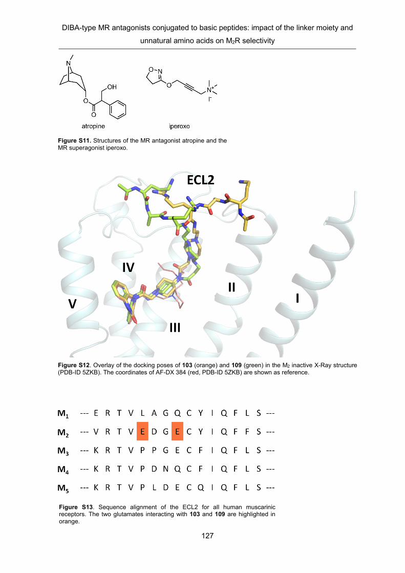
DIBA-type MR antagonists conjugated to basic peptides: impact of the linker moiety and
unnatural amino acids on M2R selectivity
127
Figure S11. Structures of the MR antagonist atropine and the MR superagonist iperoxo.
Figure S12. Overlay of the docking poses of 103 (orange) and 109 (green) in the M2 inactive X-Ray structure (PDB-ID 5ZKB). The coordinates of AF-DX 384 (red, PDB-ID 5ZKB) are shown as reference.
Figure S13. Sequence alignment of the ECL2 for all human muscarinic receptors. The two glutamates interacting with 103 and 109 are highlighted in orange.

Chapter 2
128
Figure S14. Surface presentation of the crystal structures of the M1R (PDB-ID 5CXV), M2R (PDB-ID 5ZKB), M3R (PDB-ID 4U15), M4R (PDB-ID 5DSG) and M5R (PDB-ID 6OL9) without (apo) bound ligand and with bound 103 and 109 (induced-fit docking). In the extracellular region of the receptors, the anionic deprotonated carboxylic groups of glutamates (E) and aspartates (D) are shown in red and the cationic protonated amine/guanidine groups of lysines (K) and arginines (R) are shown in blue. In the case of all MR subtypes, the headgroup (piperidinylacetyl-dibenzodiazepinone moiety) of 103 and 109 was located in the orthosteric binding site, albeit adapting different orientations. In the case of the M2R, the peptide moieties of 103 and 109 show interactions with two Glu residues in ECL2, which are absent in the M1 and M4 receptor (cf. 2.5. Supplementary Figure S13).

DIBA-type MR antagonists conjugated to basic peptides: impact of the linker moiety and
unnatural amino acids on M2R selectivity
129
Figure S15. Chemical structures of 103, 109 and AF-DX 384. 103 and 109 are shown in the protonation state defined for Induced-fit docking.
2. Analytical data of compound 6
Compound 6 was synthesized according to the reported procedure.35 1H-NMR (600.3 MHz,
DMSO-d6): δ (ppm) 1.35-1.46 (m, 2H), 1.52-1.64 (m, 2H), 1.69-1.77 (m, 2H), 1.79-1.88 (m,
1H), 1.91-2.06 (m, 2H), 2.63 (d, J 6.8 Hz, 2H), 2.71-2.81 (m, 1H), 2.81-2.97 (m, 4H), 2.98-
3.32 (m, 1H), 3.33-3.45 (m, 3H), 4.11 (brs, 2H), 6.58-6.63 (m, 1H), 6.72-6.77 (m, 1H), 6.99-
7.03 (m, 1H), 7.12 (t, J 7.7 Hz, 1H), 7.44-7.50 (m, 2H), 7.66 (t, J 7.9 Hz, 1H), 7.76-7.79 (m,
1H), 7.91-7.94 (m, 1H), 7.95-7.97 (m, 2H), 8.01 (t, J 1.9 Hz, 1H), 9.13-9.37 (m, 1H) (note:
one exchangeable proton could not be found). 13C-NMR (150.9 MHz, DMSO-d6): δ (ppm)
25.01, 25.98, 28.70, 30.76, 34.48, 35.77, 40.85, 42.20, 45.36, 48.66, 62.53, 116.03 (q, J
297 Hz, TFA), 116.03, 116.24, 119.74, 126.10, 126.81, 127.60, 127.74, 127.83, 130.15,
130.36, 130.51, 131.86, 133.71, 134.35, 138.16, 138.21, 143.26, 146.40, 158.23 (q, J 35
Hz, carbonyl group of TFA),162.30, 168.37. HRMS (ESI): m/z [M+H]+ calcd. for
[C30H35ClN3O3S]+: 552.2082, found 552.2095. RP-HPLC (220 nm): 98% (tR = 8.1 min, k =
9.7). C30H34ClN3O3S . C4H2F6O4 (552.1300 + 228.04).

Chapter 2
130
2.6. References
1. M. P. Caulfield and N. J. M. Birdsall, International Union of Pharmacology. XVII. Classification of muscarinic acetylcholine receptors, Pharmacol. Rev., 1998, 50, 279-290.
2. A. C. Kruse, B. K. Kobilka, D. Gautam, P. M. Sexton, A. Christopoulos and J. Wess, Muscarinic acetylcholine receptors: novel opportunities for drug development, Nat. Rev. Drug Discov., 2014, 13, 549-560.
3. J. Wess, R. M. Eglen and D. Gautam, Muscarinic acetylcholine receptors: mutant mice provide new insights for drug development, Nat. Rev. Drug Discov., 2007, 6, 721-733.
4. K. J. Broadley and D. R. Kelly, Muscarinic Receptor Agonists and Antagonists, Molecules, 2001, 6, 142-193.
5. R. M. Eglen, Muscarinic Receptor Subtype Pharmacology and Physiology, Prog. Med. Chem., 2005, 43, 105-136.
6. A. R. Bolbecker and A. Shekhar, Muscarinic agonists and antagonists in schizophrenia: recent therapeutic advances and future directions, Handb. Exp. Pharmacol., 2012, 208, 167-190.
7. R. M. Eglen, Overview of muscarinic receptor subtypes, Handb. Exp. Pharmacol., 2012, 208, 3-28.
8. D. Erskine, J. P. Taylor, G. Bakker, A. J. H. Brown, T. Tasker and P. J. Nathan, Cholinergic muscarinic M1 and M4 receptors as therapeutic targets for cognitive, behavioural, and psychological symptoms in psychiatric and neurological disorders, Drug Discov. Today, 2019, 24, 2307-2314.
9. D. J. Foster, D. L. Choi, P. J. Conn and J. M. Rook, Activation of M1 and M4 muscarinic receptors as potential treatments for Alzheimer's disease and schizophrenia, Neuropsychiatr. Dis. Treat., 2014, 10, 183-191.
10. A. C. Kruse, A. M. Ring, A. Manglik, J. Hu, K. Hu, K. Eitel, H. Hubner, E. Pardon, C. Valant, P. M. Sexton, A. Christopoulos, C. C. Felder, P. Gmeiner, J. Steyaert, W. I. Weis, K. C. Garcia, J. Wess and B. K. Kobilka, Activation and allosteric modulation of a muscarinic acetylcholine receptor, Nature, 2013, 504, 101-106.
11. J. Antony, K. Kellershohn, M. Mohr-Andra, A. Kebig, S. Prilla, M. Muth, E. Heller, T. Disingrini, C. Dallanoce, S. Bertoni, J. Schrobang, C. Trankle, E. Kostenis, A. Christopoulos, H. D. Holtje, E. Barocelli, M. De Amici, U. Holzgrabe and K. Mohr, Dualsteric GPCR targeting: a novel route to binding and signaling pathway selectivity, FASEB J., 2009, 23, 442-450.
12. V. A. Avlani, C. J. Langmead, E. Guida, M. D. Wood, B. G. Tehan, H. J. Herdon, J. M. Watson, P. M. Sexton and A. Christopoulos, Orthosteric and allosteric modes of interaction of novel selective agonists of the M1 muscarinic acetylcholine receptor, Mol. Pharmacol., 2010, 78, 94-104.
13. N. J. M. Birdsall and S. Lazareno, Allosterism at muscarinic receptors: ligands and mechanisms, Mini-Rev. Med. Chem., 2005, 5, 523-543.
14. A. Bock, N. Merten, R. Schrage, C. Dallanoce, J. Batz, J. Klockner, J. Schmitz, C. Matera, K. Simon, A. Kebig, L. Peters, A. Muller, J. Schrobang-Ley, C. Trankle, C. Hoffmann, M. De Amici, U. Holzgrabe, E. Kostenis and K. Mohr, The allosteric vestibule of a seven transmembrane helical receptor controls G-protein coupling, Nat. Commun., 2012, 3, 1044.
15. A. Christopoulos, Advances in G protein-coupled receptor allostery: from function to structure, Mol. Pharmacol., 2014, 86, 463-478.

DIBA-type MR antagonists conjugated to basic peptides: impact of the linker moiety and
unnatural amino acids on M2R selectivity
131
16. M. De Amici, C. Dallanoce, U. Holzgrabe, C. Trankle and K. Mohr, Allosteric ligands for G protein-coupled receptors: a novel strategy with attractive therapeutic opportunities, Med. Res. Rev., 2010, 30, 463-549.
17. T. Disingrini, M. Muth, C. Dallanoce, E. Barocelli, S. Bertoni, K. Kellershohn, K. Mohr, M. De Amici and U. Holzgrabe, Design, Synthesis, and Action of Oxotremorine-Related Hybrid-Type Allosteric Modulators of Muscarinic Acetylcholine Receptors, J. Med. Chem., 2006, 49, 366-372.
18. C. K. Jones, A. E. Brady, A. A. Davis, Z. Xiang, M. Bubser, M. N. Tantawy, A. S. Kane, T. M. Bridges, J. P. Kennedy, S. R. Bradley, T. E. Peterson, M. S. Ansari, R. M. Baldwin, R. M. Kessler, A. Y. Deutch, J. J. Lah, A. I. Levey, C. W. Lindsley and P. J. Conn, Novel selective allosteric activator of the M1 muscarinic acetylcholine receptor regulates amyloid processing and produces antipsychotic-like activity in rats, J. Neurosci., 2008, 28, 10422-10433.
19. A. Kebig, E. Kostenis, K. Mohr and M. Mohr-Andra, An optical dynamic mass redistribution assay reveals biased signaling of dualsteric GPCR activators, J. Recept. Signal Transduct., 2009, 29, 140-145.
20. J. R. Lane, P. M. Sexton and A. Christopoulos, Bridging the gap: bitopic ligands of G-protein-coupled receptors, Trends Pharmacol. Sci., 2013, 34, 59-66.
21. K. Mohr, C. Trankle, E. Kostenis, E. Barocelli, M. De Amici and U. Holzgrabe, Rational design of dualsteric GPCR ligands: quests and promise, Br. J. Pharmacol., 2010, 159, 997-1008.
22. T. Steinfeld, M. Mammen, J. A. Smith, R. D. Wilson and J. R. Jasper, A novel multivalent ligand that bridges the allosteric and orthosteric binding sites of the M2 muscarinic receptor, Mol. Pharmacol., 2007, 72, 291-302.
23. C. Valant, K. J. Gregory, N. E. Hall, P. J. Scammells, M. J. Lew, P. M. Sexton and A. Christopoulos, A novel mechanism of G protein-coupled receptor functional selectivity. Muscarinic partial agonist McN-A-343 as a bitopic orthosteric/allosteric ligand, J. Biol. Chem., 2008, 283, 29312-29321.
24. K. Mohr, J. Schmitz, R. Schrage, C. Trankle and U. Holzgrabe, Molecular alliance-from orthosteric and allosteric ligands to dualsteric/bitopic agonists at G protein coupled receptors, Angew. Chem. Int. Ed., 2013, 52, 508-516.
25. V. Nawaratne, K. Leach, C. C. Felder, P. M. Sexton and A. Christopoulos, Structural determinants of allosteric agonism and modulation at the M4 muscarinic acetylcholine receptor: identification of ligand-specific and global activation mechanisms, J. Biol. Chem., 2010, 285, 19012-19021.
26. X. Chen, J. Klockner, J. Holze, C. Zimmermann, W. K. Seemann, R. Schrage, A. Bock, K. Mohr, C. Trankle, U. Holzgrabe and M. Decker, Rational design of partial agonists for the muscarinic m1 acetylcholine receptor, J. Med. Chem., 2015, 58, 560-576.
27. J. Schmitz, D. van der Mey, M. Bermudez, J. Klockner, R. Schrage, E. Kostenis, C. Trankle, G. Wolber, K. Mohr and U. Holzgrabe, Dualsteric muscarinic antagonists--orthosteric binding pose controls allosteric subtype selectivity, J. Med. Chem., 2014, 57, 6739-6750.
28. R. Matucci, M. Nesi, M. V. Martino, C. Bellucci, D. Manetti, E. Ciuti, A. Mazzolari, S. Dei, L. Guandalini, E. Teodori, G. Vistoli and M. N. Romanelli, Carbachol dimers as homobivalent modulators of muscarinic receptors, Biochem. Pharmacol., 2016, 108, 90-101.
29. C. G. Gruber, A. Pegoli, C. Müller, L. Grätz, X. She and M. Keller, Differently fluorescence-labelled dibenzodiazepinone-type muscarinic acetylcholine receptor ligands with high M2R affinity, RSC Med. Chem., 2020, 11, 823-832.
30. M. Keller, C. Trankle, X. She, A. Pegoli, G. Bernhardt, A. Buschauer and R. W. Read, M2 Subtype preferring dibenzodiazepinone-type muscarinic receptor ligands:

Chapter 2
132
Effect of chemical homo-dimerization on orthosteric (and allosteric?) binding, Biorg. Med. Chem., 2015, 23, 3970-3990.
31. A. Pegoli, X. She, D. Wifling, H. Hubner, G. Bernhardt, P. Gmeiner and M. Keller, Radiolabeled Dibenzodiazepinone-Type Antagonists Give Evidence of Dualsteric Binding at the M2 Muscarinic Acetylcholine Receptor, J. Med. Chem., 2017, 60, 3314-3334.
32. X. She, A. Pegoli, C. G. Gruber, D. Wifling, J. Carpenter, H. Hübner, J. Wan, G. Bernhardt, P. Gmeiner, N. D. Holliday and M. Keller, Red-emitting dibenzodiazepinone derivatives as fluorescent dualsteric probes for the muscarinic acetylcholine M2 receptor, J. Med. Chem., 2020, 63, 4133-4154.
33. X. She, A. Pegoli, J. Mayr, H. Hubner, G. Bernhardt, P. Gmeiner and M. Keller, Heterodimerization of Dibenzodiazepinone-Type Muscarinic Acetylcholine Receptor Ligands Leads to Increased M2R Affinity and Selectivity, ACS Omega, 2017, 2, 6741-6754.
34. A. Pegoli, D. Wifling, C. G. Gruber, X. She, H. Hubner, G. Bernhardt, P. Gmeiner and M. Keller, Conjugation of Short Peptides to Dibenzodiazepinone-Type Muscarinic Acetylcholine Receptor Ligands Determines M2R Selectivity, J. Med. Chem., 2019, 62, 5358-5369.
35. M. S. Gitler, R. C. Reba, V. I. Cohen, W. J. Rzeszotarski and J. Baumgold, A novel m2-selective muscarinic antagonist: binding characteristics and autoradiographic distribution in rat brain, Brain Res., 1992, 582, 253-260.
36. J. W. Clader, W. Billard, H. Binch, L.-Y. Chen, G. Crosby, R. A. Duffy, J. Ford, J. A. Kozlowski, J. E. Lachowicz, S. Li, C. Liu, S. W. McCombie, S. Vice, G. Zhou and W. J. Greenlee, Muscarinic M2 antagonists: anthranilamide derivatives with exceptional selectivity and in vivo activity, Biorg. Med. Chem., 2004, 12, 319-326.
37. V. I. Cohen, J. Baumgold, B. Jin, R. De la Cruz, W. J. Rzeszotarski and R. C. Reba, Synthesis and structure-activity relationship of some 5-[[[(dialkylamino) alkyl]-1-piperidinyl] acetyl]-10, 11-dihydro-5H-dibenzo [b, e][1, 4] diazepin-11-ones as M2-selective antimuscarinics, J. Med. Chem., 1993, 36, 162-165.
38. C. Höring, U. Seibel, K. Tropmann, L. Grätz, D. Mönnich, S. Pitzl, G. Bernhardt, S. Pockes, A. Strasser, A dynamic, split-luciferase-based mini-G protein sensor to functionally characterize ligands at all four histamine receptor subtypes, Int. J. Mol. Sci., (2020, under review).
39. J. H. Gaddum, Quantitative effects of antagonistic drugs, J. Physiol. (Cambridge, U. K.), 1937, 89, 7-9P.
40. D.M. Thal, B. Sun, D. Feng, V. Nawaratne, K. Leach, C.C. Felder, M.G. Bures, D.A. Evans, W.I. Weis, P. Bachhawat, T.S. Kobilka, P.M. Sexton, B.K. Kobilka, A. Christopoulos, Crystal structures of the M1 and M4 muscarinic acetylcholine receptors, Nature, 53, 2016, 335-340.
41. K. Haga, A.C. Kruse, H. Asada, T. Yurugi-Kobayashi, M. Shiroishi, C. Zhang, W.I. Weis, T. Okada, B.K. Kobilka, T. Haga, T. Kobayashi, Structure of the human M2 muscarinic acetylcholine receptor bound to an antagonist, Nature, 482, 2012, 547-551.
42. R. Suno, S. Lee, S. Maeda, S. Yasuda, K. Yamashita, K. Hirata, S. Horita, M.S. Tawaramoto, H. Tsujimoto, T. Murata, M. Kinoshita, M. Yamamoto, B.K. Kobilka, N. Vaidehi, S. Iwata, T. Kobayashi, Structural insights into the subtype-selective antagonist binding to the M2 muscarinic receptor, Nat. Chem. Biol., 14, 2018, 1150-1158.
43. Y.-C. Cheng, Prusoff, W. H., Relation between the inhibition constand (Ki) and the concentration of inhibitor which causes fifty per cent inhibition (IC50) of an enzymic reaction, Biochem. Pharmacol., 1973, 22, 3099-3108.

133

134

135
CHAPTER 3
Further DIBA-peptide conjugates and stability studies
in human plasma

Chapter 3
136
The DIBA-peptide conjugates presented in this chapter were published together with DIBA-
peptide conjugates prepared by Dr. Andrea Pegoli in the Journal of Medicinal Chemistry
prior to the submission of this thesis:
A. Pegoli, D. Wifling, C.G. Gruber, X. She, H. Hubner, G. Bernhardt, P. Gmeiner, M. Keller,
Conjugation of Short Peptides to Dibenzodiazepinone-Type Muscarinic Acetylcholine
Receptor Ligands Determines M2R Selectivity, J. Med. Chem., 62 (2019) 5358-5369.
Author contributions:
A.P., D.W., and C.G.G. contributed equally. A.P., C.G.G., M.K., and X.S. performed the
syntheses and the analytical characterization of chemical compounds. A.P., C.G.G., and
M.K. performed radioligand competition binding experiments and analyzed the data. M.K.
initiated and planned the project. M.K. and G.B. supervised the research. M.K., A.P., D.W.,
G.B., and C.G.G. wrote the manuscript.
The rationale of the DIBA-peptide conjugation approach, which is aiming at highly selective
dualsteric M2R ligands, was described in Chapter 2. The series of DIBA-peptide conjugates
described in Chapter 2 represents the continuation of the novel approach reported by Pegoli
et al. in 2019.1 In addition to the determination of M1-M5 receptor affinities in radioligand
competition binding assays, one of the six presented dibenzodiazepinone derivatives as
well as the DIBA-peptide conjugates UR-AP148 (3)1 and UR-AP158 (4)1 (for structure see
Figure 2.1) were investigated with respect to stability in human plasma. Results and
discussions

Further DIBA-peptide conjugates and stability studies in human plasma
137
3.1. Results and Discussion
3.1.1. Chemistry
The N-terminally acetylated peptides 140-143 were synthesized on a chlorotrityl-resin
(Fmoc strategy) according to the procedure procedure described in Chapter 2. Coupling of
140-143 to amine precursor 52 via the peptide C-termini, followed by cleavage of protecting
groups, afforded the DIBA-peptide conjugates 144-147 (Scheme 3.1). Coupling of Boc-L-
Arg(Pbf)-OH (67) to amine 52, followed by deprotection, afforded 148. Acetylation of 148 at
the α-amino group to give 149 (Scheme 3.1).
3.1.2. Radioligand competition binding studies
M1-M5 receptor affinities of 144-149 were determined by competition binding with the
orthosteric MR antagonist [3H]NMS using genetically engineered CHO cells (Table 3.1).
Radioligand displacement curves are shown Figure 3.1. The M2R selectivity of 144-149 over
the M1R and M4R was less pronounced than towards the M3R and M5R. Compared to the
structurally closely related DIBA-peptide conjugate 3, the M2R selectivity of 144-149 was
generally lower (Table 3.1). This demonstrated that minor changes in the peptide moiety
can have a marked effect on MR binding and M2R selectivity, respectively.
Scheme 3.1. Synthesis of DIBA-peptide conjugates 144-147 and dibenzodiazepinone derivatives 148 and 149. Reagents and conditions: (1) EDC, HOBt, DMF, 5 °C, 3 h, (2) TFA/H2O (95:5 v/v), rt, 3 h; (d) acetic anhydride, DIPEA, DMF, rt, 2 h, 78%; (e) DIPEA, DMF, rt, 30 min.

Chapter 3
138
Figure 3.1. Displacement curves obtained from competition binding experiments with [3H]NMS (0.2 nM (M1R, M2R, M3R), 0.1 nM (M4R) or 0.3 nM (M5R)) and 144-149 at intact CHO-hMxR cells (x = 1-5). Data represent mean values ± SEM from at least three independent experiments (each performed in triplicate).

Further DIBA-peptide conjugates and stability studies in human plasma
139
3.1.3. Stability studies in human plasma
Stabilities in human plasma/PBS (1:2 v/v) were studied for the DIBA-peptide conjugates 3,
4 and 146, which proved to be highly stable (> 99% intact compound after 24 h, Table 3.2).
LC-MS analysis revealed that the Lys-Gly amide bond in 146 was the major cleavage site
(structure of 146 see Scheme 3.1 or Table 3.1). Recoveries of the DIBA-peptide conjugates
3, 4 and 146 from human plasma/PBS (1:2 v/v) were determined for two different
concentrations (80 µM and 4 µM). In the case of the concentration of 80 µM, recoveries of
3, 4 and 146 were in the range of 64-95%. The ratios of DIBA-peptide conjugate recovery
over recovery of internal standard (150) are shown in Table 3.3 (see experimental section).
Table 3.1. M1-M5 receptor binding data (pKi values) of compounds 3 and 144-149.a
cmpd.
structure (for A cf. Scheme 3.1)
pKi
selectivity toward M2R (ratios Ki (M1,3,4,5R)/Ki
(M2R)) M1R M2R M3R M4R M5R M1 M3 M4 M5
3 Ac-Lys-Arg-NH-A 7.38 ± 0.14
8.97 ± 0.02
5.17 ± 0.07
7.21 ± 0.06
6.37 ± 0.07 49 6500 60 400
144 Ac-Arg-Lys-NH-A 7.02 ± 0.08
7.92 ± 0.18
5.26 ± 0.13
6.59 ± 0.06
6.30 ± 0.19 6.5 400 17 39
145 Ac-Arg-Arg-NH-A 7.70 ± 0.11
8.77 ± 0.03
6.02 ± 0.19
7.53 ± 0.16
6.79 ± 0.05 12 710 19 97
146 Ac-Arg-Lys-Gly-NH-A 7.64 ± 0.05
8.83 ± 0.10
6.11 ± 0.16
7.76 ± 0.03
6.44 ± 0.13 15 620 11 260
147 Ac-Lys-Arg-Gly-NH-A 7.49 ± 0.12
8.38 ± 0.05
6.19 ± 0.15
7.56 ± 0.10
6.70 ± 0.10 8.2 180 6.9 49
148 H-Arg-NH-A 6.97 ± 0.07
8.27 ± 0.12
5.90 ± 0.17
7.25 ± 0.11
5.62 ± 0.11 18 260 9.8 410
149 Ac-Arg-NH-A 7.01 ± 0.09
7.95 ± 0.09
5.83 ± 0.14
6.83 ± 0.10
6.01 ± 0.11 8.4 140 13 86
aDetermined by competition binding with [3H]NMS (Kd values/applied concentrations: M1, 0.17/0.2 nM; M2, 0.10/0.2 nM; M3, 0.12/ 0.2 nM; M4, 0.052/0.1 nM; M5, 0.20/0.3 nM) at live CHO-hMxR cells (x = 1-5) at 23 °C.

Chapter 3
140
3.2. Experimental section
3.2.1. General experimental conditions
Acetonitrile (HPLC gradient grade) was obtained from Honeywell (Seelze, Germany) or
Sigma-Aldrich (Taufkirchen, Germany). DMF, atropine, EDC, DIPEA, HFIP, 1-methyl-D-
tryptophan (150) and TFA were purchased from Sigma-Aldrich. Absolute EtOH was
obtained from Honeywell. CH2Cl2, HOBt, DMF for peptide synthesis and NMP for peptide
synthesis were obtained from Acros Organics/Fisher Scientific (Nidderau, Germany). Boc-
Arg(Pbf)-OH (67) was purchased from Carbolution Chemicals (St. Ingbert, Germany).
Fmoc-Arg(Pbf)-OH, Fmoc-Lys(Boc)-OH, acetic anhydride and solid supports for SPPS ( H-
Arg(Pbf)-, H-Lys(Boc)-, and H-Gly-2-ClTrt PS resin) were purchased from Merck Chemicals
(Darmstadt, Germany). Deuterated solvents were from Deutero (Kastellaun, Germany).
HBTU and piperidine were from Iris Biotech (Marktredwitz, Germany). The synthesis of
compound 52 was described elsewhere.2 Millipore water was used for the preparation of
buffers and HPLC eluents. 1.5- or 2-mL polypropylene reaction vessels with screw cap
(Süd-Laborbedarf, Gauting, Germany) were used for small scale reactions, for the
investigation of plasma stabilities and for the preparation and storage of stock solutions.
Temperature-controlled reactions and plasma stabilities were performed in 1.5-mL reaction
vessels using a Thermocell Mixing Block MB-102 from Bioer (Hangzhou, China). NMR
spectra were recorded on a Bruker Avance III HD 600 equipped with a cryogenic probe
(14.1 T, 1H: 600.1 MHz, 13C: 150.9 MHz) (Bruker, Karlsruhe, Germany). Abbreviations for
the multiplicities of the signals are s (singlet), d (doublet), t (triplet), m (multiplet) and brs
(broad-singlet). HRMS was performed on an Agilent 6540 UHD Accurate-Mass Q-TOF
LC/MS system (Agilent Technologies, Santa Clara, CA USA) using an ESI source. Optical
rotations at 589 nm (Na-D line) were measured on a. Preparative HPLC was performed with
a system from Knauer (Berlin, Germany), composed of two K-1800 pumps and a K-2001
detector, or with a Prep 150 LC System from Waters (Eschborn, Germany) consisting of a
Table 3.2. Stabilities of 3, 4 and 146 in human plasma/PBS (1:2 v/v) at 37 °C.a
compd.
% intact peptide in plasma after the specified incubation times 1 h 6 h 24 h 48 h
3 > 99 > 99 > 99 > 99 4 99 ± 3 > 99 > 99 > 99
146 > 99 > 99 > 99 83 ± 6 aThe initial concentrations of compounds 3, 4 and 146 in plasma/PBS (1:2 v/v) were 100 µM. Data represent the mean (± SEM) of three independent experiments (SEM not given when no decomposition was detected). LC-MS analysis revealed the Lys-Gly amide bond in 146 as the major cleavage site (structure of 146 cf. Scheme 3.1 or Table 3.1).

Further DIBA-peptide conjugates and stability studies in human plasma
141
2545 Binary Gradient Module, a 2489 UV/Visible Detector and a Waters Fraction Collector
III. When using acetonitrile as organic solvent, a Kinetex-XB C18 (5 μm, 250 mm × 21 mm;
Phenomenex, Aschaffenburg, Germany) was used as stationary phase at a flow rate of 18
mL/min. Mixtures of 0.1% aq TFA and acetonitrile or mixtures of 0.1% aq TFA and MeOH
were used as mobile phase. The detection wavelength was 220 nm. The solvent of collected
fractions was removed by lyophilization. Analytical HPLC analysis was performed with a
system from Agilent Technologies composed of a 1290 Infinity binary pump equipped with
a degasser, a 1290 Infinity autosampler, a 1290 Infinity thermostated column compartment,
a 1260 Infinity diode array detector, and a 1260 Infinity fluorescence detector. A Kinetex-
XB C18 (2.6 μm, 100 × 3 mm; Phenomenex) was used as stationary phase at a flow rate
of 0.6 mL/min. Mixtures of 0.04% aq TFA (A) and acetonitrile (B) were used as mobile
phase. The following linear gradient was applied: 0-20 min: A/B 90:10-70:30, 20-22 min:
70:30-5:95, 22-26 min: 5:95.For all analytical HPLC runs the oven temperature was set to
25 °C, the injection volume was 20 μL and detection was performed at 220 nm. Compound
concentrations were between 80 and 150 μM.
3.2.2. Compound characterization
Target compounds (144-149) were characterized by 1H-NMR, 13C-NMR and 2D-NMR (1H-
COSY, HSQC, HMBC) spectroscopy, HRMS and RP-HPLC analysis. The purity (HPLC,
220 nm) of all target compounds amounted to ≥97%).
Annotation concerning the 1H- and 13C-NMR spectra of 144-149: due to a slow rotation
about the exocyclic dibenzodiazepinone amide group on the NMR time scale, two isomers
(ratios provided in the experimental protocols) were evident in the 1H- and 13C-NMR spectra.
Annotation concerning the 1H-NMR spectra (solvent: DMSO-d6) of 144-149: in order to
integrate signals interfering with the broad water signal at ca 3.5 ppm, spectra were
additionally recorded in DMSO-d6/D2O (4:1 v/v) (spectra shown in Appendix).
3.2.3. Screening for pan-assay interference compounds (PAINS).
The search for PAINS in 144-149 via the public tool
http://zinc15.docking.org/patterns/home3 gave no hits.

Chapter 3
142
3.2.4. Solid-phase peptide synthesis (SPPS)
SPPS was performed according to a previously described procedure.1 Peptides 140-143 were used in the next step without purification. The chemical identities and purities of 140-
143 were assessed by LC-HRMS (data not shown).
3.2.5. General procedure for the synthesis of compounds 144-147
The coupling of the side chain-protected peptides 140-143 to amine 52 was performed in
1.5- or 2-mL polypropylene reaction vessels with screw cap. HOBt (1.2 equiv.) and EDC
(1.2 equiv.) were added to a cooled (5 °C) solution of the respective side chain-protected
peptide (1 equiv.) in DMF (0.1-0.4 mL). A solution of 52 (free base, 1-1.2 equiv.) in DMF
(0.1 mL) was immediately added and stirring was continued at 5 °C for 3 h. 1% aq TFA (0.1-
0.4 mL, depending on the amount of DMF used to dissolve the peptide) was added and the
mixture was subjected to preparative HPLC to isolate the protected intermediates
(conditions not given). The latter were dissolved in TFA/H2O (95:5 v/v). The mixtures were
stirred at rt for 3 h, CH2Cl2 (ca 20 mL) was added and the volatiles were removed by
evaporation. The final products were purified by preparative HPLC.
3.2.6. Experimental synthetic protocols and analytical data of compounds 144-149
S)-2-((S)-2-Acetamido-5-guanidinopentanamido)-6-amino-N-(4-(1-(2-oxo-2-(11-oxo-10,11-dihydro-5H-dibenzo[b,e][1,4]diazepin-5-yl)ethyl)piperidin-4-yl)butyl)hexanamide tris(hydrotrifluoroacetate) (144)
Purification by preparative HPLC (gradient: 0-25 min: 0.1% aq TFA/acetonitrile 81:19-72:28,
tR = 8 min) yielded 144 as a white fluffy solid (16 mg, 0.015 mmol, 43%). Ratio of
configurational isomers evident in the NMR spectra: ca 1.5:1. 1H-NMR (600 MHz, DMSO-
d6): δ (ppm) 1.11-1.19 (m, 2H), 1.20-1.39 (m, 8H), 1.40-1.54 (m, 7H), 1.58-1.68 (m, 2H),
1.69-1.82 (m, 2H), 1.86 (s, 3H), 2.70-2.78 (m, 2H), 2.85-2.95 (s, 1H), 2.96-3.15 (m, 5H),
3.21-3.29 (m, 1H), 3.52-3.60 (m, 1H), 3.76 (d, 0.6H, J 16 Hz), 3.90 (d, 0.4H, J 16 Hz), 4.11-
4.23 (m, 2H), 4.33-4.49 (m, 1H), 7.03 (brs, 2H), 7.21-7.31 (m, 2H), 7.31-7.64 (m, 5H), 7.64-
7.94 (m, 9H), 8.05-8.10 (m, 1H), 9.56 (brs, 1H), 10.73 (s, 0.4H), 10.78 (s, 0.6H).13C-NMR
(150.9 MHz, MeOH-d4): δ (ppm) 22.24, 22.47, 23.21, 25.02, 26.55, 28.72, 28.86, 29.06,
31.40, 32.74, 34.93, 38.41, 38.64, 40.39, 52.27, 52.77, 53.22, 55.93, 121.87, 122.29,
124.87, 125.46, 127.25, 127.70, 128.25, 128.69, 128.95, 129.98, 130.35, 130.92, 131.51,
131.61, 133.02, 133.75, 134.66, 135.74, 140.98, 142.70, 156.77, 165.73, 166.03, 169.70,
171.10, 171.40 (note: 13C-signal of the exocyclic carbonyl group, attached to the
diazepinone ring, not apparent). HRMS (ESI): m/z [M+H]+ calcd. for [C38H57N10O5]+
Further DIBA-peptide conjugates and stability studies in human plasma
143
733.4508, found 733.4504. RP-HPLC (220 nm): 97% (tR = 6.4 min, k =7.4). C38H56N10O5 ·
C6H3F9O6 (732.93 + 342.07).
(S)-2-Acetamido-5-guanidino-N-((S)-5-guanidino-1-oxo-1-((4-(1-(2-oxo-2-(11-oxo-10,11-dihydro-5H-dibenzo[b,e][1,4]diazepin-5-yl)ethyl)piperidin-4-yl)butyl)amino)pentan-2-yl)pentanamide tris(hydrotrifluoroacetate) (145)
Purification by preparative HPLC (gradient: 0-25 min: 0.1% aq TFA/acetonitrile 81:19-72:28,
tR = 8 min) yielded 145 as a white fluffy solid (20 mg, 0.018 mmol, 52%). Ratio of
configurational isomers evident in the NMR spectra: ca 1.5:1. 1H-NMR (600 MHz, DMSO-
d6): δ (ppm) 1.14-1.26 (m, 4H), 1.28-1.57 (m, 11H), 1.57-1.69 (m, 2H), 1.70-1.83 (m, 2H),
1.85 (s, 3H), 2.80-2.95 (m, 1H), 2.95-3.02 (m, 2H), 3.02 -3.14 (m, 5H), 3.29-3.37 (m, 1H),
3.57-3.64 (m, 1H), 3.76 (d, 0.6H, J 16 Hz), 3.90 (d, 0.4H, J 16 Hz), 4.14-4.25 (m, 2H), 4.35-
4.47 (m, 1H), 7.01 (brs, 4H), 7.21-7.41 (m, 5H), 7.41-7.55 (m, 3H), 7.55-7.62 (m, 0.6H),
7.62-7.80 (m, 4.4H), 7.81-7.83 (m, 0.6H), 7.83-7.87 (m, 1H), 7.87-7.90 (m, 0.4H), 7.92-7.96
(m, 1H), 8.05-8.10 (m, 1H), 9.55 (brs, 1H), 10.73 (s, 0.4H), 10.78 (s, 0.6H). 13C-NMR (150.9
MHz, MeOH-d4): δ (ppm) 22.24, 23.22, 25.03, 25.07, 28.70, 28.87, 29.06, 29.22, 32.74,
34.92, 38.42, 40.31, 40.38, 52.10, 52.35, 52.78, 53.23, 55.93, 121.87, 122.29, 124.89,
125.47, 127.24, 127.70, 128.23, 128.71, 128.97, 129.56, 129.68, 130.00, 130.35, 130.85,
130.93, 131.55, 133.04, 133.76, 134.66, 135.75, 140.96, 156.75 (2 × guanidinium group),
157.8 (broad signal, carbonyl group of TFA), 165.73, 166.03, 169.68, 170.87, 171.45.
HRMS (ESI): m/z [M+H]+ calcd. for [C38H57N12O5]+ 761.4569, found 761.4556. RP-HPLC
(220 nm): 97% (tR = 6.8 min, k =7.9). C38H56N12O5 · C6H3F9O6 (760.95 + 342.07).
(S)-2-((S)-2-Acetamido-5-guanidinopentanamido)-6-amino-N-(2-oxo-2-((4-(1-(2-oxo-2-(11-oxo-10,11-dihydro-5H-dibenzo[b,e][1,4]diazepin-5-yl)ethyl)piperidin-4-yl)butyl)amino)ethyl)hexanamide tris(hydrotrifluoroacetate) (146)
Purification by preparative HPLC (gradient: 0-25 min: 0.1% aq TFA/acetonitrile 81:19-72:28,
tR = 8 min) yielded 146 as a white fluffy solid (16 mg, 0.014 mmol, 35%). Ratio of
configurational isomers evident in the NMR spectra: ca 1.5:1. 1H-NMR (600 MHz, DMSO-
d6): δ (ppm) 1.12-1.19 (m, 2H), 1.19-1.26 (m, 2H), 1.26-1.40 (m, 6H), 1.40-1.58 (m, 7H),
1.58-1.71 (m, 2H), 1.70-1.76 (m, 1H), 1.76-1.83 (m, 1H), 1.85 (s, 3H), 2.71-2.79 (m, 2H),
2.85-2.97 (m, 1H), 2.97-3.13 (m, 5H), 3.29-3.35 (m, 1H), 3.58-3.70 (m, 3H), 3.76 (d, 0.6H,
J 16 Hz), 3.90 (d, 0.4H, J 16 Hz), 4.15-4.27 (m, 2H), 4.33-4.48 (m, 1H), 7.06 (brs, 2H), 7.21-
7.57 (m, 6.6H), 7.57-7.62 (m, 0.4H), 7.63-7.80 (m, 7H), 7.80-7.84 (m, 0.6H), 7.86-7.91 (m,
0.4H), 8.02-8.09 (m, 2H), 8.09-8.14 (m, 1H), 9.56 (brs, 1H), 10.73 (s, 0.4H), 10.78 (s, 0.6H).

Chapter 3
144
13C-NMR (150.9 MHz, MeOH-d4): δ (ppm) 22.08, 22.42, 23.24, 24.96, 26.52, 28.67, 28.85,
29.10, 31.00, 32.74, 34.92, 38.43, 38.62, 40.39, 41.85, 52.20, 52.44, 52.76, 53.21, 55.89,
121.85, 122.27, 124.86, 125.44, 126.72, 127.21, 127.68, 128.23, 128.67, 128.95, 129.66,
129.98, 130.33, 130.90, 130.93, 131.51, 133.00, 133.73, 134.64, 135.73, 140.96, 156.76,
166.00, 168.31, 169.59, 171.60, 171.70 (note: 13C-signal of the exocyclic carbonyl group,
attached to the diazepinone ring, not apparent). HRMS (ESI): m/z [M+H]+ calcd. for
[C40H60N11O6]+ 790.4723, found 790.4730. RP-HPLC (220 nm): 98% (tR = 6.4 min, k =7.4).
C40H59N11O6 · C6H3F9O6 (789.98 + 342.07).
(S)-2-Acetamido-6-amino-N-((S)-5-guanidino-1-oxo-1-((2-oxo-2-((4-(1-(2-oxo-2-(11-oxo-10,11-dihydro-5H-dibenzo[b,e][1,4]diazepin-5-yl)ethyl)piperidin-4-yl)butyl)amino)ethyl)amino)pentan-2-yl)hexanamidetris (hydrotrifluoroacetate) (147)
Purification by preparative HPLC (gradient: 0-25 min: 0.1% aq TFA/acetonitrile 81:19-72:28,
tR = 8 min) yielded 147 as a white fluffy solid (21 mg, 0.019 mmol, 54%). Ratio of
configurational isomers evident in the NMR spectra: ca 1.5:1. 1H-NMR (600 MHz, DMSO-
d6): δ (ppm) 1.12-1.19 (m, 2H), 1.19-1.40 (m, 8H), 1.40-1.58 (m, 7H), 1.58-1.66 (m, 1H),
1.66-1-77 (m, 2H), 1.77-1.83 (m, 1H), 1.85 (s, 3H), 2.71-2.79 (m, 2H), 2.85-2.96 (m, 1H),
2.97-3.13 (m, 5H), 3.30-3.36 (m, 1H), 3.58-3.70 (m, 3.6H), 3.76 (d, 0.4H, J 17 Hz), 4.17-
4.25 (m, 2H), 4.35-4.47 (m, 1H), 7.01 (brs, 2H), 7.19-7.57 (m, 6.6H), 7.57-7.62 (m, 0.4H),
7.62-7.80 (m, 7H), 7.80-7.85 (m, 0.6H), 7.86-7.91 (m, 0.4H), 8.02-8.10 (m, 2H), 8.10-8.15
(m, 1H), 9.57 (brs, 1H), 10.73 (s, 0.4H), 10.78 (s, 0.6H). 13C-NMR (150.9 MHz, MeOH-d4):
δ (ppm) 22.35, 22.44, 23.26, 24.92, 26.65, 28.67, 28.80, 29.12, 31.09, 32.75, 34.94, 38.46,
38.70, 40.35, 41.88, 52.26, 52.44, 52.63, 52.77, 53.23, 55.93, 121.87, 122.30, 124.88,
125.46, 126.74, 127.26, 127.34, 127.71, 128.25, 128.71, 129.03, 129.66, 129.68, 130.00,
130.35, 130.93, 131.60, 133.03, 133.76, 134.66, 135.75, 139.42, 140.96, 156.77, 157.5
(broad signal, carbonyl group of TFA), 163.68, 164.18, 165.74, 166.02, 168.26, 169.57,
171.50, 172.03. HRMS (ESI): m/z [M+H]+ calcd. for [C40H60N11O6]+ 790.4723, found
790.4730. RP-HPLC (220 nm): 97% (tR = 6.5 min, k =7.5). C40H59N11O6 · C6H3F9O6 (789.98
+ 342.07).

Further DIBA-peptide conjugates and stability studies in human plasma
145
(S)-2-Amino-5-guanidino-N-(4-(1-(2-oxo-2-(11-oxo-10,11-dihydro-5H-dibenzo[b,e][1,4]diazepin-5-yl)ethyl)piperidin-4-yl)butyl)pentanamide tris(hydrotrifluoroacetate) (148)
Compound 148 was prepared from amine 52 (55.6 mg, 0.137 mmol) and compound 67 (60
mg, 0.114 mmol) according to the general procedure for the synthesis of 144-147.
Purification by preparative HPLC (gradient: 0-25 min: 0.1% aq TFA/acetonitrile 81:19-72:28,
tR = 7 min) yielded 148 as a white fluffy solid (67 mg, 0.074 mmol, 65%). Ratio of
configurational isomers evident in the NMR spectra: ca 1.5:1. 1H-NMR (600 MHz, DMSO-
d6): δ (ppm) 1.13-1.29 (m, 4H), 1.30-1.52 (m, 7H), 1.64-1.71 (m, 2H), 1.71-1.83 (m, 2H),
2.83-2.96 (m, 1H), 2.96-3.04 (m, 1H), 3.04-3.14 (m, 4H), 3.28-3.35 (m, 0.8H), 3.55-3-63 (m,
1.2H), 3.65-3.73 (m, 1H), 3.75 (d, 0.6H, J 17 Hz), 3.90 (d, 0.4H, J 17 Hz), 4.32-4.48 (m,
1H), 6.95-7.49 (m, 7H), 7.49-7.56 (m, 1.4H), 7.56-7.63 (m, 0.6H), 7.63-7.79 (m, 2H), 7.79-
7.92 (m, 2H), 8.11-8.24 (m, 3H), 8.41-8.48 (m, 1H), 9.57 (brs, 1H), 10.73 (s, 0.4H), 10.78
(s, 0.6H). 13C-NMR (150.9 MHz, MeOH-d4): δ (ppm) 22.32, 24.20, 28.31, 28.74, 28.98,
32.81, 34.95, 38.67, 40.08, 51.94, 52.80, 53.24, 55.96, 116.05 (TFA), 118.04 (TFA), 121.91,
122.34, 124.93, 125.51, 126.75, 127.27, 127.71, 128.75, 129.00, 129.71, 130.04, 130.38,
130.97, 131.63, 133.07, 133.80, 134.68, 135.77, 140.99, 156.84, 158.3 (q, J 32 Hz,
carbonyl group of TFA), 163.70, 164.35, 165.78, 166.07, 168.05, 171.50. HRMS (ESI): m/z
[M+H]+ calcd. for [C30H43N8O3]+ 563.3453, found 563.3462. RP-HPLC (220 nm): 99% (tR =
6.2 min, k =7.1). C30H42N8O3 · C6H3F9O6 (562.72 + 342.07).
(S)-2-Acetamido-5-guanidino-N-(4-(1-(2-oxo-2-(11-oxo-10,11-dihydro-5H-dibenzo[b,e][1,4]diazepin-5-yl)ethyl)piperidin-4-yl)butyl)pentanamide bis(hydrotrifluoroacetate) (149)
Acetyl anhydride (3.6 µL, 0.038 mmol) was added to a solution of 149 (25 mg, 0.032 mmol)
and DIPEA (28 µL, 0.16 mmol) in DMF (0.3 mL) and the mixture was stirred at rt for 2 h.
Isolation of the product by preparative HPLC (gradient: 0-25 min: 0.1% aq TFA/acetonitrile
81:19-72:28, tR = 9 min) afforded 149 as a white fluffy solid (20.8 mg, 0.025 mmol, 78%).
Ratio of configurational isomers evident in the NMR spectra: ca 1.5:1. 1H-NMR (600 MHz,
DMSO-d6): δ (ppm) 1.13-1.26 (m, 4H), 1.30-1.50 (m, 8H), 1.57-1.66 (m, 1H), 1.69-1.82 (m,
2H), 1.83 (s, 3H), 2.85-2.96 (m, 1H), 2.96-3.14 (m, 5H), 3.28-3.35 (m, 1H), 3.57-3.61 (m,
1H), 3.76 (d, 0.6H, J 16 Hz), 3.91 (d, 0.4H, J 16 Hz), 4.13-4.21 (m, 1H), 4.33-4.48 (m, 1H),
7.03 (brs, 2H), 7.20-7.31 (m, 2H), 7.31-7.39 (m, 1.6H), 7.39-7.49 (m, 1.4H), 7.49-7.55 (m,
1.4H), 7.55-7.62 (m, 0.6H), 7.62-7.79 (m, 3H), 7.79-7.85 (m, 0.6H), 7.85-7.93 (m, 1.4H),
7.95-8.02 (m, 1H), 9.55 (brs, 1H), 10.73 (s, 0.4H), 10.78 (s, 0.6H). 13C-NMR (150.9 MHz,
MeOH-d4): δ (ppm) 22.49, 25.16, 28.69, 29.09, 29.26, 32.74, 34.90, 38.31, 40.33, 52.13,

Chapter 3
146
52.65, 52.78, 53.23, 55.93, 116.05 (TFA), 118.10 (TFA), 121.88, 122.30, 124.89, 125.47,
126.74, 127.25, 127.70, 128.25, 128.70, 128.95, 129.69, 130.00, 130.36, 131.61, 133.03,
133.76, 134.68, 135.77, 140.97, 156.77, 158.2 (broad signal, carbonyl group of TFA),
163.67, 164.17, 165.73, 166.03, 169.19, 171.22. HRMS (ESI): m/z [M+H]+ calcd. for
[C32H45N8O4]+ 605.3558, found 605.3560. RP-HPLC (220 nm): 98% (tR = 8.0 min, k = 9.5).
C32H44N8O4 · C4H2F6O4 (604.76 + 228.05).
3.2.7. Cell culture
CHO-K9 cells, stably transfected with the DNA of human muscarinic receptors M1-M5
(obtained from Missouri S&T cDNA Resource Center; Rolla, MO) were cultured in HAM’s
F12 medium supplemented with fetal calf serum (Biochrom, Berlin, Germany) (10%) and
G418 (Biochrom) (750 μg/mL).
3.2.8. Radioligand competition binding
Competition binding experiments were performed as described in Chapter 2.
3.2.9. Investigation of the stability of 3, 4 and 146 in human plasma.
The metabolic stabilities of 3, 4 and 146 were investigated in human blood plasma/PBS pH
7.4 (1:2 v/v) (in the following referred to as plasma/PBS) at 37 °C. 1-Methyl-D-tryptophan
(150) was used as internal standard. As the purity (RP-HPLC, 220 nm) of 150 was < 95%
(data not shown) it was purified by preparative HPLC to give a purity of > 99%. Plasma was
obtained by the collection of human blood from a healthy donor in 5.5-mL heparinized
plasma-monovettes, followed by centrifugation at 1200 g at 4 °C for 10 min. The
supernatants were pooled in two 50-mL Falcon tubes and centrifuged again at 1200 g at 4
°C for 10 min. The plasma was aliquoted and stored at −80 °C.
For the addition of the compounds to plasma/PBS, 5 mM stock solutions in
acetonitrile/0.04% aqueous TFA (3:7 v/v) were used. Recoveries were determined for
concentrations of 80 μM and 4 μM, and an internal standard concentration of 10 μM. For
this purpose, 150 and the DIBA-peptide conjugates were added to plasma/PBS (total
volume: 70 μL), immediately followed by vortexing (ca 10 s) and precipitation of protein by
the addition of EtOH/acetonitrile (1:1 v/v) (140 μL). The mixture was vortexed for 5 min und
centrifuged at 16,100 g at 4 °C for 10 min. Aliquots (180 μL) of the supernatant were
transferred into 1.5-mL polypropylene reaction vessels containing 10% aqueous TFA (5 μL).
The volatiles were removed in a vacuum concentrator under reduced pressure at 40 °C (ca

Further DIBA-peptide conjugates and stability studies in human plasma
147
60 min), and the residue was taken up in acetonitrile/0.04% aqueous TFA (1:9 v/v) (90 μL)
by vortexing (2 min). The samples were filtrated using a 0.2-μm RC-membrane filter
(Phenomenex, Aschaffenburg, Germany) and analyzed by RP-HPLC using the analytical
HPLC system and the conditions as described under general experimental conditions, but
applying the following gradient: 0–12 min: A/B 90:10–70:30, 12–16 min: 70:30–5:95, 16–20
min: 5:95. On the day of an experiment four-point calibrations were performed for the
respective DIBA-peptide conjugates and the internal standard. Peak areas representing
100% recovery were obtained by analyzing 53.3 μM and 2.67 μM DIBA-peptide solutions
as well as a 6.67 μM solution of 150 (in duplicate each). All peak areas were transformed
into concentrations (μM) and percent recoveries of 3, 4, 146 and 150 were calculated based
on the average values of the 100% reference samples (see Table 3.3). Recovery ratios
were obtained by dividing the recovery of the DIBA-peptide conjugate by the recovery of
150 for each individual sample (n = 3 or 4, cf. Table 3.3).
For the investigation of the stabilities in plasma, the DIBA-peptide conjugates (3, 4, 146)
and 150 were added to plasma/PBS at a concentration of 100 μM and 10 μM, respectively
(in triplicate each). After 1, 6, 24 and 48 h of incubation under shaking at 37 °C, aliquots (70
μL) were taken and processed as described above for the determination of recoveries. On
the day of an experiment four-point calibrations were performed for the respective DIBA-
peptide conjugates and 150. Peak areas representing 100% recovery were obtained by
analyzing 66.7 μM ‘DIBA-peptide’ solutions and a 6.67 μM solution of 150 (in duplicate).
Based on the calibration, the peak areas of the 100% references and of the samples were
transformed into concentrations (μM). Recoveries of 3, 4 and 146 were calculated by
multiplying the recovery of 150, obtained for each individual sample, with the recovery ratio
obtained for the concentration of 80 µM (see above). The plasma concentrations of the
DIBA-peptide conjugates were obtained by dividing the determined ‘DIBA-peptide’
concentration by the respective recovery. Degradation (%) of 3, 4 and 146 was calculated
based on the average values of the 100% reference samples.
Note: recovery ratios obtained for a concentration of 4 µM were not used as 3, 4 and 146
exhibited high stabilities (lowest concentration in plasma ca 80 µM (146, 48 h)).

Chapter 3
148
3.2.10. Data processing
Retention (capacity) factors were calculated from retention times (tR) according to k =
(tR−t0)/t0 (t0 = dead time). Data from radioligand ([3H]NMS) competition binding assays were
processed as reported previously.4 Ki values for the calculation of relative MR affinities (M2R
selectivity, Table 3.1) were obtained by transforming pIC50 values from individual
competition binding experiments with [3H]NMS to IC50 values followed by conversion to Ki
values according to the Cheng-Prusoff equation5 and calculation of mean Ki values.
Table 3.3. Recoveries of DIBA-peptide conjugates 3, 4 and 146 from human plasma/PBS (1:2 v/v) for two different concentrations (80 µM and 4 µM), and ratios of ‘DIBA-peptide’ recovery over recovery of internal standard (150).
compd.
‘DIBA-peptide’ concentration 80 µM
‘DIBA-peptide’ concentration 4 µMa
recovery ‘DIBA-peptide’ (%)a
recovery I.S. (150) (%)a ratiob
recovery ‘DIBA-peptide’ (%)a
recovery I.S. (150) (%)a ratiob
3 75
91 86
101 101 102
0.75 0.90 0.85
(0.83 ± 0.05)
60 59 58 56
98 85 88 95
0.62 0.69 0.66 0.59
(0.64 ± 0.02)
4 64 95 79 81
96 100 98 99
0.67 0.95 0.81 0.82
(0.81 ± 0.06)
87 86 86 90
98 98 91 103
0.89 0.89 0.94 0.88
(0.90 ± 0.02)
146 75 76 72 72
87 89 95 92
0.87 0.86 0.75 0.78
(0.81 ± 0.03)
41 40 44
83 81 73
0.49 0.50 0.61
(0.53 ± 0.04)
aRecoveries of the DIBA-peptide conjugates and of the internal standard (I.S.) from human plasma/PBS (1:2 v/v) using a DIBA-peptide concentration of 80 µM or 4 µM and an I.S. concentration of 10 µM (five independent experiments). bRatios of DIBA-peptide recovery over recovery of I.S. calculated for individual experiments, as well as mean recovery ratios ± SEM (given in parenthesis).

Further DIBA-peptide conjugates and stability studies in human plasma
149
3.3. References
1. A. Pegoli, D. Wifling, C. G. Gruber, X. She, H. Hubner, G. Bernhardt, P. Gmeiner and M. Keller, J. Med. Chem., 2019, 62, 5358-5369.
2. M. Keller, C. Trankle, X. She, A. Pegoli, G. Bernhardt, A. Buschauer and R. W. Read, Biorg. Med. Chem., 2015, 23, 3970-3990.
3. C. Aldrich, C. Bertozzi, G. I. Georg, L. Kiessling, C. Lindsley, D. Liotta, K. M. Merz, Jr., A. Schepartz and S. Wang, J. Med. Chem., 2017, 60, 2165-2168.
4. A. Pegoli, X. She, D. Wifling, H. Hubner, G. Bernhardt, P. Gmeiner and M. Keller, J. Med. Chem., 2017, 60, 3314-3334.
5. Y.-C. Cheng, Prusoff, W. H., Biochem. Pharmacol., 1973, 22, 3099-3108.

150

151
CHAPTER 4
Fluorescently labeled dibenzodiazepionone-type
muscarinic M2R ligands

Chapter 4
152
Prior to the submission of this thesis, this chapter was published in RSC Med. Chem.:
Corinna G. Gruber, Andrea Pegoli, Christoph Müller, Lukas Grätz, Xueke She, Max Keller.
Differently fluorescence-labelled dibenzodiazepinone-type muscarinic acetylcholine
receptor ligands with high M2R affinity. RSC Med. Chem., 11 (2020), 823-832. Reproduced
with permission from RSC Medicinal Chemistry.
Author contributions:
C.G.G., A.P. and X.S. synthesized the fluorescent ligands. C.G.G. performed radiochemical
and flow cytometric binding experiments and analysed the data. M.K. initiated the project
and supervised the research. C.G.G. and M.K. wrote the manuscript. All authors have given
approval to the final version of the manuscript.

Fluorescently labelled dibenzodiazepinone-type MR ligands with high M2R affinity
153
4.1. Introduction
In the past decades, fluorescence-based techniques have been increasingly used to study
membrane receptors such as G-protein coupled receptors (GPCRs), including the analysis
of ligand-receptor interactions as well as the investigation of receptor expression, structure
and function.1-4 Besides fluorescently tagged receptors, fluorescent ligands represent
interesting molecular tools, which can also be used to study endogenously expressed
receptors.1 Therefore, there is a growing demand of suitable fluorescent GPCR ligands.
Compared with radioligands, fluorescent ligands exhibit several advantages, such as less
problems concerning safety risks, legal issues and waste disposal. Furthermore, they are
applicable to techniques, which have become routine in many laboratories, for instance
fluorescence microscopy and flow cytometry. Typically, fluorescent ligands are composed
of a pharmacophore, mediating receptor affinity, a spacer/linker moiety and the
fluorophore.1, 5 The design of fluorescent ligands is not trivial, e.g. with respect to high
receptor affinity and favorable physicochemical properties. Crucial factors are the type of
the fluorescent dye (size, lipophilicity, spectroscopic and bleaching properties, etc.) and the
point of attachment, structure and length of the linker.1, 6-11
Numerous fluorescent probes have been reported for GPCRs, for instance for histamine,12-
16 dopamine,17, 18 opioid,19-22 neuropeptide Y,23-28 adenosine,29 and neurotensin30, 31
receptors. Concerning muscarinic acetylcholine receptors (MRs), various fluorescent MR
ligands were reported, e.g. the BODIPY558/568-labelled derivative 151,32 derived from the
M1 subtype preferring MR antagonist pirenzepine, the BODIPY630/650-labelled tolterodine
derivative 152,33 the Alexa488-labelled telenzepine derived MR antagonist 153,34, 35 the
Cy3B-labelled telenzepine analog 154,34-36 and the lissamine rhodamine B-labelled AC-42
derivative 15537 (Figure 4.1; note: in the case of 151 and 153-155 further congeners,
containing the same pharmacophore but different fluorophores such as Cy3, Cy5 Cascade
Blue or 6-carboxyfluorescein and, in part, also different linkers, were reported32, 36-41). The
recent finding that replacement of the diethylamine moiety in the M2R preferring
dibenzodiazepinone-type MR antagonist DIBA42 (1, Figure 4.1) by bulky moieties was well
tolerated with respect to M2R binding,43-46 gave rise to prepare a series of DIBA-derived red-
emitting fluorescent MR ligands, comprising compounds 156 and 157 (Figure 4.1). Whereas
the indolinium-type cyanine dye containing ligands (e.g. 157) were investigated in
fluorescence-based techniques, the pyridinium-type cyanine dye containing probes (e.g.
156) were only characterized with respect to M1R-M5R affinity in radioligand competition
binding studies.47 In the present study, a new series of fluorescently labelled
dibenzodiazepinone-type MR ligands was prepared using various fluorescent dyes
(TAMRA, BODIPY630/650, pyridinium-type cyanine dyes Py-1 and Py-5). The probes were

Chapter 4
154
characterized with respect to M1R-M5R affinity (radioligand competition binding) and by flow
cytometric saturation binding studies, the latter including the reported ligand 156.
Figure 4.1. Structures and MR binding data of the reported fluorescent probes 151-155, being non-selective MR ligands (151-154) or showing a slight preference for the M1R (152),33 and structures and MR affinities of the M2R preferring dibenzodiazepinone-type MR ligands DIBA and the DIBA-derived fluorescent probes 156 and 157.47 (note: binding data for all MR subtypes were not reported in all cases). aTahtaoui et al. (reported Ki values were converted to pKi values);32 bJones et al. (reported Ki values were converted to pKi values);33 cHern et al. (reported Ki values were converted to pKi values);34 dNenasheva et al.;35 eDaval et al.;37 fGitler et al. (reported Ki values were converted to pKi values);42 gShe et al.47
4.2. Results and Discussion
4.2.1. Chemistry
A set of six fluorescent dibenzodiazepinone-type MR ligands (164-169) was prepared from
the previously reported amine-functionalized dibenzodiazepinone derivatives 15843 and
7246 using four different fluorescent dyes: 5-TAMRA succinimidyl ester (160), BODIPY
630/650 succinimidyl ester (161), and the pyrylium dyes Py-1 (162)48 and Py-5 (163)48 (cf.
Figure 4.2). Treatment of 158 with the fluorescent dyes 160-163 in the presence of DIPEA

Fluorescently labelled dibenzodiazepinone-type MR ligands with high M2R affinity
155
or triethylamine yielded the fluorescent ligands 164-167 (Scheme 4.1). Likewise, treatment
of 72 with 162 or 163 gave the fluorescent probes 168 and 169, respectively (Scheme 4.1).
Purification by preparative RP-HPLC afforded 164-169 with high purities (≥ 96%, HPLC
analysis at 220 nm). All fluorescent ligands were investigated with respect to their chemical
stability in PBS pH 7.4. Whereas compounds 164, 165, 168 and 169 showed no
decomposition within the incubation period of 48 h (Figure S2, 4.6 Supplementary
Information), compounds 166 and 167 showed minor decomposition after 24 h (after 24 h,
peak areas (220 nm) of 166 and 167 amounted to 94% and 90%, respectively, of the total
peak area).
Figure 4.2. Structures and M2R affinities of the previously described DIBA derived amine-functionalized precursors 158 and 72,43, 46 and structures of the fluorescent dyes 160-163, which were used for the preparation of the fluorescent MR ligands 164-169 (note: M2R binding data of 158, reported as pIC50 value,43 were re-analyzed to obtain the pKi value).

Chapter 4
156
Scheme 4.1. Synthesis of the fluorescent dibenzodiazepinone-type MR ligands 164-169, labelled with TAMRA (164, 169), BODIPY630/650 (165), pyrylium/pyridinium dye Py-1 (166, 168) or Py-5 (167). Reagents and conditions: (a): DIPEA, DMF, rt, 2 h, 31% (164), 23% (165), 16% (166), 23% (168), 66% (169); (b) triethylamine, DMF, rt, 2h, 28% (167).
4.2.2. Radioligand competition binding studies with [3H]NMS
M1R-M5R affinities of 164-169 were determined at intact CHO-hMxR cells (x = 1-5) using
the orthosteric non-selective MR antagonist [3H]NMS as radioligand. Compounds 164-169 were capable of completely displacing [3H]NMS from all MR subtypes (Figure 4.3 and Figure
S1, 4.6 Supplementary Information). The corresponding pKi values are listed in Table 1. All
fluorescent ligands 164-169 exhibited high M2R affinity (pKi values > 8.7). The TAMRA-
labelled compound 169 showed the highest M2R affinity (pKi: 9.62). For this fluorescent
ligand, sigmoidal [3H]NMS displacement curves (M1R-M5R) are shown in Figure 4.3B. As in
the case of the recently reported series of dibenzodiazepinone-type fluorescent ligands
including 156 and 157,47 164-169 showed no or very low preference for the M2R compared
to the M1 and M4 receptor, but moderate to pronounced M2R selectivity towards the M3 and
M5 subtypes (Table 4.1). Compound 169 exhibited the highest, but still moderate preference
for the M2R over the M1R and M4R.

Fluorescently labelled dibenzodiazepinone-type MR ligands with high M2R affinity
157
Figure 4.3. (A) Concentration-dependent effects of 164-169 on [3H]NMS (c = 0.2 nM) equilibrium M2R binding determined at intact CHO-hM2R cells. (B) Radioligand displacement curves obtained from competition binding experiments with [3H]NMS (0.2 nM (M1R, M2R, M3R), 0.1 nM (M4R) or 0.3 nM (M5R)) and compound 169 performed at intact CHO-hMxR cells (x = 1-5). M2R data are the same as in A. Data in A and B represent mean values ± SEM from two (169, M5R) or at least three independent experiments (each performed in triplicate).
Table 4.1. M1-M5 receptor binding data (pKi values) of compounds 156 and 164-169 obtained from radioligand competition binding studies, and M2R binding data (pKd values) from flow cytometric saturation binding experiments.
compd. pKia pKdb
M1R M2R M3R M4R M5R M2R
15647 7.86 8.52 6.83 8.02 6.41 8.63 ± 0.08
164 (UR-CG072) 8.21 ± 0.02 8.75 ± 0.07 6.89 ± 0.06 8.43 ± 0.07 6.56 ± 0.10 8.36 ± 0.09
165 (UR-CG073) 9.00 ± 0.13 9.16 ± 0.10 7.43 ± 0.13 9.38 ± 0.05 8.00 ± 0.08 8.41 ± 0.10
166 (UR-CG074) 8.15 ± 0.05 8.87 ± 0.08 7.16 ± 0.01 8.58 ± 0.08 7.39 ± 0.07 8.70 ± 0.04
167 (UR-AP175) 8.31 ± 0.04 9.04 ± 0.15 7.07 ± 0.04 8.37 ± 0.04 7.15 ± 0.06 8.74 ± 0.13
168 (UR-CG135) 8.32 ± 0.03 9.02 ± 0.10 7.20 ± 0.15 8.57 ± 0.01 6.90 ± 0.03 9.19 ± 0.03
169 (UR-MK342) 8.59 ± 0.10 9.62 ± 0.04 7.09 ± 0.06 9.01 ± 0.03 6.75 ± 0.05 8.86 ± 0.06
aDetermined by competition binding with [3H]NMS (Kd values/applied concentrations: M1, 0.17/0.2 nM; M2, 0.10/0.2 nM; M3, 0.12/ 0.2 nM; M4, 0.052/0.1 nM; M5, 0.20/0.3 nM) at whole CHO-hMxR cells (x = 1-5) at 23 °C. Means ± SEM from two (169, M5R) or at least three independent experiments (each performed in triplicate). bDetermined by flow cytometric saturation binding experiments at intact CHO-hM2R cells at 22 °C. Means ± SEM from two (169) or at least three independent experiments (performed in duplicate).

Chapter 4
158
4.2.3. Fluorescence properties
Excitation and corrected emission spectra of 164-169, recorded in PBS containing 1%
bovine serum albumin (BSA), are shown in supplementary Figure S3 (4.6. Supplementary
Infromation) and the respective excitation and emission maxima are summarized in
Table 4.2. It should be noted that compounds 156 and 167, both labelled with the
pyrylium/pyridinium-type fluorescent dye Py-5 are perfectly suited for an excitation with an
argon laser (488 nm), belonging to the standard equipment of many instruments.
Compound 165 is well suited for an excitation with the commonly used red diode laser (ca.
635 nm), and 164, 166, 168 and 169 require green light for an optimal excitation (cf. Table
4.2).
Table 4.2. Excitation and emission maxima of the fluorescent ligands 156, 164-169 determined in PBS containing 1% BSA. compd. dye λex/λem
15647 pyridinium cyanine (Py-5) 484/643
164 5-TAMRA 557/583
165 BODIPY 630/650 639/647
166 pyridinium cyanine (Py-1) 526/602
167 pyridinium cyanine (Py-5) 483/634
168 pyridinium cyanine (Py-1) 526/604
169 5-TAMRA 550/583
4.2.4. Flow cytometric M2R saturation binding studies
M2R affinities of the fluorescent ligands 156 and 164-169 were also determined by flow
cytometric saturation binding studies at intact CHO-hM2R cells at 22 °C using a
FACSContoII (156, 164-169) or a FACSCalibur (169) flow cytometer, both equipped with
two light sources (argon and red diode laser) (cf. Figure 4.4). For these experiments, an
incubation period of 2 h was applied because association binding experiments with recently
reported radio- and fluorescence labelled dibenzodiazepinone-type MR ligands at intact
CHO-hM2R cells or cell homogenates revealed that equilibrium was reached within 2 h.45-47
The obtained pKd values of compounds 156 and 164-169 were in good agreement with the
corresponding pKi values obtained from competition binding studies with [3H]NMS (Table
4.1).

Fluorescently labelled dibenzodiazepinone-type MR ligands with high M2R affinity
159
Figure 4.4. Representative saturation isotherms (specific binding, dashed line) obtained from flow cytometric saturation binding experiments performed with 156 and 164-169 at intact CHO-hM2R cells. Unspecific binding was determined in the presence of atropine (for structure see Figure S4, 4.5. Supplementary Information) used in 500- or 1000-fold excess. Cells were incubated with the fluorescent ligands at 22 °C in the dark for 2 h. Experiments were performed in duplicate. Measurements were performed on a FACSCantoII (156, 164-168) or a FACSCalibur (169) flow cytometer (Becton Dickinson). Used laser lines/emission filters: 164, 488 nm/585 ± 21 nm (PE channel); 165, 488 nm/660 ± 10 nm (APC channel); 156 and 166-168, 488 nm/670 ± 65 nm (PerCP-Cy5 channel); 169, 488 nm/585 ± 21 nm (channel FL-2). Data represent mean values ± SEM (total and unspecific binding) or calculated values ± propagated error (specific binding). Note: total and unspecific binding data represent autofluorescence-corrected data.
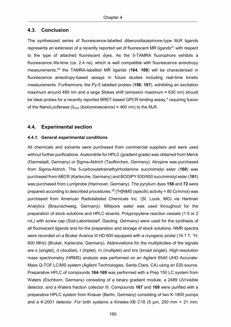
Chapter 4
160
4.3. Conclusion
The synthesized series of fluorescence-labelled dibenzodiazepinone-type M2R ligands
represents an extension of a recently reported set of fluorescent MR ligands47 with respect
to the type of attached fluorescent dyes. As the 5-TAMRA fluorophore exhibits a
fluorescence life-time (ca. 2.4 ns), which is well compatible with fluorescence anisotropy
measurements,49 the TAMRA-labelled MR ligands (164, 169) will be characterized in
fluorescence anisotropy-based assays in future studies including real-time kinetic
measurements. Furthermore, the Py-5 labelled probes (156, 167), exhibiting an excitation
maximum around 480 nm and a large Stokes shift (emission maximum ≈ 630 nm) should
be ideal probes for a recently reported BRET-based GPCR binding assay,4 requiring fusion
of the NanoLuciferase (λmax (bioluminescence) ≈ 460 nm) to the M2R.
4.4. Experimental section
4.4.1. General experimental conditions
All chemicals and solvents were purchased from commercial suppliers and were used
without further purifications. Acetonitrile for HPLC (gradient grade) was obtained from Merck
(Darmstadt, Germany) or Sigma-Aldrich (Taufkirchen, Germany). Atropine was purchased
from Sigma-Aldrich. The 5-carboxytetramethylrhodamine succinimidyl ester (160) was
purchased from ABCR (Karlsruhe, Germany) and BODIPY 630/650 succinimidyl ester (161)
was purchased from Lumiprobe (Hannover, Germany). The pyrylium dyes 158 and 72 were
prepared according to described procedures.48 [3H]NMS (specific activity = 80 Ci/mmol) was
purchased from American Radiolabeled Chemicals Inc. (St. Louis, MO) via Hartman
Analytics (Braunschweig, Germany). Millipore water was used throughout for the
preparation of stock solutions and HPLC eluents. Polypropylene reaction vessels (1.5 or 2
mL) with screw cap (Süd-Laborbedarf, Gauting, Germany) were used for the synthesis of
all fluorescent ligands and for the preparation and storage of stock solutions. NMR spectra
were recorded on a Bruker Avance III HD 600 equipped with a cryogenic probe (14.1 T; 1H:
600 MHz) (Bruker, Karlsruhe, Germany). Abbreviations for the multiplicities of the signals
are s (singlet), d (doublet), t (triplet), m (multiplet) and brs (broad singlet). High-resolution
mass spectrometry (HRMS) analysis was performed on an Agilent 6540 UHD Accurate-
Mass Q-TOF LC/MS system (Agilent Technologies, Santa Clara, CA) using an ESI source.
Preparative HPLC of compounds 164-169 was performed with a Prep 150 LC system from
Waters (Eschborn, Germany) consisting of a binary gradient module, a 2489 UV/visible
detector, and a Waters fraction collector III. Compounds 167 and 169 were purified with a
preparative HPLC system from Knauer (Berlin, Germany) consisting of two K-1800 pumps
and a K-2001 detector. For both systems a Kinetex-XB C18 (5 μm, 250 mm × 21 mm;

Fluorescently labelled dibenzodiazepinone-type MR ligands with high M2R affinity
161
Phenomenex, Aschaffenburg, Germany) served as stationary phases at a flow rate of 20
mL/min. Mixtures of 0.1% aq TFA and acetonitrile were used as mobile phase. The
detection wavelength was set to 220 nm throughout. The solvent of collected fractions was
removed by lyophilization using a Scanvac freeze drying apparatus (Labogene, Allerød,
Denmark) equipped with a RZ 6 rotary vane vacuum pump (Vacuubrand, Wertheim,
Germany). Analytical HPLC analysis was performed with a system from Agilent
Technologies composed of a 1290 Infinity binary pump equipped with a degasser, a 1290
Infinity autosampler, a 1290 Infinity thermostated column compartment, a 1260 Infinity diode
array detector, and a 1260 Infinity fluorescence detector. A Kinetex-XB C18 (2.6 μm, 100 ×
3 mm; Phenomenex) was used as stationary phase at a flow rate of 0.6 mL/min. Mixtures
of 0.04% aq TFA (A) and acetonitrile (B) were used as mobile phase. The following linear
gradients were applied: compounds 164-166, 168 and 169 (purity): 0-12 min: A/B 90:10 to
55:45, 12-16 min: 55:45 to 5:95, 16-20 min: 5:95; compound 167 (purity): 0-15 min: A/B
90:10 to 50:50, 15-19 min: 50:50 to 5:95; 20-22 min: 5:95; compounds 164-169 (chemical
stabilities): 0-20 min: A/B 90:10 to 35:65, 20-22 min: 35:65 to 5:95, 22-26 min: 5:95. For all
analytical HPLC runs the oven temperature was set to 25 °C, the injection volume was 20
μL and detection was performed at 220 nm. The stock solutions (final concentration: 1 mM)
of fluorescent ligands were prepared in DMSO and were stored at -78°C.
Annotation concerning the 1H-NMR spectra of the dibenzodiazepinone derivatives 164-169:
due to a slow rotation about the exocyclic amide group of the diazepinone ring on the NMR
time scale, two isomers (ratios provided in the experimental protocols) were evident in the 1H-NMR spectra.
4.4.2. Compounds characterization
All target compounds (164-169) were characterized by 1H-NMR spectroscopy, HRMS, and
RP-HPLC analysis. The HPLC purity of all fluorescent ligands amounted to ≥ 96% (220 nm)
(chromatograms shown in the Supplementary Information, NMR spectra shown in
Appendix).

Chapter 4
162
4.4.3. Experimental synthetic protocols and analytical data
2-(6-(Dimethylamino)-3-(dimethyliminio)-3H-xanthen-9-yl)-5-((2-(3-(1-(4-(1-(2-oxo-2-(11-oxo-10,11-dihydro-5H-dibenzo[b,e][1,4]diazepin-5-yl)ethyl)piperidin-4-yl)butyl)-1H-imidazol-4-yl)propanamido)ethyl)carbamoyl)benzoate bis(hydrotrifluoroacetate) (164)
The reaction was carried out in a 1.5-mL propylene reaction vessel equipped with a micro
stir bar. Amine precursor 158 (tris(hydrotrifluoroacetate), 14.5 mg, 0.016 mmol) and DIPEA
(10.3 mg, 14 µL, 0.089 mmol) were dissolved in anhydrous DMF (90 µL) followed by the
addition of 160 (4.2 mg, 0.008 mmol) dissolved in anhydrous DMF (50 µL). After stirring at
room temperature in the dark for 2 h, 10% aq TFA (corresponding to 0.09 mmol of TFA)
were added. Purification of the product by preparative HPLC (gradient: 0-40 min: 0.1% aq
TFA/acetonitrile 81:19-28:72, tR = 13 min) yielded compound 164 as a red solid (3.0 mg,
31%). Ratio of configurational isomers evident in the 1H-NMR spectrum: ca 1.5:1. 1H-NMR
(600 MHz, MeOH-d4): δ (ppm) 1.33-1.40 (m, 4H), 1.44-1,57 (m, 3H), 1.85-1.93 (m, 3H),
1.93-1.99 (m, 1H), 2.62 (t, 2H, J 7.2 Hz), 2.89-2.98 (m, 1H), 3.00 (t, 2H, J 7.2 Hz), 3.02-3.10
(m, 1H), 3.45(t, 3H, J 6.0 Hz, interfering with the 13C satellite of the solvent residual peak),
3,56 (t, 2H, J 6.1 Hz), 3.72 (d, 1.4H, J 7.2 Hz), 3.80 (d, 0.60H, J 17 Hz), 4.18 (t, 2H, J 7.2
Hz), 4.37-4.46 (m, 1H), 7.0-7.02 (m, 2H), 7.03-7.08 (m, 2H), 7.11-7.14 (m, 2H), 7.24-7.36
(m, 2H), 7.36-7.43 (m, 1.4H), 7.46-7.55 (m, 3.2H), 7.59-7.65 (m, 1.4H), 7.65-7.70 (m, 0.6H),
7.72-7.77 (m, 0.4H), 7.87-7.92 (m, 0.6H), 7.95-7.99 (m, 0.4H), 8.22-8.26 (m, 1H), 8.73-8.77
(m, 1H), 8.80 (s, 1H). Note: exchangeable protons (NH, OH) were not apparent. The proton
signals of the four methyl groups (12 protons) were not apparent due to an interference with
the solvent residual peak. HRMS (ESI): m/z [M+H]+ calcd. for [C57H62N9O7]+: 984.4767,
found: 984.4757. RP-HPLC (220 nm): 97% (tR = 7.40 min, k = 8.7). C57H61N9O7 . C4H2F6O4
(984.17 + 228.04).
(E)-6-(2-(4-(2-(5,5-Difluoro-7-(thiophen-2-yl)-5H-4l4,5l4-dipyrrolo[1,2-c:2',1'-f][1,3,2]diazaborinin-3-yl)vinyl)phenoxy)acetamido)-N-(2-(3-(1-(4-(1-(2-oxo-2-(11-oxo-10,11-dihydro-5H-dibenzo[b,e][1,4]diazepin-5-yl)ethyl)piperidin-4-yl)butyl)-1H-imidazol-4-yl)propanamido)ethyl)hexanamide bis(hydrotrifluoroacetate) (165)
Compound 165 was prepared from 158 (tris(hydrotrifluoroacetate), 8.6 mg, 0.009 mmol)
and 161 (3.1 mg, 0.0047 mmol) according to the procedure used for the synthesis of 164.
DIPEA: 6.1 mg, 8.2 µL, 0.047 mmol. Purification of the product by preparative HPLC
(gradient: 0-40 min: 0.1% aq TFA/acetonitrile 81:19-19:81, tR = 22 min) yielded 165 as a
blue solid (1.35 mg, 23%). Ratio of configurational isomers evident in the 1H-NMR spectrum:
ca 1.5:1. 1H-NMR (600 MHz, MeOH-d4): δ (ppm) 1.25-1.33 (m, 7H), 1.42-1.51 (m, 2H), 1.52-

Fluorescently labelled dibenzodiazepinone-type MR ligands with high M2R affinity
163
1.64 (m, 4H), 1.77-1.93 (m, 4H), 2.16 (t, 2H, J 7.2 Hz), 2.52 (t, 2H, J 7.2 Hz), 2.81-2.88 (m,
1H), 2.92 (t, 2H, J 7.2 Hz), 2.95-3.02 (m, 1H), 3.21-3.25 (m, 4H, interfering with the solvent
peak), 3.26-3.28 (m, 3H, interfering with the solvent peak), 3.36-3.41 (m, 1H, interfering with
the 13C satellite of the solvent residual peak), 3.66-3.72 (m, 1H); 4.09 (t, 2H, J 7.2 Hz), 4.33-
4.42 (m, 1H), 4.58 (s, 2H), 6.87 (d, 1H, J 4.2 Hz), 7.03-7.07 (m, 2H), 7.15 (d, 2H, J 4.2 Hz),
7.19-7.23 (m, 2H), 7.25-7.35 (m, 3H), 7.39 (s, 1H), 7.39-7.42 (m, 0.4H), 7.46-7.51 (m, 1.6H),
7.52-7.66 (m, 7H), 7.66-7.71 (m, 0.6H), 7.71-7.76 (m, 0.4H), 7.89-7.94 (m, 0.6H), 7.95-8.00
(m, 0.4H), 8.11 (d, 1H, J 3.9 Hz), 8.72 (s, 1H). Note: exchangeable protons (NH) were not
apparent. HRMS (ESI): m/z [M+H]+ calcd. for [C61H68BF2N10O6S]+: 1117.5100, found:
1117.5110. RP-HPLC (220 nm): 99% (tR = 7.62 min, k = 9.0). C61H67BF2N10O6S . C4H2F6O4
(1117.14 + 228.04).
(E)-2,6-Dimethyl-1-(2-(3-(1-(4-(1-(2-oxo-2-(11-oxo-10,11-dihydro-5H-dibenzo[b,e][1,4]diazepin-5-yl)ethyl)piperidin-4-yl)butyl)-1H-imidazol-4-yl)propanamido)ethyl)-4-(2-(2,3,6,7-tetrahydro-1H,5H-pyrido[3,2,1-ij]quinolin-9-yl)vinyl)pyridin-1-ium bis(hydrotrifluoroacetate) trifluoroacetate (166)
Compound 166 was prepared from 158 (tris(hydrotrifluoroacetate), 28 mg, 0.031 mmol) and
162 (17 mg, 0.043 mmol) according to the procedure used for the synthesis of 164. DIPEA:
40 mg, 53 µL, 0.31 mmol. Purification of the product by preparative HPLC (gradient: 0-40
min: 0.1% aq TFA/acetonitrile 85:15-38:62, tR = 23 min) yielded 166 as a red solid (5.4 mg,
16%). Ratio of configurational isomers evident in the 1H-NMR spectrum: ca 1.5:1. 1H-NMR
(600 MHz, MeOH-d4): δ (ppm) 1.32-1.39 (m, 4H), 1.41-1.56 (m, 3H), 1.81-1.92 (m, 3H),
1.93-2.00 (m, 5H), 2.60 (t, 2H, J 7.3 Hz), 2.75 (t, 4H, J 6.6 Hz), 2.82 (s, 6H), 2.89-2.96 (m,
3H), 2.99-3.06 (m, 1H), 3.45 (t, 1H, J 13 Hz, interfering with the 13C satellite of the solvent
residual peak), 3.62 (t, 2H, J 7.2 Hz), 3.69-3.81 (m, 2H), 4.14 (t, 2H, J 7.3 Hz), 4.38 (s, 0.4
H), 4.40-4.42 (m, 0.6H), 4.43-4.48 (m, 2H), 6.83 (d, 1H, J 16 Hz), 7.13 (s, 2H), 7.25-7.41
(m, 3.6H), 7.47-7.55 (m, 2.4H), 7.61-7.67 (m, 4H), 7.67-7.71 (m, 0.6H), 7.73-7.77 (m, 0.4H),
7.89-7.92 (m, 0.6H), 7.97 (d, 0.4H, J 7.6 Hz), 8.79 (s, 1H). Note: exchangeable protons
(NH) were not apparent. The proton signals of two methylene groups (4 protons) of the
fluorophore were not apparent due to an interference with the solvent residual peak. HRMS
(ESI): m/z [M]+ calcd. for [C53H63N8O3]+: 859.5018, found: 859.5026. RP-HPLC (220 nm):
96% (tR = 9.5 min, k = 12.0). C53H63N8O3+ . C2F3O2
− . C4H2F6O4 (860.14 + 341.07).

Chapter 4
164
4-((1E,3E)-4-(4-(Dimethylamino)phenyl)buta-1,3-dien-1-yl)-2,6-dimethyl-1-(2-(3-(1-(4-(1-(2-oxo-2-(11-oxo-10,11-dihydro-5H-dibenzo[b,e][1,4]diazepin-5-yl)ethyl)piperidin-4-yl)butyl)-1H-imidazol-4-yl)propanamido)ethyl)pyridin-1-ium bis(hydrotrifluoroacetate) trifluoroacetate (167)
Compound 167 was prepared from 158 (tris(hydrotrifluoroacetate), 6.4mg, 0.007 mmol) and
163 (7.7 mg, 0.021 mmol) following the procedure used for the synthesis of 164, but
triethylamine (7.1 mg, 9.8 µL, 0.07 mmol) was used instead of DIPEA. Purification of the
product by preparative HPLC (gradient: 0-40 min: 0.1% aq TFA/acetonitrile 81:19-40:60, tR
= 16 min) yielded 167 as a red solid (2.3 mg, 28%). Ratio of configurational isomers evident
in the 1H-NMR spectrum: ca 1.5:1. 1H-NMR (600 MHz, MeOH-d4): δ (ppm) 1.32-1.38 (m,
4H), 1.39-1.55 (m, 3H), 1.82-1.89 (m, 3H), 1.89-1-97 (m, 1H), 2.60 (t, 2H, J 7.3 Hz), 2.85
(s, 6H), 2.89-2.95 (m, 3H), 3.02 (s, 7H), 3.44 (t, 1H, J 13 Hz, interfering with the 13C satellite
of the solvent residual peak), 3.64 (t, 2H, J 7.2 Hz), 3.68-3.80 (m, 2H), 4.15 (t, 2H, J 7.4
Hz), 4.36-4.45 (m, 1H), 4.50 (t, 2H, J 7.1 Hz), 6.57 (d, 1H, J 15 Hz), 6.76 (d, 2H, J 8.8 Hz),
6.92-6.99 (m, 1H), 7.02 (d, 1H, J 15 Hz), 7.25-7.41 (m, 3.6H), 7.44 (d, 2H, J 8.9 Hz), 7.47-
7.55 (m, 2.4H), 7.61-7.66 (m, 1.6H), 7.66-7.72 (m, 3H), 7.73-7.78 (m, 0.4H), 7.89-7.92 (m,
0.6H), 7.98 (d, 0.4H, J 8.4 Hz), 8.79 (s, 1H). Note: exchangeable protons (NH) were not
apparent. HRMS (ESI): m/z [M]+ calcd. for [C51H61N8O3]+: 833.4861, found 833.4879. RP-
HPLC (220 nm): 98% (tR = 8.4 min, k = 10.0). C51H61N8O3+ . C2F3O2
− . C4H2F6O4 (834.10 +
341.07).
E)-2,6-Dimethyl-1-(2-(4-(4-(1-(2-oxo-2-(11-oxo-10,11-dihydro-5H-dibenzo[b,e][1,4]diazepin-5-yl)ethyl)piperidin-4-yl)butyl)piperazin-1-yl)ethyl)-4-(2-(2,3,6,7-tetrahydro-1H,5H-pyrido[3,2,1-ij]quinolin-9-yl)vinyl)pyridin-1-ium tris(hydrotrifluoroacetate) trifluoroacetate (168)
Compound 168 was prepared from 72 (17 mg, 0.018 mmol), and 162 (10 mg, 0.026 mmol)
according to the procedure used for the synthesis of 164. DIPEA: 23 mg, 31 µL, 0.18 mmol.
Purification of the product by preparative HPLC (gradient: 0-40 min: 0.1% aq
TFA/acetonitrile 81:19-29:71, tR = 18 min) yielded 168 as a red solid (5.1 mg, 23%). Ratio
of configurational isomers evident in the 1H-NMR spectrum: ca 1.5:1. 1H-NMR (600 MHz,
MeOH-d4): δ (ppm) 1.33-1.57 (m, 7H), 1.68-1.77 (m, 2H), 1.90-2.00 (m, 6H), 2.56-2.63 (m,
2H), 2.75 (t, 4H, J 6.3 Hz), 2.80 (s, 6H), 2.92 (t, 3H, J 6.7 Hz), 2.98-3.18 (m, 8H), 3.45 (t,
1H, J 12 Hz, interfering with the 13C satellite of the solvent residual peak), 3.52-3.59 (m,
2H), 3.70-3.82 (m, 2H), 4.37-4.46 (m, 1H), 4.51 (t, 2H, J 6.6 Hz), 6.84 (d, 1H, J 16 Hz), 7.13
(s, 2H), 7.24-7.32 (m, 1H), 7.32-7.36 (m, 1H), 7.40 (t, 0.4H, J 7.7 Hz), 7.47-7.56 (m, 2.2H),
7.60-7.72 (m, 5H), 7.76 (t, 0.4H, J 7.8 Hz), 7.91 (d, 0.6H, J 8.2 Hz), 7.98 (d, 0.4H, J 8.2 Hz).

Fluorescently labelled dibenzodiazepinone-type MR ligands with high M2R affinity
165
Note: exchangeable protons (NH) were not apparent. The proton signals of two methylene
groups (4 protons) of the fluorophore were not apparent due to an interference with the
solvent residual peak. HRMS (ESI): m/z [M]+ calcd. for [C51H64N7O2]+: 806.5116, found
806.5121. RP-HPLC (220 nm): 97% (tR = 9.6 min, k = 12.0). C51H64N7O2+ . C2F3O2
− .
C6H3F9O6 (807.12 + 455.09).
2-(6-(Dimethylamino)-3-(dimethyliminio)-3H-xanthen-9-yl)-5-((2-(4-(4-(1-(2-oxo-2-(11-oxo-10,11-dihydro-5H-dibenzo[b,e][1,4]diazepin-5-yl)ethyl)piperidin-4-yl)butyl)piperazin-1-yl)ethyl)carbamoyl)benzoate tris(hydrotrifluoroacetate) (169)
Compound 169 was prepared from 72 (8.9 mg, 0.010mmol) and 160 (4.1 mg, 0.0077 mmol)
according to the procedure used for the synthesis of 164. DIPEA: 11 mg, 14 µL, 0.083
mmol. Purification of the product by preparative HPLC (gradient: 0-40 min: 0.1% aq
TFA/acetonitrile 81:19-30:40, tR = 13 min) yielded 169 as a red solid (6.5 mg, 66%). Ratio
of configurational isomers evident in the 1H-NMR spectrum: ca 1.5:1. 1H-NMR (600 MHz,
MeOH-d4): δ (ppm) 1.35-1.43 (m, 4H), 1.44-1.61 (m, 3H), 1.68-1.77 (m, 2H), 1.89-2.02 (m,
2H), 2.61 (brs, 2H), 2.83 (s, 2H), 2.86-3.00 (m, 1H), 3.00-3.12 (m, 4H), 3.45 (t, 2H, J 12 Hz,
interfering with the 13C satellite of the solvent residual peak), 3.66 (t, 2H, J 6.2 Hz), 3.70-
3.83 (m, 2H), 4.40 (d, 0.6H, J 17 Hz), 4.44 (d, 0.4H, J 17 Hz), 7.00 (d, 2H, J 2.2 Hz), 7.05
(d, 0.8H, J 2.2 Hz), 7.07 (d, 1.2H, J 2.3 Hz), 7.13 (d, 2H, J 9.4 Hz), 7.25-7.36 (m, 2H), 7.39
(t, 0.4H, J 7.8 Hz), 7.47-7.56 (m, 3.2H), 7.60-7.67 (m, 1.4H), 7.70 (t, 0.6H, J 7.4 Hz), 7.76
(t, 0.4H, J 7.6 Hz), 7.91 (d, 0.6H, J 7.8 Hz), 7.98 (d, 0.4H, J 7.7 Hz), 8.23-8.28 (m, 1H), 8.77
(s, 1H). Note: exchangeable protons (NH, OH) were not apparent. The proton signals of the
four methyl groups (12 protons) and four protons of the piperazine ring (CH2 groups) were
not apparent due to an interference with the solvent residual peak. HRMS (ESI): m/z [M+H]+ calcd. for [C55H63N8O6]+: 931.4865, found 931.4868. RP-HPLC (220 nm): 99% (tR = 6.9 min,
k = 8.1). C55H62N8O6 . C6H3F9O6 (931.15 + 342.06).
4.4.4. Investigation of chemical stability
The chemical stabilities of 164-169 were investigated in PBS pH 7.4 at 22 ± 1 °C using
propylene vessels. The incubation was started by the addition of 10 μL of 1 mM solution of
the fluorescent ligand in DMSO to PBS (90 µL) to yield a final concentration of 100 μM.
After 0, 24 and 48 h, aliquots (20 μL) were taken and added to 1% aq TFA/acetonitrile (8:2
v/v) (20 μL). The resulting solutions were analyzed by RP-HPLC (analytical HPLC system
and conditions see general experimental conditions; tR: 7.8 min (164), 13.5 min (165), 9.95
min (166), 8.1 min (167), 10.0 min (168), 7.3 min (169)).

Chapter 4
166
4.4.5. Cell culture
CHO-K9 cells, stably transfected with the DNA of human muscarinic receptors M1-M5
(obtained from Missouri S&T cDNA Resource Center; Rolla, MO) were cultured in HAM’s
F12 medium supplemented with fetal calf serum (Biochrom, Berlin, Germany) (10%) and
G418 (Biochrom) (750 μg/mL).
4.4.6. Determination of excitation and emission spectra
Excitation and emission spectra of compounds 164-169 were recorded in PBS, pH 7.4,
containing 1% BSA (Serva, Heidelberg, Germany), at 22 °C with a Cary Eclipse
spectrofluorimeter (Varian Inc., Mulgrave, Victoria, Australia) using acryl cuvettes (10 × 10
mm, Ref. 67.755, Sarstedt, Nümbrecht, Germany). The slit adjustments
(excitation/emission) were 5/10 nm for excitation spectra and 10/5 nm in case of emission
spectra. Net spectra were calculated by subtracting the respective vehicle reference
spectrum, and corrected emission spectra were calculated by multiplying the net emission
spectra with the respective lamp corrections spectrum (same slit adjustments, etc.).
4.4.7. Radioligand competition binding assay
Equilibrium competition binding studies with [3H]NMS were performed at intact CHO-hMxR
cells (x = 1-5) at 23 ± 1 °C in white 96-wells plates with clear bottom (Corning Life Science,
Tewksbury, MA; Corning cat. No. 3610) using Leibovitz’s L-15 medium (Gibco, Life
Technologies, Darmstadt, Germany) supplemented with 1% BSA (Serva) as binding buffer
(in the following referred to as L15 medium). Experiments were performed using a
previously described protocol for MR binding studies with [3H]NMS,43 but the total volume
per well was 200 µL, i.e. wells were pre-filled with 180 µL of L15 medium followed by the
addition of L15 medium (20 µL) containing [3H]NMS (10-fold concentrated), to determine
total binding, or pre-filled with 160 µL of L15 medium followed by the addition of L15 medium
(20 µL) containing atropine or the compound of interest (10-fold concentrated) and L15
medium (20 µL) containing [3H]NMS (10-fold-concentrated), to determine unspecific binding
and the displacing effect of a compound of interest, respectively. The concentrations of
[3H]NMS were 0.2 nM (M1R, M2R, M3R), 0.1 nM (M4R) or 0.3 nM (M5R). Samples were
incubated in the dark under gentle shaking for 3 h. Prior to the competition binding
experiments, the Kd values of [3H]NMS were determined by saturation binding applying the
same conditions (buffer, temperature, incubation time, unspecific binding, etc.). The
obtained Kd values amounted to 0.17 ± 0.01 nM (M1R), 0.10 ± 0.01 nM (M2R), 0.12 ± 0.01
nM (M3R), 0.052 ± 0.01 nM (M4R) and 0.20 ± 0.02 nM (M5R) (mean value ± SEM from at
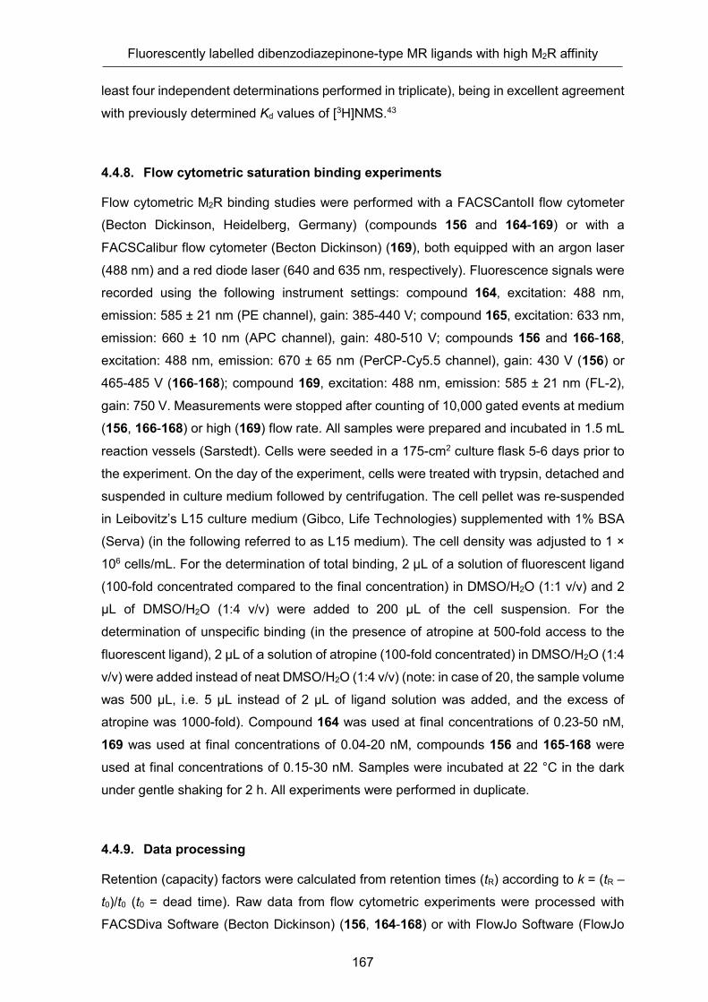
Fluorescently labelled dibenzodiazepinone-type MR ligands with high M2R affinity
167
least four independent determinations performed in triplicate), being in excellent agreement
with previously determined Kd values of [3H]NMS.43
4.4.8. Flow cytometric saturation binding experiments
Flow cytometric M2R binding studies were performed with a FACSCantoII flow cytometer
(Becton Dickinson, Heidelberg, Germany) (compounds 156 and 164-169) or with a
FACSCalibur flow cytometer (Becton Dickinson) (169), both equipped with an argon laser
(488 nm) and a red diode laser (640 and 635 nm, respectively). Fluorescence signals were
recorded using the following instrument settings: compound 164, excitation: 488 nm,
emission: 585 ± 21 nm (PE channel), gain: 385-440 V; compound 165, excitation: 633 nm,
emission: 660 ± 10 nm (APC channel), gain: 480-510 V; compounds 156 and 166-168, excitation: 488 nm, emission: 670 ± 65 nm (PerCP-Cy5.5 channel), gain: 430 V (156) or
465-485 V (166-168); compound 169, excitation: 488 nm, emission: 585 ± 21 nm (FL-2),
gain: 750 V. Measurements were stopped after counting of 10,000 gated events at medium
(156, 166-168) or high (169) flow rate. All samples were prepared and incubated in 1.5 mL
reaction vessels (Sarstedt). Cells were seeded in a 175-cm2 culture flask 5-6 days prior to
the experiment. On the day of the experiment, cells were treated with trypsin, detached and
suspended in culture medium followed by centrifugation. The cell pellet was re-suspended
in Leibovitz’s L15 culture medium (Gibco, Life Technologies) supplemented with 1% BSA
(Serva) (in the following referred to as L15 medium). The cell density was adjusted to 1 ×
106 cells/mL. For the determination of total binding, 2 µL of a solution of fluorescent ligand
(100-fold concentrated compared to the final concentration) in DMSO/H2O (1:1 v/v) and 2
µL of DMSO/H2O (1:4 v/v) were added to 200 µL of the cell suspension. For the
determination of unspecific binding (in the presence of atropine at 500-fold access to the
fluorescent ligand), 2 µL of a solution of atropine (100-fold concentrated) in DMSO/H2O (1:4
v/v) were added instead of neat DMSO/H2O (1:4 v/v) (note: in case of 20, the sample volume
was 500 µL, i.e. 5 µL instead of 2 µL of ligand solution was added, and the excess of
atropine was 1000-fold). Compound 164 was used at final concentrations of 0.23-50 nM,
169 was used at final concentrations of 0.04-20 nM, compounds 156 and 165-168 were
used at final concentrations of 0.15-30 nM. Samples were incubated at 22 °C in the dark
under gentle shaking for 2 h. All experiments were performed in duplicate.
4.4.9. Data processing
Retention (capacity) factors were calculated from retention times (tR) according to k = (tR –
t0)/t0 (t0 = dead time). Raw data from flow cytometric experiments were processed with
FACSDiva Software (Becton Dickinson) (156, 164-168) or with FlowJo Software (FlowJo

Chapter 4
168
LLC, Ashland, OR) (169) to obtain arithmetic mean values of the areas of the signals
detected in the respective channel (FACSCantoII) and geometric mean values of the height
of the signals detected in channel FL-2 (FACSCalibur), respectively. Specific binding data
from flow cytometric saturation binding experiments, obtained by subtracting unspecific
binding data from total binding data, were plotted against the fluorescent ligand
concentration and analyzed by a two-parameter equation describing hyperbolic binding
(one site-specific binding; GraphPad Prism 5, GraphPad Software, San Diego, CA) in order
to obtain Kd and Bmax values. Kd values of individual experiments were transformed to pKd
values. Unspecific binding data were fitted by linear regression. Data from the radioligand
([3H]NMS) saturation43 and competition44 binding assays were processed as reported
previously. pIC50 values were converted to pKi values according to the Cheng-Prusoff
equation50 (logarithmic form). Propagated errors were calculated as described previously.47

Fluorescently labelled dibenzodiazepinone-type MR ligands with high M2R affinity
169
4.5. Supplementary Information
Figure S1. Radioligand displacement curves obtained from competition binding experiments with [3H]NMS (0.2 nM (M1R, M2R, M3R), 0.1 nM (M4R) or 0.3 nM (M5R)) and 164-168 at intact CHO-hMxR cells (x = 1-5). Data represent are mean values ± SEM from at least three independent experiments (each performed in triplicate).

Chapter 4
170
Figure S2. Chromatograms of the HPLC analysis (method see general experimental conditions) of compounds 164-169 after incubation in PBS (pH 7.4) at 22 °C for up to 48 h. Whereas compounds 164, 165, 168 and 169 showed no decomposition, compounds 166 and 167 showed minor decomposition after 24 h. Compound 165 showed high adsorption to the surface of the polypropylene vessel immediately upon sample preparation. After 48 h, adsorbed 165 was desorbed by replacement of the residual original solution (PBS, pH 7.4) with 0.1% aq TFA/acetonitrile (1:1 v/v, 60 µL) (‘recovery’). For injection, this solution was 1:1 diluted with water.

Fluorescently labelled dibenzodiazepinone-type MR ligands with high M2R affinity
171
Figure S3. Excitation and corrected emission spectra of fluorescent ligands 164-169 recorded in PBS, pH 7.4, containing 1% BSA, at 22 °C using fluorescent ligand concentrations of 3 µM (164, 165, 169) or 5 µM (166, 167, 168).
Figure S4. Structure of the MR antagonist atropine.

Chapter 4
172
4.6. References
1. S. J. Briddon, B. Kellam and S. J. Hill, Design and use of fluorescent ligands to study ligand-receptor interactions in single living cells, Methods Mol. Cell. Biol., 2011, 746, 211-236.
2. L. A. Stoddart, L. E. Kilpatrick, S. J. Briddon and S. J. Hill, Probing the pharmacology of G protein-coupled receptors with fluorescent ligands, Neuropharmacology, 2015, 98, 48-57.
3. L. A. Stoddart, L. E. Kilpatrick and S. J. Hill, NanoBRET Approaches to Study Ligand Binding to GPCRs and RTKs, Trends Pharmacol. Sci., 2018, 39, 136-147.
4. L. A. Stoddart, C. W. White, K. Nguyen, S. J. Hill and K. D. G. Pfleger, Fluorescence- and bioluminescence-based approaches to study GPCR ligand binding., Br. J. Pharmacol., 2016, 173, 3028-3037.
5. S. J. Briddon and S. J. Hill, Pharmacology under the microscope: the use of fluorescence correlation spectroscopy to determine the properties of ligand-receptor complexes, Trends Pharmacol. Sci., 2007, 28, 637-645.
6. J. G. Baker, R. Middleton, L. Adams, L. T. May, S. J. Briddon, B. Kellam and S. J. Hill, Influence of fluorophore and linker composition on the pharmacology of fluorescent adenosine A1 receptor ligands, Br. J. Pharmacol., 2010, 159, 772-786.
7. K. Kuder and K. Kiec-Kononowicz, Fluorescent GPCR Ligands as new tools in pharmacology, Curr. Med. Chem., 2008, 15, 2132-2143.
8. K. J. Kuder and K. Kiec-Kononowicz, Fluorescent GPCR ligands as new tools in pharmacology-update, years 2008- early 2014, Curr. Med. Chem., 2014, 21, 3962-3975.
9. R. J. Middleton, S. J. Briddon, Y. Cordeaux, A. S. Yates, C. L. Dale, M. W. George, J. G. Baker, S. J. Hill and B. Kellam, New Fluorescent Adenosine A1-Receptor Agonists That Allow Quantification of Ligand-Receptor Interactions in Microdomains of Single Living Cells, J. Med. Chem., 2007, 50, 782-793.
10. R. J. Middleton and B. Kellam, Fluorophore-tagged GPCR ligands, Curr. Opin. Chem. Biol., 2005, 9, 517-525.
11. A. J. Vernall, S. J. Hill and B. Kellam, The evolving small-molecule fluorescent-conjugate toolbox for Class A GPCRs, Br. J. Pharmacol., 2014, 171, 1073-1084.
12. M. Amon, X. Ligneau, J. C. Schwartz and H. Stark, Fluorescent non-imidazole histamine H3 receptor ligands with nanomolar affinities, Bioorg. Med. Chem. Lett., 2006, 16, 1938-1940.
13. L. Li, J. Kracht, S. Peng, G. Bernhardt and A. Buschauer, Synthesis and pharmacological activity of fluorescent histamine H1 receptor antagonists related to mepyramine, Bioorg. Med. Chem. Lett., 2003, 13, 1245-1248.
14. L. Li, J. Kracht, S. Peng, G. Bernhardt, S. Elz and A. Buschauer, Synthesis and pharmacological activity of fluorescent histamine H2 receptor antagonists related to potentidine, Bioorg. Med. Chem. Lett., 2003, 13, 1717-1720.
15. S. F. Malan, A. van Marle, W. M. Menge, V. Zuliani, M. Hoffman, H. Timmerman and R. Leurs, Fluorescent ligands for the histamine H2 receptor: synthesis and preliminary characterization, Bioorg. Med. Chem., 2004, 12, 6495-6503.
16. S. X. Xie, G. Petrache, E. Schneider, Q. Z. Ye, G. Bernhardt, R. Seifert and A. Buschauer, Synthesis and pharmacological characterization of novel fluorescent histamine H2-receptor ligands derived from aminopotentidine, Bioorg. Med. Chem. Lett., 2006, 16, 3886-3890.
17. M. Leopoldo, E. Lacivita, E. Passafiume, M. Contino, N. A. Colabufo, F. Berardi and R. Perrone, 4-[omega-[4-arylpiperazin-1-yl]alkoxy]phenyl)imidazo[1,2-a]pyridine derivatives: fluorescent high-affinity dopamine D3 receptor ligands as potential probes for receptor visualization, J. Med. Chem., 2007, 50, 5043-5047.
18. A. Tabor, S. Weisenburger, A. Banerjee, N. Purkayastha, J. M. Kaindl, H. Hubner, L. Wei, T. W. Gromer, J. Kornhuber, N. Tschammer, N. J. Birdsall, G. I. Mashanov, V. Sandoghdar and P. Gmeiner, Visualization and ligand-induced modulation of

Fluorescently labelled dibenzodiazepinone-type MR ligands with high M2R affinity
173
dopamine receptor dimerization at the single molecule level, Sci. Rep., 2016, 6, 33233.
19. S. Arttamangkul, V. Alvarez-Maubecin, G. Thomas, J. T. Williams and D. K. Grandy, Binding and internalization of fluorescent opioid peptide conjugates in living cells, Mol. Pharmacol., 2000, 58, 1570-1580.
20. G. Balboni, S. Salvadori, A. Dal Piaz, F. Bortolotti, R. Argazzi, L. Negri, R. Lattanzi, S. D. Bryant, Y. Jinsmaa and L. H. Lazarus, Highly selective fluorescent analogue of the potent delta-opioid receptor antagonist Dmt-Tic, J. Med. Chem., 2004, 47, 6541-6546.
21. A. Drakopoulos, Z. Koszegi, Y. Lanoiselee, H. Hubner, P. Gmeiner, D. Calebiro and M. Decker, Investigation of Inactive-State kappa Opioid Receptor Homodimerization via Single-Molecule Microscopy Using New Antagonistic Fluorescent Probes, J. Med. Chem., 2020, 63, 3596-3609.
22. R. A. Houghten, C. T. Dooley and J. R. Appel, De novo identification of highly active fluorescent kappa opioid ligands from a rhodamine labeled tetrapeptide positional scanning library, Bioorg. Med. Chem. Lett., 2004, 14, 1947-1951.
23. S. Dukorn, T. Littmann, M. Keller, K. Kuhn, C. Cabrele, P. Baumeister, G. Bernhardt and A. Buschauer, Fluorescence- and Radiolabeling of [Lys(4),Nle(17,30)]hPP Yields Molecular Tools for the NPY Y4 Receptor, Bioconjugate Chem., 2017, 28, 1291-1304.
24. Y. Dumont, P. Gaudreau, M. Mazzuferi, D. Langlois, J. G. Chabot, A. Fournier, M. Simonato and R. Quirion, BODIPY-conjugated neuropeptide Y ligands: new fluorescent tools to tag Y1, Y2, Y4 and Y5 receptor subtypes, Br. J. Pharmacol., 2005, 146, 1069-1081.
25. M. Keller, D. Erdmann, N. Pop, N. Pluym, S. Teng, G. Bernhardt and A. Buschauer, Red-fluorescent argininamide-type NPY Y1 receptor antagonists as pharmacological tools, Bioorg. Med. Chem., 2011, 19, 2859-2878.
26. M. Liu, R. R. Richardson, S. J. Mountford, L. Zhang, M. H. Tempone, H. Herzog, N. D. Holliday and P. E. Thompson, Identification of a Cyanine-Dye Labeled Peptidic Ligand for Y1R and Y4R, Based upon the Neuropeptide Y C-Terminal Analogue, BVD-15, Bioconjugate Chem., 2016, 27, 2166-2175.
27. E. Schneider, M. Keller, A. Brennauer, B. K. Hoefelschweiger, D. Gross, O. S. Wolfbeis, G. Bernhardt and A. Buschauer, Synthesis and characterization of the first fluorescent nonpeptide NPY Y1 receptor antagonist, ChemBioChem, 2007, 8, 1981-1988.
28. R. Ziemek, A. Brennauer, E. Schneider, C. Cabrele, A. G. Beck-Sickinger, G. Bernhardt and A. Buschauer, Fluorescence- and luminescence-based methods for the determination of affinity and activity of neuropeptide Y2 receptor ligands, Eur. J. Pharmacol., 2006, 551, 10-18.
29. E. Kozma, P. S. Jayasekara, L. Squarcialupi, S. Paoletta, S. Moro, S. Federico, G. Spalluto and K. A. Jacobson, Fluorescent ligands for adenosine receptors, Bioorg. Med. Chem. Lett., 2013, 23, 26-36.
30. M. P. Faure, P. Gaudreau, I. Shaw, N. R. Cashman and A. Beaudet, Synthesis of a biologically active fluorescent probe for labeling neurotensin receptors, J. Histochem. Cytochem., 1994, 42, 755-763.
31. M. Keller, K. K. Kuhn, J. Einsiedel, H. Hubner, S. Biselli, C. Mollereau, D. Wifling, J. Svobodova, G. Bernhardt, C. Cabrele, P. M. Vanderheyden, P. Gmeiner and A. Buschauer, Mimicking of Arginine by Functionalized N(omega)-Carbamoylated Arginine As a New Broadly Applicable Approach to Labeled Bioactive Peptides: High Affinity Angiotensin, Neuropeptide Y, Neuropeptide FF, and Neurotensin Receptor Ligands As Examples, J. Med. Chem., 2016, 59, 1925-1945.
32. C. Tahtaoui, I. Parrot, P. Klotz, F. Guillier, J. L. Galzi, M. Hibert and B. Ilien, Fluorescent pirenzepine derivatives as potential bitopic ligands of the human M1 muscarinic receptor, J. Med. Chem., 2004, 47, 4300-4315.

Chapter 4
174
33. L. H. Jones, A. Randall, C. Napier, M. Trevethick, S. Sreckovic and J. Watson, Design and synthesis of a fluorescent muscarinic antagonist, Bioorg. Med. Chem. Lett., 2008, 18, 825-827.
34. J. A. Hern, A. H. Baig, G. I. Mashanov, B. Birdsall, J. E. Corrie, S. Lazareno, J. E. Molloy and N. J. Birdsall, Formation and dissociation of M1 muscarinic receptor dimers seen by total internal reflection fluorescence imaging of single molecules, Proc. Natl. Acad. Sci. U. S. A., 2010, 107, 2693-2698.
35. T. A. Nenasheva, M. Neary, G. I. Mashanov, N. J. Birdsall, R. A. Breckenridge and J. E. Molloy, Abundance, distribution, mobility and oligomeric state of M(2) muscarinic acetylcholine receptors in live cardiac muscle, J. Mol. Cell. Cardiol., 2013, 57, 129-136.
36. A. Harris, S. Cox, D. Burns and C. Norey, Miniaturization of fluorescence polarization receptor-binding assays using CyDye-labeled ligands, J. Biomol. Screen., 2003, 8, 410-420.
37. S. B. Daval, C. Valant, D. Bonnet, E. Kellenberger, M. Hibert, J. L. Galzi and B. Ilien, Fluorescent derivatives of AC-42 to probe bitopic orthosteric/allosteric binding mechanisms on muscarinic M1 receptors, J. Med. Chem., 2012, 55, 2125-2143.
38. S. B. Daval, E. Kellenberger, D. Bonnet, V. Utard, J. L. Galzi and B. Ilien, Exploration of the orthosteric/allosteric interface in human M1 muscarinic receptors by bitopic fluorescent ligands, Mol. Pharmacol., 2013, 84, 71-85.
39. K. A. Jacobson, B. Fischer and A. M. van Rhee, Molecular probes for muscarinic receptors: functionalized congeners of selective muscarinic antagonists, Life Sci., 1995, 56, 823-830.
40. Y. Karton, J. Baumgold, J. S. Handen and K. A. Jacobson, Molecular probes for muscarinic receptors: derivatives of the M1-antagonist telenzepine, Bioconjugate Chem., 1992, 3, 234-240.
41. Y. Wang, Q. Gu, F. Mao, R. P. Haugland and M. S. Cynader, Activity-dependent expression and distribution of M1 muscarinic ACh receptors in visual cortex neuronal cultures, J. Neurosci., 1994, 14, 4147-4158.
42. M. S. Gitler, R. C. Reba, V. I. Cohen, W. J. Rzeszotarski and J. Baumgold, A novel m2-selective muscarinic antagonist: binding characteristics and autoradiographic distribution in rat brain, Brain Res., 1992, 582, 253-260.
43. M. Keller, C. Trankle, X. She, A. Pegoli, G. Bernhardt, A. Buschauer and R. W. Read, M2 Subtype preferring dibenzodiazepinone-type muscarinic receptor ligands: Effect of chemical homo-dimerization on orthosteric (and allosteric?) binding, Bioorg. Med. Chem., 2015, 23, 3970-3990.
44. A. Pegoli, X. She, D. Wifling, H. Hubner, G. Bernhardt, P. Gmeiner and M. Keller, Radiolabeled Dibenzodiazepinone-Type Antagonists Give Evidence of Dualsteric Binding at the M2 Muscarinic Acetylcholine Receptor, J. Med. Chem., 2017, 60, 3314-3334.
45. A. Pegoli, D. Wifling, C. G. Gruber, X. She, H. Hubner, G. Bernhardt, P. Gmeiner and M. Keller, Conjugation of Short Peptides to Dibenzodiazepinone-Type Muscarinic Acetylcholine Receptor Ligands Determines M2R Selectivity, J. Med. Chem., 2019, 62, 5358-5369.
46. X. She, A. Pegoli, J. Mayr, H. Hubner, G. Bernhardt, P. Gmeiner and M. Keller, Heterodimerization of Dibenzodiazepinone-Type Muscarinic Acetylcholine Receptor Ligands Leads to Increased M2R Affinity and Selectivity, ACS Omega, 2017, 2, 6741-6754.
47. X. She, A. Pegoli, C. G. Gruber, D. Wifling, J. Carpenter, H. Hübner, J. Wan, G. Bernhardt, P. Gmeiner, N. D. Holliday and M. Keller, Red-emitting dibenzodiazepinone derivatives as fluorescent dualsteric probes for the muscarinic acetylcholine M2 receptor, J. Med. Chem., 2020, 63, 4133-4154.
48. B. K. Hoefelschweiger, PhD thesis, University of Regensburg, 2005. 49. A. Rinken, D. Lavogina and S. Kopanchuk, Assays with Detection of Fluorescence
Anisotropy: Challenges and Possibilities for Characterizing Ligand Binding to GPCRs, Trends Pharmacol. Sci., 2018, 39, 187-199.

Fluorescently labelled dibenzodiazepinone-type MR ligands with high M2R affinity
175
50. Y.-C. Cheng and W. H. Prusoff, Relation between the inhibition constant (Ki) and the concentration of inhibitor which causes fifty per cent inhibition (IC50) of an enzymic reaction, Biochem. Pharmacol., 1973, 22, 3099-3108.

176

177
Summary

Summary
178
In humans, the family of muscarinic acetylcholine receptors (mAChR, MRs) comprises five
subtypes (M1R-M5R), belonging to class A of the GPCR superfamily. MRs mediate the
action of the neurotransmitter acetylcholine (ACh) in the central and peripheral nervous
system. Whereas the M1R, M3R and M5R preferably couple to Gq-type G-proteins, the M2
and M4 receptor mainly activate Gi/o-type G-proteins. Due to the high conservation of the
orthosteric (acetylcholine) binding site within the family of MRs, the development of highly
subtype selective MR ligands is difficult. Selective MR ligands are needed as molecular or
pharmacological tools and also as new drug candidates anticipated to cause less adverse
effects. As the vestibule of MRs, also referred to as the common allosteric site, is less
conserved than the orthosteric pocket, the dualsteric ligand approach, i.e. the design of
ligands, which simultaneously address the orthosteric and an allosteric binding site, was
suggested as a promising strategy to develop MR ligands with high affinity and improved
subtype selectivity.
This work was aiming at the synthesis and pharmacological characterization of
dibenzodiazepinone-type MR ligands with pronounced M2R selectivity. The tricyclic MR
ligands were prepared by linking the pharmacophore DIBA via various linker moieties to
several short peptides, yielding a series of DIBA-peptide conjugates (70 compounds). The
linker structure was varied with respect to length, rigidity/flexibility and number of basic
groups. All peptide moieties contained at least one basic amino acid. In addition to
proteinogenic amino acids, also unnatural amino acids were incorporated. MR affinities and
selectivity profiles of the DIBA-peptide conjugates were determined by radioligand
competition binding at CHO-hMxR cells (x = 1-5) using [3H]N-methyl scopolamine as labeled
ligand. M1, M2 and M4 receptor affinities were determined for all target compounds.
Additionally, M3 and M5 receptor affinities were determined for selected compounds,
showing either high M2R selectivity over the M1 and M4 receptor, or a very low or no
preference for the M2R.
The study revealed that, besides the peptide structure, the type of the linker considerably
determines M2R affinity and selectivity. DIBA-peptide conjugates derived from amine
precursor 35 (compounds 101-106), containing an N-acylated 4-aminopiperidine and a
bisalkylated piperazine ring in the linker moiety, exhibited pronounced M2R selectivity (e.g.
105: Ki M1R/M2R/M3R/M4R/M5R = >140:1:>160:59:>140), but only moderate M2R affinity
(pKi 7.05-7.67). The subset of compounds derived from amine precursor 40 (107-109),
representing the higher homologue of precursor 35 (extension of the linker by one
methylene group), exhibited higher M2R affinities (pKi 8.93-9.08) and displayed also high
M2R selectivity (e.g. 109: Ki M1R/M2R/M3R/M4R/M5R = 70:1:11,000:48:6000). Considering
M2R over M3 and M5 receptor selectivity, compounds such as DIBA-peptide conjugates 107-
109 represent the most selective M2R antagonists reported to date. Regarding M2R over M1

Summary
179
and M4 receptor selectivity, higher M2R selectivity compared to 103-105 or 107-109 had
only been reported for one other compound class. For three selected DIBA-peptide
conjugates, M2R antagonism was confirmed in a M2R miniGi recruitment assay yielding Kb
values, which were in excellent agreement with the respective Ki values from radioligand
competition binding studies.
The investigation of three selected DIBA-peptide conjugates (3, 4, 146) with respect to
stability in human plasma showed highly stability against proteolytic degradation (> 99%
intact compound after 24 h at 37 °C).
Moreover, six fluorescently labeled dibenzodiazepinone-type MR ligands (164-169) were
prepared using various fluorescent dyes (5-TAMRA, BODIPY, pyridinium dye Py-1, and
pyridinium dye Py-5). All fluorescent probes exhibited high M2R affinity (pKi (radioligand
competition binding): 8.75‐9.62), a low preference for the M2R over the M1 and M4 receptor
and moderate to pronounced M2R selectivity compared to the M3R and M5R. The
fluorescent ligands were successfully used as probes in flow cytometric M2R saturation
binding assays resulting in pKd values of 8.36-9.19. Therefore, they are considered useful
molecular tools for future studies using methods such as fluorescence anisotropy and BRET
based MR binding assays.
In conclusion, this thesis afforded new fluorescently labeled molecular tools for the M2R and
new highly selective M2R antagonists, which might serve as lead structures for the
development of drug-like selective M2R antagonists, representing potential therapeutics for
the treatment of diseases associated with cholinergic dysfunction such as Alzheimer’s
disease.

180

181
Appendix

Appendix
182
1H and 13C-NMR spectra of compounds 6, 23-25, 28, 31, 35, 40, 43, 53-60, 63-65, 73-82, 85-99, 101-126, 134, 135 and 1H-NMR spectra of compounds 137-139 (Chapter 2)
1H-NMR spectrum (600 MHz, DMSO-d6) of compound 6
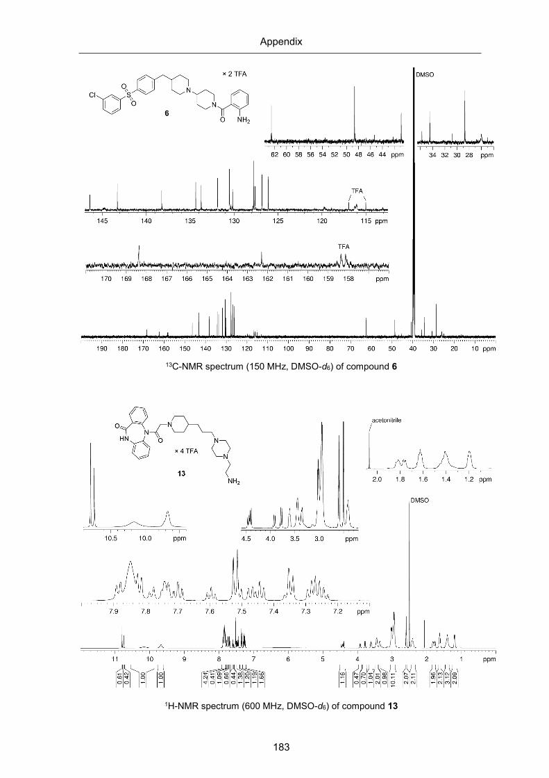
Appendix
183
13C-NMR spectrum (150 MHz, DMSO-d6) of compound 6
1H-NMR spectrum (600 MHz, DMSO-d6) of compound 13

Appendix
184
1H-NMR spectrum (600 MHz, DMSO-d6/D2O 4:1 v/v) of compound 13
13C-NMR spectrum (150 MHz, DMSO-d6) of compound 13

Appendix
185
1H-NMR spectrum (600 MHz, DMSO-d6) of compound 23
1H-NMR spectrum (600 MHz, DMSO-d6/D2O 4:1 v/v) of compound 23

Appendix
186
13C-NMR spectrum (150 MHz, DMSO-d6) of compound 23
1H-NMR spectrum (600 MHz, DMSO-d6) of compound 24

Appendix
187
1H-NMR spectrum (600 MHz, DMSO-d6/D2O 4:1 v/v) of compound 24
13C-NMR spectrum (150 MHz, DMSO-d6) of compound 24

Appendix
188
1H-NMR spectrum (600 MHz, DMSO-d6) of compound 25
1H-NMR spectrum (600 MHz, DMSO-d6/D2O 4:1 v/v) of compound 25

Appendix
189
13C-NMR spectrum (150 MHz, DMSO-d6) of compound 25
1H-NMR spectrum (600 MHz, DMSO-d6) of compound 28

Appendix
190
13C-NMR spectrum (150 MHz, DMSO-d6) of compound 28
1H-NMR spectrum (400 MHz, MeOH-d4) of compound 31

Appendix
191
13C-NMR spectrum (100 MHz, MeOH-d4) of compound 31
1H-NMR spectrum (400 MHz, MeOH-d4) of compound 35

Appendix
192
13C-NMR spectrum (100 MHz, MeOH-d4) of compound 35
1H-NMR spectrum (600 MHz, DMSO-d6) of compound 40

Appendix
193
1H-NMR spectrum (600 MHz, DMSO-d6/D2O 4:1 v/v) of compound 40
13C-NMR spectrum (150 MHz, DMSO-d6) of compound 40

Appendix
194
1H-NMR spectrum (600 MHz, DMSO-d6) of compound 43
1H-NMR spectrum (600 MHz, DMSO-d6/D2O 4:1 v/v) of compound 43

Appendix
195
13C-NMR spectrum (150 MHz, DMSO-d6) of compound 43
1H-NMR spectrum (600 MHz, DMSO-d6) of compound 53

Appendix
196
1H-NMR spectrum (600 MHz, DMSO-d6/D2O 4:1 v/v) of compound 53
13C-NMR spectrum (150 MHz, DMSO-d6) of compound 53

Appendix
197
1H-NMR spectrum (600 MHz, DMSO-d6) of compound 54
1H-NMR spectrum (600 MHz, DMSO-d6/D2O 4:1 v/v) of compound 54

Appendix
198
13C-NMR spectrum (150 MHz, DMSO-d6) of compound 54
1H-NMR spectrum (600 MHz, DMSO-d6) of compound 55

Appendix
199
1H-NMR spectrum (600 MHz, DMSO-d6/D2O 4:1 v/v) of compound 55
13C-NMR spectrum (150 MHz, DMSO-d6) of compound 55

Appendix
200
1H-NMR spectrum (600 MHz, DMSO-d6) of compound 56
1H-NMR spectrum (600 MHz, DMSO-d6/D2O 4:1 v/v) of compound 56

Appendix
201
13C-NMR spectrum (150 MHz, DMSO-d6) of compound 56
1H-NMR spectrum (600 MHz, DMSO-d6) of compound 57

Appendix
202
1H-NMR spectrum (600 MHz, DMSO-d6/D2O 4:1 v/v) of compound 57
13C-NMR spectrum (150 MHz, DMSO-d6) of compound 57

Appendix
203
1H-NMR spectrum (600 MHz, DMSO-d6) of compound 58
1H-NMR spectrum (600 MHz, DMSO-d6/D2O 4:1 v/v) of compound 58

Appendix
204
13C-NMR spectrum (150 MHz, DMSO-d6) of compound 58
1H-NMR spectrum (600 MHz, DMSO-d6) of compound 59

Appendix
205
1H-NMR spectrum (600 MHz, DMSO-d6/D2O 4:1 v/v) of compound 59
13C-NMR spectrum (150 MHz, DMSO-d6) of compound 59

Appendix
206
1H-NMR spectrum (600 MHz, DMSO-d6) of compound 60
1H-NMR spectrum (600 MHz, DMSO-d6/D2O 4:1 v/v) of compound 60

Appendix
207
13C-NMR spectrum (150 MHz, DMSO-d6) of compound 60
1H-NMR spectrum (600 MHz, DMSO-d6) of compound 63

Appendix
208
1H-NMR spectrum (600 MHz, DMSO-d6/D2O 4:1 v/v) of compound 63
13C-NMR spectrum (150 MHz, DMSO-d6) of compound 63

Appendix
209
1H-NMR spectrum (600 MHz, DMSO-d6) of compound 64
1H-NMR spectrum (600 MHz, DMSO-d6/D2O 4:1 v/v) of compound 64

Appendix
210
13C-NMR spectrum (150 MHz, DMSO-d6) of compound 64
1H-NMR spectrum (600 MHz, DMSO-d6) of compound 65

Appendix
211
1H-NMR spectrum (600 MHz, DMSO-d6/D2O 4:1 v/v) of compound 65
13C-NMR spectrum (150 MHz, DMSO-d6) of compound 65
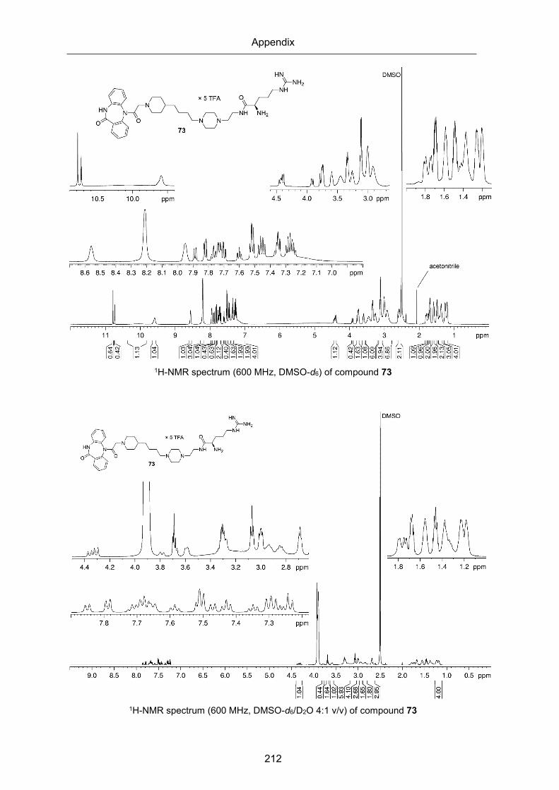
Appendix
212
1H-NMR spectrum (600 MHz, DMSO-d6) of compound 73
1H-NMR spectrum (600 MHz, DMSO-d6/D2O 4:1 v/v) of compound 73
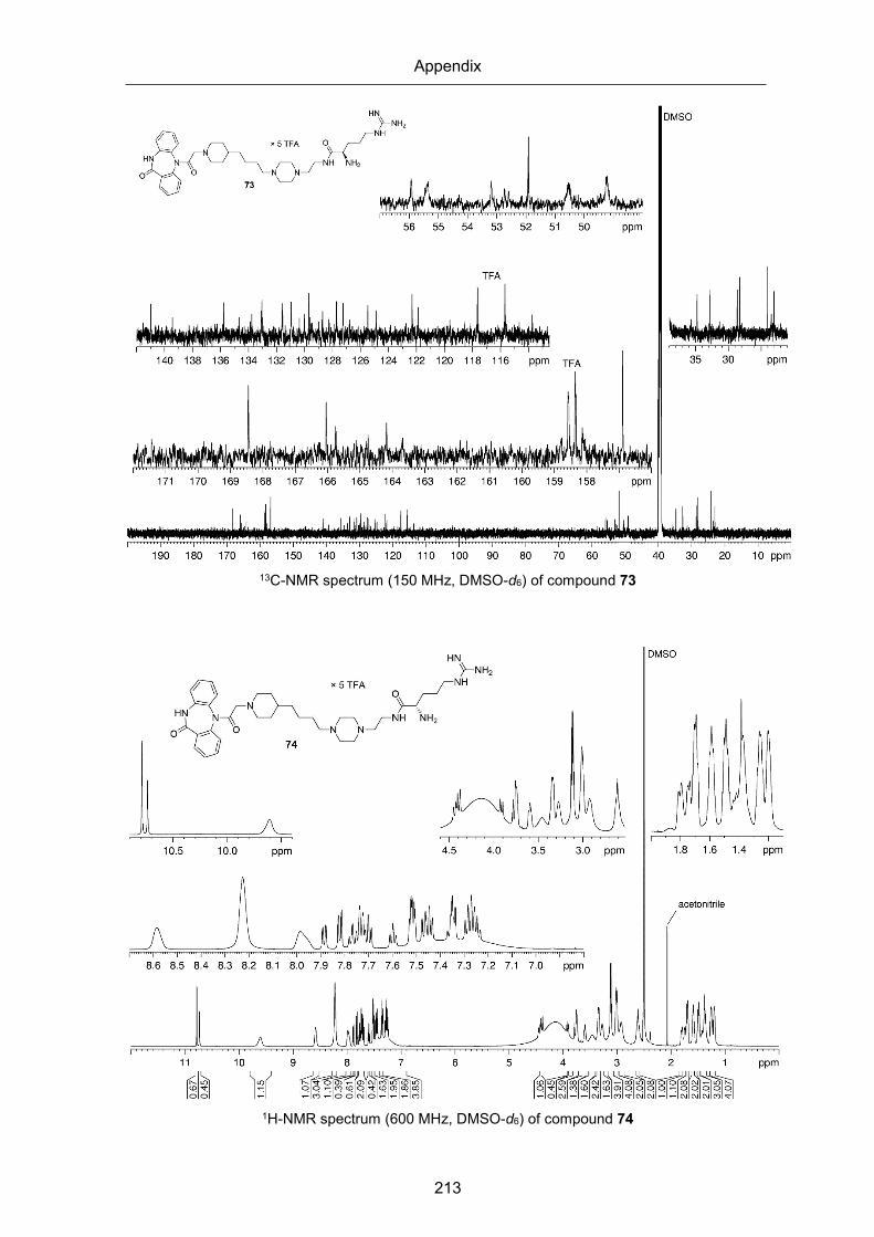
Appendix
213
13C-NMR spectrum (150 MHz, DMSO-d6) of compound 73
1H-NMR spectrum (600 MHz, DMSO-d6) of compound 74

Appendix
214
1H-NMR spectrum (600 MHz, DMSO-d6/D2O 4:1 v/v) of compound 74
13C-NMR spectrum (150 MHz, DMSO-d6) of compound 74

Appendix
215
1H-NMR spectrum (600 MHz, DMSO-d6) of compound 75
1H-NMR spectrum (600 MHz, DMSO-d6/D2O 4:1 v/v) of compound 75

Appendix
216
13C-NMR spectrum (150 MHz, DMSO-d6) of compound 75
1H-NMR spectrum (600 MHz, DMSO-d6) of compound 76

Appendix
217
1H-NMR spectrum (600 MHz, DMSO-d6/D2O 4:1 v/v) of compound 76
13C-NMR spectrum (150 MHz, DMSO-d6) of compound 76

Appendix
218
1H-NMR spectrum (600 MHz, DMSO-d6) of compound 77
1H-NMR spectrum (600 MHz, DMSO-d6/D2O 4:1 v/v) of compound 77

Appendix
219
13C-NMR spectrum (150 MHz, DMSO-d6) of compound 77
1H-NMR spectrum (600 MHz, DMSO-d6) of compound 78

Appendix
220
1H-NMR spectrum (600 MHz, DMSO-d6/D2O 4:1 v/v) of compound 78
13C-NMR spectrum (150 MHz, DMSO-d6) of compound 78
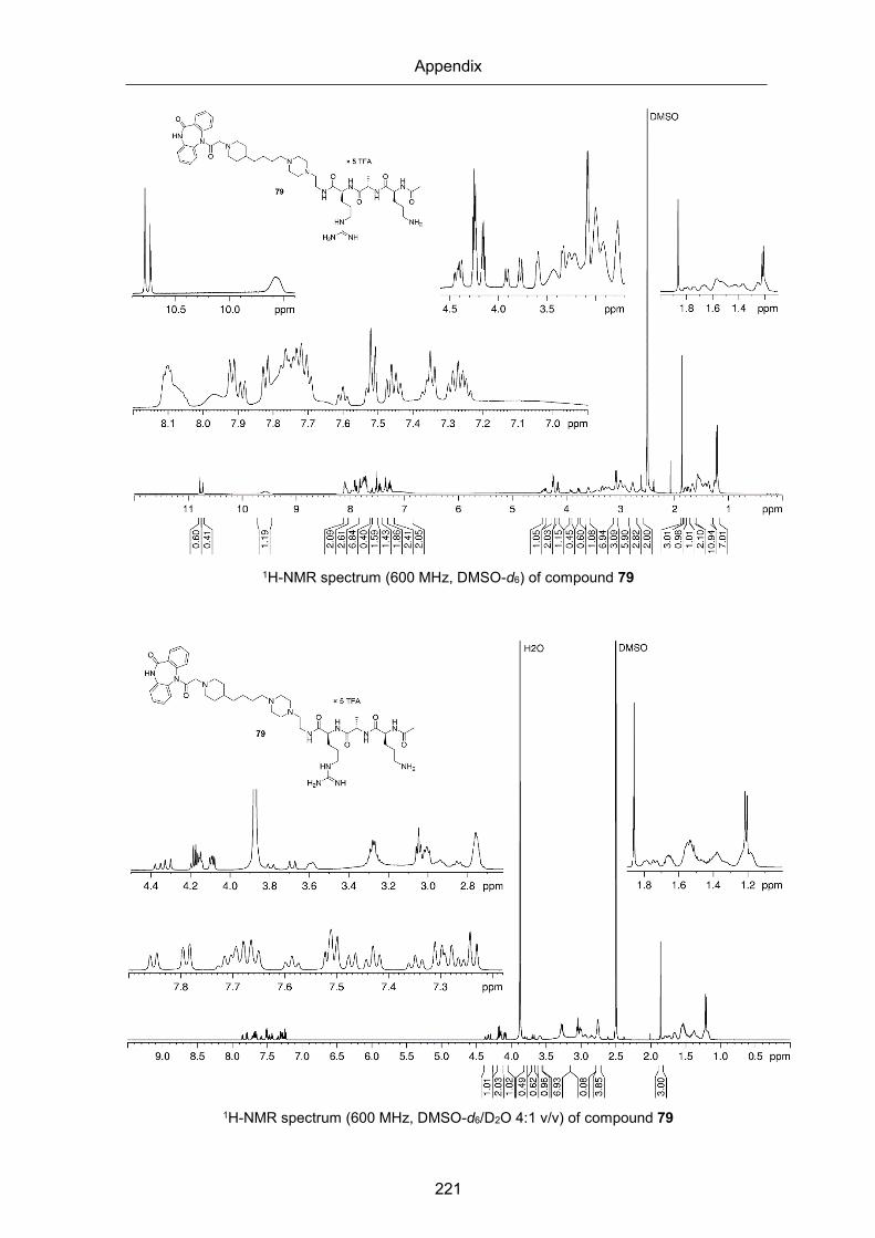
Appendix
221
1H-NMR spectrum (600 MHz, DMSO-d6) of compound 79
1H-NMR spectrum (600 MHz, DMSO-d6/D2O 4:1 v/v) of compound 79

Appendix
222
13C-NMR spectrum (150 MHz, DMSO-d6) of compound 79
1H-NMR spectrum (600 MHz, DMSO-d6) of compound 80

Appendix
223
1H-NMR spectrum (600 MHz, DMSO-d6/D2O 4:1 v/v) of compound 80
13C-NMR spectrum (150 MHz, DMSO-d6) of compound 80

Appendix
224
1H-NMR spectrum (600 MHz, DMSO-d6) of compound 81
1H-NMR spectrum (600 MHz, DMSO-d6/D2O 4:1 v/v) of compound 81

Appendix
225
13C-NMR spectrum (150 MHz, DMSO-d6) of compound 81
1H-NMR spectrum (600 MHz, DMSO-d6) of compound 82

Appendix
226
1H-NMR spectrum (600 MHz, DMSO-d6/D2O 4:1 v/v) of compound 82
13C-NMR spectrum (150 MHz, DMSO-d6) of compound 82
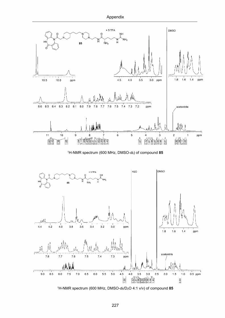
Appendix
227
1H-NMR spectrum (600 MHz, DMSO-d6) of compound 85
1H-NMR spectrum (600 MHz, DMSO-d6/D2O 4:1 v/v) of compound 85

Appendix
228
13C-NMR spectrum (150 MHz, DMSO-d6) of compound 85
1H-NMR spectrum (600 MHz, DMSO-d6) of compound 86

Appendix
229
1H-NMR spectrum (600 MHz, DMSO-d6/D2O 4:1 v/v) of compound 86
13C-NMR spectrum (150 MHz, DMSO-d6) of compound 86

Appendix
230
1H-NMR spectrum (600 MHz, DMSO-d6) of compound 87
1H-NMR spectrum (600 MHz, DMSO-d6/D2O 4:1 v/v) of compound 87

Appendix
231
13C-NMR spectrum (150 MHz, DMSO-d6) of compound 87
1H-NMR spectrum (600 MHz, DMSO-d6) of compound 88

Appendix
232
1H-NMR spectrum (600 MHz, DMSO-d6/D2O 4:1 v/v) of compound 88
13C-NMR spectrum (150 MHz, DMSO-d6) of compound 88

Appendix
233
1H-NMR spectrum (600 MHz, DMSO-d6) of compound 89
1H-NMR spectrum (600 MHz, DMSO-d6/D2O 4:1 v/v) of compound 89

Appendix
234
13C-NMR spectrum (150 MHz, DMSO-d6) of compound 89
1H-NMR spectrum (600 MHz, DMSO-d6) of compound 90

Appendix
235
1H-NMR spectrum (600 MHz, DMSO-d6/D2O 4:1 v/v) of compound 90
13C-NMR spectrum (150 MHz, DMSO-d6) of compound 90

Appendix
236
1H-NMR spectrum (600 MHz, DMSO-d6) of compound 91
1H-NMR spectrum (600 MHz, DMSO-d6/D2O 4:1 v/v) of compound 91

Appendix
237
13C-NMR spectrum (150 MHz, DMSO-d6) of compound 91
1H-NMR spectrum (600 MHz, DMSO-d6) of compound 92

Appendix
238
1H-NMR spectrum (600 MHz, DMSO-d6/D2O 4:1 v/v) of compound 92
13C-NMR spectrum (150 MHz, DMSO-d6) of compound 92

Appendix
239
1H-NMR spectrum (600 MHz, DMSO-d6) of compound 93
1H-NMR spectrum (600 MHz, DMSO-d6/D2O 4:1 v/v) of compound 93

Appendix
240
13C-NMR spectrum (150 MHz, DMSO-d6) of compound 93
1H-NMR spectrum (600 MHz, DMSO-d6) of compound 94

Appendix
241
1H-NMR spectrum (600 MHz, DMSO-d6/D2O 4:1 v/v) of compound 94
13C-NMR spectrum (150 MHz, DMSO-d6) of compound 94

Appendix
242
1H-NMR spectrum (600 MHz, DMSO-d6) of compound 95
1H-NMR spectrum (600 MHz, DMSO-d6/D2O 4:1 v/v) of compound 95

Appendix
243
13C-NMR spectrum (150 MHz, DMSO-d6) of compound 95
1H-NMR spectrum (600 MHz, DMSO-d6) of compound 96
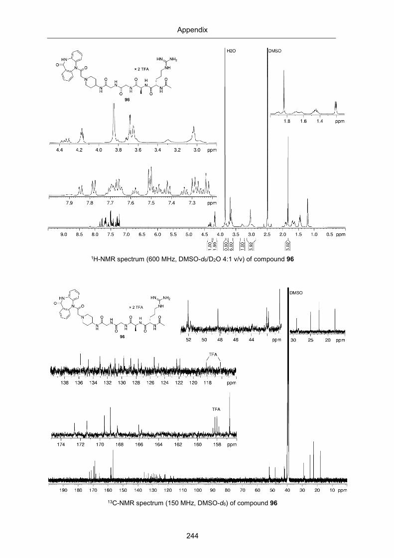
Appendix
244
1H-NMR spectrum (600 MHz, DMSO-d6/D2O 4:1 v/v) of compound 96
13C-NMR spectrum (150 MHz, DMSO-d6) of compound 96

Appendix
245
1H-NMR spectrum (600 MHz, DMSO-d6) of compound 97
1H-NMR spectrum (600 MHz, DMSO-d6/D2O 4:1 v/v) of compound 97

Appendix
246
13C-NMR spectrum (150 MHz, DMSO-d6) of compound 97
1H-NMR spectrum (600 MHz, DMSO-d6) of compound 98

Appendix
247
1H-NMR spectrum (600 MHz, DMSO-d6/D2O 4:1 v/v) of compound 98
13C-NMR spectrum (150 MHz, DMSO-d6) of compound 98

Appendix
248
1H-NMR spectrum (600 MHz, DMSO-d6) of compound 99
1H-NMR spectrum (600 MHz, DMSO-d6/D2O 4:1 v/v) of compound 99

Appendix
249
13C-NMR spectrum (150 MHz, DMSO-d6) of compound 99
1H-NMR spectrum (600 MHz, DMSO-d6) of compound 101

Appendix
250
1H-NMR spectrum (600 MHz, DMSO-d6/D2O 4:1 v/v) of compound 101
13C-NMR spectrum (150 MHz, DMSO-d6) of compound 101
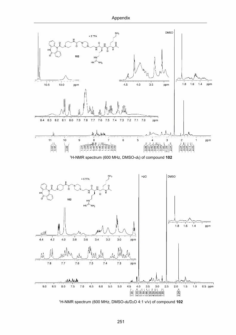
Appendix
251
1H-NMR spectrum (600 MHz, DMSO-d6) of compound 102
1H-NMR spectrum (600 MHz, DMSO-d6/D2O 4:1 v/v) of compound 102

Appendix
252
13C-NMR spectrum (150 MHz, DMSO-d6) of compound 102
1H-NMR spectrum (600 MHz, DMSO-d6) of compound 103

Appendix
253
1H-NMR spectrum (600 MHz, DMSO-d6/D2O 4:1 v/v) of compound 103
13C-NMR spectrum (150 MHz, DMSO-d6) of compound 103

Appendix
254
1H-NMR spectrum (600 MHz, DMSO-d6) of compound 104
1H-NMR spectrum (600 MHz, DMSO-d6/D2O 4:1 v/v) of compound 104

Appendix
255
13C-NMR spectrum (150 MHz, DMSO-d6) of compound 104
1H-NMR spectrum (600 MHz, DMSO-d6) of compound 105

Appendix
256
1H-NMR spectrum (600 MHz, DMSO-d6/D2O 4:1 v/v) of compound 105
13C-NMR spectrum (150 MHz, DMSO-d6) of compound 105

Appendix
257
1H-NMR spectrum (600 MHz, DMSO-d6) of compound 106
1H-NMR spectrum (600 MHz, DMSO-d6/D2O 4:1 v/v) of compound 106

Appendix
258
13C-NMR spectrum (150 MHz, DMSO-d6) of compound 106
1H-NMR spectrum (600 MHz, DMSO-d6) of compound 107

Appendix
259
1H-NMR spectrum (600 MHz, DMSO-d6/D2O 4:1 v/v) of compound 107
13C-NMR spectrum (150 MHz, DMSO-d6) of compound 107

Appendix
260
1H-NMR spectrum (600 MHz, DMSO-d6) of compound 108
1H-NMR spectrum (600 MHz, DMSO-d6/D2O 4:1 v/v) of compound 108

Appendix
261
13C-NMR spectrum (150 MHz, DMSO-d6) of compound 108
1H-NMR spectrum (600 MHz, DMSO-d6) of compound 109

Appendix
262
1H-NMR spectrum (600 MHz, DMSO-d6/D2O 4:1 v/v) of compound 109
13C-NMR spectrum (150 MHz, DMSO-d6) of compound 109

Appendix
263
1H-NMR spectrum (600 MHz, DMSO-d6) of compound 110
1H-NMR spectrum (600 MHz, DMSO-d6/D2O 4:1 v/v) of compound 110

Appendix
264
13C-NMR spectrum (150 MHz, DMSO-d6) of compound 110
1H-NMR spectrum (600 MHz, DMSO-d6) of compound 111

Appendix
265
1H-NMR spectrum (600 MHz, DMSO-d6/D2O 4:1 v/v) of compound 111
13C-NMR spectrum (150 MHz, DMSO-d6) of compound 111

Appendix
266
1H-NMR spectrum (600 MHz, DMSO-d6) of compound 112
1H-NMR spectrum (600 MHz, DMSO-d6/D2O 4:1 v/v) of compound 112

Appendix
267
13C-NMR spectrum (150 MHz, DMSO-d6) of compound 112
1H-NMR spectrum (600 MHz, DMSO-d6) of compound 113

Appendix
268
1H-NMR spectrum (600 MHz, DMSO-d6/D2O 4:1 v/v) of compound 113
13C-NMR spectrum (150 MHz, DMSO-d6) of compound 113

Appendix
269
1H-NMR spectrum (600 MHz, DMSO-d6) of compound 114
1H-NMR spectrum (600 MHz, DMSO-d6/D2O 4:1 v/v) of compound 114

Appendix
270
13C-NMR spectrum (150 MHz, DMSO-d6) of compound 114
1H-NMR spectrum (600 MHz, DMSO-d6) of compound 115

Appendix
271
1H-NMR spectrum (600 MHz, DMSO-d6/D2O 4:1 v/v) of compound 115
13C-NMR spectrum (150 MHz, DMSO-d6) of compound 115

Appendix
272
1H-NMR spectrum (600 MHz, DMSO-d6) of compound 116
1H-NMR spectrum (600 MHz, DMSO-d6/D2O 4:1 v/v) of compound 116

Appendix
273
13C-NMR spectrum (150 MHz, DMSO-d6) of compound 116
1H-NMR spectrum (600 MHz, DMSO-d6) of compound 117

Appendix
274
1H-NMR spectrum (600 MHz, DMSO-d6/D2O 4:1 v/v) of compound 117
13C-NMR spectrum (150 MHz, DMSO-d6) of compound 117

Appendix
275
1H-NMR spectrum (600 MHz, DMSO-d6) of compound 118
1H-NMR spectrum (600 MHz, DMSO-d6/D2O 4:1 v/v) of compound 118

Appendix
276
13C-NMR spectrum (150 MHz, DMSO-d6) of compound 118
1H-NMR spectrum (600 MHz, DMSO-d6) of compound 119

Appendix
277
1H-NMR spectrum (600 MHz, DMSO-d6/D2O 4:1 v/v) of compound 119
13C-NMR spectrum (150 MHz, DMSO-d6) of compound 119

Appendix
278
1H-NMR spectrum (600 MHz, DMSO-d6) of compound 120
1H-NMR spectrum (600 MHz, DMSO-d6/D2O 4:1 v/v) of compound 120

Appendix
279
13C-NMR spectrum (150 MHz, DMSO-d6) of compound 120
1H-NMR spectrum (600 MHz, DMSO-d6) of compound 121

Appendix
280
1H-NMR spectrum (600 MHz, DMSO-d6/D2O 4:1 v/v) of compound 121
13C-NMR spectrum (150 MHz, DMSO-d6) of compound 121

Appendix
281
1H-NMR spectrum (600 MHz, DMSO-d6) of compound 122
1H-NMR spectrum (600 MHz, DMSO-d6/D2O 4:1 v/v) of compound 122

Appendix
282
13C-NMR spectrum (150 MHz, DMSO-d6) of compound 122
1H-NMR spectrum (600 MHz, DMSO-d6) of compound 123

Appendix
283
1H-NMR spectrum (600 MHz, DMSO-d6/D2O 4:1 v/v) of compound 123
13C-NMR spectrum (150 MHz, DMSO-d6) of compound 123
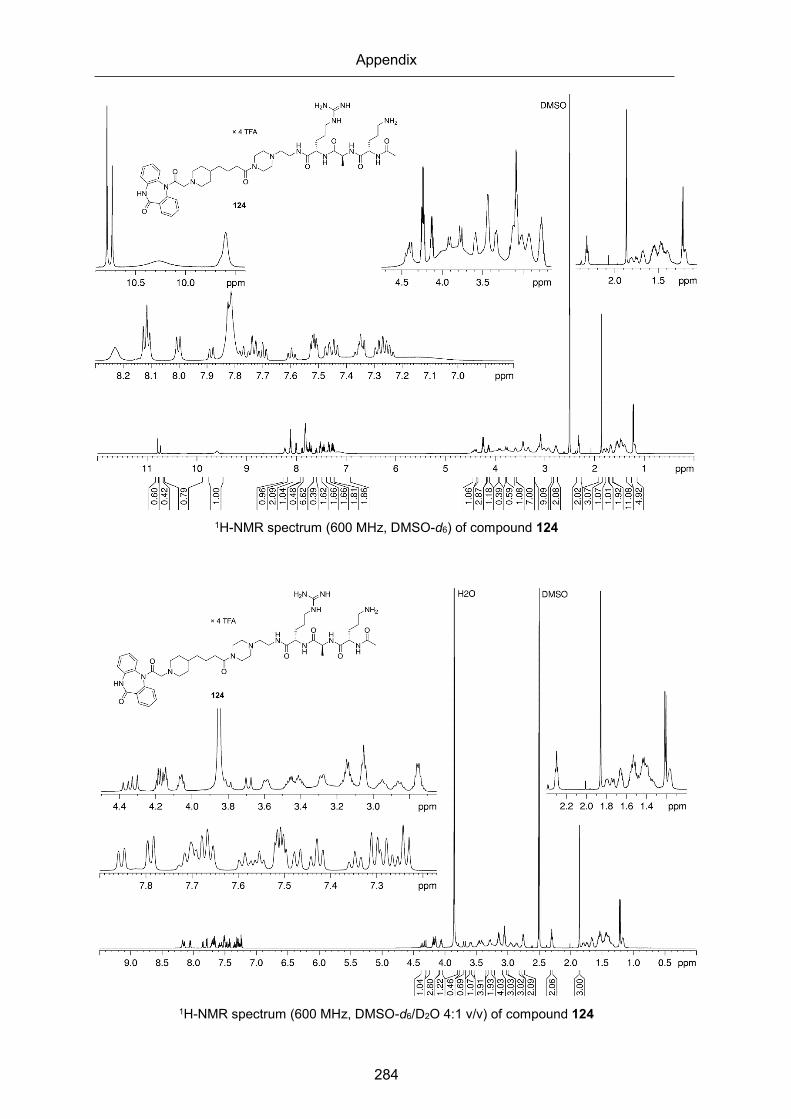
Appendix
284
1H-NMR spectrum (600 MHz, DMSO-d6) of compound 124
1H-NMR spectrum (600 MHz, DMSO-d6/D2O 4:1 v/v) of compound 124

Appendix
285
13C-NMR spectrum (150 MHz, DMSO-d6) of compound 124
1H-NMR spectrum (600 MHz, DMSO-d6) of compound 125

Appendix
286
1H-NMR spectrum (600 MHz, DMSO-d6/D2O 4:1 v/v) of compound 125
13C-NMR spectrum (150 MHz, DMSO-d6) of compound 125

Appendix
287
1H-NMR spectrum (600 MHz, DMSO-d6) of compound 126
1H-NMR spectrum (600 MHz, DMSO-d6/D2O 4:1 v/v) of compound 126

Appendix
288
13C-NMR spectrum (150 MHz, DMSO-d6) of compound 126
1H-NMR spectrum (600 MHz, DMSO-d6) of compound 134
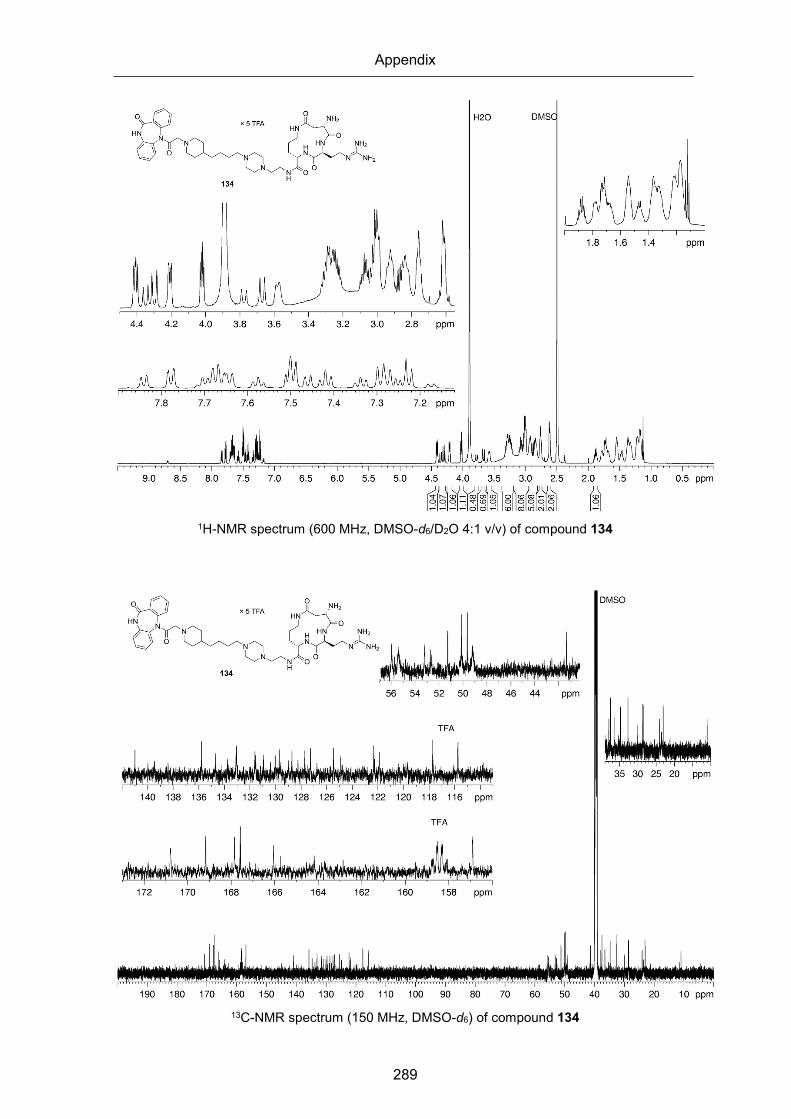
Appendix
289
1H-NMR spectrum (600 MHz, DMSO-d6/D2O 4:1 v/v) of compound 134
13C-NMR spectrum (150 MHz, DMSO-d6) of compound 134

Appendix
290
1H-NMR spectrum (600 MHz, DMSO-d6) of compound 135
1H-NMR spectrum (600 MHz, DMSO-d6/D2O 4:1 v/v) of compound 135

Appendix
291
13C-NMR spectrum (150 MHz, DMSO-d6) of compound 135
1H-NMR spectrum (600 MHz, DMSO-d6) of compound 137

Appendix
292
1H-NMR spectrum (600 MHz, DMSO-d6/D2O 4:1 v/v) of compound 137
1H-NMR spectrum (600 MHz, DMSO-d6) of compound 138

Appendix
293
1H-NMR spectrum (600 MHz, DMSO-d6/D2O 4:1 v/v) of compound 138
1H-NMR spectrum (600 MHz, DMSO-d6) of compound 139

Appendix
294
1H-NMR spectrum (600 MHz, DMSO-d6/D2O 4:1 v/v) of compound 139

Appendix
295
RP-HPLC chromatograms of compounds 6, 13, 23-25, 31, 35, 40, 43 53-60,63-65, 73-82, 85-99, 101-126, 134 and 135 (Chapter 2)

Appendix
296

Appendix
297

Appendix
298

Appendix
299

Appendix
300

Appendix
301

Appendix
302

Appendix
303

Appendix
304

Appendix
305

Appendix
306

Appendix
307

Appendix
308

Appendix
309
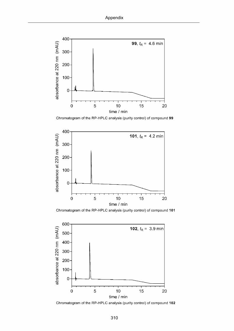
Appendix
310

Appendix
311

Appendix
312
[

Appendix
313

Appendix
314

Appendix
315

Appendix
316

Appendix
317

Appendix
318

Appendix
319
Chromatogram of RP-HPLC analysis (purity control) of compound 135

Appendix
320
1H and 13C-NMR spectra of compounds 144-149 (Chapter 3)
1H-NMR spectrum (600 MHz, DMSO-d6) of compound 144
144
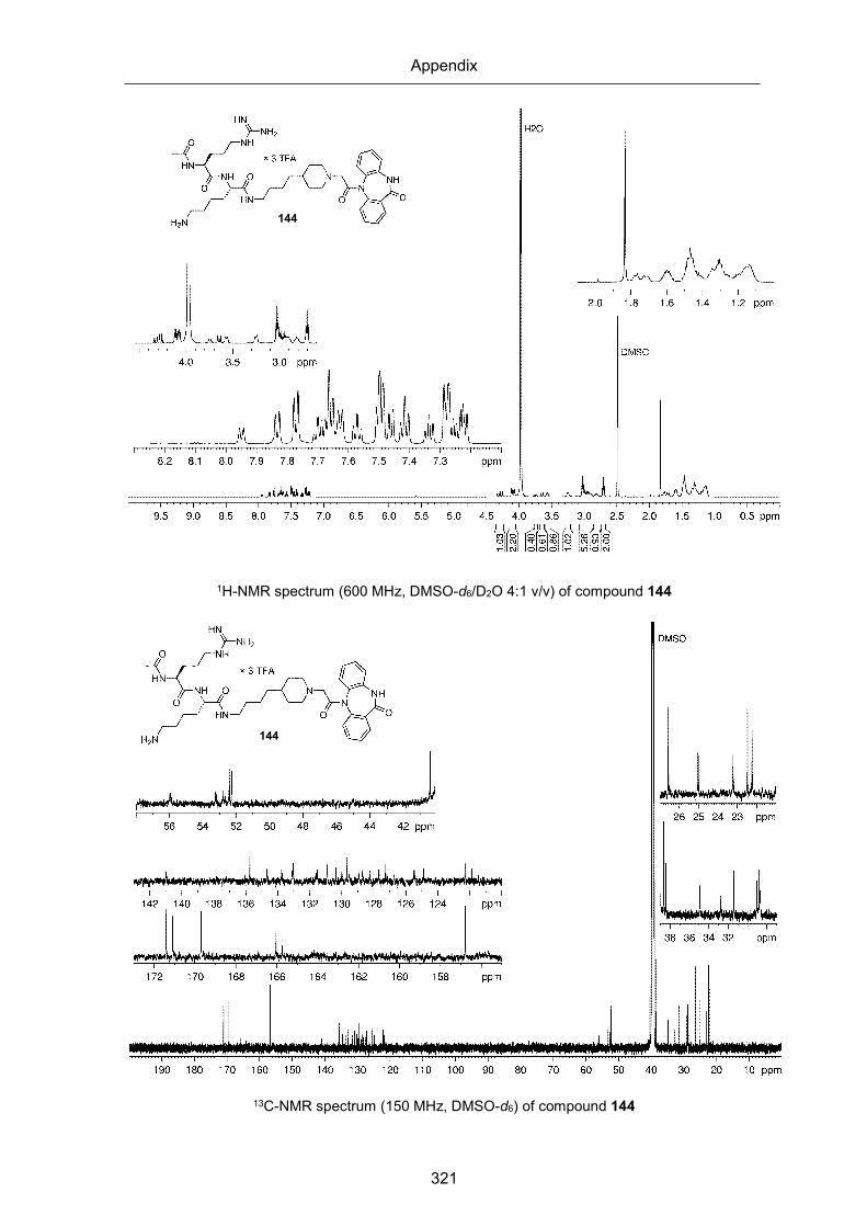
Appendix
321
1H-NMR spectrum (600 MHz, DMSO-d6/D2O 4:1 v/v) of compound 144
13C-NMR spectrum (150 MHz, DMSO-d6) of compound 144
144
144

Appendix
322
1H-NMR spectrum (600 MHz, DMSO-d6) of compound 145
1H-NMR spectrum (600 MHz, DMSO-d6/D2O 4:1 v/v) of compound 145
145
145

Appendix
323
13C-NMR spectrum (150 MHz, DMSO-d6) of compound 145
1H-NMR spectrum (600 MHz, DMSO-d6) of compound 146
145
146

Appendix
324
1H-NMR spectrum (600 MHz, DMSO-d6/D2O 4:1 v/v) of compound 146
13C-NMR spectrum (150 MHz, DMSO-d6) of compound 146
146
146

Appendix
325
1H-NMR spectrum (600 MHz, DMSO-d6) of compound 147
1H-NMR spectrum (600 MHz, DMSO-d6/D2O 4:1 v/v) of compound 147
147
147

Appendix
326
13C-NMR spectrum (150 MHz, DMSO-d6) of compound 147
1H-NMR spectrum (600 MHz, DMSO-d6) of compound 148
147
148

Appendix
327
1H-NMR spectrum (600 MHz, DMSO-d6/D2O 4:1 v/v) of compound 148
13C-NMR spectrum (150 MHz, DMSO-d6) of compound 148
148
148

Appendix
328
1H-NMR spectrum (600 MHz, DMSO-d6) of compound 149
1H-NMR spectrum (600 MHz, DMSO-d6/D2O 4:1 v/v) of compound 149
149
149

Appendix
329
13C-NMR spectrum (150 MHz, DMSO-d6) of compound 149
RP-HPLC chromatograms of compounds 144-149 (Chapter 3)
RP-HPLC analysis (purity control) of compound 144
0 5 10 15 20 25
0
100
200
300
400
500
tR = 6.4 min
time / min
abso
rban
ce a
t 220
nm
/ m
AU
149

Appendix
330
RP-HPLC analysis (purity control) of compound 145
RP-HPLC analysis (purity control) of compound 146
RP-HPLC analysis (purity control) of compound 147
0 5 10 15 20 25
0
100
200
300
400
500
tR = 6.8 min
time / min
abso
rban
ce a
t 220
nm
/ m
AU
0 5 10 15 20 25
0
100
200
300
400
500tR = 6.4 min
time / min
abso
rban
ce a
t 220
nm
/ m
AU
0 5 10 15 20 25
0
100
200
300
400tR = 6.5 min
time / min
abso
rban
ce a
t 220
nm
/ m
AU

Appendix
331
RP-HPLC analysis (purity control) of compound 148
RP-HPLC analysis (purity control) of compound 149
0 5 10 15 20 25
0
100
200
300
400tR = 6.2 min
time / min
abso
rban
ce a
t 220
nm
/ m
AU
0 5 10 15 20 25
0
100
200
300
400 tR = 8.0 min
time / min
abso
rban
ce a
t 220
nm
/ m
AU

Appendix
332
1H and spectra of compounds 164-169 (Chapter 4)
1H-NMR spectrum (600 MHz, MeOH-d4) of compound 164
164

Appendix
333
1H-NMR spectrum (600 MHz, MeOH-d4) of compound 165
1H-NMR spectrum (600 MHz, MeOH-d4) of compound 166
165
166

Appendix
334
1H-NMR spectrum (600 MHz, MeOH-d4) of compound 167
1H-NMR spectrum (600 MHz, MeOH-d4) of compound 168
167
168

Appendix
335
1H-NMR spectrum (600 MHz, MeOH-d4) of compound 169
169

Appendix
336
RP-HPLC chromatograms of compounds 164-169 (Chapter 4)
0 5 1 0 1 5 2 0
0
2 5 0
5 0 0
7 5 0
1 0 0 0
1 2 5 0
1 5 0 01 5 , t R = 7 . 4 m i n
t i m e / m i n
ab
so
rba
nc
e a
t 2
20
nm
(m
AU
)
Chromatogram of the RP-HPLC analysis (purity control) of compound 164
0 5 1 0 1 5 2 0
0
2 5 0
5 0 0
7 5 0
1 0 0 0
t i m e / m i n
ab
so
rba
nc
e a
t 2
20
nm
(m
AU
)
1 6 , t R = 7 . 6 m i n
Chromatogram of the RP-HPLC analysis (purity control) of compound 165
164, tR = 7.4 min
165, tR = 7.6 min

Appendix
337
0 5 1 0 1 5 2 0
0
1 0 0
2 0 0
3 0 0
4 0 0
1 7 , t R = 9 . 5 m i n
t i m e / m i n
ab
so
rba
nc
e a
t 2
20
nm
(m
AU
)
Chromatogram of the RP-HPLC analysis (purity control) of compound 166
0 5 1 0 1 5 2 0
0
2 0 0
4 0 0
6 0 0
8 0 0
1 0 0 01 8 , t R = 8 . 4 m i n
t i m e / m i n
ab
so
rba
nc
e a
t 2
20
nm
(m
AU
)
Chromatogram of the RP-HPLC analysis (purity control) of compound 167
0 5 1 0 1 5 2 0
- 5 0
0
5 0
1 0 0
1 5 0 1 9 , t R = 9 . 6 m i n
t i m e / m i n
ab
so
rba
nc
e a
t 2
20
nm
(m
AU
)
Chromatogram of the RP-HPLC analysis (purity control) of compound 168
166, tR = 9.5 min
167, tR = 8.4 min
168, tR = 9.6 min
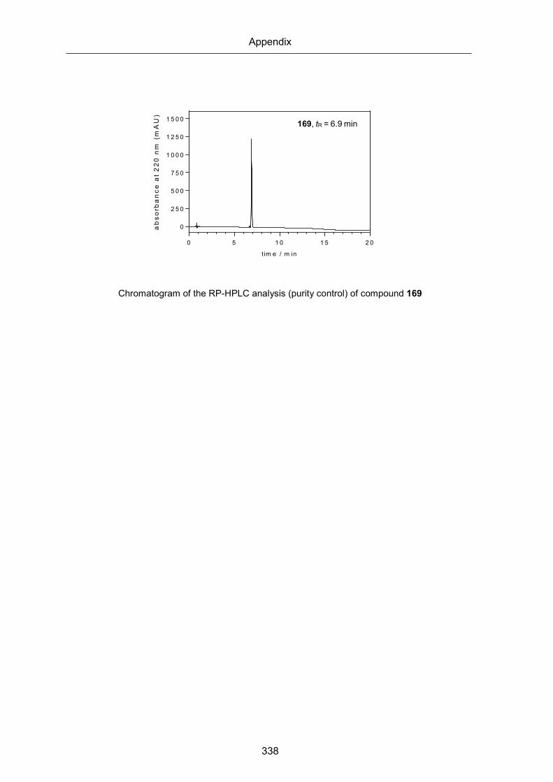
Appendix
338
0 5 1 0 1 5 2 0
0
2 5 0
5 0 0
7 5 0
1 0 0 0
1 2 5 0
1 5 0 0
tim e / m in
ab
sorb
an
ce a
t 2
20
nm
(m
AU
)
2 0 , tR = 6 .9 m in
Chromatogram of the RP-HPLC analysis (purity control) of compound 169
169, tR = 6.9 min

Appendix
339
Abbreviations
Agb shorter Arginine homologue with an ethylene instead of a trimethylene
moiety in the side chain
aq. aqueous
Boc tert-butoxycarbonyl
br broad signal
BSA bovine serum albumin
c concentration
CCh carbachol
CH2Cl2 dichlormethane
CHO-cells chinese hamster ovary cells
CNS central nervous system
COSY correlated spectroscopy
CRC concentration response curve
d doublet
DEA diethylamine
DIPEA N,N-diisopropylethylamine
DMF N,N-dimethylformamide
DMSO dimthylsulfoxid
δ chemical shift
EDC × HCl N-(3-dimethylaminopropyl)-N′-ethylcarbodiimide hydrochloride
ECL1 extracellular loop 2
eq. equivalent(s)
ESI electrospray ionization
EtOH ethanol
FACS fluorescence activated cell sorter
Fmoc 9-fluorenylmethoxycarbonyl
GPCR G-protein coupled receptor

Appendix
340
HBTU O-(1H-benzotriazol-1-yl)-N,N,N′,N′-tetramethyluronium
hexafluorophosphate
HFIP 1,1,1,3,3,3-hexafluoro-2-propanol
HMBC heteronuclear multiple bond correlation
HOBt 1-hydroxybenzotriazole hydrate
HRMS high resolution mass spectrometry
HSQC heteronuclear multiple bond correlation
J coupling constant
k retention (or capacity) factor (HPLC)
Kd dissociation constant obtained from a saturation binding experiment
Ki dissociation constant obtained from a competition binding experiment
K2CO3 potassium carbonate
m multiplet
M molar (mol/L)
MeOH methanol
miniG engineered minimal G-protein chimeras
MR muscarinic receptor
MxR muscarinic Mx (x = 1-5) receptor
NLuc Nanoluceriferase
NlucC C-terminal, smaller NLuc fragment with 11 amino acids
NlucN N-terminal, larger NLuc fragment with 158 amino acids
NMS N-methylscopolamine
PBS phosphate buffer saline
Pd/C palladium on carbon
PE petroleum ether
pIC50 negative logarithm of the IC50 in M
pKd negative logarithm of the Kd in M
pKi negative logarithm of the Ki in M
ppm parts per million

Appendix
341
PyBOP (Benzotriazol-1-yloxy)tripyrrolidinophosphonium hexafluorophosphate
q quartet
RP reverse-phase
Rt room temperature
s singulet
SEM standard eror of the mean
SOCl2 thionyl chloride
t0 hold-up time (also referred as dead time)
t (1) time, (2) triplet
TFA trifluoroacetic acid
TLC thin-layer chromatography
tR retention time
UV ultraviolet

Appendix
342
Overview of bold compound numerals and lab codes
Compound Labcode Compound Labcode Compound Labcode
2 UR-AP138 54 UR-CG152 99 UR-CG095 3 UR-AP148 55 UR-CG153 100 UR-CG269 4 UR-AP158 56 UR-CG154 101 UR-CG096 5 UNSW-MK259 57 UR-CG155 102 UR-CG190 8 UR-CG002 58 UR-CG172 103 UR-CG191 9 UR-CG057 59 UR-CG173 104 UR-CG197
10 UR-CG060 60 UR-CG195 105 UR-CG276 11 UR-CG025 61 UR-CG040 106 UR-CG277 12 UR-CG098 62 UR-MK242 107 UR-CG220 13 UR-CG079 63 UR-CG051 108 UR-CG221 17 UR-CG137 64 UR-CG263 109 UR-CG239 18 UR-CG138 65 UR-CG237 110 UR-CG213 19 UR-CG121 68 UR-CG156 111 UR-CG214 20 UR-CG140 69 UR-CG157 112 UR-CG215 21 UR-CG142 70 UR-CG160 113 UR-CG238 22 UR-CG122 71 UR-CG254 114 UR-CG235 23 UR-CG065 72 UR-SK-III-19 115 UR-CG104 24 UR-CG178 73 UR-CG103 116 UR-CG201 25 UR-CG128 74 UR-CG113 117 UR-CG202 27 UR-CG038 75 UR-CG114 118 UR-CG231 28 UR-CG039 76 UR-CG115 119 UR-CG203 30 UR-CG054 77 UR-CG150 120 UR-CG247 31 UR-CG055 78 UR-CG151 121 UR-CG233 33 UR-CG056 79 UR-CG170 122 UR-CG174 34 UR-CG061 80 UR-CG171 123 UR-CG175 35 UR-CG064 81 UR-CG194 124 UR-CG186 37 UR-CG179 82 UR-CG259 125 UR-CG187 38 UR-CG209 83 UR-CG041 126 UR-CG232 39 UR-CG216 84 UR-CG268 127 UR-CG240 40 UR-CG217 85 UR-CG097 128 UR-CG242 41 UR-SK-II-68 86 UR-CG100 129 UR-CG243 42 UR-CG206 87 UR-CG176 130 UR-CG244 43 UR-CG208 88 UR-CG177 131 UR-CG245 44 UR-CG083 89 UR-CG188 132 UR-CG248 45 UR-CG082 90 UR-CG189 133 UR-CG249 46 UR-CG081 91 UR-CG205 134 UR-CG250 47 UR-CG084 92 UR-CG260 135 UR-CG251 48 UR-CG080 93 UR-CG275 137 UR-CG265 49 UR-CG158 94 UR-CG117 138 UR-CG266 50 UR-CG159 95 UR-CG120 139 UR-CG267 51 UR-CG161 96 UR-CG092 140 UR-CG007 52 UR-MK216 97 UR-CG093 141 UR-CG008 53 UR-CG118 98 UR-CG094 142 UR-CG009

Appendix
343
Compound Labcode Compound Labcode Compound Labcode
143 UR-CG010 148 UR-CG020 165 UR-CG073 144 UR-CG015 149 UR-CG021 166 UR-CG074 145 UR-CG016 156 UR-SK-68 167 UR-AP175 146 UR-CG017 157 UR-MK257 168 UR-CG135 147 UR-CG018 164 UR-CG072 169 UR-MK342

344
Eidesstattliche Erklärung
Ich erkläre hiermit an Eides statt, dass ich die vorliegende Arbeit ohne unzulässige Hilfe
Dritter und ohne Benutzung anderer als der angegebenen Hilfsmittel angefertigt habe; die
aus anderen Quellen direkt oder indirekt übernommenen Daten und Konzepte sind unter
Angabe des Literaturzitats gekennzeichnet.
Einige der experimentellen Arbeiten wurden in Zusammenarbeit mit anderen Institutionen
und Personen durchgeführt. Vermerke zu den Beiträgen der betreffenden Personen finden
sich in den jeweiligen Kapiteln.
Weitere Personen waren an der inhaltlich-materiellen Herstellung der vorliegenden Arbeit
nicht beteiligt. Insbesondere habe ich hierfür nicht die entgeltliche Hilfe eines
Promotionsberaters oder anderer Personen in Anspruch genommen. Niemand hat von mir
weder unmittelbar noch mittelbar geldwerte Leistungen für Arbeiten erhalten, die im
Zusammenhang mit dem Inhalt der vorgelegten Dissertation stehen.
Die Arbeit wurde bisher weder im In- noch im Ausland in gleicher oder ähnlicher Form einer
anderen Prüfungsbehörde vorgelegt.
Regensburg, 22.10.2020
Corinna Weinhart