Inaugural-Dissertation - Universität zu Köln
133
Genomic approaches to dorsoventral patterning and functional studies of epithelial morphogenesis in the short-germ beetle Tribolium castaneum Inaugural-Dissertation zur Erlangung des Doktorgrades der Mathematisch-Naturwissenschaftlichen Fakultät der Universität zu Köln vorgelegt von Nadine Frey aus Lindau (Bodensee) Köln, 2017
-
Upload
khangminh22 -
Category
Documents
-
view
2 -
download
0
Transcript of Inaugural-Dissertation - Universität zu Köln

Genomic approaches to dorsoventral patterning and
functional studies of epithelial morphogenesis in
the short-germ beetle Tribolium castaneum
Inaugural-Dissertation
zur
Erlangung des Doktorgrades
der Mathematisch-Naturwissenschaftlichen Fakultät
der Universität zu Köln
vorgelegt von
Nadine Frey
aus Lindau (Bodensee)
Köln, 2017

2
Berichterstatter: Herr Professor Dr. Siegfried Roth
Herr PD Dr. Michael Kroiher
Vorsitzender der
Prüfungskommission: Frau Professor Dr. Ute Höcker
Tag der mündlichen Prüfung: 19.12.2017

3
Table of contents
Zusammenfassung ................................................................................................................................... 7
Abstract ................................................................................................................................................... 9
CHAPTER I
Reconstructing the dorsoventral gene regulatory network of Tribolium castaneum ....................... 10
1 Introduction ................................................................................................................................... 10
1.1 Gene regulatory networks (GRNs)......................................................................................... 10
1.2 Tribolium as an emerging model organism ........................................................................... 11
1.3 The evolution of dorsoventral patterning ............................................................................. 13
1.3.1 Formation of the dorsoventral axis in Drosophila melanogaster ................................. 14
1.3.2 The dorsoventral gene regulatory network of Tribolium castaneum ........................... 15
1.4 Identification of new DV-patterning genes by differential expression analysis ................... 17
1.4.1 Generation of knockdown embryos lacking key DV patterning genes ......................... 17
1.4.2 Validation of the knockdown efficiency ........................................................................ 20
1.4.3 Identification of differentially expressed genes ............................................................ 20
1.5 Aims of the study ................................................................................................................... 22
2 Results ........................................................................................................................................... 23
2.1 Verification of the RNA-seq data by an in-situ hybridization screen .................................... 23
2.1.1 Detailed analysis of two subgroups of differentially expressed genes ......................... 24
2.1.2 Identification of genes regulated by Dpp ...................................................................... 24
2.1.3 Expression patterns of genes regulated by Toll and/or Twist ....................................... 26
2.2 Functional analysis of genes potentially involved in the DV-GRN of Tribolium .................... 28
2.2.1 Functional analysis of Sog/Dpp targets ......................................................................... 29
2.2.2 Functional analysis of Toll and Twist targets ................................................................. 31
3 Discussion ...................................................................................................................................... 36
3.1 Verification of the RNA-sequencing data .............................................................................. 36
3.2 Expression patterns of some candidates suggest an involvement in DV patterning ............ 37
3.2.1 Tc-uninflatable knockdown affects the formation of abdominal segments ................. 38
3.2.2 Tc-tartan is an early regulator in neurogenesis ............................................................ 38
3.2.3 Involvement of the FGF pathway in formation of mesodermal derived tissue ............ 39
4
3.2.4 Extracellular matrix components (ECMs) and their role in mesoderm formation ........ 40
3.2.5 Zfh1 - A new essential regulator maintaining late Tc-twist expression ........................ 41
3.3 Possibilities and limitations of comparative analysis by RNA-sequencing ............................ 42
3.3.1 Stronger influence of Tribolium homologs on early embryonic development ............. 42
3.3.2 Comparative analysis for de novo identification of DV-GRN components ................... 43
3.3.3 Reasons for the detection of false positives ................................................................. 44
3.4 Conclusion ............................................................................................................................. 45
3.5 Outlook .................................................................................................................................. 46
CHAPTER II
Fog signaling and its role in epithelial morphogenesis in Tribolium castaneum ............................... 47
1 Introduction ................................................................................................................................... 47
1.1 The role of cell shape changes in embryonic development .................................................. 47
1.2 The Fog signaling pathway .................................................................................................... 49
1.2.1 The Pathway components and their interaction ........................................................... 49
1.2.2 Regulation of Fog signaling ............................................................................................ 52
1.2.3 Fog and T48 - Two distinct mechanisms of myosin localization ................................... 52
1.3 Conservation of Fog signaling in insect morphogenesis ....................................................... 54
1.4 Different modes of mesoderm internalization...................................................................... 55
1.5 Morphogenetic movements during Tribolium gastrulation .................................................. 56
1.6 Aims of the study ................................................................................................................... 59
2 Results ........................................................................................................................................... 60
2.1 Expression patterns of the Fog signaling key components in Tribolium castaneum ............ 60
2.2 Knockdown of Fog signaling components in Tribolium results in a variety of defects ......... 65
2.3 Fog signaling is required for primitive pit formation ............................................................ 66
2.4 Mesoderm invagination is delayed upon Tc-fog knockdown ............................................... 70
2.5 Positioning of the primordial germ cells is controlled by Fog signaling ................................ 75
2.6 Regulation of Fog signaling in Tribolium ............................................................................... 77
2.7 Serosa spreading - A novel function of Fog signaling ............................................................ 81
2.8 Knockdown of smog does not result in morphogenetic defects .......................................... 83
2.9 T48 does not contribute to mesoderm internalization in Tribolium castaneum .................. 85

5
3 Discussion ...................................................................................................................................... 86
3.1 Fog and its conserved role in tissue internalization during gastrulation .............................. 86
3.2 Genes with less influence on apical constrictions in Tribolium compared to Drosophila ..... 88
3.3 The importance of posterior amniotic fold formation by Fog signaling ............................... 89
3.4 Positioning of the primordial germ cells is influenced by Fog signaling ............................... 90
3.5 The regulatory network underlying Fog signaling ................................................................. 92
3.6 Serosa spreading and cellularization - A novel function of Fog signaling ............................. 93
3.6.1 Fog signaling regulates flattening of the serosa cells .................................................... 93
3.6.2 A role of Fog signaling in the blastoderm ...................................................................... 95
3.7 Conclusion ............................................................................................................................. 97
Material &Methods ............................................................................................................................... 98
1 Strains ............................................................................................................................................ 98
2 Animal keeping and embryo collection ......................................................................................... 98
3 Total RNA isolation ........................................................................................................................ 99
4 cDNA synthesis .............................................................................................................................. 99
5 Primer design ............................................................................................................................... 100
6 Standard Polymerase Chain Reaction (PCR) ................................................................................ 100
7 Gel electrophoresis...................................................................................................................... 101
8 In-situ hybridization (ISH) ............................................................................................................ 101
8.1 Generation of mRNA probes ............................................................................................... 101
8.1.1 Two-step Polymerase Chain Reaction ......................................................................... 101
8.1.2 Probe synthesis............................................................................................................ 102
8.2 Embryo fixation ................................................................................................................... 103
8.3 Detection of mRNA expression ........................................................................................... 103
9 Double in-situ hybridization (dISH) ............................................................................................. 104
10 Fluorescence staining .............................................................................................................. 106
10.1 Phalloidin staining ............................................................................................................... 106
10.2 Nuclear staining ................................................................................................................... 107
10.2.1 DAPI staining................................................................................................................ 107
10.2.2 Sytox staining ............................................................................................................... 107
11 Generation of knockdown embryos by RNA interference (RNAi) ........................................... 108
11.1 Production of double-stranded RNA (dsRNA) ..................................................................... 108

6
11.1.1 2. PCR for amplification of a dsRNA template ............................................................. 108
11.1.2 dsRNA synthesis .......................................................................................................... 108
12 Adult or pupal injections (pRNAi) ............................................................................................ 109
13 Embryonic injections (eRNAi) .................................................................................................. 110
14 Cuticle preparation .................................................................................................................. 111
15 Cryosections ............................................................................................................................ 111
16 Microscopy .............................................................................................................................. 112
17 Live-imaging ............................................................................................................................. 112
References ........................................................................................................................................... 113
Appendix A: Primerlist RNA-Seq .......................................................................................................... 123
Appendix B: Genes differentially expressed upon Tc-dpp kd and Tc-sog kd ...................................... 124
Appendix C: Genes differentially expressed upon Tc-twist vs Tc-Toll knockdown ............................. 125
Appendix D: Marker gene expression in Tc-sog and Tc-dpp knockdown embryos ............................ 127
Appendix E: Marker gene expression in Tc-Toll and Tc-twist knockdown embryos ........................... 128
Appendix F: Varity in knockdown strength of Tc-LanB1 ..................................................................... 129
Appendix G: Primerlist Fog signaling ................................................................................................... 130
Appendix H: Movies ............................................................................................................................ 130
Acknowledgements ............................................................................................................................. 131
Erklärung zur Dissertation ................................................................................................................... 132
Lebenslauf ........................................................................................................................................... 133

7
Zusammenfassung
Ein entscheidender Schritt in der frühen Embryonalentwicklung ist die Festlegung verschiedener
Zellschicksale entlang der dorsoventralen (DV) Achse. Das Zellschicksal wird durch die räumliche und
zeitliche Expression bestimmter Gene festgelegt, die durch genregulatorische Netzwerke (GRN)
reguliert werden. Diese Arbeit befasst sich mit der Evolution des DV-GRN von Insekten. In der Gruppe
der stark spezialisierten Drosophiliden ist die dorsoventrale Musterbildung durch den Toll Signalweg
reguliert, welcher den BMP Signalweg kontrolliert. Während das dorsoventrale Netzwerk in Drosophila
melanogaster gut untersucht ist, ist nur wenig über das genregulatorische Netzwerk bekannt, welches
die Bildung der dorsoventralen Achse des Roten Reismehlkäfers Tribolium castaneum reguliert. Das
Ziel dieser Arbeit war es neue potentielle Dorsoventral-Gene in Tribolium zu identifizieren. Um dieses
Ziel zu erreichen, wurde eine vergleichende Transkriptom-Analyse nach der Herunterregulierung
verschiedener Bestandteile des Toll und BMP Signalwegs durch RNAi durchgeführt. Ich habe die
Expressionsmuster ausgewählter Gruppen der 796 differentiell exprimierten Genen untersucht. Des
Weiteren, wurden funktionelle Untersuchungen für Gene durchgeführt, die ein typisches
dorsoventrales Expressionsmuster aufweisen. Die Ergebnisse weisen darauf hin, dass einige der
konservierten Dorsoventral-Gene (z.B. Tartan) einen stärkeren Einfluss auf die dorsoventrale
Musterbildung in Tribolium haben als ihre Drosophila Homologe. Zusätzlich unterscheiden sich die
Expressionsdomänen von einigen Notch Signalweg Bestandteilen erheblich von denen ihrer Homologe
in Drosophila. Interessanterweise wird Zfh-1 anders als in Drosophila, für die Aufrechterhaltung der Tc-
twist Expression während der Keimbandstreckung benötigt und ist daher essenziell ist für die
Entwicklung des Mesoderms in Tribolium.
Neben der Bildung der Köperachsen, tragen auch morphogenetische Bewegungen von Epithelien
erheblich zur Entwicklung komplexer Embryonalstrukturen bei. In Drosophila ist der Fog Signalweg an
der Kontrolle morphogenetischer Bewegungen beteiligt. Fog induziert apikale Zellkonstriktionen,
welche die Invagination von Epithelien ermöglichen. Die Inaktivierung des Fog Signalwegs führt zu
Defekten in der Internalisierung des Mesoderms und des hinteren Mitteldarms. Bisher wurde davon
ausgegangen, dass der Fog Signalweg ausschließlich während Gastrulation einiger stark abgeleiteter
Fliegen eine Rolle spielt. Im zweiten Teil dieser Arbeit wird die Funktion des Fog Signalwegs im
Tribolium Embryo untersucht, welcher eine im Vergleich zu Drosophila ursprünglichere Form der
Embryonalentwicklung aufweist. Interessanterweise führt die Herunterregulierung wichtiger Fog
Signalwegbestandteile in Tribolium zu einer verzögerten Mesoderminternalisierung und einem
vollständigen Verlust der Internalisierung des Hinterdarms und posterioren Endoderms. Dies weist auf
eine Konservierung des Fog Signalwegs für die Gewebeinternalisierung während der Gastrulation
außerhalb von höheren Dipteren hin. Zusätzlich werden in dieser Arbeit bisher unbekannte Funktionen

8
des Fog Signalwegs bei der Bildung der posterioren Amnionfalte, bei der Positionierung der
primordialen Keimzellen und in der Ausweitung der extraembryonalen Serosa erläutert. Auch deuten
die Ergebnisse auf eine weitere konservierte, allgemeinere Rolle von Fog für die Koordinierung von
Zellformveränderungen im Blastoderm hin.

9
Abstract
A crucial step in early embryonic development is the determination of different cell fates along the
dorsoventral (DV) axis. These cell fates are defined by the spatially and temporally restricted
expression of gene sets, controlled by complex gene regulatory networks (GRN). Here, we address the
evolution of the DV-GRN within insects. In the highly derived group of the drosophilids, dorsoventral
patterning is dominated by Toll-signaling, which directly controls BMP-signaling. Whereas the DV-GRN
of Drosophila melanogaster is well understood, little is known about the GRN that acts during
establishment of the DV axis in more basally branching insects like the red flour beetle Tribolium
castaneum. This work aimed to identify new potential DV patterning genes in Tribolium. To achieve
this goal, a comparative transcriptome analysis after knockdown of Toll signaling and BMP signaling
components by RNAi was performed. I analyzed the expression patterns of selected subgroups of the
796 differentially expressed genes. Furthermore, functional studies for genes exhibiting typical
dorsoventral expression patterns were performed. The results suggest that some of the conserved DV-
GRN components (e.g. Tartan) have a stronger and earlier influence on DV patterning in Tribolium. In
addition, the expression of Notch signaling components clearly differs from the expression pattern of
their Drosophila homologs. But the most striking finding was the identification of Zfh-1, which is
required for maintaining Tc-twist expression during germ band extension and is thus essential for
mesoderm development in Tribolium.
Besides the establishment of the body axis, morphogenetic movements of epithelial sheets
significantly contribute to the development of complex embryonic structures. The Fog signaling
pathway is one of the best studied processes in initiating early morphogenetic movements by cell
shape changes. Modifications of the acto-myosin cytoskeleton by Fog signaling result in apical cell
constrictions which enable bending of epithelial sheets. In Drosophila, loss of Fog signaling causes
defects in mesoderm and posterior midgut internalization. So far, it was assumed that Fog signaling is
exclusively involved in gastrulation in some highly derived flies. In the second part of this work the role
of Fog signaling was analyzed in Tribolium, which possesses a more ancestral mode of embryogenesis.
Interestingly, knockdown of important Fog signaling components in Tribolium cause similar defects in
ventral furrow formation and internalization of the posterior endoderm. This indicates the
conservation of Fog signaling in tissue internalization during gastrulation outside of higher dipterans.
In addition, this work presents so far unknown functions of the Fog signaling pathway in formation of
the posterior amniotic fold, positioning of the primordial germ cells and spreading of the serosa.
Furthermore, the results suggest another conserved more general role of Fog in coordinating cell shape
changes in the blastoderm.

10
CHAPTER I
Reconstructing the dorsoventral gene regulatory network of Tribolium castaneum
1 Introduction
1.1 Gene regulatory networks (GRNs)
Pathways are a simple way to visualize gene interactions. In a pathway each component acts upon a
substrate or another gene product. Nevertheless, a pathway is often limited in describing the various
complex gene interactions. In fact, genes or gene products are not only affecting single targets, but are
also influenced by numerous gene products, deriving from multiple pathways. This complex system of
interaction between different pathways can be best described as networks (Stern, 2010). Gene
regulatory networks (GRNs) consist of various regulatory inputs like binding of gene products to
enhancers, silencers and insulators (cis-regulatory elements) (Levine and Davidson, 2005). The
diversity of cis-regulatory elements establishes compartmentation by enabling the independent
control of gene transcription in different body parts (Carroll, 2005).
Transcription factors (TFs) are often the most essential parts of GRNs and act as key regulators.
Different transcription factors can concurrently bind to one enhancer. Depending on the set of bound
TFs at a specific time, the enhancer activates the expression of a specific gene set, thus resulting in
functional output (Levine and Davidson, 2005). Furthermore, gene expression can be influenced by
genetic circuits. By feed-forward loops components of a network can modify their own expression by
either enhancing or repressing it. These circuits act directly by negative or positive self-regulation or
indirectly via regulation of target genes (Peter and Davidson, 2011).
Embryogenesis is controlled by large gene-regulatory networks, which generate spatially and
temporally refined patterns of gene expression, enabling progressive restriction of cell fates from
pluripotent fields of cells to complex organs and tissues. (Sandmann et al., 2007). Evolutionary
alterations of the functional organization of the gene regulatory networks that control development
of the body plan cause changes in animal anatomy (Peter and Davidson, 2011). In fact, instead of
changes in protein structure, evolutionary changes in morphology occur primarily through changes in
regulatory sequences (Carroll, 2005). The dorsoventral gene regulatory network (DV-GRN) of
Drosophila was already intensively studied (Bonn and Furlong, 2008; Stathopoulos and Levine, 2002,
2005). However, elucidating the genes and their interlinkage in the DV-GRN of more ancestral insects
like Tribolium or Oncopeltus, might provide insights in the integration of Toll signaling in the regulation
of early developmental processes like cell-fate determination.

11
1.2 Tribolium as an emerging model organism
Due to their diversity and the easy husbandry conditions, insects are suitable for evolutionary
comparative analysis. Drosophila is a member of the evolutionary relatively young order of Diptera,
and many aspects of its embryogenesis are highly derived. Tribolium castaneum (Tc) is a more basally
branching member of the insect lineage, with ~300 million years of independent evolution from
Drosophila (Rousso et al., 2010). Thus, it shares many features with other insects, particularly a more
ancestral mode of embryonic development, compared to Drosophila. In long germ-type embryos, like
those of Drosophila melanogaster, the segments are formed synchronously in the blastoderm. In short
germ-type insects, like Tribolium castaneum, only head and thorax segments are generated in the
blastoderm stage, while the abdominal segments derive from a posterior segment addition zone (Fig.
1.1) after gastrulation. In addition, Tribolium embryos develop two distinct extraembryonic
membranes: amnion and serosa. The fruit fly possesses only a reduced amnioserosa (Tautz and
Sommer, 1995). However, the localization of the three germ layers (mesoderm, neurogenic ectoderm
and dorsal ectoderm) along the DV axis does not differ between both insects.
Figure 1.1: Fate determination and segmentation in D. melanogaster and T. castaneum Drosophila as well as Tribolium mesoderm, neurogenic ectoderm and dorsal ectoderm are specified in the blastoderm. In long germ-type embryos like Drosophila, all segments are formed synchronously in the blastoderm. In short germ-type embryos, like Tribolium castaneum the segments are successively added at the posterior end after gastrulation. Only the most anterior segments are formed in the blastoderm. Drosophila possesses a single extraembryonic tissue, the amnioserosa. Tribolium has two extraembryonic membranes: amnion and serosa. modified from (Sommer, 2009)

12
Many tools for functional analysis are available in Tribolium castaneum. Diverse methods for
generation of transgenic lines (Berghammer et al., 2009; Gilles and Averof, 2014; Gilles et al., 2015;
Lorenzen et al., 2003; Pavlopoulos et al., 2004) and for transient gene expression (Benton et al., 2013)
could be established, enabling advanced techniques like live-imaging. In addition, Tribolium shows a
strong RNAi response (parental as well as embryonic) (Bucher et al., 2002; Posnien et al., 2009),
providing many possibilities for functional studies. Furthermore, the availability of a well annotated
genome (Tcas 5.2/OGS 3) as well as various transcriptomic data (different embryonic stages) and a
database, containing functional information on genes based on a genome-wide RNAi screen
(iBeetleBase, Schmitt-Engel et al., 2015), qualify Tribolium as a perfect candidate for comparative
analysis. Although the DV-GRN of Tribolium shows some regulatory differences compared to the
Drosophila DV-GRN, the flour beetle has still an evolutionary intermediate position compared to other
insects. For example, the jewel wasp Nasonia vitripennis (Hymenoptera), or the milk weed bug
Oncopeltus (Hemiptera) show both a stronger reliance on BMP signaling (Lynch and Roth, 2011; Ozuak
et al., 2014; Sachs et al., 2015). The evolution of the insect DV-GRN will be described in detail in the
next section.

13
1.3 The evolution of dorsoventral patterning
The formation of the two body axes, the anterior-posterior (AP) axis and the dorsoventral (DV) axis is
a crucial early step in establishing the body of any animal (Wilson et al., 2014). Though, early
embryogenesis and corresponding formation of the axes varies in different species. BMP (bone
morphogenic protein) signaling and its role in dorsoventral patterning is highly conserved (De Robertis
and Kuroda, 2004). However, in the model organism Drosophila melanogaster the Toll pathway plays
the major role in dorsoventral patterning, whereas BMP acts downstream and only on the dorsal side
of the embryo (Lynch and Roth, 2011) (Fig. 1.2). Although, Toll signaling has a conserved function in
innate immunity of almost all bilaterians (Caamano and Hunter, 2002; Lemaitre et al., 1996; Rosetto
et al., 1995), its role in axis formation appears to be an evolutionary novelty in insects (Leulier &
Lemaitre 2008). This leads to the conclusion that Toll was integrated into an existing patterning
pathway during insect evolution. Regarding its evolutionary changing role in DV-patterning, the Toll-
NFκB/Dorsal pathway is an interesting subject that promises insights into evolution of gene regulation
and into functional constraints, underlying gene regulatory network (GRN) evolution. Comparative
studies using more basally branching insects might help to understand the mechanisms of such
modifications. The emerging model organism Tribolium castaneum is an insect with more ancestral
features of embryogenesis and is thus, a perfect candidate for comparative analysis. The following
section concentrates on Drosophila in order to highlight the best understood DV patterning system,
which is the foundation for comparative studies in this thesis.
Figure 1.2: Evolution of dorsoventral patterning The BMP pathway (blue) plays a conserved role in the establishment of dorsoventral polarity in vertebrates as well as in arthropods. However, during insect evolution the Toll pathway (red) which has a conserved role in innate immunity, became dominant in establishing the DV axis.

14
1.3.1 Formation of the dorsoventral axis in Drosophila melanogaster
One of the best studied developmental processes is the dorsoventral axis formation of Drosophila
melanogaster. Multiple former studies uncovered the fine-tuned, spatio-temporal gene expression
which leads to the determination of different cell fates.
The DV-axis is established by migration of the oocyte nucleus from a central-posterior towards an
anterior-cortical location (Peri and Roth, 2000; Roth, 2003; Roth and Lynch, 2009). The maternal mRNA
of the TGFα-like ligand Gurken is located around the nucleus and activates upon translation, the EGF
receptor (Roth, 2004) in the overlying follicle cells (Schupbach and Roth, 1994). EGFR signaling restricts
the expression of the sulfotransferase Pipe to the ventral side where it modifies several components
of the vitelline membrane (Nilson and Schupbach, 1998; Roth, 2003). This dorsoventral polarity of the
egg chamber is transmitted to the early embryo via an extracellular proteolytic cascade in the
perivitelline space (Fig. 1.3).
The final step of the protease cascade is the cleavage of Spätzle. After cleavage, Spätzle binds and
activates the Toll receptor. Probably, the cleaved Spätzle protein is present in a concentration gradient.
Cleavage of the Toll-ligand Spätzle generates a gradient of Toll receptor activation on the ventral side
of the embryo (Morisato, 2001; Moussian and Roth, 2005). The activation of the Toll receptor leads to
the phosphorylation and degradation of Cactus. Usually Cactus binds the NF-κB transcription factor
Dorsal, which is consequently retained in the cytoplasm. Upon degradation of Cactus, Dorsal can
translocate into the nuclei which leads to the establishment of a nuclear Dorsal gradient (Fig. 1.3) with
peak levels at the ventral midline (Moussian and Roth, 2005; Roth et al., 1991). The nuclear Dorsal
gradient in Drosophila is stable and leads to the transcriptional regulation of 60-70 different target
genes (Type I, Type II, Type III) which are activated or repressed by Dorsal in a concentration-
dependent manner (Reeves and Stathopoulos, 2009) (Fig. 1.3). Genes like twist and snail are
expressed, due to high nuclear Dorsal levels (Type I), on the ventral-most side of the embryo and
specify the presumptive mesoderm (Jiang et al., 1991; Leptin, 1991). In more lateral regions of the egg
circumference, intermediate nuclear Dorsal concentrations activate the transcription of genes like
ventral nervous defective (vnd) (Type II), which trigger formation of the neuroectoderm.

15
Figure 1.3: Establishment of the dorsoventral axis in Drosophila melanogaster Activation of EGF signaling (blue) in the dorsal ovary follicle cells by Gurken (green) restricts Pipe (red) to the ventral side. Pipe activates a protease cascade in the perivitelline space. The cleaved and thus active ligand Spätzle binds to Toll receptors which finally results in the establishment of a nuclear Dorsal gradient with peak levels at the ventral midline (brown). Dependent on the nuclear Dorsal concentration, different target genes are expressed along the dorsoventral axis.
Two subgroups of Type III genes are regulated by low nuclear dorsal concentrations. Type III+ genes
like short gastrulation (sog) are activated, while Type III- genes like the BMP homolog decapentaplegic
are repressed (Hong et al., 2008; Reeves and Stathopoulos, 2009). dpp is expressed on the dorsal most
side, where no nuclear Dorsal is present. Sog acts as an inhibitory modulator for BMP signaling and
antagonizes the BMP2/4-like ligand Decapentaplegic (Dpp) in lateral regions (Reeves et al., 2012). Dpp
gets bound and transported to the dorsal side, where it is cleaved by the metallo-protease Tolloid.
Released Dpp can than bind to its receptors (Nunes da Fonseca et al., 2008). This mechanism restricts
the activation of BMP signaling to the dorsal side, which is necessary for the specification of the dorsal
non-neurogenic ectoderm and the amnioserosa (Nunes da Fonseca et al., 2008). The differences of the
DV-GRN of Tribolium will be discussed in the following section.
1.3.2 The dorsoventral gene regulatory network of Tribolium castaneum
DV patterning in Tribolium shows many similarities to DV-patterning in Drosophila melanogaster.
However, the mRNA of the Tribolium TGFα homolog is not localized in the ovaries. Thus, the process
of DV polarity transmission from the ovary to the embryo remains unclear (Lynch and Roth, 2011).
Former studies showed that Toll is also a key component in DV patterning of Tribolium castaneum. Like
in Drosophila, the activation of Tc-Toll by binding of Tc-Spätzle leads to nuclear uptake of Tc-Dorsal.
But in contrast to Drosophila, the resulting Tc-Dorsal gradient is spatio-temporally dynamic (Chen et
al., 2000; Nunes da Fonseca et al., 2008). The Tc-Dorsal gradient refines from a broad domain in the
early blastoderm to a narrow stripe along the ventral midline (Fig. 1.4). Finally, it disappears from the
germ rudiment before the onset of gastrulation (Chen et al., 2000). In addition, Toll and cactus are
zygotically expressed genes in Tribolium, whereas they are maternally expressed in the fruit fly (Lynch
and Roth, 2011). Thus, the dynamism of the Dorsal gradient is caused by several negative and positive
feedback mechanisms that act on zygotic level (Nunes da Fonseca et al., 2008). Previous studies (Lynch

16
and Roth, 2011; Nunes da Fonseca et al., 2008) showed that Tc-Cactus is activated by Tc-Dorsal itself
and in addition by the Tc-Dorsal target Tc-Twist. This Dorsal-/Twist-dependent activation of Tc-Cactus
simultaneously leads to an enhanced repression of Tc-Dorsal. In addition, Tc-Dorsal regulates its own
activator Tc-Toll in a positive feedback loop (Fig. 1.4). Furthermore, ChIP sequencing experiments
revealed a potential self-regulative input of Tc-Dorsal and a positive regulation of Tc-Dorsal on the Toll
ligand Tc-Spätzle (Stappert, PhD Thesis 2014).
Another difference is that the BMP pathway plays a more important role in DV-patterning of Tribolium
castaneum, and that the Toll pathway influences less target genes compared to the derived patterning
system of Drosophila. It was shown, that loss of sog in Drosophila results in weaker defects than the
Tc-sog kd, which leads to a strong dorsalization of the embryo, indicating that Tc-Dorsal is not a direct
repressor of Tc-dpp (Nunes da Fonseca et al., 2010). Moreover, dpp knockout in Drosophila does not
affect the mesoderm, while knockdown of dpp in Tribolium results in a slight expansion of the
mesoderm (Nunes da Fonseca et al., 2008). These results strengthen the presumption that Toll's role
in DV patterning is derived, and that the DV-GRN changed since the last common ancestor of flies and
beetles. The approach to elucidate these changes is described in the following sections.
Figure 1.4: Regulation of dorsoventral patterning genes The Drosophila nuclear Dorsal-gradient is stable and regulates the transcription of ~ 50 target genes in a concentration-dependent manner. The same signaling cascade initiates the expression of Tc-twist and other DV patterning genes in Tribolium castaneum. However, in contrast to Drosophila the nuclear Tc-Dorsal gradient is dynamic due to feedback mechanisms. Tc-cactus is regulated by Dorsal and by the Dorsal target gene twist. This activation establishes a negative feedback loop, as Cactus has a negative influence on the nuclear uptake of Dorsal. In addition, Tc-Dorsal regulates its own activator Tc-Toll in a positive feedback loop. modified from (Chen et al., 2000; Nunes da Fonseca et al., 2008; Stappert, PhD Thesis 2014)

17
1.4 Identification of new DV-patterning genes by differential expression
analysis
Comparative studies are often performed by candidate gene approaches, requiring sequence
identification of already known pathway components in the organism of interest by DNA or protein
BLAST. Many important evolutionary studies are based on this approach. However, analyzing
evolutionary changes in gene regulatory networks by a candidate gene approach depends on the
presence of the respective pathway components, rendering discovery of new genes involved in a GRN
impossible. Modern techniques like whole genome sequencing, identification of binding regions by
ChIP-sequencing or generation of transcriptomes by RNA-sequencing (RNA-seq), facilitate new
opportunities in the wide field of evolution and development. To get more insights into the DV-GRN of
Tribolium castaneum and the differences in composition and wiring of the genes to the DV-GRN of
Drosophila melanogaster, I performed a genome wide comparative differential expression analysis
after RNAi. The establishment of this modern technique for comparative studies in Tribolium was
performed together with Dominik Stappert (Frey, Master Thesis 2013; Stappert, PhD Thesis 2014).
1.4.1 Generation of knockdown embryos lacking key DV patterning genes
To identify new potential target genes, transcriptomes of Tc-Toll1, Tc-twist, Tc-sog and Tc-dpp
knockdown (kd) embryos were generated by parental RNA interference (pRNAi). Wildtype embryos
derived from uninjected mothers served as control. Considering possible false positive results by genes
activated via the injection procedure itself (e.g. inflammatory genes, RNAi machinery genes), an
additional control was used. Therefore, female pupae were injected with dsRNA for dsRed which is
derived from Discosoma spec. and is not present in the Tribolium genome. Thus, dsRed knockdown
embryos should not show differential gene expression.
The RNA for transcriptome analyses was isolated from embryos derived from dsRNA injected mothers
and staged to 7.5h to 11.5h AEL. At that stage, the embryo proceeds from the early differentiated
blastoderm stage to the horseshoe stage. The carefully selected time window captures a critical time
in DV patterning. During the differentiated blastoderm numerous direct target genes of the key
regulators Dorsal and Twist are expressed. These genes are especially important for tissue specification
and early tissue differentiation during the gastrulation of the embryo. The embryos were stained with
DAPI and surveyed for phenotypic penetrance (Fig. 1.5).
1 Knockdown of Tc-dorsal results in a higher death rate of the injected females and to a reduced number of eggs. Tc-Toll RNAi is upstream of Tc-dorsal and thus phenocopies Tc-dorsal with regard to its dorsoventral phenotype.

18
dsRed kd embryos do not show any morphological defects and look similar to embryos from untreated
wildtype control females (Fig. 1.5 A-A`). Although knockdown of Tc-twist results in complete loss of the
mesoderm (Stappert et al., 2016), the visible defects in DAPI-stained Tc-twist kd embryos are rather
mild. Older embryos show characteristic finger-like structures at the posterior end (Fig. 1.5 B-B’’).
These structures are the malphigian tubules which are usually wrapping the connection of the hindgut
and the midgut. Compared to wildtype (WT) blastoderm stage embryos (Fig. 1.5 A`), which show an
oblique borderline between the big anterior serosa cells and the more compact posterior germ band
cells, the border becomes straight and is shifted towards the posterior pole after knockdown of Tc-Toll
(Fig. 1.5 C’). Elongating germ bands look like a thin tube which is interrupted at different places (Fig.
1.5 C’’). Due to their respective defects, Tc-sog kd and Tc-dpp kd embryos show antagonistic
phenotypes. Whereas the knockdown of Tc-sog results in loss of the neurogenic ectoderm and the
head structures (Fig. 1.5 D), Tc-dpp kd embryos lack the dorsal ectoderm (Fig. 1.5 E). Similar to Tc-Toll
kd, Tc-sog-RNAi and Tc-dpp-RNAi embryos reveal already strong DV-defects in the differentiated
blastoderm stage. Tc-sog kd embryos have a straight serosa-germ band border. Furthermore, the
border shifts towards the posterior pole (Fig. 1.5 D’). Also, the serosa-embryo border of Tc-dpp-
deficient embryos loses its obliqueness, but shifts to a more anterior position (Fig. 1.5 E’), compared
to wildtype embryos. Furthermore, extending germ bands show the typically reduced head upon loss
of Tc-sog and tube-shaped remaining segments (Fig. 1.5 D’’). The tube-like shape of the germ band
was also seen for Tc-dpp-deficient embryos. In addition, some tissue at the posterior end does not get
invaginated and is thus not covered by the serosa (Fig. 1.5 E’’).

19
Figure 1.5: Fate maps of dorsoventral phenotypes (A-E) Fate maps of Tribolium embryos at the beginning of gastrulation2. The different colors indicate different cell fates as displayed in the legend. (A’-E’) DAPI stained embryos corresponding to A-E. The dashed lines represent the embryo-serosa border. (A’’-E’’) DAPI stained elongated germ bands of the respective knockdowns shown in (A-E). Anterior to the left; kd = knockdown. (A) Fate map of a wildtype embryo or of embryos from mothers injected with dsRNA of dsRed. The embryo-serosa border at beginning of gastrulation is oblique (A’). (A’’) Fully extended germ band stage wildtype embryo. (B) Fate map of Tc-twist knockdown embryos in which the mesoderm is lost. In Tc-twist kd embryos, the embryo-serosa border is not affected (B’), while germ bands show a misplacement of the malpighian tubules (B’’). (C) Fate map of dorsalized Tc-Toll knockdown embryos. The dorsal serosa is expanded and its border becomes straight (C’). The mesoderm is lost and the neurogenic ectoderm is also affected. (C’’) Germ band embryos after Tc-Toll kd have a tube-like morphology. (D) Fate map of dorsalized Tc-sog knockdown embryos. The mesoderm is present, but the neurogenic ectoderm is lost and the dorsal serosa is expanded. Similar to Toll kd embryos, the serosa is shifted towards the posterior pole (D’) and the elongated germ band forms a tube with reduced head structures (D’’). (E) Fate map of ventralized Tc-dpp knockdown embryos. The dorsal serosa is lost and the neurogenic ectoderm is expanded. (E’) The embryo-serosa border is shifted towards the anterior pole and germ band embryos show a typical tissue ball at the posterior end, which is not surrounded by serosa (E’’). modified from (Fonseca et al., 2009)
2 In former schemes the amnion comprised the whole dorsal-most side reaching to the primitive pit. Due to unpublished results by Matthew A. Benton, the borders of the amnion were modified in A.

20
1.4.2 Validation of the knockdown efficiency
Before the isolated RNA was sequenced, the knockdown efficiency of the samples was analyzed by
qRT-PCR (Stappert et al., 2016). Five replicates of wildtype, Tc-Toll kd and Tc-twist kd and 3 replicates
of each dsRed kd, Tc-sog kd and Tc-dpp kd were chosen due to the best results for phenotypic
penetrance; analysis of the knockdown efficiency by qRT-PCR and quality of the isolated RNA and send
to the Cologne Center for Genomics (CCG)3. After an additional quality control, the samples were
sequenced by Ilumina sequencing (HiSeq 2000, paired end mode, read size of 100 bp) (Stappert, PhD
Thesis 2014).
1.4.3 Identification of differentially expressed genes
The bioinformatic analysis of the RNA-sequencing data was performed by Dominik Stappert. In this
study, only genes which showed up as differentially expressed upon analysis with two different
programs, DESeq2 and edgeR were considered. Details about the used pipeline and algorithms are
described in the PhD Thesis of Dominik Stappert, 2014 and in our corresponding paper (Stappert et al.,
2016). A comparison of the transcriptomes of uninjected wildtype embryos compared to embryos
derived from mothers injected with dsRed dsRNA revealed that the injection procedure itself has no
influence on gene regulation in the embryos. For future generation of Tribolium transcriptomes it is
thus sufficient to use control embryos from uninjected mothers.
To identify differentially expressed genes, the sequenced transcriptomes of the respective knockdown
embryos were first compared to the transcriptomes of control embryos (WT and dsRed-kd). The
identified genes should be regulated either by Tc-Toll and/or Tc-twist or by BMP signaling. The
comprehensive transcriptome analysis revealed in total 796 genes (Fig. 1.6 A, table), which were
differentially expressed in the knockdown embryos compared to the control with a false discovery rate
(FDR) of 1%. The data set identified 310 differentially expressed genes in Tc-Toll kd embryos compared
to the control, 347 upon Tc-twist kd compared to the control. 377 genes showed up as potentially
regulated by Dpp. However, only 18 differentially expressed genes could be identified by comparing
the transcriptomes of Tc-sog knockdown embryos and the control (detailed list in the supplement of
(Stappert et al., 2016)). Although the fate shift seen in Tc-sog kd embryos is severe (massive expansion
of the neuroectoderm), the effect on more downstream components is restricted. Sog is an inhibitory
modulator of BMP signaling (van der Zee et al., 2006) and might not have as many target genes as a
transcription factor. A heat map (Fig. 1.6 B) shows the 222 genes up- or downregulated in more than
one knockdown condition.
3 6 samples were then send to GATC (Konstanz) and 18 samples to the Cologne Center for Genomics (CCG) for sequencing (in total 24 RNA samples). Due to a first test of the procedure, the first six samples were produced and sent separately from the other samples.

21
Figure 1.6: 222 genes are differentially expressed in more than one knockdown condition (A) Venn diagram depicting the number of genes which are differentially expressed upon kd of Tc-twist, Tc-Toll, Tc-dpp, Tc-sog as compared to wildtype embryos. Each field in the Venn diagram shows the number of genes that are exclusively found in the overlap of conditions indicated by the diagram. (B) Heat map showing if genes that are found in the overlapping fields of the Venn diagram are up regulated (yellow) or down regulated (blue). The numbers in the figure correspond to the numbers in the fields of the Venn diagram. (Stappert et al., 2016)

22
1.5 Aims of the study
Although they interact differently, the transcription factors Dorsal and Twist are key regulators of
dorsoventral patterning in Tribolium and Drosophila, and likely control major parts of the DV-GRN in
both organisms. However, BMP signaling has a stronger influence on DV patterning in Tribolium, while
Toll signaling seems to control less genes compared to Drosophila. Since little is known of the
components involved in the Tribolium DV-GRN, a simple candidate gene approach might not be
sufficient to reveal all evolutionary changes compared to the DV-GRN of Drosophila.
The goal of this study was to identify new potential DV pattering genes via unbiased comparative
differential transcriptome analysis after knockdown via pRNAi of (Tc-Toll, Tc-twist, Tc-short
gastrulation and Tc-decapentaplegic) followed by high-throughput RNA sequencing. Providing direct
information on differential gene expression, this approach facilitates analysis of GRNs. Based on the
RNA-sequencing data, two subgroups of potential DV patterning genes will be chosen and further
investigated by in-situ hybridization, concerning their mRNA expression domains. A selection of these
genes will be functionally analyzed by the generation of knockdown embryos via pRNAi. The identified
differences in the DV-GRNs of Tribolium and Drosophila will be discussed in an evolutionary context.

23
2 Results
2.1 Verification of the RNA-seq data by an in-situ hybridization screen
For the identification of genes involved in the dorsoventral gene regulatory network (DV-GRN), the
transcriptomes of Tc-Toll, Tc-twist, Tc-sog and Tc-dpp knockdown embryos were compared to a control
(Stappert et al., 2016).
201 genes showed up as upregulated upon Tc-Toll kd compared to the control. To verify the
bioinformatics analysis, the expression patterns of a random selection of them was investigated. I
analyzed the mRNA expression for some of the genes in wildtype embryos and compared it to Tc-Toll
deficient embryos. The expression patterns in Tc-Toll knockdown embryos reflect the data of the
differential transcriptome analysis. As the serosa expands after knockdown of Tc-Toll 8 of 10 the genes
are strongly expressed in the serosa (Fig. 2.1). TC008197 is only expressed in the dorsal serosa (Fig. 2.1
I). Especially this part is strongly expanded in Toll knockdown embryos (Fig. 2.1 I’). The verification of
the RNA-sequencing data correlates well with the RNA-seq data set. Thus, subgroups of potential
dorsoventral patterning genes had to be chosen for further analysis.
Figure 2.1: Expression patterns of genes differentially expressed upon knockdown of Tc-Toll The expression pattern of selected genes which were upregulated in Tc-Toll kd embryos compared to the control. (A) DAPI staining of a wildtype embryo in the differentiated blastoderm stage. The dashed line indicates the oblique border between the serosa (anterior) and the embryo proper (posterior). (B) In Tc-Toll knockdown embryos, the embryo-serosa border is straight and shifted towards the posterior. (B-I) Expression patterns as identified by ISH; (B’-H’) Expression of the same gene as depicted in pictures labeled with corresponding capital letters in Tc-Toll kd embryos. All embryos shown in lateral view – with exception to G, which is shown in a ventral view. Anterior to the left. All genes are expressed in the serosa. Note that TC008197 (I) is just expressed in the posterior-dorsal part of the serosa and that TC006785 (G) is also expressed in two ventral stripes. The expression pattern in Tc-Toll knockdown embryos reflects the data from the differential transcriptome analysis. As the serosa expands upon knockdown of Tc-Toll most of the genes show a wider expression domain.

24
2.1.1 Detailed analysis of two subgroups of differentially expressed genes
Facing a large numbers of potential target genes (796, FDR of 1%), it was necessary to reduce the data
set for screening. Thus, I concentrated on those 222 genes which were differentially expressed in more
than one knockdown condition (Fig. 1.6 B). The mesoderm is missing in Tc-Toll and Tc-Twist
knockdowns, hence this comparison should predominantly reveal genes specifying the mesodermal
fates of the embryo (Fig. 1.5 B and C). The opposing influence of Tc-sog and Tc-dpp kd is especially
affecting the ectoderm. Thus, the comparisons of their transcriptomes should result in a list of genes
mainly expressed in the ectoderm (Fig. 1.5 D and E). According to the resulting fate shift of the
respective knockdown conditions, differentially expressed genes after knockdown of both Tc-Toll and
Tc-twist (Fig. 2.2 A), as well as after knockdown of Tc-dpp and Tc-sog (Fig. 2.2 B) were most interesting
for first analysis.
Figure 2.2. Genes differentially expressed upon Tc-Toll kd vs. Tc-twist kd and Tc-sog vs. Tc-dpp kd ctrl - control embryos; kd - knockdown; FDR - false discovery rate; up - upregulated; down - downregulated. Transcriptomes
of different knockdown conditions were first compared to the transcriptome of control embryos. The number of significantly
differentially expressed genes was detected with a FDR of 1%. To increase the dataset, also genes with a FDR of 5% were
included. (A) Number of genes differentially expressed between Tc-sog kd and Tc-dpp kd. (B) Number of genes differentially
expressed between Tc-twist kd and Tc-Toll kd. For the identity of corresponding genes see Appendix B and C.
2.1.2 Identification of genes regulated by Dpp
The first group consists of all genes which were differentially expressed in Tc-dpp and Tc-sog
knockdown embryos with a FDR of 1% (Fig. 2.2 A). As expected, the identified 14 genes which were
up- or downregulated in Tc-dpp knockdown embryos are regulated in the opposite direction after Tc-
sog knockdown. Out of this group of 14 genes, 10 are downregulated in Tc-dpp kd embryos compared
to knockdown of Tc-sog. Thus, these genes should be expressed either in the serosa or in ectodermal
regions of the embryo.

25
An in-situ hybridization screen revealed that 5 of these 10 genes are strongly expressed in the
extraembryonic serosa (Fig. 1.5 A-E). The other 5 genes did not show a localized mRNA expression
pattern. This finding can be explained by the prominent shift of the embryo-serosa border in different
directions (Fig. 1.5 D’ and E’). The remaining 4 genes showed up as being upregulated upon Tc-dpp kd,
whereas they are downregulated in Tc-sog deficient embryos. These 4 genes are likely acting in the
neurogenic ectoderm of the embryo and are therefore interesting candidates. Among these genes, Tc-
patched (TC004745) is expressed mainly in the head anlagen of differentiated blastoderm stage
embryos (Fig. 2.3 F). This expression domain is also visible for Tc-tartan (TC014658), but in addition Tc-
tartan, as well as Tc-prospero (TC010596), show an additional broad neuroectodermal expression (Fig.
2.3 G and H). To enlarge the data set and to increase the chances to identify promising candidates, also
the 14 genes (exclusively on this list) which showed up as differentially regulated with a false discovery
rate of 5% were analyzed. 7 of them showed a localized expression pattern: 4 are expressed in the
serosa, 2 are expressed in segmental stripes and one shows mRNA expression in the neuroectoderm
(see Appendix B).
Figure 2.3: Expression patterns of genes differentially expressed upon knockdown of Tc-sog and Tc-dpp (A-H) Expression patterns as identified by ISH; (A’-H’) Same embryo as depicted in pictures labeled with corresponding capital letters, nuclei stained with DAPI for staging; Embryos in A-D in primitive pit stage; Embryos in E-H are gastrulating; Embryos in A-E: lateral view; embryos in F-H ventral view; Anterior to left. A - D) TC015392, TC006771, TC015379, and TC010157 are expressed in the serosa. Note that TC015392 is just expressed in part of the serosa and that TC006771 is also expressed in the primitive pit. E) TC001715 is expressed in the serosa, but also in the germ rudiment. F) TC004745/Tc-patched is not expressed in the serosa, but in the embryo proper. A DV stripe of strong expression posterior to the presumptive head lobes is visible. G) TC014658/Tc-tartan is expressed in the same domain in which strong expression for TC004745/Tc-patched is detected. The expression is absent from the mesoderm. H) TC010596/Tc-prospero is not expressed in the serosa, but inside the germ rudiment, in which the expression is absent from the future mesoderm, although the expression borders are not well defined.

26
2.1.3 Expression patterns of genes regulated by Toll and/or Twist
The second subgroup of 38 genes showed up as differentially expressed after knockdown of both, Tc-
Toll and Tc-twist with a false discovery rate of 1% (Fig. 2.1 A).
As the mesoderm is missing in both knockdown conditions, the 19 genes which are downregulated in
both Tc-Toll and Tc-twist kd, are of special interest. The 14 genes which showed a specific mRNA
expression domain, include already known dorsoventral patterning genes like Tc-twist itself, Tc-cactus
and Tc-snail (Fig. 2.4 A, B and C). Also, the known Drosophila Twist targets heartless (Fig. 2.4 G) and
downstream-of-fgf (dof) (Fig. 2.4 H) showed up as downregulated in Tc-Toll and Tc-twist kd embryos.
Like the remaining genes, both show a clear ventral expression domain in the presumptive mesoderm
of blastoderm Tribolium embryos (Fig. 2.4 A-D and G-L). Furthermore, I could identify Tc-Delta (Fig. 2.4
D). In Drosophila, Delta is expressed in the neuro-ectoderm, whereas it is repressed in the presumptive
mesoderm (Vassin et al., 1987). However, in Tribolium Tc-Delta seems to be positively regulated by
Toll and Twist in the mesoderm. Its expression is furthermore co-localized with two other components
of the Delta/Notch pathway: Tc-E(spl)1 and Tc-E(spl)3. Both enhancer of split homologs of Tribolium
are expressed in a mesodermal domain with enhanced levels at its lateral borders (Fig. 2.4 E and F).
Similar to Delta, E(spl)1 and E(spl)3 are expressed in the neurogenic ectoderm in Drosophila (Knust et
al., 1992; Wech et al., 1999). The only gene showing a typical anterio-posterior (AP) expression pattern
was TC008064 (sloppy paired/slp). Tc-slp is a pair-rule gene (Choe and Brown, 2007) which is expressed
in segmental stripes along the AP axis (Fig. 2.4 M). Some of the candidates from this group (TC003461,
TC003606, TC007056, TC009862, TC010195 and TC013142), did not show a specific mRNA expression
pattern (data not shown). Of the remaining 19 genes which are upregulated in Tc-Toll kd embryos and
downregulated in Tc-twist embryos, only two showed a distinct expression pattern. Interestingly, they
are expressed in the serosa, which is expanded after loss of Tc-Toll. Similar to the analysis of potential
Tc-Sog vs Tc-Dpp targets, the expression patterns of 20 genes (exclusively on this list) which showed
up as differentially expressed with a FDR of 5% were also analyzed. 5 of them are expressed in the
mesoderm, 3 in the serosa and 2 are expressed in segmental stripes (see Appendix C).

27
Figure 2.4: Expression patterns of genes downregulated upon knockdown of Tc-Toll and Tc-twist (A-N) Whole-mount ISH of embryos at blastoderm stage. (A-C) Primitive pit stage (E-H, J, L, M, N) or early gastrulation stage (D, I, K). (A′-N′) DAPI staining of the respective embryos. (A-H, M) Ventral surface views. (I-L, N) Lateral views with dorsal side pointing upwards. The anterior pole points to the left. All genes (except in G, H and M) are expressed in the presumptive mesoderm. Note that Tc-cactus (B) is also expressed in ventral parts of the serosa. Tc-E(spl)1 (G) and Tc-E(spl)3 (H) are expressed in narrow lateral stripes at the border of the mesoderm.

28
2.2 Functional analysis of genes potentially involved in the DV-GRN of
Tribolium
To further investigate the role of selected candidate genes in the DV-GRN of Tribolium castaneum,
functional studies using pRNAi were performed. The survival of the mothers injected with the
respective dsRNA, the phenotypic penetrance in the embryos derived from these mothers and
analyzed the expression of different marker genes in the knockdown (see Appendix D and E) were
determined for each kd condition. Tc-twist was used as mesodermal marker, Tc-achaete-scute
homolog (Tc-ash) as neuronal marker (Wheeler et al., 2003), Tc-pannier as marker for the amnion and
the dorsal ectoderm (van der Zee et al., 2005) and Tc-engrailed (Brown et al., 1994) (not shown in Fig.
2.5) or Tc-gooseberry (Davis et al., 2001) as segmental markers (Fig. 2.5).
Figure 2.5: Expression of marker genes (A-D) Expression of Tc-twist, Tc-achaete-scute homolog (Tc-ash), Tc-pannier and Tc-gooseberry (Tc-gsb) in wildtype embryos. (A) Tc-twist is expressed in a strong mesodermal stripe in differentiated blastoderm embryos. (B) Amniotic Tc-pannier expression along the embryo-serosa border and in the dorsal ectoderm around early gastrulation. (C) Expression of neuronal precursor marker Tc-achaete-scute homolog during germ band extension. (D) Expression of segmental marker Tc-gooseberry during germ band extension.

29
2.2.1 Functional analysis of Sog/Dpp targets
2.2.1.1 Neuroectodermal expression of Tc-uniflatable
A candidate showing an interesting neuroectodermal expression pattern could be identified among
the differentially expressed genes with a false discovery rate of 5% (see Appendix B). Tc-uninflatable
(TC000871) is a potential candidate, as it is known to be involved in Notch signaling in Drosophila (Xie
et al., 2012; Zhang and Ward, 2009).
Tc-uif is upregulated in Tc-dpp kd and downregulated in Tc-sog kd embryos. In blastoderm embryos, it
is expressed in a broad neuroectodermal domain, while it is absent from the mesoderm on the ventral
side (Fig. 2.6 A). Shortly before gastrulation the expression becomes enhanced in two stripes flanking
the mesoderm (Fig. 2.6 B). In elongating germ bands, Tc-uif is expressed uniformly with exception of
the mesoderm (data not shown). Interestingly, uif also shows an early expression in ectodermal cells
in Drosophila embryos (Zhang and Ward, 2009). No changes in the early expression of the marker genes
in Tc-uif kd embryos could be detected (see Appendix D). However, the neuroectodermal expression
domain is expanded to the ventral side after Tc-twist knockdown (Fig. 2.6 C). This result suggests a
negative regulation of Tc-uif by either Twist itself, or by a Twist target gene.
Knockdown of Tc-uninflatable results in a consistent phenotype with a high phenotypic penetrance
(61.45%; N=182). The knockdown embryos show thinner abdominal segments compared to wildtype
embryos (Fig. 2.6 D and E). In addition, the posterior segments were often bended. Although, the
mRNA shows an early localization before gastrulation, the defects are restricted to the segment
addition zone of extending germ band embryos. However, affecting the segment addition zone
indicates an important role of Tc-uif in embryonic development.
Figure 2.6: Functional analysis of Tc-uninflatable (A-C) Expression patterns detected by ISH. (A’-C’) Corresponding embryos, nuclei stained with DAPI for staging; All embryos in ventral view; Anterior to left. (A-C) Expression of TC000871/Tc-uniflatable (Tc-uif) in wildtype embryos. (A) In primitive pit stage embryos, Tc-uif is expressed in broad lateral domains. (B) When gastrulation proceeds, expression of TC000871 enhances in stripes flanking the mesoderm. (C) In Tc-twist kd embryos the neuroectodermal expression domain expands towards the ventral side. E shows DAPI staining of older embryos (~24h-48h AEL) after Tc-uif knockdown via pRNAi. The abdominal segments of these embryos are thinner and often bent compared to WT (D).

30
2.2.1.2 Tc-tartan is involved in neurogenesis of Tribolium castaneum
The second candidate Tartan, is a transmembrane protein with extracellular leucine-rich repeats (LRRs)
and is expressed in proneural clusters and sensory mother cells of Drosophila embryos (Chang et al.,
1993). The weak neuroectodermal expression of Tc-tartan which is visible in blastoderm stage embryos
(Fig. 2.7 A) becomes more enhanced and segmental in elongating germ bands, whereas expression is
still absent from the mesoderm along the ventral midline (Fig. 2.7 B and C). Matching the fate shift
upon twist knockdown, the neuroectodermal expression domain of Tc-tartan shows an expansion
towards the ventral side, where the mesoderm is missing (Fig. 2.7 D). This indicates that Tc-tartan is
repressed in the mesoderm similar to Tc-uif.
Although I could not observe morphological defects in DAPI-stained Tc-tartan knockdown embryos,
changes in marker gene expression were observed. In contrast to wildtype embryos which express Tc-
ash in proneural clusters cells (Fig. 2.7 E), Tc-tartan kd embryos lack expression of the neural marker,
except in the pregnathal head region (Fig. 2.7 F). This result suggests a strong involvement of Tc-tartan
in the neurogenesis of Tribolium castaneum embryos.
Figure 2.7. Functional analysis of Tc-tartan (A-K) Expression patterns detected by ISH. (A’-K’) Corresponding embryos, nuclei stained with DAPI for staging; All embryos in ventral view; with exception of A and A’ which show the embryo in a lateral view; Anterior to left. (A-C) Wildtype expression of TC014658/Tc-tartan in different stages of embryonic development. (A) Shows high expression levels of Tc-tartan in the head anlagen and weak expression in the more lateral neuroectoderm, while it is absent from the mesoderm. (B and C) In the elongating germ band the neuronal expression of TC014658 becomes more segmental. (D) Knockdown of Tc-twist results in lateral expression of Tc-tartan expands towards the ventral side. (E and F) The knockdown of Tc-tartan results in loss of the neural cells (E), indicated by the absence of the Tc-achaete-scute homolog (Tc-ash) compared to wildtype (F).

31
2.2.2 Functional analysis of Toll and Twist targets
2.2.2.1 FGF pathway components mimic the Tc-twist knockdown phenotype
Four of the candidate genes showing mesodermal expression were chosen for further functional
studies. Tc-heartless (TC004713, Tc-htl), which encodes a FGF receptor and is known to be a target of
Twist in Drosophila, is expressed in a Tc-twist like mesodermal domain (Fig. 2.8 B and C) in the
blastoderm. The loss of Tc-htl expression in Tc-twist knockdown embryos (data not shown) indicates
that it is also a target of Twist in Tribolium. In addition, Tc-heartless deficient embryos have the same
abnormal structure of the hindgut and the malpighian tubules as Tc-twist kd embryos (Fig. 2.8 A’, B’
and C’). Furthermore, using the G04609 line or “heart-GFP-line” (Koelzer et al., 2014), I was able to
show that the knockdown of Tc-htl frequently (70.91%; N=115) results in loss of the cardioblast cell
row (Fig. 2.8 A’’, B’’ and C’’), similar to the knockdown of Tc-twist. Using the muscle enhancer line
pBA19 (Lorenzen et al., 2003), I could also show a complete loss of the somatic muscles in Tc-twist kd
embryos (Fig. 2.8 B’’), as well as upon loss of Tc-htl (Fig. 2.8 C’’’). However, the marker genes do not
show changes in their expression (see Appendix E).
A second component of the FGF pathway was identified. Tc-downstream-of-fgf (TC0113239/dof) is a
known Twist target gene in Drosophila and is involved in the FGF-dependent migration of tracheal and
mesodermal cells. In Tribolium, Tc-dof is first expressed in a mesodermal stripe in the blastoderm (Fig.
2.8 D). I was not able to observe defects in DAPI-stained embryos (Fig. 2.8 D’) or in the expression of
marker genes after Tc-dof knockdown (see Appendix E). Although, the knockdown does not lead to
complete loss of the cardioblast cell row, like in Tc-twist or Tc-htl deficient embryos, the loss of Tc-dof
results in a clear reduction of the cardioblasts (Fig. 2.8 D’’). This is in line with a reduction of the somatic
muscles after knockdown of Tc-dof in embryos of the pBA-19 line (Fig. 2.8 D’’’). It is likely that Tc-dof
has a similar, but also slightly weaker role in DV patterning of Tribolium compared to Drosophila.
The fact that knockdown embryos of both, Tc-heartless as well as Tc-dof mimic the knockdown of Tc-
twist, indicates that FGF signaling is most likely a direct and highly important target of Twist in
Tribolium. The strong defects in heart precursor cells and the somatic muscles suggest, that FGF
signaling has a strong influence on development of the mesodermal derived tissues.

32
Figure 2.8: Functional analysis of FGF signaling components The FGF-receptor heartless (htl) and downstream-of-fgf (dof) are involved in the formation of the heart, the visceral mesoderm and somatic muscles. (B-D) Both expressed in a twist-like stripe in the mesoderm. Knockdown embryos were created in transgenic lines in which mesodermal derived tissue is labeled by GFP: the heart-GFP line G04609 (Koelzer et al., 2014) (A’’-D’’) and muscle-GFP line pBA-19 (Lorenzen et al., 2003) (A’’’-D’’’). The results were compared to embryos after twist kd. (B’-C’) Interestingly, upon htl knockdown the embryos showed fingering at the posterior end similar to twist kd embryos. They also show loss of the mesodermal derived cardioblasts (A’’-C’’) and complete loss of the somatic muscles (A’’’-C’’’). In comparison dof kd embryos show a weaker phenotype. The knockdown embryos lack the fingering (D’). Furthermore, the cardioblasts as well as the somatic muscles are not missing but strongly reduced (D’’ and D’’’). It seems that in Tribolium loss of a FGF ligand leads to weaker defects than the loss of the FGF receptor.

33
2.2.2.2 Laminin B1 – A regulator of heart and muscle development
Besides its enhanced mesodermal expression, Tc-Laminin B1 (TC005184, Tc-LanB1) showed also a
weaker expression in the whole embryo proper, but it is absent from the serosa (Fig. 2.9 A and B). The
mesodermal expression is maintained during the elongation of the germ band (Fig. 2.9 C). After
knockdown of Tc-LanB1 via pRNAi, I observed fully elongated germ bands with a thinner and irregular
shaped abdomen compared to WT (Fig. 2.9 D and D’). The defects in embryogenesis occurred in
71.83% (N=116) of the knockdown embryos. However, expression of the selected marker genes did
change neither in the early nor in the late stages (Appendix E). While the cardioblast cell row is always
completely lost (Fig. 2.9 E and E’), the knockdown of Tc-LanB1 results only in partial loss and
disorganization of the mesoderm derived somatic muscles (Fig. 2.9 F and F’). Furthermore, the muscle
effect strongly varies in terms of strength (Appendix F). Compared to loss of LanB1 in Drosophila, the
effects seem to be more severe during Tribolium embryogenesis (Urbano et al., 2009).
Figure 2.9: Knockdown of Tc-LanB1 leads to loss of mesodermally derived tissues (A-C) Tc-LanB1 is expressed in the whole germ rudiment and upregulated in the presumptive mesoderm in differentiated blastoderm embryos. (D and D’) Tc-LanB1 knockdown. DAPI-stained germ band stage embryo shows defects in posterior segments. (E-E‘) Embryos of the enhancer trap line G04609. The cardioblast cell row (the presumptive heart) expresses EFGP (magenta counterstaining is DAPI). (E) Wildtype embryo of the G04609 line. (E‘) G04609 embryo after Tc-LanB1 kd shows loss of the cardioblast cell row. (F and F‘) pBA19 enhancer trap line which expresses EGFP in muscles. (F) Wildtype pBA19 embryo. The body wall muscles are labeled by EGFP. (F’) Tc-LanB1 knockdown in pBA19, which shows GFP expression in somatic muscles. The somatic muscles are partially lost and severely disorganized.

34
2.2.2.3 Zfh1 – A transcription factor regulating late Tc-twist expression
Tc-zincfinger homeodomain 1 (TC011114; Tc-zfh1) is an unusual transcription factor combining zinc
fingers and a homeodomain (Fortini et al., 1991). Transcription factors are often main regulators of
GRN, thus it appears to be an interesting candidate which might be involved in DV patterning of
Tribolium. Zfh1 was identified as differentially expressed with a FDR of only 5%. Expression of Tc-zfh1
appears firstly in early blastoderm embryos in a posterior located spot (Fig. 2.10 A). When the
extraembryonic serosa separates from the embryo proper (differentiated blastoderm), Tc-zfh1
expression expands to a ventral twist-like stripe (Fig. 2.10 B). This mesodermal expression also persists
after gastrulation and is visible along the ventral midline (Fig. 2.10 C and D). The knockdown of Tc-zfh1
results in high mortality of the injected mothers (72%; N=200). In addition, the survivors showed a
reduced egg lay rate (data not shown). However, 94% of the embryos derived from these females
(N=16) showed severe morphological defects (Fig. 2.10 E and F). A reduction of the dsRNA
concentration from 1 µg/µl to 0.1 µg/µl reduced the sterility but did not affect the phenotypic
penetrance of the embryos (data not shown). Surprisingly, the investigation of the marker gene
expression shows only changes in the expression of Tc-twist. While the early expression of Tc-twist
seems to be unaffected (Fig. 2.10 G), Tc-twist expression is completely absent from the developing
abdominal segment during germ band elongation (Fig. 2.10 H and I). These findings indicate that Tc-
zfh1 is not only a target gene of Tc-Twist, but that it also regulates the maintenance of Tc-twist
expression during germ band development.

35
Figure 2.10: Analysis of mRNA expression and functional relevance of Tc-zfh1 (A-D) Wildtype expression of TC0111114/Tc-Zinc finger homeodomain 1 (Tc-zfh1) in different stages. (A’-I’) Same embryo as depicted in pictures labeled with corresponding capital letters, nuclei stained with DAPI for staging; all embryos in ventral view; Anterior to left. (A) Initially, Tc-zfh1 is expressed in blastoderm embryos in spot at the posterior pole. (B) In the differentiated blastoderm around gastrulation, expression additionally arises in a ventral domain similar to the mesodermal expression of Tc-twist. (C and D) In the elongating germ band, Tc-zfh1 is expressed at the ventral midline. Upon knockdown of TC011114 severe defects occur already in early germ band stage embryos. Often, first defects can be observed at the posterior part of the embryo (E). Later these defects become more severe and different structures of the embryos are affected (F). G-I) The early Tc-twist expression is not affected by the knockdown of Tc-zfh1(G) while, Tc-twist expression is missing in most of the thorax and abdominal segments (H) compared to Tc-twist wildtype expression (I).
In summary, the expression analysis of genes differentially expressed in Tc-dpp and Tc-sog kd embryos,
as well as genes downregulated after knockdown of Tc-Toll and Tc-twist confirmed two aspects. First,
the reliability of the experimental approach and the RNA-sequencing data and second, expression
patterns of genes could be identified which clearly differ from the already investigated expression
domains of their Drosophila homologs. Finally, first functional studies for Tc-uif, Tc-tartan, Tc-htl and
Tc-dof, LanB1 and zfh1 indicate a function of these genes in early but also in later development of
Tribolium embryos.

36
3 Discussion
3.1 Verification of the RNA-sequencing data
The identification of the single nodes in a GRN is a requirement for uncovering evolutionary changes
in the respective network. As comparative studies by a candidate gene approach are only helpful to
investigate changes in the regulation of already known homologs, we used differential transcriptome
analyses by RNA-sequencing after pRNAi. This approach enables the genome wide identification of
new potential components of the Tribolium DV-GRN. In fact, we uncovered a broad range of interesting
candidates. 796 genes showed up as differentially expressed in knockdown embryos lacking Toll or
BMP signaling components. Determination of the knockdown penetrance for each sample as well as
testing the knockdown efficiency by qRT-PCR and the quality control of the isolated RNA of the
respective samples, helped to select the best possible starting material for a comparative
transcriptome analysis. During the bioinformatics analysis, two different programs were used to
compare the dependence of the resulting data on the used algorithms. As more than 85% of the
differentially expressed genes identified by edgeR overlapped with those identified by DESeq2 we
considered only the genes which showed up in both programs for further analysis. In a next step we
tried to identify some of the already known DV-GRN components in the different knockdown
backgrounds compared to the control. Tc-twist, Tc-Toll as well as Tc-sog were found to be significantly
downregulated in the respective knockdown samples. However, Tc-dpp did not appear as
downregulated upon knockdown of Tc-dpp. It could be, that this was due to the poorly annotated Tc-
dpp sequence in the official gene set the RNA-sequencing reads were mapped to (OGS 2/Tcas 3.0). The
new official genome (OGS 3/Tcas 5.2) shows a sequence of almost twice the size. Nevertheless, other
marker genes expressed in the serosa like Tc-zerknüllt 1 (zen) 1, Tc-zen2 or Tc-dorsocross (doc) or on
the dorsal side like Tc-iroquois (Tc-iro) showed the expected downregulation in Tc-dpp knockdown
samples (Nunes da Fonseca et al., 2008; van der Zee et al., 2005; van der Zee et al., 2006). Both, Tc-
Toll as well as Tc-twist kd showed a downregulation of the mesodermal expressed genes Tc-twist, Tc-
snail and Tc-cactus. These first results suggest a successful generation of RNA from different
knockdown backgrounds as well as an accurate bioinformatics analysis of the RNA-sequencing data.
Next, I tried to reproduce and thus verify the differential regulation of some candidate genes by ISH.
Due to the fate shift of the serosa, a lot of genes upregulated in Tc-Toll kd embryos compared to the
control showed expression in the serosa. Screening the expression patterns of some of those genes in
wildtype and Tc-Toll knockdown embryos, showed the expected expansion of the serosa and thus of
the expression domains of these genes (Fig. 2.1). Proving the biological authenticity of the RNA-
sequencing data legitimates further studies on expression patterns of genes potentially involved in the
DV-GRN of Tribolium castaneum.

37
3.2 Expression patterns of some candidates suggest an involvement in DV
patterning
Among 796 differentially expressed genes we found 222 to be differentially expressed in more than
one knockdown condition with a FDR of 1% (Fig. 1.6). The majority of the identified genes differentially
expressed between Tc-sog and Tc-dpp knockdown showed strong expression in the extraembryonic
serosa (Fig. 2.5 A-E) and only 3 out of 14 genes showed expression within the embryo proper (Fig. 2.5
F-H). In addition, these genes are all strongly expressed in the head anlagen of differentiated
blastoderm embryos. In fact, the analysis of the expression domains of this group reflects the strong
interaction of the anterior-posterior (AP) and dorsoventral (DV) patterning system during early
embryogenesis. Loss of some of the most important DV patterning genes like Tc-Toll, Tc-sog and Tc-
dpp do not exclusively affect the fate determination along the DV axis, but cause also a contrary shift
of the embryo-serosa border, and thus influence the size of the presumptive head and the serosa.
With high chance, the genes downregulated in Tc-Toll and Tc-twist RNAi embryos are acting in
specification of the presumptive mesoderm. Indeed, the ISH screen revealed that 12 genes show high
expression levels in the mesoderm or in region directly flanking the mesoderm. By relaxing the FDR to
5%, additional ventrally expressed genes were identified. The identification of already known elements
of the early dorsoventral patterning system like Tc-twi, Tc-snail, Tc-cactus served as control to verify
the accuracy of the bioinformatic analysis. Furthermore, the results showed a different regulation of
the Delta/Notch pathway in Tribolium compared to Drosophila. In the fly, both enhancer of split
homologs (E(spl)1 and E(spl)3) as well as mRNA of the Notch ligand Delta are expressed in the
neurogenic ectoderm, while Notch signaling is inhibited in the mesoderm by the repression of Delta
(Aranda et al., 2008; De Renzis et al., 2006; Vassin et al., 1987). In contrast, all three Tc-Delta, Tc-E(spl)1
and Tc-E(spl)3 seem to be active in the mesoderm. During late differentiated blastoderm, the
enhancers of split expression domains slowly clear from the mesoderm and get enhanced at its
borders. Those findings suggest that the pathway has a different region of activity and might thus
influence also other downstream components. The FGF signaling pathway is represented by the
receptor Tc-heartless and a more downstream component Tc-dof. Both are also mesodermally
expressed in Drosophila (Beermann and Schroder, 2008; Sharma et al., 2015). Furthermore, important
genes involved in cell adhesion (integrins, laminin B1 and dystroglycan) could be identified as targets
of Tc-Toll and Tc-Twist. However, zinc finger homeodomain 1 (zfh1) was the only transcription factor
showing a distinct mesodermal expression pattern and a function in DV patterning.

38
3.2.1 Tc-uninflatable knockdown affects the formation of abdominal segments
Two genes differentially expressed upon Tc-sog and Tc-dpp kd showed neuroectodermal mRNA
expression domains in the ISH screen. In Drosophila, uninflatable (uif) is localized in the apical plasma
membrane of all ectodermal derived epithelia and is mainly involved in tracheal inflation (Zhang and
Ward, 2009) and in antagonizing Notch signaling during imaginal wing disc development (Xie et al.,
2012). The co-expression with the Delta/Notch components E(spl)1 and E(spl)3 (Fig. 2.3 A and B), could
indicate an earlier involvement of uninflatable in Tribolium embryogenesis, although loss of Tc-uif only
affects late embryogenesis (Fig. 2.6 D and E). The embryos show thinner abdominal segments which
suggest defects in the segment addition zone. However, none of the marker genes showed an altered
expression pattern (Appendix D). Also Drosophila embryos show no early alterations in embryonic
development due to loss of uninflatable, although also Dm-uif is expressed in the early embryo before
gastrulation (Zhang and Ward, 2009). So far, no linkage of the early expression pattern of Tc-uif and
the rather late effect on the development of the abdominal segments could be identified. To further
investigate the role of Tc-uif in dorsoventral patterning of Tribolium, a more detailed analysis of the
phenotype would be necessary.
3.2.2 Tc-tartan is an early regulator in neurogenesis
Tartan is a transmembrane protein containing an extracellular leucine-rich repeat (LRR) domain (Milan
et al., 2001). Early Drosophila tartan expression arises in transversal segmental stripes and two
additional longitudinal ventral stripes before the expression is restricted in sensory mother cells and
proneural clusters in later development. However, Dm-tartan deficient embryos show defects in the
formation of the central and peripheral nervous system. But as the neurons are not completely absent,
it is likely that Dm-tartan does not act alone, but that the pathway is at least partially redundant (Chang
et al., 1993). Interestingly, a second closely related gene with LRRs, Dm-capricious (Lorenzen and
Capko, 2003), was identified in the Drosophila genome. Unlike Dm-tartan, Dm-cap does not show an
early expression before gastrulation but is expressed in motorneurons and muscles in later stages
(Milan et al., 2001), supporting the redundancy in neuron development. Tc-tartan is exclusively
expressed in the neuroectoderm of blastoderm embryos with enhanced expression in the head
anlagen (Fig. 2.7 G and 2.3 A). Later, strong expression levels are visible in segmental clusters in the
neuroectoderm (Fig. 2.7 B and C). Former studies (Chang et al., 1993) showed that Dm-tartan
expression is probably negatively regulated by Single-minded (Sim) on the ventral side and by
Decapentaplegic (Dpp) on the dorsal side of the embryo. Expansion of the Tc-tartan expression domain
towards the ventral side after loss of Tc-twist (Fig. 2.7 D) suggests, that Tc-tartan is negatively regulated
either by Twist itself or like Dm-tartan, by a Twist target (e.g. Sim). Unlike in Drosophila, loss of Tc-

39
tartan results in complete loss of proneural clusters in thorax and abdominal segments as indicated by
the absence of Tc-ash expression (neural marker) (Fig. 2.7 E compared to F). The significantly stronger
defects are in line with the fact, that I identified only a single gene in Tribolium that shows more
similarities to Dm-tartan. Thus, it seems that Tc-tartan is absolutely required for development of the
nervous system in Tribolium embryos.
3.2.3 Involvement of the FGF pathway in formation of mesodermal derived tissue
Fibroblast growth factors (FGFs) as well as their corresponding receptors (FGFRs) play key roles in early
embryonic development and cell migration in both, invertebrates and vertebrates (Bottcher and
Niehrs, 2005; Muha and Muller, 2013). Previous studies revealed that while two different FGF
receptors (Heartless and Branchless) are responsible for heart formation and development of the
tracheal system in Drosophila, only a single FGFR ortholog exists in Tribolium. Tc-heartless acts in both
processes (Beermann and Schroder, 2008; Beiman et al., 1996; Klambt et al., 1992; Sharma et al.,
2015). The analysis of Tc-heartless (Tc-htl) knockdown embryos show that the Tribolium FGF receptor
is not only essential for the development of the cardioblasts (Fig. 2.8 C’’), but that Tc-htl kd embryos
show the same malformation of the hind gut (proctodeum) and the malpighian tubules like Tc-twist kd
embryos (Fig. 2.8 B’ and C’). These defects indicate a general involvement of Tc-htl in the development
of all mesodermally derived tissues, including the somatic muscles. In fact, knockdown of Tc-htl in the
pBA-19 line with muscle-specific expression of GFP results in complete loss of the body wall muscles
(Fig. 2.8 C’’’) similar to loss of Tc-twist. Thus, also the single FGF receptor Heartless seems to have a
prominent role in formation of the somatic muscles. Previous studies in Drosophila underline the
importance of heartless in the dorsal-directed migration of mesodermal cells including the dorsal
somatic muscles (Gisselbrecht et al., 1996). These findings could be also verified by other studies
(Sharma et al., 2015). Furthermore, it was shown that Tc-Heartless also acts in the development of the
tracheal network, thereby combining the function of two separate FGF receptors in Drosophila.
Another component of the FGF pathway could be identified as being upregulated in Tc-Toll and Tc-
twist knockdown embryos. The mRNA of the adaptor protein Downstream of FGFR (Dof), also known
as Stumps, is co-expressed with both FGF receptors in Drosophila. It is thought to promote the
migration of both, mesodermal as well as tracheal cells (Imam et al., 1999; Vincent et al., 1998). The
involvement in FGFR-dependent cell migration could also be demonstrated by the knockdown of dof
in Tribolium, which is like Dm-dof co-expressed with its receptor Heartless (Fig. 2.8 D). Although the
effect was weaker compared to knockdown of Tc-heartless, Tc-dof kd embryos showed at least partial
loss of the cardioblast cell row (Fig. 2.8 C’’ and D’’) and somatic muscles (Fig. 2.8 C’’’ and D’’’). In
summary, my results indicate a similar role of FGF signaling in the DV-GRN of Tribolium castaneum
compared to Drosophila.

40
3.2.4 Extracellular matrix components (ECMs) and their role in mesoderm formation
Basement membranes (BM) provide mechanical stability and are physical barriers between different
cell types. They are essential for tissue morphogenesis like differentiation, shape, adhesion, survival
and migration of cells. BMs consist of Laminins, Collagen IV and other extracellular matrix (ECM)
components like Nidogen. Laminins bind to cell surface receptors to mediate cellular responses
(Kusche-Gullberg et al., 1992; Montell and Goodman, 1989; Urbano et al., 2011). The transcripts of Tc-
Laminin B1 (FDR1%) as well as two of its receptors: Tc-Integrin αPS2/Inflated (FDR1%) and Tc-
Dystoglycan (FDR5%) were identified as upregulated in Tc-Toll and Tc-twist kd embryos. In Drosophila
only LanB1 is expressed in the presumptive mesoderm (Bogaert et al., 1987; Dekkers et al., 2004;
Montell and Goodman, 1989; Schneider and Baumgartner, 2008; Urbano et al., 2011; Wehrli et al.,
1993). In contrast, all three of them showed mesodermal expression in Tribolium blastoderm embryos
(Fig. 2.4 I and N; data not shown for dystroglycan). The most obvious defect upon knockdown of Tc-
LanB1 is an irregular shape of the posterior segments (Fig. 2.9 D’). In addition, I could show the loss of
the mesodermally derived cardioblasts (Fig. 2.9 E’) and an irregular shape and uneven distribution of
the somatic muscles (Fig. 2.9 F’). Compared to loss of LanB1 in Drosophila, which only results in mild
muscle defects (Urbano et al., 2009), the knockdown of Tc-LanB1 seems to cause stronger defects.
Furthermore, the early co-expression of the three ECM components in Tribolium could indicate an
ancestral feature of ventral furrow formation which might have been partially lost in Drosophila. It was
shown that the interaction of Laminin, Integrins and Dystoglycan plays an important role in
gastrulation of many animals, especially in vertebrates (Ettensohn and Winkelbauer, 2004). However,
we did not find evidence for an involvement in ventral furrow formation of the ECM components in
Tribolium. Analyzing defects in mesoderm invagination requires more complex and time-intense
methods like preparation and staining of cross-sections. A more careful analysis of embryos lacking
ECM components might confirm a more ancestral role of Laminin B1, Integrin αPS2 and Dystroglycan
in mesoderm invagination during the gastrulation of Tribolium embryos.

41
3.2.5 Zfh1 - A new essential regulator maintaining late Tc-twist expression
Zinc finger homeodomain 1 (Zfh1) is a promising candidate for being an essential component of the
DV-GRN of Tribolium. It is upregulated after knockdown of Tc-Toll and Tc-twist. Fitting the
transcriptomic data, Tc-zfh1 is, similar to its Drosophila homolog (Casal and Leptin, 1996; Lai et al.,
1991), exclusively expressed in the mesoderm (Fig. 2.10 A-D). This gene was also isolated by screening
a cDNA expression library derived from Drosophila embryos in which all cells show mesodermal
characteristics (Casal and Leptin, 1996). As both, homeodomain and zinc-finger proteins possess the
most common DNA-binding motifs, they are involved in a wide range of developmental processes in
D. melanogaster (Fortini et al., 1991). Zfh1 belongs to a rare class of transcription factors containing
both, homeodomains as well as zinc finger domains. Loss of Zfh1 function in Drosophila embryos
results in various degrees of spatially restricted defects in cell fate or positioning and the lack the
caudal visceral mesoderm (CVM) (Broihier et al., 1998; Kusch and Reuter, 1999). Thus, it is unlikely that
Dm-Zfh1 is required for the initial mesoderm formation or for the differentiation of mesodermally
derived tissues (Lai et al., 1993). Knockdown of Tc-zfh1 by RNAi leads to multiple severe morphogenetic
defects during germ band formation (Fig. 2.10 E and F). Loss of the Tc-zfh1 expression domain after Tc-
twist knockdown indicates that at least early Tc-zfh1 expression is dependent on Twist or one of its
targets. However, late Tc-zfh1 expression is similarly to Drosophila, independent of Twist (data not
shown). In contrast, late Tc-twist expression is deleted in all abdominal segments upon knockdown of
Tc-zfh1 (Fig. 2.10 G-I). These findings indicate that Tc-zfh1 is not only a target of Twist, but that it also
involved in maintaining Tc-twist expression in the elongating germ band. Little is known about the re-
initialization of twist expression in the growth zone of Tribolium. The strong effect on Tc-twist
expression suggests, that compared to its role in Drosophila, Tc-zfh1 is an essential component for
mesoderm development in Tribolium. In summary, the identification of Tc-zfh1 could be a first step in
uncovering the pathways acting in the segment addition zone of short germ insects like Tribolium.
Part of the Tribolium phenotype after loss of zfh1 is a strong sterility effect. Pupal as well as adult
injections with a standard concentration of 1µg/µl dsRNA resulted in complete sterility and high
mortality of the female beetles. This effect could be avoided by lowering the concentration of the
injected dsRNA. It was already shown that Zfh1 loss of function leads to defects in the gonadal
mesoderm in Drosophila (Broihier et al., 1998; Kusch and Reuter, 1999). Thus, the observed sterility of
Tc-zfh1 dsRNA injected beetles implies a similar role in development of the gonads in Tribolium.

42
3.3 Possibilities and limitations of comparative analysis by RNA-sequencing
Since the Tribolium DV-GRN differs from the well-studied dorsoventral patterning system of
Drosophila, the main goal of this study was to identify new genes functioning in the establishment of
the DV axis of Tribolium. In addition, differences in the position, regulation and function of already
known elements within the network should be uncovered. Thus, we performed a differential
transcriptome analysis after knockdown by pRNAi as a global and unbiased approach.
3.3.1 Stronger influence of Tribolium homologs on early embryonic development
Some genes or even whole pathways seem to play comparable roles in Drosophila and Tribolium, like
the components of the FGF pathway (heartless and dof). Other Tribolium DV patterning homologs
show a tendency to have a stronger influence on the early development compared to Drosophila.
While two genes regulate the determination and differentiation of proneural clusters in Drosophila
(tartan and capricious), the single Tribolium homolog (Tc-tartan) is showing comparatively stronger
defects after knockdown by pRNAi. The same is true for different components of the extracellular
matrix (ECM). The mRNA of Integrin, Laminin B1 and Dystroglycan is co-localized in the mesoderm of
Tribolium. In contrast, they have complementary expression domains in Drosophila. Moreover, the
strong defects after knockdown of Tc-laminin B1 suggest a relevant and maybe more ancestral role of
ECM components in mesoderm development. Another gene with an only minor role in the DV-GRN of
Drosophila is zfh1. However, my results show that Tc-zfh1 is essential for the maintenance of mRNA
expression of the mesodermal key regulator Tc-twist during germ band formation and extension. This
new role of Zfh1 which was not shown so far might be an adaption to the more ancestral
developmental mode of short-germ embryogenesis. The stronger influence of LanB1, heartless and
tartan on Tribolium DV patterning can be explained by the fact that only single homologs were
identified in Tribolium. In contrast, the Drosophila genome contains a second homolog for each of
these genes. In Tribolium these genes are absolutely required for functions which are distributed
between two genes in the fly. If a duplication and a subsequent sub-functionalization occurred during
evolution in drosophilids or if one of these genes was secondarily lost in the beetle, remains unclear.

43
3.3.2 Comparative analysis for de novo identification of DV-GRN components
In total we could identify 796 genes (Fig. 1.6 A, table) which were differentially expressed in the
knockdown embryos compared to the control with a false discovery rate (FDR) of 1%. More than 300
differentially expressed genes could be identified upon knockdown of each, Tc-Toll, Tc-twist and Tc-
dpp knockdown. 18 genes showed differential expression levels in Tc-sog knockdown embryos
compared to the control. A more thorough analysis on two subgroups: Tc-Toll kd versus Tc-twist kd
and Tc-sog kd versus Tc-dpp kd revealed numerous genes showing typical dorsoventral expression
patterns. First functional studies on some of the genes showing promising expression patterns resulted
in interesting new insights in the Tribolium DV-GRN.
The identified genes and their function in DV patterning of Tribolium revealed some of the differences
in the DV-GRN of Drosophila and Tribolium. However, only one gene has a profound new role in
Tribolium embryogenesis. One explanation could be that the Tribolium DV-GRN does not drastically
differ in the genes that are involved but rather in the interplay of those genes. This theory fits the
findings suggesting that evolutionary changes in GRN occur most likely on the level of transcriptional
regulation instead of changes in protein structure. Thus, comparative transcriptome analysis after RNAi
can only suggest a list of genes that are potentially influenced. It remains open if the genes showed up
due to a simple fate shift (e.g. shift of the embryo-serosa border) or if the regulation of their expression
was directly influenced by the loss of their regulator. For this purpose, a ChIP sequencing approach can
be used. ChIP sequencing detects only genes that are bound by a specific gene (e.g. Dorsal). In
consequence, the identified genes are most likely direct target genes. The most promising approach
might thus be a combination of both, transcriptome analysis and ChIP sequencing. However, the data
set provide a great basis for several new questions on early development of Tribolium castaneum.

44
3.3.3 Reasons for the detection of false positives
Although genome wide comparative analysis after RNAi is a great achievement for the field of
evolution & development, one should not disregard technical difficulties. So far no “gold standard”
bioinformatic pipeline for analysis of RNA-seq data is available. Different programs are provided for
the intermediate steps like trimming, mapping of the reads, generation of count tables and
identification of differential expression. In addition, improved versions of used programs as well as
new developed tools for bioinformatic analyses are constantly released. Thus, it is challenging to find
the best method to analyze these data and to ensure the accuracy of the generated lists of potential
candidate genes. Choosing stringent settings like low false discovery rates (FDR) should ensure the
breakdown of the data set to a manageable number of genes. However, this procedure poses a danger
of missing potential interesting candidates. On the other hand, false positive candidates should be
avoided. Nevertheless, the appearance of false positives during comparative analyses is a constant
problem in analyzing RNA-sequencing data (Conesa et al., 2016).
The generation of lists containing differentially expressed genes in more than one knockdown
condition also resulted in a list of genes which are allegedly differentially expressed in both, Tc-Twist
and Tc-dpp knockdown embryos. A closer look on the fate shift caused by the respective knockdowns
revealed that there is almost no overlap in the regions affected by both knockdowns (Fig. 2.12). Indeed,
an ISH screen resulted in the identification of only one distinct expression pattern (fushi tarazu). In
addition, examining the genomic region revealed that most of the genes showed a hit in a poorly
annotated region with low RNA seq support. Thus, most of those genes can be considered as false
positives (Dassen, Bachelor Thesis 2016).
One reason for false positives is a low cutoff. Genes with low fold-changes can be wrongly detected as
positive hit (Sims et al., 2014). In addition, genes with very low expression levels and thus low raw read
counts can show false high fold-changes in comparative analysis. For example, a gene with a read count
of 2 in wildtype samples could show a read count of 8 in a knockdown sample and thus produce a high
fold change. Such low read counts are problematic as they might only reflect background signal and
have to be removed by normalization. Since these genes are measured with higher noise, measuring a
large fraction of the genes with low read counts can produce a dataset that is biased towards
identifying differentially expressed genes with low read counts. (Busby et al., 2013). The choice of
analysis tool (programs) is also connected to the detection rate of false positives (Soneson and
Delorenzi, 2013). The algorithms of both DESeq2 and edgeR, show inflated false positives rates
compared to other programs (Rocke et al., 2015). Another possible source for false positive results is
the mapping of the reads to a genome. The genome used for this work in 2013 was the official genome
OGS2 (Tcas 3.0). Although, many gene models were already quite accurate some had to be annotated
45
and changed dramatically in the new official genome OGS3 (Tcas 5.2) published in 2016. Thus, some
of the false positive can be explained by mismapping of the reads. However, it could be interesting to
map our RNA sequencing data to the reannotated genome and compare the resulting lists of
differentially expressed genes. It might be also worth to run some of the analyses on improved versions
of the used programs or even by newly developed tools. In summary, the bioinformatic analysis of RNA
sequencing data is a critical step in global comparative analyses. The pipeline for the analysis should
be chosen carefully according to the number of groups compared and the general biological question
of the study.
3.4 Conclusion
In summary, we achieved the aim of a global and unbiased identification of Tribolium DV-GRN
components in Tribolium castaneum. In total 116 potential candidate genes from different
comparisons were investigated by an ISH screen. The identified localized expression patterns and first
functional studies indicate a potential involvement of some of the discovered genes in dorsoventral
patterning. The analysis of the 28 genes differentially regulated upon Tc-sog and Tc-dpp knockdown
(14 with FDR of 1%, 14 with a FDR of 5%) revealed that the neuroectodermal expressed genes Tc-
uniflatable and Tc-tartan are both involved in the development of Tribolium embryos. Especially the
investigation of the 58 genes which showed up as differentially regulated after knockdown Tc-Toll and
Tc-twi (38 with FDR of 1%, 20 with a FDR of 5%) led to the discovery of new expression patterns, such
as the mesodermal expression of the Tc-E(spl) genes, as well as new early phenotypes caused by the
knockdown of genes known to have later and often less important roles in Drosophila. In particular,
Tc-zfh1 is likely to be an essential component of the Tribolium DV-GRN that is required to maintain the
expression of Tc-twi during germ band elongation. Compared to expensive and time-consuming RNAi
or mutant screening projects, differential expression analysis by RNA sequencing provides faster
results of equal accuracy and biological relevance.

46
3.5 Outlook
Also, the analysis of 30 genes (FDR of 1%) differentially expressed in Tc-Toll kd embryos resulted in
potentially interesting candidates (Dassen, Bachelor Thesis 2016). One of the most interesting genes
is TC011067. Its expression domain in late uniform blastoderm stages spans the ventral most region
along the whole AP axis and strongly resembles the expression domain of Tc-cactus. Due to a partial
inside-out phenotype and missing head structures after pRNAi, TC011067 was also identified as
potential DV patterning gene by the iBeetle4 screen. TC011067 is annotated as Serine Protease P125
and is currently investigated in our lab. First results by Salim Din Muhammad suggest that TC011067 is
not only a Toll target but that it is also a positive feedback element, which is likely to act upstream of
Toll. Thus, this gene might help to gain important insights in the early establishment and stabilization
of the nuclear Dorsal gradient and the spatial and temporal influence of the nuclear Dorsal gradient
on the transcriptional regulation of its target genes. This finding proves that the enormous amount of
data can be used for a broad range of questions on early embryonic DV patterning and morphogenesis.
Another important step could be the improvement of the data set by BLASTing the RNA-seq reads to
the new official gene set (OGS 3). Results of a colleague already showed that the accuracy of the new
genome does not only reduce the detection of false positives, but that it also results in gain of some
new potential candidates.
4 The iBeetle projects is a large-scale RNAi screening project in the red flour beetle Tribolium castaneum. Using the RNA
interference method, genes are silenced in the pupal and larval developmental stages. The observed phenotypes are annotated in the public iBeetle-Base. The here described results are based on personal communication with Muhammad Salim Din Muhammad.

47
CHAPTER II Fog signaling and its role in epithelial morphogenesis in Tribolium castaneum
4 Introduction
4.1 The role of cell shape changes in embryonic development
The determination of cell fates by anterio-posterior and dorsoventral patterning during early
embryogenesis is crucial for the development of complex structures like organs. However, another
mechanism should not be ignored. Cell migration and morphogenetic movements of epithelial sheets
are essential for correct formation and folding of tissues. Morphogenesis, the reorganization of cells
and tissues into new forms, is an essential part of animal development (Sawyer et al., 2010). These
movements ensure the internalization of cells from the outer surface to the interior by ingression of
single cells or the invagination of cell sheets. Spatial and temporal changes in the cytoarchitecture and
the resulting cell shape changes are the underlying processes for morphogenetic movements (Suzuki
et al., 2012). To study morphogenesis in the context of development it is necessary to understand the
molecular mechanisms of how patterning genes interfere with cytoskeleton components in order to
change cell shapes (Dawes-Hoang et al., 2005; Leptin, 1994).
The invagination of epithelial cell sheets during gastrulation is shared in many organisms (Leptin,
1999). The Fog signaling pathway is one of the best studied processes in initiating early morphogenetic
movements by cell shape changes and plays a major role during gastrulation of Drosophila
melanogaster (Manning and Rogers, 2014).
The process of cell shape changes is taking place in different steps (Fig. 1.1 A). First, the apical surface
of cells is domed. Subsequently, Fog signaling activates ROK which leads to rearrangements of the
actomyosin network. The actomyosin network is connected to adherens junctions, which are
facilitating its localization to the apical side of the cells. The force generated by apically localized
contraction of the actin-myosin cytoskeleton results in flattening of the apical cell surface. At the same
time, the cells start to elongate along the apical-basal axis, leading to a columnar shape of the previous
cuboidal cells. The continued constriction of apical actin-myosin pulls the adherens junctions close
together, resulting in the apical constriction of the cells (Dawes-Hoang et al., 2005). The reduction of
the apical surface diameter is also affecting the cell shape of the neighboring cells and hence triggering
bending of the whole tissue sheet (Fig. 1.1 B). These cell shape changes are especially important to
drive the internalization of the presumptive mesoderm and the posterior midgut primordium during
fly gastrulation (Fig. 1.1 C) (Manning and Rogers, 2014; Parks and Wieschaus, 1991).

48
Figure 1.1: Morphogenetic events induced by Fog signaling Model of Fog function in controlling cell shape changes in the mesoderm of Drosophila embryos. (A) Fog signaling at the apical side of the cell (blue dots) results in localized activation of ROK (red asterisk) and leads to a rearrangement of the actin-myosin network. Thus, myosin (pink) is relocated to the apical side of the cell (arrows). In consequence, by contraction of the actin-myosin cytoskeleton the domed apical cell surface gets pulled down and flattens. In addition, the adherens junctions (Baumert et al.), connected to the actin-myosin network, move to the apical edge of the cell (arrows). The result of these movements and contractions is the apical constriction of the cells. (B) This process is essential for the bending and folding of epithelial cell sheets during embryonic gastrulation. (C’-C’’’) A Drosophila embryo undergoing gastrulation. Embryos were stained for Neurotactin to outline cells. The yellow arrows indicate cell groups undergoing Fog pathway induced apical constriction. (C’) Ventral furrow formation. (C’’) Posterior midgut invagination. (C’’’) Closer view of posterior midgut cells undergoing apical constriction. Germ cells are carried in with this invagination. (modified from Dawes-Hoang et al., 2005 and Manning and Rogers, 2014)

49
4.2 The Fog signaling pathway
Formation of the ventral furrow and invagination of the posterior midgut are the first morphogenetic
movements in Drosophila embryogenesis and were extensively studied (Leptin, 1994). One of the best
understood pathways regulating these processes is the Folded gastrulation (Fog) pathway, which
induces apical constriction of cells and thus folding of epithelial sheets (Manning and Rogers, 2014).
4.2.1 The Pathway components and their interaction
Folded gastrulation (Fog) is a secreted protein localized in cells of the presumptive mesoderm and
posterior ectoderm (Costa et al., 1994; Manning and Rogers, 2014; Morize et al., 1998). Fog is
expressed in a ventral stripe in the presumptive mesoderm and a smaller posterior domain, precisely
mimicking the pattern of constriction initiations (Costa et al., 1994). Experiments showed that a
depletion of Fog in some cells does not affect a wide area of cells. Thus, it is likely that Fog diffusion to
the neighboring cells is rather limited. Loss of Fog in Drosophila embryos prevents apical constrictions
in both, the posterior midgut and the ventral furrow (Costa et al., 1994). In addition, problems in
germband extension (convergent extension) occur which leads, as a secondary effect, to wrinkled or
twisted embryos (Parks and Wieschaus, 1991) (Fig. 1.2).
Apical constriction is initiated when Folded Gastrulation (Fog) binds to the G-protein coupled receptor
(GPCR) Mist (Mesoderm Invagination Signaling Transducer), which was first identified as Methuselah-
like 1 by a GPCR targeted RNAi screen in 2013 (Adams and Celniker, 2000; Brody and Cravchik, 2000;
Manning et al., 2013). GPCRs are a large group of receptors for classical neurotransmitters and
neuromodulators that are present in vertebrates and invertebrates (Brody and Cravchik, 2000; Venter
et al., 1988), although only few are involved in regulating morphogenesis. Mist possesses an
extracellular domain enabling the interaction with large ligands like Fog. In Drosophila the mist
expression domain largely overlaps with fog expression and includes the ventral presumptive
mesoderm as well as the posterior-dorsal region of posterior midgut primordium (Dawes-Hoang et al.,
2005; Manning et al., 2013). Similar to fog the loss of mist affects correct ventral furrow and posterior
midgut formation during gastrulation (Manning et al., 2013; Parks and Wieschaus, 1991).

50
Figure 1.2: Cta- mutant embryos show several defects during gastrulation Scanning Electron Microscopy (Misof et al.) of wildtype and cta- mutant embryos. All are ventral views. H and P show dorsolateral views. (A, C, E, G, I, K, M, O) and (B, D, F, H, J, L, N, P) show the embryos depicted in (A, C, E, G, I, K, M, O). (A and B) Wildtype embryo at the onset of ventral furrow formation. The arrow in (A) indicates the edge of the flattened zone. The arrow in (B) points to a midcentral cell beginning apical constriction. (I and J) Beginning ventral furrow formation in a cta- mutant embryo. As in wildtype, a flattened zone of cells is visible (arrow in I). Some midcentral cells undergo apical constriction (arrow in J). (C and D) Wildtype embryo forming a shallow groove (arrow in C). (K and L) A cta- mutant embryo of an analogous stage to the WT embryo in (C and D). The cta- mutant embryo has constricted some cells (arrow in L), but has failed to form a shallow groove. (E and F) Wildtype embryo that has deepened and closed over the ventral furrow. The forming cephalic fold is also apparent. (M and N) A cta- mutant embryo of similar stage to the embryo in (E) and (F). No ventral groove has formed. Instead, only some of the midcentral cells have constricted (arrow in N). (G) Wildtype embryo that has begun to extend its germband. (0) A cta- mutant embryo that has closed over its ventral furrow, even though some midcentral cells have failed to change shape. This embryo failed to extend its germband, instead forming folds on the ventrolateral sides. (H) Wildtype at a stage similar to the embryo in (G). Cell shape changes similar to those forming the ventral furrow form the posterior midgut invagination. The posterior midgut begins as a shallow cup under the pole cells. (P) Because very few cells at the posterior pole of the cta- mutant embryo change their shape (arrow), it fails to form a posterior midgut invagination. (modified from Parks and Wieschaus, 1991)
Transmission of the extracellular Fog signal across the plasma membrane by Mist is dependent on a
trimeric G-protein interacting with the GPCR. Identified in a screen for female sterile mutations, the G-
protein α-subunit Concertina (Cta) was the pathway element which was discovered first (Schupbach
and Wieschaus, 1989). Cta mRNA is uniformly expressed in Drosophila embryos and is required to
recruit a Rho guanine nucleotide exchange factor (RhoGEF2) and Myosin II to the apical membrane
(Kerridge et al., 2016; Parks and Wieschaus, 1991). In detail, the receptor Mist is interacting with a
trimer consisting of Gα, Gβ and Gγ. Upon binding of Fog, Cta's exchange of GTP for GDP enables Cta to
dissociate from its partners and thus to activate the downstream target RhoGEF2. Activation of
RhoGEF2 in turn results in activation of the GTPase Rho1 and Rho Kinase (Rok) (Manning and Rogers,

51
2014). This signaling cascade finally results in apical-medial accumulation of non-muscular Myosin II
(Fig. 1.3) and thus, in pulsatile contraction of the actomyosin network (Dawes-Hoang et al., 2005;
Martin et al., 2009). The morphological defects observed in cta- mutant embryos coincide with the
effects of loss of Fog and Mist. Lack of Fog, Mist or Cta results not only in failure of posterior midgut
primordium invagination and an abnormal ventral furrow formation, but affects various invagination
events during Drosophila development, like the internalization of the salivary glands or folding of the
wing imaginal discs (Costa et al., 1994; Manning and Rogers, 2014; Nikolaidou and Barrett, 2004).
Figure 1.3: The Fog signaling pathway Scheme depicting the interaction of the Fog signaling components. The G-protein coupled receptor (GPCR) Mist interacts with the inactive, α-subunit of a G-protein (Cta) bound to GDP. When the ligand Fog binds to the receptor, it stimulates the exchange of GTP for GDP, which enables Cta to dissociate from its heterotrimer partners, Gβ and Gγ. The subsequent activation of RhoGEF2 results in activation of Rho1 and Rok. Finally, the phosphorylation of the regulatory light chain of non-muscle myosin II by Rok induces apical actomyosin network contraction in the cells. (modified from Manning and Rogers, 2014).

52
4.2.2 Regulation of Fog signaling
Fog signaling plays an important role in initiating ventral furrow formation and posterior midgut
invagination by apical constriction. cta and RhoGEF2 are expressed ubiquitously in Drosophila. The
expression domains of fog and mist largely overlap, but seem to be under independent control (Dawes-
Hoang et al., 2005; Manning et al., 2013; Manning and Rogers, 2014).
Twist and Snail function as the master regulators of mesoderm specification and ventral furrow
formation (Leptin, 1991; Leptin and Grunewald, 1990). Former experiments could show that twist-
mutant embryos, lack mesodermal fog expression, indicating that Fog activity in the ventral furrow is
completely dependent on Twist (Costa et al., 1994). However, the posterior expression domain
remains unaffected after loss of twist or snail (Leptin, 1994). Surprisingly, mesodermal localization of
mist is probably regulated by Snail. snail- mutant embryos lack ventral mist expression, whereas it’s
expression is maintained upon loss of twist. Similar to fog, mist expression around the posterior midgut
primordium is unaffected even after loss of both, twist and snail (Manning et al., 2013). In contrast to
Twist, which is known as a key activator, Snail mainly functions as a transcriptional repressor (Leptin,
1991). Thus, it might be that mist expression is not activated by Snail itself, but rather indirectly by a
Snail target (Manning et al., 2013).
4.2.3 Fog and T48 - Two distinct mechanisms of myosin localization
Former results indicate that Fog signaling is not the only pathway regulating the coordination and
contraction of the actomyosin network to induce apical constriction (Dawes-Hoang et al., 2005).
Although all cells show apical accumulation of myosin after ubiquitous expression of fog, basal myosin
is only lost in ventral cells. Loss of RhoGEF2 also results in reduction of the basal myosin, but the cells
fail to accumulate myosin to the apical side. In contrast to loss of fog, mist or cta, embryos lacking
RhoGEF2 do neither invaginate the mesoderm nor the posterior midgut (Barrett et al., 1997; Hacker
and Perrimon, 1998). Thus, apical myosin localization seems to be not only dependent on Fog signaling,
but on an additional factor.
Another target of Twist is the ventrally localized transmembrane protein T48 (Gould et al., 1990;
Leptin, 1991). It has been shown that T48 contributes complementary to Fog signaling to the induction
of apical constriction via direct binding with RhoGEF2 (Fuse et al., 2013; Kolsch et al., 2007). Similar to
twist, the mRNA of the transmembrane anchor T48 is expressed in a broad ventral stripe in the
presumptive mesoderm. The protein is localized to the apical cell membrane recruiting RhoGEF2.
Unlike fog or mist, T48 is not expressed at the posterior midgut primordium (Fig. 1.4 B-F). This is in line
with observations that T48 mutant embryos show no defects in posterior midgut invagination.

53
Nevertheless, loss of T48 results in disorganized and delayed formation of the ventral furrow. These
results indicate that neither Fog signaling nor T48 is absolutely required for mesoderm internalization
(Kolsch et al., 2007). The fact that cta+T48 double mutants completely fail to form a ventral furrow
and to invaginate the posterior midgut rather suggest, that they might act in parallel to induce apical
constriction (Kolsch et al., 2007; Manning and Rogers, 2014). While Fog signaling causes via activation
of Cta the release of RhoGEF2 from microtubules, T48 concentrates RhoGEF2 to the apical membrane
(Manning and Rogers, 2014). Thus, Twist regulates two targets acting in separate pathways to modify
and activate the actomyosin network during Drosophila gastrulation (Fig. 1.4 A).
Figure 1.4: Regulation of apical constriction by Twist and Snail (A) Scheme of regulatory inputs on Fog signaling. Two pathways act complementary to induce apical constriction. (B-H) mRNA
expression patterns of twist, snail, fog, mist, T48, cta and RhoGEF2 at the onset of Drosophila gastrulation. (modified from
Urbansky et al., 2016)

54
4.3 Conservation of Fog signaling in insect morphogenesis
So far it was stated, that Fog signaling is poorly conserved. The ligand Fog was assumed not to be
conserved outside of the genus Drosophila (Manning and Rogers, 2014), and that its receptor Mist is
not conserved outside of insects. In contrast, downstream components like members of the Gα12/13
family, RhoGEFs or RhoA have been proven to be highly conserved regulators of apical constriction in
vertebrates (Suzuki et al., 2012). They are involved in different processes like cytoskeletal and cell
shape changes, such as neurite outgrowth and retraction, tumor cell invasion, or angiogenesis during
human development (Waterhouse et al., 2011; Xiang et al., 2013).
However, recent data from our lab (Rodrigo Nunes da Fonseca and Cornelia von Levetzow, Kai Conrads,
Matthias Pechmann) as well as by Urbansky et al., 2016 draw a different picture. It seems that Fog is a
fast-evolving protein containing neither conserved domains nor transmembrane regions. The rapid
and dramatic sequence changes require a careful phylum based reciprocal BLAST approach to identify
fog in other insect species (Conrads, Master Thesis 2015). Comparative studies by Urbansky et al., 2016
using Drosophila melanogaster and the midge Chironomus riparius, a basal member of the Diptera,
suggest that Fog signaling is involved in gastrulation in only some highly derived lineages. Chironomus
possesses a different mode of mesoderm internalization. Instead of forming a deep tube-like ventral
furrow, single cells move from the epithelial surface inwards (ingression). Unlike Drosophila,
mesoderm internalization in Chiromonus seems to work independently of Fog signaling. Ectopic
expression of fog mRNA prior to gastrulation was sufficient to induce cell shape changes, resulting in
ventral furrow formation similar to Drosophila gastrulation. Two different fogs were identified in
Chiromonus. While fog1 was not expressed at all, fog2 showed a broad domain of expression before
onset of gastrulation. Nevertheless, loss of fog2 did not result in early morphogenetic defects,
suggesting a late function of Fog in the midge (Urbansky et al., 2016). Thus, it seems likely that Fog
function was recruited from later developmental stages to accelerate the process of gastrulation in
some insects with fast developing embryos. The identification of twist and snail, fog, mist, T48, cta and
RhoGEF2 in the genomes of major families of winged insects indicates a conservation of these genes
for more than 400 million years (Misof et al., 2014; Urbansky et al., 2016).
The work by Kai H. Conrads (unpublished) uncovered the function of Fog signaling also in mesoderm
and gut invagination in the jewel wasp Nasonia vitripennis (Hymenoptera). Furthermore, our group
could identify the respective orthologs of fog, mist and cta in the red flour beetle Tribolium castaneum
(Coleoptera). Both organisms exhibit more ancestral features during embryonic development
compared to Drosophila.

55
4.4 Different modes of mesoderm internalization
The highly divers class of insects possesses various modes of embryonic development. Changes in
embryogenesis are often correlated with changes in morphogenesis and thus, with different strategies
of mesoderm invagination (Roth, 2004). Three basic modes of internalization can be distinguished (Fig.
1.5).
Type I can be found in many dipterans, coleopterans and neuropterans (Bock, 1939). The cells of the
presumptive mesoderm undergo apical constriction and simultaneously apicobasal elongation, which
results in formation of a well-defined ventral furrow. At the same time, the lateral neuroectodermal
sheets move ventral and fuse at the ventral midline. This results in a tube-like structure of the
internalized mesoderm (Leptin and Grunewald, 1990; Sweeton et al., 1991). Type II mesoderm
internalization is determined by disruption of the epithelial continuity. The ectodermal cells separate
from the future mesoderm, which remains a stiff plate. As the ectodermal tissue moves over the
mesoderm and fuses at the ventral side, the mesoderm gets internalized. This more passive mode of
mesoderm invagination is often found in hymenopterans and some lepidopterans (Fleig et al., 1988;
Roth, 2004). Type III mode strongly differs from the mechanisms mentioned so far and is common for
numerous hemimetabolous insects (Anderson, 1972; Jura, 1972). The mesodermal cells build an
irregular mass that gets pushed inwards by the ectodermal cells fusing at the ventral midline. During
the whole process of internalization, the mesodermal cells stay mitotically active and either no or only
a shallow and less distinct ventral furrow can be observed (Roth, 2004). This type shows some
similarities with a process called ingression, in which single cells move inwards in a less organized way.
However, variations in mesoderm formation can be found within a single insect order (Roth, 2004).
Ingression-like mesoderm internalization has also been reported for different more basally branching
flies like Chironomus riparius (Urbansky et al., 2016) or Anopheles gambiae (Goltsev et al., 2007), and
seems to be independent of Fog signaling.
While type I mesoderm internalization is common for highly derived flies like Drosophila, it seems that
hemimetabolous insects mainly internalize their mesoderm via the ingression-like type III mode
(Anderson, 1972). The fast embryogenesis of many holometabolous insects require a simple, but highly
organized mechanism for mesoderm internalization, indicating that formation of a ventral furrow by
apical constriction might be derived from a more ancient mode of cell ingression (Roth, 2004; Urbansky
et al., 2016).

56
Figure 1.5: Three types of mesoderm internalization Type I: Cells of the presumptive mesoderm undergo apical constriction, resulting in a deep ventral furrow. Type II: The neuroectoderm separates from the mesodermal cells, which form a stiff plate. The ectodermal epithelium covers the mesoderm and fuses at the ventral midline. Type III: The mesodermal cells stay mitotically active, while they get passively internalized by the ectoderm moving to the ventral side. (modified from Roth, 2004)
4.5 Morphogenetic movements during Tribolium gastrulation
Morphogenetic evolution usually originates in changes of gene regulation. In detail, it was shown that
differences in gene expression can be correlated to changes in tissue and cell behavior (Urbansky et
al., 2016).
Unlike in Drosophila, Tribolium castaneum gastrulation involves complex morphogenetic movements
not only of embryonic tissues, but also of two distinct extraembryonic membranes: amnion and serosa.
At the onset of gastrulation, these membranes undergo drastic rearrangements. After cellularization
the cells are evenly distributed in the undifferentiated blastoderm. The serosa becomes first visible in
the differentiated blastoderm stage after ~8 h after egg lay (30°C). In this stage the serosa cells are
wider compared to the cells of the embryo proper. The embryo-serosa border is running from a more
anterior position on the ventral side (Fig. 1.6 A’’) to a more posterior position on the dorsal side (Fig.
1.6 A and A’). The cells neighboring the serosa border are part of the amnion, which dorsally slightly

57
extends towards the posterior pole (Fig. 1.6 A)5. At around the same time, cells at the posterior pole
of the egg start to undergo apical constrictions and form a cup-shaped pit, the so-called primitive pit
(Fig. 1.6 A pp and A’). The primitive pit finally becomes a deep fold while the amnion and serosa start
to spread to the ventral side, covering the embryo proper (Handel et al., 2000) (Fig. 1.6 B and B’). At
the same time, the mesodermal cells undergo apical constriction and form a ventral furrow. The edges
of the extraembryonic membranes are not yet closed at that time (serosal window) (Fig. 1.6 B’’). After
serosal window closure, the mesoderm gets fully internalized. The abdominal segments are
successively added from a posterior segment addition zone (Fig 1.6 C-C’’). During germ band extension
the embryo is completely covered by the serosa and additionally by the amnion on the ventral side
(Fig. 1.6 D and D’). The segments are now well-defined and the appendages like legs and mouth parts
start to form (Fig. 1.6 D’’).
Figure 1.6: Gastrulation of Tribolium castaneum (A–D) Schematic wildtype gastrulation and germband extension. (A’-D’) Lateral and (A’’-D’’) ventral view of DAPI stained embryos corresponding to the developmental stages depicted in A-D. (A) The formation of the posterior primitive pit (pp) in differentiated blastoderm embryos marks the onset of gastrulation. The anterior extraembryonic serosa (red) with its flattened and stretched-out cells can be easily distinguished from the embryo proper (green) with its columnar and cells in higher density (A’ and A’’). The cells neighboring the serosa border (purple) will form the amnion. (B) During gastrulation amnion and serosa overgrow the germ rudiment and form a deep posterior and a smaller anterior fold. (B and B’’) The edge of the extraembryonic tissue forms the serosal window (Sweeton et al.). (C-C’’) After closure of the serosal window, the amnion and the serosa are visible as separate membranes and protect the embryo during germband extension (D-D’’). (modified from van der Zee, 2005)
5 In former schemes the amnion comprised the whole dorsal-most side reaching to the primitive pit. Due to unpublished results by Matthew A. Benton, the borders of the amnion were modified in A.

58
The morphogenetic movements involved in mesoderm internalization in Tribolium vary along the
anterio-posterior (AP) axis (Fig. 1.7). The mesoderm of the most anterior segments (pregnathal) stays
relatively flat and forms a stiff plate, while the ectoderm breaks the contact and migrates ventrally
until it fuses at the ventral midline (Fig. 1.7 A-A’’ and D). This mechanism resembles type II mode which
is also observed during the gastrulation of Nasonia vitripennis (Hymenoptera). Similar to mesoderm
internalization of Drosophila (type I), the more posterior parts of the mesoderm in gastrulating
Tribolium embryos (thorax) form a deep ventral furrow until the ectodermal plates fuse on top of it
(Fig. 1.7 B-B’’ and D). Interestingly, even a third type of mesoderm internalization can be observed
during Tribolium gastrulation. The cells of the segment addition zone lack Tc-twist expression and form
a multilayered mass. These cells clearly differ from the remaining epithelial cells and get pushed
inwards by the ectoderm moving to the ventral midline (Fig. 1.7 C-C’’ and E) (Handel et al., 2005; Roth,
2004).
Figure 1.7: Different mechanisms of Tribolium mesoderm internalization along the AP axis
Schematic model of different modes of mesoderm internalization. blue = lateral ectoderm, green = mesectoderm, red = mesoderm precursors expressing twist, pink = mesoderm precursors not expressing twist. (A-A’’) The anterior mesoderm forms a stiff plate and gets covered by the ectodermal epithelium. (B-B’’) The mesoderm of the thorax segments gets internalized, similar to Drosophila, by establishing a deep ventral furrow. (C-C”) In segments deriving from the growth zone (abdominal), a multilayered mass of cells is internalized by the migration of the lateral ectodermal cells. (D) Tribolium embryo during gastrulation. The lines correlate to the different mechanisms of mesoderm internalization. (E) Location of mesoderm internalization in the segment addition zone depicted in C. (modified from Lynch et al.,2012)

59
4.6 Aims of the study
Previous results from our group suggest, that Fog is indeed conserved outside of drosophilids.
Orthologs of the fast-evolving Fog protein could be identified not only in Tribolium, but also in Nasonia
vitripennis. Recent studies seem to confirm the theory that apical constrictions in an epidermal sheet
might be an adaption to enable a very fast gastrulation (Urbansky et al., 2016). Tribolium is a more
basally branching member of the insects compared to the fruit fly. Thus, it differs drastically in
embryogenesis and early morphogenetic movement. In consequence, the role of Fog signaling might
deviate from its role during Drosophila gastrulation.
In this work, I carefully analyze the expression patterns of Tc-fog, Tc-mist, Tc-cta as well as Tc-T48. As
a second potential receptor (Smog), which might have a function redundant or maybe complementary
to mist, was recently identified in Drosophila, I also analyzed the expression and function of Tc-smog.
Furthermore, we wanted to analyze the influence of Fog signaling on different morphogenetic
movements like mesoderm and gut internalization during Tribolium gastrulation. Thus, I investigated
different defects in the respective knockdown embryos. In addition, the examination of changes in the
mRNA expression patterns of the Fog signaling components in different knockdown backgrounds, may
give further insights into the regulation of apical constriction by Fog signaling in Tribolium castaneum.
60
5 Results
5.1 Expression patterns of the Fog signaling key components in Tribolium
castaneum
One homolog for each of the key components of Drosophila Fog signaling: folded gastrulation (fog),
mesoderm invagination signal transducer (mist) and concertina (cta) could be identified by BLAST
approaches (iBeetle Genome Browser; OGS 3.0). None of these genes shows an mRNA expression in
syncytial stages of embryogenesis (data not shown). However, a BLAST search using transcriptomes of
different embryonic stages (pre-blastoderm, blastoderm and differentiated blastoderm), indicates at
least a maternal supply of the ligand Tc-fog and the G-protein α-subunit Tc-cta (iBeetle Genome
Browser; RNA-Seq data was supplied by Michael Schoppmeier, Ho Chung, Ho Ryun and Uli Loehr). In
fact, low expression levels are often hard to detect by in-situ hybridization (ISH).
The mRNA expression patterns of the major players Tc-fog, Tc-mist) and Tc-cta were determined by
ISH. A first signal of Tc-fog mRNA is detectable in the late blastoderm (Fig. 2.1 A) and becomes
enhanced during the differentiated blastoderm stage (Fig. 2.1 B-E). It persists throughout gastrulation
and during germ band extension (Fig. 2.1 F-H). Similar to the Dm-fog expression, Tc-fog is expressed in
an additional domain on the ventral side of the embryo proper. In the differentiated blastoderm fog
can be detected in a small mesodermal domain (Fig. 2.1 B). This expression domain expands to a broad
stripe in the posterior mesoderm (Fig. 2.1 C and D) including the ventral part of the gastrulation fold
(Fig. 2.1 E). However, fog expression only covers the posterior half of the Tc-twist domain (unpublished
data, Rodrigo Nunes da Fonseca). During late gastrulation, the germ rudiment is covered by the amnion
as well as the serosa. The mesodermal Tc-fog expression is now located in the posterior part of the
extending germ band (Fig. 2.1 F-H).

61
Figure 2.1: Expression patterns of Tc-folded gastrulation (Tc-fog) throughout early embryogenesis (A-H) Whole mount ISH of wildtype embryos from early blastoderm until beginning of germ band extension. (A’-H’) DAPI staining of the respective embryos. The anterior is left, all dorsal view, except D and G which are ventral views. (A) Extraembryonic Tc-fog expression starts at the anterior pole of the embryo similar to markers of the early serosa (B-E) and persists throughout the serosa until late differentiated blastoderm stage. (B) Embryonic Tc-fog starts at a ventral domain most likely in the presumptive mesoderm. (C-E) The expression becomes enhanced at the ventral midline in later stages. (F-H) Both, the serosal as well as the embryonic expression persist throughout gastrulation and beginning germ band extension.
Although Fog signaling components are not expressed in the amnioserosa of Drosophila (Costa et al.,
1994; Manning et al., 2013), the extraembryonic mRNA expression domain is shared by the ligand and
its receptor in Tribolium. Also, Tc-mist becomes first expressed in the serosa (Fig. 2.2 B). However,
around the time the primitive pit starts to form, Tc-mist expression in the serosa starts retracting to
the ventral side and eventually disappears at the onset of gastrulation (Fig. 2.2 C-F). Similar to Tc-fog,
Tc- mist is expressed in an additional domain in the embryo proper. The expression starts at the
differentiated blastoderm stage at the posterior pole of the egg (Fig. 2.2 B). During formation of the
primitive pit (Fig. 2.2 C, D and E) and the posterior amniotic fold (Fig. 2.2 F), the posterior expression
of Tc-mist becomes strongly enhanced. While the germ band extends, mist expression is restricted to
the posterior segments. A pattern of stripes and spots is forming (Fig. 2.2 G and H).

62
Figure 2.2: Expression patterns of Tc-mist throughout early embryogenesis (A-H) Whole mount ISH of wildtype embryos from early blastoderm until beginning of germ band extension. (A’-H’) DAPI staining of the respective embryos. The anterior is left, all dorsal view, except D, G and H which are ventral views. (A-H) Wildtype expression of Tc-mist. (B) Tc-mist is first detected in the serosa of early differentiated blastoderm embryos. Around the same time Tc-mist expression becomes also initiated at the posterior pole. (C-E) The serosal expression of Tc-mist starts to vanish starting on the dorsal side until it finally completely disappears right before gastrulation. In contrast, expression becomes enhanced in the posterior region and is eventually visible throughout the posterior amniotic fold (F) which indicates the onset of gastrulation. (G) During germ band extension Tc-mist is expressed in the region of the posterior segments. (H) The expression pattern subsequently expands and becomes segmental when germ band extension proceeds.
The mRNA expression of the G-protein α-subunit Tc-Concertina (Cta) is co-localized with the posterior
expression domain of the receptor Mist. First signs of Tc-cta expression are visible in the early
differentiated blastoderm stage (Fig. 2.3 B). As the embryo begins to gastrulate, the posterior
expression of Tc-cta becomes stronger (Fig. 2.3 C-E). In contrast to Tc-mist expression, the borders of
the concertina expression domain are not sharp but fuzzy (Fig. 2.3 C and D). However, late cta
expression is rather weak and stays restricted to the most posterior part of the developing embryo
(Fig. 2.3 G and H).

63
Figure 2.3: Expression pattern of Tc-concertina (Tc-cta) throughout early embryogenesis (A-H) Whole mount ISH of wildtype embryos from early blastoderm until beginning of germ band extension. (A’-H’) DAPI staining of the respective embryos. The anterior is left, all dorsal view, except G and H which are ventral views. (A-H) Wildtype expression of the G-protein α-subunit Tc-cta. Similar to Tc-fog, Tc-cta becomes first time visible shortly before primitive pit formation at the posterior pole of the embryo (B) and gets stronger during posterior amniotic fold formation (C-F). (G and H) During gastrulation and early germ band elongation Tc-cta is expressed in the segment addition zone of the embryo.
In D. melanogaster, the mRNA expression domains of the ligand Fog and its receptor Mist largely
overlap (Manning et al., 2013). In contrast, in Tribolium Tc-fog and Tc-mist only share their
extraembryonic expression domain in the serosa, whereas they do not overlap in the embryo proper.
Only Tc-fog is expressed in the mesoderm, which later gets invaginated by the formation of the ventral
furrow. Instead, they share their expression domain in the extraembryonic serosa. Moreover, mist and
the downstream component concertina are both expressed at the primitive pit, which develops into a
deep fold during Tribolium gastrulation. Later they are expressed in the segment addition zone (Fig.
2.4).

64
Figure 2.4: Comparison of the Fog signaling mRNA components expression domains
(A-X) Whole mount ISH of wildtype embryos from early blastoderm until beginning of germ band extension. (A’-X’) DAPI staining of the respective embryos. The anterior is left, all dorsal view, except G and H which are ventral views., except for (D, G, L, O, P, W and X) which show ventral surface views. (A-H) Wildtype expression of Tc-fog. (I-P) Wildtype expression of Tc-mist. (Q-X) Wildtype expression of G-protein α-subunit Tc-cta. The receptor Mist and its ligand Fog show both an overlap in their extraembryonic mRNA expression domains. Furthermore, the posterior mRNA expression around the primitive pit is shared by Tc-mist and Tc-cta.

65
5.2 Knockdown of Fog signaling components in Tribolium results in a variety
of defects
In Drosophila, Fog signaling plays an essential role in cell shape changes and thus the internalization of
epithelial sheets. Fog deficient embryos show disorganized apical constriction of presumptive
mesodermal cells, the posterior midgut does not invaginate and the germ band fails to extend (Costa
et al., 1994).
To analyze the functional relevance of the Fog pathway in Tribolium embryogenesis, I produced not
only Tc-cta knockdown (kd) but also Tc-fog kd and Tc-mist kd embryos by parental RNAi (pRNAi).
Unfortunately, the Tc-cta kd leads to lethality and sterility of the injected beetles. Interestingly, the
mutations in the cta gene were first isolated in a screen for female sterile mutations (Schupbach and
Wieschaus, 1989). Females homozygous for these mutations survive but are sterile (Parks and
Wieschaus, 1991). Even injection of adult beetles instead of pupae did not result in a sufficient amount
of eggs for statistical analyses. Similarly, the injection of Tc-mist dsRNA leads to a sterility effect
although much milder compared to Tc-cta kd. The best results were accomplished by injection of Tc-
fog dsRNA. Nevertheless, it was necessary to characterize the phenotype of mist and concertina
knockdown embryos. DAPI stainings, as well as live-imaging movies revealed that the knockdown of
all three key components of the pathway results in similar defects (Fig. 2.5).
Extending Tc-fog kd germ band embryos show a variety of morphological defects (Fig. 2.5 A). The
serosa fails to migrate over the posterior pole and leaves the embryo uncovered at the ventral and
posterior side. Thus, the embryo looks broader at the posterior end. However, the anterior head lobes
are covered by the serosa. Similar defects are observed in Tc-mist and Tc-cta knockdown embryos of
the same developmental stage (Fig. 2.5 B and C). In summary, the loss of Tc-fog, Tc-mist and Tc-cta
affects the embryos in the same way. Furthermore, the phenotypic penetrance was for all three
knockdowns around 75-95% (Tc-fog kd: N=163; Tc-mist kd: N=57; Tc-cta kd: N=83).
Figure 2.5: Knockdown of Tc-fog, Tc-mist and Tc-cta by RNAi results in the same phenotype.
(A-C) DAPI staining of Tc-fog, Tc-mist and Tc-cta knockdown embryos. The anterior is left, all lateral view, the dorsal side is upwards. All pictures show lateral views of embryos during early germ band extension. In all three knockdown embryos the serosa is only covering part of the future head lobes while the posterior of the embryo is broader and not covered by serosa.

66
5.3 Fog signaling is required for primitive pit formation
Live-imaging is an excellent method to get a more detailed impression of the early morphogenetic
movements during Tribolium embryogenesis and its disturbance after RNAi. Compared to looking at
fixed samples, live-imaging enables real-time analysis of morphogenetic changes. To image enough
embryos for proper statistics, the development of more than only one embryo was tracked at the same
time. As control, embryos from mock (H2O) injected mothers were simultaneously imaged.
To further characterize the embryonic and extraembryonic development of knockdown embryos
compared to the wildtype, we used a “Life-Actin” line (created by Tania Vazquez Faci; Van der Zee lab;
unpublished). In this transgenic line, the cell outlines are labeled by GFP fused to actin. The pictures in
Figure 2.6 show stills from movies of wildtype and cta knockdown embryos and are used for an
accurate description of the various defects. Due to the strong sterility effect after injection of dsRNA
for Tc-cta, the statistics are based on embryos upon Tc-fog kd. More than 180 embryos of mothers
injected with Tc-fog dsRNA were analyzed by live-imaging (Fig. 2.7, movies in Appendix H). Around 76%
(N=139) of the embryos showed a strong phenotype (Fig. 2.7 A). A limiting factor for the evaluation of
the different defects is the orientation of the respective embryos. Thus, the number of the analyzed
embryos (N) varies between the subgroups of defects (see Fig. 2.7 B, and Table C).
In a wildtype undifferentiated blastoderm embryo, the cells are evenly distributed and shaped. The
posterior pole does not show a sign of invagination yet (Fig. 2.6 A and A’). Around 8h AEL the
extraembryonic serosa is specified in the anterior part of the egg. As the serosa cells are bigger they
can be easily distinguished from the embryo proper (Fig. 2.6 B, dashed line marks the embryo-serosa
border). Around the same time, the most-posterior cells start to flatten (Fig. 2.6 B’). These cells
subsequently bend inwards and the primitive pit is formed (Fig. 2.6 C and C’), which finally becomes a
deep amniotic fold (Fig. 2.6 D and D’) when the embryo gastrulates. The extraembryonic tissue
(amnion and serosa) starts to cover the germ band on the ventral side which can be easily seen by the
still open serosal window (Fig. 2.6 E, arrow). After around 14h AEL the serosal window finally closes
(Fig. 2.6 F, arrow) and the germ band elongates.

67
Figure 2.6: Description of the Tc-cta knockdown phenotype throughout early Tribolium embryogenesis
Stills from movies of a Life-Actin line. The anterior is left, the dorsal side is upwards. (A-F) Development of a WT embryo. (A) Undifferentiated blastoderm wildtype embryo. (B) In the differentiated blastoderm the serosa is specified. (B’) The most posterior cells of the embryo start to flatten. (C and C’) The primitive pit is formed. (D and D’) During gastrulation, the primitive pit develops to a deep fold. (E) Amnion and serosa envelop the embryo. (F) The serosal window closes during germ band extension. (G-L) Development after Tc-cta knockdown. (G and G’) No defects are visible in an undifferentiated blastoderm embryo. (H/H’ and I/I’) The posterior cells flatten but no primitive pit is formed. (I/I’) The amnion cells become visible at the posterior-dorsal side of the egg (I; yellow outline). (J/J’) The actin-myosin cable at the border of the serosa-amnion border becomes visible (blue arrow). Aggregation of cells at the posterior pole of a Tc-cta kd embryo (white arrow). (K; blue arrow) The serosa only covers part of the head lobes but not the remaining embryo. (K; white arrow) The serosa rips off from the amnion. (L) Sometimes holes open during germ band extension.

68
Compared to the wildtype, cta and fog-RNAi embryos show several defects in embryonic development,
especially in morphogenesis. Although the most-posterior cells move slightly away from the vitelline
membrane surrounding the egg (compare Fig. 2.6 G/G’ and H/H’), they do not undergo apical
constriction. In 95% (Fig. 2.7 B) of differentiated blastoderm stage embryos no formation of a primitive
pit can be observed (Fig. 2.6 compare C/C’ and I/I’). Instead, the extraembryonic amnion cells become
visible at the posterior-dorsal side of the egg (Fig. 2.6 I; white outline), as they become broader.
However, in cta-RNAi as well as fog-RNAi embryos, they look more similar to the flat and broad serosa
cells, from which they are separated by an actin-myosin cable (Fig. 2.6 J, blue arrow). In 96% (Fig. 2.7
B) of the knockdown embryos, the serosa and the amnion remain on the dorsal side of the egg.
Furthermore, embryos lacking Fog signaling components do not form a deep posterior amniotic fold
during gastrulation (compare Fig. 2.6 D and J). The embryo starts to develop without getting covered
by amnion and serosa. In addition, one can observe an aggregation of cells at the posterior pole (Fig.
2.6 J/J’ arrow) in ~85% (Fig. 2.7 B) of the kd embryos. Probably, the serosa cells do not manage to
flatten and are thus not able to surround the embryo. In consequence, they rip off from the amnion
cells when the lateral sides of the embryo start to condensate (Fig. 2.6 K; white arrow, 96% see Fig. 2.7
B). Interestingly, the serosa is still able to cover at least part of the head lobes in around 90% (Fig. 2.7
B) of the embryos (Fig. 2.6 K; orange arrow). In some embryos, holes open up in the abdominal
segments of the elongating germ band (Fig. 2.6 L). These holes become wider as the germ band
elongation proceeds. However, only about 29% of the Tc-fog knockdown embryos show this dramatic
defect. Thus, it is likely that this effect represents the strongest phenotypic defect.

69
Figure 2.7: Statistical characterization of the Tc-fog kd phenotype
(A) 98% of the offspring of beetles injected with H2O show a normal development (N=124). In comparison, the relative phenotypic penetrance upon Tc-fog RNAi is 76% (N=183). (B) Knockdown of Tc-fog results in several morphological defects. In 96% of all knockdown embryos the serosa does not expand towards the ventral side of the egg and ruptures from the embryo proper around beginning of germ band extension. Nevertheless, the anterior head lobes of almost all knockdown embryos (90%) are covered by the serosa. In addition, the majority of Tc-fog kd embryos (95%) fail to form a primitive pit. Instead, in around 85% of the offspring the presumptive primordial germ cells become visible at the posterior end. Less often (29%) the formation of holes along the midline of the embryos could be observed. (C) The table shows the absolute numbers as well as the percentage of the control as well as the fog kd embryos and their different defects.

70
5.4 Mesoderm invagination is delayed upon Tc-fog knockdown
Another essential step during Tribolium gastrulation is the invagination of the ventrally located
mesoderm. A deep ventral gastrulation furrow forms in wildtype embryos. However, compared to
Drosophila, the depth of this furrow varies along the anterio-posterior axis (Handel et al., 2005). After
disruption of Fog signaling, Drosophila embryos show an incomplete invagination of the mesoderm
during gastrulation (Kolsch et al., 2007; Sweeton et al., 1991). To test for a potential conserved function
of Fog signaling in mesoderm internalization, I used Tc-fog knockdown embryos. As the invagination
of the mesoderm and especially the closure of the overlying ectoderm is difficult to track via live
imaging, fixed embryos were investigated. In order to ensure proper staging, the embryos were
primarily staged using a two-hour time window. Second, the process of segmentation was determined
by an in-situ hybridization using the segmentally expressed pair-rule gene Tc-gooseberry (Tc-gsb)
(Davis et al., 2001). Simultaneously, the mesoderm was stained with an mRNA probe for Tc-twist. To
visualize and compare the mesoderm invagination along the entire anterio-posterior axis, wildtype as
well as fog knockdown embryos were sectioned in 30µm slices.
In horseshoe stage embryos (~16-18h AEL) the anterior part of the ventral furrow is quite shallow (Fig.
2.8 A1), whereas the furrow becomes more prominent in middle and posterior regions of the
gastrulating embryo (Fig. 2.8 A2 and A3). Nevertheless, the mesoderm is not yet internalized and the
ectoderm is still widely open. In comparison, slightly older wildtype embryos around serosal window
closure (~19-21h AEL) show a complete internalization of the Tc-twist expressing mesodermal cells
along the entire AP-axis (Fig. 2.8 B1-B3). At that time point, the embryo proper got completely covered
by amnion and serosa (Fig. 2.8 B) and the neighboring ectodermal plates have fused on top of the
mesoderm. The gastrulation of embryos upon Tc-fog knockdown differs from the wildtype situation.
Due to the missing primitive pit and amniotic fold formation, the Fog-deficient embryos around 19-
21h AEL did not get covered by amnion and serosa (Fig. 2.8 C). Although the overall morphology looks
very different from wildtype embryos of the same age, the number of Tc-gooseberry stripes indicate a
similar developmental stage (Fig. 2.8 compare B and C). Nevertheless, the mesoderm internalization is
clearly delayed. The location of the Tc-twist expressing cells in the cryo-section resembles the situation
of younger wildtype embryos. The ventral furrow is less curved in Tc-fog knockdown embryos
compared to the control, especially in posterior regions (Fig. 2.8 compare A3 and C3). Furthermore, the
borders of the ectodermal plates did not yet fuse along the ventral midline (Fig. 2.8 C1-C3).

71
To investigate ventral furrow formation on a cellular level, embryos of the respective stages were
stained with a nuclear marker (Sytox green) and the cell outlines were visualized by an F-actin labeling
with Phalloidin (magenta). Wildtype embryos after serosal window closure have a completely
internalized mesoderm which has already differentiated to a mesenchymal mass and is covered by the
ectoderm (Fig. 2.8 E-E’’). The progress of mesoderm internalization in embryos of the same
developmental age lacking Tc-fog mRNA, resembles more a younger wildtype situation (Fig. 2.8
compare D-D’’ and F-F’’). The depth of the ventral furrow as well as the progress of ectoderm closure
is similar between Tc-fog knockdown embryos 19-21h AEL and wildtype embryos around the
horseshoe stage.
Although Tc-fog knockdown embryos form a ventral furrow for mesoderm internalization the depth of
the furrow and the timing differ from the wildtype situation. The mesoderm internalization upon loss
of Fog signaling is delayed compared to the wildtype control.

72
Figure 2.8: Mesoderm internalization is delayed in Tc-fog RNAi embryos
(A-C) Whole mount ISH of embryos from early horseshoe wildtype embryos (16-18h AEL, 25°C) and slightly older (19-21h AEL,
25°C) wildtype and fog kd embryos. DAPI staining in blue. Tc-gooseberry (orange in A-C; red in A1-C3) was used as segmental
marker to determine the developmental stage and as orientation along the anterio-posterior axis. The forth stripe between
C2 and C3 is very weak, but visible in lateral views. The anterior pole points to the left. (A1-C3) Cryosections of the respective
embryos seen in A-C. Tc-gooseberry expression was detected by Fast Red (magenta) and the mesodermal marker Tc-twist
was stained by NBT/BCIP (blue). The figure shows three different sections of each embryo along the anterio-posterior axis.
(A1-A3) Wildtype embryos at the horseshoe stage show already the formation of a ventral furrow in order to internalize the
mesoderm. Whereas the furrow is very deep in posterior regions (A3), it is flatter in the anterior part of the embryo. (B1-B3).
When the germ band starts to elongate, the mesoderm is already fully internalized along the whole anterio-posterior axis.
(C1-C3) In comparison, the ectoderm is not yet closed on top of the mesoderm in Tc-fog kd embryos of the same age. (D-F’’)
Phalloidin (magenta) and Sytox (green) staining of a posterior area of embryos of an equivalent age as the embryos in A-C.
Although wildtype embryos around 19-21h AEL have an internalized mesoderm which already starts to differentiate, fog kd
embryos show the formation of a ventral furrow (F-F’’) comparable to younger wildtype embryos (D-D’’).

73
As the first results suggested that at least the depth and speed of ventral furrow formation is affected
but probably not the mesoderm internalization itself, further experiments were done. During germ
band extension, the internalized mesoderm becomes segmented. These segmental cluster of Tc-twist
expressing cells can be visualized in longitudinal sections (Figure 2.9). In wildtype embryos the
mesoderm is on the ventral side covered by the ectoderm. Furthermore, the sections clearly show the
organization of the differentiating mesoderm in the respective thorax and abdominal segments of the
elongating embryo (Fig. 2.9 A-B’). The whole embryo proper is protected by the amnion, from which
it is separated by a liquid filled space, the amniotic cavity (Fig. 2.9 C). Tc-fog knockdown embryos show
a general developmental delay (Fig. 2.9 D). Although the Tc-twist positive cells are at this stage
completely covered by the ectoderm, the organization of the mesoderm seems to be affected. Some
of the mesodermal cells are not as deeply internalized as in control embryos (Fig. 2.9 D-E’).
Furthermore, the development of the segments is defective after Tc-fog knockdown. The segmental
mesoderm looks misshaped and less organized (Fig. 2.9 compare B and E). The morphogenetic defects
are most severe at the posterior end of the developing embryo. Instead of covering the ectoderm on
the ventral side, the amnion is located posterior-dorsally to the embryo proper. Neighboring the
amnion, the serosa is covering the yolk on the dorsal side of the egg (Fig. 2.9 F). These results suggest
that the mesoderm gets indeed internalized even after loss of Fog signaling. However, the
internalization is not as organized as in wildtype embryos.
Figure 2.9: Internalization of the mesoderm in extending germ band embryos (A, B, D, E) Whole mount ISH of elongating germ band embryos. (B’, C, E’ and F). DAPI staining of the respective embryos. Anterior points towards the right, dorsal side is up. (A) The mesoderm of almost fully extended wildtype embryos is visualized by the expression of Tc-twist. (B) The mesoderm is internalized and covered by a layer of ectodermal cells. (C) In the posterior segment addition zone, the mesoderm (me) is facing the yolk. The amnion (am) covers the ectoderm (ec) on the ventral side. (D and E) Also Tc-fog RNAi embryos of the same age show internalization of the mesoderm. (E’ and F) However, the segmental mesodermal packs seem to be less defined, especially in posterior regions of the embryo.

74
As the mesoderm internalization is clearly affected by loss of Fog signaling, it is interesting to further
investigate the formation of different mesodermally derived tissues. Two different transgenic lines
were used in order to visualize the development and organization of somatic muscles and cardioblasts.
Uninjected control embryos of the pBA-19 line (Lorenzen et al., 2003) express EGFP in all somatic
muscle cells shortly before hatching of the larva (Fig. 2.10 A). Although the muscles are clearly visible
in Tc-fog knockdown embryos, they seem to be less organized compared to the very structured somatic
muscle pattern in control embryos (Fig. 2.10 B).
A second mesodermally derived tissue is the dorsally located heart. The enhancer trap line G04609 (or
heart-GFP line; Trauner et al., 2009; Koelzer et al., 2014) expresses EGFP in the cardioblast cell row
(Fig. 2.10 C, white arrows). In control embryos, the cardioblast cell rows move to the dorsal side of the
embryo, where they fuse during dorsal closure. However, in Tc-fog knockdown embryos, the number
of cardioblasts is strongly reduced (Fig. 2.10 D, white arrow). In addition, there is a second EGFP signal
in the heart-GFP line, which is presumably a subgroup of cells located next to tendons (Koelzer et al.,
2014). Also this group of cells is affected by loss of Fog signaling. Although most of these cells seem to
be present, they are unevenly distributed and form misshaped groups (Fig. 2.10 compare C and D). In
summary, the results indicate a delay in mesoderm invagination upon loss of Tc-fog. However, similar
to Drosophila, the vast majority of the mesoderm gets internalized. Compared to wildtype embryos,
the internalization process is less organized. In consequence, the differentiation of cardioblasts as well
as the accurate positioning of the somatic muscles is affected.
Figure 2.10: Defects of the mesodermal derived tissue in late embryonic development
(A and B) Embryos of the pBA19 enhancer trap line embryo (Lorenzen et al., 2003). The body wall muscles marked by EGFP in the pBA19 line (A) are formed after Tc-fog kd (B) although they show a more disorganized distribution compared to wildtype. (C and D) embryos of the enhancer trap line G04609. The cardioblast cell row (the presumptive heart) of this enhancer trap line expresses EFGP (green) (Trauner et al., 2009; Koelzer et al., 2014). (C) In wildtype embryos the cardioblast cells can be easily observed (arrows), while there is only a small bunch of GFP positive cardioblasts formed in Tc-fog RNAi embryos.
75
5.5 Positioning of the primordial germ cells is controlled by Fog signaling
One of the most important roles of Fog signaling in Drosophila is the posterior midgut invagination.
During gastrulation, a group of cells at the posterior pole undergoes apical constriction and thus start
to invaginate. In Tribolium, the process of primitive pit and amniotic fold formation seems to correlate
with the invagination of the posterior midgut in Drosophila. As shown before, Tc-mist as well as Tc-cta
are both expressed at the posterior pole (Fig. 2.4). Furthermore, in 95% of the analyzed embryos (Fig.
2.7 B), loss of one of the main Fog signaling components results in absence of the primitive pit (Fig. 2.6
H-J). In wildtype embryos, the cells at the posterior pole give rise to the posterior midgut and hindgut
(Berns et al., 2008). Unlike in Drosophila, Tribolium embryos do not develop pole cells. However, the
cells forming the primitive pit are positive for the putative primordial germ cell marker Tc-tapas (Fig.
2.11 A and E) (unpublished data Jeremy Lynch, University of Illinois Chicago). During gastrulation these
cells move to the inside, although the exact mechanisms of primordial germ cell internalization in
Tribolium castaneum are yet unknown. When the germ band starts to elongate, the primordial germ
cells form a cluster of cells located at the posterior tip of the segment addition zone (Fig. 2.11 B and
F).
Analyzing live-imaging movies Tc-fog knockdown embryos revealed that 85% of the gastrulating
knockdown embryos show the formation of an external cluster of cells at the posterior end (Fig. 2.6 J
and 2.7 B). Further experiments show that this cell cluster expresses Tc-tapas (Fig. 2.11 C and G). The
cells remain at the posterior pole associated with presumptive endoderm even during germ band
extension (Fig. 2.11 D and H). It seems that Fog signaling is required for the correct positioning of the
primordial germ cells during Tribolium gastrulation.
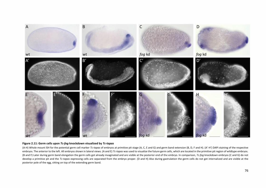
76
Figure 2.11: Germ cells upon Tc-fog knockdown visualized by Tc-tapas
(A-H) Whole mount ISH for the potential germ cell marker Tc-tapas of embryos at primitive pit stage (A, C, E and G) and germ band extension (B, D, F and H). (A’-H’) DAPI staining of the respective
embryos. The anterior to the left. All embryos shown in lateral views. (A and E) Tc-tapas was used to visualize the future germ cells, which are located in the primitive pit region of wildtype embryos.
(B and F) Later during germ band elongation the germ cells got already invaginated and are visible at the posterior end of the embryo. In comparison, Tc-fog knockdown embryos (C and G) do not
develop a primitive pit and the Tc-tapas expressing cells are separated from the embryo proper. (D and H) Also during gastrulation the germ cells do not get internalized and are visible at the
posterior pole of the egg, sitting on top of the extending germ band.

77
5.6 Regulation of Fog signaling in Tribolium
In Drosophila melanogaster, the ventrally expressed transcription factors twist and snail are required
for both, specification of the mesoderm and activation of ventral fog expression (Manning and Rogers,
2014). Loss of Dm-twist and Dm-snail cause depletion of the mesodermal fog domain (Leptin, 1994).
However, the dorsoventral gene regulatory network (DV-GRN) of Tribolium differs from the Drosophila
dorsoventral patterning system. To identify the regulatory mechanisms of Fog signaling in Tribolium,
it was necessary to analyze the expression domains of Tc-fog and Tc-mist in different RNAi
backgrounds.
Like in Drosophila, Tribolium twist shows expression in a ventral stripe (Sommer and Tautz, 1994). As
Tc-fog expression is co-localized in a mesodermal domain (Fig. 2.12 A), it seemed likely that its
mesodermal expression is regulated by Twist in Tribolium, too. Interestingly, neither Tc-fog nor Tc-mist
showed changes in their mRNA expression pattern after knockdown of Tc-twist (Fig. 2.12 B and F).
Prior experiments in Tc-snail embryos showed the same result for Tc-fog (unpublished data from
Cornelia von Levetzow). Thus, it seems that unlike in Drosophila, mesodermal Fog signaling is not
regulated by Twist or Snail but by another dorsoventral patterning gene.
Similar to Drosophila, Toll is the key regulator of the DV-GRN in Tribolium (Lynch and Roth, 2011). The
dynamic nuclear Dorsal gradient, with peak level at the ventral side then activates different target
genes in a concentration-dependent manner (Chen et al., 2000; Nunes da Fonseca et al., 2008). The
loss of Toll results in dorsalization of the embryo (Fonseca et al., 2009). The mesoderm as well as parts
of the neuroectoderm are missing. Furthermore, the embryo-serosa border becomes straight and is
shifted towards the posterior pole, which causes also a reduction of the presumptive head region. As
expected, the embryonic ventral expression domain of Tc-fog is depleted in Tc-Toll knockdown
embryos (Fig. 2.12 C). Due to a reduction of the ventral serosa, Tc-mist shows a weak uniform
expression. The posterior mRNA expression of Tc-mist however, remains unaffected (Fig. 2.12 G).
Another gene which was chosen to analyze the regulation of Fog signaling in Tribolium was Tc-caudal
(Tc-cad). Tc-caudal is expressed in a posterior-to-anterior gradient and is thus an essential part of the
posterior system (Schroder et al., 2000; Wolff et al., 1998). Similar to Tc-Toll knockdown embryos, Tc-
caudal deficient embryos still form a primitive pit, although the formation of the posterior amniotic
fold is less distinct. In addition, the dorsal serosa is expanded towards the posterior pole. Analysis of
the Tc-fog expression domain upon Tc-caudal knockdown revealed on the one hand a loss of the
mesodermal expression and on the other hand the de novo appearance of a posterior Tc-fog domain
(Fig. 2.12 D). Thus, it is likely that Caudal regulates Tc-fog expression in opposing ways. It activates Tc-

78
fog expression in the mesoderm and simultaneously inhibits fog at the primitive pit. However, loss of
Tc-caudal did not influence the expression of Tc-mist (Fig. 2.12 H).
The results of the careful analyses of Fog signaling components in different genetic backgrounds
suggest, that Fog signaling in Tribolium might be regulated not only by dorsoventral patterning. Rather
also parts of the anterio-posterior gene regulatory network are involved.
Figure 2.12: Expression of Tc-fog and Tc-mist in different RNAi backgrounds
(A-H) Whole mount ISH of wildtype and knockdown embryos around primitive pit stage. The anterior is left, the dorsal side is upwards. (A) In wildtype embryos of the primitive pit stage, Tc-fog is expressed in the serosa and the posterior part of the future mesoderm. (E) Similarly, also Tc-mist shows in addition to a posterior expression domain at the primitive pit, serosal expression. However, the expression starts to faint from the dorsal side of the serosa. (B and F) Knockdown of Tc-twist does not influence the expression domains of fog and mist. (C) Although the ventral posterior expression domain is abolished in Tc-Toll kd embryos, they are still able to form a primitive pit. (G) Tc-Toll RNAi embryos are still expressing mist around the primitive pit. (D)Tc-caudal knockdown causes loss of the ventral Tc-fog expression, whereas a new expression domain appears at the posterior pole. (H) However, both Tc-mist expression domains remain unaffected by loss of Tc-caudal.

79
As afore mentioned, unlike Tc-fog knockdown embryos, embryos after Tc-Toll knockdown are still able
to form a posterior fold comparable to the amniotic fold. However, this fold looks more radial-
symmetric than in wildtype embryos (Fig. 2.13 A). Due to loss of the mesoderm in Toll knockdown
embryos, the ventral Tc-fog expression domain gets lost, whereas Tc-mist expression is still detectable
(Fig. 2.13 C and G). To analyze the effect of the posterior mRNA expression of the receptor without the
embryonic mRNA expression of its ligand Fog on the formation of the amniotic fold, embryos deficient
for both, Tc-Toll and Tc-fog were generated. dsRNA for both genes was co-injected in a transgenic line
expressing nuclear EGFP (each 1µg/µl) (Sarrazin et al., 2012). The development of the double
knockdown embryos and a wildtype control were documented by live-imaging.
94% of the offspring derived from mothers injected with dsRNA for Tc-Toll and Tc-fog showed a
phenotype (Fig. 2.13 B). Nevertheless, not all of the defective embryos showed the expected defects
for a double knockdown. Around 7% of the embryos showed the typical phenotype of a Tc-fog
knockdown (Fig. 2.13 B). Interestingly, none of the 76 investigated embryos showed only the defects
of a Tc-Toll single knockdown. However, 99% of the double knockdown embryos showed no sign of
primitive pit formation and embryo invagination (Fig. 2.13 A and B). Furthermore, the germ cells were
also visible in some of the double knockdown embryos. Statistics on the occurrence of germ cells were
not possible as the germ cells are not easy to detect in nGFP embryos.

80
Figure 2.13: Double knockdown of Tc-Toll and Tc-fog results in loss of the posterior amniotic fold (A) DAPI staining of Tc-Toll, Tc-fog and Tc-Toll + Tc-fog knockdown embryos. The anterior pole points to the left, the dorsal
side points upwards. All pictures show lateral views of embryos during formation of the posterior amniotic fold. Although,
the posterior fold is forming upon knockdown of Tc-Toll, it is missing in Tc-fog knockdown embryos and after double
knockdown of Tc-Toll + Tc-fog, respectively. (B) The statics shows that 97% of the mock embryos (H2O injected mothers)
develop a normal primitive pit. 3% of the control embryos show a natural occurrence of random defects. 87% of the embryos
derived from mothers injected with Tc-Toll + Tc-fog dsRNA do not form a primitive pit. 7% of the knockdown embryos show
only the defects typical for a Tc-fog single knockdown and 6% of the embryos were wildtypes.
These results indicate that the formation of a posterior fold in Tc-Toll knockdown embryos depends on
Fog signaling. Although the ventral Tc-fog mRNA expression neighboring the posterior pole is missing
upon Tc-Toll knockdown, the formation of an amniotic fold suggests that the posterior localized
receptor still gets activated. One explanation could be a long-range signaling by diffusion of the Fog
protein diffusing from the serosa. Another possibility is, that Fog signaling might have a distinct
function in serosa spreading and that this spreading, which is absent in Tc-Toll + Tc-fog double
knockdown embryos, might be required for primitive pit formation.

81
5.7 Serosa spreading - A novel function of Fog signaling
So far, it was believed that Fog signaling is especially important for apical constrictions during insect
gastrulation. However, both the ligand Tc-fog as well as its receptor Tc-mist are expressed in the
extraembryonic serosa during the differentiated blastoderm stage (Fig. 2.14 A and B). Although the
serosal Tc-mist expression disappears during gastrulation (Fig. 2.2 E), Tc-fog expression persists (Fig.
2.14 F-H). The membrane marker GAP43-YFP (Benton et al., 2013) enables the visualization of the cell
outlines during embryonic development. During gastrulation of wildtype embryos, the serosal cells
flatten and thus become wider (Fig. 2.14 C). The change of the cell shape is important as the serosa
cells do not undergo further cell divisions, but have to surround the whole embryo proper during germ
band extension (Fig. 2.14 E). At the stage of serosal window closure, the serosal cells have reached the
highest level of cell flattening, seem to have the same size and are evenly distributed in the entire egg
(Fig. 2.14 E and E’, blue marked cells).
The single knockdown of Tc-fog leads usually to a weaker phenotype when the dsRNA is not injected
into adults but embryos (injections performed by Matt A. Benton). As fog is maternally supplied
(iBeetle Genome Browser; RNA-Seq data was supplied by Michael Schoppmeier, Ho Chung, Ho Ryun
and Uli Loehr), the already existing protein weakens the RNAi effect. Although, the loss of Tc-fog is still
strong enough to cause defects in internalization of the germ cells (Fig. 2.14 D), the serosa manages to
cover the embryo during germ band elongation (Fig. 2.14 F). This circumstance enables a more
accurate comparison of the serosa spreading in wildtype and Tc-fog knockdown embryos. Compared
to the evenly enlarged serosa cells in wildtype embryos, the serosa of weak Tc-fog kd looks less
organized. The anterior serosa cells are still very small and not as flat as more posterior cells.
Furthermore, the more posterior serosa cells are extremely large and stretched (Fig. 2.14 F and F’, red
marked cells). This effect is probably caused by the condensation of the embryo proper which pulls the
neighboring serosa cells ventrally.

82
Fig. 2.14: Loss of Tc-fog affects serosal expansion (A and B) Expression of Tc-fog and Tc-mist in differentiated blastoderm wildtype embryos. Both genes are expressed in the extraembryonic serosa. (C, E) Wildtype embryos injected with H2O and the membrane marker GAP43-YFP. (D and F) Embryo injected with Tc-fog RNAi and GAP43-YFP. (E’ and F’) Higher magnification of embryos depicted in E and F. The anterior is left, the dorsal side is upwards. (A) Wildtype expression of Tc-fog. (B) Wildtype expression of Tc-mist. (C) A wildtype embryo at the beginning of gastrulation. Amnion and serosa starts to fold over the embryo proper. (E) The same embryo depicted in C shortly after serosal window closure. The serosa cells (blue) are evenly sized and distributed (also see E’). (D) A Tc-fog knockdown embryo showing a mild phenotype. Although the germ cells are visible during gastrulation, serosa and amnion manage to over grow the embryo. (F) Shortly before serosal window closure, a weak Tc-fog kd shows an uneven pattern of the serosa cells (red). (F’) The anterior serosa cells are very small, whereas the more posterior serosa cells look wider and plane. (pictures C-F provided by Matthew A. Benton)

83
The co-expression of Tc-fog and Tc-mist already suggested a potential function of Fog signaling in the
serosa of Tribolium embryos. In addition, the formation of an amniotic fold after Tc-Toll knockdown
(Fig. 2.6 C and G) supposes, that the Fog protein might diffuse from the serosa to the posterior pole
and acts via long range signaling. Our recent results in weak Tc-fog knockdowns indicate that the serosa
might not only be a second source for posterior Fog signaling, but that it might have a novel function
in coordinated cell flatting and spreading of the serosa cells during gastrulation.
5.8 Knockdown of smog does not result in morphogenetic defects
Recent studies (Kerridge et al., 2016), revealed that a second G-protein coupled receptor (GPCR) called
Smog, is required for cell shape changes associated with both mesoderm invagination and ectoderm
elongation. This additional receptor of Fog signaling is ubiquitously expressed in Drosophila. To analyze
if Smog is also involved in early Tribolium morphogenesis, it was first identified by a BLAST approach
(iBeetle Genome Browser; OGS 3.0).
In a second step, the expression pattern of Tc-smog was determined. Different embryonic stages,
including the yet uncellularized pre-blastoderm (syncytium) (Fig. 2.15 A’), the blastoderm Fig. 2.15 B’),
the differentiated blastoderm (Fig. 2.15 C’), different gastrulation stages (Fig. 2.15 D’-F’) and the early
elongating germ band (Fig. 2.15 G’ and H’) were analyzed. None of the stages showed a definite
staining result. The weak color reaction seen in all embryonic stages (Fig. 2.15 A-H) could indicate a
weak uniform expression of Tc-smog. However, also the sense probe showed a very weak signal. Such
weak stainings can also be interpreted as unspecific background staining. According to transcriptomic
data (iBeetle Genome Browser; RNA-Seq data was supplied by Michael Schoppmeier, Ho Chung, Ho
Ryun and Uli Loehr), smog is weakly expressed in the ovaries but not in the blastoderm. However,
other transcriptome data suggest a general expression of Tc-smog in a not further defined stage (RNA-
Seq data was supplied by the iBeetle consortium and 9 external contributors). Functional studies on
knockdown embryos generated by both, parental6 (N=11) as well as embryonic (N=20) dsRNA injection
did not indicate a function of Tc-smog on early morphogenetic movements or cell shape changes in
Tribolium catsaneum (data not shown).
6 The injection of Tc-smog dsRNA in pupae as well as in adults results in sterility of the females. Thus, the knockdown
experiment was repeated by embryonic injections.

84
Figure 2.15: Expression patterns of Tc-smog
(A-H) Whole mount ISH of wildtype embryos from early blastoderm until beginning of germ band extension. (A’-H’) DAPI staining of the respective embryos. The anterior pole to the left, the dorsal side upwards, except for F, G and H which show ventral surface views. In Tribolium castaneum smog is either not expressed or shows a very weak uniform expression throughout early embryonic development. The blue dots in C-H are unspecific background staining.

85
5.9 T48 does not contribute to mesoderm internalization in Tribolium
castaneum
Another gene which contributes to cell shape changes in Drosophila melanogaster is T48. It encodes
for a transmembrane protein that helps to recruit RhoGEF2 to apical membranes. Under control of
Dm-Twist, it is expressed in a ventral stripe (Gould et al., 1990; Leptin, 1991). Like loss of Fog or Cta
alone, the depletion of T48 affects mesoderm internalization but does not block it completely. Thus, a
strong effect on mesoderm internalization could only be detected by abolishing both T48 and Fog
signaling (Kolsch et al., 2007). Most likely, T48 and Fog signaling act in parallel to concentrate RhoGEF2
apically and they redundantly contribute to mesoderm invagination in D. melanogaster (Manning and
Rogers, 2014).
Tc-T48 has been identified and cloned in our lab previously, but no expression or functional data could
be obtained using ISH and RNAi (unpublished data, Rodrigo Nunes da Fonseca and Cornelia von
Levetzow). As the quality of the Tribolium genome increased over the last years, and transcriptomic
data of different developmental stages became available, we wanted to review the previous results. I
could verify the old sequencing data by BLAST to the new official genome (iBeetle Genome Browser;
Tcas 5.2/OGS 3.0). An in-situ hybridization using an additional probe, which does not overlap with the
previously used one, did result in uniformly stained embryos of different developmental stages (data
not shown), consistent with transcriptomic data of blastoderm and differentiated blastoderm embryos
(iBeetle Genome Browser; RNA-Seq data supplied by Michael Schoppmeier, Ho Chung, Ho Ryun and
Uli Loehr). However, parental (N=296 embryos) as well as embryonic RNAi (N=30 embryos)
experiments could not demonstrate a role of T48 in mesoderm internalization of Tribolium castaneum.
Thus, a double knockdown of both Tc-fog and Tc-T48 by co-injecting the dsRNA into pre-blastoderm
embryos was created (injections by Matthew A. Benton). Unlike in Drosophila, Tribolium double
knockdown embryos did not show visible defects in ventral furrow formation (data not shown).
However, compared to the embryos injected only with Tc-fog dsRNA, the general phenotype seemed
to be stronger in the Tc-fog and Tc-T48 double knockdown embryos. Only 8 out of 20 (40%)Tc-fog kd
embryos showed a strong phenotype, whereas 16 out of 20 (80%) double kd embryos showed the
typical morphological defects, like the serosa remaining on the dorsal side, no primitive pit formation
and visible germ cells (data not shown). Regarding uniform T48 expression and the influence on all
aspects of the phenotype, it is likely that T48 in Tribolium castaneum does not exclusively enhance
mesoderm internalization but instead is a more global factor contributing to cell shape changes in the
whole embryo.

86
6 Discussion
6.1 Fog and its conserved role in tissue internalization during gastrulation
As a member of the coleopterans Tribolium possesses several more ancestral features of embryonic
development, compared to the highly derived fruit fly D. melanogaster. Drosophila is a typical long
germ insect in which all segments develop simultaneously before gastrulation. In contrast, only the
head and thorax segments of Tribolium are specified prior to gastrulation. The abdominal segments
are formed consecutively from a posterior segment addition zone after gastrulation (short germ
embryogenesis) (Tautz and Sommer, 1995). The different modes of early tissue specification are not
the only differences between the two model systems. While Drosophila embryos possess a single
rudimentary extraembryonic membrane (amnioserosa), which remains on the dorsal side of the egg
throughout embryogenesis, two separate extraembryonic membranes (amnion and serosa) protect
the developing Tribolium embryos (Tautz and Sommer, 1995). These membranes are an essential part
of the first morphogenetic movements taking place during Tribolium gastrulation, including the
formation of the posterior amniotic fold (Handel et al., 2000).
Fog signaling is affecting the development of various tissues, throughout embryogenesis but especially
during gastrulation. Thus, the loss of the Fog signaling components Tc-fog, Tc-mist and Tc-cta results
in a highly complex phenotype, including defects in cell shape changes at the posterior pole (amniotic
fold formation) and invagination of the mesoderm (Fig. 3.1). In addition, late defects occur in hindgut
formation and dorsal closure. Furthermore, the data presented in this work suggest a so far unknown,
but more general function of Fog signaling in coordinating tissue-wide cell shape changes like flattening
and spreading of the serosa. In the following chapters, the different defects and their dependence on
Fog signaling are discussed in detail.
Figure 3.1: Gastrulation after fog knockdown During gastrulation of wildtype embryos, the ventral mesoderm starts to be internalized (arrows). Around the same time, the
serosa (orange/red) and the amnion (purple) start to overgrow the germ rudiment. In contrast, upon Tc-fog kd both
extraembryonic tissues stay at the dorsal side of embryo. Especially the amnion is strongly pulled towards the posterior-
ventral side by the condensation of the embryo proper. However, the primitive pit and the later deep posterior gastrulation
fold are completely missing. In addition, knockdown of fog results in a delay in mesoderm invagination. modified from
(Rodrigo Nunes da Fonseca; unpublished)

87
The co-expression of fog, mist and T48 in the mesoderm of Drosophila embryos already indicate a role
of Fog signaling in mesoderm development (Kolsch et al., 2007; Leptin, 1991; Manning et al., 2013).
Different studies provide accurate information about the role of Fog signaling in inducing apical
constriction for successful internalization of the mesoderm. Although the mesoderm gets internalized,
the mesodermal cells in loss of function mutant Drosophila embryos (fog, mist-, cta- and T48-) show an
unorganized pattern of apical constriction and a delay in mesoderm invagination (Costa et al., 1994;
Dawes-Hoang et al., 2005; Kolsch et al., 2007; Manning and Rogers, 2014).
In contrast to Drosophila, the only pathway component showing enhanced expression in the Tribolium
mesoderm prior to gastrulation is Tc-fog (Fig. 1.1 A-E). Nevertheless, Tc-fog, Tc-mist and Tc-cta
knockdown embryos show the same defects in mesoderm internalization that have been described in
Drosophila. In Tribolium, the position of Tc-twist positive segmental cell clusters covered by the
ectoderm (longitudinal sections) of control and Tc-fog knockdown embryos, proves the successful
internalization of the mesoderm (Fig. 2.9 B and E). However, an analysis of Tc-twist expressing cells in
cross-sections of carefully staged Tribolium wildtype and knockdown embryos revealed a reduction in
the depth of the ventral furrow. Especially posterior regions were affected. Loss of Tc-fog results in a
delay of around 3h in mesoderm invagination (Fig. 2.8).
The effect of Fog signaling on mesoderm internalization in Tribolium strongly resembles the situation
in Drosophila, proving that Fog signaling has indeed a conserved role in regulating morphogenetic
movements during gastrulation. Although Fog signaling is essential for timing and coordination of
ventral furrow formation, the fact that loss of Tc-fog is not sufficient to prevent mesoderm
internalization suggests, that Fog signaling is not the only pathway regulating mesoderm
morphogenesis.
88
6.2 Genes with less influence on apical constrictions in Tribolium compared
to Drosophila
In Drosophila the transmembrane anchor protein T48 contributes to bending of epithelial sheets by
apical constrictions. Complementary to Fog signaling, which activates RhoGEF2 release from
microtubules, T48 concentrates RhoGEF2 to the apical membrane (Kolsch et al., 2007; Manning and
Rogers, 2014). These parallel pathways explain the incomplete progression of apical constriction in the
absence of one of those pathways, as seen in fog- mutant embryos (Costa et al., 1994; Kolsch et al.,
2007; Sweeton et al., 1991). Embryos deficient for T48 fail in formation of ventral furrow although
invagination of the posterior midgut primordium is unaffected. However, cells lacking T48, only show
a less organized and delayed invagination but are still able to undergo apical constriction, although it
might be slightly weaker. Recent studies could show that the combined loss of fog and T48 in
Drosophila, abolishes ventral furrow formation and results in an ingression-like mesoderm cell
behavior, reminiscent of mesoderm invagination observed in Chironomus riparius. Thus, mesoderm
ingression does not require Fog or T48 function (Urbansky et al., 2016). An opposing effect was
observed for the early ubiquitous expression of fog and T48 in C. riparius embryos. Both genes were
sufficient to invoke a Drosophila-like invagination of the mesoderm Chironomus embryos. Dm-T48
shows an early mRNA localization in a ventral twist-like stripe in the presumptive mesoderm, whereas
it is absent from the posterior area of midgut invagination. In Tribolium, transcriptomic data suggest a
weak T48 expression prior to gastrulation (although no localized mRNA expression domain could be
identified by ISH). In contrast, it seems not to be expressed at all in Chironomus (Urbansky et al., 2016).
Furthermore, Tc-T48 knockdown embryos did not show defects in primitive pit or ventral furrow
formation. It also did not affect spreading of the serosa. In fact, double knockdown experiments using
Tc-T48 and Tc-fog dsRNA resulted in embryos with typical features of strong Tc-fog knockdowns,
including visible primordial germ cells and lack of posterior amniotic fold formation. This suggests a
more general function of T48 for enhancing Fog signaling in all tissues. In summary, the results indicate
that an evolutionary gain of early fog and T48 activity in the mesoderm may constitute a genetic switch
to increase the speed of fly gastrulation (see results 2.9).
Another Fog signaling component, the GPCR Smog which was recently discovered, could be identified
in Tribolium. smog is uniformly expressed in both, Drosophila and Tribolium embryos. An involvement
of smog in cell shape changes during mesoderm internalization, PMG invagination and especially
ectoderm extension has been reported in Drosophila (Kerridge et al., 2016). However, the results by
analysis of Tc-smog kd by parental as well as embryonic RNAi suggest, that Tc-smog has no role during
Tribolium gastrulation, neither for amniotic fold or ventral furrow formation, nor for serosa spreading
or ectoderm morphogenesis.

89
6.3 The importance of posterior amniotic fold formation by Fog signaling
One of the early morphogenetic movements during Tribolium embryogenesis is the formation of a
posterior amniotic fold. At the onset of gastrulation, Fog signaling induces apical constrictions of the
cells at the posterior pole. In turn, these cells form a cup-like structure which finally converts to a deep
fold when the amnion, part of the dorsal ectoderm and the serosa start to move ventrally and thus
fold over (posterior amniotic fold) to finally cover the embryo proper (Handel et al., 2000). Embryos
lacking one of the Fog signaling components fail to form a primitive pit (Fig. 2.6). In consequence,
amnion and serosa remain on the dorsal side and the embryo proper gets not covered.
One theory suggests, that the spreading of the serosa to the ventral side at the onset of gastrulation,
might indirectly be required for primitive pit formation. It was proposed that germband condensation
drives initiation of amnion folding. Expansion of the amniotic fold and closure of the amniotic cavity
are likely driven by contraction of an actomyosin cable at the boundary between the amnion and
serosa (Benton et al., 2013). Not only loss of Tc-fog but also the double knockdown of Tc-Toll and Tc-
fog embryos results in immobility of the serosa. Similarly, double knockdown embryos do not form a
primitive pit, whereas it is still formed upon Tc-Toll single kd (Fig. 2.13 A). Thus, it seems possible that
the spreading of the serosa influences posterior amniotic fold formation.
During later development, defects in hindgut formation occur in Tc-fog knockdown embryos. Although
the hindgut fate seems to be unaffected, the invagination tube is located outside of the embryos (von
Levetzow, PhD Thesis 2008). The defects in Drosophila fog- mutant embryos are more severe. It seems
that the defective invagination of the PMG primordium during gastrulation results in absence of a tube-
like hindgut or posterior midgut in later stages (Lengyel and Iwaki, 2002). However, also in Drosophila
Fog signaling does not affect the cell fate. The strong defects can be explained by the initial failure of
tissue invagination.
Besides the missing pole cells, the posterior amniotic fold in Tribolium resembles the posterior midgut
(PMG) invagination of Drosophila (Handel et al., 2000). The process of PMG invagination in Drosophila
is well-studied. After the first apical constrictions appear at the dorsal side of the posterior midgut
primordium, shrinking of the remaining cell apices results in formation of a cup-like indentation
(Sweeton et al., 1991; Turner and Mahowald, 1976). During germ band extension this cup moves
dorsally before it becomes deeper by further invagination of the cells, forming a structure referred to
as amnioproctodeal tube (Rickoll and Counce, 1980; Sonnenblick, 1941). Only the center cells belong
to the PMG primordium. The remaining invaginated cells give rise to the hindgut and the malpighian
tubules, as indicated by the expression of the hindgut (proctodeum) marker brachyenteron (Berns et
al., 2008; Kispert et al., 1994; Singer et al., 1996; Wu and Lengyel, 1998).

90
Although the hindgut in Tribolium is formed much later during germ band retraction, Tc-brachyenteron
is already expressed at the tip of the posterior amniotic fold during late gastrulation and throughout
germ band extension (Berns et al., 2008). Hence it is likely, that the formation of the amniotic fold in
Tribolium is homologous to the formation of the amnioproctodeal tube in Drosophila. Supporting this
theory, it has been shown that the midgut epithelium of Tribolium confusum probably derives from
cells associated with the proctodeal invagination (Handel et al., 2000; Stanley, 1970).
6.4 Positioning of the primordial germ cells is influenced by Fog signaling
One of the most obvious morphological defects after loss of Fog signaling is the failure to internalize
the primordial germ cells at the posterior pole (Fig. 2.11). Primordial germ cells (PGCs) are the
progenitor cells that give rise to the gametes (Marlow, 2015). Different mechanisms of germ cell
formation evolved during insect evolution. While specification of the primordial germ cells in some
insects is induced by maternal supply of several gene products (germ plasm), they are specified by
zygotically activated transcription factors in others (Lynch et al., 2012).
In many holometabolous insects including Drosophila and Nasonia, the PGCs emerge from so-called
pole cells at the posterior pole of the egg prior to cellularization. Maternally supplied germ plasm
induces the formation of the pole cells which get internalized during posterior midgut invagination
later in embryogenesis (Fig. 2.3). Another feature that is shared by both species is the localization of
oskar mRNA in the pole plasm (Ephrussi and Lehmann, 1992; Lynch et al., 2012; Lynch et al., 2011).
After crossing the endoderm, the cells migrate to their final position in the gonadal mesoderm
(Coffman, 2003; Sonnenblick, 1941). The pole cell-based mode of PGC formation seems to be derived
from a more ancestral mechanism, which has been described for hemipterans and some
holometabolous lineages, e.g. members of the coleopterans (e.g. Tribolium) (Lynch et al., 2012). This
second type of primordial germ cell formation is not dependent on maternally supplied pole plasm.
The germline fate is specified by zygotic gene expression, often later in embryonic development
(Extavour and Akam, 2003). Similar to the system observed in Drosophila, PGCs separate from
blastoderm after cellularization, get internalized (Fig. 2.3 C1-C4) and translocate to the gonads.
Tribolium embryos lack a localized oskar expression. Instead, ubiquitous mRNA of Tc-vasa gets
selectively degraded, so that it is only found at the posterior pole at the onset of gastrulation. Later it
marks the primordial germ cells (Lynch et al., 2012; Schroder, 2006). Oskar localization is probably
secondarily lost in Tribolium, as it was also found in Gryllus bimaculatus (Ewen-Campen et al., 2012).
In contrast to the so far mentioned mechanisms, the PGCs in Gryllus form in later embryonic stages

91
from a subgroup of mesodermal cells which give rise to the future gonads. This probably most ancestral
mode of germline specification is usually found in basally branching insects.
Figure 3.2: Germ cell formation in Drosophila, Nasonia, and Tribolium Red =oskar (osk) mRNA, magenta = Tc-vasa mRNA, blue = nuclei. (A1-C1) In the early stages of embryogenesis, osk mRNA is
localized in the posterior pole plasm of Drosophila (A1) and oosome of Nasonia (B1), while Tc-vasa is ubiquitous expressed
in Tribolium (C1). (A2-C2) In Drosphila and Nasonia the posterior cells of the syncytial blastoderm interact with the pole plasm
(A2), or the oosome (B2), respectively. In the Tribolium, the nuclei are surrounded by a homogenous environment (C2). (A3-
C3) In the fly the pole cells are formed by single nuclei that entered the pole plasm (A3). In Nasonia, the oosome along with
multiple nuclei bud simultaneously from the posterior (B3). Tc-vasa becomes localized at the posterior pole of late
blastoderm Tribolium embryos (B4). (A4-C4) After formation of the pole cells and completed cellularization of the blastoderm,
Dm-osk (A4) and Nv-osk (B4) get degraded. (C4) Tc-vasa positive cells delaminate from the blastoderm into the interior of the
embryo in order to form the primordial germ cells. (Lynch et al.,2012)
Although the different modes of germline specification seem to correlate in general to more derived
and more ancestral lineages, the formation of pole cells in coleopterans occurs several times, even
within closely related species. Although the PGCs of Callosobruchus, a seed beetle, are derived from
pole cells, Atrachya embryos do not possess these specific cells (Benton et al., 2016; Quan and Lynch,
2016). Thus, it is likely that minor changes in gene regulation act as a switch between the different
ways of PGC formation.

92
I observed a cluster of cells at the posterior pole of Tc-fog knockdown embryos which were positive
for the putative germ cell marker Tc-tapas (Patil et al., 2014). Furthermore, the timing of their
occurrence and their location indicate their germline fate.
Fog signaling might influence the direction of PGC positioning in Tribolium castaneum. It is possible
that the physical circumstances, generated by apical constriction of the most posterior cells, trigger
the PGCs to leave the epithelium to the interior of the embryo. As Tc-fog knockdown embryos lack
apical constrictions in this area, the cells move to the outer surface. Hence, Fog signaling is probably
only indirectly required for PGC internalization. The influence of Fog signaling on positioning of the
PGCs has shed some more light on the evolutionary history of germline determination in insects.
Together with the dependency on maternally supplied gene products, early morphogenetic
movements induced by Fog, might explain the transition from a later zygotic germline specification.
This also includes extensive cell migration to an early and pole cell based PGC specification in derived
species like Drosophila.
6.5 The regulatory network underlying Fog signaling
Similar to Drosophila dorsoventral patterning, Toll signaling initiates the early fate determination in
Tribolium castaneum (Lynch and Roth, 2011; Nunes da Fonseca et al., 2008). In both organisms, the
two transcription factors twist and snail are expressed in a ventral stripe and control specification of
the mesoderm (Leptin, 1991, 1994; Sommer and Tautz, 1994). In Drosophila, ventral Fog signaling is
activated by both of them, leading to ventral furrow formation (Manning and Rogers, 2014). The co-
localization of Tc-fog and Tc-twist strongly indicates a regulation of the ventral Fog signaling by Tc-
Twist similar to Drosophila. However, the observation that neither Tc-fog nor Tc-mist showed changes
in their expression domains (Fig. 2.12 B and F) in Tc-twist knockdown embryos, is contradictory to this
hypothesis. In addition, former results suggest that Tc-snail is not required for Tc-fog expression
(Rodrigo Nunes da Fonseca, unpublished). These data rather suggest that the mesodermal activation
of Tc-fog depends on other factors.
DV and AP patterning are connected to a certain degree. Thus, the expression pattern analysis of Tc-
fog and Tc-mist in caudal knockdown embryos should reveal a possible influence of the AP patterning
system on Fog signaling. The knockdown of Tc-caudal results in defects in convergent extension and
thus, in a broader and shorter germ rudiment. Furthermore, the dorsal serosa is slightly expanded
towards the posterior pole (Schroder et al., 2000; Wolff et al., 1998). Tc-mist expression remains
unaffected upon knockdown of caudal (Fig. 2.12 H). The fact that the mesodermal Tc-fog expression
domain is lost after Tc-caudal kd, whereas a new expression domain appears in the region of the
93
primitive pit (Fig. 2.12 D) suggest, that Tc-caudal has two divergent roles in Fog regulation. It activates
Tc-fog expression in the mesoderm and simultaneously inhibits its expression at the primitive pit.
Although Tc-caudal deficient embryos form a primitive pit, the amniotic fold is less distinct.
In summary, Tc-mist expression seems to be independent of both, the DV patterning system and Tc-
caudal. However, Tc-caudal regulates Tc-fog expression in the mesoderm as well as in the primitive
pit. Besides changes in the DV-GRN of Tribolium (Stappert et al., 2016), also the regulatory network
underlying Fog signaling differs from Drosophila.
6.6 Serosa spreading and cellularization - A novel function of Fog signaling
6.6.1 Fog signaling regulates flattening of the serosa cells
The serosa is a monolayered epithelium that protects the embryo from desiccation and against
pathogens (Jacobs et al., 2013; Panfilio, 2008). To form a differentiated blastoderm, the embryonic
cells start to contract and become columnar. In contrast, the serosa cells start to spread and become
squamous to finally envelop the embryo and the yolk. Squamous epithelia are often associated with
developmental or tissue remodeling processes (Pope and Harris, 2008).
Although, germ band extension seems to be unaffected, Tc-fog knockdown embryos are deformed in
late stages (von Levetzow, PhD Thesis 2008). These morphological defects can be explained by the late
influence of serosa and amnion on dorsal closure of the embryo. At the end of embryogenesis, the
extraembryonic membranes rupture (~ 52h AEL) and retract to the dorsal side (Hilbrant et al., 2016;
Panfilio et al., 2013). In consequence, the lateral parts of the embryo start to move dorsally and finally
fuse at the dorsal midline (Koelzer et al., 2014). This process is disrupted by loss of Fog signaling and
the dorsal side of the embryo remains open, similar to Tc-zen2 knockdown embryos (van der Zee et
al., 2005). As the extraembryonic tissue has a great influence on embryonic morphogenesis, different
questions raised facing the serosal co-expression of Tc-fog and Tc-mist. Is the immobility of the serosa
after loss of Fog signaling a primary or secondary effect? Could Fog signaling influence cell shape
changes in the serosa? If not, why are ligand and receptor co-expressed?
Knockdown of Tc-Toll results in dorsalized embryos which lack the mesoderm as well as the ventral
ectoderm. The embryo-serosa border becomes straight and is shifted towards the posterior pole,
deleting part of the head anlagen. In Tc-Toll kd embryos Tc-fog is strongly expressed in the symmetric
serosa, while Tc-mist shows a weaker but uniform expression instead of ventrally enhanced serosal
expression in wildtype embryos. Furthermore, the mesodermal expression domain of Tc-fog is lost,
indicating that it is activated by dorsoventral patterning genes, whereas the posterior Tc-mist

94
expression remains unaffected (Fig. 2.12 C and G). Interestingly, Tc-Toll knockdown embryos are still
capable to form a primitive pit. This implies that the posterior localized receptor still gets activated.
Two theories might explain the unaffected formation of the posterior amniotic fold. The first
explanation could be that the serosa serves as a source for long-range signaling. Diffusion of the Fog
protein from the serosa to the posterior pole could be still sufficient to induce apical constrictions and
hence primitive pit indentation. Drosophila Fog is thought to act as an autocrine factor which only
diffuses a couple of cell widths (Costa et al., 1994). However, the Tribolium Fog protein is less than half
the size of Dm-Fog, making long-range signaling more likely (Fig. 3.3).
Figure 3.3: Fog is a fast-evolving protein The Fog protein evolved very fast in both, nucleotide as well as amino acid sequence. Furthermore, the Tribolium Fog is much smaller than its Drosophila and Anopheles orthologs. (Rodrigo Nunes da Fonseca, unpublished)
Nevertheless, the simultaneous expression of Tc-fog and Tc-mist in the serosa might give hints for
another explanation. Fog signaling could have a distinct function in cell shape changes in the serosa,
influencing its spreading. Cell flattening requires extensive remodeling of adherens junctions (Szuperak
et al., 2011) and changes in the actin-myosin network. The transformation from a cuboidal to a
squamous epithelium involves shrinking the lateral membrane and extending the basal and apical
membranes (Brigaud et al., 2015). Studies on the amnioserosa cells in Drosophila revealed, that its cell
shape changes are dependent on a rotation of the microtubule cytoskeleton. The authors suggested
that similar mechanisms may underlie the development of other squamous epithelial monolayers
(Pope and Harris, 2008).
Injecting Tc-fog dsRNA (1µg/µl) into embryos resulted in weaker phenotypes in which the primordial
germ cells did not get invaginated, but amnion and serosa managed to cover the embryo during
gastrulation (Fig. 2.14 D and F). Those weak phenotypes enabled a proper comparison of the cell shape
changes within the serosa. In wildtype embryos, all serosa cells start to flatten at the same time and
are of equal size when the reach their peak levels of flattening at serosal window closure (Fig. 2.14 C
and E). Interestingly, the anterior serosa cells did not undergo cell flattening after loss of Tc-fog. In
contrast, the posterior serosa cells were abnormally enlarged around the stage of serosal window

95
closure (Fig. 2.14 E’ and F’). One feature of a strong Tc-fog knockdown phenotype is, that the serosa
rips off from the amnion and the embryo proper during germ band condensation (Fig. 2.6 K). If the
serosa cells do not undergo cell flattening, they remain on the dorsal side. The embryonic cells move
to the ventral side exerting physical force, especially on the lateral serosa cells. As the connection
between the serosa and the amnion might be a structural weak point, the cells lose contact and the
serosa moves back to the dorsal side.
The results presented in this work indicate that Fog signaling in Tribolium is not only required for tissue
internalization via apical constrictions, but that it is rather involved in cell shape changes in general,
especially for coordinated cell flattening of the serosa cells. This function was never observed before.
However, so far Fog signaling was mainly studied in Drosophila in which the amnioserosa remains on
the dorsal side.
6.6.2 A role of Fog signaling in the blastoderm
The generation of Tc-fog knockdowns by embryonic injection revealed another interesting part of the
phenotype, which was never observed in knockdown embryos after pRNAi. The injection of higher
dsRNA concentrations (>3 µg/µl) led to the formation of big holes in the uniform blastoderm. Holes
seem to occur naturally also in wildtype embryos but usually close very fast. The holes which form in
the early blastoderm of Tc-cta, Tc-fog and Tc-mist knockdown embryos remain open (unpublished data
by Matthew A. Benton). As Fog signaling induces pulsatile contraction of the actomyosin network
(Dawes-Hoang et al., 2005; Martin et al., 2009), it might also influence the coordination of the
cytoskeleton during cellularization and in the early blastoderm.
Figure 3.4: Fog signaling is involved in germ rudiment formation of Tribolium castaneum (A) Wildtype embryo injected with H2O and the membrane marker GAP43-YFP. (B) Embryo injected with Tc-fog RNAi and GAP43-YFP. Holes are visible in the posterior part of the blastoderm. (pictures provided by Matthew A. Benton)

96
In Tribolium the cells of the blastoderm undergo some cell shape changes prior to gastrulation. The
embryonic cells start to contract and become columnar, while the serosa cells started to flatten and
become squamous (Benton et al., 2013; Handel et al., 2005; Handel et al., 2000). This explains the
extension of some of the holes during early condensation of the germ rudiment (Fig. 3.4 A and B).
Interestingly, knockdown experiments in the Gryllus bimaculatus, result in the same phenotype upon
cta and mist knockdown (Fig. 3.5 A and B, unpublished data by Matthias Pechmann). Tribolium as well
as Gryllus are more basally branching insects compared to Drosophila. Thus, it is likely that the role of
Fog signaling in coordinating embryo condensation represents an ancestral process.
Figure 3.5: Fog signaling is involved in germ rudiment formation of Gryllus bimaculatus Females of a H2B-eGFP line expressing nuclear eGFP were used for dsRNA injection. (A) Control embryo. (B) Embryo injected with Gb-fog RNAi embryo showing a disintegrating blastoderm with numerous holes. (pictures provided by Matthias Pechmann)

97
6.7 Conclusion
In Drosophila Fog signaling is involved in tissue internalization during gastrulation, like internalization
of the mesoderm and the posterior midgut primordium. By activating the GPCR Mist, the Gα-subunit
Concertina (Cta) and RhoGEF2, Fog signaling controls rearrangements of the actomyosin network,
resulting in apical constrictions and thus bending of epithelial sheets (Dawes-Hoang et al., 2005;
Manning and Rogers, 2014). The loss of pathway components results in defects in ventral furrow
formation. However, the mesoderm still gets internalized, indicating that Fog signaling is important
but not absolutely essential for mesoderm invagination (Costa et al., 1994).
Although pathway homologs were identified in the genomes of many different winged insects, recent
comparative studies in the more basally branching fly Chironomus, in which Fog signaling plays only a
minor role in mesoderm internalization, indicate that Fog signaling was recruited to increase the speed
of gastrulation (Urbansky et al., 2016). This could be an adaption to the very fast embryonic
development of highly derived long-germ insects like Drosophila melanogaster.
This work focused on Fog signaling in the red flour beetle Tribolium castaneum which possesses a more
ancestral mode of embryogenesis. The observations in embryos deficient of Fog signaling components
showed that Fog signaling is also involved in mesoderm internalization and invagination of posterior
endoderm homologous to the PMG primordium. This suggests indeed a conserved role of Fog signaling
during gastrulation outside of dipterans. However, the defects in early morphogenetic movements
after loss of Tc-fog are more multifaceted. Tc-fog is required for formation of the posterior amniotic
fold as well as the positioning of the primordial germ cells. Furthermore, the presented data suggest
an involvement of Fog signaling in cell shape changes other than apical constrictions, like the transition
from a cuboidal to a flat squamous shape of extraembryonic cells during serosa spreading. In addition,
cellularization defects in blastoderm embryos of Tribolium and Gryllus indicate an additional
conservation of Fog signaling in stabilizing the cytoskeleton during formation of the blastoderm.

98
Material &Methods
7 Strains
The San Bernadino wildtype strain was used for RNAi injections and as wild-type control, if not
specified otherwise. For selected experiments, different transgenic lines like pBA19 (“muscle-line”)
(Lorenzen et al., 2003), G04609 (“heart-line”) (Koelzer et al., 2014), a nGFP strain (Sarrazin et al., 2012)
and a Life-actin GFP line (Tania Vazques, van der Zee lab; unpuplished) were used.
8 Animal keeping and embryo collection
The beetles are kept in boxes containing a mixture of 75% wheat flour "Extra" type 405 (Diamant Mehl)
and 25% dark wheat flour type 1050 (Diamant Mehl) mixed with 25 g/kg dry yeast. To avoid fungal
infestation, the flour is complemented with 0.3 g/kg fungicide (Fumagilin B, Medivet Pharmaceuticals
Ltd). The animals are kept at 30°C, which ensures a generation cycle of around 30 days (Bucher, 2009).
For the collection of eggs, the beetles are transferred to special wheat flour ("Instant" Diamant Mehl,
type 405). This flour has a grain size suited for efficient egg collection. Around 30 minutes after transfer
to new flour, the females start to lay eggs. A certain stage of embryos is obtained as follows: After a
specific egg-laying period the beetles are transferred to new flour and the eggs are further incubated
at 30°C for several hours (minimum age) on fresh flour before they are collected (total time - 0.5 h =
maximum age). Adult beetles are separated from flour and eggs by using sieves with a mash size of
710 μm. The eggs are separated from the flour by using sieves with a mesh size of 300 μm.
For constant access to young beetles with high egg lay (EL) rates, one EL per week was set up. 1.2 g
embryos were transferred to a box containing 1250 g of mixed flour. After 4 weeks the pupae can be
sorted (males/females) for RNAi experiments. A turn-over of the stock was done every 3 months.
Beetles from every-week-egg lays were collected and 23 g were transferred into new boxes containing
900 g of mixed flour.

99
9 Total RNA isolation
For the isolation of total RNA, Tribolium eggs are collected in a defined stage, dependent on
subsequent experiments. Due to problems with the quality of isolated Tribolium RNA, the
manufacture’s protocol using TRIzol (Life Technologies) was modified (Stappert et al., 2016).
First, the eggs were washed with water followed by two minutes incubation in 50% DanKlorix (Colgate-
Palmolive). To remove all remaining Klorix solution, they were again rinsed with clear water. The eggs
were transferred into a 1.5ml RNAse-free reaction tube and shock frozen in liquid nitrogen (-80°C) for
later processing or treated with TRIzol for direct RNA isolation. The eggs were homogenized in 500 μl
TRIzol using a pestle. It should be noticed that working on ice is important to avoid degradation of the
RNA. After centrifugation for 10 min (12,000 x g at 4°C) the supernatant was transferred into a new
reaction tube and mixed with additional 500 μl TRIzol (total volume of 1ml). The samples were
incubated for 5 min at room temperature before 100 μl chloroform were added. The tubes were
shaken thoroughly by hand for 15 seconds and incubated for 2-3 minutes at RT. The phases were
separated by 15 minutes centrifugation at 4°C (12,000 x g). The upper aqueous phase (containing the
RNA) was transferred into a new reaction tube and the contained RNA was precipitated by adding 40
μl 3M NaAc pH5.2 and 800μl room temperature 100% EtOH followed by incubation for at least 30 min
at -80°C. After centrifugation (20 min, full speed at 4°C), the RNA pellet was purified from
contaminations (TRIzol or polysaccharides from flour) by different washing steps. 800 μl 3M NaAc
pH5.2 were added and samples were incubated for 5 minutes at RT. After centrifugation (15 min, full
speed, 4°C) the pellet was washed with 800 μl room temperature 70% EtOH, followed again by
incubation for 5 minutes at RT and centrifugation (15 min, full speed, 4°C). The washing steps were
repeated before the pellet was resuspended in 21 μl RNase-free H2O. The concentration and 260/280
and 260/230 ratios were checked on a NanoDrop2000 (Thermo Scientific). The samples were stored
at -80°C.
10 cDNA synthesis
Complementary DNA (cDNA) is synthesized from RNA by the enzyme reverse transcriptase and was
used as template for probe and dsRNA synthesis as well as qRT-PCR. Due to an optimized protocol for
generation of first-strand cDNA for qRT-PCR, the SuperScript VILO cDNA Synthesis Kit (Life
Technologies) was used according to the manufacture’s manual.

100
11 Primer design
Primers are required to amplify DNA by PCR. Primers are DNA oligonucleotides, which bind to the DNA
which should be amplified. Polymerases can bind to the primers and copy the sequence selected by a
forward (5’) and a reverse primer (3’). The online application Primer3 version 0.4.0
(http://bioinfo.ut.ee/primer3-0.4.0/) (Untergasser et al., 2012) was used for primer design. For in-situ
probes or dsRNA synthesis the fragment size was set to 500 bp to 800 bp. The primer size was set to
20-24 bp. Furthermore, the primers should show a low self- and cross-complementarity and the GC
content should be <50%. The suggested primers were tested for specificity by BLASTing them to the
Tribolium genome. Appropriate primers should bind to a unique sequence without any mismatches.
The primer design for the in-situ hybridization screen and dsRNA synthesis of the differential
expression analysis was based on the Tribolium Tcas 3.0 annotation/ OGS 2 (http://bioinf.uni-
greifswald.de/gb2/gbrowse/tribolium/). The primers used for analysis of the Fog signaling pathway
were designed by using the new T. castaneum genome assembly Tcas 5.2/ OGS 3. All primers used in
this work are listed in the Appendix A, B, C and G.
12 Standard Polymerase Chain Reaction (PCR)
Polymerase Chain reaction is a method for in vitro amplification of a selected piece of DNA using Taq
polymerase (Mullis, 1990). Taq polymerase is derived from the thermophilic bacterium Thermus
aquaticus and is able to resist the high temperatures that are required for denaturation of double
stranded DNA (dsDNA) into single stranded DNA (ssDNA). Denaturation takes place at 94°C. Primers
are used to specify the target DNA and to enable binding of the polymerase. For amplification of a
specific DNA fragment, two primers (5’ and 3’) are needed. Annealing of the primers to single DNA
strands takes place at 55-65°C. Too low temperatures result in unspecific binding of the primers. The
last step enables efficient binding of polymerase to the primers and elongation of the complement
strand. Elongation of the new strand takes place at 72°C. To efficiently amplify the DNA, all three steps
are repeated ~35 times.
Reaction mix for standard PCR
10 µl Red Taq mix, Sigma 7 µl H2O 1 µl cDNA 1 µl specific 5’ primer (10 μM) 1 µl specific 3’ primer (10 μM)
Cycler program for standard PCR
Denaturation 5 min 94°C Denaturation 30 sec 94°C Annealing 30 sec 55-57°C Elongation 1 min 72°C Elongation 5 min 72°C
35x

101
13 Gel electrophoresis
To confirm the presence and correct size of PCR products, 5 µL of the reaction was run on a 1% agarose
gel run in 0.5x TRIS-Acetat-EDTA-Buffer (TAE) at 135 V. To visualize the DNA, 1-2 drops of 0.025%
ethidium bromide solution per 50 ml agarose were used. The size of the bands was determined using
2.5 µL of the Smart Ladder MW 1700-10 (Eurogentec).
14 In-situ hybridization (ISH)
In-situ hybridization is a method to detect the presence and location of mRNA in different tissues. The
gene specific mRNA is hybridized with a complementary mRNA-probe. The probe is for example
labeled with Digoxygenin (DIG) which is detected by using an anti-DIG antibody (anti-DIG-AB). The
enzyme alkaline phosphatase is conjugated with the anti-DIG-AB and converts a substrate like
NBT/BCIP (nitro blue tetrazolium chloride/5brome-4chlor-3indolyl-phosphate) into a purple dye. The
staining indicates the spatial expression of the gene of interest.
14.1 Generation of mRNA probes
14.1.1 Two-step Polymerase Chain Reaction
A two-step polymerase chain reaction (PCR) strategy was used to amplify DNA as template for
antisense mRNA probe synthesis. To detect complementary mRNA in subsequent in-situ hybridizations
(ISH), the DNA template was equipped with a T7 promotor. To amplify DNA of the gene of interest,
first a standard PCR (denaturation at 94°C, annealing at 57°C, elongation at 72°C, 35 cycles) using cDNA
as template was performed. A linker sequence was attached to the gene specific primers used for the
1. PCR. A second PCR was performed using only one of the gene specific primers (3’ for sense/5’ for
antisense) and one T7 universal primer that contains the T7 promotor sequence and is able to bind to
the linker sequence which was attached during the 1.PCR. The universal primer binds to a specific
sequence which is attached to the 5’ end of the gene specific primer (T7 linker) and was amplified
during the first PCR. Cycling parameters for the second PCR reaction are the same as for the first PCR.
The amplicon from the second PCR is used as template for RNA probe synthesis.
50 x TAE
2 M TRIS 0.05 M EDTA In H2O (adjusted to pH 8.0)

102
14.1.2 Probe synthesis
To label the probe during synthesis UTP-Digoxigenin (DIG) (Roche) was used. 3 μl of the amplicon from
the second PCR were mixed with 7 μl of T7 master mix and incubated for 4 hours at 37°C. The reaction
was stopped by adding 15 μl H2O and 25 μl 2x Stop solution (0.2 M NaAc, 1% HAc; pH 6.0). For
precipitation 2.5 μl tRNA (20 mg/ml; Roche), 5 μl LiCl (4M) and 150 μl 100% EtOH were added. The
probes were incubated for at least 30 minutes at -20°C followed by a full speed centrifugation at room
temperature for 30 minutes. The RNA pellet was dried for 5 minutes before it was resuspended in 40
μl 50% formamide, 2x SSC. The probes were stored at -20°C. The correct size of the probes was tested
on a 1% agarose gel.
Reaction mix for sense probe
10 µl Red Taq mix, Sigma 7 µl H2O 1 µl cDNA 1 µl 5’ T7 universal primer (10 μM) 1 µl specific 3’ primer (10 μM)
Reaction mix for antisense probe
10 µl Red Taq mix, Sigma 7 µl H2O 1 µl cDNA 1 µl specific 5’ primer (10 μM) 1 µl 3’ T7 universal primer (10 μM)
Linker sequences attached to the gene specific primers
5’ T7 linker: ggccgcgg 3’ T7 linker: cccggggc
5’ T7 universal primer: gagaattc taatacgactcactatag ggccgcgg 3’ T7 universal primer: agggatcc taatacgactcactataggg cccggggc
T7 master mix
3.5 µl RNAse-free H2O 0.25 µl RNAse inhibitor 1 µl 10x T7 reaction buffer 1 µl DIG labelling mix 1 µl T7 polymerase

103
14.2 Embryo fixation
Tribolium eggs were rinsed with tap water to remove dirt and left-over flour. For dechorionization the
eggs were put for 2 min in 50% DanKlorix (Colgate-Palmolive). The Klorix was then thoroughly washed
away with tap water. To fix the embryos, they were incubated in fixation solution (2 ml 10%
formaldehyde, 3 ml PBS, 5 ml heptane), shaking at 120 rpm, for 45 min to 1.5 h. The embryos were
devitellinized by methanol (MeOH) shock or by hand dissection. The embryos can be stored at -20°C.
14.3 Detection of mRNA expression
Embryos were transferred from MeOH into PBST (first 2:1, then 1:2 MeOH:PBST), washed with PBST
(2 times, 5 min each), fixed in 5% PFA/PBST for 30 min. The PFA was washed away with 1xPBST (four
times, 5 min each). The embryos were slowly transferred into hybridization solution I (first 1:1 hyb sol
I:1xPBST for 10 min, then 100% hyb sol I for 10 min) and then pre-hybridized in hybridization solution
II at 65°C for 1 h. 4 μl probe, specific for the mRNA of interest, was diluted in 196 μl hybridization
solution II and incubated with the embryos overnight at 60 °C while rotating. The probe was washed
away by washing four times for 5 min, and four times for 30 min with hybridization solution I at 60 °C.
The embryos were then transferred into PBST (first 2:1, then 1:2, hyb sol I:1xPBST, at 60 °C) and washed
for 10 min with PBST (room temperature). For blocking, embryos were incubated two times for 30 min
in PBST that contains bovine serum albumin (BSA, 100 μl/ml) and normal goat serum (NGS, 30 μl/ml).
Then, anti-digoxigenin antibody coupled with alkaline phosphatase was added in a final concentration
of 1:4000 and incubated with the embryos overnight at 4 °C while rotating. The next day the embryos
were rinsed by washing three times for 5 min and three times for 15 min with PBST and three times
for 5 min with AP-buffer. For the color reaction 8 μl NBT/BCIP (nitro blue tetrazolium chloride/5brome-
4chlor-3indolyl-phosphate; Roche) solution in 400 μl AP-buffer were added to the embryos. The color
reaction was observed and stopped by washing three times with PBST when background staining
started to develop. The staining was then fixed by incubating the embryos for 30 min in 5% PFA in
PBST. Background staining was in some cases removed by incubating the embryos for ≥20 min in 100%
ethanol (EtOH). The EtOH was washed away by two to three washes with 1xPBST. The embryos were
embedded into VECTASHIELD Mounting Medium with DAPI (Vector Laboratories).

104
15 Double in-situ hybridization (dISH)
This protocol was modified from (Schinko et al., 2009). The amount of each probe to use is a critical
variable that must be optimized for every probe preparation. DIG-labeled probes detected by
NBT/BCIP are more sensitive than fluorescein-labeled probes detected by INT/BCIP. Thus, best results
can be expected by detect the stronger signal first, using a fluorescein-labeled probe visualized by
NBT/BCIP (Roche) color reaction. Second, the weaker signal should be detected by a DIG-labeled probe
stained by FAST Red (Roche). Fast red staining is both, colormetric (red) and fluorescent (orange).
Day 1 of the double in-situ hybridization protocol is similar to day 1 of the standard ISH protocol (see
above). However, both mRNA probes (2 µl each) were mixed and diluted in 196 µl hybridization
solution II and incubated with the embryos overnight at 60 °C while rotating.
10x PBS (Phosphate Buffered Saline) dissolve in 800 ml H2O 80 g NaCl 2 g KCl 14.4 g Na2HPO4 2.4 g KH2PO4 Adjust to pH 7.4 fill up to 1 l with millipore H2O (autoclave)
1x PBST 5 ml 10x PBS 500 ml 20% Tween fill up to 50 ml with millipore H2O
Hybridization Solution I
25 ml formamide 12.5 ml 20xSSC 50 µl heparine (50 mg/ml) 250 µl 20% Tween20 fill up to 50 ml with millipore H2O
Hybridization Solution II
25 ml formamide 12.5 ml 20xSSC 50 µl heparine (50 mg/ml) 250 µl 20% Tween20 500 µl salmon sperm (100 mg/ml) fill up to 50 ml with millipore H2O
AP-buffer
1 ml TRIS-HCl (pH 9.5) 500 µl 1M MgCl2 200 µl 5M NaCl 50 µl 20% Tween20 fill up to 50 ml with millipore H2O
Blocking Solution
100 µl BSA (1 mg/ml in PBS) .30 µl NGS (60 mg/ml) fill up to 1 ml with 1xPBST

105
Also the second day resembles mainly the protocol for single ISH. Dilute the antibody for the first
staining (anti-DIG-AP) was diluted 1:2000 in blocking solution. 500 µl of diluted antibody was added to
the embryos. The samples were incubated over night at 4°C (or for 1 h at room temperature) on a
rotator.
As mentioned above, best results were achieved by staining with NBT/BCIP first. Thus, the next day
the embryos were rinsed by washing three times for 5 min and three times for 15 min with PBST and
three times for 5 min with AP-buffer (see single ISH protocol). 8 μl NBT/BCIP solution were diluted in
400 μl AP-buffer and were added to the embryos. The reaction was stopped by both, a decrease in pH
and dilution of the staining buffer. The alkaline phosphatase was deactivated by washing the embryos
three times for 20 min in glycine-HCl (100 mM glycine-HCl, pH 2.3) on a rotator. Afterwards the
inactivation buffer (IB) was removed by washing the embryos three times 20 min in 1xPBST. To remove
the unspecific background staining, the embryos can be incubated for 15-30 min in 100% EtOH on a
rotator, followed by a slow retransfer into PBST. After another round of blocking (2x 30 min, rotator)
on room temperature, the second antibody was added. If the DIG-labeled probe was used as the first
stain, then use the fluorescein-labeled probe here. Dilute the antibody for the second staining (anti-
fluorescein-AP) was diluted 1:2000 in blocking solution and 500 µl of the diluted antibody was added
to the embryos. The embryos were incubated in the dark over night at 4°C or for 1 h at room
temperature on a rotator.
The second color reaction was performed on day 4. The embryos were washed three times for 5 min
and three times for 15 min with PBST. Afterwards, the embryos were prepared for the staining by
washing them three times for 10 min in 0.1 M Tris-HCl (pH 8.0) solution. For staining, 1 Fast Red tablet
was dissolved in 2 ml of 0.1 M Tris-HCl solution (pH 8.0). The samples should be incubated in the dark
at room temperature until the staining is strong and distinct. If the staining is weak or absent after 3
h, continue staining overnight at 4°C. The color reaction is stopped by washing the embryos 3 times 10
min in PBST. EtOH should not be used to remove Fast Red background staining, as also the specific
staining would be removed! The embryos were mounted with VECTASHIELD Mounting Medium with
DAPI (Vector Laboratories).
Inactivation buffer (100 mM glycine-HCl)
11.1 g Glycine-HCl 800 ml millipore H2O adjust to pH 2.3 fill up to 1 l with millipore H2O
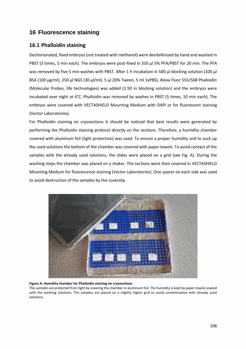
106
16 Fluorescence staining
16.1 Phalloidin staining
Dechorionated, fixed embryos (not treated with methanol) were devitellinized by hand and washed in
PBST (3 times, 5 min each). The embryos were post-fixed in 350 μl 5% PFA/PBST for 20 min. The PFA
was removed by five 5 min washes with PBST. After 1 h incubation in 500 μl blocking solution (100 μl
BSA (100 μg/ml), 250 μl NGS (30 μl/ml), 5 μl 20% Tween, 5 ml 1xPBS), Alexa Fluor 555/568 Phalloidin
(Molecular Probes, life technologies) was added (1:50 in blocking solution) and the embryos were
incubated over night at 4°C. Phalloidin was removed by washes in PBST (5 times, 10 min each). The
embryos were covered with VECTASHIELD Mounting Medium with DAPI or for fluorescent staining
(Vector Laboratories).
For Phalloidin staining on cryosections it should be noticed that best results were generated by
performing the Phalloidin staining protocol directly on the sections. Therefore, a humidity chamber
covered with aluminum foil (light protection) was used. To ensure a proper humidity and to suck up
the used solutions the bottom of the chamber was covered with paper towels. To avoid contact of the
samples with the already used solutions, the slides were placed on a grid (see Fig. A). During the
washing steps the chamber was placed on a shaker. The sections were then covered in VECTASHIELD
Mounting Medium for fluorescence staining (Vector Laboratories). One spacer on each side was used
to avoid destruction of the samples by the coverslip.
Figure A: Humidity chamber for Phalloidin staining on cryosections. The samples are protected from light by covering the chamber in aluminum foil. The humidity is kept by paper towels soaked with the washing solutions. The samples are placed on a slightly higher grid to avoid contamination with already used solutions.
107
16.2 Nuclear staining
16.2.1 DAPI staining
Dechorionated, fixed embryos were transferred from methanol to PBST (PBS supplemented with 1 %
Tween20) and washed three times for 5 min with PBST. Then, the embryos were embedded in
VECTASHIELD Mounting Medium with DAPI (4′,6-Diamidin-2-phenylindol) (Vector Laboratories).
16.2.2 Sytox staining
Dechorionated and fixed embryos were stained by ~30 min incubation in PBST with 1:5000 Sytox Green
(Thermo Fisher Scientific). During the staining the embryos should be kept in the dark at 4°C. After
several washing steps with PBST, the embryos can be mounted in VECTASHIELD Mounting Medium for
fluorescence staining (Vector Laboratories). For nuclear staining of cryosections, the protocol was
performed on slides in a humidity chamber (see Phalloidin staining).

108
17 Generation of knockdown embryos by RNA interference (RNAi)
RNA interference is a natural defense mechanism against RNA-viruses. Furthermore, it is involved in
gene regulation and transposal control. In developmental biology it used as a popular tool for transient
gene knockdown. dsRNA that is present in cells silences gene expression by degradation of mRNA.
Long dsRNA from an exogenous source like viruses is cut by Dicer, an RNAse III enzyme, into small
fragments (Miller et al., 2012). The produced small interfering RNA (siRNA) is incorporated into the
RNA-induced silencing complex (RISC) which binds mRNA complementary to the siRNA and causes
mRNA cleavage through the action of the catalytic Argonaute proteins (Hammond et al., 2001). The
cleavage of mRNA reduces the amount of mRNA available for translation and thus phenocopies a loss
of function mutation (Hammond et al., 2001; Miller et al., 2012). This mechanism can be
experimentally employed for the specific knockdown of target genes by inserting long dsRNA
molecules (about 200 bp to 1.5 kb) into cells, whereas the dsRNA molecules correspond to part of the
coding region of the respective target gene.
17.1 Production of double-stranded RNA (dsRNA)
17.1.1 2. PCR for amplification of a dsRNA template
Similar to the generation of RNA probes for in-situ hybridization we used a PCR-based approach for
dsRNA synthesis. The linker sequences attached to the fragments during the first PCR serve as binding
sites for the T7 universal primer. In contrast to the second PCRs used for probe synthesis, generation
of dsRNA requires both T7 3’ and T7 5’ primers. Cycling parameters for the PCR reaction are the same
as used for standard PCR. The amplicons had a size between 500 bp and 800 bp.
17.1.2 dsRNA synthesis
For synthesis of dsRNA the MEGAscript T7 Kit (Life Technologies) was used. 12 μl of the T7 master mix
is added to 8 μl template from the second PCR. The reaction mix is placed on 37°C for 4 h or overnight.
To stop the reaction 115 µl RNAse-free water and 15 µl ammonium acetate stop solution were added
before the dsRNA was purified by adding 150 µl phenol:chloroform. The reaction was vortexed for 1
minute followed by 5 minutes centrifugation (5000 rpm). The upper, aqueous layer was transferred
into a new tube avoiding the interface. dsRNA was precipitated by adding 150 μl isopropanol followed
by incubation for at least 30 minutes at 80°C. The sample was spun at full speed in a refrigerated
centrifuge (4°C) for 15 minutes before the supernatant was decanted and the dsRNA pellet was

109
washed with 300 µl 70% ethanol. After another full speed centrifugation for 5 minutes at 4°C the pellet
dried for 5 minutes before it was resuspended in 21 μl RNase-free H2O. To check the quality of the
dsRNA, 1 µl of the dsRNA can be separated by gel electrophoresis. If multiple bands are observed, the
reaction can be boiled (96°C, 30 sec) to resolve secondary structures. The concentration of the dsRNA
was checked with a NanoDrop 2000 (Thermo Scientific).
18 Adult or pupal injections (pRNAi)
Two possibilities for parental RNAi (pRNAi) in beetles were used: Either the injection into female pupae
or the injection into virgin adult females (Bucher et al., 2002; Posnien et al., 2009). It should be noticed
that knockdown of some genes results in sterility of the beetles, if they are injected as pupae. If sterility
occurs, it is advisable to inject into adult females or to lower the concentration of the dsRNA. The
pupae or adult females (adults were first anesthetized on ice) were fixed with their abdomen on double
sided tape (Doppelklebeband Fotostrip, transparent 05338, Tesa). The dsRNA was diluted to a final
concentration of 1 μg/μl. For better visualization of the injected volume dsRNA solution can be mixed
with phenol red (1 µl in 10 µl dsRNA). For injection of the dsRNA, thin glass needles were generated
from capillaries (Hilgenberg) by a laser needle puller (Sutter Instrument Co. Model P-2000). The dsRNA
was loaded to the needle with microloader tips (Eppendorf). The tip of the needle was opened using a
razor blade. After one day of regeneration time, injected females were pooled with males at a ratio of
4:1. The RNAi effect can last from a few days to several weeks and can be checked by DAPI staining,
ISH or qRT-PCR. To account for any bias caused by the injection procedure itself (increase of death rate
or number of embryos showing defects or the upregulation of immune- and stress-related genes),
dsRNA of an exogenous gene (e.g. dsRed or eGFP) can be used as control. However, the results of our
differential transcriptome analysis suggest that there is no significant upregulation of RNAi-related
genes or genes involved in immune- and stress response caused by the injection procedure itself
(Stappert et al., 2016). Thus, injection of H2O or using an equal amount of uninjected wildtype beetles
should be sufficient as control.
Settings for needle puller
Heat = 400 Filament = 4 Velocity = 60 Delay = 255 Pull = 150

110
19 Embryonic injections (eRNAi)
Especially for genes showing a sterility effect upon dsRNA injection into female pupae or even adult
beetles, it might be necessary to generate knockdowns by embryonic injection (Schinko et al., 2009).
Furthermore, eRNAi can be used for timed gene knockdown in a specific embryonic stage and thus for
analysis of a specific part of the phenotype (Horn and Panfilio, 2016). The following protocol was
adapted from the embryonic injections protocol by (Benton et al., 2013). As the needles (Hilgenberg)
for embryonic injections need to be shorter and less flexible than for pupal or adult injections, a
different program for pulling the needles was used (see below). Afterwards the needles were opened
by polishing them for 15 to 30 min on a grinder (Narishige, EG-44). The opened needle was cleaned by
rinsing it with some EtOH (if the EtOH gets sucked up into the tip, the needle is open). The embryos
were dechorionated by washing them three times with 25% DanKlorix for 2bmin followed by careful
washing with tap water. For the subsequent injection procedure, the embryos were lined up in with
the anterior facing the edge of the slide. Due to the evaporation of the water surounding the embryos,
they stick to the slide. To avoid dehydration of the embryos and for subsequent live-imaging, the
embryos were covered with halocarbon oil 700 (Sigma; CAS 9002-83-9). If the embryos should be fixed
and devitellinized by MeOH shock (for staining), they were lined up on parafilm and were not covered
with oil. After injection of the embryos, the parafilm were cut into stripes transferred into reaction
tubes containing fixation solution. After 25 min fixation at room temperature, the parafilm dissolved
and the embryos could be treated with MEOH.
Settings for needle puller
Heat = 325 Filament = 4 Velocity = 50 Delay = 255 Pull = 150
Settings for microinjector
Time = 0.5-1.00 Pressure = 30-60 (highly dependent on needle) Balance = 0.5 (const. pressure) settings should be adjusted for each needle
111
20 Cuticle preparation
Cuticle preparations of larva shortly after hedging, is a simple method to analyze morphological defects
after gene knockdown by pRNAi. Eggs were collected and incubated for 72 h at 30°C. The
dechorionated embryos were embedded in Hoyer’s medium 1:1 mixed with lactic acid. Careful
pressure on the coverslip helps to ensure that the Hoyer’s is also reaching unhedged larva in the eggs.
After one night at 60°C the larval tissue is digested and just the cuticles are left. The cuticles were
visualized using the Zeiss Axioplan2 microscope (dark field).
21 Cryosections
The embedding agar (2 g agarose and 15 g sucrose in 100 ml 1xPBS) was melted using the microwave.
The embryos were then embedded in the agarose in a petri dish (make sure that the agar surrounds
the embryo from all sides!). After the agarose cooled down, blocks of agarose containing the embryos
(1.0 cm x 0.5 cm x 0.5 cm) were cut out. To make the cells more permeable, the embedded embryos
were incubated overnight in sucrose solution (15 g sucrose in 50 ml 1xPBS). In addition, the treatment
of the embryos with sucrose solution helps to attach the embryo to the agarose. The tissue specimen
was bound to the specimen block by using Tissue-Tek O.C.T.TM Compound (Sakura). After shock
freezing the samples in -80°C isopentan, the embryos were sliced at -20°C in intervals of 30 µm (Leica
CM 1850 Cryostat) and transferred to Superfrost Ultra Plus microscope slides (Thermo Scientific). The
sections were dried over night at room temperature and were stored on RT or at -20°C for long term
storage.
Hoyer’s medium
0.6 g/ml Arabic gum 1.2 M chloral hydrate 217 mM glycerol
Embedding medium
2 g agarose 15 g sucrose in 100 ml 1xPBS heated in microwave
Sucrose solution
15 g sucrose in 50 ml 1xPBS

112
22 Microscopy
All embryos were covered with a coverslip using two spacers (18x18 mm coverslips cut in half). Pictures
of non-fluorescent ISH stainings were taken with a “SteREO Lumar.V12” stereomicroscope (Zeiss) or a
Axioplan2 microscope (Zeiss) operated by the AxioVision 4.8.2.0 software. Pictures of embryos stained
with fluorescent dyes (Sytox Green, Phalloidin) were taken using an “Axio Imager 2” microscope
equipped with an “ApoTome 2” (Zeiss). The Z stacks were combined to a projection using the Zen2
Blue software, AxioVision 4.8.2.0 or Image J. Pictures of cross- and longitudinal sections stained with
Phalloidin and Sytox Green were imaged using the Zeiss LSM700 with Imager.M2 and analyzed with
the Zen 2 Black Software.
All pictures were processed using Adobe Photoshop CS4 for brightness and contrast as well as black or
white balance. Changes were always applied to the whole picture.
23 Live-imaging
Eggs for live-imaging were treated very gently especially during chorion removal as described in section
8.2. Keep the eggs in tap water afterwards to avoid desiccation. For mounting, the eggs were placed
with a fine brush from the water surface onto a coverslip and lined up in a row. After approximately 2
minutes air-drying the embryos stick to the glass and can be covered with halocarbon oil 700 (Sigma;
CAS 9002-83-9). For subsequent imaging the coverslip with the embryos was attached to a slide using
two spacers (18x18 mm coverslips cut in half). Embryogenesis was visualized at room temperature
using the Zeiss AxioImager.Z2 in combination with an Apotome.2 module and a movable stage (Zen 2
Blue) or the Applied Precision DeltaVision RT microscope.

113
References Adams, M.D., Celniker, S.E., 2000. The genome sequence of Drosophila melanogaster. Science 287, 2185-2195.
Anderson, D. T. (1972) The development of hemimetabolous insects. In S. J.Counce and C.
H.Waddington (eds): Developmental Systems: Insects. Vol. 1. London: Academic Press, pp. 95–163.
E. Bock „Bildung und Differenzierung der Keimblätter bei Chrysopa perla“ (1939) Z. Morph. Ökol. Tiere
35: 615-702.
Aranda, M., Marques-Souza, H., Bayer, T., Tautz, D., 2008. The role of the segmentation gene hairy in Tribolium. Dev Genes Evol 218, 465-477. Barrett, K., Leptin, M., Settleman, J., 1997. The Rho GTPase and a putative RhoGEF mediate a signaling pathway for the cell shape changes in Drosophila gastrulation. Cell 91, 905-915. Beermann, A., Schroder, R., 2008. Sites of Fgf signalling and perception during embryogenesis of the beetle Tribolium castaneum. Dev Genes Evol 218, 153-167. Beiman, M., Shilo, B.Z., Volk, T., 1996. Heartless, a Drosophila FGF receptor homolog, is essential for cell migration and establishment of several mesodermal lineages. Genes Dev 10, 2993-3002. Benton, M.A., Akam, M., Pavlopoulos, A., 2013. Cell and tissue dynamics during Tribolium embryogenesis revealed by versatile fluorescence labeling approaches. Development 140, 3210-3220. Benton, M.A., Kenny, N.J., Conrads, K.H., Roth, S., Lynch, J.A., 2016. Deep, Staged Transcriptomic Resources for the Novel Coleopteran Models Atrachya menetriesi and Callosobruchus maculatus. PLoS One 11, e0167431. Berghammer, A.J., Weber, M., Trauner, J., Klingler, M., 2009. Red flour beetle (Tribolium) germline transformation and insertional mutagenesis. Cold Spring Harb Protoc 2009, pdb prot5259. Berns, N., Kusch, T., Schroder, R., Reuter, R., 2008. Expression, function and regulation of Brachyenteron in the short germband insect Tribolium castaneum. Dev Genes Evol 218, 169-179. Bonn, S., Furlong, E.E., 2008. cis-Regulatory networks during development: a view of Drosophila. Curr Opin Genet Dev 18, 513-520. Bottcher, R.T., Niehrs, C., 2005. Fibroblast growth factor signaling during early vertebrate development. Endocr Rev 26, 63-77. Brigaud, I., Duteyrat, J.L., Chlasta, J., Le Bail, S., Couderc, J.L., Grammont, M., 2015. Transforming Growth Factor beta/activin signalling induces epithelial cell flattening during Drosophila oogenesis. Biol Open 4, 345-354. Brody, T., Cravchik, A., 2000. Drosophila melanogaster G protein-coupled receptors. J Cell Biol 150, F83-88. Broihier, H.T., Moore, L.A., Van Doren, M., Newman, S., Lehmann, R., 1998. zfh-1 is required for germ cell migration and gonadal mesoderm development in Drosophila. Development 125, 655-666.
114
Brown, S.J., Patel, N.H., Denell, R.E., 1994. Embryonic expression of the single Tribolium engrailed homolog. Developmental genetics 15, 7-18. Bucher, G., 2009. The beetle book. Protocol Collection, 1-34. Bucher, G., Scholten, J., Klingler, M., 2002. Parental RNAi in Tribolium (Coleoptera). Curr Biol 12, R85-86. Busby, M.A., Stewart, C., Miller, C.A., Grzeda, K.R., Marth, G.T., 2013. Scotty: a web tool for designing RNA-Seq experiments to measure differential gene expression. Bioinformatics 29, 656-657. Caamano, J., Hunter, C.A., 2002. NF-kappaB family of transcription factors: central regulators of innate and adaptive immune functions. Clin Microbiol Rev 15, 414-429. Carroll, S.B., 2005. Evolution at two levels: on genes and form. PLoS Biol 3, e245. Casal, J., Leptin, M., 1996. Identification of novel genes in Drosophila reveals the complex regulation of early gene activity in the mesoderm. Proc Natl Acad Sci U S A 93, 10327-10332. Chang, Z., Price, B.D., Bockheim, S., Boedigheimer, M.J., Smith, R., Laughon, A., 1993. Molecular and genetic characterization of the Drosophila tartan gene. Dev Biol 160, 315-332. Chen, G., Handel, K., Roth, S., 2000. The maternal NF-kappaB/dorsal gradient of Tribolium castaneum: dynamics of early dorsoventral patterning in a short-germ beetle. Development 127, 5145-5156. Choe, C.P., Brown, S.J., 2007. Evolutionary flexibility of pair-rule patterning revealed by functional analysis of secondary pair-rule genes, paired and sloppy-paired in the short-germ insect, Tribolium castaneum. Dev Biol 302, 281-294. Coffman, C.R., 2003. Cell migration and programmed cell death of Drosophila germ cells. Annals of the New York Academy of Sciences 995, 117-126. Conesa, A., Madrigal, P., Tarazona, S., Gomez-Cabrero, D., Cervera, A., McPherson, A., Szczesniak, M.W., Gaffney, D.J., Elo, L.L., Zhang, X., Mortazavi, A., 2016. A survey of best practices for RNA-seq data analysis. Genome Biol 17, 13. Conrads, K.H., Master Thesis 2015. Analysis of Fog signaling in Nasonia vitripennis. University of Cologne. Costa, M., Wilson, E.T., Wieschaus, E., 1994. A putative cell signal encoded by the folded gastrulation gene coordinates cell shape changes during Drosophila gastrulation. Cell 76, 1075-1089. Dassen, M., Bachelor Thesis 2016. Investigation of potential dorso-ventral patterning genes in Tribolium castaneum. Davis, G.K., Jaramillo, C.A., Patel, N.H., 2001. Pax group III genes and the evolution of insect pair-rule patterning. Development 128, 3445-3458. Dawes-Hoang, R.E., Parmar, K.M., Christiansen, A.E., Phelps, C.B., Brand, A.H., Wieschaus, E.F., 2005. folded gastrulation, cell shape change and the control of myosin localization. Development 132, 4165-4178.

115
De Renzis, S., Yu, J., Zinzen, R., Wieschaus, E., 2006. Dorsal-ventral pattern of Delta trafficking is established by a Snail-Tom-Neuralized pathway. Dev Cell 10, 257-264. De Robertis, E.M., Kuroda, H., 2004. Dorsal-ventral patterning and neural induction in Xenopus embryos. Annu Rev Cell Dev Biol 20, 285-308. Ephrussi, A., Lehmann, R., 1992. Induction of germ cell formation by oskar. Nature 358, 387-392. Ewen-Campen, B., Srouji, J.R., Schwager, E.E., Extavour, C.G., 2012. Oskar predates the evolution of germ plasm in insects. Curr Biol 22, 2278-2283. Extavour, C.G., Akam, M., 2003. Mechanisms of germ cell specification across the metazoans: epigenesis and preformation. Development 130, 5869-5884. Fleig, R., Walldorf, U., Gehring, W.J., Sander, K., 1988. In situ localization of the transcripts of a homeobox gene in the honeybee Apis mellifera L. (Hymenoptera). Roux Arch Dev Biol 197, 269-274.
Fleig, R., K. Sander, (1986) „Embryogenesis of the honeybee Apis melifera L. (Hymenoptera: Apidae): a
SEM-study“ Int. J. Insect Morph. Embryol. 15, 449-462.
Fleig, R., K Sander, “Honeybee morphogenesis: Embryonic cell movements shape the larval body”
(1988) Development 103: 525-534.
Fonseca, R.N., Lynch, J.A., Roth, S., 2009. Evolution of axis formation: mRNA localization, regulatory circuits and posterior specification in non-model arthropods. Curr Opin Genet Dev 19, 404-411. Fortini, M.E., Lai, Z.C., Rubin, G.M., 1991. The Drosophila Zfh-1 and Zfh-2 Genes Encode Novel Proteins Containing Both Zinc-Finger and Homeodomain Motifs. Mechanisms of Development 34, 113-122. Frey, N., Master Thesis 2013. "Identification of new dorso-ventral patterning genes by differential transcriptome analyses in Tribolium castaneum". Fuse, N., Yu, F., Hirose, S., 2013. Gprk2 adjusts Fog signaling to organize cell movements in Drosophila gastrulation. Development 140, 4246-4255. Gilles, A.F., Averof, M., 2014. Functional genetics for all: engineered nucleases, CRISPR and the gene editing revolution. Evodevo 5, 43. Gilles, A.F., Schinko, J.B., Averof, M., 2015. Efficient CRISPR-mediated gene targeting and transgene replacement in the beetle Tribolium castaneum. Development 142, 2832-2839. Gisselbrecht, S., Skeath, J.B., Doe, C.Q., Michelson, A.M., 1996. heartless encodes a fibroblast growth factor receptor (DFR1/DFGF-R2) involved in the directional migration of early mesodermal cells in the Drosophila embryo. Genes Dev 10, 3003-3017. Goltsev, Y., Fuse, N., Frasch, M., Zinzen, R.P., Lanzaro, G., Levine, M., 2007. Evolution of the dorsal-ventral patterning network in the mosquito, Anopheles gambiae. Development 134, 2415-2424. Gould, A.P., Brookman, J.J., Strutt, D.I., White, R.A., 1990. Targets of homeotic gene control in Drosophila. Nature 348, 308-312.

116
Hacker, U., Perrimon, N., 1998. DRhoGEF2 encodes a member of the Dbl family of oncogenes and
controls cell shape changes during gastrulation in Drosophila. Genes Dev 12, 274-284.
Hammond, S.M., Boettcher, S., Caudy, A.A., Kobayashi, R., Hannon, G.J., 2001. Argonaute2, a link between genetic and biochemical analyses of RNAi. Science 293, 1146-1150. Handel, K., Basal, A., Fan, X., Roth, S., 2005. Tribolium castaneum twist: gastrulation and mesoderm formation in a short-germ beetle. Dev Genes Evol 215, 13-31. Handel, K., Grunfelder, C.G., Roth, S., Sander, K., 2000. Tribolium embryogenesis: a SEM study of cell shapes and movements from blastoderm to serosal closure. Dev Genes Evol 210, 167-179. Hilbrant, M., Horn, T., Koelzer, S., Panfilio, K.A., 2016. The beetle amnion and serosa functionally interact as apposed epithelia. Elife 5. Hong, J.W., Hendrix, D.A., Papatsenko, D., Levine, M.S., 2008. How the Dorsal gradient works: insights from postgenome technologies. Proc Natl Acad Sci U S A 105, 20072-20076. Horn, T., Panfilio, K.A., 2016. Novel functions for Dorsocross in epithelial morphogenesis in the beetle Tribolium castaneum. Development 143, 3002-3011. Imam, F., Sutherland, D., Huang, W., Krasnow, M.A., 1999. stumps, a Drosophila gene required for fibroblast growth factor (FGF)-directed migrations of tracheal and mesodermal cells. Genetics 152, 307-318. Jacobs, C.G., Rezende, G.L., Lamers, G.E., van der Zee, M., 2013. The extraembryonic serosa protects the insect egg against desiccation. Proc Biol Sci 280, 20131082. Jiang, J., Kosman, D., Ip, Y.T., Levine, M., 1991. The dorsal morphogen gradient regulates the mesoderm determinant twist in early Drosophila embryos. Genes Dev 5, 1881-1891.
Johannsen, OA., Butt, FH., (1941) “Embryology of insects and myriapods” McGraw-Hill, New York
Jura, C., (1972) “Development of apterygote insects.” Developmental systems: Insects pp. 49-94.
Kerridge, S., Munjal, A., Philippe, J.M., Jha, A., de las Bayonas, A.G., Saurin, A.J., Lecuit, T., 2016. Modular activation of Rho1 by GPCR signalling imparts polarized myosin II activation during morphogenesis. Nat Cell Biol 18, 261-270. Kispert, A., Herrmann, B.G., Leptin, M., Reuter, R., 1994. Homologs of the mouse Brachyury gene are involved in the specification of posterior terminal structures in Drosophila, Tribolium, and Locusta. Genes Dev 8, 2137-2150. Klambt, C., Glazer, L., Shilo, B.Z., 1992. breathless, a Drosophila FGF receptor homolog, is essential for migration of tracheal and specific midline glial cells. Genes Dev 6, 1668-1678. Knust, E., Schrons, H., Grawe, F., Campos-Ortega, J.A., 1992. Seven genes of the Enhancer of split complex of Drosophila melanogaster encode helix-loop-helix proteins. Genetics 132, 505-518. Koelzer, S., Kolsch, Y., Panfilio, K.A., 2014. Visualizing late insect embryogenesis: extraembryonic and mesodermal enhancer trap expression in the beetle Tribolium castaneum. PLoS One 9, e103967.

117
Kolsch, V., Seher, T., Fernandez-Ballester, G.J., Serrano, L., Leptin, M., 2007. Control of Drosophila
gastrulation by apical localization of adherens junctions and RhoGEF2. Science 315, 384-386.
Kusch, T., Reuter, R., 1999. Functions for Drosophila brachyenteron and forkhead in mesoderm specification and cell signalling. Development 126, 3991-4003. Lai, Z.C., Fortini, M.E., Rubin, G.M., 1991. The embryonic expression patterns of zfh-1 and zfh-2, two Drosophila genes encoding novel zinc-finger homeodomain proteins. Mech Dev 34, 123-134. Lai, Z.C., Rushton, E., Bate, M., Rubin, G.M., 1993. Loss of function of the Drosophila zfh-1 gene results in abnormal development of mesodermally derived tissues. Proc Natl Acad Sci U S A 90, 4122-4126. Lemaitre, B., Nicolas, E., Michaut, L., Reichhart, J.M., Hoffmann, J.A., 1996. The dorsoventral regulatory gene cassette spatzle/Toll/cactus controls the potent antifungal response in Drosophila adults. Cell 86, 973-983. Lengyel, J.A., Iwaki, D.D., 2002. It takes guts: the Drosophila hindgut as a model system for organogenesis. Dev Biol 243, 1-19. Leptin, M., 1991. twist and snail as positive and negative regulators during Drosophila mesoderm development. Genes Dev 5, 1568-1576. Leptin, M., 1994. Morphogenesis. Control of epithelial cell shape changes. Curr Biol 4, 709-712. Leptin, M., 1999. Gastrulation in Drosophila: the logic and the cellular mechanisms. EMBO J 18, 3187-3192. Leptin, M., Grunewald, B., 1990. Cell shape changes during gastrulation in Drosophila. Development 110, 73-84. Levine, M., Davidson, E.H., 2005. Gene regulatory networks for development. Proc Natl Acad Sci U S A 102, 4936-4942. Lorenzen, G., Capko, J., 2003. Steps to increase the profitability of your practice. J Med Pract Manage 18, 235-238. Lorenzen, M.D., Berghammer, A.J., Brown, S.J., Denell, R.E., Klingler, M., Beeman, R.W., 2003. piggyBac-mediated germline transformation in the beetle Tribolium castaneum. Insect Mol Biol 12, 433-440. Lynch, J.A., El-Sherif, E., Brown, S.J., 2012. Comparisons of the embryonic development of Drosophila, Nasonia, and Tribolium. Wiley Interdiscip Rev Dev Biol 1, 16-39. Lynch, J.A., Ozuak, O., Khila, A., Abouheif, E., Desplan, C., Roth, S., 2011. The phylogenetic origin of oskar coincided with the origin of maternally provisioned germ plasm and pole cells at the base of the Holometabola. PLoS Genet 7, e1002029. Lynch, J.A., Roth, S., 2011. The evolution of dorsal-ventral patterning mechanisms in insects. Genes Dev 25, 107-118. Manning, A.J., Peters, K.A., Peifer, M., Rogers, S.L., 2013. Regulation of epithelial morphogenesis by the G protein-coupled receptor mist and its ligand fog. Sci Signal 6, ra98.

118
Manning, A.J., Rogers, S.L., 2014. The Fog signaling pathway: insights into signaling in morphogenesis. Dev Biol 394, 6-14. Marlow, F., 2015. Primordial Germ Cell Specification and Migration. F1000Res 4. Martin, A.C., Kaschube, M., Wieschaus, E.F., 2009. Pulsed contractions of an actin-myosin network drive apical constriction. Nature 457, 495-499. Milan, M., Weihe, U., Perez, L., Cohen, S.M., 2001. The LRR proteins capricious and Tartan mediate cell interactions during DV boundary formation in the Drosophila wing. Cell 106, 785-794. Miller, S.C., Miyata, K., Brown, S.J., Tomoyasu, Y., 2012. Dissecting systemic RNA interference in the red flour beetle Tribolium castaneum: parameters affecting the efficiency of RNAi. PLoS One 7, e47431. Misof, B., Liu, S., Meusemann, K., Peters, R.S., Donath, A., 2014. Phylogenomics resolves the timing and pattern of insect evolution. Science 346, 763-767. Morisato, D., 2001. Spatzle regulates the shape of the Dorsal gradient in the Drosophila embryo. Development 128, 2309-2319. Morize, P., Christiansen, A.E., Costa, M., Parks, S., Wieschaus, E., 1998. Hyperactivation of the folded gastrulation pathway induces specific cell shape changes. Development 125, 589-597. Moussian, B., Roth, S., 2005. Dorsoventral axis formation in the Drosophila embryo--shaping and transducing a morphogen gradient. Curr Biol 15, R887-899. Muha, V., Muller, H.A., 2013. Functions and Mechanisms of Fibroblast Growth Factor (FGF) Signalling in Drosophila melanogaster. Int J Mol Sci 14, 5920-5937. Mullis, K.B., 1990. Target amplification for DNA analysis by the polymerase chain reaction. Ann Biol Clin (Paris) 48, 579-582. Nikolaidou, K.K., Barrett, K., 2004. A Rho GTPase signaling pathway is used reiteratively in epithelial folding and potentially selects the outcome of Rho activation. Curr Biol 14, 1822-1826. Nilson, L.A., Schupbach, T., 1998. Localized requirements for windbeutel and pipe reveal a dorsoventral prepattern within the follicular epithelium of the Drosophila ovary. Cell 93, 253-262. Nunes da Fonseca, R., van der Zee, M., Roth, S., 2010. Evolution of extracellular Dpp modulators in insects: The roles of tolloid and twisted-gastrulation in dorsoventral patterning of the Tribolium embryo. Dev Biol 345, 80-93. Nunes da Fonseca, R., von Levetzow, C., Kalscheuer, P., Basal, A., van der Zee, M., Roth, S., 2008. Self-regulatory circuits in dorsoventral axis formation of the short-germ beetle Tribolium castaneum. Dev Cell 14, 605-615. Ozuak, O., Buchta, T., Roth, S., Lynch, J.A., 2014. Dorsoventral polarity of the Nasonia embryo primarily relies on a BMP gradient formed without input from Toll. Curr Biol 24, 2393-2398. Panfilio, K.A., 2008. Extraembryonic development in insects and the acrobatics of blastokinesis. Dev Biol 313, 471-491.

119
Panfilio, K.A., Oberhofer, G., Roth, S., 2013. High plasticity in epithelial morphogenesis during insect
dorsal closure. Biol Open 2, 1108-1118.
Parks, S., Wieschaus, E., 1991. The Drosophila gastrulation gene concertina encodes a G alpha-like protein. Cell 64, 447-458. Patil, V.S., Anand, A., Chakrabarti, A., Kai, T., 2014. The Tudor domain protein Tapas, a homolog of the vertebrate Tdrd7, functions in the piRNA pathway to regulate retrotransposons in germline of Drosophila melanogaster. BMC Biol 12, 61. Pavlopoulos, A., Berghammer, A.J., Averof, M., Klingler, M., 2004. Efficient transformation of the beetle Tribolium castaneum using the Minos transposable element: quantitative and qualitative analysis of genomic integration events. Genetics 167, 737-746. Peri, F., Roth, S., 2000. Combined activities of Gurken and decapentaplegic specify dorsal chorion structures of the Drosophila egg. Development 127, 841-850. Peter, I.S., Davidson, E.H., 2011. Evolution of gene regulatory networks controlling body plan development. Cell 144, 970-985. Pope, K.L., Harris, T.J., 2008. Control of cell flattening and junctional remodeling during squamous epithelial morphogenesis in Drosophila. Development 135, 2227-2238. Posnien, N., Schinko, J., Grossmann, D., Shippy, T.D., Konopova, B., Bucher, G., 2009. RNAi in the red flour beetle (Tribolium). Cold Spring Harb Protoc 2009, pdb prot5256. Quan, H., Lynch, J.A., 2016. The evolution of insect germline specification strategies. Curr Opin Insect Sci 13, 99-105. Reeves, G.T., Stathopoulos, A., 2009. Graded dorsal and differential gene regulation in the Drosophila embryo. Cold Spring Harbor perspectives in biology 1, a000836. Reeves, G.T., Trisnadi, N., Truong, T.V., Nahmad, M., Katz, S., Stathopoulos, A., 2012. Dorsal-ventral gene expression in the Drosophila embryo reflects the dynamics and precision of the dorsal nuclear gradient. Dev Cell 22, 544-557. Rickoll, W.L., Counce, S.J., 1980. Morphogenesis in the embryo of Drosophila melanogaster - Germ band extension. Wilehm Roux Arch Dev Biol 188, 163-177. Rocke, D.M., Ruan, L., Gossett, J.J., Durbin-Johnson, B., Aviran, S., 2015. Controlling False Positive Rates in Methods for Differential Gene Expression Analysis using RNA-Seq Data. bioRxiv. Rosetto, M., Engstrom, Y., Baldari, C.T., Telford, J.L., Hultmark, D., 1995. Signals from the IL-1 receptor homolog, Toll, can activate an immune response in a Drosophila hemocyte cell line. Biochem Biophys Res Commun 209, 111-116. Roth, S., 2003. The origin of dorsoventral polarity in Drosophila. Philos Trans R Soc Lond B Biol Sci 358, 1317-1329; discussion 1329. Roth, S., 2004. Gastrulation: From cells to Embryo. edited by Claudio Stern, Cold Spring Harbor Laboratory Press, „Gastrulation in other insects“, Pages 105-119.

120
Roth, S., Hiromi, Y., Godt, D., Nusslein-Volhard, C., 1991. cactus, a maternal gene required for proper
formation of the dorsoventral morphogen gradient in Drosophila embryos. Development 112, 371-
388.
Roth, S., Lynch, J.A., 2009. Symmetry breaking during Drosophila oogenesis. Cold Spring Harbor perspectives in biology 1, a001891. Rousso, T., Lynch, J., Yogev, S., Roth, S., Schejter, E.D., Shilo, B.Z., 2010. Generation of distinct signaling modes via diversification of the Egfr ligand-processing cassette. Development 137, 3427-3437. Sachs, L., Chen, Y.T., Drechsler, A., Lynch, J.A., Panfilio, K.A., Lassig, M., Berg, J., Roth, S., 2015. Dynamic BMP signaling polarized by Toll patterns the dorsoventral axis in a hemimetabolous insect. Elife 4, e05502. Sandmann, T., Girardot, C., Brehme, M., Tongprasit, W., Stolc, V., Furlong, E.E., 2007. A core transcriptional network for early mesoderm development in Drosophila melanogaster. Genes Dev 21, 436-449. Sarrazin, A.F., Peel, A.D., Averof, M., 2012. A segmentation clock with two-segment periodicity in insects. Science 336, 338-341. Sawyer, J.M., Harrell, J.R., Shemer, G., Sullivan-Brown, J., Roh-Johnson, M., Goldstein, B., 2010. Apical constriction: a cell shape change that can drive morphogenesis. Dev Biol 341, 5-19. Schinko, J., Posnien, N., Kittelmann, S., Koniszewski, N., Bucher, G., 2009. Single and double whole-mount in situ hybridization in red flour beetle (Tribolium) embryos. Cold Spring Harb Protoc 2009, pdb prot5258. Schroder, R., 2006. vasa mRNA accumulates at the posterior pole during blastoderm formation in the flour beetle Tribolium castaneum. Dev Genes Evol 216, 277-283. Schroder, R., Eckert, C., Wolff, C., Tautz, D., 2000. Conserved and divergent aspects of terminal patterning in the beetle Tribolium castaneum. Proc Natl Acad Sci U S A 97, 6591-6596. Schupbach, T., Roth, S., 1994. Dorsoventral patterning in Drosophila oogenesis. Curr Opin Genet Dev 4, 502-507. Schupbach, T., Wieschaus, E., 1989. Female sterile mutations on the second chromosome of Drosophila melanogaster. I. Maternal effect mutations. Genetics 121, 101-117. Sharma, R., Beer, K., Iwanov, K., Schmohl, F., Beckmann, P.I., Schroder, R., 2015. The single fgf receptor gene in the beetle Tribolium castaneum codes for two isoforms that integrate FGF8- and Branchless-dependent signals. Dev Biol 402, 264-275. Sims, D., Sudbery, I., Ilott, N.E., Heger, A., Ponting, C.P., 2014. Sequencing depth and coverage: key considerations in genomic analyses. Nature reviews. Genetics 15, 121-132. Singer, J.B., Harbecke, R., Kusch, T., Reuter, R., Lengyel, J.A., 1996. Drosophila brachyenteron regulates gene activity and morphogenesis in the gut. Development 122, 3707-3718. Sommer, R.J., 2009. The future of evo-devo: model systems and evolutionary theory. Nature reviews. Genetics 10, 416-422.

121
Sommer, R.J., Tautz, D., 1994. Expression patterns of twist and snail in Tribolium (Coleoptera) suggest a homologous formation of mesoderm in long and short germ band insects. Developmental genetics 15, 32-37. Soneson, C., Delorenzi, M., 2013. A comparison of methods for differential expression analysis of RNA-seq data. BMC Bioinformatics 14, 91. Sonnenblick, B.P., 1941. Germ Cell Movements and Sex Differentiation of the Gonads in the Drosophila Embryo. Proc Natl Acad Sci U S A 27, 484-489. Stanley, M.G., AW 1970. The embryonic development of Tribolium confusum. Ann Entomol Soc Am 63, 1248–1256. Stappert, D., PhD Thesis 2014. Two novel, complementary next generation sequencing approaches to reveal the dorso-ventral gene regulatory network of Tribolium castaneum Stappert, D., Frey, N., von Levetzow, C., Roth, S., 2016. Genome-wide identification of Tribolium dorsoventral patterning genes. Development 143, 2443-2454. Stathopoulos, A., Levine, M., 2002. Dorsal gradient networks in the Drosophila embryo. Dev Biol 246, 57-67. Stathopoulos, A., Levine, M., 2005. Localized repressors delineate the neurogenic ectoderm in the early Drosophila embryo. Dev Biol 280, 482-493. Stern, D.L., 2010. Evolution, Development, & The Predictable Genome. Roberts and Company Puplishers. Suzuki, M., Morita, H., Ueno, N., 2012. Molecular mechanisms of cell shape changes that contribute to vertebrate neural tube closure. Dev Growth Differ 54, 266-276. Sweeton, D., Parks, S., Costa, M., Wieschaus, E., 1991. Gastrulation in Drosophila: the formation of the ventral furrow and posterior midgut invaginations. Development 112, 775-789. Szuperak, M., Salah, S., Meyer, E.J., Nagarajan, U., Ikmi, A., Gibson, M.C., 2011. Feedback regulation of Drosophila BMP signaling by the novel extracellular protein larval translucida. Development 138, 715-724. Tautz, D., Sommer, R.J., 1995. Evolution of segmentation genes in insects. Trends Genet 11, 23-27. Trauner, J., Schinko, J., Lorenzen, M.D., Shippy, T.D., Wimmer, E.A., Beeman, R.W., Klingler, M., Bucher, G., Brown, S.J., 2009. Large-scale insertional mutagenesis of a coleopteran stored grain pest, the red flour beetle Tribolium castaneum, identifies embryonic lethal mutations and enhancer traps. BMC Biol 7, 73. Turner, F.R., Mahowald, A.P., 1976. Scanning electron microscopy of Drosophila embryogenesis. 1. The structure of the egg envelopes and the formation of the cellular blastoderm. Dev Biol 50, 95-108. Urbano, J.M., Torgler, C.N., Molnar, C., Tepass, U., Lopez-Varea, A., Brown, N.H., de Celis, J.F., Martin-Bermudo, M.D., 2009. Drosophila laminins act as key regulators of basement membrane assembly and morphogenesis. Development 136, 4165-4176.

122
Urbansky, S., Gonzalez Avalos, P., Wosch, M., Lemke, S., 2016. Folded gastrulation and T48 drive the
evolution of coordinated mesoderm internalization in flies. Elife 5.
van der Zee, M., Berns, N., Roth, S., 2005. Distinct functions of the Tribolium zerknullt genes in serosa specification and dorsal closure. Curr Biol 15, 624-636. van der Zee, M., Stockhammer, O., von Levetzow, C., Nunes da Fonseca, R., Roth, S., 2006. Sog/Chordin is required for ventral-to-dorsal Dpp/BMP transport and head formation in a short germ insect. Proc Natl Acad Sci U S A 103, 16307-16312. Vassin, H., Bremer, K.A., Knust, E., Campos-Ortega, J.A., 1987. The neurogenic gene Delta of Drosophila melanogaster is expressed in neurogenic territories and encodes a putative transmembrane protein with EGF-like repeats. EMBO J 6, 3431-3440. Venter, J.C., di Porzio, U., Robinson, D.A., Shreeve, S.M., Lai, J., Kerlavage, A.R., Fracek, S.P., Jr., Lentes, K.U., Fraser, C.M., 1988. Evolution of neurotransmitter receptor systems. Prog Neurobiol 30, 105-169. Vincent, S., Wilson, R., Coelho, C., Affolter, M., Leptin, M., 1998. The Drosophila protein Dof is specifically required for FGF signaling. Molecular Cell 2, 515-525. von Levetzow, C., PhD Thesis 2008. Konservierte und divergente Aspekte der twist-, snail- und concertina-Funktion im Käfer Tribolium castaneum University of Cologne. Waterhouse, R.M., Zdobnov, E.M., Tegenfeldt, F., Li, J., Kriventseva, E.V., 2011. OrthoDB: the hierarchical catalog of eukaryotic orthologs in 2011. Nucleic Acids Res 39, D283-288. Wech, I., Bray, S., Delidakis, C., Preiss, A., 1999. Distinct expression patterns of different enhancer of split bHLH genes during embryogenesis of Drosophila melanogaster. Dev Genes Evol 209, 370-375. Wheeler, S.R., Carrico, M.L., Wilson, B.A., Brown, S.J., Skeath, J.B., 2003. The expression and function of the achaete-scute genes in Tribolium castaneum reveals conservation and variation in neural pattern formation and cell fate specification. Development 130, 4373-4381. Wilson, M.J., Kenny, N.J., Dearden, P.K., 2014. Components of the dorsal-ventral pathway also contribute to anterior-posterior patterning in honeybee embryos (Apis mellifera). Evodevo 5, 11. Wolff, C., Schroder, R., Schulz, C., Tautz, D., Klingler, M., 1998. Regulation of the Tribolium homologues of caudal and hunchback in Drosophila: evidence for maternal gradient systems in a short germ embryo. Development 125, 3645-3654. Wu, L.H., Lengyel, J.A., 1998. Role of caudal in hindgut specification and gastrulation suggests homology between Drosophila amnioproctodeal invagination and vertebrate blastopore. Development 125, 2433-2442. Xiang, S.Y., Dusaban, S.S., Brown, J.H., 2013. Lysophospholipid receptor activation of RhoA and lipid signaling pathways. Biochim Biophys Acta 1831, 213-222. Xie, G., Zhang, H., Du, G., Huang, Q., Liang, X., Ma, J., Jiao, R., 2012. Uif, a large transmembrane protein with EGF-like repeats, can antagonize Notch signaling in Drosophila. PLoS One 7, e36362. Zhang, L., Ward, R.E.t., 2009. uninflatable encodes a novel ectodermal apical surface protein required for tracheal inflation in Drosophila. Dev Biol 336, 201-212.

123
Appendix A: Primerlist RNA-Seq
The list contains the primer sequences of genes for generation of the initial knockdown samples for RNA-sequencing and of marker genes used for characterization of the respective knockdown phenotypes. Primers containing the linker sequences and both T7 universal primers where used for RNA-probe synthesis and generation of dsRNA for injections.
Nr. Gene Sequence
1 5’ T7 linker ggccgcgg
2 3’ T7 linker cccggggc
3 5’ T7 universal primer gagaattc taatacgactcactatag ggccgcgg
4 3’ T7 universal primer agggatcc taatacgactcactataggg cccggggc
5 dsRed_fwd ggccgcggTGGTGTAGTCCTCGTTGTGG
6 dsRed_rev cccggggcAGTTCATGCGCTTCAAGGTG
7 TC000176 (Toll)_fwd ggccgcggAACCCGAAGCGTTTTATGTC
8 TC000176 (Toll)_rev cccggggcTACGTCCAGTTTCCGATGAG
9 TC014598 (twist)_fwd ggccgcggGCTGATGGACCTGACCAACT
10 TC014598 (twist)_rev cccggggcCTCCAATCACCCTCCATCC
11 TC008466 (dpp)_fwd ggccgcggAGATCGACACTGTTGCCCCTTTT
12 TC008466 (dpp)_rev cccggggcAGATGGTTGGTTTTGGGGTCTTG
13 TC012650 (sog)_fwd ggccgcggTACCGAAACCTGGAGTGCGTGT
14 TC012650 (sog)_rev cccggggcCCTTCCAGTCGCCACTACAT
15 TC010407 (pannier)_fwd ggccgcggGTCAAAACTGCCACCCTGTT
16 TC010407 (pannier)_rev cccggggcCCGGTACAACCAAAAGTGCT
17 TC008433 (achaete-scute homolog) fwd ggccgcggGTCATCCAGAGCAAACGACC
18 TC008433 (achaete-scute homolog)_rev cccggggcTCGGGACTTTTCGGTTCGTA
19 TC006788 (gooseberry)_fwd ggccgcggACGTTGGAAGTTGAGGCAAG
20 TC006788 (gooseberry)_rev cccggggcACCAGCGAAAAGGCTGTAAT
21 TC009896 (engrailed)_fwd ggccgcggCAAAAGGGCCAAAATCAAAA
22 TC009896 (engrailed)_rev cccggggcAAAAATCCCGTGTCTTGCAC

124
Appendix B: Genes differentially expressed upon Tc-dpp kd and Tc-sog kd Genes detected as up- or downregulated upon Tc-dpp kd and Tc-sog kd with a false positive rate of 1% (FDR01) and 5% (FDR05). The list contains the official TC-identifier, information about up- or downregulation in the different knockdown conditions and Drosophila orthologs of the respective genes. Furthermore, the primer sequences and expression patterns are listed.
Nr. Gene Tc-twist kd
vs. wt Tc-Toll kd
vs. wt potential Drosophila homolog 5’ Primer (fwd) 3’ Primer (rev) Expression Pattern
FDR
01
1 TC001715 down up windpipe ggccgcggAGTGGTGATGAGGGAAGTGG cccggggcTGACGCTTTGATCGTCACTC serosa, prim pit, uniform in germband, serosal window free!
2 TC006222 down up dimmed ggccgcggGATGTGGAGGTGGAGGACAT cccggggcGCGTTCTCTTGGTGGTTTTC first uniform, later anterior serosa, germband uniform, serosal window
3 TC006771 down up downstream of receptor kinase ggccgcggAGATGGAAGCTCTGGAACGA cccggggcTCCAAAGTTGCTCCTGCTTT first nothing, later serosa + prim. pit, germband nothing
4 TC008855 down up Dopamine transporter ggccgcggGTAGAGCGAAACCGACGAAG cccggggcTAGCTGCCATTTCGGGTATC nothing
5 TC010157 down up Succinyl coenzyme A synthetase α subunit ggccgcggCATCAACCTGCAGAAGCAAA cccggggcCGTGATGTCGCTCAAGAAAA serosa (stronger dorsal), serosal window!!!
6 TC010855 down up epidermal retinol dehydrogenase 2 (LOC660853) ggccgcggACCCCAAAGCTTACGCCTAT cccggggcGCGCTTTCATGATGGATTTT serosa, uniform in germband (very weak!!!)
7 TC014139 down up n.a. ggccgcggCGAAAGAAGCCCAAGAACTG cccggggcCCAGTTGGGATCACTCTGGT uniform, later serosa, germband uniform
8 TC015188 down up waterproof ggccgcggTTGATCGAACAAACGAGCAG cccggggcGACAACACCATCCCCAAATC serosa
9 TC015379 down up n.a. ggccgcggTTCTGCAGACATGGAAGCAC cccggggcTCAGCGTAACTTTCGCCTTT serosa, serosal window!!!
10 TC015392 down up n.a. ggccgcggCCAGACAAAGGGACTTGGAA cccggggcTCCAATCCGAACAACTCCTC serosa
11 TC004745 up down patched ggccgcggGGCAAGTTCCTCTTCGTGAG cccggggcCCTCCTGTGCGAACTTCTTC stronger at headregion, segment. in germband,strong in head
12 TC007409 up down twin of eyeless ggccgcggCCCAGAACGAGAGCGTCTAC cccggggcCCCGACATCGAACTGTAGGT x
13 TC010596 up down prospero ggccgcggCAGGTCTTCTCGCCCTACAG cccggggcGCCTGTCTGGCGTACTTCTC stronger at headregions + most ventral regions free, points in germband
14 TC014658 up down tartan ggccgcggCAAGGGACTTGGCAAATTGT cccggggcTTGCTCAACCAGAGCAAATG stronger at headregion, strong in head + 2 spots in growth zone
FDR
05
1 TC002459 down up CG3165 ggccgcggATTAGTCCCGAAGCCACCTC cccggggcCCGCACAAGTGACTAGCATC serosa
2 TC004480 down up n.a. ggccgcggCGAGCAGACATTTCTCTTGAGG cccggggcTGGTAATTACTCGGCGTACC x
3 TC004902 down up n.a. ggccgcggACTGGAATGCAGAGTTCCGA cccggggcGCACGGACACCAGATCAATT x
4 TC008024 down up vir-1 - virus-induced RNA 1 (CG31764) ggccgcggAGGTGGCGGTATTGTGGATA cccggggcCGTCGGCTAATACCTGATTGAC twi domain weaker, growth zone
5 TC009479 down up n.a. ggccgcggCACCTCCTGCACCATCTACA cccggggcTCCATTTCTTCTCAAGCCGAAC serosa
6 TC010590 down up Rcd6 - Reduction in Cnn dots 6 (CG11175) ggccgcggTCTTTGTCTCATCGCCTGGA cccggggcTCGGGATAATGGCGCTACTT serosa
7 TC013334 down up n.a. ggccgcggTCCCCAATCAGAACCTCCAC cccggggcTCCGCTTGTTCAAATCTGGC x
8 TC012208 down up n.a. ggccgcggCACAGTCCTTCTCGCTGGTA cccggggcGTGTGGTCCCTCAACTTGC weak in twi domain
9 TC014534 down up chemosensory protein 7 ggccgcggTTCGGCCGCTGAAAACAAAT cccggggcCGTTTCTTGTACTGACCCTGT weak uniform
10 TC001270 up down knot ggccgcggTCTCGCGAGTACCTCTTGAC cccggggcGGTGTTTCGTTCCTGTTCCC S/E border
11 TC004474 up down Toll 7 ggccgcggACACGTTCCTGGACAAGACT cccggggcAGTGGGAGTTCGACGACAAT stripes in early embryo
12 TC000871 up down uninflatable ggccgcggCAGATGCGACAAAGAGGACG cccggggcCGTAAGCGGGCGACATTAAA lateral stripes in early embryo, later stronger in head and growth zone
13 TC011763 up down empty spiracles ggccgcggGCTTCTCCATCGACTCCATC cccggggcTGCTTCCACTTGTTGACGTG embryo uniform, germband uniform
14 TC001364 up down hedgehog ggccgcggATCGCTCGAAATACGGAATG cccggggcAAAGTCCGCCGTAGCATAAA AP-pattern

125
Appendix C: Genes differentially expressed upon Tc-twist vs Tc-Toll knockdown Genes detected as up- or downregulated upon Tc-twist kd and Tc-Toll kd with a false positive rate of 1% (FDR01) and 5% (FDR05). The list contains the official TC-identifier, information about up- or downregulation in the different knockdown conditions and Drosophila orthologs of the respective genes. Furthermore, the primer sequences and expression patterns are listed.
Nr. Gene Tc-twist kd
vs. wt Tc-Toll kd
vs. wt potential Drosophila homolog 5' Primer (fwd) 3' Primer (rev) Expression Pattern
FDR
01
1 TC001667 down down inflated/integrin αPS2 ggccgcggACCAACACACGCTACAACCA cccggggcAGTACGACGACCCACAAAGG ventral stripe at gasrtulation, later along midline
2 TC002003 down down cactus ggccgcggCACCTGTTGAAACACCAACG cccggggcTCGCTGTACTCGTCCTCTGA ventral midline
3 TC004114 down down delta ggccgcggCCTGTTTGAATGGCGGTACT cccggggcTTCGACTCGGAGGACACTCT ventral (haben wir bestimmt irgendwo)
4 TC004713 down down heartless ggccgcggGTACCCCAAAACCCCAGATT cccggggcGCTGGTTTCACCTGCATTTT ventral stripe, later two stripes along midline
5 TC005328 down down n.a. ggccgcggGATAAAAGAGGGGCCCAGAG cccggggcGCATTCGGTTACGAGACCAT prim. pit, gastrulation, growth zone
6 TC005541 down down E(spl)mγ-HLH/ E(spl)3 ggccgcggGAAGGTGATGAAGCCGATGT cccggggcTGGTCAGAAACTGCGACACT two stripes (prim pit stage), later two spots head
7 TC006580 down down E(spl)mγ-HLH/E(spl) 1 ggccgcggAAGAGCCCCAGCCGATCT cccggggcACCACGGCCTCCACACAT two stripes (prim pit stage), later two spots head
8 TC008064 down down sloppy-paired 2 ggccgcggAATAACTCTCCTACCTACGGTTTTT cccggggcCACAGTGACAGGTTTGAGGAG AP pattern
9 TC011323 down down dof/stumps ggccgcggTATCACCCCGAAAGAGATCG cccggggcTCTCCTCAACTCCTGCCACT ventral stripe, midine germband
10 TC014474 down down escargot/snail ggccgcggGTGCAAATGCTGGAGGACAT cccggggcGCAGGTGGGACACGAATACT ventral stripe
11 TC014598 down down twist ggccgcggGCTGATGGACCTGACCAACT cccggggcCTCCAATCACCCTCCATCC ventral stripe, later along midline
12 TC010105 down down lethal (2) essential for life ggccgcggGGACGATGACGACTTCCACT cccggggcGGTTGTTCGACTTTGGGTTG artifacts
13 TC010461 down down n.a. ggccgcggCCGAACGAACCTACGACAAT cccggggcGAATTCCGGTGTCGTGACTT nothing
14 TC003461 down down Laminin A ggccgcggTAACGGGAGGTTTTGTCAGG cccggggcATTGCGGATCCTACGTGTTC x
15 TC003606 down down n.a. ggccgcggATCGTCGTGATGGTGGTGA cccggggcTGTACACATAAGGATCCATGTCC nothing
16 TC007056 down down n.a. ggccgcggGTCATCCAGCAGTGCAAGAA cccggggcTCTTTCGGCAAAAACTTGCT nothing
17 TC009862 down down n.a. ggccgcggCAACCCAACCAACCAAGTCT cccggggcAAAAGTCGCAAGGCGTTAAA x
18 TC010195 down down Papilin ggccgcggGGGTGCAACATCACAAACTG cccggggcCCTTCCGGTTTGACACAAGT x
19 TC013142 down down JhI-21 ggccgcggGCCTCTGGATCGCTCTACTG cccggggcCCGCCACAGAAAACCATAAT artifacts
20 TC000319 up up n.a. ggccgcggACACGAGGTTTTACCGTTGG cccggggcGAGAAATGCATTCGCAGACA maternal, pot. embryo not serosa
21 TC006513 up up foraging ggccgcggAATTGTCAAAGGACCGCAAC cccggggcCTCCCAGACAGCTCTCCATC maternal
22 TC015747 up up pollux ggccgcggCAGACGCAGTTTCAGCCATA cccggggcTGCATTTTCCTTCTCCATCC nothing
23 TC000089 down up n.a. ggccgcggGGAACGTCCTTCCGTTACAA cccggggcGACGGAGATGAACCTTTTGG nothing
24 TC000611 down up similar to CG3823 CG3823-PA (LOC657947) ggccgcggGACATCGGAAAAATCCGAGA cccggggcGGGGAATGTTCAGAATGTGG nothing
25 TC004948 down up similar to peroxiredoxin (LOC656825) ggccgcggCAAGGCGACAAATGGGTAGT cccggggcCGAGGGCATTGTAACCTTGT nothing
26 TC005503 down up similar to AGAP011121-PA (LOC654889) ggccgcggCCCAGAGGCTACTGTCCAAA cccggggcTCCAGGCTCTCACATGACTG nothing
27 TC006631 down up sodium-dependent phosphate transporter ggccgcggTTTACGTTTTTGGGGCTTTG cccggggcACTCATGGCAATGGTGAACA nothing
28 TC007255 down up similar to AGAP000696-PA, transcript variant 1 ggccgcggTGTCGCTTTCTGTAGCATCG cccggggcCTTTAGGGGTGGTGGTTGTG nothing
29 TC008197 down up similar to SET domain containing 3 (LOC664524) ggccgcggCCAAGGAGGAGATCATGGAA cccggggcCATGCTGTGAAGATGCGACT pot. serosa, stronger dorsal, segmental stripes in germband
30 TC008400 down up similar to Cuticular protein 100A CG12045-PA ggccgcggGCAGTTTTGCTCGTTTTGGT cccggggcGCAAAAGCGTATTGCTCACA serosa
31 TC011140 down up Cpap3-a1 ggccgcggGTCGCAAGGGAGAAACTTTG cccggggcCTTGGCTGGTTTAGCAGGAG nothing

126
32 TC011602 down up similar to GA14337-PA (LOC662721) ggccgcggAAAGGAGGGTGGGGCTTAC cccggggcGTTGCTGCAAGTTGTTGACG nothing
33 TC012828 down up similar to Cuticular protein 92F CG5494-PA ggccgcggTCGCACTGGTCATTATCCAA cccggggcCCATCATGTCCAATCACAGC actifacts
34 TC014517 down up similar to conserved hypothetical protein
(LOC660482) ggccgcggACCGCACCAAAAAGTCAATC cccggggcCGAGTCCTAACAGCCCCATA actifacts
35 TC015517 down up similar to take-out-like carrier protein JHBP-1
(LOC663271) ggccgcggCGCCCGAAATTATCAAATGT cccggggcTCAGCTGTTCGTTACCGTTG uniform
36 TC015555 down up n.a. ggccgcggTTGTCGTTCGAAGTGCAGAC cccggggcTTGCCCAAATTCCACTTTTT nothing
37 TC015564 down up similar to CG2837 CG2837-PD (LOC660047) ggccgcggTGGTGTCCACCTGTTGATTG cccggggcGTCGTTGCTTCCACTCGTTT nothing
38 TC016034 down up similar to conserved hypothetical protein
(LOC100141613) ggccgcggATCACGCAGAGAAGGCAAGT cccggggcGGTGGGGAACGATAAGGTCT x
FDR
05
1 TC000216 down down neuralized ggccgcggCGACTCCAATTTCCTGCTCG cccggggcCCGCACATATAAAGGACGGC ventral stripe (twi domain), germband uniform
2 TC000920 down down deformed ggccgcggCGTGGCGAGTTAATTGAGGG cccggggcAATTGGCATCTTCGAGTCGC ventral stripe + 2 stripes in head region, 2 stripes in germband
3 TC001169 down down Cpap 3-c5 ggccgcggGCAGGCCGATTGAAGTTGAT cccggggcGTTGTTGTCGCACTTCCAGT x
4 TC005184 down down Lan B1 ggccgcggGCTTTTGAGCCGTCGTACAT cccggggcTGCAGAAATTGTCAAGGGCC ventral stripe, later along midline
5 TC006788 down down gooseberry ggccgcggACGTTGGAAGTTGAGGCAAG cccggggcACCAGCGAAAAGGCTGTAAT x
6 TC000035 down down n.a. ggccgcggTTCCTGCAAATACCCCGGAT cccggggcTCAGTCAGTGTTGGCATGTT twi domain free (very dynamic!?)
7 TC000922 down down zen2 ggccgcggAACGCCCCAGTTTTCAACAA cccggggcCTCATCCTTCACCACCACCT serosa
8 TC001270 down down knot ggccgcggTCTCGCGAGTACCTCTTGAC cccggggcGGTGTTTCGTTCCTGTTCCC S/E border
9 TC001364 down down hedgehog ggccgcggATCGCTCGAAATACGGAATG cccggggcAAAGTCCGCCGTAGCATAAA AP-pattern
10 TC006750 down down multiple edematous wings/integrin αPS1 ggccgcggCGAAATCGCAAACACCAAGC cccggggcATCGGTTAAAATCGTGGCCG germband uniform
11 TC006882 down down n.a. ggccgcggTGGCATCGAGTGTGAGTTCT cccggggcGCGAGCTTGGTCACATACAG x
12 TC008062 down down sloppy-paired ggccgcggACTAATCCTGGGCCTGTCCT cccggggcACTTAAGTTGTGCCGGATGG AP-pattern
13 TC009157 down down dystroglycan ggccgcggACCACCACTACTGTAAGCCC cccggggcGGTTGAAATGGCACCAGGAG ventral twi domain, germband midline
14 TC011114 down down Zn finger homeodomain 1 ggccgcggCGCCTTCAACGTCACATGAT cccggggcGACCTTCGCTTGTTTCGTGT early posterior spot, ventral stripe at gastrulation
15 TC004491 up up fumble ggccgcggGACATCACCAAAGACGAGGC cccggggcGCTGCTAGCGACCAAATCTC nothing
16 TC009220 up up n.a. ggccgcggTGCAGAACCTCATCGACGAT cccggggcGACTTTCTCCTCCGTCTCCC uniform, uniform in germband
17 TC011870 up up kayak ggccgcggGGTTTCGTCCCACCTCTAGT cccggggcAATTCGGTCTGCAAGGCTTC overstained
18 TC007256 down up n.a. ggccgcggCCTCTTCACAGAACTTGGAGG cccggggcACACACATAACACAACGCCT x
19 TC010980 down up n.a. ggccgcggTGGAATGTCGGCCGATATGA cccggggcTTGCCCAAACCAATACCAGC x
20 TC030077 down up serpin 5 ggccgcggGCGCTCGAGAAATCGGAAAT cccggggcTTGACTTTCTCTTGCGCGAC serosa

127
Appendix D: Marker gene expression in Tc-sog and Tc-dpp
knockdown embryos
Marker gene expression after RNAi for genes differentially regulated upon Tc-sog and Tc-dpp knockdown (A-E) wildtype embryos. (F-I) TC006771 kd embryos. (J-M) TC014658/Tc-tartan kd embryos. (N-Q) TC000871/Tc-uninflatable kd embryos. (A, F, J, N) Expression of amnion marker Tc-pannier at serosal window stage. (B, G, K, O) Expression of mesodermal marker Tc-twist during germ band extension. (C, H, L, P) Expression of neuronal precursor marker Tc-achaete-scute homolog during germ band extension. (D, I, Q) Expression of segmental marker Tc-gooseberry during germ band extension. (E, M) Expression of segmental marker Tc-engrailed during germ band extension. Expression patterns in all kd embryos are wildtype -like with the following exception: (L) The expression of Tc-achaete-scute homolog is only visible in the head and the posterior end of elongated germ band embryos upon TC014658/Tc-tartan kd. All panels show ISHs and ventral surface views of embryos with the anterior pole pointing to the left. exp - expression; kd - knock down; Tc-ash - Tc-achaete-scute homolog; Tc-gsb - Tc-gooseberry; Tc-en - Tc-engrailed.

128
Appendix E: Marker gene expression in Tc-Toll and Tc-twist
knockdown embryos
Marker gene expression after RNAi for genes differentially regulated upon Tc-Toll and Tc-twist knockdown. (A-E) wildtype embryos. (F-I) TC011323/Tc-dof kd embryos. (J-M) TC004713/Tc-heartless kd embryos. (N-Q) TC011114/Tc-Zinc finger homeodomain 1 (Tc-zfh1) kd embryos. (R-U) TC005184/Tc-LanB1 kd embryos. (A, F, J, N, R) Expression of amnion marker Tc-pannier at serosal window stage. (B, G, K, O, S) Expression of mesodermal marker Tc-twist during germ band extension. (C, H, L, P, T) Expression of neuronal precursor marker Tc-achaete-scute homolog during germ band extension. (D, Q, U) Expression of segmental marker Tc-gooseberry during germ band extension. (E, I, M) Expression of segmental marker Tc-engrailed during germ band extension. Expression patterns in all kd embryos are wild type-like with following exception: (O) Tc-twist expression is missing from a large portion of the trunk in TC011114/Tc-zfh1 kd embryos. All panels show ISHs and ventral surface views of embryos with the anterior pole pointing to the left with the exception of (O) showing a lateral view. exp - expression; kd - knock down; Tc-ash- Tc-achaete-scute homolog; Tc-gsb - Tc-gooseberry; Tc-en - Tc-engrailed.

129
Appendix F: Varity in knockdown strength of Tc-LanB1
Knockdown of Tc-LanB1 leads to loss of mesodermal derived tissues. (A-D) pBA19 enhancer trap line which expresses EGFP in muscles (blue counterstaining is DAPI) (Lorenzen et al., 2003). (A) Wild type pBA19 embryo. The body wall muscles are labeled by EGFP. (B-D) Tc-LanB1 knockdown. (B) Mild phenotype showing partial loss and irregular shape of the muscles. (C) Intermediate Tc-LanB1 kd phenotype. The number of muscles fibers is strongly reduced, remaining muscles have a more spherical shape compared to wild type. (D) Strong Tc-LanB1 kd phenotype. The muscles are almost completely lost. (E-H) Embryos of the enhancer trap line G04609. The cardioblast cell row (the presumptive heart) expresses EFGP (blue counterstaining is DAPI) (Koelzer et al., 2014; Trauner et al., 2009). (E) Wild type embryo of the G04609 line. (F-H) Different stages of G04609 embryos upon Tc-LanB1 kd (F = youngest embryo, before serosal rupture; G = oldest embryo, after serosal rupture) show loss of the cardioblast cell row. kd: knockdown DAPI: 4',6-diamidino-2-phenylindole for nuclear staining.

130
Appendix G: Primerlist Fog signaling The list contains the primer sequences which were used to analyze the Fog signaling pathway in Tribolium. Primers containing the linker sequences and both T7 universal primers where used for RNA-probe synthesis and generation of dsRNA for injections. The primer in red were used to create non-overlapping fragments.
Nr. Gene Primer Name/Number Sequence
1
cta
359_TC034430_for ggccgcggTCAAGCTGTGGCGGGATAG
2 360_TC034430_rev cccggggcCAGTGGATGTTCGTCTCGGGATT
3 1151_TC034430 (cta) fwd_new ggccgcggAATCCCGAGACGAACATCCA
4 1152_TC034430 (cta) rev_new cccggggcTTACATAAGAACCCGGCGGA
5
fog
fog_fwd_ROD CACCGACAAGGACGTGGCGATTGT
6 fog_rev_ROD AATCCTCCTCCAGAAGAGCG
7 953_TC006723_fwd ggccgcggCCCGCATCCATAATTGTGTT
8 954_TC006723_rev cccggggcAGAAGAGCGCCTCACCATC
9 1141_TC006723_fwd 2.0 ggccgcggTAATCACCGTTGCATCTTGC
10 1142_TC006723_rev 2.0 cccggggcATCGTGGTCACTTCCACCTC
11 1153_TC006723 (fog) fwd_new ggccgcggCCAGTACAGAAGGTGGGGAG
12 1154_TC006723 (fog) fwd_new cccggggcACCTAACAGTCGAACAAAAGGT
13
mist
955_TC010654_ fwd 1 ggccgcggACAAAAATGGGAGCTTGTGG
14 956_TC010654_ rev 1 cccggggcGGAGAAAGTTGGAGCTGTCG
15 957_TC010654_ fwd 2 ggccgcggGCCTTCTTCTGGCTCAACAC
16 958_TC010654_rev 2 cccggggcAAGACCCTTGGAAGCAGTT
17 1155_TC010654 (mist) fwd_new ggccgcggATGACGGCTGTTAGGGTGAA
18 1156_TC010654 (mist) rev_new cccggggcGACACAGCGATTGAGAAGCA
19
smog
1097_1_TC013504 (smog) fwd ggccgcggCGATCCTGGAAACGATCCTA
20 1098_1_TC013504 (smog) rev cccggggcTGCAGATGGTTTTGCTCTTG
21 1157_TC013504 (smog) fwd_new ggccgcggGCCCGTCTTACTTTGACCAC
22 1158_TC013504 (smog) rev_new cccggggcTCTCTGCGAGCCGAACATAT
23
T48
T48_Conny_fwd ATTCGCCCGAAAGACGAGACT
24 T48_Conny_rev CACTCTGAATCCGCAGTGGAA
25 3 A11_T48_fwd ggccgcggGCCCCAAAGGATGTATCAAA
26 4 A11_T48_fwd cccggggcGAGTGGCATGAAGTGCAGAA
27 1159_TC033934 (T48) fwd_new ggccgcggTTATAACACTGAGGCCCGCA
28 1160_TC033934 (T48) rev_new cccggggcCTTTGGGGCCACATTTCAGG
29 1161_TC033934 (T48) fwd_iBeetle ggccgcggCGACACGCGCTACATTCATT
30 1162_TC033934 (T48) rev_iBeetle cccggggcAACAGCACCACAATGTCCAC
Appendix H: Movies
1) Life-Act_wildtype
2) Life-Act_Tc-cta knockdown
3) Life-Act_Tc-fog knockdown
4) nGFP_wildtype
5) nGFP_Tc-Toll+Tc-fog double knockdown

131
Acknowledgements
First and foremost, I would like to thank my supervisor Prof. Dr. Siegfried Roth. Thank you, Siegfried,
for giving me freedom and guidance at the same time. For your constrictive criticism and general help
during the last years and during writing up this work.
Of course, I would also like to thank the other members of my committee, Dr. Michael Kroiher and
Prof. Dr. Ute Höcker. Also Dr. Kristen Panfilio, who was open to numerous discussions during my PhD.
Thanks to Oliver and Stefan, what would be science without the help of our technicians!
Kai, thank you a thousand times for the tons of sweets which served as energy source and soul food!
You saved many lives ;-)
Thank you, Salim, you are one of the friendliest persons I’ve ever met. Stay as open minded as you are
and never stop laughing!
Danka and Dennis, I can’t find words for both of you! You guys know how to make a normal day special!
Thank you for all the professional discussions as well as discussions about the latest Ikea or Aldi product
lines ;-) But most important: for your honest friendship!!!
I would like to thank two special men, which always encourage me: Dominik and Thorsten.
Dominik, you took care of me from my first day on. You, guided me through my Bachelor Thesis, my
Master Thesis and the first year of my PhD. You taught me all your little tricks to make lab work highly
efficient. Thank you very much.
Thorsten, we spend a long time in this lab. You taught me everything about live-imaging. We worked,
we discussed, we laughed, and we traveled. Thanks for many amazing hours and your integrity!
Thank you to all former and current members of the lab Waldemar, Rodrigo, Jeremy, Buru, Matt,
Matthias and Fabian for being part of this little family.
Of course, I want to thank all my friends and family members who supported me during the last 10
years. Danke Mama, danke Papa! Diese Arbeit ist auch für euch. Danke Sarah, Melanie und Dominic!
Ihr seid einfach total verrückt und habt es immer geschafft mich für ein paar Tage alles andere
vergessen zu lassen. Ihr seid die Besten!
Last, but most important: Thank you Manuel. We shared our lives for the last 17 years. We went to
school, moved to a new city, helped each other during our studies and through the PhD. And still I am
not tiered of you! We share our love for science and you are supporting me no matter what I do. I just
want to say once more: I love you!

132
Erklärung zur Dissertation
Ich versichere, dass ich die von mir vorgelegte Dissertation selbständig angefertigt, die benutzten
Quellen und Hilfsmittel vollständig angegeben und die Stellen der Arbeit – einschließlich Tabellen,
Karten und Abbildungen -, die anderen Werken im Wortlaut oder dem Sinn nach entnommen sind, in
jedem Einzelfall als Entlehnung kenntlich gemacht habe; dass diese Dissertation noch keiner anderen
Fakultät oder Universität zur Prüfung vorgelegen hat; dass sie – abgesehen von unten angegebenen
Teilpuplikationen – noch nicht veröffentlicht worden ist sowie, dass ich eine solche Veröffentlichung
vor Abschluss des Promotionsverfahrens nicht vornehmen werde. Die Bestimmungen dieser
Promotionsordnung sind mir bekannt. Die von mir vorgelegte Dissertation ist von Prof. Dr. Siegfried
Roth betreut worden.
Köln, den 13.10.2017
Nadine Frey
Teilpublikation:
Stappert D & Frey N, von Levetzow C, Roth S (2016).
Genome-wide identification of Tribolium dorsoventral patterning genes.
Development. 2016 Jul 1;143(13):2443-54. doi: 10.1242/dev.130641.

133
Lebenslauf
PERSÖNLICHE INFORMATIONEN
Name: Nadine Frey Geburtstag: 10.10.1986 Geburtsort: Lindau / Bodensee Nationalität: deutsch
SCHULBILDUNG
09.1993 – 08.1997 Grundschule Lindau (Aeschach) 09.1997 - 06.2008 Abitur at Bodensee-Gymnasium, Lindau
Leistungskurs: Biologie, Deutsch Weitere Prüfungsfächer: Englisch, Religion
STUDIENVERLAUF
10.2008 - 09.2011 B.Sc. in Biologie an der Universität zu Köln Bachelor Arbeit: “Conservation of a protein cascade between Nudel
and Spätzle - An important step in dorsoventral patterning of Tribolium castaneum“
10.2011 - 09.2013 M.Sc. in “Biological Science” an der Universität zu Köln Master Arbeit: “Identification of new dorsoventral patterning genes by differential transcriptome analyses in Tribolium castaneum”
PROMOTION
Seit 01.2014 Promotion an der Universität zu Köln Institut für Entwicklungsbiologie Betreuer: Prof. Dr. Siegfried Roth
PUBLIKATIONEN
Stappert D & Frey N, von Levetzow C, Roth S (2016). Genome-wide identification of Tribolium dorsoventral patterning genes. Development. 2016 Jul 1;143(13):2443-54. doi: 10.1242/dev.130641.
Benton MA, Pechmann M, Frey N, Stappert D, Conrads KH, Chen YT, Stamataki E, Pavlopoulos A, Roth S. (2016). Toll genes have an ancestral role in axis elongation. Current Biology. 2016;26:1609–1615.doi: 10.1016/j.cub.2016.04.055