blume.pdf - Humboldt-Universität zu Berlin
119
Characterization of the Differential Significance of Sugar Import in the Apicomplexan Parasites Toxoplasma gondii and Plasmodium Dissertation zur Erlangung des akademischen Grades doctor rerum naturalium (Dr. rer. nat.) im Fach (Biologie) eingereicht an der Mathematisch-Naturwissenschaftlichen Fakultät I der Humboldt-Universität zu Berlin von Diplom Biophysiker Martin Blume Präsident der Humboldt-Universität zu Berlin Prof. Dr. Jan-Hendrik Olbertz Dekan der Mathematisch-Naturwissenschaftlichen Fakultät I Prof. Dr. Andreas Herrmann Gutachter/innen: 1. Prof. Richard Lucius 2. Prof. Kai Matuschewski 3. Prof. Dominque Soldati-Favre Tag der mündlichen Prüfung: 30.05.2011
-
Upload
khangminh22 -
Category
Documents
-
view
2 -
download
0
Transcript of blume.pdf - Humboldt-Universität zu Berlin

Characterization of the Differential Significance of Sugar Import in the Apicomplexan Parasites Toxoplasma gondii
and Plasmodium
Dissertation
zur Erlangung des akademischen Grades
doctor rerum naturalium
(Dr. rer. nat.)
im Fach (Biologie)
eingereicht an der Mathematisch-Naturwissenschaftlichen Fakultät I
der Humboldt-Universität zu Berlin
von
Diplom Biophysiker Martin Blume
Präsident der Humboldt-Universität zu Berlin
Prof. Dr. Jan-Hendrik Olbertz
Dekan der Mathematisch-Naturwissenschaftlichen Fakultät I
Prof. Dr. Andreas Herrmann
Gutachter/innen: 1. Prof. Richard Lucius
2. Prof. Kai Matuschewski
3. Prof. Dominque Soldati-Favre
Tag der mündlichen Prüfung: 30.05.2011


I
Acknowledgements
I would like to thank my supervisor Nishith Gupta, who was always present and approachable
during the last years for his demanding support.
I would also like to thank Richard Lucius for his support and the opportunity to work in his
department.
I would like to thank Kai Matuschewski for the openness that founded a fruitful collaboration.
Also big thanks to the members of Kai’s lab: Diana, Manu, Caro, Georgina, Katja B.,
Elyzana, Alyssa, Caro, Katja M., Markus, Taco and Joana for the very friendly working at-
mosphere and all the technical help.
I also would like to thank Dominique Soldati for letting me spend pivotal months in her lab
and for the support afterwards.
Thanks also to Tobias and Paco for the funny and inspiring beer hours.
Special thanks to Frank Seeber for very useful advice and with many clarifying discussions.
I would like to thank our collaborators Scott Landfear, Dayana Rodriguez-Contreras and
Marco Sanchez that contributed a lot of data and advice to this research. I also would like to
thank David Roos and Dhanasekaran Shanmugam for perfoming the microarray experiments
for us.
Big thanks to Christian, Veikko, Dingo and Martin and Gregor for being such good and un-
complicated friends. Thank you, my dear parents, for your unconditional support.
Marion, thank you for taking care of me inside and outside of the lab.
I want to thank all the members of the Molecular Parasitology department for making this
place a very friendly working environment. Special thanks to Grit for her incredible multi-
tasking skills and for managing the lab and creating a very familiar atmosphere. I also thank
to Jörg and Vera for “splitting cells” together and for discussions.
Finally, I would like to thank the organizers of the MDC PhD program. It is a pleasure to
carry out a dissertation in such a terrific environment.

II
III
Abstract
Toxoplasma gondii and Plasmodium species are obligate intracellular pathogens that utilize
host sugars for energy homeostasis, macro molecular synthesis and to generate glycoconju-
gates, which are all important to their survival and/or virulence. Here, we report that expres-
sion of the T. gondii glucose transporter, TgGT1, and of its homologs of P. falciparum and P.
berghei (PfHT1 and PbHT1) can restore the transport of glucose, mannose, galactose and
fructose in a Leishmania mexicana null mutant. Besides TgGT1, Toxoplasma harbours three
additional putative sugar transporters (TgST1-3), of which only TgGT1 and TgST2 localize to
the parasite surface, while TgST1 and TgST3 remain intracellular. Surprisingly, TgGT1 and
TgST2 are nonessential in T. gondii tachyzoites as their individual and collective ablation
inflicts only a 30% or no defect in the parasite replication, respectively. The Δtggt1 mutant is
unable to import glucose and consequently displays an attenuated glucose-dependent motility,
which is completely rescued by glutamine. The lack of exogenous glucose in the parasite cul-
ture prompts T. gondii to procure glutamine to sustain its metabolism. Unexpectedly, the in
vivo virulence of the Δtggt1 in mice remains unchanged. Taken together, these data demon-
strate that host-derived glucose is absolutely nonessential for T. gondii tachyzoites and under-
score glutamine as a complement substrate. Furthermore, the Δtggt1 strain provides a model
for further investigating metabolic interactions between T. gondii and its host cell, and high-
lights its adaptation to disparate host cells.
In contrast to T. gondii, erythrocytic stages of Plasmodium species critically depend on glu-
cose uptake, and the PfHT1 transporter is considered as a drug target against human malaria.
Here, we report that PbHT1 (a PfHT1 homolog) is also essential for blood stage development
in the rodent malaria parasite P. berghei. PbHT1 is expressed throughout the life cycle. More-
over, a PfHT1- and PbHT1-specific sugar analogue, compound 3361, can inhibit the hepatic
development and ookinete formation in P. berghei. These results signify that PbHT1 and ex-
ogenous glucose are also required during the ex-erythrocytic stages of P. berghei. To permit a
high-throughput screening of selective PfHT1 inhibitors and their subsequent in vivo assess-
ment, we have established a Saccharomyces cerevisiae mutant expressing codon-optimized
PfHT1, and generated a PfHT1-dependent Δpbht1 of P. berghei strain, respectively. This re-
search provides a platform that would facilitate the development of drugs against human ma-
laria, and suggests a disease control aspect by preventing the parasite transmission. Collec-

IV
tively, this thesis underscores various previously unknown aspects of sugar metabolism in
Toxoplasma and Plasmodium, and unravel their metabolic differences.
Keywords:
Toxoplasma gondii, Plasmodium, glucose, sugar, transporter, metabolism

V
Zusammenfassung
Toxoplasma gondii und Plasmodium Spezies sind obligat intrazelluläre Parasiten, die Zucker
zur Energiehomöostase als auch für die Synthese lebenswichtiger und pathogener Glycokon-
jugate und Makromoleküle verwenden. Die hier vorgestellten Daten zeigen, dass der in einer
Leishmania mexicana Nullmutante exprimierte Glukosetransporter von T. gondii, TgGT1,
und die homologen Transporter von P. falciparum und P. berghei, PfHT1 und PbHT1, neben
Glukose auch Mannose, Fructose und Galactose transportieren. Toxoplasma Tachyzoiten
exprimieren neben TgGT1 noch drei weitere putative Zuckertransporter (TgST1-3), von de-
nen allerdings nur TgST2 an der Parasitenoberfläche lokalisiert. TgST1 und TgST3 verblei-
ben intrazellulär. Erstaunlicher Weise sind TgGT1 und auch TgST2 nicht essentiell, wie
durch ihre individuelle und gleichzeitige Gendeletion belegt wird. Die Gendeletion von
TgST2 bewirkt keinen, und Deletion von TgGT1 lediglich einen 30%igen Wachstumsdefekt.
Die TgGT1 Mutante, Δtggt1, zeigt keine Glukoseaufnahmeaktivität und folglich eine vermin-
derte glukoseabhängige Motilität. In Δtggt1 Parasiten wird ein verstärkter Glutaminstoff-
wechsel nachgewiesen, der anscheinend ausreichend ist dessen Motilität und Replikation zu
erhalten. Diese Tatsache wird durch die unverminderte Virulenz des Δtggt1 Stammes in Mäu-
sen weiterhin bestätigt. Zusammenfassend zeigen diese Daten, dass die Glukoseaufnahme aus
dem Wirt keine essentielle Rolle für Toxoplasma Tachyzoiten spielt und, dass sie komplett
durch Glutamin ersetzt werden kann. Weiterhin erweist sich Δtggt1 als hervorragendes Mo-
dell, um die Parasiten-Wirts-Wechselwirkung auf der Stoffwechselebene zu untersuchen. Es
gewährt Einblick in die Anpassungsfähigkeit von Toxoplasma gondii an unterschiedliche
Wirtszellen.
Im Gegensatz zu Toxoplasma benötigen erythrozytäre Plasmodien Glukose und der Transpor-
ter PfHT1 wird derzeit als drug-target eingestuft. In dieser Arbeit wird gezeigt, dass das
PfHT1-Homolog, PbHT1, essentiell in Blutstadien des Nagerparasiten Plasmodium berghei
ist, jedoch auch während des gesamten Lebenszyklus des Parasiten exprimiert wird. Ein
PfHT1- und PbHT1-spezifischer Inhibitor (Compound 3361) kann die Entwicklung von Le-
berstadien und die Ookinetenbildung stark hemmen, was die Notwendigkeit der Zuckerauf-
nahme in diesen exoerythrozytären Stadien bezeichnet. Um zukünftig PfHT1-Inhibitoren im
Hochdurchsatzverfahren zu identifizieren und ihre in vivo Aktivität testen zu können, wurden
auf Saccharomyces cerevisiae und P. berghei basierende Expressionssysteme für PfHT1 ent-
wickelt. Diese Plattform sollte die Entwicklung von antiplasmodiellen Zuckeranaloga verein-
VI
fachen. Unsere Daten zeigen weiterhin eine beeinträchtigte Parasitentransmission von C3361-
behandelten Mäusen und deuten damit auf neue Gesichtspunkte hinsichtlich der Wirkung die-
sen Verbindungen hin.
Abschließend stellt diese Arbeit die Unterschiedlichkeit des zentralen Kohlenstoffwechsels
von Toxoplasma und Plasmodium Parasiten durch bisher unbekannte Aspekte heraus.
Schlagwörter:
Toxoplasma gondii, Plasmodium, Glukose, Zucker, Transporter, Metabolismus

VII
Table of Contents
Acknowledgements...............................................................................................I
Abstract ............................................................................................................. III
Zusammenfassung.............................................................................................. V
Table of Contents............................................................................................. VII
List of Figures ....................................................................................................XI
List of Tables.................................................................................................. XIII
List of Abbreviations....................................................................................... XV
1 Introduction .................................................................................................. 1 1.1 Apicomplexan Parasites ........................................................................................... 1
1.1.1 The life cycle of Toxoplasma gondii ...................................................................... 2 1.1.2 The life cycle of Plasmodium ................................................................................. 3
1.2 Replication Compartments in Parasitized Host Cells ........................................... 4 1.2.1 Toxoplasma gondii.................................................................................................. 4 1.2.2 Plasmodium spp. ..................................................................................................... 5
1.3 Central Carbon Metabolism of Hexoses ................................................................. 5 1.3.1 Glycolysis and apicoplast synthesis pathways........................................................ 6 1.3.2 TCA cycle ............................................................................................................... 7 1.3.3 The pentose phosphate pathway (PPP) pathway .................................................... 7
1.4 Protein and Lipid Synthesis Using Hexoses............................................................ 8 1.4.1 Lipid synthesis in Toxoplasma and Plasmodium.................................................... 8 1.4.2 Protein glycosylation in T. gondii and Plasmodium ............................................... 8
1.5 Sugar Import in Toxoplasma and Plasmodium ...................................................... 9 1.5.1 Sugar transport proteins .......................................................................................... 9 1.5.2 Glucose uptake in Toxoplasma gondii .................................................................. 10 1.5.3 Glucose uptake in Plasmodium............................................................................. 10
1.6 Aim of This Study ................................................................................................... 11 2 Materials and Methods .............................................................................. 13
2.1 Materials .................................................................................................................. 13 2.1.1 Biological resources.............................................................................................. 13 2.1.2 Chemical reagents................................................................................................. 14 2.1.3 Vectors .................................................................................................................. 15 2.1.4 Antibodies and working dilutions......................................................................... 16 2.1.5 Enzymes................................................................................................................ 17 2.1.6 Instruments............................................................................................................ 17 2.1.7 Plasticware and disposables.................................................................................. 17 2.1.8 Commercial kits .................................................................................................... 18 2.1.9 Reagent preparations............................................................................................. 18
2.2 Methods - Culture and Transfection..................................................................... 23 2.2.1 Propagation of mammalian cells........................................................................... 23 2.2.2 Propagation of Toxoplasma gondii tachyzoites .................................................... 23
VIII
2.2.3 Toxoplasma gondii transfection............................................................................ 23 2.2.4 Giemsa staining of blood smears of P. berghei .................................................... 23 2.2.5 P. berghei propagation and transfection ............................................................... 23 2.2.6 Ookinete culture and purification ......................................................................... 24 2.2.7 Propagation of Anopheles stephensi mosquitoes .................................................. 25 2.2.8 Preparation of oocysts and salivary gland sporozoites of P. berghei ................... 25 2.2.9 Liver stages of P. berghei ..................................................................................... 25 2.2.10 Making of Saccharomyces cerevisiae competent cells......................................... 25 2.2.11 Transformation of S. cerevisiae competent cells .................................................. 26
2.3 Methods - Molecular Cloning ................................................................................ 26 2.3.1 PCR reactions........................................................................................................ 26 2.3.2 Agarose gel-electrophoresis.................................................................................. 26 2.3.3 Vector preparation ................................................................................................ 27 2.3.4 Insert preparation .................................................................................................. 27 2.3.5 Ligation ................................................................................................................. 27 2.3.6 Preparation of competent Escherichia coli cells................................................... 27 2.3.7 Transformation of Escherichia coli ...................................................................... 27 2.3.8 DNA preparations ................................................................................................. 28 2.3.9 RNA preparation and cDNA synthesis ................................................................. 28
2.4 Methods - Assays..................................................................................................... 29 2.4.1 Indirect immuno-fluorescence assay (IFA) .......................................................... 29 2.4.2 Glucose and glutamine uptake assays with intracellular T. gondii ....................... 29 2.4.3 Plaque and replication assays................................................................................ 29 2.4.4 Motility assay........................................................................................................ 30 2.4.5 Functional expression of PfHT1, PbHT1, TgST1-3 and TgGT1 in
Leishmania mexicana and Xenopus laevis oocytes .............................................. 30 3 Results.......................................................................................................... 35
3.1 Host-derived Glucose and Its Transporter in the Obligate Intracellular Parasite Toxoplasma gondii are Dispensable by Glutaminolysis ........................ 35
3.1.1 TgGT1 is capable of transporting major sugars in L. mexicana null mutant........ 35 3.1.2 TgGT1 is not essential to the survival of T. gondii............................................... 37 3.1.3 T. gondii tachyzoites express three additional novel sugar transporters............... 38 3.1.4 TgGT1 and TgST2 localize to the surface of T. gondii, whereas TgST1 and
TgST3 are intracellular. ........................................................................................ 39 3.1.5 TgST2 is dispensable for T. gondii tachyzoite survival and is not redundant
with TgGT1........................................................................................................... 41 3.1.6 Only TgGT1 gene deletion confers a modest growth defect in T. gondii............. 42 3.1.7 The ∆tggt1 but not Δtgst2 mutant display attenuated glucose metabolism .......... 45 3.1.8 In vitro survival of T. gondii does not require host-derived glucose .................... 46 3.1.9 In vivo virulence of T. gondii does not require host-derived glucose................... 46 3.1.10 Glutamine and glucose are the major nutrients for Toxoplasma gondii ............... 47
3.2 A Constitutive Pan-Hexose Permease for the Plasmodium Life Cycle and Transgenic Models for Screening of Anti-Malarial Sugar Analogs ................... 50
3.2.1 PbHT1 and PfHT1 are functionally homologous pan-hexose permeases ............ 50 3.2.2 PbHT1 is indispensable for the intra-erythrocytic stages of P. berghei ............... 55 3.2.3 Sexual development of P. berghei is inhibited by a glucose analog .................... 56 3.2.4 Glucose is required for the hepatic development of P. berghei............................ 58 3.2.5 In vivo Δpbht1-PfHT1 mouse model for assessment of PfHT1 inhibitors ........... 63 3.2.6 PfHT1-complemented S. cerevisiae for screening of PfHT1 inhibitors ............... 65
4 Discussion .................................................................................................... 69

IX
4.1 Implications on the metabolism of Toxoplasma gondii ........................................ 69 4.1.1 Major nutrients of T. gondii .................................................................................. 69 4.1.2 Changes in gene expression of Δtggt1 .................................................................. 70 4.1.3 Novel aspects of bioenergetic metabolism of T. gondii........................................ 72
4.2 Potential function of TgST1, TgST2, TgST3 ........................................................ 72 4.3 Metabolic Interactions Between Toxoplasma and Its Host Cell ......................... 73
4.3.1 The central carbon metabolism of the host cell and the parasitic requirements.......................................................................................................... 73
4.4 Outlook..................................................................................................................... 75 4.5 The Hexose Permeation Pathway of Plasmodium ................................................ 76
4.5.1 The pan-hexose nature of PfHT1 and PbHT1....................................................... 76 4.5.2 The essentiality for Plasmodium hexose transporters........................................... 77 4.5.3 PfHT1 as a drug target .......................................................................................... 77
4.6 Comparative Discussion ......................................................................................... 78 4.6.1 Why is hexose import essential for Plasmodium but not for Toxoplasma?.......... 78
5 Conclusion ................................................................................................... 83
References .......................................................................................................... 85
List of Publications............................................................................................ 95
Appendix A ........................................................................................................ 97
Appendix B......................................................................................................... 98
Appendix C ........................................................................................................ 99
Eidesstattliche Erklärung ............................................................................... 101

X
XI
List of Figures
Figure 1: Canonical representation of an apicomplexan parasite. ............................................. 1 Figure 2: Schematic life cycle of Toxoplasma gondii................................................................ 3 Figure 3: Life cycle of Plasmodium falciparum. ....................................................................... 4 Figure 4: TgGT1 can mediate the import of glucose, mannose, fructose, and galactose in
L. mexicana null mutant (Δlmgt)............................................................................. 36 Figure 5: TgGT1 is not essential for in vitro survival of T. gondii. ......................................... 37 Figure 6: Toxoplasma gondii expresses four sugar permeases. ............................................... 39 Figure 7: Toxoplasma expresses three novel sugar permeases TgST1, TgST2 and
TgST3, of which only TgST2 resides in the plasma membrane. ............................ 40 Figure 8: TgST2 is expendable in parental and Δtggt1 parasites. ............................................ 42 Figure 9: Δtggt1 but not Δtgst2 strain of T. gondii demonstrates a protracted in vitro
growth...................................................................................................................... 44 Figure 10: The Δtggt1 but not Δtgst2 strain of T. gondii is compromised in utilizing
host-derived glucose, and exhibits a delayed replication........................................ 45 Figure 11: Virulence of T. gondii RH hxgprt- parasites in mice does not require glucose...... 47 Figure 12: Glutamine fulfills the metabolic needs of the Δtggt1 mutant. Freshly-
harvested tachyzoites were used to perform the motility assays............................. 49 Figure 13: PfHT1 and PbHT1 mediate time-dependent uptake of four hexoses in
Leishmania mexicana and Xenopus laevis oocytes................................................. 51 Figure 14: Glucose and mannose exhibit high-affinity towards PbHT1 and PfHT1,
where as fructose and galactose are low affinity ligands. ....................................... 53 Figure 15: PbHT1, PfHT1, TgGT1 and EtHT1 are homologous proteins............................... 54 Figure 16: PbHT1 is essential in blood stage parasites. ........................................................... 56 Figure 17: A glucose analog, C3361, can inhibit glucose transport by PbHT1, and
reduces transmission and/or ookinete formation of P. berghei............................... 58 Figure 18: PbHT1 is expressed during liver stages, and C3361 attenuates the hepatic
development of P. berghei. ..................................................................................... 60 Figure 19: C3361 does not influence the replication of Toxoplasma gondii. .......................... 62 Figure 20: PfHT1 complementation-based PbHT1 deletion in P. berghei. ............................. 63 Figure 21: Transgenic P. berghei as a model for the in vivo assessment of PfHT1
inhibitors.................................................................................................................. 64 Figure 22: Yeast-optimized PfHT1 can rescue the growth of the S. cerevisiae mutant on
glucose and mannose, and confers a model for high-throughput screening of analog-based PfHT1 inhibitors................................................................................ 66
Figure 23: PfHT1 fusion transporters localize peripheral and confer growth rescue on glucose..................................................................................................................... 67
Figure 24: C3361 inhibits PfHT1-mediated growth of Saccharomyces cerevisiae on glucose..................................................................................................................... 68
Figure 25: Principal component analysis of Δtggt1 and ΔKU80 transcriptomes. .................... 70

XII
Figure 26: Toxoplasma-induced modification of the host metabolism in parasitized cells.......................................................................................................................... 75
Figure 27: The central carbon metabolism of Toxoplasma gondii and Plasmodium species. .................................................................................................................... 81
Figure 28: Alignment of TgST1, TgST2, TgST3, TgGT1 and HsGLUT1. ............................. 97 Figure 29: Mis-localization of TgGT1. .................................................................................... 98

XIII
List of Tables
Table 1: Primer sequences for indicated purposes and used restriction site used for cloning..................................................................................................................... 31
Table 2: Affinities of TgGT1 towards four hexoses. ............................................................... 35 Table 3: In vitro doubling rates of T. gondii tachyzoites in human foreskin fibroblasts. ........ 44 Table 4: Genes of glutaminolysis and gluconeogenesis in Toxoplasma gondii and
Plasmodium ............................................................................................................. 79

XIV

XV
List of Abbreviations
ACT Artemisinin based combination therapies BCAA branched-chain amino acids BSA bovine serum albumin fraction 5 CAT chloramphenicol acyltransferase cDNA complementary deoxyribonucleic acid cRNA coding ribonucleic acid CSP circum sporozoite protein DAPI 4',6-diamidino-2-phenylindole DEPC diethylpyrocarbonate DMEM Dulbecco’s Modified Eagle Medium DMSO dimethyl sulfoxide DNA deoxyribonucleic acid EDTA ethylendiamine-tetraacetate EGTA ethylene glycol tetraacetic acid ER endoplasmatic reticulum FAS 1/2 fatty acid synthesis type 1/2 pathway FBA flux-balance analysis FCS fetal calf serum G6Pase glucose-6-phosphatase GPI glycosylphosphatidylinositol H.O.S.T host organelle sequestering tubulo-structures HA Haemaglutinin HBSS Hank’s Balanced Salt Solution HFF Human foreskin fibroblast HIF1 hypoxia-inducible factor 1 HPLC high performance liquid chromatography HXGPRT hypoxanthine-xanthine-guanine phosphoribosyl transferase IPC inositol hosphoceramide IPTG isopropyl-β-D-thiogalactopyranosid LDL low-density lipoprotein LiAc lithium acetate MB megabases MOPS morpholino propanesulfonic acid MTOC microtubule organizing center NMRI Naval Medical Research Institute PAS periodic acid-Schiff

XVI
PBS phosphate buffered saline PCR polymerase chain reaction PDH pyruvate dehydrogenase PEG polyethylenglycol Pf/PbHT1 Plasmodium falciparum / berghei hexose transporter Pfi/oTPT Plasmodium falciparum inner/outer triose phosphate transporter PV parasitophorous vacuole PVM parasitophorous vacuole membrane RNA ribonucleic acid ROP rhoptry secreted proteins ROS radical oxygen species rpm rotation per minute rt-PCR reverse transcription polymerase chain reaction SAM significance analysis of microarray SDS sodium dodecyl sulphate spp species TCA pentose phosphate pathway TE Tris EDTA TFR transferrin receptor TgAPT Toxoplasma apicoplast membrane-localized phosphate translocator TMHMM Transmembrane Hidden-Markov model

1 Introduction
1.1 Apicomplexan Parasites
Apicomplexan parasites constitute a phylum of diverse but evolutionarily related protists that
follow an obligate intracellular life style. Two major classes of this phylum are the Coccidia
(Toxoplasma gondii, Eimeria and Cryptosporidia) and the Hematosporidia (Plasmodia, Theil-
eria and Babesia). The denominating structural feature is their apical complex that is involved
in host cell invasion, and consists of the conoid (a conical tubular structure) and secretory
organelles (rhoptries and micronemes). Invasive stages of apicomplexans share a similar cell
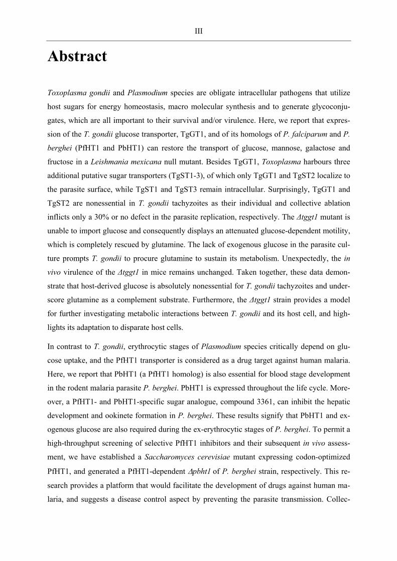
architecture (Striepen et al., 2007). The cellular morphology (Fig. 1) is conserved by a micro-
tubule network that underlies the inner membrane complex, which itself is firmly connected
to the plasma membrane. All apicomplexans posses an apicoplast, a single golgi stack and an
endoplasmatic reticulum network which surrounds the nucleus.
Figure 1: Canonical representation of an apicomplexan parasite.
AP apicoplast; AR apical rings; CC centrocone; CE centrosome; CO conoid; DG dense granule; ER endoplasmic reticulum; G Golgi; IMC inner membrane complex; MI mitochondrion; MN microneme; MT subpellicular microtubule; NU nucleus; RH rhoptry. Adopted from Striepen, et al., 2007.
The mitochondrion is present in all apicomplexan species except for Cryptosporidium, where
it is present in a degenerated form known as a mitosome. The mitochondrion is found in close
proximity with the apicoplast. This plastid-like organelle has been acquired by secondary en-
dosymbiosis and is present in most apicomplexans. Together, apicomplexan parasites impose
a major burden on human and animal health. Toxoplasma gondii is among the most successful

2
parasites and is able to infect most warm-blooded vertebrates. The global prevalence in hu-
mans ranges from 0 to 100%, with 25% in the US (Jones et al., 2007) and 20-80% in Europe
(Tenter et al., 2000). This parasite can invade and replicate in virtually all nucleated cells
which demonstrates its ability to adapt to diverse nutritional environments (Ginger, 2006).
This enormous flexibility is also supported by its genome size (80 MB), which is the largest
among the apicomplexans (Sibley and Boothroyd, 1992), and encodes ~8000 genes. Acute
infections are usually asymptomatic in healthy individuals or cause mild flue-like symptoms,
but may lead to chronic infection. T. gondii causes severe disease such as encephalites and
systemic infections in immuno-compromised individuals and in newborns. Toxoplasma also
serves as a research model organism for other apicomplexan parasites, due to relative ease of
its in vitro culture and genetic manipulation (Kim and Weiss, 2004).
Plasmodium species cause malaria. In humans P. falciparum alone is responsible for more
than 1 million human deaths every year, and inflicts a major socioeconomic burden in en-
demic regions (Sachs and Malaney, 2002). In contrast to T. gondii, Plasmodium parasites are
highly host-specific, and infect a particular cell type within a small range of host organisms.
1.1.1 The life cycle of Toxoplasma gondii
The life cycle of Toxoplasma consists of the sexual and asexual phases (Fig. 2). The sexual
cycle is confined exclusively to the intestine of feline species, whereas the asexual reproduc-
tion can occur in most warm-blooded animals. Upon ingestion of tissue cysts by cats, the
parasite invades, gut epithelium cells and differentiate into merozoites. These merozoites pro-
duce progeny parasites and can also differentiate into "female" macrogametes or "male" mi-
crogametes. After the intracellular mating process, unsporulated oocysts are released into the
gut lumen and shed into the environment, where they undergo sporogony to form two sporo-
blasts, each containing four sporozoites. Sporulated oocysts remain infectious for months and
can initiate another sexual or asexual cycle through oral ingestion.
The asexual phase is initiated by oral infection of non-feline species with either oocyst-
contaminated food or undercooked meat containing tissue cysts. Sporozoites exit the oocyst,
penetrating the gut epithelia and differentiate into tachyzoites. Tissue cyst-release bradyzoites
can also infect epithelial cells and form tachyzoites. These tachyzoites replicate quickly by
endodyogeny and cause acute Toxoplasmosis hallmarked by tissue lysis. Their dissemination
throughout the host organism and across the brain blood barrier occurs via infected macro-
phages and dendritic cells (Lambert and Barragan, 2010; Lambert et al., 2010). The host im-
mune response against tachyzoites induces their differentiation into encysted bradyzoites,

3
which cause a life-long chronic infection. Carnivorism or congenital transmission initiate an-
other asexual cycle (Dubey 1998). Tachyzoites can also be maintained under in vitro condi-
tions in a variety of cell types.
Figure 2: Schematic life cycle of Toxoplasma gondii.
Adapted from (Black and Boothroyd, 2000).
1.1.2 The life cycle of Plasmodium
The invertebrate vector of Plasmodium parasites are mosquitoes and the vertebrate hosts are
mostly mammals including humans (Fig. 3) (Huff 1947). Infected mosquitoes inject sporo-
zoites into the skin of the mammalian host during a blood meal. Sporozoites are motile cells
and a fraction of them remains in the skin, while others enter the blood stream to reach the
liver and infect hepatocytes. The schizogonic development into blood-stage merozoites
mainly occurs in the liver, but both hepatic and cutaneous parasites can undergo this process
and release merozoites within budding merosomes into the blood stream (Gueirard et al.).
Free merozoites invade erythrocytes and rapidly multiply asexually via schizogony and initi-
ate the symptomatic phase of the Malaria disease. Simultaneously, a fraction of the intracellu-
lar parasites differentiates into micro- and macro-gametocytes. These gametocytes are taken
up by mosquitoes during a blood meal, and develop into gametes inside their midgut. After
fertilization they form a zygote, and then motile ookinetes, which are able to escape the mid-
gut by penetrating the peritrophic membrane and epithelial cells. A small fraction of the ooki-
netes develops into oocysts outside of the midgut protected from the basal membrane. These
oocysts eventually lyse and release sporozoites into the hemocoel, which mature and invade
the salivary glands for another round of infection of intermediate hosts.

4
Figure 3: Life cycle of Plasmodium falciparum.
The right side shows the parasite development in the definite host, the mosquito. Developmental stages in the mammalian host are depicted on the left side. Hosts vary according to the Plasmodium species (Adapted from (Menard 2005).
1.2 Replication Compartments in Parasitized Host Cells
1.2.1 Toxoplasma gondii
The replicative stages of Toxoplasma include tachyzoite, bradyzoite and sporozoite. The
tachyzoite stage is amicable for continuous in vitro culture and genetic manipulation. Its high
replication rate demands import of large amounts of nutrients and presents a decent model for
metabolic studies. Their in vitro culture is performed mainly in human foreskin fibroblasts
(HFF) and in kidney epithelial cells (vero) cells from African green monkey.
Once the tachyzoite has actively invaded its host cell, it induces several modifications to the
parasitized cell. The parasite forms a parasitophorous vacuole (PV) that confers a unique or-
ganelle for its replication. It consists of host-derived lipids and selected proteins from the host
cell (Sibley 1995) The selective exclusion of host proteins is fundamental to the non-
fusogenic nature of the vacuole, which protects the parasite from lysosomal fusion and subse-
quent degradation (Mordue et al., 1999). However, this non-fusogenic state of the PVM cre-
ates an additional barrier for accessing to nutrients. To facilitate nutrient uptake, the PVM is

5
permeable to small compounds below 1.4 kDa including sugars and amino acids (Schwab et
al., 1994).
The PV is further modified by rhoptry- and dense granule-secreted parasite proteins. The host
endoplasmatic reticulum and mitochondria are also recruited by the parasite and establish an
intimate contact with the PVM (Sinai et al., 1997) presumably to provide nutrients. Another
significant ultrastructural feature of T. gondii-infected cells for nutrient acquisition is the re-
cruitment of the host microtubules, which invaginate the PVM without penetrating it
(Coppens et al., 2006). These host organelle sequestering tubulo-structures (H.O.S.T) are con-
sidered to function as conduits for capturing endo-lysosomal nutrients, such as cholesterol.
This host LDL-derived cholesterol accumulates in these structures and released into the PV
lumen in pinched-off vesicles. Another membranous structure, known as the nanotubular-
network, fills the parasitophorous vacuole. It creates a dense network within the PV space,
which is connected to the PVM (Sibley et al., 1995). As implied by its subcellular location, it
is considered to be involved in nutrient transport to the parasite interior.
1.2.2 Plasmodium spp.
The parasite replication takes place extracellular within an oocyst in the mosquito host and
primarily in two cell types within the mammalian host: hepatocytes and erythrocytes. The PV
provides a replication niche in both cells. The PVM of intra-erythrocytic stages is permeable
for small molecules including hexoses and amino acids (Desai et al., 1993), and also allows
export of parasite proteins to the RBC. These secreted proteins extensively re-model the in-
fected RBC (iRBC) (Maier et al., 2009). An exemplary host modification is the parasite-
induced new permeation pathway (Merckx et al., 2009). These pore-like channels in the
plasma membrane of the iRBC are permeable for small soluble molecules, and are assumed to
support the parasite nutrition and disposal of metabolic waste. The PV forming during the
hepatic development of the parasite also allows the diffusion of small molecules below 855
Da. Similar to T. gondii infected cells, the PVM, of Plasmodium associates with the host ER
but not with mitochondria (Bano et al., 2007).
1.3 Central Carbon Metabolism of Hexoses
The purpose of the central carbon metabolism is to convert energy-rich molecules such as
hexoses ways that support life. Hexoses are degraded in order to provide free energy, chemi-
cal energy (ATP), reduction equivalents (NADH, NADPH) and biosynthetic precursors.

6
These products are used to synthesize and maintain the major constituents of a cell such as
lipids, proteins and nucleotides. T. gondii (Polonais and Soldati-Favre et al., 2010) and Plas-
modium spp. (Olszewski and Llinas 2010) harbour a full set of glycolytic enzymes (Fleige et
al., 2007) (Roth et al., 1988), a complete tricarboxylic acid (TCA) cycle (Fleige et al., 2008;
Olszewski et al., 2010) and the pentose-phosphate pathway (PPP) (Bozdech and Ginsburg,
2005), to satisfy these cellular requirements. In addition, Toxoplasma features a gluconeo-
genesis pathway (Fleige et al., 2008).
1.3.1 Glycolysis and apicoplast synthesis pathways
Both, Plasmodium and Toxoplasma parasites rely largely on glycolysis, and degrade glucose
primarily to lactate (Fry et al., 1990; Ohsaka et al., 1982; van Dooren et al., 2006; Vander
Jagt et al., 1990) (Scheibel and Miller 1969). Glycolytic enzymes are localized in the cytosol
of Toxoplasma and are also expressed (Bozdech et al., 2003) and active (Olszewski et al.,
2010; Scheibel and Miller, 1969) in intraerythrocytic Plasmodium parasites. Glucose is the
main energy source for the gliding motility and parasitic invasion of the host cell of
Toxoplasma (Pomel et al., 2008). It also maintains cytosolic ATP-levels and pH homeostasis
in Plasmodium (Saliba et al., 2004). Pyruvate produced by glycolysis is mostly excreted as
lactate or decarboxylated to acetyl-CoA by the pyruvate dehydrogenase (PDH) complex. This
PDH complex localizes exclusively to the apicoplast in Plasmodium and Toxoplasma (instead
of mitochondria like in many eukaryotes). Thus, the canonical connection of cytosolic glyco-
lysis with the TCA cycle in mitochondria does not exist in these parasites (Fleige et al., 2007;
Foth et al., 2005; Olszewski et al., 2010). This further underlines the role of glycolysis for
energy generation over oxidative phosphorylation.
Interestingly, several glycolytic steps can also take place in the apicoplast (Fleige et al., 2007;
Maeda et al., 2009; Saito et al., 2008). The apicoplast also harbours other essential pathways
in both parasites. These include fatty acid synthesis type 2 (FAS2), the DOXP pathway for
isoprenoid synthesis and heme biosynthesis (Seeber and Soldati-Favre, 2010). The apicoplast
metabolism is connected to cytosolic and its own glycolysis to fuel its biosynthetic pathways
with ATP, reduction equivalents, triose phosphates and acetyl-CoA. Plastid transporters,
TgAPT in T. gondii (Brooks et al., 2010) and PfiTPT and PfoTPT in Plasmodium (Lim et al.,
2010) import three-carbon phosphates. Toxoplasma can presumably, use these triose phos-
phates to generate reduction equivalents and ATP by glycolytic enzymes within the apicoplast
(Fleige et al., 2007). The energy source for the Plasmodium apicoplast is unknown (Polonais
and Soldati-Favre, 2010).

7
1.3.2 TCA cycle
The cycle serves as a major hub of cellular metabolism and functions by integrating different
carbon fluxes that originate from the degradation of amino acids, fatty acids and/or sugars.
The pathway directs these substrates towards biosynthetic pathways and/or oxidative phos-
phorylation. The TCA localizes to the mitochondrion in Toxoplasma and Plasmodium (Fleige
et al., 2008; Seeber et al., 2008; van Dooren et al., 2006). T. gondii tachyzoites do not seem to
require a canonical flow of substrates through the TCA cycle (Fleige et al., 2008) as depletion
of the succinyl-CoA synthetase causes only a mild replication defect in vitro. The TCA cycle
functions in a bifurcated fashion during in vitro blood stages of P. falciparum. It is fuelled by
glutamine to produce citrate and ultimately malate via the reductive branch, and generates
malate via its oxidative branch (Olszewski et al., 2010). Its importance for the parasite sur-
vival has not been reported yet.
1.3.3 The pentose phosphate pathway (PPP) pathway
The PPP directly connects the parasite replication to the available of hexoses. It uses glucose-
6-phosphate and generates precursors for the synthesis of nucleotides and aromatic amino
acids. It also recycles NADP to NADPH. The PPP is well conserved and expressed in T.
gondii (Bahl et al., 2010). Also P. falciparum (Bozdech et al., 2003) and P. berghei (Hall et
al., 2005) express most of the enzymes in their blood stages and generate up to 80 % of the
nucleotides directly or indirectly using this pathway (Roth et al., 1986). Furthermore, the
PPP-generated reduction power is needed to sustain the glutathione use of Plasmodium
(Becker et al., 2003).

8
1.4 Protein and Lipid Synthesis Using Hexoses
Besides being substrate for above described metabolic pathways, hexoses are also critical for
lipid synthesis and protein modification. Glycosylation of parasite protein and lipid synthesis
are common and essential eukaryotic phenomena that are also conserved in Toxoplasma and
Plasmodium.
1.4.1 Lipid synthesis in Toxoplasma and Plasmodium
Toxoplasma (Gupta et al., 2005) as well as Plasmodium (Dechamps et al., 2010a; Dechamps
et al., 2010b; Dechamps et al., 2010c) can synthesize major phosholipids using precursors
such as polar head groups (serine, choline, ethanolamine, inositol), fatty acids and glycerol-3-
phosphate. The bulk synthesis of latter two molecules involves the degradation of hexoses via
glycolysis and respective FAS pathways (Mazumdar and Striepen, 2007). Inositol, a close
derivative of hexoses, is utilized for the synthesis of phosphatidyl inositol, inositol phospho-
ceramide (IPC) and glycerol phosphatidylinositol (GPI) anchors in Toxoplasma (Azzouz et
al., 2000; Azzouz et al., 2006; Sonda et al., 2005) as well as in Plasmodium (de Macedo et
al., 2003; Kimmel et al., 2003; Naik et al., 2003). Furthermore, T. gondii tachyzoites and
Plasmodium can incorporate galactose into plant-type lipids such as monogalactosylcere-
brosides (MCDG), monogalactosyldiacylglycerol (MGDG) and digalactosyldiacylglycerol
(DGDG) (Marechal et al., 2002). Finally, hexoses are also utilized to synthesize sphingolipids
in both parasites (Azzouz et al., 2002; Couto et al., 2004; Landoni et al., 2007; Sonda and
Hehl, 2006; Zhang et al., 2010).
1.4.2 Protein glycosylation in T. gondii and Plasmodium
The hexose-requiring N-glycosylation of proteins is essential in T. gondii, and its disruption
by the inhibitor tunicamycin kills the parasites (Luk et al., 2008). Also, GPI-anchored pro-
teins, whose synthesis requires hexoses and their close derivative inositol, fulfill essential
functions in T. gondii such as motility, host cell adhesion (Lagal et al., 2010) and host interac-
tion (Fauquenoy et al., 2008). Direct N- or O-glycosylation of proteins is less common in
Plasmodium (Gowda and Davidson, 1999), however proteins are readily modified by GPI-
anchorage (Gowda et al., 1997). GPI-anchored proteins are surface molecules, which are
highly expressed throughout the life cycle of Plasmodium, and perform crucial functions. For
example, the well known circum sporozoite protein (CSP) requires a GPI anchor for its func-
tion (Dame et al., 1984). These proteins also include several merozoite surface proteins dur-

9
ing the blood stage (Debierre-Grockiego and Schwarz, 2010; Gilson et al., 2006) and P28 in
ookinetes (Blanco et al., 1999; Martinez et al., 2000). An inhibitor, mannosamine that selec-
tively disrupts GPI synthesis by preventing acetylation of the premature GPI anchor aborts in
vitro blood cultures of P. falciparum (Gowda and Davidson, 1999; Naik et al., 2000; Naik et
al., 2003). This illustrates the omnipresence and functional significance of GPI anchors for
Plasmodium parasites.
1.5 Sugar Import in Toxoplasma and Plasmodium
1.5.1 Sugar transport proteins
A distinct class of trans-membrane proteins transports hexoses and inositol. These transporter
proteins are categorized according the energy dependence of their transport reaction. Primary
active permeases such as ATP-binding cassette (ABC-) transporters hydrolyze ATP to trans-
locate their substrates. Secondary active transporters exploit an electrochemical gradient of
mostly ions to energize otherwise unfavorable transport reactions. Facilitative transporters
function passively and allow gradient-dependent diffusion of selected molecules across the
membrane. Many proteins of the second and third group belong to the multi-facilitator super-
family (MFS) of transporters.
Hexose-specific ABC-transporters require hexose-binding proteins that bind hexoses with an
extremely high affinity. Their Km values are in the range of 1 μM or below (Verdon et al.,
2003). They can, therefore, also access minute amounts of sugars. These hexose-binding pro-
teins appear to be confined to bacteria and are not conserved in Toxoplasma and Plasmodium.
Secondary active sugar transporters form a subfamily within the MFS known as solute carrier
family 2A (SLC5A). These transporters are present in bacteria and in mammalian intestine.
Other cells requiring sugars rely mostly on facilitative sugar transporters of the SLC2A sub-
family and exploit an inward hexose gradient across their plasma membrane that is estab-
lished by active catabolism of hexoses. Once inside hexoses are quickly phosphorylated in the
cytosol to prevent their outward diffusion. These transporters contain 12 trans-membrane do-
mains (TMDs) linked by short loop-regions and their N- and C-termini face the cytosol.
These proteins are characterized by well-conserved amino acids and motifs, which are also
considered as their signature residues (Joost and Thorens, 2001).

10
1.5.2 Glucose uptake in Toxoplasma gondii
The first indirect evidence of sugar import by T. gondii comes from the detection of periodic
acid-Schiff (PAS)-stained carbohydrate storage and by a prominent lactate dehydrogenase
activity in Toxoplasma tissue cysts (Meingassner et al., 1977). The very low succinate dehy-
drogenase activity in these cysts indicated glycolysis (and thus sugars) as being the main en-
ergy source. Accordingly, isolated extracellular tachyzoites metabolized glucose to lactate and
acetate, which accumulated in exogenous media (Ohsaka et al., 1982). Finally a sugar trans-
porter (TgGT1) was cloned from tachyzoite cDNA that mediated uptake of glucose (Km ~30
µM) and fructose in Xenopus oocytes (Joet et al., 2002). TgGT1 localizes uniformly to the
parasite plasma membrane (Pomel et al., 2008). The finding that gliding motility (and thus
host cell invasion) of extracellular tachyzoites is also dependent on glucose (Pomel et al.,
2008) led to the hypothesis that TgGT1 might be essential in Toxoplasma tachyzoites.
1.5.3 Glucose uptake in Plasmodium
P. knowlesi-infected erythrocytes metabolize glucose to lactate 25 to 75 times faster than un-
infected cells (McKee et al., 1946) and glucose is vital for in vitro blood cultures of all
treated Plasmodium species (Schuster, 2002). Glucose import in Plasmodium was initially
characterized as an active energy-dependent and concentrative process (Izumo et al., 1989).
However, later on the accumulation of radiolabeled glucose inside infected red blood cells
was attributed to glucose phosphorylation rather than its active transport, which was found to
be entirely equilibrative (Kirk et al., 1996). Indeed, the P. falciparum hexose transporter,
PfHT1, was found to be a high-affinity facilitative transporter for glucose (Km ~1 mM) and
fructose (Km ~11.5 mM). It is localized at the parasite plasma membrane in blood stages and
its mRNA was most abundant in asexual blood stages and least expressed in the gametocytes
(Woodrow et al., 1999; Woodrow et al., 2000). Other species of Plasmodium such as P.
knowlesi and P. yoelii, that infect monkeys and rodents, respectively, also express similar
transporters (Joet et al., 2002).
Interestingly, a glucose analog, compound 3361 (C3361), that carries an alkyl-substitution at
its O3-position, selectively inhibits PfHT1 (Ki ~53 µM) and blood cultures at a very similar
concentration (IC50 ~16 µM) (Saliba et al., 2004). Consistently, PfHT1 as well as its ho-
molog in the murine malaria parasite Plasmodium berghei (PbHT1) are refractory to gene
deletion (Slavic et al., 2010). Thus, PfHT1 is considered as a drug target for blood stages. In
this regard a Leishmania mexicana null mutant model expressing PfHT1 has been developed
for in vitro screening of selective PfHT1 inhibitors (Feistel et al., 2008). Besides the expres-

11
sion of PbHT1 during mosquito stages (Slavic et al., 2010), little is known about the signifi-
cance of HT1 during other stages of Plasmodium.
1.6 Aim of This Study
Glucose is considered as a central nutrient for T. gondii and Plasmodium. However, prior to
this work there has been no genetic evidence that glucose import is essential (or otherwise) in
Toxoplasma and Plasmodium. One hexose transporter of T. gondii (TgGT1) has been bio-
chemically characterized. Other potential sugar permeases shall be identified and functionally
analyzed together with TgGT1 to evaluate their potential as a drug target or to uncover poten-
tial mechanisms by which the parasites may circumvent the need for host-hexoses.
A single hexose transporter (HT1) from different Plasmodium spp. has also been character-
ized. However, the importance of hexose transport during the ex-erythrocytic life cycle is not
clear. This work has employed the rodent malaria parasite model (P. berghei) to assess the
functional significance of sugar for Plasmodium. In addition, the drug development against
human malaria requires heterologous models expressing the PfHT1 for screening and testing
of inhibitors in vivo and in vitro.

12

2 Materials and Methods
2.1 Materials
2.1.1 Biological resources
Naval Medical Research Institute (NMRI) mice Charles River Laboratories, Germany Anopheles stephensi Nijmegen, Netherlands Human Foreskin Fibroblasts (HFF) Carsten Lüder, University of Göttingen,
Germany Huh7 human hepatoma cells Kai Matuschewski, Max-Planck-Institut
für Infektionsbiologie, Berlin, Germany Toxoplasma gondii tachyzoites (RH hxgprt-) Dominique Soldati-Favre, University of
Geneva, Switzerland Plasmodium berghei ANKA (-/+GFP) Max-Planck-Institut für Infektionsbiolo-
gie, Berlin, Germany, (Janse et al., 2006) E. coli XL-1blue Stratagene, Germany Saccharomyces cerevisiae EBY.VW.4000 Eckhard Boles, University of Frankfurt
/M, Germany
Genotype:
CEN.PK2-1C MAT α leu2-3,112 ura3-52 trp1-289 his-Δ1 MAL2-8c SUC2 hxt17 hxt13Δ::loxP
hxt15Δ::loxP hxt16Δ::loxP hxt14Δ::loxP hxt12Δ::loxP hxt9Δ::loxP hxt11Δ::loxP
hxt10Δ::loxP hxt8Δ::loxP hxt514Δ::loxP hxt2Δ::loxP hxt367Δ::loxP gal2Δ stl1Δ::loxP
agt1Δ::loxP ydl247wΔ::loxP yjr160cΔ::loxP

14
2.1.2 Chemical reagents
Ampicillin Sigma, Germany Bromophenol blue Merck, Germany BSA fraction 5 Roth, Germany Chloramphenicol Roth, Germany Chloroform Roth, Germany Deoxytriphospatenucleotides (dNTPs) Rapidozym, Germany Diethylpyrocarbonate (DEPC) Sigma, Germany Dimethyl sulfoxide (DMSO) Sigma, Germany Dubecco’s Modified Eagle Media (DMEM) w/o Na-pyruvate, w/o L-glutamine, 4.5 g/l D- glucose
Biochrom, Germany
EDTA Applichem, Germany Ethidium bromide Applichem, Germany Fetal Calf Serum Biochrom, Germany Fluoromount G / DAPI SouthernBiotech, USA Gentamycin Invitrogen, Germany Giemsa solution BDH, Dubai Glacial acetic acid (99 %) Applichem, Germany Glutaraldehyde Roth, Germany Glutathione Sigma, Germany Glycerol Applichem, Germany Heparin Braun, Germany Hexoses Applichem, Germany HPLC-purified water Roth, Germany IPTG Applichem, Germany Isofluoran Baxter, Germany Ketamine (10 %) Bayer, VFW L-glutamine (200 mM) Biochrom, Germany Lithium acetate Applichem, Germany
Methanol Roth, Germany
Mycophenolic acid, Xanthine Applichem, Germany Na-pyruvate (100 mM) Biochrom, Germany Non-essential amino acids (100x) Biochrom, Germany Nycodenz Axis Shield, Norway Paraformaldehyde Roth, Germany PBS Biochrom, Germany Penicillin / Streptomycin (10000 U/ml and 10000 µg/ml) Biochrom, Germany Penicillin / Streptomycin (5000 U/ml and 5000 μg/ml) Invitrogen, Germany Protease peptone Difco, US

15
Pyrimethamine Sigma, Germany RPMI-Medium 1640 Invitrogen, Germany Salmon sperm DNA (10 mg/ml) Invitrogen, Germany Salts Roth, Applichem, Germany Tris-HCl Applichem, Germany Triton X-100 Applichem, Germany Trypsin / EDTA Biochrom, Germany Tryptone Applichem, Germany X-Gal Applichem, Germany Xylazin hydrochloride (2 %) Bayer, VFW, Rompun ® Xylen Cynol FF Merck, Germany Yeast extract Roth, Germany Yeast nitrogen base (YNB) Sigma, Germany [14C(U)]-D-glucose, [3,4-3H(N)]-L-glutamine Hartmann Analytic, Germany Synthetic codon-optimized PfHT1 Genscript, USA Primers (see Table 1) Invitrogen, Germany Compound 3361 David H. Peyton, Portland
State University, US
2.1.3 Vectors
The vector for P. berghei transfection was originally created by A.P. Waters (University Lei-
den, Netherlands) (pb3D-) and modified into pb3D+ by M. Ganter (Harvard University, Bos-
ton, US). Both vectors are pyrimethamine-selectable in P. berghei. The drug resistance is con-
ferred by a mutated dihydrofolate thymidylate synthase (DHFR-TS) gene of T. gondii that is
regulated by untranslated regions (UTR) of P. berghei DHFR-TS. The pb3d+ vector also con-
tains 3’UTR of DHFR-TS, which regulates the expression of a cloned cDNA. The pb3d- vec-
tor was used for conventional gene replacement in P. berghei, whereas the pb3D+ vector was
used for gene replacement by functional complementation.
All transfection vectors for T. gondii were obtained from Dominique Soldati-Favre (Univer-
sity of Geneva, Switzerland), except for the pNTP3, which was provided by Isabelle Coppens
(John Hopkins Bloomberg School of Public Health, Baltimore, US). A pTUB8-CAT knock-
out vector was modified to construct a chloramphenicol-selectable TgGT1 knock-out vector.
An ApaI-excisable fragment was replaced with 3 kb long 5’UTR of TgGT1, and a XbaI- plus
XhoI-excised fragment was replaced with 3 kb of the respective 3UTR. The p2854
(pyrimethamine-selectable) vector was used to construct a TgST2 knock-out vector by the
introduction of 3 kb long 5’ and 3’ UTRs between the ApaI / HindIII and at the NotI sites
respectively. For complementing TgGT1 knock-out parasites, the pNTP3 vector was altered

16
by replacing the TgNTP3 promoter between the ApaI and PacI sites by a 4.5 kb fragment con-
taining the TgGT1 promoter and cDNA with a C-terminal HA-tag. Furthermore, a hypoxan-
thine guanosine ribosyltransferase (HXGPRT) expression cassette was introduced between
KpnI and ApaI sites as a selection marker in this vector. This expression cassette confers my-
cophenolic acid resistance to HXGPRT knock-out (RH hx-) parasites. For N-terminal HA-
tagged expression of TgST1-3, and C-terminal HA-tagged expression of TgGT1 the respec-
tive cDNAs were inserted between NheI and PacI sites (TgST1-3) and NcoI and PacI site
(TgGT1) of the pNTP3 vector. For the Ty1 epitope-tagged expression of all four transporters
respective cDNAs were inserted between EcoRI and NsiI sites, except TgST2 which was
cloned between the EcoRI and Sbf1 site into the pT8 vector.
The pESC-ura vector (Agilent Technologies, US) was modified by inserting PfHT1 and
ScHXT9 genes into its NotI site for expression in S. cerevisiae. This vector was also modified
by introducing cDNAs encoding for 80 or 55 N-terminal amino acids of ScITR1 (ScITR1-
GM80) and ScHXT9 (ScHXT9-GM55) transporters between its EcoRI and NotI sites. The
eventual vector was used to clone hybrid isoforms of PfHT1 at NotI site. A yeast expression
plasmid (p426-HXT7-his) is designed to express N-terminal his-tagged proteins under the
ScHXT7 promoter. In order to express PfHT1 and ScHXT9 as his-tagged fusion proteins
ScITR1-GM80-PfHT1, ScHXT9-GM55-PfHT1, PfHT1 and ScHXT9 were amplified from
pESC-ura templates and introduced into the SpeI site of p426-HXT7-his. Both yeast expres-
sion vectors contain the S. cerevisiae ura3 gene that confers uracil prototrophy to mutant
strains.
The pX63NeoRI vector was used as provided by Scott Landfear (Oregon Health University,
Oregon, US) for expression in L. mexicana. All four T. gondii transporters as well as PfHT1
and PbHT1 were cloned at its EcoRV site. The pL2-5 vector was used for expression in
Xenopus laevis oocyte. Both Plasmodium transporters were cloned between BglII and PacI
sites. This vector contains β-globulin sequences for stabilizing of transcribed cRNA.
2.1.4 Antibodies and working dilutions
α-TgGAP45 (1:3000) (Plattner et al., 2008) α-TgGra3 (1:500) (Dubremetz et al., 1993) α-TgSag1 (1:1000) (Kim and Boothroyd, 1995) α-HSP70 (1:1000) (Tsuji et al., 1994) α-P28 (for bead coating) (Siden-Kiamos et al., 2000) pro-
vided by Robert Sinden (Impe-rial College, London, UK)

17
PbACP (1:500) (Friesen et al., 2010) α-Ty (BB2 hybridoma cell culture supernatant 1:50) (Bastin et al., 1996) α-HA (rabbit, mouse) (1:1000) Invitrogen, Germany Alexa 594, Alexa 488 (anti mouse, anti rabbit) (1:3000) Invitrogen, Germany
2.1.5 Enzymes
Pfu Ultra II Fusion HS DNA polymerase Stratagene, Germany Dream Taq polymerase Fermentas, Germany Restriction endonucleases, Klenow enzyme NEB, Germany T4 ligase Invitrogen, Germany Antartic phosphatise, Klenow NEB, Germany Proteinase K Sigma, Germany Zymolase Zymo research, USA
2.1.6 Instruments
PCR Thermocycler (DNA engine PTC-200) Bio-Rad, Hercules, USA PCR Thermocycler (FlexCycler) JenaAnalytic, Germany PCR Thermocycler (Mastercycler Gradient) Eppendorf, Germany Gel electrophoresis chamber and power supply Amersham Biosciences, USA Water purification systems (MilliQ PF, MilliPure RX20) MilliPore, USA Microscope (Laborvert) Leitz / Leica, Germany Microscope (Apotome Imager.Z2) Zeiss, Germany Confocal Microscope Leica LSM-SP2 Leica Microsystems, Germany Stereo fluorescence microscope setup MZ10F Leica Microsystems, Germany Gel documentation & EASY Enhanced Analysis Herolab, Germany BioPhotometer Eppendorf, Germany Nanodrop ND 1000 Wilmington, USA BTX square wave electroporator (ECM 830) BTX, USA Amaxa Electroporator Amaxa, Germany Scintillation counter (1450 MicroBeta TriLux) PerkinElmer, USA
2.1.7 Plasticware and disposables
Cryo tubes Biochrom, Nalgene, Germany Syringes BD, Germany Needles BD, Germany

18
Microscopy slides Menzel, Germany Cover slips Roth, Germany Improved Neubauer counting chamber Neubauer, Germany Tissue culture flasks, Petridishes, Multi-well plates Greiner Bio-One, Austria LabTek chamber slides ThermoScientific, Germany PCR tubes Rapidozym, Germany Eppendorf tubes (1.5 ml, 2 ml) Greiner Bio-One, Austria Pipette tips Greiner Bio-One, Austria RNAase-free barrier tips Sorenson BioScience, USA Filter sterilizer (0.22 µm) Schleicher Schuell, Germany Disposable pipettes (10 ml, 25 ml, 50ml) Greiner Bio-One, Austria Falcon tubes (15 ml, 50 ml) Greiner Bio-One, Austria Polypropylene tubes (12 ml) Greiner Bio-One, Austria Electroporation cuvettes (4mm gap) Eppendorf, Germany Amaxa electroporation cuvettes Amaxa, Germany Amaxa electroporator Amaxa, Germany
2.1.8 Commercial kits
DNA purification (plasmid preps) Jena Analytic, Invitek, Invitrogen, Qiagen, Germany
RNA isolation kit Qiagen pDrive cloning kit Qiagen, Germany Reverse transcription PCR (SuperScript III) Invitrogen, Germany Trizol Invitrogen RNA purification Qiagen µMACS mRNA isolation Miltenyi Biotec, Germany µMACS one-step cDNA synthesis Miltenyi Biotec, Germany Yeast miniprep kit Zymo Research, US
2.1.9 Reagent preparations
D10
500 ml of DMEM media were supplemented with 5 ml of Penicillin / Streptomycin (10000
U/ml and 10000 µg/ml), 5 ml of sterile 200 mM L-glutamine, 50 ml FCS, 5 ml of 100 mM
Na-pyruvate and 5 ml of 100x non-essential amino acids.

19
P. berghei freezing solution
This solution was prepared by mixing glycerol and Alsever’s solution at 1:9 ratio. 200 µl of
isolated blood were diluted with 100 µl of freezing solution and directly frozen at -80°C.
P. berghei media for transfection
This media contained 160 ml of RPMI 1640 with 25 mM HEPES and L-Glutamine 2.05 mM
(Gibco), 40 ml of heat-inactivated FCS and 50 μl gentamycin (50 mg/ml). The media was
filtered sterile.
P. berghei ookinete (incomplete media)
0.425 g NaHCO3 and 2.5 ml of Penicillin / Streptomycin (5000 U/ml and 5000 μg/ml) were
added to 500 ml of RPMI 1640, and the pH was adjusted to 8.0 and the solution was sterile
filtered.
P. berghei ookinete (complete media)
Ookinete complete media was prepared from above-mentioned incomplete medium by addi-
tion of 10 % heat inactivated FCS, 100 nM hypoxanthine and 50 µM xanthurenic acid. The
pH was adjusted to 8.0 prior use.
Pyrimethamine stock solution (100x)
7 mg pyrimethamine was dissolved in 1 ml of DMSO and finally diluted in drinking water.
LB media
10 g of tryptone, 5 g of yeast extract and 10 g NaCl were dissolved in a final volume of 1 liter
ddH2O. For LB plates, 15 g of agar-agar was also included. The LB media was sterilized by
autoclaving. Ampicillin (100 µg/ml final concentration) or supplements such as IPTG (0.0012
%) and X-gal (0.004 %) were added prior to pouring plates or inoculation.

20
SOB media
20 g of tryptone, 5 g of yeast extract, 0.5 g NaCl and 186 mg KCl were dissolved in 1 liter
ddH2O and autoclaved. The MgCl2 (2 M sterile stock solution) was added to a final concen-
tration of 10 mM just before usage.
SOC-media
Deionised water containing 2 % of tryptone and 0.5 % of yeast extract (w/v), 10 mM NaCl
and 2.5 mM KCl, was autoclaved, allowed to cool down before addition of filter-sterile glu-
cose (20 mM).
YP-media
20 g peptone, 10 g yeast extract and 20 g agar-agar (optional) was solubilized in a final vol-
ume of 950 ml ddH2O water, autoclaved and stored at room temperature.
Sugar stock solutions
D-Glucose, maltose, D-mannose or D-galactose were dissolved to make stocks of 40 % in
ddH2O (v/w) filter sterilized and stored at 4°C.
Yeast-extract peptone dextrose / maltose (YPD / YPM) media
950 ml of YP media was supplemented with 50 ml of filter-sterile glucose stock (40%) for
YPD media or with sterile maltose stock (40 %) for YPM media, and stored at 4°C. Final
sugar concentration in media was 2 %.
10x amino acid mix
The following compounds were dissolved in a final volume of 500 ml ddH2O:
adenine hemisulfate (400 mg), L-arg (200 mg), L-asp (1000 mg), L-gln (1000 mg), L-his (200
mg), L-leu (600 mg), L-lys (300 mg), L-met (200 mg), L-phe (500 mg), L-ser (3750 mg), L-
thr (2000 mg), L-try (400 mg), L-tyr (300 mg), L-val (1500 mg) and uracil (200 mg).

21
The solution was filter sterilized and stored in dark (wrapped in aluminium foil) at 4°C up to
4 months. Uracil was omitted for the preparation of uracil selection media.
Synthetic drop-out media
The basic media contained 1.7 g YNB (free of ammonium sulphate and amino acids) and 5 g
ammonium sulphate in 500 ml ddH2O (filter-sterilized and stored at 4°C). The SD media was
prepared by supplementing YNB with 10x amino acid mix (-ura) and appropriate sterile sugar
stock solution (40 %) to final concentration of 2 %.
For SD plates, 20 g agar agar was also included in YNB, and the media was autoclaved and
stored at room temperature. The media was warmed in microwave before adding supplements
and pouring plates.
Cytomix for Toxoplasma gondii transfection
The solution contained 120 mM KCl, 0.15 mM CaCl2, 100 mM K2HPO4/KH2PO4, 500 mM
HEPES, 100 mM EGTA and 100 mM MgCl2. It was filter sterilized and stored at 4°C.
Escherichia coli transformation buffers
Transformation buffer 1: 30 mM KOAc (pH 5.8), 50 mM MnCl2 x H2O, 10 mM CaCl2, 100
mM RbCl and 15 % glycerol.
Transformation buffer 2: 10 mM MOPS (pH 7), 75 mM CaCl2, 10 mM RbCl and 15% glyc-
erol.
Both buffers were autoclaved before the addition of sterile glycerol.
S. cerevisiae transformation buffers
TE buffer
The 10x TE buffer contained 100 mM Tris-HCl plus 10 mM EDTA, and was adjusted to pH
7.5 before autoclaving. The 1x TE buffer was freshly prepared from 10x TE buffer.

22
LiAc / TE buffer
The 10x LiAc contained 1 M lithium acetate in ddH2O. The pH was adjusted with acetic acid
to 7.5 and the solution was autoclaved. The 1x LiAc / TE buffer was prepared by diluting 10x
LiAc solution in 10x TE buffer and water.
PEG3350 / LiAc / TE buffer
This buffer contained 40 % PEG3350 and was prepared fresh from 10x TE, 10x LiAc and
PEG3350 (50 %). The autoclaved PEG 3350 stock solution was composed of 50 % (w/v)
polyethylene glycol (M = 3350 g/mol) in ddH2O.
TAE buffer for agarose gel electrophoresis
The 1x TAE buffer was prepared from 50x buffer, which contained 242 g/l Tris base, 57.1
ml/l glacial acetic acid and 18.6 g/l of EDTA.
Lysis buffer for Toxoplasma DNA preparation
10 mM Tris-HCl, pH 8; 5 mM EDTA, 0.5% SDS, 200 mM NaCl were solved in water and
autoclaved. 100 µg/ml proteinase K were added prior use.
Zymolase digestion buffer
This buffer contained 1.2 M sorbitol, 0.3 % β-mercaptoethanol and 16.7 U / ml zymolase en-
zyme diluted in water.
Egression buffer
This buffer contained 10 μM A23187 in HBSS with Calcium and Magnesium. 25 mM glu-
cose or 10 mM glutamine were also added for egression of radioactively labelled parasites.
23
2.2 Methods - Culture and Transfection
2.2.1 Propagation of mammalian cells
Human foreskin fibroblasts (HFF) and human hepatoma (Huh7) were maintained in T-300 or
T-75 tissue culture flasks using D10 media and seeded as required into dishes (22 cm2), in
multi-well plates or in chamber slides by trypsinization. Cells were maintained humidified
incubator (10 % CO2, 37°C).
2.2.2 Propagation of Toxoplasma gondii tachyzoites
Tachyzoites were maintained using confluent monolayers of HFF cells. Transgenic parasites
were selected in 25 μg/ml mycophenolic acid (MPA) with 50 μg/ml xanthine, 1 μM
pyrimethamine or chloramphenicol (6.8 µg/ml). Limiting dilutions of parasites were used to
infect confluent monolayers in 96-well plates and the parasite clones were scored one week
after infection.
2.2.3 Toxoplasma gondii transfection
10 to 30*106 freshly-egressed or syringe-released parasites were centrifuged at 300 g for 10
min at room temperature, and the pellet was suspended in 700 µl of cytomix. This mixture
was complemented with 5-50 µg of linearized or circular plasmid DNA, 30 µl ATP (sterile
100 mM stock) and 12 µl GSH (sterile 250 mM stock). Electroporation was done by two 1.7
kV pulses at an interval of 100 msec using a BTX square wave electroporator.
2.2.4 Giemsa staining of blood smears of P. berghei
In order to monitor parasitemia of infected mice, a drop of blood was collected from the tail
and smeared on to a microscope slide. The smear was fixed in methanol for five minutes,
stained for 20 min in 1:10 water-diluted Giemsa solution, washed in water and dried for mi-
croscopic examination with an 100x oil-immersion objective.
2.2.5 P. berghei propagation and transfection
P. berghei ANKA strain parasites were propagated by intraperitoneal injection of fresh or
stored blood cultures into about eight weeks old NMRI mice. For transfection, blood with a
parasitemia of ~2 % was harvested by heart puncture from isofluoran-anesthetized mice using
heparin-treated syringes. The blood was diluted with pre-warmed transfection media (1:2) and

24
red blood cells were isolated by centrifugation at 1000 rpm for 8 min. The pellet was sus-
pended in 50 ml transfection media and mixed with another 100 ml of pre-warmed transfec-
tion media. The culture was incubated for 17 hrs at 37°C with gentle shaking at 10 % O2, 5 %
CO2, 85 % N2. To purify mature schizonts, 35 ml of culture media were layered onto 10 ml of
55% Nycodenz / PBS in 50 ml flacon tubes and centrifuged (no brake, 25 min, 1000 rpm)
before harvesting the interphase with a Pasteur pipette. The schizonts were washed in 30 ml
transfection media in two falcon tubes, and then suspended in 10-15 ml media (10 to 30*106/
ml schizonts). 1 ml of schizonts per transfection was sedimented and suspended in 100 µl
human T-cell nucleofector solution supplemented with 2-5 µg (5-10 µl) of KpnI/NotI lin-
earized DNA. The suspension was electroporated using the program U33 and the Amaxa elec-
troporator. 50 µl of culture media was added to the parasites, and were then intravenously
injected into animals (75µl / NMRI mouse). Pyrimethamine selection was applied to mice 24
hrs later. 100x pyrimethamine stock solution was used to supplement the drinking water and
the pH was adjusted to 5 - 3.5 before feeding. A blood parasitemia could be detected five to
eight days post-infection in Giemsa-stained blood smears. The blood was harvested at a para-
sitemia of ∼2 % by heart puncture, and 200 µl were used to generate two freezer stocks. Re-
sidual blood was used to isolate the parasite DNA for genotyping. The PCR-verified trans-
genic parasite population was transferred into a NMRI mouse for further selection, genotyped
and cryo-preserved at a parasitemia of about 2 %. Frozen stocks were injected into the perito-
neum of NMRI mice for cloning and blood with a parasitemia below 1 % was harvested. The
presence of 7*106 erythrocytes / µl of blood was assumed for absolute quantification of in-
fected blood cells. The infected whole blood was diluted in RPMI 1640 media to obtain 1
iRBC / 100 µl. Five to ten mice were injected intravenously. Positive mice were then bled at a
parasitemia of ∼2 %. The obtained parasites were genotyped to ensure clonality before mak-
ing freezer stocks.
2.2.6 Ookinete culture and purification
1 ml of blood culture was harvested from infected NMRI mice three to four days post-
infection, and analyzed for exflagellation. A blood drop was covered with a square cover slip
on a microscopic slide and incubated for 10 min at room temperature. The temperature drop-
induced exflagellation process could be microscopically monitored by counting areas of
bouncing red blood cells that were hit by the released and highly mobile male gametes. Three
exflagellation events per field using the 100x magnification objective were considered suffi-
cient. 1 ml of the collected blood was incubated in 9 ml of complete ookinete media for 18 –

25
20 hrs at 20°C without shaking. The cell suspension was centrifuged at 1500 rpm for 8 min
and suspended in 0.5 ml of ookinetes complete media containing 3 µl of P28-monoclonal an-
tibody-coated (Siden-Kiamos et al., 2000) paramagnetic dynabeads. After inverting the sam-
ple for 10 minutes in a 1.5 ml Eppendorf tube, the tube was placed into a magnetic rack. The
media was removed and the beads were washed with PBS. After removal of the tube from the
magnetic rack, the ookinetes were suspended in 100 µl deionized water and pipetted up and
down to facilitate the detachment of ookinetes from the beads. The ookinetes were counted in
a Neubauer counting chamber.
2.2.7 Propagation of Anopheles stephensi mosquitoes
The mosquitoes were raised in a 14 hrs light /10 hrs dark cycle, 75 % humidity and at 20°C.
2.2.8 Preparation of oocysts and salivary gland sporozoites of P. berghei
P. berghei-infected mice harbouring exflagellation-competent parasites were anesthetized
with Ketamine / Xylazin hydrochloride by intraperitoneal injection and put on to a mosquito
cage containing 12 hrs-starved mosquitoes for 30 min in dark. Ten mosquitoes were collected
with a suction pump at 17 days post-infection, immobilized on ice, washed for 3 min in 70 %
ethanol and put in RPMI 1640 supplemented with 3 % BSA for examination of infectivity by
dissection under a binocular microscope. The midguts containing oocysts were collected in
PBS on ice, 17 to 24 days post-infection. Salivary glands were dissected in RPIMI 1640 with
3 % BSA, stored on ice and disrupted in an 1.5 ml Eppendorf tube to release sporozoites for
counting and assays. For infection of liver cells, mosquitoes were dissected directly in D10
media.
2.2.9 Liver stages of P. berghei
10.000 trypsinized Huh7 cells were seeded into one well of an eight-well LabTek chamber
slide and grown in D10 media for one day. 30.000 sporozoites isolated from salivary glands
were pipetted on to the cells. The chamber slide was centrifuged at 1000 rpm for 10 min at
room temperature to enhance the infection rate. Infected cells were maintained for 68 hrs at
37°C with 10 % CO2, and washed three times daily with fresh media.
2.2.10 Making of Saccharomyces cerevisiae competent cells
4 ml of synthetic complete media containing 2 % maltose were inoculated with the
EBY.VW.4000 strain picked from a master plate, and incubated with shaking at 30°C over

26
night. This pre-culture was used to inoculate 50 ml of YPM media (EBY.VW.4000 strain) at
an initial OD600 of 0.1. The culture was incubated at 30°C with shaking and allowed to reach
an OD600 of 0.4. Yeast cells were harvested by centrifugation at room temperature at 1000 g
for 5 min in sterile 50 ml falcon tubes. After washing the cells with 25 ml of sterile TE-buffer
and 10 ml of LiAc / TE buffer, cells were finally resuspended in “N x 100 µl” of LiaAc / TE
buffer for “N” transformations. This suspension was kept at room temperature for at least 30
min, before transformation.
2.2.11 Transformation of S. cerevisiae competent cells
100-200 ng of plasmid preparation and 100 µg of salmon sperm DNA were mixed by finger
tapping, and then mixed with 100 µl of competent yeast suspension. The transformation mix
was vortexed for 10 sec at maximum speed after adding 0.6 ml of PEG / LiAc / TE solution.
The mixture was then horizontally incubated for 30 min at 200 rpm at 30°C. 70 µl of DMSO
were added to each reaction and the suspension was mixed by inverting the tube. Then, the
mixture was heat-shocked for 15 min at 42°C in water bath and cooled immediately for 2 min
on ice. The cells were pelleted at 14000 g for 15 sec and the supernatant was removed. The
cell pellet was washed in 1x TE buffer and suspended in 100 µl of TE buffer for plating on
SD (-ura) plates with 2 % maltose. The plates were incubated at 30°C for three to four days.
One colony was picked from each plate and streaked on to a master plate for subsequent ex-
periments. Freezer stocks were made in 2 % glycerol.
2.3 Methods - Molecular Cloning
2.3.1 PCR reactions
10-500 ng of the template DNA was used for PCR. The Dream-Taq polymerase (Fermentas)
was employed for standard analytical PCR and Pfu-Ultra FusionII high fidelity polymerase
(Stratagene) was used for expression cloning. PCR composition and conditions were set ac-
cording to the primers, polymerase and amplification targets.
2.3.2 Agarose gel-electrophoresis
Gels (0.5 to 2 %) were prepared from agarose in TAE buffer. The solution was boiled in a
microwave oven to dissolve agarose and allowed to cool down before ethidium bromide was
added. The gel was cast, and immersed in 1x TAE buffer after it had solidified. The DNA was
loaded in 1x loading dye, and separated at 80 – 200 V for 1-2 hrs.
27
2.3.3 Vector preparation
1-2 µg (Nanodrop D100) of plasmid was digested with appropriate enzyme for 2 hrs at 37°C
for cloning. Subsequently, the digestion reaction was resolved on 1 % agarose gel, and a lin-
earized fragment of right size was extracted according to the kits’ protocol. The DNA was
eluted from the gel-extraction column by two serial applications elution buffer. In case of
symmetric cloning, the linearized vectors were dephosphorylated using Antartic Phosphatase
(NEB) for 1 hr at 37°C followed by heat inactivation (65°C, 30 min).
2.3.4 Insert preparation
50 µl PCR reactions were run on agarose gels to verify size and purity of amplified fragments.
The amplicon of right size was cut out and gel extracted according to the kits’ manual. The
extracted sample was digested by appropriate restriction enzymes for 2 hrs at 37°C. After
column purification the ligation was performed.
2.3.5 Ligation
The ligation of digested and dephosphorylated (optional) vector with the insert was done at
1:3 and 1:5 molar ratio of vector to insert (10 fmol of vector and 30 or 50 fmol of insert). The
reaction was performed at room temperature for one hour, or at 14°C overnight. To estimate
the amount of religated vector, water (instead of insert) was used as a negative control.
2.3.6 Preparation of competent Escherichia coli cells
2 ml of LB media were inoculated with XL-1blue or DH-5α cells and incubated over night
200 ml of SOB media were inoculated with the 2 ml pre-culture of E. coli. This culture was
harvested at OD600 of 0.4 to 0.5 (1300 g, 10 min, 4°C). The pellet was washed once in 50 ml
of ice-cold TFB-I buffer. The cells were resuspended in 6.4 ml of TFB-II buffer and frozen
stocks were made.
2.3.7 Transformation of Escherichia coli
A frozen stock of competent E. coli cells was thawn on ice. Ligation reactions were diluted at
least 10-fold with competent cells and incubated on ice for 30 min. The mixture was heat-
shocked for 45 sec at 42°C in water bath and then cooled immediately on ice for 2 min. About
800 µl of warm (42°C) SOC medium was added to each transformation reaction followed by
incubation at 37°C on shaker for 1hr. The cells were pelleted at 13.000 g for 30 sec and 90 %

28
of medium was discarded. The cells were suspended in residual media and plated by flame-
sterile triangular spatula on LB plates supplemented with Ampicillin. The plates were incu-
bated at 37°C for 12 to 16 hrs.
2.3.8 DNA preparations
Plasmid minipreps were prepared from E. coli cultures according to the kits’ protocol. Midi-
scale plasmid preps were done using MIDI plasmid kit from Invitrogen. Genomic DNA was
prepared from scraped T. gondii-infected host cells according to (Rotureau et al., 2005).
Briefly, the cell pellet was lysed in 200 µl of lysis buffer and incubated for 30 min at 65°C
prior to precipitation with 450 µl of absolute cold ethanol. The DNA pellet was dried at room
temperature and dissolved in sterile water.
Genomic DNA of P. berghei-infected erythrocytes was isolated from harvested blood.
Briefly, erythrocytes were purified using a batting, cellulose and glass bead-filled column,
centrifuged (1500 rpm, 5 min, no brake) lysed with saponin and free parasites were pelleted
by centrifugation (3000 rpm, 5min) of the flow through. After washing in PBS, the parasites
were further processed using the Qiagen DNA preparation kit for blood samples according the
kits’ protocol or store at -20°C.
2.3.9 RNA preparation and cDNA synthesis
RNAase-free material and DEPC treated water were used throughout the procedure.
Toxoplasma gondii-infected cells were washed with PBS and dissolved in 1 ml of Trizol and
stored at -80°C. The sample was thawed for 15 min in a 42°C water bath, 200 µl of chloro-
form was added and after mixing the two phases were allowed to separate for 15 min at room
temperature. The sample was centrifuged for 10 min at 14000 rpm at 4°C and the upper phase
was precipitated with 200 µl isopropanol for 30 min at -80°C. The RNA pellet was obtained
by centrifugation (4°C, 10 min, 14000 rpm) after thawing the sample at room temperature,
washed in 70 % ethanol, dried and dissolved in RNAase-free water. Alternatively, µMACS
mRNA isolation kit and one-step cDNA synthesis kit were used. RNA preparation from in-
fected blood was done by obtaining free blood stages of P. berghei parasites (as described in
the DNA purification protocol) and subjecting them to RNA isolation using the Qiagen RNA
preparation kit. The cDNA was synthesised using SuperScript III first-strand cDNA synthesis
kit (Invitrogen) using oligo-dT primers.

29
2.4 Methods - Assays
2.4.1 Indirect immuno-fluorescence assay (IFA)
Infected cells in chamber slides or on cover slips were fixed in 4 % paraformaldehyde or 4 %
paraformaldehyde with 0.05 % glutaraldehyde or with methanol for 10 to 20 min. The fixative
was neutralized by washing two times with PBS and incubation with 0.5 M glycine / PBS for
at least 5 minutes or over night at 4°C. The first antibodies were applied for 1 hour in block-
ing solution, after permeabilization for 20 min in 0.2 % Triton X-100 / PBS and blocking in
0.2 % Triton X-100 / 2 % BSA / PBS for another 20 min at room temperature. The secondary
antibodies were applied at room temperature for 45 min in blocking solution, after 3 washes
with 0.2 % Triton X-100 / PBS. Finally, the samples were washed three times and mounted in
Fluoromount G / DAPI onto microscopic slides.
All other samples were treated equally except for mosquito midguts, which were fixed with 4
% paraformaldehyde and permeabilized in 1 % saponin and 2 % Triton X-100 at 37°C. Eryth-
rocytes were fixed with 4 % paraformaldehyde and 0.05 % glutaraldehyde. Yeast cells were
grown in selective liquid media and harvested by centrifugation at 1000 g. They were zymo-
lase-digested for 30 min at 30°C before fixation in 4 % paraformaldehyde at 4°C for 12 hrs
and processing for IFA.
2.4.2 Glucose and glutamine uptake assays with intracellular T. gondii
Confluent HFF cells in 6-well plates were infected at MOI of 4 for 24 hrs. All samples were
washed 2 times with 4 ml of glucose-free DMEM prior to their labelling with 1 ml of 14C-
glucose (0.2 μCi/mM, 5 mM) or 3H-glutamine (0.5 μCi/mM, 2 mM) in DMEM for 2 hrs at
37°C. To isolate the labelled tachyzoites, the samples were washed 4x with DMEM-glucose
(25 mM) and treated (10 min, 37°C) with 1.5 ml of the egression buffer (10 μM A23187, 25
mM glucose / 10 mM glutamine in HBSS). Ionophore-egressed tachyzoites were collected
(450 g x 15 min), washed once, and subjected to scintillation and cell counting.
2.4.3 Plaque and replication assays
The parasite plaque assay recapitulates all the events of parasite lytic cycle including host cell
invasion, intracellular growth and egression followed by spreading by gliding motility. The
HFF cells in 6-well plates were infected with T. gondii tachyzoites, cultured for 7 days, fixed
with -80°C methanol, stained with crystal violet and documented using an inverse Leica mi-

30
croscope with 4x magnification objective. The mean area of 80 plaque images was calculated
in total three independent biological replicates for subsequent evaluation. For replication as-
says, infected HFFs (MOI = 1.5) were cultured for 40 hrs, and then the parasites were
egressed for 10 min by 10 μM of A23187 in HBSS. The number of egressed parasites was
normalized to the number of invasive parasites as deduced by complementary plaque assays.
2.4.4 Motility assay
Tachyzoites were syringe-released from HBSS-washed HFF monolayers (30-36 hrs post-
infection), washed twice in HBSS and pre-incubated in HBSS supplemented with indicated
reagents on a cover slip for 10 min at room temperature. The samples were further incubated
(15 min, 37°C) and fixed with 4 % paraformaldehyde / 0.05 % glutaraldehyde, and stained
with mouse monoclonal anti-TgSag1 and Alexa488 antibodies. Images were recorded, and at
least 800 parasites were counted from three biological replicates in five random fields each to
calculate the motile fraction.
2.4.5 Functional expression of PfHT1, PbHT1, TgST1-3 and TgGT1 in Leishmania
mexicana and Xenopus laevis oocytes
These experiments were done in collaboration with Prof. Scott Landfear, Dr. Dayana Rodri-
guez-Contreras and Dr. Marco Sanchez (Oregon Health and Science University, US). Expres-
sion plasmids have been constructed as described in Materials and Methods and transgenic
Leishmania parasites and Xenopus oocytes were generated and analysed by our collarborators.
Briefly, Xenopus constructs were transcribed into cRNA using the mMESSAGE
mMACHINE T7 Ultra Kit (Ambion). Stage V-VI oocytes were injected with 27.6 nl of cRNA
(~14 ng) or water, and then incubated for 4 days at 16°C in ND96 buffer. Uptake of radio-
labeled sugars ([14C]D-glucose (25 Ci/mmol), [14C]D-fructose (300 mCi/mmol), [14C]D-
galactose (52 mCi/mmol) and [14C]D-mannose (50 mCi/mmol)) was assayed by incubating
oocytes with radiolabeled hexoses, followed by three washes in ND96 buffer, and liquid scin-
tillation counting, as described before (Sanchez et al., 2004). For expression in L. mexicana
mutant, the Δlmgt promastigotes (Burchmore et al., 2003) were transformed with pX63NeoRI
constructs, and maintained in RPMI 1640 medium (pH 7.2) containing 10 % FBS and the
drug G418 (100 μg/ml). The parasites were washed twice in phosphate-buffered saline (PBS,
pH 7.4) and resuspended in PBS. Time courses were performed at 25°C with 100 µM of
[14C]D-glucose, [14C]D-mannose, [14C]D-fructose and [14C]D-galactose by an oil-drop
method (Seyfang and Landfear, 2000). For substrate saturation curves, incubations with

31
[3H]D-glucose were performed for 30 seconds. To determine Ki (n=3) values, inhibition
curves were generated with 100 µM of [14C]D-glucose as substrate, and 0-50 mM of sugars or
0-0.25 mM of C3361. The Km values were determined from Michaelis-Menten model,
whereas the Ki from nonlinear regression formulations of the data using experimental the Km
value in a one-site competition model (GraphPad Prism version 4.0). Statistic analysis was
carried out using a two-tail unpaired t test.
Table 1: Primer sequences for indicated purposes and used restriction site used for cloning
Primer Name (restriction site)
Primer Sequence (restriction site underlined)
Cloning Vector (research objective)
TgST1 Cloning TgST1-F1 (NotI) CTCGCGGCCGCATGACGCAGCCTGTCGCAG
TgST1-R1 (NotI) CTCGCGGCCGCCTACTGAGCAATAGCCATATTCTTG
pESC-Ura, pX63NeoRI (functional expression)
TgST1-F2 (NheI) CTCGCTAGCATGACGCAGCCTGTCGCAG
TgST1-R2 (PacI) CTCTTAATTAACTACTGAGCAATAGCCATATTCTTG
pNTP3 (5’-HA tag for immuno-localization)
TgST1-F3 (EcoRI) CTCGAATTCCCTTTTTCGACAAAATGACGCAGCCTGTCGCAG
TgST1-R3 (NsiI) CTCATGCATACTGAGCAATAGCCATATTCTTG
pT8 (3’-Ty1 tag for immuno-localization)
TgST1-F4 (NsiI) CTCATGCATGTGAATTTAAGGCGTCAGGTTG
TgST1-R4 (NsiI) CTCATGCATCTACTGAGCAATAGCCATATTCTTG
TgST1-R3.1 (NsiI)
CTCATGCATATCGAGCGGGTCCTGGTTCGTGTGGACCTCCAACACGGAAAGAGCTGCATTCTCTCG
pT8 (Ty1 tag in the cytosolic loop for im-muno-localization)
TgST2 Cloning TgST2-F1 (NotI) CTCGCGGCCGCATGACTCAGAGGGAGAGATCG
TgST2-R1 (NotI) CTCGCGGCCGCCTACTCTTCAGTGTCTTTCGCT
pESC-Ura, pX63NeoRI (functional expression)
TgST2-F2 (NheI) CTCGCTAGCATGACTCAGAGGGAGAGATC
TgST2-R2 (PacI) CTCTTAATTAACTACTCTTCAGTGTCTTTCGC
pNTP3 (5’-HA tag for immuno-localization)
TgST2-F3 (EcoRI) CTCGAATTCCCTTTTTCGACAAAATGACTCAGAGGGAGAGATCG
TgST2-R3 (SbfI) CTCCCTGCAGGCTCTTCAGTGTCTTTCGC
pT8 (3’-Ty1 tag for immuno-localization)
TgST3 Cloning TgST3-F1 (NotI) CTCGCGGCCGCATGATGCGACCAACATGTGGC
TgST3-R1 (NotI) CTCGCGGCCGCCTAAGAAGGTAACTCCCGTATGG
pESC-Ura, pX63NeoRI (functional expression)
TgST3-F2 (NheI) CTCGCTAGCATGATGCGACCAACATGTG
TgST3-R2 (PacI) CTCTTAATTAACTAAGAAGGTAACTCCCGTATGG
pNTP3 (5’-HA tag for immuno-localization)
TgST3-F3 (EcoRI) CTCGAATTCCCTTTTTCGACAAAATGATGCGACCAACATGTG
TgST3-R3 (NsiI) CTCATGCATAAGAAGGTAACTCCCGTATGG
pT8 (3’-Ty1 tag for immuno-localization)
TgST3-F4 (NsiI) CTCATGCATGGCGCCATTATCATGCAGC
TgST3-R4 (NsiI) CTCATGCATCTAAGAAGGTAACTCCCGTATGGCTTC
TgST3-R3.1 (NsiI) CTCATGCATATCGAGCGGGTCCTGGTTCGTGTGGACCTCCAAGGCGTGACTCTCCTGTTCACC
pT8 (Ty1 tag in the cytosolic loop for im-muno-localization)

32
TgGT1 Cloning TgGT1-F1 (NotI) CTCGCGGCCGCATGGCGACGGAGGAGATGCGT
TgGT1-R1 (NotI) CTCGCGGCCGCTTAAACCACCTCCGTCCCCT
pESC-Ura, pX63NeoRI (functional expression)
TgGT1-F2 (NsiI) CTCATGCATACGGAGGAGATGCGTG
TgGT1-R2 (PacI) CTCTTAATTAAAACCACCTCCGTCCC
pTetO7Sag1 (5’-myc tag for immuno-localization)
TgGT1-F3 (NcoI) CTCCCATGGCGACGGAGGAGATGCGTG
TgGT1-R3 (PacI) CTCTTAATTAATCAAGCGTAATCTGGAACATCGTATGGGTAAACCACCTCCGTCCCCTTG
pNTP3 (3’-HA tag for immuno-localization)
Experimental Annotation TgST1-An-F1 AACTGTGCTCTTCTTGTGC
TgST2-An-F1 GTGAGCCACTAGCTTATGC
TgST3-An-F1 GTGCATATCTATCTCTTACGCTCC
pDrive (T/A-cloning for ORF verification)
TgGT1 knock-out TgGT1-5’UTR-F (ApaI) CACGGGCCCAATGAGCTGCGAGGCCTCTG
TgGT1-5’UTR-R (ApaI) CTCGGGCCCGAACGATCCACTTCCTCTCGG
pTUB8CAT (homolo-gous recombination at 5’-end)
TgGT1-3’UTR-F (XhoI) CTCCTCGAGGCATGCCTTACCAGTTGCGC
TgGT1-3’UTR-R (XbaI) CTCTCTAGAAACCACTGTACAGCGACAGCG
pTUB8CAT (homolo-gous recombination at 3’-end)
TgGT1-5’Scr-F CTCAAGCTTCATGTCAAGGGAAGCCACC
CAT-R CCGGGCCGATCGTTACGCCCCGCCCTGCCAC
pDrive (T/A-cloning for testing 5’-recombination)
CAT-F CCGGGCTGCAGGAGAAAAAAATCACTGGA
TgGT1-3’Scr-R CTCTCTAGAGTGCACCGCTTCGGACGT
pDrive (T/A-cloning for testing 3’-recombination)
TgHXGPRT-F (KpnI) CTCGGTACCGATCAGCACGAAACCTTGCATTCAAACCCGC
TgHXGPRT-R (ApaI) CTCGGGCCCCCCTCCACCGCGGTGTCACT
Δtggt1 complementation vector
TgDLC-F CTCCCATGGCGGACAGGAAGGCGGTG
TgDLC-R CTCGCGGCCGCTCAGCCACTTTTGAACAGCA quality testing of Δtggt1 cDNA
TgST2 knock-out TgST2-5’UTR-F (ApaI) CTCGGGCCCCGCAGGAAGAAACTGAGATGTCACTG
TgST2-5’UTR-R (HindIII) CTCAAGCTTGGTATTGCACAAACTGAAAAAATAGGT
p2854 (homologous recombination at 5’-end)
TgST2-3’UTR-F (NotI) TATGCGGCCGCGGTTTGCCGAACGCATAGACAG
TgST2-3’UTR-R (NotI) GATGCGGCCGCGAGTGTATCTGGATCACTGTCGCTG
p2854 (homologous recombination at 3’-end)
TgST2-5’Scr-F TCAGTCGCACCTCTACTACAGAGTCAC
DHFR-R CTGGAAAGGTGGTGGCTTCTCG
pDrive (T/A-cloning for testing 5’-recombination)
DHFR-F CGAATCCAGATGGAGATGGCTGTC
TgST2-3’Scr-R GCAGCTGTTGGAGACCGGATCG
pDrive (T/A-cloning for testing 3’-recombination)

33
PbHT1 Cloning PbHT1-5’UTR-F (NotI) CTCTCTGCGGCCGCGCAAAACTAAATCGGATTAATGCCGA
PbHT1-HA-R1 (BamHI) CTCATCGGATCCTTAAGCGTAATCTGGAACATCGTATGGGTAAACTCTTGATTTGCTTATATGTTTTTGTCTTTCTTC
pb3D+ knock-out complementation
PbHT1-F2 (BglII) CTCATCAGATCTATGGATATATTATCAAGAGGGGG
PbHT1-R2 (PacI) CTCATCTTAATTAATCAAGCGTAATCTGGAACATCGTATGGGTAAACTCTTGATTTGCTTATATGTTTTG
pL5-2 (Xenopus laevis Oocyte functional ex-pression)
PbHT1-F3 (BglII) CTCTCTGATATCATGGATATATTATCAAGAGGGGGGACTC
PbHT1-R3(PacI) CTCTCTGATATCTTAAACTCTTGATTTGCTTATATGTTTTTGTCTTTCTTC
pX63neoRI (L.mexicana functional expression)
PbHT1-F4 (NotI) CTCATCGCGGCCGCATGGATATATTATCAAGAGGGGG
PbHT1-R4 (NotI) CTCATCGCGGCCGCTTAAACTCTTGATTTGCTTATATGTTT
pESC-ura (functional expression in S.cerevisiae)
PfHT1 Cloning
PfHT1-F1 (BamHI) CTCTCTGGATCCAAAATGACGAAAAGTTCGAAAGATATATGTAGTGAG
PfHT1-R1 (BamHI) CTCTCTGGATCCTCATACAACCGACTTGGTCATATGC
pb3D+ knock-out complementation & PbHT1 complementa-tion screen at 5’-end
PfHT1-F2 (BglII) CTCATCAGATCTATGACGAAAAGTTCGAAAGATATATGT
PfHT1-R2 (PacI) CTCATCTTAATTAATCAAGCGTAATCTGGAACATCGTATGGGTATACAACCGACTTGGTCATATGC
pL5-2 (Xenopus laevis Oocyte functional ex-pression)
PfHT1-F2 (EcoRV) CTCTCTGATATCATGACGAAAAGTTCGAAAGATATATGTAGTGAG
PfHT-R2 (EcoRV) CTCATCGATATCTCATACAACCGACTTGGTCATAT
pX63neoRI (L.mexicana functional expression)
PfHT1-syn-F3 (BamHI) CTCATCGGATCCATGACTAAGTCTTCAAAGGATATCTGT
PfHT1-syn-R3 (SalI) CTCATCGTCGACAACAACGGACTTTGTCATATGTT
pESC-ura (epitope-tagged functional ex-pression in S.cerevisiae)
Expression in Yeast ScHXT9-F (SpeI) CTCATCACTAGTATGTCCGGTGTTAATAATACATCC
ScHXT9-R (SpeI) CTCATCACTAGTTTAGCTGGAAAAGAACCTCTTGT
p426-Hxt7-his (func-tional expression in S. cerevisiae)
ScHXT9-F (NotI) CTCATCGCGGCCGCATGTCCGGTGTTAATAATACATCC
ScHXT9-R (NotI) CTCATCGCGGCCGCTTAGCTGGAAAAGAACCTCTTGT
pESC-Ura (functional expression in S. cere-visiae)
ScHXT9-GM55-F (EcoRI) CTCGAATTCATGTCCGGTGTTAATAATACATCCG
ScHXT9-GM55-R (NotI) CTCGCGGCCGCAGAGGGGTTTTTGAGGTAAGTCAA
Construction of GM55-PfHT1-Syn in pESC-Ura
ScITR1-GM80-F (EcoRI) CTCGAATTCATGGGAATACACATACCATATCTCAC
ScITR1-GM80-R (NotI) CTCGCGGCCGCATTGGTTAAAAGTGATCATGACCG
Construction of GM80-PfHT1-Syn in pESC-Ura
PfHT1-Syn-F (SpeI) CTCATCACTAGTATGACTAAGTCTTCAAAGGATATCTGT
PfHT1-Syn-R (SpeI) CTCATCACTAGTTTAAACAACGGACTTTGTCATATG
p426-Hxt7-his (func-tional expression in S. cerevisiae)
PfHT1-GM55-F (SpeI) CTCATCACTAGTATGTCCGGTGTTAATAATACATCC
PfHT1-Syn-R (SpeI) CTCATCACTAGTTTAAACAACGGACTTTGTCATATG
p426-Hxt7-his (func-tional expression in S. cerevisiae)
PfHT1-GM55-F (SpeI) CTCATCACTAGTATGTCCGGTGTTAATAATACATCC
PfHT1-Syn-R (SpeI) CTCATCACTAGTTTAAACAACGGACTTTGTCATATG
p426-Hxt7-his (func-tional expression in S. cerevisiae)
34
PbHT1 Knockout PbHT1-5’UTR-F (NotI) CTCATCGCGGCCGCGCAAAACTAAATCGGATTAATGCCGA
PbHT1-5’UTR-R (BamHI) CTCATCGGATCCACGCAATATATTCATTTTTTCGTATTAATACACATATATTTCTTG
pb3D+ (for homologous recombination at 5’-end)
PbHT1-3’UTR-F (ApaI) CTCATCGGGCCCCATACAAGAACACACCGGCAAT
PbHT1-3’UTR-R (KpnI) CTCATCGGTACCCTTATGTTAAACAATTATCCTTTCCAATTATCACAC
pb3D+ (for homologous recombination at 3’-end)
PbHT1-5’UTR-F (NotI) CTCATCGCGGCCGCGCAAAACTAAATCGGATTAATGCCGA
PbHT1-HA-R (BamHI) CTCATCGGATCCTTAAGCGTAATCTGGAACATCGTATGGGTAAACTCTTGATTTGCTTATATGTTTTTGTCTTTCTTC
pb3D+ (PbHT1-HA-based PbHT1 gene deletion)
PbHT1-5’Scr-F GCCGTTGGAGAAAATGCAATTAAG
PbHT1-5’Scr-R TCCAGATGGAGATGGCTGTCTAG
pDrive (T/A-cloning for testing 5’-recombination)
PbHT1-3’Scr-F PbHT1-3’Scr-R
CAGACACACCGGTTTATGCAT GATTCGCTAATATCTATGTCAAACCTCCTAGTAG
pDrive (T/A-cloning for testing 3’-recombination)
HA-R AGCGTAATCTGGAACATCGTATGGGTA Test of PbHT1-HA transcript
PbHT1-Clone-F GAAGAAAGACAAAAACATATAAGCAAATCAAGAGTTTAA Clonality testing of Δpbht1-PbHT1-HA)
PfHT1-F (BamHI) CTCATCGGATCCAAAATGACGAAAAGTTCGAAAGATATATGTAGTGAG
PfHT1-R (BamHI) CTCATCGGATCCTCATACAACCGACTTGGTCATATGC
pb3D+ (PfHT1-based PbHT1 gene deletion)

3 Results
3.1 Host-derived Glucose and Its Transporter in the Obligate In-
tracellular Parasite Toxoplasma gondii are Dispensable by
Glutaminolysis
3.1.1 TgGT1 is capable of transporting major sugars in L. mexicana null mutant
Joët et al. (Joet et al., 2002) have shown that TgGT1 can facilitate the transport of glucose and
fructose in Xenopus oocytes. However, it remains unknown if TgGT1 can also mediate the
import of galactose and mannose, required for glycolipid and glycoprotein synthesis. TgGT1
was tested in an L. mexicana sugar transporter null mutant, ∆lmgt, which is defective in im-
porting glucose, mannose, fructose and galactose (Burchmore et al., 2003). After cloning of
TgGT1 into the pX63NeoRI expression plasmid, Dr. Dayana Rodriguez-Contreras (Oregon
Health and Science University, US) generated transgenic ∆lmgt-TgGT1 parasites and ana-
lyzed their hexose uptake activity. TgGT1 expression restored the ability of ∆lmgt cells to
take up all four hexoses, whereas the control cells that have been transformed with an empty
vector were unable to import sugars (Fig. 4A). All transgenic parasite lines incorporated [3H]-
adenosine indicating that they were viable. Substrate saturation curves revealed an apparent
Km of ~50 µM for glucose (Fig. 4B, Table 2). To assess the affinity of TgGT1 towards other
hexoses, their ability to inhibit the uptake of glucose was tested (Fig. 4C). Glucose inhibited
the transport of glucose with a Ki of ~53 µM that is similar to its Km (Table 2), and mannose
exhibited a 5 times higher Ki of ~250 µM. In contrast, fructose and galactose did not signifi-
cantly prevent TgGT1-mediated glucose transport up to 5 mM, but it was reduced by ~50-
70% at 50 mM of inhibitors. These results confirm that TgGT1 has a high affinity for glucose
and mannose, and fructose and galactose are low-affinity ligands.
Table 2: Affinities of TgGT1 towards four hexoses.
TgGT1 Km Ki Glucose 49.8 ± 21.3 µM 53.1 ± 3.0 µM Mannose -- 249 ± 44 µM Galactose -- ~3.0 ± 0.2 mMFructose -- ~3.5 ± 1.6 mM

36
Figure 4: TgGT1 can mediate the import of glucose, mannose, fructose, and galactose in L. mexicana null mutant (Δlmgt).
(A) TgGT1-complemented promastigotes were assayed for their ability to transport 100 μM D-[3H]glucose, D-[3H]mannose, D-[14C]fructose, or D-[14C]galactose. Empty vector (pX63NeoRI) transfected cells were used as the control. (B) Substrate saturation curve for D-[3H]glucose. Uptake was determined over a 30 sec period for a range of substrate concentrations (0.01–2 mM). (C) Inhibition of 100 μM D-[3H]glucose uptake in the presence of sugar inhibitors (0 –50 mM). The Km and Ki were determined by Michaelis–Menten and nonlinear regression analyses.

37
3.1.2 TgGT1 is not essential to the survival of T. gondii
TgGT1 has been considered as the only glucose transporter in T. gondii, and presumed to be
indispensable for the parasite survival. Our data on the import of major hexoses by TgGT1
support this hypothesis. In order to test it, the TgGT1 gene was deleted by double homolo-
gous recombination using the knockout construct pTUB8CAT (Fig. 5A). Following selection
of stable transformants, chloramphenicol-resistant clones were analyzed for disruption of the
TgGT1 locus (Fig. 5A). The knockout construct-specific PCRs using the primer pairs TgGT1-
5'UTR-F, CAT-R and CAT-F, TgGT1-3'UTR-R revealed the presence of the CAT resistance
cassette adjacent to the 5’- and 3’-UTRs in the genome of the ∆tggt1 parasites and in the con-
trol plasmid but not in the parental cells (Fig. 5B). Similarly, PCRs with 5’ and 3’ recombina-
tion-specific primers (TgGT1-5'Scr-F, CAT-R and CAT-F, TgGT1-3'Scr-R yielded DNA
bands of the expected sizes in TgGT1-ablated parasites but not in the parental and control
samples (Fig. 5B). Finally, sequencing of the obtained UTRs confirmed the occurrence of the
recombination events at the TgGT1 locus. The absence of the TgGT1 transcript in the Δtggt1
was verified by rt-PCR and indicated its deletion (Fig. 5C). The control transcript for the
dynein light chain is detectable in this mRNA preparation. This disruption of TgGT1 proves
its non-essential nature for in vitro tachyzoites of T. gondii.
Figure 5: TgGT1 is not essential for in vitro survival of T. gondii.
(A) Schematic deletion of the TgGT1 locus mediated by homologous recombination that replaces the TgGT1 gene with a chloramphenicol acyltransferase (CAT) expression cassette allowing the selection of drug-resistant

38
transgenic parasites. Subsequently, the mutants were screened by PCR. Integration of the plasmid into the para-site genome was monitored by TgGT1-5’UTR-F / CAT-R and CAT-F / TgGT1-3’UTR-R primers. The events of successful homologous recombination were detected by TgGT1-5’Scr-F / CAT-R and CAT-F / TgGT1-3’Scr-R primer pairs. (B) PCR detection of the recombination at the TgGT1 locus of a clonal mutant. Plasmid-derived 5’- and 3’-UTRs were confirmed in the gDNA of Δtggt1 mutant and in control plasmid but not in the parental strain. Genomic PCR using recombination-specific primers produced the expected DNA bands in Δtggt1 but not in the parental strain or the control plasmid. (C) RT-PCR using the parental and Δtggt1 mRNA. TgGT1-specific prim-ers produced an anticipated 1.7-kb band with the parental mRNA but not with Δtggt1 strain, which is indicative of the absence of TgGT1 mRNA in the knockout strain. The control transcript of TgDLC8 (270 bp) was ampli-fied with TgDLC8-F / R primers in both mRNA pools indicating intact mRNA preparations.
3.1.3 T. gondii tachyzoites express three additional novel sugar transporters
To test if the dispensability of TgGT1 is due to functional redundancy of sugar transport in T.
gondii, we searched the Toxoplasma Gene Database (www.ToxoDB.org) for the presence of
potential transporters. Three genes, named as TgST1, TgST2 and TgST3, were identified and
their full-length ORFs were cloned from tachyzoite mRNAs (Fig. 6A). All the novel sugar
permeases harbor a sugar transport domain (Pfam 00083) of the conserved multifacilitator
SLC2A family. Twelve transmembrane / hydrophobic regions are recognized by SOSUI (Fig.
6C) algorithms. The N- and C-termini are predicted to be cytosolic. TgGT1 is 37.2% identical
to Plasmodium (PfHT) and 25% identical human transporters (HsGLUT1) respectively.
TgST1-3 proteins are only ~20 % identical to TgGT1, PfHT and HsGlut1, but they show a
higher degree of mutual resemblance. TgST1 and TgST2 with 34.4% identity are most
proximal. The sequence divergence of TgST1-3 proteins is also supported by phylogenetic
analysis, which revealed their clustering distant from protozoan, plant and mammalian per-
meases (Fig. 6A & 6B). In addition, the N- termini of TgST1, TgST2 and TgST3 extend be-
yond TgGT1 (Fig. 6C, Appendix A). Though similar to each other, none of these extensions
is homologous to a protein with known function. All permeases were evaluated for their func-
tion in S. cerevisiae and L. mexicana; however, our attempts to identify substrates for these
proteins have met with no success, so far (data not shown).

39
Figure 6: Toxoplasma gondii expresses four sugar permeases.
(A) The mRNA expression of TgST1 (1.8kb), TgST2 (2kb), TgST3 (2.1kb) and TgGT1 (1.7kb) in the RH hxgprt- parental strain. The novel permeases are compared to bona fide sugar transporters PfHT1, HsGLUT1 and TgGT1. (B) Evolutionary proximity of sugar permeases of T. gondii with other bona fide transporters as pre-dicted by Phylip program suite using the maximum likelihood method. NCBI accession numbers: TgGT1, AF518411; TgST1, EF198053; TgST2, EF427938; TgST3, EF427939; PfHT1, AJ131457 and HsGlut1; NM_006516; PaHxs1, ACA58225; ScHXT13, NP_010845; AtHT, AAF74569; THT1, AAA92489. The numeri-cal depiction on the tree represents bootstrap values (1000 total replicates). (C) The graphical output of the SOSUI algorithm is shown and illustrates the 12 predicted transmembrane helices with cytosolic C- and N-termini for all four sugar permeases.
3.1.4 TgGT1 and TgST2 localize to the surface of T. gondii, whereas TgST1 and TgST3
are intracellular.
The transport of major hexoses by TgGT1 in L. mexicana mutant and unique features of TgST
proteins prompted us to examine their subcellular localization in T. gondii to obtain an insight
into their function. Therefore, we over-expressed their epitope-fusion constructs in intracellu-
lar tachyzoites (Fig. 7). TgGT1-HA under the control of the NTPase3 gene promoter localizes
in the plasma membrane as confirmed by its co-localization with TgGAP45, a protein present
in the inner membrane complex of T. gondii by fluorescence microscopy (Fig. 7A). Expect-
edly, TgGT1-HA regulated by its endogenous promoter also localizes to the plasma mem-
brane and co-localizes with the surface protein TgSag1 in extracellular tachyzoites. TgGT1

40
expression appears to be homogenous suggesting a uniform uptake of hexoses by the parasite.
Interestingly, only permeabilized extracellular tachyzoites were stained for the TgGT1-HA
protein confirming the predicted cytosolic orientation of its C-terminus (Fig. 7A). N-
terminally HA-fused TgSTs were expressed transiently under the control of the NTPase3
promoter (Fig. 7B). TgST2 localizes to the periphery of T. gondii as shown by its co-
localization with TgGAP45. In contrast, TgST1 and TgST3 were only detected within the
parasite, and shown to partially co-localize with a dense granule marker protein, TgGRA3. To
rule out the probability of a mislocalization caused by their over-expression under a strong
promoter such as pNTPase3 and/or N-terminal tag as observed for TgGT1 (Appendix B), we
designed new constructs including TgST2 with a Ty1 tag at their C-terminus. The expression
of all fusion proteins was regulated by the TUB8 gene promoter in stably transfected parasites
(Fig. 7C). As predicted, TgST2-Ty1 is targeted to the parasite surface. In contrast, TgST1 and
TgST3 were again intracellular and reveal only partial co-localization with the TgGRA3 pro-
tein. To further assess the puzzling localization of TgST1 and TgST3, we introduced an inter-
nal Ty1 tag in their cytosolic loop between the 6th and 7th trans-membrane helices (Fig. 7D).
Figure 7: Toxoplasma expresses three novel sugar permeases TgST1, TgST2 and TgST3, of which only TgST2 resides in the plasma membrane.

41
All samples were permeabilized with Triton T-X100, if not states otherwise. TgST1 and TgST3 were co-stained with αTgGra3 and TgST2 with αTgGAP45. (A) Transiently-expressed TgGT1-HA regulated by NTPase3 or TgGT1 promoter localizes to the parasite plasma membrane in intracellular and extracellular tachyzoites. (B) Transiently-transfected parasites expressing N-terminally HA-tagged TgST1, TgST2 or TgST3 in pNTP3 vector. (C) C-terminally Ty1-fused and TUB8-regulated T. gondii sugar permeases in stably-transfected parasites. (D) TgST1 and TgST3 were tagged with Ty1 between 6th and 7th transmembrane domains (TMD) in pTUB8 vector, and stably expressed.
Yet again, TgST1 and TgST3 did not appear at the parasite surface, but are expressed in un-
known intracellular compartments. The analysis of their localization by fluorescence micros-
copy demonstrated that irrespective of the epitope fusion, the staining pattern is comparable in
all three cases suggesting that a mislocalization due to tag interference or over-expression is
improbable. Definitive assessment of their subcellular location will await a confirmation with
specific antibodies recognizing the endogenous proteins. These data also indicate that the sort-
ing of TgST2 to the parasite surface does not require its free N- and C-termini. As depicted in
Fig. 6C, the membrane topologies of all four permeases imply that their N- and C-termini face
the parasite cytosol, and the putative exofacial substrate-binding site should project either into
the parasitophorous vacuole (TgGT1, TgST2) or into intracellular organelles (TgST1,
TgST3).
3.1.5 TgST2 is dispensable for T. gondii tachyzoite survival and is not redundant with
TgGT1
To examine if the surface-associated potential sugar permease TgST2 is vital for T. gondii, we
deleted the TgST2 gene. Upon transfection of the RH hxgprt- tachyzoites with the p2854
knockout construct (Fig. 8A); the pool of pyrimethamine-resistant parasites was screened to
identify clones, in which the TgST2 had been replaced by the DHFR-TS cassette (Fig. 8). The
presence of 5’- and 3’-UTRs was validated in the ∆tgst2 parasites and in the plasmid, but not
in the parental strain using the construct-specific primers (Fig. 8B). Likewise, the 5’ and 3’
recombination-specific primers amplified the expected PCR bands in the ∆tgst2 mutant but
not in the parental strain and control plasmid. Sequencing of the UTRs and the absence of
TgST2 but not of the TgST1, TgST3 and TgGT1 transcripts corroborated the successful abla-
tion of the TgST2 locus (Fig. 8C). Like for the TgGT1 gene, disruption of TgST2 confirms its
dispensability for the survival and propagation of tachyzoites in tissue culture. However, the
localization of both permeases at the parasite surface raised the obvious question of their
functional redundancy. Hence, we generated a ∆tggt1/∆tgst2 double deletion mutant by ablat-
ing the TgST2 gene in the ∆tggt1 strain. Further PCR screening yielded the mutants that had
undergone double homologous recombination. The mRNAs of TgGT1 and TgST2 were not
detectable in this mutant, whereas the TgST1 and TgST3 transcripts were still present (Fig.

42
8D). The in vitro viability of the ∆tggt1/∆tgst2 mutant confirms their collective non-
essentiality as well as the non-redundant functions of TgGT1 and TgST2 in T. gondii tachy-
zoites.
Figure 8: TgST2 is expendable in parental and Δtggt1 parasites.
(A) Schematic deletion of TgST2 locus mediated by homologous recombination that replaces TgST2 gene with dihydrofolate reductase (DHFR-TS) expression cassette allowing the selection of drug-resistant transgenic para-sites for subsequent PCR screening of deletion mutants. RH hxgprt- parasites (107) were transfected with the p2854 TgST2 knockout plasmid containing 2.8 kb of 5’- and 3’-UTRs flanking the DHFR-TS cassette. The plasmid presence in the parasite genome was monitored with TgST2-5’UTR-F/DHFR-R and DHFR-F/TgST2-3’UTR-R primers. The events of successful homologous recombination at 5’ and 3’-end of TgST2 gene were detected with TgST2-5’Scr-F/DHFR-R and DHFR-F/TgST2-3’Scr-R primers. (B) PCR detection of the double homologous recombination at TgST2 gene locus of a clonal mutant. Presence of plasmid-derived 5’-UTR and 3’-UTR was confirmed in the gDNA isolated from Δtgst2 but not from the parental strain. TgST2 deletion plas-mid was used as the control template to denote the expected amplicons. The genomic PCR using 5’ or 3’ recom-bination-specific primers yielded the expected DNA bands in TgST2 mutant but not with the parental strain or control plasmid. Specificity of the PCR products was confirmed by sequencing. (C) Absence of TgST2 transcript but not of TgST1, TgST3 and TgGT1 verified the selective knock-out of the TgST2 gene. (D) Genetic deletion of TgST2 was implemented in Δtggt1 strain as described above. RT-PCR using the mRNA extracted from Δtggt1/Δtgst2 parasites and transcript-specific primers confirms the presence of TgST1 and TgST3 mRNAs but not of TgGT1 and TgST2 transcripts.
3.1.6 Only TgGT1 gene deletion confers a modest growth defect in T. gondii
To establish the impact of TgGT1 and TgST2 gene deletions, we evaluated the phenotype of
all mutants by plaque assays that recapitulate all the events of several parasite lytic cycles.
Their representative images in Fig. 9A confirm that the plaques formed by the ∆tgst2 were
similar in size to those of the parental strain. The ∆tggt1 and ∆tggt1/∆tgst2 mutants, in con-

43
trast, showed a modest reduction in their plaque sizes. The quantitative measurements, shown
in Fig. 9B, corroborated these data and established a significant 30% growth defect in both
TgGT1-deficient strains. The TgGT1-specificity of the phenotype was confirmed by construc-
tion of a complemented ∆tggt1 mutant that expressed TgGT1-HA under its endogenous pro-
moter and the NTPase3 3’UTR (∆tggt1-GT1). The growth impairment was entirely restored
in the ∆tggt1-GT1 strain. No apparent defects were observed in both ∆tgst2 strains when
compared to their respective parental strains. To examine if the reduced plaques of the ∆tggt1
were due to a decrease in its motility, we performed motility assays in buffers mimicking the
culture conditions. No reduction was observed in motility of the mutant, when compared to
the parental strain (Fig. 9C). Although the plaque assay is a good indicator of the overall fit-
ness of T. gondii, it is not suitable to assess selective defects during its replication cycle.
Therefore, in vitro replication assays were performed to estimate the intracellular replication
rate (Table 3). The ∆tgst2 mutant exhibited a doubling time of about 8.8 hrs, and thus, dem-
onstrated no significant delay of replication when compared to its parental strain (~8.6 hrs).
The ∆tggt1 and ∆tggt1/∆tgst2 mutants with comparable rates of ~9.8 hrs and ~9.5 hrs, how-
ever, divide about 10-12% slower than the control parasites. This defect was entirely restored
in the TgGT1-complemented ∆tggt1 strain which exhibited a doubling rate of ~8.4 hrs. The
cumulative effect of the retarded cell division over five to six replications that are required to
accomplish one ‘invasion-to-lysis’ cycle by T. gondii should lead to an expected growth de-
fect of ~30%, as revealed by plaque assays. Taken together, TgGT1 contributes to, but is not
essential, for the in vitro growth of tachyzoites, whereas TgST2 is absolutely expendable. It
can also be concluded that the collective deletion of both surface transporters does not exert a
synthetic lethal or cumulative growth phenotype.

44
Figure 9: Δtggt1 but not Δtgst2 strain of T. gondii demonstrates a protracted in vitro growth.
(A) Representative images of the parental, Δtggt1, Δtgst2 and Δtggt1/Δtgst2 strains as generated by plaque as-says. (B) The scanned images were digitized and scored for the sizes of 80 plaques formed by individual strains in three independent experiments. Differences between all strains are significant in Mann Whitney test (P < 0.001) (C) Quantitative diagram of the motile fraction of extracellular parasites in Hanks Balanced Salt Solution that was supplemented to mimic media conditions (25 mM glucose, 2 mM glutamine, 1 mM Na-pyruvate and 1x non-essential amino acids).
Table 3: In vitro doubling rates of T. gondii tachyzoites in human foreskin fibroblasts.
Parasite Strains Intracellular Replication(hrs in standard media)
Intracellular Replication (hrs in glucose-free media)
Parental (hxgprt-) 8.6 ± 0.2 9.4 ± 0.3 Δtggt1 9.8 ± 0.1 9.8 ± 0.3
Δtgst2 8.8 ± 0.3 9.5 ± 0.1
Δtggt1/Δtgst2 9.5 ± 0.1 8.9 ± 0.2
Δtggt1-TgGT1-HA 8.4 ± 0.5 10.6 ± 0.2

45
3.1.7 The ∆tggt1 but not Δtgst2 mutant display attenuated glucose metabolism
To investigate if the replication defect in the ∆tggt1 and ∆tggt1/∆tgst2 strains is due to attenu-
ated import of host-derived glucose, we measured the utilization of sugar by intracellular
parasites. These results (Fig. 10A) demonstrated about 80% reduction in labeling of the
∆tggt1 when incubated with 14C- glucose, which was completely restored in the comple-
mented strain (∆tggt1-GT1) confirming the specificity of the phenomena. The ablation of
TgST2 did not exert any measurable decrease in glucose import, whereas the ∆tggt1/∆tgst2
cells exhibited a comparable diminution in their labeling as observed for ∆tggt1. To deduce
the origin of residual labeling in ∆tggt1, we used a 20-fold excess of 2-deoxy-D-glucose (2-
DOG) to block the glucose metabolism in host cells through inhibition of the host hexokinase.
2-DOG is also known to affect the glucose transport by TgGT1; however, more than 3000-
fold excess of inhibitor over glucose has been applied to abolish the process (Joet et al.,
2003a). These assays resulted in a complete loss of metabolic counts in the ∆tggt1 parasites
that is consistent with the import of other glucose-derived metabolites from the host cell. Col-
lectively, these data reveal that the hampered growth of ∆tggt1 strain is a direct consequence
of attenuated uptake of host-derived glucose, and TgST2 does not participate in glucose
transport in intracellular tachyzoites.
Figure 10: The Δtggt1 but not Δtgst2 strain of T. gondii is compromised in utilizing host-derived glucose, and exhibits a delayed replication.
(A) 14C(U)-glucose utilization assays were performed with parasite-infected HFF monolayers (MOI = 4) at 24 hrs post-infection (0.2 µCi/mmol of 14C(U)-glucose 5mM). (B) The HFF monolayers were infected for 40 hrs in normal or glucose-free media, and the inoculums-normalized total parasite yield was calculated.

46
3.1.8 In vitro survival of T. gondii does not require host-derived glucose
The modest contribution of host glucose for the replication of ∆tggt1 mutants prompted us to
test whether the parasite can tolerate a lack of glucose in culture media. All strains were able
to replicate under the conditions where glucose was omitted in the medium a day prior to in-
fection. In glucose-free media, ∆tgst2 replicates at rates that are similar to the ∆tggt1 and
∆tggt1/∆tgst2 strains in glucose-media and parental strain in non-glucose media (Fig. 10B,
Table 3). Together with our data on glucose import by T. gondii, these results imply that
tachyzoites can tolerate a substantial depletion of glucose in the host cytosol. More impor-
tantly, the absence of host-derived glucose failed to further delay the replication of the ∆tggt1
and ∆tggt1/∆tgst2 confirming the aforesaid notion. In accordance, akin to the parental tachy-
zoites, the ∆tggt1-TgGT1 strain regained its modest dependence on exogenous glucose. The
dispensable nature of glucose for these mutants implies the dependence of tachyzoites on al-
ternative host-derived nutrients.
3.1.9 In vivo virulence of T. gondii does not require host-derived glucose
Because the ∆tggt1 mutant cannot utilize external glucose and displays a 30% growth defect
together with a markedly reduced motility in minimal media (Fig. 12A left pannels, B 1st and
2nd bar), we conducted in vivo assays to test its virulence in Balb/c mice (Fig. 11). These ex-
periments were performed by Dr. Tobias Fleige in the laboratory of Prof. Dominique Soldati-
Favre (University of Geneva, Switzerland). The viability of the injected tachyzoites was veri-
fied by plaque assays. All mice infected with 50 wild-type or mutant tachyzoites exhibited
severe defects at day 8, and were sacrificed. From the group infected with 500 parasites of the
mutant, one mouse had to be sacrificed at day 7, whereas all the other animals were killed at
day 8 due to their physical impairments. Briefly, no difference in virulence of the ∆tggt1 mu-
tant was observed in comparison to the RH strain. These data are rather unexpected in light of
the aforementioned facts and that sugars are utilized in the synthesis of key virulence mole-
cules in T. gondii.

47
Figure 11: Virulence of T. gondii RH hxgprt- parasites in mice does not require glucose.
The parental or Δtggt1 parasites were used to infect five 7-week-old Balb/c mice per group by intraperitoneal injection. The animals were inspected twice daily (AM and PM). These experiments were performed following the approval and within the guidelines of the appropriate committee in Switzerland (Veterinarian Geneva Can-tonal Office; Project License: 1026/3142/2).
3.1.10 Glutamine and glucose are the major nutrients for Toxoplasma gondii
It has been shown that glycolysis is needed for parasite motility (Pomel et al., 2008); hence,
we implemented this assay in a defined media to deduce the source of alternative nutrients for
the Δtggt1 mutant (Fig. 12). As shown in Fig. 12A, the number of mutants forming a secre-
tory trail as well as its length (detected by anti-Sag1 antibody) in the absence of glucose is
much lower compared to the parental strain. The fraction of motile Δtggt1 parasites is below
10% in salt buffers and it was not influenced by external glucose indicating Δtggt1 is unable
to utilize the sugar (Fig. 12B). Next to the sugar, glutamine and pyruvate are the most abun-
dant carbon sources in culture media, and the former has been shown to serve as a major nu-
trient in transformed or cancer cells (DeBerardinis et al., 2007; Reitzer et al., 1979). Notably,
the motility of the mutant is completely rescued in minimal media containing glutamine but
not when the media is supplemented with pyruvate (Fig. 12B). Intracellular Δtggt1 parasites
incorporated 60% more glutamine than control strain, a phenomenon that was reversed in the
complemented strain (Fig. 12C). Together, these results confirm that glutamine acts as a sup-
plement bioenergetic substrate in tachyzoites, and demonstrate that glucose contributes to, but
is not essential, to empower the parasite motility. In order to identify other major nutrients
that can sustain Toxoplasma replication, we performed plaque assays with the parental and
Δtggt1 mutant under glutamine-free conditions (Fig. 12D). Representative images illustrate
that parental parasites can well tolerate the lack of glutamine during the one week incubation
time. However, the Δtggt1 parasites are almost completely compromised in their growth as

48
the number and the size of plaques is severely reduced (red arrows). These results are cor-
roborated by replication assays (Fig. 12E). Infected monolayers were incubated for 40 hrs
with and without glutamine. Parental parasites replicate about 50% slower under glutamine-
free conditions. However, the replication of the Δtggt1 mutant is almost completely abolished
following glutamine removal. These results indicate that glutamine and glucose are the main
nutrients for Toxoplasma gondii.

49
Figure 12: Glutamine fulfills the metabolic needs of the Δtggt1 mutant. Freshly-harvested tachyzoites were used to perform the motility assays.
(A) Representative images of the parental and Δtggt1 strains in Hanks Balanced Salt Solution supplemented with the indicated reagents. The parasites and trails were stained with α-TgSag1 antibody. (B) Quantitative depiction of the motile fraction from three independent experiments. (C) 3,4-3H(N)-Glutamine (0.5 µCi/mM, 2mM) incor-poration assays were executed with parasite-infected HFF monolayers (MOI = 4) at 24 hrs post-infection. (D) Plaque assay of the parental and the Δtggt1 parasites in glutamine-free media. The HFF monolayers were fixed and stained with crystal violet as described in materials and methods. (E) Replication assays of the parental and the Δtggt1 parasites with and without glutamine (mean ± SEM).

50
3.2 A Constitutive Pan-Hexose Permease for the Plasmodium
Life Cycle and Transgenic Models for Screening of Anti-
Malarial Sugar Analogs
3.2.1 PbHT1 and PfHT1 are functionally homologous pan-hexose permeases
To examine the substrate specificity and transport kinetics of PbHT1, we once again em-
ployed the Leishmania mexicana null mutant Δlmgt (Burchmore et al., 2003) that provides the
ideal background for uptake and kinetic analysis of sugar permeases. After construction of
expression plasmids the Leishmania parasites were transfected and analyzed by Dr. Dayana
Rodriguez-Contreras and Prof. Scott Landfear (Oregon Health and Science University, US).
PfHT1 can transport glucose in the L. mexicana model (Feistel et al., 2008) and was thus in-
cluded alongside to compare its functional equivalence with PbHT1. Interestingly, PbHT1
and PfHT1 restored the ability of Δlmgt cells to import glucose, mannose, fructose as well as
galactose in a time-dependent manner (Fig. 13A). In contrast, the mutant harboring the empty
vector (pX63NeoRI) did not transport any of these substrates. The uptake was largely linear
for all sugars within 30 seconds and the extent of labeling indicated glucose and mannose as
the preferred ligands for both permeases. Fructose and galactose were both transported with
lower affinity. These results could be replicated in Xenopus oocytes. After injection of cRNA
transcribed from provided vectors, Dr. Marco Sanchez (Oregon Health and Science Univer-
sity) observed that both Plasmodium permeases facilitated the transport of all four hexoses in
a time-dependent manner. Again, equivalent negative control cells (water-injected oocytes)
displayed a negligible import (Fig. 13B). The uptake was largely linear for mannose, fructose
and galactose over time, but not for glucose, for which it reached equilibrium at 60 min. As
anticipated, glucose and mannose were imported with higher efficiency than fructose and ga-
lactose.

51
Figure 13: PfHT1 and PbHT1 mediate time-dependent uptake of four hexoses in Leishmania mexicana and Xenopus laevis oocytes.
(A) Uptake of 100 µM [14C]D-glucose, [14C]D-mannose, [14C]D-fructose and [14C]D-galactose in the Δlmgt promastigotes expressing PbHT1, PfHT1 1 was measured by an oil-drop method. Mean ± SD (N=3). (B) Uptake of the same 14C-labelled hexoses by Xenopus oocyte. Mean ± SD (N=3) except for D-Mannose (N=2).

52
We next determined the Km values of the Plasmodium transporters for glucose in the
Leishmania null mutant. For substrate saturation curves with glucose, assays were performed
for 30 seconds, after pilot experiments indicated that PbHT1- and PfHT1-mediated sugar
transport was linear for this period over the range of concentrations. The assay minimized the
prospect of sugar metabolism and sequestration subsequent to their transport, and allowed us
to measure initial rates. PbHT1 and PfHT1 displayed apparent Km values of 87 µM and 175
µM, respectively (Fig 14A, C), compared to a Km of ~50 µM for TgGT1 (Blume et al., 2009).
The Km value of PfHT1 for glucose in Leishmania mutant is similar to other studies in the
Leishmania (~300 µM; (Feistel et al., 2008) and in Xenopus (~480 µM; (Woodrow et al.,
1999) The concentration-dependent inhibition of glucose transport by the four hexoses dem-
onstrated that glucose (PbHT1 Ki: 100 µM; PfHT1 Ki: 266 µM) and mannose (PbHT1 Ki:
93µM; PfHT1 Ki: 276µM) are high-affinity ligands for both permeases (Fig. 14B, C). Appar-
ently, PbHT1 and PfHT1 transport these sugars with similar affinities. In accord with the time
kinetics, the Ki for PbHT1-expressing Leishmania parasites were consistently lower than
PfHT1-expressing cells. Ki for fructose and galactose were much higher than for glucose and
mannose, suggesting that both sugars are low-affinity ligands. Notably, however, Plasmodium
transporters have higher affinity for fructose compared to TgGT1 (Fig. 14C). These results
might have some biological significance because fructose can effectively replace glucose in
intra-erythrocytic cultures P. falciparum (Geary et al., 1985; Woodrow et al., 2000). Taken
together, our results suggest a higher affinity of PfHT1 for fructose than previously reported
(11.5 mM (Woodrow et al., 2000)), and demonstrate PbHT1 and PfHT1 as functionally
analogous pan-hexose permeases.

53
Figure 14: Glucose and mannose exhibit high-affinity towards PbHT1 and PfHT1, where as fructose and galactose are low affinity ligands.
(A) Substrate saturation curves of stable transgenic PbHT1 and PfHT1-expressing Leishmania mutant parasites. 2x10⁷ L. mexicana mutant cells expressing PbHT1 or PfHT1 were incubated with 0-2 mM of D-glucose for 30 seconds at 25°C. (B) Inhibition of PbHT1- or PfHT1-mediated uptake of [14C]D-glucose (100 μM) with indi-cated non-labeled hexoses as inhibitors (0-50 mM). (C) The Km and Ki values for glucose, mannose, fructose and galactose for PbHT1 and PfHT1. The kinetic data of TgGT1 (Blume et al., 2009) are included for comparison. Mean ± SEM
The functional similarity of PbHT1, PfHT1 and TgGT1 proteins is also supported by their
sequence alignment (Fig 15A). All permeases harbor a sugar transport domain (Pfam 00083)
of the SLC2A family and 12 transmembrane regions (Fig. 15B). PbHT1 is 73%, 35% and
38% identical to transporters from P. falciparum (PfHT1), T. gondii (TgGT1) and E. tenella
(EtHT1) (Fig. 15C). PbHT1 and PfHT1 display a higher mutual resemblance compared with
Toxoplasma and Eimeria permeases. Likewise, the coccidian proteins, TgGT1 and EtHT1
with 52% identity, are more related to each other than to PbHT1 or PfHT1.

54
Figure 15: PbHT1, PfHT1, TgGT1 and EtHT1 are homologous proteins.
(A) ClustalX sequence alignment of PbHT1 (HM156735), PfHT1 (XM_001349522.1), TgGT1 (AF518411) and EtHT1 (HM161877) amino acid sequences. (B) For all four permeases hydrophobicity regions were predicted with the Hydrophobicity Mobyle@Pasteur program. Kyte and Doolittle hydrophobicity score per amino acid is shown with two threshold values for all four permeases. Peptide segments with a cut-off value above 1 were identified as bona fide transmembrane domains, whereas helices below 0.6 were considered putative. (C) Iden-tity (%) between the four apicoplexan sugar transporters, including putative Eimeria tenella hexose transporter 1 (EtHT1).

55
3.2.2 PbHT1 is indispensable for the intra-erythrocytic stages of P. berghei
To examine whether PbHT1 indeed serves as the major sugar permease in P. berghei, we first
searched for the expression of potential paralog(s) in the Plasmodium database
(www.PlasmoDB.org). Notably, our in silico analysis did not identify any other permease
with a complete sugar transport domain, twelve trans-membrane helices and typical mem-
brane topology. The fact that previous attempts to knockout PbHT1 have failed also suggested
its essential function (Slavic et al., 2010). Therefore, a complementation-based knockout ap-
proach was employed (Fig. 16A) to credibly assess the in vivo significance of this hexose
permease for the intra-erythrocytic stages of Plasmodium. In addition to the conventional
gene knockout plasmid pb3D- with 5’- and 3’-UTRs, we generated a construct for simultane-
ous complementation of PbHT1 with its epitope-fused isoform (PbHT1-HA) using the identi-
cal 5’- and 3’-UTRs for recombination events termed as pb3D+PbHT1-HA (Fig. 16A). After
transfection of blood-stage schizonts, pyrimethamine-resistant P. berghei parasites were se-
lected and genotyped by PCR. In three independent assays, the presence of the DHFR-TS
resistance cassette flanked by the 5’- and 3’-recombination-specific UTRs was detectable only
in the parasite pools transfected with PbHT1-HA complementation construct (pb3D+PbHT1-
HA) but not with the conventional knockout vector pb3D- (Fig. 16B). The stable PbHT1-HA
complemented parasite population was cloned and the absence of wild-type parasites was
confirmed by PCR, which amplified an expected fragment of ~7kb from Δpbht1-PbHT1-HA
gDNA (Fig. 16C) as opposed to ~1-kb fragment from the parental gDNA and no band with
the knockout vector. Sequencing of the recombination-specific products further confirmed the
occurrence of crossover events at the predicted loci. PbHT1-HA mRNA was also transcribed
in Δpbht1-PbHT1-HA, but not in the parental strain (Fig. 16D). The encoded protein was also
expressed and localized to the parasite surface in blood-stage schizonts of transgenic P. berg-
hei (Fig. 16E). The Δpbht1-PbHT1-HA strain developed normally in Anopheles stephensi
mosquitoes, and yielded sporozoites on day 17. Consistent with previous findings (Slavic et
al., 2010), these results demonstrate that PbHT1 encodes a surface-localized essential per-
mease, and illustrate sugars as indispensable nutrients for the erythrocytic stages of P. berg-
hei, in vivo.

56
Figure 16: PbHT1 is essential in blood stage parasites.
(A) Two knock-out constructs were designed for double homologous recombination at the pbht1 genomic locus. pb3D- replaces pbht1 with a DHFR-TS expression cassette conferring resistance to pyrimethamine, while pb3d+PbHT1-HA includes an expression construct for PbHT1-HA for functional complementation. (B) Recom-bination-specific primers for 5' (PbHT1-5'Scr-F / PbHT1-5'Scr-R) and 3' (PbHT1-3'Scr-F / PbHT1-3'Scr-R) crossover detection yielded amplicons only with Δpbht1-PbHT1-HA gDNA. The p3D+PbHT1-HA plasmid and parental gDNA gave no products of the right size. Neither 5’ nor 3' crossover could be detected in the parasite pools that were transfected with pb3D- knock-out construct. (C) Clone-specific primers (PbHT1-Clone-F and PbHT1-3Scr-R) amplify a gDNA fragment of 927 bp in the parental and of 6729 bp in Δpbht1-PbHT1-HA strain, and none with the plasmid control. (D) RT-PCR using total RNA isolated from parental and Δpbht1-PbHT1-HA parasites. PbHT1 (PbHT1-F / PbHT1-R) and PbHT1-HA (PbHT1-F / HA-R)-specific primers pro-duce amplicons of 1566 bp and 1602 bp, respectively. (E) Immuno-fluorescence of Δpbht1-PbHT1-HA-infected erythrocytes using rabbit anti-HA antibody. The parasite cytosol is stained with anti-HSP70 antibody. Bar 5µm
3.2.3 Sexual development of P. berghei is inhibited by a glucose analog
We employed Δpbht1-PbHT1-HA parasites to monitor the endogenous expression of PbHT1
in P. berghei in mosquito stages. Ookinetes, unsporulated and sporulated oocysts and sporo-
zoites isolated from salivary glands exhibited PbHT1-HA at their surface (Fig. 17A). This is
consistent with reported results that detected PbHT1 by GFP tagging in these stages (Slavic et
al., 2010). Since PbHT1 is expressed in mosquito stages but refractory to gene deletion with-
out concurrent complementation in blood stages, we resorted to biochemical inhibition of this
transporter to test its importance. Compound 3361 is a recognized glucose analog and selec-
tively inhibits PfHT1 and P. falciparum blood cultures via specifically blocking PfHT-
mediated glucose transport (Joet et al., 2003a; Saliba et al., 2004). We first tested whether the

57
analog would inhibit the uptake of glucose in a dose-dependent manner in our L. mexicana
cells expressing PbHT1 or PfHT1 (Fig. 17B). Both PbHT1 and PfHT1 are inhibited to the
same extent, and exhibited Ki values of ~8.6 µM and ~9.4 µM, respectively. Inhibition of
TgGT1 was much lower (Ki: ~82 µM) than of Plasmodium transporters. Consequently, P.
berghei-infected blood samples were isolated from mice and subjected to in vitro ookinete
differentiation in the presence of C3361 or its solvent DMSO. Ookinete formation was re-
duced by ~75 % in C3361-treated samples (Fig. 17C). This reduction could partially be res-
cued by elevating glucose concentration. The sensitivity of ookinete formation to a PbHT1
inhibitor suggests the importance of exogenous glucose for differentiation of gametocytes into
advanced mosquito stages of P. berghei. To examine the influence of C3361 on oocyst pro-
duction, we infected mice with 105 GFP-expressing parasite-infected blood cells and treated
with C3361 (2 mg/kg body weight) for two days and at day three directly prior to mosquito
feeding. The mean parasitemia on the day of insect feeding in DMSO- and C3361-treated
mice were 32.5 ± 2.1 % and 15.4 ± 1.7 % (P <0.05), respectively. There was no apparent in-
fluence on the proportion of male gametocytes as observed during exflagellation and in blood
smears. The quantification of oocysts in infected insects from three independent assays re-
vealed a decrease of ~65 % (Fig. 17D) that is in accord with the in vitro reduction in ookinete
formation. Protein expression and C3361 inhibition assays together suggest that glucose and
its permease, PbHT1, are crucial for sexual development of P. berghei.

58
Figure 17: A glucose analog, C3361, can inhibit glucose transport by PbHT1, and reduces transmission and/or ookinete formation of P. berghei.
(A) Surface expression of PbHT1-HA protein during the sexual stages of P. berghei. Immuno-fluorescence assay was performed with the Δpbht1-PbHT1-HA parasites using rabbit anti-HA antibody. Oocysts extracted from mosquito mid-guts, sporozoites and ookinetes were fixed in 4 % paraformaldehyde and permeabilized. (B) C3361-mediated inhibition of [3H]D-glucose transport (100 µM for 30 sec) by Δlmgt promastigotes (2x10⁷) expressing PbHT1, PfHT1 and TgGT1 permeases. Unlike TgGT1, Plasmodium transporters display similar inhibition kinetics. (C) Effect of C3361 and DMSO treatment on in vitro formation of ookinetes. The blood samples from P. berghei-infected mice were treated with C3361 (~100 µM) or DMSO during in vitro differentia-tion of gametocytes into ookinetes for 18–20 hours. Ookinetes were purified with αP28-coated paramagnetic beads and counted (N=3, Mean ± S.E.M.). (D) Total oocysts were counted following dissection of mosquitoes on day 10. Graph represents the oocyst output/infected insect from two assays. Statistics was performed by one-tailed Mann-Whitney test (N=3, Mean ± S.E.M.).
3.2.4 Glucose is required for the hepatic development of P. berghei
Next, we used Δpbht1-PbHT1-HA to determine the protein expression during liver stages of
P. berghei. Huh7 cells were infected with sporozoites to monitor the hepatic expression by
IFA (Fig. 18A). PbHT1 is present on the plasma membrane of hepatic stages throughout the
time course of 48 hrs, and merozoites budding out of multinucleated schizonts also displayed
protein at 68 hrs. Hence, PbHT1 is a constitutive transporter at the surface of P. berghei that
is expressed in intracellular as well as extracellular stages during the entire parasite life cycle.
Expression of PbHT1 during the liver stages of P. berghei yet again indicates a dependence of
the parasite on import of host-derived sugars. To test this notion, we treated sporozoite-

59
infected Huh7 liver cells with C3361, and monitored P. berghei growth after 68 hrs by IFA
(Fig. 18B). A significant reduction in the mean circumference of the parasites was already
apparent at 1 µM C3361. Noticeably, 100 µM C3361 largely arrested the maturation of the
liver-stages. Smaller size together with the reduced expression of acyl carrier protein (ACP), a
signature apicoplast protein, implies that the P. berghei development is arrested at an early
stage during intra-hepatic growth. Quantification of three independent assays revealed a 40 %
to 60 % decrease in the cross-sectional area of schizonts at micromolar amounts of the analog
(1–50 µM C3361) (Fig. 18C). Further reduction in the schizont size up to 90 % was found at
100 µM C3361. Quantification of size distribution indicated that increasing doses of C3361
cause a progressive shift to smaller schizonts (Fig. 18D). More than 90 % of schizonts exhib-
ited an area >300 µm2 at 50 µM C3361, which was reduced further to a mean area of ~100
µm2 at 100 µM of the drug. Growth inhibition of P. berghei by C3361 (IC50: ~15 µM) is very
much in congruence with the Ki value for PbHT1 (~8.6 µM), indicating a specific attenuation
of hexose uptake.

60
Figure 18: PbHT1 is expressed during liver stages, and C3361 attenuates the hepatic development of P. berghei.
(A) Huh7 liver cells were infected with sporozoites isolated from the mosquito salivary glands at an MOI of 3. Immuno-fluorescence assay was performed with the Δpbht1-PbHT1-HA parasites using rabbit anti-HA antibody. Samples were fixed with methanol, permeabilized and processed at the indicated time points. Bars, 5 µm (B) Huh7 liver cells were infected with sporozoites at an MOI of 3 for 68 hrs and fixed with methanol before stain-ing with anti-HSP70 and anti-ACP antibodies. Area of the liver schizonts decreased with increasing concentra-tions of C3361. Apicoplast development and segregation were also inhibited. (C) The mean area of at least 20 liver schizonts from three independent biological replicates was measured using ImageJ. Values ±SEM (D) Per-centage of the liver-stage schizonts with respect to the indicated categories of their areas shows a downward shift in parasite size with increasing dose of C3361.

61
To further assess the specificity of Plasmodium inhibition by C3361, we tested the intracellu-
lar replication of T. gondii in C3361-treated Huh7 hepatoma cells. TgGT1 was also inhibited
by C3361, albeit with 10-fold higher Ki (82 µM) than PbHT1 (Fig. 17B). Twice as much
C3361 (200 µM) did not influence the replication of YFP-Toxoplasma tachyzoites, nor did it
change growth or morphology of Huh7 cells (Fig. 19A). To further substantiate these find-
ings, we monitored the growth of T. gondii in fibroblasts by quantitative fluorescence assay in
presence of the inhibitor. No significant growth inhibition was observed in C3361-treated
samples when compared to DMSO, while pyrimethamine arrested the T. gondii growth (Fig.
19B). These data demonstrate that C3361 does not influence the growth, morphology or per-
missiveness of these mammalian cells to T. gondii and / or P. berghei infection. Also, major
glucose and fructose transporters of human (HsGLUT1, HsGLUT5) are not inhibited by up to
1 mM of C3361 in Xenopus assays (Joet et al., 2003a). Nevertheless, the attenuation of para-
site growth due to an inhibition of host sugar permeases that would limit the parasite’s access
to glucose and off-target inhibition of the P. berghei cannot be formally excluded. The former
assumption, however, supports above results that import of host-derived glucose is critical for
the intra-hepatic growth of Plasmodium.
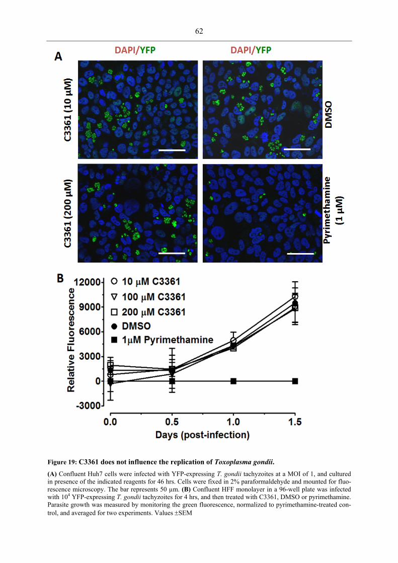
62
Figure 19: C3361 does not influence the replication of Toxoplasma gondii. (A) Confluent Huh7 cells were infected with YFP-expressing T. gondii tachyzoites at a MOI of 1, and cultured in presence of the indicated reagents for 46 hrs. Cells were fixed in 2% paraformaldehyde and mounted for fluo-rescence microscopy. The bar represents 50 µm. (B) Confluent HFF monolayer in a 96-well plate was infected with 104 YFP-expressing T. gondii tachyzoites for 4 hrs, and then treated with C3361, DMSO or pyrimethamine. Parasite growth was measured by monitoring the green fluorescence, normalized to pyrimethamine-treated con-trol, and averaged for two experiments. Values ±SEM

63
3.2.5 In vivo Δpbht1-PfHT1 mouse model for assessment of PfHT1 inhibitors
To facilitate the search and evaluation of anti-malarial sugar analogs, we exploited the func-
tional resemblance of PfHT1 with PbHT1, and created a transgenic Δpbht1-PfHT1 P. berghei
line that is entirely dependent on PfHT1 for its growth. We designed and transfected a PfHT1-
expressing PbHT1 deletion construct pb3D+PfHT1 (Fig. 20A), and cloned drug-resistant
parasites. Construct-specific PCR revealed 5’- and 3’-UTRs in the control plasmid and in the
gDNA obtained from Δpbht1-PfHT1 but not in the parental parasites (Fig. 20B). Likewise,
PCR reactions using recombination-specific primers amplified expected fragments in the
complemented parasites but not in the parental strain (Fig. 20C). The crossover events were
also verified by sequencing of amplified gDNA. The absence of PbHT1 and the concurrent
presence of the PfHT1 transcript and gene locus in the transgenic strain confirmed the clonal-
ity and the PfHT1-dependence of the Δpbht1-PfHT1 strain (Fig. 20D). These transgenic para-
sites developed normally in mice and mosquitoes, consolidating the functional relationship of
PbHT1 and PfHT1, in vivo. Importantly, the Δpbht1-PfHT1 P. berghei mouse model vali-
dated the essentiality of PbHT1, and offered an in vivo system for the pharmacological testing
of PfHT1 inhibitors.
Figure 20: PfHT1 complementation-based PbHT1 deletion in P. berghei.
(A) Double crossover-mediated deletion of PbHT1 by PfHT1-dependent rescue. The PbHT1 gene locus and the PfHT1-harboring knockout construct are aligned, with primers indicated by arrows. (B) Plasmid-specific 5’- and 3’- primer sets (PbHT1-5'UTR-F / R and PbHT1-3'UTR-F / R) amplify expected fragments only with transgenic gDNA (Δpbht1-PfHT1) and KO construct (pb3D+PfHT1) but not with parental gDNA. (C) 5’- and 3’- recom-bination-specific (PbHT1-5'Scr-F / R and PbHT1-3'Scr-F / R) primer sets amplify genomic fragments from Δpbht1-PfHT1 but not in parental strain or plasmid. (D) PfHT1 can complement absence of PbHT1 in the Δpbht1-PfHT1 parasite line. PCR verification of the Δpbht1-PfHT1 strain using PbHT1- or PfHT1-specific

64
primers. PbHT1 is detectable in the parental strain but not in Δpbht1-PfHT1. Likewise, PfHT1 can only be am-plified from transgenic gDNA or cDNA.
To put our model into perspective and practice, mice were infected with the Δpbht1-PfHT1 or
the parental strain (105 infected erythrocytes), and allowed to develop a detectable blood para-
sitemia prior to C3361 treatment. All mice displayed a parasitemia >1% on day 3 of infection.
Subsequent daily intravenous injection of C3361 (2 mg/kg body weight) yielded about 45 %
significant reduction in parasitemia at day 5 (P <0.05) in drug-treated mice as compared to
respective DMSO-treated controls (Fig. 21A), indicative of a drug effect on PfHT1 and
PbHT1. Survival plots of infected animals also showed an apparent modest advantage in all
C3361-treated mice as compared to mock-treated animals (Fig. 21B). As shown in vitro in
Leishmania (Fig. 17B), the drug also appears to inhibit the function of Plasmodium transport-
ers, in vivo. Importantly, the notably weak influence of C3361 on parasitemia rejects its anti-
malarial efficacy despite a reported strong in vitro action against P. falciparum (Joet et al.,
2003a). Though the pharmacokinetics of C3361 in mouse is not known, our results illustrate
the value of this model for the in vivo appraisal and validation of an analog drug, and bridge a
crucial gap in preclinical drug development, i.e. from in vitro screening to in vivo examination
of qualified drugs.
Figure 21: Transgenic P. berghei as a model for the in vivo assessment of PfHT1 inhibitors.
In vivo test of C3361 in NMRI mice intravenously injected with 105 parental- or Δpbht1-PfHT1-infected RBCs. Following a detectable parasitemia on day 3, mice were treated with C3361 (2 mg/kg body weight) or DMSO. The mean parasitemia (A) and Kaplan-Meier-plot (B) from two assays are depicted. Mean ± SEM

65
3.2.6 PfHT1-complemented S. cerevisiae for screening of PfHT1 inhibitors
The development of proficient anti-malarial drugs stipulates in vitro models for the large-
scale screening of PfHT1 inhibitors prior to their pharmacological assessment. To address this
constraint, we focused on establishing a model that would combine user safety and amicabil-
ity with minimal investment and standard laboratory resources. Furthermore, this model
should be compatible with existing automation technology. Towards this end, following our
initial futile attempts to functionally express the native PfHT1 ORF in S. cerevisiae, we de-
signed a yeast-optimized synthetic ORF (referred as PfHT1-Syn hereafter), and then gener-
ated fusion constructs of full-length PfHT1 with the N-terminal 55 residues of ScHxt9 protein
(his-GM55-PfHT1-Syn) and 80 residues of ScITR1 (his-GM80-PfHT1-Syn) (Fig. 22A) to
enhance their expression and targeting. All constructs were expressed in the S. cerevisiae
EBY4000 mutant that is compromised in its growth on most sugars except on maltose and
galactose (Wieczorke et al., 1999). As clearly depicted in the Fig. 22B, PfHT1-Syn, GM55-
PfHT1-Syn as well as GM80-PfHT1-Syn rescued the growth defect of the yeast mutant on
glucose and mannose as carbon sources. The rescue phenotype conferred by PfHT1-Syn is
very similar to that of ScHxt9 expressed under the same promoter (HXT7) on both carbon
sources. GM55-PfHT1-Syn and GM80-PfHT1-Syn conferred an enhanced complementation
compared to PfHT1-Syn that is probably due to their improved targeting and optimal confor-
mation at the surface. It is interesting to note that these cell lines require low glucose (0.06 %)
for their growth, and did not grow on high glucose (2 %), which suggests glucose perception
and import as independent modules of yeast growth, as conceptualized by recent elegant work
(Youk and van Oudenaarden, 2009).

66
Figure 22: Yeast-optimized PfHT1 can rescue the growth of the S. cerevisiae mutant on glucose and man-nose, and confers a model for high-throughput screening of analog-based PfHT1 inhibitors.
(A) Schematics of PfHT1 ORF and its variants. Yeast-optimized PfHT1 ORF (PfHT1-Syn) was fused either with 6xHis tag (His-PfHT1-Syn) or with 6xHis epitope and N-terminal 55 residues of ScHxt9 (His-GM55-PfHT1-Syn) or N-terminal 80 residues of ScITR1 (His-GM80-PfHT1-Syn). (B) Complementation of S. cerevisiae EBY4000 mutant with the illustrated expression constructs. 2 µl of serially diluted yeast cells were spotted on uracil-free synthetic media supplemented with 0.06 % glucose or 2 % mannose, and incubated at 30°C for 3–4 days.
In order to corroborate these results, we tested the expression and localization of the fusion
transporters by using the anti-His antibody. Consistent with rescue assays, PfHT1-Syn,
GM55-PfHT1-Syn and GM80-PfHT1-Syn display a peripheral presence by yeast IFA (Fig.
23A). Furthermore, to exclude the appearance of plasmid-independent suppressor mutants, we
tested the plasmid-dependency of our transformed strains by plasmid-loss assay. After culture
in non-selective media, strains were plated for single colonies and replicated onto plasmid-
selective and non-selective maltose media. Several colonies on the non-selective maltose me-
dia had lost the plasmid as confirmed by their inability to grow on selective plates without
uracil (Fig. 23B, red circles). These colonies also lost their ability to grow on non-selective
glucose plates. All colonies that grew on uracil-free media with maltose also grew on glucose
except for the empty plasmid negative control (Fig. 23B, green circles), which is consistent
with plasmid-dependent growth on glucose.

67
Figure 23: PfHT1 fusion transporters localize peripheral and confer growth rescue on glucose.
(A) Immuno-fluorescence assay of S. cerevisiae expressing the indicated his-tagged proteins. Zymolase-treated transgenic cells from log-phase liquid cultures were fixed with 4 % PFA overnight at 4°C, and processed for IFA using mouse anti-His antibody at 1:200 dilution. (B) Plasmid loss assay was performed in non-selective yeast-peptone-maltose liquid cultures for 8-10 generations followed by single cell plating onto non-selective synthetic media with uracil and maltose. This master plate was replica printed onto selective (uracil-free) media with mal-tose and onto non-selective media supplemented with glucose to score for yeast cells with reverted phenotype. Plasmid depleted cells (red circles) as confirmed by their reduced or absent growth on selective maltose media

68
maintain to their original no-growth phenotype on non-selective glucose plates, while exhibiting normal growth on non-selective maltose media. In contrast, cells that retained the plasmid (green circles) grow under all condi-tions. Also, note that yeast cells harboring empty vector do not grow in non-selective glucose media regardless of their plasmid status, and ScHxt9 behaves identical to Plasmodium constructs. Similar assays were also per-formed with mannose as a carbon source, which yielded identical data. Only the assays performed with glucose are shown to avoid redundancy.
In further assays, we examined C3361 (a glucose analog)-mediated inhibition of yeast mu-
tants expressing His-PfHT1-Syn, His-GM55-PfHT1-Syn and His-GM80-PfHT1-Syn in glu-
cose- and mannose-containing agar plates. These assays revealed that C3361 retarded the
growth of yeast strains complemented with Plasmodium proteins but not with ScHxt9 on glu-
cose plates (Fig. 24). This inhibition was glucose-specific since C3361 did not influence
growth on mannose. Taken together, these data further support our functional and inhibition
data obtained in L. mexicana. More importantly, they emphasize the expediency of the yeast
model for a high-throughput selection of PfHT1 inhibitors.
Figure 24: C3361 inhibits PfHT1-mediated growth of Saccharomyces cerevisiae on glucose.
C3361-mediated inhibition of PfHT1 transport activity on 0.06 % glucose or mannose. Cells were spotted on agar plates supplemented with C3361 (100 µM) or DMSO and incubated at 30°C for 4 days. Chemical stability of C3361 is not know4.

4 Discussion
4.1 Implications on the metabolism of Toxoplasma gondii
4.1.1 Major nutrients of T. gondii
The results reported in this thesis clearly demonstrate that Toxoplasma expresses a single
functional hexose transporter (TgGT1) that is capable of transporting glucose and mannose as
major, and fructose and galactose as minor substrates. The transporter is entirely dispensable
in Toxoplasma tachyzoites, and its deletion aborts glucose utilization. The in vitro growth
delay of the Δtggt1 strain is modest and no decrease in its virulence in mice was observed.
Besides, the intrinsic growth defect of this mutant is not enhanced by the depletion of exoge-
nous glucose. Toxoplasma can effectively compensate restricted access to glucose by resort-
ing to glutamine metabolism demonstrating an unexpected metabolic adaptation (Blume et
al., 2009). Collectively, these data strongly advocate for dispensability of exogenous glucose
for T. gondii. This metabolic flexibility may contribute to the parasite's ability to replicate in
virtually all nucleated cells regardless of their intracellular glucose levels.
The replication of the Δtggt1 strains is almost completely inhibited without glutamine. Its pro-
longed culture, although at a much slower replication rate, is however, possible over weeks
(data not shown). This indicates that other minor nutrient(s) can also be used by the parasite to
sustain its vital functions. In line, the labelling of intracellular Δtggt1 parasites with 14C-
glucose indicates that host-derived products of glucose metabolism are taken up by the para-
site. Glycolysis is strongly induced in parasite-infected HFF cells (Blader et al., 2001; Fouts
and Boothroyd, 2007) and the residual labeling of Δtggt1 (Fig10 A), is abolished when glyco-
lysis is inhibited by the hexokinase inhibitor, 2DOG (Blume et al., 2009). Several potential
transporters for triose phosphates are present in the parasite genome, and could contribute to
this phenomenon. Other substrates that may support the basal survival of the Δtggt1 mutant in
absence of glutamine are succinate and branched-chain amino acids (BCAA). Succinate has
been shown to enhance the replication of a succinyl-CoA synthetase knock out mutant (Fleige
et al., 2008), and is thus imported directly or indirectly by tachyzoites. A transporter that is
homolog to INDY permases and potentially transports TCA cycle metabolites is expressed by
tachyzoites (T. Fleige personal communication). BCAA can also be metabolized to pyruvate
and acetyl-CoA via conserved enzymes for their degradation (Seeber et al., 2008).

70
4.1.2 Changes in gene expression of Δtggt1
In order to further deduce the metabolic adaptation in the Δtggt1 parasites, we performed mi-
croarray analysis of this parasite (8059 probes, triplicate assays, in cooperation with Prof
David S Roos, University of Pennsylvania, US). The obtained data represent log2 expression
values of KU80 deficient parasites (as a control) and the Δtggt1 parasites. A principal compo-
nent analysis (Fig. 25) was carried out in order to visualize differences in the complex data-
sets. This analysis revealed that all three Δtggt1 samples cluster together but are separated
from the control parasites in the first two principal components (Fig. 25).
Figure 25: Principal component analysis of Δtggt1 and ΔKU80 transcriptomes.
An R-based online tool MIDAW (http://midaw.cribi.unipd.it/dnachip/) was used to perform data normalization, principal component and significance analysis (PC1, 2 - principal component 1, 2).
One sample of the control parasites appeared to be distinct from the total three control sam-
ples. This outlier was thus omitted from further analysis. The significance analysis of microar-
ray (SAM) algorithm (using R-based online tool MIDAW
(http://midaw.cribi.unipd.it/dnachip/) was used to determine differentially regulated genes in
Δtggt1. In total, 80 non-redundant ToxoDBv6.0 gene models appear to be regulated. Among
these were 53 genes that could not be functionally annotated. The remaining 27 genes (Ap-
pendix C) were manually analyzed. As expected, the TgGT1 transcript lies below the detec-
tion limit. The most up-regulated (15-fold) gene in the Δtggt1 mutant belongs to a family of

71
the short chain dehydrogenases and appears homologous to the fabG protein (ToxoDB ID
TGME49_106450) participating in the FAS2 pathway. Furthermore, a gene model
(TGME49_094820) that is predicted to be the FAS1 protein is 8-fold down-regulated. It re-
mains to be tested if their mRNA expression levels reflect the activity of these gene products.
The FAS1 pathway does appear to be very critical in tachyzoites, as the strong down-
regulation is well tolerated and may be compensated by the FAS2 pathway, other potentially
redundant enzymes such fatty acid elongases (Mazumdar and Striepen, 2007) or increased
fatty acid uptake.
Another gene (TGME49_054330) with a predicted triacylglycerol lipase activity is 2.3-fold
more abundant in the Δtggt1 parasites. In addition, a phosphodiesterase (TGME49_063270) is
1.5-fold induced. The up-regulation of both genes suggests that increased uptake of lipid in-
termediates (diacylglycerol and triacylglycerol) contributes to the fatty acid pool of the Δtggt1
mutant. The transcript levels indicate the fatty acid synthesis being a limiting factor for the
growth of the T. gondii mutant. Interestingly, Streptomyces coelicolor (a soil-dwelling bacte-
rium) uses glycerophosphodiesters as carbon sources and the expression its glycero-
phosphodiesterases is directly regulated by available nutrients (Santos-Beneit et al., 2009).
The glucose-6-phosphate dehydrogenase enzyme that catalyzes the initial rate limiting step of
the PPP pathway is repressed by 1.6-fold in the Δtggt1 parasites. The PPP delivers NADPH
and ribose-5-phosphate. The NADPH is also produced by TCA cycle enzymes such as isocit-
rate dehydrogenase (Fleige et al., 2008), but ribose-5-phosphate for nucleotide synthesis is
exclusively produced by the PPP. Therefore, the activity of this pathway should correlate to
the replication of the parasites. It remains to be established whether the decreased replication
of the Δtggt1 is a caused by a limited ribose-5-phosphate pool or if the down-regulation of the
glucose-6-phsohate dehydrogenase is a consequence of decreased nucleotide demand due to
delayed growth, potentially caused by slow fatty acid biogenesis.
Other significantly regulated genes include a plasma membrane proton P-type ATPase
(TgPMA) (TGME49_052640) that is reported to be expressed specifically in bradyzoites
(Holpert et al., 2001). Six predicted protein kinases were also differentially regulated but their
function remains to be established. The distinct transcriptome of the Δtggt1 parasites demon-
strates that their metabolic adaptation to different host cells could very well involve transcrip-
tomic regulation.

72
4.1.3 Novel aspects of bioenergetic metabolism of T. gondii
It has been proposed that glycolysis is the main ATP source for extracellular tachyzoites and
also responsible for the F-actin and MyosinA-based parasite motility (Pomel et al., 2008).
This notion is also supported by complementation of a conditional knock-out mutant with the
mutated isoforms of the aldolase enzyme. All enzyme isoforms that retained their catalytic
activity complemented growth and motility of the parasite, while enzymatically-dead aldo-
lases failed to do so (Starnes et al., 2009). Our observation that extracellular Δtggt1 parasites
do not glide in the presence of glucose is consistent with these results. Interestingly, this work
identified glutamine as another source of energy for extracellular Δtggt1 parasites that is suf-
ficient to drive the parasitic motility. The generation of ATP from glutamine involves oxida-
tive phosphorylation is a likely source. It has been demonstrated that oxidative phosphoryla-
tion is functional in T. gondii (Vercesi et al., 1998) and a severe depletion of intracellular
ATP levels was observed in intracellular parasites following treatment with the ATPase in-
hibitor oligomycin (Lin et al., 2009). Similarly, the ATP levels in extracellular Δtggt1 para-
sites are elevated by glutamine in an-oligomycin sensitive manner (W. Bohne personal com-
munication). This ability may have been overlooked by the excess of glucose in the parasite
cultures. The prolonged glucose-independent proliferation of Δtggt1 parasites reveals an in-
trinsic ability of T. gondii to efficiently metabolize glutamine to energy.
4.2 Potential function of TgST1, TgST2, TgST3
Toxoplasma expresses four sugar transporter like proteins during its tachyzoite stage, but only
TgGT1 is involved in sugar transport. TgST2, which also localizes to the parasite plasma
membrane, does not take part in the import of glucose as its deletion in the wild-type and the
Δtggt1 strain does not influence glucose uptake by intracellular parasites. Accordingly, het-
erologous expression of TgST2 in Δlmgt L. mexicana parasites fails to mediate transport ac-
tivity of any of the four tested hexoses (Blume et al., 2009). TgST2 is non-redundant with
TgGT1 and cannot replace its function. Recently, two sugar transporters of L. mexicana that
were previously described as hexose transporters (Burchmore et al., 2003) were also found to
transport ribose with considerable efficiency (Naula et al., 2010). The affinity of TgST2 to-
wards ribose remains to be tested.
TgST1, TgST 2 and TgST3 might be involved in glucose sensing. Sugar transporter-like pro-
teins (ScSnf3 and ScRgt2) of S. cerevisiae have been reported to lack a transport activity and
are involved in sugar sensing instead (Ozcan et al., 1996). It may well be that Toxoplasma

73
uses sugar sensors to adapt to the nutritional environment of different host cells. The N-
terminal extensions of TgST1-3 show no homology to known proteins and could function as
signal transduction domains in this regard.
4.3 Metabolic Interactions Between Toxoplasma and Its Host
Cell
The obligate intracellular life style of T. gondii reflects its strict dependence on its host cell
for acquisition of nutrients. In this regard, the parasitic strategies include its adaptation to the
host environment and metabolic manipulation of the host cell. For example: Toxoplasma may
have preserved redundant pathways to broaden its host specificity. Besides, it also may adjust
the host cell by either direct manipulation via secreted proteins or indirectly by metabolic
shifts in the host cell. These parasite-induced adaptations should lead to the integration of its
own metabolic network with that of the host cell. The two previously disconnected networks
are fused by many (unknown) nutrient exchange reactions. They thus can be considered as a
single entity that ultimately ensures the parasite replication while maintaining the integrity of
the host cell until its lysis. The metabolic shift in fatty acid biogenesis in the Δtggt1 mutant
exemplifies this notion. The following discussion attempts to integrate the nutritional re-
quirements of T. gondii with its own metabolic capacities and with those of its host cell.
4.3.1 The central carbon metabolism of the host cell and the parasitic requirements
An example that illustrates both strategies is the induction of the host glucose-6-phosphatase
(G6Pase) system in a T. gondii-parasitized cell (Fig. 26). This enzyme localizes to the host ER
in uninfected cells and dephosphorylates glucose-6-phosphate to produce glucose. Free glu-
cose is usually a rare intracellular compound as it is quickly phosphorylated by hexokinases.
In order to gain access to and to compete with its host cell for glucose, Toxoplasma strongly
induces the G6Pase and re-localizes it from the host ER to the lumen of the parasitophorous
vacuole as shown by cytochemical staining of infected cells (Melo and de Souza, 1997). The
parasite also expresses a glucose transporter and a hexokinase, both of which exhibit a re-
markably high affinity to facilitate glucose uptake (Blume et al., 2009; Joet et al., 2002; Saito
et al., 2002).

74
Host cell metabolism is also modulated by parasite-directed induction (Fouts and Boothroyd,
2007) and stabilization (Wiley et al., 2010) of HIF1 (hypoxia inducible factor 1) (Fig. 26).
HIF1 is a potent regulator of the host carbohydrate metabolism and adjusts it to cope with low
oxygen levels (Denko, 2008) by restricting carbon metabolism to glycolysis and by down-
regulating mitochondrial functions (Zhao et al., 2010). Indeed, a significant up-regulation of
several pro-glycolytic genes is observed in infected cells (Fig. 26, (Fouts and Boothroyd,
2007). Significant induction of the solute carrier 16 member 1 transporter, that is implicated
in pyruvate and lactate export and of lactate dehydrogenase A and B suggest an increased
glycolytic flux and lactate production (Blader et al., 2001; Fouts and Boothroyd, 2007). Con-
sistently, mitochondrial pathways are down regulated. The PDH enzyme complex that fuels
the TCA cycle by metabolizing pyruvate, might be inactivated via phosphorylation by an up-
regulated PDH kinase (Blader et al., 2001; Fouts and Boothroyd, 2007). Thus, the glucose-
derived carbon flow is diverted from the mitochondrial TCA cycle towards glycolytic lactate
production. In further agreement with a diminished TCA cycle in infected host cells is the fact
that the expression of the host glutaminase is suppressed in parasitized cells (Fouts and
Boothroyd, 2007). This enzyme catalyzes the deamination of glutamine to glutamate, which is
then further deaminated to the TCA cycle intermediate, alpha-ketoglutarate. In further agree-
ment, T. gondii does not exhibit any detectable growth defect in host cells that are deficient of
aerobic respiration (Schwartzman and Pfefferkorn, 1982). Collectively, these changes in the
host metabolism support the parasite growth as inhibition of HIF1 leads to a retarded of para-
site replication (Wiley et al., 2010). A potential beneficial effect of HIF1-modulated host cells
could involve a decreased production of reactive oxygen species by the host mitochondria.

75
Figure 26: Toxoplasma-induced modification of the host metabolism in parasitized cells.
The parasite recruits the host microtubule organization center (MTOC) and scavenges low-density lipoprotein (LDL)-derived vesicles along microtubules that invaginate the parasitophorous vacuole membrane. The hypoxia inducible factor (HIF1) is activated in infected cells. It induces several glycolytic enzymes and increases perme-ability of the plasma membrane for glycolytic by-products. Mitochondrial functions are down-regulated by phosphorylation of the pyruvate dehydrogenase and by decreased glutaminase mRNA abundance. Furthermore, endoplasmatic reticulum (ER)-bound activity of the glucose-6-phosphatase is increased and expanded into the parasitophorous vacuole (PV). References are (1) Fouts et al 2007; (2) Widley et al 2010; (3) Coppens et al 2000, 2006; (4) Melo et al 1997; (5) Schwab 1994
4.4 Outlook
This study reveals previously unknown metabolic features of T. gondii. It is now clear that the
parasite can survive without glucose and is capable of utilizing glutamine to support its
growth. Microarray analysis of the Δtggt1 transcriptome and biochemical labeling also indi-
cate the uptake of alternative nutrients by T. gondii. Future research should aim to verify the
postulations made in this work especially on how the parasites metabolic network interacts
with that of its host cell. The research should facilitate and also profit from the construction of
genome-scale models by flux-balance analysis (FBA) and unravel new insights of the para-
site-host interactions.

76
4.5 The Hexose Permeation Pathway of Plasmodium
The function of PfHT1 has been extensively researched and PfHT1 is now considered as a
qualified drug target during blood stages (Joet et al., 2003b; Patel et al., 2008). In brief,
PfHT1 has been characterized biochemically (Fayolle et al., 2006; Manning et al., 2002;
Woodrow et al., 1999; Woodrow et al., 2000), and specific inhibitors have been developed
(Ionita et al., 2007; Joet et al., 2003a; Saliba et al., 2004). Recently, genetic essentiality of
PfHT1 and its P. berghei homolog PbHT1 was reported for blood stages (Slavic et al., 2010).
This thesis focuses on previously unknown aspects of the PfHT1 and PbHT1, and reveals the
importance of hexose transport in ex-erythrocytic stages. Furthermore, this work provides a
complete platform for screening of anti-malarial sugar analogues, which should facilitate the
in vitro identification of high-affinity PfHT1 inhibitors prior to their in vivo testing in trans-
genic P. berghei model.
4.5.1 The pan-hexose nature of PfHT1 and PbHT1
Our results demonstrate that the PfHT1 and PbHT1 can transport glucose, mannose, fructose
and galactose with similar kinetics and hence are functional pan-sugar permeases. Similar to
TgGT1, glucose and mannose are high-affinity substrates for both Plasmodium proteins,
whereas galactose and fructose are transported with low affinity. PfHT1 can also compensate
for the loss of PbHT1 function in P. berghei. Previously PfHT1 has been expressed in
Xenopus oocytes and reported to transport glucose with a Km of 0.48 to 1 mM (Woodrow et
al., 1999; Woodrow et al., 2000) and fructose with a Km of 11.5 mM (Woodrow et al., 2000).
It has been shown that fructose can replace glucose as a carbon source in P. falciparum cul-
tures (Geary et al., 1985; Woodrow et al., 2000). Notably, P. falciparum and P. berghei para-
sites possess a phosphoglucose isomerase (Aoki et al., 2010) that should be capable of inter-
converting hexose phosphates. The transport of mannose and of galactose may also be
biologically relevant since these two hexoses are required for protein glycosylation and lipid
synthesis (Gowda and Davidson, 1999; Marechal et al., 2002). Glucose levels are in the range
of millimoles in human blood (Iannaccone et al., 2005) and the hemolymph of Anopheles
stephensi mosquitoes (Mack et al., 1979), hence PfHT1 and PbHT1 should always be satu-
rated for the glucose transport. Fructose, mannose and galactose are present in micro molar
serum concentrations (Bossolan et al., 2007; Kawasaki et al., 2002; Rodriguez et al., 2005).
The apparent Ki values for mannose indicate that mannose uptake should not be saturated
despite a high affinity of both proteins. The Ki values for galactose and fructose are one to

77
two orders of magnitude higher than the levels that the parasite should encounter in the blood.
Therefore, it seems unlikely that the import of these two hexoses plays a significant role.
Collectively, these data indicate that both Plasmodium transporters are functional homo-
logues, and under physiological conditions glucose and mannose should be their major sub-
strates.
4.5.2 The essentiality for Plasmodium hexose transporters
Multiple attempts to knockout PbHT1 have been unsuccessful by conventional means. It was
only possible to ablate PbHT1 during concurrent functional complementation with PfHT1 and
or with PbHT1-HA. These results lend support to a recent study that also suggested the ge-
netic essentiality of PbHT1 and PfHT1 for blood stages (Slavic et al., 2010). It is also consis-
tent with the fact that biochemical inhibition of PfHT1 by C3361 is detrimental to the P. fal-
ciparum cultures (Joet et al., 2003a; Saliba et al., 2004). The relevance of glucose import
beyond erythrocytic stages of P. berghei is another interesting finding of this work. The pres-
ence of C3361 drastically inhibits ookinete formation, and aborts liver stage development of
P. berghei. This inhibitory effect is in good agreement with the Ki value of PbHT1 for C3361
in the L. mexicana model. A definitive assessment of the essentiality of PbHT1 during ex-
erythrocytic stages, however, requires conditional deletion of PbHT1 at different stages. The
inhibitory effect of C3361 on ookinete formation reveals their dependence on exogenous sug-
ars. Proteomic and transcriptomic studies of gametocytes and ookinetes, however, demon-
strate an up-regulation of the TCA cycle and oxidative phosphorylation, which would suggest
an increased metabolism of amino acids (Hall et al., 2005). This raises the obvious question
why hexose uptake is important during in mosquito stages despite an apparent shift towards
mitochondrial phosphorylation (see comparative discussion section).
4.5.3 PfHT1 as a drug target
The potential of PfHT1 as a drug target during blood stages of Plasmodium is well docu-
mented. This work demonstrated that PbHT1 is also necessary for the hepatic development of
P. berghei. That could well be true for P. falciparum. Abortion of Plasmodium infection dur-
ing the liver passage by antibiotic treatment or genetically-manipulated strains can generate
protective immunity in the host (Friesen et al., 2010; Mueller et al., 2005). Thus, prophylacti-
cally targeting PfHT1 for inhibition of the clinically silent liver stages might be a vital strat-
egy to generate protective immunity and prevent blood stage parasitemia. Another interesting
disease control aspect associated with the targeting of PfHT1 emerges from the fact that

78
C3361 treatment of Plasmodium-infected mice reduces transmission to the mosquito host.
This effect is most likely mediated by inhibition of ookinete formation in the mosquito mid-
gut. The fraction of gametocytes among blood-stages in C3361-treated mice appears unal-
tered. The transmission-blocking activity of PfHT1-targeting drugs should be corroborated by
genetic means and/or membrane-feeding systems.
The wide-spread use Artemisinin-based combination therapies (ACT) has caused a decline in
the transmission rate (WHO, 2008) via its gametocytocidal action (Mehra and Bhasin, 1993).
This effect, however, is slow and the patients' blood remains partly infective to mosquitoes
even after two weeks of treatment (Mens et al., 2008; Okell et al., 2008b). Using drugs that
function by a different mechanism (such as PfHT1) would contribute to an improvement to
the anti-malarial action of ACT (Okell et al., 2008a).
It should be noted here that only one single mutation in PfHT1 is sufficient to diminish the
inhibitory effect of C3361 by 5-fold, without compromising the glucose transport activity
(Joet and Krishna 2004). Also, the over expression of PfHT1 can alleviate the inhibitory ef-
fect of C3361 on P. falciparum cultures (Slavic et al., 2010). This may not hold true for novel
sugar analogues, but does indicate the risk of drug resistance by the parasite that may come at
little fitness cost.
4.6 Comparative Discussion
4.6.1 Why is hexose import essential for Plasmodium but not for Toxoplasma?
Glucose and its transporter are not required for Toxoplasma tachyzoites; however Plasmo-
dium species appear to critically depend on glucose uptake throughout their life cycle. While
T. gondii is able to replicate in most nucleated cells, replication of Plasmodium is confined to
erythrocytes and hepatocytes (and to a minor extent to skin fibroblasts) and the mosquito
midgut. The fundamental difference in glucose metabolism of these two parasites may ac-
count for their host ranges and target tissue. The number of genes that the genome of each
parasite harbours also indicates a higher metabolic flexibility in T. gondii. It possesses ~8000
genes, whereas Plasmodium encodes ~5000 genes.
A critical difference between Toxoplasma and Plasmodium appears to be the fructose-1,6-
bisphosphatase and pyruvate carboxylase, which catalyze final and initial steps of gluconeo-
genesis, respectively (Fig. 27, Table 4, (Foth et al., 2005)). This pathway may enable T.
gondii to synthesize hexose derivatives from amino acids and other metabolites (communica-

79
tion with D. Roos, (Fleige et al., 2007). This imposes an obligation for hexose uptake on
Plasmodium parasites, to maintain glycosylation and GPI-anchor synthesis, while T. gondii
can resort to the synthesis of hexose-derived metabolites from amino acids and other carbon.
Table 4: Genes of glutaminolysis and gluconeogenesis in Toxoplasma gondii and Plasmodium
Gene Annotations from Parasite Database1 Parasite Pathways and Associated Enzymes T. gondii ME49 P. falciparum 3D7 P. berghei ANKA
EC Numbers2
Enzymes of Glutaminolysis3 Conversion of Glutamine to Glutamate
Glutamine Amidotransferase TGME49_081490 PF11_0169 PB000299.02.0 6.3.5.3 Glutaminase (related to Class I Glutamine Amidotransferase) TGME49_010760 X X
Glutamine-Ammonia Ligase (Glutamine Synthetase) TGME49_073490 PFI1110w PB000319.00.0 6.3.1.2
GMP Synthase (Glutamine-Hydrolyzing) TGME49_030450 PF10_0123 PB000253.01.0 6.3.5.2
Conversion of Glutamate to α-ketoglutarate Conversion of Glutamate to a-
ketoglutarate NAD-specific Glutamate
Dehydrogenase
TGME49_049390 PF08_0132 PB300354.00.0 PB301271.00.0
1.4.1.2
NADP-specific Glutamate Dehydrogenase TGME49_093180
PF14_0164 PF14_0286
PB000714.01.0 PB300230.00.0
1.4.1.4
Enzymes of Gluconeogenesis3
Fructose-1,6-bisphosphatase TGME49_005380 TGME49_047510
X X 3.1.3.11
Phosphoenolpyruvate Carboxykinase
TGME49_089650 TGME49_089930
PF13_0234 PB001070.000 4.1.1.49
Pyruvate Carboxylase TGME49_084190 X X 6.4.1.1 1 The genomes of three apicomplexan organisms were mined for a broad range of glutaminolysis- and gluconeo-genesis-associated genes using the BLAST algorithm and manual inspection. The gene IDs correspond to respec-tive parasite database (www.ToxoDB.org and www.PlasmoDB.org). 2 EC numbers are depicted as assigned in Toxoplasma database. 3 Function of enzymes associated with these pathways are based on putative annotations, and require experimen-tal verification.
The role of hexoses in energy generation via glycolysis is not as clear. Plasmodium as well as
Toxoplasma depend largely on glycolysis for their cellular needs (Ohsaka et al., 1982; Pomel
et al., 2008; Starnes et al., 2009; van Dooren et al., 2006; Vander Jagt et al., 1990) (Scheibel
and Miller, 1969). Both parasites utilize hexoses for ATP generation in their cytosol through
glycolytic enzymes (Fleige et al., 2007; Maeda et al., 2009; Pomel et al., 2008; Saito et al.,
2008). This work (Blume et al., 2009) demonstrates that glutamine can also sustain motility
and thus presents an alternative source of energy. This glutamine dependent utilization occurs
by oxidative phosphorylation in an oligomycin-sensitive manner (Lin et al., 2009) W. Bohne
personal communication). In contrast, P. falciparum cultured exposed C3361 exhibits signs of

80
energy starvation. The pH homeostasis is disrupted and cytosolic ATP-levels drop rapidly but
stabilize at 20 % of the control parasites (Saliba et al., 2004). While this strongly suggests an
important role of glucose for in vitro cultured parasites, it does not exclude other minor en-
ergy sources that may contribute to sustain the basal ATP levels. Consistently, transcriptional
profiling of parasite isolates reveals physiological states that are divergent from the cultured
parasites (Daily et al., 2007). The parasite isolates adapt to an environmental stress by me-
tabolism of alternative carbon sources besides the glycolysis driven-growth. The former state
is marked by increased mitochondrial biogenesis and induction of genes associated with glyc-
erol, lactic acid and lipid catabolism by respiration and oxidative phosphorylation. A trans-
porter for glycerol uptake has already been reported (Promeneur et al., 2007) (Fig. 27). How-
ever, the respiratory chain was demonstrated to support only the pyrimidine synthesis, and is
dispensable otherwise in cultured pararsites (Painter et al., 2007).
Also during ex-erythrocytic stages other pathways besides glycolysis may play a significant
role in energy generation. Transcriptome and proteome analysis reveal that gametocytes and
ookinetes display an increased expression of TCA cycle genes and an up-regulated oxidative
phoshorylation (Hall et al., 2005).
Taken together, obligate glycosylation reactions impose the most stringent requirement for
hexoses on Plasmodium parasites, while Toxoplasma can presumably sustain them by catabo-
lizing amino acids. Energy generation is certainly its major task of glycolysis in Toxoplasma
as well as Plasmodium but both parasites appear competent in using oxidative phosphoryla-
tion-derived ATP to a certain degree. The metabolic diversity of P. falciparum isolates and
the T. gondii Δtggt1 mutant illustrate that uncultured parasite samples and genetically re-
stricted parasites are tools to reveal the metabolic plasticity of parasitic metabolism.

81
Figure 27: The central carbon metabolism of Toxoplasma gondii and Plasmodium species.
The central carbon metabolism is distributed into three compartments, the cytosol (light blue), apicoplast (green) and mitochondrion (brown), extracellular space is shown in white. Pathways that are presumably absent in Plas-modium are marked in red. 1-deoxy-D-xylulose 5-phosphate pathway (DOXP), fatty acid synthesis type 2/1 (FAS2/1), pyruvate dehydrogenase (PDH), Toxoplasma gondii apicoplast phosphate translocator (TgAPT), in-ner/outer triose phosphate transporter (iTPT/oTPT), dihydroxyacetone phosphate (DHAP), phosphoenolpyruvate (PEP), branched chain amino acids (BCA), acetyl coenzyme A (Acetyl-CoA), alpha ketoglutarate (αKG), nico-tine amid adenine dinucleotide (NADH), flavin adenine dinucleotide (FADH), oxaloacetate (OOA), malate (MAL).

82

5 Conclusion
This thesis establishes the unexpected dispensability of glucose and its transporter for the
tachyzoite stages of the obligate intracellular parasite Toxoplasma gondii. It further reveals
the compensatory mechanism making use of glutamine that illustrates the metabolic robust-
ness of T. gondii.
Furthermore, this work confirms the potential of Plasmodium hexose transporters as a drug
target, and highlights new anti-malarial aspects by targeting hexose transport. The drug
screening platforms utilizing transgenic S. cerevisiae and P. berghei should facilitate the
search of novel anti-malarial sugar analogs.
Finally, the differential importance of sugar import for Toxoplasma and Plasmodium can be
ascribed to differences in their pathways of the central carbon metabolism.

84

References
Aoki, K., Tanaka, N., Kusakabe, Y., Fukumi, C., Haga, A., Nakanishi, M., Kitade, Y., and Nakamura, K.T. (2010) Crystallization and preliminary X-ray crystallographic study of phosphoglucose isomerase from Plasmodium falciparum. Acta Crystallogr Sect F Struct Biol Cryst Commun 66: 333-336.
Azzouz, N., Kedees, M.H., Gerold, P., Becker, S., Dubremetz, J.F., Klenk, H.D., Eckert, V., and Schwarz, R.T. (2000) An early step of glycosylphosphatidyl-inositol anchor bio-synthesis is abolished in lepidopteran insect cells following baculovirus infection. Glycobiology 10: 177-183.
Azzouz, N., Rauscher, B., Gerold, P., Cesbron-Delauw, M.F., Dubremetz, J.F., and Schwarz, R.T. (2002) Evidence for de novo sphingolipid biosynthesis in Toxoplasma gondii. Int J Parasitol 32: 677-684.
Azzouz, N., Shams-Eldin, H., Niehus, S., Debierre-Grockiego, F., Bieker, U., Schmidt, J., Mercier, C., Delauw, M.F., Dubremetz, J.F., Smith, T.K., and Schwarz, R.T. (2006) Toxoplasma gondii grown in human cells uses GalNAc-containing glycosylphosphati-dylinositol precursors to anchor surface antigens while the immunogenic Glc-GalNAc-containing precursors remain free at the parasite cell surface. Int J Biochem Cell Biol 38: 1914-1925.
Bahl, A., Davis, P.H., Behnke, M., Dzierszinski, F., Jagalur, M., Chen, F., Shanmugam, D., White, M.W., Kulp, D., and Roos, D.S. (2010) A novel multifunctional oligonucleo-tide microarray for Toxoplasma gondii. BMC Genomics 11: 603.
Bano, N., Romano, J.D., Jayabalasingham, B., and Coppens, I. (2007) Cellular interactions of Plasmodium liver stage with its host mammalian cell. Int J Parasitol 37: 1329-1341.
Bastin, P., Bagherzadeh, Z., Matthews, K.R., and Gull, K. (1996) A novel epitope tag system to study protein targeting and organelle biogenesis in Trypanosoma brucei. Mol Bio-chem Parasitol 77: 235-239.
Becker, K., Rahlfs, S., Nickel, C., and Schirmer, R.H. (2003) Glutathione--functions and me-tabolism in the malarial parasite Plasmodium falciparum. Biol Chem 384: 551-566.
Black, M.W., and Boothroyd, J.C. (2000) Lytic cycle of Toxoplasma gondii. Microbiol Mol Biol Rev 64: 607-623.
Blader, I.J., Manger, I.D., and Boothroyd, J.C. (2001) Microarray analysis reveals previously unknown changes in Toxoplasma gondii-infected human cells. J Biol Chem 276: 24223-24231.
Blanco, A.R., Paez, A., Gerold, P., Dearsly, A.L., Margos, G., Schwarz, R.T., Barker, G., Rodriguez, M.C., and Sinden, R.E. (1999) The biosynthesis and post-translational modification of Pbs21 an ookinete-surface protein of Plasmodium berghei. Mol Bio-chem Parasitol 98: 163-173.
Blume, M., Rodriguez-Contreras, D., Landfear, S., Fleige, T., Soldati-Favre, D., Lucius, R., and Gupta, N. (2009) Host-derived glucose and its transporter in the obligate intracel-lular pathogen Toxoplasma gondii are dispensable by glutaminolysis. Proc Natl Acad Sci U S A 106: 12998-13003.

86
Bossolan, G., Trindade, C.E., and Barreiros, R.C. (2007) Blood galactose and glucose levels in mothers, cord blood, and 48-hour-old breast-fed full-term infants. Neonatology 91: 121-126.
Bozdech, Z., Llinas, M., Pulliam, B.L., Wong, E.D., Zhu, J., and DeRisi, J.L. (2003) The transcriptome of the intraerythrocytic developmental cycle of Plasmodium falciparum. PLoS Biol 1: E5.
Bozdech, Z., and Ginsburg, H. (2005) Data mining of the transcriptome of Plasmodium falci-parum: the pentose phosphate pathway and ancillary processes. Malar J 4: 17.
Brooks, C.F., Johnsen, H., van Dooren, G.G., Muthalagi, M., Lin, S.S., Bohne, W., Fischer, K., and Striepen, B. (2010) The toxoplasma apicoplast phosphate translocator links cy-tosolic and apicoplast metabolism and is essential for parasite survival. Cell Host Mi-crobe 7: 62-73.
Burchmore, R.J., Rodriguez-Contreras, D., McBride, K., Merkel, P., Barrett, M.P., Modi, G., Sacks, D., and Landfear, S.M. (2003) Genetic characterization of glucose transporter function in Leishmania mexicana. Proc Natl Acad Sci U S A 100: 3901-3906.
Coppens, I., Dunn, J.D., Romano, J.D., Pypaert, M., Zhang, H., Boothroyd, J.C., and Joiner, K.A. (2006) Toxoplasma gondii sequesters lysosomes from mammalian hosts in the vacuolar space. Cell 125: 261-274.
Couto, A.S., Caffaro, C., Uhrig, M.L., Kimura, E., Peres, V.J., Merino, E.F., Katzin, A.M., Nishioka, M., Nonami, H., and Erra-Balsells, R. (2004) Glycosphingolipids in Plas-modium falciparum. Presence of an active glucosylceramide synthase. Eur J Biochem 271: 2204-2214.
Daily, J.P., Scanfeld, D., Pochet, N., Le Roch, K., Plouffe, D., Kamal, M., Sarr, O., Mboup, S., Ndir, O., Wypij, D., Levasseur, K., Thomas, E., Tamayo, P., Dong, C., Zhou, Y., Lander, E.S., Ndiaye, D., Wirth, D., Winzeler, E.A., Mesirov, J.P., and Regev, A. (2007) Distinct physiological states of Plasmodium falciparum in malaria-infected pa-tients. Nature 450: 1091-1095.
Dame, J.B., Williams, J.L., McCutchan, T.F., Weber, J.L., Wirtz, R.A., Hockmeyer, W.T., Maloy, W.L., Haynes, J.D., Schneider, I., Roberts, D., and et al. (1984) Structure of the gene encoding the immunodominant surface antigen on the sporozoite of the hu-man malaria parasite Plasmodium falciparum. Science 225: 593-599.
de Macedo, C.S., Shams-Eldin, H., Smith, T.K., Schwarz, R.T., and Azzouz, N. (2003) In-hibitors of glycosyl-phosphatidylinositol anchor biosynthesis. Biochimie 85: 465-472.
DeBerardinis, R.J., Mancuso, A., Daikhin, E., Nissim, I., Yudkoff, M., Wehrli, S., and Thompson, C.B. (2007) Beyond aerobic glycolysis: transformed cells can engage in glutamine metabolism that exceeds the requirement for protein and nucleotide synthe-sis. Proc Natl Acad Sci U S A 104: 19345-19350.
Debierre-Grockiego, F., and Schwarz, R.T. (2010) Immunological reactions in response to apicomplexan glycosylphosphatidylinositols. Glycobiology 20: 801-811.
Dechamps, S., Maynadier, M., Wein, S., Gannoun-Zaki, L., Marechal, E., and Vial, H.J. (2010a) Rodent and nonrodent malaria parasites differ in their phospholipid metabolic pathways. J Lipid Res 51: 81-96.

87
Dechamps, S., Shastri, S., Wengelnik, K., and Vial, H.J. (2010b) Glycerophospholipid acqui-sition in Plasmodium - a puzzling assembly of biosynthetic pathways. Int J Parasitol 40: 1347-1365.
Dechamps, S., Wengelnik, K., Berry-Sterkers, L., Cerdan, R., Vial, H.J., and Gannoun-Zaki, L. (2010c) The Kennedy phospholipid biosynthesis pathways are refractory to genetic disruption in Plasmodium berghei and therefore appear essential in blood stages. Mol Biochem Parasitol 173: 69-80.
Denko, N.C. (2008) Hypoxia, HIF1 and glucose metabolism in the solid tumour. Nat Rev Cancer.
Desai, S.A., Krogstad, D.J., and McCleskey, E.W. (1993) A nutrient-permeable channel on the intraerythrocytic malaria parasite. Nature 362: 643-646.
Dubey, J.P. (1998) Advances in the life cycle of Toxoplasma gondii. Int J Parasitol 28: 1019-1024.
Dubremetz, J.F., Achbarou, A., Bermudes, D., and Joiner, K.A. (1993) Kinetics and pattern of organelle exocytosis during Toxoplasma gondii/host-cell interaction. Parasitol Res 79: 402-408.
Fauquenoy, S., Morelle, W., Hovasse, A., Bednarczyk, A., Slomianny, C., Schaeffer, C., Van Dorsselaer, A., and Tomavo, S. (2008) Proteomics and glycomics analyses of N-glycosylated structures involved in Toxoplasma gondii--host cell interactions. Mol Cell Proteomics 7: 891-910.
Fayolle, M., Ionita, M., Krishna, S., Morin, C., and Patel, A.P. (2006) Probing struc-ture/affinity relationships for the Plasmodium falciparum hexose transporter with glu-cose derivatives. Bioorg Med Chem Lett 16: 1267-1271.
Feistel, T., Hodson, C.A., Peyton, D.H., and Landfear, S.M. (2008) An expression system to screen for inhibitors of parasite glucose transporters. Mol Biochem Parasitol 162: 71-76.
Fleige, T., Fischer, K., Ferguson, D.J., Gross, U., and Bohne, W. (2007) Carbohydrate me-tabolism in the Toxoplasma gondii apicoplast: localization of three glycolytic isoen-zymes, the single pyruvate dehydrogenase complex, and a plastid phosphate transloca-tor. Eukaryot Cell 6: 984-996.
Fleige, T., Pfaff, N., Gross, U., and Bohne, W. (2008) Localisation of gluconeogenesis and tricarboxylic acid (TCA)-cycle enzymes and first functional analysis of the TCA cycle in Toxoplasma gondii. Int J Parasitol 38: 1121-1132.
Foth, B.J., Stimmler, L.M., Handman, E., Crabb, B.S., Hodder, A.N., and McFadden, G.I. (2005) The malaria parasite Plasmodium falciparum has only one pyruvate dehydro-genase complex, which is located in the apicoplast. Mol Microbiol 55: 39-53.
Fouts, A.E., and Boothroyd, J.C. (2007) Infection with Toxoplasma gondii bradyzoites has a diminished impact on host transcript levels relative to tachyzoite infection. Infect Im-mun 75: 634-642.
Friesen, J., Silvie, O., Putrianti, E.D., Hafalla, J.C., Matuschewski, K., and Borrmann, S. (2010) Natural immunization against malaria: causal prophylaxis with antibiotics. Sci Transl Med 2: 40ra49.
88
Fry, M., Webb, E., and Pudney, M. (1990) Effect of mitochondrial inhibitors on adenosi-netriphosphate levels in Plasmodium falciparum. Comp Biochem Physiol B 96: 775-782.
Geary, T.G., Divo, A.A., Bonanni, L.C., and Jensen, J.B. (1985) Nutritional requirements of Plasmodium falciparum in culture. III. Further observations on essential nutrients and antimetabolites. J Protozool 32: 608-613.
Gilson, P.R., Nebl, T., Vukcevic, D., Moritz, R.L., Sargeant, T., Speed, T.P., Schofield, L., and Crabb, B.S. (2006) Identification and stoichiometry of glycosylphosphatidylinosi-tol-anchored membrane proteins of the human malaria parasite Plasmodium falcipa-rum. Mol Cell Proteomics 5: 1286-1299.
Ginger, M.L. (2006) Niche metabolism in parasitic protozoa. Philos Trans R Soc Lond B Biol Sci 361: 101-118.
Gowda, D.C., Gupta, P., and Davidson, E.A. (1997) Glycosylphosphatidylinositol anchors represent the major carbohydrate modification in proteins of intraerythrocytic stage Plasmodium falciparum. J Biol Chem 272: 6428-6439.
Gowda, D.C., and Davidson, E.A. (1999) Protein glycosylation in the malaria parasite. Para-sitol Today 15: 147-152.
Gueirard, P., Tavares, J., Thiberge, S., Bernex, F., Ishino, T., Milon, G., Franke-Fayard, B., Janse, C.J., Menard, R., and Amino, R. Development of the malaria parasite in the skin of the mammalian host. Proc Natl Acad Sci U S A 107: 18640-18645.
Gupta, N., Zahn, M.M., Coppens, I., Joiner, K.A., and Voelker, D.R. (2005) Selective disrup-tion of phosphatidylcholine metabolism of the intracellular parasite Toxoplasma gondii arrests its growth. J Biol Chem 280: 16345-16353.
Hall, N., Karras, M., Raine, J.D., Carlton, J.M., Kooij, T.W., Berriman, M., Florens, L., Janssen, C.S., Pain, A., Christophides, G.K., James, K., Rutherford, K., Harris, B., Harris, D., Churcher, C., Quail, M.A., Ormond, D., Doggett, J., Trueman, H.E., Men-doza, J., Bidwell, S.L., Rajandream, M.A., Carucci, D.J., Yates, J.R., 3rd, Kafatos, F.C., Janse, C.J., Barrell, B., Turner, C.M., Waters, A.P., and Sinden, R.E. (2005) A comprehensive survey of the Plasmodium life cycle by genomic, transcriptomic, and proteomic analyses. Science 307: 82-86.
Holpert, M., Luder, C.G., Gross, U., and Bohne, W. (2001) Bradyzoite-specific expression of a P-type ATPase in Toxoplasma gondii. Mol Biochem Parasitol 112: 293-296.
Huff, C.G. (1947) Life Cylce of Malarial Parasites. Annu. Rev. Microbiol. 1: 43-60.
Iannaccone, U., Bergamaschi, A., Magrini, A., Marino, G., Bottini, N., Lucarelli, P., Bottini, E., and Gloria-Bottini, F. (2005) Serum glucose concentration and ACP1 genotype in healthy adult subjects. Metabolism 54: 891-894.
Ionita, M., Krishna, S., Leo, P.M., Morin, C., and Patel, A.P. (2007) Interaction of O-(undec-10-en)-yl-D-glucose derivatives with the Plasmodium falciparum hexose transporter (PfHT). Bioorg Med Chem Lett 17: 4934-4937.
Izumo, A., Tanabe, K., Kato, M., Doi, S., Maekawa, K., and Takada, S. (1989) Transport processes of 2-deoxy-D-glucose in erythrocytes infected with Plasmodium yoelii, a rodent malaria parasite. Parasitology 98 Pt 3: 371-379.

89
Janse, C.J., Franke-Fayard, B., and Waters, A.P. (2006) Selection by flow-sorting of geneti-cally transformed, GFP-expressing blood stages of the rodent malaria parasite, Plas-modium berghei. Nat Protoc 1: 614-623.
Joet, T., Holterman, L., Stedman, T.T., Kocken, C.H., Van Der Wel, A., Thomas, A.W., and Krishna, S. (2002) Comparative characterization of hexose transporters of Plasmo-dium knowlesi, Plasmodium yoelii and Toxoplasma gondii highlights functional dif-ferences within the apicomplexan family. Biochem J 368: 923-929.
Joet, T., Eckstein-Ludwig, U., Morin, C., and Krishna, S. (2003a) Validation of the hexose transporter of Plasmodium falciparum as a novel drug target. Proc Natl Acad Sci U S A 100: 7476-7479.
Joet, T., Morin, C., Fischbarg, J., Louw, A.I., Eckstein-Ludwig, U., Woodrow, C., and Krishna, S. (2003b) Why is the Plasmodium falciparum hexose transporter a promis-ing new drug target? Expert Opin Ther Targets 7: 593-602.
Joet, T., Krishna, S. (2004) The hexose transporter of Plasmodium falciparum is a worthy drug target. Acta Trop 89(3) 371-4
Jones, J.L., Kruszon-Moran, D., Sanders-Lewis, K., and Wilson, M. (2007) Toxoplasma gondii infection in the United States, 1999 2004, decline from the prior decade. Am J Trop Med Hyg 77: 405-410.
Joost, H.G., and Thorens, B. (2001) The extended GLUT-family of sugar/polyol transport facilitators: nomenclature, sequence characteristics, and potential function of its novel members (review). Mol Membr Biol 18: 247-256.
Kawasaki, T., Akanuma, H., and Yamanouchi, T. (2002) Increased fructose concentrations in blood and urine in patients with diabetes. Diabetes Care 25: 353-357.
Kim, K., and Boothroyd, J.C. (1995) Toxoplasma gondii: stable complementation of sag1 (p30) mutants using SAG1 transfection and fluorescence-activated cell sorting. Exp Parasitol 80: 46-53.
Kim, K., and Weiss, L.M. (2004) Toxoplasma gondii: the model apicomplexan. Int J Parasi-tol 34: 423-432.
Kimmel, J., Ogun, S.A., de Macedo, C.S., Gerold, P., Vivas, L., Holder, A.A., Schwarz, R.T., and Azzouz, N. (2003) Glycosylphosphatidyl-inositols in murine malaria: Plasmodium yoelii yoelii. Biochimie 85: 473-481.
Kirk, K., Horner, H.A., and Kirk, J. (1996) Glucose uptake in Plasmodium falciparum-infected erythrocytes is an equilibrative not an active process. Mol Biochem Parasitol 82: 195-205.
Lagal, V., Binder, E.M., Huynh, M.H., Kafsack, B.F., Harris, P.K., Diez, R., Chen, D., Cole, R.N., Carruthers, V.B., and Kim, K. (2010) Toxoplasma gondii protease TgSUB1 is required for cell surface processing of micronemal adhesive complexes and efficient adhesion of tachyzoites. Cell Microbiol 12: 1792-1808.
Lambert, H., and Barragan, A. (2010) Modelling parasite dissemination: host cell subversion and immune evasion by Toxoplasma gondii. Cell Microbiol 12: 292-300.
Lambert, H., Dellacasa-Lindberg, I., and Barragan, A. (2010) Migratory responses of leuko-cytes infected with Toxoplasma gondii. Microbes Infect.

90
Landoni, M., Duschak, V.G., Peres, V.J., Nonami, H., Erra-Balsells, R., Katzin, A.M., and Couto, A.S. (2007) Plasmodium falciparum biosynthesizes sulfoglycosphingolipids. Mol Biochem Parasitol 154: 22-29.
Lim, L., Linka, M., Mullin, K.A., Weber, A.P., and McFadden, G.I. (2010) The carbon and energy sources of the non-photosynthetic plastid in the malaria parasite. FEBS Lett 584: 549-554.
Lin, S.S., Gross, U., and Bohne, W. (2009) Type II NADH dehydrogenase inhibitor 1-hydroxy-2-dodecyl-4(1H)quinolone leads to collapse of mitochondrial inner-membrane potential and ATP depletion in Toxoplasma gondii. Eukaryot Cell 8: 877-887.
Luk, F.C., Johnson, T.M., and Beckers, C.J. (2008) N-linked glycosylation of proteins in the protozoan parasite Toxoplasma gondii. Mol Biochem Parasitol 157: 169-178.
Mack, S.R., Samuels, S., and Vanderberg, J.P. (1979) Hemolymph of Anopheles stephensi from noninfected and Plasmodium berghei-infected mosquitoes. 3. Carbohydrates. J Parasitol 65: 217-221.
Maeda, T., Saito, T., Harb, O.S., Roos, D.S., Takeo, S., Suzuki, H., Tsuboi, T., Takeuchi, T., and Asai, T. (2009) Pyruvate kinase type-II isozyme in Plasmodium falciparum local-izes to the apicoplast. Parasitol Int 58: 101-105.
Maier, A.G., Cooke, B.M., Cowman, A.F., and Tilley, L. (2009) Malaria parasite proteins that remodel the host erythrocyte. Nat Rev Microbiol 7: 341-354.
Manning, S.K., Woodrow, C., Zuniga, F.A., Iserovich, P., Fischbarg, J., Louw, A.I., and Krishna, S. (2002) Mutational analysis of the hexose transporter of Plasmodium falci-parum and development of a three-dimensional model. J Biol Chem 277: 30942-30949.
Marechal, E., Azzouz, N., de Macedo, C.S., Block, M.A., Feagin, J.E., Schwarz, R.T., and Joyard, J. (2002) Synthesis of chloroplast galactolipids in apicomplexan parasites. Eu-karyot Cell 1: 653-656.
Martinez, A.P., Margos, G., Barker, G., and Sinden, R.E. (2000) The roles of the glycosyl-phosphatidylinositol anchor on the production and immunogenicity of recombinant ookinete surface antigen Pbs21 of Plasmodium berghei when prepared in a baculovi-rus expression system. Parasite Immunol 22: 493-500.
Mazumdar, J., and Striepen, B. (2007) Make it or take it: fatty acid metabolism of apicom-plexan parasites. Eukaryot Cell 6: 1727-1735.
McKee, R.W., Ormsbee, R.A., Anfinsen, C.B., Geiman, Q.M., and Ball, E.G. (1946) Studies on Malarial Parasites : Vi. the Chemistry and Metabolism of Normal and Parasitized (P. Knowlesi) Monkey Blood. J Exp Med 84: 569-582.
Mehra, N., and Bhasin, V.K. (1993) In vitro gametocytocidal activity of artemisinin and its derivatives on Plasmodium falciparum. Jpn J Med Sci Biol 46: 37-43.
Meingassner, J.G., Matthaei, C., Teutsch, H.F., and Sasse, D. (1977) [Histochemistry of the carbohydrate metabolism in cysts of Toxoplasma gondii (author's transl)]. Z Para-sitenkd 51: 219-228.

91
Melo, E.J., and de Souza, W. (1997) Relationship between the host cell endoplasmic reticu-lum and the parasitophorous vacuole containing Toxoplasma gondii. Cell Struct Funct 22: 317-323.
Menard, R. (2005) Medicine: knockout malaria vaccine? Nature 433: 113-114.
Mens, P.F., Sawa, P., van Amsterdam, S.M., Versteeg, I., Omar, S.A., Schallig, H.D., and Kager, P.A. (2008) A randomized trial to monitor the efficacy and effectiveness by QT-NASBA of artemether-lumefantrine versus dihydroartemisinin-piperaquine for treatment and transmission control of uncomplicated Plasmodium falciparum malaria in western Kenya. Malar J 7: 237.
Merckx, A., Bouyer, G., Thomas, S.L., Langsley, G., and Egee, S. (2009) Anion channels in Plasmodium-falciparum-infected erythrocytes and protein kinase A. Trends Parasitol 25: 139-144.
Mordue, D.G., Hakansson, S., Niesman, I., and Sibley, L.D. (1999) Toxoplasma gondii re-sides in a vacuole that avoids fusion with host cell endocytic and exocytic vesicular trafficking pathways. Exp Parasitol 92: 87-99.
Mueller, A.K., Labaied, M., Kappe, S.H., and Matuschewski, K. (2005) Genetically modified Plasmodium parasites as a protective experimental malaria vaccine. Nature 433: 164-167.
Naik, R.S., Davidson, E.A., and Gowda, D.C. (2000) Developmental stage-specific biosyn-thesis of glycosylphosphatidylinositol anchors in intraerythrocytic Plasmodium falci-parum and its inhibition in a novel manner by mannosamine. J Biol Chem 275: 24506-24511.
Naik, R.S., Krishnegowda, G., and Gowda, D.C. (2003) Glucosamine inhibits inositol acyla-tion of the glycosylphosphatidylinositol anchors in intraerythrocytic Plasmodium fal-ciparum. J Biol Chem 278: 2036-2042.
Naula, C.M., Logan, F.M., Wong, P.E., Barrett, M.P., and Burchmore, R.J. (2010) A glucose transporter can mediate ribose uptake: definition of residues that confer substrate specificity in a sugar transporter. J Biol Chem 285: 29721-29728.
Ohsaka, A., Yoshikawa, K., and Hagiwara, T. (1982) 1H-NMR spectroscopic study of aerobic glucose metabolism in Toxoplasma gondii harvested from the peritoneal exudate of experimentally infected mice. Physiol Chem Phys 14: 381-384.
Okell, L.C., Drakeley, C.J., Bousema, T., Whitty, C.J., and Ghani, A.C. (2008a) Modelling the impact of artemisinin combination therapy and long-acting treatments on malaria transmission intensity. PLoS Med 5: e226; discussion e226.
Okell, L.C., Drakeley, C.J., Ghani, A.C., Bousema, T., and Sutherland, C.J. (2008b) Reduc-tion of transmission from malaria patients by artemisinin combination therapies: a pooled analysis of six randomized trials. Malar J 7: 125.
Olszewski, K.L., and Llinas, M. Central carbon metabolism of Plasmodium parasites. Mol Biochem Parasitol.
Olszewski, K.L., Mather, M.W., Morrisey, J.M., Garcia, B.A., Vaidya, A.B., Rabinowitz, J.D., and Llinas, M. (2010) Branched tricarboxylic acid metabolism in Plasmodium falciparum. Nature 466: 774-778.

92
Ozcan, S., Dover, J., Rosenwald, A.G., Wolfl, S., and Johnston, M. (1996) Two glucose transporters in Saccharomyces cerevisiae are glucose sensors that generate a signal for induction of gene expression. Proc Natl Acad Sci U S A 93: 12428-12432.
Painter, H.J., Morrisey, J.M., Mather, M.W., and Vaidya, A.B. (2007) Specific role of mito-chondrial electron transport in blood-stage Plasmodium falciparum. Nature 446: 88-91.
Patel, A.P., Staines, H.M., and Krishna, S. (2008) New antimalarial targets: the example of glucose transport. Travel Med Infect Dis 6: 58-66.
Plattner, F., Yarovinsky, F., Romero, S., Didry, D., Carlier, M.F., Sher, A., and Soldati-Favre, D. (2008) Toxoplasma profilin is essential for host cell invasion and TLR11-dependent induction of an interleukin-12 response. Cell Host Microbe 3: 77-87.
Polonais, V., and Soldati-Favre, D. (2010) Versatility in the acquisition of energy and carbon sources by the Apicomplexa. Biol Cell 102: 435-445.
Pomel, S., Luk, F.C., and Beckers, C.J. (2008) Host cell egress and invasion induce marked relocations of glycolytic enzymes in Toxoplasma gondii tachyzoites. PLoS Pathog 4: e1000188.
Promeneur, D., Liu, Y., Maciel, J., Agre, P., King, L.S., and Kumar, N. (2007) Aquaglycero-porin PbAQP during intraerythrocytic development of the malaria parasite Plasmo-dium berghei. Proc Natl Acad Sci U S A 104: 2211-2216.
Reitzer, L.J., Wice, B.M., and Kennell, D. (1979) Evidence that glutamine, not sugar, is the major energy source for cultured HeLa cells. J Biol Chem 254: 2669-2676.
Rodriguez, P., Rivas, C.I., Godoy, A., Villanueva, M., Fischbarg, J., Vera, J.C., and Reyes, A.M. (2005) Redefining the facilitated transport of mannose in human cells: absence of a glucose-insensitive, high-affinity facilitated mannose transport system. Biochem-istry 44: 313-320.
Roth, E.F., Jr., Schulman, S., Vanderberg, J., and Olson, J. (1986) Pathways for the reduction of oxidized glutathione in the Plasmodium falciparum-infected erythrocyte: can para-site enzymes replace host red cell glucose-6-phosphate dehydrogenase? Blood 67: 827-830.
Roth, E.F., Jr., Calvin, M.C., Max-Audit, I., Rosa, J., and Rosa, R. (1988) The enzymes of the glycolytic pathway in erythrocytes infected with Plasmodium falciparum malaria para-sites. Blood 72: 1922-1925.
Rotureau, B., Gego, A., and Carme, B. (2005) Trypanosomatid protozoa: a simplified DNA isolation procedure. Exp Parasitol 111: 207-209.
Sachs, J., and Malaney, P. (2002) The economic and social burden of malaria. Nature 415: 680-685.
Saito, T., Maeda, T., Nakazawa, M., Takeuchi, T., Nozaki, T., and Asai, T. (2002) Characteri-sation of hexokinase in Toxoplasma gondii tachyzoites. Int J Parasitol 32: 961-967.
Saito, T., Nishi, M., Lim, M.I., Wu, B., Maeda, T., Hashimoto, H., Takeuchi, T., Roos, D.S., and Asai, T. (2008) A novel GDP-dependent pyruvate kinase isozyme from Toxoplasma gondii localizes to both the apicoplast and the mitochondrion. J Biol Chem 283: 14041-14052.

93
Saliba, K.J., Krishna, S., and Kirk, K. (2004) Inhibition of hexose transport and abrogation of pH homeostasis in the intraerythrocytic malaria parasite by an O-3-hexose derivative. FEBS Lett 570: 93-96.
Sanchez, M.A., Tryon, R., Pierce, S., Vasudevan, G., and Landfear, S.M. (2004) Functional expression and characterization of a purine nucleobase transporter gene from Leishmania major. Mol Membr Biol 21: 11-18.
Santos-Beneit, F., Rodriguez-Garcia, A., Apel, A.K., and Martin, J.F. (2009) Phosphate and carbon source regulation of two PhoP-dependent glycerophosphodiester phosphodi-esterase genes of Streptomyces coelicolor. Microbiology 155: 1800-1811.
Scheibel, L.W., and Miller, J. (1969) Glycolytic and cytochrome oxidase activity in Plasmo-dia. Mil Med 134: 1074-1080.
Schuster, F.L. (2002) Cultivation of plasmodium spp. Clin Microbiol Rev 15: 355-364.
Schwab, J.C., Beckers, C.J., and Joiner, K.A. (1994) The parasitophorous vacuole membrane surrounding intracellular Toxoplasma gondii functions as a molecular sieve. Proc Natl Acad Sci U S A 91: 509-513.
Schwartzman, J.D., and Pfefferkorn, E.R. (1982) Toxoplasma gondii: purine synthesis and salvage in mutant host cells and parasites. Exp Parasitol 53: 77-86.
Seeber, F., Limenitakis, J., and Soldati-Favre, D. (2008) Apicomplexan mitochondrial me-tabolism: a story of gains, losses and retentions. Trends Parasitol 24: 468-478.
Seeber, F., and Soldati-Favre, D. (2010) Metabolic pathways in the apicoplast of apicom-plexa. Int Rev Cell Mol Biol 281: 161-228.
Seyfang, A., and Landfear, S.M. (2000) Four conserved cytoplasmic sequence motifs are im-portant for transport function of the Leishmania inositol/H(+) symporter. J Biol Chem 275: 5687-5693.
Sibley, L.D., and Boothroyd, J.C. (1992) Construction of a molecular karyotype for Toxoplasma gondii. Mol Biochem Parasitol 51: 291-300.
Sibley, L.D. (1995) Invasion of vertebrate cells by Toxoplasma gondii. Trends Cell Biol 5: 129-132.
Sibley, L.D., Niesman, I.R., Parmley, S.F., and Cesbron-Delauw, M.F. (1995) Regulated se-cretion of multi-lamellar vesicles leads to formation of a tubulo-vesicular network in host-cell vacuoles occupied by Toxoplasma gondii. J Cell Sci 108 ( Pt 4): 1669-1677.
Siden-Kiamos, I., Vlachou, D., Margos, G., Beetsma, A., Waters, A.P., Sinden, R.E., and Louis, C. (2000) Distinct roles for pbs21 and pbs25 in the in vitro ookinete to oocyst transformation of Plasmodium berghei. J Cell Sci 113 Pt 19: 3419-3426.
Sinai, A.P., Webster, P., and Joiner, K.A. (1997) Association of host cell endoplasmic reticu-lum and mitochondria with the Toxoplasma gondii parasitophorous vacuole mem-brane: a high affinity interaction. J Cell Sci 110 ( Pt 17): 2117-2128.
Slavic, K., Straschil, U., Reininger, L., Doerig, C., Morin, C., Tewari, R., and Krishna, S. (2010) Life cycle studies of the hexose transporter of Plasmodium species and genetic validation of their essentiality. Mol Microbiol 75: 1402-1413.
Sonda, S., Sala, G., Ghidoni, R., Hemphill, A., and Pieters, J. (2005) Inhibitory effect of aureobasidin A on Toxoplasma gondii. Antimicrob Agents Chemother 49: 1794-1801.
94
Sonda, S., and Hehl, A.B. (2006) Lipid biology of Apicomplexa: perspectives for new drug targets, particularly for Toxoplasma gondii. Trends Parasitol 22: 41-47.
Starnes, G.L., Coincon, M., Sygusch, J., and Sibley, L.D. (2009) Aldolase is essential for en-ergy production and bridging adhesin-actin cytoskeletal interactions during parasite invasion of host cells. Cell Host Microbe 5: 353-364.
Striepen, B., Jordan, C.N., Reiff, S., and van Dooren, G.G. (2007) Building the perfect para-site: cell division in apicomplexa. PLoS Pathog 3: e78.
Tenter, A.M., Heckeroth, A.R., and Weiss, L.M. (2000) Toxoplasma gondii: from animals to humans. Int J Parasitol 30: 1217-1258.
Tsuji, M., Mattei, D., Nussenzweig, R.S., Eichinger, D., and Zavala, F. (1994) Demonstration of heat-shock protein 70 in the sporozoite stage of malaria parasites. Parasitol Res 80: 16-21.
van Dooren, G.G., Stimmler, L.M., and McFadden, G.I. (2006) Metabolic maps and functions of the Plasmodium mitochondrion. FEMS Microbiol Rev 30: 596-630.
Vander Jagt, D.L., Hunsaker, L.A., Campos, N.M., and Baack, B.R. (1990) D-lactate produc-tion in erythrocytes infected with Plasmodium falciparum. Mol Biochem Parasitol 42: 277-284.
Vercesi, A.E., Rodrigues, C.O., Uyemura, S.A., Zhong, L., and Moreno, S.N. (1998) Respira-tion and oxidative phosphorylation in the apicomplexan parasite Toxoplasma gondii. J Biol Chem 273: 31040-31047.
Verdon, G., Albers, S.V., Dijkstra, B.W., Driessen, A.J., and Thunnissen, A.M. (2003) Crys-tal structures of the ATPase subunit of the glucose ABC transporter from Sulfolobus solfataricus: nucleotide-free and nucleotide-bound conformations. J Mol Biol 330: 343-358.
WHO (2008) Global malaria control and elimination: report of a technical review. World Health Organization.
Wieczorke, R., Krampe, S., Weierstall, T., Freidel, K., Hollenberg, C.P., and Boles, E. (1999) Concurrent knock-out of at least 20 transporter genes is required to block uptake of hexoses in Saccharomyces cerevisiae. FEBS Lett 464: 123-128.
Wiley, M., Sweeney, K.R., Chan, D.A., Brown, K.M., McMurtrey, C., Howard, E.W., Giac-cia, A.J., and Blader, I.J. (2010) Toxoplasma gondii activates hypoxia-inducible factor (HIF) by stabilizing the HIF-1alpha subunit via type I activin-like receptor kinase re-ceptor signaling. J Biol Chem 285: 26852-26860.
Woodrow, C.J., Penny, J.I., and Krishna, S. (1999) Intraerythrocytic Plasmodium falciparum expresses a high affinity facilitative hexose transporter. J Biol Chem 274: 7272-7277.
Woodrow, C.J., Burchmore, R.J., and Krishna, S. (2000) Hexose permeation pathways in Plasmodium falciparum-infected erythrocytes. Proc Natl Acad Sci U S A 97: 9931-9936.
Youk, H., and van Oudenaarden, A. (2009) Growth landscape formed by perception and im-port of glucose in yeast. Nature 462: 875-879.
Zhang, K., Bangs, J.D., and Beverley, S.M. (2010) Sphingolipids in parasitic protozoa. Adv Exp Med Biol 688: 238-248.

95
Zhao, F., Mancuso, A., Bui, T.V., Tong, X., Gruber, J.J., Swider, C.R., Sanchez, P.V., Lum, J.J., Sayed, N., Melo, J.V., Perl, A.E., Carroll, M., Tuttle, S.W., and Thompson, C.B. (2010) Imatinib resistance associated with BCR-ABL upregulation is dependent on HIF-1alpha-induced metabolic reprograming. Oncogene 29: 2962-2972.
List of Publications
The thesis resulted in the publication of the following peer-reviewed scientific articles:
Articles in interna-tional peer-reviewed journals
Blume et al, 2009, PNAS “Glucose and its Transporter in the Obligate Intracellular Parasite Toxoplasma gondii are Dispensable by Glutaminolysis” (Appen-dix D) Blume et al, 2010 / 2011, FASEB, accepted “In vivo Essentiality of a Constitutive Plasmodium Pan-Hexose Permease and Transgenic Models for Screening of Anti-Malarial Sugar Analogs” (Appendix D)
Oral presentation on international confer-ences
ICOPA, 2010, Melbourne, Australien “In vivo Essentiality of a Constitutive Plasmodium Pan-Hexose Per-mease and Transgenic Models for Screening of Anti-Malarial Sugar Analogs” Molecular Parasitology Meeting, 2009, Woods Hole, USA „Host-Derived Glucose and its Permease in Toxoplasma gondii are Dispensable by Glutaminolysis” DGP Conference, 2010, Düsseldorf, Germany “Validation of Plasmodium Hexose Transporter 1 as a Drug Target and Establishment of In Vitro and In Vivo Heterologous Screening Models for Anti-Malarial Sugar Analogues” DGP Conference, 2008, Hamburg, Germany “Genetic and Biochemical Characterization of Multiple Sugar Trans-porters in the Toxoplasma gondii”
Poster presentations on interantional con-ferences
EVIMALAR ,2010, Heidelberg, Germany “Plasmodium Expresses a Pan-Sugar Permease Throughout its Life-Cycle” International Conference on Toxoplasmosis, 2009, Kerkrade, Nether-lands “Glucose and its Transporter in the Obligate Intracellular Parasite Toxoplasma gondii are Dispensable by Glutaminolysis”

96

Appendix A
Figure 28: Alignment of TgST1, TgST2, TgST3, TgGT1 and HsGLUT1.
Conserved residues are marked in black. Accession numbers are: TgGT1 AF518411; TgST1, EF198053; TgST2, EF427938; TgST3, EF427939; HsGLUT1 NP_006507

98
Appendix B
Figure 29: Mis-localization of TgGT1.
Expression of N-terminally c-myc tagged TgGT1 regulated by the inducible Sag1-Tet promoter.

99
Appendix C
Fold Changes of differentially regulated genes between the ΔKU80 Δtggt1 mutant parasites, as identi-
fied by Significance of Microarray Analysis (SAM). Predicted GO Functions and Processes were re-
trieved from www.ToxoDB.org
ToxoDB v6.1 mo-del
Fold change
(up, down)
Predicted GO Function Predicted GO Process
TGME49_094820 8,00 L-aminoadipate-semialdehyde dehydro-genase activity, acyl carrier activity, oxi-
doreductase activity
metabolic process, oxidation reducti-on
TGME49_120200 4,01 null null
TGME49_112520 5,47 ATP binding, nucleic acid binding, tRNA isopentenyltransferase activity, zinc ion
binding tRNA processing
TGME49_094330 2,99 carbon-sulfur lyase activity null TGME49_117710 4,95 catalytic activity metabolic process
TGME49_049840 3,02 ATP binding, ATPase activity, microtubule motor activity, nucleoside-triphosphatase
activity, nucleotide binding microtubule-based movement
TGME49_004360 1,83 serine-type endopeptidase activity proteolysis
TGME49_010960 1,77 ATP binding, DNA binding, DNA clamp loader activity, nucleoside-triphosphatase
activity, nucleotide binding DNA replication
TGME49_078830 1,65 binding, catalytic activity, glucose-6-phosphate dehydrogenase activity
glucose metabolic process, metabolic process, oxidation reduction
TGME49_063820 1,67 ATP binding, ATP-dependent helicase ac-tivity, helicase activity, nucleic acid binding null
TGME49_042120 1,72 ATP binding, protein kinase activity protein amino acid phosphorylation
TGME49_042230 1,78 ATP binding, protein kinase activity, protein serine/threonine kinase activity protein amino acid phosphorylation
TGME49_068730 1,47 electron carrier activity, protein disulfide oxidoreductase activity cell redox homeostasis
TGME49_092140 1,42 ATP binding, protein kinase activity, protein
serine/threonine kinase activity, protein tyrosine kinase activity
protein amino acid phosphorylation
TGME49_120750 1,31 electron carrier activity, oxidoreductase activity null
TGME49_052360 1,32 ATP binding, protein kinase activity, protein serine/threonine kinase activity protein amino acid phosphorylation
TGME49_032600 1,27 null lipid metabolic process, metabolic process
TGME49_063270 1,50 glycerophosphodiester phosphodiesterase
activity, phosphoric diester hydrolase activity
glycerol metabolic process, lipid metabolic process
TGME49_100660 1,85 DNA binding, DNA-directed RNA poly-merase activity transcription

100
TGME49_074170 1,84 ATP binding, fumarylacetoacetase activity, protein kinase activity
aromatic amino acid family meta-bolic process, protein amino acid
phosphorylation
TGME49_108960 2,06 ATP binding, nucleoside-triphosphatase activity, nucleotide binding null
TGME49_054330 2,32 triacylglycerol lipase activity lipid metabolic process
TGME49_112510 5,96 ATP-dependent DNA helicase activity, DNA binding, binding
double-strand break repair via non-homologous end joining
TGME49_040930 3,79 null Mo-molybdopterin cofactor biosyn-thetic process
TGME49_009980 6,92 ATP binding, protein kinase activity, protein serine/threonine kinase activity protein amino acid phosphorylation
TGME49_052640 7,17 ATP binding, catalytic activity ATP biosynthetic process, cation transport, metabolic process
TGME49_106450 15,17 binding, catalytic activity, oxidoreductase activity metabolic process

Eidesstattliche Erklärung
Hiermit erkläre ich an Eides statt, die vorliegende Dissertation selbstständig und angefertigt
und keine anderen als die angegebenen Hilfsmittel verwendet zu haben.
Berlin, den 15.12.2010
Martin Blume