OPUS Würzburg - Universität Würzburg
145
This document is licensed under the Creative Commons Attribution-NonCommercial-ShareAlike 4.0 International License (CC BY-NC-SA 4.0): http://creativecommons.org/licenses/by-nc-sa/4.0 This CC license does not apply to third party material (attributed to another source) in this publication.
-
Upload
khangminh22 -
Category
Documents
-
view
1 -
download
0
Transcript of OPUS Würzburg - Universität Würzburg

This document is licensed under the Creative Commons Attribution-NonCommercial-ShareAlike 4.0 International License (CC BY-NC-SA 4.0): http://creativecommons.org/licenses/by-nc-sa/4.0 This CC license does not apply to third party material (attributed to another source) in this publication.

Identification and characterization of synaptic proteins of Drosophila melanogaster using
monoclonal antibodies of the Wuerzburg Hybridoma Library.
Identifikation und Charakterisierung von synaptischen Proteinen von Drosophila
melanogaster mit Hilfe von monoklonalen Antikörpern der Würzburger Hybridoma-
Bibliothek
Doctoral thesis for a doctoral degree at the Graduate School of Life Sciences,
Julius-Maximilians-Universität Würzburg, Section Neuroscience.
submitted by Partho Halder
from Ranchi, India
Wuerzburg, 17 August 2011

Submitted on:
Members of the Promotionskomiteez
Chairperson:
Primary Supervisor
Supervisor (Second):
Supervisor (Third):
Date of Public Defence:
Prof. Dr. Michael Sendtner
Prof. Dr. Erich Buchner
PD Dr. Alois Hofbauer
Prof. Dr. Stephan Sigrist
I 8. JÄlt. 20tz
f'Lltu
handed.out:
Date of Receipt of Certificates

4
Dedicated to my family.

5
INDEX
Abbreviations 1
1. Introduction 1.1 Nervous system to neurons to synapses to synaptic proteins 3 1.2 Drosophila as a model organism 11 1.3 Monoclonal Antibody of the IgM class 13
2. Materials 2.1 Fly strains 18 2.2 Buffers and reagents 2.2.1 SDS-PAGE 2.2.1.1 Mini Protean™ (BioRad®) system 18 2.2.1.2 Novex Mini XCell™ (Invitrogen®) system 19
2.2.1.3 Tricine SDS-PAGE 19 2.2.2 Western Blotting 20 2.2.3 Coomassie Staining 2.2.3.1 CBB R250 21 2.2.3.2 Colloidal CBB G250 21 2.2.4 Silver Staining 21 2.2.5 Destaining of silver stained gels 22 2.2.6 Immunoprecipitation 22 2.2.7 Protein precipitation 2.2.7.1 TCA precipitation 23 2.2.7.2 Chloroform Methanol precipitation 23 2.2.7.3 Ethanol precipitation 23 2.2.8 Hybridoma cell culture 23 2.2.9 Capture ELISA for Isotyping 23 2.2.10 Subcellular fractionation 24 2.2.11 Two dimensional electrophoresis (IEF/SDS) 24 2.2.12 Two dimensional electrophoresis (NEPHGE/SDS) 24 2.2.13 Grape juice agar for egg laying 25 2.2.14 Cryosectioning and Immunostaining 26 2.2.15 Adult brain whole mounts 26 2.2.16 Larval brain whole mounts 27 2.2.17 Larval NMJ dissection 27
2.2.18 Proteoliposome formation 27 2.2.19 Hydroxyapatite column chromatography 27 2.2.20 Ion exchange chromatography 28
2.3 Protein marker 28

6
2.4 Antibodies 28
3. Methods 3.1 Fly rearing 30 3.2 Collection of large amount of fly heads 30 3.3 Hybridoma cell culture and mAb production 30 3.4 Production of serum free supernatant 31 3.5 Cryopreservation of cell lines 32 3.6 Characterization of the mAbs 32 3.7 Protein quantification 32 3.8 Protein precipitation 3.8.1 TCA precipitation 32 3.8.2 Chloroform Methanol precipitation 33 3.8.3 Ethanol precipitation 33 3.9 Sub-cellular fractionation of fly head 33 3.10 Subfractionation of total membrane fraction 34 3.11 Generation of proteoliposomes 3.11.1 By simple dilution below CMC 34 3.11.2 By gel filtration chromatography 35 3.12 Immunoprecipitation for enrichment of na21 antigen 35 3.13 Resolution of proteins by electrophoresis 3.13.1 One dimensional SDS-PAGE with self made gels 36 3.13.2 One dimensional SDS-PAGE with pre-cast gels 36 3.13.3 Tricine SDS-PAGE 36 3.13.4 Two dimensional electrophoresis (IEF/SDS) 37 3.13.5 Two dimensional electrophoresis (NEPHGE/SDS) 38 3.14 Western blotting 39 3.15 Partial Western blotting 40 3.16 Staining of proteins in gels 3.16.1 Coomassie R250 staining 40 3.16.2 Coomassie G250 staining 41 3.16.3 MS compatible silver staining 41 3.16.4 Destaining silver stained gels 42 3.17 Cryosections and immunostaining 42 3.18 Collection of eggs 43 3.18.1 Selection of EPS-15 homozygous mutants 44 3.19 Adult brain whole mounts 44 3.20 Larval brain whole mounts 46 3.21 Purification of IgM by CHA column chromatography 47 3.22 Partial enrichment of the na21 antigen by ion exchange 47

7
chromatography
4. Results and Discussion 4.1 Monoclonal antibody ab52 49 4.1.1 Discussion 58 4.2 Monoclonal antibody na21 61 4.2.1 Discussion 78 4.3 Other mAbs 82 4.4 Discussion 88
5. Summary 90 6. Zusamenfassung 92 7. References 94 8. Appendix 8.1 Publcations 105
8.2 Curriculum Vitae 107 8.3 Hits from mass spectrometric analysis of 2DE spot 112

8
ACKNOWLEDGEMENT
“Knowledge is in the end based on acknowledgement.” -Ludwig Wittgenstein
First of all I am highly grateful to Prof. Dr. Erich Buchner for accepting me as a doctoral student under his kind supervision. I am thankful to him for his mentorship and support throughout my thesis and for the different ways in which he has inspired me and the various things that I have got to learn from him.
I am also thankful to my co-supervisors: PD Dr. Alois Hofbauer and Prof. Dr. Stephan Sigrist for all their suggestions, feedback and guidance throughout my thesis. I am also grateful to the heads of the department Prof. Dr. Martin Heisenberg (earlier) and Prof. Dr. Charlotte Förster (later) for providing a congenial and stimulating working environment. I am highly grateful to the Graduate School of Life Science, for selecting me as one of the first batch of fellows and giving me this opportunity to do my doctoral thesis at the University of Wuerzburg. I am thankful to the entire staff of the GSLS for their help and support. I am also thankful to DAAD STIBET for the fellowship near the end of my thesis and to Prof. Dr. Michael Sendtner at the Institute for Clinical Neuroscience for supporting me for the last two months of my thesis.
I would like to thank all my colleagues: Alex, Alice, Andrea (Kaiser), Andrea (Schneider), Bea, Dominique, Janine, Mandy and members of the Sendtner lab. I am also grateful to Frau Mohr and Frau Stahl for the administrative support at the department. I am also thankful to Prof. Dr. Hildeburge Beier, Prof. Dr. Marie-Christine Dabauvalle, Dr. Clemens Grimm, Prof. Dr. Reinhard Jahn and Prof. Dr. Albert Sickmann for their collaborations. Special thanks to Barbara, Dieter, Gertrud and Ursel for their excellent technical assistance.
My special thanks go to my friends (in alphabetical order) Amulya, Andreas, Jayavarshini, Preeti, Shambhavi, and others for always being there for me through the ups and downs of my life during the last four years.
Finally I would like to thank my parents and my sister for all their patience, encouragement and support in helping me to do whatever I have been able to. It would never have been possible without the foundations they laid and all the sacrifices they made, especially in tolerating the huge separation between us. I would thus like to dedicate this work to them. I apologize if I miss someone and I want to thank all those who taught me something in my journey in the last four years.

9
ABBREVIATIONS AIDS Acquired Immune Deficiency Syndrome APS Ammonium PeroxydiSulfate BCA BiCinchoninicAcid or 2-(4-carboxyquinolin-2-yl)quinoline-4-carboxylic acid BSA Bovine Serum Albumin Ca+2 Calcium divalent cation CBB Coomassie Brilliant Blue CHT Ceramic Hydroxyapatite CMC CarboxyMethylCellulose CMC Critical Micelle Concentration CNS Central Nervous System CS Canton S CSP Cysteine String Protein Cy3 Cyanine3 CyO Curly of Oster D Diffusion coefficient DMA N,N-Dimethylacrylamide DMSO DiMethyl SulfOxide DTT Dithiothreitol or (2S,3S)-1,4-Bis-sulfanylbutane-2,3-diol ECL™ Enhanced ChemiLuminescence EDTA 2,2',2'',2'''-(Ethane-1,2-Diyldinitrilo)Tetraacetic Acid EGTA Ethylene Glycol-bis(2-aminoethylether)-N,N,N′,N′-Tetraacetic Acid ELISA Enzyme-Linked ImmunoSorbent Assay EPS-15 Epidermal growth factor receptor Pathway Substrate clone 15 FBS Fetal Bovine Serum Fc Fragment crystallisable GAL4 Galactose 4 (yeast transcription activator of galactose-induced genes) GFP Green Fluorescent Protein HA HydroxyApatite HCS Hybridoma Cloning Supplement HEP Hybridoma Express Plus HEPES 2-[4-(2-HydroxyEthyl)Piperazin-1-yl]EthaneSulfonic acid HRP Horse Radish Peroxidase HSP70 Heat Shock Proteins of 70 kDa HT Hypoxanthine-Thymidine IAA 2-Iodoacetamide IEF IsoElectricFocusing IgG Gammaimmunoglobulin

10
IgM Macroimmunoglobulin MGUS Monoclonal Gammopathy of Undetermined Significance IP ImmunoPrecipitation IPG Immobilized pH Gradient κ kappa chain mAb monoclonal Antibody MS Mass Spectrometry MUNC Mammalian UNCoordinated protein NEPHGE Non-Equilibrium pH Gradient gel Electrophoresis NFDM Non Fat Dry Milk NMJ Neuro-Muscular Junction NP-40 Nonidet P-40 (Octylphenoxypolyethoxyethanol) NSF N-ethylmaleimide Sensitive Fusion protein PAA PolyAcrylAmide PC Phosphatidylcholine PDB Protein Data Bank PE Phosphatidylethanolamine PFA Paraformaldehyde (Polyoxymethylene) pH Power of Hydrogen PIPES 1,4-PIPerazinediREthaneSulfonic acid Rab Ras-related in brain protein Rhod-PE 1,2-dioleoyl-sn-glycero-3-phosphoethanolamine-N-(lissamine rhodamine B sulfonyl) (ammonium salt) RER Rough Endoplasmic Reticulum RPMI 1640 Roswell Park Memorial Institute 1640 medium RT Room Temperature S Svedberg unit SDS-PAGE Sodium Dodecyl Sulfate PolyAcrylamide Gel Electrophoresis SNAP SyNaptosomal-Associated Protein SNARE Soluble NSF Attachment protein Receptor SRPK Serine/Arginine Protein Kinase TBCE Tubulin Binding Chaperone E TCA TriChloroacetic Acid TEMED N,N,N',N'- TEtraMethylEthyleneDiamine Tween 20 Polyoxyethylene (20) sorbitan monolaurate twi twist (mesoderm specific promoter) UAS Upstream Activation Sequence VAMP Vesicle Associated Membrane Protein

11
Chapter 1 INTRODUCTION 1.1 Nervous system to neurons to synapses to synaptic proteins
All living organisms have a fundamental property, i.e. the ability to extract,
transform and use energy from the environment which enables them to carry out
the various life processes like nutrition, locomotion, reproduction, etc which are
performed by specialized organ systems. One such organ system is the nervous
system, which has the ability to receive stimuli through sensory organs, process
them and evoke required responses. The ability to sense stimuli and react to them
was already present in single celled organisms (eg. movement towards the
increasing gradient of nutrient concentration by chemotaxis in prokaryotes), but
with increase in number of cells in metazoans there was need for the evolution of a
more sophisticated organ system that could connect and co-ordinate the various
cells as a single responsive system. Besides the various incoming sensory stimuli
also needed a processing system to elicit the necessary responses that would enable
the organism to respond to a given stimulus in the right way to ensure its survival.
Further with increase in complexity in the nature of the incoming signals and the
amount of processing required the need for a highly specialized organ system
arose. This need was fulfilled by the evolution of the nervous system, which had
specialized cells which could capture incoming stimulus, transmit these signals to
the processing center and finally transmit the generated response to the effector
cells. So this system had the unique ability of transmitting the signals between the
cells within the body of the organism also over long distances, in the form of
electrical and chemical signals. With its ability to process the input data into
meaningful information and generate a suitable response as output, the nervous
system was ‘nature’s computer’ with a processor and wires that connected it to all
the input and output devices.

12
The simplest nervous system evolved in organisms of the phylum Cnidaria (eg.
Hydra) as diffuse network of nervous cells which had long processes to physically
connect to other cells over long distances and acted more or less in coordination. In
higher animals the specialized nervous cells began to bundle their long processes
into fibers and this further evolved into ‘nerves’, which could transmit the
electrochemical messages more efficiently over large distances, analogous to wires
carrying electrical signals. The electro-chemical nature of the signals also allowed
faster transmission and hence better responsiveness to the system and the
organism. Evolution of bilateral symmetry in life forms, led to organization of this
system by clustering of sensory nerve cells with photo-, gustatory and olfactory
receptors towards the anterior part of the body. This process of ‘cephalization’ of
the most of the sensory organs gave evolution of the body part called ‘head’ which
contained the clustered neurons within as the ‘brain’ and the bundled nerves
extended to the posterior side as the ‘longitudinal nerve cord’. The brain and the
longitudinal nerve cord together comprised the ‘central nervous system’ (CNS) and
is analogous to the computer’s processing unit. The simplest CNS is found in the
phylum Platyhelminthes (flat worms eg. Planarians). In more complex
invertebrates such as Annelida the nervous system has the brain and the ventral
nerve cord containing segmentally arranged cluster of neurons called ganglia. Both
annelids and arthropods have a ganglion in each body segment (segmental
ganglia). Other species in the animal kingdom, such as slow molluscs (eg. Chitons)
have little or no cephalization, while cephalopod mollusks like squids have the
most sophisticated nervous system among all invertebrates. Gradually in
vertebrates the nervous system evolved more to gain the highest level of
complexity in form of the human brain. Such complex nervous systems, have
specific neural circuits dedicated to carry specific information to specialized
centers in the nervous system which are responsible for processing of the

13
information. So the nervous system of higher organisms is a large aggregate of
numerous processing centers which are connected to each other in a complex
network, much more complex than the most complicated network of the circuit
elements of even the most advanced microporcessor. The enigma of such
complexity of the neuronal networks however could not discourage scientists from
studying the organization of these networks. One of the first milestones in the
elucidation of this network was the ‘reticular theory’ proposed to explain the
morphology and functioning of the nervous system (Golgi, 1906), and was later
replaced by the ‘neuron doctrine’ (Ramón y Cajal, 1906).
The unique electrical and physiological functions of the nervous system are due to
the special structural and biochemical properties of the cells that constitute it. Cells
of the nervous system are broadly classified into two major types:
a) Neurons b) Glia.
Neurons (coined by Ramón y Cajal) are electrically excitable cells, which can
generate of a plasmalemmal ‘all-or-none’ action potential, capable of propagating
through surface of the cell membrane as an ionic current due to sequential opening
and closing of specific ion channels. The signal is further transmitted between two
cells as a chemical or electrical signals. Hence neurons transmit the signals over
long distances through a given network. They possess special electrical and
chemical properties to carry out these functions. Glia on the other hand are
nonexcitable cells of the nervous system. Various types of glia play diverse roles in
genesis, development, nutrition, control, damage repair etc of adjacent neurons and
their circuits. According to current estimates the adult human brain possesses
around 100 billion neurons and around 9-fold more glial cells (Verkhratsky and
Butt, 2007).

14
A typical neuron consists of a cell body (soma/ perikaryon) and long cellular
processes called neurites which are of two major types namely dendrites and
axons. Besides other common organelles the soma contains Nissl bodies, which are
aggregates of rough endoplasmic reticulum (RER) and free ribosomes acting as
sites of active protein synthesis. Dendrites are highly branched extensions of the
soma that increase the surface area and can account for up to 90% of the
cytoplasmic membrane area of a typical neuron (Martini and Bartholomew, 2010).
Distinctive features of neurons are their axons, usually a single, long cylindrical
process that carries the information received at the soma to other cells as electrical
impulses. It lacks most organelles, but is rich in cytoskeletal elements and transport
vesicles for delivering any required cellular machinery to the distal end of the
neuron. It also contains mitochondria to deliver the energy for all its local
activities.
Besides their special electrical and chemical properties, neurons also have
specialized morphological features to carry out the functions of information
transmission. The transmission of information between cells of a neural circuit
occurs via points of contacts between cells. The specialized contacts where the
signals are transmitted from one neuron to the other were named ‘synapses’ (from
the Greek word synaptein, meaning to bind together) by Sir Charles Scott
Sherrington (Sherrington, 1906). The majority of synaptic inputs from other
neurons are usually received at the dendrites.
A typical chemical synapse has two parts: the presynaptic side (Murthy and De
Camilli, 2003) comprising enlarged structures at the distal part of the axon called
presynaptic terminals (by Ramón y Cajal) which abut the postsynaptic side
comprising postsynaptic densities and receptors, etc (Sanes and Lichtman, 2001).

15
The two sides are separated by a narrow gap of 10-200 nm, called the synaptic
cleft. Signal transmission across the synaptic cleft in the majority of synapses
occurs chemically. In these chemical synapses, specific messenger molecules
called neurotransmitters are contained in membranous sacs, which were discovered
by electron microscopy and were called ‘synaptic vesicles’ (SVs) by Bernard Katz.
The neurotransmitters are released by exocytosis (Fernandez-Chacon and Südhof,
1999) at specialized presynaptic sites, called active zones (Schoch and
Gundelfinger, 2006) in response to Ca+2 influx caused by the incoming electrical
signal. (Lin and Scheller, 2000; Pang and Südhof, 2010). However, chemical
synapses are ‘reliably unreliable’ and every action potential is not converted into a
secretory signal. Infact in most terminals, only 10%–20% of action potentials
trigger an actual release (Goda and Südhof, 1997). The neurotransmitter molecules
released by the fused SVs then diffuse across the synaptic cleft and bind to their
corresponding receptors on the postsynaptic side, leading to an electrical or
chemical signal in the postsynaptic cell. Thus the information travels down a
neural network as a train of electro-chemical signals. In an electrical synapse
(Meier and Dermietzel, 2006), on the other hand, the signal is transmitted only as
current (ionic fluxe) through special ion pores called connexons, which form form
a specialized form of tight-junction called gap junction. Electrical synapses
constitute only a small proportion of all synapses in an adult brain. Other types of
tight-junctions are more abundant in epithelial and muscular tissues (Meşe et al.,
2007). Fusion of SVs in response to a membrane depolarization is called active
release. The random fusion of single SVs in absence of any electrical stimulus is
called spontaneous release (Fatt and Katz, 1952) and generates small postsynaptic
currents called miniature postsynaptic current. Inspired by Max Planck’s ‘Quantum
Theory’, Katz described the event of fusion of an individual SV as discrete quanta
of neurotransmission which is popularly referred to as the ‘quantal release theory’
16
(Del Castillo and Katz, 1954). It was later replaced by the ‘vesicle release theory’
(Del Castillo and Katz, 1955). Today is it estimated that, an average 42 nm sized
SV contains ~1500 neurotransmitter molecules that can be released simultaneously
(Haucke et al., 2011). Infact the SV is often considered the most well characterized
organelle of the cell (Takamori et al., 2006).
Each neuron with its highly branched structure, has the possibility to form
numerous synpases with many adjacent cells. The synaptic connections of a typical
circuit comprise a dense tangle of dendrites, axons terminals and glial (cellular)
processes that are together called ‘neuropils’. With a high cell density, numerous
possibilities to intercommunicate and the inherent property of nervous system to
comprise of complex neural circuits, the human brain is probably the ‘most
complex network system’ in the known universe. Modern neuroscience aims to
understand the functioning of this complex system. It was probably the increasing
synaptic connectivity and the consequent increase in complexity of the brain that
led to the evolution of higher brain functions like consciousness, perception,
attention, cognition, thought, emotions, behavior, learning and memory etc. The
molecular basis of these higher brain functions is still far from being well
understood and a substantial part of the modern neuroscientific community
continues to pursue this aspect. Learning and memory are among such higher brain
functions, which have been widely studied at our department for quite some time.
Learning can be simply be defined as the modification of existing (or acquisition of
new) knowledge, behaviours, skills, etc., while memory can simply be defined as
the ability to store, retain, and recall information and experiences. Memory is
thought to arise as a ‘memory trace’ and then stored as an ‘engram’. An engram is
a hypothetical means by which memory traces are stored as biophysical or
biochemical changes in the brain (and other neural tissue) in response to external

17
stimuli. One such theory of memory formation is the synaptic tagging and capture
hypothesis (Redondo and Morris, 2011). The synapse has the ability to change its
strength (level of activity) in response to stimuli by either use or disuse. Such
activity-dependent modification of synaptic strength is called synaptic plasticity
and is thought to be one of the possible mechanisms of formation of the traces
leading to memory formation (Martin and Morris, 2002). Synaptic plasticity is thus
considered one of the important neurochemical foundations of learning and
memory (Kandel, 2000). Synaptic plasticity comprises both:
i) morphological changes in synapses, i.e. changes in number of synapses (formation of new synapses to strengthen an existing circuit or loss of existing synapses for abating/removing a circuit) and,
ii) functional changes in synapses i.e. increasing the rate of activity of a synapse or decreasing its rate of activity for a given amount of stimulus.
These structural and functional changes in synapses ultimately influence the
fundamental process of synaptic neurotransmission. Hence the understanding of
synaptic neurotransmission is required for the understanding of synaptic plasticity.
Fusion of synaptic vesicles at the active zone is followed by retraction of the fused
vesicle by endocytosis, such that it can be refilled with neurotransmitters and
reused for neurotransmission (Heuser and Reese, 1973). This recycling of SVs has
evolved for enable synapses to quickly prepare themselves for continued
neurotransmission. This is of particular important for highly active synapses which
need to maintain a high rate of transmission (Shupliakov and Brodin, 2010). The
whole process of from SV fusion to its endocytic re-uptake and reuse is called the
‘synaptic vesicle cycle’ illustrated in the Fig. 1 below. Each of the steps including
docking, priming, fusion and recycling are a highly co-ordinated process executed
by the complex interplay of several proteins acting at the SV surface and active


20
Kingdom : Animalia Phylum : Arthropoda Class : Insecta Order : Diptera Family : Drosophilidae Subfamily : Drosophilinae Genus : Drosophila Subgenus : Sophophora Species : D. melanogaster
Charles W Woodworth began breeding fruit flies and proposed their use as model
organism for genetic studies to William E Castle who further passed it on to Frank
E Lutz who then introduced them to Thomas Hunt Morgan (Morgan, 1934).
Features like short generation time, high reproductive rate, reasonable costs, ease
of handling, etc made it an instant favorite. While the ethical constraints of
vertebrate model organisms were not involved, most of the vertebrate genes were
present due to high degree of evolutionary conservation making the fruit fly a
valuable experimental model. Consequently it became the model which led to
many seminal discoveries were made by Morgan’s group in the ‘Fly Room’ of the
Columbia University which enlightened almost all aspects of biology. The gradual
development of various genetic tools (Ryder and Russell, 2003) for genetic
manipulations like transgenic expression, miss-expression, mutagenesis, etc.,
facilitated Drosophila as one of the most powerful tools for genetic research. The
possibility of spatio-temporal control of gene-expression using binary expression
systems like the UAS-GAL4 (Elliott and Brand, 2008) further enhanced the
possibilities in genetic research using Drosophila. Today more than a century after
its introduction, D. melanogaster is one of the best established model organisms
used in almost all facets of biological research (Arias, 2008) including
neuroscience (Bellen et al., 2010). Inspite of being an invertebrate it is a suitable
model to study vertebrate diseases (Botas, 2007; Doronkin and Reiter, 2008)
including neurodegeneration (Lu and Vogel, 2009; Ambegaokar et al., 2010).
My doctoral project also aimed to identify and characterize synaptic proteins in the

21
fruitfly, Drosophila melanogaster with the help of monoclonal antibodies from the
Wuerzburg Hybridoma Library.
1.3 Monoclonal Antibody of the IgM class: The Wuerzburg Hybridoma Library
is a valuable resource as it contains more than 200 monoclonal antibodies.
Monoclonal antibodies (mAbs) are one of the most important tools in modern
biology and have come a long way from being mere analytical tools to becoming
indispensible in diagnostics as well as therapeutics (Albrecht et al., 2009). They are
used in treatment for a wide variety of diseases like cancers (Weiner et al., 2010),
immunological disorders (Chan and Carter, 2010), respiratory diseases (Schachter
and Neuman, 2009), infectious diseases (Lachmann, 2009). Today, mAbs represent
over 30% of all biological proteins undergoing clinical trials and are the second
largest class of biodrugs after vaccines (Elbakri et al., 2010) and with the advent of
more efficient, genetically engineered antibodies (Cuesta et al., 2010; Li and Zhu,
2010) this trend is expected to grow (Aires da Silva et al., 2008; Nelson et al.,
2010). Non-allowance of ascites in many countries and cumbersomeness of other
methods makes hybridoma cell culture as the most popular methods even today.
Antibodies can be of different classes or isotypes as based on their heavy chain Fc
regions (Mix et al., 2006). The different isotypes have different structural and
chemical properties, which explains their specific and diverse roles in the immune
system (Schroeder and Cavacini, 2010). IgM is one such class of antibodies which
has been unique, right from their discovery as the horse anti-Type I Pneumococcus
polysaccharide antibody which was heavier (19S) than the (7S) rabbit anti-Type III
Pneumococcus polysaccharide antibody (Heidelberger and Pedersen, 1937). Due to
its larger size it was called ‘macroglobulin’, leading to its nomenclature as IgM,
while the 7S isotype is what we now call IgG (Cohen, 1965). Elevated levels of
IgMs were detected by Jan Gösta Waldenström (Waldenstom, 1944) in a condition

22
which is now called Waldenström’s Macroglobulinemia (Neparidze and
Dhodapkar, 2009) and is now classified (McMaster and Landgren, 2010) under a
wider group of diseases called IgM monoclonal gammopathy of undetermined
significance (IgM-MGUS). Today it is known that IgMs are expressed as
membrane-bound monomers (~180 kDa) on all naïve B1 cells even in the absence
of an apparent antigen and constitute the major component of the ‘natural’ or
‘innate’ antibodies. So they are the first isotype of antibodies to be produced prior
to the onset of class switch recombination (CSR) and somatic hypermutation
(SHM). As a consequence their affinity is often lower than that of other isotypes.
Upon antigenic stimulus (immunization or infection) IgMs are thus the first class
of antibodies produced during a primary antibody response, hence forming the first
line defense against invading pathogens and maintaining tissue homeostasis by
regulating the clearance of cellular debris. Such IgMs are usually secreted as
pentamers with a J chain and are around 970 kDa in size (Saltzman et al., 1994) or
hexamers without the J chain and are around 1.15 MDa in size (Randall et al.,
1990). The polymeric structure of IgM (Perkins et al., 1991), as seen in Fig. 3
illustrates 10 antigen binding sites, bestowing higher valency to IgMs and allowing
them to bind to antigens with a wide range of avidities. This makes IgMs more
efficient than other isotypes in causing agglutination or clumping, which facilitates
the removal of foreign pathogens or antigens. The lower affinity and higher
valency makes IgMs polyreactive, and enables them to recognize antigens with
repeating epitopes, like bacterial surface polysaccharides (Cutler et al., 2007). Thus
IgMs play a significant role in the immune system (Vollmers and Brändlein, 2006)
and their properties have been utilized in immunotherapy of a wide range of
diseases like arthritis (Odani-Kawabata et al., 2010), graft-versus-host diseases
(Godder et al., 2007; Waid et al., 2009), neurological disorders (Rodriguez et al.,
2009) or infectious diseases (Lu et al., 2011). The natural immunity of humans


24
with very low D (diffusion coefficient) values around 3.2±1.4 x 10⁻⁷ cm2/s
(Saltzman, 1994) reduce the capacity and resolution of the method. IgMs are
denatured by extreme pH thus making affinity chromatography difficult. Solubility
is also reduced at low conductance, thus making ion exchange chromatography
unsuitable. Precipitation at low conductance was exploited in the method of
euglobulin precipitation (García-González et al., 1988), based on which the
technique of euglobulin adsorption chromatography (Bouvet et al., 1984) was
developed. However, the method has problems like non-specificity leading to
contamination with other co-precipitated proteins and low efficiency resulting in
substantial loss of sample. Strong hydrophobic surfaces also denature IgMs thus
ruling out hydrophobic interaction chromatography as an option for IgM
purification. Furthermore IgMs do not bind well to protein A or G and hence they
cannot be affinity purified like other isotypes, using these immunoglobulin-binding
bacterial proteins (Ey et al., 1978). Protein L (Björck, 1988) does bind to IgMs
with κappa-I type of light chain (Nilson et al., 1993), but it also binds to other
isotypes with suitable light chains types (De Château et al., 1993) and hence is not
useful for specific purification of IgMs from serum. Thus the unique biochemical
and structural properties of IgMs makes them difficult to purify and study by
standard methods established for other isotypes (Mahassni et al., 2009; Gagnon,
2009). The growing importance of IgMs and the difficulties in their purification
called for effective yet simple methods for their purification. This would allow
more detailed studies of their properties and functions and help to improve the
availability and effectiveness of IgMs in various biological and medical
applications. Hydroxyapatite (HA) column chromatography has been long used for
purification of proteins by chromatography (Hjerten et al., 1956) and has found
various applications in protein purification (Cummings, 2009). IgMs being more
charged than IgGs bind to HA much more strongly than IgGs and contaminants,

25
thus HA column chromatography is a simple method for quick and easy
purification of IgMs under mild conditions with a high recovery rate, yielding
substantially purified, immunoreactive antibody in concentrated form (Henniker
and Bradstock, 1993; Aoyama and Chiba, 1993). Upon selective desorption on
ceramic hydroxyapatite (CHT), most IgMs typically elute as a characteristic peak
between 200 and 300 mM phosphate with gradient elution (Gagnon et al., 2008),
thus making it a standard method which needs minimal optimization. The
compatibility of HA chromatography with the properties of cell culture
supernatants allows the direct application of the hybridoma supernatants to the
column with minor modifications but without sample dilution, thus reducing the
overall time required.
Many of the mAbs of the Wuerzburg Hybridoma Library are also IgMs. Two such
IgMs used in this doctoral work were produced by the cell lines na21 and ab52.
Based on the above advantages, we here applied HA based selective desorption
chromatography to purify the IgM, produced by the cell line na21 which was
generated by serum-free hybridoma culture as described herein. This thesis will
further describe the materials, methods and results from the experiments aiming to
purify, characterize several mAbs and their target antigens.

26
Chapter 2. MATERIALS 2.1 Fly strains: Drosophila melanogaster commonly known as ‘fruit fly’ was used as the model
organism for this doctoral research work. Canton S available in the department was
used as the wild-type strain unless otherwise mentioned. For the confirmation of
the ab52 antigen, the EPS-15 mutant strain was kindly provided by Hugo Bellen.
The genotype was: yw ; eps15Δ29 ; +
CyO, twi>GFP +
Other fruit fly species tested for presence of the na21 antigen were obtained from
the Drosophila Species Stock Center, San Diego. The species tested were: D.
simulans, D. ananassae, D. willistoni, D. sechellia and D. yakuba.
2.2 Buffers and reagents
2.2.1 SDS-PAGE
2.2.1.1 Mini Protean™ (BioRad®) electrophoresis system:
Table 1. Resolving gel (reagents for two mini gels)
Table 2. 5% Stacking gel (reagents for two mini gels)
Gel percentage 12.5% 15% Reagents (ml) (ml)
30% Acrylamide: bisacrylamide (29:1) 3.125 3.75 1.88 M Tris/HCl, pH 8.8 1.5 1.5 dH2O 1.375 0.75 0.5% SDS 1.5 1.5 10% APS 0.04 0.04 TEMED 0.006 0.006
Reagents (ml) 30% Acrylamide: bisacrylamide (29:1) 0.5 0.635 M Tris/HCl, pH 6.8 0.6 dH2O 1.3 0.5% SDS 0.6 10% APS 0.03 TEMED 0.004

27
SDS PAGE Running 125 mM Tris (15.142 g) buffer (10x) 960 mM Glycine (72.07 g)
0.5% (w/v) SDS (5 g) pH adjusted to 8.9 and final volume was made upto 1 liter by dH2O (stored at room temperature)
Lämmli buffer 120 mM Tris (1.2 ml of 1 M stock, pH 6.8) (2x) 20% (v/v) Glycerol (2 ml of 100% Glycerol) 4% (w/v) SDS (4 ml of 10% stock) 0.02% (w/v) Bromophenol blue (0.2 ml of 1% stock) 5.0% (v/v) β-Mercaptoethanol (0.5 ml added fresh) Final volume made upto 10 ml with dH2O
2.2.1.2 Novex Mini XCell™ (Invitrogen®) electrophoresis system:
Gels : precast Bis-Tris NuPAGE™ gels.
Running buffer : 1x MES/ MOPS SDS running buffer from supplier.
Loading buffer : 1x LDS buffer with 1x Reducing agent from supplier.
2.2.1.3 Tricine SDS-PAGE
Components 7.5% 10% 12.5% 15% 17.5%
30% Acrylamide (Rotipur Gel A) 7.5 ml 10.0 ml 12.5 ml 15.0 ml 17.5 ml
2% Bisacrylamide (Rotipur Gel B) 3.0 ml 4.0 ml 5.0 ml 6.0 ml 7.0 ml
1.5 M Tris-HCl, pH 8.8 7.5 ml 7.5 ml 7.5 ml 7.5 ml 7.5 ml
dH2O 11.5 ml 8 ml 4.5 ml 1.0 ml -
Solution degassed for 5 min
10 % SDS (w/v) 300 µl
10 % APS (freshly made) 200 µl
TEMED 20 µl
Table 3. Resolving gel (8.5 x 16 x 0.1 cm = 30 ml)
Overlaid with dH2O and allowed to polymerize for ~15 min.

28
Components 5% Stacking gel
30% Acrylamide 2.5 ml
2% Bisacrylamide 1.0 ml
0.5 M Tris-HCl, pH 6.8 3.7 ml
dH2O 7.5 ml
Solution degassed for 5 min.
10% SDS 150 µl
10% APS 200 µl
TEMED 10 µl Table 4. Stacking gel (1.5 x 16 x 0.1 cm = 15 ml)
Allowed to polymerize for ~10 min.
Tricine Sample Buffer:
Tris 0.1 M 1 ml of 0.5 M stock SDS 4% 2 ml of 10% stock Glycerine 20% 1 ml of 99% stock β-Mercaptoethanol 0.8 M 0.3 ml of 14M stock Bromophenol blue 0.025% 0.1 ml of 1% stock dH2O 0.6 ml to make volume upto 5ml
2.2.2 Western Blotting:
Transfer buffer (1x) 25mM Tris (6.04 g)
150 mM Glycine (22.52 g) 20% Methanol (400 ml)
pH was adjusted to 8.3 and volume made upto 2 liter with dH2O (stored at 4°C)
Washing buffer 100 mM Tris (12.11 g) (1x TBST) 1.5 M NaCl (87.66 g) 0.5% Tween 20 (5.0 ml) pH adjusted to 7.6 and volume made upto 1 liter with dH2O to get the 10x stock.
Blocking solution 1 g Non fat dry milk powder dissolved in 20 ml of (5% NFDM) 1x TBST (per mini blot), briefly boiled, filtered.

29
Development method ECL™ Western blot detection kit (Amersham GE®)
Detection system Chemiluminiscence produced by the HRP coupled to the 2° Ab in presence of the luminal substrate was captured as exposures on X-Ray films which were developed using the developer and fixative solutions (Kodak).
2.2.3 Coomassie® Staining:
2.2.3.1 CBB R250:
• Staining MeOH 45% 450 ml solution Acetic acid 10% 100 ml dH2O 45% 450 ml CBB R250 0.25% 2.5 g The mixture was constantly stirred for 3 hrs for proper mixing and then filtered through filter paper.
• Destaining solution MeOH 30% 300 ml Acetic acid 10% 100 ml
Total volume was made upto 1liter with dH2O
• Drying solution MeOH 20% 200 ml Glycerol 3% 30 ml Total volume was made upto 1liter with dH2O
2.2.3.2 Colloidal CBB G250:
• Fixative ortho-phosphoric acid (H3PO4) 0.85 % (1.5 ml of 85% stock) MeOH 20% 30 ml
Final volume was made upto 150 ml with dH2O
• Staining MeOH 20% 30 ml solution dH2O 65% 90 ml R250 solution 20% 30 ml Roti®-Blue Roti®-Blue was added periodically while stirring for 3 hr.
• Destaining solution MeOH 25% 250 ml Final volume was made upto 1 liter with dH2O
2.2.4 Silver Staining: (all solutions should be made fresh)
• Fixative 40% EtOH (20 ml) 10% Acetic acid (5 ml) Final volume made upto 50 ml by dH2O

30
• Wash 30% EtOH (15 ml) Final volume made upto 50 ml by dH2O
• Sensitizer 0.02% Na2S2O3 (0.01 g) Final volume made upto 50 ml by dH2O
• Silver solution 0.2% AgNO3 (0.1 g) Final volume made upto 50 ml by dH2O (cooled to 4ºC before use)
• Developer 3% Na2CO3 (3 g) 0.05% Formalin (50 µl of 35% Formaldehyde) Final volume made upto 100 ml by dH2O
• Stopper 0.05 M EDTA (0.931 g) Final volume made upto 50 ml by dH2O
• Storage 1% Acetic acid (0.5 ml Hac) Final volume made upto 50 ml by dH2O
2.2.5 Destaining of silver stained gels: (all solutions should be made fresh)
• Solution A 3.7 g of NaCl and 3.7 g CuSO4 in 45 ml of dH2O by constant stirring to get an aqua marine solution, while constantly stirring add 25% NH4OH to form a light bluish ppt, keep adding drop by drop till ppt turns dark blue and more till the ppt slowly dissolves to yield a clear, transparent dark blue solution.
• Solution B 0.01 g Na2S2O3 in 50 ml dH2O
• Destaining solution Mix equal volumes of solutions A and B just before use.
• Stopper 10% Acetic acid
• Storage 1% Acetic acid
2.2.6 Immunoprecipitation (IP)
IP lysis buffer (1X) 25 mM Tris (1.514g) 0.15 M NaCl (4.383g) 2 mM EDTA (0.372g)
2 mM EGTA (0.38g) 10% Glycerol (50 ml)
0.1% NP-40 (0.5 ml)

31
pH adjusted to 7.6 and final volume was made to 500 ml by dH2O. To 10ml of the above IP buffer 1 tablet of protease inhibitor cocktail (Complete Mini™, Roche®) was added freshly before use and buffer was cooled to 4ºC before use.
2.2.7 Protein precipitation
2.2.7.1 TCA precipitation: • TCA stock 72% (w/v) TCA • Acetone ≥99% pure
2.2.7.2 Chloroform Methanol precipitation: • Chlorofom ≥99% pure • Methanol ≥99% pure
2.2.7.3 Ethanol precipitation: • Ethanol ≥99% pure
2.2.8 Hybridoma cell culture:
• HT medium 20% v/v medium 199 70% v/v RPMI 1640 with Glutamax 10% v/v fetal bovine serum (FBS), 1x HT (hypoxanthin-thymidin) cocktail 1x Anti-Anti (antimicrobial agent) All media components (Invitrogen) were mixed freshly before use and filter
sterilized through a 0.2 µm filter (Whatman)
• Staining solution Trypan Blue (Sigma)
• Serum free medium 50% v/v HT medium without FBS 40% HEP (PAA Laboratories) 10% v/v HCS (PAA Laboratories)
2.2.9 Capture ELISA for Isotyping:
• 1xPBS 10 mM phosphate buffer, 150 mM NaCl, pH 7.4
• Washing buffer 1x PBST (0.05% Tween 20)
• Substrate 5-Aminosalicylic Acid (1 mg/ml) in 0.02 M sodium phosphate, pH 6.8 + 0.01% H2O2 (v/v) • Stopping solution 3 N NaOH

32
2.2.10 Subcellular fractionation:
• Homogenization buffer 50 mM Tris, 150 mM NaCl, 1 mM EDTA, 1 mM EGTA final pH 7.4 supplemented with 1 tablet (per 10 ml buffer) of protease inhibitors mix (Complete Mini™, Roche)
2.2.11 Two dimensional electrophoresis (IEF/SDS):
• Sample homogenizing mix: Zoom® 2D Protein Solubilizer 1 or 2 91 µl 1 M Tris Base 0.5 µl 100x Protease Inhibitor Cocktail (Roche®) 1 µl 2 M DTT 1 µl ddH2O 2 µl
• Alkylating agent: DMA (N,N-Dimethylacrylamide)
• Strip rehydration mix: Zoom® 2D Protein Solubilizer 1 or 2 135-156 µl 2M DTT 1 µl Ampholytes 3-10 (Servalyte™, Serva) 2 µl ddH2O 5 µl 1% Bromophenolblue trace Sample homogenate 1-22 µl Final volume 165 µl
• Equilibration solution (400 µl per strip): 4x NuPAGE™ LDS Sample Buffer final conc 1x 10x NuPAGE™ Reducing Agent final conc 1x
• Alkylation solution: 1x NuPAGE™ LDS Sample Buffer 400 µl 125 mM Iodoacetamide 0.0093 g
• Strip overlaying solution : 0.5% agarose solution in SDS-PAGE running buffer
• SDS-PAGE running buffer: 1x MOPS/MES 2.2.12 Two dimensional electrophoresis (NEPHGE/SDS): • Tube gels of length 11cm and diameter 3mm were casted overnight with:

33
9 M Urea 4% acrylamide 2.5% NP-40 5% ampholytes pH 2-11 0.03% APS 0.2% TEMED
• Sample loading buffer 1 9.5 M Urea 0.5% SDS 5% β-mercaptoethanol 2% ampholytes pH 2-11
• Sample loading buffer 2 9.5 M Urea 5% NP-40 5% β-mercaptoethanol 2% ampholytes pH 2-11
• Sample overlaying solution 6 M Urea 5% NP-40 1% ampholytes pH 2-11
• Cathode buffer 20 mM NaOH
• Anode buffer 10 mM H3PO4
• SDS sample buffer 60 mM Tris-Cl pH 6.8 2% SDS 5% β-mercaptoethanol 10% Glycerin
• Tube gel overlaying solution 1% Agarose in SDS sample buffer
• 1x SDS-PAGE running buffer Tris 3 g Glycine 14.4 g SDS 1 g Volume made upto 1 liter with dH2O
2.2.13 Grape juice agar for egg laying:
1.5 g agar dissolved in 40ml dH2O and boiled briefly + 1.5 g sucrose dissolved in
10ml of pure grape juice warmed upto 60ºC and mixed to the agar solution + 0.5ml

34
glacial acetic acid. Total volume ~ 50ml poured on empty petri plates and allowed
to solidify under a hood for 5 minutes. Can be stored at 4ºC for upto a week or
used for egg laying.
2.2.14 Cryosectioning and Immunostaining
• Fixative: (made freshly before use)
o 4% PFA: 2g dissolved initially in 25 ml dH2O by heating upto 60ºC with
constant stirring
o 100 µl of 1 M NaOH was added and the solution turned clear
o On complete dissolution it was cooled to RT
o 19 ml of (1/15) M Na2HPO4 and 5 ml of (1/15) M K2HPO4 were added
o pH adjusted to 7.4 by (1/15) M K2HPO4 and final volume made to 50 ml
o Solution was chilled on ice before use
• Drosophila Ringer NaCl 7.48g KCl 0.35g CaCl2 0.2g Na2HPO4 0.105g KH2PO4 0.048g Final volume made upto 1liter with dH2O
• Wash and Cryoprotectant 25% Sucrose (25 g in 84 ml of above Ringer solution)
• Embedding medium 16% CMC (1.6 g in 10ml of dH2O)
• PBS (10x) 14.8 g Na2HPO4 4.3 g KH2PO4 72 g NaCl
pH adjusted to 7.4 and final volume made to 1liter by dH2O
2.2.15 Adult brain whole mounts:
• Fixative made freshly as described above in section 2.2.11.
• Drosophila Ringer as described above in section 2.2.11. • Wash buffer 1x PBST (0.5% Triton X-100).

35
• Blocking solution 5% Normal-Goat-Serum in 1x PBST.
2.2.16 Larval brain whole mounts: Same as above only with 0.3% Triton X-100.
2.2.17 Larval NMJ dissection:
• Ca2+ free saline (should be pre-cooled to 4°C): NaCl 7.6 g Sucrose 12.32 g KCl 0.37 g MgCl2 0.38 g HEPES 1.3 g EDTA 0.19 g Final volume madeup to 1liter with dH2O.
• Fixative made freshly as described above in section 2.2.11.
• 2x PEM buffer: PIPES 30.24g in 100ml dH2O (dissolves at pH 7.0) EGTA 0.76g in 100ml dH2O (use conc. NaOH to adjust pH to 7.0) MgSO4 0.24g in 100ml dH2O (No pH adjustment)
20 ml PIPES + 20 ml EGTA + 20 ml MgSO4 + 40 ml dH2O = 100 ml of 2x PEM buffer
• Blocking solution (should be freshly prepared): 1x PBS 4.64 ml BSA (2%) 100 mg Triton X-100 (0.2%) 10 µl Normal Horse/Goat Serum (5%) 250 µl Final volume made upto 5ml.
• Wash buffer 1x PBST (0.1% Triton X-100)
2.2.18 Proteoliposome formation:
• Phospholipid solution 20 mg Soyabean PC (Sigma) in 1ml of Chloroform:MeOH (2:1) + 300 µl of 1 mg/ml Rhod-PE (Avanti Polar Lipids Inc.)
2.2.19 Hydroxyapatite column chromatography:
• Low salt (buffer A) 10 mM sodium phosphate pH 6.7


37
*WHL = Wuerzburg Hybridoma Library, Wuerzburg, Germany. DSHB = Developmental Studies Hybridoma Bank, Iowa, USA. SYSY = Synaptic Systems, Goettingen, Germany.
2.4.2 Secondary antibody:
• α-mouse IgG-HRP (Biorad®) used at 1:7500 in 1x TBST for WB • Biotinylated α-mouse IgG (Vector Labs) used at 1:200 in 1x PBST for IHC • α-mouse IgG-Cy3™ (Invitrogen®) used at 1:1000 in 1x PBST for IHC • α-mouse IgG-AlexaFluor488™(Invitrogen®) used at 1:1000 in 1x PBST for IHC

38
Chapter 3. METHODS 3.1 Fly rearing: Flies were reared in large (10 x 4.5 cm) or medium (9 x 3.5
cm) sized vials and maintained at 25°C with 60-70% relative humidity and 14/10
hr light/dark cycle. Flies were reared on food which had the following
composition:
Water 39 liter Yellow cornmeal 2.85 gm Yeast 675 gm Light malt extract (dehydrated) 1.8 gm Soy flour 390 gm Light corn syrup 3 liter Agar 225 gm Propionic acid 0.188 liter
3.2 Collection of large amount of fly heads: For collection of large amount of
fly heads, flies stock was expanded by frequently flipping stocks to new vials and
having more progeny. Large sized polylpropylene cages 60 x 60 x 60 cm were also
used for large scale fly culture. Adult flies were anesthetized with CO2 or cold
(4°C) treatment and collected in 50ml falcon tubes which were then snap frozen by
dipping the in liquid N2. Frozen flies could be stored at -80°C. To isolate the heads,
frozen flies were vigorously vortexed to separate all jointed body parts and passed
through a stack of two sieves. The upper sieve with 800µm mesh size retained
thorax and abdomen while the lower sieve with 500µm mesh size retained the
heads and smaller body parts passed through. Thus the isolated heads were
collected from the lower sieve and used for further experiments.
3.3 Hybridoma cell culture and mAb production: Hybridoma clones were
generated as described earlier (Hofbauer, 1991; Hofbauer et al., 2009). For mAb
production, cryopreserved hybridoma cell lines were thawed slowly back to room
temperature, diluted to 9 volumes of freshly made HT medium followed by
sedimentation of cells by centrifuging at 1000 rpm for 2 minutes. The supernatant
was discarded and the pellet was gently resuspended in 2 ml of HT medium and
incubated in 24 well Nunclon™Δ plates (Nunc). 50 µl of FBS was added per well

39
to facilitate the initial growth of the thawed cells. Growth of cells was monitored
daily under inverted microscope (Zeiss) and after 2-3 days, when cells proliferated,
the cell suspension was used to inoculate 5 ml HT medium in 50 ml T-flasks
(Grenier Bio) and further cultured for 2 days. Thereafter fresh medium was
provided and after 2 more days, cell density was determined by Trypan Blue
(Sigma) exclusion staining of cell suspension using a Neubauer-counting chamber
(GLW). When the cell density reached ~105 cells/ml and the medium color turned
yellow, supernatant was withdrawn, centrifuged at 2000 rpm for 5 min to pellet all
cells and the cell-free supernatant was tested for the presence of antibodies to find
the optimal dilution for a reliable signal in western blots and/or
immunohistochemistry. Upon detection of an antibody signal, the antibody
producing cells were further cultured for continued antibody production until the
cell density reached ~106 cells/ml. At this stage they could be either split into more
flasks or used to inoculate larger T-flasks (250 ml, 75 cm2, Grenier Bio).
Supernatant from larger flasks was withdrawn every 3rd day.
3.4 Production of serum free supernatant: For production of serum-free
supernatant, concentrations of fetal bovine serum (FBS) were gradually lowered
from 10% to 0% in 10 steps. At each step, concentrations of Hybridoma Express
Plus (HEP) medium and Hybridoma Cloning Supplement (HCS) were increased by
4% and 1%, respectively, and cells were cultured for 3 days. After a month, the
cells were growing in serum-free medium (50% HT medium, 40% HEP, 10% v/v
HCS). For large scale production, serum-free cultures with a cell density of ~106
cells/ml were used to inoculate a two-compartment bioreactor, CELLine CL1000
(Integra Biosciences). The cells grew in 20 ml serum-free medium in the lower
compartment separated by a 10 kDa semi-permeable cellulose acetate membrane,
from 1 liter normal HT medium (with FBS) in the upper compartment. This

40
allowed diffusion of nutrients and small signaling molecules while preventing
contamination of the culture by serum proteins. Serum-free supernatant was
withdrawn every 7th day.
3.5 Cryopreservation of cell lines: For cryopreservation, 6 ml of cell culture
(late log phase) were centrifuged at 1000 rpm for 2 minutes and the cells were
gently resuspended in HT medium supplemented with additional 10% FBS and
10% DMSO (cell culture tested, Sigma). The cells were frozen at -80oC over night
and thereafter stored in liquid nitrogen.
3.6 Characterization of the mAbs: For the characterization of mAbs their
isotype was determined by capture ELISA using the ISO2-KT (Sigma) mouse
monoclonal isotyping kit following the manufacturer’s instructions. 5-
Aminosalicylic acid (Sigma) was used as substrate (1mg/ml) in 0.02 M sodium
phosphate buffer (pH 6.8) with 0.01% H2O2 v/v. The isotype was also confirmed
by immunoassay based Isoquick Strips (Envirologix) following the manufacturer’s
instructions. For storage of the monoclonal antibodies, suitable sized aliquots of
the culture supernatant were snap frozen in liquid nitrogen and stored at -20°C.
However in case of IgM antibodies, which tend to aggregate upon repeated
freezing and thawing, they were either stored as small frozen aliquots or larger
aliquots were stored at 4°C by adding 0.02% NaN3 (w/v) as antimicrobial agent.
3.7 Protein quantification: Protein concentrations of samples were determined
by the BCA Protein Assay Kit (Pierce®) following manufacturer’s instructions.
A562 was measured using the Sunrise™ Basic (Tecan) 96-well microplate reader.
3.8 Protein precipitation:
3.8.1 TCA precipitation: To a protein sample solution, 1/5th volume of 72%
(w/v) TCA stock solution (e. g. 0.25 ml TCA stock + 1 ml sample) was added and
mixed well by brief vortexing. The mixture was incubated on ice for 30 min

41
followed by centrifugation at 14000 rpm for 30 min at 4oC. The supernatant was
discarded and the pellet was washed with 0.5 ml of ice-cold Acetone. Sample was
again centrifuged at 14000 rpm for 10 min at 4oC and supernatant was discarded.
The pellet was air-dried at 37oC for 5 min (or till no more acetone was visible) and
was ready to be dissolved in Lämmli buffer for SDS-PAGE.
3.8.2 Chloroform Methanol precipitation (for removal of salts & detergents):
To the protein sample (say 0.1 ml) 4 volumes (0.4 ml) of Methanol was added and
mixture was mixed well by vortexing for 1 min. Then 1 volume (0.1 ml) of
Chlorofom was added followed by vortexing as above and addition of 3 volumes
(0.3 ml) of ddH2O again followed by vortexing as above. The sample was
centrifuged at 14000 rpm for 1 min at RT to get a protein layer at the interface
between the upper aqueous and lower organic phases. Upper aqueous phase was
discarded and 4 vol (0.4 ml) of Methanol was added, followed by vortexing as
above. The sample was again centrifuged at 14000 rpm for 2 min at RT and as
much supernatant as possible was removed without disturbing the pellet. The pellet
was air-dried at 37oC for 5 min (or till no more liquid was visible) and was ready to
be dissolved in Lämmli buffer for SDS-PAGE.
3.8.3 Ethanol precipitation: To protein sample solution 10 volumes of pre-
chilled (-20oC) Ethanol was added and mixed well by brief vortexing. The mixture
was incubated at -20oC for 3 hr followed by vortexing and centrifugation at 14000
rpm for 5 min at 4oC. The supernatant was discarded and the pellet was washed
with 70% Ethanol followed by centrifugation at 14000 rpm for 2 min at 4oC. The
supernatant was discarded and the pellet was air-dried at 37oC for 5 min (or till no
more liquid was visible) and was ready to be dissolved in Lämmli buffer for SDS-
PAGE.
3.9 Sub-cellular fractionation of fly head: Frozen heads collected from the lower

42
sieve as described earlier in section 4.2 were pulverized in a mortar-pestle, which
was prechilled to -80°C. The powder was dissolved in homogenization buffer
(section 2.2.10) 10 ml per g of fly head. The sample was thoroughly mixed to get a
uniform homogenate which was then incubated on ice for 5 minutes. Thereafter it
was spun twice at 13000 rpm for 15 min each, at 4°C to pellet the exoskeleton, cell
debris, nuclei. The ‘post-nuclear supernatant’ (S1) was re-spun in a 60Ti rotor,
using an L8 ultracentrifuge (Beckman Coulter®) at 100000g for 1 hr at 4°C to get
the ‘cytosolic fraction’ as the supernatant (S2) and the ‘total membrane fraction’ as
the pellet (P2) which were then tested on Western blots. Anti HSP70 antibody
(BB70, Axxora GmbH) was used as a cytosolic marker (1:1000) while anti-
Syntaxin antibody (8C3, DSHB) was used as a membrane marker (1:1000).
3.10 Subfractionation of total membrane fraction: The total membrane pellet
was resolved by density gradient centrifugation. 5-25% continuous gradient was
prepared in 13.2ml Ultra-Clear® polyallomer, open top tubes (Beckman Coulter®)
using a gradient mixer (made in house at workshop) with 25% and 5% sucrose
solutions (w/v) in homogenization buffer (section 2.2.10). Total membrane pellet
(P2) from 10 ml of S1 (10000 fly head equivalents) was resuspended in 0.5 ml of
the same homogenization buffer by mild homogenization and pipetting and then
overlaid upon the gradient. The samples were centrifuged in a SW41Ti rotor, using
an L8 ultracentrifuge (Beckman Coulter®) at 39000 rpm (~ 187813 g) for 4.5 hr at
4°C. Fractions were carefully withdrawn from top gradually by a pipette.
3.11 Generation of proteoliposomes:
3.11.1 By simple dilution below CMC: For generation of proteoliposomes P2 was
dissolved in 1% (w/v) CHAPS followed by ultracentrifugation at 100000g for 1 hr
at 4°C to get supernatant (S3) and a pellet (P3). S3 was then diluted 10 times to
bring the CHAPS concentration to 0.1% (w/v) which is below its critical micelle

43
concentration (CMC). Any proteoliposome formed were separated as pellet (P4) by
another round of ultracentrifugation at 100000g for 1 hr at 4°C.
3.11.2 By gel filtration chromatography: The total membrane pellet P2 was
dissolved in 3% (w/v) CHAPS followed by ultracentrifugation at 100000g for 1 hr
at 4°C to get supernatant (S3) and a pellet (P3). S3 was used to dissolve
phospholipids (Soybean phosphatidylcholine, Sigma) containing Rhodamine-
Phosphatidylethanolamine (Avanti® Polar Lipids Inc.) as a fluorescent marker. The
sample was resolved by gel filtration chromatography, through a Sephadex™ G50
(GE) column to separate the proteoliposomes from the monomeric detergent
molecules. Fractions eluted around the Rhodamine-PE peak were collected and
analyzed by Western blot.
3.12 Immunoprecipitation for enrichment of na21 antigen: CS fly heads were
isolated as described earlier in section 4.2. Heads were homogenized in IP lysis
buffer (1 ml per gm of fly head) in a prechilled glass homogenizer (Kontes).
Homogenate was centrifuged at 13000 rpm twice for 15 mins each at 4ºC to get rid
of the debris. 5 µl aliquot of the supernatant was stored at -20 ºC as an IP input
control. To another aliquot undiluted mAb na21 was added (0.5 ml supernatant per
ml of tissue lysate) and incubated for 3 hr with gentle rotatory mixing at 4ºC. 0.1
ml of mAb 3C11 was added to 0.5 ml lysate as positive control. 0.1 ml of protein
G agarose beads were washed with the IP lysis buffer for 15 min and then pelleted
by a short spin. The mAb-lysate mixture was added to the beads and incubated
overnight with gentle rotatory mixing at 4ºC. Next morning the beads were spun
down at 13000 rpm for 3 min. Supernatant was discarded and beads were washed
with 0.5 ml of IP lysis buffer. Washed beads were spun down and the supernatant
was discarded. The beads were washed three times more as above and after the
final wash 50 µl of 2x Lämmli buffer was added. 2 fresh heads were also

44
homogenized in 20 µl of 2x Lämmli buffer. All samples were incubated at 95ºC for
3 min, followed by a short spin and then loaded on separate lanes of a 12.5% PAA
gel and resolved by SDS-PAGE.
3.13 Resolution of proteins by electrophoresis:
3.13.1 One dimensional SDS-PAGE with self made gels: 1D SDS-PAGE was
based on Lämmli’s system (1970). In brief, 0.75 mm thick, 12.5% PAA gel was
casted as per recipe described in section 3.2.1.1. Samples were heat denatured at
95ºC for 3 min, followed by a short spin and then loaded on separate lanes. The
Mini Protean™ (Bio-Rad) apparatus was used. Samples were initially stacked at 60
V and then upon entering the resolving gel, run at 100 V till the smallest (10 kDa)
marker was ~1 cm above the bottom.
3.13.2 One dimensional SDS-PAGE with pre-cast gels: 1 mm thick, 12% Bis-
Tris, precast gels (NuPAGE™, Invitrogen®) were used. Samples were prepared
with the 4x LDS sample buffer and 10x Reducing agent (both Invitrogen®), both
diluted to get final concentrations of 1x of each. Samples were heat denatured at
70ºC for 10 min, followed by a short spin and then loaded on separate lanes. The
X-Cell Sure Lock™ (Invitrogen®) apparatus was used and samples were resolved at
100 V constant voltage till the smallest (10 kDa) marker was ~1 cm above the
bottom. PageRuler™ Prestained Protein Ladder (Fermentas SM0671) was used as
the molecular weight marker.
3.13.3 Tricine SDS-PAGE: For better resolution of proteins in the lower
molecular weight range, Tricine SDS-PAGE (Schägger and von Jagow, 1987) was
used. In brief, 15% PAA gels were casted as per recipe described in section 2.2.1.3
and fly eggs collected over grape-juice plates were homogenized in 2x Tricine
sample buffer and resolved by SDS-PAGE. The stacking was done at 60 V
followed by separation at a constant current of 25 mA.

45
3.13.4 Two dimensional electrophoresis (IEF/SDS): Proteins from fly head
homogenate were resolved by 2D electrophoresis using the Zoom® 2D (Invitrogen)
setup. In brief, 100 freshly isolated CS fly heads were homogenized in 100 µl of
Zoom® 2D Protein Solubilizer 1 (Invitrogen) containing 1x protease inhibitors
(Complete-Mini™, Roche®). The homogenate was then centrifuged at 13000 rpm
for 15 min at 4°C, to get rid of exoskeleton, cell debris, and nuclei. 1 µl of 99%
DMA (Sigma) was added to the post-nuclear supernatant and incubated on a rotary
shaker at room temperature for 15 min to alkylate the proteins. Thereafter 1 µl of 2
M DTT was added to quench any excess DMA and the sample was ready for
loading. 25 µl of this homogenate, equivalent to 25 fly heads was mixed with
rehydration mixture as follows:
Zoom® 2D Protein Solubilizer 1/2 137 µl 2 M DTT 1 µl Bromophenolblue) traces Servalyte 3-10 pH ampholyte (Serva) 2 µl Sample (homogenate) 25 µl Final volume 165 µl Immobilized pH gradient (IPG) (Zoom®, Invitrogen) strips of pH range 3-10 were
rehydrated overnight at 18°C with this sample following manufacturer’s
instructions in the Zoom® IPG Runner™ cassette. Next day the sample in the
rehydrated strips was resolved by isoelectric focusing (IEF) with a Zoom® Dual
power supply unit (Invitrogen), while keeping the power limited to 0.1 W per strip
and using the voltage regime shown in Table 6 (below). Thereafter the strips were
incubated in the equilibration solution (1x NuPAGE™ LDS sample buffer with 1x
NuPAGE™ Reducing Agent, both Invitrogen) and alkylation solutions (1x
NuPAGE™ LDS sample buffer) with 125 mM IAA (Sigma) both for 15 min,
respectively, with gentle shaking. Thereafter the strips were loaded in the IPG well
of 4-12% Bis-Tris NuPAGE™ (Invitrogen) 2D PAA gel and overlaid with agarose

46
Table 6. Voltage regime for isoelectric focusing.
solution (0.5% in 1x MOPS running buffer). The second dimension was run at 100
V at 4°C to avoid over heating. Besides the molecular weight marker, 2 fly heads,
freshly homogenized in 1x LDS sample buffer (Invitrogen) and heat denatured at
70ºC for 10 min was also loaded on the 1D well, to serve as a 1D reference to the
2D separation profile. After the 2nd dimension electrophoresis, proteins were either
blotted from the gel onto nitrocellulose membrane for detection by Abs or the gel
was stained to visualize the proteins.
3.13.5 Two dimensional electrophoresis (NEPHGE/SDS): For resolution of
larger amounts of proteins, Non-Equilibrium pH Gradient gel Electrophoresis
(NEPHGE) was performed as per O’Farrell et al. (1977) with some modifications.
In brief, glass tubes with length 12cm and inner diameter 3mm were rinsed with
isopropanol and Na-Silicate followed by baking in an oven at 110°C for 1 hr. One
end of the tubes was sealed with parafilm and tube gels of length 11 cm were
casted overnight based on the recipe described in section 2.2.11. Soluble
(cytosolic) fraction S2 was obtained as described earlier in section 3.8 and
precipitated with 9 volumes of chilled acetone for 3 hours at -20°C. The sample
was centrifuged at 10000 g for 10 min at 4°C to pellet the precipitated proteins.
The supernatant was discarded, the pellet was air-dried and resuspended in sample
loading buffer 1. Upon dissolution, an equal volume of buffer 2 (9.5 M Urea, 5%
Step Voltage (V) Time (min) Equivalent Volthours (Vh)
1 200 20 66.7
2 450 15 112.5
3 750 15 187.5
4 750-2000 45 468.75
5 2000 30 1000

47
NP-40, 5% β-mercaptoethanol, 2% ampholytes pH 2-11) was added. Sample
equivalent to 100 heads was loaded on top of the tube gel. The sample was
overlaid with 40 µl of overlaying solution (6 M Urea, 5% NP-40, 1% ampholytes
pH 2-11). Electrophoresis was carried out in the Model 175 Tube Cell (Bio-Rad)
setup at 200 V for 15 min, followed by 300 V for 30 min and finally at 400 V for
120 min. 10 mM H3PO4 and 20 mM NaOH were used as anode and cathode
electrophoresis buffers respectively. As a marker for highly basic proteins,
Cytochrome C having a pI >11, was loaded on one of the tube gels as a control for
the progress of the 1st dimension. At the end of the run, the NEPHGE gel with the
sample was slowly withdrawn from the glass tube, equilibrated for 20 min with the
SDS sample buffer (60 mM Tris-Cl pH 6.8, 2% SDS, 5% β-mercaptoethanol, 10%
Glycerol) and overlaid with 1% agarose in SDS sample buffer on a 12% PAA gel.
10 freshly homogenized fly heads were also loaded in an adjacent lane, to serve as
a 1D reference to the 2D profile. Electrophoresis was carried out at 15 mA for 16
hr. The tube gel with Cytochrome C was cut into 0.5 cm pieces and each piece was
incubated overnight with 3 ml ddH2O at 4°C. The pH of the solutions was
measured next morning to determine the pH gradient along the length of the gel.
3.14 Western blotting: Proteins from gels (both 1D and 2D) were transferred
onto 0.45 µm nitrocellulose membranes (Protran®, Whatman). Mini gels were
transferred using the Mini Trans-Blot™ (BioRad®) wet-blot apparatus with 1x
transfer buffer (Towbin et al., 1979), described in section 2.2.2 for 1hr at 100 V.
Larger gels were transferred using the PerfectBlue™ (peqLAB) semi-dry blotting
apparatus with the same buffer at 2 mA of current per cm2 of gel area for 1hr.
Thereafter membranes were stained with Ponceau S (Sigma) solution to check for
success of protein transfer. Membranes were then blocked with 5% milk solution at
RT for 2 hr with gentle shaking. Thereafter membranes were incubated overnight

48
with primary mAb at suitable diluted (section 2.4.1) in 1x TBST at 4ºC with gentle
shaking. Next morning membranes were incubated at RT for 30 min before being
washed three times with 1x TBST, for 5 min each. Then membranes were
incubated with corresponding secondary antibody and washed again as above.
Finally membranes were incubated with the a freshly prepared mixture (1 ml per
mini-blot) of the equal volumes of the two substrate reagents from the ECL™ kit
(Amersham GE®) for 2 minutes in darkness. Thereafter excess detection reagent
was decanted off and signals from the blot were captured as exposures on X-ray
films for various exposure times in a dark room. The signal was developed using
the development and fixation solutions (Kodak).
3.15 Partial blotting of NEPHGE 2D gel followed by Western blot for
detection of ab52 antigen: After 2DE, the gel was blotted for 20 minutes only,
using the semi-dry blotting apparatus as described above in section 3.10.5. Due to
the reduced time, only part of the protein content of the gel was transferred onto
the membrane, while the rest was retained in the gel, which was then silver stained
for MS compatibility as described later in section 3.14.3 The blot was then blocked
and developed with ab52 (diluted 1:10) as described above in section 3.10.5.
Overnight exposure was done to get a strong signal for the antigen along with
weak non-specific signals on the blot, which would serve as landmarks for
comparison with the silver-stained gel. Images of the over-exposed blot and the
silver-stained gel were digitally superimposed with Photoshop (Adobe) to pinpoint
the protein spot in the silver stained gel that corresponded to the signal in the
Western blot. This spot was then excised and analyzed by mass spectrometry (MS)
as described in Hofbauer et al., (2009).
3.16 Staining of proteins in gels:
3.16.1 Coomassie® R250 staining: After electrophoresis, the part of the gel to be

49
stained was cut out from the glass plates and washed in dH2O for 3 min. The gel
was then incubated in Coomassie® Brilliant Blue R250 staining solution (section
2.2.3.1) for 30 mins at RT. The dye was decanted for reuse and the gel was washed
in dH2O for 2 mins. The gel was then incubated with destaining solution for 2 hrs
at RT. Thereafter destaining solution was discarded and fresh solution was added
and left overnight until the gel matrix was almost destained and only specific
protein bands were visible in minimal background. The gel’s image was then
stored (before any band was to be cut out or the gel was to be dried). For
preservation, the gel was incubated in drying solution for 6-8hrs. Then it was
wrapped with transparent cellophane sheets and dried under vacuum and heat.
3.16.2 Coomassie® G250 staining: After electrophoresis, the part of the gel to be
stained was cut out from the glass plates and washed in dH2O for 3 min. The gel
was then incubated with fixative for 30 minutes followed by staining in colloidal
Coomassie® G250 staining solution (section 2.2.3.2) overnight at RT. Next day
staining solution was decanted off and gel was destained for 2 hr or more till until
the gel matrix was almost destained and only specific protein bands were visible in
minimal background.
3.16.3 MS compatible silver staining: All solutions were made fresh and using
high purity chemical in Milli-Q™ H2O. After completion of electrophoresis the
part of the gel to be silver stained was cut out from the glass plates and washed in
dH2O for 3 min. Thereafter the gel was incubated overnight at 4ºC in the fixative
(section 2.2.4). Next morning gel was washed in 30% EtOH twice for 20 min each.
The gel was then washed in Milli-Q™ H2O for 20 min. The gel was then sensitized
for 1 min followed by washing three times in Milli-Q™ H2O for 20 seconds each.
The gel was then incubated in prechilled, AgNO3 solution at 4ºC for 20 mins. The
gel was again washed three times in Milli-Q™ H2O for 20 seconds each. The gel

50
was transferred in a fresh container and rewashed in Milli-Q™ H2O for 1 min. A
small volume of developer was then added to the gel to remove excess stain from
the gel and the coloured developer was discarded. More developer was then added
to develop the bands until suitable intensity was observed. The reaction was
stopped by discarding the developer and adding stopping solution. Finally the gel
was washed briefly in Milli-Q™ H2O and preserved in 1% acetic acid at 4ºC.
3.16.4 Destaining silver stained gels: For destaining of silver stained gels, the gel
was incubated in the freshly prepared destaining solution (section 2.2.5) with mild
shaking at RT. A mini-gel is destained in about 15 min. Destaining was stopped by
adding the stopping solution. The gel could now be preserved in the storage
solution or washed three times in Milli-Q™ H2O for 10 min each, after which it is
like a fixed gel, ready to be restained with silver (beginning from sensitization
step) or Coomassie (beginning from staining step). Staining gels for the second
time after destaining apparently gives better staining probably because of removal
of some of the impurities.
3.17 Cryosections and immunostaining: Cryosection of adult fly heads were
made as previously described (Buchner et al., 1986). In brief, adult flies were
anesthetized and immersed in freshly prepared, ice-cold fixative (section 2.2.14).
Their proboscis and ventral air sacs in the head were removed under a
stereomicroscope, followed by fixation for 3 hr at 4ºC. Thereafter flies were
washed once in cryoprotectant solution and incubated in the cryoprotectant
overnight at 4ºC. The desired body part (head or whole body) was then embedded
in a drop of the embedding medium (section 2.2.14) which was placed on a metal
holder and the specimen was oriented for horizontal sections. The holder with the
preparation was immersed in melting nitrogen (slush, obtained by evacuation of
liquid nitrogen in a desiccator) for rapid freezing to reduce tissue damage by ice

51
crystal formation. Thereafter 10 µm thick cryosections were cut at -26ºC with a
cryotome (2800 Frigocut® E, Reichert Jung). Series of consecutive 10 µm sections
were collected on subbed glass slides (Menzel Gläser), thawed on the slide and air-
dried. The slides were blocked for 2 hr at RT in a humid chamber with normal
serum (1:66 in 1x PBST pH 7.6) from the species in which the secondary antibody
was generated (Vectastain®, Vector Labs). Thereafter the sections were incubated
with 1° Ab suitably diluted in 1x PBST at 4ºC overnight in a humid chamber. Next
morning slides were brought back to RT for 30 min and excess 1° mAb was
drained off, the slides were washed once briefly and then twice for 10 min each
with 1x PBST. The slides were incubated with biotinylated α-mouse IgG
secondary Ab (diluted 1:200 in 1x PBST) at 37ºC for 1 hr and the signal was
developed as Avidin-Biotin Complex (Mouse ABC Kit, Vector Labs) using 3,3’-
diaminobenzidine (DAB, Linaris) following the manufacturer’s instructions. After
washing in PBS the sections were permanently mounted with Kaiser’s gelatin
(Merck). The sections were imaged in transmission brightfield mode (Leica DMR).
3.18 Collection of eggs: For collection of eggs, grape juice agar plates were
made as described in section 2.2.13. Adult flies were anesthetized and collected
over the solidified agar on plates. An empty fly vial with holes made in its bottom
and having a vial stopper stuffed inside and pushed to the bottom was inverted and
placed on the petri plate such that their mouths overlapped (as shown in Fig. 5
above). The two were then sealed with cellotape and the flies were allowed to
recover. The vials were incubated in 25ºC and flies were allowed to lay eggs on the
agar surface. Plates were withdrawn after 8 hr and flies were anesthetized with CO2
on the vial stopper and plates were carefully replaced with fresh plates. The eggs
laid on the plates could now be scraped off gently with a wet brush for further
experiments.


55
reconstructions were generated using the Fiji (Schindelin, 2008; Schmid et al.,
2010) based on ImageJ (Collins, 2007).
3.21 Purification of IgM by CHA column chromatography: Low and high salt
buffers (section 2.2.19) were made as per the Henderson-Hasselbalch equation
(Henderson, 1908; Hasselbalch, 1917). Buffers were filter sterilized (0.45 µm,
Millipore) and degassed before use. 40 ml of serum-free cell culture supernatant
was equilibrated to room temperature by gentle mixing on the rotary shaker and
then clarified through a 0.2µ filter (Whatman). A 5ml ceramic hydroxyapatite
column (Bio-Scale Mini™ CHT Type II 40µm, Bio-Rad) was used with a Äkta
Purifier (GE Healthcare) FPLC system for the purification. The column was first
rinsed with 10 column volumes of the high salt buffer at a flow rate of 1ml/min to
wash out all bound substances, followed by equilibration with 10 column volumes
of the low salt buffer at 1 ml/min. Thereafter sample was loaded onto the column
at 0.2 ml/min. The flow through was collected and then the column was washed
with 100ml of the low salt buffer at 0.5 ml/min. Finally the IgM was eluted using a
linear gradient from 10mM to 500mM over a volume of 40 ml at 1ml/min. 0.4 ml
eluate fractions were collected and resolved by SDS-PAGE as described in section
3.10.2 and blotted on nitrocellulose membrane as described in section 3.11. The
blots were stained with Ponceau S to estimate the protein content and developed
with anti-IgM antibodies (section 2.4) to check for the elution profile of the IgM.
The purified IgM from specific fractions were also used to detect the target antigen
in Western blot of wild type fly head homogenates and cryosections of fly heads.
3.22 Partial enrichment of the na21 antigen by ion exchange
chromatography: Partial enrichment of the endogenously expressed na21 antigen
from head homogenates was tried using ion exchange chromatography. Total
membrane pellet P2 was resuspended in 0.4% CHAPS followed by

56
ultracentrifugation at 100000g for 1 hr at 4°C. The supernatant was then diluted by
7.5 volumes to reduce the detergent concentration well below its CMC (thus to
prevent the formation of micelles) using 4 M Urea as a chaotrope. The sample was
then dialyzed overnight against the homogenization buffer (section 2.2.9)
supplemented with 4 M Urea through a cellulose membrane (ZelluTrans, Carl
Roth) with MWCO 4000-6000. Next morning the dialyzed sample was again
centrifuged as above and the clarified supernatant was loaded onto a Mono Q™
(GE) column which was already washed and equilibrated with the low salt buffer
A (section 2.2.20). Proteins were eluted with a linear gradient of the high salt
buffer B. Fractions were precipitated with TCA and tested on Western blots.

57
Chapter 4. RESULTS AND DISCUSSION
This doctoral project involved identification and characterization of antigens
recognized by monoclonal antibodies from the Wuerzburg Hybridoma Library
(Hofbauer 1991; Hofbauer et al., 2009). The monoclonal antibodies that gave a
clear signal on Western blots and hence could be characterized not only based on
anatomical stainings but also in terms of their biochemical properties were ab52
and na21. Some other monoclonal antibodies were also studied but they failed to
give a clear signal on Western blots. Hence these monoclonal antibodies could be
only studied by immunohistochemistry to describe their staining pattern and thus to
illustrate the localization of their target antigens. This section is therefore divided
into three major parts: ab52 (4.1), na21 (4.2) and other mAbs (4.3).
4.1 Monoclonal antibody ab52 (Halder et al., 2011):
Migration pattern of antigen recognized by ab52 on 1DE:
The monoclonal antibody ab52 recognized a single distinct band around ~100 kDa
on Western blots of freshly homogenized, wild-type (Canton S) fly heads as shown
in Fig. 9a. The monoclonal gave a clear signal at dilution 1:10 with a single fly
head loaded per lane.
Staining pattern of the mAbs ab52 on cryosections: Immunohistochemical
studies based on staining of cryosections of adult fly heads showed ubiquitous
staining of the synaptic neuropil. The central brain, optic lobe and all other
neuropils were strongly stained, indicating that the antigen is expressed throughout
the brain and localized in the synaptic neuropils as shown in the Fig. 9c.
Characterization of the mAb isotype: Upon isotyping, ab52 was found to be an
IgM with a kappa (κ) type light chain. In general IgMs, are unsuitable for


61
after O’Farrell et al., (1977). Proteins from the cytosolic fraction (S2) were
precipitated with 9 volumes of chilled acetone for 3 hr at -20°C, redissolved in the
lysis buffers 1 and 2 and resolved as per their pI in tube gels by NEPHGE (section
3.13.5), followed by SDS-PAGE. The separated proteins were blotted for only 20
min, to transfer only part of the protein content onto the membrane, while retaining
the rest of it in the gel. The proteins in the gel were visualized by MS-compatible
silver staining (Fig. 11B). The blot was then incubated with mAb ab52 (1:10) and
developed with a long exposure of 2 hours to obtain, besides the specific signal for
the ab52 antigen, some non-specific spots (Fig. 11A) as landmarks relative to the
specific signal. The non-specific spots on the Western blot were numbered 1’- 6’
and their corresponding spots on the gel were numbered 1- 6. Images of the silver
stained gel and blot were digitally superimposed to align the pairs of spots and thus
pinpoint the silver-stained spot (encircled in Fig. 11B), corresponding to the mAb
ab52 antigen signal in the Western blot. The spot was excised and analyzed by
mass spectrometry (by Dr. Urs Lewandrowski in the Sickmann group at the Rudolf
Virchow Center of the University of Wuerzburg).
Mass spectrometric identification of the antigen candidates: The protein spot
excised out of the NEPHGE 2D gel (encircled in Fig. 11B) was analyzed by MS as
described earlier (Hofbauer et al., 2009) and was found to contain peptide patterns
Accession Protein MW Cumulative Mascot scoring
Sequence coverage
Q8MMD3 Epidermal growth factor receptor pathway substrate clone
15*
119761 1039 35
Q9VUC1 Hsc70Cb* 89016 483 13 P16568 Protein Bicaudal D 89127 115 3
Table 7. Proteins identified in the gel piece encircled in Fig. 11B. *These protein identifications featured several different accession entries which belonged to the same protein but isoforms were not resolvable. Accessions are given in Swiss-Prot format.


67
of different isotypes, they apparently are produced by two distinct hybridoma cell
lines and can not be sublcones from a common parent cell. aa2 being an IgG1 is
more suitable for applications like IP and hence has already been contributed to the
Developmental Studies Hybridoma Bank (DSHB) or easy distribution to the entire
research community. However ab52 being an IgM is only suitable for
immunostainings and Western blots.
EPS-15 in Drosophila has been characterized as a protein of the peri-active zone
required for normal synaptic bouton development and synaptic vesicle recycling
(Majumdar et al., 2006; Koh et al., 2007).
An interesting by-product of the 2DE analysis of the ab52 antigen was the
observation of the conspicuous train of synapsin isoforms (Fig. 10).
Protein isoforms differing by a single charge yield such a ‘train of spots’ on 2DE
(Anderson and Hickman, 1979). Hence 2DE is able to resolve proteins with even
single charge differences and thus can demonstrate minute differential post-
translational modifications in isoforms of a given protein that lead to differences in
their pI values. Phosophorylation is one such post-translational modification
known to lower the pI of proteins, often seen as a shift of the spots by 1 to 2 pH
units per phosphoryl group added, towards the acidic end of the pH gradient
(Yamagata et al., 2002). The observed shift is due to the very low pK value of the
PO4 group and its repulsive interactions to SDS, reducing the number of SDS
molecules that can bind to the protein (Immler et al., 1998). Since every additional
phosphorylation on a protein will cause a further shift, an increasing number of
phosphorylations would thus lead to ‘train of spots’ (Görg et al., 2004). Since
vertebrate Synapsins are known to have multiple phosphorylation sites (Cesca et
al., 2010), its differentially phosphorylated forms can lead to such trains of spots

68
horizontally shifted towards the acidic end in 2DE (Kang et al., 2010). Fig 10 may
have been the first demonstration of the existence of multiple phosphorylated
forms of Drosophila Synapsins seen by the ‘train of spots’ in a 2D gel. It was
however not possible to get well stained spots in a stained gel, corresponding to
such train of spots on a blot, probably due to the low protein loading capacity of
the small sized 2D gels. This observation was however reproducible (Racic, 2010).
Recently immunoprecipitation of Drosophila Synapsins using the mAb 3C11,
followed by separation of major isoforms by 1D SDS-PAGE and mass
spectrometric analysis of the 1D bands showed the presence of multiple
phosphorylations in Synapsins (Nuwal et al., 2011). It is also noteworthy that an
additional, heavier band for Synapsin was observed) in Sap47 null mutant
(Sap47156) flies which was not observed in samples treated with alkaline
phosphatase (N. Funk unpublished; Nuwal, 2010). This could be a hint for a
possible interaction of the two proteins SAP-47 and Synapsins, in a
phosphorylation dependent manner. Also Sap47156 mutant larvae were recently
shown to have a 50% reduction in odorant-tastant associative learning ability
(Saumweber et al., 2011). Synapsins are assumed to tether the synaptic vesicles to
the cytomatrix of the presynaptic terminal and phosphorylation is believed to play
a role in the recruitment of vesicles to the various vesicle pools (Fdez and Hilfiker,
2006). The demonstration of hyperphosphorylated Drosophila Synapsins, calls for
a detailed study of the various phosphorylated forms of Synapsins and their
possible role in synaptic vesicle recruitment, learning and memory. Around 22
spots of synapsins were observed in the blot above and 2D-PAGE using larger gels
with higher protein loading capacity and greater resolution ability could probably
lead to an increase in the number of distinct spots. Hence 2DE would probably be
the best-suited technique for further study of the various phosphorylated forms of
Synapsins.


77
Software, Inc.) showed 26 hits which were <= 11 kDa as shown in the screen shot
in Fig. 30. Upon careful analysis of the hits based on size, putative localization,
level of expression, etc a list of 8 probable candidates was made as shown in Table
8. Interestingly most of these hits were ATPases, especially vacuolar ATPases
which were putative proton pumps. Synaptic vesicles are also known to contain at
least one V-ATPase (Takamori et al., 2006).
No. Protein/ Gene Information available on Flybase Size (kDa)
1 Oligomycin sensitivity-conferring protein CG4307
molecular function: hydrogen-exporting ATPase activity, phosphorylative mechanism,
biological process: proton transport
13.5
2 Vacuolar H+ ATPase 14kD subunit CG8210
-do- 14
3 CG3321 -do- 9
4 Vacuolar H+ ATPase G-subunit CG6213
-do- 14
5 CG4692 -do- 12
6 IP04021p CG1268 molecular function: -do-
biological process: ATP hydrolysis coupled proton transport; ATP synthesis coupled proton transport
10
7 RE19842p VhaM9.7-2 CG7625
molecular function: -do-
biological process: ATP hydrolysis coupled proton transport; ATP synthesis coupled proton transport
10
8 CG12400 molecular function: NADH dehydrogenase activity; NADH dehydrogenase (ubiquinone) activity,
biological process: mitochondrial electron transport, NADH to ubiquinone
13
Table 8. List of probably candidates based on size and expression pattern.
Moreover similarities in the biochemical behaviour of the protein in comparison to
other synaptical vesicle proteins like CSP and Synaptobrevin (VAMP2) also
indicate that the protein can be a synaptic vesicle protein. Due to time limitations,


80
Using the low detergent concentrations (0.25% TX-114, 0.25% TX-100 and 0.5%
CHAPS) it was attempted to bring the antigen into solution and then pull down the
protein by immunoprecipitations using protein-G beads. These experiments did not
succeed in enriching the antigen with respect to the input as shown in Fig. 33a.
Besides the poor binding of protein-G to IgMs, presence of detergents may also
prevent the binding of the Ab to the antigen. When samples from such IPs that did
show a signal for the antigen in Western blots, were silver stained, the presence of
many bands in the same size range as the na21 antigen made it impossible to
pinpoint the exact band corresponding to the IP signal (Fig. 33b).
Protein-L reported to bind to murine IgMs if they have the kappa-I (κ-I) type of
light chains (Nilson et al. 1993). Since a method or kit for rapid determination of
the light chain type of an Ab is not available, it was decided to purify the mAb
from serum free supernatant to use it for an IP by covalent coupling to protein-L. If
the mAb would be of the κ-I type, then it would bind to the protein-L beads and
upon covalent coupling, we would have a bead-mAb complex ready for use in IP
based immunoenrichment of the antigen. Hydroxyapatite based column
chromatography was used as a simple, one-step method to purify the IgM from
serum-free hybridoma culture supernatants (section 3.21). This work was done in
collaboration with Clemens Grimm, at the department of Biochemistry, Biocenter,
Uni Wuerzburg. As can be seen from the elution profile in Fig. 34A the mAb was
eluted as a distinct peak (Peak 1) around 55% buffer B (320 mM sodium
phosphate). A second peak was eluted (Peak 2) around 73% buffer B (392 mM
sodium phosphate). The fractions were tested for the presence of the monoclonal
antibody by Ponceau-S staining (Fig. 34B) and Western blot (Fig. 34C). Fractions
30-35 corresponding to peak 1, gave strong signals for the mAb light and heavy
chains in both Ponceau-S stainings and Westerns blots. The protein concentration


83
Fig 36. Frontal cryosections of adult fly heads immunostained with mAb na21 serum-free supernatant at dilution 1:3 (A) and purified fraction C15 at dilution 1:100 (B). The mAb in both cases stains most of the neuropil of the central brain (CB) seen here at the level of the esophageal canal. In the optic lobe neuropil na21 highlights specific layers in medulla (Me), and lobula (Lo) while the lamina (La), LP=lobula plate.
Thus the mAb was purified to a high degree and eluted as a distinct peak. This
purified mAb was then used in an attempt to immunoenrich the target antigen by
covalent coupling to protein-L beads, but it was not able to pull down the antigen.
In fact the failed elution of the Ab chains in the IP sample indicated that the IgM
had not bound to the beads. This could be a hint that the IgM may not have the κ-I
type of light chains. Hence mAb based immunopurification of the antigen did not
seem to be the right approach to purify the antigen.
So we tried to use Tricine SDS-PAGE (for better resolution of the proteins in the
lower Mr range. Thereafter the proteins resolved on a 15% Tricine gel were
partially blotted (i.e. for a shorter period of time so that most of the proteins still
remained in the gel) and then the gel was stained with colloidal Commassie, while
the membrane was developed to get a Western blot signal. By superimposing the
images of the Western blot and the stained gel, the part of the gel corresponding to
the Western signal which was then cut out and subjected to MS analysis to identify
the protein (Fig. 37). The major candidate among the hits was the protein coded by
the gene CG11051 called Neuropeptide like protein precursor-2 (Nplp2). But a
deficiency line for the gene did not show any half-gene dosage effect when the


87
maternal protein and may also be expressed at a very early stage in development.
This could indicate its possible role in development or a fundamental role in
cellular system which requires its presence from such an early stage. The proteins
presence in 5 other fruitfly species and a similar staining pattern of the mAb na21
in honey bee brains also indicates towards the conserved nature of the protein,
which is often an indication of the degree of necessity of a protein in the biological
system.
The ubiquitous staining of the central brain neuropils (which was continued via the
sub-esophageal ganglion to the thoracic ganglion) but selectively stronger staining
in the layers of the synaptic neuropils of the optic lobe, illustrates an interesting
localization pattern for the protein. Hence it seems to be necessary in most central
neuropils while being needed only in specific synapses of the optic neuropils.
Similarly its presence in other body parts like sensory cells of the antennae,
specific synapses of the indirect flight muscles, etc also indicated specialized role
of the protein in various body parts. The 2nd and 3rd larval stages showed a second
band in Western blots, which could be a splice variant of the gene (which would
mean that the epitope is likely to be present in the common fragment). The
presence of multiple isoforms also supports the possibility of differential
localization due to differential splicing of the mRNA and possibly different roles
of the isoforms in different tissues.
Hence we could infer that the antigen is widely expressed in different body parts as
indicated by the stainings. Though this was in agreement to the Western blot
signals, but specificity of these staining could only be confirmed with absence of
staining in a mutant for the antigen. This is however not possible until the target
antigen recognized by na21 is identified.

88
The subcelular fractionation seemed to work well with the cytosolic marker HSP70
and membrane marker Syntaxin being present exclusively in the supernatant and
pellet fractions respectively. The partial extraction of the protein from the
membrane pellet upon highly alkaline treatment indicated that the protein could be
strongly associated to the membrane. Finally upon cloud point precipitation, the
protein was partitioned exclusively into the detergent phase which indicating that it
is probably an integral membrane protein. In the proteoliposome enrichment
experiment, the elution profile of our protein of interest was similar to that of
Synaptobrevin. This also indicated that the antigen behaves like an integral
membrane protein and could be a vesicle associated protein. na21 was found to be
an IgM, so its application in IP for enrichment of its target antigen was unsuitable.
IgMs are pentameric and show weak or no binding to protein-A, -G and A/G beads
(Kronvall et al., 1974; Björck and Kronvall, 1984; Sikkema, 1989). However IP
was still attempted but, as expected, it was not successful. This could also be due
to the low antibody concentration of the antibody in the supernatant, evident from
the fact that a strong Western blot signal is achieved only when the supernatant is
used at dilution 1:10 or less.
To try immunopurification with protein-L beads the IgM was purified by HA
column chromaptgraphy. The eluted fractions from the CHA column when tested
on gel showed the expected bands for the light and heavy chains. These fractions
also gave a strong signal at Mr ~85 kDa in the Western blots. The molecular weight
of this band can be explained by the presence of a covalently linked heterodimer of
a heavy and a light chain. An additional band at around 45 kDa was also observed.
A possible explanation for the existence of this band could be a non-reduced
intramolecular disulphide bridge within an antibody peptide chain or partial
proteolytic degradation of the antibody.

89
Finally the purified IgM when used to immunoenrich the target antigen by covalent
coupling to protein-L beads, but was not able to pull down the antigen. Infact
elution of the Ab chains in the IP sample also indicated non-binding of the mAb to
the beads. This could be possible if the IgM did not have the κ-I type of light
chain. Hence mAb based immunopurification of the antigen seemed to be a
difficult approach due to the IgM nature of the mAb and the membrane associated
nature of the target antigen.
This also explained the failure in initial attempts to purify the antigen by
immunoprecipitation, as lack of sufficient detergents in the buffers would not
allow solubilization of the membranes to release the protein into solution for the
possibility of interaction with the mAb. Integral membrane proteins are also highly
hydrophobic hence highly prone to aggregation and precipitation at pH values near
their pI. This could probably explain the lack of success in trying to resolve the
protein by conventional 2DE (IEF-PAGE).
Failure of IPs, 2DE and Tricine SDS-PAGE to enrich the protein dentification,
along with the membrane associated nature, small size of the protein and IgM
nature of the mAb clearly indicated biochemical purification to be one of only
possible methods for purification of the antigen sufficient enough to give a single
specific band in 1DE which would have a corresponding Western signal with the
mAb so that the protein could be identified by MS analysis.
Elution of the protein through an anionic exchange column (MonoQ™, GE) as
peak over two fractions, indicated that the protein is probably anionic in nature.
These preliminary experiments on biochemical purification gave promising results
and showed that using biochemical methods the na21 antigen could be purified by
a series of fractionation, ultracentifugation, dialysis and chromatographic steps.


93
nb33 stain the same cells double labeling using nb168 and rabbit anti-PDF
antibody was done, which showed almost complete merge of the two signals,
shown below in Fig. 44. Thus it was confirmed that nb168 also recognized PDF
positive neurons and hence could also be used as a marker antibody for these cells,
just like nb33.
4.3.5 nb169: The mAb nb169 was found to be an IgG1. It stained a pair of large
neurons in the central brain, on either side of the esophageal canal, which
apparently send out a widespread network of arborizations throughout rest of the
central brain. Besides the two large neurons, there are also 3-4 smaller neurons
near the auxillary medulla in the region between the optic neuropils and the central
brain. In some preparations a thin neurite is seen to connect the few cells near the
auxillary medulla, passing through the medulla all the way to its outer edge as
marked by the white arrows in Fig. 45. When observed under higher magnification,
as shown below in Fig. 46, it is suggested that the stained structure is actually a
single large neuron with a single nucleus, and not a collection of more than one
neurons close to one another. The smaller neurons near the auxillary medulla are
also clearly visible.
Fig. 45. Staining pattern of nb169 (provided by B. Mühlbauer)

94
Fig. 46. 63x magnified confocal image showing large neuron (yellow arrow head) and smaller neurons (white arrows) stained by nb169. (Whole brain schematic adapted from JFly http://jfly.iam.u-tokyo.ac.jp/html/figures/Head&Brain_Outline/Head&Brain_Outline.html).
Analysis of the 3D structure (using Fiji) created by the stack of confocal images
revealed that the pair of large neurons in the central brain are located in the
posterior cellular cortex of the brain while their arborizations are directed to the
anterior side as shown in Fig. 47. Interestingly the nb169 was found to also stain a
pair of cells on the exterior edges of 5 consecutive segments in larval ventral
ganglion as shown below in Fig 48.

95
Fig. 47. Two views of 3D reconstruction of the staining pattern of nb169.
Fig. 48. Pairs of cells in 5 consecutive segments in larval whole brain stained by nb169.
4.3.6 nc7: The mAb nc7 was found to specifically stain the trachea and thus
beautifully revealed the majority of the tracheal network within the adult brain as
shown below in Fig. 49.

96
Fig. 49. Staining pattern of nc7 (provided by B. Mühlbauer)
4.4 Discussion
Besides these mAbs, there are some which stain structures (like eyes, occelli,
lamina, etc) which are usually removed in whole mount and hence can not be
stained in such preparations. They are being pursued by immunohistochemistry on
cryosections. Upon looking at the 5 pairs of cells labeled in larval brains ventral
ganglion with the mAb nb169 (Fig. 48) and the large pair of cells in the adult with
the 3-4 pair of smaller cells in the adult brain (Fig. 46) a question that comes to my
mind is that, could these be the same cells, such that these pairs of cells in the
larval stage migrates to the central brain during metamorphosis and one of them
gives rise to the large pair of neuron while the rest migrate near the auxillary
medulla to become the 3-4 smaller pairs of cells. This can be confirmed by staining
the adult thoracic ganglion (which develops from the larval ventral ganglion) to see
if any cells there are stained, similar to the ones stained in the larval ventral
ganglion. Staining of the pupal CNS could also give more insights into this aspect.

97
The Würzburg Hybridoma Library contains about 200 mAbs, which selectively
stain specific structures in the brain of Drosophila melanogaster. Unfortunately
most of these mAbs are still uncharacterized. In many cases these mAbs stain very
few cells or limited structures, as seen in examples above, thus indicating a very
low abundance of the endogenously expressed target antigen in the brain. Thus no
signal in Western blots of homogenized heads are obtained, making it difficult to
follow the target protein through a purification process. Besides many of the mAbs
are also IgMs making the enrichment of the target antigen by antibody based
immopurification strategies difficult. Identification of such mAb antigens would
unconventional strategies like immunostaining of the specific cells by the mAb
without fixation in large number of brains, followed by mild proteolytic digestion
of the tissue, to dissolve the matrix and release cells into a suspension which could
be then sorted by FACS to enrich for the stained cells, which could then be used
for IP or other purification methods, to end up with substantial amount of the
antigen which would probably give a signal in WB and a protein band which could
be stained by Coomassie, thus ensuring sufficient amount of the protein to be
identified by peptide mass fingerprinting using mass spectrometry.
Since immunostaining without fixation and dissolution of brains by mild
proteolysis are not trivial, there are no established protocols to do this. Due to lack
of insight and time, such attempts could not be made within the scope of this
thesis. As a first step to more detailed analysis of such antigens, immunostainings
were done to characterize the staining patterns, which indicate the localization of
the target antigens.
These and other mAbs with specific and interesting staining pattern are currently
being studied by a colleague Beatriz Blanco and further characterization of such
mAbs and their target antigens would need more time and work.

98
Chapter 5 SUMMARY
For a large fraction of the proteins expressed in the human brain only the primary
structure is known from the genome project. Proteins conserved in evolution can
be studied in genetic models such as Drosophila. In this doctoral thesis monoclonal
antibodies (mAbs) from the Wuerzburg Hybridoma library are produced and
characterized with the aim to identify the target antigen. The mAb ab52 was found
to be an IgM which recognized a cytosolic protein of Mr ~110 kDa on Western
blots. The antigen was resolved by two-dimensional gel electrophoresis (2DE) as a
single distinct spot. Mass spectrometric analysis of this spot revealed EPS-15
(epidermal growth factor receptor pathway substrate clone 15) to be a strong
candidate. Another mAb from the library, aa2, was already found to recognize
EPS-15, and comparison of the signal of both mAbs on Western blots of 1D and
2D electrophoretic separations revealed similar patterns, hence indicating that both
antigens could represent the same protein. Finally absence of the wild-type signal
in homozygous Eps15 mutants in a Western blot with ab52 confirmed the ab52
antigen to be EPS-15. Thus both the mAbs aa2 and ab52 recognize the Drosophila
homologue of EPS-15. The mAb aa2, being an IgG, is more suitable for
applications like immunoprecipitation (IP). It has already been submitted to the
Developmental Studies Hybridoma Bank (DSHB) to be easily available for the
entire research community.
The mAb na21 was also found to be an IgM. It recognizes a membrane associated
antigen of Mr ~10 kDa on Western blots. Due to the membrane associated nature
of the protein, it was not possible to resolve it by 2DE and due to the IgM nature of
the mAb it was not possible to enrich the antigen by IP. Preliminary attempts to
biochemically purify the endogenously expressed protein from the tissue, gave

99
promising results but could not be completed due to lack of time. Thus
biochemical purification of the protein seems possible in order to facilitate its
identification by mass spectrometry. Several other mAbs were studied for their
staining pattern on cryosections and whole mounts of Drosophila brains. However,
many of these mAbs stained very few structures in the brain, which indicated that
only a very limited amount of protein would be available as starting material.
Because these antibodies did not produce signals on Western blots, which made it
impossible to enrich the antigens by electrophoretic methods, we did not attempt
their purification. However, the specific localization of these proteins makes them
highly interesting and calls for their further characterization, as they may play a
highly specialized role in the development and/or function of the neural circuits
they are present in. The purification and identification of such low expression
proteins would need novel methods of enrichment of the stained structures.

100
Chapter 6 ZUSAMMENFASSUNG
Für einen Großteil der Proteine, die im menschlichen Gehirn exprimiert werden, ist
lediglich die Primärstruktur aus dem Genomprojekt bekannt. Proteine, die in der
Evolution konserviert wurden, können in genetischen Modellsystemen wie
Drosophila untersucht werden. In dieser Doktorarbeit werden monoklonale
Antikörper (mAk) aus der Würzburger Hybridoma Bibliothek produziert und
charakterisiert, mit dem Ziel, die erkannten Proteine zu identifizieren. Der mAk
ab52 wurde als IgM typisiert, das auf Western Blots ein zytosolisches Protein von
Mr ~110 kDa erkennt. Das Antigen wurde durch zwei-dimensionale
Gelelektrophorese (2DE) als einzelner Fleck aufgelöst. Massenspektrometrische
Analyse dieses Flecks identifizierte dass EPS-15 (epidermal growth factor receptor
pathway substrate clone 15) als viel versprechenden Kandidaten. Da für einen
anderen mAk aus der Bibliothek, aa2, bereits bekannt war, dass er EPS-15 erkennt,
wurden die Western-Blot-Signale der beiden Antikörper nach 1D und 2D
Trennungen von Kopfhomogenat verglichen. Die Ähnlichkeit der beiden Muster
deuteten darauf hin, dass beide Antigene dasselbe Protein erkennen. Das Fehlen
des Wildtyp-Signals in homozygoten Eps15 Mutanten in einem Western Blot mit
mAk ab52 bestätigten schließlich, dass EPS-15 das Antigen zu mAk ab52 darstellt.
Demnach erkennen beide mAk, aa2 und ab52, das Drosophila Homolog zu EPS-
15. Da mAk aa2 ein IgG ist, dürfte er für Anwendungen wie Immunpräzipitation
(IP) besser geeignet sein. Er wurde daher bereits bei der Developmental Studies
Hybridoma Bank (DSHB) eingereicht, um ihn der ganzen Forschergemeinde leicht
zugänglich zu machen.
Der mAk na21 wurde ebenfalls als IgM typisiert. Er erkennt ein Membran
assoziiertes Antigen von Mr ~10 kDa auf Western Blots. Aufgrund der
Membranassoziierung des Proteins war es nicht möglich, es in 2DE aufzulösen und

101
da es sich um ein IgM handelt, war eine Anreicherung des Antigens mittels IP
nicht erfolgreich. Vorversuche zur biochemischen Reinigung des endogenen
Proteins aus Gewebe waren Erfolg versprechend, konnten aber aus Zeitmangel
nicht abgeschlossen werden. Daher erscheint eine biochemische Reinigung des
Proteins für eine Identifikation durch Massenspektrometrie möglich.
Eine Reihe weiterer mAk wurden hinsichtlich ihrer Färbemuster auf
Gefrierschnitten und in Ganzpräparaten von Drosophila Gehirnen untersucht.
Allerdings färbten viele dieser mAk sehr wenige Strukturen im Gehirn, so dass nur
eine sehr begrenzte Menge an Protein als Startmaterial verfügbar wäre. Da diese
Antikörper keine Signale auf Western Blots produzierten und daher eine
Anreicherung des Antigens durch elektrophoretische Methoden ausschlossen,
wurde keine Reinigung versucht. Andererseits macht die spezifische Lokalisation
dieser Proteine sie hoch interessant für eine weitere Charakterisierung, da sie eine
besonders spezialisierte Rolle in der Entwicklung oder für die Funktion von
neuralen Schaltkreisen, in denen sie vorkommen, spielen könnten. Die Reinigung
und Identifikation solcher Proteine mit niedrigem Expressionsniveau würde neue
Methoden der Anreicherung der gefärbten Strukturen erfordern.

102
Chapter 7 REFERENCES
Aires da Silva F, Corte-Real S, Goncalves J (2008) Recombinant antibodies as therapeutic agents: pathways for modeling new biodrugs. BioDrugs 22:301-14.
Albrecht H, Radosevich JA, Babich M (2009) Fundamentals of antibody-related therapy and diagnostics. Drugs Today (Barc) 45: 199-211.
Ambegaokar SS, Roy B, Jackson GR (2010) Neurodegenerative models in Drosophila: polyglutamine disorders, Parkinson disease, and amyotrophic lateral sclerosis. Neurobiol Dis 40:29-39.
Anderson NL and Hickman BJ (1979) Analytical techniques for cell fractions. XXIV. Isoelectric point stadnards for two-dimensional electrophoresis. Anal Biochem 93:312-20.
Aoyama K and Chiba J (1993) Separation of different molecular forms of mouse IgA and IgM monoclonal antibodies by high-performance liquid chromatography on spherical hydroxyapatite beads.. J Immunol Methods 162:201-10.
Arias AM (2008) Drosophila melanogaster and the development of biology in the 20th century. Methods Mol Biol 420:1-25.
Azuma Y, Ishikawa Y, Kawai S, Tsunenari T, Tsunoda H, Igawa T, Iida S, Nanami M, Suzuki M, Irie RF, Tsuchiya M, Yamada-Okabe H (2007) Recombinant human hexamer-dominant IgM monoclonal antibody to ganglioside GM3 for treatment of melanoma. Clin Cancer Res 13:2745-50.
Bellen HJ, Tong C, Tsuda H (2010) 100 years of Drosophila research and its impact on vertebrate neuroscience: a history lesson for the future. Nat Rev Neurosci 11:514-22.
Beutner U, Lorenz U, Illert B, Rott L, Timmermann W, Vollmers HP, Müller-Hermelink HK, Thiede A, Ulrichs K (2008) Neoadjuvant therapy of gastric cancer with the human monoclonal IgM antibody SC-1: impact on the immune system. Oncol Rep 19:761-9.
Bieber MM, Twist CJ, Bhat NM, Teng NN (2007) Effects of human monoclonal antibody 216 on B-progenitor acute lymphoblastic leukemia in vitro. Pediatr Blood Cancer 48:380-6.

103
Björck L (1988) Protein L. A novel bacterial cell wall protein with affinity for Ig L chains. J Immunol 140:1194-7.
Björck L and Kronvall G (1984) Purification and some properties of streptococcal protein G, a novel IgG-binding reagent. J Immunol 133:969-74.
Bordier C (1981) Phase separation of integral membrane proteins in Triton X-114 solution. J Biol Chem 256:1604-7.
Botas J (2007) Drosophila researchers focus on human disease. Nat Genet 39:589-91.
Bouvet JP, Pires R, Pillot J (1984) A modified gel filtration technique producing an unusual exclusion volume of IgM: a simple way of preparing monoclonal IgM. J Immunol Methods 66:299-305.
Buchner E, Buchner S, Crawford G, Mason WT, Salvaterra PM, Sattelle DB (1986) Choline acetyltransferase-like immunoreactivity in the brain of Drosophila melanogaster. Cell Tiss Res 246: 57-62.
Carr CM and Rizo J (2010) At the junction of SNARE and SM protein function. Current Opin Cell Biol 22:488-95.
Cesca F, Baldelli P, Valtorta F, Benfenati F (2010) The synapsins: key actors of synapse function and plasticity. Prog Neurobiol 91:313-48.
Chan AC and Carter PJ (2010) Therapeutic antibodies for autoimmunity and inflammation. Nat Rev Immunol 10:301-16.
Chen YC (2009) Diploma thesis, University of Wuerzburg.
Cohen S (1965) Nomenclature of human immunoglobins. Immunology 1965 8:1-5.
Collins TJ (2007) ImageJ for microscopy. Biotechniques 43: 25-30.
Cuesta AM, Sainz-Pastor N, Bonet J, Oliva B, Alvarez-Vallina L (2010) Multivalent antibodies: when design surpasses evolution. Trends Biotechnol 28:355-62.
Cummings LJ, Snyder MA, Brisack K (2009) Protein chromatography on hydroxyapatite columns. Methods Enzymol 463:387-404.

104
Cutler JE, Deepe GS Jr, Klein BS (2007) Advances in combating fungal diseases: vaccines on the threshold. Nat Rev Microbiol 5:13-28.
De Château M, Nilson BH, Erntell M, Myhre E, Magnusson CG, Akerström B, Björck L (1993) On the interaction between protein L and immunoglobulins of various mammalian species. Scand J Immunol 37:399-405.
Del Castillo J and Katz B (1954) Quantal components of the end-plate potential. J Physiol (Lond.) 124:560–573.
Del Castillo J and Katz B (1955) Local activity at a depolarized nerve-muscle junction. J Physiol 128:396-411.
Doronkin S and Reiter LT (2008) Drosophila orthologues to human disease genes: an update on progress. Prog Nucleic Acid Res Mol Biol 82:1-32.
Elbakri A, Nelson PN, Abu Odeh RO (2010) The state of antibody therapy. Hum Immunol 71:1243-50.
Elliott DA and Brand AH (2008) The GAL4 system: a versatile system for the expression of genes. Methods Mol Biol 420:79-95.
Ey PL, Prowse SJ, Jenkin CR (1978) Isolation of pure IgG1, IgG2a and IgG2b immunoglobulins from mouse serum using protein A-sepharose. Immunochemistry 15:429-36.
Fatt P and Katz B (1952) Spontaneous subthreshold activity at motor nerve endings. J Physiol 117:109-28.
Fdez E, Hilfiker (2006) Vesicle pools and synapsins: new insights into old enigmas. Brain Cell Biol 35:107-15.
Fernandez-Chacon R and Südhof TC (1999) Genetics of synaptic vesicle function: toward the complete functional anatomy of an organelle. Ann Rev Physiol 61:753-76.
Fischbach K-F and Dittrich APM (1989) The optic lobe of Drosophila melanogaster. I. A Golgi analysis of wild-type structure. Cell Tissue Res 258:441–475.

105
Fujiki Y, Hubbard AL, Fowler S, Lazarow PB (1982) Isolation of intracellular membranes by means of sodium carbonate treatment: application to endoplasmic reticulum. J Cell Biol 93:97-102.
Gagnon P (2009) Monoclonal antibody purification with hydroxyapatite. N Biotechnol 25:287-93.
Gagnon P, Hensel F, Richieri R (2008) Purification of IgM Monoclonal Antibodies. BioPharm Int March Suppl. 26.
García-González M, Bettinger S, Ott S, Olivier P, Kadouche J, Pouletty P (1988) Purification of murine IgG3 and IgM monoclonal antibodies by euglobulin precipitation. J Immunol Methods 111:17-23.
Goda Y and Südhof TC (1997) Calcium regulation of neurotransmitter release: reliably unreliable? Curr Opin Cell Biol 9:513-8.
Godder KT, Henslee-Downey PJ, Mehta J, Park BS, Chiang KY, Abhyankar S, Lamb LS (2007) Long term disease-free survival in acute leukemia patients recovering with increased gammadelta T cells after partially mismatched related donor bone marrow transplantation. Bone Marrow Transplant 39:751-7.
Görg A, Weiss W, Dunn MJ (2004) Current two-dimensional electrophoresis technology for proteomics. Proteomics 4:3665-85. Golgi C (1906) The neuron doctrine - theory and facts. In Nobel Lectures: Physiology or Medicine 1901-1921, Elsevier Amsterdam [1967].
Gundersen CB, Mastrogiacomo A, Faull K, Umbach J A (1994) Extensive lipidation of a Torpedo cysteine string protein. J Biol Chem 269:19197-9.
Halder P, Chen Y-C, Brauckhoff J, Hofbauer A, Dabauvalle M-C, Lewandrowski U, Winkler C, Sickmann A, Buchner E (2011) Identification of Eps15 as the Antigen Recognized by the Monoclonal Antibodies aa2 and ab52 of the Würzburg Hybridoma Library Against Drosophila Brain. PLoS ONE.
Hasselbalch KA (1917) Die Berechnung der Wasserstoffzahl des Blutes aus der freien und gebundenen Kohlensäure desselben, und die Sauerstoffbindung des Blutes als Funktion der Wasserstoffzahl. Biochemische Zeitschrift 78: 112–144.

106
Haucke V, Neher E, Sigrist SJ (2011) Protein scaffolds in the coupling of synaptic exocytosis and endocytosis. Nat Rev Neurosci 12:127-38.
Heidelberger M and Pedersen KO (1937) The molecular weight of antibodies. J Exp Med 65:393-414.
Henderson LJ (1908) Concerning the relationship between the strength of acids and their capacity to preserve neutrality Am J Physiol 21:173–179.
Henniker AJ and Bradstock KF (1993) Purification of two murine monoclonal antibodies of the IgM class by hydroxylapatite chromatography and gel filtration. Biomed Chromatogr 7:121-5.
Heuser JE and Reese TS (1973) Evidence for recycling of synaptic vesicle membrane during transmitter release at the frog neuromuscular junction. J Cell Biol 57:315–344.
Hjerten S, Levin O, Tiselius A (1956) Protein chromatography on calcium phosphate columns.. Arch Biochem Biophys 65:132-55.
Hofbauer A (1991) A library of monoclonal antibodies against the brain of Drosophila melanogaster). Professorial dissertation (in German), University of Wuerzburg, Germany.
Hofbauer A, Ebel T, Waltenspiel B, Oswald P, Chen YC, Halder P, Biskup S, Lewandrowski U, Winkler C, Sickmann A, Buchner S, Buchner E (2009) The Wuerzburg hybridoma library against Drosophila brain. J Neurogenet 23: 78-91.
Immler D, Gremm D, Kirsch D, Spengler B, Presek P, Meyer HE (1998) Identification of phosphorylated proteins from thrombin-activated human platelets isolated by two-dimensional gel electrophoresis by electrospray ionization-tandem mass spectrometry (ESI-MS/MS) and liquid chromatography-electrospray ionization-mass spectrometry (LC-ESI-MS). Electrophoresis 19:1015-23.
Ito K (1999) Dissection of Drosophila CNSs Last accessed in August 2011 at: http://jfly.iam.u-tokyo.ac.jp/html/manuals/index_E.html
Jahn R, Lang T, Südhof TC (2003) Membrane fusion. Cell 112:519-33.
Jahn R and Scheller RH (2006) SNAREs - engines for membrane fusion. Nature Rev Mol Cell Biol 7:631-43.

107
Kandel ER (2000) The Molecular Biology of Memory Storage: A Dialog between Genes and Synapses. In Nobel Lectures, Physiology or Medicine 1996-2000, World Scientific Publishing Co., Singapore [2003].
Kang SU, Zhang M, Burgos M, Lubec G (2010) Mass spectrometrical characterisation of mouse and rat synapsin isoforms 2a and 2b. Amino Acids 38:1131-43.
Kittel RJ, Wichmann C, Rasse TM, Fouquet W, Schmidt M, Schmid A, Wagh DA, Pawlu C, Kellner RR, Willig KI, Hell SW, Buchner E, Heckmann M, Sigrist SJ (2006) Bruchpilot promotes active zone assembly, Ca2+ channel clustering, and vesicle release. Science 312: 1051-1054.
Klagges BR, Heimbeck G, Godenschwege TA, Hofbauer A, Pflugfelder GO, Reifegerste R, Reisch D, Schaupp M, Buchner S, Buchner E (1996) Invertebrate synapsins: a single gene codes for several isoforms in Drosophila. J Neurosci 16: 3154-3165.
Köhler G and Milstein C (1975) Continuous cultures of fused cells secreting antibody of predefined specificity. Nature 256:495-7.
Koh TW, Korolchuk VI, Wairkar YP, Jiao W, Evergren E, Pan H, Zhou Y, Venken KJ, Shupliakov O, Robinson IM, O'Kane CJ, Bellen HJ (2007) Eps15 and Dap160 control synaptic vesicle membrane retrieval and synapse development. J Cell Biol 178:309-22.
Kronvall G, Seal US, Svensson S, Williams RC Jr (1974) Phylogenetic aspects of staphylococcal protein A-reactive serum globulins in birds and mammals. Acta Pathol Microbiol Scand B Microbiol Immunol 82:12-8.
Lachmann P (2009) Anti-infective antibodies--reviving an old paradigm. Vaccine 27 Suppl 6:G33-G37.
Laemmli UK (1970) Cleavage of structural proteins during the assembly of the head of bacteriophage T4. Nature 227:680-5.
Li J and Zhu Z (2010) Research and development of next generation of antibody-based therapeutics. Acta Pharmacol Sin 31:1198-207.
Lin RC and Scheller RH (2000) Mechanisms of synaptic vesicle exocytosis. Ann Rev Cell Dev Biol 16:19-49.

108
Lu Q, Rouby JJ, Laterre PF, Eggimann P, Dugard A, Giamarellos-Bourboulis EJ, Mercier E, Garbino J, Luyt CE, Chastre J, Georgescu-Kyburz V, Rudolf MP, Gafner V, Lazar H, Koch H, Perez A, Krämer SD, Tamm M (2011) Pharmacokinetics and safety of panobacumab: specific adjunctive immunotherapy in critical patients with nosocomial Pseudomonas aeruginosa O11 pneumonia. J Antimicrob Chemother 66:1110-6.
Lu B and Vogel H (2009) Drosophila models of neurodegenerative diseases. Annu Rev Pathol 4:315-42.
Mahassni SH, Klapper DG, Hiskey RG (2009) Purification of a murine IgM monoclonal antibody. Hybridoma (Larchmt) 28:189-97.
Majumdar A, Ramagiri S, Rikhy R (2006) Drosophila homologue of Eps15 is essential for synaptic vesicle recycling. Exp Cell Res 312:2288-98.
Martin SJ and Morris RG (2002) New life in an old idea: the synaptic plasticity and memory hypothesis revisited. Hippocampus 12(5):609-36.
Martini FH and Bartholomew EF (2010) Chapter 12 Neural Tissue. In Fundamentals of Anatomy and Physiology 5th edition, Benjamin Cummings.
McMaster ML and Landgren O (2010) Prevalence, clinical aspects, and natural history of IgM MGUS. Cytometry B Clin Cytom 78 Suppl 1:S91-7.
Meier C and Dermietzel R (2006) Electrical synapses--gap junctions in the brain. Results Probl Cell Differ 43:99-128.
Meşe G, Richard G, White TW (2007) Gap junctions: basic structure and function. J Invest Dermatol 127:2516-24.
Mix E, Goertsches R, Zett UK (2006) Immunoglobulins--basic considerations. J Neurol 253 Suppl 5:V9-17.
Morgan TH (1934) The relation of genetics to physiology and medicine. In Nobel Lectures: Physiology or Medicine 1922-1941, Elsevier Amsterdam [1965].
Murthy VN and De Camilli P (2003) Cell biology of the presynaptic terminal. Annu Rev Neuroscience 26:701-28.

109
Nelson AL, Dhimolea E, Reichert JM (2010) Development trends for human monoclonal antibody therapeutics. Nat Rev Drug Discov 9:767-74.
Neparidze N and Dhodapkar MV (2009) Waldenstrom's macroglobulinemia: Recent advances in biology and therapy. Clin Adv Hematol Oncol 7:677-81.
Nieratschker V, Schubert A, Jauch M, Bock N, Bucher D, Dippacher S, Krohne G, Asan E, Buchner S, Buchner E (2009) Bruchpilot in ribbon-like axonal agglomerates, behavioral defects, and early death in SRPK79D kinase mutants of Drosophila. PLoS Genet 5: e1000700
Nilson BH, Lögdberg L, Kastern W, Björck L, Akerström B (1993) Purification of antibodies using protein L-binding framework structures in the light chain variable domain. J Immunol Methods 164:33-40.
Nuwal T (2010) PhD Thesis, University of Wuerzburg.
Nuwal T, Heo S, Lubec G, Buchner E (2011) Mass spectrometric analysis of synapsins in Drosophila melanogaster and identification of novel phosphorylation sites. J Proteome Res 10:541-50.
Odani-Kawabata N, Takai-Imamura M, Katsuta O, Nakamura H, Nishioka K, Funahashi K, Matsubara T, Sasano M, Aono H (2010) ARG098, a novel anti-human Fas antibody, suppresses synovial hyperplasia and prevents cartilage destruction in a severe combined immunodeficient-HuRAg mouse model. BMC Musculoskelet Disord 11:221-31.
O'Farrell PZ, Goodman HM, O'Farrell PH (1977) High resolution two-dimensional electrophoresis of basic as well as acidic proteins. Cell 12: 1133-1141.
Pang ZP and Südhof TC (2010) Cell biology of Ca2+-triggered exocytosis. Curr Opin Cell Biol 22:496-505.
Racic S (2010) Diploma thesis, University of Wuerzburg.
Ramón y Cajal S (1906) The structure and connexions of neurons. In Nobel Lectures: Physiology or Medicine (1901–1921), Amsterdam: Elsevier [1967].
Randall TD, King LB, Corley RB (1990) The biological effects of IgM hexamer formation. Eur J Immunol 20:1971-9.

110
Redondo RL and Morris RG (2011) Making memories last: the synaptic tagging and capture hypothesis. Nat Rev Neurosci 12:17-30.
Reichmuth C, Becker S, Benz M, Debel K, Reisch D, Heimbeck G, Hofbauer A, Klagges B, Pflugfelder GO, Buchner E (1995) The sap47 gene of Drosophila melanogaster codes for a novel conserved neuronal protein associated with synaptic terminals. Mol Brain Res 32: 45-54.
Richmond J (2007) Synaptic function. In WormBook ed. The C. elegans Research Community, doi/10.1895/wormbook.1.69.1, http://www.wormbook.org.
Rodriguez M, Warrington AE, Pease LR (2009) Invited article: human natural autoantibodies in the treatment of neurologic disease. Neurology 72:1269-76.
Ryder E and Russell S (2003) Transposable elements as tools for genomics and genetics in Drosophila. Brief Funct Genomic Proteomic 2:57-71.
Saltzman WM, Radomsky ML, Whaley KJ, Cone RA (1994) Antibody diffusion in human cervical mucus. Biophys J 66:508-15.
Sanes JR and Lichtman JW (2001) Induction, assembly, maturation and maintenance of a postsynaptic apparatus. Nature Rev Neuroscience 2:791-805.
Saumweber T, Weyhersmüller A, Hallermann S, Diegelmann S, Michels B, Bucher D, Funk N, Reisch D, Krohne G, Wegener S, Buchner E, Gerber B (2011) Behavioral and synaptic plasticity are impaired upon lack of the synaptic protein SAP47. J Neurosci 31:3508-18.
Schachter EN and Neuman T (2009) The use of monoclonal antibodies and related agents in the treatment of respiratory disease. Drugs Today (Barc) 45:533-48.
Schägger H and von Jagow G (1987) Tricine-sodium dodecyl sulfate-polyacrylamide gel electrophoresis for the separation of proteins in the range from 1 to 100 kDa. Anal Biochem 166:368-79.
Schindelin J (2008) Fiji is just ImageJ - Batteries included. Proceedings of the ImageJ User and Developer Conference, Luxembourg.
Schmid B, Schindelin J, Cardona A, Longair M, Heisenberg M (2010) A high-level 3D visualization API for Java and ImageJ. BMC Bioinformatics 11: 274-280.

111
Schoch S and Gundelfinger ED (2006) Molecular organization of the presynaptic active zone. Cell Tissue Res 326:379-91.
Schroeder HW Jr and Cavacini L (2010) Structure and function of immunoglobulins. J Allergy Clin Immunol 125 (Suppl 2):S41-52.
Schwartz-Albiez R, Monteiro RC, Rodriguez M, Binder CJ, Shoenfeld Y (2009) Natural antibodies, intravenous immunoglobulin and their role in autoimmunity, cancer and inflammation. Clin Exp Immunol 158 Suppl 1:43-50.
Sherrington CS (1906) In The integrative action of the nervous system. Charles Scribner’s Sons, New York.
Shupliakov O and Brodin L (2010) Recent insights into the building and cycling of synaptic vesicles. Exp Cell Res 316:1344-50.
Sikkema JWD (1989) An Fc-binding protein. Amer Biotech Lab 7:42-43.
Söllner TH (2004) Intracellular Intracellular and viral membrane fusion: a uniting mechanism. Curr Opin Cell Biol 16:429-35.
Sudhof TC (2004) The synaptic vesicle cycle. Annu Rev Neurosci 27:509-47.
Takamori S, Holt M, Stenius K, Lemke EA, Grønborg M, Riedel D, Urlaub H, Schenck S, Brügger B, Ringler P, Müller SA, Rammner B, Gräter F, Hub JS, De Groot BL, Mieskes G, Moriyama Y, Klingauf J, Grubmüller H, Heuser J, Wieland F, Jahn R (2006) Molecular anatomy of a trafficking organelle. Cell 127:831-46.
Towbin H, Staehelin T, Gordon J (1979) Electrophoretic transfer of proteins from polyacrylamide gels to nitrocellulose sheets: procedure and some applications. Proc Natl Acad Sci U S A 76: 4350-4354.
Verkhratsky A and Butt A (2007) Chapter 1 Introduction to Glia. In Glial Neurobiology: A Textbook, John Wiley and Sons Ltd.
Vollmers HP and Brändlein S (2006) Natural IgM antibodies: the orphaned molecules in immune surveillance. Adv Drug Deliv Rev 58:755-65.
Vollmers HP and Brändlein S (2009) Natural antibodies and cancer. N Biotechnol 25:294-8.

112
Wagh DA, Rasse TM, Asan E, Hofbauer A, Schwenkert I, Dürrbeck H, Buchner S, Dabauvalle MC, Schmidt M, Qin G, Wichmann C, Kittel R, Sigrist SJ, Buchner E (2006) Bruchpilot, a protein with homology to ELKS/CAST, is required for structural integrity and function of synaptic active zones in Drosophila. Neuron 49: 833-844.
Waid TH, Thompson JS, Siemionow M, Brown SA (2009) T10B9 monoclonal antibody: a short-acting nonstimulating monoclonal antibody that spares gammadelta T-cells and treats and prevents cellular rejection. Drug Des Devel Ther 3:205-12.
Weiner LM, Surana R, Wang S (2010) Monoclonal antibodies: versatile platforms for cancer immunotherapy. Nat Rev Immunol 10: 317-327.
Waldenstrom J (1944) Incipient myelomatosis or `essential' hyperglobulinemia with fibrinopenia: a new syndrome? Acta Medica Scandinavica 117: 216-247.
Yamagata A, Kristensen DB, Takeda Y, Miyamoto Y, Okada K, Inamatsu M, Yoshizato K (2002) Mapping of phosphorylated proteins on two-dimensional polyacrylamide gels using protein phosphatase. Proteomics 2:1267-76.
Yang QE (2009) IgM, not IgG, a key for HIV vaccine. Vaccine 27:1287-8.
Zinsmaier KE, Hofbauer A, Heimbeck G, Pflugfelder GO, Buchner S, Buchner E (1990) A cysteine-string protein is expressed in retina and brain of Drosophila. J Neurogenet 7: 15-29.

113
Chapter 8 APPENDIX
8.1 Publications relevant to the thesis:
8.1.1 Published papers:
Hofbauer A, Ebel T, Waltenspiel B, Oswald P, Chen YC, Halder P, Biskup S,
Lewandrowski U, Winkler C, Sickmann A, Buchner S, Buchner E - The
Wuerzburg Hybridoma Library against Drosophila brain. Journal of Neurogenetics
(2009) 23(1-2): 78-91 (Review article in special issue on Martin Heisenberg).
Halder P, Chen Y-C, Brauckhoff J, Hofbauer A, Dabauvalle M-C, Lewandrowski
U, Winkler C, Sickmann A, Buchner E - Identification of Eps15 as the Antigen
Recognized by the Monoclonal Antibodies aa2 and ab52 of the Würzburg
Hybridoma Library Against Drosophila Brain. PLoS ONE (2011).
8.1.2 Published abstracts:
Halder P, Brauckhoff J, Hofbauer A, Buchner E (2009) ‘Identification and
characterization of proteins of brain proteins in Drosophila melanogaster’,
presented at the 12th European Drosophila Neurobiology Conference (Neurofly),
University of Wuerzburg 6-10 Sep, 2008. Published as collection of abstracts in
Journal of Neurogenetics Vol. 23 Suppl 1:1-84.
Halder P, Brauckhoff J, Hofbauer A, Buchner E (2009) ‘Identification of the
antigens for the MABs na21 and ab52 from the Würzburg Hybridoma library of
Drosophila melanogaster’ presented at the 8th Göttingen Meeting of the German
Neuroscience Society, Göttingen. Published as proceedings of the meeting.

114
Halder P, Hofbauer A, Buchner E (2010) ‘Monoclonal antibodies of the
Wuerzburg hybridoma library: pearls from the sea’ presented at the 13th European
Drosophila Neurobiology Conference (Neurofly), University of Manchester.
Published as collection of abstracts in Journal of Neurogenetics Vol. 24 Suppl 1:1-
95.

The Wuerzburg Hybridoma Library against Drosophila Brain
Alois Hofbauer,1,2 Thomas Ebel,2 Bernhard Waltenspiel,1 Peter Oswald,2 Yi-chun Chen,2
Partho Halder,2 Saskia Biskup,2 Urs Lewandrowski,3 Christiane Winkler,3 Albert Sickmann,3
Sigrid Buchner2 and Erich Buchner2
1Institut fur Zoologie, Lehrstuhl fur Entwicklungsbiologie, Regensburg, Germany2Theodor-Boveri-Institut fur Biowissenschaften, Lehrstuhl fur Genetik und Neurobiologie, Wurzburg, Germany
3Rudolf-Virchow-Center for Experimental Biomedicine, Wuerzburg, Germany
Abstract: This review describes the present state of a project to identify and characterize novel nervous system proteins by using
monoclonal antibodies (mAbs) against the Drosophila brain. Some 1,000 hybridoma clones were generated by injection of
homogenized Drosophila brains or heads into mice and fusion of their spleen cells with myeloma cells. Testing the mAbs secreted by
these clones identified a library of about 200 mAbs, which selectively stain specific structures of the Drosophila brain. Using the
approach ‘‘from antibody to gene’’, several genes coding for novel proteins of the presynaptic terminal were cloned and characterized.
These include the ‘‘cysteine string protein’’ gene (Csp, mAb ab49), the ‘‘synapse-associated protein of 47 kDa’’ gene (Sap47, mAbs
nc46 and nb200), and the ‘‘Bruchpilot’’ gene (brp, mAb nc82). By a ‘‘candidate’’ approach, mAb nb33 was shown to recognize the
pigment dispersing factor precursor protein. mAbs 3C11 and pok13 were raised against bacterially expressed Drosophila synapsin and
calbindin-32, respectively, after the corresponding cDNAs had been isolated from an expression library by using antisera against
mammalian proteins. Recently, it was shown that mAb aa2 binds the Drosophila homolog of ‘‘epidermal growth factor receptor
pathway substrate clone 15’’ (Eps15). Identification of the targets of mAbs na21, ab52, and nb181 is presently attempted. Here, we
review the available information on the function of these proteins and present staining patterns in the Drosophila brain for classes of
mAbs that either bind differentially in the eye, in neuropil, in the cell-body layer, or in small subsets of neurons. The prospects of
identifying the corresponding antigens by various approaches, including protein purification and mass spectrometry, are discussed.
Keywords: brain proteins, CSP, synapsin, SAP47, Bruchpilot, calbindin, PDF, Eps15
INTRODUCTION
The brain is distinguished from other organs by its
enormous complexity, both at the structural and the
molecular levels. In order to understand the development
of neuronal networks and how the various cell types
operate and interact to initiate, maintain, modulate, and
terminate behavior, it is necessary to identify and localize
the proteins that are specifically expressed in the brain as
a prerequisite for the study of their molecular, cellular,
and systemic function. Of particular interest are those
proteins that are cell-specifically expressed and are
responsible for the differentiation of neurons and glial
cells into thousands of different cell types with their
individual molecular, structural, and functional character-
istics. One approach to the identification and localization
of brain proteins makes use of the exquisite ability of the
mammalian adaptive immune system to recognize foreign
proteins, which can be exploited by the generation of
monoclonal antibodies (mAbs) against brain homoge-
nates. This approach has been used successfully with
various organisms, and in Drosophila, it has been
pioneered many years ago by the group of the late
Seymour Benzer (Fujita et al., 1982; Fujita, 1988). The
present review summarizes the information on the genes
and proteins identified by Abs of the Wuerzburg
hybridoma library (Hofbauer, 1991) (Table 1). In this
project, we intended to find Abs that bind to specific cells
or compartments of the Drosophila brain and use them to
identify the genes coding for the corresponding antigens.
MATERIALS AND METHODS
Fly Strains
Wild-type Berlin and Canton S were used as wild-type
control and for the staining on cryostat sections. The
Received 7 August 2008; Revised 5 September 2008; Accepted 9 September 2008
Address correspondence to Alois Hofbauer, Institut fur Zoologie, Lehrstuhl fur Entwicklungsbiologie, Universitatsstr. 31, D-93047
Regensburg, Germany. E-mail: [email protected]
J. Neurogenetics, 2009: 1�14, iFirst article
Copyright # 2008 Informa UK Ltd.
ISSN: 0167-7063 print/1563-5260 online
DOI: 10.1080/01677060802471627
1
Downloaded At: 16:13 29 January 2009

Pdf 01 mutant (Renn et al., 1999), as well as lines with a
modified expression of the Pdf gene, were obtaind from
Paul Taghert: y w;; P{w� mF6-2} Pdf 01 expresses AA 1-
82 of the pigment dispersing factor (PDF) precursor with
the PDF peptide replaced by Locusta myosupressin. In yw; P{w� mE10-2A}; Pdf 01 PDF is expressed, whereas
the rest of the precursor is replaced by preproFMRFamide
sequences.
Immunizations
Inbred BALBc mice (Charles River WIGA, Sulzfeld,
Germany) were used for immunizations throughout. The
injections were done mostly intraperitoneally, sometimes
in combination with subcutaneous injections (aa, ab, and
ca). Adjuvans was used only when purified proteins were
injected. Homogenates were prepared by using a glass
homogenator (Dounce, 2 mL). We tried different im-
munization strategies by using either brains or heads,
combinations of mutants and wild type, and immuniza-
tions of newborns with mutants missing parts of the visual
system in combination with later immunizations, using
wild type, to provoke Abs against the differences between
mutant and wild-type heads. There might have been some
effects, but we could not find any mutant-specific
differences; therefore, no further details are presented
here. mAbs Pok13 and 3C11 were generated by injecting
calbindin32 and synapsin, respectively, that had been
cleaved from bacterially expressed glutathione S-transfer-
ase (GST) fusion protein purified by glutathion-agarose
from bacterial lysate. The general pattern of immuniza-
tions is presented in Table 2.
Cell Culture
Hybridoma medium consisted of 0.7 RPMI 1640
(7 parts), 0.2 medium 199 (2 parts), and 0.1 fetal calf
serum (FCS) (1 part) and was hybridoma tested (Invitro-
gen, Paisley, UK). Selection medium (HAT medium)
consisted of hybridoma medium with a hypoxanthin-
aminopterin-thymidin cocktail added. Polyethylene glycol
(PEG) solution consisted of PEG 4000 (Merck, Darm-
stadt, Germany), 1 g/mL in RPMI, plus 10% (vol)
dimethyl sulfoxide (DMSO).
The fusion procedure was based on the methods
described in Zola and Brooks (1982). Immunized mice
were sacrificed, then the spleen was removed and
mazerated between the frosted ends of sterile glass slides.
To the cell suspension, about 2 x 10⁷ myeloma cells were
added. The cells were washed in RPMI medium and
centrifuged. The pellet was slowly and gently resus-
pended by adding 1 mL of PEG solution over 1 minute.
After another 90 seconds, the suspension was gradually
diluted by adding 10 mL of RPMI in increasing amounts
over the next 2 minutes. After pelleting the cells, the
supernatant was replaced by cell-culture medium and after
3 minutes by pure hybridoma tested FCS. The resus-
pended cells were individually seeded in cell-culture
plates (96 wells) containing equilibrated HAT medium.
Cells were grown to cultures, and aliquots were frozen as
well as samples of supernatants. Cell lines producing Abs
as confirmed by enzyme-linked immunosorbent assay
(ELISA) were subcloned. Subclones were accepted as the
origin for cell lines if microscopic examination showed
only one clone per well.
ELISA
To test cell lines for the production of Abs, we used the
TSP system (Nunc, Langenselbold, Germany). With this
technique, Abs were adsorbed to plastic knobs inserted
into the culture wells. The Abs were then detected by
using the ABC technique (Vector, Burlingame, Califor-
nia, USA) with horseradish peroxidase (HRP) as enzyme
and catechol/phenylendiamine as chromogen (Hanker
et al., 1977).
Immunohistochemistry
Phosphate-buffered paraformaldehyde (4%) was used for
fixation. Flies were cold anesthetized, in ice-cold fixative
the proboscis and the air sacks of the head were removed,
and the flies were fixed for 2 hours. The fixative was
replaced by 25% sucrose overnight. Heads were em-
bedded in methyl cellulose and frozen rapidly in liquid
nitrogen. Cryostat sections (10 or 20 mm) were used for
immunohistochemical staining. The hybridoma super-
natant was used at a dilution of 1�1 in phosphate-
buffered saline (PBS) for the first try and adjusted to
optimal dilution in later experiments. The primary Ab was
detected by using the ABC elite kit (Vector Laboratories,
Burlingame, CA, USA) with HRP as the detecting
enzyme and diaminobenzidine (Sigma-Aldrich, Tauf-
kirchen, Germany) as chromogen. All reaction solutions
contained 0.1% TritonX100.
Mass Spectrometry (MS)
Subsequent to gel electrophoretic separations, gel spots/
bands of interest were excised and proteins reduced,
carbamidomethylated, and tryptically digested, as de-
scribed previously (Schindler et al., 2008). The resulting
peptide mixtures were subjected to nanoscale liquid
chromatographic separation coupled online to a LTQ-XL
mass spectrometer (Thermo-Fischer Scientific, Dreieich,
2 A. Hofbauer et al.
Downloaded At: 16:13 29 January 2009

Germany). An UltiMate 3000 high-performance liquid
chromatography (HPLC) system (Dionex, Idstein, Germany)
was used in combination with a preconcentration setup
comprising a 100-mm inner diameter�2 cm length
precolumn and a 75-mm inner diameter�15 cm length
separation column, both with Synergy Hydro-RP material
(Phenomenex, Aschaffenburg, Germany). Peptides were
separated by using a binary gradient system (solvent A:
0.1% formic acid in water; solvent B: 84% acetonitrile,
0.1% formic acid). Online mass spectrometric detection by
LTQ-XL was achieved by using a duty cycle consisting of
a single survey scan, followed by five tandem-MS scans of
selected precursors.
Mass spectrometric data interpretation was performed
by using the Mascot Algorithm (version 2.1; Matrix
Science, London, UK) and Mascot Daemon (version
2.1.6). Searches were conducted against Flybase (http://
flybase.bio.indiana.edu/) with the following settings.
Carbamidomethylation (C) was set as fixed and oxidation
(M) as variable modification. Trypsin, with one misclea-
vage site, was chosen as protease, while precursor and
fragment-ion tolerance was set to 1.5 Da. Identified
peptides were validated manually after identification by
the algorithm.
RESULTS
The number of genes expressed in the developing, or the
adult, nervous system is of the order of several thousands,
and probably more than half of them are still unknown.
We, therefore, injected heterogeneous cocktails of possi-
ble antigens obtained by homogenizing native or chemi-
cally fixed isolated brains or whole heads into mice and
fused their spleen cells with myeloma cells to obtain
about 1,000 hybridoma cell lines. The supernatants of
these were screened for immunoglobulin content by
ELISA and for staining of brain structures by immuno-
histochemistry on frozen head sections. This approach
resulted in a collection of about 200 mAbs directed
against diverse antigens of the Drosophila brain. This
library was supplemented by a few hybridoma clones,
which were obtained after immunization of mice with
affinity-purified tagged proteins of bacterially expressed
Drosophila cDNAs, which had been isolated from a
cDNA expression library due to cross-reactions with
antisera against mammalian proteins.
Histological Staining Patterns
After immunizations with a complex mixture of antigens,
as contained in the crude homogenates of brains or heads,
one has to expect Abs directed against a wide variety of
different types of antigens. Because we characterized the
Abs in a first screen according to their staining pattern on
cryostat sections after formaldehyde fixation, the general
patterns we describe here refer to anatomical and
histological aspects of the nervous system. They are not
exclusive, as some Abs show overlapping patterns.
A minority of Abs label different body tissues or
cellular compartments, such as the trachea, cell-body
layers due to specific nuclear staining, muscle, cuticula, or
the perineureum (Figure 1).
In the retina, several Abs recognize specific cell
types, such as receptor cells or pigment cell types,
staining whole cells or cell compartments. Some are
A
B
C
D
Figure 1. Tissue-specific staining patterns in the Drosophilabrain. (A) Antibody nc7 is specific for chitin and, therefore,
demonstrates the tracheal system in the brain. (B) Selective
staining of all cell nuclei (ga21). (C) nc120 binds exclusively to
the perineureal sheeth. (D) ab49 binds to cysteine string protein
and stains all neuropil. Horizontal cryostat sections, scale bar:
50 mm.
Monoclonal Antibodies for Drosophila Nervous System Analysis 3
Downloaded At: 16:13 29 January 2009

restricted to the retina, whereas some also stain other
areas of the brain (Figure 2). One of these Abs, nb181, is
specific to all photoreceptor cells staining their full cell
morphology. It was used to describe the eyelet, a group of
four extraretinal photoreceptor cells at the posterior rim of
the compound eye (Hofbauer and Buchner, 1989). These
receptor cells originate from the Bolwig organ, the larval
photoreceptor organ (Helfrich-Forster et al., 2002).
Recently, it was shown that during metamorphosis, the
surviving photoreceptor cells of the eyelet switch from
rhodopsin-5 to -6 gene expression (Sprecher and Desplan,
2008).
One class of Abs appeared to stain many cells, but
differentiated between various components of the cells.
Most conspicuous among these are Abs selective for
synaptic neuropil regions (Figure 3A�3H). Some of them
do not bind to perikarya, but stain neuropil regions rather
uniformly, presumably representing the density of sy-
napses because the antigen is contained in all synapses,
while others clearly differentiate between different neu-
ropil areas. This was most obvious in the neuropil areas of
the optic lobes, there revealing characteristic patterns of
layers mainly in the medulla (Figure 3). One of these Abs,
nb236, shows staining exclusively in the lamina neuropil
(Figure 3F). It might be specific for a type of lamina glia.
The largest class of Abs, also the class most difficult
to characterize, comprises Abs that clearly bind to a few
individual elements in the nervous system. Typically,
these Abs stain a small number of cell bodies and only
parts of their arborizations or even only substructures of
their terminals (Figure 4A�4E). Again, this can be
demonstrated most easily in the optic lobes, where
many Abs reveal individual cell bodies in the area
between the anterior medulla neuropil and the central
brain, as well as a pattern of dots in one or few distinct
layers of the medulla neuropil, possibly representing
terminal boutons of tangential cells (Figure 4D 4E). We
found no morphologically distinct staining of repetitive
cells in the optic lobes, such as constituents of medulla
columns or lamina cartridges.
Abs staining small cell populations in the optic lobes
usually also stained cells in the central brain. These cell
bodies were scattered in the cell-body layers, most
frequently in the posterior cortex and in the dorsal area
in or near the pars intercerebralis (Figure 4A and 4C).
Terminal staining was most conspicuous in the central
A B C D
FE
Figure 2. Different staining patterns in the Drosophila retina indicating selective staining of cell types or cell compartments. (A)
antibody nc133, (B) ab160, (C) ab79, and (D) nc42. (E) Antibody nc10 is selective for retinal and antennal cells innervating a
subpopulation of antennal glomeruli. (F) ab47 stains tangential cells in the medulla and further cells in the central brain, in addition to
strong retina staining. AG, antennal glomeruli; M, medulla; R, retina. Horizontal cryostat sections, scale bars: 50 mm.
4 A. Hofbauer et al.
Downloaded At: 16:13 29 January 2009

complex, especially the fan-shaped body, but was found
also in other areas (Figure 4C�4E).
Only in a small number of cases did the Ab bind to
all parts of a cell, dendrites, axons, neurite, and cell body,
such that the complete morphology of the targeted cells
was revealed. An obvious example for such a fortunate
case is nb33, an Ab that selectively stains cells expressing
the pigment dispersing factor gene (Pdf) (Figure 4B).
Antigen Analysis
We have used several of our Abs to identify the
corresponding antigen and its encoding gene. Three
approaches have been used: 1) screening a cDNA
expression library with the antibody and verification of
positive clones by generating new mouse polyclonal
antisera against the bacterially expressed cDNA-encoded
protein; whenever the Western signal and the staining
pattern on brain sections of the mAb matched with the
signals obtained by the new polyclonal antisera, one could
be reasonably certain that the correct gene was identified;
2) a second approach uses the mAb to affinity purify the
antigen from head homogenates by immunoprecipitation.
Alternatively, in favorable cases, the antigen can be
purified by two-dimensional (2-D) gel electrophoresis.
The purified protein can then be subjected to MS analysis
and identified by comparison of the observed peptide
fingerprint with the Drosophila proteome inferred from
the genome sequence; and 3) the third*candidate*approach can be applied only if the cells that are stained
by the mAb are known such that one can speculate which
proteins might specifically be expressed in these cells.
Final proof for the correct association of mAb and gene is
obtained in all three situations when the staining is
eliminated in a null mutant for the identified gene.
Table 1 lists the mAbs of the Wuerzburg hybridoma
library, for which the antigen has been identified or for
which some information has been published. The present
knowledge about the identified proteins is briefly re-
viewed in the following paragraphs.
mAb ab49: Cysteine String Protein (CSP)
The mAb, ab49, was selected for the first large screens of
a cDNA expression library that had been generated by
K.E.Z. in our group. mAb ab49 stains all neuropil (Figure
1D) and synaptic boutons on adult and larval muscles. A
number of clones from the same gene were isolated with
mAb ab49. To verify that these clones indeed code for the
ab49 antigen, bacterially expressed GST fusion protein
was purified and injected into mice whose antisera
showed a staining pattern essentially identical to mAb
ab49. By dot blots of sequential decapeptides, it was later
shown that mAb ab49 recognizes the amino-acid epitope,
FTGA (Arnold et al., 2004). Cysteine string proteins are
conserved cysteine-rich molecular cochaperones for
Hsp70 family chaperones and are attached to vesicles of
synapses and secretory organs by a string of palmitoylated
cysteine residues (Buchner and Gundersen, 1997; Cham-
berlain and Burgoyne, 2000; Zinsmaier and Bronk, 2001;
Evans et al., 2003). Deletion of the Csp gene in
Drosophila impairs synaptic transmission, especially at
high temperatures (Zinsmaier et al., 1994; Umbach et al.,
1994; Eberle et al., 1998; Bronk et al., 2005). CSP is also
required for nerve terminal growth and prevention of
neurodegeneration in Drosophila and mice (Fernandez-
Chacon et al., 2004; Chandra et al., 2005; Schmitz et al.,
2006; Dawson-Scully et al., 2007). Transgenic pan-neural
expression of wild-type cDNAs in the Csp null mutants
rescues the temperature-sensitive paralytic phenotype.
However, if cDNA constructs lacking the codons for the
cysteine string are used, targeting of CSP to vesicular
membranes fails and no phenotypic rescue is observed
(Arnold et al., 2004). A similar defect of CSP targeting is
observed when palmitoylation of the cysteine string is
prevented by mutation of the palmitoyl transferase
A
C
E
G H
F
D
B
Figure 3. Staining patterns of neuropil structures, as demon-
strated in the optic lobes. Selective specificities to cell types or
subcellular structures result in characteristic layering patterns.
(A) ca51, (B) ab49, (C) nb43, (D) na21, (E) nc82, (F) nb236, (G)
aa2, and (H) nc46. CB, Cucatti’s bundle of the medulla; iC, inner
chiasm; La, lamina; Lo, lobula; Lp, lobula plate; M, medulla; R,
retina. Horizontal cryostat sections, scale bars in E for A�E, in H
for F�H: 50 mm.
Monoclonal Antibodies for Drosophila Nervous System Analysis 5
Downloaded At: 16:13 29 January 2009

huntingtin-interacting protein 14 (HIP14) (Ohyama et al.,
2007; Stowers and Isacoff, 2007). In vertebrates, CSPs
have been shown to form a trimeric complex with HSC70
(70-kDa heat-shock cognate protein) and SGT (small
glutamine-rich tetratricopeptide repeat domain protein)
that can function as a guanine nucleotide exchange factor
(GEF) for Gas (Swayne et al., 2006). Various interaction
partners of CSPs have been described, such as G-proteins,
cystic fibrosis transmembrane conductance regulators, a-
synuclein, huntingtin, HIP14, and myosin 1e, but the
molecular function of CSP that prevents the temperature-
sensitive failure of synaptic exocytosis observed in
Drosophila Csp null mutants remains to be elucidated.
mAbs nc46 and nb200: Synapse-associatedProtein of 47 kDa (SAP47)
These two antibodies stained all neuropil similar to Figure
1D. By screening the Zinsmaier cDNA expression library
with MAB nc46, five independent cDNAs were isolated,
but protein expressed from only one clone generated
antisera with a similar staining pattern as nc46. This clone
identified the Sap47 gene of Drosophila (Reichmuth et
al., 1995) and was shown to recognize an epitope in the
N-terminal decapeptide, FSGLTNQFTS. A second Ab,
mAb nb200, showed very similar staining, both on
sections and in Western blots, and later was shown to
recognize the same protein (epitope QQAKHF in a central
domain). Very little is known about the protein encoded
by the Sap47 gene. Homologs can be found in sea
anemones, sea urchins, insects, frogs, fish, and mammals,
but no information is available on their function. The
encoded polypeptides belong to a novel superfamily of
proteins containing the ‘‘BSD’’ domain, which is found
in conserved transcription factors (e.g., BTF2 of mam-
mals), synapse-associated proteins (e.g., SAP47 of Dro-sophila), and DOS2-like proteins (e.g., DOS2 of yeast)
(Doerks et al., 2002). Drosophila Sap47 null mutants are
viable and fertile (Funk et al., 2004) but show complex
behavioral phenotypes, which are presently investigated.
SAP47 largely colocalizes with synapsins to synaptic
terminals but is also found in axons and in some perikarya
(Reichmuth et al., 1995). Interestingly, preliminary
experiments indicate an interaction between DrosophilaSAP47 and synapsin. Intrigued by the fact that mAb nc46
also stains synaptic regions in fish and mouse brain
(Reichmuth et al., 1995), we used immunoprecipitation
and MS (Protana, Denmark) to identify the proteins
recognized in vertebrate brains. It turned out that mAb
nc46, which in Drosophila specifically detects the
synapse-associated protein, SAP47, does not recognize
the mouse homolog of SAP47, but cross-reacts with
mouse synapsin I and immunoglobulin heavy-chain
binding protein (BiP). In mouse-brain sections, nc46
shows staining similar to Abs against the synaptic protein,
synaptophysin (Figure 5). The epitope recognized by
mAb nc46 in Drosophila SAP47 (FSGLTNQFT) is not
found in the mammalian synapsin I or the BiP sequence.
Thus, the cause of this cross-reaction remains unknown.
Table 1. List of antibodies which have been published or for which the antigen is known
Antibody Stains Figure Antigen (approach) Reference
aa2 All neuropil 3G EPS15 (2)
ab43 Few cells Buchner et al. (1988)
ab47 Few cells 4D, E Buchner et al. (1988)
ab49 All neuropil 1D, 3B CSP (1) Buchner et al. (1988), Zinsmaier et al. (1990)
ca8 Few cells Buchner et al. (1988)
ca51 Antennae, layers in visual system 3A Buchner et al. (1988), Stortkuhl et al. (1994)
fb45 Mushroom bodies, visual system Bicker et al (1993)
nc82 All neuropil, presynaptic active
zone
3E Bruchpilot (2) Wagh et al. (2006), Kittel et al. (2006)
nb33 PDF-neurons 4B PDF precursor (3)
nb181 Photoreceptors Hofbauer and Buchner, 1989
nb230 Antennae, layers in visual system Stortkuhl et al. (1994)
na21 Antennae, layers in visual system 3D Stortkuhl et al. (1994)
nc10 Retina, antennae, olfactory glo-
meruli
2E Stortkuhl et al. (1994)
nc46 nb200 All neuropil 5, 3H (mouse synapsin),
SAP47 (1)
Reichmuth et al. (1995), Funk et al. (2004)
pok13 Numerous cells, selected neuropil 6 calbindin-32 (1) Reifegerste et al. (1993)
3C11 All neuropil synapsin (1) Klagges et al. (1996), Godenschwege et al. (2004)
Approaches: 1, screen of cDNA expression libraries; 2, protein isolation and mass spectrometry; 3, candidate approach.
6 A. Hofbauer et al.
Downloaded At: 16:13 29 January 2009

mAb 3C11 (anti-SYNORF1): Synapsin
The Synapsin gene of Drosophila was cloned by chance
due to the cross-reaction of an unrelated peptide antiserum
at a time when unsuccessful homology screens with
vertebrate synapsin cDNA sequences had led to the
speculation that invertebrates might not have synapsin
homologs. The isolated cDNA clone was tagged and
expressed in Escherichia Coli, and the purified protein
was injected in mice for the generation of mAbs. The
clone, 3C11, produced the strongest staining of most or
all synaptic terminals (similar to Figure 3H) and was
selected for further analysis. The recognized epitope,
LFGGMEVCGL, lies in the conserved C domain, such
that mAb 3C11 binds to synapsins of many invertebrate
species. The synapsin gene (Syn) has several unusual
features. On its opposite strand lies the Timp gene
overlapping with intron 9 and exons 8 and 9 of Syn.
This nested organization of Syn and Timp genes has been
conserved in evolution (Pohar et al., 1999; Yu et al.,
2003). Translation of the Syn mRNA apparently starts at a
noncanonical CTG codon, as no ATG is found between an
upstream in-frame stop codon and the codons for the
conserved A-domain (Klagges et al., 1996). This con-
jecture is supported by N-terminal peptide sequencing by
Edman degradation (Godenschwege et al., 2004). Another
striking feature is the TAG stop codon that separates two
large open-reading frames and is found in all known
cDNAs of Syn. Translation termination at this stop codon
leads to isoforms of 70�80 kDa, while isoform(s) of about
140 kDa are generated presumably by TAG read-through
(Godenschwege et al., 2004). Alternative splicing, as a
mechanism to remove the stop codon, seems less likely,
because transgenic expression of the largest cDNA in Synnull mutants produces both the large and the small
isoforms, and no high-score splice sites are detected in
the cDNA sequence that could explain the removal of the
stop codon as part of an alternatively spliced intron. Yet
another intriguing feature of the Drosophila synapsin gene
is the fact that its pre-mRNA is very efficiently edited by
the enzyme, adenosine deaminase acting on RNA
(ADAR) (Diegelmann et al., 2006). The encoding of the
consensus sequence, RRXS, for phosphorylation by
protein kinase A (PKA) and calcium/calmodulin depen-
dent kinases I and IV (CamK-I and -IV) in the conserved
‘‘A’’ domain of vertebrate synapsins is conserved in the
genomic DNA of Drosophila but modified to RGXS in
most or all mature transcripts of late larvae, pupae, and
adults. The N-terminal decapeptide, containing the edited
sequence, is not significantly phosphorylated by bovine
PKA in vitro, while the genomically encoded decapeptide
is an excellent substrate (Diegelmann et al., 2006). The in-vivo consequences of Syn mRNA editing are presently
investigated.
Vertebrate synapsins are believed to regulate the
trafficking of synaptic vesicles between reserve and
cycling pools, but also may have other roles, as they
show structural similarity to ATPases and interact with
sarcoma (Src) kinase to enhance their activity in synaptic
vesicle fractions (Hilfiker et al., 1999, 2005; Baldelli
A B C
D E
Figure 4. Selective staining of small neuronal subpopulations in the Drosophila brain. (A) A cluster of neurons stained in the pars
intercerebralis (ab135). (B) nb33 is specific for Pdf-expressing neurons. Here, one cell body and arborizations in the most distal medulla
neuropil are shown. (C�E) Scattered cell bodies and traces of arborizations in the central brain. (C) nc53, (D), and (E) ab47 (two sections
out of a series). AG, antennal glomeruli; CC, central complex; P, peduncule of the mushroom bodies. Horizontal cryostat sections, scale
bars (in C for C�E): 50 mm.
Monoclonal Antibodies for Drosophila Nervous System Analysis 7
Downloaded At: 16:13 29 January 2009

et al., 2005; Onofri et al., 2007). They are substrates for
various kinases, including PKA and CamK-I, which
phosphorylate the conserved N-terminal ‘‘A’’ domain.
Extensive attempts have been made to elucidate the
cellular and systemic function of Drosophila synapsins.
Null mutants with large or small deletions in the Syn gene
are viable and fertile but show altered responses in
various behavioral paradigms, including several learning
assays (Godenschwege et al., 2004; Michels et al., 2005).
Electron microscopy and basic electrophysiology of larval
neuromuscular junctions failed to reveal clear differences
between wild-type and Syn null mutants, but recent FM1-
43 staining demonstrated altered distributions of recycling
and reserve pools of synaptic vesicles in type-Ib boutons
of the Syn97 null mutant (Akbergenova & Bykhovskaia,
2007). The role that the editing of synapsin pre-mRNA
plays for learning and memory and interaction with
SAP47 are major unsolved problems.
mAb nc82: Bruchpilot Protein (BRP)
During a sabbatical of SB and EB in the laboratory of
Veronica Rodrigues in 2000�2001, it was noted that mAb
nc82, which in adults, stains all neuropil (Figure 3E),
binds in larval nerve-muscle preparations selectively to
small spots within each synaptic bouton. The obvious
speculation that these spots might represent active zones
was first verified by Wucherpfennig et al. (2003) by
demonstrating a close association of nc82 staining with
the distribution of postsynaptic glutamate receptors. By
2D-electrophoresis and Western blotting, two protein
spots could be isolated and analyzed by MALDI-TOF,
which identified a protein encoded by the annotated gene,
CG30337. Reverse-transcriptase polymerase chain reac-
tion (RT-PCR) experiments and sequencing in collabora-
tion with the Sigrist group connected the three annotated
‘‘genes,’’ CG12933, CG30336, and CG30337, to a single
large gene, which received the name ‘‘bruchpilot’’ (brp)
(German for crash pilot), based on the unstable flight of
animals panneurally expressing a brp-RNAi construct.
The largest known BRP isoform of 190 kDa contains
domains with high homology to the mammalian active
zone protein, ELKS/CAST/ERC, and a large C-terminal
coiled-coil domain. Other isoforms are yet to be char-
acterized. The brp knock-down animals also showed
almost no spontaneous walking activity, and in larval
neuromuscular preparations, the quantal content of ex-
citatory junction currents (EJCs) was reduced to about
50%. At the ultrastructural level, presynaptic electron
dense projections (T-bars) were absent from adult photo-
receptor terminals and drastically reduced in larval
neuromuscular boutons (Wagh et al., 2006). In sub-
diffraction resolution STED (stimulated emission
depletion), fluorescence microscopy BRP is arranged in
donut-shaped rings at the center of active zones. Null
mutants are lethal, but synaptic boutons of larval escapers
can be investigated. They entirely lack T-bars, display
presynaptic membrane ‘‘rufflings,’’ show increased min-
iature EJC amplitude, a 70% reduction in EJC quantal
content, and transient short-term facilitation. Also, the
expression of GFP-labeled presynaptic voltage-sensitive
calcium channels was severely reduced at larval synaptic
boutons lacking BRP, suggesting that in wild-type
synapses, BRP is involved in concentrating calcium
channels near docking sites for synaptic vesicles (Kittel
et al., 2006a). Obvious questions on the function of BRP,
A B
C D
E
G H
F
Figure 5. Immunohistochemical localization of the monoclonal
antibody (mAb) nc46 antigen in mouse brain. (A) negative
control without first antibody. (B) Positive control with a
polyclonal antibody against GFAP, a marker of astrocytes,
dilution 1:1,000. (C, E, and G) Section stained with mAb nc46,
dilution 1:100, at 10, 20, and 40 times magnification. (D) Section
treated with a polyclonal antiserum against the synaptic protein
synaptophysin, dilution 1:2,000. CA, cornu ammonis; mo,
stratum moleculare; gr, stratum granulare; py, stratum pyrami-
dale; luc, stratum lucidum.
8 A. Hofbauer et al.
Downloaded At: 16:13 29 January 2009

such as its interactions with other proteins and its role
during active zone assembly, remain to be solved (Kittel
et al., 2006b).
mAb aa2: Epidermal Growth Factor Receptor
Pathway Substrate Clone 15 (EPS-15)
This Ab (Figure 3G) recognized a protein present in all
synapses, both in adults and in larvae. Confocal double-
labeling experiments, during a sabbatical of EB and SB in
the laboratory of K. Zinsmaier on larval neuromuscular
boutons with mAb aa2 and rabbit antidynamin serum
(kindly provided by Mani Ramaswami), indicated colo-
calization of the two antigens. Initial attempts to purify
the aa2 antigen by immunoprecipitation failed, but after
raising the hybridoma cells in serum-free medium (to
eliminate bovine immunoglobulins, which compete with
aa2 for binding to the protein-G agarose), two gel pieces,
containing a 100-kDa Coomassie-stained band, were
subjected to analysis by a linear ion-trap mass spectro-
meter (LTQ LC/MS). Among 25 proteins of the Droso-phila proteome with peptide patterns similar to proteins in
the gel pieces (scores above 150), four were known
components of the presynaptic endocytosis complex of
Drosophila (dynamin, EPS-15, a-adaptin, dynamin-asso-
ciated protein 160; DAP160). Since mAb aa2 did not
recognize GFP-labeled dynamin (flies kindly provided by
Richard Ordway), we tested wild-type and homozygous
EPS-15 null mutant larvae (escapers) with mAb aa2 and
anti-EPS-15 antiserum (mutants and antiserum kindly
provided by Hugo Bellen) and found that aa2, like anti-
EPS-15 antiserum, failed to label synaptic terminals of the
mutant. We, thus, conclude that mAb aa2 most likely
binds to EPS-15.
mAb na21
The staining of this Ab in the antennae of Drosophila has
briefly been described previously (Stortkuhl et al., 1994).
Here, we include the staining pattern of na21 in the adult
optic lobes (Figure 3D), which closely resembles that of
an Ab against choline acetyl transferase (Chat) (Buchner
et al., 1986). The identification of the na21 antigen is
presently attempted.
mAb pok13: Calbindin
Vertebrate parvalbumin, calbindin D-28k, and calretinin
are calcium-binding proteins with four to six EF-hand
calcium-binding domains. In the brain, they are expressed
at high levels in various cell types; parvalbumin is often
colocalized with the neurotransmitter, gamma aminobu-
tyric acid (GABA). These proteins presumably function
both as calcium buffers and calcium sensors and are
important for calcium homeostasis. Human calbindin
could play a role in nitric oxide regulation, as 80% of
its cysteines can be S-nitrosylated (Tao et al., 2002). It has
been shown that calbindin interacts with caspase-3 and
protects the cells against apoptosis (Bellido et al., 2000).
Knock-out mice lacking calretinin and/or calbindin suffer
from ataxic movement disorders, which are accompanied
by fast network oscillations in the cerebellar cortex
(Cheron et al., 2004), while parvalbumin deficiency leads
to a propensity to epileptic seizures (Schwaller et al.,
2004). In flies, only a single gene codes for a protein that
shows high homology to these related calcium-binding
proteins. It has been identified by screening the Zinsmaier
l-gt11 head cDNA expression library with an antiserum
against carp parvalbumin. It codes for six EF-hand
domains and has been named the calbindin-32 gene
(Cbn) due to the apparent size of the encoded protein
(Reifegerste et al., 1993) (synonyms: calbindin 53E;
Cbp53E). Spleen cells of a mouse injected with purified
cDNA-encoded protein were fused with myeloma cells,
the hybridoma cells were screened to isolate the mAb,
pok13, which binds to calbindin both on Western blots
and in histochemical preparations (Figure 6). mAb pok13
stains certain muscles and a large number of neurons,
such as in the distal lamina (probably monopolar cells
L2), a group of cell bodies caudal to the medulla
(probably T2 and/or T3) (Fischbach and Dittrich, 1989),
and cells in the third antennal segment. While most
neuropil stains weakly, several regions of the central-
brain neuropil are conspicuously devoid of staining, such
as the ellipsoid body, the fan-shaped body, and the
mushroom bodies. Attempts to isolate mutants or P-
insertions in the Cbn gene have failed so far, and apart
from its calcium-binding property, as indicated by a shift
in apparent molecular weight in the presence of calcium
(Figure 6C), at present no functional information on
calbindin in Drosophila is available.
mAb nb33: Pigment Dispersing Factor (PDF)
Precursor
nb33 stains, very selectively, both the ‘‘large ventral
lateral neurons’’ and the ‘‘small ventral lateral neurons’’
in Drosophila (lLNv, sLNv), in addition to a few cells in
the abdominal ganglia. The lateral neurons are known to
be clock neurons containing the peptide PDF, the
Drosophila homolog of the crustacean peptide pigment
dispersing hormone (PDH) (Helfrich-Forster & Homberg,
1993). Indeed, the nb33 staining pattern is essentially
identical to the pattern observed with an antiserum against
crab PDH, and the antibody has been used as an
alternative to anti-PDH serum (Veleri et al., 2003;
Monoclonal Antibodies for Drosophila Nervous System Analysis 9
Downloaded At: 16:13 29 January 2009

Helfrich-Forster et al., 2007). There might be subtle
differences in the circadian distribution of the antigen and
in the staining intensity of cell compartments, but the
morphology of stained cells is identical.
The Drosophila Pdf gene codes for a putative 102-
amino-acid precursor protein; only the last 18 amino acids
represent the final PDF peptide. This peptide shows high
homology to the PDH of crustaceans and to the PDF of
other Drosophilids and other insects. In contrast, the
remaining precursor sequence appears much more vari-
able (Park & Hall, 1998). In experiments on Pdf 01, a null
mutant for PDF, we obtained no staining with nb33 nor
with anti-PDH. Transgenic flies expressing a modified
PDF precursor without the final PDF sequence on a Pdf 01
background showed normal nb33 staining, but no staining
with anti-PDH serum. Flies expressing a precursor
truncated to the final PDF sequence produced no staining
with nb33, but we obtained normal staining with the
serum against PDH (fly strains kindly provided by P.
Taghert). Thus, nb33 recognizes an epitope in the
precursor region, which is not contained in the final
PDF peptide.
We compared the nb33 staining pattern of D.melanogaster with several other Drosophila species.
There is virtually no difference in the staining among
the species of the melanogaster species subgroup tested
(D. melanogaster, D. teissieri, D. yakuba, D. simulans).
Of the other species subgroups combined in the melano-
gaster species group, we found weak staining in D.eugracilis, D. ananassae, and D. saltans. The staining in
this species was less distinct, often reduced to the staining
of cell bodies only. In addition, cross-reactions of the Ab
with antigens in additional cell nuclei and cell-body layers
became evident. No staining was obtained in D. pseu-doobscura. (subgenus Sophophora) and D. virilis (sub-
genus Drosophila).
Considering that the epitope of nb33 is somewhere
on the precursor outside the PDF peptide domain, it is not
surprising that the species range of nb33 is much more
restricted, compared to the anti-PDH serum (Nassel et al.,
1993, Helfrich-Forster et al., 1998).
DISCUSSION
This review summarizes the information available for
several novel brain proteins identified by the approach
from antibody to gene and, for the first time, presents the
immunohistochemical staining patterns for a number of
unpublished Abs from the Wuerzburg hybridoma library
in a generally accessible form. In the original screen of the
library, these staining patterns served as the selection
criterion. By this procedure, we primarily obtained a
collection of Abs that reveal anatomical and morpholo-
gical details of the Drosophila brain. A subgroup of Abs
was then used to identify the corresponding antigens and
their encoding genes. Even without any knowledge about
the antigens or genes, the library of Abs is useful for
descriptive neuroanatomical work. Details revealed by
A
B
C
D
Figure 6. Staining patterns of pok13 directed against Droso-phila calbindin. (A) In the dorsal protocerebrum, numerous cell
bodies and the ‘‘unstructured’’ neuropil are labeled, while the
mushroom bodies, the fan-shaped body, and the ellipsoid body
show very low staining. (B) The horizontal section at the level of
the esophagus canal illustrates the distribution of CBN immu-
nopositive cell bodies, specifically in the lamina cortex and
posterior to the medulla (possibly of L2 and T2/T3 neurons,
respectively). (C) Cells in the third antennal segment are labeled.
Inset: Coomassie staining of 7.5% SDS gel loaded with affinity-
purified bacterially expressed Drosophila calbindin (lanes 1 and
2) or the GST-tag Sj-26 (lanes 3 and 4) in the presence of 1 mM
CaCl2 (lanes 1 and 3) or 5 mM EGTA (lanes 2 and 4). (D) Direct
flight muscles (dFMs) show strong staining, while indirect flight
muscles (iFMs) and tergotrochanter muscle (TTM) lack CBN.
Ant, antennae; Re, retina; La, lamina; Me, medulla; Lo, lobula;
LP, lobula plate; Pk, mushroom body; Ek, ellipsoid body; Fk,
fan-shaped body.
10 A. Hofbauer et al.
Downloaded At: 16:13 29 January 2009

such Abs can be compared between different species or
between wild-type und morphological mutants affecting
the nervous system. In addition, one of the synapse-
specific antibodies, nc82, is widely used for convenient
staining of all neuropil to obtain a framework for cell-
specific labeling by other Abs or green flourescent protein
(GFP) expression. This Ab was actually used for estab-
lishing a standard reference tool for neuroanatomical
work on Drosophila (Rein et al., 2002). Since nc82
exclusively binds to a small spot in each synapse (the
active zone), the brain remains transparent in confocal
immunofluorescence microscopy of whole-mount pre-
parations.
The route from a mAb to the identification of its
antigen and the corresponding gene is not necessarily
straightforward. Screening of cDNA expression libraries
with the mAb cannot be successful if the epitope
recognized by the Ab is generated by post-translational
modification. Also, conformational epitopes that require
chaperones for correct folding may not form in the
bacterial environment of the expression clones. Another
problem of most cDNA expression libraries is the fact that
the cDNAs are inserted into the phage with random
orientation and random reading frame. Thus, only one of
six clones can, in principle, contain the correct coding
sequence, and five of six clones may generate new
epitopes that are not present in the fly but might cross-
react with the Ab and produce false-positive signals. It is,
therefore, not surprising that in our screens, we always
had false-positive clones necessitating considerable effort
to identify the correct clone. Oligo-dT primed cDNA
libraries, in addition, are likely to contain many 5’
incomplete cDNAs and thus may lack epitopes near the
N-termini of the encoded proteins. Attempts with this
approach to identify the antigens of various mAbs that
only bind to few brain cells, including mAbs nb33, ca8,
nb236, and fa56, produced only false-positive clones,
indicating that in addition to the mentioned limitations,
this approach can be expected to be successful only if the
mRNA encoding the antigen is relatively abundant in the
head poly-A� mRNA fraction.
MS of trypsin-digested proteins, and comparison of
the observed peptide spectra with the predicted peptides
of all hypothetical proteins encoded by the whole genome
of the species under study, has become a powerful
technique for the identification of isolated proteins. For
Drosophila, this approach became feasible in 2000, when
the nonrepetitive genome sequence became available
(Adams et al., 2000).
From Table 1, it is apparent that it has been possible
to identify the antigens for several mAbs. However, both
molecular approaches worked, so far, only for antigens,
which are present in most or all neurons. The only antigen
that is expressed exclusively in a small number of neurons
was identified by the candidate approach, making use of
knowledge on cells with a similar gestalt in other species.
Apparently, the number of cDNA copies in head libraries
and the amount of antigen in head homogenates are too
low for a successful molecular approach. Thus, it seems
necessary to enrich mRNAs or proteins only from cells
positive for the mAb. This could, perhaps, be achieved by
dissociating the cells from immature brains and subjecting
them to fluorescence-activated cell sorting.
Alternative approaches to identify cell-specifically
expressed genes have been developed in recent decades.
Embryonic expression patterns were determined for 44%
of the protein-coding genes of Drosophila in a large-scale
approach using in-situ hybridization and quantitative
microarray time-course analysis (Tomancak et al.,
2007). For adult flies, the enhancer trap system (O’Kane
& Gehring, 1987; Brand & Perrimon, 1993) has received
the highest attention. The most extensive collection of
some 4,000 enhancer trap lines has been generated by a
Japanese consortium headed by K. Ito (available from the
Bloomington stock center). However, many enhancer trap
lines suffer from limited specificity of the expression
drivers, since not all regulatory elements of a gene can be
expected to interact with the promotor used in the
enhancer trap construct in exactly the same fashion as
with its endogenous promotor. In addition, the expression
patterns may depend on the genomic insertion site of the
transposon relative to the promotor, and even variability
Table 2. Series of immunizations
Code Antigen Immunization newborns Immunization (juveniles, adults)
aa ab Fixed brains, freeze-dried � Wild-type
ca Brains, freeze-dried sol (�s) sol (�s) wild-type
fa fb Heads mnb (�s) mnb (�s) wild-type
ga Heads rol sol (�s) rol sol (�s) wild-type
na nb nc Heads mnb sol mnb sol (�s) wild-type
po Dmel\calbindin-32 � Purified protein (�F)
3C Dmel\synapsin � Purified protein (�F)
Code refers to the first two characters of the name of the hybridoma clone, with the first letter referring to the immunization regime,
the second letter distinguishing individual animals. �s, immunization followed by immunsuppression (cycloheximide); F, Freund’s
adjuvant.
Monoclonal Antibodies for Drosophila Nervous System Analysis 11
Downloaded At: 16:13 29 January 2009

of expression, depending on the UAS-reporter used, has
been described for various Gal4 lines (Ito et al., 2003; A.
Jenett, personal communication). Thus, it is not surprising
that the cell-specific all-or-none staining observed with
the majority of our mAbs is only rarely seen in enhancer-
trap-reporter stainings. Attempts to overcome these
limitations of the enhancer trap system use the ‘‘gene
trap’’ (or fly trap) technique which requires a P
transposon construct containing an artificial exon coding
for the reporter to insert into an intron, thus generating a
fusion protein with the endogenous expression pattern
(Kelso et al., 2004; Buszczak et al., 2007). The important
advantage of these techniques is the possibility of a
straightforward identification of the gene coding for a
protein of the observed expression pattern.
CONCLUSION
In conclusion, we predict that the mAbs of our library,
which selectively stain only a few neurons, will be useful
for reproducible display of cell morphology, but that the
systematic identification of the protein recognized by the
Ab will require new, sophisticated approaches.
ACKNOWLEDGMENTS
The authors thank Dagmar Richter and Dieter Dudaczek
for their excellent technical assistance, Hugo Bellen and
Karen L. Schulze for providing Eps15 mutants, antiserum,
and advice, Heiner Dircksen for providing the anti-PDH
antibody, Paul Taghert, Susan Renn, and Mei Han for
providing fly strains, and the DFG for financial support to
E.B. (SFB581/B6,B21, HO798/5, and GK1156) and to
A.H. (SFB798/5).
REFERENCES
Adams, M. D. et al. (2000). The genome sequence of
Drosophila melanogaster. Science, 287, 2185�2195.
Akbergenova, Y., & Bykhovskaia, M. (2007). Synapsin main-
tains the reserve vesicle pool and spatial segregation of the
recycling pool in Drosophila presynaptic boutons. BrainRes, 1178, 52�64.
Arnold, C., Reisch, N., Leibold, C., Becker, S., Prufert, K.,
Sautter, K., et al. (2004). Structure-function analysis of the
cysteine string protein in Drosophila: cysteine string,
linker, and C-terminus. J Exp Biol, 207, 1323�1334.
Baldelli, P., Fassio, A., Corradi, A., Cremona, O., Valtorta, F., &
Benfenati, F. (2005). Synapsins and neuroexocytosis:
recent views from functional studies on synapsin null
mutant mice. Arch Ital Biol, 143, 113�126.
Bellido, T., Huening, M., Raval-Pandya, M., Manolagas, S. C.,
& Christakos S. (2000). Calbindin-D28k is expressed in
osteoblastic cells and suppresses their apoptosis by inhibit-
ing caspase-3 activity. J Biol Chem, 275, 26328�26332.
Bicker, G., Kreissl, S., & Hofbauer, A. (1993). Monoclonal
antibody labels olfactory and visual pathways in Droso-
phila and Apis brains. J Comp Neurol, 335, 413�424.
Brand, A. H., & Perrimon, N. (1993). Targeted gene expression
as a means of altering cell fates and generating dominant
phenotypes. Development. 118, 401�415.
Bronk, P., Nie, Z., Klose, M. K., Dawson-Scully, K., Zhang, J.,
Robertson, R. M., et al. (2005). The multiple functions of
cysteine-string protein analyzed at Drosophila nerve term-
inals. J Neurosci, 25, 2204�2214.
Buchner, E., Bader, R., Buchner, S., Cox, J., Emson, P. C.,
Flory, E., et al. (1988). Cell-specific immuno-probes for the
brain of normal and mutant Drosophila melanogaster. I.
Wild-type visual system. Cell Tissue Res, 253, 357�370.
Buchner, E., Buchner, S., Crawford, G., Mason, W. T.,
Salvaterra, P. M., & Sattelle, D. B. (1986). Choline
acetyltransferase-like immunoreactivity in the brain of
Drosophila melanogaster. Cell Tissue Res, 246, 57�62.
Buchner, E., & Gundersen, C. B. (1997). The DnaJ-like cysteine
string protein and exocytotic neurotransmitter release.
Trends Neurosci, 20, 223�227.
Buszczak, M., Paterno, S., Lighthouse, D., Bachman, J., Planck,
J., Owen, S., et al. (2007). The carnegie protein trap library:
a versatile tool for Drosophila developmental studies.
Genetics, 175, 1505�1531.
Chamberlain, L. H., & Burgoyne, R. D. (2000). Cysteine-string
protein: the chaperone at the synapse. J Neurochem, 74,
1781�1789.
Chandra, S., Gallardo, G., Fernandez-Chacon, R., Schluter, O.
M., & Sudhof, T. C. (2005). Alpha-synuclein cooperates
with CSPalpha in preventing neurodegeneration. Cell, 123,
383�396.
Cheron, G., Gall, D., Servais, L., Dan, B., Maex, R., &
Schiffmann, S. N. (2004). Inactivation of calcium-binding
protein genes induces 160-Hz oscillations in the cerebellar
cortex of alert mice. J Neurosci, 24, 434�441.
Cheron, G., Servais L., & Dan, B. (2004). Cerebellar network
plasticity: from genes to fast oscillation. Neuroscience, 153,
1�19.
Dawson-Scully, K., Lin, Y., Imad, M., Zhang, J., Marin, L.,
Horne, J. A., et al. (2007). Morphological and functional
effects of altered cysteine string protein at the Drosophila
larval neuromuscular junction. Synapse, 61, 1�16.
Diegelmann, S., Nieratschker, V., Werner, U., Hoppe, J., Zars,
T., & Buchner, E. (2006). The conserved protein kinase-A
target motif in synapsin of Drosophila is effectively
modified by pre-mRNA editing. BMC Neurosci, 7, 76.
Eberle, K. K,, Zinsmaier, K. E., Buchner, S., Gruhn, M., Jenni,
M., Arnold, C., et al. (1998). Wide distribution of the
cysteine string proteins in Drosophila tissues revealed by
targeted mutagenesis. Cell Tissue Res, 294, 203�217.
Doerks, T., Huber, S., Buchner, E., & Bork, P. (2002). BSD: a
novel domain in transcription factors and synapse-asso-
ciated proteins. Trends Biochem Sci, 27, 168�170.
12 A. Hofbauer et al.
Downloaded At: 16:13 29 January 2009

Evans, G. J., Morgan, A., & Burgoyne, R. D. (2003). Tying
everything together: the multiple roles of cysteine string
protein (CSP) in regulated exocytosis. Traffic, 4, 653�659.
Fernandez-Chacon, R., Wolfel, M., Nishimune, H., Tabares, L.,
Schmitz, F., Castellano-Munoz, M., et al..(2004). The
synaptic vesicle protein CSP alpha prevents presynaptic
degeneration. Neuron, 42, 237�251.
Fischbach, K.-F., & Dittrich, A. P. M. (1989). The optic lobe of
Drosophila melanogaster. I. Golgi analysis of wild-type
structure. Cell Tissue Res, 258, 441�475.
Fujita, S. C., Zipursky, S. L., Benzer, S., Ferrus, A. Shotwell, S.
(1982). Monoclonal antibodies against the Drosophila
nervous system. Proc Natl Acad Sci U S A, 79, 7929�7933.
Fujita, S. C. (1988). Use of hybridoma libraries in the study of
the genetics and development of Drosophila. Ann Rev
Entomol, 33, 1�15.
Funk, N., Becker, S., Huber, S., Brunner, M., & Buchner, E.
(2004). Targeted mutagenesis of the Sap47 gene of
Drosophila: Flies lacking the synapse associated protein
of 47 kDa are viable and fertile. BMC Neuroscience, 5, 16.
Godenschwege, T. A., Reisch, D., Diegelmann, S., Eberle, K.,
Funk, N., Heisenberg, M., et al. (2004). Flies lacking all
synapsins are unexpectedly healthy but are impaired in
complex behaviour. Europ J Neurosci, 20, 611�622.
Hanker, J. S., Yates, P. E., Metz, C. B., & Rustioni, A. (1977). A
new, specific, sensitive, and noncarcinogenic reagent for
the demonstration of horseradish peroxidase. Histochem J,
9, 789�792.
Helfrich-Forster, C., & Homberg, U. (1993). Pigment-dispersing
hormone-immunoreactive neurons in the nervous system of
wild-type Drosophila melanogaster and of several mutants
with altered circadian rhythmicity. J Comp Neurol, 337,
177�190.
Helfrich-Forster, C., Stengl, M., & Homberg, U. (1998).
Organization of the circadian system in insects. Chronobiol
Internat, 15, 567�594.
Helfrich-Forster, C., Edwards, T., Yasuyama, K., Wisotzki, B.,
Schneuwly, S., Stanewsky, R., et al. (2002). The extra-
retinal eyelet of Drosophila: development, ultrastructure,
and putative circadian function. J Neurosci, 22, 9255�9266.
Helfrich-Forster, C., Shafer, O. T., Wulbeck, C., Grieshaber, E.,
Rieger, D., & Taghert, P. (2007). Development and
morphology of the clock-gene-expressing lateral neurons
of Drosophila melanogaster. J Comp Neurol, 500, 47�70.
Hilfiker, S., Pieribone, V. A., Czernik, A. J., Kao, H. T.,
Augustine, G. J., & Greengard, P. (1999). Synapsins as
regulators of neurotransmitter release. Philos Trans R Soc
Lond B Biol Sci, 354, 269�279.
Hilfiker, S., Benfenati, F., Doussau, F., Nairn, A. C., Czernik, A.
J., Augustine, G. J., et al. (2005). Structural domains
involved in the regulation of transmitter release by
synapsins. J Neurosci, 25, 2658�2669.
Hofbauer, A. (1991). (A library of monoclonal antibodies
against the brain of Drosophila melanogaster). Professorial
dissertation, University of Wurzburg, Germany.
Hofbauer, A., & Buchner, E. (1989). Does Drosophila have
seven eyes? Naturwiss, 76, 335�336.
Ito, K., Okada, R., Tanaka, N. K., & Awasaki, T. (2003).
Cautionary observations on preparing and interpreting brain
images using molecular biology-based staining techniques.
Microsc Res Tech, 62, 170�186.
Kelso, R. J., Buszczak, M., Quinones, A. T., Castiblanco, C.,
Mazzalupo, S., & Cooley, L. (2004). Flytrap, a database
documenting a GFP protein-trap insertion screen in
Drosophila melanogaster. Nucleic Acids Res, 32, D418�D420.
Kittel, R. J., Wichmann, C., Rasse, T. M., Fouquet, W.,
Schmidt, M., Schmid, A., et al. (2006a). Bruchpilot
promotes active zone assembly, Ca2� channel clustering,
and vesicle release. Science, 312, 1051�1054.
Kittel, R. J., Hallermann, S., Thomsen, S., Wichmann, C.,
Sigrist, S. J., & Heckmann, M. (2006b). Active zone
assembly and synaptic release. Biochem Soc Trans, 34,
939�941.
Klagges, B. R. E., Heimbeck, G., Godenschwege, T., Hofbauer,
A., Pflugfelder, G. O., Reifegerste, R., et al. (1996).
Invertebrate synapsins: a single gene codes for several
isoforms in Drosophila. J Neurosci, 16, 3154�3165.
Michels, B., Diegelmann, S., Tanimoto, H., Schwenkert, I.,
Buchner E., & Gerber B. (2005). A role for synapsin in
associative learning: the Drosophila larva as a study case.
Learn Mem, 12, 224�231.
Nassel, D. R., Shiga, S., Mohrherr, C. J., & Rao, K. R. (1993).
Pigment-dispersing hormone-like peptide in the nervous
system of the flies Phormia and Drosophila: immunocy-
tochemistry and partial characterization. J Comp Neurol,
331, 183�198.
Ohyama, T., Verstreken, P., Ly, C. V., Rosenmund, T., Rajan,
A., Tien, A. C., et al. (2007). Huntingtin-interacting protein
14, a palmitoyl transferase required for exocytosis and
targeting of CSP to synaptic vesicles. J Cell Biol, 179,
1481�1496.
O’Kane, C. J., & Gehring, W. J. (1987). Detection in situ of
genomic regulatory elements in Drosophila. Proc Natl
Acad Sci U S A, 84, 9123�9127.
Onofri, F., Messa, M., Matafora, V., Bonanno, G., Corradi, A.,
Bachi, A., et al. (2007). Synapsin phosphorylation by SRC
tyrosine kinase enhances SRC activity in synaptic vesicles.
J Biol Chem, 282, 15754�15767.
Park, J. H., & Hall, J. C. (1998). Isolation and chronobiological
analysis of a neuropeptide pigment-dispersing factor gene
in Drosophila melanogaster. J Biol Rhythms, 13, 219�228.
Pohar, N., Godenschwege, T. A., & Buchner, E. (1999).
Invertebrate tissue inhibitor of metalloproteinase: structure
and nested gene organization within synapsin locus is
conserved from Drosophila to man. Genomics, 57, 293�296.
Reichmuth, C., Becker, S., Benz, M., Debel, K., Reisch, D.,
Heimbeck, G., et al. (1995). The sap47 gene of Drosophila
melanogaster codes for a novel conserved neuronal protein
associated with synaptic terminals. Mol Brain Res, 32, 45�54.
Reifegerste, R., Grimm, S., Albert, S., Lipski, N., Heimbeck, G.,
Hofbauer, A., et al. (1993). An invertebrate calcium-
binding protein of the calbindin subfamily: protein struc-
ture, genomic organization, and expression pattern of the
calbindin-32 gene of Drosophila. J Neurosci, 13, 2186�2198.
Monoclonal Antibodies for Drosophila Nervous System Analysis 13
Downloaded At: 16:13 29 January 2009

Rein, K., Zockler, M., Mader M. T., Grubel, C., & Heisenberg,
M. (2002). The Drosophila standard brain. Curr Biol, 12,
227�231.
Renn, S. C., Park, J. H., Rosbash, M., Hall, J. C., & Taghert, P.
H. (1999). A pdf neuropeptide gene mutation and ablation
of PDF neurons each cause severe abnormalities of
behavioral circadian rhythms in Drosophila. Cell, 99,
791�802.
Schindler, J., Lewandrowski, U., Sickmann, A., & Friauf, E.
(2008). Aqueous polymer two-phase systems for the
proteomic analysis of plasma membranes from minute
brain samples. J. Proteome Res., 7, 432�442.
Schmitz, F., Tabares, L., Khimich, D., Strenzke, N., de la Villa-
Polo, P., Castellano-Munoz, M., et al. (2006). CSPalpha-
deficiency causes massive and rapid photoreceptor degen-
eration. Proc Natl Acad Sci U S A, 103, 2926�2931.
Schwaller, B., Tetko, I. V., Tandon, P., Silveira, D. C.,
Vreugdenhil, M., Henzi, T., et al. (2004). Parvalbumin
deficiency affects network properties resulting in increased
susceptibility to epileptic seizures. Mol Cell Neurosci, 25,
650�663.
Sprecher, S. G., & Desplan, C. (2008). Switch of rhodopsin
expression in terminally differentiated Drosophila sensory
neurons. Nature, 454, 533�537.
Stortkuhl, K. F., Hofbauer, A., Keller, V., Gendre, N., &
Stocker, R. F. (1994). Analysis of immunocytochemical
staining patterns in the antennal system of Drosophila
melanogaster. Cell Tissue Res, 275, 27�38.
Stowers, R. S., & Isacoff, E. Y. (2007). Drosophila huntingtin-
interacting protein 14 is a presynaptic protein required for
photoreceptor synaptic transmission and expression of the
palmitoylated proteins synaptosome-associated protein 25
and cysteine string protein. J Neurosci, 27, 12874�12883.
Swayne, L. A., Beck, K. E., & Braun J. E. (2006). The cysteine
string protein multimeric complex. Biochem Biophys Res
Commun, 348, 83�91.
Tao, L., Murphy, M. E., & English, A. M. (2002). S-nitrosation
of Ca(2�)-loaded and Ca(2�)-free recombinant calbindin
D(28K) from human brain. Biochemistry, 41, 6185�6192.
Tomancak, P., Berman, B. P., Beaton, A., Weiszmann, R.,
Kwan, E., Hartenstein, V., et al. (2007). Global analysis of
patterns of gene expression during Drosophila embryogen-
esis. Genome Biol, 8, R145.
Umbach, J. A., Zinsmaier, K. E., Eberle, K. K., Buchner, E.,
Benzer, S., & Gundersen, C. B. (1994). Presynaptic
dysfunction in Drosophila csp mutants. Neuron, 13, 899�907.
Veleri, S., Brandes, C., Helfrich-Forster, C., Hall, J. C., &
Stanewsky, R. (2003). A self-sustaining, light-entrainable
circadian oscillator in the Drosophila brain. Curr Biol, 13,
1758�1767.
Wagh, D. A., Rasse, T. M., Asan, E., Hofbauer, A., Schwenkert,
I., Durrbeck, H., et al. (2006). Bruchpilot, a protein with
homology to ELKS/CAST, is required for structural
integrity and function of synaptic active zones in Droso-phila. Neuron, 49, 833�844.
Wucherpfennig, T., Wilsch-Brauninger, M., & Gonzalez-Gaitan,
M. (2003). Role of Drosophila Rab5 during endosomal
trafficking at the synapse and evoked neurotransmitter
release. J Cell Biol, 161, 609�624.
Yu, W. P., Brenner, S., & Venkatesh, B. (2003). Duplication,
degeneration, and subfunctionalization of the nested sy-
napsin-Timp genes in Fugu. Trends Genet, 19, 180�183.
Zinsmaier, K. E., & Bronk, P. (2001). Molecular chaperones and
the regulation of neurotransmitter exocytosis. BiochemPharmacol, 62, 1�11.
Zinsmaier, K. E., Hofbauer, A., Heimbeck, G., Pflugfelder, G.
O., Buchner, S., & Bucher, E. (1990). A cysteine-string
protein is expressed in retina and brain of Drosophila.
J Neurogen, 7, 15�29.
Zinsmaier, K. E., Eberle, K. K., Buchner, E., Walter, N., &
Benzer, S. (1994). Paralysis and early death in cysteine-
string protein mutants of Drosophila. Science, 263, 977�980.
Zola, H., & Brooks, D. (1982) Techniques for the production
and characterisation of monoclonal hybridoma antibodies.
In: Hurrell, J. G. R. (Ed.), Monoclonal HybridomaAntibodies: Techniques and Applications (pp. 1�57).
Boca Raton, Florida, USA: CRC Press.
14 A. Hofbauer et al.
Downloaded At: 16:13 29 January 2009

Identification of Eps15 as Antigen Recognized by theMonoclonal Antibodies aa2 and ab52 of the WuerzburgHybridoma Library against Drosophila BrainPartho Halder1,2., Yi-chun Chen1., Janine Brauckhoff1, Alois Hofbauer3, Marie-Christine Dabauvalle4,
Urs Lewandrowski5,6, Christiane Winkler5, Albert Sickmann5,6,7, Erich Buchner1,2*
1 Department of Neurobiology and Genetics, Theodor-Boveri Institute, University of Wuerzburg, Wuerzburg, Germany, 2 Institute of Clinical Neurobiology, University of
Wuerzburg, Wuerzburg, Germany, 3 Department of Developmental Biology, Institute of Zoology, University of Regensburg, Regensburg, Germany, 4 Division of Electron
Microscopy, Theodor-Boveri Institute, University of Wuerzburg, Wuerzburg, Germany, 5 Rudolf Virchow Center, DFG Research Center for Experimental Biomedicine,
University of Wuerzburg, Wuerzburg, Germany, 6 Leibniz Institut fur Analytische Wissenschaften-ISAS-e.V., Dortmund, Germany, 7 Medizinisches Proteom-Center, Ruhr-
Universitat Bochum, Bochum, Germany
Abstract
The Wuerzburg Hybridoma Library against the Drosophila brain represents a collection of around 200 monoclonalantibodies that bind to specific structures in the Drosophila brain. Here we describe the immunohistochemical stainingpatterns, the Western blot signals of one- and two-dimensional electrophoretic separation, and the mass spectrometriccharacterization of the target protein candidates recognized by the monoclonal antibodies aa2 and ab52 from the library.Analysis of a mutant of a candidate gene identified the Drosophila homolog of the Epidermal growth factor receptorPathway Substrate clone 15 (Eps15) as the antigen for these two antibodies.
Citation: Halder P, Chen Y-c, Brauckhoff J, Hofbauer A, Dabauvalle M-C, et al. (2011) Identification of Eps15 as Antigen Recognized by the Monoclonal Antibodiesaa2 and ab52 of the Wuerzburg Hybridoma Library against Drosophila Brain. PLoS ONE 6(12): e29352. doi:10.1371/journal.pone.0029352
Editor: Patrick Callaerts, VIB & Katholieke Universiteit Leuven, Belgium
Received June 28, 2011; Accepted November 27, 2011; Published December 19, 2011
Copyright: � 2011 Halder et al. This is an open-access article distributed under the terms of the Creative Commons Attribution License, which permitsunrestricted use, distribution, and reproduction in any medium, provided the original author and source are credited.
Funding: This publication was funded by the German Research Foundation (DFG) and the University of Wuerzburg in the funding programme Open Access Publishing. PHis a PhD fellow supported by a grant of the German Excellence Initiative to the Graduate School of Life Sciences, University of Wuerzburg. The work was supported by DFGgrants to EB (SFB481 and SFB554) and by the ‘‘Ministerium fur Innovation, Wissenschaft und Forschung des Landes Nordrhein-Westfalen’’ and the ‘‘Bundesministerium furBildung und Forschung.’’ The funders had no role in study design, data collection and analysis, decision to publish, or preparation of the manuscript.
Competing Interests: The authors have declared that no competing interests exist.
* E-mail: [email protected]
. These authors contributed equally to this work.
Introduction
The generation of hybridomas was one of the milestones in
modern biology [1,2] leading to the production of monoclonal
antibodies (mAbs), one of the most important tools in biology.
Instead of remaining as mere tools for research, mAbs have
become indispensible in therapeutics as well as diagnostics [3].
Today mAbs are important therapeutic agents for a wide variety of
diseases like cancers [4], autoimmunity [5], respiratory diseases
[6], infectious diseases [7] and AIDS [8]. Currently mAbs
represent over 30% of all biological proteins undergoing clinical
trials and are the second largest class of biodrugs after vaccines [9–
11]. With the advent of more efficient, genetically engineered
antibodies [12] this trend is expected to grow [13,14]. The
advancement of technology and the widespread applications of
mAbs led to the development of alternate methods of production,
like nonrodent hybridomas [15], plants [16], ascites [17] and
bioreactors [18]. Soon after the production of mAbs against
specific proteins, mAbs were randomly generated as ‘hybridoma
libraries’ against complex protein mixtures from tissues or
subcellular compartments [19–21]. The production of such
libraries against the Drosophila nervous system was pioneered by
the group of the late Seymour Benzer [22,23]. One such extensive
hybridoma library, generated against Drosophila brain homogenate
is the Wuerzburg Hybridoma Library [24,25]. MAbs from this
library can be used as tools for cell-specific neuroanatomical
staining [24] and, in favorable cases, for the identification of novel
brain proteins by either a ‘‘candidate’’ or ‘‘from antibody to gene’’
approach. By the candidate approach the antigen of mAb nb33
which binds to pigment dispersing factor (PDF) containing
neurons was identified as the PDF precursor protein (but not the
PDF itself) [25]. The approach ‘‘from antibody to gene’’ has led to
the discovery of several novel synaptic proteins. Initially, target
proteins were identified by screening of cDNA expression libraries
for clones expressing proteins with an epitope recognized by a
given mAb. The cysteine string protein (CSP) and its gene (Csp)
was discovered by mAb ab49 [26], the synapse-associated protein
of 47 kD (SAP-47) and its gene (Sap47) was discovered by mAb
nc46 and later shown to be also recognized at a different epitope
by nb200 [25,27]. More recently, protein purification and mass
spectrometry was used for the identification of the protein
Bruchpilot (BRP) and its gene (brp) recognized by the mAb nc82
[28,29]. In addition to the mAbs generated against brain
homogenate our hybridoma library contains mAbs 3C11 and
pok13 which were generated against bacterially expressed
Drosophila synapsin and calbindin-32, respectively [30,31]. Besides
these mAbs with known targets the library contains a large
collection of mAbs which recognize different structures like body
PLoS ONE | www.plosone.org 1 December 2011 | Volume 6 | Issue 12 | e29352

tissues (eyes, muscles, cuticula, perineureum, trachea) or cellular
compartments (cell-body layers, nucleus, membranes) or small
subsets of neurons, but whose target proteins are unknown [25].
Only few antibodies with unknown targets bind to synaptic
neuropil. In line with our long standing research focus on synaptic
proteins [26–31] we here describe the mAbs aa2 and ab52, their
binding to synaptic neuropil of the adult brain, their immunohis-
tochemical staining of the synaptic boutons of larval motor
neurons, and the experiments leading to the identification of the
homologue of Epidermal growth factor receptor Pathway
Substrate clone 15 (Eps15) as the antigen they recognize in the
Drosophila brain. Eps15 is a substrate for the tyrosine kinase activity
[32] of the Epidermal Growth Factor Receptor (EGFR) and
contains Ca2+ binding EF hands, which comprise the Eps15
homology (EH) domain [33]. Proteins containing EH domains
interact with partner proteins containing the Asn-Pro-Phe (NPF)
motif and play an important role in synaptic vesicle recycling and
receptor endocytosis [34].
Materials and Methods
Fly strainsUnless indicated otherwise, Canton S (CS) was used as the wild
type for all experiments. eps15D29 mutant flies [35] were kindly
provided by H. Bellen.
Hybridoma cell culture, monoclonal antibody productionHybridoma clones were generated as described earlier [24,25].
For mAb production, cryopreserved cell lines were thawed and
cultured, initially in 24 well NunclonTMD plates (Nunc) with HT
medium as described earlier [25]. 50 ml of fetal bovine serum was
added per well to facilitate the initial growth of the thawed cells.
Growth of cells was monitored daily under an inverted microscope
(Zeiss). Upon proliferation after 2–3 days, 1 ml of actively growing
cell suspension was used to inoculate 5 ml HT medium in 50 ml T
flasks (Greiner Bio) and further cultured for 2 days. Thereafter
fresh medium was provided and after 2 more days cell density was
determined by Trypan Blue (Sigma) exclusion staining of cell
suspension using a Neubauer-counting chamber (GLW). When the
supernatant appeared yellowish (cell density ,106 cells/ml),
supernatant medium was withdrawn and centrifuged at
2000 rpm for 5 min to pellet all cells and the cell-free supernatant
was tested for the presence of antibodies to find the optimal
dilution for a reliable signal in western blots and/or immunohis-
tochemistry. Upon detection of an antibody signal, the antibody
producing cells were further cultured for continued antibody
production until the cell density reached ,106 cells/ml. At this
stage they could be either split into more flasks or used to inoculate
larger flasks (250 ml, 75 cm2, Grenier Bio). Supernatant from
larger flasks was withdrawn every 3 days.
Characterization of the monoclonal antibodiesFor the characterization of monoclonal antibodies their isotype
was determined by capture ELISA using the ISO2-KT (Sigma)
mouse monoclonal isotyping kit following the manufacturer’s
instructions. 5-Aminosalicylic acid (Sigma) was used as substrate
(1 mg/ml) in 0.02 M sodium phosphate buffer (pH 6.8) with
0.01% H2O2 (v/v). Isotype of a given mAb was visually evident as
development of color by the chromogenic substrate with its
corresponding anti-isotype antibody. The isotype was further
confirmed by immunoassay based Isoquick Strips (Envirologix)
following the manufacturer’s instructions. For storage of the
monoclonal antibodies, suitably sized aliquots of the culture
supernatant were snap frozen in liquid nitrogen and stored at
220uC. However in case of IgM antibodies, which (like IgG3)
tend to aggregate upon freezing and thawing, they were stored at
4uC by adding 0.02% NaN3 (w/v) as antimicrobial agent.
Immunostaining of fly headsCryosections of adult fly heads were made essentially as
previously described [36]. Series of consecutive sections were
collected on subbed glass slides (Menzel Glaser), thawed on the
slide and air-dried. The slides were blocked for 2 hr at RT in a
humid chamber with normal serum from the species in which the
secondary antibody was generated (Vector Labs) diluted 1:20 in 1x
PBST (phosphate buffered saline, pH 7.6 with 0.1% Triton-
X100). Thereafter the sections were incubated with primary mAbs
(aa2 1:2, ab52 1:5) in PBST at 4uC overnight in a humid chamber.
Excess primary mAb was removed and the slides were washed
three times for 20 min each with PBST. The slides were incubated
with Cy2 labeled a-mouse secondary Ab (diluted 1:500 in PBST)
and either DAPI (0.2 mg/ml) or Cy3 labeled anti-HRP (1:500)
(Jackson Immuno Res. Inc.) at room temperature for 2 hrs (anti-
HRP cross-reacts with a carbohydrate epitope on Drosophila
neuronal membranes). After washing in PBST the sections were
permanently mounted with VectashieldH (Vector Labs). Optical
sections were obtained by a confocal scanning microscope (Leica
TCS-SP2).
Immunostaining of motorneuron terminalsThe procedure for obtaining larval nerve-muscle preparations
for immuno-labeling has recently been described [37]. The ‘filets’
were blocked in 5% normal goat serum (Vector Labs) in 1x PBST
for 1 hour at RT with gentle shaking. Thereafter the preparations
were incubated overnight in the primary antibody (mAB aa2
diluted 1:2, mAb ab52 (1:2), guinea pig anti-Eps15 antiserum
(kindly provide by H. Bellen) (1:300) in PBST) at 4uC. Next day
the preparations were washed in 1x PBST’, once for 30 min and 4
times for 1 h each at room temperature with gentle shaking. Then
they were incubated overnight in the goat-a-mouse-Alexa488 and
goat-a-guinea pig Cy3 secondary antibodies (Invitrogen), both
diluted 1:500 in the blocking solution at 4uC. Next, the
preparations were washed 3 times in PBST for 20 min each at
room temperature with gentle shaking. The preparations were
mounted in VectashieldH. Images were acquired with a confocal
scanning microscope. The confocal stacks were analyzed using the
Fiji package [38] based on ImageJ [39,40].
SDS-PAGE and Western blotSamples were prepared in 1x LDS sample buffer (Invitrogen)
and resolved using the NuPAGEH precast gel system (Invitrogen)
by SDS-PAGE. In brief, samples were run on NovexH Bis-Tris
12% gels with 1x MOPS SDS running buffer (Invitrogen). Gels
were transferred onto 0.45 mm nitrocellulose membrane (Pro-
tranH, Whatman) with 3 mm Chr paper (Whatman) sandwiches in
a Mini Trans-BlotH (Bio-Rad) apparatus using the Towbin buffer
system [41] at 100 V for 1 hr. Thereafter the membranes were
stained with Ponceau S (0.1% w/v) solution (Sigma) and blocked
in 5% (w/v) non-fat dry milk (Roth) in 1x TBST (10 mM Tris
pH 7.6, 150 mM NaCl, 0.05% v/v Tween-20) for 2 hours at
room temperature. Thereafter blots were incubated overnight in
primary antibodies at suitable dilutions in 1x TBST at 4uC. The
mAb aa2 was used at dilution of 1:2 while ab52 was used at a
dilution of 1:10. Next morning, blots were washed in 1x TBST
three times for 5 minutes each and then incubated for one hour
with the goat-a-mouse-HRP secondary antibody (Bio-Rad) diluted
at 1:7500 in 1x TBST at room temperature. Thereafter blots were
washed as earlier and developed with ECLTM (Amersham, GE)
Monoclonal Antibodies aa2 and ab52 Detect Eps15
PLoS ONE | www.plosone.org 2 December 2011 | Volume 6 | Issue 12 | e29352

and signals were obtained as exposures on X-Ray films in the dark
and developed them using developing and fixing solutions
(Kodak).
Subcellular fractionationAdult CS flies were anesthetized with CO2, collected in 50 ml
falcon tubes and snap frozen by immersing the tubes in liquid N2.
Frozen flies were vigorously vortexed to separate all jointed body
parts and passed through a stack of two sieves. The upper sieve
with 800 mm mesh size retained thorax and abdomen while the
lower sieve with 500 mm mesh size retained the heads, and smaller
body parts passed through. Frozen heads from the lower sieve
were collected and pulverized in a mortar-pestle, which was
prechilled to 280uC. The powder was dissolved in homogeniza-
tion buffer A (50 mM Tris, 150 mM NaCl, 1 mM EDTA, 1 mM
EGTA final pH 7.3) supplemented with 1 tablet (per 10 ml buffer
per 1 gm of fly heads) of protease inhibitors mix (Complete
MiniTM, Roche). The sample was thoroughly mixed to get a
uniform homogenate, which was then incubated on ice for 5
minutes. Thereafter it was spun twice at 13000 rpm for 15 min
each, at 4uC to pellet the exoskeleton, cell debris, nuclei (P1). The
post-nuclear supernatant (S1) was re-spun in an ultracentrifuge (L8
Beckman, 60Ti rotor) at 100000 g for 1 hr at 4uC to get the
cytosolic fraction as the supernatant (S2) and the total membrane
fraction as the pellet (P2) which were then tested on Western blots.
Two-dimensional electrophoresis (2DE), IEF/SDS-PAGEProteins from fly head homogenate were resolved by 2D
electrophoresis using the ZoomH 2D (Invitrogen) setup. In brief,
100 freshly isolated CS fly heads were homogenized in 100 ml of
ZoomH 2D Protein Solubilizer1 (Invitrogen) containing 1x
protease inhibitors (Complete-MiniTM, Roche). The homogenate
was then centrifuged at 13000 rpm for 15 min at 4uC, to remove
the exoskeleton, cell debris, and nuclei. 1 ml of 99% N,N-
Dimethylacrylamide (DMA, Sigma) was added to the post-nuclear
supernatant and incubated on a rotary shaker at room
temperature for 15 min to alkylate the proteins. Thereafter 1 ml
of 2 M DTT was added to quench any excess DMA and the
sample was ready for loading. 25 ml of this homogenate, equivalent
to 25 fly heads was mixed with strip rehydration buffer (ZoomH2D Protein Solubilizer1, 20 mM DTT, traces of Bromophenol-
blue) supplemented with 0.01% (v/v) 3–10 pH ampholyte (Serva)
to get a final volume of 165 ml. Immobilized pH gradient (IPG)
(ZoomH, Invitrogen) strips for the range 3–10 pH were rehydrated
with this sample as per the manufacturer’s instructions in the
ZoomH IPG RunnerTM cassette overnight at 18uC. Next day the
sample in the rehydrated strips was resolved by isoelectric focusing
with a ZoomH Dual power supply unit (Invitrogen), while keeping
the power limited to 0.1 W per strip and using the voltage regime
shown in Table 1.
Thereafter the strips were incubated in the equilibration
solution (1x NuPAGETM LDS sample buffer with 1x NuPAGETM
Reducing Agent, both Invitrogen) and alkylation solutions (1x
NuPAGETM LDS sample buffer) with 125 mM Iodoacetamide
(Sigma) for 15 minutes respectively, with gentle shaking.
Thereafter the strips were loaded into the IPG well of 4–12%
Bis-Tris NuPAGETM (Invitrogen) 2D PAA gel and overlaid with
agarose (0.5% w/v in 1x MOPS running buffer). The second
dimension was run at 100V after which proteins were blotted from
the gel on two separate membranes consecutively to get duplicate
blots of the same 2D separation profile. These membranes were
then incubated with the mAb aa2 and ab52 separately and
developed to compare their signal profiles.
Two-dimensional electrophoresis NEPHGE/SDS-PAGEFor separation of larger amounts of proteins, Non-Equilibrium
pH Gradient gel Electrophoresis (NEPHGE) was performed
according to the method of O’Farrell et al. [42] with some
modifications. In brief, tube gels [9 M Urea, 4% acrylamide,
2.5% NP40, 5% ampholytes (ServalyteTM, Serva) pH 2–11,
0.03% APS, 0.2% TEMED] of length 11 cm and diameter
3 mm, were casted overnight. Soluble (cytosolic) fraction S2 was
obtained as described above and 100 ml (100 head equivalents)
were precipitated with 900 ml of chilled acetone for 3 hours at
220uC. The sample was centrifuged at 10000 g for 10 min at
4uC to pellet the precipitated proteins. The supernatant was
discarded, the pellet was air-dried and resuspended in 50 ml
sample loading buffer 1 (9.5 M Urea, 0.5% SDS, 5% b-
mercaptoethanol, 2% ampholytes pH 2–11). Upon dissolution,
Table 1. Voltage regime for isoelectric focusing.
Step Voltage (V) Time (min) Total Volthours (Vh)
1 200 20 66.7
2 450 15 112.5
3 750 15 187.5
4 750–2000 45 468.75
5 2000 30 1000
doi:10.1371/journal.pone.0029352.t001
Figure 1. Staining patterns of mAbs aa2 and ab52. (A)Cryosections of an adult fly head were probed with mAb aa2 (left)and mAb ab52 (right). Both antibodies stain all synaptic neuropil(green) but not the surrounding the cell body layer whose nuclei arestained with DAPI (blue). (B, C) Synaptic boutons of larval motor neuronterminals stained with anti-HRP (left, red) and mAbs aa2 (B, middle,green), or ab52 (C, middle, green). The overlays in the right columndemonstrate that the epitopes recognized by both mAbs are present inall boutons (here shown for muscles M12/13 (B) and muscles M6/7 (C))but not in the axons. Scale bars in A: 100 mm; in C for B and C: 50 mm).doi:10.1371/journal.pone.0029352.g001
Monoclonal Antibodies aa2 and ab52 Detect Eps15
PLoS ONE | www.plosone.org 3 December 2011 | Volume 6 | Issue 12 | e29352

an equal volume of buffer 2 (9.5 M Urea, 5% NP-40, 5% b-
mercaptoethanol, 2% ampholytes pH 2–11) was added. The
sample was loaded on top of the tube gel and overlaid with 40 ml
of overlaying solution (6 M Urea, 5% NP-40, 1% ampholytes
pH 2–11). Electrophoresis was carried out in the Model 175
Tube Cell (Bio-Rad) setup at 200 V for 15 min, followed by
300 V for 30 min and finally at 400 V for 120 min. 10 mM
H3PO4 and 20 mM NaOH were used as anode and cathode
electrophoresis buffers respectively. As a marker for highly basic
proteins, cytochrome C having a pI.11, was loaded on one of
the tube gels as a control for the progress of the 1st dimension. At
the end of the run, the NEPHGE gel with the sample was slowly
withdrawn from the glass tube, equilibrated for 20 min with the
SDS sample buffer (60 mM Tris-HCl, 2% SDS, 5% b-
mercaptoethanol, 10% Glycerol, pH 6.8) and overlaid with 1%
agarose in SDS sample buffer on a 12% PAA gel. 10 freshly
homogenized fly heads were also loaded in an adjacent lane to
serve as a 1D reference to the 2D profile. Electrophoresis was
carried out at 15 mA for 16 hr. The tube gel with cytochrome C
was cut into 0.5 cm pieces and each piece was incubated
overnight with 3 ml dH2O at 4uC such that the pH along the
length of the gel could be measured next morning.
Partial blot and silver staining of NEPHGE gelAfter 2DE, the gel was blotted for 20 minutes using the Towbin
transfer buffer system at 2 mA/cm2 of gel area in a PerfectBlueTM
(peqLAB) semi-dry blotting apparatus. By this procedure only part
of the protein content of the gel was transferred onto the
membrane, while the rest was retained in the gel, which was then
silver stained for MS compatibility as described earlier [43]. The
blot was blocked as described above and then incubated with
primary antibody (ab52, diluted 1:10) and developed as described
above. Overnight exposure was done to get a strong signal for the
antigen along with weak non-specific signals on the blot, which
would serve as landmarks for comparison with the silver-stained
gel. Images of the over-exposed blot and the silver-stained gel were
digitally superimposed with Photoshop (Adobe) to pinpoint the
protein spot in the silver stained gel that corresponded to the signal
in the Western blot. This spot was then excised and analyzed by
mass spectrometry as described below.
Immunoprecipitation of the antigen for mAb aa23000 fly heads were homogenized in 1 ml of homogenization
buffer (50 mM Tris, 100 mM NaCl, 10% glycerol, 1% Triton X-
100, 1.5 mM MgCl2, 0.01% b-mercaptoethanol, protease inhib-
itors as in buffer-A, final pH 7.5), centrifuged at 14000 rpm, 4uCfor 10 min to yield the supernatant S1. 600 ml of protein-G
agarose beads (Roche) were washed in homogenization buffer and
incubated in 8 ml of undiluted mAb aa2 supernatant (neutralized
to pH 7.5 with 1 M Na2HPO4) for 3 h at 4uC with gentle mixing,
followed by washing and incubation with supernatant S1 for 3 h at
4uC. The mixture was centrifuged (1500 rpm, 4uC, 2 min), the
pellet was washed with 1 ml homogenization buffer, and 60 ml of
5x Lammli buffer was added to the beads. The sample was heat
denatured and loaded in two gels and resolved in parallel by SDS-
PAGE. One gel was stained by Coomassie [44] while the other
was blotted. The blot was blocked as described earlier and
incubated with the primary antibody aa2 (1:2), followed by
development as described above. Superimposition of the stained
gel and blot images allowed the identification of the proteins bands
in the stained gel that corresponded to the Western blot signal.
The specific bands were excised and analyzed by mass
spectrometry as described below.
Mass spectrometric (MS) analysisThe protein spot from the silver stained NEPHGE gel,
corresponding to the mAb ab52 Western signal, and the
Figure 2. mAbs aa2 and ab52 recognize soluble proteins atidentical Mr on 1D Western blot. (A) Blot of a single SDS gelelectrophoresis lane loaded with homogenate from 2 wild-type (CS)heads. The blotted membrane was vertically cut in two halves; one wasdeveloped with mAb aa2 (left), the other with mAb ab52 (right). Signalsat identical Mr suggests that both mAbs probably recognize the sameantigen. (B) Western blot of subcellular fractions of wild-type (CS) flyheads showing that the protein detected by mAb ab52 is exclusivelypresent in the cytosolic supernatant. S1 = postnuclear supernatant; S2= cytosolic fraction; P2 = total membrane fraction (10 head equivalentsloaded per lane). The synaptic vesicle protein CSP recognized by themAb ab49 was used as a marker for the total membrane fraction.doi:10.1371/journal.pone.0029352.g002
Figure 3. aa2 and ab52 produce signals at identical Mr and pI on 2D Western blot profile. Two sequential Western blots from a single 2DEgel loaded with sample equivalent to 25 fly heads. The two membranes were cut along the horizontal white line, the upper parts were developedwith mAbs aa2 (left, dilution 1:2) or ab52 (right, dilution 1:10), the lower parts were stained with mAbs 3C11 (anti-SYN, 1:100) and nc46 (anti-SAP47,1:200) as controls for both blots.doi:10.1371/journal.pone.0029352.g003
Monoclonal Antibodies aa2 and ab52 Detect Eps15
PLoS ONE | www.plosone.org 4 December 2011 | Volume 6 | Issue 12 | e29352

Coomassie-stained immunoprecipitated proteins in the 1-D SDS
gel corresponding to the mAb aa2 Western signal were excised
and the proteins were reduced, carbamidomethylated, and
digested by trypsin followed by MS analysis as described earlier
[25]. For data evaluation, raw-data was converted to Mascot-mgf
files using ProteomeWizard (http://proteowizard.sourceforge.net/).
Searches were conducted against a subset of the Swissprot database
(www.uniprot.org) containing 35373 sequences (5th May 2011).
Mascot (v2.2) was used as search engine with Mascot Daemon
support (v2.2.0) with the following parameters: Trypsin was set as
protease with one miscleavage site allowed, precursor and fragment
ion tolerance was 0.5 Da, carbamidomethylation (C) was chosen as
fixed and oxidation (M) as variable modification. Peptides with
p,0.01 and scores above 38 were considered for subsequent manual
validation.
Results and Discussion
Characterization of the mAb isotypesaa2 was found to be an IgG1, while ab52 was found to be an
IgM. The fact that aa2 and ab52 are of different isotypes indicates
that they are produced by two distinct hybridoma cell lines and
hence not subclones from a common parent hybridoma cell. Both
mAbs had the kappa (k) type of light chain.
Staining pattern of the mAbs aa2 and ab52On cryosections of adult heads these two antibodies equally
stain all synaptic neuropil as shown in Fig. 1A. In larval nerve-
muscle preparations both antibodies stain all synaptic boutons
(Fig. 1B,C).
Migration pattern of antigens recognized by aa2 andab52 on 1DE
In Western blots of freshly homogenized CS fly heads, both
antibodies produce a single signal around 100 kDa. To test for
identical migration properties of the recognized antigens,
proteins from 2 freshly homogenized CS fly heads were resolved
by SDS-PAGE, followed by Western blotting and then the blot
of a single lane was vertically cut into two halves. One half of the
lane was incubated with mAb aa2 and the other with mAb ab52,
both halves were separately washed, incubated with secondary
antibody, washed again and then developed together (Fig. 2A).
The developed blots suggest that the antigens recognized by the
two mAbs have the same Mr and hence may be the same
protein.
Figure 4. Identification of mAb aa2 antigen by comparison of aCoomassie stained gel and a Western blot. Coomassie-bluestained gel (left) and Western blot of a gel run in parallel (right). Loadedwas: supernatant S1 (3 head equivalents, lanes 1, 19), immunoprecip-itation of S1 with mAb aa2 and protein-G beads (,80 head equivalents,lanes 2, 29 or ,800 head equivalents, lanes 3, 39), and 3 headshomogenized in Lammli buffer (lanes 4, 49). The blot was developedwith mAb aa2. LC, light chain, HC, heavy chain of mAb aa2. The boxedbands of the gel were cut out and subjected to MS analysis.doi:10.1371/journal.pone.0029352.g004
Figure 5. Enrichment of the ab52 antigen for MS by NEPHGE/SDS-PAGE and its final identification as Eps15. (A) Western blotdeveloped with mAb ab52 after partial transfer of proteins from the gel shown in (B). The signal from the mAb ab52 can be clearly seen as a distinctspot ,100 kDa (asterisk), with a corresponding strong signal in the 1D lane loaded with 10 freshly homogenized fly heads. Non-specific signals werenumbered as landmarks 19–69. (B) Silver stained NEPHGE gel with protein spots corresponding to the non-specific Western signals of (A) numbered 1–6 and the spot corresponding to the Western signal from mAb ab52 (encircled). (C) Western Blot developed with mAbs ab52 and ab49 showing thatin the Eps15 null mutant eps15D29 the ,100 kDa signal characteristic for ab52 in the wild type (CS) is absent. CSP recognized by mAb ab49 was usedas a loading control.doi:10.1371/journal.pone.0029352.g005
Monoclonal Antibodies aa2 and ab52 Detect Eps15
PLoS ONE | www.plosone.org 5 December 2011 | Volume 6 | Issue 12 | e29352

Subcellular fractionation of ab52 antigenUpon subcellular fractionation as described in Materials and
Methods, the mAb ab52 antigen remained in the cytosolic
supernatant (S2), indicating that it is soluble under the conditions
of homogenization (Fig 2B). CSP (cysteine string protein) detected
by mAb ab49 [26], remains in the pellet, which represents the total
membrane fraction, thus demonstrating the effectiveness of the
fractionation.
Migration pattern of antigens recognized by aa2 andab52 on 2DE
Since aa2 and ab52 seemed to recognize the same antigen on
1DE and since the antigen recognized by ab52 was found to be a
soluble, cytosolic protein, we used 2DE to resolve this protein as a
distinct spot and compared the Western blots signals of the 2DE
profile for both mAbs aa2 and ab52. CS fly head homogenates
were resolved by 2DE (IEF/SDS-PAGE) followed by partial
Western blot of the same gel consecutively on two separate
membranes to get duplicate blots of the same 2DE separation
profile. Development of the blots incubated with aa2 and ab52
separately, revealed signals for both antibodies as a single, distinct
spot with identical patterns having Mr ,100 kDa in the pI range
6–7 as shown in Fig 3. This further indicated that both mAbs
indeed recognize the same antigen.
Enrichment of the antigen of mAb aa2 byimmunoprecipitation
mAb aa2 was used to enrich the target antigen it recognized
by immunoprecipitation (IP) to facilitate its identification by
MS. Homogenized fly heads were subjected to immunoprecip-
itation using serum-free supernatant and protein G beads as
described in Materials and Methods. Identical aliquots of the
S1 input to the IP, of the proteins eluted from the beads by SDS
buffer, and of homogenate from 3 fresh heads were loaded in
two gels, one was Coomassie stained and one was blotted and
developed with mAb aa2. Among numerous bands in the
Coomassie-stained gel, one band appeared to correspond to the
mAb aa2 signal in the Western blot (boxed in Fig. 4). Note that
in lane 39 compared to lane 29 of the gel a tenfold higher
amount of protein was loaded, leading to the recognition of
proteolytic degradation products of the aa2 antigen in the blot
lane 39.
Enrichment of the antigen of mAb ab52 by NEPHGEfollowed by SDS PAGE
mAb ab52 was found to be an IgM, making its application in IP
for enrichment of its target antigen unsuitable. However the
soluble nature of the antigen allowed us to resolve it as a distinct
spot by 2DE. Thus the cytosolic supernatant (S2) was subjected to
Table 2. Proteins with cumulative Mascot scores.300 identified in the gel pieces boxed in lane 2 and 3 of Fig. 4.
Accession Protein MWCumulativeMascot
Sequencecoverage
scoring lane 2/3 lane 2/3
Q9XTL9 Glycogen phosphorylase 97334 746/916 22/24
P27619 Dynamin * 98147 678/249 16/6
Q9W0E4 Puromycin sensitive aminopeptidase * 99891 612/2 11/2
A4V310 Cheerio, Filamin * 93171 594/364 11/8
Q7KN62 Transitional endoplasmic reticulum ATPase TER94 89545 423/704 11/13
P91926 AP-2 complex subunit alpha/alpha adaptin* 106352 411/2 9/2
Q8MMD3 Epidermal growth factor receptor pathway substrate clone 15* 119761 348/593 9/10
P13060 Elongation factor 2 * 95424 310/2 9/2
Q9VAY2 Glycoprotein 93 90296 258/371 8/10
Q9VUC1 Hsc70Cb, isoform A 89016 257/471 9/12
Q9V9U3 CG1910 * 51670 530/2 23/2
P41073 Zinc finger protein on ecdysone puffs 78570 136/386 6/11
Protein identifications marked with asterisks (*) featured several different accession entries which belonged to the same protein but isoforms were not resolvable.Accessions are given in Swiss-Prot format.doi:10.1371/journal.pone.0029352.t002
Table 3. Proteins identified in the gel piece encircled in Fig. 5B.
Accession Protein MWCumulativeMascot scoring Sequence Coverage
Q8MMD3 Epidermal growth factor receptorpathway substrate clone 15*
119761 1039 35
Q9VUC1 Hsc70Cb * 89016 483 13
P16568 Protein Bicaudal D 89127 115 3
doi:10.1371/journal.pone.0029352.t003
Monoclonal Antibodies aa2 and ab52 Detect Eps15
PLoS ONE | www.plosone.org 6 December 2011 | Volume 6 | Issue 12 | e29352

Non-Equilibrium pH Gradient gel Electrophoresis (NEPHGE)
followed by SDS-PAGE as described in Methods. Western
blotting (Fig. 5A) was done for only 20 min, to transfer only part of
the total protein content of the gel onto the membrane, while
retaining the rest of it in the gel, which was later visualized by MS-
compatible silver staining (Fig 5B). The blot was incubated with
mAb ab52 and developed with a long exposure of 2 hours to
obtain the specific signal for ab52 and in addition, some non-
specific spots (Fig. 5A). The non-specific spots on the Western blot
were numbered 19–69 and their corresponding spots on the gel
were numbered 1–6. These pairs of spots were used to align the
blot to the silver-stained gel and thus pinpoint the silver-stained
spot (circled in Fig. 5B) corresponding to the mAb ab52 antigen
signal in the Western blot.
Mass spectrometric identification of the antigencandidates
The bands cut out from the gel of the IP experiment (boxed in
Fig. 4) were analyzed by mass spectrometry after proteolytic
digestion and signals were matched. As is common for 1D-PAGE
separations, several different Drosophila proteins were identified in
each band, of which 12 had a score.300 (Table 2). Three of the
identified proteins are known components of the peri-active zone
of Drosophila synapses and thus in view of the localization data are
preferred candidates: Eps15, shibire (dynamin) and a-adaptin.
Furthermore, Dap160 was identified as a low scoring but
significant component of lane 3 (score 83, 3 peptides matched).
For a detailed list of all identified proteins please refer to table S1.
Dap160/intersectin is a prominent binding partner of Eps15 [35].
This suggests that one of these proteins could be the desired
antigen while the other three may have been co-immunoprecip-
itated and, due to similar molecular weights, enriched in the cut-
out gel pieces.
The gel piece analyzed from the NEPHGE 2D gel (encircled in
Fig. 5B) contained proteins which could be matched to three
entries of the of the Drosophila proteome (Table 3). Since Hsc70Cb
and Bicaudal D have not been reported to match to the synaptic
neuropil or the synaptic localization of the mAb ab52 antigen
these proteins presumably are false positive hits leaving Eps15 as a
strong candidate. We next demonstrated that a Western blot of an
adult eps15D29 null mutant escaper produced no signal with mAb
ab52 but normal SAP47 loading control signals (Fig. 5C) and that
eps15D29 null mutant larvae showed no synaptic neuropil staining
with mAbs aa2 or ab52 (Fig. 6A,B). Note that with mAb ab52
there is a gradient of staining intensity from the periphery to the
center of the neuropil, indicating the (large) IgM penetrates whole
mounts less easily than the IgG aa2. Finally, we show that the
immunohistochemical signals generated within synaptic boutons of
larval nerve-muscle preparations by mAb aa2 exactly match the
signals generated by anti-Eps15 antiserum (Fig. 6C).
We thus conclude that mAbs aa2 and ab52 of the Wurzburg
Hybridoma Library indeed recognize the same protein, Eps15 of
Drosophila, a protein of the peri-active zone required for normal
synaptic bouton development and synaptic vesicle recycling
[34,35,45,46]. The two mAbs are of different isotypes produced
by two distinct hybridoma lines. mAb aa2 being an IgG1 is more
suitable for applications like whole mount stainings and
immunoprecipitation (IgMs usually do not bind to protein-A or
–G), while for immunostainings on sections or motor neuron
terminals and Western blots mAb ab52 (being an IgM) is also
suitable.
Supporting Information
Table S1 Extended MS results for boxed bands fromFigure 4 lane 2 and 3 and for encircled spot of 2DE gel inFigure 5B.(XLS)
Acknowledgments
The authors would like to thank Dieter Dudaczek, Gertrud Gramlich and
Kathrin Eberhardt for excellent technical assistance.
Author Contributions
Conceived and designed the experiments: EB PH. Performed the
experiments: JB EB YC PH UL CW. Analyzed the data: EB PH AH
UL AS. Contributed reagents/materials/analysis tools: MCD. Wrote the
paper: EB PH.
Figure 6. Verification of the prime candidate from MS, EPS15,as the antigen recognized by mAbs aa2 and ab52. Immunohis-tochemical staining of larval synaptic neuropil with mAbs aa2 (A) andab52 (B) is present in wild type (WT) but absent in eps15D29 null mutants(D29) and perfectly matches the distribution of Eps15 in synapticboutons, here shown on muscle M13 (C). Scale bar in A for A and B:100 mm; in C: 5 mm.doi:10.1371/journal.pone.0029352.g006
Monoclonal Antibodies aa2 and ab52 Detect Eps15
PLoS ONE | www.plosone.org 7 December 2011 | Volume 6 | Issue 12 | e29352
References
1. Kohler G, Milstein C (1975) Continuous cultures of fused cells secretingantibody of predefined specificity. Nature 256: 495–497.
2. Kohler G, Milstein C (1976) Derivation of specific antibody-producing tissueculture and tumor lines by cell fusion. Eur J Immunol 7: 511–519.
3. Albrecht H, Radosevich JA, Babich M (2009) Fundamentals of antibody-relatedtherapy and diagnostics. Drugs Today (Barc) 45: 199–211.
4. Weiner LM, Surana R, Wang S (2010) Monoclonal antibodies: versatile
platforms for cancer immunotherapy. Nat Rev Immunol 10: 317–327.5. Chan AC, Carter PJ (2010) Therapeutic antibodies for autoimmunity and
inflammation. Nat Rev Immunol 10: 301–316.6. Schachter EN, Neuman T (2009) The use of monoclonal antibodies and related
agents in the treatment of respiratory disease. Drugs Today (Barc) 45: 533–548.
7. Lachmann P (2009) Anti-infective antibodies-Reviving an old paradigm.Vaccine 27 Suppl. 6: G33–G37.
8. Li L, Sun T, Yang K, Zhang P, Jia WQ (2010) Monoclonal CCR5 antibody fortreatment of people with HIV infection. Cochrane Database Syst Rev 8:
CD008439.
9. Aires da Silva F, Corte-Real S, Goncalves J (2008) Recombinant antibodies astherapeutic agents: pathways for modeling new biodrugs. BioDrugs 22: 301–314.
10. Elbakri A, Nelson PN, Odeh RO (2010) The state of antibody therapy. HumImmunol 71: 1243–1250.
11. Nelson AL, Dhimolea E, Reichert JM (2010) Development trends for humanmonoclonal antibody therapeutics. Nature Reviews Drug Discovery 9: 767–774.
12. Cuesta AM, Sainz-Pastor N, Bonet J, Oliva B, Alvarez-Vallina L (2010)
Multivalent antibodies: when design surpasses evolution. Trends Biotechnol 28:355–362.
13. Li J, Zhu Z (2010) Research and development of next generation of antibody-based therapeutics. Acta Pharmacol Sin 31: 1198–1207.
14. Reichert JM (2011) Antibody-based therapeutics to watch in 2011. MAbs 3:
76–99.15. Groves DJ, Morris BA (2000) Veterinary sources of nonrodent monoclonal
antibodies: interspecific and intraspecific hybridomas. Hybridoma 19: 201–214.16. De Muynck B, Navarre C, Boutry M (2010) Production of antibodies in plants:
status after twenty years. Plant Biotechnol J 8: 529–563.17. Brodeur BR, Tsang PS (1986) High yield monoclonal antibody production in
ascites. J Immunol Methods 86: 239–241.
18. Lipman NS, Jackson LR (1998) Hollow fibre bioreactors: an alternative tomurine ascites for small scale (,1 gram) monoclonal antibody production. Res
Immunol 149: 571–576.19. Saumweber H, Symmons P, Kabisch R, Will H, Bonhoeffer F (1980)
Monoclonal antibodies against chromosomal proteins of Drosophila melanogaster:
establishment of antibody producing cell lines and partial characterization ofcorresponding antigens. Chromosoma 80: 253–275.
20. Kuo CH, Gilon H, Blumenthal AB, Sedat JW (1982) A library of monoclonalantibodies to nuclear proteins from Drosophila melanogaster embryos. Charac-
terization by a cultured cell assay. Exp Cell Res 142: 141–154.21. Kushner PD (1984) A library of monoclonal antibodies to Torpedo cholinergic
synaptosomes. J Neurochem 43: 775–786.
22. Fujita SC, Zipursky SL, Benzer S, Ferrus A, Shotwell SL (1982) Monoclonalantibodies against the Drosophila nervous system. Proc Natl Acad Sci U S A 79:
7929–7933.23. Fujita SC (1988) Use of hybridoma libraries in the study of the genetics and
development of Drosophila.. Annu Rev Entomol 33: 1–15.
24. Hofbauer A (1991) A library of monoclonal antibodies against the brain ofDrosophila melanogaster). Professorial dissertation (in German), Germany: University
of Wuerzburg.25. Hofbauer A, Ebel T, Waltenspiel B, Oswald P, Chen YC, et al. (2009) The
Wuerzburg hybridoma library against Drosophila brain. J Neurogenet 23: 78–91.
26. Zinsmaier KE, Hofbauer A, Heimbeck G, Pflugfelder GO, Buchner S, et al.
(1990) A cysteine-string protein is expressed in retina and brain of Drosophila.J Neurogenet 7: 15–29.
27. Reichmuth C, Becker S, Benz M, Debel K, Reisch D, et al. (1995) The sap47gene of Drosophila melanogaster codes for a novel conserved neuronal protein
associated with synaptic terminals. Mol Brain Res 32: 45–54.
28. Wagh DA, Rasse TM, Asan E, Hofbauer A, Schwenkert I, et al. (2006)
Bruchpilot, a protein with homology to ELKS/CAST, is required for structuralintegrity and function of synaptic active zones in Drosophila. Neuron 49: 833–844.
29. Kittel RJ, Wichmann C, Rasse TM, Fouquet W, Schmidt M, et al. (2006)Bruchpilot promotes active zone assembly, Ca2+ channel clustering, and vesicle
release. Science 312: 1051–1054.
30. Klagges BR, Heimbeck G, Godenschwege TA, Hofbauer A, Pflugfelder GO,
et al. (1996) Invertebrate synapsins: a single gene codes for several isoforms in
Drosophila. J Neurosci 16: 3154–3165.
31. Reifegerste R, Grimm S, Albert S, Lipski N, Heimbeck G, et al. (1993) An
invertebrate calcium-binding protein of the calbindin subfamily: proteinstructure, genomic organization, and expression pattern of the calbindin-32
gene of Drosophila. J Neurosci 13: 2186–2198.
32. Salcini AE, Chen H, Iannolo G, De Camilli P, Di Fiore PP (1999) Epidermal
growth factor pathway substrate 15, Eps15. Int J Biochem Cell Biol 31: 805–809.
33. Confalonieri S, Di Fiore PP (2002) The Eps15 homology (EH) domain. FEBS
Lett 513: 24–29.
34. Montesinos ML, Castellano-Munoz M, Garcıa-Junco-Clemente P, Fernandez-
Chacon R (2005) Recycling and EH domain proteins at the synapse. Brain ResBrain Res Rev 49: 416–428.
35. Koh TW, Korolchuk VI, Wairkar YP, Jiao W, Evergren E, et al. (2007) Eps15and Dap160 control synaptic vesicle membrane retrieval and synapse
development. J Cell Biol 178: 309–322.
36. Buchner E, Buchner S, Crawford G, Mason WT, Salvaterra PM, et al. (1986)
Choline acetyltransferase-like immunoreactivity in the brain of Drosophila
melanogaster. Cell and Tissue Research 246: 57–62.
37. Nieratschker V, Schubert A, Jauch M, Bock N, Bucher D, et al. (2009)
Bruchpilot in ribbon-like axonal agglomerates, behavioral defects, and earlydeath in SRPK79D kinase mutants of Drosophila. PLoS Genet 5: e1000700.
38. Schindelin J (2008) Fiji is just ImageJ-Batteries included. Proceedings of theImageJ User and Developer Conference, Luxembourg.
39. Schmid B, Schindelin J, Cardona A, Longair M, Heisenberg M (2010) A high-level 3D visualization API for Java and ImageJ. BMC Bioinformatics 11:
274–280.
40. Collins TJ (2007) ImageJ for microscopy. Biotechniques 43: 25–30.
41. Towbin H, Staehelin T Gordon J (1979) Electrophoretic transfer of proteinsfrom polyacrylamide gels to nitrocellulose sheets: procedure and some
applications. Proc Natl Acad Sci U S A 76: 4350–4354.
42. O’Farrell PZ, Goodman HM, O’Farrell PH (1977) High resolution two-
dimensional electrophoresis of basic as well as acidic proteins. Cell 12:1133–1141.
43. Mortz E, Krogh TN, Vorum H, Gorg A (2001) Improved silver staining
protocols for high sensitivity protein identification using matrix-assisted laserdesorption/ionization-time of flight analysis. Proteomics 1: 1359–1363.
44. Diezel W, Kopperschlager G, Hofmann E (1972) An improved procedure forprotein staining in polyacrylamide gels with a new type of Coomassie Brilliant
Blue. Anal Biochem 48: 617–620.
45. Majumdar A, Ramagiri S, Rikhy R (2006) Drosophila homologue of Eps15 is
essential for synaptic vesicle recycling. Exp Cell Res 312: 2288–2298.
46. Majumder R, Krishnan KS (2010) Synaptic vesicle recycling: genetic and cell
biological studies. J Neurogenet 24: 146–157.
Monoclonal Antibodies aa2 and ab52 Detect Eps15
PLoS ONE | www.plosone.org 8 December 2011 | Volume 6 | Issue 12 | e29352

Identification and Structural Characterization ofInterneurons of the Drosophila Brain by MonoclonalAntibodies of the Wurzburg Hybridoma LibraryBeatriz Blanco Redondo1., Melanie Bunz2., Partho Halder1,2., Madhumala K. Sadanandappa1,4,
Barbara Muhlbauer2, Felix Erwin2, Alois Hofbauer3, Veronica Rodrigues4{ , K. VijayRaghavan4,
Mani Ramaswami4,5, Dirk Rieger2, Christian Wegener2, Charlotte Forster2, Erich Buchner1,2*
1 Institute of Clinical Neurobiology, University of Wurzburg, Wurzburg, Germany, 2 Department of Neurobiology and Genetics, Theodor-Boveri-Institute, Biocenter,
University of Wurzburg, Wurzburg, Germany, 3 Institute of Zoology, University of Regensburg, Regensburg, Germany, 4 National Centre for Biological Sciences, Tata
Institute of Fundamental Research, Bangalore, India, 5 School of Genetics and Microbiology and School of Natural Sciences, Smurfit Institute of Genetics and Trinity
College Institute of Neuroscience, Trinity College Dublin, Dublin, Ireland
Abstract
Several novel synaptic proteins have been identified by monoclonal antibodies (mAbs) of the Wurzburg hybridoma librarygenerated against homogenized Drosophila brains, e.g. cysteine string protein, synapse-associated protein of 47 kDa, andBruchpilot. However, at present no routine technique exists to identify the antigens of mAbs of our library that label only asmall number of cells in the brain. Yet these antibodies can be used to reproducibly label and thereby identify these cells byimmunohistochemical staining. Here we describe the staining patterns in the Drosophila brain for ten mAbs of theWurzburg hybridoma library. Besides revealing the neuroanatomical structure and distribution of ten different sets of cellswe compare the staining patterns with those of antibodies against known antigens and GFP expression patterns driven byselected Gal4 lines employing regulatory sequences of neuronal genes. We present examples where our antibodiesapparently stain the same cells in different Gal4 lines suggesting that the corresponding regulatory sequences can beexploited by the split-Gal4 technique for transgene expression exclusively in these cells. The detection of Gal4 expression incells labeled by mAbs may also help in the identification of the antigens recognized by the antibodies which then inaddition to their value for neuroanatomy will represent important tools for the characterization of the antigens. Implicationsand future strategies for the identification of the antigens are discussed.
Citation: Redondo BB, Bunz M, Halder P, Sadanandappa MK, Muhlbauer B, et al. (2013) Identification and Structural Characterization of Interneurons of theDrosophila Brain by Monoclonal Antibodies of the Wurzburg Hybridoma Library. PLoS ONE 8(9): e75420. doi:10.1371/journal.pone.0075420
Editor: Gregg Roman, University of Houston, United States of America
Received July 25, 2013; Accepted August 11, 2013; Published September 17, 2013
Copyright: � 2013 Blanco Redondo et al. This is an open-access article distributed under the terms of the Creative Commons Attribution License, which permitsunrestricted use, distribution, and reproduction in any medium, provided the original author and source are credited.
Funding: This publication was funded by the German Research Foundation (DFG, http://www.dfg.de/en/) and the University of Wuerzburg (http://www.uni-wuerzburg.de/) in the funding programme Open Access Publishing. The work was supported by the German Science Foundation, grants DFG-SFB554, -SFB581,and -GRK1156 (to E.B.) and –SFB1047 to C.F., C.W. and D.R., and by the Science Foundation Ireland (http://www.sfi.ie/) (to M.R.). B.B.R. and P.H. were supported bya grant of the German Excellence Initiative to the Graduate School of Life Sciences, University of Wuerzburg. M.K.S. was supported by INSPIRE fellowship fromDepartment of Science and Technology (DST), India, (www.dst.gov.in/) and by a short-term fellowship of the DAAD, Germany (https://www.daad.de/en/). K.V.R’sresearch was supported by the NCBS-TIFR (www.ncbs.res.in/) grants. The funders had no role in study design, data collection and analysis, decision to publish, orpreparation of the manuscript.
Competing Interests: The authors have declared that no competing interests exist.
* E-mail: [email protected]
. These authors contributed equally to this work.
{ Deceased.
Introduction
The concept of identifiable neurons represents a unique feature
of invertebrate neuroscience. Neuron identity in general is a
consequence of differential cell-specific and temporal regulation of
gene expression or other epigenetic mechanisms throughout
development. Thus in principle it should be possible to label each
individual neuron. Groups of neurons that express a common gene
can be detected by localizing the corresponding mRNA by in-situ
hybridization or they can be labelled by enhancer trap or gene
trap techniques. Alternatively, antibodies can be used to label
groups of neurons that contain a common gene product or a
common metabolic product of cell-specific gene expression, like
e.g. a certain neurotransmitter or a particular post-translational
modification (PTM). Thus it is not surprising that antibodies
against numerous known proteins, their metabolic products, or
their PTMs are available. However, since a large percentage of
human genes and their homologues in genetic model systems like
Drosophila are still uncharacterized, an attractive alternative
approach to the identification of cell-specifically expressed genes
– and thereby characterization of those cells – is the generation of
libraries of monoclonal antibodies against whole tissue homoge-
nates. We have used this approach that originally was developed
for Drosophila by the group of Seymour Benzer [1,2], and obtained
antibodies against antigens located in neuronal compartments like
the cell body, the axon, or the synaptic neuropil, and antibodies
that selectively stained individual cells or cell types in the retina or
in the brain [3]. In some cases it has been possible to identify the
PLOS ONE | www.plosone.org 1 September 2013 | Volume 8 | Issue 9 | e75420

proteins recognized by the antibodies, like cysteine string protein
(CSP), synapse-associated protein of 47 kDa (SAP47), Bruchpilot
(BRP), Epidermal Growth Factor Receptor Substrate 15 (EPS15),
Pigment Dispersing Factor (PDF) precursor (reviewed in [4]).
However, this approach ‘‘from antibody to gene’’ has worked so
far for antibodies that recognize relatively abundant proteins like
those found in all synapses, or, as in the case of the PDF precursor,
by a candidate approach when the cells identified by the antibody
were already known to contain a certain protein or peptide. Here
we present a collection of mAbs that can be used to reproducibly
identify small subsets of brain neurons and thus represent valuable
tools for cellular neuroanatomy. These antibodies may attain
particular importance when the same cells are independently
labelled by antisera against known antigens, tissue in-situ
hybridization, or by GFP expression in enhancer trap or gene
trap lines. Recently, the number of lines expressing Gal4 in subsets
of Drosophila brain neurons has been significantly increased by
high-throughput approaches employing fragments of regulatory
sequences of genes expressed in the nervous system [5,6] (see also
ViennaTiles http://stockcenter.vdrc.at). Whenever Gal4 expres-
sion and staining by our mAbs overlap it may become possible to
identify even rare cell-specific antigens and the genes responsible
for their expression. In cases where a mAb identifies the same Gal4
expressing cells in different lines with otherwise non-overlapping
expression patterns, the corresponding regulatory sequences can
be used for a split-Gal4 approach [7] to transgene expression
exclusively in these cells. Here we present the labelling patterns of
ten mAbs and examples of co-expression of the unknown mAb
antigens and GFP driven by Gal4 lines.
Materials and Methods
Fly strainsCanton S (CS) wild type was used throughout the experiments
except when stated otherwise. The line 386Y contains a Gal4
enhancer trap P-element in the amontillado gene [8]. The stocks
of the Janelia Gal4 collection used here were obtained from the
Bloomington stock center and are listed in table 1 (5 digit
identifier).
Generation and characterization of monoclonalantibodies
Hybridoma cell culture, monoclonal antibody production, and
isotyping have recently been described [9].
Wholemount confocal fluorescence microscopyDifferent authors contributing to the stainings in Figures 1–6
used slightly varying procedures for antibody staining of adult or
larval whole-mount brains of Drosophila. BM (Figure 1A, B, D E,
H) followed the detailed description given in [10]. The procedure
used by MB and FE (Figure 1F, I, J) has been described in [11,12]
and was followed with minor modifications: Brains were removed
from the head capsule in PBS containing 0.5% Triton X-100
(PBS-0.5T) at room temperature (RT) and fixed for 1 h in 4%
paraformaldehyde (PFA) in PBS-0.1T. They were washed five
times in PBS for 10 minutes each, blocked in PBS-0.5T and 5%
normal goat serum (NGS) for 3 h at RT or overnight at 4uC. The
brains were incubated for 36 h at 4uC with the primary antibodies
diluted as indicated in Table 1 in PBS-0.5T with 5% NGS, washed
five times 10 min in PBS-0.5T at RT and incubated overnight at
4uC with two of the following secondary antibodies diluted in PBS
with 5% Normal Goat Serum and 0.5% Triton X-100: Alexa
Flour 488 goat anti-rabbit IgG (1:200), Alexa Flour 488 goat anti-
mouse IgG (1:200), Alexa Flour 555 goat anti- rabbit IgG (1:200),
Alexa Flour 555 goat anti-mouse IgG (1:200) (all from Life
Technologies). Finally, the brains were washed 5610 min in PBS-
0.5T at RT and once in PBS-0.1T and mounted in VectashieldTM
(Vector Laboratories) for fluorescence microscopy. The brains
were examined by confocal microscopy (Leica TSC SPE All
pictures were taken at 206. BBR and MKS (Figures 1C, G, 2, 3,
4H-M, 5, 6F-H) followed the procedure described in [13] with
some modifications: Brains of adults or wandering third instar
larvae were dissected in PBS, fixed for 30 min – 1 h in 4% PFA in
0.04 M phosphate buffer, washed 365 min in PBS-0.3T, blocked
at RT for 2 h in 5% NGS in PBS-0.3T, incubated in primary
antibody (including chicken anti-GFP 1:1000) for 48–60 h on a
horizontal shaker at 4uC, followed by 6620 min washes in PBS-
0.3T, incubation in secondary antibody as above at dilutions 1:400
(or Alexa Fluor 488 goat anti-chicken 1:400, Alexa Flour 568 goat
anti-mouse IgG 1:400) for 12–24 h on a horizontal shaker at 4uC,
6620 min washes in PBS-0.3T at RT and mounting in
VectashieldTM. The brains were imaged using confocal micro-
scope Olympus FV1000-IX81 (Tokyo, Japan). Staining patterns
clearly similar to the ones shown in Figure 1 were obtained with:
ab47 five times in seven stained brains (5/7), ab135 (3/4), ab158
(4/5), ca8 (12/14), fb20 (4/4), nb139 (10/11), nb168 (3/3), nb169
(7/7), nc24 (9/11), nc53 (12/14).
CryosectionsThe procedure for immunostaining of cryosections has recently
been described [9]. Dilutions of antibodies used: nb169 1:2; anti-
serotonin (Millipore AB938) 1:500; anti-GFP (Life Technologies
A11122) 1:3000; DAB staining with Vectastain kit (mouse)
according to manufacturer’s protocol. Secondary antibodies for
immuno-fluorescence: Alexa-488 anti-rabbit (goat) 1:400 (Life
Technologies A11008); Dylight 549 anti-mouse (donkey) 1:200
(Jackson Laboratories 715-505-151).
Results and Discussion
Figure 1 depicts maximum intensity z-projections of wild-type
brain wholemount preparations stained with 10 different mono-
clonal antibodies of the Wurzburg hybridoma library. Five of the
antibodies (ab47, ab135, ab158, ca8, nb139) clearly label cell
bodies in the pars intercerebralis, four (ab47, ab158, fb20, nc24)
highlight layers of the fan-shaped body, one (ca8) binds to the
protocerebral bridge, and all except nb169 detect fine arborisa-
tions in tangential layers of the medulla. Each staining pattern is
characteristic of the particular antibody and was obtained at least
three times (Table 1). Three of the antibodies (ca8, nb168, nb169)
also produced reliable stainings in the larval nervous system
(Figures 2, 4K-M).
The similarity of mAb nb168 staining (Figure 1G) to the known
distribution of pigment dispersing factor (PDF) in Drosophila
[14,15] is obvious. Indeed, double stainings of wholemount brains
with mAb nb168 and an antiserum against PDH [16] (which
cross-reacts with PDF) reveal that the same neurons are labelled by
the two antibodies (Figure 3A1-3). Mutation of the Pdf gene
eliminates mAb nb168 staining and thus demonstrates that mAb
nb168 very likely recognizes PDF or its precursor protein (data not
shown). Since the hybridoma clones secreting mAb nb168 and
mAb nb33, which recognizes PDF precursor protein but not the
mature PDF [4], were obtained in the same fusion experiment
using B-lymphocytes from a single mouse, it is possible that they
derive from a common B cell and thus recognize the same epitope.
The staining pattern of mAb nb169 in the adult brain is of
particular interest. It includes one very large cell body in each
brain hemisphere and a diffuse network of arborisations, in
Monoclonal Antibodies for the Drosophila Brain
PLOS ONE | www.plosone.org 2 September 2013 | Volume 8 | Issue 9 | e75420

Table 1. List of mAbs compared with GFP (detected by anti-GFP (1:1000) driven by Gal4-lines) or stainings with antibodies againstknown antigens (pigment dispersing hormone, PDH (1:1500); myoinhibitory peptide, MIP (1:1000); neuropeptide F, NPF (1:300);period protein, PER (1:2000); serotonin, 5HT (1:400)).
mAb/subclone Dilution IsotypeGal4-line or antibody [Bloomingtonstock #]
co-localization + = complete (+) =in subset
ab47/7 (1:5–1:20) IgM k R49D04 [38679] (+)
‘‘ (1:5–1:20) ‘‘ R51C07 [38773] –
‘‘ (1:5–1:20) ‘‘ R67E08 [39445] –
‘‘ (1:5–1:20) ‘‘ R57A02 [39878] (+)
‘‘ (1:5–1:20) ‘‘ R75F06 [39901] –
‘‘ (1:5–1:20) ‘‘ R78E11 [40001] –
‘‘ (1:5–1:20) ‘‘ R40E08 [41238] –
‘‘ (1:5–1:20) ‘‘ R42H01 [48150] –
‘‘ (1:5–1:20) ‘‘ R12G09 [48525] –
‘‘ (1:5–1:20) ‘‘ R49C04 [50415] (+)
ab135/4 (1:20) IgM 386Y (amon-Gal4) –
ab158 (1:20) IgM 386Y (amon-Gal4) (+)
ca8/3/3 (1:20) IgM k 386Y (amon-Gal4) (+)
‘‘ (1:20) ‘‘ R59E08 [39219] –
‘‘ (1:20) ‘‘ R83H07 [40371] –
‘‘ (:20) ‘‘ R14C06 [48604] –
fb20/1 (1:5–1:20) IgM k R49D04 [38679] –
‘‘ (1:5–1:20) ‘‘ R51C07 [38773] (+)
‘‘ (1:5–1:20) ‘‘ R67E08 [39445] (+)
‘‘ (1:5–1:20) ‘‘ R57A02 [39878] –
‘‘ (1:5–1:20) ‘‘ R75F06 [39901] –
‘‘ (1:5–1:20) ‘‘ R78E11 [40001] –
‘‘ (1:5–1:20) ‘‘ R40E08 [41238] –
‘‘ (1:5–1:20) ‘‘ R42H01 [48150] –
‘‘ (1:5–1:20) ‘‘ R12G09 [48525] –
‘‘ (1:5–1:20) ‘‘ R49C04 [50415] –
nb139 (1:2) n.d. anti-PDH –
nb168 (1:20) IgG anti-PDH +
nb169/2 (1:20) IgG1 386Y (amon-Gal4) (+)
‘‘ (1:20) ‘‘ anti-PDH –
‘‘ (1:20) ‘‘ anti-5HT –
‘‘ (1:20) ‘‘ R33G11 [45964] (+)
‘‘ (11:20) ‘‘ R49C04 [50415] (+)
nc24 (1:5) IgM anti-PDH –
‘‘ (1:5) ‘‘ anti-MIP –
‘‘ (1:5) ‘‘ R49D04 [38679] (+)
‘‘ (1:5) ‘‘ R51C07 [38773] –
‘‘ (1:5) ‘‘ R67E08 [39445] –
‘‘ (1:5) ‘‘ R57A02 [39878] (+)
‘‘ (1:5) ‘‘ R75F06 [39901] –
‘‘ (1:5) ‘‘ R78E11 [40001] –
‘‘ (1:5) ‘‘ R40E08 [41238] (+)
‘‘ (1:5) ‘‘ R12G09 [48525] –
‘‘ (1:5) ‘‘ R49C04 [50415] (+)
nc53 (1:5) IgM anti-PDH –
‘‘ (1:5) ‘‘ anti-NPF –
‘‘ (1:5) ‘‘ anti-PER –
doi:10.1371/journal.pone.0075420.t001
Monoclonal Antibodies for the Drosophila Brain
PLOS ONE | www.plosone.org 3 September 2013 | Volume 8 | Issue 9 | e75420

addition to a small number of cell bodies in the dorso-lateral
protocerebrum and the visual system (Figures 1H, 3B2). The
distribution of the corresponding antigen was investigated in more
detail. Horizontal diamino benzidine (DAB)-stained frozen
sections reveal two immuno-positive cell bodies of the lateral
protocerebrum with some of their arborisations at the dorso-
ventral level of the fan-shaped body (Figure 4A), the two very large
cell bodies with their prominent neurite (in an adjacent section) in
the posterior protocerebrum at the level of the noduli (Figure 4B),
as well as the labelling of cell bodies in the visual system with fine
arborisations and varicosities in the accessory medulla, in a
tangential layer of the medulla and, conspicuously, in the cell body
layer of the lamina (Figure 4C, D). These features were partly
reminiscent of stainings using antisera against serotonin [17]. Thus
we wanted to determine if the cells identified by mAb nb169
contain serotonin (5HT). Fluorescent double staining with anti-
5HT serum and nb169 (Figure 4E, F) clearly demonstrate that
mAb nb169 identifies a set of cells similar to, but distinct from,
serotonergic neurons in the Drosophila brain. The similarity is
particularly striking in the visual system where the two antibodies
stain distinct neurons of nearly identical axon course and pattern
of arborisations in the cellular cortex of the lamina (compare
Figure 4C with F, G). This group of cells has been denoted as
lamina tangential or wide-field neurons (Lat [18] or Lawf [19]).
Figure 1. Z-projections of confocal stacks of whole-mount adult Drosophila brains (frontal view) stained with ten differentmonoclonal antibodies of the Wurzburg hybridoma library. Note in (H) that the bright spot at the left rim of the projection is due to astaining artefact from outside the brain. Scale bar 100 mm.doi:10.1371/journal.pone.0075420.g001
Monoclonal Antibodies for the Drosophila Brain
PLOS ONE | www.plosone.org 4 September 2013 | Volume 8 | Issue 9 | e75420

(Note that the fluorescence of the retina is unspecific). The
serotonergic fibers and varicosities in this region have previously
been investigated in detail in various insects [20,21,22] and are
assumed to serve a paracrine function [23]. In the larval ventral
ganglion mAb nb169 selectively labels five neurons on each side
(Figure 4K-M). Screening patterns of GFP expression driven by
subsets of the Janelia Gal4 collection [5,6] (http://flweb.janelia.
org/cgi-bin/flew.cgi) for similarities with our mAb stainings, we
discovered two lines (R33G11 and R49C04) that drove GFP
expression in two large cell bodies in the caudal protocerebral
cellular cortex. Double staining of brains from such flies with mAb
nb169 and anti-GFP antiserum revealed that both antibodies label
Figure 2. Z-projections of confocal stacks of larval nervous systems stained with two of the monoclonal antibodies shown inFigure 1. Larval staining of nb169 is shown in Figure 4K-M, the other seven antibodies did not produce reliable staining in the larval nervous system.Scale bar 100 mm.doi:10.1371/journal.pone.0075420.g002
Figure 3. Z-projections of confocal stacks of whole-mount brains double-stained with mAb nb168 and anti-PDH antiserum (A1-3)or mAb nb169 and anti-GFP in a fly expressing GFP driven by Janelia Gal4 line R49C04 (B1-3). The perfect co-localization in A3 indicatesthat nb168 could recognize Drosophila PDF or its precursor protein, whereas co-localization in B3 is restricted to the two very large cells stained bynb169 in the posterior protocerebrum. Scale bar 100 mm.doi:10.1371/journal.pone.0075420.g003
Monoclonal Antibodies for the Drosophila Brain
PLOS ONE | www.plosone.org 5 September 2013 | Volume 8 | Issue 9 | e75420
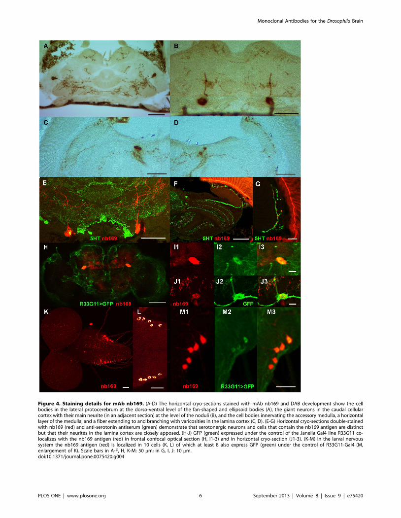
Figure 4. Staining details for mAb nb169. (A-D) The horizontal cryo-sections stained with mAb nb169 and DAB development show the cellbodies in the lateral protocerebrum at the dorso-ventral level of the fan-shaped and ellipsoid bodies (A), the giant neurons in the caudal cellularcortex with their main neurite (in an adjacent section) at the level of the noduli (B), and the cell bodies innervating the accessory medulla, a horizontallayer of the medulla, and a fiber extending to and branching with varicosities in the lamina cortex (C, D). (E-G) Horizontal cryo-sections double-stainedwith nb169 (red) and anti-serotonin antiserum (green) demonstrate that serotonergic neurons and cells that contain the nb169 antigen are distinctbut that their neurites in the lamina cortex are closely apposed. (H-J) GFP (green) expressed under the control of the Janelia Gal4 line R33G11 co-localizes with the nb169 antigen (red) in frontal confocal optical section (H, I1-3) and in horizontal cryo-section (J1-3). (K-M) In the larval nervoussystem the nb169 antigen (red) is localized in 10 cells (K, L) of which at least 8 also express GFP (green) under the control of R33G11-Gal4 (M,enlargement of K). Scale bars in A-F, H, K-M: 50 mm; in G, I, J: 10 mm.doi:10.1371/journal.pone.0075420.g004
Monoclonal Antibodies for the Drosophila Brain
PLOS ONE | www.plosone.org 6 September 2013 | Volume 8 | Issue 9 | e75420

the same two perikarya (Figure 3B1-3 for line R49C04, Figure 4H,
enlarged in I1-3 (whole-mount), and 4J1-3 (cryo-section) for line
R33G11), while GFP expression was also prominent in various
other structures (Figures 3B1, 4H, J2). The co-localization of the
nb169 antigen and GFP driven by line R33G11 is also seen in the
larval ventral nerve cord (Figure 4M1-3). GFP driven by line
R49C04 in larvae, on the other hand, does not co-localize with
nb169 antigen. The Gal4 line R33G11 contains a regulatory DNA
fragment of the gene CG18405, Sema-1a. Gal4 lines driven by
other regulatory fragments of this gene show no expression in these
cells such that the relation of the antigen recognized by nb169 to
the Sema-1a gene remains obscure. Obviously, it will be fascinating
to learn more about the neurons and the antigen recognized by
mAb nb169. The discovery of two Gal4 lines with very different
expression patterns overlapping apparently only in the pair of
giant neurons labelled by mAb nb169 suggests that it might be
possible to generate a line expressing a transgene of choice only in
these two giant neurons by split-Gal4 technique [7]. The co-
localization results of all tested mAb/Gal4 pairs are summarized in
Table 1. Figure 5 shows examples for co-localization of GFP
expression driven by Janelia Gal4 lines with stainings by mAb
ab47 (Figure 5A-C) and nc24 (Figure 5D-F).
Strategies towards identifying the antigens for mAbsEarlier attempts to identify the antigen of mAb nb33 [4] by
extensive screening of a cDNA expression library had failed (K.
Zinsmaier, unpublished), whereas screening of the same library
with mAbs recognizing synaptic proteins was successful [24,25].
Also, for none of the antibodies described here a reliable Western
blot signal from brain homogenate could be obtained, defeating
any attempts to identify the antigens by protein purification and
mass spectrometry [26,9]. Thus the classical approaches to the
identification of antigens for mAbs cannot be applied. We
therefore here present the distribution of the antigens in the
Drosophila brain and make them available for use in neuroanatomy
and for comparison with cell identification by other techniques
mentioned above. We first selected a small number of Gal4 lines
from the Janelia collection [5,6] that showed similarity between
their expression patterns revealed by GFP and the staining
patterns of our mAbs in the adult fly brain. We then investigated
by double stainings whether there was any overlap detectable.
Table 1 summarizes the results. For mAb nb169 and Gal4-lines
R33G11 and R49C04 we find clear co-localization of GFP and
antibody binding as described above. Frequently, however, in spite
of apparent similarity of staining patterns in the neuropil, e.g. in
the fan-shaped body or in horizontal layers of the medulla, the
GFP-positive and the mAb-stained cell bodies were distinct albeit
often in close proximity. In some cases a small subset of mAb-
stained cells also expressed GFP driven by the Gal4-line indicated
((+) in Table 1). The stainings shown in Figure 5 suggest that Gal4
expression from different Gal4 lines may overlap as their GFP
reporter is observed in mAb-stained cells at similar positions.
However, the positions of cell bodies are somewhat variable from
fly to fly such that additional evidence for the overlap will be
required.
Since it was shown that mAb nb168 binds to a neuropeptide or
its precursor we wanted to test if other antibodies might also
identify peptidergic cells. In general, neuropeptides are cleaved
from precursor proteins by proteolytic enzymes. Thus many
neuropeptide-containing cells can be labelled by GFP driven by
Figure 5. Examples for the co-localization of staining by mAbs ab47 (A-C) and nc24 (D-F) and GFP driven by selected Janelia Gal4lines. Due to the variability of cell body positions in different individuals it is only suggestive that the Gal4 expression patterns overlap. Scale bar100 mm.doi:10.1371/journal.pone.0075420.g005
Monoclonal Antibodies for the Drosophila Brain
PLOS ONE | www.plosone.org 7 September 2013 | Volume 8 | Issue 9 | e75420

386Y-Gal4, a Gal4 enhancer trap construct under regulatory
control of the prohormone convertase 2 gene (amon) [8,27,28].
Figure 6 shows the co-localization of nb169, ca8, and ab158
antibody staining with amon.GFP expression, supporting a
speculation that these three mAbs may perhaps bind to
neuropeptide-related antigens.
The interpretation of co-localization (or its lack) of any Gal4-
driven GFP expression with a staining pattern obtained with an
antibody against an unknown antigen remains difficult. Several
mechanisms may be responsible for discrepancies between the
Gal4-driven GFP expression patterns and the antigen distributions
even if the antigen is the product of the corresponding gene. First,
enhancer, silencer, and promoter elements may be widely
separated in a regulatory region of a gene and usually combine
in a complex, non-additive fashion to produce the final expression
pattern. Second, the GFP of the mCD8-GFP construct expressed
by Gal4 is targeted to all membranes such that the entire cell is
labelled, whereas the antigen recognized by the mAb may be
specifically and efficiently targeted to a specific cell compartment
and might be detected by the antibody only there. Third, the
antigen recognized by the mAb may perhaps be a processed or
post-translationally modified version of a gene product and
therefore be present only in a subset of the cells expressing the
gene. Thus, for each of the mAbs shown here considerable efforts
Figure 6. The antigens detected by mAbs nb169, ca8, and ab158 may be related to neuropeptides. (A-E) The cell stained by nb169 (red)(A, enlarged in B), and the giant cells in the caudal protocerebrum (C and D from different sections) all are positive for GFP (green) expressed underthe control of 386Y amon-Gal4 which has been shown to selectively mark neuropeptide-containing cells. (E) The neurite of a 386Y amon-Gal4.GFP-positive cell at the boundary between lamina cortex and neuropil apparently contains in discrete varicosities the nb169 antigen. (F,G) Examples areshown for co-localization of 386Y amon-Gal4-driven GFP and ca8 antigen in cells of the lateral protocerebrum. (H) Cells containing ab158 antigen inthe pars intercerebralis, a region containing many neurosecretory cells, are also positive for amon-Gal4-driven GFP. Scale bar in A: 50 mm; in B-H:10 mm.doi:10.1371/journal.pone.0075420.g006
Monoclonal Antibodies for the Drosophila Brain
PLOS ONE | www.plosone.org 8 September 2013 | Volume 8 | Issue 9 | e75420

will be necessary in any attempt to identify the antigen. However,
in cases where neuronal cell bodies identified by a mAb can be
labelled in vivo by GFP in principle it can become feasible to
separate these cell bodies and isolate their mRNAs for gene
expression profiling [29] or generation of a cell-specific cDNA
expression library which can be screened with the mAb. This
approach to the identification of unknown antigens labelled by
monoclonal antibodies only in a few cells in the brain can now be
attempted e.g. with mAb nb169.
Acknowledgments
We thank Dr. Aljoscha Nern for helpful discussions and Ms. Gertrud
Gramlich for excellent help with the hybridoma cell culture. The provision
of Janelia Gal4 fly lines by the Bloomington stock center is gratefully
acknowledged.
Author Contributions
Conceived and designed the experiments: VR KVR MR DR CF AH EB.
Performed the experiments: BBR MB PH FE MKS BM FE EB . Analyzed
the data: KVR MR DR CF AH EB. Contributed reagents/materials/
analysis tools: AH CW. Wrote the paper: EB.
References
1. Fujita SC, Zipursky SL, Benzer S, Ferrus A, Shotwell SL (1982) Monoclonal
antibodies against the Drosophila nervous system. Proc Natl Acad Sci U S A 79:
7929–7933.
2. Fujita SC (1988) Use of hybridoma libraries in the study of the genetics and
development of Drosophila. Annu Rev Entomol 33: 1–15.
3. Hofbauer A (1991) A library of monoclonal antibodies against the brain of
Drosophila melanogaster). Professorial dissertation (in German), Germany:Uni-
:University of Wuerzburg .
4. Hofbauer A, Ebel T, Waltenspiel B, Oswald P, Chen YC, et al. (2009) The
Wuerzburg hybridoma library against Drosophila brain. J Neurogenet 23: 78–91.
5. Pfeiffer BD, Jenett A, Hammonds AS, Ngo TT, Misra S, et al. (2008) Tools for
neuroanatomy and neurogenetics in Drosophila. Proc Natl Acad Sci U S A 105:
9715–9720.
6. Jenett A, Rubin GM, Ngo TT, Shepherd D, Murphy C, et al. (2012) A GAL4-
driver line resource for Drosophila neurobiology. Cell Rep 2: 991–1001.
7. Luan H, Peabody NC, Vinson CR, White BH (2006) Refined spatial
manipulation of neuronal function by combinatorial restriction of transgene
expression. Neuron 52: 425–436.
8. Taghert PH, Hewes RS, Park JH, O’Brien MA, Han M, et al Multiple amidated
neuropeptides are required for normal circadian locomotor rhythms in
Drosophila. (2001) J Neurosci 21: 6673–86.
9. Halder P, Chen YC, Brauckhoff J, Hofbauer A, Dabauvalle MC, et al. (2011)
Identification of Eps15 as antigen recognized by the monoclonal antibodies aa2
and ab52 of the Wuerzburg Hybridoma Library against Drosophila brain. PLoS
ONE 6(12): e29352.
10. Thum AS, Knapek S, Rister J, Dierichs-Schmitt E, Heisenberg M, et al. (2006)
Differential potencies of effector genes in adult Drosophila. J Comp Neurol 498:
194–203.http://www.jneurosci.org/openurl?query = rft.jtitle%3DThe+Journal+of+comparative+neurology%26rft.stitle%3DJ+Comp+Neurol%26rft.aulast%3D
Thum%26rft.auinit1%3DA.+S.%26rft.volume%3D498%26rft.issue%3D2%26rft.
spage%3D194%26rft.epage%3D203%26rft.atitle%3DDifferential+potencies+of
+effector+genes+in+adult+Drosophila.%26rft_id%3Dinfo%3Adoi%2F10.
1002%2Fcne.21022%26rft_id%3Dinfo%3Apmid%2F16856137%26rft.
genre%3Darticle%26rft_val_fmt%3Dinfo%3Aofi%2Ffmt%3Akev%3Amtx%3A
journal%26ctx_ver%3DZ39.88-2004%26url_ver%3DZ39.88-2004%2
11. Helfrich-Forster C (2007) Immunohistochemistry in Drosophila: sections and
whole mounts. Methods Mol Biol 362: 533–47.
12. Helfrich-Forster C, Shafer O, Wulbeck C, Grieshaber E, Rieger D, et al. (2007)
Development and morphology of the clock-gene-expressing lateral neurons of
Drosophila melanogaster. J Comp Neurol 500: 47–70.
13. Wu JS, Luo L (2006). A protocol for dissecting Drosophila melanogaster brains for
live imaging or immunostaining. Nature protocols Vol.1 No.4: 2110–2115.
14. Helfrich-Forster C, Homberg U (1993) Pigment-dispersing hormone-immuno-
reactive neurons in the nervous system of wild-type Drosophila melanogaster and of
several mutants with altered circadian rhythmicity. J Comp Neurol 337: 177–
190.15. Helfrich-Forster C (1997) Development of pigment-dispersing hormone-
immunoreactive neurons in the nervous system of Drosophila melanogaster.
J Comp Neurol 380: 335–354.16. Lohr J, Klein J, Webster SG, Dircksen H (1993) Quantification, immunoaffinity
purification and sequence analysis of a pigment-dispersing hormone of the shorecrab, Carcinus maenas (L.). Comp Biochem Physiol B 104: 699–706.
17. Buchner E, Bader R, Buchner S, Cox J, Emson PC, et al. (1988) Cell-specific
immuno-probes for the brain of normal and mutant Drosophila melanogaster. I.Wildtype visual system. Cell Tissue Res 253: 357–70.
18. Fischbach K-F, Dittrich APM (1989) The optic lobe of Drosophila melanogaster. I. AGolgi analysis of wild-type structure. Cell Tissue Res 258: 441–475.
19. Tuthill JC, Nern A, Holtz SL, Rubin GM, Reiser MB (2013) Contributions ofthe 12 neuron classes in the fly lamina to motion vision. Neuron 79: 128–140.
20. Nassel DR, Hagberg M, Seyan HS (1983) A new, possibly serotonergic, neuron
in the lamina of the blowfly optic lobe: an immunocytochemical and Golgi-EMstudy. Brain Res 280: 361–367.
21. Nassel DR, Meyer EP, Klemm N (1985) Mapping and ultrastructure ofserotonin-immunoreactive neurons in the optic lobes of three insect species.
J Comp Neurol 232: 190–204.
22. Nassel DR (1991) Neurotransmitters and neuromodulators in the insect visualsystem. Prog Neurobiol 37: 179–254.
23. Nassel DR, Elekes K (1984) Ultrastructural demonstration of serotonin-immunoreactivity in the nervous system of an insect (Calliphora erythrocephala).
Neurosci Lett 48: 203–10.24. Zinsmaier KE, Hofbauer A, Heimbeck G, Pflugfelder GO, Buchner S, et al.
(1990) A cysteine-string protein is expressed in retina and brain of Drosophila.
J Neurogenet 7: 15–29.25. Reichmuth C, Becker S, Benz M, Debel K, Reisch D, et al. (1995) The sap47
gene of Drosophila melanogaster codes for a novel conserved neuronal proteinassociated with synaptic terminals. Mol Brain Res 32: 45–54.
26. Wagh DA, Rasse TM, Asan E, Hofbauer A, Schwenkert I, et al. (2006)
Bruchpilot, a protein with homology to ELKS/CAST, is required for structuralintegrity and function of synaptic active zones in Drosophila. Neuron 49: 833–844.
27. Wegener C, Herbert H, Kahnt J, Bender M, Rhea JM (2011) Deficiency ofprohormone convertase dPC2 (AMONTILLADO) results in impaired produc-
tion of bioactive neuropeptide hormones in Drosophila. J Neurochem 118: 581–95.
28. Siekhaus DE, Fuller RS (1999) A role for amontillado, the Drosophila homolog of
the neuropeptide precursor processing protease PC2, in triggering hatchingbehavior. J Neurosci 19: 6942–54.
29. Nagoshi E, Sugino K, Kula E, Okazaki E, Tachibana T, et al. (2010) Dissectingdifferential gene expression within the circadian neuronal circuit of Drosophila.
Nat Neurosci 13: 60–8.
Monoclonal Antibodies for the Drosophila Brain
PLOS ONE | www.plosone.org 9 September 2013 | Volume 8 | Issue 9 | e75420