18.3.2022 in Würzburg Abstract Book - GfH-Tagung
321
32.GfH-Jahrestagung 16.-18.3.2022 in Würzburg Abstract Book Die mit *** gekennzeichneten Poster sind für die Posterpreisnominierung vorgeschlagen worden. Fehlende Nummern verweisen auf zurückgezogene Abstracts. Selected Presentations ........................................................................2 Workshop 1: Technology and Bioinformatics ...................................................................................... 7 Workshop 2: Neurodevelopmental Disorders ................................................................................... 14 Workshop 3: Clinical Genetics .......................................................................................................... 23 Workshop 4: Inherited Cancer Genetics ........................................................................................... 29 Workshop 5: Complex Diseases ....................................................................................................... 36 Workshop 6: Neurogenetics .............................................................................................................. 43 Workshop 7: From Basic Mechanisms to Therapy ........................................................................... 49 Workshop 8: (Epi-)Genomics and Cancer ........................................................................................ 56 Workshop 9: Monogenic Syndromes ................................................................................................ 63 Postersessions ...............................................................................70 001–011 Basic Mechanisms and Epigenetics .............................................................................. 70 012–033 Cancer Genetics ............................................................................................................ 82 034–111 Clinical Genetics, Genetic Counselling and Prenatal Diagnostics .............................. 106 112–126 Complex Diseases, Population & Evolutionary Genetics and Genetic Epidemiology 179 127–139 Cytogenetics and CNVs .............................................................................................. 198 140–185 Monogenic Disease – from Gene Identification to Molecular Mechanism .................. 209 186–205 Technology and Bioinformatics ................................................................................... 256 206–208 Therapy for Genetic Diseases ..................................................................................... 278 Authors Index ...............................................................................281
-
Upload
khangminh22 -
Category
Documents
-
view
0 -
download
0
Transcript of 18.3.2022 in Würzburg Abstract Book - GfH-Tagung
32.GfH-Jahrestagung 16.-18.3.2022 in Würzburg
Abstract Book
Die mit *** gekennzeichneten Poster sind für die Posterpreisnominierung vorgeschlagen worden. Fehlende Nummern verweisen auf zurückgezogene Abstracts.
Selected Presentations ........................................................................2 Workshop 1: Technology and Bioinformatics ...................................................................................... 7
Workshop 2: Neurodevelopmental Disorders ................................................................................... 14
Workshop 3: Clinical Genetics .......................................................................................................... 23
Workshop 4: Inherited Cancer Genetics ........................................................................................... 29
Workshop 5: Complex Diseases ....................................................................................................... 36
Workshop 6: Neurogenetics .............................................................................................................. 43
Workshop 7: From Basic Mechanisms to Therapy ........................................................................... 49
Workshop 8: (Epi-)Genomics and Cancer ........................................................................................ 56
Workshop 9: Monogenic Syndromes ................................................................................................ 63
Postersessions ...............................................................................70 001–011 Basic Mechanisms and Epigenetics .............................................................................. 70
012–033 Cancer Genetics ............................................................................................................ 82
034–111 Clinical Genetics, Genetic Counselling and Prenatal Diagnostics .............................. 106
112–126 Complex Diseases, Population & Evolutionary Genetics and Genetic Epidemiology 179
127–139 Cytogenetics and CNVs .............................................................................................. 198
140–185 Monogenic Disease – from Gene Identification to Molecular Mechanism .................. 209
186–205 Technology and Bioinformatics ................................................................................... 256
206–208 Therapy for Genetic Diseases ..................................................................................... 278
Authors Index ...............................................................................281
Abstract Book
Selected Presentations
SEL-001
Single-cell phenotyping of pleiotropic developmental disorders during embryonic development at single cell resolution
J. Henck1, X. Huang2, C. Qiu2, V. Sreenivasan 1, U. Kornak3, D. Ibrahim4, A. Despang4, C. Trapnell2, L. Saunders2, R. Behncke5, S. Ulferts5, N. Hansmeier5, R. Hägerling5, I. Kurth6, N. Haag6, M. Robson4, A. Ringel4, W. L. Chan7, V. Kalscheuer4, L. Wittler4, D. Beier8, D. Dickel9, L. Pennacchio9, A. Visel9, J. Cao10, J. Shendure2, M. Spielmann1
1Institut für Humangenetik- Universitätsklinikum Schleswig Holstein, Lübeck, Deutschland 2University of Washington, Department of Genome Sciences, Seattle, Vereinigte Staaten 3Institute of Human Genetics, University Medical Center Göttingen, Göttingen, Deutschland 4Max Planck Institute for Molecular Genetics, Berlin, Deutschland 5Charité – Universitätsmedizin Berlin, Berlin, Deutschland 6Institute of Human Genetics, RWTH Aachen University, Aachen, Deutschland 7Institute for Medical and Human Genetics, Charité-Universitätsmedizin Berlin, Berlin, Deutschland 8Department of Pediatrics, University of Washington School of Medicine, Seattle, Vereinigte Staaten 9Environmental Genomics & System Biology Division, Lawrence Berkeley National Laboratory, Berkeley, Vereinigte Staaten 10Laboratory of single-cell genomics and population dynamics, Rockefeller University, New York, Vereinigte Staaten
For many years the laboratory mouse has remained the quintessential research animal of choice for studying molecular mechanisms of human development disorders. The recent development of CRISPR/Cas based genome editing tools now allow the investigation of any gene or regulatory element in vivo. However, the current phenotyping approaches lack the necessary throughput and resolution for detailed investigations of pleiotropic disorders at the organismal scale. The recent developments in single-cell genomics offer the possibility to overcome these shortfalls and answer central questions of development.
In the current study we set out to establish single cell RNA sequencing as a tool for large scale standardized and comprehensive phenotypic analysis of whole mouse mutant embryos. In a single multiplexed experiment, we applied combinatorial indexing based single cell RNA sequencing to profile 103 whole mouse embryos of 22 different mutants and 4 different wildtype strains at embryonic stage E13.5. Towards evaluating the sensitivity of this technique, the selected mouse mutants in this study range from established multisystem disorders to single enhancer knockouts resulting in different phenotype severities. The resulting Mouse Mutant Cell Atlas (MMCA) consists of over 1.9 million single cell RNA-seq profiles. We developed an analytical framework for molecular phenotyping of cell type and trajectory composition changes, gene expression alterations and developmental phenotypes. Moreover, we identify mutation and strain specific cell type changes, compare phenotyping of gain and loss of function mutants, and characterize deletions of topological associating domain boundaries. Overall, we identified 300 significantly changed cell type proportions from 52 sub trajectories across the 22 mutants compared to the wildtype. Some pleiotropic genes such as Sox9 showed general changes in over 30 sub-trajectories indicating their generally regulatory functions during embryogenesis, while other mutants such as Ttc21b showed very specific changes in single sub-trajectories such as the retina. The deletions of noncoding elements showed milder changes compared to the other mutants, however some specific trajectories still revealed major phenotypes in the limb and the brain. Some trajectories also showed a retardation of development. A unique strength of the experimental framework of the approach allowed combining multiple mutants which enabled the discovery of a shared phenotype between unrelated genotypes, undetected before.
In summary our findings show that whole embryo single cell phenotyping represents a powerful tool to systematically characterize developmental disorders at unprecedented resolution.

SEL-002
Systematic analysis of non-coding de novo mutations from whole genome sequence data of triads with non-syndromic cleft lip with/without cleft palate
H. K. Zieger1, L. Weinhold2, A. Schmidt1, M. Holtgrewe3, S. A. Juranek4, A. Siewert1, F. Thieme1, F. Brand5, J. Welzenbach1, D. Beule3,6, K. Paeschke4, P. M. Krawitz2, K. U. Ludwig1
1Institute of Human Genetics, University of Bonn, School of Medicine & University Hospital Bonn, Bonn, Deutschland 2Institute for Medical Biometry, Informatics and Epidemiology, University Hospital Bonn, Bonn, Deutschland 3Core Unit Bioinformatics, Berlin Institute of Health, Berlin, Deutschland 4Department of Oncology, Hematology and Rheumatology, University Hospital Bonn, Bonn, Deutschland 5Institute for Genomic Statistics and Bioinformatics, University Hospital Bonn, Bonn, Deutschland 6Max Delbrück Center for Molecular Medicine, Berlin, Deutschland
Non-syndromic cleft lip with/without cleft palate (nsCL/P) is a common multifactorial disorder with strong genetic contribution, and common risk variants at about 45 loci have been identified. Here, we systematically investigate the contribution of de novo mutations (DNMs) to nsCL/P risk, using whole-genome sequence (WGS) data for 211 nsCL/P and 284 non-cleft reference trios from the Kids First Project. For the total set of 31,490 DNMs, overall comparison between cohorts did not show any robust differences, neither in absolute numbers nor when the DNMs were weighted for different functional scores (e.g. CADD, LINSIGHT, ReMM). However, we observed nominally significant accumulation of non-coding DNMs in nsCL/P at regions marked as bivalent TSS/enhancer states in nsCL/P during human embryonic face development at Carnegie Stage 15 (p=0.0269). When focusing on previously identified GWAS loci, we observed a significant enrichment of non-coding DNMs in topologically associating domains at two GWAS risk loci, i.e. 4q28.1 (DNMs in 7 cases, 0 controls, p=0.0008) and 2p21PKDCC (7 cases, 2 controls, p=0.0161). We finally used transcription factor (TF) binding information to identify TFs with potential key role in nsCL/P etiology. Based on position weight matrices, we predicted TF binding sites for 810 human TFs, and calculated changes of binding capacity at 28,773 DNM-sites. We observed a significant enrichment of DNM-hits for motif TFAP2A in nsCL/P, and identified ATF3, MSC and HES5/7 as potential TF candidates. Notably, for MSC and ATF3, this finding was supported by a strong quantitative effect on the predicted binding change, which for MSC is currently validated using in vitro assays. So far, we have been able to demonstrate binding of MSC to two regions, and a change in binding by the respective DNMs. Analyses of single-cell expression data during murine organogenesis (Cao et al. 2019) confirmed Msc expression in myocytes during embryonic development. Notably, MSC has been shown to be involved in the development of orofacial muscles, together with a subset of candidate genes located at GWAS loci. Our study provides novel insights into nsCL/P etiology and suggests a TF-based approach that can be used to annotate non-coding risk variants from WGS data.
SEL-003
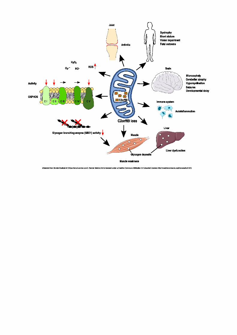
C2orf69 mutations disrupt mitochondrial function and cause a multisystem human disorder with recurring autoinflammation
E. Lausberg1, S. Gießelmann1, J. P. Dewulf2, E. Wiame2, A. Holz3, R. Salvarinova4, C. Van Karnebeek5, P. Klemm6, K. Ohl6, M. Mull7, T. Braunschweig8, J. Weis9, C. J. Sommer10, S. Demuth11, C. Haase12, C. Stollbrink-Peschgens6, F. Debray13, C. Libioulle13, D. Choukair14, P. T. Oommen15, A. Borkhardt15, H. Surowy16, D. Wieczorek16, N. Wagner6, R. Meyer1, T. Eggermann1, M. Begemann1, E. Van Schaftingen2, M. Häusler6, K. Tenbrock6, L. Van Den Heuvel17, M. Elbracht1, I. Kurth1, F. Kraft1
1Institut für Humangenetik, Universitätsklinikum RWTH Aachen, Aachen, Deutschland 2Laboratory of Physiological Chemistry, de Duve Institute, Brüssel, Belgien 3CeGaT GmbH and Praxis für Humangenetik, Tübingen, Deutschland 4Division of Biochemical Diseases, Department of Pediatrics, British Columbia Children's Hospital Vancouver, Vancouver, Kanada 5Department of Pediatrics, Centre for Molecular Medicine and Therapeutics, UBC, Vancouver, Kanada 6Department of Pediatrics, RWTH Aachen University, Aachen, Deutschland 7Department of Diagnostic and Interventional Neuroradiology, Medical Faculty, RWTH Aachen University, Aachen, Deutschland 8Institute of Pathology, Medical Faculty, RWTH Aachen University, Aachen, Deutschland 9Institute of Neuropathology, Medical Faculty, RWTH University Hospital, Aachen, Deutschland 10Institute of Neuropathology, University Medical Center of the Johannes Gutenberg University Mainz, Mainz, Deutschland 11Praxis für Humangenetik Erfurt, Erfurt, Deutschland 12HELIOS Klinikum Erfurt, Ambulanz für angeborene Stoffwechselerkrankungen, Sozialpädiatrisches Zentrum, Erfurt, Deutschland 13Department of Human Genetics - CHU de Liège, Lüttich, Belgien 14Department of General Pediatrics, University Children's Hospital, University Heidelberg, Heidelberg, Deutschland 15Department of Pediatric Oncology, Hematology and Clinical Immunology, University Children's Hospital, Medical Faculty, Heinrich-Heine-University Düsseldorf, Düsseldorf, Deutschland 16Institute of Human Genetics, Medical Faculty, Heinrich-Heine-University, Düsseldorf, Deutschland 17Department of Pediatrics, Translational Metabolic Laboratory at Department of Laboratory Medicine, Radboudumc, Nijmegen, Niederlande
BACKGROUND. Deciphering the function of the many genes previously classified as uncharacterized open reading frame (ORF) would complete our understanding of a cell's function and its pathophysiology.
METHODS. Whole-exome sequencing, yeast 2-hybrid and transcriptome analyses, and molecular characterization were performed in this study to uncover the function of the C2orf69 gene.
RESULTS. We identified loss-of-function mutations in the uncharacterized C2orf69 gene in 8 individuals of five unrelated families with brain abnormalities involving hypomyelination and microcephaly, liver dysfunction, and recurrent autoinflammation. C2orf69 contains an N-terminal signal peptide that is required and sufficient for mitochondrial localization. Consistent with mitochondrial dysfunction, the patients showed signs of respiratory chain defects and disintegration of mitochondria. A CRISPR/Cas9-KO cell model of C2orf69 exhibited a reduced activity of complex I, II and V and showed an increased glycolytic activity. Transcriptomic analysis of patient-derived cells revealed alterations in immunological signaling pathways. Deposits of periodic acid–Schiff–positive (PAS-positive) material in tissues from affected individuals, together with decreased glycogen branching enzyme 1 (GBE1) activity, indicated an additional impact of C2orf69 on glycogen metabolism.
CONCLUSIONS. Our study identifies C2orf69 loss as cause of a new disease (Combined oxidative phosphorylation deficiency 53) and characterizes the so far functionally undescribed gene C2orf69 as a novel regulator of human mitochondrial function. Moreover we suggest that this gene has additional influence on other metabolic pathways and potentially links mitochondrial function to glycogen metabolism by regulation of GBE1.
PIC
SEL-004
A national diagnostic framework for patients with ultra-rare disorders: molecular genetic findings using phenotypic and sequencing data
M. Danyel1, Link zu der Liste der Autoren
1Institut für Medizinische Genetik und Humangenetik, Charité, Berlin
on behalf of the TRANSLATE NAMSE consortium, Universität Bonn, Bonn, Deutschland.
Most individuals with rare diseases first contact primary care physicians. Although efficient diagnostic routines exist for a subset of rare diseases, ultra-rare entities often require expert clinical knowledge or comprehensive genetic diagnostics, which poses structural challenges to public healthcare systems. To address these challenges, a novel structured diagnostic concept based on the presence of multidisciplinary expertise at centers for rare diseases (CRDs) that have been established at German university hospitals in recent years, was evaluated in a prospective study (TRANSLATE-NAMSE). Between January 2018 and December 2020, 5652 patients were enrolled in the study and were comprehensively assessed by multidisciplinary teams (MDTs) at ten CRDs. Exome sequencing (ES) was initiated for 282 adult and 1283 pediatric patients and partially complemented by additional molecular tests. Conclusive diagnoses were established in 494 individuals, covering 400 diagnostic-grade genes, suggesting ultra-rare disorders were enriched in this cohort. In addition, we describe 64 novel gene-phenotype associations, mainly in individuals with neurodevelopmental delay. A subcohort of 210 individuals was analyzed with the artificial intelligence-based PEDIA protocol, which integrates next-generation phenotyping on medical imaging and sequencing data. With the entire cohort data, we developed a tool to predict the diagnostic yield from the clinical features of a patient if advanced molecular testing strategies are applied.
A network of centers specializing in the diagnosis and treatment of ultra-rare diseases and exchanging data within and beyond this consortium could also be a blueprint for the "§ 64e Modellvorhaben zur umfassenden Diagnostik und Therapiefindung mittels Genomsequenzierung bei seltenen Erkrankungen".
Results in this talk will be presented on behalf of the TRANSLATE NAMSE consortium (>150 members, 10 Universitätskliniken).
Workshop 1: Technology and Bioinformatics
W1-001
Factors impacting scalability of genome analyses beyond the exome in clinical routine
T. B. Haack 1, M. Sturm1, O. Kelemen1, C. Schroeder1, A. Dufke1, S. Ossowski1, O. Riess1
1Institut für Medizinische Genetik und Angewandte Genomik, Universität Tübingen , Tübingen, Deutschland
Objective: Broad implementation of full genome sequencing (GS) is the next logical step to address the diagnostic gap in rare disease genetics, to identify novel disease genes and mechanisms, and to pave the way towards disease prevention in common phenotypes. It also opens the discussion on the chances to integrate genetic risk stratification for common diseases via polygenic risk scores as additional diagnostic merit. However, the application of GS on a routine basis is challenged by laboratory, bioinformatic, interpretation, and financial issues. We aimed to develop and test a scalable framework for genome-based analyses in regular care.
Methods: From 2019 on, we performed in the setting of a tertiary care center PCR-free short read genome sequencing on a total of 3,300 probands. Additional bioinformatic algorithms had to be developed and/or implemented to improve the detection of SNVs/InDels, CNVs, SVs, and systematically benchmark the evaluation of repeat expansions. While investigated phenotypes were initially focused on retinal diseases, intellectual disability, and pediatric cancer, the range of indications was subsequently expanded to include neurological disorders and familial breast cancer. For the latter, calculation and reporting of polygenic risk scores were established.
Results: While wet lab automation and scaling was feasible without major difficulties, the primary bioinformatic steps required acquisition of additional infrastructures (15 servers, 2 petabyte storage, DRAGEN). Several modifications were needed to improve the decision support system for clinical variant interpretation for SVs, CNVs, MEIs, and REs. The entire pipeline for genome-based genetic testing was validated and received DAkks accreditation according to DIN EN ISO 15189. Amongst the firm genetic diagnoses, 20 – 25 % of the disease-causal variants were not the "classical" SNVs or InDels in coding regions.
Conclusion: Translation of GS from research into clinical routine is feasible but requires the concerted action of scientist, medical doctors, bioinformaticians, and health care managers. Our study indicates that GS has benefits in the detection of almost all types of genetic variation and that these findings translate into additional diagnoses. However, we are still far from understanding and fully exploiting the wealth of information provided by a full genome"s sequence. Amongst others, small numbers of uniformly processed genome datasets and shortage of trained genome analysts are major hurdles for a rollout of GS on a national scale.

W1-002
GestaltMatcher research platform facilitates the novel gene-phenotype exploration
T. C. Hsieh1, L. Averdunk2, C. C. Mak3, T. Kamphans4, W. Meiswinkel4, P. Krawitz1
1University Hospital Bonn, Institute for Genomic Statistics and Bioinformatic, Bonn, Deutschland 2Heinrich Heine University, Institute of Human Genetics and Department of Pediatrics, Düsseldorf, Deutschland 3University of Hong Kong, Department of Paediatrics and Adolescent Medicine, Hong Kong, Hongkong 4GeneTalk, Bonn, Deutschland
Question: The next-generation phenotyping (NGP) approaches for syndromology, such as GestaltMatcher [1], have learned facial representations of multiple disorders by training on thousands of patient photos. GestaltMatcher can not only predict the disorder but also quantify the similarity between patients that enable the novel gene-phenotype exploration. However, there is no platform for clinicians to easily upload the patients and select the patients from publications to perform GestaltMatcher for the gene-phenotype association analysis. Therefore, we proposed the GestaltMatcher research platform to provide a user-friendly interface to upload patients, select patients from existing publications, and conduct gene-phenotype association experiments.
Methods: We built a research platform in GestaltMatcher Database (GMDB) [2]. Users can first upload their patients to GMDB and later send them to the research platform to perform the GestaltMatcher analysis. GMDB currently contains 5510 patients with 573 different disorders from 1481 publications. Users can analyze their patients and also include the patients from publication in GMDB into their experiments. The research platform supports the GestaltMatcher approach to calculate the similarities among the selected cohort, further generating the matrix of pairwise distances (ranks) and the figure of t-SNE for the two-dimensional visualization. We selected two cohorts as examples: Cohort-1 contains 33 patients with disease-causing mutations in Gene-X and Cohort-2 consists of five patients with disease-causing mutations in Gene-Y.
Results: We show two kinds of analysis that users can perform on the platform. In Cohort-1, with the matrix of pairwise distances and the figure of t-SNE, we validate that the facial phenotype of the ten patients with the mutations in the first exon of Gene-X is different from the other 23 patients with the mutations in the second exon of Gene-X. The patients in the first exon and the patients in the second exon form two clear clusters. We conclude that the second exon of Gene-X can cause a novel phenotype that has not been linked to Gene-X yet. Moreover, we prove that the facial phenotype of the five patients in Cohort-2 is similar to Rothmund-Thomson syndrome (OMIM:268400). The result further suggests that the phenotype caused by Gene-Y can be merged into the phenotypic series of Rothmund-Thomson syndrome.
Conclusions: GestaltMatcher research platform provides users with a user-friendly interface to explore the novel gene-phenotype association.
References:
1. Hsieh, T.-C. et al. GestaltMatcher: Overcoming the limits of rare disease matching using facial phenotypic descriptors. medRxiv (2021) doi:10.1101/2020.12.28.20248193.
2. Hsieh, T.-C. GestaltMatcher Database. https://db.gestaltmatcher.org.

W1-003
Efficiency of Three Computer-aided Facial Phenotyping Tools (DeepGestalt, GestaltMatcher, D-Score) - Comparative Diagnostic Accuracy Study
A. M. V. Reiter1, J. T. Pantel1, M. Danyel1, D. Horn1, C. E. Ott1, M. A. Mensah1,2
1Charité - Universitätsmedizin Berlin, Institut für Medizinische Genetik und Humangenetik, Berlin, Deutschland 2Berlin Institute of Health, BIH Biomedical Innovation Academy, Digital Clinician Scientist Program, Berlin, Deutschland
Background: Genetic syndromes can often be diagnosed based on particular features of the face. Various systems have been developed for automated computer-assisted phenotyping of a patient"s face. Some of them have a remarkable sensitivity. However, the models of DeepGestalt and GestaltMatcher only assign scores for disease entities but lack a class for 'inconspicuous face'. Thus, their specificity is unclear. Moreover, there are few data and studies comparing the accuracy of different approaches.
D-Score is a new tool to increase the specificity of computer-assisted facial phenotyping. D-Score aims to distinguish photographs of dysmorphic faces from inconspicuous control subjects, in addition or prior to the use of previously described methods.
Aim: To determine and compare the diagnostic accuracy and potential clinical utility of D-Score, GestaltMatcher and DeepGestalt.
Methods: The three systems were tested with 323 images of patients, each with one of 17 dysmorphism-associated syndromes, and with the same number of age-, sex-, and ethnically matched inconspicuous control images. We compared the sensitivity, specificity, and the number of syndromes proposed as differential diagnoses. The power to binary classify images as dysmorphic or non-dysmorphic was analysed by comparing the score values obtained in each case.
Results: While gender and ethnic background had no effect on the accuracy of the systems, for all three, accuracy depended strongly on the age and syndrome of the individuals depicted. All three applications worked best with children (3 to 10 years old). While D-Score classified binary, DeepGestalt suggested 292 different syndromes, and GestaltMatcher returned 1187 with an overlap of 276 syndromes. False positive syndrome suggestions followed a nonrandom distribution as syndromes associated with rather mild facial dysmorphism were suggested most frequently (e.g., Angelman syndrome: DeepGestalt FPR 84%, GestaltMatcher FPR 92%). Both tools showed a good top-10 sensitivity (GestaltMatcher 90%, DeepGestalt 95%). While GestaltMatcher was unable to discriminate dysmorphic faces from normal control faces (AUROC 0.53), DeepGestalt showed moderate class discrimination ability (AUROC 0.70), and of the three, D-Score showed the highest discriminative power (AUROC 0.85).
Conclusions: The three applications have different strengths and limitations. Knowing these characteristics of the tools, they can be helpful to medical professionals in a stratified manner. Tools such as D-Score can be particularly useful in helping clinicians with little experience in clinical syndromology or limited diagnostic equipment to decide whether or not a patient needs further workup regarding a syndromologic differential diagnosis. Systems such as DeepGestalt are most useful in searching for relatively "frequent" syndromes. Programs such as GestaltMatcher should be employed when the patient has a particularly rare diagnosis probably unknown to the clinician.
W1-004
AutoCaSc: Prioritizing candidate genes for neurodevelopmental disorders
B. Popp1, J. Lieberwirth1, B. Büttner1, C. Klöckner1, K. Platzer1, M. Radtke1, M. Wegler1, R. Abou Jamra1
1University of Leipzig Hospitals and Clinics, Institute of Human Genetics, Leipzig, Deutschland
Routine exome sequencing (ES) in individuals with developmental disorders (DDs) remains inconclusive in roughly half of the cases. Research analysis of unsolved cases can identify novel candidate genes but is subjective, slow and hard to compare between labs. The field needs automated and standardized assessment of gene and variant characteristics to prioritize candidates.
We developed the AutoCaSc web-application (webAutoCaSc), which can be used for candidate prioritization based on variant and gene-specific information. It was developed in the Python programming language using the Dash framework and is freely available at https://autocasc.uni-leipzig.de. The tool automates our fine-tuned candidate scoring scheme (CaSc) which is composed of the four categories "Variant attributes" (6 points), "Inheritance" (3 points), "Gene constraint" (1 point) and "Gene plausibility" (6 points). The first three categories were implemented as simple decision trees, while "Gene plausibility" is a precomputed composite score of expression (GTEx), model organism (MGI), protein-protein interaction (STRING), literature (PubTator Central) and de novo occurrence in cohorts with comparable disorders (DisGeNET and PsyMuKB) data. A command line implementation of our algorithm (vcfAutoCaSc) was designed to allow direct pipeline integration and scoring of ES variant call files (VCFs) in larger cohorts or as a part of in-house pipelines.
We validated our approach using synthetic trios and real in-house trio ES data. AutoCaSc consistently (94.5%) scored 79 variants in recently published DD genes, which we synthetically injected into two publicly available ES trios in the top three ranks. The injected variant had a mean rank of 1.5 and 2.3 in the CEU and ASH trio, respectively. In real data from 93 trios, AutoCaSc identified all previously identified candidate variants scored with CaSc by a human evaluator. AutoCaSc placed these in the top ranks while evaluating additional highly scoring variants that were missed in the initial manual evaluation. With a CaSc of 11.4, a homozygous loss of function variant in CNTN2 (c.940C>T, p.(Arg314*) was the highest scoring variant in a gene currently not associated with NDD. Other high scoring variants with previously undescribed or unclear associations to NDD affected the genes DLGAP1, HDAC4, ANKRD17, SMURF1, NRXN3, PRICKLE1 and CASC5, while H3F3A and ANKRD17 have been published after our analysis.
AutoCaSc enables anybody to quickly screen a variant of interest for its plausibility for DDs. We provide usage recommendations, based on our experience and extensive application in projects describing novel DD associated genes. Our implementation is capable of pipeline integration and screening of large cohort datasets. AutoCaSc will further empower a standardized matchmaking collaboration and the accelerated identification of novel NDD entities.
W1-005
Bone2Gene: deep learning-based diagnosis of rare skeletal disorders
S. Rassmann1, A. Hustinx1, M. Ibarra1, T. C. Hsieh1, K. Skaf2, A. Keller3, K. Mohnike2, P. Krawitz1, B. Javanmardi1
1Universitätsklinikum Bonn, Institute for Genomic Statistics and Bioinformatics, Bonn, Deutschland 2Otto-von-Guericke-Universität Magdeburg, Medizinische Fakultät, Magdeburg, Deutschland 3Universitätsklinikum Leipzig, Kinderzentrum am Johannisplatz, Leipzig, Deutschland
Introduction: Rare genetic disorders collectively affect more than 6% of the global population. One of the main groups of such disorders are skeletal dysplasias, often resulting in short stature, altered biomechanics, pain, fatigue, and reduced functional performance. As genetically caused bone dysplasias are highly heterogeneous a precise differential diagnostics is required. However, some disorders are so rare that even experienced clinicians might have seen only some of them, making their accurate diagnosis a very challenging process. In this project, we are collecting hand X-Ray images of patients clinically or molecularly diagnosed with rare skeletal dysplasias to build a reference diagnostic tool for clinicians based on Deep Learning (DL).
Method: DL usually requires massive amounts of training data. However, data for rare genetic disorders is sparse due to the inherently low prevalence combined with difficulties in the collection and digitization of X-Ray imagery. We address this issue by employing transfer learning from a public bone age dataset provided by the Radiological Society of North America (RSNA). Furthermore, our data stems from varying acquisition sites and shows imprinted labeling and digitization artifacts which potentially induces biases. To eradicate these we trained DL models to automatically extract only the hands and conceal the origin of the X-Ray.
Results: Our bone age DL model trained on the RSNA dataset reached a relatively good accuracy with a mean age difference (MAD) of around 5 months. Using knowledge transfer from the bone age model, we built a DL classifier fine-tuned on around 600 images of six skeletal disorders, namely Noonan, Ullrich-Turner, SHOX-mutation, Silver–Russell, Hypochondroplasia, and Pseudohypoparathyroidism. This preliminary classifier has a wide range of accuracy (e.g. 95% for SHOX-mutation compared to 0% for Hypochondroplasia) which is mostly caused by our currently imbalanced dataset (242 SHOX-mutation images compared to only 25 for Hypochondroplasia).
Conclusion: By growing the underlying database and enhancing the performance of our model, we envision Bone2Gene becoming a reliable reference tool for the differential diagnosis of rare skeletal dysplasias. Moreover, DL models might use distinctive features evading the human eye. Therefore, studying the trained models might conjointly reveal new characteristics of the classified disorders.
W1-006
Deep phenotyping – symptom annotation made simple with SAMS
R. Steinhaus1,2, S. Proft1,2, E. Seelow3, T. Schalau1, P. N. Robinson4,5, D. Seelow1,2
1Berliner Institut für Gesundheitsforschung, Bioinformatik und translationale Genetik, Berlin, Deutschland 2Charité – Universitätsmedizin Berlin, Institut für Medizinische Genetik und Humangenetik, Berlin, Deutschland 3Charité – Universitätsmedizin Berlin, Medizinische Klinik mit Schwerpunkt Nephrologie und Internistische Intensivmedizin, Berlin, Deutschland 4The Jackson Laboratory for Genomic Medicine, Farmington, CT, Vereinigte Staaten 5University of Connecticut, Institute for Systems Genomics, Farmington, CT, Vereinigte Staaten
Precision medicine needs precise phenotypes. Correctly characterizing the symptoms of diseases is of utmost importance to diagnose, understand, and treat the disease. However, this can be difficult to achieve in routine clinical care, as hospital information systems are usually aimed at accounting rather than providing a thorough description of the patients" phenotypes. This is especially problematic for rare genetic diseases which often do not even have an ICD-10 code. In addition, it hampers clinical studies where concise information about signs and symptoms is needed.
With SAMS (Symptom Annotation Made Simple), we offer a free and simple tool for tracking medical signs, symptoms, and diagnoses based on four widely used annotation systems: HPO, OMIM, Orphanet, and DIMDI Alpha-IDs. Intuitive web-based interfaces allow users to easily annotate diseases and clinical signs with tailored modes for both clinicians and patients. Clinicians are supported with a deep learning-based expert system leading them towards a differential diagnosis. SAMS empowers patients to record their symptoms and share them with their doctors.
SAMS offers a web interface that can easily be integrated into hospital information systems without any installation. Import and export of a patient's medical history in the GA4GH Phenopacket format is possible. A prototype can be accessed at: https://www.genecascade.org/sams/
W2-001
De novo variants in the PABP-domain of PABPC1 lead to developmental delay
M. Wegler1, X. Jia2, M. Alders3, A. Boumann4, J. Chen2, X. Duan5, J. L. Lauzon6, I. B. Mathijssen3, H. Sticht7, S. Syrbe8, S. Tan2, H. Guo2, R. Abou Jamra1
1Institut für Humangenetik Leipzig, Leipzig, Deutschland 2Central South University, Center for Medical Genetics & Hunan Key Laboratory of Medical Genetics, Changsha, Deutschland 3University of Amsterdam, Department of Human Genetics, Amsterdam, Niederlande 4Erasmus MC University Medical Center, Department of Clinical Genetics, Rotterdam, Niederlande 5Daping Hospital, Army Medical University, Department of pediatrics, Chongqing, China, Volksrepublik 6Cummings School of Medicine, University of Calgary, Department of Medical Genetics, Alberta, Kanada 7Friedrich-Alexander-Universität Erlangen-Nürnberg, Institute of Biochemistry, Erlangen-Nürnberg, Deutschland 8University Hospital Heidelberg, Division of Pediatric Epileptology, Heidelberg, Deutschland
Developmental delays (DD) are often monogenic and highly heterogeneous. Roughly, half of the cases remain without a concrete genetic diagnosis. To decipher the genetics of DD, we re-analyze negative cases of exome sequencing, identify candidate genes and we internationally join forces clinical, genetic and functional aspects to describe novel DD forms.
We identified four heterozygous de novo variants in PABPC1 (NM_002568.3), leading to amino acid changes, p.(Pro555del), p.(Gly563Ser), p.(Glu564Gly), and p.(Ile570Thr), in four unrelated individuals with global developmental delay, neonatal seizures, and behavior disorders including autism. The four variants cluster in the evolutionarily conserved polyadenylate-binding protein (PABP) domain. Molecular modeling predicted pathogenic effects of the four variants due to a decreased binding affinity to mRNA metabolism-related proteins, such as PAIP2. We confirmed this by performing co-immunoprecipitation experiments using mutant PABPC1 vectors that showed a significant weakening of the interaction between mutant PABPC1 and PAIP2.
PABPC1 binds the poly(A) tail of mRNA and regulates processes of mRNA metabolism. While also being involved in cell proliferation, its precise functional role is still unknown. Via in utero electroporation of mouse embryo brains, we revealed that Pabpc1 knockdown decreases the proliferation of neural progenitor cells. The wild type Pabpc1 could rescue this disturbance, while the disorder-relevant variants did not.
Combining clinical and genetic data, the molecular modeling, and the functional validation, we suggest that pathogenic missense variants in the PABP-domain of PABPC1 lead to a novel form of developmental disorder, possibly by interfering with the translation initiation of specific genes, subsequently leading to an impaired neurogenesis in cortical development.
Workshop 2: Neurodevelopmental Disorders
W2-002
FBXO11 haploinsufficiency also stems from de novo missense variants and impairs neuronal differentiation and migration in an iPSC-based neuronal model
A. Gregor1, T. Meerbrei2, T. Gerstner3, A. Toutain4,5, S. A. Lynch6, K. Stals7, C. Maxton8, J. R. Lemke9, J. A. Bernat10, H. M. Bombei10, N. Foulds11, D. Hunt11,12, A. Kuechler13, J. Beygo13, P. Stöbe14, A. Bouman15, M. Palomares-Bralo16, F. Santos-Simarro16, S. Garcia-Minaur16, M. Pacio-Miguez16, B. Popp9, G. Vasileiou2, M. Hebebrand2, A. Reis2, S. Schuhmann2, M. Krumbiegel2, N. J. Brown17,18, P. Sparber19, L. Melikyan19, L. Bessonova19, T. Cherevatova19, A. Sharkov20,21, N. Shcherbakova20,22, T. Dabir23, U. Kini24, E. M. C. Schwaibold25, T. B. Haack14, M. Bertoli26, S. Hoffjan27, R. Falb14, M. Shinawi28, A. B. Ekici2, S. Uebe2, H. Sticht29, C. Zweier1,2
1Inselspital Bern, Universitätsklinik für Humangenetik, Bern, Schweiz 2Friedrich-Alexander-Universität Erlangen-Nürnberg, Institute of Human Genetics, Erlangen, Deutschland 3Department of Pediatrics, Sørlandet Hospital, Arendal, Norwegen 4CHU de Tours, Service de Génétique, Tours, Frankreich 5Université de Tours, Inserm, UMR 1253, iBrain, Tours, Frankreich 6Temple Street Children's Hospital Dublin 1, Dept of Clinical Genetics, Dublin, Irland 7Royal Devon & Exeter NHS Foundation Trust, Exeter Genomics Laboratory, Exeter, Vereinigtes Königreich 8Praxis für Kinderneurologie, Hamburg, Deutschland 9University of Leipzig Hospitals and Clinics, Institute of Human Genetics, Leipzig, Deutschland 10University of Iowa Hospital and Clinics, Division of Medical Genetics & Genomics, Stead Family Department of Pediatrics, Iowa City, Vereinigte Staaten 11University Hospital Southampton, Wessex Clinical Genetics Services, Southampton, Vereinigtes Königreich 12Faculty of Medicine, University of Southampton, Department of Human Genetics and Genomic Medicine, Southampton, Vereinigtes Königreich 13Universitätsklinikum Essen, Universität Duisburg-Essen, Institut für Humangenetik, Essen, Deutschland 14University of Tübingen, Institute of Medical Genetics and Applied Genomics, Tübingen, Deutschland 15Erasmus MC University Medical Center Rotterdam, Department of Clinical Genetics, Rotterdam, Niederlande 16University Hospital La Paz, Institute of Medical and Molecular Genetics, Madrid, Spanien 17University of Melbourne, Royal Children's Hospital, Department of Paediatrics, Melbourne, Australien 18Murdoch Children's Research Institute, Victorian Clinical Genetics Services, Parkville, Australien 19Research Centre for Medical Genetics, Moscow, Russische Föderation 20Veltischev Research and Clinical Institute for Pediatrics of the Pirogov Russian National Research Medical University,, Moscow, Russische Föderation 21Genomed Ltd.,, Moscow, Russische Föderation 22Independent Clinical Bioinformatics Laboratory,, Moscow, Russische Föderation 23Belfast City Hospital, Department of Human Genetics and Genomic Medicine, Belfast, Vereinigtes Königreich 24Oxford and Spires Cleft Centre, Oxford Centre for Genomic Medicine, Oxford, Vereinigtes Königreich 25Heidelberg University, Institute of Human Genetics, Heidelberg, Deutschland 26Newcastle upon Tyne NHS Foundation Trust, Northern Genetics Service, Newcastle upon Tyne, Vereinigtes Königreich 27Ruhr University, Department of Human Genetics, Bochum, Deutschland 28Washington University School of Medicine, Division of Genetics and Genomic Medicine, Department of Pediatrics, St. Louis, Vereinigte Staaten 29Friedrich-Alexander-Universität Erlangen-Nürnberg, Institute of Biochemistry, Erlangen, Deutschland
Recently, we and others identified de novo FBXO11 variants as causative for a variable neurodevelopmental disorder (NDD). We now assembled clinical and mutational information on 23 additional individuals. The phenotypic spectrum remains highly variable, with developmental delay and/or intellectual disability as the core feature and behavioral anomalies, hypotonia and various facial dysmorphism as frequent aspects. The mutational spectrum includes intragenic deletions, likely gene disrupting and missense variants distributed across the protein. To further characterize the functional consequences of FBXO11 missense variants, we analyzed their effects on protein expression and localization by overexpressing mutant constructs in HEK293 and HeLa cells. We found that the majority of missense variants resulted in subcellular mislocalization and/or reduced FBXO11 protein expression levels. Together with the mutational data our functional results suggest that most missense variants likely lead to a loss of the original FBXO11 function and thereby highlight haploinsufficiency as the most likely disease mechanism for FBXO11-associated NDDs.
To better understand the molecular mechanisms resulting from FBXO11 haploinsufficiency, we created a neuronal disease model. We generated FBXO11 knockout induced pluripotent stem cells using CRISPR/CAS9 technology and differentiated those cells into neuronal precursor cells and neurons using

a dual SMAD inhibition protocol. As FBXO11 functions as a nuclear E3-ubiquitin ligase subunit, we hypothesized that target proteins may be involved in transcriptional regulation and performed whole transcriptome analysis on FBXO11 deficient neurons. Our data of decreased expression of differentiation genes and increased expression of stemness genes suggest that neuronal differentiation might be impaired in these neurons. We confirmed the known stemness factor NANOG to interact with FBXO11 by mass-spectrometry and subsequent co-immunoprecipitation. In line with our results from transcriptomic analysis, we found that cell proliferation rates during neuronal differentiation are increased in FBXO11 knockout cells. Additionally, neuronal migration is impaired in the neurosphere assay. Our data therefore suggest that impaired neural differentiation and migration may be key factors in the pathogenesis of FBXO11-associated NDDs.

W2-003
Biallelic variants in KARS1 are associated with neurodevelopmental disorders and hearing loss recapitulated by the knockout zebrafish
B. Vona1,2, S. J. Lin3, P. G. Barbalho3, R. Kaiyrzhanov4, R. Maroofian4, C. Petree3, M. Severino5, V. Stanley6, P. Varshney3, P. Bahena2, F. Alzahrani7, A. Alhashem8, A. T. Pagnamenta9, G. Aubertin10, J. I. Estrada-Veras11,12,13, H. A. Díaz Hernández14, N. Mazaheri15,16, A. Oza17, J. Thies18, D. L. Renaud19, S. Dugad20, J. McEvoy6,20, T. Sultan21, L. S. Pais22, B. Tabarki8, D. Villalobos-Ramirez23, A. Rad1, .. Genomics England Research Consortium24, H. Galehdari15, F. Ashrafzadeh25, A. Sahebzamani26, K. Saeidi27, E. Torti28, H. Z. Elloumi28, S. Mora28, T. B. Palculict28, H. Yang28, J. D. Wren3, B. Fowler29, M. Joshi20, M. Behra30, S. M. Burgess31, S. K. Nath32, M. G. Hanna4, M. Kenna17, J. L. Merritt II33, H. Houlden4, E. G. Karimiani34,35, M. S. Zaki36, T. Haaf2, F. S. Alkuraya7, J. G. Gleeson6, G. K. Varshney3
1Eberhard Karls University of Tübingen, Department of Otolaryngology–Head & Neck Surgery, Tübingen Hearing Research Centre, Tübingen, Deutschland 2Julius Maximilians University Würzburg, Institute of Human Genetics, Würzburg, Deutschland 3Oklahoma Medical Research Foundation, Genes & Human Disease Research Program, Oklahoma City, Vereinigte Staaten 4University College London, Department of Neuromuscular Disorders, Queen Square Institute of Neurology, London, Vereinigtes Königreich 5IRCCS Istituto Giannina Gaslini, Neuroradiology Unit, Genoa, Italien 6University of California San Diego, Department of Neurosciences, Rady Children’s Institute for Genomic Medicine, San Diego, Vereinigte Staaten 7King Faisal Specialist Hospital and Research Center, Department of Genetics, Riyadh, Saudi-Arabien 8Prince Sultan Military Medical City, Department of Pediatrics, Riyadh, Saudi-Arabien 9University of Oxford, NIHR Biomedical Research Centre, Wellcome Centre for Human Genetics, Oxford, Vereinigtes Königreich 10Victoria General Hospital, Division of Medical Genetics, Department of Pathology and Lab Medicine, Island Health, Victoria, Kanada 11Henry M Jackson Foundation for the Advancement of Military Medicine, Bethesda, Vereinigte Staaten 12Pediatric Subspecialty Genetics Walter Reed National Military Medical Center, Bethesda, Vereinigte Staaten 13Uniformed Services University of the Health Sciences, Murtha Cancer Center / Research Program, Department of Surgery, Bethesda, Vereinigte Staaten 14National Institute of Medical Sciences and Nutrition Salvador Zubirán, Department of Gastrointestinal Endoscopy, Mexico City, Mexiko 15Shahid Chamran University of Ahvaz, Department of Genetics, Faculty of Science, Ahvaz, Iran 16Narges Medical Genetics and Prenatal Diagnostics Laboratory, Kianpars, Iran 17Boston Children’s Hospital and Harvard Medical School, Dept. of Otolaryngology and Otolaryngology and Communication Enhancement, Boston, Vereinigte Staaten 18Seattle Children’s Hospital, Department of Biochemical Genetics, Seattle, Vereinigte Staaten 19Mayo Clinic College of Medicine and Science, Departments of Neurology and Pediatrics, Rochester, Vereinigte Staaten 20S. P. Pune University, Bioinformatics Centre, Pune, Indien 21Children’s Hospital and Institute of Child Health, Department of Pediatric Neurology, Lahore, Pakistan 22Broad Institute of Massachusetts Institute of Technology and Harvard, Broad Center for Mendelian Genomics, Program in Medical and Population Genetics, Cambridge, Vereinigte Staaten 23University of Würzburg, Department of Bioinformatics, Biocenter, Würzburg, Deutschland 24Genomics England Research Consortium, London, Vereinigtes Königreich 25Mashhad University of Medical Sciences, Department of Pediatric Diseases, Mashhad, Iran 26Kerman Welfare Organization, Pediatric and Genetic Counselling Center, Kerman, Iran 27Kerman University of Medical Sciences, Neuroscience Research Center, Institute of Neuropharmacology, Kerman, Iran 28GeneDx, Gaithersburg, Vereinigte Staaten 29Oklahoma Medical Research Foundation, Imaging core facility, Oklahoma City, Vereinigte Staaten 30University of Puerto Rico, Department of Neurobiology, San Juan, Puerto Rico 31National Human Genome Research Institute, Translational & Functional Genomics Branch, Bethesda, Vereinigte Staaten 32Oklahoma Medical Research Foundation, Arthritis & Clinical Immunology Research Program, Oklahoma City, Vereinigte Staaten 33University of Washington, Department of Pediatrics, Biochemical Genetics, Seattle, Vereinigte Staaten 34St. George’s, University of London, Cranmer Terrace, Molecular and Clinical Sciences Institute, London, Vereinigtes Königreich 35Islamic Azdad University, Innovative Medical Research Center, Mashhad Branch, Mashhad, Iran 36National Research Centre, Clinical Genetics Department, Human Genetics and Genome Research Division, Cairo, Ägypten
Lysyl-tRNA synthetase 1 (KARS1, OMIM: 601421) is an essential enzyme that catalyzes the aminoacylation of lysine onto the cognate tRNA. Pathogenic variants in KARS1 are associated with complex clinical manifestations. Through international collaboration, we identified 22 affected individuals from 16 unrelated families harboring ten previously unreported and four known biallelic missense variants in KARS1. Affected individuals presented with moderate-to-severe developmental delay, progressive neurological and neurosensory abnormalities, and variable white matter involvement. By merging clinical reports from our patient cohort (n=22) with previously published patients in the literature (n=30), we provide a cumulative phenotypic characterization for KARS1-related disease and reveal novel KARS1-associated signs such as autism, hyperactive behavior, arthrogryposis, pontine

hypoplasia, and atrophy of the cerebellum with prevalent vermian involvement. The most commonly reported phenotypes are sensorineural hearing impairment, neurodevelopmental delay, speech delay and intellectual disability. We generated homozygous kars1-/- knockout zebrafish, which recapitulate many key tissue-specific disease phenotypes. We showed that pleiotropic phenotypes are due to dysregulation of multiple genetic pathways including p53 signaling and apoptosis. Inhibition of p53 rescued several defects of kars1-/- knockouts. Our work provides a novel animal model for human diseases related to KARS1 and reveals p53 signaling components as potential therapeutic targets.
W2-004
Gene burden analysis identifies UCHL1 as a novel cause of autosomal dominant neurodegeneration with spasticity, ataxia, neuropathy, and optic atrophy
J. Park1, A. Tucci2, V. Cipriani2,3,4,5, A. Velic6, J. Senderek7, M. Butryn8, G. A. Hahn9, C. Bartels10, N. van Os11, G. Demidov1, N. Deininger1, M. Rautenberg1, J. Admard1, L. Vestito12, M. Sturm1, B. van de Warrenburg11, B. Maček6, M. Synofzik13,14, S. Ossowski1, D. Timmann15, M. Wolf16,17, -. Genomics England Research Consortium18, O. Riess1,19, D. Smedley2, H. Houlden20, L. Schöls13,14, H. Hengel1,13,14, T. Haack1,19
1Universitätsklinikum Tübingen, Institut für Medizinische Genetik und angewandte Genomik, Tübingen, Deutschland 2William Harvey Research Institute, School of Medicine and Dentistry, London, Vereinigtes Königreich 3University College London, UCL Institute of Ophthalmology, London, Vereinigtes Königreich 4Moorfields Eye Hospital NHS Foundation Trust, London, Vereinigtes Königreich 5University College London, UCL Genetics Institute, London, Vereinigtes Königreich 6University of Tübingen, Proteome Center Tübingen, Tübingen, Deutschland 7LMU Munich, Friedrich-Baur-Institute at the Department of Neurology, Munich, Deutschland 8German Center for Neurodegenerative Diseases, DZNE, Magdeburg, Deutschland 9CeGaT GmbH, Center for Genomics and Transcriptomics, Tübingen, Deutschland 10Otto-von-Guericke University, Department of Neurology, Magdeburg, Deutschland 11Donders Institute for Brain, Department of Neurology, Radboud University Medical Center, Nijmegen, Niederlande 12UCL GOS Institute of Child Health, Genetics and Genomic Medicine, London, Vereinigtes Königreich 13University of Tübingen, Department of Neurology and Hertie-Institute for Clinical Brain Research, Tübingen, Deutschland 14German Center of Neurodegenerative Diseases, DZNE, Tübingen, Deutschland 15Essen University Hospital, Department of Neurology, Essen, Deutschland 16Klinikum Stuttgart, Department of Neurology, Stuttgart, Deutschland 17Medical Faculty Mannheim, Department of Neurology, Mannheim, Deutschland 18Genomics England, London, Vereinigtes Königreich 19University of Tübingen, Center for Rare Diseases, Tübingen, Deutschland 20University College London Queen Square Institute of Neurology, Department of Neuromuscular Diseases, London, Vereinigtes Königreich
Objective: The increasing availability of rare disease patients" exome and genome datasets within national and international networks substantially contributes to the success of cohort-based gene burden analyses. Our objective was to apply gene burden approaches for the identification of genetic diagnoses and candidate variants/genes in a large group of patients with spastic paraplegia and ataxia within a routine diagnostic context.
Methods: A case-control gene burden analysis was conducted on 1,547 selected cases with either spastic paraplegia, ataxia, or spastic ataxia and 3,624 matched controls. Candidate rare variant enrichment was further evaluated using an in-house database of 14,303 exomes and genomes. Individuals with loss-of-function variants (LoFs) in UCHL1 (Ubiquitin C-terminal hydrolase L1) were clinically re-examined and additional UCHL1 families were ascertained through national and international collaborations. Using patients" fibroblasts, we conducted transcriptomics and mass-spectrometry-based proteomics.
Results: Gene burden analysis prioritized UCHL1 as a candidate gene for an autosomal dominant disorder in four unrelated families. Additional individuals harboring 8 heterozygous LoFs (in 10 families) and a highly predicted pathogenic in-frame duplication (in 3 families) in UCHL1, for a total of 33 cases from 17 families, were identified within European networks and the 100,000 Genomes Project in the UK. Affected individuals (mean disease onset 49 years) presented with spasticity (23/30), ataxia (27/30), neuropathy (11/20), optic atrophy (10/17), and intellectual disability in one case, similar to the previously reported recessive families with spastic paraplegia type 79, but overall milder. A combined analysis of untargeted transcriptome and proteome datasets from patient-derived fibroblasts confirmed haploinsufficiency as the likely pathomechanism and showed comparable dysregulation of MME (membrane metallo-endopeptidase or neprilysin) also suggesting a link to amyloid-ß degradation pathways.
Conclusion: Our statistical analysis, in-depth clinical work-up and functional studies establish haploinsufficiency of UCHL1 as a novel disease mechanism for a neurodegenerative disorder.

W2-005
SKI haploinsufficiency causes a neurodevelopmental disorder
A. Jahn1,2,3,4,5,6,7,8, T. Widmann1,2,3,4,5,6,7, J. Porrmann 1,2,3,4,5,6,7, J. Hartig 1,3,5, A. Richter1,2,3,4,5,6,7,8, A. Rump 1,2,3,4,5,6,7, K. Hackmann1,2,3,4,5,6,7, J. Schallner 9, A. K. Kahlert 9,10, J. Holder 11, T. Scott 11, S. Banka12, A. Green13, C. Colson14, F. Mari15, J. PIARD16, A. Fernández Jaén17, B. ISIDOR18, E. E. Palmer19, C. Saunders20, J. Levy21, H. VAN ESCH22, C. van Ravenswaaij-Arts23, A. LAVILLAUREIX24, A. Tam25, Z. Stark 26, L. Cohen27, B. Popp1,2,5,28, A. Tzschach 29, E. Schröck 1,2,3,4,5,6,7,8, N. DiDonato 1,2,5,8, D. A. Scott11
1University Hospital Carl Gustav Carus at the Technische Universität Dresden, Dresden, Institute for Clinical Genetics, Dresden, Deutschland 2ERN-GENTURIS, Hereditary Cancer Syndrome Center Dresden, Dresden, Deutschland 3National Center for Tumor Diseases Dresden (NCT/UCC), Dresden, Deutschland 4German Cancer Research Center (DKFZ), Heidelberg, Deutschland 5Faculty of Medicine and University Hospital Carl Gustav Carus, Technische Universität Dresden, Dresden, Deutschland 6Helmholtz-Zentrum Dresden-Rossendorf (HZDR), Dresden, Deutschland 7German Cancer Consortium (DKTK), Dresden, Dresden, Deutschland 8Max Planck Institute of Molecular Cell Biology and Genetics, Dresden, Deutschland 9University Clinic Carl Gustav Carus, , Department of Neuropediatrics, Dresden, Deutschland 10Praxis für Humangenetik, 67065 Ludwigshafen am Rhein, Ludwigshafen, Deutschland 11Baylor College of Medicine, Houston, Vereinigte Staaten 12The University of Manchester, Manchester, Vereinigtes Königreich 13Our Lady's Hospital, , Crumlin, Irland 14Hopital Jeanne de Flandre, CHRU, Lille, Frankreich 15Policlinico "S. Maria alle Scotte", Siena, Italien 16CHU St Jacques, BESANCON, Frankreich 17School of Medicine. Universidad Europea de Madrid, Madrid, Spanien 18CHU Nantes, Nantes, Deutschland 19Sydney Children’s Hospital, Sydney, Australien 20University of Kansas School of Medicine, Kansas, Vereinigte Staaten 21Université de Paris 48, Paris, Frankreich 22Center for Human Genetics, Leuven, Belgien 23Universitary Medical Centre Groningen, Groningen, Niederlande 24CHU Rennes, Rennes, Frankreich 25University of California, San Francisco, Vereinigte Staaten 26University of Melbourne, Melbourne, Australien 27Barzilai Medical Center, Barzilai, Israel 28University Medical Center Leipzig, Leipzig, Deutschland 29University of Freiburg, Freiburg, Deutschland
Larger constitutional deletions of the chromosome 1p36 region (OMIM #607872) are frequently associated with characteristic facial features and intellectual disability as well as less common variable features. The SKI gene (Sloan-Kettering Institute protooncogene, OMIM *164780) encodes for a transcriptional co-repressor of TGF-beta signaling. Heterozygous N-terminal variants in the SMAD2/3-binding and Dachshund homology domain of SKI are associated with the Shprintzen-Goldberg syndrome (SGS; OMIM #182212) which is characterized by variable intellectual disability, craniosynostosis, musculoskeletal findings and cardiovascular anomalies. These variants are missense or in-frame deletions which were reported to stabilize SKI which in turn attenuates TGF-beta signaling.
Here we describe a cohort of 17 patients with loss-of-function variants in SKI (11 intragenic nonsense variants, 6 deletions <0.5 kb; gnomAD pLI=1), collected through matchmaking. All variants were found to be de novo (for two individuals paternal DNA was not available). The age of the individuals ranged from 21 months to 52 years, the male to female ratio was 13:4 and different ethnic backgrounds were reported (Caucasian n=12) . All patients showed neurodevelopmental delay/intellectual disability and/or behavioral anomalies. The intellectual ability was variable, ranging from normal (n=1) to severe (n=4; borderline n=3; mild n=6; moderate n=2; developmental delay n=1). Behavioral abnormalities reported for 12 individuals included autism spectrum disorder and attention deficit hyperactivity disorder. Deep set eyes were observed in 53% (n=9/17), obesity in 47% (n=8/17), hypertrichosis or lumbosacral hirsutism in 35% (n=6/17) and seizures in 24% (n=4/17) of the individuals. For seven patients cranial imaging was performed and showed no abnormalities, except for microcephaly and macrocephaly, each present in two individuals. Heart anomalies were rare (one interrupted aortic arch and truncus arteriosus and one examination pending). Other infrequent phenotypes in the cohort were poor growth (n=1), hypokalemia (n=1) and metopic craniosynostosis (n=1).

In summary, these findings establish haploinsufficiency of SKI as a new cause for a neurodevelopmental disorder. SKI haploinsufficiency presents clinically with variable intellectual disability reminiscent of the chromosome 1p36 deletion syndrome but distinct from SGS.
W2-006
Biallelic variants in PCDHGC4 cause a novel neurodevelopmental syndrome with progressive microcephaly, seizures, and joint anomalies
G. Yigit1, M. Iqbal2,3,4, R. Maroofian5, B. Çavdarlı6, F. Riccardi7,8, M. Field9, S. Banka10,11, D. K. Bubshait12, Y. Li1, J. Hertecant13, S. M. Baig4,14,15, D. Dyment16, S. Efthymiou5, U. Abdullah17, E. U. H. Makhdoom2,3,4,18, Z. Ali19, T. Scherf de Almeida1, F. Molinari7, C. Mignon-Ravix7, B. Chabrol20, J. Antony21, L. Ades22,23, A. T. Pagnamenta24, A. Jackson10,11, S. Douzgou10,11, Genomics England Research Consortium25,26, C. Beetz27, V. Karageorgou27, B. Vona28, A. Rad28, J. M. Baig29, T. Sultan30, J. R. Alvi30, S. Maqbool31, F. Rahman31, M. B. Toosi32, F. Ashrafzadeh32, S. Imannezhad32, E. G. Karimiani33,34, Y. Sarwar4, S. Khan4, M. Jameel4, A. A. Noegel3,35, B. Budde2, J. Altmüller2, S. Motameny2, W. Höhne2, H. Houlden5, P. Nürnberg2,35, B. Wollnik1,36, L. Villard7,8, F. S. Alkuraya37,38, M. Osmond16, M. S. Hussain2,3,35
1University Medical Center Göttingen, Institute of Human Genetics, Göttingen, Deutschland 2University of Cologne and University Hospital Cologne, Cologne Center for Genomics (CCG), Cologne, Deutschland 3University of Cologne, Institute of Biochemistry I, Medical Faculty, Cologne, Deutschland 4National Institute for Biotechnology and Genetic Engineering (NIBGE) College, Human Molecular Genetics Laboratory, Health Biotechnology Division, Faisalabad, Pakistan 5UCL Institute of Neurology, Department of Neuromuscular Disorders, London, Vereinigtes Königreich 6Ankara Bilkent City Hospital, Department of Medical Genetics, Ankara, Türkei 7MMG, Aix Marseille Univ, INSERM, Marseille, Frankreich 8Département de Génétique Médicale, Assistance Publique - Hôpitaux de Marseille, Hôpital La Timone Enfants, Marseille, Frankreich 9Hunter Genetics, Genetics of Learning Disability Service, Waratah, Australien 10Manchester University NHS Foundation Trust, Manchester Centre for Genomic Medicine, St Mary's Hospital, Manchester, Vereinigtes Königreich 11University of Manchester, Division of Evolution and Genomic Sciences, School of Biological Sciences, Faculty of Biology, Medicine and Health, Manchester, Vereinigtes Königreich 12Imam Abdulrahman Bin Faisal University, Department of Pediatrics, College of Medicine, Dammam, Saudi-Arabien 13Tawam Hospital, Paediatric Genetic and Metabolic Service, Al Ain, Vereinigte Arabische Emirate 14Aga Khan University, Department of Biological and Biomedical Sciences, Karachi, Pakistan 15Pakistan Science Foundation (PSF), Islamabad, Pakistan 16University of Ottawa, Children’s Hospital of Eastern Ontario Research Institute, Ottawa, Kanada 17University Institute of Biochemistry and Biotechnology (UIBB), PMAS-Arid Agriculture University, Rawalpindi, Pakistan 18Government College University, Neurochemicalbiology and Genetics Laboratory (NGL), Department of Physiology, Faculty of Life Sciences, Faisalabad, Pakistan 19University of Copenhagen, Department of Cellular and Molecular Medicine, The Panum Institute, Copenhagen, Dänemark 20Hôpital Timone Enfants, Service de Neurologie Pédiatrique, Assistance Publique - Hôpitaux de Marseille, APHM, Marseille, Frankreich 21The Children's Hospital at Westmead, T.Y. Nelson Department of Neurology and Neurosurgery, Sydney, Australien 22University of Sydney, Specialty of Child and Adolescent Health and Discipline of Genomic Medicine, The Children’s Hospital at Westmead Clinical School, Sydney, Australien 23The Children’s Hospital at Westmead, Department of Clinical Genetics, Sydney, Australien 24University of Oxford, National Institute for Health Research Oxford Biomedical Research Centre, Wellcome Centre for Human Genetics, Oxford, Vereinigtes Königreich 25Genomics England, London, Vereinigtes Königreich 26Queen Mary University of London, William Harvey Research Institute, London, Vereinigtes Königreich 27CENTOGENE GmbH, Rostock, Deutschland 28Eberhard Karls University Tübingen, Department of Otolaryngology, Head and Neck Surgery, Tübingen Hearing Research Centre (THRC), Tübingen, Deutschland 29International Islamic University, Department of Bioinformatics & Biotechnology, Faculty of Basic and Applied Sciences, Islamabad, Pakistan 30Children's Hospital and Institute of Child Health, Department of Pediatric Neurology, Lahore, Pakistan 31Institute of Child Health and The Children Hospital, Development and Behavioural Pediatrics Department, Lahore, Pakistan 32Mashhad University of Medical Sciences, Pediatric Neurology Department, Ghaem Hospital, Mashhad, Iran 33University of London, Molecular and Clinical Sciences Institute, St. George’s, London, Vereinigtes Königreich 34Islamic Azad University, Innovative Medical Research Center, Mashhad Branch, Mashhad, Iran 35University Hospital Cologne, Center for Molecular Medicine Cologne (CMMC), University of Cologne, Faculty of Medicine, Cologne, Deutschland 36University of Göttingen, Cluster of Excellence “Multiscale Bioimaging: from Molecular Machines to Networks of Excitable Cells” (MBExC), Göttingen, Deutschland 37King Faisal Specialist Hospital and Research Center, Department of Translational Genomics, Center for Genomic Medicine, Riyadh, Saudi-Arabien 38Alfaisal University, Department of Anatomy and Cell Biology, College of Medicine, Riyadh, Saudi-Arabien
Clustered protocadherins (cPCDH) are transmembrane proteins that constitute the largest subgroup within in the cadherin superfamily of cell-surface receptors. cPCDHs are widely, but differentially, expressed in the developing and mature vertebrate nervous system, and they provide neuronal cells with distinct and unique "barcodes" that form a molecular basis for self-nonself discrimination and neurite

self-avoidance during neural circuit assembly. cPCDH are involved in different neurodevelopmental processes including neuronal survival, targeting of axons, dendrite arborization, and synaptic development. However, no Mendelian disorder has yet been directly linked to mutations in a member of the cPCHD family. Here, we report bi-allelic pathogenetic variants (three missense, five truncating) in Protocadherin-gamma-C4 (PCDHGC4) in 19 individuals from nine independent families who presented with a novel neurodevelopmental syndrome with progressive microcephaly, short stature, seizures, intellectual disability, and additional dysmorphic features. The five truncating variants are predicted to induce early protein truncation most like leading to complete loss of protein function. Three missense variants are located in extracellular cadherin (EC) domains EC5 and EC6, affecting evolutionary highly conserved amino acid residues, and using three-dimensional molecular modelling we could show that two of the identified exchanges influence the Ca2+-binding affinity, which is essential for multimerization of the protein, whereas the third missense variant directly influences the cis-dimerization interface of PCDHGC4. In conclusion, our findings indicate that bi-allelic, pathogenic variants in PCDHGC4 are causative of a novel autosomal recessive neurodevelopmental disorder, which, to the best of our knowledge, is the first time a member of the cPCDH family has been linked to a Mendelian disorder in humans.

Workshop 3: Clinical Genetics
W3-001
TRANSLATE-NAMSE: Improving care for people with rare diseases through the implementation of the National Action League for People with Rare Diseases
M. Glauch1, T. N. Konsortium1,2, H. Krude2, Link zu der Liste der Autoren
1Universitätsklinikum Tübingen, Zentrum für Seltene Erkrankungen, Tübingen, Deutschland 2Charitè Universitätsmedizin Berlin, Berliner Centrum für Seltene Erkrankungen , Berlin, Deutschland
Patients with rare diseases are systematically disadvantaged in the health care system because there are only few experts with experience for each of the more than 6,000 different diseases. In particular the diagnosis in most cases is severely delayed (diagnosis-odyssey) and one major goal of improvement is to reach a quick and precise diagnosis as an absolutely necessary prerequisite for e.g. targeted treatment.
In 2010, NAMSE was founded in Germany, which compiled a catalogue of measures with 52 recommendations. The central measure was the establishment of disease-overarching, coordinating structures for rare diseases, the so called NAMSE-A centres, that focus on the diagnosis of so far unsolved cases. TRANSLATE-NAMSE as a project funded by the "G-BA" aimed to evaluate the implementation of a structured diagnostic process including exome sequencing.
10 networking A-centers - with proven competence in the care of people with rare diseases including the special competence in genome diagnostics available at four of these centers, have tested the effect of interdisciplinary case conferences including expertise from a large panel of clinics and human genetics. After having implemented or excluded other diagnostic approaches, the qualified indication for innovative genome diagnostics was made in order to avoid costs due to redundant diagnostic measures and to limit the burden on patients.
5,652 undiagnosed patients were followed in a structured way according to the care pathways including in total 14,850 case conferences. A confirmed diagnosis could be made in 29.7% of these patients. While the majority was diagnosed by conventional however specialized diagnostics, in 506 cases the diagnosis was exome sequencing based. More than 100 of the diagnosed diseases had only been known for less than three years and 70% of the diagnoses were only made once. By combining all available expert knowledge in case conferences and by implementation of exome sequencing, the process to find the diagnosis took on average only half a year which is remarkable since the patients recruited had previously been diagnosed for their symptoms for an average of four years in children and eight years in adults.
Through interdisciplinary networking of the expertise in the participating centres, it was possible to offer patients a significantly improved diagnostic process. The key elements here are the case conferences used in the diagnosis process including the interdisciplinary indication for exome sequencing and the interdisciplinary evaluation of "variants of unknown significance, VUS" of human geneticists together with comprehensive clinical expertise.
Due to the very positive results, the newly established diagnostic care pathways could be consolidated within the framework of "selective contracts" (at least by the health care insurances AOK and vdek). Services of the centers for rare diseases as well as innovative genome diagnostics could be transferred into the standard care.
W3-002
More than 800 cases – an update on prenatal trio exome analyses
M. Ritthaler1, H. Gabriel1, F. Battke1, S. Biskup1
1Praxis für Humangenetik Tübingen, Tübingen, Deutschland
In recent years, next generation sequencing (NGS) has become the standard for identifying the causes of genetic diseases. Trio exome analysis is particularly invaluable in solving syndromic cases. The solution rate for trios is about 37%, compared to 21% for the single exome. The trio considerably simplifies the identification of causative variants by comparing the sequence information with data obtained for the parents and allows, for example, the discovery of de novo variants.
However, in addition to the possibilities of the prenatal trio, there are also many challenges, such as a short turnaround time, patient"s acceptance, incidental findings with medical relevance for the parents, interpretation of unclear variants, as well as ethical and psychosocial issues.
Here we present an update on prenatal trio analyses; more than 800 cases over the course of the last three years with a solution rate of approximately 1/3 of cases. In addition to sequence variants with the expected inheritance patterns and a high proportion of de novo variants, mitochondrial variants, low-grade mosaicism, chromosomal alterations (gains, losses, structural variants) and uniparental disomies were detected. We also compared solution rates of different disease groups/ fetal anomalies. As expected, fetal skeletal malformations resulted in the highest percentage of solved cases, but causative variants were also found for a large proportion of cases with soft markers.
Overall, our results highlight the diagnostic value of prenatal trio exome analysis in the investigation of genetic causes for fetal anomalies.
W3-003
Secondary findings in patients undergoing exome and genome sequencing: Experience based on 12788 patients
A. Liebmann1,2, U. Faust1,2, A. Gschwind1,2, C. Roggia1,2, M. Loitz1,2, S. Waldmüller1,2, S. Ossowski1,2, T. Haack1,2, A. Dufke1,2, C. Schroeder1,2, O. Riess1,2
1University Hospital Tübingen, Institute of Medical Genetics and Applied Genomics, Tuebingen, Deutschland 2University Hospital Tübingen, Centre for Rare Diseases, Tübingen, Deutschland
Introduction:
Implementation of exome and genome sequencing (ES/GS) into routine diagnostics implicates the possibility of identifying secondary findings (SF). The American College of Medical Genetics and Genomics (ACMG) has recommended that clinical sequencing laboratories return SFs associated with medically actionable conditions. In our routine diagnostics, patients have had the option to receive SF since 2017. In this study our goal was to retrospectively analyze all SF in our ES/GS cohort to better estimate the prevalence of SFs in Germany.
Material and Methods:
ES was performed in 10,771 and GS in 2,017 probands between 01/2017 and 01/2021 for different clinical indications. NGS libraries were prepared from genomic DNA using standard protocols (Agilent SureSelect XT Human All Exon V5/V7 enrichment kits, Illumina TruSeq DNA PCR-Free Kits) and sequenced on an Illumina NovaSeq6000. The cohort was searched for likely pathogenic or pathogenic, clinically relevant DNA variants (LPV/PV) in the 59 genes of ACMG59 SF list and in additional 8 HBOC genes (ATM, BARD1, BRIP1, CDH1, CHEK2, PALB2, RAD51C and RAD51D) using an in-house bioinformatics pipeline (megSAP,https://github.com/imgag/megSAP). Heterozygous carriers in genes associated with autosomal-recessive conditions were excluded. Variants were classified according to the ACMG guidelines. As a next step all variants that confirmed the clinical diagnosis were excluded. The number of follow-up segregations of family members performed at our institute was assessed.
Results:
In total 5.1% (n=652) LPV/PVs were detected in the ACMG59 and HBOC genes in the cohort of 12,788 exomes/genomes independent of the clinical indication. SFs were identified in 3.2% of patients (n=403), and 6 patients had two SFs each (MYH7+RAD51D, PKP2+TNNI3, CHEK2+KCNH2, MYH7+RAD51D, BARD1+BRCA1, ATM+MYBPC3). SFs were mainly detected in CHEK2 (n = 71), BRCA2 (n=36), ATM (n = 31), BRCA1 (n = 23), RAD51D (n=23), APOB (n=22) and RYR1 (n=22). No SFs were found in 24 genes of the ACMG59 list as well as in one of the HBOC genes (CDH1). Genetic counseling was performed at our institute in 83 cases upon completion of the SF-report. In 45% (n=37) of the cases, follow-up appointments with other family members were made to perform segregation studies. In 55% (n=46) of the cases, we have not heard back from the families so far.
Conclusion:
Our study indicates that medically actionable secondary findings can be identified in about 3.2% of individuals in our cohort. This approach has the potential to enable patients and their relatives to optimize individual prevention strategies. Detailed counseling on SFs pre- and post-genetic testing is crucial. More follow-up studies will be needed to understand how patients and their families cope with SFs in the long run and whether they truly take action upon receiving the result of SFs.
W3-004
Blakemore-Durmaz-Vasileiou (BDV) Syndrome: a Novel Syndrome with Profound Obesity and Neurodevelopmental Delay Resembling Prader-Willi Syndrome
G. Vasileiou1, E. Bosch1, M. Hebebrand1, B. Popp2, T. Penger3, B. Behring3, H. Cox4, S. Towner5, C. Kraus1, W. G. Wilson5, S. Khan4, M. Krumbiegel1, A. B. Ekici1, S. Uebe1, R. Trollmann3, J. Woelfle3, A. Reis1
1Universitätsklinikum Erlangen, Friedrich-Alexander-Universität Erlangen-Nürnberg (FAU), 91054 Erlangen, Germany, Institute of Human Genetics, Erlangen, Deutschland 2University of Leipzig Hospitals and Clinics, Institute of Human Genetics, Leipzig, Deutschland 3University Hospital Erlangen, 91054 Erlangen, Germany, Department of Pediatrics and Adolescent Medicine, Erlangen, Deutschland 4Birmingham Women’s Hospital, Edgbaston, Birmingham B15 2TG, Birmingham Women’s Hos, Birmingham, Vereinigtes Königreich 5University of Virginia, Charlottesville, Virginia USA, Department of Pediatrics, Division of Genetics, Virginia, Vereinigte Staaten
Early childhood obesity in combination with neurodevelopmental delay is a relatively frequent presentation in genetic clinics. Aetiological diagnosis though is challenging, as the so far described 55 syndromes are responsible for only a small subset of cases.
CPE encodes carboxypeptidase E enzyme, which directs proneuropeptides and prohormones to the regulated secretory pathway and converts them to bioactive forms. Previously, four individuals from two consanguineous families with morbid obesity, neurodevelopmental delay and endocrine anomalies harbouring biallelic loss-of-function CPE variants were reported. Cpe-deficient mouse models exhibit slowly progressing obesity, degeneration of hippocampal neurons, memory deficits, behavioural anomalies and diabetes.
Here we report four individuals from three unrelated consanguineous families, two siblings of Syrian, one of Egyptian and one of Pakistani descent, all carrying novel causative biallelic loss-of-function variants in CPE. Prader-Willi syndrome (PWS) was initially suspected and excluded for all herein described cases. Exome sequencing revealed the biallelic variant p.(Arg121*) in Syrian siblings. The same change was identified in the Egyptian individual, whereas the Pakistani individual harboured the variant p.(Ser333Alafs*22).
By comparing affected individuals' phenotypes to those of the four previously reported cases, a novel syndrome with a recognisable clinical presentation could be delineated, which we named Blakemore-Durmaz-Vasileiou (BDV) syndrome. Major clinical features of BDV syndrome include neurodevelopmental disorder with moderate intellectual disability, severe speech delay and mild to moderate motor delay, mild infantile hypotonia, morbid obesity with onset of weight gain between 6 months and 4 years, hyperphagia, hypogonadotropic hypogonadism and hypothyroidism. Rarer clinical findings included behavioural disorders, insulin resistance and diabetes mellitus type 2. With the exception of failure to thrive in infancy and severe neonatal hypotonia, BDV syndrome has common clinical manifestations with PWS in early and late childhood.
Based on the probability of LoF intolerance score (pLI = 0.99) the CPE gene seems to be highly intolerant to heterozygous truncating variants, suggesting that a heterozygous state may also be associated with a milder phenotype. Computational analysis indicated that the functional and C-terminal domains of CPE are highly conserved and intolerant not only to loss-of-function variants but also to missense variants. Our findings suggest that missense variants may also be clinically relevant, thus requiring careful examination before classification as benign variants. Rare CPE variants might also be a risk factor for obesity as a complex trait, so far undetected in GWAS of frequent variants.
In summary, we establish BDV syndrome as a novel autosomal recessive genetic entity clinically overlapping with PWS syndrome.

W3-005
Loss-of-function variants in KIF21A cause autosomal recessive severe fetal akinesia with arthrogryposis multiplex
R. J. Falb1, A. J. Müller1, W. Klein2, M. Grimmel1, S. Spranger3, M. Sturm1, S. Ossowski1, D. Gläser4, O. Riess1,5, T. B. Haack1,5
1Universitätsklinikum Tübingen, Institut für Medizinische Genetik und Angewandte Genomik , Tübingen, Deutschland 2genetikum, Stuttgart, Deutschland 3Praxis fuer Humangenetik, Klinikum Bremen-Mitte, Bremen, Deutschland 4MVZ genetikum GmbH, Neu-Ulm, Deutschland 5University of Tübingen, Centre for Rare Diseases, Tuebingen, Deutschland
Purpose: Fetal akinesia (FA) results in variable clinical presentations and has been associated with more than 166 different disease loci. However, the underlying molecular cause remains unclear in many individuals. We aimed to further define the set of genes involved.
Methods: We performed a search for recessive-type variants affecting the same gene in exome datasets of 8 unsolved index cases. All shared arthrogryposis with contractures of the joints in at least two different body parts as a common feature. Clinical and ultrasonographic findings of KIF21A cases were reviewed.
Results: Our combined analysis strategy prioritized homozygous loss-of-function variants in the kinesin family member 21A gene (KIF21A) in 5 affected fetuses of two unrelated families. In all fetuses, first abnormalities including reduced fetal movements and multiple joint contractures were noticed by ultrasonography between the 19th and 26th week of gestation. All had a marked thoracic hypoplasia and polyhydramnios was observed in four out of five fetuses. Additional common features included micro- and/or retrognathia in combination with variable additional dysmorphic features. All pregnancies were terminated between the 21st and 29th week of gestation.
Conclusion: So far, heterozygous gain-of-function KIF21A missense variants have been associated with autosomal dominant congenital fibrosis of the extraocular muscles (CFEOM1). Our study suggests KIF21A loss-of-function variants as a novel molecular defect involved in the pathogenesis of severe neurogenic FA sequence with arthrogryposis of multiple joints, pulmonary hypoplasia, and facial dysmorphisms. This hypothesis is further corroborated by a recent report on overlapping phenotypes observed in Kif21a null piglets.
W3-006
The autosomal recessive hearing loss gene LHFPL5 – a game changer for Mendelian rule?
J. Weingart1, P. Gallant2, J. Doll1,3, B. Vona4, A. Rad5, R. Maroofian6, T. Haaf1, M. Hofrichter1
1Institute of Human Genetics, Julius-Maximilians-University, Würzburg, Deutschland 2Department of Biochemistry and Molecular Biology, Julius-Maximilians-University, Würzburg, Deutschland 3Institute of Pathology, Julius-Maximilians-University, Würzburg, Deutschland 4Institute of Human Genetics and Institute for Auditory Neuroscience & InnerEarLab, University Medical Center, Göttingen, Deutschland 5Department of Otolaryngology, Head and Neck Surgery, Tübingen Hearing Research Centre (THRC), Eberhard Karls University, Tübingen, Deutschland 6Department of Neuromuscular Disorders, University College London, Institute of Neurology London, London, Vereinigtes Königreich
Non-syndromic hearing loss (NSHL) is clinically and genetically highly heterogeneous. To date, more than 100 NSHL genes with different modes of inheritance are known. Diverse types of variants in the LHFPL5 (Lipoma HMGIC fusion partner-like 5; OMIM: 609427) gene have been associated with an autosomal recessive form of NSHL (DFNB67). Mouse experiments suggest a role for Lhfpl5 in hair cell morphogenesis.
In our study including over 100 HL probands from Iran, we identified three families with biallelic likely pathogenic variants in the gene LHFPL5. These families presented a high number of affected children in one generation. Extending this observation, a literature review of published DFNB67 pedigrees revealed an excess of affected individuals surpassing the 25% expected for an autosomal recessive trait according to Mendelian rules.
To study whether this unusual inheritance pattern is coincidence or a general trait, we used Drosophila melanogaster as a model. The human LHFPL5 gene has a well-known fly ortholog: Tmhs (Tetraspan membrane protein in hair cell). Notably, Tmhs has also been associated with hearing loss in the fly.
A mutant Tmhs fly line was generated using the CRISPR/Cas9 technique. Herein, a deletion in the coding sequence of Tmhs was inserted using two guide RNAs. The mutated flies were crossed with two different marker fly lines carrying labelled (but otherwise neutral) transgenes near the Tmhs locus. In each case, their heterozygous offspring were also crossed among each other. Counting the numbers of offspring according to their diverse phenotypes representing their respective genotype (wildtype, mutant and mutation carrier) allowed the detection of the segregation of the mutant versus wildtype alleles. Herein, we counted 700 flies, of which 59% were heterozygous mutation carriers and 29% homozygous for the mutation. Wildtype flies were underrepresented by a rate of 12%. In conclusion, we could observe a drift of the modified allele in the next generation.
Altogether, our study provides evidence that mutations in Tmhs/LHFPL5 are subject to meiotic distortion in both Drosophila melanogaster and humans.
Workshop 4: Inherited Cancer Genetics
W4-001
Change of clinical management for pancreatic carcinoma – recommendation for hereditary cancer testing for all patients
M. Bermúdez1,2,3, A. Kübler1,2,3, C. Kahlert4, M. Diestler4, J. Weitz4, D. Aust5, G. Baretton5, P. Wimberger6, K. Kast7, C. Meisel6, A. Jahn1,2,3,8,9, L. Gieldon10, J. Porrmann1,2,3, J. Wagner1,2,3, M. Arlt1,2,3,8,11, M. Franke1,2,3, J. Fischer1,2,3, K. Hackmann1,2,3, A. Rump1,2,3,8,9, B. Klink1,2,3,12,13, S. Drukewitz8,14, S. Kreuzfeld15,16, A. Mock15,16,17, C. E. Heilig15,16, D. B. Lipka15,16, V. Teleanu15,16,18, R. F. Schlenk16,17,18,19, S. Uhrig20,21, M. Fröhlich20,21, B. Hutter20,21, B. Bros16,21, D. Hübschmann16,20,22, N. Paramasivam20, D. Richter3,8,11,20, K. Beck15,16, K. Pfütze16,23, I. Buchhalter24, W. Weichert25,26, K. Spiekermann25,27, P. J. Jost25,28,29, U. Keilholz30,31, F. Klauschen31,32, S. Bauer33,34, J. T. Siveke33,34,35,36, T. Kindler37,38,39, M. Boerries40,41,42, A. L. Illert40,42,43, M. Bitzer44,45,46, K. Schulze-Osthoff45,47, P. Schirmacher16,48, A. Stenzinger16,48, P. Horak15,16, C. Heining3,8,11, G. Folprecht8,49, S. Fröhling15,16, H. Glimm3,8,11,50,51, E. Schröck1,2,3,8,9,52
1Institute for Clinical Genetics, University Hospital Carl Gustav Carus at the Technical University Dresden, Dresden, Deutschland 2ERN-GENTURIS, Hereditary Cancer Syndrome Center, Dresden, Deutschland 3Center for Personalized Oncology, University Hospital Carl Gustav Carus, Technical University Dresden, Dresden, Deutschland 4Department for Visceral, Thoracic and Vascular Surgery, University Hospital Carl Gustav Carus at the Technische Universität Dresden, Dresden, Deutschland 5Institute of Pathology, University Hospital Carl Gustav Carus at the Technische Universität Dresden, Dresden, Deutschland 6Department of Gynecology and Obstetrics, University Hospital Carl Gustav Carus at the Technical University Dresden, Dresden, Deutschland 7Center for Hereditary Breast and Ovarian Cancer, Center for Integrated Oncology (CIO), Medical Faculty, University Hospital Cologne,, Köln, Deutschland 8German Cancer Consortium (DKTK) Dresden, Dresden, Deutschland 9National Center for Tumor Diseases (NCT) Dresden, German Cancer Research Center (DKFZ), Heidelberg, Dresden, Deutschland 10Institute of Human Genetics, University Hospital Heidelberg, Heidelberg, Deutschland 11Department of Translational Medical Oncology, NCT Dresden and DKFZ, Dresden, Deutschland 12National Center of Genetics, Laboratoire National de Santé, Dudelange, Luxemburg 13Department of Oncology, Luxembourg Institute of Health, Luxembourg, Luxemburg 14Core Unit for Molecular Tumor Diagnostics, NCT Dresden and DKFZ, Dresden, Deutschland 15Department of Translational Medical Oncology, National Center for Tumor Diseases (NCT) Heidelberg and German Cancer Research Center (DKFZ), Heidelberg, Deutschland 16German Cancer Consortium (DKTK), Heidelberg, Heidelberg, Deutschland 17Department of Medical Oncology, NCT Heidelberg and Heidelberg University Hospital, Heidelberg, Deutschland 18Department of Hematology, Oncology and Rheumatology, Heidelberg University Hospital, Heidelberg, Deutschland 19NCT Trial Center, NCT Heidelberg and DKFZ, Heidelberg, Deutschland 20Computational Oncology Group, Molecular Precision Oncology Program, NCT Heidelberg and DKFZ, Heidelberg, Deutschland 21Division of Applied Bioinformatics, DKFZ, Heidelberg, Deutschland 22Heidelberg Institute for Stem Cell Technology and Experimental Medicine, Heidelberg, Deutschland 23Sample Processing Laboratory, Molecular Diagnostics Program, NCT Heidelberg and DKFZ, Heidelberg, Deutschland 24Omics IT and Data Management Core Facility, DKFZ, Heidelberg, Deutschland 25DKTK, Munich, München, Deutschland 26Institute of Pathology, Technical University Munich, München, Deutschland 27Department of Medicine III, University Hospital, LMU Munich, München, Deutschland 28Department of Hematology and Oncology, Klinikum rechts der Isar, Technical University Munich, München, Deutschland 29Division of Clinical Oncology, Department of Medicine, Medical University of Graz, Graz, Österreich 30Charité Comprehensive Cancer Center, Charité – Universitätsmedizin Berlin, Berlin, Deutschland 31DKTK Berlin, Berlin, Deutschland 32Institute of Pathology, Charité – Universitätsmedizin Berlin, and Berlin Institute of Health, Berlin, Deutschland 33Department of Medical Oncology, West German Cancer Center, University Hospital Essen, Essen, Deutschland 34DKTK Essen, Essen, Deutschland 35Bridge Institute of Experimental Tumor Therapy, West German Cancer Center, University Hospital Essen, Essen, Deutschland 36Division of Solid Tumor Translational Oncology, DKTK, Essen, and DKFZ, Heidelberg, Deutschland 37UCT Mainz, Johannes Gutenberg University Mainz, Mainz, Deutschland 38Department of Hematology, Medical Oncology and Pneumology, University Medical Center, Mainz, Deutschland 39DKTK Mainz, Mainz, Deutschland 40Comprehensive Cancer Center Freiburg, University of Freiburg Medical Center, Faculty of Medicine, University of Freiburg, Freiburg, Deutschland 41Institute of Medical Bioinformatics and Systems Medicine, University of Freiburg Medical Center, Faculty of Medicine, University of Freiburg, Freiburg, Deutschland 42DKTK Freiburg, Freiburg, Deutschland

43Department of Internal Medicine I, University of Freiburg Medical Center, Faculty of Medicine, University of Freiburg, Freiburg, Deutschland 44Department of Internal Medicine I, University Hospital Tübingen, Tübingen, Deutschland 45DKTK Tübingen, Tübingen, Deutschland 46Center for Personalized Medicine, Eberhard-Karls University, Tübingen, Deutschland 47Department of Molecular Medicine, Interfaculty Institute for Biochemistry, University of Tübingen, Tübingen, Deutschland 48Institute of Pathology, Heidelberg University Hospital, Heidelberg, Deutschland 49Department of Medicine I, University Hospital Carl Gustav Carus, Technical University Dresden, Dresden, Deutschland 50- Helmholtz-Zentrum Dresden - Rossendorf (HZDR), Dresden, Deutschland 51- National Center for Tumor Diseases (NCT) Dresden, German Cancer Research Center (DKFZ), Heidelberg, Deutschland 52Max Planck Institute of Molecular Cell Biology and Genetics, Dresden, Deutschland
Genetic counselling and genetic testing are not routinely offered to patients with pancreatic carcinoma in Germany. Various studies report that these patients could be afflicted with a genetic tumour risk syndrome such as familial atypical multiple mole melanoma-pancreatic carcinoma (FAMMPC), hereditary breast and ovarian cancer (HBOC), hereditary colorectal cancer (HNPCC), TP53 associated tumour diseases, Peutz-Jeghers syndrome or others. Therefore, we retrospectively analysed a cohort of 142 pancreatic cancer patients, who underwent germline testing in clinical routine (n=57) or within the precision oncology NCT-DKTK-MASTER program (n=85). We identified 44 likely pathogenic/pathogenic (LP/P) variants in 39 of 142 cases (27.5%) by re-evaluating a custom-designed 40-gene NGS-panel. The most frequently observed genes with LP/P variants were BRCA1 (4.9 %, n=7) and BRCA2 (4.9 %, n=7), ATM (2.8 %, n=4), PALB2 (2.8 %, n=4), NBN (2.8 %, n=4), CHEK2 (2.1 %, n=3) and FANCM (2.1 %, n=3). In two cases, (1.4 %) we diagnosed FAMMPC with proof of a pathogenic CDKN2A-variant and one case was found to carry a pathogenic variant in TP53.
We established that 29 % (9 of 31) of the families with detected LP/P variant and available pedigree data (n=81) would have not been offered genetic testing because of currently too restrictive inclusion criteria for hereditary tumour syndromes (FPC, HBOC and HNPCC) in Germany. Furthermore, we could not see a relevant association between mutation carrier status and younger age of onset (mean AO of the 103-non-carriers 50.7 years compared to the 40 mutation carriers with mean AO of 55.0 years).
In addition for patients in the MASTER study, PARP-inhibitors were recommended experimentally in 9.4% (8 of 85) solely based on LP/P germline variants in homologous recombination related genes and further 4.7% (4 of 85) as part of germline/somatic biomarker combinations from parallel germline and tumour testing. In line with these recommendations, PARP-Inhibitors were recently FDA- and EMA-approved for germline BRCA-mutated platinum sensitive metastatic pancreatic carcinomas, which might provide additional clinical benefit for these patients.
Our data and numerous other studies strongly support to offer germline testing for all patients with pancreatic cancer independently of clinical features, family history or inclusion criteria. This recommendation is already implemented in clinical guidelines of ASCO 2020 and NCCN 2020 and needs to be urgently implemented in Germany.
W4-002
Colorectal cancer risk: the interplay of polygenic background, high-impact monogenic variants, and family history
E. Hassanin1, I. Spier2,3, H. Klinkhammer1,4, F. David2, D. Bobbili5, R. Aldisi1, N. Dueñas6,7, C. Perne2,3, M. Nöthen2, P. Krawitz1, R. Hüneburg8,3, A. Forstner2,9,10, P. May5, A. Mayr4, C. Maj1, S. Aretz2,3
1Institute for Genomic Statistics and Bioinformatics, University Hospital Bonn, Bonn, Deutschland 2Institut für of Humangenetik, Universität Bonn, Bonn, Deutschland 3National Center for Hereditary Tumor Syndromes, University Hospital Bonn, Bonn, Deutschland 4Institute for Medical Biometry, Informatics and Epidemiology, Medical Faculty, University Bonn, Bonn, Deutschland 5Luxembourg Centre for Systems Biomedicine, University of Luxembourg, Esch-sur-Alzette, Luxembourg, Deutschland 6Catalan Institute of Oncology-IDIBELL, ONCOBELL, Hospitalet de Llobregat, Barcelona, Barcelona, Spanien 7Centro de Investigación Biomédica en Red de Cáncer (CIBERONC), Instituto Salud Carlos III, Madrid, Madrid, Spanien 8Department of Internal Medicine I, University Hospital Bonn, Germany, Bonn, Deutschland 9Centre for Human Genetics, University of Marburg, Marburg, Deutschland 10Institute of Neuroscience and Medicine (INM-1), Research Center Julich, Julich, Deutschland
Background: Several common, low penetrant genetic risk variants (SNPs) associated with colorectal cancer (CRC) are widespread in the population. Summarised in quantitative polygenic risk scores (PRS), the combined effect might explain a substantial fraction of CRC risk variability and can be used to stratify individuals for CRC risk both in the general population and among individuals predisposed for hereditary CRC. The study aims to investigate to which extent PRS, high-impact monogenic variants for hereditary Lynch syndrome and polyposis, and family history (FH) affect CRC risk by assessing cancer prevalence and cancer cumulative lifetime incidence using European population-based data. Methods:. 163,516 individuals from the UK Biobank were stratified as follows: 1. carriers or non-carriers of rare, germline pathogenic variants (PV) in CRC susceptibility genes (APC, MLH1, MSH2, MSH6, PMS2), 2. individuals with low (<20%), intermediate (20-80%), or high (>80%) PRS, and 3. individuals with or without a FH of CRC. Multivariable logistic regression was used to compare the odds ratio (OR) across the different groups while Cox proportional hazards models were used to compute the cumulative lifetime incidence. Result: Taking non-carriers with intermediate PRS as reference, we show that PV carriers with high PRS had four times higher OR than carriers with low PRS (OR = 17.5 and 3.9). Non-carriers without a positive FH, but high PRS, have a doubled CRC risk. CRC cumulative lifetime incidence by age 75 years for carriers of PV with low PRS is 40% and reaches 74% for carriers with high PRS, compared to 6% and 22% for non-carriers, respectively. A suspicious FH is associated with a further increase of the cumulative incidence reaching 98% for carriers and 26% for non-carriers. Individuals without FH and high PRS and individuals with FH and intermediate PRS both have similar CRC risks with an OR of around 2, whereas a low PRS even in the context of a FH result in a decreased risk compared to the reference group. The full model including PRS, carrier status, and FH improved the area under the curve (AUC; 0.704) in risk prediction by 1.6%, 5%, and 5.8%, respectively. The PRS and FH modifies the relative risk across all five genes, however, the effect of PRS and FH is conversely related to the penetrance of the gene with the smallest effects in MLH1 PV carriers. Conclusions: The findings demonstrate that CRC risk is strongly influenced by the PRS for both a sporadic and monogenic background. A high PRS and positive FH in non-carriers confers a CRC risk in the same order of magnitude as a low PRS and negative FH in PV carriers. FH, monogenic variants, and PRS contribute to CRC risk. The implementation of PRS in routine patient care will likely improve individualised risk stratification for sporadic and monogenic CRC, which will in turn guide tailored preventive strategies in high, moderate, and low risk groups.
W4-003
Cancer in Children With Fanconi Anemia and Ataxia-Telangiectasia – A Nationwide Register-Based Cohort Study in Germany
I. Popp1, C. M. Dutzmann2, C. Spix3, M. Kaiser3, F. Erdmann 3, M. Erlacher 4,5,6, T. Dörk-Bousset7, D. Schindler1, C. P. Kratz2, R. Kalb1
1Department of Human Genetics, University of Wuerzburg, Wuerzburg, Deutschland 2Department of Pediatric Hematology and Oncology, Hannover Medical School, Hannover, Deutschland 3Institute of Medical Biostatistics, Epidemiology and Informatics, University Medical Center of the Johannes Gutenberg University Mainz, Division of Childhood Cancer Epidemiology, Mainz, Deutschland 4Department of Pediatrics and Adolescent Medicine, University of Freiburg, Division of Pediatric Hematology and Oncology, Freiburg, Deutschland 5German Cancer Consortium (DKTK), Freiburg, Deutschland 6German Cancer Research Center (DKFZ), Heidelberg, Deutschland 7Department of Gynecology and Obstetrics, Hannover Medical School, Hannover, Deutschland
Fanconi anemia (FA) and ataxia-telangiectasia (AT) are rare genetic disorders characterized by hypersensitivity to either alkylating agents such as mitomycin C (FA) or ionizing radiation (IR) as well as an increased cancer risk. Impairment or inactivation of the underlying FA gene or ATM, respectively, is associated with defects in the repair of DNA double-strand lesions.
Patients with FA frequently develop leukemia or solid tumors, and patients with AT mainly lymphomas. However, no reliable cancer risk calculations are available. To calculate an accurate childhood cancer risk for patients with FA and AT using a nationwide register-based cohort study we identified 581 patients with FA or AT who were diagnosed in German reference laboratories for DNA repair disorders located in Wuerzburg (FA and AT) and Hannover (AT) over a period of more than 40 years. All diagnoses were functionally and/or genetically confirmed. Data of patients enrolled in this study were matched with the Database of the German Childhood Cancer Registry (GCCR) by an encrypted algorithm to protect personal data.
In total, 33 of 421 FA patients developed cancer before the age of 18, predominantly myeloid neoplasms. Compared to the general population this accounts for a 39-fold increased cancer risk. Based on genetic data, whenever available, the corresponding cancer risk for individual complementation groups was calculated. Patients belonging to the FA subgroups FA-D1 (BRCA2) and FA-N (PALB2) displayed highest cancer risks.
Matching 160 AT patient data sets with the GCCR identified 19 childhood cancer cases. This correlates with a likelihood of 14% to develop cancer before reaching adulthood. The major types of cancers included Hodgkin lymphoma, non-Hodgkin lymphoma and leukemia. In summary, AT patients have a 56-fold increased risk for childhood cancer compared to the general population.
This study provides the first comprehensive dataset for the reliable estimation of age-related childhood cancer risks in patients with the genetic disorders FA and AT.
W4-004
Transcript Capture and Ultra-Deep Long Read RNA Sequencing to Diagnose Lynch Syndrome
V. Schwenk1, R. Magalhaes Leal Silva1, F. Scharf1, M. Wendlandt1, R. K. Knaust1, T. Häusser1, M. Morak1,2, J. Romic-Pickl1, A. Laner1, V. Steinke-Lange1,2, E. Holinski-Feder1,2, D. A. Wolf1
1MGZ - Medizinisch Genetisches Zentrum München, München, Deutschland 2Medizinische Klinik und Poliklinik IV, Campus Innenstadt, Klinikum der Universität München, München, Deutschland
Introduction:
Genomic variants in hereditary cancer genes often increase the risk of tumorigenesis due to aberrant splicing and allelic imbalances in mRNA expression. While DNA sequencing efficiently identifies genomic variants, many variants are graded as ACMG Class 3 with unclear pathogenic significance. The International society for gastrointestinal hereditary tumours (InSiGHT) recommends complementary RNA analysis as a potential solution to this problem. Here, we tested the diagnostic utility of single-molecule long-read mRNA sequencing for the diagnosis of Lynch Syndrome. RNA of 16 patients with known pathogenic variants and two patients with variants of unknown significance in mismatch repair genes were evaluated using Oxford Nanopore Technologie"s (ONT) sequencing.
Material / Methods:
RNA isolated from peripheral blood mononuclear cell cultures was subjected to Oxford Nanopore"s PCR-based cDNA sequencing. A transcript capture step was added to enrich mRNA of 123 cancer genes for ultra-deep sequencing. Puromycin treatment of the cell cultures was performed to block degradation of aberrant transcripts by nonsense-mediated mRNA decay (NMD).
Results:
An ultra-high average sequencing depth of up to 13,000x was achieved enabling comprehensive analysis of cancer-associated transcripts. A set of 16 patients with confirmed pathogenic/likely pathogenic variants in DNA mismatch repair genes was evaluated. Our capture-seq approach confirmed pre-existing DNA and RNA data (RT-PCR) for all 16 patients.
In addition, two cases previously graded as variants of unknown significance were re-evaluated. PMS2 variant c.163+5 G>C which showed inconclusive results by RT-PCR, was found to result in skipping of exon 2 in ~60% of the reads, resulting in re-classification of the variant to ACMG Class 4 (likely pathogenic).
MSH6 variant c.628-4G>C did not alter mRNA splicing thus classifying this intronic variant as likely benign (ACMG Class 2).
Conclusions and Outlook:
Our workflow provides ultra-deep and high quality long-read RNA-seq data. Assessment of these data facilitates variant interpretation and increases diagnostic yield in Lynch Syndrome. The newly gathered information allowed for re-classification of variants of unknown significance.
Going forward, the procedure shows potential for automating laboratory steps and bioinformatics evaluation, suggesting that routine application in a diagnostic clinical setting might be feasible.

W4-005
Mobile Element Insertions as Potential Cancer Predisposition in High-Risk HBOC Patients
J. Klein1, A. Otto1, G. Kastens1, G. Schmidt1, T. Dörk2, B. Schlegelberger1, D. Steinemann1, W. Hofmann1
1Medizinische Hochschule Hannover, Human Genetics, Hannover, Deutschland 2Medizinische Hochschule Hannover, Molekulare Gynäkologie, Hannover, Deutschland
In more than 60% of patients at high-risk for hereditary breast and ovarian cancer (HBOC) syndrome, current diagnostic tests fail to identify the causative genetic factor. A currently underrated type of predisposing event is the disruption of gene structures by mobile element insertions (MEIs), since their systematic detection has not been possible before the implementation of high-throughput sequencing. Here, we present the reanalysis of panel sequencing data from 303 HBOC patients using the bioinformatic detection tool Mobster. All patients were negative for pathogenic variants in 22 HBOC-associated genes after routine diagnostics and at high risk for a genetic predisposition based on personal and family history. The median age at first diagnosis was 43 years and considered early onset. The cohort included 281 breast cancer cases, 12.5% of which were bilateral, 17 ovarian cancer cases, and 5 cases with breast and ovarian cancer. Of all breast tumors, 14.1% were triple negative. The families fulfilled on average 2.1 HBOC criteria of the German HBOC consortium. After filtering and manual evaluation of the data, we selected an Alu element-insertion in intron 54 of ATM detected in two non-related patients for further characterization. Transcript analysis revealed the expression of an alternative ATM transcript in patients, but not controls. Sanger sequencing manifested an exon skipping event resulting in the exclusion of exon 54. Subsequent semi-quantitative fragment analysis via capillary electrophoresis revealed that up to 37% of total ATM mRNA in the patients lacked exon 54. The direct association between the Alu element-insertion and the splicing aberration was investigated via a minigene splicing assay. Since the aberrant transcript is unlikely to be translated into a functional protein due to a frameshift and subsequent premature stop codon in exon 55, the Alu element-insertion is a likely pathogenic variant associated with HBOC in two families. Our work has important implications for the treatment and surveillance of the patients and their families in addition to valuable insights into the detection and characterization of MEIs. In the future, we plan the retrospective MEI detection via Mobster in HBOC patients aiming to increase its diagnostic yield.
W4-006
Rare germline variants in the E-cadherin gene CDH1 are associated with an increased risk of brain tumors of neuroepithelial and epithelial origin
F. Brand1, A. Förster1, R. Banan2,3, R. Hüneburg4,5, C. A. M. Weber1, W. Ewert6, J. Kronenberg7,8,9, C. Previti10,11, N. Elyan1, U. Beyer1, H. Martens1, B. Hong12, J. H. Bräsen13, A. Erbersdobler14, J. K. Krauss12, M. Stangel7,8, A. Samii15, S. Wolf10, M. Preller6,16, S. Aretz4,17, B. Wiese12,18, C. Hartmann2, R. G. Weber1
1Hannover Medical School, Department of Human Genetics, Hannover, Deutschland 2Hannover Medical School, Institute of Pathology, Department of Neuropathology, Hannover, Deutschland 3University Hospital Heidelberg, Institute of Pathology, Department of Neuropathology, Heidelberg, Deutschland 4University Hospital Bonn, National Center for Hereditary Tumor Syndromes, Bonn, Deutschland 5University Hospital Bonn, Department of Internal Medicine I, Bonn, Deutschland 6Hannover Medical School, Institute for Biophysical Chemistry, Hannover, Deutschland 7Hannover Medical School, Department of Neurology, Clinical Neuroimmunology and Neurochemistry, Hannover, Deutschland 8University of Veterinary Medicine Hannover, Center for Systems Neuroscience, Hannover, Deutschland 9German Aerospace Centre (DLR), Institute of Aerospace Medicine, Radiation Biology Department, Köln, Deutschland 10German Cancer Research Center (DKFZ), Genomics and Proteomics Core Facility, High Throughput Sequencing Unit W190, Heidelberg, Deutschland 11German Cancer Research Center (DKFZ), Omics IT and Data Management Core Facility W610, Heidelberg, Deutschland 12Hannover Medical School, Department of Neurosurgery, Hannover, Deutschland 13Hannover Medical School, Institute of Pathology, Nephropathology, Hannover, Deutschland 14University of Rostock, Institute of Pathology, Rostock, Deutschland 15International Neuroscience Institute, Department of Neurosurgery, Hannover, Deutschland 16University of Applied Sciences Bonn-Rhein-Sieg, Department of Natural Sciences, Rheinbach, Deutschland 17University of Bonn, Medical Faculty, Institute of Human Genetics, Bonn, Deutschland 18Diakovere Krankenhaus gGmbH, Henriettenstift, Department of Neurology, Hannover, Deutschland
The genetic basis of brain tumor development is poorly understood. In search of rare germline alterations predisposing to gliomas, i.e. tumors thought to be derived from glial cells that originate from the neuroepithelium, leukocyte DNA of 21 patients from 15 families with ≥ 2 glioma cases each was analyzed by whole-genome or targeted sequencing. As a result, we identified two families with rare germline variants, p.(A592T) or p.(A817V), in the E-cadherin gene CDH1 that co-segregate with the tumor phenotype, consisting primarily of oligodendrogliomas, WHO grade II/III, IDH-mutant, 1p/19q-codeleted (ODs). Rare CDH1 variants, previously shown to predispose to diffuse gastric and lobular breast cancer, were significantly overrepresented in these glioma families (13.3%) versus controls (1.7%). In 68 individuals from 28 gastric cancer families with pathogenic CDH1 germline variants, brain tumors, including a pituitary adenoma, i.e. an epithelial tumor, were observed in three cases (4.4%), a significantly higher prevalence than in the general population (0.2%). Furthermore, rare CDH1 variants were identified in tumor DNA of 6/99 (6%) ODs. CDH1 expression was detected in undifferentiated and differentiating oligodendroglial cells, the presumed cellular origin of ODs, isolated from rat brain. Functional studies using CRISPR/Cas9-mediated knock-in or stably transfected cell models demonstrated that the identified CDH1 germline variants affect cell membrane expression, cell migration and aggregation. E-cadherin ectodomain containing variant p.(A592T) had an increased intramolecular flexibility in a molecular dynamics simulation model. E-cadherin harboring intracellular variant p.(A817V) showed reduced β-catenin binding resulting in increased cytosolic and nuclear β-catenin levels, which could be reverted by treatment with the MAPK-interacting serine/threonine kinase 1 inhibitor CGP 57380. Our data provide evidence for a role of heterozygous deactivating CDH1 variants in the risk and tumorigenesis of neuroepithelial and epithelial brain tumors, particularly ODs, possibly mediated by WNT/β-catenin signaling. (Supported by the Wilhelm Sander-Stiftung, grant no. 2018.097.1).
Workshop 5: Complex Diseases
W5-001
Whole genome sequencing in COVID-19: A national initiative to understand the host genetics of SARS-CoV-2 infections and COVID-19 disease
A. Schmidt1,2, E. C. Schulte3,2, S. Motameny2,4, N. Casadei2,5,6, F. Brand2,7, M. Nothnagel2,4, G. Demidov2,5,6, K. Becker2,4, J. Altmüller2,4, A. Heimbach1,2,8, M. M. Nöthen1,2,8, P. Nürnberg2,4, S. Ossowski2,5,6, O. Riess2,5,6, K. U. Ludwig1,2
1Universitätsklinikum Bonn, Institut für Humangenetik, Bonn, Deutschland 2on behalf of the DeCOI host genetics group, Germany, Deutschland 3Universitätsklinikum der LMU, Institut für Psychiatrische Phänomik und Genomik, München, Deutschland 4Universität Köln, Cologne Center for Genomics, Köln, Deutschland 5Eberhard Karls Universität Tübingen, Institut für Medizinische Genetik und Angewandte Genomik, Tübingen, Deutschland 6Eberhard Karls Universität Tübingen, NGS Competence Center Tübingen (NCCT), Tübingen, Deutschland 7Universität Bonn, Institut für Genomische Statistik und Bioinformatik, Bonn, Deutschland 8Universität Bonn, West German Genome Center, Standort Bonn, Bonn, Deutschland
By December 2021, almost 270 million infections and more than 5 million deaths have been observed in the context of the SARS-CoV-2 pandemic. Infected individuals show a large variability in the course of the disease, ranging from asymptomatic infections to severe COVID-19 with fatal outcomes. There is increasing evidence that, in addition to virus-related features, genetic and non-genetic factors of the host also influence individual outcomes. In support of this, the heritability of COVID-19 symptoms has been estimated to be approximately 30% based on an early twin study, and genome-wide association studies (GWAS), as well as sequencing-based studies, have already successfully identified common and rare risk variants for susceptibility to SARS-CoV-2 infection and severity of COVID-19.
In March 2020, the German COVID-19 OMICs Initiative (DeCOI) was founded to investigate the epidemiology, etiology and pathophysiology of COVID-19 by use of omics technologies. One pillar of the DeCOI network is the comprehensive analysis of host genetic factors through whole genome sequencing (WGS) of infected individuals. To this end, DNA from eligible individuals after consent for genome analyses was collected from over ten hospitals and/or biobanks in Germany, and sequencing was continuously performed at three sites of the NGS competence centers (Cologne, Bonn, Tübingen; funded by the DFG). The latest data freeze comprised WGS data of about 1,200 individuals, representing one of the largest COVID-19 WGS data sets worldwide. Of those, 952 passed stringent quality control and had phenotype data available for analysis. We first looked for protein-altering variants in TLR7, which is the only conclusive risk gene for severe COVID-19 until now, in a subset of European individuals (n=752, mean age: 56 years, females: 44%) but did not observe any loss-of function or predicted deleterious missense variants in this subset. We next performed gene-based association tests comparing severely affected individuals (n=199) to those not severely affected (n=473). Again, we did
not observe significant associations after correction for multiple testing, with p=5x10⁻5 for KCNMB1 representing the smallest P-value. Furthermore, we are currently also contributing gene-based results to the international COVID-19 Host Genetics Initiative, with the aim of increasing power through meta-analysis. In parallel, we are currently performing a GWAS using common variants of the WGS data.
At the GfH, we will present the first results and introduce the DeCOI network as a resource to the national human genetics community. We are currently increasing sample sizes by including additional severely affected individuals, e.g. through the NAPKON project of the Network of University Medicine, and establishing further analysis strategies. With this work, we contribute to the international efforts to elucidate the host genetics of COVID-19 and to the ultimate aim to improve risk prediction and treatment.
W5-002
Severe COVID-19: Identification of rare risk variants using targeted resequencing of candidate genes
J. Boos1, A. Schmidt1, F. Degenhardt2, B. S. Löscher2, J. Fazaal1, L. Valenti3,4,5, D. Prati4,5, A. Bandera3,4,5, R. de Cid6,7, L. Bujanda8,9,7, J. Banales8,9,10,7, B. Nafria-Jimenez11,7, A. Julià12,7, R. Asselta13,14,5, S. Duga13,14,5, P. Invernizzi15,16,5, M. Buti8,17,18,7, A. Albillos8,19,7, J. Fernández20,21,7, N. Sacchi22,5, E. Pontali23,5, M. Romero Gomez8,24,25,26,27,7, J. Welzenbach1, D. Ellinghaus2,28, A. Franke2,29, K. U. Ludwig1
1Universität Bonn, Institut für Humangenetik, Bonn, Deutschland 2Christian-Albrechts-University, Institute of Clinical Molecular Biology, Kiel, Deutschland 3University of Milan, Milan, Italien 4Fondazione IRCCS Ca' Granda Ospedale Maggiore Policlinico, Milan, Italien 5on behalf of the Severe COVID-19 GWAS group, n.a., Italien 6Germans Trias i Pujol Research Institute (IGTP), Genomes for Life-GCAT lab, Badalona, Spanien 7on behalf of the Severe COVID-19 GWAS group, n.a., Spanien 8Instituto de Salud Carlos III (ISCIII), Centro de Investigación Biomédica en Red en Enfermedades Hepáticas y Digestivas (CIBEREHD), Madrid, Spanien 9Department of Liver and Gastrointestinal Diseases, Biodonostia Health Research Institute – Donostia University Hospital, University of the Basque Country (UPV/EHU), CIBERehd, Ikerbasque, San Sebastian, Spanien 10Ikerbasque, Basque Foundation for Science, Bilbao, Spanien 11Osakidetza Basque Health Service, Donostialdea Integrated Health Organisation, Clinical Biochemistry Department, San Sebastian, Spanien 12Vall d’Hebron Hospital Universitari, Vall d’Hebron Institut de Recerca (VHIR), Barcelona, Spanien 13IRCCS Humanitas Research Hospital, Rozzano, Milan, Italien 14Department of Biomedical Sciences, Humanitas University, Pieve Emanuele, Milan, Italien 15San Gerardo Hospital, European Reference Network on Hepatological Diseases (ERN RARE-LIVER), Monza, Italien 16University of Milano-Bicocca, Division of Gastroenterology, Center for Autoimmune Liver Diseases, School of Medicine and Surgery, Milan, Italien 17Universitat Autònoma de Barcelona, Bellatera, Spanien 18Hospital Universitari Vall d’Hebron, Vall d’Hebron Barcelona Hospital Campus, Liver Unit, Department of Internal Medicine, Barcelona, Spanien 19Hospital Universitario Ramón y Cajal, University of Alcalá, Instituto Ramón y Cajal de Investigación Sanitaria (IRYCIS), Department of Gastroenterology, Madrid, Spanien 20Hospital Clinic, University of Barcelona, and IDIBAPS, Barcelona, Spanien 21European Foundation for the Study of Chronic Liver Failure (EF-CLIF), Barcelona, Spanien 22E.O. Ospedali Galliera, Department of Infectious Diseases, Genova, Italien 23E.O. Ospedali Galliera, Genova, Italien 24University of Seville, Digestive Diseases Unit, Virgen del Rocio University Hospital, Institute of Biomedicine of Seville, Seville, Spanien 25University of Sevilla, Sevilla, Spanien 26Hospital Universitario Virgen del Rocío de Sevilla, Sevilla, Spanien 27Instituto de Biomedicina de Sevilla (IBIS), Sevilla, Spanien 28University of Copenhagen, Novo Nordisk Foundation Center for Protein Research, Disease Systems Biology, Faculty of Health and Medical Sciences, Copenhagen, Dänemark 29University Hospital Schleswig-Holstein (UKSH), Kiel, Deutschland
The etiology of infectious diseases is multifactorial and includes both genetic and non-genetic factors. In addition to clinical risk factors (e.g., advanced age, male sex and the presence of comorbidities), genetic risk variants in the host"s genome are also likely to contribute to disease variability. Accordingly, since the beginning of the current pandemic, there is ongoing effort to identify risk factors for susceptibility towards SARS-CoV-2 infections and severity of the associated disease COVID-19. While DNA-array-based genotyping and subsequent GWAS have already led to the identification of common risk variants at more than 15 loci, sequencing-based approaches are needed to analyse the impact of rare or even private variants. Previously published WES/WGS studies have so far identified one gene, TLR7, as a likely causal gene for severe COVID-19 in male patients below the age of 60.
In this study we sought to investigate TLR7 and identify further risk genes for severe COVID-19 using targeted sequencing based on molecular inversion probes (MIPs). We included protein-coding regions of 55 candidate genes with prior evidence for a role in COVID-19 pathomechanisms. Besides TLR7 these genes include genes located at GWAS risk loci (e.g. SLC6A20, ABO), genes from diagnostic investigations (e.g. TBK1), genes from literature (e.g. ACE2, TMPRSS2) and other genes with functional evidence (e.g. IFNAR1/2). Overall, 988 MIPs were designed for a total of 148 kb. The sequencing cohort comprised 2,459 severely affected patients and 6,645 population-based controls from Spain & Italy, collected through and provided by the Severe COVID-19 GWAS Study group. Sequencing was performed on two S4 flow cells on the NovaSeq6000. After quality control and technical filtering, the

final data set comprised 1,694 cases and 5,343 controls), and a set of 3,562 variants. Of those variants, the vast majority (n=3,372) were rare (MAF<1%), and 170 of them showed a high impact according to the VEP definition. In TLR7, we did not identify any loss-of-function mutation, but observed an overrepresentation of variants with a CADD score >= 20 in cases (n=13 [0.77%] cases vs. n=15 [0.28%] controls).
We are currently further investigating the TLR7 findings and are running statistical association studies on the variant set, including variant collapsing approaches and subgroup-analysis on predefined factors, such as gender, age and comorbidities. The results will be presented at the conference. With our investigation, we provide new insights into the genetic etiology of COVID-19 through the analysis of one of the largest cohorts of severe COVID-19 worldwide. This work was supported by the DFG and the DeCOI multi-omics initiative.
W5-003
Multi-OMICS single-cell sequencing to evaluate the immune response of COVID-19 vaccinated individuals
Y. Singh1, A. S. Selting1, G. Gabernet2, E. Buena-Atienza1, C. Ruschil3, M. Kowarik3, J. Huebener-Schmid1, M. Salker4, N. Casadei1, S. Ossowski1, T. DECOI5, O. Riess1
1Institute of Medical Genetics and Applied Genomics, Tübingen University, Tübingen, Tübingen, Deutschland 2QBIC Tübingen University, Tübingen, Deutschland 3DZNE, UKT, Tübingen University, Tübingen, Deutschland 4Research Institute of Women's Health, UKT, Tübingen, Deutschland 5University of Bonn, Bonn, Deutschland
To fight the COVID-19 pandemic, several DNA, RNA or attenuated viral-based SARS-CoV-2 vaccines were approved throughout the globe. The COVID-19 vaccines have shown great efficacy in generating humoral (antibodies against the spike proteins & cytokines) and cellular (T cell-based) immune responses. However, how these COVID-19 vaccines induce immunity against the SARS-CoV-2 virus at the molecular level is not explored extensively yet and it is an emerging research field. Thus, we employed multi-OMICS single-cell RNA (SC-RNA-seq) sequencing to understand the immune response against different COVID-19 vaccines. Peripheral blood mononuclear cells (PBMCs) were used from pre-vaccinated and vaccinated individuals (after 2nd or even 3rd dose). SC-RNA-seq revealed that DNA and RNA based vaccines induce a specific antibody response and B cell development in a vaccine dependent manner at the molecular level. Furthermore, an antigen-specific T cells response was also observed already after 1st dose of COVID-19 vaccines and was further augmented after 2nd dose of the vaccine. In sum, COVID-19 vaccines generate a strong immune response in our preliminary findings and further multi-OMICS data analysis is ongoing to understand the precise mechanism of the actions of the different vaccines at the genetic level.

W5-004
Analysis of pathway-based polygenic risk scores in major psychiatric disorders
F. S. David1, C. Maj2, S. Heilmann-Heimbach1, S. Herms1,3, P. Hoffmann1,3, A. Krug4, T. Kircher5,6, U. Dannlowski7, M. Rietschel8, P. Krawitz2, M. M. Nöthen1, A. J. Forstner1,9
1Institute of Human Genetics, University Hospital Bonn, Bonn, Deutschland 2Institute for Genomic Statistics and Bioinformatics, University Hospital Bonn, Bonn, Deutschland 3Department of Biomedicine, University of Basel, Basel, Schweiz 4Department of Psychiatry und Psychotherapy, University Hospital Bonn, Bonn, Deutschland 5Department of Psychiatry and Psychotherapy, University of Marburg, Marburg, Deutschland 6Center for Mind, Brain and Behavior, University of Marburg, Marburg, Deutschland 7Institute for Translational Psychiatry, University of Münster, Münster, Deutschland 8Department of Genetic Epidemiology in Psychiatry, Central Institute of Mental Health, Medical Faculty Mannheim / Heidelberg University, Mannheim, Deutschland 9Institute of Neuroscience and Medicine (INM-1), Research Center Jülich, Jülich, Deutschland
Genome-wide association studies (GWAS) have identified large numbers of common genetic variants associated with psychiatric disorders such as major depressive disorder (MDD), bipolar disorder (BD), and schizophrenia spectrum disorders (SSD). However, functional interpretation of the identified variants remains challenging, limiting insights on how a set of risk variants in a single individual contributes to disease pathology. In recent years, polygenic risk scores (PRS) have emerged as a powerful approach for summarizing individual-level disease risk by calculating a weighted sum of disorder-associated alleles. Refining this genome-wide approach, we here explored the predictive power of pathway-based PRS (pPRS) in a subset (n = 2058) of the German FOR2107 cohort comprising individuals diagnosed with MDD (n = 836), BD (n = 148) or SSD (n = 140) as well as healthy controls (HC, n = 934). Genome-wide genotyping for all individuals was performed using the Illumina Infinium PsychArray, followed by data quality control with PLINK v1.9 and imputation to the 1000 Genomes phase 3 reference panel with SHAPEIT and IMPUTE2. Based on GWAS summary statistics for MDD, BD, and SSD published by the Psychiatric Genomics Consortium, pPRS were then calculated and tested for association with MDD, BD, and SSD in the respective patient subgroups compared to HC using the PRSet feature in PRSice-2. 2512 pathways from the Reactome Pathway Database were examined in the present study. For evaluation of statistical significance, permutation-based p values were used. While none of the pPRS achieved statistical significance after adjustment for multiple testing (p < 0.05) in any disorder group, several pathways of interest were among the top ranking nominally significant ones, including pathways related to biosynthesis of resolvins for MDD, glutamate and glutamine metabolism for BD, and neurotransmitter clearance for SSD. Our findings suggest the usefulness of pPRS for the identification of biological pathways relevant for disease pathology, especially since statistical power will increase with growing sample size. Compared to conventional enrichment analyses, the pPRS approach may be more suitable to assess the accumulation of risk variants in pathways not only across but also within individuals, providing (i) more direct insights into disease mechanisms and (ii) the possibility to identify etiological subgroups. Further studies in independent cohorts are required to validate our results.
W5-005
The integration of rare variants in genetic risk modeling of complex phenotypes improves risk prediction
R. Aldisi1, E. Hassanin1, A. Mayr2, H. Klinkhammer2, H. Fröhlich3, P. Krawitz1, C. Maj1
1Uniklinikum Bonn, IGSB, Bonn, Deutschland 2Uniklinikum Bonn, IMBIE, Bonn, Deutschland 3University of Bonn, Fraunhofer Institute for Algorithms and Scientific Computing, Bonn, Deutschland
Background: Despite the discovery of many disease-associated common variants, a relevant aspect of the genetic landscape of complex traits remains unexplored. Many studies suggest that complex phenotypes are influenced by both low effect common variants and high effect rare deleterious variants. Polygenic risk score (PRS) methods evaluate the additive effects attributable to common variants and have been used to assess the genetic basis of many phenotypes. In contrast, burden tests are often used to identify an enrichment of deleterious variants in specific genes for the same phenotypes. Those kinds of genetic contributions are typically analyzed independently, thus ignoring the possibility that they could have additive effects. Here we aim to integrate common and rare functional variants in genetic risk modeling.
Methods: We calculated gene-based scores that give added weights to rare and deleterious variants for 200,000 exomes from UKBioBank and we extracted 32 biomarkers as phenotypes. For each biomarker, association analysis was performed on 50,000 individuals using linear regression. The beta coefficients from the regression were used as weights to calculate gene-based risk score (GBRS) for the remaining individuals. The GBRS and PRS were then used to generate prediction models, both individually and in combination.
Results and Discussion: Association analyses of 200,000 individuals confirmed the significant association of genes with different biomarkers. Furthermore, the combined models for 25 biomarkers show R2 improvement compared to individual models. Taken together, our findings suggest that rare variants strongly contribute to complex traits and that this should be accounted for in genetic risk analysis and predictions.
W5-006
Functionality of a common genetic variant in the vitronectin gene (VTN) and significant association with AMD susceptibility
F. Biasella1, C. Kiel1, B. H. F. Weber1, U. Friedrich1
1Institut für Humangenetik , Universität Regensburg , Regensburg, Deutschland
Question: Age-related macular degeneration (AMD) is a complex disorder of vision impairment without the underlying causes being fully understood to this day. The human blood protein vitronectin (VTN) has been suggested to be crucial in the formation of AMD-related abnormal extracellular deposits. Non-synonymous variant rs704 in the VTN gene was significantly associated with AMD in a genome-wide association study (GWAS) and has further prompted us to evaluate the potential role of this protein in AMD pathogenesis.
Methods: Based on the largest GWAS dataset for AMD genetics which was generated by the International AMD Genomics Consortium (IAMDGC) in 2016, we performed a refined association analysis to fully delineate the relationship of rs704 with AMD and its clinical subtypes. In vitro, cultured retinal pigment epithelium cells (ARPE-19) heterologously expressing VTN isoforms (AMD risk- and non-risk-associated) served to study the impact of rs704 on protein expression, secretion, and processing by western blot analysis. Fluorescent labeling and confocal imaging of the extracellular matrix (ECM) deposited were conducted to explore the role of VTN in ECM deposition and organization. By exposing human umbilical vein endothelial cells (HUVECs) to purified recombinant VTN isoforms, we investigated functional differences in cellular processes related to angiogenesis. In HUVECs, the effect of VTN on the expression and secretion of plasminogen activator inhibitor 1 (PAI-1), angiogenesis regulator and VTN interaction partner, was also assessed. Finally, protein interaction studies were conducted to reveal differences in VTN ability to bind PAI-1 and mediate its activity.
Results: Our statistical analysis revealed an association of rs704 exclusively with the neovascular form of AMD. In vitro, the risk-associated isoform of VTN showed a dramatic increase in protein expression and a reduced endoproteolytic processing. The presence of VTN in the ARPE19-deposited ECM affected the deposition and clustering of ECM components (e.g., fibronectin, elastin, collagen VI), with the risk-associated isoform showing a stronger effect. Both VTN isoforms reduced HUVEC adhesion and tubulogenesis, while the non-risk associated isoform demonstrated a slightly reduced migration. Upon exposure to VTN isoforms HUVECs showed increased cellular and extracellular PAI-1 protein. While the risk-associated isoform showed stronger binding to PAI-1, both isoforms equally retained its activity.
Conclusions: Our results suggest an involvement of VTN in various cellular and extracellular processes which could be implicated in the pathogenesis of AMD. By primarily altering VTN protein expression, the AMD-risk associated variant of rs704 could modulate these processes, and thus contribute to ECM and vascular changes that lead to neovascular complications in AMD.
Workshop 6: Neurogenetics
W6-001
CAPRIN1: a recurrent de novo mutation perturbs liquid-liquid phase separation causing early onset ataxia and mild intellectual disability
A. Delle Vedove1,2,3, J. Natarajan4, G. Zanni5, M. Eckenweiler6, A. Muiños Bühl1,2,3, M. Storbeck1,2,3, J. Guillén Boixet4, S. Barresi5, S. Pizzi5, I. Hölker1,2,3, F. Körber7, E. S. Bertini5, J. Kirschner6,8, S. Alberti4, M. Tartaglia5, B. Wirth1,2,3
1University Hospital Cologne, Institute of Human Genetics, Cologne, Deutschland 2University of Cologne, Center for Molecular Medicine Cologne, Cologne, Deutschland 3University of Cologne, Institute for Genetics, Cologne, Deutschland 4Technische Universität Dresden, Center for Molecular and Cellular Bioengineering, Dresden, Deutschland 5Ospedale Pediatrico Bambino Gesù IRCCS, Genetics and Rare Diseases Research Division, Rome, Italien 6University of Freiburg, Department of Neuropediatrics and Muscle Disorders, Freiburg, Deutschland 7University Hospital Cologne, Institute of Diagnostic and Interventional Radiology, Cologne, Deutschland 8University Hospital Bonn, Department of Neuropediatrics, Bonn, Deutschland
CAPRIN1 is a ubiquitously expressed protein, abundant in the brain, where it regulates the transport and translation of mRNAs of genes involved in synaptic plasticity. Like other proteins (TDP-43, FUS or TIA1) related with neurodegenerative disorders (ND), CAPRIN1 is a component of stress granules and harbours a prion-like domain.
Here we describe two unrelated children from non-consanguineous families of Turkish and Italian ancestry, who respectively at 10 and 7 years of age developed ataxia and cognitive decline. Trio whole exome sequencing unravelled an identical de novo c.1535C>T variant (p.Pro512Leu) in CAPRIN1. This variant is not reported in gnomAD and affects a highly conserved residue. Applying in silico prediction tools (PLAAC, Zyggregator, Aggrescan), we found an increased aggregation propensity of the mutated protein, suggesting that protein misfolding might be the underlying pathomechanism. Indeed, we observed that overexpression of the mutated but not wild-type CAPRIN1 caused the formation of insoluble aggregates in transfected HEK293T and SH-SY5Y cells, as shown by immunostaining analysis and confirmed by biochemical studies. The mutated CAPRIN1 formed ubiquitinated aggregates, which were positive for proteins linked to NDs, and particularly ataxia, like ATXN2, GEMIN5, SNRNP200 and SNCA. We found that purified GFP-CAPRIN1 remained soluble upon addition of RNA in vitro, while GFP-CAPRIN1P512L aggregated with several types of RNA. Moreover, the wild-type CAPRIN1 protein interacted dynamically with RNA, whereas mutated CAPRIN1 bound irreversibly to RNA. To investigate the mutation effects in cortical neurons, we generated CAPRIN1P512L heterozygous and homozygous iPSC lines using the CRISPR/Cas9 system. iPSC-derived cortical neurons showed reduced neuronal activity and increased neuronal death.
In conclusion, we identified p.Pro512Leu as a functionally crucial CAPRIN1 mutation associated with an early onset neurodegenerative disorder, which is in contrast to CAPRIN1 haploinsufficiency that associates with autism-spectrum disorders. CAPRIN1P512L is linked to RNA-dependent increased aggregation propensity causing accumulation of ND- and ataxia-related proteins and electrophysiological alterations in neurons. CAPRIN1P512L acts in a dominant negative fashion.
W6-002
Biallelic TBC1D2B loss-of-function variants cause an evolving neurological disorder with seizures, gingival overgrowth, and developmental regression
F. L. Harms1, P. Parthasarathy2, D. Zorndt1, M. Alawi3, S. Fuchs1, B. J. Halliday2, C. McKeown4, H. Sampaio5,6, N. Radhakrishnan7, S. K. Radhakrishnan8, M. Gorce9, B. Navet10,11, A. Ziegler10,11, R. Sachdev4, M. Mojarrad12,13,14, J. A. Rexach15, S. P. Robertson2, S. Nampoothiri16, K. Kutsche1
1University Medical Center Hamburg-Eppendorf, Institute of Human Genetics, Hamburg, Deutschland 2University of Otago, Dunedin School of Medicine, Department of Women's and Children's Health, Dunedin, Neuseeland 3University Medical Center Hamburg-Eppendorf, Bioinformatics Core, Hamburg, Deutschland 4Sydney Children’s Hospital, Centre for Clinical Genetics, Randwick, Australien 5University of New South Wales, Randwick Campus, Department of Women and Children’s Health, Randwick, Australien 6Sydney Children's Hospital, Randwick, Australien 7Amrita Institute of Medical Sciences & Research Centre, Department of Ophthalmology, Cochin, Indien 8Amrita Institute of Medical Sciences & Research Centre, Department of Neurology, Cochin, Indien 9Children University Hospital, Metabolic Disease Department, Toulouse, Frankreich 10University Hospital, Department of Biochemistry and Genetics, Angers, Frankreich 11INSERM U1083, UMR CNRS6015, Institut MitoVasc, MitoLab Team, Angers, Frankreich 12Mashhad University of Medical Sciences, Faculty of Medicine, Department of Medical Genetics, Mashhad, Iran 13Mashhad University of Medical Sciences, Medical Genetics Research Center, Mashhad, Iran 14Genetic Center of Khorasan Razavi, Mashhad, Iran 15University of California Los Angeles, David Geffen School of Medicine, Department of Neurology, Program in Neurogenetics, Los Angeles, Vereinigte Staaten 16Amrita Institute of Medical Sciences & Research Centre, Department of Pediatric Genetics, Cochin, Indien
Members of the family of Tre2-Bub2-Cdc16 (TBC)-domain containing GTPase activating proteins (RABGAPs) are key regulators of RAB GTPase activity, but also have GAP-independent functions. RAB GTPases are implicated in membrane trafficking pathways, such as vesicular trafficking as well as autophagosome formation and maturation. We recently reported biallelic loss-of-function variants in TBC1D2B as the underlying cause of cognitive impairment, seizures, and/or gingival overgrowth in four individuals from three families. The youngest 8-month-old patient presented with axial hypotonia, seizures, and brain anomalies. The oldest adult patients, two siblings from an Indian family, developed neuroregression beginning in their teens characterized by progressive neurologic deterioration and slurred speech in one sibling. His older sister died at age 25 years and no DNA was available for testing. Onset of gingival overgrowth in three patients was in their early childhood. We ascertained two additional unrelated patients through GeneMatcher with biallelic TBC1D2B loss-of-function variants. One is an 8-year-old boy with seizures, gingival overgrowth, developmental regression, and brain anomalies. The other was an adult who died aged 26 years. She had seizures, gingival overgrowth, and developmental delay. She developed a progressive functional decline at age 20 years. We showed TBC1D2B mRNA levels to be drastically reduced and TBC1D2B protein to be absent in fibroblasts of two of the initially reported patients. TBC1D2B encodes a member of the TBC/RABGAP family which has been implicated in preventing multivesicular bodies from degradation, thus favoring RAB31-mediated secretion of exosomes. In immunofluorescence analysis, we detected ectopically expressed TBC1D2B to co-localize with vesicles positive for RAB5, a small GTPase orchestrating early endocytic vesicle trafficking, and to partially co-localize with EEA1, the marker for early endosomes. In two independent TBC1D2B CRISPR/Cas9 knockout HeLa cell lines that serve as cellular model of TBC1D2B deficiency, epidermal growth factor internalization was significantly reduced compared with the parental HeLa cell line suggesting a role of TBC1D2B in early endocytosis. Serum deprivation of TBC1D2B knockout HeLa cell lines caused a decrease in cell viability and an increase in apoptosis. Our ongoing in vitro studies revealed TBC1D2B to be in complex with the membrane-bound, lipidated form of LC3B (LC3B-II), a marker of autophagy, and to co-localize with LC3B-positive vesicles. Serum starvation of TBC1D2B knockout HeLa cell lines induced an increase in autophagic flux and degradation. Together, our data reveal that loss of TBC1D2B causes an evolving neurological phenotype with seizures, gingival overgrowth, and developmental regression, possibly by a combined effect of deficits in vesicular trafficking and an increase in autophagix flux.
W6-003
Biallelic FRA10AC1 variants cause a neurodevelopmental disorder with growth retardation
L. von Elsner1, G. Chai2,3,4, P. E. Schneeberger1, F. L. Harms1, C. Casar5, M. Qi5, M. Alawi5, G. M. H. Abdel-Salam6,7, M. S. Zaki 6,7, F. Arndt8, X. Yang2,3, V. Stanley2,3, M. Hempel1, J. G. Gleeson2,3, K. Kutsche1
1University Medical Center Hamburg-Eppendorf, Institute of Human Genetics, Hamburg, Deutschland 2University of California, Department of Neurosciences, San Diego, Vereinigte Staaten 3Rady Children's Institute for Genomic Medicine, San Diego, Vereinigte Staaten 4Capital Medical University, Xuanwu Hospital, Department of Neurology, Beijing, China, Volksrepublik 5University Medical Center Hamburg-Eppendorf, Bioinformatics Core, Hamburg, Deutschland 6National Research Centre, Clinical Genetics Department, Cairo, Ägypten 7National Research Centre, Centre of Excellence for Human Genetics, Cairo, Ägypten 8University Medical Center Hamburg-Eppendorf, Department for Pediatric Cardiology, Hamburg, Deutschland
The major spliceosome mediates pre-mRNA splicing by recognizing the highly conserved sequences at the 5' and 3' splice sites and the branch point. More than 150 proteins participate in the splicing process and are organized in the spliceosomal A, B, and C complexes. FRA10AC1 is a peripheral protein of the spliceosomal C complex and its ortholog in the green alga facilitates recognition or interaction with splice sites. We identified biallelic pathogenic variants in FRA10AC1 in five individuals from three consanguineous families. The two unrelated patients 1 and 2 with loss-of-function variants showed developmental delay, intellectual disability, and no speech, while three siblings with the c.494_496delAAG (p.Glu165del) variant had borderline to mild intellectual disability. All patients had microcephaly, hypoplasia or agenesis of the corpus callosum, growth retardation, and craniofacial dysmorphism. FRA10AC1 transcripts and proteins were drastically reduced or absent in fibroblasts of patients 1 and 2. In a heterologous expression system, the p.Glu165del variant impacts intrinsic stability of FRA10AC1 but does not affect its nuclear localization. By co-immunoprecipitation, we found ectopically expressed HA-FRA10AC1 in complex with endogenous DGCR14, another component of the spliceosomal C complex, while the splice factors CHERP, NKAP, RED, and SF3B2 could not be co-immunoprecipitated. Using an in vitro splicing reporter assay, we did not obtain evidence for FRA10AC1 deficiency to suppress missplicing events caused by mutations in the highly conserved dinucleotides of 5' and 3' splice sites in an in vitro splicing assay in patient-derived fibroblasts. Our data highlight the importance of specific peripheral spliceosomal C complex proteins for neurodevelopment. It remains possible that FRA10AC1 may have other and/or additional cellular functions, such as coupling of transcription and splicing reactions.
W6-004
Pathogenic variants of CSNK2B cause a distinguished intellectual disability-craniodigital syndrome by interrupting canonical Wnt signaling pathway
M. Asif1,2,3, E. Kaygusuz1,4, M. Shinawi5, A. Nickelsen6, T. C. Hsieh7, P. Wagle8, B. Budde1, J. Hochscherf9, U. Abdullah10, S. Höning2, C. Nienberg6, D. Lindenblatt9, A. A. Noegel2,3, J. Altmüller1,11,12, H. Thiele1, S. Motameny1, N. Fleischer13, I. Segal14, S. Tinschert15, N. G. Samra14,16, J. M. Savatt17, N. L. Rudy18, C. D. Luca19, P. Fortugno19,20, P. Krawitz7, A. C. E. Hurst18, K. Niefind9, J. Jose6, F. Brancati19,20, P. Nürnberg1,3, M. S. Hussain1,2,3
1University of Cologne, Cologne Centre for Genomics, Cologne, Deutschland 2University of Cologne, Center for Biochemistry, Medical Faculty, Cologne, Deutschland 3University of Cologne, Center for Molecular Medicine Cologne (CMMC), Cologne, Deutschland 4Bilecik Şeyh Edebali University, Molecular Biology and Genetics, Bilecik, Turkei 5University School of Medicine, Division of Genetics and Genomic Medicine, Department of Pediatrics, St. Louis, MO, Vereinigte Staaten 6Westphalian Wilhelms-University, Institute of Pharmaceutical and Medicinal, Münster, Deutschland 7University Hospital Bonn, Institute for Genomic Statistics and Bioinformatics, Bonn, Deutschland 8University of Cologne, Cologne Excellence Cluster on Cellular Stress Responses in Aging-Associated Diseases (CECAD), Cologne, Deutschland 9University of Cologne, Department of Chemistry, Cologne, Deutschland 10PMAS-Arid Agriculture University, University Institute of Biochemistry and Biotechnology (UIBB), Rawalpindi, Pakistan 11Universitätsmedizin Berlin, Core Facility Genomics, Berlin Institute of Health at Charité, Berlin, Deutschland 12Max Delbrück Center for Molecular Medicine in the Helmholtz Association (MDC), Berlin, Deutschland 13FDNA Inc., Boston, MA, Vereinigte Staaten 14Hospital Center, Safed, Israel 15Medizinische Universität, Zentrum Medizinische Genetik, Innsbruck, Österreich 16Bar-Ilan University, Azrieli Faculty of Medicine, Safed, Israel 17Genomic Medicine Institute, Danville, PA, Vereinigte Staaten 18University of Alabama at Birmingham, Department of Genetics, Birmingham, Alabama, Vereinigte Staaten 19University of L’Aquila, Medical Genetics, Department of Life, Health and Environmental Science, L’Aquila, Italien 20San Raffaele Roma IRCCS, Rome, Italien
Intellectual disability-craniodigital syndrome (IDCS) refers to cranial anomalies, including microcephaly, facial dysmorphism and digital anomalies of upper and/or lower limbs (syndactyly, brachydactyly, polydactyly, hyperphalangism, and clinodactyly). Neurologically, IDCS presents intellectual disability and epilepsy. IDCS is an umbrella term, lumping together different conditions with overlapping clinical features. Pathogenic CSNK2B variants have been reported to co-segregate with global developmental delay and epilepsy, a condition termed Poirier-Bienvenu neurodevelopmental syndrome (POBINDS, [MIM 618732]). So far, only CKAP2L has been reported to cause one of the IDCS named Filippi syndrome. We have extended our research to identify the second causative gene in unsolved cases manifesting rather phenotypically new IDCS.
Here, we report three IDCS patients carried two different de novo missense variants affecting the same codon of CSNK2B. Further two patients manifesting POBINDS – proposed to be distinct based on computer-assisted differential diagnosis – were also identified in this study. Both syndromes are proposed to be distinct based. The IDCS variants, NP_001311.3;p.Asp32His and NP_001311.3;p.Asp32Asn, lead to an up-regulation of CSNK2B expression at transcript and protein level along with global dysregulation of canonical Wnt signaling. Our findings present impaired interaction of the two key players DVL3 and β-catenin with mutated CK2β. The variants compromise the kinase activity of CK2 as evident by a marked reduction of phosphorylated β-catenin and consequent absence of active β-catenin inside nuclei of the patients derived lymphoblastoid cell lines (LCLs). Supporting these findings, whole transcriptome profiling of patient-derived LCLs harboring the NP_001311.3;p.Asp32His variant confirmed marked difference in the expression of genes involved in the Wnt signaling pathway. As a further proof of concept, whole phosphoproteome analysis of the LCLs of the same patient showed absence of phosphorylation for 313 putative CK2 substrates, enriched in the regulation of nuclear β-catenin and transcription of the target genes.
Our findings suggest that discrete variants in CSNK2B cause dominant negative perturbation of the canonical Wnt signaling pathway leading to a new craniodigital syndrome distinguishable from POBINDS.
W6-005
Variant-specific effects define the phenotypic spectrum of HNRNPH2-associated neurodevelopmental disorders in males
H. J. Kreienkamp1, M. Wagner2, H. Weigand2, A. McConkie-Rossell3, M. McDonald3, B. Keren4, C. Mignot4, J. Gauthier5, J. F. Soucy5, J. L. Michaud5, M. Dumas6, R. Smith6, M. Hempel1, C. Kubisch1, J. Denecke7, P. M. Campeau5, J. M. Bain8, D. Lessel1
1University Medical Center Hamburg-Eppendorf , Institute of Human Genetics , Hamburg, Deutschland 2Technical University of Munich, München, Deutschland 3Duke University, Durham, Vereinigte Staaten 4Hôpital La Pitié-Salpêtrière, Paris, Deutschland 5CHU Sainte-Justine, Montreal, Kanada 6Maine Medical Center, Portland, Vereinigte Staaten 7University Medical Center Hamburg-Eppendorf, Hamburg, Deutschland 8Columbia University, New York, Vereinigte Staaten
Bain type of X-linked syndromic intellectual developmental disorder, caused by pathogenic missense variants in HRNRPH2, was initially described in six female individuals affected by moderate to severe neurodevelopmental delay. Although it was initially postulated that the condition would not be compatible with life in males, several affected male individuals harboring pathogenic variants in HNRNPH2 have since been documented. However, functional in-vitro analyses of identified variants have not been performed and therefore possible genotype-phenotype correlations remain elusive. Here, we present eight male individuals, including a pair of monozygotic twins, harboring pathogenic or likely pathogenic HNRNPH2 variants. Notably, we present the first individuals harboring nonsense or frameshift variants who, similarly to an individual harboring a de novo p.(Arg29Cys) variant within the first quasi-RNA-recognition motif (qRRM), displayed mild developmental delay, and developed mostly autistic features and/or psychiatric co-morbidities. Additionally, we present two individuals harboring a recurrent de novo p.(Arg114Trp), within the second qRRM, who had a severe neurodevelopmental delay with seizures. Functional characterization of the three most common HNRNPH2 missense variants revealed dysfunctional nucleocytoplasmic shuttling of proteins harboring the p.(Arg206Gln) and p.(Pro209Leu) variants, located within the nuclear localization signal, whereas proteins with p.(Arg114Trp) showed reduced interaction with members of the large assembly of splicing regulators (LASR). Moreover, RNA-sequencing of primary fibroblasts of the individual harboring the p.(Arg114Trp), revealed substantial alterations in the regulation of alternative splicing along with global transcriptome changes. Thus, we further expand the clinical and variant spectrum in HNRNPH2-associated disease in males and provide novel molecular insights suggesting the disorder to be a spliceopathy on the molecular level.
W6-006
GRIN2A null variants confer a high risk for psychiatric disorders and potentially enable precision medicine approaches
J. Lemke1, I. Krey1, B. Popp1, V. Strehlow1, S. Syrbe2
1Institut für Humangenetik, Leipzig, Deutschland 2Universitätsklinik für Kinder- und Jugendmedizin, Heidelberg, Deutschland
Psychiatric disorders are considered complex and highly polygenic. One of the very few loci recurrently being associated with several psychiatric disorders by GWAS is 16p13.2, containing GRIN2A, a gene encoding the GluN2A subunit of the glutamatergic N-methyl-D-aspartate receptor (NMDAR). Through our registry, we therefore enquired on psychiatric diagnoses in all 236 recruited individuals with GRIN2A-related disorders. For 122 individuals, we received reply confirming (n = 25) or refuting (n = 97) clear psychiatric diagnoses. This cohort revealed several novel and striking insights. I) individuals with a GRIN2A null variant had a significantly higher probability of a psychiatric diagnosis compared with carriers of a missense variant (p=0.012). II) GRIN2A null variants predispose to the complete spectrum of psychiatric disorders (comprising mood, anxiety, psychotic, personality and eating disorders) with unusually early onset. III) GRIN2A is the first (and thus so far only) gene associated with monogenic psychiatric disorders. IV) GRIN2A-related disorders can manifest as isolated non-syndromic psychiatric disorder (i. e. lacking any of the other known GRIN2A-specific features). V) We recently found that individuals with GRIN2A null variants benefited from treatment with NMDAR co-agonists and application of L-serine particularly improved behavior and psychotic symptoms. Thus, we do not only reveal the first monogenic cause of isolated psychiatric disorders, our findings also revises our current perception of psychiatric disorders being merely complex and polygenic. Moreover, the strategy of increasing the glutamate potency of deficient NMDAR receptors by treatment with co-agonists appears to be a very promising precision medicine approach. We therefore believe that our findings may open a completely novel facet of the exciting field of psychiatry genetics.
Workshop 7: From Basic Mechanisms to Therapy
W7-001
Heterozygous truncating variants in SUFU cause congenital ocular motor apraxia (COMA)
G. Yigit1, S. Schröder2, Y. Li1, J. Altmüller3, I. Bader4, A. Bevot5, S. Biskup6, S. Dreha-Kulaczewski2, G. C. Korenke7, R. Kottke8, J. A. Mayr9, M. Preisel9, S. P. Toelle10, S. Wente-Schulz11, S. B. Wortmann9,12, H. Hahn1, E. Boltshauser10, A. Uhmann1, B. Wollnik1,13, K. Brockmann2
1University Medical Center Göttingen, Institute of Human Genetics, Göttingen, Deutschland 2University Medical Center Göttingen, Interdisciplinary Pediatric Center for Children with Developmental Disabilities and Severe Chronic Disorders, Department of Pediatrics and Adolescent Medicine, Göttingen, Deutschland 3University of Cologne, Cologne Center for Genomics, Center for Molecular Medicine Cologne, Cologne, Deutschland 4Paracelsus Medical University, Department of Clinical Genetics, University Children’s Hospital, Salzburg, Österreich 5University Hospital Tübingen, Department of Pediatric Neurology, Tübingen, Deutschland 6Praxis für Humangenetik Tübingen, Tübingen, Deutschland 7University Hospital Oldenburg, Department of Pediatric Neurology, Oldenburg, Deutschland 8University Children’s Hospital, Department of Diagnostic Imaging, Zurich, Schweiz 9Paracelsus Medical University, Department of Pediatrics, University Hospital Salzburg, Salzburg, Österreich 10University Children’s Hospital, Department of Pediatric Neurology, Zurich, Schweiz 11Hannover Medical School Children’s Hospital, Department of Pediatric Kidney, Liver and Metabolic Diseases, Hannover, Deutschland 12Radboudumc, Radboud Center for Mitochondrial Medicine, Department of Pediatrics, Amalia Children’s Hospital, Nijmegen, Niederlande 13University of Göttingen, Cluster of Excellence “Multiscale Bioimaging: from Molecular Machines to Networks of Excitable Cells” (MBExC), Göttingen, Deutschland
Congenital ocular motor apraxia (COMA) designates the inability to initiate saccades, i.e., the eye movements performing rapid gaze shift. A frequent and consistent co-occurrence of early-onset (congenital) OMA, also designated infantile-onset saccade initiation delay, with early-onset cerebellar ataxia and global developmental delay was described with most of the patients experiencing gradual resolution of OMA and ataxia over their first decade of life, whereas cognitive impairment persisted to a variable extent. However, no gene associated with isolated COMA (OMIM 257550) has been identified yet. This study aimed to delineate the genetic basis of (COMA) in patients not otherwise classifiable. We compiled clinical and neuroimaging data of individuals from six unrelated families with distinct clinical features of COMA who do not share common diagnostic characteristics of Joubert syndrome or other known genetic conditions associated with COMA. We used exome sequencing to identify pathogenic variants and functional studies in patient-derived fibroblasts. In 15 individuals, we detected familial as well as de novo heterozygous truncating causative variants in the Suppressor of Fused (SUFU) gene, a negative regulator of the Hedgehog (HH) signaling pathway. Functional studies showed no differences in cilia occurrence, morphology, or localization of ciliary proteins, such as smoothened. However, analysis of expression of HH signaling target genes detected a significant increase in the general signaling activity in COMA patient–derived fibroblasts compared with control cells. We observed higher basal HH signaling activity resulting in increased basal expression levels of GLI1, GLI2, GLI3, and Patched1. Neuroimaging revealed subtle cerebellar changes, but no full-blown molar tooth sign. Taken together, our data imply that the clinical phenotype associated with heterozygous truncating germline variants in SUFU is a forme fruste of Joubert syndrome.
W7-002
Towards a targeted drug therapy for Best vitelliform macular dystrophy
A. Milenkovic1, S. Amslinger1, L. M. J. Dürr1, D. Schmid1, B. H. F. Weber1
1Universität Regensburg, Institut für Humangenetik, Regensburg, Deutschland
Purpose: Bestrophin-1 (BEST1) encodes an integral membrane protein that forms a homopentameric calcium-activated anion channel in the human retinal pigment epithelium (RPE). Mutations in BEST1 cause several distinct clinical entities severely affecting the retina/RPE/choriocapillaris complex. So far, there is no effective therapy for the BEST1-associated diseases. Our current efforts are focused on identifying bioactive compounds which restore partial or complete functionality of the mutated channel.
Methods: A commercially available small-scale compound library composed of 2.560 small molecules was screened in a BEST1-expressing MDCKII cell model in the presence of a yellow fluorescent protein (YFP)-based halide sensor. Cells were subjected to an outwardly directed iodide (I–) gradient to drive BEST1-mediated I– efflux producing an augmented fluorescence signal which was recorded in a 96 well plate reader setup.
Results: Quantification of YFP intensities revealed 2.462 compounds without significant effects on the kinetics of I– efflux. To further eliminate false positives and validate true hit compounds, immunostaining and additional YFP-based experiments were performed for 98 potential hit compounds (3,8 %). Ultimately, this identified an ester-derivative from the group of isoflavones which was also positively evaluated in several patient-derived RPE cell lines differentiated from induced pluripotent stem cells. To expand the repertoire of substances with potentially improved pharmacological properties and increased substance activity, 41 new isoflavone ester-derivatives were synthesized by substituting hydroxy groups in the mother compound by various alternative functional groups. Seven of them revealed similar or even stronger effects on mutated BEST1 channel activity compared to the mother compound. Currently, the identified lead compounds undergo in vitro preclinical testing.
Conclusion: Seven bioactive small molecules show a strong and reproducible improvement in anion permeability of mutant BEST1 channels. This is a promising start for a targeted therapy of BEST1-related retinal dystrophies to become reality.

W7-003
RB1-negative organoids model retinoblastoma with proliferation of cone photoreceptors and loss of retinal cell type differentiation
D. Kanber1,2, J. Woestefeld1, H. Döpper1,3, M. Bozet1, A. Brenzel4, J. Altmüller5,6,7, F. Kilpert1, D. Lohmann1,2, C. Pommerenke8, L. Steenpass1,9
1Institute of Human Genetics, University Hospital Essen, Essen, Deutschland 2Eye Oncogenetics Research Group, University Hospital Essen, Essen, Deutschland 3Department of Cardiology and Vascular Medicine, West German Heart and Vascular Center, University Hospital Essen, Essen, Deutschland 4Institute for Experimental Immunology and Imaging, Imaging Center Essen (LMU), University Hospital Essen, Essen, Deutschland 5West German Genome Center, University of Cologne , Cologne, Deutschland 6Berlin Institute of Health at Charite – Universitätsmedizin Berlin, Core Facility Genomics, Berlin, Deutschland 7Max Delbrück Center for Molecular Medicine in the Helmholtz Association, Berlin, Deutschland 8Leibniz Institute DSMZ - German Collection of Microorganisms and Cell Cultures, Bioinformatics and Databases, Braunschweig, Deutschland 9Leibniz Institute DSMZ - German Collection of Microorganisms and Cell Cultures, Department Human and Animal Cell Lines, Braunschweig, Deutschland
Retinoblastoma is a tumor of the eye and caused by biallelic inactivation of the RB1 tumor suppressor gene in maturing retina cells. Cancer models are essential for understanding tumor development and in preclinical research. Because of the complex organization of the affected tissue, the human retina, such models have proved challenging to develop for retinoblastoma. Here, we present an organoid model for retinoblastoma based on differentiation of human embryonic stem cells into neural retina after inactivation of RB1 by CRISPR/Cas9 mutagenesis. Wildtype and RB1 heterozygous mutant retinal organoids were indistinguishable with respect to morphology, temporal development of retinal cell types and global mRNA expression. However, complete loss of RB1 protein resulted in spatially disorganized organoids and aberrant differentiation, indicated by depletion of most retinal cell types. Only cone photoreceptors of S- and LM-subtypes were abundant and continued to proliferate, supporting these as candidate cells-of-origin for retinoblastoma. Transcriptome analysis of RB1 knockout organoids and primary retinoblastoma revealed gain of a retinoblastoma expression signature in the organoids, characterized by upregulation of RBL1 (p107), MDM2, DEK, SYK, BCOR and HELLS. In addition, only RB1 knockout organoids disintegrated beyond day 130 of differentiation and showed upregulation of genes related to immune response and extracellular matrix. Retinal organoids represent the only human cancer model for development of retinoblastoma available to date and set the stage for new experimental strategies and discoveries in retinoblastoma research.
W7-004
Decipher transcription regulation of the human SNCA gene via THAP1 revealed a critical role of its enhancers
F. Cheng1, C. Liu2, P. Barbuti 3, N. Casadei 1,4, J. Huebener-Schmid 1, T. Ott1, K. Grundmann-Hauser 1, R. Krueger 1, O. Riess 1,4
1Institute of Medical Genetics and Applied Genomics, University of Tuebingen, Tuebingen, Deutschland 2Institute of Biology, University of Hohenheim, Stuttgart, Deutschland 3Luxembourg Centre for Systems Biomedicine (LCSB), University of Luxembourg, Belvaux, Luxemburg 4National NGS Competence Center, University of Tuebingen, Tuebingen, Deutschland
Alpha-synuclein (SNCA) is a key gene in the pathogenesis of Parkinson"s disease (PD). Evidences from both human PD patients and SNCA overexpressing transgenic animal models supported that increased human α-synuclein levels contribute to PD pathogenesis. However, little is known about the transcription control of the human SNCA gene in brain. Our previous report showed decreased SNCA expression in both THAP1 patients" iPSCs derived middle brain dopaminergic (mDA) neurons and THAP1 heterozygous knock-out SH-SY5Y cells, but the detailed mechanisms are still unclear. Using multi-omics approaches we showed that THAP1 regulates expression of SNCA through controlling the activities of its promoter and enhancers via both direct and indirect pathways. Further chromatin conformation capture analysis proved the physical interaction between the enhancers in intron 4 of the SNCA gene and the promoter region. Knocking-out the SNCA enhancer regions in both dopaminergic SH-SY5Y cells and in human SNCA transgenic rat models drastically reduced the expression of SNCA. Taking together, our study identified THAP1 as a new transcription regulator of SNCA, which may link THAP1 dystonia to SNCA-related pathogenesis. Both in vitro and in vivo data supported the critical role of SNCA enhancers in regulating its own expression, which may provide new gene therapy approaches for SNCA aggregated neurodegenerative diseases.
W7-005
Muscle weakness in patients suffering from AAMR syndrome is caused by α-Dystroglycan hyperglycosylation
P. Franzka1, H. Henze2, M. J. Jung2, S. C. Schüler2, S. Mittag1, K. Biskup3, L. Liebmann1, T. Kentache4, J. Morales5, B. Martínez5, I. Katona6, T. Herrmann1, A. K. Hübner1, J. C. Hennings1, S. Groth2, L. Gresing1, R. Horstkorte7, T. Marquardt8, J. Weis6, C. Kaether2, O. M. Muchinick5, A. Ori2, O. Huber1, V. Blanchard3, J. von Maltzahn2, C. A. Hübner1
1Universitättsklinikum Jena, Jena, Deutschland 2Fritz-Lipmann-Institut , Jena, Deutschland 3Charité-Universitätsmedizin Berlin, Berlin, Deutschland 4Université Catholique de Louvain, Brüssel, Belgien 5Instituto Nacional de Ciencias Médicas y Nutrición Salvador Zubirán, Mexico City, Mexiko 6Universitätsklinikum Aachen, Aachen, Deutschland 7Martin-Luther-Universität Halle-Wittenberg, Halle, Deutschland 8Universitätsklinikum Münster, Münster, Deutschland
Glycosylation is the most common post-translational modification of proteins and lipids. The glycosylation status can affect protein stability and conformation. It plays a prominent role in cell-to-cell communication, cell matrix interaction, adhesion, protein targeting and folding, viral or bacterial infection, progression of cancer and aging. Abnormal glycosylation of proteins can induce deleterious effects as observed in congenital disorders of glycosylation (CDGs), which often result in serious malfunctions of different organ systems such as brain and muscle. CDGs often manifest as myopathies, because hypoglycosylation of the sarcolemma-associated protein α-dystroglycan (α-DG) destabilizes muscle fibers. Myopathies caused by defects of GDP-mannose-pyrophosphorylase-B (GMPPB) are typical examples, because this enzyme is required to provide GDP-mannose as a sugar donor for glycosylation. We recently identified that mutations of its catalytically inactive homolog GDP-mannose-pyrophosphorylase-A (GMPPA) cause AAMR syndrome, a disorder characterized by achalasia, alacrima, mental retardation and muscle weakness.
To elucidate the function of GMPPA we generated a GMPPA knockout (KO) mouse model. Importantly, these mice recapitulate many features of human AAMR syndrome. Homozygous GMPPA KO mice show a progressive gait disorder with muscle weakness. Furthermore, KO mice show cognitive impairments, progressive neurodegeneration and structural brain alterations, such as cortical layering defects and deformed hippocampal neuron projections.
We identified GMPPA as an allosteric feedback inhibitor of GMPPB. Depletion of GMPPA thus results in increased GDP-mannose levels and increases incorporation of mannose into glycochains of various brain and muscle proteins, including α-DG. GMPPA knockdown in myoblasts recapitulated these findings and revealed that the hyperglycosylation accelerates α-DG turnover and thereby reduces its overall abundance, which is likely a central event in the pathophysiology of the myopathy.
Importantly, dietary mannose depletion corrected α-DG glycosylation, prevented neurodegeneration and normalized motor functions as well as skeletal muscle morphology in mice. We thus identified GMPPA defects as the first CDG characterized by α-DG hyperglycosylation, unveiled the underlying disease mechanisms and identified potential dietary treatment options.
PIC

W7-006
The novel stress-responsive gene Gdpgp1/mcp-1 in neuronal cells
A. Schulz1, M. Hammarlund2, C. Hübner1
1Universitätsklinikum Jena , Institut für Humangenetik, Jena, Deutschland 2Yale University, Department of Genetics, New Haven, Vereinigte Staaten
Successful adaptation to stressful environmental or intrinsic conditions is crucial for the survival of cells and involves changes in genetic programs that modulate metabolism, cell death, growth and differentiation. Maladaptive responses, on the other hand, might explain the relative sensitivity and vulnerability of certain cell types to stress, such as neuronal cells. To identify novel cellular responses to stress in neurons, we performed a transcriptional analysis in acutely stressed mouse neurons, followed by functional characterization in Caenorhabditis elegans. In both contexts, we found that the gene Gdgp1/mcp-1 is down-regulated by a variety of stressors. Knockdown of Gdgp1 in mouse neurons lead to widespread neuronal cell death. Loss of mcp-1, the single homologue of Gdpgp1 in C. elegans, lead to increased degeneration of GABA neurons as well as reduced survival of animals following environmental stress. Over-expression of mcp-1 in neurons enhanced survival under hypoxia and protected against neurodegeneration in a tauopathy model. Together, we identified the C. elegans gene mcp-1 and its mammalian homologue Gdpgp1 as a novel mechanism, conserved across phyla, by which neurons respond to both intrinsic and environment-induced stress. Furthermore, our data indicate that down-regulation of Gdpgp1/mcp-1 is a maladaptive response that limits neuronal stress resistance and reduces survival.

Workshop 8: (Epi-)Genomics and Cancer
W8-001
Deciphering the genomic and transcriptomic architecture of IGH::ZFP36L1 fusion in mature B cell malignancies with del(14)(q24q32)
I. Nagel1,2, C. López3,4,5, I. Salaverria3,5, S. Gonzalez6,7, G. Clot3,5, M. Kulis3,5, I. Vater1, D. Martin-García3,5, T. S. Baumann5,8, R. Beekman3, M. Alcoceba5,9, M. J. Calasanz10, E. Evelyne Callet-Bauchu11, R. Clifford12, D. Costa5,13, J. Edelmann14, M. Gonzalez5,9, S. Hillebrecht4, M. Kaehler2, A. Kasten2, S. Jayne15, W. Klapper16, J. I. Martin-Subero1,3,5,17, A. Navarro3,5, D. Oscier18, H. Parker19, X. S. Puente5,20, M. Ritgen21, B. Rodríguez6,21, A. Schuh12, M. Szczepanowski16,21, J. C. Strefford19, F. Sole22, V. Waetzig2, M. J. S. Dyer15, E. Campo3,5,13,17, D. Torrents6,23, S. Beà3,5,13,17, R. Siebert1,4
1University of Kiel, Institute of Human Genetics, Kiel, Deutschland 2Christian-Albrechts-University Kiel & University Hospital Schleswig-Holstein, Institute of Experimental and Clinical Pharmacology, Kiel, Deutschland 3Institut d'Investigacions Biomèdiques August Pi Sunyer (IDIBAPS), Barcelona, Spanien 4Ulm University and Ulm University Medical Center, Institute of Human Genetics, Ulm University and Ulm University Medical Center, Ulm, Deutschland 5Centro de Investigación Biomédica en Red de Cáncer (CIBERONC), Barcelona, Spanien 6Universitat de Barcelona, Programa Conjunto de Biología Computacional, Barcelona Supercomputing Center (BSC), Institut de Recerca Biomèdica (IRB), Spanish National Bioinformatics Institute, Barcelona, Spanien 7Barcelona Institute of Science and Technology , Barcelona, Spanien 8Department of Haematology, Hospital Clínic de Barcelona, Barcelona, Spanien 9Hospital Universitario de Salamanca (HUS/ISBAL), Barcelona, Spanien 10University of Navarra, Department of Genetics, Pamplona, Spanien 11Laboratoire d'Hématologie Hospices Civils de Lyon, Centre Hospitalier Lyon-Sud, Lyon, Frankreich 12University Oxford, Oxford, Nuffield Division of Clinical Laboratory Sciencies, Oxford, Vereinigtes Königreich 13Hospital Clínic, Hematopathology Unit, Barcelona, Spanien 14Ulm University and Ulm University Medical Center, Deparment of Internal Medicine, Ulm, Deutschland 15University of Leicester, Ernest and Helen Scott Haematological Research Institute and Department of Cancer Studies and Molecular Medicine, Leicester, Vereinigtes Königreich 16University Hospital of Schleswig-Holstein, Institute of Pathology, Hematopathology Section and Lymph Node Registry, Kiel, Deutschland 17University of Barcelona, Barcelona, Spanien 18Royal Bournemouth Hospital, Department of Haematology, Bournemouth, Vereinigtes Königreich 19University of Southampton, Cancer Genomics, School of Cancer Sciences, Faculty of Medicine, Southampton, Vereinigtes Königreich 20Universidad de Oviedo, Departamento de Bioquímica y Biología Molecular, Instituto Universitario de Oncología, Oviedo, Spanien 21University Hospital of Schleswig-Holstein, Second Department of Medicine, Kiel, Deutschland 22Universitat Autònoma de Barcelona, MDS Research Group, Institut de Recerca Contra la Leucèmia Josep Carreras, ICO-Hospital Germans Trias i Pujol, Badalona, Spanien 23Institució Catalana de Recerca i Estudis Avançats-ICREA, Barcelona, Spanien
Deletion del(14)(q24q32) is a recurrent aberration in B cell malignancies, particular in chronic lymphocytic leukemia (CLL). However, neither the underlying mechanism nor the biological consequences are known. We identified and characterized the deletion del(14)(q24q32) in 70 B cell neoplasms (83% of them were CLL) using whole-genome and exome sequencing, copy number analysis, karyotyping, fluorescence in situ hybridization, Sanger sequencing, RNA sequencing (RNAseq), reverse transcription PCR (RT-PCR), functional studies and computational methods. The study comprises three cohorts of cases (cohort 1 n=637 CLLs, cohort 2 n=23 CLLs, and cohort 3 n=73 B-cell malignancies), in which the 14q deletion was evaluated with different methodological approaches.
The recurrent del(14)(q24q32) generates an IGH::ZFP36L1 fusion. The fusion prevalence in an unbiased cohort of 636 CLL was 1.1%. Deletion del(14)(q24q32) was detected as sole abnormality in 23% of the cases (13/57), and was associated with low genomic complexity (median of 2.5 CNA per case). Trisomy 12 and unmutated IGHV status were significantly enriched in these cases. Analyses of clinical data suggest that the CLL patients with IGH::ZFP36L1 fusion have reduced time-to-treatment compared to patients without that fusion. We determined the precise coordinates of the 14q deletion for 36 patients. The centromeric breakpoints were located mainly within ZFP36L1 intron 1 (28/36, 78%) and the telomeric breakpoints at IGH switch regions, suggesting antibody-related aberrant class switch recombination as a major mechanism generating this change. We identified a consensus recognition motif for translin around the breakpoints. RNAseq showed ZFP36L1 to be overexpressed in all analyzed

del(14)(q24q32) cases of Cohort 1 (n = 4) whereas RT-qPCR analysis in nine cases of Cohort 3 did not show significant ZFP36L1 deregulation compared to CLL without 14q-aberraton. By RNAseq and RT-PCR, we detected different chimeric IGH::ZFP36L1 fusion transcripts predominantly predicted to encode a truncated ZFP36L1 protein of 316-385 amino acids. As ZFP36L1 is an RNA binding protein which mediates mRNA decay, we investigated the ability of different truncated ZFP36L1-variants (representing the longest and shortest variants identified) to mediate mRNA decay by luciferase assays. The results indicate that the fusion transcripts do not have a tumorigenic potential. Furthermore, there was downregulation of 96% (172/180) of genes mapping to the deleted 14q fragment, among them TRAF3 and DICER1.
Our data indicate that the biological consequence of deletion del(14)(q24q32) with IGH::ZFP36L1 fusion most likely is the haploinsufficiency of several deleted genes rather than abnormal gene activation of ZFP36L1 or a dominant negative effect as consequence of the fusion. Clinically, CLL patients harboring IGH::ZFP36L1 fusion may represent an unrecognized high-risk subset of patients.
W8-002
The epigenetic link between early B-cells and Merkel cell carcinoma
E. Chteinberg1,2, J. Kolarova1, J. Vogt1, A. Macamo2, F. Bormann3, H. Kretzmer4, E. J. Speel2, J. van den Oord5, C. Schneider6, S. Stilgenbauer6, J. Becker7,8, V. Winnepenninckx2, E. Biessen9,10, M. Zenke11,12, A. K. Kurz13, R. Siebert1, A. zur Hausen2
1University Hospital Ulm, Institute of Human Genetics, Ulm, Deutschland 2Maastricht University Medical Centre+, Department of Pathology, GROW-School for Oncology & Developmental Biology, Maastricht, Niederlande 3Bioinformatics.Expert UG, Berlin, Deutschland 4Max Planck Institute for Molecular Genetics, Department of Genome Regulation, Berlin, Deutschland 5University of Leuven, Laboratory of Translational Cell and Tissue Research, Leuven, Belgien 6University Hospital Ulm, Department of Internal Medicine , Ulm, Deutschland 7University Hospital of Essen, Translational Skin Cancer Research (TSCR), German Cancer Consortium (DKTK) , Essen, Deutschland 8German Cancer Research Center (DKFZ), Heidelberg, Deutschland 9University Hospital RWTH Aachen, Institute for Molecular Cardiovascular Research (IMCAR), Aachen, Deutschland 10Maastricht University, Department of Pathology,Experimental Vascular Pathology,Cardiovascular Research Institute Maastricht, Maastricht, Niederlande 11University Hospital RWTH Aachen, Institute for Biomedical Engineering, Department of Cell Biology, Aachen, Deutschland 12RWTH Aachen University, 3Helmholtz Institute for Biomedical Engineering, Aachen, Deutschland 13University Hospital RWTH Aachen, Department of Internal Medicine IV, Aachen, Deutschland
Merkel cell carcinoma (MCC) is an aggressive skin cancer, which mainly occurs in elderly patients. Intriguingly, the majority of MCC express markers usually associated with the B-lymphoid lineage. Among these, 89.5% (128/143) of MCCs express - the pivotal B-cell differentiation transcription factor (TF) - paired box gene 5 (PAX5). In hematopoietic cells PAX5 induces B-cell specific transcription by binding distinct sites located in enhancer and promoter elements. DNA methylation of CpG islands located in these PAX5 binding sites has been shown to inversely correlate with expression levels of PAX5. To shed light on the consequences of PAX5 expression in MCC, we compared genome-wide DNA methylation pattern of PAX5 binding sites in MCC to different B-cell populations.
We quantified DNA methylation of 13 MCC tissues obtained from the Laboratory of Translational Cell and Tissue Research, University of Leuven, Belgium, by using the Infinium Methylation EPIC arrays. For comparison, available and public DNA Infinium HM450k array methylation data from 46 lymphoid leukemia and lymphoma cell lines and 91 benign B-cell populations was mined. Moreover, we subjected the 13 MCC tissues and three MCC cell lines, to RNAseq with an average output of reads with 1.2 giga base pairs per sample. Protein expression of pivotal B-cell TFs was studied using immunohistochemistry.
We focused the analysis on 61043 CpGs localized within known PAX5 binding sites in promoter and enhancer elements in the lymphoblastoid cell line GM12878. Comparing benign B-cell subsets with MCC revealed 1425 of these loci to be differentially methylated. After excluding 245 CpGs associated unspecifically with tumorigenesis, we were left with 1180 CpGs located in regulatory elements of 271 genes. Of these, 168 genes were associated with hypermethylated CpGs and were not expressed in MCC. HOMER TF binding motif analysis revealed IRF and ETS-family transcription binding site association with the not expressed genes. This led us to study the RNA and protein expression of IRF and ETS-family transcription factors in MCC. By this, we could show absence of the key B-cell transcription factors IRF4 and SPI1 in MCC. As expression of these TFs inverse correlates with DNA methylation of their binding sites in B-cells, we postulate that the lack of their expression causes the differential DNA methylation in MCC as compared to B-cell and also the different programming of the MCC tumor cells as compared to B-cell neoplasms. Thus, this study not only sheds light into the pathogenesis of MCC but also indicates the importance of TF combinatoric binding to regulate differentiation programs.

W8-003
DNA methylome profiling of peripheral T-cell lymphomas from prospective randomized European clinical trials
A. Pfaus1, R. Koch2, V. Fataccioli3, S. Glaser1, M. Ziepert4, J. Vogt1, L. Wiehle1, P. Patil1, D. De Jong5, A. Rosenwald6, O. Ammerpohl1, P. Staber7, M. Löffler4, O. Tournilhac8, P. Gaulard3, L. Trümper2, L. de Leval9, R. Siebert1
1Ulm University and Ulm University Medical Center, Institute of Human Genetics, Ulm, Deutschland 2University Medical Center Goettingen, Department of Hematology and Medical Oncology, Goettingen, Deutschland 3Hôpitaux Universitaires Henri Mondor, Département de Pathologie, Créteil, Frankreich 4University of Leipzig, Institute of Medical Informatics, Statistics, Epidemiology, Leipzig, Deutschland 5Vrije Universiteit Amsterdam, Dept of Pathology, Amsterdam, Niederlande 6University of Wuerzburg, Institute of Pathology, Wuerzburg, Deutschland 7Medical University of Vienna, Division of Hematology, Vienna, Österreich 8Centres Hospitalier Universitaire Estaing, Service d'Hématologie Clinique et de Thérapie Cellulaire, Clermont-Ferrand, Frankreich 9Lausanne University Hospital, Institute of Pathology, Lausanne, Schweiz
Peripheral T-cell lymphomas (PTCL) are a heterogeneous group of rare lymphoid malignancies derived from mature T-cells. With current treatment options, the majority of patients do not achieve remission or experience relapse after completion of therapy, generally with dismal outcome. Mechanisms of progression and relapse remain elusive and predictive biomarkers do not exist. Despite recurrent mutations in genes involved in DNA methylation like DNMT3A, TET2 or IDH2, no genome-wide DNA methylation profiling has been reported yet in an extended patient cohort particularly not from patients treated within clinical trials.
Within the European TransCan project, we aim at a multi-omics characterization including exome (tumor and germline) and RNA sequencing as well as DNA methylation profiling of samples from patients with PTCL treated in prospective randomized European clinical trials. Here, we present first results of the comprehensive characterization of the DNA methylation landscape. To this end, Illumina MethylationEPIC array analysis was conducted in 94 PTCLs tumor samples. Moreover, blood samples taken before treatment initiation were studied from 18 patients representing extreme good and bad responders (n=9 and n=9, respectively).
DNA methylation data was exploited to calculate the biological (epigenetic) age using a range of different algorithms ("epigenetic clocks"). Moreover, DNA methylome profiles of PTCL tumor samples were compared to non-malignant T-cell populations of defined differentiation stages. Unsupervised analysis of malignant and benign samples revealed three distinct epigenetic PTCL subgroups strongly correlating with the biological age. Divergences between chronological (real) and biological (epigenetic) age is a recognized proxy of any kind of pathological condition. Treatment naïve blood samples partly suggested a correlation of DNA methylation and clinical outcome. We show that extreme good therapy responders tend to have a younger biological age predicted by DNA methylation than poor responders.
We conclude that the identification of epigenetic subgroups and biomarkers can become an important step to understand and optimize treatment response in these biologically heterogeneous entities. Our first results from this ongoing study show that DNA methylation analysis in tumor and blood samples before treatment might give a good proxy to estimate the response to therapy and, thus, to guide therapeutic protocols in the future.
W8-004
Genetic and epigenetic characterization of chronic lymphocytic leukemia with translocations involving the immunoglobulin loci
C. Drewes1, S. Hillebrecht1, N. Okeke1, A. Pfaus1, C. López1,2,3, A. Mottok1, C. Schneider4, S. Stilgenbauer4, E. Tausch4, R. Siebert1,3
1Institut für Humangenetik, Universitätsklinikum, Ulm, Deutschland 2Hospital Clínic, Institut d'Investigaciones Biomèdiques August Pi I Sunyer (IDIBAPS), Haematopathology Section, Barcelona, Spanien 3Institut für Humangenetik, Universitätsklinikum, Kiel, Deutschland 4Innere Medizin III, Universitätsklinikum Ulm, Ulm, Deutschland
Immunoglobulin (IG) translocations occur frequently in various B-cell neoplasms and at least in part of the malignancies, constitute diagnostic or prognostic markers. They act predominantly via IG-enhancer hijacking, and thus, lead to the overexpression of oncogenes on the translocation partners. Approximately 5-10 % of cases of B-cell chronic lymphocytic leukemia (CLL) harbor IG translocations. Known IG translocation partners in CLL include BCL2, BCL3, BCL10, BCL11A and MYC. However, a large part of partner genes and functional significance are still unknown and many of these subgroups are insufficiently characterised to date. Therefore, the present project aims at characterizing IG translocated CLLs on the genomic, epigenomic and transcriptional level and to translate the molecular findings into clinically applicable biomarkers.
Using Fluorescence in situ hybridization (FISH), we have so far identified a total of 275 cases of (or resembling) CLL with breakpoints in one of the IG loci, excluding cases with t(11;14) and t(14;18). Using break-apart and double color double fusion probes by FISH the most common partners identified are BCL3 in 29 % and MYC in 15 % of cases. In the majority of cases the partner remained unresolved by FISH. These samples with an unknown break are currently analysed by targeted capture-based sequencing to identify novel translocation partners.
We generated global DNA methylation profiles using 850K EPIC and 450K BeadChip arrays of 139 samples focusing on cases with identified and recurrent translocation partner genes. Copy number variant (CNV) analysis of these samples revealed an, as compared to CLL in general, biased distribution of additional alterations, with higher frequency of trisomy 12 (37 %) and lower frequency of deletion 13q (16 %). Deletion of 11q was present in 22 % and deletion in 17p in 13 % of cases. Deletions in the IGH, IGK and IGL loci indicating clonal rearrangements of these loci were detected in 85 %, 49 % and 32 % of cases, respectively. Regarding IGHV somatic hypermutation 26 % of cases had a mutated IGHV status and 55 % an unmutated IGHV (19 % unknown). Analyses for IGHV subset assignment as well as, frequent gene mutations such as TP53, NOTCH1, SF3B1 are ongoing.
Our analyses on a large cohort of CLLs with IG translocation gives an overview of the distribution of translocation partners and provide novel insights into the genetic and epigenetic landscapes of IG translocated CLL.
W8-005
Optical Mapping – Comprehensive Detection of structural genomic variants
U. Koehler1, K. Sendelbach1, D. Eisenreich1, T. Neuhann1, A. Abicht1, E. Holinski-Feder1
1MGZ - Medizinisch Genetisches Zentrum, Zytogenetik, München, Deutschland
Optical Genome Mapping (OGM) is a whole genome approach used to identify structural genomic variants (SVs). It can be applied for evaluation of individuals with constitutional disorders such as developmental delay/intellectual disability/congenital anomalies, and any other structural variation of the genome and detects structural variants which may be missed by Next-Generation Sequencing or cytogenetic methods. Variants include unbalanced (copy number gains and losses) as well as balanced genomic structural aberrations (e.g. inversions, insertions, translocations). Interstitial deletions can be detect at a resolution as precise as 500 base pairs.Further, OMG can be applied to detect repeat expansion (e.g. FMR1), and repeat contraction, such as D4Z4 in FSHD. To investigate these structural variations, very high molecular weight DNA (250 kb on average) is extracted from EDTA blood or tissue samples stored at -80°C. After direct fluorophore labelling and staining of specific sequence motifs of single DNA molecules, sample is loaded on flow cells containing nanochannels in which labelled DNA molecules are uncoiled and scanned with a laser (Saphyr, Bionano). Molecules are aligned de novo to a reference genome for constructing consensus genome maps, which are then visualised through software Access (Bionano). Since June 2021, MGZ applied Optical Mapping for the detection of a plurality of genomic variants in up-to-date (Dec., 1st.2021) 51 patients. Indications include: copy number loss (9 patients), copy number gains (8), complex insertion (1), balanced translocation (5), hereditary cancer genes (5), FSHD (3), miscellaneous genes (10), balanced microarray results (10). OMG not only depicts chromosome and genomic position of copy number gains, but also its orientation (direct or inverted). In cases of apparently balanced translocations it uncovers the exact breakpoints and involvement of breakpoint genes. In a patient with an insertion (18;13) it precised breakpoints of the inserted segment, further it depicts an inversion of the inserted segment and a small deletion in one of the breakpoints. In a case of Familial Adenomatous Polyposis (FAP) it uncovers highly complex genomic structural variants (insertion/deletion/inversion), thus solving a long time open case of genetic cause of FAP. For FSHD cases, Optical Mapping, demonstrates the number of D4Z4 repeats, whereas hypomethylation detection of D4Z4 is currently not available. To conclude, Optical Mapping is an excellent tool for the detection of structural genomic variants thus closing the gap between karyotyping, Fluorescence-in situ hybridisation, chromosomal microarray analysis, short read Next-Generation sequencing, and targeted long read sequencing.
W8-006
Optical genome mapping for repeat expansion disorder testing
K. Neveling1, E. J. Kamsteeg1, M. Pennings1, R. van Beek1, A. Hoischen1
1Radboudumc, Nijmegen, Niederlande
Short tandem repeat (STR) expansions are often unstable and can be associated with genetic disorders, with the size of expansions correlating with the severity and age of onset. Therefore, being able to accurately detect the total length of expansion and any somatic expansions is important. Current diagnostic assays include laborious repeat-primed PCR-based tests as well as Southern blotting, which are unable to precisely determine long repeat expansions and/or require set-up for each locus separately. Sequencing based assays have not yet replaced these diagnostics assays.
Here we show that using intact high molecular weight optical genome mapping (OGM) molecules, the length of repeat expansions across the genome can be revealed efficiently. We performed OGM for 60 samples with known clinically relevant repeat expansions for 4 loci (FMR1, DMPK, CNBP, RFC1) all involved in various movement disorders or developmental disease. Using OGM, we can select within the reference genome the two labels flanking the repeats of interest, measure the interval lengths of the aligned molecules, and estimate the mean and variance of the repeat sizes. Using the label distances in the optical mapping molecules, a histogram of repeat sizes and a Gaussian mixture model can be used to identify the zygosity of the repeat expansion region. Moreover, with the standard deviations of the clusters, a mosaic expansion of various repeat sizes is observed.
All known disease causing repeat expansions were detected, and allelic differences were obvious – either between wildtype and expanded alleles, or two expanded alleles for recessive cases. An apparent strength of OGM was the more accurate length measurement for very long repeat expansion alleles. In addition we now have evidence for somatic repeat instability for several repeat expansions, such as DMPK, leveraging the analysis of intact, native DNA molecules. Whether absolute repeat size or somatic (in)stability have prognostic value is currently investigated.
In conclusion, for tandem repeat expansions, above ~500 bp, optical genome mapping provides an efficient method to identity repeat lengths across multiple loci simultaneously. With long intact molecules spanning repeats even kilobases in size, absolute repeat lengths and somatic instability can be detected with high confidence.
Workshop 9: Monogenic Syndromes
W9-001
The piRNA-pathway factor FKBP6 is essential for spermatogenesis but dispensable for control of LINE-1 expression in the human adult testis
M. J. Wyrwoll1, R. Stakaitis2,3,4, I. Golubickaite2,5,4, M. S. Oud6, C. Gaasbeek6, L. Nagirnaja7, H. G. Leitch8, C. Jayasena9, A. Sironen10,11, S. Kliesch12, C. H. P. Grangeiro13, T. F. Araujo14, K. D'Hauwers15, N. Rotte1, B. Stallmeyer1, L. Ramos16, M. J. Xavier17, D. F. Conrad7, K. Almstrup2,18, J. A. Veltman17, F. Tüttelmann1, G. W. van der Heijden16
1Institute of Reproductive Genetics, University of Münster, Münster, Deutschland 2Department of Growth and Reproduction, Copenhagen University Hospital - Rigshospitalet, Copenhagen, Dänemark 3Laboratory of Molecular Neurooncology, Neuroscience Institute, Lithuanian University of Health Sciences, Kaunas, Litauen 4Conrad Lab, Division of Genetics, Oregon National Primate Research Center, Molecular and Medical Genetics, Center for Embryonic Cell and Gene Therapy, Oregon Health & Science University, Portland, Vereinigte Staaten 5Department of Genetics and Molecular Medicine, Lithuanian University of Health Sciences, Kaunas, Litauen 6Department of Human Genetics, Donders Institute for Brain, Cognition and Behaviour, Radboudumc, Nijmegen, Niederlande 7Division of Genetics, Oregon National Primate Research Center, Oregon Health and Science University, Beaverton, Vereinigte Staaten 8MRC London Institute of Medical Sciences, London, Vereinigtes Königreich 9Section of Investigative Medicine, Imperial College London, London, Vereinigtes Königreich 10Natural Resources Institute Finland, Production Systems, Jokioinen, Finnland 11Great Ormond Street Institute of Child Health, University College London, London, Vereinigtes Königreich 12Centre of Reproductive Medicine and Andrology, University Hospital of Münster, Münster, Deutschland 13Complexo Hospitalar of Federal University of Ceará, Fortaleza, Brasilien 14Department of Genetics, Ribeirão Preto Medical School, University of São Paulo, Ribeirão Preto, Brasilien 15Department of Urology, Radboudumc, Nijmegen, Niederlande 16Department of Obstetrics and Gynecology, Radboud University Medical Center, Nijmegen, Niederlande 17Biosciences Institute, Faculty of Medical Sciences, Newcastle University, Newcastle upon Tyne, Vereinigtes Königreich 18Department of Cellular and Molecular Medicine, Faculty of Health and Medical Sciences, University of Copenhagen, Copenhagen, Dänemark
Infertility affects around 7% of the male population and can be due to crypto- and azoospermia, where very few or no sperm are found in the ejaculate. These conditions form a continuum and can be found in the same individual in repeated semen analyses. In mice, the PIWI-interacting RNAs (piRNAs), a subgroup of small RNAs highly enriched in germ cells, are known to be essential for spermatogenesis.
Screening for variants in human orthologs in exome sequencing data of men from four different cohorts identified biallelic loss-of-function variants in FKBP6 in six men with crypto-/azoospermia. Three of these men carried an identical variant, two in a homozygous and one in a compound-heterozygous state. According to the exome sequencing data, those men are not closely related to each other. All men with biallelic FKBP6 variants had no or extremely few sperm in the ejaculate, which were not suitable for assisted reproductive techniques due to major abnormalities. Testicular histology of five of these men revealed an identical phenotype with germ cell arrest at early round spermatid stage, though apoptosis of less differentiated germ cells was also noted. Lack of FKBP6 expression in affected men was confirmed by RT-qPCR and immunofluorescence staining (IF). In testicular sections of fertile men, we detected FKBP6 protein in germ cells from mid-meiosis and onwards and just after nuclear appearance of the transcription factor MYBL1. MYBL1 is known to be important for the expression of genes related to piRNA processing, corroborating that MYBL1 is responsible for FKBP6 activation in humans. In mice, FKBP6 has also been described as a component of the synaptonemal complex (SC), but in human controls, we did not detect FKBP6 as part of the SC, but rather as part of nuages, germ cell-specific cytoplasmic structures with piRNA pathway activity. These findings suggest a role of FKBP6 in the piRNA-biogenesis of pachytene piRNAs in humans. Increased meiotic Line-1 expression, encoding L1ORF1P, is a hallmark of piRNA-pathway dysfunctionality in mice. We therefore determined L1ORF1P expression in affected men using IF. We found that loss of FKBP6 does not result in increased L1ORF1P levels, which supports the notion that repression of LINE-1 elements in adult testes is not dependent on the piRNA-pathway. The phenotype observed in these men is therefore more likely related to dysregulation of pachytene piRNAs involvement in post-meiotic transcript degradation and translational activation rather than dysfunctional regulation of LINE-1 elements.

Based on our findings, FKBP6 reaches a "strong" level of evidence for being associated with male infertility according to the ClinGen criteria, making it directly applicable for clinical routine diagnostics. Affected men can be counselled accordingly before testicular biopsy, helping to prevent unnecessary surgical procedures, as the attempt of testicular sperm extraction failed in all five men, who underwent surgery.
W9-002
Mutations in ATP11A cause autosomal-dominant auditory synaptopathy/neuropathy
A. Volk1, S. Chepurwar2, S. von Loh1, R. Lang-Roth3, D. Wigger4, D. Beutner3,2, C. Kubisch1, N. Strenzke5,2
1Universitsklinik Hamburg-Eppendorf, Institut für Humangenetik, Hamburg, Deutschland 2Universitätsmedizin Göttingen, Hals-Nasen-Ohren-Klinik, Göttingen, Deutschland 3Universitätsklinik Köln, Hals-Nasen-Ohren-Klinik, Köln, Deutschland 4Universität Ulm, Institut für Humangenetik, Ulm, Deutschland 5Universitätsmedizin Göttingen, Institute for Auditory Neuroscience, Göttingen, Deutschland
Auditory synaptopathy/neuropathy (AS/AN) is a specific type of sensorineural hearing loss characterized by sustained cochlear outer hair cell function but abnormal auditory brainstem responses. It is a heterogeneous disorder, which may be caused by genetic or environmental factors (like postnatal hyperbilirubinemia). The genetic forms are subdivided into syndromic and non-syndromic types, and show different inheritance patterns. In 2017, we published a large family with autosomal-dominant AS/AN and identified regions on chromosomal bands 12q24 or 13q34 as likely carrying the second locus for autosomal-dominant AS/AN (AUNA2).
By whole genome sequencing we now detected a 5500bp deletion on chromosome 13q34. The deletion affects both isoforms of ATP11A and leads to the use of an alternative last exon. ATP11A encodes a P-type ATPase which translocates phospholipids from exoplasmic to the cytoplasmic leaflet of the plasma membranes. Overexpression of ATP11A carrying the altered C-terminal end, revealed correct CDC50A-dependent subcellular localization. By a flippase activity assay we could however detect a reduced translocation of phosphatidylserine. Atp11a is expressed in fibers and synaptic contacts of the auditory nerve and in the cochlear nucleus in mice. Conditional Atp11a knockout mice, show a progressive reduction or de-synchronization of action potential generation in spiral ganglion neurons starting by the age of 10 weeks, recapitulating the human phenotye of auditory synaptopathy/neuropathy.
By applying whole genome sequencing, immunohistochemistry, in vitro functional assays and generation of a mouse model, we could identifiy mutations of ATP11A as the genetic cause of AUNA2.

W9-003
Pathogenic variants in LEF1 cause a syndrome combining ectodermal dysplasia and limb malformations by disrupting WNT signaling
S. Alawbathani1,2, W. Dufour3,4, A. S. Jourdain3,5, M. Asif1,2,6, G. Baujat7, C. Becker1, B. Budde1, L. Gallacher8,9, T. Georgomanolis1, J. Ghoumid3,4, W. Höhne1, S. Lyonnet7, S. Manouvrier-Hanu3,4, S. Motameny1, A. A. Noegel2,6, L. Pais10, C. Vanlerberghe3,4, P. Wagle11, S. M. White8,9, M. Willems12, P. Nürnberg1,6, F. Escande3,5,6, F. Petit3,4, M. S. Hussain1,2,6
1University of Cologne, Faculty of Medicine and University Hospital Cologne, Cologne Center for Genomics (CCG), Köln, Deutschland 2University of Cologne, Center for Biochemistry, Medical Faculty, Cologne, Deutschland 3Univ. Lille, EA7364 RADEME, Lille, Frankreich 4CHU Lille, Clinique de génétique Guy Fontaine, Lille, Frankreich 5CHU Lille, Institut de Biochimie et Biologie Moléculaire, Lille, Frankreich 6University of Cologne, Faculty of Medicine and University Hospital Cologne, Center for Molecular Medicine Cologne (CMMC), Cologne, Deutschland 7Hôpital Necker Enfants Malades, Service de génétique, Paris, Frankreich 8Murdoch Children’s Research Institute, Victorian Clinical Genetics Services, Melbourne, Australien 9University of Melbourne, Department of Paediatrics, Melbourne, Australien 10Broad Institute of MIT and Harvard, Program in Medical and Population Genetics, Cambridge, MA, Vereinigte Staaten 11University of Cologne, Cologne Excellence Cluster on Cellular Stress Responses in Aging-Associated Diseases (CECAD), , Cologne, Deutschland 12Hôpital Arnaud de Villeneuve, Service de génétique, CHU de Montpellier, Frankreich
Ectrodactyly ectodermal dysplasia without cleft lip/palate (MIM 129810) is reported with clinical manifestation of split-hand/foot malformation accompanied by ectodermal anomalies like hypotrichosis and abnormal dentition. This disorder has received insufficient attention in the past. We recruited 12 individuals from 5 unrelated families manifesting a syndrome with variable expression of limb malformations and/or ectodermal dysplasia. The phenotypic spectrum includes various limb malformations, such as radial ray defects, polydactyly or split hand/foot, and ectodermal dysplasia in some individuals. High-throughput sequencing allowed the identification of 4 novel LEF1 variants. These variants were monoallelic in 11 affected individuals and biallelic in one. LEF1 encodes a transcription factor acting downstream of the WNT-β-Catenin signaling pathway. By performing pulldown assays, we have shown that out of four, only the p.M23dup variant impaired interaction with β-catenin due to its location in a highly conserved β-catenin binding domain of LEF-1. Whole transcriptomic profiling further confirmed that Wnt/ β-catenin signaling pathway is impaired. We have seen significant differential expression of transcripts already known as downstream targets of the Wnt/ β-catenin signaling pathway and HOX family — both Wnt and HOX are crucial for embryonic developmental events. Our functional data show that two molecular mechanisms are at play: haploinsufficiency or loss of DNA-binding are responsible for a mild to moderate phenotype, while loss of β-Catenin binding due to biallelic variants is associated with a severe phenotype. Our findings establish mono- and biallelic variants in LEF1 as a cause for a syndrome comprising limb malformations and ectodermal dysplasia.

W9-004
Biallelic variants in YRDC cause a developmental disorder with progeroid features
J. Schmidt1, J. Goergens2,3, T. Pochechueva4, A. Kotter5, N. Schwenzer4,6, M. Sitte7, G. Werner1, J. Altmüller8,9,10, H. Thiele8, P. Nürnberg8, J. Isensee11, Y. Li1, C. Müller1, B. Leube12, H. C. Reinhardt13, T. Hucho11, G. Salinas7, M. Helm5, R. D. Jachimowicz2,3,14,15, D. Wieczorek12, T. Kohl4,16, S. E. Lehnart4,6,16, G. Yigit1, B. Wollnik1,6
1University Medical Center Göttingen, Institute of Human Genetics, Göttingen, Deutschland 2Max-Planck-Institute for Biology of Ageing, Cologne, Deutschland 3Faculty of Medicine and University Hospital Cologne, Department I of Internal Medicine, Cologne, Deutschland 4University Medical Center Göttingen, Heart Research Center Göttingen, Göttingen, Deutschland 5Johannes Gutenberg-University Mainz, Institute of Pharmaceutical and Biomedical Sciences, Mainz, Deutschland 6University of Göttingen, Cluster of Excellence “Multiscale Bioimaging: From Molecular Machines to Networks of Excitable Cells” (MBExC), Göttingen, Deutschland 7University Medical Center Göttingen, NGS-Integrative Genomics Core Unit (NIG), Göttingen, Deutschland 8University of Cologne, Cologne Center for Genomics (CCG), Cologne, Deutschland 9Berlin Institute of Health at Charité – Universitätsmedizin Berlin, Core Facility Genomics, Berlin, Deutschland 10Max Delbrück Center for Molecular Medicine in the Helmholtz Association (MDC), Berlin, Deutschland 11University Hospital of Cologne, Translational Pain Research, Cologne, Deutschland 12Heinrich-Heine-University, Institute of Human Genetics, Düsseldorf, Deutschland 13University Hospital Essen, Department of Hematology and Stem Cell Transplantation, Essen, Deutschland 14University of Cologne, Center for Molecular Medicine Cologne, Cologne, Deutschland 15University of Cologne, Cologne Excellence Cluster on Cellular Stress Response in Aging-Associated Diseases, Cologne, Deutschland 16DZHK (German Centre for Cardiovascular Research), partner site, Göttingen, Deutschland
The highly conserved YrdC domain-containing protein (YRDC) interacts with the well-described KEOPS complex, regulating specific tRNA modifications to ensure accurate protein synthesis. Previous studies have linked the KEOPS complex to a role in promoting telomere maintenance and controlling genome integrity. Here, we report on a newborn with a severe neonatal progeroid phenotype including generalized loss of subcutaneous fat, microcephaly, growth retardation, wrinkled skin, renal failure, and premature death at the age of 12 days. By trio whole-exome sequencing, we identified a novel homozygous missense mutation, c.662T>C, in YRDC affecting an evolutionary highly conserved amino acid (p.Ile221Thr). Functional analyses of patient-derived dermal fibroblasts revealed that mutant YRDC results in aberrant tRNA modification mainly caused by reduced t6A modifications. Furthermore, we established and performed a novel and highly sensitive 3-D Q-FISH analysis based on single-telomere detection to investigate the impact of YRDC on telomere maintenance. This analysis revealed significant telomere shortening in YRDC-mutant cells. Moreover, single-cell RNA sequencing analysis of YRDC-mutant fibroblasts revealed significant transcriptome-wide changes in gene expression, specifically enriched for genes associated with processes involved in DNA repair. We next examined the DNA damage response of primary patient cells and detected an overall increased susceptibility to genotoxic agents and a global DNA double-strand break repair defect, affecting both homologous recombination and non-homologous end joining in the patient cells. Thus, our data suggest that YRDC may affect the maintenance of genomic stability. Together, our findings indicate that biallelic variants in YRDC result in a novel progeroid syndrome and might be linked to increased genomic instability and telomere shortening.
W9-005
A homozygous hypomorphic BNIP1 variant causes an increase in autophagosomes and reduced autophagic flux and results in a spondylo-epiphyseal dysplasia
T. Holling1, G. SriLakshmi Bhavani2, L. von Elsner1, H. Shah3, N. Kausthubham2, S. S. Bhattacharyya4, A. Shukla2, G. R. Mortier5, T. Danyukova6, S. Pohl6, K. Kutsche1, K. M. Girisha2
1University Medical Center Hamburg-Eppendorf, Institute of Human Genetics, Hamburg, Deutschland 2Manipal Academy of Higher Education, Kasturba Medical College, Department of Medical Genetics, Manipal, Indien 3Manipal Academy of Higher Education, Kasturba Medical College, Department of Orthopedics, Manipal, Indien 4Manipal Hospital, Department of Pediatrics, Bengaluru, Indien 5University of Antwerp and Antwerp University Hospital, Center for Medical Genetics, Antwerp, Belgien 6University Medical Center Hamburg-Eppendorf, Department of Osteology and Biomechanics, Hamburg, Deutschland
BNIP1 (BCL2 interacting protein 1) is a soluble N-ethylmaleimide-sensitive factor-attachment protein receptor involved in membrane fusion at the ER. Exome sequencing identified the homozygous BNIP1 intronic variant c.84+3A>T in the apparently unrelated patients 1 and 2 with disproportionate short stature and normal intelligence. Radiographs showed abnormalities affecting both the axial and appendicular skeleton with signs of a spondylo-epiphyseal dysplasia in both probands. Transcript analysis revealed ~80% aberrantly spliced BNIP1 pre-mRNAs and a reduced BNIP1 mRNA level to ~80% that collectively caused a BNIP1 protein level reduction by ~50% in patient 1 compared to control fibroblasts. Cell viability and proliferation was normal in patient 1 cells. The BNIP1 ortholog in drosophila, Sec20, is an important regulator of endocytosis, autophagy, and lysosomal degradation. We qualitatively and quantitatively assessed lysosome positioning and identified a decrease in lysosomes in the perinuclear region and an increase in the cell periphery in patient 1 cells. However, lysosomal function appeared to be unaffected in patient-derived fibroblasts as centripetal and centrifugal lysosomal movement, lysosomal enzyme activity, and membrane-bound LAMP2 level were all normal, while slightly increased levels of cathepsin D and Z were observed. LC3B turnover assays by qualitative and quantitative immunofluorescence microscopy and immunoblotting demonstrated an increase in LC3B-positive puncta per cell and in LC3B-II levels, respectively, in patient 1 fibroblasts under steady-state condition, suggesting an accumulation of autophagosomes. By treating serum-starved fibroblasts with or without bafilomycin A1, we identified a significant increase in LC3B-positive puncta and in LC3B-II levels in serum-starved patient 1 cells compared to control cells, while autophagic flux was decreased. Together, our data suggest a block at the terminal stage of autolysosome formation and/or clearance in patient cells with a homozygous hypomorphic BNIP1 variant. BNIP1 together with RAB33B and VPS16, in which biallelic variants cause the Smith-McCort dysplasia type 2 and a multisystem disorder with short stature, respectively, highlight the importance of membrane trafficking, autophagosome-lysosome fusion, and autophagy in skeletal development.
W9-006
Heterozygous DACT1 variants impacting DVL2 binding in patients with kidney anomalies and features of Townes-Brocks syndrome 2
A. Christians1, E. Kesdiren1, I. Hennies2, A. D. Hofmann3, M. O. Trowe4, F. Brand1, H. Martens1, I. A. Kindem5, Z. Gucev6, M. Zirngibl7, R. Geffers8, T. Seeman9, A. Kispert4, H. Billing7, A. Bjerre5, V. Tasic6, D. Haffner2, J. Dingemann3, R. G. Weber1
1Hannover Medical School, Department of Human Genetics, Hannover, Deutschland 2Hannover Medical School, Department of Pediatric Kidney, Liver and Metabolic Diseases, Hannover, Deutschland 3Hannover Medical School, Department of Pediatric Surgery, Hannover, Deutschland 4Hannover Medical School, Institute of Molecular Biology, Hannover, Deutschland 5Oslo University Hospital, Division of Pediatric and Adolescent Medicine, Oslo, Norwegen 6University Children’s Hospital, Medical Faculty Skopje, Skopje, Mazedonien 7University Children’s Hospital Tubingen, Department of Pediatric Nephrology, Tubingen, Deutschland 8Helmholtz Centre for Infection Research, Genome Analytics Research Group, Braunschweig, Deutschland 9Charles University Prague, Department of Pediatrics, Second Faculty of Medicine, Prague, Tschechien
Despite recent efforts, most patients with congenital anomalies of the kidney and urinary tract (CAKUT) remain genetically unsolved. In search of novel genes associated with syndromic CAKUT in humans, we applied whole-exome sequencing to a patient with renal anomalies, i.e. left-sided kidney agenesis and right-sided duplex kidney, who also had extrarenal abnormalities, e.g. anorectal and sacral as well as brain anomalies. In this syndromic CAKUT patient, we identified a very rare heterozygous missense variant in the DACT1 (dishevelled binding antagonist of beta catenin 1) gene. DACT1 encodes an intracellular signaling protein that interacts with dishevelled family proteins and negatively modulates WNT signaling by promoting DVL2 (dishevelled segment polarity protein 2) degradation. Our patient"s features overlapped Townes-Brocks syndrome 2 (TBS2), previously described in a family carrying a DACT1 nonsense variant, and those of Dact1-deficient mice. Therefore, we assessed the role of DACT1 in the pathogenesis of syndromic CAKUT by whole-exome and targeted DACT1 sequencing of 208 further CAKUT families. Altogether, seven very rare heterozygous DACT1 missense variants, predominantly likely pathogenic and almost exclusively maternally inherited, were detected in a total of eight families with renal agenesis, dysplasia, or hypoplasia and TBS2 features of 209 CAKUT families (3.8%), significantly more frequently than in controls. All DACT1 variants were located in the DVL2 interaction region, and biochemical characterization using co-immunoprecipitation revealed reduced mutant DACT1-DVL2 binding, suggesting that the identified variants act as hypomorphs. By RNA in situ hybridization on murine embryo sections at different developmental stages, Dact1 mRNA expression was detected in various organs that were affected by anomalies in the patients, including the kidney, anal canal, vertebrae, and brain. CRISPR/Cas9-derived knockout of Dact1 impaired tubule formation of murine inner medullary collecting duct cells, a cellular model for tubulomorphogenesis. In summary, we provide evidence that heterozygous hypomorphic DACT1 variants cause CAKUT in patients with features overlapping TBS2. (DFG grant no. KO5614/2-1 and MA9606/1-1)
Postersessions
001–011 Basic Mechanisms and Epigenetics
P-BasEpi-001
KPNB1 activates the mitochondrial protease CLPP, leading to the modulation of Machado-Joseph disease protein ataxin-3
M. Abeditashi1,2,3, J. J. Weber1,2,4, P. Pereira Sena1,2,3, A. Velic5, M. Kalimeri1,2, J. Schmidt1,2, J. Hübener-Schmid1,2, B. Macek5, O. Riess1,2, T. Schmidt1,2
1Institute of Medical Genetics and Applied Genomics, University of Tübingen, Tübingen, Deutschland 2Center for Rare Diseases, University of Tübingen, Tübingen, Deutschland 3Graduate School of Cellular Neuroscience, University of Tübingen, Tübingen, Deutschland 4Department of Human Genetics, Ruhr University Bochum, Bochum, Deutschland 5Proteome Center Tübingen, University of Tübingen, Tübingen, Deutschland
Machado-Joseph disease (MJD) is one of the nine characterized polyglutamine (polyQ) disorders and caused by the expansion of a polyQ tract in the ataxin-3 protein. Ataxin-3 is a primarily cytoplasmic protein; however, nuclear localization of polyQ-expanded ataxin-3 and formation of intranuclear inclusions are prominent pathological hallmarks of MJD. Therefore, the nucleocytoplasmic transport machinery and its implication in the pathology of polyQ disorders has been garnered attention as a disease modulating mechanism. Herein, we report on the analysis of the nuclear transport protein karyopherin-β1 (KPNB1) and its implications in the molecular pathogenesis of MJD. Although there is a direct interaction with ataxin-3, modulating KPNB1 levels did not affect the subcellular localization of ataxin-3. Interestingly, overexpression of KPNB1 decreased ataxin-3 protein levels and aggregation, whereas its knockdown and inhibition led to an elevation of soluble and insoluble levels of ataxin-3. Additionally, analysis of two MJD transgenic mouse models demonstrated reduced KPNB1 protein levels. Label-free quantitative proteomics and knockdown experiments revealed mitochondrial protease CLPP as a potential mediator of the ataxin-3-lowering effect induced by KPNB1. Our findings represent a new regulatory mechanism of controlling the turnover of polyQ-expanded ataxin-3 and, thereby a potential target of therapeutic value for MJD.
P-BasEpi-002
DNA Methylation Signatures in Blood DNA of Hutchinson-Gilford Progeria Syndrome.
Y. Bejaoui1, A. Razzaq1, N. Yousri2, J. Oshima3, A. Megarbane1, A. Qannan1, R. Pottabatula4, T. Alam5, G. M . Martin3, H. F. Horn1, T. Haaf4, S. Horvath6, N. El Hajj1
1Hamad Bin Khalifa University, College of Health and Life Sciences, Doha, Katar 2Weilll Cornell Medicine, Genetic Medicine, Doha, Katar 3University of Washington, Department of Laboratory Medicine and Pathology, Washington, Vereinigte Staaten 4Julius Maximilians University, Würzburg, Institute of Human Genetics, Würzburg, Deutschland 5Hamad Bin Khalifa University, College of Science and Engineering, Doha, Katar 6University of California Los Angeles, Department of Human Genetics,, Los Angeles, Vereinigte Staaten
Hutchinson Gilford Progeria Syndrome (HGPS) is an extremely rare genetic disorder caused by mutations in the LMNA gene and characterized by premature and accelerated aging beginning in childhood. In this study, we performed the first genome-wide methylation analysis on blood DNA of 15 patients with progeroid laminopathies using Infinium Methylation EPIC arrays including 8 patients with classical HGPS. We could observe DNA methylation alterations at 61 CpG sites as well as 32 significant regions following a 5 Kb tiling analysis. Differentially methylated probes were enriched for phosphatidylinositol biosynthetic process, phospholipid biosynthetic process, sarcoplasm, sarcoplasmic reticulum, phosphatase regulator activity, glycerolipid biosynthetic process, glycerophospholipid biosynthetic process, and phosphatidylinositol metabolic process. Differential methylation analysis at the level of promoters and CpG islands revealed no significant methylation changes in blood DNA of progeroid laminopathy patients. Nevertheless, we could observe significant methylation differences in classic HGPS when specifically looking at probes overlapping solo-WCGW partially methylated domains. Comparing aberrantly methylated sites in progeroid laminopathies, classic Werner syndrome, and Down syndrome revealed a common significantly hypermethylated region in close vicinity to the transcription start site of a long non-coding RNA located anti-sense to the Catenin Beta Interacting Protein 1 gene (CTNNBIP1). By characterizing epigenetically altered sites, we identify possible pathways/mechanisms that might have a role in the accelerated aging of progeroid laminopathies.

P-BasEpi-003
*** Paternal age effects on the sperm epigenome and its implications for pregnancy outcome and the next generation
L. Bernhardt1, M. Dittrich1,2, T. Hahn3, M. Schorsch3, T. Müller2, T. Haaf1
1Julius-Maximilians-University, Institute of Human Genetics, Würzburg, Deutschland 2Julius-Maximilians-University, Department of Bioinformatics, Würzburg, Deutschland 3Fertility Center, Wiesbaden, Deutschland
Advances in assisted reproductive technologies allow one to overcome fertility problems that are associated with socio-economical factors leading to a trend towards late parenthood. Unlike the well-known impact of advanced maternal age on female fertility and pregnancy outcome, the effects of increasing paternal age are still largely neglected. In contrast to the non-dividing oocytes in females of reproductive age, spermatogenesis in the constantly dividing male germline continues from puberty onwards throughout life. Consequently, the number of spermatogonial cell divisions increases from 35 at puberty to >800 at 50 years of age. During each cell division not only the DNA sequence itself but also the epigenetic marks including DNA methylation patterns must be copied to the daughter cells. Considering that the error rate for copying epigenetic marks is at least one order of magnitude higher than that for genetic information, the mutational load is estimated to be up to 100 times higher for epimutations than for DNA sequence mutations (mainly point mutations).
The aim of this study was to elucidate the effects of advanced paternal age on sperm methylation and to identify candidate genes affecting male fertility, pregnancy outcome and health of the next generation. To this end a total of 73 human sperm samples (excess material from fertility treatment) with an age range from 25 to 50 years and widely different semen parameters were analyzed by Reduced Representation Bisulfite Sequencing (RRBS). Somatic contamination was excluded by analysis of a panel of several dozen imprinted genes.
In contrast to numerous previous studies linking male infertility to aberrant sperm methylation patterns, in particular of imprinted genes, our genome-wide screen (more than 360.000 regions analyzed) detected only a very weak signal of differential methylation between samples with normal and abnormal spermiograms. Moreover, we did not find differentially methylated regions (DMRs) between sperm samples leading to a pregnancy (by IVF/ICSI) and life-birth and those not producing a pregnancy. Consistent with earlier observations, our RRBS screen revealed more than 1300 ageDMRs showing a significant (FDR corrected) correlation between DNA methylation level and age. The vast majority (nearly 90%) of these ageDMRs became hypomethylated with age, whereas a small percentage gained methylation with age.
We propose that the ageing sperm epigenome contributes to the medical problems and increased disease risks (i.e. for neurodevelopmental, metabolic and cardiovascular disease) in the offspring of old fathers.
P-BasEpi-004
The importance of considering highly confined tissue-specific mosaicism in Beckwith-Wiedemann syndrome
J. Beygo1, D. Kanber1, S. Ueberberg1, S. Kaya1, K. Schäfer2, M. Elgizouli1,3, F. J. Kaiser1,4, A. Kuechler1
1Institut für Humangenetik, Universitätsklinikum Essen, Universität Duisburg-Essen, Essen, Deutschland 2Abteilung für Pädiatrische Hämatologie und Onkologie, Universitätsklinikum Gießen, Gießen, Deutschland 3Institut für Medizinische Genetik, Universität Zürich, Zürich, Schweiz 4Essener Zentrum für Seltene Erkrankungen (EZSE), Universitätsmedizin Essen, Essen, Deutschland
Hemihypertrophy or hemihyperplasia is defined as asymmetric regional body overgrowth due to an underlying abnormality of cell proliferation. It can occur as an isolated phenomenon or as part of a syndrome - most commonly Beckwith-Wiedemann syndrome (BWS). BWS is an overgrowth associated imprinting disorder caused by genetic and epigenetic disturbances affecting one or both imprinting control regions on chromosome 11p15.5. Molecular genetic diagnostic of BWS can be challenging as the molecular disturbances often occur postzygotically and are thus present in a mosaic state that can also be tissue-specific.
Here we report on a case of BWS where the patient presented with normal to large birth measurements, postnatal hypoglycaemia and hemihypertrophy of the right leg. Additionally, abdominal ultrasound showed an enlarged kidney with normal morphology.
First molecular genetic analyses by MS-MLPA on DNA from blood and two buccal swab samples (left and right side) from the patient showed a normal dosage for the region 11p15.5 and a normal methylation for the two imprinting control regions (ICR) 1 (H19-IGF2:IG-DMR) and 2 (KCNQ1OT1-TSS:DMR). Additionally, sequencing of CDKN1C revealed no pathogenetic variant. As it can be difficult to distinguish between hemihypertrophy and hemihypotrophy MS-MLPA for chromosome 7 (GRB10:alt-TSS-DMR and PEG10:TSS-DMR) was conducted excluding a Silver-Russell syndrome.
The patient was regularly monitored by abdominal ultrasound and by the age of 2 years, a nephroblastoma was detected and treated surgically. MS-MLPA on DNA extracted from fat of the renal capsule was conducted and showed a slight hypermethylation of the ICR1 and a slight hypomethylation at the ICR2 with normal dosage indicative of a paternal uniparental disomy in a mosaic state. This was confirmed later by microsatellite analyses. Thus, BWS was molecularly confirmed as underlying diagnosis in a highly tissue-specific manner.
This case emphasises the need for careful clinical and molecular genetic diagnostics and adequate monitoring/surveillance of cases that are negative for (epi)genetic disturbances on chromosome 11p15.5 and highlights the importance of considering tissue-specific mosaicism.
P-BasEpi-005
*** Multisite de novo mutations after paternal exposure to ionizing radiation
F. Brand1, A. Knaus1, L. Weinhold2, M. Holtgrewe3, D. Beule3, M. Schmid2, P. Krawitz1
1Institut für Genomische Statistik und Bioinformatik, Bonn, Deutschland 2Institut für Medizinische Biometrie, Informatik und Epidemiologie (IMBIIE), Bonn, Deutschland 3Berlin Institute of Health, Core Unit Bioinformatics, Berlin, Deutschland
In our ongoing study we evaluate the effects of ionizing radiation on the offspring of exposed soldiers.
We sequenced the whole genome of 310 individuals from 88 families to an average depth of 30X on Illumina NovaSeq devices. For 28 soldiers, we could obtain a retrospective dosage estimation ranging up to 325mSv of exposure during their service (Mean: 8.32mSv, Std: 48.51mSv). Other participants were not significantly exposed due to the tasks they performed in operation of the radar devices or the period they served in. Some individuals were also sequenced on HiSeq X devices as part of an earlier pilot study. The control cohort consists of 1275 families with no known exposure to ionizing radiation which have been sequenced on HiSeq devices.
Our focus lies on specific mutational patterns such as multisite de novo mutations (MSDNs; at least two de novo mutations within 20bp), and de novo SVs and CNVs which are suspected to have a causal relationship with prolonged parental exposure to ionizing radiation.
After accounting for known confounders like parental age and sequencing platforms artifacts, we found no significant difference (p=0.26) in the mean number of (DNMs) between both cohorts. We found on average 5.4 MSDNs/offspring in the case cohort and 3.9 MSDNs/offspring in the control cohort. We detected 43% more MSDNs per DNM in the case cohort (p < 0.00001). The number of mutations in MSDN clusters is increased by 33% on average (p=0.018) in the offspring of radar soldiers. Additionally, we identified ten candidates for large de novo SVs in the case cohort, including two translocations.
All MSDNs, and structural variants are currently undergoing extensive validation by Sanger or long range amplicon (PacBio) sequencing, which is also used to assert the parental origin of the mutation in question. Our efforts are now focused on statistical evaluations of the raw and validated datasets to draw final conclusions about the consequences of prolonged paternal exposure to ionizing radiation on the following generation.

P-BasEpi-006
Delineating global methylation of induced pluripotent stem cells and their differentiated cell lines including RPE and retinal organoids
C. Kiel1, P. Berber1, K. Plössl1, A. Wolf2, T. Langmann3,4, B. H. F. Weber5,1
1Universität Regensburg, Institut für Humangenetik, Regensburg, Deutschland 2Universitätsklinikum Ulm, Augenheilkunde, Ulm, Deutschland 3Universitätsklinikum Köln, Experimentelle Immunologie des Auges, Köln, Deutschland 4Center for Molecular Medicine Cologne, Köln, Deutschland 5Universitätsklinikum Regensburg, Institut für Klinische Humangenetik, Regensburg, Deutschland
Purpose: Epigenetic modifications such as DNA methylation are part of the regulatory repertoire to tailor cell type-specific gene expression. Still, little is known about methylation profiles of induced pluripotent stem cells (iPSCs) and their alterations during differentiation processes into defined cell types. The aim of this project is to assess global methylation in cells of different origin and developmental stages, including primary tissues such as human retina, fibroblasts or peripheral blood mononuclear cells (PBMCs), the iPSC mother cells, and various iPSC-derived cell lines including retinal pigment epithelium (RPE) or endothelial cells (ECs) as well as 3D retinal organoids (ROs).
Methods: Methylation data were obtained using the Infinium MethylationEPIC BeadChip (Illumina), addressing 850,000 methylation sites per sample at single-nucleotide resolution. After stringent quality control, a total of 127 samples were included in our analysis (human retinae N = 12, fibroblast cells N = 4, PBMCs N = 2, iPSCs N = 25 from 22 donors, ECs N = 8, RPE N = 50 from 19 donors, and ROs N = 26 from 3 donors). Quantile normalization was performed and clustering of samples was analyzed by principle component analysis (PCA). Differences in the methylation status of CpG sites were investigated by a linear regression model adjusting for the donor. Enrichment analysis was performed by gene ontology testing and correction for multiple testing was performed by false discovery rate (FDR < 0.001).
Results: Data showed methylation profiles specific for each cell type/tissue. Pairwise comparison of cell-types revealed 592,636 unique differentially methylated positions (DMPs), which displayed significant differences in at least one comparison. The highest similarity between cell types was found in the comparison of ROs with human retinae (24,259 DMPs). Considering various stages of maturation of iPSC-derived RPEs and ROs revealed 118,642 and 21,074 DMPs, respectively. Enrichment analysis attributes these changes to developmental processes and structural morphogenesis, as well as cell type-specific pathways for RPEs (cell junction) and ROs (neurogenesis).
Conclusion: Distinct and identifiable methylation profiles were seen both in primary tissues as well as cultivated cell lines. In the course of maturation of iPSC-derived cell lines transitions in general developmental processes are observed besides cell type-specific biological pathways. Of note, iPSC-derived ROs show a strong similarity to human retinae, even more than to their originating iPSC lines, supporting their value as research models for hereditary retinal diseases.
P-BasEpi-007
O-GlcNAc transferase in the pathophysiology of Machado-Joseph disease
P. Pereira Sena1, J. J. Weber2, M. Watchon3, K. J. Robinson3, Z. Wassouf4, S. Hauser5, J. Helm5, M. Abeditashi1, J. Schmidt1, J. Hübener-Schmid1, L. Schöls5, A. Laird3, O. Riess1, T. Schmidt1
1Institute of Medical Genetics and Applied Genomics, Tübingen, Deutschland 2Ruhr-University Bochum, Bochum, Deutschland 3Macquarie University, Sydney, Australien 4Welcome Sanger Institute, Cambridge, Vereinigtes Königreich 5German Center for Neurodegenerative Diseases, Tübingen, Deutschland
Question: O-GlcNAcylation is a nutrient sensor protein posttranslational modification (PTM) defined by the attachment of an O-linked-N-acetylglucosamine (O-GlcNAc) moiety to target proteins, mediated by the enzyme O-GlcNAc transferase (OGT). Although defective O-GlcNAcylation is implicated in neurodegeneration, this PTM has not been extensively investigated in polyglutamine (polyQ) disorders. We therefore aimed to evaluate OGT and O-GlcNAcylation in Machado-Joseph disease (MJD), a neurodegenerative condition characterized by ataxia and caused by an abnormal polyQ stretch within the deubiquitinase ataxin-3, resulting in increased propensity of this protein to aggregate.
Methods: We analyzed transiently transfected cells with wild-type and/or polyQ-expanded ataxin-3, induced pluripotent stem cell (iPSC)-derived cortical neurons from MJD patients, as well as MJD mouse and zebrafish models. MJD pathogenesis was assessed by the phenotypes and MJD molecular hallmarks. Genetic and pharmacological approaches were employed for modulating O-GlcNAcylation in the context of MJD.
Results: We provide evidence that OGT is dysregulated in MJD, therefore compromising protein O-GlcNAcylation. We further demonstrate that wild-type ataxin-3 modulates OGT protein levels, presenting OGT as a novel substrate for ataxin-3. Targeting OGT levels and activity impacted ataxin-3 aggregates, protein clearance and cell viability, and alleviated the motor impairment that resembles patient ataxia in an MJD animal model.
Conclusions: The discovery of O-GlcNAcylation as an important PTM in the molecular pathogenesis of neurodegenerative disorders highlights this pathway as a promising target for those yet incurable conditions. We demonstrate that ataxin-3 has a physiological role in regulating OGT protein levels, and this mechanism is impaired in MJD. Altering OGT levels or activity in MJD models provided beneficial effects in cellulo and in vivo, thus reassuring that OGT is a disease-relevant enzyme and an auspicious candidate for the development of therapeutics for MJD.
P-BasEpi-008
*** Increasing methylation of sperm and oocyte ribosomal DNA in the aging mammalian germline
R. Potabattula1, T. Trapphoff2, S. Dieterle2, M. Schorsch3, G. Ewa Ptak4, H. Niemann5, M. Dittrich1, T. Haaf1
1University of Würzburg, Institute of Human Genetics, Würzburg, Deutschland 2Fertility Center, Dortmund, Deutschland 3Fertility Center, Wiesbaden, Deutschland 4Jagiellonian University, Malopolska Centre of Biotechnology, Krakow, Polen 5Medical University, Hannover, Deutschland
Background
Methylation of tandemly arrayed ribosomal DNA (rDNA) transcription units on acrocentric short arms leads to epigenetic rDNA silencing, which affects ribosome biogenesis and overall protein synthesis. Since rDNA methylation in somatic cells reflects changes in nucleolar biology during aging, we aimed to systematically analyze the correlation between donor's age and rDNA methylation in constantly mitotically dividing male mammalian germline as well as in meiotically arrested oocytes.
Results
Using bisulfite pyrosequencing (BPS), we showed that methylation of rDNA transcription unit including upstream control element (UCE), core promoter (CP), 18S and 28S rDNA in human sperm (cohort 1, n=186; cohort 2, n=109) significantly increases with donor's age. Deep bisulfite sequencing of single rDNA molecules in human sperm from younger (26-36 years, n=23) versus older males (43-60 years, n=23) revealed that methylation does not only depend on the donor's age but also on the region and sequence context (A vs G alleles). The age-related increase in rDNA methylation is at least partially due to an increasing number of hypermethylated alleles with >50% methylated CpG sites, which represent functionally relevant epimutations. This positive correlation between sperm rDNA methylation and biological age has been evolutionarily conserved among mammals with widely different life spans including humans (25-66 years, n=295), bovine (1-12 years, n=36), and mice (3-12 months, n=80).
In addition, the age effect on rDNA methylation was studied in individual human immature germinal vesicle oocytes. BPS was used to determine the methylation of rDNA CP and UCE regions. In 95 oocytes from 42 younger women (26-32 years), mean methylation of rDNA CP and UCE were 7.4±4.0% and 9.3±6.1%, respectively. In 79 oocytes from 48 older women (33-39 years), methylation levels increased to 9.3±5.3% (p=0.014) and 11.6±7.4% (p=0,039), respectively. Multiple oocytes from the same woman can exhibit varying rDNA methylation levels and consequently epigenetic ages, associated with developmental potential.
It is noteworthy that age effects on rDNA CP methylation in sperm (0.33% increments per year; p<0.0001) and oocyte (0.18%; p=0.075) were larger than in somatic blood tissue (0.06%; p<0.01).
Discussion
An age-dependent increase in rDNA methylation has been observed in widely different somatic tissues across mammalian species. Our results show that this evolutionarily conserved rDNA methylation clock operates in similar ways in both the dividing male and the non-dividing female mammalian germlines. After fertilization, the rapidly cleaving embryos are highly dependent on efficient ribosome biogenesis and protein synthesis. We propose that germline rDNA hypermethylation may interfere with nucleolar structure/function and ribosome-dependent cellular processes in the early embryo and, directly or indirectly, with its developmental potential.
P-BasEpi-009
The potential of DNA methylation profiles for stratification of NSTEMI and unstable angina pectoris patients
W. Pulverer1, S. Schönthaler1, T. Zeller2,3, M. Karakas2,3, V. Rovite4, J. Klovins4, A. Weinhäusel1, C. Nöhammer1
1AIT Austrian Institute of Technology GmbH, Vienna, Österreich 2University Medical Center Eppendorf, Clinic for Intensive Care Medicine, Hamburg, Deutschland 3German Center for Cardiovascular Research (DZHK), Partner Site Hamburg/Kiel/Luebeck, Hamburg, Deutschland 4Latvian Biomedical Research and Study Centre, Riga, Lettland
Introduction
Acute coronary syndrome (ACS) as part of coronary artery diseases (CAD) contribute significantly to mortality accounting for nearly one third of all deaths worldwide. ACS can be divided into ST-segment elevation myocardial infarction (STEMI), non-ST-segment elevation infarction (NSTEMI) and unstable angina pectoris (uAP). NSTEMI and uAP are triggered by a variety of environmental and lifestyle factors as well as by genetic risk factors, which interact with one another. There is a need for accurate diagnostic tools for the early identification and monitoring of CAD, also with respect for the differentiation of NSTEMI and uAP. The role of DNA methylation has attracted attention in the field of CAD diagnostics. Therefore, we aim in this study to investigate the role of epigenetics in terms of DNA Methylation in CAD, by investigating DNA methylation profiles in whole blood samples of NSTEMI, uAP and non-cardiac chest pain (NCCP) patients.
Methods
A three-phase approach was performed: First a genome-wide DNA methylation discovery study was performed using Illumina EPIC BeadChips on a cohort of 23 NSTEMI, 25 uAP and 24 NCCP patients. Second a technical validation on 89 in phase 1 via bioinformatics tools identified and selected methylation marks was done applying methylation sensitive restriction enzyme (MSRE) microfluidic qPCR in the same patient cohort. Finally, a biological validation was conducted applying the previous set up MSRE qPCR assay in an independent cohort which consisted of NSTEMI (n=96), uAP (n=32) and control blood (n=96).
Results
A unique DNA methylation pattern differentiating NSTEMI, uAP and NCCP was identified by genome-wide profiling of 72 samples with Illumina"s EPIC BeadChips. 89 single CpGs with p<0.05 and a delta-β of >0.15 were selected for further qPCR-based validation. Technical validation using qPCR confirmed the DNA methylation patterns and was able to differentiate ACS samples (NSTEMI and uAP) from controls with high accuracy (38% confirmed (n=34 out of 89 CpGs); p<0,05; average logFC=1.21 between classes; AUC-value: 0.83; sensitivity: 0.84; specificity: 0.68). The biological similarity between NSTEMI and uAP was found to be also reflected in the epigenetic profile, which hampered clear discrimination between theses 2 groups (34% confirmed (n=30 out of 89); p<0,05; average logFC=1.0 between classes; AUC-value: 0.71; sensitivity: 0.72; specificity: 0.6).
Conclusion
A unique epigenetic profile is present in DNA from whole blood and differentiates ACS from NCCP. However, the epigenetic profile between NSTEMI and uAP is quite similar and the observed DNA methylation differences between the two groups are low. Nonetheless, we were able to technically confirm 34 CpGs (biologically confirmed: 14 CpGs) able to differentiate between NSTEMI/uAP and NCCP, as well as 30 CpGs (biologically confirmed: 7 CpGs), which may facilitate the differentiation of NSTEMI vs. uAP.

P-BasEpi-010
Towards the integration of phenotypic and molecular signatures into a prodromal marker for Parkinsons disease
J. Schulze-Hentrich1, T. Hentrich1
1Institut für Medizinische Genetik und Angewandte Genomik, Tübingen, Deutschland
Clinically, Parkinson"s disease (PD) is highly challenging due to the inability to derive a definitive diagnosis at early stages and difficulties in managing the worsening symptomology at older age. Although disturbances in sleep and other biofunctions often surface decades prior to clinical symptoms and point to a long prodromal phase of disease unfolding, these phenotypic signs alone lack predictive power. Hence, there is an urgent need to complement phenotypic with molecular markers.
In this context, DNA methylation (DNAm) may present an ideal marker as several DNAm sites are altered in blood of PD patients. In addition, as DNAm encodes an epigenetic clock for biological age and PD pathology shares principles with accelerated aging, DNAm may also allow identifying prodromal disease stages.
Here, we use a unique longitudinal cohort of individuals at risk for neurodegenerative diseases established in Tübingen (TREND) and longitudinally determine DNAm profiles in blood of individuals that converted to a clinical disease stage. We link these signatures with rich measures on phenotype, lifestyle choices, and other metadata to derive a distinctive phenotypic-molecular fingerprint of prodromal diseases stages in PD towards a novel diagnostic tool.
P-BasEpi-011
The TNFα/TNR1A/TNR1B signaling axis in intestinal epithelial barrier function.
I. A. Vergara Dal Pont1, R. E. Vandenbroucke2, S. Boulant3, C. Schaaf1, G. Rappold1, B. Niesler1
1University of Heidelberg, Institute of Human Genetics, Heidelberg, Deutschland 2Ghent University, VIB Center for Inflammation Research , Ghent, Belgien 3University of Heidelberg, Department of Infectious Diseases, Heidelberg, Deutschland
The intestinal epithelium consists of a cellular monolayer that prevents the infiltration of pathogens and antigens into the intestinal mucosa and concomitantly mediates the highly selective resorption and secretion of nutrients, solutes and water. Its dysfunction plays a major role in both the pathogenesis and the chronic manifestation of Inflammatory Bowel Disease (IBD). Tumor Necrosis Factor alpha (TNFα) as a key pro-inflammatory cytokine with pleiotropic functions is highly upregulated at the intestinal mucosa in IBD. The TNFα signaling cascade is initiated by two different ligands, the membrane (mTNFα) and the soluble (sTNFα) forms of TNFα, and mediated by two different membrane receptors TNR1A (TNFRSF1A) and TNR1B (TNFRSF1B). The latter are not only activated in separate ways, but can determine divergent effects in a tissue- and cell-specific manner. In this work, we aimed to investigate the specific role of TNR1A in intestinal epithelial barrier function in the context of inflammatory conditions.
Therefore, we generated an in vitro model of the differentiated intestinal epithelium on which potent and selective pharmacological modulators directed towards different levels of the TNFα signaling were applied. The colorectal carcinoma cell line T84 was fully differentiated on Transwell inserts and different end-point experiments were ran by applying combinations of sTNFα (natural agonist of TNR1A and marginal agonist of TNR1B), TROS (competitive, selective antagonist of TNR1A) and ADALIMUMAB (competitive, global antagonist of the sTNFα signaling). To assess the consequences of these modulations on the different permeability pathways of the paracellular route, different assays were performed: electrical resistance across the monolayers, permeability assays with fluorescently labelled molecular species, cell viability and expression and subcellular localization of key tight junctional proteins (TJPs). A detailed image of the cellular pathways regulated by the aforementioned modulations will be obtained by transcriptomic expression analyses focusing on genes related to the cellular processes that account for the barrier function.
The experiments performed with TROS and ADALIMUMAB in the presence of sTNFα indicated that the activation of TNR1A behaves in a barrier-disruptive manner, and mediates most of the barrier function impairment determined by sTNFα. To complete the study, our work will proceed towards the characterization of the role of TNR1B in this specific context.
Gaining knowledge about the specific role of each of these receptors in this specific context could provide the basis for the initiation of the work towards the modification of the actual pharmacological therapies for IBD that are dominated by global TNFα inhibitors. A receptors-specific therapy could lead to a better outcome rather than the neutralization of the whole signaling.
PIC
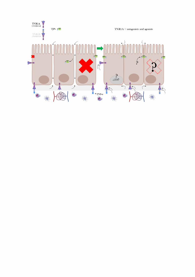
012–033 Cancer Genetics
P-CancG-012
The effect of the translocation t(9;11) on the spatial localization of MLL::MLLT3 genes in AML
R. Bawadi1, S. Käfer1, Y. L. Behrens1, R. Gabdoulline2, D. Heckl3, B. Schlegelberger1, F. Thol2, G. Göhring1
1Hannover Medical School, Department of Human Genetics , Hannover, Deutschland 2Hannover Medical School, Department of Haematology, Haemostasis, Oncology and Stem Cell Transplantation, Hannover, Deutschland 3Martin Luther University Halle-Wittenberg, Pediatric Hematology and Oncology, Halle, Deutschland
Chromosomes occupy a certain volume in the interphase nucleus called chromosome territories. Chromatin methylation that regulates gene expression depends on nuclear positioning. Chromatin located in the inner nuclear compartment is hypo- and in the outer compartment hypermethylated.
The translocation t(9;11)(p22;q23) resulting in the fusion gene KMT2A::MLLT3 is a characteristic aberration of acute myeloid leukemia (AML). We hypothesize that the translocation t(9;11) is causing a change in the location of the genes involved leading to a dysregulation of gene expression.
We aim to investigate whether the fusion gene KMT2A::MLLT3 induces a position change of the KMT2A and MLLT3 genes and identify the associated expression changes. Using 3-dimensional fluorescence in situ hybridization (3D-FISH) we visualized the locations of the KMT2A and MLLT3 genes in four different groups (CD34+ cord blood (CB) cells, normal bone marrow (BM) samples, AML samples with normal karyotype (AML NK), and AML samples with an isolated translocation t(9;11)). The radial and the relative positions of the genes were measured using Imaris software and calculated as described in the literature. Results showed that KMT2A and MLLT3 genes involved in the translocation showed a shift in position compared to control groups towards the outer compartment, which may result in hypermethylation and downregulation of the genes. Preliminary RT-PCR analyses confirmed this hypothesis. However, more analyses as well as a comparison with public expression data are required.
In conclusion, in AML patients with translocation t(9;11) leukemogenesis might not be driven by the fusion gene alone but by an overall change in the nucleus architecture leading to a global expression change.
P-CancG-013
Clonal Evolution at First Sight: a Combined Visualization of Diverse Diagnostic Methods Improves Understanding of Leukemia Progression
Y. L. Behrens1, S. Sandmann2, F. Thol3, M. Heuser3, D. Dörfel4, F. Löhr5, A. Castrup6, D. Steinemann1, J. Varghese2, B. Schlegelberger1, M. Dugas2,7, G. Göhring1
1Hannover Medical School, Department of Human Genetics, Hannover, Deutschland 2University of Münster, Institute of Medical Informatics, Münster, Deutschland 3Hannover Medical School, Department of Hematology, Hemostasis, Oncology and Stem Cell Transplantation, , Hannover, Deutschland 4KRH Klinikum Siloah, Department of Hematology, Oncology and Immunology, Hannover, Deutschland 5Klinikum Braunschweig, Department of Hematology and Oncology, Braunschweig, Deutschland 6Hämato-Onkologische Praxis im Medicum, Bremen, Deutschland 7Heidelberg University Hospital, Institute of Medical Informatics, Heidelberg, Deutschland
Patients with myeloid neoplasia are classified due to the WHO classification systems and besides clinical and hematological criteria, cytogenetic and molecular genetic alterations highly impact treatment stratification. In routine diagnostics, a combination of methods is used to decipher different types of genetic variants, i.e. single nucleotide variants (SNVs), insertions/deletions (indels), structural variants (SVs) and copy number variations (CNVs) which may not be detected using one single method.
We used a bioinformatic approach to analyze clonal evolution and genetic architecture in patients with myeloid neoplasia based on SNVs, indels, SVs and CNVs. Eight patients were comprehensively analyzed using karyotyping, fluorescence in situ hybridization, array-CGH and a custom NGS panel.
Clonal evolution was reconstructed manually, integrating all mutational information on SNVs, indels, SVs and CNVs. Cancer cell fractions (CCFs) for SNVs and indels were estimated based on variante allele frequencies (VAF), assuming heterozygous variants (2*VAF=CCF). CCFs for SVs and CNVs were estimated based on cell counts reported for karyotyping and FISH analyses. For SVs as well as CNVs, which were only detected by array-CGH, CCF was estimated based on logRatio. Altogether, we differentiate between three cases: 1) The CNV occurred prior to the SNV/indel, but in the same cells. 2) The SNV/indel occurred prior to the CNV, but in the same cells. 3) SNV/indel and CNV exist in parallel, independent of each other.The bioinformatic approach reconstructed clonal evolution (linear and/or branching) for all patients and the results were visualized by fishplots. We identified alterations, which play a role in the pathogenesis of the disease (driver) and alterations, which occur during disease development (passenger). On three samples, we showed that reconstruction of clonal evolution is possible even with data from one time point only. For other samples, providing data on more than one time point, the effect of therapy was estimated.
This bioinformatic approach offers the possibility of analyzing clonal evolution and genetic architecture at one or more time points of analysis. The visualization of the results in fishplots contributes to a better understanding of genetic architecture and helps to identify possible targets for the disease (personalized therapy). Furthermore, this model can be used to identify markers in order to assess minimal residual disease (MRD).
P-CancG-014
Germline variants in DNA repair genes including BRCA1/2 cause familial myeloproliferative neoplasms
M. Elbracht1, R. Meyer1, K. Kricheldorf2, D. Gezer2, T. Eggermann1, B. Betz3, I. Kurth1, L. L. Teichmann4, T. H. Brümmendorf2, U. Germing5, S. Isfort2, S. Koschmieder2
1Institute for Human Genetics, Medical Faculty, RWTH Aachen University, Aachen, Deutschland 2Department of Hematology, Oncology, Hemostaseology and Stem Cell Transplantation, Medical Faculty, RWTH Aachen University, Aachen, Deutschland 3Institute for Human Genetics, Medical Faculty and University Hospital Düsseldorf, Heinrich-Heine-University, Düsseldorf, Deutschland 4Department of Hematology and Oncology, University Hospital, Bonn, Deutschland 5Department of Hematology, Oncology and Clinical Immunology, Medical Faculty and University Hospital Düsseldorf, Heinrich-Heine-University, Düsseldorf, Deutschland
The molecular causes of myeloproliferative neoplasms (MPN) are not fully understood. Familial clustering occurs in 7 – 8 % of cases, with relatives of MPN patients exhibiting a 5- to 7-fold MPN risk. Known predisposing germline genetic variations and associated MPN risks include: JAK2 "46/1"" haplotype (up to 4-fold); telomerase reverse transcriptase TERT promoter variant (rs2736100) (2-fold); duplication of ATG2B (autophagy related 2B) and GSKIP (GSK3B interacting protein) as well as pathogenic germline variants in RBBP6 (retinoblastoma binding protein 6). To identify additional predisposing genetic germline variants, we performed whole exome sequencing in five families, each with parent-child or sibling pairs affected by MPN and carrying the somatic JAK2 V617F mutation. In four families, we detected rare germline variants in known tumor predisposition genes of the DNA repair pathway including the highly penetrant BRCA1 and BRCA2 genes as well as heterozygous variants in CHEK2 and ATM. The identification of an underlying hereditary tumor predisposition is of major relevance for the individual patients but also for their families in context of therapeutic options and preventive care. Two patients with essential thrombocythemia (ET) or polycythemia vera (PV) progressed to acute myeloid leukemia (AML) which may suggest a high risk of leukemic transformation in these familial MPNs. In summary, the data suggest that MPNs are more frequently triggered by germline mutations than previously assumed and, conversely, the spectrum of known tumor predisposition syndromes appears to be broader than anticipated (Blood Adv 2021 Sep 14;5(17):3373-3376).
P-CancG-015
*** Comparison of DNA methylation changes in murine and human Burkitt Lymphoma
S. Glaser1, M. Ren2, H. Kretzmer3, S. Sander2, R. Siebert1
1Universitätsklinikum Ulm, Institut für Humangenetik, Ulm, Deutschland 2Deutsches Krebsforschungszentrum, Adaptive Immunity and Lymphoma, Heidelberg, Deutschland 3Max Planck Institute for Molecular Genetics, Department of Genome Regulation, Berlin, Deutschland
Comparison of DNA methylation changes in murine and human Burkitt Lymphomas
Selina Glaser, Meiqi Ren, Helene Kretzmer, Sandrine Sander, Reiner Siebert
Burkitt Lymphoma (BL) is an aggressive B-cell lymphoma representing the most common lymphoma in children. Genetically, BL is characterized by chromosomal translocations bringing the MYC oncogene under the control of the IG enhancers resulting in MYC deregulation. Various OMICs-based analyses on primary human BL samples have recently described the mutational, transcriptomic and epigenomic landscapes of human BL. In parallel, advanced mouse models have been developed for various lymphoma subtypes, including a model which clinically, histologically and transcriptionally mimics human BL (Sander et al. Cancer Cell, 2012). This transgenic BL mouse model expresses MYC and a constitutively active form of PI3K in B-cells undergoing a germinal-center (GC) reaction (Cγ1-cre,R26StopFLMYC,R26StopFLP110*). In the present study we aimed at investigating in how far DNA methylation changes in the mouse BL model resemble the changes in human BL.
We generated DNA methylation profiles of tumor samples (n=8) and corresponding non-neoplastic GC B-cell populations (n=4) of the above mentioned BL mouse model using a custom service for Infinium® Mouse Methylation BeadChip (Illumina Inc., San Diego, CA, USA)). For comparison, we mined previously obtained and public data from human BL (n=70) and normal B-cell populations (n=20) obtained by either Infinium® HumanMethylation450 or Infinium® MethylationEPIC BeadChip analysis (e.g. Kretzmer et al., Nat Genet, 2015). Differentially methylated loci for human and mouse samples were identified using the OMICS Explorer 3.6 (Qlucore; Lund, Sweden). To assign differentially methylated CpGs to chromatin states in B-cells, publically available ChIP-seq data of B-cell populations comprising five (mouse) to six (human) different histone marks (H3K4me3, H3K4me1, H3K36me3, H3K27ac, H3K27me3, and H3K9me3) were used to segment the genomes into a five state model (enhancer, promoter, poised promoter, transcription, heterochromatin) using ChromHMM.
For both mouse and human BL, we identified differentially methylated loci in comparison to the corresponding non-neoplastic GC B-cell controls (σ/σmax ≥ 0.4, q ≤ 0.01; t test). This yielded 43,931 hypo- and 31,823 hypermethylated CpGs in human BL, and 2,142 hypo- and 4,406 hypermethylated CpGs in mouse BL. In both species hypomethylated loci were enriched within heterochromatic regions while hypermethylated loci were enriched within poised promoter regions. Remarkably, we identified a network of 39 genes containing hypermethylated CpGs in both mouse and human, from which half contribute to hub of interacting proteins, many of them being transcription factors.
Thus, by cross-species epigenetic analyses of human and mouse BL we have provided initial evidence for potentially evolutionary conserved mechanisms involved in lymphomagenesis.
P-CancG-016
A novel variant in PRKAR1A at the exon-intron border leads to aberrant splicing in patients affected by Carney complex
M. Grosse1, A. Abicht2, C. Grasemann3, C. Gebhard2, C. Kiewert4, N. Unger5, F. Weber6, F. Kaiser1,7, M. Munteanu1,8
1University Hospital Essen, Institute for Human Genetics, Essen, Deutschland 2Medical Genetics Center, Munich, Deutschland 3Ruhr-University Bochum, Center for Rare Diseases, Bochum, Deutschland 4University Hospital Essen, Department of Pediatrics II, Essen, Deutschland 5University Hospital Essen, 5Department of Endocrinology, Diabetes and Metabolism, Essen, Deutschland 6University Hospital Essen, 6Department of Surgery, Essen, Deutschland 7University Hospital Essen, Essen Centre for Rare Diseases, Essen, Deutschland 8Caritas-Krankenhaus Bad Mergentheim, Pediatric Endocrinology and Diabetology, Bad Mergentheim, Deutschland
Carney complex 1 (CNC, OMIM# 160980) is an autosomal-dominantly inherited complex tumor predisposition syndrome associated with skin pigment abnormalities and neoplasms of heart, endocrine glands and other organs. CNC is caused by distinct heterozygous constitutional loss-of-function variants in the PRKAR1A gene. PRKAR1A codes for the cAMP-dependent protein kinase type I-alpha regulatory subunit, an enzyme that represents an integral part of protein kinase A (PKA) that is involved in many functions in inter- and intracellular signaling. We present a case series of four related female individuals (a mother and her three daughters) carrying the same novel heterozygous intronic PRKAR1A variant NM_002734.4: c.-7_+1del, p.?. The variant was identified by targeted genetic investigation of the youngest daughter (born 2005) after a diagnosis of an ACTH independent cushing's syndrome at the age of 13 in line with bilateral adrenocortical micronodular hyperplasia that was treated successfully by bilateral adrenalectomy. The girl did not show any dermal or other clinical manifestations of CNC. Thorough investigation of the family history revealed that the mother also has a history of cushing"s syndrome but is otherwise affected rather mildly of CNC considering her age of 49. The two other daughters (twins born 2002) do not show any CNC-manifestation. Since the identified variant is located at the first exon-intron border we suspected an effect on splicing. To prove this, RNA from patient's and control blood samples have been investigated. By this, we identified a set of different transcripts, one of which exclusively in patient's RNA. Sequencing analyses of this transcript predicted three putative start codons upstream of the native start codon due to a partial intron retention. To further investigate whether the use of any of these start codons might result in translation of an aberrant protein, the transcript was inserted into a pEGFP-N3 expression vector. After transfection of HEK293 cells EGFP-fusion proteins were visualized by fluorescent microscopy and western blot analyses confirmed the presence of the fusion protein with the predicted mass weight. The aberrant transcript was degraded in patient's blood cells but degradation could be inhibited by puromycin which blocks nonsense-mediated decay. We give functional evidence for the identified non-coding variant to be causative for Carney complex in the affected individuals. The rather mild manifestation in the mother and (so far) incomplete penetrance in the family reflects the underlying pathogenic mechanism of the variant generating at least one aberrant PRKAR1A-transcript resulting in a reduction of the functional gene product.
P-CancG-017
Development of combined somatic copy number alteration and fragmentation analysis for monitoring in colorectal cancer patients using liquid biopsy
A. Hallermayr1,2,3, V. Steinke-Lange1,4, A. Benet-Pagès1, T. Wohlfrom1, F. Scharf1, E. Heitzer5,6,7, C. Haberl8, M. de Wit9,10, H. Vogelsang11, M. Rentsch12,13, E. Holinski-Feder1,4, J. Pickl1,4
1MGZ – Medizinisch Genetisches Zentrum, München, Deutschland 2Pettenkofer School of Public Health, München, Deutschland 3Ludwig-Maximilians-University, Institute for Medical Information Processing, Biometry, and Epidemiology – IBE, München, Deutschland 4Klinikum der Universität München, Medizinische Klinik und Poliklinik IV, Campus Innenstadt, München, Deutschland 5Medical University of Graz, Institute of Human Genetics, Diagnostic and Research Center for Molecular Biomedicine (Austria), Graz, Österreich 6BioTechMed-Graz, Graz, Österreich 7Christian Doppler Laboratory for Liquid Biopsies for Early Detection of Cancer, Graz, Österreich 8Barmherzige Brüder, Klinikum St. Elisabeth, Department of Oncology and Hematology, Straubing, Deutschland 9Vivantes Klinikum Neukoelln, Department of Haematology, Oncology and Palliative Medicine, Berlin, Deutschland 10Vivantes Auguste-Viktoria-Klinikum, Department of Oncology, Berlin, Deutschland 11Klinikum Garmisch-Partenkirchen, Teaching Hospital, Ludwig-Maximilians-University Munich, Department of General, Visceral, Thoracic and Endocrine Surgery, Garmisch-Partenkirchen, Deutschland 12Klinikum Ingolstadt, Department of General, Visceral and Thorax Surgery, Ingolstadt, Deutschland 13Ludwig-Maximilians-University of Munich, Campus Großhadern, Department of General, Visceral, Vascular and Transplant Surgery, University Hospital Munich, München, Deutschland
Liquid biopsy (LB) for non-invasive disease monitoring of cancer patients is progressing towards routine clinical practice. So far the main focus is on circulating tumor DNA (ctDNA) analysis, targeting actionable somatic hotspot variants to support therapy decisions. To expand the advantages of LB to all cancer patients, untargeted approaches are required. Whole-genome sequencing (WGS) of ctDNA provides a promising tool for real-time monitoring of treatment response, as well as early diagnosis.
Combined analysis of multiple features of ctDNA, including somatic copy number alterations (SCNAs), global fragmentation and tissue specific epigenetic signatures, enables the sensitive detection of ctDNA in total circulating free DNA (cfDNA). To identify the optimal sequencing coverage, we performed WGS in a small cohort encompassing nine healthy individuals and two stage IV colorectal cancer patients at baseline. Downsampling of sequencing data was performed to 10x, 7x, 5x, 3x, 2x and 1x coverage. The fraction of ctDNA in total cfDNA was predicted in all control and CRC samples using ichorCNA.
5x coverage was identified to be the minimal required coverage to reliably identify significant differences in global fragmentation profiles between control and CRC patient samples. Further, at the same coverage it was possible to identify SCNAs, and CRC specific epigenetic signatures. Identification of SCNAs improved, when filtering for fragment lengths from 90 to 150 bp, which is more common in tumor derived DNA. Although sensitivity increased with higher coverage, we show that 5x coverage is acceptable for sensitive analysis of ctDNA for routine clinical practice, by keeping costs per sample in the range of targeted hotspot assays (i.e. Droplet Digital PCR).
We developed a cost-effective and sensitive method for untargeted LIquid biopsy Fragmentation, Epigenetic signature and Copy Number Alteration analysis (LIFE-CNA), which may expand the detection of residual disease and recurrence, as well as treatment monitoring to all cancer patients. The cost-effectiveness and sensitivity of our approach represents the basis to enable implementation of LIFE-CNA into routine clinical practice. To demonstrate clinical validity of LIFE-CNA, we investigated a total of ~200 plasma samples from healthy individuals and CRC patients.
P-CancG-018
Further evidence for the association of CHEK2 with susceptibility to Testicular Germ Cell Tumors
K. Kirchner1, F. O. Paulsen2, B. Sievers1, C. Seidel2, D. Lessel1
1Universitätsklinikum Hamburg Eppendorf, Institut für Humangenetik, Hamburg, Deutschland 2Universitätsklinikum Hamburg-Eppendorf, Hamburg, Deutschland
Testicular germ cell tumor (TGCT) is the most frequent malignancy in young man aged 15-45 years. Based on histology, TGCTs are classically divided in seminoma and nonseminoma but current data suggest that all TGCTs likely arise from incompletely differentiated primordial germ cells (PGCs). Regarding the genetic basis, TGCTs are estimated to have the third highest heritability among all malignancies. Intriguingly, despite huge successes of genome-wide association studies (GWASs) that have identified several strong hits in biologically plausible loci, no Mendelian susceptibility genes have been consistently linked to TGCTs so far.
Using different discovery strategies in three independent TGCT cohorts, our group has recently shown that pathogenic/likely pathogenic (P/LP) CHEK2 germline variants are strongly enriched in all three cohorts thereby identifying CHEK2 as the first TGCT susceptibility gene (AlDubayan et al. 2019). Namely, carriers of germline P/LP variants in CHEK2 were 4-6 times more likely to develop TGCTs than unaffected male individuals and, on average, had a 6-year earlier age of presentation than TGCT men with wild-type CHEK2 alleles. Moreover, the low-penetrance CHEK2 variant (p.Ile157Thr) was found to be a Croatian founder TGCT risk variant.
To further validate these findings, 102 unselected men with TGCTs (not specifically selected for early-onset or a positive family history of TGCTs) were ascertained by the Department of Oncology at the University Medical Center Hamburg-Eppendorf. As a control group we used the cancer-free, non-Finnish European, male individuals from the Genome Aggregation Database (GnomAD) cohort (mean n= 29420). Sanger sequencing of all CHEK2 coding exons (NM_007194.3) revealed three heterozygous carriers of one of the P/LP CHEK2 variants (c.1100delC, c.444+1G>A and c.447+2T>G), according to the American College of Medical Genetics and Genomics (ACMG) criteria. Thus, we found that unselected men with TGCTs were approximately 4 times more likely to carry P/LP CHEK2 variants compared with the GnomAD cohort ([OR,] 4.23; 95% CI, 1.39-12.43; P = 0.037; Fisher"s exact). Notably, the observed frequency of P/LP CHEK2 variants of 2.94% in our cohort was strikingly similar to the one we previously observed in unselected TGCT cases of European ancestry (2.9%; AlDubayan et al. 2019). However, no significant association was found between CHEK2 status and further clinical and histopathological characteristics. In addition, we found a significant enriched amount of low-penetrance CHEK2 variant (p.Ile157Thr) with four heterozygous carriers ([OR], 4.22; 95% CI, 1.65-10.91; P = 0.017; Fisher"s exact).
Taken together, we provide further evidence for CHEK2 being the moderate-penetrance TGCT susceptibility gene. In addition, our data suggest that the low-penetrance CHEK2 variant (p.Ile157Thr) leads to increased TGCT risk beyond the Croatian population.
P-CancG-019
Germline duplications of the MET gene and papillary renal cancer
A. Gehrig1, E. Kunstmann1, E. Gerhard-Hartmann2
1Institut für Humangenetik, Würzburg, Deutschland 2Institut für Pathologie, Würzburg, Deutschland
Renal cancer is one of the 10 most common cancers in the Western population. Activating alterations in the MET gene are well known somatic drivers of tumorigenesis, whereas hereditary MET gene variations are rare. Typically, germline mutations manifest as multifocal bilateral papillary renal carcinoma. To date, only activating missense mutations have been characterized as causative germline variants.
Here we present a family with three first grade relatives affected from renal cancer. The index patient suffered from bilateral multifocal papillary renal cancer. Next generation sequencing was performed with TruSight Hereditary Cancer Panel (Illumina), the data was analyzed with GensearchNGS software (PhenoSystems). CNV analysis revealed a duplication of the entire MET gene. Duplications of the MET gene as germline variants are of unknown significance to date.
Data of the family and of MET-amplification in tumor tissue samples and control tissue will be shown. We will discuss whether duplications of the MET gene cause a hereditary tumor syndrome.
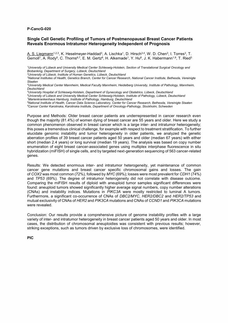
P-CancG-020
Single Cell Genetic Profiling of Tumors of Postmenopausal Breast Cancer Patients Reveals Enormous Intratumor Heterogeneity Independent of Prognosis
A. S. Liegmann1,2,3, K. Heselmeyer-Haddad3, A. Lischka1, D. Hirsch3,4, W. D. Chen3, I. Torres3, T. Gemoll1, A. Rody5, C. Thorns6,7, E. M. Gertz8, H. Alkemade1, Y. Hu8, J. K. Habermann1,9, T. Ried3
1University of Lübeck and University Medical Center Schleswig-Holstein, Section of Translational Surgical Oncology and Biobanking, Department of Surgery, Lübeck, Deutschland 2University of Lübeck, Institute of Human Genetics, Lübeck, Deutschland 3National Institutes of Health, Genetics Branch, Center for Cancer Research, National Cancer Institute, Bethesda, Vereinigte Staaten 4University Medical Center Mannheim, Medical Faculty Mannheim, Heidelberg University, Institute of Pathology, Mannheim, Deutschland 5University Hospital of Schleswig-Holstein, Department of Gynecology and Obstetrics, Lübeck, Deutschland 6University of Lübeck and University Medical Center Schleswig-Holstein, Institute of Pathology, Lübeck, Deutschland 7Marienkrankenhaus Hamburg, Institute of Pathology, Hamburg, Deutschland 8National Institute of Health, Cancer Data Science Laboratory, Center for Cancer Research, Bethesda, Vereinigte Staaten 9Cancer Center Karolinska, Karolinska Institute, Department of Oncology-Pathology, Stockholm, Schweden
Purpose and Methods: Older breast cancer patients are underrepresented in cancer research even though the majority (81.4%) of women dying of breast cancer are 55 years and older. Here we study a common phenomenon observed in breast cancer which is a large inter- and intratumor heterogeneity; this poses a tremendous clinical challenge, for example with respect to treatment stratification. To further elucidate genomic instability and tumor heterogeneity in older patients, we analyzed the genetic aberration profiles of 39 breast cancer patients aged 50 years and older (median 67 years) with either short (median 2.4 years) or long survival (median 19 years). The analysis was based on copy number enumeration of eight breast cancer-associated genes using multiplex interphase fluorescence in situ hybridization (miFISH) of single cells, and by targeted next-generation sequencing of 563 cancer-related genes.
Results: We detected enormous inter- and intratumor heterogeneity, yet maintenance of common cancer gene mutations and breast cancer specific chromosomal gains and losses. The gain of COX2 was most common (72%), followed by MYC (69%); losses were most prevalent for CDH1 (74%) and TP53 (69%). The degree of intratumor heterogeneity did not correlate with disease outcome. Comparing the miFISH results of diploid with aneuploid tumor samples significant differences were found: aneuploid tumors showed significantly higher average signal numbers, copy number alterations (CNAs) and instability indices. Mutations in PIKC3A were mostly restricted to luminal A tumors. Furthermore, a significant co-occurrence of CNAs of DBC2/MYC, HER2/DBC2 and HER2/TP53 and mutual exclusivity of CNAs of HER2 and PIK3CA mutations and CNAs of CCND1 and PIK3CA mutations were revealed.
Conclusion: Our results provide a comprehensive picture of genome instability profiles with a large variety of inter- and intratumor heterogeneity in breast cancer patients aged 50 years and older. In most cases, the distribution of chromosomal aneuploidies was consistent with previous results; however, striking exceptions, such as tumors driven by exclusive loss of chromosomes, were identified.
PIC
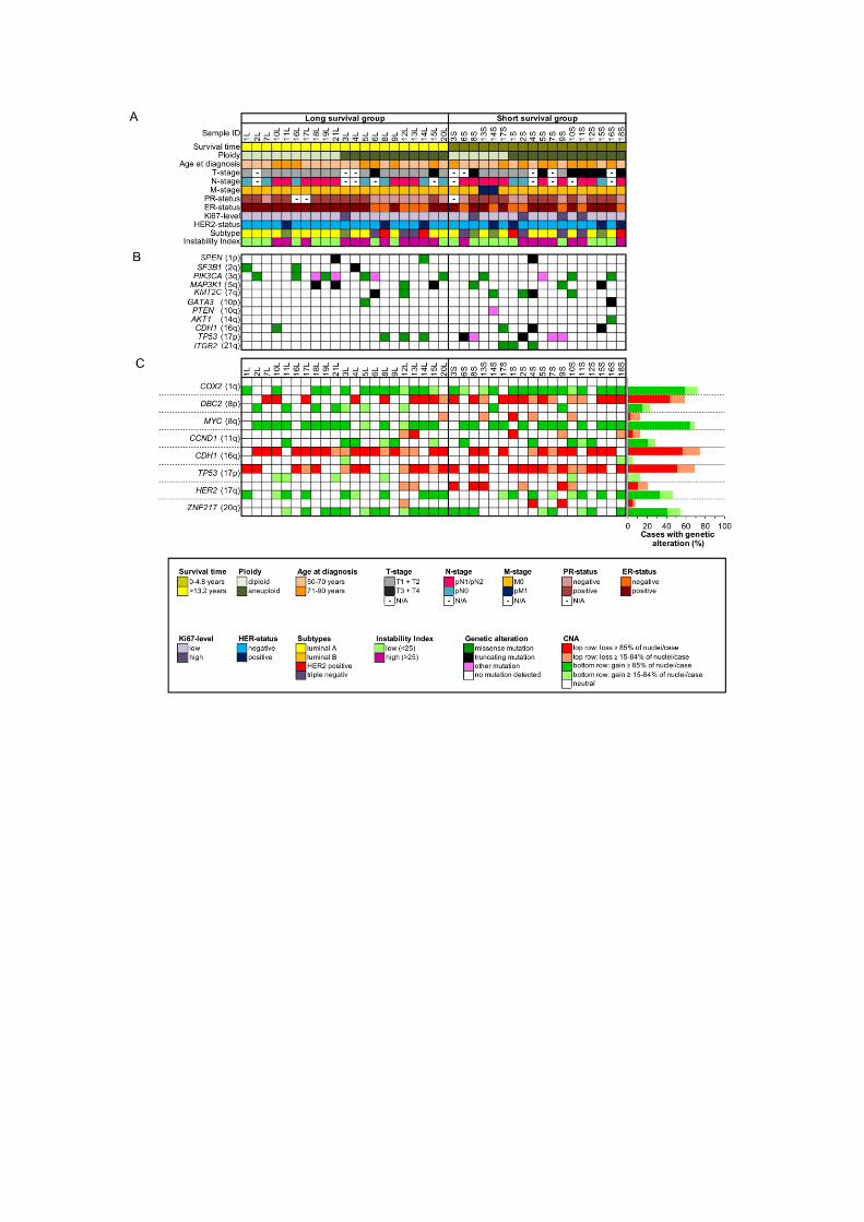

P-CancG-021
Identification of RAG1/RAG2- and AID-mediated recombination events in acute B-lymphoblastic leukemia with t(5;14)(q31.1;q32.3)/IL3-IGH-fusion
J. D. Marx1, S. Balachandran1, L. J. Russell2, I. Nagel3, C. Becher3, K. Nebral4, R. Panzer-Grümayer4, N. Schub5, M. Gramatzki5, A. Caliebe3, M. Spielmann1, E. M. Murga Penas1
1Institute for Human Genetics, University Hospital Schleswig-Holstein/ University of Luebeck, Lübeck, Deutschland 2Translational and Clincal Research Institute, Newcastle University, Newcastle-upon-Tyne, Vereinigtes Königreich 3Institute for Human Genetics, University Hospital Schleswig-Holstein/Christian-Albrechts University Kiel, Kiel, Deutschland 4St. Anna Children´s Cancer Research Institute, Vienna, Österreich 5Division of Stem Cell Transplantation and Immunotheraphy, 2nd Medical Department, University Hospital Schleswig-Holstein, Campus Kiel, Kiel, Deutschland
Introduction: Knowledge of the mechanisms that lead to translocations in hematological malignancies provides essential insights into the development of these diseases. In IGH-associated translocations, a common pathomechanism for the generation of Double-Strand-Breaks (DSBs) seems to be mediated by the RAG1/RAG2 complex at the IGH-locus. At the IGH-translocation partners, a second mechanism based on the activity of the AID, causing a T:G mismatch that is recognized by RAG1/RAG2, has been proposed. Further non-IGH-associated translocations as the t(12;21)/ETV6-RUNX1 are suggested to be caused by a RAG1/RAG2-mediated mechanism. For the rare B-lymphoblastic leukemia/lymphoma with t(5;14)(q31.1;q32.3)/IL3-IGH-fusion that is recognized as full entity in the WHO-Classification, the mechanisms are unknown so far. We report a molecular genetic analysis of two new cases of B-lymphoblastic leukemia (B-ALL) with t(5;14)(q31;q32)/IL3-IGH-fusion.
Methods: DNA from two cases with the diagnosis of a B-ALL was available for molecular analyses. The presence of the t(5;14) was confirmed by FISH with specific probes for the IL3 gene and IGH locus. Whole genome sequencing (WGS) was performed with NovaSeq6000. Confirmation of the WGS results by amplification and Sanger-sequencing of the direct IL3-JH IGH segments was possible in one case.
Results: WGS and direct amplification of the IL3-IGH fusion in case 1 and WGS in case 2 allowed mapping of the breaks on chromosome 5 to the 5`-non-coding region of IL3. The breakpoint region of both cases spanned about 1200 nucleotides and showed an increased GC-content and a high number of CpG dinucleotides, which were significantly clustered to the breakpoint region. This pointed to an involvement of the AID and RAG1/RAG2 activity in the generation of the DSBs. The breaks on chromosome 14 in case 1 showed all features of an illegitimate V(D)J recombination, like the localization in the JH4 and DH1-26 segments and the presence of recombination signal sequences (RSS). In case 2, the localization of the breakpoints in the constant region of IGH elicited the mediation of an illegitimate V(D)J recombination as the pathomechanism involved. However, the flanking sequences at the breakpoints showed features of cryptic RSS (cRSS) that might likely function as a target for RAG1/RAG2.
Conclusions: We present here the first characterization of the pathomechanisms leading to the t(5;14)/IL3-IGH-fusion in B-ALL. Our observations indicate that the DSBs at the IL3 locus on chromosome 5 are probably mediated by the activity of AID, leading to a T:G mismatch and followed by RAG1/RAG2 activity. The DSBs on chromosome 14 are suspected to be caused by RAG1/RAG2 activity either, as expected, by an illegitimate V(D)J recombination or at cRSS. Therefore, the activity of RAG1/RAG2 is likely the major driver for the development of the translocation in t(5;14)/IL3-IGH-fusion cases analyzed here.
P-CancG-022
Who am I? Donor DNA in Patients after Stem Cell Transplantation
M. Mertens1, M. Sadlo1, J. Hentschel1
1Universitätsklinikum Leipzig, Leipzig, Deutschland
Background: Several diseases like leukemia or metabolic disorders of the hematopoietic system require a stem cell transplantation. In Germany, 3,500 persons per year receive a stem cell transplantation to restore bone marrow function, resulting in a chimerism. 10 % are children <18 years. In general, indications for genetic diagnostics have increased exponentially in recent years also for patients after stem cell transplantation. Due to the genetic chimerism germline testing based on blood samples cannot be done in transplanted patients. The presence of donor DNA in buccal swaps and nails of stem cell recipients has previously been documented (Thiede et al. 2000; Imanishi et al. 2007). Crain et al. 2005 even detected donor-derived chimerism in brain cells. Yet, the literature lacks a statistical sound assessment of quantitative extend of donor DNA in various tissue types. Therefore, it is not surprising that recommendations for germline genetic testing after stem cell transplantation are still missing. Our study evaluates the best procedure to enable a molecular genetic diagnostic for patients after stem cell transplantation.
Methods: We performed a pilot trial, which included seven patients. Six of them received an allogeneic stem cell transplant and one an autologous stem cell transplant and was therefore used as a control. We isolated DNA from buccal swabs and nails. DNA isolates from blood, which were collected before transplantation were used as reference material for the recipients.
We analyzed the DNA samples with two different approaches: In one, we used a short tandem repeat (STR) assay containing 28 STR markers (custom-made) followed by capillary electrophoresis to detect donor derived DNA. In the other, we used a NGS-RC-PCR-based SNP-Assay including 34 Loci (Nimagen). To calculate the proportion of donor and recipient DNA, we compared the genetic profiles in the different materials with the sample collected before transplantation.
Results: We successfully extracted DNA from all tissues, but one. Our pilot study showed that DNA of the stem cell donor could be detected in both buccal swaps and nail samples from the recipient after stem cell transplantation. The detected donor DNA proportion varies between 11 % and 39 % and between 11 % and 44 % in the buccal swaps and nail samples, respectively.
Conclusion and outlook: The study confirms the presence of donor DNA in buccal swaps and documents a significant donor DNA percentage in nail samples of the recipient after allogenic stem cell transplantation. Currently, we extent the study to additional patients to derive more robust conclusions regarding the DNA proportion of donor signatures in recipient's materials. We will analyze additional tissue types like follicular hair cells and investigate how the donor DNA proportion varies over time after transplantation to provide guidance for germline testing after stem cell transplantation.
P-CancG-023
Tumor predisposition in young head and neck cancer patients
R. Meyer1,2,3, F. Kraft1, D. Dey1, M. Bourgeois2,3, M. Klein4, M. Ooms4, P. Winnand4, B. J. Siegmund4, F. Hölzle4, S. Hackenberg5, M. Elbracht1, I. Kurth1, F. Beier2,3, T. Brümmendorf2,3, M. Tometten2,3
1Insititute of Human Genetics, Medical Faculty, RWTH Aachen University, Aachen, Deutschland 2Department of Hematology, Oncology, Hemostaseology and Stem Cell Transplantation, Medical Faculty, RWTH Aachen University, Aachen, Deutschland 3Center for Integrated Oncology Aachen Bonn Cologne Düsseldorf (CIO ABCD), Aachen, Deutschland 4Department of Oral, Maxillofacial and Facial Plastic Surgery, Medical Faculty, RWTH Aachen University, Aachen, Deutschland 5Department of Otolaryngology, Head and Neck Surgery, Medical Faculty, RWTH Aachen University, Aachen, Deutschland
A considerable and so far underestimated proportion of all cancers is caused by genetic predisposition. Recent studies suggest that at least about 13% of all malignancies are caused by an underlying genetic predisposing factor. This proportion is even higher in certain patient groups, e. g. in patients with a family history of malignant disease or a young age of onset of cancer.
In 2016, about 14,000 people in Germany developed squamous cell carcinoma of the head and neck (HNSCC) mainly affecting males with a median age of onset of 63 years and in association with typical extrinsic risk factors: tobacco smoking, alcohol consumption, and HPV-infection. A small subset of patients however develops HNSCC at a young age <50 years, often without identifiable extrinsic risk factors. In few cases, the detection of a genetic tumor predisposition in young HNSCC patients has been reported, however, comprehensive studies regarding a germline tumor predisposition in a larger number of patients of this unique cohort were missing.
Since significantly increased risks for the development of HNSCC in the context of telomere biology disorders (TBD) had already been reported, samples from young patients with head and neck cancer were included in the Aachen telomeropathy registry (ATR). TBD are characterized by impaired telomere maintenance leading to bone marrow failure, organ fibrosis, and high cancer risks. Using whole exome sequencing (WES) we analyzed genomic information of 37 patients who were younger than 50 years at initial HNSCC diagnosis and were included in the ATR.
In this study, we aimed to assess the prevalence of tumor-predisposing genetic alterations providing novel insights into mechanistic pathways of tumorigenesis of HNSCC. In individual cases, the results provide important information for a personalized clinical management as well as the basis for an evidence-based genetic counseling.
Funded under the Excellence Strategy of the Federal Government and the Länder.
P-CancG-024
*** T-cell prolymphocytic leukemia is associated with deregulation of oncogenic microRNAs on transcriptional and epigenetic level
P. Patil1, S. Hillebrecht1, E. Chteinberg1, C. López2,1,3, U. H. Toprak4,5,6,7, J. Seufert4,7, S. H. Bernhart8, H. Kretzmer8, A. K. Bergmann2,9, S. Bens2,1, J. Högel1, A. Scheffold10, B. M. Chelliah Jebaraj10, A. Schrader11, P. Johansson12, D. Costa3, M. Schlesner13,7, J. Dürig14, M. Herling11,15, E. Campo3, S. Stilgenbauer10, L. Wiehle1, R. Siebert2,1
1Institute of Human Genetics, Ulm University and Ulm University Medical Center, Ulm, Deutschland 2Institute for Human Genetics, Christian-Albrechts-University Kiel and University Hospital Schleswig-Holstein, Kiel, Deutschland 3Haematopathology Section, Hospital Clínic, Institut d'Investigaciones Biomèdiques August Pi I Sunyer (IDIBAPS), Barcelona, Spanien 4Faculty of Biosciences, Heidelberg University, Heidelberg, Deutschland 5Division Neuroblastoma Genomics, German Cancer Research Center (DKFZ), Heidelberg, Deutschland 6Hopp-Children’s Cancer Center , at the NCT Heidelberg (KiTZ), Heidelberg, Deutschland 7Bioinformatics and Omics Data Analytics, German Cancer Research Center (DKFZ), Heidelberg, Deutschland 8Interdisciplinary Center for Bioinformatics, University of Leipzig, Leipzig, Deutschland 9Institute for Human Genetics, Hannover Medical School, Hannover, Deutschland 10Department of Internal Medicine III, University of Ulm, Ulm, Deutschland 11Center for Integrated Oncology Köln Bonn, Internal Medicine, University of Cologne, Cologne, Deutschland 12Institute for Cell Biology , University of Duisburg-Essen, Essen, Deutschland 13Biomedical Informatics, Augsburg University, Augsburg, Deutschland 14University Hospital Essen, Department of Hematology, Essen, Deutschland 15Cellular Therapy and Hemostaseology, University of Leipzig, Leipzig, Deutschland
Deregulation of micro(mi)-RNAs is considered as a common mechanism involved in tumorigenesis. Both oncogenic and tumor suppressive roles of miRNAs have been reported in mature B and T-cell malignancies. T-cell prolymphocytic leukemia (T-PLL) is an infrequent but well defined T-cell malignancy with a genetic hallmark being inv(14), t(14;14). or t(X;14). In recent years, transcriptomic studies in T-PLL have revealed the deregulation of many genes on mRNA level in T-PLL. Contrastingly, studies on the non-coding transcriptional landscape in T-PLL are rather limited. Recently, miRNA expression studies in T-PLL were carried out using sequencing and array-based techniques in T-PLL. Herein, we investigated the expression of 2083 miRNAs in T-PLL using a relatively novel approach for studying miRNA expression i.e. a ribonuclease protection-based assay.
We detected 111 miRNAs which were differentially expressed in T-PLL as compared to physiologic CD4+ and CD8+ T-cell subsets. Of those, 27 miRNAs were downregulated and 84 miRNAs upregulated in T-PLL compared to healthy controls. In addition, 33 of the 111 differentially expressed miRNAs belonged to oncogenic miRNA gene clusters. Integrated genetic analyses revealed that the differentially expressed miRNAs are rarely affected by genomic structural variants, single nucleotide variants or indels but with a noteworthy exception of copy number aberrations. Remarkably, we found strong upregulation of the miR-200c/-141 cluster in T-PLL to be associated with DNA hypomethylation and active promoter marks which corroborated with the previous studies on miRNAs in T-PLL. Contrastingly, we observed downregulation of miR-618 associated with the DNA hypermethylation in T-PLL. In line with findings in T-cell leukemias, we detected upregulation of oncogenic clusters miR-106a/-363 and miR-106b-25/miR-17-92 in T-PLL.
Using a miRNome-wide approach orthogonal to RNA-seq our study gives a concise set of significantly deregulated miRNAs and miRNA clusters which might play an oncogenic role and might be used as diagnostic biomarkers in T-PLL. Our study also identifies significant similarities in miRNA profiles in T-PLL with previously published studies. In addition, it corroborates the significance of miRNA deregulation on epigenomic level and indicates an interplay of genomic and epigenomic factors playing a possible regulatory role in miRNA deregulation in T-PLL.
P-CancG-025
Whole-exome sequencing in eccrine porocarcinoma indicates promising therapeutic strategies
E. Denisova1, D. Westphal2, H. Surowy3, F. Meier2, B. Hutter1, J. Reifenberger4, A. Rütten5, A. Schulz2, M. Sergon6, M. Ziemer7, B. Brors1, R. Betz8, S. Redler3
1German Cancer Research Center (DKFZ), Division of Applied Bioinformatics, Heidelberg, Deutschland 2Carl Gustav Carus Medical Center, TU Dresden, Department of Dermatology, Dresden, Deutschland 3Medical Faculty and University Hospital Düsseldorf, Heinrich-Heine-University Düsseldorf,, Institute of Human Genetics, Düsseldorf, Deutschland 4University Hospital Düsseldorf, Department of Dermatology, Düsseldorf, Deutschland 5Dermatopathology, Bodensee, Friedrichshafen, Deutschland 6Carl Gustav Carus Medical Center, TU Dresden, Institute of Pathology, Dresden, Deutschland 7University Medical Center, Leipzig, Department of Dermatology, Venereology and Allergology, Leipzig, Deutschland 8University of Bonn, Medical Faculty & University Hospital Bonn, Institute of Human Genetics, Bonn, Deutschland
Background: Malignant sweat gland tumours are rare, and research suggests that they arise from the intraepidermal ductal portion/acrosyringium of the cutaneous sweat glands. Eccrine porocarcinoma (EP) is the most common form of sweat gland malignancy. The aetiology of EP is largely unknown. However, research has implicated somatic mutations. The current lack of data on somatic mutation patterns in EP hampers the development of effective therapies.
Objective: To investigate the mutational landscape of EP using Whole exome sequencing (WES) and formalin-fixed paraffin-embedded samples of matched primary EP and healthy surrounding tissue.
Methods: Tissue samples were obtained from 14 well characterised EP patients from Central Europe. WES was performed using the Agilent SureSelect XT Human All Exon V7 Enrichment Kit (Agilent). Paired-end 2x100 bp DNA sequencing was performed on a HiSeq 4000 system (Illumina).
Results: Mutational profiling revealed a high overall median mutation rate. This was attributed to signatures of mutational processes related to ultraviolet (UV) exposure, APOBEC enzyme dysregulation, and defective homologous double strand break repair. All of these processes cause genomic instability and are implicated in carcinogenesis. Recurrent driving somatic alterations were detected in the EP candidate drivers TP53, FAT2, CACNA1S, and KMT2D. The analyses also identified copy number alterations and recurrent gains and losses in several chromosomal regions including that containing BRCA2, as well as deleterious alterations in multiple HRR components. In accordance with this reduced or even a complete loss of BRCA2 protein expression was detected in 50% of the investigated EP tumours.
Conclusions: Our results implicate crucial oncogenic driver pathways, and suggest that defective homologous double strand break repair and the p53 pathway are involved in EP aetiology. Targeting of the p53 axis and PARP inhibition, and/or immunotherapy, may represent promising treatment strategies
P-CancG-026
Ago-RIP Sequencing Identifies New MicroRNA-449a-5p Target Genes Increasing Sorafenib Efficacy in Hepatocellular Carcinoma
T. Reinkens1, A. Stalke1, N. Huge1, B. Vajen1, M. Eilers1, V. Schäffer1, O. Dittrich-Breiholz1, B. Schlegelberger1, T. Illig1, B. Skawran1
1Medizinische Hochschule Hannover, Institut für humangenetik, Hannover, Deutschland
BACKGROUND:
Patients with hepatocellular carcinoma (HCC) have very limited treatment options. For the last fourteen years, the multi-tyrosine kinase inhibitor sorafenib has been used as standard-of-care therapeutic agent in advanced HCC. Unfortunately, drug resistance develops in many cases. Therefore, we aimed to find a way to mitigate drug resistance and to improve the sorafenib efficacy in HCC cells. MicroRNAs play a significant role in targeting genes involved in tumor control suggesting microRNA/sorafenib combination therapy as a promising treatment option in advanced HCC.
METHODS:
MiR-449a-5p target genes were identified by Ago-RIP sequencing and validated by luciferase reporter assays and expression analyses. Target gene expression and survival data were analyzed in public HCC datasets. Tumor-relevant functional effects of miR-449a-5p and its target genes as well as their impact on the effects of sorafenib were analyzed using in vitro assays. An indirect transwell co-culture system was used to survey anti-angiogenic effects of miR-449a-5p.
RESULTS:
PEA15, PPP1CA and TUFT1 were identified as direct target genes of miR-449a-5p. Overexpression of these genes correlated with a poor outcome of HCC patients. Transfection with miR-449a-5p and repression of miR-449a-5p target genes inhibited cell proliferation and angiogenesis, induced apoptosis and reduced AKT and ERK signaling in HLE and Huh7 cells. Importantly, miR-449a-5p potentiated the efficacy of sorafenib in HCC cells via downregulation of PEA15, PPP1CA and TUFT1.
CONCLUSIONS:
This study provides detailed insights into the targetome and regulatory network of miR-449a-5p. Our results demonstrate for the first time that targeting PEA15, PPP1CA and TUFT1 via miR-449a overexpression could have significant implications in counteracting sorafenib resistance suggesting miR-449a-5p as a promising candidate for a microRNA/sorafenib combination therapy.
PIC

P-CancG-027
Identification and characterisation of BMPR1A and SMAD4 germline variants in patients with colorectal adenomatous polyposis
A. K. Sommer1, I. B. A. te Paske2, J. Garcia-Pelaez3, L. Matalonga4, S. Peters1, V. Steinke-Lange5,6, E. Holinski-Feder5,6, C. Oliveira3, N. Hoogerbrugge2, R. M. de Voer2, I. Spier1,7, S. Aretz1,7
1Institute of Human Genetics, Medical Faculty, University of Bonn, Bonn, Deutschland 2Department of Human Genetics, Radboud university medical center, Radboud Institute for Molecular Life Sciences, Nijmegen, Niederlande 3I3S, Instituto de Investigação e Inovação em Saúde, University of Porto, Porto, Portugal; Ipatimup, Institute of Molecular Pathology and Immunology of the University of Porto; Faculty of Medicine, University of Porto, Porto, Portugal 4CNAG-CRG, Centre for Genomic Regulation, Barcelona Institute of Science and Technology, Barcelona, Spanien 5MGZ - Medizinisch Genetisches Zentrum, Munich, Deutschland 6Campus Innenstadt, Klinikum der Universität München, Munich, Deutschland 7Center for Hereditary Tumor Syndromes, University of Bonn, Bonn, Deutschland
Background. In up to 50% of patients with a suspected hereditary gastrointestinal polyposis syndrome, no genetic cause can be uncovered in routine diagnostics. The European Reference Network for Genetic Tumour Risk Syndromes (ERN GENTURIS) aims at identifying novel causal germline variants for unexplained colorectal polyposis in a large multicenter cohort. Therefore, whole exome sequencing (WES) data from patients with negative results in routine diagnostics are reanalysed within the framework of the European Solve-RD project.
Methods. Germline WES data from 211 unrelated patients with adenomatous or serrated polyposis from multiple centres in the Netherlands and Germany were re-evaluated in a combined analysis. A predefined list of 229 established and proposed cancer predisposing genes was used to screen for the presence of (likely) pathogenic variants. Only rare variants meeting specified quality criteria were selected.
Results. Among 14 (likely) causative rare alterations in the cohort, we identified one truncating, pathogenic variant in SMAD4, and three variants in BMPR1A, two of which are likely pathogenic due to aberrant splicing. All four variants are absent or very rare in population databases. The functional characterisation of the variants is ongoing. Surprisingly, the variants were found in four unrelated patients with an adenomatous polyposis phenotype. Histologic reclassification by an experienced gastrointestinal pathologist confirmed the adenomas in the three BMPR1A variant carriers, while the polyps in the SMAD4 variant carrier could be classified as likely juvenile just when being aware of the affected gene. The potential splice variant carriers both have a family history of colorectal polyposis and the variants segregate in affected family members.
Discussion. Pathogenic germline variants in the BMPR1A and SMAD4 genes are expected to cause juvenile polyposis and thus, both genes have not been considered in routine diagnostics of adenomatous polyposis patients in the past, although it is known that juvenile polyps are prone to misclassification, since they might resemble hyperplastic, inflammatory, or adenomatous polyps. In casuistic reports, single pathogenic BMPR1A variants were described as underlying cause of a mixed or untypical colorectal polyposis. The herein reported genotype-phenotype discrepancy supports a broader disease spectrum in BMPR1A and SMAD4 germline variant carriers and challenges to correctly classify juvenile polyps. Given the until recently not appreciated phenotypic variability, germline mutation screening in patients with a suspected adenomatous polyposis should be extended by other polyposis genes.
Solve-RD received funding from EU Horizon 2020 (No.779257). All authors are member of ERN GENTURIS and the Solve-RD consortium.
P-CancG-028
Development of APC-specific ACMG/AMP variant classification guidelines
I. Spier1,2, X. S. Yin3,4, M. Richardson5, M. Pineda6, D. Ritter7,8, T. Pesaran5, G. Capellá6, S. V. Tavtigian9, J. P. Plazzer3, A. Laner10, A. Latchford11, I. M. Frayling12, S. Plon7,8, M. Greenblatt13, F. A. Macrae3,14, S. Aretz1,2
1Medical Faculty, University of Bonn, Institute of Human Genetics, Bonn, Deutschland 2University Hospital Bonn, National Center for Hereditary Tumor Syndromes, Bonn, Deutschland 3Royal Melbourne Hospital, Department of Colorectal Medicine and Genetics, Parkville, Australien 4University of Melbourne, Melbourne Medical School, Parkville, Australien 5Ambry Genetics, Aliso Viejo, California, Vereinigte Staaten 6Hereditary Cancer Program, Institut Català d’Oncologia – IDIBELL, Barcelona, Spanien 7Baylor College of Medicine, Houston, Texas, Vereinigte Staaten 8Baylor College of Medicine, Texas Children's Cancer Center, Houston, Texas, Vereinigte Staaten 9University of Utah School of Medicine, Department of Oncological Sciences and Huntsman Cancer Institute, Salt Lake City, Utah, Vereinigte Staaten 10Medical Genetics Center Munich, Department of Genomics, Munich, Deutschland 11St Mark’s Hospital, Polyposis Registry, London, Vereinigtes Königreich 12University Hospital of Wales, Institute of Medical Genetics, Cardiff, Vereinigtes Königreich 13University of Vermont College of Medicine, Vermont Cancer Center, Burlington, Vermont, Vereinigte Staaten 14University of Melbourne, Department of Medicine, Parkville, Australien
Introduction: The proper characterisation of the clinical significance of germline variants is of high relevance to translate genetic testing results into medical practice and thereby improve patient care. Pathogenic APC variants are causative for Familial adenomatous polyposis (FAP), a colorectal cancer predisposition syndrome. During the last decades, thousands of rare APC germline variants have been identified in patients with FAP and listed in phenotype-based locus-specific databases (LSDBs). In parallel, the application of high-throughput techniques in patients with non-related phenotypes and healthy individuals generates an additional plethora of variants in public data aggregation resources such as ClinVar, a large number of which are variants of uncertain clinical significance (VUS) or variants with conflicting assertions (54% of the ~9000 APC variants in ClinVar are VUS). This project aims to improve variant classification by the development of APC-specific classification criteria within the approval process of a Hereditary Colon Cancer / Polyposis Variant Curation Expert Panel (VCEP) from the International Society for Gastrointestinal Hereditary Tumours (InSiGHT) and ClinGen (https://clinicalgenome.org/affiliation/50099/). Methods: APC-specific adaptations of the interpretation guidelines published by the American College of Medical Genetics and the Association of Molecular Pathology (ACMG/AMP) were discussed in video conferences of the APC-subgroup of the VCEP based on database analyses, literature search and expert opinions. Results: Three of the 28 original criteria were left unchanged (BS4, BP1, BP5), whilst eight were not used for different reasons (PM1, PM3, PM4, PP2, PP4, PP5, BP3, BP6). For the remaining 17 criteria, gene- or disease-based specifications and/or evidence strength modifications were made. The main changes concern the "pathogenic very strong" (PVS1) criterion (e. g. specifications at the 5" and 3" end of the gene and specifications for splice variants), and modifications of the allele frequencies of the "pathogenic moderate" criterion PM2 (≤ 0.0008%), the "stand alone benign" criterion BA1 (≥ 0.1%) and the "benign strong" criterion BS1 (≥ 0.001%). Moreover, a point system for the phenotypic description of variant carriers was developed. The next steps will be the submission to the Sequence Variant Interpretation (SVI) working group from ClinGen and the validation of the APC-specific modifications by a systematic evaluation of previously classified variants. Conclusions: It is expected that using the APC-specific guidelines will improve variant interpretation and facilitates resolution of variants with conflicted assertions in ClinVar. Based on the adapted allele frequency thresholds, it is likely that in particular a considerable portion of VUS can be reclassified as (likely) benign. The reclassifications will be publicly available through both ClinGen, ClinVar and the InSiGHT APC LSDB (www.lovd.nl/APC).
P-CancG-029
Phenotypic analysis of 106 serrated polyposis patients
V. Steinke-Lange1,2, M. Morak1,2, A. Lausch2, E. Holinski-Feder1,2
1MGZ - Medizinisch Genetisches Zentrum, München, Deutschland 2Medizinische Klinik und Poliklinik IV – Campus Innenstadt, Klinikum der Universität München, Arbeitsgruppe erbliche gastrointestinale Tumore, München, Deutschland
Introduction: Genetic and/or non-genetic causes of serrated polyposis syndrome (SPS) are widely unknown. SPS patients show a broad phenotypic spectrum regarding polyp burden and age of onset and might therefore belong to different entities. We therefore collected phenotypic data of 106 SPS patients to identify phenotypic subgroups.
Patients and methods: 70 female (66 %) and 36 male patients were enrolled, 53 patients (50 %) fulfilled the WHO criteria for SPS. We calculated the annual gain of serrated polyps and compared the results depending on different parameters using Mann-Whitney-U tests.
Results: Female patients developed significantly more sessile-serrated lesions per year than male patients (U=885.500; p=0.012). This also applied for sessile-serrated lesions of the proximal colon (U=733.500; p=0.0004), but not for the distal colon (U=1158.00; p=0.474). Regarding hyperplastic polyps, there was no significant difference. Smoking and BMI also had no significant influence on serrated polyp burden in our cohort. Control colocnoscopy 1-2 years after initial diagnosis revealed polyps in 70%. We were able to figure out two clinical subgroups: while one part of the patients was continuously prone to serrated polyps, the other patients only showed a temporary polyposis with several inconspicuous colonoscopies afterwards. Hypothetically, the first could be due to a genetic predisposition, while the other might mainly be exogenic. Only two patients developed colorectal cancer during surveillance, each after 4-5 years without colonoscopy.
Conclusions: SPS patients may either have a temporary or permanent risk for serrated polyps and colorectal cancer. Therefore, close meshed surveillance by colonoscopy especially following initial diagnosis is necessary.
P-CancG-030
Optical Genome Mapping and Long-Read Sequencing-based Insights into Human Haematological Malignancies
H. Surowy1, D. Falkenstein1, B. Hildebrandt1, F. Sundermann1, D. Wieczorek1, H. J. Luedecke1
1Institute of Human Genetics, Medical Faculty and University Hospital Düsseldorf, Heinrich Heine University Düsseldorf, Düsseldorf, Deutschland
INTRODUCTION: The genetic architecture of malignancies has predominantly been assessed with conventional short-read next-generation sequencing (NGS) approaches. These can identify point mutations with high accuracy, but possess a reduced sensitivity towards structural genetic variations (SVs). In contrast, newer technologies based on the analysis of long contiguous DNA fragments excel in the identification of small and large copy-number changes (CNVs) or copy-neutral SVs that may strongly influence tumor evolution, and potentially supplement existing cytogenetic approaches. METHODS: We investigated the performance of optical genome mapping (OGM, Bionano) in comparison to tumor-cytogenetic analyses in three probands with hematological malignancies. The analyses were supplemented by long-read sequencing (LRS, PacBio CLR, 50x depth). Bioinformatic analyses were done using proprietary software (Bionano) or workflows (PacBio) supplied by the respective manufacturers. RESULTS: In a case with acute myeloid leukemia (AML), a cytogenetically identified translocation with high clonality (46,XX,t(3;21)(q26;q22)[24]/46,XX[0]) was concordantly detected by OGM. The breakpoints were located to within RUNX1 (chr21) and upstream of MECOM2 (chr3), with a potential transcription of the fusion region from RUNX1, and confirmed by LRS. OGM additionally detected the presence of two distinct clones with mosaic deletions on chr11 with sizes of 13.4 mb and 10.0 mb. Each deletion had a variant allele fraction of approximately 25% and included the WT1 gene region. Only the smaller mosaic deletion was detected by the LRS SV calling workflow. Near-heterozygous loss of WT1 was subsequently verified by FISH and MLPA. In a second AML case, OGM detected a mosaic translocation (46,XX,t(2;11)(p21;q23)[17]/46,XX[8]), also consistent with cytogenetics. OGM could locate the breakpoints to within ASXL2 (chr2) and KMT2A (chr11), likely resulting in an oncogenic KMT2A—ASXL2 fusion gene. LRS confirmed the creation of an open reading frame comprising KMT2A exons 1-10 and ASXL2 exons 2-13. In a third case with myelodysplastic syndrome (MDS), cytogenetics and OGM also concordantly detected the mosaic triploidy of chromosomes 8, 9 and 19 (49,XX,+8,+9,+19[9]/46,XX[6]), which was not identified by the LRS workflow. OGM further identified a 1.8kb intragenic deletion in KMT2C, but the spacing of label sites prevented a conclusion if an exon located in the region could be affected. However, analysis of LRS data found the deletion to be completely intronic. DISCUSSION: OGM is capable to detect a broad range of structural alterations, including copy-neutral SVs, with high sensitivity, and is an important complementation and expansion to existing cytogenetic approaches. If an exact characterization of SV breakpoints is required to substantiate conclusions, OGM must be supplemented by other methods such as LRS, which to date however remains considerably more expensive financially and computationally.
P-CancG-031
New insights from optical genome mapping data after genetic standard diagnostic work up in the diagnosis of AML/MDS patients
M. Tembrink1, W. M. Gerding1, V. Nilius-Eliliwi2, T. Mika2, F. Dimopoulos2, S. Ladigan-Badura2, M. Eckhardt2, M. Pohl2, M. Wünnenberg2, P. Farshi3, P. Reimer3, R. Schroers2, D. B. Vangala2, H. P. Nguyen1
1Ruhr-Universität Bochum, Humangenetik, Bochum, Deutschland 2Universitätsklinikum Knappschaftskrankenhaus Bochum, Hämatologie und Onkologie, Medizinische Klinik, Bochum, Deutschland 3Kliniken Essen-Mitte, Klinik für Hämatologie, Internistische Onkologie und Stammzelltransplantation, Essen, Deutschland
Background: Genetic analysis of AML/MDS patients nowadays involves karyotyping as a standard diagnostic methodology. Depending on the clinical question, further analyses like FISH and RT-PCR panels are performed. Other methods like chromosomal microarray for the analysis of copy number changes, NGS and optical genome mapping (OGM) have the potential to extend the diagnostic accuracy and comprehensiveness but are currently not used on a regular basis. The aim of this study was to apply optical genome mapping to AML/MDS patients who underwent conventional karyotyping and further genetic analysis depending on the clinical question. Optical genome mapping and its concordance, usability and the possibility to gain additional diagnostic relevant information for these patients is presented.
Methods: In 27 patients with AML or MDS, bone marrow aspirate or peripheral blood samples underwent routine diagnostics. These included classical karyotyping and fluorescence in situ hybridization or RT-PCR panels analysis wherever indicated. OGM was performed following a recently established workflow using the Bionano Genomics Saphyr© platform followed by analysis using Bionano Access 1.6 software. Conventional karyotyping plus additional applied diagnostic methodologies were compared regarding concordance and disease specific content of information to optical genome mapping. In order to find out whether OGM might also add additional disease-specific information related to hematological malignancies, 184 genes associated with myeloid neoplasms were used to investigate disease specific genomic loci in detail.
Results: In 93% of the analyzed patient samples, OGM was concordant to classical karyotyping. Common genetic causes in AML or MDS could be identified concordant. In addition, FISH and RT-PCR results were confirmed for common aberrations and in some cases, OGM was capable of detecting specific mutations that were not visible in applied standard methods (i.e. MLLT10-r and non-inv(3) MECOM). In 62% of samples the karyotype could be redefined adding new or specifying information (i.e. clarifying origin of marker chromosomes). In 2 samples analyzed, a subclonal trisomy 21 was not detected by OGM. Further analysis revealed that in a predefined myeloid gene-set a total of 61 additional structural variants could be detected in our patient cohort.
Conclusions: OGM offers a whole genome approach to cytogenetic diagnostics in AML and MDS with a high concordance to classical cytogenetics. From our point of view, the methodology has the potential to enter routine diagnostics for detailed, genome-wide structural chromosomal diagnostics. Furthermore, OGM can build up the basis for identification of novel genetic regions of interest for further research. Thus, novel insights from OGM data are able to broaden the knowledge about the genetic basis of hematological malignancies and be a powerful tool to increase the knowledge about genetic factors in tumor biology.
P-CancG-032
Rare germline variants in the POLE and POLD1 genes in familial glioma
C. A. M. Weber1, F. Brand1, A. Förster1, R. Geffers2, B. Auber1, A. Samii3, J. K. Krauss4, B. Wiese4,5, C. Hartmann6, R. G. Weber1
1Hannover Medical School, Department of Human Genetics, Hannover, Deutschland 2Helmholtz Centre for Infection Research, Genome Analytics Research Group, Braunschweig, Deutschland 3International Neuroscience Institute, Department of Neurosurgery, Hannover, Deutschland 4Hannover Medical School, Department of Neurosurgery, Hannover, Deutschland 5Diakovere Krankenhaus gGmbH, Henriettenstift, Department of Neurology, Hannover, Deutschland 6Hannover Medical School, Institute of Pathology, Department of Neuropathology, Hannover, Deutschland
Understanding the genetic basis of brain tumor risk is far from complete. In search of rare germline alterations predisposing to glioma, we performed whole-exome sequencing on leukocyte DNA of 53 tumor families with at least one glioma case each. In two families, we identified rare heterozygous missense variants in the POLE gene that co-segregated with the tumor phenotype. Altogether, rare heterozygous POLE or POLD1 missense variants predicted to be deleterious were detected in eight patients of seven of 53 (13%) families. Rare variants in the exonuclease domain (ED) of POLE encoding the catalytic subunit of DNA polymerase ɛ and POLD1 encoding the catalytic subunit of DNA polymerase δ were previously shown to predispose to colorectal adenomas and carcinomas. The eight patients carrying POLE/POLD1 variants in our cohort were all affected by gliomas, i.e. glioblastomas, astrocytomas, and oligodendrogliomas. The other tumor types diagnosed in our families with POLE/POLD1 variants were colorectal and breast (occurring twice each) as well as small cell lung, prostate and uterus cancer, meningioma, and optic glioma (occurring once each). Tumors from patients with pathogenic variants in, and, possibly, outside the POLE/POLD1 ED are frequently characterized by an accumulation of somatic mutations, making these tumors susceptible to checkpoint blockade immunotherapy. Although all POLE/POLD1 variants identified in glioma patients here were located outside of the ED, we found evidence for hypermutation in 3 of 7 primary gliomas analyzed. For functional characterization, we generated a CRISPR/Cas9-mediated knockout of POLE or POLD1 in two cell lines. In a HPRT1 mutation assay, POLE-/- LN-229 glioblastoma cells displayed higher resistance to 6-thioguanine, which indicates an increased HPRT1 mutation rate compared to POLE+/+ cells. In ongoing experiments, the impact of the identified variants on polymerase function is being investigated by analysis of S-phase progression of POLE/POLD1 knockout cells after re-expression of POLE/POLD1 variants versus wildtype. Because very rare spinal metastases occurred in glioma patients carrying a POLE variant here and in a previous study, leading to the hypothesis that POLE variants may promote metastasis, we analyzed the DNA of two spinal metastases from other glioblastoma patients and identified a rare POLE variant in one case. In summary, we provide evidence for a potential role of rare POLE/POLD1 variants in glioma predisposition.
P-CancG-033
*** Genome sequencing of HBOC patients in routine diagnostics
D. Witt1, K. Bosse1, U. Faust1, S. Gauß1, T. Haack1, A. Liebmann1, B. Menden1, O. Rieß1, L. Ruisinger1, O. Seibel-Kelemen1, A. Stäbler1, M. Sturm1, C. Schroeder1
1Universitätsklinikum Tübingen, Humangenetik, Tübingen, Deutschland
Objective
Multi-gene-panels or exome sequencing are the current standard for genetic testing of hereditary breast and ovarian cancer (HBOC) patients. Pathogenic or likely pathogenic (P/LP) variants are found in 15% to 25% of high-risk families. Genome sequencing (GS) has shown superior diagnostic sensitivity compared to exome or panel sequencing in most diseases. In addition, Polygenic Risk Scores (PRS) and structural variants are not necessarily covered by multi-gene panels or exomes. The aim of this proof-of-principle study was the identification of added value of GS in a diagnostic setting.
Methods
Two-hundred sixty-five patients with breast or ovarian cancer (BC, OC) who were tested for significant findings in one of 13 HBOC-genes were included. Libraries were prepared from blood derived DNA and sequenced on an Illumina NovaSeq6000. Sequencing data was analyzed using the MegSAP pipeline (https://github.com/imgag/megSAP). Variants were classified according to the ACMG guidelines and filtered using an in-house cohort of more than 2700 genomes of patients without cancer. Polygenic risk scores (PRS) were calculated for breast and ovarian cancer (BC313, OCAC36) and Canrisk (https://www.canrisk.org) was used for risk calculation.
Results
Of the 265 HBOC patients, 232 patients were diagnosed with BC, 29 with OC, and 4 with BC and OC. The mean sequencing depth of the 13 diagnostic HBOC genes was 48x and the mean low-coverage regions in exons was 0.1%. Three pathogenic structural variants were identified: One tandem-duplication of exon 13 in BRCA1, and two deletions of exons of BRCA1 (Exon 17 and 24). The BC-PRS was significantly higher in the HBOC-cohort compared to the control cohort (p<0.01). Patients with a P/LP variant in HBOC genes showed a trend to a lower PRS compared to patients without a monogenic disease-causing variant (p=0.07). There was no difference of the OC-PRS between OC patients with and without P/LP variants (p=0.632). Within the HBOC-cohort 20.4% of patients (n=54) were in the high-risk percentile for the BC-PRS (>=90%) and 13.2% (n=35) for the OC-PRS.
Conclusion
Genome sequencing offers important additional information on the genetic cause of HBOC patients compared to multi-gene panels and exome sequencing. This includes the detection of structural variants with breakpoints and PRS. These data can be used to enhance individual risk estimates for cancer. Prospective clinical trials are needed to determine the preventive value of such combined lifetime-risks in preparation of broader personalized cancer prevention concepts also in unaffected individuals.
034–111 Clinical Genetics, Genetic Counselling and Prenatal Diagnostics
P-ClinG-034
First description of inheritance of a postzygotic OPA1 mosaic variant
S. Alter1, N. Farassat1, S. Küchlin1, W. A. Lagrèze1, J. Fischer1
1Universitätsklinikum Freiburg, Institut für Humangenetik, Freiburg, Deutschland
Optic atrophy 1 (MIM #165500) is caused by pathogenic variants in the gene OPA1 (OPA1 MI-TOCHONDRIAL DYNAMIN-LIKE GTPase, MIM *605290) and is inherited in an autosomal dominant manner. We describe a 6-year-old male patient with a severe early onset manifestation of optic atrophy, whose parents are subjectively asymptomatic. OPA1 mutation analysis revealed the heterozygous missense variant NM_015560.3:c.806C>T, p.(Ser269Phe) in the patient. The father did not carry the variant, whereas in the mother we detected a low-grade mosaicism of the variant c.806C>T of approximately 10 %. To determine the degree of mosaicism precisely, we analyzed the DNA of the mother obtained from different tissues i) peripheral blood, ii) oral mucosa and iii) skin by next-generation sequencing (NGS). We detected the variant with a mosaic of 15.3 % variant allele frequency (VAF) in peripheral blood, 18.4 % in oral mucosa and 10.0 % in skin. In line with this, ophthalmological investigation of the mother showed subclinical manifestation of Optic atrophy 1. To our knowledge, this is the first report of an OPA1 postzygotic mosaic in a parent that was inherited to offspring.
P-ClinG-038
Why we should re-analyse – Insights from 152 cases with developmental disorders
T. Bartolomaeus1, R. Abou Jamra1, B. Popp1
1Institut für Humangenetik Leipzig, Klinische Genomik, Leipzg, Deutschland
Clinical guidelines recommend iterative re-analysis in undiagnosed cases. However, re-interpretation and reporting results considering novel data is not well investigated. Thus, we re-evaluated the results of a cohort of 152 consanguineous families with children with developmental disorders that we have reported five years ago.
In 2017, we reported 62 variants in 58 established genes in 61 families, as well as 53 variants in 53 candidate genes in 49 families. The remaining 43 families were negative. We re-evaluated all previously reported variants according to updated classification guidelines for genetic diagnostic or according to our internal candidate gene scoring system for research aspects. All sequencing data was re-processed using up-to-date tools, references, and databases for case-level re-analysis.
In 28/152 (18%) families, re-evaluation and re-analysis led to a clinically relevant change: In 13 families, previously reported (likely) pathogenic variants were re-classified as VUS or benign. Previously reported (likely) pathogenic variants in genes TSEN15, NAPB and FAR1 in three families were re-classified since information on gene-disease validity is limited. In 12 families, we identified 12 disease causing variants that were previously missed. we recommend filtering for low frequency variants that are a) already reported in HGMD or ClinVar, b) truncating, or c) affecting the same amino acid position as a known missense pathogenic variant. With this approach 10/12 (83%) of the previously missed disease causing variants in the here presented cohort can be identified. Two previously reported variants were missed by the updated computational pipeline due to alignment (old data) or reference (ambiguous region in hg38) issues. We submitted all relevant variants to public databases and revised their previous classification.
Our results support the need to re-evaluate research screening studies, not only of negative cases, but also of those that are supposedly solved. We highlight potential benefits and pitfalls of computational re-analysis. Its complexity for old data should be weighed against the decreasing re-testing costs. Since extensive re-analysis, where all variants of an individual are evaluated, is beyond the resources of most institutions, we present our procedure of few easy and automated steps that would re-classify most variants correctly and would identify the majority (10 of 12) of missed variants.
P-ClinG-039
A rare case of a 7-year-old boy with severe hemophilia A and moyamoya caused by Xq28 deletion
S. Block1, A. Caliebe2, I. Vater2, A. E. Volk1, R. Schneppenheim3, R. Woitschach1
1Universitätsklinikum Hamburg-Eppendorf, Institut für Humangenetik, Hamburg, Deutschland 2Universitätsklinikum Schleswig-Holstein, Institut für Humangenetik, Kiel, Deutschland 3Universitätsklinikum Hamburg-Eppendorf, Klinik für Pädiatrische Hämatologie und Onkologie, Hamburg, Deutschland
Hemophilia A is an X-linked recessive disorder caused by mutations in the F8-gene in Xq28. Mutations in F8 result in coagulation factor VIII deficiency leading to bleeding disorders. Most hemophilia A cases are due to F8 point mutations or small indels. Less than 5% of hemophilia A cases result from gross deletions/insertions or complex rearrangements. Moyamoya disease is a rare, progressive, cerebrovascular angiopathy characterized by bilateral stenosis of the intracranial carotid artery and its branches associated with newly formed collateral telangiectatic vessels to bypass occlusion. The etiology of the disease is not entirely understood. Polygenic and monogenic causes have been proposed. Risk alleles in RNF213, pathogenic variants in ACTA2, and X-linked moyamoya syndrome caused by microdeletions within Xq28 have been identified. To date, only a few patients have been described with severe hemophilia A and moyamoya (SHAM) syndrome, a contiguous gene syndrome caused by Xq28 deletions encompassing the genes F8 and BRCC3.
We report on a 7-year-old patient with SHAM syndrome. In our patient, hemophilia A was initially diagnosed after birth and moyamoya disease after suffering several strokes by two years of age. A 0.2 Mb deletion in Xq28 was identified by array-CGH. Segregation analysis revealed that the mother is a heterozygous carrier of the Xq28 deletion.
To date, very few cases of individuals or families with SHAM syndrome have been published. Therefore, we reviewed the literature and compared the clinical findings in our patient with previous reports. Our presentation discusses the importance of early identification of SHAM syndrome, which allows for better clinical monitoring and management and provides essential information for family planning.
P-ClinG-041
Broadening the phenotypic and molecular spectrum of LOX associated aortopathy
F. Boschann1, H. Sczakiel1, B. Fischer-Zirnsak1, S. Biskup2, B. Callewaert3, M. Hellström-Pigg4, T. Holling5, A. Kobelt6, K. Kutsche5, I. Maystadt7, A. C. Möllring5, G. Rosenberger5, E. Roser8, H. Schüler5, B. Schulte2, P. Gehle1, D. Horn1
1Charité Universitätsmedizin Berlin, Berlin, Deutschland 2Praxis für Humangenetik Tübingen, Tübingen, Deutschland 3Ghent University Hospital, Ghent, Belgien 4Karolinska University Hospital, Stockholm, Schweden 5Universitätsklinikum Hamburg-Eppendorf, Hamburg, Deutschland 6Klinikum Chemnitz, Chemnitz, Deutschland 7Institut de Pathologie et de Génétique, Gosselies, Belgien 8Klinikum Stuttgart, Stuttgart, Deutschland
Introduction: In 2016, heterozygous loss-of-function variants in LOX were first described in association with thoracic aortic aneurysms (TAAD). Since then, only few new pathogenic sequence variants and affected individuals have been published. In these reports, patients were often assigned to the non-syndromic TAAD form. Here we present ten unrelated patients with novel LOX variants.
Methods: Exome or Panel sequencing was performed in different clinical diagnostic labs. For four patients, fibroblasts were cultured from skin biopsies or aortic tissue and the variants were further functionally investigated. These experiments comprise real-time quantitative PCR for mRNA quantification, LOX transcript analysis for splice variants, western blotting, LOX enzyme assay, and electron microscopy. Furthermore, deep phenotyping of all patients was performed.
Results: We identified ten novel LOX variants, including five frameshift, two nonsense, two missense, and one intronic variant. The clinical manifestations were highly variable. Four patients had thoracic aortic dissection, with the youngest at age 21. Two patients underwent prophylactic aortic root repair before the age of 45. Two adult patients had mild aortic root enlargement and one had normal aortic diameters. Notably, dilatation of the aortic root could already be shown echocardiographically in a 14-month-old child. Additional clinical features comprise tall stature, dolichostenomelia, pectus deformity, joint hypermobility, high arched palate, atrophic scars and bruising susceptibility. In one patient aortic tortuosity and cerebral bleeding were noted.
Conclusion: Our findings broaden the hitherto known molecular and phenotypic spectrum of LOX associated TAAD and strongly suggest searching for LOX variants in patients with aortic events and/or connective tissue findings. The potentially early onset of aortic dilatation further highlights the importance of early follow-up in children of affected individuals.
P-ClinG-042
*** Clonal Hematopoiesis as a Pitfall in Germline Variant Interpretation in the Context of Mendelian Disorders.
T. Brunet1,2, R. Berutti1,2, V. Dill3, J. S. Hecker3, D. Choukair4, S. Andres5, M. Deschauer6, J. Diehl-Schmid7,8, M. Krenn9, G. Eckstein2, E. Graf1, T. Gasser10,11, T. M. Strom1, J. Hoefele1, K. S. Götze3, T. Meitinger1, M. Wagner1,2
1Institute of Human Genetics, Technical University Munich, Munich, Germany, Munich, Deutschland 2Institute of Neurogenomics, Helmholtz Zentrum München, Neuherberg, Deutschland 3Technical University Munich, School of Medicine, Department of Medicine III., Munich, Deutschland 4Division of Paediatric Endocrinology and Diabetology, University Children's Hospital, Heidelberg, Deutschland 5Center of Human Genetics and Laboratory Diagnostics, Martinsried, Deutschland 6Department of Neurology, Klinikum rechts der Isar, Technical University of Munich, School of Medicine, Munich, Deutschland 7Department of Psychiatry and Psychotherapy, Klinikum rechts der Isar, Technical University of Munich, School of Medicine, Munich, Deutschland 8Munich Cluster for Systems Neurology (SyNergy), Munich, Deutschland 9Department of Neurology, Medical University of Vienna, Vienna, Österreich 10Department of Neurodegenerative Diseases, Hertie Institute for Clinical Brain Research, University of Tübingen, Tübingen, Deutschland 11German Center for Neurodegenerative Diseases (DZNE), Tübingen, Deutschland
Purpose: Clonal hematopoiesis due to somatic mutations in hematopoietic stem/progenitor cells is an age-related phenomenon and commonly observed when sequencing blood DNA in elderly individuals. Several genes that are implicated in clonal hematopoiesis are also associated with Mendelian disorders when mutated in the germline, potentially leading to variant misinterpretation.
Methods: We performed a literature search to identify genes associated with age-related clonal hematopoiesis followed by an OMIM query to identify the subset of genes in which germline variants are associated with Mendelian disorders. The results were retrospectively screened for diagnostic cases in which the presence of age-related clonal hematopoiesis confounded exome sequencing data interpretation.
Results: In total, 58 genes were found in which somatic mutations are implicated in clonal hematopoiesis while germline variants in the same genes are associated with Mendelian (mostly neurodevelopmental) disorders. Using five selected cases of individuals with suspected monogenic disorders, we illustrate how clonal hematopoiesis poses a potential pitfall in variant interpretation of exome sequencing data.
Conclusion: The presence of clonal hematopoiesis in either variant databases or exome sequencing datasets poses a pitfall, potentially leading to variant misclassification and erroneous conclusions regarding gene-disease associations.
P-ClinG-043
POU3F3-associated neurodevelopmental disorder due to deletion 2q12.1
A. L. Burgemeister1, G. Rettenberger2, E. Daumiller1, B. Zirn1
1genetikum, Stuttgart, Deutschland 2genetikkum, Ulm, Deutschland
POU3F3 encodes a transcription factor involved in the development oft the central nervous system and has recently been associated with a characteristic neurodevelopmental disorder. Main features are developmental delay and/or intellectual disability with impaired speech and language skills in all individuals, characteristic low-set, prominent and/or cupped ears in most, autism spectrum disorder, muscular hypotonia, drooling and vision problems in many individuals and sometimes brain abnormalities, sleeping problems, epilepsy and cryptorchidism.
To the best of our knowledge, only 20 individuals with POU3F3-associated neurodevelopmental disorder have been described in the literature so far. Here we report a 22-year old woman with de novo translocation (1;2)(q32.1;q12) resulting in a 1,65 Mb deletion on chromosome 2q12.1 encompassing the POU3F3 gene. Haploinsufficiency of POU3F3 is presumably causing the woman's clinical features: mild intellectual disability, atypical autism, muscular hypotonia and low-set, cupped ears.
We present the clinical phenotype of our patient as the first German case report in comparison to previously published cases. The patient and her family seek contact with other families with individuals affected by this extremely rare disorder.
P-ClinG-044
Poirier-Bienvenu neurodevelopmental syndrome in three brothers due to a pathogenic CSNK2B-variant and paternal germ line mosaicism.
S. Burkhard1, D. Nagy2, H. C. Duba2, M. Witsch-Baumgartner1, J. Zschocke1
1Medizinische Universität Innsbruck, Institut für Humangenetik, Innsbruck, Österreich 2Johannes Kepler Universität, Institut für Medizinische Genetik, Linz, Österreich
Background: Poirier-Bienvenu syndrome (POBINDS; OMIM# 618732) is a rare, autosomal dominant neurodevelopmental disorder caused by variants in the CSNK2B gene (OMIM *115441). Thus far, less than 40 patients have been described in literature. All of them were simplex cases, the majority with confirmed de novo variants. The phenotypic description within the patient cohorts revealed heterogeneity of the clinical features with an overlap of seizures, mild to severe developmental delay and intellectual disability.
Patients: We report three teenage brothers with global developmental delay (dominantly with speech delay and dyspraxia), intellectual disability, generalized epilepsy (age of onset 6 months – 2 years), autism spectrum disorder, restlessness, short attention span, aggressive behavior, Smith-Magenis-like facial features and obesity. No distinctive brain anomaly or other malformation were detected.
Method: Whole-exome sequencing using the Twist Comprehensive Exome (Twist Bioscience) on the NextSeq2000 sequencing platform (Illumina). Alignment of sequences to the human reference sequence GRCh37 (hg19). Data analysis with SeqNext, varSEAK (JSI medical systems GmbH) and VarSeq (Golden Helix).
Results/Discussion: We identified a novel heterozygous 4 bp frameshift duplication c.583_586dupATGG (p.(Ala196Aspfs*51)) in the C-terminal domain of the CSNK2B gene in all three brothers. This variant was subsequently also identified in the healthy father in a mosaic state (11% of reads [34/303] in DNA from leucocytes). The CSNK2B gene encodes for the regulatory β-subunit of the protein kinase CK2, an ubiquitous serine/threonine kinase predominantly expressed in the brain. The p.(Ala196Aspfs*51) variant alters and elongates the c-terminal protein sequence important for β-subunit dimerization, interaction with the catalytic α-subunits and ultimately protein complex formation. We hypothesize that this frameshift variant interferes with formation of the protein kinase CK2 complex leading to POBINDS in our patients. The father does not show any symptoms related to POBINDS, suggesting that the mosaic CSNK2B variant in his case was not sufficient to cause POBINDS.
Conclusion: The phenotype of the three brothers fits well with the recently described spectrum of Poirier-Bienvenu neurodevelopmental syndrome. This is the first report of a familial case of POBINDS due to parental germ line mosaicism.
Keywords: CSNK2B, Protein kinase CK2, Casein 2 kinase, Poirier-Bienvenu syndrome, POBINDS, epilepsy, ID, developmental delay, germ line mosaicism
P-ClinG-046
Unexpected Array CGH result in a patient with leading symptom ocular coloboma
K. Cremer1, H. Hundertmark1, N. Ortega1, E. Mangold1, J. Maric-Biresev1, H. Engels1, J. Trautmann1
1Institute of Human Genetics, University of Bonn, School of Medicine & University Hospital Bonn, Bonn, Deutschland
Background: Ocular colobomata are caused by a failure of the embryonic fissure to close during development. They are characteristically variable in appearance - affecting in varying degree the iris, choroid and/or optic nerve of one or both eyes. Ocular colobomata can occur isolated, but is often associated with other, systemic anomalies in particular of the central nervous system. In recent years, a large number of pathogenic variants in different genes have been identified. A large proportion of isolated ocular coloboma, however, remains idiopathic, without a confirmed genetic or environmental cause.
Case Report: We report on a 31-year-old male with bilateral retinal, choroidal, iris colobomata, mild facial dysmorphism and premature graying of the hair. The patient furthermore reported an aplasia of the 12th rib on the left side and cysts in the jaw.
Results: Cytogenetic banding analysis gave a normal male karyotype (46,XY). Notably, no cell with an additional marker chromosome was found in 50 mitoses evaluated, resulting in no evidence of an isodicentric chromosome derivative 22 (Cat Eye syndrome). Microarray CGH analysis and supplementary qPCR analyses in the parents identified a de novo interstitial heterozygous deletion of max. 3.47 Mb in chromosome subbands 6p24.3-p24.1 encompassing the gene TFAP2A as well as 24 additional genes. Heterozygous pathogenic mutations in TFAP2A cause the rare autosomal dominant Branchiooculofacial syndrome (BOFS, MIM # 113620). The clinical presentation is extremely variable: typical symptoms include branchial skin defects, abnormalities of the eyes (e.g. colobomata, microphthalmia), nasolacrimal duct stenosis / atresia and facial dysmorphism. Other reported abnormalities include subcutaneous cysts, dental abnormalities, and premature hair graying. Intellect is usually normal. Thus, we were able to diagnose the patient with BOFS.
Conclusion: More than 95% of mutations in patients with BOFS are single nucleotide variants or small intragenic deletions / insertions. Here we present a de novo deletion in 6p24.3-p24.1 encompassing TFAP2A identified by array CGH as a rare cause of BOFS. Our case shows the value of array diagnostics even in patients without the classical chromosomal microarray indication of neurodevelopmental disorders.
P-ClinG-047
Au-Kline Syndrome: Clinical presentation of three patients
M. Danyel1, S. Schwartzmann1, A. A. Suter2, H. Sczakiel1, F. Boschann1, N. Ehmke1, V. Schriever3, M. Linhard3, J. T. Schröder4, D. Horn1
1Charité-Universitätsmedizin Berlin, Institut für Medizinische Genetik u. Humangenetik, Berlin, Deutschland 2Universität Zürich, Institut für Medizinische Genetik , Schlieren, Schweiz 3Charité-Universitätsmedizin Berlin, SPZ Neuropädiatrie, Berlin, Deutschland 4Charité-Universitätsmedizin Berlin, Klinik für Kinderchirurgie, Berlin, Deutschland
Au-Kline syndrome (AKS) is characterized by moderate to severe developmental delay and distinct facial features such as long palpebral fissures, flat orbits, large and deeply grooved tongue, wide nasal bridge and hypoplastic nostrils. Congenital heart failure, renal and skeletal anomalies can additionally be observed in affected individuals. Heterozygous loss-of-function variants in HNRNPK, which encodes the heterogeneous nuclear ribonucleoprotein K, have been associated with AKS. Here we present three patients with de novo variants (two frameshift and a canonical splice variant) in HNRNPK identified using trio exome sequencing. The indication for exome sequencing and the molecular diagnosis of AKS were made by interdisciplinary medical teams within the TRANSLATE-NAMSE project. Individual 1 is a 1-year-old male who presented with developmental delay, microcephaly, hydronephrosis, rib hypoplasia and muscular hypotonia. He was shown to carry the de novo frameshift variant c.1048_1051del; (p.Asp350Hisfs42*). Individual 2 is a 7-years-old male with developmental delay, hydronephrosis, hearing impairment and congenital heart failure. He carries the de novo variant c.1040_1041delCT; (p.Ser346Cysfs15*). Individual 3 is a 4-year-old female who presented with developmental delay, multicystic renal dysplasia as well as persistent tachypnea and tachycardia. The de novo splice-variant: c.402+1G>A; p.? was identified. In addition, a likely pathogenic variant in VHL (incidental finding) was detected. The presented data expand the phenotypic spectrum of AKS.
P-ClinG-048
BRCA2-stress-paradigm: BRCA2 heterozygosity and additional stress factors predispose to neuropsychiatric abnormalities
M. Dewenter1, A. S. Pabst1, S. Diederich1, J. Winter1, D. Hartwich1, S. Schweiger1
1Universitätsmedizin Mainz, Institut für Humangenetik, Mainz, Deutschland
DNA damage is the molecular basis of tumorigenesis. Besides the second hit hypothesis (Knudson, 1971), new evidence suggests that haploinsufficiency of BRCA2 without loss of the wild type allele is enough to cause increased levels of DNA damage in healthy BRCA2 carrier breast tissue (Karaayvaz-Yildirim et al., 2020). In addition to its involvement in tumorigenesis, there is scattered evidence implicating DNA damage and impaired DNA damage repair in (neuro-) psychiatric diseases and intellectual disability (Shiwaku & Okazawa, 2015, Raza et al., 2016). Association of a common BRCA2 variant with bipolar disorder (Tesli et al., 2010) and of two de novo missense mutations in the BRCA2 gene with autism spectrum disorder (Neale et al., 2012) support this.
Here we report on three unrelated carriers of BRCA2 loss of function (LoF) mutations who were identified by Next Generation Sequencing (NGS) in a cohort of 161 patients with neuropsychiatric disorders and/or intellectual disability. No other causative variants were identified. All three patients came from unfavorable social backgrounds including substantial stress exposure during pregnancy.
Causes of DNA damage are abundant – both by external (exogenous) and cell internal (endogenous) insults. Influenceable factors include smoking (Yamaguchi, 2019), alcohol abuse (Garaycoechea et al., 2018), chemical agents, irradiation (Mehta et al., 2014) as well as chronic stress (Flint & Bovbjerg, 2012). To repair these coincidental DNA damages, the cell relies on DNA repair mechanism such as homologous recombination involving BRCA2.
Our observations support the hypothesis that the combination of reduced DNA repair capabilities (due to a heterozygous BRCA2 mutation) and accumulation of DNA damage (by stressors) increases the risk for neuropsychiatric disorders (BRCA2-stress-paradigm). Larger studies will be required to confirm and quantify these results.

P-ClinG-049
Diagnostic RFC1 fragment length analysis in 372 patients with suspected CANVAS disease
C. Dufke1, N. Deininger1, E. Buena Atienza1, O. Rieß1, T. B. Haack1
1University of Tuebingen, Institute of Medical Genetics and Applied Genomics, Tuebingen, Deutschland
Objective: In 2019 Cortese et al. reported on a biallelic pentanucleotide repeat expansion in the second intron of RFC1 as a frequent cause of recessive late-onset ataxia mainly encompassing clinical features as cerebellar ataxia, neuropathy, and vestibular areflexia (CANVAS). In this study, four different sequence constellations of the repeat were identified. In addition to two wild-type sequences (AAAAG and AAAGG), which can be either short or expanded, the homozygous expanded AAGGG sequence is the only disease-causing sequence in the Caucasian cohort studied. Subsequent studies showed further variation of the pentanucleotide sequence in different cohorts, identifying more disease-causing constellations. We aimed to establish RFC1 testing in routine diagnostics and to determine the contribution of expanded motives in a prospective cohort of ataxia patients referred from movement disorders specialists with suspected CANVAS disease for genetic testing
Methods: Conventional fragment length analysis for RFC1 was established using flanking and repeat-specific primers for the two wildtype and the most common disease-causing sequence motives observed in Caucasians. Four different PCR reactions were performed per sample followed by fragment analysis on an ABI Genetic Analyzer and evaluation with the GeneMapper software. In the presence of a homozygous AAGGG expansion, the AAGGG-specific primer showed the characteristic decreasing sawtooth pattern, while both the flanking primers and the other two repeat specific primers showed no peaks.
Results: In ataxia patients referred for testing we found a homozygous AAGGG expansion in 74 out of 372 patients. This corresponds to a detection rate of 20%. In another 30 samples (8%) we found a heterozygous AAGGG expansion, which represents a carrier status for CANVAS syndrome. Besides the limited size determination for expanded alleles, especially the cases (n=7; 2%), where the pattern of repeat specific primers does not give an unambiguous result, show the limitations of this method. First data from long read genome sequencing have produced promising results and are likely to contribute to the precise determination of repeat length and structure in the nearby future.

P-ClinG-050
Optimized clinical exome design to analyse complex genetic regions covering all inherited eye diseases and retinal dystrophies
T. Eisenberger1, V. Frank1, M. Strüve1, A. Wartenberg1, L. Kruszynski-Bischof1, G. Renschler1, N. Bachmann1, C. Bergmann1
1Medizinische Genetik Mainz, Limbach Genetics, Mainz, Deutschland
Analysis of non-syndromic and syndromic retinal dystrophies requires comprehensive testing approaches due to huge genetic heterogeneity. Limitations of NGS-based testing in difficult-to-target regions due to highly repetitive or homologous nature of the underlying genomic sequence are well known. Therefore, we have developed an optimized design of a clinical exome that targets any gene based on public and licenced mutation/variant databases (HGMD, ClinVar) also including non-coding variants. Sequencing at higher coverage and validating the bioinformatic processing of the data, better performance and sensitivity than observed in out-of-the-shelf WES products could be demonstrated. Moreover, more robust validity is demonstrated for copy-number-variations. Genomic rearrangements and a multitude of different microdeletion syndromes conventially analysed by approaches like array-CGH are also reliably detected by our one-step testing setup.
Our design is optimized and allows the analysis of all genes associated with inherited eye disorders such as retinal dystrophies. Awareness of technical limitations of available testing kits as well as of the presence of the complex nature in critical genes requires in depth knowledge about the underlying genomic regions and disease pathomechanisms. X-linked retinitis pigmentosa (XLRP) is an important cause of blindness in males. The major gene for XLRP is RPGR with most variants located in its ORF15 region. Due to its highly repetitive purine-rich sequence, conventional sequencing of ORF15 is cumbersome and comprehensive next-generation sequencing (NGS) has remained challenging. With an optimized custom design we demonstrate sufficient coverage of the purine-rich sequence region. Performance analysis demonstrates high sensitivity in the detection of variants within ORF15.
We additionally addressed mitochondrial diseases in our custom design and included mtDNA analysis after intensive validation of its technical performance.
In conclusion, our clinical exome approach allows for fast, reliable and comprehensive analysis of all known genes for retinal degeneration, including one of the most prevalent single genetic loci in RP, ORF15 in RPGR, which is a major target for ocular gene therapy.
P-ClinG-051
*** Long Read-Sequencing for RFC1 Repeat Analysis Reveals High Prevalence of Pathogenic Alterations in Ataxia Patients
H. Erdmann1,2, V. Scholz1, S. Kleinle1, F. Schöberl3, R. I. M. Leal Silva1,4, M. Giurgiu1,5, E. Holinski-Feder1,6, D. Wolf1, A. Abicht1,2
1Medizinisch Genetisches Zentrum (MGZ), München, Deutschland 2Klinikum der Universität München, Friedrich-Baur-Institut der Neurologischen Klinik, München, Deutschland 3Klinikum der Universität München, Neurologische Klinik und Poliklinik, München, Deutschland 4Universidade de Porto, Faculdade de Medicina, Porto, Portugal 5Charité Berlin, Experimental and Clinical Research Center (ECRC) of the Max Delbrück Center, Berlin, Deutschland 6Klinikum der Universität München, Medizinische Klinik IV - Campus Innenstadt, München, Deutschland
Hereditary ataxia are a diverse group of phenotypically similar but genetically different diseases characterized by slowly progressive impairment of coordination and gait. Considering their frequency in ataxia patients genetic testing is routinely performed after excluding acquired forms. A high proportion within the hereditary forms are caused by expansions of a monomorphic microsatellite pattern beyond a certain threshold. As such, testing in patients with late-onset ataxia involves the different types of spinocerebellar ataxia (SCA), fragile-X-associated tremor ataxia syndrome (FXTAS) and related repeat expansion disorders. After inconspicuous repeat analysis, the diagnosis might be extended by a next-generation sequencing analysis. However, a substantial proportion of patients with suspected hereditary ataxia remains unclarified.
Recently, biallelic repeat expansions in the intronic region of the replication factor C1 (RFC1) were identified as origin of autosomal-recessive ataxia and linked to the clinical phenotype of CANVAS syndrome, which is characterized by cerebellar ataxia, sensory neuropathy and vestibular areflexia. Contrary to other repeat expansion disorders, the RFC1 associated late-onset ataxia requires the expansion of an compared to the wildtype altered microsatellite pattern (usually AAGGG). To establish a diagnostic method being able to detect both parameters, microsatellite pattern and number of repeat units, we combined CRISPR/Cas9 target enrichment with long-read sequencing by Oxford Nanopore Technology (ONT) sequencing. In contrast to PCR-based methods combined to Southern blotting, this method shows a high efficiency and precision and additionally allows for the study of the molecular architecture of the repeat.
We studied a cohort of 76 patients with late-onset ataxia. Consistent with the previously reported high heterozygous carrier frequency of pathogenic (AAGGG)expanded RFC1 alleles (0.7% to 4%), pathogenic biallelic expansions in RFC1 including the rare pathogenic (ACAGG)expanded motif were identified in approximately 20% of patients. The phenotype-genotype relationship has been studied and revealed a high variance of the clinical presentation. Only a few patients showed the full spectrum presentation of CANVAS. The majority presented with unspecific symptoms of ataxia with one patient showing an isolated sensory neuropathy as only disease manifestation. Additionally, pathogenic expansions in RFC1 were also found in a patient with the phenotype of amyotrophic lateral sclerosis (ALS). Our study highlights the relevance of considering pathogenic alterations in RFC1 in late-onset ataxia, involvement in the pathogenesis of other neurological impairments needs further confirmation.
P-ClinG-052
Towards European Standard Clinical Practice guidance for individuals with familial leukemia
A. Förster1, B. Schlegelberger1, T. Ripperger1
1Department of Human Genetics OE 6300, Hannover Medical School, Hannover, Deutschland
Although hematologic malignancies (HM) are no longer considered exclusively sporadic, awareness for familial cases has yet to be raised. Individuals carrying a (likely) pathogenic germline variant (e.g., in ETV6, GATA2, SAMD9, SAMD9L, or RUNX1) are at an increased risk. Given the clinical and psychological impact associated with the diagnosis of a genetic predisposition to HM, it is of utmost importance to provide standardized patient care, harmonized across Europe. Here, the Familial Leukemia Subnetwork within the ERN PaedCan aims to develop European consensus guidelines that reflect current best practices for patients and healthy relatives with (suspected) familial leukemia. We describe key issues for the medical care of individuals and families with familial leukemia that shall pave the way for European consensus recommendations: (i) identification of individuals suggestive of familial leukemia, (ii) comprehensive genetic analysis and variant interpretation, (iii) genetic counselling and patient education, and (iv) surveillance within or guided by registries/studies as well as interaction with patient representatives. The recommendations shall cover both, index patients as well as their family members at risk. The present guideline on familial leukemia is proposed by an interdisciplinary team of experts including hematologists, oncologists, and human geneticists, with the intention to provide general recommendations in areas where disease specific recommendations do not yet exist. It illustrates the need of natural history studies and the impact of respective registries for future evidence-based recommendations and shall be updated as new evidence-based standards are established. Currently, the draft of the European Standard Clinical Practice is evaluated by PaedCan and SIOPE for final endorsement.
P-ClinG-053
"Pan-Nephro" comprehensive genetic testing for all kidney diseases: Premium performance in difficult-to-target regions – Quality is key
V. Frank1, T. Eisenberger1, E. Decker1, M. Grohmann1, T. Wagner1, L. Kruszynski-Bischof1, G. Renschler1, N. Bachmann1, C. Bergmann1
1Medizinische Genetik Mainz, Limbach Genetics, Mainz, Deutschland
We have developed an optimized diagnostic pan-nephro approach - covering all genes (>600) known to be causative for any kind of kidney disease - to enable comprehensive genetic testing for all kidney diseases including differential diagnosis. Instead of extracting virtual panels out of a whole exome approach, we continuously update the existing design based on curated literature, expert knowledge and variant databases (e.g. HGMD, ClinVar).
Notably, out-of-the-shelf WES products lack specific adaptions of the design in difficult-to-target regions as well as non-coding variants reported to be clinically relevant. Awareness of technical limitations of available testing kits and of the presence of complex nature in critical genes requires in depth knowledge about the underlying genomic regions and pathomechanisms. Many genes responsible for kidney disorders are greatly demanding due to the presence of highly homologous sequences. For example, the PKD1 gene, the major gene for autosomal dominant polycystic kidney disease (ADPKD), has six pseudogenes hampering unambiguous variant calling. Major challenges are also posed by sequence and copy number variations (CNVs) in the human CFH gene cluster within the RCA (regulators of complement activity) region linked to C3 glomerulopathy and thromobotic microangiopathies such as atypical haemolytic uremic syndrome (aHUS).
In our customized approach, we specifically target these regions by optimizing target size and bait composition including non-coding regions for optimal variant calling. Sequencing at higher coverage and validating the bioinformatic processing of the data, we demonstrate significantly improved performance and sensitivity for small and complex sequence alterations including CNVs and genomic rearrangements.
In conclusion, our approach enables high-performance and fast-track analysis of all kidney disease genes decisive for genetic testing of complex and difficult-to-target regions.
P-ClinG-054
Increased nuchal translucency (NT). Can we do more? Prenatal trio exome sequencing revealed unexpected findings in fetuses with increased NT.
H. Gabriel1, M. Ritthaler1, F. Battke1, S. Biskup1
1Praxis fuer Humangenetik Tuebingen, Tuebingen, Deutschland
It is estimated that ultrasound diagnosis in the first trimester allows for the detection of approx. half of all major structural anomalies, including those of the central nervous system, cardiovascular system and gastrointestinal system. Another aspect of fetal ultrasound diagnosis is to detect so called "soft markers", which are indicative of chromosomal abnormalities. One of these soft markers is nuchal translucency (NT) which is observed as a subcutaneous edema in the nuchal region during the late first trimester. Increased NT is associated with a spectrum of structural anomalies. Mostly, NT measurement is offered as part of first-trimester screening for chromosomal abnormalities (e.g. trisomies). It is well established that about 20% of fetuses with increased NT will have a chromosomal abnormality. Microarray provides an additional diagnostic yield of about 5%.RASopathy disorders/Noonan syndrome is considered the most frequently reported syndrome associated with increased NT. With the introduction of next-generation-sequencing (NGS), prenatal panel testing for Noonan syndrome genes is nowadays widely offered in cases with increased NT. Aside from these Noonan syndrome genes, there are only a limited number of gene for which increased NT is documented.
Whole exome sequencing (WES) and trio exome sequencing is becoming the first-tier diagnostic test for many unclear genetic conditions. Recently, we have shown that trio-exome-sequencing is also a powerful diagnostic tool in prenatal testing. Here, we performed prenatal trio exome sequencing in cases with increased NT/Hygroma.
Method:
A cohort of 143 fetuses with increased NT/hygroma seen during routine ultrasonography were analysed by trio exome sequencing. Only likely pathogenic and pathogenic variants were reported. The mean turn-around-time (TAT) was 18 days with a shortest TAT of 5 days.
Results:
Among the 143 tested fetuses, we detected pathogenic or likely pathogenic variants in 40 cases (28%). Noonan syndrome genes were found in 7 cases (incl. 4x PTPN11). In 4 cases we detected a causative variant in genes which are known to be associated with increased NT. Another 4 cases showed pathogenic microdeletions or -duplications. In all other cases (25x) pathogenic variants were found in genes which are not to be known to be associated with increased NT so far.
Conclusion:
Our data clearly demonstrate that prenatal testing should not be limited to structural or numerical chromosomal aberrations, or RASopathies, in cases with [AP1] increased NT. Increased NT can be found in a wide range of syndromic conditions and increased NT might be one of the earliest detectable phenotypic features in the first trimester. Therefore, increased NT is a significant marker for wide range of genetic disorders. We have shown the value of trio exome sequencing for the prenatal genetic diagnosis of fetuses with increased NT, and trio exome testing should be the standard approach for fetuses with increased NT.
P-ClinG-055
Considerations on prenatal genetic diagnostics based on the principles of biomedical ethics proposed by Beauchamp and Childress (1977)
A. Gazou1, L. von der Heyden2, G. du Bois1, K. Mehnert2
1MVZ genetikum GmbH, Stuttgart, Deutschland 2MVZ genetikum GmbH, Neu-Ulm, Deutschland
The technical improvement of genetic diagnostics expands the application possibilites of invasive and non-invasive tests. Geneticists and other health-care providers are daily confronted with the responsibility to evaluate, offer and discuss the best diagnostic options in every individual case and evaluate the possible consequences. Particularly in a prenatal context physisicians and their patients face many ethical dilemmas. The German "Gendiagnostikgesetz" and §219a Strafgesetzbuch (StGB) only set a broad frame in which many options remain open for discussion. In our instituion we perform invasive genetic testing in over 1000 chorionic villus or amniocentesis samples per year. In 2021 we performed approximately 700 prenatal whole-exomes, mostly as a trio-analysis with parental samples. On the basis of different prenatal in-house cases we want to demonstrate how the principles of biomedical ethics - autonomy, non-maleficence, beneficence, and justice - as proposed by Beauchamp and Childress in 1979 can be used to systematically evaluate competing interests (mother, fetus, society etc.) and facilitate the common decision making process of health-care providers and their patients when discussing prenatal genetic diagnostics and its consequences.
P-ClinG-056
BCORL1 variant in two male fetuses with multiple ultrasonographic abnormalities
R. Gembicki1, J. Weichert2, M. Shoukier3, I. Nagel4, M. Spielmann1,4, I. Hüning1
1Institute of Human Genetics, University of Lübeck, Lübeck, Deutschland 2Division of Prenatal Medicine, Department of Obstetrics and Gynecology, University Hospital of Schleswig-Holstein, Lübeck, Deutschland 3Pränatal-Medizin München, München, Deutschland 4Institute of Human Genetics, University of Kiel, Kiel, Deutschland
Hemizygous mutations in BCORL1 (BCL6 corepressor-like 1), a transcriptional co-repressor, are associated with Shukla-Vernon syndrome, an extremely rare X-linked recessive neurodevelopmental disorder characterized by developmental delay, intellectual disability, behavioral abnormalities and dysmorphic features. Some patients present with seizures and cerebellar hypoplasia. Female carriers are mostly unaffected but may have learning difficulties. So far, only few cases of Shukla-Vernon syndrome with BCORL1 pathogenic variants haven been reported. Recently, novel variants of unknown significance (VUS) of BCORL1 in male patients with more severe clinical manifestations like neonatal intractable epilepsy, profound global developmental delay and major brain malformations, inherited from healthy mothers, were reported. All these reported mutations in BCORL1 are missense alterations. Here, we report a novel hemizygous frameshift alteration c.960dupT, p.(V321Cfs*99) in BCORL1 in two deceased male fetuses with multiple ultrasonographic abnormalities in two consecutive pregnancies of the same mother.
The first fetus presented with oligohydramnios, ventricular septal defect, horseshoe kidney, dolichocephaly and Dandy-Walker malformation and fetal growth being in the lower percentile range. The other one had a congenital heart defect (small left heart structures), ventriculomegaly, omphalocele, single umbilical artery, accelerated fetal heart rate, suspicion of renal agenesis and markedly reduced amniotic fluid. The couple has no further children and a prior early miscarriage. The phenotypically unaffected mother was a heterozygous carrier of a de novo BCORL1 variant.
This frameshift mutation in the fourth of 13 exons of BCORL1 leads to a premature stop codon and most likely to a loss of function. In the general population (dbSNP) this alteration is not listed, indicating that it is very rare. So far, no disease associated loss-of-function (LOF) variants in BCORL1 have been described in the literature. Anyhow, the probability of loss-of-function intolerance (pLI)-score of 1 might suggest that hemizygous mutations are not compatible with life. The fact that the variant occurred de novo in the mother is indicative for pathogenicity. The loss-of-function might explain the more severe phenotype of the fetuses, compared to phenotypes associated with BCORL1 missense variants. On the other hand, eight hemizygous loss-of-function mutations (frameshift and stop mutations) in BCORL1 are listed in gnomAD. In addition, in male mouse models with BCORL1 knockout, only spermatogenesis was affected. Further, BCORL1 loss is described to be associated with infertility, which might explain the underrepresentation of BCORL1 LOF variants in the general population. In summary, the current data are insufficient to assess whether this variant is associated with the malformations of the two deceased fetuses. Thus, we currently classify the sequence change as a VUS.
P-ClinG-057
Fourth Patient With Metaphyseal Chondromatosis With D-2-Hydroxyglutaric Aciduria Caused By A Recurrent IDH1 Mosaic Mutation
B. Greiten1, A. S. Liegmann1, F. Rust2, I. Nagel2, S. Colling3, Y. Hellenbroich1, M. Spielmann1,2, I. Hüning1
1Institute of Human Genetics, University of Lübeck, Lübeck, Deutschland 2Institute of Human Genetics, University of Kiel, Kiel, Deutschland 3Department of Paediatrics and Adolescent Medicine, University of Lübeck, Lübeck, Deutschland
Mutations in the isocitrate dehydrogenase (IDH) enzyme IDH1, found in various tumors, lead to elevated levels of D-2-hydroxyglutarate (D-2HG) by changing the enzymatic activity of the IDH enzyme: α-ketoglutarate is converted to D-2HG instead of isocitrate to α-ketoglutarate. Mutations of the IDH1 gene as somatic mosaics cause metaphyseal chondromatosis with urinary excretion of D-2-hydroxy-glutaric acid (MC-HGA). MC-HGA is a rare disorder characterized by metaphyseal disorganization, chondrodysplasia, urinary excretion of D-2HG and cerebral involvement. So far only 3 patients with an IDH1 mosaic variant have been described in the literature. Two of them had the pathogenic variant c.395G>A, p.(Arg132His) and one the pathogenic variant c.394C>A, p.(Arg132Ser) in the IDH1 gene. We report on one new patient with MC-HGA and review the literature.
We report on a 7-months-old girl with MC-HGA. Prenatal ultrasound revealed shortened long bones of the upper and lower extremities (from -2,4 SD to -4,0 SD), intrauterine growth retardation and an enlargement of the subarachnoid space. A prenatal karyogram from amnion cells showed a normal female karyotype (46,XX). Echocardiography showed a patent foramen ovale and tricuspid insufficiency. The patient had generalized hypotonia, motor delay, shortened long bones and flexion contractures of the right 3rd and 4th fingers. She presented with short stature (-2,2 SD) and head circumference was relatively large (+1,7 SD). Dysmorphic features included low-set ears, epicanthus, short nose with slightly anteverted nostrils, long philtrum, small upper lip, retrognathia and a prominent forehead. The girl suffered from hearing loss. Cerebral MRI showed enlarged subarachnoid space, immature and altered gyration, subdural hygroma, small pons, poorly developed tentorium cerebelli and falx cerebri as well as cerebral atrophy with a loss of white matter. Urine analysis showed an excessive excretion of hydroxy-glutaric-acid.
A 200 kb gain in 5p13.1p12(42493787_42695214) (GRCh37/hg19) encompassing exons 2 to 4 of the GHR gene was detected via Array-GCH in the patient and her mother (body height: 158 cm). The mother had pubertas praecox and was treated with decapeptyl to prolong the growth period (predicted body height without treatment 145 cm). It is unclear whether this gain plays a role in the familial short stature.
Whole exome sequencing showed the pathogenic variant c.395G>A, p.(Arg132His) in the IDH1 gene in somatic mosaicism. The variant was found in 59 of 152 reads (38,8%). This missense mutation has been described in various cancers and is predicted to be likely pathogenic. The mutation was not found in the parents. The phenotype of our patient overlaps with the phenotype of the three patients in the literature. Thus, this is the fourth reported patient with metaphyseal chondromatosis with urinary excretion of D-2-hydroxy-glutaric acid and a mosaic IDH1 variant.
P-ClinG-058
A rare case of 3MC syndrome with thoracic aortic dissection
M. Grossmann1, S. Krüger1, A. Bier1, S. Reif1, C. Jänecke1, V. Klaschka1, J. Plaschke2, N. Weiss3, A. Abicht4, M. Timmer1
1Gemeinschaftspraxis für Humangenetik, Mitglied im Praxisnetz Humangenetik Deutschland, Dresden, Deutschland 2MEWIGEN, Dresden, Deutschland 3Universitätsklinikum Carl Gustav Carus, Universitäts GefäßCentrum, Dresden, Deutschland 4MGZ München, Mitglied im Praxisnetz Humangenetik Deutschland, München, Deutschland
The term '3MC syndrome' is derived from the Mingarelli, Malpuech, Michels and Carnevale syndromes. The main features of 3MC syndrome are facial dysmorphisms (hypertelorism, blepharophimosis, blepharoptosis, highly arched eyebrows). Beside these symptoms cleft lip and palate, postnatal growth deficiency, cognitive impairment, hearing loss, craniosynostosis, radioulnar synostosis, genital and vesicorenal anomalies can occur. Rarely described are anterior chamber defects, cardiac anomalies, caudal appendage, umbilical hernia, and diastasis recti. The 3MC syndrome is inherited in an autosomal recessive manner. Causative mutations are described in MASP1, COLEC10 and COLEC11 genes. Only a few patients with 3MC syndrome were described to date.
Here we describe a 44-year-old patient of Egyptian origin referred because of Stanford type A thoracic aortic dissection, spontaneous deep vein thrombosis, and multiple small intracerebral haemorrhage. He also presented with blepharophimosis, blepharoptosis, epicanthus inversus, high-arched eyebrows, skull asymmetry/craniosynostosis, high palate, and obesity. Furthermore, he had hearing loss, thought to be the result of a boxing accident at 15 years of age. According to patient's statement, his parents are not consanguineous. Photos of his parents did not show the mentioned above patient's features of cranium and face.
Chromosome and microarray analyses showed no anomalies. No clearcut pathogenic mutation for the thoracic aortic dissection was detected. In contrast, NGS analysis revealed a homozygous nonsense mutation c.1612C>T, p.(Arg538*) in the last codon of MASP1 gene within the serine protease domain of the protein isoform MASP-3. The variant is neither described in HGMD Professional 2021.3, nor in ClinVar and LOVD database. It is designated as rs369868022 in NCBI dbSNP and detected twice in heterozygous and not in homozygous state under 125650 (control) individuals (allele frequency 0,0008%) of gnomAD v2.1.1 database. According to the 5-tiered ACMG classification system for sequence variants, we classify the detected sequence variant as likely pathogenic (class 4). MASP1 gene encodes three alternative splice products (MASP-1, MASP-3, and MAp44) playing roles in the lectin complement pathway and possibly involved in coagulation.
In summary, we hypothesize that the aortic dissection as well as the coagulation disorders could be part of 3MC syndrome. Novel MASP1 mutations and phenotypic features could expand the genotypic and phenotypic spectrum of the 3MC syndrome.
P-ClinG-059
Identification of novel causes of autosomal recessive neurodevelopmental disorders in a large cohort of multiplex families from Turkey
E. Gümüslü1, E. Gümüs2, O. Oz3, M. Ozkan4, K. Karaer5, A. Ekici1, P. Boonsawat6, E. Yildiz4, A. Rauch6, N. Aydinli4, A. Reis1
1Institute of Human Genetics University Hospital Friedrich-Alexander-University of Erlangen-Nürnberg, Erlangen, Deutschland 2Sitki Kocman University School of Medicine, Mugla, Türkei 3Harran University School of Medicine, Medical Genetics Department, Sanliurfa, Türkei 4Istanbul University School of Medicine, Pediatric Neurology Department, Istanbul, Türkei 5Pamukkale University School of Medicine, Medical Genetics Department, Denizli, Türkei 6University of Zurich, Institute of Medical Genetics, Zurich, Schweiz
Neurodevelopmental disorders (NDDs) are genetically and phenotypically highly heterogeneous. Mendelian forms are the most frequent cause in societies with well-developed health care systems. Autosomal recessive (AR) genetic defects are more difficult to diagnose and are thus underreported. In families from consanguineous marriages, recessive causes are enriched, but these families can in addition present all other forms of inheritance, making a diagnosis challenging, especially in singletons. We thus hypothesized that consanguineous families with multiple affected individuals would be enriched for autosomal recessive causes of NDD.
We recruited 183 patients from 82 consanguineous families with NDDs with two or more affected individuals through clinical genetic and neuro-paediatric consultations from various academic hospitals in Turkey. We performed exome sequencing in all affected individuals and parents in order to identify the molecular aetiology of these patients.
In 32 individuals (17.5%) from 20 families we identified previously described pathogenic or likely pathogenic variants in 18 genes, indicating a relatively high frequency of founder mutations. In 114 individuals, we found rare variants of unknown significance in established or candidate genes for NDDs. Of these, in 54 individuals the phenotype was in accordance with the literature, suggesting that they are causative. The total number of "solved" cases rises to 86 (47%). In 41 (22%) individuals we found multiple rare homozygous variants in known and candidate genes causing overlapping phenotypes, which makes these cases difficult to disambiguate. In addition, we identified nine (5%) individuals with autosomal dominant and eight (4%) with X-linked modes of inheritance. Additionally we propose 63 possible candidate genes from 97 unsolved cases. Surprisingly, only in seven families all affected individuals shared the identical variant, indicating that most multiplex families have multiple diagnoses.
The diagnostic yield in our study was 47% and allowed broadening of the phenotypic spectrum associated with these genes. Overall, the majority of variants were missense variants and thus difficult to interpret. Affected individuals often presented variants in multiple genes, raising the possibility of blended phenotypes. Our study not only confirms the significant genetic heterogeneity of NDDs in consanguineous families but also shows that multiplex consanguineous families may present several genetic disorders, complicating analysis even further. Our study also confirms that there is still a large number of recessive genes for NDDs waiting to be identified.
P-ClinG-060
Two sisters with a novel homozygous pathogenic variant in PSAP causing metachromatic leukodystrophy
B. Hanker1, I. Nagel2, M. Rapp3, M. Spielmann1,2, I. Hüning1
1Universität Lübeck, Institut für Humangenetik, Lübeck, Deutschland 2Universität Kiel, Institut für Humangenetik, Kiel, Deutschland 3Kinderzentrum Pelzerhaken, Neuropädiatrie, Neustadt in Holstein, Deutschland
Metachromatic leukodystrophy (MLD) is a neurodegenerative disorder with progressive demyelination and dysfunction of the central and peripheral nervous system. Most of the cases are caused by variants in the ARSA gene. Less frequently pathogenic variants in the PSAP gene, which affect the function of saposin B, are known to be responsible for MLD. PSAP encodes prosaposin which is cleaved to four glycoproteins which play a role for the function of specific lysosomal hydrolases.
We report on two girls of a consanguineous family with three years and 11 months of age. The older sister was born at term. The pregnancy was uneventful. Body measurements were in normal range. The first months of life were normal. At the age of 18 months regression was noticed, especially gait disturbances. A brain MRI showed signs of leukodystrophy. The younger girl was born at term. Body measurements were within the normal range. She showed normal development to first presentation.
After normal karyotyping and array-CGH in the older girl exome sequencing was performed showing a homozygous in frame deletion in PSAP. The two sisters showing the same homozygous variant c.679_681delAAG; p.(Lys227del). The parents were both heterozygous carriers for this variant.
Here we report on two sisters with this pathogenic variant in PSAP, the first time described in homozygous condition. The younger sister is still in a pre-symptomatic stage of the disease. Our findings expand the list of patients with MLD due to pathogenic variants in PSAP.
A review of the literature showed only 12 pathogenic variants in PSAP in saposin B. Most of the patients with pathogenic variants show regression of acquired skills. Most of them show gait disturbances, hypotonia, abnormal movements, dysarthria, speech regression and loss of fine motor skills. Ataxia is mentioned too. In contrast to MLD caused by pathogenic variants in ARSA a genetic therapeutic approach is not available.

P-ClinG-061
INPP4A-related genetic and phenotypic spectrum
L. Hecher1, F. L. Harms2, J. Lisfeld2, M. Alawi3, J. Denecke1, K. Kutsche2
1Universitätsklinikum Hamburg-Eppendorf, Pädiatrie, Hamburg, Deutschland 2Universitätsklinikum Hamburg-Eppendorf, Humangenetik, Hamburg, Deutschland 3Universitätsklinikum Hamburg-Eppendorf, Bioinformatik, Hamburg, Deutschland
Type I inositol polyphosphate-4-phosphatase (INPP4A) belongs to the group of phosphoinositide phosphatases and preferentially hydrolyzes the substrate phosphatidylinositol-3,4-bisphosphate to form phosphatidylinositol 3-phosphate. INPP4A produces multiple alternatively spliced mRNAs encoding shorter and longer INPP4A isoforms with the same N-terminus. In addition, usage of alternate exons at the 3" end results in isoforms with a hydrophilic C-terminus or a hydrophobic C-terminal transmembrane domain. INPP4A controls cell proliferation, apoptosis, and endosome function. Knockout Inpp4a mouse models show a severe movement disorder, postnatal neuronal degeneration, and lethality by 2-3 weeks of age. Different biallelic truncating variants in INPP4A have been reported in individuals with a spectrum of neurodevelopmental disorders, ranging from moderate intellectual disability to postnatal microcephaly, severe developmental delay, seizures, and hypoplasia of the cerebellar vermis and hemispheres. We report a 2-year-old girl with a phenotype at the severe end of the spectrum who died at the age of 27 months. She presented with postnatal microcephaly, global developmental delay, visual impairment, myoclonic seizures, and pontocerebellar hypoplasia. She carried the novel homozygous INPP4A frameshift variant NM_001134224.2: c.2840del/p.(Gly947Glufs*12). The variant c.2840del is absent in the gnomAD database. Qualitative and quantitative transcript analysis revealed the mutant INPP4A mRNAs to be expressed in leukocytes of the proband and to escape nonsense-mediated mRNA decay (NMD) as INPP4A mRNA levels were similar in the proband and four healthy individuals. These data suggest production of a carboxy-terminally altered INPP4A protein with loss of 31 amino acid residues and addition of 11 novel residues in the proband. We correlated the location of all identified pathogenic INPP4A variants, the affected transcript variants, NMD prediction, and/or available INPP4A transcript data with the clinical presentation in the four families. We put forward the hypothesis that the severity of the neurodevelopmental phenotype likely depends on the number and kind of INPP4A transcript variants affected by the respective sequence variant as well as the different compensatory abilities of the remaining intact INPP4A transcript variants and the encoded INPP4A isoforms. Our genotype-phenotype correlation suggests an important and non-redundant function of INPP4A isoforms with a hydrophobic or hydrophilic C-terminus in the brain. Functional studies are required to characterize the properties and functions of the multiple INPP4A isoforms.
P-ClinG-062
Baraitser-Winter-Cerebrofrontofacial Syndrome 2 with early onset severe autistic behavior: A case report.
T. Herget1, F. Kortüm1, K. Hermann2, M. Hempel3
1Universitätsklinikum Hamburg-Eppendorf, Institut für Humangenetik, Hamburg, Deutschland 2Universitätsklinikum Hamburg-Eppendorf, Klinik und Poliklinik für Kinder- und Jugendmedizin, Hamburg, Deutschland 3Universitätsklinikum Heidelberg, Institut für Humangenetik, Heidelberg, Deutschland
Baraitser-Winter-Cerebrofrontofacial Syndrome Type 2 (BWCFFS2) is a rare autosomal dominant disorder caused by pathogenic variants in the ACTG1 gene. The phenotype is characterised by craniofacial dysmorphism and developmental delay with a variable degree of intellectual disability, possibly accompanied by sensorineural hearing loss, bilateral ptosis, ocular colobomata, seizures and anterior neuronal migration disorder like lissencephaly or pachygyria.
We describe a five year old boy, second child to non-consanguineous parents from Greece, showing global developmental delay with absent speech, muscular hypotonia and pes valgoplanus, bilateral sensorineural hearing loss with cochlea implantation, mild facial dysmorphism consisting in low anterior hairline, hypertelorism, long and slightly downslanting palpepral fissures, arched eyebrows with medial flarring, discrete bilateral ptosis, slender triangular nose and a diagnosis of early onset autism. The family history was remarkable of muscular hypotonia, developmental delay and autistic features without dysmorphism in the older brother, caused by a de novo pathogenic variant in the TRIO gene. No other affected family member was reported.
Extended metabolic work up in blood and urine gave normal results. Brain MRI at the age of one year was inconspicuous. EEG showed no pathological findings. Genotyping for the familiar TRIO variant could exclude this variant in our patient. Finally, whole exome sequencing was carried out in him, uncovering a de novo missense variant NM_001614.5: c.547C>T [p.(Arg183Trp)] in ACTG1. This variant has not been reported so far, however, another exchange (c.548G>A [p.(Arg183Trp)]) at the same amino acid position has been described in a patient with non-syndromic hearing loss.
Pathogenic variants in ACTG1 have been associated with non-syndromic hearing loss as well as with BWCFFS2, for which a broad clinical variability is known, even within families. In addition to the typical features of BWCFFS like craniofacial dysmorphism, hearing loss and developmental delay our patient shows early onset autism but no brain malformation or seizures. So far, autism has not been described as a major feature of BWCFFS2, probably due to missed diagnosis or lack or reporting. Hereby, we propose, severe autistic behaviour with an onset in early childhood could be part of the clinical manifestations of BWCFFS2 and expansion of the known phenotype.
P-ClinG-063
Haplotype assembly – estimations from amplicon-based ONT Long-Read Sequencing
C. Hewel1, M. Linke1, S. Diederich1, S. Schweiger1, S. Gerber1
1University Medical Center of the Johannes Gutenberg University Mainz, Mainz, Deutschland
Haplotypes are key features of disease and population genetic analyses and data sets in which they prove to be useful evolve in two main directions. On the one hand, cost reduction of SNP-arrays allows genotyping hundreds of thousands of individuals resulting in large data sets. On the other hand, NGS now enables exhaustive screening of millions of genetic variants within tens of thousands of individuals, such as in the Haplotype Reference Consortium data set. By covering multiple nearby heterozygous variants in an individual, long read sequencing offers the possibility to resolve haplotypes across hundreds of kilobases. Here, we address the question of a possible founder effect of a certain mutation in 2 families originating from the same geographic area.
A Long-Range PCR (15,1 kb) library was prepared using the Q20+ protocol and sequenced by the R10.3 MinION chemistry. Haplotype assembly was performed with WhatsHap and SHAPEIT4 by taking into account additional SNP array data of both families. Very early data most likely argues against a possible founder effect and allows the conclusion that the identical gene variant has occurred independently.
P-ClinG-064
Non-directiveness and shared decision-making in genetic counselling in the context of prenatal diagnostics - A systematic review from 1990 to 2021
W. Hofmann1
1genetikum Stuttgart, Stuttgart, Deutschland
Objective: The research objective was to systematically review published empirical studies on how the counselling concept of non-directiveness and the communication model of shared decision-making in a genetic counseling setting in the context of prenatal diagnosis affect patient decision-making and satisfaction.
Methods: A systematic electronic search of three databases, a hand-search of reference lists as well as a search in the journal medizinischegenetik was conducted, followed by a selection of studies which fullfiled the inclusion criteria. Data relevant to the research study were extracted and a narrative synthesis was carried out. A PRISMA flow chart was used for the graphical representation of the citation flow.
Results: In total, more than 1060 references were retrieved and a set of 38 studies were selected as eligible. Although strong heterogeneity was observed in the individual studies, a trend in using communication strategy of shared decision-making can been seen mainly driven by offer prenatal screening tests, such as noninvasive prenatal testing (NIPT) provided by gynecologists and midwives too. While some studies found that both non-directiveness and shared decision-making have no influence or no negative influence on evaluated patient outcomes, other research groups highlighted following counsellor behaviors as important: empathy, responsiveness to patient values and beliefs, unbiased conversation, choice of words, and dealing with conveying probabilities. Other factors to consider such as lack of time, high workload, and insufficient training programs can negatively affect the behavior of counsellors, who in turn are very aware of their behavior on counsellees.
Conclusion: Further research is needed to investigate how individualized and oral genetic counselling desired by the vast majority of counsellees can succeed with the knowledge of constant technological advances in genetics and genomics.
Practice implications: Interested genetic clinicians with a focus on prenatal diagnostics, obstetricians and gynecologists offering prenatal screening and students of the new master's program Genetic and Genomic Counseling in German-speaking countries can use the present work as a basis for an in-depth discussion of succeeded genetic counselling.
P-ClinG-065
NSDHL mutation as a rare cause of fetal cortical malformation
A. Hotz1, D. Gläser2, B. Brinckwirth3, S. Alter1, J. Kopp1, A. Tzschach1, T. Richter2, A. Geipel4, J. Fischer1, K. Komlosi1
1Institute of Human Genetics, Medical Center, Faculty of Medicine, University of Freiburg, Freiburg, Deutschland 2MVZ genetikum GmbH, Neu-Ulm, Deutschland 3praenatal-suedwest, Practice of Ultrasound & Prenatal Medicine, Freiburg, Deutschland 4Department of Obstetrics and Prenatal Medicine, University of Bonn, Bonn, Deutschland
Introduction: Pathogenic variants in genes related to the migration and differentiation of cortical neurons can result in malformations of cortical development. Although whole exome sequencing analysis has been successfully implemented in prenatal diagnostics, in a substantial number of pregnancies with abnormal brain development the underlying cause remains unknown. Even with trio-based exome sequencing a large proportion of the identified variants remain of uncertain significance and further functional analyses are often limited or not performed in the prenatal setting.
Clinical report: We report on the prenatal findings of a 31-year-old healthy mother in her first pregnancy with a male fetus. Prenatal ultrasound at 18+6 weeks of gestation detected mild ventriculomegaly with suspicion of abnormal Insula and Cavum septi pellucidi. As fetal brain development can only be diagnosed later in the second trimester a control scan at 22+6 weeks confirmed suspicion of lissencephaly type 1 with abnormal gyration and absent corpus callosum. No other organ involvement was found but amniocentesis was performed. Due to the progressive cerebral maldevelopment the pregnancy was terminated and a fetal cord blood sample was obtained.
Methods and Results: Prenatal genetic diagnosis showed a normal male karyotype and no pathogenic deletion/duplication. Trio-based exome sequencing revealed the maternal hemizygous variant of unknown significance c.686+5G>A in the NSDHL gene on the X chromosome. Segregation analysis in the maternal family suggested a de novo mutation in the mother and X chromosome inactivation showed a skewed pattern. Further mRNA analysis of fetal cord blood demonstrated an aberrant splicing in the fetus: in at least 50% of the transcripts skipping of exon 6 of the NSDHL gene could be confirmed.
Conclusion: The NSDHL gene encodes 3β-hydroxysteroid dehydrogenase involved in cholesterol biosynthesis. Pathogenic variants can cause CHILD syndrome (Congenital Hemidysplasia with Ichthyosiform Erythroderma and Limb Defects, MIM #308050) and CK syndrome (MIM #300831). CHILD syndrome is an X-linked dominant disorder caused by variants which result in the loss of function of the NSDHL protein and usually is lethal in males. CK syndrome is an allelic X-linked recessive disorder with less than 20 patients reported so far. In our case the functional analysis of the fetal sample supports the pathogenicity of the NSDHL variant and makes the diagnosis of CK syndrome the most probable cause of the cortical malformation. As there seems to be an apparent preferential transmission to the offspring the recurrence risk in future male pregnancies is high. Our case describes for the first time prenatal ultrasound findings of CK syndrome and adds to the phenotypic spectrum of NSDHL disorders. It underlines the importance of trio-based WES in prenatal genetic diagnosis enabling proper genetic counseling and information on reproductive decisions and options to families.
P-ClinG-066
Novel OTUD5 variant detected in a patient with multiple congenital anomalies-neurodevelopmental syndrome and suggested in a fetus in the same family
I. Hüning1, I. Nagel2, J. Horns2, H. Rehder3, M. Spielmann2,1
1University of Lübeck, Institute of Human Genetics, Lübeck, Deutschland 2University of Kiel, Institute of Human Genetics, Kiel, Deutschland 3Medical University of Vienna, Institute of Medical Genetics, Vienna, Österreich
The OTUD5 gene at Xp11.23 encodes ovarian tumor deubiquitinase 5 protein, which is a deubiquitinating enzyme member of the ovarian tumor family. Recently 3 families with 16 male patients were reported, affected by a new X-linked recessive disorder (Multiple congenital anomalies-neurodevelopmental syndrome; MCAND; OMIM 301056), arising from pathogenic missense OTUD5 variants (Tripolszki et al. Clin Genet. 2021; Saida et al. Front Cell Dev Biol. 2021). Here, we report on a 29-year-old male patient with MCAND and a truncating variant in OTUD5 as well as a male fetus of the same family, a putative carrier of the same variant.
The index patient presents with severe intellectual impairment, hypotonia, distinctive dysmorphic facial features (hypertelorism, epicanthus, ptosis, blepharophimosis, wide nasal ridge, bulbous nose, high insertion of columella), intrauterine growth retardation, scoliosis, strabismus, bifid tongue, tetralogy of Fallot, single transverse palmar crease, self-injurious behavior, hypoplasia of corpus callosum, cryptorchidism and short stature. Single exome analysis of lymphocyte DNA showed the hemizygous variant OTUD5: c.1492C>T; p.(Gln498*).
In 1990 the mother of the patient had a spontaneous abortion at 14 weeks of gestation. The pathological examination of the male fetus showed facial dysmorphic features with hypertelorism as well as growth retardation, postaxial polydactyly of the left foot, cleft palate, paramedian labial pits and broad thumbs. The cytogenetic analysis revealed a male karyotype 46,XY. Unfortunately fetal DNA was no more available for molecular analysis.
The phenotype of the index patient is very similar to the phenotype of patients with missense variants in OTUD5 recently described in the literature. Also the phenotype of the fetus is consistent with the features from the literature. Molecular analysis of OTUD5 in the unaffected mother is pending.
Here, we report on two additional patients with MCAND and a novel truncating variant in OTUD5 in the index patient, suggesting that also truncating variants in OTUD5 are responsible for MCAND and supporting the data on intrafamilial variability.
P-ClinG-067
Supplementing evidence for novel disorders through routine diagnostics
R. T. Jauss1, S. Schließke2, R. Abou Jamra2
1MedVZ am Universitätsklinikum Leipzig, Institut für Humangenetik, Leipzig, Deutschland 2Universitätsklinikum Leipzig, Institut für Humangenetik, Leipzig, Deutschland
Identifying variants in definite disease-causing genes is crucial for a fast and reliable diagnosis for patients. Previously, as single genes or small panels were the standard of genetic testing, identified variants needed to be evaluated regarding their relevance. In the meantime, as exome sequencing has become the standard, we in addition need to prove if the gene itself is of particular relevance. Thus, we established the MorbidGenes Panel based on the publicly available and widespread databases ClinVar, HGMD, and OMIM. The panel currently includes 4377 genes (v.7, as of Nov. 2021) that have an OMIM disease entry or that have at least four pathogenic or likely pathogenic variants in ClinVar or HGMD. However, rare disorders with only little evidence for their pathogenicity might be missed in such approaches, as the routine diagnostic is biased towards genes and variants with satisfactory evidence. In our routine diagnostics, we still consider many genes as obviously relevant, although these have less evidence because of lacking OMIM entries or comprising only a small number of publicly available variants from only one or few studies. We thus thought to support validation and delineation of such genes and phenotypes based on our experience.
From our comprehensive dataset comprising more than 9500 analyses and 7500 individuals with 2500 identified variants in 900 genes, we here present patients harbouring unique variants in genes for which only little public evidence on their pathogenicity is available, which we still found relevant enough to be reported. In total, we identified 78 genes that we identified to be of clinical relevance, but that have insufficient evidence in public databases. Of particular interest are eleven genes (ATP6V0A1, CNTN2, GABRD, GNAI1, NCKAP1, RHEB, RIPPLY2, TCF7L2, TMLHE, UBE4B and UFSP2) that were not associated with an OMIM phenotype yet or the provisional associations were pending conformation, classifying 5 as pathogenic or likely pathogenic and 6 as hot variants of unknown significance, thus considering these genes as validated. Publishing this series could enable further validation by OMIM entries. Moreover, we identified 30 pathogenic and likely pathogenic variants in genes currently comprising only a small number of reported pathogenic variants in public databases, supplementing further support for genes with hitherto limited evidence and providing additional relevance regarding the reliability of the gene-disorder associations. For a further number of genes, we could suggest an expansion of the associated phenotypes or of the causative variants. Thus, our results aim at validation and delineation of gene-disorder associations, also aiming to motivate clinicians and scientists in routine diagnostics to provide additional evidence in publicly available databases or by publishing short case reports.
P-ClinG-068
PHIP-associated Chung-Jansen syndrome: report of 14 new individuals
A. Kampmeier1, S. Beck-Wödl2, J. Beygo1, E. Leitao1, E. K. Bijlsma3, N. C. Bramswig1, C. Depienne1, M. Elbracht4, U. Grasshoff2, T. B. Haack2, U. Heinrich5, R. Abou Jamra6, M. Koch-Hogrebe7, E. Lausberg4, V. López-González8, D. Popp6, I. Rost5, K. Rostasy7, M. J. Sanchez-Soler8, A. Schmetz9, D. Wieczorek9, F. J. Kaiser1,10, A. Kuechler1
1Institut für Humangenetik, Universitätsmedizin Essen, Essen, Deutschland 2Institut für Medizinische Genetik und Angewandte Genomik, Universitätsklinikum Tübingen, Universität Tübingen, Tübingen, Deutschland 3Department of Clinical Genetics, Leiden University Medical Center, Leiden, The Netherlands, Leiden, Niederlande 4Institut für Humangenetik, Uniklinik RWTH Aachen, Rheinisch-Westfälisch Technische Hochschule Aachen, Aachen, Deutschland 5Zentrum für Humangenetik und Laboratoriumsdiagnostik Dr. Klein Dr. Rost und Kollegen, Martinsried, Deutschland 6Institut für Humangenetik, Universitätsklinikum Leipzig, Leipzig, Deutschland 7Vestischen Kinder- und Jugendklinik Datteln, Abteilung für Neuropädiatrie, Datteln, Deutschland 8Sección Genética Médica, Servicio de Pediatría, Hospital Clínico Universitario Virgen de la Arrixaca, Murcia, Spanien 9Institut für Humangenetik, Universitätsklinikum Düsseldorf, Düsseldorf, Deutschland 10Essener Zentrum für Seltene Erkrankungen (EZSE), Universitätsmedizin Essen, Essen, Deutschland
In 2018, Jansen and colleagues described a new syndrome mainly characterized by developmental delay (DD), learning difficulties/intellectual disability (ID), behavioral abnormalities, facial dysmorphism and obesity due to haploinsufficiency of PHIP (pleckstrin homology domain interacting protein, OMIM *612870, CHUJANS, #617991). As currently less than 40 patients have been described, it still appears to be a rare cause of DD/ID.
In a collaborative effort, we collected 14 additional individuals with PHIP variants. While single base pair substitutions resulting in exchanges of specific amino acid residues or a premature stop of translation were identified by whole exomes sequencing, larger deletions affecting parts of PHIP or span the entire gene as well as adjacent genomic regions were detected by different types of array analyses. Confirmation testing (by Sanger sequencing, qPCR or FisH) and segregation analysis showed either de novo occurrence or inheritance from an also (mildly) affected parent. In accordance with previously described patients, all individuals reported here show developmental delay, learning disability or ID, behavioral abnormalities, weight problems with increasing age and characteristic craniofacial features (prominent eyebrows, thick alae nasi, and long philtrum).
Our findings further expand the mutational and clinical spectrum of PHIP. We discuss the molecular and clinical features in comparison to the published individuals. The fact that in some families the variant was inherited from a more mildly affected parent further illustrates the variability of the associated phenotype and underscores the importance of a thorough clinical evaluation.
P-ClinG-069
COMPOUND HETEROZYGOUS VARIANTS IN SPART CAUSE MITOCHONDRIAL DYSFUNCTION AND CELL CYCLE ARREST ASSOCIATED WITH TROYER SYNDROME
A. Kampmeier 1, C. Diquigiovanni2, C. Bergamini3, M. Bertrand 4, F. Braun 5, A. Della Marina5, T. Haack4, E. Bonora2, F. Kaiser1, A. Küchler 1
1University of Duisburg-Essen, Institut of human genetics , Essen, Deutschland 2University of Bologna, Department of Medical and Surgical Sciences , Bologna, Italien 3University of Bologna, Department of Pharmacy and Biotechnology , Bologna, Italien 4University of Tübingen, Institute of Medical Genetics and Applied Genomics, Tübingen, Deutschland 5University of Duisburg-Essen, Department of Pediatric Neurology, Centre for Neuromuscular Disorders, Centre for Translational Neuro- and Behavioral Sciences, Essen, Deutschland
Bi-allelic pathogenic variants in SPART (OMIM *607111) have been associated with Troyer syndrome (OMIM #275900), a form of spastic paraplegia presenting with lower extremity spasticity and weakness, degeneration of corticospinal tract axons, short stature and cognitive defects. SPART encodes for Spartin, a multifunctional protein consisting of an N-terminal domain, interacting with microtubules for protein trafficking, and a C-terminal senescence domain. Previously it has been found that homozygous loss-of-function variants in SPART cause mitochondrial dysfunction characterized by complex I impairment and altered pyruvate metabolism.
Here we present a 5-year-old boy with short stature and muscle weakness with reduced walking distance as well as developmental delay. Performing trio-exome sequencing, we identified two novel compound heterozygous missense variants in SPART, classified as variants of unknown significance. The parents, as well as three unaffected siblings, were heterozygous carriers for one of the variants each.
Functional analysis performed on the patient´s fibroblasts showed an altered mitochondrial network, decreased activity of the oxidative phosphorylation system (OXPHOS) and ATP levels, increased mitochondrial reactive oxygen species (ROS) production, increased mitochondrial membrane potential and altered Ca2+ levels in comparison with control fibroblasts. Interestingly, re-expression of SPART restored both the ATP/ADP ratio and intracellular Ca2+ levels to control levels, providing evidence that these observed defects were specifically caused by mutated Spartin. Immunofluorescence staining in control and patient-derived fibroblasts revealed a marked nuclear localization of Spartin in the mutant cells, whereas in controls it was evenly distributed in the cells. Noticeably, cell cycle analysis revealed that the patient´s fibroblasts were retained in S phase. In addition, decreased levels of Coenzyme Q10 (CoQ10) were detected compared to control fibroblasts, along with the decrease CoQ7 and CoQ9 (two enzymes involved in the formation of Q10). Supplementing patient's fibroblasts with CoQ10 caused increased ATP synthesis compared to untreated patient´s fibroblasts.
Our findings suggest CoQ10 supplementation as an interesting therapeutic approach for the patient, which should be tested in vivo.

P-ClinG-070
Diagnosis of congenital contractural arachnodactyly (CCA) in a 12-year-old boy by clinical score and a novel de novo missense variant in FBN2
M. Kehrer1,2, S. Baumann1, V. Horber3, N. Kaiser3, A. Dufke1,2, O. Rieß1,2, T. Haack1,2, U. Grasshoff1,2
1University of Tübingen, Institute of Medical Genetics and Applied Genomics, Tübingen, Deutschland 2Centre for Rare Diseases, Tübingen, Deutschland 3University Children’s Hospital of Tubingen, Department of Pediatric Neurology and Developmental Medicine, Tubingen, Deutschland
Heterozygous pathogenic variants in FBN2 (fibrillin 2) are associated with congenital contractural arachnodactyly (CCA), which is a rare autosomal dominant connective tissue disorder comprising a very broad phenotypic spectrum. Accurate diagnosis is important for clinical management and prognosis, but difficult due to the rarity of the disease and often non-specific clinical presentation. To facilitate clinical diagnosis of CCA, a quantitative tool for phenotyping called "clinical scoring system for CCA" has recently been developed by Meerschaut et al. (2020). We report on a 12-year-old boy with severe progressive kyphoscoliosis. Initial clinical course and muscle biopsy suggested a nemaline myopathy. Previously performed genetic diagnostics, in particular myopathy NGS panels, had not shown any pathogenic variants. Trio exome sequencing revealed a novel heterozygous de novo missense variant (c.3212A>G, p.Tyr1071Cys) in exon 24 of FBN2. It was initially classified as variant of unknown clinical significance (class 3). By CCA-specific phenotyping of the boy by use of the clinical scoring system, he was categorized as a highly likely CCA patient. Clinical re-evaluation finally led to re-classification of the missense variant as likely pathogenic (class 4). We present detailed phenotype information of the patient and the clinical CCA score. We review the literature and compare his clinical features with those of other CCA patients.

P-ClinG-071
Detection of pathogenic variants in the SMN1 locus on short-read-sequencing using an SMN1 specific workflow
S. Kleinle1, S. Gehling1, T. Wolfrohm1, V. Scholz1, A. Benet-Pages1, T. Neuhann1, A. Abicht1, E. Holinski-Feder1
1Medizinisch Genetisches Zentrum München, München, Deutschland
Spinal muscular atrophy (SMA) is a severe neuromuscular disease characterized by progressive muscle weakness and atrophy due to the degeneration of spinal motor neurons, which is caused by loss of SMN1 gene function. Due to high homologies within the SMN1/SMN2 locus, analysis of the SMN1 gene is not possible by standard short-read-sequencing methods. The coding regions of SMN1 and SMN2 differ from each other by a single nucleotide in exon 7, the "gene-determining variant" (GDV). This variant inhibits correct splicing of SMN2 and leads to only 10% of functional protein production from the SMN2 gene. In 95% of cases, SMA is caused by a homozygous deletion of SMN1 on chromosome 5q13. Around 5% of individuals with SMA are compound heterozygous for an SMN1 deletion on one allele and a point mutation on the other allele (PMID: 32809522). In patients with clinical suspicion of SMA, traditional analysis by multiplex ligation probe amplification (MLPA) is performed to detect homozygous deletions in the SMN1 gene. However, patients with an atypical clinical presentation remain undiagnosed in the era of NGS WES analyses. On the basis of four cases, we present a workflow to detect homozygous SMN1 deletions and SMN1 point mutations on NGS short-read-sequencing analysis. Homozygous SMN1 deletions are detected by filtering sequence reads for the "gene-determining variant" (GDV). Point mutations are detected by aligning sequencing reads from SMN1 and SMN2 to an SMN1 reference sequence.
P-ClinG-072
SCN1A-associated arthrogryposis multiplex cogenita – two new patients
P. Kloehn1, I. Nagel1, C. Eckmann-Scholz2, M. Schewe3, M. Shoukier4, R. Woitschach5, Y. Hellenbroich 6, M. Spielmann1,6, A. Caliebe1
1Christian-Albrechts-University and University Hospital Schleswig-Holstein, Campus Kiel, Institute of Human Genetics, Kiel, Deutschland 2Christian-Albrechts-University and University Hospital Schleswig-Holstein, Campus Kiel,, Department of Obstetrics & Gynecology, Kiel, Deutschland 3Christian-Albrechts-University, Institute of Physiology, Kiel, Deutschland 4Pränatal-Medizin München, München, Deutschland 5University Medical Center Hamburg-Eppendorf, Institute of Human Genetics, Hamburg, Deutschland 6University of Lübeck and University Hospital Schleswig-Holstein, Campus Lübeck, Institute of Human Genetics, Lübeck, Deutschland
Arthrogryposis multiplex congenital (AMC) is the consequence of fetal akinesia with an incidence of 1:3000 to 1:5000. Apart from exogenous factors and maternal disease more than 400 genes are associated with this condition(1). Recently, three patients with AMC were described in whom a de novo pathogenic variant in SCN1A was diagnosed (2). SCN1A encodes the sodium channel protein type 1 subunit alpha which is a central component of the Nav 1.1. Pathogenic variants in SCN1A are associated with severe epilepsy disorders, including Dravet syndrome and generalised epilepsy with febrile seizures plus, but also other disorders such as hemiplegic migraine. Herein we present two patients with SCN1A-associated AMC. Patient 1 is the first child of healthy non-consanguineous parents. The mother noticed no fetal movements. There was polyhydramnios. The child was born at 30+6 weeks of gestation by Caesarean section. She displayed nonimmunologic hydrops fetalis, akinesia and joint contractures of the distal upper and lower limbs. Clinical examinations revealed immature cortical gyration, enlarged cavum septum pellucidi, 11 pairs of ribs with a hypoplastic twelfth rib on the right, enlarged liver and fractures of the humerus and ribs. Due to the lack of self-breathing the infant was artificially ventilated. The patient died of multiple organ failure on the second day of life. Patient 2 is the first child of healthy non-consanguineous parents. The father has two sons from an earlier partnership. At 27 weeks of gestation no fetal movements were present. Detailed sonography revealed polyhydramnios, hydrothorax, thin bones and retrognathia. Intrauterine fetal demise occurred in the 30th week of gestation. The prenatal findings could be confirmed. In both patients chromosome analysis was performed, leading to a normal female (patient 1) respectively male karyotype (patient 2). In patient 1 trio exome sequencing was performed. In patient 2 a clinical exome was analysed with subsequent targeted analysis of the parents. In both families the affected child was diagnosed with a de novo variant in SCN1A confirmed by Sanger sequencing. The variant in patient 2 is identical to a variant which has already been reported in a patient with AMC (2). Our results confirm that pathogenic variants in SCN1A are also associated with AMC. We propose that severe AMC should be added to the spectrum of phenotypes associated with pathogenic variants in SCN1A.
(1) Kiefer and Hall, Am J Med Genet 2019 (2) Jaber et al., J Med Genet 2021
P-ClinG-073
Genetic diagnostics in a MZEB - experiences of a pilot study
C. Knopp1, M. Elbracht1, S. Busse2, E. Lausberg1, C. von Hoegen2, R. Meyer1, R. Steiner2, L. Mattern1, K. Eggermann1, H. Schüler1, M. Begemann1, T. Eggermann1, I. Kurth1, J. B. Schulz3, A. Maier2
1RWTH Aachen University, Medical Faculty, Institute of Human Genetics, Aachen, Deutschland 2RWTH Aachen University, Medical Faculty, Medical treatment center for adults with intellectual disabilities and / or severe multiple disabilities, Aachen, Deutschland 3RWTH Aachen University, Medical Faculty, Department of Neurology, Aachen, Deutschland
In November 2018, the medical treatment center for adults with intellectual disabilities and / or severe multiple disabilities (Medizinisches Behandlungszentrum für Erwachsene mit geistiger Behinderung und / oder schweren Mehrfachbehinderungen, MZEB) at the RWTH Aachen University Hospital started its work as a MZEB within the environment of a university hospital. In many of the patients presented to the MZEB since then, the etiology of the disease was unclear, despite extensive prior diagnostics and diagnostic imaging in the past. In close cooperation between the MZEB and the Institute of Human Genetics at the RWTH Aachen University Hospital, we subjected 60 adult patients with intellectual disabilities and symptoms that remained unclear in childhood to a standardized genetic diagnostic (cytogenetics, molecular karyotyping, exome sequencing) as part of an interdisciplinary pilot study. This showed a high diagnostic rate with a genetic diagnosis in around 50 % of the patients.
We want to introduce the MZEB as a newly created structure in Germany for diagnostics, organization and comprehensive therapy planning for adult patients with intellectual disabilities and / or severe multiple disabilities. We show how genetic diagnostics and counseling can be integrated into the assessment and care of patients at a MZEB and how patients with intellectual disability benefit from a genetic diagnosis also in adulthood. In addition, for some disease genes recently identified in childhood patients, we will present the associated phenotype and course of the respective disease in adulthood.
P-ClinG-074
A novel heterozygous nonsense variant in LMX1A is associated with mild non-syndromic hearing loss
A. Koparir1, M. A. Hofrichter 1, J. Schröder1, T. Röder1, J. Doll2, W. Shehata-Dieler3, T. Haaf1
1Institute of Human Genetics, Julius Maximilians University Würzburg, Würzburg, Deutschland 2Institute of Pathology, Julius Maximilians University Würzburg, Würzburg, Deutschland 3Comprehensive Hearing Center, Department of Otorhinolaryngology, Plastic, Aesthetic and Reconstructive Head and Neck Surgery, University Hospital, University of Würzburg, Würzburg, Deutschland
Non-syndromic hearing loss (NSHL) is a common clinically and genetically heterogeneous disorder, which follows different inheritance pattern. LMX1A is a homeobox transcription factor, which plays a crucial role in development of the inner ear, is known to cause autosomal dominant as well as recessive NSHL.
LMX1A protein comprises a DNA-binding homeodomain, which is essential for DNA binding and two LIM domains, which influence protein-protein interactions. To date, three heterozygous variants in LMX1A have been reported to cause autosomal dominant NSHL with varying severity and age of onset, which affect either the homeodomain or second LIM domain. Further, a homozygous variant in the C-terminus of the protein have been shown to be associated with severe NSHL.
In the present study, we identified a novel heterozygous LMX1A variant in a family, using whole-exome sequencing and subsequent Sanger sequencing. The heterozygous c.379C>T, p. (Arg127*) nonsense variant was segregating in patients with late-onset mild-to-moderate NSHL. The mutant mRNA is predicted to be subject to nonsense mediated decay. The resulting low dosage expression from the one normal allele is consistent with the observed mild phenotype.
Our findings suggest that our nonsense variant in LMX1A is responsible for mild NSHL. We demonstrate the first disease-causing nonsense variant in LMX1A. The current study will improve our understanding of the genetic causes of NSHL.
P-ClinG-075
Availability of specific guidelines for 35 diseases associated with 73 actionable genes as defined by the ACMG
C. Köppen1, G. Göhring1, B. Schlegelberger1, B. Pabst1, A. Stalke1, T. Ripperger1, B. Auber1
1Hannover Medical School, Department of Human Genetics, Hannover, Deutschland
Aim: Define the number of specific guidelines in Germany and world-wide addressing clinical actionability of secondary findings.
Background: Whole exome or genome sequencing offers the possibility to identify disease causing variants in genes not associated with the initial clinical question. Different studies have reported a frequency of secondary findings ranging from 1 to 4%. The identification of such so-called "secondary findings" can be medically actionable, if for example additional surveillance can be offered to carriers. In 2013 and 2017, the American College of Medical Genetics and Genomics has issued consensus papers to promote the standardized search and disclosure of secondary findings. An updated list of now 73 recommended medically actionable genes was published in 2021. German consensus on secondary findings has not yet been published.
Methods: The literature review included guidelines and articles that provide guidance or recommendations for diseases associated with ACMG-defined actionable genes. Applicable guidelines in Germany were identified by using the search function of the Arbeitsgemeinschaft der Wissenschaftlichen Medizinischen Fachgesellschaften (AWMF) web page. International guidelines were identified using the National Comprehensive Cancer Network web page, the medical literature database PubMed, and the Orphanet portal for rare diseases. Additionally, for genes related to cardiovascular phenotypes, the web page of the European Society of Cardiology (ESC) was screened.
Results: Internationally, we identified published guidelines for all 35 diseases addressed by the ACMG consensus on secondary findings. In most of these guidelines, specific treatment options for healthy carriers are not discussed. In Germany, we identified currently valid German guidelines for 17 out of 35 diseases (49%). For 6 out of 16 cancer diseases (38%), German guidelines were found. We identified 4 German guidelines for cardiovascular diseases published by the AWMF. In addition, 8 out of 9 guidelines for cardiovascular diseases of the Deutsche Gesellschaft fur Kardiologie-, Herz- und Kreislaufforschung (DGK) were based on recommendations of the ESC. Out of 10 guidelines for diseases of inborn errors of metabolism and miscellaneous phenotypes, we detected 3 German guidelines published by the AWMF related to diseases associated with ACMG-defined secondary finding genes. Only three of the 17 identified German guidelines issue specific treatment recommendations for healthy persons at risk (i.e., breast, ovarian, and colorectal cancer).
Conclusion: While international recommendations are published for all diseases addressed by ACMG's secondary findings gene list, German guidelines are available for only half of the diseases. Generally, the specific situation of healthy carriers is rarely addressed in guidelines. These results illustrate the necessity of a critical evaluation of the ACMG-defined actionable genes list, especially in Germany.
P-ClinG-076
The Angelman Syndrome Online Registry – a multilingual approach to support global research
I. Krey1, R. S. Møller2, S. Syrbe3, J. R. Lemke1, C. Heine1, M. Frömming1, J. Herrmann1, S. Beblo1, S. Weckhuysen4, C. Courage4,5
1Institut für Humangenetik Leipzig, Leipzig, Deutschland 2Department of Epilepsy Genetics and Personalized Medicine, The Danish Epilepsy Centre, Dianalund, Denmark, Dianalund, Dänemark 3Division of Paediatric Epileptology, Centre for Paediatrics and Adolescent Medicine, University Hospital Heidelberg, Heidelberg, Germany, Heidelberg, Deutschland 4Department of Neurology, Antwerp University Hospital, Antwerp, Belgium, Antwerpen, Belgien 5Folkhälsan Research Center, Helsinki, Finland, Helsinki, Finnland
In collaboration with the German Angelman syndrome (AS) community, we developed a web‐based AS Online Registry to congregate existing as well as future information and scientifically quantify observations made by parents, families and medical professionals. With its user-friendly design as well as its concise and multilingual questionnaire, the registry aims at families who had so far refrained from being recruited by other, more comprehensive and/or English-only, registries. Data can be entered by both parents/families and medical professionals. The study design allows for re-contacting individuals (e.g. to request additional information) enabling collection of longitudinal data.
Since its launch in June 2020, more than 300 individuals with AS age 2 month to 83 years have registered and entered their clinical and genetic data. In addition to the German, Turkish, English, Dutch, Italian, Danish and Finnish versions of the registry, we aim for translation into further languages to enable international and user-friendly recruitment of AS individuals.
This novel registry will allow for extensive genotype-phenotype correlations and facilitate sharing of de-identified information among clinicians, researchers as well as the Global AS Registry. Furthermore, the registry will allow for identification of individuals suitable for future clinical or pharmacologic trials according to particular genotypic and/or phenotypic properties.

P-ClinG-077
Two unrelated individuals with the same combination of POLR3B variants causing a 4H leukodystrophy
A. Kübler1, K. Hackmann1, Z. Kowalzyk1, M. Smitka2, E. Schröck1, N. Di Donato1, B. Popp3, M. Bermúdez1
1Institut für Klinische Genetik, Universitätsklinikum Carl Gustav Carus an der Technischen Universität Dresden, Dresden, Deutschland 2Klinik und Poliklinik für Kinder- und Jugendmedizin, Universitätsklinikum Carl Gustav Carus an der Technischen Universität Dresden, Dresden, Deutschland 3Medizinisches Versorgungszentrum (MVZ) am Universitätsklinikum Carl Gustav Carus GmbH, Dresden, Dresden, Deutschland
POLR3B encodes the second-largest catalytic subunit (RPC2) of RNA polymerase III (Pol III), a protein complex composed of 17 subunits and involved in transcription of housekeeping genes. Bi-allelic pathogenic variants in POLR3B cause 4H leukodystrophy, characterized by hypomyelination, hypodontia, and hypogonadotropic hypogonadism.
Here, we report on two unrelated individuals in whom exome sequencing revealed the same combination of pathogenic variants in POLR3B including the common missense variant c.1568T>A, p.(Val523Glu) and the splice acceptor variant c.2084-6A>G. Analyzing the effect of the missense variant using a recently published cyro-EM structure of human Pol III complex, showed that the amino acid change neither affects known domains nor clusters with other pathogenic variants affecting the DNA/RNA transcription scaffold, but instead is located at the interaction surface with the RPC5 subunit. Exchange of a hydrophobic valine into a negatively charged glutamine might influence complex formation. Together with the relatively mild phenotype observed on homozygous carriers of the variant it points to a hypomorphic effect. Regarding c.2084-6A>G previous RNA studies showed that it creates a cryptic splice acceptor causing a frameshift and a premature stop codon (p.Gly695Valfs*5).
Individual 1 is a 56-year-old man with intellectual disability, gait ataxia, myopia, cerebral hypomyelination, muscular hypotonia and hypogonadism. A paternal sample was not available. Through segregation of both variants in healthy mother and sister and specific phenotype a compound-heterozygous state can be assumed in the affected index. Individual 2 is a 4-year-old boy with intellectual disability, gait ataxia, action tremor, myopia, cerebral hypomyelination, muscular hypotonia and selective tooth agenesis. Parental samples were not available. Segregation in the patients" healthy brother is planned, but the phenotypic overlap together with the different allele frequencies for both variants in gnomAD and the previous description of these variants in trans make the diagnosis very likely.
For both individuals first symptoms started in early childhood. MRI shows a combination of progressive dysmyelination of the whole supratentorial white matter with cerebellar atrophy leading to progressive early-onset ataxia. Being able to walk independently in childhood, the older individual is not able to walk without support since 5th decade of life. He presented with hypogonadism expressed with testicular atrophy, delayed pubertal development after 30-years of age and mild osteopetrosis.
Overall, tetrad of neurologic, dental, ocular, and endocrine phenotypes is highly suggestive of the diagnosis POLR3-related disorder and should prompt broad genetic exome testing with focus on currently associated genes POLR3A, POLR3B and POLR1C. Based on complexity of Pol III and our structural analyses, we suspect that variants in the other 14 subunits might cause similar recessive disorders.
P-ClinG-078
Exome sequencing vs chromosomal microarray for CNV analysis: ES is a reasonable first-tier diagnostic test for CNV detection
G. Leszinski1,2, K. Riedhammer1,3, D. Westphal1,4, T. Brunet1,2, S. Andres1, T. Meitinger1, R. Berutti1,2, M. Wagner1,2
1Institute of Human Genetics, Klinikum rechts der Isar, School of Medicine, Technical University of Munich, Munich, Deutschland 2Institute of Neurogenomics, Helmholtz Zentrum München, Neuherberg, Deutschland 3Department of Nephrology, Klinikum rechts der Isar, School of Medicine, Technical University of Munich, Munich, Deutschland 4Department of Internal Medicine I, Klinikum rechts der Isar, School of Medicine, Technical University of Munich, Munich, Deutschland
Introduction: Copy number variations (CNVs) account for a relevant proportion of genetic variation throughout the genome and are an important cause for different genetic diseases. To date, chromosomal microarray (CMA) is recognized as the "gold standard" method for detection of CNVs in diagnostic routine. Although next generation sequencing (NGS) techniques like exome sequencing (ES) have the potential to detect SNVs and CNVs all-in-one, CNV exploitation through ES is still done tentatively in diagnostic settings. We aimed to assess if ES is suitable as a first-tier method for CNV detection.
Methods: First, we evaluated diagnostic yields through CNV analysis from ES data (using ExomeDepth) across 2,152 individuals that underwent ES. Second, we analyzed the recall rate for CMA detected CNVs in ES for 19 individuals and cross-checked results with whole genome sequencing (WGS) data (triplet analysis). Third, we cross-sectionally evaluated the recall rate of ES for 32 clinical relevant alterations (27 CNVs, 5 aneuploidies) detected via CMA in real.
Results: The overall diagnostic yield in our ES cohort comprising 2,152 individuals with 13 different disease categories was 43.1%, with yield through CNV analysis of 3.1%: 70 clinical relevant CNVs, with sizes between 54bp and 14.7Mb, in 68 individuals were found. With 5.6%, the proportion of CNVs contributing to disease causing variation was highest among patients with developmental disorders (n = 197). The recall rate for 41 (via WGS data verified) CMA detected CNVs from 19 individuals that underwent triplet analysis (meaning CMA, ES and WGS) was 58.5% (24 of 41) regardless of coverage of the corresponding region in ES, and was 100% (24 of 24) with consideration of sufficient coverage in ES data. All clinical relevant CNVs (n=27) and autosomal aneuploidies (n=3) from in-house CMA cases were recalled in ES; two gonosomal aneuploidies could not be called by ExomeDepth but could be confirmed by ES through autozygosity analysis.
Conclusion: CNV analysis from ES data is considerably improving the diagnostic yield and actually enables the detection of small (< 50kb) CNVs that would probably be missed by CMA; in our ES cohort, 37% of clinical relevant CNVs (26 of 70) were below this threshold. In our recall studies (19 cases with triplet analyses and 32 real-diagnosed CMA cases), all CMA detected CNVs that were located in ES covered regions were reliably recalled by ES. Considering the relatively low proportion of cases with CNVs as underlying genetic defect and high sensitivity of ES for the detection of CNVs, we suggest ES as a comprehensive first-tier diagnostic test for SNV and CNV analysis in individuals with suspected Mendelian diseases without a tentative diagnosis.
P-ClinG-079
Critically ill newborn with multiple non-specific abnormalities – diagnosis of Pallister-Killian syndrome by next generation sequencing
J. Lisfeld1, F. Kortüm1, S. Fuchs1, D. Singer2, M. Hempel1,3
1Institute of Human Genetics, University Medical Center Hamburg-Eppendorf, Hamburg, Deutschland 2Division of Neonatology and Pediatric Critical Care Medicine, University Medical Center Hamburg-Eppendorf, Hamburg, Deutschland 3Institute of Human Genetics, Heidelberg University, Heidelberg, Deutschland
Background: In critically ill newborns with rather non-specific symptoms receiving intensive care, a thorough clinical-genetic examination is oftentimes limited and a definite clinical diagnosis is not always possible. For these patients whole exome sequencing (WES) is the most powerful diagnostic tool. This is true not only for single nucleotide variants but also for chromosomal rearrangements. We report on a patient, in whom rapid trio WES revealed the diagnosis of Pallister-Killian syndrome.
Case report: The boy was the first child born to healthy non-consanguineous parents with unremarkable family history at 34+4 weeks of gestation. Birth measurements were normal. He required assisted ventilation due to respiratory failure and developed an intracerebral bleeding. Additional features were an increased muscle tone, thick and leather-like skin, cleft palate, pyelectasis and facial dysmorphisms (straight palpebral fissures, hypertelorism, long philtrum, large mouth, tent shaped upper lip, low set and dysplastic ears).
Methods and results: Chromosome analysis on peripheral blood lymphocytes after birth showed a normal male karyotype (46,XY). Rapid trio WES (result within 2 weeks) was performed. No monogenetic abnormalities were detected. However, copy number analysis revealed four copies of the chromosomal region 12p13.33p11.1, thereby indicating a tetrasomy 12p, which was not present in the parental blood. A chromosome analysis on cultivated fibroblasts was added and revealed a mosaicism for tetrasomy 12p (mos 47,XY,+i(12)(p10)[19]/46,XY[11]).
Discussion: Mosaic tetrasomy 12p (additional isochromosome 12p) causes Pallister-Killian syndrome (PKS), a rare genetic disorder characterized by multiple congenital abnormalities, developmental delay / intellectual disability, epilepsy and several dysmorphic features. In patients displaying specific clinical symptoms, postnatal diagnosis of PKS is established by skin biopsy and analysis of fibroblasts. Array CGH analysis in peripheral blood or interphase FISH may also detect tetrasomy 12p whenever a higher percentage of mosaicism is present. In our patient a rapid diagnosis for further clinical care and surveillance was required. Trio WES revealed a de novo tetrasomy for chromosome 12p. Chromosome analysis on cultivated fibroblasts confirmed the diagnosis of PKS. Over time specific features of PKS such as areas of alopecia were uncovered by reverse phenotyping.
Conclusion: As symptoms caused by chromosomal rearrangements usually occur prenatally or soon after birth rapid trio WES in critically ill newborns should facilitate detection of chromosomal rearrangements. This is particularly true for chromosomal mosaicisms known to disappear in blood lymphocytes over time.
P-ClinG-080
Diagnostic yield and benefits of whole-exome sequencing in patients with congenital renal anomalies diagnosed in the first thousand days of life
H. Martens1, L. Werfel1,2, I. Hennies2, A. Christians1, A. C. Gjerstad3, K. Fröde2, L. Brunkhorst2, B. M. Ludwikowski4, R. Geffers5, H. Billing6, M. Kirschstein7, A. D. Hofmann8, A. Bjerre3, D. Haffner2, R. G. Weber1
1Hannover Medical School, Department of Human Genetics, Hannover, Deutschland 2Hannover Medical School, Department of Pediatric Kidney, Liver and Metabolic Diseases, Hannover, Deutschland 3Oslo University Hospital, Division of Paediatric and Adolescent Medicine, Oslo, Norwegen 4Auf der Bult Kinder- und Jugendkrankenhaus, Department of Pediatric Surgery and Urology, Hannover, Deutschland 5Helmholtz Centre for Infection Research, Genome Analytics Research Group, Braunschweig, Deutschland 6University Children’s Hospital, Department of Paediatrics I, Tubingen, Deutschland 7General Hospital, Department of Pediatrics, Celle, Deutschland 8Hannover Medical School, Department of Pediatric Surgery, Hannover, Deutschland
Congenital anomalies of the kidney and urinary tract (CAKUT) are the predominant cause of chronic kidney disease (CKD) in children and adolescents. CAKUT can occur in an isolated form or with extrarenal features. Although over 50 genes are known to cause CAKUT if mutated, the diagnostic yield of whole-exome sequencing (WES) studies in CAKUT patients is typically lower than 15%. Here, we asked for the diagnostic yield in a specific cohort of CAKUT patients, i.e. infants and small children diagnosed before the age of three years, and whether an early genetic diagnosis may impact patient management. In 100 patients diagnosed with CAKUT in the first 1,000 days of life, WES was performed and variants in 58 established CAKUT-associated genes were extracted and classified according to the ACMG guidelines. The translational value of the genetic findings was assessed. In 25% of patients diagnosed with CAKUT early in life, we identified a likely pathogenic (LP) or pathogenic (P) rare variant in one or two of 15 CAKUT-associated genes, including LIFR, PAX2, SALL1, SIX2, and TBC1D1, playing a role in 10 different signaling pathways, including GDNF/RET and WNT signaling. Of the 27 different variants detected, 14 were loss-of-function and five de novo variants. In 21 of the 25 (84%) patients carrying a LP/P variant, a gene was affected that was previously associated with specific extrarenal anomalies, allowing early diagnostic tests in these patients and potential benefits for patient management. Accordingly, variations in four genes, i.e. HNF1B, UMOD, GDF6, and GATA3, were detected in five patients presenting with extrarenal features that had previously been associated with the aberrant gene, including eye anomalies, heart defects, skeletal dysplasia, hypomagnesemia, hypoparathyroidism, and, recurrently, hyperuricemia or ear anomalies. Patients with end-stage CKD requiring renal replacement therapy under three years of age were significantly more likely to carry LP/P variants than those without. Altogether, we demonstrate a comparatively high diagnostic yield of WES in children diagnosed with CAKUT early in life, particularly in those with end-stage CKD under three years of age, and suggest a benefit for managing extrarenal features. (Funded by the Deutsche Forschungsgemeinschaft, grant no. MA9606/1-1, and the Else Kröner-Fresenius-Stiftung, grant no. 2018_Kolleg.12)
P-ClinG-081
Identification of a novel pathogenic TCNT3 variant in a 46,XY fetus with Meckel-Gruber syndrome and sex reversal
E. Maurer1, C. Fauth1, I. Mutz-Dehbalaie2, M. Witsch-Baumgartner1, J. Zschocke1
1Medizinische Universität Innsbruck, Sektion für Humangenetik, Innsbruck, Österreich 2Medizinische Universität Innsbruck, Department für Gynäkologie und Geburtshilfe , Innsbruck, Österreich
Introduction: Mohr-Majewski syndrome (oral-facial-digital syndrome type IV, OFDIV, OMIM #258860) is a rarely described syndrome characterized by oral frenulum, facial dysmorphisms, pre- and/or post-axial polydactyly, severe talipes, and brain anomalies, showing an extreme phenotypic heterogeneity overlapping from Joubert syndrome to Meckel-Gruber syndrome. It is caused by biallelic null variants in TCTN3, which encodes tectonic 3 and is part of the transition zone complex at the cilium/plasma membrane border. Missense variants in the same gene cause Joubert syndrome 18 (OMIM 614815). Both conditions are inherited as autosomal recessive traits and belong to the group of ciliopathies with a continuum of phenotypic expression. The OFDIV syndrome is, together with Meckel-Gruber Syndrome, the most severe clinical form of ciliopathies.
Patient: A 22 years old woman was referred to our clinic because one fetus from a dichorionic diamniotic twin pregnancy (17th gestational week) showed multiple congenital anomalies (oligohydramnios, polycystic kidneys, encephalocele) detected by ultrasonography. Meckel-Gruber Syndrome was suspected. Sectio was performed at 34th gestational week. The affected female fetus died postpartum, the second fetus was unaffected. Fetus showed occipital encephalocele, microretrognathia, cleft palate and low-set ears, postaxial hexadactyly of limbs, an enlarged anterior fontanel, and normal external female genitalia.
Methods: Exome sequencing by Twist Comprehensive Exome + Mitochondrial Panel (Twist Bioscience; NextSeq2000, Illumina) of the fetal DNA, data analysis with SeqNext and varSEAK [JSI medical systems GmbH]. First MGS genes (B9D1, B9D2, CC2D2A, CEP290, KIF14, MKS1, NPHP3, RPGRIP1L, TCTN2, TMEM107, TMEM216, TMEM231, TMEM67) were analysed. Variant filtering with HPO term: "Abnormal oral frenulum morphology (HP:0000190)", "Cleft palate (HP:0000175)", "Postaxial polydactyly (HP:0100259)", MAF <1%, coverage >20x, not classified as benign or likely benign in variant database (ClinVar and in house). Standard chromosome analysis.
Result: We detected a novel homozygous truncating variant c.509dup in exon 4 of 14 in the TCNT3 gene. The expected effect is a frameshift which leads to a premature stopcodon (p.Asn170Lysfs*72).This variant is not yet described variant databases (ClinVar, HGMD) and population database (gnomAD), nor in the literature. Additionally, CNV analysis revealed a male genotype. This was confirmed by chromosomal analysis (46,XY).
Conclusion: We report the identification of a novel truncating variant in TCTN3 in a continuum of clinical spectrum, combining features of Meckel and OFD IV syndrome. In the literature, few cases of MGS patients with ambiguous genitalia are described, and mostly the genetic reason is not confirmed. We describe the first genetically confirmed patient with OFD IV with sex reversal.
P-ClinG-082
Andrological findings in infertile men with two (biallelic) CFTR mutations
M. Messner 1, J. Zschocke1, F. Tüttelmann2, S. Rudnik-Schöneborn1
1Medizinische Universität Innsbruck, Institut für medizinische Genetik, Innsbruck, Österreich 2Universitätsklinikum Münster, Institut für Humangenetik, Münster, Deutschland
Background: Biallelic mutations of the gene for the cystic-fibrosis-transmembrane-conductance-regulator (CFTR) classically lead to cystic fibrosis (CF) and in mild cases to male infertility. In most patients, this manifests itself clinically as congenital bilateral aplasia of the vasa deferentia (CBAVD) or as obstructive azoospermia without further symptoms of CF. Affected men have a good chance of having their own children through assisted reproduction. This multicenter study was initiated to answer the question of the andrological symptom spectrum in men with biallelic CFTR mutations.
Methods: In this retrospective study the andrological parameters of patients with two confirmed CFTR mutations who were reviewed in one of the cooperating fertility centers in Germany and Austria in the period of January till July 2019. Minimum study entry criteria were the presence of two (biallelic) CFTR mutations and results of at least one semen analysis. Andrological assessments were undertaken by standardized data sheets and compared with normal reference values.
Results: Seventyone patients fulfilled the required inclusion criteria. The gonadotropin values (FSH, LH) of the patients were within the normal range, 22% of patients had reduced testosterone levels. The values for total testicular volume were increased compared to normal (46.0 ml, p < 0.01), although the means remained in the reference range of 12-25 ml. Spermiogram examinations revealed azoospermia in 70 of 71 patients (98.6 %) and extreme oligozoospermia in one patient with a sperm concentration of < 0.1 million/ml. Four semen parameters (ejaculate volume, pH, α-glucosidase and fructose) which are relevant for obstructive azoospermia were significantly reduced (p < 0.01). By means of imaging methods, the diagnosis of CBAVD was confirmed in 18 % of patients only, while the diagnosis of CBAVD remained uncertain in 31 % of patients. In 12 % of patients, the vasa deferentia were present but hypoplastic, whereas in 39 %, the vasa deferentia were normally present bilaterally. Seminal vesicles were not detectable in 37 % and only unilaterally present in 37 % of patients. No significant differences were found between group I (patients with two confirmed pathogenic CFTR mutations) and group II (patients with one pathogenic mutation and one VUS) regarding semen parameters and imaging findings of the genital tract.
Conclusion: The study reveals that the clinical spectrum of genital anomalies and abnormal semen findings in biallelic CFTR mutation carriers is much broader than previously thought. We recommend to analyse the CFTR gene in patients with obstructive azoospermia and to extend this analysis to all patients with unexplained azoospermia in the presence of normal gonadotropin levels.
Published in Human Reproduction, Vol.36, No. 3, pp. 551-559, 2021 (doi:10.1093/humrep/deaa348)
P-ClinG-083
Bilateral retinal detachment, myopia and nephropathy in a girl with biallelic pathogenic LAMB2 variants: clinical spectrum of LAMB2-related disorders
A. Möllring1, K. Doerry2, R. Wriedt3, C. Bergmann4, A. Volk1, M. Hempel1,5
1University Medical Center Hamburg-Eppendorf, Institute of Human Genetics, Hamburg, Deutschland 2University Medical Center Hamburg-Eppendorf, Department of Pediatrics, Hamburg, Deutschland 3University Medical Center Hamburg-Eppendorf, Department of Ophthalmology, Hamburg, Deutschland 4Medizinische Genetik Mainz Limbach Genetics, Mainz, Deutschland 5University Hospital Heidelberg, Institute of Human Genetics, Heidelberg, Deutschland
Biallelic pathogenic variants in LAMB2 cause Pierson syndrome (PS). PS is characterized by an early onset renal disease with glomerulonephritis, proteinuria, nephrotic syndrome or renal failure and ocular findings such as microcoria, microphthalmia, detachment of the retina and visual impairment. In addition, severe neurodevelopmental delay may occur. In the majority of patients LAMB2-related nephropathy leads to severe, rapidly progressive renal insufficiency in the neonatal period already. The prognosis is usually poor because of renal failure. A genotype-phenotype correlation was hypothesized with loss of function variants causing Pierson syndrome and missense mutations leading to congenital nephrotic syndrome with or without ocular abnormalities.
Here, we report a girl with early onset severe myopia, bilateral retinal detachment and congenital strabismus. Arterial hypertension was diagnosed as an accidental finding during pre-operation work up when she was 9 years old and subsequently linked to subclinical nephropathy. She had no renal insufficiency and no developmental delay. Panel and segregation analysis revealed compound heterozygous pathogenic variants in LAMB2: c.4519C>T/p.(Gln1507*) and c.240T>G/p.(Ser80Arg). While the variant c.4519C>T has been described to cause classic PS, the variant c.240T>G was hypothesized to be a hypomorphic allele. There was no compound heterozygosity detected for either variant in the three healthy siblings.
We reviewed the literature for further patients with biallelic pathogenic LAMB2 variants and clinical manifestations without an obvious renal phenotype. We were able to identify a few individuals with predominant ocular phenotype. We consider to expand LAMB2-associated clinical spectrum to patients with ocular findings such as high myopia and detachment of the retina even if renal symptoms are not clinically prominent.
P-ClinG-084
Mutational spectrum of ALS genes in a large German cohort – doorway to gene therapy
K. Müller1,2, W. Ruf2, R. Siebert1, A. C. Ludolph2
1Universitätsklinikum Ulm, Institut für Humangenetik, Ulm, Deutschland 2Universitätsklinikum Ulm, Neurologie, Ulm, Deutschland
Recent advances in ALS genetics have led to the discovery of more than 30 different genes mutated in ALS with mostly autosomal-dominant pattern of inheritance. To assess the burden of rare genetic variants in these genes, to obtain an overview of the complete mutational spectrum in the known ALS genes and to value their contribution to ALS, a homogenous large cohort of familial and sporadic ALS cases needs to be studied.
In the Department of Neurology at Ulm University we newly diagnose and treat each year over 500 patients with ALS. On this basis and as a member of the ALS/MND-NET a collection of blood and DNA samples from more than 5000 ALS patients with complete phenotype and family history information is available and NGS-based testing to diagnose ALS has become part of routine clinical practice. The diagnosis of ALS includes determination of the C9orf72 repeat length, next generation sequencing of a panel containing 43 ALS genes and more recently investigation of the CAG trinucleotide repeat expansion in ATXN2.
The genetic analyses of more than 3000 patients included in the German ALS network MND-NET, which was the patient resource for this study, have been finalized, but cosegregation with disease is ongoing in families with multiple affected family members supporting evidence of pathogenicity for the many variants of uncertain significance. While, thus, final evaluation of sequence variants still awaits a complete work-up, we detected C9orf72 repeat expansions in around 300/3000 patients.
The genomic data provided here provide the basis for clinical trials using targeted strategies which are in planning and in preparation for ALS patients with SOD1 mutations, C9orf72 hexanucleotide repeat expansions, ATXN2 trinucleotide expansions and FUS mutations. We offer all ALS patients genetic counselling and genetic screening because of these potential benefits including earlier diagnosis according to the revised El Escorial criteria.

P-ClinG-085
Chances of Genetic Counselling in Germany: experiences with Genetic Counsellors in everyday practice
T. Neuhann1, K. Steinmüller1, S. Heidemann2, A. Abicht1, E. Holinski-Feder1
1MGZ München, München, Deutschland 2Institut für Tumorgenetik Nord, Kiel, Deutschland
The Genetic Counsellor profession has been internationally long established. Genetic counsellors (GCs) work in a multidisciplinary team with human genetic medical doctors as well as molecular geneticists and clinical scientist. Their main role is patient accompaniment, taking and interpreting family and medical history as well as explaining the results of genetic testing. Since 2019, the University of Innsbruck, Austria, also offers a German-taught MSc programme for Genetic and Genomic counselling, thus opening the chances for GCs in German speaking countries.
Over 3 months, MGZ München had the support of two German speaking genetic counselling students in their final year, training in the United States of America, and one genetic counselling student training in Austria, absolving their practical clinical training.
Under the supervision and with support of medical doctors (Fachärztin/-arzt für Humangenetik), GCs took the medical and family history, gave support in explaining medical facts and flanking information concerning genetic testing. Together with the multidisciplinary team GCs gave further support to understand test results by explaining them in easy to understand language to patient if necessary.
GCs were an immense support in patient management and guidance and alleviated the work load for medical professionals. Due to their special training with a strong psychological focus, they provided specialised additional support for the patients to cope with psychological and ethical issues in the context of genetic testing.
Especially in times of increasing demand for genetic testing, counselling, and complexity of genetic results, GCs offer a chance to offer professional genetic counselling to a greater proportion of patients and opens the opportunity for medical doctors to have more time for prospective topics like clinical or genetic differential diagnosis and individualized treatment options, thereby optimizing patient care. Challenges remain a legal clarification of the delegability of medical services; also reimbursement of counselling performed with the assistance of GCs in the German health system remains to be established.
P-ClinG-086
Trio whole exome sequencing and its impact on variant classification: De novo HCN1 mutation in an infant girl with developmental delay and ataxia
L. Neuhäusler1, A. Hentrich1, M. Wenzel1, D. Gläser1
1MVZ genetikum GmbH, 89231, Deutschland
Epileptic encephalopathy is a condition in which epileptic activity can occur in combination with cognitive impairments. The onset is usually in the neonatal period or in early childhood. We report on a 3-year old girl who presented in 2020 with motor and speech delay as well as ataxia. At that time, we performed clinical exome sequencing, which revealed the heterozygous missense mutation c.1072T>C (p.Cys358Arg) in the HCN1 gene (transcript ENST00000303230). This mutation has not yet been described in the literature. Pathogenic variants in the HCN1 gene cause an autosomal dominant inherited developmental and epileptic encephalopathy, often accompanied by ataxia. Since the lack of segregation data, the clinical relevance of this mutation remained unclear. Subsequently, segregation analyses of the clinical unaffected parents was performed via Sanger sequencing and revealed a de novo status of the detected HCN1 mutation. Based on the available data and due to the method-caused lack of parenthood confirmation, this mutation was classified a class 3 variant according to ACMG criteria. Previously in the literature HCN1 mutations are always associated with epileptic activity. The clinical phenotype of our patient fits to the HCN1 mutation spectrum, except for EEG abnormalities. To exclude the presence of possible further relevant mutations, trio whole exome sequencing of the index patient and her parents recently was performed. This analysis detected no further relevant mutation and affirmed the de novo status of the HCN1 mutation. Based on the supplemented available data, the mutation c.1072T>C (p.Cys358Arg) in the HCN1 gene has now been reclassified as a likely pathogenic mutation (class 4) according to ACMG criteria. Since the HCN1-based clinical phenotype is consistent with the developmental delay and ataxia present in the patient, we interpreted this mutation likely causative for the clinical symptomatic of the patient. The current case implicates that HCN1-related epileptic encephalopathy may also occur in patients without EEG abnormalities, but further findings are required. Moreover, the current case emphasizes the relevance of trio whole exome sequencing in providing extensive sequence information for variant classification.

P-ClinG-087
Prenatal phenotype of PNKP-related primary microcephaly and unexpected complex variant mechanisms in RNA-analysis
S. Neuser1, I. Krey1, A. Schwan2, T. Bartolomaeus1, J. Döring3, S. Syrbe3, M. Plassmann4, S. Rohde5, C. Roth6, H. Rehder7,8, R. Abou Jamra1, M. Radtke1, D. Le Duc1, S. Schubert1, L. Bermúdez-Guzmán9, A. Leal9, K. Schoner8, B. Popp1
1University of Leipzig Medical Center, Institute of Human Genetics, Leipzig, Deutschland 2MVZ Dr. Eberhard & Partner Dortmund, Dortmund, Deutschland 3Heidelberg University Hospital, Department of Pediatrics, Hospital for Children and Adolescents, Heidelberg, Deutschland 4Praxis für Pränatalmedizin, Dortmund, Deutschland 5Klinikum Dortmund, Department of Radiology and Neuroradiology, Dortmund, Deutschland 6University of Leipzig Medical Center, Department for Pediatric Radiology, Leipzig, Deutschland 7Medical University Vienna, Institute of Medical Genetics, Vienna, Österreich 8Philipps University Marburg, Institute of Pathology, Department of Fetal Pathology, Marburg, Deutschland 9University de Costa Rica, Section of Genetics and Biotechnology, School of Biology, San José, Costa Rica
The polynucleotide kinase 3"-phosphatase (PNKP) is involved in the repair of single- and double-strand DNA breaks. Biallelic pathogenic variants in PNKP cause four formally distinct neurodevelopmental and neurological diseases ranging from syndromic childhood manifestations as well as developmental and epileptic encephalopathy to adult-onset Charcot-Marie-Tooth disease. To date, only postnatal descriptions with a broad symptomatic variability exist.
By prenatal trio-exome sequencing, we identified two compound heterozygous PNKP variants (c.302C>T, p.(Pro101Leu) and c.498G>A, p.[(=),0?]) in a male fetus with micro- and brachycephaly, brain malformations and microretrognathia diagnosed at 13th gestational week. Segregation analysis confirmed both variants in a previous affected sister fetus. Both pregnancies were terminated and fetopathological examination of the index fetus revealed micrencephaly with pronounced hypoplastic frontal lobes, shortened occipital lobes, missing temporo-parietal lobulation and hypoplastic cerebellum. These findings suggested a recessive primary microcephaly, especially in the view of a similar phenotype in the previous pregnancy.
We performed RT-PCR analysis on RNA from fetal muscle and a paternal PAXgene sample to characterize the silent variant c.498G>A, which affects the last base of exon 4. This showed an in-frame deletion of parts of the FHA- and phosphatase-domains by exon skipping. We retrospectively investigated two unrelated individuals with the same splice-donor variant c.1029+2T>C and a second missense variant. Interestingly, our RNAseq revealed unexpected complex splicing effects. Additionally, all identified missense variants were located in the FHA-domain and computational modelling approved significant clustering of the affected amino acids. The distinct variant effects were analyzed through the 3D protein model.
We conclude that the range of PNKP associated manifestations extends to severe prenatal presentation, indicating a continuous phenotypical spectrum. Syndrome-oriented autopsy and knowledge of distinct fetal phenotypes are crucial for validation and weighing of unknown genetic variants. In silico tools implicate a splice effect for 35 % of all reported PNKP variants. RNA analysis regarding hidden and complex splicing events will support variant interpretation - also for novel identified variants. Genotype-phenotype correlation in PNKP may be related with affection of different domains, but a precise prediction – especially in setting of prenatal counselling – is still not possible.
P-ClinG-088
How DeepGestalt triggered WES re-analysis and led to the identification of a KANSL1 intragenic deletion causing Koolen-de Vries syndrome
C. Perne1, A. Schmidt1, S. Peters1, E. Mangold1, K. Cremer1, T. Bender1, A. Heimbach1, S. Sivalingam1, J. Trautmann1, H. Hundertmark1, N. Ortega Ibaňez1, A. Knaus1, F. Brand1, P. M. Krawitz1, H. Engels1
1Universitätsklinikum Bonn, Institut für Humangenetik, Bonn, Deutschland
We present a fifteen-year-old girl with global developmental delay and moderate intellectual disability. She has had muscular hypotonia since early childhood. Brain MRI showed two heterotopic foci as well as symmetrically clumped hippocampi. The patient has a long face, slightly upslanting palpebral fissures, ptosis of the left eye, a prominent, bulbous nasal tip and low-hanging columella, pale skin with many moles and thick curly hair and a missing left upper canine tooth. Her family described her as extremely friendly, but anxious in contact with other children. Chromosome analysis, FraX diagnostics and chromosomal microarray (CMA) performed at the age of eight years were unremarkable.
The Gestalt Score in Face2Gene using DeepGestalt scored remarkably high for Koolen-de Vries syndrome. Nevertheless, Sanger sequencing and MLPA of the KANSL1-gene gave normal results. We then performed trio Whole Exome Sequencing (WES) and subsequent variant calling according to the GATK best practice pipeline. Again, no pathogenic variant was detected in KANSL1 or any other gene. Only after targeted re-analysis of KANSL1 sequencing data in Integrative Genomics Viewer (IGV), we were able to detect a 4708 bp intragenic deletion comprising parts of intron 6 and exon 7 (c.1849-4611_1895del;r.spl). The deletion was verified by qPCR and could be confirmed by Sanger sequencing using adjusted primers. The variant was absent in the parents; hence, it is highly likely that this variant occurred de novo in the patient.
Koolen-De Vries syndrome (OMIM # 610443) is caused either by recurrent heterozygous 500- to 650-kb deletions at chromosome 17q21.31 that include KANSL1 or by heterozygous intragenic pathogenic variants in KANSL1. The above-mentioned recurrent CNV usually is detected by CMA and identified in approximately 95% of patients. CMA can additionally detect smaller atypical partial deletions of KANSL1 (Koolen et al 2012, Dubourg et al. 2011, Cooper et al. 2011). However, CMA, MLPA as well as WES may miss small intragenic CNVs depending e.g. on deletion size or deletion localization as demonstrated here.
This case demonstrates the utility of Face2Gene and DeepGestalt in facilitating the diagnosis of genetic syndromes with typical facial dysmorphism and stresses the importance of accurate CNV analysis of WES and WGS data.
P-ClinG-089
Deep intronic de novo germline EHMT1 variant in a patient with syndromic developmental delay
S. Peters1, A. Schmidt1, T. Bender1,2, K. Cremer1, C. Perne1, E. Mangold1, H. Raschke1, A. Heimbach1, S. Sivalingam3, P. M. Krawitz4, H. Engels1
1Institute of Human Genetics, School of Medicine, University of Bonn - University Hospital Bonn, Bonn, Deutschland 2Center for Rare Diseases Bonn, University of Bonn - University Hospital Bonn, Bonn, Deutschland 3Core Unit for Bioinformatics Data Analysis, Medical Faculty, University of Bonn, Bonn, Deutschland 4Institute for Genomic Statistics and Bioinformatics, Medical Faculty, University of Bonn, Bonn, Deutschland
Pathogenic heterozygous germline variants in the EHMT1-gene cause Kleefstra syndrome 1 (OMIM # 610253), which is characterized by intellectual disability with severe expressive language delay (often without active speech), distinctive facial features and additional abnormalities, including organ malformations, seizures, and psychiatric disorders.
We present a six year old girl with developmental delay with prominent speech involvement and dysmorphic facial features (hypertelorism, hypoplastic midface, broad nasal root, short nose, anteverted nares, thin lips) that correlate well with Kleefstra syndrome. Chromosome analysis, FraX diagnostics, Angelman syndrome gene panel and MLPA, as well as chromosomal microarray were unremarkable.
Diagnostic trio whole exome sequencing and variant prioritization using phenotype data revealed a heterozygous deep intronic variant (NG_011776.1:c.2712+1866G>A) in EHMT1 in the patient; no other (likely) pathogenic variant was identified. The variant was present in 45 of 90 reads in the index; however, the variant position was covered by only two sequencing reads in the father and not covered at all in the mother. In subsequent Sanger sequencing, the variant was not found in either parent but confirmed in the patient. Hence, it is highly likely that this variant occurred de novo in the patient. In silico tools predict that this variant generates a new splice donor site. To analyze the possible splice effect of this variant, a transcript analysis using the patient"s cDNA is currently in progress.
Our findings once again stress the importance of deep intronic variants and highlight this pitfall of whole exome sequencing which can only be overcome systematically by e.g. diagnostic whole genome sequencing.
P-ClinG-090
A novel pathogenic variant in ERCC2 causing a unclassifiable phenotype in two unrelated patients, further expansion of the clinical spectrum
J. Porrmann1, M. Hasanhodzic2, A. Kübler1, A. Maver 3, C. Menzel4, A. K. Kahlert1,5, J. Wagner1, K. Hackmann1, O. Kuzmich6, J. Köhler6, J. Schallner7, E. Schröck1, W. Sponholz8, M. C. Martens8, S. Emmert8, R. Panzer8, L. Böckmann8, N. Di Donato1
1Institut für Klinische Genetik, Universitätsklinikum Carl Gustav Carus an der Technischen Universität Dresden, Dresden, Deutschland 2Department of endocrinology, metabolic diseases and genetics University Clinical Centre Tuzula, Tuzula, Bosnien und Herzegowina 3. Centre for Mendelian Genomics, Clinical Institute of Medical Genetics, UMC Ljubljana, Ljubljana, Slowenien 4Hautarztpraxis Dr. Carina Menzel,, Dresden, Deutschland 5Institut für Immunologie und Genetik Ludwigshafen, Ludwigshafen, Deutschland 6DWI - Leibniz-Institut für Interaktive Materialien e.V, Aachen, Aachen, Deutschland 7Klinik und Poliklinik für Kinder- und Jugenmedizin, Abteilung Neuropädiatrie & Sozialpädiatrisches Zentrum, Universitätsklinikum Carl Gustav Carus an der Technischen Universität Dresden, Dresden, Deutschland 8Klinik und Poliklinik für Dermatologie und Venerologie, Universitätsmedizin Rostock, Rostock, Deutschland
Biallelic variants in ERCC2 (OMIM:126340) are associated with trichothiodystrophy (TTD), xeroderma pigmentosum (XP), cerebro-oculo-facio-skeletal syndrome (COFS) and overlapping phenotypes. We report on two unrelated patients with striking hypomyelination, cataracts and mild intellectual disability in whom exome-sequencing revealed the same compound-heterozygous variant combination including the truncating variant c.1703_1704del, p.(Phe568Tyrfs*2) and the novel missense variant c.2080C>T, p.(Pro694Ser) in ERCC2. The patients' phenotypes however, do not fulfill the diagnostic criteria for classical TTD, XP or COFS due to absence of short stature, characteristic hair abnormalities and photosensitivity. The in-silico analysis of the protein structure indicates that the variant c.2080C>T, p.(Pro694Ser) might not affect XPD-interactions with the DNA or p44, which are commonly described in conjunction with XP and TTD disease causing variants. Therefore, we propose that the variant c.2080C>T, p.(Pro694Ser) causes a distinct phenotype which can be primarily characterized as a neurodevelopmental disorder with striking hypomyelination but mild intellectual impairment and early cataracts.
P-ClinG-091
The Current Benefit of Genome Sequencing Compared to Exome Sequencing in Patients with Developmental or Epileptic Encephalopathies
A. Grether1, I. Ivanovski1, M. Russo1, A. Begemann1, K. Steindl1, L. Abela2, M. Papik1, M. Zweier1, P. Joset1, A. Rauch1
1Insitut für Medizinische Genetik, Universität Zürich, Schlieren, Schweiz 2Children's Hospital Zurich, Zürich, Schweiz
Background
As the technology of next generation sequencing rapidly develops and costs are constantly reduced, the clinical availability of whole genome sequencing (WGS) increases. Thereby, it remains unclear what exact advantage WGS offers in comparison to whole exome sequencing (WES) for the diagnosis of genetic diseases using current technologies.
Methods
To examine the additional diagnostic yield of WGS, trio-WGS was conducted for 20 index patients with epileptic encephalopathy or developmental epileptic encephalopathy, that remained undiagnosed after WES and chromosomal microarray.
Results
With this approach a diagnosis was reached for four patients (20%). However, retrospectively all pathogenic variants could have been detected in a WES analysis conducted with today's methods and knowledge.
Conclusion
The additional diagnostic yield of WGS versus WES should largely be ascribed to reanalysis, new scientific insights and technological progress and cannot be credited to the superiority of WGS. Nevertheless, it is noteworthy that a whole genome approach has great potential for the analysis of copy-number neutral variants not seen with WES as well as variants in non-coding regions, especially as potentially more knowledge of the function of non-coding regions arises. We therefore conclude that even though today the benefit of WGS is limited, it may increase substantially in the future.
PIC
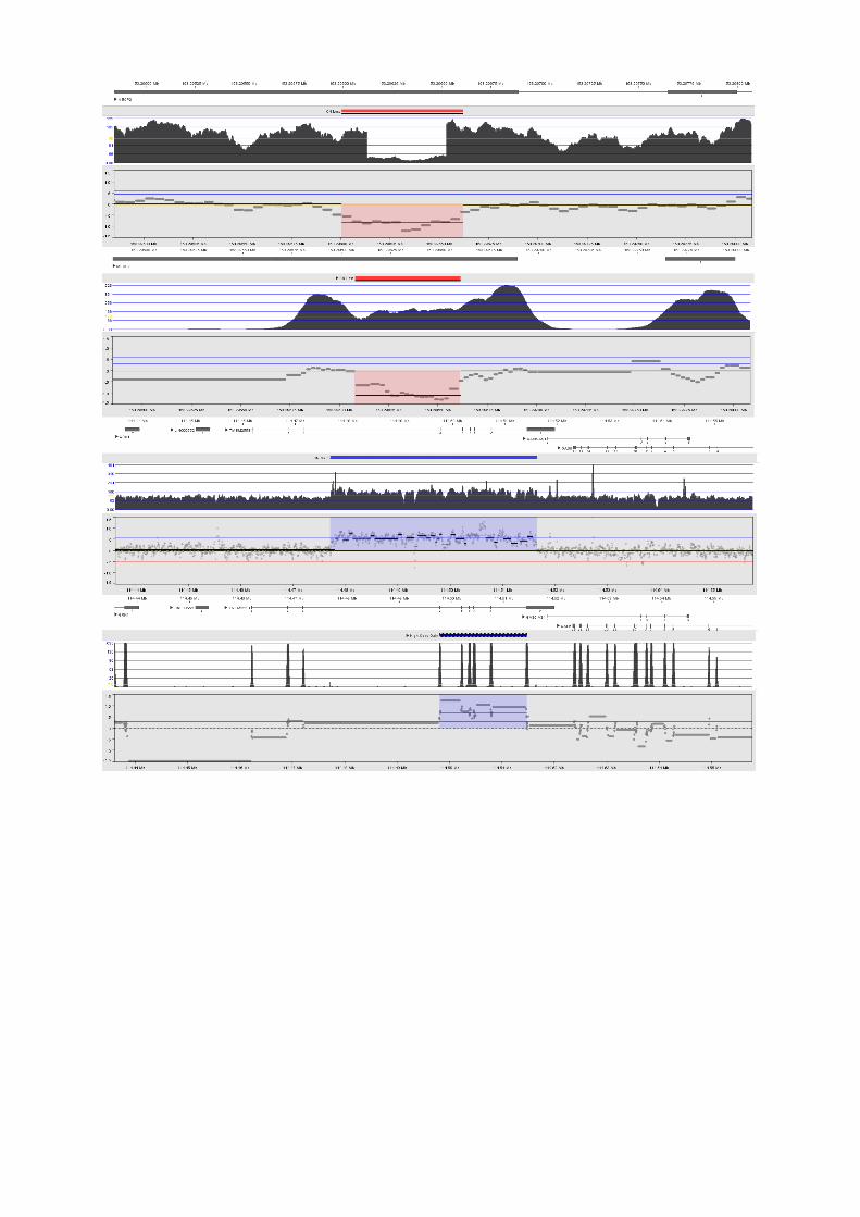

P-ClinG-092
Twins with congenital disorder of glycosylation based on a SRD5A3 intragenic tandem duplication
M. Rieger1, M. Türk2, C. Kraus1, S. Uebe1, A. B. Ekici1, M. Krumbiegel1, C. Huchzermeyer3, A. Reis1, C. Thiel1
1Institute of Human Genetics, Friedrich-Alexander-Universität Erlangen-Nürnberg (FAU), Erlangen, Deutschland 2Department of Neurology, Friedrich-Alexander-Universität Erlangen-Nürnberg (FAU), Erlangen, Deutschland 3Department of Ophthalmology, Friedrich-Alexander-Universität Erlangen-Nürnberg (FAU), Erlangen, Deutschland
Steroid 5α-reductase type 3 congenital disorder of glycosylation (SRD5A3-CDG) is a rare metabolic disease mainly characterized by psychomotor disability, muscular hypotonia as well as ophthalmologic manifestations. Affected individuals occasionally show seizures, facial dysmorphism, dermatosis like ichthyosis, hypertrichosis, and skeletal anomalies of the spine. SRD5A3-CDG is caused by bi-allelic pathogenic variants in SRD5A3. So far, only 23 distinct mutations, comprising sequence variants and one complex rearrangement, in 35 families are described in the literature.
By exome sequencing in 32-year old monozygotic male twins, we identified only the heterozygous splice variant c.562+3delG in SRD5A3, but no second variant. The twins presented with psychomotor deficit, a complex eye disease including retinal dystrophy, pallor of the papilla, ocular nystagmus, and strabismus as well as skeletal anomalies with scoliosis, kyphosis, and extension deficits of the proximal interphalangeal joints IV. Minor facial dysmorphism included prominent brow ridges and up slanting palpebral fissures. Although copy number variants in SRD5A3 were not described in the literature so far, we applied an exome-based copy number analysis (ExomeDepth, GATK) due to the SRD5A3-CDG typical phenotype of the twins. With this method, we identified as a second compound heterozygous variant a previously not reported tandem duplication of exons 2 – 4 in SRD5A3. Using reverse transcription PCR, an in-frame duplication with tandem orientation was confirmed, predicted to lead to a change in protein confirmation. We propose the combination of the splice site mutation and the novel tandem duplication of exon 2 – 4 to be disease causing. Since splicing mutations and duplications can lead to hypomorphic alleles, a possibly atypical or milder SRD5A3-CDG phenotype needs to be considered. Although the affected twins mostly presented with the characteristic SRD5A3-CDG features, the neurological findings were rather subtle and might be the result of the specific genotype.
Our findings further expand the mutational and clinical spectrum of SRD5A3-CDG. They emphasize the relevance of intragenic copy number analysis in this disorder, which is essential to consider in patients with strong clinical suspicion and only one detectable single nucleotide variant.
P-ClinG-093
Molecular diagnosis of ZFP57-associated 6q24 imprinting disturbances in a patient with suspected Beckwith-Wiedemann syndrome
T. Eggermann1, T. Ripperger1, J. Penkert1, C. Kratz1, M. Begemann1, M. Elbracht1
1RWTH Aachen, Aachen, Deutschland
Beckwith-Wiedemann syndrome (BWS) is typically associated with molecular alterations affecting the imprinting control regions 1 and/or 2 in 11p15.5. Molecularly as well as clinically, there is an overlap with other imprinting disorders including 6q24-associated transient neonatal diabetes mellitus (TNDM). We report on the molecular findings in a 6 months old boy referred for molecular BWS testing. Clinical scoring for BWS yielded a score of 5 points (i.e., macroglossia, mild lateral overgrowth, and transient postnatal hypoglycaemia). Molecular testing for 11p15.5 disturbances was negative, but testing of multiple imprinted loci (multilocus testing) revealed a loss of methylation of the imprinted loci in PLAGL1 (6q24), GRB10 (7p13), and PEG3 (19q13). This imprinting signature is typically observed in patients with a molecular subtype of 6q24-associated transient neonatal diabetes mellitus (TNDM) caused by biallelic ZFP57 mutations associated with hypomethylation of maternally imprinted loci. Sequencing of the index patient and his parents confirmed homozygosity for a ZFP57 missense variant (i.e., NM_0011098009.3:c.748C>T p.(Arg250Cys)) formally classified as variant of uncertain significance.
In fact, patients suffering from ZFP57-associated TNDM show some clinical overlap with BWS, but all cases reported so far were identified due to their metabolic impairment.
In our patient, identification of the multilocus imprinting disorder (MLID) as well as subsequent detection of its probable cause was key for clinical management and for genetic counselling. Instead of diagnosing BWS based on the clinical score and offering tumor surveillance for Wilms tumor, our patient required awareness for MLID-associated issues including endocrine monitoring regarding TNDM. Even though the observed variant is classified as VUS, parents have to be aware of a potential probability of 25% for a further child with the same (epi)genotype.
Our patient illustrates that ZFP57-associated imprinting disturbances might mimic a BWS phenotype when TNMD is absent. It has to be emphasized that the probable molecular diagnosis in our patient was not obtained by restrictive 11p15 testing, but that only multilocus testing has allowed the identification of this unexpected result. As there is an increasing number of similar reports on unusual results in imprinting disorder patients, multilocus testing is suggested in patients with negative testing results from disease-specific first-line assays.
P-ClinG-094
Male patient with a novel likely pathogenic variant in TFAP2B causing Char syndrome
K. Roberts1, F. Rust1, A. Caliebe1, M. Spielmann1,2, F. Riepe3, N. Hornig1, M. Kautza-Lucht1
1University of Kiel, Institute of Human Genetics, Kiel, Deutschland 2University of Luebeck, Institute of Human Genetics, Luebeck, Deutschland 3Kinderärzte Kronshagen, Kronshagen, Deutschland
Char syndrome is a rare autosomal disorder caused by a heterozygous pathogenic variant in TFAP2B. It leads to a triad of dysmorphic facial features, patent ductus arteriosus and anomalies of the fifth fingers. The prevalence of Char syndrome is unknown and, in all likelihood, very low (Gelb 2021). Here we report a 7 ½ year old patient with Char syndrome and a likely pathogenic missense variant in TFAP2B. The patient presents with patent ductus arteriosus and a single crease of the fifth finger of the right hand with possible symphalangism and aplasia or hypoplasia of the middle phalanges. He displays distinctive dysmorphic facial features, such as depressed nasal bridge, broad flat nasal tip, short philtrum with prominent philtral ridges, thickened everted lips (duck-bill lips), mild ptosis, slightly downslanted palpebral fissures and hypertelorism. He showed some of the less common features of the syndrome, such as syndactyly of the fourth and fifth fingers, short fingers and syndactyly of the fourth and fifth toes of the right foot. Furthermore, he displayed hirsutism of the back, dark pigmentation anomalies of the upper body, an umbilical hernia, behaviour and emotional disorder and speech deficits. Like his father and paternal half-brother, who both have a form of polydactyly, our patient also showed postaxial polydactyly type B. The family is originally from Ghana and has two more sons whose development is normal. After chromosome banding analysis, array-CGH and fragile X diagnostics, exome sequencing was performed, showing a likely pathogenic heterozygous missense variant in TFAP2B (c.707G>A, p.Arg236His). Missense variants involving this amino acid (Arg236Cys, Arg236Ser) have been previously classified as pathogenic (Zhao et al. 2001). The phenotype of the patient is consistent with the features described in literature. A segregation analysis of the parents is pending. Here we report an additional patient with Char syndrome and a novel missense variant in TFAP2B. Our results are in line with previous reports about Char syndrome caused by an amino acid change at position Arg236 in TFAP2B. The Arg236His substitution has so far not been described and may potentially explain the further symptoms as presented by our patient.
Sources:
Gelb BD. Char Syndrome. 2003 Aug 15 [updated 2020 May 21]. In: Adam MP, Ardinger HH, Pagon RA, Wallace SE, Bean LJH, Mirzaa G, Amemiya A, editors. GeneReviews® [Internet]. Seattle (WA): University of Washington, Seattle; 1993–2021. PMID: 20301285.
Zhao F, Weismann CG, Satoda M, Pierpont ME, Sweeney E, Thompson EM, Gelb BD. Novel TFAP2B mutations that cause Char syndrome provide a genotype-phenotype correlation. Am J Hum Genet. 2001 Oct;69(4):695-703. doi: 10.1086/323410. Epub 2001 Aug 14. PMID: 11505339; PMCID: PMC1226056.
P-ClinG-095
A novel nonsense variant in RAB33B as cause of Smith-McCort dysplasia 2
C. Roggia1,2, A. Dufke1,2, A. Müller1, T. Haack1,2, O. Rieß1,2, M. Kehrer1,2
1Universitätsklinikum Tübingen, Medizinische Genetik und angewandte Genomik, Tübingen, Deutschland 2Universitätsklinikum Tübingen, Zentrum für Seltene Erkrankungen, Tübingen, Deutschland
Smith-McCort dysplasia (SMC, OMIM #615222) is a very rare and disabling autosomal-recessive spondylo-epi-metaphyseal dysplasia with coarse facies, short trunk dwarfism with barrel-shaped chest and protuberant abdomen, kyphoscoliosis, genu valgum or varum, rhizomelic limb shortening, and limited joint extensibility. Generalized platyspondyly with double-humped vertebral endplates and lace-like appearance of iliac crests are considered to be specific radiological features. Cognitive abilities are not impaired. The disorder is caused by mutations in either DYM or RAB33B, the latter coding for a GTP-binding protein involved in retrograde transport of Golgi vesicles and autophagy. So far only 15 individuals affected by RAB33B-related Smith-McCort dysplasia (SMC Type 2) have been described in the literature. We report an additional case of SMC type 2 due to a novel pathogenic stop-mutation in RAB33B-gen.
A 29-year-old refugee from Palestine was referred to our department for genetic testing after an external clinical diagnosis of osteogenesis imperfecta. He presented with short stature, chest deformity, short neck, genua valga, and abnormal gait. He was not able to stretch his arms or close his fist. Prognathism and pes planus were also present. Radiological reports of chest and limbs were not available. He was the firstborn child to consanguineous parents (first cousins). Both couples of grandparents were also consanguineous. His neuropsychological development was normal, but delayed height gain was noticed since the age of 2. Progressive bone deformities and painful joint stiffness appeared at the age of 15 years. Several fractures of fingers and arms occurred in adolescence, always after traumatic events. Ocular, hearing, or dental problems were denied. Blue sclera as a child could not be recalled. Currently, his chief complaint was severe hip joint pain not responsive to ibuprofen therapy. Body height was 140 cm, head circumference 54 cm, and body weight 40 kg. Hematological and biochemical parameters were normal except for low vitamin D levels. All his seven siblings and his own three children were healthy and of normal stature according to age. Two of the patient"s cousins show a similar clinical phenotype. Further clinical, radiological, or genetic investigations in the family had never been performed.
Genome-sequencing of blood DNA revealed a homozygous pathogenic sequence variant, c.186_199delinsACGA, p.Glu63Argfs*51 in RAB33B, which was not present in an in-house database as well as in gnomAD-Browser (gnomad.broadinstitute.org). Orthopedic and analgesic counseling was recommended. Our data support previous reports stating that the phenotype of SMC type 2 seems to appear homogeneous regardless of the type of mutation in RAB33B.
P-ClinG-096
Phenotype diversity associated with TP63 mutations
A. Schmetz1, X. Xiong2, N. Cesarato2, F. B. Basmanav2, P. Gierthmühlen3, J. Schaper4, D. Schlieper5, M. Wehner2, J. Frank6, R. C. Betz2, S. Redler1
1University Hospital Düsseldorf, Institute of Human Genetics, Düsseldorf, Deutschland 2University of Bonn, Institute of Human Genetics, Bonn, Deutschland 3University Hospital Düsseldorf, Department of Prosthodontics, Düsseldorf, Deutschland 4University Hospital Düsseldorf, Center for Rare Diseases, Düsseldorf, Deutschland 5University Hospital Düsseldorf, Interdisciplinary Centre for Palliative Medicine, Düsseldorf, Deutschland 6Göttingen University Medical Center, Department of Dermatology, Venereology and Allergology, Göttingen, Deutschland
Background: TP63-associated disorders have progressed a long way since their initial clinical description to the identification of the underlying genetic cause. In 1968 Rapp & Hodgkin first reported a family with the key features anhydrotic ectodermal dysplasia, cleft lip and cleft palate and this condition was called Rapp-Hodgkin syndrome (#129400). Successively, a variety of apparently clinically distinctive ectodermal phenotypes were described and six additional TP63-associated entities were established: ectrodactyly, ectodermal dysplasia and cleft lip/palate (#604292), limb-mammary syndrome (#603543), Hay-Wells also called ankyloblepharon-ectodermal defects-cleft lip/palate syndrome (#106260), acro-dermato-ungual-lacrimal-tooth syndrome (#103285), non-syndromic Split-hand/foot malformation 4 (#605589) and orofacial cleft 8 (#618149). A plethora of varied and broad-ranging cutaneous and extra-cutaneous manifestations have been reported to be associated with TP63 mutations, making a precise phenotyping and the assignment to one of these historically evolved subentities increasingly challenging.
Objectives: Here, we describe a 41-year-old South Indian patient and his 10-year-old son. Both reported a history of hair fragility and alopecia affecting mainly the scalp hair, dysplastic and brittle nails, dry, thickened, vulnerable skin and hypohidrosis. In addition, the son had lacrimal duct stenosis and unilateral conductive hearing impairment, whereas the father displayed extensive teeth malformations with multiple retained and in part supernumerary teeth. No other family members were clinically affected.
Methods & Results: Whole-exome sequencing revealed the heterozygous variant c.1922C>T p.(Ala641Val) in exon 14 of TP63 (tumor protein p63) (NM_003722.5) in the father and the son. The variant could neither be detected in the unaffected wife/mother nor in the parents of the index. We conclude that the variant occurred de novo in the affected father.
Conclusions: Taken together, the findings of our patients and critical review of the literature point to phenotype diversity associated with TP63 mutations and stress the need to learn in more detail about the range of TP63-associated clinical manifestations. In the case of TP63-associated diseases, it seems reasonable to critically question the old clinical classification and to overcome historical terminology.
P-ClinG-097
Establishing de novo missense variants in NSF as a cause for infantile epileptic encephalopathy with burst-suppression pattern
S. Schuhmann1, F. Knieling2, C. Kraus1, M. Krumbiegel1, A. Reis1, R. Trollmann2, G. Vasileiou1
1Friedrich-Alexander-Universität Erlangen-Nürnberg FAU, Institute of Human Genetics, Erlangen, Deutschland 2Friedrich-Alexander-Universität Erlangen-Nürnberg FAU, Department of Pediatrics and Adolescent Medicine, Erlangen, Deutschland
Intracellular vesicle transport and membrane fusion are essential for several biological processes including the secretion of hormones and neurotransmitters. Deregulation of these pathways was implicated in neurological diseases and epileptic encephalopathies. The ATPase N-ethylmaleimide-sensitive factor (NSF) plays an important role in membrane trafficking. Upon interaction with its receptors and calcium, NSF regulates the fusion of vesicles with target membranes in all eukaryotic cells. A Drosophila model carrying a missense mutation in the homolog of NSF, comatose (comt), developed temperature-dependent electrical bursts in flight muscles which resemble human febrile seizures. Downregulation of NSF in neuronal cell lines induced enhancement of neurite outgrowth, a structural change similar to that following epilepsy. These results provided the first evidence that variants in NSF could be related to the development of epilepsy.
Recently, two unrelated Japanese individuals harbouring de novo heterozygous missense variants in NSF were reported for the first time. Both of them presented with infantile epileptic encephalopathy characterized by a burst suppression pattern in the electroencephalogram (EEG). The individuals further showed frequent vomiting in the neonatal period and profound intellectual disability. One of them died in infancy of respiratory failure. The NSF-associated epileptic encephalopathy was listed in OMIM as developmental and epileptic encephalopathy 96. Functional studies in Drosophila indicated a dominant-negative effect of the identified NSF variants.
Here we describe the third patient, a 5-month-old female individual from healthy non-consanguineous parents presenting with severe infantile epileptic encephalopathy. After birth, she showed failure to thrive requiring gastric tube and developed recurrent vomiting. Lethargy and muscular hypotonia were also reported. At the age of 10 days the first seizures were observed, at the age of 3 months she had up to 100 seizures per day and EEG showed a burst-suppression pattern which later evolved to hypsarrythmia. Social development and motor milestones were significantly delayed. Diagnostic trio exome sequencing revealed no disease causing variants. In a research setting we identified a de novo heterozygous missense variant c.695G>A, p.(Arg232Gln) in NSF, neither previously described in affected individuals nor listed in genetic databases. Similar to the previously described pathogenic changes, the identified missense variant was located in the AAA domain (D1) of the NSF protein.
Describing the third reported case worldwide with a de novo pathogenic variant in NSF, we establish NSF as an infantile epileptic encephalopathy gene. We also highlight the burst-suppression pattern in EEG during the neonatal period as main feature of NSF-associated epileptic encephalopathy.

P-ClinG-098
First German Language Master of Science in Genetic and Genomic Counselling
G. Schwaninger1, S. Rudnik-Schöneborn1, J. Zschocke1
1Institut für Humangenetik, Medizinische Universität Innsbruck, Innsbruck, Österreich
The profession of Genetic Counsellors (GCs) is well established on all continents with more than 7,000 trained GCs worldwide. It has met resistance in the German language countries, mainly due to a perceived conflict of competencies with medical geneticists (Schwaninger et al. 2021, PMID 33797821). In 2019, the Medical University of Innsbruck, Austria introduced the first Master of Science programme in Genetic and Genomic Counselling for the German-speaking countries and has recently received accreditation by the European Board of Medical Genetics (EBMG). Here we wish to report the experiences from the first class of students graduating in spring 2022.
The Innsbruck master programme follows the international standards for the GC profession on the European level. This allows graduates with an additional two years of practical training in a medical genetics institute to apply for registration as a European Certified Genetic Counsellor with the EBMG, granting international reciprocity and freedom to move workplace within Europe and abroad. The five-semester part-time programme comprises 120 ECTS. 42 ECTS cover lectures and seminars (counselling and communication, human genetics, medical genetics and genomics, structure of the healthcare systems, applied genetic counselling, genetic and genomic diagnostics and bioinformatics, probability calculation and statistics, ethical, legal and cultural aspects, and research methods), 20 ECTS practical components, and 20 ECTS master thesis. There is an additional 15-week (36 ECTS) practical placement plus 2 ECTS of laboratory rotations. The programme is currently graduating the first cohort of seven students and has started a second-year group with twelve students from Germany, Austria and Switzerland.
The implementation of the master programme has commenced a lively discussion on the future of genetic counselling in all German-speaking countries. The programme team, in conjunction with the first graduates, works to establish a solid evidence base for the implementation process also in the German language countries. Research projects on the professional scope of practice, the separation of the professional role of genetic counsellors and the service of genetic counselling, issues around the job title, remuneration concepts and the optimal integration of GCs into genetic services are undertaken.
Most crucial will be a close collaboration of genetic counsellors with medical geneticists building trust in their education and the range of skills from genetics knowledge to psychological attending that has been shown to be of great benefit for genetic services.
P-ClinG-099
Broadening the phenotypic spectrum of FINCA syndrome: biallelic NHLRC2 variants in four families with intellectual disability and epilepsy
H. L. Sczakiel1, N. Damseh2, M. Al-Ashhab2, T. Balci3, K. Van Engelen 3, M. Osmond 4, B. Popp5, J. Porrmann5, N. Ehmke1, M. Danyel1, C. Stoltenburg6, D. Horn1, F. Boschann1
1Charité - Universitätsmedizin Berlin, Institut für Medizinische Genetik und Humangenetik, Berlin, Deutschland 2Al-Makassed Hospital, Jerusalem, Israel 3LHSC Victoria Hospital, London, Kanada 4CHEO, Ottawa, Kanada 5Universitätsklinikum Carl Gustav Carus, Dresden, Deutschland 6Charité - Universitätsmedizin Berlin, Sozialpädiatrisches Zentrum Neuropädiatrie, Berlin, Deutschland
FINCA syndrome is a multisystem disorder characterized by pulmonary fibrosis, neurodegeneration and cerebral angiomatosis. Biallelic variants in NHLRC2 have been identified as the molecular cause of FINCA syndrome. So far, ten patients from six families have been described. Common manifestations of FINCA syndrome in these patients included progressive respiratory findings, developmental delay, muscular hypotonia/dystonia, seizures and brain atrophy. While the first patients characterized were severely affected and died within the first two years of life, by now also more mildly affected patients have been reported.
Here, we present our findings of five additional patients from four families carrying biallelic NHLRC2 variants. By exome analysis we identified the novel variant c.1A>G p.(Met1?) and the previously reported c.442G>T p.(Asp148Tyr) variant each in homozygous state in three patients from two consanguineous families. In two further patients from non-consanguineous families we identified the two novel variants c.1750delC p.(Leu584*) and c.2074G>T p.(Asp692Tyr) as well as the novel variant c.148C>T p.(Gln50*) and the known variant c.442G>T p.(Asp148Tyr), respectively, in compound-heterozygous state.
All of our patients presented with motor as well as speech delay. While seizures and EEG abnormalities were also observed as a frequent manifestation, most patients did not show any signs of pulmonary affection or distinct MRI abnormalities (cerebral angiomatosis). Since the latter have so far been described as two of the key features related to NHLRC2-associated disease our findings broaden the hitherto known phenotypic spectrum of biallelic NHLRC2 variants and point out that NHLRC2-related disease should also be considered in patients presenting with intellectual disability and epilepsy without pulmonary findings or characteristic cerebral angiomatosis.
P-ClinG-100
Severe unilateral hand malformation and cholesteatoma in a boy with Helsmoortel-Van der Aa syndrome
F. Stock1, N. C. Bramswig1, L. Stühn1, F. J. Kaiser1,2, A. Kuechler1
1Universitätsklinikum Essen, Institut für Humangenetik, Essen, Deutschland 2Essener Zentrum für Seltene Erkrankungen (EZSE), Universitätsmedizin Essen, Essen, Deutschland
Helsmoortel-Van der Aa syndrome (OMIM #615873) is a very rare neurodevelopmental disorder with variable unspecific malformations and other extraneurologic features. It is caused by monoallelic pathogenic variants in the ADNP gene (OMIM #611386) and transmitted in an autosomal dominant fashion.
We present the case of a boy born with monodactylic symbrachydactyly of the left hand and a small atrial septal defect II (resolved spontaneously). In his infancy recurrent middle ear infections and frequent vomiting were reported. Later speech and motor retardation became evident. The boy developed a cholesteatoma in his left ear at age eight years, which resulted in hearing loss after radical operation. At age 2 ½ years he had his second toes transplanted on his left hand at thumb and small finger position, thus being able to grip with that hand.
Conventional and molecular karyotyping showed a regular male karyotype 46,XY without significant CNVs. Trio-exome sequencing of the boy and his parents revealed a de novo truncating variant in ADNP, known to be causative for Helsmoortel-Van der Aa syndrome. This diagnosis explains the boy"s developmental delay, intellectual disability, obsessive-compulsive behaviour, frequent ear infections, heart defect, and hand malformation. Yet his monodactylic symbrachydactyly in particular appears as an extreme manifestation of this syndrome, as the previously described patients presented milder forms of hand malformation (clinodactyly, polydactyly, small fifth fingers, fetal finger pads, prominent interphalangeal joints, and distal phalanges).
The appearance of Helsmoortel-Van der Aa syndrome is rather unspecific; as there are no highly suggestive facial features of this syndrome, clinical diagnosis is nearly impossible. The phenotype of our patient was dominated by a severe hand malformation and cholesteatoma (both unilateral), which is rather untypical for this entity. A coincidental occurrence cannot be excluded, yet no further pathogenic DNA variants were identified. This case once again underlines the necessity of whole exome sequencing to solve malformation syndromes without a clear clinical diagnosis.

P-ClinG-101
*** OnkoRiskNET: A multicenter, interdisciplinary, telemedicine-based model to improve care for patients with a genetic tumor risk syndrome
J. Tecklenburg1, B. Vajen1, A. Bergmann1, E. Baumann2, E. Schröck3, M. Arnold4, P. Anders1, S. Morlot1, B. Schlegelberger1
1Medizinische Hochschule Hannover, Institut für Humangenetik, Hannover, Deutschland 2Hochschule für Musik und Theater Hannover, Institut für Journalistik und Kommunikationsforschung, Hannover, Deutschland 3Universitätsklinikum Carl Gustav Carus, Institut für Klinische Genetik, Dresden, Deutschland 4Institut für angewandte Versorgungsforschung, Berlin, Deutschland
Background
Genetic tumor risk syndromes are responsible for at least 50,000 of the 500,000 cases of cancer diagnosed in Germany every year. The diagnosis of a genetic tumor risk syndrome enables personalized therapy, aftercare and prevention and the identification of further affected family members. Currently, the care of oncological patients is characterized by a lack of specialists in human genetics, lack of access to human genetic care in rural areas and lack of interdisciplinary networking and structured care pathways between oncologists and specialists in human genetics. As a result, genetic tumor risk syndromes are underdiagnosed with potentially fatal consequences for patients and their families. OnkoRiskNET aims to close the gap in care through the formation of a cooperation network between practicing oncologists and specialists in human genetics and the use of telemedical genetic counselling.
Methods
The OnkoRiskNET study is supported by a grant from the Federal Joint Committee of the Federal Republic of Germany. The study will include 2000 oncological index patients from 20 oncology practices in Lower Saxony and Saxony after study start in January 2021. Randomization is carried out by means of a stepped wedge design at the level of the practices. Patients will either go through routine care or the new form of care with structured cooperation between human genetics specialists and oncologists, case management and the use of telemedical genetic counselling. Using a mixed-methods approach, the following parameters will be evaluated in the control and intervention group: (1) Uptake of human genetic counselling by patients with suspected tumour risk syndrome and their first degree relatives; (2) Patient satisfaction and psychological distress after genetic counselling and testing; (3) Factors influencing the acceptance and experience of telemedical genetic counselling; (4) Satisfaction of oncologists and specialists for human genetics with the structured pathway; (5) Cost efficiency of the new form of care.
Conclusion
The new form of care aims to improve the care of patients with a tumour risk syndrome and to facilitate access to genetic diagnostics. The study is intended to show possibilities of how comprehensive and interdisciplinary human genetic patient care can be designed in the future using digitalization and networking in the health care system.
P-ClinG-102
*** Career satisfaction of German human genetics residents
J. Tecklenburg1, R. Meyer2, I. Krey3, B. Schlegelberger1
1Medizinische Hochschule Hannover, Institut für Humangenetik, Hannover, Deutschland 2Universitätsklinikum Aachen, Institut für Humangenetik, Aachen, Deutschland 3Universitätsklinikum Leipzig, Institut für Humangenetik, Leipzig, Deutschland
Objective Due to the rapid increase in knowledge and new diagnostic and therapeutic possibilities, the need for specialists in human genetics will increase enormously in the coming years. In this context, the resource of human genetics residents requires special attention. Therefore, we conducted a survey to investigate the career satisfaction of human genetics residents in Germany.
Methods We developed an online survey for the evaluation of a broad range of factors concerning the situation of human genetics residents in Germany. Human genetics residents working at institutions with an authorization for specialist training were asked to participate in the online survey. To analyse the situation of specialist training in human genetics and the influence of multiple factors on career satisfaction, descriptive statistics, comparisons of mean values as well as multiple linear regression analyses were carried out.
Results Of the 71 institutions contacted 41 (58%) provided feedback and reported the number of 114 residents in human genetics. In total 58 residents completed the questionnaire (50,9%). Overall career satisfaction was high with a mean score of 30.8 (scale ranging from 8-40). Factors significantly influencing career satisfaction were general life satisfaction, occupational self-efficacy expectations and content with the doctors entitled to the specialty training. Except for the reduced perception to achieve their professional goals expressed by women with children, career satisfaction was influenced by neither gender nor parental status, other sociodemographic factors, variables concerning the personal professional life and the residency in general, the subjective perceived workload nor the site of specialist training. Participation in research activities differed significantly between male and female residents. The residents' assessment of their own professional prospects and the prospects of the subject were consistently positive, even though residents consider the current requirement planning by the GB-A for human geneticists as inappropriate and believe that human genetics is not yet firmly anchored as a specialist discipline in the consciousness of other medical colleagues and the general public.
Conclusion Career satisfaction of German human genetics residents is generally high and mainly influenced by life satisfaction, occupational self-efficacy expectations and quality of the specialist training. In contrast to other specialties career satisfaction seems to be independent from gender or parental status even though male residents were significantly more often involved in research activities. In order to keep human genetics residents in the specialty, measures that enable balanced professional and care work as well as continuous improvement of specialist education, e.g. through the implementation of structured curricula and continuing education of the doctors entitled to specialist training, is of great importance.
P-ClinG-104
9p Deletion 9p24.3p23 in an 8-year old Girl with Developmental Delay, a large Head Circumference and Brachydactyly
L. Toutouna1, B. Gläser1, K. Komlosi1, B. Herr1, S. Alter1, J. Fischer1, A. Tzschach1
1Medical Center, University of Freiburg, Institute of Human Genetics, Freiburg, Deutschland
Introduction:
Deletions in the distal short arm of chromosome 9 are rare. To date over 140 individuals with 9p deletions habe been described, carrying either an isolated deletion or an unbalanced translocation involving 9p and another chromosome. Clinical features include developmental delay, trigonocephaly, short palpebral fissures, a long philtrum, flat nasal bridge with anteverted nares, ear anomalies with low-set malformed ears and micro-/retrognathia. Other features include hypotonia, widely spaced nipples, male to female sex reversal and rarely finger anomalies such as dolichomesophalangy and clino-/brachydactyly, heart defects, omphalocele and teeth/skeletal anomalies. Various studies have attempted to elucidate a genotype-phenotype correlation as well as a critical region for trigonocephaly with inconclusive results. Developmental delay is a universal characteristic of 9p deletions and is independent of the exact region and location of the deletion.
Clinical report:
Here we report an 8-year old girl with developmental delay affecting language and psychomotor development since infancy. Epileptic seizures manifested in the first 15 months. She walked with over 2.5 years. Speech development was also delayed with sentence building starting in nursery school. Physical exam revealed mild facial dysmorphic features, widely spaced nipples as well as brachydactyly of the 4th and 5th fingers and more pronouncedly of the 3rd -5th toes with sandal gap. Her body measurements at age 7 9/12 were weight at 85th centile, length at 85th centile and head circumference at 92nd centile.
Methods and Results:
Chromosome analysis revealed a distal interstitial deletion involving bands 9p24.3p23: 46,XX,del(9)(p24.3p23). The microarray analysis defined a 12.13Mb deletion including 98 genes and setting the breakpoints at positions chr9:2025657_14155895 (GRCh37) interrupting SMARCA2, associated with Nicolaides-Baraitser syndrome, at the distal end and NFIB, haploinsufficiency of which is associated with intellectual disability and macrocephaly, at the proximal end. FISH analyses with the probe RP11-1142H1 (Empire Genomics) binding in 9p24.3 (pos. 2362216-2567823) in the patient and both parents confirmed a de novo deletion. Whole exome sequencing (WES) showed no additional pathogenic variants.
Conclusions:
Numerous patients with a distal deletion in 9p have been described, presenting with a variety of clinical manifestations. Here we report a girl with a de novo deletion of 12.13Mb involving 9p24.3p23. None of the listed patients in DECIPHER nor in the literature shares the identical deletion breakpoints with our patient. Interestingly she lacked the typical trigonocephaly but demonstrated a large head circumference and mild dysmorphic features as well as pronounced brachydactyly of the toes. This case contributes to the delineation of the phenotypic spectrum of 9p deletions.
P-ClinG-105
Significance of the sperm DNA fragmentation index (DFI) for male fertility diagnostics
O. Vedrines 1, M. Stumm1
1Medicover Genetics, Cytogenomics, Berlin, Deutschland
Sperm analysis involves analyzing the concentration, motility and percentage of normally formed sperm. But these parameters do not provide complete information as they do not consider the analysis of the integrity of the DNA molecule which has an effect on natural fertilization, successful implantation and achievement of pregnancy. We analyzed whether the age of the patients correlates with an increase in DNA fragmentation in sperm and how the DNA fragmentation correlates with the values of other sperm parameters.
We compared the data of standard sperm analysis and DNA Fragmentation Index (DFI) of 715 patients (2009 to 2021). 500 sperms/patient were analyzed and the DFI was calculated. We grouped the patients according to their DFI: DFI ≤ 15 %: pregnancy with a fertile female partner can be achieved by natural conception. 16<DFI≤29 %: Intra uterine insemination (IUI) is recommended. DFI ≥ 30 %: In Vitro Fertilization (IVF) or Intracytoplasmic Sperm Injection (ISCI) are recommended. We analyzed the normal distribution of each parameter, performed t-tests, and calculated the correlation coefficient p to determine, which sperm parameter is most impacted by the DFI.
The DFI increases significantly with patient´s age. Patients with high DFI ≥ 30 % are on average 40 years old while with low and medium DFI categories have a median age of 36.5 and 38.5 years. There is a direct correlation between DFI and the other parameters: DFI increases significantly as sperm concentration, percentage of normally formed sperm, and motility decreases: mean concentrations are 66.0M/ml, 51.3M/ml and 39.9M/ml, normal morphology are 4.04%, 3.37% and 2.69%, and motility are 52.1%, 43.3% and 32.4% in the low, medium and high DFI categories. The motility parameter correlates best with DFI: its correlation coefficient is -0.46, compared to -0.25 and -0.21 for the percentage of normally formed sperm and concentration. Finally, patients were divided into 4 groups according to the number of abnormal spermogram parameters: Group 1: All 3 parameters are normal. 4.3% of these patients have a DFI ≥ 30%. Group 2: One of 3 parameters is abnormal. 10.5 % of these patients have a DFI ≥ 30 %. Group 3: Two of the 3 parameters are abnormal. 29.2 % of these patients have a DFI ≥ 30 %. Group 4: All 3 spermogram parameters are abnormal. 47.6 % of these patients have a DFI ≥ 30 %.
The DFI shows a direct correlation to the age of the patients. Also, the DFI shows a correlation to all 3 standard spermiogram parameters, alone and in combination. Interestingly, 4% of patients with a normal spermiogram and <40 years show a high DFI (>30%). This highlights the fact that DNA fragmentation analysis can provide additional information that routine sperm analysis does not. Especially in patients with a normal spermiogram and unexplained fertility problems, the DNA fragmentation index can serve as an additional clinical biomarker. DFI analysis allows a more detailed assessment of male fertility.
P-ClinG-106
Results of trio exome analyses in 20 hospitalised newborns and infants with an unclear symptom complex (OPS number 1-944.10)
C. Velmans1, V. Riehmer1, N. Fuhrmann1, P. Herkenrath2, A. Koy2, A. Kribs2, A. Oberthür2, A. Vierzig2, M. Emmel3, J. Becker1, S. Kaspar1, N. Reintjes1, S. Blick1, K. Nohl1, S. Seland1, K. Brockmeier3, J. Dötsch2, F. Erger1, J. Schreml1, C. Netzer1
1Institut für Humangenetik, Uniklinik Köln, Köln, Deutschland 2Kinderklinik, Uniklinik Köln, Köln, Deutschland 3Kinderkardiologie, Uniklinik Köln, Köln, Deutschland
Background: The majority of congenital syndromal disorders are genetically determined. By means of trio exome analysis, a genetic cause can currently be confirmed in up to half of those affected. An early diagnosis often has great relevance for the families and the treating physicians as most of the newborns and infants are hospitalized for a long period of time.
Methods: Since the beginning of 2021, trio exome analyses can be performed at our Institute of Human Genetics as part of the so-called basic diagnostics for unclear symptom complex (OPS number 1-944.10) for newborns and infants with unclear syndromal disease who are treated at the Children's Hospital and Pediatric Cardiology at the University Hospital of Cologne. The analyses were embedded in extensive pre- and post-test genetic counselling and required close cooperation between pediatricians and clinical geneticists.
Results: Of the 20 patients examined so far (age at start of diagnostics 4 days to 9 months) with an unclear symptom complex (abnormalities in at least two organ systems with unclear etiology), a genetically caused syndromal disease could be confirmed in 11 patients (n = 20, 55 %). Two thirds of the patients had an autosomal dominant disorder caused by a heterozygous de novo variant. In addition, variants of unclear significance which fit well with the phenotype of the patients but require further clarification were detected in three patients. In several patients, two independent causative genetic diseases were diagnosed or incidental findings with medical relevance were raised. The diagnostic result was available after an average of two weeks. The diagnoses ranged from more common rare diseases such as CHARGE and Marfan syndrome to very rare syndromes with less than 50 described cases so far as for example Noonan syndrome type 12 and Au-Kline syndrome.
Summary: Early trio exome analyses in newborns and infants with an unclear symptom complex represent a good diagnostic option with a high diagnostic yield and considerable clinical relevance.
P-ClinG-107
ODonnell-Luria-Rodan Syndrome - description of a second multinational cohort and refinement of the phenotypic spectru
C. Velmans1, A. O' Donnell-Luria2, E. Argilli3, F. Tran Mau-Them4, A. Vitobello4, M. C. Y. Chan5, J. L. F. Fung5, M. Rech6, A. Abicht7, M. Aubert-Mucca8, J. Carmichael9, N. Chassaing8, R. Clark10, C. Coubes11, A. S. Denommé-Pichon4, K. de Dios12, E. M. England13, B. Funalot14, M. Gerard15, M. Joseph9, C. Kennedy9, C. Kumps16, M. Willems11, I. van de Laar17, C. D. Aarts-Tesselaar18, M. A. van Slegtenhorst17, D. Lehalle14, K. Leppig19, L. Leßmeier1, L. Pais2, H. Paterson2, S. Ramanathan10, L. H. Rodan20, A. Superti-Furga16, B. H. Y. Chung5, E. Sherr3, C. Netzer1, C. P. Schaaf6,21,22, F. Erger1
1Institut für Humangenetik, Uniklinik Köln, Köln, Deutschland 2Boston Children’s Hospital, Harvard Medical School, Manton Center for Orphan Disease Research, Boston, Vereinigte Staaten 3 Department of Neurology, University Hospital California San Francisco, Brain Development Research Program, San Francisco, Vereinigte Staaten 4FHU-TRANSLAD, University Hospital Dijon‐Bourgogne, Inserm UMR 1231 Genetics of Developmental Disorders, Dijon, Frankreich 5Department of Paediatrics and Adolescent Medicine, The University of Hong Kong, Hong Kong, Hongkong 6Molecular and Human Genetics, Baylor College of Medicine, Houston, Vereinigte Staaten 7Medical Genetics Center (MGZ) Munich, München, Deutschland 8Department of Medical Genetics, University Hospital Toulouse, Toulouse, Frankreich 9Valley Children`s Hospital, Madera, Madera, Vereinigte Staaten 10Pediatrics Specialty Clinics, Loma Linda University Medical Center, San Bernardino, Vereinigte Staaten 11Department of Medical Genetics, University Hospital Montpellier, Montpellier, Frankreich 12Department of Medical Genetics, Dayton Children`s Hospital, Dayton, Vereinigte Staaten 13Center for Mendelian Genomics and Medical and Population Genetics Program, Broad Institute of MIT and Harvard, Cambridge, Vereinigte Staaten 14Department of Clinical Genetics, University Hospital Henri Mondor, Créteil, Frankreich 15Service de Génétique, CHU Caen, Caen, Frankreich 16Division of Genetic Medicine, Lausanne University Hospital (CHUV, Lausanne, Schweiz 17Department of Clinical Genetics, Erasmus MC, University Medical Center Rotterdam, Rotterdam, Niederlande 18Department of Pediatrics, Amphia Hospital, Breda, Niederlande 19Genetic Services, Kaiser Permanente of Washington, Seattle, Vereinigte Staaten 20Department of Neurology, Boston Children's Hospital, Harvard Medical School, Boston, Vereinigte Staaten 21Institute of Human Genetics, Heidelberg University, Heidelberg, Deutschland 22Jan and Dan Duncan Neurological Research Institute, Texas Children’s Hospital, Houston, Vereinigte Staaten
Background: O"Donnell-Luria-Rodan Syndrome (ODLURO) is an autosomal-dominant neurodevelopmental disorder caused by pathogenic, mostly truncating variants in KMT2E. It was first described by O"Donnell-Luria et al. in 2019 in a cohort of 38 patients. Clinical features encompass macrocephaly, mild intellectual disability, autism spectrum disorder (ASD) susceptibility and seizure susceptibility.
Methods: Affected individuals were ascertained at pediatric and genetic centers in various countries by diagnostic chromosome microarray or exome/genome sequencing. Patients were collected into a case cohort and were systematically phenotyped where possible.
Results: We report 18 additional patients from 17 families with genetically confirmed ODLURO. We identified 15 different heterozygous likely pathogenic or pathogenic sequence variants (14 novel) and two partial microdeletions of KMT2E. We confirm and refine the phenotypic spectrum of the KMT2E-related neurodevelopmental disorder, especially concerning cognitive development, with rather mild intellectual disability and macrocephaly with subtle facial features in most patients. We observe a high prevalence of autism spectrum disorder in our cohort (41%), while seizures are present in only two patients. We extend the phenotypic spectrum by sleep disturbances.
Conclusion: Our study, bringing the total of known ODLURO patients to more than 60 within two years of the first publication, suggests an unexpectedly high relative frequency of this syndrome worldwide. It seems likely that ODLURO, although just recently described, is among the more common single-gene etiologies of neurodevelopmental delay and ASD. We present the second systematic case series of patients with ODLURO, further refining the mutational and phenotypic spectrum of this not-so-rare syndrome.
P-ClinG-108
Diagnostic yield of genetic testing in patients with inherited eye diseases using virtual gene panel analysis
P. Villavicencio-Lorini1,2, C. Grünauer-Kloevekorn3, K. Hoffmann1, B. Schulte4,5, K. Stingl6, C. Schuart7, H. Thieme7, C. Schober8, A. Viestenz8, S. Biskup4,5
1Martin-Luther-Universität Halle-Wittenberg, Institut für Humangenetik, Halle (Saale), Deutschland 2MVZ Universitätsklinikum Halle, Facharztzentrum für Pädiatrie & Humangenetik , Halle (Saale), Deutschland 3Praxisklinik Augenärzte am Markt, Halle (Saale), Deutschland 4Praxis für Humangenetik, Tübingen, Deutschland 5Center for Genomics and Transcriptomics CeGaT GmbH, Tübingen, Deutschland 6Universitätsklinikum Tübingen, Zentrum für Seltene Augenerkrankungen, Universitätsaugenkliniik, Tübingen, Deutschland 7Otto-von-Guericke Universität Magdeburg, Universitätsaugenklinik, Magdeburg, Deutschland 8Universitätsklinikum Halle, Universitätsaugenklinik, Halle (Saale), Deutschland
Background
Inherited eye diseases often lead to severe visual impairment in children and young adults and have a significant impact on their psychomotor development, social participation, and mental health. In recent years, pathophysiologically adapted treatments as well as gene therapeutic approaches have been developed. To increase the chances for treatment at early disease stages, a rapid determination of the genetic cause of inherited eye diseases is necessary. The introduction of multigene panel analysis or virtual gene panel analysis using exome enrichment has made diagnostic classification more efficient.
Methods
To assess the diagnostic rate of these molecular genetic testing methods in the field of ophthalmogenetics, we evaluated the genetic findings in a cohort of 34 patients of different ages with diseases of the anterior segment of the eye (9 of 34 cases with aniridia, microphthalmia, anterior dysgenesis, cataract, ectopia lentis and myopia) as well as the posterior segment (25 of 34 cases with retinitis pigmentosa, cone-rod dystrophy and optic neuropathies). In about 3/4 of the cases a non-syndromic and in 1/4 of the cases a syndromic form was present.
Results
Molecular genetic diagnostics revealed pathogenic or probably pathogenic germline disease gene variants in 17 of 34 cases (50%), variants of unclear significance in 6 of 34 cases (approximately 18%), and unremarkable findings in 11 of 34 cases (approximately 32%). In the patient group with diseases of the anterior segment of the eye, a diagnostic classification was achieved in 5 out of 9 cases (approximately 55%) by detection of variants or CNVs in or including the genes PAX6, FOXC1, RARB, OCRL and FBN1. In the group of patients with diseases of the posterior segment of the eye, a diagnostic classification was achieved in 12 out of 25 cases (48%) by detection of variants in the genes ABCA4, RP2, RPGR, RS1, NMNAT1 and OPA1, among others. The diagnostic classification had the consequence that in most cases a complication risk adapted ophthalmological or interdisciplinary care could be pursued. In the case of genetic determined retinopathy or optic neuropathy patients could be referred to a specialized center for inclusion in a study registry for gene therapeutic approaches. Furthermore, information about diagnosis, prognosis and inheritance improved the psychosocial handling of the disease.
Conclusion
Ophthalmogenetic diagnostics contribute to a better care for patients with inherited eye diseases and is expected to advance gene therapeutic approaches. Further development of diagnostic possibilities and closer interdisciplinary cooperation is therefore necessary.
P-ClinG-109
Two novel variants expand the genetic and phenotypic spectrum of CNOT3-associated developmental disorder
J. Wagner1, J. Fischer1, K. Albertowski2, J. Schallner3, B. Leeder4, K. Hackmann1, A. K. Kahlert1, A. Tzschach1, E. Schröck1, B. Popp5, N. Di Donato1
1Institut für Klinische Genetik, Universitätsklinikum Carl Gustav Carus an der Technischen Universität Dresden, Dresden, Deutschland 2Klinik und Poliklinik für Kinder- und Jugendpsychiatrie und –psychotherapie, Autismusambulanz, Universitätsklinikum Carl Gustav Carus an der Technischen Universität Dresden, Dresden, Deutschland 3Klinik und Poliklinik für Kinder- und Jugendmedizin, Abteilung Neuropädiatrie & Sozialpädiatrisches Zentrum, Universitätsklinikum Carl Gustav Carus an der Technischen Universität Dresden, Dresden, Deutschland 4Sozialpädiatrisches Zentrum, Klinikum Frankfurt (Oder), Frankfurt (Oder), Deutschland 5Medizinisches Versorgungszentrum (MVZ) am Universitätsklinikum Carl Gustav Carus Dresden GmbH, Dresden, Deutschland
Heterozygous pathogenic variants in CNOT3 cause an autosomal dominant intellectual developmental disorder with speech delay, autism and dysmorphic facies (IDDSADF, OMIM 618672). Currently 22 individuals are described with mostly de novo truncating variants. Missense variants cluster in the N-terminal coiled-coil (CC) domain. The CNOT3 gene product is part of the CCR4-NOT protein complex, which is involved in the regulation of RNA polymerase II.
We report on two unrelated German individuals (one girl, one boy) in whom novel heterozygous CNOT3 variants were detected by next generation sequencing (c.575A>G, p.(N192S) and c.2151_2153del, p.(D717del), respectively[PB1] ). The de novo c.575A>G, p.(N192S) variant affects a highly conserved amino acid in the CC-domain in proximity to a mutational cluster around Arginine 188. The in frame deletion of an Aspartate residue at position 717 affects the C-terminal NOT box where to date only one missense variant has been described as pathogenic. Segregation using deep amplicon sequencing identified the variant with an allele fraction of 9 % in the sample of the healthy[PB2] mother. In accordance to the previously reported individuals, both herein described individuals presented with mild intellectual disability[PB3] , autism spectrum disorder, cerebral MRI abnormalities[PB4] (cerebellar hypoplasia, arachnoid cyst, septum pellucidum cyst), strabismus and dysmorphic features[PB5] (upslanted palpebral fissures, thin lips, smooth philtrum). The female individual had significant macrocephaly (4,69 z) and obesity (BMI 25, 2,47 z), which is a novel association in this disorder. She also carries a pathogenic TNPO3 (OMIM 608423) variant (c.1543del, p.(L515fs)) without clear signs of muscular dystrophy at age 8 years.
The individuals reported here broaden the clinical and genetic spectrum of IDDSADF syndrome. [PB6] Broad genetic testing using exome sequencing remains the first line diagnostic choice. We report the first case of parental mosaicism with markedly increased recurrence risk.
P-ClinG-110
There is more to it than just congenital heart defects – The phenotypic spectrum of TAB2-related syndrome
D. Westphal1,2, E. Mastantuono2, H. Seidel1,3, K. Riedhammer1,4, A. Hahn5, K. Vill6, M. Wagner1,6,7
1Klinikum rechts der Isar München, Institut für Humangenetik, München, Deutschland 2Klinikum rechts der Isar München, Klinik und Poliklinik für Innere Medizin I, München, Deutschland 3Deutsches Herzzentrum München, Klinik für Angeborene Herzfehler und Kinderkardiologie, München, Deutschland 4Klinikum rechts der Isar München, Abteilung für Nephrologie, München, Deutschland 5Universitätsklinikum Giessen, Kinderneurologie, Giessen, Deutschland 6Dr. von Haunersches Kinderspital, Sozialpädiatrisches Zentrum, München, Deutschland 7Helmholtz Zentrum München, Institut für Neurogenomik, München, Deutschland
Background: Congenital heart defects (CHD) are the most common birth defect and disease-causing variant in TAB2 have found to be associated with isolated CHD. Recently, it became evident that loss-of-function variants in TAB2 can also cause syndromic CHD that includes connective tissue anomalies. The number of published cases is limited posing a challenge for counseling affected patients and their relatives.
Methods: Cases in whom whole exome sequencing was executed at our institute between January 2015 and June 2021 were screened for disease-causing variants in TAB2. Additionally, a PubMed-based review of the literature was performed in September 2021 in order to give an updated clinical overview of the TAB2-associated phenotypic spectrum, including our cases.
Results: We identified three cases with syndromic CHD caused by different heterozygous loss-of-function variants in TAB2. In one of these cases, the variant was inherited by a healthy father. A comparison with published cases highlights that most patients were affected by structural and/or arrhythmic heart disease (about 90 %) while about two third of all cases had syndromic comorbidity especially connective tissue defects and dysmorphic abnormalities.
Conclusion: Our findings indicate a variable expressivity as well as reduced penetrance of TAB2-associated CHD. Disease-causing variants in TAB2 should be considered in cases with isolated CHD but also in syndromic CHD with connective tissue abnormalities. However, prediction of the patients" clinical outcome solely based on the variant in TAB2 is still extremely challenging.
P-ClinG-111
Progressive choreodystonia in X-linked hyper-IgM immunodeficiency: a rare but recurrent presentation
M. Škorvánek1, R. Jech2, J. Winkelmann3, M. Zech3
1Department of Neurology, ty, Kosice, Slovak Republic, Kosice, Slowakei 2Department of Neurology, Charles University, 1st Faculty of Medicine and General University Hospital in Prague, Prague, Czech Republic, Prag, Tschechien 3Institut für Humangenetik, Technische Universität München, München, Deutschland
An association between movement disorders and immune-system dysfunction has been described in the context of rare genetic diseases such as ataxia telangiectasia as well as infectious encephalopathies. We encountered a male individual with immunodeficiency of unknown etiology since childhood. A medication-refractory, progressive choreodystonic movement disorder emerged at the age of 42 years and prompted an exome-wide molecular testing approach. This revealed a pathogenic hemizygous variant in CD40LG, the gene implicated in X-linked hyper-IgM syndrome. Only two prior reports have specifically suggested a causal relationship between disease-causing variants in CD40LG and involuntary hyperkinetic movements. Our findings thus confirm the existence of a particular CD40LG-related condition, combining features of compromised immunity with neurodegenerative movement abnormalities. Establishing the diagnosis is crucial because of potential life-threatening immunological complications.
112–126 Complex Diseases, Population & Evolutionary Genetics and Genetic Epidemiology
P-Compl-112
Male-pattern hair loss: Integration of GWAS and single-cell RNASeq data to identify pathobiologically relevant hair follicle cell types
N. Engelmann1, S. Henne1, S. Heilmann-Heimbach1
1Institut für Humangenetik, Universitätsklinikum, Bonn, Deutschland
Male-pattern hair loss (MPHL) is a highly heritable and progressive form of hair loss. The phenotype is strictly androgen-dependent and affected hair follicles (HF) show characteristic changes in hair cycle dynamics (i.e. shorter growth / prolonged resting). While GWAS on MPHL have identified >360 genomic risk loci and have implicated numerous candidate genes and pathways, little is known about the cell types and hair cycle stages in which these genes and pathways exert the pathobiological effects. This knowledge however is critical to enable functional follow-up of GWAS findings in pathogenic cell types and to understand causal disease mechanisms.
Here, we used (i) a statistical model that relies on the assumption that genes with critical functions in pathogenic cell types are likely to be located within disease-associated loci (Hu et al., 2011) together with (ii) published MPHL GWAS data, and (iii) a comprehensive single-cell RNASeq data set of the murine HF during hair growth and rest (Joost et al., 2020) to identify MPHL-relevant HF cell types at different stages of HF cycling. In brief, murine gene identifiers were uniquely mapped to human identifiers using the NCBI database. Expression data for 15,827 mapped genes were normalised, averaged across all single cells within a single cell type and transformed into nonparametric scores (NPS) based on their intra- and inter-cell type expression specificity. Only cell types with data from at least 3 individual cells were considered for further analysis, leaving 47 and 29 cell types from growing and resting HF, respectively. Genes were assigned to MPHL-loci based on (i) linkage disequilibrium or (ii) topologically associated domains. For each GWAS locus, a cell type-specific locus P-score (LPS) was calculated as the Bonferroni-corrected NPS of the most specifically expressed gene at this locus. Cell type-specific enrichment scores were then calculated as the sum of -log(LPS) across all GWAS loci. Finally, the significance of the cell type enrichment was determined using a permutation-based test.
Our results point to a pathogenic role of different HF cell types across and in specific hair cycle stages. For example, while cells and cellular processes within the basal interfollicular epidermis and dermal fibroblasts seem to be of pathogenic relevance across hair cycle stages, sebaceous gland cells and vascular cells seem to specifically contribute to MPHL pathogenesis during growth or rest, respectively. Pathway-based analyses of the most specifically expressed genes in associated cell types suggest a similar picture on the molecular level, where e.g. androgen signalling plays a role across cell types and cell cycle stages whereas ErbB- or EDA-signalling are only active in specific cell types and cell cycle stages. In summary, our data provide novel insight into MPHL pathogenic cell types and constitute an important basis for systematic functional follow-up of GWAS findings in relevant cell types.
P-Compl-113
Exploring the association between loci with antagonistic effect directions on multiple neuropsychiatric disorders and brain phenotypes
L. M. Federmann1,2, L. Sindermann2, C. Jockwitz1,3, F. S. David2, A. Teumer4, K. Amunts1,5, U. Dannlowski6, T. W. Mühleisen1,5, S. Caspers1,3, S. Cichon1,7,8, A. J. Forstner1,2
1Research Centre Jülich, Institute of Neuroscience and Medicine (INM-1), Jülich, Deutschland 2University of Bonn, School of Medicine & University Hospital Bonn, Institute of Human Genetics, Bonn, Deutschland 3Heinrich Heine University, Medical Faculty & University Hospital Düsseldorf, Institute for Anatomy I, Düsseldorf, Deutschland 4University Medicine Greifswald, Institute for Community Medicine, Greifswald, Deutschland 5Heinrich Heine University, Medical Faculty & University Hospital Dusseldorf, Cecile and Oskar Vogt Institute of Brain Research, Düsseldorf, Deutschland 6University of Münster, Institute for Translational Psychiatry, Münster, Deutschland 7University Hospital Basel, Institute of Medical Genetics and Pathology, Basel, Schweiz 8University of Basel, Department of Biomedicine, Basel, Schweiz
Introduction:
The genome-wide association meta-analysis of eight neuropsychiatric disorders by Lee et al. (2019) identified 11 independent single nucleotide polymorphisms (SNPs) that increased the risk for one disorder and were protective for another disorder (antagonistic SNPs). Large-scale brain imaging studies by the ENIGMA Consortium showed structural alterations of various brain regions in patients with neuropsychiatric disorders, representing brain image-derived phenotypes (IDPs). However, the link between genomics and brain structure is not yet fully understood and the study by Lee et al. (2019) did not characterize the association of the 11 antagonistic SNPs with IDPs. Therefore, the present study explored whether the antagonistic SNPs are implicated in structural brain alterations which might be relevant for the development of neuropsychiatric disorders.
Methods:
We investigated the association between the 11 antagonistic SNPs and 80 IDPs of cortical surface area (SA), cortical thickness (CT), subcortical volumes, and intracranial volume (ICV) by using the summary statistics of large ENIGMA genome-wide association studies that each included more than 10,000 individuals. Subsequent analyses focussed on SNP-IDP associations that remained significant after performing multiple testing correction using the Benjamini-Hochberg method. On the basis of large-scale brain imaging studies by ENIGMA, we assessed for all significant SNP-IDP pairs whether the implicated IDP has been reported to be altered in patients with neuropsychiatric disorders associated with the respective SNP.
Results:
Seven antagonistic SNPs were significantly associated with at least one IDP. The implicated IDPs included 12 SA regions located in parts of the lateral prefrontal, insular, superior temporal, posterior cingulate, and occipital cortex, as well as CT in regions of the cingulate cortex and the inferior parietal lobule. Furthermore, the volume of the caudate nucleus and the ICV were each associated with one antagonistic SNP. The preliminary assessment of large-scale brain imaging studies supported some of the observed SNP-IDP associations. In particular, the T allele of the SNP rs2921036 was protective against schizophrenia and associated with an increase of SA of the superior temporal gyrus, which may be consistent with the global decrease of SA including this region that has been reported in schizophrenia patients.
Conclusion:
The results of the present analysis suggest that specific antagonistic SNPs associated with neuropsychiatric disorders might contribute to alterations in SA rather than CT, subcortical volumes, or ICV. Our study provides first insights into how antagonistic SNPs may lead to different effects on several neuropsychiatric disorders. Further genomic imaging analyses in large samples of patients and controls are required to validate the relevance of the identified SNP-IDP associations for disease development.


P-Compl-114
Human-specific ARHGAP11B is essential for neural progenitor levels in hominid brain organoids
J. Fischer1,2, E. Fernández Ortuño2, J. Peters2, T. Namba2, C. Eugster Oegema2, W. Huttner2, M. Heide2
1Institut für Klinische Genetik, Universitätsklinikum Carl Gustav Carus an der Technischen Universität Dresden, Dresden, Deutschland 2Max Planck Institute of Molecular Cell Biology and Genetics, Dresden, Deutschland
BACKGROUND: ARHGAP11B is a human-specific gene that is expressed in apical and basal radial glia in the developing human brain. It is able to increase the number of progenitor cells in mouse and ferret embryonic and marmoset fetal brains, but data in hominid brains is absent so far.
METHODS: Electroporation of human and chimpanzee brain organoids derived from induced pluripotent stem cells (human iPSC line SC102A-1 and chimpanzee iPSC line Sandra A) was carried out. ARHGAP11B, ARHGAP11A220 (acting dominant-negatively on ARHGAP11B"s function) or empty vector plasmids together with a vector expressing GFP were electroporated into the ventricles of 55-day old brain organoids, which were then fixed 2-15 days after electroporation.
RESULTS: ARHGAP11B is able to increase the abundance of TBR2+ basal progenitor cells two days after chimp organoid electroporation. Moreover, between 4 to 10 days after ARHGAP11B electroporation an increase in PCNA+ and HOPX+ cells in the subventricular zone (SVZ) was observed indicative for an increase in basal radial glia. This coincided with a reduction in cell maturation and differentiation as shown by a decreased number of CTIP2+, HuC+ and NeuN+ cells in comparison to control electroporations. 15 days after electroporation of chimp organoids with ARHGAP11B an increase in upper layer SATB2+ neurons was observed. Finally, dominant-negative inhibition of ARHGAP11B"s function by electroporation of ARHGAP11A220 in human brain organoids led to fewer TBR2+ and BrdU+ progenitor cells in the SVZ.
CONCLUSION: ARHGAP11B increases the relative numbers of basal progenitors and slows progenitor differentiation in chimpanzee organoids. Moreover, it gives rise to an increased number of upper layer neurons. Dominant-negative inhibition of ARHGAP11B"s function in human organoids leads to reduced basal progenitor levels. These findings further support the role of ARHGAP11B as an essential factor in cortical expansion during human brain development and evolution.
P-Compl-115
The effect of LPA p.Thr1399Pro on lipoprotein(a) concentrations and coronary artery disease is modified by the LPA splice site variant KIV-2 4925G>A
R. Grüneis1, C. Lamina1, S. Di Maio1, S. Schönherr1, P. Zöscher1, L. Forer1, A. Peters2,3, C. Gieger2,3,4, A. Köttgen5,6, F. Kronenberg1, S. Coassin1
1Medical University of Innsbruck, Institute of Genetic Epidemiology, Innsbruck, Österreich 2German Center for Diabetes Research (DZD), München-Neuherberg, Deutschland 3Helmholtz Zentrum München - German Research Center for Environmental Health, Institute of Epidemiology, Neuherberg, Deutschland 4Helmholtz Zentrum München, German Research Center for Environmental Health, Research Unit of Molecular Epidemiology, Neuherberg, Deutschland 5University of Freiburg, Institute of Genetic Epidemiology, Freiburg, Deutschland 6German Chronic Kidney Disease study, Freiburg, Deutschland
Introduction
High Lipoprotein(a) [Lp(a)] concentrations are a major genetic risk factor for coronary artery disease (CAD) risk. Lp(a) is mainly regulated by the LPA gene, which shows a very complex gene structure, consisting of different highly homologous kringle IV domains (KIV-1 to -10). The KIV-2 domain is encoded by a coding copy number variation (CNV), with the number of KIV-2 repeats showing an inverse correlation with Lp(a) concentrations. Recently, multiple previously missed functional variants with a strong impact on Lp(a) concentrations have been detected within the CNV region, such as the splice site variant 4925G>A. Many SNPs in the non-repetitive gene region, such as LPA p.Thr1399Pro (rs41272110) have been associated with Lp(a) but with inconsistent effects across studies.
Methods
We analyzed the effect of the interaction between LPA rs41272110 and the LPA splice site variant 4925G>A in the KIV-2 region on Lp(a) concentrations in the German Chronic Kidney Disease (GCKD) study, KORA F3 and KORA F4 (total=10,405) by stratified and combined quantile regression models. The impact of both variants and their combinations on CAD risk was assessed by stratified survival analyses in the UK Biobank (n=186,088).
Results
We identified a hitherto unknown linkage disequilibrium (LD) between rs41272110 and 4925G>A (R2=0.836-0.872, D"=0.984-0.985). In quantile regression rs41272110 alone showed no impact on Lp(a) concentration (β=-0.06, [-0.79;0.68], p=0.879). However, in a joint model including both variants, rs41272110 was associated with markedly increased Lp(a) (β=+9.40 mg/dL, [6.45;12.34], p=4.07E-10). Accordingly, survival analysis showed no effect of rs41272110 alone on CAD risk (HR=1.01, [0.97;1.04], p=0.731), but in a joint model containing both SNPs, individuals carrying rs41272110 but not 4925G>A (4% of the population) show a previously hidden significantly increased CAD risk (HR=1.10, [1.04;1.16], p=6.9e-04). This is missed in single SNP analysis because the partial LD with the Lp(a)-lowering splice site variant 4925G>A creates a spurious Lp(a) and CAD risk net lowering effect.
Conclusion
We found a novel Lp(a)-increasing effect of rs41272110, which is masked by partial LD with KIV-2 4925G>A. The LD between these variants strongly affects genetic CAD risk assessment at single individual level. This emphasizes the importance of accounting for SNP-SNP interactions for correct risk stratification and implementation of genetic risk scores for Lp(a).
PIC
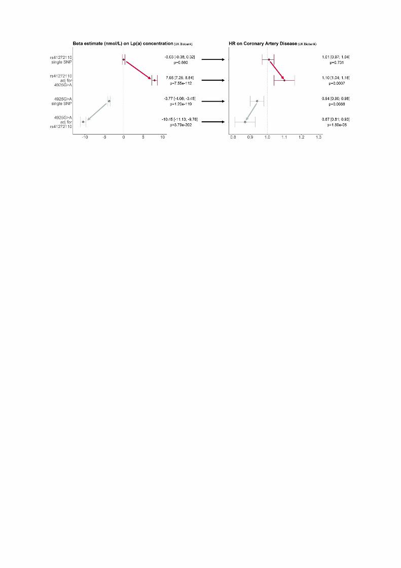
P-Compl-116
The role of rare variants in male-pattern hair loss: Analysis of whole exome sequencing data in the UK Biobank
S. Henne1, S. Sivalingam2,3,4, L. Hochfeld1, C. Maj3, O. Borisov3, A. Buness2,3,4, M. Nöthen1, P. Krawitz3, S. Heilmann-Heimbach1
1Institut für Humangenetik, Universitätsklinikum Bonn, Bonn, Deutschland 2Core Unit for Bioinformatics Analysis, Universität Bonn, Bonn, Deutschland 3Institut für Genomische Statistik und Bioinformatik, Universität Bonn, Bonn, Deutschland 4Institut für Medizinische Biometrie, Informatik und Epidemiologie, Universität Bonn, Bonn, Deutschland
Male-pattern hair loss (MPHL) is a progressive and highly heritable form of hair loss with a lifetime prevalence of ~80% in European men. Genome-wide association studies (GWAS) have yielded substantial insights into the genetic basis of MPHL, identifying more than 622 genetic risk variants at 367 genomic loci. While these analyses provided systematic insights into the contribution of common variants (MAF>1%) to MPHL etiology, no study has systematically investigated the contribution of rare variants (MAF<1%). A large exome data set created by the UK Biobank (UKBB) now enables the analysis of rare variants in coding areas of the genome. Here, we report the results of our analysis of a first tranche of 200,629 exomes from the UKBB.
Participants were classified as cases or controls based on their reported hair loss pattern (1-unaffected, 2-frontotemporal balding, 3-frontotemporal and vertex balding, 4-baldness of the top of the scalp). We used two different classification schemes: (i) an all-model, where controls (pattern 1) were compared to cases (patterns 2-4) and (ii) an extreme model, where supercontrols (pattern 1, age≥60) were compared to severe cases (pattern 4, age<60). Exome variant data were downloaded from the UKBB in PLINK format. Individuals were filtered for confirmed white British ancestry and data was quality controlled in PLINK in regards to sex, relatedness, missingness and Hardy-Weinberg disequilibrium. Variants were filtered for a MAF<1% and a nonsynonymous consequence in a protein-coding gene predicted by VEP. We performed SKAT-O gene-based analysis with an additional threshold including only splice-, stop- or start-altering and frameshift variants. GWAS-style single variant analyses were performed using PLINK. Analyses were corrected for age and ten principal components.
Our SKAT-O analyses detected a significant association of HEPH and EDA2R, as well as nominally significant associations (p≤0.05) of 3,119 genes. Of these, 20% are located within 500kb of a GWAS locus. While this does not represent an enrichment, we observe an enrichment of associated genes (pall≤5x10⁻³, pextreme≤0.05) in hair follicle-expressed genes located within topologically associated domains of GWAS loci. Our GWAS-like analyses identified two significantly associated rare missense variants located within HEPH and EDA2R, as well as 17,892 nominally significant variants. We identified promising candidate genes such as LAMA5, which lies at a novel risk locus, HOXC13, a novel candidate gene at a known risk locus, and WNT10A, a gene previously implicated by GWAS which shows an independent association in our analyses. In summary, this first systematic analysis of the contribution of rare coding variants to MPHL etiology broadens the allelic spectrum of previously reported candidate genes, yields evidence for novel MPHL candidate genes at and beyond known GWAS loci and provides a basis for future study of the contribution of rare variants to MPHL pathobiology.
P-Compl-117
Investigation of non-coding risk variants in non-syndromic cleft lip with/ without cleft palate using massively parallel reporter assays
R. Hollstein1, S. Schultz1, M. Schubach2, A. Biedermann1, F. Thieme1, M. Kircher2, K. U. Ludwig1
1Universität Bonn, Institut für Humangenetik, Bonn, Deutschland 2Universitätsmedizin Berlin, Berlin Institute of Health at Charité , Berlin, Deutschland
Non-syndromic cleft lip with/without cleft palate (nsCL/P) is the most common craniofacial malformation in humans, with a prevalence of approximately 1 in 1,000. The etiology of nsCL/P is multifactorial and shows a twin-based heritability of 90%. Till date, at least 45 nsCL/P risk loci have been identified by GWAS. The majority of the associated variants map to non-coding regions, suggesting that the underlying pathomechanisms act through regulatory effects on temporal or spatial gene expression. However, the substantial number of candidate variants at each locus and the tissue specificity of regulatory effects make it challenging to pinpoint the effects of the associated risk variants in vitro.
Massively Parallel Reporter Assays (MPRA) have been recently developed to enable high-throughput screening of candidate regulatory sequences (CRSs) and/or candidate single nucleotide variants (SNVs) with possible regulatory effects on gene expression in vitro. For this, CRSs are cloned into a reporter plasmid with a unique barcode attached to the reporter gene. If a CRS is regulatory active, the barcoded reporter gene is expressed and can be detected using RNA-sequencing of the barcode.
To investigate the effects of nsCL/P associated variants, we designed an oligonucleotide library with 230 bases long DNA-fragments, each harbouring one allele of 1,053 GWAS variants. As the capacity of one MPRA is limited (max. 244,000 sequences), we prioritized those loci with a strong effect in the European dataset based on relative risk estimates below 0.8 or larger than 1.2. This resulted in the inclusion of 31 loci, and the respective lead SNVs plus all variants in LD were considered for the MPRA. The highest number of variants was included for chromosome 5p12 with 166 SNVs, the lowest for chromosome 17q23.2 with 1 SNV. We also included 103 known regulatory elements as positive controls and 59 negative controls to the oligo library. Using 100 unique barcodes per sequence, we obtained an nsCL/P oligo library with 244,000 oligonucleotides which was ordered at Agilent.
Upon receipt, we cloned the oligonucleotide library into a pLS-mP plasmid and integrated the minimal promoter and a GFP-gene between CRS and barcode. Currently, the plasmid is undergoing extensive quality control to quantify the individual CRS and barcodes. We will then transfect human embryonic kidney (HEK) cells, human embryonic palatal mesenchyme (HEPM) cells and iPSC-derived neural crest cells with the final library plasmid. The abundance of each barcoded transcript will be measured using RNA-seq, and normalized for abundance of cellular inserted plasmids per barcode. This study will contribute to the identification of functionally relevant variants at GWAS risk loci and allow for further functional analyses, with the overall aim to better understand the nsCL/P etiology.
P-Compl-118
Molecular genetic characterization of patients with bipolar disorder and controls for induced pluripotent stem cell generation
K. Hünten1, F. S. David1, S. Sivalingam2, B. Burger3, E. C. Beins1, L. Sirignano4, F. Streit4, F. Stein5, P. I. Pfefferle6, M. Rietschel4, M. Peitz7, S. Cichon3, O. Brüstle7, M. M. Nöthen1, A. J. Forstner1,8
1University of Bonn, Institute of Human Genetics, Bonn, Deutschland 2University of Bonn, Core Unit for Bioinformatics Data Analysis, Bonn, Deutschland 3University of Basel , Department of Biomedicine, Basel, Schweiz 4Central Institute of Mental Health, Department of Genetic Epidemiology in Psychiatry, Mannheim, Deutschland 5UKGM, University Hospital for Psychiatry and Psychotherapy, Marburg, Deutschland 6Philipps University of Marburg, Comprehensive Biobank Marburg (CBBMR), Marburg, Deutschland 7University of Bonn, Institute of Reconstructive Neurobiology, Bonn, Deutschland 8Research Center Jülich, Institute of Neuroscience and Medicine (INM-1), Jülich, Deutschland
Bipolar disorder (BD) is a common psychiatric disorder characterized by recurrent episodes of (hypo-) mania and depression. Genetic factors contribute substantially to the etiology of BD with heritability estimates ranging between 60 and 85%. Recent genome-wide association studies support a strong polygenic influence. However, not much is known about the functional effects of a high polygenic risk. In the current project, we aim to explore whether polygenic risk factors contribute to an impaired neurodevelopment and whether this can be detected in induced pluripotent stem cells (iPSCs). We selected unrelated patients with BD type 1 (BD1) and controls from the FOR2107 cohort (https://for2107.de/) for the generation and analysis of iPSCs. Selection criteria included male sex, BD1 (patients only), available quality-controlled genome-wide genotype data and peripheral blood mononuclear cells (PBMCs). Additionally, polygenic risk scores (PRS) for BD1 were calculated using PRS-CS (Ge et al., 2019) and summary statistics of a large genome-wide association study of BD (Stahl et al., 2019). Copy number variants (CNVs) significantly associated with BD or schizophrenia were excluded in 12 BD1 patients with the highest and 12 controls with the lowest PRS using the Illumina GenomeStudio software (cnvPartition). Subsequently, 10 patients with the highest and 10 controls with the lowest PRS were selected for whole-genome sequencing (WGS) to rule out the presence of rare risk variants for BD implicated in previous BD sequencing studies. We did not consider variants if they had a MAF>1% (gnomAD, non-neuro dataset), a PHRED-scaled CADD score<20, or were present in both patients and controls. A total of 16 BD1 patients and 55 controls had available PBMCs and genotype data. One BD1 patient was excluded in the CNV analysis due to the presence of a microdeletion on chromosome 2 (NRXN1). Four BD1 patients and four controls did not carry rare variants previously implicated in BD in the WGS analysis. Of these, the three BD1 patients with the highest and three controls with the lowest PRS were then selected for iPSC generation. In conclusion, we describe a systematic procedure to characterize patients and controls at the genetic level prior to iPSC generation. Our filtering approach resulted in the exclusion of rare variants previously implicated in BD that could have influenced subsequent iPSC analyses.
P-Compl-119
Search for exonic homozygous/compound heterozygous variants in affected sib-pairs identifies novel candidate genes for nonsyndromic cleft palate
N. Ishorst1, H. Shkuro1, J. Fazaal1, A. K. Hoebel1, H. Thiele2, T. Kruse3, M. J. Dixon4, K. U. Ludwig1, E. Mangold1
1University of Bonn, School of Medicine & University Hospital Bonn, Institute of Human Genetics, Bonn, Deutschland 2University of Cologne, Cologne Center for Genomics CCG, Cologne, Deutschland 3University of Cologne, Department of Orthodontics, Cologne, Deutschland 4University of Manchester, Manchester Academic Health Sciences Centre, Manchester, Vereinigtes Königreich
Nonsyndromic cleft palate only (nsCPO) belongs to the typical forms of orofacial clefts and is a common congenital malformation. nsCPO is a multifactorial disorder with environmental and genetic factors contributing to disease risk. Of note, the genetic contribution is rather high with an estimated heritability of >90%. In the last years genome-wide association studies have identified 10 genome-wide significant risk loci. One of them has been confirmed, and nine have been reported recently but still await independent replication. However, these common risk loci explain only a fraction of the estimated heritability. Epidemiological observations and genetic studies suggest that high penetrance rare/low frequency variants contribute to nsCPO that might be inherited in an autosomal-recessive manner.
In this study, we focussed on detection of novel nsCPO candidate genes by identifying compound heterozygous/homozygous variants. To that end, we reanalysed existing whole-exome sequencing data from six affected sib-pairs born to unaffected parents.
After filtering for population frequency (MAF<5%) and manual inspection of reads, we detected 169 putative compound heterozygous and 13 homozygous variants.
We next filtered these 182 variants/80 candidate genes based on (1) the functional effects at transcriptional level and in silico predictions, (2) intolerance for a certain class of rare variation, and (3) expression in mouse embryonic palatal shelves at embryonic day E13.5. This resulted in a list of the 32 most promising candidate genes, 29 with putative compound heterozygous and 3 with homozygous variants. Compound heterozygosity was then validated with either manual inspection of reads or Sanger sequencing. The final list of 21 nsCPO candidate genes with compound heterozygous/homozygous variants included one compound heterozygous situation within GRHL3, which has been implicated in nsCPO, and novel genes encoding proteins involved in cell migratory processes, such as DDR2, an interaction partner of the gene product of the well established clefting gene CDH1.
Together, our study shows the value of reanalysing existing datasets and has identified nsCPO candidate genes that follow an autosomal-recessive inheritance. To show that these candidates are truly involved in nsCPO, further replication studies and functional characterization are warranted.
P-Compl-120
SLC1A2 is a new candidate gene for neuromyelitis optica spectrum disorder
M. Meyenborg1, M. Spielmann2, A. Caliebe1, I. Nagel1
1Institute of Human Genetics, University of Kiel, Kiel, Deutschland 2Institute of Human Genetics, University of Lübeck, Lübeck, Deutschland
Atypical inflammatory demyelinating (AID) syndromes are a heterogeneous group of rare inflammatory diseases of the central nervous system. In contrast to multiple sclerosis they do not present the characteristic MRI findings or clinical symptoms. They also show a poor response to treatments that are used against multiple sclerosis. The clinical course of disease in patients with AID syndromes is heterogeneous. Some patients recover completely whereas other patients have quick disease progress with poor prognosis. One of these syndromes is neuromyelitis optica spectrum disorder (NMOSD). Patients present multiple motor and cognitive disorders with different expression. In most patients with NMOSD, IgG autoantibodies against aquaporin-4 (AQP4), a water channel localized in astrocytes, were found.
A 37 years old male presented with neurological symptoms e.g. optic neuritis, spastic tetraparesis and hyperreflexia. Serological tests for AQP4 autoantibodies gave negative results. After other potential diseases had been excluded, the patient's symptoms led to the suspicion of NMOSD. But even though different immunomodulatory therapies were escalated, the disease progressed rapidly. Using exome sequencing, we identified the heterozygous variant c.343C>T [p(Arg115CysSLC)] in SLC1A2. SLC1A2 is a protein coding gene which encodes a sodium dependent amino acid transporter. It is localized on the presynaptic membrane on astrocytes and removes L-glutamate, L-aspartate and D-aspartate from the synaptic cleft. According to prediction tools (e.g. REVEL, CADD, SIFT, PROVEAN), the amino acid exchange from arginine to cysteine at position 115 is predicted to be pathogenic or protein damaging. The affected arginine residue is located in the sodiumdicarboxylate symporter family-domain, is highly preserved and is involved in ligand-interactions. In addition, functional analyses provide evidence, that this variant leads to a decreased surface expression and transport activity of SLC1A2.
It was shown that IgG autoantibodies for AQP4 in patients with NMOSD reduce SLC1A2 activity followed by reduced glutamate-reuptake from the synaptic cleft. Thus, our identified variant in SLC1A2 might have a similar effect by causing decreased transport activity. Furthermore, it is known that increased release or decreased reuptake of glutamate leads to an excessive stimulation of glutamate receptors. This results in neurological damage and cell death, a process called excitotoxicity. Hence, there might be a correlation between reduced SLC1A2 activity which results in excessive supply of glutamate in the synaptic cleft and the neurological disorders of the patient described herein.
Our current data are insufficient to assess whether this variant is associated with the symptoms of our patient. Presently his parents are recruited to assess whether this variant occurred de novo. This would support our hypothesis that pathogenic variants in SLC1A2 can be associated with NMOSD.
P-Compl-121
Comparison of transcript levels and neurotransmitter receptor densities in human hippocampal regions
T. Mühleisen1,2,3, B. Burger3, L. Zhao1, D. I. Hilger1, E. Beins4, A. J. Forstner1,4, S. Herms3,4, P. Hoffmann3,4, K. Amunts1,2, N. Palomero-Gallagher1,2,5, S. Cichon1,3,6
1Forschungszentrum Jülich, INM-1, Jülich, Deutschland 2Heinrich Heine University Düsseldorf, Medical Faculty, Düsseldorf, Deutschland 3University Hospital Basel and University of Basel, Department of Biomedicine, Basel, Schweiz 4University Hospital of Bonn, Institute of Human Genetics, Bonn, Deutschland 5RWTH Aachen University, Department of Psychiatry, Psychotherapy and Psychosomatics, Aachen, Deutschland 6University Hospital Basel, Institute of Medical Genetics and Pathology, Basel, Schweiz
Introduction:
The hippocampus plays a crucial role in memory and learning, and there is strong evidence that its regions are differentially affected by neuropsychiatric disease. Although the hippocampus has been subject of multiple mapping studies addressing various organizational levels, not much is known about regional differences at the transcriptomic and proteomic level. Here we screened for differential expression of neurotransmitter receptors and their genes in the cornu ammonis (CA) and dentate gyrus (DG) to evaluate their regional specificity by comparing RNA transcripts and protein densities from the same donor samples.
Methods:
Seven fresh-frozen samples were obtained at autopsy. Donors (71.4±15 years) were free from neurological or psychiatric diseases. RNA expression was genome-wide analysed and normalized. Receptor densities (protein expression levels) were quantified by autoradiographic analysis.
Results:
We selected fourty-four genes for thirteen neurotransmitter receptors. Highest RNA expression levels in CA and DG were found for the muscarinic cholinergic and adenosinergic systems, followed by the serotonergic and dopaminergic systems. Expression of CHRM2 (cholinergic M2 receptor) was significantly higher in CA than in DG. CA and DG did not differ significantly in their ADORA1 (adenosine A1 receptor), serotonergic or dopaminergic receptor gene expression levels. At the protein level, adenosinergic and serotonergic systems showed highest densities, followed by the cholinergic and dopaminergic systems. CA contained a significantly higher density of A1 and serotonergic 5-HT1A receptors than did DG. The opposite holds true for 5-HT2 densities. Cholinergic and dopaminergic (D1) densities were comparable in CA and DG. For some receptor types, higher densities were found with increasing RNA expression levels in both CA and DG (e.g. A1, D1). For other receptor types (e.g. 5-HT1A), CA and DG differed in this relationship.
Conclusions:
The relationship between protein and RNA expression differed between CA and DG for different transmitters and their receptors, e.g. for A1 receptors, we found equal transcript levels in CA and DG but higher densities in CA than in DG. Our findings suggest the presence of region- and receptor type-specific regulatory mechanisms between both modalities. A deduction of receptor densities from gene expression data alone may therefore be challenging. Regulatory mechanisms may include posttranslational modifications and receptor protein trafficking. Especially receptor trafficking is a well-known process affecting synaptic plasticity during development, normal brain function and diseases. We hypothesize that the identified differences could be associated with region-specific plasticity mechanisms in learning and memory processes.
Acknowledgment:

This study is dedicated to the late Karl Zilles whose ideas contributed significantly to its design. We will sorely miss his critical spirit.
P-Compl-122
Association of rare genetic variants at the ULBP3 locus with alopecia areata
P. Schwerbrock1, A. Schmidt1, R. Aldisi2, N. Cesarato1, D. Axt1, S. Herms1, S. Heilmann-Heimbach1, K. Ludwig1, P. Krawitz2, C. Maj2, R. Betz1, B. Basmanav1
1Universität Bonn, Institut für Humangenetik, Bonn, Deutschland 2Universität Bonn, Institut für Genomische Statistik und Bioinformatik, Bonn, Deutschland
Background: Alopecia areata (AA) is a common autoimmune-mediated disorder with an estimated lifetime risk of about 2,1%. Hair loss typically commence with the development of isolated hairless patches, which then progress centrifugally and may merge into complete hair loss over the entire head and body. Genome-wide association studies reported common risk variants in 14 susceptibility loci, the majority of which are related to immunity. The elucidation of rare variants in AA is still in its infancy, however, based on evidence from other autoimmune diseases, it is highly likely that they play a significant role in the genetic etiology of AA. ULBP3 is the only AA GWAS gene that was not implicated in other autoimmune diseases and encodes for a ligand of the NKG2D receptors expressed on NK cells, CD8+ T cells, and some autoreactive or immunosuppressive CD4+ T cells.
Methods: By using Single Molecular Real Time (SMRT) sequencing technology, we sequenced the entire coding and non-coding region around ULPB3 (10 kb) in 1.000 AA patients and 1.000 ethnicity and gender matched controls. The results were analyzed by a rare variant burden analysis pipeline, which implements a gene-based scoring system using functional annotations and allele frequency weighting function.
Results: A very restricted number of rare variants were identified in the coding sequence of ULBP3 with nominal overrepresentation in the patients. The analysis of the entire region including the non-coding sequences revealed a statistically significant association of rare ULBP3 variants (MAF<0.01) with AA (p=0.028).
Conclusions: Our study shows the contribution of rare genetic variation at the ULBP3 locus to disease susceptibility in AA. Since rare variants typically have larger effect sizes and consequences more easily traceable by functional assays, a follow-up of the variants emerging from our study can provide mechanistic insights into the etiology of this common autoimmune disorder.
P-Compl-123
Single-cell based transcriptional differences between candidate genes for syndromic and non-syndromic cleft lip with or without cleft palate
A. Siewert1, J. Welzenbach1, B. Reiz2, E. Mangold1, H. Dickten2, K. U. Ludwig1
1Institute of Human Genetics, University of Bonn, School of Medicine and University Hospital Bonn, Bonn, Deutschland 2FASTGenomics, Comma Soft AG, Bonn, Deutschland
Cleft lip with or without cleft palate (CL/P) is a common human birth defect which can occur as a non-syndromic, isolated phenotype (nsCL/P) or within a complex malformation syndrome (syCL/P). In the latter case, additional tissues and organ systems present with developmental peculiarities.
We here hypothesized that candidate genes involved in syCL/P may be expressed in more cell types during embryonic development, compared to genes causing nsCL/P. To study this, we re-analyzed single-cell RNA sequencing (scRNA-seq) data from the Mouse Organogenesis Cell Atlas data base (Cao et. al 2019), covering the relevant time period for facial development and organogenesis in mice from embryonic days E9.5 – E13.5. We downloaded data from ~1.3 million high-quality single cells and split the data by embryonic day. We also downloaded a second scRNA-seq data set that was obtained only from the developing mouse face at E11.5 (Li et. al 2019). This data set comprised 7,249 high-quality single cells. Our analyses were performed using the R packages Seurat v4 (Hao and Hao et. al 2021) and scCATCH (Shao et. al 2020) and were run on the analytical ecosystem FASTGenomics (Scholz et. al 2018). 79 candidate genes associated with nsCL/P were defined based on data from prior genome-wide association studies (Welzenbach et. al 2021), while syCL/P genes were retrieved from a previously assembled gene list (Bishop et. al 2020) and divided by autosomal-dominant (AD) (25 genes) and autosomal-recessive (AR) (26 genes). At each time point, we extracted the percentage of cell types expressing the respective gene and compared average gene expression levels between the different groups.
Our results show that on average, AD syCL/P genes are expressed in significantly more cell types during embryonic development compared to nsCL/P genes (lowest P-value: 2.45x10⁻⁴ for E9.5). We also observed that AD syCL/P genes have significantly higher gene expression levels than nsCL/P genes (lowest P-value: 4.43x10⁻³ for E9.5). Looking at the specific cell types in which nsCL/P genes are expressed, we found genes restricted to epithelial cells (e.g. Irf6, Grhl3 and Esrp1) as well as genes expressed in a set of multiple cell types such as connective tissue, chondrocytes, osteoblasts, jaw and teeth (e.g. Nbl1, Fgf10, Epha3, Grem1).
While this first analysis supports our hypothesis, we will systematically follow up on these results by examining the enrichment of CL/P candidate gene expression in various tissues. Overall, these findings will help to extend the understanding of the molecular mechanisms and etiological differences between syndromic and non-syndromic CL/P.
P-Compl-124
Investigation of polygenic risk scores in social anxiety disorder patients
A. Vogelsang1, M. L. Chung2, L. Sindermann1, F. David1, C. Elling2, L. E. Seib-Pfeifer 3, S. Heilmann-Heimbach 1, P. Hoffmann1, S. Herms1, B. Schmidt4, K. Domschke5,6, V. Nieratschker7, U. Dannlowski8, E. J. Leehr 8, E. Leibing9, M. M. Nöthen1, F. Geiser2, J. Schumacher 10, C. Maj 11, R. Conrad2, A. J. Forstner 1,12
1University Hospital Bonn, Institute of Human Genetics, Bonn, Deutschland 2University Hospital Bonn, Department of Psychosomatic Medicine and Psychotherapy, Bonn, Deutschland 3University of Bonn, Department of Psychology, Bonn, Deutschland 4University Hospital of Essen, Institute for Medical Informatics, Essen, Deutschland 5University Medical Center Freiburg, Department of Psychiatry and Psychotherapy, Freiburg, Deutschland 6University of Freiburg, Centre for Basics in Neuromodulation, Freiburg, Deutschland 7University of Tübingen, Department of Psychiatry and Psychotherapy, Tübingen, Deutschland 8University of Münster, Institute for Translational Psychiatry, Münster, Deutschland 9University Medical Center Göttingen, Clinic of Psychosomatic Medicine and Psychotherapy, Göttingen, Deutschland 10University of Marburg, Centre for Human Genetics, Marburg, Deutschland 11University of Bonn, Institute of Genomic Statistics and Bioinformatics, Bonn, Deutschland 12Research Center Jülich, Institute of Neuroscience and Medicine (INM-1), Jülich, Deutschland
Background:
Social anxiety disorder (SAD) is one of the most prevalent anxiety disorders (AD) with a lifetime prevalence of around 11-12%. SAD is characterized by a persistent fear in social situations that results in avoidance behavior and socio-economic burden. In addition, SAD is often co-occurring with depressive symptoms or major depressive disorder (MDD). An estimated heritability of around 10-50% reflects the contribution of genetic factors to disease etiology. However, while large cross-disorder genome-wide association studies (GWAS) have been conducted in AD, genetic analyses of SAD have so far only included limited sample sizes, leaving the genetic basis of SAD and its subphenotypes largely unknown. Therefore, in the present study we investigated the association of polygenic risk scores (PRS) for anxiety and other psychiatric disorders with disease subphenotypes in a large cohort of patients with SAD.
Methods:
Patients with SAD (n=1304) were drawn from our Social Phobia Research project (https://www.socialphobiaresearch.de/) or provided through clinical collaborators in Germany. Controls (n=4140) were derived from the population-based Heinz Nixdorf Recall study. All individuals were genome-wide genotyped using the Infinium Global Screening Array. We applied standard quality control procedures and performed imputation of the genotyping data using the 1000 Genomes Project reference panel. PRS computation was conducted using PRSice-2 and summary statistics of publicly available AD, MDD and depression GWAS. Correlation analyses of PRS and different SAD subphenotypes (e.g. severity of SAD, depression diagnosis, comorbidity with other psychiatric disorders, or drug/alcohol abuse) were performed using R.
Results:
Preliminary analyses of the depression PRS (Howard et al., 2019) revealed a significant association with SAD case-control status. Furthermore, we found that the depression PRS was nominally associated with depressive symptoms in SAD patients. This association, however, did not withstand stringent Bonferroni correction for multiple testing.
Conclusion:
Our preliminary results do not provide strong evidence that genetic risk factors for depression contribute to specific subphenotypes of SAD. However, systematic analyses of additional PRS, particularly for AD, are needed to characterize the genetic basis of SAD subphenotypes. These analyses are currently underway and will be presented at the upcoming conference.

Funding: Social Phobia Research is supported by the Else Kröner-Fresenius-Stiftung (2019_A127).

P-Compl-125
Lifetime risk of autosomal recessive neurodegeneration with brain iron accumulation (NBIA) disorders calculated from genetic databases.
M. Wagner1,2, H. Kolarova1,3,4, J. Tan1,3,5, T. Meitinger1, T. Klopstock 3,6
1Institut für Humangenetik, Munich, Deutschland 2Institute of Neurogenomics, Neuherberg, Deutschland 3Friedrich-Baur-Institute, München, Deutschland 4Charles University and General University Hospital, Prague, Tschechien 5dDepartment of Neurology, the Second Affiliated Hospital of Dalian Medical University, Dalian, China, Volksrepublik 6German Center for Neurodegenerative , München, Deutschland
Background: Neurodegeneration with brain iron accumulation (NBIA) is a group of clinically and genetically heterogeneous diseases characterized by abnormal iron accumulation overload in the basal ganglia and progressive neurodegeneration. To date, lLittle is known about the epidemiology of NBIA disorders and its subtypes. In the absence of large-scale population-based studies and low prevalences, obtaining reliable epidemiological data requires innovative approaches.
Methods: All pathogenic variants were collected from the 13 genes associated with autosomal recessive NBIA By determining the frequency of disease-causing alleles in (PLA2G6, PANK2, COASY, ATP13A2, CP, AP4M1, FA2H, CRAT, SCP2, C19orf12, DCAF17, GTPBP2, and REPS1). The allele frequencies of these disease-causing variants were assessed in exome/genome collections: in the Genome Aggregation Database (gnomAD) and in our in-house database. Lifetime risks were calculated from the sum of allele frequencies in the respective genes under assumption of Hardy-Weinberg equilibrium., we calculated the lifetime risk for all autosomal recessive NBIA disorders.
Findings: The combined estimated lifetime risk of all 13 investigated NBIA disorders is 0.88 (95% confidence interval 0.70-1.10) per 100,000 based on the global gnomAD dataset (n=282,912 alleles), 0.92 (0.65-1.29) per 100,000 in the European gnomAD dataset (n=129,206), and 0.90 (0.48-1.62) per 100,000 according toin our in-house database (n=44,324). Individually, the highest lifetime risks (>0.15 per 100,000) are found for disorders caused by variants in PLA2G6, PANK2 and COASY.
Interpretation: This population-genetic estimation on the lifetime risks of autosomal recessive NBIA disorders reveals frequencies that by far exceeding previous population-based numbers. Importantly, our approach represents the lifetime risks from conception, thus including prenatal deaths. Understanding the true lifetime risk of NBIA disorders is important in estimating disease burden, allocating resources and targeting specific interventions.
P-Compl-126
Hereditary gelsolin amyloidosis- an Italian family with main finnish mutation
D. Wand1
1Universitätsspital Basel, Institut für Medizinische Genetik und Pathologie, Basel, Schweiz
Hereditary gelsolin amyloidosis (HGA) is an autosomal dominant inherited amyloidosis (AGel, MIM #105120). It is an adult onset slowly progressive chronic disease with various systematic symptoms. The typical diagnostic "Meretoja triad" comprises bilateral facial nerve paresis, cutis laxa and lattice corneal dystrophy. So far the majority of HGA patients have been reported in Finland. Two disease-causing gelsolin gene mutations described so far (c.640G>A, p.Asp187Asn and c.640G>T, p.Asp187Tyr), resulting in amino acid substitution and alteration of the primary structure of gelsolin (GSN, MIM#137350). Two other mutations have been related to renal phenotype without neurological involvement.
We present a family (4 generations and 6 members) with confirmed heterozygote gelsolin mutation c.640G>A, the most mutation among the finnish population. The family has no known finnish ancestors. All family members developed bilateral blepharochalasis after the onset of "Meretoja triad". In heterozygotes, first symptoms were diagnosed between the third and fourth decade of life. We report the clinical findings, family description, differential diagnosis of bilateral facial palsy, the risk of fatal cerebral bleeds, aspects of epidemiology and genealogy, pathogenesis, consequences and strategies of symptomatic and future treatments of HGA. Depression is frequently found as a comorbidity in patients with HGA. Thus, psychological support should also be considered as part of a holistic approach to improve the quality of life of AGel patients. A transgenic murine model of HGA expressing AGel is available- an important step toward specific treatment.
127–139 Cytogenetics and CNVs
P-CytoG-127
Validation analysis of a non-invasive pre-implantation genetic test for aneuploidy (niPGT-A)
M. Blankenburg1, J. Stefaniak2, M. Bloechle2, M. Stumm1
1Medicover-Genetics GmbH, Berlin, Deutschland 2Kinderwunsch Berlin, Berlin, Deutschland
Introduction:
Preimplantation genetic diagnosis (PGD) is increasingly used to test human embryos obtained by in vitro fertilization (IVF) for monogenic (single gene) defects, chromosomal structural rearrangements or aneuploidy. The aim is to select unaffected embryos for implantation and thus increase the chance of a healthy baby. Invasive techniques such as polar body-, blastomer-, trophectodermbiopsy or blastocentesis are used to obtain embryonic DNA. This procedure is partly associated with technical restrictions but also ethical problems and risk to the embryo. The recent finding of cell-free embryonic DNA (cfDNA) in culture media samples from blastocysts (BCM) could open new possibilities, since invasive interventions on the embryo can be avoided and false findings due to mosaic constellation can be reduced.
Materials and Methods
The niPGT-A (Yikon Genomics) test based on MALBAC amplification was tested in a validation study. Different analyses were carried out for this purpose. 1. Ten genomic DNAs with known complete and partial aneuploidies were diluted to single cell level and analyzed. To check the sensitivity of the test different DNA starting amounts of each sample were analyzed. The results were compared with the results from previous analyses. 2. Five prenatal samples from native amniotic fluid supernatants were examined and compared with the results of the conventional analysis. To check the sensitivity of the test as well as to detect the optimal mixing ratio of the sample to the stabilization buffer, different volumes of each sample were analyzed. 3. DNA analysis of 10 BCM samples were performed on the basis of results from the other validation analyses. Finally, these results were compared to results from trophectoderm (TE) cell analyses.
Results
When analyzing genomic DNAs, all aberrations could be confirmed with both DNA starting amounts. In the second validation analysis of amniotic fluid supernatants, all aberrations were confirmed with both volumes of each sample. For validation analyses with genomic DNAs as well as the analyses with amniotic fluids the full concordance rate was 100 %. In the analysis of BCM, the cfDNA showed a partial concordance rate of approx. 77.7% with regard to the aberrant chromosomes to the corresponding DNA from biopsied TE cells.
Conclusion
The technical and analytical reliability of the analysis platform, kit and software have been tested. In the first two analyses (genomic DNA and amniotic fluid) all aberrations could be reliably detected. The final aim was to determine the efficiency and concordance rate between the cfDNA from BCM samples and the DNA from TE cell biopsies of the same embryo. First results show a high level of agreement with the results of the TE analysis with regard to the affected chromosomes. Due to the small number of BCM samples further validation analyses are in progress.
P-CytoG-128
High-resolution genomic profiling and locus-specific FISH in subcutaneous and visceral adipose tissue of obese patient
V. P. Brandt1, H. Holland1, M. Blüher2,3, N. Klöting2,3
1University of Leipzig - Saxonian Incubator for Clinical Translation (SIKT) and Faculty of Medicine, Leipzig, Deutschland 2Helmholtz Insitute for Metabolic, Obesity and Vascular Research (HI-MAG) of the Helmholtz Zentrum München at the University of Leipzig and University Hospital Leipzig, Leipzig, Deutschland 3University of Leipzig Medical Center, Medical Department III - Endocrinology, Nephrology, Rheumatology, Leipzig, Deutschland
Adiposity is known as a heterogeneous and multifactorial disease. The distribution of body fat mass (visceral and subcutaneous fat tissue) is crucial for the development of metabolic complications. Comprehensive genetic analyses on different fat tissues are rare but necessary to provide more detailed information. Therefore, we performed genetic analyses of three patients using high resolution genome wide SNP array (blood, visceral fat tissue) and FISH analyses (visceral and subcutaneous fat tissue) in very obese (BMI >> 30) patients. Altogether, we identified 45 small Copy Number Variations (losses: 1p31.1, 1p22.2, 1q21.3, 2q34, 2q37.1, 3q28, 6p25.3, 7q31.33, 7q33, 8p23.3, 10q22.3, 11p15.4, 11p15.1, 11p14.2, 11p12, 13q12.3, 15q11.2-q13.1, 15q13.3, 20q13.2, 22q11.21; gains: 2q22.1-q22.2, 3p14.3, 4p16.3, 4q32.2, 6q27, 7p14.3, 7q34, 11p12, 12p11.21, 16p11.2-p11.1, 17q21.31) and 289 small copy-neutral Loss of Heterozygosity (cn-LOH). For the chromosomal region 15q11.2-q13.1, we detected a microdeletion (Prader-Willi-Syndrome) in one patient. Using SNP array we identified chromosomal differences between EDTA-blood and visceral fat tissue (deletion and gain). Interestingly, small losses of 7q31.33, 7q33, 11p14.2, 11p12, 13q12.3 as well as small gain of 7q34 were detected only in fat tissue (not in blood). Furthermore, FISH analyses on 7q31.33, 7q33 and 11p12 showed differences between subcutaneous and visceral fat tissue. These chromosomal alterations were visible as partial or complete interstitial microdeletion. More partial interstitial deletions than complete interstitial deletions were detected (for visceral and subcutaneous fat tissue, respectively) by FISH. Generally, the deletions were detected more frequent in visceral fat tissue.
Predominantly detected cn-LOHs vs CNVs suggests a meaning of these cn-LOHs for the pathogenesis of adiposity. Altogether, the significance of these mostly not yet described genetic aberrations in different fat tissues needs to confirmed in a larger series.
P-CytoG-129
A complex clinical phenotype with multiple exostosis, corneal dystrophy and dysmorphisms in a patient with [inv(11)(p15.3p11.2)]
E. Chatziangeli1, S. Pucci-Pegler1, C. Elhardt2, M. Hillmer1, M. Klehr-Martinelli1, C. Wertheimer2, R. Siebert1, S. Bens1
1Universitätsklinikum Ulm, Institut für Humangenetik, Ulm, Deutschland 2Universitätklinikum Ulm, Klinik für Augenheilkunde, Ulm, Deutschland
We present a 28 years old patient with multiple exostosis affecting elbows, shoulder, knees, thighs and left Achilles tendon as well as with granulomatous corneal dystrophy with cholesterol deposits since childhood.
Clinical examination revealed a turricephaly, pectus excavatum, low set eyes, thin eye brows, asymmetry of arm length, an eye pit on the right side and thick hair. Family history revealed his 56 years old mother and 80 years old maternal grandfather to carry multiple exostosis as well. His father, two paternal aunts and a daughter from one of these aunts have been diagnosed with corneal dystrophy. The parents are not consanguineous.
Chromosome banding analyses revealed a male karyotype with paracentric inversion of the short arm of one chromosome 11 with the breakpoints mapping to 11p15.3 and 11p11.2 [inv(11)(p15.3p11.2)]. Interestingly, the EXT2 gene, associated with multiple exostosis type 2, is located in the cytogenetic band 11p11.2. Thus, we hypothesized the inversion not to be balanced and performed molecular karyotyping. Indeed, a submicroscopic chromosomal deletion of approximately 281 kb including the EXT2 and ALX4 genes was detected by CytoScan HD analysis. EXT2-associated multiple exostosis type 2 is inherited in an autosomal dominant manner and likely explains the multiple exostosis in the patient. ALX4 is associated with frontonasal dystrophy type 2, an autosomal recessive disorder. Moreover, a deletion of 115 kb in size was detected at the second breakpoint of the inversion in 11p15.3, which includes the PARVA gene. This gene is not known as OMIM Morbid gene.
In parallel, the family was referred to ophthalmologic investigations and a blood probe from the father of the propositus was subjected for NGS-based panel sequencing for corneal dystrophies. We identified the pathogenic variant c.305A>G, p.(Asn102Ser) in the UBIAD1 gene in heterozygous state, which has already been published as causal for autosomal dominant inherited Schnyders corneal dystrophy.
In summary the multiple exostosis in the patient is likely due to a presumably maternally inherited deletion of the EXT2 gene associated with a paracentric inversion of the short arm of chromosome 11, while the corneal dystrophy seems to rely on a presumably paternally inherited mutation in the UBIAD1 gene. Formal verification of the latter mutation in the patient itself and chromosomal analysis for the structural aberrant chromosome 11 in the parents (predominantly mother) of the patient are pending.
The present case emphasizes that in the absence of routine whole genome sequencing the combination of cytogenetic, molecular cytogenetic and sequencing analysis can unravel the genetic mechanisms leading to complex clinical phenotypes like that one observed in this family. These in turn are mandatory for genetic counseling and determining associated recurrence risks for other family members including future children.

P-CytoG-130
Large terminal mosaic 3p26 deletion (8.5Mb) in a healthy man and subsequent prenatal diagnosis
E. Daumiller1, W. Hofmann1, G. Rettenberger2, G. du Bois1, A. L. Burgemeister1, B. Zirn1, I. Dietze-Armana2
1genetikum, Stuttgart, Deutschland 2genetikum, Neu Ulm, Deutschland
Chromosome analysis was performed for recurrent miscarriages in an otherwise healthy couple. In the male partner, mosaicism with terminal deletion in chromosome 3, from 3p26 to p-terminal, was detected. His karyotype in lymphocytes was mos 46,XY,del(3)(:p26->qter)[21]/46,XY[5]. Subsequent FISH analysis with DNA probes for the terminal regions of the short arms of chromosome 3 confirmed these findings. Microarray analysis determined the size of the 3p26 deletion as of 8.5Mb. The deletion encompassed several genes including ITPR1.
Non-mosaic terminal 3p26 deletions of similar size have been described in patients with developmental delay and dysmorphic features. The abnormal karyotype might explain the recurrent miscarriages of this couple. However, this could not be proven, since fetal tissue from previous miscarriages was not available.
Deletions and mutations in the gene ITPR1 are associated with Gillespie syndrome, an inherited disorder that involves eye abnormalities, weak muscle tone from birth (congenital hypotonia), problems with balance and coordinating movements (ataxia), and mild to moderate intellectual disability. The 35-year-old male described here does not have any of these features.
Based on the chromosome finding and the increased risk of a child with 3p- syndrome, the couple decided for prenatal diagnosis with chorionic villus sampling in their next pregnancy. Fortunately, two normal chromosomes 3 were found and a healthy baby was born.

P-CytoG-131
Complex Chromosomal Rearrangement with Ringchromosome 8 and Unbalanced Translocation between Chromosomes 8 and 9 in mosaicism in a syndromic child
I. Dietze-Armana1, G. Rettenberger1, J. Maier2, K. Mehnert1
1genetikum, Neu-Ulm, Deutschland 2Kinderärzliche BAG, Leinfelden-Echterdingen, Deutschland
Case Report: A newborn female was referred to genetic diagnostics because of four-finger line, sandal furrow, coarse facial features and muscular hypotonia with feeding difficulties. Additionally the girl showed large fontanel, disturbance of thermoregulation and corpus callosum agenesis. Birthweight was 3070g, birth length 49cm and head circumference was 32,5cm. Pediatrician professed Aicardi syndrome.
Results: Because of syndromal features chromosome analysis was performed and revealed a mosaic with an ringchromosome 8 in ~70% of cells, monosomy 8 without ringchromosome in ~10% and one metaphase with two normal chromosomes 8 and additive r(8). The other aberrant cell line showed in 20% a derivative chromosome 8 with unknown additional material on distal 8p.
Fluorescence in situ hybridization displayed deletion of 8ptel, but normal signal pattern of 8qtel. Array-CGH displayed a complex r(8) with 6,8 Mb deletion 8p23.1->pter and 31 Mb duplication of 8p11.1->p23.1 like an inv dup 8p formed as an ringchromosome. Furthermore, the derivative chromosome 8 could be characterized by an unbalanced translocation der(8)t(8;9)(p23.1;q22.32).
Conclusions: Classic triad (agenesis of the corpus callosum, central chorioretinal lacunae and infantile spasms) marks Aicardi syndrome, but no causative gene was identified until now. We suggested, that both, r(8) and der(8)t(8;9)(p23.1;q22.32), were causative for phenotype of our patient.
Cases with an inverted duplication of 8p showed low birthweight, agenesis of corpus callosum, facial dysmorphisms and severe mental retardation.
Microduplications in 9q22.3 including PTCH1 gene, as in our case, cause short stature, intellectual disability, microcephaly, facial dysmorphism and various defects of the heart, distal extremities (limbs) and eyes (Izumi et al 2011).
Therefore, we concluded, both unbalanced chromosomal aberrations were causally for described phenotype in our patient. Although microcephaly, low birthweight and heart defect could not be observed. However, development of the newborn remains to be seen and needs more investigation.
P-CytoG-132
Comparison of Bionano optical genome mapping with conventional cytogenetic tests
J. Flunkert1, O. Vedrines1, M. Stumm1
1Medicover Genetics GmbH, Berlin, Deutschland
Structural variant detection still relies mainly on traditional cytogenetic methods such as conventional chromosomal analysis, Fluorescence in situ Hybridization or microarrays. However, even the combined use of these different diagnostic tools is often not enough to clarify the complex nature of some syndromal cases. Bionano optical genomic mapping (OGM) allows whole genome structural variation detection from 500 bp up to entire chromosomes and therefore provides an approach to overcome the limitations of routine diagnostic methods. To compare the performance of Bionano optical mapping with cytogenetic standard tests, ultra-molecular weight DNA from 10 blood samples with known balanced and unbalanced structural variants was extracted and labeled at specific sequence motifs. The molecules were then linearized in nanochannel arrays on specific Chips and imaged by the Saphyr Genome Imaging instrument. De novo genome assemblies were created and structural variants and Copy number changes were called by comparing sample maps to a reference. OGM was able to identify all previously reported aberrations from duplication, deletion, translocation, inversion and aneuploidy samples. Furthermore, the higher resolution of the OGM compared to chromosomal and microarray analysis allowed to map the respective breakpoints more accurately and down to gene level. Our results show that OGM can combine multiple diagnostic tests in one workflow and has the potential to replace them in the future.
P-CytoG-133
Interstitial deletion at 4p16.3p16.1 not affecting the Wolf-Hirschhorn critical regions associated with global developmental delay
K. Hackmann1, B. Popp2, B. Bonnaire3, E. Schröck1, J. Porrmann1, M. Franke1
1Institut für Klinische Genetik, Universitätsklinikum Carl Gustav Carus an der Technischen Universität Dresden, Dresden, Deutschland 2Medizinisches Versorgungszentrum (MVZ) am Universitätsklinikum Carl Gustav Carus GmbH, Dresden, Deutschland 3Kinderzentrum Dresden-Friedrichstadt, Dresden, Deutschland
We report on a male individual with global developmental delay who carries an interstitial heterozygous deletion of about 7.5Mb on the short arm of chromosome 4 identified by CMA. The deletion does not encompass the two WHS-critical regions (WHSC1 and WHSC2). Exome analysis did not reveal any further potentially relevant variants. At age of 4 1/12 years, his clinical features included expressive language disorder and global developmental delay with an estimated developmental age of 18 months. He walked at three years of age and spoke first words at 2 6/12 years. He presented sleep disturbances and an affinity towards temper tantrums. At the last investigation, his growth measures were normal. He could speak about 50 words but no sentences. Facial features were unspecific with frontal bossing, relatively large almond shaped eyes, bushy lateral eyebrows, thin upper-lip and a flat facial shape. A cMRI at age 3 8/12 years showed hypoplasia of the corpus callosum, incomplete myelination and unspecific white matter lesions.
The region affected by the deletion on chromosome 4 includes 102 RefSeq annotated genes. Due to this number and the large size, this deletion can be classified as pathogenic according to recent ClinGen recommendations. However, according to the OMIM database, haploinsufficiency is a typical pathomechanism only for MSX1. This gene is associated with orofacial clefting and tooth agenesis but does not explain the developmental phenotype. A DECIPHER database search identified three individuals with partially overlapping deletions with sizes from 3.3 to 6.1 Mb which do also not affect WHSC1/2 (patients: 4453, 248323, 390160). Affected individuals have an unspecific intellectual disability and the larger deletions were inherited from a similarly affected parent while the smallest one is de novo. The overlapping region contains four genes with a pLI-score of 1 (CRMP1, JAKMIP1, KIAA0232, PPP2R2C). KIAA0232 is a candidate gene for autism spectrum disorders identified in early exome screening studies and PPP2R2C was implicated in autosomal dominant intellectual disability because it was disrupted in a familial reciprocal translocation and segregated with the disease.
Here we review the literature and analyze expression and model organism databases to discuss whether the phenotype in these individuals can be explained by the loss of PPP2R2C.
P-CytoG-134
Unbalanced chromosome translocation between chromosomes Y and 17 as a rare cause of SHOX deficiency disorder
K. Komlosi1, A. Tzschach1, C. Leinen1, F. Wernet1, J. Fischer1, B. Gläser1
1Institute of Human Genetics, Medical Center, Faculty of Medicine, University of Freiburg, Freiburg, Deutschland
Introduction: Variants of short stature homeobox-containing gene (SHOX) are one of the leading genetic causes of short stature and are considered to be responsible for 2-15% of idiopathic short stature. The phenotypic spectrum of SHOX deficiency disorders, caused by haploinsufficiency of SHOX, ranges from Leri-Weill dyschondrosteosis (LWD) with the classic clinical triad of short stature, mesomelia, and Madelung deformity to nonspecific short stature (ISS) at the mild end of the spectrum. In 80%-90% of affected individuals a heterozygous SHOX deletion encompassing the whole gene and/or the cis-acting conserved non-coding elements, in 10%-20% a heterozygous pathogenic SHOX variant, and in rare cases a duplication can be detected. Only a few cases of balanced and unbalanced translocations involving X, Y, and other chromosomes have been described with SHOX deficiency.
Clinical report: We report a 27-year-old young man with clinical features of LWD with disproportionate short stature (height 164.5 cm, 1st centile), mesomelia, Madelung deformity, high-arched palate and hypertrophy of calf muscles. The father of the patient shows similar features and has a height of 165 cm, the paternal grandmother was 154 cm tall.
Methods and Results: Conventional chromosome analysis from the patient's peripheral blood sample revealed a normal male karyotype and FISH analysis with SHOX probes from Xp22.33/Yp11.32 region showed a normal pattern. Subsequent microarray analysis revealed, however, a terminal microdeletion of 533.52 kb from the region Yp11.32 including the 5'UTR and exon 1 of the SHOX gene. Additionally a terminal microduplication of 826.48 kb from 17q25.3 was detected suggesting an unbalanced translocation. FISH analyses with subtelomere probes specific for Xp22.33/Yp11.32 and 17q25.3 confirmed an unbalanced translocation with the karyotype (ISCN 2020): 46,X,der(Y)t(Y;17)(p11.32;q25.3).ish der(Y)(362K4+,839D20-,DYZ3+).
Conclusion: Translocations between the Y chromosome and autosomes are rare and only a few cases with SHOX deficiency disorder have been reported so far. It has been shown that enhancer deletions generate phenotypes indistinguishable from single nucleotide variants and deletions affecting the SHOX coding region and can result in either LWD or ISS. Larger duplications of the terminal long arm of chromosome 17 have been described with various clinical features. According to the current knowledge, the relatively small terminal 17q duplication in our patient has probably no clinical consequence. Our case demonstrates the usefulness of microarray analysis in the routine workup of short stature and the complementation with conventional cytogenetics to reveal rare causes.
P-CytoG-136
Elucidation of a complex chromosome 16p aberration by 'Optical Genome Mapping' and 'Long-Read Whole Genome Sequencing'
H. J. Lüdecke1, S. Schuhmann2, C. T. Thiel2, F. Sundermann1, D. Falkenstein1, D. Wieczorek1, H. Surowy1
1University Clinic, Heinrich-Heine University, Institute of Human Genetics, Düsseldorf, Deutschland 2Friedrich-Alexander-University Erlangen-Nürnberg (FAU), Institute of Human Genetics, Erlangen, Deutschland
In the family of a boy with Rubinstein-Taybi syndrome 1, RSTS1 (OMIM #180849) and confirmed deletion of CREBBP, arr[hg38] 16p13.3(3,037,608_6,280,855)x1, this particular 3.2 Mb deletion was prenatally detected in further fetus. FISH analyses in the healthy parents raised the suspicion of a paracentric 16p inversion in the father because the probe for CREBBP appeared more proximal than normal on one chromosome 16p. Mispairing of chromosomes in paternal meiosis was considered the potential cause of the recurrent deletion.
However, with the help of the optical genome mapping technology of Bionano Genomics, an even more complex 16p-rearrangement could be postulated in the healthy father. a) The first event must have been a head-to-tail tandem duplication of an approximately 23 Mb segment of 16p. b) Thereafter, two independent deletions, approximately 3.2 Mb, respectively 19.8 Mb in size, must have followed. The maps of the breakpoints indicated that these deletions did not occur adjacent to each other but corrected in sum the dosage of the duplicated segment, resulting in a 'balanced' copy number neutral chromosome rearrangement. The genomic coordinates of the 3.2 Mb deletion that includes the CREBBP gene are in good agreement with the result of the array analysis in the index patient.
The temporal sequence of a segmental duplication followed by two independent deletions could be confirmed by DNA sequence analyses. At first, 'continuous long-read', CLR, sequences were generated by single molecule, real-time, SMRT, sequencing on a PacBio Sequel II system, Pacific Biosciences. By this, the two insertion breakpoints as well as the fusion sequences of the two independent deletions could be determined exactly. All breakpoint sequences were finally verified by Sanger sequencing of breakpoint specific PCR products.
Unfortunately, no DNA of the index case with RSTS1 was available to retrace the events in paternal meiosis that led to the unbalanced status in the index.
The reported case emphasises the importance of molecular genetic analyses in apparently healthy parents of patients with structural chromosome aberrations to assess potential recurrence risks. Evaluation of structural variants by optical genome mapping and long-read whole genome sequencing are ideal to overcome the limitations of cytogenetic and molecular cytogenetic techniques.
P-CytoG-137
In depth evaluation of a 9p tetrasomy
S. Schubert1, M. Radtke1, M. Mertens1, A. C. Teichmann1
1Uniklinikum Leipzig, Humangenetik, Leipzig, Deutschland
Introduction: We present a rare case of hypertrophy of the choroid plexus with chromosome 9p triplication. Chromosome 9p triplication is an abnormality with two extra copies of genetic material on the short arm (p) of chromosome 9. The symptoms and the severity of the condition depend on the size and location of the triplication and the genes that are involved. The general symptoms include growth retardation, recurrent joint dislocations, scoliosis, developmental delay, intellectual disability, behavioral problems and distinctive facial features
Materials and Methods: We report a case of a 1-year old male patient presenting in our genetic counselling unit with speech and motor developmental delay, hydrocephalus and hypertrophy of the choroid plexus with consecutive ventriculomegaly. The family history is unremarkable. We performed exome-sequencing with subsequent karyotyping and FISH-analysis. For specific delineation of the chromosome 9 haplotype, we conducted a NGS-based allele-fraction analysis.
Results: The NGS-based CNV-analysis revealed a 40 Mb triplication (CN4) on chromosome 9, region 9p:31023-40232529. Karyotyping (GTG- and C-banding) and FISH-analysis (fluorescence in situ hybridisation) with a CEP 9 probe (9p11-q11 Alpha Satellite DNA) revealed an additional chromosome, which could be identified as an additional isodicentric chromosome 9p.
Conclusion: The karyotyping, FISH- and bioinformatic analysis revealed that the initial NGS-based diagnostic of a 9p triplication turned out to be a complete tetrasomy 9p with an additional isodicentric chromosome. Our genetic analysis therefore supports the importance of gene dosage measurement with NGS-based CNV analysis to reveal the size of a gain or loss in combination with Karyotyping and FISH to decipher the conformation of a copy number variation.
P-CytoG-138
Case report of a partial deletion of the PHIP gene that is causative for Chung-Jansen syndrome
C. Steinborn1, S. Anders1, J. Naab1, S. Demuth2, M. Klaus1
1Mitteldeutscher Praxisverbund Humangenetik GmbH, Dresden, Deutschland 2Mitteldeutscher Praxisverbund Humangenetik GmbH, Erfurt, Deutschland
In 2018 Jansen et al. first delineated an intellectual disability-overweight syndrome, also called Chung-Jansen syndrome (OMIM #617991, Orpha: 589905). Patients exhibit a range of common clinical features, such as global developmental delay, intellectual and learning disabilities, behavioural problems, overweight, and dysmorphic features. Jansen et al. found haploinsufficiency of the PHIP gene (OMIM *612870) to be causative for this syndrome. The PHIP gene encodes the pleckstrin homology domain interacting protein that binds the insulin receptor substrate protein, modulates insulin signalling, and is involved in pancreatic beta cell survival and growth. Most patients reported so far exhibit mutations in the gene, leading to PHIP haploinsufficiency. To date only one patient was reported with a complete deletion of PHIP and another patient with a gene-interrupting translocation.
Here we report the case of a 36-year-old man with learning disability, behavioural problems, overweight, and dysmorphic features. Oligonucleotide array CGH revealed a 441 kb deletion of the subband 6q14.1, partly affecting the PHIP gene (exons 1-7 of 40 exons). The array CGH of the mother showed no abnormalities, confirming that the deletion was not maternally inherited. The father of the patient was not available for genetic testing, thus not allowing a definite confirmation of the genetic origin of the aberration.
To the best of our knowledge, this is the first report of a patient with Chung-Jansen syndrome caused by a partial deletion of the PHIP gene. This case thus emphasizes the notion, that PHIP haploinsufficiency is disease-causing not only for gene mutations but also for (partial) PHIP deletions.

140–185 Monogenic Disease – from Gene Identification to Molecular Mechanism
P-MonoG-140
In vitro analysis of a novel TECTA splice site variant causing hearing loss in two independent Iranian families
D. Bengl1, M. Hofrichter1, A. Borst1, D. Liedtke1, J. Doll2, R. Maroofian3, B. Vona4, T. Haaf1
1Julius-Maximilians-University, Institute of Human Genetics, Würzburg, Deutschland 2Julius-Maximilians-University, Institute for Pathology, Würzburg, Deutschland 3University College London, Department of Neuromuscular Disorders, London, Vereinigtes Königreich 4University Medical Center Göttingen, Institute of Human Genetics and Institute of Auditory Neuroscience & InnerEarLab, Göttingen, Deutschland
Non syndromic hearing loss (NSHL) is predominantly inherited as an autosomal recessive trait (75-80%) although there are also hearing loss (HL) associated variants that follow an autosomal dominant (20%), X-linked (2-5%) or mitochondrial (1%) pattern. Overall, there are more than 100 genes currently associated with NSHL. Tectorin alpha, TECTA (OMIM: 602574) is known to show both autosomal recessive as well as dominant modes of inheritance depending on the type and location of the variant. The encoded protein, alpha-tectorin, is one of the most important noncollagenous components of the tectorial membrane, which is an extracellular matrix extending through the entire length of the cochlea and plays a critical role in sound perception.
Here, we have studied two unrelated consanguineous Iranian families with one individual each suffering from profound NSHL. Using exome sequencing (ES) we identified the same novel pathogenic homozygous c.5272+5G>C, p.? (NM_005422.4) variant in TECTA in both affected individuals. This variant was predicted to abolish the splice donor site of the respective exon in-silico. For experimental validation, an in vitro splice assay was conducted. A vector-construct containing the exon of interest and flanking intronic regions was transferred into U2OS cells, utilizing their splice-machinery. Via RNA-extraction, cDNA synthesis and Sanger sequencing, the splice assay demonstrated that the variant influences splicing of TECTA"s pre-mRNA, causing exon skipping and leading to a frameshift with a premature stop codon. The probably resulting truncated protein lacks the Zona pellucida (ZP) domain, which is required for secretion and polymerization of extracellular proteins, and thus leads to an impaired incorporation of alpha-tectorin into the tectorial membrane. However, our results also imply that the mis-splicing effect of the variant is limited, as there is still wild type mRNA detectable. This observation can be a possible explanation for the missing phenotypes in index patients' heterozygous parents. In summary, we were able to identify, validate and classify a novel pathogenic variant near a splice site in the hearing loss gene TECTA, show its impact on pre-mRNA, explain its mode of inheritance and thus expand the spectrum of variants in deafness-associated genes.
P-MonoG-141
*** Patient-derived Retinal Organoids as a Disease Model for Adult-Onset Retinitis Pigmentosa
P. Berber1, C. Kiel1, A. Milenkovic1, T. Strunz1, C. Brandl2, B. Weber1,3
1Universität Regensburg, Institut für Humangenetik, Regensburg, Deutschland 2Universitätsklinikum, Opthalmologie, Regensburg, Deutschland 3Universitätsklinikum, Institut für Klinische Humangenetik, Regensburg, Deutschland
Retinal organoids are three-dimensional cell cultures derived from human induced pluripotent stem cells (hiPSC), which have a similar cellular composition and histoarchitecture as the native retina. This makes them unique to study inherited retinal dystrophies that cause photoreceptor degeneration such as retinitis pigmentosa (RP). Recent data suggest that ROs most closely resemble the fetal retina making the modelling of adult-onset retinal disease challenging.
Using our previously published protocol, we differentiated retinal organoids from hiPSC lines from three healthy donors and one adult-onset RP patient, the latter harboring an autosomal dominant mutation in the Retinitis Pigmentosa 1 gene (adRP1, OMIM # 603937). Retinal organoids were analyzed after 6 months, 1 year, 1.5 years and 2 years in culture via immunocytochemistry (n = 34), RNA-sequencing (n = 47) and methylation analysis (n = 38).
Retinal organoids from each timepoint showed good morphological development of a trilayered retinal structure, with rod and cone photoreceptors, horizontal, amacrine and ganglion cells. Surprisingly, despite an early decrease in the number of ganglion cells, some synuclein gamma (SNCG) positive transcription factor AP-2 alpha (AP2α) negative cells survived and were present even in the 2-year-old retinal organoids. In contrast, photoreceptors from 1-year-old retinal organoids showed excellent maturation characteristics, such as Retinoschisin (RS1) expression localized to the photoreceptor inner segment membrane and rhodopsin and cone opsin expression in the photoreceptor outer segments (POS). Although the POS deteriorated over time, the morphological development of outer plexiform layer (OPL) synapses improved, as seen by the development of C-terminal binding protein 2 (CTBP2) positive crescents within the OPL. adRP1 retinal organoids showed fewer Cone Rod Homeobox (CRX) positive photoreceptors and a lower proportion of rod photoreceptors (Rhodopsin positive area / Recoverin positive area) in comparison to controls after 1.5 years in culture. To conclude, characterizing 1-, 1.5- and 2-year-old retinal organoids proved most valuable to establish the long-term development of retinal organoids and to show the suitability of such a cellular system to model adult-onset retinitis pigmentosa, such as adRP1.
P-MonoG-142
Further delineation of the ZNF292-associated phenotypic spectrum
M. Bertrand1, F. Tran Mau-Them2, M. Grimmel1, S. Beck-Wödl1, A. Bevot3, R. Buchert1, C. Coubes4, J. H. Döring5, D. Fütterer3, M. Haaga3, T. Herget6, M. L. Jacquemont7, G. Jouret8, J. Lespinasse9, L. Perrin10, C. Racine11, A. Riess1, M. Sturm1, C. Thauvin-Robinet12, A. Tzschach13, L. Faivre11, A. Kuechler14, T. B. Haack1
1Institut für Medizinische Genetik und Angewandte Genomik, Tübingen, Deutschland 2Unité Fonctionnelle d'Innovation en Diagnostic Génomique des Maladies Rares, Dijon, Frankreich 3Universitätsklinik für Kinder- und Jugendmedizin, Tübingen, Deutschland 4Département de génétique médicale, Montpellier, Frankreich 5Universitätsklinikum Heidelberg, Sektion Neuropädiatrie und Stoffwechsel, Heidelberg, Deutschland 6Institut für Humangenetik, Hamburg, Deutschland 7CHU La Réunion, La Réunion, Frankreich 8National Center of Genetics, Dudelange, Luxemburg 9Centre hospitalier métropole de Savoie, Chambéry, Frankreich 10Hôpital universitaire Robert-Debré, Paris, Frankreich 11Centre de Référence Anomalies du développement et Syndromes Malformatifs, Dijon, Frankreich 12Centre de Génétique, Dijon, Frankreich 13Institut für Humangenetik, Freiburg, Deutschland 14Institut für Humangenetik, Essen, Deutschland
Objective:
Heterozygous loss-of-function variants in ZNF292 coding for zinc finger protein 292 have been recently associated with a neurodevelopmental disorder (NDD) characterized by variable intellectual disability with speech delays and nonspecific syndromic features (MIM #619188, MRD64). We aimed to determine the prevalence of pathogenic ZNF292 variants in national and international cohorts and to investigate the associated phenotypic spectrum.
Methods:
An in-house database containing >15,000 exome and genome datasets including about 3,200 from patients with syndromic or nonsyndromic NDDs was searched for rare loss-of-function variants in ZNF292. Additional rare disease cohorts were queried by calls for collaboration within the European Solve-RD and ERN ITHACA networks as well as through personal communication. Patients were examined by local clinicians and clinical and molecular data were collected in a standardized table. Facial characteristics of patients with available photographs were analyzed using the Face2Gene RESEARCH application.
Results:
Our search with stringent filtering criteria performed on the in-house database prioritized seven loss-of-function ZNF292 variants in NDD individuals while no comparable variants were detected in patients with unrelated phenotypes or healthy individuals. Additional patients carrying pathogenic ZNF292 variants were identified by collaborative efforts for a total of 20 previously unreported patients from 16 independent families. Main clinical characteristics included language delay, variable intellectual disability, behavioral abnormalities, and mild facial dysmorphisms with additional syndromic features in a subset of patients. The mutational spectrum comprised one splice site, six stop-gain, and eight frameshift variants leading to a premature termination codon. All changes were absent from the gnomAD browser and predicted to be deleterious by CADD scoring. Of note, in two of the four familial cases, ZNF292 variants were inherited from mothers with a very mild cognitive impairment which would have gone unnoticed without targeted thorough re-phenotyping upon the diagnoses of their sons.
Conclusion:
We provide further evidence for the association of loss-of-function variants in ZNF292 with an autosomal dominant intellectual developmental disorder with variable expressivity. Our data suggest that pathogenic ZNF292 variants might represent a more common cause of NDD with mild to moderate intellectual disability.
P-MonoG-143
*** Pathophysiology of REEP1-associated axonopathies
A. Bock1, C. Beetz2, C. Hübner1
1University Hospital Jena, Institute of Human Genetics, Jena, Deutschland 2University Hospital Jena, Institute of Clinical Chemistry and Laboratory Diagnostics, Jena, Deutschland
Progressive age-related genetic disorders that are characterized by degeneration of long axons are often referred to as hereditary axonopathies. Pure hereditary spastic paraplegia (HSP) is characterized by the degeneration of corticospinal tract fibres, which results in muscle weakness and a spastic gait disorder. These symptoms are accompanied by other neurologic disturbances such as peripheral nerve impairment, muscle atrophy, or intellectual impairment in complicated HSP. There are more than 80 genetic types of HSP. SPG31 is one of the more common autosomal dominant forms of a pure HSP and is caused by mutations in the gene encoding the membrane shaping receptor expression enhancing protein 1 (REEP1). REEP1 is an ER-resident protein with an N-terminal ER-targeting domain and a more centrally located second hydrophobic domain that is thought to form a hairpin-like structure thus inducing positive ER membrane curvature. Some patients with mutations in REEP1 do not present with HSP but rather with distal hereditary motor neuronopathies (distal HMN, dHMN). dHMN is characterized by degeneration and loss of motor neurons in the anterior horn of the spinal cord and subsequent muscle atrophy. To study these opposing phenotypes, we generated two mouse lines to model either REEP1 associated HSP or REEP1 associated HMN. Here, we provide in vivo evidence that the phenotype compatible with HSP is caused by REEP1 loss-of-function, while the HMN associated phenotype is caused by REEP1 gain-of-function. Our in vitro studies further provide the molecular events that result in REEP1 gain-of-function for HMN-associated mutations.

P-MonoG-144
WARS1 and SARS1: two tRNA synthetases implicated in autosomal recessive microcephaly
N. Bögershausen1, H. E. Krawczyk1, R. Abou Jamra2, I. Hüning3, A. Molins Polo4, G. Yigit1, J. Schmidt1, J. Altmüller5, B. Dörgeloh6, M. I. Mendes7, G. S. Salomons7, D. E. C. Smith7, A. Busche8, S. Biskup9, A. Zibat1, E. Bültmann10, M. Spielmann3, P. Nürnberg5,11, J. R. Lehmke2, Y. Li1, M. Zenker12, H. Hillen13,14,15, C. P. Kratz6, B. Wollnik1,15
1Universitätsmedizin Göttingen, Institut für Humangenetik, Göttingen, Deutschland 2Universitätsklinikum Leipzig, Institut für Humangenetik, Leipzig, Deutschland 3Universitätsklinikum Schleswig-Holstein, Institut für Humangenetik, Lübeck, Deutschland 4MVZ Labor Krone, Filialpraxis für Humangenetik, Bielefeld, Deutschland 5Universität Köln, Cologne Center for Genomics (CCG), Köln, Deutschland 6Medizinische Hochschule Hannover, Abteilung für Pädiatrische Hämatologie und Onkologie, Hannover, Deutschland 7Vrije Universiteit Amsterdam, Department of Clinical Chemistry, Metabolic Unit, Amsterdam, Niederlande 8Westfälische Wilhelms-Universität Münster, Institut für Humangenetik, Münster, Deutschland 9CeGaT GmbH, Center for Genomics and Transcriptomics , Tübingen, Deutschland 10Medizinische Hochschule Hannover, Institut für Diagnostische und Interventionelle Neuroradiologie, Hannover, Deutschland 11Universität Köln, Center for Molecular Medicine Cologne (CMMC), Köln, Deutschland 12Universitätsklinikum Magdeburg, Institut für Humangenetik, Magdeburg, Deutschland 13Max Planck Institute for Biophysical Chemistry, Research Group Structure and Function of Molecular Machines, Göttingen, Deutschland 14University Medical Center Göttingen, Department of Cellular Biochemistry, Göttingen, Deutschland 15University of Göttingen, Cluster of Excellence "Multiscale Bioimaging: From Molecular Machines to Networks of Excitable cells" (MBExC), Göttingen, Deutschland
Aminoacylation of transfer RNA (tRNA) is a key step in protein biosynthesis, which is carried out by highly specific aminoacyl-tRNA synthetases (ARS). ARS have been implicated in autosomal dominant as well as autosomal recessive human disorders. Autosomal dominant variants in Tryptophanyl-tRNA Synthetase 1 (WARS1) are known to cause a distal hereditary motor neuropathy, but a recessively inherited phenotype is yet to be described. Seryl-tRNA Synthetase 1 (SARS1) has twice been implicated in an autosomal recessive developmental disorder. Here, we report five individuals, from three families, with biallelic missense variants in WARS1 or SARS1, who presented with an overlapping phenotype of microcephaly, developmental delay, intellectual disability, and brain anomalies. Detailed structural mapping showed that the SARS1 variant is located directly within the enzyme"s active site, most likely diminishing its activity, while the WARS1 variant is located in the less well characterized N-terminal domain. We therefore sought to further analyze the mutational effects of the WARS1 variant in patient fibroblasts and in transfected HEK-cells. Preliminary results indicate that mutant WARS1 protein levels might be reduced in comparison to wild-type WARS1 in HEK-cells, hinting at an impact on protein synthesis or degradation. Western-Blot analyses and cycloheximide-chase assays are currently being performed to investigate the abundance and stability of the mutant WARS1 protein. As structural mapping indicated that the variant might affect the structural integrity of WARS1, we are currently using a co-immunoprecipitation assay to test the dimerization capacity of mutant WARS1. Concomitantly, a LC-MS/MS-based enzyme-activity assay is being performed to determine amino acylation efficiency.
In summary, we describe two overlapping autosomal recessive developmental syndromes caused by variants in the tRNA synthetase genes WARS1 and SARS1, present functional insights into the pathogenesis of the novel WARS1-associated syndrome and define an emerging disease spectrum: aminoacyl-tRNA synthetase-associated developmental disorders with or without microcephaly (ARS-DDM).
P-MonoG-145
Minigene splice assay and Luciferase assay for functional analysis of a potential splice site variant.
A. Borst1, D. Horn2, E. Klopocki1
1Universität Würzburg, Institut für Humangenetik, Würzburg, Deutschland 2Charité Universitätsmedizin Berlin, Institute for Medical and Human Genetics, Berlin, Deutschland
Splice site mutations are well-known to have a huge impact on gene function in a broad range of different inherited disorders and congenital malformations like craniosynostosis. Craniosynostosis is a type of congenital skull malformation, in which one or more cranial sutures are fused prematurely. This disorder can be caused by various mutations in a large number of different genes and splice site variants have been reported to be disease causing in some of these. Often only in-silico splice prediction tools are used for classification of these novel variants. Further functional analyses of these novel variants are often missing, due to the lack of patient RNA or appropriate patient-derived primary cell cultures.
The aim of our study was to functionally classify a novel TCF12 splice site variant, using the minigene splice assay and luciferase assay. In-silico splice analysis programs predicted changes in the splicing process. To validate this prediction, we performed an in-vitro minigene splice assay. The assay was conducted in U-2 OS cells by transfection with a pSPL3b cam vector containing the exon, with the TCF12 variant, and flanking intronic regions. We were able to prove aberrant splicing of the TCF12 gene caused by the tested variant, leading to the skipping of exon 16 on mRNA level. This results suggests the loss of 69 amino acids in-frame in the AD2 domain of the TCF12 protein. To analyse the activity of the aberrant transcription factor we subsequently performed a luciferase assay. This showed a significant reduction in the activity of the aberrant TCF12 protein compared to the wild-type protein. Taken together, we were able to validate the TCF12 variant with our in-vitro minigene assay and to prove a functional impact of the aberrant splicing using a luciferase assay system. In summary, applying functional in-vitro assays, enabled classification of this novel TCF12 variant as a likely pathogenic variant associated with craniosynostosis.

P-MonoG-146
*** Diagnostic yield of trio genome sequencing as first-tier analysis for rare diseases
R. Buchert1, U. Kotzaeridou 2, J. Hanebeck 2, S. Beck-Wödl 1, M. Bertrand 1, A. Dufke 1,3, U. Grasshoff 1,3, M. Grimmel 1, M. Kehrer 1,3, A. Müller 1, A. Rieß 1,3, M. Sturm 1, S. Ossowski1, G. F. Hoffmann 2, O. Rieß 1,3, T. B. Haack 1,3
1University Tübingen, Institute of Medical Genetics and Applied Genomics, Tübingen, Deutschland 2University Hospital Heidelberg, Department of Child Neurology and Metabolic Medicine, Center for Pediatric and Adolescent Medicine, Heidelberg, Deutschland 3University of Tübingen, Centre for Rare Diseases, Tübingen, Deutschland
Objective
Whole exome sequencing (WES) has greatly enhanced the diagnostic yield for rare syndromic disorders over the last years. Still, depending on the study population up to 60% of cases remain unsolved after WES. In this study we sought to improve the diagnostic yield by performing trio genome sequencing as a first-tier diagnostics.
Methods
We performed diagnostic genome sequencing on a NovaSeq6000 platform (Illumina) using the TruSeq PCR-free kit (Illumina) on 109 patient-parent trios with rare diseases, mainly intellectual disability, who had not undergone previous NGS analysis. Data analysis was performed using our in-house pipeline GSVar filtering for rare single nucleotide, copy number and structural variants which either occurred de novo or were inherited in a recessive manner.
Results
With our trio approach we could identify a clear pathogenic variant in 40% of cases, strong variants of unknown significance in 16% of cases and 4 potential new candidate genes pending further follow-up.
Genome sequencing was not only efficient in identifying single nucleotide variants (SNVs) but also facilitated the detection of copy number variants (CNVs) in 13% of cases and more complex structural variants (SVs) in 3% of cases. CNV detection with genome sequencing proved to be more comprehensive than array analysis, since the exact breakpoint could be detected in most cases and smaller CNVs which would have been missed by array analysis were still easily detected. The detected causative variants occurred de novo in 67% of cases and were thus easily identified and classified with our trio approach.
Nevertheless, we were still unable to identify pathogenic variants in 41% of cases. The biggest issue we encountered was the interpretation of deep intronic or intergenic variation. Therefore, we are planning on performing transcriptome analysis on the unsolved cases to elucidate the effects of these variants on transcription and improve their interpretation in the future.
Conclusion
In summary, our trio genome approach proved to have a high efficiency in detecting pathogenic single nucleotide, copy number and structural variants in rare diseases and can be applied as a first-tier method in genetic diagnostics.
P-MonoG-147
Functional characterization of a JAG1 promoter variant in a patient with clinically observed Alagille Syndrome
N. Buhl1,2, E. D. Pfister1, U. Baumann1, B. Hartleben3, B. Schlegelberger2, T. Illig2,4, B. Skawran2, A. Stalke1,2
1Medizinische Hochschule Hannover, Pädiatrische Gastroenterologie und Hepatologie, Hannover, Deutschland 2Medizinische Hochschule Hannover, Institut für Humangenetik, Hannover, Deutschland 3Medizinische Hochschule Hannover, Institut für Pathologie, Hannover, Deutschland 4Medizinische Hochschule Hannover, Hannover Unified Biobank, Hannover, Deutschland
Objectives and Study
The Alagille Syndrome (ALGS) belongs to the genetically-determined hepatopathies, which are multisystemic and inherited in an autosomal dominant manner. It is characterized by abnormalities of liver, heart, face, vertebrae and eyes. Two members of the NOTCH-Signaling pathway - JAG1 and NOTCH2 - are known to be associated with Alagille Syndrome. Most of the Alagille Syndrome-associated variants are located in the coding or canonical splice site regions of these two genes. Pathogenic variants in the promoter have not been described so far. We report on a female patient with clinically distinct ALGS and a variant in the JAG1 promoter region. We performed segregation and functional in vitro analyses to better assess the pathogenicity of the variant.
Methods
For functional characterization, we compared the activity of the c.-100C>T promoter with the wild type promoter. For this purpose, 1.1 kB of the JAG1 promoter was cloned into a pGL3 basic vector upstream of a luciferase reporter gene. The variant was introduced by site-directed mutagenesis. A luciferase assay was performed after transfecting HEK293T cells or Huh7 cells with the reporter vector. Segregation analysis with the patient"s and the parent"s DNA was done by Sanger sequencing and short tandem repeat (STR) analysis to confirm maternity and paternity.
Results
The patient was born as second child of healthy, non-consanguineous parents. At 4 weeks of age, neonatal cholestasis developed. With cardiac defects, butterfly vertebrae and histological ductopenia, the patient was clinically diagnosed with ALGS. With compensated cirrhosis, hepatocellular carcinoma was detected at the age of 5 years, followed by liver transplantation by maternal left lateral living donation. At 6 years of age, cardiac surgery was performed due to a Bland-White-Garland malformation. Next generation sequencing using an amplicon-based custom panel for pediatric hepatopathies revealed the heterozygous JAG1 promoter variant NC_000020.10: g.10654278G>A, NM_000214.3: c.-100C>T, which is absent in controls (population database gnomAD). Luciferase assays for functional characterization showed, compared to the wild type, a significantly reduced luciferase activity in both, HEK293T cells and Huh7 cells. Sanger sequencing and STR analysis revealed the patient's variant to be de novo. Currently, at the age of 22 years and on tacrolimus monotherapy, excellent graft function and normal renal function are evident.
Conclusion
Our functional analysis results indicate that the JAG1 promoter variant c.-100C>T has a pathogenic effect caused by decreased JAG1 expression. The additional fact, that the variant occurred de novo and cannot be found in population data bases confirms that it is indeed the cause of the clinically observed Alagille Syndrome. Genetic testing of JAG1/NOTCH2 in patients with suspected Alagille Syndrome should therefore also include detailed characterization of the promoter region of JAG1 or NOTCH2.

P-MonoG-148
The piRNA biogenesis pathway is a major target for impaired spermatogenesis and male infertility
C. Bühlmann1, A. K. Dicke1, M. Wyrwoll1, C. Krallmann2, S. Kliesch2, F. Tüttelmann1, B. Stallmeyer1
1University of Münster, Institute of Reproductive Genetics, Münster, Deutschland 2University Hospital Münster, Centre of Reproductive Medicine and Andrology, Münster, Deutschland
Infertility, defined as the inability to conceive after one year of unprotected intercourse, affects around 10-15% of couples worldwide. In about half of the cases, a male factor can be identified which accounts for roughly 7% of men. Around 11% of cases attribute to azoospermia, meaning there are no sperm found in the ejaculate even after centrifugation. In non-obstructive azoospermia caused by spermatogenic failure a genetic cause is frequently suspected but monogenetic causes are still poorly understood.
Piwi-interacting RNAs (piRNAs) are a subgroup of small non-coding RNAs which are 25–30 nt in length and bind to argonaute proteins belonging to the PIWI protein family. They play an important role during spermatogenesis being essential for protecting the germ cell genome from retrotransposons. Even though this mechanism has been well defined in mammals such as mice, the role of piRNAs with regard to human male infertility remains unclear.
In this study, we screened for bi-allelic variants in genes related to the piRNA biogenesis in exome sequencing data of more than 1600 infertile men from our Male Reproductive Genomics (MERGE) cohort. Other causes of infertility like Y-chromosomal AZF-deletions and karyotype aberrations were excluded beforehand. Only rare variants with a gnomAD Popmax minor allele frequency (MAF) <1% were taken into account.
We identified 18 men carrying bi-allelic high impact variants (loss-of-function or missense with a CADD-score >20) in 9 different genes. For all of these candidate genes, impaired piRNA biogenesis leading to infertility has been described in male knock-out mice. Specifically, we describe variants in DDX4, GPAT2, GTSF1, MAEL, MOV10L1, PIWIL1, PLD6, TDRD1 and TDRD12. All variants except for MAEL were present in a homozygous state. Compound heterozygosity was confirmed for the MAEL variants via long-range PCR and MinION sequencing.
Histological analysis revealed a high overlap between the testicular phenotype observed in the human patients (aberrant seminiferous tubules with incomplete spermatogenesis, ranging from elongated spermatids as most advanced germ cells up to sertoli cell only phenotype (SCO, no germ cells)) compared to the phenotype seen in the knock-out mouse models. These data demonstrate the functional impact of impaired piRNA biogenesis on human spermatogenesis and the piRNA pathway as a major target for male infertility.
This work was carried out within the frame of the DFG Clinical Research Unit "Male Germ Cells" (CRU 326).
P-MonoG-149
Functional analyses of pathogenic variants in SNRPE associated with the rare hair loss disorder hypotrichosis simplex
N. Cesarato1, F. B. Basmanav1, R. C. Betz1
1Institute of Human Genetics, University of Bonn, Bonn, Deutschland
Question
Pathogenic variants in SNRPE cause autosomal-dominant hypotrichosis simplex. The extent of scalp and body hair involvement shows great interindividual variability, even within the same family. In particular, all of the affected individuals present with scanty or no eyebrows, while some of them show also hypotrichosis of scalp and body hair. SNRPE encodes a core protein of the U snRNPs, key factors of the minor and major spliceosomes. This gene is also involved in 3′ end processing of replication-dependent histone mRNAs and it was found to regulate the expression of the androgen receptor (AR) in prostate cancer cells and their proliferation. However, it is yet not clear how pathogenic variants in this gene cause the hair loss phenotype and which specific signaling pathways are involved. Therefore, our aim is to understand the function of SNRPE in relevance to hair biology and the mechanisms by which pathogenic variants in SNRPE cause hypotrichosis. In addition, we want to investigate the intrafamilial phenotypic variability caused by the same mutation.
Methods
We transfected HEK293T and HACAT cells with wildtype and mutant SNRPE constructs and performed immunofluorescence, co-immunoprecipitation and western blotting (WB). We silenced the endogenous SNRPE mRNA and analyzed the AR expression via qPCR. We plan to determine the different expression of SNRPE in scalp and eyebrows hair follicles.
Results
Downregulation of SNRPE slows HACAT cell growth and affects AR mRNA expression. SNRPE mutants are less expressed than the wildtype and are partially degraded via the proteasome; they are also incorporated in the minor spliceosome. In addition, SNRPE mutants show mislocalization compared to WT, by accumulating in the cytosol and not entering the nucleus.
Conclusions
The mechanism by which mutations in SNRPE cause hypotrichosis is still unclear. However, mutant forms of SNRPE are less expressed, mislocalized and partially degraded via the proteasome. They are anyway incorporated in the minor and major spliceosomal subunits. In the future, we plan to verify toxicity in cells and to perform RNA sequencing on HACAT cells in which SNRPE has been downregulated, in order to analyze the dysregulated pathways.
P-MonoG-150
Exome sequencing in individuals with congenital anomalies of the kidney and urinary tract (CAKUT): a single-center experience
J. Ćomić1,2, K. M. Riedhammer1,2, V. Tasic3, J. Putnik4, N. Abazi-Emini5, A. Paripovic4, N. Stajic4, V. Nushi-Stavileci3, M. C. Braunisch2, J. Hoefele1
1Institut für Humangenetik, Klinikum rechts der Isar, Technische Universität München, München, Deutschland 2Abteilung für Nephrologie, Klinikum rechts der Isar, Technische Universität München, München, Deutschland 3Kinderklinik, Universitätsklinikum im Kosovo, Prishtina, Kosovo 4Abteilung für Nephrologie, Institut für Mutter- und Kindergesundheit in Serbien, Belgrad, Serbien 5Universitäts-Kinderkrankenhaus, Medizinische Fakultät von Skopje, Skopje, Mazedonien
Background: Individuals with congenital anomalies of the kidney and urinary tract (CAKUT) show a broad spectrum of malformations. CAKUT can occur in an isolated fashion or as part of a syndromic disorder and can lead to end-stage kidney disease (ESKD). A monogenic cause can be found in approximately 12% of affected individuals.
Methods: 86 unrelated individuals with CAKUT were analyzed by exome sequencing (ES). Prioritized rare variants were rated according to the recommendations of the American College of Medical Genetics and the Association for Clinical Genomic Science. Clinical data were collected using a standardized questionnaire.
Results: In the study cohort, 7/86 individuals had a (likely) pathogenic variant in PAX2, PBX1, EYA1 or SALL1 gene. Additionally, in one individual, a chromosome 17q12 deletion syndrome (including the HNF1B) was detected. 62 individuals had a kidney affection, 36 of them bilateral. All solved cases (8/86, 9%) had bilateral kidney affection.
Conclusion: Although the diagnostic yield in CAKUT cohorts is low, our single-center experience argues, that, in individuals with bilateral kidney affection, genetic diagnostics should be considered.
P-MonoG-151
Monoallelic and biallelic variants in RELN underlie a graded series of neurodevelopmental disorder
N. Di Donato 1, P. Dinkel1, R. Guerrini 2, C. J. Billington3, T. Gertler 4, R. J. Hopkin 5, S. Jacob 6, S. Keedy 7, D. Lohmann 8, I. G. Mahmoud 9, E. Parrini 2, A. Timms 10, R. Webster 11, M. Willis 12, M. Heide 13, E. Schrock 1, M. Zaki 9, E. S. Gershon7, J. G. Gleeson14, R. J. Leventer 15, W. B. Dobyns 3
1Institut für Klinische Genetik, Universitätsklinikum Carus an der TU Dresden, Dresden, Deutschland 2University of Florence, Florence, Italien 3University of Minnesota, Division of Genetics and Metabolism, Minneapolis, Vereinigte Staaten 4Ann & Robert H. Lurie Children's Hospital of Chicago, Chicago, Vereinigte Staaten 5Cincinnati Children's Hospital, Cincinnati, Vereinigte Staaten 6University of Minnesota Medical School, Minnesota, Vereinigte Staaten 7University of Chicago, Chicago, Vereinigte Staaten 8Institut für Humangenetik, Universitätsklinikum Essen, Essen, Deutschland 9National Research Center, Clinical Genetics Department, Cairo, Ägypten 10Seattle Children's Research Institute, Seattle, Vereinigte Staaten 11Children's Hospital at Westmead, Sydney, Australien 12Naval Medical Center San Diego, San Diego, Vereinigte Staaten 13Max-Planck-Institut für molekulare Zellbiologie und Genetik, Dresden, Deutschland 14 University of California, San Diego, Vereinigte Staaten 15University of Melbourne Department of Paediatrics., Melbourne, Australien
Reelin is a large extracellular protein that plays several critical roles in brain development and function. It is encoded by RELN, first identified as the gene disrupted in the reeler mouse, a classic neurological mutant that exhibits ataxia, tremors and a "reeling" gait. In humans, biallelic variants in RELN have been associated with a recessive variant of lissencephaly (LIS) with cerebellar hypoplasia that matches well with the homozygous mouse mutant, consisting of an abnormal cortical structure, small hippocampi, and severe cerebellar hypoplasia. Despite the large size of the gene (65 exons), only 11 affected individuals from 6 families have been reported. Heterozygous carries in these families were briefly reported as unaffected, although putative loss-of-function (pLoF) variants are practically absent in the population (pLI=1). Here we present data on 7 individuals from 4 families with biallelic and 12 individuals from 7 families with monoallelic (heterozygous) variants of RELN and frontotemporal or temporal-predominate LIS. Most individuals with monoallelic variants have moderate frontotemporal LIS, but with normal cerebellar structure and a constant association with intellectual disability and severe behavioral dysfunction. However, one adult showed abnormal MRI imaging with normal intelligence and neurological profile.
Thorough literature analysis supports a causal role of monoallelic RELN variants for 4 seemingly distinct phenotypes including frontotemporal LIS, epilepsy, autism and probably schizophrenia. Interestingly, we observed a significantly higher proportion of the pLoF variants in the biallelic compared to the monoallelic cohort, where the variant spectrum included missense and splice-site variants. We assessed the impact of two canonical splice-site variants observed as monoallelic or bi-allelic variants in patients with normal or minimally affected cerebellum and demonstrated exon skipping resulting in in-frame loss of 46 or 52 amino acids in the central RELN domain. Previously published functional studied demonstrated severe reduction in overall RELN secretion caused by heterozygous missense variants p.Cys539Arg, and p.Arg3207Cys associated with LIS suggesting a dominant negative effect. We conclude that biallelic variants resulting in complete absence of RELN expression are associated with consistent phenotype and absence of RELN aborts cerebellar development. However, low expression of a shorted RELN would be still sufficient to maintain nearly normal cerebellar structure. Monoallelic variants are associated with pleiotropy and incomplete penetrance even within one family and probably have a dominant negative effect. Interestingly reduced RELN secretion in heterozygous patients affects only cortical structure whereas cerebellum remains intact. Our data significantly expands the spectrum of RELN-related neurodevelopmental disorders ranging from lethal brain malformations to adult phenotypes with normal brain imaging.
P-MonoG-152
*** Novel luciferase reporter assay for functional characterisation of male infertility-associated DMRT1 variants
J. Emich1, A. Gaikwad1, B. Stallmeyer1, D. Fietz2,3, H. C. Schuppe3,4, M. Oud5, S. Kliesch6, J. Gromoll6, C. Friedrich*1, F. Tüttelmann*1
1University of Münster, Institute of Reproductive Genetics, Münster, Deutschland 2Justus Liebig University Gießen, Institute for Veterinary Anatomy, Histology and Embryology, Gießen, Deutschland 3Justus Liebig University Gießen, Hessian Centre of Reproductive Medicine, Gießen, Deutschland 4Justus Liebig University Gießen, Department of Urology, Pediatric Urology and Andrology, Gießen, Deutschland 5Radboud University Medical Center, Department of Human Genetics, Nijmegen, Niederlande 6University Hospital Münster, Centre of Reproductive Medicine and Andrology, Münster, Deutschland
*These authors contributed equally and should be considered shared last authors.
Introduction: Non-obstructive azoospermia, defined by the complete lack of spermatozoa in the ejaculate, is clinically the most severe form of male infertility. Despite this, our knowledge of monogenic causes of infertility remains incomplete. The DMRT genes encode a large family of transcription factors whose function in sexual development has been well studied. In human DMRT1, many severe variants are associated with gonadal dysgenesis, while missense variants are suspected to cause impaired spermatogenesis and thus, male infertility. Missense variants are usually classified as "variants of uncertain significance" (VUS) due to lack of functional evidence. Thus, in this study, we aimed at functionally characterising the pathogenicity of variants in infertile men by establishing a luciferase-based reporter assay.
Materials and methods: Exome sequencing data of >1,600 infertile men from the Male Reproductive Genomics (MERGE) cohort was queried for rare (MAF <0.001 in gnomAD) variants in DMRT1. Missense variants were cloned by site-directed mutagenesis and heterologously expressed in HEK293T cells. Subsequently, a luciferase reporter assay using CYP19A1 and Stra8 target promoters was performed to investigate the variants' transcriptional activity.
Results: We identified 15 rare, heterozygous variants in DMRT1 in 18 men with azoospermia or cryptozoospermia. Of note, one missense variant (c.344T>A; p.Met115Lys) arose de novo and one variant affected a splice site (c.968-11_977del), presumably leading to a truncated protein. Both variants were assessed as likely pathogenic by ACMG/AMP guidelines. Aside from these, all other variants were classified as VUS prior to in vitro analyses. In the luciferase assay, 3 of the 14 missense variants resulted in differential DMRT1 transcriptional activity compared to the wildtype (WT) protein. While the de novo variant presented with a strong loss of transcriptional activity for both target promoters (-90%), two other missense variants (c.991G>C; p.Asp331His and c.1054C>A; p.Leu352Ile) showed a significant gain of function affecting the CYP19A1 promoter (~30%). In addition, two published missense variants (c.240G>C; p.Arg80Ser and c.331A>G; p.Arg111Gly) causal for gonadal dysgenesis were analysed and showed differential transcriptional activity compared to WT.
Conclusions: In order to reduce the percentage of missense variants classified as VUS, functional analyses to distinguish pathogenic from benign variants are essential to improve counselling of infertile patients. In this study, we present a novel luciferase-based reporter assay to functionally characterise the pathogenicity of variants in DMRT1. This assay supports three of the investigated variants as relevant in the pathogenesis of spermatogenic failure and thus, male infertility.
This work was supported by the DFG Clinical Research Unit 326 'Male Germ Cells'.
P-MonoG-153
CDKL5 Deficiency Disorder develops in female mice during vulnerable time windows
V. Engelhardt1, J. Krummeich1, M. Hambrock2, V. Suckow2, H. Todorov1, Y. Hérault3, K. Radyushkin4, S. Schweiger1, V. Kalscheuer2
1Institute of Human Genetics, University Medical Center of the Johannes Gutenberg-University, Mainz, Deutschland 2Max Planck Institute for Molecular Genetics, Research Group Development and Disease, Berlin, Deutschland 3Institut Clinique de la Souris, Département Médecine translationnelle et neurogénétique, Université de Strasbourg, Illkirch-Graffenstaden, Deutschland 4Focus Program Translational Neuroscience, Center for Rare Diseases, University Medical Center of the Johannes Gutenberg-University, Mainz, Deutschland
CDKL5 Deficiency Disorder (CDD) is a severe neurodevelopmental disorder that is caused by mutations in the X-linked cyclin dependent kinase like 5 (CDKL5) gene. CDKL5 encodes a serine/threonine kinase with tissue- and developmental stage-specific expression. Patients display early-onset epilepsy, global developmental delay, intellectual disability and gross motor dysfunction. CDD primarily affects girls who carry the gene defect heterozygous.
In order to understand the dynamics of the disease and also of the high clinical variability in patients we established a Cdkl5 knockout (KO) mouse model by targeting exon 4 and performed a comprehensive behavioral analysis at two age groups: 4-6 and 12-14 weeks. As has been described previously, male KO animals show significant aberrations in cognitive abilities as well as an anxiolytic phenotype that developed in the older age group. Female animals were cognitively impaired like male animals. However, in anxiety tests female Cdkl5 KO animals showed similar behavior as wild-type littermates. Solely Cdkl5+/- females spent a little less time in the light and more time in the dark compartment in a light/dark transition experiment. A hyperactivity phenotype particularly in the dark (which is the active phase of mice) built up with time in the Cdkl5+/- animals being more abundant in the older animals than in the young. Sensorimotor gating as measured in a pre-pulse inhibition experiment however, seems unstable in the younger animals and stabilizes later on.
Taken together our data draw a dynamic picture of the formation of behavior aberrations in heterozygous Cdkl5 KO female animals suggesting that vulnerability of the animals differs with age. Further molecular analysis will show how the behavior trajectory is accompanied by molecular changes that might explain the development of the phenotype and the vulnerability during the developmental windows.

P-MonoG-154
A new variant in ACTN2 in a patient with slowly progressive proximal myopathy
H. Faust1, R. Abou Jamra1, P. Baum2, M. Metelmann2
1Uniklinikum Leipzig, Institut für Humangenetik, Leipzig, Deutschland 2Uniklinikum Leipzig, Klinik und Poliklinik für Neurologie, Leipzig, Deutschland
The gene ACTN2 (actinin alpha 2) encodes a cytoskeletal protein and is highly expressed in cardiac and skeletal muscle. Pathogenic variants in ACTN2 are associated with several cardiac phenotypes including hypertrophic and dilative cardiomyopathy. So far, only six different ACTN2 variants associated with myopathy in 31 individuals from 10 families have been reported. Among these, early onset myopathy cases due to de novo variants as well as late onset myopathy due to heterozygous variants and one homozygous variant, including missense variants and one frameshift variant, were reported. The small number of variants identified in phenotypically heterogeneous cases makes reporting further cases of importance in order to validate the gene and delineate the phenotype.
We report a 56 years old female patient with slowly progressive myopathy carrying a splice variant (NM_001103.3:c.1108-2A>T) in the ACTN2 gene. The identified variant is not present in GnomAD, affects a highly conserved position of the splice site and the affected exon is present in biological relevant transcripts and is out of frame. Although, the precise effect on the mRNA is actually unclear and requires further investigation. Clinically, the patient presented with proximal muscle weakness, predominantly in the legs. This was examined and diagnosed at the age of 52 years as she had troubles climbing stairs and cycling. Interestingly, she remembered problems with long jumps and sprints already during childhood, which points to a slight paresis in thigh musculature. Further diagnostics showed slightly elevated creatinine kinase levels in blood, myopathic changes in electromyography and muscle biopsy as well as fatty degeneration and atrophy of gluteal, ischiocrural musculature and adductors in MRI.
The phenotype overlaps with the symptoms of the previously reported cases. However, it also adds a new combination of symptoms to the variable phenotypical spectrum, thus underlining the necessity for larger cohorts to fully understand the ACTN2 associated myopathy.
P-MonoG-155
Deciphering MALSU1 in a consanguineous family with mitochondrial cardiomyopathy
L. Holthöfer1, M. Selig1, V. Engelhardt1, D. Hartwich1, H. Müller1, C. Sommer2, T. Konrad3, A. Marx3, A. Schänzer4, M. Bauer5, J. Winter1, S. Schweiger1
1University Medical Centre of the Johannes Gutenberg University, Institute of Human Genetics, Mainz, Deutschland 2University Medical Centre of the Johannes Gutenberg University, Institute of Neuropathology, Mainz, Deutschland 3University Medical Centre of the Johannes Gutenberg University, Department of Cardiology Cardiology II, Mainz, Deutschland 4University Medical Centre of the Justus Liebig University, Institute of Neuropathology, Gießen, Deutschland 5Medical Centre Ludwigshafen, Institute of Laboratory Diagnostics, Hygiene and Transfusion Medicine, Ludwigshafen, Deutschland
Clinical description: We describe a consanguineous family with two brothers that show the clinical picture of a mitochondrial cardiomyopathy. The older boy has a history of learning difficulties but was considered as healthy until the age of 17. He then showed signs of a decompensated heart insufficiency and was diagnosed with non-compaction cardiomyopathy (LVNC). After hospitalisation with STEMI and a cardiogenic shock he died at the age of 19. The younger brother had been diagnosed with hypertrophic cardiomyopathy (HCM) at the age of 3 years. He also showed learning difficulties. At the age of 16 he showed progressive heart insufficiency. The family´s third child is completely healthy.
Methods: We undertook targeted next-generation sequencing (NGS) and searched for variants in a set of genes known to be causative for cardiomyopathy including mitochondrial encoded genes using the patients´ DNA isolated from whole blood. Furthermore, we sequenced the entire mitochondrial DNA (mtDNA) from urine sediment. Putative pathogenic variants were not detected, so we searched for variants in candidate genes causing mitochondriopathies. Available muscle biopsies from the left quadriceps muscle, the heart and skin biopsies were investigated by standard histological and electron microscopic techniques. The expression of various proteins was determined by immunohistochemistry and WesternBlot. Using the Seahorse Technique we analysed the activity of combined oxidative phosphorylation (OXPHOS).
Results: A homozygous missense variant in MALSU1 (C7orf30) was detected in genomic DNA of the two patients. Both parents and the healthy brother were identified as heterozygous carriers. MALSU1 encodes an assembly and stability factor of the large subunit of the mitochondrial ribosomes (mt-LSU), suggesting a critical role in mitochondrial translation for MALSU1. Protein modelling suggested substantial destabilization of MALSU 1 through the missense variant found in the family. Western blot analysis confirmed significant reduction of protein expression in cells of the two homozygous family members. Comparative seahorse analysis of fibroblasts of the two homozygous and the heterozygous brother showed a decrease in mitochondrial activity in the homozygous samples but not in the heterozygous sample. Ongoing Western blot analysis of mitochontrial translated proteins versus cytosolic synthesized proteins will show if the MALSU 1 variant influences mitrochondrial protein synthesis.
Conclusion: Mutations in mito-ribosomal proteins are a common cause of mitochondrial protein synthesis deficiencies. Here, we provide further evidence for MALSU1 to likely play a role in mitochondriopathies. Further functional studies are warranted to assess the functional role.
P-MonoG-156
Longitudinal blood-based RNASeq analyses in Spinocerebellar Ataxia Type 3
J. Hübener-Schmid1, N. Vasilenko1, J. Admard1, J. Faber2, M. M. Santana3, H. Hengel4, M. Raposo5, J. van Gaalen6, H. Jacobi7, D. Timmann8, J. de Vries9, J. Infante10, K. Reetz11, B. van de Warrenburg6, M. Lima5, L. Pereira de Almeida3, L. Schals4, T. Klockgether2, N. Casadei1, O. Riess1, E. Consortium2
1Universität Tübingen, Institut für Medizinische Genetik und Angewandte Genomik, Tübingen, Deutschland 2University Hospital, Department of Neurology, Bonn, Deutschland 3University of Coimbra, Center for Neuroscience and Cell Biology, Coimbra, Portugal 4Universität Tübingen, Department of Neurodegenerative Diseases, Tübingen, Deutschland 5Universidade dos Acores, Faculdade de Ciencias e Tecnologia, Ponta Delgada, Portugal 6Radboud university medical center, Donders Institute for Brain, Cognition and Behaviour, Nijmegen, Niederlande 7University Hospital Heidelberg, Department of Neurology, Heidelberg, Deutschland 8Essen University Hospital, Department of Neurology, Essen, Deutschland 9University of Groningen, Department of Neurology, Groningen, Niederlande 10University Hospital Marques de Valdecilla , Service of Neurology, Santander, Spanien 11RWTH Universität Aachen, Department of Neurology, Aachen, Deutschland
Spinocerebellar ataxia type 3 (SCA3), also known as Machado-Joseph disease, is a devasting autosomal dominantly inherited neurodegenerative disease. It is late manifesting and steadily progressing, leading to premature death of the patients. Currently, no cure is available for SCA3. As disease protein lowering therapies are coming into reach, blood-based biomarkers, which indicate disease onset or changes with disease progression or under potential therapy, are of urgent need. To identify new blood-based biomarkers we performed longitudinal RNASeq analyses from 176 SCA3 mutation carriers and 44 healthy controls, bio-sampled under highly standardized conditions in the European ESMI (European SCA3/MJD Initiative) cohort. Total RNA including miRNA was isolated in a highly standardized manner from Paxgene samples collected from up to three yearly follow-up patient visits. mRNA enrichment was followed by cDNA synthesis and sequencing on Illumina NovaSeq. All data sets were analysed for aberrant splicing, allele specific expression events, and the detection of potential new transcripts. We integrated highly standardized clinical data including clinical scores, like the scale for the assessment and rating of ataxia (SARA), to perform differential gene expression analysis, in silico biological analysis and network-based meta-analysis such as pathway analysis and casual network analysis. Our overall aim is to integrate our data into a risk prediction model to evaluate most robust potential SCA3 biomarkers in blood using a single standardized test.
P-MonoG-157
Transcriptomes of MPO-deficient psoriasis patients reveals expansion of CD4+ cytotoxic T cells and an involvement of the complement system
U. Hüffmeier1, B. Frey2, I. Becker2, C. Berking3, D. Pauli3, A. Ekici1, R. Mössner4, D. Wilsmann-Theis5, M. Sticherling3, S. Haskamp1, P. Kirchner1, S. Uebe1
1Humangenetisches Institut, FAU Erlangen-Nürnberg, Erlangen, Deutschland 2Department of Radiation Oncology, Translational Radiobiology, Universitätsklinikum Erlangen, Erlangen, Deutschland 3Department of Dermatology, Universitätsklinikum, Deutsches Zentrum Immuntherapie, Erlangen, Deutschland 4Department of Dermatology, Göttingen, Deutschland 5Department of Dermatology, Bonn, Deutschland
Generalized pustular psoriasis (GPP) is a severe psoriatic subtype characterized by epidermal neutrophil infiltration. Although variants in IL36RN and MPO have been shown to affect immune cells, a systematic analysis of neutrophils and peripheral blood mononuclear cells (PBMCs) subsets and their differential gene expression dependent on MPO genotypes was not performed yet.
We assessed transcriptomes of MPO-deficient patients using single cell RNA-sequencing (scRNAseq) of PBMCs and RNA-sequencing of neutrophils in stable disease state. Cell type annotation by multimodal reference mapping of scRNAseq data was verified by flow cytometry of surface and intracellular markers; proportions of CD4+ cytotoxic T-lymphocytes (CTLs) and other CD4+ effector cells were increased in GPP, while frequencies of naïve CD4+ T cells were significantly lower. The expression of FGFBP2 marking CD4+ CTLs and CD8+ effector memory T-cells (TEMs) was elevated in GPP patients with disease-contributing variants compared to non-carriers (p=0.0015). In neutrophils, differentially expressed genes (DEGs) were significantly enriched in genes of the classical complement activation pathway.
Future studies assessing affected cell-types and pathways will show their contribution to GPP"s pathogenesis, and indicate whether findings can be transferred to the acute epidermal situation and whether depletion or inactivation of CD4+ CTLs may be a reasonable therapeutic approach.
P-MonoG-158
Influence of Cyp46A1 on the pathogenesis of SCA3
J. Jung1
1Universitätsklinikum Tübingen, Institut für Medizinische Genetik, Tübingen, Deutschland
Cholesterol has been shown to be critical for the physiology of neurons in their development as well as in adult life. Defects in the metabolism of brain cholesterol may contribute to neurodegenerative diseases like Spinocerebellar ataxia (SCA) or Huntington's disease (HD). Since cholesterol itself cannot cross the blood-brain-barrier, it is almost exclusively synthesized in situ. It must be excreted and thus is converted into 24S-hydroxycholesterol. This process is mediated by the neuronal cholesterol 24-hydroxylase (CYP46A1) enzyme. CYP46A1 was shown to be a key enzyme which controls cholesterol turnover, and its expression is reduced in patients with neurodegenerative diseases. The same reduction of CYP46A1 was also found in our transgenic SCA3 mouse model, as we analysed the RNA levels of CYP46A1 in the brain of the mice.
The cause of the reduction of CYP46A1 in neurodegenerative diseases is still unknown. To gain further inside into possible disruptive factors of CYP46A1 expression, we investigated a single nucleotide polymorphism (SNP), which is located in the CYP46A1 gene. Previous studies on this SNP were done in patients with Alzheimer's disease (AD) but their results have been contradictory. Some studies with AD patients found a correlation between the SNP and the possibility of developing the disease while others did not.
We genotyped 465 samples from SCA3 Patients and found a slightly higher minor allele frequency (MAF) than the supposed MAF in the general population. But more interesting results were found upon correlating the genotyping results and the length of the expanded CAG repeats of ATXN3 with the Age at Onset of the SCA3 patients. By adding the genotype of the SNP the predictive power of the Age at Onset was improved significantly.
The polymorphism is located in the 2nd intro in a known long intrinsic non-coding RNA (LincRNA) sequence which is speculated to have a role in the regulation of CYP46A1. This could explain why an intronic polymorphism is able to have an effect on the expression of its respective gene.
P-MonoG-159
Diagnosing rare disorders in low and middle income countries (LMICs) using next-generation sequencing and automated phenotyping
A. Knaus1, J. Onwuchekwa1,2, E. Ekure3, E. Abdalla3,4, P. Krawitz1
1Universitätsklinikum Bonn, Institut für Genomische Statistik und Bioinformatik, Bonn, Deutschland 2Universität Siegen, Medical informatics with a focus on mobile health information systems, Siegen, Deutschland 3College of Medicine, University of Lagos;, Department of Paediatrics, Lagos, Niger 4Alexander University, Department of Human Genetics, Alexandria, Ägypten
Between 5% and 8% of a given population is affected by rare disorders, with congenital malformations and neurological impairments being the most common disabilities of which 80% of have a genetic origin. Although many genetic conditions are individually rare, they are common in aggregate and place a great burden on affected patients and the medical system. Whole exome sequencing (WES) is increasingly becoming the standard in diagnosis of rare disorders in high income countries (HICs). Contrarily, these next generation sequencing and diagnostic methods are unavailable in low- and middle-income countries (LMICs).
There are thousands of rare disorders that are diagnosable through a combination of facial gestalt, clinical features, laboratory abnormalities, and family history. But geneticists and syndromologists who are able to recognize these genetic disordersare lacking in LMICs. Hence, developing and deployment of computer-based systems (so called Clinical Decision Support Systems, CDSS), such as PEDIA (prioritization of exome data by image analysis) as a reference or augmented system is increasingly important.
Here, we present the first results of a multinational study that will be carried out in LMICs in Africa, where a sophisticated diagnostic pipeline that is based on the PEDIA approach is being deployed. In the pilot phase, Trio-WES of 12 families recruited from Lagos University Teaching Hospital, Nigeria and University Hospital Alexandria, Egypt was carried out. Using the PEDIA pipeline we were able to genetically diagnose eight affected individuals (67%) with common and rare genetic disorders, one individual had chromosomal anomalies, one individual remained with only a clinical diagnosis of the ultra rare Goldenhar syndrome, ony two individuals remained unsolved. These samples were selected based on their high gestalt score from Face2Gene"s DeepGestalt.
We show that the PEDIA approach is also applicable in ethnically diverse populations. However, further cases are needed to evaluate performance and ultimately avoid biases in deep phenotyping. Additionally, novel genotype-phenotype associations can be identified from WES data originating from LMICs that lack access to molecular genetic diagnostics.
Finally, the impact of CDSS and WES leading to a high diagnostic yield of rare disorders on public health, healthcare systems and on an individual level will be analyzed. This will involve the use of program evaluation frameworks and standards so as to investigate the outcomes of diagnosis, treatment and post-diagnostics options on health economic burden in terms of cost effectiveness and cost benefit analyses, and quality-adjusted life years (QALY). The quality of life (QoL) of individuals and their caregivers suffering from rare disorders, as well as the social consequences due to cultural differences will also be studied.
P-MonoG-160
EVPL is a novel candidate gene for dentin dysplasia typ I
J. Köhler1, J. Tölle2, P. Klöhn1, I. Nagel3, J. Wiltfang4, M. Spielmann1, C. Dörfer2
1Christian-Albrechts-University Kiel & University Hospital Schleswig-Holstein, Campus Kiel, Kiel, Germany, Institute of Human Genetics, Kiel, Deutschland 2University Hospital of Schleswig-Holstein, Campus Kiel, Kiel, Germany, 2Clinic for Conservative Dentistry and Periodontology, Kiel, Deutschland 3Institute of Human Genetics, Christian-Albrechts-University Kiel & University Hospital Schleswig-Holstein, Campus Kiel, Kiel, Germany, Humangenetik, Kiel, Deutschland 4University Hospital of Schleswig-Holstein, Campus Kiel, Germany, Department of Oral and Maxillofacial Surgery, Kiel, Deutschland
Heritable dentin defects are rare diseases affecting both deciduous and permanent teeth, which present with abnormally mineralized dentin. Based on clinical manifestations and imaging features, these diseases were classified into dentinogenesis imperfecta and dentin dysplasia (two subtypes: DD-I and DD-II). Dentin dysplasia type I is a hereditary tooth disorder with an incidence rate of 1 per 1x10⁵ in Europe. It is characterized by deformations in dentine structure and mineralization as well as root formation. DD-Ib is associated with shortened roots, severely obliterated pulps with ubiquitary apical osteolysis in both child and adult dentition. Although some preservative strategies have been developed, preterm tooth loss remains inevitable. Despite the fact that DD-I is a special dentin genetic disease, there is no consensus in literature concerning the definition, classification, clinical features, etiology, diagnosis, differential diagnosis, or treatment. Investigators widely speculated about the pathogenesis of DD in earlier studies. Some genes have been described as possible causative candidates for DD: SMOC2, SSUH2 and VPS4B. Here, we identified a new candidate gene for DD-Ib.
In this study we describe four individuals of a three-generation family affected with severe DD and one unaffected individual. All four of them displayed DD-Ib characteristics in panoramic radiography but no abnormality in clinical tooth morphology. Root deformation was accompanied by severe inflammation and osseous defects.
We performed whole exome sequencing (WES) on the five family members (n=5) to identify causative variants for DD and filtered for variants segregating with the disease. All affected individuals but not the unaffected family member carried a heterozygous variant in EVPL (c.807+4_807+5insCGACCT; NM_001329747.2) predicted to lead to loss of the donor splice site of exon 7. EVPL encodes for a protein of the plakin family. The protein is a part of the desmosomes and the epidermal cornified envelope. EVPL has never been described in this special disease context until now. Further studies are ongoing to enroll additional individuals for segregation analysis and to analyze the real effect of the variant on RNA splicing and subsequently, functional analyses are planned to shed more light into the role of EVPL in DD aiming at gaining better treatment options for DD.

P-MonoG-161
Serological protein profiles in systemic auto-inflammatory diseases
J. Krainer1, S. Schönthaler1, S. Siebenhandl1, M. Gattorno2, A. Weinhäusel1
1Austrian Institute of Technology, Wien, Österreich 2IRCCS Istituto Giannina Gaslini, UOC Pediatria II, Pediatric Rheumatology, Genova, Italien
Systemic auto-inflammatory diseases (SAID) is a group of mendelian diseases resulting from a dysregulation of the innate immune system. In contrast to autoimmune diseases, SAIDs lack high-titer autoantibodies or antigen specific T-cells and are characterized by recurrent fever attacks, unprovoked inflammation, abdominal pain, arthritis and cutaneous signs. In some patients with inherited SAIDs, molecular analysis is able to provide a definitive diagnosis whereas other patients (70-80%) with clinical picture highly consistent with auto-inflammatory disease lack a molecular validation.
The main goal of this study is to evaluate serological biomarkers of patients in both active and inactive disease state using Familial Mediterranean Fever (FMF), Cryopyrin-Associated Auto-inflammatory Syndrome (CAPS), Mevalonate Kinase Deficiency (MVK), Tumor necrosis factor (TNF)-associated periodic syndrome (TRAPS) as model diseases for defined SAIDs and undefined SAID patients (uSAID).
In total 90 patient plasma samples with active and inactive disease state (FMF n=7/6, CAPS n=7/6, MVK n=5/4, TRAPS n=6/4, uSAID n=17/28) are analyzed using the OLINK"s ProseekTM 92-plexed Inflammation panel that covers a broad variety of chemokines, interleukines and fibroplast growth factors. For 19 patients paired samples (n=38) for active and inactive disease are available (FMF n=10, CAPS n=12, MVK n=8, TRAPS n=8).
Biostatistical analysis of the 92 serological biomarkers revealed a significant (|log2FC| > 1; p < 0.05) upregulation of one interleukin in active disease during paired group comparison based on disease activity for FMF, CAPS, TRAPS and all active/inactive pairs combined analysis. Further findings in pairwise group comparisons of active disease samples show a significant disease specific upregulation of SLAMF1 and TNFS14 in MVK and CAPS respectively (|log2FC| > 1; p < 0.05).
Based on the findings we hypothesize that measuring relevant serological biomarkers in active disease state have the potential to improve diagnosis of patients with undefined SAIDs.
P-MonoG-162
Autoantibody profiling of patients with systemic autoinflammatory diseases
J. Krainer1, S. Siebenhandl1, S. Schönthaler1, S. Nagy1, S. Pabinger1, S. Özen2, M. Gattorno3, A. Weinhäusel1
1Austrian Institute of Technology, Wien, Österreich 2Hacettepe University, Department of Pediatric Rheumatology, Ankara, Türkei 3IRCCS Istituto Giannina Gaslini, UOC Pediatria II, Pediatric Rheumatology, Genova, Italien
Systemic autoinflammatory diseases (SAID) is a group of diseases resulting from a dysregulation of the innate immune system. SAIDs are characterized by recurrent fever attacks, unprovoked inflammation, abdominal pain, arthritis and cutaneous signs. In some patients with inherited SAIDs, molecular analysis is able to provide a definitive diagnosis whereas other patients (70-80%) with clinical picture highly consistent with autoinflammatory diseases lack a molecular validation.
The main goal of the study is to evaluate antibody profiles between different groups of SAID patients and controls. The analyzed groups are Systemic Juvenile Idiopathic Arthritis (SJIA) [N=117 ; thereof 56 paired samples from active and inactive disease], Familial Mediterranean Fever (FMF) [n=63], Mevalonate Kinase Deficiency (MKD) [n=7], Tumor necrosis factor (TNF)-associated periodic syndrome (TRAPS) [n=7], Cryopyrin-Associated Auto-inflammatory Syndrome (CAPS) [n=15], undefined SAID (uSAID) [n=77], and control samples [n=76].
Isolated and standardized IgG from plasma or serum of 362 samples was used to analyze antibody reactivity using AITs 16k protein microarray presenting 7400 different human proteins isolated from 15286 human cDNA expression clones.
Group comparisons between i) active and inactive disease, ii) different disease groups, and iii) disease vs. controls were calculated. Resulting lists of significant differentially reactive proteins were further analyzed in gene set enrichment analysis and overrepresentation analysis (ORA) using the kyoto encyclopedia of genes and genomes (KEGG) and Reactome database.
The group comparison between SJIA active/inactive resulted in 857 differentially reactive proteins (p<0.05). Comparing both activity states with control samples revealed 967 and 1187 differentially reactive proteins. In total 272 proteins (12.76%) of these were overlapping between the two lists.
For FMF, 2504 proteins were differentially reactive compared to the control independent from the genotype. In the ORA for the class comparison of all FMF cases vs. control, several immune system-related pathways appear among the highest-ranking pathways (Signaling via the T-cell receptor (TCR)).
We conclude that the results presented in this study will have a significant impact on clarification and research of molecular pathology in inflammatory disease and to improve early diagnostics and optimized treatment strategy towards advancing wellbeing of affected patients.
P-MonoG-163
*** Trajectory of Neuroligin/Neurexin dysregulation associates with the establishment of an ASD-like phenotype in Tuberous Sclerosis
J. Krummeich1, C. Caliendo1, A. Arlt2, K. Rolski3, K. Vincze3, R. Schneider3, S. Gerber1, M. Schmeißer4, S. Schweiger1
1Universitätsmedizin Mainz, Institut für Humangenetik, Mainz am Rhein, Deutschland 2Universitätsklinikum Münster, Münster, Deutschland 3Universität Innsbruck, Institut für Biochemie, Innsbruck, Österreich 4Universitätsmedizin Mainz, Institut für mikroskopische Anatomie und Neurobiologie, Mainz am Rhein, Deutschland
Tuberous sclerosis (TS) is an autosomal dominant disorder caused by heterozygous mutations in one of two genes, Tsc1 and Tsc2, that both negatively regulate mTOR (mammalian target of rapamycin) kinase activity. At the synapse, mTOR is a key enzyme controlling the local synthesis of proteins. Its dysfunction leads to mis-regulation of cell growth and proliferation. In affected individuals this can lead to autism spectrum disorder combined with moderate to severe intellectual disability and seizures – summarized as so called TAND (Tsc-associated neuropsychiatric disorder). The sequential appearance of these TAND-symptoms in children with TS suggests a dynamic rather than a static process in disease development. Since most studies so far, however, have been conducted at single time points, trajectory of symptom development and underlying molecular changes over time have largely been neglected.
In a heterozygous Tsc2 knock-out mouse model we have longitudinally analyzed behavior at different time points after birth and have matched this with molecular signatures throughout brain development, to carefully characterize the cascade of cellular processes leading into the disease phenotype. We found that, similar to patients, TAND symptoms develop stepwise in a time-dependent manner, starting with aberrations in grooming behavior at 2 months of age - a timepoint at which all other behavioral parameters were still unaffected – followed by impairments in social interaction and nest building at 3-4 months of age and ending up with cognitive impairments at 8-10 months of age. To elucidate the molecular causes underlying the behavioral deficits, comparative proteome analysis of cortical homogenate and synaptosomes at different time points from early to late postnatal stages was carried out using serial Western blot and mass spectrometry analysis. We show that Tsc2 is reduced only at very early stages and is fully compensated at later time points when behavior aberrations occur suggesting that the behavior phenotype develops independent of the primary defect. Furthermore we found that the formation of behavior aberrations correlates with a window of neuroligin and neurexin mis-expression in cortical but not hippocampal tissue. As mutations in Neuroligins/ Neurexins are, after Shank mutations, the most frequent genetic causes for ASD, we suggest that the Neuroligin/Neurexin system plays a critical role in the formation of a TAND phenotype in TS.
Together our data suggests substantial homeostatic dynamics of gene expression underlying the TS phenotype and a correlation of ASD-like disease symptoms with cortical dysregulation of Neuroligins and Neurexin.
P-MonoG-164
Novel molecular insights into the DHX30-associated neurodevelopmental disorders
J. Lederbauer1, I. Mannucci1, H. J. Kreienkamp1, D. Lessel1
1Universitätsklinikum Hamburg-Eppendorf, Institut für Humangenetik, Hamburg, Deutschland
RNA helicases (RH) are highly specialized proteins that use ATP hydrolysis for the unwinding of RNA secondary structures and the remodeling of ribonucleoprotein particles (RNPs), which are classified into six known superfamilies based on their sequence and structure. Among these, the large helicase superfamily 2 (SF2), designated by the signature sequence DExD or DExH in their ATP-binding motif II (Walker B motif), contains more than 50 members in humans, including a helicase called DHX30.
Initially, we reported 12 unrelated individuals with global developmental delay (GDD), intellectual disability (ID) accompanied by severe speech impairment and gait abnormalities, harboring one of six different de novo missense variants located within highly conserved helicase core motifs (HCMs) of DHX30 (Lessel et al. 2017). Recently, we performed clinical, genetic and functional analyses to provide further understanding of DHX30-related neurodevelopmental disorders through the identification of 25 previously unreported individuals (Mannucci et al. 2021). All individuals harboring heterozygous missense variants within HCMs had global developmental delay, intellectual disability, severe speech impairment and gait abnormalities. All of these missense variants impaired the ATPase and helicase activity of DHX30, triggered hyper-assembly of stress granules (SGs) and interfered with global translation. Strikingly, four individuals harboring heterozygous variants resulting in either haploinsufficiency or truncated proteins presented with a milder clinical course, similar to an individual bearing a de novo mosaic HCM missense variant. Functionally, we established DHX30 as an ATP-dependent RNA helicase and as an evolutionary conserved factor in SG assembly. Based on the clinical course, the variant location and type we established two distinct clinical subtypes. DHX30 loss-of-function variants cause a milder phenotype whereas a severe phenotype is caused by HCM missense variants that, in addition to the loss of ATPase and helicase activity, lead to a detrimental gain-of function with respect to SG formation.
Furthermore, we now identified three novel de novo DHX30 missense variants within HCMs. One of the novel variants c.1775C>T, p.Ala592Val, affects the highly conserved motif III. This is intriguing as other HCMs are either nucleotide-interacting motifs (I, II and VI) or nucleic acid-binding motifs (Ia, Ib and IV). The functional importance of motif III in RHs still remains mostly elusive. Extensive functional characterization of the novel missense variants with respect to ATPase activity, RNA recognition and propensity to trigger stress granule (SG) formation will be presented and compared to our previous findings.
P-MonoG-165
Aberrant cell-death response switch in a novel syndrome characterized by intrauterine multisystem anomalies and early demise
I. Lessel1, L. Piehl1, K. Kirchner1, S. Meien1, S. Fuchs1, M. Hempel1, T. Diehl2, H. Thiele3, A. Bartholomäus4, C. Kubisch1, D. Lessel1
1Institut für Humangenetik, Universitätsklinikum Eppendorf, Hamburg, Deutschland 2Universitätsklinikum Eppendorf, Hamburg, Deutschland 3Universität Köln, Cologne Center for Genomics, Köln, Deutschland 4GFZ German Research Centre for Geosciences, Postdam, Deutschland
Embryonic development is a most vulnerable phase of life that is dependent on an intricate balance of molecular, cellular, and tissue sculpting under accurate chronologic and spatial coordination. Pregnancy loss occurs in ~30% of all conceptions and in ~10-15% of clinically recognizable pregnancies due to a multitude of underlying reasons including a strong genetic contribution. During the first trimester pregnancy losses are in ~50% attributable to chromosomal abnormalities. Moreover, recent studies also identified selected Mendelian disorders responsible for stillbirth. However, in the majority of cases the underlying cause of fetal death still remains elusive.
Here, we studied a consanguineous family with history of recurrent early miscarriages and two affected fetuses, who presented on sonographic evaluation in the 18th and 24th gestational week (GW) with severe intrauterine growth retardation, reduced fetal movements, and multiple brain, cardiac, intestinal and kidney abnormalities. Intrauterine fetal death of the first fetus occurred in the 24th GW. The birth of the second fetus was induced in the 29th GW. He was afflicted with a complex syndrome characterized by dysmorphic and progeroid facial features, persistent fetal circulation, respiratory distress, small bowel atresia, hypertrophic cardiomyopathy, aggravated inflammatory response, severe combined immunodeficiency, and calcifications in the brain and the abdominal area. He suffered liver cell necrosis, impaired wound healing and multiple skin necrotic lesions following very mild pressure, indicating a compromised cell death response. Following renal failure he deceased at the age of 39 days.
Exome sequencing identified a homozygous variant in CFLAR, encoding cFLIP, a major regulator of cell death pathways. Extensive in-vitro analysis of the identified variant revealed enhanced binding to unprocessed procaspase-8, which correlated with abrogated caspase-8 activity, thus disclosing the inability to execute CASP8-mediated extrinsic apoptosis. Furthermore, we observed enhanced binding to RIPK1 and phosphorylated-RIPK1, which correlated with an enhanced aberrant execution of the RIPK1-RIPK3-MLKL phosphorylation cascade upon apoptotic stimuli. Suggesting that upon apoptotic stimuli the here identified CFLAR variant sensitized cells to necroptosis, a form of programmed necrosis with enhanced inflammation. Moreover, we observed compensatory reduced RNA and protein expression of RIPK3 and CASP10 in patient-derived fibroblasts, which is reminiscent of many cancer cells where RIPK3 epigenetic silencing suppresses necroptosis.
To conclude, above identifying a novel Mendelian cause for fetal death and linking for the first time a pathogenic CFLAR variant to a human disease, we further expand the knowledge underlying the molecular switch between apoptosis and necroptosis, and provide the first direct evidence for the importance of necroptosis inhibition for proper human embryonic development.
PIC
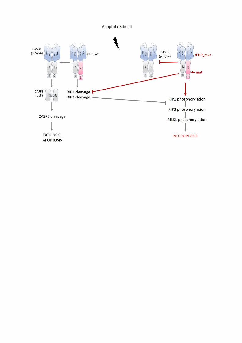
P-MonoG-166
Detailed analysis of Tnap function in zebrafish paves the way for establishment of a new in vivo model constituting the rare disease hypophosphatasia
A. Borst 1, B. Ohlebusch 1,2, R. Hark 1, E. Riekert 1, K. Marnet 1,3, F. Jakob 1,3, E. Klopocki 1, S. Graser3, D. Liedtke1
1Julius-Maximilians-Universität Würzburg, Institut für Humangenetik, Würzburg, Deutschland 2Universität zu Köln, Institut für Zoologie, Köln, Deutschland 3Julius-Maximilians-Universität Würzburg, Bernhard-Heine-Centrum für Bewegungsforschung, Würzburg, Deutschland
Hypophosphatasia (HPP) is a rare, hereditary disease caused by mutations in the ALPL gene coding for the ectoenzyme tissue-nonspecific alkaline phosphatase (TNAP). The different disease manifestations, ranging from odonto to perinatal lethal HPP forms, prominently display severe defects during dental and bone mineralization in patients. Apart from these symptoms, the disease can sporadically cause craniosynostosis as well as neurological disorders like epileptic seizures, anxiety, and depression. Currently, different cell culture and mouse models for experimental investigation of HPP symptoms have been established, but to date lack affirmation in a non-mammalian species. Our work aims to establish the zebrafish (Danio rerio) as an auxiliary in vivo vertebrate model system for detailed functional investigations of TNAP/Tnap and visualization of tissue-specific effects of HPP.
Initially, we identified a single gene homologue corresponding to ALPL in the zebrafish genome (alpl) and conducted detailed expression analyses at different developmental stages by whole mount in situ hybridization (ISH), qPCR, and combined ISH-immunostaining. Zebrafish embryos showed prominent alpl expression in distinct brain regions, nephros, retina, tooth buds and fins. In the adult zebrafish brain, alpl mRNA and Tnap activity were detected in restricted regions, like the amygdala, an organ which is linked to anxiety disorders in humans. Additionally, alpl expression was observed in proliferating areas of adult neurogenesis and implying a potential function within neural stem cells. Further functional experiments utilizing zebrafish embryos showed that inhibition of Tnap function, either by using chemical compounds like levamisole or by mRNA splicing blockage via Morpholino antisense oligonucleotides, result in consistent developmental alterations. Treated embryos display prominent mineralization and neural effects. Finally, we aim to develop several zebrafish transgenic lines to investigate different HPP symptoms, by either a general or a tissue-restricted knockdown in bones, teeth, and in the nervous system, or by overexpression of different human ALPL variants. Preliminary results from these lines imply that the zebrafish is a useful option to model HPP, to analyze molecular consequences of reduced TNAP/Tnap function, and to visualize altered developmental processes in different tissues. The establishment of these diverse molecular techniques in zebrafish paves the way for future tissue-specific investigations and consequently helps providing new molecular insights into functional consequences of HPP-causing ALPL mutations in vivo.
P-MonoG-167
Novel variants broaden the mutational spectrum of Hereditary Sensory and Autonomic Neuropathy disorders
A. Lischka1, K. Eggermann1, A. Çakar2, C. J. Record3, M. Elbracht1, T. Hornemann4, J. Senderek5, Y. Parman2, M. Auer-Grumbach6, M. M. Reilly3, G. Woods7, P. Laššuthová8, I. Kurth1
1University Hospital RWTH Aachen, Institute of Human Genetics, Aachen, Deutschland 2Istanbul University, Istanbul Faculty of Medicine, Department of Neurology, Neuromuscular Unit, Istanbul, Türkei 3UCL Queen Square Institute of Neurology, Department of Neuromuscular Diseases, Centre for Neuromuscular Diseases, London, Vereinigtes Königreich 4University Hospital Zurich, Department of Clinical Chemistry, Zurich, Schweiz 5Ludwig-Maximilians-University, Department of Neurology, Friedrich-Baur-Institute, Munich, Deutschland 6Medical University of Vienna, Department of Orthopedics and Trauma Surgery, Vienna, Österreich 7Cambridge Biomedical Campus, Cambridge Institute for Medical Research, Cambridge, Vereinigtes Königreich 8Charles University in Prague and Motol University Hospital, 2nd Faculty of Medicine, Department of Paediatric Neurology, Prague, Tschechien
Introduction: Currently approximately 20 genes have been identified in which pathogenic sequence variants lead to a monogenetic disorder of lack of pain perception. This includes clinical entities such as hereditary sensory and autonomic neuropathies (HSAN) and congenital insensitivity to pain (CIP). Clinically, the various disorders manifest themselves through repeated trauma and mutilation. Yet, small individual patient cohorts and the lack of standardized phenotype information hinder the complete elucidation of these genetic disorders.
Methods: The European Network on Inherited Sensory Neuropathies and Insensitivity to Pain (ENISNIP) was established by seven research centers and two patient advocacy organizations specialized on HSAN/CIP and it aimes at accumulating the knowledge from clinicians, geneticists, basic scientists and patients. For the present work, existing sequencing datasets of the ENISNIP project partners and collaborating research institutions were screened and novel likely pathogenic alterations in the already known HSAN/CIP genes were compiled.
Results: In 43 patients, we identified 45 likely disease-causing novel variants in the following HSAN/CIP genes: ATL3, DST, FLVCR1, NGF, NTRK1, PRDM12, RAB7A, SCN9A, SPTLC2 and WNK1. All variants were rare or absent from control cohorts and none had previously been reported in the literature. If applicable, the pathogenicity was corroborated by segregation analyses within the families.
Conclusions: Through compiling the existing sequencing data within the network, here we report on 45 novel pathogenic variants in known HSAN/CIP genes. This work thus expands the mutational spectrum of HSAN/CIP, gives insights in the pathogenicity and facilitates future diagnosis of affected patients of these rare disorders.
P-MonoG-168
Phenotypic characterization of individuals with a PRKAR1B-associated neurodevelopmental disorder
F. Marbach1, B. S. Lipska-Ziętkiewicz2, V. Michaud3, H. Margot3, J. Lespinasse4, F. Tran Mau-Them5,6, C. Coubes7, J. Park8, A. Knurowska9, S. Grosch8, C. Roggia8, P. Caro1, C. P. Schaaf1
1Heidelberg University, Institute of Human Genetics, Heidelberg, Deutschland 2Medical University of Gdańsk, Centre for Rare Diseases, Clinical Genetics Unit, Department of Biology and Genetics, Gdańsk, Polen 3Univ. Bordeaux, MRGM INSERM U1211, CHU de Bordeaux, Service de Génétique Médicale , Bordeaux, Frankreich 4CH-Chambéry, Génétique Chromosomique, Chambéry, Frankreich 5CHU Dijon Bourgogne, Unité Fonctionnelle Innovation en Diagnostic Génomique des maladies rares, Dijon, Frankreich 6INSERM UMR1231, Génétique des Anomalies du Développement GAD, F-21000, Dijon, Frankreich 7CHU de Montpellier, Service de Génétique Clinique, Département de Génétique Médicale, Maladies Rares et Médecine Personnalisée, Montpellier, Frankreich 8University of Tübingen, Institute of Medical Genetics and Applied Genomics, Tübingen, Deutschland 9Medical University of Gdańsk, II Department of Radiology, Gdańsk, Polen
The genetic landscape of intellectual disability (ID) is diverse and still expanding, reflecting the complex developmental biology of the human central nervous system (CNS). The discovery of new genetic etiologies of ID has been accelerated by the broad application of exome/genome sequencing in clinical practice, with well over 1500 syndromic and non-syndromic genetic disorders (with an additional >1250 candidate genes) listed in the SysID database as of Dec. 2021. In Apr. 2021, we published the characterization of a neurodevelopmental disorder caused by de novo missense variants in the PRKAR1B gene in 6 patients, who were mostly recruited from a large cohort of patient/parent trio-exomes (PMID: 33833410). Four of the 6 initial patients carried the same de novo missense variant NM_001164760 c.1003C>T, p.Arg335Trp. As of Dec. 2021, clinical data on 5 additional patients carrying the p.Arg335Trp variant have been collected by our group through different collaborations. Symptoms of the PRKAR1B-related disorder include developmental delay, autistic behavior, apraxia or clumsiness, and, in the case of the recurring p.Arg335Trp variant, insensitivity to pain of varying degree.
PRKAR1B encodes the R1β subunit of the protein kinase a (PKA) complex, a heterotetramer of two regulatory and two catalytic subunits. PKA mediates downstream signaling of the second messenger cAMP, and its activity is important to a plethora of biological processes. While ubiquitously present across human tissues, the composition of the PKA complex varies in different cell types even within the same organ. The R1β subunit is highly expressed in the CNS of rodents and humans, and RNAscope imaging performed in human embryos at Carnegie stage 22 (estimated postfertilization age of 52 to 55 days) demonstrated expression of PRKAR1B also during embryonal development of the brain. Functional studies in HEK293 cells transfected with PRKAR1B constructs harboring variants identified in our patients, demonstrated reduced basal enzymatic activity of PKA in the presence of mutant R1β. It should be noted that the phenotypes of R1β-deficient (biallelic knockout) mice and rats seem to approximate some aspects of the phenotype of patients carrying monoallelic PRKAR1B-variants, such as deficits in spatial learning and memory formation in rats, and abnormal nociception in both species.
Having recently established a new genetic disease association, we are working to characterize the phenotype in more detail by recruiting and phenotyping additional patients, to identify possible genotype-phenotype correlations, and to develop a comprehensive and testable molecular disease model, which can be validated by in vitro experiments. We present the latest unpublished results of our ongoing effort.
P-MonoG-169
VPS13A disease shows variable chorein expression due to distinctive VPS13A mutations as observed in the largest cohort of 106 international patients
G. Miltenberger-Miltenyi1, B. Bader2, A. Velayos-Baeza3, C. Dobson-Stone4, A. P. Monaco5, A. Danek1
1Ludwig-Maximilians-Universität München, Neurologische Klinik und Poliklinik, Munich, Deutschland 2Schön Klinik Bad Aibling Harthausen, Bad Aibling, Deutschland 3The Wellcome Trust Centre for Human Genetics, University of Oxford, UK, Oxford, Vereinigtes Königreich 4Brain and Mind Centre and School of Medical Sciences, University of Sydney, Australia, Sydney, Australien 5Ballou Hall, Tufts University, Medford, MA, USA, Medford, Vereinigte Staaten
Background: VPS13A disease (Chorea-acanthocytosis, ChAc, OMIM #200150, chorein protein, VPS13A gene) is an autosomal recessively inherited devastating and uncurable neurologic disease, with an estimated prevalence of around 1500 affected individuals world-wide. Due to its rarity and unspecific initial symptoms, diagnosis delay is usually extended. Here we present the spectrum of associations between VPS13A mutations and the chorein status in the world-wide largest international VPS13A disease cohort of 106 ChAc patients from 18 different countries across 5 continents.
Methods: We collected clinical data and blood samples of >700 international patients with suspected ChAc, during the last 15 years. We analyzed the presence of chorein in erythrocyte membrane extracts with Western blot. The VPS13A gene was sequenced at our centre and elsewhere.
Results: We present 74 unique pathogenic VPS13A mutations (2 novel) with no evidence of mutation clustering. These variants included missense, nonsense, small-scale and gross deletions, as well as splice site variants. The mutations led to a complete loss of chorein in most patients. However, one patient showed normal chorein levels with an N-terminal antibody, while absent chorein when using a C-terminal antibody. Furthermore, in 6 cases, where pathogenic mutations were detectable in the VPS13A gene, normal chorein levels were repeatedly detectable, indicating different effects of such mutations on the protein.
Conclusions: Our study gives a broad insight to the genotype and chorein status of VPS13A disease and contributes to the understanding of the natural history of this condition. The data showed that In addition to clinical symptoms, testing of chorein levels is essential to correctly interpret functional consequences of VPS13A variants. Based on these findings we currently elaborate specific guidelines helping the early diagnosis of this rare disease, thus the reduction of diagnosis delay. The VPS13A mutations and related clinical data are currently made accessible at the open access LOVD mutation database.
Disclosure: We thank the Advocacy for Neuroacanthocytosis Patients for their generous support of this study.
P-MonoG-170
De novo variants in ATP2B1 lead to neurodevelopment delay
H. Oppermann1, J. Rahimi1, N. Urban2, M. Wegler1, H. Sticht3, M. Schaefer2, B. Popp1, F. Gaunitz4, M. Morleo5, V. Nigro5, S. Maitz6, G. M. S. Mancini7, C. Ruivenkamp8, E. K. Suk9, T. Bartolomaeus1,10, A. Merkenschlager11, D. Koboldt12, D. Bartholomew12, A. P. A. Stegmann13, M. Sinnema13, I. Duynisveld14, R. Salvarinova15, S. Race16, B. B. A. de Vries15, A. Trimouille17, S. Naudion17, D. Marom18, U. Hamiel18, N. Henig18, F. Demurger19, N. Rahner20, E. Bartels20, J. A. Hamm21, A. M. Putnam21, R. Person22, M. M. Morrow22, R. A. Jamra1
1Universitätsklinikum Leipzig AöR, Institut für Humangenetik, Leipzig, Deutschland 2Universitätsklinikum Leipzig AöR, Rudolf-Boehm-Institute of Pharmacology and Toxicology, Leipzig, Deutschland 3Friedrich-Alexander-Universität Erlangen-Nürnberg, Institute of Biochemistry, Erlangen, Deutschland 4Universitätsklinikum Leipzig AöR, Klinik und Poliklinik für Neurochirurgie, Leipzig, Deutschland 5University of Campania, Telethon Institute of Genetics and Medicine, Naples, Italien 6San Gerardo Hospital, Clinical Pediatric Genetic Unit, Monza, Italien 7ErasmusMC University Medical Center, Department of Clinical Genetics, Rotterdam, Niederlande 8Leiden University Medical Center, Clinical Genetics, Leiden, Niederlande 9Praxis für Humangenetik-Friedrichstrasse, Berlin, Deutschland 10CeGaT GmbH and Praxis für Humangenetik , Tübingen, Deutschland 11Universitätsklinikum Leipzig AöR, Department of Neuropediatrics, Leipzig, Deutschland 12Nationwide Children's Hospital, Columbus, Vereinigte Staaten 13Maastricht University Medical Center+ , Department of Clinical Genetics, Maastricht, Niederlande 14Severinus Institute for Intellectual Disability, Veldhoven, Niederlande 15Radboud university medical center, Department of Human Genetics, Nijmegen, Niederlande 16University of British Columbia, BC Children’s Hospital,, Division of Biochemical Genetics, Vancouver, Kanada 17Universitaire de Bordeaux, Bordeaux, Frankreich 18Tel Aviv University, Tel Aviv, Israel 19service de génértique CHBA, Vannes, Frankreich 20Institute for Clinical Genetics, Bonn, Deutschland 21East Tennessee Children’s Hospital, Knoxville, Vereinigte Staaten 22GeneDx, Inc., Gaithersburg, Vereinigte Staaten
We describe a cohort of 13 unrelated individuals with probable causative variants in ATPase plasma membrane Ca2+ transporting 1 (ATP2B1) ascertained through international matchmaking efforts. All probands share the phenotype of mild to moderate global developmental delay. Other common clinical features include autism (6/12), seizures (7/13), and distal limb abnormalities (4/13), such as clinodactyly and arachnodactyly. Ten variants in ATP2B1 were proved de novo, while for the remaining three individuals parents were unavailable for segregation. Ten probands had missense variants of which eight were in specific functional domains; the other three individuals carry nonsense variants. ATP2B1 encodes a plasma membrane calcium-transporting protein, which plays a central role in calcium homeostasis and is mainly expressed in the central nervous system. 3D structural protein modeling suggested that the variants have a destabilizing effect on the protein. We performed Ca2+ imaging after introducing all ten missense variants in transfected HEK293 cells and showed that nine variants lead to a significant decrease in Ca2+ export capacity compared with the wild type construct. These nine variants also exhibited incorrect intracellular localization of ATP2B1, which suggested a loss of function mechanism. The genetic and phenotypic similarities among probands as well as the functional analyses imply that de novo variants in ATP2B1 cause a novel monogenic neurodevelopmental disorder.
P-MonoG-171
Detection of a Structural Variant with Short Read Whole Genome Sequencing in a Young Woman with Recessive Limb Girdle Muscular Dystrophy
N. Pluta1, S. Hoffjan2, F. Zimmer1, C. Köhler3, H. P. Nguyen2, M. Vorgerd4, J. Mohr4, D. Atlan5, B. Wolf6, A. K. Zaum1, S. Rost1
1Institut für Humangenetik, Biozentrum, Würzburg, Deutschland 2 Ruhr-Universität Bochum, Abteilung für Humangenetik, Bochum, Deutschland 3Katholisches Klinikum, Klinik für Kinder- und Jugendmedizin, Bochum, Deutschland 4BG-Universitätskliniken Bergmannsheil, Heimer-Institut für Muskelforschung, Bochum, Deutschland 5Phenosystems SA, Braine le Chateau, Belgien 6University of Applied Sciences Western Switzerland, iCoSys, Fribourg, Schweiz
Introduction
Despite advancing technology in human genetic diagnostics, some patients still remain genetically unsolved even when, for example, a muscle biopsy or clinical history gives a clear indication for the examination of certain genes. The methods commonly used in routine diagnostics, such as whole exome sequencing, often do not cover large areas of genes, especially intronic or regulatory regions. These areas, which may contain variants that negatively affect the gene product, can now be investigated by whole genome sequencing (WGS), allowing many previously unsolved cases to receive a genetic diagnosis after all.
Material/Methods
In the present case, a 22-year-old female patient has severely elevated CK values of about 15000 U/l and proximal muscle weakness. In an immunohistological examination of muscle tissue no gamma-sarcoglycan could be stained and dystrophin was only weakly stainable. A short read WGS was performed on the patient´s DNA with a NovaSeq6000 (Illumina) and analyzed with GensearchNGS (Phenosystems).
Results
In routine diagnostics, which included 65 muscle genes in a panel analysis comprising genes for sarcoglycans and dystrophin, no relevant genetic alterations could be found. Genome analysis revealed a homozygous inversion on chromosome 13 in a larger homozygous region in the patient. The two breakpoints are located in intron 2 of SGCG and upstream of LINC00621. The parents are each heterozygous carriers of the inversion. Analysis of mRNA from a muscle biopsy showed that neither SGCG nor LINC00621 was expressed in the patient. The breakpoint in SGCG results in the inability to form gamma-sarcoglycan, leading to the manifestation of the autosomal recessive limb-girdle muscular dystrophy 5 (LGMDR5) in the patient.
Discussion
As far as known, inversions in SGCG and other sarcoglycan genes have not yet been described as causative for recessive LGMDs. This case shows well that even short read WGS can reveal structural variants and it is especially notable that the detection of the here presented variant would have taken much longer and would have been less accurate by conventional methods compared to whole genome analysis.
P-MonoG-173
Fatty acid synthesis suppresses dietary polyunsaturated fatty acid use
A. Worthmann1, Sh.Y.L. Piel2, A.W. Fischer1,3,4, M. O’Farell5, S. Adak6, K. Kloth2, M. Sundd7, H. Siffeti2, M. Musfeldt2, T. Bierhals2, M. Heine1, M. Pauly1, F. Haumann2, S. Kundu8, M. M. Fuh1, A. Neu,9, C. Beysen10, P. Pertzborn1, H. Schlüter11, U. Knippschild12, K. Tödter1, M. Hempel2, C. F. Semenkovich6, J. Heeren1, L. Scheja1, C. Kubisch2, Ch. Schlein2
1Universitätsklinikum Hamburg-Eppendorf, Institut für Biochemie und Molekulare Zellbiologie, Deutschland 2Universitätsklinikum Hamburg-Eppendorf, Institut für Humangenetik, Deutschland 3Harvard University, Department of Molecular Metabolism, Boston, Vereinigte Staaten 4Harvard Medical School, Department of Cell Biology, Boston, Vereinigte Staaten 5Sagimet Biosciences, San Mateo, Vereinigte Staaten 6Washington University, Department of Medicine, St. Louis, Vereinigte Staaten 7National Institute of Immunology, New Delhi, Indien 8University of Delhi South Campus, Department of Biochemistry, New Delhi, Indien 9Universitätsklinikum Hamburg-Eppendorf, Department of Pediatrics, Deutschland 10FluxBio, San Mateo, Vereinigte Staaten 11Universitätsklinikum Hamburg-Eppendorf, Klinische Chemie und Laboratoriumsmedizin, Deutschland 12Department of General and Visceral Surgery, University Hospital Ulm, Deutschland
Background: Metabolic diseases such as obesity, type 2 diabetes mellitus and non-alcoholic steatohepatitis (NASH) are closely linked to aberrant synthesis of endogenous fatty acids in the liver, called de novo lipogenesis, which is mediated by the enzyme fatty acid synthase (FASN). The newly synthesized fatty acids from the lipogenesis pathway are either saturated or monounsaturated and can adversely affect metabolic health through mechanisms that are not sufficiently understood.
Methods:By trio whole exome sequencing of a 5-year-old patient and his parents, we identified a putatively pathogenic de novo missense variant in FASN. We characterized the consequences of this hypofunctional variant by investigating plasma lipidomics and compared them to a preclinical mouse model of genetically low lipogenesis. Further, the results were confirmed and generalized by analysis of hepatic fatty acid profiles in a cohort of patients with high FASN expression in the liver and from NASH patients treated with FASN inhibitor TVB-2640 (ClinicalTrials.gov number NCT03938246).
Results: Lipidomic analysis of the index patient revealed that de novo low lipogenesis as caused by the hypofunctional FASN mutation leads to a surprising but substantial plasma increase of the strictly dietary class of polyunsaturated fatty acids (PUFA). By comparative analysis of both a preclinical mouse model of genetically reduced lipogenesis, a human cohort with exceptionally high levels of lipogenesis and NASH patients treated with a FASN inhibitor, we indeed confirmed and extended the findings that endogenous lipogenesis suppresses the use of dietary PUFA.
Conclusions: We provide evidence that endogenous lipogenesis determines the use of PUFA and therefore might substantially affect the metabolic benefit of dietary or therapeutically administrated polyunsaturated fatty acids in humans.
P-MonoG-174
*** Retinoschisin and novel interaction partners of the Na/K-ATPase define a growing protein complex at the inner segments of photoreceptor cells
V. Schmid1, A. Wurzel1, K. Plössl1, C. Wetzel2, A. Bruckmann3, B. Weber1,4, U. Friedrich1
1University of Regensburg, Institute for Human Genetics, Regensburg, Deutschland 2University of Regensburg, Department of Psychiatry and Psychotherapy, Regensburg, Deutschland 3University of Regensburg, Biochemistry Center Regensburg, Laboratory for RNA Biology, Regensburg, Deutschland 4University Clinics Regensburg, Institute of Clinical Human Genetics, Regensburg, Deutschland
Purpose: Mutations in the RS1 gene cause X-linked juvenile retinoschisis (XLRS), a hereditary retinal dystrophy manifesting in juvenile or adolescent males. Recently, we and others showed that the retinoschisin protein encoded by RS1 binds to the retinal Na/K-ATPase and modulates its localization at the plasma membrane of the inner segments of mammalian photoreceptors. Here, we aimed to identify novel interaction partners of the Na/K-ATPase-retinoschisin complex further elucidating the role of retinoschisin as a putative regulator of photoreceptor membrane compartmentalization.
Methods: Porcine retinal lysates were subjected to co-immunoprecipitation targeting the α3-subunit of the Na/K-ATPase (ATP1A3). Bound proteins were eluted and separated by SDS-PAGE for mass spectrometric analysis. Identified proteins were verified in co-immunoprecipitation experiments with murine retinal lysates. Localization of proteins of interest was investigated via immunohistochemistry in eyes from wildtype (wt) and retinoschisin-deficient (Rs1h knockout, Rs1tm1Web) mice. Effect of retinoschisin-deficiency on the total mRNA and protein level was investigated in retinoschisin-deficient murine retinal lysates. In addition, the Kv channel regulated ion flow was analyzed by patch-clamp analysis of Y-79 cells, incubated with or without purified retinoschisin.
Results: Mass spectrometry identified the voltage-gated potassium ion channel (Kv) subunits Kv2.1 and Kv8.2. Binding to the retinal Na/K-ATPase was independently verified. Immunohistochemical analyses in murine retinal cryosections revealed Kv localization to the inner photoreceptor segments, fully overlapping with the localization of retinoschisin and the retinal Na/K-ATPase. In retinae from retinoschisin-deficient mice, Kv2.1 and Kv8.2 revealed a pathological distribution in line with the findings of the retinal Na/K-ATPase. While Kv2.1 and Kv8.2 total protein amount is greatly reduced, protein expression of the retinal Na/K-ATPase remains unaffected. Of note, retinoschisin-deficiency appears to have no effect on Kv channel regulated potassium ion flow.
Conclusions: Our findings show that Kv subunits Kv2.1 and Kv8.2 are part of a growing macromolecular complex at the photoreceptor inner segments together with retinoschisin and the retinal Na/K-ATPase. Defective compartmentalization of this complex in early stages of retinal developmental may be a critical step in XLRS pathogenesis possibly rendering a simple gene replacement therapy of the retinoschisin protein little effective.
P-MonoG-175
Genetic Polymorphisms and their impact on the Age at Onset in Spinocerebellar Ataxia Type 3
T. Laidig1, J. Jung1,2, C. Meyer1, L. Czisch1, R. M. Burger1, P. Pereira Sena1,2, J. J. Weber1,3, D. Weishäupl1, L. Bannach Jardim4, M. L. Saraiva-Pereira5, M. C. França Jr.6, C. R. Gordon7, M. R. Cornejo-Olivas8, T. Ott1, O. Rieß1,2, T. Schmidt1,2
1Universität Tübingen, Institut für Medizinische Genetik und Angewandte Genomik, Tübingen, Deutschland 2Universitätsklinikum Tübingen, Zentrum für seltene Erkrankungen (ZSE), Tübingen, Deutschland 3Ruhr-Universität Büchum, Humangenetik, Bochum, Deutschland 4Hospital de Clinicas de Porto Alegre, Porto Alegre, Brasilien 5Universidade Federal do Rio Grande do Sul, Porto Alegre, Brasilien 6Universidade Estadual de Campinas (UNICAMP), Campinas, Brasilien 7Tel Aviv University, Tel Aviv, Israel 8Instituto Nacional de Ciencias Neurológicas, Lima, Peru
Spinocerebellar ataxia type 3 (SCA3) or Machado-Joseph disease (MJD) is an autosomal-dominantly inherited, neurodegenerative disorder caused by the expansion of a CAG repeat in the ATXN3 gene resulting in an expanded polyglutamine repeat in the encoded ataxin-3 protein. SCA3/MJD, therefore, belongs to the group of polyglutamine. Statistically, a correlation between the number of CAG repeats and the age at onset of SCA3 patients exists and patients with more CAG repeats have an earlier onset of symptoms. However, this statistical correlation is not perfect and the number of CAG repeats contributes only about 55% to the age at onset. Therefore, the remaining 45% are influenced by other factors, which we aim to identify in this study.
In order to identify modifiers of the disease progression, we genotyped in a combined European and South American approach more than 500 SCA3/MJD patients for promising polymorphisms in candidate genes.
Candidate genes included ataxin-3 itself and known interaction partners of ataxin-3, functional modifiers identified in previous studies as well as genes with known relevance for the pathophysiology of SCA3/MJD. We selected polymorphisms with a high likelihood of having a functional relevance i.e. polymorphisms in the promoter regions as well as polymorphisms leading to amino acid changes. While controlling for ethnic origin we assessed the contribution of the respective polymorphism to the age at onset in addition to the already known modifying factor, the length of the expanded CAG repeat within ATXN3. We indeed identified interesting polymorphisms contributing to the age at onset including certain haplotypes within ATXN3 itself.
Subsequent functional characterizations will reveal the impact of these polymorphisms on pathogenic mechanisms in SCA3/MJD. We hope that our results will improve the prediction of clinical symptoms and contribute to the understanding of pathogenic processes in SCA3/MJD.

P-MonoG-176
Posterior lissencephaly caused by domain specific missense-variants in CEP85L
I. Schumann1, S. Khosrawikatoli2, M. Karnstedt1, R. Abou Jamra1, B. Popp1
1Universitätsklinikum Leipzig, Leipzig, Deutschland 2Kleinwachau Sächsisches Epilepsiezentrum, Radeberg, Deutschland
Lissencephaly or "smooth brain" is a neurodevelopmental disorder (NDD) characterized by the absence of normal cerebral convolutions with abnormalities of cortical thickness. It can affect the whole brain but also be region specific.
Here, we report a 39-year-old man with a pronounced lissencephaly with gyri malformation in the parietal, occipital and temporal region. He developed seizures at age of six months. Further, he was delayed with walking and language development. He presented with gait insecurity, blurred language, and only simple language understanding. Electroencephalography tests correspond to a focal epilepsy. Using exome sequencing, we identified a heterozygous missense variant in CEP85L at c.191C>T, p.(Ser64Phe).
Because a paternal sample was not available, we used allele specific RT-PCR leveraging nearby heterozygous polymorphisms segregated in the mother and showed that the variant is located on the paternal allele. As the father was described as healthy, a de novo occurrence on the paternal allele seems likely but cannot be further discerned, highlighting an underappreciated problem in older individuals with NDD. The region between amino acids (AA) 39-77, other pathogenic missense and in-frame variants have been reported. The affected AA position is conserved, and multiple in-silico analyses predicted a damaging effect (CADD = 27.9). Using structural modeling and multiple sequence alignments we show that all described variants fall into an unstructured, but locally highly conserved N-terminal protein domain. Considering this local clustering and the low protein wide conservation scores (gnomAD pLI: 0, Z-score: 0.4) a currently unknown functionally critical domain in this region seems likely.
We annotated phenotypic features of all described individuals (n=19) with CEP85L-associated disease using Human Phenotype Ontology (HPO) terms. Variable NDD were present in 100% with global developmental delay in 42%, speech or motor delay in 37% and 16%, respectively, and intellectual disability in 32%. Abnormal cortical gyration and lissencephaly were present in 84% and 47% had subcortical band heterotopia. Most individuals had different types of seizures (95%).
Despite lacking segregation, one can classify the variant c.191C>T, p.(Ser64Phe) as likely pathogenic according to the American College of Medical Genetics (ACMG) guidelines when taking into account this relatively specific phenotype (PM1: hot spot, PM2: MAF = 0 , PP3: in silico pathogen, PP4: specific phenotype). After the recent initial descriptions of a CEP85L associated disorder (OMIM: #618873), the identification of the variant in the individual presented here further supports the implication of the CEP85L gene with posterior cortex lissencephaly with associated clinical characteristics. Further studies will be needed to elucidate the structure of the critical protein region and clarify the pathomechanism associated with N-terminal CEP85L variants.
P-MonoG-177
Homozygous variant in SERPING1 causes hereditary angioedema in a consanguineous Brazilian family
L. Sella Motta Maia1, B. Burger1, A. Ghannam2, F. Leonel Nunes3, M. Mendonça Dias3, C. Drouet4,5, L. K. Arruda3, S. Cichon1,6,7
1 Universitätsspital Basel, Departement Biomedizin, Basel Stadt, Schweiz 2KininX SAS, Grenoble, Frankreich 3Ribeirão Preto Medical School, University of São Paulo, Internal Medicine Department, Ribeirão Preto, SP, Brasilien 4 Institut Cochin, Université de Paris, INSERM UMR1016,, Paris, Frankreich 5Univ. Grenoble-Alpes & Centre Hospitalier Universitaire de Grenoble, Grenoble, Frankreich 6Institute of Medical Genetics and Pathology, University Hospital Basel, Basel, Schweiz 7Institute of Neuroscience and Medicine (INM-1), Research Center Juelich, Juelich, Deutschland
Introduction
Hereditary angioedema (HAE) is characterized by recurrent episodes of severe and potentially life-threatening non-pruritic subcutaneous and submucosal edema, often affecting the upper airways. Different clinical and genetic subtypes exist, and the most common forms (HAE types I/II) are caused by dominant variants in the SERPING1 gene, resulting in C1-inhibitor (C1INH) deficiency. In HAE-1, variants result in reduced expression of functionally C1-INH protein. In HAE-2, variants impact on the function of C1-INH. C1-INH is a highly glycosylated serine-protease-inhibitor (SERPIN) that regulates multiple proteases pathways, in particular kallikrein-kinin system (KKS). In patients with HAE, C1-INH deficiency affects KKS control, resulting in excessive release of bradykinin (BK), the predominant mediator of enhanced vascular permeability in angioedema attacks. At present, few SERPING1 mutations were described that act in a recessive fashion. Here, we describe a novel recessive mutation in a consanguineous HAE family from Brazil
Materials and Methods
Two sisters from consanguineous, unaffected parents were severely affected by HAE since adolescence (13 yrs.) and young adulthood (28 yrs.), respectively. Both fulfilled the diagnostic criteria for HAE-1. No other member of this large, four-generation Brazilian family presented any symptoms of HAE. We extracted gDNA from whole blood of 34 family members (23 women and 11 men) and sequenced the coding region of the SERPING1 gene.Antigenic C1-INH, C4, and C1q were measured by radial
immunodiffusion, CI-INH function was measured using C1s and Kallikrein proteases. Anti‐C1-INH immunoblot was performed on 7.5% SDS‐PAGE under non-reducing conditions
Results
In both symptomatic sisters, we found a homozygous missense variant in exon 6 (c.964G>A); p.(Val322Met). Fourteen family members (including the sisters" parents) were heterozygous carriers of the variant. Detailed clinical evaluation of these individuals by an expert physician excluded any HAE symptoms. DNA from eighteen individuals presented with the wild-type.Variant p.(Val322Met) affects a highly conserved position among serpins (80%) that is located within the breach/gate region, recognized as highly strategic for serpin function. C1-INH function was more affected using C1s than Kallikrein for heterozygous patients; for homozygous patients both proteases were not controlled. C1-INH displays molecular species distribution characteristic of an intermediate type
Conclusions
HAE I/II is typically an autosomal dominant condition caused by more than 800 described SERPING1 variants. There are few reports of HAE variants acting in a recessive fashion. The novel recessive variant identified in several members of our Brazilian family gives us the opportunity to study the effect of the C1-INH p.(Val322Met) variant on the structure-function relationship in the ongoing international efforts toward the recognition of C1-INH deficiency as a conformational disease

PIC
P-MonoG-178
Human models for White Sutton syndrome: POGZ mutations change the transcriptome and induce defects in neural progenitor cell biology
A. Soliman1, M. Bertin1, S. Schweiger1, J. Winter1
1Institute of Human Genetics University Medical Centre Mainz, Mainz, Deutschland
Pogo transposable element derived with ZNF domain (POGZ) has been identified as one of the most recurrently mutated genes in patients with neurodevelopmental disorders (NDDs), including autism spectrum disorder (ASD), intellectual disability, and White-Sutton syndrome; however, the underlying disease-causing cellular and molecular mechanisms are still unclear. Here, we generated several induced pluripotent stem cell lines (iPSCs) with heterozygous POGZ mutations, either derived from patient fibroblasts or introduced by CRISPR / Cas9 genomic editing, and differentiated them into neural progenitor cells (NPCs), neurons, and cerebral organoids. We demonstrate that frameshift mutations, either in the N-terminus or in the HP1-binding zinc finger-like (HPZ) domain, decrease POGZ protein expression but do not impair nuclear localization of the wildtype POGZ protein produced from the second allele. By using a 3D neurosphere model we show that POGZ deficiency impairs self-renewal activity and enhances NPC differentiation and neuronal migration. Furthermore, organoids carrying POGZ mutations formed smaller areas of ventricle-like structures than control organoids, suggesting that POGZ regulates neuronal development and cytoarchitecture. POGZ binds to chromatin and has been suggested to act as a transcriptional regulator. To identify transcriptional changes caused by heterozygous POGZ mutations, we carried out RNA sequencing of the iPSC-derived NPCs. At the GfH congress, we will report on the detected transcriptomic changes.

P-MonoG-179
KIF12 variants and disturbed hepatocyte polarity in children with a phenotypic spectrum of cholestatic liver disease
A. Stalke1,2, M. Sgodda3, T. Cantz3, B. Skawran1, E. Lainka4, B. Hartleben5, U. Baumann2, E. D. Pfister2
1Medizinische Hochschule Hannover, Institut für Humangenetik, Hannover, Deutschland 2Medizinische Hochschule Hannover, Pädiatrische Gastroenterologie und Hepatologie, Hannover, Deutschland 3Medizinische Hochschule Hannover, Translationale Hepatologie und Stammzellbiologie, Hannover, Deutschland 4Universitätsklinikum Essen, Päd. Nephrologie, Gastroenterologie, Endokrinol., Essen, Deutschland 5Medizinische Hochschule Hannover, Institut für Pathologie, Hannover, Deutschland
Objectives and study:
Recently, KIF12 has been identified as a cholestasis-associated candidate gene. As KIF12 is a member of a microtubule-associated motor protein family involved in organizing the cytoskeleton and intracellular transport, KIF12-associated cholestasis is assumed to be a result of disturbed cell polarity.
We describe six cases with likely pathogenic KIF12 variants from four unrelated families, their different phenotypes and our investigations to study hepatocyte polarity.
Methods:
Children with familial cholestasis and a likely pathogenic variant in the KIF12 gene were identified by exome sequencing. Parents and siblings were tested for the variants to analyze segregation. Immunofluorescence imaging of apical markers MRP2 und BSEP, basolateral marker OATP1B1, tight junction protein ZO-1 and KIF12 itself was performed on patient"s liver tissue sections.
Results:
We detected two different homozygous KIF12 variants in five patients ((NM_138424.1) 4 patients: c.655C>T p.(Arg219*); 1 patient: c.482-4_500del p.?). Segregation analyses confirmed autosomal recessive inheritance. The patient"s clinical manifestation ranged from neonatal cholestasis with complete clinical remission, or absent clinical symptoms with the diagnosis made incidentally, to a progressive course ending in liver transplantation. Immunofluorescence imaging of liver sections of KIF12 patients revealed an ectopic cytoplasmic MRP2 staining. BSEP, and partly ZO-1 staining appeared in long clustered structures. KIF12 and OATP1B1 staining was widely unremarkable.
Conclusion:
Our results strongly support pathogenic KIF12 variants as cause for familial cholestatic liver disease and suggest that these variants result in functional cell polarity disturbance. Due to its wide clinical presentation with even asymptomatic cases, KIF12-associated cholestatic liver diseases are potentially underdiagnosed.

P-MonoG-180
Expanding the phenotypic and biochemical spectrum of NDUFAF3-related mitochondrial disease.
A. van der Ven1, I. Wente2, T. Bierhals1, K. Tsiakas3, R. G. Feichtinger4, J. A. Mayr4, M. Hempel1,5, I. Wittig2
1Institut für Humangenetik UKE Hamburg-Eppendorf, Hamburg, Deutschland 2Functional Proteomics, Institute of Cardiovascular Physiology, Goethe Universität , Frankfurt am Main, Deutschland 3Department of Pediatrics, UKE Hamburg-Eppendorf, Hamburg, Deutschland 4Salzburger Landeskliniken (SALK) and Paracelsus Medical University (PMU), Salzburg, Österreich 5Institute of Human Genetics, University Heidelberg, Heidelberg, Deutschland
Introduction: Mitochondrial disorders most frequently originate from impaired oxidative phosphorylation (OXPHOS) activity. Mitochondrial complex I (CI) is the largest OXPHOS component. It is composed of three functional modules: the Q-, the P- and the N-module. NDUFAF3 (NADH: Ubiquinone Oxidoreductase Complex Assembly Factor-3) is one of 13 proteins known to aid the correct assembly of this large multienzyme complex. Recent investigations have advanced the understanding of NDUFAF3 for CI biogenesis, pinpointing it to the step were the Q- and the P-module are joined. Recessive disease-causing variants in NDUFAF3 have previously been described as the cause of a distinct mitochondrial disease condition (OMIM # 618240). The seven patients reported to date exhibited severe neurologic symptoms and lactic acidosis, followed by death within the first two years of age in six cases.
Methods and Results: We present a 10-year-old patient with developmental delay, exercise intolerance, dystonia, basal ganglia abnormalities, and elevated lactate concentration in blood. Trio-exome sequencing revealed a paternally inherited splice site variant and a maternally inherited missense variant in NDUFAF3, classified as pathogenic and likely pathogenic, respectively, according to ACMG criteria. Spectrophotometric analysis of fibroblast-derived mitochondria demonstrated isolated and relatively mild reduction of CI in our patient. Complexome analysis revealed undetectable levels of NDUFAF3 and a severely decreased amount of fully assembled CI. Furthermore, an unusual occurrence of the assembly factors ACAD9 and ECSIT was noted on fully assembled complex I.
Conclusion: With this untypical presentation of a patient with onset of symptoms during early childhood and comparatively attenuated course, we provide further insight into the phenotypic spectrum of NDUFAF3-related mitochondrial disease. The relatively mild phenotype of our patient is likely a result of the paternally inherited splice-site variant. This variant is relevant for only one of the four coding NDUFAF3 transcript-isoforms, accounting for approximately 25% of NDUFAF3 expression across human tissues. The data obtained via complexome analysis are in line with the previously defined role of NDUFAF3 for CI biogenesis. Beyond that, the abnormal occurrence of the assembly factors ACAD9 and ECSIT additionally indicates an incomplete maturation of CI in our patient.
P-MonoG-181
A homozygous STING1 gene variant causes STING-associated vasculopathy with onset in infancy (SAVI)
R. Wan1, S. von Hardenberg1, L. I. Olfe1, B. Auber1, M. Wetzke2, D. Steinemann1
1Hannover Medical School, Department of Human Genetics, Hannover, Deutschland 2Hannover Medical School, Department of Pediatric Pneumology, Allergology and Neonatology, Hannover, Deutschland
Background: Stimulator of interferon response protein (STING) encoded by stimulator of interferon response cGAMP interactor 1 (STING1) plays a key role in activating the type I interferon response. STING1 gain of function (GoF) variants cause auto-activation without ligand binding and lead to a rare auto-inflammatory disease named STING-associated vasculopathy with onset in infancy (SAVI; MIM615934; AD inheritance). Patients with SAVI exhibit interstitial lung disease, recurrent fever and elevated interferon signature. Most of the causative heterozygous variants arise de novo. Interestingly, 8 SAVI patients from 5 different families carrying the STING1 homozygous variant R281W (Lin et al., 2020, Alghamdi et al., 2021). Functional in vitro analysis has shown that R281W leads to an autoactivation of STING, but the resulting GoF is weaker compared to other variants and that R281W leads to a SAVI phenotype only in a homozygous state.
Case report: Here we report on another patient carrying a homozygous variant R281W in STING1 that caused features of SAVI. The variant was identified by whole exome sequencing: STING1 (NM_198282.3):c.841C>T p. (Arg281Trp). Segregation analysis within the consanguineous family showed that each of the healthy parents as well as one healthy brother carry the variant in heterozygous state. A healthy sister did not carry the variant. The interferon signature was highly elevated in the patient and moderately elevated in the healthy heterozygous family members. This differs from published cases where the heterozygous R281W variant have been shown not to affect the interferon signature (Lin et al., 2020). Clinically, our patient presented with recurrent severe hypoxaemia and hypoventilation, diffuse alveolar hemorrhage and progressive interstitial lung disease, but no skin vasculitis. This raises the possibility that this variant may potentially lead to an atypical SAVI phenotype. A therapy with Janus kinase inhibitor Baricitinib was started, but no significant improvement of the clinical condition could be observed so far.
P-MonoG-182
THE RECURRENT MISSENSE MUTATION p.(Arg367Trp) IN YARS1 CAUSES A DISTINCT NEURODEVELOPMENTAL PHENOTYPE
D. Wieczorek1, L. Averdunk1,2, H. Sticht3, H. Surowy1, H. J. Lüdecke1, M. Koch-Hogrebe4, H. S. Alsaif5, K. Kahrizi6, H. Alzaidan5, B. S. Alawam5, M. Tohary5, C. Kraus7, S. Endele7, H. Najmabadi5, A. Reis7, F. S. Alkuraya5
1Medical Faculty and University Hospital Düsseldorf, Institute of Human Genetics, Düsseldorf, Deutschland 2Medical Faculty and University Hospital Düsseldorf, Department of General Pediatrics, Neonatology and Pediatric Cardiology, Düsseldorf, Deutschland 3Friedrich-Alexander-Universität Erlangen, Division of Bioinformatics, Erlangen, Deutschland 4University Witten/Herdecke, Children's Hospital Datteln, Datteln, Deutschland 5King Faisal Specialist Hospital and Research Center, Department of Genetics, Riyadh, Saudi-Arabien 6University of Social Welfare and Rehabilitation Sciences, Genetics Research Center, Tehran, Iran 7University Hospital Erlangen, Institute of Human Genetics, Erlangen, Deutschland
Pathogenic variants in aminoacyl-tRNA synthetases (ARS) cause a diverse spectrum of autosomal recessive disorders. Tyrosyl tRNA synthetase (TyrRS) is encoded by YARS1 (cytosolic, OMIM*603623) and is responsible of coupling tyrosine to its specific tRNA. We identified eleven individuals with the recurrent homozygous missense variant c.1099C>T;p.(Arg367Trp) (NM_003680.3) in YARS1. This variant causes a multisystemic disorder with developmental delay, microcephaly, failure to thrive and short statue. Affected individuals have muscular hypotonia, microcytic anemia, hepatomegaly, ataxia, brain anomalies, and hypothyroidism. TyrRS has two additional functional domains (N-Terminal TyrRSMini and C-terminal EMAP-II-like domain) which confer cytokine-like functions. In silico analyses show that the mutation p.(Arg367Trp) does not affect the catalytic domain responsible of enzymatic coupling, but destabilizes the cytokine-like C-terminal domain. Biallelic pathogenic variants that reside in different functional domains of TyrRS cause variable clinical phenotypes [(e.g. p.(Phe269Ser) - retinal anomalies, p.(Pro213Leu)/p.(Gly525Arg) - mild ID, p.(Pro167Thr) - high fatality)].
The diverse clinical spectrum of ARS-associated disorders is related to mutations affecting the various non-canonical domains of ARS, and impaired protein translation is likely not the exclusive disease-causing mechanism of YARS1- and ARS-associated neurodevelopmental disorders.
P-MonoG-183
Comprehensive characterization of submicroscopic structural variants at the OPN1LW/OPN1MW gene cluster in patients with Blue Cone Monochromacy
B. Wissinger1, B. Baumann1, E. Buena Atienza1,2, Z. Ravesh1,3, S. Kohl1
1University Clinics Tübingen, Institute for Ophthalmic Research, Tübingen, Deutschland 2University Clinics Tübingen, Institute for Medical Genetics and Applied Genomics, Tübingen, Deutschland 3Oxford University Hospitals NHS Foundation Trust, Oxford Genetics Laboratories - The Churchill Hospital, Oxford, Vereinigtes Königreich
Objectives: To characterize the prevalence and the origin as well as the structure and the molecular mechanisms underlying disease-causing structural variants (SVs) at the red and green cone opsin gene cluster in a comprehensive cohort of patients with X-linked Blue Cone Monochromacy (BCM), a rare retinopathy associated with low vision, colour vision deficiency and photophobia. Methods: SVs were identified by a custom sequence tag site (STS) screening protocol. The extent, structure and composition of SVs were determined at single base resolution using a combination of STS mapping, long distance PCR arrays, genome walking, inverse PCR, and Sanger sequencing of breakpoint bridging PCR fragments. Microsatellite markers were genotyped by PCR amplification and fragment sizing on a capillary sequencer. Results: We found that 73 of the 213 molecularly confirmed index patients in our cohort carry an SV. The structure and precise breakpoints of the SVs were resolved in all but one of the 73 families. In total, 42 distinct SVs were identified including 40 novel SVs thereby quadrupling the number of precisely mapped SVs underlying BCM. 22 families – all from the United States – showed the same SV and we confirmed a common ancestry of this mutation. Most BCM-linked SVs represent deletions restricted to the OPN1LW/OPN1MW gene cluster. However, we also observed several patients with more complex SVs including deletions combined with an inversion, interstitial insertion of autosomal sequences, or an inverted duplication. Notably, there was no "region of overlap" among the entire spectrum of BCM-linked SVs. However, 90% of SVs encompass the upstream locus control region, an essential enhancer element. Its minimal functional extent based on deletion mapping in patients was refined to 358 bp. Breakpoint analyses suggest a diversity of molecular mechanisms underlying SV formation (NHEJ, MMEJ, NAHR) as well as in one case the gene conversion-based exchange of a 142 bp deletion between opsin genes. Using parsimonious assumptions, we reconstructed the composition and copy number of the OPN1LW/OPN1MW gene cluster prior to the mutation event and found evidence that large gene arrays are predisposed to the occurrence of SVs at this locus. Conclusions: Our study of a comprehensive cohort of BCM families shows that SVs are a common cause of BCM and to a large extent due to unique mutation events. Based on our data, we hypothesize that such mutation events are more likely to occur in subjects with an initial high copy number of red and green opsin genes. The availability of diagnostic PCR for the identified SVs now enables simplified and more robust testing of female carriership in these families.
P-MonoG-184
Lost in promiscuity: Evolutionary and disease-related aspects of the proposed cardiolipin cleaving function of HSD10
Y. Wohlfarter1, J. Koch 1, R. Eidelpes2, G. Ömer1, D. Karall3, S. Scholl-Bürgi3, A. Amberger1, J. Zschocke1, M. A. Keller1
1Medical University of Innsbruck, Institute of Human Genetics, Innsbruck, Österreich 2Karolinska Institute, Department of Medical Biochemistry and Biophysics, Stockholm, Schweden 3Medical University of Innsbruck, Department of Paediatrics I (Inherited Metabolic Disorders), Innsbruck, Österreich
Background:
Recently, the multifunctional mitochondrial enzyme HSD10, encoded by the HSD17B10 gene, was described to cleave the mitochondrial lipid class cardiolipins into diacylglycerols, dihydroxyacetone and orthophosphates in vitro. This has been proposed to be relevant for the homeostasis and regulation of cardiolipins, which is tightly linked to the proper functioning of mitochondria, as they are crucial for the supercomplex assembly, cristae formation, and ROS scavenging. Mutations in the gene TAFAZZIN - encoding for the cardiolipin remodeling enzyme tafazzin - lead to detrimental consequences. This is seen in the rare x-linked disease Barth Syndrome, characterized by growth delay, neutropenia, and cardiomyopathy. In its main function HSD10 is a crucial structural component of the mitochondrial RNAse P complex, mediating the binding between MRPP1 and MRPP3. HSD10 also has an important enzymatic role in isoleucine catabolism. While the structural RNAse P function of HSD10 is essential for survival, its dehydrogenase activity cannot be correlated with the clinical presentation of HSD10 disease, which is a rare x-linked disorder that presents with cardiomyopathy, epilepsy, and the loss of cognitive skills.
Methods:
To investigate the physiological compositional consequences of the proposed cardiolipin-cleaving function of HSD10, we modulated its activity via siRNAs and analysed the resultant effect on cardiolipins in wild type and tafazzin-deficient cells, as well as in patient derived fibroblast by means of LC-MS/MS lipidomics. Additionally, we monitored the enzymatic HSD10 function in isoleucine catabolism by GC-MS.
Results:
In our model systems we could verify the absence of the HSD10 protein and its missing activity via the accumulation of an intermediate of the isoleucine catabolism. However, when examining whether HSD10 is able to cleave and alter cardiolipins in cells with different genetic backgrounds in combination with modulated lipid environments we could not detect any effect of different HSD10 activity levels on the established cardiolipin patterns. These results thus highlight a lack of experimental evidence for a relevant physiological contribution of HSD10 to cardiolipin metabolism.
Conclusions:
When studying the biochemical genetics of inherited diseases, it is essential to experimentally substantiate the proposed physiological relevance of in vitro enzymatic observations to not create a distorted basis for future research and the development of novel therapeutic strategies. We argue that the promiscuity of the HSD10 substrate binding pocket is predominantly relevant in vitro and can be explained on basis of simple evolutionarily principles, since HSD10 performs two different functions at two different regions within the enzyme. As the structural function in the RNase P complex is essential, the resultant evolutionary trajectory of the catalytic center intrinsically points towards a loss of substrate specificity.

P-MonoG-185
*** Single center study to identify the genetic pathology in 1000 cases of hereditary retinal disease and optic neuropathy using genome sequencing
T. Zuleger1, N. Weisschuh2, P. Mazzola1, D. Witt1, K. Schäferhoff1, R. J. Falb1, A. Liebmann1, T. Heinrich1,3, M. Sturm1, S. Ossowski1, L. Kühlewein2, F. Nasser2, O. Riess1,4,5, K. Stingl2, B. Wissinger2, S. Kohl2, T. B. Haack1,4
1Institut für Medizinische Genetik und Angewandte Genomik, Universitätsklinikum Tübingen, Tübingen, Deutschland 2Institute for Ophthalmic Research, Centre for Ophthalmology, University Eye Hospital Tuebingen, Tübingen, Deutschland 3MVZ für Humangenetik und Molekularpathologie GmbH, Rostock, Deutschland 4Center for Rare Diseases Tuebingen, Tübingen, Deutschland 5NGS Competence Center Tuebingen, Tübingen, Deutschland
Objective: Inherited retinal degeneration (IRD) and inherited optic neuropathies (ION) comprise a group of clinically and genetically heterogeneous disorders affecting vision. Definition of the molecular bases of IRD and ION is a prerequisite to establish precise diagnoses and to subsequently provide more accurate prognoses and guidance on putative therapeutic or clinical management decisions. In this single center study, we implemented genome sequencing (GS) in routine diagnostics for comprehensive genetic testing of IRD and ION patients.
Method: GS was performed in 1000 patients from 965 families clinically diagnosed at University Eye Hospital Tübingen, Germany. Genomic DNA was processed using the TruSeq DNA PCR-free Kit (Illumina) and paired-end GS was performed on a NovaSeq6000 System (Illumina). Data analysis was conducted using an in-house bioinformatics pipeline optimized for single nucleotide variant, copy number variant (CNV) as well as structural variant (SV) detection.
Result: Using GS, a definite genetic diagnosis was established in 57.0 % (n=570) of cases. For another 16.6 % (n=166) of patients, variants of uncertain significance were identified in known IRD or ION genes. In 26.4 % (n=264) an underlying genetic cause of IRD or ION remained unresolved. We have identified 1098 unique variants in 190 genes, reaffirming the known vast genetic heterogeneity in IRD. Proportionally 50.6 % of the variants (n=555) have already been described (i.e. have an entry in HGMD) while 43.1 % of the variants (n=474) were novel (i.e. have no entry in HGMD). In addition, 6.3 % (n=69) were CNVs/SVs for which no HGMD entries could be defined, as most HGMD entries do not specify exact breakpoints.
Conclusion: Since nearly half of the variants we have identified are novel, our study once more demonstrates that the mutation spectrum in IRD and ION genes is far from being saturated. This study highlights the utility of GS for genetic analyses of IRD and ION. GS expanded the diagnostic yield to rare non-coding variants and allowed precise determination of CNVs and furthermore the identification of SVs.
186–205 Technology and Bioinformatics
P-Techno-186
A pipeline for NGS-based variant counting applied to assess biomarkers in cfDNA for early detection of second cancers in heritable retinoblastoma
N. Barwinski1,2, M. Zeschnigk1,2, P. Ketteler2,3, D. Lohmann1
1University Hospital Essen, University Duisburg-Essen, Institute for Human Genetics, Essen, Deutschland 2German Cancer Consortium (DKTK), partner site Essen, Essen, Deutschland 3University Hospital Essen, University Duisburg-Essen, Department of Pediatric Hematology and Oncology, Essen, Deutschland
In most settings, the aim of NGS data analysis is to detect the presence of variant alleles. With massive parallel sequencing it is possible to achieve levels of detection that permit detection of variants present in tumor-derived DNA fragments against a vast background of non-variant cfDNA. However, some types of recurrent genetic alterations in cancer do not create variants and therefore cannot be detected with this approach. These include LOH (loss of heterozygosity) at tumor suppressor loci or amplification of chromosomal regions. Both alterations result in a skewing of the allele ratio. The ensuing imbalance can be detected if the parental origins can be discerned, e.g. by some informative SNP or super-alleles formed by variant sites in linkage disequilibrium at the population level. However, the statistical models and, consequently, the bioinformatic analyses required for this type of assay are distinct from conventional rare variant detection pipelines.
Library preparation for NGS sequencing usually includes PCR amplification. Inflation introduced by PCR increases the sample size artificially and this distorts any statistical analysis that depends on the sample size (e.g estimation of variance). Correction of sample size can be achieved by adding unique molecular barcodes (UMIs) to the DNA molecules in the original sample. UMI grouping permits error correction within the bioinformatical analysis, and this bars the overestimation of sample size due to PCR inflation.
We chose snakemake to implement the workflow for NGS-based variant counting as it facilitates reproducible data analysis and enables parallelization (Mölder et al. 2021). We calculate an alignment using bwa-mem followed by indexing and then use the "debarcer" package (Stählberg et al. 2017) to perform UMI grouping and error correction. Lastly, we transform, analyze, and plot the data using R.
In our "NIRBTEST" project, the intended use of this analysis is to develop a biomarker assay on cfDNA for early detection of second cancers in heritable retinoblastoma (Rb). Cancer predisposition in these patients is caused by variants in the RB1 tumor suppressor gene and LOH at this locus is a frequent result of tumor-initiating second mutations. As it is to be expected that RB1-LOH is a common feature of cancers developed on this genetic background, detection of a skewed allelic ratio at informative SNPs in proximity to RB1 can indicate the presence of DNA fragments leaked from these cancers.
In this project, blood samples are collected and analyzed to develop a cfDNA biomarker assay for the non-invasive early detection of cancers in Rb survivors. We generated reference samples with known proportions of tumor-derived DNA for test validation. Tumor fractions of as little as 1% were detectable. In summary, we provide a pipeline for detection of genetic alterations by variant counting in cfDNA that may assist early detection of SPMs in Rb patients which have LOH as a common feature.

P-Techno-187
Characterization of putative splice-associated variants in cancer predisposition-related genes in the NCT/DKTK MASTER study
A. A. Baumann1, A. Jahn1,2,3,4, A. Rump1,2,3,4, S. Drukewitz2,5, N. Paramasivam6, M. Fröhlich6,7, A. Rathgeber6,7, T. Widmann1,2,3,4, A. Kübler1,4, M. Bermudez1,4, K. Hackmann1,4, J. Porrmann1,4, J. Wagner1,4, M. Arlt1,2,4,8, M. Franke1,4, J. Fischer1,4, Z. Kowalzyk1,4, B. Hutter6,7, S. Uhrig6,7, J. Hüllein6,7, P. Horak9,10, S. Kreutzfeldt9,10, A. Mock9,10,11, C. E. Heilig9,10, D. B. Lipka9,10, R. F. Schlenk10,11,12,13, C. Heining2,4,8, G. Folprecht2,14, L. Möhrmann2,4,8, P. Schirmacher10,15, A. Stenzinger10,15, M. Allgäuer15, A. Laßmann9, V. Endris15, O. Neumann15, R. Penzel15, K. Beck9,10, D. Richter2,4,8, U. Winter9,10, S. Wolf10,16, K. Pfütze10,17, I. Buchhalter18, M. Augustin19, W. E. Aulitzky20, P. Hohenberger21,22, W. Weichert23,24, M. Kroiss25,26,27, K. H. Metzeler28, P. J. Jost23,29,30, U. Keilholz31,32, F. Klauschen32,33, S. Bauer34,35, J. T. Siveke34,35,36,37, C. H. Brandts38,39,40,41, T. Kindler42,43,44, M. Boerries45,46,47, A. L. Illert45,47,48, N. von Bubnoff48,49, M. Bitzer50,51, K. Schulze-Osthoff51,52, B. Brors7,10, D. Hübschmann6,10,53, S. Fröhling9,10, H. Glimm2,4,8, E. Schröck1,2,3,4,54, B. Klink1,2,4,55,56
1Uniklinikum an der Technischen Universität Dresden, Deutschland, Institut für Klinische Genetik, Dresden, Deutschland 2German Cancer Consortium (DKTK), Dresden, Deutschland 3National Center for Tumor Diseases (NCT), Dresden, Deutschland 4Center for Personalized Oncology, University Hospital Carl Gustav Carus, Dresden, Deutschland 5Core Unit for Molecular Tumor Diagnostics, NCT Dresden, Dresden, Deutschland 6Molecular Precision Oncology Program, NCT Heidelberg and DKFZ, Computational Oncology Group, Heidelberg, Deutschland 7Division of Applied Bioinformatics, DKFZ, Heidelberg, Deutschland 8NCT Dresden and DKFZ, Department of Translational Medical Oncology, Dresden, Deutschland 9National Center for Tumor Diseases (NCT) Heidelberg and German Cancer Research Center (DKFZ), Department of Translational Medical Oncology, Heidelberg, Deutschland 10German Cancer Consortium (DKTK), Heidelberg, Deutschland 11NCT Heidelberg and Heidelberg University Hospital, Department of Medical Oncology, Heidelberg, Deutschland 12Heidelberg University Hospital, Department of Hematology, Oncology and Rheumatology, Heidelberg, Deutschland 13NCT Heidelberg and DKFZ, NCT Trial Center, Heidelberg, Deutschland 14University Hospital Carl Gustav Carus, Technical University Dresden, Department of Medicine I, Dresden, Deutschland 15Heidelberg University Hospital, Institute of Pathology, Heidelberg, Deutschland 16Genomics and Proteomics Core Facility, DKFZ, High-Throughput Sequencing Unit, Heidelberg, Deutschland 17Molecular Diagnostics Program, NCT Heidelberg and DKFZ, Sample Processing Laboratory, Heidelberg, Deutschland 18Omics IT and Data Management Core Facility, DKFZ, Heidelberg, Deutschland 19Klinikum Nuremberg, Paracelsus Medical University, Department of Hematology and Oncology, Nuremberg, Deutschland 20Robert Bosch Hospital, Stuttgart, Department of Hematology, Oncology and Palliative Medicine, Stuttgart, Deutschland 21Interdisciplinary Tumor Center Mannheim, Mannheim University Medical Center, Heidelberg University, Sarcoma Unit, Mannheim, Deutschland 22Medical Faculty Mannheim, Heidelberg University, Division of Surgical Oncology and Thoracic Surgery, Mannheim, Deutschland 23DKTK, Munich, Deutschland 24Technical University Munich, Institute of Pathology, Munich, Deutschland 25Würzburg University Hospital, Department of Medicine I, Würzburg, Deutschland 26Comprehensive Cancer Mainfranken, University of Würzburg, Würzburg, Deutschland 27University Hospital, Ludwig Maximilians University Munich, Department of Medicine IV, Munich, Deutschland 28University Hospital, LMU Munich, Department of Medicine III, Munich, Deutschland 29Klinikum rechts der Isar, Technical University Munich, Department of Hematology and Oncology, Munich, Deutschland 30Department of Medicine, Medical University of Graz, Division of Clinical Oncology, Graz, Österreich 31Charité Comprehensive Cancer Center, Charité – Universitätsmedizin Berlin, Berlin, Deutschland 32DKTK, Berlin, Deutschland 33Charité – Universitätsmedizin Berlin, and Berlin Institute of Health, Institute of Pathology, Berlin, Deutschland 34West German Cancer Center, University Hospital Essen, Department of Medical Oncology, Essen, Deutschland 35DKTK, Essen, Deutschland 36West German Cancer Center, University Hospital Essen, Bridge Institute of Experimental Tumor Therapy, Essen, Deutschland 37DKTK, Division of Solid Tumor Translational Oncology, Essen, Deutschland 38University Cancer Center (UCT) Frankfurt, University Hospital Frankfurt, Goethe University, Frankfurt, Deutschland 39University Hospital Frankfurt, Goethe University, Department of Medicine, Hematology/Oncology, Frankfurt, Deutschland 40Frankfurt Cancer Institute, Frankfurt, Deutschland 41DKTK, Frankfurt, Deutschland 42UCT Mainz, Johannes Gutenberg University Mainz, Mainz, Deutschland 43University Medical Center, Mainz, Department of Hematology, Medical Oncology and Pneumology, Mainz, Deutschland 44DKTK, Mainz, Deutschland 45Comprehensive Cancer Center Freiburg, University of Freiburg Medical Center, Faculty of Medicine, University of Freiburg, Freiburg, Deutschland 46University of Freiburg Medical Center, Faculty of Medicine, University of Freiburg, Institute of Medical Bioinformatics and Systems Medicine, Freiburg, Deutschland 47DKTK, Freiburg, Deutschland 48University of Freiburg Medical Center, Faculty of Medicine, University of Freiburg, Department of Internal Medicine I, Freiburg, Deutschland 49University Hospital of Schleswig-Holstein, Department of Hematology and Oncology, Lübeck, Deutschland
50University Hospital Tübingen, Department of Internal Medicine I, Tübingen, Deutschland 51DKTK, Tübingen, Deutschland 52Interfaculty Institute for Biochemistry, University of Tübingen, Department of Molecular Medicine, Tübingen, Deutschland 53Heidelberg Institute for Stem Cell Technology and Experimental Medicine, Heidelberg, Deutschland 54Max Planck Institute of Molecular Cell Biology and Genetics, Dresden, Deutschland 55National Center of Genetics, Laboratoire National de Santé, Dudelange, Luxemburg 56Luxembourg Institute of Health, Department of Oncology, Luxembourg, Luxemburg
Even though splice-associated germline variants (SAVs) can be causative for various diseases including genetic tumor risk syndromes, they are not always detected in routine sequencing and these variants are difficult to evaluate. Broad molecular profiling of tumor and control samples of cancer patients is performed in the precision oncology program MASTER (Molecularly Aided Stratification for Tumor Eradication) by the National Center for Tumor diseases (NCT) and the Deutsches Konsortium für Translationale Krebsforschung (DKTK) and enables further investigation of SAVs. In this study, putative SAVs were prioritized with the help of different bioinformatic splice prediction scores (SpliceAI, ada-/rf-score, MaxEntScan) and other features. In combined genome/exome (tumor and control sample) and tumor transcriptome mappings of about 2,000 MASTER patients, 87 out of 568 prioritized variants within +/- 25 bp around the exon-intron boundaries of 387 selected cancer-related genes (together 6,106 coding exons) displayed aberrant transcripts in comparison to control samples. Of those, 39 SAVs were detected in clinically relevant cancer predisposition genes with tumor suppressor function. Twenty of these 39 SAVs were missed in routine variant detection pipelines, e.g. due to strict filtering for an intronic limit of +/- 1,2 bp around the splice sites. Even deeper exonic synonymous and nonsense variants were found to alter splicing in this study. By integrating RNA-seq data (ClinGen PVS1), re-evaluation of SAVs according to ACMG criteria was performed. With this approach, 17 variants were evaluated as pathogenic, 10 as likely pathogenic, and 12 were characterized as variants of unknown significance (VUS). Variant assessment with vs. without RNA-seq data changed the class for nine variants (5-tier system). One of the reasons for this low fraction of re-classifications was that 16 of the (likely) pathogenic variants were located in the canonical intronic splice sites (+/- 1,2 bp) and that 12 variants were listed as (likely) pathogenic in ClinVar. For SAVs assessed as VUS, the evaluation was inconclusive due to conflicting (benign and pathogenic) ACMG criteria or lack of information. These findings support the notion that SAVs should be considered in clinical sequencing workflows and that further data collection and functional validation is required for a comprehensive evaluation of potential SAVs.
P-Techno-188
*** Single-cell transcription profiles in Bloom syndrome patients link BLM deficiency with altered condensin complex expression signatures
I. I. Gönenc1, A. Wolff1, J. Schmidt1, A. Zibat1, C. Müller1, L. Cyganek2,3, L. Argyriou1, M. Räschle4, G. Yigit1,3, B. Wollnik1,3,5
1University Medical Center Göttingen, Institute of Human Genetics, Göttingen, Deutschland 2University Medical Center Göttingen, Stem Cell Unit, Clinic for Cardiology and Pneumology, Göttingen, Deutschland 3German Center for Cardiovascular Research (DZHK), partner site Göttingen, Göttingen, Deutschland 4Technical University of Kaiserslautern, Department of Molecular Genetics, Kaiserslautern, Deutschland 5University of Göttingen, Cluster of Excellence "Multiscale Bioimaging: from Molecular Machines to Networks of Excitable Cells" (MBExC), Göttingen, Deutschland
Bloom syndrome (BS) is an autosomal recessive disorder with so far around 300 affected individuals worldwide. BS is clinically characterized by primary microcephaly, growth retardation, immunodeficiency, and cancer predisposition. It is mainly caused by biallelic loss-of-function mutations in the BLM gene, which encodes the BLM helicase. The BLM helicase has roles in DNA replication and repair processes and is thus an important protein for conserving genomic stability. Here, we present for the first time gene expression profiles of BS patient-derived fibroblasts detected by single-cell (sc) transcriptome analysis. In our study, we compared differences in gene expression levels of three BS fibroblast cell lines with biallelic loss-of-function mutations in BLM and two age-matched healthy control fibroblast cell lines. We observed specific deregulation in gene sets related to the molecular processes particularly affected in BS, such as mitosis, chromatid segregation, cell cycle regulation as well as genomic instability. Furthermore, many cancer-related terms were obtained from enrichment analysis of the sc-transcriptome data with respect to Medical Subject Headings. Especially, in BS cells, expression levels of genes from the Fanconi Anemia pathway were increased such as FANCM, FANCD2, and FANCI, which encode known interaction partners of BLM during the homologous recombination DNA repair. In addition, we detected significant upregulation of various genes associated with inherited forms of primary microcephaly, which could explain partly the molecular pathogenesis of microcephaly in BS, i.e., one of the main clinical characteristics in patients. Besides, highly significant overexpression of genes encoding the members of the condensin I and II complexes was present in our data, which provides the first evidence of a link between BLM dysfunction and transcriptional changes in condensin complex I and II genes.
Overall, our findings give novel insights into gene expression profiles and pathway alterations in Bloom syndrome on a single-cell level while highlighting the power of single-cell transcriptomics for elucidating the molecular pathogenesis in diseases such as BS.
P-Techno-189
Real-time nanopore sequencing and adaptive sampling for targeted sequencing without the need for prior wet lab enrichment
C. Hewel1, M. Linke1, S. Schweiger1, S. Gerber1
1University Medical Center of the Johannes Gutenberg University Mainz, Mainz, Deutschland
Conventional targeted sequencing methods are well established and provide a cost-effective and efficient way to provide a molecular diagnosis. However, they may not use every benefit of third-generation long-read nanopore sequencing to the full potential. Even Long-Range-PCR products (≤ 30kb) limit nanopore runs that could otherwise produce reads well over 100 kb and amplification erases nucleotide modifications. Other enrichment methods specifically designed for nanopore sequencing, such as hybrid capture or CRISPR–Cas9 enrichment, address some of these issues, but they require specialized reagents and extra preparation time.
Thus, we wanted to test another method of enrichment, one that produces unamplified enrichment and does not rely on wet lab based enrichment protocols: "Read Until" or adaptive sampling applies the ability of nanopore sequencing devices to reject sequences by reversing the polarity of the voltage across a single pore and allow a new sequencing read to begin sooner. It therefore enables enrichment or depletion of specific genomic regions. That makes adaptive sampling an attractive option to generate target panels without the need for prior wet lab enrichment.
Genomic DNA (GIAB, NA12878) was prepared according to the Q20+ protocol and sequenced with the R10.4 MinION chemistry (Oxford Nanopore Technologies). The region of interest (enrichment) was defined by a clinical exome (Mendeliome) that includes 4469 genes associated with known diseases. For the initial run, adaptive sampling was conducted via the Adaptive Sampling API in MinKNOW (21.10.4) using Guppy (5.0.17). After that, the reads were aligned with minimap2 (2.23) against the human reference genome and percentage of enrichment was calculated compared to the genomic background. The per run output of a R10.4 flow cell consisted of around 4 Mio. Reads and 4 Gb (basecalled) throughput (72h, no washing).
Amid this early developmental stage of selective sequencing, we were able to enrich a clinical exome with sufficient coverage and sequencing quality using multiple R10.4 flow cells. That means enrichment works in principle, even with the newer pore types/chemistries (Q20+ and R10.4). Although throughput is reduced, as was expected. Further improvements will have to include target region modifications and sequencing script modifications to strike the best balance between reduced throughput and increased sensitivity of the new chemistry/nanopore types. Nevertheless, we find that adaptive sampling is an intriguing option to aid molecular diagnostics.
P-Techno-190
Modernizing GestaltMatcher
A. Hustinx1, B. Javanmardi1, T. C. Hsieh1, P. Krawitz1
1Institute for Genomic Statistics and Bioinformatics, Bonn, Deutschland
Introduction The majority of monogenic disorders lead to characteristic facial dysmorphisms. In 2018 the deep learning-based facial analysis framework, DeepGestalt, was developed to pick up on these dysmorphisms [1]. It achieved high accuracy in identifying 216 different genetic syndromes based solely on a frontal face photo. The main problem with this approach is the need to retrain the entire network every time a new syndrome is to be added to the list of compatible syndromes. To address, this the GestaltMatcher framework was designed [2]. GestaltMatcher uses the feature representation of an image, combined with those of a gallery of other patients, to infer the most likely syndromes. This expanded the list of diagnosable syndromes to be potentially infinite, mostly being limited by the variety of the gallery set. Even though this addresses the problem of the limited list of diagnosable syndromes, some other non-ideal methods stemming from DeepGestalt and its base network were used. These include using an old model architecture, lack of diverse data augmentation, loss of aspect ratio in faces due to square cropping and not addressing the data imbalance.
Methods In this work the GestaltMatcher framework is modernized, drawing inspiration from prominent AI research to achieve higher diagnostic performance while still allowing the potentially limitless list of diagnosable syndromes. The modernization is split into three categories: architecture, training, and testing. As the name suggests, first we update the old architecture (inspired by [3]) with a more recent one that is shown to perform well for object classification. Secondly, we improve the performance of the initial base model used for transfer learning and address the data imbalance during the training of the new GestaltMatcher model. Lastly, we use two tricks from state-of-the-art AI papers to maximize the robustness and accuracy of our model: test time augmentation, and model ensembles.
Results We show the influence of the mentioned changes on the performance of GestaltMatcher on the frequent and rare subsets of the GestaltMatcher DataBase (GMDB) [4]. The modernized models greatly improve the diagnostic performance on both subsets, especially on the frequent subset (increased from ~19.9% to ~28.7% top-1 accuracy).
Conclusion By modernizing GestaltMatcher we increase its, and consequently DeepGestalt's, diagnostic accuracy. This improves the overall usefulness of the framework while using the same GMDB.
References [1] Gurovich, Yaron, et al. "Identifying facial phenotypes of genetic disorders using deep learning." Nature medicine 25.1 (2019): 60-64. [2] Hsieh, Tzung-Chien, et al. "GestaltMatcher: Overcoming the limits of rare disease matching using facial phenotypic descriptors." medRxiv (2021). [3] Yi, Dong, et al. "Learning face representation from scratch." arXiv preprint arXiv:1411.7923 (2014). [4] Hsieh, T.-C. GestaltMatcher Database. https://db.gestaltmatcher.org (2021).
P-Techno-191
Eye2DR: integrating phenotypic and genotypic data for the diagnosis of diabetic retinopathy
M. A. Ibarra-Arellano1, R. Aldisi1, S. Rassman1, A. Hustinx1, C. Maj1, I. Moghul2, N. Pontikos2, P. Krawitz1, B. Javanmardi1
1University Hospital Bonn, IGSB, Bonn, Deutschland 2University College London, Institute of Ophthalmology, London, Vereinigtes Königreich
According to the World Health Organization, there are an estimated 422 million people in the world suffering from diabetes. Diabetic Retinopathy (DR) is a common comorbidity of diabetes estimated to affect over 93 million people worldwide and is the leading cause of non-congenital blindness. Being a non-reversible process, early diabetic eye screenings play an essential role in both early detection and monitoring the progression of the disease. Nonetheless, diabetic eye screenings are time-, effort-, and cost- consuming, prone to misdiagnosis, and with the increasing number of diabetic patients, it is bound to get worse. The use of Deep Learning (DL) models in medical image analysis has become widely accepted due to their high effectiveness and great performance in multiple classification tasks. In the case of DR, it has already proven to be useful as a tool for differential diagnosis on proliferative DR. However, the sensitivity of DL methods at detecting the early stages of DR can still be improved. In this project, we are exploring how the integration of phenotypic and genotypic data can help improve DR detection. We are addressing the phenotypic component by training a convolutional neural network for DR detection using 60,000 fundus images from public EyePacs and Messidor datasets. The model is then benchmarked on a small subset of around 2,000 UK Biobank fundus images belonging to patients with a diagnosis of Type 2 diabetes mellitus with ophthalmic complications [ICD 10 code E11.3]. Meanwhile, for the genotypic component, we are calculating a polygenic risk score for DR using UK Biobank data. We are then integrating both results with a model ensemble expecting to see an improvement in performance for the classification of DR, particularly in the early stages.
PIC
P-Techno-192
Optimization of assay for transposase-accessible chromatin using sequencing of neutrophils
J. Klima1, A. B. Ekici1, S. Uebe1, S. Haskamp1, C. Schmidl2, U. Hüffmeier1
1Institute of Human Genetics, University Hospital, FAU, Erlangen, Deutschland 2Regensburg Center for Interventional Immunology (RCI), University Hospital, Regensburg, Deutschland
Psoriasis is a chronic, inflammatory skin disease. Palmoplantar pustular psoriasis (PPP) is one rarer subtype of this clinically and genetically heterogeneous condition. It is characterized by hyperkeratosis, and clusters of sterile, neutrophil-filled pustules of the palms and soles. In contrast to other forms of psoriasis, there is not a single validated genetic risk factor for PPP. In our group, we aim to identify genetic risk factors for PPP. Therefore, we follow a data integration approach combining whole genome sequencing (WGS), RNA sequencing (RNA-seq) and assay for transposase-accessible chromatin using sequencing (ATAC-seq), the latter two from disease-relevant tissues. ATAC-seq is a method to sequence open chromatin only, which is accomplished by a mutated, hyperactive Tn5-transposase cutting DNA at accessible sites and by ligating sequencing adaptors to the corresponding DNA. Since neutrophils have been shown to play a crucial role in this psoriatic subtype, the RNA-seq and the ATAC-seq datasets are obtained from neutrophils derived from patients" blood. ATAC-seq on neutrophils is challenging due to one of the neutrophils" basic functions: NETosis, i.e. the release of neutrophil extracellular traps (NETs). NETs are mainly composed of histones and cell-free DNA. Hence, sequencing data derived from neutrophils undergoing NETosis is strongly biased towards cell-free DNA. In psoriasis, the quantity of NETs from peripheral neutrophils has been shown to be altered. Here, I present efforts made to optimize ATAC-seq on neutrophils extracted from peripheral blood of PPP patients. I will elaborate on the extraction of neutrophils, the optimal cell number, amplification and clean-up of libraries, and last but not least, how I dealt with aspects related to NETosis.
P-Techno-193
Boosting Polygenic Risk Scores
H. Klinkhammer1,2, C. Staerk1, C. Maj2, P. Krawitz2, A. Mayr1
1Universitätsklinikum Bonn, Institut für Medizinische Biometrie, Informatik und Epidemiologie, Bonn, Deutschland 2Universitätsklinikum Bonn, Institut für Genomische Statistik und Bioinformatik, Bonn, Deutschland
Background and Objective
Polygenic risk scores (PRS) evaluate the individual genetic liability to a certain trait. As genotyping data becomes more and more available genome wide association studies (GWAS) are performed to find associations between variants and the trait of interest. Usually, PRS models are based on summary statistics of univariate effects taken from a GWAS after selecting the most influential variants. However, there is still some expected heritability that is not explained by those models.
To improve the prediction performance of PRS multivariable models are fitted directly on the genotype data. Due to the large and high-dimensional data, efficient algorithms have to be developed to overcome the computational burden.
Methods
We implemented a component-wise L2 boosting algorithm to fit genotypic data to a continuous outcome using the genotypic variants as linear base-learners. Similar to the snpnet approach for the lasso by Qian et al.1 we iteratively work on smaller batches of variants which are chosen in a data-driven way. This procedure increases the computational efficiency as in each boosting step the set of possible base learners is reduced while avoiding to load data that is bigger than RAM into the memory.
Results
Simulation studies show that our algorithm yields sparse and predictive PRS models. By using data-driven batches we do not lose prediction accuracy compared to classical boosting but can decrease the runtime of the fitting process. Furthermore, our method yields competitive results in comparison to other methods such as the lasso. This is also shown for various phenotypes based on data from the UK biobank.
Discussion
We could show that boosting models can be used to construct PRS for continuous outcomes. Additionally we found an efficient way to decrease the runtime by using batches of variants without losing predictive performance. Due to the modular structure of boosting algorithms the method can be extended to construct PRS for different outcomes such as binary or time-to-event data.
References:
[1] Qian J et al. (2020) A fast and scalable framework for large-scale and ultrahigh-dimensional sparse regression with application to the UK Biobank. PLoS 16(10): e1009141. https://doi.org/10.1371/journal.pgen.1009141.
P-Techno-194
Paralogue annotation of WNT5A enabled identifying a novel pathogenic variant in a family with an autosomal dominant Robinow Syndrome
S. Komatsuzaki1, N. Baba2, M. Ungelenk1, K. Mrasek1, A. Fuchs3, C. Wurst3, I. Schreyer2, C. A. Hübner1
1University Hospital Jena, Institute of Humangenetics, Jena, Deutschland 2Zentrum für Ambulante Medizin, Praxis für Humangenetik, Jena, Deutschland 3SRH Zentralklinikum, Sozialpädiatrisches Zentrum, Suhl, Deutschland
When analyzing exome data we often encounter variants of unknown significance (VUS). Although functional read outs may sometimes help, this approach cannot be provided as a standard diagnostic procedure because it is very cost intensive and time consuming. Walsh et al. reported that paralogue annotation enabled a re-classification of VUS as likely pathogenic in approximately 30 % of variants in for the genes RYR2 and SCN5A. This report motivated us to apply the paralogue annotation for the evaluation of VUS variants of other genes.
Here, we report on a 2-year-old male patient with disproportionate short stature, relatively large head circumference, speech delay and tracheomalacia. Exome analysis unveiled a rare heterozygous variant of unknown significance in FGFR3 (c.2125G>A, p.E709K) and in WNT5A (c.754T>C, p.C252R). Since the clinical phenotypes associated with FGFR3 and WNT5A mutations overlap and either variant might explain the symptoms of our patient, we applied the paralogue annotation for the evaluation of both variants. The mutation p.C276R in the paralogous gene WNT10A, which corresponds with p.C252R in WNT5A, was reported for patients with odontoonychodermal dysplasia (REF). Moreover, MacDonald et al. reported that the mutation p.C217A in the murine homologue Wnt3A, which corresponds with position p.C252R in human, conferred a complete loss of function of Wnt3A. Based on these findings, we concluded that our patient likely suffers from autosomal dominant Robinow syndrome caused by the mutation c.754T>C, p.C252R in WNT5A. This conclusion was further bolstered by our segregation analysis, which identified the same WNT5A mutation in the mildly affected mother. In contrast, , the father, who carried the FGFR3 variant was clinically unaffected.
Although in silico protein prediction programs are used as routine tool in data analysis of exome sequencing, paralogue annotation has not been implemented into these programs. The development of a user-friendly paralogue annotation tool is desirable. Moreover, the ACMG guidelines for variant classification should take into account data available for paralogue genes.
P-Techno-195
GenOtoScope: Automated annotation of variants associated with hereditary hearing loss
C. Landgraf1, D. P. Melidis2, G. Schmidt1, A. Schöner-Heinisch1, S. von Hardenberg1, A. Förster1, A. Lesinski-Schiedat3, B. Auber1, W. Nejdl2
1Medizinische Hochschule Hannover (MHH), Institut für Humangenetik, Hannover, Deutschland 2Leibniz Universität Hannover, Forschungszentrum L3S, Hannover, Deutschland 3Medizinische Hochschule Hannover (MHH), Klinik für Hals-, Nasen-, Ohrenheilkunde, Hannover, Deutschland
Hearing loss (HL) is the most common sensory disorder. Aside from a wide variety of acquired causes, at least 50% of severe to profound HL have a monogenetic etiology, and this proportion extends to 80% in prelingual deafness. With hundreds of genes known to be associated, hereditary causes of HL are extremely heterogeneous and often difficult to distinguish clinically. Thus, screening for pathogenic variants by single gene testing is like looking for a needle in a haystack. Since the establishment of next generation sequencing techniques (WES/WGS), this hurdle can be passed by the simultaneous analysis of a large number of genes. On the other hand, this also poses new challenges, in particular the need for consistent interpretation of a vast number of genomic variants. To minimize this problem, the American College of Medical Genetics and Genomics and the Association for Molecular Pathology (ACMG/AMP) introduced standards and guidelines for the interpretation of sequence variants, which have subsequently been augmented with disease-specific recommendations for several conditions, including HL (by Oza et al.). However, analyses remain time-consuming and prone to inconsistent interpretation and could therefore benefit massively from automated variant (pre-)assessment.
We developed "GenOtoScope", a bioinformatics tool, which automates all 12 ACMG/AMP criteria that can be assessed without further individual patient information or human curator investigation, including the refined loss of function criterion (PVS1). We benchmarked the performance against two other variant classification tools using two manually curated HL data sets: ClinGen expert clinical validity curation of 164 hearing loss gene-disease pairs (158 variants in 9 genes) and a local data set from Hannover Medical School"s department of human genetics (118 variants in 36 genes). GenOtoScope achieved the best average accuracy and precision for either data set: Compared to the second best tool, GenOtoScope improved accuracy metric by 25.75% and 4.57% and precision metric of 52.11% and 12.13% on the two data sets, respectively.
GenOtoScope is an open-source tool written in Python programming language. Two types of interfaces are provided: A freely accessible website to classify single variants, and a command line application capable of automatically classifying large sets of variants (e.g. full WES data sets). The command line application along with all source code, documentation and example outputs can be found via our project GitHub page.
Funded by Volkswagen Foundation.
P-Techno-196
PTEE resource facilitates the choice of tissue for RNA-seq-based clinical genetics studies
D. Le Duc1, A. Velluva1, M. Radtke1, S. Horn2, B. Popp1, K. Platzer1, E. Gjermeni3,4, C. C. Lin5, J. R. Lemke1, A. Garten6, T. Schöneberg2, M. Blüher7, R. Abou Jamra7
1Institut für Humangenetik Uniklinikum Leipzig, Leipzig, Deutschland 2Universität Leipzig, Institut für Biochemie der Medizinischen Fakultät, Leipzig, Deutschland 3Centre for Rehabilitation Schmannewitz, Department of Cardiology, Schmannewitz, Deutschland 4Heart Center Leipzig at University of Leipzig, Department of Electrophysiology, Leipzig, Deutschland 5National Yang Ming Chiao Tung University, Institute of Biomedical Informatics, Taipei, Taiwan 6Universität Leipzig, Pediatric Research Center, University Hospital for Children and Adolescents, Leipzig, Deutschland 7University of Leipzig and University Hospital Leipzig, Helmholtz Institute for Metabolic, Obesity and Vascular Research (HI-MAG) of the Helmholtz Zentrum München, Leipzig, Deutschland
Background: RNA-seq emerges as a valuable method for clinical genetics. The transcriptome is "dynamic" and tissue-specific, but typically the probed tissues to analyze (TA) are different from the tissue of interest (TI) based on pathophysiology.
Results: We developed Phenotype-Tissue Expression and Exploration (PTEE), a tool to facilitate the decision about the most suitable TA for RNA-seq. We integrated phenotype-annotated genes, used 54 tissues from GTEx to perform correlation analyses and identify expressed genes and transcripts between TAs and TIs. We identified skeletal muscle as the most appropriate TA to inquire for cardiac arrhythmia genes and skin as a good proxy to study neurodevelopmental disorders. We also explored RNA-seq limitations and show that on-off switching of gene expression during ontogenesis or circadian rhythm can cause blind spots for RNA-seq-based analyses.
Conclusions: PTEE aids the identification of tissues suitable for RNA-seq for a given pathology to increase the success rate of diagnosis and gene discovery. PTEE is freely available at https://bioinf.eva.mpg.de/PTEE/.
PIC
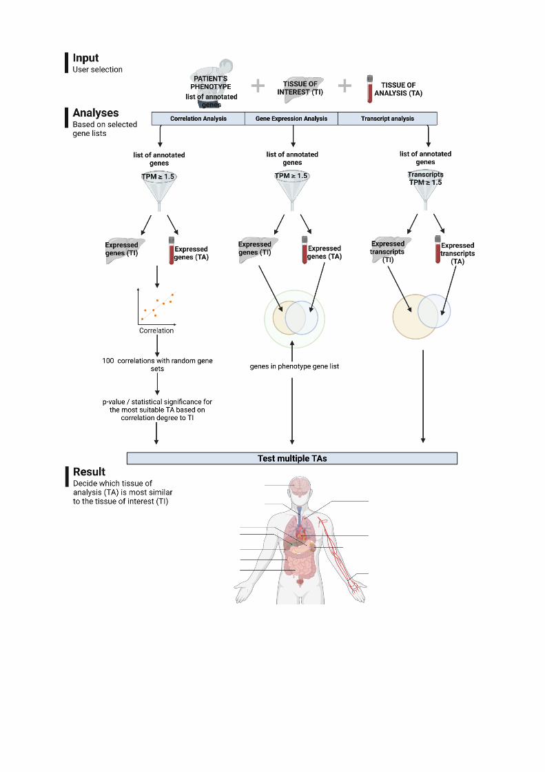
P-Techno-197
*** Efficacy of conventional fragment analyses and exome sequencing as diagnostic approach for movement disorders- data from a single center study
B. Menden 1, C. Dufke1, N. Deininger1, M. Rautenberg1, F. Harmuth1, J. Park1, K. Grundmann-Hauser1, A. Söhn1, M. Sturm1, G. Demidov1, S. Ossowski1, O. Rieß1, R. Schüle2, M. Synofzik2, L. Schöls2, T. Haack1
1Uniklinik Tübingen, Institut für Medizinische Genetik, Tübingen, Deutschland 2Uniklinik Tübingen, Abteilung für Neurologie, Tübingen, Deutschland
Objective
Movement disorders (MD) comprise a clinically and genetically heterogenous group of disorders that often overlap on the phenotypic and molecular level. Over the last years, the implementation of broad sequencing approaches combined with in-depth phenotypic characterization substantially improved the diagnostic yield. However, published cohorts with a systematic assessment of genetic testing strategies are rather small and the range of performed genetic tests and observed diagnostic yields is highly variable. This single center study aimed to evaluate the efficacy of a combined diagnostic approach of conventional fragment analyses (FA) and exome sequencing (ES) in order to provide a rationale and benchmark for currently evolving genome-based diagnostics applying bioinformatic tools that enable the detection of structural variants (SVs) and repeat expansions (REs).
Methods
Metadata of individuals investigated by diagnostic-grade ES (n=2041) and/or FA (n=4726 tests in 1079 patients) between 10/2016 and 12/2020 were identified from in-house databases. Assignment to a MD subgroup in ES cases was determined by HPO-terms. Repeat length was determined by FA and systematically validated by genome sequencing (GS) in cases representative for the different RE loci.
Results
The HPO-based query identified 2041 MD index cases including individuals with ataxia (n=899), dystonia (n=265), spasticity (n=573), and combined MD (n=304). Overall diagnostic yield of ES was 19,7% (ACMG class 4/5). Especially for the ataxia cohort the observed diagnostic yield declines with age. Highest diagnostic yield was reached in the spasticity group with 29%. 11% of pathogenic findings in ES were intronic and non-coding variants or CNVs.
Of all conducted FA (n=4726 tests in 1079 patients) 2.5% revealed pathogenic findings in 10,8% of all patients with MD. The diagnostic yield was highest for SCA8- analyses with 10% and Friedreich ataxia, as well as FXTAS with 8% and 7% respectively. The diagnostic yield for SCA1, SCA2, SCA3, SCA6, SCA7, SCA17 and DRPLA ranged between 0,4-3%. SCA12 and SCA10 analyses revealed no pathogenic findings. Systematic validation via Expansion Hunter in GS confirmed all expanded alleles and repeat size was estimated precisely especially for shorter repeats.
Conclusion
Our data suggest that a genome-based diagnostic approach with expanded bioinformatic analyses including SVs, CVs, and REs has the potential to replace a step-by-step diagnostic approach using conventional methods. The improved detection of CNVs and REs on a GS basis as well as the detection of intronic and intergenic variants is expected to further increase the diagnostic yield in MD. Besides, this unbiased diagnostic approach will help clinicians to diagnose patients with atypical presentation.
P-Techno-198
Implementing RNA analysis in human genetics: from gene expression profiling to integration into genome analysis improvement
C. Nicolas1,2, A. Jakob1,2, P. Michaela1,2, B. L. Rebecca1, S. Christopher1, H. Tobias1, O. Stephan1,2, R. Olaf1,2
1Universitätsklinikum Tübingen, Institute of Medical Genetics and Applied Genomics, Tübingen, Deutschland 2 Universitätsklinikum Tübingen, NGS Competence Center, Tübingen, Deutschland
Objectives
First applications of RNA analysis in human genetics aimed mainly to detect splicing defect or loss of gene expression. More recent developments of RNA-analyses are focusing on the analysis of gene expression changes in disease as biomarkers, in response to treatments or to predict targeted medication as in cancer therapy. By using blood RNA-seq analysis, we developed a strategy enabling both the comprehensive analysis of disease and the validation of genomic events in genetic or sporadic disorders.
Methods
We are using high-throughput and standardized protocols to complementary monitor gene expression in blood and to investigate systematically the impact of pathogenic and likely pathogenic variants detected using whole-genome sequencing in splicing and in regulatory elements in rare diseases.
Results
RNA-seq was optimized for tumor material in clinical oncology to prioritize therapeutic relevance of somatic mutation detected by NGS-based DNA panel sequencing by investigating loss of heterozygosity as well as the identification of gene fusion at the RNA level.
Discussion
We will present how ongoing developments using unique molecular identifiers, single-cell sequencing or long-read sequencing will lead to novel application of RNA diagnostics such as the identification of infection or immune response related to therapy.
P-Techno-199
Gene Expression and Genomic Variant Validation with RNA from Blood
M. Pogoda1,2, J. Admard1,2, O. Rieß1,2, S. Ossowski1,2, T. Haack1,2, C. Groß1,2, R. Buchert1, O. Seibel-Kelemen1, N. Casadei1,2
1Institut für Medizinische Genetik und Angewandte Genomik, NCCT, Tübingen, Deutschland 2Eberhard Karls University Tübingen, NGS Competence Centre Tübingen (NCCT), Tübingen, Deutschland
Fragestellung/Introduction
With ever decreasing sequencing costs, Whole Exome Sequencing (WES) and Whole Genome Sequencing (WGS) have become common tools in scientific and diagnostic questions. However, genomic information alone often cannot explain specific phenotypes or symptoms, especially in rare disorders. Many variants in the genome are of uncertain significance, especially intronic variants. Thus, the impact of genomic variants on the transcriptome has gained more and more importance. While most studies so far focus on pure gene expression, our focus is to validate the impact of genomic variants on the splicing of RNA. A reliable state-of-the-art protocol to extract RNA from patients is using blood collected in tubes with RNA-stabilizing substances. Although blood is relatively easy to obtain and although it delivers relatively large amounts of RNA, there are certain downsides to its usage, e.g. the presence of high amounts of the globin mRNA. For our application, we further noticed that using an rRNA and globin depletion-based approach to sequence total RNA was problematic as we found high levels of intronic reads. Thus, the validation of pathogenic splicing variants was not possible using these data.
Methoden/Methods
We tested three different approaches to overcome these limitations of RNASeq from blood: a globin and ribosomal depletion-based approach, poly(A) enrichment alone and poly(A) enrichment followed by ribosomal and globin depletion.
Ergebnisse/Results
Our results suggest that poly(A) enrichment offers the most useful data to study splicing events in mRNA, providing the best compromise between usable read number, number of coding transcripts detected and amount of intronic reads
Schlussfolgerungen/Conclusions
For the validation of splicing variants, total RNASeq from blood provides certain challenges that still need to be investigated. Methodological optimizations might be required to fully understand the sequencing data from RNA from blood.

P-Techno-200
A new tool in the box: Long-read sequencing in routine diagnostics
D. Popp1, J. Hentschel1, S. Neuser1, M. Karnstedt1, M. Radtke1, R. Abou Jamra1
1Institut für Humangenetik, Universitätsklinikum Leipzig, Leipzig, Deutschland
In routine diagnostics, short-read sequencing is the dominating technique for exome analysis. State-of-the-art short-read sequencing provides high-quality data at an impressive throughput. However, the limited read length is a drawback leaving cases unsolved. Here, we present two routine diagnostic cases that could not be conclusively solved and therefore long-read Nanopore sequencing was applied to get further insights.
The first individual is a child with intellectual disability, autism, facial dysmorphism and seizures. By exome sequencing, we identified a single basepair deletion leading to a frameshift in SETD1A (c.1143del, p.(Tyr382Thrfs*115)). SETD1A associated neurodevelopmental disorder (MIM #619056) has a high phenotypic overlap with the child"s symptoms. We could exclude the variant in the mother, but segregation analysis was restricted as the father already died. Consequently, we were interested in phasing the variant as finding it on the maternal allele would mean it occurred de novo. For phasing, we used a SNP about 5 kb downstream of the variant. A long-range PCR spanning the variant and the SNP and subsequent Nanopore sequencing was performed. As the variant was found on the paternal allele, it is still unclear if the variant occurred de novo or was inherited from the father. Now, analysis of further paternal relatives is ongoing to check for the variant.
The second individual was a newborn with a leukodystrophy, muscular hypotonia, poor head control, failure to thrive and motor developmental delay. We identified two missense variants in the gene NFU1. Pathogenic variants in NFU1 are associated with a mitochondrial dysfunction syndrome, which is a severe autosomal recessive disorder of systemic energy metabolism resulting in respiratory failure, lack of neurologic development and early death. By segregating the parents, the first variant (c.565G>A, p.(Gly189Arg)) was determined to be maternally inherited while the second variant (c.545G>T, p.(Arg182Leu)) occurred de novo. Hence, the variants could not be phased. Furthermore, an RNA sample was not available as the patient deceased meanwhile. Hence, by long-range PCR, a product of about 6.5 kb size containing both variants was obtained and subsequently sequenced on a Nanopore device. As the variants were not observed at the same reads, they were pinpointed as compound-heterozygous and confirmed as disease-causing. As a result, the recurrence risk was assessed as very low (<1%) which was relevant to the parents as they had a lasting wish for a child.
The presented families are unique, but the field of human genetics often handle rare disorders and special constellations. Thus, long-read sequencing is a valuable addition to the toolbox of routine genetic diagnostic methods. Long-read sequencing has specific advantages not only when phasing variants as presented here but also for the analysis of repeat expansions and structural variants.
P-Techno-201
Identification of a non-canonical transcription factor binding site using deep learning
S. Proft1,2, J. Leiz3,4, R. Opitz5, M. Jung1, U. Heinemann6, D. Seelow1,2, K. Schmidt-Ott3,4, M. Rutkiewicz6,7
1Charité - Universitätsmedizin Berlin, BIH - Exploratory Diagnostic Sciences (EDS), Berlin, Deutschland 2Charité - Universitätsmedizin Berlin, Institut für Medizinische Genetik und Humangenetik, Berlin, Deutschland 3Charité–Universitätsmedizin Berlin, Medizinische Klinik mit Schwerpunkt Nephrologie und Internistische Intensivmedizin, Berlin, Deutschland 4Max Delbrück Center for Molecular Medicine in the Helmholtz Association (MDC), Berlin, Deutschland 5Charité–Universitätsmedizin Berlin, Institut für Experimentelle Pädiatrische Endokrinologie, Berlin, Deutschland 6Max-Delbrück-Center for Molecular Medicine in the Helmholtz Association (MDC), Macromolecular Structure and Interaction, Berlin, Deutschland 7Polish Academy of Sciences, Institute of Bioorganic Chemistry, Poznan, Polen
The experimental identification of transcription factor binding sites is costly, time intensive, and tissue-specific. It is virtually impossible to test the effects of variants revealed by Whole Genome Sequencing on TF binding, so reliable predictions are indispensable.
Current models for TF binding are usually based on positional weight matrices (PWM) which depict sequence motifs. However, such motif-based models cannot identify binding sites that do not follow the main motif.
To circumvent this limitation, we have trained a neural network with GRHL1 binding data from SELEX experiments. The classifier was then applied to 7,857 sequences containing GRHL1 binding sites obtained from ChIP-Seq experiments (length > 197 bp) and could identify 46 potential binding sites for which the PWM-based approach did not suggest any binding at all. Using isothermal titration calorimetry (ITC), we could confirm binding between a predicted non-canonical DNA sequence and the GRHL1 protein.
We are currently extending these experiments with artificially introduced variants to find out how good the binding affinity predicted by our model correlates with the real binding strength.
Our results show that it is possible to use neural networks to discover hitherto unknown TF binding motifs. We will apply our approach to a wider array of transcription factors to identify novel binding sites, with a special focus on known disease mutations in promoter regions.

P-Techno-202
Highly sensitive Liquid Biopsy Duplex Seq enables molecular diagnosis of a 10 year old child with clinically confirmed overgrowth syndrome in plasma
A. Hallermayr1, T. Neuhann1, V. Steinke-Lange1, E. Holinski-Feder1, J. Romic-Pickl1
1MGZ Medizinisch Genetisches Zentrum München, München, Deutschland
Introduction: Liquid Biopsy is a promising approach for identification a patient´s mutational profile in plasma, but accurate detection of variants at low frequencies is challenging. We established Liquid Biopsy Duplex Sequencing, enabling highly sensitive analysis of variants associated with overgrowth syndrome and tumor disease. Here we present its application in clinical use for elucidating a molecularly unsolved case.
Materials and Methods: Somatic mutation profiles of 33 genes were obtained from plasma samples using NGS Duplex Sequencing.
Results: Analytical validation of the Liquid Biopsy Duplex Sequencing panel showed 99.7% sensitivity and 91.5% precision for variants with 0.5% allele frequency in a total of 33 genes.
The Liquid Biopsy panel was applied for molecular diagnosis of a 10 year old child with a clinically diagnosed asymmetric overgrowth syndrome including arteriovenous malformations limited to one side of the body. Somatic KRAS c.35G>A, p.(Gly12Asp) variant was detected in plasma with 1% variant allele frequency. Notably, the variant was not detected in a parallel analysis of a skin biopsy sample.
The KRAS c.35G>A, p.(Gly12Asp) variant leads to constitutive overactivation and increased signal transduction into downstream pathways and is associated with overgrowth including various types of congenital nevi and vascular malformations (so-called mosaic RASopathies). Consequently, detection of KRAS c.35G>A, p.(Gly12Asp) variant in plasma could molecularly explain the clinically observed overgrowth syndrome.
Summary: Liquid Biopsy provides the mutational profile in contrast to tissue biopsy, which is limited to detection of variants present in the resected specimen. Liquid Biopsy Duplex Sequencing pushes the boundaries for detection of low frequency variants in plasma. Our broad Duplex Sequencing panel enables highly sensitive screening of all therapy relevant variants for overgrowth syndrome. In our case study, Liquid Biopsy Duplex Sequencing identified KRAS c.35G>A, p.(Gly12Asp) as the molecular cause of the clinically confirmed overgrowth syndrome in a 10 year old child. The identification of the KRAS variant may lead to novel therapy options.
P-Techno-203
Identification of rare variants for nonsyndromic cleft lip with/without cleft palate in a cohort of multiplex families
A. Scheer1, F. Brand2, J. Fazaal1, N. Ishorst1, P. M. Krawitz3, E. Mangold1, K. U. Ludwig1
1Institute of Human Genetics, University of Bonn, School of Medicine & University Hospital Bonn, Bonn, Deutschland 2Institute for Genomic Statistics and Bioinformatics, University Hospital Bonn, Bonn, Deutschland 3Institute for Medical Biometry, Informatics and Epidemiology, University Hospital Bonn, Bonn, Deutschland
Nonsyndromic cleft lip with/without cleft palate (nsCL/P) are among the most common birth defects and have a multifactorial etiology. In the last years, genome-wide association studies (GWAS) have identified around 45 common risk regions with small to moderate effect sizes, but the contribution of rare variants with higher effect sizes has been studied to a lesser extent. The aim of our study was to identify rare causal variants with higher penetrance in 55 individuals from 10 nsCL/P multiplex families. Each of the families had at least three affected family members in at least two generations. Whole-genome sequencing was performed using Illumina Truseq Nano DNA Library Prep Kit, and variant calling was performed family-wise using an in-house pipeline. In the five families analyzed till date, overall, 33,683,757 single-nucleotide variants were identified.
We first focused on the protein-coding regions and applied technical, pedigree-based and frequency filtering to the first five families, to find completely co-segregating risk variants. This analysis together with subsequent annotation in the Variant Effect Predictor identified 73 candidate variants in the analyzed cohort of five families, comprising nine loss-of-function variants, 63 missense and one splice-acceptor variant. Two of these genes have been previously suggested as risk genes: PLEKHA5 in an independent exome sequencing study (Cox et al., 2018) and RHPN2 which is located in a GWAS risk region (Chevrier et al., 2016).
Second, we analyzed the dataset according to an autosomal-dominant model with reduced penetrance. Therefore, we considered variants that occurred in at least one of the affected family members but not necessarily in the non-affected. Again, we found variants in previously described risk genes (CTNND1, Cox et al., 2018; IFT88, Barba et al., 2019; FILIP1L, Beaty et al., 2013; EPHA3, Ludwig et al., 2012), but also identified novel candidate genes based on the presence of risk variants in two different families (HKDC1, KRT77, COL7A1). Further evidence for some of the genes from both approaches were obtained through analysis of single-cell expression data during mouse embryonic development. Together, our study identifies novel candidate genes for nsCL/P, which still need support from additional, independent studies.
P-Techno-204
Classification of fluorescent R-Band metaphase chromosomes using a convolutional neural network is precise and fast in generating karyograms
B. Vajen1, S. Hänselmann2, F. Lutterloh1, S. Käfer1, J. Espenkötter1, A. Beening1, J. Bogin2, B. Schlegelberger1, G. Göhring1
1Medizinische Hochschule Hannover, Institut für Humangenetik, Hannover, Deutschland 2Metasystems, Software Development, Altlussheim, Deutschland
Background
Karyotype analysis has a great impact on the diagnosis, treatment and prognosis in hematologic neoplasms. The identification and characterization of chromosomes is a challenging process and needs experienced personal. Artificial intelligence provides novel support tools. However, their safe and reliable application in diagnostics needs to be evaluated. Here, we present a novel laboratory approach to classify chromosomes in cancer cells using a convolutional neural network (CNN).
Methods
We collected a total of 330,131 normal karyograms and the images of the associated metaphases from routine diagnostic karyotyping from 2012 to 2019 without filtering for image quality. A self-developed convolutional neural net (CNN) was trained to predict the chromosome class and the rotation angle from individual chromosome images. To test this CNN in routine diagnostics, we analyzed 200 metaphases with normal karyotype from twenty individuals (ten metaphases per individual) either with or without CNN support. Furthermore, we analyzed 10 cases (10 metaphases each) with different chromosomal aberrations: isolated chromosomal aberrations involving different chromosomes, complex karyotypes, composite karyotypes, isolated whole arm translocations and isolated deletions.
Results
First, we evaluated the performance of the CNN on individual chromosomes using a set of 16,506 normal karyograms that the CNN had never seen during training (i.e. the test set). The CNN predicted the correct chromosome class for 98.8% of chromosomes. In a second step, we tested the developed CNN in routine diagnostics against the routinely used automatic classification algorithm within the Ikaros karyotyping software. Better prediction of the chromosome class and rotation angle by the CNN required fewer manual correction steps, which led to a reduction in the turnaround time of 43% for the karyotyping workflow. Even in metaphases with diverse chromosomal aberrations such as complex karyotypes we observed a significant time saving.
Conclusion
These results demonstrate that the CNN has potential application value in chromosome classification of hematologic neoplasms. This study contributes to the development of an automatic karyotyping platform.
P-Techno-205
A graphical user interface for real-time multi-omics analysis of Nanopore sequencing experiments
A. Wierczeiko1, S. Pastore1, C. Hewel1, S. Weißbach1, I. Graebling1, S. Gerber1
1Institut für Humangenetik, Universitätsmedizin der Johannes Gutenberg Universität Mainz, Computational Systems Genomics Lab, MAINZ, Deutschland
In standard sequencing experiments, the experimental steps and data analysis are performed independently and the data analysis is initiated only after sequencing is complete. However, recently established third-generation long-read nanopore sequencing techniques allow an analysis of experimental data to begin while the sequencing process is still underway.
We have developed a self-updating analysis toolbox for Nanopore sequencing that allows even inexperienced users to perform sophisticated multi-omics analyses in real-time via a user-friendly and intuitive graphical user interface. In addition to standard quality control, differential expression, alternative splicing, and epigenetic profiling, we focus on useful aspects of run-time analysis. For example, by updating results in real-time, saturation in the number of detected differentially expressed genes can be detected even as sequencing depth increases, which may allow early termination of the sequencing run to save resources.
Another major advantage of real-time nanopore sequencing analysis is interactive adaptive sampling. This technique allows the researcher to either avoid or preferentially sequence genomic regions during the ongoing sequencing experiment to improve efficiency and customize the experiment. It also enables efficient use of resources and allows the user to be notified locally, as well as remotely, of upcoming problems or successes. Our tool holds great potential for optimizing medical diagnostics, e.g., by detecting and characterizing structural variations, correctly sequencing repetitive regions, or revealing epigenetic changes and transcriptomic profiles to support the genetic diagnosis. The toolbox is easily expandable and new modules can be quickly integrated.
206–208 Therapy for Genetic Diseases
P-Therap-206
How can whole CFTR genotyping contribute in genetically unsolved Cystic Fibrosis cases?
S. Ahting1, L. Nährlich2, J. Hentschel1
1Universitätsklinikum Leipzig, Institut für Humangenetik, Leipzig, Deutschland 2Universitätsklinikum Giessen-Marburg GmbH, Zentrum für Kinderheilkunde und Jugendmedizin Abteilung Allgemeine Pädiatrie und Neonatologie Funktionsbereich Päd. Pneumologie und Allergologie, Giessen, Deutschland
Background: The importance of genotyping for CF patients has changed dramatically over the last decades. Today, the knowledge about the individual CFTR variant is critical on a patient level to confirm the diagnosis, select treatment options and counsel patients and families. According to the most recent German CF Registry annual report (2020), 99.4% of 6648 reported patients underwent genetic testing. In 5.4% (357 in total) of them, genetic testing could identify either none or only one causative variant, leaving the patients and their families unable to initiate appropriate treatment. Whole CFTR genotyping can therefore not only aid in uncovering previously unidentified CFTR mutations but is also crucial for patient care and therapy.
Methods: On the German Cystic Fibrosis Registry website "Muko.web", patients" genotypes can be registered by selecting from a dropdown list of previously known CFTR mutations. CFTR variants not listed in CFTR2 can be documented as free text. According to the latest data analysis (May 2020, not published), there are 1172 alleles reported as free text, 731 of which in patients with a clinically confirmed diagnosis of CF. To conclusively identify the reported variants, they were verified using databases such as ClinVar, HGMD and CFTR1/2. Subsequent feedback to the CF centres and crosschecking and correction of the registry database entries was carried out. Patients whose variants could not be identified were reported back to the CF centres, alongside an offer to redo the genetic testing.
Results: 18.9% (138 in total) of variants in the free text could not be identified using the information provided on the registry website, either due to the use of different nomenclatures or misspellings. 64 anonymised genetic reports were received for correction of the above variants, 50 of which helped to solve the case and to provide the correct genotype for the patient. 18 variants were corrected through the CF centres upon revision, leaving a total of 70 patients that have been offered genetic testing. 48 variants could be identified that have not been previously reported in the context of CF.
Outlook: Currently, we are collecting blood samples of the remaining 70 patients, along with further samples from patients with unidentified or unclear genotype. Whole CFTR genotyping will then be carried out using an NGS custom design panel covering all 27 CFTR exons as well as intronic and regulatory regions, thereby providing the patients with their most up-to-date variant information and treatment accessibility. Furthermore, it will be used to identify previously unknown CFTR variants and assess their disease causing probability. Variants in intronic regions can additionally be analysed by mRNA sequencing to evaluate their functional relevance. We will also upload all variants to ClinVar and report previously unknown variants to CFTR1/2 for database completion.
P-Therap-207
L-Serine treatment is associated with improvements in behavior,EEG and seizure in individuals with GRIN-related disorders due to null variants
I. Krey1, S. von Spiczak2, K. M. Johannesen3, R. S. Møller3, C. Hikel4, G. Kurlemann5, H. Muhle2, D. Beysen6, T. Dietel7, J. R. Lemke1, S. Syrbe8
1Institut für Humangenetik Leipzig, Leipzig, Deutschland 2Department of Neuropediatrics, University Medical Center Schleswig-Holstein, Campus Kiel, Kiel, Germany, Kiel, Deutschland 3Department of Epilepsy Genetics and Personalized Medicine, The Danish Epilepsy Centre, Dianalund, Denmark, Dianalund, Dänemark 4Department of Children and Adolescents, GFO Kliniken Niederrhein, Dinslaken, Germany, Dinslaken, Deutschland 5Hospital for Children, Bonifatius Hospital Lingen, Germany, Lingen, Deutschland 6Department of Paediatric Neurology, Antwerp University Hospital, Antwerp, Belgium, Antwerpen, Niederlande 7Epilepsy Center Kork, Medical Faculty of the University of Freiburg, Kehl, Germany, Kehl, Deutschland 8Division of Paediatric Epileptology, Centre for Paediatrics and Adolescent Medicine, University Hospital Heidelberg, Heidelberg, Germany, Heidelberg, Deutschland
Pathogenic missense variants in GRIN2A and GRIN2B may result in gain or loss of function (GoF/LoF) of the N-methyl-D-aspartate receptor (NMDAR). This observation gave rise to the hypothesis of successfully treating GRIN-related disorders due to LoF variants with co-agonists of the NMDAR. In this respect, we describe a retrospectively collected series of nine individuals with GRIN2A- or GRIN2B-related disorders who were treated with L-serine, each within an independent n-of-1 trial. Our cohort comprises one individual with a LoF missense variant with clinical improvements confirming the above hypothesis and replicating a previous n-of-1 trial. A second individual with a GoF missense variant was erroneously treated with L-serine and experienced temporary behavioral deterioration further supporting the supposed functional pathomechanism. Seven additional individuals with null variants (that had been interpreted as loss-of-function variants despite not being missense) again showed clinical improvements. Among all eight individuals with LoF missense or null variants, L-serine treatment was associated with improvements in behavior in seven (88 %), in development in three (38 %) and/or in EEG or seizure frequency in four (50 %). None of these eight individuals experienced side effects or adverse findings in the context of L-serine treatment. In summary, we describe first evidence that L-serine treatment may not only be associated with clinical improvements in GRIN-related disorders due to LoF missense but particularly also null variants.
P-Therap-208
*** Co-regulation of gene expression - a potential hurdle for future gene therapies?
T. Strunz1, M. Kellner1, C. Kiel1, B. H. F. Weber1,2
1Institut für Humangenetik, Regensburg, Deutschland 2Institut für klinische Humangenetik, Regensburg, Deutschland
Purpose:
Gene therapy is a promising approach to cure heritable diseases caused by loss or gain of function mutations, or by haploinsufficiency of a gene product. Accordingly, a new functional gene copy is introduced into the genome or the mutated DNA is modified by genome editing technology. For gene therapy to be successful, it is crucial to fully understand endogenous gene expression and its regulation. For example, the alteration of gene expression of one gene can have fundamental co-regulatory consequences for other genes, which may in turn cause unintentional phenotypic changes. Here, we describe an approach to identify such co-regulated genes in diverse tissues and highlight potential effects in gene therapy when addressing diseases caused by haploinsufficiency.
Methods:
Gene expression regulation was characterized based on expression quantitative trait locus (eQTL) analyses in 49 tissues of the Genotype-Tissue Expression (GTEx) project. A subsequent colocalization study of regulated genes within a window of 1 Mbp identified co-regulated genes affected by the same genetic signal. Use of the ClinGen database (https://clinicalgenome.org/) and a comprehensive literature search generated a list of 713 genes known to cause haploinsufficiency phenotypes.
Results:
From a total of 33,000 regulated genes, 14,636 were found to be co-regulated in at least one of the tissues analyzed. Grouping of genes regulated by the same eQTL signal revealed 14,727 unique clusters containing up to nine co-regulated genes. Many of these genes reside over 100 kbp apart from each other, with an average distance of 112 kilobase pairs. Of the 713 genes known to express clinical symptoms due to haploinsufficiency of a defined gene product, 231 (32.4%) are part of at least one of the identified gene clusters. Down or up-regulation of these 231 genes can directly be linked to the expression of other genes.
Conclusions:
We generated an open database providing access to thousands of co-regulated genes in 49 human tissues. Co-regulated genes are in close proximity or at distances of up to 2 Mbp and many clusters of expression regulation are shared between tissues. Our findings have a strong impact on current and future therapeutic approaches aiming at the modification of gene expression. This is particularly critical when gene therapy targets a gene which may influence the expression of a neighbouring co-regulated gene possibly triggering unforeseen side effects.

Authors Index
A
Aarts-Tesselaar, C.D. P-ClinG-107
Abazi-Emini, N. P-MonoG-150
Abdalla, E. P-MonoG-159
Abdel-Salam, G.M.H. W6-003
Abdullah, U. W2-006, W6-004
Abeditashi, M. P-BasEpi-001, P-BasEpi-007
Abela, L. P-ClinG-091
Abicht, A. P-ClinG-107
Abicht, A. P-CancG-016, P-ClinG-051, P-ClinG-058, P-ClinG-071, P-ClinG-085, W8-005
Abou Jamra, R. P-Techno-196, P-ClinG-038, P-ClinG-067, P-ClinG-068, P-ClinG-087, P-MonoG-
144, P-MonoG-154, P-MonoG-176, P-Techno-200, W1-004, W2-001
Ades, L. W2-006
Admard, J. P-MonoG-156, P-Techno-199, W2-004
Ahting, S. P-Therap-206
Al-Ashhab, M. P-ClinG-099
Alam, T. P-BasEpi-002
Alawam, B.S. P-MonoG-182
Alawbathani, S. W9-003
Alawi, M. P-ClinG-061, W6-002, W6-003
Alberti, S. W6-001
Albertowski, K. P-ClinG-109
Albillos, A. W5-002
Alcoceba, M. W8-001
Alders, M. W2-001
Aldisi, R. W5-005, P-Compl-122, P-Techno-191, W4-002
Alhashem, A. W2-003
Ali, Z. W2-006
Alkemade, H. P-CancG-020
Alkuraya, F.S. P-MonoG-182, W2-003
Alkuraya, F.S. W2-006
Allgäuer, M. P-Techno-187
Almstrup, K. W9-001
Alsaif, H.S. P-MonoG-182
Alter, S. P-ClinG-034, P-ClinG-065, P-ClinG-104
Altmüller, J. P-MonoG-144, W2-006, W5-001, W6-004, W7-001, W7-003, W9-004
Alvi, J.R. W2-006
Alzahrani, F. W2-003
Alzaidan, H. P-MonoG-182
Amberger, A. P-MonoG-184
Ammerpohl, O. W8-003
Amslinger, S. W7-002
Amunts, K. P-Compl-113, P-Compl-121
Anders, P. P-ClinG-101
Anders, S. P-CytoG-138
Andres, S. P-ClinG-042, P-ClinG-078
Antony, J. W2-006

Araujo, T.F. W9-001
Aretz, S. W4-002, P-CancG-027, P-CancG-028, W4-006
Argilli, E. P-ClinG-107
Argyriou, L. P-Techno-188
Arlt, A. P-MonoG-163
Arlt, M. P-Techno-187, W4-001
Arndt, F. W6-003
Arnold, M. P-ClinG-101
Arruda, L.K. P-MonoG-177
Ashrafzadeh, F. W2-003, W2-006
Asif, M. W6-004, W9-003
Asselta, R. W5-002
Atlan, D. P-MonoG-171
Auber, B. P-CancG-032, P-ClinG-075, P-MonoG-181, P-Techno-195
Aubert-Mucca, M. P-ClinG-107
Aubertin, G. W2-003
Auer-Grumbach, M. P-MonoG-167
Augustin, M. P-Techno-187
Aulitzky, W.E. P-Techno-187
Aust, D. W4-001
Averdunk, L. P-MonoG-182, W1-002
Axt, D. P-Compl-122
Aydinli, N. P-ClinG-059
B
Baba, N. P-Techno-194
Bachmann, N. P-ClinG-050, P-ClinG-053
Bader, B. P-MonoG-169
Bader, I. W7-001
Bahena, P. W2-003
Baig, J.M. W2-006
Baig, S.M. W2-006
Bain, J.M. W6-005
Balachandran, S. P-CancG-021
Balci, T. P-ClinG-099
Banales, J. W5-002
Banan, R. W4-006
Bandera, A. W5-002
Banka, S. W2-005, W2-006
Bannach Jardim, L. P-MonoG-175
Barbalho, P.G. W2-003
Barbuti, P. W7-004
Baretton, G. W4-001
Barresi, S. W6-001
Bartels, C. W2-004
Bartels, E. P-MonoG-170
Bartholomew, D. P-MonoG-170
Bartholomäus, A. P-MonoG-165
Bartolomaeus, T. P-ClinG-038, P-ClinG-087, P-MonoG-170
Barwinski, N. P-Techno-186
Basmanav, B. P-Compl-122
Basmanav, F.B.Ü. P-MonoG-149
Basmanav, F.B. P-ClinG-096
Battke, F. P-ClinG-054, W3-002
Bauer, M. P-MonoG-155
Bauer, S. P-Techno-187, W4-001
Baujat, G. W9-003
Baum, P. P-MonoG-154
Baumann, A.A. P-Techno-187
Baumann, B. P-MonoG-183
Baumann, E. P-ClinG-101
Baumann, S. P-ClinG-070
Baumann, T.S. W8-001
Baumann, U. P-MonoG-147, P-MonoG-179
Bawadi, R. P-CancG-012
Beblo, S. P-ClinG-076
Becher, C. P-CancG-021
Beck, K. P-Techno-187, W4-001
Beck-Wödl, S. P-MonoG-146
Beck-Wödl, S. P-ClinG-068, P-MonoG-142
Becker, C. W9-003
Becker, I. P-MonoG-157
Becker, J. P-ClinG-106
Becker, J. W8-002
Becker, K. W5-001
Beekman, R. W8-001
Beening, A. P-Techno-204
Beetz, C. P-MonoG-143, W2-006
Begemann, A. P-ClinG-091
Begemann, M. P-ClinG-073, P-ClinG-093, SEL-003
Behncke, R. SEL-001
Behra, M. W2-003
Behrens, Y.L. P-CancG-013, P-CancG-012
Behring, B. W3-004
Beier, D. SEL-001
Beier, F. P-CancG-023
Beins, E. P-Compl-121
Beins, E.C. P-Compl-118
Bejaoui, Y. P-BasEpi-002
Bender, T. P-ClinG-088, P-ClinG-089
Benet-Pages, A. P-ClinG-071
Benet-Pagès, A. P-CancG-017
Bengl, D. P-MonoG-140
Bens, S. P-CytoG-129, P-CancG-024
Berber, P. P-MonoG-141, P-BasEpi-006
Bergamini, C. P-ClinG-069
Bergmann, A. P-ClinG-101
Bergmann, A.K. P-CancG-024
Bergmann, C. P-ClinG-050, P-ClinG-053, P-ClinG-083
Berking, C. P-MonoG-157
Bermudez, M. P-Techno-187
Bermúdez, M. W4-001, P-ClinG-077
Bermúdez-Guzmán, L. P-ClinG-087
Bernat, J.A. W2-002
Bernhardt, L. P-BasEpi-003
Bernhart, S.H. P-CancG-024
Bertin, M. P-MonoG-178
Bertini, E.S. W6-001
Bertoli, M. W2-002
Bertrand, M. P-ClinG-069, P-MonoG-146
Bertrand, M. P-MonoG-142
Berutti, R. P-ClinG-042, P-ClinG-078
Bessonova, L. W2-002
Betz, B. P-CancG-014
Betz, R. P-CancG-025, P-Compl-122
Betz, R.C. P-ClinG-096, P-MonoG-149
Beule, D. P-BasEpi-005, SEL-002
Beutner, D. W9-002
Bevot, A. P-MonoG-142, W7-001
Beyer, U. W4-006
Beygo, J. P-BasEpi-004, P-ClinG-068, W2-002
Beysen, D. P-Therap-207
Beà, S. W8-001
Bhattacharyya, S.S. W9-005
Biasella, F. W5-006
Biedermann, A. P-Compl-117
Bier, A. P-ClinG-058
Bierhals, T. P-MonoG-180
Biessen, E. W8-002
Bijlsma, E.K. P-ClinG-068
Billing, H. P-ClinG-080, W9-006
Billington, C.J. P-MonoG-151
Biskup, K. W7-005
Biskup, S. P-ClinG-041, P-ClinG-054, P-ClinG-108, P-MonoG-144, W3-002, W7-001
Bitzer, M. P-Techno-187, W4-001
Bjerre, A. P-ClinG-080, W9-006
Blanchard, V. W7-005
Blankenburg, M. P-CytoG-127
Blick, S. P-ClinG-106
Block, S. P-ClinG-039
Bloechle, M. P-CytoG-127
Blüher, M. P-CytoG-128, P-Techno-196
Bobbili, D. W4-002
Bock, A. P-MonoG-143
Boerries, M. P-Techno-187, W4-001
Bogin, J. P-Techno-204
Boltshauser, E. W7-001
Bombei, H.M. W2-002
Bonnaire, B. P-CytoG-133
Bonora, E. P-ClinG-069
Boonsawat, P. P-ClinG-059
Boos, J. W5-002
Borisov, O. P-Compl-116
Borkhardt, A. SEL-003
Bormann, F. W8-002
Borst, A. P-MonoG-166
Borst, A. P-MonoG-145, P-MonoG-140
Bosch, E. W3-004
Boschann, F. P-ClinG-041, P-ClinG-099, P-ClinG-047
Bosse, K. P-CancG-033
Boulant, S. P-BasEpi-011
Bouman, A. W2-002
Boumann, A. W2-001
Bourgeois, M. P-CancG-023
Bozet, M. W7-003
Bramswig, N.C. P-ClinG-068, P-ClinG-100
Brancati, F. W6-004
Brand, F. P-BasEpi-005, P-ClinG-088, P-Techno-203, SEL-002, W5-001
Brand, F. W4-006, P-CancG-032, W9-006
Brandl, C. P-MonoG-141
Brandt, V.-P. P-CytoG-128
Brandts, C.H. P-Techno-187
Braun, F. P-ClinG-069
Braunisch, M.C. P-MonoG-150
Braunschweig, T. SEL-003
Brenzel, A. W7-003
Brinckwirth, B. P-ClinG-065
Brockmann, K. W7-001
Brockmeier, K. P-ClinG-106
Brors, B. P-CancG-025, P-Techno-187
Bros, B. W4-001
Brown, N.J. W2-002
Bruckmann, A. P-MonoG-174
Brunet, T. P-ClinG-042, P-ClinG-078
Brunkhorst, L. P-ClinG-080
Bräsen, J.H. W4-006
Brümmendorf, T. P-CancG-023
Brümmendorf, T.H. P-CancG-014
Brüstle, O. P-Compl-118
Bublitz, J. W4-005
Bubshait, D.K. W2-006
Buchert, R. P-MonoG-146, P-MonoG-142, P-Techno-199
Buchhalter, I. P-Techno-187, W4-001
Budde, B. W2-006, W6-004, W9-003
Buena Atienza, E. P-ClinG-049, P-MonoG-183
Buena-Atienza, E. W5-003
Buhl, N. P-MonoG-147
Bujanda, L. W5-002
Buness, A. P-Compl-116
Burgemeister, A.L. P-ClinG-043, P-CytoG-130
Burger, B. P-Compl-118, P-Compl-121, P-MonoG-177
Burger, R.-M. P-MonoG-175
Burgess, S.M. W2-003
Burkhard, S. P-ClinG-044
Busche, A. P-MonoG-144
Busse, S. P-ClinG-073
Buti, M. W5-002
Butryn, M. W2-004
Böckmann, L. P-ClinG-090
Bögershausen, N. P-MonoG-144
Bühlmann, C. P-MonoG-148
Bültmann, E. P-MonoG-144
Büttner, B. W1-004
C
Calasanz, M.J. W8-001
Caliebe, A. P-CancG-021, P-ClinG-039, P-ClinG-072, P-ClinG-094, P-Compl-120
Caliendo, C. P-MonoG-163
Callewaert, B. P-ClinG-041
Campeau, P.M. W6-005
Campo, E. P-CancG-024, W8-001
Cantz, T. P-MonoG-179
Cao, J. SEL-001
Capellá, G. P-CancG-028
Carmichael, J. P-ClinG-107
Caro, P. P-MonoG-168
Casadei, N. W7-004
Casadei, N. P-MonoG-156, P-Techno-199, W5-001, W5-003
Casar, C. W6-003
Caspers, S. P-Compl-113
Castrup, A. P-CancG-013
Cesarato, N. P-MonoG-149, P-ClinG-096, P-Compl-122
Chabrol, B. W2-006
Chai, G. W6-003
Chan, M.C.Y. P-ClinG-107
Chan, W.-L. SEL-001
Chassaing, N. P-ClinG-107
Chatziangeli, E. P-CytoG-129
Chelliah Jebaraj, B.M. P-CancG-024
Chen, J. W2-001
Chen, W.-D. P-CancG-020
Cheng, F. W7-004
Chepurwar, S. W9-002
Cherevatova, T. W2-002
Choukair, D. P-ClinG-042, SEL-003
Christians, A. W9-006, P-ClinG-080
Christopher, S. P-Techno-198
Chteinberg, E. W8-002, P-CancG-024
Chung, B.H.Y. P-ClinG-107
Chung, M.-L. P-Compl-124
Cichon, S. P-Compl-113, P-Compl-118, P-Compl-121, P-MonoG-177
Cipriani, V. W2-004
Clark, R. P-ClinG-107
Clifford, R. W8-001
Clot, G. W8-001
Coassin, S. P-Compl-115
Cohen, L. W2-005
Colling, S. P-ClinG-057
Colson, C. W2-005
Conrad, D.F. W9-001
Conrad, R. P-Compl-124
Consortium, E. P-MonoG-156
Cornejo-Olivas, M.R. P-MonoG-175
Costa, D. P-CancG-024, W8-001
Coubes, C. P-ClinG-107
Coubes, C. P-MonoG-142, P-MonoG-168
Courage, C. P-ClinG-076
Cox, H. W3-004
Cremer, K. P-ClinG-046, P-ClinG-088, P-ClinG-089
Cyganek, L. P-Techno-188
Czisch, L. P-MonoG-175
Çakar, A. P-MonoG-167
Çavdarlı, B. W2-006
Ćomić, J. P-MonoG-150
D
D'Hauwers, K. W9-001
Dabir, T. W2-002
Damseh, N. P-ClinG-099
Danek, A. P-MonoG-169
Dannlowski, U. P-Compl-113, P-Compl-124, W5-004
Danyel, M. P-ClinG-047, P-ClinG-099, W1-003, SEL 004
Danyukova, T. W9-005
Daumiller, E. P-CytoG-130, P-ClinG-043
David, F. P-Compl-124, W4-002
David, F.S. W5-004, P-Compl-113, P-Compl-118
de Cid, R. W5-002
de Dios, K. P-ClinG-107
De Jong, D. W8-003
de Leval, L. W8-003
de Voer, R.M. P-CancG-027
de Vries, B.B.A. P-MonoG-170
de Vries, J. P-MonoG-156
de Wit, M. P-CancG-017
Debray, F.-G. SEL-003
Decker, E. P-ClinG-053
DECOI, T. W5-003
Degenhardt, F. W5-002
Deininger, N. P-ClinG-049, P-Techno-197, W2-004
Della Marina, A. P-ClinG-069
Delle Vedove, A. W6-001
Demidov, G. P-Techno-197, W2-004, W5-001
Demurger, F. P-MonoG-170
Demuth, S. P-CytoG-138, SEL-003
Denecke, J. P-ClinG-061, W6-005
Denisova, E. P-CancG-025
Denommé-Pichon, A.S. P-ClinG-107
Depienne, C. P-ClinG-068
Deschauer, M. P-ClinG-042
Despang, A. SEL-001
Dewenter, M. P-ClinG-048
Dewulf, J.P. SEL-003
Dey, D. P-CancG-023
Di Donato, N. P-MonoG-151
Di Donato, N. P-ClinG-077, P-ClinG-090, P-ClinG-109
Di Maio, S. P-Compl-115
Dicke, A.-K. P-MonoG-148
Dickel, D. SEL-001
Dickten, H. P-Compl-123
DiDonato, N. W2-005
Diederich, S. P-ClinG-048, P-ClinG-063
Diehl, T. P-MonoG-165
Diehl-Schmid, J. P-ClinG-042
Diestler, M. W4-001
Dietel, T. P-Therap-207
Dieterle, S. P-BasEpi-008
Dietze-Armana, I. P-CytoG-131, P-CytoG-130
Dill, V. P-ClinG-042
Dimopoulos, F. P-CancG-031
Dingemann, J. W9-006
Dinkel, P. P-MonoG-151
Diquigiovanni, C. P-ClinG-069
Dittrich, M. P-BasEpi-003, P-BasEpi-008
Dittrich-Breiholz, O. P-CancG-026
Dixon, M.J. P-Compl-119
Dobson-Stone, C. P-MonoG-169
Dobyns, W.B. P-MonoG-151
Doerry, K. P-ClinG-083
Doll, J. P-ClinG-074, P-MonoG-140, W3-006
Domschke, K. P-Compl-124
Douzgou, S. W2-006
Dreha-Kulaczewski, S. W7-001
Drewes, C. W8-004
Drouet, C. P-MonoG-177
Drukewitz, S. P-Techno-187, W4-001
du Bois, G. P-ClinG-055, P-CytoG-130
Duan, X. W2-001
Duba, H.-C. P-ClinG-044
Dueñas, N. W4-002
Dufke, A. P-MonoG-146
Dufke, A. P-ClinG-070, P-ClinG-095, W1-001, W3-003
Dufke, C. P-ClinG-049, P-Techno-197
Dufour, W. W9-003
Duga, S. W5-002
Dugad, S. W2-003
Dugas, M. P-CancG-013
Dumas, M. W6-005
Dutzmann, C.M. W4-003
Duynisveld, I. P-MonoG-170
Dyer, M.J.S. W8-001
Dyment, D. W2-006
Díaz Hernández, H.A. W2-003
Döpper, H. W7-003
Dörfel, D. P-CancG-013
Dörfer, C. P-MonoG-160
Dörgeloh, B. P-MonoG-144
Döring, J. P-ClinG-087
Döring, J.H. P-MonoG-142
Dörk, T. W4-005
Dörk-Bousset, T. W4-003
Dötsch, J. P-ClinG-106
Dürig, J. P-CancG-024
Dürr, L.M.J. W7-002
E
Eckenweiler, M. W6-001
Eckhardt, M. P-CancG-031
Eckmann-Scholz, C. P-ClinG-072
Eckstein, G. P-ClinG-042
Edelmann, J. W8-001
Efthymiou, S. W2-006
Eggermann, K. P-ClinG-073, P-MonoG-167
Eggermann, T. P-CancG-014, P-ClinG-073, P-ClinG-093, SEL-003
Ehmke, N. P-ClinG-047, P-ClinG-099
Eidelpes, R. P-MonoG-184
Eilers, M. P-CancG-026
Eisenberger, T. P-ClinG-050, P-ClinG-053
Eisenreich, D. W8-005
Ekici, A. P-ClinG-059, P-MonoG-157
Ekici, A.B. P-ClinG-092, P-Techno-192, W2-002, W3-004
Ekure, E. P-MonoG-159
El Hajj, N. P-BasEpi-002
Elbracht, M. P-CancG-014, P-CancG-023, P-ClinG-068, P-ClinG-073, P-ClinG-093, P-MonoG-
167, SEL-003
Elgizouli, M. P-BasEpi-004
Elhardt, C. P-CytoG-129
Elling, C. P-Compl-124
Ellinghaus, D. W5-002
Elloumi, H.Z. W2-003
Elyan, N. W4-006
Emich, J. P-MonoG-152
Emmel, M. P-ClinG-106
Emmert, S. P-ClinG-090
Endele, S. P-MonoG-182
Endris, V. P-Techno-187
Engelhardt, V. P-MonoG-153, P-MonoG-155
Engelmann, N. P-Compl-112
Engels, H. P-ClinG-046, P-ClinG-088, P-ClinG-089
England, E.M. P-ClinG-107
Erbersdobler, A. W4-006
Erdmann, F. W4-003
Erdmann, H. P-ClinG-051
Erger, F. P-ClinG-107, P-ClinG-106
Erlacher, M. W4-003
Escande, F. W9-003
Espenkötter, J. P-Techno-204
Estrada-Veras, J.I. W2-003
Eugster Oegema, C. P-Compl-114
Evelyne Callet-Bauchu, E. W8-001
Ewa Ptak, G. P-BasEpi-008
Ewert, W. W4-006
F
F. Horn, H. P-BasEpi-002
Faber, J. P-MonoG-156
Faivre, L. P-MonoG-142
Falb, R. W2-002
Falb, R.J. W3-005, P-MonoG-185
Falkenstein, D. P-CancG-030, P-CytoG-136
Farassat, N. P-ClinG-034
Farshi, P. P-CancG-031
Fataccioli, V. W8-003
Faust, H. P-MonoG-154
Faust, U. P-CancG-033, W3-003
Fauth, C. P-ClinG-081
Fazaal, J. P-Compl-119, P-Techno-203, W5-002
Federmann, L.M. P-Compl-113
Feichtinger, R.G. P-MonoG-180
Fernández, J. W5-002
Fernández Jaén, A. W2-005
Fernández Ortuño, E. P-Compl-114
Field, M. W2-006
Fietz, D. P-MonoG-152
Fischer, J. P-Compl-114, P-ClinG-109, P-Techno-187, W4-001
Fischer, J. P-ClinG-034, P-ClinG-065, P-ClinG-104, P-CytoG-134
Fischer-Zirnsak, B. P-ClinG-041
Fleischer, N. W6-004
Flunkert, J. P-CytoG-132
Folprecht, G. P-Techno-187, W4-001
Forer, L. P-Compl-115
Forstner, A.J. P-Compl-124
Forstner, A. W4-002
Forstner, A.J. P-Compl-113, P-Compl-118, P-Compl-121, W5-004
Fortugno, P. W6-004
Foulds, N. W2-002
Fowler, B. W2-003
Frank, J. P-ClinG-096
Frank, V. P-ClinG-053, P-ClinG-050
Franke, A. W5-002
Franke, M. P-CytoG-133, P-Techno-187, W4-001
Franzka, P. W7-005
França Jr., M.C. P-MonoG-175
Frayling, I.M. P-CancG-028
Frey, B. P-MonoG-157
Friedrich, U. P-MonoG-174, W5-006
Friedrich*, C. P-MonoG-152
Fröde, K. P-ClinG-080
Fröhlich, H. W5-005
Fröhlich, M. P-Techno-187, W4-001
Fröhling, S. P-Techno-187, W4-001
Frömming, M. P-ClinG-076
Fuchs, A. P-Techno-194
Fuchs, S. P-ClinG-079, P-MonoG-165, W6-002
Fuhrmann, N. P-ClinG-106
Funalot, B. P-ClinG-107
Fung, J.L.F. P-ClinG-107
Förster, A. P-ClinG-052, W4-006, P-CancG-032, P-Techno-195
Fütterer, D. P-MonoG-142
G
Gaasbeek, C. W9-001
Gabdoulline, R. P-CancG-012
Gabernet, G. W5-003
Gabriel, H. P-ClinG-054, W3-002
Gaikwad, A. P-MonoG-152
Galehdari, H. W2-003
Gallacher, L. W9-003
Gallant, P. W3-006
Garcia-Minaur, S. W2-002
Garcia-Pelaez, J. P-CancG-027
Garten, A. P-Techno-196
Gasser, T. P-ClinG-042
Gattorno, M. P-MonoG-161, P-MonoG-162
Gaulard, P. W8-003
Gaunitz, F. P-MonoG-170
Gauthier, J. W6-005
Gauß, S. P-CancG-033
Gazou, A. P-ClinG-055
Gebhard, C. P-CancG-016
Geffers, R. P-CancG-032, P-ClinG-080, W9-006
Gehle, P. P-ClinG-041
Gehling, S. P-ClinG-071
Gehrig, A. P-CancG-019
Geipel, A. P-ClinG-065
Geiser, F. P-Compl-124
Gembicki, R. P-ClinG-056
Gemoll, T. P-CancG-020
Genomics England Research Consortium, ..
W2-003, W2-004, W2-006
Georgomanolis, T. W9-003
Gerard, M. P-ClinG-107
Gerber, S. P-Techno-205, P-ClinG-063, P-MonoG-163, P-Techno-189
Gerding, W.M. P-CancG-031
Gerhard-Hartmann, E. P-CancG-019
Germing, U. P-CancG-014
Gershon, E.S. P-MonoG-151
Gerstner, T. W2-002
Gertler, T. P-MonoG-151
Gertz, E.M. P-CancG-020
Gezer, D. P-CancG-014
Ghannam, A. P-MonoG-177
Ghoumid, J. W9-003
Gieger, C. P-Compl-115
Gieldon, L. W4-001
Gierthmühlen, P. P-ClinG-096
Gießelmann, S. SEL-003
Girisha, K.M. W9-005
Giurgiu, M. P-ClinG-051
Gjermeni, E. P-Techno-196
Gjerstad, A.C. P-ClinG-080
Glaser, S. P-CancG-015, W8-003
Glauch, M. W3-001
Gleeson, J.G. P-MonoG-151, W2-003, W6-003
Glimm, H. P-Techno-187, W4-001
Gläser, B. P-ClinG-104, P-CytoG-134
Gläser, D. P-ClinG-065, P-ClinG-086, W3-005
Goergens, J. W9-004
Golubickaite, I. W9-001
Gonzalez, M. W8-001
Gonzalez, S. W8-001
Gorce, M. W6-002
Gordon, C.R. P-MonoG-175
Graebling, I. P-Techno-205
Graf, E. P-ClinG-042
Gramatzki, M. P-CancG-021
Grangeiro, C.H.P. W9-001
Grasemann, C. P-CancG-016
Graser, S. P-MonoG-166
Grasshoff, U. P-MonoG-146
Grasshoff, U. P-ClinG-068, P-ClinG-070
Green, A. W2-005
Greenblatt, M. P-CancG-028
Gregor, A. W2-002
Greiten, B. P-ClinG-057
Gresing, L. W7-005
Grether, A. P-ClinG-091
Grimmel, M. P-MonoG-146
Grimmel, M. P-MonoG-142, W3-005
Grohmann, M. P-ClinG-053
Gromoll, J. P-MonoG-152
Grosch, S. P-MonoG-168
Grosse, M. P-CancG-016
Grossmann, M. P-ClinG-058
Groth, S. W7-005
Groß, C. P-Techno-199
Grundmann-Hauser, K. W7-004
Grundmann-Hauser, K. P-Techno-197
Grünauer-Kloevekorn, C. P-ClinG-108
Grüneis, R. P-Compl-115
Gschwind, A. W3-003
Gucev, Z. W9-006
Guerrini, R. P-MonoG-151
Guillén Boixet, J. W6-001
Guo, H. W2-001
Göhring, G. P-CancG-012, P-CancG-013, P-ClinG-075, P-Techno-204
Gönenc, I.I. P-Techno-188
Götze, K.S. P-ClinG-042
Gümüs, E. P-ClinG-059
Gümüslü, E. P-ClinG-059
H
Haack, T.B. W1-001, P-MonoG-146
Haack, T. P-CancG-033, P-ClinG-069, P-ClinG-070, P-ClinG-095, P-Techno-197, P-Techno-199, W2-004, W3-003
Haack, T.B. P-ClinG-049, P-ClinG-068, P-MonoG-142, P-MonoG-185, W2-002, W3-005
Haaf, T. P-BasEpi-002, P-BasEpi-003, P-BasEpi-008, P-ClinG-074, P-MonoG-140, W2-003, W3-006
Haag, N. SEL-001
Haaga, M. P-MonoG-142
Haase, C. SEL-003
Haberl, C. P-CancG-017
Habermann, J.K. P-CancG-020
Hackenberg, S. P-CancG-023
Hackmann, K. P-CytoG-133, P-ClinG-077, P-ClinG-090, P-ClinG-109, P-Techno-187, W2-005,
W4-001
Haffner, D. P-ClinG-080, W9-006
Hahn, A. P-ClinG-110
Hahn, G.-A. W2-004
Hahn, H. W7-001
Hahn, T. P-BasEpi-003
Hallermayr, A. P-CancG-017, P-Techno-202
Halliday, B.J. W6-002
Hambrock, M. P-MonoG-153
Hamiel, U. P-MonoG-170
Hamm, J.A. P-MonoG-170
Hammarlund, M. W7-006
Hanebeck, J. P-MonoG-146
Hanker, B. P-ClinG-060
Hanna, M.G. W2-003
Hansmeier, N. SEL-001
Hark, R. P-MonoG-166
Harms, F.L. W6-002, W6-003
Harms, F.L. P-ClinG-061
Harmuth, F. P-Techno-197
Hartig, J. W2-005
Hartleben, B. P-MonoG-147, P-MonoG-179
Hartmann, C. P-CancG-032, W4-006
Hartwich, D. P-ClinG-048, P-MonoG-155
Hasanhodzic, M. P-ClinG-090
Haskamp, S. P-MonoG-157, P-Techno-192
Hassanin, E. W4-002, W5-005
Hauser, S. P-BasEpi-007
Hebebrand, M. W2-002, W3-004
Hecher, L. P-ClinG-061
Hecker, J.S. P-ClinG-042
Heckl, D. P-CancG-012
Heide, M. P-MonoG-151
Heide, M. P-Compl-114
Heidemann, S. P-ClinG-085
Heilig, C.E. P-Techno-187, W4-001
Heilmann-Heimbach, S. P-Compl-124
Heilmann-Heimbach, S. P-Compl-112, P-Compl-116, P-Compl-122, W5-004
Heimbach, A. P-ClinG-088, P-ClinG-089, W5-001
Heine, C. P-ClinG-076
Heinemann, U. P-Techno-201
Heining, C. P-Techno-187, W4-001
Heinrich, T. P-MonoG-185
Heinrich, U. P-ClinG-068
Heitzer, E. P-CancG-017
Hellenbroich, Y. P-ClinG-072
Hellenbroich, Y. P-ClinG-057
Hellström-Pigg, M. P-ClinG-041
Helm, J. P-BasEpi-007
Helm, M. W9-004
Hempel, M. P-ClinG-062, P-ClinG-079, P-ClinG-083, P-MonoG-165, P-MonoG-180, W6-003, W6-005
Henck, J. SEL-001
Hengel, H. P-MonoG-156, W2-004
Henig, N. P-MonoG-170
Henne, S. P-Compl-116, P-Compl-112
Hennies, I. P-ClinG-080, W9-006
Hennings, J.C. W7-005
Hentrich, A. P-ClinG-086

Hentrich, T. P-BasEpi-010
Hentschel, J. P-CancG-022, P-Techno-200, P-Therap-206
Henze, H. W7-005
Herget, T. P-ClinG-062, P-MonoG-142
Herkenrath, P. P-ClinG-106
Herling, M. P-CancG-024
Hermann, K. P-ClinG-062
Herms, S. P-Compl-121, P-Compl-122, P-Compl-124, W5-004
Herr, B. P-ClinG-104
Herrmann, J. P-ClinG-076
Herrmann, T. W7-005
Hertecant, J. W2-006
Heselmeyer-Haddad, K. P-CancG-020
Heuser, M. P-CancG-013
Hewel, C. P-ClinG-063, P-Techno-189, P-Techno-205
Hikel, C. P-Therap-207
Hildebrandt, B. P-CancG-030
Hilger, D.I. P-Compl-121
Hillebrecht, S. P-CancG-024, W8-001, W8-004
Hillen, H. P-MonoG-144
Hillmer, M. P-CytoG-129
Hirsch, D. P-CancG-020
Hochfeld, L. P-Compl-116
Hochscherf, J. W6-004
Hoebel, A.-K. P-Compl-119
Hoefele, J. P-ClinG-042, P-MonoG-150
Hoffjan, S. P-MonoG-171, W2-002
Hoffmann, G.F. P-MonoG-146
Hoffmann, K. P-ClinG-108
Hoffmann, P. P-Compl-121, P-Compl-124, W5-004
Hofmann, A.D. P-ClinG-080, W9-006
Hofmann, W. P-ClinG-064, P-CytoG-130
Hofmann, W. W4-005
Hofrichter, M.A. P-ClinG-074
Hofrichter, M. P-MonoG-140, W3-006
Hohenberger, P. P-Techno-187
Hoischen, A. W8-006
Holder, J. W2-005
Holinski-Feder, E. P-CancG-017, P-CancG-027, P-CancG-029, P-ClinG-051, P-ClinG-071, P-ClinG-085, P-Techno-202, W4-004, W8-005
Holland, H. P-CytoG-128
Holling, T. W9-005, P-ClinG-041
Hollstein, R. P-Compl-117
Holtgrewe, M. P-BasEpi-005, SEL-002
Holthöfer, L. P-MonoG-155
Holz, A. SEL-003
Hong, B. W4-006
Hoogerbrugge, N. P-CancG-027
Hopkin, R.J. P-MonoG-151
Horak, P. P-Techno-187, W4-001
Horber, V. P-ClinG-070
Horn, D. P-ClinG-041, P-ClinG-047, P-ClinG-099, P-MonoG-145, W1-003
Horn, S. P-Techno-196
Hornemann, T. P-MonoG-167
Hornig, N. P-ClinG-094
Horns, J. P-ClinG-066
Horstkorte, R. W7-005
Horvath, S. P-BasEpi-002
Hotz, A. P-ClinG-065
Houlden, H. W2-003, W2-004, W2-006
Hsieh, T.-C. W1-002, P-Techno-190, W1-005, W6-004
Hu, Y. P-CancG-020
Huang, X. SEL-001
Huber, O. W7-005
Hucho, T. W9-004
Huchzermeyer, C. P-ClinG-092
Huebener-Schmid, J. W7-004
Huebener-Schmid, J. W5-003
Huge, N. P-CancG-026
Hundertmark, H. P-ClinG-046, P-ClinG-088
Hunt, D. W2-002
Hurst, A.C.E. W6-004
Hussain, M.S. W9-003, W2-006, W6-004
Hustinx, A. P-Techno-190, P-Techno-191, W1-005
Hutter, B. P-CancG-025, P-Techno-187, W4-001
Huttner, W. P-Compl-114
Hägerling, R. SEL-001
Hänselmann, S. P-Techno-204
Häusler, M. SEL-003
Häusser, T. W4-004
Hérault, Y. P-MonoG-153
Högel, J. P-CancG-024
Höhne, W. W2-006, W9-003
Hölker, I. W6-001
Hölzle, F. P-CancG-023
Höning, S. W6-004
Hübener-Schmid, J. P-MonoG-156, P-BasEpi-001, P-BasEpi-007
Hübner, A.-K. W7-005
Hübner, C. P-MonoG-143, W7-006
Hübner, C.A. P-Techno-194, W7-005
Hübschmann, D. P-Techno-187, W4-001
Hüffmeier, U. P-MonoG-157, P-Techno-192
Hüllein, J. P-Techno-187
Hüneburg, R. W4-002, W4-006
Hüning, I. P-ClinG-066, P-ClinG-056, P-ClinG-057, P-ClinG-060, P-MonoG-144
Hünten, K. P-Compl-118
I
Ibarra, M. W1-005
Ibarra-Arellano, M.A. P-Techno-191
Ibrahim, D. SEL-001
Illert, A.L. P-Techno-187, W4-001
Illig, T. P-CancG-026, P-MonoG-147
Imannezhad, S. W2-006
Infante, J. P-MonoG-156
Invernizzi, P. W5-002
Iqbal, M. W2-006
Isensee, J. W9-004
Isfort, S. P-CancG-014
Ishorst, N. P-Compl-119, P-Techno-203
ISIDOR, B. W2-005
Ivanovski, I. P-ClinG-091
J
Jachimowicz, R.D. W9-004
Jackson, A. W2-006
Jacob, S. P-MonoG-151
Jacobi, H. P-MonoG-156
Jacquemont, M.-L. P-MonoG-142
Jahn, A. W2-005, P-Techno-187, W4-001
Jakob, F. P-MonoG-166
Jakob, A. P-Techno-198
Jameel, M. W2-006
Jamra, R.A. P-MonoG-170
Jauss, R.-T. P-ClinG-067
Javanmardi, B. P-Techno-190, P-Techno-191, W1-005
Jayasena, C. W9-001
Jayne, S. W8-001
Jech, R. P-ClinG-111
Jia, X. W2-001
Jockwitz, C. P-Compl-113
Johannesen, K.M. P-Therap-207
Johansson, P. P-CancG-024
Jose, J. W6-004
Joseph, M. P-ClinG-107
Joset, P. P-ClinG-091
Joshi, M. W2-003
Jost, P.J. P-Techno-187, W4-001
Jourdain, A.-S. W9-003
Jouret, G. P-MonoG-142
Julià, A. W5-002
Jung, J. P-MonoG-158, P-MonoG-175
Jung, M.J. W7-005
Jung, M. P-Techno-201
Juranek, S.A. SEL-002
Jänecke, C. P-ClinG-058
K
Kaehler, M. W8-001
Kaether, C. W7-005
Kahlert, A.-K. W2-005
Kahlert, A.-K. P-ClinG-090, P-ClinG-109
Kahlert, C. W4-001
Kahrizi, K. P-MonoG-182
Kaiser, F. P-CancG-016, P-ClinG-069
Kaiser, F.J. P-BasEpi-004, P-ClinG-068, P-ClinG-100
Kaiser, M. W4-003
Kaiser, N. P-ClinG-070
Kaiyrzhanov, R. W2-003
Kalb, R. W4-003
Kalimeri, M. P-BasEpi-001
Kalscheuer, V. P-MonoG-153, SEL-001
Kamphans, T. W1-002
Kampmeier, A. P-ClinG-069
Kampmeier, A. P-ClinG-068
Kamsteeg, E.-J. W8-006
Kanber, D. W7-003, P-BasEpi-004
Karaer, K. P-ClinG-059
Karageorgou, V. W2-006
Karakas, M. P-BasEpi-009
Karall, D. P-MonoG-184
Karimiani, E.G. W2-003, W2-006
Karnstedt, M. P-MonoG-176, P-Techno-200
Kaspar, S. P-ClinG-106
Kast, K. W4-001
Kasten, A. W8-001
Kastens, G. W4-005
Katona, I. W7-005
Kausthubham, N. W9-005
Kautza-Lucht, M. P-ClinG-094
Kaya, S. P-BasEpi-004
Kaygusuz, E. W6-004
Keedy, S. P-MonoG-151
Kehrer, M. P-MonoG-146
Kehrer, M. P-ClinG-070, P-ClinG-095
Keilholz, U. W4-001
Keilholz, U. P-Techno-187
Kelemen, O. W1-001
Keller, A. W1-005
Keller, M.A. P-MonoG-184
Kellner, M. P-Therap-208
Kenna, M. W2-003
Kennedy, C. P-ClinG-107
Kentache, T. W7-005
Keren, B. W6-005
Kesdiren, E. W9-006
Ketteler, P. P-Techno-186
Khan, S. W3-004
Khan, S. W2-006
Khosrawikatoli, S. P-MonoG-176
Kiel, C. P-BasEpi-006, P-MonoG-141, P-Therap-208, W5-006
Kiewert, C. P-CancG-016
Kilpert, F. W7-003
Kindem, I.A. W9-006
Kindler, T. P-Techno-187, W4-001
Kini, U. W2-002
Kircher, M. P-Compl-117
Kircher, T. W5-004
Kirchner, K. P-CancG-018, P-MonoG-165
Kirchner, P. P-MonoG-157
Kirschner, J. W6-001
Kirschstein, M. P-ClinG-080
Kispert, A. W9-006
Klapper, W. W8-001
Klaschka, V. P-ClinG-058
Klaus, M. P-CytoG-138
Klauschen, F. P-Techno-187, W4-001
Klehr-Martinelli, M. P-CytoG-129
Klein, M. P-CancG-023
Klein, W. W3-005
Kleinle, S. P-ClinG-071, P-ClinG-051
Klemm, P. SEL-003
Kliesch, S. P-MonoG-148, P-MonoG-152, W9-001
Klima, J. P-Techno-192
Klink, B. P-Techno-187, W4-001
Klinkhammer, H. P-Techno-193, W4-002, W5-005
Klockgether, T. P-MonoG-156
Kloehn, P. P-ClinG-072
Klopocki, E. P-MonoG-166
Klopocki, E. P-MonoG-145
Klopstock, T. P-Compl-125
Klovins, J. P-BasEpi-009
Klöckner, C. W1-004
Klöhn, P. P-MonoG-160
Klöting, N. P-CytoG-128
Knaus, A. P-MonoG-159, P-BasEpi-005, P-ClinG-088
Knaust, R.K. W4-004
Knieling, F. P-ClinG-097
Knopp, C. P-ClinG-073
Knurowska, A. P-MonoG-168
Kobelt, A. P-ClinG-041
Koboldt, D. P-MonoG-170
Koch, J. P-MonoG-184
Koch, R. W8-003
Koch-Hogrebe, M. P-ClinG-068, P-MonoG-182
Koehler, U. W8-005
Kohl, S. P-MonoG-183, P-MonoG-185
Kohl, T. W9-004
Kolarova, H. P-Compl-125
Kolarova, J. W8-002
Komatsuzaki, S. P-Techno-194

Komlosi, K. P-CytoG-134, P-ClinG-065, P-ClinG-104
Konrad, T. P-MonoG-155
Konsortium, T.-N. W3-001
Koparir, A. P-ClinG-074
Kopp, J. P-ClinG-065
Korenke, G.C. W7-001
Kornak, U. SEL-001
Kortüm, F. P-ClinG-062, P-ClinG-079
Koschmieder, S. P-CancG-014
Kotter, A. W9-004
Kottke, R. W7-001
Kotzaeridou, U. P-MonoG-146
Kowalzyk, Z. P-ClinG-077, P-Techno-187
Kowarik, M. W5-003
Koy, A. P-ClinG-106
Kraft, F. P-CancG-023, SEL-003
Krainer, J. P-MonoG-161, P-MonoG-162
Krallmann, C. P-MonoG-148
Kratz, C. P-ClinG-093
Kratz, C.P. P-MonoG-144, W4-003
Kraus, C. P-ClinG-092, P-ClinG-097, P-MonoG-182, W3-004
Krauss, J.K. P-CancG-032, W4-006
Krawczyk, H.E. P-MonoG-144
Krawitz, P.
SEL-004, P-BasEpi-005, P-Compl-116, P-Compl-122, P-MonoG-159, P-Techno-
190, P-Techno-191, P-Techno-193, W1-002, W1-005, W4-002, W5-004, W5-005, W6-004
Krawitz, P.M. P-Techno-203, SEL-002
Krawitz, P.M. P-ClinG-088, P-ClinG-089
Kreienkamp, H.-J. P-MonoG-164, W6-005
Krenn, M. P-ClinG-042
Kretzmer, H. P-CancG-015, P-CancG-024, W8-002
Kreutzfeldt, S. P-Techno-187
Kreuzfeld, S. W4-001
Krey, I. P-ClinG-076, P-Therap-207, P-ClinG-087, P-ClinG-102, W6-006
Kribs, A. P-ClinG-106
Kricheldorf, K. P-CancG-014
Kroiss, M. P-Techno-187
Kronenberg, F. P-Compl-115
Kronenberg, J. W4-006
Krude, H. W3-001
Krueger, R. W7-004
Krug, A. W5-004
Krumbiegel, M. P-ClinG-092, P-ClinG-097, W2-002, W3-004
Krummeich, J. P-MonoG-163, P-MonoG-153
Kruse, T. P-Compl-119
Kruszynski-Bischof, L. P-ClinG-050, P-ClinG-053
Krüger, S. P-ClinG-058
Kubisch, C. P-MonoG-165, W6-005, W9-002
Kuechler, A. P-ClinG-068, P-BasEpi-004, P-ClinG-100, P-MonoG-142, W2-002
Kulis, M. W8-001
Kumps, C. P-ClinG-107
Kunstmann, E. P-CancG-019
Kurlemann, G. P-Therap-207
Kurth, I. P-CancG-014, P-CancG-023, P-ClinG-073, P-MonoG-167, SEL-001, SEL-003
Kurz, A.K. W8-002
Kutsche, K. P-ClinG-041, P-ClinG-061, W6-002, W6-003, W9-005
Kuzmich, O. P-ClinG-090
Käfer, S. P-CancG-012, P-Techno-204
Köhler, C. P-MonoG-171
Köhler, J. P-ClinG-090
Köhler, J. P-MonoG-160
Köppen, C. P-ClinG-075
Körber, F. W6-001
Köttgen, A. P-Compl-115
Kübler, A. P-ClinG-077, P-ClinG-090, P-Techno-187, W4-001
Küchler, A. P-ClinG-069
Küchlin, S. P-ClinG-034
Kühlewein, L. P-MonoG-185
L
Ladigan-Badura, S. P-CancG-031
Lagrèze, W.A. P-ClinG-034
Laidig, T. P-MonoG-175
Lainka, E. P-MonoG-179
Laird, A. P-BasEpi-007
Lamina, C. P-Compl-115
Landgraf, C. P-Techno-195
Laner, A. P-CancG-028, W4-004
Lang-Roth, R. W9-002
Langmann, T. P-BasEpi-006
Latchford, A. P-CancG-028
Lausberg, E. SEL-003, P-ClinG-068, P-ClinG-073
Lausch, A. P-CancG-029
Lauzon, J.L. W2-001
LAVILLAUREIX, A. W2-005
Laßmann, A. P-Techno-187
Laššuthová, P. P-MonoG-167
Le Duc, D. P-Techno-196, P-ClinG-087
Leal, A. P-ClinG-087
Leal Silva, R.I.M. P-ClinG-051
Lederbauer, J. P-MonoG-164
Leeder, B. P-ClinG-109
Leehr, E.J. P-Compl-124
Lehalle, D. P-ClinG-107
Lehmke, J.R. P-MonoG-144
Lehnart, S.E. W9-004
Leibing, E. P-Compl-124
Leinen, C. P-CytoG-134
Leitao, E. P-ClinG-068
Leitch, H.G. W9-001
Leiz, J. P-Techno-201
Lemke, J. W6-006
Lemke, J.R. P-ClinG-076, P-Techno-196, P-Therap-207, W2-002
Leonel Nunes, F. P-MonoG-177
Leppig, K. P-ClinG-107
Lesinski-Schiedat, A. P-Techno-195
Lespinasse, J. P-MonoG-142, P-MonoG-168
Lessel, D. W6-005, P-CancG-018, P-MonoG-164, P-MonoG-165
Lessel, I. P-MonoG-165
Leszinski, G. P-ClinG-078
Leube, B. W9-004
Leventer, R.J. P-MonoG-151
Levy, J. W2-005
Leßmeier, L. P-ClinG-107
Li, Y. W9-004
Li, Y. P-MonoG-144, W2-006, W7-001
Libioulle, C. SEL-003
Lieberwirth, J. W1-004
Liebmann, A. W3-003, P-CancG-033, P-MonoG-185
Liebmann, L. W7-005
Liedtke, D. P-MonoG-166, P-MonoG-140
Liegmann, A.-S. P-CancG-020, P-ClinG-057
Lima, M. P-MonoG-156
Lin, C.-C. P-Techno-196
Lin, S.-J. W2-003
Lindenblatt, D. W6-004
Linhard, M. P-ClinG-047
Linke, M. P-ClinG-063, P-Techno-189
Lipka, D.B. P-Techno-187, W4-001
Lipska-Ziętkiewicz, B.S. P-MonoG-168
Lischka, A. P-MonoG-167, P-CancG-020
Lisfeld, J. P-ClinG-079, P-ClinG-061
Liu, C. W7-004
Lohmann, D. P-MonoG-151
Lohmann, D. P-Techno-186, W7-003
Loitz, M. W3-003
Luca, C.D. W6-004
Ludolph, A.C. P-ClinG-084
Ludwig, K. P-Compl-122
Ludwig, K.U. P-Compl-117, P-Compl-119, P-Compl-123, P-Techno-203, SEL-002, W5-001, W5-002
Ludwikowski, B.M. P-ClinG-080
Luedecke, H.-J. P-CancG-030
Lutterloh, F. P-Techno-204
Lynch, S.A. W2-002
Lyonnet, S. W9-003
López, C. P-CancG-024, W8-001, W8-004
López-González, V. P-ClinG-068
Löffler, M. W8-003
Löhr, F. P-CancG-013
Löscher, B.-S. W5-002
Lüdecke, H.-J. P-CytoG-136, P-MonoG-182
M
M . Martin, G. P-BasEpi-002
Macamo, A. W8-002
Macek, B. P-BasEpi-001
Macrae, F.A. P-CancG-028
Magalhaes Leal Silva, R. W4-004
Mahmoud, I.G. P-MonoG-151
Maier, A. P-ClinG-073
Maier, J. P-CytoG-131
Maitz, S. P-MonoG-170
Maj, C. P-Compl-124
Maj, C. W5-005, P-Compl-116, P-Compl-122, P-Techno-191, P-Techno-193, W4-002,
W5-004
Mak, C.C. W1-002
Makhdoom, E.U.H. W2-006
Mancini, G.M.S. P-MonoG-170
Mangold, E. P-ClinG-046, P-ClinG-088, P-ClinG-089, P-Compl-119, P-Compl-123, P-Techno-203
Mannucci, I. P-MonoG-164
Manouvrier-Hanu, S. W9-003
Maqbool, S. W2-006
Marbach, F. P-MonoG-168
Margot, H. P-MonoG-168
Mari, F. W2-005
Maric-Biresev, J. P-ClinG-046
Marnet, K. P-MonoG-166
Marom, D. P-MonoG-170
Maroofian, R. P-MonoG-140, W3-006
Maroofian, R. W2-003, W2-006
Marquardt, T. W7-005
Martens, H. P-ClinG-080, W4-006, W9-006
Martens, M.C. P-ClinG-090
Martin-García, D. W8-001
Martin-Subero, J.-I. W8-001
Martínez, B. W7-005
Marx, A. P-MonoG-155
Marx, J.D. P-CancG-021
Mastantuono, E. P-ClinG-110
Matalonga, L. P-CancG-027
Mathijssen, I.B. W2-001
Mattern, L. P-ClinG-073
Maurer, E. P-ClinG-081
Maver, A. P-ClinG-090
Maxton, C. W2-002
May, P. W4-002
Mayr, A. W5-005
Mayr, A. P-Techno-193, W4-002
Mayr, J.A. P-MonoG-180, W7-001
Maystadt, I. P-ClinG-041

Mazaheri, N. W2-003
Mazzola, P. P-MonoG-185
Maček, B. W2-004
McConkie-Rossell, A. W6-005
McDonald, M. W6-005
McEvoy, J. W2-003
McKeown, C. W6-002
Meerbrei, T. W2-002
Megarbane, A. P-BasEpi-002
Mehnert, K. P-ClinG-055, P-CytoG-131
Meien, S. P-MonoG-165
Meier, F. P-CancG-025
Meisel, C. W4-001
Meiswinkel, W. W1-002
Meitinger, T. P-ClinG-042, P-ClinG-078, P-Compl-125
Melidis, D.P. P-Techno-195
Melikyan, L. W2-002
Menden, B. P-Techno-197
Menden, B. P-CancG-033
Mendes, M.I. P-MonoG-144
Mendonça Dias, M. P-MonoG-177
Mensah, M.A. W1-003
Menzel, C. P-ClinG-090
Merkenschlager, A. P-MonoG-170
Merritt II, J.L. W2-003
Mertens, M. P-CancG-022, P-CytoG-137
Messner, M. P-ClinG-082
Metelmann, M. P-MonoG-154
Metzeler, K.H. P-Techno-187
Meyenborg, M. P-Compl-120
Meyer, C. P-MonoG-175
Meyer, R. P-CancG-023, P-CancG-014, P-ClinG-073, P-ClinG-102, SEL-003
Michaela, P. P-Techno-198
Michaud, J.L. W6-005
Michaud, V. P-MonoG-168
Mignon-Ravix, C. W2-006
Mignot, C. W6-005
Mika, T. P-CancG-031
Milenkovic, A. W7-002, P-MonoG-141
Miltenberger-Miltenyi, G. P-MonoG-169
Mittag, S. W7-005
Mock, A. P-Techno-187, W4-001
Moghul, I. P-Techno-191
Mohnike, K. W1-005
Mohr, J. P-MonoG-171
Mojarrad, M. W6-002
Molinari, F. W2-006
Molins Polo, A. P-MonoG-144
Monaco, A.P. P-MonoG-169
Mora, S. W2-003

Morak, M. P-CancG-029, W4-004
Morales, J. W7-005
Morleo, M. P-MonoG-170
Morlot, S. P-ClinG-101
Morrow, M.M. P-MonoG-170
Mortier, G.R. W9-005
Motameny, S. W2-006, W5-001, W6-004, W9-003
Mottok, A. W8-004
Mrasek, K. P-Techno-194
Muchinick, O.M. W7-005
Muhle, H. P-Therap-207
Muiños Bühl, A. W6-001
Mull, M. SEL-003
Munteanu, M. P-CancG-016
Murga Penas, E.M. P-CancG-021
Mutz-Dehbalaie, I. P-ClinG-081
Möhrmann, L. P-Techno-187
Möllring, A. P-ClinG-083
Möllring, A.C. P-ClinG-041
Mössner, R. P-MonoG-157
Møller, R.S. P-ClinG-076, P-Therap-207
Mühleisen, T. P-Compl-121
Mühleisen, T.W. P-Compl-113
Müller, A. P-MonoG-146
Müller, A. P-ClinG-095
Müller, A.J. W3-005
Müller, C. P-Techno-188, W9-004
Müller, H. P-MonoG-155
Müller, K. P-ClinG-084
Müller, T. P-BasEpi-003
N
Naab, J. P-CytoG-138
Nafria-Jimenez, B. W5-002
Nagel, I. W8-001, P-CancG-021, P-ClinG-056, P-ClinG-057, P-ClinG-060, P-ClinG-066, P-
ClinG-072, P-Compl-120, P-MonoG-160
Nagirnaja, L. W9-001
Nagy, D. P-ClinG-044
Nagy, S. P-MonoG-162
Najmabadi, H. P-MonoG-182
Namba, T. P-Compl-114
Nampoothiri, S. W6-002
Nasser, F. P-MonoG-185
Natarajan, J. W6-001
Nath, S.K. W2-003
Naudion, S. P-MonoG-170
Navarro, A. W8-001
Navet, B. W6-002
Nebral, K. P-CancG-021
Nejdl, W. P-Techno-195

Netzer, C. P-ClinG-107
Netzer, C. P-ClinG-106
Neuhann, T. P-ClinG-085, P-ClinG-071, P-Techno-202, W8-005
Neuhäusler, L. P-ClinG-086
Neumann, O. P-Techno-187
Neuser, S. P-ClinG-087, P-Techno-200
Neveling, K. W8-006
Nguyen, H.P. P-CancG-031, P-MonoG-171
Nickelsen, A. W6-004
Nicolas, C. P-Techno-198
Niefind, K. W6-004
Niemann, H. P-BasEpi-008
Nienberg, C. W6-004
Nieratschker, V. P-Compl-124
Niesler, B. P-BasEpi-011
Nigro, V. P-MonoG-170
Nilius-Eliliwi, V. P-CancG-031
Noegel, A.A. W2-006
Noegel, A.A. W6-004, W9-003
Nohl, K. P-ClinG-106
Nothnagel, M. W5-001
Nushi-Stavileci, V. P-MonoG-150
Nährlich, L. P-Therap-206
Nöhammer, C. P-BasEpi-009
Nöthen, M. P-Compl-116, W4-002
Nöthen, M.M. P-Compl-118, P-Compl-124, W5-001, W5-004
Nürnberg, P. W9-004
Nürnberg, P. P-MonoG-144, W2-006, W5-001, W6-004, W9-003
O
O' Donnell-Luria, A. P-ClinG-107
Oberthür, A. P-ClinG-106
Ohl, K. SEL-003
Ohlebusch, B. P-MonoG-166
Okeke, N. W8-004
Olaf, R. P-Techno-198
Olfe, L.I. P-MonoG-181
Oliveira, C. P-CancG-027
Onwuchekwa, J. P-MonoG-159
Oommen, P.T. SEL-003
Ooms, M. P-CancG-023
Opitz, R. P-Techno-201
Oppermann, H. P-MonoG-170
Ori, A. W7-005
Ortega Ibaňez, N. P-ClinG-088
Ortega, N. P-ClinG-046
Oscier, D. W8-001
Oshima, J. P-BasEpi-002
Osmond, M. P-ClinG-099
Osmond, M. W2-006
Ossowski, S. P-MonoG-146, P-MonoG-185, P-Techno-197, P-Techno-199, W1-001, W2-004, W3-003, W3-005, W5-001, W5-003
Ott, C.-E. W1-003
Ott, T. P-MonoG-175, W7-004
Otto, A. W4-005
Oud, M. P-MonoG-152
Oud, M.S. W9-001
Oz, O. P-ClinG-059
Oza, A. W2-003
Ozkan, M. P-ClinG-059
Ömer, G. P-MonoG-184
Özen, S. P-MonoG-162
P
Pabinger, S. P-MonoG-162
Pabst, A.-S. P-ClinG-048
Pabst, B. P-ClinG-075
Pacio-Miguez, M. W2-002
Paeschke, K. SEL-002
Pagnamenta, A.T. W2-003, W2-006
Pais, L. P-ClinG-107
Pais, L. W9-003
Pais, L.S. W2-003
Palculict, T.B. W2-003
Palmer, E.E. W2-005
Palomares-Bralo, M. W2-002
Palomero-Gallagher, N. P-Compl-121
Pantel, J.T. W1-003
Panzer, R. P-ClinG-090
Panzer-Grümayer, R. P-CancG-021
Papik, M. P-ClinG-091
Paramasivam, N. P-Techno-187, W4-001
Paripovic, A. P-MonoG-150
Park, J. W2-004, P-MonoG-168, P-Techno-197
Parker, H. W8-001
Parman, Y. P-MonoG-167
Parrini, E. P-MonoG-151
Parthasarathy, P. W6-002
Pastore, S. P-Techno-205
Paterson, H. P-ClinG-107
Patil, P. P-CancG-024, W8-003
Pauli, D. P-MonoG-157
Paulsen, F.-O. P-CancG-018
Peitz, M. P-Compl-118
Penger, T. W3-004
Penkert, J. P-ClinG-093
Pennacchio, L. SEL-001
Pennings, M. W8-006
Penzel, R. P-Techno-187
Pereira de Almeida, L. P-MonoG-156
Pereira Sena, P. P-BasEpi-007, P-BasEpi-001, P-MonoG-175
Perne, C. P-ClinG-088, P-ClinG-089, W4-002
Perrin, L. P-MonoG-142
Person, R. P-MonoG-170
Pesaran, T. P-CancG-028
Peters, A. P-Compl-115
Peters, J. P-Compl-114
Peters, S. P-ClinG-089, P-CancG-027, P-ClinG-088
Petit, F. W9-003
Petree, C. W2-003
Pfaus, A. W8-003, W8-004
Pfefferle, P.I. P-Compl-118
Pfister, E.-D. P-MonoG-147, P-MonoG-179
Pfütze, K. P-Techno-187, W4-001
PIARD, J. W2-005
Pickl, J. P-CancG-017
Piehl, L. P-MonoG-165
Pineda, M. P-CancG-028
Pizzi, S. W6-001
Plaschke, J. P-ClinG-058
Plassmann, M. P-ClinG-087
Platzer, K. P-Techno-196, W1-004
Plazzer, J.-P. P-CancG-028
Plon, S. P-CancG-028
Pluta, N. P-MonoG-171
Plössl, K. P-BasEpi-006, P-MonoG-174
Pochechueva, T. W9-004
Pogoda, M. P-Techno-199
Pohl, M. P-CancG-031
Pohl, S. W9-005
Pommerenke, C. W7-003
Pontali, E. W5-002
Pontikos, N. P-Techno-191
Popp, B.
W1-004, P-ClinG-038, P-ClinG-077, P-ClinG-087, P-ClinG-099, P-ClinG-109, P-
CytoG-133, P-MonoG-170, P-MonoG-176, P-Techno-196, W2-002, W2-005, W3-004, W6-006
Popp, D. P-Techno-200, P-ClinG-068
Popp, I. W4-003
Porrmann, J. W2-005
Porrmann, J. P-ClinG-090, P-CytoG-133, P-Techno-187, W4-001
Porrmann, J. P-ClinG-099
Potabattula, R. P-BasEpi-008
Pottabatula, R. P-BasEpi-002
Prati, D. W5-002
Preisel, M. W7-001
Preller, M. W4-006
Previti, C. W4-006
Proft, S. P-Techno-201, W1-006
Pucci-Pegler, S. P-CytoG-129
Puente, X.-S. W8-001
Pulverer, W. P-BasEpi-009
Putnam, A.M. P-MonoG-170
Putnik, J. P-MonoG-150
Q
Qannan, A. P-BasEpi-002
Qi, M. W6-003
Qiu, C. SEL-001
R
Race, S. P-MonoG-170
Racine, C. P-MonoG-142
Rad, A. W3-006
Rad, A. W2-003, W2-006
Radhakrishnan, N. W6-002
Radhakrishnan, S.K. W6-002
Radtke, M. P-ClinG-087, P-CytoG-137, P-Techno-200, W1-004
Radtke, M. P-Techno-196
Radyushkin, K. P-MonoG-153
Rahimi, J. P-MonoG-170
Rahman, F. W2-006
Rahner, N. P-MonoG-170
Ramanathan, S. P-ClinG-107
Ramos, L. W9-001
Raposo, M. P-MonoG-156
Rapp, M. P-ClinG-060
Rappold, G. P-BasEpi-011
Raschke, H. P-ClinG-089
Rassman, S. P-Techno-191
Rassmann, S. W1-005
Rathgeber, A. P-Techno-187
Rauch, A. P-ClinG-091, P-ClinG-059
Rautenberg, M. P-Techno-197, W2-004
Ravesh, Z. P-MonoG-183
Razzaq, A. P-BasEpi-002
Rebecca, B.-L. P-Techno-198
Rech, M. P-ClinG-107
Record, C.J. P-MonoG-167
Redler, S. P-CancG-025, P-ClinG-096
Reetz, K. P-MonoG-156
Rehder, H. P-ClinG-066, P-ClinG-087
Reif, S. P-ClinG-058
Reifenberger, J. P-CancG-025
Reilly, M.M. P-MonoG-167
Reimer, P. P-CancG-031
Reinhardt, H.C. W9-004
Reinkens, T. P-CancG-026
Reintjes, N. P-ClinG-106
Reis, A. P-ClinG-059
Reis, A. P-ClinG-092, P-ClinG-097, P-MonoG-182, W2-002, W3-004
Reiter, A.M.V. W1-003
Reiz, B. P-Compl-123
Ren, M. P-CancG-015
Renaud, D.L. W2-003
Renschler, G. P-ClinG-050, P-ClinG-053
Rentsch, M. P-CancG-017
Rettenberger, G. P-ClinG-043, P-CytoG-130, P-CytoG-131
Rexach, J.A. W6-002
Riccardi, F. W2-006
Richardson, M. P-CancG-028
Richter, A. W2-005
Richter, D. P-Techno-187, W4-001
Richter, T. P-ClinG-065
Ried, T. P-CancG-020
Riedhammer, K. P-ClinG-078, P-ClinG-110
Riedhammer, K.M. P-MonoG-150
Rieger, M. P-ClinG-092
Riehmer, V. P-ClinG-106
Riekert, E. P-MonoG-166
Riepe, F. P-ClinG-094
Riess, O. W7-004
Riess, A. P-MonoG-142
Riess, O. P-BasEpi-001, P-BasEpi-007, P-MonoG-156, P-MonoG-185, W1-001, W2-004, W3-003, W3-005, W5-001, W5-003
Rietschel, M. P-Compl-118, W5-004
Rieß, A. P-MonoG-146
Rieß, O. P-MonoG-146
Rieß, O. P-CancG-033, P-ClinG-049, P-ClinG-070, P-ClinG-095, P-MonoG-175, P-Techno-197, P-Techno-199
Ringel, A. SEL-001
Ripperger, T. P-ClinG-093, P-ClinG-052, P-ClinG-075
Ritgen, M. W8-001
Ritter, D. P-CancG-028
Ritthaler, M. W3-002, P-ClinG-054
Roberts, K. P-ClinG-094
Robertson, S.P. W6-002
Robinson, K.J. P-BasEpi-007
Robinson, P.N. W1-006
Robson, M. SEL-001
Rodan, L.H. P-ClinG-107
Rodríguez, B. W8-001
Rody, A. P-CancG-020
Roggia, C. P-ClinG-095, P-MonoG-168, W3-003
Rohde, S. P-ClinG-087
Rolski, K. P-MonoG-163
Romero Gomez, M. W5-002
Romic-Pickl, J. P-Techno-202, W4-004
Rosenberger, G. P-ClinG-041
Rosenwald, A. W8-003
Roser, E. P-ClinG-041
Rost, I. P-ClinG-068
Rost, S. P-MonoG-171
Rostasy, K. P-ClinG-068
Roth, C. P-ClinG-087
Rotte, N. W9-001
Rovite, V. P-BasEpi-009
Rudnik-Schöneborn, S. P-ClinG-082, P-ClinG-098
Rudy, N.L. W6-004
Ruf, W. P-ClinG-084
Ruisinger, L. P-CancG-033
Ruivenkamp, C. P-MonoG-170
Rump, A. W2-005
Rump, A. P-Techno-187, W4-001
Ruschil, C. W5-003
Russell, L.J. P-CancG-021
Russo, M. P-ClinG-091
Rust, F. P-ClinG-057, P-ClinG-094
Rutkiewicz, M. P-Techno-201
Räschle, M. P-Techno-188
Röder, T. P-ClinG-074
Rütten, A. P-CancG-025
S
Sacchi, N. W5-002
Sachdev, R. W6-002
Sadlo, M. P-CancG-022
Saeidi, K. W2-003
Sahebzamani, A. W2-003
Salaverria, I. W8-001
Salinas, G. W9-004
Salker, M. W5-003
Salomons, G.S. P-MonoG-144
Salvarinova, R. P-MonoG-170, SEL-003
Samii, A. P-CancG-032, W4-006
Sampaio, H. W6-002
Samra, N.G. W6-004
Sanchez-Soler, M.J. P-ClinG-068
Sander, S. P-CancG-015
Sandmann, S. P-CancG-013
Santana, M.M. P-MonoG-156
Santos-Simarro, F. W2-002
Saraiva-Pereira, M.L. P-MonoG-175
Sarwar, Y. W2-006
Saunders, C. W2-005
Saunders, L. SEL-001
Savatt, J.M. W6-004
Schaaf, C. P-BasEpi-011
Schaaf, C.P. P-MonoG-168
Schaaf, C.P. P-ClinG-107
Schaefer, M. P-MonoG-170
Schalau, T. W1-006
Schallner, J. W2-005
Schallner, J. P-ClinG-090, P-ClinG-109
Schals, L. P-MonoG-156
Schaper, J. P-ClinG-096
Scharf, F. P-CancG-017, W4-004
Scheer, A. P-Techno-203
Scheffold, A. P-CancG-024
Scherf de Almeida, T. W2-006
Schewe, M. P-ClinG-072
Schindler, D. W4-003
Schirmacher, P. P-Techno-187, W4-001
Schlegelberger, B. P-CancG-012, P-CancG-013, P-CancG-026, P-ClinG-052, P-ClinG-075, P-ClinG-101, P-ClinG-102, P-MonoG-147, P-Techno-204, W4-005
Schlein, C. P-MonoG-173
Schlenk, R.F. P-Techno-187, W4-001
Schlesner, M. P-CancG-024
Schlieper, D. P-ClinG-096
Schließke, S. P-ClinG-067
Schmeißer, M. P-MonoG-163
Schmetz, A. P-ClinG-096, P-ClinG-068
Schmid, D. W7-002
Schmid, M. P-BasEpi-005
Schmid, V. P-MonoG-174
Schmidl, C. P-Techno-192
Schmidt, A. W5-001, P-ClinG-088, P-ClinG-089, P-Compl-122, SEL-002, W5-002
Schmidt, B. P-Compl-124
Schmidt, G. P-Techno-195, W4-005
Schmidt, J. P-BasEpi-001, P-BasEpi-007
Schmidt, J. W9-004, P-MonoG-144, P-Techno-188
Schmidt, T. P-MonoG-175, P-BasEpi-001, P-BasEpi-007
Schmidt-Ott, K. P-Techno-201
Schneeberger, P.E. W6-003
Schneider, C. W8-002, W8-004
Schneider, R. P-MonoG-163
Schneppenheim, R. P-ClinG-039
Schober, C. P-ClinG-108
Scholl-Bürgi, S. P-MonoG-184
Scholz, V. P-ClinG-051, P-ClinG-071
Schoner, K. P-ClinG-087
Schorsch, M. P-BasEpi-003, P-BasEpi-008
Schrader, A. P-CancG-024
Schreml, J. P-ClinG-106
Schreyer, I. P-Techno-194
Schriever, V. P-ClinG-047
Schrock, E. P-MonoG-151
Schroeder, C. P-CancG-033, W1-001, W3-003
Schroers, R. P-CancG-031
Schröck, E. W2-005
Schröck, E. P-ClinG-077, P-ClinG-090, P-ClinG-101, P-ClinG-109, P-CytoG-133, P-Techno-187, W4-001
Schröder, J.-T. P-ClinG-047
Schröder, J. P-ClinG-074
Schröder, S. W7-001
Schuart, C. P-ClinG-108
Schub, N. P-CancG-021
Schubach, M. P-Compl-117
Schubert, S. P-CytoG-137, P-ClinG-087
Schuh, A. W8-001
Schuhmann, S. P-ClinG-097, P-CytoG-136, W2-002
Schulte, B. P-ClinG-041, P-ClinG-108
Schulte, E.C. W5-001
Schultz, S. P-Compl-117
Schulz, A. W7-006, P-CancG-025
Schulz, J.B. P-ClinG-073
Schulze-Hentrich, J. P-BasEpi-010
Schulze-Osthoff, K. P-Techno-187, W4-001
Schumacher, J. P-Compl-124
Schumann, I. P-MonoG-176
Schuppe, H.-C. P-MonoG-152
Schwaibold, E.M.C. W2-002
Schwan, A. P-ClinG-087
Schwaninger, G. P-ClinG-098
Schwartzmann, S. P-ClinG-047
Schweiger, S. P-MonoG-178
Schweiger, S. P-MonoG-153, P-ClinG-048, P-ClinG-063, P-MonoG-155, P-MonoG-163, P-
Techno-189
Schwenk, V. W4-004
Schwenzer, N. W9-004
Schwerbrock, P. P-Compl-122
Schäfer, K. P-BasEpi-004
Schäferhoff, K. P-MonoG-185
Schäffer, V. P-CancG-026
Schänzer, A. P-MonoG-155
Schöberl, F. P-ClinG-051
Schöls, L. P-BasEpi-007, P-Techno-197, W2-004
Schöneberg, T. P-Techno-196
Schöner-Heinisch, A. P-Techno-195
Schönherr, S. P-Compl-115
Schönthaler, S. P-BasEpi-009, P-MonoG-161, P-MonoG-162
Schüle, R. P-Techno-197
Schüler, H. P-ClinG-041
Schüler, H. P-ClinG-073
Schüler, S.C. W7-005
Scott, T. W2-005
Scott, D.A. W2-005
Sczakiel, H. P-ClinG-041, P-ClinG-047
Sczakiel, H.L. P-ClinG-099
Seelow, D. W1-006, P-Techno-201
Seelow, E. W1-006
Seeman, T. W9-006
Segal, I. W6-004
Seib-Pfeifer, L.-E. P-Compl-124
Seibel-Kelemen, O. P-CancG-033, P-Techno-199
Seidel, C. P-CancG-018
Seidel, H. P-ClinG-110
Seland, S. P-ClinG-106
Selig, M. P-MonoG-155
Sella Motta Maia, L. P-MonoG-177
Selting, A.S. W5-003
Sendelbach, K. W8-005
Senderek, J. P-MonoG-167, W2-004
Sergon, M. P-CancG-025
Seufert, J. P-CancG-024
Severino, M. W2-003
Sgodda, M. P-MonoG-179
Shah, H. W9-005
Sharkov, A. W2-002
Shcherbakova, N. W2-002
Shehata-Dieler, W. P-ClinG-074
Shendure, J. SEL-001
Sherr, E. P-ClinG-107
Shinawi, M. W2-002, W6-004
Shkuro, H. P-Compl-119
Shoukier, M. P-ClinG-056, P-ClinG-072
Shukla, A. W9-005
Siebenhandl, S. P-MonoG-161, P-MonoG-162
Siebert, R. P-CancG-015, P-CancG-024, P-ClinG-084, P-CytoG-129, W8-001, W8-002, W8-003, W8-004
Siegmund, B.J. P-CancG-023
Sievers, B. P-CancG-018
Siewert, A. P-Compl-123, SEL-002
Sindermann, L. P-Compl-113, P-Compl-124
Singer, D. P-ClinG-079
Singh, Y. W5-003
Sinnema, M. P-MonoG-170
Sirignano, L. P-Compl-118
Sironen, A. W9-001
Sitte, M. W9-004
Sivalingam, S. P-ClinG-088, P-ClinG-089, P-Compl-116, P-Compl-118
Siveke, J.T. P-Techno-187, W4-001
Skaf, K. W1-005
Skawran, B. P-CancG-026, P-MonoG-147, P-MonoG-179
Smedley, D. W2-004
Smith, D.E.C. P-MonoG-144
Smith, R. W6-005
Smitka, M. P-ClinG-077
Sole, F. W8-001
Soliman, A. P-MonoG-178
Sommer, A.K. P-CancG-027
Sommer, C. P-MonoG-155
Sommer, C.J. SEL-003
Soucy, J.-F. W6-005
Sparber, P. W2-002
Speel, E.J. W8-002

Spiekermann, K. W4-001
Spielmann, M. P-CancG-021, P-ClinG-056, P-ClinG-057, P-ClinG-060, P-ClinG-066, P-ClinG-072, P-ClinG-094, P-Compl-120, P-MonoG-144, P-MonoG-160, SEL-001
Spier, I. P-CancG-028, P-CancG-027, W4-002
Spix, C. W4-003
Sponholz, W. P-ClinG-090
Spranger, S. W3-005
Sreenivasan, V. SEL-001
SriLakshmi Bhavani, G. W9-005
Staber, P. W8-003
Staerk, C. P-Techno-193
Stajic, N. P-MonoG-150
Stakaitis, R. W9-001
Stalke, A. P-MonoG-179, P-CancG-026, P-ClinG-075, P-MonoG-147
Stallmeyer, B. P-MonoG-148, P-MonoG-152, W9-001
Stals, K. W2-002
Stangel, M. W4-006
Stanley, V. W2-003, W6-003
Stark, Z. W2-005
Steenpass, L. W7-003
Stefaniak, J. P-CytoG-127
Stegmann, A.P.A. P-MonoG-170
Stein, F. P-Compl-118
Steinborn, C. P-CytoG-138
Steindl, K. P-ClinG-091
Steinemann, D. P-CancG-013, P-MonoG-181, W4-005
Steiner, R. P-ClinG-073
Steinhaus, R. W1-006
Steinke-Lange, V. P-CancG-029, P-CancG-017, P-CancG-027, P-Techno-202, W4-004
Steinmüller, K. P-ClinG-085
Stenzinger, A. P-Techno-187, W4-001
Stephan, O. P-Techno-198
Sticherling, M. P-MonoG-157
Sticht, H. P-MonoG-170, P-MonoG-182, W2-001, W2-002
Stilgenbauer, S. P-CancG-024, W8-002, W8-004
Stingl, K. P-ClinG-108, P-MonoG-185
Stock, F. P-ClinG-100
Stollbrink-Peschgens, C. SEL-003
Stoltenburg, C. P-ClinG-099
Storbeck, M. W6-001
Strefford, J.C. W8-001
Strehlow, V. W6-006
Streit, F. P-Compl-118
Strenzke, N. W9-002
Strom, T.M. P-ClinG-042
Strunz, T. P-Therap-208, P-MonoG-141
Strüve, M. P-ClinG-050
Stumm, M. P-ClinG-105, P-CytoG-127, P-CytoG-132
Sturm, M. P-MonoG-146
Sturm, M. P-CancG-033, P-MonoG-142, P-MonoG-185, P-Techno-197, W1-001, W2-004, W3-005
Stäbler, A. P-CancG-033
Stöbe, P. W2-002
Stühn, L. P-ClinG-100
Suckow, V. P-MonoG-153
Suk, E.-K. P-MonoG-170
Sultan, T. W2-003, W2-006
Sundermann, F. P-CancG-030, P-CytoG-136
Superti-Furga, A. P-ClinG-107
Surowy, H. P-CancG-030, P-CancG-025, P-CytoG-136, P-MonoG-182, SEL-003
Suter, A.-A. P-ClinG-047
Synofzik, M. P-Techno-197
Synofzik, M. W2-004
Syrbe, S. P-ClinG-076, P-ClinG-087, P-Therap-207, W2-001, W6-006
Szczepanowski, M. W8-001
Söhn, A. P-Techno-197
Škorvánek, M. P-ClinG-111
T
Tabarki, B. W2-003
Tam, A. W2-005
Tan, J. P-Compl-125
Tan, S. W2-001
Tartaglia, M. W6-001
Tasic, V. P-MonoG-150, W9-006
Tausch, E. W8-004
Tavtigian, S.V. P-CancG-028
te Paske, I.B.A. P-CancG-027
Tecklenburg, J. P-ClinG-101, P-ClinG-102
Teichmann, A.-C. P-CytoG-137
Teichmann, L.L. P-CancG-014
Teleanu, V. W4-001
Tembrink, M. P-CancG-031
Tenbrock, K. SEL-003
Teumer, A. P-Compl-113
Thauvin-Robinet, C. P-MonoG-142
Thiel, C. P-ClinG-092
Thiel, C.T. P-CytoG-136
Thiele, H. P-Compl-119, P-MonoG-165, W6-004, W9-004
Thieme, F. P-Compl-117, SEL-002
Thieme, H. P-ClinG-108
Thies, J. W2-003
Thol, F. P-CancG-012, P-CancG-013
Thorns, C. P-CancG-020
Timmann, D. P-MonoG-156, W2-004
Timmer, M. P-ClinG-058
Timms, A. P-MonoG-151
Tinschert, S. W6-004
Tobias, H. P-Techno-198
Todorov, H. P-MonoG-153
Toelle, S.P. W7-001
Tohary, M. P-MonoG-182
Tometten, M. P-CancG-023
Toosi, M.B. W2-006
Toprak, U.H. P-CancG-024
Torrents, D. W8-001
Torres, I. P-CancG-020
Torti, E. W2-003
Tournilhac, O. W8-003
Toutain, A. W2-002
Toutouna, L. P-ClinG-104
Towner, S. W3-004
Tran Mau-Them, F. P-ClinG-107
Tran Mau-Them, F. P-MonoG-142
Tran Mau-Them, F. P-MonoG-168
Trapnell, C. SEL-001
Trapphoff, T. P-BasEpi-008
Trautmann, J. P-ClinG-046, P-ClinG-088
Trimouille, A. P-MonoG-170
Trollmann, R. P-ClinG-097, W3-004
Trowe, M.-O. W9-006
Trümper, L. W8-003
Tsiakas, K. P-MonoG-180
Tucci, A. W2-004
Tzschach, A. W2-005
Tzschach, A. P-ClinG-065, P-ClinG-104, P-ClinG-109, P-CytoG-134, P-MonoG-142
Tölle, J. P-MonoG-160
Türk, M. P-ClinG-092
Tüttelmann, F. P-ClinG-082, P-MonoG-148, P-MonoG-152, W9-001
U
Uebe, S. P-ClinG-092, P-MonoG-157, P-Techno-192, W2-002, W3-004
Ueberberg, S. P-BasEpi-004
Uhmann, A. W7-001
Uhrig, S. P-Techno-187, W4-001
Ulferts, S. SEL-001
Ungelenk, M. P-Techno-194
Unger, N. P-CancG-016
Urban, N. P-MonoG-170
V
Vajen, B. P-Techno-204, P-CancG-026, P-ClinG-101
Valenti, L. W5-002
van Beek, R. W8-006
van de Laar, I. P-ClinG-107
van de Warrenburg, B. P-MonoG-156, W2-004
Van Den Heuvel, L. SEL-003
van den Oord, J. W8-002
van der Heijden, G.W. W9-001
van der Ven, A. P-MonoG-180
Van Engelen, K. P-ClinG-099

VAN ESCH, H. W2-005
van Gaalen, J. P-MonoG-156
Van Karnebeek, C. SEL-003
van Os, N. W2-004
van Ravenswaaij-Arts, C. W2-005
Van Schaftingen, E. SEL-003
van Slegtenhorst, M.A. P-ClinG-107
Vandenbroucke, R.E. P-BasEpi-011
Vangala, D.B. P-CancG-031
Vanlerberghe, C. W9-003
Varghese, J. P-CancG-013
Varshney, G.K. W2-003
Varshney, P. W2-003
Vasileiou, G. W3-004, P-ClinG-097, W2-002
Vasilenko, N. P-MonoG-156
Vater, I. P-ClinG-039, W8-001
Vedrines, O. P-ClinG-105
Vedrines, O. P-CytoG-132
Velayos-Baeza, A. P-MonoG-169
Velic, A. P-BasEpi-001, W2-004
Velluva, A. P-Techno-196
Velmans, C. P-ClinG-106, P-ClinG-107
Veltman, J.A. W9-001
Vergara Dal Pont, I.A. P-BasEpi-011
Vestito, L. W2-004
Vierzig, A. P-ClinG-106
Viestenz, A. P-ClinG-108
Vill, K. P-ClinG-110
Villalobos-Ramirez, D. W2-003
Villard, L. W2-006
Villavicencio-Lorini, P. P-ClinG-108
Vincze, K. P-MonoG-163
Visel, A. SEL-001
Vitobello, A. P-ClinG-107
Vogelsang, A. P-Compl-124
Vogelsang, H. P-CancG-017
Vogt, J. W8-002, W8-003
Volk, A. W9-002, P-ClinG-083
Volk, A.E. P-ClinG-039
von Bubnoff, N. P-Techno-187
von der Heyden, L. P-ClinG-055
von Elsner, L. W6-003, W9-005
von Hardenberg, S. P-MonoG-181, P-Techno-195
von Hoegen, C. P-ClinG-073
von Loh, S. W9-002
von Maltzahn, J. W7-005
von Spiczak, S. P-Therap-207
Vona, B. W2-003, P-MonoG-140, W2-006, W3-006
Vorgerd, M. P-MonoG-171
W
Waetzig, V. W8-001
Wagle, P. W6-004, W9-003
Wagner, J. P-ClinG-109, P-ClinG-090, P-Techno-187, W4-001
Wagner, M. P-ClinG-042, P-Compl-125, P-ClinG-078, P-ClinG-110, W6-005
Wagner, N. SEL-003
Wagner, T. P-ClinG-053
Waldmüller, S. W3-003
Wan, R. P-MonoG-181
Wand, D. P-Compl-126
Wartenberg, A. P-ClinG-050
Wassouf, Z. P-BasEpi-007
Watchon, M. P-BasEpi-007
Weber, B. P-MonoG-141, P-MonoG-174
Weber, B.H.F. P-Therap-208
Weber, B.H.F. P-BasEpi-006, W5-006, W7-002
Weber, C.A.M. P-CancG-032, W4-006
Weber, F. P-CancG-016
Weber, J.J. P-BasEpi-007, P-MonoG-175
Weber, J.J. P-BasEpi-001
Weber, R.G. P-CancG-032, W4-006, P-ClinG-080, W9-006
Webster, R. P-MonoG-151
Weckhuysen, S. P-ClinG-076
Wegler, M. W2-001, P-MonoG-170, W1-004
Wehner, M. P-ClinG-096
Weichert, J. P-ClinG-056
Weichert, W. P-Techno-187, W4-001
Weigand, H. W6-005
Weingart, J. W3-006
Weinhold, L. P-BasEpi-005, SEL-002
Weinhäusel, A. P-BasEpi-009, P-MonoG-161, P-MonoG-162
Weis, J. SEL-003, W7-005
Weishäupl, D. P-MonoG-175
Weiss, N. P-ClinG-058
Weisschuh, N. P-MonoG-185
Weitz, J. W4-001
Weißbach, S. P-Techno-205
Welzenbach, J. P-Compl-123, SEL-002, W5-002
Wendlandt, M. W4-004
Wente, I. P-MonoG-180
Wente-Schulz, S. W7-001
Wenzel, M. P-ClinG-086
Werfel, L. P-ClinG-080
Werner, G. W9-004
Wernet, F. P-CytoG-134
Wertheimer, C. P-CytoG-129
Westphal, D. P-CancG-025
Westphal, D. P-ClinG-110, P-ClinG-078
Wetzel, C. P-MonoG-174
Wetzke, M. P-MonoG-181
White, S.M. W9-003
Wiame, E. SEL-003
Widmann, T. P-Techno-187, W2-005
Wieczorek, D. P-MonoG-182, P-CancG-030, P-ClinG-068, P-CytoG-136, SEL-003, W9-004
Wiehle, L. P-CancG-024, W8-003
Wierczeiko, A. P-Techno-205
Wiese, B. P-CancG-032, W4-006
Wigger, D. W9-002
Willems, M. P-ClinG-107
Willems, M. W9-003
Willis, M. P-MonoG-151
Wilsmann-Theis, D. P-MonoG-157
Wilson, W.G. W3-004
Wiltfang, J. P-MonoG-160
Wimberger, P. W4-001
Winkelmann, J. P-ClinG-111
Winnand, P. P-CancG-023
Winnepenninckx, V. W8-002
Winter, J. P-MonoG-178, P-ClinG-048, P-MonoG-155
Winter, U. P-Techno-187
Wirth, B. W6-001
Wissinger, B. P-MonoG-183, P-MonoG-185
Witsch-Baumgartner, M. P-ClinG-044, P-ClinG-081
Witt, D. P-CancG-033, P-MonoG-185
Wittig, I. P-MonoG-180
Wittler, L. SEL-001
Woelfle, J. W3-004
Woestefeld, J. W7-003
Wohlfarter, Y. P-MonoG-184
Wohlfrom, T. P-CancG-017
Woitschach, R. P-ClinG-039, P-ClinG-072
Wolf, A. P-BasEpi-006
Wolf, B. P-MonoG-171
Wolf, D. P-ClinG-051
Wolf, D.A. W4-004
Wolf, M. W2-004
Wolf, S. P-Techno-187, W4-006
Wolff, A. P-Techno-188
Wolfrohm, T. P-ClinG-071
Wollnik, B. P-MonoG-144, P-Techno-188, W2-006, W7-001, W9-004
Woods, G. P-MonoG-167
Wortmann, S.B. W7-001
Wren, J.D. W2-003
Wriedt, R. P-ClinG-083
Wurst, C. P-Techno-194
Wurzel, A. P-MonoG-174
Wyrwoll, M. P-MonoG-148
Wyrwoll, M.J. W9-001
Wünnenberg, M. P-CancG-031
X
Xavier, M.J. W9-001
Xiong, X. P-ClinG-096
Y
Yang, H. W2-003
Yang, X. W6-003
Yigit, G. W2-006, W7-001, P-MonoG-144, P-Techno-188, W9-004
Yildiz, E. P-ClinG-059
Yin, X.S. P-CancG-028
Yousri, N. P-BasEpi-002
Z
Zaki, M.S. W6-003
Zaki, M. P-MonoG-151
Zaki, M.S. W2-003
Zanni, G. W6-001
Zaum, A.-K. P-MonoG-171
Zech, M. P-ClinG-111
Zeller, T. P-BasEpi-009
Zenke, M. W8-002
Zenker, M. P-MonoG-144
Zeschnigk, M. P-Techno-186
Zhao, L. P-Compl-121
Zibat, A. P-MonoG-144, P-Techno-188
Zieger, H.K. SEL-002
Ziegler, A. W6-002
Ziemer, M. P-CancG-025
Ziepert, M. W8-003
Zimmer, F. P-MonoG-171
Zirn, B. P-ClinG-043, P-CytoG-130
Zirngibl, M. W9-006
Zorndt, D. W6-002
Zschocke, J. P-ClinG-082, P-ClinG-044, P-ClinG-081, P-ClinG-098, P-MonoG-184
Zuleger, T. P-MonoG-185
zur Hausen, A. W8-002
Zweier, C. W2-002
Zweier, M. P-ClinG-091
Zöscher, P. P-Compl-115