Uriel Koziol Dissertation Final Print Version - OPUS Würzburg
348
Molecular and developmental characterization of the Echinococcus multilocularis stem cell system Molekulare und entwicklungsbiologische Charakterisierung des Echinococcus multilocularis Stammzellsystems Doctoral thesis for a doctoral degree at the Graduate School of Life Sciences, Julius-Maximilians-Universität Würzburg, Section Infection and Immunity submitted by Uriel Koziol from Montevideo Würzburg, 2014
-
Upload
khangminh22 -
Category
Documents
-
view
0 -
download
0
Transcript of Uriel Koziol Dissertation Final Print Version - OPUS Würzburg

Molecular and developmental characterization of the
Echinococcus multilocularis stem cell system
Molekulare und entwicklungsbiologische Charakterisierung des Echinococcus
multilocularis Stammzellsystems
Doctoral thesis for a doctoral degree at the Graduate School of Life Sciences,
Julius-Maximilians-Universität Würzburg,
Section Infection and Immunity
submitted by
Uriel Koziol
from
Montevideo
Würzburg, 2014

Submitted on: …………………………………………………………..……..
Office stamp
Members of the Promotionskomitee:
Chairperson: Prof. Dr. Markus Engstler
Primary Supervisor: Prof. Dr. Klaus Brehm
Supervisor (Second): Prof. Dr. Joachin Morschhäuser
Supervisor (Third): Dr. Daniel Lopez
Supervisor (Fourth): ………………………………………………………….
(If applicable)
Date of Public Defence: …………………………………………….…………
Date of Receipt of Certificates: ……………………………………………….

Affidavit
I hereby confirm that my thesis entitled “Molecular and developmental
characterization of the Echinococcus multilocularis stem cell system” is the
result of my own work. I did not receive any help or support from commercial
consultants. All sources and / or materials applied are listed and specified in the
thesis.
Furthermore, I confirm that this thesis has not yet been submitted as part of
another examination process neither in identical nor in similar form.
Würzburg, 10th June 2014 Uriel Koziol

Acknowledgements
First of all, I would like to thank Prof. Klaus Brehm, for accepting me into his
Lab, and for his patience, enthusiasm, frankness and support (even for crazy ideas
and projects). This lab is an excellent place to work and to learn, and I can´t
remember a single Monday morning in which I was not motivated to start my week.
I have learned a lot in my almost 4 years in Würzburg, and I believe I have also
matured as a professional, thanks to his guidance and support.
I would also like to thank all of the members of my thesis committee: Prof.
Klaus Brehm, Prof. Joachim Morschhäuser, and Dr. Daniel Lopez, for accepting to
be my supervisors and for their accessibility and their help. Likewise, I am very
grateful to the Graduate School of Life Sciences, for their support (including my
doctoral fellowship) and for always looking for new ways to help us students.
I am very thankful to all the members of the “Echis” Lab (a.k.a. AG Brehm),
especially to Monika Bergmann (Moni) and Dirk Radloff (Dirkules). Their excellent
work and constant good mood is what keeps the lab alive, and I would have had to
keep on doing my PhD until the 2018 World Cup if it was not for their help. Also,
Dirk was “der offizielle Übersetzer” for old German papers, bank letters, thesis
abstracts, and much more. Many thanks to my fellow students (many of whom are
now doctors): Andreas, Anna, Dominick, Emilia, Ferenc, Julian, Justin, Luis,
Marcela, Nadine, Raphaël, Sarah, Serrana, Silvia, Theresa, Tim; to the Azubis for
their help in the laboratory (Daniella, Olivia, Lea, Regina); and to all of the IHM
Mitarbeiter, especially to the Friday beering crew, as well as Reiner, Michael and
Gunther, for providing us with jirds and with Helles.
Last, but definitively not least, I want to thank my family (pero esto tiene que ser
en español). A mi esposa Ceci, lo mejor que me pasó en la vida, y espero que nunca
más tengamos que estar lejos. A mi familia en Uruguay, que siempre me apoyaron;
tanto allá como acá, siempre supe que ellos están conmigo. A todos ustedes, gracias
por su cariño, apoyo, y paciencia.
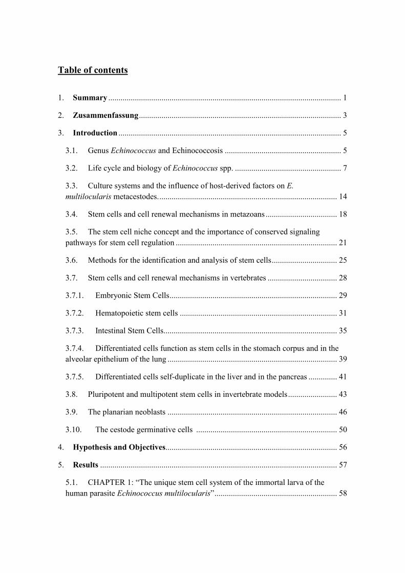
Table of contents
1. Summary .................................................................................................................. 1
2. Zusammenfassung ................................................................................................... 3
3. Introduction ............................................................................................................. 5
3.1. Genus Echinococcus and Echinococcosis ......................................................... 5
3.2. Life cycle and biology of Echinococcus spp. .................................................... 7
3.3. Culture systems and the influence of host-derived factors on E.
multilocularis metacestodes. ....................................................................................... 14
3.4. Stem cells and cell renewal mechanisms in metazoans ................................... 18
3.5. The stem cell niche concept and the importance of conserved signaling
pathways for stem cell regulation ............................................................................... 21
3.6. Methods for the identification and analysis of stem cells ................................ 25
3.7. Stem cells and cell renewal mechanisms in vertebrates .................................. 28
3.7.1. Embryonic Stem Cells .................................................................................. 29
3.7.2. Hematopoietic stem cells ............................................................................. 31
3.7.3. Intestinal Stem Cells..................................................................................... 35
3.7.4. Differentiated cells function as stem cells in the stomach corpus and in the
alveolar epithelium of the lung ................................................................................... 39
3.7.5. Differentiated cells self-duplicate in the liver and in the pancreas .............. 41
3.8. Pluripotent and multipotent stem cells in invertebrate models ........................ 43
3.9. The planarian neoblasts ................................................................................... 46
3.10. The cestode germinative cells ..................................................................... 50
4. Hypothesis and Objectives.................................................................................... 56
5. Results .................................................................................................................... 57
5.1. CHAPTER 1: “The unique stem cell system of the immortal larva of the
human parasite Echinococcus multilocularis” ............................................................ 58

5.2. CHAPTER 2: “A novel terminal-repeat transposon in miniature (TRIM) is
massively expressed in Echinococcus multilocularis stem cells.” ........................... 111
5.3. CHAPTER 3: Further experimental results regarding the E. multilocularis
stem cell system. ....................................................................................................... 145
5.3.1. The cell-cycle kinase em-plk1 is expressed in E. multilocularis germinative
cells 146
5.3.2. The insulin receptor emir2 is upregulated in the proliferating region of the
developing protoscolex ............................................................................................. 148
5.3.3. Expression of FGF receptor homologs of E. multilocularis ...................... 150
5.3.4. Transcriptomic analysis of HU treated metacestodes – A first glimpse of the
germinative cell transcriptome .................................................................................. 158
5.3.5. First steps towards the development of clonal analyses of E. multilocularis
germinative cells ....................................................................................................... 166
5.4. CHAPTER 4: “Anatomy and development of the larval nervous system in
Echinococcus multilocularis” ................................................................................... 168
5.5. CHAPTER 5: Further experimental results regarding the E. multilocularis
neuromuscular system. .............................................................................................. 210
5.5.1. Persistence of the nervous system during the development from
protoscoleces to metacestode vesicles ...................................................................... 211
5.5.2. Discovery of neuropeptide-encoding genes in E. multilocularis ............... 217
5.5.3. Expression and effects of NPs during metacestode growth and regeneration .
227
6. Discussion ............................................................................................................. 231
6.1. Tissue turnover and growth in E. multilocularis metacestodes depends on
undifferentiated germinative cells............................................................................. 231
6.2. Gene expression patterns of germinative cells: molecular markers and
population heterogeneity…………………….……………………………………...233
6.3. Self-renewal of the germinative cells ............................................................ 236
6.4. E. multilocularis germinative cells and the stem cell systems of other
flatworms: similarities and differences ..................................................................... 238
6.5. Complex expression patterns of E. multilocularis FGFRs ............................ 241

6.6. Evolution of the E. multilocularis metacestode: asexual reproduction and the
neuromuscular system ............................................................................................... 243
7. Materials and Methods ....................................................................................... 247
7.1. Parasite culture and experimental manipulation ............................................ 247
7.1.1. Media .......................................................................................................... 247
7.1.2. E. multilocularis isolates ............................................................................ 247
7.1.3. In vivo E. multilocularis maintenance, isolation of parasite material and
standard in vitro co-culture technique....................................................................... 248
7.1.4. Axenic culture of E. multilocularis metacestodes ...................................... 249
7.1.5. Isolation and activation of E. multilocularis protoscoleces ....................... 249
7.1.6. Primary cell isolation and culture............................................................... 250
7.1.7. Live microscopy of parasite cultures ......................................................... 251
7.1.8. 5-ethynyl-2′deoxyuridine (EdU) incubation and detection ........................ 251
7.1.9. Treatment of primary cells and metacestode vesicles with hydroxyurea... 251
7.1.10. X-ray irradiation of metacestode vesicles .................................................. 252
7.1.11. Treatment of primary cells with in vitro synthezised peptides .................. 253
7.2. Manipulation of nucleic acids ........................................................................ 254
7.2.1. Synthetic oligonucleotides used in this work ............................................. 254
7.2.2. General precautions for working with RNA .............................................. 263
7.2.3. RNA isolation and quantification ............................................................... 263
7.2.4. DNAse treatment of RNA .......................................................................... 264
7.2.5. cDNA synthesis .......................................................................................... 264
7.2.6. PCR, RT-PCR, and semi-quantitative RT-PCR ......................................... 264
7.2.7. Rapid amplification of cDNA ends (RACE).............................................. 265
7.2.8. Electrophoresis of DNA and RNA ............................................................. 266
7.2.9. Molecular Cloning...................................................................................... 266
7.2.10. Restriction digestion ................................................................................... 268

7.2.11. High throughput RNA sequencing (RNA seq) and analysis ...................... 268
7.3. Histological procedures and transmission electron microscopy .................... 270
7.3.1. Preparation of cell suspensions (cell maceration) and staining.................. 270
7.3.2. Fixation of metacestode vesicles and protoscoleces for histological
sectioning and whole-mount procedures .................................................................. 270
7.3.3. Preparation of Paraplast sections................................................................ 270
7.3.4. Preparation of Cryosections ....................................................................... 271
7.3.5. Alkaline phosphatase histochemistry (ALP-HC) ....................................... 271
7.3.6. Acetylcholinesterase histochemistry (AChE-HC) ..................................... 272
7.3.7. 4′,6-diamidino-2-phenylindole (DAPI) and phalloidin staining ................ 272
7.3.8. Processing of samples for Transmission Electron Microscopy (TEM) ..... 272
7.4. Detection of protein and mRNA localization in situ ..................................... 274
7.4.1. Antibodies used in this work ...................................................................... 274
7.4.2. Immunohistofluorescence (IHF) and immunohistochemistry (IHC) ......... 275
7.4.3. Whole-mount in situ hybridization (WMISH) ........................................... 275
7.4.4. Epifluorescence microscopy and confocal laser scanning microscopy ..... 275
7.5. Manipulation of proteins ................................................................................ 277
7.5.1. Preparation of lysates for SDS-PAGE ....................................................... 277
7.5.2. Sodium Dodecyl Sulfate-Polyacrylamide Gel Electrophoresis ................. 277
7.5.3. Western Blot ............................................................................................... 277
7.6. Bioinformatics and statistics .......................................................................... 279
7.6.1. Datasets and programs ............................................................................... 279
7.6.2. Discovery of neuropeptide and peptide hormone (NP) genes in the genomes
of cestodes ................................................................................................................. 280
7.6.3. Statistics ..................................................................................................... 281
8. Bibliography ........................................................................................................ 282
Appendix 1: E. multilocularis primary cell isolation ................................................... 308

Appendix 2: EdU detection in Whole-mounts ............................................................. 310
Appendix 3: Preparation of cell macerates (cell suspensions) for microscopy ............ 311
Appendix 4: Immunohistochemistry of Paraplast sections .......................................... 312
Appendix 5: Immunohistofluorescence on cryosections .............................................. 314
Appendix 6: Whole-mount Immunohistofluorescence protocols for protoscoleces and
small metacestodes ....................................................................................................... 315
Appendix 7: Fluorescent Whole-Mount in situ Hybridization (WMISH) for E.
multilocularis metacestodes ......................................................................................... 317
Appendix 8: Fluorescein-tyramide synthesis (Hopman et al., 1998) ........................... 327
Appendix 9: in vitro synthesis and quantification of Digoxigenin-labeled RNA probes
...................................................................................................................................... 328
Curriculum Vitae .......................................................................................................... 331

1. Summary
1
1. Summary
The metacestode larva of Echinococcus multilocularis is the causative agent of
alveolar echinococcosis (AE), one of the most dangerous zoonotic diseases in the
Northern Hemisphere. Unlike “typical” metacestode larvae from other tapeworms, it
grows as a mass of interconnected vesicles which infiltrates the liver of the intermediate
host, continuously forming new vesicles in the periphery. From these vesicles,
protoscoleces (the infective form for the definitive host) are generated by asexual
budding. It is thought that in E. multilocularis, as in other flatworms, undifferentiated
stem cells (so-called germinative cells in cestodes and neoblasts in free-living
flatworms) are the sole source of new cells for growth and development. Therefore, this
cell population should be of central importance for the progression of AE.
In this work, I characterized the germinative cells of E. multilocularis, and
demonstrate that they are indeed the only proliferating cells in metacestode vesicles.
The germinative cells are a population of undifferentiated cells with similar
morphology, and express high levels of transcripts of a novel non-autonomous
retrotransposon family (ta-TRIMs). Experiments of recovery after hydroxyurea
treatment suggest that individual germinative cells have extensive self-renewal
capabilities. However, germinative cells also display heterogeneity at the molecular
level, since only some of them express conserved homologs of fgfr, nanos and
argonaute genes, suggesting the existence of several distinct sub-populations. Unlike
free-living flatworms, cestode germinative cells lack chromatoid bodies. Furthermore,
piwi and vasa orthologs are absent from the genomes of cestodes, and there is
widespread expression of some conserved neoblast markers in E. multilocularis
metacestode vesicles. All of these results suggest important differences between the
stem cell systems of free-living flatworms and cestodes.

1. Summary
2
Furthermore, I describe molecular markers for differentiated cell types,
including the nervous system, which allow for the tracing of germinative cell
differentiation. Using these molecular markers, a previously undescribed nerve net was
discovered in metacestode vesicles. Because the metacestode vesicles are non-motile,
and the nerve net of the vesicle is independent of the nervous system of the protoscolex,
we propose that it could serve as a neuroendocrine system. By means of bioinformatic
analyses, 22 neuropeptide genes were discovered in the E. multilocularis genome. Many
of these genes are expressed in metacestode vesicles, as well as in primary cell
preparations undergoing complete metacestode regeneration. This suggests a possible
role for these genes in metacestode development. In line with this hypothesis, one
putative neuropeptide (RGFI-amide) was able to stimulate the proliferation of primary
cells at a concentration of 10-7 M, and the corresponding gene was upregulated during
metacestode regeneration.

2. Zusammenfassung
3
2. Zusammenfassung
Das Metazestoden Larvenstadium von Echinococcus multilocularis ist die
Ursache für die alveoläre Echinokokkose (AE), eine der gefährlichsten Zoonosen in der
nördlichen Hemisphäre. Im Gegensatz zu Metazestoden anderer Bandwürmer wächst es
zu einem Labyrinth verknüpfter Vesikel, die in der Peripherie permanent neu gebildet
werden und dabei die Leber des Wirts infilitrieren. In diesen Vesikeln werden die
Protoskolizes (das infizierende Stadium für den Endwirt) durch asexuelle Knospung aus
der Vesikelwand heraus gebildet. Man geht davon aus dass in E. multilocularis, wie in
anderen Plattwürmen, undifferenzierte Stammzellen (so gennante „Germinative cells”
in Bandwürmern und Neoblasten in Turbellarien) der einzige Ursprung neuer Zellen für
Wachstum und Entwicklung sind. Deshalb sollte diese Zellpopulation eine zentrale
Rolle im Fortschritt der AE spielen.
In dieser Arbeit habe ich die Germinative cells von E. multilocularis
charakterisiert und zeige, dass sie tatsächlich die einzigen sich vermehrenden Zellen in
Metazestodenvesikeln sind. Die Germinative cells sind eine Population von
undifferenzierten Zellen mit ähnlicher Morphologie, die eine hohe Zahl an Transkripten
einer neuen Retrotransposonfamilie (ta-TRIMs) exprimieren. Experimente nach
Behandlung mit Hydroxyurea deuten darauf hin, dass einzelne Germinative cells die
Fähigkeit haben sich selbst zu erneuern. Allerdings, zeigen die Germinative cells auch
Heterogenität auf molekurarer Ebene, da nur manche von Ihnen konservierte Homologe
von fgfr, nanos und argonaute Genen exprimieren, was auf die Existenz eindeutiger
Subpopulationen hinweist. Im Gegensatz zu Turbellarien fehlen den Germinative cells
von Zestoden “Chromatoid bodies”, weiterhin fehlen dem Genom der Zestoden
Orthologe von piwi und vasa und es werden einige Neoblastenmarker in den
Metazestodenvesikeln von E. multilocularis umfassend exprimiert. All diese Ergebnisse
zeigen deutliche Unterschiede zwischen den Stammzellsystemen von Turbellarien und
Zestoden auf.
Ich beschreibe ausserdem molekulare Marker für differenzierte Zelltypen,
inklusive solche des Nervensystems. Mit diesen Markern wurde ein Nervennetz in
2. Zusammenfassung
4
Metazestodenvesikeln endeckt, das bis dato unbeschrieben war. Da die Vesikel
unbeweglich sind und ihr Nervennetz unabhängig vom Nervensystem des Protoscolex
ist wird angenommen dass es als Neuroendokrinsystem dient. Mit Hilfe von
Genomanalysen wurden 22 Neuropeptidgene im Genom von E. multilocularis entdeckt.
Viele von ihnen werden sowohl in Metazestodenvesiklen exprimiert als auch in
Primärzellpräparationen, die zu kompletten Vesikeln regenerieren. Das weist auf eine
mögliche Rolle dieser Gene in der Metazestodenentwicklung hin. Einhergehend mit
dieser Hypothese war ein putatives Neuropeptid (RGFIamide) in der Lage die
Vermehrung von Primärzellen bei einer Konzentration von 10-7 M zu stimulieren, dabei
war das korrespondierende Gen während der Metazestodenregeneration hochreguliert.

3. Introduction
5
3. Introduction
3.1. Genus Echinococcus and Echinococcosis
The clinical term Echinococcosis is used to describe a group of zoonotic
diseases caused by infection with the metacestode larvae of cestodes of the genus
Echinococcus (order Cyclophyllidea, family Taeniidae). From the medical and
veterinary point of view, the two most important species of this genus are Echinococcus
granulosus (informally called the “dog tapeworm”) and Echinococcus multilocularis
(the so-called “fox tapeworm”), which are the causative agents of Cystic
Echinococcosis (CE) and Alveolar Echinococcosis (AE), respectively (Eckert and
Deplazes 2004). E. granulosus has a cosmopolitan distribution: it is present in over 100
countries from all continents except Antarctica, and is of medical and veterinary
relevance (Eckert and Deplazes 2004; Moro and Schantz 2009). E. multilocularis on the
other hand is restricted to endemic regions in the northern hemisphere (Figure I1
(Torgerson et al. 2010)), and has a much lower global incidence. However, as it will be
described below, the characteristics of E. multilocularis makes AE more difficult to
treat than CE. AE is almost impossible to cure when detected at late stages of
development, and is typically lethal if left untreated (Craig 2003; Eckert and Deplazes
2004; Moro and Schantz 2009; Brunetti, Kern, and Vuitton 2010). This makes AE one
the most dangerous zoonoses in the northern hemisphere, and it has been estimated that
the global burden of AE is comparable to that of many other neglected tropical diseases
such as Leishmaniasis and Trypanosomiasis, for which research efforts are much more
intensive (Torgerson et al. 2010). Other species of Echinococcus have been described
and are currently recognized, including Echinococcus vogeli and Echinococcus
oligarthrus, which cause Polycystic Echinococcosis (PE). These two species are limited
in distribution to the neotropical region in Central and South America and are of only
limited medical relevance (Eckert and Deplazes 2004; D'Alessandro and Rausch 2008;
Moro and Schantz 2009).

3. Introduction
6
Figure I1. Distribution of AE in the world. Figure from Torgerson et al. (2010)

3. Introduction
7
3.2. Life cycle and biology of Echinococcus spp.
The life cycle of Echinococcus spp. involves two mammalian hosts (Figure I2
for E. granulosus and Figure I3 for E. multilocularis). The adult worm develops in the
small intestine of the definitive host (typically a canid for all species except for
Echinococcus felidis), with a morphology that is relatively typical for cyclophyllidean
cestodes, and which is very similar for all Echinococcus species (Thompson 1986;
Eckert and Deplazes 2004). The anterior region of the adult is denominated the scolex
and it contains the attachment organs (suckers and rostellum). Behind the scolex, the
neck region proliferates extensively and continuously generates a chain of segments
(proglottids), each one developing a complete set of male and female reproductive
organs. The adults of Echinococcus spp. are unusual among cestodes in that, unlike
other taeniids, they only have a very small number of proglottids (3 to 6). Furthermore,
unlike some of the best known taeniids such as Taenia solium, the adult of
Echinococcus spp. only lives in the intestine for a limited period of time (up to 12
months) after which the infection is lost (Craig 2003).
Within each proglottid, fertilization occurs. It is thought that Echinococcus spp.
reproduce mostly by self-fertilization, but there is limited evidence indicating that
occasional cross-fertilization can take place (Haag et al. 2011). Fertilization results in
the production of thousands of infective eggs, that are released (within the mature
proglottids) with the host´s feces to the environment. The eggs are then released from
the proglottid and can survive for many months under ideal conditions (Craig 2003).
The released eggs contain the first larval stage, the oncosphere, a highly reduced
organism with six small hooks and several protective layers around it. The eggs are
accidentally ingested by the intermediate host and hatch in the intestine, where the
larvae penetrate the intestinal wall and ingress into the portal vein, from which they are
transported to the liver, and then to the rest of the body, via the blood stream. Most
commonly, primary infections develop in the liver (especially in the case of E.
multilocularis), but other organs can be primary infection sites for E. granulosus
(especially the lungs and the brain) (Craig 2003; Eckert and Deplazes 2004; Brunetti,
Kern, and Vuitton 2010).

3. Introduction
8
Figure I2. Life cycle of E. granulosus. Figure from the Center for Disease
Control and Prevention (http://www.cdc.gov/parasites/echinococcosis/biology.html)
Figure I3. Life cycle of E. multilocularis. (A) Adult mature parasite. (B) Foxes
(left, red fox; right, Arctic fox) are the principal definitive hosts; domestic animals such
as dogs, other canids, and cats can also be involved in the cycle. (C) Proglottids with
eggs are released with the feces of the fox. (D) Egg with oncosphere. (E) Accidental
infection of humans. (F) Infection of rodents with oncospheres (G) Development of
metacestodes in the liver of the rodent. (H) Detail of a metacestode vesicle with
protoscoleces. Figure from Eckert and Deplazes (2004).
3. Introduction
9
Several different ungulate species are the typical intermediate hosts for the
different lineages of the E. granulosus species complex, and most of them are domestic
species. Indeed, each of the different lineages of E. granulosus, which can be
distinguished on the basis of their mitochondrial DNA genotype (genotypes G1 to G10),
have specific intermediate hosts for which they are highly infective and in which they
can develop normally (e.g. E. granulosus genotypes G1 and G2 infect sheep, whereas
G4 is infective for horses and G5 is infective for cattle) (Thompson 1986; Moro and
Schantz 2009). It has been further proposed that several of these genotypes should be
elevated to the rank of species, including genotypes G1 (as E. granulosus sensu stricto),
G4 (as Echinococcus equinuus), and G5 (as Echinococcus ortleppi) (Moro and Schantz
2009; Nakao et al. 2010). The fact that all genotypes of E. granulosus except genotype
G8 have a domestic life cycle between dogs and domestic ungulates makes control
strategies that target the natural life cycle feasible, and has even lead to the eradication
of the disease in some islands such as Iceland (Beard 1973; Moro and Schantz 2009).
In contrast, several rodent species are the natural intermediate host for E. multilocularis,
in particular species of the families Cricetidae and Arvicolidae (Rausch 1954; Craig
2003; Eckert and Deplazes 2004). Therefore, the natural life cycle of E. multilocularis is
sylvatic, occurring between foxes and rodents. Because of this, control of the disease by
monitoring and treatment of the natural hosts is very costly and difficult.
Humans can be an accidental host for E. granulosus and E. multilocularis,
although they are an aberrant one and a dead-end for the life cycle of the parasite. In the
case of E. granulosus, humans become exposed to the infective eggs when they come in
contact with infected dogs and their feces (Moro and Schantz 2009). This is more
common in the country-side of countries where cattle and sheep are raised (particularly
in developing countries in which the dogs have access to the infected offal). For E.
multilocularis, it is not completely clear how most humans become exposed to the
parasite. Living in the country-side and owning dogs are risk factors for AE, suggesting
that in many cases people become exposed to eggs released from their dogs, after they
become infected from eating wild infected rodents. Also, ingestion of contaminated
vegetables and fruits can be the source of E. multilocularis infection in men (Moro and
Schantz 2009).
Once in the intermediate host, the oncosphere develops by metamorphosis into
the next larval stage, the metacestode. The metacestode stage of the genus Echinococcus

3. Introduction
10
is an evolutionary novelty that is quite divergent from the “typical” development of the
metacestode stage of other cestodes (Freeman 1973; Slais 1973). In more typical
cestodes, the metacestode is similar to a “juvenile” tapeworm, containing the scolex
with the attachment organs, but lacking segmentation and the reproductive systems. In
the case of Echinococcus, the metacestodes develop as fluid-filled vesicles. These
metacestode vesicles comprise a thin layer of tissue (the germinal layer) covered by a
syncitial tegument that secretes an acellular, carbohydrate-rich external layer (the
laminated layer). The remaining volume of the vesicles is filled with fluid (hydatid
fluid).
Within the germinal layer, thickenings (buds) occur that invaginate into the
vesicle, resulting in the formation of brood capsules (Goldschmidt 1900; Thompson
1986; Leducq and Gabrion 1992; Koziol, Krohne, and Brehm 2013) (Figure I4). Within
the brood capsules, a new budding process occurs, that results in the formation of
protoscoleces, the infective form for the definitive host (Figure I4). The protoscolex
already resembles the anterior region of the adult form, and remains quiescent with the
scolex invaginated within a small posterior body (Figure I4). The life cycle of
Echinococcus spp. is finally closed when the definitive host ingests an infected
intermediate host. After ingestion of the protoscolex by the definitive host, it evaginates
its scolex, attaches to the intestine and develops into the adult tapeworm. The formation
of many protoscoleces in each metacestode represents a form of asexual propagation by
the parasite. Asexual reproduction is very rare in cestodes, but it is relatively common
within the family Taeniidae (genera Echinococcus and Taenia). It is possible that these
processes are homologous between Echinococcus and Taenia species, although this is
controversial (Freeman 1973; Slais 1973; Moore and Brooks 1987; Hoberg et al. 2000;
Loos-Frank 2000; Trouvé, Morand, and Gabrion 2003; Swiderski et al. 2007).
3. Introduction
11
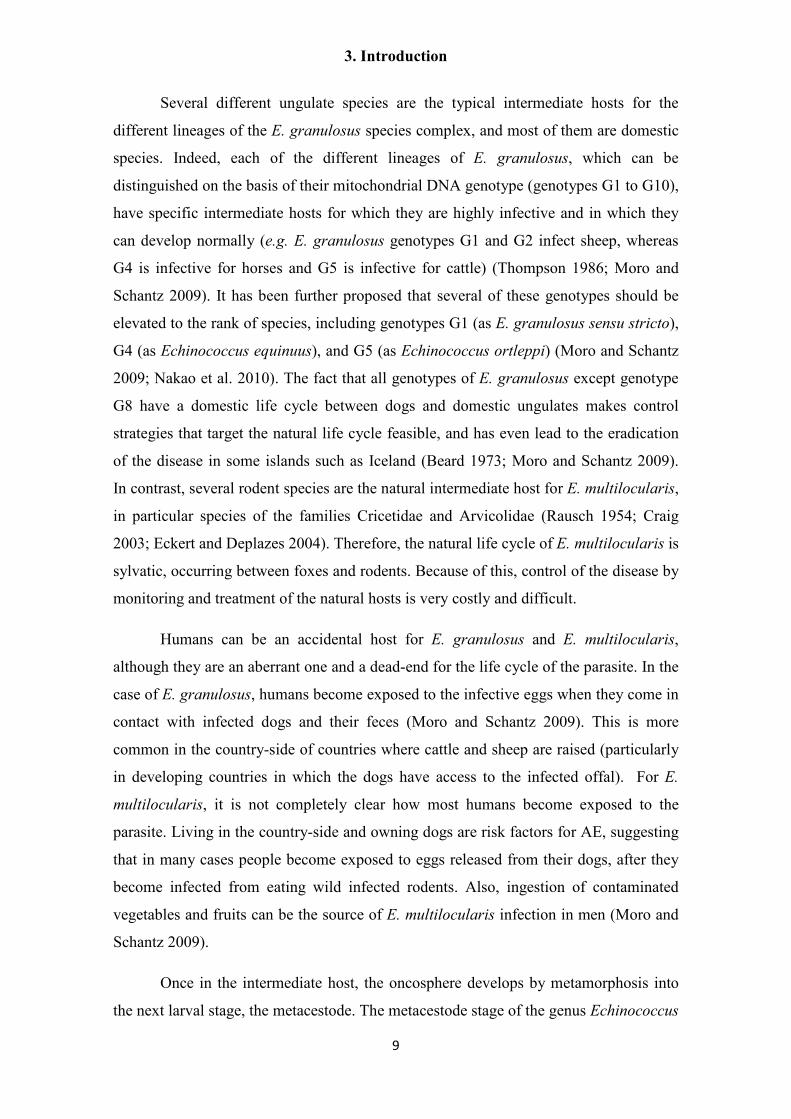
Figure I4. Schematic drawing showing the general organization and
development of E. multilocularis metacestodes. A. Early brood capsule bud. B. Brood
capsule with protoscolex bud. C. Brood capsule with protoscolex in late development.
D. Brood capsule with invaginated protoscolex. The syncitial tegument is shown in
orange, the germinative cells in brown, glycogen/lipid storage cells in violet, calcareous
corpuscle cells in light blue, nerve cells in green and muscle cells and fibers in red. bc,
brood capsule; GL, germinal layer; HF, hydatid fluid; LL, laminated layer; ps,
protoscolex; r, rostellum; s, sucker. Figure and legend from Koziol et al. (2014).
Figure I5. Diagrammatic comparison of E. granulosus (a) and E.
multilocularis (b) metacestodes. Figure from Diaz et al., 2011.

3. Introduction
12
E. multilocularis and E. granulosus differ in the morphology and development
of the metacestode stage (Thompson 1986; Eckert and Deplazes 2004) (Figure I5). In
the case of E. granulosus, each oncosphere develops into a single vesicle (the hydatid
cyst) covered by a thick laminated layer. This mode of development is denominated
“unilocular”. Each vesicle can grow to huge dimensions (exceeding 20 cm in diameter),
and from the germinal layer brood capsules develop internally, each containing several
protoscoleces. Rarely, endogenous formation of daughter cysts within the “mother” cyst
occurs, by a process that is still incompletely understood (Fairley and Wright-Smith
1929; Rogan and Richards 1986). Exogenous formation of new vesicles is controversial
for E. granulosus, and if it exists at all it must be very rare (Rausch and D'Alessandro
1999).
In contrast, development of E. multilocularis is only unilocular during the very
early stages of development. After the first week of development, new vesicles are
generated by exogenous budding of the metacestode, which therefore develops as a
multilocular labyrinth of interconnected vesicles (Rausch 1954; Ohbayashi 1960;
Sakamoto and Sugimura 1970). This process occurs continuously, and at later stages
small protrusions of the metacestode tissue, devoid of laminated layer, have been
described to emerge from the periphery of the metacestode mass and infiltrate the host
tissues, resulting in the formation of new vesicles not only in the liver but also in
neighboring organs (Eckert, Thompson, and Mehlhorn 1983; Mehlhorn, Eckert, and
Thompson 1983). The metacetode tissue can even form metastases in distant organs
during late stages of infection. It is thought that this occurs by infiltration of small
vesicles or groups of parasite cells into the blood and lymph vessels, which are then
distributed to other organs where they initiate the development of new metacestode
tissue (Eckert, Thompson, and Mehlhorn 1983). When vesicles mature, they produce
brood capsules, and from each brood capsule typically only one or a few protoscoleces
develop. The mature, protoscolex-filled vesicles then cease to grow. Most of the
metacestode vesicles in late infections have already ceased to grow, and can even
become necrotic in the center of the metacestode tissue. Only the tissue in the periphery
is still active, and continues to grow and infiltrate the organs of the host (Eckert,
Thompson, and Mehlhorn 1983).
Growth of E. multilocularis is very fast in rodents, the natural intermediate
hosts: after a few months the development of metacestodes is complete (i.e., mature

3. Introduction
13
protoscoleces are formed) and the host either dies from the infection or is easily preyed
on by the definitive hosts (Rausch 1954; Craig 2003). In contrast, growth in humans is
aberrant, since it is much slower, and usually no protoscoleces are produced (Craig
2003; Moro and Schantz 2009). Because of this, the parasite origin of the metacestode
vesicles was not initially recognized by doctors, and AE was originally thought to be
either a form of liver cancer or a necrosis of the liver tissue. It was Rudolf Virchow,
working in the University of Würzburg, who showed in the 1850s that a species of
Echinococcus is responsible for the etiology of AE (Tappe and Frosch 2007).
Originally, it was thought that AE was caused by an aberrant form of E. granulosus in
man. Only after more than 90 years of R. Virchow´s findings, when the natural
intermediate hosts were discovered, was it shown that E. multilocularis is actually a
distinct species (Rausch 1954; Tappe and Frosch 2007; Nakao et al. 2010).
Because of the slow growth and the infiltrative nature of E. multilocularis
metacestodes, AE remains asymptomatic for up to 10 to 15 years after the initial
infection, and only rather unspecific symptoms appear after this time (Moro and
Schantz 2009). If discovered in time, AE can in principle be cured by radical resection
of the infected region. However, it is usually discovered only at late stages, at which
point complete resection is impossible, and microscopic portions of parasite tissue
infiltrate the liver, resulting in recurrence if surgery is performed (Brunetti, Kern, and
Vuitton 2010). Metastases are common in the lungs, peritoneal cavity and brain, making
the surgical option impractical as well. The only option for most cases of late stage AE
is chemotherapeutic treatment with benzamidazoles (BMZs: Albendazole or
Mebendazole), but this treatment is only parasitostatic, and must be taken for many
years, usually for the rest of the patient´s life (Brunetti, Kern, and Vuitton 2010). BMZs
are toxic for a small proportion of patients, for whom there is no therapeutic option
against AE (Eckert and Deplazes 2004). Furthermore, BMZs are largely unavailable for
most of the infected population of the world, living in the least developed areas of
China and Russia (Torgerson et al. 2010). If untreated, late stage AE is almost
invariably deadly within 8 to 11 years (Eckert and Deplazes 2004).

3. Introduction
14
3.3. Culture systems and the influence of host-derived factors on E.
multilocularis metacestodes.
The metacestode stage of E. multilocularis is able to grow continuously by
asexual formation of new vesicles within an appropriate host, and can be maintained
indefinitely in vivo by serial passage of metacestode tissue from one host to the next
(Norman and Kagan 1961; Spiliotis and Brehm 2009). In this sense, the metacestode
larva of E. multilocularis can be considered immortal, similarly to the adult stage of
other cestodes, which are able to grow and produce new segments for as long as the host
survives. This implies that the metacestode tissue must contain cellular mechanisms for
continuous tissue turnover and growth. That is, there must be a population or
populations of cells that can self-renew and generate all of the cell types of metacestode
vesicles, protoscoleces and eventually the adult if a protoscolex infects a definitive host.
The metacestode tissue and cells can also be cultured in vitro, by means of
special culture conditions that were optimized in the laboratory of Dr. Klaus Brehm.
The first methods developed for the robust culture of metacestode vesicles in vitro are
the co-culture systems, in which metacestode vesicles are cultured in media optimized
for mammalian cells in the presence of fetal calf serum and mammalian feeder cells
(Jura et al. 1996; Brehm and Spiliotis 2008a; Spiliotis et al. 2008). Growth of the
metacestode is absolutely dependent on the feeder cells, and different cell lines can
serve this function, although the best cells identified so far are primary liver cells and
hepatoma cell lines from rodents (Spiliotis et al. 2004). By a series of elegant
experiments it was shown that these feeder cells were not only providing soluble factors
required by the metacestode vesicles, but also eliminating toxic substances (likely
reactive oxygen species) from the cell culture media, and that both processes were
necessary for optimal metacestode growth (Brehm and Spiliotis 2008a; Brehm and
Spiliotis 2008b; Spiliotis et al. 2008). Based on these experiments, an axenic culture
system was developed in which the metacestode vesicles can grow in the absence of
host cells, by culturing them in filtered media pre-conditioned by feeder cells, under
microaerobic and reducing conditions (Spiliotis et al. 2004; Spiliotis et al. 2008).
Furthermore, primary cells can be harvested from these axenic vesicles, and under
similar conditions, these cells are able to completely regenerate metacestode vesicles

3. Introduction
15
(Spiliotis et al. 2008; Spiliotis et al. 2010) (Figure I6). This shows that at least at the
population level the primary cell preparations are multipotent, and allows for the first
time to study the development of E. multilocularis in vitro.
The strict requirement for serum and soluble host factors indicates that some sort
of molecular dialog is occurring between the metacestode and the host, in which the
host cells provide in vitro, and also likely in vivo, signals that promote and regulate the
development of the metacestode (Brehm 2010a). Because of the high evolutionary
conservation of signaling pathways among metazoans, including signaling ligands and
receptors, it is possible that this interaction occurs between the growth factors and
cytokines of the host, and the cognate receptors of the parasite. One of the main lines of
research in the laboratory of Dr. Klaus Brehm has therefore been the characterization of
these conserved signaling pathways in E. multilocularis. It has been shown that host
growth factors such as insulin and fibroblast growth factors (FGFs) are capable at a
biochemical level of interacting with the parasite receptors and activating the
downstream signaling cascades (Förster 2012; Hemer et al. 2014). Furthermore,
addition of these growth factors at physiologically relevant concentrations promotes
growth of metacestode vesicles and regeneration from primary cell preparations, thus
showing an effect of host growth factors on metacestode development (Förster 2012;
Hemer et al. 2014). However, it is not known how these host factors could promote the
development of the metacestode at a cellular / tissular level, since it is not clear which
cells express the parasite receptors and how they respond to the host factors.

3. Introduction
16
Figure I6. Regeneration of metacestode vesicles from E. multilocularis
primary cell preparations. This primary culture was performed in the presence of
Reuber RH rat hepatoma cells in a trans-well system. Progressive developmental stages
are: A. Initial culture. B. Formation of cell aggregates. C. Appearance of cavities within
the aggregates and release of the first vesicles (arrow). D. Completely developed
vesicles with a laminated layer. Figure from Spiliotis et al., 2008.

3. Introduction
17
Cestodes are part of the phylum Platyhelminthes (flatworms), which contains
many free-living groups and a large monophyletic clade of parasites, the Neodermata
(Ax 1996; Baguñà and Riutort 2004; Olson and Tkach 2005). This clade includes the
well known parasitic classes Cestoda, Trematoda and Monogenea. Flatworms are a
highly diverse phylum in terms of morphology, development, and life-cycles. However,
they have in common a unique population of undifferentiated stem cells, commonly
known as “neoblasts”. It is thought that neoblasts are the only proliferative cell
population, and are therefore the source of new cells for normal tissue turnover, growth
and regeneration, whereas all differentiated cells are post-mitotic (Gustafsson 1990;
Peter et al. 2004; Reuter and Kreshchenko 2004; Koziol and Castillo 2011). This is an
unusual cellular mechanism for tissue turnover, since in most animals several tissue-
specific stem cells exist and many differentiated cell types are also able to proliferate
(this subject is further developed in the following section). In cestodes, classical studies
have described a population of undifferentiated stem cells similar to the neoblasts, the
so-called germinative cells (see section 3.10). In particular, in E. multilocularis
metacestodes, ultrastructural studies demonstrated the existence of germinative cells in
the germinal layer, which proliferate and accumulate during brood capsule and
protoscolex development (Sakamoto and Sugimura 1970). Because of the importance of
conserved signaling pathways in stem cell biology in animals, and the presumed
relevance of the germinative cells as the source of new cells for metacestode growth and
development, they constitute a natural focus of research as the possible targets of host-
derived growth factors. The main objective of this thesis is therefore the
characterization of the stem cell system of E. multilocularis, and to investigate the
possible role of previously characterized parasite signaling pathways in their
physiology. In the following sections of the introduction, I will explore the subject of
stem cells and the different mechanisms of tissue turnover in well studied models, and
compare them to what is known and hypothesized for cestodes and other flatworms.

3. Introduction
18
3.4. Stem cells and cell renewal mechanisms in metazoans
In many adult tissues in animals (particularly for tissues with fast cellular
turnover), differentiated effector cells are post-mitotic, and have a limited lifetime.
These cells are lost from the “wear and tear” of the tissue, or by programmed cell death,
and are replaced from an undifferentiated pool of stem cells and their progeny (Bryder,
Rossi, and Weissman 2006; Pellettieri and Sanchez Alvarado 2007). Stem cells are
defined as cells that have the long-term ability to self-renew (that is, to generate new
stem-cells by cell division) and that have the potency to differentiate into many cell
types. This general definition is somewhat vague, in that “long-term self renewal” and
“many cell types” are not explicitly defined. Usually, “long-term self-renewal” refers to
the ability to self-renew for the duration of the life of the adult organism. As for the
potency, stem cells and their progeny are defined as: 1) pluripotent, when they can give
rise to all of the cells of an organism, from all three embryonic germinal layers (in
mammals, the term totipotent is used specifically for cells that can give rise to all
embryonic germinal layers as well as to extra-embryonic tissues); 2) multipotent, when
they can differentiate into many different cell types; 3) oligopotent, when they can
differentiate into a few, generally related cell types, and 4) unipotent, when they can
only differentiate into one cell type (Seita and Weissman 2010).
In mammals, pluripotent stem cells are only known from the early embryonic
stages (Hanna, Saha, and Jaenisch 2010). In adults, all of the well-characterized stem
cell systems are actually tissue-specific, and their multipotency is generally defined as
the ability to generate all of the main cell types of their tissue (Bryder, Rossi, and
Weissman 2006). However, some of the best known stem cells in mammals and other
animals are actually unipotent, such as the germ line stem cells (GSCs) of mice,
Drosophila and Caenorhabditis which will only produce gametes throughout the life-
span of the adult organism (Alberts 2000; Xie 2008).
In many systems, the stem cells give rise to progenitor cells. These progenitor
cells can actively proliferate and differentiate into many cell types, but they no longer
have the ability for long-term self renewal, and are therefore sometimes referred to as
transit-amplifying cells. In some of the best studied stem cell systems, such as the
murine hematopoietic stem cell (HSC) system, there is a gradient of self-renewal and

3. Introduction
19
differentiation potency, in which stem cells give rise to multipotent progenitors, which
in turn give rise to a hierarchy of progenitor cells with progressively reduced self-
renewal and differentiation potency (Bryder, Rossi, and Weissman 2006) (see section
3.7.2).
In many other adult tissues, especially those with a slow cellular turnover, the
source of normal cell renewal is not from undifferentiated stem cells, but from self-
replication of differentiated effector cells (Yanger and Stanger 2011). Furthermore, in
some systems it has been shown that committed progenitors and differentiated cells
have the ability to function as stem cells (that is, they are the source of new cells for
several different cell types), either by proliferation and trans-differentiation (direct
transformation of a differentiated cell into another cell type) or by de-differentiation
(transformation of a differentiated cell into an undifferentiated stem cell type). In
particular, even in high-turnover tissues with canonical stem cell systems, some
differentiated cells and committed progenitors have been shown to function as a reserve
system which can take over the role of the stem cells under special conditions (such as
stem cell depletion and during injury repair). Examples of these mechanisms are further
explored in sections 3.7.3, 3.7.4 and 3.7.5.
In addition to self-renewal and differentiation, some other characteristics have
been traditionally thought to be shared by all or most stem cells in different tissues,
although the evidence was always limited to a few types of stem cells, in particular the
HSCs (Alberts 2000). One characteristic is quiescence: stem cells in some models have
been shown to proliferate only infrequently and to have low metabolic activity, which is
thought to protect the stem cell, in particular to prevent its DNA from the incorporation
of deleterious mutations. The stem cells could then become active when the tissue needs
to be expanded or repaired. In addition, most stem cells were thought to divide
primarily by asymmetric cell divisions, in which one daughter cell would retain the
stem cell identity, whereas the other daughter cell would become a progenitor cell with
limited self-renewal potency. This would give a straightforward mechanism for tissue
homeostasis, since the number of stem cells would remain constant. However, recent
developments in the study of mammalian stem cells have challenged these paradigms,
and have shown that quiescence and asymmetric cell division are not necessary
attributes of stem cells in all tissues (Barker, Bartfeld, and Clevers 2010; Barker and
Clevers 2010; Klein and Simons 2011; Lander 2011). Many mammalian adult tissues

3. Introduction
20
such as the intestinal epithelium, the inter-follicular skin epithelium and the germ line in
the testis have been shown to be supported under conditions of normal homeostasis by
actively proliferating stem cells, which divide in both symmetric and asymmetric ways,
and whose fate is stochastic, depending on their interaction with their specific niche. As
will be described in later sections, it is thought that in these and other tissues quiescent
stem cells could be a separate population which normally remain inactive, but that may
become active and have a specific role during injury repair and regeneration (Li and
Clevers 2010; Doupe and Jones 2013). Even in the case of the Drosophila GSCs, which
are known to divide by asymmetric divisions in which the daughter cell receives
specific cytoplasmatic factors, these factors are neither necessary nor sufficient for the
specification of the daughter cell fate. The fate of the daughter cell is instead defined by
their interaction with the stem cell niche, since only those cells that remain within the
niche maintain the stem cell identity (Losick et al. 2011) . In this way, homeostasis of
the stem cell compartment is achieved at a population level by extrinsic signals so that
in average half of the daughter cells remain as stem cells under normal conditions.

3. Introduction
21
3.5. The stem cell niche concept and the importance of conserved signaling
pathways for stem cell regulation
The stem cell niche is defined, in its most restrictive sense, as a localized
microenvironment within tissues where stem cells reside, and which provides signals
which promote self-renewal of the stem cells (Morrison and Spradling 2008; Lander et
al. 2012). The niche must therefore consist of specific cells, the signals they produce,
and the extracellular matrix (ECM) surrounding the stem cells. Furthermore, the niche
environment may provide signals that regulate the proliferation and/or differentiation of
the stem cells.
In order to identify the components of the niche, specific perturbation of defined
cell types and signaling pathways can be performed, in order to assess the effect on the
stem cell population. However, in order to show that these properties are important for a
specific localized niche, and not a general property of the whole tissue, more precise
experimental methods are needed (Morrison and Spradling 2008). The best examples of
stem cell niches have been identified in invertebrates, in particular for the GSCs of
Drosophila and Caenorhabditis (Xie 2008; Losick et al. 2011) (Figure I7). In both
cases, the niche is composed of neighboring cells which make direct contact to the stem
cells, and provide them with short-range signals that maintain the resident cells in an
undifferentiated state. The specific ECM in those niches potentiates these signaling
mechanisms and limits their diffusion, preventing activation of these signaling pathways
outside of the niche. In the case of Drosophila male GSCs, it has been actually shown
that removing a stem cell from the niche results in the loss of its stem cell identity,
whereas re-incorporation of a daughter progenitor cell into the niche reinstates a stem
cell identity to this cell (Brawley and Matunis 2004; Losick et al. 2011).
Theoretically, stem cells could instead maintain their identity by cell-
autonomous mechanisms or from general signals provided in a non-localized fashion by
the surrounding tissue, and this has been proposed to be the case for a few specific stem
cell systems, such as the Drosophila intestinal stem cells. It is thought however that the
stem cell niche can have specific regulatory functions which would not be achieved
from non-localized signals, acting as a mechanism of feedback control in which limited
niche space results in a limit in the expansion of stem cells (Lander et al. 2012). The

3. Introduction
22
niche is also thought to function as a coordinator of signals for different cellular
compartments within a tissue or organ, in particular in those with several different cell
lineages as is the case of the hair follicle.
Metazoans employ a relatively small number of conserved signaling systems to
regulate and coordinate their embryonic and adult development (Gilbert 2006). Some of
these signaling pathways have been shown to be important for many stem cell types,
and to be activated by specific niche signals in many tissues and organisms. These
include in particular the canonical Wnt/beta-catenin pathway (Clevers 2006; Nusse et al.
2008), the Delta/Notch pathway (Koch, Lehal, and Radtke 2013), the BMP pathway
(Watabe and Miyazono 2009), and signaling by fibroblast growth factors (FGFs) (Coutu
and Galipeau 2011) (Figure I8). Although some common themes can be inferred about
their function in stem cell biology, it is important to realize that their actual roles vary
greatly between different stem cell systems, and even within one system, they may have
several overlapping roles over the stem cells and their progeny. For instance, canonical
Wnt, FGF and Notch signaling are generally associated with stem cell self-renewal, and
in many cases with stem cell proliferation resulting in their expansion. However, these
signals may also promote differentiation of progenitor daughter cells, and Notch in
many cases promotes stem cell quiescence rather than proliferation (Koch, Lehal, and
Radtke 2013). In the case of BMP signaling, it has been shown to inhibit stem cell
proliferation and to promote differentiation in many mammalian tissues, working as an
antagonist of Wnt signaling (Watabe and Miyazono 2009; Sato and Clevers 2013). In
contrast, BMP signaling is one of the most important signals secreted by the niche cells
in the Drosophila gonads, promoting the maintenance of stem cell identity in GSCs
(Losick et al. 2011).
In addition to localized signals from the niche, long-range signals from the
surrounding tissue and from the endocrine system may modulate the activity of the stem
cells and their progeny, to match growth with the nutritional status and to coordinate
growth and development throughout the organism. Among these, the insulin pathway
regulates metabolism, growth and proliferation in response to nutritional status in
metazoans cells (Siddle 2011), including stem cells (as occurs for example for
Drosophila GSCs (Losick et al. 2011)). These signals are not considered part of the
stem-cell niche proper, but are nonetheless of great importance for stem cell physiology.
3. Introduction
23
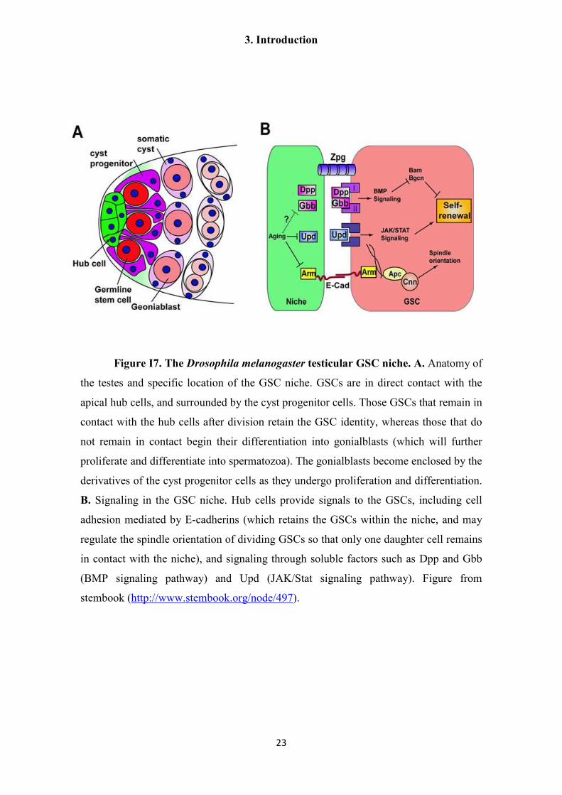
Figure I7. The Drosophila melanogaster testicular GSC niche. A. Anatomy of
the testes and specific location of the GSC niche. GSCs are in direct contact with the
apical hub cells, and surrounded by the cyst progenitor cells. Those GSCs that remain in
contact with the hub cells after division retain the GSC identity, whereas those that do
not remain in contact begin their differentiation into gonialblasts (which will further
proliferate and differentiate into spermatozoa). The gonialblasts become enclosed by the
derivatives of the cyst progenitor cells as they undergo proliferation and differentiation.
B. Signaling in the GSC niche. Hub cells provide signals to the GSCs, including cell
adhesion mediated by E-cadherins (which retains the GSCs within the niche, and may
regulate the spindle orientation of dividing GSCs so that only one daughter cell remains
in contact with the niche), and signaling through soluble factors such as Dpp and Gbb
(BMP signaling pathway) and Upd (JAK/Stat signaling pathway). Figure from
stembook (http://www.stembook.org/node/497).

3. Introduction
24
Figure I8. The FGF signaling pathway. A fibroblast growth factor ligand
(FGF) interacts with the extracellular immunoglobulin-like domain of a FGF receptor
(FGFR) resulting in the activation of the intracellular tyrosine kinase domain by
autophosphorylation. Heparan sulfate proteoglycans (HSPGs) are co-receptors for
FGFs, and can also modulate their bioavailability. The intracellular domain then signals
through different downstream pathways, mainly: the Janus kinase/signal transducer and
activator of transcription (Jak/Stat; 1, brown), phosphoinositide phospholipase C
(PLCg; 2, gray), phosphatidylinositol 3-kinase (PI3K; 3, green) and mitogen-activated
protein kinase/extracellular signal-regulated kinase (MAPK/Erk; 4, blue). Dual
specificity phosphatases (DUSPs), Spred and Sprouty proteins (orange) reduce or
terminate FGF signaling. Figure and legend modified from Lanner and Rossant (2010).

3. Introduction
25
3.6. Methods for the identification and analysis of stem cells
Identification of stem cells within tissues is not trivial, since they are not easily
distinguishable by histological methods, and most molecular markers are also shared
with their immediate progeny (Morrison and Spradling 2008). In a few cases, unique
molecular markers have been found for a particular stem cell population, but such
markers are only rarely shared by more than one stem cell type (e.g. the Lgr5 R-spondin
receptor, see section 3.7.3). If such markers are found, the stem cells can even be
identified in vivo within the tissues by creating transgenic organisms with a genetic
fusion of the promoter of the marker gene to fluorescent proteins, such as GFP
(Rompolas, Mesa, and Greco 2013; Ritsma et al. 2014).
Stem cells have been identified and isolated from cell suspensions of diverse
tissues by Fluorescence-Activated Cell Sorting (FACS), using complex combinations of
different positive and negative surface markers, after which their properties and potency
can be determined by in vivo and in vitro assays. This was first achieved for murine
HSCs, resulting in the first prospective purification of an adult stem cell population
(Spangrude, Heimfeld, and Weissman 1988), which has since been extensively refined
for achieving higher stem cell purities (Bryder, Rossi, and Weissman 2006). However,
it is not easy to identify such cells in situ using these complex marker combinations,
which are appropriate for FACS but not for immunohistofluorescence methods (IHF).
At the functional level, three main strategies are used for characterizing stem
cells, each with its own advantages and disadvantages (Yanger and Stanger 2011):
1) Transplantation experiments
In these experiments, purified stem cell preparations or even individual stem cells
are introduced into a living organism, and their self-renewal and differentiation is
measured (when individual stem cells are transplanted, clonal analysis of their output
can be achieved). In order to identify the cells that are derived from the donor, these are
genetically labeled (for example by using a different, identifiable donor genotype or
strain). Usually, the host organism is depleted of its own stem cells (for example by
lethal irradiation) in order to increase the engraftment of the donor stem cells. These
assays are very powerful, but require a purified stem cell population, and typically

3. Introduction
26
measure the potency of the donor stem cells under conditions that are not of normal
homeostatic cell turnover. The classic example for this kind of experiments is the
characterization of HSCs from bone marrow. Indeed, the existence of HSCs was
originally postulated from the results of transplantation experiments into lethally
irradiated hosts. Today, long-term repopulation of all main hematopoietic lineages after
transplantation of a lethally irradiated host is typically used as the operational definition
of HSCs.
2) In vitro analysis
In these experiments, stem cells are isolated and cultured in vitro under appropriate
conditions in order to determine their self-renewal and differentiation potential. By
performing clonal analysis of their proliferative output (i.e. by seeding cells at clonal
density or by seeding individual cells into culture) the potency of individual stem cells
can be determined for large numbers of such cells. However, for most stem cells
determining the ideal culture conditions is not trivial, and furthermore, different culture
conditions may affect their proliferative output. Finally, whether the potency of such
cells in vivo would be the same as the one displayed in vitro is unknown
3) Lineage tracing of genetically labeled cells
For models that can be genetically manipulated, indelible genetic labeling of stem
cells can be achieved by means of the Cre recombinase system (Jaisser 2000). In this
system, a construct is introduced into the genome in which the Cre recombinase is under
the control of the promoter of a stem cell-specific gene. When the Cre recombinase is
expressed in the stem cells, it can permanently activate a marker gene such as GFP or
GUS, which is in a different locus and is interrupted by a sequence flanked by Cre
target sequences (lox sequences). Furthermore, temporal control of activation can be
achieved by using instead a fusion of Cre to mutant versions of the estrogen receptor
ligand binding domain (CreER). CreER is normally cytoplasmatic, and will only
become activated by injection of synthetic estrogen analogues such as tamoxifen,
resulting in the translocation of CreER to the nucleus were it can remove the
inactivating sequences from the target gene. Therefore, stem cells and their progeny can
be traced in vivo by activating CreER and analyzing the labeled cells after different time
periods. By adding limiting concentrations of tamoxifen, only a small proportion of the
stem cells become labeled, and clonal analysis of individual stem cells can be

3. Introduction
27
performed. This is a very powerful method, since it allows the determination of the
potency of individual stem cells under normal in vivo conditions, but only if an
exclusive marker gene is known for such cells, and if the progeny of the stem cells
remain in close proximity (as is usually the case for epithelial tissues). In any case, even
when no such gene is known, one can search for stem cell activity within any tissue by
randomly activating CreER in a small subset of all cells, and searching for long-term
self-renewal and multipotent differentiation among the progeny of the activated cells.
This will not give any indication, however, about the identity of such cells.

3. Introduction
28
3.7. Stem cells and cell renewal mechanisms in vertebrates
Because of the obvious relevance of vertebrate stem cells for human health and
medicine, they have been extensively studied, although they represent relatively
difficult models given the complexity and size of vertebrate tissues. Most studies are
performed in the murine model, although amphibians (Xenopus and several urodeles)
and fish (such as zebrafish) are also important, in particular for studies of vertebrate
regeneration, a process that is very limited in mammals (Tanaka and Reddien 2011).
In the following paragraphs, I will summarize current knowledge regarding
selected well-studied mammalian adult tissues and for embryonic stem cells, in order to
illustrate the great variety of cell renewal strategies that can be found in mammals.
Within this variety, a general trend that can be found in many fast-renewing adult
tissues is that they are supported under normal conditions by actively proliferating stem
cells, but these are supplemented under special conditions (such as injury repair) by
other cell populations that are normally quiescent (quiescent stem cells or differentiated
cells).

3. Introduction
29
3.7.1. Embryonic Stem Cells
In mammals, only the zygote and the early blastomeres of the embryo are
totipotent (Hanna, Saha, and Jaenisch 2010). Early during mammalian development, the
blastomeres divide into the trophoblast, which will contribute exclusively to the extra-
embryonic placenta, and the inner cell mass (ICM), which will generate all of the tissues
of the embryo and several other extra-embryonic tissues The ICM cells within the
embryo are thus pluripotent, but are not self-renewing since they become quickly
committed to either the epiblast (which will generate the embryonic tissues) or to extra-
embryonic lineages. The epiblast cells themselves are initially pluripotent but become
further committed to contribute to specific germ layers.
Murine ICM cells can be isolated and cultured in vitro, and can be propagated
under specific conditions without losing their pluripotency (that is, they can be induced
to self-renew). These cells are referred to as embryonic stem cells (ESCs) (Hanna, Saha,
and Jaenisch 2010). By changing the culture conditions, they can be further induced to
differentiate into specific cell types. ESCs were originally cultured in the presence of
feeder cells, but later methodological refinement allowed their culture under completely
defined conditions (Ying et al. 2008). Important exogenous factors that promote ESC
self-renewal include signaling by Leukemia Inhibitory Factor (LIF) through Stat3
(JAK/STAT pathway) and activation of the Wnt pathway (ten Berge et al. 2011). A
further important factor is the inhibition or antagonism of the ERK kinase cascade
(Ying et al. 2008), which is normally activated in ESCs (as well as in the ICM) by
autocrine FGF4-mediated signaling. FGF4 instructs ESCs to exit self-renewal and
primes them for differentiation (Lanner and Rossant 2010).
ESCs remain pluripotent, as can be seen by in vivo experiments such as their
contribution to the formation of tissues from all three germ layers in embryonic
chimaeras, and the formation of teratomas when injected into adult hosts (Hanna, Saha,
and Jaenisch 2010). They also retain several regulatory and epigenetic characteristics
from the ICM cells: they express the transcription factors Oct4, Nanog and Sox2 (which
form a positive feedback regulatory circuit that promotes the maintenance of
pluripotency), they lack differentiation markers, and they retain both X chromosomes in
a pre-inactivation state (for female-derived ESC). However, they show extensive

3. Introduction
30
genome methylation, unlike the ICM cells which are hypomethylated (Hanna, Saha, and
Jaenisch 2010).
Pluripotent embryonic cells can also be isolated and propagated in vitro from the
epiblast (epiblast stem cells, EpiSCs). These cells have reduced potency as compared to
ESC, since they show multi-lineage differentiation in teratomas, but are very inefficient
in chimaera formation and have limited clonogenic potential (Hanna, Saha, and Jaenisch
2010). It is thought that EpiSCs are in a “primed” pluripotent state, ready to
differentiate, unlike the ESCs which are in a “naïve” pluripotent state. This can be also
seen at the epigenetic and gene regulatory level, since EpiSC show a reduction in the
expression of Nanog and other pluripotency regulators, the activation of early lineage
markers, and the inactivation of one of the X chromosomes (for female-derived
EpiSCs). Furthermore, whereas FGF/ERK signaling induces the differentiation of ES
cells into an EpiSC-like state, it promotes the self-renewal and proliferation of EpiSC
(Hanna, Saha, and Jaenisch 2010; Lanner and Rossant 2010).
Pluripotent ESCs have also been isolated from human embryos (hESCs).
Originally, however, hESC cultured under diverse conditions consistently showed
characteristics similar to mouse EpiSC (that is, hESC were already in a “primed”
pluripotent state) (Hanna, Saha, and Jaenisch 2010). Very recently, culture conditions
have been developed that result in hESC propagation in a “naïve” state (Gafni et al.
2013), holding great therapeutic promise which is however curtailed because of the
ethical implications of working with cells derived from human embryos. Importantly,
pluripotent cells with similar characteristics to “naïve” ESCs can be derived from adult
somatic cells by transfection with transgenes for the transcription factors Oct4, Sox-2,
c-Myc and Klf-4, which promote pluripotency (induced pluripotent stem cells, iPSCs)
(Takahashi and Yamanaka 2006; Rais et al. 2013), or by transfer of adult somatic nuclei
into oocytes (nuclear transfer embryonic stem cells, NT-ESCs) (Tachibana et al. 2013).
These strategies open the door for the generation of pluripotent cells for therapeutic
applications that are independent of the use of human embryos.

3. Introduction
31
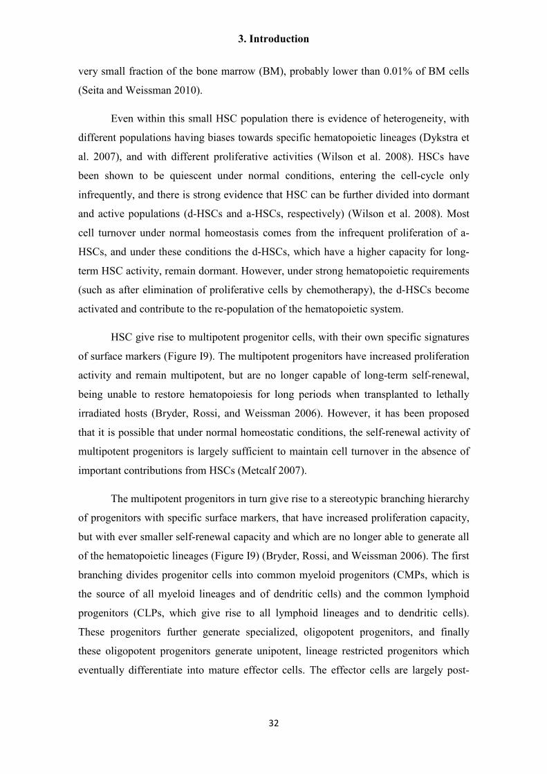
3.7.2. Hematopoietic stem cells
The murine hematopoietic system was the first extensively characterized
mammalian adult stem cell system, and HSCs were the first adult stem cells to be
prospectively purified and characterized (Spangrude, Heimfeld, and Weissman 1988;
Bryder, Rossi, and Weissman 2006). The HSC paradigm has therefore been extensively
applied, for better or worse, to many other stem cell systems. Originally, the existence
of HSC was proposed from the observation that cell preparations from the bone marrow
of a donor could rescue hematopoiesis in lethally irradiated hosts, and limiting amounts
of donor bone marrow cells were able to generate multi-lineage clonal colonies in the
host spleen. The cells responsible of generating these colonies were called CFU-S
(colony forming unit - spleen) and were proposed to be multipotent hematopoietic stem
cells (Domen, Wagers, and Weissman 2006). Today, it is known that only a fraction of
the CFU-Ss are true HSCs, whereas the rest represent multipotent progenitors with
limited self-renewal capacity (Bryder, Rossi, and Weissman 2006).
HSCs are defined as long-term self-renewing cells that are able to give rise to all
of the main hematopoietic lineages. Operationally, they are defined by their ability to
restore and contribute for long periods to all of the hematopoietic lineages after
transplantation into lethally irradiated hosts (Bryder, Rossi, and Weissman 2006). No
single specific marker of HSC is known, and instead, they are recognized by flow-
cytometry methods from the presence or absence of several surface lineage markers in
complex combinations (Bryder, Rossi, and Weissman 2006; Seita and Weissman 2010)
(Figure I9). For many of these markers, their exact function is unknown, and do not
appear to play essential roles in the physiology of HSC and their progeny. Probably
because of this, although there is a good degree of correlation between murine and
human HSC/progeny markers, they also show many differences, such as the expression
of CD34 in human HSC, which is absent in murine HSC (Seita and Weissman 2010)
(Figure I9). HSC can also be recognized by flow-cytometry from their high dye efflux
activity (the so-called “side-population activity”) since they exclude vital fluorescent
dyes such as Hoechst 33342 (Bryder, Rossi, and Weissman 2006). From the application
of ever more complex combinations of surface markers, together with in vivo and in
vitro experiments for assaying the potency of isolated bone marrow cell populations,
HSCs have been isolated to very high levels of purity. The HSC population represents a
3. Introduction
32
very small fraction of the bone marrow (BM), probably lower than 0.01% of BM cells
(Seita and Weissman 2010).
Even within this small HSC population there is evidence of heterogeneity, with
different populations having biases towards specific hematopoietic lineages (Dykstra et
al. 2007), and with different proliferative activities (Wilson et al. 2008). HSCs have
been shown to be quiescent under normal conditions, entering the cell-cycle only
infrequently, and there is strong evidence that HSC can be further divided into dormant
and active populations (d-HSCs and a-HSCs, respectively) (Wilson et al. 2008). Most
cell turnover under normal homeostasis comes from the infrequent proliferation of a-
HSCs, and under these conditions the d-HSCs, which have a higher capacity for long-
term HSC activity, remain dormant. However, under strong hematopoietic requirements
(such as after elimination of proliferative cells by chemotherapy), the d-HSCs become
activated and contribute to the re-population of the hematopoietic system.
HSC give rise to multipotent progenitor cells, with their own specific signatures
of surface markers (Figure I9). The multipotent progenitors have increased proliferation
activity and remain multipotent, but are no longer capable of long-term self-renewal,
being unable to restore hematopoiesis for long periods when transplanted to lethally
irradiated hosts (Bryder, Rossi, and Weissman 2006). However, it has been proposed
that it is possible that under normal homeostatic conditions, the self-renewal activity of
multipotent progenitors is largely sufficient to maintain cell turnover in the absence of
important contributions from HSCs (Metcalf 2007).
The multipotent progenitors in turn give rise to a stereotypic branching hierarchy
of progenitors with specific surface markers, that have increased proliferation capacity,
but with ever smaller self-renewal capacity and which are no longer able to generate all
of the hematopoietic lineages (Figure I9) (Bryder, Rossi, and Weissman 2006). The first
branching divides progenitor cells into common myeloid progenitors (CMPs, which is
the source of all myeloid lineages and of dendritic cells) and the common lymphoid
progenitors (CLPs, which give rise to all lymphoid lineages and to dendritic cells).
These progenitors further generate specialized, oligopotent progenitors, and finally
these oligopotent progenitors generate unipotent, lineage restricted progenitors which
eventually differentiate into mature effector cells. The effector cells are largely post-

3. Introduction
33
mitotic, although there are exceptions (for example, cell proliferation is essential for the
function of B-lymphocytes) (Metcalf 2007).
The stem cell niche of HSCs is difficult to study, since BM is a large and
complex tissue, and HSC are scarce and difficult to identify by
immunohistofluorescence methods (since a complex combination of markers must be
used to identify the HSC exclusively) (Seita and Weissman 2010; Lo Celso and
Scadden 2011). Two main possible locations have been proposed for the HSC stem cell
niche, and it is possible that both function as niches for all HSCs or for different sub-
populations of HSC: the first is located next to the osteoblasts of the endosteal bone
surface, and the other is adjacent to the sinusoidal vasculature endothelium. HSCs have
been associated to both locations, and both cell types secrete factors that have been
shown by genetic manipulation to be important for HSC self-renewal and localization
(Lo Celso and Scadden 2011). These signaling factors include among others kit ligand
(also known as SCF) that activates c-kit in the HSCs; thrombopoietin, which activates
the c-Mpl receptor; and SDF1, the ligand for the CXCR4 receptor (Seita and Weissman
2010). Activation by gain-of-function mutants of the Notch pathway can lead to
increased proliferation and expansion of multipotent progenitors, but loss-of-function
studies show that these signaling pathways are not essential for HSC/progenitor
function, and their role in vivo is controversial (Lo Celso and Scadden 2011).
Transplantation of HSCs and progenitor cells is of wide therapeutic use, for
example to treat genetic or acquired bone marrow failure, and to restore hematopoiesis
after high-dose chemotherapy against diverse cancers (Domen, Wagers, and Weissman
2006). For therapeutic use, pure HSCs preparations are not practical given the very
small proportion of HSCs and the complexity of their isolation. Instead, the typical
grafts used are un-fractioned preparations of bone marrow cells, or CD34+ enriched cell
preparations (comprising HSCs, hematopoietic progenitors and diverse contaminants)
from mobilized peripheral blood or umbilical cord blood (Domen, Wagers, and
Weissman 2006; Seita and Weissman 2010). Furthermore, techniques for ex vivo
expansion of HSCs are still very limited (less than 10-fold expansion under ideal
conditions), since treatment with most growth factors and cytokines which are
important for self-renewal in vivo result in proliferation accompanied of cell
differentiation in vitro (Seita and Weissman 2010; Takizawa, Schanz, and Manz 2011).
3. Introduction
34
Figure I9. Model of the hematopoietic stem cell and progenitor hierarchy.
Markers for murine and human hematopoietic cell populations are given on the left. See
the main text for details. Figure from Bryder, Rossi, and Weissman (2006).

3. Introduction
35
3.7.3. Intestinal Stem Cells
The epithelium of the small intestine is the tissue with the fastest cellular
turnover in mice and other mammals (Alberts 2000). Morphologically, it is a
monostratified epithelium, folded into finger-like extensions (villi) and invaginations
(crypts) (Figure I10). The villi contain several terminally differentiated cell types,
including the enterocytes (involved with the absorption of nutrients), goblet cells
(glandular cells), endocrine cells and other quantitatively minor cell types (Alberts
2000; Stange 2013). These cells are lost by apoptosis and shedding from the tip of the
villi after an average life-span of just 4 to 5 days. Cell proliferation occurs in the crypts
and at the base of the villi. Newly generated cells continuously move upwards towards
the surrounding villi while differentiating into mature cell types, displacing the already
differentiated cells towards the tip of the villi, in a manner reminiscent of a conveyor
belt (Stange 2013).
Intestinal stem cells (ISCs) are found in the crypts, and give rise to transit-
amplifying cells (TA-cells) which undergo 4 to 5 divisions in the lower regions of the
villi (Barker and Clevers 2010; Stange 2013). The identity of ISCs was for a long time
controversial, and only recently were they identified as the undifferentiated crypt base
columnar cells (CBCs), which lay at the bottom of the crypts, intercalated with the
relatively long-lived Paneth cells (a type of differentiated gland cell that produces
bactericidal products) (Barker et al. 2007). About 14 CBCs can be found at the bottom
of each crypt, and they express a unique marker, the R-spondin receptor Lgr5 (Barker et
al. 2007; Barker and Clevers 2010; Carmon et al. 2011; de Lau et al. 2011; Glinka et al.
2011). These cells are also denominated the Lgr5+ ISC population. Lgr5 is not only a
specific marker of the stem cell population in the intestinal epithelium, but it has also
been found to be expressed by stem cells in other adult epithelial tissues (such as the
pylorus region of the stomach and the regenerating liver (Barker and Clevers 2010;
Barker et al. 2010; Huch, Boj, and Clevers 2013)).
Lineage tracing mediated by the CreER system conclusively showed that
individual Lgr5+ ISCs have the ability for long-term production of all intestinal
lineages, demonstrating that they are true stem cells (Barker et al. 2007). Furthermore,
isolated Lgr5+ ISC have been shown to generate complete intestinal organoids in vitro,
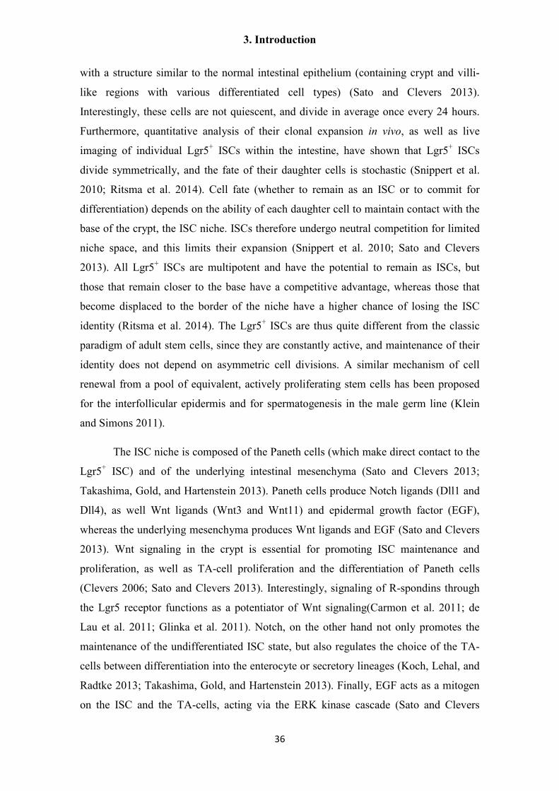
3. Introduction
36
with a structure similar to the normal intestinal epithelium (containing crypt and villi-
like regions with various differentiated cell types) (Sato and Clevers 2013).
Interestingly, these cells are not quiescent, and divide in average once every 24 hours.
Furthermore, quantitative analysis of their clonal expansion in vivo, as well as live
imaging of individual Lgr5+ ISCs within the intestine, have shown that Lgr5+ ISCs
divide symmetrically, and the fate of their daughter cells is stochastic (Snippert et al.
2010; Ritsma et al. 2014). Cell fate (whether to remain as an ISC or to commit for
differentiation) depends on the ability of each daughter cell to maintain contact with the
base of the crypt, the ISC niche. ISCs therefore undergo neutral competition for limited
niche space, and this limits their expansion (Snippert et al. 2010; Sato and Clevers
2013). All Lgr5+ ISCs are multipotent and have the potential to remain as ISCs, but
those that remain closer to the base have a competitive advantage, whereas those that
become displaced to the border of the niche have a higher chance of losing the ISC
identity (Ritsma et al. 2014). The Lgr5+ ISCs are thus quite different from the classic
paradigm of adult stem cells, since they are constantly active, and maintenance of their
identity does not depend on asymmetric cell divisions. A similar mechanism of cell
renewal from a pool of equivalent, actively proliferating stem cells has been proposed
for the interfollicular epidermis and for spermatogenesis in the male germ line (Klein
and Simons 2011).
The ISC niche is composed of the Paneth cells (which make direct contact to the
Lgr5+ ISC) and of the underlying intestinal mesenchyma (Sato and Clevers 2013;
Takashima, Gold, and Hartenstein 2013). Paneth cells produce Notch ligands (Dll1 and
Dll4), as well Wnt ligands (Wnt3 and Wnt11) and epidermal growth factor (EGF),
whereas the underlying mesenchyma produces Wnt ligands and EGF (Sato and Clevers
2013). Wnt signaling in the crypt is essential for promoting ISC maintenance and
proliferation, as well as TA-cell proliferation and the differentiation of Paneth cells
(Clevers 2006; Sato and Clevers 2013). Interestingly, signaling of R-spondins through
the Lgr5 receptor functions as a potentiator of Wnt signaling(Carmon et al. 2011; de
Lau et al. 2011; Glinka et al. 2011). Notch, on the other hand not only promotes the
maintenance of the undifferentiated ISC state, but also regulates the choice of the TA-
cells between differentiation into the enterocyte or secretory lineages (Koch, Lehal, and
Radtke 2013; Takashima, Gold, and Hartenstein 2013). Finally, EGF acts as a mitogen
on the ISC and the TA-cells, acting via the ERK kinase cascade (Sato and Clevers

3. Introduction
37
2013). The Wnt and EGF signaling from both sources are largely redundant, but only
Paneth cells are capable of providing Notch signals since these are membrane bound
and require direct cell to cell contact (Koch, Lehal, and Radtke 2013). Paneth cells are
essential in vivo for stem cell maintenance, since the Lgr5+ ISCs disappear when Paneth
cells are specifically depleted (Sato et al. 2011). In vitro, the signals they produce can be
substituted by exogenous addition of the relevant ligands to the media, but even under
these conditions the addition of Paneth cells greatly improves the seeding efficiency of
Lgr5+ ISCs (Sato and Clevers 2013). BMP signaling, which is strong in the villi and
driven by BMP-4 secretion from the villus mesenchyma, has an opposite effect to Wnt
signaling and promotes ISC differentiation and quiescence (Sato and Clevers 2013).
The BMP antagonist Noggin is specifically found in the crypt and counteracts this
influence (Watabe and Miyazono 2009).
In addition to the recently characterized Lgr5+ ISC, it has long been proposed
that quiescent stem cells exist in the intestinal epithelium, in a higher position in the
crypt (the so-called +4 position, by counting cells from the bottom of the crypt) (Alberts
2000). These cells were characterized as quiescent from their ability to retain labeling
with the thymidine analog 5-bromo-2'-deoxyuridine (BrdU), and the loss of BrdU after
tissue injury was interpreted as a re-entry of these quiescent, label-retaining cells (LRC)
into the cell cycle. Although it is still possible that a separate population of quiescent
stem cells exists, it has been shown that the intestinal LRC are actually undifferentiated
but committed progenitors for Paneth and secretory cells. These cells are generated
from the Lgr5+ ISC population and normally differentiate into Paneth or secretory cells.
However, when the Lgr5+ ISCs are not sufficient for tissue homeostasis, such as when
mitotic cells are specifically depleted, these committed progenitors have the capacity to
revert to an Lgr5+ ISC phenotype and therefore repopulate the crypts with ISCs
(Buczacki et al. 2013). In summary, the intestinal epithelium is normally replenished by
the proliferation of active Lgr5+ ISCs, but after injury, committed progenitors that retain
the ability to serve as “reserve stem cells” become activated. These progenitors are not
in a strict sense stem cells, since they do not normally self-renew, but are constantly
generated from the Lgr5+ ISC pool.
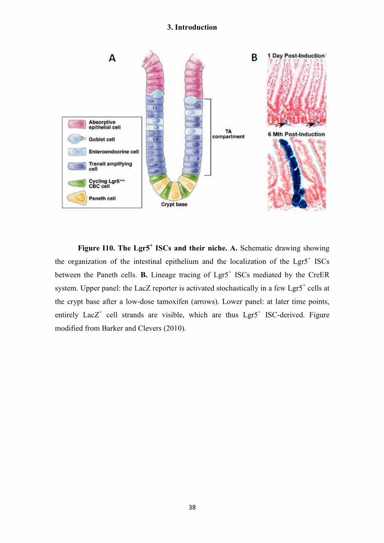
3. Introduction
38
Figure I10. The Lgr5+ ISCs and their niche. A. Schematic drawing showing
the organization of the intestinal epithelium and the localization of the Lgr5+ ISCs
between the Paneth cells. B. Lineage tracing of Lgr5+ ISCs mediated by the CreER
system. Upper panel: the LacZ reporter is activated stochastically in a few Lgr5+ cells at
the crypt base after a low-dose tamoxifen (arrows). Lower panel: at later time points,
entirely LacZ+ cell strands are visible, which are thus Lgr5+ ISC-derived. Figure
modified from Barker and Clevers (2010).

3. Introduction
39
3.7.4. Differentiated cells function as stem cells in the stomach corpus and in the
alveolar epithelium of the lung
In the previous examples, stem cells contributing to tissue turnover are
undifferentiated cells, which do not seem to contribute to any direct effector function in
their tissue. But in other murine tissues, differentiated cells can function as stem cells
under normal homeostasis or under special conditions. For example, in the epithelium
of the stomach corpus region, chief cells are long-lived secretory cells that produce
digestive enzymes. It was recently discovered that a sub-population of chief cells, which
express the Troy receptor, has the ability to proliferate and of long-term clonal
expansion, giving rise to all differentiated cell types of the stomach gland epithelium
(Stange et al. 2013). These cells also express the Lgr5 receptor, and can form long-lived
organoids when cultured in vitro. Troy+ chief cells normally show low proliferative
activity, and it is thought that their contribution for normal cellular turnover is very
small. Instead, they function as “reserve stem cells”, and become activated after
depletion of proliferating cells in the stomach.
Similarly, in the alveolar sacs of the lungs two main epithelial cell types are
present: the type 1 (AEC1) cells are squamous cells that mediate gas exchange, and type
2 (AEC2) cells are cuboidal cells that secrete surfactant that prevents alveolar collapse.
During embryonic development, both cell types originate from a bipotent progenitor
(Desai, Brownfield, and Krasnow 2014). In the adult, the alveolar sac epithelium has a
low rate of cellular turnover, which is provided by proliferation of the long lived AEC2
cells. AEC2 cells can self-renew to give rise to more AEC2 cells, and they can also
generate AEC1 cells (Barkauskas et al. 2013; Desai, Brownfield, and Krasnow 2014)
(Figure I11). This occurs at a low rate during normal homeostasis, but the AEC2 cells
can also respond to injury of the alveoli and increase their proliferative output. In
summary, adult AEC2 cells function as stem cells while at the same time performing an
essential effector function in the alveoli.

3. Introduction
40
Figure I11. Developmental origin and turnover of murine alveolar epithelial
cells. Bipotent progenitors differentiate into AEC1 or AEC2 cells during development.
Mature AEC2 cells function as stem cells in the adult and are activated for alveolar
renewal and repair (with a very slow turnover). Dying AEC1 cells are proposed to
produce a signal that is transduced by epidermal growth factor receptor (EGFR),
activating the division of a nearby AEC2 cell. Figure modified from Desai et al. (2014).
3. Introduction
41
3.7.5. Differentiated cells self-duplicate in the liver and in the pancreas
In tissues with slow turnover, it is not rare for new cells to originate from self-
duplication of existing differentiated cells (which are unipotent, and only give rise to
more cells of the same type).
During normal tissue homeostasis in the liver, and during compensatory growth
after partial hepatectomy (PHx), mature hepatocytes proliferate to generate new
hepatocytes. This occurs at a very low rate under normal conditions, but hepatocytes are
able to respond to PHx by entering the cell-cycle in a coordinated manner (Fausto and
Campbell 2003; Yanger and Stanger 2011). Most hepatocytes participate of
compensatory growth, and after a few rounds of cell-division, liver mass is restored to
its original value. The hepatocytes have a huge potential for self-renewal, as can be seen
in experiments of sequential PHx and in experiments of serial liver transplantations
(Yanger and Stanger 2011). Similarly, in many models of regeneration the liver duct
cells self-renew without any input from stem cells. However, in specific liver injuries
mediated by chemicals, the hepatocytes are unable to proliferate, and new cells are
originated from a population of undifferentiated stem cells, the so-called oval cells,
which are believed to differentiate into hepatocytes and duct cells (and are thus
bipotential) (Fausto and Campbell 2003; Dorrell et al. 2011; Yanger and Stanger 2011).
So far, no specific marker of the oval cells has been found. They are thought to be
facultative stem cells that arise by de-differentiation of cells from the biliary ducts, but
the precise source of these cells is controversial. Recently, the Lgr5 receptor was shown
to be specifically expressed by a population of small cells near the bile duct, but only
after liver injury (Huch, Boj, and Clevers 2013). The Lgr5+ cells were shown by Cre-
mediated lineage tracing to give rise to hepatocytes after injury, and were able in vitro
to generate organoids and to differentiate into hepatocytes and duct cells.
Another well studied example of self-renewing differentiated cells are the
insulin-producing beta cells from the Langerhans islets of the pancreas. Lineage tracing
by Cre-mediated recombination has shown that new beta cells originate from pre-
existing, insulin producing (i.e. differentiated) beta cells (Dor et al. 2004). Furthermore,
all beta cells seem to contribute similarly to the production of new beta cells during islet
growth and maintenance (Brennand, Huangfu, and Melton 2007). An external source of

3. Introduction
42
beta cells has only been documented under conditions of extreme beta cell loss. Under
these conditions, new beta cells have been shown to originate by trans-differentiation of
mature glucagon-producing alpha cells (Thorel et al. 2010).
3. Introduction
43
3.8. Pluripotent and multipotent stem cells in invertebrate models
Drosophila and Caenorhabditis are the most common and powerful invertebrate
models for developmental biology, and it is precisely in these models that the first
extensive characterization of stem cell niches and their signals were performed.
However, they are not very representative systems for the diversity of mechanisms of
cell turnover found in adult metazoans, since adult Drosophila and Caenorhabditis have
short life-spans, most (Drosophila) or all (Caenorhabditis) of their adult somatic tissues
are no longer proliferative, and have very limited regenerative abilities. Finally, both of
these models specify the germ line during early embryonic development, via the
inheritance by specific blastomeres of cytoplasmic determinants (germ plasm) that were
maternally synthezised and deposited in the egg (“preformation”). In contrast, most
metazoans specify the germ line later in development, as a result of inductive signals
from surrounding tissues (“epigenesis”) (Extavour and Akam 2003).
Those tissues that show strong proliferation in adult Drosophila, such as the
germ line and the midgut, are supported by tissue- or cell-specific stem cells (Losick et
al. 2011; Takashima, Gold, and Hartenstein 2013). In contrast, there is good evidence in
other invertebrate models for the existence of pluripotent or multipotent somatic stem
cells in larvae and in adults. Examples of these organisms are found in most metazoan
lineages, including pre-bilaterian metazoans (e.g. poriferans, cnidarians), deuterostomes
(e.g. echinoderms) and lophotrochozoans (e.g. annelids, mollusks, and platyhelminthes)
(Juliano, Swartz, and Wessel 2010; Solana 2013). Usually, these organisms specify their
germ line by epigenesis, and may have a fluid limit between the soma and the germ line
(multipotent somatic stem cells may contribute to the germ line in the adult, for example
during regeneration).
These multipotent somatic stem cells have a conserved set of markers, which is
shared with the germ line (Ewen-Campen, Schwager, and Extavour 2010; Juliano,
Swartz, and Wessel 2010). The germ line stem cells (GSCs), although immediately
unipotent (since they only give rise to gametes), conserves a “hidden” potential for
pluripotency which is revealed after fertilization. Their pluripotency is also evidenced in
vitro, as cultured germ line stem cells can generate chimeras when injected into host
blastocysts and teratomas when injected into adult hosts, similarly to ESCs (Hanna,

3. Introduction
44
Saha, and Jaenisch 2010). This set of shared expressed genes has therefore been
denominated the “Germline Multipotency Program” (GMP) (Juliano, Swartz, and
Wessel 2010). The gene products of the GMP components are associated with mRNA
metabolism and the post-transcriptional regulation of gene expression. Furthermore,
many are components of germ granules, which are cytoplasmic ribonucleoprotein
(RNP) granules usually found in the cells of the germ line (Extavour and Akam 2003).
The germ granules are part of the germ plasm in metazoans with preformation
mechanisms of germ line segregation. Germ granules are therefore thought to be centers
for post-transcriptional regulation of mRNA, and to carry essential determinants for
germ line specification. Multipotent somatic stem cells of invertebrates may also have
similar RNP granules with GMP components, and these granules receive different
names in different species (e.g. chromatoid bodies, nuage, etc.) (Ewen-Campen,
Schwager, and Extavour 2010).
Among the proteins of the proposed GMP, most have RNA binding activity, and
many are translational repressors, such as Bruno, Nanos and Pumilio. Nanos proteins
have two zinc-finger motifs with sequence-unspecific RNA binding activity (Curtis et
al. 1997), whereas Pumilio is a member of the PUF family of sequence-specific RNA
binding proteins (Wickens et al. 2002). Nanos and Pumilio interact with each other in
Drosophila, Caenorhabditis and humans, and work in many cases together by binding
to the 3´ UTR of target mRNAs and promoting their translational repression,
deadenylation and degradation (Parisi and Lin 2000; Jaruzelska et al. 2003). They are
required for maintaining the identity and quiescence of the primordial germ cells in
Drosophila, as well as regulating proliferation and maintenance of germ cell identity in
Drosophila and Caenorhabditis (Subramaniam and Seydoux 1999; Crittenden et al.
2003; Kadyrova et al. 2007; Ariz, Mainpal, and Subramaniam 2009). Other well
conserved GMP proteins are Vasa, a DEAD-box ATP-dependent RNA helicase which
is widely used as a marker for the germ line in many metazoans (Rebscher et al. 2007;
Juliano, Swartz, and Wessel 2010; Lasko 2013), and Piwi, a member of the Argonaute
family of proteins. Argonaute proteins are involved in gene silencing through small
RNAs such as microRNAs (miRNAs) and small interfering RNAs (siRNAs) (Lasko
2013). Piwi proteins are associated with a specific class of small RNAs (Piwi-associated
RNAs, or piRNAs) that are characteristically longer (26-31 nucleotides) than miRNAs
and siRNAs, and unlike these, piRNAs are generated in a pathway that is independent

3. Introduction
45
of the endoribonuclease Dicer (Juliano, Wang, and Lin 2011). Piwi and piRNAs affect
gene regulation at the epigenetic level (through heterochromatin formation and DNA
methylation) and by post-transcriptional regulation of RNA stability. The best
characterized role of Piwi proteins and piRNAs is in the silencing of transposable
elements, thus preventing their deleterious expansion in the genome of the germ line
(Juliano, Wang, and Lin 2011; Skinner et al. 2014). It is clear however that other genes
are also regulated by Piwi (Juliano, Wang, and Lin 2011; Peng and Lin 2013; Ross,
Weiner, and Lin 2014), both in and outside the germ line, affecting aspects of germline
specification, gametogenesis and stem cell maintenance in diverse organisms.
3. Introduction
46
3.9. The planarian neoblasts
As previously mentioned, it is believed that in the Platyhelminthes all
differentiated cells are post-mitotic, and a separate population of undifferentiated stem
cells (the neoblasts) is the single source of new cells for normal tissue turnover, growth
and regeneration (Peter et al. 2004; Reuter and Kreshchenko 2004). This has been
studied in detail for free-living flatworms, especially for planarians, which are well
known for their extensive regenerative capabilities (order Tricladida, in particular the
species Schmidtea mediterranea and Dugesia japonica) (Reddien and Sanchez
Alvarado 2004; Rossi et al. 2008; Rink 2013). Other free-living flatworm lineages that
have been studied include the more basal groups Macrostomida (genera Macrostomum
and Microstomum) (Palmberg 1990; Ladurner, Rieger, and Baguna 2000; Peter et al.
2004; Bode et al. 2006; Pfister et al. 2008; De Mulder et al. 2009b) and Catenulida
(Moraczewski 1977; Dirks et al. 2012a; Dirks et al. 2012b). Recently, the development
of technical advances such as RNA interference (RNAi), FACS analysis and isolation of
neoblasts, and high throughput RNA sequencing have revolutionized the study of
planarian regeneration and neoblast biology, placing planarians as one of the most
important models for the study of regeneration at the cellular and molecular levels
(Newmark and Sanchez Alvarado 2002; Rink 2013). However, some key techniques
such as transgenesis and in vitro cell culture are still in their infancy (Schürmann and
Peter 2001; Peter et al. 2004; Baguna 2012).
Planarians are able to regenerate a complete organism from almost any region of
the body (Reddien and Sanchez Alvarado 2004). Furthermore, they constantly renew all
of their somatic tissues at a very high rate (Rink 2013). The main tissues and organs of
planarians, including the epidermis, digestive system, excretory system and nervous
system lack any residing proliferating cells, and are instead supported by the integration
of proliferating neoblasts that reside in the mesodermal parenchyma that surrounds the
organs (Figure I12) (Rossi et al. 2008; Rink 2013). Neoblasts can be found throughout
the planarian body except in the anterior-most region (in front of the photoreceptors)
and in the pharynx, and these are precisely the only regions that are unable to regenerate
after excision (Newmark and Sanchez Alvarado 2000; Reddien and Sanchez Alvarado
2004; Rink 2013). During regeneration, proliferating neoblasts accumulate close to the

3. Introduction
47
wound site. As they enter diverse differentiation pathways they form a mass of
undifferentiated and differentiating cells at the wound site (the regeneration blastema).
The blastema is already post-mitotic, and the proliferating neoblasts are situated
immediately adjacent to it (in the post-blastema) (Reddien and Sanchez Alvarado 2004;
Eisenhoffer, Kang, and Sanchez Alvarado 2008).
Planarian neoblasts are small and basophilic undifferentiated cells, with a large
nucleus and nucleolus and scant cytoplasm (Baguñà and Romero 1981; Peter et al.
2004; Rossi et al. 2008). At the ultrastructural level, they show abundant free ribosomes
and mitochondria, and lack discernible rough endoplasmic reticulum and Golgi
cisternae (Morita, Best, and Noel 1969; Hay and Coward 1975). Importantly, they show
perinuclear RNP granules denominated “chromatoid bodies”, which are molecularly and
morphologically similar to the germ granules present in the germ cells of many animals
(Morita, Best, and Noel 1969; Hay and Coward 1975; Auladell, Garcia-Valero, and
Baguña 1993; Yoshida-Kashikawa et al. 2007; Rossi et al. 2008) (Figure I12). Because
neoblasts are the only proliferating cells, they are specifically sensitive to ionizing
irradiation, making this a very useful technique for depleting tissues of neoblasts for
diverse experimental purposes (Hayashi et al. 2006; Rossi et al. 2007; Eisenhoffer,
Kang, and Sanchez Alvarado 2008; Salvetti et al. 2009; Solana et al. 2012).
Molecular markers of the neoblast population were found originally by analysis
of candidate genes, and later from transcriptomic analyses of neoblast-depleted
planarians and of FACS-isolated neoblasts (Shibata et al. 1999; Salvetti et al. 2000;
Orii, Sakurai, and Watanabe 2005; Reddien et al. 2005; Salvetti et al. 2005; Guo, Peters,
and Newmark 2006; Rossi et al. 2007; Eisenhoffer, Kang, and Sanchez Alvarado 2008;
Rossi et al. 2008; Rouhana et al. 2010; Labbe et al. 2012; Onal et al. 2012; Rink 2013).
These studies have consistently shown that neoblasts specifically express several
components of the GMP, and more generally, of many proteins related to post-
transcriptional regulation of gene expression (Juliano, Swartz, and Wessel 2010;
Rouhana et al. 2010). Many of these genes have been show by RNAi to have an
important function in neoblast maintenance or differentiation, including orthologs of
vasa and piwi (Reddien et al. 2005; Wagner, Ho, and Reddien 2012). Several piwi genes
are expressed in planarian neoblasts, and in particular smedwi-1 from Schmidtea
mediterranea and its orthologs in other planarian species are the most widely used
neoblast markers (Reddien et al. 2005; Palakodeti et al. 2008) (Figure I12). Although no

3. Introduction
48
clear phenotype was observed from smedwi-1 RNAi, other piwi genes such as smedwi-2
and smedwi-3 have been shown by RNAi to be important for neoblast differentiation
and self-renewal. Other gene categories with an enriched expression in neoblasts
include epigenetic regulators and transcription factors, as well as components of
particular signaling pathways, including FGF receptors (Ogawa et al. 2002; Onal et al.
2012; Wagner, Ho, and Reddien 2012; Rink 2013). Recently, it was shown that
although no clear-cut orthologs of the mammalian ESC regulators Oct4, Sox2 or Nanog
can be found in planarians, the transcriptomes of neoblasts and mammalian ESC are
highly correlated, indicating a broad conservation of pluripotency regulators between
both models, and by extension, across metazoans (Onal et al. 2012).
Traditionally, neoblasts were considered to be a homogeneous population of
pluripotent cells. Originally, neoblasts were shown at the whole-population level to be
pluripotent and essential for regeneration, since only neoblast-enriched cell preparations
were able to rescue lethally irradiated hosts after transplantation, whereas transplants of
differentiated cells were unable to do so (Baguñà, Saló, and Auladell 1989). However,
these experiments left open the possibility that several neoblast lineages with restricted
potencies exist which can rescue and repopulate the irradiated host. Recently, it was
shown through impressive single-cell transplantation experiments that at least a
proportion of neoblasts (clonogenic neoblasts, or cNeoblasts) are truly pluripotent,
being able to completely repopulate and replace all of the tissues of an allogeneic host.
cNeoblasts were shown to be at least 5% of all neoblasts (which is the percentage of
irradiated hosts that were rescued by the transplants), and is likely to be higher given the
technically challenging method employed (Wagner, Wang, and Reddien 2011).
However, it is not known whether all neoblasts are cNeoblasts, and there is evidence
from gene expression studies showing that neoblasts are actually heterogeneous, since
several markers are only expressed in sub-populations of the neoblasts. For example,
the planarian nanos homolog is specifically expressed in the germ line stem cells
(GSC), which are morphologically undistinguishable from somatic neoblasts and
express other common neoblast markers (Sato et al. 2006; Handberg-Thorsager and
Salo 2007; Wang et al. 2007). nanos+ GSC are regenerated from nanos
- somatic
neoblasts (Sato et al. 2006), and the germ line can contribute to somatic tissues during
regeneration (Reddien and Sanchez Alvarado 2004), demonstrating that the soma –
germ line is fluid in planarians and that these neoblast sub-populations can interconvert

3. Introduction
49
between each other. Furthermore, some proliferating neoblasts (expressing smedwi-1
and proliferation markers, and X-ray sensitive) show co-expression of specific lineage
markers during regeneration of the eyes and of the excretory system (Reddien 2013).
This suggests the existence of populations of committed, lineage-specific neoblasts as
well. Finally, it has been shown that some cells with neoblast-like morphology, or with
intermediate morphologies between neoblasts and differentiated cells, are already post-
mitotic and express different combinations of markers that represent different stages or
different lineages of neoblast differentiation (Higuchi et al. 2007; Shibata, Rouhana, and
Agata 2010).
Figure I12. The planarian neoblasts. A. Distribution of proliferative cells as
determined by BrdU incorporation (green) in the planarian Girardia dorotocephala. Bar
represents 450 µm. From Newmark and Sanchez Alvarado, 2000. B. Distribution of
smedwi-1+ cells (red) in the planarian Schmidtea mediterranea. Bar represents 200 µm.
From Rink, 2013. C. Schematic illustration of the distribution of neoblasts in the
parenchyma, in a transverse section at the level of the pharynx. From Rink, 2013. D.
Transmission electron microscopy image of a neoblast, showing a chromatoid body
(CB) and mitochondria (arrowheads). Bar represents 100 nm. From Rossi et al., 2012.
Abbreviations: Bm, basal membrane; Ep, epidermis; Phr, photoreceptor; Phx, pharynx;
VNC, ventral nerve cords.
3. Introduction
50
3.10. The cestode germinative cells 1
In cestodes, undifferentiated proliferating cells (equivalent to the planarian
neoblasts) are usually denominated as “germinative cells” (Gustafsson 1990; Reuter and
Kreshchenko 2004; Koziol and Castillo 2011). By classic histological techniques,
germinative cells have been characterized as round or oval cells with a strongly
basophilic cytoplasm (due to the abundance of rRNA), few cytoplasmic extensions, a
large nucleus and a large and prominent nucleolus (Douglas 1961; Bolla and Roberts
1971; Wikgren and Gustafsson 1971; Sulgostowska 1972; Gustafsson 1976b; Loehr and
Mead 1979; Koziol et al. 2010) (Figure I13). More than one lineage of morphologically
similar germinative cells may actually exist, and in some models, several sub-types of
germinative cells have been proposed based on their nucleo-cytoplasmic ratio, absolute
size and staining characteristics (Douglas 1961; Sulgostowska 1972). At the
ultrastructural level, germinative cells have been described in oncospheres,
metacestodes and adult cestodes (Collin 1969; Sakamoto and Sugimura 1970; Bolla and
Roberts 1971; Wikgren and Gustafsson 1971; Swiderski 1983; Jabbar et al. 2010).
These cells have a high nucleo-cytoplasmic ratio, nuclei with little heterochromatin, an
electron-dense cytoplasm with abundant free ribosomes, and absent or scarce
endoplasmic reticulum and Golgi apparatus. Their morphology is thus very similar to
neoblasts from planarians, except that chromatoid bodies have never been described in
cestodes (Figure I13).
In adult cestodes and during the early stages of segmentation, a conserved
pattern has been described for the distribution of proliferating germinative cells within
the neck region of several species (the generative region from which new segments are
formed). Proliferative cells are found mainly or exclusively in the external region of the
medullar parenchyma, close to the inner muscle layer in Diphyllobothrium spp.
(Wikgren and Gustafsson 1971), Cylindrotaenia diana (Douglas 1961), Hymenolepis
diminuta and Hymenolepis nana (Bolla and Roberts 1971; Henderson and Hanna 1988),
1 This section is largely based on a previous review written by myself and Dr. E. Castillo: Koziol, U., and E.
Castillo. 2011. Cell proliferation and differentiation in cestodes. Pp. 121-138 in A. Esteves, ed. Research
in Helminths. Transworld Research Network, Kervala, India.

3. Introduction
51
Taenia solium (Willms et al. 2001) and Mesocestoides corti (Koziol et al. 2010) (Figure
I14). Proliferation in the cortical parenchyma is absent or restricted to the innermost
regions. The conservation of this close apposition between the germinative cells and the
inner muscle layer suggests that the muscle cells and/or the closely positioned nerve
cords could provide signals for the germinative cells. In contrast, in the metacestode
stages of Hymenolepis diminuta and Taenia solium, proliferating germinative cells are
not restricted to the medullar parenchyma, suggesting that greater variability exists in
larval stages (Bolla and Roberts 1971; Merchant, Corella, and Willms 1997).
In Echinococcus spp., studies on stem cells have focused in the oncosphere,
where 10 germinative cells were described (Swiderski 1983) (Figure I15), and in the
metacestode stage. During early metacestode development in E. multilocularis, two
types of undifferentiated cells were described at the ultrastructural level: “light stained
undifferentiated cells” (LS-cells) and “dark-stained undifferentiated cells” (DS-cells),
both of which were found in mitosis (Sakamoto and Sugimura 1970). LS-cells were
only found during the earliest stages of the oncosphere to metacestode metamorphosis,
and were proposed to give rise to the DS-cells. DS-cells accumulate during the
formation of brood capsules and protoscoleces, and it was proposed that DS-cells
differentiate into several cell types such as tegumental cells, muscle cells and glycogen
storing cells (Figure I16). Ultrastructural studies of E. granulosus metacestodes also
described other differentiated cell types, such as calcareous corpuscle cells and
excretory cells (cells of the excretory tubules and flame cells) (Lascano, Coltorti, and
Varela-Diaz 1975; Smith and Richards 1993). In the metacestode of E. granulosus,
proliferating cells incorporating 3H-thymidine (3H-T) are found dispersed throughout
the germinal layer, accumulating during the development of protoscoleces (Galindo et
al. 2003). At later stages of development and in fully differentiated protoscoleces, the
number of cells incorporating 3H-T decreased to very low levels, suggesting that the
protoscoleces entered a quiescent state.

3. Introduction
52
Figure I13. The germinative cells of cestodes. A. Schematic drawing of a
germinative cell from the neck region of segmenting Diphyllobothrium dendriticum.
Modified from Gustafsson, 1976. B. Schematic drawings of germinative cells (of two
different proposed sub-types) from the developing proglottids of Cylindrotaenia diana
(syn. Baerietta diana). Modified from Douglas, 1961. C. Germinative cells from the
neck region of segmenting Mesocestoides corti (syn. Mesocestoides vogae) stained with
DAPI (DNA) and ethidium bromide (all nucleic acids). The cell on the left is in mitosis.
Bar represents 10 µm. Modified from Koziol et al., 2010. D. Transmission electron
microscopy image of a germinative cell (referred to as “dark undifferentiated cell”)
from the developing metacestode of E. multilocularis. Bar represents 1 µm. Modified
from Sakamoto and Sugimura (1970). E. Germinative cells from the neck region of
adult Hymenolepis citelli in interphase (1) and in diverse stages of mitosis (2 to 4:
prophase, metaphase, anaphase and telophase, respectively) as seen in cell macerates.
Bar represents 20 µm. Modified from Loehr and Mead, 1979.

3. Introduction
53
Nothing was known before this work regarding specific gene expression in
cestode germinative cells, but analysis of the genome sequences of cestodes and
trematodes has shown that important components of the GMP, such as orthologs of
piwi, vasa and group 9 tudor genes have been lost in these lineages (Tsai et al. 2013).
This, combined with the lack of chromatoid bodies in cestode germinative cells,
implicates the existence of important differences with planarian neoblasts (Skinner et al.
2014). It is possible that in the stem cells of cestodes and trematodes other gene
paralogs of the Argonaute, DEAD box helicase and Tudor protein families could be
performing similar functions as piwi, vasa and group 9 tudor genes, respectively.
Recently, the existence of neoblast-like cells in the parenchyma of the adult and larval
stages of the trematode Schistosoma mansoni has been demonstrated. In S. mansoni,
paralogs of piwi (sm-ago2-1) and vasa (sm-vlg-3) are specifically expressed in the
neoblast-like stem cells and have important roles in their maintenance, which is
consistent with that hypothesis (Collins et al. 2013; Wang, Collins, and Newmark
2013). Furthermore, at least one FGF receptor is specifically expressed in the S.
mansoni neoblast-like cells and is essential for their maintenance, indicating further
commonalities between the planarian and S. mansoni stem cell systems (Collins et al.
2013).

3. Introduction
54
Figure I14. Distribution of proliferating cells close to the inner muscle layer
in segmenting cestodes. A. BrdU incorporation in cells close to the inner muscle layer
(thus seen as a ring of BrdU+ cells in cross-sections) in M. corti. Bar represents 50 µm.
Modified from Koziol et al., 2010. B. 3H-thymidine incorporation in D. dendriticum. IP,
inner parenchyma; LM, inner longitudinal muscle layer; N, nerve cords; OP, outer
parenchyma. Modified from Gustafsson, 1976. C. 3H-thymidine incorporation in
Hymenolepis nana in cells close to the inner muscle layer (arrows). Modified from
Henderson and Hanna, 1988. D. Detail of 3H-thymidine incorporation in Hymenolepis
diminuta in germinative cells in the border of the inner parenchyma (MP) just internal
to the inner muscle layer (MB). Modified from Bolla and Roberts, 1971.
Figure I15. Distribution of the five pairs of
germinative cells present in the oncospheres of E.
granulosus. The distribution was reconstructed from
electron microscopical studies. From Swiderski, 1983.

3. Introduction
55
Figure I16. Cell types during early development of E. multilocularis
metacestodes. Differentiated cell types proposed to originate from undifferentiated cells
(germinative cells) from ultrastructural studies. From Sakamoto and Sugimura, 1970.

4. Hypothesis and objectives
56
4. Hypothesis and Objectives
The hypothesis behind this work is that germinative cells are the only stem cells /
progenitor cells of Echinococcus multilocularis, and that they may express conserved
regulators and signaling cascades found in stem cells of other metazoans, including
other flatworms.
The general objective of this work is to characterize the germinative cells of
Echinococcus multilocularis metacestodes.
The specific objectives of this work are:
1) To characterize the germinative cells of the metacestode stage of E.
multilocularis at the morphological level
2) To demonstrate that only the germinative cells proliferate in E.
multilocularis metacestodes, and give rise to differentiated cell types.
3) To discover genes that are expressed in the germinative cells, including
specific molecular markers for this population (or sub-populations).
4) To determine if the previously described insulin and FGF signaling cascades
of E. multilocularis (Förster 2012; Hemer et al. 2014) are expressed and/or
active in the germinative cells.
5) To discover molecular markers for differentiated cell types that would allow
to trace their differentiation and to describe the development of the
metacestode.

5. Results

5. Results
58
5.1. CHAPTER 1: “The unique stem cell system of the immortal larva of the
human parasite Echinococcus multilocularis”
Originally published as Koziol et al. (2014), EvoDevo 5:10
Author contributions
General study design:
Klaus Brehm and Uriel Koziol
Experimental design and work:
Uriel Koziol designed, performed or participated in all the experiments. Theresa
Rauschendörfer performed RT-PCR, cloning, and part of the histochemical analyses of
alkaline phosphatase genes under the direct supervision of Uriel Koziol. Luis Zanón
Rodríguez performed long-range PCR, part of the RT-PCR, cloning, and confirmatory
WMISH experiments of argonaute genes under the direct supervision of Uriel Koziol.
Georg Krohne designed, supervised and interpreted the results of electron microscopy
experiments.
Analysis of the results:
Uriel Koziol, Klaus Brehm, Georg Krohne
Writing of the manuscript:
Uriel Koziol wrote the first manuscript draft, which was corrected by Klaus Brehm and
accepted by all the authors
5. Results
59
The unique stem cell system of the immortal larva of the human
parasite Echinococcus multilocularis
Uriel Koziol1,3, Theresa Rauschendorfer1, Luis Zanon Rodríguez1, Georg Krohne2,
Klaus Brehm1,*
1 University of Würzburg, Institute of Hygiene and Microbiology, Josef-Schneider-
Strasse 2, D-97080 Würzburg, Germany
2 University of Würzburg, Department of Electron Microscopy, Biocenter, D-97078
Würzburg, Germany
3 Universidad de la República, Facultad de Ciencias, Sección Bioquímica y Biología
Molecular, Iguá 4225, CP 11400, Montevideo, Uruguay.
* Corresponding Author

5. Results
60
Abstract
Background
It is believed that in tapeworms a separate population of undifferentiated cells,
the germinative cells, is the only source of cell proliferation throughout the life cycle
(similarly to the neoblasts of free living flatworms). In Echinococcus multilocularis, the
metacestode larval stage has a unique development, growing continuously like a mass
of vesicles that infiltrate the tissues of the intermediate host, generating multiple
protoscoleces by asexual budding. This unique proliferation potential indicates the
existence of stem cells that are totipotent and have the ability for extensive self renewal.
Results
We show that only the germinative cells proliferate in the larval vesicles and in
primary cell cultures that undergo complete vesicle regeneration, by using a
combination of morphological criteria and by developing molecular markers of
differentiated cell types. The germinative cells are homogeneous in morphology but
heterogeneous at the molecular level, since only sub-populations express homologs of
the post-transcriptional regulators nanos and argonaute. Important differences are
observed between the expression patterns of selected neoblast marker genes of other
flatworms and the E. multilocularis germinative cells, including widespread expression
in E. multilocularis of some genes that are neoblast-specific in planarians. Hydroxyurea
treatment results in the depletion of germinative cells in larval vesicles, and after
recovery of hydroxyurea treatment, surviving proliferating cells grow as patches that
suggest extensive self-renewal potential for individual germinative cells.
Conclusions
In E. multilocularis metacestodes, the germinative cells are the only proliferating
cells, presumably driving the continuous growth of the larval vesicles. However, the
existence of sub-populations of the germinative cells is strongly supported by our data.
Although the germinative cells are very similar to the neoblasts of other flatworms in

5. Results
61
function and in undifferentiated morphology, their unique gene expression pattern and
the evolutionary loss of conserved stem cells regulators suggest that important
differences in their physiology exist, which could be related to the unique biology of E.
multilocularis larvae.
Keywords: cestoda, Echinococcus, neoblast, germinative cell, stem cell, nanos,
argonaute, mucin, alkaline phosphatase.

5. Results
62
Background
The Platyhelminthes (flatworms) comprise a highly diverse phylum in terms of
morphology, embryology, life-cycle complexity and capacity for regeneration and
asexual reproduction (Hyman 1951; Ax 1996; Egger, Gschwentner, and Rieger 2007;
Martin-Duran and Egger 2013). However, they are united by having a unique
population of undifferentiated stem cells, commonly known as ‘neoblasts’ (Reuter and
Kreshchenko 2004; Koziol and Castillo 2011). It is thought that neoblasts represent the
only proliferative cell population, and are therefore the source of new cells for normal
tissue turnover, growth and regeneration.
The characterization of neoblasts has been most extensive for free living
flatworms, especially for planarians. Planarian neoblasts have been shown to be truly
pluripotent (Wagner, Wang, and Reddien 2011), and are essential for planarian
regeneration (Baguñà, Saló, and Auladell 1989). Classical ultrastructural studies in
planarians described the neoblasts as small, round cells with a large nucleus containing
little heterochromatin and a large nucleolus, with scant cytoplasm containing
mitochondria, abundant free ribosomes and few other organelles (Rossi et al. 2008;
Rink 2013). Furthermore, they possess cytoplasmic electron-dense ribonucleoprotein
(RNP) granules called chromatoid bodies, which are molecularly and morphologically
similar to the well known germ granules present in the germ cells of many animals.
Germ granules are thought to function as centers for post-transcriptional regulation of
mRNA, similar to other RNP bodies in somatic cells (Extavour and Akam 2003; Ewen-
Campen, Schwager, and Extavour 2010). Many studies have shown that genes involved
in post-transcriptional regulation and chromatin modification are highly upregulated in
neoblasts (Rossi et al. 2007; Eisenhoffer, Kang, and Sanchez Alvarado 2008; Rouhana
et al. 2010; Labbe et al. 2012; Onal et al. 2012; Wagner, Ho, and Reddien 2012). These
include genes that are typically considered markers of germ cells in other model
animals, such as the DEAD box RNA helicase vasa and the Argonaute family gene piwi
(Ewen-Campen, Schwager, and Extavour 2010). This expression of germ line markers
in somatic multipotent stem cells has also been found in other animal lineages, and has
been interpreted as part of a multipotency program conserved between the germ line and

5. Results
63
multipotent somatic stem cells (Juliano, Swartz, and Wessel 2010). The development of
molecular markers has further shown that the neoblasts are actually heterogeneous at the
molecular level (Rossi et al. 2008; Rink 2013).
The main groups of parasitic flatworms, including cestodes, trematodes and
monogeneans, form the monophyletic clade Neodermata (Ax 1996; Baguñà and Riutort
2004). In cestodes, classical studies have evidenced a population of undifferentiated
stem cell similar to the neoblasts, which are denominated the germinative cells (Douglas
1961; Sakamoto and Sugimura 1970; Bolla and Roberts 1971; Wikgren and Gustafsson
1971; Sulgostowska 1972; Gustafsson 1976b; Mead 1982; Koziol et al. 2010).
However, unlike for planarian neoblasts, chromatoid bodies have never been described
for the germinative cells. Germinative cells are thought to drive the development
throughout the cestode life cycle. In the ‘typical’ cestode life cycle, the oncosphere
(first larval stage) is a highly reduced organism that has a small population of set-aside
germinative cells. Once the oncosphere infects the intermediate host, it develops into the
metacestode (second larval stage), and it is thought that only the germinative cells
contribute to this metamorphosis (Rybicka 1966). Usually, the metacestode is similar to
a ‘juvenile’ tapeworm, containing the scolex (head) with the attachment organs, but
lacking the reproductive systems. Finally, the intermediate host containing the
metacestode is ingested by the definitive host, and the metacestode develops into an
adult in the intestine. New segments, each containing a complete set of male and female
reproductive systems, are generated continuously from the proliferative region of the
neck, behind the scolex, and produce oncospheres by sexual reproduction. In the neck
region of segmenting cestodes, the proliferating germinative cells are localized close to
the inner muscle layer, and have been shown to be the only proliferating cell type
(Douglas 1961; Bolla and Roberts 1971; Gustafsson 1976b; Henderson and Hanna
1988; Koziol et al. 2010).
The metacestode stage of Echinococcus multilocularis is atypical in its
development and morphology (Eckert, Thompson, and Mehlhorn 1983; Mehlhorn,
Eckert, and Thompson 1983; Eckert and Deplazes 2004). After the oncosphere is
ingested by the intermediate host (diverse rodents, but also accidentally humans) it
develops in the liver as a labyrinth of vesicles, which grow cancer-like and infiltrate the
tissue of the host, forming new vesicles and even metastases. The metacestode growth

5. Results
64
causes the disease alveolar echinococcosis, one of the most dangerous zoonoses of the
Northern Hemisphere (Eckert and Deplazes 2004). The metacestode vesicles comprise a
thin layer of tissue (the germinal layer) covered by a syncitial tegument that secretes an
acellular, carbohydrate-rich external layer (the laminated layer) (Figure 1). The
remaining volume of the vesicles is filled with fluid (hydatid fluid). Within the germinal
layer, thickenings (buds) occur that invaginate into the vesicle, resulting in the
formation of brood capsules (Figure 1A). Within the brood capsules, a new budding
process occurs, that results in the formation of protoscoleces, the infective form for the
definitive host (Figure 1B-C). The protoscolex already resembles the anterior region of
the adult form, with a scolex that lays invaginated within a small posterior body (Figure
1D). After ingestion of the protoscolex by the definitive host (canids), it evaginates its
scolex, attaches to the intestine and develops into the adult tapeworm (Eckert and
Deplazes 2004).
The metacestode tissue can be maintained and will grow indefinitely in
intermediate hosts by serial passage, and is in this sense ´immortal´ (Norman and Kagan
1961; Spiliotis and Brehm 2009). Recently, we have developed methods for the axenic
in vitro maintenance of metacestode vesicles, and for primary cell cultures that result in
complete regeneration of metacestode vesicles (Spiliotis et al. 2008). These methods
allow for in vitro analysis of development in Echinococcus metacestodes, and show that
at least at a population level, the primary cell preparations are multipotent. Classical
ultrastructural studies in E. multilocularis and the related Echinococcus granulosus
demonstrated the existence of germinative cells in the germinal layer, which proliferate
and accumulate during brood capsule and protoscolex development (Sakamoto and
Sugimura 1970). This accumulation of proliferating cells in the developing protoscolex
was confirmed by labeling with radioactive thymidine (Galindo et al. 2003). Nothing is
known to this date about gene expression in cestode germinative cells, but the genome
sequencing project of E. multilocularis demonstrated the lack of vasa and piwi
orthologs, suggesting fundamental differences between germinative cells and planarian
neoblasts (Tsai et al. 2103). Differentiated cell types have also been described in the
germinal layer, including tegumental cells (the cell bodies of the tegumental syncitium,
which are connected to the overlying syncitial tegument by cytoplasmatic bridges),
muscle cells, glycogen/lipid storing cells, and recently nerve cells (Sakamoto and

5. Results
65
Sugimura 1970; Lascano, Coltorti, and Varela-Diaz 1975; Koziol, Krohne, and Brehm
2013).
In this work, we characterize the germinative cells in the metacestodes and in
primary cell cultures as the only proliferating cells, driving metacestode growth and
regeneration. By developing methods for analyzing gene expression with cellular
resolution in E. multilocularis, we show that differentiated cell types do not proliferate,
and that the germinative cells are heterogeneous at the molecular level, showing in
addition several differences with the neoblasts from other flatworms. Finally, by
analyzing the response of the metacestodes after partial germinative cell depletion, we
provide evidence that indicates extensive self renewal capabilities for individual
germinative cells.

5. Results
66
Results
Cell proliferation in E. multilocularis larval development
In order to detect proliferating cells, we incubated metacestode vesicles from in
vitro culture with the thymidine analog 5-ethynyl-2′-deoxyuridine (EdU) (Salic and
Mitchison 2008), which is incorporated into DNA during its synthesis in the S-phase of
the cell cycle, and later performed the fluorescent detection reaction on metacestode
whole-mounts and sections. A relatively long time (2 hours) and high concentration of
EdU (10 µM) was required for any labeling to be detected, probably because of slow
equilibration between the EdU concentration in the medium and in the large amount of
hydatid fluid within the vesicles. For typical labeling experiments, we used a 5 hour
incubation time and 50 µM EdU. EdU positive (EdU+) cells can be detected throughout
the germinal layer, and are in average 5.9% of all the cells (n=6 independent labeling
experiments, with > 200 cells per experiment; range =2.4%-10.9%) (Figure 2A). The
vast majority of the labeled cells were in interphase, but a few cases of mitotic cells
with low levels of labeling were observed, suggesting that during the 5 hour pulse they
were labeled just at the end of the S-phase and transited through G2 / mitosis
(Additional File 1).
There is a strong accumulation of EdU+ cells in the brood capsule buds and in
the protoscolex buds (Figures 2A and 2B). During early development, most EdU+ cells
do not reach the periphery of the bud (Figure 2B). This pattern becomes more distinct as
development progresses, and when the main nervous commissure becomes evident by
FMRFamide immunoreactivity (Koziol, Krohne, and Brehm 2013) most EdU+ cells are
located posterior to it (Figure 2C). In the last stages of protoscolex development, there
are some EdU+ cells in the posterior body, while in the scolex EdU+ cells accumulate
massively at the base of the developing suckers, but not in the rest of the sucker tissue
(Figure 2D). Finally, cell proliferation becomes very low when protoscolex
development is complete and the scolex is invaginated (Figures 2A and 2B). Identical
results were obtained when metacestodes that had been cultured in vivo in gerbils were
incubated with EdU ex vivo immediately after removing the material from the host
(Additional File 2), and similar patterns of cell proliferation have been described for
protoscolex development in E. granulosus (Galindo et al. 2003).

5. Results
67
EdU incorporation remains very low for the first hours after protoscoleces are
isolated from the metacestode material. However, when we activated protoscoleces by
artificially mimicking the ingestion by the definitive host, the number of EdU+ cells
increased dramatically. Furthermore, prolonged in vitro culture of protoscoleces in the
absence of activation factors also resulted in an increase of cell proliferation in many of
them (Additional File 3). This indicates that in the developed protoscolex there is a
large population of cells capable of proliferation, but they remain in a quiescent state or
with slow cell-cycle kinetics for as long as the protoscolex remains resting within the
metacestode.
As a complementary approach, we analyzed the distribution of mitotic cells by
immunohistochemistry against histone H3 phosphorylated in Serine 10 (H3S10-P,
(Hendzel et al. 1997)) after allowing the mitotic figures to accumulate by in vitro
incubation with colchicine (Wikgren and Gustafsson 1971; Koziol et al. 2010). The
distribution of H3S10-P+ cells was identical to the distribution of EdU+ cells,
confirming the previous results (Figure 2A). The percentage of H3S10-P+ cells in the
germinal layer was low in the absence of colchicine incubation (<0.5% of all cells),
which suggests a fast transition through mitosis, as has been described in other cestodes
(Wikgren and Gustafsson 1967; Bolla and Roberts 1971).
Identification of germinative cells as the only proliferating cells
Because of the small size of Echinococcus cells and the loose organization of the
germinal layer, it is very difficult to identify cell types in situ by morphology except by
means of electron microscopy (Sakamoto and Sugimura 1970; Gustafsson 1976c). In
order to identify the EdU+ cells, we performed a tissue maceration procedure that
results in a suspension of cells that retain their original morphology (David 1973; Loehr
and Mead 1979; Baguñà and Romero 1981; Newmark and Sanchez Alvarado 2000). We
then stained these suspensions with 4',6-diamidino-2-phenylindole (DAPI, that stains
specifically DNA) combined with propidium iodide (PI) or the Thermo Scientific™
Cellomics Whole Cell Stain (WCS), that stain all nucleic acids and are therefore
analogous to the traditional Pyronin Y staining for basophilic, RNA rich cells (Wikgren
and Gustafsson 1971). In parallel, we performed staining of cell suspensions for lipids
using Nile Red (NR) combined with DAPI (Figure 3).

5. Results
68
With this method, we consistently identified the germinative cells as small (5 to
12 µm across the longest axis), pear-shaped to fusiform cells that are strongly stained
with PI and WCS, and that can sometimes have thin cytoplasmic extensions protruding
from the poles. The nucleus is round and very large, with one to three very prominent
nucleoli and with finely granular chromatin, giving a very bright staining with DAPI.
Cytoplasmic lipid droplets were rare.
The germinative cells were the only cells that incorporated EdU after 2 to 6
hours of incubation in vitro (n=5 independent labeling experiments): after 5 hours of
labeling, an average of 24% ± 6.7% (standard deviation) of the germinative cells were
EdU+. Germinative cells were also the only cells observed in mitosis. Size differences
were observed between these cells, and smaller germinative cells were less likely to
incorporate EdU (Additional File 4), suggesting that cell size may be related in part to
different cell-cycle phases. In small metacestode vesicles, the germinative cells were on
average 21% of all the cells. We observed that in larger vesicles, the apparent
abundance of the germinative cells was higher, up to ca. 50% of all cells. However, in
these vesicles the tissue maceration was incomplete, and we believe that the germinative
cells were overrepresented in the cell suspensions. Indeed, taking into account that in
whole-mounts of these vesicles an average of 5.9% of all cells were EdU+, and that
24% of all germinative cells are EdU+ in the cell suspensions, by assuming that all
EdU+ are germinative cells (see above) one can approximately estimate the fraction of
germinative cells as 25% of all cells. In activated protoscoleces, although the tissue
maceration was also incomplete, we observed germinative cells as the only EdU+ cells
after a 5 hour pulse as well (Additional File 5).
All morphologically differentiated cells were consistently EdU-, indicating that
they are generated by differentiation of the proliferating germinative cells. Among the
differentiated cells, we could recognize several types by comparison with ultrastructural
descriptions from E. multilocularis metacestodes and classic histological studies in
other cestodes (Sakamoto and Sugimura 1970; Lascano, Coltorti, and Varela-Diaz
1975; Gustafsson 1976b; Loehr and Mead 1979) (Figure 3). These included: 1) the
tegumental cells, with abundant cytoplasm strongly stained with PI/WCS, uniformly
stained by NR, and with an irregularly shaped border. The nucleus can be slightly
irregularly shaped and shows chromatin clumps in the periphery; 2) the glycogen/lipid

5. Results
69
storage cells. These cells have large and smooth cytoplasmic lobes, show very low
staining with PI/WCS, and have lipid droplets as seen by NR staining; 3) the calcareous
corpuscle forming cells, with a small and eccentric nucleus and a large round cytoplasm
that shows little staining for PI, WCS or NR; 4) several small cell types with a small,
heterochromatin-rich nucleus. It is likely that muscle cells and nerve cells are found in
this category after losing their cytoplasmic extensions during the maceration procedure.
In order to confirm that the differentiated cell types are generated from the pool
of proliferating germinative cells, we performed EdU pulse-chase assays, in which we
incubated the vesicles for 2 hours with 50 µM to 100 µM EdU, followed by washing
and incubation in EdU free medium for up to 7 days. Unfortunately, we observed that
the EdU signal was stronger after a 3 day chase period than directly after the pulse (data
not shown), indicating that EdU remains in the hydatid fluid after washing. As a
complementary approach, we performed continuous EdU labeling experiments with 1
µM to 10 µM EdU for up to 14 days. In this setting we observed that the higher
concentrations (10 µM) showed some toxicity in this setup, whereas lower
concentrations (0.5 µM) did not yield cells with enough labeling for detection. In both
pulse-chase and continuous labeling experiments, we observed EdU+ tegumental and
glycogen/lipid storing cells after 7 days (Figure 3 and data not shown), suggesting
differentiation of germinative cells into these cell types. In summary, we identified the
germinative cells as the only proliferating cell population, and the evidence suggests
that differentiated cell types are generated from the germinative cells.
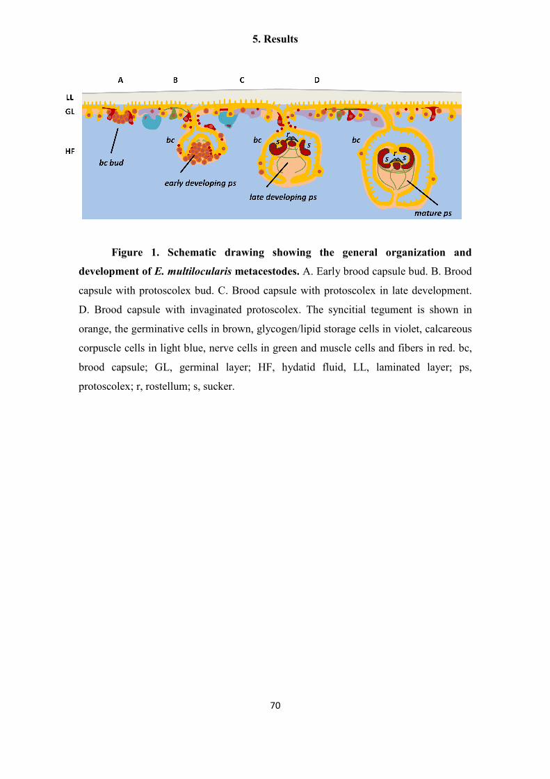
5. Results
70
Figure 1. Schematic drawing showing the general organization and
development of E. multilocularis metacestodes. A. Early brood capsule bud. B. Brood
capsule with protoscolex bud. C. Brood capsule with protoscolex in late development.
D. Brood capsule with invaginated protoscolex. The syncitial tegument is shown in
orange, the germinative cells in brown, glycogen/lipid storage cells in violet, calcareous
corpuscle cells in light blue, nerve cells in green and muscle cells and fibers in red. bc,
brood capsule; GL, germinal layer; HF, hydatid fluid, LL, laminated layer; ps,
protoscolex; r, rostellum; s, sucker.
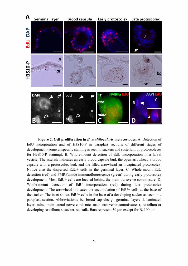
5. Results
71
Figure 2. Cell proliferation in E. multilocularis metacestodes. A. Detection of
EdU incorporation and of H3S10-P in paraplast sections of different stages of
development (some unspecific staining is seen in suckers and rostellum of protoscoleces
for H3S10-P staining). B. Whole-mount detection of EdU incorporation in a larval
vesicle. The asterisk indicates an early brood capsule bud, the open arrowhead a brood
capsule with a protoscolex bud, and the filled arrowhead an invaginated protoscolex.
Notice also the dispersed EdU+ cells in the germinal layer. C. Whole-mount EdU
detection (red) and FMRFamide immunofluorescence (green) during early protoscolex
development. Most EdU+ cells are located behind the main transverse commissure. D.
Whole-mount detection of EdU incorporation (red) during late protoscolex
development. The arrowhead indicates the accumulation of EdU+ cells at the base of
the sucker. The inset shows EdU+ cells in the base of a developing sucker as seen in a
paraplast section. Abbreviations: bc, brood capsule; gl, germinal layer; ll, laminated
layer; mlnc, main lateral nerve cord; mtc, main transverse commissure; r, rostellum or
developing rostellum; s, sucker; st, stalk. Bars represent 30 µm except for B, 100 µm.

5. Results
72
Figure 3. Cell suspensions of Echinococcus multilocularis. This is a montage
of images of different cell types as observed in cell suspensions after different staining
procedures. Cell types are indicated on the left (GC: germinative cells; TC, tegumental
cells; SC, glycogen/lipid storage cells; CC, calcareous corpuscle cells) and staining
procedures are indicated on top (NR, Nile Red, shown in orange; PI, propidium iodide,
shown in orange; WCS, whole cell staining, shown in green), including the detection of
EdU (shown in red) after two different labeling treatments, 50 µM for 5 hours and 1-10
µM for 7 days (see the main text for details). The bar represents 10 µm.
5. Results
73
Gene expression patterns in the germinative cells
To identify genes that are specifically expressed in the germinative cells, we
analyzed the expression of several candidate genes among planarian neoblast markers
by whole-mount in situ hybridization (WMISH).
em-h2b. As a possible general marker of all proliferating germinative cells, we
analyzed the expression of histone H2B homologs, since canonical histones are
synthesized in a cell-cycle dependent manner: histone transcripts only accumulate
during the S-phase, when new histones are needed to accompany the synthesis of DNA
(Jaeger et al. 2005). Furthermore, H2B genes have been found to be specifically
expressed in proliferating planarian neoblasts and in the neoblast-like cells of the
trematode Schistosoma mansoni (Solana et al. 2012; Collins et al. 2013).
Several canonical H2B genes are present in the E. multilocularis genome. Most
of them are almost identical to each other (>95% nucleotide identity), which we
denominate the em-h2b-1 group. Another gene, em-h2b-2, also shows high amino acid
identity (97%) but lower nucleotide identity (85%) to em-h2b-1. Using probes for em-
h2b-1 and em-h2b-2 gave identical results, which were indistinguishable from the EdU
labeling pattern in the germinal layer and throughout the development of brood capsules
and protoscoleces (Figure 4). This is particularly striking during late protoscolex
development, where a massive accumulation of em-h2b+ cells is found at the base of
the suckers but no expression is seen in the remaining sucker tissue (Figure 4E).
The WMISH signal labeled the cytoplasm of cells with the typical germinative
cell morphology (Figure 4B). Furthermore, when combining WMISH with EdU
detection after a 5 hour pulse, 78% of all em-h2b+ cells were also EdU+ (n=197 h2b+
cells), and conversely, 87% of all EdU+ cells were also em-h2b+(n=176 EdU+ cells )
(Figure 4F). Because only germinative cells proliferate (see above), em-h2b is therefore
a bona fide marker of S-phase germinative cells, but would not detect resting or G1 and
G2/M germinative cells. The smaller proportion of EdU- h2b+ cells had probably
already entered S-phase but were fixed before enough EdU was incorporated for
detection, while the EdU+ h2b- cells were probably fixed after they had already
incorporated EdU but exited the S-phase during the incubation time.

5. Results
74
Figure 4. WMISH detection of em-h2b. A. General view of the germinal layer.
B. Detail of the germinal layer; showing the germinative cell morphology of the
positive cells. C. Early brood capsule bud. D. Protoscolex bud. E. Late protoscolex
development. The arrowhead indicates the accumulation of positive cells at the base of
the sucker. F. Co-localization of em-h2b (dark precipitate) and EdU detection (red) after
a 50 µM, 5 h pulse. The inset shows an example in which the em-h2b signal was
inverted and pseudo-coloured in green to facilitate the visualization of the co-labeling.
Abbreviations are as in Figure 2. Bars represent 25 µm.

5. Results
75
em-nos-1 and em-nos-2.We then turned to possible post-transcriptional
regulators of the germinative cells. nanos genes are molecular markers of the germ line
in many classical models, but are also expressed in multipotent stem cells in various
basal metazoan lineages (Mochizuki et al. 2000; Rebscher et al. 2007; Ewen-Campen,
Schwager, and Extavour 2010; Juliano, Swartz, and Wessel 2010). Two nanos genes are
present in E. multilocularis (em-nos-1 and em-nos-2). Both genes were expressed in few
cells with a patchy distribution in the germinal layer (Figures 5A and D), and with the
morphology of large germinative cells (<1.6% of all cells for both em-nos-1 and em-
nos-2, n=4632 cells and n=7475 cells, respectively; Figures 5B and E). Furthermore,
em-nos-1 and em-nos-2 cells can incorporate EdU (19% of em-nos1+ cells are EdU+
after a 5 hour pulse, n=96), although the vast majority of EdU+ cells do not express
either gene (<5% of all EdU+ cells express either nanos gene, Figures 5C and F).
Altogether, these data show that a small subpopulation of the germinative cells in the
germinal layer express em-nos-1 and em-nos-2, although it is not clear if both genes are
co-expressed in the same cells.
During brood capsule and protoscolex development, em-nos1 expression was
not detected. Expression of em-nos2 was sometimes seen around brood capsule buds,
and later during early protoscolex development as a small population of cells at the base
of the protoscolex bud (Figures 5G and H). Finally, em-nos-2 is expressed in a few cells
associated with the developing nervous system, in the region of the developing lateral
ganglia and the main commissure (Figure 5I). These results show that most proliferating
cells do not express nanos genes in the developing protoscolex, and suggest a role for
em-nos-2 during nervous system development.

5. Results
76
Figure 5. WMISH detection of em-nos-1 (A-C) and em-nos-2 (D-I). A and D,
general view and B and E, detail of the positive cells in the germinal layer. C and F, co-
localization in the germinal layer of em-nos-1 and em-nos-2 (green) with EdU
incorporation (red) after a 5 h 50 µM pulse. Double positive cells are indicated with a
filled arrowhead, whereas cells expressing a nanos gene but EdU- are indicated with an
open arrowhead. G. Expression of em-nos-2 in cells surrounding a brood capsule bud
(dashed circle). H. Protoscolex bud. I. Later protoscolex development. Arrows indicate
em-nos-2+ cells in the position of the developing lateral ganglia. Abbreviations are as in
Figure 2. Bars represent 10 µm except for H, 20 µm.

5. Results
77
em-ago2. Although piwi genes are not present in E. multilocularis, there are
other argonaute proteins encoded by the genome: an ortholog of human Ago-1-4
proteins which is likely involved in RNA interference (EmAgo1 (Spiliotis et al. 2010))
and three copies of an argonaute gene family that is specific for cestodes and trematodes
(Tsai et al. 2013), which we dubbed em-ago2-A to em-ago2-C. We further identified a
pseudogene, em-ago2-Ψ (Additional File 6). These copies have resulted from a recent
duplication that occurred after the divergence of Hymenolepis and [Echinococcus +
Taenia] (see the phylogenetic analyses in Tsai et al. (2013)), with 88 to 99% nucleotide
sequence identity between copies (depending of the copies and the specific regions
compared). Moreover, they are organized as two couples of tandemly arranged copies in
close proximity to a copy of a Sec61 homolog (Additional File 6). This conservation of
synteny suggests that one first duplication occurred which resulted in two adjacent
copies of an original em-ago2 gene located next to a Sec61 gene, followed by the
duplication of the whole region. Long-range PCR with genomic DNA confirmed the
organization of these genomic regions (Additional File 6), while sequencing of smaller
PCR fragments confirmed the existence of all four copies, demonstrating that they are
not an artifact of genome assembly. By RT-PCR only em-ago2-A, em-ago2-B and em-
ago2-Ψ mRNA could be detected, whereas ago-2-C was absent or barely detected in all
of the larval stages and in primary cell cultures.
We performed WMISH using two different probes for em-ago2-A. These probes
would probably cross-react with all other em-ago2 copies, and we refer to the
expression pattern of all these genes by the collective name em-ago2. em-ago2
expression was similar to the pattern of EdU incorporation in the germinal layer, and
cells strongly expressing em-ago2 accumulate in the brood capsules and protoscolex
buds (Figures 6A to 6C). The distribution of the em-ago2 signal within cells is very
distinct since it is only observed close to or within the nucleus (Figure 6A). Strongly
positive em-ago2+ cells account for approximately 30% of all cells in the germinal
layer, but notably, some expression of em-ago2 was observed in more than 50% of all
cells, indicating that it is not exclusive of the germinative cell population.
There is no clear-cut correlation between the level of em-ago2 expression and
proliferation, since approximately 50% of EdU+ cells show low or no expression of em-
ago2 (Figure 6A). Conversely, during early protoscolex development, it is clear that

5. Results
78
although em-ago2 expression is present in most cells, the cells with the strongest em-
ago2 signal are almost invariably EdU- (Figure 6C). During late protoscolex
development, some em-ago2 signal is observed in most cells (Figures 6D and E), and is
not restricted to the base in the suckers (where cell proliferation occurs). It is therefore
clear that the expression of em-ago2 is not restricted to proliferating cells. Moreover,
the expression of em-nos-1, em-nos-2 and em-ago2 point to extensive heterogeneity at
the molecular level among the proliferating germinative cells.
Figure 6. WMISH detection of em-ago2. A. General view of the germinal
layer. The insets show details of EdU+ em-ago2- cells (open arrowheads) and EdU+
em-ago2+ cells (filled arrowheads) after a 5 h, 50 µM EdU pulse. B. Early brood
capsule bud. C. Protoscolex bud; colors are coded as in Figure 5A. D. Late protoscolex
development. E. Detail of a sucker from a protoscolex in late development, showing
that em-ago2 expression is not restricted to the base; colors are coded as in Figure 5A.
Abbreviations are as in Figure 2. Bars represent 10 µm in A1 and A2, and 40 µm in all
other panels.

5. Results
79
em-hdac1 and em-phb1. The histone deacetylase HDAC1 is one of many
chromatin modifying proteins that are specifically expressed in planarian neoblasts
(Eisenhoffer, Kang, and Sanchez Alvarado 2008; Zhu and Pearson 2013). Neoblast
specific expression has also been shown for the mRNA of homologs of prohibitin-1 and
prohibitin-2 (Rossi et al. 2007; Eisenhoffer, Kang, and Sanchez Alvarado 2008). In
mammalian cells, prohibitins form complexes in the inner mitochondrial membrane
with unclear biochemical function, and have been linked to mitochondrial biogenesis
and cell proliferation (Merkwirth and Langer 2009).
We found single-copy orthologs of HDAC1 (em-hdac1) and prohibitin-1 (em-
phb1) in the E. multilocularis genome, and both genes showed widespread expression in
the germinal layer and during protoscolex development (Additional Files 7 and 8). We
also determined the distribution of the Em-PHB1 protein by immunohistochemistry, and
compared this to the distribution in planarian tissues, using a commercial antibody that
recognizes a conserved region in all PHB1 proteins. In planarians, although low levels
of PHB1 are seen in post-mitotic tissues such as the pharynx, the highest signal is
observed in neoblast-like cells in the mesenchyma (Additional File 9). In contrast, E.
multilocularis Em-PHB1 is observed throughout the germinal layer, brood capsules and
developing protoscoleces (Additional File 10). In summary, neither gene has a
germinative cell specific expression in E. multilocularis metacestodes.
Identification of molecular markers for differentiated cell types.
The identification of differentiated cell types in the germinal layer is difficult,
and unlike the situation in adult cestodes, trematodes and planarians, the lack of spatial
segregation of any post-mitotic cell types makes it impossible to trace the differentiation
of germinative cells in situ. Therefore, we set out to find molecular markers of
differentiated cell types in Echinococcus.
em-muc-1 and em-alp-2 as tegumental cell markers. Since the laminated layer
is synthesized by the tegumental syncitium, genes coding for laminated layer
components should be expressed by the tegumental cells (Diaz et al. 2011). We
analyzed the expression of em-muc-1, a member of an Echinococcus-specific, highly
expressed apomucin gene family that has been proposed to be a main component of the
laminated layer (Diaz et al. 2011; Parkinson et al. 2012; Tsai et al. 2013). Because all

5. Results
80
members are very similar (with regions with over 90% identity at the nucleotide level) it
is likely that the em-muc-1 probe recognizes most genes of this gene family.
em-muc-1 is strongly expressed in the germinal layer but not in protoscoleces, as
is expected for a component of the laminated layer (Figure 7A). em-muc-1 was
expressed in cells with abundant cytoplasm that fuse or interdigitate with each other,
and which constitute 27 to 37% of all cells in the germinal layer (two independent
WMISH experiments; n=3440 and 780 cells, respectively; Figure 7B). In stark contrast,
no expression is observed in early brood capsule buds or in developing protoscoleces.
To our surprise, however, we could detect low levels of em-muc-1 in late brood
capsules, which suggests that the glycocalix of the brood capsule may also contain the
product of em-muc-1, although the laminated layer does not line the brood capsule
cavity (Diaz et al. 2011) (Figure 7D). By combining WMISH with EdU detection, we
observed the absence of EdU+ cells among the em-muc-1+ cell population (n=1454 em-
muc-1+ cells from two independent WMISH experiments; Figure 7C). We conclude
that em-muc-1 is a robust marker for tegumental cells in the germinal layer, and confirm
that the tegumental cell population does not proliferate.
While searching for possible histochemical markers, we observed that alkaline
phosphatase activity in the metacestode is very high (strong reaction in less than 5
minutes) and restricted to the distal syncitial tegument of the germinal layer (Figure 8A;
see also (del Cacho et al. 1996)), but is not found in brood capsules (Additional File 11).
This indicated that one or more alkaline phosphatase genes must be expressed in the
tegumental cells. In protoscoleces, alkaline phosphatase was detected only after several
hours, and only in the excretory system (Figure 8 B), similarly to what has been
described in the developing adult (Arsac et al. 1997). This activity increased after
protoscolex activation (Additional File 11). Four genes encoding alkaline phosphatases
(em-alp-1 to em-alp-4) were found in the E. multilocularis genome. By RT-PCR, em-
alp-1 and em-alp-2 were found to be specifically expressed in the germinal layer, while
em-alp-3 was only detected in protoscoleces, with a strong up-regulation after
protoscolex activation (Figure 8C). em-alp-4 has substitutions of conserved catalytic
amino acid residues, and no expression was detected by RT-PCR in the germinal layer
or in protoscoleces, suggesting that it is a pseudogene, although expression was
observed in high throughput RNA sequencing data of adult worms (Tsai et al. 2013).

5. Results
81
Altogether, the data suggested that em-alp-1 and em-alp-2 were expressed in the
tegumental cells of the germinal layer, while em-alp-3 was expressed in the protoscolex
excretory system, and is to the best of our knowledge the first gene shown to be up-
regulated after protoscolex activation. We therefore analyzed the expression pattern of
em-alp-2 by WMISH, and found it to be identical to that of em-muc-1 in the germinal
layer, with no expression in the brood capsule buds or in the developing protoscoleces
(Figure 8D). In conclusion, em-alp-2 is another marker for the tegumental cells in the
germinal layer.

5. Results
82
Figure 7. Expression of em-muc-1. A. Semi-quantitative RT-PCR with serial
ten-fold dilutions of cDNA from protoscoleces (PS) and germinal layer (GL). Controls
without reverse transcriptase (-RT) and without template (H20) are included. B-D.
WMISH of em-muc-1. B. General view of the germinal layer. C. Double detection of
WMISH (green) and EdU incorporation after a 5 h, 50 µM EdU pulse (red); note the
lack of EdU labeling among the em-muc-1+ cells. D1 and D2.Section of metacestode
processed for WMISH, showing the lack of expression in the developing protoscoleces,
strong expression in the germinal layer, and expression in the brood capsule wall.
Abbreviations are as in Figure 2. Bars represent 20 µm except for B, 10 µm.

5. Results
83
Acetylated tubulin α as a marker of nerve cells. We have recently shown that
a net of nerve cells in the germinal layer can be detected by immunohistofluorescence
against acetylated tubulin α (AcTub+ cells, (Koziol, Krohne, and Brehm 2013)). Here,
we show that the nerve cells do not proliferate, since they are EdU- after a 5 hour, 50
µM EdU pulse (n = 874 AcTub+ cells from three independent experiments; Additional
File 12). Similarly, in activated protoscoleces all AcTub+ cells (including in this case
both nerve cells and flame cells (Koziol, Krohne, and Brehm 2013)) were EdU- (data
not shown).
We performed a quantitative analysis of the formation of new AcTub+ cells by
determining the percentage of EdU+ AcTub+ cells during continuous EdU labeling. No
double labeled cells were observed after 7 days, but the percentage increased to 13.3%
after 14 days (Additional File 12). Because no EdU incorporation was observed in nerve
cells even after 7 days of continuous exposure, this strongly indicates that all EdU+
nerve cells must originate from the differentiation of proliferating germinative cells,
which would require more than 7 days after exiting the cell cycle to become AcTub+.
Higher concentrations of EdU (10 µM) apparently had a toxic effect, and only 2.8% of
the AcTub+ cells were EdU+ after 14 days (Additional File 12).
em-tpm-1.hmw as a marker for muscle cells during protoscolex
development. Using a specific antibody that recognizes the high molecular weight
(HMW) isoforms of two tropomyosin genes from cestodes (tpm-1 and tpm-2 ), HMW
tropomyosins have been shown to be present exclusively in the muscle fibers in the
cestode Mesocestoides corti, and are strongly expressed in the suckers of E. granulosus
protoscoleces (Alvite and Esteves 2009; Koziol et al. 2011). Using this antibody, we
confirmed that HMW tropomyosin isoforms can be found in the muscle fibers in the
germinal layer, accumulating in the interior of brood capsules, and in the muscle layers
during protoscolex development in E. multilocularis (Additional File 13), in perfect
correlation to the description of muscle fibers as determined by phalloidin labeling
(Koziol, Krohne, and Brehm 2013).
Because in cestodes the nucleus of muscle cells is located in a non-contractile
cell body, connected by thin cytoplasmic bridges to the contractile myofibers (Halton
and Maule 2003; Smyth and McManus 2007), it is not possible to identify the cell

5. Results
84
bodies by immunodetection of HMW-Tropomyosins. Instead, we analyzed the
expression of the HMW isoform of em-tpm-1 by WMISH using a specific probe.
Surprisingly, no expression was observed in the germinal layer, suggesting that muscle
cells in this tissue express other tropomyosin isoforms (i.e. from em-tpm-2). Instead,
em-tpm-1.hmw was detected in individual cells in the center of invaginating brood
capsules (Figure 9A), in close proximity to the location of muscle fibers (Koziol,
Krohne, and Brehm 2013). During early protoscolex development, when muscle
development is already underway (Koziol, Krohne, and Brehm 2013), em-tpm-1.hmw is
expressed in two symmetrical bands of superficial cells and one internal medial band
(Figure 9B). Because of their distribution, these are likely to be the subtegumental
circular muscle cells and the inner longitudinal muscle cells. Finally, strong expression
was observed in the muscular suckers and rostellum and in individual cells in the body
of the developed protoscolex (Figure 9C). In summary, em-tpm-1.hmw can be used as a
molecular marker for the development of muscle cells during brood capsule and
protoscolex development, but not in the germinal layer.

5. Results
85
Figure 8. Alkaline phosphatase activity and gene expression. A1. Alkaline
phosphatase histochemistry in the germinal layer, showing strong activity in the
syncitial tegument. A2. The signal in A1 was inverted and pseudo-colored in green, and
combined with DAPI (blue) and phalloidin (red) staining to show the distribution of the
nuclei and muscle fibers, respectively. B. Alkaline phosphatase histochemistry in an
activated protoscolex, showing activity in the excretory system. C. Semi-quantitative
RT-PCR with serial ten-fold dilutions of cDNA from activated protoscoleces (PS act),
non-activated protoscoleces (PS not act) and germinal layer (GL). The experiment was
repeated three times with similar results. D. WMISH of em-alp-2, showing strong
expression in the germinal layer but not in the brood capsule bud. Abbreviations are as
in Figure 2. Bars represent 20 µm.
Figure 9. WMISH detection of em-tpm-1.hmw. A. Early brood capsule bud as
seen from above. The inset shows a lateral view. B. Protoscolex bud. C. Developed
protoscolex. The arrowheads point to the suckers. Abbreviations are as in Figure 2. Bars
represent 20 µm (A, B) or 40 µm (C).

5. Results
86
Partial depletion of germinative cells by irradiation and hydroxyurea treatments
Total and partial elimination of neoblasts by irradiation has been a powerful tool
for studying neoblast gene expression and physiology in many flatworms (Pfister et al.
2007; Rossi et al. 2007; Eisenhoffer, Kang, and Sanchez Alvarado 2008; Pfister et al.
2008; Wagner, Wang, and Reddien 2011; Wagner, Ho, and Reddien 2012; Collins et al.
2013). In E. multilocularis, comparable doses of ionizing radiation (50-100 Gy) were
found to only retard growth of metacestodes, and did not eliminate the germinative cells
(Pohle et al. 2011). We performed similar experiments with a single X-ray irradiation
dose of 150 Gy on metacestode vesicles (lacking brood capsules and protoscoleces). At
48 hours post-irradiation, we observed that the number of cells incorporating EdU per
area of germinal layer decreased in average to 22% of unirradiated controls, showing
that very high dosages of X-ray irradiation decrease but are unable to eliminate all
proliferating cells (Figure 10A). Interestingly, we observed no significant increase in the
number of EdU incorporating cells for up to 48 days post-irradiation, and the number of
EdU+ cells per area was still in average only 28% that of unirradiated controls (Figure
10A). Despite this long-term decrease in cell proliferation, no difference in survival was
observed between irradiated and unirradiated metacestodes after 48 days (82% versus
86%, p=0.49, Chi-Square test).
As an alternative approach, we used hydroxyurea (HU), an inhibitor of
ribonucleotide reductase (RRM) that is specifically toxic to cells undergoing DNA
synthesis during cell proliferation (Sinclair 1965; Krakoff, Brown, and Reichard 1968;
Farber and Baserga 1969), and which has also been used in other invertebrates to
eliminate stem cells (Salo and Baguna 1984; Pfister et al. 2007; De Mulder et al.
2009a). The mechanism of toxicity is based on the depletion of dNTPs that results from
RRM inhibition, provoking the cessation of DNA replication, thus leading to stalled
replication forks and eventually to chromosomal DNA damage (Petermann et al. 2010).
In order to confirm a similar effect of HU on E. multilocularis cells, we incubated
primary cell cultures with different HU concentrations. Indeed, incorporation of the
thymidine analog 5-bromo-2'-deoxyuridine (BrdU) was reduced by approximately 50%
and 90% in the presence of 10 mM and 40 mM HU, respectively. Furthermore, the
regeneration of metacestode vesicles from primary cells was strongly decreased by 10
mM HU and abolished by 40 mM HU (Additional File 14).

5. Results
87
We therefore incubated metacestode vesicles (lacking brood capsules and
protoscoleces) with 40 mM HU for 7 days, and allowed them to recover in HU free
media for 24 hours. This resulted on average in a 90% reduction in the number of cells
incorporating EdU per area of germinal layer (Figure 10B). Furthermore, the
experimental results could be divided into two groups: Group 1, from experiments
performed on larger and older vesicles, resulted in a decrease of only 66% to 93% in the
number of EdU+ cells (similar to the results observed after X-ray irradiation), whereas
Group 2, performed in smaller and younger vesicles, resulted in a stronger decrease of
97.7% to 99.8% of EdU+ cells with respect to the non-treated controls.
We analyzed in detail the effect of HU on the metacestode vesicles.
Quantification of germinative cells in tissue macerates showed that the loss of EdU+
cells was paralleled by a decrease in germinative cells, from 20-22% to 3-5% of all cells
(two independent experiments, p≤0.001 for both, Chi-Square test). However,
differentiated cells did not seem to be affected by the HU treatment: nerve cell numbers
per area of germinal layer were not significantly reduced, as determined by AcTub
immunohistofluorescence (Figure 10C) and the tegumental cell marker em-muc-1
showed a qualitatively similar expression pattern in both conditions (Figure 10D). We
conclude that HU treatment for 7 days specifically depletes the germinative cell
population, with little effect on the number of differentiated cells.
Prolonged times of recovery of vesicles from Group 1 in HU free media did not
result in a significant increase of EdU+ cells after up to 22 days, similar to the results
obtained after X-ray irradiation. However, in Group 2, we could observe highly
localized, time-dependent increments in the number of EdU+ cells, strongly suggestive
of clonal growth from surviving proliferating cells (Figure 11). In most experiments
only isolated EdU+ cells could be found after 1 day of recovery. As recovery time
increased, patches of 2 EdU+ cells (after 1 to 4 days of recovery), 3 to 4 EdU+ cells
(after 4 to 9 days of recovery) and of more than 30 EdU+ cells (after 9 days of recovery)
could be found in some of the metacestode vesicles. This strongly indicates the
existence of cells in the metacestode (presumably germinative cells) that can respond to
the strong depletion of proliferating cells by undergoing self-renewing divisions into
proliferation-competent cells.

5. Results
88
Figure 10. Effect of X-Ray irradiation and HU treatment. A. Number of
EdU+ cells per area at different times after X-Ray irradiation (150 Gy) and in non-
irradiated controls, after a 5 h 50 µm EdU pulse (average and standard deviations of 7 to
14 vesicles per time point). B. Number of EdU+ cells per area in vesicles treated with
HU (40 mM for 7 days) and in non-treated controls (all vesicles were allowed to recover
for 24 hours in HU free medium, followed by a 5 h 50 µm EdU pulse; average and
standard deviation of 7 independent experiments). C. Number of AcTub+ cells per area
in vesicles treated with HU (40 mM for 7 days) and in non-treated controls (average and
standard deviation of 9 to 11 vesicles pooled from 3 independent experiments). D.
WMISH of em-muc-1 in a vesicle treated with HU (40 mM for 7 days) and in a non-
treated control. * p<0.001,**p<0.01. The Mann-Whitney U-Test was used for A-C.

5. Results
89
Figure 11. Growing patches of EdU+ cells during recovery from HU
treatment. Vesicles were treated for 7 days with 40 mM HU, transferred to HU-free
medium and samples were taken for EdU labeling (5 h with 50 µM EdU) and detection
at the indicated times of recovery.

5. Results
90
Germinative cells proliferate and are enriched in primary cell preparations.
In the previously described primary cell regeneration system (Spiliotis et al.
2008), primary cells obtained from E. multilocularis metacestodes initially form small
aggregates that grow and fuse to each other. Within these aggregates fluid-filled cavities
are formed, and eventually new metacestode vesicles are generated by a still
incompletely understood process. We analyzed cell proliferation within the early
aggregates (after 2 days of culture) by EdU labeling. We observed extensive cell
proliferation in a layer within the aggregates, but the innermost cells did not incorporate
EdU, suggesting that they have exited the cell cycle, which may be related to the initial
formation of internal cavities (typically observed after 4 days of culture) (Figure 12A).
In order to morphologically identify the proliferating cells during regeneration, we
prepared cell suspensions by the tissue maceration procedure, and based the
identification of cell types on the previously described morphological criteria. As found
in metacestodes, germinative cells were the only cell type labeled by EdU in primary
cell preparations (Figures 12C and D). Furthermore, germinative cells were enriched in
two day-old aggregates, comprising 62% to 83% of all cells, as compared to 32% to
55% in the metacestodes that were used to generate the primary cell preparations (3
independent primary cell preparations, p<0.02 for all preparations, Chi-square test;
furthermore, note that the percentage of germinative cells is likely overestimated for the
metacestodes since large vesicles were used). Among the differentiated cells,
tegumental and glycogen/lipid storage cells were conspicuously few, and an increase in
cells with degenerating morphology was noted. Further confirmation of the strong
depletion of tegumental cells was obtained by analyzing the expression of the
tegumental cell markers em-muc-1, em-alp-2 and of em-alp-1. Preliminary high
throughput RNA sequencing data indicated low expression levels for all three genes in
primary cells (2%, 7% and 0% of the levels found in the germinal layer, respectively),
that was confirmed by semi-quantitative RT-PCR for em-alp-1 and em-alp-2 (Figure
12B).
To confirm the enrichment of germinative cells and to determine which other
cell types are present in early primary cell aggregates, we performed transmission
electron microscopical studies of two day-old primary cell aggregates. Abundant
germinative cells could be found in the aggregates, surrounded by an external layer of

5. Results
91
cells showing signs of degeneration (necrosis) (Figure 12E and Additional File 15).
Also, many muscle fibers and some muscle cells could be identified (Figure 12F). In
cryosections of primary cells after three days of culture, muscle fibers were also found
by phalloidin staining, and nerve cells were identified by AcTub
immunohistofluorescence (Additional File 16). In summary, early primary cell
preparations are enriched in proliferating germinative cells, but other cell types such as
muscle cells and nerve cells are also present.

5. Results
92
Figure 12. Characterization of early primary cell preparations. A. EdU
incorporation after a 50 µM 5 h pulse of primary cell aggregates (2 days-old). B. Semi-
quantitative RT-PCR of em-alp-1 and em-alp-2 genes with serial ten-fold dilutions of
cDNA from primary cells (PC) and germinal layer (GL). The experiment was repeated
three times with similar results. C and D. Representative microscopy fields of cell
suspensions obtained from the germinal layer (C) and from primary cells (D). EdU+ and
EdU- germinative cells are indicated by filled and open arrowheads, respectively. The
inset in D shows a close-up of two EdU+ germinative cells in the primary cell
preparations. E and F. TEM of primary cell aggregates (2 days-old). E. Accumulation of
germinative cells. F. A muscle cell (m), containing myofibers and extensive smooth
endoplasmic reticulum, and a germinative cell (g) in the periphery of an aggregate.
Notice also cell debris surrounding the cells. Bars represent 4 µm (A, E, F and inset in
D) and 20 µm (C and D).

5. Results
93
Discussion
Germinative cells are the only proliferating cells in Echinococcus multilocularis
larvae
The germinative cells in E. multilocularis constitute a morphologically
homogeneous population, similar to descriptions in other cestode species and life stages
(Douglas 1961; Bolla and Roberts 1971; Sulgostowska 1972; Gustafsson 1976b; Koziol
et al. 2010), and generally similar to the neoblasts in free living flatworms and the
neoblast-like cells of the trematode Schistosoma mansoni (Reuter and Kreshchenko
2004; Collins et al. 2013). The main differences observed within the germinative cells
in E. multilocularis were related to the number of nucleoli, size, and the presence or
absence of thin cytoplasmic projections. All of these differences could be related in part
to normal changes during the cell cycle and cell migration (Wachtler and Stahl 1993;
Alberts 2000), although other authors have sub-divided the germinative cells into
different types based on size and histological details (Douglas 1961; Sulgostowska
1972). Germinative cells were identified as the only proliferating cell type during
metacestode growth and regeneration, indicating that all new cells must originate from
this population. However, the existence of de-differentiation and trans-differentiation
processes in differentiated cells cannot completely be ruled out at this point.
Unambiguous lineage tracing will require analyses mediated by stable genetic markers,
and current efforts in our laboratory are being made towards transgenic modification of
Echinococcus germinative cells. Furthermore, the markers for differentiated cell types
developed in this work will be an important tool for tracing the differentiation pathways
of germinative cells.
Unfortunately, we have been unable to find a universal molecular marker for the
germinative cells so far. However, H3S10-P and em-h2B are useful endogenous markers
to identify the proliferating germinative cells in metacestode tissues, and open the
possibility to identify such cells in in vivo material (in the absence of labeling with
thymidine analogs). Despite the morphological uniformity of germinative cells, gene
expression analyses have clearly shown that the germinative cells are heterogeneous at
the molecular level: em-ago-2, em-nos-1 and em-nos-2 genes are only expressed in
subpopulations of the proliferating germinative cells. Because argonaute and nanos

5. Results
94
proteins are well known post-transcriptional regulators with important roles in stem cell
and germ cell biology (Carmell et al. 2002; Ewen-Campen, Schwager, and Extavour
2010; Juliano, Swartz, and Wessel 2010), this points to the existence of different sub-
populations of germinative cells, perhaps with different self-renewal or differentiation
potencies.
The capacity for self renewal of at least some of the germinative cells is strongly
suggested by the HU-mediated depletion experiments, and is expected given the ability
of metacestode tissue to be indefinitely passaged in vivo (Norman and Kagan 1961;
Spiliotis and Brehm 2009). It is interesting that no proliferation response was observed
after milder depletions, and the metacestode vesicles were able to survive for long
periods under these conditions. This suggests that the control of cell proliferation is
relatively lax in E. multilocularis metacestodes. It is possible that a relatively low
number of proliferating germinative cells are enough for basal tissue turnover, but that a
larger number is required for actively growing vesicles. Similarly, large differences in
the number of EdU incorporating cells can be observed in vesicles incubated ex vivo
from a single infected rodent (Koziol and Brehm, unpublished data).
Could cells expressing nanos in Echinococcus metacestodes represent the germ
line?
In many free living flatworms, it is traditionally thought that the germ line is
segregated by epigenesis in the juveniles or adults (Extavour and Akam 2003). In fact,
the limit between the somatic stem cells and the germ line is fluid in planarians and
Macrostomum, since the somatic neoblasts are able to contribute to the germ line during
regeneration (Sato et al. 2006; Handberg-Thorsager and Salo 2007; Pfister et al. 2008).
However, recent studies using molecular markers have allowed the identification of
germ cells already at the time of hatching, suggesting that in these flatworms the germ
line may be segregated earlier than was previously thought (Handberg-Thorsager and
Salo 2007; Pfister et al. 2008). For example nanos, another classical germ line marker,
has been shown to be expressed only in the planarian germ line stem cells, but not in the
morphologically indistinguishable somatic neoblasts (Sato et al. 2006; Handberg-
Thorsager and Salo 2007; Wang et al. 2007).

5. Results
95
In cestodes, it is assumed that the germ line is originated by epigenesis, from
the germinative cells in the neck region of the developing adult (Sulgostowska 1972;
Gustafsson 1990; Extavour and Akam 2003). It is conceivable, however, that a
subpopulation of the germinative cells could be segregated into the germ line earlier in
development, particularly since lineage tracing has never been achieved in cestode
embryos (Rybicka 1966). In the case of E. multilocularis, the extensive asexual
reproduction and growth in the intermediate host makes an epigenetic mechanism very
likely, since early segregation of the germ line by preformation would require
incorporation of cells from a segregated germ line within every new vesicle and
protoscolex that is asexually generated. Our data suggests that the germinative cell
subpopulation expressing nanos homologues are not germ line cells: no expression was
observed of em-nos-1 during brood capsule and protoscolex development, and em-nos-2
was not consistently observed in brood capsule buds. Moreover, em-nos-2 expression in
late protoscolex development suggests a role in the formation of the nervous system.
Relatedly, specific nanos paralogs are expressed in the nervous system in many
metazoans (Haraguchi et al. 2003; Ye et al. 2004; Aoki et al. 2009; Kanska and Frank
2013).
Echinococcus primary cells as an experimental model for stem cell research
A great advantage of the E. multilocularis model is the possibility of long term
in vitro culture for primary cell preparations, resulting in complete vesicle regeneration
(Spiliotis et al. 2008). Here, we show that primary cell cultures are enriched for
germinative cells, which actively proliferate from the earliest stages of development.
Genes enriched in transcriptomic studies of early primary cells and depleted in HU-
treated metacestodes could therefore be mined to search for germinative cell-specific
expression.
The mechanism for enrichment is not clear, and could involve differential
extraction of the germinative cells from the metacestode tissues, differential survival
and aggregation of the germinative cells during culture, and the accumulation of
germinative cells from self-renewing divisions. There is indirect evidence supporting all
of these mechanisms, since: 1) it has been shown previously that 30% of all cells in
fresh primary cell preparations are in S and G2/M, making the total percentage of

5. Results
96
proliferating cells very large before they are set into culture (Spiliotis et al. 2008), 2)
there is evidence of abundant cell death in electron microscopical studies and cell
suspensions, and 3) germinative cells actively proliferate in the early aggregates.
Besides the germinative cells, differentiated cells such as nerve cells and muscle cells
are present in the aggregates. Their role in regeneration, if any, is unknown, but the
neuromuscular system has been suggested to influence cell proliferation, differentiation
and pattern formation in free living flatworms (Reuter and Gustafsson 1996; Rossi,
Iacopetti, and Salvetti 2012; Witchley et al. 2013).
Differences between the Echinococcus germinative cells and the neoblasts in other
flatworms
The lack of chromatoid bodies and the absence of vasa and piwi orthologs imply
important differences between the germinative cells of cestodes and the neoblasts of
planarians. Furthermore, genes that are neoblast-specific in planarians, such as hdac1
and phb1, are widely expressed in E. multilocularis. Homologs of these genes have
been shown to be important for stem cell biology and cell proliferation in other
organisms, but they do not have a stem cell or even a proliferating cell-specific
expression in these models (MacDonald and Roskams 2008; Brunmeir, Lagger, and
Seiser 2009; Merkwirth and Langer 2009; Murko et al. 2010). It is therefore possible
that the specific expression of these genes in neoblasts is a planarian novelty, which
may not be shared with other flatworms. At the functional level, the response of E.
multilocularis to partial germinative cell depletion is also different to that described in
free living flatworms, since in planarians and in Macrostomum there is always a quick
response by which the neoblast population is restored to normal levels within narrow
margins, even when relatively large numbers of neoblasts remain in the tissues (Salvetti
et al. 2009; De Mulder et al. 2010).
Recently, Newmark and colleagues demonstrated the existence of neoblast-like
cells in the trematode Schistosoma mansoni, and in this organism, which also lacks vasa
and piwi orthologs, paralogs of these genes are specifically expressed in the neoblast-
like stem cells and have important roles in their maintenance (Collins et al. 2013; Wang,
Collins, and Newmark 2013). In Schistosoma mansoni sporocysts the ago2-1 gene (an
ortholog of the em-ago-2 genes) is expressed in all neoblast-like cells, and nanos-2 is

5. Results
97
expressed in a large subpopulation of neoblast-like cells. In adults, both genes are
expressed in many, if not all, somatic stem cells. Therefore, differences can also be
observed in the gene expression repertoire between E. multilocularis germinative cells
and the Schistosoma neoblast-like cells, making the E. multilocularis stem cell system
unique among flatworms. Further gene expression studies will depict a clearer picture of
their similarities and differences, including the analysis of vasa-like genes (paralogs of
vasa such as PL-10) which are expressed in planarian and schistosome stem cells
(Rouhana et al. 2010; Wagner, Ho, and Reddien 2012; Wang, Collins, and Newmark
2013).
The E. multilocularis metacestode, with its ability to grow infiltratively like a
tumor into the tissues of the host, is a relatively recent evolutionary novelty and is
derived from the more typical cysterci larvae found in other taeniid cestodes (Freeman
1973; Slais 1973). Although rare in most cestodes, asexual reproduction by the
formation of new scoleces is found in many taeniids (Slais 1973; Moore and Brooks
1987; Toledo et al. 1997; Loos-Frank 2000) and it is possible that the huge proliferative
potential of E. multilocularis germinative cells and larvae was adapted from an already
increased potential found in a common taeniid ancestor. The germinative cells in E.
multilocularis, although similar in morphology and functionally analogous to the
neoblasts in other flatworms, show important differences at the level of gene
expression, which could be related to this recent evolutionary change in their
developmental biology. At this point, nothing is known about gene expression in the
germinative cells of adult cestodes. Because of the biological hazard related to working
with E. multilocularis adults, comparative studies in related cestode models such as
Hymenolepis microstoma (Cunningham and Olson 2010) and Mesocestoides corti
(Specht and Voge 1965; Koziol et al. 2010) would be of great importance to delineate
which characteristics of the germinative cells of E. multilocularis metacestodes are
unique to this species and larval form, and which are a general feature of cestode
germinative cells.

5. Results
98
Conclusions
E. multilocularis is an important model for the study of parasite development
and host-parasite interactions, and is the only flatworm model with a robust cell culture
system (Brehm 2010b). Growth and regeneration of E. multilocularis larvae is driven by
the germinative cells, which are morphologically and functionally similar to the
neoblasts of free living flatworms, but which show important differences in their gene
expression patterns and in the loss of conserved stem cell regulators. This work
represent the first description of the germinative cells at the molecular level, giving the
first evidence of the existence of sub-populations of germinative cells with different
gene expression patterns, and provides molecular markers for identifying differentiated
cell types in situ. Some differences between the germinative cells and the neoblasts of
other flatworms could have arisen as specific adaptations of the stem cell system for the
unique asexual development of E. multilocularis. The novel development of E.
multilocularis and other taeniids is therefore an excellent model for the study of the
evolutionary origins of asexual reproduction and its effect on stem cell systems.

5. Results
99
Methods
Parasite material, culture and primary cell preparation
Parasite isolates were maintained by serial intraperitoneal passage in Meriones
unguiculatus as previously described (Spiliotis and Brehm 2009). Unless otherwise
stated, all experiments were performed on in vitro cultured metacestodes. Standard in
vitro culture of metacestodes was done in co-culture with rat Reuber hepatoma feeder
cells, and primary cell preparations were performed and cultured in cDMEM-A pre-
conditioned medium essentially as previously described (Spiliotis and Brehm 2009),
with the following modifications: 1) cells were detached from the metacestode tissue
with a single treatment of 20 minutes with trypsin/ethylenediaminetetraacetic acid
(EDTA) and 2) primary cells were cultured in cDMEM-A instead of hydatid fluid.
For primary cell cultures, isolate H95 (Jura et al. 1996), which has been
passaged for 18 years and has developed a strong defect in protoscolex formation was
used. For other experiments, more recent isolates were used, obtained from accidental
infections of Old World Monkeys in a breeding exclosure (Tappe et al. 2007). Dugesia
tahitiensis ((Peter, Ladurner, and Rieger 2001), obtained from Bernhard Egger) was the
planarian species used for immunohistofluorescence.
Ethical approval
All experiments were carried out in accordance with European and German regulations on the
protection of animals (Tierschutzgesetz). Ethical approval of the study was obtained from the
local ethics committee of the government of Lower Franconia (55.2-2531.01-31/10).
EdU labeling and detection
For short term labeling, 50 µM of 5-ethynyl-2′-deoxyuridine (EdU, Life
Technologies) was added to the media and the material was incubated for 5 hours. For
continuous labeling, 1 µM EdU was used for up to 14 days. The length of incubation
and the EdU concentrations were determined after varying the parameters in
preliminary experiments (see the main text). Before fixation, metacestode vesicles were
gently opened using a syringe tip in order to allow the entry of the fixative and other
reagents during detection procedures. Samples were fixed in 4% paraformaldehyde
prepared in phosphate buffer saline (PFA-PBS) for 1 hour at room temperature, and

5. Results
100
processed for detection of EdU in paraplast sections. Detection was performed with the
Click-iT® EdU Alexa Fluor® 555 Imaging Kit as described by the manufacturer for
sections. Whole-mount detection was performed by a modified protocol in which all
steps were doubled in length and the washes were increased in number. For double
labeling, EdU detection was always performed after the immunohistofluorescence or in
situ hybridization protocols.
Tissue maceration and staining of cell suspensions
Cell suspensions were prepared by a modification of the method of David
(David 1973). Metacestode vesicles were opened and washed in phosphate buffer saline
(PBS), and placed in maceration solution (13:1:1 distilled water : glacial acetic acid :
glycerol, 100 µl of solution per vesicle). Primary cell aggregates (from one well of a
six-well plate, after two days of culture) were washed in PBS, allowed to settle and
placed in 500 µl of maceration solution. Both kinds of samples were pipetted up and
down with a p1000 pipette, and placed overnight at 4 ºC. The next day they were once
again disaggregated by pipetting, diluted to 1:10 with maceration solution, and 10 µl
were spotted on SuperFrost slides (Thermo Scientific). The slides were dried overnight
at room temperature and stained by either of these procedures:
A) Propidium iodide (PI) plus 4',6-diamidino-2-phenylindole (DAPI) : after washing the
slides with PBS plus 0.05% Triton X.100, the slides were stained successively with
DAPI (1 µg/ml in PBS) and PI (2.5 µg/ml in PBS), washed twice with PBS and
mounted with Fluoprep (bioMérieux).
B) Whole Cell Stain (WCS) plus DAPI: the Cellomics WCS Green (Thermo Scientific)
was used as instructed by the manufacturer, followed by DAPI staining, washing and
Fluoprep mounting. When done in combination, EdU detection was performed first,
followed by WCS plus DAPI staining.
C) Nile Red plus DAPI: after washing with PBS, the slides were stained with Nile Red
(Greenspan, Mayer, and Fowler 1985) (Sigma-Aldrich, 100 ng/ml in PBS from a 4.2
mg/ml stock in acetone) followed by DAPI staining, washing, and Fluoprep Mounting.
Imaging was performed with the rhodamine channel of the Zeiss Axio Imager.Z1
microscope.

5. Results
101
Hydroxyurea treatment and X-ray irradiation
Metacestode vesicles were cultured in axenic, pre-conditioned cDMEM-A
medium (Spiliotis and Brehm 2009) with a nitrogen gas phase (40 vesicles in 5 ml of
medium in 25 cm² cell culture flasks, vertically positioned). Hydroxyurea (HU) was
added to a final concentration of 40 mM from a 2 M stock (dissolved in medium),
whereas only medium was added to controls. HU was added daily to the medium since
it is not stable in solution at temperatures circa 37 ºC (Heeney et al. 2004), and the
medium was replaced every two days. After 7 days of treatment the vesicles were
washed extensively and transferred to HU-free medium. Some vesicles were fixed
immediately for immunohistofluorescence and whole-mount in situ hybridization. The
remaining vesicles were kept in HU free medium, taking samples for EdU labeling after
1, 4, 9 and 22 days.
Determination of BrdU incorporation in primary cells after HU treatment was
done with the Cell Proliferation Elisa, BrdU (Colorimetric) Kit (Roche). Briefly,
primary cells were cultured in the presence of 0, 10 or 40 mM HU for 40 hours, after
which half of the medium was replaced with fresh medium containing HU and BrdU
(10 µM, final concentration). The cells were cultured for 4 hours and processed for
detection as indicated by the manufacturer. For studying the effect of HU on
regeneration, primary cells were cultured with 0, 10 or 40 mM HU, changing medium
and HU every 48 to 72 h. After three weeks the number of newly formed vesicles was
counted.
For X-ray irradiation, a 150 Gy dose was applied to metacestodes with a
Faxitron CP160 source. Vesicles were then set back into axenic culture, taking samples
for EdU labeling after 2, 7, 20 and 48 days. Surviving metacestodes were defined as
vesicles being able to maintain turgency and with an apparently intact germinal layer as
seen under a dissecting microscope.
PCR, RT-PCR and molecular cloning
For RT-PCR, RNA was extracted with Tri-Reagent (5 PRIME) and 700 ng of
total RNA were used for cDNA synthesis using PrimeScript reverse transcriptase
(Takara). For the analysis of genes lacking introns, RNA was previously treated with

5. Results
102
RQ1 DNase (Promega, 2 units / µg for one hour) and mock controls with no reverse
transcriptase were done in parallel to ensure that no amplification was obtained from
contaminating genomic DNA. For genes with introns, primers were always designed in
two separate exons. A list of primers and annealing temperatures, together with the E.
multilocularis GeneDB codes (http://www.genedb.org/Homepage/Emultilocularis), is
included as supplementary material for all genes (Additional File 17).
For semi-quantitative RT-PCR, 10-fold serial dilutions of each cDNA were used
for PCR with Taq polymerase (New England Biolabs), and the amplification was
limited to 28 to 30 cycles. For normalization, RT-PCR with the constitutive gene em-elp
(Brehm et al. 2003) was performed.
For the cloning of gene fragments for whole-mount in situ hybridization, to
confirm the complete coding domain sequences (CDS) of genes and for long-range PCR
with genomic DNA, KOD Hot Start polymerase (Millipore) was used following the
manufacturer´s instructions. In the case of em-ago2-A, the 5´region of the gene is
interrupted by the end of the genomic scaffold. We obtained most of the 5´region of the
CDS by taking advantage of the high similarity between the em-ago2 genes, using a
combination of a specific em-ago2-A primer with a primer for the 5´end of em-ago2-B.
PCR products for sequencing and probe synthesis were cloned into pDrive (Qiagen) or
pJet1.2 (Thermo-Scientific).
Alkaline Phosphatase Histochemistry
Alkaline Phosphatase Histochemistry was done in cryosections and whole-mounts with
Nitro blue tetrazolium chloride and 5-Bromo-4-chloro-3-indolyl phosphate (NBT/BCIP)
as described by Cox and Singer (Cox and Singer 1999).
Immunohistochemistry and Immunohistofluorescence
Immunohistochemistry and immunohistofluorescence in paraplast sections and
cryosections, and whole mount immunohistofluorescence were performed as previously
described (Koziol et al. 2011; Koziol, Krohne, and Brehm 2013). For anti-PHB1 and
anti-H3S10-P, a step of heat induced epitope retrieval was included after re-hydration,
by boiling the slides for 20 minutes in a microwave in a solution of 10 mM sodium
citrate, pH 6.0 with 0.1% Triton X-100.

5. Results
103
Primary antibodies used were Anti-PHB1 (rabbit polyclonal, Sigma-Aldrich
HPA003280, 1:100 dilution), anti-Phospho-Histone H3 (Ser10) (rabbit polyclonal, Cell
Signaling Technology 9701, 1:100 dilution), anti-FMRFamide (Immunostar, 20091),
anti-HMW-Tropomyosin ((Alvite and Esteves 2009; Koziol et al. 2011) ,1:500 dilution)
and anti-acetylated tubulin (mouse monoclonal, clone 6-11B-1, Santa Cruz
Biotechnology, 1:100 dilution). In the case of anti-PHB1, we also performed a Western
Blot analysis with protein extracts of E. multilocularis metacestodes that confirmed that
the antibody recognized a protein of the expected size. Secondary antibodies used were
anti-mouse conjugated to FITC, anti-rabbit conjugated to FITC and anti-rabbit
conjugated to peroxidase (Jackson ImmunoResearch).
Whole-Mount in situ hybridization (WMISH)
Digoxigenin-labeled probes were synthesized by in vitro transcription with T7
or SP6 polymerases (New England Biolabs), using the DIG RNA labeling mix (Roche)
as described by the manufacturer, from a fragment of the relevant gene cloned into
pDrive or pJet1.2 (previously linearized by digestion after the gene fragment with the
appropriate restriction enzyme). A list of the probes used and their lengths is described
for each gene in Additional File 17. The probes were then purified with the RNeasy
Mini Kit (Qiagen), checked by agarose gel electrophoresis and quantified by
comparison of serial dilutions in a dot blot with the DIG-labeled Control RNA (Roche).
The WMISH protocol was adapted from the one used in the laboratory of Pete
Olson (http://www.olsonlab.com/). All solutions used up to the hybridization step were
RNAse free by treatment with DEPC. The metacestode vesicles (with or without
developing protoscoleces) were opened with a syringe tip and fixed in PFA-PBS
overnight at 4 º C. The next day, the samples were washed twice in 100% methanol, and
kept at -20 ºC in methanol until further use. The vesicles were then transferred to 100%
ethanol, rehydrated by successive steps in 75% and 50% ethanol in PBS, and washed
extensively with PBS plus 0.1% Tween-20 (PBS-T). The tissue was then permeabilized
with 15 µg/ml Proteinase K (Fermentas) in PBS-T for 10 minutes, rinsed twice for 5
minutes in 0.1 M triethanolamine (TEA), pH 8, and treated twice with 0.25% v/v acetic
anhydride in TEA buffer for 5 minutes. After washing twice for 5 minutes with PBS-T,

5. Results
104
the samples were re-fixed for 20 minutes in PFA-PBS at room temperature and washed
extensively with PBS-T.
Samples were then transferred to pre-hybridization buffer (50% formamide, 5 X
SSC buffer (Sambrook, Fritsch, and Maniatis 1989), 1 mg/ml Torula yeast RNA, 100
µg/ml heparin, 1 X Denhardt´s solution, 0.1% Tween-20, and 0.1% CHAPS; all
components were obtained from Sigma-Aldrich). The buffer was changed twice before
pre-hybridizing for 6 to 24 hours at 60 ºC. Probes were then denatured by heating at 80 º
C for three minutes and placing directly on ice for 3 minutes, and added to the
specimens at a concentration of 0.2 to 2 ng/µl. Hybridization was performed at 53-54 ºC
(for the shorter probes for em-muc-1 and em-h2b of circa 200 bp) or at 57-58 º C (for
other probes) for 16 to 24 hours with constant shaking.
After hybridization, the samples were washed twice with the pre-hybridization
buffer for 10 minutes at 57 ºC, three times in SSC 2X plus 0.1% Tween-20 for 20
minutes at 57 ºC, and three times with SSC 0.2X plus 0.1% Tween-20 at 57 ºC. Samples
were then transferred to room temperature, washed twice with maleic acid buffer
(MAB-T : 100 mM maleic acid, 150 mM NaCl, 0.1% Tween-20) and blocked for two
hours at room temperature with blocking buffer (MAB-T plus 1% w/v blocking reagent
for nucleic acid hybridization and detection, Roche, and 5% v/v heat-inactivated sheep
serum, Sigma Aldrich). Then they were incubated overnight with shaking at 4 ºC with
anti-digoxigenin antibodies conjugated to either alkaline phosphatase or to peroxidase
(Roche) in blocking buffer without sheep serum.
Finally, samples were extensively washed with MAB-T and development was
performed with NBT/BCIP for alkaline phosphatase conjugated antibodies
(conventional WMISH), or with Fluorescein-Tyramide for peroxidase antibodies,
prepared and used as described by Hopman et al. (Hopman, Ramaekers, and Speel
1998) (fluorescent WMISH). Control sense probes were also used for all genes except
em-tpm-1.hmw, and at least one control sense probe was included in all WMISH
experiments, with no resulting signal (examples are included in Additional File 18).

5. Results
105
Fluorescence Microscopy
Samples were analyzed by confocal microscopy (Leica TCS SP5) and by
epifluorescence microscopy (Zeiss Axio Imager.Z1 and Keyence BZ9000). For the
quantification of EdU+ and AcTub+ cells, at least 4 random microscopic fields were
captured for each whole-mount metacestode vesicle, from which the positive cells were
manually counted and averaged.
Transmission Electron Microscopy (TEM)
Protocols for TEM were performed as previously described (Spiliotis et al. 2008).
Abbreviations
ALP-HC: alkaline phosphatase histochemistry; AcTub: acetylated tubulin alpha; BC:
brood capsule; BrdU: 5-bromo-2'-deoxyuridine; CHAPS: 3-[(3-
Cholamidopropyl)dimethylammonio]-1-propanesulfonate; cDNA: complimentary
DNA; CDS: coding domain sequence; DAPI, 4',6-diamidino-2-phenylindole; EDTA:
Ethylenediaminetetraacetic acid, EdU: 5-Ethynyl-2'-deoxyuridine; ELISA: enzyme-
linked immunosorbent assay; FITC: Fluorescein isothiocyanate; GL: germinal layer;
H3S10-P: Histone H3 phosphorylated in Serine 10; HF: hydatid fluid; HMW: high
molecular weight; HU: hydroxyurea; LL: laminated layer; MAB: maleic acid buffer;
NBT/BCIP: Nitro blue tetrazolium chloride and 5-Bromo-4-chloro-3-indolyl phosphate;
NR: Nile red; PBS: phosphate buffer saline; PCR: polymerase chain reaction; PFA:
paraformaldehyde; PI, propidium iodide; PS: protoscolex; RNP: ribonucleoprotein
particle; RRM: ribonucleotide reductase; RT-PCR: reverse transcription polymerase
chain reaction; SSC: saline-sodium citrate buffer; TEA: triethanolamine; TEM:
transmission electron microscopy; WCS: whole cell stain; WMISH: whole mount in
situ hybridization.
Competing interests
The authors declare that they have no competing interests.

5. Results
106
Author´s contributions
UK carried out or participated in all experiments and drafted the manuscript. UK and
KB designed the study, as well as analyzed and interpreted the data. GK designed and
interpreted the results of TEM experiments. TR and LZ participated in the
characterization of the em-alp and em-ago2 gene families, respectively. All authors read
and approved the final manuscript.
Acknowledgements
This work was supported by grants from the Deutsche Forschungsgemeinschaft (DFG;
BR 2045/4-1) and the Wellhöfer Foundation (all to KB). UK was supported by a grant
of the German Excellence Initiative to the Graduate School of Life Sciences, University
of Würzburg. The authors wish to thank Monika Bergmann and Dirk Radloff for
excellent technical assistance, Dr. Thomas Kerkau of the Institut für Virologie und
Immunobiologie, Würzburg, for his assistance with the X-Ray irradiation equipment,
and Hilde Merkert, of the Institute for Molecular Infection Biology, Würzburg, for her
assistance with confocal microscopy. This publication was funded by the German
Research Foundation (DFG) and the University of Würzburg in the funding programme
Open Access Publishing.

5. Results
107
Additional Files
Additional File 1 - Supplementary figure (.tif format). Example of a rare mitotic
EdU+ cell after a 50 µM 5 h pulse. DAPI staining is shown in blue, EdU detection in
red, and tubulin immunohistofluorescence in green. The bar represents 5 µm.
Additional File 2 - Supplementary figure (.tif format). EdU incorporation in ex-vivo
cultured metacestode material. Abbreviations: bc, brood capsule; dev ps, developing
protoscoleces; gl, germinal layer; inv ps, invaginated protoscolex; host, host tissue. Bars
represent 200 µm.
Additional File 3 - Supplementary figure (.tif format). EdU incorporation after
protoscolex isolation and activation. The protocol and representative images of EdU
incorporation are shown for each condition. The experiment was repeated twice with
similar results.
Additional File 4 - Supplementary figure (.tif format). Histogram of cell areas as
seen in cell suspensions (used as a proxy for cell size) for all germinative cells and for
EdU+ germinative cells in the germinal layer and in primary cell preparations. Smaller
cells are less likely to incorporate EdU, and EdU+ cells are overrepresented at
intermediate sizes. This is compatible with smaller cells being in G1/G0-phase, cells of
intermediate size in S-phase and the larger cells in G2-phase. However, it is possible
that other factors (such as different germinative cell sub-populations) also affect
germinative cell size.
Additional File 5 - Supplementary figure (.tif format). Example of a EdU+
germinative cell in a cell suspension prepared from protoscoleces (previously incubated
for 5 h in 50 µM EdU). WCS is shown in green, EdU in red and DAPI in blue.
Additional File 6 - Supplementary figure (.tif format). Genomic organization of em-
ago2 genes. A. Graphical representation of the end of scaffold 7767 and the beginning
of scaffold 7771, showing the position of primers used for PCR with genomic DNA.

5. Results
108
Both DNA strands and all six reading frames are shown, together with the position of
genes and their exons and the em-ago2-ψ pseudogene. B. PCR with genomic DNA to
confirm the structure of both scaffolds. Lanes 1, 2 and 3 in each gel indicate primer sets
1F/R, 2F/R, 3F/R for scaffolds 7767 and 7771, and lane 4 indicates primer set 4F/R for
scaffold 7771. Bands of the expected size are observed in all cases (indicated by red
dots) except for the primer combination 7767-3F/R. It is possible that a difference
occurs in the analyzed isolate (MP1) as compared to the reference genome in this
particular region.
Additional File 7 - Supplementary figure (.tif format). Fluorescent WMISH of em-
hdac1 on metacestode vesicles. Bars represent 40 µm.
Additional File 8 - Supplementary figure (.tif format). WMISH of em-phb1 on
metacestode vesicles, on the germinal layer (upper panel), brood capsules (middle
panel) and protoscolex buds (lower panel).
Additional File 9 - Supplementary figure (.tif format). Immunohistofluorescence of
PHB1 in sections of Dugesia tahitiensis. A. General view of a cross section at the level
of the pharynx (DAPI stained). A1 and A2. Detail of the region of the pharynx (f) and
the nerve cords (Figure A1) and of the mesenchyme surrounding the intestine (i) (Figure
A2), as seen with DAPI and PHB1 immunoreactivity. Because of strong auto-
fluorescence seen in the region of the gut and epidermis in all fluorescence channels, we
also provide the signal in the rhodamine channel for comparison (autofluorescence thus
appears yellow in the merged image). PHB1 is most strongly expressed in small
neoblast-like cells in the mesenchyme and around the nerve cords. B. Negative control
(no primary antibody). Bars represent 50 µm in A1, A2 and B, and 20 µm in the
detailed views.
Additional File 10 - Supplementary figure (.tif format). Em-PHB1 immunodetection
in E. multilocularis. Top, immunohistochemistry (IHC) of PHB1 in sections of a vesicle
with brood capsules and protoscoleces. Bottom, control IHC without any primary
antibody. Bars represent 20 µm.

5. Results
109
Additional File 11 - Supplementary figure (.tif format). Details of alkaline
phosphatase activity in E. multilocularis. A. Lack of alkaline phosphatase activity in the
tegument of the brood capsule. B. Qualitative assessment of whole-mount alkaline
phosphatase activity in the excretory system of activated and non-activated
protoscoleces. 150 protoscoleces were examined and their signal classified as either
strong (strong signal in scolex and body), moderate (moderate signal only in scolex or
body), weak (barely detectable signal) or not stained (no signal observed).
Additional File 12 - Supplementary figure (.tif format). Differentiation of nerve cells
in EdU continuous labeling experiments. A. Double detection of AcTub
immunohistofluorescence and EdU incorporation after a 5 h 50 µM pulse. No double
positive cells can be detected. B. Example of a double positive cell after 14 days of
incubation in 10 µM EdU. C. Percentage of AcTub+EdU+ double positive cells over all
AcTub+ cells after 5 h, 7 days and 14 days of incubation in medium containing 1 µM or
10 µM EdU (average and standard deviation of 2 to 3 metacestode vesicles per time
point and condition). Bars represent 20 µm.
Additional File 13 - Supplementary figure (.tif format). Immunohistofluorescence
with anti-HMW-Tropomyosin in metacestode sections. A. Germinal layer. B. Brood
capsule. C. Brood capsule with a protoscolex bud. D. Invaginated protoscolex.
Arrowheads point to the subtegumental muscle layer, which is greatly thickened in
brood capsules as compared to the germinal layer. Bars represent 20 µm.
Additional File 14 - Supplementary figure (.tif format). Effect of hydroxyurea (HU)
on primary cells. A. Effect on proliferation as assayed by BrdU incorporation and
ELISA detection. The BrdU incorporation levels in non-treated controls was set as
100% and relative values are shown for 10 mM and 40 mM HU, as well as for the
control without BrdU (average and standard deviation of 4 independent experiments).
B. Effect on new vesicle of regeneration from primary cells after 3 weeks of culture
(average and standard deviation of two independent experiments).
Additional File 15 - Supplementary figure (.tif format). Example of degenerating
cell in TEM analysis of early (two days of culture) primary cell preparations.

5. Results
110
Additional File 16 - Supplementary figure (.tif format). AcTub
immunohistofluorescence and phalloidin staining of primary cell aggregates after 3 days
of culture. Bars represent 20 µm.
Additional File 17 - Supplementary table (.xls format). List of GeneDB gene codes,
primers and probes from this work.
Additional File 18 - Supplementary figure (.tif format). Representative examples of
WMISH experiments and the respective controls performed with sense probes, for
genes with different levels of expression.

5. Results
111
5.2. CHAPTER 2: “A novel terminal-repeat transposon in miniature (TRIM) is
massively expressed in Echinococcus multilocularis stem cells.”
To be submitted as Koziol et al. to Molecular Biology and Evolution
Author contributions
General study design:
Uriel Koziol, Klaus Brehm, Cecilia Fernández
Experimental design and work:
Uriel Koziol designed, performed or participated in all the experiments and in
bioinformatic/phylogenetic analyses related to E. multilocularis and Taenia spp.
Santiago Radio and Pablo Smircich performed the bioinformatic analyses in E.
granulosus, as well as gene ontology and RNA secondary structure analyses.
Magdalena Zarowiecki performed RNAseq analysis, tRNA identification, and mapping
of TRIM elements on the E. multilocularis chromosomes.
Analysis of the results:
Uriel Koziol, Santiago Radio, Magdalena Zarowiecki, Pablo Smircich, Klaus Brehm,
Writing of the manuscript:
Uriel Koziol wrote the first manuscript draft, which was corrected and accepted by all
the authors

5. Results
112
A novel terminal-repeat retrotransposon in miniature (TRIM) is
massively expressed in Echinococcus multilocularis stem cells.
Koziol, U.1,2., Radio, S.3, Smircich, P.3, Zarowiecki, M.4, Fernández, C.5, Brehm, K.1,*
1University of Würzburg, Institute of Hygiene and Microbiology, Josef-Schneider-
Strasse 2, D-97080 Würzburg, Germany
2Universidad de la República, Facultad de Ciencias, Sección Bioquímica y Biología
Molecular, Iguá 4225, CP 11400, Montevideo, Uruguay
3Universidad de la República, Facultad de Ciencias, Laboratorio de Interacciones
Moleculares, CP 11400, Montevideo, Uruguay
4Parasite Genomics, Wellcome Trust Sanger Institute, Wellcome Trust Genome
Campus, Hinxton, Cambridge CB10 1SA, UK
5Cátedra de Inmunología, Facultad de Química, Universidad de la República, Avenida
Alfredo Navarro 3051, piso 2, Montevideo, CP11600, Uruguay
* Corresponding Author

5. Results
113
Abstract
Taeniid cestodes (including the human parasites Echinococcus spp. and Taenia
solium) have a very low number of mobile genetic elements (MGEs) in their genome,
despite lacking a canonical ortholog of Piwi. The MGEs of these parasites are virtually
unexplored, and nothing is known about their expression and silencing. In this work, we
report the discovery of ta-TRIM, a novel family of small non-autonomous
retrotransposons (Terminal Repeat Retroransposons in Miniature, TRIMs). ta-TRIMs
are only the second TRIM elements discovered in animals, and are likely the result of
convergent reductive evolution in different taxonomic groups. These elements
originated at the base of the taeniid tree and have expanded during taeniid
diversification, including after the divergence of closely related species such as
Echinococcus multilocularis and Echinococcus granulosus. They are massively
expressed in larval stages, from full length copies and from isolated terminal repeats
that show transcriptional read-through into downstream regions, generating novel non-
coding RNAs and transcriptional fusions to coding genes. Despite their high
expression, it is likely that they are no longer mobile in E. multilocularis, perhaps due to
the extinction of their autonomous retrotransposons partner. In E. multilocularis, ta-
TRIMs are specifically expressed in the germinative cells (the somatic stem cells) during
asexual reproduction of metacestode larvae. This would provide a developmental
mechanism for insertion of ta-TRIMs into cells that will eventually generate the adult
germ line. Future studies of ta-TRIMs in comparison with other MGEs could give the
first clues on MGE silencing mechanisms in cestodes.

5. Results
114
Introduction
Mobile genetic elements (MGEs) have the capacity to replicate within the
genome of their host. This gives them a selective advantage over other genetic elements,
resulting in their ability to increase their frequency in natural populations, even though
their expansion can usually be detrimental to the host (Kidwell and Lisch 2001; Werren
2011). In this sense, they act as “selfish” genetic elements, and can accumulate to very
large numbers, making a sizeable portion of the genomes of most eukaryotes (Lander et
al. 2001; Feschotte, Jiang, and Wessler 2002; Wicker et al. 2007; Werren 2011).
However, like all genetic elements they are a substrate for natural selection and can be
an important source of variation and novelty for the evolution of genomes (Kidwell and
Lisch 2001; Werren 2011).
Long terminal repeat retrotransposons (LTR-RTns) are a large group of MGEs
present in almost all eukaryotes, including animals (metazoans) (Boeke and Stoye 1997;
Havecker, Gao, and Voytas 2004; Wicker et al. 2007). These elements consist of two
identical direct repeats (long terminal repeats; LTRs) that flank sequences coding for
structural components (Gag) and enzymatic components (protease (PR), reverse
transcriptase (RT), RNAse H (RH) and integrase (IN)) which are required for the
retrotransposition cycle. PR, RT, RH and IN are encoded in a polyprotein (Pol) that is
later processed into individual polypeptides by PR. The 5´LTR functions as an RNApol
II promoter for the transcription of the retrotransposon RNA (rtnRNA). Although it has
an identical sequence, the 3´LTR is instead the site of 3´end processing of the
retrotransposon transcript, which is cleaved and polyadenylated by the cellular
machinery. In the related retroviruses of vertebrates, different strategies result in the
specific induction of promoter activity and the suppression of 3´end processing at the
5´LTR, and conversely in weak promoter activity and strong 3´ end processing at the
3´LTR (Klaver and Berkhout 1994; Schrom et al. 2013).
The rtnRNA, which therefore lacks the 5´most region of the 5´LTR (U3 region)
and the 3´most region of the 3´LTR (U5 region), is exported to the cytoplasm where it is
translated. In the cytoplasm, the rtnRNA associates with Gag to form viral-like particles
(VLPs) where reverse transcription takes place (Telesnitsky and Goff 1997; Havecker,

5. Results
115
Gao, and Voytas 2004). Reverse transcription is a complex mechanism that results in
the formation of double stranded retrotransposon cDNA (rtnDNA) with complete LTRs
(Telesnitsky and Goff 1997). For reverse transcription, two other sequence elements are
crucial. The first is the primer binding site (PBS), located immediately downstream of
the 5´LTR, where binding of a tRNA to a sequence of 8 to 18 bases complementary to
its 3´end serves as the primer for the synthesis of the (-)rtnDNA strand (Mak and
Kleiman 1997). The other is the polypurine tract (PPT), located immediately upstream
of the 3´LTR, from which the synthesis of the (+) rtnDNA strand begins (Telesnitsky
and Goff 1997). The rtnDNA is imported into the nucleus, where it is integrated into
the genome by IN. For this, IN recognizes sequences in the ends of the LTRs (around 8
to 20 bases), of which the most crucial part are the invariable 5´-CA-3´-OH ends
(Hindmarsh and Leis 1999; Zhou et al. 2001). IN catalyzes the joining of the ends of the
rtnDNA to a staggered double stranded cut in the genomic DNA. The single stranded
gaps between the rtnDNA and the genomic DNA are repaired by the cell, resulting in
the duplication of the cut target sequence, as direct repeats flanking the integrated
retrotransposon (target site duplications; TSD; (Hindmarsh and Leis 1999; Ballandras-
Colas et al. 2013)).
In addition to these functional retrotransposons, other elements have been
described that lack most or all of the coding sequences, but which are still mobilized by
the machinery of functional (autonomous) elements (Havecker, Gao, and Voytas 2004;
Wicker et al. 2007; Schulman 2012). This non-autonomous elements (NA-elements)
only have the non-coding sequences that are essential for the retrotransposition cycle,
such as the LTRs, PBS and PPT. NA-elements include large retrotransposon derivatives
(LARDs, > 4 kb), and terminal-repeat retrotransposons in miniature (TRIMs, <4 kb,
typically around 0.5 to 1 kb in length) (Witte et al. 2001; Jiang, Jordan, and Wessler
2002; Havecker, Gao, and Voytas 2004; Wicker et al. 2007; Schulman 2012). LARDs
and TRIMs were originally described in plants (Witte et al. 2001; Jiang, Jordan, and
Wessler 2002; Kalendar et al. 2008), and only recently was the first example of a group
of TRIMs described in a metazoan (the red harvester ant, Pogonomyrmex barbatus;
(Zhou and Cahan 2012)). TRIMs of plants and ants are presumed to be the result of
convergent reductive evolution from autonomous elements, retaining only the minimal
sequences required to efficiently complete the retrotranspotion cycle (Schulman 2012).

5. Results
116
Because of the deleterious effect of MGEs, the hosts have developed effective
mechanisms to suppress their expression and expansion. This is particularly important
in the germ line of animals, since replication of MGEs in these cells would lead to their
accumulation in the next generation. The Piwi pathway is a conserved metazoan
mechanism that silences MGEs in the germ line (Juliano, Wang, and Lin 2011). Piwi is
a member of the Argonaute family of proteins, which are involved in gene silencing
through small RNAs (Hock and Meister 2008). Piwi proteins are associated with a
specific class of small RNAs (Piwi-associated RNAs, or piRNAs), and the Piwi /
piRNA complex can silence the MGEs at the epigenetic level and by post-
transcriptional regulation of RNA stability (the specific mechanisms vary in different
animal models, (Juliano, Wang, and Lin 2011)).
Piwi proteins are highly expressed in the germ line of many metazoans, and the
pathway has been shown to be crucial for repressing MGE activity in diverse animal
models (Juliano, Wang, and Lin 2011). Many invertebrates have a discontinuous germ
line, which is generated from multipotent stem cells by inductive mechanisms after
embryonic development (Extavour and Akam 2003; Juliano, Swartz, and Wessel 2010).
In planarians and other free-living flatworms (phylum Platyhelminthes), these adult
multipotent somatic stem cells are denominated neoblasts, and are the cellular basis for
their regenerative capabilities, including the ability for de novo formation of the germ
line (Sato et al. 2006; Handberg-Thorsager and Salo 2007; Wang et al. 2007; Rossi et
al. 2008; Juliano, Wang, and Lin 2011; Rink 2013) . Free-living flatworms have been
shown to specifically express piwi homologs in their neoblasts (Reddien et al. 2005;
Palakodeti et al. 2008), and it has been suggested that these piwi genes could play a role
in the protection of the genome of the neoblasts from MGEs (Rink 2013). In contrast,
piwi orthologs have been lost from the main group of parasitic flatworms (the
monophyletic clade Neodermata, including the flukes [Digenea] and tapeworms
[Cestoda] (Tsai et al. 2013; Skinner et al. 2014)). In the digenean Schistosoma mansoni,
a member of a divergent group of neodermatan-specific argonaute genes (sm-ago2-1)
is highly expressed in neoblast-like stem cells, and it has been proposed that this gene
could be performing similar functions to those of piwi in other organisms (Collins et al.
2013; Wang, Collins, and Newmark 2013). We have recently characterized the
neoblast-like cells of the asexually proliferating metacestode larva of the human parasite

5. Results
117
Echinococcus multilocularis (Cestoda: Taeniidae) (Koziol et al. 2014). In cestodes, such
cells are typically denominated “germinative cells” (Reuter and Kreshchenko 2004;
Koziol and Castillo 2011). In contrast to S. mansoni, only a fraction of the E.
multilocularis germinative cells express em-ago2-A-C (orthologs of sm-ago2-1), and
expression is also seen in post-mitotic, differentiated cells, so it is unclear if the
functions of these argonaute genes are shared between cestodes and trematodes (Koziol
et al. 2014). However, the burden of MGEs in E. multilocularis is very low (circa 2% of
the genome), as is for other related cestodes, indicating that a highly effective
mechanism of MGE suppression must be at play (Tsai et al. 2013; Skinner et al. 2014).
In this work, we report the discovery of a novel group of TRIM elements that is
specific for E. multilocularis and related taeniid cestodes. TRIMs have somehow
escaped silencing and are massively expressed in the E. multilocularis germinative
cells, constituting their best molecular marker to date. These TRIMs have expanded
during the evolution and divergence of taeniids, and may still be active in some species.
In E. multilocularis, these elements may now be inactive for retrotransposition, but have
left on their wake a substantial re-shaping of the host´s transcriptome, as they are the
source of TRIM transcripts and of fusion transcripts to coding genes and novel non-
coding RNAs.

5. Results
118
Results
Discovery of TRIM elements in taeniid cestodes
In Echinococcus spp., the metacestode larvae develop as fluid-filled cysts in
which numerous protoscoleces, the infective form for the definitive host, are developed
from the cyst wall by asexual budding. Previously, a long non-coding RNA (lncRNA)
of ca. 900 bases with similarity to the Echinococcus granulosus repeat element EgRep
(Marin et al. 1993) was described to be highly expressed in the cyst wall and
protoscoleces of Echinococcus granulosus, and similar lncRNAs were reported to be
found among ESTs of Echinococcus multilocularis (Parkinson et al. 2012). By
mapping ESTs of E. multilocularis and E. granulosus to their recently published
genomic sequences, we have found that these lncRNAs are transcribed from many loci
that have all of the characteristics of short non-autonomous retrotransposons (TRIMs),
and which we have denominated ta-TRIMs (taeniid TRIMs). This includes: 1) two
LTRs of ca. 198 bp, starting with 5´-TG-´3 and finishing with 5´-CA-3´ (i.e. with 5´-
CA-3´at both 3´ends); 2) a primer binding site (PBS) with 9 bases of complementarity
to the 3´ end of an Echinococcus LeutRNA, positioned at 4 bases from the end of the
5´LTR; and 3) a polypurine tract (PPT) of 15 bases, located 3 bases upstream of the
3´LTR (Figure 1 and Figure 3A). Between both LTRs, there are approximately 590 bp
that lack any open reading frames longer than 120 codons, and show no similarity by
blastx to any known proteins, but which have a strong secondary structure which is
conserved between different copies. This region may contain the packaging signal
(PSI), a region of secondary structure which is not conserved at the level of primary
sequence, but which is of importance for the packaging of retroviral RNAs with Gag
proteins (Wicker et al. 2007). Flanking the LTRs, short (4 to 5 bp) target duplication
sites can be found in many of the elements, as is characteristic of retrotransposons and
retroviruses after integration. All of these characteristics strongly indicate that ta-TRIMs
are NA-retrotransposons that must be mobilized from an autonomous element in trans.
From complete ESTs and RNAseq data of both species, and from 3´ RACE
experiments in E. multilocularis, the initiation of transcription and the site of
polyadenylation could clearly be identified within the 5´ LTR and the 3´ LTR,

5. Results
119
respectively (Figure 1, and Figure 3A). Polyadenylation of different TRIM loci occurred
within a 6 bp window in the 3´LTR, upstream of which no canonical polyadenylation
signal (AATAAA) could be found. Instead, there is an A/T rich region, containing in
most cases an alternative polyadenylation signal previously proposed for Echinococcus
spp., AATATA (Konrad et al. 2003; Koziol et al. 2009) located 12 to 15 bp upstream of
the cleavage and polyadenylation site. Therefore, the LTRs could be unambiguously
divided into U3, R and U5 regions (Figure 1).
We searched for ta-TRIMs in the draft genomes of other taeniid cestodes (Taenia
solium (Tsai et al. 2013), Taenia asiatica and Taenia taeniaeformis (50 helminth
genomes initiative, at the Wellcome Trust Sanger Institute)), as well as non-taeniid
cestodes and other flatworms (Hymenolepis microstoma (Tsai et al. 2013),
Mesocestoides corti and Diphyllobothrium latum (50 helminth genomes initiative),
Schistosoma mansoni (Berriman et al. 2009) and the planarian Schmidtea mediterranea
(Robb, Ross, and Sanchez Alvarado 2008)). We could clearly find related ta-TRIM
sequences in all Taenia spp., which were approximately 50% identical after alignment
to the Echinococcus ta-TRIMs. This comprised the conserved LTRs and also parts of
the region between LTRs, including the crucial PBS and PPT (Supplementary data 1).
Furthermore, TSD sequences could usually be found flanking the LTRs of the complete
ta-TRIMs in these species (Supplementary data 2). In contrast, no similar sequences
could be detected in other flatworms, or in GenBank. Because the analyzed species of
Taenia and Echinococcus cover all of the major lineages of taeniid cestodes (Knapp et
al. 2011; Nakao et al. 2013), this indicates that ta-TRIMs originated at the base of the
taeniid tree and are specific for this family.
We classified the ta-TRIM elements of E. multilocularis, by searching for them
by blastn and dividing them into complete/near-complete elements (lacking only a few
bases), or into partial elements (lacking at least 20 nucleotides from either LTR). At
least 8 almost-complete elements can be found in E. multilocularis, but many have
substitutions in presumably important positions such as the 5´-CA-3´ motif at the LTRs,
or within the PBS and PPT, resulting in only two “perfect” elements (loci 36 and 39 of
Supplementary Data 3). In addition, 41 more elements could be found that comprised at
least 80% of the length of ta-TRIM, and a total of 1058 loci in the genome show
significant similarity by blastn to the identified perfect elements of E. multilocularis

5. Results
120
(threshold = e-10, with hits located less than 2 kb apart counted as single hits). Pairwise
divergence between copies that were at least 80% complete was between 1.2% and
22.3%, not taking into account two linked copies (15 kb distance) in chromosome 3
with 0.1% divergence. These numbers may change as newer versions of the E.
multilocularis assembly are released, but are likely to be close to the real numbers given
the high quality of the current one (Tsai et al., 2013). The ta-TRIMs of E. multilocularis
were found dispersed in all chromosomes, and in unplaced contigs (Supplementary Data
4). Among the partial ta-TRIM sequences, many consisted of “solo-LTRs”, that is,
isolated LTR elements. Solo-LTRs are a common derivative of LTR-retrotransposons,
and are thought to be originated from unequal crossing-over between LTRs of a single
element. Consistent with this origin, many of the solo-LTRs are flanked by TSDs (see
below).
Similar results were found in the E. granulosus draft genomic assembly. In
contrast, in Taenia solium at least 14 different “perfect” ta-TRIMs can be found, with
pairwise divergence between copies of 1.1% to 15.8%. The total number of copies is
difficult to assess given the fragmentary nature of the T. solium genomic assembly, but
611 loci show significant similarity by blastn to the perfect elements (using the same
criteria as for E. multilocularis). Only a superficial analysis of the number of ta-TRIMs
was performed for the unpublished provisional drafts of T. asiatica and T.
taeniaeformis. Results in T. asiatica were similar to T. solium, and at least 3 perfect ta-
TRIM elements could be found. In contrast, in T. taeniaeformis, although some almost
complete ta-TRIMs could be found, they were divergent and had substitutions in key
positions, suggesting that they are pseudo-elements.

5. Results
121
Figure 1. Alignment of ta-TRIMs from Echinococcus multilocularis (Em)
and Taenia solium (Ts). The limits of the U3, R and U5 regions of the LTRs are
indicated above for Em. The arrowheads in the 5´LTR show the beginning of
transcription in Em elements (black), and Ts (grey), as determined from full length
ESTs. White arrowheads in the 3´LTR show the 3´end of ta-TRIM transcripts in Em, as
determined from full length ESTs and 3´ RACE experiments. PBS, primer binding site;
PPT, polypurine site; TSD, target site duplications.
5. Results
122
Evolution and retrotransposition events of ta-TRIMs
Strikingly, ta-TRIMs showed high similarity between E. granulosus and E.
multilocularis, as well as between T. solium and T. asiatica (>90 % for the most similar
copies), but had much lower similarity when comparing other species pairs (ca. 50 %).
Phylogenetic analysis of ta-TRIMs from all the analyzed species show that the elements
of Echinococcus spp. form a well supported clade, as do those of T. asiatica plus T.
solium, and those of T. taeniaeformis (Figure 2A). Furthermore, most T. asiatica
elements form a monophyletic clade. This topology suggests that massive independent
expansions of ta-TRIM elements occurred in each of the main taeniid lineages, as well
as in T. asiatica. Alternatively, the reciprocal monophyly between the elements of each
species could be the consequence of extensive gene conversion between all of the
copies of each genome. We searched for evidence of ongoing gene conversion between
copies of ta-TRIMs in each species using the program GENECONV (Sawyer 1989;
Sawyer 1999). No evidence was found in E. multilocularis, E. granulosus, T. asiatica or
T. taeniaeformis, but in T. solium 4 examples were found that showed statistically
significant evidence of gene conversion (global p-values < 0.05). The low levels of gene
conversion suggest that the first hypothesis is correct. However, we cannot discard the
possibility that gene conversion was more extensive in the past, during the early
expansion of ta-TRIMs.
We searched for evidence of the occurrence of ta-TRIM integration events after
the splitting of E. multilocularis and E. granulosus, a relatively recent event that was
estimated to have occurred between 2.5 and 9.2 million years ago (Knapp et al. 2011).
For this, we compared the flanking sequences of complete/almost-complete elements
from E. multilocularis (at least 2 kb upstream and downstream of each ta-TRIM) with
the homologous regions of E. granulosus (Figure 2B). In three out of seven
comparisons, strong evidence of integration could be found, since the ta-TRIM is
precisely lacking in the equivalent region in E. granulosus, and at that position a
sequence can be found that is identical or has only one mismatch to the flanking TSD of
the E. multilocularis ta-TRIM (Figure 2B and Supplementary Data 5). With the same
strategy, we searched for integration events after the splitting of T. solium and T.
asiatica, and from five regions that could be compared between these assemblies, three
examples showed strong evidence of integration after the splitting of these species.

5. Results
123
As a result of the particular mechanism of reverse transcription of LTR
retrotransposons, both LTRs of each element are initially identical at the time of
insertion (Telesnitsky and Goff 1997). Therefore, nucleotide identity between the 5´ and
3´ LTR of each ta-TRIM can be used to estimate the time of integration (Kijima and
Innan 2010), if the rate of neutral nucleotide substitution is known (calibrated from the
fossil record, or from biogeographic vicariance estimates). An important assumption for
this method is that gene conversion must not occur between the LTRs (Kijima and
Innan 2010). The size of the LTRs of ta-TRIMs is around or below the limit for efficient
gene conversion in other systems (around 200 bp in mice, and ca. 300-500 bp in
humans (Chen et al. 2007)). Furthermore, it has been shown in other models that gene
conversion is very low when LTRs are very close (< 4 kb distance (Kijima and Innan
2010)). Therefore, this assumption seems to be reasonable in our case. Any gene
conversion events between LTRs of different ta-TRIMs would be expected to increase
their observed divergence values, increasing their estimated insertion date. Thus, any
estimates obtained would be a conservative maximum date.
We thus analyzed the divergence between 5’ and 3’ LTRs for complete/almost-
complete ta-TRIMs, for one representative of each main taeniid lineage (E.
multilocularis, T. solium and T. taeniaeformis; Figure 2C). In E. multilocularis,
divergence values between LTRs were between 6.1% and 22.9% (n = 21), whereas in T.
solium they were between 2.2% and 14.9% (n = 25), and in T. taeniaeformis between
19% and 27% (n = 3). The distribution of the divergence values of LTRs was
significantly different between all species pairs (Mann Whitney Wilcoxon U-Test,
Bonferroni correction, p<0.02). Assuming equal substitution rates for all lineages, this
implies that the waves of retrotransposition occurred at different time points for each
lineage, and the relatively large divergence in E. multilocularis and T. taeniaeformis
LTRs further suggests that in these elements ta-TRIMs may be no longer active.
There is no fossil record for taeniids, but the neotropical sister species
Echinococcus vogeli and Echinococcus oligarthrus were proposed to have split during
the great american biotic interchange, 3 million years ago (Knapp et al. 2011).
Unfortunately, there is only very limited sequence information for these taxa. Given the
low number of synonymous substitution sites available for analysis, and since we were
interested in calibrating non-coding sequences (which can have different rates of neutral

5. Results
124
substitution to coding sequences, (Subramanian and Kumar 2003; Hoffman and Birney
2007)), we obtained instead an estimate for the divergence of neutral intronic sequences
(Hoffman and Birney 2007) in two available genes. The estimated neutral substitution
rate for introns in these species was 7.1 x 10⁻⁹ substitutions per site per year, which is
well within the margin of rates described for neutral substitutions in other metazoans
(Bowen and McDonald 2001; Gillooly et al. 2005; Hoffman and Birney 2007). By
applying the substitution rate found for introns on the substitution values of LTRs
(corrected using the K2P+G model), we estimated that the most recent ta-TRIM
insertion occurred 0.84 x 10⁶, 4.76 x 10⁶, and 13.3 x 10⁶ years ago for T. solium, E.
multilocularis, and T. taeniaeformis, respectively. Furthermore, by directly comparing
the divergence values of LTRs with the divergence values found for introns between E.
multilocularis and T. solium (30.3 ± 3.1 %, n = 7 genes), it is apparent that most
insertions must have occurred after the divergence of both lineages. In contrast,
divergence in introns between E. multilocularis and E. granulosus (3.9 ± 0.76 %, n = 8
genes) is of the same magnitude as between the most similar LTRs in E. multilocularis,
suggesting that the last insertions in E. multilocularis occurred approximately at the
same time as these species diverged.
In summary, we found evidence for retrotransposition of ta-TRIM elements after
the divergence of the main lineages of Taeniidae, as well as after the splitting of E.
multilocularis and E. granulosus, and after the splitting of T. solium and T. asiatica.
These elements may still be active (or may have been active until very recently) in T.
solium, whereas in E. multilocularis and T. taeniaeformis, it seems that they do not
mobilize any longer.

5. Results
125
Figure 2. Evolution and insertions of ta-TRIMs. A. Phylogenetic tree of ta-
TRIMs from taeniid species, inferred by Maximum Likelihood analysis (Kimura 2-
parameter model with gamma distributed sites). Bootstrap values (1000 replicates) are
indicated next to selected nodes. Eg, E. granulosus; Em, E. multilocularis; Ta, T.
asiatica; Ts, T. solium; Tt, T. taeniaeformis. B. Diagram explaining the identification of
insertion sites between closely related species (sp.1 and sp.2). C. Histogram showing
the divergence of 5´and 3´ LTRs for ta-TRIMS of three taeniid species (see the text for
details).
5. Results
126
A candidate autonomous element for the mobilization of ta-TRIMs in trans
So far, no specific retrotransposons have been proposed to be responsible for the
mobilization of TRIMs in any species (Schulman 2012), probably because the similarity
between them may be very limited. Initially, we unsuccessfully looked for candidate
retrotransposons in E. multilocularis by searching for a short distance (<20 kb) between
blastn searches for ta-TRIMs and tblastn searches for reverse transcriptases (data not
shown). As an alternative, we characterized some of the most complete LTR-
retrotransposons in the E. multilocularis genome, and compared their sequence to the E.
multilocularis ta-TRIMs. We found one family of LTR-retrotransposons (which we
have dubbed lennie) that has characteristics suggesting that it may have fulfilled this
role (Supplementary Data 6). In particular, the first 8 bases of the LTR which are crucial
for interaction with IN (Zhou et al. 2001) are identical between ta-TRIMs and lennie.
Furthermore, since the cognate tRNAs are specifically packaged into VLPs during the
retrotransposition cycle (Boeke and Stoye 1997; Mak and Kleiman 1997), any
autonomous elements mobilizing ta-TRIMs should have the same PBS. lennie has a
PBS of 8 bases that is complementary to the same LeutRNA as ta-TRIM, and is
positioned at the same distance from the 5´ LTR. Finally, the length of the TSD
generated is characteristic for each IN group (Wu et al. 2005; Ballandras-Colas et al.
2013). Most TSDs of ta-TRIMs and solo-LTRs in E. multilocularis are 4 bases long
(18/19 analyzed cases, with one case of 5 bases), which is coincident with the length of
the TSDs for lennie elements. We were unable to detect intact copies of lennie in the E.
multilocularis genome (all had at least one frameshift in pol). If lennie was the element
mobilizing ta-TRIMs in trans, then the lack of intact lennie elements could explain the
absence of recent ta-TRIM retrotransposition events in E. multilocularis.
Due to the draft status of the genome assemblies of Taenia spp., we were unable
to determine if intact lennie elements are present in these species. However, in all the
analyzed Taenia spp., the TSDs of ta-TRIMs were 5 bases long (T. solium, 6/6 events;
T. asiatica, 2/2 events; T. taeniaeformis, 2/2 events; the proportions of 4 base long and 5
base long TSDs are significantly different between T. solium and E. multilocularis by
Fisher´s exact test, p < 0,0001). This suggests that the element mobilizing the ta-TRIMs
may have been different in Taenia spp.
5. Results
127
Massive expression of ta-TRIMs and generation of novel transcripts from solo-
LTRs
Originally, the transcripts of ta-TRIMs were noticed in E. granulosus due to their
massive expression (ca. 10% of all ESTs in oligo-capped libraries (Parkinson et al.
2012)). Here, we analyzed a small collection of full length ESTs from E. multilocularis
metacestodes, and found that also in this species, ta-TRIMs are very highly expressed
(1.8% showed significant [< e-5] similarity to ta-TRIMs by blastn, n = 4195 ESTs). In
T. solium, a similarly high proportion of ESTs in GenBank showed similarity to ta-
TRIMs, (1.4 %, n = 74730 ESTs).
By using diverse EST libraries of E. multilocularis (see materials and methods)
we searched for ta-TRIM-like transcripts by blastn and mapped them to the genome of
E. multilocularis under stringent requirements (single mapping position in the genome
with >99% identity to the EST). In this way, we identified 73 different loci with
similarity to ta-TRIMs and with strong evidence of transcriptional activity, which we
then manually curated. For 63 loci, transcription was apparently originated from within
an LTR (Figure 2 and Figure 4; Supplementary Data 4), strongly indicating that it was
generated from an LTR promoter and not by readthrough from upstream genes.
25 loci corresponded to full-length ta-TRIMs, which in most cases showed
several deletions and substitutions of presumably important sites, but which nonetheless
had two LTRs (Figure 3A). Alternative cis-splicing events were sometimes detected
within the ta-TRIM for these loci, and were confirmed for one locus by 3´ RACE
(Figure 3A). No transcriptional readthrough was detected downstream of the U5 region
of the 3´ LTR for complete ta-TRIMs. The other 38 loci corresponded to transcription
initiation from solo-LTRs, and these showed two main distinct behaviors. In some
cases, solo-LTRs generated internal transcripts, from the U3/R limit to the R/U5 limit,
indicating that in the absence of ta-TRIM sequences downstream to the LTR, short
“abortive” transcripts are generated (Figure 3B, example 3). These are reminiscent of
short transcripts originating from mutant retroviruses in which polyadenylation is no
longer repressed in the 5´ LTR (Schrom et al. 2013). For 16 loci, only this kind of
transcripts was found. Other solo-LTRs (22 loci) showed transcriptional readthrough

5. Results
128
into the downstream neighboring regions, accompanied in some cases by short internal
transcripts. In most cases this resulted in the generation of lncRNAs, which were in
many cases cis-spliced (Figure 3B, panels 1 and 2). In 5 loci, the solo-LTR promoter
initiated transcripts that were fused to a downstream coding gene, generating an
alternative isoform under the control of the solo-LTR (Figure 4). The predicted amino
acid sequences of the downstream genes were always conserved (at least among all
cestodes analyzed) strongly indicating that they correspond to protein-coding genes.
We further confirmed by RT-PCR these transcriptional fusions for 3 out 4
investigated loci (the fourth transcriptional fusion may also occur, but the RT-PCR
unspecifically amplified a transcript from a different ta-TRIM locus). Control RT-PCRs
in which the forward primer was located ≤200 bp upstream of the solo-LTRs, as well as
in the U3 region within the LTR, gave no amplification, providing strong confirmatory
evidence that the transcript is initiated within the solo-LTR (data not shown). For most
loci, analysis of RNAseq data suggested that the new LTR promoter is a minor
alternative one, but in the case of locus 8 (shown in Figure 4B) it seems to be the main
or only promoter (data not shown).
Very interestingly, three of these loci were found to differ between E.
multilocularis and E. granulosus. In one case, a complete ta-TRIM element was found
in the corresponding E. granulosus region, which presumably would not generate fusion
transcripts (locus 64, Figure 4G). No ta-TRIM or solo-LTR was found for the other two
cases in E. granulosus (loci 8 and 21), and because the loci are collinear except at the
precise position of the solo-LTR, this suggests that a ta-TRIM was inserted and reduced
into a solo-LTR after the divergence of both species (Figure 4F and data not shown).
The E. granulosus ortholog of locus 21 is a trans-spliced gene (evidence from two
independent ESTs, BI244081.2 and CV681147.1, containing the E. granulosus spliced
leader (Brehm et al., 2000) at the 5´end). In E. multilocularis the LTR-initiated
transcript is spliced in cis to an exactly corresponding splice acceptor site (Figure 4C),
suggesting that it competes for this site with the spliced-leader.
In summary, ta-TRIMs and derived solo-LTRs are massively transcribed in E.
granulosus, E. multilocularis and T. solium. In E. multilocularis, we provide strong
evidence for the generation from dispersed solo-LTRs of novel lncRNA and alternative

5. Results
129
splicing isoforms for coding genes. Some of these ta-TRIM elements are absent in the
corresponding regions in E. granulosus, suggesting that these differences were
generated during or after their speciation.
Figure 3. Transcription of ta-TRIM and derived elements in E.
multilocularis. Continuous lines indicate the genomic locus, and EST and 3´RACE
(3´R) data are shown below, with mapped regions drawn as grey rectangles, and
intervening introns as dotted lines connecting them. A. Example of a full length ta-
TRIM. B1-B3, examples of solo-LTRs initiating the transcription of diverse non-coding
RNAs (see the main text for details).

5. Results
130
Figure 4. Transcriptional fusions of solo-LTRs and downstream coding
genes. Drawings are as in Figure 3. “RT-PCR” indicates sequenced RT-PCR products
obtained in this work. The white arrowhead (“SL”) in panel C indicates the position of
the trans-splicing acceptor site in the ortholog of E. granulosus. Not all alternative
splicing isoforms that were obtained are shown. A. Locus 2. B. Locus 8. C. Locus 21.
D. Locus 60. E. Locus 64. F. Dot plot (identity within 10 bp windows is shown as blue
dots) of the 2 kb region upstream of the start codon (ATG) of EmuJ_000465100 (Locus
21) and the ortholog region of E. granulosus. The solo LTR (blue rectangle) is precisely
lacking in E. granulosus, and a similar sequence is seen at this position as in the TSDs
of the solo-LTR of E. multilocularis. G. Comparison of the upstream region of locus 64
in E. multilocularis, where a solo-LTR found, with the ortholog region in E. granulosus,
containing a full length ta-TRIM. Red bars indicate blastn hits. Drawing generated with
WebACT (Abbott et al. 2005).

5. Results
131
Expression of ta-TRIMs in E. multilocularis germinative cells
Finally, we performed whole-mount in situ hybridization (WMISH) with a ta-
TRIM probe to analyze at the cellular level the expression of ta-TRIMs during the
development of the metacestode larva of E. multilocularis. Within the metacestode
cysts, the cells are organized as a thin layer (the germinal layer), which has dispersed
germinative cells (stem cells). These germinative cells are the only proliferating cell
type in the larva, whereas all differentiated cells are post-mitotic (Koziol et al. 2014).
The ta-TRIM probe showed a strong and specific signal in dispersed cells with the
characteristic morphology of the germinative cells (small sized, pear-shaped to fusiform
cells with a high nucleo-cytoplasmic ratio, and one or more large nucleoli (Gustafsson
1990; Reuter and Kreshchenko 2004; Koziol et al. 2014)) (Figures 5A and 5H).
Furthermore, they were in average 23 % of all cells in the germinal layer (n = 2
independent WMISH experiments with two different isolates, 1130 total cells counted),
which corresponds well to the proportion of germinative cells as estimated by
morphology (21 % to 25 % of all cells, (Koziol et al. 2014)). Finally, the ta-TRIM signal
is observed in 94.3 % of all cells undergoing S-phase, as determined by their
incorporation of the thymidine analog 5-Ethynyl-2´-deoxyuridine (EdU) (n = 3
independent WMISH experiments with three different isolates, 427 total cells counted).
All of these results strongly indicate that ta-TRIMs are specifically expressed in the
germinative cells, and only very low or null levels can be seen in other cells.
Furthermore, ta-TRIMs appear to be expressed in almost all of the proliferating
germinative cells, given the high co-localization of ta-TRIMs with EdU incorporation.
Throughout the development of protoscoleces from the germinal layer, the ta-
TRIM+ cells show the stereotypical distribution of the germinative cells (Koziol et al.
2014) and continue to incorporate EdU (Figure 5B-G). They massively accumulate
during the early budding process that leads to the formation of brood capsules
(thickenings of the germinal layer that invaginate into the cyst, where protoscolex
development proceeds (Koziol, Krohne, and Brehm 2013), Figure 5 B), as well as
during early and intermediate protoscolex formation, except at the apical most region
were no proliferation is observed (Figure 5C and 5D). At the last stages of development,

5. Results
132
expression is restricted to ever decreasing cell numbers in the posterior body, whereas in
the scolex (the head of the protoscolex with the attachment organs) expression is only
observed in the proliferative region at the base of the suckers (Galindo et al. 2003;
Koziol et al. 2014) (Figures 5 E-G). In summary, during the development of the
metacestode larva, the ta-TRIM WMISH signal is strongly and specifically observed in
the germinative cells.
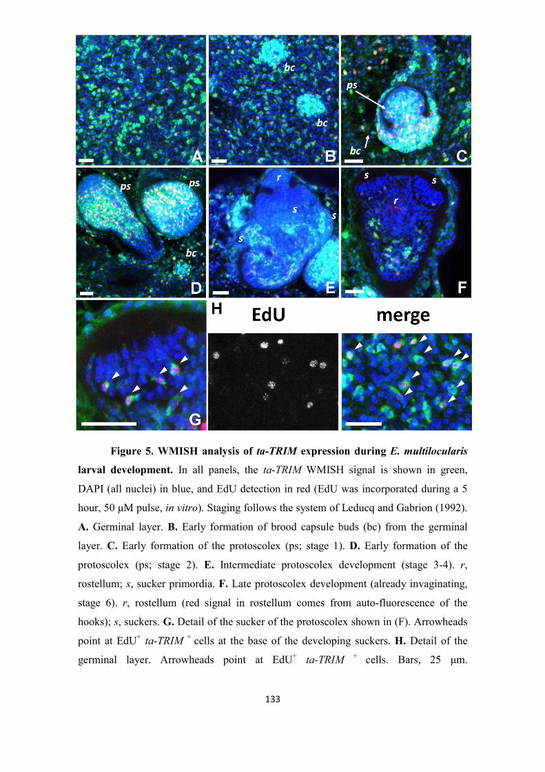
5. Results
133
Figure 5. WMISH analysis of ta-TRIM expression during E. multilocularis
larval development. In all panels, the ta-TRIM WMISH signal is shown in green,
DAPI (all nuclei) in blue, and EdU detection in red (EdU was incorporated during a 5
hour, 50 µM pulse, in vitro). Staging follows the system of Leducq and Gabrion (1992).
A. Germinal layer. B. Early formation of brood capsule buds (bc) from the germinal
layer. C. Early formation of the protoscolex (ps; stage 1). D. Early formation of the
protoscolex (ps; stage 2). E. Intermediate protoscolex development (stage 3-4). r,
rostellum; s, sucker primordia. F. Late protoscolex development (already invaginating,
stage 6). r, rostellum (red signal in rostellum comes from auto-fluorescence of the
hooks); s, suckers. G. Detail of the sucker of the protoscolex shown in (F). Arrowheads
point at EdU+ ta-TRIM + cells at the base of the developing suckers. H. Detail of the
germinal layer. Arrowheads point at EdU+ ta-TRIM + cells. Bars, 25 µm.

5. Results
134
Discussion
Convergent reductive evolution of LTR-retrotransposons into TRIMs
In this work, we describe a new family of TRIM elements that is found
exclusively in taeniid cestodes, and has expanded during the divergence of this family.
So far, TRIM-like elements have been described in plants (Witte et al. 2001; Kalendar
et al. 2008), fungi (Dos Santos et al. 2012) and in two distant metazoan groups: ants
(Zhou and Cahan 2012), and taeniid cestodes (this work). The TRIM elements of each
of these taxonomic groups show no similarity to each other, and are therefore a likely
case of convergent reductive evolution, in which different retrotransposons are stripped
of all non-essential sequences as they parasitize autonomous elements for
retrotransposition (Schulman 2012). As such, TRIM-like elements are probably widely
present in the genomes of other groups, but their discovery is difficult since they
possess no similarity to other TRIM-like elements, have only short LTRs, and lack
coding sequences. In our case, the discovery was prompted by the high expression of
these elements in cestodes, but silent or lowly expressed TRIMs would easily escape
detection.
The distribution of individual TRIM families in animals seems to be more
restricted than in plants, since similar TRIM elements are found throughout mono and
dicotyledons (Witte et al. 2001; Kalendar et al. 2008). Interestingly, very similar TRIMs
have been described in distant ant species and the tree of TRIM elements in ants shows
no correspondence with the species tree, suggesting horizontal transfer events between
species (Zhou and Cahan 2012). In sharp contrast, we find that phylogenetic clades of
ta-TRIMs are specific to individual taeniid lineages (Figure 2A). This is strong evidence
against horizontal transference of these elements, and suggests that independent
expansions of ta-TRIMs occurred in each lineage. Further support for this comes from
the fact that the distribution of estimated insertion dates are significantly different for
ta-TRIMs of each species investigated (Figure 2C), and that specific insertions occurred
after the divergence of closely related species (Figure 2B). However, we cannot rule out
the possibility that the phylogenetic pattern is caused by extensive gene conversion
between ta-TRIM elements in each genome. In general, ectopic gene conversion would
not be expected to maintain sequence similarity for such a large number of elements

5. Results
135
widespread throughout the chromosomes (Chen et al. 2007). However, it has been
shown that LTR-retrotransposons in Saccharomyces cerevisiae can undergo ectopic
gene conversion between the genomic DNA copies and the cDNA intermediates
(Melamed, Nevo, and Kupiec 1992), and similar mechanisms have been proposed to
mediate gene conversion between non-linked loci in other organisms (Kass, Batzer, and
Deininger 1995; Benovoy and Drouin 2009). Therefore, high levels of rtnDNA could
result in genome-wide gene conversion events. Furthermore, we observed evidence for
ongoing gene conversion in T. solium. At this point, it is not possible to decide between
both explanations for the observed phylogenetic pattern, but large scale studies of
synteny of ta-TRIMs in all the species may be able to distinguish between them, once
less fragmentary genomic assemblies are available.
Except for a few cases in which the retrotransposon and the NA-element showed
extensive sequence similarity (e.g. the LARD Dasheng and the retrotransposon RIRE-2
in rice, (Jiang, Jordan, and Wessler 2002)), few pairs of NA-elements and their possible
mobilizing retrotransposons in trans have been proposed. In the case of TRIMs, no
candidate has been identified so far. It is possible that in TRIMs, extensive sequence
reduction has limited similarity to a few key positions for interaction with RT and the
primer tRNA (the PBS and PPT) and with INT (the ends of the LTRs), and that most of
the specificity is achieved by the specific packaging of the TRIM RNA into the VLP
particle (Friedl et al. 2010). Packaging is dependent on the PSI element, which is only
conserved at the secondary structure level (Wicker et al. 2007) and therefore difficult to
identify. Based on this, we propose that the element lennie may have been the factor
mobilizing ta-TRIMs in E. multilocularis. The absence of identifiable intact lennie
elements in the genome could explain the lack of recent retrotransposition events in E.
multilocularis, as well as the absence of gene conversion events (since no cDNA copies
would be produced). Furthermore, it would be expected that competition with highly
expressed ta-TRIM elements could drive the extinction of an autonomous
retrotransposon group such as lennie, as the rate of their own retrotransposition
decreases and the existing copies accumulate deleterious substitutions (Schulman 2012).
In Taenia spp., the different size of TSDs as compared with E. multilocularis gives
evidence that a different element may be the mobilizing factor in trans, since TSD size
is specific for each IN group (Zhou et al. 2001; Ballandras-Colas et al. 2013). This
5. Results
136
implicates a unique shift of “host” (mobilizing factor) during the evolution and
divergence of ta-TRIMs in taeniids.
Expression of ta-TRIM elements and the generation of new transcripts
The EST evidence suggests that in all the analyzed taeniid species, TRIMs are
transcribed at very high levels (> 1 % of all polyadenylated RNAs). Furthermore, the
presence of solo-LTRs has a clear effect in the expression of downstream sequences in
E. multilocularis, resulting in many new lncRNA, and in alternative promoters for
coding genes (Figures 3 and 4). Therefore, although the elements may no longer be
active for retrotransposition in E. multilocularis, they have extensively modified its
transcriptome. The evidence we show here is very conservative: more examples of
lncRNA originating from solo-LTRs can be found when using slightly lower stringency
values for the mapping of ESTs (data not shown), and it is likely that many more
examples will be found with larger datasets, given the number of positions (> 1000)
with similarity to ta-TRIMs in the genome. Usage of retrotransposons as alternative
promoters and as a source of new lncRNA has been described for many individual
examples, and also at a large global scale for model organisms such as humans, mice
and Drosophila (Peaston et al. 2004; Cohen, Lock, and Mager 2009; Faulkner et al.
2009; Kelley and Rinn 2012; Lenhard, Sandelin, and Carninci 2012; Batut et al. 2013).
It has been proposed that this can result in evolutionary innovation in the expression
patterns of the involved genes, and may lead to the coordinately regulated expression of
various genes, as they acquire the expression pattern of the invading retrotransposons
(Peaston et al. 2004; Batut et al. 2013). The finding of splicing between a solo-LTR and
an ancestral trans-splicing acceptor site (Figure 4C) provides a novel and simple
evolutionary mechanism by which LTR retrotransposons can be “captured” as
alternative promoters. In this model, the pre-existing trans-splicing acceptor site (as
found in E. granulosus) is spliced to an appropriate splice-donor site originating from
the new LTR. This donor site may be there by mere chance, since it does not need to be
a particularly strong one (e.g. a simple 5´-GT-3´ motif). This is because the spliceosome
has a “donor-first” syntaxis, in which a cis-splicing site efficiently out-competes the
spliced-leader for a downstream splicing acceptor site (Hastings 2005).

5. Results
137
The generation of lncRNA from solo-LTRs seems to be a simple consequence of
downstream transcriptional readthrough. However, lncRNAs have been recently shown
to have many functions for the regulation of gene expression in trans, and the
transcription of lncRNA itself may also alter gene expression in cis (Mercer, Dinger,
and Mattick 2009; Kornienko et al. 2013). Therefore, some of these new lncRNAs may
have been exapted for new functions in E. multilocularis.
The differences in ta-TRIM transcriptional fusions between E. multilocularis and
E. granulosus suggest that they could lead to differences in gene regulation between
both species. Furthermore, because of the stem cell specific expression of ta-TRIMs
(Figure 5), this could lead to novel stem-cell specific transcripts. Future comparative
analyses of the ta-TRIM derived transcriptome of both species may identify many more
examples. It is possible that the differences in ta-TRIM derived transcripts between
Echinococcus spp. have contributed to some of the important differences in morphology
and development found between these closely related species (Thompson 1986),
particularly since the gene complement of both species is almost identical (Olson et al.
2011; Tsai et al. 2013).
ta-TRIMs as a germinative cell marker in E. multilocularis
The WMISH experiments (Figure 5) provide very strong evidence of a
germinative cell specific expression of ta-TRIMs throughout the development of the
metacestode larva from the cyst wall to the mature protoscolex. The germinative cells of
E. multilocularis larvae are a morphologically homogeneous population of
undifferentiated cells, which however show heterogeneity at the molecular level in the
expression of conserved stem cell regulators such as nanos and argonaute genes
(Koziol et al. 2014). This suggests that there may be in reality several sub-populations
with different proliferation and/or self-renewal potencies. In contrast, it seems that ta-
TRIMs are expressed in almost all of the morphologically defined germinative cells, and
are therefore the best molecular marker so far for the total germinative cell population.
The small number of EdU+ ta-TRIM - cells (ca. 5 % of all EdU+ cells) could indicate the
existence of a different small sub-population of germinative cells, or could be the result
of fluctuating silencing of these elements in the germinative cells. At this point, nothing

5. Results
138
is known about the mechanism by which cestodes silence MGEs in the absence of a
canonical Piwi pathway (Skinner et al. 2014), but a comparison of the chromatin
structure and histone modifications between ta-TRIM loci and autonomous, silenced
retrotransposons may provide a first experimental strategy towards its elucidation. It is
possible that taeniid cestodes control the expansion of ta-TRIMs by simply repressing
the autonomous element mobilizing ta-TRIMs in trans. This would explain the absence
of strong deleterious effects in the face of massive ta-TRIM expression.
From an evolutionary point of view, expression of ta-TRIMs in somatic stem
cells which will eventually form all of the tissues of the next life stage (including the
germ line) would allow the expansion and transmission of ta-TRIMs in the genome of
the next generation. At this point, we do not know if ta-TRIMs are also expressed in the
germ line in the gonads of the adult stage. However, published RNAseq data (Tsai et al.
2013) show expression in the adult stage, suggesting that this is possible (data not
shown). It would be interesting to determine if in planarians, which can also generate
the germ line from somatic stem cells after embryonic development, specific
retrotransposon families also show a stem-cell specific expression.
The expression of retrotransposons in somatic stem cells of E. multilocularis is
analogous to the highly specific expression of several endogenous retrovirus families
during early mammalian development, before the specification of the germ line (Brulet
et al. 1985; Evsikov et al. 2004; Peaston et al. 2004), as well as in the germ line itself
(Dupressoir and Heidmann 1996): only expression at these stages may result in the
expansion of endogenous retroviruses in the genome of the following generation.
Indeed, expressed endogenous retroviruses have been shown to be excellent markers for
totipotency or pluripotency in the early mammalian embryo and in embryonic stem cells
(Santoni, Guerra, and Luban 2011; Macfarlan et al. 2012). It has been shown that many
genes specifically expressed in pluripotent embryonic cells are transcribed from similar
upstream LTR promoters (Macfarlan et al. 2012; Fort et al. 2014), and this has been
proposed to result in the concerted expression of genes important for pluripotency. The
widespread presence in E. multilocularis of transcription from solo-LTRs and their
presumably stem-cell specific expression suggest that similar mechanisms may be at
play in the stem cells of taeniid cestodes.

5. Results
139
Materials and methods
ESTs and genomic assemblies.
Genomic assemblies from Echinococcus multilocularis, Echinococcus
granulosus, Taenia solium and Hymenolepis microstoma (Tsai et al. 2013) as well as
Schistosoma mansoni (Berriman et al. 2009) were downloaded from the GeneDB
database at the Wellcome Trust Sanger Institute (genedb.org). Draft assemblies of
Taenia asiatica, Taenia taeniaeformis, Mesocestoides corti and Diphyllobothrium latum
were generated by the Parasite genomics group of the Wellcome Trust Sanger Institute
in the context of the 50 helminth genomes initiative, and are available at
ftp://ftp.sanger.ac.uk/pub/project/pathogens/HGI. ESTs from Taenia solium were
downloaded from GenBank, and ESTs from Echinococcus spp. were collected from
GenBank, GeneDB and the Echinococcus Full-Length cDNA project
(http://fullmal.hgc.jp/index_em_ajax.html).
Search for ta-TRIM ESTs and genomic loci
Initially, we performed blastn searches of Echinococcus spp. ESTs and genomic
loci with similarity to the previously described Cluster A of lncRNAs of E. granulosus
(Parkinson et al. 2012), as well as against the EgRep repetitive element (Marin et al.
1993). Once the loci were recognized due to their characteristics as possible TRIMs,
complete ta-TRIM elements from E. multilocularis were used for blastn searches in
other cestodes and flatworms. Because of the divergence between ta-TRIM sequences in
different taeniid species, the manually identified ta-TRIM sequences from each species
were used for new blastn searches, in the same as well as in other species. A list of
complete and almost complete elements was extracted for each species from these
blastn results, which was used for analysis of TSD sites, LTR divergence and
comparison of synteny between species. A longer list containing all fragments longer
than 800 bp from each species was compiled for phylogenetic analyses.
5. Results
140
ESTs of E. multilocularis with similarity to ta-TRIMs were mapped by blastn,
eliminating all hits with < 99 % identity to the genome or smaller than 50 bp, as well as
those which mapped to more than one region in the genome with > 99 % identity. The
resulting loci, plus a duplicated genomic locus which was supported from 3´RACE
experiments, were collected and manually analyzed to determine if they were complete
ta-TRIMS or solo-LTRs, whether transcription was likely initiated from within an LTR,
and whether there was downstream transcriptional readthrough into neighboring
intergenic regions or coding genes.
Identification of the ta-TRIM primer binding site (PBS).
A list of E. multilocularis tRNA genes was generated from the genomic
assembly with tRNAscan-SE (Lowe and Eddy, 1997). The 3´ region of these tRNAs
was compared to the region immediately downstream of the 5´ LTR. Only a family of LeutRNA genes was identified as having complementarity for 8 or more bases in this
region.
Phylogenetic analysis
An alignment of complete ta-TRIM elements and fragments longer than 800
bases from all species was performed with ClustalW (Thompson, Higgins, and Gibson
1994), and the region corresponding to the 5´ LTR was removed (since it is not
independent from the 3´ LTR sequence). Maximum Likelihood phylogenetic analysis
was performed using MEGA 5.0 (Tamura et al. 2011), under a Kimura 2-parameter
model with gamma distributed sites (K2P+G, gamma parameter = 1), which was the
model that gave the best fit to the data using the “find best DNA/protein models (ML)”
feature.
Comparison of ta-TRIM loci between taeniid species
When comparing ta-TRIM loci for evidence of integration, each locus with a
complete or almost complete ta-TRIM of E. multilocularis was blasted together with 2

5. Results
141
kb of upstream and downstream flanking sequence to the E. granulosus genome. Blast
hits with <80% identity upstream or downstream of the ta-TRIM were discarded, and
the retrieved sequences were aligned with ClustalW and manually inspected. The same
procedure was performed when comparing T. solium and T. asiatica.
Search for gene conversion
Alignments of ta-TRIMs for each taeniid species were analyzed using the
program GENECONV (Sawyer 1999), which looks for statistically significant tracts of
identity between two sequences, given their overall divergence. Global p-values below
0.05 were considered significant.
Estimates of substitution rates from introns and divergence between LTRs
Introns from the elp and pold genes from E. vogeli and E. oligarthrus (Knapp et
al. 2011) were retrieved from GenBank and aligned using ClustalW. The first 10 and
the last 30 bases of each alignment were discarded (to remove functional splice sites,
Hoffman and Birney 2007) and the substitutions per site (transitions and transversions
only) under a K2P+G model were estimated using MEGA 5.0 (Tamura et al. 2011). The
intron neutral substitution rate was estimated from the average of K2P+G estimates of
both genes using the formula r = s / 2t, where r is the substitution rate, s the estimated
substitutions per site, and t the time of divergence (set as 3 million years for E. vogeli
and E. oligarthrus). For E. multilocularis, E. granulosus and T. solium, divergence
values were also estimated for other well characterized genes (emmpk2, emsmadC,
emmpk1, emraf, emegfr, and emir1 from E. multilocularis (Spiliotis, Kroner, and Brehm
2003; Spiliotis et al. 2005; Spiliotis et al. 2006; Gelmedin, Caballero-Gamiz, and Brehm
2008; Zavala-Gongora et al. 2008; Hemer et al. 2014), egpum1 from E. granulosus
(Koziol, Marin, and Castillo 2008), and their orthologs retrieved from the other
species). When comparing T. solium and E. multilocularis, only the shortest introns
showing trustworthy alignments were included, and therefore the divergence values
between both species are conservative.

5. Results
142
The 5´ LTR and 3´ LTR was aligned for each complete and almost complete ta-
TRIM element from each species. The actual per site divergence, as well as the total
estimated substitutions per site (transitions and transversions only) under a K2P+G
model, were calculated using MEGA 5.0 (Tamura et al. 2011).
3´ Rapid amplification of cDNA ends (RACE) and RT-PCR.
Reverse transcription was performed with 700 ng of total RNA from in vitro
cultured E. multilocularis larvae (Spiliotis and Brehm 2009) using Prime-Script reverse
transcriptase (Takara) as instructed by the manufacturer, with the primer
AAGCAGTGGTATCAACGCAGAGTAC-T30-VN. RT-PCRs were performed with 2
µl of cDNA per reaction using KOD polymerase (Millipore). For 3´ RACE, a semi
nested RT-PCR approach was performed, using degenerate forward nested primers for
the U5 region of several ta-TRIMs (TGTGTCTTCTTTCGTNTTCAGGGAG and
TCAGGGAGTCYYGGGAYGCTACA for the first and second PCR reactions,
respectively) and the reverse primer AAGCAGTGGTATCAACGCAGAGTAC. For
confirming the transcriptional fusions between solo-LTRs and downstream coding
genes, the following primer combinations were used: locus 8,
TTCGTCTTCTTTCGTCTTCAGAGAG and GCATCCTTGATCGAAGTTTGGG
(fusion to EmuJ_000118100); locus 21, CTTTTGTACTTTGAGTTAGCCCCTTGTAC
and CCATGGCGAAATCGACCAC (fusion to EmuJ_000465100); locus 60,
CCTTGTACCTAGCTAAGAGGGCTGAC and CGACGTAGGCACTCAAGCAAG
(fusion to EmuJ_001025350); locus 2 (the only one for which RT-PCR was
unsuccessful, due to unspecific amplification from another ta-TRIM element),
CCGAGTATTGTGTCTTCTTTCGTCTTC and CGGAATGACATTTGGCAAAGTC
(fusion to EmuJ_000054900). All products of the expected size were gel-purified,
cloned into pJET1.2 (Thermo-Scientific), and several clones were sequenced for each
product.

5. Results
143
Whole-mount in situ hybridization (WMISH)
Fluorescent WMISH of in vitro cultured E. multilocularis larvae was performed
with a digoxigenin-labeled antisense probe, corresponding to the region interior to the
LTRs of the ta-TRIM locus 39 of E. multilocularis (Supplementary Data 3), as
described (Koziol et al. 2014). Primers TTGGTGGCAGCGGAAAGC and
CCTCTTTTGAGTGTTATCCCCAGC were used to amplify the probe region from
genomic DNA of E. multilocularis, and the product was cloned into pJET1.2 (Thermo-
Scientific). Three independent experiments with different laboratory isolates were
performed. These isolates were GH09 and Ingrid/10, obtained from accidental
infections of Old World Monkeys in a breeding exclosure (Tappe et al. 2007), and
MS10/10, obtained from an infected dog. All isolates had been kept in the laboratory by
serial peritoneal injection into Meriones unguiculatus for 4 years or less. Control
WMISH experiments using the corresponding sense probe were always negative (data
not shown). In vitro EdU labeling (50 µM for 5 hours) and detection were performed as
previously described (Koziol et al. 2014).

5. Results
144
Supplementary data
Supplementary Data 1. Alignment of examples of ta-TRIM elements from taeniids.
Supplementary Data 2. Target Duplication Sites (TSDs) found in ta-TRIM
elements from taeniids. Abbreviations are as for Figure 2A.
Supplementary Data 3. List of E. multilocularis loci with similarity to ta-TRIMs
and with evidence of transcription from ESTs and 3’ RACE analyses. Locus
number and the position of ESTs and 3’ RACE sequences (mapped by blastn analyses)
are indicated, as well as the manual annotation of the loci. Note that one EST may have
several discontinuous blast hits due to the presence of introns.
Supplementary Data 4. Distribution of ta-TRIM-like sequences in E. multilocularis
chromosomes and unplaced contigs.
Supplementary Data 5. Examples of integrations of ta-TRIMs after the divergence
of Echinococcus spp., and of Taenia spp. Alignment of a ta-TRIM element and its
surrounding region from E. multilocularis with the ortholog region of E. granulosus,
and of a ta-TRIM element and its surrounding region from T. solium with the ortholog
region of T. asiatica.
Supplementary Data 6. Example of lennie element from E. multilocularis.

5. Results
145
5.3. CHAPTER 3: Further experimental results regarding the E. multilocularis
stem cell system.
In this chapter, I will describe further experiments that I performed for the
characterization of the Echinococcus multilocularis stem cell system, which were not
included in the previous articles. Some of these results were included within other
articles of which I am a co-author, as indicated.

5. Results
146
5.3.1. The cell-cycle kinase em-plk1 is expressed in E. multilocularis germinative
cells
Polo-like kinases (PLKs) are a family of Serine/Threonin kinases with important
roles in the regulation of cell-cycle progression in eukaryotes (Archambault and Glover
2009). Within this family, PLK-1 is specifically expressed during the late G2 and M
phases of the cell-cycle, and has a key conserved role in regulating the transition
through mitosis (Winkles and Alberts 2005). PLK-1 homologs were also shown to be
expressed in the neoblasts of planarians and neoblast-like cells of the trematode
Schistosoma mansoni (Labbe et al. 2012; Collins et al. 2013).
During his doctoral thesis, Andreas Schubert characterized em-plk1, the E.
multilocularis ortholog of plk-1. Because in E. multilocularis only the germinative cells
proliferate, it was likely that em-plk1 was specifically expressed in these cells. As part
of the characterization of em-plk1, I determined its gene expression pattern during E.
multilocularis larval development by means of WMISH (Figure R3.1). Cells expressing
em-plk1 (em-plk1+) were observed dispersed throughout the germinal layer, and
accumulate in early brood capsules and in the proliferative region of developing
protoscoleces. During late protoscolex development, em-plk1+ cells are observed to
accumulate at the base of the suckers, similarly to what is observed for EdU+ and em-
h2b+ cells (see Chapter 1; (Koziol et al. 2014)). Overall, these data strongly support the
hypothesis that em-plk1 is specifically expressed in E. multilocularis proliferating
germinative cells. It is likely that these consist of cells in the late G2 and M phases of
the cell cycle, and as such em-plk1 would be a complementary marker for EdU
incorporation and em-h2b expression (Koziol et al. 2014).
These results have already been published as Schubert et al., 2014, PloS
Neglected Tropical Diseases 8(6): e2870.

5. Results
147
Figure R3.1. WMISH detection of em-plk1 transcripts. A. General view of a
metacestode hybridized with the emplk1 antisense probe. Note the accumulation of em-
plk1 positive cells in the developing protoscoleces (arrows). B. General view of a
metacestode hybridized with the em-plk1 sense control probe. No signal is detected. C-
F. Details of em-plk1 WMISH detection in different developmental stages. C. Detail of
the metacestode germinative layer, in which disperse emplk1 positive cells can be
distinguished (inset: higher magnification of a positive cell, combined with 4',6-
diamidino-2-phenylindole (DAPI) nuclear staining). D. Accumulation of em-plk1
positive cells in the early brood capsule bud (arrow). E. Early protoscolex development,
with abundant emplk1 positive cells in the interior. F. Late protoscolex development.
em-plk1 positive cells accumulate at the base of the developing suckers (arrows). Bars
represent 200 µm (A, B) and 50 µm (C-F). Figure and legend have been previously
published as a part of Schubert et al., 2014.

5. Results
148
5.3.2. The insulin receptor emir2 is expressed in the proliferating region of the
developing protoscolex
Previously, it was shown in our laboratory that physiologically relevant
concentrations of human insulin can promote cell proliferation, glucose uptake and
metacestode growth and regeneration of E. multilocularis in vitro. Two insulin receptor
homologs (emir1 and emir2) were found in the E. multilocularis genome, and both were
shown to bind to human insulin in yeast-two hybrid experiments. Furthermore, addition
of insulin induced tyrosine phosphorylation of EmIR1 and phosphorylation of
conserved canonical downstream targets. In sum, the experiments showed that insulin
can promote the development of the parasite, and that this is probably mediated through
the activation of conserved canonical insulin receptors and signaling pathways (Hemer
et al. 2014).
The EmIR1 protein was shown to be expressed at low levels in the germinative
cells, and at particularly high levels in the glycogen storage cells. It was therefore
proposed that EmIR1 mediated the increase of glucose uptake after insulin stimulation
(Hemer et al. 2014). Expression of EmIR2 was highest in primary cell preparations as
compared to metacestodes and protoscoleces, and it was proposed that it could be
expressed in the germinative cells, although immunohistofluorescence experiments
were not conclusive in this regard. As a complementary approach, I studied the
expression of emir2 mRNA by WMISH during metacestode and protoscolex
development. No expression could be detected in the germinal layer, but strong
expression could be seen in the proliferative region (rich in EdU+ cells) during early
protoscolex development (Figure R3.2). Although the signal was not defined enough to
confirm that individual EdU+ cells were also emir2+, these results indicate that emir2
may be specifically expressed in parasite stem cells or, at least, in parasite tissues that
are actively proliferating during development. These results were previously published
as Hemer et al. (2014), BMC Biology 12:5.

5. Results
149
Figure R3.2. WMISH detection of emir2. Germinal layer with developing
protoscoleces stained for emir2 WMISH (upper panel), EdU incorporation (mid panel)
and DAPI (lower panel). Sense (left) and antisense (right) probes have been used as
indicated. Bar represents approximately 50 µm. Figure and legend have been
previously published as a part of Hemer et al., 2014.

5. Results
150
5.3.3. Expression of FGF receptor homologs of E. multilocularis
Previous results from Dr. Sabine Förster demonstrated that human FGF-1 and
FGF-2 are capable of stimulating cell proliferation, growth and regeneration in E.
multilocularis metacestodes and primary cells (Förster 2012). Furthermore, two genes
coding for FGF receptors (FGFRs) were previously found in the E. multilocularis
genome: emfr1 (previously denominated emfr) and emfr2 (Förster 2012). However, no
canonical FGF ligand could be found (Förster 2012). Both EmFR1 and EmFR2 were
shown to be activated by human FGF-1 and FGF-2 after heterologous expression in the
Xenopus oocyte system. Therefore, it was concluded that human FGF ligands can
activate the cognate FGFRs of E. multilocularis, and that this promotes metacestode
growth and development (Förster 2012).
Given that genes coding for FGFRs are expressed in stem cells in other
flatworms (Ogawa et al. 2002; Collins et al. 2013), and that human FGF ligands induce
metacestode growth and development, I investigated in this work the expression
patterns of FGFR genes during E. multilocularis larval development, and performed
phylogenetic analyses in order to establish orthology relationships among flatworm
FGFRs. While searching for other tyrosine kinase receptors for constructing the
phylogenetic tree, I discovered a third FGFR in E. multilocularis, emfr3, which was
previously unnoticed (since it had not been included in the original automatic gene
predictions). Sequence analysis after cloning of the complete coding domain sequence
(CDS, corrected in the 3´ region from the original gene model) showed that the
predicted EmFR3 protein has all of the typical FGFR domains and motifs, including
(from N-terminal to C-terminal): a signal peptide, at least two immunoglobulin-like
domains, a single-pass transmembrane region, and a tyrosine kinase (TK) domain
(Figure R3.3). The CDS of emfr3 was sub-cloned (minus the signal-peptide coding
region) into pSecTag2/HygroA. This construct was used by C. Dissous and colleagues
(Center for Infection and Immunology, Institut Pasteur de Lille, France) for expression
in the Xenopus oocyte system, by which they demonstrated that EmFR3 is also able to
interact with human FGF-1 and FGF-2 (C. Dissous, personal communication to K.
Brehm).

5. Results
151
Phylogenetic analyses of flatworm FGFR genes had to be restricted to
alignments of the TK domain, since other regions did not show sufficient sequence
conservation for reliable alignments to be made. This limited the information for
phylogenetic inference, resulting in low support values throughout most of the tree.
However, one clearly supported node linked emfr3 with the Schistosoma mansoni stem
cell-specific fgfrA (Collins et al. 2013), indicating that both genes are orthologs (Figure
R3.4).
Figure R3.3. Diagram of the domain organization of EmFR3. From N-
terminal to C-terminal: SP, signal peptide; Ig, immunoglobulin-like domain; TM, trans-
membrane region; TK, tyrosine kinase domain. Total length is 883 amino acid residues.
Figure R3.4. Phylogeny of FGFRs from flatworms. The TK domain of
FGFRs from E. multilocularis (EMFR1-3), Schistosoma mansoni, planarians (Dugesia
japonica and Schmidtea mediterranea), and FGFR1 of Homo sapiens were aligned, and
a neighbor joining phylogenetic analysis was performed with MEGA5.0 using the JTT
matrix-based substitution model. Numbers next to the nodes are bootstrap support
values (1000 replicates). GeneDB codes (for E. multilocularis and S. mansoni) or NCBI
accession codes (for planarians and H. sapiens) are given in parenthesis.

5. Results
152
The three receptors showed very different expression patterns in E.
multilocularis larval stages. emfr1 was expressed at low levels throughout the germinal
layer and especially during the development of protoscoleces (Figure R3.5). The
intensity of the signal was heterogeneous, but no clear pattern could be discerned and it
was too low to determine with certainty the percentage of positive cells.
Figure R3.5. WMISH analysis of emfr1 expression. Left panel, sense probe
(negative control). Right panel, antisense probe. gl, germinal layer; ps, developing
protoscolex. Bar: 20 µm.
In contrast, emfr2 was specifically expressed in a small population of cells,
typically of small size, which comprised 1.7% to 6.3% of all cells in the germinal layer
(two independent WMISH experiments with isolates H95 and GH09, respectively;
Figure R3.6, B-C). Furthermore, none of the emfr2+ cells were EdU+, indicating that
these cells were out of the cell-cycle, and therefore not proliferating germinative cells
(n>200 cells from two independent WMISH experiments; Figure R3.7, A). During
initial protoscolex development, emfr2+ cells accumulate in the peripheral-most layer of
cells, as well as in the anterior-most region (which will give the rostellum), and in three
longitudinal bands of cells in the interior of the protoscolex buds (Figure, R3.6, B and
D). Practically none of these cells are EdU+ (<< 1% of emfr2+ cells are EdU+, Figure
R3.7, B). Later, some emfr2+ cells are found in the protoscolex body, but most

5. Results
153
accumulate in the developing rostellum and suckers (Figures R3.6, B and R3.7, C-D).
Importantly, emfr2 expression is restricted to the sucker cup, where cells are
differentiating into em-tpm-1.hmw+ muscle cells (Koziol et al. 2014), and not to the
sucker base, were EdU+ germinative cells accumulate ((Koziol et al. 2014); Figure R3.7,
C-D). In summary, emfr2 is expressed in cells that are not actively cycling, and many of
them are likely to be differentiating and/or differentiated muscle cells.
Figure R3.6. WMISH analysis of emfr2 expression during metacestode
development. In all panels the WMISH signal is shown in green, and DAPI nuclear
staining is shown in blue. A. Sense probe (negative control). B. General view of a
region of a metacestode with protoscoleces in different stages of development. C. Detail
of the germinal layer. D. Detail of a protoscolex during early development (ca. stage 2
of Leducq and Gabrion, 1992). ps, protoscolex; r, rostellum; s, sucker. Bars: 50 µm for
A, B and D, 20 µm for C.

5. Results
154
Figure R3.7. Co-detection of emfr2 WMISH and EdU incorporation during
metacestode development. Metacestodes were cultured in vitro with 50 µM EdU for 5
hours and were then processed for WMISH and EdU detection. In panels displaying
multiple channels, the WMISH signal is shown in green, EdU detection in red, and
DAPI nuclear staining in blue. A. Germinal layer. B. Early protoscolex development. C.
Late protoscolex development. D. Detail of a sucker from the same specimen as in A,
maximum intensity projection of a confocal Z-stack. r, rostellum; s, sucker. Bars: 10 µm
for A and B, 50 µm for C.
Finally, emfr3 is expressed in very few cells in the germinal layer (<<1% of all
cells, although the number is difficult to estimate since they are absent in most random
microscopy fields). emfr3+ cells accumulate in small numbers during brood capsule and
protoscolex development (Figure R3.8, B-E). Particularly, emfr3+ cells can be found in
characteristic positions at the base of the stalk connecting the developing protoscolex to
the brood capsule and in the periphery of the brood capsule itself, as well as in the body
of the developing protoscolex. The emfr3+ cells have a large nucleus and nucleolus (as

5. Results
155
compared for example with the emfr2+ cells) and appear to have the morphology of
germinative cells (Figure R3.8, E). Finally, a few emfr3+ cells are present in the body
region at the final stages of protoscolex development, and some signal can be seen in
the rostellar region as well (Figure R3.8, F). In the developing protoscolex and in the
germinal layer, emfr3+ EdU+ double positive cells can be found (Figure R3.9).
Therefore, emfr3 is expressed in a very small number of proliferating cells with the
morphology of germinative cells, indicating that it labels a minor germinative cell sub-
population.
In summary, FGFR genes of E. multilocularis showed important differences to
each other in their complex expression patterns, and also to the expression in most or all
neoblasts of planarians and neoblast-like stem cells of Schistosoma mansoni.
Figure R3.8 (next page). WMISH analysis of emfr3 expression during
metacestode development. In all panels the WMISH signal is shown in green, and
DAPI nuclear staining is shown in blue. A. Sense probe (negative control). B. Early
brood capsule formation. C. Early protoscolex formation. D. Detail of early protoscolex
formation, showing an emfr3+ cell in the region of the stalk connecting the developing
protoscolex to the brood capsule. E. Detail of early protoscolex formation, showing the
morphology of an emfr3+ cell (inset: only DAPI channel is shown). F. Late protoscolex
formation. bc, brood capsule; gl, germinative layer: ps, protoscolex; r, rostellum; s,
sucker; st, stalk. Bars: 25 µm for B, C, D and F; 10 µm for E.

5. Results
156
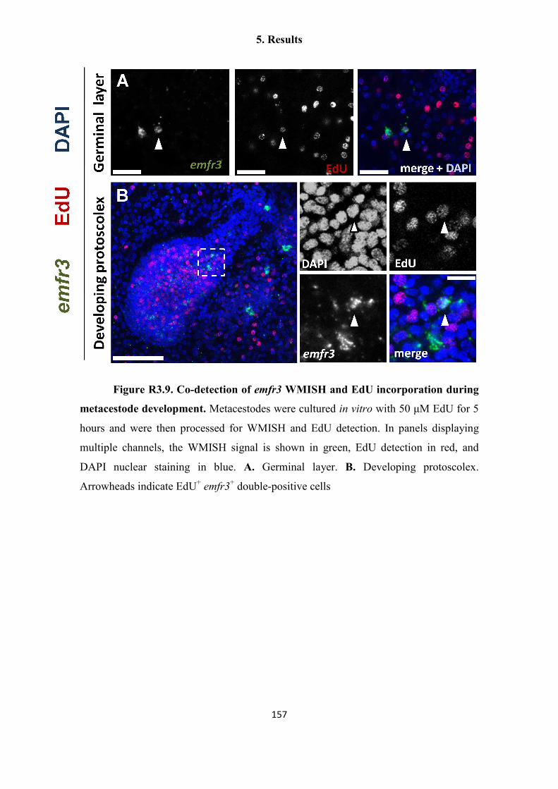
5. Results
157
Figure R3.9. Co-detection of emfr3 WMISH and EdU incorporation during
metacestode development. Metacestodes were cultured in vitro with 50 µM EdU for 5
hours and were then processed for WMISH and EdU detection. In panels displaying
multiple channels, the WMISH signal is shown in green, EdU detection in red, and
DAPI nuclear staining in blue. A. Germinal layer. B. Developing protoscolex.
Arrowheads indicate EdU+ emfr3+ double-positive cells

5. Results
158
5.3.4. Transcriptomic analysis of HU treated metacestodes – A first glimpse of the
germinative cell transcriptome
The previously described gene expression studies of E. multilocularis
germinative cells were performed based on a candidate-gene approach, by selecting
conserved stem cell regulators. However, in order to have a more general, bias-free
view of gene expression in E. multilocularis germinative cells, we performed
transcriptomic analyses by high-throughput RNA sequencing (RNAseq) of vesicles
treated in vitro for 7 days with 40 mM hydroxyurea, versus untreated control vesicles
(HU versus control groups, both under axenic culture conditions). The rationale behind
this approach is that HU specifically depletes the germinative cells after 7 days, but has
no appreciable effect on the number of differentiated cells (Koziol et al. 2014).
Therefore, many genes specifically depleted in the HU group could be germinative cell-
specific.
We performed three biological replicates of comparisons between HU and
control groups, which were processed for RNAseq by our collaborators in the Wellcome
Trust Sanger Institute (Matt Berrimann and Magdalena Zarowiecki, Cambridge, United
Kingdom). M. Zarowiecki performed a preliminary analysis, searching for significantly
depleted genes in the HU group, and analyzed this gene set for significant enrichment of
Gene Ontology (GO) groups (Table R3.1). In total, 154 genes were less abundantly
expressed in the HU group as compared with the controls (although this number is still
preliminary). Interestingly, most of the GO terms showing the highest significance were
related to cell proliferation, cell cycle regulation, DNA repair and cell fate commitment,
which strongly suggested that within the list of HU-depleted genes there is a strong
signature of germinative cell-specific genes.
We analyzed the preliminary list of down-regulated genes for further
confirmation that germinative cell transcripts could be specifically depleted in the HU
group. Markers of tegumental cells (em-muc-1, em-alp-1 and em-alp-2) showed no
significant difference, nor did em-phb1, em-hdac1, em-ago2-A/B/C and emfr1, which
were shown in the previous experiments to be widely expressed ((Koziol et al. 2014);
see above). In contrast, em-nos-1, em-nos-2, emfr3 and em-plk1, all of which are

5. Results
159
expressed in sub-populations of germinative cells ((Koziol et al. 2014; Schubert et al.
2014); see above), showed a statistically significant depletion in the HU group. emfr2
was also significantly depleted, suggesting that the post-mitotic cells expressing this
gene are mostly differentiating cells.
Few genes related to the germline multipotency program or to post-
transcriptional regulation in general were found to be down-regulated after HU
treatment (with the exception of a bruno homolog, EmuJ_000943000). In particular,
none of the orthologs of the Schistosome “vasa-like genes” sensu Skinner et al. (2012)
(EmuJ_000777250, EmuJ_000098400, EmuJ_001183300) showed a significant change
after HU treatment. These “vasa-like genes” are DEAD box RNA helicases that are
paralogs of vasa, such as PL-10 and p68.
In contrast, the regulators showing the greatest fold-change and smallest p-
values were almost invariably transcription factors (a selected list of interesting
candidate genes is presented as Table R3.2, including some references to their stem cell
and cell differentiation-related functions in other metazoans). Among these, four out of
seven E. multilocularis Sox-family transcription factors (Kamachi and Kondoh 2013)
can be found. Phylogenetic analyses demonstrated that the down-regulated Sox genes
belong to two sub-families (Figure R3.10). One gene belongs to the SoxB sub-family,
which in other metazoans includes both positive and negative transcriptional regulators,
among which is the mammalian ESC pluripotency factor Sox2 (Uchikawa, Kamachi,
and Kondoh 1999; Bowles, Schepers, and Koopman 2000). The other three genes group
with divergent planarian Sox genes which have been shown to be specifically enriched
in neoblasts (Onal et al. 2012; Wagner, Ho, and Reddien 2012).
Besides possible transcriptional and post-transcriptional regulators, I also mined
the list of HU-depleted transcripts for signaling receptors and kinases, which could in
principle be targeted by pharmacological methods for functional analyses, or eventually
as potential therapeutic targets. Few signaling receptors were found in the list of HU-
depleted genes. These include emfr2 and emfr3, but also a member of the Frizzled
family of receptors for Wnt ligands (Clevers 2006) with conserved roles in the
maintenance and expansion of stem cells (EmuJ_000636500). As expected, a large
number of homologs of kinases related to cell-cycle regulation and proliferation are

5. Results
160
among the transcripts with the most pronounced depletion, including em-plk1 (see
above), the polo-like kinase family member PLK-4 (EmuJ_000104700), cyclin-
dependent kinases (CDKs, such as EmuJ_000046100 and EmuJ_000659500), greatwall
kinase (EmuJ_000711100), maternal embryonic leucin zipper kinase
(EmuJ_000500200), aurora kinase A (EmuJ_001059700) and wee1 kinases
(EmuJ_001003100 and EmuJ_000659400). LATS2 (EmuJ_000700100), a kinase of the
hippo signaling pathway that limits and coordinates the growth of tissues and organs
(Zhao, Li, and Guan 2010) was also depleted. Finally, emSSY (EmuJ_000139200) also
showed strong depletion after HU treatment. emSSY is a paralog of the previously
described emmpk2 p38 MAPK (Gelmedin 2008; Gelmedin, Caballero-Gamiz, and
Brehm 2008), but has an unique Ser-Ser-Tyr motif in the kinase activation loop instead
of the Thr-Gly-Tyr motif that is universally conserved for p38 MAPK (Kultz 1998).
emSSY and emmpk2 are clustered together in the genome of E. multilocularis (separated
by only one gene), which suggests that emSSY originated from a recent duplication of
emmpk2 (Gelmedin 2008). By searching in the genomes of other flatworms and
performing phylogenetic analyses, it became clear that this duplication and their
clustered arrangement are specific to cestodes (Figure R3.11). The possibility that this
could be a cestode-specific and germinative cell-specific p38 kinase with atypical
biochemical properties makes it a very interesting target for future studies.

5. Results
161
Table R3.1. Gene Ontology (GO) Analysis of transcripts specifically
depleted after HU treatment (performed by M. Zarowiecki)
GO Term Annotated
genes
Significant
genes
Expected
significant
genes
P-value
GO:0006270 DNA replication
initiation
7 6 0.44 4.00E-07
GO:0007156 homophilic cell
adhesion
41 12 2.59 4.90E-06
GO:0006298 mismatch repair 7 5 0.44 1.80E-05
GO:0007050 cell cycle arrest 64 16 4.04 2.10E-05
GO:0051320 S phase 43 11 2.71 6.50E-05
GO:0006865 amino acid
transport
24 7 1.51 0.00051
GO:0022403 cell cycle phase 174 40 10.98 0.00057
GO:0006275 regulation of DNA
replication
4 3 0.25 0.00095
GO:0006261 DNA-dependent
DNA replication
22 11 1.39 0.0016
GO:0000087 M phase of mitotic
cell cycle
86 16 5.43 0.00193
GO:0000278 mitotic cell cycle 145 26 9.15 0.00199
GO:0006281 DNA repair 106 22 6.69 0.00219
GO:0000082 G1/S transition of
mitotic cell cycle
6 3 0.38 0.00431
GO:0000725 recombinational
repair
17 6 1.07 0.00692
GO:0000077 DNA damage
checkpoint
7 3 0.44 0.0072
GO:0009219 pyrimidine
deoxyribonucleotide
metabolic (...)
3 2 0.19 0.01141
GO:0032515 negative regulation
of phosphoprotein
phosphatase activity
3 2 0.19 0.01141
GO:0032774 RNA biosynthetic
process
748 35 47.18 0.0125
GO:0007067 mitosis 48 8 3.03 0.01496
GO:0045165 cell fate
commitment
19 5 1.2 0.01532
GO:0010564 regulation of cell
cycle process
35 8 2.21 0.02136
GO:0000245 spliceosomal
complex assembly
10 3 0.63 0.02141
GO:0007569 cell aging 4 2 0.25 0.02186

5. Results
162
GO:0060078 regulation of
postsynaptic
membrane potential
4 2 0.25 0.02186
GO:0034502 protein localization
to chromosome
11 3 0.69 0.02809
GO:0021536 diencephalon
development
5 2 0.32 0.03493
GO:0007269 neurotransmitter
secretion
5 2 0.32 0.03493
GO:0010863 positive regulation
of phospholipase C
activity
5 2 0.32 0.03493
GO:0031398 positive regulation
of protein
ubiquitination
5 2 0.32 0.03493
GO:0014902 myotube
differentiation
5 2 0.32 0.03493
GO:0007052 mitotic spindle
organization
5 2 0.32 0.03493
GO:0002064 epithelial cell
development
5 2 0.32 0.03493
GO:0006468 protein
phosphorylation
316 27 19.93 0.03757
GO:0048523 negative regulation
of cellular process
218 27 13.75 0.04062
GO:0044093 positive regulation
of molecular
function
78 9 4.92 0.0455
GO:0006260 DNA replication 103 23 6.5 0.04556
GO:0006259 DNA metabolic
process
427 59 26.94 0.04962

5. Results
163
Table R3.2. Selection of genes coding for conserved transcription factors that are
specifically depleted after HU treatment
GeneDB code GeneDB description Roles of homologs
EmuJ_000098000 bhlh factor math6 bHLH proteins have
widespread roles in cell-fate
determination (Jones 2004),
including math6 (Inoue et al.
2001)
EmuJ_000627500 achaete scute transcription
factor
Regulates ISC fate (van der
Flier et al. 2009)
EmuJ_000344700 neurogenic differentiation
factor 1
bHLH proteins have
widespread roles in cell-fate
determination (Jones 2004)
EmuJ_000215900 GATA binding factor 2 GATA factors 1-3 are
essential for hematopoiesis,
including HSC/progenitor
function and differentiation
(Bresnick et al. 2012)
EmuJ_000457300 DNA binding protein inhibitor
ID 4
ID proteins inhibit
differentiation and promote
proliferation of stem cells
(Yokota 2001)
EmuJ_000909600 MYB MYB proteins regulate cell
proliferation and
differentiation (Oh and
Reddy 1999; Prouse and
Campbell 2012)
EmuJ_000704700 transcription factor Sox1a Sox proteins regulate cell fate
and differentiation (Kamachi
and Kondoh 2013)
EmuJ_000451500 Basic helix loop helix
dimerisation region bHLH
bHLH proteins have
widespread roles in cell-fate
determination (Jones 2004)
EmuJ_000449700 POU specific (from blast,
Class III POU)
POU factors have roles in
stem cell maintenance (e.g.
Oct4) and in lineage
commitment (e.g. Class III
POUs) (Tantin 2013)
EmuJ_000191100 transcription factor SOX 14 Sox proteins regulate cell fate
and differentiation (Kamachi
and Kondoh 2013)
EmuJ_000232900 transcription factor SOX 6 Sox proteins regulate cell fate
and differentiation (Kamachi
and Kondoh 2013)
EmuJ_000437800 transcription factor SOX 14 Sox proteins regulate cell fate
and differentiation (Kamachi
and Kondoh 2013)

5. Results
164
Figure R3.10. Phylogeny of Sox proteins from E. multilocularis. The HMG
domain of Sox proteins from E. multilocularis (Emu), Homo sapiens (Hs, from Bowles,
Schepers, and Koopman 2000), and published planarian genes (S. mediterranea, Smed)
were aligned, and a neighbor joining phylogenetic analysis was performed with
MEGA5.0 using the JTT substitution model. Numbers next to the nodes are bootstrap
support values (1000 replicates). HU-depleted genes of E. multilocularis are indicated
in parentheses. Blue labeling indicates groups A to H of Sox proteins as defined by
Bowles, Schepers, and Koopman, 2000. Red labeling indicates a divergent group of
flatworm sequences that cannot be easily classified according to the scheme of Bowles,
Schepers and Koopman for vertebrate Sox proteins. Human Tcf1, Tcf3, HMG1 and
HMG2 proteins were used to root the tree. GeneDB codes (for E. multilocularis) or
NCBI accession codes (for S. mediterranea) are given.

5. Results
165
Figure R3.11. Phylogeny of p38 MAPK proteins from neodermatans.
Proteins sequences of the p38 MAPK family from E. multilocularis, (Emu), Taenia
solium (Ts), Hymenolepis microstoma (Hm) and Schistosoma japonicum were aligned
with p38 MAPK sequences of Homo sapiens, together with an ERK MAPK of Homo
sapiens (MAPK1) as an outgroup. Neighbor joining phylogenetic analysis was
performed with MEGA5.0 using the JTT substitution model. Numbers next to the nodes
are bootstrap support values (1000 replicates). The motif found in the kinase activation
loop of cestode proteins is indicated to the right. GeneDB codes (for E. multilocularis,
T. solium and H. microstoma) or NCBI accession codes (for S. japonicum and Homo
sapiens) are given. “div. loop” indicates the cestode-specific group of divergent p38
MAPKs.

5. Results
166
5.3.5. First steps towards the development of clonal analyses of E. multilocularis
germinative cells
The study of the potency of individual E. multilocularis germinative cells will
require clonal analyses, but the techniques required for this are not as yet available (see
also the Discussion section). We therefore set out to develop transplant experiments for
tracing clones originating from individual E. multilocularis cells, after introduction into
a host metacestode. Ideally, genetically manipulated E. multilocularis cells expressing a
marker gene, or different E. multilocularis isolates with specific antigens could be used.
However, transgene integration has not been achieved in E. multilocularis yet, and there
are no known differential antigens between isolates. Instead, inspired by the classic
quail-chick chimera experiments used for cell tracing during chicken embryonic
development (Le Douarin and Teillet 1974), we decided to investigate whether E.
multilocularis primary cells (EmPCs) could be integrated into the metacestode of the
related taeniid species Taenia crassiceps (TcMC).
Previously, Andreas Schubert had shown that T. crassiceps proliferating cells
could integrate into E. multilocularis metacestodes that had been depleted of
proliferating cells by long-term treatment with a PLK-1 inhibitor (A. Schubert, personal
communication). This suggested that no histoincompatibility system was present. As a
proof of principle, an experiment was performed in which a large number of EmPCs
were introduced into TcMC using a simple 26G3/8 syringe. Because TRIM copies are
very divergent between taeniid species (e.g. only ca. 50% identity between E.
multilocularis and T. solium TRIMs), and are highly expressed in the E. multilocularis
germinative cells, we performed WMISH with a specific E. multilocularis ta-TRIM
probe (“emTRIM”) to search for viable E. multilocularis germinative cells that had
integrated into the TcMC tissues. In transplanted TcMC, many emTRIM + cells with
germinative cell morphology could be found after 5 days of culture, sometimes in
patches that suggested growth from only one or a few founding EmPCs (Figure R3.12;
2/4 specimens). No emTRIM + cells were found in control, non-transplanted TcMC (0/3
specimens). In conclusion, EmPCs can integrate into TcMC, and E. multilocularis
germinative cells can be specifically detected within those tissues. Possible applications

5. Results
167
of these results for clonal analysis of E. multilocularis germinative cells are proposed in
the Discussion section.
Figure R3.12. Detection of emTRIM + cells by WMISH after transplantation
of E. multilocularis cells into T. crassiceps. Left, general view of a region of one
transplanted T. crassiceps metacestode. Right, detail of an isolated patch of emTRIM +
cells. Bar: 20 µm.
5. Results
168
5.4. CHAPTER 4: “Anatomy and development of the larval nervous system in
Echinococcus multilocularis”
Originally published as Koziol et al. (2013), Frontiers in Zoology 10:24
Author contributions
General study design:
Uriel Koziol and Klaus Brehm
Experimental design and work:
Uriel Koziol designed, performed or participated in all the experiments. Georg Krohne
designed, supervised and interpreted the results of electron microscopy experiments.
Analysis of the results:
Uriel Koziol, Klaus Brehm, Georg Krohne
Writing of the manuscript:
Uriel Koziol wrote the first manuscript draft, which was corrected by Klaus Brehm and
accepted by all the authors

5. Results
169
Anatomy and development of the larval nervous system in
Echinococcus multilocularis
Uriel Koziol1,3, Georg Krohne 2, Klaus Brehm1,*
1 University of Würzburg, Institute of Hygiene and Microbiology, Josef-Schneider-
Strasse 2, D-97080 Würzburg, Germany
2 University of Würzburg, Department of Electron Microscopy, Biocenter, D-97078
Würzburg, Germany
3 Universidad de la República, Facultad de Ciencias, Sección Bioquímica y Biología
Molecular, Iguá 4225, CP 11400, Montevideo, Uruguay.
* Corresponding Author

5. Results
170
Background
The metacestode larva of Echinococcus multilocularis (Cestoda: Taeniidae) develops in
the liver of intermediate hosts (typically rodents, or accidentally in humans) as a
labyrinth of interconnected cysts that infiltrate the host tissue, causing the disease
alveolar echinococcosis. Within the cysts, protoscoleces (the infective stage for the
definitive canid host) arise by asexual multiplication. These consist of a scolex similar
to that of the adult, invaginated within a small posterior body. Despite the importance of
alveolar echinococcosis for human health, relatively little is known about the basic
biology, anatomy and development of E. multilocularis larvae, particularly with regard
to their nervous system.
Results
We describe the existence of a subtegumental nerve net in the metacestode cysts, which
is immunoreactive for acetylated tubulin-α and contains small populations of nerve cells
that are labeled by antibodies raised against several invertebrate neuropeptides.
However, no evidence was found for the existence of cholinergic or serotoninergic
elements in the cyst wall. Muscle fibers occur without any specific arrangement in the
subtegumental layer, and accumulate during the invaginations of the cyst wall that form
brood capsules, where protoscoleces develop. The nervous system of the protoscolex
develops independently of that of the metacestode cyst, with an antero-posterior
developmental gradient. The combination of antibodies against several nervous system
markers resulted in a detailed description of the protoscolex nervous system, which is
remarkably complex and already similar to that of the adult worm.
Conclusions
We provide evidence for the first time of the existence of a nervous system in the
metacestode cyst wall, which is remarkable given the lack of motility of this larval
stage, and the lack of serotoninergic and cholinergic elements. We propose that it could
function as a neuroendocrine system, derived from the nervous system present in the
bladder tissue of other taeniids. The detailed description of the development and
anatomy of the protoscolex neuromuscular system is a necessary first step toward the
understanding of the developmental mechanisms operating in these peculiar larval
stages.

5. Results
171
Keywords:
Echinococcus; metacestode; protoscolex; nervous system; neuropeptide; serotonin;
acetylated tubulin

5. Results
172
Introduction
The metacestodes of Echinococcus multilocularis and Echinococcus granulosus
are the causative agents of alveolar and cystic echinococcosis (AE, CE), respectively.
These larvae develop from onchospheres after oral ingestion of infectious eggs by their
natural intermediate hosts (rodents for E. multilocularis and different ungulate species
for each lineage of the E. granulosus species complex), but humans can also serve as
accidental intermediate hosts. Most commonly, metacestode growth occurs in the liver
(especially in the case of E. multilocularis), but other parenteral sites can also be
infected. AE is one of the most dangerous zoonotic diseases in the Northern
Hemisphere, while the more widely distributed CE is a neglected tropical disease of
both medical and veterinary importance (Eckert and Deplazes 2004).
Echinococcus metacestode larvae consist of fluid-filled cysts that are covered
by an acellular laminated layer. Within the cysts, the cells are organized as a thin layer,
the germinal layer or “germinal membrane”, which is juxtaposed to the laminated layer.
This layer consists of several cell types such as tegumental cells, which compose the
tegumental syncitium that covers the cyst’s interior and secretes the laminated layer.
Other cell types have been described within the germinal layer, including glycogen
storing cells, calcareous corpuscle cells, cells forming the excretory ducts (and in the
case of E. granulosus also flame cells), muscle cells and undifferentiated stem cells
(Morseth 1967b; Sakamoto and Sugimura 1969; Sakamoto and Sugimura 1970;
Lascano, Coltorti, and Varela-Diaz 1975). In the case of E. granulosus, each larva
grows typically as a single cyst, whereas the E. multilocularis metacestode proliferates
as a labyrinth of small interconnected vesicles that infiltrates the tissues of the host.
Within the cysts, brood capsules are formed from the germinal layer. These are
vesicular structures, with a cavity lined by a tegumental syncytium (that is, with the
opposite polarity to that of the cyst wall, in which the tegumental syncytium covers the
external side). Two theories have been proposed for the mechanism of formation of the
brood capsules, based on histological and electron-microscopical investigations: that
they are formed by cavitation of a solid mass of cells that accumulates as a bud in the
germinal layer (Sakamoto and Sugimura 1970; Thompson 1976), or by invagination and

5. Results
173
posterior constriction of a thickened region of the germinal layer (Goldschmidt 1900;
Leducq and Gabrion 1992).
Within the brood capsules, protoscoleces are formed, which are the infective
stage for the definitive hosts (canids). The protoscolex contains a scolex with four
suckers and an armed rostellum, already very similar in morphology to the scolex of the
adult worm. The scolex is invaginated, but after ingestion by the definitive host the
scolex evaginates and the protoscolex establishes in the intestine, were it develops into
the adult worm. The formation of protoscoleces within the brood capsule has been
described by several authors (Goldschmidt 1900; Rausch 1954; Mankau 1956;
Thompson 1976; Leducq and Gabrion 1992): initially, a small bud develops on the
inner side of the brood capsule wall, covered by the brood capsule tegument, and grows
into the brood capsule cavity. Early during development, the rostellar primordium can
be distinguished at the tip of the protoscolex bud, and later the primordia of the suckers
become apparent. Once the development of the rostellum and the suckers is complete,
the scolex invaginates into the posterior body, which is now only connected to the brood
capsule by a thin stalk (however, the invagination of the scolex before development is
completed has also been described (Goldschmidt 1900; Slais 1973)). The formation of
multiple protoscoleces within each cyst results in asexual multiplication in the
intermediate host, a process that is rare in cestodes but relatively common within the
family Taeniidae (genera Taenia and Echinococcus (Slais 1973; Moore and Brooks
1987; Hoberg et al. 2000; Loos-Frank 2000; Swiderski et al. 2007)).
Few ultrastructural, histochemical or immunohistochemical studies have been
conducted on the nervous system of Echinococcus, resulting in a rather incomplete
picture of its morphology and development. The elements of the nervous system have
been best described for the adult form of E. granulosus, by acetylcholinersterase
histochemistry (AChE HC) (Shield 1969) and by immunohistofluorescence (IHF)
against 5-hydroxytryptamine (serotonin; 5-HT) (Brownlee et al. 1994), with similar but
also complementary results (not all elements could be described with each technique).
Briefly, in the scolex, it consists of two main lateral ganglia connected to each other by
transverse and ring commisures, two postero-lateral ganglia, and a rostellar nerve ring
and paired rostellar ganglia. Ten longitudinal nerve cords run in the strobila, that are

5. Results
174
interconnected by numerous transverse commisures. Neurites emanate from the nerve
cords forming a subtegumental plexus innervating the subtegumental musculature. In
the protoscolex of E. granulosus, the nervous system was partially described by a
pioneering IHF study using antibodies against 5-HT and against mammalian
neuropeptides (Fairweather et al. 1994). It was suggested that the nervous system was
similar but simpler than that of the adult worm, with fewer 5-HT positive cells, and
lacking for example the postero-lateral ganglia. No detailed IHF study has been
performed with other neural markers for protoscoleces of Echinococcus spp., no study
has been conducted in E. multilocularis and to the best of our knowledge, there has been
no study on the cyst wall of the metacestode.
In this work, we use phalloidin staining and IHF with antibodies against several
nervous system markers to describe the muscle and nervous systems in the germinal
layer and protoscolex of E. multilocularis. We provide evidence for the first time of
nerve cells in the germinal layer of the cyst wall, and describe the development of the
muscle and nervous systems of the protoscolex during asexual multiplication. Finally,
we provide the most detailed account of the neuroanatomy of the mature protoscolex
available to date.

5. Results
175
Results
The muscle system in the cyst wall
Phalloidin staining revealed long muscle fibers in the metacestode cyst wall,
situated below the tegument, external to all the nuclei of the cells of the germinal layer
(subtegumental muscle layer) (Figures 1, 2 and 3). The fibers show no particular
arrangement at a global level, but locally bundles of parallel fibers can be seen. Muscle
fibers had been previously described by TEM in the cyst wall of both E. granulosus and
E. multilocularis (Sakamoto and Sugimura 1969; Sakamoto and Sugimura 1970;
Lascano, Coltorti, and Varela-Diaz 1975). Larger vesicles show a denser array of fibers
and fiber bundles, especially in the case of in vitro cultured vesicles, whereas smaller
vesicles show scarcer, isolated fibers. Fully mature vesicles obtained in vivo become
packed with protoscoleces and calcareous corpuscles, with a very thin germinal layer
that has few or no muscle fibers (data not shown).
The nervous system in the cyst wall
Acetylated tubulin-α immunoreactivity (AcTub-IR) in the cyst wall revealed a
discontinuous nerve net of multipolar cells, with perykarya sunken in the deeper levels
of the germinal layer, and long branching neurites projecting towards the subtegumental
muscle layer, in many cases contacting the neurites from other nerve cells (Figures 2
and 3 A, 3 B). Also, a smaller number of other AcTub-IR nerve cells were found, with
longer and much less branched projections (Figure 2 E). The neurites of these cells run
deeper in the germinal layer, below most of the subtegumental muscle fibers.
The AcTub-IR nerve net was found in vesicles from both in vivo and in vitro
cultures, being most developed in larger vesicles with a well developed muscle layer
and a thicker germinal layer, while in smaller vesicles fewer nerve cells were apparent.
Upon in vitro cultivation, the nerve net was clearly observable in all vesicles, distributed
throughout the cyst wall. In in vivo generated material, the nervous system was
observable in some but not all the vesicles. Particularly, degenerating and fully mature
vesicles, with a thin germinal layer, showed few or no nervous elements. Also, in very

5. Results
176
young and small vesicles, with a thin germinal layer (Rausch 1954; Thompson 1976),
muscle fibers were sometimes not apparent, and the nerve cells were few, distributed in
small patches in the cyst wall. From whole-mount preparations of in vitro cultured
material, we estimated that AcTub-IR nerve cells represented 0.2% to 1.9% of all cells
in the germinal layer.
The only cells labeled in the cyst wall with the antibody against AcTub were the
nerve cells and some very rare flame cells, which were only observed in material from
in vivo cultivation. These are easily distinguishable from the nerve cells given their
morphology (including an AcTub-IR bundle of cilia or "flame", and a roundish cell
body that is also strongly AcTub-IR), and by their co-staining with phalloidin in a basal
ring surrounding the bundle of cilia (Wahlberg 1998; Valverde-Islas et al. 2011). Their
morphology is however somewhat abnormal as compared to the morphology of flame
cells in the protoscolex, since the phalloidin ring and the bundle of cilia are small and
irregularly shaped [Additional File 1]. AcTub-IR has also been described in gland cells
and glandular ducts in other flatworms (see for example (Hartenstein and Jones 2003)),
but because of the morphology of the AcTub-IR cells in the cyst wall, we conclude that
most, if not all, the AcTub-IR positive cells that are not flame cells correspond to nerve
cells.
A small subpopulation of the cells in this nerve net was revealed by FMRFa
immunoreactivity (FMRFa-IR), that was localized to a few nerve cells extending long,
varicose projections with little branching (Figure 3 C). Staining was seen in the nerve
cell bodies and in the varicose neurites. Similarly, RYamide immunoreactivity (RYa-
IR) and FVamide immunoreactivity (FVa-IR) was revealed in even smaller numbers of
nerve cells with similar morphology, and in many cases only the nerve projections were
apparent, the perykarya being indistinct (Figure 3 D and data not shown).
Interestingly, no clear evidence was obtained for the existence of serotoninergic
or cholinergic nerve cells, although 5-HT and acetylcholine are two of the best
characterized classical neurotransmitters in cestodes (Maule, Marks, and Day 2006),
and both kinds of elements are detectable in the protoscolex ((Fairweather et al. 1994);
see below). Only some (presumably non-specific) 5-HT-immunoreactivity (5-HT-IR)

5. Results
177
was observed surrounding the calcareous corpuscles (data not shown). Furthermore, no
acetylcholinersterase activity was observed in the nerve cells by HC. Only some very
rare cells, apparently lacking projections, were positive for acetylcholinesterase HC, and
these were all negative for AcTub-IR [Additional File 2].
The finding of a nervous system in the cyst wall was unexpected, since it is a
non-motile stage, and a nervous system has never been described in previous
ultrastructural studies on Echinococcus (Morseth 1967b; Sakamoto and Sugimura 1969;
Lascano, Coltorti, and Varela-Diaz 1975). Therefore, we performed our own TEM
studies with in vitro cultured vesicles [Additional File 3 A, B]. In sections tangential to
the cyst wall, we found long, thin cellular projections, packed with microtubuli, and in
some cases containing mitochondria and some few small electron-dense vesicles. We
interpret these as the projections of nerve cells. They are few, small, and rather
inconspicuous, possibly explaining why they have not been previously described. We
have found in rare occasions cell bodies connected to such projections, which we
identify as the putative nerve cells, containing cytoplasm with numerous free ribosomes
and mitochondria, and with regions packed with microtubuli in parallel arrays and
occasional vesicles [Additional File 3 C].
Formation of brood capsules
Brood capsule formation begins as a compact, bud shaped accumulation of cells
in the germinal layer. By confocal microscopy of whole-mounts and sections, we were
able to clearly confirm that the brood capsule forms from an invagination of the
germinal layer that is not accompanied by the laminated layer (Leducq and Gabrion
1992) (Figure 1) [Additional Files 4, 5 and 6]. Furthermore, there is a reorganization of
the subtegumental muscle layer, as muscle fibers with a circular disposition accumulate
surrounding the invagination. Later, the base narrows, forming a stalk that connects the
brood capsule proper to the germinal layer, but even in this and later stages this stalk is
hollow and internally covered by muscle fibers, that are predominantly circular. The
other end becomes dilated, with the walls becoming thinner. Thus, the subtegumental
layer of muscle fibers in the cyst wall is continuous with the muscle layer in the brood
capsule stalk and wall. The muscle fibers in the dilated brood capsule do not initially

5. Results
178
show any particular arrangement, but when the protoscolex bud emerges as a thickening
in the brood capsule wall, the muscles become rearranged, showing a clear grid of
longitudinal and circular muscles that extend from the brood capsule into the
developing protoscolex (see also below). The rest of the brood capsule becomes a thin
layer of germinal tissue as protoscolex development proceeds, with sparser and less
organized muscle fibers.
We found no evidence of nerve cell bodies within the brood capsule itself.
However, AcTub-IR perykarya were seen at the base of some brood capsules, and
projections from the nerve cells in the cyst wall sometimes entered the base of the early
brood capsules. No connection was observed between the nervous system of the cyst
wall and the developing protoscolex.
Figure 1 (next page). Muscle fibers in the germinal layer and during brood
capsule formation. All specimens stained with Phalloidin. A. Early brood capsule
development (bc, brood capsule; gl, germinal layer; h, host tissue; ll, laminated layer).
B. Brood capsules containing protoscolex buds (ps). C. Brood capsules during late
protoscolex development. D. Whole-mount preparation showing an early brood capsule
seen from above (inset shows merge with DAPI staining). Note the organization of the
muscle fibers in the brood capsule region. E. General arrangement of the muscle fibers
in the metacestode and brood capsules. A, B and C are sections from in vivo cultured
material; D and E are whole-mount preparations of in vitro cultured material. Bars
represent 50 µm.

5. Results
179

5. Results
180
Figure 2. AcTub-IR in whole-mount preparations of the germinal layer. A.
AcTub-IR nerve cells in the germinal layer (in vitro cultured cyst, analyzed by confocal
microscopy). B. Corresponding image of Phalloidin staining. C. Merge of AcTub-IR
and Phalloidin staining combined to DAPI staining. Notice the lack of AcTub-IR cell
bodies in the brood capsule. D. AcTub-IR nerve cells in a very small vesicle obtained
from in vivo culture, containing only one brood capsule with a protoscolex bud (this
image is a mosaic of three pictures and an overlay of several focal plains of
epifluorescence microscopy). E. Unusual AcTub-IR nerve cell with long, unbranched
neurites (in vitro cultured material). Neurites from other nerve cells are outside of the
focal plane. Bars represent 50 µm.

5. Results
181
Figure 3. AcTub-IR, FMRFa-IR and RYa-IR in sections of the germinal
layer. A. multipolar AcTub-IR cell. Note the neurites projecting towards the
subtegumental muscle layer. B. Pseudounipolar AcTub-IR cell. C. FMRFa-IR in two
cell bodies and their projections. D. RYa-IR in a nerve cell body and projections in a
tangential section of the germinal layer. A and B are confocal microscopy images from
in vivo cultured material; C and D are epifluorescence images from in vitro cultured
material. Abbreviations are as in Figure 1. Bars represent 20 µm in A, B and 10 µm in
C, D.
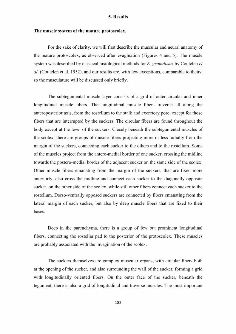
5. Results
182
The muscle system of the mature protoscolex.
For the sake of clarity, we will first describe the muscular and neural anatomy of
the mature protoscolex, as observed after evagination (Figures 4 and 5). The muscle
system was described by classical histological methods for E. granulosus by Coutelen et
al. (Coutelen et al. 1952), and our results are, with few exceptions, comparable to theirs,
so the musculature will be discussed only briefly.
The subtegumental muscle layer consists of a grid of outer circular and inner
longitudinal muscle fibers. The longitudinal muscle fibers traverse all along the
anteroposterior axis, from the rostellum to the stalk and excretory pore, except for those
fibers that are interrupted by the suckers. The circular fibers are found throughout the
body except at the level of the suckers. Closely beneath the subtegumental muscles of
the scolex, there are groups of muscle fibers projecting more or less radially from the
margin of the suckers, connecting each sucker to the others and to the rostellum. Some
of the muscles project from the antero-medial border of one sucker, crossing the midline
towards the postero-medial border of the adjacent sucker on the same side of the scolex.
Other muscle fibers emanating from the margin of the suckers, that are fixed more
anteriorly, also cross the midline and connect each sucker to the diagonally opposite
sucker, on the other side of the scolex, while still other fibers connect each sucker to the
rostellum. Dorso-ventrally opposed suckers are connected by fibers emanating from the
lateral margin of each sucker, but also by deep muscle fibers that are fixed to their
bases.
Deep in the parenchyma, there is a group of few but prominent longitudinal
fibers, connecting the rostellar pad to the posterior of the protoscolex. These muscles
are probably associated with the invagination of the scolex.
The suckers themselves are complex muscular organs, with circular fibers both
at the opening of the sucker, and also surrounding the wall of the sucker, forming a grid
with longitudinally oriented fibers. On the outer face of the sucker, beneath the
tegument, there is also a grid of longitudinal and traverse muscles. The most important

5. Results
183
group of muscle fibers in the sucker are the perpendicular fibers, connecting the base
and the outer face of the sucker.
The rostellar pad, beneath the hooks, is similar in its constitution to the suckers,
being surrounded at its outer wall by circular and longitudinal muscles, and with
powerful perpendicular fibers connecting the base to the tip of the pad.
Finally, there are numerous muscle fibers in the scolex that are not so intricately
organized, including dorso-ventral fibers and transverse fibers. In particular, there is a
great accumulation of radial fibers beneath the rostellar pad proper. These could be of
importance for the evagination of this organ. Few other fibers can be seen in the body of
the protoscolex.
5. Results
184
Figure 4. The muscle system of the protoscolex. A. Phalloidin staining of a
transverse section of the scolex. B. Phalloidin staining of a transverse section of the
posterior body. C. Diagram showing the main groups of muscle fibers in the scolex
(left, frontal view; right, dorsal view). See the main text for details. Abbreviations: dmf,
deep muscle fibers connecting dorso-ventrally adjacent suckers; ilmf, inner longitudinal
muscle fibers; S, sucker; slmf, subtegumental longitudinal muscle fibers, scmf,
subtegumental circular muscle fibers; stm, subtegumental muscles (referring to both
longitudinal and circular muscle fibers); R, rostellum, rm, radial muscles below the
rostellar pad; rmfs, radial muscle fibers connecting each sucker to the others and to the
rostellum. Asterisks mark several groups of muscle fibers connecting the suckers to
each other. Bars represent 50 µm.

5. Results
185
The nervous system of the mature protoscolex.
Patterns obtained with antibodies
The nervous system of the protoscolex was reconstructed from confocal sections
of whole mount material and minor details were obtained from cryosections. AcTub-IR
revealed the perinuclear cytoplasm and the neurites of nerve cells, as well as the body
and cilia of the flame cells (Figure 5), [Additional File 7]. Because of their morphology
and distribution in the nervous system, we conclude that most of the cells labeled by
AcTub-IR in the protoscolex, excluding the flame cells, are nerve cells. The flame cells
possessed thin AcTub-IR protrusions projecting from the cell bodies, that sometimes
appeared to contact the nerve cords. They were nonetheless easily distinguished from
nerve cells thanks to their characteristic morphology and by the co-labeling with
phalloidin at the base of the flame. A large number of nerve cells were detected by this
method, resulting in exquisite detail, but at the same time obscuring in some instances
the general layout of the nervous system. The FMRFa-IR, on the other hand, strongly
stained all the main and minor nerve tracts in the scolex, the nerve cords and connecting
commisures, as well as several minor projections from the nerve cords, giving the best
results for the reconstruction of the general layout of the nervous system, although it
was very difficult to recognize the perikarya of individual nerve cells (Figure 6),
[Additional File 8]. The pattern of RYa-IR was essentially indistinguishable to that
obtained by FMRFa-IR, and the pattern or FVa-IR was similar, except that IR was
typically low in the nerve cords as compared to the scolex [Additional File 9]. Finally,
5-HT-IR identified a small subset of nerve cell bodies and their neurites, that follow the
main nerve tracts, the nerve cords, and some minor tracts and projections (Figure 7). In
combination, these markers allowed for a detailed description of the nervous system of
the protoscolex.
Neuroanatomy
The nervous system in the scolex of the protoscolex of E. multilocularis is very
similar to that of the adult stage of E. granulosus. All the main elements observed by 5-
HT IHF and/or acetylcholinesterase HC (Shield 1969; Brownlee et al. 1994) were
5. Results
186
revealed by AcTub and FMRFa IHF, as well as some minor elements and details not
previously described.
Two lateral ganglia are situated in the scolex at the level of the anterior border of
the suckers. These contain numerous AcTub-IR nerve cell bodies, but it was not
possible to distinguish a distinct neuropile within the ganglia, probably due to their
small size. These ganglia are connected by a main transverse commissure and by an
anterior ring commissure; the main transverse commissure displayed relatively low
AcTub-IR, and in some FMRFa and AcTub IHF preparations it appeared to be formed
by two individual commissures. From each lateral ganglion, two connectives are
emitted towards a rostellar nerve ring at the base of the rostellar pad. These connectives
are very closely juxtaposed, and only distinguishable by reconstruction from confocal
sections. One pair of dorsal and one pair of ventral medial connectives also connect the
anterior ring commissure to the rostellar ring and are the continuations of the minor
medial nerve cords (see below). Each of these dorsal and ventral nerve pairs are further
connected midway to the rostellum by a short commissure. Finally, a pair of dorso-
ventral connectives, termed the ‘X-commissure’ by Shield [21], directly connect the
dorsal and ventral medial nerve cords to the main transverse commissure, resulting in a
characteristic cross as seen in cross-sections (Figures 6B, 6D).
The rostellar nerve ring itself appears as a smooth FMRFa-IR ring and has
numerous AcTub-IR bipolar nerve cell bodies, that give rise to two coronae of long
neurites innervating the rostellum. The neurites of the external corona surround the
rostellar pad and reach the apex of the rostellum where the hooks are inserted (this
region was denominated the ´rostellar cone´ by Galindo et al. (Galindo, Schadebrodt,
and Galanti 2008) , and is probably the precursor of the so-called “rostellar gland” of
the adult, composed by modified tegumental cells with secretory properties (Thompson,
Dunsmore, and Hayton 1979)). The internal corona is composed of neurites that reach
and presumably traverse into the rostellar pad. Within the rostellar pad itself few
AcTub+ neurites or cell bodies are visible. There seemed to be no indication of distinct
lateral rostellar ganglia sensu Fairweather et al. (Brownlee et al. 1994), but rather the
whole rostellar nerve ring seemed to be ganglionic in nature.

5. Results
187
To the posterior, the lateral ganglia emit two thick connectives towards the
postero-lateral ganglia, located at the same level as the posterior border of the suckers.
By FMRFa-IR, the lateral ganglia, the thick connectives and the postero-lateral ganglia
appear as a continuous mass of nervous tissue. The postero-lateral ganglia are connected
to each other by a posterior ring commissure. The two main lateral nerve cords are born
from the postero-lateral ganglia, and each of them is accompanied by a dorso-lateral and
a ventro-lateral minor nerve cord, that are born at the point where the posterior ring
commissure fuses with the sucker nerve ring (see below; Figures 6A, 6B; Additional
File 8).
Each sucker is surrounded at its base by an external nerve ring, that fuses at its
medial margin with the medial nerve cords and at its anterior and posterior margins with
the anterior and posterior ring commisures. The sucker nerve ring has numerous
AcTub+ IR cells projecting neurites that either surround the body of the sucker and
result in nerve terminals at its opening, or go deep into the body of the sucker and
connect to an internal sucker nerve ring. AcTub-IR nerve cell bodies are also present in
this ring (Figure 5 C), but only neurites are labeled by 5-HT-IR and FMRFa-IR. They
appear to be bipolar, and extend long neurites towards the surface of the sucker.
In the posterior body, the five pairs of longitudinal nerve cords are connected by
transverse ring commissures, forming a typical orthogonal plan (Halton and Gustafsson
1996; Reuter, Mäntylä, and Gustafsson 1998), although the neurites connecting each
pair of adjacent nerves are not completely aligned to each other. Along the main nerve
cords, the bodies of nerve cells can be distinguished by AcTub-IR, FMRFa-IR, RYa-IR
and 5-HT-IR (Figures 6A, 7A, Additional File 9C and data not shown). From these cell
bodies neurites extend towards the subtegumental region, where they form a peripheral
plexus. Among the AcTub-IR neurites, some of their terminals are located in the
subtegumental region, presumably innervating the subtegumental muscles, while others
traverse the tegument and are therefore probably sensory terminals (such as those
previously described by TEM in E. granulosus adults by Morseth (Morseth 1967a);
Figure 5E). There are many medial AcTub-IR neurites, that make the medial nerve
cords untraceable with AcTub-IR (but they are readily distinguished by IR to the other

5. Results
188
antibodies). In the scolex, the nerve terminals of the peripheral plexus are very densely
distributed (Figure 5A).
Throughout this description, 5-HT-IR was scarcely mentioned because of the
similarity to previous results in E. granulosus adults (Brownlee et al. 1994). An
important distinction, however, lies in the number of 5-HT-IR nerve cells observed. We
observed that although the positions of the 5-HT-IR cells were relatively fixed, the
number of cells in each position and the total number in each protoscolex showed some
variation. This variation occurred in completely mature protoscoleces, as judged by the
morphology of the suckers and the rostellum, and also by the low levels of cell
proliferation as determined by 5-ethynyl-2′-deoxyuridine (EdU) incorporation assays
(U. Koziol and K. Brehm, unpublished results; see (Galindo et al. 2003)). 5-HT-IR
nerve cell bodies were located in the rostellar nerve ring (mode = 3, range = 2-4), in
each lateral ganglion (mode = 1, range = 1-2), on the region of the anterior commissure
(mode = 1, range = 0-3), in each postero-lateral ganglion (mode = 2, range = 1-3), on
the main lateral nerve cords (mode = 2, range = 1-3) and rarely on the medial nerve
cords (mode = 0, range = 0-2) (Figure 7). More rarely, we observed ectopically
positioned 5-HT-IR nerve cell bodies (e.g. between nerve cords). The total number of 5-
HT-IR cells was therefore variable, with an average of 15.2 cells per protoscolex (n =
74), and the majority had between 14 and 17 5-HT-IR cells (Figure 7 C). This was the
case for both GH09, an isolate kept by intraperitoneal serial passage in M. unguiculatus
for 3 years (average = 15.3 5-HT-IR cells / protoscolex), as for J2012, that had only
been passaged for 6 months (average = 15.2 5-HT-IR cells / protoscolex; n = 37 for
each isolate). This indicates that this variation is a typical feature of the E.
multilocularis protoscolex and not an abnormal characteristic of a specific laboratory
isolate.

5. Results
189

5. Results
190
Figure 5. AcTub-IR in the protoscolex (previous page). A. AcTub-IR in the
nerve terminals in the surface of the protoscolex (confocal microscopy, merge of 3
optical sections). B. AcTub-IR in the nerve cell bodies and neurites below the sections
shown in A (confocal microscopy, merge of 3 optical sections). C. AcTub-IR cells and
their projections in the internal sucker nerve ring (section). D. AcTub-IR cell bodies and
their projections deep within the scolex (section). E. Detail showing a nerve terminal
penetrating and traversing the tegument (arrow). Asterisks mark flame cells. (In panel
BF, bright field microscopy is added to the merge of the fluorescence channels).
Abbreviations: esr, external sucker ring; mnc, main nerve cords; rc, coronae of neurites
projecting from the rostellar ring; rr, rostellar ring; stm, subtegumental muscle layer.
Bars represent 25 µm in A, B, and D, 50 µm in C and 20 µm in E.

5. Results
191
Figure 6. FMRFa-IR in the protoscolex. A. General view of the protoscolex;
merge of several focal planes of epifluorescence microscopy, anterior is to the left. B.
Diagram showing the main elements of the central nervous system in the scolex as
identified by FMRFa-IR. C. Detail of FMRFa-IR in the scolex, confocal projection of
10 um of optical sections with color-coded depth; the inset shows another projection of
10 um thickness, deeper within the scolex, where the main transverse commissure can
be seen. Anterior is to the left. D. Serial transverse sections (from anterior to posterior),
reconstructed from the confocal sections of C. Abbreviations: alnc, accessory lateral
nerve cord; arc, anterior ring commissure; esr, external sucker ring; isr, internal sucker
ring; lg, lateral ganglia; lrc, lateral rostellar connectives; mlnc, main lateral nerve cords;
mnc, medial nerve cords; mrc, medial rostellar connectives; mtc, main transverse
commissure; plg, postero-lateral ganglia; prc, posterior ring commissure; rr, rostellar
ring; rc, coronae of rostellar ring projections; x, x-commissure. Bars represent 50 µm.

5. Results
192
Figure 7. 5-HT-IR in the protoscolex. A. General view of the protoscolex. B.
Detail of the scolex (different specimen). A and B are merges of several focal planes of
epifluorescence microscopy, anterior is to the top. C. Histogram showing the frequency
of the abundance of 5-HT-IR nerve cells per protoscolex. Abbreviations are as in figure
6. Bars represent 50 µm in A, 20 µm in B.
5. Results
193
The development of muscle fibers and the nervous system in the protoscolex
IR patterns obtained with each antibody during development
Phalloidin staining revealed the development of the muscle fibers throughout
protoscolex development. In the case of the nervous system, 5-HT-IR was detectable
from early stages of development, clearly labeling the cell bodies of nerve cells, and
later the projections from these cells. FMRFa-IR was detectable either at the same
stages of development as 5-HT, or usually slightly later, showing similar patterns.
Finally, AcTub-IR appeared relatively late during development, only in those regions of
the nervous system that could be previously detected by the other antibodies. However,
a greater number of nerve cells could be detected at this point with this antibody.
Developmental stages
We have adopted the staging system of Leducq and Gabrion (Leducq and
Gabrion 1992) for describing the development of the protoscolex. In stage 1, the initial
protoscolex bud appears in the wall of the brood capsule, without any external
distinctive features. Already at this stage, there is a well defined grid of longitudinal and
circular subtegumental muscles [Additional File 10], that continue into the wall of the
brood capsule, and several transverse muscle fibers. It is at this stage that 5-HT-IR
appears, as a pair of 5-HT positive cells close to the tip of the bud, without any
projections except that they are connected to each other by a short commissure (Figure 8
A). Rarely, faint FMRFa-IR also appears in a pair of cells in a similar position (Figure 8
A). These cells mark the beginning of the development of the rostellar nerve ring. The
development of the nervous system then proceeds towards the posterior, resulting in a
developmental gradient along the antero-posterior axis.
In stage 2, the rostellar primordium becomes apparent at the tip, composed of an
apical rostellar bulb, that will differentiate into the muscular rostellar pad, and a fold
(the prebulbar region) at its base. Some of the deep longitudinal muscle fibers can
already be seen at this stage. [Additional File 11]. The nerve cells in the developing

5. Results
194
rostellar ring form anterior projections towards and into the rostellar bulb, and in some
cases 5-HT-IR and FMRFa-IR cells can be seen at this and later stages within the
rostellar bulb (however, these probably become relocated later during development
since no such cells were observed in the mature rostellar pad). At the same time
posterior projections are formed towards the developing lateral ganglia, in which some
5-HT-IR cells with varying labeling intensity can already be seen, already connected by
the main nerve commissure (Figure 8 A), [Additional File 11]. The longitudinal nerve
cords cover part of the antero-posterior axis. Sometimes supernumerary 5-HT-IR cells
or positive cells with atypical positions can be seen (Figure 8 A). The fate of these cells
is not clear; presumably these are eliminated or relocated later in development. The
FMRFa-IR pattern closely matches the 5-HT-IR, except that it extends more posteriorly
in the developing longitudinal nerve cords (Figure 8 A). No AcTub-IR is usually seen at
this stage.
In stages 3 to 4, the region that will become the scolex becomes distinct from the
posterior body, and cells begin to accumulate at the site where the suckers will be
developed. At the same time, the histogenesis of the hook begins at the side of the
prebulbar fold. At these stages, the rostellar ring and its coronae, the lateral ganglia, the
main transverse commissure and the anterior ring commissure are clearly evident by 5-
HT and FMRFa IHF, and weakly 5-HT-IR cells can be seen at the position of the
postero-lateral ganglia (Figure 8 A). The main lateral nerve cords are seen extending
posteriorly and the anterior portion of the medial nerve cords is also distinct. At this
point, AcTub-IR brightly labels the cells of the rostellar nerve ring and the coronae of
rostellar projections, directed towards the bulb (internal corona) and the prebulb
(external corona) (Figure 8 B).
In stages 5 to 6, the development of the suckers and their associated muscles and
nerve rings proceeds. At the same time, the folds of the rostellar prebulb, carrying the
developing hooks, grow and engulf the rostellar bulb, coalescing over it. Therefore, the
rostellar bulb becomes sub-apical, with the hooks and the prebulb covering it as is found
in the mature rostellum. At this point minor connectives and commissures in the scolex
are developing, and the postero-lateral ganglia and the posterior ring commissure are
well formed. FMRFa-IR labels the main nerve cords all the way towards the posterior of

5. Results
195
the protoscolex soma; 5-HT-IR usually labels the cords only to a more anterior position,
but positive cells can already be found on the main nerve cords. Finally, at stage 7 the
development is complete and the scolex lies invaginated within the posterior body.

5. Results
196
Figure 8. Development of the protoscolex nervous system. A. 5-HT-IR and
FMRFa-IR in the early stages of protoscolex development. Abbreviations are as in
figure 6 and sp, sucker primordium. The asterisk marks a 5-HT-IR cell in an unusual
position, perhaps in the developing mtc. Note also some supernumerary 5-HT-IR cells
with low signal levels next to the lateral ganglia. B. AcTub-IR in late protoscolex
development. S1, S2, S3, S4 are stages 1 to 4, respectively. Bars represent 20 µm.

5. Results
197
Discussion
This work represents the first detailed description of the nervous and muscle
systems in the development of the post-onchosperal larvae of Echinococcus, including
the first description, to the best of our knowledge, of nervous elements in the germinal
layer of the metacestode. The presence of muscle fibers and nerve cells in the germinal
layer of the metacestode is intriguing, since this larval stage is a non-motile cyst.
However, possible functions for these systems can be hypothesized. In the case of the
muscle system, the accumulation of circular fibers around the invaginating brood
capsule suggests that they could be involved in the mechanism of invagination and in
the constriction of the stalk. Furthermore, the muscle layer in the cyst wall is continuous
to that of the brood capsule and the protoscolex. In the case of the nervous system,
although the projecting neurites of the nerve cells are in close apposition to the muscle
fibers, a myoregulatory role seems improbable, given the lack of motility and the
absence of serotoninergic and cholinergic elements (which are the best characterized
myomodulatory neurotransmitters in flatworms (Ribeiro, El-Shehabi, and Patocka 2005;
Maule, Marks, and Day 2006)). Furthermore, this nervous system is independent from
that of the developing protoscolex, suggesting that it would not serve as a scaffold for
the development of the protoscolex nervous system. An attractive hypothesis is that it
could represent a neuroendocrine system, secreting factors that regulate the
development of the metacestode. Relatedly, possible neuroendocrine secretory terminals
have been described at the ultrastructural level in the nervous system of cestodes
(Halton and Maule 2003), and in free living flatworms there are several lines of
evidence suggesting a role of the nervous system in regulating cell proliferation and
pattern formation during asexual reproduction and regeneration (Reuter and Gustafsson
1996; Rossi, Iacopetti, and Salvetti 2012). In other systems, muscle fibers or
myofibroblasts can also be a source of growth factors stimulating stem cell proliferation
and maintenance in superjacent epithelia (Powell et al. 1999; Takashima, Gold, and
Hartenstein 2013). If such a role was demonstrated for the neuromuscular system in
Echinococcus cysts, it would open new areas for drug research targeting these signaling
systems, offering a potential link to many present efforts targeting the neuromuscular
system in trematodes (Mousley et al. 2005) .

5. Results
198
The nervous and muscle systems in E. multilocularis are probably homologous
to those present in the motile bladder tissue from Taenia spp., although modified
because of the lack of motility in Echinococcus cysts. Indeed, the limited reports
regarding the nervous system in the bladder of Taenia spp. indicate that RFamide-IR
and acetylcholinesterase positive (AChE+) fibers are present (Fairweather et al. 1991;
Vasantha et al. 1992). Furthermore, we have confirmed the presence of FMRFa-IR,
AcTub-IR and AChE+ elements in the bladder tissue of in vitro cultured Taenia
crassiceps, together with a well organized grid of subtegumental longitudinal and
circular muscles [Additional File 12].
In another publication (Tsai et al. 2013) we performed a bioinformatic search for
neuropeptide-encoding genes in the genomes of E. multilocularis and other cestodes,
finding several genes that showed significant expression in the germinal layer (as
determined by high throughput RNA sequencing and RT-PCR). It is thus possible that
some of these neuropeptides are expressed in the nerve net of the germinal layer.
Surprisingly, there is no AcTub-IR during the early development of the
protoscolex nervous system, which is at odds with the occurrence of AcTub-IR in
pioneer neurons during the development of many metazoans (Jellies et al. 1996;
Younossi-Hartenstein, Ehlers, and Hartenstein 2000; Younossi-Hartenstein and
Hartenstein 2000; Younossi-Hartenstein, Jones, and Hartenstein 2001). It seems that in
the case of E. multilocularis, tubulin-α acetylation occurs only at late stages of
neurogenesis. Interestingly, 5-HT and FMRFa-IR were detected from very early time
points, even in young nerve cells with few short projections. Similar results have been
described in the development of other invertebrates (Voronezhskaya and Elekes 1996;
Dickinson, Croll, and Voronezhskaya 2000; Franchini 2005) , and the early expression
of neurotransmitter molecules could have regulatory roles during the development of
the nervous system (Cameron, Hazel, and McKay 1998; Nguyen et al. 2001; Daubert
and Condron 2010) .
This description of the nervous system in the scolex is the most detailed to date,
benefiting from the use of several different markers in combination with confocal
5. Results
199
microscopy. In particular, the use of AcTub-IR resulted in great detail, and allowed us
to identify large numbers of nerve cell bodies not previously detectable with 5-HT-IR,
AChE HC or IR against mammalian neuropeptides (Shield 1969; Brownlee et al. 1994;
Fairweather et al. 1994). Prominent examples of this include the rostellar ring, which
appears as fully ganglionic by AcTub-IR (whereas it appeared to be composed of two
individual ganglia by 5-HT-IR) and the inner sucker nerve ring, that has numerous
AcTub-IR cell bodies but that was only described as a network of nerve processes in
previous studies (Brownlee et al. 1994; Fairweather et al. 1994). On the other hand,
FMRFa-IR gave the best results for the reconstruction of the general layout of the
nervous system, and for tracing the main commisures and connectives. Indeed, it
allowed the detection of all nervous elements previously described from 5-HT-IR and/or
AChE HC, together with fine details not previously described [Additional File 13].
Once the development of the nervous system is complete in the protoscolex, its
morphology is already similar to that of the scolex of the developed adult, with no
major rearrangements produced during adult development. This is probably related to
the fact that the scolex, once evaginated, must be fully functional to avoid being
dispelled from the host. However, the number of 5-HT-IR cells in the scolex of adult E.
granulosus is higher, and has been described as fixed for each region (e.g., exactly five
5-HT-IR cells have been described in the postero-lateral ganglia, 3 in the rostellar ring,
etc. (Brownlee et al. 1994)). Therefore, assuming that the nervous system of the adult of
E. multilocularis is identical to that of E. granulosus, new nerve cells must be
incorporated into the nervous system of the scolex during adult development (as has
been described for example for peptidergic neurons of Hymenolepis diminuta
(Fairweather et al. 1988)). The final numbers of 5-HT-IR cells might become fixed in
adults as compared to the protoscolex, but there is little information about the constancy
in the number and position of individually identifiable neurons in flatworms in general
(Halton and Gustafsson 1996; Younossi-Hartenstein, Ehlers, and Hartenstein 2000).
The general structure of the nervous system in the scolex of Echinococcus is also
remarkably similar to classical descriptions of the related Taenia spp. (Cohn 1899;
Vasantha et al. 1992), although the scolex in these species is at least one order of
magnitude larger. It is also worth mentioning the similarities observed in the

5. Results
200
development of the scolex of Echinococcus spp. ((Goldschmidt 1900); this work) and
that of Taenia spp. (Crusz 1947; Mount 1970; Shield, Heath, and Smyth 1973). In both
cases the rostellar bulb and prebulb are the first signs of differentiation appearing on a
thickened scolex primordium in the brood capsule (Echinococcus) or bladder wall
(Taenia). Later, as the prebulb carrying the hooks grows and engulfs the bulb, the
primordia of the suckers are formed.
The highly detailed description of the nervous system in these larval stages is a
necessary first step towards future studies regarding the function of the nervous system,
and of particular neurotransmitters, neuropeptides and their signal transduction systems
in the biology and development of E. multilocularis. Furthermore, the use of axenic in
vitro cultivation systems for E. multilocularis metacestodes (Spiliotis and Brehm 2009)
provides the opportunity, for the first time, to study the mechanisms behind the peculiar
developmental processes of these organisms, including their developmental plasticity,
unusual asexual multiplication by protoscolex formation, and the continuous growth and
exogenous budding of metacestodes, all of which are based on the presence of a
totipotent population of stem cells (the so-called germinative cells or neoblasts)
(Sakamoto and Sugimura 1970; Reuter and Kreshchenko 2004; Brehm 2010b; Koziol et
al. 2010). Also in this case, a careful description of developmental sequences such as
the development of the neuromuscular system described herein is an essential first step.
Conclusions
E. multilocularis is an important human parasite and a recently emerging model
system to study host-dependent parasite development in vitro [63]. We herein provide
the most detailed description to date of the morphology of the nervous system of
protoscoleces throughout development. We found a remarkable complexity of the
protoscolex nervous system, comparable to that observed in many adult cestodes. The
data we provide will be important for future studies on parasite developmental plasticity
and functional and evolutionary analyses on cestodes in general, and taeniids in
particular. Furthermore, we demonstrate for the first time that the cystic metacestode
stage, which grows tumor-like within the intermediate host, contains a nerve net with
peptidergic elements. Although the function of this nerve net cannot yet be clearly

5. Results
201
determined, we suggest that it could act as a neuroendocrine system that regulates
parasite development within the host. As such, it could be a potential target for the
development of novel anti-parasitics. Using the information provided by our study, and
the recently determined E. multilocularis genome sequence [42], we are currently
characterizing parasite neuropeptides that are expressed in the metacestode in order to
determine their function in parasite physiology.

5. Results
202
Materials and Methods
Parasite culture and fixation
Parasite isolates were maintained by serial intraperitoneal passage in Meriones
unguiculatus as previously described (Spiliotis and Brehm 2009). Isolates were
originally obtained from accidental infections of Old World Monkeys in a breeding
exclosure (Tappe et al. 2007), and had been passaged at the time of this study for
between six months and four years. All experiments were carried out in accordance with
European and German regulations on the protection of animals (Tierschutzgesetz). In
vitro co-culture of parasite vesicles with rat Reuber hepatoma cells was performed as
previously described (Spiliotis and Brehm 2009). Protoscoleces were isolated from in
vivo parasite material and either activated by successive treatments with pepsin at low
pH and taurocholate (Gelmedin, Spiliotis, and Brehm 2010) , or alternatively set into
culture in DMEM (Gibco) plus 10% fetal calf serum (Biochrom) for 12 hours, that also
resulted in significant scolex evagination. Parasite vesicles obtained in vitro (Spiliotis
and Brehm 2009) were gently opened using a syringe tip in order to allow the entry of
the fixative and other reagents during immunohistofluorescence. The material analyzed
from in vivo cultures consisted of small pieces of metacetode tissue surrounded by host
liver tissue. All samples for IHF were fixed in 4% paraformaldehyde (PFA) buffered in
phosphate buffer saline (PBS), for 1 to 4 hours at room temperature.
Antibodies
We used a suite of antibodies typically employed for the description of the nervous
system in diverse invertebrates:
1) Anti-acetylated tubulin-α (AcTub), mouse monoclonal antibody (Santa Cruz, clone
6-11B-1; diluted 1:100; (Piperno and Fuller 1985)). Tubulin-α acetylation is a post-
translational modification that occurs in highly stable microtubuli, mainly occurring in
the neuronal cytoskeleton (neurotubuli) and in cilia and flagella. Because it is able in

5. Results
203
principle to label all nerve cells independently of their neurotransmitter, AcTub
immunoreactivity (IR) has been used to describe in detail the complete nervous system
of numerous invertebrates, including free living flatworms and onchospheres (Siddiqui
et al. 1989; Muller and Westheide 2000; Younossi-Hartenstein, Ehlers, and Hartenstein
2000; Younossi-Hartenstein and Hartenstein 2000; Younossi-Hartenstein, Jones, and
Hartenstein 2001; Orrhage and Müller 2005; Persson et al. 2012). Furthermore, it has
been used to study the development of the nervous system in both vertebrates and
invertebrates, since many pioneer axons show AcTub-IR (Chitnis and Kuwada 1990;
Jellies et al. 1996; Younossi-Hartenstein, Ehlers, and Hartenstein 2000; Younossi-
Hartenstein and Hartenstein 2000; Younossi-Hartenstein, Jones, and Hartenstein 2001).
2) Anti-FMRFamide (FMRFa), rabbit polyclonal antibody (Immunostar, diluted 1:300).
Antibodies against the neuropeptide FMRFa have also been used extensively for the
characterization of invertebrate nervous systems. It is acknowledged that anti-FMRFa
antibodies, and in particular the one used in this and other studies, can recognize most
neuropeptides containing a C-terminal RFamide motif (Brennan et al. 1993; Hartenstein
and Jones 2003; Orrhage and Müller 2005; Harzsch and Muller 2007).
3) Anti-RYamide (RYa; 1:250 dilution) and Anti-FVamide (FVa; 1:125 dilution), rabbit
polyclonal antibodies, kindly provided by Markus Conzelmann (Max Planck Institute
for Developmental Biology, Tübingen, Germany). These antibodies recognize small
amidated dipeptide motifs frequently found in neuropeptides of invertebrates, and are
highly specific (Conzelmann and Jekely 2012). For instance, the anti-RYa antibody
does not cross- react with neuropeptides with an RFamide motif. Anti-FLamide was
also used but no IR was found in E. multilocularis (data not shown).
4) Anti-5-HT, rabbit polyclonal antibody (Immunostar, diluted 1:300). Antibodies
against 5-HT have been extensively used to characterize the serotoninergic components
of the nervous system in invertebrates in general and flatworms in particular (see for
example (Brownlee et al. 1994; Fairweather et al. 1994; Orrhage and Müller 2005;
Maule, Marks, and Day 2006))

5. Results
204
Immunohistofluorescence in whole-mounts (WMIHF) and in cryosections
We followed the WMIHF protocols described by Collins et al. (Collins et al. 2011) for
Schistosoma mansoni, with some modifications. After fixation, samples were washed
three times for 10 min with PBS with 0.3% Triton X-100 (PBS-T). After this,
permeabilization was achieved by either A) 20 min incubation in PBS with 1% sodium
dodecyl sulfate (SDS) for metacestode vesicles or B) a 5 min treatment with proteinase
K (Fermentas, 2 µg/ml in PBS-T plus 0.5% SDS), followed by re-fixation with 4%
PFA buffered in PBS for 10 min at room temperature (proteinase K treatment was
essential for protoscolex WMIHF). After permeabilization, samples were washed three
more times with PBS-T and blocked for 2 h in PBS-T with 3% bovine seralbumin
(BSA, Sigma-Aldrich) and 5% normal sheep serum (Sigma-Aldrich). Incubation with
the primary antibody, diluted in PBS-T with 3% BSA, was carried overnight at 4 º C.
Then, samples were washed five times for 30 min with PBS-T, and incubated overnight
with the FITC conjugated secondary antibody (donkey anti-mouse or donkey anti-rabbit
as required, Jackson Immunoresearch) at 4 º C. Finally the samples were washed five
times for 30 min with PBS-T, and co-stained with 4',6-diamidino-2-phenylindole
(DAPI) and TRITC-conjugated Phalloidin (Sigma-Aldrich) as previously described
(Koziol et al. 2011). Negative controls lacking the primary antibody were performed,
which showed no signal. Cryosections were prepared as previously described (Koziol et
al. 2010), and processed for IHF with a similar protocol, except that no detergent was
added to PBS for the washes, permeabilization was achieved using 0.1% Triton X-100
in PBS, and all incubation times were reduced. Samples were analyzed by confocal
microscopy (Leica TCS SP5) and by epifluorescence microscopy (Zeiss Axio
Imager.Z1).
Acetylcholinesterase Histochemistry. (AChE HC).
AChE HC was carried out as described by the direct method of Karnovsky and Roots
(Karnovsky and Roots 1964).
Transmission electron microscopy

5. Results
205
Metacestode vesicles obtained after 3 months of in vitro culture of the GH09 isolate
(containing brood capsules) were fixed, processed and analyzed by TEM, essentially as
previously described (Spiliotis et al. 2008).

5. Results
206
Acknowledgments
This work was supported by grants from the Deutsche Forschungsgemeinschaft (DFG;
BR 2045/4-1) and the Wellhöfer Foundation (all to KB). UK was supported by a grant
of the German Excellence Initiative to the Graduate School of Life Sciences, University
of Würzburg. The authors wish to thank Monika Bergmann and Dirk Radloff for
excellent technical assistance, and Hilde Merkert, of the Institute for Molecular
Infection Biology, Würzburg, for her assistance with confocal microscopy. This
publication was funded by the German Research Foundation (DFG) and the University
of Würzburg in the funding programme Open Access Publishing.
Authors’ contributions
UK carried out or participated in all experiments. UK and KB designed the study and
drafted the manuscript. GK designed and interpreted the results of TEM studies. All
authors read and approved the final manuscript.
Competing interests
The authors declare that they have no competing interests.

5. Results
207
Additional files
Additional File 1 - Supplementary figure (.tif format). Unusual flame cell in the
germinal layer of Echinococcus multilocularis. Confocal microscopy, section of in
vivo cultured material. The bar represents 10 µm.
Additional File 2 – Supplementary figure (.tif format). AChE histochemistry in
Echinococcus multilocularis. A. Double labeling of AChE HC and AcTub-IR; note the
single AChE HC positive cell without any projections (asterisk), and negative to
AcTub-IR. AcTub-IR cell bodies are indicated by arrows. The bar represents 50 µm. B.
AChE HC reaction in the nervous system of the protoscolex.
Additional File 3 – Supplementary figure (.tif format). Transmission electron
microscopy of the germinal layer of Echinococcus multilocularis. Nerve projections
(A, B, C, arrows) and a putative nerve cell body (C, asterisk) are highlighted in red.
Abbreviations: mc, mitochondria; nu, nucleus; tu, microtubules; ve, vesicles. Bars
represent 1 µm.
Additional File 4 – Supplementary movie (.avi format). Early brood capsule. Serial
confocal sections of an early developing brood capsule from in vivo cultured material of
Echinococcus multilocularis stained for AcTub-IR (green), Phalloidin (red) and DAPI
(blue).
Additional File 5 – Supplementary movie (.avi format). Stalked brood capsule.
Serial confocal sections of a brood capsule with a stalk from in vivo cultured material of
Echinococcus multilocularis stained for Phalloidin (red) and DAPI (blue).
Additional File 6 – Supplementary movie (.avi format). Brood capsule with
developing protoscolex. Serial confocal sections of a brood capsule with a protoscolex
bud in a whole-mount in vitro cultured cyst of Echinococcus multilocularis, stained for
AcTub-IR (green), Phalloidin (red) and DAPI (blue).

5. Results
208
Additional File 7 – Supplementary movie (.avi format). AcTub-IR in the
protoscolex of Echinococcus multilocularis. Serial confocal sections of a whole-mount
specimen.
Additional File 8 - Supplementary movie (.avi format). FMRFa-IR in the
protoscolex of Echinococcus multilocularis. Serial confocal sections of a whole-mount
specimen stained for FMRFa-IR (green), Phalloidin (red) and DAPI (blue).
Additional File 9 – Supplementary figure (.tif format). FVa-IR and RYa-IR in the
protoscolex of Echinococcus multilocularis. A. Developing protoscolex, FVa-IR
(whole-mount). B. Transverse section of the scolex, FVa-IR. C. Mature protoscolex,
RYa-IR (whole-mount). D. Section of developing protoscolex, RYa-IR. Abbreviations
are as in figures 4 and 6, and cns, central nervous system; px, subtegumental plexus.
Bars represent 50 µm in A, C and D, and 25 µm in B.
Additional File 10 – Supplementary movie (.avi format). Subtegumental muscle
fibers in a stage 1 protoscolex bud of Echinococcus multilocularis. Serial confocal
sections of a whole-mount protoscolex bud partially surrounded by the remains of the
brood capsule, stained with Phalloidin.
Additional File 11 – Supplementary movie (.avi format). 5-HT-IR in a stage 2
developing protoscolex of Echinococcus multilocularis. Serial confocal sections of a
whole-mount specimen stained for 5-HT-IR (green) and Phalloidin (red).
Additional File 12 – Supplementary figure (.tif format). Muscle and nervous
systems in Taenia crassiceps bladder tissue cultured in vitro. This is a laboratory
strain passaged in M. unguiculatus. It multiplies by budding but is unable to produce
scoleces, generating only bladder tissue. Samples were processed identically to the E.
multilocularis cysts. A. Phalloidin staining; note the longitudinal and circular muscle
fibers (whole-mount). B. AcTub-IR in the subtegumental layer; inset shows the flame
cells in detail (whole-mount). C. FMRFa-IR in the subtegumental layer (whole-mount).
D. AChE HC (section). Bars represent 200 µm in A, 50 µm in B and C, 20 µm in the
inset in B.

5. Results
209
Additional File 13 – Supplementary table (.doc format). Comparison of the
description of the protoscolex nervous system in this and in previous
investigations.

5. Results
210
5.5. CHAPTER 5: Further experimental results regarding the E. multilocularis
neuromuscular system.
In this chapter, I will describe further experiments that I performed for the
characterization of the Echinococcus multilocularis neuromuscular system, which were
not included in the previous article. Some of these results were included in another
article of which I am a co-author, as indicated.

5. Results
211
5.5.1. Persistence of the nervous system during the development from
protoscoleces to metacestode vesicles
Another peculiarity of the development of Echinococcus larval stages is the
ability of protoscoleces to develop into metacestode vesicles if they are released in the
intermediate host, as well as in vitro (Yamashita, Ohbayashi, and Sakamoto 1960;
Yamashita et al. 1962; Smyth, Howkins, and Barton 1966; Rogan and Richards 1986;
Rogan and Richards 1989; al Nahhas et al. 1991; Cucher et al. 2013). This ability is
more marked and has been studied in greater detail for E. granulosus, and spillage of
protoscoleces from ruptured cysts is the main source of secondary cysts in CE (Brunetti,
Kern, and Vuitton 2010).
Under the typical in vitro co-culture conditions used in our laboratory, a small
proportion (<1%) of protoscoleces were able to complete this developmental transition,
following similar developmental stages as the ones described for E. granulosus (Rogan
and Richards 1986) (Figure R5.1). A large fraction of protoscoleces never begins the
transformation into metacestode vesicles, while many others die during the process.
Initially, the protoscoleces swell and adopt a vesiculated shape, and many cells
disappear. Rudimentary suckers and rostellum remain, as well as internal “filaments”
connecting the scolex to the posterior of the body. Eventually, the laminated layer
appears, and only shapeless tissue remnants are found in the original position of the
scolex, which are usually still are connected to the posterior region of the body through
the “filaments”. The process took at least 50 days, under our culture conditions, from
the start of the culture until the laminated layer becomes apparent by standard
microscopy. After 105 days of culture, these protoscolex-derived vesicles showed a
strong positive reaction for alkaline phosphatase in the tegument, as is characteristic for
the metacestode germinal layer, but not for protoscoleces (Koziol et al. 2014) (Figure
R5.2). Furthermore, small accumulations of proliferating cells, reminiscent of early
brood capsule formation, could be seen in some of the metacestode vesicles obtained
after 105 days of culture (see below). These results suggest that the transformation of
protoscoleces into metacestode vesicles was complete.

5. Results
212
Figure R5.1. Developmental stages of the transformation of protoscoleces
into metacestode vesicles in vitro. Time elapsed since the beginning of in vitro culture
is indicated on the bottom right corner. dead ps., dead protoscoleces; ves. ps.,
vesiculating protoscoleces.

5. Results
213
Figure R5.2. Alkaline phosphatase histochemistry (ALP-HC) of
protoscoleces during transformation into metacestode vesicles in vitro.
Protoscoleces before culture (day 0) and after transformation into metacestode vesicles
in vitro (day 105 of culture) were analyzed for alkaline phosphatase activity in the
tegument by ALP-HC in cryosections. For comparison, the same experiment was
performed in parallel for metacestode vesicles obtained by standard in vitro co-culture
of parasite material with feeder cells. Bars: 40 µm.
Previous histological and ultrastructural studies of the transition from the
protoscolex to the vesicle stage have focused on the tegumental syncitium and the de
novo formation of the laminated layer (Rogan and Richards 1989; al Nahhas et al.
1991), but little is known about the fate of the neuromuscular system and the
germinative cells. Recently, it was shown that the serotoninergic nervous system of E.
granulosus disappears very quickly during this transition (Camicia et al. 2013). We
investigated the fate of the muscular and nervous systems by phalloidin staining and
acetylated α-tubulin (AcTub) immunohistofluorescence. The muscular system became
quickly disorganized after just one week, and it largely disappeared from the body of
the protoscolex after one month, although it could be seen in the posterior-most region
of the body, and more prominently in the suckers and rostellum. As many tegumental
cells disappeared, most remaining cells were found in the remains of the scolex and next
5. Results
214
to the “filaments”. Interestingly, the “filaments” are AcTub+ and are actually the
persisting nervous system, which although somewhat disorganized, remains in the
scolex and in the body (Figure R5.3). At the same time, the remaining proliferating
(EdU+) cells cluster around the AcTub+ nerve cords and the remains of the scolex
(Figure R5.4, A). At the final stages of development, the EdU+ cells are still found close
to the remains of the scolex, but appear to be dispersing into the rest of the developing
vesicle, and new cells can be found in the subtegumental region (Figure R5.4, B).
Unfortunately, we were unable to determine if the “filaments” were still AcTub+ at this
stage due to insufficient availability of specimens. Finally, when the laminated layer is
apparent, the germinal layer has a typical morphology, with dispersed EdU+ cells which
sometimes accumulate to form structures reminiscent of early brood capsules (Figure
R5.4, C-D). In summary, although the muscular system is largely lost, the nervous
system persists until later stages, and the remaining proliferating cells accumulate in its
proximity during the transition to the vesicle stage. In general, the persistence of the
nervous system and its proximity to the remaining proliferating cells suggests that it
may play a role during this developmental transition, and is reminiscent of the close
apposition of neoblast populations and the nerve cords of free-living flatworms (Bode et
al. 2006; Rossi, Iacopetti, and Salvetti 2012).
.

5. Results
215
Figure R5.3. AcTub immunohistofluorescence and phalloidin staining of
protoscoleces during transformation into metacestode vesicles in vitro. Two
different specimens are shown (A, B). The phalloidin signal is essentially lost in the
body, and very reduced in the suckers; only the signal in the rostellum remains
(compare for example with the non-vesiculating protoscoleces in the top left corner in
B). t. r, rostellum; s, sucker. Bars: 50 µm.

5. Results
216
Figure R5.4. EdU incorporation in protoscoleces during transformation into
metacestode vesicles in vitro. Specimens were cultured in vitro with 50 µM EdU for 5
hours and were then processed for AcTub immunohistofluorescence (IHF) and/or EdU
detection. A. Early vesiculating protoscolex, stained in whole-mount for EdU detection
(red), AcTub IHF (green), and DAPI (blue). Note the proximity of the EdU+ cells to the
AcTub+ nervous system in the scolex and in the “filaments”. B. Late vesiculating
protoscolex, stained in whole-mount for EdU (yellow) and DAPI (blue). C. Specimen
after complete transformation into a metacestode vesicle (with a laminated layer, at day
105 after the beginning of culture), stained in cryosections for EdU (yellow) and DAPI
(blue). D. Detail of C, showing a structure similar to an early brood capsule bud
containing numerous EdU+ cells. Bars: 50 µm.

5. Results
217
5.5.2. Discovery of neuropeptide-encoding genes in E. multilocularis
Neuropeptides (i.e. bioactive peptides or peptide hormones, abbreviated as NPs)
are structurally and functionally diverse in metazoans, having prominent roles as
modulators in the nervous system, reproductive system, and in many other tissues.
Typically, NPs serve as ligands for signaling through G-protein coupled receptors
(GPCRs) of the Rhodopsin and Secretin families (Frooninckx et al. 2012; Hoyer and
Bartfai 2012). Drugs targeting these receptors and signaling systems have been
proposed as promising targets for anthelminthics (Mousley et al. 2005; Marks and
Maule 2010).
NPs are processed from larger pre-prohormone precursors, and this processing is
essential for their biological activity (Li and Kim 2008). It is common for individual
precursors to code for several different peptides which are processed into individual
NPs, in many cases with similar sequence and activity to each other. Initially, the pre-
prohormone enters the secretory pathway, and its signal peptide is cleaved as it enters
the rough endoplasmic reticulum. Then, the active peptide is proteolitically cleaved
from the prohormone at dibasic sites (or more rarely monobasic sites) by proprotein
convertases, in particular by members of the PC1 and PC2 subfamilies (Steiner 1998;
Seidah et al. 2008). The basic C-terminal residues are removed by carboxypeptidase E,
and finally, peptides containing a C-terminal glycine residue are amidated (the glycine
residue is lost, and the resulting C-terminal α-amide group comes from the glycine
residue itself) (Bradbury and Smyth 1991; Attenborough et al. 2012). Amidation is in
many cases essential for the interaction of the NPs with their receptors (Li and Kim
2008).
As shown in the previous chapter, nerve cells showing immunoreactivity to
different invertebrate neuropeptides can be detected in the germinal layer of E.
multilocularis metacestodes. Because of the possible role of NPs in regulating
development in flatworms, we were interested in determining which NP-encoding genes
are expressed during larval development. Although much progress has been achieved in
the discovery of NPs in other flatworms (McVeigh et al. 2009; Collins et al. 2010),
almost nothing was known before this work about the diversity of NP genes in cestodes.

5. Results
218
Only two neuropeptide sequences had been published (NPF and GNFFRFamide from
Moniezia expansa (Marks and Maule 2010)), and two ESTs from taeniids had been
proposed to code for NPs (McVeigh et al. 2009) (E. granulosus npp-6 and Taenia
solium npp-20; re-named npp-20.3 in this work). Recently, two insulin-like peptides
(emilp1 and emilp2) were also described for E. multilocularis in this laboratory (Hemer
et al. 2014). Therefore, the first step was to obtain a list of NP-encoding genes for E.
multilocularis. The following results were previously included as part of Tsai et al.
(2013), Nature 496:57-63.
Because the biologically relevant peptides are short stretches of moderately
conserved amino acid residues (usually 3–40 residues), identifying genes coding for
NPs by purely bioinformatic methods is challenging. In this work, two strategies were
performed in order to identify NP genes in the genome of E. multilocularis and other
cestodes (E. granulosus, Taenia solium and Hymenolepis microstoma). First, a blastp
search was conducted for known invertebrate NP genes against the predicted proteome
of E. multilocularis, and hits were manually examined for important conserved
sequences in the active peptides. Pre-propeptides were also examined for the presence
of a signal peptide sequence (Petersen et al. 2011) and then scanned for mono and di-
basic cleavage sites for prohormone convertases. In this way, a few conserved NP genes
could be found, but this strategy would not recover divergent or novel cestode NP
genes.
The initial results indicated that cestodes are peculiar among invertebrates, since
cestode NP genes typically encode only one or at most two copies of similar mature
neuropeptide sequences per propeptide, and the mature NP sequence is often in the C-
terminus of the propeptide, not requiring a downstream proprotein convertase site.
Therefore, systematic searches for multiple proprotein convertase sites (e.g.
{[K/R][K/R]X4-20[K/R][K/R]} >2) as described for other invertebrates (Nathoo et al.
2001; McVeigh et al. 2009) are not adequate. Instead, we took advantage of the
observation that mature NP sequences are highly conserved between E. multilocularis
and Hymenolepis microstoma, whereas the surrounding regions are poorly conserved
but usually still detectable by blast (an example is shown in Figure R5.5). Therefore, a
novel homology-based strategy was developed for discovering conserved cestode NPs
(see the Materials and Methods section). Briefly, a wide list of possible NP genes from

5. Results
219
E. multilocularis, with a very high proportion of false positives, was filtered by
checking for high conservation of the predicted active peptide and cleavage sites but
lower conservation in the rest of the CDS in H. microstoma orthologs. The resulting
candidates were further checked for the presence of a signal peptide, and homologs of
the candidates were also searched for in E. granulosus, T. solium, S. mansoni and free
living flatworms. For one gene (EmuJ_000530500), the correct 5’ end containing a
putative signal peptide was obtained by 5´ Rapid Amplification of cDNA Ends (5´
RACE).
Figure R5.5. Example of tblastn analysis for the discovery of cestode NP
genes. One candidate NP gene from E. multilocularis (Query) was blasted (tblastn)
against the genome of H. microstoma (Sbjct). Notice the great similarity in the predicted
mature peptide (green), including a C-terminal glycine which could result in C-terminal
amidation (blue), and in the putative proprotein convertase sites (red), as compared to
the rest of the protein. Furthermore, both proteins contain a predicted signal peptide
(not shown). This blast analysis resulted in the discovery of the sequelog group npp-34,
not previously known for flatworms but with similarity to Luqins from other
lophotrochozoans (see the main text).

5. Results
220
In total, we found 22 groups of NP genes conserved between E. multilocularis,
E. granulosus, T. solium, and H. microstoma (Table R5.1). Of these groups, 16 could be
assigned to 11 previously described flatworm “sequelogs” (groups of genes coding for
similar peptides but of uncertain homology due to the short length of the conserved
peptides) or to specific flatworm sequences (groups npp-4, npp-5, npp-2, npp20.1,
npp20.2, npp20.3, npp20.4, npp20.5, npp-6, npp-24, npp-27, npp-14, npp-15.1, npp15.2,
spp-3, spp-18 (McVeigh et al. 2009; Collins et al. 2010); note that we have divided the
npp20 sequelog into 5 sub-families). For three other groups we found similar peptides
in Schistosoma, founding three new flatworm sequelog groups (npp-29, npp-31, and
npp-32). For one group, general similarity was found with other RL-amides, but not to
any group in particular, and a new sequelog group was founded (npp-30). Finally, for
two genes, no evidence of conservation was found in other flatworms, resulting in two
candidate cestode-specific sequelog groups (npp-33 and npp-34). However, npp-34
shows similarities with Luqins, a family of NPs found in mollusks and annelids
(Veenstra 2010; Veenstra 2011), suggesting a common origin, and that npp-34
sequences may eventually be discovered in other flatworms as well.
The total number of conserved NP genes was 21 to 22 in taeniids and 25 in H.
microstoma (which has three specific gene duplications of groups npp-20.3, npp-27 and
npp-29, with duplicated genes clustered (<21 kb distance) to each other in the genome).
With the exception of these duplicated genes from H. microstoma, and of the conserved
and clustered duplicated genes npp-15.1 and npp-15.2, no other significant clustering of
NP genes was observed in any genome. The total number of conserved predicted mature
peptides was found to be between 27 and 29. These numbers are in broad concordance
with the number of predicted peptide GPCRs in the genomes of cestodes (e.g. 29
predicted peptide GPCR receptors in E. multilocularis, (Tsai et al. 2013)), suggesting
that a large proportion of the cestode NP genes has been found.
The discovered cestode NPs are dominated by R(F/Y)amides, (I/L/M)amides
(McVeigh et al. 2009; McVeigh et al. 2011), and by an expansion of neuropeptide F
family genes (groups npp-20.1 to npp-20.5, similar to the expansion in the planarian
Schmidtea mediterranea (Collins et al. 2010)). As previously discussed (McVeigh et al.
2009; McVeigh et al. 2011), most of these sequelogs show little to no similarity to
vertebrate NPs. Even neuropeptide F genes show divergent features, such as a C-

5. Results
221
terminal GRARF-amide motif in npp-20.2 genes, instead of the highly conserved
GRPR(F/Y)-amide motif, and a tryptophan residue in position -10 of npp-20.3 genes
instead of the highly conserved tyrosine. Finally, cestodes appear to have lost several
neuropeptide and peptide hormones (e.g. FMRFamide, vasopressin/oxytocin-like
peptides, Gonadotropin Release Hormone-like peptides and GPA2/GPB5 heterodimeric
glycoproteins) that are widely conserved in metazoans, including annelids and mollusks
(i.e. groups that are phylogenetically close to the Platyhelminthes) (Veenstra 2010;
Veenstra 2011).

5. Results
222
Table R5.1. Predicted neuropeptide / peptide hormone genes in cestodes
Species Sequelog Gene code 2 Predicted
peptide(s) (only
conserved peptides
shown)
Peptide
structural
family3
Signal
Peptide
E. multilocularis spp-18 EmuJ_000092400.1 GLHFFRL Non amidated
peptide
YES
E. granulosus spp-18 EgrG_000092400.1 GLHFFRL Non amidated
peptide
YES
T. solium spp-18 TsM_001104400.1* GLHFFRL Non amidated
peptide
YES
H. microstoma spp-18 HmN_000234350 GLHFFRL Non amidated
peptide
?
E. multilocularis npp-14 EmuJ_000740500 GIMKMRI-amide;
ASIRRMRM-amide
(L/M/I)amide YES
E. granulosus npp-14 EgrG_000740500 GIMKMRI-amide;
ASIRRMRM-amide
(L/M/I)amide YES
T. solium npp-14 TsM_001092400.1 GIMKMRI-amide;
AGIRRMRM-amide
(L/M/I)amide YES
H. microstoma npp-14 HmN_000787500.1 GIRKMRI-amide;
GIMKMRI-amide;
AGIRRMRM-amide
(L/M/I)amide YES
E. multilocularis npp-15.1 EmuJ_000945400.1 AQFLRL-amide (L/M/I)amide YES
E. granulosus npp-15.1 EgrG_000945400.1 AQFLRL-amide (L/M/I)amide YES
T. solium npp-15.1 TsM_000537100.1 AQFLRL-amide (L/M/I)amide YES
H. microstoma npp-15.1 HmN_000592500.1 AQFLRL-amide (L/M/I)amide YES
E. multilocularis npp-15.2 EmuJ_000945300.1 AQFLRI-amide (L/M/I)amide YES
T. solium npp-15.2 TsM_000537000.1 AQFLRI-amide (L/M/I)amide NO
H. microstoma npp-15.2 HmN_000592400 AQFLRI-amide (L/M/I)amide YES
E. multilocularis npp-2 EmuJ_000530500 RGFI-amide (2x) (L/M/I)amide YES
E. granulosus npp-2 EgrG_000530500 RGFI-amide (2x) (L/M/I)amide YES
T. solium npp-2 TsM_001095700.1* RGFI-amide (2x) (L/M/I)amide YES
H. microstoma npp-2 HmN_000050100.1 RGFI-amide (2x);
RSFL-amide
(L/M/I)amide YES
E. multilocularis npp-20.1 EmuJ_001070600 ADPLSIINPPESVLSN
PAALRDYLRQVNEYF
AIIGRPRF-amide
Neuropeptide F YES
E. granulosus npp-20.1 EgrG_001070600 ADPLSIINPPESVLSN
PAALRDYLRQVNEYF
AIIGRPRF-amide
Neuropeptide F YES
T. solium npp-20.1 TsM_000819400.1* ADPLSIVNPPESVLN
NPAALRDYLRQVNE
Neuropeptide F YES
2 Wellcome Trust Sanger Institute GeneDB codes
3 Modified from McVeigh et al 2009

5. Results
223
YFAIIGRPRF-amide
H. microstoma npp-20.1 HmN_000462100 AEADLTDRILNDPAA
LRDYLRQINEYFAIIG
RPRF-amide
Neuropeptide F YES
E. multilocularis npp-20.2 EmuJ_000043400.1 ?-
KDLFKYMAQLNDYY
VLFGRARF-amide
Neuropeptide F YES
E. granulosus npp-20.2 EgrG_000043400.1 ?-
KDLFKYMAQLNDYY
VLFGRARF-amide
Neuropeptide F YES
T. solium npp-20.2 TsM_000527100.1 ?-
KDLFKYMAQLNDYY
MLFGRARF-amide
Neuropeptide F YES
H. microstoma npp-20.2 HmN_000295600 SRSLEEILSDPEFME
WLNSKFPNIAFFLSG
RNQEPSTETVREIFRF
MAELNNYYAIYGRA
RF-amide
Neuropeptide F YES
E. multilocularis npp-20.3 EmuJ_001194600 QIFRNVREFRRYLQR
LDEWLAITGRPRF-
amide
Neuropeptide F NO; but
SP after
DS ATG
E. granulosus npp-20.3 EgrG_001194600 QIFRNVREFRRYLQR
LDEWLAITGRPRF-
amide
Neuropeptide F NO
T. solium npp-20.3 NOT PREDICTED QIFRNVKEFRRYLQR
LDEWLAITGRPRF-
amide
Neuropeptide F NO
H. microstoma npp-20.3.1 HmN_000069000.1 PLFRTARELRRYLEQL
DEWLAITGRPRF-
amide
Neuropeptide F YES
H. microstoma npp-20.3.2 HmN_000069100 PVFRNVQELRRYLEQ
LDEWLAITGRPRF-
amide
Neuropeptide F NO
E. multilocularis npp-20.4 EmuJ_000926000.1 SHPTVPLLPQYLERLK
KFQSLKDKEKYVAAL
NSYYMIFGRPRF-
amide
Neuropeptide F YES
E. granulosus npp-20.4 EgrG_000926000.1 SHPTVPSLPQYLERLK
KFQSLKDKEKYVAAL
NSYYMIFGRPRF-
amide
Neuropeptide F YES
T. solium npp-20.4 TsM_000401700.1 SRPTVPSLPQYLERLK
KFHSLRDKEKYVAAL
NSYYMIFGRPRF-
amide
Neuropeptide F YES
H. microstoma npp-20.4 HmN_000206600 NDITYLPKRFKTAKD
MERYMAALNAYYMI
FGRPRF-amide
Neuropeptide F YES
E. multilocularis npp-20.5 EmuJ_000706000.1 EPQTSGWDLADDLG
TLESGEDFNDINDAV
Neuropeptide F NO; but
SP DS ATG

5. Results
224
LSSEKTSLTREEIQRYL
AQMKAYYLKNGMP
RY-amide
E. granulosus npp-20.5 EgrG_000706000.1 EPQTSGWDLADDLG
TLESGEDFNDINDAV
LSSEKTSLTREEIQRYL
AQMKAYYLKNGMP
RY-amide
Neuropeptide F NO; but
SP after
DS ATG
T. solium npp-20.5 TsM_000781400.1* EPQAGGWDLADDL
GTLGTGEDFGDINDA
VLSSEKTSLTREEIQR
YLAQMKAYYLKNGM
PRF-amide
Neuropeptide F NO; but
SP after
DS ATG
H. microstoma npp-20.5 HmN_000096800.1 DFSISDSWDLDDDLI
EDDDLKEANDAVMA
SAKTALTEEEAQKYL
AQMKAYYLKNGMP
RF-amide
Neuropeptide F NO
E. multilocularis npp-24 EmuJ_001046700.1 FKPKMPIFLGLI-
amide
(L/M/I)amide YES
E. granulosus npp-24 EgrG_001046700.1 FKPKMPIFLGLI-
amide
(L/M/I)amide YES
T. solium npp-24 TsM_000993300.1* FKPKMPIFLGLI-
amide
(L/M/I)amide YES
H. microstoma npp-24 HmN_000377100.1 SRSPIFHGLL-amide (L/M/I)amide YES
E. multilocularis npp-27 EmuJ_000347700 RLPYLIGGIRY;
ALDYLYGGVRY
Non amidated
peptide
YES
E. granulosus npp-27 EgrG_000347700 RLPYLIGGIRY ;
ALDYLYGGVRY
Non amidated
peptide
YES
T. solium npp-27 TsM_000472700.1 RLPYLIGGIRY;
ALDYLYGGVRY
Non amidated
peptide
NO
H. microstoma npp-27.1 HmN_000326200.1 ALDYLYGGVRY Non amidated
peptide
YES
H. microstoma npp-27.2 HmN_000326100.1 SLDYLYGGVRY Non amidated
peptide
YES
E. multilocularis npp-29 EmuJ_000229800 MLYW; MVYW Non amidated
peptide
YES
E. granulosus npp-29 EgrG_000229800 MLYW ; MVYW Non amidated
peptide
YES
T. solium npp-29 NOT PREDICTED MLYW; MVYW Non amidated
peptide
YES
H. microstoma npp-29.1 HmN_000212550 MIYW (2x) Non amidated
peptide
YES
H. microstoma npp-29.2 HmN_000212650 MIYW (2x) Non amidated
peptide
YES
E. multilocularis npp-30 EmuJ_000489700.1 GFLHSGRL-amide;
GFLTASRL-amide
(L/M/I)amide YES
E. granulosus npp-30 EgrG_000489700.1 GFLHSGRL-amide;
GFLTASRL-amide
(L/M/I)amide YES
T. solium npp-30 TsM_000991300.1 GFLHSGRL-amide ; (L/M/I)amide YES

5. Results
225
GFLTASRL-amide
H. microstoma npp-30 HmN_000183900 GFLTSSRL-amide;
GFLTAGRL-amide
(L/M/I)amide YES
E. multilocularis npp-31 EmuJ_000076000.1 APEMLWDIE Non amidated
peptide
YES
E. granulosus npp-31 EgrG_000076000.1 APEMLWDIE Non amidated
peptide
YES
T. solium npp-31 TsM_000528000.1 APEMLWDIE Non amidated
peptide
YES
H. microstoma npp-31 HmN_000223650 APELIWDIE Non amidated
peptide
YES
E. multilocularis npp-32 EmuJ_000179900.1 GPEIFIPLMEA Non amidated
peptide
YES
E. granulosus npp-32 EgrG_000179900.1 GPEIFIPLMEV Non amidated
peptide
YES
T. solium npp-32 TsM_000448600.1* GPEIFIPLMEA Non amidated
peptide
YES
H. microstoma npp-32 HmN_000205400.1 GPELFIPYMEA Non amidated
peptide
YES
E. multilocularis npp-33 EmuJ_000829900.1 TMPYSGGIF-amide IFamide YES
E. granulosus npp-33 EgrG_000829900.1 TMPYSGGIF-amide IFamide YES
T. solium npp-33 TsM_000416300.1* TMPYSGGIF-amide IFamide YES
H. microstoma npp-33 HmN_000514300 TPYSGGIF-amide IFamide YES
E. multilocularis npp-34 EmuJ_000316500 SPQFAWRPHSRF-
amide
R(F/Y)amide YES
E. granulosus npp-34 EgrG_000316500 SPQFAWRPHSRF-
amide
R(F/Y)amide YES
T. solium npp-34 TsM_001139300.1* SPQFAWRPHSRF-
amide
R(F/Y)amide YES
H. microstoma npp-34 HmN_000315550 SPQFAWRPHSRY-
amide
R(F/Y)amide YES
E. multilocularis npp-4 EmuJ_000240300.1 GNFFRF-amide R(F/Y)amide YES
E. granulosus npp-4 EgrG_000240300.1 GNFFRF-amide R(F/Y)amide YES
T. solium npp-4 TsM_000895400.1 GNFFRF-amide R(F/Y)amide NO
H. microstoma npp-4 HmN_000166500.1 GNYFRF-amide R(F/Y)amide YES
E. multilocularis npp-5 EmuJ_000993900.1 MQRPAFLFDAW-
amide; GAYLDLPW-
amide
(A/P)Wamide YES
E. granulosus npp-5 EgrG_000993900.1 MQRPAFLFDAW-
amide; GAYLDLPW-
amide
(A/P)Wamide YES
T. solium npp-5 TsM_001041500.1 MQRPAFLFDAW-
amide; GAYLDLPW-
amide
(A/P)Wamide YES
H. microstoma npp-5 HmN_000072750 GAYLDLPW-amide (A/P)Wamide YES
E. multilocularis npp-6 EmuJ_000759050 AIRLLRL-amide (L/M/I)amide YES
E. granulosus npp-6 EgrG_000759050 AIRLLRL-amide (L/M/I)amide YES
T. solium npp-6 TsM_000844500.1 AIRLLRL-amide (L/M/I)amide YES
H. microstoma npp-6 HmN_000648800.1 AVRLLRL-amide (L/M/I)amide YES
E. multilocularis spp-3 EmuJ_000185700 ARFRPRL-amide; (L/M/I)amide YES

5. Results
226
NYNFRARL-amide
E. granulosus spp-3 EgrG_000185700 ARFRPRL-amide;
NYNFRARL-amide
(L/M/I)amide YES
T. solium spp-3 TsM_001117900.1* ARFRPRL-amide (L/M/I)amide YES
H. microstoma spp-3 HmN_000148600.1 MPFLSRL-amide;
LYAFHSRL-amide
(L/M/I)amide YES
(?) - not clear from current gene model; (*) - gene model corrected from original
prediction; correction submitted to GeneDB; (SP after DS ATG) - signal peptide
observed after downstream start (ATG) codon but not in the start (ATG) codon of the
current prediction.

5. Results
227
5.5.3. Expression and effects of NPs during metacestode growth and regeneration
In order to determine which NPs are expressed in the metacestode vesicles, we
analyzed RNAseq data of this stage and of pre-gravid and gravid adults (Tsai et al.
2013); Table R5.2). As expected from the complex development of the nervous system
in adult cestodes (Gustafsson 1992; Brownlee et al. 1994), expression of NP genes is
higher in E. multilocularis pre-gravid and gravid adult stages as compared to
metacestode vesicles (Table R5.2). However, expression of several NP genes could be
detected in the metacestode transcriptomic data set (between 0.64% and 86.23% of the
expression levels found in the gravid adult stage), and was confirmed by semi-
quantitative RT-PCR for 6 genes (all RT-PCR products were confirmed by sequencing).
For those genes for which no expression was detected in the transcriptomic data set of
metacestode vesicles, no RT-PCR product was obtained (Table R5.2). As a control, RT-
PCR in activated protoscoleces, which also have a developed peptidergic nervous
system (Fairweather et al. 1994; Koziol, Krohne, and Brehm 2013), were positive for all
of these genes except for em-npp-4, which may therefore be an adult-specific NP. This
suggests that some of these NPs could have a biological role, perhaps as peptide
hormones, in the metacestode stage.
We reasoned that if any NPs expressed in the metacestode vesicles had a role in
the regulation of larval development, these might be up-regulated during regeneration
from primary cell preparations. Several NP-genes were predicted to be expressed in the
regenerating primary cells from RNAseq experiments (em-npp2, em-npp-5, em-npp-6,
em-npp-20.1, em-npp-20.2, em-npp-20.3, em-npp-20.4, em-npp-24, em-npp-27 and em-
npp-34). Many of these genes appeared to be up-regulated in primary cells as compared
to metacestode vesicles, but gave variable results in two different RNAseq replicates
(data not shown). We analyzed three of these presumably up-regulated genes (em-npp-
2, em-npp-6, and em-npp-14) and a NP gene which did not show any variation (npp-
20.3), by semi-quantitative RT-PCR in primary cells and metacestode vesicles (Figure
R5.6). No reproducible difference in expression could be detected for em-npp-20.3 or
em-npp-14, whereas em-npp-2 and em-npp-6 were reproducibly up-regulated in primary
cells and were selected for further studies.
5. Results
228
The predicted active peptides encoded by these genes (RGFIamide and
AIRLLRLamide, respectively) were synthezised in vitro and tested at different
concentrations on primary cell preparations. Addition of RGFIamide at concentrations
of 10-7 to 10-6 M resulted in a significant increase in DNA synthesis, as measured by
BrdU incorporation (Figure R5.7), whereas AIRLLRLamide did not show a
reproducible effect at the same concentrations (data not shown). Therefore, RGFIamide
could have a specific effect on cell proliferation in E. multilocularis, which is consistent
with its up-regulation during regeneration.

5. Results
229
Table R5.2. RNAseq and RT-PCR gene expression data for neuropeptide /
peptide hormone genes in E. multilocularis larval and adult stages.
Gene GeneDB code Vesicles
RT-PCR
Protoscolex
RT-PCR
Vesicles
RNAseq %
Pre-gravid
RNAseq %
Gravid
RNAseq %
spp-18 EmuJ_000092400 N/D N/D 0.00 80.60 100.00
npp-14 EmuJ_000740500 YES (30
cycles)
N/D 3.35 51.33 100.00
npp-15.1 EmuJ_000945400 N/D N/D 11.07 58.72 100.00
npp-15.2 EmuJ_000945300 N/D N/D 86.23 34.19 100.00
npp-2 EmuJ_000530500 YES (30
cycles)
YES (30
cycles)
2.83 100.00 77.88
npp-20.1 EmuJ_001070600 NO (40
cycles)
YES (40
cycles)
0.00 67.22 100.00
npp-20.2 EmuJ_000043400 YES (30
cycles)
YES (30
cycles)
22.77 100.00 35.78
npp-20.3 EmuJ_001194600 YES (30
cycles)
YES (30
cycles)
10.53 70.15 100.00
npp-20.4 EmuJ_000926000 N/D N/D 1.80 49.72 100.00
npp-20.5 EmuJ_000706000 N/D N/D 1.61 100.00 55.85
npp-24 EmuJ_001046700 NO (30
cycles)
YES (30
cycles)
0.00 100.00 54.74
npp-27 EmuJ_000347700 YES (30
cycles)
YES (30
cycles)
2.56 96.99 100.00
npp-29 EmuJ_000229800 N/D N/D 3.84 51.67 100.00
npp-30 EmuJ_000489700 N/D N/D 10.90 56.74 100.00
npp-31 EmuJ_000076000 N/D N/D 0.00 100.00 64.15
npp-32 EmuJ_000179900 N/D N/D 2.56 95.61 100.00
npp-33 EmuJ_000829900 N/D N/D 4.56 88.09 100.00
npp-34 EmuJ_000316500 NO (30
cycles)
YES (30
cycles)
0.00 100.00 37.55
npp-4 EmuJ_000240300 NO (40
cycles)
NO (40
cycles)
0.00 50.09 100.00
npp-5 EmuJ_000993900 N/D N/D 0.64 100.00 54.96
npp-6 EmuJ_000759050 YES (30
cycles)
YES (30
cycles)
2.27 90.28 100.00
spp-3 EmuJ_000185700 N/D N/D 0.94 100.00 80.31
Vesicles: metacestode vesicles. Pre-gravid and gravid refers to adults before and after
egg production begins. RNAseq data are expressed as percentage of the stage with the
highest RPKM (reads per kilobase per million reads) for each gene. Qualitative RT-
PCR results are shown (yes: amplification after the indicated number of cycles; no, no
amplification after the indicated number of cycles; N/D, not determined).

5. Results
230
Figure R5.6. Semi-quantitative RT-PCR analysis of NP genes in primary
cells and metacestode vesicles. RT-PCR was performed with serial ten-fold dilutions
of cDNA from primary cells (after 4 days of culture) and metacestode vesicles (from
axenic culture). The constitutive gene em-elp was used for normalization. The
experiment was repeated three times with similar results.
Figure R5.7. Effect of RGFIamide on primary cell proliferation. E.
multilocularis primary cells were cultured in complete DMEM medium for a total of 44
hours in the presence of different concentrations of RGFIamide. BrdU (10 µM) was
added during the last four hours of culture, and its incorporation was measured by
ELISA (see the Materials and Methods section for more details). The control without
RGFIamide was defined as 100 % of incorporation. A background control of cells that
were not incubated with BrdU is included (“no BrdU”). Averages and standard
deviations of 4 independent experiments are shown. *p<0.05 (Mann-Whitney-Wilcoxon
Test) as compared to the control without RGFIamide.

6. Discussion
231
6. Discussion
6.1. Tissue turnover and growth in E. multilocularis metacestodes depends on
undifferentiated germinative cells
The results obtained in this work show that in the metacestode larva of E.
multilocularis, germinative cells are the only cells in which proliferation can be
detected. Therefore, they must be the only appreciable source of new cells for tissue
growth and turnover under normal conditions and during regeneration. This mechanism
of tissue growth and regeneration is rather unique to flatworms, as opposed to a
combination of stem cells and self-duplicating differentiated cells as seen in most
metazoans (Yanger and Stanger 2011). Confirming that this mechanism also operates
for E. multilocularis larvae is not trivial: very little is known about cell proliferation and
differentiation in larval cestodes, and since asexual reproduction is an evolutionary
novelty of taeniids (Freeman 1973; Slais 1973; Moore and Brooks 1987), it was
possible in principle that this adaptation could be accompanied by novel mechanisms of
cell generation. Furthermore, in the cysticercus larva of the closely related Taenia
solium, the results of autoradiographic studies after 3H-thymidine labeling had been
interpreted to support the presence of DNA synthesis in differentiated cells, even in
cells that are usually considered to be terminally differentiated (e.g. calcareous
corpuscle cells) (Merchant, Corella, and Willms 1997). It is possible that the labeled
nuclei were misidentified in these histological studies, or that very fast cell
differentiation processes could result in labeled differentiated cells after a period of
mere hours of incubation with 3H-thymidine.
Any contribution of differentiated cells to proliferation must therefore be
negligible. Of course at this point we cannot rule out the occurrence of de-
differentiation processes, in which germinative cells could be generated from
differentiated cells under certain conditions. However, the results observed during
regeneration from primary cell preparations suggest otherwise, since most differentiated

6. Discussion
232
cells appeared to degenerate. If any de-differentiation occurs, then cell-cycle re-entry
would occur only after de-differentiation, since the surviving differentiated cells were
never labeled by EdU whereas germinative cell proliferation could be observed from the
earliest time points. Ruling out the existence of de-differentiation will ultimately
depend on unambiguous lineage tracing mediated by stable genetic markers.
Unfortunately, reliable genetic manipulation of E. multilocularis is not as yet
achievable, although retroviral-mediated transgene insertion has been shown in proof-
of-principle experiments (M. Spiliotis and K. Brehm, unpublished results). The markers
for germinative cells and differentiated cell types developed in this work could be an
important tool for tracing the differentiation pathways of germinative cells if CreER-
mediated lineage tracing techniques are developed (Jaisser 2000). In this way, stable
genetic labeling of ta-TRIM expressing cells could be performed to determine the self-
renewal and differentiation potential of individual germinative cells, and performing
similar experiments with em-nos-1 and em-nos-2 would evidence the potency of these
particular germinative cell sub-populations. Stable labeling of cells expressing em-muc,
em-alp-1, em-alp-2 and em-tpm1.hmw (which has a specific promoter different from the
em-tpm-1.lmw isoform, from which specific CreER expression could be driven (Koziol
et al. 2011)) would be able to conclusively demonstrate the lack of contribution to tissue
turnover for individual differentiated cell types.

6. Discussion
233
6.2. Gene expression patterns of germinative cells: molecular markers and
population heterogeneity
This work represents the first description of gene expression patterns and
molecular markers for cestode germinative cells, and the results obtained demonstrate
that germinative cells are a morphologically similar but molecularly heterogeneous
population. This heterogeneity includes the expression of em-nos-1, em-nos-2, em-ago2
and emfr3 in sub-populations of the germinative cells of diverse sizes (<<1% to ca. 50%
of all proliferating germinative cells as judged from EdU co-labeling experiments).
Because Argonaute, Nanos and FGFR proteins are well known regulators with
important roles in stem cell biology (Juliano, Swartz, and Wessel 2010; Lanner and
Rossant 2010; Coutu and Galipeau 2011), this indicates the existence of different sub-
populations of germinative cells, with different self-renewal or differentiation potencies.
Unfortunately, most of the germinative cell-specific genes studied (with the exception
of emfr3) code for intracellular proteins and as such would not be appropriate for the
isolation of live cells by FACS using specific antibodies, as is commonplace for other
models (e.g. HSC). An important follow-up step would be to determine the functional
relevance of these markers for the physiology of germinative cells. In principle, this
could be achieved by RNAi, but the technique is only at a proof-of-concept stage for E.
multilocularis (Spiliotis et al. 2010), and has given equivocal results in our laboratory so
far (Sarah Hemer and Raphaël Duvoisin, personal communication).
It is possible that the heterogeneous germinative cell population contains a
sizeable proportion of committed progenitors, as is common for many stem cell systems
in metazoans (Bryder, Rossi, and Weissman 2006; Reddien 2013). The uniform
structure of the germinal layer hinders the possible identification of such cells, since
unlike other flatworm models (Collins et al. 2013; Rink 2013), there is no spatial
segregation between the proliferating germinative cells and differentiated cells, nor are
there any specific regions or organs which give rise to particular cell types. For
example, in the plerocercoid larva of the cestode Diphyllobothrium dendriticum, a
specific population of proliferating germinative cells has been described around the
growing nerve cords, and these cells integrate into the cords to form new nerve cells

6. Discussion
234
(Gustafsson 1976a). Similarly, neoblast subpopulations from free-living flatworms have
been described close to and within particular organs, suggesting that they consist of
lineage-restricted neoblasts which differentiate into organ-specific cell types. In
Macrostomum lignano, gastrodermal neoblasts are found within the intestinal
epithelium, and have been proposed to give rise to intestinal cells (Rieger et al. 1999).
Perhaps the best evidence is found in planarians, for the existence of photoreceptor-
specific progenitors and for progenitors of the excretory system. In both cases, the cells
in question are situated close to the organs, while they proliferate and co-express
neoblast markers with organ/tissue specific markers (Reddien 2013). In our results in E.
multilocularis, the only association of proliferating germinative cells with a specific
organ was observed in the formation of the suckers (during later stages of protoscolex
development). At this stage, there is a massive accumulation of proliferating
germinative cells (EdU+, em-h2b+, and em-TRIM
+ cells) at the base of the sucker,
whereas differentiating cells accumulate in the adjacent exterior region of the sucker
cup (including differentiating em-tpm-1.hmw+ muscle cells, emfr2
+ cells and AcTub+
nerve cells). This suggests that these germinative cells could be committed to the
formation of the muscular suckers and their associated nervous structures.
So far, gene expression studies were performed for selected genes from a
candidate-gene approach. In order to obtain a larger number of candidate genes that may
be specifically expressed in the germinative cells, we have undertaken transcriptomic
studies of tissues that are depleted of germinative cells (through HU treatment, this
work) and of early primary cell preparations that are enriched in germinative cells ((Tsai
et al. 2013), unpublished results). Genes enriched in transcriptomic studies of early
primary cells and depleted in HU-treated metacestodes are therefore strong candidates
for having a germinative cell-specific expression. The analysis of HU-treated
metacestodes is analogous to the transcriptomic analyses performed in planarians and
schistosomes after depleting all proliferating cells by means of ionizing irradiation or
RNAi (Rossi et al. 2007; Eisenhoffer, Kang, and Sanchez Alvarado 2008; Solana et al.
2012; Wagner, Ho, and Reddien 2012; Collins et al. 2013). However, irradiated samples
are usually studied within 24 hours when searching for neoblast-specific transcripts. In
our case, because of the longer incubation period required with HU, the treatment will
not only deplete the germinative cells, but probably also their differentiating progeny, as

6. Discussion
235
these cells differentiate but are no longer generated from the germinative cell pool. Such
effects were also observed with longer incubation periods after irradiation in planarians
(Eisenhoffer, Kang, and Sanchez Alvarado 2008). Furthermore, differentiation of
proliferating cells to some differentiated cell types can occur within seven days, as seen
in continuous EdU labeling experiments (Koziol et al. 2014).
Given that both HU treatment and primary cell preparation are relatively harsh
procedures, which could affect gene expression in many ways, it is remarkable that the
observed results indicate a strong “stem cell-signature” in both data sets. This includes
the specific depletion of markers of germinative cell sub-populations after HU treatment
(see chapter 3 of the Results section), and the enrichment of germinative cell markers in
the primary cell preparations (for example, em-plk1 and em-nos-2; (Schubert et al.
2014) and unpublished results). Further work must now be performed to confirm that
the candidate genes from RNAseq experiments are specifically expressed in germinative
cells. The planned experiments include first a screening by quantitative RT-PCR of
candidate genes, to confirm their decreased expression after other methods of
germinative cell depletion (such as treatment with the PLK-1 inhibitor BI2536
(Schubert et al. 2014)). The rationale behind this is to show that the decrease of the
transcripts is due to germinative cell depletion, and not a specific HU effect. Then,
WMISH analysis of selected confirmed genes would follow. Finally, a method for
FACS isolation of viable proliferative cells from the cestode Mesocestoides corti has
been recently described, which allowed the purification of RNA of the isolated cells at
the ng to µg scale (Dominguez et al. 2014). The adaptation of this method to E.
multilocularis cells, in combination with RNAseq, would allow us to obtain the
germinative cell-specific transcriptome with minimal alterations of gene expression or
contamination from other cell types.

6. Discussion
236
6.3. Self-renewal of the germinative cells
Because E. multilocularis metacestode larvae can be passaged indefinitely in
intermediate hosts (Norman and Kagan 1961; Spiliotis and Brehm 2009), and new cells
are originated from germinative cells (Koziol et al. 2014), it follows that at least a
fraction of the germinative cells should be able to self-renew indefinitely. The capacity
for self-renewal of at least some of the germinative cells is strongly suggested by the
HU mediated depletion experiments, where growth of clones, presumptively arising
from single proliferating cells, was observed in metacestode vesicles after partial
germinative cell depletion (Chapter 1, (Koziol et al. 2014)). However, this is not an
ideal experiment for the analysis of cell renewal, since reaching the appropriate level of
depletion (i.e. high enough to promote extensive compensatory proliferation, but not so
high that all germinative cells are eliminated) is technically difficult, and the
differentiated post-mitotic progeny of each clone cannot be distinguished from pre-
existing differentiated cells. Furthermore, it is not possible to determine the proportion
of self-renewing cells among the germinative cell population, in particular since the
HU-induced DNA damage may limit the proliferation of some of the surviving
germinative cells for long periods.
Determination of the self-renewal and differentiation potency of individual E.
multilocularis germinative cells will require clonal analyses, but other than the difficult
tracing of surviving proliferating cells after HU-depletion, the techniques required are
largely lacking. Lineage tracing of genetically labeled cells is not yet available, and in
vitro analysis of cultured individual E. multilocularis cells has not been feasible so far,
since cells do not seem to grow at low seeding densities under current standard culture
conditions, and their lack of adherence and small size also makes experimental
manipulation extremely difficult (e.g. media changing and tracking of individual cells).
The demonstration that emTRIM + germinative cells from E. multilocularis cells can
integrate into T. crassiceps tissues opens up the possibility of using this transplantation
system to study the self-renewal of individual germinative cells, with the caveat that
their potential may be different in this xenogeneic setting. For this, limiting dilutions of
monodispersed primary cell preparations of E. multilocularis would be injected into the
cavity of T. crassiceps metacestodes in order to obtain clones that originate from single

6. Discussion
237
germinative cells. Then, the number of em-TRIM + germinative cells of the clones could
be traced with a time-series of transplanted T. crassiceps metacestodes, and the
production of differentiated cells could be determined using the markers developed in
this work. Furthermore, treatment of primary cell preparations by RNAi before
transplantation (which is possible for primary cells but not for complete metacestode
tissues (Spiliotis et al. 2010)) would allow studying the specific functions of genes for
germinative cell self-renewal and differentiation. An important addition to this method
would be the development of species-specific antibodies for E. multilocularis cells,
similarly to the quail-specific antibodies used in quail-chick chimera experiments
(Lance-Jones and Lagenaur 1987). These antibodies would allow for the reliable
determination of the E. multilocularis origin for all cells in the clones.

6. Discussion
238
6.4. E. multilocularis germinative cells and the stem cell systems of other
flatworms: similarities and differences
The analysis of the genomes of cestodes and trematodes has shown that they
have lost components of the GMP which are otherwise highly conserved in metazoans,
including in free-living flatworms (Tsai et al. 2013). Since trematodes and cestodes
have been proposed from molecular phylogenetic studies to be sister groups (Olson and
Tkach 2005), this suggests that at least vasa, and perhaps also piwi and group 9 tudor
genes were lost in the last common ancestor of cestodes and trematodes.
The loss of conserved GMP components, combined with the lack of chromatoid
bodies in the germinative cells, suggest that important differences exist in the regulation
of stem cells between cestodes and planarians. Among the missing components of the
GMP, the lack of orthologs of piwi and vasa are particularly striking, given the almost
universal expression of these genes in the germ line of metazoans (Ewen-Campen,
Schwager, and Extavour 2010; Juliano, Swartz, and Wessel 2010; Juliano, Wang, and
Lin 2011), their specific expression and function in the planarian neoblasts and their
progeny (Reddien et al. 2005; Palakodeti et al. 2008), and the conserved role of piwi in
protecting the genome from the expansion of transposable elements (Juliano, Wang, and
Lin 2011; Skinner et al. 2014). In the trematode Schistosoma mansoni, other paralogs of
the Argonaute (sm-ago2-1) and DEAD box helicase families (sm-vlg3) are specifically
expressed in neoblast-like cells of adults and larvae (Collins et al. 2013; Wang, Collins,
and Newmark 2013). Furthermore, Sm-ago2.1 is associated with small RNAs derived of
transposable elements (Cai et al. 2012). This suggests that these paralog genes may have
functionally replaced the corresponding canonical GMP components (Skinner et al.
2014). In contrast, the data obtained so far indicates that this is not conserved in E.
multilocularis, since (1) em-ago2 genes (the orthologs of sm-ago2-1) are not expressed
in all the proliferating germinative cells; (2) em-ago2 genes are also expressed in post-
mitotic cells; and (3) the “vasa-like” genes (vlg) of E. multilocularis do not seem to be
specifically expressed in the germinative cells, since their expression is not affected by
HU treatment. It is still possible that em-ago2 genes have a similar function in the
germinative cell sub-population in which they are expressed, and that the expression of
em-ago2 genes and vlg genes in post-mitotic cells serves new additional functions in E.

6. Discussion
239
multilocularis. In general, however, these differences suggest important differences
between S. mansoni neoblast-like cells and E. multilocularis germinative cells. The
RNAseq experiments of HU-treated E. multilocularis vesicles indicate instead a
preponderance of transcription factors in the transcriptome of germinative cells, as
compared with the strong over-representation of post-transcriptional regulators in free-
living flatworms and trematodes (Rouhana et al. 2010; Collins et al. 2013; Rink 2013).
One conserved aspect between the S. mansoni and E. multilocularis stem cells
systems is the expression of nanos homologs in sub-populations of somatic stem cells in
their larvae (and also in the adult of S. mansoni), in contrast with the germ-line specific
expression of the nanos homolog of planarians. However, in S. mansoni the nanos
homologue sm-nanos2 is expressed in a large population of larval neoblast-like cells
(and perhaps in all of the adult neoblast-like cells) (Collins et al. 2013; Wang, Collins,
and Newmark 2013), whereas in E. multilocularis the populations of germinative cells
expressing nanos homologs are very small (<5%). In the later stages of protoscolex
development, the expression seems to be restricted to the developing nervous system,
and is not seen in any sub-populations of the germinative cells. It would be very
interesting to determine if nanos genes are activated during the formation of the
reproductive systems in adult cestodes, and if so whether they show specific expression
in the germ line.
In E. multilocularis, the number of genomic copies of transposable elements is
very low, as are their transcript levels, suggesting that alternative mechanisms to the
canonical Piwi pathway repress their activity (Tsai et al. 2013; Skinner et al. 2014). This
dearth of transposable elements is also observed in Hymenolepis microstoma (also a
cyclophyllidean cestode), but may not be a common characteristic of all cestodes, since
larger, repeat-rich genomes are found in pseudophyllidean cestodes (Matt Berrimann,
personal communication). The taeniid-specific family of ta-TRIM elements, which is
massively expressed in the germinative cells, seems to have somehow escaped this
mechanism of repression. It is likely that because ta-TRIM elements are non-
autonomous, they are controlled instead by the repression of the elements that
contribute to their retrotransposition in trans, or even that these elements may already
be extinct in the genome of E. multilocularis. This could explain the absence of strong
negative effects despite their massive expression levels. In any case, they are the best

6. Discussion
240
markers for the E. multilocularis germinative cell population found so far, and
determining which transcription factors drive their expression could lead to the
identification of transcriptional regulators of the germinative cells.
To the best of my knowledge, ta-TRIMs are the first transposables elements
shown to have a stem-cell specific expression in flatworms. Their expression in the
somatic germinative cells of the metacestode would allow ta-TRIMs to propagate in
their genome, and to be in turn transmitted to the next generation as these germinative
cells contribute to the formation of protoscoleces. Because a similar contribution of
somatic neoblasts to the germ line of planarians is known to occur during regeneration
(Sato et al. 2006), it would be of interest to determine if any transposable elements may
have a similar expression pattern and propagation strategy in these organisms. Indeed,
the expression of piwi homologs in planarian neoblasts has been suggested to limit the
expansion of transposable elements in the genome of these indefinitely self-renewing
cells (Rink 2013). This would be of particular importance for asexual planarian strains
in which new individuals are continuously generated from the somatic neoblast
population.

6. Discussion
241
6.5. Complex expression patterns of E. multilocularis FGFRs
At the moment when this project started, only a single FGFR gene was known in
E. multilocularis (emfr1), which was thought to mediate all of the effects on growth and
development of metacestodes that were observed by the addition of human FGF ligands
(Förster 2012). This, in combination with the expression of FGFR genes in stem cells of
planarians and schistosomes, prompted us to speculate that emfr1 could be specifically
expressed in the germinative cells.
However, the reality was far more complex, since three FGFR genes are now
known to exist, and their expression patterns are very different from each other (as well
as from the expression patterns of FGFR genes in other flatworms (Ogawa et al. 2002;
Collins et al. 2013; Wang, Collins, and Newmark 2013)). The only FGFR gene that
seems to be specific for germinative cells is emfr3, and it is only expressed in a very
small sub-population, whereas the likely ortholog of emfr3 in S. mansoni (sm-fgfrA) is
expressed in all of the adult and larval neoblast-like cells, as well as in the gonads
(Collins et al. 2013; Wang, Collins, and Newmark 2013). Similarly, planarian FGFR
genes are expressed in the neoblasts and gonads, but also in the nervous system (Ogawa
et al. 2002). In contrast, emfr2 is exclusively expressed in post-mitotic cells. These cells
could consist of differentiating lineage(s) of the germinative cells, given the decrease of
emfr2 transcripts after HU-mediated germinative cell depletion. Finally, emfr1 has
widespread expression in the germinal layer.
Because of these complex expression patterns, it is not easy to determine which
of these receptors mediate the response of E. multilocularis to human FGF ligands,
although the widespread expression of emfr1 makes it a promising candidate for this
role. This could occur by activation in the germinative cells, or by an indirect effect
through the stimulation of differentiated cells. Such an indirect effect could also be
mediated through EmFR2 stimulation in post-mitotic cells, and FGF addition could also
promote the differentiation of emfr2+ cells. Indirect effects of FGF signaling are
commonplace for FGF-mediated stimulation of proliferation in other systems (Coutu
and Galipeau 2011).

6. Discussion
242
Eventually, RNAi experiments targeting each individual receptor in primary
cells may identify their specific contributions. Incubation of metacestode vesicles with
the inhibitor BIBF1120 (Vargatef), which inhibits all three E. multilocularis receptors,
resulted in vesicle death (parasitocidal effect) (Förster 2012). This contrasts with the
parasitostatic effect of HU and the PLK1 inhibitor BI2536, that target specifically the
proliferating germinative cells (Koziol et al. 2014; Schubert et al. 2014). It seems
therefore that FGFR activation is not only required for growth and proliferation of
germinative cells, but also for parasite survival, presumably through effects that are
germinative cell-independent.

6. Discussion
243
6.6. Evolution of the E. multilocularis metacestode: asexual reproduction and
the neuromuscular system
Whether the asexual formation of scoleces in the metacestodes of different
taeniids is homologous or convergent is still a matter of debate (Moore and Brooks
1987; Swiderski et al. 2007). The distribution of asexual proliferation across the
phylogeny of taeniids suggests that this trait was present in their last common ancestor,
since it is present in Echinococcus spp. and in several basal Taenia spp., including
Taenia mustelae (Freeman 1973), which is probably more related to Echinococcus than
to Taenia (making Taenia paraphyletic) (Hoberg et al. 2000; Trouvé, Morand, and
Gabrion 2003; Hoberg 2006; Lavikainen et al. 2008; Knapp et al. 2011) (Figure D.1).
However, this would imply multiple independent losses of asexual reproduction.
On the other hand, some authors have concluded based on morphological
grounds that asexual reproduction is a convergent phenomenon arising in several taeniid
lineages (Hoberg et al. 2000; Swiderski et al. 2007). It may be, however, that these
morphological differences can be reconciled. For example, the development of
Echinococcus spp. has been described as fundamentally different to that of other
taeniids since protoscoleces are formed ”endogenously” within the brood capsules
(Freeman 1973), which were considered to be “daughter cysts”, originating in the
interior of the “mother cyst” from solid buds of the germinal layer (Sakamoto and
Sugimura 1970; Thompson 1976). Because brood capsules are actually invaginations of
the germinal layer (Leducq and Gabrion 1992; Koziol, Krohne, and Brehm 2013) which
is itself homologous to the bladder tissue of Taenia (Freeman 1973), it is possible that
the last taeniid common ancestor already possessed the ability to generate multiple
scoleces from the bladder wall, and that the formation of brood capsules was a posterior
evolutionary development in Echinococcus, as a way to generate extensions of germinal
layer that are free of the covering laminated layer, where protoscolex production could
proceed. The posterior stages of scolex development are actually very similar in all
taeniids, as discussed above (Koziol, Krohne, and Brehm 2013). If asexual reproduction
was indeed ancestral for taeniids, it would imply that the huge proliferative potential of
E. multilocularis larval germinative cells was derived from stem cells with similar
potency found in the last common taeniid ancestor. Further studies of the developmental

6. Discussion
244
mechanisms and genes involved in asexual proliferation in different taeniids would be
of great importance to resolve this issue.
Figure D.1. Mapping of the capacity for asexual scolex production on the
phylogeny of Taeniidae. Species with known asexual protoscolex production (Loos
Frank, 2000; Swiderski et al., 2007) are indicated on the molecular phylogeny of Knapp
et al. (2011), which was inferred from nuclear protein-coding genes. Nodes that are also
congruent with a molecular phylogeny inferred from mitochondrial genes (Lavikainen
et al. 2008) are indicated with an asterisk. The larval forms showing asexual scolex
formation can be classified by their morphology (Swiderski et al., 2007, Hoberg et al.,
2000) into: coenuri (with multiple scoleces invaginating into one large central cavity: T.
martis, T. multiceps, T. mustelae, T. serialis); hydatids (with a laminated layer and
protoscoleces originating from brood capsules; Echinococcus spp.); polycephalic larvae
(with scoleces on external stalks connected to a central bladder that typically regresses:
T. parva, T. twitchelli); and larvae with exogenous budding of scoleces from the
posterior bladder tissue (T. crassiceps).

6. Discussion
245
The nervous and muscle systems in E. multilocularis are probably homologous
to those present in the bladder tissue from Taenia spp. Because they have no function in
motility, and since the nerve net in the germinal layer is not connected to the
protoscolex nervous system, we have proposed that they may be instead a source of
signals for metacestode development (Koziol, Krohne, and Brehm 2013). This is
consistent with the presence of nerve cells and muscle cells in primary cell preparations,
whereas many other differentiated cell types are depleted (Koziol et al. 2014).
Furthermore, similar roles for the neuromuscular system in stem cell regulation and
pattern formation have been proposed for free-living flatworms (Reuter and Gustafsson
1996; Rossi, Iacopetti, and Salvetti 2012; Witchley et al. 2013); see also the discussion
for Koziol, Krohne and Brehm (2013)). It is also possible that the neuromuscular
system of E. multilocularis metacestode vesicles represents a vestigial evolutionary
remnant without a specific function, and further functional studies are required. As a
first step towards this goal, we developed a novel strategy to identify NP-encoding
genes in the E. multilocularis genome, and determined which are expressed in the
germinal layer and primary cells. This strategy resulted in an increase from only 4
known or predicted NPs to 22 candidate NP gene groups conserved in cestodes. These
NP-encoding genes from cestodes are particular in that only one or two mature peptides
are encoded in most precursors, and many of them only seem to be conserved among
flatworms. Furthermore, many metazoan NP and peptide hormone genes seem to have
been lost in flatworm parasites. Because of these peculiarities, the novel strategy
employed here was instrumental in their discovery. This strategy was only possible
thanks to the availability of two sequenced cestode genomes that were at the appropriate
evolutionary distance, so that the mature NP residues were still highly conserved, but
the remaining of the CDS showed only modest conservation (Tsai et al. 2013). For
instance, such an analysis would have been impossible between E. multilocularis and T.
solium, since their close evolutionary proximity results in high similarity throughout the
CDS of NP genes (thus any taeniid or Echinococcus specific NP genes will have been
missed in this analysis), or between E. multilocularis and planarians, since the small
mature NPs only show modest conservation. Given the current commonality of genome
sequencing projects for metazoans, this strategy could also be applied for the de novo
prediction of candidate NPs in other groups.

6. Discussion
246
One NP gene, coding for the predicted mature peptide RGFIamide, was shown
to be up-regulated in primary cells. Furthermore, synthetic RGFIamide was shown to
increase the proliferation of primary cells after addition in vitro. This indicates that
endogenously produced RGFIamide could have a similar effect in primary cells and
metacestode tissues, although the concentrations required (10-7 to 10-6 M) are at the
higher end of what is typical for the activity of NPs (10-9 to 10-6 M) (Siegel et al. 2006).
Its action could be either as a widely diffused “endocrine factor” (for example by
secretion into the hydatid fluid), or as local, niche-like signals in the vicinity of the
nerve cells. Future experiments should include the localization of cells producing
RGFIamide, determining whether they are nerve cells, and confirming by mass-
spectrometry analyses that RGFIamide can be found as a mature NP in E. multilocularis
tissues. Finally, many other possible effects of this and other NPs should be addressed,
such as their global effect on metacestode regeneration, their influence on the
differentiation of germinative cells, and their effect on metacestode growth and
protoscolex development.
7. Materials and Methods
247
7. Materials and Methods
7.1. Parasite culture and experimental manipulation
7.1.1. Media
Media Description
DMEM Dulbecco’s Modified Eagle’s Medium, high glucose
(4.5 g/l) GlutaMAX (Life Technologies 10566-016)
Complete DMEM DMEM + 10% Fetal Bovine Serum (Biochrom, S 0115)
and antibiotics (Penicillin 100 U /ml, Life
Technologies; Streptomycin, 100 µg/ml, Life
Technologies; Levofloxacin (sold as Tavanic), 20
μg/ml, Sanofi-Aventis)
cDMEM-A (also known as A4) Filter-sterilized supernatant of Reuber H35 culture
(1x10⁶ cells in 50 ml Complete DMEM for seven days
in a 75 cm2 flask, with the addition before using of
reducing agents β-mercaptoethanol (1:100.000 v/v),
batho-cuproine disulfonic acid (10 μM) and L-
cysteine (100 μM))
7.1.2. E. multilocularis isolates
Isolate Year of isolation Original stage
isolated Original Host
Protoscolex
production
(in vitro)
H95 1995 Oncosphere (ex.
adult worms) Vulpes vulpes No
Java 2005 Metacestode Macaca fascicularis No (since aprox.
2009)
Ingrid 2010 Metacestode Macaca fascicularis No (since aprox.
2012)
GH09 2009 Metacestode Macaca mulatta Yes
7. Materials and Methods
248
GT10 2010 Metacestode Macaca mulatta Rare
G8065 2009 Metacestode Macaca mulatta Rare
J2012 2012 Metacestode Macaca fascicularis Yes
DPZ13 2013 Metacestode Macaca fascicularis Yes
MS1010 2010 Metacestode Canis familiaris Yes
For most experiments with metacestode vesicles, recent isolates that are able to
produce protoscoleces in vitro were used (mostly isolates GH09, MS1010, DPZ13,
J2012). For primary cell preparations, older isolates that have largely lost the ability to
produce protoscoleces in vitro (and sometimes also in vivo) were preferred, since they
grow faster in vitro and give greater primary cell yields (isolates H95, Java and Ingrid).
7.1.3. In vivo E. multilocularis maintenance, isolation of parasite material and
standard in vitro co-culture technique
The parasite isolates were maintained by serial intraperitoneal passage in
Meriones unguiculatus and cultured in vitro under sterile conditions as previously
described (Spiliotis and Brehm 2009). Briefly, the metacestode material was isolated
under sterile conditions, cut into small pieces with a scalpel and passed through a
metallic tea strainer for sieving. The material was then washed extensively with
Phosphate-Buffered Saline (PBS: 2.67 mM KCl, 1.47 mM KH2PO4, 137 mM NaCl, 8.1
mM Na2HPO4; Life Technologies 21600-069). For routine maintenance of isolates, the
parasite material was then kept in a minimum volume of PBS and incubated overnight
at 4 °C with levofloxacin (sold as Tavanic, Sanofi-Aventis, and used at 20 µg/ml final
concentration). The material was washed three times with PBS, and injected into the
peritoneal cavity of M. unguiculatus (ca. 0.5 ml/ inoculum). For standard in vitro
culture and amplification of parasite material in the co-culture system, the grounded
material was added directly to T-75 culture flasks (Sarstedt, Germany) containing 50 ml
of complete DMEM with ca. 106 Reuber H35 feeder cells (Rat hepatoma cell line,
ATCC number CRL-1600). The flasks were incubated at 37 °C in a 5% CO2

7. Materials and Methods
249
atmosphere (Hera cell 240 incubator, Heraeus), and the medium and cells were replaced
weekly. When necessary, the material was split into more flasks or transferred into
larger T-160 culture flasks (Sarstedt, Germany) with up to 150 ml of complete DMEM.
Under these conditions, those isolates that are able to produce protoscoleces in vitro
begin to do so after 2 to 3 months of culture.
7.1.4. Axenic culture of E. multilocularis metacestodes
Metacestode vesicles obtained from the co-culture system were cultured under
axenic conditions as described (Koziol et al. 2014). For this, they were washed
extensively with sterile PBS to remove any adhering Reuber H35 cells and cultured in
conditioned medium cDMEM-A at 37 °C under a nitrogen atmosphere (obtained by
bubbling pressurized nitrogen into the media and closing the lid of the culture flasks
hermetically). The medium was changed every 48 to 72 hours, and initially the culture
flask was also changed in order to remove any possible carryover of contaminating
Reuber H35 cells.
7.1.5. Isolation and activation of E. multilocularis protoscoleces
Metacestode material obtained from in vivo culture in M. unguiculatus was
isolated and minced as described above. Then, the protoscoleces were released from the
metacestode vesicles by placing them in a 50 ml Falcon tube filled to up to 25 ml with
PBS, and shaking the tube vigorously for 10 to 20 minutes. The parasite material is then
first filtered through sterile 150 µm gauze to remove large pieces of tissue, and then
filtered through 30 µm gauze with the aid of a pipette. The protoscoleces are retained in
the second gauze filter and can be transferred with PBS and a pipette to a Petri dish. By
swirling, the protoscoleces accumulate in the center of the dish, and can be collected
separately from any remaining material (mostly calcareous corpuscles) in 1.5 ml
microtubes. Some small vesicles may be co-isolated with protoscoleces, but they are
usually destroyed if the activation treatment is performed (see below).
Protoscoleces can then be directly cultured under standard co-culture conditions
(as described above for total metacestode material). Some will evaginate spontaneously

7. Materials and Methods
250
in culture, and many will begin the de-differentiation process to form metacestode
vesicles (see chapter 5 of the Results section).
Protoscoleces can also be activated by mimicking the ingestion by the definitive
host. For this, the isolated protoscoleces are treated with Pepsin under acidic conditions
(15 mg of pepsin (Sigma Aldrich) in 30 ml of DMEM, with 200 µl of 25% HCl giving a
final pH of 2.0; sterile filtered) for 1 hour at 37 ºC with shaking (125 RPM). Then, they
are allowed to sediment, washed 3 times with sterile PBS, and treated with sodium
taurocholate (60 mg in 30 ml of DMEM, checking that the pH remains at 7.4; sterile
filtered) for three hours at 37ºC with shaking (125 RPM). Finally, they are once again
washed 3 times with sterile PBS.
7.1.6. Primary cell isolation and culture
Primary cell isolation from axenic metacestode vesicles was performed as
described (Koziol et al. 2014) with a protocol almost identical to the one originally
published by Spiliotis and Brehm (2009). A working protocol for primary cell isolation
is included in Appendix 1.
Echinococcus cells are very small, and difficult to distinguish from tissue debris
that is generated during the isolation procedure, making their direct quantification by
microscopy difficult in the absence of staining. Primary cells were therefore quantified
by measuring their turbidity with a spectrophotometer (U-2000, Hitachi, USA). For this,
one unit of parasite cells was defined as the amount of cells that increased the
absorbance of light (λ = 600 nm) by 0.02 units after being diluted 125:10.000 in PBS.
Typically, 1000 to 3000 units are obtained from 50 ml of metacestode vesicles. In one
pilot assay, I estimated by direct counting with a Neubauer chamber after staining with
Hoechst 33342 (Life Technologies) that 1 unit of primary cells is roughly equivalent to
65.000 cells. Although not absolutely precise, this method is very useful since it allows
for reproducible culture development between different experiments (M. Spiliotis,
unpublished results). Primary cells were cultured with cDMEM-A in multi-well culture
plates (500 units for each well of a 6-well plate, 200 units for each well of a 12-well
plate, 50-100 units for each well of a 48 well plate, and 30-60 units for each well of a
96-well plate, unless otherwise stated) under a nitrogen atmosphere (by placing the

7. Materials and Methods
251
plates in a Ziploc bag, purging the air with pressurized nitrogen, and sealing the bag).
Media was changed every 48 to 72 h.
7.1.7. Live microscopy of parasite cultures
Parasite cultures were observed and photographed with a light stereomicroscope,
(Leica DM IL LED, Germany) using the bright-field and phase-constrast settings.
7.1.8. 5-ethynyl-2′deoxyuridine (EdU) incubation and detection
Incubation of in vitro cultured parasite vesicles and primary cells with EdU (Life
Technologies), as well as fluorescent detection of EdU with the Click-iT® EdU Alexa
Fluor® 555 Imaging Kit (Life Technologies), was performed as described (Koziol et al.
2014). A working protocol for EdU detection in whole-mounts is included in Appendix
2. EdU detection was performed after the WMISH protocol in co-labeling experiments.
7.1.9. Treatment of primary cells and metacestode vesicles with hydroxyurea
(HU)
Axenic metacestode vesicles were cultured in cDMEM-A medium with a
nitrogen gas phase (40 vesicles in 5 ml of medium in T-25 25 cm2 cell culture flasks
(Sarstedt, Germany), vertically positioned). HU (Sigma-Aldrich) was added to a final
concentration of 40 mM (from a 2 M stock prepared in DMEM; normal DMEM was
added to controls). HU was added every day to the medium since it is not stable in
solution at temperatures around 37°C, and the medium was changed completely every
two days. After seven days of treatment, the vesicles were washed extensively with
PBS. Some vesicles were fixed immediately for immunohistofluorescence and whole-
mount in situ hybridization. The remaining vesicles were transferred to HU-free
cDMEM-A medium and cultured under a nitrogen gas phase, taking samples for EdU
labeling after 1, 4, 9 and 22 days. Surviving metacestodes were defined as vesicles
being able to maintain turgency and with an apparently intact germinal layer as seen
under a dissecting microscope.
7. Materials and Methods
252
Primary cells were cultured in 48 well plates in cDMEM-A in the presence of 10
or 40 mM HU (from a 2 M stock prepared in DMEM; normal DMEM was added to
controls), changing half of the medium and HU each 48 to 72 h. The regeneration of
metacestode vesicles was quantified after three weeks by direct microscopical
inspection.
For the determination of the effect of HU on BrdU incorporation, primary cells
from the Ingrid isolate were cultured in a 96 well-plate (8 units of cells per well) in 200
µl of cDMEM-A with 10 or 40 mM HU (from a 2 M stock prepared in DMEM; normal
DMEM was added to controls) for 40 hours. After this time, half of the medium was
replaced with fresh medium containing HU and BrdU (10 µM final concentration). The
cells were cultured for 4 more hours and processed for detection with the Cell
Proliferation Elisa, BrdU (Colorimetric) Kit (Roche), as indicated by the manufacturer,
including the optional blocking step. The experiment was repeated four times (starting
with fresh primary cell preparations each time), and in each experiment three technical
replicates (i.e. three wells) were performed per experimental condition.
7.1.10. X-ray irradiation of metacestode vesicles
For X-ray irradiation, axenic metacestode vesicles in cDMEM-A medium were
subjected to a 150 Gy dose with a Faxitron CP160 source (Faxitron). Vesicles were then
set back into axenic culture, and samples were taken for EdU labeling after 2, 7, 20 and
48 days. Surviving metacestodes were defined as vesicles being able to maintain
turgency and with an apparently intact germinal layer as seen under a dissecting
microscope.

7. Materials and Methods
253
7.1.11. Treatment of primary cells with in vitro synthezised peptides
Predicted mature neuropeptides (cell-culture grade, minimum purity of 95% as
determined by high pressure liquid chromatography) were synthezised by Metabion
(Germany). The peptides synthesized were RGFIamide (dissolved in DMEM medium
as an original stock solution of 2 mg/ml, equivalent to 4.08 mM) and AIRLLRLamide
(dissolved in dimethylsulfoxide as an original-stock solution of 20 mg/ml, equivalent to
23.44 mM). Primary cells from the Ingrid isolate were cultured in 96 well-plates (8
units per well) in 200 µl of complete DMEM for 40 hours, in the presence of 10-8 to 10-6
M of each peptide (from 10-5 to 10-3 stock solutions; an equivalent volume of DMEM or
dimethylsulfoxide was added to the controls for RGFIamide and AIRLLRLamide,
respectively). After this time, half of the medium was replaced with fresh medium
containing the peptides and BrdU (10 µM, final concentration). The cells were cultured
for 4 more hours and processed for detection with the Cell Proliferation Elisa, BrdU
(Colorimetric) Kit (Roche), as indicated by the manufacturer, including the optional
blocking step. The experiment was repeated four times (starting with fresh primary cell
preparations each time), and in each experiment three technical replicates (i.e. three
wells) were performed per experimental condition. For this experiment, non-
conditioned complete DMEM was chosen in order to be able to detect relatively minor
effects of the peptides on the proliferation of primary cells, which may be masked by
other factors present in the conditioned medium (where cells show optimal growth).

7. Materials and Methods
254
7.2. Manipulation of nucleic acids
7.2.1. Synthetic oligonucleotides used in this work
Gene or plasmid GeneDB code Primer name Sequence Experiment
em-ago2-A EmuJ_000739100 911700_Fwd_1 ATGCTTCCTT
CGCGTCCTG
GTAG
To obtain the 5´region of
em-ago2-A and probe for
WMISH (ca. 1.35 Kb)
em-ago2-A EmuJ_000739100 739100_Rev_700 ATTGCCAGG
TCCACTCCCT
TTGCA
To obtain the 5´region of
em-ago2-A and probe for
WMISH (ca. 1.35 Kb)
em-ago2-A EmuJ_000739100 739100_1.6_Fwd TTTGTGGTG
CATGGATTG
TCAAG
For sequencing and probe
for WMISH (ca. 1.6 kb)
em-ago2-A EmuJ_000739100 739100_1.6_Rev ACATTTGGTC
TTGTTGTCCA
GGA
For sequencing and probe
for WMISH (ca. 1.6 kb)
em-ago2-A EmuJ_000739100 739100_sq_Fwd GCTCCCCTCG
TTGTTCAGA
G
For semi-quantitative RT-PCR
em-ago2-A EmuJ_000739100 AgoCommon_sq_
Rev
CACCCAACTT
GGCGTTTAT
C
For semi-quantitative RT-PCR
em-ago2-B EmuJ_000911700 911700_sq_Fwd CCAAAAACG
TTAACCTCTT
AATGGAC
For semi-quantitative RT-PCR
em-ago2-B EmuJ_000911700 AgoCommon_sq_
Rev
CACCCAACTT
GGCGTTTAT
C
For semi-quantitative RT-PCR
em-ago2-C EmuJ_000911600 911600_sq_Fwd CACAAATGC
TAATAAATTA
TTGAAGGC
For semi-quantitative RT-PCR

7. Materials and Methods
255
em-ago2-C EmuJ_000911600 AgoCommon_sq_
Rev
CACCCAACTT
GGCGTTTAT
C
For semi-quantitative RT-PCR
em-ago2-C EmuJ_000911600 911600_sp_F2 GCGGTCGGA
CCAAGGAGC
For semi-quantitative RT-
PCR, second set
em-ago2-C EmuJ_000911600 911600_sp_F2 TCCCCCTATC
AAGTTTCTTT
GACCAAC
For semi-quantitative RT-
PCR, second set
em-ago2-ψ N/A Pseudo_F TTGTCCGGA
AGGTGGAAT
GACTAG
For semi-quantitative RT-PCR
em-ago2-ψ N/A Pseudo_R GTGCCCGGT
AGAACATTC
TTCC
For semi-quantitative RT-PCR
em-alp-1 EmuJ_000393300 1730AP-Fwd AGAATTTGG
CTCGCAAGG
AG
For semi-quantitative RT-PCR
em-alp-1 EmuJ_000393300 1730AP-Rev ACTGGACTC
CCGCTGGAT
AG
For semi-quantitative RT-PCR
em-alp-2 EmuJ_000393400 emap2-CDS-F AGCTCAGCT
TCTTGATAAT
CATG
Full CDS + partial 5´UTR
region for sequencing and
probe for WMISH (1.6 kb)
em-alp-2 EmuJ_000393400 emap2-CDS-R TTATTCACAG
GGTGTCTTTT
TCTG
Full CDS + partial 5´UTR
region for sequencing and
probe for WMISH (1.6 kb)
em-alp-2 EmuJ_000393400 emap2-F GAGGGCTAC
TAGGGCTAT
CATTCTG
For semi-quantitative RT-PCR
em-alp-2 EmuJ_000393400 emap2-R TTCCGAACG
TAGCTCAGG
AGG
For semi-quantitative RT-PCR
em-alp-3 EmuJ_000752700 emap3-F AGAAGCAGC
CCACGAGCA
GTGAAG
For semi-quantitative RT-PCR

7. Materials and Methods
256
em-alp-3 EmuJ_000752700 emap3-R GGACGGACT
CTACTCTCTC
CTCAGG
For semi-quantitative RT-PCR
em-alp-4 EmuJ_000752800 emap4-F TGTGCTTTCA
TGGGAGGTG
C
For semi-quantitative RT-PCR
em-alp-4 EmuJ_000752800 emap4-R CAGAAACGT
AGGATTTATT
TGTTGGAC
For semi-quantitative RT-PCR
em-elp EmuJ_000485800 Em10-15 AATAAGGTC
AGGGTGACT
AC
RT-PCR
em-elp EmuJ_000485800 Em10-16 TTGCTGGTA
ATCAGTCGA
TC
RT-PCR
emfr1 EmuJ_000833200 EmW_000833200
_Nd_Fwd
TCTCCTCTGC
ATCATCGCAT
C
Second round of nested RT-
PCR for WMISH probe (ca.
1.7 kb)
emfr1 EmuJ_000833200 EmW_000833200
_Nd_Rev
GTAAATGTG
GGCCGACAC
TCAG
Second round of nested RT-
PCR for WMISH probe (ca.
1.7 kb)
emfr1 EmuJ_000833200 EmW_000833200
_Fwd
GCAGTGGGC
GTCTTCTTTC
AC
First round of nested RT-PCR
for WMISH probe
emfr1 EmuJ_000833200 EmW_000833200
_Rev
GAACTTTGA
CAGTGTAGC
GAGGATG
First round of nested RT-PCR
for WMISH probe
emfr3 EmuJ_000893600 EmuJ_000893600
-HindIII-F
ATA AAGCTT
GGAAGTCCC
CGTTTTCACG
G
Second round of nested RT-
PCR for cloning into
pSecTag2Hygro
emfr3 EmuJ_000893600 EmuJ_000893600
.2_R_NotI_nostop
ATAGCGGCC
GCAAGGCTG
AGGCGCCCA
CG
Second round of nested RT-
PCR for cloning into
pSecTag2Hygro
7. Materials and Methods
257
emfr3 EmuJ_000893600 EmuJ_000893600
.2_F_SP
GGATGTGTG
AGGATTGAA
GTCCC
First round of nested RT-PCR
emfr3 EmuJ_000893600 EmuJ_000893600
.2_R_3U
TGGGCAGCC
GATCATATTA
AAG
First round of nested RT-PCR
emfr3 EmuJ_000893600 EmuJ_000893600
-sq-R
GCAAGCGGT
CATGAGGCT
GTAG
Second round of nested RT-
PCR for WMISH probe (ca.
1.9 kb)
emfr3 EmuJ_000893600 EmuJ_000893600
-sq-F2
TTGCCCAGTC
ATCCGCTAC
AAG
Second round of nested RT-
PCR for WMISH probe (ca.
1.9 kb)
emfr3 EmuJ_000893600 EmuJ_000893600
_seq1215F
GCCATGTCG
TTGTATGTGT
C
Complete sequencing of
cloned emfr3 fragment
emfr3 EmuJ_000893600 EmuJ_000893600
_seq1314R
GCAGATGAG
TAAGAAACC
CTC
Complete sequencing of
cloned emfr3 fragment
em-h2b-1 EmuJ_000387800 H2B-387800.1-F ATGGCTCCT
AAAGTAGTG
TCAGGC
Sequencing and probe for
WMISH (255 bp)
em-h2b-1 EmuJ_000387800 H2B-R GGTCGACTT
CTTATTGTAA
TGCG
Sequencing and probe for
WMISH (255 bp)
em-h2b-2 EmuJ_000472800 H2B-472800.1-F ATGGCACCT
AAGGTTGTG
TCAGGA
Sequencing and probe for
WMISH (255 bp)
em-h2b-2 EmuJ_000472800 H2B-R GGTCGACTT
CTTATTGTAA
TGCG
Sequencing and probe for
WMISH (255 bp)
em-hdac1 EmuJ_001102300 HD1-FullCDS-F ATGGATCCT
GCTGTTGAC
AAGAAAG
Full CDS sequencing and
probe for WMISH (ca. 1.7 kb)
7. Materials and Methods
258
em-hdac1 EmuJ_001102300 HD1-FullCDS-R TCAAGTACT
ATTAGCCCA
AGGGATAAT
TAG
Full CDS sequencing and
probe for WMISH (ca. 1.7 kb)
em-muc-1 EmuJ_000742900 2736Muc-Fwd GCTACTAAC
AGCCCATAC
ATTTGC
For semi-quantitative RT-
PCR, sequencing, and probe
for WMISH (188 bp).
em-muc-1 EmuJ_000742900 2736Muc-Rev TAGGAGGTG
ATAAAGAGA
AGGAGG
For semi-quantitative RT-
PCR, sequencing, and probe
for WMISH (188 bp).
em-nos-1 EmuJ_000861500 EmW_000861500
.1_CDS_F
ATGCCTGCC
GGTTCTCTCT
TG
Full CDS sequencing and
probe for WMISH (684 bp)
em-nos-1 EmuJ_000861500 EmW_000861500
.1_CDS_R
TTAGCTGAG
CGAGAAATT
ACGG
Full CDS sequencing and
probe for WMISH (684 bp)
em-nos-2 EmuJ_000606200 EmW_000606200
.1_CDS_F
ATGCGGGCG
ATGATTCCA
Full CDS sequencing and
probe for WMISH (720 bp)
em-nos-2 EmuJ_000606200 EmW_000606200
.1_CDS_R
CTACAAGGA
TAGAAAGGA
GATTTCTTG
Full CDS sequencing and
probe for WMISH (720 bp)
em-npp-14 EmuJ_000740500 2738-
(K/R)MR(I/M)NH2
-F
AGATTGGTA
CACGAGGGC
ACAG
RT-PCR
em-npp-14 EmuJ_000740500 2738-
(K/R)MR(I/M)NH2
-R
TGTCGGTAC
CTTTGGGCTT
TC
RT-PCR
em-npp-2 EmuJ_000530500 RGFINH2-1880-
QF
AGAACTTGT
CTCCCTTCTC
GTTTGC
RT-PCR
em-npp-2 EmuJ_000530500 RGFINH2-1880-
QR
GCCTCTTCTG
CAATCTAGT
GCCAC
RT-PCR and 5´RACE (second
round)

7. Materials and Methods
259
em-npp-2 EmuJ_000530500 1880_REV_UTR TTTGTGGGA
CTCCTCTCTT
CG
5´RACE (first round)
em-npp-20.1 EmuJ_001070600 NPF1-F ATGGAGCAG
CAACAACAG
GATC
RT-PCR
em-npp-20.1 EmuJ_001070600 NPF1-R TGTAAGGCC
TCCACTAAA
GAATCTTC
RT-PCR
em-npp-20.2 EmuJ_000043400
.1
NPF2-3433-WF ATGGTGGGC
AGGCTTCATT
G
RT-PCR
em-npp-20.2 EmuJ_000043400
.1
NPF2-3433-WR AGTTCCTGCC
ATCCTGACCT
TC
RT-PCR
em-npp-20.3 EmuJ_001194600 NPF3-1768-CORR-
F
CAATTTCGTG
GTGTGGAAT
GGC
RT-PCR
em-npp-20.3 EmuJ_001194600 NPF3-1768-CORR-
R
TGGTCGTCG
AATTGTGAA
TCTTC
RT-PCR
em-npp-24 EmuJ_001046700
.1
3405-
FKPKMPIFLGLINH
2-F
CAGATGCTC
ACAACTTTGT
TGC
RT-PCR
em-npp-24 EmuJ_001046700
.1
3405-
FKPKMPIFLGLINH
2-R
GGAAACAGC
AATGTCAAA
TTCAAAG
RT-PCR
em-npp-27 EmuJ_000347700 3006-GG(I/V)RY-F TCTGTCTAG
GCCCCAAAA
TTTC
RT-PCR
em-npp-27 EmuJ_000347700 3006-GG(I/V)RY-R TGATTGACA
GAAGGATGA
TCGG
RT-PCR

7. Materials and Methods
260
em-npp-34 EmuJ_000316500 SPQFAWRPHSRFN
H2-1680-WF
CCATTCTAAC
ACTCTTCTAC
TTCGC
RT-PCR
em-npp-34 EmuJ_000316500 SPQFAWRPHSRFN
H2-1680-WR
TACCGGGAA
GTGAGGCAT
G
RT-PCR
em-npp-4 EmuJ_000240300 GNFFRNH2-1872-
QF
ATCGATGCC
CTGGAGAGT
CG
RT-PCR
em-npp-4 EmuJ_000240300 GNFFRNH2-1872-
QR
AATCCACATT
CTCGATCAT
GACATG
RT-PCR
em-npp-6 EmuJ_000759050 NPP6-F CTGTGACTTA
CCTACTTGCA
CACAG
RT-PCR
em-npp-6 EmuJ_000759050 NPP6-R AGTGAGTCG
TTGTTAATAG
AGAGGC
RT-PCR
em-phb1 EmuJ_000738700 PHB-1F TGGTTGCAG
CTGGCAGTA
TC
Probe for WMISH (299 bp)
em-phb1 EmuJ_000738700 PHB-1R CTCATAATCG
AAGCCAAGG
TTAG
Probe for WMISH (299 bp)
emplk1 EmuJ_000471700 AK0-55 CTCTCATGG
AACTGCATA
AGAG
RT-PCR for WMISH probe
(1.3 kb)
emplk1 EmuJ_000471700 AK0-60 CGATCTATCA
TATCGTAGG
CG
RT-PCR for WMISH probe
(1.3 kb)
em-tpm-1.hmw EmuJ_000958100 TPM1-HMW F AGCGGAGTC
TCGTTTGGA
AAC
WMISH probe (278 bp)

7. Materials and Methods
261
em-tpm-1.hmw EmuJ_000958100 TPM1-HMW R TGATGGCTC
GGTAACGTT
CC
WMISH probe (278 bp)
ta-TRIM N/A 3´RACE-Fn-1 TGTGTCTTCT
TTCGTNTTCA
GGGAG
nested 3´RACE of ta-TRIMs in
E. multilocularis
ta-TRIM N/A 3´RACE-Fn-2 TCAGGGAGT
CYYGGGAYG
CTACA
nested 3´RACE of ta-TRIMs in
E. multilocularis
ta-TRIM fusion "locus
2"
EmuJ_000054900 Fusion-locus2-F CCGAGTATT
GTGTCTTCTT
TCGTCTTC
Confirmation of fusion of
CDS to solo-LTR
ta-TRIM fusion "locus
2"
EmuJ_000054900 Fusion-locus2-R CGGAATGAC
ATTTGGCAA
AGTC
Confirmation of fusion of
CDS to solo-LTR
ta-TRIM fusion "locus
8"
EmuJ_000118100 Fusion-locus8-F TTCGTCTTCT
TTCGTCTTCA
GAGAG
Confirmation of fusion of
CDS to solo-LTR
ta-TRIM fusion "locus
8"
EmuJ_000118100 Fusion-locus8-R GCATCCTTG
ATCGAAGTT
TGGG
Confirmation of fusion of
CDS to solo-LTR
ta-TRIM fusion "locus
21"
EmuJ_000465100 Fusion-locus21-F CTTTTGTACT
TTGAGTTAG
CCCCTTGTAC
Confirmation of fusion of
CDS to solo-LTR
ta-TRIM fusion "locus
21"
EmuJ_000465100 Fusion-locus21-R CCATGGCGA
AATCGACCA
C
Confirmation of fusion of
CDS to solo-LTR
ta-TRIM fusion "locus
60"
EmuJ_001025350 Fusion-locus60-F CCTTGTACCT
AGCTAAGAG
GGCTGAC
Confirmation of fusion of
CDS to solo-LTR
ta-TRIM fusion "locus
60"
EmuJ_001025350 Fusion-locus60-R CGACGTAGG
CACTCAAGC
AAG
Confirmation of fusion of
CDS to solo-LTR

7. Materials and Methods
262
ta-TRIM locus 39 N/A Inter-F TTGGTGGCA
GCGGAAAGC
PCR for probe for WMISH of
ta-TRIM (region between
LTRs of locus 39, 584 bp)
ta-TRIM locus 39 N/A Inter-R CCTCTTTTGA
GTGTTATCCC
CAGC
PCR for probe for WMISH of
ta-TRIM (region between
LTRs of locus 39, 584 bp)
N/A N/A Smart CDS Primer
IIA
AAGCAGTGG
TATCAACGC
AGAGTACT3
0VN
Reverse transcription
N/A N/A Smart PCR Primer
IIA
AAGCAGTGG
TATCAACGC
AGAGT
3´RACEs
pJG4-5 (Invitrogen) N/A 4-5 dw GATGCCTCCT
ACCCTTATGA
TGTG
pJG4-5 (Invitrogen) N/A pJG 4-5 SPR CTTATGATGT
GCCAGATTA
TG
pDrive (QIAGEN) N/A T7 GTAATACGA
CTCACTATAG
pDrive (QIAGEN) N/A SP6 CATTTAGGT
GACACTATA
G
pJet1.2 (Fermentas) N/A pJetFwd CGACTCACT
ATAGGGAGA
GCGGC
pJet1.2 (Fermentas) N/A pJetRev AAGAACATC
GATTTTCCAT
GGCA
pSecTag2/HygroA
(Invitrogen)
N/A psecfw CTAGTTATTG
CTCAGCGGT
GG

7. Materials and Methods
263
pSecTag2/HygroA
(Invitrogen)
N/A psecrev TAATACGAC
TCACTATAG
GG
7.2.2. General precautions for working with RNA
When working with RNA, powder-free latex gloves were always used. The
surface of the working bench and of the pipettes was cleaned with RNAse Exitus Plus to
remove traces of RNAses, followed by rinsing with 70% ethanol. A separate stock of
chemicals and plastic material was reserved for working exclusively with RNA.
Glassware was treated by dry heat (180 °C for 2 hours) to remove RNAses. Solutions
and buffers were treated when possible with diethyl pyrocarbonate (DEPC, Applichem)
to remove RNAse traces, by adding 0.1% DEPC to the solutions and incubating them
with stirring overnight, followed by autoclaving the solutions two times to eliminate the
DEPC. In the case of solutions that are incompatible with DEPC treatment, these were
prepared with DEPC-treated water, and taking care not to introduce contaminations
during their preparation by using clean disposable weighing material. Enzymatic
reactions can be inhibited by small traces of DEPC. Therefore, commercial nuclease-
free double distilled water (Qiagen) was used for enzymatic reactions instead.
7.2.3. RNA isolation and quantification
RNA was isolated from metacestode vesicles and protoscoleces (ca. 100 mg of
material) and from primary cells (ca. 500 units) using 1 ml of Tri-Reagent (5 PRIME)
as instructed by the manufacturer (in the case of protoscoleces, yield can be improved
by using a glass pestle homogenizer, although this is not essential). The concentration of
RNA was determined spectrophotometrically using the NanoDrop 1000 (Thermo
Scientific) at a wavelength of 260 nm (using an extinction coefficient of 0.025 (µg/ml)-1
cm-1 for single stranded RNA). The purity of the nucleic acids was determined on the
basis of the ratios of absorbance at a wavelength of 260 nm over absorbance at a
wavelength of 280 nm using the NanoDrop 1000 (this ratio is expected to be between
ca. 1.8 and 2.0 for pure RNA at neutral pH). The integrity of the RNA was confirmed

7. Materials and Methods
264
by the lack of degradation of ribosomal RNAs, as evaluated by agarose gel
electrophoresis (see below).
7.2.4. DNAse treatment of RNA
For RT-PCR experiments of genes lacking introns, traces of genomic DNA were
removed before cDNA synthesis by treatment with RQ1 DNAse (Promega). RNA was
incubated with RQ1 DNAse (2 units per µg of RNA to be treated) at 37 °C for 1 hour.
The RNA was then re-purified by extraction with 1 volume of chloroform, followed by
centrifugation at 12,000 g for 20 minutes at 4 °C. The aqueous phase was transferred to
a new tube and precipitated by the addition of 0.1 volumes of sodium acetate (3 M, pH
5.2, RNAse free) and 2.5 volumes of 100% ethanol, followed by centrifugation at
12,000 g for 20 minutes at 4 °C. The supernatant was discarded, and the RNA pellet
was washed twice with 70% ethanol and allowed to dry at room temperature for 5 to 10
minutes. Finally, the RNA pellet was dissolved in commercial RNAse-free water and its
concentration determined by spectrophotometry as described above.
7.2.5. cDNA synthesis
First strand cDNA synthesis was performed with Prime Script reverse
transcriptase (Takara) as instructed by the manufacturer, using typically 700 ng of total
RNA and 50 pmol of primer Smart CDS Primer IIA
(AAGCAGTGGTATCAACGCAGAGTAC-T30-VN). For specific non-quantitative
experiments, the amount of RNA was sometimes increased to up to 3000 ng.
7.2.6. PCR, RT-PCR, and semi-quantitative RT-PCR
For general purposes, Taq polymerase (New England Biolabs) was used for
PCR, whereas the proofreading KOD Hot Start polymerase (Novagen) was used for
cloning larger fragments (>1 kb), for which the chance of incorporating an incorrect
nucleotide would be high in the absence of proofreading activity. For Taq polymerase,
the reaction conditions consisted in 1 x reaction buffer (with 1.5 mM MgCl2), 0.2 mM
of each dNTP (Applichem), 0.8 µM of each primer (custom synthesized by Sigma-
7. Materials and Methods
265
Aldrich), 1 unit of Taq polymerase, DNA template, and sterile double-distilled water up
to a final volume of 25 µl. Cycling conditions for Taq polymerase consisted of an initial
denaturation step at 95 °C for 2 minutes, followed by 30 to 35 cycles of denaturation
(95 °C for 30 s to 1 min), annealing (at the lowest primer Tm for 30 s o 1 min) and
extension (at 72 °C, 1 min per kb of the amplicon), and finally a last extension step at
72 °C for 10 minutes. For KOD Hot Start polymerase, the reaction conditions consisted
of 1 X reaction buffer, 1.5 mM MgCl2, 0.2 mM of each dNTP (Applichem), 0.3 µM of
each primer (custom synthesized by Sigma-Aldrich), 1 unit of KOD Hot Start
Polymerase, DNA template, and sterile double-distilled water up to a final volume of 50
µl. Cycling conditions for KOD Hot Start polymerase consisted of an initial
denaturation and polymerase activation step at 95 °C for 2 min, followed by 35 cycles
of denaturation at 95 °C for 20 s, annealing at the lowest primer Tm for 10 s, and
extension at 70 °C for between 10 and 20 s per kb of the amplicon. The amount of
template used per reaction for both polymerases was 1 to 2 µl of cDNA synthesis
reaction for cDNA amplification, and 100 ng for gDNA amplification.
For semi-quantitative RT-PCR, ten-fold serial dilutions of each cDNA were used
for PCR with Taq polymerase (New England Biolabs), and the amplification was
limited to 28 to 30 cycles. For normalization, RT-PCR with the constitutive gene em-elp
(Brehm et al. 2003) was performed.
7.2.7. Rapid amplification of cDNA ends (RACE)
For 5´ RACE of the em-npp-2 gene (EmuJ_000530500), a cDNA library
previously generated in the laboratory with vector pJG4-5 was used. A nested PCR was
performed using in the first reaction 2 µl of the library as template together with primers
1880_REV_UTR and 4-5 dw. In the second reaction 0.5 µl of the first reaction was used
as template together with primers RGFINH2-1880-QR and pJG 4-5 SPR.
For 3´ RACE of the ta-TRIM elements, a semi-nested PCR was performed using
in the first reaction 2 µl of cDNA as template (containing the
AAGCAGTGGTATCAACGCAGAGTAC sequence at the 3´end of all cDNAs from
the Smart CDS Primer IIA sequence) together with primers
TGTGTCTTCTTTCGTNTTCAGGGAG and Smart PCR Primer IIA. For the second

7. Materials and Methods
266
reaction, 0.5 µl of the first reaction was used as template with primers
TCAGGGAGTCYYGGGAYGCTACA and Smart PCR Primer IIA.
7.2.8. Electrophoresis of DNA and RNA
Electrophoresis of nucleic acids was performed by standard agarose gel
electrophoresis in TAE buffer (40 mM Tris base, 20 mM acetic acid, 1 mM EDTA), and
the gels were post-stained with ethidium bromide and photographed under UV light in a
trans-illuminator (Sambrook, Fritsch, and Maniatis 1989). The molecular weight marker
SmartLadder (Eurogentec, Germany) was included in the gels. For gel purification of
DNA fragments, the regions of the agarose gel containing the DNA fragments of
interest were excised from the gel with a clean scalpel, and the DNA was purified using
the QIAEX II gel extraction kit (Qiagen) as instructed by the manufacturer. The DNA
concentration was determined spectrophotometrically at 260 nm with the NanoDrop
1000 (Thermo-Scientific). The extinction coefficient used for double stranded DNA was
0.020 (µg/ml)-1 cm-1.
For the electrophoresis of RNA the same methods were used (non-denaturing
agarose gels prepared in TAE Buffer), except that the buffer was specially prepared to
be RNAse free, and the electrophoresis chamber was decontaminated by treating for 15
to 30 min with 3 % H2O2, followed by rinsing with 95% ethanol and with RNAse-free
water.
7.2.9. Molecular Cloning
PCR and RT-PCR amplicons were cloned either directly or after gel purification
with the PCR cloning kit vectors pDrive (T/A cloning vector, for Taq polymerase
amplicons, Qiagen) and pJET1.2 (Thermo-Scientific; blunt end cloning vector, used for
KOD Hot Start polymerase amplicons and also for Taq polymerase amplicons, by
including the blunting step of the protocol) as instructed by the manufacturers.
For sub-cloning of the CDS of emfr3 (minus the signal peptide region), the CDS
was originally amplified from a pool of cDNA obtained from different developmental
stages by a nested PCR approach, using primer combinations EmuJ_000893600.2_F_SP

7. Materials and Methods
267
and EmuJ_000893600.2_R_3U for the first reaction, and EmuJ_000893600-HindIII-F
and EmuJ_000893600.2_R_NotI_nostop (containing HindIII and NotI adapters at their
5´ ends) for the second reaction. The amplified fragment was cloned into pJET1.2, and
excised by restriction digestion with HindIII and NotI for sub-cloning into the HindIII
and NotI sites of the pSecTag2/HygroA plasmid (Invitrogen). The resulting plasmid
contains the emfr3 CDS (minus its signal peptide) in frame with the murine Ig κ-chain
signal peptide at its 5´ end and with the His-Tag and c-myc tag at its 3´ end. This
fusion-CDS is downstream of T7 pol and Cytomegalovirus (CMV) promoters.
Chemically competent Escherichia coli Top 10 cells (Invitrogen) were
previously prepared by a standard CaCl2 protocol (Sambrook, Fritsch, and Maniatis
1989) and stored at -80 ºC. Transformation of E. coli Top 10 cells was performed as
follows. First, the cells were thawed on ice, and the ligation mixture (no more than 10%
of the total volume of the competent cell suspension) was added. The cells were
incubated for 20 min on ice, followed by a 90 s incubation at 42 ºC (heat shock
treatment), and a 90 s incubation on ice. Then, 500 µl of Luria Broth (LB, Invitrogen) or
SOC (Invitrogen) medium (pre-warmed at 37 ºC) was added to the cells, and they were
allowed to recover at 37 ºC for 45 minutes with shaking at 220 RPM. Finally, 20 to 80%
of the cells were plated on LB agar plates (LB with 1.5% Bacto-Agar) containing 100
µg / ml ampicillin (Sigma-Aldrich) and incubated overnight at 37 ºC to allow clonal
colonies to appear. When cloning into the pDrive vector, which has the blue/white lacZ
mediated screening system, 5-bromo-4-chloro-3-indolyl-β-D-galactopyranoside (X-Gal,
Fermentas) was added to the plates (40 µl per plate from a 40 mg/ml solution of X-Gal
in N,N-dimethylformamide).
Screening for recombinant clones was done by colony-PCR followed by
agarose gel electrophoresis of the amplicons. For this, individual colonies were picked
and resuspended in 10 µl of sterile double-distilled water. 1 to 2 µl of each clone
suspension were used directly for PCR with Taq polymerase (New England Biolabs) in
a protocol identical to the one previously detailed, except that the initial denaturation
step at 95 ºC was increased to 5 min. The primers used were T7 and SP6 for pDrive
derived constructs, pJetFwd and pJetRev for pJET1.2 derived constructs, and psecfwd
and psecrev for pSecTag2/HygroA derived constructs.

7. Materials and Methods
268
Positive clones were then inoculated into 5 ml of LB medium with 100 µg/ml
ampicillin, and incubated overnight at 37 ºC with shaking (220 RPM). The bacterial
cells were harvested by centrifugation and used for small-scale isolation of plasmid
DNA (“minipreps”) with the NucleoSpin Plasmid Kit (Macherey-Nagel). Sequencing
with vector primers (and for large inserts, also with internal primers) was done by the
Sanger dideoxy-termination method at the Institut für Hygiene und Mikrobiologie,
Würzburg.
7.2.10. Restriction digestion
Restriction digestion was carried out with diverse enzymes purchased from New
England Biolabs, using the buffer systems and temperatures recommended by the
manufacturer. Reactions were incubated for 2 h to overnight.
7.2.11. High throughput RNA sequencing (RNAseq) and analysis
Samples for RNAseq were frozen in ten volumes of Tri-Reagent (5 PRIME) and
shipped to the Wellcome Trust Sanger Institute (Cambridge, United Kingdom) with dry
ice. There, RNA was isolated and processed for RNAseq. The following description of
the methods used at the Wellcome Trust Sanger Institute was written by Magdalena
Zarowiecki, and is transcribed here after minor editing.
Total RNA was isolated using TRIzol (Invitrogen, UK) according to
manufacturer specifications (E. multilocularis). Samples were treated with TURBO
DNA-free DNase (Ambion) to reduce genomic DNA contamination, ethanol
precipitated and re-suspended in nuclease free water. RNA quality was assessed using
an Agilent RNA 6000 Nano-Bioanalyzer. The transcriptome libraries were sequenced
following Illumina RNA-seq protocols on the Illumina Genome Analyser IIx or HiSeq,
producing paired reads of 76 or 100 bp. Four fluorescently labeled nucleotides and a
specialized polymerase were used to determine the clusters base by base in parallel.
Paired end RNA-seq libraries (400-500 bp fragments) were produced using
polyadenylated mRNA purified from total RNA from E. multilocularis. Image

7. Materials and Methods
269
deconvolution and quality value calculations were performed using the Illumina GA
pipeline v1.6.
RNA-Seq reads were mapped to the reference genomes with Tophat v2.0.9, and
the FPKM values (Fragments Per Kilobase of transcript per Million mapped reads) were
calculated using the Cuffdiff programme v2.1.1 included in the Cufflinks package
(http://cufflinks.cbcb.umd.edu/); default parameters plus -u and –b option. Reads per
gene was calculated using BEDTools v 2.10.1 coverageBed, and duplicates were
marked up and removed using custom scripts and SAMtools v.0.1.19+. Significantly
differentially expressed genes between non-treated and stem-cell depleted samples was
performed using DESeq v.1.12.1, which uses a negative binomial distribution model to
test for differential expression, on the statistical programming platform R v.3.0.0. For
normalization, size factors and dispersion were estimated separately for each library,
and the negative binomial test applied. A DESeq q-value cutoff 1e-5 was applied.
GO-enrichment analysis on significantly differentially expressed genes was
performed using topGO v.2.12.0 on biological processed (BP) using the "classic"
algorithm under the Fisher statistic. The GO-annotation of genes was obtained from
http://www.genedb.org.
7. Materials and Methods
270
7.3. Histological procedures and transmission electron microscopy
7.3.1. Preparation of cell suspensions (cell maceration) and staining
Cell suspensions from metacestodes and primary cells were prepared and fixed
on SuperFrost Plus glass slides (Thermo-Scientific) for microscopical examination of
their morphology, using a modification of the method of David (1973), as described
(Koziol et al. 2014). A working protocol is included in Appendix 3. The staining
procedures are also explained in detail by Koziol et al. (2014)
7.3.2. Fixation of metacestode vesicles and protoscoleces for histological sectioning
and whole-mount procedures
Before fixation, metacestode vesicles were gently opened using a syringe tip in
order to allow the fast entry of the fixative and other reagents. Total metacestode
material, metacestode vesicles and protoscoleces were fixed in 4% paraformaldehyde
(Sigma-Aldrich) prepared in PBS (PFA 4% - PBS) for 1 hour at room temperature (for
EdU detection), 1 to 4 hours at room temperature (for IHF in whole-mounts, Paraplast
sections and cryosections) or overnight at 4 ºC (for WMISH, in which case DEPC-
treated PBS was used for preparing the PFA 4% - PBS solution).
7.3.3. Preparation of Paraplast sections
Fixed specimens were washed extensively with PBS, and embedded into
Paraplast (Sigma-Aldrich) blocks according to the following protocol. First the samples
were dehydrated in an ethanol dilution series (25 % ethanol in PBS, 50 % ethanol in
PBS, 75 % ethanol, and 100% ethanol; 15 minutes each). They were then serially
incubated in Peel-A-Way embedding molds (Sigma-Aldrich) with 100% isopropanol (2
incubations of 15 min each), a mixture of 1:1 isopropanol and Paraplast at 55-60 ºC (40
minutes) and Paraplast at 55-60 ºC (two incubations of 40 minutes each). Finally, the
molds were put on ice for the solidification of the Paraplast into blocks, and once a layer
of solid Paraplast could be observed over the top of the mold, the whole mold was
covered with ice and the blocks were allowed to solidify overnight at 4 ºC. The

7. Materials and Methods
271
Paraplast blocks were cut into 6 to 10 µm thick sections with an RM 2065 microtome
(Leica, Germany), transferred to a tray with warm distilled water (ca. 50 ºC), and
collected on SuperFrost Plus glass slides (Thermo Scientific). After this, they were
allowed to dry overnight at 37 ºC and stored at 4 ºC until further use.
7.3.4. Preparation of Cryosections
After fixation, specimens were embedded in OCT compound (Tissue-Tek),
snap-frozen with liquid nitrogen, and the cryoblock was allowed to equilibrate to -20 ºC
before cutting. Sections (5 to 15 µm thick) were obtained with a 2800 Frigocut E
cryotome (Leica, Germany) and mounted on SuperFrost Plus glass slides (Thermo
Scientific), allowing them to dry for 2 hours at room temperature before storing them at
-20 ºC until further use. Sometimes, a cryoprotection step was added by pre-incubating
the specimens in 30% sucrose at 4 °C for 48 hours before embedding.
7.3.5. Alkaline phosphatase histochemistry (ALP-HC)
ALP-HC on cryosections was performed according to the protocol of Cox and
Singer (Cox and Singer 1999). In brief, cryosections were thawed for 30 min, and re-
fixed with PFA 4% – PBS for 15 min. They were then washed 5 times with PBS, 3 min
each, and permeabilized with PBS + 0.2 % Tween-20 for 10 min. After washing for 10
min with PBS, they were incubated with ALP Buffer (Tris.HCl 100 mM, NaCl 100
mM, MgCl2 5 mM, pH 9.5) for 5 min, and finally the reaction was initiated by
incubating the slides with 20 µl/ml of NBT/BCIP substrate preparation (Fluka) in ALP
buffer. The reaction was stopped with PBS + 25 mM ethylenediaminetetraacetic acid
(EDTA, Applichem). All steps were carried out at room temperature.
ALP-HC of whole-mounts was performed by T. Rauschendörfer during her
Bachellor Thesis using an equivalent protocol.

7. Materials and Methods
272
7.3.6. Acetylcholinesterase histochemistry (AChE-HC)
AChE-HC was performed on cryosections, according to the method of
Karnovsky and Roots (Karnovsky and Roots 1964). Briefly, thawed cryosections were
incubated at room temperature with the reaction solution, which must be prepared
freshly as follows: first, 5 mg of acetylthiocholine (Sigma-Aldrich) are dissolved in 6.5
ml of 0.1 M phosphate buffer, pH 6.0. The following solutions are added in order, under
continuous stirring: 0.5 ml of 0.1 M sodium citrate, 1 ml of 30 mM CuSO4, 1 ml of
distilled water and 1 ml of 5 mM potassium ferrycianide. The reaction solution is stable
for hours at room temperature.
7.3.7. 4′,6-diamidino-2-phenylindole (DAPI) and phalloidin staining
Co-staining of cryosections and whole-mounts after IHF was done with DAPI
dilactate (Sigma-Aldrich, 1 µg/ml in PBS for 10 to 15 min) and/or TRITC-conjugated
phalloidin (Sigma-Aldrich, diluted 1:40 for metacestode vesicles and 1:40 to 1:200 for
protoscoleces, in PBS + 1 % bovine serum albumin (BSA, Sigma Aldrich) for 20
minutes at room temperature), followed by several washes in PBS. DAPI co-staining
was also performed for Paraplast sections after IHF and WMISH.
7.3.8. Processing of samples for Transmission Electron Microscopy (TEM)
The fixation of specimens was initiated at room temperature with Fixation
solution A (2.5% gluteraldehyde; 50 mM cacodylate, pH 7.2; 50 mM KCl; 2,5 mM
MgCl2) for primary cell aggregates or with Fixation Solution B (2.5% gluteraldehyde
and 2 % formaldehyde, 50 mM cacodylate, pH 7.2; 50 mM KCl; 2,5 mM MgCl2 ) for
metacestode vesicles. After 10 to 15 min of fixation, the samples were transferred to 4
°C and fixation was continued for 3 hours. Specimens were washed at 4 °C for 3 to 5
times (for primary cell aggregates and metacestode vesicles, respectively), each for 5
min, with cacodylate buffer (50 mM cacodylate, pH 7.2). Samples were stored
overnight at 4°C, before continuing with fixation with 2% OsO4 in cacodylate buffer for
90 to 120 min. After fixation, the specimens were washed 5 times with distilled water, 3
min each, and contrasted with 0.5% uranyl acetate overnight. The next day, the
specimens were washed 5 times with distilled water, 3 min each, and dehydrated at 4 ºC

7. Materials and Methods
273
by serial incubations of 30 min each in 50% ethanol, 70% ethanol, 90% ethanol, 96%
ethanol, and finally three 30 min incubations in 100% ethanol. The dehydrated
specimens were transferred to propylene oxide at room temperature (three successive
incubations of 30 min each) and then to a 1:1 mix of propylene oxide and Epon 812 for
overnight incubation. Finally, the specimens were transferred to pure Epon 812 (three
incubations of 2 hours each) and the Epon 812 was polymerized at 60 ºC for two days.
Cutting of ultrathin sections and mounting of the specimens was performed by Daniela
Bunsen and Prof. Georg Krohne, of the Department of Electron Microscopy, University
of Würzburg. The sections were examined with an EM10 and an EM900 electron
microscope (Zeiss, Germany).
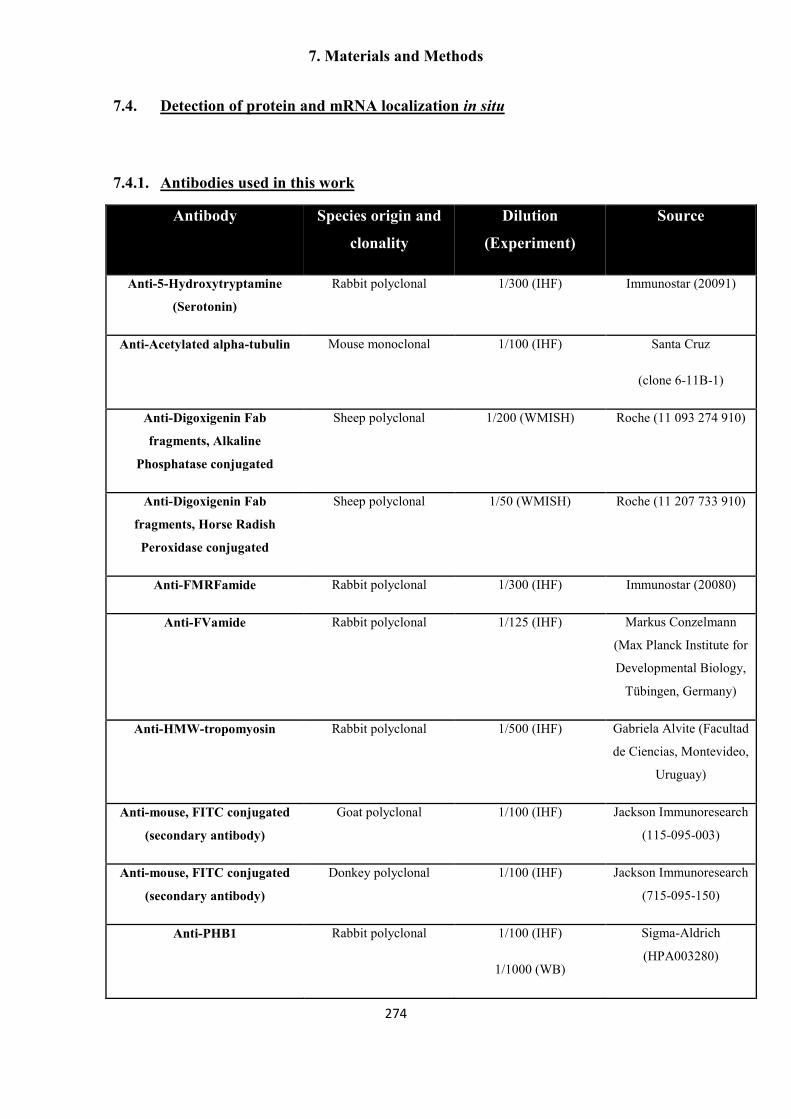
7. Materials and Methods
274
7.4. Detection of protein and mRNA localization in situ
7.4.1. Antibodies used in this work
Antibody Species origin and
clonality
Dilution
(Experiment)
Source
Anti-5-Hydroxytryptamine
(Serotonin)
Rabbit polyclonal 1/300 (IHF) Immunostar (20091)
Anti-Acetylated alpha-tubulin Mouse monoclonal 1/100 (IHF) Santa Cruz
(clone 6-11B-1)
Anti-Digoxigenin Fab
fragments, Alkaline
Phosphatase conjugated
Sheep polyclonal 1/200 (WMISH) Roche (11 093 274 910)
Anti-Digoxigenin Fab
fragments, Horse Radish
Peroxidase conjugated
Sheep polyclonal 1/50 (WMISH) Roche (11 207 733 910)
Anti-FMRFamide Rabbit polyclonal 1/300 (IHF) Immunostar (20080)
Anti-FVamide Rabbit polyclonal 1/125 (IHF) Markus Conzelmann
(Max Planck Institute for
Developmental Biology,
Tübingen, Germany)
Anti-HMW-tropomyosin Rabbit polyclonal 1/500 (IHF) Gabriela Alvite (Facultad
de Ciencias, Montevideo,
Uruguay)
Anti-mouse, FITC conjugated
(secondary antibody)
Goat polyclonal 1/100 (IHF) Jackson Immunoresearch
(115-095-003)
Anti-mouse, FITC conjugated
(secondary antibody)
Donkey polyclonal 1/100 (IHF) Jackson Immunoresearch
(715-095-150)
Anti-PHB1 Rabbit polyclonal 1/100 (IHF)
1/1000 (WB)
Sigma-Aldrich
(HPA003280)

7. Materials and Methods
275
Anti-phospho-histone H3
(Ser10)
Rabbit polyclonal 1/100 (IHF) Cell Signaling
Technology (9701)
Anti-rabbit, FITC conjugated
(secondary antibody)
Donkey polyclonal 1/100 (IHF) Jackson Immunoresearch
(711-095-152)
Anti-rabbit, Horse Radish
Peroxidase conjugated
(secondary antibody)
Goat polyclonal 1/5000 (WB) Jackson Immunoresearch
(111-055-003)
Anti-RYamide Rabbit polyclonal 1/250 (IHF) Markus Conzelmann
(Max Planck Institute for
Developmental Biology,
Tübingen, Germany)
7.4.2. Immunohistofluorescence (IHF) and immunohistochemistry (IHC)
IHF (in sections and in whole-mounts) and IHC (in sections) were performed as
described (Koziol et al. 2011; Koziol, Krohne, and Brehm 2013). Working protocols for
these techniques are included in Appendices 4 to 6.
7.4.3. Whole-mount in situ hybridization (WMISH)
In vitro transcription of digoxigenin labeled riboprobes, quantification of
riboprobes by dot blot, and WMISH were performed as described (Koziol et al. 2014).
A detailed working protocol for all of these steps, including all of the reagents and
solutions, is included in Appendices 7 to 9. The probes used for each gene are indicated
above in the list of synthetic oligonucleotides used for this work, except for the probe
used for emfr2, which consisted in a full-length CDS probe (minus the signal peptide
region), from a clone previously obtained by Monika Bergmann.
7.4.4. Epifluorescence microscopy and confocal laser scanning microscopy
Samples were analyzed by confocal microscopy (Leica TCS SP5; Leica
Microsystems, Germany) and by epifluorescence microscopy (ZeissAxio Imager.Z1
(Zeiss, Germany) and Keyence BZ9000 (Keyence, Germany)). For the quantification of

7. Materials and Methods
276
positive cells after different staining procedures, random fields were photographed and
the total number of positive cells in a given area and/or the proportion of positive cells
was manually counted (specific details for each experiment are included in the main
text). ImageJ (Rasband 1997) was used for analyzing and merging different channels,
adjusting brightness and contrast, and measuring lengths and areas.

7. Materials and Methods
277
7.5. Manipulation of proteins
7.5.1. Preparation of lysates for SDS-PAGE
Parasite material (4 to 5 metacestode vesicles) were lysed directly in 100 to 200
µl of 1 x sample buffer for SDS-PAGE (Tris-HCl (62.5 mM pH 6.8); 10 % Glycerol; 2
% (w/v) SDS, 5 % (v/v) ß-mercaptoethanol, 0.002 % Bromophenol Blue) by boiling for
15 minutes at 100 ºC.
7.5.2. Sodium Dodecyl Sulfate-Polyacrylamide Gel Electrophoresis (SDS-PAGE)
SDS-PAGE was performed as described (Sambrook, Fritsch, and Maniatis
1989), using a Biorad Mini-Protean system. The separation gel contained 10%
acrylamide, and the stacking gel contained 4% acrylamide. The molecular weight
marker Color Plus Prestained Protein Marker (10- 230 kDa) (New England Biolabs)
was included in the gel.
7.5.3. Western Blot
The proteins from the SDS-PAGE gel were transferred onto a nitrocellulose
membrane (GE Healthcare) using the Biorad Mini Trans-Blot Cell. The transfer was
carried out for 1 h at 350 mA with running buffer (25 mM Tris, pH 8.3, 193 mM
Glycin, and 20% methanol). The membrane was then blocked for 1 h at room
temperature in blocking buffer (5 % skimmed milk in TBS-T: 20 mM Tris pH 8.0; 150
mM NaCl; 0.1 % Tween-20) with shaking. The anti-PHB1 antibody (Sigma-Aldrich)
was diluted 1:1000 with blocking buffer and incubated with the membrane overnight at
4 ºC with gentle rocking. The membrane was washed 3 times, for 10 min each time, at
room temperature with TBS-T with shaking, after which it was incubated with an anti-
rabbit antibody conjugated with horse radish peroxidase (Jackson Immunoresearch,
diluted 1:5000 in blocking buffer) for 1 h at room temperature with shaking. The
membrane was washed again 3 times, for 10 min each time, with TBS-T at room
temperature with shaking and the bound antibody was visualized with a

7. Materials and Methods
278
chemiluminescent substrate (ECL-Western Blot Substrate, Pierce) by exposing X-ray-
films (Kodak). The films were developed with a Curix 60 automated developer (Agfa).

7. Materials and Methods
279
7.6. Bioinformatics and statistics
7.6.1. Datasets and programs
Genomic and predicted transcriptomic and proteomic datasets for Echinococcus
multilocularis, Echinococcus granulosus, Taenia solium, Hymenolepis microstoma
(Tsai et al. 2013) and Schistosoma mansoni (Berriman et al. 2009) were downloaded
from GeneDB (http://www.genedb.org) and used locally for blast searches, motif
searches, and alignments with Bioedit ((Hall 1999); in particular, alignments were done
with the ClustalW program (Thompson, Gibson, and Higgins 2002) included in the
Bioedit package). Visualization of predicted genes and RNAseq data was done with
IGV (Thorvaldsdóttir, Robinson, and Mesirov 2013) and Artemis (Rutherford et al.
2000). Sequences and blast analyses for other organisms were obtained from the NCBI
GenBank server (http://ncbi.nlm.nih.gov/), except for Schmidtea mediterranea
(SmedGD, Robb et al. 2007; http://smedgd.neuro.utah.edu/).
In addition, an EST database for Echinococcus spp. (Echi-EST-DB) was
assembled by Ferenc Kiss by combining the available ESTs from NCBI and diverse
unpublished EST projects and collections: from the Wellcome trust Sanger Institute
(ftp://ftp.sanger.ac.uk/pub/pathogens/Echinococcus/multilocularis/ESTs/ and
ftp://ftp.sanger.ac.uk/pub/pathogens/Echinococcus/granulosus/ESTs/), the
Echinococcus full length cDNA project (http://fullmal.hgc.jp/index_em_ajax.html), and
samples of a yeast-two-hybrid library previously sequenced in our laboratory.
Analysis of domain structure for protein sequences was performed with Interpro
Protein Sequence Analysis and Classification (http://www.ebi.ac.uk/interpro/) and
Smart analysis searches (http://smart.embl-heidelberg.de).
Phylogenetic analyses of aligned protein sequences were performed using
MEGA 5.0 (Tamura et al. 2011). Typically, maximum likelihood (ML) analyses were
performed using a substitution model with the best fit to the datasets (using the “find
best DNA/protein models (ML)” feature), and compared with Neighbor Joining (NJ)
analyses using the JTT model. In most cases and for most nodes, the results were
largely comparable and only the NJ JTT tree is shown. Bootstrap analysis of support for
individual nodes was performed with 1000 replicates.

7. Materials and Methods
280
7.6.2. Discovery of neuropeptide and peptide hormone (NP) genes in the genomes
of cestodes
Initially, a list of neuropeptides from diverse publications (McVeigh et al. 2009;
Collins et al. 2010; Veenstra 2010; Veenstra 2011) and from Neuropedia (Kim et al.
2011) was used for blastp and tblastn searches against the E. multilocularis predicted
proteins and genome contigs, respectively. A high expect value (100) was used, given
the small size and low conservation of NPs, and the results were manually inspected.
Because the first strategy was only marginally successful, and because the initial
results suggested that the usual de novo strategies for NP gene discovery would not
work in cestodes (see the Results section), we searched for novel conserved cestode NP
genes with the following strategy.
1) We searched for E. multilocularis genes with the motif {[K/R][K/R]X4-
20[K/R/stop codon]} as candidate neuropeptide encoding genes (that is, all possible
peptides that could be released from a potential precursor by canonical cleavage by
prohormone convertases).
2) We filtered these hits discarding a) Gene models with CDS coding for
proteins longer than 200 amino acids; b) Gene models with significant similarity (blastp
expect value < 10-5) to uniprot reference proteins (since they will most likely not
represent NP encoding genes, but rather other proteins with spurious similarity to the
NP; if it was indeed a NP gene with high similarity to a Uniprot protein, then it should
have been detected with the original blast strategy using well-known NP genes).
3) We analyzed all the filtered hits by tblastn against the H. microstoma genome
contigs (expect value threshold = 0.1), and manually inspected the results, looking for
high conservation within the putative mature neuropeptide sequences and proprotein
convertase sites, but lower conservation in the rest of the CDS. (Figure R5.5 shows such
an example). All hits were analyzed for the presence of 1) single or multiple copies of
putative mature peptide sequences 2) Presence of a signal peptide (SignalP 4.0 server
(Petersen et al. 2011; 2014)); 3) search for conservation of the putative mature peptides
in other flatworms and lophotrochozoans. Finally, similar sequences were searched for
by blastp and tblastn in E. granulosus and T. solium.

7. Materials and Methods
281
7.6.3. Statistics
Statistical analyses were performed using the non-parametric Mann-Whitney-
Wilcoxon rank-sum test for comparing medians in diverse experiments. The chi-square
test was used for comparing proportions of cells in cell maceration experiments, and to
compare the proportion of surviving metacestode vesicles after hydroxyurea treatment.

8. Bibliography
282
8. Bibliography
Abbott, J. C., D. M. Aanensen, K. Rutherford, S. Butcher, and B. G. Spratt. 2005. WebACT--an online companion for the Artemis Comparison Tool. Bioinformatics 21:3665-3666.
al Nahhas, S., C. Gabrion, S. Walbaum, and A. F. Petavy. 1991. In vivo cultivation of Echinococcus multilocularis protoscoleces in micropore chambers. Int J Parasitol 21:383-386.
Alberts, B. 2000. Molecular biology of the cell. Garland Science. Alvite, G., and A. Esteves. 2009. Echinococcus granulosus tropomyosin isoforms: from
gene structure to expression analysis. Gene 433:40-49. Aoki, Y., S. Nakamura, Y. Ishikawa, and M. Tanaka. 2009. Expression and syntenic
analyses of four nanos genes in medaka. Zoolog Sci 26:112-118. Archambault, V., and D. M. Glover. 2009. Polo-like kinases: conservation and
divergence in their functions and regulation. Nat Rev Mol Cell Biol 10:265-275. Ariz, M., R. Mainpal, and K. Subramaniam. 2009. C. elegans RNA-binding proteins
PUF-8 and MEX-3 function redundantly to promote germline stem cell mitosis. Dev Biol 326:295-304.
Arsac, C., S. Walbaum, M. E. Sarciron, and A. F. Petavy. 1997. Histochemical observations of alkaline phosphatase activity of Echinococcus multilocularis during in vivo development in golden hamsters, an alternative definitive host. Exp Anim 46:25-30.
Attenborough, R. M., D. C. Hayward, M. V. Kitahara, D. J. Miller, and E. E. Ball. 2012. A "neural" enzyme in nonbilaterian animals and algae: preneural origins for peptidylglycine alpha-amidating monooxygenase. Mol Biol Evol 29:3095-3109.
Auladell, C., J. Garcia-Valero, and J. Baguña 1993. Ultrastructural localization of RNA in the chromatoid bodies of undifferentiated cells (neoblasts) in planarians by the RNase-gold complex technique. Journal of morphology 216:319-326.
Ax, P. 1996. Multicellular Animals - A New Approach to the Phylogenetic Order in Nature – Vol I, Springer Verlag, Berlin.
Baguna, J. 2012. The planarian neoblast: the rambling history of its origin and some current black boxes. Int J Dev Biol 56:19-37.
Baguñà, J., and M. Riutort. 2004. Molecular phylogeny of the Platyhelminthes. Canadian Journal of Zoology 82:168-193.
Baguñà, J., and R. Romero. 1981. Quantitative analysis of cell types during growth, degrowth and regeneration in the planarians Dugesia (S) mediterranea and Dugesia (G) tigrina. Hydrobiologia 84:181-194.
Baguñà, J., E. Saló, and C. Auladell. 1989. Regeneration and pattern formation in planarians. III. Evidence that neoblasts are totipotent stem cells and the source of blastema cells. Development 108:77-86.
Ballandras-Colas, A., H. Naraharisetty, X. Li, E. Serrao, and A. Engelman. 2013. Biochemical characterization of novel retroviral integrase proteins. PLoS One 8:e76638.

8. Bibliography
283
Barkauskas, C. E., M. J. Cronce, C. R. Rackley, E. J. Bowie, D. R. Keene, B. R. Stripp, S. H. Randell, P. W. Noble, and B. L. Hogan. 2013. Type 2 alveolar cells are stem cells in adult lung. J Clin Invest 123:3025-3036.
Barker, N., S. Bartfeld, and H. Clevers. 2010. Tissue-resident adult stem cell populations of rapidly self-renewing organs. Cell Stem Cell 7:656-670.
Barker, N., and H. Clevers. 2010. Leucine-rich repeat-containing G-protein-coupled receptors as markers of adult stem cells. Gastroenterology 138:1681-1696.
Barker, N., M. Huch, P. Kujala, M. van de Wetering, H. J. Snippert, J. H. van Es, T. Sato, D. E. Stange, H. Begthel, M. van den Born, E. Danenberg, S. van den Brink, J. Korving, A. Abo, P. J. Peters, N. Wright, R. Poulsom, and H. Clevers. 2010. Lgr5(+ve) stem cells drive self-renewal in the stomach and build long-lived gastric units in vitro. Cell Stem Cell 6:25-36.
Barker, N., J. H. van Es, J. Kuipers, P. Kujala, M. van den Born, M. Cozijnsen, A. Haegebarth, J. Korving, H. Begthel, P. J. Peters, and H. Clevers. 2007. Identification of stem cells in small intestine and colon by marker gene Lgr5. Nature 449:1003-1007.
Batut, P., A. Dobin, C. Plessy, P. Carninci, and T. R. Gingeras. 2013. High-fidelity promoter profiling reveals widespread alternative promoter usage and transposon-driven developmental gene expression. Genome Res 23:169-180.
Beard, T. C. 1973. The elimination of echinococcosis from Iceland. Bulletin of the World Health Organization 48:653.
Benovoy, D., and G. Drouin. 2009. Ectopic gene conversions in the human genome. Genomics 93:27-32.
Berriman, M., B. J. Haas, P. T. LoVerde, R. A. Wilson, G. P. Dillon, G. C. Cerqueira, S. T. Mashiyama, B. Al-Lazikani, L. F. Andrade, P. D. Ashton, M. A. Aslett, D. C. Bartholomeu, G. Blandin, C. R. Caffrey, A. Coghlan, R. Coulson, T. A. Day, A. Delcher, R. DeMarco, A. Djikeng, T. Eyre, J. A. Gamble, E. Ghedin, Y. Gu, C. Hertz-Fowler, H. Hirai, Y. Hirai, R. Houston, A. Ivens, D. A. Johnston, D. Lacerda, C. D. Macedo, P. McVeigh, Z. Ning, G. Oliveira, J. P. Overington, J. Parkhill, M. Pertea, R. J. Pierce, A. V. Protasio, M. A. Quail, M. A. Rajandream, J. Rogers, M. Sajid, S. L. Salzberg, M. Stanke, A. R. Tivey, O. White, D. L. Williams, J. Wortman, W. Wu, M. Zamanian, A. Zerlotini, C. M. Fraser-Liggett, B. G. Barrell, and N. M. El-Sayed. 2009. The genome of the blood fluke Schistosoma mansoni. Nature 460:352-358.
Bode, A., W. Salvenmoser, K. Nimeth, M. Mahlknecht, Z. Adamski, R. M. Rieger, R. Peter, and P. Ladurner. 2006. Immunogold-labeled S-phase neoblasts, total neoblast number, their distribution, and evidence for arrested neoblasts in Macrostomum lignano (Platyhelminthes, Rhabditophora). Cell Tissue Res 325:577-587.
Boeke, J. D., and J. P. Stoye. 1997. Retrotransposons, Endogenous Retroviruses, and the Evolution of Retroelements in J. M. Coffin, S. H. Hughes, and H. E. Varmus, eds. Retroviruses. Cold Spring Harbor Laboratory Press, Cold Spring Harbor (NY).
Bolla, R. I., and L. S. Roberts. 1971. Developmental physiology of cestodes. IX. Cytological characteristics of the germinative region of Hymenolepis diminuta. J Parasitol 57:267-277.
Bowen, N. J., and J. F. McDonald. 2001. Drosophila euchromatic LTR retrotransposons are much younger than the host species in which they reside. Genome Res 11:1527-1540.

8. Bibliography
284
Bowles, J., G. Schepers, and P. Koopman. 2000. Phylogeny of the SOX family of developmental transcription factors based on sequence and structural indicators. Dev Biol 227:239-255.
Bradbury, A. F., and D. G. Smyth. 1991. Peptide amidation. Trends Biochem Sci 16:112-115.
Brawley, C., and E. Matunis. 2004. Regeneration of male germline stem cells by spermatogonial dedifferentiation in vivo. Science 304:1331-1334.
Brehm, K. 2010a. The role of evolutionarily conserved signalling systems in Echinococcus multilocularis development and host-parasite interaction. Med Microbiol Immunol 199:247-259.
Brehm, K. 2010b. Echinococcus multilocularis as an experimental model in stem cell research and molecular host-parasite interaction. Parasitology 137:537-555.
Brehm, K., and M. Spiliotis. 2008a. Recent advances in the in vitro cultivation and genetic manipulation of Echinococcus multilocularis metacestodes and germinal cells. Exp Parasitol 119:506-515.
Brehm, K., and M. Spiliotis. 2008b. The influence of host hormones and cytokines on Echinococcus multilocularis signalling and development. Parasite 15:286-290.
Brehm, K., M. Wolf, H. Beland, A. Kroner, and M. Frosch. 2003. Analysis of differential gene expression in Echinococcus multilocularis larval stages by means of spliced leader differential display. Int J Parasitol 33:1145-1159.
Brennan, G. P., D. W. Halton, A. G. Maule, and C. Shaw. 1993. Electron immunogold labeling of regulatory peptide immunoreactivity in the nervous system of Moniezia expansa (Cestoda: Cyclophyllidea). Parasitol Res 79:409-415.
Brennand, K., D. Huangfu, and D. Melton. 2007. All beta cells contribute equally to islet growth and maintenance. PLoS Biol 5:e163.
Bresnick, E. H., K. R. Katsumura, H. Y. Lee, K. D. Johnson, and A. S. Perkins. 2012. Master regulatory GATA transcription factors: mechanistic principles and emerging links to hematologic malignancies. Nucleic Acids Res 40:5819-5831.
Brownlee, D. J., I. Fairweather, C. F. Johnston, and M. T. Rogan. 1994. Immunocytochemical localization of serotonin (5-HT) in the nervous system of the hydatid organism, Echinococcus granulosus (Cestoda, Cyclophyllidea). Parasitology 109 ( Pt 2):233-241.
Brulet, P., P. Duprey, M. Vasseur, M. Kaghad, D. Morello, P. Blanchet, C. Babinet, H. Condamine, and F. Jacob. 1985. Molecular analysis of the first differentiations in the mouse embryo. Cold Spring Harb Symp Quant Biol 50:51-57.
Brunetti, E., P. Kern, and D. A. Vuitton. 2010. Expert consensus for the diagnosis and treatment of cystic and alveolar echinococcosis in humans. Acta Trop 114:1-16.
Brunmeir, R., S. Lagger, and C. Seiser. 2009. Histone deacetylase HDAC1/HDAC2-controlled embryonic development and cell differentiation. Int J Dev Biol 53:275-289.
Bryder, D., D. J. Rossi, and I. L. Weissman. 2006. Hematopoietic stem cells: the paradigmatic tissue-specific stem cell. Am J Pathol 169:338-346.
Buczacki, S. J., H. I. Zecchini, A. M. Nicholson, R. Russell, L. Vermeulen, R. Kemp, and D. J. Winton. 2013. Intestinal label-retaining cells are secretory precursors expressing Lgr5. Nature 495:65-69.
Cai, P., X. Piao, N. Hou, S. Liu, H. Wang, and Q. Chen. 2012. Identification and characterization of argonaute protein, Ago2 and its associated small RNAs in Schistosoma japonicum. PLoS Negl Trop Dis 6:e1745.

8. Bibliography
285
Cameron, H. A., T. G. Hazel, and R. D. McKay. 1998. Regulation of neurogenesis by growth factors and neurotransmitters. J Neurobiol 36:287-306.
Camicia, F., M. Herz, L. C. Prada, L. Kamenetzky, S. H. Simonetta, M. A. Cucher, J. I. Bianchi, C. Fernandez, K. Brehm, and M. C. Rosenzvit. 2013. The nervous and prenervous roles of serotonin in Echinococcus spp. Int J Parasitol 43:647-659.
Carmell, M. A., Z. Xuan, M. Q. Zhang, and G. J. Hannon. 2002. The Argonaute family: tentacles that reach into RNAi, developmental control, stem cell maintenance, and tumorigenesis. Genes Dev 16:2733-2742.
Carmon, K. S., X. Gong, Q. Lin, A. Thomas, and Q. Liu. 2011. R-spondins function as ligands of the orphan receptors LGR4 and LGR5 to regulate Wnt/beta-catenin signaling. Proc Natl Acad Sci U S A 108:11452-11457.
Clevers, H. 2006. Wnt/beta-catenin signaling in development and disease. Cell 127:469-480.
Cohen, C. J., W. M. Lock, and D. L. Mager. 2009. Endogenous retroviral LTRs as promoters for human genes: a critical assessment. Gene 448:105-114.
Cohn, L. 1899. Untersuchungen über das centrale Nervensystem der Cestoden. Zool. Jahrb. 12:89-160.
Collin, W. K. 1969. The cellular organization of hatched oncospheres of Hymenolepis
citelli (Cestoda, Cyclophyllidea). J Parasitol 55:149-166. Collins, J. J., 3rd, X. Hou, E. V. Romanova, B. G. Lambrus, C. M. Miller, A. Saberi, J.
V. Sweedler, and P. A. Newmark. 2010. Genome-wide analyses reveal a role for peptide hormones in planarian germline development. PLoS Biol 8:e1000509.
Collins, J. J., 3rd, R. S. King, A. Cogswell, D. L. Williams, and P. A. Newmark. 2011. An atlas for Schistosoma mansoni organs and life-cycle stages using cell type-specific markers and confocal microscopy. PLoS Negl Trop Dis 5:e1009.
Collins, J. J., 3rd, B. Wang, B. G. Lambrus, M. E. Tharp, H. Iyer, and P. A. Newmark. 2013. Adult somatic stem cells in the human parasite Schistosoma mansoni. Nature 494:476-479.
Conzelmann, M., and G. Jekely. 2012. Antibodies against conserved amidated neuropeptide epitopes enrich the comparative neurobiology toolbox. Evodevo 3:23.
Coutelen, F., J. Biguet, J. M. Doby, and S. Deblock. 1952. Le systéme musculaire du scolex echinococcique. Mécanismes de dévagination et d´invagination du rostre et des ventouses. Ann Parasitol Hum Comp 27:86-104.
Coutu, D. L., and J. Galipeau. 2011. Roles of FGF signaling in stem cell self-renewal, senescence and aging. Aging (Albany NY) 3:920-933.
Cox, W. G., and V. L. Singer. 1999. A high-resolution, fluorescence-based method for localization of endogenous alkaline phosphatase activity. J Histochem Cytochem 47:1443-1456.
Craig, P. 2003. Echinococcus multilocularis. Curr Opin Infect Dis 16:437-444. Crittenden, S. L., C. R. Eckmann, L. Wang, D. S. Bernstein, M. Wickens, and J.
Kimble. 2003. Regulation of the mitosis/meiosis decision in the Caenorhabditis
elegans germline. Philos Trans R Soc Lond B Biol Sci 358:1359-1362. Crusz, H. 1947. The early development of the rostellum of Cysticercus fasciolaris Rud.,
and the chemical nature of its hooks. J Parasitol 33:87-98. Cucher, M., G. Mourglia-Ettlin, L. Prada, H. Costa, L. Kamenetzky, C. Poncini, S.
Dematteis, and M. C. Rosenzvit. 2013. Echinococcus granulosus pig strain (G7 genotype) protoscoleces did not develop secondary hydatid cysts in mice. Vet Parasitol 193:185-192.

8. Bibliography
286
Cunningham, L. J., and P. D. Olson. 2010. Description of Hymenolepis microstoma (Nottingham strain): a classical tapeworm model for research in the genomic era. Parasit Vectors 3:123.
Curtis, D., D. K. Treiber, F. Tao, P. D. Zamore, J. R. Williamson, and R. Lehmann. 1997. A CCHC metal-binding domain in Nanos is essential for translational regulation. Embo J 16:834-843.
Chen, J. M., D. N. Cooper, N. Chuzhanova, C. Ferec, and G. P. Patrinos. 2007. Gene conversion: mechanisms, evolution and human disease. Nat Rev Genet 8:762-775.
Chitnis, A. B., and J. Y. Kuwada. 1990. Axonogenesis in the brain of zebrafish embryos. J Neurosci 10:1892-1905.
D'Alessandro, A., and R. L. Rausch. 2008. New aspects of neotropical polycystic (Echinococcus vogeli) and unicystic (Echinococcus oligarthrus) echinococcosis. Clin Microbiol Rev 21:380-401, table of contents.
Daubert, E. A., and B. G. Condron. 2010. Serotonin: a regulator of neuronal morphology and circuitry. Trends Neurosci 33:424-434.
David, C. N. 1973. A Quantitative Method for Maceration of Hydra Tissue. Wilhelm Roux' Archiv 171:259-268.
de Lau, W., N. Barker, T. Y. Low, B. K. Koo, V. S. Li, H. Teunissen, P. Kujala, A. Haegebarth, P. J. Peters, M. van de Wetering, D. E. Stange, J. E. van Es, D. Guardavaccaro, R. B. Schasfoort, Y. Mohri, K. Nishimori, S. Mohammed, A. J. Heck, and H. Clevers. 2011. Lgr5 homologues associate with Wnt receptors and mediate R-spondin signalling. Nature 476:293-297.
De Mulder, K., G. Kuales, D. Pfister, B. Egger, T. Seppi, P. Eichberger, G. Borgonie, and P. Ladurner. 2010. Potential of Macrostomum lignano to recover from gamma-ray irradiation. Cell Tissue Res 339:527-542.
De Mulder, K., G. Kuales, D. Pfister, M. Willems, B. Egger, W. Salvenmoser, M. Thaler, A. K. Gorny, M. Hrouda, G. Borgonie, and P. Ladurner. 2009a. Characterization of the stem cell system of the acoel Isodiametra pulchra. BMC Dev Biol 9:69.
De Mulder, K., D. Pfister, G. Kuales, B. Egger, W. Salvenmoser, M. Willems, J. Steger, K. Fauster, R. Micura, G. Borgonie, and P. Ladurner. 2009b. Stem cells are differentially regulated during development, regeneration and homeostasis in flatworms. Dev Biol 334:198-212.
del Cacho, E., C. Causape, C. Sanchez-Acedo, and J. Quilez. 1996. Cytochemical study of the germinal membrane of the Echinococcus granulosus cyst. Vet Parasitol 62:101-106.
Desai, T. J., D. G. Brownfield, and M. A. Krasnow. 2014. Alveolar progenitor and stem cells in lung development, renewal and cancer. Nature 507:190-194.
Diaz, A., C. Casaravilla, F. Irigoin, G. Lin, J. O. Previato, and F. Ferreira. 2011. Understanding the laminated layer of larval Echinococcus I: structure. Trends Parasitol 27:204-213.
Dickinson, A. J., R. P. Croll, and E. E. Voronezhskaya. 2000. Development of embryonic cells containing serotonin, catecholamines, and FMRFamide-related peptides in Aplysia californica. Biological Bulletin 199:305-315.
Dirks, U., H. R. Gruber-Vodicka, B. Egger, and J. A. Ott. 2012a. Proliferation pattern during rostrum regeneration of the symbiotic flatworm Paracatenula galateia: a pulse-chase-pulse analysis. Cell Tissue Res 349:517-525.

8. Bibliography
287
Dirks, U., H. R. Gruber-Vodicka, N. Leisch, S. Bulgheresi, B. Egger, P. Ladurner, and J. A. Ott. 2012b. Bacterial symbiosis maintenance in the asexually reproducing and regenerating flatworm Paracatenula galateia. PLoS One 7:e34709.
Domen, J., A. Wagers, and I. L. Weissman. 2006. Bone marrow (hematopoietic) stem cells. Pp. 13. Regenerative medicine. Stem Cell Information from the National Institutes of Health Resource for Stem Cell Research. United States
Dominguez, M. F., U. Koziol, V. Porro, A. Costabile, S. Estrade, J. Tort, M. Bollati-Fogolin, and E. Castillo. 2014. A new approach for the characterization of proliferative cells in cestodes. Exp Parasitol 138:25-29.
Dor, Y., J. Brown, O. I. Martinez, and D. A. Melton. 2004. Adult pancreatic beta-cells are formed by self-duplication rather than stem-cell differentiation. Nature 429:41-46.
Dorrell, C., L. Erker, J. Schug, J. L. Kopp, P. S. Canaday, A. J. Fox, O. Smirnova, A. W. Duncan, M. J. Finegold, M. Sander, K. H. Kaestner, and M. Grompe. 2011. Prospective isolation of a bipotential clonogenic liver progenitor cell in adult mice. Genes Dev 25:1193-1203.
Dos Santos, L. V., M. V. de Queiroz, M. F. Santana, M. A. Soares, E. G. de Barros, E. F. de Araujo, and T. Langin. 2012. Development of new molecular markers for the Colletotrichum genus using RetroCl1 sequences. World J Microbiol Biotechnol 28:1087-1095.
Douglas, L. T. 1961. The development of organ systems in nematotaeniid cestodes. I. Early histogenesis and formation of reproductive structures in Baerietta diana (Helfer. 1948). J Parasitol 47:669-680.
Doupe, D. P., and P. H. Jones. 2013. Cycling progenitors maintain epithelia while diverse cell types contribute to repair. Bioessays 35:443-451.
Dupressoir, A., and T. Heidmann. 1996. Germ line-specific expression of intracisternal A-particle retrotransposons in transgenic mice. Mol Cell Biol 16:4495-4503.
Dykstra, B., D. Kent, M. Bowie, L. McCaffrey, M. Hamilton, K. Lyons, S. J. Lee, R. Brinkman, and C. Eaves. 2007. Long-term propagation of distinct hematopoietic differentiation programs in vivo. Cell Stem Cell 1:218-229.
Eckert, J., and P. Deplazes. 2004. Biological, epidemiological, and clinical aspects of echinococcosis, a zoonosis of increasing concern. Clin Microbiol Rev 17:107-135.
Eckert, J., R. C. Thompson, and H. Mehlhorn. 1983. Proliferation and metastases formation of larval Echinococcus multilocularis. I. Animal model, macroscopical and histological findings. Z Parasitenkd 69:737-748.
Egger, B., R. Gschwentner, and R. Rieger. 2007. Free-living flatworms under the knife: past and present. Dev Genes Evol 217:89-104.
Eisenhoffer, G. T., H. Kang, and A. Sanchez Alvarado. 2008. Molecular analysis of stem cells and their descendants during cell turnover and regeneration in the planarian Schmidtea mediterranea. Cell Stem Cell 3:327-339.
Evsikov, A. V., W. N. de Vries, A. E. Peaston, E. E. Radford, K. S. Fancher, F. H. Chen, J. A. Blake, C. J. Bult, K. E. Latham, D. Solter, and B. B. Knowles. 2004. Systems biology of the 2-cell mouse embryo. Cytogenet Genome Res 105:240-250.
Ewen-Campen, B., E. E. Schwager, and C. G. Extavour. 2010. The molecular machinery of germ line specification. Mol Reprod Dev 77:3-18.
Extavour, C. G., and M. Akam. 2003. Mechanisms of germ cell specification across the metazoans: epigenesis and preformation. Development 130:5869-5884.

8. Bibliography
288
Fairley, N. H., and R. J. Wright-Smith. 1929. Hydatid infestation (Echinococcus granulosus) in sheep, oxen and pigs, with special reference to daughter cyst formation. The Journal of Pathology and Bacteriology 32:309-335.
Fairweather, I., A. E. A. Campbell, P. J. Skuce, C. F. Johnston, and C. Shaw. 1991. Development of the peptidergic nervous system during ontogeny of the cysticercus larva of the tapeworm Taenia crassiceps. Regulatory Peptides 35:235.
Fairweather, I., G. A. Macartney, C. F. Johnston, D. W. Halton, and K. D. Buchnan. 1988. Immunocytochemical demonstration of 5-hydroxytryptamine (serotonin) and vertebrate neuropeptides in the nervous system of excysted cysticercoid larvae of the rat tapeworm, Hymenolepis diminuta (Cestoda, Cyclophyllidea). Parasitol Res 74:371-379.
Fairweather, I., M. T. McMullan, C. F. Johnston, M. T. Rogan, and R. E. Hanna. 1994. Serotoninergic and peptidergic nerve elements in the protoscolex of Echinococcus granulosus (Cestoda, Cyclophyllidea). Parasitol Res 80:649-656.
Farber, E., and R. Baserga. 1969. Differential effects of hydroxyurea on survival of proliferating cells in vivo. Cancer Res 29:136-139.
Faulkner, G. J., Y. Kimura, C. O. Daub, S. Wani, C. Plessy, K. M. Irvine, K. Schroder, N. Cloonan, A. L. Steptoe, T. Lassmann, K. Waki, N. Hornig, T. Arakawa, H. Takahashi, J. Kawai, A. R. Forrest, H. Suzuki, Y. Hayashizaki, D. A. Hume, V. Orlando, S. M. Grimmond, and P. Carninci. 2009. The regulated retrotransposon transcriptome of mammalian cells. Nat Genet 41:563-571.
Fausto, N., and J. S. Campbell. 2003. The role of hepatocytes and oval cells in liver regeneration and repopulation. Mech Dev 120:117-130.
Feschotte, C., N. Jiang, and S. R. Wessler. 2002. Plant transposable elements: where genetics meets genomics. Nat Rev Genet 3:329-341.
Förster, S. 2012. Nuclear Hormone Receptors and Fibroblast Growth Factor Receptor Signaling in Echinococcus multilocularis. University of Würzburg, Würzburg.
Fort, A., K. Hashimoto, D. Yamada, M. Salimullah, C. A. Keya, A. Saxena, A. Bonetti, I. Voineagu, N. Bertin, A. Kratz, Y. Noro, C. H. Wong, M. de Hoon, R. Andersson, A. Sandelin, H. Suzuki, C. L. Wei, H. Koseki, Y. Hasegawa, A. R. Forrest, and P. Carninci. 2014. Deep transcriptome profiling of mammalian stem cells supports a regulatory role for retrotransposons in pluripotency maintenance. Nat Genet 46:558-566.
Franchini, A. 2005. The distribution of cells containing FMRFamide- and 5-HT-related molecules in the embryonic development of Viviparus ater (Mollusca, Gastropoda). Eur J Histochem 49:301-308.
Freeman, R. S. 1973. Ontogeny of cestodes and its bearing on their phylogeny and systematics. Adv Parasitol 11:481-557.
Friedl, A. A., M. Kiechle, H. G. Maxeiner, R. H. Schiestl, and F. Eckardt-Schupp. 2010. Ty1 integrase overexpression leads to integration of non-Ty1 DNA fragments into the genome of Saccharomyces cerevisiae. Mol Genet Genomics 284:231-242.
Frooninckx, L., L. Van Rompay, L. Temmerman, E. Van Sinay, I. Beets, T. Janssen, S. J. Husson, and L. Schoofs. 2012. Neuropeptide GPCRs in C. elegans. Front Endocrinol (Lausanne) 3:167.
Gafni, O., L. Weinberger, A. A. Mansour, Y. S. Manor, E. Chomsky, D. Ben-Yosef, Y. Kalma, S. Viukov, I. Maza, A. Zviran, Y. Rais, Z. Shipony, Z. Mukamel, V. Krupalnik, M. Zerbib, S. Geula, I. Caspi, D. Schneir, T. Shwartz, S. Gilad, D.
8. Bibliography
289
Amann-Zalcenstein, S. Benjamin, I. Amit, A. Tanay, R. Massarwa, N. Novershtern, and J. H. Hanna. 2013. Derivation of novel human ground state naive pluripotent stem cells. Nature 504:282-286.
Galindo, M., R. Paredes, C. Marchant, V. Mino, and N. Galanti. 2003. Regionalization of DNA and protein synthesis in developing stages of the parasitic platyhelminth Echinococcus granulosus. J Cell Biochem 90:294-303.
Galindo, M., G. Schadebrodt, and N. Galanti. 2008. Echinococcus granulosus: cellular territories and morphological regions in mature protoscoleces. Exp Parasitol 119:524-533.
Gelmedin, V. 2008. Targeting flatworm signaling cascades for the development of novel anthelmintic drugs. University of Würzburg, Würzburg.
Gelmedin, V., R. Caballero-Gamiz, and K. Brehm. 2008. Characterization and inhibition of a p38-like mitogen-activated protein kinase (MAPK) from Echinococcus multilocularis: antiparasitic activities of p38 MAPK inhibitors. Biochem Pharmacol 76:1068-1081.
Gelmedin, V., M. Spiliotis, and K. Brehm. 2010. Molecular characterisation of MEK1/2- and MKK3/6-like mitogen-activated protein kinase kinases (MAPKK) from the fox tapeworm Echinococcus multilocularis. Int J Parasitol 40:555-567.
Gilbert, S. F. 2006. Developmental Biology, 8th edition. Sinauer Sunderland, Massachusetts.
Gillooly, J. F., A. P. Allen, G. B. West, and J. H. Brown. 2005. The rate of DNA evolution: effects of body size and temperature on the molecular clock. Proc Natl Acad Sci U S A 102:140-145.
Glinka, A., C. Dolde, N. Kirsch, Y. L. Huang, O. Kazanskaya, D. Ingelfinger, M. Boutros, C. M. Cruciat, and C. Niehrs. 2011. LGR4 and LGR5 are R-spondin receptors mediating Wnt/beta-catenin and Wnt/PCP signalling. EMBO Rep 12:1055-1061.
Goldschmidt, R. 1900. Zur Entwicklungsgeschichte der Echinococcusköpfchen. Zool. Jahrb. 13.
Greenspan, P., E. P. Mayer, and S. D. Fowler. 1985. Nile red: a selective fluorescent stain for intracellular lipid droplets. J Cell Biol 100:965-973.
Guo, T., A. H. Peters, and P. A. Newmark. 2006. A Bruno-like gene is required for stem cell maintenance in planarians. Dev Cell 11:159-169.
Gustafsson, M. K. 1976a. Observations on the histogenesis of nervous tissue in Diphyllobothrium dendriticum Nitzsch, 1824 (Cestoda, Pseudophyllidea). Z Parasitenkd 50:313-321.
Gustafsson, M. K. 1976b. Studies on cytodifferentiation in the neck region of Diphyllobothrium dendriticum Nitzsch, 1824 (Cestoda, Pseudophyllidea). Z Parasitenkd 50:323-329.
Gustafsson, M. K. 1992. The neuroanatomy of parasitic flatworms. Adv Neuroimmunol. 2:267-286.
Gustafsson, M. K. S. 1976c. Basic cell types in Echinococcus granulosus (Cestoda, Cyclophyllidea). Acta Zool. Fenn. 146:1-16.
Gustafsson, M. K. S. 1990. The cells of a cestode. Diphyllobothrium dendriticum as a model in cell biology. Acta Acad Abo Ser B 50:13-44.
Haag, K. L., P. B. Marin, D. A. Graichen, and M. L. De La Rue. 2011. Reappraising the theme of breeding systems in Echinococcus: is outcrossing a rare phenomenon? Parasitology 138:298-302.

8. Bibliography
290
Halton, D. W., and M. K. S. Gustafsson. 1996. Functional morphology of the Platyhelminth Nervous System. Parasitology 113:547-572.
Halton, D. W., and A. G. Maule. 2003. Flatworm nerve–muscle: structural and functional analysis. Can J Zool 82:316-333.
Hall, T. A. 1999. BioEdit: a user-friendly biological sequence alignment editor and analysis program for Windows 95/98/NT. Pp. 95-98. Nucleic acids symposium series.
Handberg-Thorsager, M., and E. Salo. 2007. The planarian nanos-like gene Smednos is expressed in germline and eye precursor cells during development and regeneration. Dev Genes Evol 217:403-411.
Hanna, J. H., K. Saha, and R. Jaenisch. 2010. Pluripotency and cellular reprogramming: facts, hypotheses, unresolved issues. Cell 143:508-525.
Haraguchi, S., M. Tsuda, S. Kitajima, Y. Sasaoka, A. Nomura-Kitabayashid, K. Kurokawa, and Y. Saga. 2003. nanos1: a mouse nanos gene expressed in the central nervous system is dispensable for normal development. Mech Dev 120:721-731.
Hartenstein, V., and M. Jones. 2003. The embryonic development of the bodywall and nervous system of the cestode flatworm Hymenolepis diminuta. Cell Tissue Res 311:427-435.
Harzsch, S., and C. H. Muller. 2007. A new look at the ventral nerve centre of Sagitta: implications for the phylogenetic position of Chaetognatha (arrow worms) and the evolution of the bilaterian nervous system. Front Zool 4:14.
Hastings, K. E. 2005. SL trans-splicing: easy come or easy go? Trends Genet 21:240-247.
Havecker, E. R., X. Gao, and D. F. Voytas. 2004. The diversity of LTR retrotransposons. Genome Biol 5:225.
Hay, E. D., and S. J. Coward. 1975. Fine structure studies on the planarian, Dugesia. I. Nature of the "neoblast" and other cell types in noninjured worms. J Ultrastruct Res 50:1-21.
Hayashi, T., M. Asami, S. Higuchi, N. Shibata, and K. Agata. 2006. Isolation of planarian X-ray-sensitive stem cells by fluorescence-activated cell sorting. Dev Growth Differ 48:371-380.
Heeney, M. M., M. R. Whorton, T. A. Howard, C. A. Johnson, and R. E. Ware. 2004. Chemical and functional analysis of hydroxyurea oral solutions. J Pediatr Hematol Oncol 26:179-184.
Hemer, S., C. Konrad, M. Spiliotis, U. Koziol, D. Schaack, S. Forster, V. Gelmedin, B. Stadelmann, T. Dandekar, A. Hemphill, and K. Brehm. 2014. Host insulin stimulates Echinococcus multilocularis insulin signalling pathways and larval development. BMC Biol 12:5.
Henderson, D. J., and R. E. Hanna. 1988. Hymenolepis nana (Cestoda:Cyclophyllidea): DNA, RNA and protein synthesis in 5-day-old juveniles. Int J Parasitol 18:963-972.
Hendzel, M. J., Y. Wei, M. A. Mancini, A. Van Hooser, T. Ranalli, B. R. Brinkley, D. P. Bazett-Jones, and C. D. Allis. 1997. Mitosis-specific phosphorylation of histone H3 initiates primarily within pericentromeric heterochromatin during G2 and spreads in an ordered fashion coincident with mitotic chromosome condensation. Chromosoma 106:348-360.
8. Bibliography
291
Higuchi, S., T. Hayashi, I. Hori, N. Shibata, H. Sakamoto, and K. Agata. 2007. Characterization and categorization of fluorescence activated cell sorted planarian stem cells by ultrastructural analysis. Dev Growth Differ 49:571-581.
Hindmarsh, P., and J. Leis. 1999. Retroviral DNA integration. Microbiol Mol Biol Rev 63:836-843, table of contents.
Hoberg, E. P. 2006. Phylogeny of Taenia: Species definitions and origins of human parasites. Parasitol Int 55 Suppl:S23-30.
Hoberg, E. P., A. Jones, R. L. Rausch, K. S. Eom, and S. L. Gardner. 2000. A phylogenetic hypothesis for species of the genus Taenia (Eucestoda : Taeniidae). J Parasitol 86:89-98.
Hock, J., and G. Meister. 2008. The Argonaute protein family. Genome Biol 9:210. Hoffman, M. M., and E. Birney. 2007. Estimating the neutral rate of nucleotide
substitution using introns. Mol Biol Evol 24:522-531. Hopman, A. H., F. C. Ramaekers, and E. J. Speel. 1998. Rapid synthesis of biotin-,
digoxigenin-, trinitrophenyl-, and fluorochrome-labeled tyramides and their application for In situ hybridization using CARD amplification. J Histochem Cytochem 46:771-777.
Hoyer, D., and T. Bartfai. 2012. Neuropeptides and neuropeptide receptors: drug targets, and peptide and non-peptide ligands: a tribute to Prof. Dieter Seebach. Chem Biodivers 9:2367-2387.
Huch, M., S. F. Boj, and H. Clevers. 2013. Lgr5(+) liver stem cells, hepatic organoids and regenerative medicine. Regen Med 8:385-387.
Hyman, L. H. 1951. The Invertebrates: Platyhelminthes and Rhynchocoela The Acoelomate Bilateria. Volume II McGraw-Hill.
Inoue, C., S. K. Bae, K. Takatsuka, T. Inoue, Y. Bessho, and R. Kageyama. 2001. Math6, a bHLH gene expressed in the developing nervous system, regulates neuronal versus glial differentiation. Genes Cells 6:977-986.
Jabbar, A., S. Crawford, D. Mlocicki, Z. P. Swiderski, D. B. Conn, M. K. Jones, I. Beveridge, and M. W. Lightowlers. 2010. Ultrastructural reconstruction of Taenia ovis oncospheres from serial sections. Int J Parasitol.
Jaeger, S., S. Barends, R. Giege, G. Eriani, and F. Martin. 2005. Expression of metazoan replication-dependent histone genes. Biochimie 87:827-834.
Jaisser, F. 2000. Inducible gene expression and gene modification in transgenic mice. J Am Soc Nephrol 11 Suppl 16:S95-S100.
Jaruzelska, J., M. Kotecki, K. Kusz, A. Spik, M. Firpo, and R. A. Reijo Pera. 2003. Conservation of a Pumilio-Nanos complex from Drosophila germ plasm to human germ cells. Dev Genes Evol 213:120-126.
Jellies, J., D. M. Kopp, K. M. Johansen, and J. Johansen. 1996. Initial Formation and Secondary Condensation of Nerve Pathways in the Medicinal Leech. J Comp Neurol 373:1-10.
Jiang, N., I. K. Jordan, and S. R. Wessler. 2002. Dasheng and RIRE2. A nonautonomous long terminal repeat element and its putative autonomous partner in the rice genome. Plant Physiol 130:1697-1705.
Jones, S. 2004. An overview of the basic helix-loop-helix proteins. Genome Biol 5:226. Juliano, C., J. Wang, and H. Lin. 2011. Uniting germline and stem cells: the function of
Piwi proteins and the piRNA pathway in diverse organisms. Annu Rev Genet 45:447-469.
Juliano, C. E., S. Z. Swartz, and G. M. Wessel. 2010. A conserved germline multipotency program. Development 137:4113-4126.

8. Bibliography
292
Jura, H., A. Bader, M. Hartmann, H. Maschek, and M. Frosch. 1996. Hepatic tissue culture model for study of host-parasite interactions in alveolar echinococcosis. Infect Immun 64:3484-3490.
Kadyrova, L. Y., Y. Habara, T. H. Lee, and R. P. Wharton. 2007. Translational control of maternal Cyclin B mRNA by Nanos in the Drosophila germline. Development 134:1519-1527.
Kalendar, R., J. Tanskanen, W. Chang, K. Antonius, H. Sela, O. Peleg, and A. H. Schulman. 2008. Cassandra retrotransposons carry independently transcribed 5S RNA. Proc Natl Acad Sci U S A 105:5833-5838.
Kamachi, Y., and H. Kondoh. 2013. Sox proteins: regulators of cell fate specification and differentiation. Development 140:4129-4144.
Kanska, J., and U. Frank. 2013. New roles for Nanos in neural cell fate determination revealed by studies in a cnidarian. J Cell Sci 126:3192-3203.
Karnovsky, M. J., and L. Roots. 1964. A "Direct-Coloring" Thiocholine Method for Cholinesterases. J Histochem Cytochem 12:219-221.
Kass, D. H., M. A. Batzer, and P. L. Deininger. 1995. Gene conversion as a secondary mechanism of short interspersed element (SINE) evolution. Mol Cell Biol 15:19-25.
Kelley, D., and J. Rinn. 2012. Transposable elements reveal a stem cell-specific class of long noncoding RNAs. Genome Biol 13:R107.
Kidwell, M. G., and D. R. Lisch. 2001. Perspective: transposable elements, parasitic DNA, and genome evolution. Evolution 55:1-24.
Kijima, T. E., and H. Innan. 2010. On the estimation of the insertion time of LTR retrotransposable elements. Mol Biol Evol 27:896-904.
Kim, Y., S. Bark, V. Hook, and N. Bandeira. 2011. NeuroPedia: neuropeptide database and spectral library. Bioinformatics 27:2772-2773.
Klaver, B., and B. Berkhout. 1994. Comparison of 5' and 3' long terminal repeat promoter function in human immunodeficiency virus. J Virol 68:3830-3840.
Klein, A. M., and B. D. Simons. 2011. Universal patterns of stem cell fate in cycling adult tissues. Development 138:3103-3111.
Knapp, J., M. Nakao, T. Yanagida, M. Okamoto, U. Saarma, A. Lavikainen, and A. Ito. 2011. Phylogenetic relationships within Echinococcus and Taenia tapeworms (Cestoda: Taeniidae): an inference from nuclear protein-coding genes. Mol Phylogenet Evol 61:628-638.
Koch, U., R. Lehal, and F. Radtke. 2013. Stem cells living with a Notch. Development 140:689-704.
Konrad, C., A. Kroner, M. Spiliotis, R. Zavala-Gongora, and K. Brehm. 2003. Identification and molecular characterisation of a gene encoding a member of the insulin receptor family in Echinococcus multilocularis. Int J Parasitol 33:301-312.
Kornienko, A. E., P. M. Guenzl, D. P. Barlow, and F. M. Pauler. 2013. Gene regulation by the act of long non-coding RNA transcription. BMC Biol 11:59.
Koziol, U., and E. Castillo. 2011. Cell proliferation and differentiation in cestodes. Pp. 121-138 in A. Esteves, ed. Research in Helminths. Transworld Research Network, Kervala, India.
Koziol, U., A. Costabile, M. F. Dominguez, A. Iriarte, G. Alvite, A. Kun, and E. Castillo. 2011. Developmental expression of high molecular weight tropomyosin isoforms in Mesocestoides corti. Mol Biochem Parasitol 175:181-191.

8. Bibliography
293
Koziol, U., M. F. Dominguez, M. Marin, A. Kun, and E. Castillo. 2010. Stem cell proliferation during in vitro development of the model cestode Mesocestoides corti from larva to adult worm. Front Zool 7:22
Koziol, U., A. Iriarte, E. Castillo, J. Soto, G. Bello, A. Cajarville, L. Roche, and M. Marin. 2009. Characterization of a putative hsp70 pseudogene transcribed in protoscoleces and adult worms of Echinococcus granulosus. Gene 443:1-11.
Koziol, U., G. Krohne, and K. Brehm. 2013. Anatomy and development of the larval nervous system in Echinococcus multilocularis. Front Zool 10:24.
Koziol, U., M. Marin, and E. Castillo. 2008. Pumilio genes from the Platyhelminthes. Dev Genes Evol 218:47-53.
Koziol, U., T. Rauschendorfer, L. Zanon Rodriguez, G. Krohne, and K. Brehm. 2014. The unique stem cell system of the immortal larva of the human parasite Echinococcus multilocularis. Evodevo 5:10.
Krakoff, I. H., N. C. Brown, and P. Reichard. 1968. Inhibition of ribonucleoside diphosphate reductase by hydroxyurea. Cancer Res 28:1559-1565.
Kultz, D. 1998. Phylogenetic and functional classification of mitogen- and stress-activated protein kinases. J Mol Evol 46:571-588.
Labbe, R. M., M. Irimia, K. W. Currie, A. Lin, S. J. Zhu, D. D. Brown, E. J. Ross, V. Voisin, G. D. Bader, B. J. Blencowe, and B. J. Pearson. 2012. A comparative transcriptomic analysis reveals conserved features of stem cell pluripotency in planarians and mammals. Stem Cells 30:1734-1745.
Ladurner, P., R. Rieger, and J. Baguna. 2000. Spatial distribution and differentiation potential of stem cells in hatchlings and adults in the marine platyhelminth Macrostomum sp.: a bromodeoxyuridine analysis. Dev Biol 226:231-241.
Lance-Jones, C. C., and C. F. Lagenaur. 1987. A new marker for identifying quail cells in embryonic avian chimeras: a quail-specific antiserum. J Histochem Cytochem 35:771-780.
Lander, A. D. 2011. The individuality of stem cells. BMC Biol 9:40. Lander, A. D., J. Kimble, H. Clevers, E. Fuchs, D. Montarras, M. Buckingham, A. L.
Calof, A. Trumpp, and T. Oskarsson. 2012. What does the concept of the stem cell niche really mean today? BMC Biol 10:19.
Lander, E. S., L. M. Linton, B. Birren, C. Nusbaum, M. C. Zody, J. Baldwin, K. Devon, K. Dewar, M. Doyle, W. FitzHugh, R. Funke, D. Gage, K. Harris, A. Heaford, J. Howland, L. Kann, J. Lehoczky, R. LeVine, P. McEwan, K. McKernan, J. Meldrim. J. P. Mesirov. C. Miranda, W. Morris, J. Naylor, C. Raymond, M. Rosetti, R. Santos, A. Sheridan, C. Sougnez, N. Stange-Thomann, N. Stojanovic, A. Subramanian, D. Wyman, J. Rogers, J. Sulston, R. Ainscough, S. Beck, D. Bentley, J. Burton, C. Clee, N. Carter, A. Coulson, R. Deadman, P. Deloukas, A. Dunham, I. Dunham, R. Durbin, L. French, D. Grafham, S. Gregory, T. Hubbard, S. Humphray, A. Hunt, M. Jones, C. Lloyd, A. McMurray, L. Matthews, S. Mercer, S. Milne, J. C. Mullikin, A. Mungall, R. Plumb, M. Ross, R. Shownkeen, S. Sims, R. H. Waterston, R. K. Wilson, L. W. Hillier, J. D. McPherson, M. A. Marra, E. R. Mardis, L. A. Fulton, A. T. Chinwalla, K. H. Pepin, W. R. Gish, S. L. Chissoe, M. C. Wendl, K. D. Delehaunty, T. L. Miner, A. Delehaunty, J. B. Kramer, L. L. Cook, R. S. Fulton, D. L. Johnson, P. J. Minx, S. W. Clifton, T. Hawkins, E. Branscomb, P. Predki, P. Richardson, S. Wenning, T. Slezak, N. Doggett, J. F. Cheng, A. Olsen, S. Lucas, C. Elkin, E. Uberbacher, M. Frazier, R. A. Gibbs, D. M. Muzny, S. E. Scherer, J. B. Bouck, E. J. Sodergren, K. C. Worley, C. M. Rives, J. H. Gorrell, M. L. Metzker, S. L.

8. Bibliography
294
Naylor, R. S. Kucherlapati, D. L. Nelson, G. M. Weinstock, Y. Sakaki, A. Fujiyama, M. Hattori, T. Yada, A. Toyoda, T. Itoh, C. Kawagoe, H. Watanabe, Y. Totoki, T. Taylor, J. Weissenbach, R. Heilig, W. Saurin, F. Artiguenave, P. Brottier, T. Bruls, E. Pelletier, C. Robert, P. Wincker, D. R. Smith, L. Doucette-Stamm, M. Rubenfield, K. Weinstock, H. M. Lee, J. Dubois, A. Rosenthal, M. Platzer, G. Nyakatura, S. Taudien, A. Rump, H. Yang, J. Yu, J. Wang, G. Huang, J. Gu, L. Hood, L. Rowen, A. Madan, S. Qin, R. W. Davis, N. A. Federspiel, A. P. Abola, M. J. Proctor, R. M. Myers, J. Schmutz, M. Dickson, J. Grimwood, D. R. Cox, M. V. Olson, R. Kaul, C. Raymond, N. Shimizu, K. Kawasaki, S. Minoshima, G. A. Evans, M. Athanasiou, R. Schultz, B. A. Roe, F. Chen, H. Pan, J. Ramser, H. Lehrach, R. Reinhardt, W. R. McCombie, M. de la Bastide, N. Dedhia, H. Blocker, K. Hornischer, G. Nordsiek, R. Agarwala, L. Aravind, J. A. Bailey, A. Bateman, S. Batzoglou, E. Birney, P. Bork, D. G. Brown, C. B. Burge, L. Cerutti, H. C. Chen, D. Church, M. Clamp, R. R. Copley, T. Doerks, S. R. Eddy, E. E. Eichler, T. S. Furey, J. Galagan, J. G. Gilbert, C. Harmon, Y. Hayashizaki, D. Haussler, H. Hermjakob, K. Hokamp, W. Jang, L. S. Johnson, T. A. Jones, S. Kasif, A. Kaspryzk, S. Kennedy, W. J. Kent, P. Kitts, E. V. Koonin, I. Korf, D. Kulp, D. Lancet, T. M. Lowe, A. McLysaght, T. Mikkelsen, J. V. Moran, N. Mulder, V. J. Pollara, C. P. Ponting, G. Schuler, J. Schultz, G. Slater, A. F. Smit, E. Stupka, J. Szustakowski, D. Thierry-Mieg, J. Thierry-Mieg, L. Wagner, J. Wallis, R. Wheeler, A. Williams, Y. I. Wolf, K. H. Wolfe, S. P. Yang, R. F. Yeh, F. Collins, M. S. Guyer, J. Peterson, A. Felsenfeld, K. A. Wetterstrand, A. Patrinos, M. J. Morgan, P. de Jong, J. J. Catanese, K. Osoegawa, H. Shizuya, S. Choi, and Y. J. Chen. 2001. Initial sequencing and analysis of the human genome. Nature 409:860-921.
Lanner, F., and J. Rossant. 2010. The role of FGF/Erk signaling in pluripotent cells. Development 137:3351-3360.
Lascano, E. F., E. A. Coltorti, and V. M. Varela-Diaz. 1975. Fine structure of the germinal membrane of Echinococcus granulosus cysts. J Parasitol 61:853-860.
Lasko, P. 2013. The DEAD-box helicase Vasa: evidence for a multiplicity of functions in RNA processes and developmental biology. Biochim Biophys Acta 1829:810-816.
Lavikainen, A., V. Haukisalmi, M. J. Lehtinen, H. Henttonen, A. Oksanen, and S. Meri. 2008. A phylogeny of members of the family Taeniidae based on the mitochondrial cox1 and nad1 gene data. Parasitology 135:1457-1467.
Le Douarin, N. M., and M. A. Teillet. 1974. Experimental analysis of the migration and differentiation of neuroblasts of the autonomic nervous system and of neurectodermal mesenchymal derivatives, using a biological cell marking technique. Dev Biol 41:162-184.
Leducq, R., and C. Gabrion. 1992. Developmental changes of Echinococcus multilocularis metacestodes revealed by tegumental ultrastructure and lectin-binding sites. Parasitology 104 Pt 1:129-141.
Lenhard, B., A. Sandelin, and P. Carninci. 2012. Metazoan promoters: emerging characteristics and insights into transcriptional regulation. Nat Rev Genet 13:233-245.
Li, C., and K. Kim. 2008. Neuropeptides. WormBook:1-36. Li, L., and H. Clevers. 2010. Coexistence of quiescent and active adult stem cells in
mammals. Science 327:542-545.

8. Bibliography
295
Lo Celso, C., and D. T. Scadden. 2011. The haematopoietic stem cell niche at a glance. J Cell Sci 124:3529-3535.
Loehr, K. A., and R. W. Mead. 1979. A Maceration Technique for the Study of Cytological Development in Hymenolepis citelli. J Parasitol 65:886-889.
Loos-Frank, B. 2000. An up-date of Verster's (1969) 'Taxonomic revision of the genus Taenia Linnaeus' (Cestoda) in table format. Syst Parasitol 45:155-183.
Losick, V. P., L. X. Morris, D. T. Fox, and A. Spradling. 2011. Drosophila stem cell niches: a decade of discovery suggests a unified view of stem cell regulation. Dev Cell 21:159-171.
Lowe, T. M. and S. R. Eddy. 1997. tRNAscan-SE: A Program for Improved Detection of Transfer RNA Genes in Genomic Sequence. Nucl Acids Res 25:955-964
MacDonald, J. L., and A. J. Roskams. 2008. Histone deacetylases 1 and 2 are expressed at distinct stages of neuro-glial development. Dev Dyn 237:2256-2267.
Macfarlan, T. S., W. D. Gifford, S. Driscoll, K. Lettieri, H. M. Rowe, D. Bonanomi, A. Firth, O. Singer, D. Trono, and S. L. Pfaff. 2012. Embryonic stem cell potency fluctuates with endogenous retrovirus activity. Nature 487:57-63.
Mak, J., and L. Kleiman. 1997. Primer tRNAs for reverse transcription. J Virol 71:8087-8095.
Mankau, S. K. 1956. Studies on Echinococcus alveolaris (Klemm, 1883), from St. Lawrence Island, Alaska. III. The histopathology caused by the infection of E.
alveolaris in white mice. Am J Trop Med Hyg 5:872-880. Marin, M., B. Garat, U. Pettersson, and R. Ehrlich. 1993. Isolation and characterization
of a middle repetitive DNA element from Echinococcus granulosus. Mol Biochem Parasitol 59:335-338.
Marks, N. J., and A. G. Maule. 2010. Neuropeptides in helminths: occurrence and distribution. Adv Exp Med Biol 692:49-77.
Martin-Duran, J. M., and B. Egger. 2013. Developmental diversity in free-living flatworms. Evodevo 3:7.
Maule, A. G., N. J. Marks, and T. A. Day. 2006. Signalling Molecules and Nerve–Muscle Function in A. G. Maule, and N. J. Marks, eds. Parasitic Flatworms. Molecular Biology, Biochemistry, Immunology and Physiology. CABI Publishing, Oxfordshire.
McVeigh, P., G. R. Mair, L. Atkinson, P. Ladurner, M. Zamanian, E. Novozhilova, N. J. Marks, T. A. Day, and A. G. Maule. 2009. Discovery of multiple neuropeptide families in the phylum Platyhelminthes. Int J Parasitol 39:1243-1252.
McVeigh, P., G. R. Mair, E. Novozhilova, A. Day, M. Zamanian, N. J. Marks, M. J. Kimber, T. A. Day, and A. G. Maule. 2011. Schistosome I/Lamides--a new family of bioactive helminth neuropeptides. Int J Parasitol 41:905-913.
Mead, R. W. 1982. Changes in germinative cell frequencies in the germinative region of Hymenolepis diminuta during development. J Parasitol 68:95-99.
Mehlhorn, H., J. Eckert, and R. C. Thompson. 1983. Proliferation and metastases formation of larval Echinococcus multilocularis. II. Ultrastructural investigations. Z Parasitenkd 69:749-763.
Melamed, C., Y. Nevo, and M. Kupiec. 1992. Involvement of cDNA in homologous recombination between Ty elements in Saccharomyces cerevisiae. Mol Cell Biol 12:1613-1620.
Mercer, T. R., M. E. Dinger, and J. S. Mattick. 2009. Long non-coding RNAs: insights into functions. Nat Rev Genet 10:155-159.

8. Bibliography
296
Merchant, M. T., C. Corella, and K. Willms. 1997. Autoradiographic analysis of the germinative tissue in evaginated Taenia solium metacestodes. J Parasitol 83:363-367.
Merkwirth, C., and T. Langer. 2009. Prohibitin function within mitochondria: essential roles for cell proliferation and cristae morphogenesis. Biochim Biophys Acta 1793:27-32.
Metcalf, D. 2007. Concise review: hematopoietic stem cells and tissue stem cells: current concepts and unanswered questions. Stem Cells 25:2390-2395.
Mochizuki, K., H. Sano, S. Kobayashi, C. Nishimiya-Fujisawa, and T. Fujisawa. 2000. Expression and evolutionary conservation of nanos-related genes in Hydra. Dev Genes Evol 210:591-602.
Moore, J., and D. R. Brooks. 1987. Asexual Reproduction in Cestodes (Cyclophyllidea: Taeniidae): Ecological and Phylogenetic Influences. Evolution 41:882-891.
Moraczewski, J. 1977. Asexual reproduction and regeneration of Catenula (Turbellaria, Archoophora). Zoomorphologie 88:65-80.
Morita, M., J. B. Best, and J. Noel. 1969. Electron microscopic studies of planarian regeneration. I. Fine structure of neoblasts in Dugesia dorotocephala. J Ultrastruct Res 27:7-23.
Moro, P., and P. M. Schantz. 2009. Echinococcosis: a review. Int J Infect Dis 13:125-133.
Morrison, S. J., and A. C. Spradling. 2008. Stem cells and niches: mechanisms that promote stem cell maintenance throughout life. Cell 132:598-611.
Morseth, D. J. 1967a. Observations on the fine structure of the nervous system of Echinococcus granulosus. J Parasitol 53:492-500.
Morseth, D. J. 1967b. Fine structure of the hydatid cyst and protoscolex of Echinococcus granulosus. J Parasitol 53:312-325.
Mount, P. M. 1970. Histogenesis of the rostellar hooks of Taenia crassiceps (Zeder, 1800) (Cestoda). J Parasitol 56:947-961.
Mousley, A., A. G. Maule, D. W. Halton, and N. J. Marks. 2005. Inter-phyla studies on neuropeptides: the potential for broad-spectrum anthelmintic and/or endectocide discovery. Parasitology 131 Suppl:S143-167.
Muller, M. C., and W. Westheide. 2000. Structure of the nervous system of Myzostoma cirriferum (Annelida) as revealed by immunohistochemistry and cLSM analyses. J Morphol 245:87-98.
Murko, C., S. Lagger, M. Steiner, C. Seiser, C. Schoefer, and O. Pusch. 2010. Expression of class I histone deacetylases during chick and mouse development. Int J Dev Biol 54:1527-1537.
Nakao, M., A. Lavikainen, T. Iwaki, V. Haukisalmi, S. Konyaev, Y. Oku, M. Okamoto, and A. Ito. 2013. Molecular phylogeny of the genus Taenia (Cestoda: Taeniidae): proposals for the resurrection of Hydatigera Lamarck, 1816 and the creation of a new genus Versteria. Int J Parasitol 43:427-437.
Nakao, M., T. Yanagida, M. Okamoto, J. Knapp, A. Nkouawa, Y. Sako, and A. Ito. 2010. State-of-the-art Echinococcus and Taenia: phylogenetic taxonomy of human-pathogenic tapeworms and its application to molecular diagnosis. Infect Genet Evol 10:444-452.
Nathoo, A. N., R. A. Moeller, B. A. Westlund, and A. C. Hart. 2001. Identification of neuropeptide-like protein gene families in Caenorhabditis elegans and other species. Proc Natl Acad Sci U S A 98:14000-14005.

8. Bibliography
297
Newmark, P. A., and A. Sanchez Alvarado. 2002. Not your father's planarian: a classic model enters the era of functional genomics. Nat Rev Genet 3:210-219.
Newmark, P. A., and A. Sanchez Alvarado. 2000. Bromodeoxyuridine specifically labels the regenerative stem cells of planarians. Dev Biol 220:142-153.
Nguyen, L., J. M. Rigo, V. Rocher, S. Belachew, B. Malgrange, B. Rogister, P. Leprince, and G. Moonen. 2001. Neurotransmitters as early signals for central nervous system development. Cell Tissue Res 305:187-202.
Norman, L., and I. G. Kagan. 1961. The maintenance of Echinococcus multilocularis in gerbils (Meriones unguiculatus) by intraperitoneal inoculation. J Parasitol 47:870-874.
Nusse, R., C. Fuerer, W. Ching, K. Harnish, C. Logan, A. Zeng, D. ten Berge, and Y. Kalani. 2008. Wnt signaling and stem cell control. Cold Spring Harb Symp Quant Biol 73:59-66.
Ogawa, K., C. Kobayashi, T. Hayashi, H. Orii, K. Watanabe, and K. Agata. 2002. Planarian fibroblast growth factor receptor homologs expressed in stem cells and cephalic ganglions. Dev Growth Differ 44:191-204.
Oh, I. H., and E. P. Reddy. 1999. The myb gene family in cell growth, differentiation and apoptosis. Oncogene 18:3017-3033.
Ohbayashi, M. 1960. Studies on echinococcosis. X. Histological observations on experimental cases of multilocular echinococcosis. Japanese Journal of Veterinary Research 8:134-160.
Olson, P. D., and V. V. Tkach. 2005. Advances and trends in the molecular systematics of the parasitic Platyhelminthes. Adv Parasitol 60:165-243.
Olson, P. D., M. Zarowiecki, F. Kiss, and K. Brehm. 2011. Cestode genomics - progress and prospects for advancing basic and applied aspects of flatworm biology. Parasite Immunol.
Onal, P., D. Grun, C. Adamidi, A. Rybak, J. Solana, G. Mastrobuoni, Y. Wang, H. P. Rahn, W. Chen, S. Kempa, U. Ziebold, and N. Rajewsky. 2012. Gene expression of pluripotency determinants is conserved between mammalian and planarian stem cells. Embo J 31:2755-2769.
Orii, H., T. Sakurai, and K. Watanabe. 2005. Distribution of the stem cells (neoblasts) in the planarian Dugesia japonica. Dev Genes Evol 215:143-157.
Orrhage, L., and C. M. Müller. 2005. Morphology of the nervous system of Polychaeta (Annelida). Hydrobiologia 535/536:79–111.
Palakodeti, D., M. Smielewska, Y. C. Lu, G. W. Yeo, and B. R. Graveley. 2008. The PIWI proteins SMEDWI-2 and SMEDWI-3 are required for stem cell function and piRNA expression in planarians. Rna 14:1174-1186.
Palmberg, I. 1990. Stem cells in microturbellarians. Protoplasma 158:109-120. Parisi, M., and H. Lin. 2000. Translational repression: a duet of Nanos and Pumilio.
Curr Biol 10:R81-83. Parkinson, J., J. D. Wasmuth, G. Salinas, C. V. Bizarro, C. Sanford, M. Berriman, H. B.
Ferreira, A. Zaha, M. L. Blaxter, R. M. Maizels, and C. Fernandez. 2012. A transcriptomic analysis of Echinococcus granulosus larval stages: implications for parasite biology and host adaptation. PLoS Negl Trop Dis 6:e1897.
Peaston, A. E., A. V. Evsikov, J. H. Graber, W. N. de Vries, A. E. Holbrook, D. Solter, and B. B. Knowles. 2004. Retrotransposons regulate host genes in mouse oocytes and preimplantation embryos. Dev Cell 7:597-606.
Pellettieri, J., and A. Sanchez Alvarado. 2007. Cell turnover and adult tissue homeostasis: from humans to planarians. Annu Rev Genet 41:83-105.
8. Bibliography
298
Peng, J. C., and H. Lin. 2013. Beyond transposons: the epigenetic and somatic functions of the Piwi-piRNA mechanism. Curr Opin Cell Biol 25:190-194.
Persson, D. K., K. A. Halberg, A. Jorgensen, N. Mobjerg, and R. M. Kristensen. 2012. Neuroanatomy of Halobiotus crispae (Eutardigrada: Hypsibiidae): Tardigrade brain structure supports the clade Panarthropoda. J Morphol 273:1227-1245.
Peter, R., R. Gschwentner, W. Schürmann, R. M. Rieger, and P. Ladurner. 2004. The significance of stem cells in free-living flatworms: one common source for all cells in the adult. Journal of Applied Biomedicine 2:21-35.
Peter, R., P. Ladurner, and R. M. Rieger. 2001. The Role of Stem Cell Strategies in Coping with Environmental Stress and Choosing Between Alternative Reproductive Modes: Turbellaria Rely on a Single Cell Type to Maintain Individual Life and Propagate Species. Marine Ecology 22:35-51.
Petermann, E., M. L. Orta, N. Issaeva, N. Schultz, and T. Helleday. Hydroxyurea-stalled replication forks become progressively inactivated and require two different RAD51-mediated pathways for restart and repair. Mol Cell 37:492-502.
Petersen, T. N., S. Brunak, G. von Heijne, and H. Nielsen. 2011. SignalP 4.0: discriminating signal peptides from transmembrane regions. Nat Methods 8:785-786.
Pfister, D., K. De Mulder, V. Hartenstein, G. Kuales, G. Borgonie, F. Marx, J. Morris, and P. Ladurner. 2008. Flatworm stem cells and the germ line: developmental and evolutionary implications of macvasa expression in Macrostomum lignano. Dev Biol 319:146-159.
Pfister, D., K. De Mulder, I. Philipp, G. Kuales, M. Hrouda, P. Eichberger, G. Borgonie, V. Hartenstein, and P. Ladurner. 2007. The exceptional stem cell system of Macrostomum lignano: screening for gene expression and studying cell proliferation by hydroxyurea treatment and irradiation. Front Zool 4:9.
Piperno, G., and M. T. Fuller. 1985. Monoclonal antibodies specific for an acetylated form of alpha-tubulin recognize the antigen in cilia and flagella from a variety of organisms. J Cell Biol 101:2085-2094.
Pohle, S., R. Ernst, C. MacKenzie, M. Spicher, T. Romig, A. Hemphill, and S. Gripp. 2011. Echinococcus multilocularis: the impact of ionizing radiation on metacestodes. Exp Parasitol 127:127-134.
Powell, D. W., R. C. Mifflin, J. D. Valentich, S. E. Crowe, J. I. Saada, and A. B. West. 1999. Myofibroblasts. I. Paracrine cells important in health and disease. Am J Physiol 277:C1-9.
Prouse, M. B., and M. M. Campbell. 2012. The interaction between MYB proteins and their target DNA binding sites. Biochim Biophys Acta 1819:67-77.
Rais, Y., A. Zviran, S. Geula, O. Gafni, E. Chomsky, S. Viukov, A. A. Mansour, I. Caspi, V. Krupalnik, M. Zerbib, I. Maza, N. Mor, D. Baran, L. Weinberger, D. A. Jaitin, D. Lara-Astiaso, R. Blecher-Gonen, Z. Shipony, Z. Mukamel, T. Hagai, S. Gilad, D. Amann-Zalcenstein, A. Tanay, I. Amit, N. Novershtern, and J. H. Hanna. 2013. Deterministic direct reprogramming of somatic cells to pluripotency. Nature 502:65-70.
Rasband, W. S. 1997. ImageJ. U. S. National Institutes of Health, Bethesda, Maryland, USA.
Rausch, R. 1954. Studies on the helminth fauna of Alaska. XX. The histogenesis of the alveolar larva of Echinococcus species. J Infect Dis 94:178-186.

8. Bibliography
299
Rausch, R. L., and A. D'Alessandro. 1999. Histogenesis in the metacestode of Echinococcus vogeli and mechanism of pathogenesis in polycystic hydatid disease. J Parasitol 85:410-418.
Rebscher, N., F. Zelada-Gonzalez, T. U. Banisch, F. Raible, and D. Arendt. 2007. Vasa unveils a common origin of germ cells and of somatic stem cells from the posterior growth zone in the polychaete Platynereis dumerilii. Dev Biol 306:599-611.
Reddien, P. W. 2013. Specialized progenitors and regeneration. Development 140:951-957.
Reddien, P. W., N. J. Oviedo, J. R. Jennings, J. C. Jenkin, and A. Sanchez Alvarado. 2005. SMEDWI-2 is a PIWI-like protein that regulates planarian stem cells. Science 310:1327-1330.
Reddien, P. W., and A. Sanchez Alvarado. 2004. Fundamentals of planarian regeneration. Annu Rev Cell Dev Biol 20:725-757.
Reuter, M., and M. Gustafsson. 1996. Neuronal signal substances in asexual multiplication and development in flatworms. Cell Mol Neurobiol 16:591-616.
Reuter, M., and N. Kreshchenko. 2004. Flatworm asexual multiplication implicates stem cells and regeneration. Canadian Journal of Zoology 82:334-356.
Reuter, M., K. Mäntylä, and M. K. S. Gustafsson. 1998. Organization of the orthogon – main and minor nerve cords. Hydrobiologia 383:175-182.
Ribeiro, P., F. El-Shehabi, and N. Patocka. 2005. Classical transmitters and their receptors in flatworms. Parasitology 131 Suppl:S19-40.
Rieger, R. M., A. Legniti, P. Ladurner, D. Reiter, E. Asch, W. Salvenmoser, W. Schürmann, and R. Peter. 1999. Ultrastructure of neoblasts in microturbellaria: significance for understanding stem cells in free-living Platyhleminthes. Invertebrate Reproduction and Development 35:127-140.
Rink, J. C. 2013. Stem cell systems and regeneration in planaria. Dev Genes Evol 223:67-84.
Ritsma, L., S. I. Ellenbroek, A. Zomer, H. J. Snippert, F. J. de Sauvage, B. D. Simons, H. Clevers, and J. van Rheenen. 2014. Intestinal crypt homeostasis revealed at single-stem-cell level by in vivo live imaging. Nature 507:362-365.
Robb, S. M., E. Ross, and A. Sanchez Alvarado. 2008. SmedGD: the Schmidtea
mediterranea genome database. Nucleic Acids Res 36:D599-606. Rogan, M. T., and K. S. Richards. 1986. In vitro development of hydatid cysts from
posterior bladders and ruptured brood capsules of equine Echinococcus granulosus. Parasitology 92 ( Pt 2):379-390.
Rogan, M. T., and K. S. Richards. 1989. Development of the tegument of Echinococcus granulosus (Cestoda) protoscoleces during cystic differentiation in vivo. Parasitol Res 75:299-306.
Rompolas, P., K. R. Mesa, and V. Greco. 2013. Spatial organization within a niche as a determinant of stem-cell fate. Nature 502:513-518.
Ross, R. J., M. M. Weiner, and H. Lin. 2014. PIWI proteins and PIWI-interacting RNAs in the soma. Nature 505:353-359.
Rossi, L., P. Iacopetti, and A. Salvetti. 2012. Stem cells and neural signalling: the case of neoblast recruitment and plasticity in low dose X-ray treated planarians. Int J Dev Biol 56:135-142.
Rossi, L., A. Salvetti, R. Batistoni, P. Deri, and V. Gremigni. 2008. Planarians, a tale of stem cells. Cell Mol Life Sci 65:16-23.

8. Bibliography
300
Rossi, L., A. Salvetti, F. M. Marincola, A. Lena, P. Deri, L. Mannini, R. Batistoni, E. Wang, and V. Gremigni. 2007. Deciphering the molecular machinery of stem cells: a look at the neoblast gene expression profile. Genome Biol 8:R62.
Rouhana, L., N. Shibata, O. Nishimura, and K. Agata. 2010. Different requirements for conserved post-transcriptional regulators in planarian regeneration and stem cell maintenance. Dev Biol 341:429-443.
Rutherford, K., J. Parkhill, J. Crook, T. Horsnell, P. Rice, M.-A. Rajandream, and B. Barrell. 2000. Artemis: sequence visualization and annotation. Bioinformatics 16:944-945.
Rybicka, K. 1966. Embryogenesis in cestodes. Adv Parasitol 4:107-186. Sakamoto, H., and M. Sugimura. 1969. Studies on Echinococcosis XXI. Electron
Microscopical Observations on General Structure of Larval Tissue of Multilocular Echinococcus. Jap J vet Res 17:67-80.
Sakamoto, T., and M. Sugimura. 1970. Studies on echinococcosis XXIII. Electron microscopical observations on histogenesis of larval Echinococcus multilocularis. Jap J vet Res 18:131-144.
Salic, A., and T. J. Mitchison. 2008. A chemical method for fast and sensitive detection of DNA synthesis in vivo. Proc Natl Acad Sci U S A 105:2415-2420.
Salo, E., and J. Baguna. 1984. Regeneration and pattern formation in planarians. I. The pattern of mitosis in anterior and posterior regeneration in Dugesia (G) tigrina, and a new proposal for blastema formation. J Embryol Exp Morphol 83:63-80.
Salvetti, A., L. Rossi, L. Bonuccelli, A. Lena, C. Pugliesi, G. Rainaldi, M. Evangelista, and V. Gremigni. 2009. Adult stem cell plasticity: neoblast repopulation in non-lethally irradiated planarians. Dev Biol 328:305-314.
Salvetti, A., L. Rossi, P. Deri, and R. Batistoni. 2000. An MCM2-related gene is expressed in proliferating cells of intact and regenerating planarians. Dev Dyn 218:603-614.
Salvetti, A., L. Rossi, A. Lena, R. Batistoni, P. Deri, G. Rainaldi, M. T. Locci, M. Evangelista, and V. Gremigni. 2005. DjPum, a homologue of Drosophila Pumilio, is essential to planarian stem cell maintenance. Development 132:1863-1874.
Sambrook, J., E. F. Fritsch, and T. Maniatis. 1989. Molecular cloning. Cold spring harbor laboratory press.
Santoni, F. A., J. Guerra, and J. Luban. 2011. HERV-H RNA is abundant in human embryonic stem cells and a precise marker for pluripotency. Retrovirology 9:111.
Sato, K., N. Shibata, H. Orii, R. Amikura, T. Sakurai, K. Agata, S. Kobayashi, and K. Watanabe. 2006. Identification and origin of the germline stem cells as revealed by the expression of nanos-related gene in planarians. Dev Growth Differ 48:615-628.
Sato, T., and H. Clevers. 2013. Growing self-organizing mini-guts from a single intestinal stem cell: mechanism and applications. Science 340:1190-1194.
Sato, T., J. H. van Es, H. J. Snippert, D. E. Stange, R. G. Vries, M. van den Born, N. Barker, N. F. Shroyer, M. van de Wetering, and H. Clevers. 2011. Paneth cells constitute the niche for Lgr5 stem cells in intestinal crypts. Nature 469:415-418.
Sawyer, S. 1989. Statistical tests for detecting gene conversion. Mol Biol Evol 6:526-538.
Sawyer, S. A. 1999. GENECONV: A computer package for the statistical detection of gene conversion http://www.math.wustl.edu/~sawyer/.

8. Bibliography
301
Schrom, E. M., R. Moschall, M. J. Hartl, H. Weitner, D. Fecher, J. Langemeier, J. Bohne, B. M. Wohrl, and J. Bodem. 2013. U1snRNP-mediated suppression of polyadenylation in conjunction with the RNA structure controls poly (A) site selection in foamy viruses. Retrovirology 10:55.
Schubert, A., U. Koziol, K. Calilliau, M. Vanderstraete, C. Dissous, and K. Brehm. 2014. Targeting Echinococcus multilocularis Stem Cells by Inhibition of the Polo-Like Kinase EmPlk1. PLoS Negl Trop Dis 8:e2870.
Schulman, A. H. 2012. Hitching a Ride: Nonautonomous Retrotransposons and Parasitism as a Lifestyle. Topics in Current Genetics 24:71-88.
Schürmann, W., and R. Peter. 2001. Planarian cell culture: a comparative review of methods and an improved protocol for primary cultures of neoblasts. Belg. J. Zool 131:123-130.
Seidah, N. G., G. Mayer, A. Zaid, E. Rousselet, N. Nassoury, S. Poirier, R. Essalmani, and A. Prat. 2008. The activation and physiological functions of the proprotein convertases. Int J Biochem Cell Biol 40:1111-1125.
Seita, J., and I. L. Weissman. 2010. Hematopoietic stem cell: self-renewal versus differentiation. Wiley Interdiscip Rev Syst Biol Med 2:640-653.
Shibata, N., L. Rouhana, and K. Agata. 2010. Cellular and molecular dissection of pluripotent adult somatic stem cells in planarians. Dev Growth Differ 52:27-41.
Shibata, N., Y. Umesono, H. Orii, T. Sakurai, K. Watanabe, and K. Agata. 1999. Expression of vasa(vas)-related genes in germline cells and totipotent somatic stem cells of planarians. Dev Biol 206:73-87.
Shield, J. M. 1969. Dipylidium caninum, Echinococcus granulosus and Hydatigera taeniaeformis: histochemical identification of cholinesterases. Exp Parasitol 25:217-231.
Shield, J. M., D. D. Heath, and J. D. Smyth. 1973. Light microscope studies of the early development of Taenia pisiformis cysticerci. Int J Parasitol 3:471-480.
Siddiqui, S. S., E. Aamodt, F. Rastinejad, and J. Culotti. 1989. Anti-tubulin monoclonal antibodies that bind to specific neurons in Caenorhabditis elegans. J Neurosci 9:2963-2972.
Siddle, K. 2011. Signalling by insulin and IGF receptors: supporting acts and new players. J Mol Endocrinol 47:R1-10.
Siegel, G. J., B. W. Agranoff, R. W. Albers, S. K. Fisher, and M. D. Uhler. 2006. Basic Neurochemistry: Molecular, Cellular and Medical Aspects. Philadelphia: Lippincott Williams and Wilkins.
Sinclair, W. K. 1965. Hydroxyurea: differential lethal effects on cultured mammalian cells during the cell cycle. Science 150:1729-1731.
Skinner, D. E., G. Rinaldi, U. Koziol, K. Brehm, and P. J. Brindley. 2014. How might flukes and tapeworms maintain genome integrity without a canonical piRNA pathway? Trends Parasitol 30:123-129.
Skinner, D. E., G. Rinaldi, S. Suttiprapa, V. H. Mann, P. Smircich, A. A. Cogswell, D. L. Williams, and P. J. Brindley. 2012. Vasa-Like DEAD-Box RNA Helicases of Schistosoma mansoni. PLoS Negl Trop Dis 6:e1686.
Slais, J. 1973. Functional morphology of cestode larvae. Adv Parasitol 11:395-480. Smith, S. A., and K. S. Richards. 1993. Ultrastructure and microanalyses of the
calcareous corpuscles of the protoscoleces of Echinococcus granulosus. Parasitol Res 79:245-250.

8. Bibliography
302
Smyth, J. D., A. B. Howkins, and M. Barton. 1966. Factors controlling the differentiation of the hydatid organism, Echinococcus granulosus, into cystic or strobilar stages in vitro. Nature 211:1374-1377.
Smyth, J. D., and D. P. McManus. 2007. The physiology and biochemistry of cestodes. Cambridge University Press.
Snippert, H. J., L. G. van der Flier, T. Sato, J. H. van Es, M. van den Born, C. Kroon-Veenboer, N. Barker, A. M. Klein, J. van Rheenen, B. D. Simons, and H. Clevers. 2010. Intestinal crypt homeostasis results from neutral competition between symmetrically dividing Lgr5 stem cells. Cell 143:134-144.
Solana, J. 2013. Closing the circle of germline and stem cells: the Primordial Stem Cell hypothesis. Evodevo 4:2.
Solana, J., D. Kao, Y. Mihaylova, F. Jaber-Hijazi, S. Malla, R. Wilson, and A. Aboobaker. 2012. Defining the molecular profile of planarian pluripotent stem cells using a combinatorial RNAseq, RNA interference and irradiation approach. Genome Biol 13:R19.
Spangrude, G. J., S. Heimfeld, and I. L. Weissman. 1988. Purification and characterization of mouse hematopoietic stem cells. Science 241:58-62.
Specht, D., and M. Voge. 1965. Asexual Multiplication of Mesocestoides Tetrathyridia in Laboratory Animals. J Parasitol 51:268-272.
Spiliotis, M., and K. Brehm. 2009. Axenic in vitro cultivation of Echinococcus
multilocularis metacestode vesicles and the generation of primary cell cultures. Methods Mol Biol 470:245-262.
Spiliotis, M., C. Konrad, V. Gelmedin, D. Tappe, S. Bruckner, H. U. Mosch, and K. Brehm. 2006. Characterisation of EmMPK1, an ERK-like MAP kinase from Echinococcus multilocularis which is activated in response to human epidermal growth factor. Int J Parasitol 36:1097-1112.
Spiliotis, M., A. Kroner, and K. Brehm. 2003. Identification, molecular characterization and expression of the gene encoding the epidermal growth factor receptor orthologue from the fox-tapeworm Echinococcus multilocularis. Gene 323:57-65.
Spiliotis, M., S. Lechner, D. Tappe, C. Scheller, G. Krohne, and K. Brehm. 2008. Transient transfection of Echinococcus multilocularis primary cells and complete in vitro regeneration of metacestode vesicles. Int J Parasitol 38:1025-1039.
Spiliotis, M., C. Mizukami, Y. Oku, F. Kiss, K. Brehm, and B. Gottstein. 2010. Echinococcus multilocularis primary cells: Improved isolation, small-scale cultivation and RNA interference. Mol Biochem Parasitol.
Spiliotis, M., D. Tappe, S. Bruckner, H. U. Mosch, and K. Brehm. 2005. Molecular cloning and characterization of Ras- and Raf-homologues from the fox-tapeworm Echinococcus multilocularis. Mol Biochem Parasitol 139:225-237.
Spiliotis, M., D. Tappe, L. Sesterhenn, and K. Brehm. 2004. Long-term in vitro cultivation of Echinococcus multilocularis metacestodes under axenic conditions. Parasitol Res 92:430-432.
Stange, D. E. 2013. Intestinal stem cells. Dig Dis 31:293-298. Stange, D. E., B. K. Koo, M. Huch, G. Sibbel, O. Basak, A. Lyubimova, P. Kujala, S.
Bartfeld, J. Koster, J. H. Geahlen, P. J. Peters, J. H. van Es, M. van de Wetering, J. C. Mills, and H. Clevers. 2013. Differentiated Troy+ chief cells act as reserve stem cells to generate all lineages of the stomach epithelium. Cell 155:357-368.
Steiner, D. F. 1998. The proprotein convertases. Curr Opin Chem Biol 2:31-39.
8. Bibliography
303
Subramaniam, K., and G. Seydoux. 1999. nos-1 and nos-2, two genes related to Drosophila nanos, regulate primordial germ cell development and survival in Caenorhabditis elegans. Development 126:4861-4871.
Subramanian, S., and S. Kumar. 2003. Neutral substitutions occur at a faster rate in exons than in noncoding DNA in primate genomes. Genome Res 13:838-844.
Sulgostowska, T. 1972. The development of organ systems in cestodes. I. A study of histology of H. diminuta (Rud., 1819). Acta Parasit Pol 20:449.462.
Swiderski, Z. 1983. Echinococcus granulosus: hook-muscle systems and cellular organisation of infective oncospheres. Int J Parasitol 13:289-299.
Swiderski, Z., J. Miquel, D. Mlocicki, B. B. Georgiev, C. Eira, B. Grytner-Ziecina, and C. Feliu. 2007. Post-embryonic development and ultrastructural characteristics of the polycephalic larva of Taenia parva Baer, 1926 (Cyclophyllidea, Taeniidae). Acta Parasitol 52:31-50.
Tachibana, M., P. Amato, M. Sparman, N. M. Gutierrez, R. Tippner-Hedges, H. Ma, E. Kang, A. Fulati, H. S. Lee, H. Sritanaudomchai, K. Masterson, J. Larson, D. Eaton, K. Sadler-Fredd, D. Battaglia, D. Lee, D. Wu, J. Jensen, P. Patton, S. Gokhale, R. L. Stouffer, D. Wolf, and S. Mitalipov. 2013. Human embryonic stem cells derived by somatic cell nuclear transfer. Cell 153:1228-1238.
Takahashi, K., and S. Yamanaka. 2006. Induction of pluripotent stem cells from mouse embryonic and adult fibroblast cultures by defined factors. Cell 126:663-676.
Takashima, S., D. Gold, and V. Hartenstein. 2013. Stem cells and lineages of the intestine: a developmental and evolutionary perspective. Dev Genes Evol 223:85-102.
Takizawa, H., U. Schanz, and M. G. Manz. 2011. Ex vivo expansion of hematopoietic stem cells: mission accomplished? Swiss Med Wkly 141:w13316.
Tamura, K., D. Peterson, N. Peterson, G. Stecher, M. Nei, and S. Kumar. 2011. MEGA5: molecular evolutionary genetics analysis using maximum likelihood, evolutionary distance, and maximum parsimony methods. Mol Biol Evol 28:2731-2739.
Tanaka, E. M., and P. W. Reddien. 2011. The cellular basis for animal regeneration. Dev Cell 21:172-185.
Tantin, D. 2013. Oct transcription factors in development and stem cells: insights and mechanisms. Development 140:2857-2866.
Tappe, D., K. Brehm, M. Frosch, A. Blankenburg, A. Schrod, F. J. Kaup, and K. Matz-Rensing. 2007. Echinococcus multilocularis infection of several Old World monkey species in a breeding enclosure. Am J Trop Med Hyg 77:504-506.
Tappe, D., and M. Frosch. 2007. Rudolf Virchow and the recognition of alveolar echinococcosis, 1850s. Emerg Infect Dis 13:732-735.
Telesnitsky, A., and S. P. Goff. 1997. Reverse Transcriptase and the Generation of Retroviral DNA in J. M. Coffin, S. H. Hughes, and H. E. Varmus, eds. Retroviruses. Cold Spring Harbor Laboratory Press, Cold Spring Harbor (NY).
ten Berge, D., D. Kurek, T. Blauwkamp, W. Koole, A. Maas, E. Eroglu, R. K. Siu, and R. Nusse. 2011. Embryonic stem cells require Wnt proteins to prevent differentiation to epiblast stem cells. Nat Cell Biol 13:1070-1075.
Thompson, J. D., T. J. Gibson, and D. G. Higgins. 2002. Multiple sequence alignment using ClustalW and ClustalX. Curr Protoc Bioinformatics Chapter 2:Unit 2 3.
Thompson, J. D., D. G. Higgins, and T. J. Gibson. 1994. CLUSTAL W: improving the sensitivity of progressive multiple sequence alignment through sequence

8. Bibliography
304
weighting, position-specific gap penalties and weight matrix choice. Nucleic Acids Res 22:4673-4680.
Thompson, R. C. 1976. The development of brood capsules and protoscolices in secondary hydatid cysts of Echinococcus granulosus. A histological study. Z Parasitenkd 51:31-36.
Thompson, R. C., J. D. Dunsmore, and A. R. Hayton. 1979. Echinococcus granulosus: secretory activity of the rostellum of the adult cestode in situ in the dog. Exp Parasitol 48:144-163.
Thompson, R. C. A. 1986. Biology and systematics of Echinococcus. Pp. 5-43 in R. C. A. Thompson, ed. The Biology of Echinococcus and hydatid disease. George Allen & Unwin.
Thorel, F., V. Nepote, I. Avril, K. Kohno, R. Desgraz, S. Chera, and P. L. Herrera. 2010. Conversion of adult pancreatic alpha-cells to beta-cells after extreme beta-cell loss. Nature 464:1149-1154.
Thorvaldsdóttir, H., J. T. Robinson, and J. P. Mesirov. 2013. Integrative Genomics Viewer (IGV): high-performance genomics data visualization and exploration. Briefings in bioinformatics 14:178-192.
Toledo, A., C. Cruz, G. Fragoso, J. P. Laclette, M. T. Merchant, M. Hernandez, and E. Sciutto. 1997. In vitro culture of Taenia crassiceps larval cells and cyst regeneration after injection into mice. J Parasitol 83:189-193.
Torgerson, P. R., K. Keller, M. Magnotta, and N. Ragland. 2010. The global burden of alveolar echinococcosis. PLoS Negl Trop Dis 4:e722.
Trouvé, S., S. Morand, and C. Gabrion. 2003. Asexual multiplication of larval parasitic worms: a predictor of adult life-history traits in Taeniidae? Parasitol Res 89:81-88.
Tsai, I. J., M. Zarowiecki, N. Holroyd, A. Garciarrubio, A. Sanchez-Flores, K. L. Brooks, A. Tracey, R. J. Bobes, G. Fragoso, E. Sciutto, M. Aslett, H. Beasley, H. M. Bennett, J. Cai, F. Camicia, R. Clark, M. Cucher, N. De Silva, T. A. Day, P. Deplazes, K. Estrada, C. Fernandez, P. W. Holland, J. Hou, S. Hu, T. Huckvale, S. S. Hung, L. Kamenetzky, J. A. Keane, F. Kiss, U. Koziol, O. Lambert, K. Liu, X. Luo, Y. Luo, N. Macchiaroli, S. Nichol, J. Paps, J. Parkinson, N. Pouchkina-Stantcheva, N. Riddiford, M. Rosenzvit, G. Salinas, J. D. Wasmuth, M. Zamanian, Y. Zheng, X. Cai, X. Soberon, P. D. Olson, J. P. Laclette, K. Brehm, and M. Berriman. 2013. The genomes of four tapeworm species reveal adaptations to parasitism. Nature 496:57-63.
Uchikawa, M., Y. Kamachi, and H. Kondoh. 1999. Two distinct subgroups of Group B Sox genes for transcriptional activators and repressors: their expression during embryonic organogenesis of the chicken. Mech Dev 84:103-120.
Valverde-Islas, L. E., E. Arrangoiz, E. Vega, L. Robert, R. Villanueva, O. Reynoso-Ducoing, K. Willms, A. Zepeda-Rodriguez, T. I. Fortoul, and J. R. Ambrosio. 2011. Visualization and 3D reconstruction of flame cells of Taenia solium (cestoda). PLoS One 6:e14754.
van der Flier, L. G., M. E. van Gijn, P. Hatzis, P. Kujala, A. Haegebarth, D. E. Stange, H. Begthel, M. van den Born, V. Guryev, I. Oving, J. H. van Es, N. Barker, P. J. Peters, M. van de Wetering, and H. Clevers. 2009. Transcription factor achaete scute-like 2 controls intestinal stem cell fate. Cell 136:903-912.
Vasantha, S., B. V. Kumar, S. D. Roopashree, S. Das, and S. K. Shankar. 1992. Neuroanatomy of Cysticercus cellulosae (Cestoda) as revealed by
8. Bibliography
305
acetylcholinesterase and nonspecific esterase histochemistry. Parasitol Res 78:581-586.
Veenstra, J. A. 2010. Neurohormones and neuropeptides encoded by the genome of Lottia gigantea, with reference to other mollusks and insects. Gen Comp Endocrinol 167:86-103.
Veenstra, J. A. 2011. Neuropeptide evolution: neurohormones and neuropeptides predicted from the genomes of Capitella teleta and Helobdella robusta. Gen Comp Endocrinol 171:160-175.
Voronezhskaya, E. E., and K. Elekes. 1996. Transient and sustained expression of FMRFamide-like immunoreactivity in the developing nervous system of Lymnaea stagnalis (Mollusca, Pulmonata). Cell Mol Neurobiol 16:661-676.
Wachtler, F., and A. Stahl. 1993. The nucleolus: a structural and functional interpretation. Micron:473-505.
Wagner, D. E., J. J. Ho, and P. W. Reddien. 2012. Genetic regulators of a pluripotent adult stem cell system in planarians identified by RNAi and clonal analysis. Cell Stem Cell 10:299-311.
Wagner, D. E., I. E. Wang, and P. W. Reddien. 2011. Clonogenic neoblasts are pluripotent adult stem cells that underlie planarian regeneration. Science 332:811-816.
Wahlberg, M. H. 1998. The distribution of F-actin during the development of Diphyllobothrium dendriticum (Cestoda). Cell Tissue Res 291:561-570.
Wang, B., J. J. Collins, 3rd, and P. A. Newmark. 2013. Functional genomic characterization of neoblast-like stem cells in larval Schistosoma mansoni. Elife 2:e00768.
Wang, Y., R. M. Zayas, T. Guo, and P. A. Newmark. 2007. nanos function is essential for development and regeneration of planarian germ cells. Proc Natl Acad Sci U S A 104:5901-5906.
Watabe, T., and K. Miyazono. 2009. Roles of TGF-beta family signaling in stem cell renewal and differentiation. Cell Res 19:103-115.
Werren, J. H. 2011. Selfish genetic elements, genetic conflict, and evolutionary innovation. Proc Natl Acad Sci U S A 108 Suppl 2:10863-10870.
Wickens, M., D. S. Bernstein, J. Kimble, and R. Parker. 2002. A PUF family portrait: 3'UTR regulation as a way of life. Trends Genet 18:150-157.
Wicker, T., F. Sabot, A. Hua-Van, J. L. Bennetzen, P. Capy, B. Chalhoub, A. Flavell, P. Leroy, M. Morgante, O. Panaud, E. Paux, P. SanMiguel, and A. H. Schulman. 2007. A unified classification system for eukaryotic transposable elements. Nat Rev Genet 8:973-982.
Wikgren, B.-J., and M. K. S. Gustafsson. 1971. Cell proliferation and histogenesis in diphyllobothrid tapeworms (Cestoda). Acta Acad Abo Ser B 31:1-10.
Wikgren, B. J., and M. K. Gustafsson. 1967. Duration of the cell cycle of germinative cells in plerocercoids of Diphyllobothrium dendriticum. Z Parasitenkd 29:275-281.
Wilson, A., E. Laurenti, G. Oser, R. C. van der Wath, W. Blanco-Bose, M. Jaworski, S. Offner, C. F. Dunant, L. Eshkind, E. Bockamp, P. Lio, H. R. Macdonald, and A. Trumpp. 2008. Hematopoietic stem cells reversibly switch from dormancy to self-renewal during homeostasis and repair. Cell 135:1118-1129.
Willms, K., M. T. Merchant, M. Gomez, and L. Robert. 2001. Taenia solium: germinal cell precursors in tapeworms grown in hamster intestine. Arch Med Res 32:1-7.
8. Bibliography
306
Winkles, J. A., and G. F. Alberts. 2005. Differential regulation of polo-like kinase 1, 2, 3, and 4 gene expression in mammalian cells and tissues. Oncogene 24:260-266.
Witchley, J. N., M. Mayer, D. E. Wagner, J. H. Owen, and P. W. Reddien. 2013. Muscle cells provide instructions for planarian regeneration. Cell Rep 4:633-641.
Witte, C. P., Q. H. Le, T. Bureau, and A. Kumar. 2001. Terminal-repeat retrotransposons in miniature (TRIM) are involved in restructuring plant genomes. Proc Natl Acad Sci U S A 98:13778-13783.
Wu, X., Y. Li, B. Crise, S. M. Burgess, and D. J. Munroe. 2005. Weak palindromic consensus sequences are a common feature found at the integration target sites of many retroviruses. J Virol 79:5211-5214.
Xie, T. 2008. Germline stem cell niches in L. Girard, ed. Stembook. Harvard University.
Yamashita, J., M. Ohbayashi, and T. Sakamoto. 1960. STUDIES ON ECHINOCOCCOSIS XI.: OBSERVATIONS ON SECONDARY ECHINOCOCCOSIS. Japanese Journal of Veterinary Research 8:315-322.
Yamashita, J., M. Ohbayashi, T. Sakamoto, and M. Orihara. 1962. STUDIES ON ECHINOCOCCOSIS. XIII: OBSERVATIONS ON THE VESICULAR DEVELOPMENT OF THE SCOLEX OF E. MULTILOCULARIS IN VITRO. Japanese Journal of Veterinary Research 10:85-96.
Yanger, K., and B. Z. Stanger. 2011. Facultative stem cells in liver and pancreas: fact and fancy. Dev Dyn 240:521-529.
Ye, B., C. Petritsch, I. E. Clark, E. R. Gavis, L. Y. Jan, and Y. N. Jan. 2004. Nanos and Pumilio are essential for dendrite morphogenesis in Drosophila peripheral neurons. Curr Biol 14:314-321.
Ying, Q. L., J. Wray, J. Nichols, L. Batlle-Morera, B. Doble, J. Woodgett, P. Cohen, and A. Smith. 2008. The ground state of embryonic stem cell self-renewal. Nature 453:519-523.
Yokota, Y. 2001. Id and development. Oncogene 20:8290-8298. Yoshida-Kashikawa, M., N. Shibata, K. Takechi, and K. Agata. 2007. DjCBC-1, a
conserved DEAD box RNA helicase of the RCK/p54/Me31B family, is a component of RNA-protein complexes in planarian stem cells and neurons. Dev Dyn 236:3436-3450.
Younossi-Hartenstein, A., U. Ehlers, and V. Hartenstein. 2000. Embryonic Development of the Nervous System of the Rhabdocoel Flatworm Mesostoma
lingua (Abildgaard, 1789). J Comp Neurol 416:461-474. Younossi-Hartenstein, A., and V. Hartenstein. 2000. The embryonic development of the
polyclad flatworm Imogine mcgrathi. Dev Genes Evol 210:383-398. Younossi-Hartenstein, A., M. Jones, and V. Hartenstein. 2001. Embryonic development
of the nervous system of the temnocephalid flatworm Craspedella pedum. J Comp Neurol 434:56-68.
Zavala-Gongora, R., B. Derrer, V. Gelmedin, P. Knaus, and K. Brehm. 2008. Molecular characterisation of a second structurally unusual AR-Smad without an MH1 domain and a Smad4 orthologue from Echinococcus multilocularis. Int J Parasitol 38:161-176.
Zhao, B., L. Li, and K. L. Guan. 2010. Hippo signaling at a glance. J Cell Sci 123:4001-4006.
Zhou, H., G. J. Rainey, S. K. Wong, and J. M. Coffin. 2001. Substrate sequence selection by retroviral integrase. J Virol 75:1359-1370.

8. Bibliography
307
Zhou, Y., and S. H. Cahan. 2012. A novel family of terminal-repeat retrotransposon in miniature (TRIM) in the genome of the red harvester ant, Pogonomyrmex
barbatus. PLoS One 7:e53401. Zhu, S. J., and B. J. Pearson. 2013. The Retinoblastoma pathway regulates stem cell
proliferation in freshwater planarians. Dev Biol 373:442-452.
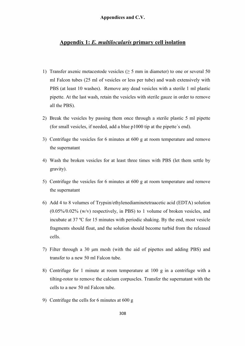
Appendices and C.V.
308
Appendix 1: E. multilocularis primary cell isolation
1) Transfer axenic metacestode vesicles (≥ 5 mm in diameter) to one or several 50
ml Falcon tubes (25 ml of vesicles or less per tube) and wash extensively with
PBS (at least 10 washes). Remove any dead vesicles with a sterile 1 ml plastic
pipette. At the last wash, retain the vesicles with sterile gauze in order to remove
all the PBS).
2) Break the vesicles by passing them once through a sterile plastic 5 ml pipette
(for small vesicles, if needed, add a blue p1000 tip at the pipette´s end).
3) Centrifuge the vesicles for 6 minutes at 600 g at room temperature and remove
the supernatant
4) Wash the broken vesicles for at least three times with PBS (let them settle by
gravity).
5) Centrifuge the vesicles for 6 minutes at 600 g at room temperature and remove
the supernatant
6) Add 4 to 8 volumes of Trypsin/ethylenediaminetetraacetic acid (EDTA) solution
(0.05%/0.02% (w/v) respectively, in PBS) to 1 volume of broken vesicles, and
incubate at 37 ºC for 15 minutes with periodic shaking. By the end, most vesicle
fragments should float, and the solution should become turbid from the released
cells.
7) Filter through a 30 µm mesh (with the aid of pipettes and adding PBS) and
transfer to a new 50 ml Falcon tube.
8) Centrifuge for 1 minute at room temperature at 100 g in a centrifuge with a
tilting-rotor to remove the calcium corpuscles. Transfer the supernatant with the
cells to a new 50 ml Falcon tube.
9) Centrifuge the cells for 6 minutes at 600 g

Appendices and C.V.
309
10) Discard the supernatant and carefully re-suspend the cells in cDMEM-A. Take a
12.5 µl aliquot and dilute it to a total volume of 1000 µl with PBS. Measure the
absorbance at 600 nm with a spectrophotometer (U-2000, Hitachi, USA) to
quantify the cells.

Appendices and C.V.
310
Appendix 2: EdU detection in Whole-mounts
1) Fix the samples for 1 h in paraformaldehyde 4 % prepared in PBS at room
temperature (RT), with shaking.
2) Wash 3 times with PBS, 15 min each, with shaking.
3) Block with 2 incubations of 10 min each in PBS + 3% bovine serum albumin
(BSA, Sigma-Aldrich), with shaking.
4) Permeabilize with 2 incubations of 20 min each in PBS + 0.5% Triton X-100
(Sigma-Aldrich), with shaking.
5) Block with 2 incubations of 10 min each in PBS + 3% BSA, with shaking.
6) Incubate for 1 hour in the dark with the Click-iT reaction cocktail (Click-iT®
EdU Alexa Fluor® 555 Imaging Kit (Life Technologies), prepared as described
by the manufacturer), with shaking.
7) Wash for 10 min with PBS + 3% BSA, with shaking.
8) Incubate for 10 min in PBS + 1 µg/ml DAPI (Sigma-Aldrich), with shaking.
9) Wash 3 times with PBS, 10 min each, with shaking.
10) Mount in Fluoprep (Biomérieux)
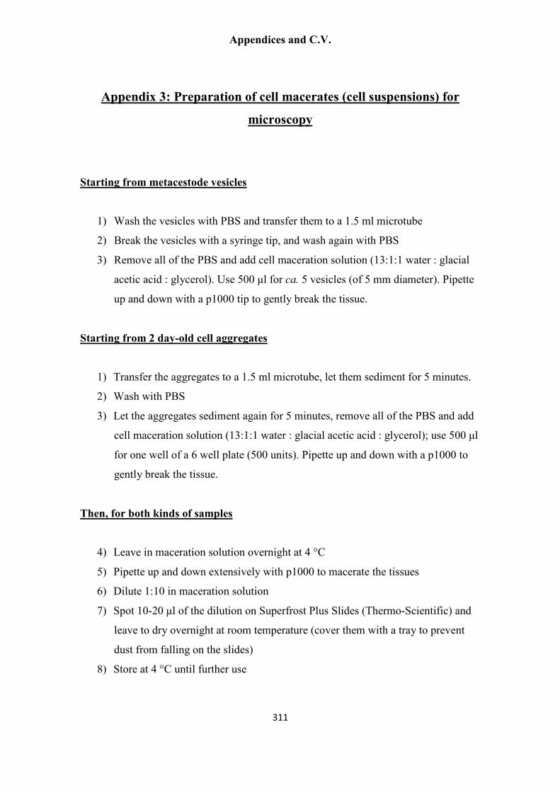
Appendices and C.V.
311
Appendix 3: Preparation of cell macerates (cell suspensions) for
microscopy
Starting from metacestode vesicles
1) Wash the vesicles with PBS and transfer them to a 1.5 ml microtube
2) Break the vesicles with a syringe tip, and wash again with PBS
3) Remove all of the PBS and add cell maceration solution (13:1:1 water : glacial
acetic acid : glycerol). Use 500 µl for ca. 5 vesicles (of 5 mm diameter). Pipette
up and down with a p1000 tip to gently break the tissue.
Starting from 2 day-old cell aggregates
1) Transfer the aggregates to a 1.5 ml microtube, let them sediment for 5 minutes.
2) Wash with PBS
3) Let the aggregates sediment again for 5 minutes, remove all of the PBS and add
cell maceration solution (13:1:1 water : glacial acetic acid : glycerol); use 500 µl
for one well of a 6 well plate (500 units). Pipette up and down with a p1000 to
gently break the tissue.
Then, for both kinds of samples
4) Leave in maceration solution overnight at 4 °C
5) Pipette up and down extensively with p1000 to macerate the tissues
6) Dilute 1:10 in maceration solution
7) Spot 10-20 µl of the dilution on Superfrost Plus Slides (Thermo-Scientific) and
leave to dry overnight at room temperature (cover them with a tray to prevent
dust from falling on the slides)
8) Store at 4 °C until further use

Appendices and C.V.
312
Appendix 4: Immunohistochemistry of Paraplast sections
1) Remove Paraplast with two incubations of 5 min each in Xylol
2) Rehydrate through a series of ethanol dilutions: 100% ethanol, 75% ethanol, 50%
ethanol in PBS, and 25% ethanol in PBS (2 min each)
3) Wash 2 times with PBS (5 min each) at room temperature
4) For those antibodies that require it (Anti-PHB1 and Anti-phospho-histone H3
(Ser10)), perform Heat Induced Epitope Retrieval (HIER) as follows. Place the
slides in a plastic box covered with a lid and filled with HIER Buffer (Sodium
Citrate Buffer 10 mM pH 6.0 + 0.1% Tween-20). Heat in a microwave at 700 Watts
until it boils, and then continue heating for 20 more min. Keep more HIER buffer at
hand at 100 ºC and use it to add periodically to the box during the incubation, if too
much HIER buffer evaporates from the box.
5) Eliminate endogenous peroxidases with 3% H2O2 in PBS for 10 min at room
temperature
6) Wash in PBS for 5 min at room temperature
7) Wash two times, 5 min each, with PBS + 0.1% Triton X-100
8) Block with PBS + 3 % bovine serum albumin (BSA, Sigma-Aldrich) + 5% Sheep
serum (Sigma-Aldrich) for 1 hour at room temperature
9) Incubate with the primary antibody at the appropriate dilution in PBS + 3 % BSA
for 2 hours at room temperature in a humid chamber, covering with Parafilm.
10) Wash 4 times, 15 min each, with PBS (the Parafilm will detach from the slide with
the first wash).

Appendices and C.V.
313
11) Incubate with the secondary antibody, conjugated to horse radish peroxidase, at the
appropriate dilution in PBS + 3 % BSA for 2 hours at room temperature in a humid
chamber, covering with Parafilm.
12) Wash 4 times, 15 min each, with PBS (the Parafilm will detach from the slide with
the first wash).
13) Perform a quick incubation with sodium acetate buffer 50 mM, pH 5.0
14) Develop the colorimetric reaction with 3-Amino-9-ethylcarbazole (AEC, Sigma-
Aldrich A6926) as follows. Dissolve 1 tablet in 2.5 ml of N.N-dimethylformamide
and mix with 47.5 ml of 50 mM sodium acetate buffer, pH 5.0, with stirring. Add
25 µl of fresh 30% (w/w) hydrogen peroxide immediately prior to use. If not all the
reaction solution will be used at once, a smaller aliquot can be prepared with a
stoichiometrically reduced amount of hydrogen peroxide, and the remaining
solution can be stored for two months at 4 ºC in the dark. Cover the slide with the
solution and incubate in the dark in a humid chamber to allow the reaction to
proceed, checking the reaction development from time to time under a microscope.
15) If desired, perform nuclear staining (e.g. DAPI staining).
16) Mount with Fluoprep (Biomérieux) or with 80% glycerol diluted with PBS.

Appendices and C.V.
314
Appendix 5: Immunohistofluorescence on cryosections
1) Thaw cryosections for 30 min at room temperature (RT) 2) Incubate 15 min with PFA 4% - PBS at room temperature
3) Wash 3 times, 5 min each, with PBS
4) Permeabilize with 0.1% Triton X-100 (Sigma-Aldrich) in PBS for 10 min at RT
5) Block with PBS + 1% bovine serum albumin (BSA, Sigma-Aldrich) + 5 %
sheep serum (Sigma-Aldrich) + 0.05% Tween-20 (Sigma-Aldrich) for 1 h at RT in a humid chamber
6) Incubate with the primary antibody at the appropriate dilution in PBS + 1 %
BSA for 2 h at room temperature in a humid chamber. For some antibodies, it is recommended to do an overnight incubation in a humid chamber, covering the slide with Parafilm at 4 º C (e.g. anti-FMRFamide, anti-5-HT).
7) Wash 4 times, 15 min each, with PBS (optional: add 0.05% Tween-20)
8) Incubate with the fluorophore-conjugated secondary antibody at the appropriate
dilution in PBS + 1 % BSA for 2 h at RT in a humid chamber
9) Remove antibody solution, and add PBS + 1 % BSA + 1:40 TRITC Phalloidin (Sigma-Aldrich) + 1 µg/ml DAPI (Sigma-Aldrich), incubating for 20 min at RT in a humid chamber
10) Wash 4 times, 15 min each, with PBS (optional: add 0.05% Tween-20)
11) Mount with Fluoprep (Biomérieux)

Appendices and C.V.
315
Appendix 6: Whole-mount Immunohistofluorescence protocols for
protoscoleces and small metacestodes
DAY 1
1) Fix protoscoleces or pierced and opened metacestode vesicles with 4% PFA -
PBS for 1 hour at room temperature (RT), with shaking.
2) Wash 3 times, 10 min each with PBS + 0.3% Triton X-100 (PBS-T) at RT, with
shaking. (Optional: at this point the protocol can be paused by keeping the
samples overnight at 4 °C).
3) Permeabilization: two different protocols, for each kind of sample. Use protocol
A for protoscoleces and protocol B for vesiculated protoscoleces and
metacestode vesicles.
o Protocol A (with proteinase K):
– 5 min PBS-T + 0.5% SDS + 2 µg/ml Proteinase K (Fermentas)
at RT
– Quick wash with PBS-T
– 10 min of re-fixation with 4% PFA / PBS-T at RT, with
shaking
o Protocol B: without proteinase K:
– 20 min PBS + 1% SDS at RT
4) Wash 3 x 10 min each in PBS-T at RT, with shaking
5) Block 2 hours with PBS-T + 3% bovine serum albumin (BSA, SIGMA) + 5%
sheep serum (SIGMA) at RT, with shaking

Appendices and C.V.
316
6) Incubate with PBS-T + 3% BSA + primary antibody at the appropriate
concentration overnight at 4 °C with slow rocking
DAY 2
7) Wash 5 times, 30 min each with PBS-T at RT, with shaking
8) Incubate with PBS-T + 3 % BSA + secondary antibody (conjugated to a
fluorophore) at the appropriate concentration overnight at 4 °C with slow
rocking. From now on protect samples from light.
DAY 3
9) Wash briefly with PBS-T
10) Incubate 30 min with PBS + 1 % BSA + DAPI 1 µg/ml + Phalloidin-TRITC
(Sigma) diluted 1:40 at RT with shaking
11) Wash 4 times, 30 min each with PBS-T at RT, with shaking
12) Mount with Fluoprep (Biomérieux)
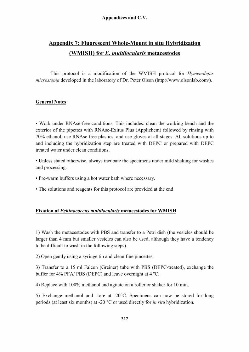
Appendices and C.V.
317
Appendix 7: Fluorescent Whole-Mount in situ Hybridization
(WMISH) for E. multilocularis metacestodes
This protocol is a modification of the WMISH protocol for Hymenolepis
microstoma developed in the laboratory of Dr. Peter Olson (http://www.olsonlab.com/).
General Notes
• Work under RNAse-free conditions. This includes: clean the working bench and the
exterior of the pipettes with RNAse-Exitus Plus (Applichem) followed by rinsing with
70% ethanol, use RNAse free plastics, and use gloves at all stages. All solutions up to
and including the hybridization step are treated with DEPC or prepared with DEPC
treated water under clean conditions.
• Unless stated otherwise, always incubate the specimens under mild shaking for washes
and processing.
• Pre-warm buffers using a hot water bath where necessary.
• The solutions and reagents for this protocol are provided at the end
Fixation of Echinococcus multilocularis metacestodes for WMISH
1) Wash the metacestodes with PBS and transfer to a Petri dish (the vesicles should be
larger than 4 mm but smaller vesicles can also be used, although they have a tendency
to be difficult to wash in the following steps).
2) Open gently using a syringe tip and clean fine pincettes.
3) Transfer to a 15 ml Falcon (Greiner) tube with PBS (DEPC-treated), exchange the
buffer for 4% PFA/ PBS (DEPC) and leave overnight at 4 ºC.
4) Replace with 100% methanol and agitate on a roller or shaker for 10 min.
5) Exchange methanol and store at -20°C. Specimens can now be stored for long
periods (at least six months) at -20 °C or used directly for in situ hybridization.

Appendices and C.V.
318
Whole-Mount In Situ Hybridization
DAY 1
Rehydrate the specimens by washing in an ethanol dilution series:
1) 100% ethanol, 5 min on a roller.
2) Wash once in 75% ethanol/distilled water (DEPC), 10 min on a roller.
3) Wash once in 50% ethanol/PBS-T (DEPC), 10 min on a roller.
4) Wash three times in PBS-T for 5 min on a roller.
Proteinase K treatment and re-fixing
5) Take the Proteinase K (Fermentas, 20 mg/ml in 50% Glycerol, stored at -20°C) and
put it into ice.
6) Add 7.5 µl of Proteinase K (20 mg/ml) to each vial containing 10 ml of PBS-T.
7) Incubate at room temperature for 10 min, without shaking.
8) Rinse twice in 5 ml of 0.1 M Triethanolamine (TEA), pH 7.8, for 5 min (mix by
inverting the specimens from time to time).
9) Add 12.5 µl of acetic anhydride to the second wash and swirl often. After 5 min add
12.5 µl more acetic anhydride, and incubate for 5 min.
10) Wash twice for 5 min with PBS-T on roller (mix by inverting the specimens from
time to time).
11) Re-fix for 20 min, in 4% paraformaldehyde in PBS-T, on roller.
12) Wash five times for 5 min or longer in PBS-T, to wash off excess
paraformaldehyde.
Equilibration of specimens in hybridization buffer
13) Add 500 µl of hybridization buffer to 2 ml PBS-T and allow specimens to settle.
14) Remove the buffer and replace with 1 ml of hybridization buffer. Transfer carefully
to 1.5 ml tubes using blue tips that have the tip cut off with clean scissors (for that,

Appendices and C.V.
319
clean the scissors with RNase Exitus Plus and 70% Ethanol and allow to dry). Transfer
5-10 vesicles per tube, depending on vesicle size.
15) Incubate at 60 °C for 10 min (use the shaking Eppendorf thermomixer, 600 RPM).
Check the real temperature with a thermometer. Usually the real temperature is 2-3
degrees below what it says on the screen.
Pre-hybridization
16) Replace the hybridization buffer with 0.5 to 1 ml of fresh hybridization buffer
(warmed to 60 °C).
17) Pre-hybridize for at least 6 hours at 60 °C using the Eppendorf thermomixer, at 600
RPM. It can be left pre-hybridizing overnight.
DAY 2
Hybridization
18) Prepare the DIG-labeled RNA probe: Denature the probe (10 µl) at 80°C for 3 min,
centrifuge and place on ice. Add the probe to 490 µl of pre-warmed hybridization buffer
(57-58 °C). Remove the pre-hybridization solution and add the hybridization solution
containing the probe to the samples. The resulting probe concentration should be 0.2 to
1 ng/µl. When re-using a probe (that is, the probe is already diluted in hybridization
buffer), denature at 80°C for 3 min, and place at 58 °C for 5 minutes before adding to
the specimens.
Use approximately 0.2 ng/ul of probe as measured by dot blot comparing to the
Roche standard (see below).
19) For “short” (up to 250 bp) riboprobes hybridize at 53-54 ºC overnight (24 hours) in
the Eppendorf thermomixer, 600 RPM.
20) For all other probes, hybridize at 57-58 °C overnight (24 hours) in the Eppendorf
thermomixer, 600 RPM.

Appendices and C.V.
320
DAY 3
Probe removal and washing
21) Carefully remove the probe from the vial and place on ice. Store at - 80°C in a
microtube. The probe can be reused several times.
22) Rinse in 1 ml of pre-warmed hybridization buffer (pre-warmed to 57-58 °C), and
wash at 57-58°C for 10 min, twice, in the Eppendorf thermomixer, at 600 RPM. Note:
While changing solutions keep the tubes on the thermomixer to keep specimens at 57-
58 °C at all times.
For “short” (up to 250 bp) riboprobes, wash at 53-54 ºC
23) Wash three times in 2 x SSC + 0.1% Tween-20 (pre-warmed to 57-58 °C) at 57-58
°C for 20 min in the Eppendorf thermomixer, 600 RPM.
For “short” (up to 250 bp) riboprobes, wash at 53-54 ºC
24) Wash three times in 0.2 x SSC + 0.1% Tween-20 (pre-warmed to 57-58 °C) at 57-
58 °C for 30 min in the Eppendorf thermomixer, 600 RPM.
For “short” (up to 250 bp) riboprobes, wash at 53-54 ºC
25) Wash twice in Maleic acid buffer (MAB) for 15 mins at room temperature on a
roller (standard shaker with 70 RPM or Blot shaker at 200 RPM).
Antibody incubation and washing
26) Pre-incubate for 2 hours in 0.5 ml of MAB + 1% Blocking reagent + 5% Sheep
serum. This whole blocking solution must be previously heated to 60 ºC for 30 minutes
to inactivate the serum, and then cooled down on ice before adding to the samples.
27) Remove the solution and replace with MAB + 1% Blocking Reagent (without sheep
serum) containing 1:50 dilution of Roche Anti-DIG Peroxidase (POD) conjugate.
Place on a rocker overnight at 4°C.

Appendices and C.V.
321
DAY 4
Washing
28) Remove the antibody.
29) Wash three times for 5 min with MAB at room temperature, mixing gently.
30) Wash three times for 1 hour (3 x 1 hour washes) at room temperature, with shaking
during the washes (Blue Shaker with 70 RPM or Blot Shaker at 200 RPM).
31) Wash once for 3 min at room temperature with PBS + 0.1 M Imidazole, pH 7.6 at
room temperature.
32) Wash once for 10 min at room temperature in PBS + 0.1 M Imidazole, pH 7.6 at
room temperature.
Fluorescent Colour Reaction
33) Prepare the fluorescent reaction solution by mixing PBS + 0.1 M imidazole, pH 7.6
with 1:100 Fluorescein Tyramide (prepared as described by Hopman et al., 1998, see
also the working protocol below) and 0.001 % final concentration H2O2 (for that, it is
better to make a 1:300 initial dilution of the 30% H2O2 and then use that in a 1:100
dilution for the final solution).
34) Add the fluorescent reaction solution to the specimens, mix by inversion (5-10
times) and incubate 5 minutes in the dark without shaking.
35) Wash 4 times for 5 minutes with PBS-T in the dark. RNAse free conditions are no
longer necessary (the FISH is sold). After this, EdU detection and/or DAPI staining (1
µg/ml for 10 minutes, followed by washes in PBS-T) can be done.
36) Mount with Fluoprep (Biomérieux) or with 80% Glycerol in PBS, and seal the
borders with nail polish to prevent evaporation.

Appendices and C.V.
322
Modifications of the fluorescent WMISH protocol for the colorimetric alkaline
phosphatase WMISH protocol
DAY 3
27) Remove the solution and replace with MAB + 1% Blocking Reagent (without sheep
serum) containing 1:200 dilution of Roche Anti-DIG Alkaline Phosphatase (AP)
conjugate (Roche 11 093 274 910). Place on a rocker overnight at 4 °C.
DAY 4
31) Wash once for 3 min in 100 mM Tris-HCl, 100 mM NaCl, 50 mM MgCl2 (pH=9.5)
32) Wash once for 10 min in 100 mM Tris-HCl, 100 mM NaCl, 50 mM MgCl2
(pH=9.5)
33-36) Perform the detection with nitro-blue tetrazolium and 5-bromo-4-chloro-3'-
indolyphosphate (NBT/BCIP, Sigma, B1911) for 2 hours to overnight (for highly
expressed genes, signal will be apparent after 15-30 minutes). Perform DAPI staining
and mount in Fluoprep.

Appendices and C.V.
323
Reagents and solutions for the WMISH protocol
DEPC treated H2O
Add 0.1% DEPC to double-distilled water, and stir overnight (leave the lid slightly
open). Autoclave twice.
PBS
Add 0.1% DEPC to a bottle of PBS, and stir overnight (leave the lid slightly open).
Autoclave twice.
PFA 4% / PBS (DEPC)
Dissolve 4 g of paraformaldehyde (Sigma-Aldrich P-6148) in 100 ml of DEPC-treated
PBS by incubating at 65 °C for 1-2 hours and shaking periodically. Make 10 ml
aliquots, and store at -20 °C.
PBS-T
PBS + 0.1% Tween-20 (SIGMA P-9416)
Proteinase K (Fermentas, Thermo Scientific # EO0491)
0.1 M Triethanolamine (TEA), pH 7 to 8
From stock TEA (90278, Sigma-Aldrich), concentration 7.53 M, prepare a 1:75.3
dilution (0.665 ml TEA in 50 ml of DEPC-treated water). Adjust pH with freshly
aliquoted 25% HCl (usually ca. 200 µl are needed) and confirm the pH by placing a
drop on a pH strip.
Acetic anhydride (Sigma-Aldrich A-6404)
10% CHAPS (Sigma-Aldrich C-3023)

Appendices and C.V.
324
Dissolve 5 g of CHAPS in 50 ml total volume with DEPC treated H2O.
10% Tween-20 (Sigma-Aldrich P-9416)
Dissolve 5 ml of Tween-20 in 50 ml total volume with DEPC treated H2O.
Heparin 100 mg/ml stock (Sigma-Aldrich H-3393)
Dissolve 0.5 g of Heparin in 5 ml of sterile (not DEPC-treated) double-distilled water.
Store at -20 °C in 0.2 ml aliquots.
Torula RNA 100 mg/ml (Sigma-Aldrich R-6625)
1) Dissolve Torula RNA at 10 mg/ml in DEPC-treated water (dissolve at 65 °C, ca.
1 hour). Start with 40 ml or less.
2) Extract with one volume of Phenol:Chloroform:Isoamyl alcohol (25:24:1),
centrifuge 20 minutes at 10,000 g at 4 °C, and transfer the aqueous phase to a
new tube.
3) Extract with one volume of Chloroform and centrifuge 20 minutes at 10,000 g at
4 °C, and transfer the aqueous phase to a new tube.
4) Precipitate the RNA by adding 0.1 volume of sodium acetate 3 M (pH 5.2,
RNAse-free) and 2.5 volumes of 100% ethanol , and centrifuge 20 minutes at
10,000 g at 4 °C.
5) Wash the pellet twice with 70% ethanol (RNAse-free).
6) Remove all the ethanol with a pipette. Dry the pellet (usually around 5 minutes
by placing at 42 °C), and resuspend in 1 ml of DEPC-treated water (pipette up
and down, this can take a while; heat for 5-10 minutes at 65 °C if needed).
7) Quantify and estimate purity with the NanoDrop.
8) Dilute to 100-200 mg/ml and store at -20 °C.
Formamide, de-ionized (Sigma-Aldrich F9037)
Aliquot in Falcon (Greiner) tubes and store a -20 °C.
20 X SSC (3 M NaCl, 0.3 M tri-sodium citrate)
175.3 g NaCl (Sigma-Aldrich)
88.2 g Tri-sodium citrate (Sigma-Aldrich)

Appendices and C.V.
325
Add double-distilled water to 800 ml, and adjust pH to 7.0 with HCl. Complete the
volume to 1 l with H2O.
Add 0.1% DEPC and stir overnight, leave the lid slightly opened. Autoclave twice.
Store at 4 °C.
Denhardt´s Solution 50X concentrate (Sigma-Aldrich, D2532).
Hybridization solution
Final concentration Stock Volume for 10 ml
50 % Formamide 100% 5 ml
5 X SSC 20 X SSC 2.5 ml
1 mg/ml Torula RNA 100-200 mg/ml 50 – 100 µl
100 µg/ml Heparin 100 mg/ml 10 µl
1 X Denhardt´s 50 X Denhardt´s 0.2 ml
0.1% Tween-20 10% Tween-20 0.1 ml
0.1% CHAPS 10% CHAPS 0.1 ml
DEPC-treated water Up to 10 ml
SSC 2X + 0.1% Tween-20
Prepare with DEPC water.
SSC 0.2X + 0.1% Tween-20
Prepare with DEPC water.
Maleic Acid Buffer (MAB)
100 mM Maleic Acid (23.21 g/l) (Sigma-Aldrich)
150 mM NaCl (17.53 g/l) (Sigma-Aldrich)

Appendices and C.V.
326
Add double-distilled water to 800 ml. Adjust pH to 7.5 and complete the volume to 1
liter. Autoclave and store at 4 °C.
Before use, add Tween-20 to 0.1% (v/v)
MAB + 1% Blocking reagent (Roche, 11 096 176 001)
Dissolve by placing at 65 °C for 30 – 60 minutes, with frequent shaking. Store at -20
°C.
MAB + 1% Blocking reagent + 5% Sheep serum (Sigma-Aldrich, S3772)
The whole blocking solution is heated to 60 º C for 30 minutes to inactivate the serum
and then cooled down on ice before adding to the samples.
Roche anti-digoxigenin, POD conjugated (Roche 11 207 733 910, store at 4 °C after
reconstitution)
PBS + 0.1 M Imidazole, pH 7.6
Add 0.1 M imidazole (Sigma-Aldrich) to sterile PBS, autoclave, and keep at 4 °C.

Appendices and C.V.
327
Appendix 8: Fluorescein-tyramide synthesis (Hopman et al., 19984)
Materials
Fluorescein-NHS ester (NHS-Fluorescein, Thermo Scientific 46410, 100 mg scale, enough for 2 preparations to be done in parallel). Once opened dissolve entire contents in N,N-dimethylformamide and use at once.
Tyramine (Sigma-Aldrich T-2879)
N,N-dimethylformamide (DMFA, Sigma-Aldrich T-8654)
Triethylamine (Sigma-Aldrich T-0886)
100% ethanol
Solutions
Fluorescein-NHS ester solution: dissolve at 10 mg/ml in DMFA in a 15 ml Falcon tube. Because we buy a 100 mg package of NHS-Fluorescein, the easiest thing to do is to dissolve everything in 10 ml DMFA, and from there do two reactions, with 4 ml of the NHS-Fluorescein stock each (2 ml remain, discard them). Keep in the dark.
DMFA-triethylamine solution: Add 50 µl of triethylamine to 5 ml of DMFA
Tyramine solution: Dissolve 50 mg of tyramine in 5 ml of DMFA-triethylamine solution
Reaction
In a 15 ml Falcon Tube, mix 4 ml fluorescein-NHS ester solution (in DMFA) and 1.37 ml tyramine solution (in DMFA-triethylamine), and incubate in the dark at room temp for 2 hours. Add 4.6 ml of 100% ethanol, and store in dark at -20 °C. Use as a 1:100 dilution as detailed in the WMISH working protocol.
4 Hopman, A. H., F. C. Ramaekers, and E. J. Speel. 1998. Rapid synthesis of biotin-, digoxigenin-,
trinitrophenyl-, and fluorochrome-labeled tyramides and their application for In situ hybridization using
CARD amplification. J Histochem Cytochem 46:771-777.

Appendices and C.V.
328
Appendix 9: in vitro synthesis and quantification of Digoxigenin-
labeled RNA probes
1) Cloning of the probe cDNA
Although other protocols recommend shorter probes for improved tissue
penetration, in my experience it is better to use full-length probes, for greater signal
intensity. For shorter genes that are highly expressed (e.g. em-h2b, em-muc), probes of
ca. 200 bases worked fine. For genes of low expression, probes of 1 kb or more are
recommended.
Clone the cDNA from a RT-PCR reaction into a PCR cloning vector such as
pDrive (Qiagen) or pJet1.2 (Thermo Scientific). For pDrive, only one clone is needed,
since it has T7 and SP6 promoters upstream and downstream of the multiple cloning
site (MCS), respectively. When cloning into pJet1.2, one needs to make sure to have
one clone with each insert orientation, since it only has a T7 promoter upstream of the
MCS.
2) Linearizing the plasmid
Digest the plasmid with an enzyme that cuts in the MCS right after the insert, on
the opposite side of the RNA polymerase promoter, so that the probe only includes the
insert sequence. For example, with pJet1.2, only XbaI and NcoI (of the usual enzymes)
are useful.
T7 XbaI, NcoI
T7 XbaI, NcoI
Antisense probe
Sense probe (control)

Appendices and C.V.
329
Digest at least 4 µg of plasmid for two hours with 40 units of enzyme, and run
100 ng on a 1% agarose gel to confirm that it is completely linearized. Then purify the
linearized plasmid DNA with a Kit (a simple DNA clean-up kit is sufficient). Quantify
by Nanodrop. Keep the plasmid as clean as possible (use clean plastic tips and elution
buffer).
Note: check that the insert does not have a restriction site for that enzyme. If no
enzyme can be used, amplify the insert plus the promoter using flanking primers, and
purify by gel extraction after agarose electrophoresis.
3) In vitro transcription
- Set up the following reaction on ice and mix well by pipetting:
Transcription Buffer 10 X (New England Biolabs) – 2 µl
DIG RNA labeling mix 10 X (Roche 11 277 073 910) – 2 µl
T7 OR SP6 polymerase (50,000 units/ml, New England Biolabs) – 2 µl
RNAse out (40 units/µl, Invitrogen) – 0.5 µl
Linearized plasmid DNA – 1 µg
Bovine Serum Albumin 1 mg/ml, molecular biology grade (New England
Biolabs) – 2 µl
Water, RNAse free – enough for a 20 µl final reaction volume (do not use
DEPC-treated water, but instead use commercial RNAse free water, since traces
of DEPC can inhibit the reaction).
- Incubate for 2 hours at 37 °C
- Add 2 µl of RQ1 DNAse (Promega) and incubate for 15 minutes at 37 °C
- Purify each reaction with the Pure Link RNA Mini Kit (Ambion). Elute in 30 µl
of RNAse free water. Alternatively, if many probes are being synthesized and a
cheaper alternative is needed, precipitate the probe overnight at -80 °C with 0.1
volumes of sodium acetate 3 M, pH 5.2 and 2.5 volumes of 100% ethanol.
Centrifuge at 12,000 g for 20 min at 4 °C, wash the RNA pellet with 70%
ethanol, dry the pellet and dissolve it in 30 µl of RNAse free water (use RNAse
free reagents).
- Run 3 µl of each sample in a 1% agarose gel to confirm the integrity of the probe
(it does not need to be a denaturing gel, but it must be RNAse free).

Appendices and C.V.
330
4) Probe quantification by Dot-blot
In a Hybond-N membrane, spot 1 µl drops of serial dilutions of the probe
(undiluted, 1:10, 1:100, 1:1000) and of the control DIG-RNA probe (Roche
11585746910, dilutions 10, 1.0, 0.1 and 0.01 ng/µl).
Dry for 1 hour at 60 °C between two Whatman Paper sheets, and cross-link
under UV light for 1 minute in a trans-illuminator.
Then perform the following detection protocol:
- Wash 5 minutes in Buffer 1 (Tris-HCl 100 mM, NaCl 150 mM, pH 7.5), with
shaking
- Block 10 minutes in Buffer 2 (Buffer 1 + 0.5% w/v Blocking Reagent, Roche),
with shaking.
- Incubate with Anti-DIG, alkaline phosphatase conjugated (Roche 11 093 274
910), 1:2000 dilution in Buffer 2, for 20 minutes, with shaking.
- 2 washes of 10 minutes each in Buffer 1, with shaking
- Brief wash in Buffer 3 (Tris-HCl 100 mM, NaCl 100 mM, MgCl2 50 mM, pH
9.5), with shaking
- Detection with nitro-blue tetrazolium and 5-bromo-4-chloro-3'-indolyphosphate
(NBT/BCIP, Sigma 72091, dilute 20 µl/ml in Buffer 3). Usually the detection is
ready after 5 to 10 minutes in the dark without shaking.

Appendices and C.V.
331
Curriculum Vitae
Personal information
Name: Koziol Antmann, Uriel Bensión. Birth Date: 10/10/1982, Montevideo, Uruguay. Nationality: Uruguayan and Italian
Education
Currently performing my doctoral studies in the Graduate School of Life
Sciences, University of Würzburg (Germany). Thesis supervisor: Prof. Dr. Klaus Brehm. (Since April 2011). Thesis title: Molecular and developmental characterization of stem cells in the human parasite Echinococcus multilocularis,
Master in Science in Biology, with specialization in Cell and Molecular
Biology (PEDECIBA Biología, Uruguay). Thesis supervisor: Dr. Estela Castillo, Sección Bioquímica, Facultad de Ciencias, Uruguay (from December 2006 until December 2009). Thesis title: Caracterización de células proliferantes en Mesocestoides corti (Cestoda), y de genes pumilio como posibles marcadores moleculares de las mismas. (Characterization of proliferative cells in Mesocestoides corti
(Cestoda) and of pumilio genes as possible molecular markers of these cells).
Approved with the maximum qualification.
Bachellor in Sciences (Orientation: Invertebrate Zoology). (Facultad de
Ciencias, Universidad de la República, Uruguay)
Graduation date: 30th August 2006. Average grade: 10.94 over 12

Appendices and C.V.
332
Selected Post-graduate courses
Short Course for Young Parasitologists. Organized by the Deutsche Gesellschaft für Parasitologie (Heidelberg, Germany, 2012).
“Transferable skill courses” in the Graduate School of Life Sciences (Würzburg, Germany) such as “Poster design”, “Scientific writing and publishing” “Statistical Data Analysis“, “Good scientific Practices”, and “Writing Grant Proposals”. (Since April 2011).
Curso Internacional de Biología del Desarrollo. (International Course on
Developmental Biology). Universidad Nacional Andrés Bello (Quintay, Chile, 2010).
Working with Pathogen Genomes. Wellcome Trust – Sanger Institute (Instituto de Higiene, Montevideo, Uruguay 2009). Salud, Bienestar y Producción en Animales de Laboratorio (Health, well-
being and production in laboratory animals). Universidad de la República - Comisión Honoraria de Experimentación Animal, Uruguay (CHEA) (2008). “Real time PCR”. Un método eficiente para caracterizar el genoma. (Real time PCR; an efficent method for genome characterization). PEDECIBA, Uruguay. (2008). Regulación de la expresión génica en eucariotas. (Regulation of gene expression in eukaryotes). PEDECIBA, Uruguay (2008). Curso Básico de Cultivo de Células. (Basic course in cell culture).
PEDECIBA, Uruguay. (2007). Microscopía Confocal. Principios y Aplicaciones. (Confocal microscopy.
Principles and applications). PEDECIBA, Uruguay (2007). Expresión y Silenciamiento de Genes en Células Animales y Vegetales.
(Expression and silencing of genes in animal and vegetal cells) CABBIO (Universidad Nacional de Córdoba, Argentina, 2007).

Appendices and C.V.
333
Positions
Assistant Professor, second level, of Sección Bioquímica (Biochemistry
department), Facultad de Ciencias, Universidad de la República, Uruguay (since December 2009, currently on a leave for the duration of my doctoral studies).
Research assistant in the project “Aplicación de la metagenómica para la
obtención de enzimas de uso potencial en industrias procesadoras de pulpa
de papel” (“Application of metagenomics for the obtention of enzymes of
potenital use in paper pulp production”), under the direction of Dr. Elena Fabiano and Dr. Francisco Noya. (Biochemistry department, IIBCE, Uruguay) (From January 2007 until July 2008).
Research internship in the laboratory of Dr. Michael Kahn, Institute of
Biological Chemistry, Washington State University. Partially financed by CSIC, Uruguay (Pullman, U.S.A, from October until December 2007)
Research and teaching assistant of Sección Bioquímica (Biochemistry
department) Facultad de Ciencias, Universidad de la República, Uruguay. (from November 2006 until december 2009).
Interim Teaching Assistant of Sección Bioquímica (Biochemistry
department) (Facultad de Ciencias, Universidad de la República) (From April 2006 until July 2006).
Research assistant in the project “Búsqueda de reguladores globales del
metabolismo del hierro en Sinorhizobium meliloti 1021” (“Search for global regulators of iron metabolism in Sinorhizobium meliloti 1021”) under the direction of Dr. Elena Fabiano and Dr. Francisco Noya. (Biochemistry department, IIBCE, Uruguay) (From May 2005 until February 2007)

Appendices and C.V.
334
Publications
Schubert, A., Koziol, U., Cailliau, K., Vanderstraete, M., Dissous. C.,
Brehm, K. (2014). Targeting Echinococcus multilocularis Stem Cells by
Inhibition of the Polo-Like Kinase EmPlk1. PloS Neglected Tropical Diseases, 8(6):e2870.
Koziol, U., Rauschendorfer, T., Zanon-Rodríguez, L., Krohne, G.,
Brehm, K. (2014). The unique stem cell system of the immortal larva of the
human parasite Echinococcus multilocularis. EvoDevo, 5:10.
Domínquez, M.F.*, Koziol, U.*, Porro, V., Costábile, A., Estrade, S., Tort, J., Bollati-Fogolin, M., Castillo, E (2014). A new approach for the
characterization of proliferative cells in cestodes. Experimental Parasitology, 138:25–29. (* Both authors contributed equally to this work).
Skinner, D.E., Rinaldi, G., Koziol, U., Brehm, K., Brindley, P.J. How
might flukes and tapeworms maintain genome integrity without a canonical
piRNA pathway? Trends in Parasitology, 30:123-129.
Hemer, S., Konrad, C., Spiliotis, M., Koziol, U., Schaack, D., Förster, S., Gelmedin, V., Stadelmann, B., Dandekar, T., Hemphill, A., Brehm, K. Host
insulin stimulates Echinococcus multilocularis insulin signalling pathways
and larval development. BMC Biology, 12:5.
Koziol U., Krohne G., Brehm K. (2013). Anatomy and development of
the larval nervous system in Echinococcus multilocularis. Frontiers in
Zoology 10:24.
Tsai I.J., Zarowiecki M., Holroyd N., Garciarrubio A., Sanchez-Flores A., Brooks K.L., Tracey A., Bobes R.J., Fragoso G., Sciutto E., Aslett M., Beasley H., Bennett H.M., Cai J., Camicia F., Clark R., Cucher M., De Silva N., Day T.A., Deplazes P., Estrada K., Fernández C., Holland P.W., Hou J., Hu S., Huckvale T., Hung S.S., Kamenetzky L., Keane J.A., Kiss F., Koziol U., Lambert O., Liu K., Luo X., Luo Y., Macchiaroli N., Nichol S., Paps J., Parkinson J., Pouchkina-Stantcheva N., Riddiford N., Rosenzvit M., Salinas G., Wasmuth J.D., Zamanian M., Zheng Y.; Taenia solium Genome Consortium, Cai X., Soberón X., Olson P.D., Laclette J.P., Brehm K., Berriman M. (2013). The genomes of four tapeworm species reveal adaptations to parasitism. Nature 496:57-63
Koziol, U., Costábile, A., Domínguez, M. F., Iriarte, A., Alvite, G., Kun,
A., Castillo, E. (2011). Developmental expression of high molecular weight
tropomyosin isoforms in Mesocestoides corti. Molecular and Biochemical
Parasitology 175:181-91.
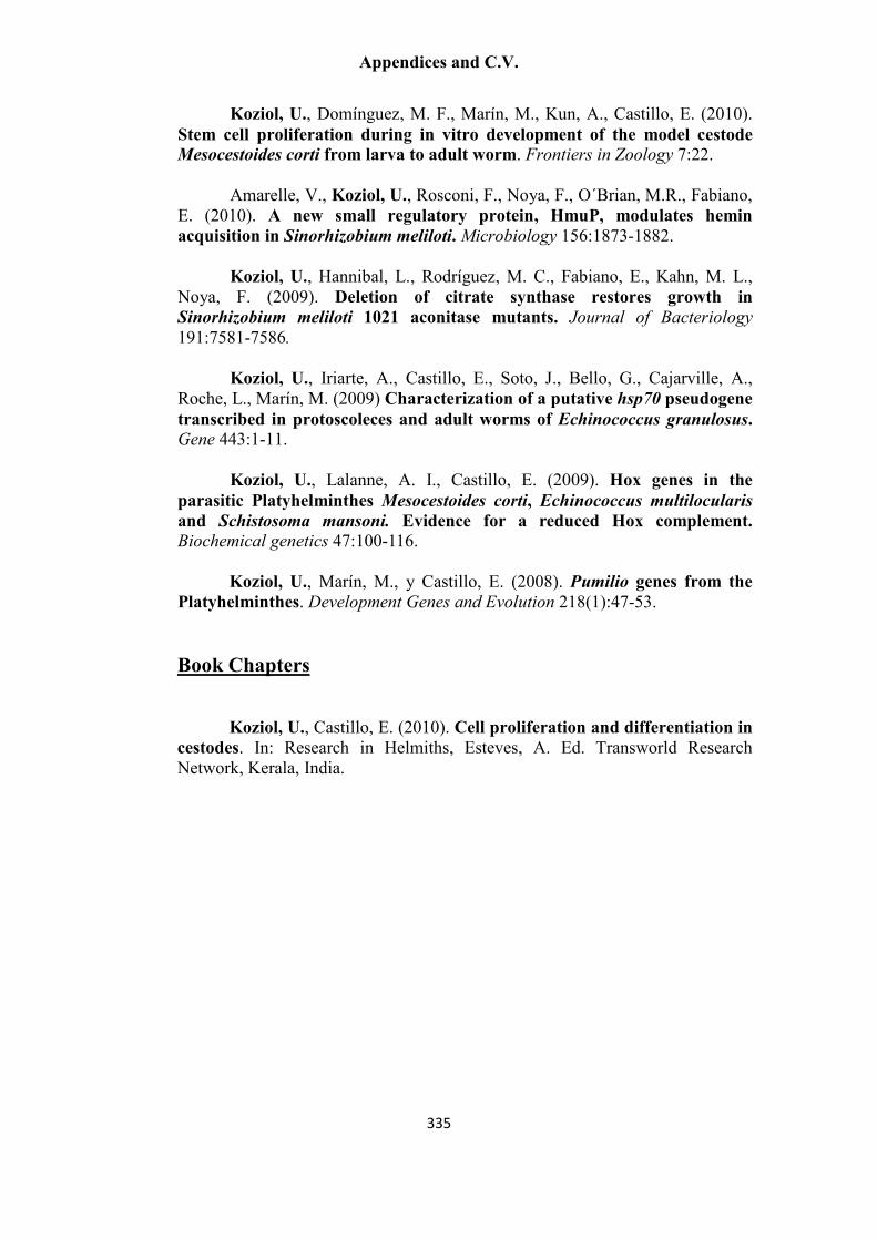
Appendices and C.V.
335
Koziol, U., Domínguez, M. F., Marín, M., Kun, A., Castillo, E. (2010). Stem cell proliferation during in vitro development of the model cestode
Mesocestoides corti from larva to adult worm. Frontiers in Zoology 7:22. Amarelle, V., Koziol, U., Rosconi, F., Noya, F., O´Brian, M.R., Fabiano,
E. (2010). A new small regulatory protein, HmuP, modulates hemin
acquisition in Sinorhizobium meliloti. Microbiology 156:1873-1882. Koziol, U., Hannibal, L., Rodríguez, M. C., Fabiano, E., Kahn, M. L.,
Noya, F. (2009). Deletion of citrate synthase restores growth in
Sinorhizobium meliloti 1021 aconitase mutants. Journal of Bacteriology
191:7581-7586.
Koziol, U., Iriarte, A., Castillo, E., Soto, J., Bello, G., Cajarville, A., Roche, L., Marín, M. (2009) Characterization of a putative hsp70 pseudogene
transcribed in protoscoleces and adult worms of Echinococcus granulosus.
Gene 443:1-11.
Koziol, U., Lalanne, A. I., Castillo, E. (2009). Hox genes in the
parasitic Platyhelminthes Mesocestoides corti, Echinococcus multilocularis
and Schistosoma mansoni. Evidence for a reduced Hox complement.
Biochemical genetics 47:100-116.
Koziol, U., Marín, M., y Castillo, E. (2008). Pumilio genes from the
Platyhelminthes. Development Genes and Evolution 218(1):47-53.
Book Chapters Koziol, U., Castillo, E. (2010). Cell proliferation and differentiation in
cestodes. In: Research in Helmiths, Esteves, A. Ed. Transworld Research Network, Kerala, India.

Appendices and C.V.
336
Selected Presentations and Conferences
Koziol, U., Krohne, G., Castillo, E., Brehm, K. Cell proliferation and
differentiation in cestode larval and adult development. Oral presentation at the Euro Evo Devo Conference (Lisbon, Portugal, 10th to 13th July 2012).
Koziol, U., Amarelle, V., Rosconi, F., Noya, F., O´Brian, M. R., Fabiano, E. (2010). Discovery of a new conserved tandem motif upstream of outer
membrane hemin transporters in alpha and beta-proteobacteria. Poster in International Society for Computational Biology Latin America Conference (Montevideo, Uruguay, 13th to 16th March 2010).
Koziol, U., Domínguez, M. F., Costábile, A., Marín, M., Castillo, E. (2009) Characterization of proliferative cells in cestodes by BrdU labelling
and molecular markers. Poster and oral presentation in the XXIII World Congress of the International Association of Hydatidology. (Colonia del Sacramento, Uruguay, December 2009).
Koziol, U., Domínguez, M.F., Costábile, A., Caurla, G., Marín, M., Kun,
A., Castillo, E. Proliferación celular durante el desarrollo de Mesocestoides
corti (Cestoda). (Cell proliferation during the development of Mesocestoides
corti (Cestoda)). Oral and poster presentation in the molecular parasitology session in the VI meeting of the Sociedad de Bioquímica y Biología Molecular (Montevideo, November 2009).
Amarelle, V., Senatore, D., Koziol, U., Noya, F., Fabiano, E. Functional
metagenomics of termite gut bacteria. poster presentation by V. Amarelle in Genomes 2008: Functional Genomics of Microorganisms, 2008 (Paris, France, April 2008)
Koziol, U., Yurgel, S., Kahn, M., Noya, F. Sinorhizobium meliloti 1021
mutations that link nitrogen and osmotic stress responses. Poster in the 8th European Nitrogen Fixation Conference (Ghent, Belgium, September 2008).

Appendices and C.V.
337
Awards and Mentions
NEB Price for the best Student Presentation, 6th Short Course for Young
Parasitologists (Heidelberg, Germany, March 2012).
Eugenio Prodanov Award (PEDECIBA Biology, Uruguay) for the best M.Sc. thesis in basic research in biology in the period from May 2009 to May 2010. The award is given to a student whose thesis, developed mainly in Uruguay, constitutes an important contribution to scientific knowledge in his research subject.
Special mention for the oral presentation “Caracterización genética y
molecular de locus acnA de Sinorhizobium meliloti. Esencialidad de la
aconitasa para la viabilidad celular” (Genetic and molecular characterization of the acnA locus of Sinorhizobium meliloti. Essentiality of aconitase for cell viability). Award for Young researchers, XII meeting of the Sociedad Uruguaya de Biociencias (Minas, Uruguay, September 2007). EMBO Poster Prize, Outstanding poster presentation, Genomes 2008: Functional Genomics of Microorganisms. (Paris, France, April 2008) (poster presentation performed by V. Amarelle).
Scholarships and Grants
PhD fellowship, Graduate School of Life Sciences, University of Würzburg
(Since April 2011, extended until July 2014).
M.Sc. Degree scholarship from the Sistema Nacional de Becas, ANII, Uruguay. (Awarded in 2009 but not accepted by me)
Scholarship for M.Sc. post-graduate studies, awarded by CSIC, Universidad de la República, Uruguay (November 2008 to November 2009).
Travel grant for the course “Expresión y Silenciamiento de Genes en
Células Animales y Vegetales”. (Expression and silencing of genes in animal
and vegetal cells) (Universidad Nacional de Córdoba, Argentina, 2007), awarded by CABBIO, Argentina-Brasil.
Travel grant for the 8
th Nitrogen Fixation Conference, Ghent, Belgium,
awarded by FEMS, 2008.
Appendices and C.V.
338
Statement of individual author contributions and of legal second
publication rights
Publication (complete reference): Koziol, U., Rauschendorfer, T., Zanon-Rodríguez, L., Krohne, G., Brehm, K.
(2014). The unique stem cell system of the immortal larva of the human parasite Echinococcus
multilocularis. EvoDevo, 5:10
Participated in Author Initials, Responsibility decreasing from left to right
Study Design KB UK
Data Collection UK TR LZR GK
Data Analysis and Interpretation UK KB GK
Manuscript Writing UK KB
Explanations (if applicable):
Publication (complete reference): Koziol U., Krohne G., Brehm K. (2013). Anatomy and development of the
larval nervous system in Echinococcus multilocularis. Frontiers in Zoology 10:24.
Participated in Author Initials, Responsibility decreasing from left to right
Study Design UK KB
Data Collection UK GK
Data Analysis and Interpretation UK KB GK
Manuscript Writing UK KB
Explanations (if applicable):
Publication (complete reference): Koziol, U., Radio, S., Smircich, P., Zarowiecki, M., Fernández, C., Brehm, K. (in
preparation, provisional title). A novel terminal-repeat transposon in miniature (TRIM) is massively expressed in
Echinococcus multilocularis stem cells.
Participated in Author Initials, Responsibility decreasing from left to right
Study Design UK KB CF
Data Collection UK SR MZ PS
Data Analysis and Interpretation UK SR PS MZ KB CF
Manuscript Writing UK KB CF
Explanations (if applicable):

Appendices and C.V.
339
I confirm that I have obtained permission from both the publishers and the co-authors for legal second
publication.
I also confirm my primary supervisor’s acceptance.
Uriel Koziol 11.04.2014 Würzburg
_____________________________________________________________________________________
Doctoral Researcher’s Name Date Place Signature