ABSTRACT SUPPLEMENT
420
Volume 26 Supplement 2016 ABSTRACT SUPPLEMENT The 60th Annual Scientific Meeting of the Japan College of Rheumatology April 21-23, 2016 Yokohama
-
Upload
khangminh22 -
Category
Documents
-
view
0 -
download
0
Transcript of ABSTRACT SUPPLEMENT

Volume 26 Supplement 2016
ABSTRACT SUPPLEMENT
The 60th Annual Scientific Meeting of the Japan College of Rheumatology
April 21-23, 2016Yokohama
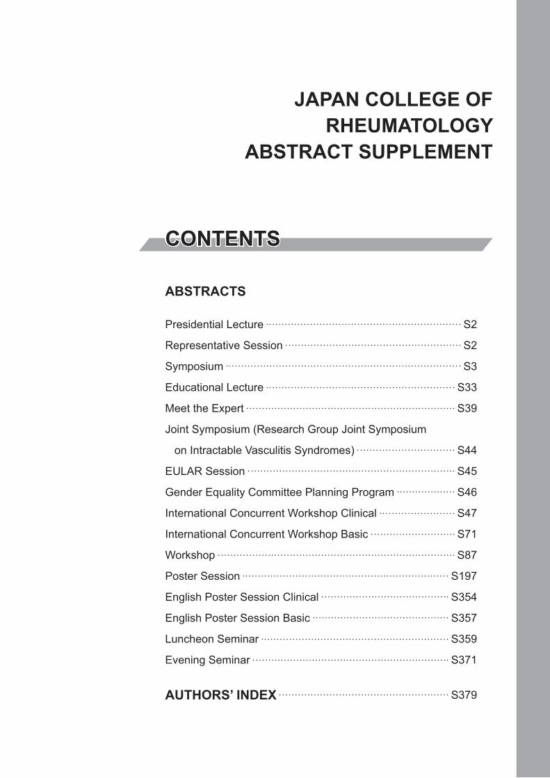
ABSTRACTS
Presidential Lecture S2
Representative Session S2
Symposium S3
Educational Lecture S33
Meet the Expert S39
Joint Symposium (Research Group Joint Symposium
on Intractable Vasculitis Syndromes) S44
EULAR Session S45
Gender Equality Committee Planning Program S46
International Concurrent Workshop Clinical S47
International Concurrent Workshop Basic S71
Workshop S87
Poster Session S197
English Poster Session Clinical S354
English Poster Session Basic S357
Luncheon Seminar S359
Evening Seminar S371
AUTHORS’ INDEX S379
CONTENTS
JAPAN COLLEGE OFRHEUMATOLOGY
ABSTRACT SUPPLEMENT
S2
Presidential Lecture
PLAdvancements and a Perspective of Surgical Treatments for Rheu-matic DiseasesHiromi OdaDepartment of Orthopaedic Surgery, Saitama Medical University, Japan
Conflict of interest: None
Synovectomy used to be performed as a treatment of rheumatoid ar-thritis (RA) during the period of time when no effective medication was available. Although synovectomy achieved certain short-term effects, its long-term results were unsatisfactory due to a recurrence of symptoms or complications. Arthroscopy, which came into clinical use in 1959, even-tually spread as an essential tool for diagnosis and treatment of joint dis-eases. Arthroscopic synovectomy has been widely used as a local treat-ment in cases in which disease activity is controlled by medication. For functional disorder due to joint destruction, arthroplasty, which flattens broken bone stumps, creates new articular facets, and rebuilds joints, has been performed. However, since RA is a progressive disease, the indica-tion of this procedure has been limited. Nonetheless, since the progres-sion of articular destruction can be suppressed by medication today, a sat-isfactory, long-term effect can still be expected when this procedures performed. Total hip arthroplasty has been spread widely around the world due to its satisfactory and long-term effects, after Charnley pub-lished in Lancet in 1961. Specifically, the combined use of a metal and ultra-high molecular weight polyethylene as a joint surface and the use of a bone cement as a fixing material has become the golden standard of ar-tificial joints. This practice has also achieved satisfactory results on other joints. Although artificial joints started to accomplish stable, satisfactory, long-term results, how to prevent loosenings and handle infections and bone defects remain problems to be solved in the future. While arthrode-sis has been performed for joints where stability has been considered more essential than mobility, cervical vertebra fixation has experienced problems, such as implant failure and nonunion. Magerl’s method for at-lantoaxial subluxation and pedicle screw or lateral mass screw fixations for subaxial subluxation have been used in recent years and have been achieving satisfactory results. Various osteotomies of the pelvis, femur and tibia have been performed for hip/knee osteoarthritis and aseptic ne-crosis of the femoral head among the younger population and have ac-complished certain positive results. How to inherit this technique remains a big challenge for the future.
Representative Session
RSMolecular Medicine of Mono-genic and Multi-genic Diseases: From Cytokine to Genomic & Cellular MedicineKen-ichi Arai1,2,3,4,5
1The University of Tokyo, 2Genomic Drug & Medicine Forum, 3Health & Medicine Paradigm Shift Consortium, 4A-IMBN, 5SBI Biotech Co., Ltd.
Conflict of interest: None
Biological drugs brought remarkable changes in treating Rheumatoid arthritis and other autoimmune diseases. Cytokines are biological protein drugs available only in minute quantity and their chemical and biological properties are clarified by recombinant DNA technology enabling the ap-plication of cytokines to medicine. In Part 1, I will talk about 1st stage biotech-revolution in 1970s triggered by Stanford University and bioven-tures in Silicon Valley, cytokine network elucidated by application of re-combinant DNA technology to immunology, Immune regulation via cel-lular interaction of T, B and dendritic cells. I will also talk about translational research, the roles of bioventures and pharmaceutical indus-try in bringing novel discovery into frontier medicine. In the 2nd biotech-revolution from 1990s, genomic & system medicine approach become possible allowing us to characterize whole genome not limited to single gene at DNA, RNA, protein and metabolite levels. Also, the development of diagnosis and treatment of multi-gene diseases such as common dis-eases and cancer become an important agenda. Now, due to the availabil-ity of novel therapy and new drugs, patients suffering from common and/or serious chronic diseases can live much longer. Narrowing the gap be-tween average life span and healthy life span by extending healthy life span become an important goal. This will help to reduce health care cost and medical expenses for aged people. To deal with multi-gene diseases, novel platform for drug development is necessary to confirm the efficacy and the safety of the combination therapy employing multiple drugs with different mechanism of action. In Part 2, I will talk about the technology revolution in IoT & AI areas and the paradigm shift of health and medi-cine. Part 1 Biotechnology and Cytokine Network 1. Ist Biotech-Revo-lution 2. From IFNs to Cytokine Network 3. Multi-potential Hemato-poietic Stem Cell: Constitutive vs Inducible Hematopoiesis 4. Th1/Th2 Paradigm: Plasticity of T cells and Epigenomic Regulation 5. Cytokine Receptor & Signal Network 6. mDC & pDC: Two Types of Dendritic Cells and Immune Regulation 7. Drug Development for Advanced Medi-cine and Translational Research Part 2 Paradigm Shift of Health & Dis-ease and Future Medicine 8. 2nd Biotech-Revolution 9. Mono- vs Multi-genic Diseases: Environmental & Epigenomic Regulation 10. Evolutionary Medicine and Agenda in Genomic Medicine 11. Average vs Healthy Life Span 12. Chronic Inflammation and Multi-genic Diseas-es: Tissue Repair Medicine & Cancer Immunotherapy 13. Management of Health and Medicine
S3
Symposium
S1-1Adaptation and positioning of the steroid therapy in RA latest treat-mentsYutaka KawahitoInflammation and Immunology, Kyoto Prefectural University of Medi-cine, Kyoto, Japan
Conflict of interest: None
Based on the progress of therapeutic drugs such as biologics and the treatment concept of T2T (treat to target), the prognosis of rheumatoid ar-thritis (RA) has been improved by early diagnosis and treatment for clini-cal remission treatment target. On the other hand, the treatment choice of each rheumatologist is different because of various drugs, and the back-ground or complication of patients. At the present, unsolved problems still exist in the medical treatment before and after the remission. One of them is the use of the steroid. The steroid is effective and a cheap medi-cation for RA, so it is used for daily clinical practice. But the use is still a subject of debate among experts. There is a little difference in its use in European and American recommendation, and Japanese guidelines. The steroid is placed as a supporting drug of the RA treatment with a non-ste-roidal anti-inflammatory drug (NSAID). We cannot improve the arthritis of RA only with these drugs. However, we must often apply it appropri-ately for reducing arthralgia and protecting ADL in the daily life. Because the steroid has strong anti-arthritic and immunosuppressive effect, it can expect the improvement of a symptom and bone destruction temporarily. In addtion, there are a lot of adverse effects such as osteoporosis, infec-tious diseases, and GI tract disturbance by the combination of NSAID use even in small quantities. In addition, it is difficult to stop it when we use it once, and the long-term effect is not clarified. “When and how should we start and reduce the steroid?” The evidence is poor. But, in this symposium, I will discuss the risk and benefit of steroid therapy includ-ing the difference of its use in EULAR, ACR and JCR.
S1-2Role of methotrexate as an anchor drug for the treatment of rheuma-toid arthritis in the new biotherapeutic eraYasuo SuzukiDivision of Rheumatology, Department of Internal Medicine, Tokai Uni-versity School of Medicine
Conflict of interest: Yes
A recent paradigm shift of the treatment of RA is to aim for remis-sion by the T2T strategy, using csDMARD as early as possible in the dis-ease process. Methotrexate (MTX) has been recognized as the anchor drug at the end of the 1990s because of its long-term effectiveness, safety profile and widespread use in clinical practice. More than 10 years passed after the introduction of new drugs targeted to key molecules and cells involved in RA pathogenesis, role of MTX might be modified. In Japan, an increase in MTX dose up to 16mg/week was approved in 2011, and 7 biologics and a JAK inhibitor are now available. On the other hand, in-fection and LPD have been increasing probably due to long-term immu-nosuppression. Although it remained unclear what is the best dosing strategy for MTX, the suggestive evidences have been accumulated re-cently. The results of PMS showed that remission rate increased approxi-mately 3 times by increasing MTX from 8mg to more than 10mg/week. In the C-OPERA study, a DB-RCT comparing MTX vs MTX plus certor-izmab pegol for early RA, MTX was increased up to 16mg/week with a rapid dose escalation, and average MTX dose throughout the study period was 11.6mg/week in both groups. These evidences suggest that the thera-peutic effect of MTX could be nearly maximum by increasing up to 12mg/week without safety concern. The recent systematic reviews showed combination of MTX and biologics for all classes was more effi-cacious than bio-monotherapy. In patients responding insufficiently to combination of MTX and biologics, the addition of the 3rd csDMARD such as igratimod could be a therpeutic option to not only achieve remis-sion but also to reduce or discontinue biologics. In this symposium, I discussed the role of MTX in the new biotherapeutic era.
S1-3Usefulness and limitations of the current biologic therapyHideto KamedaDivision of Rheumatology, Department of Internal Medicine, Toho Uni-versity Ohashi Medical Center
Conflict of interest: Yes
Six biological agents targeting TNF and each one biological agent against IL-6 receptor and T cell co-stimulatory molecules are available for the treatment of RA in Japan. All agents accept the effectiveness in more than the half of patients with RA, and biological agents targeting TNF show the increased effectiveness by concomitant use of methotrex-ate. At the same time, any biological agents in any RA population never showed the remission rate exceeding 50%. This fact can be attributable to the restriction in the approved dosing regimen and the existence of pa-tient population indeed requiring agents against novel molecular targets, both leading to insufficient coverage on variety of the RA patients. The limitation in the range of approved dosage may be due to the budget re-striction in the drug development, which is insufficient for thorough ex-amination of dose-response. Thus, rheumatologists have been doing a hard work for the adjustment of dosages or dosing intervals for each pa-tient in the postmarketing phase. In addition, the difference in the expres-sion amount of the target molecule and the subsequent difference in the agent dose for sufficient neutralization of the targeted molecule might be more important than the difference in the species of molecule to be tar-geted, because the expectation to the effectiveness of the remaining agents decreases for patients having repeatedly failed in prior biological agents. However, the increase in quantity of a biological agent by more than several times will not be acceptable if it is proportionally reflected to the treatment costs. In this respect, a prominent effectiveness of gluco-corticoids in autoimmune diseases can be attributable to the fact that it is inexpensive even after the dose escalation by 100 times according to the diseases. In this talk, I am going to discuss the above-mentioned points with selected evidences in order to bridge over the following 2 lectures focusing on the agents under development.
S1-4Perspective of RA treatment by new biological agents in clinical de-velopmentTsutomu TakeuchiDivision of Rheumatology, Department of Internal Medicine, Keio Uni-versity School of Medicine, Japan
Conflict of interest: Yes
Biological agents are positioned as an indispensable drug in rheuma-toid arthritis (RA) treatment algorithm. The unique characteristics of the agents are made of proteins produced by the living and the targets by the agents are theoretically very clear, not acting on off-targets. At present, seven bio-original DMARDs in addition to one bio-similar products are commercialized in Japan, in which mechanism of action of those targets are inhibitions of TNF, IL-6 receptor, and T cell activation. I would like to introduce biological agents on new targets for RA patients, which are now going to late clinical development. Those include IL-6, GM-CSF/GM-CSF receptor, and RANKL. Biological agents targeting on IL-6 such as clazakizumab, olokizuma, and sirkumab may be reviewed by summarizing the publications. In particular, sirkumab, which may go into phase III clinical trials is highlighted and overviewed. Similarities and dis-similarities between IL-6 and IL-6 receptor as a target are discussed. GM-CSF and GM-CSF targets are similarly reviewed and mavrilimumab, targeting on GM-CSF receptor, may be overviewed. Denosumab, target-ing on RANKL has been developed for RA in Japan and phase II trial (DRIVE study) as well as phase III study (DESIABLE study) would be overviewed. Finally, future perspective of these biological agents in late clinical development in the present RA treatment algorithm and personal-ization should be discussed.
S1-5New perspective of kinase-targeting low molecular DMARDs for the treatment of RAYoshiya Tanaka
S4
The First Department of Internal Medicine, School of Medicine, Univer-sity of Occupational and Environmental Health, Japan
Conflict of interest: Yes
RA is a systemic autoimmune disease characterized by synovial in-flammation and joint destruction. However, sDMARDs such as MTX and bDMARDs have revolutionized treatment of RA. However, bDMARDs are limited to intravenous or subcutaneous uses and orally available small but strong products have been developed. The multiple cytokines and cell surface molecules bind to receptors, resulting in the activation of various signaling, including phosphorylation of kinase proteins. Janus kinase (JAK) plays pivotal roles in the pathological processes of RA. Tofaci-tinib, a small orally available product, inhibits phosphorylation of JAK1/JAK3. Six phase 3 studies revealed that tofacitinib was significantly ef-fective than placebo in active RA, but its association with carcinogenicity and infections remains debated. Accordingly, multiple oral small prod-ucts targeting kinases are emerging. Baricitinib is a Jak1/Jak2 inhibitor. Four phase 3 studies indicate oral baricitinib is more efficacious than pla-cebo and adalimumab with or without MTX in patients with sDMARD-naïve, inadequately responsive to sDMARD (sDMARD-IR) and bD-MARD-IR. The adverse effects were partly similar to tofacitnib. Filgotinib and ABT-494 are sDMARDs targeting JAK1 and showed simi-lar efficacy and safety profiles as tofacitinib in phase 2 trials. Also, the JAK3 inhibitors decernotinib and peficitinib showed strong and rapid ef-ficacy, comparable to tofacitinib in phase 2 trials. Oral kinase inhibitors targeting Syk and Btk are also under the development. Thus, small prod-ucts targeting specific kinase could represent a valuable addition to the current therapies and these kinase inhibitors would take in the therapeutic armamentarium in RA and multiple autoimmune diseases. However, the commonly observed adverse events of them were related to infection, he-matologic, hepatic and renal disorders. Major concerns regarding long-term safety should be clarified by post-marketing surveillance.
S2-1Regulation of bone destruction by the adaptive immune systemKazuo OkamotoDepartment of Immunology, Graduate School of Medicine and Faculty of Medicine, The University of Tokyo, Japan
Conflict of interest: None
The immune and skeletal systems are closely related through a num-ber of shared regulatory molecules including cytokines. Progressive joint destruction in rheumatoid arthritis (RA) is the most typical pathological conditions that depend on the interaction between the skeletal and im-mune systems. Large amounts of the inflammatory cytokines such as IL-6, IL-1 and TNF are released, and lymphocytes, synovial macrophages and synovial fibroblasts accumulate and proliferate in the inflamed tis-sues. These inflammatory cytokines promote not only the immune re-sponses but also osteoclast differentiation and survival by inducing RANKL on synovial fibroblasts. As proven by the clinical efficacy of an-ti-cytokine therapies, it is apparent that the inflammatory cytokines are strongly associated with the pathogenesis of RA. However, since the au-toimmune response is considered as the core of the progression of RA, elucidation of the mechanism how the adaptive immune systems affect bone is important for understanding the pathogenesis of RA. A unique ef-fector helper T cell subset, Th17, has stimulatory effects on osteoclasto-genesis and plays a key role in the pathogenesis of RA through IL-17. IL-17 not only induces RANKL on synovial fibroblasts but also activates local inflammation, leading to the inflammatory cytokine production. On the other hand, Foxp3+CD4+ Treg cells are a specialized T-cell subset that engages in the maintenance of immunological self-tolerance. Under ar-thritic conditions, a part of Foxp3+CD4+ T cells lose Foxp3 expression and undergo transdifferentiation into Th17 cells (called exFoxp3Th17). Notably, exFoxp3Th17 cells highly express RANKL and thus contribute to the pathogenesis of RA. Furthermore, recent studies revealed that the immune complex directly enhances osteoclastic bone resorption under in-flammatory conditions such as RA. It is becoming clear that the adaptive immune systems exert direct deleterious effects on bone cells in RA.
S2-2Inflammation and OsteoclastogenesisTakeshi Miyamoto, Morio Matsumoto, Masaya NakamuraDepartment of Orthopedic Surgery, Kieo University School of Medicine, Japan
Conflict of interest: Yes
Patients with inflammatory arthritis diseases, such as rheumatoid ar-thritis (RA), exhibit continuous joint inflammation, a chronic inflamma-tion, with joint destruction. In arthritis animal models, the joint erosion but not joint inflammation was reportedly blocked by inhibiting osteo-clast differentiation or activity. In human, treatment with receptor activa-tor of nuclear factor kappa B ligand (RANKL) was reported to block joint erosion but not disease activity in RA patients, suggesting that os-teoclasts play a pivotal role for joint destruction but not joint inflamma-tion. We found that signal transducer and activator of transcription 3 (Stat3) was required for both joint inflammation and erosion by osteoclast formation. I will discuss the inflammation and osteoclastogenesis.
S2-3Involvement of RANKL-induced incomplete cytokinesis in poly-ploidization of osteoclasts: a novel mechanism of osteoclast poly-ploidizationNoriko Takegahara1, Hyunsoo Kim2, Yongwon Choi2
1Osaka University, Suita Osaka, JAPAN, 2University of Pennsylvania, Philadelphia, Pennsylvania, USA
Conflict of interest: None
Polyploidy, in which a cell has more than the diploid complement of chromosomes, is a widespread physiological phenomenon observed espe-cially in plants, fungi, and insects. Although it is less common in mam-mals, polyploidization occurs in selected tissues including the placenta, liver, heart, skeletal muscle, and bone marrow during normal develop-ment and aging. During developmental programs, cells obtain additional sets of chromosomes by various mechanisms, including endocycles, en-domitosis, incomplete cytokinesis, and cell fusion. Endocycles, endomi-tosis, and incomplete cytokinesis are directly associated with the prolifer-ative state of the cell. By contrast, cell fusion is entirely independent of cell proliferation. Osteoclasts are specialized polyploid cells that resorb bone. Upon stimulation with receptor activator of nuclear factor kappa-B ligand (RANKL), myeloid precursors commit to becoming polyploid, largely via cell fusion. Although generation of polyploid osteoclasts is thought to occur due to cell fusion, independently of cell proliferation, a relationship between cell proliferation and osteoclast differentiation has been pointed out. Here, we demonstrated that in addition to cell fusion, incomplete cytokinesis also plays a role in osteoclast polyploidization. Fluorescence in situ hybridization revealed that some of osteoclasts ex-hibited nuclear polyploidy (i.e., they contained nuclei with more than the diploid complement of chromosomes [> 2N]) in vivo, suggesting that cells that undergo incomplete cytokinesis are physiologically involved in formation of polyploid osteoclasts. Our findings reveal an unexpected pattern of cell division and fusion during the generation of polyploid os-teoclasts.
S2-4The regulation of hematopoietic stem cells by mesenchymal stem cellsYuya KunisakiCenter for Cellular and Molecular Medicine, Kyushu University Hospital
Conflict of interest: None
Somatic stem cells self-renew to maintain tissue homeostasis for the lifetime of organisms through tightly controlled proliferation and differ-entiation. Hematopoietic stem cells (HSCs) are essentially required for the hematopoietic homeostasis. Therefore, They do not only ensure life-long replenishment of all blood lineages, but also keep their pool con-stant. Cell cycle quiescence is a critical feature contributing to stem cell maintenance. Recent studies have highlighted the importance of bone marrow microenvironments that regulate HSC functions (HSC niches).

S5
In the HSC field, there has been a considerable interest and debate re-garding whether or not quiescence and proliferation of HSCs is regulated by distinct niches. Previous reports suggest that quiescent HSCs reside near osteoblasts in the bone marrow whereas actively cycling HSCs are found near sinusoids. However, this popular concept has not been sup-ported by rigorous analyses. To get more insight into the spatial localiza-tion of HSCs, we have developed a whole-mount staining technique that allows precise measurements of 3D distances of HSCs from structures and is amenable to computational simulation to define the significance of these interactions. This novel approach has allowed us to uncover two distinct types of vessels associated with quiescent and proliferating HSCs and to underscore the importance of arteriolar vessels for stem cell quies-cence. In addition, we will introduce the groundwork for the characteriza-tion and identification of subsets of stromal cells that comprise the bone marrow niche and provide definitive data on the hierarchical organization of MSCs and precursors of stromal cells in the bone marrow.
S2-5In vivo imaging of osteoclast dynamicsJunichi KikutaDepartment of Immunology and Cell Biology, Graduate School of Medi-cine, Osaka University, Japan
Conflict of interest: None
Rheumatoid arthritis (RA) is a chronic autoimmune disease charac-terized by synovial joint inflammation and progressive cartilage/bone de-struction. Arthritic bone destruction is considered to be mediated mainly by enhanced activation of osteoclasts at inflammatory sites. To prevent RA-associated bone destruction, it is important to understand the cellular dynamics of osteoclastic bone resorption in vivo. Because bone is the hardest tissue in the body, it is difficult and almost impossible to visualize the inner bone tissue in living animals. In the fields of bone and mineral research, cell morphology and structure in bone tissues can be analyzed by conventional methods such as micro-CT and histological analysis. These methods allow for the evaluation of cell shape and molecular ex-pression, but cannot observe living osteoclast movement. Thus, how the bone-resorptive functions of mature osteoclasts are controlled in vivo re-mains unclear. To answer this question, we utilized an advanced imaging system to visualize living bone tissues with intravital multiphoton mi-croscopy that we have originally established. By using this imaging sys-tem, we succeeded in visualizing the in vivo behavior of living mature osteoclasts on the bone surface, and identified different functional subsets of osteoclasts in terms of their motility and function, i.e., ‘static - bone resorptive’ and ‘moving - non resorptive’. Treatment with recombinant RANKL or bisphosphonate changed the composition of these populations as well as the total number of mature osteoclasts. We also found that RANKL-bearing Th17 cells could control bone resorption of mature os-teoclasts, demonstrating novel actions of Th17. Furthermore, we have es-tablished the practical imaging of bone destruction by osteoclasts in ar-thritic joints using intravital multiphoton microscopy. In this symposium, we show the latest data and also discuss the further application of intravi-tal bone imaging.
S2-6Osteoimmunology and arthritisYuho KadonoDepartment of Orthopaedic Surgery, Sensory and Motor System Medi-cine, Graduate School of Medicine, The University of Tokyo, Tokyo, Ja-pan
Conflict of interest: Yes
Bone homeostasis is maintained with the balance of bone resorption by osteoclasts and formation by osteoblasts. Macrophage lineage cells differentiate into osteoclasts with RANKL produced by osteoblasts or os-teocytes. RANKL was originally discovered as the factor expressed on T cells that promoted survival and activation of dendritic cells. IFN-gamma produced by Th1 cells inhibits RANKL-induced osteoclastogenesis. It is well known that the immune system greatly participates in osteoclasto-genesis. The synovial tissue of rheumatoid arthritis is comprised of fi-broblasts, macrophages and lymphocytes, and produces inflammatory cy-
tokines such as TNF-alpha. These cytokines act on synovium itself to promote RANKL expression, which leads to bone destruction. At the same time, the expression of DKK-1 and sclerostin are decreased, those which inhibit Wnt signaling for osteoblast differentiation. It is thought that biologics work on immune cells or synovial fibroblasts and reduce both RANKL and DKK-1 expression. They indirectly inhibit bone de-struction and also restore bone. Ankylosing spondylitis is characterized by the inflammation of sacroiliac joint and enthesis, where both bone de-struction and formation are seen. If there is much TNF, enhanced expres-sion of DKK-1 and sclerostin inhibit Wnt signaling. It is also thought that more transmembrane form and less soluble form of TNF result in more bone destruction and less formation. There is abundant TGF-beta near syndesmophytes and TGF-beta promotes the differentiation of the Th17 cell in collaboration with IL-6. When Th17 cell is dominant in local site, more IL-17, IL-22, and IL-23 are produced. IL-22 and IL-23 are known to promote the differentiation of osteoblasts by enhanced production of both Wnt and BMP. In autoimmune diseases, bone and immune system collaborate on forming the arthritis or enthesitis. I present the pathology of these diseases in touch with osteoimmunology.
S3-1The physiological role of inflammasome-mediated immune responsesNaohiro InoharaDept. Pathology., University of Michigan Medical School, Ann Arbor, MI, USA
Conflict of interest: None
IL-1β is a cytokine that is secreted by inflammasome activation and plays an important in host protection against pathogens, whereas its ab-normal secretion results in autoinflammatory diseases. The suppressive regulatory mechanism of NLRP3 inflammasome is lost by mutations in patients with Cryopyrin (NLRP3)-associated periodic syndrome (CAPS). To understand the pathogenesis of CAPS, it is important to know how IL-1β secretion is regulated in response to inflammation or infection. In ad-dition to danger signals that are required for inflammasome activation, the functional expression level of NLRP3 must be first upregulated through cell surface pattern recognition receptors such as Toll-like recep-tors by inflammatory molecules. The dual requirement for NLRP3 in-flammasome activation means that each requirement can be filled sepa-rately by different bacteria within the same tissue environment. For example, during intestinal Clostridium difficile infection IL-1β secretion requires not only Clostridium difficile but also commensal Enterobacteri-aceae species, which are responsible for host lethality and are the primary targets of IL-1β-mediated host responses in the mouse model. On the oth-er hand, NLRP3 inflammsome activation and IL-1β secretion in the intes-tine are induced only by one commensal P. mirabilis, which have toxins and high pyrogenicity. Some intracellular bacteria, including Salmonella and Legionella, hide in host cells and avoid detection by cell surface de-tection, IL-1β secretion is induced by non-NLRP3 type inflammasomes such as NLRPC4. Once IL-1β is secreted at the infection site, IL-1β me-diates local inflammatory responses such as neutrophil recruitment and induction of other cytokines (i.e. IL-22) that are important for systemic elimination of pathogens and pathobionts. The basal levels of IL-1β in non-infection state also induce chronic responses that impacts the inflam-matory state. We believe that these new findings will advance our under-standing of acute and chronic responses in autoinflamamtory diseases and contribute to development of new therapeutic approaches to treating au-toimmune disease.
S3-2Review in Familial Mediterranean fever - New insights into disease associated gene and cytokine/chemokine signalingTomohiro Koga1, Kiyoshi Migita2, Koh-ichiro Yoshiura3, Shuntaro Sato4, Kazunaga Agematsu5, Akihiro Yachie6, Junya Masumoto7, Yukitaka Ueki8, Katsumi Eguchi8, Atsushi Kawakami1
1Unit of Translational Medicine, Department of Immunology and Rheu-matology, Nagasaki University Graduate School of Biomedical Sciences, 2Department of Rheumatology and Clinical Research Center, Nagasaki Medical Center, Omura, Japan, 3Department of Human Genetics, Atomic Bomb Disease Institute, Nagasaki University, 4Nagasaki University Hos-pital, Clinical Research Center, Nagasaki, Japan, 5Department of Infec-

S6
tious Immunology, Shinshu University, Graduate School of Medicine, Matsumoto, Japan, 6Department of Pediatrics, School of Medicine, Insti-tute of Medical, Pharmaceutical and Health Sciences, Kanazawa Univer-sity, Kanazawa, Japan, 7Proteo-Science Center (PROS), Ehime Universi-ty, Japan, 8Center for Rheumatic Disease, Sasebo Chuo Hospital, Sasebo, Japan
Conflict of interest: None
Familial Mediterranean fever (FMF), the most common autoinflam-matory hereditary disease, is characterized by recurrent attacks of fever with arthritis and serositis. The mutation of Mediterranean fever (MEFV) gene, which encodes pyrin, is closely associated with the pathogenesis of FMF. Although it has become apparent that FMF patients is not quite rare in Japan and that its survival rate is more than 90%, there is no funda-mental treatment for this disease. Long-term treatment of oral colchicine is usually used among FMF patients. Even though it has been reported by case reports that IL-1 inhibitor, TNF inhibitor or IL-6 inhibitor is ef-fective for the treatment of colchicine resistant FMF, the mechanism of the development of FMF has not been elucidated. Accordingly, it is re-quired the identification of inflammatory cytokines involved in the devel-opment, the analysis of signal associated molecules which composed of inflamasome and discover of disease associated genes other than MEFV gene. Our research group has been addressing the formation of the FMF consortium, construction of bio-bank and analyzing the clinical informa-tion, genome DNA and serum from FMF patients. Also, we are going to search the new therapeutic targets in FMF. We recently found specific molecular interactions in patients with FMF based on multiple cytokines measurement. In addition, the active form of IL-1b is a valuable bio-marker for monitoring disease activity in patients with FMF. We per-formed a comprehensive genome analysis to determine the entire nucleo-tide sequence of MEFV gene including the coding regions, the transcriptional regulatory regions, untranslated region and promoter re-gion. We have been attempting to identify new disease-associated genes in all exon analysis using next-generation sequencer for the cases that can not be fully described in the mutation of the MEFV gene.
S3-3Interferonopathy; dissecting SLE pathogenesis through the analyses of SLE-like Mendelian diseasesHirotsugu Oda1,2
1Kitano Hospital, Osaka, Japan, 2RIKEN-IMS
Conflict of interest: None
Type I interferon (IFN) is a group of cytokines induced by outer stimuli such as viral infection. When induced, it activates a group of IFN-stimulated genes (ISGs). The association of SLE and ISGs detected by GWAS studies, and the overexpression of ISGs in SLE patients, both suggested type I IFN as a key molecule in SLE pathogenesis. A number of Mendelian diseases called “type I interferonopathy” is widely known, in that its genetically-determined type I IFN overproduction causes SLE-like phenotypes. Aicardi-Goutières syndrome (AGS) is an infantile-onset encephalopathy, which is characterized by developmental delay, intracra-nial calcification and CNS inflammation. AGS patients sometimes show SLE phenotypes, such as autoantibody production, low complement lev-els and chilblains, and indeed, some AGS patients fulfill the diagnostic criteria of SLE. Several genes engaged in nucleic acid metabolism are known to be responsible for AGS, in that their loss-of-function mutations result in the accumulation of nucleic acids and type I IFN overproduc-tion. Our whole exome sequencing identified mutations in MDA5 (IFIH1 gene) in three AGS patients without mutations in known AGS genes. MDA5 is a cytosolic receptor of double-strand RNA, and we showed that these gain-of-function mutations in AGS patients spontaneously activate type I IFN signaling. Funabiki previously reported a mouse model of SLE due to an Ifih1 missense mutation (Funabiki et al, Immunity, 2014), which is in concordance with our results. Furthermore, Liu reported mu-tations in another upstream molecule of IFN pathway, STING (TMEM173 gene), is responsible for infantile-onset vasculitis and inter-stitial pneumonitis (Liu et al, NEJM, 2014), which they call SAVI (STING-Associated Vasculopathy of Infancy). In this talk, recent reports of interferonopathy and its mouse models aiming at the understanding of SLE pathogenesis will be discussed.
S3-4Psoriasis with a genetic basis: DITRA and CAMPSKazumitsu SugiuraDepartment of Dermatology, Nagoya University Graduate School of Medicine
Conflict of interest: None
I shall introduce 2 novel autoinflammatory diseases, interleukin-36 receptor antagonist (DITRA)- and caspase recruitment domain family, member 14-mediated psoriasis (CAMPS). Psoriasis is classified into 5 types: psoriasis vulgaris (PsV), psoriatic arthritis (PsA), psoriatic erythro-derma, psoriasis guttate, and generalized pustular psoriasis (GPP). The cause underlying GPP was unknown. GPP is an incurable disease, with more than 1,900 patients registered in Japan. Because many GPP cases are adult-onset, it was not considered a monogenic autoinflammatory dis-ease. However, we found that most GPP cases that are not accompanied by PsV are DITRA-mediated. IL36RN encodes interleukin-36 receptor antagonist (IL-36Ra), which antagonizes pro-inflammatory cytokines IL-36α, IL-36β, and IL-36γ in the skin. The inflammatory signal is consid-ered to be activated to cause the DITRA-mediated lesion via IL-36 recep-tor due to lack of IL-36Ra antagonism against IL-36α, IL-36β, and IL-36γ. CAMPS is an autosomal dominant inherited disease due to CARD14. CAMPS phenotypes include PsV, PsA, GPP, or the psoriasis-related pityriasis rubra pilaris. CARD14 activates NF-κB in epidermal keratinocytes. However, the pathomechanism of the involvement of the CARD14 mutation in CAMPS has not been elucidated, although some CARD14 mutants have been proven as gain-of-function mutants in vitro.
S3-5Disease modeling of autoinflammatory syndromes with patient de-rived iPS cellsMegumu K SaitoCenter for iPS cell research and application (CiRA), Kyoto University, Kyoto, Japan
Conflict of interest: None
Induced pluripotent stem cells (iPSCs) is a pluripotent cell lines which can be established patients. iPSCs can be established from various somatic cells such as peripheral blood cells and skin fibroblasts. By dif-ferentiating patient-derived iPSCs into the responsible cell types, we are now able to obtain various patient-derived differentiated cells. Therefore, disease-associated iPSCs have been regarding as a promising tool to link the patients’ phenotypes to basic research, thereby contributing to the progress in medicine. The number of articles regarding the iPSC-based in vitro disease modeling is increasing explosively. Autoinflammatory syn-dromes are one of the disease entities characterized by the abnormal function of innate immunity. Most of autoinflammatory syndromes are accompanied by genetic alterations. We previously reported disease mod-eling of CINCA syndrome by using iPSCs derived from patients with so-matic mosaicism of NLRP3. We consider iPSCs as a useful tool for un-derstanding the pathophysiology of antoinflammatory syndromes, and for developing novel therapeutic approaches. In this presentation, we would like to introduce our efforts for establishing disease models of autoin-flammatory syndromes.
S4-1Current understandings of ankylosingspondylitis: from Japanese re-portsKurisu Tada1, Ken Yamaji1, Naoto Tamura1, Shigeto Kobayashi2, Yoshinari Takasaki1
1Department of Internal Medicine and Rheumatology, Juntendo Universi-ty School of Medicine, Tokyo, Japan, 2Juntendo Koshigaya Hospital
Conflict of interest: None
Ankylosing spondylitis (AS) is a chronic inflammatory rheumatic disease mainly affecting the axial joint and may lead to structural dam-age, syndesmophyte formation, ankylosis of spine, and finally impair the quality-of-life. AS develops in young age, with a peak age of onset around 20 years, and is highly associated with HLA-B27. HLA-B27 is
S7
present in 90% of AS patients, and there is a clear correlation between the prevalence of AS and prevalence of HLA-B27. In Japan, prevalence of HLA-B27 is very low, about 0.5%, and this leads to the low preva-lence of AS. Therefor, in Japan, clinical experience of AS is olso low, and there are some misdiagnosi, overdiagnosis, and overtreatment. In 2009, ASAS (Assesment of SpondyloArthritis international Society) develop the new classification criteria, and this is aimed early diagnosis and early treatment. In 2015, AS was approved as intractable disease defined by Japanese Ministry of Health, therefor AS patients can be administered expensive biological agents with low cost. But not all the AS patients needs this treatment. We must understand and diagnosis this disease cor-rectly.
S4-2Precise understanding of ASAS criteria, undifferentiated SpA and non-radiographic axial SpAShigeto KobayashiDepartment of Rheumatology and Internal Medicine, Juntendo Koshiga-ya Hospital, Saitama, Japan
Conflict of interest: None
The ASAS criteria for SpA have been frequently misinterpreted as diagnostic criteria. Patients will be easily diagnosed with SpA using a checklist of the ASAS criteria. However, individual patients should be di-agnosed based on as the following: physician’s judgment, excluding and/or differential diagnosis, case confirmation by other expert rheumatolo-gists, confirmation over several months, and others procedures. uSpA is unclassified rather than early SpA. It is a generic term for patients who do not fit into the classic SpA disease classification. The main feature of uSpA patients exhibit is peripheral arthritis and not axial spondylitis. Male predominance, a high rate of HLA-B27-positive cases and an ele-vated level of serum CRP at the onset are clinical manifestations relevant to reactive arthritis. It has been postulated in US literature that most pa-tients with uSpA have an unclear preceding history of infection. nr-axial SpA is implied as early AS and has been positioned in the target group to promote early treatment with TNF inhibitors. However, not all patients with nr-axial SpA progress to AS with radiographic sacroiliitis. There-fore, non-radiographic should not be regard as preradiographic. Further-more, more females, a lower rate of HLA-B27-positive cases, and limited effect of biologics are characteristics of nr-axial SpA which are irrelevant to AS. Fibromyalgia is frequently misdiagnosed in Japan as well as over-seas. It will be possible that the features of nr-axial SpA were affected by those of FM overdiagnosed by general physician. The concept of the nr-axial SpA is not fully and equally understood between the ASAS and the Spondyloarthritis Research and Treatment Network (SPARTAN) in the USA. Treatment with TNF inhibitors for nr-axial SpA in Europe have al-ready been approved, but not by the FDA in the USA. It has been con-cluded by the FDA that the natural history of nr-axial SpA must be clari-fied in further investigation.
S4-3Proper understanding of the significance of imagingHideharu SugimotoDepartment of Radiology, Jichi Medical University, Shimotsuke, Japan
Conflict of interest: None
In the diagnosis of ankylosing spondylitis (AS), it is important to demonstrate sacroiliitis (SII) by imaging. SII is classified into five stages in the modified NY criteria. However, it is difficult to diagnose SII in grade 1 and 2. Further, concordance rate (κ value) among readers is low. In the interpretation of the SII, less experienced readers tend to overdiag-nose SII as compared to the experienced readers. When patients have in-flammatory low back pain and there is no positive plain X-ray findings, if the AS is still suspected, MRI is effective in diagnosis of SII. In the MRI findings of SII, there are two categories: structural changes, inflammatory lesions. In the clinical practice, it is not often to demonstrate intensive BME by MRI. In the MRI diagnosis, by adding findings of structural changes, specificity of AS diagnosis is improved. Psoriatic arthritis (PsA) was classified into 5 types. However, there is an overlap in each type, and disease type also varies over time. Therefore, it is practical to classify
into two types; peripheral arthritis and axial arthritis. PsA of hands and feet shows three patterns; DIP/PIP arthritis, Ray pattern, and RA like pat-tern. SII in PsA tends to be asymmetric, and intervertebral joint lesions are small as compared to those of AS lesion. Lumbar spine lesions are relatively infrequent, discontinuous, and asymmetrical. In PsA, MRI can depict joint lesions that are not clinically apparent. There are six parame-ters in the MRI findings; synovitis, erosion, bone marrow edema (BME), tenosynovitis, periarticular inflammation, bone proliferation. BME pre-cedes erosion. Although MRI findings of early PsA are nonspecific, ede-ma of enthesitis and diaphysis, and diffuse soft tissue edema are signifi-cantly frequent as compared to early RA.
S4-4Psoriasis treatment modalities-topical, oral, phototherapy ands bio-logicsAkimichi Morita Department of Geriatric and Environmental Dermatology, Nagoya City University Graduate School of Medical Sciences, Nagoya, Japan
Conflict of interest: Yes
Psoriasis is a chronic immune-mediated autoinflammatory skin dis-ease. Risk factors and comorbidities commonly observed in patients with psoriasis include metabolic syndrome, cardiovascular disease, arthritis symptoms, and psychiatric illness. The estimated prevalence of psoriasis is 1% to 3% of the population worldwide, and the prevalence in Japan is 0.2%. Pathophysiologic mechanisms are related to aberrant immune re-sponses, cell types, and proinflammatory mediators, such as tumor necro-sis factor-a and interleukins (IL) 12, 23 and 17A. Biologics approved for the treatment of moderate-to-severe plaque psoriasis targeting these in-flammatory mediators have improved the response rates associated with conventional oral agents, such as cyclosporine A, etretinate, and metho-trexate. The “treat to target” recommendation is a PASI (psoriasis area and severity index) of 90. Phototherapy is also used to treat psoriasis. Ul-traviolet light (UV) phototherapy using narrowband UVA (311-313 nm) is a well-established treatment for psoriasis. UV phototherapy has two primary modes of action: apoptosis induction and immune suppression. Narrowband UVB depletes pathogenic T cells by inducing apoptosis and regulatory T cells. Bath-psoralen plus ultraviolet light A (PUVA) therapy continues to be beneficial and used in the treatment of psoriasis due to its efficacy, safety profile, and low cost. Bath-PUVA therapy significantly re-duces the number of Th17 cells and significantly increases regulatory T cell (Treg) function to almost normal levels, thus resolving the Th17 and Treg imbalance in patients with psoriasis, and induces activated Treg. These four treatment modalities: topical, oral, phototherapy, and biolog-ics, are important and their application for treating and controlling psoria-sis is relatively complex. Cooperation between rheumatologists and der-matologists is therefore essential for providing optimal care to patients with psoriasis.
S4-5Psoriatic arthritis: clinical manifestations and the efficacy of biolog-icsAtsuo TaniguchiInstitute of Rheumatology, Tokyo Women’s Medical University
Conflict of interest: None
Psoriatic arthritis is a characteristic inflammatory arthritis associated with psoriasis. The recent studies shows that the disease burden of the psoriatic arthritis is comparable to those of rheumatoid arthritis and anky-losing spondylitis. The impact of psoriatic arthritis on physical activities and quality of life has been shown to be similar to rheumatoid arthritis. The successful introduction of biologics including TNF inhibitors dra-matically changed the treatment of psoriatic arthritis and facilitated the development of the management recommendations for psoriatic arthritis. The three main drug classes for the treatment of psoriatic arthritis are NSAID, conventional synthetic (cs) DMARD and biological DMARD. TNF inhibitors have sufficient evidence for the efficacy on peripheral ar-thritis. The radiographic progression has been shown to be suppressed by TNF inhibitors. TNF inhibitors indicated to be effective in the treatment of enthesitis, dactylitis. Therefore, patients with predominant enthesitis or
S8
dactylitis should be treated with biologics when NSAID or local therapy is ineffective. csDMARD has no evidence for the treatment of spondyli-tis. Some studies have shown the suppression of inflammation of spine by biological DMARD. However, the suppression of radiographic pro-gression of psoriatic spondylitis has not been indicated although the as-sessment of the effectiveness of biological DMARD on the progression of spinal disease need longer time. At the moment, for psoriatic spondyli-tis, biological DMARD is recommended when use of NSAID with local therapy and/or rehabilitation shows inadequate response. The biological DMARDs to block IL-17 or IL-12/23 pathway have been shown to be ef-fective to suppress disease activity of psoriatic arthritis. At the moment, those drugs should be used for patients with inadequate response to TNF inhibitors.
S4-6Psoriatic arthritis: Medical care and complications in JapanShigeyoshi TsujiDepartment of Orthopaedics, National Hospital Organization, Osaka Minami Medical Center, Japan
Prevalence of Japanese patients with psoriasis (PSO) has been re-ported to be 0.34% in 2015 (K.Kubota et al BMJ 2015). At the same time, we have reported that there is a psoriatic arthritis (PsA) patients in 14.5% of patients with psoriasis (Ohara et al J.Rheumatol 2015). In Ja-pan, there were about 63,000 patients with PsA. In recent years, since the biologics for PSO/PsA adopt Japanese insurance, the treatment of PSO/PsA got better. The recommendations from the Nature Review Rheumatology 2014, Once a diagnosis of PSO, you should take care of many symptoms of "Psoriatic Disease" (the skin and nails symptoms and joint symptoms and metabolic syndrome, cardiovascular disease …) Certainly the skin and joint symptoms of PSO/PsA are a dramatic im-provement by biologics, but it is not yet fully possible to correspond about the "many complications" in the current situation. In this session, we would like to talk about epidemiology, clinical findings, the inter-de-partment cooperation and the complications that should be noted.
S5-1Progress in pathophysiology and treatment of pulmonary arterial hypertension associated with connective tissue diseasesShunji YoshidaSection of Rheumatology, Department of Internal Medicine, Fujita Health University School of Medicine, Toyoake, Japan
Conflict of interest: None
Connective tissue diseases (CTD) are frequently complicated by pulmonary hypertension (PH). Although right heart catheterization is gold standard for definitive diagnosis of PH, most rheumatologists find it difficult to perform, so useful screening methods have thus been sought. The most useful method is echocardiography. Exercise echocardiography is also being investigated in order to reduce false-negative results in this method, and approaches such as nailfold videocapillaroscopy and flow-mediated dilation are also being investigated. In addition, based on the fact that PH first develops after the effective pulmonary vascular bed be-comes £1/3, 3D angiography using computed tomography is also being investigated. Because PH in systemic sclerosis (SSc) often devel-ops gradually, yearly screening with tests such as echocardiography and pulmonary function tests is useful. Conversely, PH in systemic lupus ery-thematosus (SLE) and many cases of mixed connective tissue disease (MCTD) has an acute onset, which means that screening tests have a lim-ited utility, and early tests must be performed following onset of the signs and findings of PH. PH in SSc is primarily caused by vascular stenosis, whereas PH in non-SSc diseases is thought to be primarily caused by pul-monary arteritis. Therefore, the main treatment is vasodilator agents for PH in SSc and high-dose steroid therapy and immunosuppressants for PH in non-SSc diseases. However, in the case of PH in SSc, venous lesions are often observed, so the use of vasodilator agents requires careful con-sideration. Even among CTD-associated PAH, which has a poorer prog-nosis than idiopathic pulmonary arterial hypertension, PH in SSc has a poorer prognosis than PH in non-SSc diseases, and few improvements in the prognosis of PH in SSc have been reported over time. In order to im-prove prognosis, it is considered necessary to achieve early diagnosis of
PH and to elucidate its pathophysiology and provide appropriate treat-ment.
S5-2Clinical characteristics and subsetting of interstitial lung disease as-sociated with dermatomyositisShinji SatoDivision of Rheumatology, Department of Internal Medicine, Tokai Uni-versity School of Medicine
Conflict of interest: None
Dermatomyositis (DM) is one of connective tissue disease character-ized by proximal muscle weakness and typical skin manifestation. DM is sometimes accompanied by interstitial lung disease (ILD) and its diversi-ty in clinical course, response to treatment, prognosis is well known. Cor-rect evaluation and selection of appropriate treatment of ILD is important because it often affects the prognosis of DM. Chronic type of ILD in pa-tients with DM has a good response in initial glucocorticoid treatment al-though it often relapses during glucocorticoid taper. On the other hand, patients who have no clinical muscle involvement termed clinically amyopathic dermatomyotisits (CADM), a clinical subtype of DM, have progressive type of ILD (rapidly progressive interstitial lung disease: RP-ILD). It is known that complicating RP-ILD in patients with DM has poor prognosis despite of intensive treatment with high-dose glucocorti-coid and immunosuppressive agents. Autoantibodies that are found in pa-tients with DM are well known to be useful for the classification of ILD. Major antibodies that are clarified to be associated with ILD and DM are anti-aminoacyl transfer RNA synthetase (ARS) antibody and anti-CADM-140/MDA5 antibody. Anti-ARS antibody is known to be associ-ated with chronic clinical course of ILD whereas anti-CADM-140/MDA5 antibody is closely associated with acute or subacute type of ILD. There-fore, these antibodies are crucial for accurate diagnosis, determining ther-apy, predicting prognosis in patients with DM and ILD.
S5-3Hematopoietic stem cell transplantation for diffuse cutaneous sys-temic scleroderma: theoretical back ground and clinical resultsHiroshi TsukamotoDepartment of clinical immunology and rheumatology/infectious disease, Kyushu University Hospital, Fukuoka, Japan
Conflict of interest: None
Systemic scleroderma (SSc) is a systemic connective tissue disease characterized by skin sclerosis and vascular lesion. Autoimmunity is thought to be involved in the pathogenesis because autoantibodies to topoisomerase I and centromere are detected and are associated with dif-fuse and limited cutaneous type, respectively. Since five-year survival of severe diffuse cutaneous SSc is reported to be 50-60%, there were needs to develop effective treatment for these patients. In Europe and Unites states, on the basis of experimental data from animal models, hematopoi-etic stem cell transplantation (HSCT) was performed in more than 300 patients with SSc. van Laar JM and his colleagues reported that autolo-gous HSCT (auto-HSCT) was superior to conventional intravenous cy-clophosphamide (CY) in the long-term survival in their phase III random-ized trial. We performed auto-HSCT in the treatment of 23 severe scleroderma patients as a phase I/II study. Peripheral blood stem cells (PBSCs) were mobilized with 4 g/m2 of CY and G-CSF. After collecting PBSCs by apheresis, they were cryopreserved until autographting. All of the patients were treated with high-dose CY (200 mg/kg) and received auto-HSCT. Skin sclerosis was markedly improved within 6 months and the improvement was sustained for more than 60 months after HSCT. Vi-tal capacity was gradually increased after HSCT. KL-6 and a titer of anti-Scl-70 were significantly decreased over a long period of time after HSCT. As toxicity, there were a variety of infections such as adenoviral hemorrhagic cystitis, sepsis, herpes zoster and cytomegaloviral antigen-emia. Progression-free and overall 5-year survivals were 56% and 83%, respectively. In the analysis of T cell receptor (TCR) repertoire, CDR3 sizes of some TCR Vβ in SSc patients were oligoclonal or monoclonal, however, they restored diversity after auto-HSCT. In conclusion, auto-HSCT is effective and potentially improves the prognosis of severe SSc.
S9
S5-4Thrombotic microangiopathy in patients with rheumatic diseasesTetsuya HoritaDivision of Rheumatology, Endocrinology and Nephrology, Hokkaido University, Sapporo, Japan
Conflict of interest: None
Thrombotic microangiopathy (TMA) is a pathological condition characterized by microvascular occlusion by platelet thrombi, thrombo-cytopenia, microangiopathic hemolytic anemia (MAHA) and renal dys-function. Two phenotypes of TMA are hemolytic uremic syndrome (HUS) and thrombotic thrombocytopenic purpura (TTP). HUS can be di-vided into two categories; Shiga-toxin associated HUS (typical HUS) which presents with diarrhea and atypical HUS which occurs in various conditions such as complement activation or endothelial damage. TTP is defined by a severe deficiency of ADAMTS13 which can be hereditary or acquired as a result of inhibition of ADAMTS13 activity by autoantibod-ies. Systemic rheumatic disorders, such as systemic lupus erythematosus (SLE), antiphospholipid syndrome (APS), especially catastrophic APS (CAPS) and systemic sclerosis (SSc) can often cause TMA, aHUS or TMA like conditions. Since TMA is a life threatening condition, prompt diagnosis and management are necessary. Although plasma exchange (PEX) is used as a first line therapy for TTP, immunosuppressive therapy is also administrated in patients with PEX refractory TTP or aHUS after consideration of the disease activity of underlying diseases. In addition, rituximab or eculizumab (anti-C5 monoclonal antibody) are also consid-ered in severe or refractory patients.
S5-5Clinical manifestations and treatments of hemophagocytic syndrome associated with autoimmune diseaseShunichi KumakuraDepartment of Medical Education and Research, Faculty of Medicine, Shimane University, Izumo, Japan
Conflict of interest: None
Hemophagocytic syndrome (HPS) is characterized by macrophagic hemophagocytosis throughout the reticuloendothelial system, including bone marrow and spleen. HPS can be classified into either primary (ge-netic) or secondary (reactive) HPS. Several genetic diseases predispose to HPS, and secondary HPS occurs in association with underlying infec-tious disease, malignant disease or autoimmune disease. Autoimmune-as-sociated hemophagocytic syndrome (AAHS) is a secondary HPS, which develops in association with underlying autoimmune disease. It occurs in association with a flare or activity of underlying autoimmune disease. SLE and AOSD are major underlying diseases for AAHS. Fever, lymph-adenopathy, hepatomegaly and splenomegaly are found in approximately 90, 40, 40 and 50% of patients, respectively. Leukocytopenia, anemia and thrombocytopenia are in 80, 90 and 70%, respectively. Coagulopathy, liver dysfunction and hyperferritinemia can been seen, and approximately 20% show a normal or low value of serum ferritin. In patients underlying SLE, normal or low CRP value is characteristic. Patients underlying AOSD do not necessarily show leukocytopenia and thrombocytopenia, despite to prominent hemophagocytosis. The most commonly used thera-py is corticosteroids, and 60% of the patients responded. Patients being refractory to corticosteroids are usually treated by cyclosporine, intrave-nous cyclophosphamide (IVCY) or intravenous immunoglobulin G, with IVCY being highly effective. Treatment with biologics, such as TNF al-pha or IL-6 inhibitor, or anti-CD20 mAb, results in favorable effects in the majority of patients. So, proceeding directly from corticosteroids to biologics is promising. Recently we have shown that the mortality rate of AAHS is 13%. Male-sex, dermatomyositis and anemia (Hb<8 g/dl) are associated with mortality (Arthritis Rheumtol 66; 2297, 2014). We should elucidate the pathogenic mechanisms of AAHS and develop treatments aimed at the underlying pathologic process.
S5-6Pathogenesis and evaluation of neuropsychiatric manifestations in connective tissue diseasesShunsei Hirohata, Yuko Sakuma, Eisuke Ogawa
department of Rheumatology and Infectious Diseases, Kitasato Universi-ty School of Medicine, Kanazawa, Japan
Conflict of interest: None
Although neuropsychiatric systemic lupus erythematosus (NPSLE) is one of the recalcitrant manifestations of SLE, its pathogenesis remains unclear. The role of anti-neuronal antibodies in the pathogenesis of NPSLE has been appreciated since Bluestein et al demonstrated that IgG anti-neuronal antibodies were present in much higher concentrations in the cerebrospinal fluid (CSF) from patients with active NPSLE. Of inter-est, CSF IgG anti-neuronal antibodies were found to be significantly ele-vated in patients with diffuse psychiatric/neuropsychological syndromes (diffuse NPSLE) compared with neurologic syndromes (focal NPSLE) N-methyl-D-aspartate (NMDA) receptors are one of the glutamate recep-tor families and its stimulation has been shown to cause excitatory synap-tic transmission in the central nervous system (CNS). DeGiorgio et al demonstrated that a subset of murine anti-DNA antibodies cross-reacts with a sequence within the NMDA receptor subunit NR2. Notably, the presence of such cross- reactive anti-DNA antibodies in the serum com-partment alone could not result in brain damages, which also require a breakdown of blood-brain barrier (BBB) to allow such autoantibodies en-ter the CNS. Accordingly, we showed that CSF anti-NR2 antibodies, but not serum anti-NR2, were closely associated with diffuse NPSLE. In ad-dition, we have also demonstrated that CSF anti-Sm antibodies, which can bind to neurons, were also associated with diffuse NP-SLE, especial-ly acute confusional state (ACS). More importantly, these results demon-strate that the severity of BBB damages, but not the intrathecal synthesis of anti-NR2 or anti-Sm, plays a crucial role in the development of ACS. Although the precise mechanism of BBB damages remains to be eluci-dated, the role of C5a is now suspected.
S6-1Defective PTEN regulation contributes to B cell hyperresponsiveness in systemic lupus erythematosusXuan ZhangDepartment of Rheumatology&Clinical Immunology, PUMC Hospital, Chinese Academy of Medical Science, Beijing, China
Conflict of interest: None
PTEN regulates normal signaling through the B cell receptor (BCR). In systemic lupus erythematosus (SLE), enhanced BCR signaling con-tributes to increased B cell activity, but the role of PTEN in human SLE has remained unclear. We performed fluorescence-activated cell sorting analysis in B cells from SLE patients and found that all SLE B cell sub-sets, except for memory B cells, showed decreased expression of PTEN compared with B cells from healthy controls. Moreover, the level of PTEN expression was inversely correlated with disease activity. We then explored the mechanisms governing PTEN regulation in SLE B cells. Notably, in normal but not SLE B cells, interleukin-21 (IL-21) induced PTEN expression and suppressed Akt phosphorylation induced by anti-immunoglobulin M and CD40L stimulation. However, this deficit was not primarily at the signaling or the transcriptional level, because IL-21-induced STAT3 (signal transducer and activator of transcription 3) phosphorylation was intact and IL-21 up-regulated PTEN mRNA in SLE B cells. Therefore, we examined the expression of candidate microRNAs (miRs) that could regulate PTEN: SLE B cells were found to express in-creased levels of miR-7, miR-21, and miR-22. These miRs down-regulat-ed the expression of PTEN, and IL-21 stimulation increased the expres-sion of miR-7 and miR-22 in both normal and SLE B cells. Indeed, a miR-7 antagomir corrected PTEN-related abnormalities in SLE B cells in a manner dependent on PTEN. Therefore, defective miR-7 regulation of PTEN contributes to B cell hyperresponsiveness in SLE and could be a new target of therapeutic intervention.
S6-2Neutrophils in the pathogenesis of systemic autoimmune diseasesMariana J KaplanNational Institutes of Health, USA
Conflict of interest: None
S10
The last decade has emphasized the role of innate immunity in the pathogenesis of systemic autoimmune diseases. In particular, a role for neutrophil dysregulation in loss of tolerance and organ damage in diseas-es like systemic lupus erythematosus, rheumatoid arthritis or ANCA-vas-culitis has been proposed. These diseases present with enhanced propen-sity of neutrophils to form neutrophil extracellular traps (NETs),and this phenomenon may lead to the externalization of modified autoantigens that, in predisposed hosts, may promote immune stimulation. Further-more, NETs are enriched with cytotoxic molecules that can damage en-dothelial cells, activate the inflammasome pathway and stimulate plasma-cytoid DCs to synthesize type I IFNs. Externalization of modified nucleic acids by NETs is an additional immunostimulatory mechanism. Recent evidence indicates that in vivo inhibition of pathways implicated in NE-Tosis can mitigate autoimmune features. As such, neutrophils may repre-sent important therapeutic targets and better understanding of pathogenic neutrophil subsets will be key in designing specific therapies that can tar-get immune dysregulation in individuals affected by these diseases.
S6-3Treatment of experimental rheumatoid arthritis with toll like recep-tor ligandsTomoko Hayashi1, Shiyin Yao1, Christa Caneda2, Dennis A Carson1, Maripat Core2
1Moores Cancer Center, University of California San Diego, 2Depart-ment of Medicine, University of California San Diego, USA
Conflict of interest: None
Rheumatoid arthritis (RA) is categorized as a disorder due to abnor-mal activation of adaptive immune responses. Recent studies indicate that innate immune activation plays a major role in triggering and perpet-uating joint inflammation in RA patients. Varieties of endogenous ligands against innate immune sensors, namely toll like receptor (TLR) ligands, are identified as instigators of joint inflammation. Activation of TLR sig-naling pathways induces negative feedback pathways to prevent exces-sive inflammation in hosts, so-called “TLR tolerance”. We hypothesized that induction of TLR tolerance would ameliorate joint inflammation in RA. Our laboratory has synthesized asmall molecule TLR7 ligand, 9-benzyl-8-hydroxy-2-(2-methoxyethoxy) adenine (SM360320, 1V136) (Chan et al, 2009) and its derivatives, and has investigated applications for treatment of autoimmune diseases. In vivo, chronic administration of low dose TLR7 ligand induced hyporesponsiveness following other TLR stimuli utilizing MyD88 adaptor protein. Our data indicated that repeated treatment with a low dose TLR7 ligand suppressed joint inflammation in the antibody induced arthritis model (Hayashi et al 2009). Repeated ad-ministration of a low dose TLR7 ligand induced negative feedback mole-cules, such as Interleukin 1 Receptor Associated Kinase (IRAK)-M and Src homology 2-containing inositol phosphatase-1 (SHIP)-1. This treat-ment regimen was also effective in other rodent models of human inflam-matory disorders, such as multiple sclerosis, and chemically induced colitis. We also discovered novel small molecule TLR4 ligands through cell based high throughput screening, and demonstrated anti-inflammato-ry effects of the novel TLR4 ligands on the serum transfer arthritis model (Chan et al, 2012, Hayashi et al 2014). These findings suggest that ma-nipulation of innate immune status by low-grade stimulation with TLR4 and TLR7 ligands may be a novel therapeutic approach for autoimmune inflammatory disorders.
S6-4Th2 and eosinophil responses suppress inflammatory arthritisGeorg Schett, Aline Bozec, Zhu ChenUniversity of Erlangen, Nuremberg, Germany
Conflict of interest: None
Th2 eosinophil immune responses are well known for mediating host defence against helminths. Herein, we describe a novel function of Th2-eosinophil responses in counteracting the development of arthritis. In two independent models of arthritis, Nippostrongylus brasiliensis in-fection led to Th2 and eosinophil accumulation in the joints associated with robust inhibition of arthritis and protection from bone loss. Mecha-nistically, this protective effect was dependent on IL-4/IL-13-induced
STAT6 activation in hematopoietic cells. Furthermore, we showed that eosinophils play a central role in the modulation of arthritis by facilitat-ing the recruitment of anti-inflammatory macrophages into arthritic joints. The presence of these pathways in human disease was confirmed by detection of GATA3 positive cells and eosinophils in the joints of rheumatoid arthritis patients. Taken together, these results demonstrate that helminth-induced activation of the Th2-eosinophil axis effectively mitigates the course of inflammatory arthritis.
S6-5Pathogenic role of lysyl oxidase-like 2 in rheumatoid arthritisTetsuya Saito, Kimito Kawahata, Hitoshi KohsakaDepartment of Rheumatology, Graduate School of Medical and Dental Sciences, Tokyo Medical and Dental University, Tokyo, Japan
Conflict of interest: None
Rheumatoid arthritis (RA) is an autoimmune inflammatory disease characterized by the synovial hyperplasia, consisting of infiltrated im-mune cells and resident synovial fibroblasts (SF), and cartilage and bone destruction. Inflammatory cytokines from the immune cells induce in-tense proliferation of SF with the activated phenotype, producing inflam-matory cytokines and matrix-degrading enzymes, and differentiating os-teoclasts. Current anti-rheumatic drugs, which inhibit inflammatory cytokines and immune cells, do not necessarily introduce remission in all cases. We thus believe that SF should be another target for anti-rheumatic treatment. Lysyl oxidase-like (LOXL) 2 belongs to the lysyl oxidase fam-ily of secreted enzymes involved in collagen crosslinking. Recent studies revealed that LOXL2 is up-regulated and responsible for fibroblast acti-vation and pathological extracellular matrix (ECM) formation in cancer and fibrotic diseases. These findings led us to assume that LOXL2 in RASF should plays crucial roles in the pathology of RA. RASF we iso-lated from the affected joints from RA patients expressed and secreted LOXL2. The level of LOXL2 secretion was augmented by TNF-α and IL-1β stimulation. The TNF-α and IL-1β stimulation induced the deposi-tion of type I, III, IV and VI collagens around RASF. Knockdown of LOXL2 mRNA using lentiviral shRNA transduction and anti-LOXL2 an-tibody treatment attenuated the collagen deposition, demonstrating that LOXL2 should be involved in the ECM formation of RASF. In addition, the LOXL2 knockdown reduced proliferation and invasion of RASF in EdU incorporation and Matrigel invasion assays. Finally, administration of β-aminopropionitrile, an inhibitor of the LOX family enzymes, ame-liorated collagen-induced arthritis of mice. Our findings revealed that LOXL2 should be involved in the ECM formation and the activation of RASF as a therapeutic target of RA.
S7-1Medical partnership of clinical practice for rheumatoid arthritis: from University Hospital’s viewpointHideki Nakamura, Atsushi KawakamiUnit of Translational Medicine, Department of Immunology and Rheu-matology, Nagasaki University Graduate School of Biomedical Sciences
Conflict of interest: Yes
Corresponding rapid progress of elucidation of pathophysiological mechanism for rheumatoid arthritis (RA), RA treatment including metho-trexate (MTX) and biologics has been changed. Alteration of conscious-ness and action of the lodgment institutions for research and education are prime tasks. Because Nagasaki that has isolated islands lacks suffi-cient rheumatologists, we started a public program for citizens and itiner-ate every 6 months. The program has lectures and question-and-answer session, in which we commentate individually. A medical partnership arose from uncertainty for great decrease of the rheumatologists in Uni-versity hospital and reginal hospitals as of introduction of novel trainee doctors system. Since basic research was subject to criticism in local University department under the problems of human resources secure-ment, increasing concerns for continuation of basic research activity most supported the implementation for the medical coalition. Consequently, we launched this partnership before a task force of RA and Allergy in the Ministry of Health, Labour and Welfare disclosed a coalition between primary care doctors and lodgment hospitals. After we sent prospectus to
S11
facilities, we created of available medicines list and shared the informa-tion in our institution. After initial treatment was performed in University and achievement of low disease activity or remission was confirmed, we started the coalition. The fundamental principles of the partnership are double medical care with semiannual meetings with doctors of coalition facilities, in which mini-lectures and journal club are performed. Over 150 patients treated with MTX or biologics were in this partnership with 40 facilities over 5 years. A part of facilities utilize ‘Ajisai net’ that is a kind of cloud service system for medical data sharing. In 5 years, mainte-nance of DAS remission was observed without major adverse events, leading formulation of effective medical coalition system.
S7-2From the situation of a medical practitioner (using Ajisai network)Keizo HirataAso Surgery Cclinic, Nagasaki, Japan
Conflict of interest: None
Although MTX and biological agents brought a marked effect in treatment of rheumatoid arthritis (RA), the use of them was limited to specialized institutions because of adverse events and complexity at the introduction. The department of immunology and rheumatology of Naga-saki university hospital started regular meetings for coalition between hospital and clinic to construct recycling-oriented local medical service system for treating RA patients in Nagasaki area in 2011. Clinics which specialized in general surgery as well as internal medicine were targeted, if they agreed. They are supported by the university hospital whenever adverse events occur. My surgery clinic also participated from the begin-ning and continued RA treatment after introduction of MTX and biologi-cal agents in reverse introduction system. At that time, Ajisai network (A-net) that was highly valued as the leading regional medical ICT network using VPN technology was already operated in Nagasaki city. My clinic using it registered all of RA patients. I could get almost all of electric medical records of the university hospital instantly and accurately. In this symposium I demonstrate the usefulness of A-net in RA coalition, pre-senting the cases. As hospital conclusion type medical care is changing to local conclusion type medical care, the reverse introductions in diseas-es such as RA that requires high specialty will also increase. Then the main theme of this meeting, “collaboration”, becomes very important. Recently regional medical pathways are operated in several diseases, and A-net is also expected to be applied to electronic regional medical path-way for RA. Each participant could get a merit, that is, congestions of outpatients in the university hospital are relieved, patients could continue safe and secure care at clinics nearby, and clinics benefit of education and learning with this coalition. I think that we should continue the face-to-face meeting to keep good relationships hereafter.
S7-3Hospital and clinic cooperation of the Akita Orthopedic Group on Rheumatoid Arthritis: from the standpoint of a local core hospitalSeiya Miyamoto1, Yoichi Kataoka2, Takeshi Kashiwagura3, Yusuke Sugimura4, Natsuo Konishi5, Masakazu Urayama6, Moto Kobayashi7, Tsutomu Sakuraba7, Hiroki Ito8, Toshiaki Aizawa9, Hidekazu Abe10, Keiji Kamo11, Hiroshi Aonuma12, Takayuki Tani13, Norio Suzuki14, Masaaki Ogino15, Hiromi Morita16, Kazuhiro Shoji17, Naohisa Miyakoshi18, Yoichi Shimada18
1Department of Orthopedic Surgery, Nakadori General Hospital, Akita, Japan, 2Joto Orthopedic Clinic, Akita, Japan, 3Department of Rehabilita-tion Medicine, Akita City Hospital, Akita, Japan, 4South Akita Orthopedic Clinic, Akita, Japan, 5Department of Orthopedic Surgery, Akita Kousei Medical Center, Akita, Japan, 6Department of Orthopedic Surgery, Oga-chi Central Hospi, Akita, Japan, 7Department of Orthopedic Surgery, Hi-raka General Hospital, Akita, Japan, 8Department of Orthopedic Surgery, Noshiro Kousei Medical Center, Akita, Japan, 9Department of Orthopedic Surgery, Kita Akita Municipal Hospital, Akita, Japan, 10Department of Orthopedic Surgery, Ugo Municipal Hospital, Akita, Japan, 11Department of Orthopedic Surgery, Akita Rosai Hospital, Akita, Japan, 12Department of Orthopedic Surgery, Kakunodate Municipal Hospital, Akita, Japan, 13Department of Orthopedic Surgery, Akita Red Cross Hospital, Akita, Ja-pan, 14Department of Orthopedic Surgery, Yuri Kumiai General Hospital, Akita, Japan, 15Department of Orthopedic Surgery, Koto Kousei Hospital,
Akita, Japan, 16Morita Orthopedic Clinic, Akita, Japan, 17Shoji Orthope-dic Clinic, Akita, Japan, 18Department of Orthopedic Surgery, Akita Uni-versity Graduate School of Medicine, Akita, Japan
Conflict of interest: None
[Background] With advances in drug therapy, patients diagnosed and treated in the early stage of rheumatoid arthritis (RA) have shown remis-sion. However, patients with unstable pathological conditions have diffi-culty repeatedly visiting distant RA hospitals and hence may receive in-sufficient treatment. To overcome these, we established the Akita Orthopedic Group on Rheumatoid Arthritis (AORA) in 2010. AORA in-cludes Department of Orthopedic Surgery of Akita University and its af-filiated medical facilities (29 sites and 35 physicians in total). All the hos-pitals have a history of prescribing biologics. Board-certified rheumatism specialists playing a role as primary care physicians prevent patients from crowding the core hospital, and enable the patients to receive treat-ment at nearby medical institutions. We accumulated patient data and compared treatments provided by AORA members every year. [Objec-tives] We aimed to evaluate treatments received by patients at hospitals and clinics by using the AORA registry. [Subjects and methods] In all, 1665, 1843, 1987, and 2021 patients were registered in the AORA regis-try in 2012, 2013, 2014, and 2015, respectively. Patients were classified into hospital and clinic groups, and their profiles were compared. Treat-ment results were evaluated using the 28-joint disease activity score based on erythrocyte sedimentation rate (DAS28-ESR) and clinical dis-ease activity index (CDAI). [Results] Age or disease period was not sig-nificantly different between groups. In all, 58% patients received treat-ment at hospitals, and 49%, 39%, and 23% of the overall population, hos-pital group, and clinic group received MTX. In 2015, the DAS28ESRs were 2.88, 2.82, and 2.90, and the CDAIs were 6.8, 6.3, and 6.5 in the patient groups, respectively, with no significant differences in both pa-rameters.
S7-4The Rheumatism Network in JapanHideshi YamazakiCenter for Rheumatic Disease, Marunouchi Hospital, Matsumoto, Japan
Conflict of interest: None
With the introduction of biologics, the treatment options and out-comes for patients with rheumatoid arthritis (RA) have improved. The standard of care for RA has been well established by all organization of rheumatology. As the medical condition differs among cases, manage-ment requires caring for individual patient’s needs. As treatments for RA improve, cooperation is needed among clinics and hospitals. There are too few rheumatologists to adequately care for RA patients in need of rheumatic disease expertise. When severe adverse effects would be oc-curred on the patients treated by biologics, they should be treated by the specialist who could treat them. The various medical staff, including doc-tors, nurses, physical therapists, occupational therapists, social workers, and other professionals, should work cooperatively to treat patients. The multidisciplinary team approach is necessary for management of RA pa-tients. To be improved the treatment of RA, many organizations of the rheumatism network have been set up in many areas in Japan. Identifying common targets in the network may enhance collaboration. It is impor-tant that rheumatic disease specialists work more closely with primary care physicians to treat RA by using a team approach. The importance of clear role definition and communication between collaborators is empha-sized. If such approaches in the Rheumatism Network are found to be ef-fectives, RA management may be improved substantially.
S7-5Construction of medical cooperation for rheumatoid arthritis and collagen vascular diseases in dense populated urban areaKoh Maruyama, Noboru Kitamura, Hajime KonoHigh-Risk Rheumatic arthritis and Collagen disease Network Center
Conflict of interest: None
[Introduction] Biological agents (BA) give a lot of benefits to RA
S12
patients. On the other hand, as it has been reported, high-risk patients (HR), such as fulminant hepatitis (FH) and interstitial pneumonia (IP) have been increasing. It must be required to take immediate action and accurate treatment to those HR. Therefore, we established the High-risk Rheumatic arthritis and Colla-gen disease Network Center (HRRCN) in north Tokyo district in November 2011. [Objective] From HRRCN ac-tivities with our experiences, we evaluated whether it can be the practical mo-del of urban medical cooperation for RA. [Results] Initially 35 facili-ties with 38 physicians are registered in 2011, and now it is 44 with 65 in 2015. Nihon Univ. Itabashi Hosp. was the only HRRCN base hospital (BH) in 2011, and to expand the capacity of patient referrals and acceptances,added Teikyo Univ. Hosp. in 2012. We organized lectures and case conferences 3 times a year for not only physicians but for nurses and pharmacists to engage their cooperation. The numbers of the patient referrals to BH have been increasing as 7 cases in 2011, 14 cases in 2012, 15 cases in 2013, 42 cases in 2014, 26 cases in 2015, which consisted of hepatitis (2011: 72%, 2012: 43%, 2013: 13%, 2014: 5%, 2015: 0%), re-spiratory diseases (14%, 7%, 7%, 10%, 15%), systemic management (14%, 7%, 7%, 5%, 8%). [DiscussionsDeveloping knowledges and skills of clinical treatment and risk management through the lectures and case conferences are very important in order to strengthen the function of HRRCN. And these educational activities can be considered to contribute the decreasing trend of hepatitis and systemic management. To accept the HR immediately, there should need at least 2 BHs with high levels of mutual cooperatio. [Conclusion] HRRCN are fully functional to the RA treatment and HR. It is believed to be a model of medical cooperation in dense populated urban area.
S7-6Merits and demerits of Hospital - clinic cooperation from practitio-ner’s point of viewSatoshi ShinoharaTochigi Rheumatoloty Clinic
Conflict of interest: None
There are some great advantages of medical cooperation in the field of rheumatology for practitioners. 1) Practitioners can seek advice from specialists when they determine diagnosis and treatment policy. This also appeals the close relationship between the hospital and clinic to patients. 2) Practitioners can learn state-of-the-art medical technology when they treat patients with specialists. This is a great opportunity for practitioners because new drugs, such as biologics and Jak inhibitors, appear one after another in this field. 3) Practitioners can send their patients to cooperative hospital when patients need hospital care. This is very important because rheumatic patients often have a number of complications. Cooperation between hospital and clinic has a number of medical merits but also has a serious managerial demerit. The problem is an increase of average cost per receipt of clinic. In usual medical cooperation, specialists in hospital perform costly treatment and scrutiny using advanced medical equip-ment. Practitioners perform routine outpatient care. In this case, the cost at hospitals becomes expensive. However it does not push up the cost at clinic. In rheumatology, specialists in the hospital introduce biologics to RA patients. Then they send the patients to clinic. Practitioners adminis-ter costly biologics to the patients in the daily practice. This system presses down on the cost at hospitals and pushes up the cost at clinic. When average cost per receipt is expensive, receipts are submitted to in-tensive investigation. In addition, practitioners regularly become to re-ceive tutoring by local bureau of Health and Welfare. These are physical and mental burden for practitioners. High average cost per receipt will be disincentive of Hospital - clinic cooperation. However, specialists in hos-pitals are unfamiliar with insurance billing. I would like to pose problem of costly receipt as the first step for problem solving.
S8-1Importance of X-ray imaging and treatment choice guided by bio-marker evaluation after remission induction in RAShintaro Hirata, Yoshiya TanakaThe First Department of Internal Medicine, School of Medicine, Univer-isty of Occupational and Environmental Health, Japan
Conflict of interest: Yes
Primary target in the T2T strategy is to induce sustained clinical re-mission that leads to prevent structural changes in joints and to minimize functional disability. Thus, evaluation of joint destruction is the most im-portant outcome in RA treatment. X-ray is a conventional imaging meth-od utilized for various bone and joint diseases. In RA, importance of X-ray imaging has been recognized as a useful tool for diagnosis as well as evaluation of therapeutic effect, since standardized scoring methods in-cluding van der Heijde’s modified total Sharp score (vdH-mTSS) and Genant’s modified total Sharp score have clearly revealed effect of bio-logical DMARDs on joint destruction especially in several randomized clinical trials. Indeed, X-ray is not suitable for evaluating disease activity of the moment when comparing with ultrasonography (US) or MRI, since it requires time to develop a newly detectable radiographic change. How-ever, X-ray is the first choice for regular structural evaluation in RA due to its benefits of economy, convenience, and ease for repetitive perfor-mance. Meanwhile, it has been reported that inhibition of joint destruc-tion is not possible in a part of RA patients, even if they achieved and sustained in clinical remission defined by composite measures including DAS28. Active synovitis could be identified by US or MRI in patients in remission. Thus, clinical remission is required but insufficient for com-plete inhibition of joint destruction. When making a decision whether to continue or taper drugs in patients in remission, it is important to confirm no radiographic progression during the treatment for longitudinal out-come. Several biomarkers may help predicting or reflecting joint destruc-tion, including MMP-3 for damage on cartilage, or ACPA/RF for identi-fying population at high risk. MBDA score may reflect biologically “true” activity. Combining X-ray with biomarkers may contribute to choose ap-propriate treatment in RA patients in clinical remission.
S8-2Significance of musculoskeletal ultrasound in reduction and discon-tinuation of treatment after achieving clinical remissionShin-ya Kawashiri1,2, Kiyoshi Aoyagi1, Atsushi Kawakami2
1Department of Public Health, Nagasaki University Graduate School of Biomedical Sciences, 2Department of Immunology and Rheumatology, Nagasaki University Graduate School of Biomedical Sciences
Conflict of interest: None
In late years, clinical remission has been the realistic goal by prog-ress of the treatment including biological DMARDs (bDMARDs) in pa-tients with rheumatoid arthritis (RA). Furthermore, the evidence regard-ing reduction or discontinuation of bDMARDs therapy after achieving clinical remission has been established, and the guidelines for the man-agement of RA suggests reduction or discontinuation of bDMARDs ther-apy may be considered. However, a rate of remission continuation after reduction or discontinuation is not so high. It revealed that residual sy-novitis was frequently present in patients in clinical remission by the spread of musculoskeletal ultrasound (US). It was reported that residual synovitis, particularly power Doppler (PD) positive synovitis, was a risk of joint destruction progress and relapse. In our previous study, a rate of residual synovitis was low in patients treated with bDMARDs. Can US predict relapse after reduction or discontinuation of bDMARDs? Iwamo-to et al. showed that patients with more than 3 of PD scores of 40 joints at discontinuation of bDMARDs was more likely to relapse within fol-lowing six months. Naredo et al. showed that high DAS28 and that pres-ence of PD positive synovitis at discontinuation were independent predic-tors of relapse. Therefore, if the discontinuation of bDMARDs was considered, low PD activity may be a requirement. We investigated the risk of relapse after discontinuation of bDMARDs within following 1 year in patients with maximum PD grade 1 or less. The non-relapse rate was 52.5%, which is relatively good results US-defined bone erosion was an independent predictor of relapse (OR 4.7). Marks et al. reported about reduction of TNF inhibitors in patients with clinical remission and US re-mission. The low DAS28 at TNF inhibitors initiation and RF negative was associated with remission maintenance.
S8-3Evaluation of RA using whole-body MRI and treatment strategy af-ter remission/LDA based on findings of conventional MRIShinsuke Yasuda1, Jun Fukae2, Michihito Kono1, Tamotsu Kamishima3, Kazuhide Tanimura2, Tatsuya Atsumi1

S13
1Department of Medicine II, Hokkaido University Graduate School of Medicine, Sapporo, Japan, 2Department of Rheumatology, Hokkaido Medical Center for Rheumatic Diseases, Sapporo, Japan, 3Faculty of Health Sciences, Hokkaido University, Sapporo, Japan
Conflict of interest: Yes
Magnetic Resonance Imaging (MRI) has been established as a useful modality to evaluate synovitis, bone edema, and bone erosion in patients with rheumatoid arthritis (RA). We use whole-body MRI technique to validate effect of anti-rheumatic drugs. A total of consecutive 30 RA pa-tients introduced biologics underwent contrast whole-body MRI before and one year after the initiation of TCZ. Hand joints and other joints (at-lantoaxial, shoulder, hip, and knee joints) were evaluated according to RAMRIS and the modified RAMRIS, respectively. TCZ treatment led to improvement in whole-body synovitis score from baseline (mean ± S.D; 31.2 ± 14.6) to one year (23.2 ± 11.3), as well as in whole-body bone edema score from 11 [1-54] to 3 [0-43]. Erosion-score was improved in six patients. Whole-body synovitis score were identified as one of the poor prognostic factors for development of bone-erosion. ⊿RAMRIS sy-novitis score of hands did not correlate with ⊿synovitis score of other joints in whole-body MRI. Treatment strategy after remission is a new horizon but an emerging clinical issue. Especially, residual synovitis after remission still harbors blood flow and may contribute to continuous joint destruction. Here we evaluated hand X-ray, US and MRI for 24 weeks in 16 ex-active RA patients who achieved LDA. Among 116 joints positive for blood flow, 47 remained positive after 24 weeks. Positivity of the blood flow after achievement of LDA was related with structural damages (P < 0.001). Synovitis with osteitis before treatment positively predicted residual synovitis after achievement of LDA (P<0.0001). Conclusion: Imaging parameters before treatment, such as whole-body synovitis score /synovitis with bone edema evaluated by US/hand MRI may predict structural changes of the joints. Interaction between synovitis and bone lesion might be a sign of resistance to treatment.
S8-4Assessing joint destruction in the knees of patients with rheumatoid arthritis by using a semi-automated software for magnetic resonance imagingHiroyuki OkaDepartment of Medical Research and Management for Musculoskeletal Pain, 22nd Century Medical & Research Center, The University of Tokyo
Conflict of interest: None
Objective: To evaluate the prevention of knee joint destruction and clinical efficacy of methotrexate (MTX) plus etanercept (ETN) compared with MTX monotherapy in patients with rheumatoid arthritis (RA) by us-ing semi-automated software for magnetic resonance imaging (MRI) scan analysis. Methods: This study enrolled patients with active moder-ate to severe RA, who displayed inadequate response to oral MTX at screening. Patients were assigned to receive either MTX plus ETN or MTX monotherapy (≥ 10 mg/week). The primary endpoint was the quan-titative knee cartilage volume using our developed software for MRI scan analysis. Results: A total of 18 female patients were enrolled in this study and allocated to the MTX + ETN group (n = 9) or the MTX mono-therapy group (n = 9). At 52 weeks, the quantitative knee cartilage vol-ume was significantly reduced compared with baseline in both the groups (MTX plus ETN group: 2.3 ± 2.3 cm3; MTX monotherapy group: 2.4 ± 1.6 cm3); however, the difference was not significant. Furthermore, we conducted subgroup analyses considering the upper quartile (n = 4) of the knee synovial membrane volume at baseline vs. the lower-three quartiles (n = 14). At 52 weeks, the knee cartilage loss was significantly higher in patients in the upper quartile (-5.0 ± 2.6 cm3 vs. -1.5 ± 0.7 cm3 in the lower-three quartiles, p = 0.008). Conclusion: The semi-automated soft-ware for MRI scan analysis can reveal useful and potentially clinically important information about the characteristics of knee joint destruction in patients with RA.
S8-5Mode of action of biological agents revealed by advanced imaging technologyMasaru IshiiDepartment of Immunology and Cell Biology, Graduate School of Medi-cine, Osaka University, Osaka, Japan
Conflict of interest: Yes
Recent development of diverse biological agents has been revolution-izing the clinical practices for treating RA. Nevertheless the actual modes of mechanism have been still elusive how each biologics act on specific target cell types and exert their respectively characteristic pharmacologi-cal effects. The presenter has so far originally established the system for visualizing inside of living bone tissues and joints by exploiting intravital two-photon microscopy, and elucidated cellular mechanisms on bone de-struction by osteoclasts. Here I introduce the recent data showing the practical mode of actions of different biologics on inflammatory bone de-struction in vivo, and, based on these fundamental data, discuss the future perspective on biological treatment of RA.
S9-1Relationship between HTLV-I infection and Sjögren’s syndromeHideki Nakamura, Atsushi KawakamiUnit of Translational Medicine, Department of Immunology and Rheu-matology, Nagasaki University Graduate School of Biomedical Sciences
Conflict of interest: None
Disease contribution rate of HTLV-I against Sjögren’s syndrome (SS) is high as 17.6% with odds ratio 3.1. Complication rate of SS toward HTLV-I-associated myelopathy (HAM) is also high. Because there is no evidence to show direct effect of HTLV-I infection toward SS although infiltration of HTLV-I-infected lymphocytes in labial salivary glands (LSGs) in HTLV-I asymptomatic carrier, we examined infection of HTLV-I toward salivary glands epithelial cells (SGECs) and expression of relevant molecules in primary SS after evaluation of ectopic germinal center formation in LSGs. American-European Consensus Group classi-fication criteria proven clinical reevaluation as a historical cohort showed 26.5% of SS was HTLV-I-seropositive and 38.5% of HAM patients com-plicated SS with low frequency of autoantibodies. HAM patients compli-cated SS also lacked both ectopic GC and CXCL13/follicular dendritic cells. HTLV-I-related proteins were detected at 72-96 co-culture. HTLV-I-related proteins and cytokeratin-double positive SGECs were 7.8 ± 1.3 %. HTLV-I proviral DNA was detected at 48 hour co-culture by in situ PCR by using HTLV-I pX region primers. ICAM-1, RANTES or interfer-on gamma-induced protein 10kDa (IP-10/CXCL10) were observed in su-pernatant co-cultured with HCT-5 by antibody array and ELISA and these molecules were detected on HTLV-I-infected SGECs. However, no increase of these molecules was observed in supernatant co-cultured with Jurkat. Pro-apoptotic molecules such as cytochrome C and anti-apoptotic molecules including HO-2, HSP-27 were detected in SGEC lysate co-cultured with HCT-5, although no apoptosis determined by TUNEL staining was observed. SS is apt to complicate HAM with low frequency of autoantibodies that is partly explained by low frequency of ectopic GC. Although HTLV-I-infected CD4+T cells are main cause of inflam-mation, HTLV-I-infected SGECs with up-regulation of associated mole-cules were partly related to pathogenesis of HTLV-I-associated SS.
S9-2Medical care for HTLV-1 positive patients with rheumatoid arthritisAkihiko Okayama1, Kunihiko Umekita1, Atsushi Kawakami2
1Department of Rheumatology, Infectious Diseases and Laboratory Medi-cine Faculty of Medicine, University of Miyazaki 5200 Kihara, Kiyotake, Miyazaki, Japan, 2Department of Immunology and Rheumatology, Naga-saki University Graduate School of Biomedical Sciences, Nagasaki, Ja-pan
Conflict of interest: None
Human T-lymphotropic virus type 1 (HTLV-1) is a causative agent of adult T-cell leukemia (ATL). HTLV-1 infection has been also considered

S14
to be related to chronic inflammatory diseases. It is still determining whether HTLV-1 infection directly contributes to the development of rheumatoid arthritis (RA). More than one million HTLV-1 positive people is estimated to exist in Japan; therefore, whether the DMARDs treatment is as safe and effective in HTLV-1 positive RA patients as in HTLV-1 negative patients is an important clinical question (CQ). The research to clarify these CQs has been performed. Several cases of ATL among RA patients treated with biologics were reported; however, there has been no evidence that DMARDs (biologics) treatment increases the risk of ATL thus far. We reported that HTLV-1 positive patients with RA had greater resistance to anti-TNF treatment than HTLV-1 negative patients; howev-er, a larger scale study is necessary to give a conclusive answer. The in-flammatory response of RA synovial cells was shown to be enhanced by the co-culture with HTLV-1 positive cells. We did a nation-wide ques-tionnaire survey to the rheumatologists and received requests for more information about HTLV-1 infection and medical care for RA. Therefore, an instruction for the rheumatologists to see the HTLV-1 positive RA pa-tients in daily clinical practice has been made, supported by Japanese Ministry of Health, Labour and Welfare and AMED. It is necessary to continue the research about HTLV-1 positive patients with RA and to im-prove this instruction.
S9-3Rheumatic diseases and nontuberculous mycobacteriosisNaoki HasegawaCenter for Infectious Diseases & Infection Control, Keio University School of Medicine
Conflict of interest: Yes
While wide distribution of biologics for many difficult inflammatory diseases including rheumatoid arthritis has dramatically improved their clinical courses, an accumulating data revealed their complications in-cluding infections. Among them infectious diseases due to nontubercu-lous mycobacteria (NTM) have recently gathered much attention since the lack of availability of effective treatment except M.kansasii. On the basis of dramatic report from USA by Winthrop, et al. in 2009 showing extremely high mortality among patients complicated with NTM diseases under anti-TNFa therapy, NTM diseases have originally been contraindi-cation for biologics use, especially anti-TNFa agents. In Japan however, an analysis of accumulated clinical RA cases using biologics under co-existence with NTM diseases have demonstrated their favorable clinical course. Consequently in 2014, it was allowed to use biologics for RA pa-tients with NTM diseases under careful evaluation and follow-up. The other important aspect of NTM diseases is that RA itself often presents pulmonary manifestations which are often indistinguishable from NTM lung disease. Recent epidemiological investigation by research group of Japanese Ministry of Health, Labor and Welfare, using domestic diagnos-tic criteria on the basis of ATS/IDSA (American Thoracic Society / Infec-tious Diseases Society of America) statement in 2007, has demonstrated an obvious increase of prevalence of pulmonary infectious diseases due to NTM species (14.7/100,000), predominantly M.avium complex, ex-ceeding that of culture confirmed tuberculosis. Taken together it is impor-tant to up-to-date current information regarding management of NTM diseases and rheumatic diseases with NTM from all aspects including ep-idemiology, diagnosis and treatment. It is also pursued to develop optimal use of biologics for RA patients with NTM diseases.
S9-4Varicella and singles in rheumatic diseasesIchiro KobayashiDepartment of Pediatrics, Hokkaido University Graduate School of Med-icine
Conflict of interest: None
Varicella-zoster virus (VZV) is a DNA virus belonging to alpha-her-pesviridae and causes varicella at the primary infection. VZV vaccine is safe and effective in healthy children and currently included in the na-tional regular vaccination program of Japan. Primary VZV infection causes severe and often fatal varicella in immunocompromised hosts par-ticularly under the treatment with corticosteroid (CS) or immunosuppres-
sants (IS). On the other hand, infection of vaccine-strain virus is a matter of concern in the immunocompromised hosts. Accordingly, Pediatric Rheumatology Association of Japan recommends;(1) any live-attenuated vaccine should be withheld in patients receiving high-dose CS, high-dose IS or biologics;(2) VZV vaccine should be considered in sensitive pa-tients ideally 3 weeks or longer before the commencement of IS, CS, or biologics;(3) live-attenuated vaccines may be considered in patients re-ceiving low-dose CS and/or IS, if indicated, as a clinical trial under the approval by Institutional Review Board. Following the primary infec-tion, VZV latently infects in the dorsal root ganglia and causes singles in immunocompromised status such as aging and under immunosuppressive therapy, which often results in postherpetic neuralgia. Because exposure to the virus such as contact with varicella patients boost VZV-specific immunity in latently infected individuals, widespread use of VZV vac-cine in children may increase the risk of singles in adults. Singles vaccine is recommended in persons over 60-year-old including patients with rheumatic diseases receiving low-dose CS and/or IS in the US. However, singles vaccine is, to date, not available in Japan and often substituted by off-label use of VZV vaccine. Furthermore, inoculation of singles vac-cine before the use of TNF-blockers is still controversial. Nation-wide study is necessary to establish vaccine guideline suitable for Japanese pa-tients with rheumatic diseases.
S9-5Paramyxovirus as the cause of Paget’s bone diseaseNoriyoshi KuriharaDivision of Hematology / Oncology, Department of Medicine, Indiana University, USA
Conflict of interest: None
Paget’s disease (PD) is characterized by abnormal osteoclasts (OCL) that secrete high IL-6 levels and induce exuberant bone formation. OCLs are increased in number and size and express a “pagetic phenotype” that distinguishes them from normal OCLs. OCL precursors from many PD patients express measles virus nucleocapsid protein (MVNP) and are hy-persensitive to 1,25-dihydroxyvitamin D3 (1,25-(OH)2D3). The in-creased 1,25-(OH)2D3 sensitivity is mediated by transcription initiation factor TFIID subunit 12 (TAF12), a co-activator of vitamin D receptor, mediated transcription, TAF12 is present at much higher levels in MVNP-expressing OCL precursors than normals. Genetic and environ-mental factors contribute to the pathogenesis of PD. The most frequent mutations linked to PD are in the SQSTM1/p62 gene, in particular p62P392L, which is found in 30% of hereditary PD patients and 10% of sporadic cases, but mutations like p62P392L appear insufficient to induce PD. Because MVNP and the p62P392L mutation are implicated in PD, marrows from 12 PD patients harboring p62P392L and eight normals were tested for MVNP expression and pagetic OCL formation. Eight out of twelve patients expressed MVNP and formed pagetic OCL in vitro, which were inhibited by antisense-MVNP. Four out of twelve patients lacked MVNP and formed normal OCL that were hyper-responsive to RANKL but unaffected by antisense-MVNP. Similarly, mice expressing only p62P394L formed normal OCL, while mice expressing MVNP in OCL, with or without p62P394L, developed pagetic OCL and expressed high IL-6 levels dependent on p38MAPK activation. IL-6 deficiency in MVNP mice abrogated pagetic OCL development in vitro. These results suggest that p62P392L and IL-6 induction by MVNP play key roles in PD.
S9-6EBV infection and rheumatic diseaseNoboru Kitamura, Yosuke Nagasawa, Mitsuhiro Iwata, Masami TakeiDivision of Hematology and Rheumatology, Department of Medicine, Nihon University School of Medicine, Tokyo, Japan
Conflict of interest: Yes
Epstein-Barr virus (EBV) infects human B lymphocyte and approxi-mately 100 % of adult man is inapparent infection. But it’s known that EBV reactivation as s result of the immunosuppressed causes a number of autoimmune disease. There was a significant higher of anti EBV anti-body in patients with systemic lupus erythematosus (SLE) compared to

S15
controls and patients with high titer of anti nucleoid antibody (ANA) is more high of EBV-VCA IgG, EA IgG. EBV infection is associated with pathogenesis of SLE. At Sjögren Syndrome (SS), EBV antigen and gene are detected by lymphoid structures in the salivary glands and infiltrated B lymphocyte and EBV infection appears to contribute to local growth and differentiation of disease-specific autoreactive B cells. At rheumatoid arthritis (RA), there are two problems associated with EBV incection. EBV reactivation may be involved in the onset of RA. We detected EBER and LMP-1 in the synovium of RA patients and cloned the signal-ing lymphocytic-activation molecule (SLAM) associated protein (SAP) gene, which activates CTL for EBV. One reason for reduced protection against EBV in RA patients is abnormal SAP function. Expression of LMP-1 on synovial cells suppresses SAP, activates EBV, and induces sy-novitis in RA patients. Furthermore, using humanized mice, we show that this mice causes erosive arthritis infected to EBV. EBV reactivation and persistent infection cause MTX-associated lymphoproliferative disorder (MTX-LPD), which not only includes immunopathy associated with au-toimmune diseases such as RA but also LPD due to EBV reactivation and persistent infection subsequent to immunosuppression by MTX. One more attention, EBV-associated LPD is observed in patients treated to-facitinib, which was anti rheumatic drug of JAK inhibitor than cyclospo-rine. EBV cause pathogensis of rheumatic disease and reactivation of EBV and changes in pathology should be monitored carefully during im-munosuppressive therapy.
S10-1Timing of hand surgery in RARyo Oda, Shogo Toyama, Daigo Taniguchi, Daisaku Tokunaga, Hiroyoshi Fujiwara, Toshikazu KuboDepartment of Orthopaedics, Graduate School of Medical Science, Kyoto Prefectural University of Medicine
Conflict of interest: None
Rheumatoid arthritis (RA) commonly occurs in the wrists or the phalangeal joints, with progressive symptoms. As the deformity and bone destruction in the upper extremities progresses, there is impairment of ac-tivities of daily living (ADL), so it is important to administer suitable treatment at the appropriate time. However, a wide variety of tissues are impaired, such as bone, cartilage, ligaments, tendons, articular capsule, and skin. Functional activities are also impaired, affecting pinching and grasping movements, writing and face washing. This has resulted in mixed outcomes of surgical treatment of RA affecting the upper extremi-ties. There have been recent advancements in early diagnosis and phar-maceutical therapy, and through the efforts of hand surgeons, there has been a gradual improvement in surgical outcomes when treating RA of the hand. There have also been major qualitative and quantitative changes in the surgical treatment of RA. Changes include the surgical indication and procedure, and the number of cases per surgical site. Arthroplasty of the lower limbs, such as total hip arthroplasty, is decreasing, but surgery of the upper limbs is increasing. These procedures have made it possible to control this disease state, and are associated with patients’ increased functional awareness and motivation for treatment. Despite diagnosis and treatment being performed in accordance with guidelines, the deformities of the upper extremities, and particularly the hand, as well as the func-tional impairment, are seen to worsen over time. Thus, treatment should be administered as early as possible, with the objective of functional re-construction where possible while preventing worsening of upper limb function. To perform surgical treatment at an early stage, surgeons require thorough knowledge of the upper limb anatomical structure and deformi-ties, and how to select the appropriate surgical technique, depending on the disability.
S10-2Problem of adjacent joint in the upper limb surgeryKatsushi Ishii1, Yuichi Mochida1, Yushi Miyamae1, Naoto Mitsugi2, Tomoyuki Saito3
1Center for Rheumatic Diseases, Yokohama City University Medical Center, 2Department of Orthopaedic Surgery, Yokohama City University Medical Center, 3Department of Orthopaedic Surgery, Yokohama City University
Conflict of interest: None
In recent year, with progress of the RA medical treatment, a control of the disease activity improves, and activities of daily living (ADL) and the quality of life (QOL) that the patients hope become higher. The surgi-cal treatment for the destructive joint is effective in the improvement of ADL and QOL. In case with the multiple destruction of lower limb joints, it is principal to perform surgery earlier for central joints such as hip and knee joints. Therefore, the indication of surgery is relatively clear in low-er limb joints compared with upper limb. Whereas, in the case with the multiple destruction of upper limb, surgical indication and timing are un-clear because the surgical procedures are complicated. Souter conducted the systematic consideration about the timing of the upper limb surgery in RA about 20 years ago, there is still not a major change in the basic way of thinking. We orthopaedic surgeon pay more attention to not only the improvement of individual joint functions, but also an adjacent articu-lar condition, and it is necessary to strive the surgical cure that was tar-geted improvements of the entire upper extremity. I will consider about the upper limb surgery in RA in consideration of the adjacent joint disor-der based on our cases.
S10-3Timing of total hip/knee arthroplasty in patients with rheumatoid ar-thritisShuji Asai, Toshihisa Kojima, Naoki IshiguroDepartment of Orthopedic Surgery, Nagoya University Graduate School of Medicine, Nagoya, Japan
Conflict of interest: None
Total hip arthroplasty (THA) and total knee arthroplasty (TKA) are common procedures for treating damaged large joints in patients with rheumatoid arthritis (RA). The incidence of THA and TKA tends to de-crease probably due to more aggressive drug therapy. However, surgical treatment, such as THA and TKA, should be indispensable for treatment of RA even in the biologic era. Our cohort study showed that the cumula-tive incidence of total joint arthroplasty in patients treated with TNF in-hibitors was approximately 10% at 5 years after initiation of agent. Addi-tionally, multivariate analysis revealed that concomitant MTX independently predicted total joint arthroplasty (HR: 0.36, 95% CI: 0.20 to 0.65). In other words, the patients, who are discourage from receiving sufficient drug therapy, are more likely to require total joint arthroplasty due to the progression of joint destruction. Japan College of Rheumatol-ogy recommends THA and TKA to the patients with advanced joint de-struction. However, the surgical indication and the timing of surgery have not been established. Therefore, we performed a multicenter prospective cohort study to establish the concrete index of timing of THA and TKA in the patients with RA. The study showed the following results: 1) range of motion (ROM) and index of activity speed [Time Up and Go test (TUG)] were significantly associated with HAQ-DI, 2) preoperative ROM had a significant effect on postoperative ROM, 3) preoperative ROM had a significant effect on postoperative TUG. Accordingly, preop-erative ROM and TUG could be a concrete index that decides the timing of leg surgery. Today, orthopedic surgeons need to perform the surgeries that offer higher patient satisfaction. It is important to predict postopera-tive physical function using a concrete index and share information with patients before surgery.
S10-4Foot and ankle surgery against RA cases by correcting the displace-ment of loading axisMakoto Hirao1, Kosuke Ebina1, Takaaki Noguchi1, Hideki Tsuboi2, Akihide Nampei4, Shigeyoshi Tsuji4, Jun Hashimoto3, Hideki Yoshikawa1
1Department of Orthopaedics, Osaka University Graduate School of Medicine, 2Department of Orthopaedics, Osaka Rosai Hospital, 3Depart-ment of Rheumatology, National Hospital Organization, Osaka Minami Medical Center, 4Department of Orthopaedics, National Hospital Organi-zation, Osaka Minami Medical Center
Conflict of interest: None
Patients with rheumatoid arthritis (RA) often suffer from destructive
S16
changes of the ankle joint and valgus hindfoot simultaneously. For the treatment of end-stage osteoarthritis (OA), low tibial osteotomy (LTO) is widely performed as a novel procedure. In this procedure, corrective os-teotomy is performed at the distal tibia to correct the displacement of weight-bearing line. Then, it is plausible to understand that correction even in hindfoot could also improve the displacement of weight-bearing line passing the ankle joint. Thus, first, we selected hindfoot correction to preserve the ankle joint mobility, and then ankle pain has been observed. In recent years, we often experienced the cases which could be escaped from the additional ankle joint surgery after subtalar realignment. At the same time, we had also experienced the cases that hallux valgus (HV) de-formity was improved after hindfoot realignment. On the other hand, there were cases with the progression of valgus hindfoot showing the re-currence of HV deformity after forefoot surgery. Taken together, correct-ing of the displacement of the loading axis not only between the hip joint and hindfoot, but also between hindfoot and forefoot is very important against comprehensive RA foot deformity.
S10-5Perioperative managements for deep vein thrombosis and pulmonary thromboembolism in rheumatoid arthritis patientsHiroto Mitsui1, Hirotaka Iguchi2, Masahiro Nozaki1, Hideyuki Goto1, Yuko Nagaya3, Masaaki Kobayashi1, Takanobu Otsuka1
1Department of Orthopaedic Surgery, Nagoya City University, Japan, 2Department of Arthroplastic Surgery, Nagoya City University, Japan, 3Department of Joint Surgery for Rheumatic diseases, Nagoya City Uni-versity, Japan
Conflict of interest: None
Treatment of rheumatoid arthritis (RA) progressed dramatically with the appearance of biological DMARDs. Operations on the hip or knee joints have decreased. However, total knee arthroplasty (TKA), total hip arthroplasty are defined to carry a high risk of pulmonary thromboembo-lism (PE) or deep vein thrombosis (DVT). Many reports have also found that patients with RA are exposed to a high risk of venous thromboembo-lism (VTE). More than half of patients who undergo TKA are said to de-velop DVT, with most remaining asymptomatic. To avoid fatal PE, we must treat DVT because once PE arises due to DVT, treatment is difficult. From the analysis of symptomatic VTE, according to the latest American college of chest physicians (ACCP) guidelines, anticoagulant therapy with antigoagulant drugs or aspirin are recommended equally (Grade 1B), and in cases with a high risk of bleeding, use of intermittent pneu-matic compression or avoidance of anticoagulant drug are recommended (Grade 2C ). Many reports have described ultrasonography as useful in checking for DVT, but have also reported that this method is more useful for the proximal type than for the distal type. Phlebography of the lower extremity is invasive if contrast agent is to be used. As identification of DVT using a blood test would be simpler, measurement of D-dimer lev-els is common. A negative result can be used to exclude the presence of thrombus, but abnormal D-dimer levels do not always indicate the exis-tence of thrombus. On the other hand, soluble fibrin monomer complex (SFMC) levels have been reported as a new DVT marker and reflect the hypercoagulable state under which thrombin forms in the blood. No methods are available to prevent or predict DVT completely. The merits and risks of anti-coagulation therapy need to be taken into consideration on a case-by-case basis. Once PE develops, diagnosis needs to be made as soon as possible, and patients should be treated using an appropriate multidisciplinary approach.
S10-6Management of Surgical Site Infection in Rheumatoid ArthritisMoritoshi Furu1,2, Hiromu Ito2, Masayuki Azukizawa2, Shuichi Matsuda2
1Department of the Control for Rheumatic Diseases, Kyoto University Graduate School of Medicine, 2Department of Orthopedic Surgery, Kyoto University Graduate School of Medicine
Conflict of interest: Yes
Rheumatoid arthritis (RA) is a joint destructive disorder that affects the entire body and leads to functional disability. Recent advances in medication such as biological DMARD (bDMARD) have reduced the
disease activity and joint destruction in most patients with RA. However, the need for surgical treatment has not declined despite the improvements in medications. RA has a high risk of operative complications, especially surgical site infection (SSI). This complication can be devastating for pa-tients and may require repeated surgical intervention. Some reports have identified the risk factors for SSI such as older age, longer duration of disease, and use of steroid or bDMARD. However, the definition of SSI was not consistent among reports and the percentage of patients who ex-perience deep SSI is not high and the most published results are from ret-rospective studies. There remains controversy about the specific risk fac-tors for SSI in patients with RA.We conducted a prospective observational study in an RA cohort to investigate the ratio of SSI and to identify the independent risk factors for SSI in patients with RA. A total of 530 orthopedic surgeries from 2004 to 2012 were analyzed. The multi-variate analysis models showed that having prosthetic replacement sur-gery, the presence of diabetes mellitus, and the preoperative use of a bD-MARD were independent risk factors for SSI. I would like to discuss it whether DMARD is a risk factor of SSI, perioperative discontinuation of DMARD, prophylaxis, and treatment of SSI in this simposium.
S11-1Hematological disorders, especially lymphoproliferative disorders associated with Sjogren’s syndromeYasufumi MasakiDepartment of Hematology and Immunology, Kanazawa Medical Uni-versity, Kahoku, Ishikawa, Japan
Conflict of interest: None
Although Sjogren’s syndrome (SjS) is an organ-specific autoimmune disorder often associate with xerophthalmia and xerostomia, as a general-ized autoimmune disease it manifests as various symptoms in general or-gans. Moreover, various blood count abnormalities, including anemia, leukocytopenia and thrombocytopenia, leukemia and myelodysplastic syndrome have been reported in patients with SjS. In patients with SjS, the incidence of lymphoma is higher than in patients with other collagen or autoimmune diseases. In these patients, polyclonal B-cell proliferation progressed to oligo- and monoclonal B-cell expansions, then lymphomas developed. Histological findings of salivary glands in SjS are called lym-phoepithelial sialadenitis including lymphoepithelial lesion which is a histological finding of epithelial destruction due to lymphocytic infiltra-tion, monocytoid B-cells and marginal zone B-cells. Although T-cell in-filtration is dominant at early stage of lesion in SjS, at later stage B-cell is dominant, then some cells acquire monoclonality causing MALT lym-phoma. A germinal center-like structure in labial salivary gland biopsy might be a prognostic factor of lymphomagenesis in patients with SjS. Patients with SjS have a high incidence of complication with not only MALT lymphoma, but other lymphomas. In our examination, MALT lymphoma in the salivary glands developed from oligo- to monoclonal expansion in labial salivary gland tissue. In contrast, clones different from the salivary gland clone, expanded in patients with extra-glandular lymphoma, therefore, clonal selection may have been caused by sustained generalized B-cell expansion. Although the standard treatment of MALT lymphoma is radiotherapy, xerostomia in patients with lymphoma associ-ated with SjS might be worsened by radiation, thus chemotherapy includ-ing rituximab should be considered.
S11-2Renal involvement in primary Sjogren syndromeMitsuhiro KawanoDivision of Rheumatology, Kanazawa University Hospital, Kanazawa, Japan
Conflict of interest: None
The kidney is one of the major target organs in primary Sjogren’s syndrome (pSS). EULAR Sjogren’s Syndrome Disease Activity Index re-gards kidney involvement as being as important as that of muscle, lung, and central and peripheral nerve with a high weighting factor. The most representative kidney lesion is tubulointerstitial nephritis (TIN), and its diverse clinical picture includes renal tubular acidosis (RTA), renal con-centrating defects or diabetes insipidus, nephrocalcinosis, osteomalacia,
S17
and Fanconi’s syndrome. The frequency of kidney involvement is usually reported to be less than 10%, but it ranges from 0.3% to 33.5% according to factors such as differences in ethnicity. TIN is often accompanied by RTA. Most of RTA in pSS is distal type with hypercalciuria and hypoka-lemia and a morning urine pH > 5.5. Clinically, periodic paralysis due to hypokalemia is a representative feature. Plasma cell-rich TIN with patchy distribution is the most common histopathological finding. Sometimes tu-bulitis and tubular atrophy are seen in the advanced stage. One of the im-portant differential diagnoses is IgG4-related TIN. These two diseases are differentiated based on differences in regional lesion distribution, charac-teristic fibrosis, and the results of IgG4-immunostaining. Glomerular le-sions can occur in pSS. Membranoproliferative glomerulonephritis with cryoglobulinemia and membranous nephropathy are two major glomeru-lar lesions. Rarely, cases with anti-neutrophil cytoplasmic antibody asso-ciated vasculitis have also been reported. In cases with glomerular le-sions, a differential diagnosis with overlap of other autoimmune disease such as lupus nephritis is needed. The response to corticosteroid therapy for TIN is generally good with the initial dose of 30-60 mg/day of pred-nisolone, and the renal prognosis in not poor.
S11-3Neurological manifestations in Sjögren’s syndrome from the perspec-tive of the neurologistsShunya Nakane1, Yukio Ando2
1Department of Neurology (Molecular Neurology and Therapeutics), Ku-mamoto University Hospital, Kumamoto, Japan, 2Department of Neurol-ogy, Graduate School of Medical Sciences, Kumamoto University
Conflict of interest: Yes
Extraglandular manifestations of Sjögren’s syndrome (SS)are due to involvement of virtually every organ including the musculoskeletal sys-tem, kidneys, lungs, and the central or peripheral nervous system. The prevalence of neurologic involvement in patients with SS remains unclear owing to conflicting results in the published series, with numbers ranging from 2% to over 60%. The diagnosis of SS with neurologic involvement is sometimes difficult, because the timing of neurological manifestation during the course of SS has also been debated. We are going to present an overview of the current state of neulorogical manifestations in SS from the viewpoint of clinical practice. Nervous system and immune network have a high order of complexity. SS is like other systemic autoimmune diseases, characterized by a large number of autoantigens and autoanti-bodies and infiltration of glandular tissue by predominantly lymphocytes. Although autoreactive T cells are involved in the pathogenesis of SS, the subject of this seminar is the humoral autoimmune response, especially autoantibodies and their target. Before the discovery of anti-aquaporin-4 (AQP4) antibody, SS was reported to have a variety of effects on the cen-tral nervous system, including myelopathy, encephalopathy and MS/NMO-like manifestations. Therefore, it remains to be elucidated whether SS myelitis is exclusively induced by anti-AQP4 antibodies and whether all brain lesions show atypical NMO-like features. Kondo et al. reported on two Japanese patients with SS, who also developed chronically pro-gressive dysautonomia. They showed an elevated titer of ganglionic ace-tylcholine receptor (gAChR) antibodies. This case indicated that anti-gA-ChR antibodies are relevant to SS. Thus, the detection of autoantibodies associated with the gAChR, which mediates fast synaptic transmission in all peripheral ganglia in the peripheral nervous system, shows promise in the search for SS biomarkers.
S11-4Muscular and articular manifestations of Sjögren’s syndrome: Re-cent progress in managementHiroto Tsuboi, Shinya Hagiwara, Hiroyuki Takahashi, Tomoya Hirota, Hiromitsu Asashima, Yuya Kondo, Isao Matsumoto, Takayuki SumidaDepartment of Internal Medicine, Faculty of Medicine, University of Tsukuba, Japan
Conflict of interest: Yes
Muscular and articular involvements are common features among ex-tra-glandular manifestations of Sjögren’s syndrome (SS). Recent studies revealed that articular manifestations (AM), arthritis, and myositis were
detected in 53%, 16%, and 3% of primary SS (pSS), respectively. We dis-cuss the recent progress in 1) clinical features, 2) laboratory and imaging findings, 3) differential diagnosis, 4) deals in EULAR SS Disease Activi-ty Index (ESSDAI), and 5) treatments for muscular and articular involve-ments of SS. 1) Arthritis was predominantly reported as symmetrical, and in PIP, MCP, and wrists joints. Muscular weakness and myalgia are common muscular symptoms. 2) Radiologically, arthritis of SS was commonly classified as non-erosive. Positivity for anti-CCP antibody in pSS was 7%, which associated with AM. A recent study showed that muscular biopsy confirmed myositis in 46% of examined pSS, in addition to elevated CK and abnormal electromyography and MRI. 3) Differential diagnosis of anti-CCP antibody positive pSS from RA with secondary SS (sSS), and pSS with definite myositis from polymyositis with sSS should be concerned. 4) For AM, synovitis in ≧6/28 joints is defined as high activity with 6 points. For muscular involvements, highly active myositis with weakness or elevated CK is defined as high activity with 18 points. 5) NSAID is effective for mild AM, however, steroid and DMARD (MTX and SASP) are used for active arthritis. Although hydroxychloroquine (HCQ) is frequently used in overseas, a recent RCT (JOQUER trial) did not confirm the effectiveness of HCQ. Among biologics, TNF inhibitors and rituximab have not been shown to be effective for AM in RCT. On the other hand, belimumab improved AM in the pilot study. Moreover, we showed effectiveness of abatacept for AM of sSS with RA in multi-center prospective trial (ROSE trial). Although steroid and immunosup-pressant are effective for muscular involvements, IVIG and rituximab might be considered for intractable cases.
S11-5Respiratory complications of Sjögren’s syndromeHiroshi MukaeSecond Department of Internal Medicine, Nagasaki University School of Medicine, Nagasaki, Japan
Conflict of interest: Yes
Sjögren’s syndrome (SS) is a chronic inflammatory disorder charac-terized by diminished lacrimal and salivary gland function associated with lymphocytic infiltration of exocrine glands. The pathogenesis of SS includes multiple genetic and nongenetic interacting factors. There is in-volvement of innate and adaptive immunity, as well as neuroendocrine and neuropathic processes on the pathogenesis of SS. In addition, infec-tion of HTLV-I causes SS-like syndromes, but there are no convincing evidences for retroviruses itself as a causal agent in the pathogenesis of SS in Caucasian populations, and the role of retroviruses remains to be elucidated. In addition to causing dry eyes and dry mouth, SS can affect extraglandular organ systems including the lung. Respiratory complica-tions of SS include airway mucosal disorders, a variety of interstitial lung diseases (ILDs), non-Hodgkin lymphomas, pleural thickening or effu-sion, and, rarely, thromboembolic disease or pulmonary hypertension. SS-associated ILD is most common in patients who have both glandular and extraglandular manifestations. The proportion of patients with SS who develop ILD is estimated to be 10-20%. The symptoms and signs of SS-associated ILD depend on the type and severity of lung parenchymal and lower airway involvements. The most common type of primary SS-associated ILD is nonspecific interstitial pneumonia (NSIP), followed by usual interstitial pneumonia (UIP), organizing pneumonia (OP), lympho-cytic interstitial pneumonia (LIP). High resolution computed tomography (HRCT) findings include bronchial wall thickening, bronchiectasis, cen-trilobular nodules, irregular interlobular thickening, ground-glass attenu-ation, subpleural honeycombing, thin-walled cysts, and branching linear shadow. In this presentation, I will talk about the clinical summary of re-spiratory complications of SS, including the results of our retrospective analyzes of the characteristics of HRCT in SS patients diagnosed by lip biopsy.
S11-6Sjögren’s Syndrome and Digestive System involvementsNaoto AzumaDivision of Rheumatology, Department of Internal Medicine, Hyogo College of Medicine, Nishinomiya, Japan
Conflict of interest: None
S18
Sjögren’s Syndrome (SS ) is a chronic autoimmune disorder primari-ly characterized by lymphocyte-mediated-destruction of the exocrine glands, resulting in dry eye and dry mouth. The inflammatory process can affect any extraglandular organ. Therefore, in addition to the common dryness signs and symptoms, systemic manifestations may occur. ESS-DAI (EULAR Sjögren’s Syndrome Disease Activity Index) is a clinical index that measures disease activity in SS, and includes 12 domains (or-gan systems). However, digestive organs are not included in evaluation points of ESSDAI. Because the exocrine glands are disordered in SS, the digestive system, which has a great deal of exocrine glands, is vulnerable to injuries due to SS. In fact, many SS associated manifestations have been reported, and clinicians often encounter digestive symptoms and ab-normalities in digestive system examinations in daily clinical practice for SS. SS associated digestive system involvements consist of various pa-thologies in every organ. However, precise pathologic analysis of these manifestation in SS has not been done sufficiently. Therefore, it is diffi-cult to understand digestive system involvements in SS appropriately. This lecture will review digestive system manifestations in SS, such as gastrointestinal tract, liver, biliary duct and pancreas, with previous re-search reports. In SS associated digestive disorders, there are more re-search reports about involvements of liver, biliary duct and pancreas than those about gastrointestinal tract involvements. However, in practical clinic, digestive complaints of SS patients are gastrointestinal symptoms mainly. I will mention our investigation of gastrointestinal symptoms in SS patients. In addition, we previously reported that salivary epidermal growth factor (EGF), contribute to wound healing in oral cavity and gas-trointestinal tract, decreased in SS. I will describe the association between digestive disorders and salivary EGF.
S12-1AA amyloidosis in experimental aspectsToshiyuki YamadaDepartment of Clinical Laboratory Medicine, Jichi Medical University, Shimotsuke, Japan
Conflict of interest: None
1. in vitro fibril formation In human AA amyloidosis, the amino ter-minal 2/3 portion of serum amyloid A (SAA) exist in the deposits. When partial SAA peptides are used, an amino terminal segment and a middle portion can make fibril, but a carboxyl terminus cannot. Culture experi-ment using human peripheral monocytes and rSAA isotype revealed that SAA1.3 generates fibrils more than other isotypes do, consistent with a genetic background of this disease. 2. Murine experimental amyloidosis (1) Time course of AA amyloidoisis: AA amyloidosis can be made in mouse by administrating inflammatory stimuli. When a single stimuli and AEF (Amyloid enhancing factor) are given, amyloid deposits appear in spleen at day 2, and then maturate up to day 30 even though SAA reach a normal level. Around day 30, the deposits almost disappears, consistent with the clinical observation that amyloid can be reduced by successful suppression of inflammatory activity. (2) Inhibition of onset: Anti-inflam-matory regimes, which suppress SAA elevation, can inhibit AA amyloi-dosis. However, tacrolimus is effective too although it does not affect SAA levels, suggesting the contribution of factors other than high SAA to AA amyloidogenesis. (3) AEF: The extracts from amyloidotic organs can dramatically reduce the term for the amyloid depositions. Not only AEF works as a seed for the amyloid, but also it enhances the SAA response during amyloidogenesis. Animal experiments indicate that AEF is trans-missible so that AA amyloidosis may be expanded in a small community. (3) Metabolic features of SAA: Mouse SAA1, which is a sole SAA iso-type generating fibrils, is cleared from plasma faster than non-amyloido-genic isotype. Instability in plasma may be a feature common to amy-loidogenic precursors. We revealed that human amyloidogenic SAA is dissociated from HDL, the plasma acceptor of SAA, more efficiently than other isotypes.
S12-2Immune functions of serum amyloid A in inflammatory disordersKiyoshi Migita1, Nozomi Iwanaga1, Yasumori Izumi1, Tadashi Nakamura2
1Departement of Rheumatology, Nagasaki Medical Center, Omura, Japan, 2Department of Rheumatology, Kumamoto Shinto General Hospital
Conflict of interest: None
Serum amyloid A (SAA) is a highly conserved, acute-phase protein synthesized predominantly by the liver. During acute inflammation, se-rum SAA levels may rise up to 1000-fold. The increase of SAA level was seen earlier than the other inflammatory markers, such as CRP, immedi-ately after acute inflammation, and its significance was demonstrated as a more sensitive inflammatory marker. SAA has been considered to have a key role in the pathogenesis of amyloid A-type amyloidosis. Sustained high serum levels of SAA is necessary precondition for the development of AA amyloidosis, and tight control of SAA (<8mg/ml) prevents the oc-currence of AA amyloidosis. SAA exhibits significant immunological ac-tivity by binding and activating cell-surface receptors, including Toll-like receptor (TLR) 2 and TLR4, formyl peptide receptor-like 1 (FPRL-1). We investigated whether it may also participate in the pathogenesis of chronic inflammatory diseases, including rheumatoid arthritis. SAA acti-vated the inflammatory cells to secrete IL-6, therefore, SAA/IL-6 mediat-ed positive feedback loop may perpetuate the inflammatory cascades. We focus on the recent findings implicating SAA in the regulation of in the inflammatory cascades, including inflammasome, and its effect on the in-flammatory genes expressions and epigenetic modulation.
S12-3Pathogenesis and risk factors of AA amyloidosis and coping strategy of gastrointestinal disorders in AA amyloidosisYasuaki Okuda1, Toshiyuki Yamada2
1Center for Rheumatic Diseases, Dohgo Spa Hospital, Matsuyama, Japan, 2Clinical laboratory medicine, Jichi Medical School
Conflict of interest: None
1. Pathogenesis and risk factors of AA amyloidosis (AAA): SAA, the precursor of AA protein, is highly amplified in the liver under the stimulation of inflammatory cytokine such as IL-6, TNFα. Continuance of longstanding high level of SAA is the most critical risk factor of the onset and the prognosis of AAA. The SAA gene products, SAA1 and SAA2 are both elevated with acute inflammation and are therefore named acute-phase SAA. More than 90% of the precursor proteins of AA protein are derived from SAA1. SAA1 has several allelic variants, in which the exon 3 polymorphism generates three common isoforms in the Japanese population. This polymorphism contributes to the susceptibility of the Japanese to RA-associated AAA; SAA1.3 is a risk factor for AA amyloi-dosis, while SAA1.1 acts as a defense. In animal model studies, elevated SAA was detected in ageing mice and organ extracts from aged mice had amyloid enhancing factor activity that accelerated experimental SAA production. We examined the contributions of ageing to the induction of AAA in RA in our large cohort and identified ageing as an independent risk factor for the formation of AAA complicating RA. Coping strategy of gastrointestinal (GI) disorders in AAA: When patients with rheumatic diseases show GI symptoms such as intractable diarrhea, abdominal full-ness, anorexia and et al., we must strongly suspect the presence of AAA and should actively conduct GI biopsy. Massive deposits of AA protein on GI tissue causes impaired peristalsis, malabsorption, protein losing, hypoalbuminemia, nausea, vomiting and diarrhea. In cases of severe GI disorders such as intractable diarrhea and paralytic ileus, Intravenous hy-peralimentation treatment and anti-IL-6 or anti-TNF or middle dose of corticosteroid therapy are favorable regimens because of rest of GI tract and the ability of strong suppression of SAA value. And case of GI perfo-ration due to ischemic colitis needs life-saving operation.
S12-4Coping with renal AA amyloidoisisTakeshi Kuroda1,2, Eriko Hasegawa2, Yukiko Nozawa2, Hiroe Sato2, Takeshi Nakatsue2, Yoko Wada2, Masaaki Nakano3, Ichiei Narita2
1Niigata University Health Administration Center, Niigata, Japan, 2Divi-sion of Clinical Nephrology and Rheumatology, Niigata University Grad-uate School of Medical and Dental Sciences, 3Department of Medical Technology, School of Health Sciences, Faculty of Medicine, Niigata University
Conflict of interest: None
S19
The AA amyloidosis is found in approximately 5% of rheumatoid ar-thritis (RA). Amyloid A (AA) which is metabolites of serum amyloid A (SAA) is deposited in the gastrointestinal (GI) tract and the kidney. Renal amyloidosis causes proteinuria, and it leads to renal failure. The diagno-sis is made by tissue biopsy such as GI, rectal, and renal biopsies. The di-agnosis of the renal amyloidosis is made by renal biopsy, but the renal bi-opsy is highly associated with GI biopsy about amyloid deposition and often copes as renal amyloidosis when amyloid is detected by GI biopsy. It had been focused on therapy with biologics as potential agents for the treatment of AA amyloidosis in RA through strong suppression of SAA. As for the biologics, an excellent effect on amyloidosis was reported re-gardless of a mechanism, but the IL-6 receptor antibody could expect a strong effect from the mechanism of action. The patient survival was im-proved by use of the biologics and it could be possible to delay dialysis initiation. Whereas we need attention for dialysis initiation, because mul-tiple organ failure has already existed in patients with the renal failure. In these patients, gender, long-lasting inflammation with RA, together with low level of serum protein, were associated with a decrease in muscle volume. This might in part explain why serum creatinine levels were not elevated when compared to the creatinine clearance levels in these pa-tients. Therefore it is necessary to consider earlier initiation of dialysis treatment than renal failure due to other diseases. To avoid a trouble at initiation, programmed initiation was effective for the preparations for shunts. The use of biologics of the normal quantity after hemodialysis initiation decreased disease activity of RA, but could not confirm the su-periority about survival for the moment. Whereas it is necessary to reduce SAA to prevent extension of other organ damage and it should be used it even if the biological dosage reduces it.
S12-5Development in the treatment of AA amyloidosis secondary to rheu-matoid arthritisTadashi Nakamura1, Kiyoshi Migita2, Masahiro Shono3
1Clinical Rheumatology, Kumamoto Shinto General Hospital, Kumamo-to, Japan, 2Department of Rheumatology, Nagasaki Medical Center, 3Ku-mamoto Yuge Hospital
Conflict of interest: None
Amyloidosis is an uncommon disorder characterized by the deposi-tion of extracellular amyloid fibrils in various tissues, which causes mul-tiple organ dysfunction. Reactive amyloid A (AA) amyloidosis is one of the most severe complications of several chronic disorders, particularly rheumatoid arthritis (RA). AA amyloidosis is a serious, potentially life-threatening disorder caused by deposition in organs of AA fibrils, which derive from the circulatory acute-phase reactant, serum amyloid A protein (SAA).Both treatment and understanding of the roles of cytokines in RA have resulted in considerable progress. Remarkable advances have been made recently, which not only provides insight into pathophysiology of the disease but also helps to discover new therapies to fight the deadly disease. The introduction of disease-modifying anti-rheumatic drugs fo-cusing inflammatory mediators revolutionized the treatment of RA. Tar-geting key components of the immune system allows efficient suppres-sion of the pathological inflammatory cascade that leads to RA symptoms and subsequent joint destruction. Also, specific treatment of AA amyloi-dosis caused by RA aims to stop SAA production. This notion of ap-proach to treatment of AA amyloidosis is the most common and best-studied therapy interferes with the synthesis of SAA, with the goal of preventing further AA fibril formation.Next approach targets AA amyloid deposits directly, by destabilizing AA fibrils so that they can no longer maintain their structural configuration. Treatment with hexanoyl bis (D-proline) efficiently depletes serum amyloid P component (SAP) from the plasma but leaves some SAP in amyloid deposits that can be specifically targeted by therapeutic anti-SAP antibodies. Also, eprodisate which binds to glycosaminoglycan-binding sites on AA fibrils and in principle can de-stabilize them in tissues, thereby causing regression of AA amyloidosis.
S12-6Genetic risk factors for AA amyloidosisChihiro TeraiJichi Medical University, Saitama Medical Center, Japan
Conflict of interest: None
Amyloid A (AA) amyloidosis complicates secondary to chronic in-flammatory diseases such as rheumatoid arthritis, inflammatory bowel diseases, and several hereditary periodic fever syndrome (HPFs). Pro-longed SAA elevation by primary inflammatory diseases is critical but not sufficient for the development of amyloidosis. Genetic factors seem to be involved in the pathogenesis and would relate to the primary diseas-es and to the SAA metabolism. Acute phase SAA1 produced in the liver by the stimuli of inflammatory cytokines, is a predominant precursor to AA. The presence of 2 SNPs within exon 3 of the SAA1 gene, 2995C/T and 3010C/T, define 3 haplotypes; SAA1.1, SAA1.3, and SAA1.5 alleles. Linkage between SAA1.3 allele and susceptibility to AA amyloidosis in Japanese RA patients has been shown, while the SAA1.1 allele was a risk factor in Caucasoid population. The only difference between the SAA1.1 and SAA1.3 proteins is a single amino acid change from Val to Ala, thus, it is unlikely that the discrepancy between 2 different ethnic groups can be explained by differences in SAA1 protein secondary structures. The -13C/T SNP in the 5'-flanking region of SAA1 was strongly associated with amyloidosis and -13T allele was closely linked to SAA1.3 and SAA1.5 haplotypes in Japanese, while -13T was linked to SAA1.1 rather than SAA1.5 in the Caucasian. These strongly suggest that the -13T allele, but not the 2995C allele is directly associated with the risk of amyloido-sis. The genetic risk factor of the primary disease like HLA-DR4 in RA, would also be risk for the development of amyloidosis. The various mu-tations that cause HPFs such as FMF and TRAPS, are the risk for amy-loidosis in HPFs, and could also induce considerable inflammation in other inflammatory condition. Mutations of MEFV or TNFRSF1A genes are reported in a few cases with AA-amyloidosis secondary to chronic in-flammation, however, the association between these genes and amyloido-sis in RA has not been demonstrated so far.
S12-7Discussion about diagnosis and treatment of AA amyloidosis compli-cated RA from the point of practical and clinical medicineHiroaki Tamura, Takao Katsuragawa, Takumi MatsumotoKin-ikyo Chuo Hospital, Sapporo, Japan
Conflict of interest: None
The incidence of clinically overt AA amyloidosis complicated RA has been reduced recently. But some patients have serious diseases with a significant mortality due to end-stage renal failure or severe infection. Therefore, the management of AA amyloidosis complicated RA is still an important research issue. We have reported that the survival rate of pa-tients with AA amyloidosis diagnosed in the 2000s has improved due to dramatic advances in treatment of active RA, and the average value of serum creatinine at the beginning of biological treatment was significant-ly lower in the treatment continue group compared to the stop group. In our series of 74 patients with AA amyloidosis complicated RA, only 6 cases (one diagnosed in 1998 and the others in 2000s) have survived more than 10 years at December 2015. Two of 6 patients are treated with dialysis due to end-stage renal disease (ESRD). In the remaining 4 cases any one of biologics has been continued and their clinical courses are good without the deterioration of kidney function. All 4 patients have se-rum SAA concentration within the reference range (<10mg/L) and amy-loid deposits in their gastro-intestinal mucosal tissues disappeared. Their urine abnormalities and disease activities have been controlled well. We administered biological agents including tocilizumab (TCZ, n=10), etan-ercept (ETN, n=4), infliximab (IFX, n=2), abatacept (ABT, n=1) or adali-mumab (ADA, n=1) to patients with AA amyloidosis complicated RA until December 2015. But treatment with TCZ, ETN or IFX was discon-tinued or switched to other biologics in half of them. There were no cases whose treatment with ABT or ADA was stopped. AA amyloidosis led to renal death or ESRD in 3 of 4 patients who completely gave up treatment with biologics. For a progressive improvement in survival period, strate-gies for early diagnosis and treatment of AA amyloidosis and perfect re-moval of amyloid deposits from gastro-intestinal tissues should be need-ed.
S20
S13-1Collaboration between internal medicine and surgery in the treat-ment of musculoskeletal diseasesFerdinand BreedveldDepartment Rheumatology, Leiden University Medical Centre, The Netherlands
Conflict of interest: None
Collaboration between internal medicine/Rheumatology and Ortho-peadic surgery is included in all guidelines and qualifier indicators for the treatment of arthritis and musculoskeletal diseases. These criteria are most frequently issued by professional organizations but their use is also supported by payors, patient organizations and Healthcare care inspec-tors. In The Netherlands all rheumatology groups have regular and struc-tured meetings and joint consultations with orthopeadic surgeons and fre-quently these patients are seen jointly. The standard of arthritis care in Holland announced that if a patient with arthritis have residual pain de-spite 3 month of adequate conservative treatment, such a patient should also be seen by the orthopeadic surgeons. Two decades ago orthopeadic surgeons performer all operations. At this moment there are surgeons specialized in various parts of the locomotor apparatus. Despite that it is felt to be necessary that one surgeon is the conductor. Given the success of pharmacological treatment, the number of operations in arthritic pa-tients decreases. Therefore concentrations towards centre for arthritic sur-gery are mandatory. Rheumatologists and orthopaedric surgeons also tightly collaborate in medical education of future doctors. Together they also formed a special society: Netherlands Society of Rheumatology and Arthritis surgery (NERASS with 200 members). Paramedical profession-als can also join NERASS such as physician assistants, Hand therapists and Podotherapists. In the presentation indications for surgery including pain, function, inflammation and deformations will be discussed.
S13-2The review of changes of surgical treatment for patients with rheu-matoid arthritis in Japan and other countriesTetsuro Yasui1,2, Jinju Nishino2, Sakae Tanaka2, Shigeto Tohma3
1Department of Orthopaedic Surgery, Teikyo University Mizonokuchi Hospital, Kanagawa, Japan, 2Department of Orthopaedic Surgery, Faculty of Medicine, The University of Tokyo, Tokyo, Japan, 3Clinical Research Center for Allergy and Rheumatology, Sagamihara National Hospital, National Hospital Organization, Kanagawa, Japan
Conflict of interest: None
In patients with rheumatoid arthritis (RA), chronic synovitis causes joint destruction. Recently, introduction of strong therapeutic reagents such as methotrexate in the last decades of 20th century and biologic agents in the early 21th century has changed the realistic goal of RA treatment to remission or near-remission. This change seems to contribute to avoidance or delay of surgical intervention. Several reports on trend of RA-related surgeries has been published from different countries. As many researchers and clinicians estimated, most of the papers concluded that the rate of patients receiving RA-related surgeries, as represented by total joint replacement for large joints, had been declining or at least rela-tively declining when compared with osteoarthritis-related surgeries. There are a few reports mentioning the changes in the operative proce-dures. They say that widespread application of strong drug therapy has changed the purpose of surgical intervention from gaining minimal activ-ity of daily life to achieving high quality of life. In the symposium pre-sentation, we will review and discuss these recent reports on trends of RA-related surgeries. In addition, we will report and discuss the trend of the rates of the surgery and type of surgical procedures in a nationwide observational cohort database of rheumatic diseases in Japan [National Database of Rheumatic Diseases by iR-net in Japan (NinJa)].
S13-3Our approach to correct RA-patients using selective surgical proce-duresFridun KerschbaumerDepartment of Orthopaedic Surgery and Rheumatology, Red Cross Hos-pital, Frankfurt, Germany
Conflict of interest: None
Although we observe a profound amelioration concerning the handi-cap of patients suffering from RA within the last fifteen years due to im-proved medical therapy we are nevertheless still confronted with progres-sive deterioration of the daily living activities regarding the upper extremities in general as well as the function of wrist and hand in particu-lar. Generally speaking we have to differentiate between functional loss and the deformities in particular patients.Not every deformity needs to be corrected surgically if it does not interfere with the overall function of the hand.Special attention is directed to grip and pinch ability within the per-sonal daily living activities (ADL) of a given patient. This includes a complete evaluation of the upper extremity function since only a normal function of elbow and shoulder joints permits the correct use of the hands within a given space. We tend to treat any functionally disorder of the el-bow or shoulder joints prior to the surgical management of the hand. Concerning the hands the functional stability of the wrist remains a pre-requisite for an adequate function of the whole hand. For instance it makes no sense to correct ulnar deviated fingers for patients with unstable,radially deviated wrist joints.It makes no sense to treat finger de-formities for patients having a stiff elbow joint in extension like not sel-dom in JRA patients. In that case we have to mobilise the elbow first by implanting a Total Joint Replacement before the treatment of the finger deformities. Having a stable elbow and wrist joint we draw our attention to the functional impairment of the thumb and fingers.In case of ulnar de-viated fingers we have also to consider and sometimes to stabilise the fourth and fifth CMC joints.Swan neck deformities need often a stabiliza-tion of the MCP-joints and flexor tendon synovectomy in addition to ten-don reconstruction. Generally speaking the surgical treatment of hand deformities remains stage related, meaning that flexible deformities are subject to soft tissue reconstruction and are surgically more demanding than stiff deformities which require more often arthrodesis or arthroplas-ty. Since the improvements medical treatment using target related bio-logicals RA patients involving not only the median but also frequently the radial and ulnar nerve.Electroneurographic studies of our RA patients have been showing that every third patient showed abnormalities some-times associated with paresis of extensor or flexor tendons or intrinsic muscles of the hand.Surgical exploration and decompression may im-prove these symptoms.
S13-4Latest treatment of hand surgery in rheumatoid arthritis in JapanHajime IshikawaDepartment of Rheumatology, Niigata Rheumatic Center, Niigata, Japan
Conflict of interest: None
With the use of methotrexate and bDMARD or a recently appeared tsDMARD, uncontrollable synovitis in the past has been soothed in the patients with rheumatoid arthritis (RA). In the real clinical practice, more than 50% of the patients are in remission. However, some patients are still difficult to reach remission due to problems such as infection, respi-ratory disease, diabetes mellitus, aging and economic burden. In the pa-tient with clinical remission, smoldering synovitis so called “silent de-structor” is often detected by ultrasonograpy or by synovial histology in the small joints of the hand. In recent years, over use with “no pain” in-creases the risk of deformity, osteoarthrosis, tendon rupture and entrap-ment neuropathy. Highly motivated patients, who concern about the ap-pearance of the hand, hope to get a higher level of activities of daily living (ADL) and quality of life (QOL). A prospective cohort study was performed for the purpose of knowing whether rheumatoid hand surgery affects the patient’s QOL and mentality as well as upper extremity func-tion. A primary hand surgery was scheduled in 119 patients with RA. Synovectomy and Darrach procedure, radiolunate arthrodesis, reconstruc-tion of the extensor tendons, arthroplasty at the metacarpophalangeal (MP) using Swanson implant, fusion at the proximal interphalangeal (PIP) joint, suspensionplasty at the carpometacarpal (CM) joint of the thumb (Thompson method) were frequently performed. As a result, J-HAQ (physical function & QOL), EuroQOL-5D (QOL), Beck Depres-sion Inventory-II (BDI-II:depression,mentality) at 6 months and at 12 months after surgery improved significantly compared to those just be-fore surgery (p<0.01). Disease activity score 28- C reactive protein 4 (DAS28-CRP (4)) decreased significantly (p<0.01). Latest hand surgery
S21
on tight medical control is possible to raise QOL and to lead mental re-mission for the patient with RA.
S13-5Foot and ankle surgical intervention aiming at higher level of quality of life for the patient with rheumatoid arthritisHuub Jl Van der Heidedepartment of orthopaedics, Leiden University Medical Center, Leiden, The Netherlands
Conflict of interest: None
Although surgical intervention for rheumatoid arthritis is declining due to the better pharmacological treatment options, the foot is still the most important reason for walking disability in rheumatoid diseases. As the foot is not included in the DAS-28, about half of the clinicians don’t check the feet on a regular basis and as the diagnosis “remission” is often based on the DAS-28 score many patients in remission still have active arthritis in their feet. For the forefoot we can observe a shift toward more joint preserving surgical options as before resection arthroplasty was the mainstay of the treatment. I’ll discuss the different treatment options for forefoot reconstruction and their results from literature. For the hindfoot the triple fusion is still the most performed surgical intervention and this operation gives good results, especially in rheumatoid patients. For the ankle there is still debate whether to implant a total ankle replacement or to do an ankle fusion. In my lecture I’ll give an overview of the literature on ankle arthritis and my treatment algorithm. Because rheumatoid pa-tients in the current time have less damage to other joints they are also not het “low demanding patients” as they were before. This also influenc-es the way we look at our results, 20 years ago we were satisfied when we kept patients ambulatory, nowadays we almost treat them as patients without a disability and expect better functional results. When we focus on the outcome we also have to take this in to account and have to use different outcome measures. Although there are more than 50 different scoring systems for the foot and ankle, only few have been validated and officially translated in different languages which makes it difficult to compare results from different international institutes.
S13-6Latest treatment of foot and ankle surgeries in rheumatoid arthritis in JapanJun Hashimoto1, Makoro Hirao2, Hideki Tsuboi3, Shigeyoshi Tsuji1, Akihide Nampei4, Syosuke Akita4, Shiro Oshima1, Yukihiko Saeki1
1Department of Rheumatology, National Hospital Organization, Osaka Minami Medical Center, Japan, 2Department of Orthopaedic surgery, Osaka University Graduate School of Medicine, Japan, 3Department of Orthopaedic surgery, Osaka Rosai Hospital, Japan, 4Department of Or-thopaedic surgery, National Hospital Organization, Osaka Minami Medi-cal Center, Japan
Conflict of interest: None
Over the past decade the advance in the pharmacotherapy of rheuma-toid arthritis (RA) has bestowed great benefits on not only the early RA patients but also the established RA patients with joint damage and irre-versible disability. The reduced disease activity could diminish the fra-gility in bone and soft tissue resulted in easy handling in delicate surgery. The remission or low disease activity of RA also could create an incen-tive for patients to demand further functional or cosmetic recovery and the better quality of life. So, while the paradigm in treatment of RA was changing into earlier introduction of pharmacotherapy and tight control, there was also the great changes both in the procedure and indication of surgical intervention in Japan. Now we are aiming to the goal that RA pa-tients still have normal skeletal function 20-30 years hence, through the application of specialist skills both in pharmacotherapy and orthopedic surgery. With respect to walking ability, I give much attention to three principles. First, surgical intervention should be indicated considering the progressive decline in skeletal function due to aging. Second, surgical in-tervention should be indicated earlier as possible in the case with multiple damages in lower legs and spine. Finally, osteotomy or arthrodesis in hind-, or mid-foot where the post-operative rest is required should be in-dicated before the immobility-induced sarcopenia is critical. The im-
provements in foot and ankle surgeries in RA are focused on following three points. The first one is to correct the malalignment in lower leg or foot in multi-joint damaged RA patients. The second is to preserve the or-gan and tissue that is required for the better function. MTP joint preserv-ing forefoot surgery is an example in this case. The third is early detec-t ion of foot problems and t imely intervent ions by mult i or transdisciplinary foot care team. These improvements in Japan might be the reason why RA foot surgeries increase in number.
S14-1Rheumatoid factor and anti-CCP antibody: The role in early diagno-sisRyo Rokutanda, Mitsumasa Kishimoto, Kenichi Yamaguchi, Tokutaro Tsuda, Hisanori Shimizu, Yuri Ohara, Masei Suda, Chisun Min, Koji Takasugi, Shunya Kaneshita, Yuko Kataoka, Masato OkadaImmuno-Rheumatology Center, St. Luke’s International Hospital, Tokyo, Japan
Conflict of interest: None
Recent advance of medical treatment has markedly improved the outcome of rheumatoid arthritis (RA). However, there is an emerging problem regarding medical cost, and establishing scheme for diagnose RA is warranted. In Japan, rheumatoid factor (RF) has been used for screening test in routine health check-up, but little is known about useful-ness of the screening test for RA in general population. St. Luke’s Center for Preventive Medicine is attached to St. Luke’s International Hospital, and all medical records of them were shared each other and available as an electric medical record. Basic health checkup consists of questionnaire regarding past medical history and daily life, physical examination, and blood test. Since 2003, RF has been a part of routine blood tests, and all the people who have health checkup are tested RF routinely. During No-vember 2013 to August 2014, all the female who had health checkup at St. Luke’s Center for Preventive Medicine were offered ACPA test in ad-dition to the routine blood tests described above. During the study period, 13094 women underwent medical checkup, and 11758 women mean aged 51.2 ±11.5 years-old had ACPA test in addition to their regular ex-amination. Among 11758 tests, the number of the seropositive patients for RF and ACPA are 1271 (10.8%) and 154 (1.3%), respectively. In se-ropositive patients, 98 were positive for both of RF and ACPA. One hun-dred fifty-five screening seropositive patients were refereed to our clinic, and five of them were diagnosed as RA at initial visit. In addition, 6 pa-tients developed RA during follow-up. Overall, the positive predictive value of screening RF, ACPA are 0.09% and 1.8%, respectively.
S14-2Usefulness of ultrasonography in the diagnosis of rheumatoid arthri-tisTadashi Okano1, Kentaro Inui1, Yuko Sugioka2, Kenji Mamoto1, Setsuko Takeda4, Ayumi Hashimoto4, Tatsuya Koike2,3, Hiroaki Nakamura1
1Department of Orthopedic Surgery, Osaka City University Graduate School of Medicine, Osaka, Japan, 2Center for Senile Degenerative Dis-orders (CSDD), Osaka City University Graduate School of Medicine, Osaka, Japan, 3Search Institute for Bone and Arthritis Disease (SINBAD), Wakayama, Japan, 4Central Clinical Laboratory, Osaka City University Hospital, Osaka, Japan
Conflict of interest: Yes
The usefulness of ultrasonography (US) in the diagnosis of rheuma-toid arthritis (RA) has been often reported, especially in early diagnosis and differential diagnosis of RA. By using US, physicians can detect subclinical joint inflammation even in patients without obvious swelling or positive inflammatory findings. Such kinds of subclinical inflammation detected by US might improve the accuracy of early diagnosis of RA. Moreover, US assessment might be useful in the diagnosis of early RA in patients with seronegative inflammatory arthritis. Although subclinical inflammation is frequently found in metacarpophalangeal (MCP) and ra-dio-carpal joints, subclinical inflammation in fifth metatarsophalangeal (MTP) joints has been considered quite specific for RA. Thus, US exami-nation in MTP joints should be considered for early diagnosis of RA. There are some specific findings to make differential diagnosis easier in
S22
the other inflammatory arthritis like spondyloarthritis (SpA) including psoriatic arthritis or ankyloses spondylitis. New bone formation on the inflammatory site or enthesitis is specific for SpA. US can also demon-strate a relevant diagnostic potential in crystal arthropathies. In crystal ar-thropathies, US can identify so-called “double contour sign” which is ho-mogenous linear deposition of crystal aggregates on the chondrosynovial interface of the hyaline cartilage as highly evocative signs of gout. Also US can detect calcium pyrophosphate dihydrate (CPPD) crystal deposi-tion as hyperechoic linear spots not generating acoustic shadowing locat-ed within the hyaline cartilage layer especially in femoral hyaline carti-lage. The main advantages of US, with respect to other imaging modalities, include low running costs, absence of radiation, multiple im-aging capability, real-time scanning and easily to use. US has huge utility comparing with traditional methods for diagnosis such as clinical exami-nation, laboratory test and conventional radiography.
S14-3Differentiating early rheumatoid arthritis from other inflammatory arthritidesNoboru HaginoDivision of Hematology and Rheumatology, Teikyo University Chiba Medical Center, Chiba, Japan
Conflict of interest: None
Rheumatoid arthritis (RA) is a chronic, autoimmune polyarthritis whose main site of inflammation resides in synovium. Early diagnosis and therapeutic intervention is of paramount importance, which is the consensus among rheumatologists. Many clinical conditions including in-fectious diseases and rheumatic diseases may mimic early RA. Of note, the recent advances of therapeutic options of spondyloarthritis (SpA), such as psoriatic arthritis, has revealed the similarities and differences be-tween RA and SpA. Although TNF inhibitors have shown effectiveness both for RA and SpA, IL-6 receptor antagonist may not work well for SpA, and the efficacy of anti-IL-17A antibody for RA seems to be some-what limited. As seen above, to make an accurate diagnosis of early ar-thritis may influence the therapeutic strategy itself, in particular the choice of biologic DMARDs. The chronic inflammation of SpA consists not only from synovitis, but also tenosynovitis and enthesitis. Its radio-graphic change is differ from RA in that SpA shows “osteoproliferative” change as well as bone erosion and joint space narrowing. In Japan, the prevalence of polymyalgia rheumatica (PMR) has increased because of the aging of the population. PMR is an idiopathic, seronegative, poly-bursitis which responds well to medium-dose glucocorticoid. PMR itself won’t cause irreversible joint destruction, but peripheral arthritis due to PMR is not uncommon, which must be differentiated from late-onset RA. In this session, practical ways of differentiating early RA from inflamma-tory arthritis due to other conditions will be addressed.
S14-4Assessment of disease activity and health-related quality of life in daily practiceNaoto YokogawaDepartment of Rheumatic Diseases, Tokyo Metropolitan Tama Medical Center, Tokyo, Japan
Conflict of interest: None
A ‘window of opportunity’, to halt irreversible damage from occur-ring through early diagnosis and concomitant start of conventional syn-thetic disease-modifying anti-rheumatic drug (csDMARD) therapy, is open during the first two years from the onset of rheumatoid arthritis (RA) but closed if the initiation of the csDMARD is delayed. The 2010 treat-to-target recommendation (T2T), treatment to target by measuring disease activity every 1-3 months during active disease and adjusting therapy accordingly to reach remission or low disease activity, is proven to be effective not only in early RA patients whose ‘window of opportu-nity’ is open but also in established RA patients whose ‘window of op-portunity’ is closed. This T2T strategy is reflected in the EULAR recom-mendat ion (2013) , the JCR guidel ine (2014) , and the ACR recommendation (2015). The use of validated composite measures of dis-ease activity which include joint assessments is recommended in the T2T
and EULAR recommendation, whereas RAPID3, consisting of three ma-jor patient-reported outcomes (pain, function, and patient global assess-ment), is also accepted in the ACR recommendation, where routine func-tional assessment by HAQ or MDHAQ (The mHAQ, still commonly used in Japan, is excluded due to the floor effect). The T2T 2014 recom-mendation emphasized that the choice of disease composite measure of disease activity and the target value should be influenced by comorbidi-ties, patient factors, and drug-related risks. However, it did not state the specific methods. In this lecture, referring to the opinions of the develop-er of DAS (Dr. Riel), SDAI (Dr. Smolen), and RAPID3 (Dr. Pincus) and our data, the effective choice of the composite measures in daily practice will be discussed. The SF36 is a validated tool to assess health-related quality of life (HRQoL) in clinical trials but not suitable for daily prac-tice. I will introduce the use of MDHAQ questionnaire to address HRQoL in daily practice.
S14-5Treatment strategy for rheumatoid arthritis patients in whom metho-trexate is contraindicatedNobunori TakahashiDepartment of Orthopaedic Surgery, Nagoya University Graduate School of Medicine, Nagoya, Japan
Conflict of interest: Yes
Methotrexate is the anchor drug in the current treatment strategy for rheumatoid arthritis (RA). Global standard of the RA treatment is first early diagnosis and second methotrexate therapy for all patients if not contraindicated. However, we have some patients in whom MTX is con-traindicated due to their comorbidity such as pulmonary, renal, or hepatic dysfunction. Worldwide guidelines for RA treatment recommend a monotherapy with conventional synthetic disease-modifying antirheu-matic drugs (csDMARDs); salazosulfapyridine, bucilamine, tacrolimus, and iguratimod are primarily used in Japan. If a monotherapy is insuffi-cient, a combination therapy of cs DMARDs or biological DMARDs (bDMARDs) is applicable. Concomitantly used MTX is well known to be important when the patients are treated with bDMARDs. The effects of concomitantly used MTX is first augmentation of bDMARDs efficacy and second suppression of the immunogenicity of bDMARDs; produc-tion of anti-bDMARDs antibody and subsequent secondary failure of bDMARDs. It has been reported that MTX mainly suppress the serum IL-6 concentration. Thus, the anti-TNF agents fatefully require the anti-IL-6 effect of MTX to demonstrate their maximum efficacy. In contrast, since the non-TNF agents (tocilizumab and abatacept) do not require MTX very much, these agents would be good treatment option for the patients without concomitant MTX. We demonstrated that there was no significant difference in the incidence rate of secondary failure between patients with and without concomitant MTX treated with etanercept or non-TNF agents. Our results suggested the low immunogenicity of these agents. We can expect the long-term retention of the low-immunogenicity bDMARDs without concomitant MTX. It is always expected to do our best to achieve the treatment goal, clinical remission, even in the RA pa-tients with contraindication for MTX by using csDMARDs, non-TNF bDMARDs, low-immunogenicity bDMARDs, or combination of these agents.
S14-6The impact of concept-based clinical training on the quality of care for RA patients in the USHiroshi KatoDivision of Rheumatology, SUNY Upstate Medical University, USA
Conflict of interest: None
This session will review rheumatology practice in the US, with par-ticular attention to 1) how US rheumatologists are trained and develop conceptual framework to solve a clinical problem and 2) how such train-ing affects the quality of care for patients with rheumatoid arthritis (RA) and other immune-mediated diseases. The major objective of rheumatol-ogy fellowship training is to learn the method of clinical problem solving rather than proficient application of “diagnostic criteria”. Specifically, fellows learn how to recognize subtle but important clinical cues, inte-
S23
grate relevant data to establish the foundation of clinical assessment, and develop reasoning skills to justify their clinical decisions. What is em-phasized in the diagnosis of RA is that arthritis is solely a manifestation of a variety of immuno-pathological processes and that there are no de-finitive disease-defining markers of RA. With this key principle in mind, fellows are trained to develop skills to carefully rule out numerous RA mimics. To better demonstrate how such a practical method is taught in the US, this session will include a brief review of systematic approach to a patient with arthralgia. The latter half of the lecture will discuss the cur-rently accepted standard RA treatment as well as how US rheumatolo-gists approach a question where there is not enough guidance by defini-tive scholarly evidence. The ultimate goal is to clarify what needs to be done to improve the quality of care for RA patients through interactive discussion with the audience.
S15-1Diagnosis of and criteria for initiation of pharmacological treatment of osteoporosisSatoshi SoenDepartment of Orthopaedic Surgery and Rheumatology, Nara Hospital, Kinki University Faculty of Medicine, Ikoma, Japan
Conflict of interest: None
For the diagnosis of osteoporosis, a medical interview, physical ex-amination, diagnostic imaging, and blood and urine examinations (in-cluding measurement of bone metabolic markers) should be conducted first. Then, bone assessment must be conducted with bone mass measure-ment and spinal radiography. Based on this information, diseases causing low bone mass or secondary osteoporosis should be excluded, and then an accurate diagnosis of primary osteoporosis should be made based on the diagnostic criteria. As for secondary osteoporosis, outside glucocorti-coid-induced osteoporosis, no diagnostic criteria or any criteria for the initiation of medical treatment have been established, which has caused the diagnosis criteria for primary osteoporosis to be used instead. How-ever, it is necessary to keep in mind that the diagnostic criteria for prima-ry osteoporosis are not intended for secondary osteoporosis. Primary os-teoporosis is diagnosed on the presence of a fragility fracture in either the lumbar spine or the proximal femur, the presence of an other fragility fracture and a bone mineral density (BMD) below 80% of young adult mean (YAM). If there is no fragility fracture, osteoporosis is diagnosed by the BMD equal to or below either 70% or -2.5SD of YAM. Based on new knowledge about risk factors and the consideration about using FRAX, the criteria for initiating pharmacological treatment to prevent fragility fracture was established by Japanese 2015 Guidelines for Pre-vention and Treatment of Osteoporosis. Addition to the subjects diag-nosed primary osteoporosis, the subjects who’s BMD is above 70% to below 80% of YAM were recommended to initiate pharmacological treat-ment, if they have family history of proximal femoral fractures, or the 10-year probability of major osteoporotic fractures is equal or above 15% by using FRAX.
S15-2Clinical efficacy of bisphosphonateHiroshi HaginoSchool of Health Science, Faculty of Medicine, Tottori University, Yona-go, Japan
Conflict of interest: Yes
Bisphosphonate (BP) is the first line drug for the treatment of osteo-porosis. New drugs such as weekly, monthly, intravenous, and jelly type bisphosphonates have been developed. In Japan, etidronate, alendronate, risedronate, minodronate, and ibandronate are available for the treatment of osteoporosis. Such nitrogen-containing bisphosphonates improve BMD well (>6% for lumbar BMD, >3% total hip BMD) and reduce ver-tebral fracture from 36 to 62%. Non-vertebral and hip fractures can be also reduced by some bisphosphonates. Ibandronate was included in new “Guideline for Prevention and Treatment of Osteoporosis” as a new bisphosphonate. And the recommendations for each drug were reconsid-ered according to the previous clinical studies. Major adverse events in bisphosphonates are osteonecrosis of the jaw and atypical femoral frac-
ture. Although their incidence rates are very low, “Managing Osteoporo-sis in Patients on Long-Term Bisphosphonate Treatment” was published by a Task Force of ASBMR in 2015 for appropriate use of bisphospho-nates. In this symposium, changes in the new guideline will be discussed and new bisphosphonates that become available in 2016 will be present-ed.
S15-3The clinical effectiveness of denosumabSakae TanakaDepartment of Orthopaedic Surgery, Faculty of Medicine, The University of Tokyo, Tokyo, Japan
Conflict of interest: Yes
Osteoclasts are differentiated from monocyte/macrophage-lineage precursor cells in the presence of receptor activator of NF-kB (RANKL) and macrophage colony-stimulating factor, and play essential roles in bone resorption. It is now widely accepted that RANKL-RANK path-ways play essential roles in the pathologic bone resorption such as osteo-porosis, rheumatoid arthritis and cancer bone metastasis. Denosumabfully is a human IgG2 monoclonal antibody that neutralizes RANKL and po-tently suppresses osteoclast development. In a study of postmenopausal osteoporosis patients (FREEDOM), denosumab significantly reduced the risk of new vertebral fractures at 3 years by 68%, non-vertebral fractures by 20% and hip fractures by 40% relative to placebo. To examine the an-ti-fracture efficacy and safety of denosumab (60 mg subcutaneous injec-tion every 6 months [Q6M]) in Japanese patients with primary osteoporo-sis, a randomized, double-blind, placebo-controlled trial with an open-label referential arm was conducted (DIRECT trial). Denosumab reduced the risk of new or worsening vertebral fracture, with incidences of 3.6% in the denosumab group and 10.3% in the placebo group in 24 months without increasing the risk of adverse events of interest. These results suggest that anti-RANKL therapy is effective in reducing osteoporosis fractures.
S15-4Efficacy of TeriparatideAkihiro Sudo1, Rui Niimi2
1Department of Orthopaedic Surgery, Mie University Graduate School of Medicine, 2Tomidahama Hospital
Conflict of interest: Yes
Teriparatide is a polypeptide hormone that contains the 1-34 amino acid fragment of recombinant human parathyroid hormone. A once-daily teriparatide composed by genetic modification was marketed in Japan in 2010, and once-weekly teriparatide acetate developed in Japan was mar-keted in 2011. Both teriparatides are bone anabolic agent that increases bone mineral density (BMD) especially in lumbar spine and reduce verte-bral fracture incidence. However, there are differences regard to amount of dosage, interval of administration, total period of treatment, injection method, change of bone turnover markers. Daily teriparatide increased bone turnover and resulted a rapid initial increase in serum P1NP levels within 1 month after treatment, followed by an increase in serum CTX levels. It was observed that BMD increase of lumbar spine was signifi-cantly correlated with change of bone turnover marker. The highest cor-relation was between P1NP absolute change at 1 month and % change of lumber spine BMD at 12 month. Baseline PINP concentration was also a useful predictor of lumbar spine BMD absolute increase. On the other hand, after weekly teriparatide injection, serum osteocalcin decreased at 24 h and urinary NTX was significantly increased transiently and then decreased at 24 h. After 24 weeks, serum osteocalcin increased signifi-cantly, and bone resorption markers decreased or remained the same. In other words, the bone turnover markers cannot explain an increase in bone turnover for weekly teriparatide. Daily teriparatide significantly in-creased BMD at the lumbar spine by 13.4%, and at the total hip by 3.7% at 24 month in Japanese subjects, and also reduced the risk of new verte-bral fracture with a relative risk of 0.35 in fracture prevention trial (medi-an duration of observation, 21 months). At 72 weeks, weekly teriparatide increased BMD by 6.4% at the lumbar spine and 3.0% at the total hip, and reduced the risk of new vertebral fractures with a relative risk of
S24
0.20.
S15-5Importance of cooperation between physicians and dentists for pre-vention of antiresorptive agent-related osteonecrosis of the jawsAkira TaguchiDepartment of Oral and Maxillofacial Radiology, Mastumoto Dental University, Shiojiri, Japan
Conflict of interest: Yes
The first report regarding an increased incidence of osteonecrosis of the jaw (ONJ) in oncology patients treated with intravenous bisphospho-nates (BPs) was published by Marx in 2003. The first peer-reviewed arti-cle describing ONJ in both oncology and osteoporosis patients was pub-lished by Ruggiero and colleagues in 2004. Since minor oral surgery like tooth extraction was reported to be one of risk factors for ONJ, this has led to confusion among physicians, dentists, and patients, especially in osteoporosis treatment. Recently, ONJ has been identified in BP-naïve patients receiving denosumab (Dmab). ONJ is defined as (1) exposed bone in the maxillofacial region that does not heal within 8 weeks after identification by a health care provider;(2) exposure to an antiresorptive agent; and (3) no history of radiation therapy to the craniofacial region. The pathophysiology of ONJ is not well understood. International con-sensus paper published in 2015 provided 4 main pathophysiologies: in-fection; suppression of bone turnover; antiangiogenic properties; genetic predisposition. Of these, the most convincing one is infection. In fact, magnetic resonance imaging reveal signal of “inflammation” but not “os-teonecrosis” in patients with ONJ. At the viewpoint of this, oral health care is the best way to reduce the incidence of ONJ in patients treated with BPs. Position paper in Japan as well as United States recommends the drug holiday before tooth extraction in patients treated with BPs; however, we recently reported that ONJ occurred even in patients who completed drug holiday before tooth extraction. Additionally, we ob-served lesser cooperation between physicians and dentists in osteoporosis treatment. Although ONJ recently tends to decrease over the world, it is likely that lesser cooperation between physicians and dentists may result in an increased incidence of ONJ in Japan.
S15-6Osteoporosis in Japanese patients with rheumatoid arthritisTakefumi Furuya1, Shigeki Momohara2, Atsuo Taniguchi1, Hisashi Yamanaka1
1Department of Internal Medicine, Institute of Rheumatology, Tokyo Women’s Medical University, Tokyo, Japan, 2Department of Orthopedic Surgery, Institute of Rheumatology, Tokyo Women’s Medical University, Tokyo, Japan
Conflict of interest: None
Patients with rheumatoid arthritis (RA) are at high risk for develop-ing fractures. Japanese patients with RA are 2-fold more likely than the general population to develop osteoporosis. Previous studies have report-ed that patients with RA, compared with controls, have an increased risk of fracture, which is most marked at the hip and spine. Thus, RA is one of the risk factors in the World Health Organization Fracture Risk Assess-ment Tool (FRAX®). In Japanese patients with RA, older age, female sex, disability, daily prednisolone dose, history of total knee replacement, and low bone mineral density are reported as significant risk factors for frac-tures which may differ depending on the skeletal site. Previous reports have indicated that serum 25 (OH) D levels were significantly lower in RA patients than in controls, and more than 70% of Japanese female pa-tients with RA were vitamin D deficient (< 20 ng/mL). Despite the posi-tive impact of biologics, fracture risk appears not to differ between RA patients treated or not treated with biologics. In Japanese patients with RA, as reported for patients of other ethnicities, a substantial gap exists between fracture risk and the initiation of osteoporosis treatment. Thus, Japanese rheumatologists should not only treat to reduce disease activity using methotrexate and biologics, but also consider medications for RA patients with osteoporosis to reduce their fracture risk. When treating os-teoporosis in Japanese female patients with RA, attention should be given to reducing vitamin D deficiencies in order to improve bone mineral den-
sity. Bone loss in RA is attributable to the abnormal activation of osteo-clasts, and one third of RA patients receive glucocorticoids. However, an-tiresorptives such as bisphosphonates and denosumab are suitable medications for osteoporosis in patients with RA. Active vitamin D3 use may also contribute to reducing the risk for new vertebral fractures in Japanese patients with RA.
S16-1Biologic DMARDs in rheumatoid arthritis Paul EmeryLeeds Institute of Rheumatic and Musculoskeletal Medicine, University of Leeds, UK
Conflict of interest: None
Biologic DMARDs have been available for over 20 years and it is therefore appropriate to review their effectiveness and safety. When first used, it was by no means obvious how effective these drugs would be. The study that changed everything was the ATTRACT study which showed for the first time a dramatic effect of TNFi (with MTX) on radio-graphic progression. A number of key insights have been observed over the last several years, the most important is the differential response ac-cording to the time of treatment. In particular, the increased response to early TNF blockade is dramatic. Ideally biomarkers should be developed for predicting response, and one of the agents that is ideal for biomarker use is that of rituximab which is an anti-CD20 which works by B-cell de-pletion. By studying high sensitivity B-cell assays it has been possible to analyse how important different aspects of therapy have been, and in par-ticular the effect of rapid depletion on duration of response. This has been now confirmed both in proof of concept studies and with other agents that act by B-cell depletion. Abatacept has been shown to be as effective as TNF inhibition in early disease but its unique mode of action high in the immunological cascade suggests it may be most effective when used early and there are some data to support its use. Recent data in sero-negative patients and its less effective results in rituximab failures are described. Tocilizumab an agent first developed in Japan has been successful both as IV and subcutaneous and more recently excellent re-sults have been achieved with early use. Its unique mode of action has particular effectiveness in patients who are sero-negative with an adult onset Still’s phenotype. The major interest in recent times has been that of remission induction and subsequent dose reduction or dose cessation. There are now a number of studies which have addressed this question and a degree of consensus has emerged.
S16-2Challenges in diagnosis and treatment of early rheumatoid arthritisCornelia AllaartDepartment of Rheumatology, Leiden University Medical Center, The Netherlands
Conflict of interest: None
The outlook for most patients with rheumatoid arthritis has greatly improved. Newer therapies, resulting in better suppression of disease ac-tivity than many classical DMARDs, helped to set new goals in achieving optimal low disease activity with a minimal delay. Targeted therapy using composite scores is introduced in daily practice, and new definitions of remission aim to pinpoint the target. Radiological damage progression becomes clinically irrelevant when inflammation is well suppressed. Af-ter intensive initial dosing, many patients will be able to taper and (tem-porary) discontinue antirheumatic drugs and drug free remission is be-coming a realistic treatment target. However, remission can be a target easily to miss even if inflammation is suppressed. During the window of opportunity, the start of treatment may prevent chronicity of arthritis as-sociated with development of damage and a need for chronic medication. To make optimal use of this, strategies to rapidly see and diagnose new patients are needed. The first manifestation of disease may be Clinically Suspect Arthralgia (CSA). How this can be recognized is still the subject of research. MRI may help distinguish non-inflammatory arthralgia from early phases of RA, and may offer the opportunity to start preventive therapy, as results from the Leiden CSA cohort show. Which would be the best treatment to use, and for how long, based on which outcome
S25
evaluations, is yet to be discovered. Earlier diagnosis and earlier treat-ment will have an impact on the balance between benefits and negative effects of our intervention, between the risk of overtreatment and under-treatment. Given the results of recent trials in early arthritis, to alleviate symptoms and improve function where no more inflammation can be found may be the biggest challenge to come.
S16-3Treat-to-Target: Japanese evidenceTsutomu Takeuchi, Yuko Kaneko, Goal study groupDivision of Rheumatology, Department of Internal Medicine, Keio Uni-versity School of Medicine, Tokyo, Japan
Conflict of interest: Yes
The treatment of Rheumatoid Arthritis (RA) has been progressed in recent twenty years. However, there have been enormous gaps between the recent monitoring methods, treatment guidelines, new drugs, and the outcome in daily clinical practice. In order to overcome such gaps, treat to target strategy for managing Rheumatoid Arthritis (RA) has developed to standardize the medical treatment, which promotes better outcome of RA as a whole. Thus, Treat-to-Target recommendations were first an-nounced in 2010 and it set the definition of treatment target, namely re-mission or at least low disease activity, assessment of disease activity by composite measures, regular adaptation of therapy, accounting for indi-vidual patient aspects such as risks and co-morbidity, and shared decision between patients and rheumatologists. In Japan, Goal study group was established to promote the Treat-to-Target recommendations to rheuma-tology community and started in 2011 and hosted more than 100 regional and 3 nation-wide meeting, recruiting 1500 rheumatologists until 2013. The web-based questionnaires were surveyed in 2012 on 301 rheumatolo-gists, showing that the understanding and perception of the overarching principles and recommendations were excellent, but the implementation of several items were not so. For example, 44% answered that they were not discussing treatment strategies with all of their patients, and among them approximately half reasoned that it was because some patients did not understand or could not make decisions. The majority also cited the lack of time as a reason of not being able to implement T2T recommen-dations. One of such barriers to implement Treat-to-Target, only 23% of the rheumatologists monitored health-assessment questionnaires (HAQ). In order to provide deep insight into the recommendations and promote outcome measures by HAQ in Treat-to-Target in Japan, the goal study group initiated HAQ survey in RA patients, who will start methotrexate in 2013 to 2014. We summarized the date for 478 patients and discuss the data. Now, the second round HAQ survey has started to compare the pa-tient characteristics, outcome, and so on. By considering these activities for Treat-to-Target in Japan, we would try to make a future perspective in the managing RA in Japan.
S16-4Contributions of imaging for rheumatoid arthritis Mikkel ØstergaardCopenhagen Center for Arthritis Research, Center for Rheumatology and Spine Diseases, Rigshospitalet, Glostrup, Denmark
Conflict of interest: None
Efficient methods for diagnosis, monitoring, and prognostication are essential in rheumatoid arthritis (RA). Conventional radiography has been the cornerstone of imaging in RA for decades, but data on the addi-tional value of ultrasonography and magnetic resonance imaging are ac-cumulating rapidly, fueling their increasing use in RA. This talk focuses on the clinical applications of these imaging modalities.
S16-5Influence of comorbidity in the prognosis of rheumatoid arthritisHisashi Yamanaka, Eiichi Tanaka, Atsuo Taniguchi, Katsunori Ikari, Ayako NakajimaInstitute of Rheumatology, Tokyo Women’s Medical University, Japan
Conflict of interest: None
[Background] Recent progress of drug therapy successfully im-proved the outcome of patients with rheumatoid arthritis (RA), however, presence of comorbidity limits the therapeutic options, and resulted in the poor outcome of patients. Thus, it is important to understand the impact of each comorbidity on the treatment pattern and the outcomes of RA pa-tients. [Methods] Using the prospective cohort study IORRA in October 2013, we conducted a cross-sectional analysis on disease activity, func-tional impairment and treatment by comorbidity:(1) coronary artery dis-ease (CAD), (2) cerebral hemorrhage/cerebral infarction/subarachnoid hemorrhage (stroke), (3) hypertension, (4) heart failure, (5) interstitial pneumonia (IP), (6) chronic obstructive pulmonary disease (COPD), (7) gastrointestinal hemorrhage (GI), (8) hepatic dysfunction, (9) cancer, (10) depression, (11) diabetes, (12) fracture. [Results] Among 5,837 patients with RA (mean age, 60.9 y; RA duration, 14.8 y; females, 85.5%; DAS28, 2.6; J-HAQ score, 0.59; patients with MTX / corticosteroid / bio-logics, 77.4% / 33.2% / 18.8%), the most common comorbidity was hy-pertension (19.3%), followed by COPD (4.7%), GI (4.5%), diabetes (4.4%), fracture (4.0%) and IP (2.8%). Mean DAS28 was the highest in patients with IP (3.2) and cancer (3.0). Mean J-HAQ was the highest in those with fracture (1.04) and IP (1.01). MTX was administered at the lowest in patients with IP (47.6%) and heart failure (55.3%). Corticoste-roids was given at the highest in IP (68.1%) and lowest in diabetes (36.3%). Patients receiving biologics was the highest in IP (25.9%) and depression (21.8%), and lowest in cancer (12.5%). [Conclusions] Al-though the therapeutic options are limited by the presence of comorbidi-ties, current RA therapy provides adequate control of the disease activity. Thus, management of comorbidities are important for the better outcome of RA patients in the daily practice.
S17-1Molecular mechanisms controlling nucleic acid sensing and responses by Toll-like receptorsKensuke MiyakeDiv. Innate Immunity, Inst. Med. Sci., The University of Tokyo, Japan
Conflict of interest: Yes
Toll-like receptors (TLRs) sense microbial invasion and mount de-fense responses. Nucleic acid (NA) is one of the principal TLR ligands. TLR7/8 and TLR9 are localized in endolysosomes and sense single strand RNA (ssRNA) and ssDNA, respectively. Cytoplasmic NA sensors respond to double strand DNA (dsDNA) and dsRNA. Self-derived NAs are continuously degraded by nucleases. Impaired degradation leads to accumulation of NAs and activation of NA sensors. For example, the lack of DNase II, the lysosomal DNase, is reported to activate the cytoplasmic DNA-sensing pathway, leading to arthritis. DNase II, on the other hand, has a role in processing dsDNA into short ssDNA fragment, to which TLR9 responds. ssRNA sensors TLR7 and 8 are reported to respond to self-derived RNA and cause SLE and arthritis, respectively, in mouse models. The structure of human TLR8 shows that TLR8 binds to uridine and dinucleotide. Moreover, uridine enhances TLR8 responses to ssRNA. TLR7 response to ssRNA is enhanced by Guanosine instead of Uridine. These results indicate that TLR7 and 8 responses are dependent on RNA metabolites such as Uridine and Guanosine. Taken together, NA metabo-lism has a key role in controlling TLR responses to NAs.
S17-2Innate lymphoid cells and inflammationShigeo Koyasu1, Hiroki Kabata1,2, Kazuyo Moro1
1RIKEN Center for Integrative Medical Sciences, 2Division of Pulmonary Medicine, Department of Medicine, Keio University School of Medicine, Japan
Conflict of interest: Yes
Innate lymphoid cells are classified into three groups, ILC1, ILC2 and ILC3s based on their ability to produce distinct sets of cytokines. We have identified a previously unidentified lymphocyte population produc-ing large amounts of type 2 cytokines, which we named Natural helper (NH) cells. We identified NH cells in lymphoid clusters in adipose tis-sues, which we termed fat-associated lymphoid cluster (FALC). NH cells produce Th2 cytokines constitutively without any stimulation, and sup-
S26
port the self-renewal of B1 cells and IgA production by B cells. Stimula-tion by IL-33 or helminth infection activates NH cells to produce large amounts of IL-5 and IL-13, which induce eosinophilia and goblet cell hy-perplasia, both of which play an important role in anti-helminth immunity and pathophysiology of allergic diseases. NH cells are now considered to be a member of group 2 innate lymphoid cells (ILC2s) that are tissue-res-ident lymphoid cells present in various tissues. We have shown that NH cells are involved in the steroid resistance of allergic airway inflamma-tion. In addition, we have identified interferon-a/b, interferon-g and IL-27 as negative regulators of NH cell functions in vivo. We will present our recent work on the role of NH cells in allergic inflammation.
S17-3Mucosal Multi-ecosystem for the Control of Symbiosis and DiseasesHiroshi KiyonoDivision of Mucosal Immunology, The Institute of Medical Science, The University of Tokyo, Tokyo, Japan
Conflict of interest: None
Digestive tract is continuously exposed to infinite beneficial and harmful antigens including commensal and pathogenic microbe via the large mucosal epithelium. The intestinal mucosal surface is thus equipped with multi-complexed but harmonized biological components including commensal microbiota, epithelial-mesenchymal cells and mucosal immu-nocompetent cells which form “Mucosal Multi-ecosystem” for the estab-lishment of beneficial symbiosis condition as well as cooperative defense force. As an example, our study identified that commensal bacteria, Alca-ligenes species can create “intra-tissue co-habitation niche” in Peyer’s patches (PPs), an example of commanding tissue for the induction and regulation of mucosal immunity. Innate lymphoid cells (ILCs) have been shown to play critical role by the cooperative interaction with epithelial cells for the creation of intra-tissue co-habitation. Further, ILCs have been shown to regulate epithelial cell glycosylation [e.g., fucosyltransfer-ase 2 (Fut2) mediated fucosylation] for the creation of healthy gut micro-biota and providing protective barrier against gut pathogens. The other form of innate immunity-associated cells, mast cells (MCs) expressing P2X7 purinoceptor are also involved in the maintenance or disruption of healthy gut environment via the extracellular ATP and P2X7 cascade. Based on our knowledge of “Mucosal Multi-ecosystem”, appropriate de-livery of vaccine or neutralizing antibody to the mucosal surface lead to the control of infectious diseases and inflammation. Cross fertilization of our knowledge in mucosal immunity and agriculture science resulted in the development rice transgenic (Tg) vaccine system, “MucoRice”. The MucoRice rice expressing vaccine or naoantibody has been shown to provide appropriate protective immunity against intestinal pathogens. The MucoRice system is thus an attractive and next generation of oral vaccine for the control of various diseases.
S17-4Cytokines in Rheumatic DiseasesYoichiro IwakuraCenter for Animal Disease Models, Research Institute of Biomedical Sci-ences, Tokyo University of Science, Chiba, Japan
Conflict of interest: None
Autoimmunity is caused by T cells and/or antibodies specific to self-antigens. However, innate immune cells such as gd T cells have no such specificity. Therefore, it is an enigma how these innate immune cells par-ticipate in the development of tissue-specific autoimmune diseases. Here, we show that gd T cells cause tissue-specific inflammation in collabora-tion with tissue-specific T cells. Previously, we showed that IL-1 recep-tor antagonist (IL-1Ra)-deficient mice (Il1rn-/-) mice spontaneously de-velop arthritis which is highly dependent on IL-17. IL-17 in the affected joints was mainly produced in gd T cells and specific antibody-mediated depletion of either gd T cells or CD4+ T cells suppressed the development of arthritis. Transfer of gd T cells together with CD4+ T cells from Il1rn-/- mouse lymph nodes induced arthritis in scid/scid mice, while gd T cells or CD4+ T cells alone did not, indicating collaboration between gd T cells and CD4+ T cells is required for the development of arthritis. We showed that CD4+ T cells induce CCL2 in joints, while IL-17-producing gd T
cells express CCR2, suggesting CCL2-CCR2 interaction is important for the recruitment of gd T cells to the joints. These CCR2+ gd T cells in Il-1rn-/- mice, which mostly belong to the Vg6+ subset, highly expressed IL-1R, resulting in these gd T cells highly sensitive to IL-1b to produce IL-17. Our findings suggest a pathogenic mechanism in which excess IL-1 signaling induced by a defect of IL-1Ra induces a CD4+ T cell-dependent autoimmune arthritis in a gd T cell-dependent manner.
S17-5B-cell mediated suppressive function in autoimmune inflammationTomohiro Kurosaki1,2, Masanori Matsumoto1,2, Yoshihiro Baba1,2, Yusuke Ohmi3, Yoshimasa Takahashi4, Koichi Furukawa3
1WPI Immunology Frontier Research Center, Osaka University, Suita, Ja-pan , 2RIKEN Center for Integrative Medical Sciences, 3Nagoya Univer-sity Graduate School of Medicine, 4 National Institute of Infectious Dis-eases
Conflict of interest: Yes
Previously, it has been thought that a most important role of B cells is to make antibodies and eradicate invading pathogens. But, in addition to such activating inflammation, new evidence is emerging that B cells mediate inhibitory functions on Dendritic cells and T cells by producing IL-10 and that generating antibodies, modified by sialylation on their Fc part, mediate anti-inflammatory functions. Therefore, B cells and gener-ated antibodies are now thought to execute suppressive functions during autoimmune inflammation, thereby providing new aspect into novel in-tervention for autoimmune diseases. Here, we show our recent progress of such suppressive roles of B cells and generating antibodies.
S17-6Inhibitory cytokine-mediated B cell regulation by CD4+CD25-LAG3+ regulatory T cellsKeishi Fujio, Tomohisa Okamura, Toshihiko Komai, Mariko Inoue, Kaoru Morita, Shuji Sumitomo, Kazuhiko YamamotoDepartment of Allergy and Rheumatology, Graduate School of Medicine, The University of Tokyo
Conflict of interest: Yes
Autoreactive B cells and autoantibody play a crucial role in the pathogenesis of autoimmune diseases. Although CD4+CD25+Foxp3+ regulatory T cells (CD25+ Treg) are associated with the suppression of autoantibody production, the disease phenotype of Foxp3-mutated IPEX patients is quite different from that of systemic autoimmune diseases. Previously, we identified an IL-10-producing CD4+CD25-Foxp3- regula-tory T cells (Treg) population that expresses both lymphocyte activation gene-3 (LAG3) and early growth response gene-2 (Egr2). We revealed that CD4+CD25-LAG3+ Treg (LAG3+Treg) suppress antibody produc-tion and exhibit therapeutic activity in lupus-prone MRL/lpr mice in a TGF-beta3-dependent manner. T cell-specific Egr2/Egr3-deficient mice exhibited enhanced development of germinal center B cells and lupus-like disease due to reduced TGF-beta3 production. LTBP3, a structural extracellular matrix protein induced by Egr2/Egr3, was required for the production of TGF-beta3. Interestingly, although TGF-beta3 as well as TGF-beta1 paradoxically promoted B cell antibody production under stimulation with TLR agonists, TGF-beta1/3 and IL-10 synergistically regulated TLR-induced B cell responses both in vitro and in vivo. In MRL/lpr mice with increased serum concentration of IL-10, administra-tion of TGF-beta3 with expression vector ameliorated lupus-like disease. In contrast to TGF-beta1 that shows pro-fibrotic effect, TGF-beta3 was reported to exhibit anti-fibrotic effect in wound healing. Therefore, induc-tion of LAG3+Treg and TGF-beta3 may be a hopeful approach to treat autoantibody-mediated autoimmune diseases.
S18-1Issues of infectious diseases in patients with rheumatoid arthritisMasayoshi HarigaiInstitute of Rheumatology, Tokyo Women’s Medical University
Conflict of interest: Yes
S27
Patients with rheumatoid arthritis (RA) have about twice higher risk for infection than non-RA individuals. Previous studies identified risk factors of infection in patients with RA. Infections with elevated risk in these patients are lower respiratory infection, skin and soft tissue infec-tion, sepsis, infectious arthritis, and osteomyelitis. Various data are avail-able for the risk of antirheumatic drug for infection, especially metho-trexate and biologics. We reported that the use of methotrexate over 8mg/week and the use of infliximab or etanercept were significant risk factors of serious infection in Japanese patients with RA using the database of the REAL study. A meta-analysis with 70 articles revealed that the use of a TNF inhibitor in patients with RA significantly increased risk for over-all opportunistic infection, mycobacterium infection, and overall viral in-fection. Damage of host in infectious disease is a function of response of host to microorganism. Too weak or too strong response of host to the microorganism may lead to severe organ damage or death. When a pa-tient with RA develops severe infection, it is a common practice to dis-continue immunosuppressants or biologics, and start treatment with ap-propriate agents against a pathological microorganism. Anecdotal reports showed that some patients with RA who developed tuberculosis or Cryp-tococcal pneumonia during treatment with a TNF inhibitor deteriorated despite cessation of a TNF inhibitor and commencing appropriate drugs for M. tuberculosis or Cryptococcus spp. Emerging evidence indicated that immune reconstitution inflammatory syndrome (IRIS) accounts for clinical courses of such cases. Continuing immunosuppressive treatment in patients with RA after reactivation of hepatitis B virus is recommended to prevent severe liver injury by IRIS. I will overview infections in pa-tients with RA in the light of IRIS in this symposium.
S18-2Fulminant cases of tuberculosis or hepatitis B virus infection in RA patients receiving biologics therapyHitoshi TokudaDepartment of Respiratory Medicine, Japan Community Health Care Or-ganization Tokyo Yamate Medical Center, Tokyo, Japan
Conflict of interest: Yes
Treatment with biologic agents has now established a major place in rheumatoid arthritis (RA) and several other rheumatic diseases. Mean-while, adverse reactions such as respiratory infections including tubercu-losis (TB), bacterial pneumonia and Pneumocystis pneumonia, and reac-tivation of hepatitis B virus infection have been emerged as the major obstacles for safe accomplishment of these therapy. Effective strategies of prevention and empirical treatment have been established, with con-siderable success. However, despite these effort, sporadic TB cases con-tinue to occur until now, and even more importantly, more than ten de-ceased cases have been reported in Japan, in spite of proper anti-TB treatment. Through the analysis of some of these cases, immune reconsti-tution inflammatory syndrome have been postulated as a provable mecha-nism. Abrupt stopping of immunosuppressive drugs such as corticoste-roids, MTX and biologic agent may cause elevated immune response against existing pathogen and may intensify inflammation, sometimes to fatal degree, which can only be controlled with proper use of immuno-suppressive drugs. This phenomenon have been recognized widely in various situation with pharmaceutical immunomodulation in non-HIV in-fected patients, including TNF blocking therapy and TB. In Japan, several case reports showed this paradoxical exacerbation do occur in RA pa-tients after the initiation of TB chemotherapy with discontinuation of bio-logics, all of which was controlled with appropriate use of corticoste-roids. Reactivation of HVB may progress to fulminant hepatitis with poor prognosis. Physicians must beware the risk of IRIS and be cautious of withdrawal of biologics therapy even at the occurrence of infectious epi-sodes.
S18-3What is Immune Reconstitution Syndrome? Its clinical and immuno-logical significanceTetsuo ShioharaDepartment of Dermatology Kyorin University School of Medicine, To-kyo, Japan
Conflict of interest: None
The immune reconstitution syndrome (IRS) is an increasingly recog-nized disease concept and is observed with a broad-spectrum of immuno-suppressive therapy-related opportunistic infectious diseases and severe drug eruptions complicated by viral reactivations. Clinical illness consis-tent with IRS includes tuberculosis, herpes zoster, herpes simples, cyto-megalovirus infections and sarcoidosis: thus, the manifestations of this syndrome and diverse and depend on the tissue burden of the preexisting infectious agents during the immumosuppressive state, the nature of the immune system being restored, and underlying diseases of the hosts. Al-though IRS has originally been reported to occur in the setting of HIV in-fection, it has become clear that the development of IRS can also be in HIV-negative hosts receiving immunosuppressive agents, such as pred-nisolone and tumor necrosis factor alfa inhibitors, upon their reduction and withdrawal. Drug-induced hypersensitivity syndrome, a life-threaten-ing multiorgan system reaction, is another manifestation of the newly ob-served IRS. Clinical recognition of the IRS is especially important in im-proving the outcome for diseases with an otherwise life-threatening progenosis. Clinicians should be aware of the implications of IRS and recognize that relieving the symptoms and signs of immune recovery by anti-inflammatory therapies needs to be balanced with anti-microbial therapies aiming at reducing the amplitude and duration of tissue burden of preexisting microbes.
S18-4Tuberculosis during anti-TNF therapy: The clinical characteristics of immune reconstitutions inflammatory syndrome due to discontinua-tion of anti-TNF therapyToru Tanaka1,2, Takefumi Saito2
1Department of Pulmonary Medicine and Oncology, Graduate School of Medicine, Nippone Medical School, Tokyo, Japan, 2National Hospital Organization Ibarakihigashi National Hospital, Japan
Conflict of interest: None
Anti-tumor necrosis factor (TNF) therapy increases the risk of tuber-culosis (TB). Discontinuing anti-TNF therapy during treatment for TB sometimes results in the reconstitution of immune system and may con-tribute to “paradoxical worsening”. This phenomenon, known as immune reconstitution inflammatory syndrome (IRIS), is well recognized in hu-man immunodeficiency virus (HIV) patients treated with anti-TB and an-ti-HIV therapy. To the best of our knowledge, there are 17 reported cases in which the patients developed TB during anti-TNF therapy and experi-ence IRIS after discontinuing anti-TNF therapy and receiving anti-TB therapy. Most of all patients with IRIS had disseminated TB at the time of a diagnosis. The clinical manifestation of IRIS was various. The time from discontinuation of anti-TNF therapy to the onset of IRIS tended to be longer under infliximab therapy than under adalimumab therapy. Re-garding the management of IRIS, half of the above patients required new treatment with or an increased dose of corticosteroids. However, two of the patients were resistant to high-dose corticosteroids and required the readministration of anti-TNF therapy for life threatening IRIS. This indi-cates that anti-TNF therapy inhibits an excessive inflammatory response and thus improves IRIS more effectively and safely than corticosteroid at the time of TB therapy. Moreover, it is suggested that continuing anti-TNF therapy may have the potential to prevent IRIS and accelerate thera-peutic response to TB in patients who develop TB during the course of anti-TNF therapy. We herein describe the cases of patients who devel-oped TB during the course of anti-TNF therapy and experienced IRIS af-ter discontinuation of anti-TNF therapy.
S18-5Development of Acute Liver Failure Due to Viral Reactivation in Pa-tients with Resolved Hepatitis B Virus (HBV) Infection during Im-munosuppressive TherapiesSatoshi MochidaDepartment of Gastroenterology & Hepatology, Saitama Medical Univer-sity, Saitama, Japan
Conflict of interest: Yes
HBV itself is not toxic against hepatocytes. Liver injuries develop as a result of immune response against hepatocytes on which HBV-related
S28
antigens are expressed. Clinical courses of patients with HBV infection are classified into immune tolerant, eradication and surveillance stages. Patients with HBV infection during the immune surveillance stage in whom serum HBs-antigen disappeared are diagnosed as having previous-ly resolved HBV (prHBV) infection. Thus, patients with prHBV infection are classified into 2 types; those after horizontal HBV infection during adulthood in whom viremia resolves transiently and those after vertical HBV infection during babyhood in whom viremia persists until senes-cence. In both patients, liver injuries do not occur, since HBV proliferates minimally since nucleoside mutations occur during the immune eradica-tion stage. During immunosuppressive therapies, however, minor HCV strains possibly showing active proliferation may appear in the sera, and liver injuries might develop. Hepatitis aggravates due to imbalance of immune reaction in patients with HBV infection leading to development of fulminant hepatitis. In patients with transient HBV infection, massive liver necrosis develops as a result of microcirculatory disturbance due to sinusoidal fibrin deposition, when hepatic macrophages activates in ne-crotic areas followed by a second attack as LPS. In contrast, in patients developing de novo hepatitis B, liver injuries persist for a long-period during immunosuppressive therapies, and sub-massive liver necrosis might develop when hepatitis exacerbates due to imbalance of immune reactions. These patients generally shows fatal outcome when antiviral therapy is initiated following onset of hepatic encephalopathy. In the present paper, the mechanisms involved in development of acute liver failure are discussed in relation to recent status of patients manifesting de novo hepatitis B during immunosuppressive therapies.
S19-1The Fact about the interpretation and scoring of large joints with ARASHI scoring systemHiroyasu Kanda, Atsushi KanekoDepartment of Orthopaedic surgery and Rheumatology, Nagoya Medical Center, Nagoya, Japan
Conflict of interest: None
Treatment of RA has made dramatic progress by the clinical applica-tion of biologic. In particular, the biologic is significantly different from the csDMARDs is that having the joint preserving effect. Joint preserving effect is evaluated in small joints of hands and feet with m-TSS.But, it gives directly loss and dysfunction with ADL and QOL is the large joints of the limbs, I think X-ray evaluation of this site is more important. Con-ventionally, large joints are evaluated by the Larsen grade and have been used for selection of surgical indications and procedures in the RA sur-gery, However, it has a problem in the evaluation of the efficacy of bio-logic. Because it has a width in a single grade, and includes a variety of lesions. So, detailed changes in the grade are ignored. Furthermore, it is assumed progression of joint destruction and not intended to repair the joint being confirmed by X-ray in the case of using a biologic. So, ARASHI study group has proposed ARASHI score that is relatively sim-ple and detailed large joint evaluation system. ARASHI score evaluates the front simple X-ray of the 10 joints (Shoulder, elbow, hand, knee, an-kle). It reflects the improvement effect of biologic, and is a highly repro-ducible method. ARASHI score consists of ARASHI status score (ASS) for evaluating one time and ARASHI change score (ACS) for comparing the two time points. Both ASS and ACS evaluates 10 joints. ASS evalu-ates the four items, 1) joint space narrowing, 2) bone erosion, 3) bone destruction, 4) joint stabilization, ACS evaluates the 5 items that were added to the 5) bone atrophy. ASS evaluates the 1 joint at 0~16 points, and 10 joints at 0~160 points. On the other hand, ACS becomes a nega-tive score if there is improvement. So it evaluates the 1 joint at -11~16 points, and 10 joints at -110~160 points. I tried to actually doing, I real-ized the difficulty of scoring 2)bone erosion and 3)bone destruction. In this symposium, I outline ARSHI score with specific example.
S19-2Comparison between ARASHI score and modified Total Sharp Score in the evaluation of large joints destruction in patients with rheuma-toid arthritis under disease control of biologic disease modifying anti-rheumatic drugsKenzo Hashizume1, Keiichiro Nishida2,3, Yoshihisa Nasu3, Ryuichi Nakahara3, Takahiro Machida3, Masahiro Horita3, Ayumu Takeshita3,
Daisuke Kaneda3, Masamitsu Natsumeda4, Kazuhiko Ezawa4, Toshifumi Ozaki3
1Okayama Rosai Hospital, 2Department of Human Morphology, Science of Functional Recovery and Reconstruction, Okayama University Gradu-ate School of Medicine, Dentistry and Pharmaceutical Sciences, 3Depart-ment of Orthopaedic surgery, Science of Functional Recovery and Re-construction, Okayama University Graduate School of Medicine, Dentistry, and Pharmaceutical Sciences, 4Kurashiki Sweet Hospital
Conflict of interest: None
Treatment of rheumatoid arthritis (RA) has improved over the past decade with the modification of treatment strategy and recent application of biologic agents (Bio). It has been revealed that the treatment by Bio achieved not only improvement of clinical symptom, but also inhibition of the progression of joint destruction in small joints of hand and foot. However, there are little evidences of inhibition of the progression of the large joint destruction with the treatment by Bio. ARASHI score, which is composed of ARASHI Status Score (ASS) and ARASHI Change Score (ACS), had been newly developed to evaluate the progression of the large joint destruction as well as remodeling. The current study was aimed to investigate the correlation among the ARASHI score, modified total sharp score (mTSS), and other clinical parameters, and to clarify the pa-tients’ parameters leading to large joint destructions in patients with RA under disease control by Bio. 75 patients with RA under disease control by Bio were available for the current study. Average disease duration was11.7 year. They were examined for serum level of C-reactive protein (CRP), mHAQ, DAS28-CRP, SDAI, CDAI, mTSS, ACS, and ASS at the beginning of the study. The amount of change of CRP, mHAQ, DAS28-CRP, SDAI, CDAI, and mTSS were defined as ΔCRP, ΔmHAQ, ΔDAS28-CRP, ΔSDAI, ΔCDAI, and ΔmTSS, respectively. There was no significant correlation between the ACS and ΔmTSS. There were no sig-nificant correlations betweenΔmTSS and other clinical parameters, whereas ACS was significantly correlated with ΔCRP, ΔmHAQ, ΔDAS28-CRP, ΔSDAI, and ΔCDAI. Our results suggested that sufficient suppression of the disease activity is important for the large joints to pre-vent the progression of joint destruction among long-standing RA pa-tients under control by Bio. The destruction of large joints did not corre-late with that of the small joints of hands and feet among these patients group.
S19-3Radiographic evaluation of large joint damage in patients with rheu-matoid arthritis using total ARASHI scoring methodHiraku Motomura1, Isao Matsushita1, Toshihito Hiraiwa1, Atsushi Kaneko2, Katsuaki Kanbe3, Katsumitsu Arai4, Naoki Kondo5, Yoshiaki Kuga6, Asami Abe7, Takeshi Matsumoto8, Natsuko Nakagawa9, Keiichiro Nishida10, Tomoatsu Kimura1
1Department of Orthopaedic Surgery, University of Toyama, Toyama, Ja-pan, 2Department of Orthopaedic Surgery and Rheumatology, National Hospital Organization Nagoya Medical Center, Nagoya, Japan, 3Depart-ment of Orthopaedic Surgery, Tokyo Women’s Medical University, Medi-cal Center East, Tokyo, Japan, 4Department of Orthopaedic Surgery, Ni-igata Prefectural Central Hospital, Niigata, Japan, 5Department of Orthopaedic Surgery, University of Niigata, Niigata, Japan , 6Department of Orthopaedic Surgery, Center for Rheumatic Disease, Wakaba Hospital, Japan, 7Department of Rheumatology, Niigata Rheumatic Center, Niigata, Japan, 8Department of Orthopaedic Surgery, Yokohama Rosai Hospital, Yokohama, Japan, 9Department of Orthopaedic Surgery, Kohnan Kak-ogawa Hospital, Kakogawa, Japan, 10Department of Human Morphology, Okayama University Graduate School of Midicine, Dentistry and Phar-maceutical Sciences, Okayama, Japan
Conflict of interest: None
Backgrounds: Structural impairment of large joints in patients with rheumatoid arthritis (RA) is strongly associated with functional disabili-ties. Although evaluation methods of large joint damage were limited, the ARASHI study group has recently devised new radiographic scoring sys-tem. Objectives: To evaluate the radiographic damage of 10 large joints (bilateral shoulder, elbow, hip, knee and ankle joints) in patients with RA using the ARASHI score, and to explore factors that predict the progres-sion of large joint damage. Methods: We have prospectively examined
S29
72 patients with RA. Radiographic findings of large joints, excluding the joints with history of surgical intervention, were evaluated at baseline us-ing the ARASHI status score and at 1 year using the ARASHI change score. Total ARASHI status score and change score were calculated from scores of all 10 large joints in each patient. We measured CRP, MMP-3 DAS28-ESR, SDAI, CDAI and HAQ-DI at baseline and at 1 year, and then compared differences of these clinical features between total ARASHI change score < 1 (non-progression) group and change score ≤ 1 (progression) group. Results: The mean total ARASHI status score of all 10 large joints in 64 patients was 8.38 (0-58) at baseline. The total ARASHI change score showed joint remodeling in 20 patients (27.8%) and progression of joint damage in 22 patients (30.6%) at 1 year. The age, disease duration, CRP, MMP-3, disease activity, HAQ-DI and total ARASHI status score at baseline. The mean MMP-3 value at 1 year in progression group was significantly higher that in non-progression group (154.2 ng/ml vs 80.46 ng/ml, P < 0.05). Conclusions: We evaluated the radiographic damage of all 10 large joints in each patient with RA using total ARASHI score. Our results showed that decrement of serum MMP-3 level was associated with the inhibition of large joint damage in RA.
S19-4Effects of tocilizumab on inhibition of large joints destruction in pa-tients with rheumatoid arthritisYuji Hirano, Yukiyoshi OishiDepartment of Rheumatology, Toyohashi Municipal Hospital, Toyohashi, Japan
Conflict of interest: Yes
Medication of rheumatoid arthritis (RA) is improved. Although data of small joints destruction is increase using evaluation by mainly modi-fied total Sharp score in many randomized clinical trials, clinical data of large joints destruction is lacking when using new drugs. Data before bD-MARDs era showed that bone erosion (ERO) in large joints were seen in 70% of RA cases and in 69% of RA cases without ERO in hand and feet (Drossaers-Bakker). In Japanese data, destruction of 4 large joints in ear-ly 5 years were observed in RA patients with large joints involvement who needed total knee arthroplasty (Oishi). It was reported that improve-ment of pulse Doppler of large joints tended to be delayed compared with that of small joints in ultrasonography examination (Harman). It was re-ported that early medication using cDMARDs (Moura), anti-TNF drugs (Seki), and biological DMARDs (Nakajima) could delayed progression of destruction in large joints. Concomitant MTX with anti-TNF drugs was suggested to inhibit large joint destruction compare with anti-TNF monotherapy (Asai). Tocilizumab (TCZ) may be one of the promising options to inhibit the progression of large joints due to strong effect to re-duce joint inflammation. 22 RA cases who continued TCZ treatment dur-ing 2 years were used in the present study. SHS was used for evaluation of small joints and ARASHI score (Kaneko) was used for evaluation of large joints of lower extremities. Delta SHS of baseline, 0-1 year and 1-2 year was 11.3, 3.2 and 1.2, respectively.Mean ARASHI score of lower extremities in 0-2 year was -0.8. In analysis using 125 joints during 2 years, 12.8% were improved, 84.0% were unchanged and 4 3.2% were worsened. The rates of concomitant MTX were decreased from 69.6% at baseline to 26.1% at 2 year. No joint surgeries were performed in this ob-servation period. TCZ was suggested to be one of the options to inhibit large joints destruction despite of small cases in this study.
S19-5 Evaluation of synovitis at the large joint using ultrasonography and histological findingsAsami Abe, Hajime Ishikawa, Akira Murasawa, Tetsuya Nemoto, Hyunho Lee, Satoshi Ito, Daisuke Kobayashi, Chinatsu Takai, Kiyoshi NakazonoDepartment of Rheumatology, Niigata Rheumatic Center, Shibata, Niiga-ta , Japan
Conflict of interest: None
Background: After starting to use biological DMARDs (bDMARD), the number of small joint (SJ) arthroplasty increased as compared with large joint (LJ) at our rheumatic center. Additionally, ultrasonography
(US) for the examination of the affected joints widely utilized and it be-came a tool by which the rheumatologist should learn to diagnose RA and to evaluate disease activity. Purpose: To clarify relationship among systemic disease activity, local disease activity using US and synovial histopathological evaluation, we examined serum CRP, MMP-3 and DAS28, and US at the surgical site before surgery. After surgery, histo-pathological examination of the gathered synovium at the surgical site was performed. Patients & methods: Between March, 2011 and Sep-tember, 2015, 668 joints underwent surgical treatment and synovial biop-sies were performed. There were 152 LJs including 8 shoulders, 60 el-bows and 84 knees, and 516 SJs including185 fingers, 118 toes, 192 wrists and 21 ankles. Male: female ratio was 1:7. The bDMARD (IFX20, ETN62, ADA17, TCZ44, ABT16, CZP4, GLM13) was used in 176 cases. Maximum power Doppler (PD) signal grade of US was determined rang-ing from 0 to 3. They were compared with serum CRP, MMP-3, DAS28 and local disease activity using histopathological evaluation using Rooney score (RS). Results: Systemic disease activity correlated with local PD signal intensity both in LJs and SJs. Also, total RS and its item score excepting “proliferating blood vessels” correlated well with PD signal intensity. Systemic disease activity, local synovial proliferation and lymphocyte infiltration were more severe in LJs than in SJs. Though sen-sitivity of synovitis at LJs lower than at SJs, there was not a significant difference in PD signal intensity and RS between at LJs and at SJs. RS reflected systemic disease activity more in LJs than in SJs. Conclusion: US is an excellent tool to reflect local synovitis as well as systemic dis-ease activity both in LJs and SJs.
S19-6Sonographic assessment of large joints in rheumatoid arthritisKei IkedaDepartment of Allergy and Clinical Immunology, Chiba University Hos-pital, Chiba, Japan
Conflict of interest: Yes
Rheumatoid arthritis (RA) is characterized by destructive synovial inflammation which causes functional impairment. Musculoskeletal ultra-sonography allows accurate assessment of synovitis by directly visualiz-ing the thickening and hyperemesis of synovial tissues and thus plays im-portant roles in the diagnosis and monitoring of RA. The image resolution and the sensitivity to detect synovial with ultrasound tend to be suboptimal in the large joints as compared to those in the small joints. In addition, because the area that can be visualized in a single image with a generally used transducer is limited, scanning technique which capture captures the whole synovial lesions in the large joints can be more com-plex. Therefore, quantitative/semi-quantitative methods to evaluate syno-vitis are less established in the large joints than in the small joints. Fur-thermore, the radiographic methods that sensitively capture the change in structural damage are also less established in the large joints than in the small joints, making studies to demonstrate a utility of ultrasonography to predict radiographic progression in the large joints difficult. However, because the influence of the large joints on physical function is more pro-found than that of the small joints, there is urgent need for the feasible and accurate assessment of the large joints. In this presentation, the cur-rent status and prospective of the sonographic assessment of large joints will be discussed.
S20-1Rheumatoid Arthritis and Healthcare Costs - International Compari-son - Impact of DMARD combination therapies on Healthcare econ-omy -Hiroaki MatsunoMatsuno Clinic for Rheumatic Diseases
Conflict of interest: None
With the introduction of biological agents (BA), the medical costs of Rheumatoid Arthritis have risen considerably. In 2014, all 7 BAs sold in Japan ranked among the world’s top 50 most expensive drugs and the 3 BAs are within top 4, yet BA selling price in Japan is relatively low. Then why BA treatment costs high and is hard to achieve in Japan? This is be-cause insurance system differs among countries. Generally BA is expen-
S30
sive and mainly used in the advanced countries. OECD countries exclud-ing the U.S. maintain medical cost through public healthcare insurance. There are essentially two major types of healthcare system: Beveridge (funded by taxation) and Bismarck (saving-type funded by payroll contri-butions). Currently a combination of the two is also popular. In Western Europe, medical cost is covered by British-type consumer tax, patients can receive treatment almost for free and BA penetration rate is high. In Australia, the self-pay ratio is 15% and BA is less popular than Western Europe. Among advanced countries, only Japan and Korea have 30% self-pay ratio basically, yet 10% is applied to those with RF positive in Korea. In the U.S., the largest BA market, lacks universal healthcare basi-cally (The poor and disabled are covered by public insurance, though) and there is a growing public concern about healthcare costs. However, drug companies often take some measures to reduce patient’s self-pay cost, thus the actual self-pay is around a few thousand yen per month and the lower price encourages more BA use in the U.S. In Japan, based on 30% self-pay ratio, around 400,000-500,000 yen per year is necessary for BA therapy, which costs 20 to 25 times more than csDMARD. Recently European countries where patients can receive medical treatment without self-pay have released guidelines and recommended csDMARD therapy before moving to BA to control price escalation. Japan also needs health-care cost control. I will report csDMARD triple therapy results in Japan.
S20-2Issues with the medical fee system in RA treatmentNobumasa MiyakeMiyake Orthopedic Clinic, Shizuoka, Japan
Conflict of interest: None
With the advances in RA treatment, the costs for treatment also has skyrocketed. Improvement in disease activity commensurate with the high costs is essential for the patient to be convinced to take up such treatment. Over the years, the RA treatment community has chosen the treatment that either has remarkable efficacy or convenience, justifying the high costs. But in reality, this has led to a completely different prob-lem - that of medical fee receipts All medical fee receipts undergo strict scrutiny for the suitability and validity of the treatment involved. But variations do arise in these scrutinies, based on several factors. In spite of efforts to standardize these scrutinies like holding pan region conferences in the case of orthopedic surgeons, instances of varying judgement on in-dividual cases often arise. In 2015 working Group of the Japan Congress of Rheumatology undertook a survey of the trustees of this congress, re-garding the doubts surrounding medical insurance. This survey revealed several doubts, notable ones being, Interval for MMP-3 blood work, Sec-ond test for anti CCP antibody, Doppler fees for ultrasound scans, MTX usage, Biologics usage and the fee calculation for surgery. We would like to thank everyone for their contribution. The Social Insurance Working Group shall strive to provide detailed response to all the doubts that were unearthed in this survey.Institutions where large number of RA patients are treated often receive corrective guidance from local bureau of health and welfare due to the average invoice medical fee points being high. The guideline from the Insurance division of MHLW stipulates issuing corrective guidance to all institutions falling in the top 8% of those which overshoots the average invoiced medical fee by a certain margin. This is also a distraction for RA treaters. Specific measures are not highlighted in this summary, but will be addressed in the presentation slides.
S20-3Health care system in Japan and RA medical economics in NinJaToshihiro MatsuiDepartment of Rheumatology, National Hospital Organization Sagamiha-ra Hospital
Conflict of interest: Yes
National medical care expenditure in Japan increased year by year, and it reached finally more than 40 trillion yen (314,700 per person) in fiscal 2013. Japanese government is promoting the use of generic drugs as part of the medical cost containment, but its market share (56% by 2015 preliminary) in Japan is still much lower than in USA (92%) and in Germany (83%). NinJa (National Database of Rheumatic Diseases by
iR-net in Japan) showed that the disease activity decreased year by year, the remission rate of DAS28-ESR dramatically increased from 12.7%(NinJa2003) to 39.3%(NinJa2014), and the rate of RA-related sur-gery decreased from 7.7% to 4.9%. The usage rate of MTX increased from 36.7% to 63.8%, biologics from 0.5% to 26.8%, tacrolimus from 0 to 10.8%, and annual costs of anti-rheumatic drug per patient gained from approximately 30,000 to 485,000. However, the usage rate of csD-MARD other than MTX has maintained after decreasing from 60.6% to 37.0%(NinJa2009), which means the necessity of csDMARD as its monotherapy and combination therapy with MTX for RA. In addition, the dose reduction and the extension of dosing interval of biologics are progressing, and the use of Biosimilar will be promoted in the future. Now getting a powerful therapeutic tool “biologics”, rheumatologists must determine the therapeutic strategy for each patient considering the expense of the patient and the cost effectiveness including negative amount due to adverse events. However, many doctors are not familiar with the underlying health care systems, such as programs for intractable diseases, disability pension, physical disability certificate, expensive health care system, and so on. In this symposium, as well as to organize the knowledge of the health care system needed to RA care, I want to think about RA medical economics on the basis of the data of NinJa.
S20-4Health Economic Issues Associated with Rheumatoid Arthritis and Cost-Effectiveness of BiologicsEiichi TanakaInstitute of Rheumatology, Tokyo Women’s Medical University, Tokyo, Japan
Conflict of interest: None
The introduction of biologics has resulted in significant advances in the treatment of rheumatoid arthritis (RA). Analyses of the IORRA, a prospective cohort study of Japanese patients with RA that has been on-going at the Institute of Rheumatology, Tokyo Women’s Medical Univer-sity since 2000, have also provided evidence for improvement in the out-comes of patients with RA over time. On the other hand, the progress in RA therapy may be associated with a further increase in medical costs, placing a heavy burden on society as well as patients. RA is a chronic condition with long-term impact, which makes not only direct costs but also indirect costs a large part of the disease burden. However, few stud-ies have been conducted to assess the health economic aspects of RA in Japan. We have investigated the direct costs and loss of work in Japanese patients with RA based on the IORRA cohort. Our studies have clearly shown that the economic burden on patients with RA has grown year by year and that direct and indirect costs associated with RA increase with the progression of functional impairment and reduction in quality of life. This means that stopping the progression of joint damage through early and appropriate treatment of RA may, for example, eliminate the need for joint surgery, prevent patients from becoming bedridden or requiring nursing care, and restore the ability to work, potentially decreasing both direct and indirect lifetime medical costs. A pharmacoeconomic study evaluates both the clinical benefits and the economic efficiency of a drug to determine whether it is worth the cost. Cost-utility analysis is mainly used to assess the cost-effectiveness of biologics in the treatment of RA. A simulation analysis based on routine clinical practice data from the IORRA has demonstrated that cost-effectiveness in patients receiving bi-ologics is well within the acceptable range compared with that in patients receiving the “anchor drug” methotrexate.
S20-5Health economic evaluation in clinical plactice guideline of rheuma-toid arthritisAtaru IgarashiDept. of Drug Policy and Management, Graduate School of Pharmaceuti-cal Sciences, The University of Tokyo, Japan
Conflict of interest: None
Health economic aspects of biologic DMARDs would be as impor-tant as their efficacies and safety, as they are so efficacious and so expen-sive, especially for establishing clinical practice guideline. Cost-effec-
S31
tiveness of clinical strategies were already referred to in foreign guidelines, like the ACR's one in 2015 and the EULAR's one in 2013. Al-though no original cost-effectiveness evaluation were conducted, impor-tance of consideration for the impact of high-cost biologic DMARDs to public health care system were repeatedly emphasized, especially in the EULAR guideline. Biologic DMARDs are assessed by various HTA agencies throughout the world for the purpose of coverage decision and/or reimbursement price modification. In general, HTA agencies are likely to set stricter criteria for indication of biologic DMARDs than clinical practice guidelines would do. For example, NICE (National Institute for Care Excellence) recommends the use of biologic DMARDs only if pa-tients' DAS score is above 5.1 and they have treated two or more conven-tional DMARDs. Not only the condition of initial indication but also that of continuous usage, are more stricter than those in the EULAR guide-line. CADTH in Canada and PBAC in Australia also set similar criteria for biologic DMARDs. HTA would be incorporated to Japanese health care reimbursement system since Apr. 2016, while it is provisional phase. Currently, several drug which met certain criteria are designated by the government and manufactures are required to submit health economic data. Data would be reviewed by external group and appraised by the governmental decision making body. Results would be reflected to next price revision, enacted from Apr. 2018. Fortunately enough, there al-ready be domestic data sources/cohorts, which would be useful for con-ducting health economic analyses of biologic DMARDs in Japan. Further health economic analyses, using these data source would highly be de-sired.
S20-6Affordability of RA Treatment, Patient’s PerspectivesTsukasa MatsubaraMatsubara Mayflower Hospital, Kato, Japan
Conflict of interest: Yes
It has been 10 years since the advent of biological treatment of RA. These drugs have had an enormous effect, and changed treatment to the degree that to call it a paradigm shift would not be an exaggeration. However, these drugs are expensive, and the expense increases year after year, and greatly affect overall medical costs. The costs, of course, are also borne by the patients. Not only in our own country, but in the west-ern nations as well, the cost of these drugs can be a burden. Alternative dosing schedules and lower dosages are being implemented, as well as the use of bio-similar (BS) drugs in order to combat high costs. We con-ducted a patient survey to determine the monthly amount that patients felt was comfortably affordable to pay for treatment. (Methods) Our facility spearheaded a patient survey with the cooperation of 33 other facilities, with a total of 9,000 patients participating. It covered 16 aspects of treat-ment. This abstract addresses the section regarding treatment costs, in particular the question “ What amount do you feel is comfortably afford-able to pay monthly for treatment?” asked of both patients using biolog-ics, and patients using DMARDS therapy. Results were tabulated sepa-rately by treatment type, and were also tabulated across 6 geographic areas spanning Japan. (Result) 88% of DMARDS treated patients and 69% of biologics treated patients replied that a cost of under 20,000 yen monthly was comfortably affordable. This is already attainted by DMARDS patients. There was no significant difference in replies regard-ing cost based on area. The survey demonstrated that in the case of bio-logics patients, medical costs are higher than they are comfortably able to pay. (Conclusion) The cost of RA treatment can adversely affect family finances. Further cost-saving approaches are needed to reduce individual patient burden.
S21-1Differential diagnosis of polymyalgia rheumatica from point of view of clinical assessment - Especially difference from late-onset rheuma-toid arthritisYuichi Nagase, Kenichi Shimane, Yoshio Uchida, Takuji NishikawaTokyo Metropolitan Bokutoh Hospital
Conflict of interest: None
Polymyalgia rheumatica (PMR) is a common inflammatory rheumat-
ic disease of older individuals and classic symptoms include pain and long-term morning stiffness of the neck, proximal arms and thighs with subacute phase; moreover, well-known association exists between PMR and giant cell arthritis. Although markers of inflammation are often raised, no specific laboratory test exists for the disorder. Therefore, the diagnosis is based on clinical assessment and it is necessary to distinguish between patients with PMR and late-onset rheumatoid arthritis (RA), which usually raise inflammatory markers as well. In 2012, a collabora-tive initiative of the EULAR and the ACR published provisional classifi-cation criteria of PMR. Sensitivity of this classification was 68%, which was lower than that of former classifications because it aimed early dif-ferentiation. Patients with RA basically have intra-articular synovitis and initial several swollen joints are small joint of the fingers and toes, which are usually not affected in patients with PMR. In terms of laboratory data, patients with PMR have negative rheumatoid factor and anti- cyclic cirullinated peptide antibody. Since main symptoms of PMR are bursitis and tenosynovitis, there is no increase of creatine kinase. In the X-ray, there is no evidence of bone destruction in patients with PMR whereas patients with RA gradually have peri-articular osteoporosis and bony ero-sion. Recently, Ultrasonography of the joints are quite useful and that is involved in the classification of PMR in 2012. Patients with PMR have tenosynovitis of long head of biceps tendon, subdeltoid bursitis. On the other hand, patients with RA usually have intra-articular synovitis. In summary, to differentiate PMR from late-onset RA, it is the key to assess comprehensively whether there is specific small joints arthritis (5th MTP joints), peri-articular bursitis (PMR) or intra-articular synovitis (RA).
S21-2The role of ultrasound in the diagnosis of polymyalgia rheumaticaShigeru OhnoCenter for Rheumatic Diseases, Yokohama City University Medical Cen-ter
Conflict of interest: None
There are number of reports concerning the ultrasonographic findings in PMR. The possible problems are listed below. 1. As there is no golden standard in the diagnosis of PMR, the background of PMR patients are not standardized between studies. Therefore, the prevalence of positive US findings are variable. 2. There are also variations in the selection of control patients, leading to variations in the sensitivities and specificities. 3. The definition and evidence concerning inter- and intra- observer varia-tions in the evaluation of bursitis, which is the most frequent findings in PMR, is limited. 4. The evidence concerning the usefulness of power Doppler evaluation in PMR is limited. 5. US evaluation is useful in the differentiation between PMR and non-inflammatory diseases but less so between PMR and elderly onset RA. 6. The relative importance of indi-vidual US findings (bursitis, tenosynovitis and joint synovitis) in PMR is unknown. 7. The evidence concerning the usefulness of the US evalua-tion of hip joint in addition to shoulders is limited. In order to standard-ize the patient populations, the new 2012 EULAR/ACR classification cri-teria for polymyalgia rheumatica might be of great value. The above mentioned problems might be solved with the conduction of clinical studies in multi-center trial. In clinical practice, the final diagnosis of PMR is made by considering the findings with high sensitivities and specificities. For example, although variations exist, the sensitivity of subdeltoid bursitis is relatively high in PMR, so the absence of bilateral subdeltoid bursitis rule out PMR with high probability. With point of care US, there are cases that PMR can be easily ruled out in minutes before waiting the results of blood exam. PMR should be diagnosed by expert clinician who has enough knowledge and experience along with clinical information included in the new classification criteria.
S21-3Utility of FDG/PET in the differential diagnosis of polymyalgia rheu-maticaHiroyuki YamashitaDivision of Rheumatic Diseases, National Center for Global Health and Medicine
Conflict of interest: None
S32
Clinical features of polymyalgia rheumatica (PMR) are scarce, which causes problems in the differential diagnosis. We identified the distinctive PMR lesion by using 18FDG-PET/CT. PMR showed increased FDG up-take in the ischial tuberosities (ITs), greater trochanters (GTs), and lum-bar spinous processes (LSPs). Positive results at two or more sites showed high sensitivity (85.7%) and specificity (88.2%) for diagnosing PMR. When PMR cases are classified based on the presence of large vas-culitis, synovitis and bursitis are rare in PMR with vascular involvement. Differentiating elderly-onset seronegative spondyloarthritis (SpA) with PMR is difficult. We compared SpA with PMR in the PET findings. No significant difference in FDG uptake for the ITs, GTs, or LSPs was noted among SpA and PMR. Although FDG uptake in the ITs, GTs, and LSPs provides evidence of enthesitis in SpA, FDG accumulation at these sites in PMR may indicate bursitis. However, FDG uptake in sacroiliac joints was significantly higher in SpA than in PMR or RA. This PET/CT finding can distinguish SpA from RA and PMR. Moreover, we compared the FDG-PET/CT findings in patients with elderly-onset RA with those in patients with PMR. Specific uptake patterns were noted in the shoulders and hips in each group: circular and linear uptake patterns were noted around the humeral head in the EORA group, whereas focal and non-lin-ear uptake patterns were noted in the PMR group. It is thought that the former shows synovitis, whereas the latter reflects the bursitis, with three-dimensional extension. Moreover, focal uptake before the hip joint, indi-cating iliopectineal bursitis, was limited to the PMR group. Conversely, as the bursitis in the ITs, LSPs, and iliopectineal part is difficult to identi-fy on US and is specific for PMR, PET was suggested. We aim to deter-mine the diagnosis by identifying the characteristic lesion sites with PET and clarify the pathology of PMR.
S21-4Definition of disease activity, remission and relapse in polymyalgia rheumatica and glucocorticoid therapyTakahiko SugiharaDepartment of Medicine and Rheumatology, Tokyo Metropolitan Geriat-ric Hospital, Tokyo, Japan.
Conflict of interest: None
Glucocorticoid (GC) therapy usually leads to rapid improvement in symptoms of polymyalgia rheumatica (PMR), and good response was in-cluded in a previous classic classification criteria. However, up to 29-45% of patients with PMR do not adequately respond to GCs within 3-4 weeks, and relapses are common. Long-term GC therapy is required to reduce the risk of relapse, and long-term GC exposure is associated with substantial morbidity. To assess effectiveness of GC treatment or relapse of the disease, disease activity of PMR should be evaluated. PMR-activi-ty score (CRP (mg/dl)+VAS p (0-10)+VAS ph (0-10)+( morning stiffness (min)×0.1)+ the ability to elevate the upper limbs (3-0) ) has been estab-lished in 2004, but the score has not been adopted in most of randomised controlled studies. One of problems of this composite score is that many older patients may continue to have pain and stiffness due to other rheu-matic conditions. Time of morning stiffness may be difficult to determine for elderly patients. And so, remission and efficacy is pragmatically de-fined by the qualitative approach or proportion of patients in disease re-mission off corticosteroids, without relapse or recurrence. This sympo-sium will review current evidence of GC treatment of PMR in terms of both effectiveness and safety. Data about GC and MTX therapy in retro-spective cohort of PMR in Tokyo metropolitan geriatric hospital will be presented.
S21-5New strategies for the treatment of PMR - Possibility of the biologic agentsKentaro ChinoDepartment of Rheumatology and Clinical Immunology Saitama Medical Center, Saitama Medical University, Kawagoe, Saitama, Japan
Conflict of interest: None
Most PMR patients are successfully treated with low-dose (10~15 mg/day) prednisolone (PSL) to achieve remission. However, some cases are refractory to PSL and have adverse events related to steroid, such as
diabetes, osteoporotic fractures, etc. Female sex, high ESR (>40mm/h) and peripheral arthritis are considered to be associated with a higher re-lapse rate/or prolonged PSL therapy and MTX is conditionally recom-mended in such cases. Though TNFαinhibitors are not recommended from the point of considerable risk of potential harm and cost-effective-ness, there are many case reports that tocilizumab was effective for PMR. We tried steroid-free tocilizumab monotherapy for remission induction in13 PMR patients. Four cases abandoned the trial due to inefficacy in 2 and adverse events in 2 cases. The other 9 cases have been successfully treated to achieve clinical remission (no myalgia and normal serum CRP and ESR). Tocilizumab may be the alternative therapy instead of PSL in some PMR patients.

S33
Educational Lecture
EL1From ABC’s of cytokine signaling to disease regulationKunihiro YamaokaDivision of Rheumatology, Department of Internal Medicine, Keio Uni-versity School of Medicine
Conflict of interest: Yes
Cytokines are physiologically active substance produced by multiple cells and act on neighboring cells through the receptor expressed on the cell surface. They possess diverse functions and have been analyzed with in vitro and in vivo experiments utilizing knockout animals. Their func-tion in human has been demonstrated by the analysis of patients with mu-tation or deficiency of a specific gene. The utility and difference of find-ings in basic science has been also confirmed by comparing with those results. Broad usage of biologics have revealed the role of cytokines in human diseases without genetic mutation through their difference in effi-cacy and the rate of side effects over time. Although a single cytokine in-hibition with a biologic is highly effective in rheumatoid arthritis, it has also been clear that non-negligible amount of patients cannot earn privi-lege. Recent development in biologics is targeting more than one cyto-kine. These clearly demonstrates that disease and cytokines are complex-ly intertwined and are not in pairs and can also vary depending on their disease stage. In recent years, small molecules targeting the intracellular protein activated by cytokines has demonstrated clinical efficacy. There-fore, there is increasing importance of understanding the biological func-tion of cytokines and cytokine signaling. Within this lecture, I would like to overview the cytokine signaling and discuss on the relationship with disease along with the efficacy of inhibiting cytokine and cytokine signal-ing.
EL2Current concept and management of sarcopeniaSumito OgawaDepartment of Geriatric Medicine Graduate School of Medicine, The University of Tokyo, Japan
Conflict of interest: None
Frailty is one of the features of geriatric syndrome, and the preven-tion from frailty and sarcopenia is a major problem in elderly population. Recent findings suggest that sarcopenia is caused by multiple processes that may involve decreased hormone levels, malnutrition, inflammatory status. Nutritional, pharmacological Intervention and exercise training may be promising candidates for the treatment of sarcopenia.
EL3The Significance of Tojisha KenkyuShin-ichiro KumagayaResearch Center for Advanced Science and Technology, The University of Tokyo
Conflict of interest: None
Tojisha kenkyu (“tojisha” means “the person centered” in Japanese, and “kenkyu” means “research”) is an approach for tojisha with troubles, such as disabilities and illnesses, to discover the meaning and mecha-nisms of those troubles and their coping methods by treating their trou-bles as a subject of research and receiving the help of peers with similar experiences, without fully relying on doctors and supporters for interpre-tations and ways to cope with their troubles. Tojisha kenkyu originally began among individuals with mentally disorders, and later expanded to those with addiction, developmental disorders, chronic pain, and then de-mentia. Now, tojisha kenkyu is no longer limited to disabilities or illness-es and is even being done by people such as students, regular office workers, families and supporters. Tojisha kenkyu is significant for two reasons. It is academically significant since it enables the discovery of novel hypothetical knowledge on academic theories and coping methods that experts tend to overlook. Our group has created the framework in which testable hypotheses are extracted from tojisha kenkyu and verified
with established experimental methods, especially regarding the topics of neurodevelopmental disorders and chronic pain. For example, against a commonly accepted view that individuals with autism spectrum experi-ence persistent deficits in social communication, the hypothesis was sug-gested in tojisha kenkyu that the core problem is more the preceding sen-sory-motor characteristics. In addition, a hypothesis on chronic pain using a model of addiction has been proposed by a person with both ad-diction and chronic pain. In this lecture, several cases of such exploration will be introduced. Tojisha kenkyu is also significant for recovery, pro-moting the well-being of tojisha through participation in tojisha kenkyu itself. When defining recovery, there are often differences between the criteria proposed by specialists and tojisha. We propose “patient created outcomes”, which are criteria attaching importance to the viewpoints of tojisha. In addition, we have just started a clinical trial in which we mea-sure the effectiveness of tojisha kenkyu using such outcome criteria. This trial will be discussed briefly as well. Finally, consideration will be given to the intermediary role tojisha kenkyu should play between the self-ad-vocacy movement and medicine.
EL4Treatment of Lupus Nephritis: Up to DateYoshihisa NojimaDepartment of Medicine and Clinical Science, Gunma University, Mae-bashi, Japan
Conflict of interest: Yes
Systemic lupus erythematosus (SLE) is an autoimmune disorder caused by the break of self-tolerance. Persistent acute or chronic inflam-mation leads to irreversible organ damages and dysfunction. Kidney is one of major targets of SLE. Lupus nephritis (LN) is primarily caused by depositions of immune complexes (IC) to glomeruli. According to ISN/RPS2003 classification, LN is categorized into 6 major classes; Class I to IV. While nucleosome-related antigens such as DNA or histones have been expensively studied as autoantigens comprising IC, recent studies identified glomerular epithelial cell- or complement-related autoantigens, which imply the multicomponent nature of LN autoantigens and are at-tracting increasing attention as biomarkers for LN. Sites of IC deposition determine histological responses of LN. Mesangial deposition is associat-ed with Class I (minimal mesangial) or Class II (mesangial proliferative). Subendothelial deposition leads to endocapillary proliferation, which are further subdivided into Class III (focal) and Class IV (diffuse). Subepi-thelial deposition gives rise to Class V (membranous). Class VI is ad-vanced sclerosis LN. Aggressive immuno-suppressive therapy is required for Class III, IV and V. We identified the presence of chronic lesions (A/C and C) and mixed type (Class III/IV+V) as histological findings indica-tive of poor renal prognosis. By analyzing 372 cases of LN registered to Japan Renal Biopsy Registry (J-RBR), we also found that mixed type comprises 20% of all cases, and whose proportion becomes higher among cases with repeat biopsies. Since the repeat biopsy is generally applied to patients with refractory or relapsing LN, this finding is consis-tent with our notion that mixed type is a Class with poorer prognosis. It would be critical how these patients are managed. In this lecture, I will overview the recent progress in treatment of LN as well as the progress in lupus pathogenesis and diagnosis.
EL5Diagnosis and treatment of drug-induced lung diseaseMinoru KanazawaDepartment of Respiratory Medicine, Saitama Medical University, Saita-ma, Japan
Conflict of interest: None
Drug-induced lung disease (DILD) is defined as an adverse clinical event which supports a link between a drug and a respiratory adverse event. The link is suspected when a drug caused a particular clinical event, however its proof may be difficult to obtain. The diagnosis of DILD depends on this relationship. Current diagnostic criteria are, 1. There should be a history of drug exposure, 2. Clinical phenotype should conform to earlier observations with the drug, 3. Etiology of lung disease other than drugs should be ruled out, 4. Improvement should follow dis-
S34
continuation of suspected drugs, 5. Symptoms should recur on rechal-lenge. In real clinical settings, cases to fulfill all five criteria are limited and many fail to prove the causal relationship, then clinicians have to make a reasonable inference. Defining clinical phenotype by evaluating clinical course, imaging, pulmonary function, blood testing, fiberoptic bronchoscopy, should be considered with a meticulous medication histo-ry. Differential diagnosis, therefore, depends on ruling out of non- DILD such as idiopathic interstitial pneumonias, infection, airway diseases and heart failure. Treatment of DILD is introduced when a clinician suspects that the etiology of lung disease may be due to drug (s) taken. In a case of clinical phenotype is mild, (A) Observe the clinical course by withdraw-ing the suspected drug. If a patient is thought to be moderate, (B) Admin-ister corticosteroid; 0.5-1.0mg/kg of prednisolone (PSL), then taper PSL gradually (corticosteroid maintenance therapy). In case of severe disease, showing respiratory distress or PaO2 less than 60 mmHg, (C) methyl-prednisolone of 500-1,000mg pulse therapy should be performed and fol-lowed by the corticosteroid maintenance therapy. The result of treatment should be reconsidered for the diagnosis. A high index of clinical suspi-cion is required, and coupling of the diagnosis and treatment remains of value in DILD.
EL6Treatment of rheumatic diseases during pregnancy and lactationAtsuko MurashimaCenter of Maternal-Fetal, Neonatal and Reproductive Medicine, National Center for Child Health and Development, Tokyo, Japan
Conflict of interest: Yes
Progress in treatment of rheumatic diseases and reproductive medi-cine has made it possible for more patients to have children. Active dis-ease in patients with rheumatoid arthritis (RA) and systematic lupus ery-thematosus (SLE) tend to bring poor outcome of pregnancy. We have to strive to let outcome be good using medicine in safe. Many of RA pa-tients in remission are capable to stop the medication after conception be-cause RA tend to improve during pregnancy. SLE patients have to contin-ue medication throughout of pregnancy. We have to pay attention to the teratogenicity in the 1st trimester and to the fetal toxicity in the 2nd and the 3rd trimester. SLE has tendency to have pregnancy complications as preeclampsia and intrauterine growth restriction especially with antiphos-pholipid antibodies syndrome. The rate of having anti-SS-A antibodies in patients with SLE is 40%. About 1% of fetus of mothers with anti-SS-A antibodies developed to congenital heart block. The method to predict or prevent CHB has not be established. We would like to introduce the find-ings produced by the research team for surveillance, the research program of the Japan Ministry of Health, Labor and Welfare, concerning the preg-nancy outcome of mothers positive for anti-SS-A antibodies and an-tiphospholipid antibodies.
EL7Visualization of skin immune responsesKenji KabashimaDepartment of Dermatology, Kyoto University, Kyoto, Japan
Conflict of interest: None
Varieties of immune cells orchestrate cutaneous immune responses. To capture such dynamic phenomena, intravital imaging is an important technique and it may provide substantial information that is not available using the conventional histological analysis. Multiphoton microscopy en-ables the direct, three-dimensional, and minimally invasive imaging of biological samples with high spatio-temporal resolution, and it has now become the leading method for in-vivo imaging studies. Using fluorescent dyes and transgenic reporter animals, not only skin structures but also cell- and humor-mediated cutaneous immune responses have been visual-ized. In this forum, I will introduce recent findings in cutaneous immune responses in mice and skin structures in some skin diseases using two-photon microscope.
EL8Basic immunology for rheumatologistsSachiko MiyakeDepartment of Immunology, Juntendo University School of Medicine
Conflict of interest: Yes
Recent advance in the development of molecular-targeting drugs have changed the concept of treatment for rheumatic diseases. In addi-tion, molecular targeted drugs have opened the door to studying and un-derstanding the pathogenesis underlying these diseases. Therefore, it is becoming important to understand basic immunology to choose the prop-er therapy for a variety of patients. In this lecture, I overview the concept of basic immunology for rheumatologists.
EL9Differential diagnosis of the arthritic diseasesHideto KamedaDivision of Rheumatology, Department of Internal Medicine, Toho Uni-versity Ohashi Medical Center
Conflict of interest: Yes
The classification criteria of rheumatoid arthritis (RA) were revised in 2010, which got closer to diagnostic criteria. The classification criteria work for the formation of the patient group to be analyzed, while the di-agnostic criteria are to be considered for individual patient. The specifici-ty of the revised RA classification criteria is entrusted by the requirement of differential diagnosis before stepping forward to the scoring system. The validity of the revised criteria in Japanese RA patients was confirmed by the subcommittee of the Japan College of Rheumatology, which listed several diseases to be differentially diagnosed: menopausal disorder, os-teoarthritis, polymyalgia rheumatica, peripheral spondyloarthritis, sys-temic lupus erythematosus, parvovirus B19 infection, adult Still’s dis-ease, etc. The differential diagnosis procedures include an interview, physical examinations, blood tests, and imaging tests with the under-standing of the strength and weakness of each. The interview may reveal the onset mode and the temporal change of the symptoms and it provides information of currently subsided symptoms and those hard to be exam-ined. The examination of the joints is indispensable for the understanding of the current disease status and a future prediction. Around 70 joints must be examined, at least on the first visit. The joint swelling is different from bony protrusion and from subcutaneous edema with the mobility. We should examine for the presence of joint manifestations such as syno-vitis as well as tendon, muscle and skin disorders. The check for the in-flammatory reactants and autoantibodies predominates in the blood tests, as well as the exclusion of possible infectious diseases. Recently, joint examination by ultrasound and MRI have been widely used. Ultrasound is particularly useful at the time of the first visit of patients by prompt identification of the presence of synovitis, tenosynovitis and the crystal deposition.
EL10Application of iPS cell technologies to cartilage regeneration and dis-ease modelingNoriyuki TsumakiCenter for iPS Cell Research and Application, Kyoto University, Japan
Conflict of interest: Yes
Primordial cartilage serves as skeletal templates during development that sustain the embryo bodies. It gives rise to two types of cartilage, growth cartilage and articular cartilage, after birth. Growth cartilage is where the bone grows in children, and its dysfunction due to genetic mu-tations cause dwarfism and skeletal malformation, conditions called skel-etal dysplasia. Articular cartilage covers the ends of bones and provides shock absorption and lubrication to diarthrodial joints. Injury and degen-eration of articular cartilage cause joint pain during motion, leading to osteoarthritis in adults. The conditions that compromise growth cartilage or articular cartilage are poorly understood, and curative drugs are not available. iPS cell technologies are beginning to be used to study these cartilage diseases. We have been developing a method in which human

S35
iPS cells (hiPSCs) are differentiated toward chondrocytes, the cells that constitute cartilage. We are generating effective and safe hiPSC-derived chondrocytes as regenerative medicine technology to treat defects in ar-ticular cartilage and sustain healthy joint function. The goal is to use these chondrocytes in clinical tests. In a separate project, we have gener-ated hiPSC-derived chondrocytes from patients with skeletal dysplasia. FGFR3 chondrodysplasia such as achondroplasia is caused by a gain-of-function mutation in the FGFR3 gene. We found that chondrocytes de-rived from hiPSCs generated from patients suffering from FGFR3 chon-drodysplasia produce abnormal cartilage that reproduces the pathology of the diseases and thus offers an iPSC-based disease model.
EL11Cutaneous manifestations of rheumatic diseasesManabu FujimotoDepartment of Dermatology, University of Tsukuba, Tsukuba, Japan
Conflict of interest: Yes
As the skin is one of the most commonly affected organs in rheumat-ic diseases, various cutaneous manifestations are observed. These skin symptoms indicate useful clinical information. First, as skin symptoms are frequently observed at the early stage, they provide a great clue to the diagnosis. Skin manifestations can be classified into disease-specific and non-specific. While disease-specific manifestations are of no doubt use-ful, non-specific findings are no less informative since they can be major symptoms in early stages or in mild forms. Secondly, skin manifestations are not only important in the diagnosis but also in the evaluation of dis-ease activity. Some cutaneous symptoms of rheumatic diseases are corre-lated with disease activity, whereas others do not. Thus, it is critical to judge these symptoms precisely. It is also important to differentiate skin eruptions related to rheumatic diseases from those unrelated. It is espe-cially important because patients with connective tissue diseases often develop drug eruptions and cutaneous infections. This lecture will pri-marily deal with systemic lupus erythematosus, systemic sclerosis, and dermatomyositis, and explore what we can read from skin rashes of pa-tients with rheumatic diseases.
EL12MTX treatment in collagen-vascular diseasesTsutomu TakeuchiDivision of Rheumatology, Department of Internal medicine, Keio Uni-versity School of Medicine, Tokyo, Japan
Conflict of interest: Yes
The patients with collagen-vascular diseases such as rheumatoid ar-thritis (RA) have been treated with glucocorticoids. Since disease-modi-fying anti-rheumatic drugs (DMARDs) are necessary to change the natu-ral history of disease course, methotrexate (MTX) has developed as DMARDs and now widely recognized as an anchor-drug for RA. In Ja-pan, MTX was approved in 1999 for RA with the maximum dose of 8mg/week it was up to 16mg/week since 2011. In other collagen-vascular dis-eases including systemic lupus erythematosus, mixed connective tissue disease, and vasculitis syndrome, MTX are widely used to control syno-vitis and/or to reduce the dose of glucocorticoids. On the other hands, we still have problems with MTX. How should we maximige the efficacy and reduce the side effects? We need both the standard treatment algo-rithm and individualization. We can accept the rapid escalation and high dose strategy. In Japan, 6 to 8mg/week MTX is started with increment of 4mg/week over every 4 weeks, targeting the tolerable maximum dose at 12 weeks. The efficacy of MTX with such strategy is outstanding not only in clinical but also in structural point of view. However, less than 50% of the patents can’t use 16mg/ week MTX, in part through the dose-dependent side effects such as stomatitis, epigastric discomforts, and ele-vation of liver enzymes. In addition, some patients can achieve clinical remission even with less than 16mg/week MTX. In order to personalize MTX treatment, one may measure the polyglutamated MTX (MTX-PG) in circulating RBC. I will introduce our approach to measure MTX-PG in a prospective cohort (MAGIK) in Japanese RA patients MTX and com-pare the MTX-PG concentration between Caucasian and Japanese. Final-ly I will touch the issues around MTX treatment in clinical practice in Ja-
pan.
EL13Design of Clinical and Epidemiological ResearchTakeo NakayamaDepartment of Health Informatics, Kyoto University School of Public Health, Kyoto, Japan
Conflict of interest: None
Epidemiological research and clinical research using epidemiological methods have played a major part in “creating” evidence. Epidemiology is defined as “scientific research that clarifies the frequency and distribu-tion of various health-related events, as well as the determinants that af-fect these (elucidation of cause and effect), that occur within a clearly specified population”. Clinical trials evaluating treatment efficacy are methodologically included as interventional studies within epidemiologi-cal research. The starting point for evidence “creation” lies in clinical questions generated from the problem consciousness of the actual field. Clinical questions are those inquiries encountered in clinical practice by medical practitioners and providers, and the answers to such questions may improve patient outcomes. These questions change with new diag-nostic methods, treatments, and medical knowledge. In order to prepare actual research, it is necessary to have a process that refines clinical ques-tions and further clarifies them as research questions. One could define research questions as clarifying the greatest common denominator of clinical questions based on individual examples and aiming for general-ization. Fukuhara has proposed an 8-item “FIRMNESS checklist” that aims to articulate research questions. What are meanings for clinicians to do clinical research? There are two points to be emphasized. One is the creation of clinical evidence which resolve unmet clinical needs, which is primary for clinical research literally. The other is a possibility that clini-cal research make participating clinicians to be better clinicians through research activity. Experiences of conducting clinical research may give clinicians better eyes to identify problems that patients are suffered, may make clinicians to record medical data better and may improve clinicians’ communication with peers, other health professionals, patients and pa-tient family. Namely, clinical research can create clinical evidence and make good clinicians as well. In the lecture, I would talk about how to design epidemiological and clinical research by introducing some exam-ples. Furthermore, I would address to the significance that clinicians learn epidemiologic methods to promote clinical research which is de-rived from their keen awareness of the clinical problem.
EL14How to use glucocorticoids: clinical pharmacology and important no-ticeShinichi KawaiDivision of Rheumatology, Department of Internal Medicine, Toho Uni-versity School of Medicine
Conflict of interest: Yes
History of glucocorticoid clinical use was initiated by Hench to a pa-tient with rheumatoid arthritis in 1948, resulting in a great success. Sub-sequently, glucocorticoids have played an important role in the treatment of rheumatic diseases, however, severe adverse events are usual, particu-larly at high doses. Lower risk/benefit ratio in the glucocorticoid therapy is a major target for all rheumatologists. Since glucocorticoids have a long history in medicine, many clinical evidences have been revealed by clinical trials. In contrast, empirical clinical uses of glucocorticoids are still common. Thus, I will summarize basic information of clinical phar-macology in this lecture first. I will then present important clinical notice at the glucocorticoid use that was based on evidence or just experience.
EL15Treatment and prevention for the multiple drug resistant bacterial infectionShigefumi MaesakiDepartment of Infectious Disease and Infection Control, Saitama Medical University, Saitama, Japan

S36
Conflict of interest: None
Antimicrobial agents are useful for the treatment for bacterial infec-tion, but many kinds of bacteria are acquired resistance for the antimicro-bial agents. Recently novel antimicrobial agents are limited and the de-velopment of these agents may be difficult. Methicillin resistant Staphylococcus aureus(MRSA) is the most popular multiple drug resis-tant strain. The treatment of MRSA infection is developing in recent years. MRSA is isolated from the patients in hospital, but many MRSA is isolated form the patients with community acquired infection. Some-times, the outbreak of multiple drug resistant bacterial infections oc-curred in hospital and serious problem for the infection control for the hospital. Multiple drug resistant Pseudomonas aeruginosa(MDRP) and Acinetobacter baumannii(MDRA) are very important multiple drug re-sistant strain because very limited antimicrobial agents are effective for the infection of these strains. Vancomycin resistant Enterococcus (VRE) isolated form the patients reported very few case in Japan, but sometimes reported outbreak cases from the hospital. In this lecture, I suggest for the treatment of the multiple drug resistant bacterial infection used the novel antimicrobial agents and useful infection control protocol for these strains.
EL16Microbiota in health and diseasesKiyoshi Takeda1,2
1Department of Microbiology and Immunology, Graduate School of Medicine, Osaka University, Suita, Japan, 2Immunology Frontier Re-search Center, Osaka University
Conflict of interest: None
The gastrointestinal tract is a site for colonization of bacteria, many of which are obligate anaerobic and therefore are hard to be cultured in vitro. Accordingly, the precise picture of intestinal bacteria remained long unknown. Recent development of next generation sequencer enabled us to analyze genomic sequence of intestinal bacteria as well as humans. There are over 1,000 species of bacteria in the intestine and they are called “microbiota”. In addition, microbiota is reported to possess over 400-fold genes (microbiome) compared to humans. These gene products include microbiota-specific enzymes that catalyze dietary compound to produce energy and nutrients for our body. Furthermore, development of effector B and T lymphocytes are recently shown to be induced by micro-biota. Thus, intestinal microbiota contributes to the maintenance of our health. Accordingly, microbiota is involved in several disorders. Indeed, altered composition of microbiota, which is called dysbiosis, is observed several disorders. These include not only inflammatory bowel diseases but also immune disorders in non-gut tissues such as rheumatoid arthritis and multiple sclerosis. Dysbiosis is further observed in non-immune dis-orders such as metabolic diseases and neurodevelopmental diseases. Thus, intestinal microbiota is critically involved in our health and diseas-es.
EL17Introduction to the genomics of rheumatic diseasesYuta KochiLaboratory for Autoimmune Diseases, RIKEN Center for Integrative Medical Sciences, Tokyo, Japan
Conflict of interest: Yes
Rheumatic diseases such as rheumatoid arthritis and systemic lupus erythematosus have complex etiology, where multiple genetic and envi-ronmental components are involved. In most rheumatic diseases, specific alleles of HLA class I/II genes are strongly associated with the diseases, allowing them to be the most major determinants of disease susceptibili-ty. This suggests that the presentation of self-peptides by specific HLA molecules is the central part of disease pathogenesis. In fact, HLA-DRB1 *04:01 and *04:05 alleles are associated not only with the susceptibility to rheumatoid arthritis but also with the presence of anti-citrullinated pro-tein antibodies in patients. In addition to HLA genes, genome-wide asso-ciation studies (GWAS) have discovered many non-HLA loci for rheu-matic diseases that showed relatively moderate risks compared to the
HLA alleles. As some of these loci are shared among different diseases, combination of genetic factors may determine susceptibility to individual diseases. However, GWAS can only identify the disease-associated ge-nomic regions, and the responsible genes and their disease causing mech-anism are largely unknown in most of the loci. Previous Expression quantitative trait locus (eQTL) studies that examined association between genetic variants and gene expression levels have suggested that majority of rheumatic disease loci (~ 80%) are eQTLs, where disease causing variants affect expression of the responsible genes. This indicates that the cumulative effects of quantitative differences in risk genes leads to the onset of rheumatic diseases.
EL18Synovial Features of Rheumatoid Arthritis -Learning new things from the past-Takashi Sawai1,2
1Department of Pathology, Tohoku University, Sendai, Japan, 2Sendai City Medical Center, Japan
Conflict of interest: None
Synovial tissues of rheumatoid arthritis (RA) include many kinds of cells such as lymphocytes, monocyte/macrophages, neutorophils, vascu-lar cells, and fibroblastic-like synoviocytes (FLSs). Among them, FLSs exhibit varied morphological and functional features, appearing from oval, spindle and dendritic shaped figures, and existing in the superficial, upper and deeper of sublining, and deeper outer layers. Although FLSs seem to play a central role in RA inflammation, these complexities make it difficult to unravel the mechanisms of RA disease. Here, the character-istics of FLSs are shown by the methods of virtual microscopy, immuno-histochemistry, electron microscopy, and in situ hybridization. Hitherto, FLSs have been divided into types A, B and AB based on morphological cell shape. Type A synoviocyte has been said to originate from macro-phage and has round cytoplasm with many cytoplasmic processes. Type B synoviocyte originates from fibroblast and has spindle-shaped cyto-plasm with abundant ER. Type AB synoviocyte has intermediate charac-teristics between these two. However, there has been a report from exam-ination of rat joint that synoviocytes are derived from monocytes. FLSs generally express CD14, HLA-DR, and CD4dim antigens, and sometimes CD68. However, the expression of Ki67 marker associated with cell pro-liferation is not so remarkable, which suggests that FLSs do not prolifer-ate in inflammatory synovial tissues but are derived from bone marrow via blood vessels. One of the most important unresolved issues in RA is the reason why RA inflammation persists so long. In this regard, we have shown that many CD14+ dendritic FLSs cells enclose the plasma cells and lymphocytes with their long cytoplasmic processes, a phenomenon called pseudoemperipolesis. Tomography by Transmission Electron Mi-croscope (TEM) has shown the fusion of both cell membranes, called tro-gocytosis, which appears to be an important phenomenon for elucidating cell function of FLS.
EL19Use of hydroxychloroquine in JapanNaoto YokogawaDepartment of Rheumatic Diseases, Tokyo Metropolitan Tama Medical Center, Tokyo, Japan
Conflict of interest: None
Hydroxychloroquine (HCQ), originally developed as an antimalarial agent, is approved for the treatment of systemic lupus erythematosus (SLE), cutaneous lupus erythematosus (CLE), and rheumatoid arthritis worldwide. HCQ has been used to treat SLE patients without organ dam-age such as lupus nephritis for many years. Recently, HCQ is recom-mended to use in all SLE patients because it can prevent organ damage and improve survival. HCQ was approved for the treatment of SLE and CLE in Japan in September 2015. The introduction of HCQ may decrease the use of steroids and promote a standard treatment of SLE in Japan. However, the risk associated with long-term use is not known in Japan. Clinicians should follow the updated Japanese guideline. 1) Dosing based on the ideal body weight (200-400mg/d) is important to avoid reti-nal toxicity. The dose should be reduced especially in patients with renal
S37
impairment. 2) Hypersensitivity reaction may occur especially during the first 1-4 weeks. Hypersensitivity may mimic flare of CLE. In suspi-cion of hypersensitivity, HCQ should be stopped immediately. Steroids may be required. 3) Retinopathy is a rare but serious complication asso-ciated with long-use of HCQ (usually after 5 years). Eye screening is mandatory before starting HCQ to check underlying retinopathy or macu-lopathy. Eye screening must be followed at least annually. Eye screening should include all of the following: visual acuity, automated visual field, SD-OCT, funduscopic exam, slit lamp exam, color vision test, and ocular pressure. 4) Teratogenicity and fetal toxicity have not been reported in human. HCQ is recommended to continue during pregnancy because of the better pregnancy and fetal outcome.
EL20ABC in the management of goutHisashi YamanakaInstitute of Rheumatology, Tokyo Women’s Medical University
Conflict of interest: Yes
Gout is a crystal induced arthritis caused by over-saturated monoso-dium urate in the peripheral joint as the result of longstanding hyperuri-cemia. Hyperuricemia is a typical life-style related disease caused by dis-turbance of life-style in individuals with certain genetic background. Clinical positioning of gout is somewhat confusing, since hyperuricemia is a metabolic disease and gouty arthritis is a rheumatic disease. Owing to the acceptance of early intervention to hyperuricemia in Japan, Japanese patients with gout have generally milder diseases. On the other hand, management of difficult gout is a serious issue in the Western countries, because historically sufficient treatment has not been well applied. It is obvious that the appropriate management of hyperuricemia is crucial for the management of gout.Recently, etiology and pathophysiology of hy-peruricemia have been well understood by the discovery of urate trans-porters and its genetic variants by GWAS studies, thus current strategy of management of hyperuricemia should be considered based on these find-ings.In the management of gout and hyperutricemia, we should consider the accurate diagnosis and management of gout and the appropriate man-agement of hyperuricemia. Regarding the diagnosis, recent introduction of ultrasound imaging greatly improved the accuracy of the diagnosis. Regarding the treatment, evidences have been accumulated to show that sufficient long-term management of hyperuricemia is resulted in prevent-ing gout flare and inhibiting the progression of renal damage. Thus, ‘Treat to Target strategy’ to keep serum urate level to less than 6.0 mg/dl has been proposed. Furthermore, we have now many therapeutic options of drugs by the addition of novel xanthine oxidase inhibitors or uricosuric agents.In this lecture, I would like review the current topics of gout and hyperuricemia, and explain useful information in the daily practice of gout and hyperuricemia to rheumatologists.
EL21Clinical subsets and autoantibodies of polymyositis and dermatomyo-sitisTsuneyo MimoriDepartment of Rheumatology and Clinical Immunology, Graduate School of Medicine, Kyoto University, Kyoto, Japan
Conflict of interest: Yes
Autoantibodies to various cellular components are the hallmark of systemic autoimmune diseases. In polymyositis (PM) and dermatomyosi-tis (DM), various myositis-specific autoantibodies (MSAs) are demon-strated to correlate to specific subsets and clinical manifestation. Howev-er, most these autoantibodies have been detected only by the complicated immunoprecipitation (IP) method. Recently, quantitative enzyme-linked immunosorbent assays (ELISAs) for some of these MSAs have been es-tablished and approved to use for diagnosis of PM/DM. Anti-aminoacyl-tRNA synthetases (ARS) are closely associated with a common clinical manifestation, termed “anti-synthetase syndrome” including interstitial lung disease (ILD). Anti-ARS antibodies are detected not only in PM/DM but also in apparently “idiopathic” interstitial pneumonia. This result suggests that anti-ARS detected in patients of ILD without myositis may predict possible future development of myositis. Anti-MDA5 (CADM-
140) antibody is reported to be associated with clinically amyopathic DM (CADM) and rapidly progressive ILD especially in eastern Asian popula-tion. Anti-TIF-1γ(p155/140) antibody is selectively found in classic DM patients especially with cancer-associated adult DM, and anti-Mi-2 anti-body is predominantly detected in patients with classic DM with favor-able prognosis. Other MSAs such as anti-SRP, a marker of intractable necrotizing myopathy, are also associated with specific clinical pictures, although routine tests have not been established yet. The newly estab-lished ELISA systems for anti-ARS, anti-MDA5, anti-TIF-1γ and anti-Mi-2 antibodies are as efficient as the standard IP assay. These systems enable easier and wider use in the detection of MSAs in patients suspect-ed to have PM/DM and classification of their subsets.
EL22The classification and treatment of spondyloarthritisYuho KadonoDepartment of Orthopaedic Surgery, Sensory and Motor System Medi-cine, Graduate School of Medicine, The University of Tokyo, Tokyo, Ja-pan
Conflict of interest: Yes
It is well known that spondyloarthritis (SpA), comprehensive disease concept including ankylosing spondylitis (AS) or psoriatic arthritis (PsA), exhibits various similar symptoms and response to the therapeutic medi-cine. SpA is roughly classified in axial SpA which is mainly involved sacroiliac joint or spine like AS, and in peripheral SpA which is involved peripheral joints like PsA or reactive arthritis. Since even PsA sometimes shows axial symptoms, we should not think that these two are totally dif-ferent, but they are overlapped. This classification is important from the viewpoint of not only symptoms but also response to the medicine. The classification criteria and the treatment recommendation have been made in each. SpA presents various symptoms, such as arthritis of spine and limbs, enthesitis of Achilles’ tendon, dactylitis, skin symptom like psoria-sis, inflammatory bowel diseases such as Crohn disease or ulcerative colitis, uveitis, precedence infection like urethritis by the chlamydia. Since it is well known that association with HLA-B27 is high, it is very important to check the family history. The existence of the inflammatory back pain, which develops by 45 years old and continues more than three months, is a key to doubt axial SpA. Axial SpA shows good response to NSAIDs, the variety of symptom is sometimes relieved naturally without medication, and only small radiographic changes are seen for years. It is hard to distinguish axial SpA from mere lumbar pain or herniation of lumbar disc. It is important to confirm whether the inflammation is seen or not in sacroiliac joint or spine by MRI. When inadequate response to NSAIDs is seen in peripheral SpA, we consider the treatment with DMARDs next and biologic at third. However, in axial SpA, biologics come next. We can use TNF inhibitor, IL-17 inhibitor, and IL-12/23 in-hibitor for SpA. Since PDE-4 inhibitor is under development, we will have more choices in future.
EL23Lymphoproliferative disorders in patients with rheumatic diseases: important consideration for methotrexate-associated lymphoprolifer-ative disordersMasayoshi HarigaiInstitute of Rheumatology, Tokyo Women’s Medical University
Conflict of interest: Yes
Rheumatoid arthritis (RA) is an immunological disease that causes proliferation of synovial tissue leading to joint destruction, and decreases quality of life of affected patients. Previous studies demonstrated higher incidence of malignant lymphoma (ML) in patients with RA compared to general population: standardized incidence ratio (SIR) of 2 to 3 in the meta-analysis and SIR of 4 to 6 in Japanese epidemiological studies. Pa-tients with Sjogren syndrome and systemic lupus erythematosus also have higher risk for ML in common with RA. Methotrexate (MTX) is widely used as an anchor drug for RA in Japan as well as worldwide. Treatment with MTX improves signs and symptoms, inhibits progression of joint destruction, and increases quality of life in patients with RA. It also yields better vital prognosis and decreases risk for cardiovascular
S38
events. MTX-associated lymphoproliferative disorders (MTX-LPD) has been reported since around 1990 and its clinical and pathological charac-teristics gradually has come into clear view. MTX-LPD includes a wide range of pathological diagnosis from hyperplasia to ML. In WHO classi-fication of tumours of haematopoietic and lymphoid tissues (IARC Press, 2008), MTX-LPD appears on the list of ‘Other iatrogenic immunodefi-ciency-associated lymphoproliferative disorders’. The higher incidence of ML in patients with RA irrespective of the use of MTX entails impor-tance of proper diagnosis and treatment of MTX-LPD in patients with RA. Assessing incidence, clinical characteristics and risk factors of MTX-LPD, especially ML, and taking measures to this adverse drug re-action is definitely a pressing task to improve medium- to long-term prognosis of Japanese patients with RA.
EL24Update of Fibromyalgia in JapanYoshifuji MatsumotoDepartment of Rheumatology, Kuwana City Medical Center
Conflict of interest: None
Much of Japanese rheumatologists has only notice of a disease name as fibromyalgia (FM), moreover they have a negative concept for FM and attitude of refusing the management of FM patients. From 2003, Japanese Ministry of Health, Labor and Welfare (MHLW) organized the study group for research on FM in Japan, thereafter, the study group is showing epidemiological various clinical and basic medical findings of FM in Ja-pan. In this instructive lecture, the following findings obtained from the study group of MHLW would be described; clinical epidemiological find-ings of FM in Japan, pathophysiological studies containing by brain im-aging study of PET-CT using 18FDG (abnormalities in domain of default mode network system) or specific ligand (11C-PK11195) of activating mi-croglia (neuroinflammation), autoantibody (anti-VGKC antibody) analy-sis, neuro-physiological examination (loss of off-set phenomenon), socio-psychological basis of juvenile FM, medical economic indicators (QUALY, DALY et) and developing clinical guideline (revised by GRADE system).for the management of Japanese FM patients (including juvenile FM). It would be clarified that development of curative manage-ment based on the pathogenesis of FM, as well as good recovering of ADL/QOL, for the eradication of FM, further strategic studies for FM should be done by the Japanese study group of MHLW.
EL25Primary immunodeficiency diseases that could exhibit symptoms and signs of rheumatic disorders or autoimmune disordersTomohiro MorioDepartment of Pediatrics and Developmental Biology, Tokyo Medical and Dental University, Tokyo, Japan
Conflict of interest: Yes
Primary immunodeficiency (PID) is a group of disorder in which part of immune system is missing or functions improperly. Patients with PID exhibit various symptoms and signs that arise from affected immune cells and systems. PID is usually genetic, categorized into nine groups; and has at least 250 disorders. More than 350 responsible genes have been identified so far. The major symptom of PID is susceptibility to infec-tion. In fact “10 warning signs of PID” developed by Jeffery Modell Foundation show clinical features caused by infection as important signs. PID also includes “disorders of immune dysregulation”(eg. FoxP3 defi-ciency and AIRE deficiency) and “autoinflammatory diseases (AID)”. Autoimmune lymphoproliferative disorder is a representative disease in the former category and sometimes resemble SLE. AID should be consid-ered when one sees Juvenile idiopathic arthritis or sarcoidosis in infancy and childhood. In addition, common variable immunodeficiency (CVID) categorized in “predominantly antibody deficiency” exhibit autoimmune manifestation upon aging; and about 40% of the CVID patients develop autoimmunity. It is reasonable to guess that the autoimmunity is caused by defective immune regulation in CVID patients. In fact, some CVID patients show defect or dysfunction of regulatory T cells, regulatory B cells, or other regulatory cells. Recent advances in high throughput ge-netic analysis, accessible human gene databases, and development of
mathematical tools for genetic data analysis led to identification of new responsible genes. Such disorders include ADA2 deficiency in polyarthri-tis nodosa, CTLA4 haploinsufficiency in CVID, and LRBA deficiency that shows defective CTLA4 induction. Many PID are now known to show autosomal dominant inheritance with low penetrance rate or are sporadic. These data indicate mutation or rare variant of a gene in PID could underlie at least a part of pathogenesis of rheumatic disorders.
EL26State of the art surgical treatment for rheumatoid footYasuhito TanakaThe Center of Rheumatic Diseases, Orthopedic Surgery, Nara Medical University, Kashihara, Nara, Japan
Conflict of interest: Yes
The foot is most frequently affected by rheumatoid arthritis. Even if activity of rheumatoid arthritis is well controlled by biologics, activity of daily living in some patients is severely limited by foot pain. Mild foot deformities or synovitis in few joints causes pain at a walk. The benefit has a big not only patient satisfaction but also medical care economy without increasing drugs if we can operate patients with minimum inva-sive surgery. Recent advancement of orthopaedic surgery in the foot and ankle field will be mentioned in this lecture. For the forefoot, we treat pa-tients aiming at joint preservation as much as possible. Our study showed that morphological characteristic in rheumatoid hallux valgus are similar to general hallux valgus. Therefore, osteotomies should be indicated for rheumatoid hallux valgus, if articular cartilage did not much affected. For the midfoot and hindfoot, we can treat lesions using arthroscopic or en-doscopic approach with benefit of progress of the arthroscope technology. Perfusion and traction work well for obtaining good arthroscopic views. Arthroscopic synovectomy is indicated for synovitis in not only the ankle but also Chopart joint, the hallux MTP joint and tenosynovitis around the ankle. On the other hand, terminal stage of foot deformities are difficult to be treated by drugs even if the paradigm shift of the treatment occurs. There are lesions to require resection arthroplasty and arthrodesis, and to-tal ankle replacement still more. For the sever forefoot deformities, MTP arthrodesis or resection arthroplasty is performed. Partial tarsal fusions are selected for limited parts of tarsal joints. In addition, ankle fusion or total ankle arthroplasty is chosen for the ankle in terminal stage. A bal-anced treatment system of orthopedics treatment and systemic control by drugs is necessary for patient satisfaction.
S39
Meet the Expert
MTE1Infectious Diseases in Immunocompromised Host-Including Charac-teristics of Biologics-associated Infections-Kazuhiro TatedaDepartment of Microbiology and Infectious Diseases, Toho University School of Medicine, Tokyo, Japan
Conflict of interest: None
Appearance and spreading of antibiotic resistant organisms are ev-erywhere in the world. In Japan, we have still problems of MRSA in the hospital and also recent epidemiological data demonstrated increase of community-associated MRSA (CA-MRSA) not only in healthy individu-als but hospital-admitted patients. Not many, but we experienced several outbreaks of multiple drug resitant Pseudomonas aeruginosa and Aci-netobacter baumannii infections. Recetntry, carbapenem-resistant Entero-bacteriaceae (CRE) is becoming a topic because there are several reports of growing numbers of CRE outbreak in Western countries. The most important point and characteristics of antibiotic resistant organisms are an increase of community-associated antibiotic resistant organisms, such as CA-MRSA and CRE, which mean the successful acquisition of antibi-otic resistance and virulence. Also the introduction of biologics and growing numbers of cases applied brought a huge impact on infectious diseases in those patients. Generally, biologics increase a risk and fre-quency of several infectious diseases, such as mycobacterial and fungal infections. Also modification of host defense systems by biologics is re-ported to sensitiazes those indivisuals to pneumoia, particularly to Legio-nella pneumonia. In this lecture, the topics of infectious diseases, such as antibiotic-resistance and emerging/re-emerging infectious diseases, will be reviewed. In addition, epidemiology, characteristics and pitfalls of in-fectious diseases frequently observed in biologics-associated infections will be discussed.
MTE2Clinical approach and management of MTX-LPD in RA patientsMichihide TokuhiraSaitama Medical Center, Saitama Medical University
Conflict of interest: None
Recent studies have highlighted LPD, especially in patients with RA, and most LPDs are thought to be caused by MTX. The background of patients with RA, such as the duration of RA and MTX therapy and the total dose of MTX, appears to have no effect on the pathogenesis of LPD development. Various LPDs including B, T, and NK phenotypes oc-cur, and often present atypical features. LPD regression after MTX with-drawal is seen in 40% of all MTX-LPD cases, and lymphocytes play an important role in this phenomenon. There are 3 distinct clinical patterns according to LPD regression pattern; regressive, relapsed, and persistent. Patients with regressive MTX-LPD have better overall survival than that of patients with the other 2 patterns, and DLBCL is a common pheno-type. In contrast, relapsed LPD often flares up within a year after MTX withdrawal, and the ratio of Hodgkin lymphoma is higher compared to that in the other 2 patterns. Before development of LPD, patients with re-lapsed LPD often have high fever, lymphadenopathy, and elevated levels of LDH and sIL-2R in serum. In patients with persistent LPD, various LPD phenotypes are seen. Although persistent LPD often shows response to chemotherapy, delayed diagnosis leads to a long exposure to MTX, re-sulting in a poor prognosis. The age and the levels of LDH, CRP, and sIL-2R in serum are significantly important prognostic factors. Recent data supposes that EBV influences the pathogenesis of MTX-LPD, how-ever, the mechanism underlying the pathogenesis of EBV is still uncer-tain. The contribution of anti-RA medication other than MTX toward LPD development is not clearly defined. Accumulating facts emphasize that tentative MTX cessation is important for patients with high fever, lymphadenopathy, and elevation of CRP/LDH levels; furthermore, care-ful follow-up is required for the elderly and for patients with poor perfor-mance status,
MTE3Early diagnosis and treatment in systemic sclerosisMasataka KuwanaDepartment of Allergy and Rheumatology, Nippon Medical School Grad-uate School of Medicine
Conflict of interest: Yes
Currently, there are no practical guidelines for patients with systemic sclerosis (SSc) because of lack of treatment modalities with high level of evidence and highly variable clinical presentation among patients. To treat SSc patients in routine clinical practice, it is essential to know natu-ral history of the disease. Since acute exacerbation is rare during the course of SSc except renal crisis, future organ involvement and prognosis can be predictable in most cases based on detailed clinical evaluations at diagnosis. In addition, early detection and treatment is crucial for SSc pa-tients since functional impairment is hardly reversible once normal tissue architecture has been replaced by fibrotic scarring tissue. In this regard, patients with Raynaud’s phenomenon with nailfold capillary changes and/or SSc-related anti-nuclear antibodies should be regarded as SSc even in the absence of apparent skin thickness. This session is aimed to understand how to predict future outcomes in SSc patients by presenting typical case scenarios.
MTE4A Practical Guide for Diagnosis and Treatment of Idiopathic Inflam-matory MyopathiesMichito HirakataDepartment of Internal Medicine/Medical Education Center, Keio Uni-versity School of Medicine, Tokyo, Japan
Conflict of interest: None
The idiopathic inflammatory myopathies, polymyositis (PM) and dermatomyositis (DM) are systemic connective tissue disorders charac-terized by chronic inflammation in skeletal muscle and involvement of multi-organs. The pathogenesis of these heterogeneous diseases is un-known, but appear to mediate an autoimmune disorder that culminates in the tissue damage. In recent years, autoantibodies directed against various cellular constituents have been found specifically in patients with PM/DM. Especially, it has been noted that these myositis- specific autoanti-bodies are closely associated with distinct clinical features and therefore significant tools for diagnosis, patient classification as well as predict of signs, symptoms of myositis, response to treatment, and prognosis. This session will review recent findings and progress on clinical and laborato-ry aspects of PM/DM, including clinical significance of myositis-specific autoantibodies and comprehensive therapeutic strategies for intractable pathological conditions. Finally, the clinical issues to be resolved as well as challenges in the patients with PM/DM will be discussed interactively, providing useful information and tips in the daily practice of rheumatic diseases for the participants in the session.
MTE5Treatment of intractable ANCA associated vasculitisYoshihiro ArimuraNephrology and Rheumatology, First Department of Internal Medicine, Kyorin University School of Medicine
Conflict of interest: Yes
ANCA associated vasculitis (AAV) includes microscopic polyangiitis (MPA), granulomatosis with polyangiitis (GPA) and eosinophilic granu-lomatosis with polyangiitis (EGPA). Recent developments of diagnostic methods and immunosuppressive treatment remarkably improved the clinical prognosis of AAV. However, Immunosuppressive treatment-resis-tant or frequent-relapse AAV indicates that AAV is still one of the intrac-table diseases. Steroid pulse and/or intravenous cyclophosphamide pulse therapy (IVCY) are the first line treatment for patients with serious AAV. Rituximab, anti-CD20 positive B cell monoclonal antibody, has been covered by insurance, for the management of refractory or severe relaps-ing GPA and MPA, since 2013 in Japan. Rituximab is increasingly used as a remission induction agent. Recently, several non-controlled studies
S40
have suggested that rituximab may be useful as remission maintaining therapy in AAV. However, randomized trials showed that rituximab has similar severe side effects with cyclophosphamide. Intravenous Immuno-globulin therapy for EGPA and targeted biologic treatments, including several monoclonal antibodies and C5a receptor (CD88) blockade for AAV, have been reported. This lecture summarizes current information regarding the treatments for AAV. At the end of this meeting, participants will understand clinical characteristics and new treatments strategies for AAV.
MTE6Clinical significance and interpretation of the autoantibody testing in systemic autoimmune diseasesTakao FujiiDepartment of Rheumatology and Clinical Immunology, Wakayama Medical University, Wakayama, Japan
Conflict of interest: Yes
Connective tissue diseases (CTD) including rheumatoid arthritis (RA) are well known as systemic autoimmune disorders because high tiers of autoantibodies (auto Abs) are frequently observed in sera from CTD pa-tients. Actually, lots of auto Abs, of which clinical significance is clearly identified, are examined in clinical practice. 1) Auto Abs associated with disease diagnosis Anti-CCP Abs in RA and anti-Sm and dsDNA Abs in systemic lupus erythematosus (SLE) are ‘marker Abs’. These Abs are highly specific and included in the international classification criteria. 2) Auto Abs associated with specific manifestations Anti-aminoacyl tRNA synthetase (ARS) Abs including anti-Jo-1 are recognized in polymyositis/dermatomyositis (PM/DM). Patients with anti-ARS Abs, however, often show the similar clinical manifestations (anti-synthetase syndrome), which consist of fever, Raynaud’s phenomenon, polyarthritis, and inter-stitial pneumonia (IP) in addition to myositis. Anti-U1RNP Ab is one of the critical risk factors for pulmonary arterial hypertension. 3) Auto Abs associated with severe clinical disorders Anti-melanoma differentiation-associated gene 5 (MDA5) Abs are closely associated with life-threaten-ing IP especially in clinically amyopathic DM. Also, anti-transcription in-termediary factor 1-γ(TIF1-γ) Abs are linked to the cancer-associated DM, so clinicians should check the malignancy before an immunosup-pressive treatment. 4) Auto Abs associated with disease activity Titers of anti-DNA Abs and MPO/PR3-ANCA correlate with disease activity in SLE and ANCA-associated vasculitis, respectively. Such auto Abs may be repeatedly determined during the remission induction treatment. In the present seminar, we can discuss the best way to interpret the autoanti-body testing according to the International recommendations for the as-sessment of autoantibodies to cellular antigens referred to as anti-nuclear antibodies (ARD, 2014).
MTE7Practicing sonographic assessment of synovial inflammationKei IkedaDepartment of Allergy and Clinical Immunology, Chiba University Hos-pital, Chiba, Japan
Conflict of interest: None
Musculoskeletal ultrasound visualizes low-echoic synovial infiltra-tion (i.e. synovial hypertrophy) and abnormal blood flow which accom-panies synovial hypertrophy (i.e. synovial Doppler signal) and determines the presence/absence and the severity of synovitis, tenosynovitis, and bursitis. However, sonographic assessment has pitfalls as other clinical measures do and needs training and experience. The accurate assessment of synovitis is possible only when clear images are acquired and a certain knowledge and understanding of joint anatomy and ultrasound physics are necessary to acquire clear images. Pitfalls can be present in each pro-cess of machine/setting, image acquisition, or interpretation and may cause either false-negative/underestimation or false-positive/overestima-tion. In this session, these factors related to pitfalls will be presented with representative ultrasound images and how to avoid them will be dis-cussed.
MTE8Juvenile idiopathic arthritis (JIA) in the biologic era. How is it diag-nosed and treated? Is it different from RA?Syuji TakeiSchool of Health Sciences, Faculty of Medicine, Kagoshima University, Kagoshima, Japan
Conflict of interest: None
JIA is a chronic disease characterized by joint inflammation of un-known cause which occurs before 16 years of age. It affects about 1 indi-viduals in every 10,000 Japanese children; the incident is about 1/40 of RA. Therefore, JIA is not a rare disease and adult rheumatologists may encounter a child with JIA at the first referral. The diagnosis of JIA is difficult because there are no specific laboratory tests. RF and/or ACPA are negative in JIA except for RF positive polyarthritis. CRP or MMP-3 sometimes remains normal range due to small amount of synovial inflam-mation. Therefore, diagnosis should be made based on careful physical examination with excluding other arthritic diseases in children. Treat-ment strategy for JIA has been established by Pediatric Rheumatology Association in USA, EU, and Japan. Methotrexate (MTX) is a major treatment option for polyarticular JIA. Weekly MTX with dose of 10 mg/m2/w is minimum to obtain the maximum efficacy; the dose is almost equal to adult RA due to rapid excretion from kidney in children. Polyar-ticular JIA patients resistant to MTX need to add biologic agents to pre-vent the progression of joint damage. At present, tocilizumab, etanercept, and adalimumab is approved for use, and the clinical study for abatacept is now undergoing. For systemic features of systemic JIA, glucocorticoid (GC), the most effective anti-inflammatory drug, is the major therapeutic option. This subtypes of JIA patients frequently develop to fatal condition such as macrophage activating syndrome (MAS) in active stage of dis-ease, therefore, GC is used as the first line therapy. Tocilizumab is initiat-ed in systemic JIA patients in case they are refractory to GC therapy or have serious side effects of GC. Prognosis of JIA has dramatically im-proved by newly developed biologic agents. Rheumatologists in the bio-logic era should aware that major cause of poor prognosis of JIA is not due to disease severity but due to the delay or inappropriate treatment.
MTE9Rehabilitation for the patients with rheumatoid arthritisRyuichi Saura1,2, Miyuki Murakawa3, Kana Nagao2, Koji Horaiya5, Tetsuya Hinoshita2, Ryuji Takayama6, Yuka Iwai2, Yuko Tamura2, Syunsuke Ichikawa7, Daisuke Itoh2, Yuji Oda2, Yasuko Ohnishi2, Taro Tohchika2, Hiroshi Ohno2, Haruki Nakano1,2, Masao Tomioka1,2, Akane Doi1,2, Michiaki Takagi3,4
1Department of Physical and Rehabilitation Medicine, Division of Com-prehensive Medicine, Osaka Medical College, Takatsuki, Osaka, Japan, 2Department of Rehabilitation, Osaka Medical Collage Hospital, Takatsu-ki, Osaka, Japan, 3Rehabilitation Unit, Yamagata University Hospital, Ya-magata, Japan, 4Department of Orthopaedic Surgery, Yamagata Universi-ty Faculty of Medicine, Yamagata, Japan, 5Department of Rehabilitation, Hokusetsu General Hospital, Takatsuki, Osaka, Japan, 6Kokochi Rehabil-itation and Visiting nursing center Takatsuki, Blooming Smile Co., Ltd., Takatsuki, Osaka, Japan, 7Kawamura Gishi Co., Ltd., Daitoh, Osaka, Ja-pan
Conflict of interest: None
Aim of the rehabilitation is to let the peoples with any disabilities live as usual humans by diminishing or compensating the difficulties in daily life. Protraction of RA induces the disorders of joint function. As a result, activities of daily living (ADL) are deteriorated, which yields the limitation of social participation. Functional disorders have once devel-oped, a plural number of it make disabilities worse. Therefore, along their disease activities and progressions, RA rehabilitation includes the various kinds of approaches such as patient education, exercise therapy, applica-tion of splints and utilization of long-term care insurance in order to pre-vent the progression of these problems. Recently, the high effectiveness of biological agents such as TNF blockers for RA is revealed and the goal of RA treatment is the achievement of functional and structural remission following clinical one. When the clinical remission has been successfully introduced by the biological agents, the problems confronting the execu-tion of rehabilitation might be reduced. However, even if RA patients

S41
have achieved their clinical remission by biological agents, joint destruc-tion might progress rapidly by overuse of it when it had been damaged beyond the moderate stage. It seems that similar troubles such as tendon rupture during exercise may be observed in upper extremities. In conclu-sion, it must be emphasized that the execution of more prudent and ap-propriate rehabilitation are very important even in the time of biological agents mainly used for RA patients. To understand the rehabilitation for RA, follows are prepared. 1. To understand the impairments, disabilities and handicaps due to RA and the clinical usefulness of rehabilitation ap-proach for the locomotor disorders. 2. To advice the way of home-based exercise and protection for joint damage in daily life to RA patients. 3. To make the simple splint or insole for relieving the pain due to RA.
MTE10How to manage the refractory RA patients?Atsushi KawakamiDepartment of Immunology and Rheumatology, Nagasaki University Graduate School of Biomedical Sciences
Conflict of interest: None
Prognosis of RA is improved after introduction of biologic DMARDs (bDMARDs) since the proportion of RA patients in clinical, functional and structural remission is proved to increase. However, some of the RA patients still remain to be refractory toward remission. In addition, physi-cians are eager to predict the mid to long clinical response at earlier op-portunity. In general, RA patients at entry with high disease activity tend to be refractory. Even in the RA patients treated by bDMARDs, there is a relation of clinical response at 3 months with that at 1 year. The presence or titer of RF/ACPA, although some not verified by large-scale study or meta-analysis, is found to associate with the efficacy of some bD-MARDs. These findings may help the decision-making of physicians. The clinical indices such as DAS28, SDAI, CDAI and HAQ are the prin-cipals, however, the imaging tools such as US and MRI may accurately reflect the refractory status in some cases. Furthermore, physicians expe-rience the difficulty for treatment if T2T therapeutic strategy is not fully introduced to the RA patients. These cases include infection, pulmonary complications, renal disturbance, HBV carrier or those previously ex-posed to HBV. The guidelines or recommendations published by JCR/ACR/EULAR are quite useful to select the DMARDs in such cases. In particular, physicians take care of the elderly RA patients since they tend to have more complications. The safety profile of DMARDs is attached in such cases. Introduction of remission is difficult in advanced RA pa-tients, therefore, the target may be changed to low disease activity. Spe-cial concerns are also necessary toward the management of RA, during pregnancy and those who wish pregnant. In this MTE, the above-men-tioned items will be discussed.
MTE11Treatment of difficult SLESeiji MinotaDivision of Rheumatology and Clinical Immunology, Department of Medicine, Jichi Medical University
Conflict of interest: Yes
SLE is so heterogeneous in the expression of symptoms and organ damage that it might be viewed as a syndrome instead of a disease. Un-derlying pathophysiology is immune complexes formation composed of mainly nuclear antigens and the corresponding autoantibodies. Immune complexes are delivered via blood stream to virtually all organs in the body and this accounts for the diversity of organ involvement. What is difficult SLE? There is no definition of it. In each organ involvement, the pathology spans from very slight to very severe, and severe involvement is always difficult treating. As a speaker of MTE in the JCR meeting, I select, arbitrarily, 1) lupus nephritis, 2) neuropsychiatric SLE, and 3) anti-phospholipid antibody syndrome as difficult SLE. 1) Kidneys are small and they weigh ~0.3 kg, albeit, they receive ~25% of cardiac output. Therefore, they receive ~50 times more blood supply per weight com-pared to the other organs in the body, plenty of blood. Organs receiving more blood supply are more susceptible to inflammation caused by im-mune complexes delivered by blood stream. To mitigate lupus nephritis,
immune complexes must be reduced in amount. To do so, immunosup-pressant is usually employed along with glucocorticoid as a base drug. Cyclophosphamide has been the mainstay of immunosuppressant. Myco-phenolate mofetil Ewas recently approved for the treatment of lupus ne-phritis in Japan, which gave us a stronger armamentarium and the method to prevent adverse effects incurred by cyclophosphamide. 2) NPSLE is very difficult in diagnosis in the first place because it spans real organic brain damage caused by SLE to mood change caused by treatment drugs. Infection may be the cause of neuropsychiatric symptoms. Treatment can not be accomplished without the help from psychiatrists. Lastly 3) anti-phospholipid syndrome comes, which is the cause of biological false pos-itive in serological test for syphilis. Catastrophic is the severest form and often fatal.
MTE12Examination of joints in rheumatic diseasesAyako NakajimaInstitute of Rheumatology, Tokyo Women’s Medical University, Japan
Conflict of interest: None
The roles of serological tests including anti-cyclic citrullinated pep-tide antibody and images including articular echography or magnetic res-onance are getting larger in diagnosing rheumatic diseases. However, these tests and images cannot be performed at any time and in any place. The basic way of making diagnosis is to listen to patient’s complain care-fully and then examine patient thoroughly by using physician’s eyes, ears, and hands. It is important to make diagnosis and judge disease ac-tivity for rheumatologist based on those clinical findings and supplement-ed by the results of blood tests and images. In diagnosis, it is important to evaluate whether pain, stiffness or swelling which patient complained come from joint or other tissues such as muscle, tendon or enthesis and coexisting skin lesions. Other joints in addition to 28 joints are need to be examined for redness, warmness, swelling, tenderness, and range of mo-tion. Sometimes patient feels swelling of joint before physician can cap-ture. In such condition, re-examination after a certain period interval may help to detect findings. Physician must pay attention to patients’ response by seeing their face not to the examination cause pain and try to make good relationship with patient. It is also important to understand the con-dition that patient stands and aim of patient to visit physician. Clinical findings must be written in chart definitely and precisely. In this session, I would like to convey how to examine rheumatic joints to young rheuma-tologists.
MTE13Rheumatic diseases of the elderlyYukitaka UekiRheumatic and Collagen Disease Center, Sasebo Chuo Hospital, Sasebo, Japan
Conflict of interest: Yes
As for an important disease, rheumatoid arthritis (RA), the vasculitis syndrome, the polymyalgia rheumatica (PMR), the dermatomyositis, and the systemic sclerosis, etc. are enumerated in the differential diagnosis of senior citizen’s rheumatic diseases. Rheumatologists may more frequent-ly encounter elderly patients with rheumatic diseases due to longer life expectancy than before. There are elderly onset arthritis patients whose rheumatoid factor and anti-CCP antibody were both negative at baseline. Differential diagnosis between seronegative RA and PMR is not easy and is still challenging for many rheumatologists. Moreover, serious compli-cation and bone destruction were developed over the period of the short time of a part of senior citizen. The diagnostic and treatment are promptly requested. There are very important diseases with PMR, RA and micro-scopic polyangitis as senior citizen’s unknown fever and the an uncertain cause of the CRP high titer. How to diagnosis and treat such diseases? In this seminar, those topics will be discussed among the participants.
MTE14Mechanisms and management of chronic musculoskeletal painTakahiro Ushida

S42
Mulitidisciplinary Pain Ceter, Aichi Medical University
Conflict of interest: None
Musculoskeletal pain is defined as an unpleasant sensory and emo-tional experience. Thus pain is always subjective and experience it in one’s brain. Concerning about popular chronic musculoskeletal pain, such as “chronic low back pain”, medical providers usually pay careful attention to local organic issues (eg. Joint deformities, inflammations etc.) and conventional bio-medical approaches (use anti-inflammatory drugs, nerve block, joint/spine surgery, etc.) are chosen for the treatment.Beside, recent chronic pain researches revealed that chronic pain condi-tion is a complex condition and it obtains organic factors as well as psy-cho-social factors. Also brain neuroscience technology clarified that acti-vation of pain associate default mode network and pain memory in patients with chronic musculoskeletal pain conditions. Multidisciplinary approaches are necessary for analyze chronic pain conditions and team of specialists in anesthesiology, psychiatry and orthopedics as well as the relevant paramedical professionals are essential to provide diagnosis and therapeutic options. Therapeutic strategy is based on a cognitive-behav-ioral approach, and patients are taught about methods for restoring physi-cal function and coping with pain, mostly with drugs and exercise thera-py, so that any pain present does not impair function and the patient can reintegrate into society.
MTE15Essentials of radiologic imaging of chest diseases in patients with RATakashi HajiroDepartment of Respiratory Diseases, Tenri Hospital, Nara, Japan
Conflict of interest: None
Patients with RA are frequently complicated with a wide variety of chest diseases, such as pulmonary infection (e.g. bacteria, tuberculosis, non-tuberculous mycobacterium, Pneumocystis), interstitial lung diseas-es, bronchiectasis, and drug-induced lung diseases. Accurate diagnosis and proper management for those complications are challenging for not only rheumatologists but also pulmonologists.Respiratory symptoms are non-specific with regard to diagnosis, therefore importance of chest im-aging, particularly chest X-ray films and CT scans, should be empha-sized. It is recommended that rheumatologists have basic knowledge of analyzing chest imaging. In this program, I would like to share essentials of imaging of chest diseases and present actual chest X-ray films and CT scans commonly seen in patients with RA. Specifically, the audience will be able to learn about nuts and bolts of reading chest X-ray and CT scans. I am looking forward to having this interactive program with you.
MTE16Examination method and treatment of the rheumatoid footKoichiro YanoDepartment of Orthopaedic Surgery, Institute of Rheumatology, Tokyo Women’s Medical University, Tokyo, Japan
Conflict of interest: None
Although the introduction of powerful antirheumatic drugs has dra-matically improved the treatment of rheumatoid arthritis (RA), many pa-tients still experience progressive joint destruction. Painful forefoot de-formities are prevalent in 80-90% of patients with RA, many of whom undergo surgery to treat them. Surgeries for the rheumatoid foot, includ-ing joint-preserving surgery, have been widely performed in Japan. How-ever, in many cases, conservative therapy might have been effective if the physicians had previously noticed the foot deformities. One of the rea-sons for the failure to diagnose foot deformities is that scales evaluating 28 joints to determine the disease activity, such as the DAS28, do not in-clude the foot and ankle. As a result, the disorders of the foot and ankle have been neglected. Indeed, I experienced a case in which foot surgery was finally performed, even though the patient’s DAS28 indicated the maintenance of clinical remission over a long period. On the other hand, there are some patients who experience treatment delays due to the physi-cians’ lack of knowledge of the various treatments for the rheumatoid foot, even though the physicians are aware of the progression of the foot
deformities. To reduce such unfortunate cases, this presentation includes the following topics: (1) The critical points of clinical examination of the rheumatoid foot, (2) Conservative treatment that anybody is able to per-form, (3) The timing of the consultation from the internist to the ortho-pedic surgeon, and (4) The latest developments in foot surgery for the rheumatoid foot.
MTE17Perioperative management for patients with rheumatoid arthritisIsao MatsushitaDepartment of Orthopaedic Surgery, Faculty of Medicine, University of Toyama
Conflict of interest: Yes
A multidisciplinary approach is required to care for patients with rheumatoid arthritis (RA) in the perioperative period. In preparation for surgery, patients must have a respiratory risk assessment performed due to the high risk of lung disease in patients with RA. Rheumatologists have to assess the perioperative patient using chest radiography, spirome-try, blood gas analysis and sometimes CT scan. Renal functions often decrease in long-standing RA patients. It is necessary for assessment re-nal function to use GFR and/or CCr. After total hip/knee arthroplasty, the use of small molecular weight heparin or Factor Xa inhibitors is recom-mended to prevent thromboembolism. However, anticoagulants are con-traindication when the value of CCr of patient is below 30ml/min. RA can involve the cervical spine with important implications for periopera-tive management, particularly positioning for anesthesia. Rheumatolo-gists must be aware of the risk of cervical instability which may be as-ymptomatic. If performed, radiology imaging should include at least flexion-extension views of the cervical spine. Methotrexate is widely considered the cornerstone of RA management. Cohort studies did not demonstrate any difference in perioperative infection between those who continued or discontinued methotrexate. JCR guideline for the manage-ment of rheumatoid arthritis 2014 demonstrates that discontinuation of MTX is not necessary in the perioperative period. On the other hand, in some reports comparing patients who used traditional DMARDs versus TNF-α inhibitors, there was an increased risk of surgical site infection (SSI) with TNF-α inhibitors. JCR guideline demonstrates that biological DMARDs slightly increase the risk of SSI, and recommends the discon-tinuation of biological DMARDs in the perioperative period. In this op-portunity, I would like to talk about the knack and pitfalls of periopera-tive managements for patients with RA.
MTE18The update for the pathophysiology and treatment of the osteoarthri-tis of the kneeMuneaki Ishijima1,2,3, Haruka Kaneko1, Shinnosuke Hada1, Mayuko Kinoshita1, Hitoshi Arita1, Jun Shiozawa1, Yuji Takazawa1, Hiroshi Ikeda1, Yasunori Okada3, Kazuo Kaneko1,2,3
1Department of Medicine for Orthopaedics and Motor Organ, Juntendo University Graduate School of Medicine, Tokyo, Japan, 2Sportology Center, Juntendo University Graduate School of Medicine, Tokyo, Japan, 3Department of Pathophysiology for locomotive and neoplastic diseases, Juntendo University Graduate School of Medicine, Tokyo, Japan
Conflict of interest: None
The recent epidemiological studies revealed that the one fourth of the reasons for requiring special assistance or nursing care in elderlies is cur-rently the locomotive disorders. Osteoarthritis of the knee (knee OA), as well as the osteoporotic fragility fractures and the spinal canal stenosis due to spondylosis, is the one of three major locomotive disorders those are related to the requiring special assistance or nursing care in elderlies. The knee OA is an age-related progressive joint disease, which is charac-terized primarily by cartilage degradation. However, the subchondral bone, meniscus, synovium and ligament, in addition to the cartilage, have been known to be also involved in the pathophysiology of knee OA. OA is an increasingly important public health concern, as the prevalence of the disease is increasing with the aging of society, and is one of the repre-sentative age-related chronic motor organ diseases responsible for the lo-comotive syndrome. The ideal management of knee OA is illustrated as
S43
a sequential, pyramidal approach. While it has been estimated that there are 25 million people with radiographic knee OA, it has been speculated that eight million have knee pain. Among the patients with painful knee OA, eighty-five thousand cases of total knee arthroplasty (TKA) are cur-rently being performed each year in Japan. The concept of locomotive syndrome should therefore be promoted to allow for the earlier identifica-tion of patients with symptomatic knee OA to prevent the development of locomotive syndrome by providing adequate pain relief. Moreover, the development of pathophysiology of knee OA should be promoted to fa-cilitate the earlier identification of asymptomatic knee OA and the devel-opment of novel treatment methods including in new drugs and interven-tions. In this session, based on the evidence found recently by the clinical researches, I’d like to focus broadly on from the pathophysiology to the treatment of knee OA.
MTE19Management of Liver Injuries in Patients Receiving Immunosup-pressive TherapiesSatoshi MochidaDepartment of Gastroenterology and Hepatology, Saitama Medical Uni-versity, Saitama, Japan
Conflict of interest: Yes
Drug-induced liver injuries (DILIs) may develop in patients receiv-ing immunosuppressive therapy, whereas the therapies could not done in patients with liver damage. Glucocorticoids were the most frequent causative drugs for DILIs; serum ALT levels increase immediately after the intake, but may attenuate then later due to spontaneous attenuation of fatty liver. Also, methotrexate may provoke DILIs frequently in a dose-related manner, but such DILIs were well controlled via methotrexate dose-reduction and/or folic acid supplementation. It should be noted, however, that any drugs may cause liver damage. Patients with liver damage were diagnosed as having DILIs according to the criteria by JDDW2014 and histological findings on liver biopsy specimens. HBV carriers and patients with previously resolved HBV (prHBV) infection should receive immunosuppressive therapies in accordance with the guideline for HBV reactivation. In patients with prHBV infection, serum HBV-DNA are scheduled to be measured depending on serum anti-HBc levels as well as duration after the initiation of therapies. Entecavir should be administrated when serum HBV-DNA levels increased to 1.3 Log IU/mL or more. Tenofovir is not recommended since renal injuries and/or hypophosphatemia might develop. The prospective study to modi-fy the guideline was initiated considering economic issues. Patients with HCV infection were recommended to receive DAA therapies, since liver function improves following HCV eradication. Ribavirin and/or DAASs as NS3/4A protease inhibitors, NS5A inhibitors and a nucleotide-type NS5B inhibitor are administrated in combination for 12 weeks depending of HCV genotypes, NS5A-RAVs, renal and cardiac diseases and under-ling drugs intake, and HCV eradication can be achieved in almost all pa-tients. Such therapies were not allowed for decompensated patients. Thus, we conducted the clinical study to improve liver function in such patients by B-RTO procedures.
MTE20Total joint arthroplasty for rheumatoid elbow and fingersKeiichiro NishidaDepartment of Human Morphology, Okayama University Graduate School of Medicine, Dentistry and Pharmaceutical Sciences
Conflict of interest: None
The clinical course of rheumatoid arthritis (RA) has dramatically im-proved over the past decade with new treatment strategies and introduc-tion of biologic DMARDs. However, significant number of patients still requires surgical reconstruction of elbow joints, as well as small joints in the hand because of functional impairment and change in patients’ body image. In the current workshop, the surgical technique for AVANTA sili-con implant arthroplasty for MCP joint will be presented. The mainstays of the soft tissue reconstruction are centralization of central slip (Wood), ulnar intrinsic release, and repair of radial collateral ligament. For swan-neck deformity without severe joint destruction at PIP joint, Modified
Zancoli method is a useful option. Total elbow arthroplasty (TEA) is an established procedure and satisfactory long-term survival rate has been reported. Modern implant designs are linked and unlinked. In the current workshop, surgical procedure for TEA by alumina ceramic elbow (JACE) will be presented. The management and repair of the triceps tendon and medial collateral ligament preservation are important points in surgical approach. Resection of bony spur extending from sublime tubercle, and release of anterior capsule contribute the improvement of postoperative elbow extension. Further, rotational alignment in the setting of humeral and ulnar component is a key point to avoid the edge loading, and to ob-tain the longer survival rate of the implant.
MTE21The therapeutic strategy for polyarticular damages in RA patientsNaoki Ishiguro, Toshihisa KojimaDepartment of Orthopaedic Surgery, Nagoya University Graduate School of Medicine
Conflict of interest: Yes
Rheumatoid arthritis (RA) is a chronic inflammatory disorder charac-terized by articular joint involvement, which leads to function impair-ment of human body. The recovery of function was gained with joint sur-gery such as joint replacement. The recent developed therapeutic methods may change the situation in recent onset RA patients. However, previous studies demonstrated the various pattern of disabilities leaded to the limitation of living life and QOL impairment in patients with estab-lished RA patients. Even now, in clinical practice, most of RA patients have long-standing disease and structural damage in their joints. Recon-structive joint surgery should be needed for further improvements of physical function for long-standing RA patients. Because the reconstruc-tive surgery may have only the limited effects on the disabled condition with established RA. It is very important to understand how much range of motion (ROM) should be needed to gain better physical function in each case. The ordinary living is supported with the variety on joint func-tion. So the Rheumatologist should evaluate the living status of RA pa-tients, not only the function of each joint.
MTE22Tips for differential diagnosis of rheumatoid arthritis in daily prac-ticeMitsumasa KishimotoImmuno-Rheumatology Center, St Luke’s International Hospital
Conflict of interest: None
For rheumatoid arthritis (RA), availability of various oral DMARDs including MTX and biological products has increased treatment options, improving both short-term and long-term outcomes and QOL. Appropri-ately-tailored treatment of individuals with RA in daily practice, however, depends on an accurate differential diagnosis which includes other auto-immune diseases, but is still often based on experientially-derived clini-cal judgement. A recent systematic literature review reported that the 2010 ACR/EULAR RA classification criteria have a moderate specificity of 61%(1), suggesting that clinical application of these criteria are only valid after careful consideration of alternative diagnoses. In this session, we aim to characterize the distinguishing clinical features of competing autoimmune and musculoskeletal diseases, helping us to avoid both un-der-diagnosis and misdiagnosis of RA, an otherwise treatable disease, and emphasizing the need for early diagnosis and its differential diagno-sis. References 1. Radner H, et al. Ann Rheum Dis 2014; 73: 114-23
S44
Joint Symposium (Research Group Joint Sym-posium on Intractable Vasculitis Syndromes)
JS-1Guideline for management of large vessel vasculitisYoshikazu NakaokaDepartment of Vascular Physiology, National Cerebral and Cardiovascu-lar Center
Conflict of interest: None
Large vessel vasculitis consists of Takayasu arteritis (TAK) and giant cell arteritis (GCA) according to CHCC2012. Whereas TAK is much more frequently observed than GCA in Japan, GCA is more frequently observed in the western countries. Glucocorticoids (GC) remain the prin-cipal therapy for large vessel vasculitis. These drugs suppress the clinical signs and symptoms of inflammation when administered in moderate to high doses, but a sizable number of patients with these conditions relapse upon tapering of GC dose or discontinuation. Such patients require re-treatment and high cumulative doses of GC, resulting in substantial toxic-ity and morbidity. Thus, immunosuppressive drugs such as methotrexate, azathioprine, cyclosporine A, cyclophosphamide and, mycophenolate mofetil have been studied with an attempt to control the disease activity and lower doses of GC, especially in the patients with TAK. However, the results of the above immunosuppressive agents for the refractory pa-tients with TAK are not still satisfactory. The pilot study using tumor ne-crosis factor (TNF)-alpha-inhibitors such as infliximab, eternercept, and adalimab in refractory TAK patients reported beneficial effects. In addi-tion, interleukin-6 (IL-6) also has a crucial role in the pathogenesis of large vessel vasculitis (LVV) including TAK and GCA, since the expres-sion level of IL-6 has been reported to be greatly elevated in the patients with LVV and to correlate positively with disease activity. Several recent studies have reported that IL-6 receptor (IL-6R) blockade with the IL-6R monoclonal antibody tocilizumab might be effective for treatment of the patients with refractory TAK. I would like to discuss the current thera-peutic strategy according to the guideline for management of large vessel vasculitis.
JS-2Japanese guidelines for the management of adults with antineutro-phil cytoplasmic antibody-associated vasculitisMasayoshi Harigai1, Sakae Honma2, Shoichi Maruyama3, Yoshihiro Arimura4
1Institute of Rheumatology, Tokyo Women’s Medical University, 2Depart-ment of Respiratory Medicine, Toho University Omori Medical Center, Tokyo, Japan, 3Department of Nephrology, Internal Medicine, Nagoya University Graduate School of Medicine, Aichi, Japan, 4Nephrology and Rheumatology, First Department of Internal Medicine, Kyorin University School of Medicine, Tokyo, Japan
Conflict of interest: Yes
Objective: Antineutrophil cytoplasmic antibody (ANCA)-associated vasculitis (AAV) is a group of refractory diseases characterized by multi-organ involvement and presence of ANCA in sera of the patients. AAV encompasses microscopic polyangiitis, granulomatosis with polyangiitis, and eosinophilic granulomatosis with polyangiitis, and physicians with various specialty take care of patients with these diseases. To further im-prove clinical outcomes of the patients with AAV, we have developed to-tally-revised clinical guidelines for the disease. Methods: The new guidelines are composed of two parts. Research Committee of Intractable Vasculitis Syndrome are in charge of the first part and developing the clinical guidelines using Grading of Recommendations Assessment, De-velopment and Evaluation (GRADE) system for the clinical questions where body of evidence is appraisable. Research Committee of Intracta-ble Vasculitis Syndrome of the MHLW, Research Committee of Intracta-ble Renal Disease of the MHLW, Research Committee of Diffuse Pulmo-nary Disorders of the MHLW are collaborating in the second part to develop a clinical manual for AAV overall. Results: We have addressed three clinical questions: 1) which treatment regimen is recommended for primary induction of remission?; 2) is plasma exchange recommended for AAV with severe or serious renal failure?; 3) which treatment regimen is recommended for maintenance therapy? Systematic literature review
team appraised the evidence profile and the guideline development group developed recommendations. We will introduce recommendations of each CQ in this symposium. Conclusion: Development of clinical guide-lines for AAV as a whole is pertinent and indispensable to standardize treatment of the disease and to promote good health of Japanese public because AAV shows multi-organ involvement. GRADE system assesses values of treatments from various perspectives, but it involves an im-mense amount of time, effort and budget.
JS-3New treatment strategy for antineutrophil cytoplasmic antibody-as-sociated vasculitis with rituximabKenji Nagasaka1,2, Ken-ei Sada3, Yoshinori Komagata4, Michi Tsutsumino5, Masayoshi Harigai6, Yoshihiro Arimura4
1Department of Rheumatology, Ome Municipal General Hospital, Ome, Tokyo, Japan, 2Department of Medicine and Rheumatology, Tokyo Medi-cal and Dental University, Tokyo, Japan, 3Department of Medicine and Clinical Science, Okayama University Graduate School of Medicine, Dentistry and Pharmaceutical Sciences, Okayama, Japan, 4First Depart-ment of Internal Medicine (Nephrology and Rheumatology), Kyorin Uni-versity School of Medicine, Mitaka, Tokyo, Japan, 5Institute of Rheuma-tology, Tokyo Women’s Medical University, Tokyo, Japan, 6Department of Epidemiology and Pharmacoepidemiology of Rheumatic Diseases, In-stitute of Rheumatology, Tokyo Women’s Medical University, Tokyo, Ja-pan
Conflict of interest: Yes
Antineutrophil cytoplasmic antibody-associated vasculitis (AAV) is a refractory and recurrent autoimmune disease. Although glucocorticoid (GC)+cyclophosphamide (CY) is effective as a remission induction ther-apy, concerns exist because of refractory cases and complications such as opportunistic infections. Maintenance therapy with GC+azathioprine (AZA) has issues such as recurrence. Furthermore, long-term GC compli-cations, partly depending on the dosage and long dosing period of GC, cannot be ignored in Japan. Recently, the efficacy of GC+rituximab (RTX) was reported in randomized controlled trials abroad. These studies showed that GC+RTX was not only as effective as GC+CY, with a high remission rate, but also more effective in reinduction therapy. Regarding maintenance therapy, GC+RTX showed significantly higher remission maintenance rate than did GC+AZA. In Japan, 7 AAV cases treated with GC+RTX were reported. According to these results, RTX is licensed for treating granulomatosis with polyangiitis and microscopic polyangiitis in Japan. However, information on RTX for treating Japanese AAV patient is insufficient due to lack of clinical trials targeting Japanese patients. Be-sides, no postmarketing surveillance is planned, suggesting that RTX would continue to be used without any suitable evidence in Japan. These facts suggest the needs of evidence for treating Japanese AAV patients, which led to this prospective cohort study conducted by the Research Committee on Intractable Vasculitides on a strategic study group, to es-tablish evidences for treatment guidelines on intractable vasculitis. All AAV patients treated with RTX have been enrolled from each institution and will be followed-up for 2 years. Various outcomes including efficacy and safety parameters will be analyzed. Biomarkers as useful predictors will also be searched. These plans are approved by the ethical committee of the Kyorin University, the research headquarter. The study is about to be launched.
JS-4Current opinion survey for the Japanese practical guideline for AN-CA-associated vasculitis using online questionnaireTakao Fujii1,3, Kazuhisa Nozawa2,3, Yoshinari Takasaki2,3, The Ministry of Health, Labour and Welfare of Japan the Research Committee on Intractable Vasculitides3
1Department of Rheumatology and Clinical Immunology, Wakayama Medical University, Wakayama, Japan, 2Department of Rheumatology, Juntendo University Faculty of Medicine, Tokyo, Japan, 3The Research Committee on Intractable Vasculitides, the Ministry of Health, Labour and Welfare of Japan
Conflict of interest: Yes
S45
Purpose. In Japan, clinical management for ANCA-associated vascu-litis (AAV) is usually shared by some departments such as rheumatology, pulmonary medicine, and nephrology. To date, the guideline (GL) for management of vasculitis syndrome (JCS 2008), clinical practice GL for ANCA-associated vasculitis (2014), and the evidence-based GL for rap-idly progressive glomerulonephritis (RPGN) (2014) were published and used in clinical practice. Aim of the present study is to examine the cur-rent opinion for AAV GLs using online questionnaire and to contribute to wide spread of the developing GL by the Research Committee on Intrac-table Vasculitides. Methods. 1) The online questionnaire system was used for collecting individual opinion of rheumatologists (The Japan Col-lege of Rheumatology, 925 councilors), pulmonologists (the Japanese Respiratory Society, 631), and nephrologists (The Japanese Society of Nephrology, 399), who are involved in the management for AAV in Ja-pan. 2) The best used GL, current opinions for AAV management and a critical point for discrepancy were examined. Results. Threehundred and thirty eight doctors answered online questionnaire. The majority was rheumatologists (43.2%) and nephrologists (31.4%). Among 3 GLs, clini-cal practice GL for ANCAassociated vasculitis (2014) was best used (63.4%) not only by rheumatologists but also by pulmonologists and ne-phrologists. Clinicians, who had a chance of consultation with other clini-cal division regarding AAV management, often felt some discrepancies with the immunosuppressant use (79.3%), steroid use (64.4%), and re-mission induction protocol (59.0%). Finally, what we can do for getting more consensus of opinion is to establish the common AAV-GL and to make a chance of discussion about GL among AAV clinicians. Conclu-sion. It seems important for enough consensus of some opinion discrep-ancies to discuss the revised clinical practice GL for ANCA-associated vasculitis in the scientific meeting with other specialities.
EULAR Session
EUS-1Early rheumatoid arthritis - from the window of opportunity to the generation of management recommendationsGerd R BurmesterDepartment of Rheumatology and Clinical Immunology, Charité- Uni-versity Medicine Berlin, Free University and Humboldt University of Berlin, Germany
Conflict of interest: None
The new millennium is characterized by dramatic changes in our di-agnostic repertoire with regard to imaging and laboratory tests as well as new treatment modalities. Moreover, the treat to target concept has great-ly helped in adjusting therapy, if the target of remission or low disease activity is not reached. Now, the challenges consist of approaches to early disease recognition and immediate treatment after diagnosis to avoid damages. The majority of early RA patients can be identified by clinical examination (joint counts) and by laboratory results (ACPA, RF, ESR, and CRP). Especially anti-citrullinated peptide/protein antibodies (ACPA) are important markers, as they are associated with more severe disease as measured by the persistence of joint tenderness, erosions and MRI abnor-malities, as well as extraarticular disease. They are more accurate than rheumatoid arthritis (RF) in diagnosis (equally sensitive but more specif-ic) and correlate with shared epitope in RF-pos. and with HLA DR3 in RF-neg. rheumatoid arthritis. Nevertheless, rheumatoid factors are still very important in early arthritis and are part of the laboratory tools. In unclear cases, especially Power Doppler ultrasound and in selected cases MRI are helpful. Patients with acute symptoms should be treated imme-diately (analgesic and antiphlogistic therapy, glucocorticoids) while other rheumatologic or infectious causes are excluded. Diagnostic challenges in early arthritis are: seronegative RA, late onset rheumatoid arthritis ver-sus acute flares of hand OA, and other rheumatic diseases with onset of symptoms in the small and medium joints (e.g. SPA, connective tissue diseases). Chest x-ray and abdominal ultrasound should be considered prior to/at the beginning of DMARD treatment (coincidence of malignan-cies and infectious diseases are not rare in early arthritis). Osteologically based diagnostics in patients with unclear symptoms may be considered (laboratory: calcium, phosphate, vitamin D, parathyroid hormone, TSH, ostase, DXA in postmenopausal women) for differential diagnosis of ar-thralgia and joint pain. A tight T2T management in all patients with early RA and strict treatment adjustment are essential to achieve the best out-come. In addition, early mobilization, functional and moderate strength training as well as life style changes are part of an integrative treatment concept which enables participation in the work force and social life leading to well-being of most patients. This lecture will focus on novel approaches to stay within an optimal “window of opportunity” in early arthritis and will allude to the current EULAR activities to generate and update management recommendations for clinically suspected arthritis, early arthritis and rheumatoid arthritis.
EUS-2Low-dose glucocorticoids in rheumatoid arthritis: EULAR visionMaurizio CutoloDirector Research Laboratories and Academic Division of Clinical Rheu-matology, Director Postgraduate School on Rheumatology - Department of Internal Medicine, University of Genova, Italy
Conflict of interest: None
Inadequate production of endogenous cortisol in relation to ongoing inflammation is recognized in chronic inflammatory conditions like rheu-matoid arthritis (RA). Therefore, daily amounts of exogenous glucocorti-coid (GCs) are adminstered with the intention to act as a “replacement” therapy1. Beneficial effects of low-dose GCs in RA treatment have achieved today a clear evidence, and from 2007 an EULAR task force has produced several guidelines and recommendations2,3. However, some hesitation still exists about the actual benefit-risk balance, and the harm seems mainly related to dose and duration of GC treatment, as well as to the patient-specific status/comorbidities. The lowest GC dosages that are considered both efficient and with the most acceptable risk of harm for long-term treatment of RA seem ≤ 5mg/day prednisone equiva-

S46
lent4. Dosages between >5 and ≤ 10mg/d may further alter the benefit-risk balance of long-term GC therapy. In early RA, the addition of low-dose GCs (<7.5mg/day) to DMARDs leads to a reduction in radiographic progression that may continue over several years5. In addition, there is some evidence that appropriate timing of exogenous GC release/avail-ability (night-time) may reduce more significantly proinflammatory cyto-kine synthesis and result at least in less morning stiffness6. Indeed, EU-LAR recommendations for the management of early RA include, as first step, the use of low-dose GCs in combination with conventional and/or biologic DMARDs7. However, most recent ACR guidelines for RA ther-apy also include low-dose GCs (<10mg/day) for short-term at any time of the disease and in presence of RA flares8. 1Straub RH. Arthritis Res Ther 2014;16.I1 2Hoes JN al. Ann Rheum Dis 2007;66:1560 3van der Goes MC al. Ann Rheum Dis 2010;69:1913 4Cutolo M al. Arthritis Res Ther 2014;16.S1 5Gorter SL al. Ann Rheum Dis 2010;69:1010 6 Butt-gereit F al. Lancet 2008;371:205 7Smolen JS al. Ann Rheum Dis 2014;73:492 8Singh JA al. A&R 2016;68:1
EUS-3New insights to the pathogenesis of rheumatoid arthritisIain B McInnesInstitute of Infection, Immunity and Inflammation, The University of Glasgow, UK
Conflict of interest: None
Recent GWAS and epidemiology studies clearly identify that host environmental interactions critically determine initial loss of B-cell toler-ance to citrullinated epitopes which may well be of stochastic origin fol-lowed by a clearly genetically dependant breach of T-cell tolerance which leads to systemic manifestations of immune dysfunction and develop-ment of synovitis. Thereafter established disease mechanisms dominate which include probably host innate and adaptive immune crosstalk and a substantial component of tissue response and remodelling. In this lecture I shall update a variety of mechanisms that mediate the chronicity events which are the characteristic pathognomonic feature of rheumatoid arthri-tis and highlight the strategic implications of such observations, both in the context of new target discovery biomarker development and underly-ing disease causative insight.
Gender Equality Committee Planning Program
GEP-1The Results of the Questionnaires Survey Conducted by the JCR Committee on Gender Equality Yohko Murakawa1, Atsuko Murashima2, Kayoko Kaneko2, Natsuko Nakagawa3, Yu Funakubo Asanuma4, Ayako Nakashima5, Asami Abe6, Ayako Kubota7, Hajime Kono8, Yusuke Miwa9, Takayuki Sumida10, Hitomi Haraoka11, Takako Miyamae5, Sachiko Miyake12
1Shimane University Faculty of Medicine, 2National Center for Child Health and Development, 3Konan-Kakogawa Hospital, 4Saitama Medical University, 5Institute of Rheumatology, Tokyo Women’s Medical Univer-sity, 6Niigata Rheumatic Center, 7Toho University, 8Teikyo University, 9Showa University, 10Tsukuba University, 11Nihon University, 12Juntendo University
Conflict of interest: None
JCR Committee on Gender Equality was established in April 2014. The aims of the committee are promoting gender equality in the JCR so-ciety and supporting each female or male JCR member for improving their skills as a rheumatologist. Our committee performed questionnaires surveys twice to gather data about what we should do to create a society in which women’s professional value is recognized and promoted. The JCR Secretariat emailed first questionnaires to all JCR members in Feb-ruary 2015, and second ones to JCR certified educational facilities in February 2016. The first ones focused on members’ (a) present profes-sional status and satisfaction with work, (b) work–life balance, (c) career paths as doctors, and (d) attitudes toward gender equality. Female doctors gain full-time employment less than male in the periods of 11-25 year af-ter graduation from medical school. Both men and women feel that fe-male doctors cannot develop career paths equal to male doctors. They agree the reasons contain childcare and nursing, few role models, and lit-tle supervisor’s expectation. However, 20% of women (W) and 27% of men answerer (M) also think that one of the factors is women's lack of vocation and responsibility. Answers to the questionnaire of ideal per-centage of female JCR directors and councilors are less than 10%: W 4.2%, M 3.7%; 10~20%: W 40.5%, M 47.2%; 21~30%: W 38.7%, M 32.6%; and more than 31%: W16.5%, M16.5 %. Most answerer choose the ideal female proportion that is greater than the present one; no direc-tor in February 2015, one director (5.3%) at present, and 8% of council-ors. The second questionnaires in February 2016 to JCR certified educa-tional facilities are implemented. The questionnaires are asking the supporting systems in the facilities for gender equalities, work–life bal-ance and career paths for doctors. We also ask responsible persons for rheumatologist education about attitudes toward gender equality. Based on the results, we discuss how our committee should precede with regard to both women and men members. Increasing the proportion of female rheumatologists may also lead to an improved work–life balance among men.
S47
International Concurrent Workshop Clinical
ICW-C1-1HLA-DRB1 analysis utilizing consecutive data of rheumatoid factor (RF) identified a genetically unique subset among rheumatoid arthri-tis and distinct genetic background of RF levels from anti-cyclic citrullinated peptide antibodiesRyosuke Hiwa1, Chikashi Terao2, Katsunori Ikari3, Koichiro Ohmura1, Shuichiro Nakabo1, Atsuo Taniguchi3, Shigeki Momohara3, Hisashi Yamanaka3, Tsuneyo Mimori1
1Department of Rheumatology and Clinical Immunology, Graduate School of Medicine, Kyoto University, 2Center for Genomic Medicine, Graduate School of Medicine, Kyoto University, 3Institute of Rheumatol-ogy, Tokyo Women’s Medical University
Conflict of interest: None
Objectives: HLA-DRB1 alleles are associated with positivity and ti-ter of anti-cyclic citrullinated peptide antibodies (CCP) in rheumatoid ar-thritis (RA). However, fluctuations of rheumatoid factor (RF) over the disease course have made it difficult to define fine subgroups of RA ac-cording to consistent RF positivity and analyze genetic background of the subgroups and the levels of RF. Here, we define RA subgroups and ana-lyze the genetic components characterizing the subgroups and the levels of RF. Methods: A total of 2,986 patients with RA and 2,008 healthy controls were recruited for two-staged analyses in this study. We geno-typed HLA-DRB1 alleles for the participants and collected consecutive data of RF in the case subjects. We classified the case subjects into 3 groups based on RF positivity, namely, (1) RF+ group (positive all time), (2) RF- group (negative all time), and (3) seroconversion group (positive or negative at least one time each across the study period). We compared HLA-DRB1 alleles between RA subsets and healthy controls and per-formed linear regression analysis to identify HLA-DRB1 alleles associat-ed with maximal levels of RF among RA subjects showing positive RF at least once. Results: A total of 1,532, 1,027, and 427 subjects were classi-fied into subsets (1), (2), and (3), respectively. Shared epitope (SE) was associated with all subgroups including RF- group in comparison with healthy controls (p<0.0001). We found that SE was rather enriched in se-roconversion group in comparison with RF+ group (p=0.00042). SE was rather negatively correlated with maximal titer of RF (p=0.011). Multiple linear regression analysis revealed this association was independent from positivity and levels of CCP. HLA-DRB1*09:01, which reduces CCP ti-ter, was not associated with RF levels (p=0.771). Conclusion: The sero-conversion group has different genetic characteristics among RA. Genetic architecture of RF levels is quite different from that of CCP.
ICW-C1-2CIGARETTE SMOKE TRIGGERED CIRCULATING AND INFIL-TRATING NEUTROPHIL EXTRACELLULAR TRAPS BY AU-TOPHAGY DEPENDENT WAY IN RA PATIENTSLi Wang, Xiaomei LiRheumatology and Immunology, Affiliated Anhui Provincial Hospital, Anhui Medical University, Hefei, Anhui
Conflict of interest: Yes
Background: Cigarette smoking is proved to be an independent risk factor for the development and progression of rheumatoid arthritis (RA). Neutrophil extracellular traps (NETs) are a source of citrullinated autoan-tigens and stimulate inflammatory responses in rheumatoid arthritis. Ob-jectives: In the current study, we analyzed the formation and magnitude of NETs from circulationg and infiltrating neutrophils, which triggered by smoking in RA patient. Methods: Circulating neutrophils were sepa-rated from peripheral blood of healthy volunteers (n=7) and RA patients (n=15). Infiltrating neutrophils were separated from knee synovial tissue, which were obtained with auto biopsy gun under B ultrasound-guided from smoking and non-smoking RA patients. These neutrophils were in-cubated with 0, 5, 10% cigarette smoke extract (CSE). The formation of Nets were observed using immunofluorescence (IF), labbed by anti-MPO/anti-H3, then autophagy were inspected by anti-LC3/anti-beclin-1. The magnitude of Nets was detected by fluorescent quantitative assay with PICO green. Results: Smoking RA patients showed a significant augment of NETs in both neutrophils circulating in peripheral blood and
infiltrating in synovial tissues, which is dramatically higher than no-smoking patients and healthy volunteers. Meanwhile, CSE treated neu-trophils highly increased NETs in non-smoking RA, but showed much slightly effect in healthy volunteers. Furthermore, synovial tissues dem-onstrated the colocalization of neutrophils with NETs and autophagy. Ad-ditionally, autophagy was idencitified as the orchestrator of NETs forma-tion, as shown by inhibition studies using wortmannin or bafilomycin A1. Conclusions: Our data show that cigarette smoke probably triggered NETs of both circulating and infiltrating neutrophils by autophagy depen-dent way in RA patients.
ICW-C1-314-3-3η is associated with immunological and structural status as well as disease activity in rheumatoid arthritisShintaro Hirata1,2, Kentaro Hanami1, Ippei Miyagawa1, Satoshi Kubo1, Kaoru Yamagata1, Shigeru Iwata1, Kazuhisa Nakano1, Shingo Nakayamada1, Kazuyoshi Saito1, Anthony Marotta3, Yoshiya Tanaka1
1The First Department of Internal Medicine, School of Medicine, Univer-isty of Occupational and Environmental Health, Japan, 2Department of Laboratory and Transfusion Medicine, University Hospital, Univeristy of Occupational and Environmental Health, Japan, 3Augurex Life Sciences Corp.
Conflict of interest: Yes
Objective: 14-3-3η is a chaperon protein which is approved as a di-agnostic marker for RA in Canada, USA, and Europe, being associated with radiographic progression. In this study we investigated the associa-tion of serum levels of 14-3-3η with disease activity indices and their changes after treatments in RA. Methods: Serum 14-3-3η was quantified in 149 RA patients prior to the initiation of therapy (BL) and at Yr1. The patients were initiated either of ADA, MTX, TCZ, or TOFA (n=49, 23, 50, 27, respectively). 14-3-3η positivity was defined at the diagnostic cut-off of ≥0.19 ng/ml. Relationship between 14-3-3η positivity and DAS28, CDAI and SDAI categorization, as well as group differences in disease activity measures, autoantibodies (RF, ACPA), and Sharp-van der Heijde score were analyzed. Results: The mean age was 57 years and 86% fe-male. The median disease duration was 51 months. 14-3-3η-positive pa-tients had higher disease activity (median), DAS28ESR [5.62 vs. 4.77, p=0.010], CDAI [24.7 vs 16.0, p=0.015] and SDAI [26.8 vs 18.8, p=0.024] as well as higher immune abnormalities, RF [84.5 vs 15.5 U/ml, p <0.0001] and ACPA [100 vs 16.9 U/ml, p=0.0002] than negative ones. Titers of 14-3-3η significantly correlated with DAS28ESR [r=0.29], CDAI [r=0.25], SDAI [r=0.24] and JSN [r=0.18]. Serum levels of 14-3-3η significantly decreased from 0.70 ng/ml at BL to 0.37 ng/ml at Yr1 (p<0.0001). At BL and Yr1, 110 (74%) and 97 (65%) were 14-3-3η posi-tive (≥0.19). Among the 110 BL-positive patients, median Yr1 DAS-28ESR was significantly lower in the patients whose 14-3-3η turned to negative (n=18) compared to the 92 patients who remained positive (n=92) [2.0 vs 2.7, p=0.004]. Conclusions: These results indicate that 14-3-3η is potentially the triple-marker for immunological and structural status as well as disease activity in RA. Moreover, higher 14-3-3η titers reflect a higher disease status and may be considered for a post-treatment changes in 14-3-3η toward negative.
ICW-C1-4Predictive value of the multi-biomarker disease activity (MBDA) score for flare and sustained remission in the HONOR studyShintaro Hirata1,2, Nadine A Defranoux3, Shaowu Tang3, Wanying Li3, CC Hwang3, Ippei Miyagawa1, Satoshi Kubo1, Kentaro Hanami1, Shigeru Iwata1, Kazuhisa Nakano1, Shingo Nakayamada1, Kazuyoshi Saito1, Yoshiya Tanaka1
1The First Department of Internal Medicine, School of Medicine, Univer-isty of Occupational and Environmental Health, Japan, 2Department of Laboratory and Transfusion Medicine, University Hospital, Univeristy of Occupational and Environmental Health, Japan, 3Crescendo Bioscience Inc.
Conflict of interest: Yes
Objective: To determine the predictive value of the multi-biomarker disease activity (MBDA) score for flare and sustained remission after dis-
S48
continuation of adalimumab (ADA) in patients with rheumatoid arthritis (RA) from the HONOR study[1],[2]. Methods: This retrospective sub-analysis was conducted on 42 RA patients from the HONOR study. Pa-tients receiving ADA and methotrexate (MTX) who maintained DAS28-ESR remission (<2.6) for ≥24 weeks and who subsequently agreed to discontinue ADA were enrolled. Clinical disease activity, functional sta-tus, and joint damage were recorded at ADA discontinuation (baseline), and after 24 and 52 weeks. MBDA (Vectra® DA) scores (remission, ≤25; low, 26-29; moderate, 30-44; high, >44) were determined from serum samples collected at baseline. The ability of predicting flare (DAS28-ESR ≤3.2) or sustained clinical remission (SC-REM) (DAS28-ESR <2.6) by MBDA score and patient characteristics were determined. Results: At ADA discontinuation, all patients had DAS28-ESR <2.6 with 81% fe-male, 69% RF+, 81% ACPA+ and 30 months mean disease duration. The median MBDA score was 24.5 [quartile; 14.3, 30.8] with 22 (52.4%) pa-tients in remission, 6 (14.3%) low, 9 (21.4%) moderate and 5 (11.9%) high. At 52 weeks, flare and SC-REM were observed in 12/42 (28.6%) and 19/42 (45.2%) patients, respectively. Rate of flare and percentage of SC-REM by MBDA category (remission/low/moderate/high) were 13.6%/50.0%/33.3%/60.0% (p=0.033) and 63.6%/33.3%/33.3%/0% (p=0.0066), respectively (P-value by two-sided Cochran-Armitage trend test). Univariate regression analyses identified MBDA score, DAS28-ESR and disease duration as predictors of flare at 52 weeks (p≤0.05). Conclusions: These findings suggest that the MBDA score could predict flare and SC-REM in RA patients in stable clinical remission, undergoing ADA withdrawal while maintaining MTX treatment. [1] Hirata S, et al, ART 2013;15:R135 [2] Tanaka Y, et al, ARD 2015;74:389-395
ICW-C1-5What blood inflammatory marker should be the most influential on patient discomfort among rheumatoid arthritis patients treated with Infliximab or Tocilizumab?Kazuha Kizaki1, Fumiharu Yamashita1, Noboru Funakoshi1, Megumi Itoi2
1Dept. of Orthopaedic Surgery & Rheumatology, Kyoto Shimogamo Hospital, 2Dept. of Orthopaedic Surgery, Meiji University of Integrative Medicine
Conflict of interest: None
Objective: In clinical setting, we often encounter patient discomfort even though serum C-Reactive Protein (CRP) value within normal ranges using Infliximab (IFX) or Tocilizumab (TCZ). This study was designed to clarify what blood inflammatory marker should be the most influential on patient discomfort among CRP normalized rheumatoid arthritis (RA) patients treated with IFX or TCZ. Methods: We recruited fifty-three fe-male RA patients receiving IFX (n=29) or TCZ (n=24). Their serum CRP values were all within normal ranges (< 3 mg/L). We measured blood in-flammatory markers as white blood cell counts (WBC), CRP, erythrocyte sedimentation rate (ESR), matrix metalloproteinase-3 (MMP-3) and se-rum amyloid-A (SAA) in addition to disease activity indexes as visual analogue scale (VAS), swollen joint counts (SJC) and tender joint counts (TJC). Among each group, Statistical analyses with Peason’s correlation and multiple stepwise forward regression analysis were conducted to clarify what variable is the most influential on patient discomfort mea-sured using VAS. Result: Among TCZ, only TJC had statistically signifi-cant positive correlation with VAS (r=0.539, p<0.01), while blood inflam-matory markers did not have correlation with VAS (CRP: r= 0.134, p=0.54; ESR: r=-0.161, p=0.46; MMP-3: r=0.316, p=0.14; SAA: r=0.064, p=0.78; WBC: r=0.349, p=0.10). Among IFX, in contrast, blood inflammatory markers as CRP, MMP-3, SAA and WBC had statistically significant positive correlation with VAS (CRP: r=0.451, p=0.01; MMP-3: r=0.457, p=0.01; SAA: r=0.543, p<0.01; WBC: r=0.523, p<0.01), though neither TJC nor ESR had correlation with VAS (TJC: r= 0.349, p=0.063; ESR: r=0.116, p=0.55). Multiple regression analysis clarified that SAA was the most influential on VAS among IFX. Conclusion: Among CRP normalized RA patients with IFX, SAA, WBC, MMP-3 and CRP were significantly correlated with VAS, though any blood inflamma-tory markers did not correlate with VAS among RA patients treated with TCZ.
ICW-C1-6Presepsin (soluble CD14 subtype) and procalcitonin as biomarkers of systemic bacterial infection in patients with rheumatoid arthritisKohei Tsujimoto, Yumiko Nakano, Mayu Yagita, Masashi Taniguchi, Iku Shirasugi, Yutaka Shinkawa, Masaaki Fujita, Saori Hatachi, Masato YagitaDepartment of Clinical Immunology and Rheumatology, Kitano hospital, The Tazuke Kofukai Medical Research Institute
Conflict of interest: None
【Object】To assess the diagnostic values of presepsin (soluble CD14 subtype) and procalcitonin in patients with rheumatoid arthritis (RA) by identifying those with bacterial infection 【Method】During June 2014-September 2015, 126 patients with RA and 25 healthy controls were enrolled. RA patients were divided into infection group and non-in-fection group. Infection was diagnosed by clinical symptoms, microbio-logical or radiographic method, and good response to antibiotics. Con-centrations of plasma presepsin, serum procalcitonin, C-reactive protein (CRP), and white blood cell counts (WBC) were measured and compared in each group. 【Results】RA patients included 26 patients in the infection group, 45 patients in CRP-positive non-infection group (CRP > 0.3 mg/dL), and 55 patients in CRP-negative non-infection group (CRP < 0.3 mg/dL). Levels of presepsin and procalcitonin in the infection group were highest and significantly higher than those in the CRP-positive non-infection group [Presepsin; 677.9 ± 158.3 pg/mL vs 192.0 ± 12.0 pg/mL (P < 0.0001), procalcitonin; 4.052 ± 1.637 ng/mL vs 0.120 ± 0.032 ng/mL (P < 0.0001)]. Both CRP and WBC count were highest in infection group, but no significant difference between the infection group and CRP-positive non-infection group was found for the concentrations of CRP (P = 0.21), or WBC counts (P = 0.56). According to receiver operat-ing characteristic curve (ROC) analysis, presepsin and procalcitonin lev-els appeared to have a higher diagnostic accuracy for infection than CRP or WBC. For the infection group, the Sequential Organ Failure Assess-ment Score positively correlated with the concentration of presepsin (R2 = 0.307; P = 0.0033), but not with that of procalcitonin (R2 = 0.0034; P = 0.776). 【Conclusion】 Presepsin and procalcitonin may be useful to iden-tify infection in RA patients. Presepsin may better reflect infection sever-ity than procalcitonin.
ICW-C2-1Repair of bone erosions in rheumatoid arthritis patients treated with the first biological disease-modifying antirheumatic drugsKaoru Minegishi1, Yukihiro Toyota1, Naomi Tsuchida1, Yumiko Sugiyama1, Daiga Kishimoto1, Reikou Kamiyama1, Maasa Tamura-Hama1, Ryusuke Yoshimi1, Yohei Kirino1, Mitsuhiro Takeno2, Yoshiaki Ishigatsubo1, Shigeru Ohno3, Atsuhisa Ueda1
1Department of Internal Medicine and Clinical Immunology, Yokohama City University Graduate School of Medicine, Yokohama, Japan, 2De-partment of Allergy and Rheumatology, Nippon Medical School Gradu-ate School of Medicine, Tokyo, Japan, 3Yokohama City University Medi-cal Center, Center for Rheumatic Diseases, Yokohama, Japan
Conflict of interest: None
Objective: This study investigated the frequency of repair of bone erosions in rheumatoid arthritis (RA) patients receiving the first biologi-cal disease-modifying antirheumatic drugs (bDMARD). Methods: We retrospectively reviewed clinical courses and radiographic changes of RA patients who received bDMARD as the first biologic agent since 2009 in Yokohama City University hospital. This study included all RA patients that fulfilled the 2010 ACR/EULAR or ACR 1987 classification criteria and were treated with bDMARD for longer than one year. Estimated yearly radiographic progression of the hands was calculated by van der Heijde-modified Sharp score (TSS). The patients who had repair of bone erosions in any joints were defined as the repaired group, while the others were defined as the non-repaired group. Results: A total of 67 RA pa-tients (86.6% female, age 52.1±14.7) received bDMARD as initial bio-logical therapy (infliximab n=18, etanercept n=10, adalimumab n=8, go-limumab n=2, certolizumab n=2, tocilizumab n=22, abatacept n=5). TSS was unchanged in 43 (64.2%), increased in 18 (26.9%) and decreased in 6 (9.0%) of the patients. Progression of bone erosions in any joint was observed in 10 patients (14.9%), while repair of bone erosion was ob-
S49
served in 4 (6.0%). There was no significant difference in the baseline characteristics including age, gender, DAS28, and ultrasound findings be-tween repaired group and non-repaired group. The repaired group showed more favorable clinical responses to bDMARD therapy in com-parison to the non-repaired group. All patients showed a good response according to EULAR criteria. The disease duration was significantly shorter in the repaired group in comparison to that in the non-repaired group (p=0.025). Conclusion: The present study reveals that repair of bone erosion is associated with early induction of bDMARD and well controlled disease activity.
ICW-C2-2Finger joint cartilage assessment by ultrasound and X-ray in rheu-matoid arthritis (RA)Takehisa Ogura, Ayako Hirata, Hideki Ito, Sayaka Takenaka, Kennosuke Mizushina, Naoko Yamashita, Yuki Fujisawa, Norihide Hayashi, Munetsugu Imamura, Rie Kujime, Sumie Nakahashi, Hideto KamedaDivision of Rheumatology, Toho University Ohashi Medical Center
Conflict of interest: None
Objectives: Joint destruction in RA includes both bone and cartilage lesions. By X-ray examination, cartilage destruction is evaluated as a joint space narrowing (JSN). However, joint space narrowing is not a di-rect evaluation of cartilage. The aim of the study was to examine the fin-ger joint cartilage by ultrasound (US) imaging and to compare it with JSN score in relation to clinical relevance. Methods: We enrolled 27 RA patients in low disease activity or clinical remission (DAS28-CRP < 2.7) in this study. The cartilage thickness (CT) of metacarpophalangeal (MCP) and proximal interphalangeal (PIP) joints of 2nd to 5th fingers was bilat-erally visualized and measured from a dorsal view, with approximately 90 degrees flexion. In addition, JSN of finger were scored by van der Heijde- modified Sharp method. Results: CT in MCP joints ranged from 0.0 to 0.8 mm (median 0.4 mm), and CT in PIP ranged from 0.0 to 0.4mm (median 0.2mm), respectively. The sum of total CT from 8 fingers ranged from 2.7 to 6.8 mm (median 4.7 mm), and there was a significant differ-ence in CT, but not in JSN score, between male and female patients (5.6 versus 4.6, respectively, p=0.005). Importantly, CT was well correlated with JSN (r=-0.696, p<0.001). Although CT was not correlated with age, disease duration, DAS28-CRP, functional disability score, positivity of rheumatoid factor and anti-CCP-antibody, CT was reduced in RA patients with elevated serum matrix metalloproteinase-3 (MMP-3) values com-pared with those with normal MMP-3 (3.9 versus 5.0, p=0.015). Conclu-sion: The US method of direct visualization and quantification of carti-lage in MCP and PIP joints can be valid and useful in RA, and our results may support the importance of MMP-3 in the pathophysiology of carti-lage destruction.
ICW-C2-3The discrepancy between patient’s evaluation and ultrasonography assessment on the most affected joint in rheumatoid arthritisRyusuke Yoshimi1, Yukihiro Toyota1, Naomi Tsuchida1, Yumiko Sugiyama1, Yosuke Kunishita1, Daiga Kishimoto1, Reikou Kamiyama1, Kaoru Minegishi1, Maasa Tamura-Hama1, Yohei Kirino1, Mitsuhiro Takeno1,2, Yoshiaki Ishigatsubo1, Shigeru Ohno3, Atsuhisa Ueda1
1Department of Internal Medicine and Clinical Immunology, Yokohama City University Graduate School of Medicine, 2Department of Allergy and Rheumatology, Nippon Medical School, 3Center for Rheumatic dis-ease, Yokohama City University Medical Center
Conflict of interest: None
[Object] Although patients often request scanning the most symp-tomatic joints in power Doppler ultrasonography (PDUS) assessment, there is no evidence for benefits associated with assessment of a selected joint on demand from patients. Here we investigated whether the patient’s subjective evaluation for the most affected joint agrees with US assess-ment.[Methods] PDUS was performed in 8 joints, including bilateral MCP 2, 3, wrist and knee joints, as a routine in a cumulative total of 406 RA patients. Patients declared the most symptomatically affected joint. If the most symptomatic joint was except the routine joints, the joint was additionally scanned. PD signals and gray-scale (GS) images were scored
semiquantitatively from 0 to 3 in each joint. If PD or GS score of the de-clared joint was the highest of the scores of scanned joints, the patient’s evaluation was regarded as agreeing with US assessment.[Results] Group A consisted of 209 patients having the most symptomatic joint among the routine 8 joints, whereas 148 having the most symptomatic joint other than the routine 8 joints were included in Group B. Forty-nine were as-ymptomatic. In the symptomatic group (Groups A and B), the agreement rates of the patient’s evaluation with PD and GS scores were 64.4% and 60.2%, respectively. The agreement rate with PD score in Group B was significantly lower than in Group A (51.4% vs 73.7%, P = 1.9 × 10⁻⁵). The agreement rate with GS score in Group B was also significantly low-er than in Group A (45.3% vs 70.8%, P = 1.3 × 10⁻⁶). Among the cases having positive PD score in any joints (n = 285), the agreement rate in Group B (n = 109) was significantly lower than Group A (n = 176) (33.9% vs 68.8%, P = 1.2 × 10⁻⁸). [Conclusions] This study suggests that agree-ment between patient’s subjective evaluation and US assessment on the most affected joints was poor, especially in case the most symptomatic joint was except the routine 8 joints.
ICW-C2-4Ultrasound evaluation of the efficacy of biologic and targeted syn-thetic DMARDs toward rheumatoid arthritis patients: Kyushu mul-ticenter rheumatoid arthritis ultrasound prospective observational cohort in JapanAyako Nishino1, Shin-ya Kawashiri1, Atsushi Kawakami1, Tamami Yoshitama2, Nobutaka Eiraku2, Naoki Matsuoka2, Yukitaka Ueki2, Akitomo Okada2, Hiroaki Hamada2, Toshihiko Hidaka2, Shuji Nagano2, Tomomi Tsuru2, Keita Fujikawa2, Yojiro Arinobu2
1Department of Immunology and Rheumatology, Nagasaki University Graduate School of Biomedical Sciences, Nagasaki, Japan, 2Kyushu mul-ticenter rheumatoid arthritis ultrasound prospective observational cohort study group
Conflict of interest: None
Background; We have been prospectively investigating the course of active RA patients by ultrasonography (US) after b/tsDMARDs being introduced in Kyushu region, Japan from June, 2013. Methods; A total 174 RA patients were consecutively recruited from June 2013 to Septem-ber 2015. Disease activity was consecutively evaluated by both US and clinical composite measures every 3 months after introduction of b/tsD-MARDs therapies. Twenty-two joints including MCP, PIP and wrist joints of bilateral hands were assessed by grey-scale (GS) and power Dopper (PD) US images. Results; One hundred twenty-five (52 in TNF inhibitors, 41 in TCZ, 29 in ABT, 3 in tofacitinib) out of 174 patients, who completed the first 6 months observation, were evaluated. In overall, treatment continuation rate was 89.6%. Clinical composite measures and total US scores improved significantly at 6 months. Age was older in ABT group, bDMARDs switchers were more frequent in TCZ group, and disease duration was shorter in TNF inhibitors group than in the other groups. Among the baseline variables, multivariate logistic regression analysis identified that total PD score at baseline and bDMARDs-naïve were the predictors of PDUS responder at 6 months. Conclusions; In ad-dition to the baseline US disease activity, previous use of bDMARDs may affect the outcome of ultrasound findings in early phase of b/tsD-MARDs therapies. Physicians are recommended to pay attention to this information to consider the efficacy of b/tsDMARDs therapies.
ICW-C2-5Inhibitory effect of abatacept on joint damage in patients with rheu-matoid arthritis assessed by MRIYuji Kukida, Akiko Kasahara, Takahiro Seno, Yu Isoda, Tomoya Sagawa, Takuya Inoue, Naoka Kamio, Risa Sagawa, Takashi Kida, Amane Nakabayashi, Hidetake Nagahara, Ken Murakami, Aihiro Yamamoto, Masataka Kohno, Yutaka KawahitoInflammation and Immunology, Graduate School of Medical Science, Kyoto Prefectural University of Medicine
Conflict of interest: Yes
[Objectives] Although only a few studies have showed MRI-efficacy of abatacept (ABT) in patients with rheumatoid arthritis (RA), there were
S50
no prospective studies for Japanese RA patients. We performed an “MRI-efficacy of ABT in RA patients in Kyoto”(MIYAKO) study. The aim of this study is to evaluate MRI-efficacy of ABT in Japanese RA patients. [Methods] This is a prospective observational study performed at our hospital from January 2012 to April 2015. Thirty-five RA patients who had not been received more than 1 biological agent were included in this study. MRI of bilateral hands was performed at baseline and 12 months (M) of intravenous ABT treatment. MRI images were scored for synovitis (0-42), osteitis (0-138) and bone erosion (0-460) according to the Rheu-matoid Arthritis MRI Scoring System (OMERACT-RAMRIS). The pri-mary endpoint was change from baseline in MRI-measured synovitis score (SS), osteitis score (OS) and bone erosion score (ES). [Results] Thirty-one patients completed this study for 12M. The rate of SDAI re-mission at 12M was 31%. Mean SS and OS showed statistically signifi-cant reductions at 12M compared with the baseline (SS at baseline/12M: 17.1±7.0/11.4±6.2 (p<0.0001), OS: 5.1±8.2/1.9±2.5 (p=0.003)). On the other hand, mean ES showed no change throughout the study (28.2±38.6/ 28.7±39.2 (p=0.38)). At 12M, 94% of patients showed no progressions in SS and OS. Reductions of ES were observed in 13% of patients, whereas 19% of patients demonstrated progressions in ES. The mean OS at 12M of patients achieving SDAI remission was statistically lower compared with that of patients not achieving SDAI remission (0.36±0.61/2.2±2.5 (p=0.03)). [Conclusion] This study demonstrated ABT has a strong in-hibitory effect on joint damage reducing synovitis and osteitis and not progressing bone erosion in Japanese RA populations.
ICW-C2-6MRI bone oedema at enrollment predicts the development of rapid radiographic progression at 1 year toward patients with early-stage rheumatoid arthritis: Results from Nagasaki University Early Ar-thritis CohortYoshikazu Nakashima1, Mami Tamai1, Junko Kita1, Tohru Michitsuji1, Toshimasa Shimizu1, Shoichi Fukui1, Masataka Umeda1, Ayako Nishino1, Takahisa Suzuki2, Yoshiro Horai1, Akitomo Okada2, Tomohiro Koga1, Shin-ya Kawashiri1, Naoki Iwamoto1, Kunihiro Ichinose1, Yasuko Hirai1, Kazuhiko Arima3, Satoshi Yamasaki4, Hideki Nakamura1, Tomoki Origuchi5, Shoichiro Takao6, Masataka Uetani7, Kiyoshi Aoyagi3, Katsumi Eguchi8, Atsushi Kawakami1
1Department of Immunology and Rheumatology, Nagasaki University Graduate School of Biomedical Sciences, Nagasaki, Japan, 2Japanese Red Cross Nagasaki Genbaku Hospital, Nagasaki, Japan, 3Department of Public Health, Nagasaki University Graduate School of Biomedical Sci-ences, Nagasaki, Japan, 4Department of Clinical Immunology and Rheu-matology, Hiroshima University, Hiroshima, Japan, 5Department of Health Sciences, Nagasaki University Graduate School of Biomedical Sciences, Nagasaki, Japan, 6Department of Radiology, Institute of Bio-medical Sciences, Tokushima University Graduate School, Tokushima, Japan, 7Department of Radiology and Radiation Research, Nagasaki Uni-versity Hospital, Nagasaki, Japan, 8Sasebo Chuo Hospital, Sasebo, Japan
Conflict of interest: None
Objectives To clarify whether MRI bone oedema predicts the devel-opment of rapid radiographic progression (RRP) in Nagasaki University Early Arthritis Cohort patients with early-stage rheumatoid arthritis (RA). Methods Early-stage RA patients (n=76) were enrolled and underwent Gd-enhanced MRI of both wrists and finger joints. Synovitis, bone oede-ma and bone erosion were evaluated using the Rheumatoid Arthritis Magnetic Resonance Imaging score (RAMRIS). RRP was defined as an annual increment > 3 at 1 year by plain radiographs and the GSS score. A multivariate logistic regression analysis was performed to establish the risk factors for RRP, using patients’ characteristics, serum variables, MRI findings, therapeutic responses and regime. Results The patients’ median age was 54.5 yrs, and their median disease duration at enrollment was 3 months. RRP was found in 12 of the 76 patients at 1 year. A univariate analysis revealed that matrix metalloproteinase-3, RAMRIS bone oedema score and RAMRIS bone erosion score were associated with RRP. Multi-variate logistic regression analyses demonstrated that the RAMRIS bone oedema score at enrollment (5-point increase, OR 2.18, 95%CI 1.32-3.59, p=0.002) is the only independent predictor of the development of RRP at 1 year. An ROC analysis identified the best cut-off value for RAMRIS bone oedema score as 5. Conclusion Our findings suggest that MRI bone oedema is closely associated with the development of RRP in early-
stage RA patients. Physicians should carefully control the disease activity when MRI bone oedema is observed in early RA patients.
ICW-C3-1Prevalence of comorbidities for patients with rheumatoid arthritis: a nationwide cross-sectional studyChien-Chang Liao, Ta-Liang ChenSchool of Medicine, Taipei Medical University, Taipei, Taiwan
Conflict of interest: None
Objectives: Physical disability becomes worse with increasing levels of comorbidity for patients with rheumatoid arthritis (RA). However, limited information was available on comorbidities for patients with RA in Asian population. The purpose of this study is to investigate the preva-lence of comorbidities for patients with RA in Taiwan. Methods: From the Taiwan’s National Health Insurance Research Database, we conduct-ed a nationwide cross-sectional study consisted of 14090 patients with RA who aged ≥20 years and 140900 non-RA people were selected with matching by age, sex, and low income for comparison. We identified 30 histories of diseases within recent 2 years as potential comorbidities for RA. The adjusted odds ratios (ORs) and 95% confidence intervals (CIs) of RA associated with comorbidities were calculated in the multiple lo-gistic regressions. Results: Hypertension (34.0%) was the most preva-lent disease for patients with RA and the followings were mental disor-ders (28.6%), peptic ulcer disease (18.7), chronic obstructive pulmonary disease (15.6%), and urinary tract infection (15.1%). After adjustment, the top 5 significant comorbidities for patients with RA were systemic lu-pus erythematosus (OR 12.0, 95% CI 10.4-13.8), gout (OR 2.93, 95% CI 2.77-3.10), psoriasis (OR 2.22, 95% CI 1.89-2.60), peripheral vascular disease (OR 1.57, 95% CI 1.36-1.81), and osteoporosis (OR 1.56, 95% CI 1.44-1.68). Conclusions: This study provided the assessment of co-morbidities for patients with RA. Our results remind clinical physicians to manage the comorbidities for patients with RA.
ICW-C3-2Health Care Utilization Associated with Adverse Events among RA Patients Treated by TNF-alpha in TaiwanHsiao-Ling Kuo1, Huang Kuo Cherh2, Wu Chu Hua2
1Division of Rheumatology and Immunology, Mackay Memorial Hospi-tal, Hsinchu, Taiwan, School of Health Care Administration, Taipei Medi-cal University, Taipei, Taiwan
Conflict of interest: None
Objective. To evaluate the costs of adverse event (AE) in patients with rheumatoid arthritis (RA) treated with biological disease-modifying anti-rheumatic drug (bDMARD) in Taiwan. Methods. Data of RA pa-tients from National Health Insurance Research Database (NHIRD) were analyzed within 1 year after the first prescription of bDMARD, to com-pare the number of OPD/ER visits and duration of hospitalization be-tween the patients with AEs (Case group) and without AEs (Comparison group). AEs included tuberculosis and severe infection. AE-related costs were also investigated. Results. A total of 2809 RA patients, who re-ceived the first bDMARD during May 15th 2008 to December 31th 2010, were analyzed. Characteristics of age and gender in the case (n = 77) and comparison (n = 2732) groups were comparable. More patients in the case group received Adalimumab (58.44%), while more patients in the comparison group received Etanercept (51.54%). The number of OPD/ER visits was significantly higher in the case group (Case vs. Compari-son: 51.58 vs. 42.13, p<0.0001). The length of hospitalization stay was significantly longer in the case group (Case vs. Comparison: 11.72 days vs. 0.51 days, p<0.0001). In the case group, the cost on OPD/ER visits was USD. 114.24/visit; on hospitalization was USD. 375.80/day. In the 1-year index period, the total AE-related cost on OPD/ER was USD. 84,655.42 (USD. 1,099.42/patient); on hospitalization was USD. 41,714.24 (USD. 541.74/patient). Conclusions. Patients with AEs dem-onstrated the higher number of OPD/ER visits and longer length of hos-pitalization stay than patients without AEs. The total AE-related cost (OPD/ER visits plus hospitalization) per patient was USD. 1,641.16 in the 1-year index period.

S51
ICW-C3-3Methotrexate toxicity among Filipino patients with rheumatoid ar-thritis included in the Rheumatoid Arthritis Database and Registry(RADAR)Eliza Mia M Dejoras, Ester G Penserga, Angeline-Therese M Santiago, Jakes M PanggatSection of Rheumatology, Department of Medicine, UP-Philippine Gen-eral Hospital, Philippines
Conflict of interest: None
Objectives:1. To know rate of MTX toxicity 2. To identify risk fac-tors 3. To describe management of toxicity Methods: This retrospective case control study used RADAR of Philippine General Hospital. All cas-es on MTX therapy were included.Disease activity was measured via DAS 28-ESR. Baseline characteristics,duration of MTX use, dose, con-comitant drugs, and toxicities were noted. Specific management of events were described. Independent t-test, Mann Whitney U (numerical data) and Chi-square, Fisher’s exact test (continuous data) were used for analy-sis. Results: Among 194 patients included, 25.7% had toxicity.Adverse events included:13% hepatotoxicity, 6% GI,4% hematologic, 2% derma-tologic, 2% pulmonary.Risk factors directly correlated with toxicity were age (p=0.024), disease duration (p<0.001), dose (p=0.03), and duration of use (p≤0.001). Anemia and osteoarthritis were associated with MTX tox-icity. Subgroup analysis showed GI toxicity with concomitant DMARD use (p=0.0007), hepatotoxicity with longer disease duration and exposure to MTX (p=0.009, 0.039). Physicians either reduced dose (52%), main-tained MTX (26%), added other DMARDs or biologics, or discontinued MTX (22%) . Fo l ic ac id was g iven to those wi th tox ic i ty. Conclusion:MTX toxicity is common in this cohort and comparable with other data in discontinuation rate.
ICW-C3-4Assessment of peripheral blood subsets related to spontaneous re-gression of Methotrexate associated lymphoproliferative disorder (MTX-LPD)Shuntaro Saito1, Katsuya Suzuki1, Kunihiro Yamaoka1, Michihide Tokuhira2, Tsutomu Takeuchi1
1Division of Rheumatology, Department of Internal Medicine, Keio Uni-versity School of Medicine, Tokyo, Japan, 2Division of Hematology, Saitama Medical Center, Saitama Medical University, Saitama, Japan
Conflict of interest: None
【Objective】 MTX-LPD is a relatively rare but well known compli-cation among rheumatoid arthritis (RA) patients. Although the regression of LPD after MTX withdrawal is regarded as a distinct character of MTX-LPD and also as an evidence of lymphomagenic potential of MTX, mechanism of regression is still unclear. Therefore, we investigated the factors involved in spontaneous regression of LPD following MTX with-drawal. 【Methods】 Whole blood sample was collected from RA patients with MTX-LPD (n=10) (week 0; day of MTX cessation, week 4 and 12) and clinically matched RA patients (control, n=10), and flowcytometric analysis was performed. Patients with MTX-LPD were divided into re-gressive LPD group (Regressive group, n=7) and persistent LPD group (Persistent group, n=3) depending on the status of LPD at week 12. 【Re-sults】 At the time of MTX cessation, number of lymphocytes was signif-icantly decreased in Regressive group, while proportion of effector mem-ory CD8+ T cells (EM CD8+) and Epstein Barr Virus antigen specific CD8+ T cells (EBV specific CD8+), Th1 cells were significantly de-creased in both groups compared to control group. After MTX cessation, significant increase of lymphocytes and proportion of EM CD8+ and EBV specific CD8+, Th1 cells, were observed in Regressive group, but not in Persistent group. Whereas granulocytic myeloid derived suppressor cell (Gr MDSC) was significantly increased in LPD group at week 0, and significantly decreased after MTX cessation only in Regressive group, but not in Persistent group. Proportion of Gr MDSC negatively and sig-nificantly correlated with the proportion of EM CD8+T and NK cells.
【Conclusions】 Proportion of Th1 cells, EM CD 8+, EBV specific CD8+ and Gr MDSC is altered following MTX cessation and is involved in re-gression of MTX-LPD.
ICW-C3-5Pulmonary barrier function rather than systemic immune state is crucial for the development and exacerbation of non-tuberculous mycobacterial (NTM) infection in patients with rheumatic diseasesSayaka Takenaka, Takehisa Ogura, Yuki Fijisawa, Naoko Yamashita, Sumie Nakahashi, Kennosuke Mizushina, Munetsugu Imamura, Hideki Ito, Rie Kujime, Norihide Hayashi, Ayako Hirata, Hideto KamedaDivision of Rheumatology, Toho University Ohashi Medical Center
Conflict of interest: None
Objective: To identify the risk factors of the development and exacer-bation of NTM infection in patients with rheumatic diseases. Methods: Among 7013 patients with rheumatic diseases visiting Ohashi Medical Center and Tokyo Medical Center, 20 patients were enrolled in this study by meeting the diagnostic criteria of NTM infection by The Japanese So-ciety for Tuberculosis and The Japanese Respiratory Society, and being followed-up for more than 1 year. The medical records of enrolled pa-tients were retrospectively reviewed. Results: Eleven patients with rheu-matoid arthritis, 4 patients with vasculitis, 3 patients with Sjögren’s syn-drome and 1 patient with dermatomyositis and systemic lupus erythematosus for each. Mycobacterium avium complex (MAC) was de-tected in 13 patients, M. chelonae in 2 patients and M. abscessus and M.kansasii in 1 patient, and undetermined in 3 patients. Notably, bronchi-ectasis was the predominant pulmonary complication which was ob-served in 13 patients, followed by interstitial lung diseases in 6 patients. Although a total of 7 patients ever experienced the exacerbation of NTM, immunological state including peripheral blood leukocyte (6500±2658 /μl versus 6892±2630 /μl; p=0.721) and lymphocyte counts (1422±445 /μl versus 1105±345 /μl; p=0.104) and the serum IgG level (1597±485 mg/dl versus 1183±310 mg/dl; p=0.096) were comparable between ever and never exacerbated patients, respectively. Conclusion: NTM infection in patients with rheumatic diseases is likely to develop on the dysfunction of pulmonary barrier rather than the systemic immune state.
ICW-C3-6A risk analysis of Pneumocystis pneumonia in patient with rheuma-toid arthritis by the National Database of Rheumatic Diseases by iR-net in JapanTakahiro Nunokawa1, Naoto Yokogawa1, Kota Shimada1, Shoji Sugii1, Jinju Nishino3, Shigeto Tohma2
1Department of Rheumatic Diseases, Tokyo Metropolitan Tama Medical Center, Tokyo, Japan, 2Clinical Research Center for Allergy and Rheuma-tology, Sagamihara National Hospital, National Hospital Organization, Sagamihara, Japan, 3Nishino Clinic, Orthopedics and Rheumatology, To-kyo, Japan
Conflict of interest: None
【Objectives】To clarify the risk factors for developing Pneumocystis pneumonia (PCP) in patients with rheumatoid arthritis (RA) by using a large registry of RA. 【Method】We identified 64 cases of PCP among 57,768 patients with RA registered in National Database of Rheumatic Disease by iR-net in Japan (NinJa) from 2010 to 2015. Clinical variables of the cases were compared to the patients who did not develop PCP.
【Result】Results from the univariate analysis indicated that development of PCP was significantly associated with older age and male sex. In a multivariable logistic regression model, development of PCP was signifi-cantly correlated with male sex (OR 2.16, CI: 1.18-3.83), as well as Steinbrocker classification classII(OR 3.41, CI 1.53-8.70) and III(OR 4.25, CI 1.55-12.42). 【Conclusion】Male sex is the high risk of PCP in Patients with RA, which correlate with the results of previous studies.
ICW-C3-7Possible preventive effect of salazosulfapyridine against development of Pneumocystis pneumonia in methotrexate-receiving patients with rheumatoid arthritisKennosuke Mizushina, Hideto Kameda, Takehisa Ogura, Ayako Hirata, Norihide Hayashi, Rie Kujime, Hideki Ito, Munetsugu Imamura, Sayaka Takenaka, Sumie Nakahashi, Naoko Yamashita, Yuki FujisawaDivision of Rheumatology, Toho University Ohashi Medical Center, Ja-pan
S52
Conflict of interest: None
Objective: Pneumocystis pneumonia (PCP) is a serious complication of rheumatoid arthritis (RA) patients in Japan. However, the preventive administration of trimethoprim/sulfamethoxazole (TMP/SMX) has been limited due to the risk of intolerable adverse events. Therefore, we exam-ined the possible preventive effect of salazosulfapyridine (SASP) against PCP development in methotrexate (MTX)-receiving RA patients. Meth-ods: We conducted a retrospective case-control study of 210 RA patients treated with MTX between January 2005 and October 2013, 61 of whom were also placed on continuous SASP. The observation started at the commencement of MTX in our department, and it ended at the develop-ment of PCP or the last observation date with MTX. PCP was diagnosed by a compatible clinical course and chest images, and the presence of Pneumocystis jirovecii as determined by polymerase chain reaction (PCR) or elevated serum β-D-glucan level. Results: Despite similar patient characteristics, PCP developed in 10 of the 149 patients who did not re-ceive SASP and none of those who did (p = 0.038 by Fisher’s exact test). And the comparison between 10 PCP (+) and 200 PCP (-) patients con-firmed the similarity in RA treatments except for SASP and the use of prednisolone, although none of PCP (+) patients received prednisolone above 5 mg/day. Conclusion: Our findings suggest a novel and conve-nient primary prevention of PCP in RA patients.
ICW-C4-2Distinct clinical features distinguishing IgG4-related disease and multicentric Castlesman’s diseaseTakanori Sasaki, Mitsuhiro Akiyama, Yuko Kaneko, Hidekata Yasuoka, Katsuya Suzuki, Kunihiro Yamaoka, Tsutomu TakeuchiDivision of Rheumatology, Department of Internal Medicine, Keio Uni-versity School of Medicine, Tokyo, Japan
Conflict of interest: None
Objectives: Features of IgG4-related disease (IgG4-RD) and multi-centric Castlesman’s disease (MCD) are somewhat similar with lymph-adenopathy, multiple organ involvement and serum IgG4 elevation. The aim of this study was to clarify differences in clinical characteristics and laboratory findings between the two diseases. Methods: Forty-eight pa-tients with IgG4-RD and 32 patients with MCD in our institute were in-cluded. Patient characteristics and laboratory data at the time of diagnosis were retrospectively collected, and compared. Results: Patients with IgG4-RD were older compared to MCD (57.4 vs 46.6 years, p<0.0001) and there was no difference in gender distribution, while lymph nodes were affected less frequently in IgG4-RD (50 vs 100%, p<0.0001), lacri-mal glands, salivary glands and pancreas were affected only in IgG4-RD. The levels of serum IgA and IgM were significantly lower in patients with IgG4-RD compared to MCD (IgA: 173 vs 705 mg/dl, p<0.0001, IgM: 84 vs 280 mg/dl, p<0.0001), however, there was no difference in serum IgE. Although serum IgG4 was equivalent (618 vs 410 mg/dl, p=0.356), IgG4/IgG ratio was significantly higher in IgG4-RD compared to MCD (0.31 vs 0.10, p<0.0001). On the other hand, elevated C-reactive protein (CRP), anemia, thrombocytosis, hypoalbuminemia and hypocho-lesterolemia were distinguishing to MCD. Conclusion: The involvement of lacrimal gland, salivary gland and pancreas was unique for IgG4-RD, and intense inflammation was the distinct characteristics for MCD. Our findings have provided information for differential diagnosis of IgG4-RD and MCD, and also suggested the involvement of different pathological pathways resulting in somewhat similar clinical features.
ICW-C4-3Cardiac Magnetic Resonance Imaging Reveals Myocardial Fibrosis and Inflammation in Dermatomyositis/Polymyositis Without Cardiac Manifestation; a pilot studyAtsuma Nishiwaki, Hitomi Haraoka, Yosuke Nagasawa, Kaita Sugiyama, Natsumi Ikumi, Hirotake Inomata, Hiromi Karasawa, Hidetaka Shiraiwa, Mitsuhiro Iwata, Noboru Kitamura, Yoshihiro Matsukawa, Masami TakeiDivision of Hematology and Rheumatology, Nihon University School of Medicine, Tokyo, Japan
Conflict of interest: None
Objective; Our aim was to evaluate cardiac involvement in patients with PM/DM without cardiac manifestations, assessing by CMR. Meth-ods; Consecutive female PM/DM patients (pts) and controls without car-diac symptoms were enrolled. Late gadolinium enhancement (LGE) was obtained for the assessment of myocardial fibrosis. Using black blood T2-WI, myocardial inflammation could be assessed. We compared the pts and controls in terms of prevalence of CMR abnormalities, and explored possible associations between CMR abnormalities and PM/DM disease characteristics. Result; We compared sixteen females (mean age, 51.9±11.0 years; seven DM, nine PM) with age, and gender matched six-teen healthy control (mean age, 52.6±5.3 years). There were no differ-ence of prevalence in LGE and T2-W1. LGE was detected in 8 of 16 pts (50%). Three of 16 pts were detected not only T2-W1, but also LGE posi-tive. T2-W1 was located in same place with LGE. There were no differ-ence of prevalence in LGE and T2-W1 between PM (56% and 11%) and DM (43% and 29%). Compared with controls, there were no difference of ejection fraction (EF) (p=0.23). The main finding observed in 7 of 16 pts showed concentric remodeling that were 75% of LGE positive, and mass/EDV were significant higher PM/DM pts than controls (p=0.006). PM/DM pts tended to have higher NT-proBNP than controls. LGE posi-tive was significantly correlated with concentric remodeling among PM/DM pts (P=0.04). Anti-Jo1 antibody positive tended to be related with LGE positive. Adjustment for disease duration, anti-Jo1 antibody, LGE positive did not modified association with concentric remodeling. Con-clusion; DM/PM pts without cardiac symptoms have a high prevalence of cardiac abnormalities. DM/PM pts with LGE associated with abnormal morphology, even with normal EF. Moreover, anti-Jo1 antibody might re-lated with prevalence of LGE. Conflict of Interest; none declared.
ICW-C4-4The relevance of inflammatory cytokines to ILD (Interstitial lung disease) with acute respiratory failure in CTD (Connective tissue dis-ease)Yuichi Ishikawa1, Shigeru Iwata1, Kentaro Hanami1, Minoru Satoh2, Kei Sakata1, Kazuhisa Nakano1, Shingo Nakayamada1, Kazuyoshi Saito1, Yoshiya Tanaka1
1The First Department of Internal Medicine, University of Occupational and Environmental Health, Japan, 2Department of Clinical Nursing, School of Health Sciences, University of Occupational and Environmen-tal Health, Japan
Conflict of interest: None
Objectives; Although acute exacerbation of ILD in CTD patients is critical, effective therapeutic strategy has not been established. We here assessed the relevance of serum markers to acute exacerbation of ILD in rheumatoid arthritis (RA), ANCA-associated vasculitis (AAV) and der-matomyositis (DMy). Methods; We retrospectively examined the asso-ciation of various serum markers with CT scores (fibrosis score=F-score, GGO (ground grass opacity) score=G-score) in 14 RA, 12 AAV, 12 DMy with ILD and acute respiratory failure who were admitted to our hospital within a decade. Results; Baseline characteristics [RA/AAV/DMy] were: gender (M:F) [10:4/5:7/2:10], age (years) [70.2/71.4/65.1], disease duration (months) [89.5/30.2/17.7], LDH (mg/dl) [331.6/446.2/785.1], KL-6 (U/ml) [331.6/446.1/1574.9], F-score [1.9/1.4/1.5], G-score [2.6/2.1/2.2](max score 3), survival rate at 90 days (%) [42.9/33.3/8.3]. Unlike RA and DMy patients, serum KL-6 levels were significantly cor-related with F-score and G-score (r2=0.71, p<0.01 and r2=0.91, p<0.01, respectively) in AAV patients. Serum MDA-5 and anti-ARS antibodies (PL-7) had no correlation with CT score in DMy patients. Among serum levels of cytokines (IFN-γ, TNF-α, IL-1β, IL-6, IL-12), IL-6 and IFN-γ were significantly increased and IL-6 was significantly correlated with F-scores (r2=0.94, p=0.03) in DMy patients. Furthermore, IL-6 and IFN-γ characteristically formed statistical clustering with F-score and G-score, respectively. Conclusion; Our resuIts suggest that pathological process-es of acute exacerbation of ILD are different among RA, AAV and DMy: in AAV KL-6 is useful for interstitial or fibrotic changes, in DMy IFN-γ and IL-6 are useful markers for interstitial inflammation and fibrotic changes, respectively. IFN-γ and IL-6 are, therefore, involved in patho-logical processes of acute respiratory failure of ILD and might be consid-ered as therapeutic targets for fatal DMy cases.
S53
ICW-C4-5The analysis of risk factors for infection in patients with inflammato-ry myopathies complicated with interstitial lung diseaseYumiko Sugiyama1,3, Maasa Tamura-Hama1,3, Naomi Tsuchida1,3, Daiga Kishimoto1,3, Reikou Kamiyama1,3, Kaoru Minegishi1,3, Ryusuke Yoshimi1,3, Yohei Kirino1,3, Shigeru Ohno2,3, Yoshiaki Ishigatsubo1,3, Atsuhisa Ueda1,3
1Department of Internal Medicine and Clinical Immunology, Yokohama City University Graduate School of Medicine, Yokohama, Japan, 2Center for Rheumatic Diseases, Yokohama City University Medical Center, Yo-kohama, Japan , 3Y-CURD Study Group
Conflict of interest: None
Background/Purpose: Inflammatory myopathy (IM) patients often undergo intensive immunosuppressive therapies (IS) for controlling inter-stitial lung disease (ILD). To prevent the patients from complicating fatal infection, it is important to perceive and care high risk patients for infec-tion. Methods: We retrospectively analyzed episodes of serious infection among 106 IM patients with ILD who received initial treatment at 2 Yo-kohama City University hospitals from 1993 to 2015. Episodes which needed additional treatment for controlling infection within 6 months af-ter starting IS were counted as ‘serious infection’. We conducted univari-ate and multivariate analysis to extract risk factors for infection from the clinical, laboratory and HRCT findings at baseline and therapeutic regi-mens including accumulation amount of predonisolone (PSL). Results: Baseline clinical data of 106 patients (female 73%, age 55±14 y.o., PM 19, DM 49, ADM 38) were as follows: CK 224 (41-10712) IU/l, CRP 0.5 (0-26.5) mg/dl, lymphocyte 1030±555/ml, albumin 3.4±0.6 g/dl. Initiate therapies they received were mPSL pulse 73%, intravenous cyclophos-phamide (IVCY) 41%, calcineurin inhibitor 69%, combined IS 35%, and prophylactic ST 50%. Forty patients (38%) had 54 episodes of serious in-fection after 40±26 days from initiation of IS and 5 died because of infec-tion. Respiratory site was the most common infected lesion; bacterial pneumonia 19, lung suppuration 2, pneumocystis pneumonia 9, CMV in-fection 15, and the others 9. The infected group showed lower albumin and higher CRP level and more often received mPSL pulse, calcineurin inhibitor, or combined IS compared to the other group, whereas multivar-iate logistic regression analysis revealed that hypoalbuminemia (<3.5 g/dl) was an independent risk factor for infection. Conclusion: We should do appropriate monitoring, prophylaxis and early treatment intervention for infection to manage IM patients with ILD, especially who show hy-poalbuminemia.
ICW-C4-6Primary prophylaxis for pneumocystis pneumonia(PCP) by Sulfa-methoxazole/Trimethoprim (ST) under immunosuppressive therapy in patients with systemic autoimmune diseasesHiroko Yoshinari, Kazuhisa Nakano, Shintaro Hirata, Masanobu Ueno, Atsushi Nagayasu, Ippei Miyagawa, Satoshi Kubo, Norifumi Sawamukai, Shingo Nakayamada, Kazuyoshi Saito, Yoshiya TanakaThe 1st Department of Internal Medicine, University of Occupational and Environmental Health, Japan
Conflict of interest: None
Objective: To clarify the retention rate, reason for withdrawal, fac-tors associated with withdrawal of primary prophylaxis of PCP with ST and incidence of PCP under immunosuppressive therapy in patients with systemic autoimmune diseases. Methods: During Sept. 2012 and Sept. 2015, ST was administered as primary prophylaxis for PCP in 428 pa-tients (pts) with systemic autoimmune disease under immunosuppressive therapies according to the predefined criteria; ≥2 out of 3 factors ((1) age≥65, (2) use of glucocorticoids, (3) coexisting pulmonary disease) in RA, or ether (1)PSL≥1mg/kg as monotherapy or (2)PSL≥0.5mg/kg with immunosuppressants) in non-RA. Retention rates were assessed by Ka-plan-Meier method, and factors associated to withdrawal of ST by Cox proportional hazard model. Results: Mean duration of administration of ST was 437 days. Among 428 pts, 69 (16%) discontinued ST, due to ad-verse events in 49 (71%) (15 renal failure, 11 liver failure, 11 hematopoi-etic disorder, 7 rash, and 6 others and reduction of immunosuppressive agents in 20 (29%). In non-RA, age>65, elevated ALT>30U/l, elevated Cr (≥1.0 mg/dl in male, ≥0.8 in female) and suppressed platelet
count<150,000/μl were identified as factors associated with the reduced retention rate. Of the 69 pts who failed to continue ST, 36 were switched to Pentamidine inhalation, 3 ware switched to Atovaquone, and 36 took no alternative agents. No pts who could continue ST developed PCP, whereas 4 (11%), including 3 with Pentamidine and 1 without any alter-native agents, developed PCP. Conclusions: In this 3-years follow-up, the retention rate of ST was 84%. In non-RA, age, liver and renal dys-function, reduced platelet counts were associated with withdrawal of ST. Patients who could not continue ST developed PCP at high rate. Taken together, improved retention rate of ST and suppressed risk of PCP could be approached by tailor-made medicine according to baseline characteris-tics in systemic autoimmune diseases.
ICW-C4-7Comparison of the efficacy and safety of low dose trimethoprim/sul-famethoxazole (SMX/TMP) therapy with high dose SMX/TMP ther-apy in connective tissue disease with Pneumocysitis pneumonia (PCP); A retrospective multicenter studyShinichiro Omura1, Toshiaki Miyamoto2, Junichi Wada1, Shinya Tamechika1, Shinji Maeda1, Taio Naniwa1
1Nagoya City University Hospital, 2Seirei Hamamatsu General Hospital
Conflict of interest: None
[Objectives] To compare the efficacy and safety with either low dose SMX/TMP therapy or high dose SMX/TMP therapy in connective tissue disease with PCP. [Methods] We retrospectively analyzed 64 connective tissue patients with PCP and allocated to low dose SMX/TMP therapy group and high dose SMX/TMP therapy group. We defined low dose therapy group if patients received within 6 tablet of SMX/TMP and also defined high dose therapy group if patients received above 7 tablet of SMX/TMP. [Results] Twenty five patients were allocated low dose ther-apy group and thirty nine patients were allocated high dose therapy group. The Clinical characteristics were same except for tablet and drug dose in both groups. Median treatment interval of initiation dose was 12.3 days in low dose therapy group and 9.7 days in high dose therapy group. Lack of therapeutic efficacy was 8% in low dose therapy group, 2.5% in high dose therapy group (P=0.55). Successful treatment rate which indicates that patients could receive consecutive initial dose of SMX/TMP therapy without discontinuation of the treatment failure or ad-verse effects was 44% in low dose therapy group, 26% in high dose ther-apy group (P=0.40). The most common treatment-limiting adverse effect was nausea, 8% in low dose therapy group, 25% in high dose therapy group (P=0.10). Treatment-limiting adverse effect rate was higher in high dose therapy group (48% vs.71%), however, which was no signifi-cant difference (P=0.06). Kaplan-Meyer method revealed that cumula-tive persistence rate was no significantly differences between the groups (P=0.07). Cumulative treatment-limiting adverse effect rate in low dose therapy group was lower than high dose therapy group (P=0.04). The overall mortality rate was 8% in low dose therapy group and 2.5% in high dose therapy group (P=0.55). [Conclusions] Low dose SMX/TMP therapy was as effective as high dose therapy and lower side effects than high dose therapy in connective tissue disease patients with PCP.
ICW-C5-1The evaluation of usefulness of treatment and fracture healing for bisphosphonate-associated atypical femoral fracturesNaoki Kondo, Junichi Fujisawa, Naoto EndoDivision of Orthopaedic Surgery, Department of Regenerative and Trans-plant Medicine, Niigata University Graduate School of Medical and Den-tal Sciences, Niigata, Japan
Conflict of interest: None
(Objective) To evaluate the usefulness of treatments and fracture healing for bisphosphonate (BP)-associated atypical femoral fractures. (Subjects and Methods) Nine patients (10 limbs) of AFFs were treated from Jan. 2009 to Jun. 2014. The average age was 62 years old (36 to 86) and all were female. Fracture site was subtrochanteric in 5 cases (6 limbs) and diaphysis in 4 cases (4 limbs). Primary diseases were systemic lupus erythematosus (SLE) in 3, rheumatoid arthritis (RA) in 2, primary osteoporosis in 2, polymyalgia rheumatica (PMR) in 1, and sarcoidosis in
S54
1. Oral methylprednisolone were administered in 7 cases, 7.3 mg/day on average. Administered BPs were alendronate in 7 cases for 4.8 years on average, risedronate in 2 for 1.7 years. The other one was switched to mi-nodronate for 4 years after alendronate for 1.3 years. Operative proce-dure, time point of cease of BPs, and aftertreatment such as LIPUS (Low Intensity Pulsed Ultra Sound) and daily teriparatide (dTPTD) were exam-ined. When fracture line is not detected on plain X-ray (both anterior-posterior view and lateral view), it was defined as fracture healing (union). The union rate and duration to union were also examined. Fol-low up duration was 1.8 years on average (6 months to 4.25 years). (Re-sults) Intramedullary nail was performed in all cases. Of these, 2 cases were required for revision surgery due to implant failure but they finally achieved union. Although BP was continued until 9 months after surgery in the first case, it was ceased preoperatively in all other 8 cases. LIPUS was administered in 8 cases. dTPTD was administered in 6 cases. Frac-ture healing was observed in 4 case (5 limbs) (union rate was 50%) and the average duration to union was 1.75 years (6 months to 2.75 years). Of these cases, dTPTD was administered in only 1 case. (Conclusion) Com-pared to previous report, union rate of AFFs was not high and it took lon-ger time to union even they were treated with LIPUS and dTPTD.
ICW-C5-2The histomorphometric findings of fracture sites in patients with bisphosphonate-related atypical femoral fractures:Local accumula-tion of osteocyte necrosis and microdamage in the cortex cause atypi-cal fracturesNaoki Kondo, Hiroshige Sano, Yasufumi Kijima, Junichi Fujisawa, Tomotake Kanai, Takehiro Murai, Naoko Kudo, Naoto EndoDivision of Orthopaedic Surgery, Department of Regenerative and Trans-plant Medicine, Niigata University Graduate School of Medical and Den-tal Sciences, Niigata, Japan
Conflict of interest: None
[Objective] Microdamage accumulation was induced by long term use of BPs in dog (Mashiba T et al, 2001). However, how high micro-damage accumulation is associated with the occurence of AFF in human is not clarified. The aim of this study was to explore the mechanism of AFFs by evaluating the degree of microcracks and osteocyte necrosis in the cortex. [Subjects and Methods] Nine AFF patients who fulfilled with ASBMR clinical features (Shane E, et al. 2014) were registered. All were female, age was 78 years old (66 to 93) on average. Primary diseases were primary osteoporosis in 6, rheumatoid arthritis in 2, and sarcoidosis in 1. The fracture sites were subtrochanteric in 7, and diaphysis in 2. Used BPs were alendronate (ALN) in 5 cases, risedronate in 2, minodro-nate (MIN) in 1, and in the other one case, ALN was used following MIN. The average duration for BPs exposure was 6.9 years (0.5 to 12). At the surgery (osteosynthesis), the specimens of fracture sites were col-lected and bulk staining were performed to detect microcracks. The crack number (Cr.N) and mean crack length (Cr.Le) were measured. In addi-tion, they were subjected to measure osteocyte density and empty lacuna density after Villaneuva bone staining. [Results] Microcrack was detect-ed in the fracture site of each specimen, suggesting the accumulation of microdamage. On the othr hand, microcrack was never detected in callus formation area. The average Cr. N was 49.3 (range; 7.5 to 145). The aver-age Cr. Le was 112.2 (range; 87-152) μm. Of 9 cases, 5 specimens were evaluated osteocyte density and empty lacuna density. Of these, empty lacuna density showed higher than osteocyte density in 3 cases (60%), suggesting the decrease in bone metabolism. [Conclusion] In the cortex of fracture sites, microcrack is highly related empty lacuna density. The appearance of empty lacunae (osteocyte necrosis) can impair the healing of microdamage and results in the occurence of BP-related AFFs.
ICW-C5-3The incidence of bisphosphonate-related beaking (atypical incom-plete fracture of femur) and the change of its form among patients with autoimmune diseases taking glucocorticoid; from a longitudinal study for three yearsHiroe Sato1, Naoki Kondo2, Junichi Fujisawa2, Naoto Endo2, Eriko Hasegawa1, Yukiko Nozawa1, Takeshi Nakatsue1, Yoko Wada1, Takeshi Kuroda3, Masaaki Nakano4, Ichiei Narita1
1Division of Clinical Nephrology and Rheumatology, Niigata University
Graduate School of Medical and Dental Sciences, Niigata, Japan, 2Divi-sion of Orthopedic Surgery, Niigata University Graduate School of Medi-cal and Dental Sciences, 3Health Administration Center, Niigata Univer-sity, 4School of Health Sciences, Faculty of Medicine, Niigata University
Conflict of interest: None
[Objectives] Beaking has been reported to precede atypical femoral fractures (AFFs). The aim of this study was to identify the incidence of bisphosphonate (BP)-related beaking (atypical femoral incomplete frac-ture of femur) among patients with autoimmune diseases under glucocor-ticoid treatment. [Methods] One hundred and twenty five patients with autoimmune diseases taking BP and glucocorticoid were included and 111 patients underwent annual X-ray and serum bone metabolism mark-ers for three years. Localized periosteal thickening of the lateral cortex in femoral X-rays was defined as beaking. [Results] Beaking was detected in 15 femora in 10 patients at the recruitment. In three year observation period, the frequency of beaking was increased to 23 femora in 15 pa-tients (three femora in two patients in the first year, three femora in a new patient and a patient who had been detected beaking of the other femora at the recruitment in the second year, and two femora in two patients in the third year). A complete AFF at the location of beaking occurred in a patient. The mean age of patients with beaking was 55.2 years old (42-73), all of them were females and taking alendronate, the mean duration of BP usage was 6.37 years (3.9 -8.4), and the mean prednisolone dose was 9.4 mg/day (5-15). Ten out of 15 was patients with systemic lupus erythematosus. Among all patients with beaking, prodromal pain was presented in four femora of four patients. Bone alkaline phosphatase, un-dercarboxylated osteocalcin, serum NTx, and urine deoxypyridinoline levels were not elevated, but urine NTx was elevated in some patients with beaking. Twelve patients with beaking discontinued taking BP after detection of beaking. Beaking of seven femora in six patients became dull within three years with BP withdrawal. [Conclusion] The develop-ment of BP-related beaking was observed in eight femora in five patients within three year observation period. BP withdrawal could affect beaking to be dull.
ICW-C5-4Medial Meniscus Extrusion and Spontaneous Osteonecrosis of the KneeTadashi YasudaDepartment of Orthopaedic Surgery, Kobe City Medical Center General Hospital
Conflict of interest: None
Objectives: Although the pathogenesis of spontaneous osteonecrosis of the knee (SONK) remains unclear, medial meniscus tear has been pro-posed as a potential etiology behind development of SONK. No informa-tion is available on correlation between meniscal extrusion and SONK. Our purpose was to determine association of the extent of meniscal extru-sion with the severity of SONK. Methods: We examined 12 knees in 12 patients with a diagnosis of SONK in the medial femoral condyle. All pa-tients were examined by X-ray and MRI that confirmed the diagnosis of SONK. There were four men and eight women, with a mean age of 70 years (55 to 82). The stage of progression of SONK was determined ac-cording to the radiological classification system. After measurement of anteroposterior, mediolateral, and superoinferior dimensions of the lesion by MRI, its ellipsoid volume was calculated from the three dimensions. The extent of medial meniscus extrusion and its degeneration and tear were also evaluated by MRI. Results: The mean volume of the lesions in 12 patients with SONK was 2837 mm3(324 to 7464). Degeneration and tear of the medial meniscus were found in 12 and 11 patients, respective-ly. The mean extrusion of the medial meniscus was 6.7 mm (3.0 to 10.2). Of the 12 knees with SONK, 2 knees showed the radiographic stage 2 le-sions, 7 knees the stage 3, and 3 knees the stage 4. When the ellipsoid volume of SONK lesion was compared among stages, the volume tended to increase with the stage progression (P=0.062 by ANOVA). Medial meniscal extrusion was likely to increase with the stage progression, al-though no statistically significant difference was found (P=0.234 by ANOVA). The simple linear regression of the ellipsoid volume of SONK lesion on medial meniscal extrusion showed a significant correlation (R=0.761, P=0.004). Conclusion: The extent of medial meniscal extru-

S55
sion was significantly associated with the size of the lesion of SONK, which could determine the prognosis of the disease.
ICW-C5-6Risk factors for femoral head avascular necrosis in pregnant women with antiphospholipid syndromeCheng-Hsun Lu1, Song-Chou Hsieh2
1Division of Allergy, Immunology and Rheumatology, Department of In-ternal Medicine, National Taiwan University Hospital Yun-Lin Branch, Yunlin, Taiwan, 2Division of Allergy, Immunology and Rheumatology, Department of Internal Medicine, National Taiwan University Hospital, Taipei, Taiwan
Conflict of interest: None
[Objective] Pregnancy, excessive corticosteroids, and autoimmune disorders can predispose to avascular necrosis (AVN). By investigating pregnant women with antiphospholipid syndrome (APS), our study in-tended to find the risk factors for femoral head AVN development. [Methods] This single-center retrospective study included patients diag-nosed with either definite or non-criteria APS, giving birth during 2010 to 2012, and aged over 30 years at delivery. The medical records until Janu-ary 2015 were reviewed. Their clinical presentations, disease duration, AVN occurrence, medications, pregnancy histories, and laboratory data were analyzed statistically. [Results] There were 43 patients included, and apparent femoral head AVN developed in 10 (23.3%). One of them happened at 32 weeks gestation, and the others occurred, on average, 14.9 months after delivery. The analysis of pre-pregnancy baseline fea-tures revealed significantly higher daily doses of steroids in the AVN group. During pregnancy, low gestational age at birth, administration of azathioprine, steroid pulse and mini-pulse therapies, and high coefficients of variation (CVs) of plasma D-dimer levels of the first two trimesters were significantly associated with AVN occurring. The age, APS catego-ries, associated diseases, levels of antiphospholipid antibodies, and cu-mulative doses of steroids during pregnancy did not significantly differ between the AVN and non-AVN groups. By binary logistic regression analysis, it was a higher CV of D-dimer levels in the first trimester found to increase the risk of AVN (p=0.03). [Conclusions] Highly fluctuating D-dimer levels in early pregnancy may indicate APS flares requiring ag-gressive treatment with steroids and azathioprine. Besides, active APS can lead to preterm labor. In this study, factors related to APS activity could predict the development of AVN of the femoral head. Further re-search on AVN prevention for such patients should focus on the manage-ment of APS activity.
ICW-C6-1The importance of Syk and Btk in B cell in patients with Systemic Lupus ErythematosusShigeru Iwata1, Shingo Nakayamada1, Hiroaki Niiro2, Maiko Yoshikawa1, Koichi Akashi2, Yoshiya Tanaka1
1The First Department of Internal Medicine, University of Occupational and Environmental Health, Kitakyushu, Japan, 2Department of Medicine and Biosystemic Science, Graduate School of Medical Sciences, Kyushu University, Fukuoka, Japan
Conflict of interest: None
Objectives; B cells play a pivotal role in the pathogenesis of auto-immune diseases. Although Syk and Btk function as key molecules in BCR signaling, the pathological role in SLE remains unclear. Methods; We assessed the effect of αBCR, CD40L, CpG (ligand of TLR9 (Toll-like receptor 9)) or IL-21 on B cell functions in vitro. PBMCs were isolated from healthy subjects (HDs) and SLE patients, and p-Syk and p-Btk in B cells were analyzed by FACS and their association with clinical charac-teristics was assessed. Results; We found that B cell activation were slightly induced by αBCR, CD40L, while combination with αBCR, CD40L and CpG caused robust proliferation, cytokine production, AIC-DA, BCL6, XBP1 gene expression and IgG production, especially in memory B cells. A Syk inhibitor (BAY613606) abrogated these B cell functions. Following stimulation through all 3 receptors, B cell subsets induced marked expression of TLR9, TRAF6 and p-NFκB, which were again abrogated by a Syk inhibitor. αBCR, CD40L and IL-21 also in-
duced B cell activation, which was abrogated by a Btk inhibitor (ONO-A). Although p-STAT1 and p-STAT3 were induced by IL-21 with αBCR, CD40L, p-STAT1 in the nucleus, but not cytoplasm, was exclusively im-paired by a Btk inhibitor. In addition, phosphorylation of STAT1, but not STAT3, in the nucleus was preferentially decreased in Btk-knockdown BJAB cells after IL-21 stimulation as well as in primary B cells. Further-more, pronounced expression of p-Syk and p-Btk was noted in B cells from SLE patients compared with HDs and levels of p-Syk correlated with the disease activity score such as SLEDAI and BILAG. Conclu-sion; Our results suggest that Syk and Btk not only play a conventional role in the regulation of BCR-signaling, but also mediate signal-crosstalk among BCR, CD40L, TLR and IL-21 in human B cells. Phosphorylation of Syk and Btk were pronounced in B cells of SLE, indicating the rele-vance to pathological processes as well as a potential as treatment-target.
ICW-C6-2Negative correlation between miR-326 and Ets-1 of regulatory T cells in new-onset patients with systemic lupus erythematosusXiaoge Sun, Xiangpei Li, Jinhui Tao, Nan Xiang, Xiaomei Li, Guosheng Wang, Xuan Fang, Chao Dai, Min Zhang, Li JinDepartment of Rheumatology and Immunology, Anhui Provincial Hospi-tal Affiliated to Anhui Medical University, Hefei , China
Conflict of interest: None
Objectives: To analyze the relationship between miR-326 and Ets-1 mRNA levels in regulatory T cells and clinical manifestations in patients with systemic lupus erythematosus and explore the role of miR-326 and Ets-1 in the pathogenesis and activity of SLE. Methods: The newly diag-nosed SLE patients who had not take glucocorticoid or immunosuppres-sants. Inactive SLE patients had been taking prednisone 10 mg/day with stable dose for more than 1 year, but not taking immunosuppres sants. Regulatory T cells were purified by MACS from 20ml peripheral blood, in which the quantity of miR-326 and Ets-1 mRNA were assessed by re-al-time PCR. Results: The level of miR-326 was significantly higher in regulatory T cells in SLE patients [1.95 (0.611, 6.164) ] than that in healthy controls [0.921 (0.345, 1.879) ](p=0.023). The difference be-tween new-onset SLE patients [6.083 (0.649, 15.074) ] and healthy con-trols was significant (p=0.012). Significant difference of the miR-326 ex-pression was found between new-onset SLE patients with serous cavity effusion and that without it (P<0.05). Significant positive correlation was found between the expression of miR-326 mRNA in regulatory T cells with CRP and anti-C1q antibody in new-onset SLE patients. The level of Ets-1 mRNA was decreased in SLE patients [0.355 (0.182, 0.484) ] com-pared to healthy controls (p=0.012). The difference was also found in new-onset SLE patients [0.198 (0.118, 0.296) ] while compared to healthy controls. Also, the level in new-onset SLE patients was lower than that in inactive SLE patients [0.459 (0.382, 0.495) ](p=0.001). Nega-tive correlation was found between miR-326 and Ets-1 mRNA expression in Treg cells from new onset SLE patients (r=-0.670 p=0.01). There was no correlation of miR-326 or Ets-1 mRNA expression with SLEDAI. Conclusion: The upregulated of miR-326 expression in regulatory T cells from SLE patients may inhibit the expression of Ets-1 to participate in the pathological process of SLE.
ICW-C6-3Evaluation of Soluble α-Klotho in Neuropsychiatric Systemic Lupus ErythematosusKunihiro Ichinose1, Takeshi Ushigusa1, Tohru Michitsuji1, Toshimasa Shimizu1, Masataka Umeda1, Shoichi Fukui1, Ayako Nishino1, Yoshikazu Nakashima1, Tomohiro Koga1, Shin-ya Kawashiri2, Naoki Iwamoto1, Yasuko Hirai1, Kazuhiko Arima2, Mami Tamai1, Hideki Nakamura1, Tomoki Origuchi3, Atsushi Kawakami1
1Department of Immunology and Rheumatology, Nagasaki University Graduate School of Biomedical Sciences, Japan, 2Department of Public Health, Nagasaki University Graduate School of Biomedical Sciences, Japan, 3Department of Rehabilitation Sciences, Nagasaki University Graduate School of Biomedical Sciences, Japan
Conflict of interest: None
[Objectives] Neuropsychiatric systemic lupus erythematosus
S56
(NPSLE) is a serious complication in SLE. No reliable diagnostic mark-ers for NPSLE have been identified, because of the variability of NPSLE manifestations. Alpha -Klotho is a single-pass transmembrane protein ex-pressed in multiple tissues, especially brain and kidneys. A reduction of Klotho protein is known to be associated with endothelial dysfunction and neuronal damage. [Methods] We sought to determine whether solu-ble alpha-Klotho (s-Klotho) in cerebrospinal fluid (CSF) could be a can-didate marker for the diagnosis of NPSLE. We retrospectively analyzed the laboratory data, symptoms and radiological image findings of patients with NPSLE (N=34). Patients with SLE patients (N=17), viral meningitis (VM) (N=19), multiple sclerosis (MS) (N=15) and neuromyelitis optica (NMO) (N=16) were included as controls. The s-Klotho level in the CSF of each subject was measured by enzyme-linked immunosorbent assay. We conducted univariate and multivariable competing-risks regression analyses to determine the predictive factors for diagnosing NPSLE. We also evaluated a cutoff value of s-Klotho for the diagnosis of NPSLE by determining the receiver operating characteristic (ROC) curve.[Results] We found that the CSF s-Klotho levels of the NPSLE patients were sig-nificantly lower than those of the SLE. The multivariable analyses re-vealed that lower CSF s-Klotho level (odds ratio [OR], 0.98; 95% confi-dential interval [CI], 0.96-0.99), lower anti-Smith antibodies (U/mL) (OR, 0.93; 95%CI, 0.82-0.99) and higher C3 (mg/dL) (OR, 1.08; 95%CI, 1.02-1.18) were significant factors for predicting NPSLE. The sensitivity and specificity of the CSF s-Klotho level for the diagnosis of NPSLE were 82.4% and 94.0%, respectively at the cut-off value of 230.2 pg/mL.[Conclusion] Our data suggested that the determination of CSF s-Klotho levels contribute to the diagnosis of NPSLE and help elucidate the mechanisms underlying this disease.
ICW-C6-4Combination of anti-beta2-glycoproteinn I domain I and phosphati-dylserine-dependent antiprothrombin antibodies may be a candidate of effective screening tests for diagnosis of antiphospholipid syn-dromeHiroyuki Nakamura, Kenji Oku, Olga Amengual, Michihiro Kono, Shun Tanimura, Yuhei Shibata, Ryo Hisada, Eri Sugawara, Toshiyuki Hattori, Kazumasa Ohmura, Sanae Shimamura, Haruki Shida, Yuka Shimizu, Masaru Kato, Toshiyuki Bohgaki, Tetsuya Horita, Shinsuke Yasuda, Tatsuya AtsumiDivision of Rheumatology, Endocrinology and Nephrology, Hokkaido University Graduate School of Medicine, Sapporo, Japan
Conflict of interest: None
[Object] Routine detection of antiphospholipid antibodies (aPL) for diagnosis of antiphospholipid syndrome (APS) is exhaustive and burdens the laboratory department. Recently, several studies showed 2 non-crite-ria aPL: anti-beta2-glycoprotein I domain I IgG antibodies (aDI) and phosphatidylserine-dependent antiprothrombin antibodies (aPS/PT), had the strong correlation with thrombotic events in APS. We hypothesized they could serve as an efficient screening procedure for APS diagnosis. [Methods] This study involved a cohort of patients with autoimmune diseases who visited our rheumatology clinic from 2005 to 2013. ADI and aPS/PT tests were performed together with criteria aPL tests in all patients. Diagnostic value was assessed using the Sapporo classification criteria Sydney revision for APS. APL score (aPL-S), a quantitative diag-nostic marker of APS, was calculated. [Results] The study comprised of 157 patients. ADI and aPS/PT (IgG and IgM) had high positive predictive values (PPV) for APS diagnosis (1.00, 0.924 and 0.923, respectively) among all aPL. All 21 patients positive for both aDI and aPS/PT (IgG and/or IgM) as well as all 10 patients positive for aDI and negative for aPS/PT met the APS criteria. The aPL-S was high (46 [34-56] and 22 [8-26], respectively) in those patients corresponding with high prevalence of APS diagnosis. Of the 14 patients positive for aPS/PT and negative for aDI, 11 (79%) met APS criteria, and the aPL-S was 23 [15-37]. Of the patients negative for both aD1 and aPS/PT, only 8%(9/112) were diag-nosed as APS, and their aPL-S was 0. AD1 and aPS/PT could cover 82% of the patients with APS in our autoimmune disease cohort with a false-positive rate of 6.7%. [Conclusions] Detection of 2 non-criteria aPL with high PPV enabled the diagnosis of APS in our autoimmune disease cohort with a very low false-positive rate and correlated with aPL-S. The combination of aDI and aPS/PT would make a very efficient screening procedure for APS diagnosis.
ICW-C6-5Clinical and immunological consequences of Total Glucosides of Pae-ony treatment in Sjögren’s syndrome: a randomized controlled trialYingbo Zhou1, Li Jin1, Feifei Kong2, Hong Zhang1, Xuan Fang1, Zhu Chen1, Guosheng Wang1, Xiangpei Li1, Xiaomei Li1
1Department of Rheumatology and Immunology, Affiliated Anhui Provin-cial Hospital, Anhui Medical University, Hefei, Anhui, China, 2Depart-ment of Rheumatology and Immunology, SuZhou Municipal Hospital, Suzhou, Anhui, China
Conflict of interest: None
Objective: To investigate the clinical and immunological conse-quences of total glucosides of paeony (TGP) treatment in patients with Sjögren’s syndrome (SS). Methods: Forty-five patients with primary SS participated in a randomized, double-blinded, placebo-controlled clinical trial. Patients were assigned randomly to TGP or placebo group in a ratio of 2:1 and followed up for 24 weeks. Clinical assessment was performed by EULAR Sjögren’s Syndrome Patient Reported Index (ESSPRI), stim-ulated and unstimulated salivary flow rate, Schirmer test and erythrocyte sedimentation rate (ESR). The proportions of B cells in peripheral blood were detected by flow cytometry. The levels of serum IL-6, TNF-α, IFN-γ and BAFF were measured before and after treatment. This trial was registered on www.chictr.org, with the register number of ChiCTR-TRC-12002325. Results: As compared to placebo control, the mean (SD) for ESSPRI in the patients who got 3 to 6 score at baseline was sig-nificantly reduced in TGP group at 18- and 24-week changed from 4.81 (0.60) to 4.15 (1.27) (P=0.012) and 4.20 (1.46) (P=0.027). Stimulated salivary flow rate increased at week 24 from 1.80 (0.39) to 2.01 (0.51) (P=0.031) and unstimulated salivary flow rate increased from 1.30 (0.92) to 1.55 (0.90) (P=0.011) in TGP group, but the placebo group showed no significant difference. ESR was decreased significantly in comparison to the placebo group at 12- and 24-week from 40.9 (18.0) to 29.4 (12.2) (P=0.003) and 30.4 (17.3) (P=0.024) in TGP group. The percentage of naive B cells decreased at week 24 in TGP group from 77.34 (12.20)% to 64.59 (15.60)%(P=0.005), while memory B cells increased from 21.79 (11.97)% to 34.21 (15.48)%(P=0.006). The concentrations of TNF-α and IFN-γ decreased in TGP group at week 24 from 32.51 (26.67) to 24.22 (13.56) (P=0.017) and 10.71 (8.94) to 6.55 (4.88) (P=0.022), respective-ly. Conclusion: TGP appears to improve the glandular secreting function and ameliorate the inflammatory process.
ICW-C6-6Characteristic phenotype of peripheral blood lymphocytes in patients with IgG4-related diseaseSatoshi Kubo, Shingo Nakayamada, Maiko Yoshikawa, Yusuke Miyazaki, Kazuhisa Nakano, Shintaro Hirata, Kazuyoshi Saito, Yoshiya TanakaThe First Department of Internal Medicine, University of Occupational and Environmental Health, Japan
Conflict of interest: None
[Objectives] The aim of this study was to investigate the characteris-tic phenotype of immune cell subsets in IgG4-RD. [Methods] Peripheral blood mononuclear cells were obtained from 16 patients with IgG4-RD, 4 with primary Sjogren syndrome (pSS) and 23 healthy donors (HD). The phenotype of circulating lymphocyte were defined based on comprehen-sive flow cytometric analysis for human immune system termed ‘the Hu-man Immunology Project’ by NIH/FOCIS. The proportion of immune cell subsets was assessed for correlations with serum IgG, IgG4 and the existence of extra glandular manifestations. [Results] Baseline charac-teristics of patients with IgG4-RD were; age 60 years old, serum IgG 2735 mg/dl, IgG4 694 mg/dl. There was no difference in the proportion of classical subset of helper T cells (Th1, Th17, and Treg) between IgG4-RD, pSS and HD. On the other hand, the proportions of effector T cells, follicular helper T cells (Tfh) and plasmablast were significant higher in IgG4-RD compared to pSS and HD. Moreover, the proportion of Tfh in peripheral blood was reflected that of Tfh in biopsy site. Among immune cell subsets, Tfh and plasmablast were positively correlated. Of note, the percentage of plasmablast was correlated with serum IgG levels. Further-more, the proportions of plasmablast and Tfh were higher in patients with extra glandular manifestations compared to patients without extra glan-
S57
dular manifestations. After treatment with glucocorticoids, the propor-tions of plasmablasts and Tfh decreased with improvement of clinical manifestations. [Conclusion] These results revealed that the higher pro-portion of Tfh cells and plasmablast is characteristically observed in IgG4-RD. The frequency of Tfh was correlated with that of plasmablasts, and the Tfh/plasmablast axis contributed to organ manifestation. Our findings would clarify the pathogenesis of IgG4-RD through the interac-tion between Tfh and plasmablasts and suggest a potential as the thera-peutic target of this disease.
ICW-C7-1Deadly chik: a report of two atypical and fatal cases of chikungunya fever complicated by severe vasculitis and renal failureGivenchy Maree Dela Cruz Garcia1,3, Noriezel Trinidad2, Raul Destura4
1Section of Rheumatology, University of the Philippines-Philippine Gen-eral Hospital, Manila, Philippines, 2Section of Rheumatology, The Medi-cal City, Pasig City, Philippines, 3Internal Medicine Department, The Medical City, Pasig City, Philippines, 4Section of Infectious Disease Ser-vices, The Medical City, Pasig City, Philippines
Conflict of interest: None
Chikungunya is generally considered self-limiting and has been re-ported as non-fatal in the past. However, after the March 2005 outbreak in La Reéunion Island, there have been several reports of unusually se-vere complications and deaths. To date, there have been no published re-ports of severe atypical and fatal cases of Chikungunya fever in the Phil-ippines. We thereby present two cases that initially presented with the usual fever, arthropathy and rash of CHIK, but during the course of their hospital stay, their rash worsened into violaceous, vesiculobullous, and vasculitic lesions over the extremities accompanied by edema and oligu-ria. Rheumatologic workup and bacterial cultures were negative. Treat-ment was mainly supportive on admission (NSAIDs, antipyretics). Hy-drocortisone was started for arthritis refractory to NSAIDs. Pulse therapy with methylprednisolone was started in the first case once with the ap-pearance of vasculitic lesions, eventually given IVIG infusion as a last resort. They both worsened with the development of acute renal failure and cardiopulmonary collapse necessitating continuous renal replacement therapy, vasopressor and ventilatory support. Despite intensive care, they both succumbed to sudden wide QRS bradyarrhythmias and subsequently died. At post- mortem, RT-PCR was positive for CHIKV Asean genotype 1 for both patients. Although it is generally self-limiting, this report il-lustrates that CHIK can be a rare cause of vasculitis and acute renal fail-ure. Early identification of features of severe disease with timely aggres-sive therapy may be prudent especially in the elderly population. Mortality with CHIK may be underreported but it should be noted that the risk of complications increases with age. Since there is no specific treatment for CHIK, prevention through vector control and public health education is key.
ICW-C7-2Development of an outcome measure including imaging for Large Vessel VasculitisDaiki Nakagomi1, Claire Cousins2, Jan Sznajd3, Shunsuke Furuta4, Aladdin Mohammad1, Raashid Luqmani3, David Jayne1
1Lupus and Vasculitis Clinic, Addenbrookes Hospital, Cambridge Univer-sity Hospitals, Cambridge, UK, 2Department of Radiology, Addenbrookes Hospital, Cambridge University Hospitals, Cambridge, UK, 3Nuffield Department of Orthopaedics Rheumatology and Musculoskeletal Scienc-es, Botnar Research Centre, University of Oxford, Oxford, UK, 4Depart-ment of Allergy and Clinical Immunology, Chiba University Hospital, Chiba, Japan
Conflict of interest: None
Objective Outcome assessment in LVV remains challenging and this impairs patient management and the conduct of clinical studies. Previous proposals for outcome tools have not included image, this study aimed to develop an imaging damage score in large-vessel vasculitis (LVV) by de-termining the optimized weight of imaging findings and to assess the dif-ference between Takayasu arteritis (TAK) and giant cell arteritis (GCA) and the correlation between the new imaging measure and other indices.
Methods Ninety-six patients (41 TAK, 55 GCA) were identified. Com-bined damage assessment scores was employed and imaging lesion in-cluding stenosis, occlusion and aneurysm were evaluated in 25 arterial regions with enhanced CT or MRA. Multiple regression analysis was performed and the weight of lesions defined as a damage index. Results We defined a damage index by multiple regression analysis and propose the name: “Combined Arteritis Damage Score (CARDS)’; CARDS= number of mild stenosis × 0.6 + number of moderate to severe stenosis × 1.2+ number of occlusions × 1.6 + number of aneurysms × 0.8 in 25 arte-rial regions. As a result, median CARDS was higher in TAK than GCA (4.1 and 0.6, p < 0.001). Conclusion We have developed a damage as-sessment tool based on imaging in LVV of potential value to clinical studies and patient management. And these results clarified that there are differences in severity, although similarities in the pathology and distri-bution of vascular lesions have led to claims that TAK and GCA are one disease.
ICW-C7-3Imbalanced expression of dysfunctional regulatory T cells and T-helper cells in polyarteritis nodosaYasuhiro Shimojima, Dai Kishida, Kenichi Ueno, Shuichi IkedaDepartment of Medicine (Neurology and Rheumatology), Shinshu Uni-versity School of Medicine
Conflict of interest: None
Objective. Immunological etiology of polyarteritis nodosa (PAN), necrotizing arteritis affecting medium- and small-sized blood vessels, re-mains elusive. In this study, we investigated the characteristics of circu-lating T-helper (Th) cells and regulatory T cells (Tregs) in PAN. Meth-ods. Peripheral blood samples were obtained from 14 patients with PAN. Nine patients having granulomatosis with polyangiitis (GPA) and 11 healthy individuals (HC) were enrolled as controls. Representative phe-notypes of CD4+ T cells, including Th1, Th2, Th17 cells and Tregs, were analyzed by flow cytometry. Suppression assay of Tregs was simultane-ously performed by evaluating the proliferation of conventional CD4+ T cells co-cultured with Tregs. Results. The number of Th cells was found to be significantly higher in patients with PAN than in HC. In comparison with GPA, the expression of Th1 cells was higher but that of Th17 cells was lower. Additionally, significant induction of Tregs was observed in PAN; however, defects in suppressive ability and CTLA-4 expression were observed. On the other hand, IFN-γ and IL-17 expression in CD4+FoxP3+ cells from PAN were higher than those from HC. A signifi-cant decrease in the frequency of Th1 cells was demonstrated after im-munosuppressive therapy in PAN; however, there were no improvements in other phenotypes or in Treg function. Conclusion. T-helper cell ex-pansion and Treg dysfunction are thought to be associated with the pathogenesis of PAN. Th1 cells show a response to immunosuppressive therapy; however the persistent immune abnormalities may interfere with complete recovery in patients with PAN.
ICW-C7-4Cardiac, Pulmonary, Neurologic and Cutaneous manifestations of Eosinophil Granulomatosis with Polyangiitis and their outcomes: a Single Center Series reviewLisa Sibbaluca Traboco1,2,3, Sandra Victorio Navarra1,2,3, Aileen Uy Agbanlog1,2,3, Juan Javier Tayengco Lichauco1,2,3, Jenna Marie Carpio Carlos2,3
1Section of Rheumatology, 2Department of Medicine, 3St Luke’s Medical Center, Quezon, Philippines
Conflict of interest: None
Background. Churg Strauss syndrome is a rare vasculitic disease in-volving small blood vessels, characterized by peripheral eosinophilia, asthma, neuropathy, & pulmonary infiltrates. Case series. We describe 8 cases of Eosinophilic Granulomatosis with Polyangiitis, aka Churg-Strauss Syndrome, diagnosed at our institution. The mean age at diagno-sis was 55 +/- 15 years, and all of them had eosinophilia (ranging be-tween 13-71%), cutaneous manifestations (erythematous rashes or digital ischemia) and neuropathy All were previously diagnosed with asthma at a mean of 6.62 years prior to EGPA diagnosis, while five patients had
S58
ENT or pulmonary infiltrates. Four patients had arrhythmia or heart fail-ure. Five were positive for ANCA. All of them received corticosteroids, while seven received cyclophosphamide therapy, half of which were giv-en Rituximab and the other maintained on Azathioprine. All patients sub-sequently exhibited improvement of their symptoms within 6 months of therapy. Conclusion. EGPA has various clinical manifestations that re-quire early recognition as well as carry morbid complications as a result of the disease activity itself or failure to initiate early treatment.
ICW-C7-5Risk factors for the relapse in patients with ANCA-associated vascu-litis received remission induction treatment with cyclophosphamideTakayuki Katsuyama, Ken-ei Sada, Sumie Hiramatsu, Yoshia Miyawaki, Keiji Ohashi, Michiko Morishita, Eri Katsuyama, Haruki Watanabe, Mariko Narazaki, Noriko Tatebe, Katsue Watanabe, Tomoko Kawabata, Jun WadaDepartment of Nephrology, Rheumatology, Endocrinology and Metabo-lism, Okayama University Graduate School of Medicine, Dentistry and Pharmaceutical Sciences, Okayama, Japan
Conflict of interest: None
[Objectives] Although the efficacy of rituximab as remission induc-tion and maintenance therapy for ANCA-associated vasculitis (AAV) pa-tients has been recently reported, intravenous cyclophosphamide pulse therapy (IV-CY) is still widely used for remission induction in patients with AAV. We performed a retrospective study to detect risk factors for the relapse after the remission induced by IV-CY. [Methods] We ana-lyzed AAV patients treated with IV-CY between January 2009 and De-cember 2013. [Results] IV-CY was administered in 41 AAV patients, and 37 cases (90.2%) achieved the remission. The mean age of 36 pa-tients with one-year observation period was 70.3 ± 10 years, and the mean eGFR was 41.4 ± 29 ml/min/1.73m2. Six patients (16.7%) experi-enced relapse of AAV, and patients with relapse were significantly older (80.2 vs. 68.4 years, p=0.0068). Lower total CY dose group (total dose ≤2g) presented significantly higher relapse rate (31.3 vs. 5.0%, p=0.0315). Multivariate analysis with age (≥75 vs. <75 years), total CY dose, renal function (eGFR ≥30 vs. <30 ml/min/1.73m2) and initial glu-cocorticoids (GC) dose (PSL >0.8 vs. ≤0.8 mg/day/kg) detected the old age (≥75 years) as an independent risk factor for AAV relapse (OR: 9.6), and revealed that lower total CY dose tends to be a risk factor for relapse (OR 10.1, p=0.0657). Infection which required hospitalization occurred in 14 cases (38.9%). Initial GC dose was higher in infection group than non-infection group (0.96 vs. 0.87mg/day/kg, p=0.0505), whereas there was no difference in age or total CY dose. [Conclusions] Remission in-duction therapy with adequate amount of CY and low-dose GC might bring better outcomes in patients with AAV.
ICW-C7-6Corticosteroid-free trial of tocilizumab monotherapy for microscopic polyangiitisRyota Sakai, Tsuneo Kondo, Jun Kikuchi, Akiko Shibata, Kentaro Chino, Ayumi Okuyama, Hirofumi Takei, Koichi AmanoDepartment of Rheumatology and Clinical Immunology, Saitama Medi-cal Center, Saitama Medical University, Kawagoe, Saitama, Japan.
Conflict of interest: None
Objectives. To assess the efficacy of tocilizumab (TCZ) monothera-py for the remission induction of microscopic polyangiitis (MPA) in a p rospec t ive s ing le -a rm, s ing le -cen te r, cohor t , p i lo t s tudy (UMIN000011242). Methods. Eligible patients were aged between 20 and 80 years and were newly diagnosed with MPA according to Watts’ classification algorithm. Seven patients received 8 mg/kg of intravenous TCZ fortnightly for the first two months (five courses), and monthly for the next 10 months (10 courses). One year after TCZ monotherapy, the patients were followed-up without any treatment. The protocol did not permit the use corticosteroids or any other immunosuppressants. Com-plete remission (CR) was defined as the Birmingham Vasculitis Activity Score of 0 for at least four weeks. Results. CR was achieved in two out of six patients (33.3%) at six months and three patients (50.0%) at 12 months. Two patients were withdrawn because of ineffectiveness at six
weeks and flare at six months, respectively. One patient was withdrawn for his own will after CR at three months. Four patients (66.6%) could be kept drug-free after one year of TCZ without relapse for 5-13 months at the last visit. Conclusion. TCZ monotherapy may be an alternative treat-ment strategy in some patients with MPA.
ICW-C8-1Diagnostic Utility of Anti-citrullinated Protein antibody (anti-CCP) Compared with Rheumatoid Factor (RF) in Rheumatoid Arthritis (RA) in a Philippine Tertiary Care SettingGivenchy Maree Dela Cruz Garcia1,2, May Jasmin Estanislao2, Catherine Pedarse2, Noriezel Trinidad3, Raoul Feliciano3
1Section of Rheumatology, University of the Philippines-Philippine Gen-eral Hospital, Manila, Philippines, 2Internal Medicine Department, The Medical City, Pasig, Philippines, 3Section of Rheumatology, The Medical City, Pasig, Philippines
Conflict of interest: None
Background In RA, most specialists rely on combination of clinical acumen and laboratory studies to make a diagnosis. The RF assay has been used for over 50 years as part of the ACR classification criteria de-spite its shortcomings. Thus the development of the anti-CCP as a diag-nostic test for RA, which in initial studies showed it to be adequately sen-sitive and specific in healthy patients and even in the presence of other rheumatic diseases or infectious diseases. To date, there have been no published studies on the sensitivity and specificity of anti-CCP in the Philippines, which is the aim of this study. Methods A cross-sectional analytical study, wherein laboratory records of patients with both RF and anti-CCP from Jan 2012 to Dec 2013 were retrieved, then matched with patient records. Inclusion criteria were presence of clinical synovitis in at least one joint and if seen by a rheumatologist. A preformed data collec-tion form comprising of demographic and clinical details was recorded. Subjects were then scored using the 2010 ACR/EULAR RA classification criteria, which served as the gold standard. Results Out of 334 records retrieved, 208 subjects were included. The ACR/EULAR RA classifica-tion criteria showed 41 cases with RA. The mean age of the group was 47+14 years, majority being female at 83.3%. The sensitivity and speci-ficity of anti-CCP were both higher than RF assay for the diagnosis of RA, which were 87.8% and 100% versus 68.3% and 98.8% respectively. The sensitivity and specificity were also determined for when a combina-tion of anti-CCP and RF tests were used which showed the following val-ues: 60.98%, 100% respectively; exhibiting that anti-CCP alone still fared better. Conclusion Anti-CCP is useful for the diagnosis of RA due to its higher sensitivity and specificity compared with RF or even in com-bination with RF assay. Therefore in a Philippine tertiary care setting, it can be used alone as both a screening and confirmatory serological mark-er.
ICW-C8-2The factors associated with remission induction for patients with rheumatoid arthritisHideki Ito, Takehisa Ogura, Yuki Fujisawa, Naoko Yamashita, Sumie Nakahashi, Kennosuke Mizushina, Sayaka Takenaka, Munetsugu Imamura, Rie Kujime, Norihide Hayashi, Ayako Hirata, Hideto KamedaDivision of Rheumatology, Department of Internal Medicine, Toho Uni-versity Ohashi Medical Center
Conflict of interest: None
【Objective】 Recently, remission has been accepted as the primary goal of the treatment of rheumatoid arthritis (RA). The aim of this study was to assess the factors associated with remission in Japanese patients with RA.【Methods】 A total of 305 patients with RA were enrolled in this study. All the patients met 1987 ACR and/or 2010 ACR/EULAR classifi-cation criteria, and visited our center between May 2014 and March 2015. Their medical records were reviewed for tender, swollen, painful and stiff joint counts, physician’s global assessment (MDGA), patient’s global assessment (PtGA), patient’s pain visual analog scale (Pain VAS), health assessment questionnaire-disability index (HAQ-DI), treatments, and laboratory data. 【Results】 Methotrexate, oral glucocorticoids and bi-ological disease-modifying antirheumatic drugs (bDMARDs) were re-

S59
ceived by 63.6%, 17.0% and 29.8% of patient, and the remission rates by SDAI were 49.5%. The patients achieving SDAI remission were younger, less positive for rheumatoid factor and less treated with biological DMARDs. SDAI remission was significantly less achieved in elderly (age ≥65, n=201) patients than non-elderly (age <65, n=104) patients (44.8% versus 58.7%, P=0.022). MDGA was not significantly different, but PtGA was significantly worse in elderly patients than non-elderly patients (23.5 versus 15.4).【Conclusion】 The composite measure of RA disease activity and remission criteria should be prudently applied in daily clinical prac-tice for elderly patients.
ICW-C8-3Factors associated with work and activity impairment in Japanese RA patientsDam Kim1,2, Yuko Kaneko2, Tsutomu Takeuchi2
1Department of Rheumatology, Hanyang University Hospital for Rheu-matic Diseases, 2Division of Rheumatology, Department of Internal Med-icine, Keio University School of Medicine
Conflict of interest: None
Objectives: Rheumatoid arthritis (RA) causes profound impacts on pain, functional capacity, quality of life and work capacity. Among them, a decline in work capacity is critical for patients’ lives, family income and social costs. In this study, we aimed to identify factors associated with work and activity impairment in Japanese patients with RA. Meth-ods: All RA patients aged ≥18 years in our institute were included in this study. Demographic and clinical characteristics, and work and activity impairment measured using Work Productivity and Activity Impairment Questionnaire (WPAI) were cross-sectionally collected from their medi-cal records. In employed patients, logistic and linear regression analyses were performed to identify associated factors for absenteeism, presentee-ism, and activity impairment. Results: A total of 1,274 RA patients were included. Their mean age was 62.2 and 85.7% were female. The mean DAS28 was 2.63 and the EQ5D was 0.77. Among 451 employed patients, absenteeism, presenteeism, and overall activity impairment were 5.2%, 52.7% and 59.6%, respectively. Presenteeism and activity impairment were significantly increased with DAS28 (p<0.01), whereas absenteeism was not (p=0.05). In multivariable regression analyses adjusted for sig-nificant variables in crude analysis, age (β=-0.17, p<0.01, and β=-0.11, p=0.046 for presenteeism and activity impairment, respectively), pain VAS (β=0.20, p<0.01; β=0.31, p<0.01), HAQ (β=6.60, p<0.01; β=9.56, p<0.01), and EQ5D (β=-47.39, p<0.01; β=-42.06, p<0.01) were factors associated with presenteeism and activity impairment, whereas absentee-ism was only associated with EQ5D (OR=0.01, p<0.01). DAS28 was as-sociated with overall activity impairment (β=1.08, p=0.02). Conclusion: In RA patients, worse disease activity and EQ5D was relevant with im-paired work productivity. In addition, pain and functional disability were the most influential to presenteeism and overall activity impairment.
ICW-C8-4Patient’s pain score with visual analogue scale as a index for predic-tion of CDR in rheumatoid arthritis treatmentIchiro Yoshii1, Tatsumi Chijiwa2
1Department of Orthopedic Surgery and Rheumatology, Yoshii Hospital, 2Department of Rheumatology, Kochi Memorial Hospital
Conflict of interest: None
[Objectives] It has been suggested that patient’s pain score mea-sured with visual analogue scale (PS-VAS) closely correlates with disease activity and Health Assessment Questionnaire Disease Index (HAQ-DI) in rheumatoid arthritis (RA) patient (1). If that is the case, comprehensive disease remission (CDR) should be predictable in evaluating PS-VAS. From the background, probability of CDR in measuring PS-VAS had been investigated. [Methods] 362 RA patients who had been treated for more than 3 years had been analyzed in this study. All the patients have been measured their PS-VAS, clinical disease activity index (CDAI), HAQ-DI. X-ray pictures of hand and foot have been taken every another year. Their average Sharp/van der Heijde score have been calculated and average value of yearly progression of SHS (dSHS) was calculated once a year. Patients were divided according to average PS-VAS with 10mm
division of scale. CDR is evaluated with average value of CDAI, dSHS, and HAQ-DI within their remission level. Sensitivity and specificity of each level of PS-VAS for CDR was calculated. [Results] Sensitivities for CDR of PS-VAS were 70.0%, 41.8%, 30.9%, 25.2%, 21.6%, 20.9%, 20.2%, 19.5%, 19.4%, and19.3%, while specificities have demonstrated 82.2%, 89.1%, 94.1%, 96.0%, 93.1%, 97.0%, 100.0%, 100.0%, 100.0%, and 100.0%, from 10mm to 100mm with 10mm division in scale, respec-tively. [Conclusions] It is strongly suggested that monitoring patient’s PS-VAS is extremely important that could predict CDR in RA treatment. Patient’s PS-VAS within 10mm in average would be reliable for expect-ing to attain CDR in probability of 70%, and if it is more than 30mm, risk to fail in CDR will be reliable with more than 90% probavility. (Refer-ence) (1) Yoshii I. The importance of pain VAS measurement in Treat to Target treatment for rheumatoid arthritis in adding to composite index. Ann Rheum Dis2015;74 (Suppl2): 982.
ICW-C8-5Are the patient-reported pain and stiffness at each joint level clini-cally relevant in the evaluation of patients with rheumatoid arthritis?Ayako Hirata, Takehisa Ogura, Norihide Hayashi, Sayaka Takenaka, Hideki Ito, Kennosuke Mizushina, Yuki Fujisawa, Munetsugu Imamura, Naoko Yamashita, Sumie Nakahashi, Rie Kujime, Hideto KamedaDivision of Rheumatology, Toho University Ohashi Medical Center
Conflict of interest: None
Objective: Patient-reported outcome has been considered as an im-portant aspect in the evaluation of patients with rheumatoid arthritis (RA). The objective of this study was to elucidate the clinical significance of self-reported joint pain and stiffness of each joint by examining the asso-ciation of those subjective findings with ultrasound (US) joint synovitis and tenosynovitis. Methods: A total of 40 patients with RA were enrolled in this study between October 2014 and June 2015. All the patients re-ported the presence of pain and/or stiffness in each joint of hands includ-ing bilateral interphalangeal (IP) joints of the thumb, proximal interpha-langeal (PIP) joints, metacarpophalangeal (MCP) joints and wrist joints. They also received joint examination by rheumatologists for the presence of tenderness and swelling, and US joint examination for the presence of joint synovitis and tenosynovitis. The medical records were reviewed for the patient characteristics and laboratory data. Results: The median age was 66.5 years and the median disease duration was 3.7 years. The medi-an DAS28-ESR was 2.7. Although joint pain and stiffness were similarly observed among IP/PIP, MCP and wrist joints (pain in 9-11% of joints and stiffness in 5-6% of joints), US joint synovitis was detected in 6% of IP/PIP joints, 16% of MCP joints and 38% of wrist joints, and US tenosy-novitis in 2% of IP/PIP joints, 8% of MCP joints and 18% of wrist joints, respectively. The concordance defined by κ value indicated moderate cor-relation with US synovitis (κ=0.43), while subjective joint pain and joint stiffness, respectively, poorly correlated with US synovitis (κ=0.19 and 0.13, respectively) and US tenosynovitis (κ=0.03 and -0.04, respectively). Conclusion: Patient-reported joint symptoms, especially joint stiffness, were not sensitive enough, and they did not correlate well with US joint findings.
ICW-C8-6Does the time-average DAS28 correlate with bone destruction better than a one-point DAS28 in rheumatoid arthritis?Hideaki Tsuji1, Chikashi Terao2, Motomu Hashimoto3, Moritoshi Furu3, Hiromu Ito3, Takao Fujii3, Wataru Yamamoto4, Noriyuki Yamakawa5, Koichiro Ohmura1, Tsuneyo Mimori1
1Department of Rheumatology and Clinical Immunology, Kyoto Univer-sity Graduate School of Medicine, 2Department of Center for Genomic Medicine, Kyoto University Graduate School of Medicine, 3Department of the Control for Rheumatic Diseases, Kyoto University Graduate School of Medicine, 4Department of Health Information Management, Kurashiki Sweet Hospital, 5Department of Immunology and Rheumatol-ogy, Katsura Hospital
Conflict of interest: None
Objectives: Disease Activity Score 28 (DAS28) is one of correlates of bone destruction in rheumatoid arthritis (RA) as well as RA-related
S60
autoantibodies and disease duration. Although one-point DAS28 is used as a covariate in many studies addressing correlates of joint destruction, time-average DAS28 would fit better than one-point DAS28 and increase the power of the studies. Methods: We recruited a total of 204 RA pa-tients in the 2012 KURAMA cohort whose data of Total Sharp Score (TSS), consecutive DAS28 and other correlates of TSS were available. We calculated time-average DAS28 and modeled TSS using time-average DAS28 and the other covariates. We randomly picked up DAS28 in each patient and constructed 100 sets of DAS28 to model TSS. We empirically evaluated fitting of the model using time-average DAS28 among the 100 models using one-time DAS28. Results: In conditioned for the autoanti-bodies and disease duration, the time-average DAS28 showed significant improvement of model fitting than one-time DAS28 (R2: 0.456 vs 0.445±0.0046, p=0.04). We obtained the same result when we log-trans-formed TSS. Conclusions: Time-average DAS28 fits TSS better than one-time DAS28. Usage of time-average DAS28 as a covariate would in-crease the power of studies to identify novel correlates of TSS.
ICW-C9-1Sero-positivity for rheumatoid factor is an independent predictor for achievement of good EULAR response at 24 weeks in ACPA high-positive RA patients treated with abatacept: Results from Japanese multicenter registryNobunori Takahashi, Toshihisa Kojima, Koji Funahashi, Naoki IshiguroDepartment of Orthopaedic Surgery, Nagoya University Graduate School of Medicine, Nagoya, Japan
Conflict of interest: None
[Objectives] Recently, some reports have been published regarding the positive association between sero-positivity for ACPA and clinical ef-ficacy of abatacept in rheumatoid arthritis (RA) patients. The aim of this study is to demonstrate the effect of double-seropositivity for ACPA and rheumatoid factor (RF) on the efficacy of abatacept. [Methods] Partici-pants were consecutive 508 RA patients treated with abatacept who were registered in the Tsurumai Biologics Communication Registry. A total of 199 biologics-naïve patients with baseline data of both ACPA (anti-CCP antibody) and RF were included in this study. We studied the percent de-creasing of DAS28-CRP score from baseline at 4, 12, and 24 weeks. [Results] In the patients with ACPA low-positive (4.5 < anti-CCP Ab <13.5 U/ml), there was no significant difference in percent decreasing of DAS28-CRP score between the RF negative (RF < 20 U/ml) group and the RF positive (RF > 20 U/ml) group at all time points. In the ACPA high positive (anti-CCP Ab > 13.5 U/ml) patients, the percent decreasing of DAS28-CRP scores in the RF positive group were significantly greater than those in the RF negative group at all time points (e.g. -32.3 vs -12.6% at 24 weeks, p < 0.01). Multivariate logistic regression analysis (adjusted with gender, age, disease duration, MTX and oral steroid usage, and DAS28-CRP score at baseline) revealed that the RF positivity was not a significant predictor for the achievement of moderate EULAR re-sponse at 24 weeks, but was an independent strong predictor for the achievement of good EULAR response at 24 weeks (adjusted odds ratio: 5.42, p = 0.035). [Conclusion] We clearly demonstrated that the positive association of the sero-positivity for RF with the good clinical response for abatacept in the ACPA high-positive RA patients. This new evidence regarding the effect of double-seropositivity is the valuable real-world data for the prediction of clinical efficacy of abatacept in daily clinical practice.
ICW-C9-2Early improvement of musculoskeletal ultrasound findings can pre-dict future clinical response to certolizumab pegol in patients with rheumatoid arthritisTakanori Fujimura1, Takashi Fujimoto1, Tsutomu Kira1, Ryota Hara1, Naoki Shimmyo1, Yasunori Kobata2, Akira Kido1, Yasuhiro Akai1, Yasuhito Tanaka1
1The Center for Rheumatic Diseases, Nara Medical University, Kashiha-ra, Japan, 2Department of Rheumatoid Arthritis and Musculoskeletal Dis-orders, South Nara Medical Center, Oyodo, Japan
Conflict of interest: None
Object: Certolizumab pegol (CZP) brings rapid improvement of the signs and symptoms of RA. Musculoskeletal ultrasound (US) has been proved to be useful at assessing synovitis precisely in patients with RA, however, whether early US assessment of synovitis can predict future clinical response is unclear. In the present study we ascertain whether US is useful for predicting future clinical response to CZP in RA patients. Methods: Twenty patients with RA were treated with subcutaneous CZP 400 mg at weeks 0, 2, 4 followed by 400 mg every 4 weeks or 200 mg every 2 weeks. The mean age of patients was 55.2 years old and the mean disease duration was 5.9 years. The mean disease activity at baseline (week 0) was 4.91 and 23.2 for 28-joint disease activity score (DAS28) and simplified disease activity index (SDAI), respectively. At baseline and weeks 2, 4 and 12, US examination was performed at bilateral MCP, PIP, IP, and wrist joints. Gray-scale (GS) and pulse Doppler (PD) signal was recorded in each joint using semi-quantitative score (0 to 3). The sum of these scores obtained from each joint was used as GSUS and PDUS score. PDUS50 response is defined as marked reduction of PDUS score to less than 50% of baseline. Results: Both GSUS and PDUS score were improved significantly as early as 2 weeks after treatment (p=0.031 and 0.011) and gradually reduced during study period. At week 12, six patients (30%) showed DAS28 remission (responders) and other 14 patients did not (non-responders). Four out of those 6 responders (67%) showed PDUS50 response at week 2, whereas only one out of 14 non-responders (7%) showed PDUS50 response at that time. Further-more, the proportion of responders in the patients who showed PDUS50 response at week 2 was significantly higher than that of those who did not show PDUS50 response at week 2 (80% and 13%, p=0.014). Con-clusion: Early improvement of US findings can predict future clinical re-sponse to CZP in RA patients.
ICW-C9-3Attaining comprehensive disease remission (CDR) in the first treat-ment year is most important for predict of sustaining CDR in a long term in rheumatoid arthritis treatmentIchiro Yoshii1, Tatsumi Chijiwa2
1Department of Orthopedic Surgery and Rheumatology, Yoshii Hospital, 2Department of Rheumatology, Kochi Memorial Hospital
Conflict of interest: None
[Objectives] Comprehensive disease remission (CDR), what means remission in all of disease activity (DA), joint structural maintenance (SM), and ability of daily activities (ADL), is the ultimate target in rheu-matoid arthritis (RA) treatment. We have investigated predictable factors for CDR in our cohort. [Methods] 439 RA patients who had been treated more than 4 year were recruited. We adapted clinical disease activity in-dex (CDAI) for DA, yearly progress of Sharp/van der Heijde score (dSHS) for SM, and modified health assessment questionnaire (mHAQ) for ADL. Remissions are defined as less than 2.8 in CDAI, less than 0.5 per year in dSHS, and less than 0.5 in mHAQ. Patients average CDAI, dSHS, and mHAQ were calculated for every year. Result was classified. When all of three indices accomplished their remission level, it is evalu-ated as remission (R), two as near miss (NM), one as failure (F), and nothing as burn (B). Probabilities of same evaluation in next year were analyzed for each year. Sensitivity and specificity for CDR, of Boolean remission, and combination of Boolean remission and pain score that demonstrated within 10 mm with visual analogue scale (PS-VAS) were evaluated in yearly period. [Results] Probabilities of R, NM, F, and B in next year were 89.4%, 69.2%, 77.3%, and 80.0% for 1st year, respective-ly, while 78.4%, 81.0%, 83.1%, and 71.4% for 2nd year, 85.6%, 77.3%, 89.6%, and 83.3% for 3rd year, and 80.8%, 73.5%, 84.5%, and 83.3% for 4th year. Sensitivity for CDR demonstrated 55.4%, 55.8%, 46.5%, and 48.3% for Boolean, while being raised up to 67.6%, 61.2%, 54.1%, and 53.9% with the combination of PS-VAS from 1st to 4th year respectively. Specificity demonstrated more than 80% in every year period for both in-dices. [Conclusion] These results suggest that attaining CDR in the first treatment year is important for maintaining CDR. Boolean remission and PS-VAS can predict CDR in future with higher probability than Boolean remission alone.
S61
ICW-C9-4Reducing and spacing after achieving sustained remission in rheu-matoid patients in real world from local biologics registryYuya Takakubo, Yasushi Naganuma, Suran Yang, Hiroharu Oki, Akiko Sasaki, Hiroshi Ohrui, Ryuji Takeuchi, Kan Sasaki, Michiaki TakagiDepartment of Orthopaedic Surgery, Yamagata University Faculty of Medicine, Japan
Conflict of interest: None
Background: Data on reducing and spacing of biologics (BIO) medi-cation after sustained remission are limited. Objectives: To retrospec-tively analyze the re-flare of disease in RA patients in sustained remis-sion, either continuing, reducing or spacing BIO. Methods: Local prefectural (ZAO) registry which is open-labelled biologics cohort study in our area has registered 191 RA patients. RA Patients were enrolled into this study if they maintained in DAS remission (DAS28-ESR < 2.6) more than one year after starting BIO and were observed more than one year after their remission. Results: Twenty-two RA patients (18 female) using BIO was fulfilled in the criteria of this study. Mean age was 52.3 year-old, mean RA affiliation 10.9 years before BIO and mean duration of re-mission 4.2 years. Of 22 RA Patients fulfilling DAS remission, 10 RA patients has reduced or spaced BIO after maintain DAS remission more than one year. In the latest observation, all of continuing, reducing or spacing group maintained DAS remission. Conclusions: Reducing or spacing of BIO may have potential to maintain the remission for RA.
ICW-C9-5Prospective Study About 6-week Extended Dosing Interval With To-cilizumab Therapy In Rheumatoid Arthritis Patients In Remission MaintenanceJun Kikuchi, Akiko Shibata, Ryota Sakai, Kentaro Chino, Tsuneo Kondo, Ayumi Okuyama, Hirofumi Takei, Koichi AmanoDepartment of Rheumatology and Clinical Immunology, Saitama Medi-cal Center, Saitama Medical University
Conflict of interest: None
Object To evaluate the efficacy and tolerability of 6-week extended dosing interval with tocilizumab (TCZ) in RA patients in remission. Methods Eligible patients were those who met the 2010 ACR/EULAR classification criteria. Consecutive patients at our institute who had re-ceived over 6 times of TCZ injection in remission maintained over 3 months with their informed consent between December 2013 and April 2015. The cut-off value of remission was DAS28-ESR <2.6. Results Twenty-one patients were enrolled. At baseline, mean age was 54.7 ± 12.5 year-old, and the percentage of female was 85.0%. Mean disease du-ration was 12.1 ± 10.3 years. 30.0% and 10.0% of patients used concomi-tant methotrexate and glucocorticoids, respectively. 65.0% of patients previously used biologics. Mean duration from starting TCZ until begin-ning the extension of dosing interval was 40.9 ± 25.6 months. Mean DAS28-ESR was 0.93 ± 0.56 at baseline. Nineteen (90.5%) patients have completed TCZ with 6-week extended dosing interval until Week 24. Among them, 16 patients maintained in remission, though DAS28-ESR mildly increased to 1.97 ± 1.05 at Week 24. The patients with shorter dis-ease duration, lower increases in CRP, ESR and DAS28-ESR at Week 6 maintained lower disease activities (Spearman’s rho=0.43, p=0.059, rho=0.52, p=0.019, rho=0.61, p=0.004, rho=0.72, p<0.001, respectively). One patient dropped from the study because of the RA flare and returned to 4-week interval at Week 18. Six adverse events were noted in 6 pa-tients. TCZ was discontinued due to the recurrence of lymphoprolifera-tive disorder in one patient at Week 5. The other 5 events (2 mild upper respiratory infections, the fracture of humerus by falling accident, stasis dermatitis and ureterolithiasis) did not lead to cessation of TCZ. Conclu-sions This trial suggested that 6-week extended dosing interval with TCZ therapy is effective and tolerable in RA patients as remission maintenance in daily clinical practice.
ICW-C9-6Discontinuation of tofacitinib after achieving low disease activity (LDA) in patients with established rheumatoid arthritis (RA) : a pro-spective, multicenter, observational studySatoshi Kubo1, Kunihiro Yamaoka1,2, Koichi Amano3, Shuji Nagano4, Shigeto Tohma6, Eiichi Suematsu5, Hayato Nagasawa3, Kanako Iwata6, Yoshiya Tanaka1
1The First Department of Internal Medicine, University of Occupational and Environmental Health, Japan, 2Division of Rheumatology, Keio Uni-versity School of Medicine, 3Department of Rheumatology and Clinical Immunology, Saitama Medical Center, Saitama Medical University, 4De-partment of Rheumatology, ASO Iizuka Hospital, 5Department of Internal Medicine and Rheumatology, National Hospital Organization Kyushu Medical Center, 6Department of Rheumatology, Clinical Research Center for Allergy and Rheumatology, Sagamihara National Hospital, National Hospital Organization
Conflict of interest: None
[Objectives] The aim of this study was to determine whether tofaci-tinib could be discontinued in patients with RA after achieving LDA by tofacitinib. [Methods] RA patients in LDA after tofacitinib treatments in the clinical trial were enrolled in this study. The discontinuation or con-tinuation of tofacitinib was determined, based on the informed consent. The primary endpoint was the proportion of patients who remained to-facitinib-free at week 52. Clinical outcomes were compared between those who continued and those who discontinued tofacitinib by LOCF method. [Results] Of 64 patients, 54 discontinue and 10 continued to-facitinib treatment. After 52 weeks, 20 of the 54 patients (37%) remained tofacitinib free. Comparing with the continuation group, the disease ac-tivity of discontinuation group was higher than that of continuation group (SDAI 9.6 vs 4.8, p=0.001). The proportion of patients who maintained LDA was also lower (68% vs 100%). However, univariate analysis re-vealed that there were significant differences of the titer of RF (40 vs 113 U/ml) between the patients who achieved discontinuation and the patients who did not. In patients with low RF, the proportion of patients who maintained LDA increased to 81% and the disease activity decreased (SDAI 6.9), with no difference comparing with continuation group (p=0.17). On the other hand, in the patients who could not achieve dis-continuation, escalation of MTX and/or re-initiating tofacitinib resulted in the reduction of disease activity. Rates of safety events were generally similar between discontinuation group and continuation group; however, one patients (10%) had herpes zoster in continuation group. [Conclu-sion] It was possible to discontinue tofacitinib without flaring in patients with established RA. Lower RF may predict maintenance of LDA after discontinuation of tofacitinib.
ICW-C10-1The availability of Nailfold Videocapillaroscopy (NVC) in patients with dermatomyositis and polymyositis and dermatomyositis (PM/DM)Yasuyuki Todoroki1, Satoshi Kubo1, Kazuhisa Nakano1, Shingo Nakayamada1, Shintaro Hirata1, Norifumi Sawamukai1, Ippei Miyagawa1, Yurie Sato1, Shin Tanaka3, Aya Nawata4, Minoru Sato2, Kazuyoshi Saito1, Yoshiya Tanaka1
1The 1st department of internal medicine, University of Occupational and Environmental Health, Japan, 2Department of Clinical Nursing, School of Health Sciences, University of Occupational and Environmental Health, Japan, 3Department of Human information and sciences, School of Health Sciences, University of Occupational and Environmental Health, Japan, 4Department of Pathology and Cell Biology, University of Occu-pational and Environmental Health
Conflict of interest: None
[Object] NVC has been applied for the diagnosis of systemic sclero-sis. However, the availability of NVC findings in patients with PM/DM is unknown. The aim of this study was to identify the possibility of their di-agnostic and prognostic value in patients with PM/DM. [Methods] 33 patients with PM/DM (7 PM/26 DM) were enrolled between April 2014 and October 2015. NVC findings, autoantibody profiles by immuno-pre-cipitation methods, pathological findings of the skin and clinical features (rash, Raynaud phenomenon, CRP, CK, ferritin, CT score) were assessed.
S62
[Results] Of 33 patients, 16 (48%) showed NVC alterations. The propor-tion of the patients with NVC alterations was higher in DM (58%; 15/26) compared to PM (14%; 1/7). There were no significant difference in the clinical features between patients who had NVC alterations and those who did not. However, the proportion of the patients with NVC alteration was higher in clinically amyopathic dermatomyositis (CADM) (85%; 11/13) compared with classic DM (p=0.001). The proportion of the pa-tients with NVC alterations was also higher in patients with anti-MDA5 antibody (p=0.057), whereas the proportion was lower in patients with anti-ARS antibody (P=0.007). Next, we focused on the correlation be-tween NVC and pathology of the skin. Interestingly, perivascular inflam-matory infiltrate in the upper dermis were characteristically observed in patients who had the NVC alterations. On the other hand, of 8 patients who could follow up after the treatment intervention, 7 (86%) showed the improvement of NVC alterations along with improvement of CT score. [Conclusions] The clinical and serological relevance of the NVC altera-tions to CADM, indicating that the NVC is an useful tool to predict a prognosis in patients with PM/DM. Furthermore, the NVC alterations were associated with perivascular inflammatory infiltrate, but they were improved after the treatment, suggesting that the NVC is also useful to clarify pathological mechanisms of PM/DM.
ICW-C10-2Inflammatory myositis associated with malignancy: A retrospective multicenter study of 213 Japanese patients with Dermatomyositis and PolymyositisShinichiro Omura1, Toshiaki Miyamoto2, Junichi Wada1, Shinya Tamechika1, Shinji Maeda1, Taio Naniwa1
1Nagoya city university hospital, 2Seirei Hamamatsu general hospital
Conflict of interest: None
[objectives] To analyze clinical characteristics, therapeutic response, and survival rate of myositis associated with malignancy. [methods]Out of 213 patients with myositis in our database, malignancy was found in 36 patients.We compared them with 177 myositis patients without malig-nancy. We defined myositis associated with malignancy if the tumor was diagnosed within 5 years before or after the onset of myositis. [Results] The most common cancer was breast cancer (9 patients) and fiftysix per-cent of the tumors were diagnosed within1 year before or after the onset of myositis. Myositis patients with malignancy were significantly older and higher prevalence of dysphagia than patients without malignancy (P<0.001). In malignancy associated myositis,thirty four patients re-ceived prednisolone and twelve patients received immunosuppressive drugs.Two patients did not receive prednisolone and recovered aftersur-gery and chemotherapy. Five patients were resistant of immunosuppres-sive therapy, but improved after the surgery or chemotherapy. Cumula-tive survival rate of myositis with malignancy were significantly lower than myositis withoutmalignancy (P<0.001). However, there were no pa-tients who died of interstitial pneumonia in myositis with malignancy. [Conclusion]In malignancy associated myositis, patients were older and higher prevalence of dysphagia than myositis without malignancy.A part of them were resistant of immunosuppressive therapy,but improved after the cancer treatment. Survival rate of myositis with malignancy was sig-nificantly lower than myositis without malignancy,but they did not die of interstitial pneumonia.
ICW-C10-3Clinical course and treatment of two refractory cases of anti-HMGCR antibody-associated necrotizing autoimmune myopathySumie Hiramatsu, Tomoko Kawabata, Katsue Watanabe, Keiji Ohashi, Yoshia Miyawaki, Michiko Morishita, Eri Katsuyama, Takayuki Katsuyama, Haruki Watanabe, Mariko Narazaki, Noriko Tatebe, Ken-ei Sada, Jun Wada1) Department of Nephrology, Rheumatology, Endocrinology and Metab-olism, Okayama University Graduate School of Medicine, Dentistry and Pharmaceutical Sciences, Japan
Conflict of interest: None
【Background】Myopathy with 3-hydroxy-3-methylglutaryl-coenzyme a reductase (HMGCR) antibody have been suggested to be statin-in-
duced, but about 40% of the patients are not associated with statin includ-ing our two cases. Though it’s sometimes difficult to distinguish to other idiopathic inflammatory myositis (IIM) by clinical features, muscle fiber necrosis without inflammatory infiltrates is characteristic. Part of patients can achieve the complete or partial remission by conventional immuno-suppressive therapy, but most cases relapse frequently during the taper of drugs and fail to response to aggressive immunosuppression. Our two cases were suffered from frequently relapse in spite of using multiple im-munosuppressive agents, but only respond to intravenous immunoglobu-lin (IVIG).【Case1】A 26-year-old women was diagnosed IIM with proxi-mal muscle weakness and elevated serum creatine kinase (CK) levels. Malignancy and neuromuscular degenerative disease was ruled out and muscle biopsy demonstrated predominantly necrotizing myopathy. Dur-ing 15 years follow-up, she relapsed frequently with gradual progression of muscle weakness under the taper of steroid and showed resistance against multiple immuno-suppressants such as methotrexate (MTX), ta-crolimus (TAC), cyclosporine A, mizoribine, cyclophosphamide, azathio-prine and rituximab. HMGCR antibody was detected and periodic admin-istration of IVIG was required to prevent the disease progression.【Case2】A-71-year-old man was also suffering from proximal muscle weakness with elevated CK levels. Malignancy and infectious disease was ruled out and necrotizing myopathy with HMGCR antibody was proven. He was also required monthly IVIG because of resistance to the induction therapy with steroid, MTX and TAC.【Conclusion】Our cases suggested that IVIG is most effective for refractory necrotizing myopathy with HMGCR anti-body. The detection of HMGCR antibody was useful for the prediction of clinical course and response of treatment.
ICW-C10-4Assessment of peripheral joints by musculoskeletal ultrasonography in polymyalgia rheumatica patients predicts future DMARDs useNaomi Tsuchida, Yohei Kirino, Yukihiro Toyota, Yumiko Sugiyama, Daiga Kishimoto, Reikou Kamiyama, Kaoru Minegishi, Maasa Tamura-Hama, Ryusuke Yoshimi, Atsuhisa UedaDepartment of Internal Medicine and Clinical Immunology, Yokohama City University Graduate School of Medicine, Yokohama, Japan
Conflict of interest: None
[Objectives] To identify factors associated with prednisolone-resis-tance in polymyalgia rheumatica (PMR). [Methods] We retrospectively analyzed 115 patients who were diagnosed with PMR based on the Bird’s criteria and followed up in our department between April 2006 and Octo-ber 2015. Clinical characteristics such as presence of peripheral joint ten-derness, initial prednisolone (PSL) dose, rate of tapering PSL, concomi-tant DMARDs use, musculoskeletal ultrasonography (MSUS) findings, power Doppler (PD) signals in peripheral joints in peripheral joints were investigated. [Results] Patients’ characteristics were as follows; 49 male, 66 female, mean age of onset 73.5 ± 8.0 y.o., CRP 7.3±4.3 mg/dl, ESR 85.5±31.6 mm/hr, RF or ACPA 6.1%. Peripheral joint pain was found in 62.6% of the patients. During the follow up periods (3.5 ± 3.5 yr), 44 cases (38.3%) had achieved remission, 48 cases (41.7%) required in-crease dose of PSL, and 17 cases (14.8%) initiated of DMARDs. Forty cases were investigated by MSUS. Thirty-two cases had positive PD sig-nals in peripheral joints, including wrist (57.5%), metacarpophalangeal (MCP) joint (15.3%), and knee (52.6%). Fifty percent of cases with PD signals in wrist and/or MCP joints required DMARDs in addition to PSL, whereas the 6.3% in the PD absent cases (p=0.01, OR 15.0, 95%CI 20.5-109.9). [Conclusion] MSUS in PMR revealed high frequency of periph-eral PD signals, which may be associated with prednisolone-resistance. In line with EULAR/ACR 2015 Recommendations for the Management of Polymyalgia Rheumatica, current study confirmed that peripheral ar-thritis is one of the risk factors for relapse/prolonged therapy.
ICW-C10-5Tocilizumab (TCZ) but not methotrexate (MTX) is steroid-sparing in polymyalgia rheumatica (PMR): a single-center retrospective studyKeisuke Izumi1,2, Mari Ushikubo1, Misako Higashida1, Hideki Ito1,3, Kumiko Akiya1, Hisaji Oshima1
1Department of Connective Tissue Diseases, National Tokyo Medical Center, 2Division of Rheumatology, Department of Internal Medicine, Keio University School of Medicine, 3Division of Rheumatology, Toho
S63
University Ohashi Medical Center
Conflict of interest: None
【OBJECTIVE】 We have previously reported the effectiveness of TCZ against PMR in a prospective cohort, but there was no control group. Here we aimed to investigate whether TCZ has a steroid-sparing effect in the treatment of PMR as compared with MTX. 【METHODS】 We retrospectively studied all patients with PMR that had been treated at our institution for at least 6 months between November 2005 and October 2015. The patients were divided into 3 groups based on the treatment at the last follow-up (f/u): prednisolone (PSL) monotherapy (PSL group), MTX combination (MTX group) and TCZ combination (TCZ group) therapies. We evaluated the PSL dose in each therapy group at the last f/u. There was no patient that used both MTX and TCZ. 【RESULTS】 Of 130 patients, 105 were in PSL group, 18 in MTX group, and 7 in TCZ group. The proportion of women in PSL, MTX, and TCZ groups was 76%, 100%, and 72%, respectively; the mean age, and PSL dose at diag-nosis were 74, 75, and 79 years, respectively, 14, 17, and 13 mg/day, re-spectively; the mean disease duration at the last f/u in PSL, MTX, and TCZ groups was 75, 68, and 44 years, respectively. There was no signifi-cant difference between each pair of the groups. The mean duration of combination treatment was comparable between MTX and TCZ groups (29 vs 23 months). The mean dose of MTX at the last f/u in MTX group was 7.3 mg/week. The mean dose of PSL and the PSL discontinuation rate at the last f/u in PSL, MTX, and TCZ groups were 3.7, 5.3, and 0.4 mg/day, respectively, and 17, 11, and 86%, respectively; thus, TCZ group was significantly superior to the other groups regarding steroid-sparing effects. The mean reduction rate of PSL at 6 months after initiating com-bination therapy was significantly higher in TCZ group than in MTX group. No patients discontinued TCZ because of adverse events. 【CON-CLUSION】 TCZ but not MTX may serve as an effective therapeutic op-tion for patients who require rapid reduction or discontinuation of PSL.
ICW-C10-6The glucocorticoid-sparing effect of tocilizumab (TCZ) is superior to that of methotrexate (MTX) in polymyalgia rheumatica (PMR): a single-center retrospective studyOkinori Murata, Keisuke Izumi, Yuko Kaneko, Hidekata Yasuoka, Katsuya Suzuki, Kunihiro Yamaoka, Tsutomu Takeuchi, Keisuke IzumiDivision of Rheumatology, Department of Internal Medicine, Keio Uni-versity School of Medicine, Tokyo, Japan
Conflict of interest: None
OBJECTIVE: Although there have been several reports regarding the effectiveness of TCZ against PMR, to our knowledge, there has been no comparative study of TCZ with MTX, which has been conventionally used as a GC-sparing agent. We sought to investigate the GC-sparing ef-fect of TCZ in PMR compared with that of MTX.METHODS: Our study included all patients with PMR that were treated at our institution for at least 6 months between November 2005 and October 2015. We stratified them to 3 groups where Prednisolone (PSL) monotherapy, MTX add-on and TCZ add-on therapies were used at the last observation. We analyzed the PSL-sparing effect in those therapies at the last observation. We ex-cluded 3 patients who used both MTX and TCZ from analysis.RESULTS: Of 89 patients, PSL monotherapy was used in 67, MTX add-on in 14, and TCZ add-on in 8. The proportion of women was 76.4 %; the mean age, CRP level, and PSL dose at diagnosis were 71.6 years, 6.2mg/dl and 14.2mg/day, respectively; the mean disease duration at the last observa-tion was 6.6 years. There was no significant difference between each pair of the groups. There were no differences in their background. The mean add-on treatment duration were comparable between MTX and TCZ groups (36, 20 months; p=0.06). The mean PSL dose and the PSL discon-tinuation rate at the last observation in PSL group, MTX group and TCZ group were 2.5, 2.5, 0.2 mg/day, respectively, and 46, 29, 75%, respec-tively; TCZ group was superior to the other groups. The mean PSL reduc-tion rate at 6 months after initiating add-on treatment was significantly different between MTX group and TCZ group (38.9, 63.5%; p=0.03). No patients discontinued TCZ because of adverse events.CONCLUSION: TCZ could significantly reduce and discontinue PSL early even though TCZ group had shorter add-on period than MTX group. TCZ can be con-sidered as an effective treatment option for patients who require quick re-
duction or discontinuation of PSL.
ICW-C11-1Clinical, and immunological characteristics in 290 patients with sys-temic lupus erythematosus in South KoreaEun-Kyoung Park, Dong-Wan Koo, Seung-Geun Lee, Ji-Heh ParkDivision of Rheumatology, Department of Internal Medicine, Pusan Na-tional University School of Medicine, Busan, South Korea
Conflict of interest: None
Object: There are studies regarding to clinical characteristics in pa-tients with Systemic lupus erythematosus (SLE) in several countries, mostly from Middle Eastern Asia and Western countries, but not from South Korea located in Northeast Asia. Our study aimed to investigate the demographic, clinical, and immunological features in patients with SLE in South Korea. Methods: We reviewed the medical records of 290 SLE patients diagnosed at a university-affiliated rheumatology center in Pusan, South Korea from January 1998 to January 2015. All patients ful-filled 1997 revised American College of Rheumatology classification cri-teria for SLE and were ethnically Korean. Results: Twenty-six patients (9%) were male, 90 patients (31%) had biopsy proven lupus nephritis (LN), 30 patients (10.3%) were diagnosed prior to age 18 (pediatric SLE) and the mean age at diagnosis was 31 ± 12.3 years. The most common clinical manifestations were arthritis (57.9%), followed by alopecia (47.2%), malar rash (44.1%), and fever (43.1%). Anemia was found in 81%, hemolytic anemia in 10.0%, leucopenia in 70.7%, thrombocytope-nia in 44.5%, and pancytopenia in 21.7%. Antinuclear antibodies were detected in 98.2%, anti-Sm in 37.5%, anti-cardiolipin IgG in 25.7%, anti-cardiolipin IgM in 10.5%, lupus anticoagulant in 24.5%, anti-dsDNA IgG in 59.8%, anti-Ro in 65.7%, and anti-La in 31.3%. The cumulative fre-quency of pericarditis, anemia, hemolytic anemia, thrombocytopenia, and pancytopenia were more prevalent in patients with LN, as compared with those without LN. Conclusions: Our study provides new epidemiologi-cal data regarding the clinical and immunological characteristics in pa-tients with SLE in Northeast Asia.
ICW-C11-2Clinical features of elderly onset systemic lupus erythematosusSaori Abe, Hiroto Tsuboi, Fumika Honda, Hidenori Takahashi, Mizuki Yagishita, Ayako Oyama, Hiroyuki Takahashi, Masahiro Yokosawa, Hiromitsu Asashima, Shinya Hagiwara, Tomoya Hirota, Naoto Umeda, Yuya Kondo, Isao Matsumoto, Takayuki SumidaDepartment of Internal Medicine, Faculty of Medicine, University of Tsukuba, Japan
Conflict of interest: None
<Objective> To clarify clinical features of elderly onset systemic lu-pus erythematosus (SLE). <Methods> We investigated consecutive pa-tients with newly onset SLE, who admitted to our department between January 2012 and October 2015. We regarded patients who developed SLE at≧60 years old as elderly onset, while the patients at <60 years old as young or middle aged onset. 1) clinical symptoms, 2) laboratory data, 3) organ involvements, and 4) induction therapy were compared between elderly onset and young or middle aged onset groups, retrospectively. <Results> 14 elderly onset patients (66.0±5.5 years old, 8 males/6 fe-males) and 28 young or middle aged onset patients (35.3±12.5 years old, 2 males/26 females) were identified. Male patients were significantly more dominant in elderly onset group than in young or middle aged onset group (p<0.05). 1) Malar rash (1/14 cases) and oral ulcer (1/14 cases) were significantly less common in elderly onset group than in young or middle aged onset group (16/28, 15/28 cases, respectively) (p<0.05). 2) Blood cell counts (WBC, hemoglobin, and platelet), serum creatinine, levels of complement, and titers of anti-DNA antibody at baseline were comparable between groups. On the other hand, the positivity for anti-Sm antibody was significantly lower in elderly onset group (0/14 cases) than in young or middle aged onset group (9/28 cases) (p<0.05). 3) Frequency of nephritis, serositis (pleuritis and pericarditis), and interstitial pneumo-nia were similar among two groups. 4) Use and dose of corticosteroid as well as concomitant use of immunosuppressants for induction therapy were comparable between groups. <Conclusion> Male dominancy and
S64
uncommon malar rash, oral ulcer, and anti-Sm antibody were characteris-tic features of elderly onset SLE compared with young or middle aged onset SLE. However, other laboratory findings, organ involvements, and induction therapy of elderly onset SLE were similar to those of young or middle aged onset SLE.
ICW-C11-3Possible association between positive serum anti-Sm antibodies and delayed-onset lupus nephritisMasahiro Nakano, Yuko Takahashi, Hiroyuki Yamashita, Hiroshi Kaneko, Akio MimoriDivision of Rheumatic Diseases, National Center for Global Health and Medicine
Conflict of interest: None
[Objectives] Associations between serum anti-Sm antibodies (Ab) and specific organ involvement in systemic lupus erythematosus (SLE), including lupus nephritis (LN), have been reported. This study investigat-ed the prevalence of serum anti-Sm Ab in SLE patients with reference to two categories of LN based on our previous report (Takahashi et al., Mod Rheumatol, 2009): delayed [develops after SLE onset (D-LN)] and early [manifests at the time of SLE onset (E-LN)] LN. [Methods] We retro-spectively examined the 146 patients in our hospital diagnosed with SLE between January 1996 and October 2010. Patients with an observation period from the onset exceeding 5 years were enrolled and then classified into E-LN, D-LN, and SLE without LN during the observation period (non-LN SLE) disease groups. Positive serum anti-Sm Ab was defined by at least once positive result before or at LN manifestation in the disease course. We also investigated the time at which serum anti-Sm Ab was first found to be positive in the disease course of each anti-Sm-positive patient. [Results] The study enrolled 129 SLE patients: 39 E-LN, 23 D-LN, and 67 non-LN SLE patients. The frequency of positive serum an-ti-Sm Ab was higher in the LN group than the non-LN group (27/62 (43.5%) vs. 16/67 (23.9%), p = 0.025), especially in the D-LN group (15/23, 65.2%), compared with non-LN patients (p = 0.001). Anti-Sm Ab first turned positive in the course of SLE in 5 of 43 anti-Sm-positive pa-tients. [Conclusion] Positive serum anti-Sm antibodies may be associated with delayed-onset lupus nephritis, which may reflect a more active, in-tractable SLE and a poorer prognosis than E-LN. Serum anti-Sm Ab sometimes appears in the course of SLE. Anti-Sm-positive SLE patients without LN should be followed closely for subsequent LN development.
ICW-C11-4Presepsin (sCD14 subtype) Concentration Is Elevated and Reflects Disease Activity in Systemic Lupus Erythematous PatientsKohei Tsujimoto, Yumiko Nakano, Mayu Yagita, Masashi Taniguchi, Iku Shirasugi, Yutaka Shinkawa, Saori Hatachi, Masaaki Fujita, Masato YagitaDepartment of Clinical Immunology and Rheumatology, Kitano hospital, The Tazuke Kofukai Medical Research Institute
Conflict of interest: None
【Object】 Presepsin (soluble CD14 subtype) has recently been identi-fied as a novel biomarker for predicting sepsis. Because presepsin is pro-duced as a consequence of cellular phagocytosis, it may reflect monocyte activation. Recent evidence indicates monocytes play an extremely im-portant role in systemic lupus erythematosus (SLE). We tested the hy-pothesis that presepsin concentrations are elevated and associated with disease activity in patients with SLE without infection. 【Methods】 Fifty patients with SLE and 22 healthy controls were enrolled in this study. Concentrations of plasma presepsin, serum C3, C4, and total hemolytic complement (CH50) levels were measured. Plasma presepsin concentra-tion was measured using a chemiluminescent enzyme immunoassay. The levels were compared between the groups. The SLE disease activity in-dex (SLEDAI) was calculated in the SLE group. Eight out of 50 SLE pa-tients who underwent intensified immunosuppressive therapy were tested twice at 2 weeks after treatment to evaluate the correlation between the concentration of presepsin and the disease activity. 【Results】 Patients with SLE had significantly higher concentrations of presepsin [220.7 (95% confidence interval (CI) 178.4- 263.0) pg/mL] than the healthy
controls [118.4 (95% CI 97- 139) pg/mL; P < 0.0001]. There were signifi-cant correlations between presepsin and compliment C3 (R2 = 0.189; P =0.002) and CH50 (R2 = 0.168; P =0.035) but not between presepsin and C4 (R2 = 0.077; P =0.054). In patients with SLE, the concentration of presepsin was significantly correlated with disease activity as assessed by SLEDAI scores (R2 = 0.718; P < 0.0001). The concentration of presepsin was significantly decreased after treatment (P = 0.016). 【Conclusion】 Measurements of plasma presepsin can be useful in assessing the disease activity of SLE and may be used to monitor the efficacy of treatment.
ICW-C11-5Low Levels of Vitamin D and Vitamin A Serum Predicted the Disease Activity in Systemic Lupus Erythematosus PatientsMirza Zaka Pratama1, Kusworini Handono2, Dian Hasanah1, Handono Kalim1
1Division of Rheumatology and Immunology, Department of Internal Medicine, Faculty of Medicine Brawijaya University, Malang, Indonesia, 2Department of Clinical Pathology, Faculty of Medicine Brawijaya Uni-versity, Malang, Indonesia
Conflict of interest: None
Objective: The aim of this research was to determine the role of vi-tamin D and vitamin A as predictive markers for disease activity in SLE. Methods: Female SLE patients (n=55) and matched healthy subjects (n=55) were recruited from Rheumatology and Immunology Clinic, Saiful Anwar Hospital, Malang, Indonesia. Disease activity was assessed using SLEDAI score. Patients were categorized as mild activity (score ≥5) and moderate-severe activity (score ≥6). Serum vitamin D and vita-min A levels were measured with ELISA. Results: Compared to healthy control, SLE patients had lower vitamin D (p=0.000) and vitamin A (p=0.024) levels significantly. SLEDAI score negatively correlated with vitamin D (p=0.000, r= -0.704) and vitamin A (p=0.004, r= -0.515) lev-els. Higher SLEDAI score was found in patients with low vitamin D (<30 ng/dl) and vitamin A (<20 ng/dl) levels compared to patients with normal vitamin levels (p=0.000). Receiver operating characteristic (ROC) curve revealed that vitamin D (cut-off value 23.4 ng/dl) was sensitive (92.3%) but less specific (58.8%) to predict patient with moderate-severe disease activity (AUC=0.744). Whereas, vitamin A (cut-off value 31.7 ng/dl) had similar sensitivity (92.3%) but higher specificity (76.5%) with AUC=0.919. Conclusion: Low vitamin D and vitamin A levels are asso-ciated with high disease activity in SLE and have high sensitivity but less specificity to predict SLE patients with moderate to severe disease activi-ty. Therefore, vitamin D and vitamin A levels can be taken into consider-ation to be predictive markers for disease activity in SLE.
ICW-C11-6Chronic daily headache survey in Korean patients with systemic lu-pus erythematosusChang-Nam Son, Jihye Bang, Ji-Min Kim, Sang-Hyon KimDivision of Rheumatology, Department of Internal Medicine, Keimyung University Dongsan Medical Center, Daegu, South Korea
Conflict of interest: None
Background: Headache is common in patients with systemic lupus erythematosus (SLE). Chronic daily headache (CDH) is a category of headache disorders that occur more than 15 days per month and associat-ed with profound decline in quality of life. Objectives: The aim of this study is to investigate the clinical chracteristics of CDH in patients with SLE and their association with the disease severity and the quality of life. Methods: A total of 40 consecutive patients with SLE underwent the sur-vey. We investigated headache characteristics, visual analogue scale (VAS) for pain, and six-question headache impact test (HIT-6) to evaluate the impact of headache on quality of life. The patients underwent re-quired blood tests for assessment of the disease activity by the Systemic Lupus Erythematosus Disease Activity Index (SLEDAI). Results: Six pa-tients (15%) met the criteria for CDH. The total score of HIT-6 is signifi-cantly higher in SLE patients with CDH than in them suffered from head-ache without CDH (P = 0.027). Especially, SLE patients with CDH had more “wish could lie down” than them suffered from headache without CDH (P = 0.017). The multivariate regression analysis indicated that the
S65
headache days per month was predictor for the headache-related disabili-ty. Conclusions: As far as we know, this is the first study evaluating cor-relation of CDH and the quality of life in Korean patients with SLE. CDH may deteriorate to the quality of life in patient with SLE.
ICW-C12-1 Psoriatic Nail Involvement and Its Relationship with Distal Inter-phalangeal Joint DiseaseTin lok Billy Lai1,2, Hin Ting Pang1, Yuen Yi Cheuk3, Man Lung Yip4
1Rheumatology Specialist, Department of Medicine and Geriatrics, Kwong Wah Hospital, Hong Kong, 2MSc (Dermatology), UK Cardiff, 3Associate Consultant Radiologist, Kwong Wah Hospital, 4Associate Consultant Rheumatologist, Department of Medicine and Geriatrics, Kwong Wah Hospital
Conflict of interest: None
Background Psoriatic nail disease and distal interphalangeal (DIP) arthritis both are common manifestations of psoriatic arthritis (PsA). Sev-eral clinical characteristics are allegedly associated with DIP joint dam-age, particularly psoriasis nail. However, there is little evidence to sub-stantiate this phenomenon. Objective To investigate the relationship between DIP involvement, psoriasis nail and other parameters. Methods A cross-sectional observation study involving 45 patients from local Rheumatology clinic. 450 psoriatic fingernails scored, and the radiograph of all these fingers were reviewed to define PsA DIP arthritic changes. Results 64.4% of PsA patients had nail psoriasis and 35.6% had DIP ar-thritis. Univariate analysis identified that swollen joint-count, digits with chronic dactylitis, HLA-B27 status and psoriasis nail, were associated with DIP arthritis. Regression model supported that nail disease was the most significant associated factor of DIP arthritis, with an odds ratio of 9.7; p=0.05. Nail psoriasis was identified in 40.2% of digits. Pitting (29.6%), onycholysis (15.1%), crumbling (8.2%), nail-bed hyperkeratosis (2.0%) were noted with the mean modified Nail Psoriasis Severity Index of 0.95 +/- 1.68. Among all digits, 57 had DIP arthritis while 393 did not. Within DIP joint with PsA radiological change, 59.6% had nail disease. Chi-square test with Bonferroni correction further supported an associa-tion between nail psoriasis and DIP involvement with p-value of 0.001. Two specific nail subtypes - crumbling and onycholysis, were found to be significantly associated with DIP disease.Conclusion A significant pro-portion of PsA patients had nail involvement and DIP arthritis. This study suggests an association between these two manifestations. Early identifi-cation of psoriasis nail among PsA patients is warranted to prevent future joint damage.
ICW-C12-2Standardized training can improve the validity of assessing synovitis and tenosynovitis by musculoskeletal ultrasound: a pilot study in Chinese doctorsXue Rong Deng, Yan Geng, Lanlan Ji, Xiaohui Zhang, Zhuoli ZhangRheumatology and Clinical Immunology Department, Peking University First Hospital, Beijing, China
Conflict of interest: None
Objective: To evaluate the validity of assessing synovitis and teno-synovitis by using musculoskeletal ultrasound scoring system after stan-dardized training in Chinese doctors. Methods: All participants received a 30 minutes’ training concerning the ultrasound scoring systems of sy-novitis and tenosynovitis. The presence of grey scale and power Doppler of synovitis and tenosynovitis was assessed by a four scale semi-quanti-tative scoring systems developed by Scheel et al, Szkudlarek et al and OMERACT, respectively. Four rounds consisted of ten still images on various grades of B mode and power Doppler of synovitis and tenosyno-vits were applied for evaluation in different orders before and after train-ing. Paired t-test was used to assess the differences before and after train-ing. Results: Thirteen rheumatologists and two radiologists completed the entire procedure. The validity was improved after training when as-sessing the severity of gray scale of synotivis and power Doppler of sy-novitis and tenosynotis, which showed significant differences. However, no significant difference was found when assessing the gray scale of te-nosynovitis. Conclusions: Standardized training is helpful to improve
the validity of ultrasound scoring systems of synovitis and tenosynovitis in Chinese rheumatologists and radiologists. The validity of assessing gray scale of tenosynovits needed improvement yet.
ICW-C12-3Evaluation of hand psoriatic arthritis with dual-energy CT iodine mapping: comparison with contrast enhanced MRITakeshi Fukuda1, Kiyofumi Hagiwara2, Daitaro Kurosaka3, Kunihiko Fukuda1
1Department of Radiology, The Jikei University School of Medicine, To-kyo, Japan, 2Department of Rheumatology, JR Tokyo General Hospital of East Japan Railway Company, Tokyo, Japan, 3Division of Rheumatology, Department of Internal Medicine, The Jikei University School of Medi-cine, Tokyo, Japan
Conflict of interest: None
Objectives To assess the feasibility of contrast enhanced dual-ener-gy CT (CEDECT) with iodine mapping in the evaluation of hand psoriat-ic arthritis (PsA). Methods This is a prospective study. 13 consecutive psoriasis patients with finger joint symptoms were included. All patients fulfilled CASPER criteria. The Patients were scheduled for both CEDECT and contrast enhanced MRI (CEMRI). Examination interval between CT and MRI was within 30 days and there was no therapy inter-vention. CEDECT of the hand was performed with 80kV/140kV, and io-dine maps were created. We modified the OMERACT Psoriatic Arthritis Magnetic Resonance Imaging Scoring System (PsAMRIS) for evaluation of both CEDECT and CEMRI images. This semi-quantitative scoring system was adapted for evaluation. Images were evaluated by 2 radiolo-gists independently under blinded to clinical data and other images. An inter-observer agreement (with using weighted κ) was calculated in each image. With using consensus score, sensitivity and specificity of CEDECT about inflammatory lesions (synovitis, tenosynovitis, and peri-articular inflammation) were calculated with CEMRI as gold standard. About structural changes (bone erosion and bone proliferation), we used CEDECT as gold standard and calculate sensitivity and specificity of CEMRI. Results Inter-observer agreement of CEDECT and CEMRI were both good (weighted κ=0.79 in CEDECT, weighted κ=0.78 in CEMRI). As to inflammatory lesions, CEDECT inter-observer agreement was also good (weighted κ=0.78). Sensitivity and specificity of CEDECT about inflammatory lesions were both 0.87. About structural changes, CEMRI had poor sensitivity (0.29) and high specificity (0.99). Conclu-sion CEDECT iodine mapping is useful in the evaluation of hand PsA. It can be used as an alternative examination for patients who are not able to perform MRI.
ICW-C12-4Quality of Life in Patients with Gout in South Western SydneyShi-Nan Luong1,2, Vivek Thakkar1,2, Geraldine Hassett1, Haider Mannan2, Kevin Pile1,2
1South Western Sydney Local Health District, 2School of Medicine, Western Sydney University, Australia
Conflict of interest: None
Objectives: To determine health related quality of life (HRQOL) in gout patients of South Western Sydney, and the utility of the Gout Impact Scale (GIS). To compare GIS in patients with acute versus non-acute gout and high frequency of gout attacks versus low frequency of gout at-tacks. To compare Short Form 36 version 2 (SF36v2) in our gout cohort with normals and other chronic diseases. Methods: Postal cross-sec-tional survey study of adult patients diagnosed with gout recruited from 3 public hospitals and a private clinic. SPSS software facilitated analysis. Results: 128/183 patients recruited with a 69.9% response rate. The overall impact of gout on their life was 5.8/10, higher in acute gout vs non-acute gout, mean 6.7 vs 5.1, p<0.02. Of patients with gout attack in the last 3 months, the severity of pain was 6.7/10. Patients with more fre-quent attacks of gout had higher overall severity of gout, mean 7.4 vs 5.1, p<0.001. Patients with acute gout had higher mean scores for gout con-cern overall (GCO), gout medication side effects (GMSE), unmet gout treatment need (UGTN), well being during attack (WBDA) and gout concern during attack (GCDA), compared to non-acute gout, p<0.02. Pa-

S66
tients with more frequent attacks had higher mean scores for GCO, GMSE, UGTN, WBDA and GCDA, compared to those with less frequent attacks, p<0.01. Gout patients scored significantly lower in all 8 domains of SF36v2 compared to Australian normative data and diabetics. Con-clusions: Results of the Gout Impact Scale made sense with greater im-pact of gout in all 5 scales in patients with acute gout or more frequent attacks, suggesting it is a robust instrument in the diverse gout popula-tion. The impact of gout was not confined to just physical domains, but had a significant negative impact on psychological health.
ICW-C12-5Musculoskeletal ultrasound features of gouty arthritis during differ-ent episodesXue Rong Deng, Yan Geng, Zhuoli ZhangRheumatology and Clinical Immunology Department, Peking University First Hospital, Beijing, China
Conflict of interest: None
Objective To investigate the ultrasound features of gouty joints dur-ing the acute and previous attacks. Methods Clinical data and ultrasound features of joints during the acute and previous attacks of gouty patients who was experiencing an acute attack were collected. The differences of ultrasound features between two episodes, as well as the relationship with clinical characteristics were analyzed. Results Sixty-four patients were enrolled with 21 (32.8%) patients at their first attack. The first metatarso-phalangeal (MTP1) joints were most frequently involved, meanwhile, 9.4% patients had two or more joints attacked during one episode. The most prevalent feature was synovitis at the acute phase, followed by dou-ble contour (DC) sign (28.1%), bone erosion (18.8%) and tophi (15.6%), while 35.9% patients had two or more pathologies. Whereas, the DC sign was found most in previously attacked joints (33.3%), followed by tophi (26.7%), bone erosion (23.3%) and synovitis (13.3%).No positive pathol-ogies were found in asymptomatic joints. Synovits was more common in joints during their acute attacks (80.0% vs13.3%, P=0.000), however, DC sign and tophi were more common in previously attacked joints (9.5% vs 33.3% and 0.0% vs 26.7%, P<0.05, respectively). Both the DC sign and tophi were positively correlated with the disease duration. Conclusions Synovitis was the most prevalent feature in gouty joints during acute epi-sodes; even bone erosion was found at their first attack. The DC sign, to-phi and bone erosion were more common in previously attacked joints. The prevalence of both DC sign and tophi was growing with disease du-rations. Subclinical synovitis was detected with a mild percentage in joints which were not at an acute episode.
ICW-C12-6Adherence with urate-lowering therapies among male patients with gout in a routine clinical settingMin wook SoDivision of Rheumatology, Department of Internal Medicine, Pusan Na-tional University Yangsan Hospital, Korea
Conflict of interest: None
Objectives The aims of this study were to assess adherence ((compli-ance and persistence) and factors that might contribute to non-adherence to urate-lowering therapies (ULT) in male patients with gout during a 12 month period in a routine clinical setting. Methods This prospective ob-servational cohort study was conducted in the rheumatology center of a local tertiary hospital. All patients were incident cases. Compliance to ULT was assessed by the clinic nurses through pill counts at the sched-uled visits and non-compliance was defined as less than 80% of the pre-scribed dose taken. Non-persistence was defined as discontinuation of ULT longer than a month. Several variables that might affect adherence were examined and serum urate levels and frequencies of acute gouty at-tack were checked. Results A total of 132 male patients with gout were recruited. The 94 (71.2%) was compliant. The medical history of hyper-tension and chronic kidney disease (CKD) was fewer in the non-compli-ant patients. The number of comorbidities of non-compliant patients was significantly lower. The previous history of non-persistence of ULT was significantly higher in the non-compliant patients. The absence of CKD and the previous history of non-persistence of ULT are the independent
factors associated with non-compliance. The 81 (61.4%) was found to be persistent. The non-persistent patients were significantly younger and a medical history of CKD was fewer. The number of comorbidities of non-persistent patients was significantly lower. The absence of CKD is an in-dependent factor associated with non-persistence. The average serum urate levels of non-compliant and non-persistent patients were signifi-cantly higher. Conclusion Accurate assessment of adherence and its as-sociated factors in a routine clinical setting is a necessary first step to-wards improving effectiveness to gout treatment.
ICW-C13-1Trend of total joint arthroplasties in our institutes in last decade of super-aging societyYuya Takakubo1,2, Hiroharu Oki1, Yasushi Naganuma1, Kan Sasaki1, Juji Ito1, Atsushi Narita1, Michiaki Takagi1
1Department of Orthopaedic Surgery, Yamagata University Faculty of Medicine, Japan, 2Yamagata Saisei Hospital
Conflict of interest: None
[Objectives] The trend of total joint arthroplasties (TJA) has in-creased year by year because the elderly people has also increased in Ja-pan. In fact, the rate of elderly people over 65 year-old increased from 18.5 % in 2004 to 25.1% in 2014 in Japan. The rapid increase of TJA may reflect the management of health care and economic conditions of social system. [Methods] We surveyed the number and rate of TJA in our institutes in the last decade. The cause of revision TJA was analyzed in 2005-09 vs in 2010-14. [Results] We had 19,849 cases of orthopaedic surgery, including 6,978 TJA from 2005 to 2014. The case of TJA in-creased from 444 in 2004 to 2,309 in 2014 year by year. The cause of TJA contained 5,126 osteoarthritis (OA, 82 %), 355 traumas (6 %), 250 loosening of TJA (5 %), 162 rheumatoid arthritis (RA, 2 %), 105 osteo-necrosis (2 %), 46 infections (1 %), 25 dislocations (0.4 %). They con-tained 2,838 cases of total hip arthroplasties (THA, 41 %), 2,201 cases of total knee arthroplasties (TKA, 32 %), 416 cases of bipolar hip arthro-plasties (6 %), 300 cases of revision THA (4 %), 265 cases of unilateral knee arthroplasties (UKA, 4 %), 26 cases of revision TKA (0.4 %). The increase of TJA was correlated to OA (r= 0.9, p< 0.05), osteonecrosis (r= 0.8), infections (r= 0.8), RA (r= 0.6), and THA (r= 0.9), TKA (r= 0.9), UKA (r= 0.9), Revision TKA (r= 0.8). The cause of 381 revision TJA contained 294 loosening (77 %), 63 infections (17 %) and 25 dislocations (7 %). The value of infections as cause of revision TJA was larger com-pared to the value of all revision TJA between in 2005-09 and in 2010-14 (1.5 vs 2.2 times). [Conclusion] The number and rate of TJA increased year by year, because of expansion of elderly people affected by OA in the super-aging society. Revision TJA due to infections may be increas-ing year by year.
ICW-C13-2Identification Of Factors Leading To Total Knee Replacement - Data from the Osteoarthritis Initiative (OAI)Stephan Reumann1, Robert Boudreau2, Wolfgang Hitzl3, Johannes Holinka1, Gerhard Hobusch1, Reinhard Windhager1, Sebastian Christian Cotofana4,5
1Department of Orthopaedics, Medical University of Vienna, Vienna, Austria, 2Center for Aging and Population Health, Dept. of Epidemiolo-gy, University of Pittsburgh, PA, 3Research Office, Biostatistics, Paracel-sus Medical University, Salzburg, Austria, 4Department of Anatomy, Ross University School of Medicine, Roseau, Commonwealth of Dominica , 5Institute of Anatomy, Paracelsus Medical University Salzburg & Nurem-berg, Salzburg, Austria
Conflict of interest: None
To investigate the driving clinical and imaging based factors leading to total knee replacement (TKR) surgery in patients with knee osteoar-thritis. 165 participants (64.5±8.4yrs, BMI 29.7±4.7kg/m2, 60% females) were identified from the Osteoarthritis Initiative (OAI) who received a TKR during a 4-year period. Annual publicly available patient and knee specific data were obtained from 165 knees in the visit before TKR, for 140 knees 1 year before the TKR, for 107 knees 2 years before TKR, for 68 knees 3 years before TKR and for 30 knees 4 years before the TKR
S67
surgery. Between these timepoints we compared the participant’s quality of life (QoL; measured by the Knee Injury and Osteoarthritis Outcome Score [KOOS]), the knee specific Western Ontario and McMaster Uni-versities Osteoarthritis Index (WOMAC) total score, WOMAC pain sub-score, knee pain intensity score (measured on a numerical rating scale) and the x-ray based Kellgren and Lawrence (KL) grades. The overall distribution of KL grades, changed significantly each successive year pri-or to the TKR surgery (p<0.045). For measures of QoL, WOMAC pain score, WOMAC total score a significant change was only observed in the year before (p<0.0001), whereas in the years more than 1 year prior to TKR (p<0.0001), no significant difference between the scores was no-ticed (p>0.06). Scores for pain intensity changed significantly starting 2 years prioir to TKR surgery (p=0.014). The results of this study indicate, that albeit a significant change in KL grades is present between each year before TKR surgery, the driving factors for surgery are based on patients quality of life and knee specific pain and functional scores. Based on these results KL grades seems to be a less strong predictive factor for TKR surgery compared to change in participants global and knee specific measures like pain and quality of life.
ICW-C13-3How precisely does ultrasonographic evaluation reflect the histologi-cal status of the articular cartilage in the knee joint?Kosuke Maeguchi1, Hiromu Ito2, Moritoshi Furu3, Yugo Morita2, Masayuki Azukizawa2, Takayuki Fujii2, Shinichi Kuriyama2, Shinichiro Nakamura2, Shuichi Matsuda2
1Kyoto University Faculty of Medicine, Kyoto, Japan, 2Department of Orthopaedic Surgery, Kyoto University Graduate School of Medicine, Kyoto, Japan, 3Department of the Control for Rheumatic Diseases, Kyoto University Graduate School of Medicine, Kyoto, Japan
Conflict of interest: None
Objective: Ultrasonographic evaluation of the joints has gained stable credibility, especially for assessment of joint inflammation and destruc-tion. However, its concordance with histological evaluation is not fully confirmed. In order to assess the reliability of ultrasonography (US) in evaluation of the articular cartilage, we evaluated the thickness and the status of the distal femoral condylar cartilages by US, and compared the results with those of histological evaluation. Patients and method: We examined 35 knees of 34 patients who underwent total knee arthroplasty (TKA) due to rheumatoid arthritis (RA) or osteoarthritis (OA). Before operation, US evaluation of the knee joint was performed and the status of the lateral and medial femoral condylar cartilages was assessed. Then, bone fragments of the distal femoral condyles were collected and histo-logically evaluated. Results: The thickness measured by US was signifi-cantly correlated with that evaluated by the histology in the medial con-dylar cartilages (p=0.0006) while the tendency did not reach to statistical significance in the lateral condylar cartilages (p=0.0861). The lateral con-dylar cartilages tended to be thinner in the US evaluation than in the his-tological evaluation (μ=0.837 in US, μ=1.71 in histology). When the car-tilages evaluated by histology were divided into thick group (>1mm in histological evaluation) and thin group (<1mm), the thickness of the car-tilages in the thick group significantly appeared thinner in US evaluation than in histological evaluation (p<0.0001). Discussion: Knee US could be effective to measure the thickness and the status of the distal femoral condylar cartilages, but the severer the cartilage damage becomes, the more imprecise the examination might be.
ICW-C13-4Are denuded areas of subchorndral bone related to localized knee or regional pain as evaluated by the Knee Pain Map? - Data from the Osteoarthritis InitiativeSebastian Christian Cotofana1, Michael Hannon2, Felix Eckstein3, Jason Grago2, Wolfgang Wirth3, David Hunter4, Kent Kwoh5
1Department of Anatomy, Ross University School of Medicine, 2Univer-sity of Pittsburgh, Pittsburgh, Pennsylvania, USA, 3Paracelsus Medical University, Salzburg, Austria & Chondrometrics GmbH, Ainring, Germa-ny, 4University of Sydney, Sydney, Australia, 5University of Arizona, Tucson, Arizona
Conflict of interest: None
To evaluate the relationship between the presence and size of denud-ed areas of subchondral bone (dABs) and locations of pain as indicated on the knee pain map (KPM) and thus to provide spatial patterns of struc-ture-pain-correlates in knee OA. 440 participants (244 females, 62.5 ± 9 years, 30.1 ± 4.9 kg/m2) from the Osteoarthritis Initiative (OAI) were in-cluded in this study. Participants used the KPM to identify the presence of localized and regional knee pain. dABs were measured in subregions of the femorotibial cartilages. Localized pain in the medial joint line was significantly associated with moderate dABs in the external cMF OR 2.02 [95% CI 1.26-3.21] and external MT OR 1.69 [95% CI 1.08-2.67] but not with the internal cMF OR 1.03 [95% CI 0.53-2.01] or with internal MT OR 1.29 [95% CI 0.25-6.77]. Medial regional pain was significantly as-sociated with moderate dABs in cMF OR 2.03 [95% CI 1.30-3.19] and in MT OR 2.46 [95% CI 1.53-3.95] but not associated with dABs in cLF OR 0.47 [95% CI 0.25-0.89 or in LT OR 0.60 [95% CI 0.36-1.00]. This cross-sectional analysis reveals a spatial pattern of structure-pain-rela-tionships in knee OA. Knee pain was associated with medially located dABs but not with those located in the lateral compartment.
ICW-C13-5Adipokine profile of synovial fluid in end-stage knee osteoarthritis - An investigation across racial groupsAnirudh Sharma1, Kala Sundararajan2, Anthony V Perruccio3, Mohit Kapoor4, Rajiv Gandhi5
1Genetics and Development, Krembil Research Institute, University Health Network, Toronto, Canada, 2University Health Network, Division of Orthopaedic Surgery, Toronto, Canada, 3Krembil Research Institute, School of Public Health and Medicine, Arthritis Program and Orthopae-dics, University of Toronto, Toronto, Canada, 4University of Toronto, University Health Network, Division of Genetics and Development, To-ronto, Canada, 5Division of Orthopaedic Surgery, Toronto Western Hos-pital, University of Toronto, Toronto, Canada
Conflict of interest: None
Object: Osteoarthritis (OA) associated clinical and epidemiological differences have been identified across races. Whether these variations are related to adipokines in joint synovial fluid (SF) remains unknown. We examined whether the SF adipokines and infrapatellar fat pad (IFP) gene expression among patients with end-stage knee OA varied by race. Method : Age, sex, body mass index (BMI) and race (White, Asian and Black) were elicited through self-report questionnaire prior to surgery. SF and IFP samples were collected at the time of total knee replacement. Ad-ipokines were examined using multiplex platform and differences in con-centrations were tested across racial groups. IFP was profiled using Adi-pogenesis PCRArray and genes of interest were validated via qPCR using Student’s t-test. Linear regression modeling was used to investigate the association between each adipokine (outcome) and race (predictor; referent group: White); adjusting for age, sex and BMI. Results: 67 pa-tients (18 White, 33 Asian, 16 Black) were included. Mean SF adiponec-tin concentration was greatest in Whites (1175.05 ng/mL), followed by Blacks (868.53 ng/mL) and Asians (702.23 ng/mL) (p=0.034). The mean SF leptin concentration was highest in Blacks (44.88 ng/mL), followed by Whites (29.86 ng/mL) and Asians (20.18 ng/mL) (p=0.021). Regres-sion analysis showed Asians had significantly lower adiponectin concen-trations compared to Whites (p<0.05). However, leptin concentrations did not differ significantly by race in adjusted analyses. Testing the IFP showed significantly higher expression of LEP gene (leptin, p=0.03) in Asians compared to Whites (n=4). Conclusions: There appears to be im-portant racial differences in SF adipokine profile and IFP gene expression among individuals with end-stage knee OA. Further work to determine the source and function of these adipokines in knee OA pathophysiology across racial groups is warranted.
ICW-C14-1Anti-cyclic citrullinated peptide antibody (anti-CCP) positivity in non-arthritic individuals with up to ten years of observationPeter ShaneHokkiado University
Conflict of interest: Yes
S68
Objectives: Anti-CCP was included as part of the comprehensive health screening in non-arthritic individuals in order to determine positiv-ity rates in the general population. The development of arthritic symp-toms and the diagnosis of rheumatoid arthritis (RA) in this population was prospectively followed up to 10 years. Methods: Commercial anti-CCP assay (SRL, Inc.) was included as part of comprehensive wellness screening. Subjects with active arthritic symptoms and/or prior history of RA or any systemic inflammatory arthritides were excluded from analy-sis. Results: Among the 1879 individuals (788 men; 1091 women; mean age 51.8yrs) with at least one screening visit, 30 (1.6%) were found to be anti-CCP positive at first visit, with 3 out of the 30 (10%) becoming symptomatic and meeting the 2010 ACR/EULAR Classification Criteria for RA after an average of 3.5yrs. Additionally, six individuals (2%) were found to newly develop anti-CCP positivity at a subsequent visit. Con-clusion: Individuals without arthritic symptoms have low rates of anti-CCP positivity. However, anti-CCP positivity seems to confer relatively high rates of symptom onset and development of RA. The utility of anti-CCP as a screening modality needs further evaluation in larger popula-tions.
ICW-C14-2Anti-centromere antibodies (ACAs) exhibits a specific distribution of titers among anti-nuclear antibodies (ANAs), and ACA-positive rheumatoid arthritis (RA) is a distinct clinical subsetNobuo Kuramoto1, Chikashi Terao2, Wataru Yamamoto2, Motomu Hashimoto3, Moritoshi Furu3, Hiromu Ito3, Kosaku Murakami1, Ran Nakajima1, Yoshitaka Imura1, Hajime Yoshifuji1, Naoichiro Yukawa1, Koichiro Ohmura1, Takao Fujii1, Tsuneyo Mimori1
1Departmetn of Clinical Rheumatology and Immunology, Kyoto Univer-sity Graduate School of Medicine, 2Center for Genemic Medicine, Kyoto University Graduate School of Medicine, 3Department of the Control for Rheumatic Disease, Kyoto University Graduate School of Medicine
Conflict of interest: None
Objective. Anti-centromere antibodies (ACA) is one of anti-nuclear antibody (ANA). Here, we characterize the titer distribution of ACAs and analyze clinical and genetic features of ACA-positive rheumatoid arthritis (RA). Methods. We examined ANA by indirect immunofluorescence for 9,575 healthy volunteers and 1,278 RA patients without systemic sclero-sis. HLA-DRB1 was genotyped for 285 RA patients. We compared distri-butions of titers among ANA and analyzed associations between ACA and clinical or genetic features in the subjects. Results. ACA demonstrat-ed a significant higher distribution levels than other ANAs (p<0.0001). ACA-positivity was associated with old age and females. ACA-positive patients with RA showed lower positivity of rheumatoid factor (p=0.0011). ACA positivity was a risk factor of high Total Sharp Score independently of known factors. ACA was associated with Raynaud’s phenomenon and complication of Sjogren’s syndrome and primary bili-ary cirrhosis (p≤0.0052). No HLA-DRB1 alleles were significantly asso-ciated with ACA. Conclusions. ACA showed a specific distribution of levels among ANA. ACA-positive RA is a distinct subset with specific clinical features and complications. ACA is a possibly new risk factor of bone destruction in RA.
ICW-C14-3Handedness influences the laterality of clinical and radiological artic-ular involvements in rheumatoid arthritisAi Yaku1, Chikashi Terao2, Motomu Hashimoto3, Moritoshi Furu3, Hiromu Ito4, Noriyuki Yamakawa6, Wataru Yamamoto5, Takao Fujii1, Tsuneyo Mimori1
1Department of Rheumatology and Clinical Immunology, Kyoto Univer-sity Graduate School of Medicine, Kyoto, Japan, 2Center for Genomic Medicine, Kyoto University Graduate School of Medicine, Kyoto Uni-versity Graduate School of Medicine, Kyoto, Japan, 3Department of the Control for Rheumatic Diseases, Kyoto University Graduate School of Medicine, Kyoto, Japan, 4Department of Orthopaedic Surgery, Kyoto University Graduate School of Medicine, Kyoto , Japan, 5Department of Health Information Management, Kurashiki Sweet Hospital, Kurashiki, Japan, 6Department of Rheumatology, Kyoto-Katsura Hospital, Kyoto, Japan
Conflict of interest: None
Background: Mechanical stress would play an important role on in-flammation of rheumatoid arthritis (RA). Some studies reported that the dominant hand joints were more affected than the non-dominant ones, but many of them led to the conclusion by evaluating only right-handed subjects. Thus, it is still inconclusive whether right or dominant hand gets more damaged in RA. Objective: To clarify the relationship of handed-ness with clinical and radiological joint involvements in RA. Methods: We enrolled a total of 334 patients. Clinical and radiological joint in-volvements were compared between right and left sides using the data of the 28 joints evaluated for DAS28 and X-rays scored by the modified To-tal Sharp Score (mTSS). Results: We found 322 and 12 patients right- and left-handed, respectively. In both groups, clinical and radiological in-volvements tended to be worse in dominant hands than in non-dominant ones (p=0.0015, mTSS in right-handed subjects). On the contrary, non-dominant foot showed more damage than dominant foot in right-handed subjects (p=0.031, mTSS). Conclusion: Handedness influences the later-ality of clinical and radiological joint involvements in RA. Laterality of damage in foot may be different from that in hand.
ICW-C14-4The relationship between rheumatoid arthritis disease activity and recurrence after arthroplasty for rheumatoid forefoot deformityTaro Kasai1,2, Gen Momoyama1,2, Tetsuro Yasui3, Naoto Yokogawa1, Kota Shimada1, Shoji Sugii1
1Department of Rheumatic Disease, Tokyo Metropolitan Tama Medical Center, 2Department of Orthopedics, Tokyo Metropolitan Tama Medical Center, 3Department of Orthopedics, Teikyo University School of medi-cine University Hospital, Mizonokuchi
Conflict of interest: None
【Object】While resection arthroplasty is a useful means to treat fore-foot deformities caused by rheumatoid arthritis (RA), the outcomes are poor in some patients. We extracted patients with recurrent deformity, and examined the relationship with RA disease activity.【Methods】The study included 46 feet in 25 RA patients who underwent resection arthro-plasty of the hallux and 4 lesser toes from 2008 to 2013. According to gender, there were 40 female feet, and 6 male feet, with a mean age at the time of surgery of 70 years, and mean follow-up period of 31 months. As endpoints, we examined the presence and number of dislocated metatar-sophalangeal (MTP) joints prior to surgery and at the time of the final follow-up, hallux valgus angle (HVA), and Disease Activity Score 28-CRP (DAS), then analyzed factors that affected recurrent deformity fol-lowing surgery.【Results】The mean DAS was 2.9 prior to surgery, and 2.5 at the time of the final follow-up, with a significant improvement ob-served. When feet with recurrent dislocation of the MTP joint or HVA > 25 degrees at the time of the final follow-up were defined as the recurrent deformity group, there were 25 patients (26 %) allocated to the recurrent deformity group. Compared to the non-recurrent deformity group, the re-current deformity group showed significantly higher DAS at the time of the final follow-up (3.6 vs 2.1, p<0.001). When patients with DAS < 2.6 prior to surgery and at the time of the final follow-up were defined as the improvement maintained group, there were 20 patients (43 %) who cor-responded. Compared to the non-maintained group, the improvement maintained group showed a higher preoperative HVA with more MTP dislocations, however only 1 patient developed recurrent deformity, which was significantly low compared to the group without maintained improvement (p<0.001).【Conclusion】The control of RA disease activity prior to and following resection arthroplasty is important for the preven-tion of recurrence.
ICW-C14-5Ageing is independent factor of Health Assessment Questionnaire Disability Index in rheumatoid arthritis patient who is older than sixty-five year oldIchiro YoshiiDepartment of Orthopedic Surgery and Rheumatology, Yoshii Hospital
Conflict of interest: None
S69
[Objectives] In order to verify influence of ageing in Health Assess-ment Questionnaire Disability Index (HAQ-DI) in rheumatoid arthritis (RA) patient, correlation among HAQ-DI, 28 joints disease activity index with C-reactive protein (DAS28-CRP), Sharp/van der Heijde score (SHS), and age was investigated and evaluated statistically with multiple linear regression analysis. [Methods] 363 RA patients who have been followed up more than 3 years were analyzed. Their average age, DAS28-CRP, HAQ-DI, SHS, and age at third year were calculated, and their correlations between HAQ-DI and the other factors were evaluated. Patient was divided according to age at 65-years-old (G-Y; younger and G-O; older). Correlations between two facotors were evaluated for all the patients (G-All) and for each age group statistically. Patients with DAS28-CRP less than 2.3 and SHS less than 50pts, were picked up (G-Rem). Correlations between HAQ-DI and the other factors for each age group were evaluated statistically. Significance level was set within 0.01%. [Results] For G-all, HAQ-DI has correlated with all of three fac-tors significantly. In G-Y, HAQ-DI has correlated with DAS28-CRP and SHS significantly, but not significant with age. In G-O, HAQ-DI has demonstrated significant correlation with all of the three. In 192 G-Rem patients, HAQ-DI has demonstrated no significant correlation with age for G-Y, however for G-O significant correlation between the two has demonstrated. In correlation between the two, all combinations except DAS28-CRP and age have demonstrated significant correlations for G-all and for G-O, however for G-Y, there demonstrated statistically significant correlation only between HDAQ-DI and DAS28-CRP. [Conclusions] These results suggested that ageing has close influence on HAQ-DI. This effect applies when patient’s age becomes over 65-year-old independent-ly. Thus, age related HAQ-DI, AGE-HAQ for short, should be considered when evaluate HAQ-DI in RA patient older than 65.
ICW-C14-6The effect of medication on development of cardiovascular disease in patients with rheumatoid arthritisSoo-Kyung Cho1,2, Dam Kim1,2, So-Young Won2, Minkyung Han2, Jiyoung Lee2, ByeongJu Park3, Eun Jin Jang4, Sang-Cheol Bae1,2, Yoon-Kyoung Sung1,2
1Department of Rheumatology, Hanyang University Hospital for Rheu-matic Diseases, Seoul, South Korea, 2Clinical Research Center for Rheu-matoid Arthritis (CRCRA), Seoul, South Korea, 3Department of Statis-tics, Kyungpook National University, Daegu, South Korea, 4Department of Information Statistics, Andong National University, Andong-si, South Korea
Conflict of interest: None
Objectives To evaluate the effects of medication on the development of cardiovascular disease (CVD) in patients with rheumatoid arthritis (RA) Methods A retrospective cohort of RA patients was established us-ing Korean national healthcare claims database between Jan 2009 and Dec 2013. There was 6 months disease-free period of CVD to determine the incident cases. After excluding the patients with CVD history, pa-tients were followed for the development of CVD or the last observation-al date (Dec 31, 2013). We performed a nested case-control study of RA patients without previous CVD. Cases who developed CVD were identi-fied and matched with up to 4 controls without CVD. Controls were matched for age, gender, RA index date, comorbidities including hyper-tension, diabetes, hyperlipidemia, chronic kidney disease, and drugs such as antiplatelet agent, cholesterol lowering agent. Information on drugs exposure: non-steroidal anti-inflammatory drugs (NSAIDs), disease mod-ifying antirheumatic drugs (DMARDs), biologic DMARDs, corticoste-roids, calcium which was used more than 30 days during 1 year before CVD was collected. Using the multivariate regression model, we evaluate the effect of each drug on the development of CVD with adjusting other risk factors. Results We identified 7,102 cases and 27,018 controls. In models adjusted for other risk factors for CVD, DMARDs (OR 0.79, 95%CI 0.73-0.85) were protective effect for CVD, while biologic DMARDs were not significant (OR 0.85, 95%CI 0.56-1.28). Corticoste-roids (OR 1.26, 95%CI 1.19-1.34) and NSAIDs (nonselective NSAIDs: OR 1.32, 95%CI 1.22-1.41, Cox-2 inhibitor: OR 1.31, 95%CI 1.18-1.45) were risk factors for CVD. These results were consistent in subgroup analyses stratified by various CV events; coronary artery disease, stroke, and cerebral hemorrhage. Conclusions The use of DMARDs showed protective effect for CVD, while corticosteroids and NSAIDs increase the
risk of CVD in RA patients.
ICW-C15-1A prospective study of glutamates of methotrexate and its efficacyAmit Sandhu1,2, Varun Dhir2, Archana Bhatnagar1, Veena Dhawan2, Jasbinder Kaur3, Ankita Sood2
1Panjab University, Chandigarh INDIA, 2PGIMER, Chandigarh, 3GMCH SEC 32, Chandigarh
Conflict of interest: None
Objective: To determine the levels of erthrocyte methotrexate poly-glutamate (1-5) levels in patients with rheumatoid arthritis and assess their relationship to efficacy and adverse effects.Methods: This study in-cluded patients of RA (fulfilling 1987 ACR), aged 18-65 years who had active disease (DAS28>3.2) and were not on methotrexate. Patients were started on MTX at 15 mg/week and increased to 25 mg/week by 8 weeks that was continued till 24 weeks. At 24 weeks, patients were classified by EULAR response criteria into responders (good or moderate response) or non-responders. MTX-PG levels (Pgs 1-5) were assayed in packed RBCs using HPLC using post-column derivitization using UV photo-oxidation and then fluorescent detection. Results: This study included 117 patients, with mean (±SD) age of 42.7±11.9 years, disease duration 2.0±1.7 years, 71.8% RF+ and 85.7% CCP+. At 24 weeks, among completers, 57 pa-tients were responders and 33 were non-responders with the mean (±SD) MTX dose being 21.7±3.9 and 22.7± 4.1 mg/week. There was no differ-ence in the mean methotrexate polyglutamate levels (pgs 1-5) between responders and non-responders at any of the visits (4, 8, 16 and 24 weeks). At 24 weeks the MTX PG levels were 158.5 and 160.2 nM re-spectively. In addition, there was no difference in the mean methotrexate polyglutamate levels between those with adverse effects and those with-out any adverse effects.Conclusions: Our study suggests that MTX-PG levels are not determinants for response to MTX,or their adverse effects..
ICW-C15-2Analysis of non-responders to infliximab: a retrospective cohort study of rheumatoid arthritis patientsSae Ochi1,2, Kazuyoshi Saito2, Yoshiya Tanaka2
1Department of Internal Medicine, Soma Central Hospital, 2The First De-partment of Internal Medicine, School of Medicine, University of Occu-pational and Environmental Health
Conflict of interest: Yes
Introduction Biologics are highly effective treatment for rheumatoid arthritis (RA). However, a small proportion of patients showed worsened disease activity after initiation of biologics. As treatment failure may lead to reduced adherence to the following treatment, identifying factors that affect refractory status to biologics is important. Method Clinical data was obtained from RA patients who were treated with infliximab (IFX) during the period of August 2003-April 2014. Patients who fell into either of the following condition were defined as non-responder. 1. Ifx was stopped within 22 week due to non-response. 2. DAS28-CRP at 22 week increased by >0.6 from that at week 0. Results Sixteen out of 582 pa-tients were identified as non-responder. Age, duration of disease, dose of MTX, disease activity were not associated with non-response, and treat-ment with prednisolone (PSL) was the only factor identified as a risk fac-tor of non-effective outcome (Odds ratio 4.8, 95% CI 1.5-15.3). Discus-sion This result showed PSL usage might be a risk factor for poor outcome of IFX. Although transient treatment with low dose corticoste-roid is reported to be beneficial to prevent bone destruction, this result suggests that corticosteroid usage requires careful consideration to avoid poor outcome of IFX in RA patients.
ICW-C15-3Evaluation of the binding kinetics and functional bioassay activity of sarilumab and tocilizumab to the human il-6 receptor alpha (il-6rα)Terra Potocky, Ashique Rafique, Jeanette Fairhurst, Miguel Lazo, Joel Martin, Tammy Huang, Jamie Orengo, Nicholas PapadopoulosRegeneron Pharmaceuticals
S70
Conflict of interest: Yes
Objectives: To evaluate the kinetic binding parameters and in vitro functional activity of sarilumab, a human monoclonal antibody (mAb) di-rected against human interleukin 6 receptor alpha (IL6-Rα), and tocili-zumab, a humanized anti-IL6Rα mAb. Sarilumab was developed using VelocImmune® mice immunized with human IL6Rα. It is currently being explored as a therapeutic treatment for rheumatoid arthritis. Methods: Kinetic binding parameters of sarilumab and tocilizumab (EU commer-cial source) were measured using surface plasmon resonance (SPR). Sari-lumab and tocilizumab blockade of IL-6/IL-6-Rα “classical” activation in cells expressing IL6Rα were compared in a HepG2/STAT3 luciferase re-porter assay and a DS-1 proliferation assay. Sarilumab blockade of trans-signaling in the presence of soluble IL6Rα was evaluated using HEK293/gp130/STAT3-luciferase reporter cells which do not express IL6Rα. An ELISA assay was used to evaluate sarilumab blockade of hIL-6Rα bind-ing to IL6. Results: Sarilumab bound with high affinity to monomeric human and monkey IL6Rα(KD = 62 pM and 72 pM, respectively) and to the dimeric human IL-6Rα-hFc with a KD value of 13 pM. Sarilumab blocked luciferase activity in HepG2/STAT3-Luc cells induced by 50 pM IL-6 with an IC50 of 146 pM and blocked proliferation in DS-1 cells in-duced by 1 pM IL6 with an IC50 of 226 pM. In both the SPR and func-tional assays, sarilumab was several-fold more potent than tocilizumab. Sarilumab blocked trans-signaling in HEK293/gp130/STAT3-Luc cells induced by 1 nM IL-6Rα and 10 nM IL-6, with an IC50 value of 860 pM. In an ELISA assay, sarilumab blocked 100 pM IL-6Rα-hFc from binding to human IL-6 with an IC50 value of 108 pM. Conclusion: Sarilumab has a higher binding affinity for IL-6Rα, blocks IL6Rα activation, and inhib-its IL6 induced cell proliferation at lower concentrations than tocilizum-ab. Sarilumab inhibits activation in both cis- and trans-signaling assays via direct blockade of IL6 binding to IL-6Rα.
ICW-C15-4Efficacy of denosumab added to a biologic agent on radiographic progression in rheumatoid arthritisTetsuo Hasegawa, Yuko Kaneko, Tsutomu TakeuchiDivision of Rheumatology, Department of Internal Medicine, Keio Uni-versity School of Medicine, Tokyo, Japan
Conflict of interest: None
Object. Accumulating evidences have shown that RANKL and in-flammatory cytokines are essential in osteoclast differentiation and acti-vation, which leads to bone destruction in rheumatoid arthritis (RA). The aim of this study was to elucidate the efficacy of concomitant use of de-nosumab and a biologic agent on radiological progression in RA patients. Methods. 40 RA patients on biologic agents who have initiated deno-sumab 60mg injections every 6 months for osteoporosis between 2013 -2015 in our institution with hand and foot X-rays available were enrolled in this study. Control group comprised of age-, sex-, disease duration-, disease activity-, and modified total Sharp score (mTSS)-matched 40 pa-tients with a biologic treatment without denosumab. Disease activity and radiographic progression 12 months later was compared between the two groups. Results. The increase in modified Sharp erosion scores was sig-nificantly smaller in the denosumab+biologics group than in the biologics group (0.16 vs 0.64, P=0.038). Clinically relevant radiological progres-sion (CRRP; mTSS≥3) rate was also lower in the denosumab+biologics group (2.5% vs 15%, P=0.048). Denosumab had no evidence in suppress-ing RA disease activity and progression of joint space narrowing. Patients who were young, had moderate disease activity, and showed greater pre-existing joint damage were prone to experience CRRP in the biologics group. The rate of overall adverse events were comparable between the two groups. Conclusions. Concurrent use of denosumab and a biologic agent in RA patients is efficacious in inhibiting structural damage and preventing CRRP without increasing adverse events compared with the treatment of a biologic agent alone.
ICW-C15-5Safety, Pharmacokinetics and Efficacy of E6011, an Anti-Fractalkine Monoclonal Antibody, in a First-in-Patient Phase 1/2 Study in Japa-nese Patients with Rheumatoid ArthritisYoshiya Tanaka1, Tsutomu Takeuchi2, Hisanori Umehara3, Toshihiro
Nanki4, Hideto Akama5
1University of Occupational and Environmental Health, Japan, Kitaky-ushu, Japan, 2Keio University, Tokyo, Japan, 3Kyoto University Graduate School of Medicine, Kyoto, Japan, 4School of Medicine, Faculty of Med-icine, Toho University, Tokyo, Japan, 5EISAI Co. Ltd., Tokyo, Japan
Conflict of interest: Yes
Background/Purpose: We developed E6011, a novel humanized an-ti-Fractalkine (CX3CL1, designated as FKN hereafter) monoclonal anti-body, and its safety and tolerability were assessed in a Phase 1/2, open-label, multiple ascending dose study in RA patients for the first time (NCT02196558). Methods: Active RA patients with inadequate response to MTX or TNF inhibitors were received 7 consecutive doses (subcutane-ous) of E6011 at week 0, 1, 2 and thereafter every 2 weeks up to week 10. The primary objective was safety and tolerability of E6011 in Japa-nese RA patients while the efficacy of E6011 was also explored. Results: Twelve subjects were enrolled in the cohort of 100 mg and subsequently 15 subjects were enrolled in the 200 mg cohort, in total, 27 subjects re-ceived repeated subcutaneous administrations of E6011. As a result, re-peated dose of E6011 was found safe and well tolerated. The incidence of AE, treatment-related AE and SAE was 59.3%, 29.6% and 7.4%, respec-tively. There were no severe AE or deaths, and no significant differences were observed in the incidence or severity of AE between the 100 mg and 200 mg cohorts. After starting multiple dose administration of E6011, the steady state was achieved at week 2, and was held to week 12. Clinical outcome was also available in the study in which response rates of ACR20, 50 and 70 at week 12 were 75.0%, 33.3%, 8.3% and 80.0%, 26.7%, 20.0% in the cohort of 100 and 200 mg, respectively. At week 12, 33.3% of subjects achieved DAS28-CRP remission in both cohorts, 16.7% and 20.0% for SDAI remission and 8.3% and 26.7% for Boolean remission were observed in the 100 mg and 200 mg cohorts, respectively. Conclusion: E6011 was safe and well tolerated, and demonstrated a promising efficacy in active Japanese RA patients with MTX or TNFi-IR. While further clinical studies are required, the results obtained indicate that a novel biological DMARD targeting FKN/CX3CR1 interaction will be clinically beneficial for active RA patients.
ICW-C15-6Joint preserving surgery for hallux valgus in rheumatoid arthritis with minimum 2-year follow-upJunichi Kushioka1, Hideki Tsuboi2, Shigeyoshi Tsuji1, Akihide Nampei1, Makoto Hirao3, Yoshio Nagayama2, Shosuke Akita1, Shiro Ohshima4, Yukihiko Saeki5, Jun Hashimoto4
1Department of Orthopaedics, Osaka Minami Medical Center, Osaka, Ja-pan, 2Department of Orthopaedics, Osaka Rosai Hospital, Osaka, Japan, 3Department of Orthopaedics, Osaka University, Osaka, Japan, 4Depart-ment of Clinical Research / Rheumatology & Allergology, Osaka Minami Medical Center, Osaka, Japan, 5Department of Clinical Research, Osaka Minami Medical Center, Osaka, Japan
Conflict of interest: None
Objectives Ninety percent of patients with rheumatoid arthritis (RA) have suffered from feet deformities, including hallux valgus. Previous studies reported that arthrodesis and resection arthroplasty of the first MTP joint were major procedures for RA patients. However, those proce-dures sacrifice the function of the first MTP joint. Recently, because of the introduction of biologics, RA can be well controlled and the function of joints can be preserved. Therefore, we perform joint-preserving sur-gery (Scarf osteotomy) for hallux valgus in RA patients including severe deformities. We aimed to investigate clinical results of Scarf osteotomy for hallux valgus in RA patients. Methods A retrospective study was un-dertaken to review a series of 55 feet with hallux valgus in 44 RA pa-tients who underwent Scarf osteotomy. The mean age at surgery and du-ration of RA affliction were 65.0 (range 45-81) years and 21.3 (range 5-45) years, respectively. There were 42 women and 2 men. They were followed up for a mean duration of 28 months and all of them were avail-able for follow-up for at least 2 years. The angles of the hallux valgus (HVA), the angles of M1M2 (M1M2A), the angles of M1M5 (M1M5A), Hardy’s classification, and DAS28-CRP were examined pre- and postop-eratively at the final follow-up. Complications were also examined. We defined a recurrence of hallux valgus as more than 25 degrees of HVA.
S71
Results The mean HVA improved from 48.7 degrees preoperatively to 14.1 degrees at the final follow-up. The mean M1M2A and M1M5A im-proved from 13.8 degrees to 8.8 degrees and from 32.4 degrees to 22.0 degrees, respectively. The median Hardy’s classification improved from 6 to 3. The mean DAS28-CRP improved from 3.1 to 2.8. There were 8 (15%) recurrences of hallux valgus, 3 (5%) hallux varus, and 5 (9%) de-layed wound healing. Conclusions Our results show that Scarf osteoto-my is a beneficial procedure for hallux valgus in RA patients.
International Concurrent Workshop Basic
ICW-B1-1Internalization of anti-DNA antibodies into live cells: one of the pos-sible pathogenic mechanisms in SLESiriphone Virachith, Kumi Inoue, Yuko Kato, Tetsuo KubotaMicrobiology and immunology, Tokyo Medical and Dental UniversityTo-kyo Medical and Dental University Graduate School of Health Sciences, Tokyo, Japan
Conflict of interest: None
Objective: We previously reported that monocytes of patients with systemic lupus erythematosus (SLE) secrete higher amount of BAFF (B cell activating factor) by stimulation with IFN-g than those from healthy controls, suggesting that patient monocytes might be in a pre-activated state. We observed later that incubation of monocytes with anti-DNA an-tibodies induced expression of BAFF, which urged us to study how anti-DNA antibodies interact, stimulate, and/or damage the cells. Methods: Mouse monoclonal IgG anti-DNA antibodies (MoAbs) 2C10, H241 and WB-6 were purified with a protein A column. Human epithelial cell lines HeLa and HEp-2, or monocytic THP-1 cells were incubated with one of the MoAbs, or serum samples from patients with SLE for 2 hours at 37℃. Fluorescence-labeled second antibody was added after wash, fixa-tion, permiabilization and blocking, and the cells were analyzed by fluo-rescence microscopy or flowcytometry. Results: Anti-DNA MoAbs, but not isotype-matched IgG, entered the cytoplasm and/or nucleus of live cells. Two hours after incubation with MoAbs, the cells were not stained by annexin V suggesting that the cell membrane was intact at this point. The second antibody did not enter the cells without fixation and permia-bilization, which also suggested the integrity of the cell membrane. Incu-bation at 4℃ significantly reduced the internalization of MoAbs indicat-ing that the process was resulted from active cellular function. Polyclonal IgG from some patient sera, but not from healthy subjects, also entered live cells, and induced apoptosis by 4-hour or longer incubation. Con-clusion: Some anti-DNA antibodies enter live cells, and stimulate or damage the cells.
ICW-B1-2Anti-enolase 1 autoantibodies in SLE with pulmonary hypertension induce migration of pulmonary artery smooth muscle cellsYuko Kato, Kazuya Shibata, Rina Ishige, Tetsuo KubotaDepartment of Microbiology and Immunology, Tokyo Medical and Den-tal University Graduate School of Health Care Sciences, Tokyo, Japan
Conflict of interest: None
Objective: The process of pulmonary vascular remodeling in pulmo-nary arterial hypertension (PAH) in systemic lupus erythematosus (SLE) largely depends on migration of pulmonary artery smooth muscle cells (PASMCs). Since some SLE patients have autoantibodies to enolase 1 (ENO1), which is reported to be involved in cell migration, we tested whether IgG from SLE with PAH have stimulatory effects on PASMC migration. Methods: Sera from 6 SLE patients, including 1 with PAH, and 7 healthy subjects were collected, and IgG was purified using Melon Gel or protein G. PASMC migration was examined by a Boyden chamber method. Lamellipodia formation and antibody binding sites in the cells were determined by immunocytochemistry. Identification of anti-ENO1 antibodies was performed by immunoprecipitation, western blotting, mass spectrometry, and ELISA. Results: IgG from SLE with PAH, sig-nificantly increased migration of PASMCs, which was 2.0-fold greater in SLE patients with PAH than those without PAH (p<0.01, n=3, 12). After incubation with IgG, the number of cells with lamellipodia, which repre-sents rearrangement of the cytoskeleton necessary to migration, was 1.4-fold higher in SLE patients with PAH than those without PAH (p<0.01, n=8, 21). In immunocytochemistry, IgG from SLE with PAH were colo-calized with b-tublin in the cytoplasm of PASMCs, and western blotting showed that the antibodies bound to a ~50 kD protein in the lysates, which was subsequently identified as ENO1. Furthermore, the titer of IgG anti-ENO1 antibodies was 1.5-fold higher in SLE patients with PAH than those without PAH. Conclusion: Anti-ENO1 antibodies in SLE with PAH induce migration of PASMCs, and possibly play a pivotal role in the pathogenesis.
S72
ICW-B1-3Curcumin Prevented the Progressivity of Nephritis in Pristane In-duced Lupus MiceTita Luthfia Sari, Mirza Zaka Pratama, Cesarius Singgih Wahono, Handono KalimDivision of Rheumatology and Immunology, Department of Internal Medicine, Faculty of Medicine Brawijaya University, Malang, Indonesia
Conflict of interest: None
Objective: Curcumin is an immune modulator compound which rarely discussed for systemic lupus erythematosus (SLE) treatment. The aim of this research was to investigate the effect of curcumin treatments in preventing the progressivity of nephritis in pristane induced lupus mice model. Methods: Female Balb/c mice (n=24) were injected by sin-gle 0.5 cc pristane intraperitoneally to induce lupus manifestations. Mice were divided into four groups, based on curcumin doses: control (no cur-cumin). C1 (12.5 mg/kgBW/day), C2 (50 mg/kgBW/day), and C3 (200 mg/kgBW/day). Curcumin were given orally every day from fourth to eight months after pristane injection. The progressivity of nephritis was monitored by measuring proteinuria and histopathologic analyses of the kidney, including class, activity index, and chronicity index of lupus ne-phritis. Results: Presence of proteinuria (>500 mg/dl) was significantly lower in C3 group compared to control (p=0.000). Most of mice in con-trol, C1, and C2 group had class IV lupus nephritis (66.6%, 66.6%, and 50% respectively) while most of the mice in C3 group had class II lupus nephritis (50%). Lupus nephritis activity index was significantly lower in C3 group compared to control (p=0.000). However, no significant differ-ence of lupus nephritis chronicity index was found from C1, C2, and C3 group compared to control. Conclusion: Curcumin treatment can pre-vent proteinuria, progressivity of lupus nephritis class, and activity index of lupus nephritis. Therefore, curcumin is a potential compound that can be developed as a complementary therapy in the treatment of SLE, espe-cially by preventing the progressivity of lupus nephritis.
ICW-B1-4Association between macrophages and proteiuria in lupus nephritisDaiga Kishimoto, Yohei Kirino, Maasa Tamura-Hama, Yukihiro Toyota, Ryusuke Yoshimi, Atsuhisa UedaDepartment of Internal Medicine and Cinical Immunology, Yokohma City University Graduate School of Medicine, Japan
Conflict of interest: None
Introduction Number of macrophages (Mφ) in glomeruli correlate with proteinuria in lupus nephritis (LN). Recently, polarity of Mφ has gained attention in inflammatory diseases. Macrophages can be classified into two functional subsets: classically activated Mφ(M1), which are characterized by cytotoxic properties; and alternatively activated Mφ(M2), which are involeved in resolution of inflammation. We have previously reported that induction of heme oxygenase (HO)-1, which are predominantly expressed in M2, led to a significant reduction of protein-uria in MRL-Faslpr mice. Object To analyze association between pro-teinuria and Mφ polarity in LN. Methods M1Mφ and M2Mφ were gen-erated from peripheral monocytes with M-CSF/GM-CSF. In renal biopsies from 20 cases with LN, immunohistochemistry was performed using Abs for CD68, CD163 (a marker of M2Mφ), and HO-1. And aver-age cell number per glomeruli of M1Mφ, M2Mφ and HO-1 positive cells were counted. Retrospectively, we analyzed association between average cell number per glomeruli and proteinuria at the time of diagnosis. In ad-dition, we generated congenic mice MRL/Faslpr.C57BL/6J-Bach1-/-(a tran-scriptional repressor of HO-1) and analyzed difference of survival rate and proteinuria. Result Histological examination revealed that M2Mφ was predominantly present in LN glomeruli compared with M1Mφ(M2Mφ 7.95 vs M1Mφ 1.57). In vitro analysis of GM- and M-CSF induced human Mφ, M2Mφ showed highly expressed CD163, HO-1, and ferritin in M2 subset. Interestingly, correlation between HO-1 and CD163 were absent, suggesting aberrant regulation of HO-1 in M2Mφin LN. There were significant positive correlation between M1/M2 Mφ number and proteinuria. Phenotype analysis of MRL/Faslpr. C57BL/6J-Bach1-/- is under way. Conclusions There were significant positive cor-relation between M1/M2 cell number and proteinuria in LN. Immunohis-tochemistry showed possible M2 dysfunction in LN.
ICW-B1-5Monocyte Chemoattractant Protein - 1 in urine as biomarker of dis-ease activity in lupus nephritis: cross sectional and longitudinal studyRanjan Gupta, Akhilesh Yadav, Ramnath Misra, Amita AggarwalDeapartment of Clinical Immunology, Sanjay Gandhi Postgraduate Insti-tute of Medical Sciences, Lucknow, India.
Conflict of interest: None
Objectives: Monocyte Chemoattractant Protein-1 (MCP-1) is in-volved in pathogenesis of lupus nephritis (LN). However, large prospec-tive studies evaluating its role as a biomarker in LN are lacking. Meth-ods: SLE patients with active nephritis (AR), active disease without nephritis (active non-renal; ANR) and inactive disease (ID) were en-rolled. Patients were treated according to the ACR guidelines and AR group was followed up every 3 months for 1 year. Urine and serum sam-ples were collected at baseline and follow up visits. Urine samples from 20 healthy subjects (HC) and 20 patients each of rheumatoid arthritis (RA) and diabetic nephropathy (DM) served as controls. Serum (sMCP-1) and urinary MCP-1 were measured using ELISA and urinary values were normalized for creatinine excretion. Non-parametric tests were used for statistical analysis. Results: A total of 121 SLE patients (females 113) were enrolled. At baseline, normalized urinary MCP-1 (uMCP-1) was significantly higher in AR group as compared to ANR, ID, HC and RA (p<0.001for all) but it was not different from DM. At baseline, uMCP-1 showed good correlation with rSLEDAI and SLEDAI (r=0.5 and 0.4, p<0.001) but not with sMCP-1. On ROC analysis to differentiate between AR and ANR, uMCP-1 performed better than sMCP-1, anti-ds DNA antibodies, C3 and C4. In the longitudinal study, uMCP-1 de-creased significantly at all visits as compared to baseline (p<0.001) whereas sMCP-1 showed an irregular erratic trend with significant vari-ability in follow up levels. uMCP-1 rose before conventional markers in 2 patients who had a relapse of LN at 11 and 12 months respectively and its levels remained persistently high in another patient who developed chronic kidney disease. Conclusion: uMCP-1 is derived from kidneys and helps differentiate between AR and ANR patients. It shows good cor-relation with disease activity and its levels fall with treatment. It may have a potential to predict poor response to therapy and relapse of LN.
ICW-B1-6Vitamin D increases bactericidal function of neutrophil independent to cathelicidin in patient with systemic lupus erythematosusElvira Sari Dewi1, Kusworini Handono2, Sumarno Reto Prawiro3
1Lupus Research Center, University of Brawijaya, Malang, Indonesia, 2Departement of Clinical Pathology, University of Brawijaya, Malang, Indonesia, 3Departement of Microbiology, University of Brawijaya, Malang, Indonesia
Conflict of interest: None
Object: to examine the effect of vitamin D {1,25 (OH)2D3} in in-creasing bactericidal function of neutrophil against Escherichia coli(E. coli) in patient with systemic lupus erythematosus (SLE). Methods: six groups culture of neutrophil cells from SLE patient with hypovitamin D were given six different dose of 1,25 (OH)2D3: 0, 10-10M, 10-9M, 10-8M, 10-7M, and 10-6M. Every group was induced by phorbol 12-myristate 13-acetate (PMA) to form neutrophil extracellular traps (NETs) as the last defense of neutrophils against bacteria. Intracellular flowcytometry was done to measure the expression of antibacterial cathelicidin. The cul-tures were infected with 103 E.coli O157 (bacteria to cell ratio: 100), cen-trifuged, and further incubated for 20 minutes with medium speed at 37°C. The harvested pellet and supernatant was plated into eosin-methy-lene blue (EMB) medium to determine colony forming unit (CFU). Re-sults: expression of cathelicidin was increased among 10-10M, 10-9M, 10-7M, 10-6M and decreased among 10-8M compared to 0 (p>0.05). There were significant higher mean of bacteria survival among 10-9M, 10-7M, 10-6M, 10-10M on pellet and 10-9M, 10-7M, 10-6M on supernatant com-pared to 0 (p<0.05). This means in the dose of 10-8M vitamin D, the bac-teria survival was not increased significantly or in other word the bacteri-cidal function of neutrophil become more efficient however the cathelicidin expression was decreased. Conclusion: vitamin D increases bactericidal function of neutrophil independent to cathelicidin in patient with SLE. Key Word: Vitamin D, neutrophil, Escherichia coli, SLE.
S73
ICW-B2-1Effect of anti-High mobility group box 1 antibody for nephritis in MRL/lpr lupus-prone miceHaruki Watanabe, Katsue Watanabe, Sumie Hiramatsu, Keiji Ohashi, Eri Katsuyama, Noriko Tatebe, Yoshia Miyawaki, Michiko Morishita, Takayuki Katsuyama, Mariko Narazaki, Tomoko Kawabata, Ken-ei Sada, Jun WadaDepartment of Nephrology, Rheumatology, Endocrinology and Metabo-lism, Okayama University Graduate School of Medicine, Dentistry and Pharmaceutical Sciences, Okayama, Japan
Conflict of interest: None
[Objectives] High mobility group box 1 (HMGB1) is a ubiquitous non-histone nuclear protein that exerts proinflammatory functions in the extracellular milieu. Here we evaluate the efficacy of neutralizing anti-HMGB1 monoclonal antibody (mAb) whether it ameliorates nephritis in MRL/lpr lupus-prone mice. [Methods] We administered the anti-HMGB1 mAb (5 mg/kg weight) neutralizing ICAM-1-inducing activity of HMGB1 in vitro or class-matched control IgG2a intravenously twice a week from 4 to 15 weeks. Urine albumin was monitored every 2 weeks and histological evaluation of kidneys was conducted at 16 weeks. Anti-ds DNA antibody titers, cytokines and chemokines were also evaluated. [Results] Anti-HMGB1 mAb tended to reduce the albuminuria compared to an isotype control at 16 weeks. Consistent with the urinary albumin excretion, histological score of the kidney also tended to be attenuated and the complement deposition was improved. There’re no significant differences in the glomerular infiltrations of F4/80 positive cells, while the infiltrations of Ly-6B positive neutrophils were significantly sup-pressed. Despite its effectiveness in nephritis, antagonizing HMGB1 treatment didn’t show significant reductions of serum cytokine and che-mokine levels and their mRNA expressions in kidney, such as IFN-alpha, TNF-alpha and IL-6. Moreover the anti-dsDNA antibody titer increased by anti-HMGB1 antibody treatment. The glomerular depositions of whole IgG, IgG2a and IgG3 were similar in the two groups. The serum HMGB1 level and the renal gene expression were not altered. The weights of lymphoid tissues were almost similar and 18F-FAC-PET/CT also showed the similar accumulation in cervical and axilla lymph nodes in the two groups. [Conclusion] Anti-HMGB1 mAb demonstrated thera-peutic potential against lupus nephritis through inhibiting neutrophil re-cruitments, but serological deterioration might blunt the effectiveness.
ICW-B2-2The CD4+CD52low T cell Contributes to Disease Activity and Autoan-tibody Production in Systemic Lupus ErythematosusMasataka Umeda1, Tomohiro Koga1, Kunihiro Ichinose1, Tohru Michitsuji1, Toshimasa Shimizu1, Shoichi Fukui1, Ayako Nishino1, Yoshikazu Nakashima1, Yasuko Hirai1, Shin-ya Kawashiri1, Naoki Iwamoto1, Toshiyuki Aramaki2, Mami Tamai1, Hideki Nakamura1, Tomoki Origuchi3, Yukitaka Ueki2, Atsushi Kawakami1
1Unit of Translational Medicine, Department of Immunology and Rheu-matology, Nagasaki University Graduate School of Biomedical Sciences, Nagasaki, Japan, 2Department of Rheumatology, Sasebo Chuo Hospital, Sasebo, Japan, 3Department of Rehabilitation Sciences, Nagasaki Univer-sity Graduate School of Biomedical Sciences, Nagasaki, Japan
Conflict of interest: None
Object: CD52 is a cell-surface glycoprotein that is widely expressed in lymphocytes, monocytes and eosinophils. CD4+CD52high T cells inhibit the activation of CD4+CD52low T cells through the release of cell-surface CD52. Soluble CD52, which is cleaved from CD4+CD52high T cells, works as a ligand of siglec-10 on CD4+CD52low T cells (Nat Immunol. 2013: 14:741-8.). CD4+CD52high T cells were reported as distinct popula-tion from conventional regulatory T cells. The role of the immune regula-tion of these cells in systemic lupus erythematosus (SLE) is unknown. We evaluated the CD4+CD52+ T cells in the human peripheral blood mononuclear cells (PBMCs) of SLE patients and clarified their roles in the pathogenesis of SLE. Methods: We isolated the PBMCs of 58 SLE patients, 22 non-SLE patients (19 with rheumatoid arthritis, 3 with mixed connective-tissue disease) and 33 healthy controls (HCs). The expres-sions of CD4+CD52high T cells and CD4+CD52low T cells were analyzed by flow cytometry. We also analyzed the correlations with clinical parame-
ters including SLEDAI, anti-ds-DNA antibodies and complement. We then analyzed circulating follicular helper like T cells (Tfh like cells) identified as CD4+CXCR5highICOShighPD-1high and plasmablast identified as CD3-CD19+CD38+CD27+. Results: We found that the expression of CD4+CD52low T cells in the SLE was significantly higher than HC and non-SLE. The expression of CD4+CD52low T cells of the SLE were posi-tively correlated with SLEDAI, anti-ds-DNA antibodies and IgG. The CD4 + CD52low T cells were reduced after induction of intravenous cyclo-phosphamide therapy for SLE (N=5). The population of Tfh like cells were increased in SLE and its expression was positively correlated with CD4+CD52low T cells. Conclusions: Collectively, our data suggest that increased CD4+CD52low T cells along with increased Tfh like cells are in-volved in the pathogenic autoantibodies production highlighting its po-tential as a therapeutic target for SLE.
ICW-B2-3miR-200a-3p regulates IL-2 production in SLE derived T cell by tar-geting C-terminal binding protein-2 (CtBP2)Eri Katsuyama, Katsue Watanabe, Sumie Hiramatsu, Yoshia Miyawaki, Keiji Ohashi, Michiko Morishita, Haruki Watanabe, Takayuki Katsuyama, Mariko Narazaki, Noriko Tatebe, Tomoko Kawabata, Ken-ei Sada, Jun WadaDepartment of Nephrology, Rheumatology, Endocrinology and Metabo-lism, Okayama University Graduate School of Medicine, Dentistry and Pharmaceutical Sciences, Okayama, Japan
Conflict of interest: None
【Objective】MicroRNAs are involved in epigenetic mechanism and play an important role as post-transcriptional regulators in the pathogene-sis of SLE. IL-2 hypoproductivity in SLE has been reported and we hy-pothesized that miRNAs may be involved in the pathobiology. In this study, CD4+T cells were isolated from spleen of MRL/lpr lupus model mice and C57BL/6J mice as a control. Total RNAs were purified and ana-lyzed by next generation sequencing to compare miRNA and mRNA pro-files between two groups, respectively. The number of leads for miR200a-3p was significantly downregulated in MRL/lpr mice (0.10) compared to control (5.32) (p=0.03). CtBP2 was identified as a possible target of mi-R200a-3p since it was significantly upregulated in mRNA level in MRL/lpr mice with 53-fold change. CtBP2 has been reported as a negative transcription factor of IL-2, thus we examined the role of miR200-3p in increased expression of CtBP2 and subsequent defects of IL-2 production in lupus T cells.【Methods】MiR200a-3p was transfected in EL4 mice T cell line and the level of mRNA and protein of CtBP2 were examined by real time PCR (RT-PCR) and western blotting, respectively. MiR200a-3p overexpressed cells were stimulated with phorbol 12-myristate 13-acetate (PMA) (10ng/ml) and ionomycin (1uM). After 6 hours stimulation, cells and supernatant were harvested and IL-2 level were examined by RT-PCR and ELISA. Luciferase plasmid containing IL-2 promoter region was transfected with miR200a-3p, then luciferase activity was evaluated after 24 hours stimulation.【Results】 In EL4 cell with miR200a-3p over-expression, CtBP2 was significantly downregulated while mRNA and cy-tokine level of IL-2 was upregulated. IL-2 promoter activity was also ele-vated under the miR200a-3p overexpression.【Conclusion】IL-2 production was elevated after miR200a-3p overexpression. Our data sug-gested that reduction of miR200a-3p is functionally associated with sup-pression of IL-2 production through CtBP2 in SLE T cell.
ICW-B2-4Decreased frequency and activated phenotype of blood CD27+IgG+B lymphocytes in systemic lupus erythematosus patientsZhen Chen1, Heng-Hui Zhang3, Meng-Tao Li2, Xiao-Feng Zeng2
1Rheumatism Department, Second Affiliated Hospital and Second Clini-cal Medical College of Fujian Medical University, Quanzhou, Fujian, China, 2Department of Rheumatology, Peking Union Medical College Hospital, Chinese Academy of Medical Science & Peking Union Medical College, Beijing, China, 3Peking University People’s Hospital, Peking University Hepatology Institute, Beijing , China
Conflict of interest: Yes
Objective: In systemic lupus erythematosus (SLE) patients, the char-
S74
acteristics of blood IgG+B cells that associated with IgG production were not well clear. And, whether follicular helper T (Tfh) cells are associated with IgG+B cells remains largely unknown. Methods: A total of 37 newly-diagnosed SLE patients and 21 healthy controls (HC) were en-rolled for this study. B lymphocyte subsets (analysed by flow cytometry) were compared. The frequency of IgG+B cells including CD27-IgG+B cells and CD27+IgG+B cells, the expression of activation makers includ-ing CXCR3, CD86 and CD95 on IgG+B cells, and the percentage of cir-culating Tfh cells as well as its subsets were analyzed by flow cytometry. The role of Tfh cells on the activation of IgG+B cells was investigated in a co-culture system. Results: A subset of activated memory B cells with an activated phenotype was increased in patients with SLE and correlated with disease activity and serologic abnormalities. The frequency of CD27+IgG+B cells reduced in SLE patients in comparison with HC, while the activation of CD27+IgG+B cells increased with elevated ex-pression of CD95, CD86 and CXCR3. Meanwhile, circulating Tfh cells (CD4+CXCR5+PD-1+), Tfh2 cells (IL-4+CXCR5+) and Tfh17 cells (IL-17+CXCR5+) as well as Tfh21 cells (IL-21+CXCR5+) were significantly expanded in SLE patients. Circulating Tfh cells from SLE patients were better able to promote the expressions of CD86 and CD95 on CD27+IgG+B cells compared with those in HC in co-culture system. Blocking with IL-21 with IL-21R FC Chimera, the expression of CD86 and CD95 on CD27+IgG+B cells induced by Tfh cells decreased in SLE patients. Conclusion: The immune dysfunction of SLE patient is associ-ated with reduction and activation of CD27+IgG+B cells as well as bal-ance disorders of Tfh subsets. The Tfh cells contribute to the activation of CD27+IgG+B cells by producing IL-21 in patients with SLE.
ICW-B2-5Characteristic expression of chemokine receptor and its relevance to differentiation of B cells in patients with Systemic lupus erythemato-sus (SLE)Maiko Yoshikawa1, Shingo Nakayamada1, Satoshi Kubo1, Shigeru Iwata1, Kei Sakata2, Masataka Torigoe1, Yusuke Miyazaki1, Kazuhisa Nakano1, Kazuyoshi Saito1, Yoshiya Tanaka1
1The First Department of Internal Medicine, University of Occupational and Environmental Health, Japan, 2Mitsubishi Tanabe Pharma Corpora-tion
Conflict of interest: None
Objectives: SLE is characterized by an expanded population of pe-ripheral memory B cells. However, little is known about the qualitative abnormality of B cells associated with pathogenesis of SLE. We assessed the subset classification of B cells by chemokine receptor expression and differentiation mechanisms of those subsets. Methods: PBMCs obtained from subjects with 56 SLE, 31 patients with rheumatoid arthritis (RA) and 8 healthy donors (HD) were analyzed. B cell subsets were catego-rized by expression of chemokine receptors such as CXCR3 and CXCR5. Additionally, pan B cells obtained from HDs were cultured 4 days under stimulation with B cell receptor, co-stimulatory molecules and cytokines such as IL-21 and type I/II interferons (IFNs), and we assessed the ex-pression of chemokine receptors and transcription factors by multi-color flow cytometry. Results: 1) The proportion of CD19+CD20+IgD-CD27-
effector memory B cells has significantly increased in SLE compared to HD and RA (p<0.01). 2) The proportion of CD19+CD20+CXCR5- B cells and CD19+CD20+CXCR3+ B cells has significantly increased in SLE compared to HD and RA. Those tendencies were remarkably noted in CD19+CD20+IgD-CD27+/- memory B cells (p<0.01). 3) CXCR5 expres-sion was decreased in cultured B cells stimulated by BCR, CD40 ligands and IFN-β(p<0.05). 4) CXCR3 expression was increased in cultured B cells stimulated by BCR, CD40 ligands and IFN-γ(p<0.01). Conclusion: The results indicated that B cell abnormality in SLE is characterized by not only quantitative increase of peripheral memory B cells but abnormal expression pattern of chemokine receptors with lost of CXCR5 and up-regulation of CXCR3, indicating a potential of preferential migration into peripheral organs. Furthermore, in vitro experiments revealed that type I/II IFNs are a potent inducer of effector memory B cells involving abnor-mal chemokine receptor expression, suggesting an importance of these cytokines for the pathogenesis of SLE.
ICW-B2-6Anti-Suprabasin Antibody: Identification of a Novel Antibody for neuropsychiatric systemic lupus erythematosusKunihiro Ichinose1, Kaname Ohyama2, Takeshi Ushigusa1, Tohru Michitsuji1, Toshimasa Shimizu1, Masataka Umeda1, Shoichi Fukui1, Ayako Nishino1, Yoshikazu Nakashima1, Tomohiro Koga1, Shin-ya Kawashiri3, Naoki Iwamoto1, Yasuko Hirai1, Kazuhiko Arima3, Mami Tamai1, Hideki Nakamura1, Tomoki Origuchi4, Atsushi Kawakami1
1Department of Immunology and Rheumatology, Nagasaki University Graduate School of Biomedical Sciences, Japan, 2Department of pharma-cy practice, Nagasaki University Graduate School of Biomedical Scienc-es, Japan, 3Department of Public Health, Nagasaki University Graduate School of Biomedical Sciences, Japan, 4Department of Rehabilitation Sciences, Nagasaki University Graduate School of Biomedical Sciences, Japan
Conflict of interest: None
[Objectives]: Neuropsychiatric systemic lupus erythematosus (NPSLE) is a serious complication in systemic lupus erythematosus (SLE). It is often difficult to diagnose and distinguish from those of other diseases, because no specific antibodies for NPSLE have yet been detect-ed. [Methods]: We developed a novel proteomic strategy for identifying and profiling antigens in immune complexes (ICs) in the cerebrospinal fluid (CSF) of 26 NPSLE patients. We used the CSF of 15 patients with multiple sclerosis (MS), 16 patients with neuromyelitis optica (NMO) and 13 patients with viral meningitis (VM) as a disease control group. We performed in vitro experiments using astrocytes and analyzed func-tional change by microarray.[Results]: We identified ICs of suprabasin (SBSN), a molecule which is thought to play a role in epidermal differen-tiation. We assessed the antibody titer of SBSN using the Gaussia lucifer-ase (GL) immunoprecipitation method, which uses a fusion protein of SBSN and GL as a reporter. We found that the titer of CSF/serum anti-body titer of SBSN is significantly higher in NPSLE compared to other groups. The astrocytes exposed to anti-SBSN antibody with lipopolysac-charide (LPS) for 24 h induced interleukin (IL)-6. Microarray data showed that the senescence and autophagy pathways, as well as the trans-forming growth factor beta (TGF-beta) signaling pathway, were signifi-cantly changed in astrocytes with anti-SBSN antibody exposure com-pared to normal immunoglobulin G (IgG) exposure.[Conclusion]: These findings indicate that SBSN could be a novel autoantibody for the evalu-ation of suspected NPSLE, and may help elucidate the pathogenesis un-derlying this disease.
ICW-B3-1Gelsolin level correlates with the progression of rheumatoid arthritis, maybe a new biomarker for prognosis and treatmentHe Huang1, Yang Li1, Fengmin Zhang2
1Department of Rheumatology and Immunology, The Second Affiliated Hospital of Harbin Medical University, Harbin, China, 2Department of Microbiology, Harbin Medical University, Harbin, China
Conflict of interest: None
Objectives Gelsolin, an actin scavenging protein, was involved in many inflammatory diseases, including rheumatoid arthritis (RA). How-ever, the mechanism of gelsolin in RA was poorly understood. In the present study, we aimed to investigate the relationship between gelsolin expression and RA progression, and the effect of treatment on gelsolin expression. Methods Serum and synovial fluid were collected from 27 cases of active RA patients, including 11 cases of patients with effective DMARDs treatment (DAS28<3.2) and 16 cases of untreated patients, and 15 cases in remission of RA. Western blot was performed to measure gel-solin expression, using osteoarthritis (OA) samples as control. Results In active RA, gelsolin expression was significantly decreased in both se-rum and synovial fluid, compared with that in OA (62.56 ± 31.36 versus 92.17 ± 18.59 mg/L, p=0; 44.73 ± 21.41 versus 59.14 ± 16.88 mg/L, p=0.008). Gelsolin expression in serum of active RA was dramatically lower than that of patients in remission (62.56 ± 31.36 versus 83.24 ± 29.02 mg/L; p=0.042), but there was no difference of gelsolin expression in serum of RA in remission compared with that from OA patients (83.24 ± 29.02 versus 92.17 ± 18.59 mg/L; p=0.28). Interestingly, in effective DMARDs-treated group (DAS28<3.2), gelsolin expression in serum and

S75
synovial fluid was dramatically higher than those in untreated group (81.30 ± 27.77 versus 49.67 ± 27.48 mg/L, p=0.0072; 55.64 ± 25.88 ver-sus 37.24 ± 14.17 mg/L, p=0.05). Conclusions Gelsolin expression was decreased in both serum and synovial fluid from active RA patients, but was increased with effective DMARDs treatment. In the remission phase of RA, gelsolin expression was consistent with that in OA. These data suggest that gelsolin is a promising biomarker for therapy and prognosis of RA.
ICW-B3-2T cell and B cell responses to Mycobacterium heat shock protein 70 in rheumatoid arthritis and their corellation with autoimmune re-sponses to BiP.Hirofumi Shoda, Norio Hanata, Shuji Sumitomo, Tomohisa Okamura, Keishi Fujio, Kazuhiko YamamotoDepartment of Allergy and Rheumatology, Graduate School of Medicine, The University of Tokyo
Conflict of interest: None
Objective. The immunological linkage between human heat shock proteins (HSPs) and Mycobacterium (Myc) HSPs was examined in rheu-matoid arthritis (RA). Methods. Serum antibodies (Abs) to human and MycHSPs in RA were measured by ELISA. Proliferations of PBMCs from HLA-DR4+ RA in response to MycHSP70 and BiP epitopes were examined by 3H-thymidine uptake and cytokine (IL-17 and IFN-gamma) secretions were measured by ELISA. HLA-DR4-Tg mice were immu-nized with MycHSP70 and immune responses to BiP was analyzed. MycHSP70 epitope was orally administered to collagen-induced arthritis (CIA). Results. Serum anti-MycHSP70 Abs were significantly elevated in RA and correlated with anti-BiP Abs. The major HLA-DR4 epitope, MycHSP70287-306, was identified as a potent inducer of PBMC prolifera-tion and cytokine secretion, and was located at the corresponding position in the human BiP major epitope, BiP336-355. PBMC proliferations in re-sponse to these epitopes were highly correlated. Immunization with MycHSP70 induced T cell proliferation to BiP and anti-BiP Abs. Oral ad-ministration of MycHSP70287-306 resulted in amelioration of CIA and sup-pressed immune responses to BiP. Conclusion. Immune responses to MycHSP70 were associated with autoimmunity to BiP based on molecu-lar mimicry of T cell epitopes.
ICW-B3-3Therapeutic application of human mesenchymal stem cells with a novel delivering scaffold for the treatment of rheumatoid arthritisKaoru Yamagata1, Shingo Nakayamada1, Kazuhisa Nakano1, Xiangmei Zhang1, Kunihiro Yamaoka2, Yoshiya Tanaka1
1The First Department of Internal Medicine, University of Occupational and Environmental Health, Japan, 2Division of Rheumatology, Keio Uni-versity
Conflict of interest: None
Objectives. Mesenchymal stem cells (MSCs) differentiate into a va-riety of cells which constitute joint tissues, and exhibit immunoregulatory property. The aim of this study is to clarify regulatory mechanism of na-no-fiber scaffold as a novel delivery system of MSCs for treatment of rheumatoid arthritis (RA). Methods. Human bone marrow-derived MSC was injected into collagen-induced arthritis (CIA) rats intra-articularly (IA) or implanted into ankles after seeded on nano-fiber (Nano-hMSC). After 6 weeks, effect of MSC on CIA by arthritis score and X-ray image, serum anti-collagen antibody level, and distribution of MSC in rat tissue of inoculation of GFP-labeled MSC were assessed. We further measured transforming growth factor (TGF) -β1 level after cultivating MSC seeded on nano-fiber. Results. Treatment with nano-hMSC significantly sup-pressed arthritis score in CIA rats, whereas IA treatment showed no ef-fects. Furthermore, Nano-hMSC rat improved bone destruction, which is evaluated by X-ray images or HE staining, to be similar level as those from wild-type (WT) rat. Serum anti-collagen antibody level was mark-edly decreased in Nano-hMSC rat compared to IA-treated CIA rat. The size and weight of spleen or lymph nodes (LN) and germinal center for-mation from Nano-hMSC rat was comparable with that from WT rat. In-oculation of MSC transfected with GFP showed that MSC remained
within implanted ankles and did not migrate to other organs. MSC cul-tured on nano-fiber showed increased TGF-β1 production compared to MSC cultured on plate. Conclusion. The data indicated that implanted Nano-hMSC remains around local joint tissues and efficiently suppressed arthritis and joint destruction through a remote anti-inflammatory and im-munoregulatory action via TGF-β1 production. These results suggested a potential clinical application of MSC-based treatment by utilizing nano-fiber scaffold for treatment of RA.
ICW-B3-4Comprehensive Novel Proteomic Analysis of Untreated RA Synovial Fluid Identified Distinct Protein Profiles of Bone and Cartilage Me-tabolism and Relationships with Inflammatory CytokinesYasushi Kondo1, Katsuya Suzuki1, Masaru Takeshita1, Yoshiaki Kassai2, Keiko Koga2, Yuumi Gotou2, Takahiro Miyazaki2, Rimpei Morita3, Yasuo Niki4, Hiroshi Inoue5, Yuko Kaneko1, Hidekata Yasuoka1, Kunihiro Yamaoka1, Tsutomu Takeuchi1
1Division of Rheumatology, Department of Internal Medicine, Keio Uni-versity School of Medicine, 2Inflammation Drug Discovery Unit, Phar-maceutical Research Division, Takeda Pharmaceutical Company Limited, 3Department of Microbiology and Immunology, Keio University School of Medicine, 4Department of Orthopedic Surgery, Keio University School of Medicine, 5Inoue Hospital
Conflict of interest: None
Object: To clarify the synovial fluid (SF) specific protein profiles. Methods: SF was collected from untreated 10 RA and 10 OA patients. A total of 1128 proteins were quantitatively measured by comprehensive high-throughput proteomics assay using nucleic acid aptamers. (SOMAs-can™ Assay; Somalogic Inc., CO, USA). We statistically extracted dif-ferentially expressed proteins (DEP) in RA SF and OA SF. Ontology analysis was underwent using Ingenuity Pathway Analysis (IPA) Knowl-edge database (Ingenuity® Systems, www.ingenuity.com). Results: A to-tal of 309 DEP and 122 of known protein ontologies were extracted from RA SF. RA SF proteins were not only associated with inflammatory sig-nals but also strongly associated with “role of osteoblasts, osteoclasts and chondrocytes”, “IL-6 signaling” and “STAT3 pathway”. Next, we fo-cused on the SF inflammatory cytokines and proteins associated with bone and cartilage metabolism. SF IL-6 was significantly increased in RA SF but TNF-α was not significant. (IL-6; p<0.001, TNF-α; p=0.406) Bone morphogenic protein (BMP) -7 which promote osteogenesis and RANKL-RANK signaling proteins including osteoprotegerin were signif-icantly decreased in RA SF than OA SF. (BMP-7; p<0.001, soluble RANKL; p=0.025, osteoprotegerin; p=0.032.) Remarkably, dickkopf (Dkk)-1 which negatively regulated Wnt signaling pathway were also suppressed in RA SF. (p<0.001) Furthermore, Dkk-1, soluble RANKL and OPG were negatively correlated with SF IL-6 levels but not associat-ed with TNF-α in RA SF. (IL-6 and DKK-1; rho=-0.64, p=0.047, soluble RANKL; rho=-0.66, p=0.038, OPG; rho=-0.66, p=0.038) Conclusion: Our results from proteomic analysis has thrown light distinct human RA SF protein profiles of bone and cartilage metabolism and relationships with IL-6 in affected joint.
ICW-B3-5Significant impact of miRNA-target gene networks on the genetics of rheumatoid arthritisYukinori Okada1,2
1Department of Human Genetics and Disease Diversity, Graduate School of Medical and Dental Sciences, Tokyo Medical and Dental University, Tokyo, Japan, 2Laboratory for Statistical Analysis, RIKEN Center for In-tegrative Medical Sciences, Yokohama, Japan
Conflict of interest: None
Objective MicroRNA (miRNA), a short endogenous noncoding RNA, plays major roles in a variety of biological processes. However, its impact on the genetics of human complex traits has not been fully as-sessed. We aimed to develop a novel analytical method to comprehen-sively evaluate the enrichment of genome-wide association study (GWAS) signals in miRNA-target gene networks, which was implement-ed as MIGWAS software. Methods We obtained GWAS results of the
S76
18 human complex traits, that comprises in total >1.75 million subjects. Relative enrichment of the association signals between miRNAs and tar-get genes predicted by the multiple public databases (miRDB, miRmap, PITA, and TargetScan) was evaluated through permutation test (×10,000 iterations). Results Of the 18 evaluated traits, rheumatoid arthritis (RA), kidney function (eGFR), and adult height exhibited significant enrich-ment of the association signals in miRNA-target gene networks (P < 0.05/18 = 0.0028, most significant enrichment in RA with P = 1.7 × 10⁻⁴), which were generally consistent with current literature-based knowledge obtained from NCBI PubMed database (adjusted P = 0.024). Our method provided a list of miRNA and target gene pairs with excess genetic asso-ciation signals, part of which included drug target genes. Conclusions Our study indicated significant impact of miRNA-target gene networks on the genetics of human complex traits, especially on RA, and provided resources which should contribute to drug discovery and nucleic acid medicine.
ICW-B3-6Cell-type-specific Effects of the Risk Alleles of Rheumatoid Arthritis and Systemic Lupus Erythematosus in Primary Human Immune CellsKazuyoshi Ishigaki1,2, Yuta Kochi2, Yumi Tsuchida1, Shuji Sumitomo1, Yasuo Nagafuchi1, Haruka Tsuchiya1, Shinichiro Nakachi1, Kensuke Yamaguchi1,2, Keiichi Sakurai1, Rika Kato1, Hirofumi Shoda1, Ryo Fujii3, Takahiro Miyazaki3, Keishi Fujio1, Kazuhiko Yamamoto1,2
1Department of Allergy and Rheumatology, Graduate School of Medi-cine, The University of Tokyo, 2Laboratory for Autoimmune Diseases, Center for Integrative Medical Sciences, RIKEN, 3Takeda Pharmaceutical Company Limited
Conflict of interest: Yes
Objectives: Genome-wide association study (GWAS) identified mul-tiple risk alleles of rheumatoid arthritis (RA) and systemic lupus erythe-matosus (SLE). It is broadly accepted that majority of GWAS risk alleles affect gene expression and the consequent modulations in the transcrip-tome increase the risk of developing diseases, because they are enriched in the enhancer regions. Contrary to the robustness of the associations be-tween the GWAS risk alleles and disease development, the detailed mechanisms how they disturb the immune system and cause autoimmu-nity is not well elucidated. In order to unveil those mechanisms, we in-vestigated the linkage between genetic polymorphisms and transcriptome variations of primary human immune cells. Methods: We performed RNA-seq of FACS-sorted CD4+ T cells (CD4T), CD8+ T cells (CD8T), B cells, monocytes and NK cells and whole peripheral blood (PB) of 110 Japanese healthy volunteers. We also genotyped those volunteers. By ex-amining the associations between individual genotype and transcriptome in each sample, we searched for cell-type-specific expression quantitative trait loci (eQTLs) whose variants regulate gene expressions. Results: We identified on average 6384 genes whose expression was influenced by genotypes of eQTLs. We assessed the enrichment of GWAS risk alleles (including SNPs linkage disequilibrium with r2>0.8) within eQTLs in each cell type, and found 31 out of 75 RA risk alleles and 18 out of 42 SLE risk alleles were overlapped with eQTLs in at least one of six cell types (enrichment p-values were 4.5x10⁻⁶ and 2.7x10⁻⁴, respectively), in-dicating those GWAS risk alleles were linked to overlapped eQTLs and exert a pathogenic function though modulating corresponding gene ex-pression in associated cell types (e.g. CCR6 in B cells of RA and IRF5 in B cells of SLE). Conclusion: By combining the results of GWAS and eQTL analysis, we found several candidates of causal genes and cell types in RA and SLE.
ICW-B4-1The epigenetic mechanism of constitutive and IL-6-induced MMP gene activation in rheumatoid arthritis synovial fibroblastsYasuto Araki1,2, Takuma Tsuzuki Wada1,2, Yoshimi Aizaki1,2, Hiroshi Kajiyama1, Kazuhiro Yokota1, Kojiro Sato1, Yu Funakubo Asanuma1, Yoon-Taek Kim3, Hiromi Oda3, Toshihide Mimura1,2
1Department of Rheumatology and Applied Immunology, Faculty of Medicine, Saitama Medical University, Saitama, Japan, 2Project Research Laboratory, Research Center for Genomic Medicine, Saitama Medical University, Saitama, Japan, 3Department of Orthopaedic Surgery, Faculty
of Medicine, Saitama Medical University, Saitama, Japan
Conflict of interest: None
Objectives. Rheumatoid arthritis (RA) synovial fibroblasts (SFs) pro-duce matrix metalloproteinases (MMPs) that degrade articular cartilage. Epigenetic mechanisms, including histone modifications, are considered to be important regulators in gene transcription. We hypothesized that epigenetic dysregulation might activate RASFs. Here we examined whether the IL-6 signal upregulated MMP expression in RASFs and whether the histone modifications were associated with the MMP gene activation in RASFs. Methods. We examined MMP expression and his-tone methylation in their promoters after IL-6 stimulation in RASFs and osteoarthritis (OA) SFs. We investigated the change in the MMP gene ex-pression after the silencing of WDR5 that is required for the histone modification of H3K4me3, an active histone marker. We examined the IL-6 signaling pathway and binding of STAT3 to the MMP promoters af-ter IL-6 stimulation in RASFs and OASFs. Results. MMP1, 3, 9 and 13 genes were actively transcribed in RASFs. The histone methylation pro-files (H3K4me3 and H3K27me3) and the result of micrococcal nuclease assay indicated that the chromatin structure was open in the MMP1, 3, 9 and 13 promoters in RASFs. The depletion of WDR5 reduced the levels of H3K4me3 and the MMP1, 3, 9 and 13 gene expression. Interestingly, the IL-6 signal significantly increased the expression of MMP1, 3 and 13, but not MMP9, in RASFs. Although cell surface expressions of gp130 and IL-6Rα were comparable and STAT3 was similarly phosphorylated after IL-6 stimulation in RASFs and OASFs, STAT3 bound to the MMP1, 3 and 13 promoters, but not the MMP9 promoter, after IL-6 stimulation only in RASFs. It was suggested that binding of STAT3 to the promoters resulted in MMP1, 3 and 13 gene activation after IL-6 stimulation in RASFs. Conclusion. Histone methylation and binding of STAT3 to the promoters regulate constitutive and IL-6-induced MMP gene activation in RASFs and possibly arthritogenic properties of RASFs.
ICW-B4-2A Novel Transcription Factor NFAT5 Plays an Important Role in Toll-like Receptor 4 signaling pathway of the activity of Rheumatoid Arthritis Synovial FibroblastsKunihiko Umekita, Shunichi Miyauchi, Kazuyoshi Kubo, Ichiro Takajo, Katoko Takajo, Ayako Kawano, Kosho Iwao, Mao Komura, Yasuhiro Nagatomo, Motohiro Matsuda, Akihiko OkayamaDepartment of Rheumatology, Infectious Diseases and Laboratory Medi-cine, University of Miyazaki, Japan
Conflict of interest: None
Objectives: To investigate the role of novel transcription factor NFAT5 in Toll-like receptor (TLR) 4 signaling pathway in the inflamma-tory response of RA synovial fibroblasts (RASF). Methods: Recombi-nant human neutrophil-derived lactoferrin (LTF) was used as one of TLR4 ligands. RASF were treated with LTF and/or TNFα, and the ex-pression of proinflammatory cytokines, such as IL-6, IL-8 and CCL20 in RASF was measured by RT-qPCR and ELISA. To repress the TLR4 sig-naling pathways, a small molecular inhibitor of TLR4 (TAK242), NF-kB inhibitor (BMS345541), and p38MAPK inhibitor (SB202190) was used. The role of NFAT5 in the TLR4 signaling pathway in RASF was investi-gated using a small interfering RNA targeting NFAT5. Results: Stimula-tion of RASF with LTF significantly increased the expression of IL-6, IL-8 and CCL20 mRNA and the levels of protein (p=0.01). Furthermore, LTF enhanced the expression of these cytokines mRNA in RASF stimu-lated by TNFα. TAK242 repressed the expression of these cytokines and chemokines in RASF stimulated by LTF. BMS345541, but not SB202190, repressed the expression of IL-6 and IL-8 mRNA induced by LTF. However both BMS345541 and SB202190 did not repress the ex-pression of CCL20 mRNA. On the other hand, silencing of NFAT5 sig-nificantly decreased the expression of not only IL-6, IL-8 but also CCL20 mRNA in RASF treated by LTF. Conclusion: These findings suggest that NFAT5 play important role as critical regulator in the proinflamma-tory response of RASF mediated by TLR4 signaling pathway.

S77
ICW-B4-3IL-17A promotes rheumatoid fibroblast-like synoviocytes migration and proliferation by increasing the accumulation of p62Jihye Bang1, Ji-Min Kim2, Chang-Nam Son2, Sang-Hyon Kim2
1Graduate School of Medicine, Keimyung University, Daegu, Korea, 2Di-vision of Rheumatology, Department of Internal Medicine, Dongsan Hospital, Keimyung University School of Medicine, Daegu, Korea
Conflict of interest: None
Objectives: Both autophagy and interleukin-17A (IL-17A) promote the migration and proliferation of fibroblast-like synoviocytes (FLS), which are critical for the pathogenesis of rheumatoid arthritis (RA). However, IL-17A induced autophagy is not reported on the effect on cell proliferation and migration in RA-FLS. In this study, we observed that RA-FLS exposed to IL-17A experienced autophagy flux, with cell migra-tion and proliferation. Methods: RA synovial tissue and fibroblast were obtained from patients during total knee replacement surgery or arthros-copy. Primary cultured FLS was cultured under IL-17A, autophagy in-ducer or inhibitor. Autophagy flux (LC3B, Beclin1, Atg5, p62) and autol-ysosome marker LAMP1 protein expression were analyzed by western blot. Functional mechanisms were quantified by proliferation and migra-tion assays. Results: The expression of autophagosome markers (LC3B, Atg5, Beclin1) was increased in RA-synovium than in that of OA-synovium. Similarly, RA-FLS showed a high expression of autophagy flux compared with OA-FLS. The accumulation of p62 was more promi-nent in RA-FLS than in OA-FLS. Moreover, IL-17A was augmented the accumulation of p62 in RA-FLS. Treatment of autophagy inhibitor bafilomycin A1 were decreased cell migration and proliferation in FLS by decreasing the accumulation of p62. Conclusions: Our results sug-gest that regulation of autophagy flux suppresses RA progression by re-covering dysfunction of autophagy flux. Thus, autophagy will be used as a tool for the management of autoimmune diseases such as rheumatoid arthritis.
ICW-B4-4The epigenetic induction of aggressive phenotype of fibroblast-like synoviocytes (FLS) in rheumatoid arthritis (RA), the role of DNA de-methylation enzyme TET3Akio Kawabe1, Kazuhisa Nakano1, Kaoru Yamagata1, Kei Sakata1,2, Shingo Nakayamada1, Yoshiya Tanaka1
1The First Department of Internal Medicine, The University of Occupa-tional and Environmental Health, Japan, 2Mitsubishi Tanabe Pharma Cor-poration
Conflict of interest: None
【Objective】 FLS plays an important role in joint destruction in RA, but its epigenetic control of the FLS remains unknown. We here assessed a novel DNA demethylation enzyme, TET, in cytokine-mediated activa-tion of FLS during pathological processes of RA. 【Methods】Gene ex-pression was determined by qPCR and protein expression by WB and immunostaining. 5-hmC was determined by dot blot. Cell migration was assessed using a scratch assay. TET3 KO and WT C57BL/6J mice were injected with K/BxN KO and WT C57BL/6J mice were injected with K/BxN sera to induce arthritis. 【Results】TET3 is co-expressed with CD55 in the intimal lining layer of synovium in patients with active RA. In vitro stimulation with TNFa and IL-1b for 2 hrs significantly increased TET3 mRNA expression in FLS. TET3 protein expression and 5-hmC in RA FLS was significantly increased by stimulation with TNFa within 48 hrs and 96hrs, respectively. TET3-knockdown of FLS with siRNA not only inhibited TNF-induced expression of key migratory genes, including CCL2 and ICAM-1, but also reduced TNF-induced FLS-migration com-pletely. Although there was no significant difference in arthritis score be-tween each group, Tet3 KO mice showed reduced joint destruction. 【Conclusion】Our findings indicate that persistent exposure to pro-in-flammatory cytokines in the synovium increases TET3 expression, result-ing in aggressive phenotype of FLS via DNA demethylation and severe joint destruction. Targeting TET3 may be a therapeutic strategy for pre-venting FLS from cytokine-mediated imprinting in RA.
ICW-B4-5Osteogenic differentiation of fibroblast-like synovial cells in Rheu-matotid arthritis is induced by microRNA-218 through ROBO/Slit pathwayNaoki Iwamoto1, Shoichi Fukui1, Chikara Miyamoto2, Astrid Jungel3, Steffen Gay3, Atsushi Kawakami1
1Unit of Translational Medicine, Department of Immunology and Rheu-matology, Nagasaki University Graduate School of Biomedical Sciences, Nagasaki, Japan, 2Department of Orthopaedic Surgery, Nagasaki Univer-sity Graduate School of Biomedical Sciences, Nagasaki, Japan, 3Center of Experimental Rheumatology, University Hospital Zurich, Schlieren, Switzerland
Conflict of interest: None
Background Inhibition of fibroblast-like synovial cells (FLS) prolif-eration is one of the therapeutic targets of RA. Moreover FLS have multi-lineage differentiation potential including osteoblasts. Therefore induc-tion of osteogenic differentiation in proliferated FLS might become superior treatment. Objective To investigate the role of microRNAs dur-ing osteogenic differentiation of RA-FLS. Methods RA-FLS was differ-entiated in osteogenic medium for up to 3 weeks. Osteogenic differentia-tion was evaluated by ALP staining and alizarin red staining. Expression of miR-218 during osteogenic differentiation of RA-FLS was analyzed by quantitative real-time PCR. To identify target of miR-218, RA-FLS was transfected with miR-218 precursor and inhibitor. Then, we analysed osteogenic differentiation after overexpression/knockdown of miR-218. ELISA was used to measure DKK-1 from FLS transfected with miR-218 precursor/inhibitor or ROBO1 knockdown FLS using ROBO1-siRNA. Results Differentiation of RA-FLS into osteoblast was evidenced by ALP staining, alizarin red staining and up-regulation of ALP and RUNX2 mRNA expression. The miRNA array revealed that 12 miRNAs were up-regulated and 24 miRNAs were down-regulated during osteogenic differ-entiation. Among them, miR-218 was further characterized because of its consistent differential expression. MiR-218 was down-regulated during osteogenic differentiation of RA-FLS (20 ± 5%, p<0.0001, n=5). Induc-tion of miR-218 in RA-FLS decreased ROBO1 expression. Conversely, the knockdown of miR-218 increased the expression of ROBO1. Finally, miR-218 promoted osteogenic differentiation of RA-FLS through DKK-1 suppression caused by down-regulation of ROBO1. Conclusion This is the first demonstration to our knowledge that microRNAs regulate osteo-genic differentiation of RA-FLS. Our results showed that miR-218 modu-late osteogenic differentiation of RA-FLS through ROBO1/DKK-1 axis. This attractive hypothesis needs to be further tested in animal models.
ICW-B4-6Glutaminase1 inhibitor inhibits synoviocytes proliferation and ame-liorates inflammatory arthritis in miceSoshi Takahashi, Jun Saegusa, Ikuko Naka, Kousaku Tsuda, Takaichi Okano, Kengo Akashi, Miwa Nishida, Sho Sendo, Yo Ueda, Keisuke Nishimura, Akira Onishi, Yoshinori Kogata, Akio MorinobuDepartment of Rheumatology and Clinical Immunology, Kobe University Graduate School of Medicine, Kobe, Japan
Conflict of interest: None
【Objectives】Cancer cells consume glucose and glutamine at a high rate compared to normal cells. Synovial fibroblasts from RA patients (RASFs) is known to have several tumor-like characteristics. However, whether RASFs show enhanced glycolysis and glutaminolysis or not, is unknown. We aimed to elucidate the metabolic characteristics of RASFs, and discover a novel therapeutic target of RA by metabolomic approach.
【Methods】The intracellular metabolites in synoviocytes were analyzed by gas chromatograph mass spectrometer (GC/MS). The expression of metabolic enzymes was evaluated by real-time PCR and Western blot-ting. siRNA and compound 968 were used to inhibit glutaminase (GLS)1. Arthritis was induced in SKG mice by zymosan A injection. SKG mice were treated with compound 968. 【Results】GC/MS-based metabolome analysis revealed that the levels of many metabolites were different be-tween RASFs and OASFs, and suggested that glutamine metabolism was increased in RASFs. The levels of GLS1 mRNA and protein were in-creased in RASFs. The expression of GLS1 mRNA in RASFs was in-creased after stimulation with IL-17 or PDGF. Cell proliferation of RAS-
S78
Fs was suppressed by knocking down the GLS1 or treatment with compound 968, an inhibitor of GLS1. Furthermore, i.p. injection of com-pound 968 ameliorated autoimmune arthritis in SKG mice. 【Conclusion】Glutamine metabolism was involved in the pathogenesis of RA. GLS1 could be a novel therapeutic target in rheumatoid arthritis.
ICW-B5-1Involvement of M2 Macrophages in the Pathogenesis of Arthritis in a Mouse ModelShinjiro Kaieda, Naomi Yoshida, Yukiko Kunitake, Daisuke Wakasugi, Masaki Okamoto, Hiroaki Ida, Tomoaki HoshinoDepartment of Medicine, Division of Respirology, Neurology and Rheu-matology, Kurume University School of Medicine, Japan
Conflict of interest: None
Background: Synovial macrophages play an important role in initi-ating and maintaining joint inflammation in arthritis. Classically activat-ed pro-inflammatory macrophages and alternatively activated anti-in-flammatory macrophages are generally referred to as M1 and M2 macrophages, respectively. The CD163, CD204, and CD206 proteins are predominantly expressed by M2-phenotype macrophages. The restrictive expression of CD163 by the monocyte-macrophage has been confirmed and it has been demonstrated to be expressed in the affected joint tissues obtained from patients with RA and spondyloarthropathy; However, the pathogenic role of M2 macrophages in inflammatory arthritis remains unclear. We investigated the involvement of M2, CD163 and CD 204 positive macrophages in the development of arthritis in mice. Methods: Collagen antibody-induced arthritis (CAIA) was induced using a combi-nation of anti-collagen antibodies and LPS. C57BL/6 (B6) background CD163 KO mice and BALB/c background CD204 KO mice were used for CAIA. Histological grading of cartilage and bone erosion in the ankle sections were performed. Total RNA was isolated from mouse ankle joints. Gene expression of IL-1β and IL-6 was determined by RT-PCR. Results: CD163 KO mice exhibited significantly higher clinical scores for arthritis than did control mice. Histomorphometric quantification of arthritic changes in the joint tissues confirmed the clinical assessment, with significantly higher erosion scores in CD163 KO mice. Correspond-ingly, mRNA expression of IL-1β and IL-6 was significantly up-regulated in inflamed ankle joints of CD163 KO mice. CD204 KO mice were found to be normally susceptible to arthritis. Conclusion: Our present study is to identify the involvement of M2, CD163-positive macrophage in CAIA. CD163 deficiency exacerbates disease severity via up-regula-tion of synovial tissue IL-1β and IL-6 expression. CD163-positive M2 macrophage may play as an inhibitor in the pathogenesis of RA.
ICW-B5-2MicroRNA-34a exacerbates murine collagen-induced arthritis via extensive T lymphocyte activationQiujie Dang, Yang LiDepartment of Rheumatology and immunology, The Second Affiliated Hospital of Harbin Medical University, Harbin, China
Conflict of interest: None
Objective: Rheumatoid arthritis (RA) is a systemic disease involving synovium hyperplasia and bone resorption. MicroRNA-34a (miR-34a), may participate in immune activation, bone metabolism and cell apopto-sis. In this study, we aimed to investigate the effect of miR-34a on RA. Material and Methods: PBMC were obtained from RA patients and healthy donors. MiR-34a expression in PBMC was performed by real-time PCR analysis. The collagen-induced arthritis (CIA) mice were di-vided into two groups which were administered with miR-34a agomir and negative control respectively by iv injection. Arthritis severity was determined by clinical score, while joint destruction was determined by histopathology. Flow cytometry was performed to detect the subgroups of T lymphocyte, including Th1/Th2/Th17/Treg. Bone marrow osteoclas-togenesis assays were utilized to value the effect of miR-34a on osteo-clast differentiation. Expression of inflammatory cytokines in joints was assessed by real-time PCR. Results: Levels of miR-34a in the PBMC from RA patients were significantly lower than that in the healthy con-trols. However, in vivo administration of miR-34a exacerbates murine
CIA. The incidence of arthritis in miR-34a agomir-treated mice was dra-matically higher, accompanied with early onset of symptoms. The arthri-tis scores were elevated with miR-34a treatment with upregulated expres-sion of inflammatory cytokines. MiR-34a-agomir delivery led to dramatically extensive T- lymphocyte activation. Nevertheless, bone marrow cells from miR-34a agomir-treated CIA mice exhibited the im-paired osteoclastogenesis capacity. Conclusion: Although miR-34a ex-pression was down-regulated in RA patients, in vivo delivery of miR-34a exacerbated the murine arthritis via extensive T cell activation, in despite of the impaired osteoclastogenesis capacity of bone marrow cells benefi-cially. Further cellular and molecular investigation is undertaken in order to explore more roles of miR-34a in autoimmune arthritis.
ICW-B5-3Increased circulating CD14brightCD16+ intermediate monocytes are regulated by TNF-α and IL-6 axis in patients with rheumatoid ar-thritisMasako Tsukamoto1, Katsuya Suzuki1, Noriyuki Seta2, Keiko Yoshimoto1, Tsutomu Takeuchi1
1Division of Rheumatology, Department of Internal Medicine, Keio Uni-versity School of Medicine, Tokyo, Japan, 2Department of Internal Medi-cine, Tokyo Dental College, Ichikawa General Hospital, Chiba, Japan
Conflict of interest: None
Objectives: Monocytes are divided into 3 subsets by the level of CD14/CD16 expression. CD16 is immunoglobulin gamma Fc region re-ceptor III(FcγRIII). It is hypothesized that immune complex including ACPA activates inflammatory cells via Fcγ receptors and leads to produc-es inflammatory cytokines in rheumatoid arthritis (RA). It has been re-ported that the proportion of intermediate monocytes (CD14brightCD16+ monocytes; Int-Mo) increased in patients with RA. However, the patho-genesis of increase of Int-mono is still unclear and the relationship be-tween cytokines and Int-Mo is unknown particularly in RA. The purpose of this study is to investigate influence of anti-cytokine treatment on Int-Mo in RA patients. Methods: 32 RA patients and 14 healthy controls (HC) were enrolled. All patients had never received a treatment with methotrexate (MTX) or any biological agents. Blood samples and clinical records of the patients were sequentially obtained. Monocyte subsets are quantified by flow cytometry. Results: 8 patients received tocilizmab (TCZ) treatment alone, 12 patients received adalimumab (ADA) with MTX treatment and others received only MTX treatment. The proportion of Int-Mo significantly increased in patients at baseline compared with HC. The proportion of Int-Mo significantly decreased after TCZ and ADA treatment. Moreover, the proportion of Int-Mo was significantly and positively correlated with DAS28 score and DAS28 score decreased in accompany with Int-Mon after inhibition of cytokine signal. Conclu-sion: Int-Mo significantly decreased with the change of disease activity by key cytokines, IL-6 or TNF-α signal blockade. This result indicates that monocyte subsets are controlled by IL-6 and TNF-α axis and reflect-ing disease activity in RA.
ICW-B5-4Distribution of peripheral blood Th22 cells and its relevance to the pathogenesis in rheumatoid arthritisYusuke Miyazaki, Shingo Nakayamada, Satoshi Kubo, Kei Sakata, Kazuhisa Nakano, Shigeru Iwata, Ippei Miyagawa, Maiko Yoshikawa, Akio Kawabe, Kazuyoshi Saito, Yoshiya TanakaThe First Department of Internal Medicine, University of Occupational and Environmental Health, Kitakusyu, Japan
Conflict of interest: None
Objectives. Although elevated levels of IL-22 in the synovial fluids of rheumatoid arthritis (RA) patients were reported, its pathological roles remain unclear. Th22 cells have been identified as a new subset secreting IL-22. We examined the frequencies of Th22 cells in peripheral blood and synovial tissue in a correlation with clinical findings in patients with RA. Methods. Circulating Th22 cells in 80 patients with active RA and 14 healthy controls (HC) were analyzed by multi-color flow cytometry. We evaluated the proportion of peripheral T helper subsets including Th22 cells before and after 24weeks treatment with biological DMARDs.

S79
IL-22 producing CD4+ T cells in synovial tissues in patients with RA and osteoarthritis (OA) were evaluated by immunohistochemistry. Results. The proportion of CD3+CD4+CCR4+CCR6+CCR10+ Th22 cells was de-creased in active RA, compared to HC, while other helper subsets such as Th1, Th17 or Treg cells did not differ between RA and HC. There was a negative correlation between the proportion of circulating Th22 cells and disease activity score in RA patients. According to the reduction of dis-ease activity at week 24 after bDMARD treatment, the proportion of Th22 cells, but not other subsets, was significantly increased. IL-22 pro-ducing CD4+ T cells were markedly infiltrated in synovial tissue in pa-tients with active RA, whereas there were few IL-22 producing CD4+ T cells in OA synovial tissues. Conclusions. The results revealed a de-crease of Th22 cells in peripheral blood and an their increase in synovial tissue in patients with highly active RA. Th22 cells, which co-express chemokine receptors CCR4, CCR6 and CCR10, therefore, possess strong potency of tissue migration and accumulate into inflamed synovial tissues where the ligands such as CCL20 are highly expressed. Thus, Th22 cells may play a role in the pathogenesis of RA synovitis.
ICW-B5-5Evaluation of pathophysiology of rheumatoid arhthritis byT cell rep-ertoire analysis using next generation sequencingKeiichi Sakurai1, Kazuyoshi Ishigaki1, Hirofumi Shoda1, Yuta Kochi2, Shuji Sumitomo1, Yasuo Nagafuchi1, Yumi Tsuchida1, Shinichiro Nakachi1, Rika Kato1, Norio Hanata1, Keishi Fujio1, Kazuhiko Yamamoto1
1Department of Allergy and Rheumatology, Graduate School of Medi-cine, The University of Tokyo, 2Laboratory for Autoimmune Diseases, Center for Integrative Medical Sciences, RIKEN
Conflict of interest: Yes
<Object> The diversity of T cell receptor (TCR) of CD4+ T cells is decreased in rheumatoid arthritis (RA). To investigate the significance of this phenomenon, we analyzed TCR repertoire and its association with disease activity. <Methods> TCR-beta CDR3 regions of peripheral CD4+ memory and naïve T cells from 18 RA patients and 21 age and sex-matched healthy donors were sequenced by next generation sequenc-er. Renyi entropy was used to comprehensively assess TCR repertoire di-versity, and correlations with disease activity, concentrations of serum cytokines and other clinical parameters were examined. <Results> TCR repertoire diversity of both memory and naïve CD4+ T cells was signifi-cantly reduced in RA patients with high disease activity compared to healthy donors. The reduction of TCR diversity in memory CD4+ T cells generally suggests the existence of antigen specific immune responses in RA, and it was associated with the possession of HLA risk allele and in-crease in disease activity but not with serum proinflammatory cytokine concentrations. The reduction of TCR diversity in naïve CD4+ T cells may be a reflection of immune phenomenon related to homeostatic ex-pansion and was associated with high disease activity. <Conclusion> Our data suggested that antigen specific immune responses of CD4+ T cells may contribute to the pathophysiology of rheumatoid arthritis in a different manner from proinflammatory cytokines.
ICW-B5-6CXCL13-producing CD4+ T cells induced by TGF-beta is similar to that in RA synoviumShio Kobayashi1, Takeshi Watanabe2, Moritoshi Furu3,4, Hiromu Ito3, Shuichi Matsuda3, Hiroyuki Yoshitomi1,5
1Center for Innovation in Immunoregulative Technology and Therapeu-tics, Kyoto University Graduate School of Medicine, Kyoto, Japan, 2Medical Research Institute, Kitano Hospital, Osaka, Japan, 3Department of Orthopaedic Surgery, Kyoto University Graduate School of Medicine, Kyoto, Japan, 4Department of the Control for Rheumatic Diseases, Kyoto University Graduate School of Medicine, Kyoto, Japan, 5Department of Tissue Regeneration, Institute for Frontier Medical Sciences, Kyoto Uni-versity, Kyoto, Japan
Conflict of interest: Yes
【Object】 Ectopic lymphoid like structures (ELSs) are frequently ob-served in inflammatory sites of chronic diseases such as rheumatoid ar-
thritis (RA). CXCL13, a crucial chemokine for the formation of ELSs, is mainly produced by CD4+ T cells in RA synovium. RA synovial CX-CL13-producing CD4+ T cells fail to produce IFN-γ, IL-4 and IL-17, and are strongly affected by proinflammatory environment, implying that these cells are a distinct population involved in RA pathology. However, it is unknown how these cells are generated from CD4+ T cells. 【Meth-od】 Human naïve CD4+ T cells obtained from peripheral blood of healthy donors were cultured with various cytokines, and were analyzed the CXCL13 production and other features. 【Result】 TGF-beta induced the differentiation of CXCL13-producing cells from naïve CD4+ T cells in vitro. Interestingly, despite middle Foxp3 expression in CXCL13-produc-ing cells, overexpression or knockdown of FoxP3 gene revealed that the expression of FoxP3 was not required for the differentiation of these cells. Consistently, neutralization of IL-2 and knockdown of both STA-T5A and STAT5B clearly upregulated the CXCL13 production. As was reported in RA synovial CD4+ T cells, TGF-b induced CXCL13-produc-ing cells failed to produce IFN-γ, IL-4 and IL-17, and lacked Tfh signa-tures. Furthermore, proinflammatory cytokines induced the secondary CXCL13 production from reactivated CXCL13-producing CD4+ T cells. TGF-beta-induced CXCL13-producing cells recruited cells expressing CXCR5, the receptor of CXCL13. 【Conclusion】CXCL13-producing cells are differentiated via TGF-beta signaling and are regulated by in-flammatory cytokines. It seems that the differentiation is favored by an IL-2-limited environment such as RA joints. Moreover, these cells have an ability to recruit cells. These findings imply that CXCL13-producing CD4+ T cells play a role in the formation of ELSs by recruiting cells into inflammatory site.
ICW-B6-1Association of Killer Cell Immunoglobulin- like Receptor Genes in Iranian Patients with Behcet’s diseaseMahdi Mahmoudi1, Hengameh Mohammad-Ebrahim1,2,3, Eskandar Kamali Sarvestani2,3, Maani Beigy1, Farimah Ashofteh1, Nooshin Ahmadzadeh1, Farhad Shahram1
1Rheumatology Research Center, Tehran University of Medical Sciences, 2Department of Immunology, Shiraz University of Medical Sciences, Shi-raz, Iran, 3Autoimmune Diseases Research Center, Shiraz University of Medical Sciences, Shiraz, Iran
Conflict of interest: None
Background: Behçet’s disease (BD) is a multi-organ vasculitis dis-order, which is characterized by mucocutaneous and ophthalmological manifestations. It has been postulated that defects in natural killer (NK) cell repertoire may be involved, mainly through Killer cell Immunoglob-ulin-like Receptors (KIRs). The aim of this study was to genotype 16 KIR genes, 3 pseudo genes, and 6 HLA class I gene ligands in BD pa-tients and control subjects. Patients and Methods: In this case-control study, KIR and HLA genes were genotyped by Sequence-Specific Primer Polymerase Chain Reaction (SSP-PCR) on genomic DNA of 397 BD pa-tients (according to International Criteria for BD (ICBD)) and 300 age- and sex- matched healthy controls. Differences in the frequency of genes and haplotypes were determined by χ2 test. Results: KIR-2DL3 showed a significant protection (OR=0.62, P=0.04, 95% CI=0.39-0.99) against BD which was operative only in women. A KIR full array genotype (2DL1/3/4/5, 3DL1/2/3, 2DS1/4/5, 3DS1, 2DP1, 3DP1) exerted a protec-tive effect independent of HLA status (OR=0.55, P=0.04, 95% CI=0.31-0.97). An inhibitory KIR genotype (KIR2DL1/2/4/5a/5b, KIR3DL1/2/3) was associated with gastrointestinal disorders (OR=9.13, P=0.001, 95% CI= 3.02-27.62). Conclusions: As far as we know, we have provided the first report on association of full KIR/HLA haplotypes with Behcet’s dis-ease. Full KIR and inhibitory KIR genotypes have potential role in sus-ceptibility and pathogenesis of Behçet’s disease.
ICW-B6-2DNA microarray analysis of labial salivary glands in patients with Sjögren’s syndrome: comparison with IgG4-related diseaseHiroyuki Takahashi1, Hiroto Tsuboi1, Mana Iizuka1, Hiromitsu Asashima1, Tomoya Hirota1, Yuya Kondo1, Masafumi Moriyama2, Sachiko Furukawa2, Isao Matsumoto1, Seiji Nakamura2, Toshio Yoshihara3, Takayuki Sumida1
1Department of Internal Medicine, Faculty of Medicine, University of

S80
Tsukuba, 2Section of Oral and Maxillofacial Oncology, Division of Max-illofacial Diagnostic and Surgical Sciences, Faculty of Dental Science, Kyushu University, 3Department of Otolaryngology, Tokyo Women’s Medical University
Conflict of interest: None
Objective: To examine genes expressed specifically in labial salivary glands (LSGs) of patients with Sjögren’s syndrome (SS) in comparison with those of patients with IgG4-related disease (IgG4-RD). Methods: 1) Gene expression was analyzed by DNA microarray in LSGs of SS, IgG4-RD and healthy controls (HC). Validation of differentially expressed genes (DEGs) up-regulated in SS was performed by quantitative PCR. 2) The protein production of validated genes in LSGs from SS and IgG4-RD was examined by immunofluorescence assay. 3) Expression and functional analysis of the DEG was performed using peripheral CD4+ T cells of SS. Results: 1) Among 1320 DEGs up-regulated in SS, CXCL9, NR4A2, CD26, SGK1 and PDK1 were selected as candidates for valida-tion. PCR validated significantly higher expression of NR4A2 and CD26 in SS than in IgG4-RD. 2) Immunofluorescence staining in LSGs re-vealed higher production of NR4A2 and CD26 in SS than in IgG4-RD and localization of NR4A2 in IL-17+ and CD4+ cells. 3) Higher expres-sion of NR4A2 was observed in peripheral naïve CD4+ T cells of SS and peripheral CD4+ T cells showed increased Th17 polarization in SS com-pared with HC. Conclusion: NR4A2 and CD26 might be novel mole-cules involved in the pathogenesis of SS via regulation of T cells in sali-vary glands.
ICW-B6-3Mast cells contribute to muscle regeneration, more than inflamma-tion in the C-protein induced myositis modelKichul Shin1, Hee Suk Lim1, Ji Yong Choi2, Yeong Wook Song2
1Division of Rheumatology, Seoul Metropolitan Government - Seoul Na-tional University Boramae Medical Center, 2Division of Rheumatology, Department of Internal Medicine, Seoul National University Hospital, Korea
Conflict of interest: None
Objective Mast cells (MCs) function as the immune sentinel in tis-sue. The well-known cellular infiltrate in inflammatory myositis mainly consists of macrophages and lymphocytes that produce numerous inflam-matory mediators augmenting inflammatory cell infiltration and myofiber damage. We aimed to study the phenotype and role of MCs in a murine myositis model. Methods C-protein induced myositis (CIM) was in-duced in MC-deficient SASH mice and controls (C57BL/6). Tissue in-flammation was evaluated in hematoxylin & eosin (H&E) stained sec-tions via a scoring system of 0-4. Immunohistochemistry (IHC) of mouse MC protease (mMCP) -1, -4, -5, -6, CD8 and measurement of serum cy-tokines and chemokines were performed. Total MC density and degranu-lating MCs was enumerated in 5 high power fields in CIM and control tissues stained with toluidine blue. Inflammatory infiltrates and regenera-tion muscle fibers were counted in CIM tissue stained with H&E. Regen-eration muscle marker Mi-2 was assessed in CIM tissue by IHC. Results Total MC density was significantly increased in CIM tissues compared with healthy mice (35.2 ± 2.3 versus (vs.) 18.0 ± 2.4, p = 0.028), as well as degranulating MCs (17.5 ± 5.0 vs. 3.3 ± 1.9, p = 0.029). However, there was no difference in muscle inflammation between CIM induced SASH mice and CIM induced controls. Serum levels of TNF-α, IL-1β, IL-6 and IFN-γ were not significantly different between the 2 groups. Yet, infiltration of CD8 positive cells in inflamed muscle fibers was decreased in SASH compared to controls (6.4 ± 0.8 vs. 2.1 ± 0.5, p < 0.0001), where Mi-2 positive fibers were also significantly decreased (SASH CIM, 2.4 ± 0.5 vs. control CIM, 10.58 ± 2.0, p < 0.0002). Conclusion The connective tissue-type MCs in skeletal muscles are activated upon CIM induction. The MCs do not engage in inducing robust muscle in-flammation, but may play a role in muscle regeneration.
ICW-B6-4Follicular Helper Type 2 T Cells are Highly Activated and Associated with Disease Activity in IgG4-Related DiseaseMitsuhiro Akiyama1, Hidekata Yasuoka1, Kunihiro Yamaoka1, Katsuya
Suzuki1, Yuko Kaneko1, Yoshiaki Kassai2, Keiko Koga2, Takahiro Miyazaki2, Rimpei Morita3, Tsutomu Takeuchi1
1Division of Rheumatology, Department of Internal Medicine, Keio Uni-versity School of Medicine, Tokyo, Japan, 2Inflammation Drug Discovery Unit, Pharmaceutical Research Division, Takeda Pharmaceutical Compa-ny Limited, Tokyo, Japan, 3Department of Microbiology and Immunolo-gy, Keio University School of Medicine, Tokyo, Japan
Conflict of interest: None
Objectives. We have previously reported that follicular helper type 2 T cells (Tfh2) were exclusively increased in IgG4-related disease (IgG4-RD). However, a functional role of Tfh2 is still unclear. The aim of this study is to further elucidate the role of activated Tfh2 in the pathogenesis of IgG4-RD. Methods. Active, untreated IgG4-RD (n=16) was included and compared to primary Sjögren syndrome (pSS, n=20), multicentric Castleman’s disease (MCD, n=5), and healthy controls (HC, n=12). Pro-portion of Tfh1, Tfh2, Tfh17 subsets and percentage of activated cells (CCR7loPD-1hi) in each subset in blood were assessed by flow cytometry. Disease activity was measured by IgG4-RD Responder Index (IgG4-RD RI). Results. Percentage of total Tfh2 was higher in IgG4-RD compared to pSS, MCD and HC and correlated with serum IgG4 levels or percent-age of plasmablasts. No obvious change was observed in percentage of total Tfh2 along with glucocorticoid (GC) treatment. As for the activation status of Tfh2, percentage of activated Tfh2 was also substantially higher in IgG4-RD (mean±SEM, 8.3±1.7%) compared to pSS (1.4±0.2%, p<0.0001), MCD (1.6±0.5%, p<0.0001), and HC (1.2±0.2%, p<0.0001). Importantly, percentage of activated Tfh2 strongly correlated with IgG4-RD RI (ρ=0.791, p<0.0001) or the number of affected organs (ρ=0.808, p<0.0001). Further, percentage of activated Tfh2 decreased by GC, which was paralleled with improvement of the disease. Of note, we experienced the case whose percentage of activated Tfh2 was re-elevated at the dis-ease relapse. Conclusion. We demonstrated that percentage of activated Tfh2 was increased and associated with disease activity and also linked to the extent of affected organs, suggesting that activated Tfh2 plays an important role in the pathogenesis of IgG4-RD. Moreover, total Tfh2 count was constantly increased in circulation even after GC treatment, in-dicating that polarization toward Tfh2 may be underlying immunological abnormality in IgG4-RD.
ICW-B6-5A20/TNFAIP3 gene mutation reinforces inflammation and causes au-tosomal dominant Behçet’s diseaseKazunaga Agematsu1, Tomonari Shigemura2, Norimoto Kobayashi2, Junya Masumoto3
1Department of Infection and Host Defense, Shinshu University, Matsu-moto, Japan, 2Department of Pediatrics, Shinshu University, Matsumoto Japan, 3Department of Pathology, Ehime University Proteo-Science Cen-ter and Graduate School of Medicine, Ehime, Japan
Conflict of interest: None
Objective Although Behçet’s disease (BD) is a chronic inflammatory disorder of uncertain etiology, the existence of familial BD with autoso-mal dominant traits suggests that responsibility gene (s) exist. We investi-gated a Japanese family with a history of BD to identify pathogenic mu-tation that reveal underling biological mechanisms driving BD. Methods Six patients over 4 generations who had suffered from frequent oral ul-cers, genital ulcers, and erythema nodosum-like lesions in the skin were assessed. The proband’s dermal symptoms responded promptly to low doses of prednisolone in parallel with decreases in serum inflammatory cytokines. Whole-exome sequencing was performed on genomic DNA extracted from mononuclear cells from proband and his mother. Cytokine production was assayed from stimulated mononuclear cells. Inflammatory cytokine secretion and Nod2-mediated NF-kB activation were analyzed by transfection using wild and mutated A20/TNFAIP3. Results By whole-exome sequencing, we identified a heterozygous missense muta-tion in A20/TNFAIP3, a gene shown to regulate NF-kB signaling. The af-fected family members, all of which carried a heterozygous C243Y muta-tion in the ovarian tumor domain of A20/TNFAIP3. Mononuclear cells obtained from the proband and his mother produced large amounts of IL-1b, IL-6 and TNF-a upon stimulation with LPS, MDP, and PolyI:C as compared with those from normal controls. Inflammatory cytokine secre-
S81
tion was suppressed by wild type transfection, but suppressed much less by mutated C243Y A20/TNFAIP3. Impaired suppression of Nod2-mediat-ed NF-kB activation by C243Y A20/TNFAIP3 was found. Conclusions These findings were in agreement with reported tnfaip3-/- murine model data. A C243Y mutation in A20/TNFAIP3 enhances production of human inflammatory cytokines by disorder of suppressing NF-kB activation, and have been responsible for the autosomal dominant Mendelian mode of BD transmission in this family.
ICW-B6-6Knock Out of Endothelin Type B Receptor Signaling Attenuate Bleo-mycin-induced Skin Screlosis in MiceKengo Akashi, Jun Saegusa, Sho Sendo, Keisuke Nishimura, Akira Onishi, Yoshinori Kogata, Akio MorinobuDepartment of Rheumatology and Clinical Immunology, Kobe University Graduate School of Medicine, Kobe, Japan
Conflict of interest: None
【Background】Endothelin-1 (ET-1) has been reported to play an im-portant role in the pathogenesis of systemic sclerosis (SSc). It has been reported that plasma concentration of ET-1 are higher in SSc patients, and that endothelin receptor expression are also increased in lungs and skins in the patients. ET-1 acts by binding to 2 subtypes of its receptors, endothelin type A (ETA) and type B (ETB) receptors. Dual ETA/B recep-tors and selective ETA receptor antagonist have been clinically used to SSc patients and the effect of those antagonists on fibroblast activation has been reported. However, ETB receptor signaling in fibrogenesis has been less understood. 【Methods】ETB receptor- knockout (ETBKO) mice, which were genetically rescued from lethal intestinal aganglionosis with ETB receptor transgene driven by the human dopamine b-hydroxy-lase (DbH) gene promoter, and wild-type mice with DbH-ETB (WT) were administered bleomycin (BLM) or PBS using osmotic pumps im-planted subcutaneously to assess skin fibrosis. Then, dermal fibroblasts isolated from ETBKO and WT mice were cultured in vitro to compare profibrotic gene expressions in response to BLM or ET-1 stimulation.
【Results】Epidermal - dermal distance which increased in WT-BLM com-pared to WT-PBS, did not increase in ETBKO-BLM. Similarly, dermal - subcutaneous fat distance was reduced in WT-BLM compared to WT-PBS, but not in ETBKO-BLM. ETBKO mice were resist to BLM-induced scleroderma via inhibition of fibroblast activation because alpha-smooth muscle actin (aSMA)-expressing myofibroblasts in dermis significantly increased in WT-BLM compared to WT-PBS but not in ET-BKO-BLM. In virto study, dermal fibroblasts isolated from ETBKO mice showed lower gene expressions of aSMA and collagen 1a1 (Col1a1) in response to BLM or ET-1 stimulation than those from WT mice. 【Con-clusion】ET-1 - ETB receptor signaling is involved in skin sclerosis and collagen synthesis of dermal fibroblast.
ICW-B6-7Value of composite assessment of nailfold microvascular changes, phenotype of peripheral lymphocyte and pathology of the skin in sys-temic sclerosis (SSc)Satoshi Kubo1, Shingo Nakayamada1, Kazuhisa Nakano1, Maiko Yoshikawa1, Yusuke Miyazaki1, Yurie Sato1, Yasuyuki Todoroki1, Norifumi Sawamukai1, Shintaro Hirata1, Ippei Miyagawa1, Kazuyoshi Saito1, Minoru Satoh2, Yoshiya Tanaka1
1The First Department of Internal Medicine, University of Occupational and Environmental Health, Japan, 2Department of Clinical Nursing, Uni-versity of Occupational and Environmental Health, Japan
Conflict of interest: None
[Objectives] SSc is a complex disease which consists with “autoim-munity”, “inflammation”, “fibrosis” and “vasculopathy”. To evaluate such complicated pathogenic mechanisms, we established composite as-sessment including the morphological microvascular damage, immuno-phenotyping of peripheral blood and the pathological findings of the skin in SSc patients. [Methods] 32 SSc patients were enrolled. Nailfold vid-eocapillaroscopy (NVC) was performed for qualitative assessment (nor-mal, early, active, late pattern) of morphological microvascular. Circulat-ing lymphocyte subsets were defined based on flow cytometric analysis
termed ‘the Human Immunology Project’ established by NIH/FOCIS. Immunohistochemical staining of the skin and organ involvement score were assessed. [Results] Qualitative assessment (normal, early, active, late) on the basis of nailfold capillary abnormalities reflected m-Rodnan skin score (p=0.001) and dysfunctions of digestive organs (p=0.01). The proportion of activated T cells, Th1 and switched memory B cells in pe-ripheral blood correlated with severity of digital ulcer, whereas plasma-blast correlated with m-Rodnan skin score (p=0.01), renal (p=0.001) or cardiac dysfunctions (p=0.04) and pulmonary hypertension (p=0.001). On the other hand, there were no correlations between pathological find-ings of the skin and organ involvement. Of note, the presences of B cells in the skin correlated with the proportion of switched memory B cells in peripheral blood (P=0.02). Likewise, the proportion of plasmablast in pe-ripheral blood was increased in patients with late NVC pattern (p=0.04). [Conclusion] Comprehensive assessment reflected the diversity of patho-genesis of SSc. Our data indicated the relationship between abnormal B cell differentiation and organ involvement as well as progress in skin mi-crovascular changes in SSc. The composite assessment is useful in evalu-ating the pathogenesis and in determining the potential therapeutic target of each patient.
ICW-B7-1TNFα Induces the Calcified Nodule Formations and Increased Os-teoblastic Marker Expression in Human Peripheral Blood Mononu-clear Cells on Dentin SlicesKazuhiro Yokota1, Yuji Akiyama1,2, Kojiro Sato1, Yoshimi Aizaki1, Yasuto Araki1, Hiroshi Kajiyama1, Yu Funakubo Asanuma1, Toshihide Mimura1
1Department of Rheumatology and Applied Immunology, Faculty of Medicine, Saitama Medical University, Saitama, Japan, 2Ogawa Red Cross Hospital, Saitama, Japan
Conflict of interest: None
[Object] Rheumatoid arthritis (RA) is a chronic systemic inflamma-tory disease with an increased risk of the developing cardiovascular dis-eases. A major risk factor for the cardiovascular diseases is vascular wall calcification, underlying molecular mechanisms of which remain as yet unclear. Recent studies have suggested that circulating osteoblast-lineage cells are associated with a marked induction of vascular wall calcifica-tion. Here we investigated the effect of proinflammatory cytokine on vas-cular wall calcification mechanism in human peripheral blood mononu-clear cells (PBMCs). [Methods] PBMCs were obtained from in healthy volunteers. PBMCs were cultured on dentine slices for 21 days in the presence of TNFα. After removal of the cells, the mineralized nodules were examined by scanning electron microscope. The structures formed in mineralized nodules were examined by energy-dispersive X-ray spec-troscopy. Measured by real-time PCR were expression levels of mRNA in alkaline phosphatase (ALP), osteocalcin, runt-related transcription fac-tor 2 (RUNX2), SP-7, which are osteoblastic markers, and osteoproteger-in (OPG) and RANKL. [Results] Stimulation of PBMCs with TNFα formed mineralized nodules on dentin slices in a dose-dependent manner. Energy-dispersive X-ray spectroscopy confirmed that these structures were calcified matrices composed of calcium and phosphate. Expression levels of mRNA on ALP, osteocalcin, RUNX2, SP-7 in PBMCs by stimu-lation with TNFα was significantly up-regulated compared with unstimu-lated ones. Ratio of OPG/RANKL mRNA was significantly higher in PBMCs by stimulation with TNFα. [Conclusions] Our results demon-strate that the proinflammatory cytokine TNFα can induce the calcified nodule formations and increased osteoblastic marker expression in PBMCs on dentin slices. It indicates that anti-TNF therapy in patients with RA may improve not only the disease activity, but also vascular wall calcification.
ICW-B7-2Tankyrase Inhibitor Promotes Osteoclastogenesis by Enhancing SH-3BP2 expressionShunichi Fujita, Tomoyuki Mukai, Takafumi Mito, Akiko Nagasu, Yoshitaka MoritaDepartment of Rheumatology, Kawasaki Medical School, Kurashiki, Ja-pan
Conflict of interest: Yes
S82
[Purpose] Tankyrase inhibitor is widely recognized as a Wnt/β-catenin inhibitor and has received the attention in cancer research by its anti-cancer effect. Tankyrase, poly (ADP-ribose) polymerases, has re-cently been reported to degrade an adaptor protein SH3BP2 (SH3 do-main-binding protein 2). In our previous studies, gain-of-function muta-tions in SH3BP2 enhance RANKL- and TNF-induced osteoclastogenesis. However, it is not fully elucidated whether Tankyrase is involved in os-teoclastogenesis. In this study, we investigated the effect of Tankyrase in-hibitor on bone metabolism. [Methods] Primary murine bone marrow-derived macrophages were treated with either RANKL or TNF in the presence of Tankyrase inhibitor (IWR1) and Wnt inhibitors (IWP2 and C59). Osteoclast differentiation was evaluated by TRAP-positive multi-nucleated cells (TRAP+ MNCs) formation and osteoclast-associated genes expression. Osteoclastic function was determined by resorption as-say. SH3BP2 and NFATc1 expression levels in the cells were determined by western blotting. [Results] Tankyrase inhibitor IWR1 enhanced both RANKL- and TNF-induced TRAP+ MNCs formation and osteoclast-as-sociated genes expression. Mineral resorbing activity was significantly enhanced in IWR1-treated cells. SH3BP2 protein levels were elevated in IWR1-treated cells but not in Wnt inhibitors-treated cells. NFATc1 nucle-ar localization was significantly augmented in the IWR1-treated cells. Fi-nally, FK506, an NFATc1 inhibitor, fully abolished the promoting effect of IWR1 in osteoclast formation in a dose dependent manner. [Conclu-sion] These data suggest that the inhibition of Tankyrase activity enhanc-es osteoclastogenesis via elevated SH3BP2 expression. Our findings highlight the undetermined effect of Tankyrase inhibitor in addition to the Wnt/β-catenin inhibition. A potential risk of osteoporosis should be fur-ther assessed before the clinical application of the Tankyrase inhibitors.
ICW-B7-3The effects of biological agents for inflammatory bone destruction analyzed by intravital two-photon microscopyYoshinobu Matsuura1,2, Junichi Kikuta1,2, Masaru Ishii1,2
1Department of Immunology and Cell Biology, Graduate School of Medi-cine & Frontier Biosciences, Osaka University, Japan, 2Japan Science and Technology Agency, CREST, Japan
Conflict of interest: None
Object: Rheumatoid arthritis (RA) is a chronic autoimmune disease that is characterized by progressive bone destruction. Recently, the re-markable advances of biological agents have improved the outcomes of RA patients. There have been many studies about the pharmacological properties of biological agents, but most of them were analyzed by con-ventional methods such as micro-CT and histological analysis. How bio-logical agents can inhibit inflammatory bone destruction in vivo remains elusive. This study aimed to investigate the effects of biological agents on the function of mature osteoclasts in inflammatory sites by using in-travital two-photon microscopy. Methods: We utilized the mice express-ing GFP under the promoter of a vacuolar type H+-ATPase a3 subunit that was abundantly expressed in mature osteoclasts (a3-GFP mice). LPS (20 mg/kg) was subperiosteally injected into the calvariae of a3-GFP mice. Anti-IL-6 receptor antibody (10 mg/kg), anti-TNFα antibody (5mg/kg), or vehicle was administered intraperitoneally. Five days later, by using intravital two-photon microscopy, we observed skull bones and visual-ized fluorescently labeled mature osteoclasts. Results: In control condi-tion, we identified different populations of living mature osteoclasts in terms of their motility and function, i.e., ‘static - bone resorptive (R)’ and ‘moving - non resorptive (N)’. On the other hand, in LPS-induced bone destruction model, the number of mature osteoclasts was increased and most of them were R-type. We also found that treatment with anti-IL-6 receptor and anti-TNFα antibody decreased the proportion of R-type os-teoclasts. Conclusions: By means of intravital imaging system, we suc-ceeded in visualization of inflammatory bone destruction in vivo, and found that biological agents could inhibit osteoclastic bone resorption in living bone tissues. This approach would be quite useful for evaluating novel anti-bone resorptive drugs currently developed in the world.
ICW-B7-4Human dendritic cell-derived osteoclasts have the ability for potent bone absorption and T cell stimulation in rheumatoid synoviumManabu Narisawa1, Satoshi Kubo1, Shingo Nakayamada1, Kei Sakata2,
Yosuke Okada1, Kunihiro Yamaoka3, Yoshiya Tanaka1
1First Department of Internal Medicine, School of Medicine, University of Occupational and Environmental Health, Japan, 2Mitsubishi Tanabe Pharma Corporation, 3Division of Rheumatology, Department of Internal Medicine, Keio University School of Medicine
Conflict of interest: None
[Objective] It was reported that dendritic cells (DCs) as well as monocytes differentiate into osteoclasts-like cells in murine system. The aim of this study is to evaluate the role of human dendritic cell-derived osteoclast (DCOC) in the pathogenesis of rheumatoid arthritis (RA). [Methods] Tartrate-resistant acid phosphatase (TRAP) and immunohisto-chemistry staining were used to detect osteoclast and DCOCs in RA sy-novial tissue. DCOCs were differentiated from human monocyte-derived DCs in vitro. The function of DCOCs was compared with human mono-cyte-derived osteoclasts (MoOCs). [Results] TRAP-positive multinucle-ated cells (osteoclasts) were detected in rheumatoid synovium. Of note, 33 % of these cells had a costimulatory molecule (CD86), which was not expressed in MoOCs. In vitro culture of human DCs with M-CSF and RANKL resulted in the differentiation into DCOCs positive for TRAP staining and cathepsin K. These DCOCs have potent bone absorption ca-pacity by the Pit-formation assay. On the other hand, DCOCs expressed major histocompatibility complex-class II and costimulatory molecules such as CD80 and CD86, which were not accompanied in MoOCs. Final-ly, co-culturing of DCOCs with CD4+ T cells increased T cell prolifera-tion, whereas MoOCs did not possess these activities. [Conclusion] In the RA synovium, there were characteristic osteoclasts with costimulatory molecules which were not expressed in MoOCs. In other words, human DCs can differentiate into DCOCs in the inflammatory lesion without be-ing controlled by osteoblasts/osteocytes. DCOCs have not only the bone resorption ability but also the function of T cell stimulation. These results suggest that DCOC may play an pivotal role in the pathogenesis of RA by the maintenance of inflammation and joint destruction.
ICW-B7-5Trabecular minimodeling structure of femoral head in rheumatoid arthritis casesHiroshige Sano, Naoki Kondo, Junichi Fujisawa, Yasufumi Kijima, Tomotake Kanai, Naoto EndoDivision of Orthopedic Surgery, Graduate School of Medical and Dental Sciences, Niigata University, Niigata, Japan
Conflict of interest: None
[Objectives] Modeling is the biologic mechanism that adapts the skeleton to provide mechanical competence. Frost considered the same modeling process can occur in miniature on trabeculae. Minimodeling was termed on this process for trabecular. We hypothesize that minimod-eling can go on throughout life, especially at the weight bearing area, and we have looked for histological finding of minimodeling in trabecular bone from adult human beings. In this study, we address the histological findings using femoral heads taken from patients with rheumatoid arthri-tis (RA). [Methods] Patients with RA who underwent a hip surgery were obtained. After femoral head was excised, we conducted a bone histo-morphometric analysis. Excluding a specimen with severe destruction and difficult to evaluate, we analyzed 14 femoral heads from 13 patients including 12 female specimens. The average age of patients was 65.6 years old (from 27 to 87). A minimodeling structure (MIS) is defined strictly as newly formed bone with an osteoid surface and with a smooth and flat bone surface, not a round, under its structure. We analyzed the histological characteristic findings between groups with MIS or not (con-trol) in the analysis area. [Results] Histologic evidence of MIS was de-tected in 8 of the entire 14 specimens (57.1%). In comparison with the control group, MIS group significantly elevated osteoid parameters (OV/TV, OV/BV, and OS/BS; p < 0.01, respectively) except osteoid thickness (O. Th). There were no significant differences in bone volume (BV/TV, Tb. Th, W. Th), and bone resorption parameters (ES/BS, Oc. S/BS), re-spectively. In MIS group, the mean MIS-BV/BV, and MIS-OV/OV were 0.08%, and 1.19%, respectively, while the mean MIS-OS/MIS-BS, and MIS-OV/MIS-BV were 44.1%, and 27.7%, respectively.. [Conclusion] These results indicated that the values of MIS-BV and MIS-OV were small, but MIS was recognized in half or more in the adult femoral heads
S83
in RA.
ICW-B7-6Dysregulated osteoclastogenesis is related to natural killer T cell dys-function in rheumatoid arthritisYong-Wook Park1, Hye-Mi Jin1, Young-Nan Cho1, Hyun-Ju Jung1, Sun-Eun Kim1, Ki-Jung Park1, Seung-Jung Kee2
1Department of Rheumatology, 2Department of Laboratory Medicine, Chonnam National University Hospital, Gwangju, Korea
Conflict of interest: None
Objective. To investigate the role played by NKT cells in osteoclasto-genesis and their effects on inflammatory bone destruction. Methods. Pa-tients with RA (n=25) and healthy controls (n=12) were enrolled in this study. In vitro osteoclastogenesis experiments were performed using pe-ripheral blood mononuclear cells (PBMCs) in the presence of M-CSF and receptor activator of nuclear factor kB ligand (RANKL). PBMCs were cultured in vitro with a-galactosylceramide (αGalCer), and prolifer-ation indices of NKT cells were estimated by flow cytometry. In vivo ef-fects of αGalCer-stimulated NKT cells on inflammation and bone de-struction were determined in collagen-induced arthritis (CIA) mice. Results. In vitro osteoclastogenesis was found to be significantly inhibited by αGalCer in healthy controls, but not in RA patients. Proliferative re-sponses of NKT cells and STAT-1 phosphorylation in monocytes in re-sponse to αGalCer were impaired in RA patients. Notably, αGalCer-stimulated NKT cells inhibited osteoclastogenesis mainly via interferon-g production, in a cytokine-dependent manner (not by cell-cell contact), and down-regulated osteoclast-associated genes. aGalCer-treated mice showed less severe arthritis and reduced bone destruction. Moreover, proinflammatory cytokine expression in arthritic joints was found to be reduced by aGalCer treatment. Conclusions. This study primarily demon-strates that aGalCer-stimulated NKT cells have a regulatory effect on os-teoclastogenesis and a protective effect on inflammatory bone destruc-tion. However, it also shows that these effects of aGalCer are diminished in RA patients, and that this is related to NKT cell dysfunction. These findings provide important information for those searching for novel therapeutic strategies to prevent bone destruction in RA.
ICW-B8-1Interleukin-32 exhibited protective effects on osteoporosisChang Keun Lee, Seokchan Hong, Bin YooDivision of Rheumatolgoy, University of Ulsan College of Medicine, Korea
Conflict of interest: None
Background/Purpose Interleukin-32 (IL-32) has been known to be implicated in the pathogenesis of various inflammatory diseases.Osteoporosis,characterized by low bone density and increases the risk of fracture, was prevalent in systemic inflammatory diseasesmediated through the link between chronic inflammation and bone loss.In this study, we try to investigate the effects of IL-32γ in osteoporosissince the role of IL-32 in bone formation as well as bone resorptionremainslargely unknown. Methods To determine whether IL-32γ affects bone forma-tion, we examined bone volume of transgenic (TG) mice overexpressing IL-32γ by using micro-CT.Ovariectomized mice were used to know the effects of IL-32γ on osteoporosisin vivo. In addition, bone formation rate was evaluated by labeling with calcein, a marker of newly formed bone. To elucidate the mechanism of IL-32γ effect on bone metabolism, we measured the levels of Dickkopf-1 (DKK-1), is well-known as having an inhibitory effects onosteoblastogenesis. Further, the concentration of IL-32γ was measured in the peripheral blood from patients with osteoporo-sis. Results Micro-CT analysis revealed that IL-32γ TG mice had an in-creased bone volume compared with wild-type (WT) mice. Furthermore, bone loss induced by ovariectomy wassubstantiallyattenuated in IL-32γTG mice compared with that in WT mice. Importantly,IL-32γ TG mice had higher bone formation rate as assessed by the mineral apposi-tion rate compared with WT mice. In addition, the level of DKK-1 was significantly lower in theIL-32γTG micethan WT mice. Finally,we found that the concentration of IL-32γ wassignificantly lower in the blood of patients with osteoporosis than in those of healthy individuals. Conclu-
sion Our present study suggested thatIL-32γenhances bone formation through association with the decrease of DKK-1, which contributes to the protective effects on osteoporosis.
ICW-B8-2microRNA 27b-3p is modulated by IL-1β in osteoarthritis (OA) syno-vial fibroblasts: Possible signalling through polo like kinase2 (PLK2) pathwayGhazaleh Tavallaee1,2, Tomas Tokar3, Igor Jurisica3, Mohit Kapoor1,2
1Department of Laboratory Medicine and Pathobiology, University of To-ronto, Toronto, Canada, 2Department of Genetics and Development, Uni-versity Health Network, Toronto Western Research Institute, Toronto, Canada, 3Department of Computer Science, University of Toronto, To-ronto, Canada
Conflict of interest: None
Objectives The aim of this study is to identify the role and regula-tion of miR-27b-3p in OA synovial fibroblasts. Methods Synovial tis-sues were obtained from OA patients who underwent total knee replace-ment under informed written consent. Synovial fibroblasts were purified, cultured and stimulated with IL-1β. mirDIP 2.0 was used to predict genes targeted by hsa-miR-27b-3p. The expression of miR-27b-3p and its can-didate target genes was assayed by qPCR. Results Our laboratory has previously shown that miR-27b-3p is significantly elevated in synovial fluid of late-stage knee OA patients compared to early OA. Synovial ex-plant cultures further revealed that this miRNA responds to IL-1β stimu-lation. In the present study, synovial fibroblasts -the major cell type in the synovial lining layer- were isolated from OA synoviums, cultured and stimulated with IL-1β. We observed a significant decrease in miR-27b-3p expression levels in IL-1β treated fibroblasts compared to untreated con-trols. Thus, we searched the genes that could be regulated by miR-27b-3p using computational biology approach. Considering top 1% predictions, 9 genes were listed as potential targets of miR-27b-3p: CADH11, SPRY, KIAA, NPEPPS, DCUN1D4, RPGRIP, GRB2, LONRF1 and PLK2. To identify the potential targets of miR-27b-3 in synovial fibroblasts, all the candidate genes were measured and exhibited substantial expression lev-els. We observed a remarkable upregulation in PLK2 mRNA level upon IL-1β stimulation, while DCUN1D4, KIAA and NPEPPS were downregu-lated. The fibroblasts are now treated with miR-27b-3p mimic to evaluate the effects of miR-27b-3p overexpression on PLK2 mRNA and protein levels, as well as, on the pathways involved in synovial inflammation and fibrosis. Conclusion In response to IL-1β stimulation, miR-27b-3p was downregulated in synovial fibroblasts, while PLK2 was upregulated. Ap-parently, miR-27b-3p contributes to synovial inflammation partly by reg-ulating PLK2 signaling pathway.
ICW-B8-3Identification of metabolomic signatures in high-fat diet induced ac-celeration in age-related and surgically induced osteoarthritis in micePoulami Datta1, Yue Zhang1, Alexa Parousis1, Anirudh Sharma1, Helal Abdurrazak Endisha1, Evgeny Rossomacha1, Rajiv Gandhi2, Jason S Rockel1, Mohit Kapoor1
1Genetics and Development, Toronto Western Research Institute, 2Divi-sion of Orthopedic Surgery, Toronto Western Hospital
Conflict of interest: None
Objectives: We sought to identify the change in metabolomics pro-file due to distinct diet regimes and their contributions to the pathogenesis of OA. Methods: 9 weeks old C57BL6 mice were fed high fat diet - HFD, 60% and lean diet -LD, 10% fat for 18 wk followed by a normal chow diet. Mice were longitudinally evaluated at times 0 (start of diet/ baseline), 18 wk of diet (end of diet) and at 9 months of age. At each time point BMI, fasting blood glucose level and body weight were measured. At sacrifice, blood plasma was collected to measure leptin levels, metab-olite levels and knee joints were collected for histopathological analysis at 9 months of age. Additional mice on HFD and LD were subjected to experimental OA by DMM surgery at the end of diet regime. Mice were maintained on normal chow and knee joints were collected for histopath-ological analysis at 10 and 20 wks post surgery. Results: We determined that HFD significantly increased fasting blood glucose levels, body

S84
weight, BMI and leptin levels as compared to LD fed mice. Histopatho-logical analysis using OARSI scoring clearly showed that HFD fed mice exhibited accelerated spontaneous OA at 9 months of age as well as ac-celeration in the surgically induced OA at 10 and 20 wks post surgery in comparison to LD fed mice. Of the 170 metabolites analysed in blood plasma at each time point, lysophosphatidyl choline analogues (lysoP-CaC20:4, lysoPCaC17:0, lysoPCaC18:0) and one phosphatidyl choline analogue (PCaaC36:2) were increased longitudinally in the HFD fed mice. Our ongoing studies are now evaluating if these LysoPC metabolo-mics signatures are responsible for initiating and accelerating cartilage degradative process observed in HFD fed mice. Conclusion: We identi-fied that high fat diet induces and maintains selective metabolic changes and increases OA progression in both spontaneous and surgically induced OA. We anticipate that these identified metabolomics signatures are in-volved in OA pathogenesis during obesity.
ICW-B8-4Cartilage-specific ablation of Unc-51 like kinase 1 results in an accel-erated osteoarthritis phenotypeJason S Rockel, Mira Abou Rjeili, Anirudh Sharma, Poulami Datta, Yue Zhang, Mohit KapoorToronto Western Research Institute, University Health Network, Toronto, ON, Canada
Conflict of interest: None
*MAR and JR have equal first author contribution Objective: Unc-51 like kinase-1 (ULK1), a serine/threonine protein kinase, is the most upstream inducer of autophagy. We recently showed that ULK1 expres-sion is suppressed in the articular cartilage during Osteoarthritis (OA); however, the role of ULK1 in OA pathogenesis is not known. We sought to determine the contribution of ULK1 to OA pathogenesis. Methods: We generated inducible, cartilage-specific ULK1 knockout (KO) mice using Cre-lox technology (ULK1 f/f; Col2-rtTA-cre) and induced experi-mental OA by surgical destabilization of the medial meniscus (DMM). At 10 weeks post-DMM surgery, the degree of cartilage degeneration, loss of cellularity, and expression of cell death and catabolic markers were de-termined. In vitro, we overexpressed ULK1 in human OA chondrocytes by transfection with a ULK1 expression plasmid to determine the effect of ULK1 on the expression of genes involved in cell death and survival. Results: We identified that cartilage-specific ablation of ULK1 resulted in accelerated DMM-induced OA including accelerated cartilage degen-eration, proteoglycan loss, chondrocyte cell death, synovial inflammation and increased expression of OA catabolic factors, as compared to control mice. Furthermore, human OA chondrocytes transfected with ULK1 ex-pression plasmid modulated the expression of mTOR, a negative media-tor of autophagy, and ADAMTS-5, an OA-associated catabolic factor. Conclusions: This study provides the first evidence that ULK1 expres-sion is vital to maintaining articular cartilage homeostasis in surgically-induced OA.
ICW-B8-5Degenerative lumbar facet cartilage exhibits reduced expression of autophagy and enhanced expression of cell death, inflammatory and catabolic mediatorsAkihiro Nakamura1,2, Raja Y Rampersaud2,3,4, Stephen J Lewis2,3,4, Brian Wu1, Mohit Kapoor1,2,3,4
1Department of Genetics and Development, Toronto Western Research Institute, University Health Network, Toronto, Canada, 2Spinal Program, Krembil Neuroscience Center, Toronto Western Hospital, University Health Network, University of Toronto, Toronto, Canada, 3Division of Orthopaedic Surgery, Department of Surgery, Toronto Western Hospital, University Health Network, University of Toronto, Toronto, Canada, 4Ar-thritis Program, Toronto Western Hospital, University Health Network, University of Toronto, Toronto, Canada
Conflict of interest: None
Objectives:Specific mechanisms associated with facet cartilage de-generation during facet joint osteoarthritis (FJ OA) are unknown. We aimed to determine the degree of FJ cartilage degeneration and expres-sion of autophagy, apoptosis, inflammatory, catabolic and cartilage matrix
markers in FJ OA cartilage compared to control cartilage. Methods:FJ cartilage (L3-S1) was collected from 52 patients (mean age=66.8 years) with lumbar spinal canal stenosis (LSS) (FJ OA group) and 46 young pa-tients (mean age=34.5 years) with lumbar disc herniation (LDH) (con-trol).The severity of FJ degeneration was assessed by the MRI grading scores and further assessed histologically using the Osteoarthritis Re-search Society International (OARSI) grading system. Protein and RNA extracted from FJ cartilage were subjected to real-time PCR and Western blot analysis. Results:MRI showed all patients in control exhibited a de-generative grade of 0 (normal) or 1, whereas all patients with FJ OA ex-hibited a grade of 2 or 3 for facet joints. Histological analysis further re-vealed a significant degree of FJ cartilage degeneration in FJ OA patients (OARSI score= 5.5± 0.2) compared to control (OARSI score= 0.9±0.3). Our results further showed FJ OA cartilage exhibited a significant de-crease in the expression of chondroprotective autophagy markers (LC3/ULK1), increase in mammalian target of rapamycin (mTOR), apoptosis (PARP-1/caspase-3), inflammatory (IL-1β/TNF-α/IL-6/COX-2/MCP-1), catabolic markers (MMP-3/MMP-13/ADAMTS-5) and decrease in the expression anabolic matrix molecules (aggrecan/type II collagen). Conclusion:This study for the first time showed FJ OA cartilage exhibits an enhanced degree of FJ cartilage degeneration associated with reduced expression of autophagy markers and cartilage matrix molecules; and en-hanced expression of apoptosis, inflammatory and catabolic markers. These results allow us a better understanding of the endogenous mecha-nisms associated with FJ cartilage degeneration.
ICW-B8-6Synovial mesenchymal stem cells (SMSC) from osteoarthritis (OA) and rheumatoid arthritis (RA) patients exhibit good potential for the development of scaffold-free tissue engineering constructs (TEC) for cartilage repairKosuke Ebina1, Makoto Hirao1, Takaaki Noguchi1, Jun Hashimoto2, Masao Yukioka3, Hideki Yoshikawa1
1Osaka University, Graduate School of Medicine, 2National Hospital Or-ganization, Osaka Minami Medical Center, 3Yukioka Hospital
Conflict of interest: None
Object: We developed scaffold-free TEC composed of SMSC and its cartilage-repairing efficacy in pig has been demonstrated. In addition, clinical trial of patients with traumatic chondral defect (control group; Ctrl) is on-going. As autogenic TEC have some limitations such as multi-ple surgeries and prolonged culture period, establishment of allogenic SMSC bank may be promising solution. Synovial tissue can be abundant-ly obtained in operation of OA and RA, however, high age and inflamma-tion may attenuate its chondrogenic potential. Methods: SMSC was ob-tained from 25 patients of 4 groups [group; number, age = Ctrl; n=6, 38.6 / OA; n=6, 71.2 / Bio-naive RA; n=6, 67.0, DAS28-CRP 2.4 / Bio RA; n=7, 72.4, DAS28-CRP 2.3]. Following items were evaluated. 1: Prolif-erative ability of SMSC. 2: Weight and volume of TEC (1.5*106 cells). 3: Pro-inflammatory cytokine gene expression. 4: Chondrogenesis evaluated by gene expression and glycosaminoglycan (GAG) synthesis. 5: Osteo-chondral repair of femoral trochlea defect using TEC in F344/NJcl/rnu nude rat [modified O’Driscoll score (0-36) 8weeks after implantation]. Results: In every item, SMSC or TEC from OA and RA groups showed equivalent quality and quantity compared with Ctrl group (mean, Ctrl / OA / Bio-naive RA / Bio RA). 1: Proliferation rate (day3 / day1) 2.0 / 1.8 / 1.7 / 1.7. 2: Weight: Volume (mg: mm3); 5.9: 5.7 / 6.1: 5.6 / 6.5: 6.3 / 5.7: 5.4. 3: No significant difference in IL1β, IL6 and IL10 gene expres-sion among all groups. 4: GAG component (%); 2.9 / 3.1 / 2.9 / 3.2, No significant difference in SOX9, COL2 and Aggrecan gene expression. 5: All groups showed significantly better repair than only-defect group (de-fect 13.4 / 21.7** / 21.0* / 21.7** / 22.4**; *p<0.05 ** p<0.01). Con-clusions: Regardless of its high age and inflammatory character, TEC de-rived from the patients with OA or RA with low-disease activity showed good potential to repair chondral defect, and may be promising source of allogenic SMSC bank.
ICW-B9-1Canonical TGF-β signaling via Smad3/4 regulates expression and function of SOCS3 to suppress Th17 differentiationMizuko Mamura1,2,3, Jeong-Hwan Yoon1,3,4, Ji Hyeon Ju5, Isao

S85
Matsumoto6, Takayuki Sumida6
1Department of Molecular Pathology, Tokyo Medical University, Tokyo, Japan, 2Physician, Student and Researcher Support Center, Tokyo Medi-cal University, Tokyo, Japan, 3Department of Internal Medicine, Kyung-pook National University School of Medicine, Daegu, Korea, 4Depart-ment of Experimental Pathology, Graduate School of Comprehensive Human Science, University of Tsukuba, Ibaraki, Japan, 5Department of Rheumatology, Catholic University of Korea, Seoul, Korea, 6Department of Internal Medicine, University of Tsukuba, Ibaraki, Japan
Conflict of interest: None
We have previously reported that non-canonical transforming growth factor-β(TGF-β) signaling via ERK-linker phosphorylated Smad2-STAT3 cooperates with T cell receptor (TCR) and interleukin (IL)-6 to induce, whereas unphosphorylated Smad3 suppresses differentiation of IL-17-producing CD4+ T helper cells (Th17). However, the roles of canoni-cal TGF-β signaling via C-terminally phosphorylated TGF-β receptor-regulated Smads (R-Smads) and Smad4 in Th17 differentiation remain largely unknown. Here we show that C-terminally phosphorylated Smad3 (pSmad3C) and Smad4 play crucial roles for suppressor of cytokine sig-naling 3 (SOCS3) to act as a negative regulator of STAT3. T cell-specific Smad4 deletion exacerbates murine collagen-induced arthritis (CIA) with increased Th17 in the arthritic lesions and draining lymph nodes, which phenocopied Smad3 deficient mice. pSmad3C and Smad4 induce tran-scription of the Socs3 gene. Moreover, pSmad3C is required for SOCS3 to interact with STAT3 to inhibit its phosphorylation. This work uncovers the novel multi-step roles of canonical TGF-β signaling via pSmad3C and Smad4 in SOCS3-mediated suppression of Th17 differentiation.
ICW-B9-2The responses of macrophages in interaction with neutrophils that undergo NETosisAkihiro Ishizu1, Daigo Nakazawa2, Haruki Shida2, Yoshihiro Kusunoki2, Fumihiko Hattanda2, Sakiko Masuda1, Saori Nishio2, Utano Tomaru3, Tatsuya Atsumi2
1Faculty of Health Sciences, Hokkaido University, 2Division of Rheuma-tology, Endocrinology and Nephrology, Hokkaido University Graduate School of Medicine, 3Department of Pathology, Hokkaido University Graduate School of Medicine
Conflict of interest: None
Objectives: Neutrophil extracellular traps (NETs) are net-like chro-matin fibers decorated with antimicrobial proteins, which are released from dying neutrophils. The death of neutrophils with NET formation is called NETosis. Although NETs play important roles in elimination of microorganisms, a persistence of NETs induce autoantibody production against NET components, including DNA and myeloperoxidase, and sub-sequent development of SLE and anti-neutrophil cytoplasmic antibody (ANCA)-associated vasculitis (AAV). Therefore, NETs are adequately regulated in vivo. Currently, DNase I-dependent digestion and phagocy-tosis by macrophages have been shown as the regulatory mechanisms. In this study, we focused on the interaction of macrophages and neutrophils that underwent NETosis. Methods: M1 and M2 macrophages derived from THP-1 cells and human peripheral blood monocytes were co-cul-tured with neutrophils underwent NETosis. Results: Macrophages dis-played a phenotype-related response after degradation of NETs. Several hours after the interaction, M2 macrophages induced a pro-inflammatory response, while M1 macrophages underwent cell death with nuclear de-condensation in a peptidylarginine deiminase 4-dependent manner result-ing in a local release of extracellular DNA. Thereafter, M1 macrophages degraded DNA derived from themselves in a caspase-activated DNase-dependent manner resulting in a clearance of the extracellular DNA with-in 24 hours. Conclusion: This transient increase and subsequent clear-ance mechanism of extracellular DNA seems reasonable in terms of the double-edged sword-like property of NETs. Since disorder of NET regu-lation is involved in the production of anti-DNA antibody and ANCA, function of macrophages in patients with SLE and AAV deserves atten-tion.
ICW-B9-3Involvement of IGF and IGFBP-4 in the induction of regulatory T cells by human mesenchymal stem cellsIppei Miyagawa1, Shingo Nakayamada1, Kazuhisa Nakano1, Kaoru Yamagata1, Kei Sakata1, Kunihiro Yamaoka2, Yoshiya Tanaka1
1The First Department of Internal Medicine, School of Medicine, Univer-sity of Occupational & Environmental Health, Japan, 2Division of Rheu-matology, Department of Internal Medicine, Keio university School of Medicine
Conflict of interest: None
【Background】Although human MSCs (hMSCs) possess the multipo-tency and immunomodulatory function, the mechanisms remain unclear. We assessed the immune-regulatory mechanisms of hMSCs, shedding light upon Treg. 【Method】Human naïve CD4+ T cells were stimulated with anti CD3/28 antibody and cultured with or without culture superna-tant of hMSCs. After 48 hours culture, proliferation, cytokine production, and surface molecules of CD4+ T cells were assessed. 【Result】The hM-SCs and their culture supernatant markedly suppressed cell proliferation of CD4+ T cells and enhanced IL-4, IL-10 production of human CD4+ T cells. The hMSC supernatant induced CD4+FoxP3+ Treg which expressed CD25, CTLA-4, GITR, PD-1/PD-L1 and IGF receptors. The Treg in-duced by hMSC supernatant efficiently suppressed proliferation of CD4+
T cells. The induction of Treg by hMSC supernatant was further en-hanced by the addition of IGFs, but was suppressed by anti-IGF1R Ab. In addition, hMSC supernatant contained a large amount of IGF-binding protein (IGFBP)-4 which inhibited IGF-mediated signaling. Inhibitory effects of IGFBP-4 in hMSC supernatant on the induction of CD4+FoxP3+ Treg were significantly abrogated by the addition of anti-IGFBP-4 antibody to the culture. 【Conclusion】The data indicated that hMSCs possess passive regulatory action of functional Treg cells through the production of IGF. However, hMSCs also produced IGFBP-4 in a large amount, which abrogated induction of Treg by IGF, suggesting that the hMSC could regulate immunity and inflammation such as RA through balance between IGF and IGFBP-4. Also, the inhibition of IGFBP-4 may be necessary for the efficient application of MSC therapy in the context of joint repair in RA.
ICW-B9-4Synergistic immunoregulatory roles and mechanisms of TGF-β3 with IL-10 in systemic autoimmunityToshihiko Komai1, Mariko Inoue1, Tomohisa Okamura1,2, Yukiko Iwasaki1, Kaoru Morita1, Shuji Sumitomo1, Kazuhiko Yamamoto1, Keishi Fujio1
1Department of Allergy and Rheumatology, Graduate School of Medi-cine, The University of Tokyo, Japan, 2Max Planck-The University of To-kyo Center for Integrative Inflammology, The University of Tokyo, Japan
Conflict of interest: None
Backgrounds: We previously identified CD4+CD25-LAG3+ regulato-ry T cells produce both IL-10 and TGF-β3 and regulate humoral immuni-ty. However, the physiological roles of the coexistence of IL-10 and TGF-β3 have not been clarified. Methods: pCAGGS-Mock, pCAGGS-Il10, or pCAGGS-Tgfb3 vectors were administered to NP-KLH immu-nized C57BL/6 mice, and NP-specific antibody levels were quantified by ELISA. Further, the effects of each vector on lupus pathogenesis of MRL/lpr mice were assessed. B cells were stimulated with Toll-like re-ceptor (TLR) agonists in the presence or absence of TGF-β3 and IL-10, and the gene expression profiles were determined by next generation se-quencing (NGS) and qRT-PCR. The antibody productions were measured by ELISA. Results: NP-specific IgG was suppressed only when pCAGGS-Il10 and pCAGGS-Tgfb3 was co-administered. In MRL/lpr mice, which displayed higher serum IL-10 levels compared with MRL/+ mice, pCAGGS-Tgfb3 ameliorated splenomegaly, glomerulonephritis, and elevated anti-dsDNA antibody along with suppressing follicular helper T cells and plasmablasts. Hierarchical clustering of NGS data re-vealed “B cells without stimulation” and “stimulated-B cells with TGF-β3/IL-10” placed in the same cluster. Pathway analyses indicated endoplasmic reticulum (ER) stress response related genes was character-istically suppressed in “stimulated-B cells with TGF-β3/IL-10”. Stimulat-ed-B cells either with TGF-β3 or IL-10 enhanced antibody production
S86
with up-regulation of ER stress response related genes, but the coexis-tence of two cytokines converted to suppress these genes and antibody production. In human, the synergistic suppression on IgG2 production was verified. Conclusion: These results indicated that TGF-β3 and IL-10 synergistically suppressed antigen-specific antibody responses and lu-pus pathogenesis via regulating ER stress responses. The combination of TGF-β3 and IL-10 might provide a novel therapeutic strategy for system-ic autoimmune diseases.
ICW-B9-5Targeted clonal expansion of autoimmune uveitis patient-derived regulatory T cells yields functionally stable cellular product for adoptive transfer-based immunotherapy developmentJohannes Nowatzky1,2, Olivier Manches1, Yusuf Yazici1,2, Juan Jose Lafaille1
1New York University School of Medicine, 2NYU Langone Medical Cen-ter, Department of Medicine, Division of Rheumatology
Conflict of interest: None
Objectives: The in vitro expansion of patient-derived regulatory T cells (Treg) for adoptive transfer is a promising therapeutic strategy for the treatment of autoimmune uveitis and other autoimmune diseases. Most of the currently tested approaches to therapeutic adoptive Treg transfer rely on the enrichment and in vitro expansion of polyclonal, pre-sumably thymic Treg cells. A major limitation of these approaches is the unintentional co-expansion of potentially harmful contaminating effector T cells which may compromise purity and suppressor function stability. Here, we hypothesized that cloning of human CD4+CD127lowCD25highFoxP3+HELIOS+ Treg yields a highly pure cellular product with stable sup-pressor function throughout repeated re-expansion cycles. Methods: Cloning of autoimmune uveitis patient-derived Treg was performed through selection for CD4+CD127lowCD25high cells by magnetic bead iso-lation and expansion in limiting dilution conditions. Suppressor function was tested in suppression assays of CD3/CD28-stimulated effector T cells. Clonality was assessed by Vbeta staining. Results: Cloning effi-ciency was 2-5/100 seeded cells. Treg clones were homogenously CD4+CD25highFoxP3+HELIOS+, and clonality was confirmed by staining for a unique TCR Vbeta chain. Clones maintained around 80% suppres-sion at a 1:4 Treg:Teff ratio over more than 10 weeks throughout several expansion cycles. Expansion rates were in the billion-fold range by 8 weeks. Suppressive function was maintained after thawing following cryopreservation for more than 6 months. Conclusions: Our results indi-cate that the massive expansion of human regulatory T cell clones and their cryopreservation from patients with autoimmune uveitis is a feasible approach for the generation of a functionally stable regulatory T cell product for preclinical and clinical testing.
ICW-B9-6Nardilysin is involved in the pathogenesis of autoimmune arthritis via antibody pruductionTakayuki Fujii1, Hiromu Ito1, Hiroyuki Yoshitomi2, Yugo Morita1, Masayuki Azukizawa1, Kazutaka Masamoto1, Moritoshi Furu1, Shuichi Matsuda1
1Department of Orthopaedic Surgery, Graduated School of Medicine Kyoto University, 2Department of Tissue Regeneration, Center for iPS Cell research and application, Kyoto University
Conflict of interest: None
OBJECTIVES: Several immunological mechanisms are involved in the pathogenesis of autoimmune arthritis. Nardilysin or N-arginine diba-sic convertase (NRDc) exists in nucleus, cytosol and cell surface with broad spectrum of functions, but the function in immune system is still unclear. The purpose of this study is to elucidate the role of NRDc in im-mune system and autoimmune arthritis. METHODS: SKG mice, a well-established mouse model of autoimmune arthritis, were crossed with NRDc deficient (Nrd1-/-) mice. The incidence and severity of arthritis in SKG:Nrd1+/+ and SKG:Nrd1-/- mice were scored. Arthritis score was also evaluated in nude mice in which naive T cells, isolated from SKG:Nrd1+/+ and SKG:Nrd1-/- mice, were adoptively transferred. To assess the capacity of antibody production, Nrd1+/+ and Nrd1-/- mice
were immunized with ovalbumin (OVA), keyhole limpet heamocyanin (KLH), hen egg lysozyme (HEL), and 4-Hydroxy-3-nitrophenyl-chiken gamma globulin (NP-CGG). The titers of antigen-specific antibodies were measured by ELISA. B cell subsets in spleen and bone marrow of Nrd1+/+ and Nrd1-/- mice were analyzed by flow cytometry. RESULTS: Mean arthritis score in SKG:Nrd1-/- mice (0.04) was significantly lower compared with that in SKG:Nrd1+/+ mice (5.05). SKG:Nrd1+/+ and SKG:Nrd1-/- naive T cells similarly induced arthritis in nude mice. The titers of antigen-specific IgM and IgG to OVA, KLH, HEL, and NP-CGG were lower (p<0.05) and the population of plasma cells in spleen and bone marrow was decreased in Nrd1-/- mice (p<0.05). CONCLUSION: These results indicated that NRDc is critically involved in the pathogene-sis of autoimmune arthritis via B cell modulation, but not T cell function. NRDc could be a novel and unique therapeutic target for autoimmune ar-thritis.
S87
Workshop
W1-1Estimation of the change of the joint ultrasound (US) and MMP-3 in polymyalgia rheumatica (PMR)Asuka Inoue1, Yuji Nozaki1, Tetsu Itami1, Kenji Sakai1, Chiemi Tasaki1, Li Jinhai1, Taeko Yumoto2, Kayo Asato1, Toshihiko Shiga1, Shoichi Hino1, Tomohiro Yano1, Kazuya Kishimoto1, Yasuaki Nagare1, Hideki Shimazu2, Koji Kinoshita2, Masanori Funauchi1, Itaru Matsumura1
1Kinki University, Osaka, Japan, 2Sakai Hospital, Kinki University, Osa-ka, Japan
Conflict of interest: None
[Objectives] The correlation of US and clinical and laboratory in pa-tient with PMR were evaluated. [Method] 43 patients (Male 17, Female 26) were diagnosed at PMR from October 2011 to 2015. The patients are satisfied with criteria in ACR/EULAR. The correlation of US with clini-cal symptoms (VAS, morning stiffness, HAQ) and laboratory test (MMP-3, CRP, ESR). We observed shoulder (subdeltoidbursitis, long head bi-ceps (LHB), subacrominal bursitis) of those thickness and power doppller of Grade 0-3 by US. We evaluated the relationship of US with the clinical and laboratory at baseline and 24 and 56 weeks. All patients were treated with PSL15mg/day. [Result]The difference of thickness of the LHB (ΔLHB) and the difference of MMP-3 (ΔMMP-3) between at baseline and 56 weeks have a positive correlation tendency. (r2=0.5,P<0.006) [Conclusion] The between ΔLHB and ΔMMP-3 have a positive correla-tion tendency. US might be a useful tool in addition to the clinical symp-toms and laboratory findings in PMR.
W1-2The efficacy of 2015 EULAR/ACR Recommendations for the Man-agement of Polymyalgia Rheumatica in Japanese patientsAkiko Ueno1, Yuriko Yamamura1, Yoshihisa Nomura2, Kazuyuki Fujita1, Nobuyuki Shibuto2, Noriya Momoki2, Keisuke Maruyama2, Tsuneto Onbe3, Masahiro Yamamura1
1Center for Rheumatology, Okayama Saiseikai General Hospital, Okaya-ma, Japan, 2Center for Nephrology, Okayama Saiseikai General Hospital, Okayama, Japan, 3Medical Center, Okayama Saiseikai General Hospital, Okayama, Japan
Conflict of interest: None
Objectives: To examine the efficacy of 2015 EULAR/ACR manage-ment recommendations for Japanese patients with polymyalgia rheumatic (PMR). Methods: PMR were diagnosed by 2012 EULAR/ACR Provi-sional Classification Criteria for PMR. 35 patients were male/fe-male=1:1.7, age 69.2 ± 9.4 years old. Results:. PMR are proved to be ac-tive by high levels of CRP, ESR, and MMP-3. Treatment was started with corticosteroids (GC); 10-60mg/day PSL (mean 15mg/day). During GC tapering, the disease recurred in 19 patients, when 7.2 ± 3.9 mg/day was used after 8.3 ± 6.0 months. MTX was added in 18 patients and PSL was increased in a patient. In addition, AZA in 1 case and TCZ in 6 cases were required for further relapse during MTX. ADA was added for TCR-resistant patient. GC could be withdrawed in 9 patients after 17.8 ± 4.8 months treatment. Adverse effects included diabetes, metabolic syn-drome, and osteoporosis. Conclusions: 2015 EULAR/ACR recommen-dations are useful for Japanese patients with polymyalgia rheumatic (PMR).
W1-3Use of serum ferritin and heme oxygenase 1 for the diagnosis of adult-onset Still’s disease: 2nd report of multicenter studyYohei Kirino1,13, Yasushi Kawaguchi2,13, Yoshifumi Tada3,13, Hiroshi Tsukamoto4,13, Masahiro Iwamoto5,13, Toshiyuki Ota6,13, Kohei Nagasawa7,13, Syuji Takei8,13, Takahiko Horiuchi9,13, Hisae Ichida2,13, Hiroki Takahashi10,13, Seiji Minota5,13, Atsuhisa Ueda1,13, Akihide Ohta11,13, Yoshiaki Ishigatsubo12,13
1Yokohama City University Hospital, Department of Internal Medicine and Clinical Immunology, 2Tokyo Women’s Medical University, Institute of Rheumatology, 3Saga University, Department of Internal Medicine, Division of Rheumatology, 4Kyushu University Graduate School of Med-
ical Sciences, Department of Medicine and Biosystemic Scienc, 5Jichi Medical University, Department of Internal Medicine, Division of Rheu-matology/Clinical Immunology, 6Iizuka Hospital, Department of Rheu-matology, 7Sawara Hospital, Rheumatic Disease Center, 8Kagoshima University, School of Health Sciences, Faculty of Medicine, 9Kyushu University Beppu Hospital, Department of Internal Medicine, 10Sapporo Medical University School of Medicine, Department of Gastroenterology, Rheumatology and Clinical Immunology, 11Saga University School of Medicine, Department of Adult and Gerontological Nursing, 12Yokohama City University, 13Hypercytokinemia Study Group
Conflict of interest: None
[Object] To evaluate the use of measuring serum ferritin and heme oxygenase (HO)-1 levels for the diagnosis of adult-onset Still’s disease (AOSD). [Methods] A total of 106 AOSD cases and 26 disease controls were enrolled. Serum ferritin and HO-1 levels were measured in all of the collected samples by means of ELISA. An association among clinical symptoms, serum ferritin, and HO-1 was explored. [Results] Serum fer-ritin and HO-1 levels were significantly higher in active and relapsed AOSD cases. In remission patients, whose serum ferritin were normal, 20.6% exhibited high HO-1 levels. Remission-induction therapy normal-ized serum ferritin levels in most of the cases, but more than half of them had high HO-1 levels. ROC analysis revealed cutoff values of serum fer-ritin >830 ng/ml (sensitivity 85.7%, specificity 82.5%) and HO-1>21.1 ng/ml (sensitivity 94.4%, specificity 87.8%), for the diagnosis of AOSD. Multivariate analysis showed typical rash, lymphadenopathy/splenomeg-aly, RF/ANA negative, and either ferritin/ HO-1 high, were factors inde-pendently associated with the diagnosis. [Conclusions]We confirmed that serum ferritin and HO-1 serve as a biomarker for AOSD. We plan to propose a new set of criteria for the diagnosis of AOSD by enrolling more patients (UMIN000012912).
W1-4Persistent pruritic eruptions associated with adult-onset Still’s dis-ease ~the study of dermatologic pathology from 3 cases~Natsuki Maeda, Yoshinori Taniguchi, Hirofumi Nishikawa, Eri Amano, Tatsuki Matsumoto, Kazu Ode, Yoshiko Shimamura, Koji Ogata, Shuichi Nakayama, Kosuke Inoue, Taro Horino, Yoshio TeradaDepartment of Endocrinology, Metabolism, Nephrology and Rheumatol-ogy, Kochi University, Nankoku, Japan
Conflict of interest: None
【Case 1】A 77-year-old woman presented with fever, arthritis, hepato-splenomegaly, elevated CRP and hyperferritinemia. Skin biopsy from precordial persistent urticarial erythema was performed. IL-18 was sig-nificantly elevated, and she was finally diagnosed as adult-onset Still’s disease (AOSD).【Case 2】A 76-year-old woman developed fever, arthri-tis, eruptions, interstitial pneumonia and hyperferritinemia. Skin biopsy from precordial persistent erythematous papules was performed, and she was finally diagnosed as AOSD. 【Case 3】A 64-year-old man presented with high fever, arthritis, hyperferritinemia and papules on the trunk to extremities. Skin biopsy from precordial persistent erythema was per-formed, and she was finally diagnosed as AOSD with high IL-18 levels.
【Outcome】All cases were treated with PSL,Tac,MTX,TCZ or RTX. 【Conclusion】Typical skin rash, which appears and disappears along with respective rise and fall of fever, is well-known in AOSD. Recent reports showed histological features of persistent pruritic eruptions in AOSD were characteristic, and dyskeratotic cells were found in the horny layers as well as in the upper layers of the epidermis. Our 3 cases of AOSD also presented similar eruptions and histological features, and we report with literature.
W1-5Clinical characteristics of interstitial lung disease associated with adult Still’s diseaseYukiko Takakuwa1, Hidehiro Yamada1, Hiroshi Ito1, Kana Ishimori1, Machiko Mizushima1, Yoshioki Yamasaki1, Seido Ooka1, Megumi Hiida2, Takahiro Okazaki1, Shoichi Ozaki1
1Division of Rheumatology and Allergy, Department of Internal Medi-cine, St. Marianna University School of Medicine, Kawasaki, Japan, 2Di-
S88
vision of Rheumatology, Department of Internal Medicine, Machida Mu-nicipal Hospital, Machida, Japan
Conflict of interest: None
[Objectives] Some patients with Adult Still’s disease-associated in-terstitial lung disease (ASD-ILD) have a poor prognosis. To clarify the clinical characteristic of ASD-ILD which is not yet established. [Meth-ods] We retrospectively investigated 78 patients who were diagnosed for ASD. ILD was diagnosed based on HRCT. We extracted the patients with ILD in ASD and reviewed the clinical and imaging characteristics of the ASD-ILD. [Results] ASD-ILD was identified in 9 (11.5%) out of 78 cases and tended to occur in older female. HRCT showed marked interstitial thickening in all cases. There were neither honeycomb formation nor lung volume loss. Remarkable thickening of the visceral pleura and thickening of the interlobular septa were detected, in the pulmonary his-topathology. ASD-ILD showed two clinical subtypes. Four cases had a shorter time from ASD onset to the detection of ILD, higher levels of se-rum ferritin and rapidly progressive ILD. The other 5 cases had a long time to the detection of ILD and stayed asymptomatic. ASD-ILD cases had a higher relapse rate of ASD. [Discussion] We thought that the cause of the interstitial thickening is more likely to be lymphedema in the acute phase of ASD, and that tissue remodeling and dense fibrosis are not in-volved in ASD-ILD.
W1-6The analysis of prognostic factors for corticosteroids resistance in patients with adult-onset Still’s diseaseSatomi Hisamoto1, Masahiro Ayano1, Goro Doi1, Yusuke Fujii1, Aya Mizuno1, Hiroki Mitoma1, Mitsuteru Akahoshi1, Yojiro Arinobu1, Hiroaki Niiro2, Hiroshi Tsukamoto1, Koichi Akashi1
1Department of Clinical Immunology, Rheumatology and Infectious Dis-ease, Kyushu University Hospital, Fukuoka, Japan, 2Clinical Education Center, Kyushu University Hospital, Fukuoka, Japan
Conflict of interest: None
[Objective] The aim of this study is to investigate prognostic factors for resistance to corticosteroids during remission induction and useful-ness the clinical stratification of AOSD by Health, Labour and Welfare Ministry. [Method] We retrospectively analyzed in 30 patients fulfilling the Yamaguchi’s criteria who have been followed up in our department between 2007 and 2015. [Result] Of 30 patients, 11 were male and 19 were female. An average age at diagnosis was 47.3 ± 16.6 years old. The clinical coarse was 13 corticosteroids responder and 17 resistance. In uni-variable and multivariavle analysis, the prognotic factors for corticoste-roids resistance were the clinical stratification and low levels of serum IgG (odds ratio; 10.6, 0.99, 95% CI; 22.25-227.3, 0.98-1.00, respective-ly). In ROC curve analysis, the optimal the clinical stratification cut-off point for corticosteroids resistance was 2 (AUC=0.843, sensitivity 100.0% and specificity 50.0%). The clinical stratification together with low levels of serum IgG was highly predictive of corticosteroids resis-tance (AUC=0.935) [Conclusion] The clinical stratification of AOSD and low levels of serum IgG were useful prognostic factors for corticosteroids resistance.
W2-1Monitoring of serum levels of interleukin-18 to predict effectiveness in adult onset Still’s diseaseHirokazu Takaoka, Tadashi NakamuraSection of Internal Medicine and Clinical Rheumatology, Kumamoto Shinto General Hospital, Kumamoto, Japan
Conflict of interest: None
[Objective] The serum levels of interleukin (IL)-18 was associated with the disease activity in AOSD, so we discussed that monitoring of se-rum levels of IL-18 was suggestive of drug discontinuation. [Methods] Eight patients with AOSD diagnosed according to Yamaguchi criteria were lowered disease activity at initial treatment with corticosteroids or immunosuppressant, which we divided into high levels IL-18 or low IL-18 in comparison. [Results] After the disease activity lowered with initial
treatment in AOSD, four patients with low levels IL-18 (182±27.5 pg/ml) were sparing corticosteroids and three patients drug-free over six months. But four patients with high levels IL-18 (14755±10596 pg/ml) were diffi-cult corticosteroid sparing and kept on some immunosuppressant or bio-logics.[Discussion] The case of high levels IL-18 was reported multiple drug resistance in AOSD, following anti-cytokine therapy so that tocili-zumab need to be continued as long as the level of serum IL-18 remains high. We need to keep corticosteroids and switch immunosuppressant in the patients of high levels IL-18, so that we suggested monitoring IL-18 was the predictive factor of treatment effectiveness. Corticosteroids and immunosuppressant could be discontinued below the serum levels of IL-18: 200 pg/ml.
W2-2Evaluation of monitoring measures for adult-onset Still’s disease (AOSD) activity during induction therapyHiroaki Hatano, Hiroyuki Yamashita, Masahiro Nakano, Saeko Yamada, Toshiharu Sasaki, Yuko Takahashi, Hiroshi Kaneko, Akio MimoriDivision of Rheumatic Diseases, National Center for Global Health and Medicine.
Conflict of interest: None
Objective: To evaluate measures for monitoring AOSD activity dur-ing induction therapy after tocilizumab (TCZ) therapy has normalized the serum C-reactive protein levels. Methods: A chart review examined the serial serum IL-18 and ferritin levels of seven AOSD patients treated with a combination of TCZ and steroids in the past 2 years. Results: There was a positive correlation between the IL-18 and ferritin levels (rs=0.50, P<0.001). The median IL-18 level was 80,654 (746-231,705) pg/mL in the active phase and 1142 (429-2,368) pg/mL in the remission phase. Rapid reductions in the high-dose steroid were achieved after the ferritin levels decreased. Patients who improved and had mild AOSD symptoms had low serum ferritin levels and high IL-18 levels, and low-dose steroids could not be withdrawn in these patients. One patient had high serum ferritin levels and low IL-18 levels during an active infection after the AOSD improved. During AOSD exacerbations due to drug aller-gies in four patients, the IL18 levels increased from a median 31,417 (19,341-84,175) to 121,780 (92,201-171,982) pg/mL. Conclusion: A rap-id reduction in high-dose steroid can be achieved when the serum ferritin levels decrease and steroid therapy withdrawal may be considered with low serum IL-18 levels.
W2-3Study of procalcitonin in connective tissue disease patients including 10 case of adult onset Still’s diseaseKenji Sakai1, Tetsu Itami1, Asuka Inoue1, Chiemi Tasaki1, Shinkai Ri1, Taeko Yumoto2, Kayo Asato1, Toshihiko Shiga1, Shoichi Hino1, Tomohiro Yano1, Kazuya Kishimoto1, Hideki Shimazu2, Yasuaki Nagare1, Yuji Nozaki1, Koji Kinoshita2, Masanori Funauchi1, Itaru Matsumura1
1Kindai University Faculty of medicine department of Hematorogy and Rheumatology, Osaka, Japan, 2Sakai Hospital Kindai University Faculty of medicine department of Rheumatology, Osaka, Japan
Conflict of interest: None
[Objectives] Significance of blood procalcitonin (PCT) level in the patient with connective tissue disease (CTD) accompanied by systemic inflammatory response syndrome (SIRS), and PCT in adult onset Still’s disease (AOSD) was also studied. [Methods] We analyzed 58 patients with CTD who became hospitalized due to SIRS. Patients who were sus-pected to have bacterial infections judged from various culture tests and improvement by antibiotics were classified into bacterial infection group (A), and others were classified into non-bacterial infection group (B). [Results] 22 patients belonged to group A and 36 patients to group B. PCT showed a significant difference in A and B (41.73±21.97 vs 0.16±0.02: p=0.018). AOSD 10 patients and other disease 26 patients in non-bacterial infection group showed a significant difference (0.33±0.07 vs 0.09±0.01: p<0.0001). There was no correlation between PCT and fer-ritin observed in AOSD. [Conclusion] PCT in bacterial infection group and non-bacterial infection group was obvious differed. In AOSD patients in non-bacterial infection group PCT level was mildly elevated, and the
S89
relationship with the pathology was discussed.
W2-4Tocilizumab as an initial remission induction therapy for adult Still’s diseaseHiroshi Ito, Kana Ishimori, Hiromi Matsushita, Machiko Mizushima, Yukiko Takakuwa, Yoshioki Yamasaki, Seido Ooka, Takahiro Okazaki, Hidehiro Yamada, Shoichi OzakiDivision of Rheumatology and Allergology, Department of Internal Med-icine, St. Marianna University School of Medicine
Conflict of interest: None
Recently, it has been reported that quite a few cases of refractory AOSD were successfully treated with tocilizumab (TCZ). Here, we re-ported our experience evaluating both the safety and the efficacy of with TCZ as initial remission induction therapy in 4 patients with AOSD.
【Cases】 We assessed retrospectively a series of 4 patients with new-on-set AOSD who were given TCZ. All patients met the classification crite-ria of AOSD proposed by Yamaguchi et al. One of the patients was male, and the mean age of onset was 32.5 years. The clinical manifestations in-cluded fever, arthralgia, skin rush and lymphadenopathy in all patients. DIC was the major complication in one patient, hemophagocytic syn-drome in one, pleurisy in one. Two patients were treated with TCZ monotherapy, and last two received TCZ, cyclosporine and oral predniso-lone (PSL). TCZ was started at 8mg/kg biweekly or monthly. All patients responded well to TCZ. In one patient, arthritis turned worse after 4 months. Remission was maintained in three patients during the 2 to 12-month follow-up period, and the PSL treatment was withdrawn. Ad-verse events were not seen in all patients. 【Conclusion】 TCZ therapy was extremely efficacious in treating AOSD, which shows a tendency to-ward a corticosteroid-sparing effect.
W2-5Clinical significance of serum interleukin-18 level in adult onset Still`s diseaseToshihiko Shiga, Tetsu Itami, Kenji Sakai, Asuka Inoue, Chiemi Tasaki, Shinkai Ri, Kayo Asato, Shoichi Hino, Kazuya Kishimoto, Yasuaki Nagare, Yuji Nozaki, Masanori Funauchi, Itaru MatsumuraDepartment of Hematology and Rheumatology, University of Kinki, Osa-ka, Japan
Conflict of interest: None
【Purpose】Adult-onset Still`s disease (AOSD) could be divided into ‘Systemic AOSD (S-AOSD)’ with severe visceral and hemotological complications, and ‘Rheumatic AOSD (R-AOSD) ’with more frequent joint damages. AOSD shares several clinical and laboratory variables with hemophagocytic syndrome (HPS). We examine the clinical signifi-cance of serum cytokine levels to evaluate the differences S-AOSD and R-AOSD, and to differentiate S-AOSD and HPS. 【Methods】Twenty-two patients with AOSD, who were admitted to our hospital between Novem-ber 2011 and October 2015, were enrolled. Serum concentration of IL-18, IL-6, sIL-2R, ferritin, CRP and LDH was determined in both AOSD (S-AOSD:16 and R-AOSD:6) and HPS. 【Results】The serum IL-18 and ferritin level in S-AOSD patients was significantly higher than that in R-AOSD patients. On the other hand, IL-6 was higher in R-AOSD patients. IL-18 in S-AOSD patients was significantly higher than that in HPS pa-tients. We determined that a cut-off of 9650pg/ml could distinguish S-AOSD from HPS with a sensitivity of 91.67% and a specificity of 87.50%. 【Conclusion】In AOSD, the cytokines of S-AOSD could be dif-ferent from that of R=AOSD. We argue that IL-18 can be a biomarker for differential diagnosis between AOSD and HPS.
W2-6The expression of Toll-like receptors in the peripheral blood of active adult-onset Still’s diseaseYoshifumi Tada1, Rie Suematsu1, Nobuyuki Ono1, Takuya Sawabe3, Akihito Maruyama1, Satoko Tashiro1, Yuri Sadanaga1, Yukiko Tokuda1, Yukihide Ono1, Yoshinobu Nakao1, Syuichi Koarada1, Akihide Ohta2
1Department of Rheumatology, Saga University, Saga, Japan, 2Depart-
ment of Nursing, Saga University, 3Department of Rheumatology, Hiro-shima Red Cross Hospital & Atomic-bomb Survivors Hospital
Conflict of interest: None
Objective. The pathogenesis of adult-onset Still’s disease (AOSD) is less understood, but it is suggested that infectious or other environmental factors trigger activation of T cells and macrophages, which results in hy-perproduction of inflammatory cytokines. We investigated the expression of Toll-like receptors (TLR), an important receptor family of infectious and endogenous damage-induced materials, in the peripheral blood of pa-tients with AOSD. Methods. RNA was extracted from peripheral blood of active AOSD patients. The expression levels of TLR2, TLR3, TLR4, TLR5, TLR7, and TLR9 were measured by real-time PCR. Results. Sev-en active AOSD patients (2 men and 5 women, mean age of 48 years) and four controls were enrolled. The expression levels of TLR5 were rel-atively higher than those in controls. TLR2, TLR4, and TLR9 showed similar levels of expression; TLR3 and TLR7 showed lower levels as compared to those in controls. Positive correlations were found among TLR2, TLR4, and TLR5 expression. The expression levels of TLR5 were correlated to soluble IL-2 receptor concentrations. Conclusion. The ex-pression levels of TLR5 tended to be higher in the peripheral blood of ac-tive AOSD patients.
W3-1Risk factors for malignancy in patients with rheumatoid arthritis based on the IORRA cohort during a 14-year observation period (2000 to 2013)Naoki Sugimoto, Eiichi Tanaka, Eisuke Inoue, Yoko Shimizu, Kumi Shidara, Ayako Nakajima, Atsuo Taniguchi, Shigeki Momohara, Hisashi YamanakaInstitute of Rheumatology, Tokyo Women’s Medical University, Tokyo, Japan
Conflict of interest: None
[Object] To examine the risk factors for malignancy in patients with rheumatoid arthritis (RA). [Methods] Among patients with RA enrolled in the IORRA cohort from April 2000 to September 2013, all malignan-cies were extracted from patients’ self-reporting and confirmed by medi-cal records. With regard to overall malignancies and malignancies at fre-quently involved sites, risk factors were analyzed using Cox regression adjusted by age, gender, smoking history and BMI. [Results] Among 11,106 patients with RA representing 122,706 person-years, 114 lung cancers, 114 malignant lymphomas, 105 breast cancers, 95 stomach can-cers and 90 colon cancers were confirmed. RA disease duration was a significant risk factor for lung cancer (HR 0.95, P = 0.001) and malignant lymphoma (HR 0.95, P = 0.004). RF positivity was a significant risk fac-tor for lung cancer (HR 2.26, P = 0.018). DAS28 was a significant risk factor for overall malignancies (HR 1.10, P = 0.008), lung cancer (HR 1.25, P = 0.012) and malignant lymphoma (HR 1.22, P = 0.049). Neither MTX nor biological agent use was a significant risk factor for overall malignancies or malignancies at specific sites. [Conclusions] Shorter dis-ease duration, RF positivity and higher disease activity were risk factors for malignancy in patients with RA.
W3-2Clinical features of rheumatoid arthritis complicating lymphomaReika Maezawa, Kazuhiro Kurasawa, Yuta Takamura, Yumeko Namiki, Ayae Tanaka, Ryutaro Yamazaki, Harutsugu Okada, Satoko Arai, Takayoshi OwadaPulmonaru Medicine and Clinical Immunology, Dokkyo Medical Univer-sity, Tochigi, Japan
Conflict of interest: None
Objective: Lymphoma frequently develops in patients with RA. Im-portantly. It remains unknown what therapies for RA we select for pa-tients with a history of lymphoma. Methods:We extracted RA patients with a history of lymphoma from RA patients who visited our hospital in October 2015. Clinical features were examined by reviewing their medi-cal records. Results: Subjects were 12 cases which included 4 males and
S90
8 females, a mean age of the onset of RA and lymphoma were 55 years (± 6.6) and 64.8 years (± 7.6), respectively. Lymphoma was developed after the onset of 11 cases in which before the onset of lymphoma all patients were treated with MTX and 3 were given bilogics. Histopathpological types of lymphoma were diffuse large B-cell in 5 cases, MALT in 3 and Hodgkin’s disease in 2 cases. The sites of lymphoma were lymph nodes in 5 cases, salivary glands in 3, and intraocular, intramuscular in 1, mam-mary gland in 1 and colon in 1. Remission was induced in 9 cases. After remission of lymphoma, therapies for RA were PSL of 6 cases. MTX of 4, tacrolimus of 1, and rituximab of 2. By these therapies, 6 of them were in below low-disease activity. Conclusion: To establish the treatment, collection of a large number of cases of RA with lymphoma is required.
W3-3Characterization of lymphoproliferative disorders occurring in 15 rheumatoid arthritis patientsShinya Hirata, Eiko Miyagawa, Ayako Fujimoto, Mikako IwakuraDepartments of Hematology, Rheumatology and Clinical Immunology, Kumamoto University Hospital, Kumamoto, Japan
Conflict of interest: None
[Objectives] Rheumatoid arthritis (RA) represents a risk factor for having malignant lymphoma. Certain RA patients (pts) treated with methotrexate (MTX) develop lymphoproliferative disorders (LPD). [Methods] We analyzed 15 pts with RA who developed LPD from Janu-ary 2010 to October 2015. [Results] Fourteen pts had been administered with MTX and 5 pts had received biological DMARDs. The mean age was 66±8, mean disease duration of RA 16±10 years, and mean duration of MTX treatment 4.9±2.3 years. Seven pts were diagnosed to have dif-fuse large B cell lymphomas, one had Follicular lymphoma, one Hodgkin lymphoma, one T cell-rich large B-cell lymphoma and one plasmablastic lymphoma. EB virus infection was detected in tumor tissues from 4 of 12 pts analyzed. Ten pts received chemotherapy, while 5 discontinued MTX. In 9 pts, RA recurred within mean 13±8 months after the treatment of LPD. LPD did not recur in all pts during observation period. [Conclusion] RA recurred after treatment of LPD was safely treated with DMARDs excluding MTX without recurrence of LPD.
W3-4Abatacept therapy in 4 rhematoid arthritis with interstitional lung diseaseHirofumi Sakaeda1, Takatoshi Ohno1, Taro Kawakami2
1Department of Orhopaedics, Gifu Red Cross Hospital, Gifu, Japan, 2Tarou Orthopaedics Clinic
Conflict of interest: None
Deciding on biologic therapy in RA patients with preexisting Inter-stitial lung disease (ILD) is not easy. 4 patients who developed ILD or whose ILD deteriorated while MTX or anti-TNFα therapy were treated with abatacept. When the patients have acute symptoms, they initiated high-dose corticosteroids and antimicrobials. After the acute symptoms resolved, although radiography showed persistence of ILD, they initiated therapy with abatacept. 2 patients get remissions, 4 patients have had no further episode of respiratory insufficiency. The effect of abatacept on lung disease is controversial. But our experiences show that we can choose abatacept to control RA disease activity and prevent deterioration of ILD.
W3-5Relation of serum rheumatoid factor (RF) level with extra-articular involvements in rheumatoid arthritis (RA) patientsYuta Bando1, Shigeko Inokuma2, Satoshi Miike2, Kei Ikeda2, Ayako Matsuki2, Aiko Saku2
1Department of Internal Medicine, Chiba Central Medical Center, Chiba, Japan, 2Department of Allergology & Rheumatology, Chiba Central Med-ical Center, Chiba, Japan
Conflict of interest: None
[Objective] Whether serum RF level indicates difference in disease state of RA is studied. [Patients & Methods] Patients who were examined for RF level (turbidimetric immunoassay, normal range: <15 U/mL) in our hospital from Jan. 2012 to Oct. 2015 were included. Patients showing an RF level of > mean +1SD were checked for extra-articular involve-ments from their medical records. [Result] A total of 4162 samples from a total of 1114 patients (M: 400, 65±15 y; F: 714, 64±16 y) were mea-sured for RF. The mean +1SD was 109+447. All 24 patients (M: 11, F: 13) with an RF ≧556 had RA. Half of them (M: 7, F: 5) had an extra-ar-ticular involvement; 6: pneumonitis, 3: Sjögren’s syndrome, 2: serositis, and 1: vasculitis. The ratio of patients with both a high RF level and an extra-articular involvement was 1.75 % in male, and 0.70 % in female. [Conclusion] Only a small number (2.15 %) of patients showed a high RF level; among them, male predominated. Half of them had any extra-articular involvement. The male predominance was quite different from it in usual RA. RA patients with a high RF level, especially male, would be better paid attention to their extra-articular involvement. Relationships among RF level, sex and extra-articular involvement remain to be eluci-dated.
W3-6Study of rheumatoid arthritis with malignant tumorEtsuko Maeshima1,2, Kanako Furukawa3,4, Yoshinobu Minami4, Takao Fujii5, Yasutaka Yamauchi6
1Department of Health and Sport Management, Osaka University of Health and Sport Sciences, Osaka, Japan, 2Third Department of Internal Medicine, Wakayama Medical Medical University, Wakayama, Japan, 3Department of Internal Medicine, Kainan Iryo Center, Wakayama, Ja-pan, 4Arida Minami Hospital, Wakayama, Japan, 5Department of Rheu-matology, Wakayama Medical University, Wakayama Japan, 6Department of Rheumatology, Naga Municipal Hospital, Wakayama, Japan
Conflict of interest: None
Purpose: To examine the risk of malignant tumor in rheumatoid ar-thritis (RA). Subjects and Methods: The clinical features and the thera-peutic agents were investigated in 200 cases with RA who had been treat-ed until October 31, 2015 from April 1, 2013. Results: There were 11 cases with malignant tumor (1 stomach cancer, 1 lung cancer, 1 duodenal cancer, 1 colon cancer, 2 hepatocellular carcinoma, 1 renal pelvis cancer, 1 breast cancer, 1 thyroid cancer, 2 malignant lymphoma, 1 MTX-related lymphoproliferative disorder). The age at the time of the malignant tumor diagnosis was 65.5 ± 10.9 years old. Disease duration of up to malignant tumor diagnosis was 166.6 ± 126.7 months. Five cases had family history of cancer. MTX had been prescribed to nine cases (an average of 6.7 mg/week), biological agents had been prescribed to five cases, DMARDs had been prescribed to two cases, immunosuppressive drugs had been pre-scribed to three cases, prednisolone had been prescribed to six cases (an average of 4.5 mg/day). The death due to cancer was one case. Conclu-sion: The proportion of blood malignancies in RA with malignant tumor was high. There was no obvious relationship between malignancies and the therapeutic agents.
W4-1Prevalence and risk of interstitial pneumonia in patients with rheu-matoid arthritisAtsushi Hashimoto1, Koichiro Horie1, Hidefumi Fukuda1, Kanako Iwata2, Hirotaka Tsuno1, Hideki Ogihara1, Misato Kawakami1, Akiko Komiya3, Toshihiro Matsui1, Shigeto Tohma2
1Department of Rheumatology, Sagamihara National Hospital, National Hospital Organization, Sagamihara, Japan, 2Department of Rheumatolo-gy, Clinical Research Center for Allergy and Rheumatology, Sagamihara National Hospital, National Hospital Organization, Sagamihara, Japan, 3Department of Clinical Laboratory, Sagamihara National Hospital, Na-tional Hospital Organization, Sagamihara, Japan
Conflict of interest: None
OBJECTIVE To reveal risk of acute interstitial pneumonia in pa-tients with rheumatoid arthritis (RA). METHOD Subjects were RA pa-tients who attended our hospital from 2009 to 2013. In those, the back-ground factors of patients who developed non-infectious acute interstitial

S91
pneumonia (IP) were retrospectively analyzed. Factors addressed in mul-tivariate logistic regression included age, sex, disease duration and activi-ty of RA, medication and underlying chronic IP found with CT. RE-SULTS Total subjects were 9210 patient-yrs consisted of 2688 patients (female 82.3%). Their median age was 66.0 yrs and disease duration was 12 yrs. Receivers of corticosteroids, methotrexate (MTX) or biologics was 53.0%, 58.9% or 13.2%, respectively. Underlying chronic IP was found in 281 patients (10.5%), which increased after the first 2 yrs of oc-currence of RA. In 30 patients with acute IP, 20 had underlying chronic IP (p<0.0001) and 20 had received MTX (p=0.4596). Significant risk fac-tors detected by multivariate analysis for acute IP were higher age (odds ratio (OR) 2.1 per 10 additional yrs of age), underlying chronic IP (OR 18.4), use of MTX (OR 4.0). There was no dose dependency in MTX. CONCLUSIONS MTX for patients with higher age and/or chronic IP can be a risk for acute IP.
W4-2Comparison of the two year change of disease activity between rheu-matoid arthritis patients with or without interstitial pneumonitisAkiko Yoshida1, Kosaku Murakami1, Takashi Matsuo1, Motomu Hashimoto2, Masao Tanaka2, Takao Fujii3, Moritoshi Furu2, Hiromu Ito4, Wataru Yamamoto5, Ran Nakajima1, Yoshitaka Imura1, Naoichiro Yukawa1, Hajime Yoshifuji1, Koichiro Ohmura1, Tsuneyo Mimori1
1Department of Rheumatology and Clinical immunology, Kyoto Univer-sity Hospital, Kyoto, Japan, 2RA Center, Kyoto University Hospital, Kyoto, Japan, 3Department of Rheumatology and Clinical immunology, Wakayama Medical University Hospital, Wakayama, Japan, 4Department of Orthopaedic Surgery, Kyoto University Hospital, Kyoto, Japan, 5Kura-shiki Sweet Hospital, Okayama, Japan
Conflict of interest: None
[Background]Rheumatoid arthritis (RA) often complicates with in-terstitial pneumonia (IP). Because of these risks, it is assumed that thera-peutic strategy be looser and disease activity be worse in RA patient with IP. [Methods]267 RA patients registered in KURAMA cohort from 2012 to 2014 were enrolled retrospectively. We checked all patients’ chest X-ray, and if abnormality was pointed out, IP was confirmed by chest CT. [Results]The number of RA patients with or without IP were 21 and 246, respectively. Baseline median age of RA patients with IP was elder than group without IP (70 vs. 64;p=0.0058), but disease duration, usage rate of methotrexate (MTX), corticosteroids, biologic DMARDs and DAS28-ESR were not different. In RA patients with IP, two-year changes of DAS28-ESR were significantly higher (0.139 vs. -0.241; p=0.0017) and the number of the patients of low disease activity (LDA) decreased (11 to 9 vs. 130 to 159). The usage rate of MTX and biologic DMARDs and median amount of MTX were declined in the patients with IP. [Conclu-sion]Our Study showed disease activity of arthritis tends to be getting higher in RA patients with IP, which might be resulted from the limitation and inadequate use of DMARDs.
W4-3Biological treatment with respiratory tract disease recognized an ex-tara-articular manifestation of rheumatoid arthritis, in particularly complicated with pulmonary nontuberculous mycobacteriosisShogo Banno, Hironobu Nobata, Tomomichi Kasagi, Hirokazu Sugiyama, Kaori Ikeda, Mayumi Itoh, Nahoko Nishimura, Wataru Kitagawa, Hirokazu ImaiDepartment of Internal Medicine, Division of Rheumatology and Ne-phrology, Aichi Medical University School of Medicine
Conflict of interest: None
The respiratory tract disease with RA is thought to be extra-articular manifestation and the evaluation of the risk of biological treatment is im-portant. We retrospectively analyzed 25 RA patients (mean age 69, M/F 3:22) complicated with bronchial and bronchiolitis including 7 patients with nontuberculous mycobacteriosis (NTM). These patients were seen high titer of ACAP (466 IU/ml) and RF (441 IU/ml) and long standing RA (12.5 years). Biological treatment discontinued by adverse event was seen in 5 patients (2 pneumonia, 1 exacerbation of MAC, 1 pyelonephri-tis). Seven were diagnosed bacteriologically with pulmonary MAC (6
nodular bronchiectatic, 1 fibrocavitary disease). Positive serum MAC an-tibody were detected in 6 patients. Four were administered biologics be-fore treatment of MAC and these patients were reached low disease ac-tivity or remission without flare of pulmonary MAC. One patients preceding biological treatment was discontinued biologics caused by ex-acerbation pulmonary MAC. Six patients complicated MAC were contin-ued biologics with recent administration of abatacept. Although RA pa-tients was complicated with respiratory tract disease including pulmonary MAC, careful administration of biologics was possible.
W4-4Clinical characteristics of interstitial pneumonia with rheumatoid arthritis in NinJa 2014 databaseShigeru Yoshizawa1, Kyoko Yoshihiro1, Toshihiro Matsui2, Shigeto Tohma3
1National Hospital Organization Fukuoka National Hospital, 2National Hospital Organization Sagamihara National Hospital, 3National Hospital Organization Sagamihara National Hospital Clinical Research Center
Conflict of interest: None
【Objectives】To evaluate the clinical characteristics of interstitial pneumonia (IP) associated with rheumatoid arthritis (RA) patients regis-tered with NinJa 2014 database. 【Methods】The RA patients of 15032 persons registered with NinJa 2014 were subjected. Seventy-four patients hospitalized for IP (IP group) were compared with the others (non-IP group). 【Results】The ratio of male patients was 0.38 in IP group, where-as 0.20 in non-IP group. The average age was 68.7 in IP group, and 64.3 in non-IP group, and disease duration was 11.8 years, and 12.9, respec-tively. The smoking history was 42.5% in IP group, and 27.7% in non-IP group. The titers of rheumatoid factor and anti-CCP antibody were higher in IP group (431 IU/ml and 476 U/ml) than in non-IP group (124 IU/ml and 243 U/ml), respectively. Glucocorticoids was medicated in 75.4% of the patients in IP group, and 40.2% in non-IP group, DMARDs was 75.4% and 92.8%, MTX was 30.4% and 64.2%, and biologic DMARDs was 21.6% and 26.9%, respectively. 【Conclusion】The patients of IP as-sociated with RA were more in men, were elder age, and had more fre-quent smoking history than that of non-IP group. Medication for RA in IP group was differed from that in non-IP group. Father study for prognosis of RA patients with IP was desired.
W4-5Airway diseases on high-resolution computed tomography in patients with rheumatoid arthritisNaoko Konda1, Yohei Seto1,2, Hisashi Yamanaka1
1Institute of Rheumatology, Tokyo Women’s Medical University, Tokyo, Japan, 2Tokyo Women’s Medical University Yachiyo Medical Center, Yachiyo, Japan
Conflict of interest: None
[Object] To determine the association between airway disease detect-ed by high-resolution computed tomography (HRCT) and characteristics in patients with rheumatoid arthritis (RA). [Methods] HRCT was per-formed to evaluate or to screen lung diseases in patients with RA who visited our clinic for regular visits. Presence of airway and interstitial dis-eases was assessed, and association was analyzed among the airway in-volvement and clinical backgrounds including age, disease duration, dis-ease activity, physical function, serology, inflammatory markers, and pulmonary symptoms. [Results] Total of 109 patients were evaluated. Presence of airway involvement was found in 46 patients (42.2%), and was significantly associated with disease duration. There were no signifi-cant association with concomitant interstitial lung involvement or other patient characteristics previously reported to be associated with intersti-tial lung diseases in RA. [Conclusion] There may be different back-ground characteristics between airway diseases and interstitial lung dis-eases in patients with RA.
S92
W4-6Clinical Characterization of Interstitial Lung Disease in Rheumatoid Arthritis Patients in High Resolutional CT and Titer of Anti Citrulli-nated Peptide AntibodiesMasaomi YamasakiShin-Yokohama Arthritis and Rheumatology Clinic
Conflict of interest: None
Background/Purpose: To investigate clinical characteristics of inter-stitial lung disease (ILD) in rheumatoid arthritis (RA) patients. Meth-ods:472 patients with RA were treated at our hospital and followed up at least one year as newly onset RA. Results:101 out of 472 patients showed ILD at initial presentation (21.4%). In HRCT findings, ILD in these 6 cases were widely spread at the initial presentation. The rest of 95 patients showed no progression of ILD and asymptomatic. However there were no difference in the HRCT findings which include nonseptal linear attenuation, ground-glass attenuation and air space consolidation between rapidly progressive ILD group and asymptomatic group, rapidly progressive ILD group showed more higher degree in honeycombing (p<0.0001) and extensive ILD (p=0.0012). Higher anti-CCP2 titers were found in higher extensive score of ILD (p<0.0001). Conclusion: HRCT findings focused on the extension score at the initial presentation is a use-ful predictor of the outcome of ILD in RA. Anti-CCP2 is one of the relat-ed factor of the extension score.
W5-1ANCA-associated vasculitis with joint symptoms at presentationYukiko Tokuda1, Nobuyuki Ono1, Yoshinobu Nakao1, Yukihide Ono1, Yuri Sadanaga1,2, Satoko Tashiro1, Akihito Maruyama1, Rie Suematsu1, Syuichi Koarada1, Akihide Ohta3, Yoshifumi Tada1
1Department of Rheumatology, Faculty of Medicine, Saga University, Saga, Japan, 2Fuji Yamato Onsen Hospital, Saga, Japan, 3Institute of Nursing, Faculty of Medicine, Saga University, Saga, Japan
Conflict of interest: None
【Objectives】Patients with ANCA-associated vasculitis (AAV) often suffer from arthralgia, which resembles rheumatoid arthritis (RA) or polymyalgia rheumatica (PMR). We investigated the patients with AAV who showed joint symptoms at the onset. 【Method】AAV patients who complained joint symptoms at their first visit were reviewed retrospec-tively.【Results】Ten patients (MPA 7, GPA 1, unclassified 2) with the av-erage age of 69.6 were enrolled. Eight patients were MPO-ANCA posi-tive, 2 were PR3-ANCA positive (including one with double-positive), and one case was ANCA-negative. RF was positive in 7 patients, whereas none was positive for ACPA. Among 9 patients, no bone erosion was noted in X-Rays. Synovitis was detected by US or MRI in 3 cases. In one case, the patient developed alveolar hemorrhage after 2 months’ treatment with PSL and DMARDs for RA. Another patient, who had been treated with PSL for PMR, showed fever and positive PR3-ANCA, and she was diagnosed as GPA after 2.5 years. All had organ injuries, and 4 cases were severe; 2 with alveolar hemorrhage and 2 RPGN.
【Conclusions】 Compared with RA or PMR, AAV can cause more severe organ damage, some of which lead to permanent sequelae. AAV is an im-portant differential diagnosis of arthritis, especially in elderly patients.
W5-2A clinical Study of ANCA-associated vasculitis (AAV) complicated with malignant tumorHiroko Uchida, Kazuhito Fukuoka, Miho Karube, Hideki Shimizu, Aya Sato, Ryohei Inanaga, Ai Ueno, Yoshinori Komagata, Shinya Kaname, Yoshihiro ArimuraDepartment of Internal Medicine I, Kyorin University, Mitaka, Japan
Conflict of interest: None
Background: Although it has been reported that the AAV complicat-ed with the malignant tumor, majority of the reports are concerned about de novo tumor, which occurred after treatment using immunosuppressive agents. Reports about the malignant tumor and simultaneous AAV rarely existed. Object: To estimate clinical features and prognosis of ANCA (+)
patients with malignancy. Methods: In order to clarify the clinical find-ings, course and laboratory data in the patients, who has underlining tu-mor with ANCA retrospectively, we attempted to make a list for about seven-years (from January 2008 to October 2015) using our hospital da-tabase. The definition of “underlining tumor ” in this study was the pa-tients found the malignancy somewhere within 6 months period before and after diagnose of ANCA (+), to eliminate de novo tumor, which was caused by immunosuppressive agents. Results: The total number of pa-tients were 11, 5 males and 6 females, average age of these patients was about 71.8 years old (56-84). The c-reactive protein levels were lower in the ANCA positive patients without the evidence of systemic vasculitis than in the patients with overt vasculitis. Conclusion: The careful obser-vation for eliminating malignancy could be important when the ANCA positive patients diagnose as AAV.
W5-3Malignancies of Anti-neutrophil cytoplasmic antibody-associated vasculitisYutaka Shinkawa, Yumiko Nakano, Mayu Yagita, Kohei Tsujimoto, Iku Shirasugi, Masashi Taniguchi, Masaaki Fujita, Saori Hatachi, Masato YagitaDepartment of Clinical Immunology and Rheumatorogy, the Tazuke-Ko-fukai Medical Research Institute, Kitano Hospital, Osaka, Japan
Conflict of interest: None
[Objective] To examine malignancy incidence and clinical features in patients with Anti-neutrophil cytoplasmic antibody-associated vasculi-tis (AAV). [Methods] Retrospective review of the records of 106 patients diagnosed with AAV observed from 2005 to 2015 in our hospital was performed. [Results] Mean age was 68.8 years. There was no gender preference (male/female: 56/53). Of 106 patients, 82 patients were diag-nosed with microscopic polyangiitis (MPA), 21 patients with eosinophilic granulomatosis (EGPA), and 3 patients with granulomatosis with polyan-giitis (GPA). Malignancy was found in 25 patients, and all malignancies were solid tumor. In six cases, malignancy was diagnosed at the same time. Three of 6 patients had gastric cancer, 1 had colon cancer, and 2 had breast cancer. All 6 patients were early cancer. Eight cases were diag-nosed after the diagnosis of AAV. [Conclusion] Incidence for malignan-cies in AAV was 23.6%(25/106), that were higher than European and American studies. The search for malignancies, particularly endoscopy are recommended in Japan.
W5-4A cross-sectional study of health-related quality of life (HRQoL) in patients with ANCA-associated vasculitis (AAV)Shinya Hirahara, Yasuhiro Katsumata, Masayoshi Harigai, Yasushi Kawaguchi, Hisae Ichida, Akiko Tochimoto, Masanori Hanaoka, Tomoaki Higuchi, Hidenaga Kawasumi, Mari Tochihara, Hisashi YamanakaInstitute of Rheumatology, Tokyo Women’s Medical University, Tokyo, Japan
Conflict of interest: None
[Object] We aimed to cross-sectionally investigate the relationship between HRQoL assessed by the SF-36 and the EQ-5D, and clinical background and indices among AAV patients. [Methods] AAV patients who visited our clinics and those who were hospitalized in our hospital from July through September 2015 were eligible. The patients were asked to complete the SF-36, the EQ-5D, and other related demographic questionnaires. Clinical information was collected simultaneously. [Re-sults] A total of 28 patients with AAV (MPA 19, GPA 6, and EGPA 3) participated. In the study population, the mean age was 66, 23 were woman, the mean disease duration was 6 years, the mean prednisolone dosage was 11.8 mg/day, the mean BVAS was 5.9, and the mean VDI was 2.9. The mean physical (PCS) and mental component summary (MCS) scores of the SF-36 were 28.3 and 46.3, respectively and were significantly lower than the Japanese norms. The mean EQ-5D score was 0.72. In univariate analyses, the PCS and the EQ-5D score had moderate inverse correlation with age, the prednisolone dosage, the BVAS, and the VDI, whereas the MCS did not. [Conclusions] In the study population,
S93
HRQoL of AAV patients was poorer than the norm. The PCS was in-versely correlated with age, the prednisolone dosage, the BVAS, and the VDI.
W5-5Clinical features in patients with elderly onset microscopic polyangi-itisShuzo Sato, Makiko Yashiro, Naoki Matsuoka, Tomoyuki Asano, Hiroko Kobayashi, Hiroshi Watanabe, Hiromasa OhiraDepartment of Gastroenterology and Rheumatology, Fukushima Medical University School of Medicine, Fukushima, Japan
Conflict of interest: None
Objectives: To clarify the clinical features in elderly microscopic polyangiitis (MPA) patients. Methods: MPA patients who initially treated in our department from April 2004 to June 2015 were included in this study. The patients were divided into 2 groups (elderly: 75 years old or older, younger: below 75 years old) and clinical features were analyzed by retrospective review. Results: Nineteen patients (elderly: 6, younger: 13) were included in our study. Elderly patients showed significantly shorter hospitalization days (elderly: 38 days, younger: 61 days, p = 0.035) and decreased AST levels. Cutaneous symptom was significantly decreased in elderly group. One year survival rate was significantly lower in elderly patients (elderly: 50%, younger: 92.3%, p = 0.02). The main cause of death was pulmonary hemorrhage and infection. Significant neg-ative correlation was detected between age at diagnosis and hospitaliza-tion days. Conclusion: Elderly MPA patients may show shorter hospital-ization, whereas elderly patients may have elevated mortality rates due in part to disease itself or adverse events.
W5-6Clinical characteristics of Giant cell arteritis (GCA) associated with polymyalgia rheumatica (PMR)Ryohei Inanaga, Hideki Shimizu, Ai Ueno, Tomoko Matsuda, Hikaru Kukimoto, Yuriko Sato, Sachie Shichijo, Ayako Miyamoto, Yoshinori Komagata, Shinya Kaname, Yoshihiro ArimuraFirst Department of Internal Medicine, Kyorin University School of Medicine, Tokyo, Japan
Conflict of interest: None
[Background] Giant cell arteritis (GCA) is a relatively rare disease in Japan, and it is common in the elderly and often associated with polymy-algia rheumatica (PMR). There have been few reports regarding GCA with PMR. [Purpose and Method] To analyze the clinical characteristics of GCA patients, we examined clinical parameters including the inci-dence of association with PMR. 19 cases of GCA, which were newly di-agnosed or flared in our hospital were examined retrospectively. We diag-nosed GCA by the criteria of the American College of Rheumatology.[Results] The total number of 19 patients consisted of 6 (31.6%) males and 13 (68.4%) females, and average age was 74.6±10.0 (53-89). Six (31.6%) patients (male: female 1:5) were associated with PMR (average ages of the onset was 74.3±9.30). In the context of the onset of PMR and GCA in these patients, they were simultaneous in 3 cases, PMR preceded GCA in 3 cases, and GCA never preceded PMR. Five cases of the 6 GCA with PMR had symptoms of PMR at the onset of GCA. [Discussion and Conclusion] PMR often preceded GCA in the cases of GCA associated with PMR, suggesting the possibility that PMR is involved in the patho-genesis of GCA. We present the clinical characteristics of GCA in our hospital, along with some literature reviews.
W6-1Three cases of hypersensitive vasculitis due to acetaminophenSho Sasaki, Shinji Sato, Takayoshi Kurabayashi, Yasushi Koyama, Shinichi Nogi, Noriko Sasaki, Naofumi Chinen, Takayuki Wakabayashi, Chiho Yamada, Yasuo SuzukiUniversity of TOKAI
Conflict of interest: None
Case 1: 59-year-old male noticed fever, erythematous rush and poly-arthritis. He had palpable purpura on both of legs and its biopsy speci-mens showed leukocytoclastic vasculitis. As high fever appeared after ac-etaminophen intake, we discontinued acetaminophen. We diagnosed as hypersensitive vasculitis (HSV) and 30mg daily dose of prednisolone was initiated and his symptoms were improved swiftly. Drug lymphocyte stimulation test (DLST) was positive for acetaminophen. Case 2: 43-year-old female visited our patient clinic for fever and erythematous rush on both of legs. Biopsy at left leg was performed and vasculitis was suspected by the pathological findings. Similar to Case 1, she noticed high fever repeatedly after acetaminophen intake. We discontinued acet-aminophen resulting improvement of symptoms. Afterwards, DLST was positive for acetaminophen. Case 3: 74-year-old female presented fever, edema both of legs with rose-pink purpura, polyarthralgia and glomerular hematuria. As a recurrent fever after intake of acetaminophen was ob-served, we suspected HSV due to acetaminophen. Although skin biopsy was not available, clinical course strongly suggested the existence of HSV. Conclusion: An attention needs to be paid to the risk of HSV in using acetaminophen.
W6-2A refractory case of vasculitis whose phenotype suddenly changed from cutaneous polyarteritis nodosa to IgA vasculitis (Henoch-Schönlein purpura) after 13 yearsMika Nambu, Hikota Osawa, Yoko Shimizu, Masanori Hanaoka, Hisae Ichida, Yasuhiro Katsumata, Yasushi Kawaguchi, Hisashi YamanakaInstitute of Rheumatology, Tokyo Women’s Medical University, Tokyo, Japan
Conflict of interest: None
[Case] A woman in her 40s was diagnosed with cutaneous polyarteri-tis nodosa 13 years ago.6 months before the admission to hospital, skin biopsy of livedo reticularis was performed and neutrophils, nuclear dust and fibrin were seen within the wall of medium-size vessels in the deep-est layer of subcutaneous tissue. She presented with palpable purpura of all extremities, upper quadrant pain, paralysis of upper and lower limbs and arthralgia of both elbows and knees 10 days before the admission. She also had a fever over 38 degrees. Skin biopsy was performed again. Neutrophils, nuclear dust and fibrin were seen in dermis but no vasculitis was observed within the wall of medium-size vessels. All symptons was reliefed with using high-dose steroid, however proteinuria, microhematu-ria and granular cast appeared 2 weeks after the admission. Renal biopsy revealed IgA nephritis, and she was diagnosed with IgA vasculitis.[Clini-cal Significance] It is difficult to judge whether clinical phenotype would change in the same pathophysiology or cutaneous polyarteritis nodosa and IgA vasculitis would be concurrent. So it is an interesting case when we discuss the pathophysiology and classification of vasculitis on the whole.
W6-3Significance of temporal artery ultrasonography in the diagnosis of giant cell arteritisKyoko Innami, Shoko Kodama, Takafumi Mito, Shunichi Fujita, Nami Kurosaki, Akiko Nagasu, Takeo Sakuta, Tomoyuki Mukai, Yoshitaka MoritaDepartment of Rheumatology, Kawasaki Medical School
Conflict of interest: Yes
[Objectives] To investigate whether temporal artery ultrasonography (US) assists the diagnostic performance of giant cell arteritis (GCA) in 1990 ACR classification criteria. [Methods] Fourteen patients who was suspected to have a GCA and received temporal artery US and temporal artery biopsy from April 2014 to August 2015 were enrolled in this study. Clinical course and laboratory data of all cases were retrospectively eval-uated, and each case was respectively defined as “biopsy-proven GCA”, “clinical GCA”, and “non-GCA”. Diagnostic significance of “halo sign” in US was evaluated. [Results] Seven of fourteen patients were diagnosed as GCA (5 biopsy-proven GCA and 2 clinical GCA), and the remaining seven cases were regarded as non-GCA. The 1990 ACR criteria classified GCA at a sensitivity of 85.7%, a specificity of 71.4%, and a positive pre-
S94
dictive value (PPV) of 75.0%. When we substituted “tenderness, pulse decrease or US halo-sign” for the item 3 in the ACR criteria, and classi-fied GCA in case of fulfillment of 4 or more items in the revised criteria, the diagnostic sensitivity was found to be 85.7%, the specificity 100%, and the PPV 100%. [Conclusion] Temporal artery US may be useful to assist diagnostic performance of the 1990 GCA criteria.
W6-4Clinical features and radiographic findings of 3 cases of giant cell ar-teritis diagnosed by FDG-PETMasatoshi Kadoya, Aki Sakashita, Atsuo Inoue, Atsushi Omoto, Wataru FukudaCenter for Rheumatic Disease, Japanese Red Cross Society Kyoto Dai-ichi Hospital, Kyoto, Japan
Conflict of interest: None
【Objective】To investigate clinical features and radiographic findings in patients with giant cell arthritis (GCA) in our institution. 【Method】Patients who were diagnosed GCA by using FDG-PET between April 2015 and October 2015 were retrospectively reviewed. 【Result】Three patients were reviewed. The mean age was 73.3yrs (range 67-75yrs). All of them were female. Clinical symptoms are the followings: systemic symptoms (pyrexia and general fatigue) in two; jaw claudication in one. No patient had headaches or visual symptoms. No case had concomitant polymyalgia rheumatica. Their blood tests showed elevation of CRP (2.85-8.53mg/dl) and erythrocyte sedimentation rates (above 50mm/hr). They underwent FDG-PET and FDG uptake was found in aorta and large vessels. We diagnosed large-vessel GCA. But in some cases, distinguish-ing between atherosclerosis and GCA by patterns of FDG uptake was dif-ficult. All patients were treated with oral prednisolone (PSL). They re-quired moderate-dose PSL (0.5-0.6 mg/kg/day), and all of them achieved good responses. 【Conclusion】FDG-PET visualizes inflammation. Both atherosclerosis and GCA cause vascular inflammation, but distinguishing the two may be difficult.
W6-5Outcome of induction therapy for giant cell arteritis and clinical fea-tures of the resistant casesTakahiko Kurasawa, Katsuya Suzuki, Hidekata Yasuoka, Yuko Kaneko, Kunihiro Yamaoka, Tsutomu TakeuchiDivision of Rheumatology, Department of Internal Medicine, Keio Uni-versity School of Medicine, Tokyo, Japan
Conflict of interest: None
[Objectives] To investigate outcome of induction therapy for giant cell arteritis (GCA) and clinical features of the resistant cases [Methods] All GCA cases referred to Rheumatology service at Keio University Hos-pital from April 2008 until October 2015 were enrolled. Clinical informa-tion was collected from medical chart and statistically analyzed. [Re-sults] 20 GCA patients including 11 females were involved. The average age at the time of diagnosis was 72.6 years old. Glucocorticoids (GCs) were administrated in all patients. Average dose was 42.8 mg/day as PSL. GC pulse therapy was introduced for five cases including three eye in-volvements. In 12 cases, which were started and followed up over 6 months at our division, 5 cases were induction failure (42%). Average pe-riod from diagnosis was 4 months, and PSL amount was 11.2 mg/day. In-crease of PSL (3), increase or add of immunosuppressants (IS) (3) and to-cilizumab (TCZ) (2) successfully made all cases to stable state at final observation point. Remarkably, all 3 TCZ received cases were effective. [Conclusions] Induction therapy for GCA by GCs was initially effective, but often resistant in the reduction phase. Application of IS and/or TCZ in addition to GCs can make re-induction and subsequent maintenance possible.
W6-6The efficacy and safety of tocilizumab monotherapy in Takayasu ar-teritisAyumi Okuyama, Akiko Shibata, Ryota Sakai, Jun Kikuchi, Kentaro Chino, Tsuneo Kondo, Hirofumi Takei, Koichi Amano
Department of Rheumatology and Clinical Immunology, Saitama Medi-cal Center, Saitama Medical University, Kawagoe, Japan
Conflict of interest: None
[Objectives]To evaluate the efficacy and safety of tocilizumab (TCZ) monotherapy in Takayasu arteritis (TAK). [Methods]We investigated 4 patients with TAK who had been diagnosed at our hospital from January 2013 to December 2014,and were enrolled in open-label prospective trial of TCZ monotherapy for TAK. TCZ (8mg/kg) was given every 2 weeks for the first 2 months and every 4 weeks for the next 10 month (total 15 times).The efficacy was assessed as symptoms,CRP/ESR,CT findings at week 12 and 48.The ”complete remission” was defined as normalization or disappearance of all findings. [Results] Baseline mean age was 43±18.8,mean disease duration was 39.7±42.6 months,mean CRP was 10.4±4.7mg/dL.All patients had artery wall thickening by CT. At week 12,all patients could achieve clinical remission. At week 48, CT findings disappeared in three cases (complete remission).One case who was preg-nant at entry had normal delivery at week 12.There were no significant adverse events. [Conclusion]TCZ monotherapy may be a good alterative remission induction therapy for TAK patients who are reluctant to take glucocorticoids.
W7-1The clinical utility of serum interleukin-17A in the diagnosis of early rheumatoid arthritisHirokazu Takaoka, Tadashi NakamuraSection of Internal Medicine and Clinical Rheumatology, Kumamoto Shinto General Hospital, Kumamoto, Japan
Conflict of interest: None
[Objective] IL-17 is a key cytokine in autoimmune disease such as RA. We considered the serum levels of IL-17A as the useful biomarker of diagnosed with early RA.[Methods] We measured the serum levels of IL-17A by enzyme-linked immunosorbent assay kit in twenty-six RA pa-tients and thirty-five non RA with arthralgia patients. In the non-RA pa-tients, adult onset Still’s disease was 8 patients, Reactive arthritis 4, vasculitis 4, inflammatory myositis 3, SLE 3, Sjogren’s syndrome 2, PMR/MCTD/SSc/Bechet/PsA 1, Pneumonia 2, FUO 3 and other 1.[Re-sults] The serum levels of IL-17A in all patients were 56.5±183.7 pg/ml, in RA patients 117.7±265.7 pg/ml and non-RA patients 11.0±51.5 pg/ml, which were higher in the RA patients (p=0.04). We measured ACPA at forty-seven out of a population of all sixty-one patients, ACPA-positive RA patients were sixteen out of a population of seventeen measured ACPA in RA. The serum levels of IL-17A was higher in sixteen ACPA-positive RA patients than in twenty-eight ACPA-negative non-RA pa-tients (P=0.0001).[Discussion] We measured serum IL-17A in patients accompanied by arthralgia, which was higher in ACPA-positive RA pa-tients. It is supposed that serum IL-17A is useful in diagnosis with early RA involved in the pathology.
W7-2Shared epitope positivity is related to efficacy of Abatacept in Rheu-matoid ArthritisKensuke Oryoji, Kenji Yoshida, Kazuo Kushimoto, Hitoshi Yamasaki, Kazuo Kamada, Shinichi Mizuki, Eisuke YokotaDivision of Rheumatology, Matsuyama Red Cross Hospital, Matsuyama, Japan
Conflict of interest: Yes
[Object]To identify the relation between the efficacy of abatacept on patients with rheumatoid arthritis (RA) and their HLA-DRB1 phenotype including whether having shared epitope (SE).[Methods]HLA-DRB1 phenotype of 46 patients treated with abatacept was identified, and divid-ed into 2 group, SE (HLA-DRB1 0101, 0401, 0404, 0405, 1001) positive and SE negative. To overcome potential bias, multivariate logistic regres-sion using the propensity score was done in this retrospective cohort. [Results]They were divided into 27 SE positive patients (59%) and 19 SE negative patients (41%). The retention rate of abatacept treatment at week 52 were 92.6%/52.6%(SE positive/SE negative, p=0.0001, log-rank

S95
test), respectively. The EULAR good response rate at week 24 were 81.5%/15.8%(SE positive/SE negative, p<0.0001, Fisher’s exact test), re-spectively. Multivariate logistic regression revealed the odds ratio of EU-LAR good response and SDAI remission achievement in SE positive pa-tients was 25.1 and 5.47 (p<0.0001 and p=0.027), respectively. [Conclusions]Abatacept is strictly effective to SE positive RA patients.
W7-3The effect of Abatacept for the clinical evaluation in patients with rheumatoid arthritis using the change of bone metabolic markersMotohiro Suzuki1, Noriyoshi Ogawa2, Toshiaki Miyamoto3, Hiroyuki Ohashi4, Yasuhiro Ohta5, Kaori Kobori6, Seiji Tsuboi7, Masakatsu Hayakawa8, Yoshinori Goto9, Taro Karahashi10, Daisuke Suzuki2, Yukihiro Matsuyama1
1Department of Orthopaedic Surgery, Hamamatsu University School of Medicine, Hamamatsu, Japan, 2Department of 3rd. Internal Medicine, Hamamatsu University School of Medicine, Hamamatsu, Japan, 3Depart-ment of Immunology of Internal Medicine, Seirei Hamamatsu Hospital, Hamamatsu, Japan, 4Department of Internal Medicine, Omaezaki City Hospital, Omaezaki, Japan, 5Yasuhiro Clinic, 6Kobori Orthopaedic Clinic, 7Department of Rheumatology, Shizuoka Kousei Hospital, Shizuoka, Ja-pan, 8Hayakawa Clinic, 9Goto Internal Medicine Clinic, 10Department of Collagen Disease and Rheumatology, Fujieda general Hospital, Fujieda, Japan
Conflict of interest: None
【Purpose】We studied the short to moderate term effect of ABT for the bone resorption in patients with RA by analyzing the changes in bone metabolic markers. 【Methods】Sixty three patients with RA (mean age:62.0 y.o.) were studied. We measured CRP and DAS28-CRP as the index of inflammation of RA, pyridinoline (PYD) and deoxypyrudinoline (DPD) as markers of bone resorption proper to treatment, 3, 6,12,18 and 24M after ABT therapy. We check the modified total Sharp Score (mTSS) at baseline, 12M and 24M. 【Results】PYD and DPD reflected the disease activity of RA from 6M to 24M. PYD and DPD reflected the mTSS of baseline but did not reflect the change of mTSS. The subgroup with de-cline of CRP more than 20% of baseline at 3M show the decline of PYD in 41% case and the decline of DPD in 32% case. On the other hand, sub-group with failure to decrease CRP at 3M showed the decline of PYD in 21% case and the decline of DPD in 37% case. PYD reflected the disease activity of RA at 3M, but DPD did not reflect the disease activity at 3M.
【Conclusion】The change of PYD and DPD reflect the disease activity of RA from 6M to 24M. The change of DPD at 3M after ABT therapy sug-gests the possibility of the direct effect of ABT for the suppression of bone resorption in RA.
W7-4IL-6 blockade increases the proportion of CD25+CD4+ T Cells in rheumatoid arthritis patientsMiho Murakami1,2, Taro Kuritani2, Norihiro Nishimoto1,2
1Department of Molecular Regulation for Intractable Diseases, Institute of Medical Science, Tokyo Medical University, 2Osaka Rheumatology Clinic
Conflict of interest: None
[Objective] The proportion of CD25+CD4+ T cells is increased in ACPA (+) rheumatoid arthritis (RA) patients compared with ACPA (-) RA patients. Abatacept (ABA), a biologic blocking T cell co-stimulation, decreased the proportion of CD25+CD4+ T cells and IL-6 levels in ACPA (+) RA patients. It has been reported that CD25 expressions are in-creased by IL-6. Thus, we aimed to investigate whether IL-6 blockade with tocilizumab (TCZ) decreases the proportion of CD25+CD4+ T cells. [Methods] PBMCs were isolated from 12 ACPA (+) RA patients at base-line, 24 and 48 weeks of TCZ treatment. The proportion of CD25+CD4+ T cells was analyzed with FACS. [Results] The proportion of CD4+ T cells were significantly increased at 24 and 48 weeks compared with that at baseline (30.5±18.7% at baseline; 38.5±12.8%, p=0.0123 at 24 weeks; 39.1±13.7%, p=0.0247 at 48 weeks). The proportion of CD25+CD4+ T cells were significantly increased at 48 weeks compared with those at baseline (4.6±1.4% at baseline; 5.9±3.4%, p=0.30 at 24 weeks; 7.1±2.2%,
p=0.039 at 48 weeks). [Conclusion] IL-6 blockade increased the propor-tion of CD4+ T cells as well as that of CD25 in CD4+ T cells. An in-crease in CD25+CD4+ T cells may result in an increase in regulatory T cells contributing to the therapeutic effect.
W7-5Differential effects of certolizumab pegol and infliximab on human monocytesTakayuki Hoshiyama, Yu Matsueda, Yuko Sakuma, Tatsuo Nagai, Shunsei HirohataDepartment of Rheumatology and Infectious Diseases, Kitasato Universi-ty School of Medicine, Sagamihara, Kanagawa, Japan
Conflict of interest: None
[Object] Certolizumab pegol (CZP) is an agent which comprises hu-manized anti-TNF-α monoclonal antibody Fab’ fragment linked to poly-ethylene glycol (PEG). The current study was undertaken to compare the effects of CZP and infliximab (IFX) on human monocytes. [Methods] Monocytes were purified from peripheral blood mononuclear cells ob-tained from healthy donors using magnetic beads. Purified monocytes were cultured in 24-well microtiter plates with CZP, IFX, control IgG, or PEG at pharmacological attainable concentration. RPMI 1640 medium supplemented with 10% fetal bovine serum (FBS) or 10% autologous normal human serum (NHS), were used for cultures. After 24 hours, the supernatants were replaced with fresh culture medium, followed by the addition of LPS. After additional 24 hours of incubation, the supernatants were assayed for TNF-α. [Results] The suppressive effects of IFX on the TNF-α production were significantly diminished, whereas those of CZP were rather enhanced, in cultures with NHS compared with in cultures with FBS. [Conclusions] These results indicate that CZP suppresses the production of TNF-α by human monocytes in a different mechanism from IFX.
W7-6Pathological changes in rheumatoid arthritis synovial tissues before and after the use of biological drugAyako Kubota1, Masayuki Sekiguchi1, Takashi Nakamura1, Ryo Takamatsu1, Toru Suguro2
1Department of Orthopaedic Surgery, Toho University School of Medi-cine, Tokyo, 2Japan Research Institute of artificial joint
Conflict of interest: None
[Purpose] We examined the impact that Bio has on RA synovial tis-sues, using the pathological findings of a RA patient who underwent sur-gery and synovial tissues were collected before and after the use of Bio. [Methods] We targeted 22 joints of RA patients. We assessed pathological findings before and after the use of Bio by identifying the presence or ab-sence of fibrinoid necrosis and plasma cells and using the Rooney score. We discussed the association with the synovial pathological findings, dis-ease activity (CDAI) and blood biochemical findings. We used ETN11, IFX5, TCZ2, ADA2 and ABT2. [Results] Fibrinoid necrosis was identi-fied in 4 joints (18.2%) and plasma cells were identified in 13 joints (59.1%) after the use of Bio, which showed significant improvement. The Rooney score also improved significantly, from 30.6 to 12.6. The perivas-cular lymphocytic infiltrate, lymphoid follicles and lymphocyte infiltra-tion decreased significantly. There was a correlation between the Rooney scores and MMP3 after the use of Bio.[Conclusions] The improvement of inflammatory in the synovial membrane was observed by the use of Bio. Furthermore, pathological findings in synovial tissues have been suggest-ed to reflect the disease activity.
W8-1TCZ modulates the production of ccfDNA derived from RA synovial cellsNaonori Hashimoto1, Kosuke Yoshida1, Teppei Hashimoto2,3, Ayako Nakai1, Kenta Kaneshiro1, Kohjin Suzuki1, Yoshiko Kawasaki3, Koji Tateishi4, Natsuko Nakagawa4, Nao Shibanuma3,4, Hiroomi Tateishi3,5, Akira Hashiramoto1
1Department of Biophysics, Kobe University Graduate School of Health

S96
Sciences, Kobe, Japan, 2Department of Internal Medicine, Kobe Univer-sity Graduate School of Medicine, Kobe, Japan, 3Department of Rheuma-tology, Kobe Kaisei Hospital, Kobe, Japan, 4Department of Orthopaedic Surgery, Konan-Kakogawa Hospital, Kakogawa, Japan, 5Department of Orthopaedic Surgery, Kobe Kaisei Hospital, Kobe, Japan
Conflict of interest: None
Objective: Circulating cell-free DNA (ccfDNA) is detected in sera of patients with rheumatoid arthritis (RA) and various cancer as well. We have shown the relation between ccfDNA in sera and disease activities of RA patients. In this study, we investigated the mechanism of ccfDNA production and the modulation of those by biological DMARDs. Meth-ods: We collected the supernatant of RA synovial cells for each step of cellular-confluency; 40% to 120%. After stimulated with IL6/IL6R or TNFα, we collected the supernatant additionally incubated with and without tocilizumab (TCZ: 100μg/mL) or etanercept (ETN: 10μg/mL). Then, we analyzed ccfDNA by qPCR, and cell viability by WST-8. Re-sults: Before and after 100% confluency state, the amount of ccfDNA in the supernatant increased first and decreased thereafter. The amount of ccfDNA incubated with TCZ was significantly suppressed, while those with ETN were not. There is no significant difference in cellular viabili-ties between non-treated and biological DMARDs-treated groups. Con-clusions: Since the amount of ccfDNA was increased by cell prolifera-tion and decreased in the growth inhibition by cell-to-cell contact. Results suggested that ccfDNA was released along with the physiological cell division, and TCZ could inhibit this.
W8-2Development of the new rheumatoid arthritis disease activity marker using glycosylation change of MMP-3Masaru Takeshita, Katsuya Suzuki, Tsutomu TakeuchiDivision of Rheumatology, Department of Internal Medicine, Keio Uni-versity School of Medicine, Tokyo, Japan
Conflict of interest: None
[Objectives] In rheumatoid arthritis (RA), MMP-3, CRP, and ESR are commonly used as a serologic disease activity marker, but they have a problem at points such as sharpness and specificity. Therefore, we ana-lyzed glycosylation change of MMP-3 to find RA specific disease activity index which reflects disease activity well. [Methods] MMP-3 was col-lected by immunoprecipitation from the serum of 24 RA patients, and their glycosylation patterns were analyzed using lectin microarray. We made disease activity index using lectin signals. It was validated by 2nd cohort, which included 60 serum from 30 RA patients. We also checked it in synovial fluids MMP-3 of RA and osteoarthritis (OA). [Results] Lec-tin such as ACG, ABA, ACA, and Jacalin reacted well with MMP-3. It meant that MMP-3 had O-glycan. We made an index, ACG/Jacalin, which correlated with DAS28-ESR (r2=0.27, p<0.01). This index was validated by 2nd cohort; ACG/Jacalin was correlated with DAS28-ESR (r2=0.34, p<0.01), and the change of ACG/Jacalin was correlated with the change of DAS28-ESR (r2=0.32, p<0.01). The ACG/Jacalin of MMP-3 in synovial fluid was also higher in RA than OA. [Conclusion] We succeed-ed in development the new RA specific disease activity index using gly-cosylation change of MMP-3.
W8-3Serum gliostatin can be a clinical marker of rheumatoid arthritis pa-tients treated with TofacitinibYohei Kawaguchi1, Yuko Nagaya2, Naoe Tatematsu1, Yusuke Oguri1, Masahiro Nozaki1, Hideyuki Goto1, Masaaki Kobayashi1, Kiyofumi Asai3, Takanobu Otsuka1
1Department of Orthopaedic Surgery, Graduate School of Medical Sci-ences, Nagoya City University , 2Department of Joint Surgery for Rheu-matic Diseases, Graduate School of Medical Sciences Nagoya City Uni-versity, 3Department of Molecular Neurobiology, Graduate School of Medical Sciences Nagoya City University
Conflict of interest: None
[Objective] Gliostatin (GLS) is known to have angiogenic and arthri-
togenic activities. We previously demonstrated significantly higher con-centrations of GLS in the sera and synovial fluids of patients with rheu-matoid arthritis (RA) compared to those with osteoarthritis or normal controls. The expression of GLS in serum from RA patients was signifi-cantly correlated with disease activity. Tofacitinib (TOF) is a novel oral Janus kinase inhibitor. In this study we examine serum GLS concentra-tions of RA patients treated with TOF. [Patients and methods] Three RA patients (two females and one male) who had a history of failed therapy with at least one biological DMARD, have received TOF therapy (10mg/day) for more than 12 weeks. The mean age and the average disease du-ration of RA were 59 years old and 22 years, respectively. Serum samples were collected for measurement of CRP, MMP-3 and GLS. GLS were measured by enzyme immunoassay. [Result] The mean CRP (6.9mg/dl) improved to 0.5 at Week 12, mean MMP-3 (418.7ng/ml) improved to 98.1 at Week 12, the mean DAS28-ESR (4.0) improved to 3.4 at Week 12. Serum GLS levels decreased from 4.9 to 1.8 ng/ml. [Conclusion] Se-rum GLS was reduced by TOF treatment, and correspond with clinical outcomes in RA.
W8-4Serum 14-3-3 η reflects therapeutic response in patients with rheu-matoid arthritisKentaro Hanami1, Shintaro Hirata1, Kaoru Yamagata1, Anthony Marotta2, Ippei Miyagawa1, Satoshi Kubo1, Kazuhisa Nakano1, Shingo Nakayamada1, Kazuyoshi Saito1, Yoshiya Tanaka1
1The First Department of Internal Medicine, School of Medicine, Univer-sity of Occupational and Environmental Health, Japan, Kitakyushu, Ja-pan, 2Augurex Life Sciences Corp., North Vancouver, Canada
Conflict of interest: None
[Purpose] 14-3-3η is a new biomarker suggesting relevance to joint damage in rheumatoid arthritis (RA). We assessed relationship between serum 14-3-3η and therapeutic response to tocilizumab (TCZ). [Meth-ods] Serum 14-3-3η was measured by ELISA before and after 1 year of treatment with TCZ in 156 RA patients. [Results] Baseline (BL) (medi-an) age was 62.0 year and disease duration was 72 months. BL character-istics of 14-3-3η-positive (≥0.19 ng/ml) patients (N=110, 74%) and nega-tive-patients were DAS28ESR [5.7 vs. 5.7, p=0.62], CDAI [34.6 vs. 24.8, p=0.54], ESR [48 vs. 35, p=0.53], RF [118.9 vs. 15, p<0.0001], CCP [92.5 vs. 28.8, p=0.028]. BL_14-3-3η had a significant correlation with DAS28-ESR 1 year after TCZ treatment (p=0.0088) in 127 RF-positive patients, whereas BL-ESR, CRP, RF, CCP did not. BL-14-3-3η low pa-tients (<3.43ng/ml) had lower DAS28 1 year after TCZ than BL-14-3-3η high ones (2.18 vs. 2.78, p=0.0024). [Conclusion] Serum 14-3-3η corre-lated with RF and CCP and may be a prognostic marker for therapeutic response to TCZ in RA.
W8-5Serum soluble folate receptor beta is a marker of responsiveness to anti-TNF biologics in RAHideo Ohtsubo1, Tadashi Nakamura2, Takemasa Matsuda1, Takami Matsuyama3
1Center for Rheumatic Diseases, Japanese Red Cross Society Kagoshima Hospital, Kagoshima, Japan, 2Section of Internal Medicine and Rheuma-tology, Kumamoto Shinto General Hospital, Japan, 3Center for Advanced Biomedical Science and Swine Research, Kagoshima University, Ka-goshima, Japan
Conflict of interest: None
Purpose. Folate receptor beta (FRB) is expressed on most synovial infiltrated macrophages from rheumatoid arthritis patients (RA). Previ-ously, we established the assay system of serum soluble FRB using the sandwitch ELISA and showed elevated levels of soluble FRB in RA sera. The purpose of this study is to define whether serum soluble FRB in RA is a predictive marker of responsiveness to anti-TNF biologic agents. Methods. Serum soluble FRB was measured by using F (ab’)2 anti-FRab antibody as captured antibody and biotinylated anti-FRB antibody as de-tection antibody. Recombinant soluble FRB was used as standard pro-tein. Serum soluble FRB values and DAS28CRP scores in 24 RA pa-tients were measured before and 3 month after the treatment. Results.

S97
Most RA patients with high values of soluble FRB was responsive to the treatment. These patients had high disease activity. Conclusion. The re-sults show that high values of serum soluble FRB is a predictive marker of responsiveness to anti-biologic agents in RA.
W8-6CXCR3-positive Th17 cells (Th17.1) and Abatacept effect in RA pa-tientsShinji Maeda, Tomoyo Maeda, Shinya Tamechika, Shinichiro Omura, Taio Naniwa, Akio NiimiDepartment of Respiratory Medicine, Allergy and Clinical Immunology, Nagoya City University School Medical Sciences, Nagoya, Japan
Conflict of interest: None
[Object] We analyzed association with Abatacept (ABT) effect and CXCR3+CCR4low Th17cells (Th17.1), the most pathologic Th17cells, in rheumatoid arthriti (RA). [Methods] We obtained peripheral blood in 40 RA patients, before and after ABTtherapy (0, 4, 24wks), and analyzed ratio of Th17.1 in CD4+ lymphocytes using multi-color flow cytometry. [Results] We analyzed the change of the ratio of Th17.1. Although clas-sical Th17 significantly decreased (p=0.0031), the change of Th17.1 was slight (p=0.18). In the association with the clinical course, there was sig-nificantly little Th17.1 (0.78%(p=0.021)) in the GOOD RESPONSE (GR,EULAR response criteria) group, in comparison with the non-GR group (1.7% ). Furthermore, there was a significant association between the change of DAS28-CRP (ΔDAS28CRP, 0-24wks) and Th17.1 (0wks) (p=0.017). In,multivariable analysis (logistic-regression analysis), we an-alyzed effect on ABT response (GR) by Th17.1 (0wks) and the clinical parameter (age, gender, DAS28, SDAI, disease duration, ACPA, past BIO history, drug). As a result, a medical history of BIO (p=0.04) and Th17.1 (p=0.03) showed a significant difference. [Conclusions]These data suggested that Th17.1 becomes the independent predictive factor of the effect of ABT treatment.
W10-1Investigation and comparison of pre-surgical status and condition among the different surgical groups of rheumatoid arthritis using the NinJa databaseSatoru Ohashi1, Toshihito Mori1, Mitsuyasu Iwasawa1, Kimio Masuda1, Hiroshi Takamure1, Toshihiro Matsui2, Atsushi Hashimoto2, Kumiko Ono3, Hiroyuki Oka4, Yuho Kadono3, Sakae Tanaka3, Shigeto Tohma5
1Department of Orthopaedic Surgery, Sagamihara Hospital, National Hospital Organization, Sagamihara, Japan, 2Department of Rheumatolo-gy, Sagamihara National Hospital, National Hospital Organization, 3De-partment of Orthopaedic Surgery, Faculty of Medicine, The University of Tokyo, Tokyo, Japan, 4Department of Joint Disease Research, 22nd Cen-tury Medical & Research Center, Faculty of Medicine, The University of Tokyo, 5Department of Rheumatology, Clinical Research Center for Al-lergy and Rheumatology, Sagamihara Hospital Hospital, National Hospi-tal Organization
Conflict of interest: None
[Objectives] NinJa 2013 was used to investigate pre-surgical status of rheumatoid arthritis patients among the different surgical groups. [Methods] We identified 325 patients (290 women, 35 men) in NinJa 2013 who underwent the extremity surgery in 2014. 37, 38, 25, 55, 84, 26 and 60 patients had surgery on the finger (Fi), wrist (W), elbow (E), hip (H), knee (K), ankle (A), and feet (Fo). Examined items included age, onset age, disease duration, BMI, stage, class, mHAQ, CRP, ESR, RF, ar-throplasty count, swollen/tender joints, PtPain/Pt/Dr VAS, DAS28, CDAI, SDAI, HAQ-DI, mHAQ, EQ-5D, and HADS. [Results] In most of the items, there were no significant differences among the surgeries except for age (mean: Fi 58.7yo vs E 68.0, H 66.8, K 65.8, Fo 66.9), on-set age (Fi 46.3yo vs E 55.0, K 53.2), disease duration (Fi 12.4y vs Fo 16.9), arthroplasty count (Fi 0.22 vs H 0.75), HAQ-DI (Fi 0.72 vs. E 1.57, H 1.24), and mHAQ (Fi 0.22 vs E 1.07, H 0.95). [Conclusion] The pa-tients who undergo finger surgery are suggested to be relatively young, have less physical function deterioration compared with other surgery groups. On the other hand, it was assumed that there are not differences in disease activity, quality of live, status of anxiety and depression among
the different surgery groups.
W10-2Changes in the number of rheumatoid arthritis-related surgeries performed by the Akita Orthopedic Group on Rheumatoid ArthritisHiroki Ito1, Yusuke Sugimura2, Seiya Miyamoto3, Yoichi Kataoka4, Masaaki Ogino5, Hiromi Morita6, Kunitaka Yan7, Wataru Watanabe8, Nobuhiro Ishizawa9, Kazuhiro Shoji10, Natsuo Konishi11, Hidetoshi Watanabe12, Takeshi Kashiwagura13, Masakazu Urayama14, Moto Kobayashi15, Tsutomu Sakuraba15, Toshiaki Aizawa16, Keiji Kamo17, Hiroshi Aonuma18, Naohisa Miyakoshi19, Yoichi Shimada19, Takayuki Tani20
1Department of Orthopedic Surgery, Noshiro Kousei Medical Center, Akita, Japan, 2South Akita Orthopedic Clinic, Akita, Japan, 3Department of Orthopedic Surgery, Nakadori General Hospital, Akita, Japan, 4Joto Orthopedic Clinic, Akita, Japan, 5Department of Orthopedic Surgery, Koto Kousei Hospital, Akita, Japan, 6Morita Orthopedic Clinic, Akita, Ja-pan, 7Yan Orthopedic Clinic, Akita, Japan, 8Department of Orthopedic Surgery, Honjo daiichi Hospital, Akita, Japan, 9Department of Orthopedic Surgery, Fujiwara Memorial Hospital, Akita, Japan, 10Shoji Orthopedic Clinic, Akita, Japan, 11Department of Orthopedic Surgery, Akita Kousei Medical Center, Akita, Japan, 12Watanabe Orthopedic Clinic, Akita, Ja-pan, 13Department of Rehabilitation Medicine, Akita City Hospital, Akita, Japan, 14Department of Orthopedic Surgery, Ogachi Central Hospi, Akita, Japan, 15Department of Orthopedic Surgery, Hiraka General Hospital, Akita, Japan, 16Department of Orthopedic Surgery, Kita Akita Municipal Hospital, Akita, Japan, 17Department of Orthopedic Surgery, Akita Rosai Hospital, Akita, Japan, 18Department of Orthopedic Surgery, Kakunodate Municipal Hospital, Akita, japan, 19Department of Orthopedic Surgery, Akita University Graduate School of Medicine, Akita, Japan, 20Depart-ment of Orthopedic Surgery, Akita Red Cross Hospital, Akita, Japan
Conflict of interest: None
[Object] To analyze the changes in the number of RA surgeries per-formed by the Akita Orthopedic Group on Rheumatoid Arthritis (AORA). [Methods]RA surgeries performed between 2010 and 2014 for patients treated at one of the 28 facilities belonging to AORA were investigated. [Results] The annual number of surgeries was approximately 60-70 throughout, showing no major changes. Regarding the proportion of sur-geries performed on the upper limbs, lower limbs, and spine among the total number of surgeries performed each year, surgeries on the upper limbs decreased, while surgeries on the lower limbs increased. Regarding the specific types of lower limbs surgery, total joint arthroplasty account-ed for approximately 30-40% of lower limbs surgeries throughout, show-ing no major changes, but foot surgeries showed a tendency to increase. The proportion of patients using biologics among patients undergoing surgery was approximately 20-30% throughout. Regarding the foot sur-gery, the proportion of patients using biologics increased. [Conclusions]The annual number of surgeries and the proportion of patients using bio-logics among patients undergoing surgery showed no major changes. Re-garding the foot surgery, the proportion of patients using biologics in-creased.
W10-3Report of the rheumatoid arthritis cases with fragility fractures caused after lower limbs arthroplasty (TKA, THA)So Nomoto1, Yuki Takeda1, Ayano Hayashi2
1Department of Musculoskeletal Center and Orthopaedic Surgery, Saisei-kai Yokohamashi Tobu Hospital, Yokohama, Japan, 2Nursing department of Saiseikai Yokohamashi Tobu Hospital
Conflict of interest: None
(Objective) We performed 257 lower limb arthroplasty (TKA and THA) for rheumatoid arthritis (RA) in the past eight years. However postoperative fragility fractures occurred in 6 patients. The purpose of this study is to analyze these cases in order to prevent these fragility frac-tures. (Methods) 1 male and 5 females (average age 78.2 years) were ob-served. We investigated the fracture site, injury mechanism, disease ac-tivity of RA, medication before fracture and treatment of each case. (Results) 2 vertebral, 3 femoral shaft and 1 tibial shaft fractures were oc-
S98
curred. The disease activities were well controlled using MTX (2 cases), MTX+SASP (1 case), BUC (1 case) and ETN (1case). Although all pa-tients received long-term glucocorticoid therapy only 2 patients were treated for osteoporosis. 3 fractures were treated conservatively and other 3 received some operations. 4 patients were treated using teriparatide. (Conclusion) It is extremely important to prevent fragility fractures in or-der to maintain good clinical results of RA patients who received TKA and/or THA. In particular for high-risk patients we have to stop glucocor-ticoid and to use more effective medication for osteoporosis as well as the patients of proximal femoral fractures with osteoporosis under the re-gional alliances.
W10-4Total hip arthroplasty for the sarcoid arthritisKazukiyo Toda, Yasuo Ito, Kie NakagoDepartment of Orthopaedic Surgery, Kobe Red Cross Hospital, Kobe, Ja-pan
Conflict of interest: None
We present a case of sarcoidosis with hip arthropathy. Chronic arthri-tis may occur in patient with sarcoidosis, such bone changes are relatively rare. We treated this patient by total hip arthroplasty. Histopathological specimens showed chronic synovitis. This case exhibited non-caseating epitheloid granuloma.
W10-5Isolated proximal tibio fibular joint arthritis in a patient with juve-nile idiopathic arthritis: A case reportTomohito Hagi, Hiroki Wakabayashi, Masahiro Hasegawa, Akihiro SudoDepartment of Orthopaedic Surgery, Mie University Graduate School of Medicine, Tsu, Mie, Japan
Conflict of interest: None
We report the case of a 14-year-old girl of juvenile idiopathic arthritis (JIA) with isolated proximal tibiofibular joint arthritis. The clinical histo-ry, magnetic resonance imaging, and pathological findings of the patient are presented. We should be careful to evaluate the patient for chronic lateral knee pain, and consider concomitant evaluation for JIA, including rheumatoid arthritis.
W10-6Gastrocnemius Muscle Flap for the treatment of Skin defect and Re-construction of the extensor mechanism following Second Revision Total Knee Arthroplasty with Rheumatoid Arthritis; a case reportTetsuya Kaneko1, Yasuyuki Tamura2, Yukio Yonemoto1, Koichi Okamura1, Chisa Okura1, Takahito Sudo1
1Department of Orthopaedic Surgery, Gunma University Graduate School of Medicine, 2Inoue Hospital
Conflict of interest: None
She had been diagnosed as having RA in 20xx. She had undergone cementless type TKA (Kyosera. inc) for left knee pain in 20xx+5. Ce-ment type prosthesis (kyosera inc) was undertaken 6 years later because aseptic loosening. The patient was pain-free for 3 years, but she needed silvercar for walking. However, in 20xx+14 she presented with a swollen and painful right knee with the prosthesis was loose, with massive bone resorption. We decided to reconstruct the knee with a semiconstrained fixed-bearing revision knee system (OSSTM; Biomet, inc, Warsow, IN). Five days after surgery, the surgical wound showed a secretion and skin necrosis. Ten days, these things worsened and exposed patellatendon. Acinetobactor was found from swab at this days. Twenty-one days, the patient underwent the wound debridment and gastrocnemius muscle flap. The result was good, and after twenty days of flap operation the flap was lived. A total layer skin graft used to cover part of the skin defect. After sixty days of flap operation, the wound was heald. The patient has been followed for six months, there was also a functional recovery of the knee motion and stability, reaching a good active ROM (-10゚ - 90゚).
W11-1Interstitial pneumonitis in RA model animal, D1CC x D1BC homo-zygous transgenic mouseSatoshi KanazawaNagoya City University Graduate School of Medical Sciences
Conflict of interest: None
[Objectives] Collagen induced arthritis (CIA) which shows acute and transient disorder is used for study of rheumatoid arthritis (RA), however it has been reported that complications including rheumatoid lung. D1CC mouse shows RA with interstitial pneumonitis (IP), thus to test whether D1CC x D1BC homozygous transgenic mouse shows IP. [Methods] I established a novel RA mouse model called D1CC mouse, in which CIITA transgene as a master switch for MHC class II gene ex-pression was introduced in chondrocytes. D1BC mouse also expressed immune checkpoint molecule, B7.1 in the same way. Bleomycin intratra-cheal instillation with sonoporation was performed in D1CC x D1BC ho-mozygous mouse. The progression of disease was monitored by measure-ment of serum SP-D. [Results] D1CC mouse showed both of RA and IP, however they did not show IP during RA. Blips of serum SP-D was ob-served only in D1CC x D1BC homozygous mouse. Also chronic inflam-mation in lung was observed after bleomycin with sonoporation. [Con-clusion] D1CC mouse showed IP and it need 30-35 weeks after RA-induction. On the other hand, chronic inflammation in lung was induced at any time in D1CC x D1BC homozygous mouse by Bleomycin with so-noporation. This model is useful tool for the screening new drugs for IP.
W11-2The function of Leucine-rich α2-glycoprotein for the differentiation of Th17 in CIA modelHayato Urushima, Minoru Fujimoto, Hiromi Honda, Tetsuji NakaNational Insutitute of Biomedical Innovation, Health and Nutrition for Immune Signal, Osaka, Japan
Conflict of interest: None
[Object] We have identified Leucine-rich alpha 2 glycoprotein (LRG) as a disease marker of rheumatoid arthritis (RA) and reported to bind with TGF-β and enhance of Smad2 phosphorylation. In collagen induced arthritis (CIA), phosphorylation of Smad 2 in CD4 T cells is important for the Th17 differentiation and the induction of arthritis. We aimed to elucidate the function of LRG on Th17 differentiation and to examine the involvement of LRG in the pathogenesis of RA. [Methods] We induced CIA in male C57BL6 or LRG KO mice. The Th17 differentiation in in-guinal lymph nodes was examined. In the presence or absence of recom-binant LRG, Th17 differentiation of naive T cells was analyzed. [Results] The serum level of IL17 and the weight of inguinal lymph nodes were significantly lower in LRG KO than in WT. The number of Th17 cells in inguinal lymph node was significantly fewer in LRG KO than in WT. In vitro study, LRG promoted the Th17 differentiation and TGF-β-induced Smad2 phosphorylation. In addition, LRG enhanced p38 MAPK phos-phorylation which is also important in the Th17 differentiation and prolif-eration. [Conclusion] Our study suggested that LRG promotes both the proliferation and Th17 differentiation of naive T cells, leading to deterio-ration of arthritis.
W11-3Soluble Sigles9 suppresses arthritis in collagen-induced mice model and inhibits M1 activation of RAW264.7 macrophagesTakuya Matsumoto, Nobunori Takahashi, Toshihisa Kojima, Naoki IshiguroDepartment of Orthopaedic Surgery, Faculty of Medicine, Nagoya Uni-versity
Conflict of interest: None
[Object] The aim of this study is to investigate the efficacy of Sigles9 on murine collagen induced arthritis (CIA) model in vivo and the activa-tion of cultured murine macrophage in vitro. [Methods] We induced CIA on DBA1J mice and Sigles9 (5 ng/gm body weight or 50 ng/gm body weight ) or PBS was administered intravenously. The mice were subject-

S99
ed to clinical,histological, and serum examination.We performed stimula-tion tests using cell-line murine macrophage RAW264.7. M1 macrophage markers (TNFα, IL-6, and iNOS) and M2 markers (CD206, Arginage-1) were evaluated by RT-PCR and western blotting. We removed sialic acid using sialidase and then performed the stimulation test.As a result. [Re-sults]Treatment with Sigles9 in CIA mice significantly suppressed the in-cidence rate and severity of arthritis in a dose-dependent manner. Serum levels of TNFα in CIA mice were significantly suppressed by Sigles9. Si-gles9 decreased the mRNA expression of M1 markers in RAW264.7, however, there was no significant change in mRNA expression of M2 marker by treatment with Sigles9. The sialidase treatment canceled the inhibitory effect of Siglec9. [Conclusions]We clearly demonstrated the efficacy of Sigles9 in murine CIA model through the suppression of M1 macrophage activation.
W11-4Leucine rich α2 glycoprotein (LRG) is involved in pulmonary fibrosis by enhancing TGF-β signaling in fibroblastsHiromi Honda1,2, Minoru Fujimoto1, Hayato Urushima1, Tetsuji Naka1
1Laboratory of Immune Signal, National Institute of Biomedical Innova-tion, Health and Nutrition, Osaka, Japan, 2Graduate School of Medicine, Osaka University, Osaka, Japan
Conflict of interest: None
Objective Lung diseases related to connective tissue diseases are of-ten accompanied by fibrosis and TGF-β is a major mediator in tissue fi-brosis. In this study, we aimed to investigate the involvement of LRG, re-cently reported as a modulator of TGF-β, in a murine model of lung fibrosis. Methods Mice were intratrachally treated with bleomycin and fibrosis were evaluated histologically and biochemically. Furthermore, TGF-β signaling was investigated using L929 mouse fibroblast cell line. Results Bleomycin treatment increased LRG protein levels in bronchial alveolar lavage fluids (BALF) and lung tissues. When compared to wild type mice, fibrosis was significantly inhibited in LRG knockout (KO) mice. Although there was no significant difference in BALF TGF-β levels between WT and LRG KO mice, phosphorylation and nuclear transloca-tion of Smad2 protein were attenuated in the lung of LRG KO mice. In vitro experiments using L929 revealed that LRG enhanced TGF-β-induced Smad2 phosphorylation and the expression of downstream genes. Furthermore, TGF-β-induced Smad2 phosphorylation was en-hanced by LRG protein in a dose-dependent manner. Conclusion These data suggest that LRG enhances TGF-β signaling in fibroblasts leading to promote fibrosis in a murine model of lung fibrosis.
W11-6The regulatory role of γδT cells in bleomycin-induced pulmonary fi-brosisSeiji Segawa, Daisuke Goto, Akira Iizuka, Shunta Kaneko, Masahiro Yokosawa, Yuya Kondo, Isao Matsumoto, Takayuki SumidaDepartment of Internal Medicine, Faculty of Medicine, University of Tsukuba, Tsukuba, Japan
Conflict of interest: None
[Purpose] Interstitial pneumonia (IP) is one of the critical complica-tions in patients with several autoimmune diseases. The purpose of this study is to clarify the role of γδT cells in bleomycin-induced IP murine models. [Methods] 1) C57BL/6 (WT) and TCRδ deficient (TCRδ-/-) mice were treated with bleomycin. After 21 days, the pathology and col-lagen production in lungs were examined. 2) WT and TCRδ-/- mice were injected with bleomycin. At 3, 7, 14, 21 days, pulmonary IL-17A+ CD4+ T cells were analyzed by FCM. 3) Splenic CD4+ T cells were isolated from IFN-γ deficient (IFN-γ-/-) mice. These cells were co-cultured with γδT cells isolated from WT or IFN-γ-/- mice in condition of Th17 cell dif-ferentiation. [Results] 1) TCRδ-/- mice showed pronounced hypercellu-larity and intimal thickening in lung parenchyma, and overproduction of collagen in lungs compared with WT mice. 2) In TCRδ-/- mice, pulmo-nary IL-17A+ CD4+ T cells increased at day 7 and 14 compared with WT mice. 3) After Th17 cell differentiation, γδT cells derived from WT mice suppressed IL-17A+ CD4+ T cells, whereas γδT cells derived from IFN-γ-/- mice did not. [Conclusion] IFN-γ+ γδT cells might play a regu-
latory role in the development of pulmonary fibrosis through the suppres-sion of IL-17A+ CD4+ T cell activity.
W12-1Searching of the epitopes and reactivity to the tissue using monoclo-nal anti-TRIM21/Ro52 autoantibodies isolated from collagen disease-patients with interstitial lung diseaseMaiko Obi1,2, Reina Tsuda1,2, Hiroyuki Hounoki1, Koichiro Shinoda1, Hirofumi Taki1, Kazuyuki Tobe1, Atsushi Muraguchi2
1The First Department of Internal Medicine, University of Toyama, 2De-partment of Immunology, University of Toyama
Conflict of interest: None
Anti-TRIM21/Ro52 antibodies appear in sera of various collagen disease patients. Many of these patients have interstitial lung disease (ILD) and some of ILDs are severe. Some reports have shown that anti-TRIM21/Ro52 autoantibodies may relate to morbidity and acuity of ILD, but its mechanisms are still unknown. To clear its mechanisms, some epi-topes of anti-TRIM21/Ro52 autoantibodies have been determined by us-ing human sera, but not by using monoclonal anti-TRIM21/Ro52 autoan-tibodies. In this study, we have obtained monoclonal anti-TRIM21/Ro52 autoantibodies from patients with anti-ARS antibody syndromes by im-munospot-array assay on a chip (ISAAC) technology, and have tried to determine their epitopes using the peptide library by ELISA and found that at least three of them that were obtained from different three patients recognized the same peptide in the library. Next, we tried to determined segments that react to anti-TRIM21/Ro52 autoantibodies using segment protein of TRIM21/Ro52 by Western Blotting and found that most of these antibodies bonded to C-C domain of TRIM21/Ro52. We consider that these monoclonal antibodies have reactivity to the tissue of collagen diseases and now try immunohistostaining using salivary glands of shogren symdrome.
W12-2Autoantibodies to the cohesion complex in patients with inflammato-ry myopathy and interstitial lung diseaseShin Tanaka1, S John Calise2, Yurie Sato3, Ippei Miyagawa3, Kazuhisa Nakano3, Yoshiya Tanaka3, Monica Vazquez-del Mercado4, Gabriel Medrano Ramirez5, Eric S Sobel2, Westley H Reeves2, Edward KL Chan2, Minoru Satoh1
1School of Health Sciences, University of Occupational and Environ-mental Health, Kitakyushu, Japan, 2University of Florida, Gainesville, USA, 3School of Medicine, University of Occupational and Environmen-tal Health, Japan, 4Hospital Civil Dr. Juan I. Menchaca, Guadalajara, Mexico, 5Hospital General de Mexico, Secretaria de Salud, Mexico City, Mexico
Conflict of interest: None
Aim: Several new myositis-specific autoantibodies (MSA) have been described recently. Nevertheless, ~50% of patients with PM/DM are still without known MSA. During screening of sera by immunoprecipitation (IP), a set of 3 proteins recognized by DM sera was noted. Target anti-gens were identified and clinical characteristics of the antibody-positive patients were analyzed. Methods: Sera of ~2600 patients including 514 PM/DM were tested by IP. The 3 protein bands of 140 -160kD, detected by a prototype DM serum were purified and identified by mass spectrom-etry (MS) and IP-western-blot (IP-WB) using mouse monoclonal anti-bodies (mAb). Results: Target antigens were identified as subunits of the cohesin complex (SA-1, SA-2, SMC-1, SMC3 and PBS5B) by MS. These subunits and an additional component Rad 21, were confirmed by IP-WB. IP patterns by anti-cohesin mAb were identical to those by auto-immune sera. Four anti-cohesin sera (3 females and a male; 2 Ameri-cans,1 Mexican and 1 Japanese) were identified. Diagnoses were PM-SSc, DM, ADM and ILD. All cases received steroid and 3 cases were also treated with immunosuppressive drugs. ILD was the main reason for treatment. Conclusion: The cohesin complex was identified as a new tar-get of autoantibodies in inflammatory myopathy and ILD.
S100
W12-3Clinical analysis of antiaminoacyl tRNA synthetase antibody (anti-ARS antibody) syndrome in our departmentYuki Shimada, Hiroshi Kajiyama, Takashi Maruyama, Daisuke Ikuma, Yoshihiro Yoshida, Kazuhiro Yokota, Toshihide MimuraDivision of Rheumatology and Applied Immunology, Department of Medicine, Saitama Medical University, Japan
Conflict of interest: None
[purpose]Because ant-iARS antibodies have been discovered recently and the disease concept of antiARS syndrome has not been well defined yet, we evaluated characteristics of patients with antiARS antibody in our department clinically. [method] We measured each anti-ARS antibody except the anti-CADM140 antibody, and examined efficacy of the treat-ment on the suspected myositis with anti-ARS antibody profile. [patients] Seventy-two patients who had a diagnosis of polymyositis, dermatomyo-sitis (PM/DM), systemic sclerosis (SSc) or anti-ARS antibody syndrome. mean 56.7±15.8 years old, men 29, women 43 [results] 1) Anti-ARS anti-body-positive group consists of 12 PM patients,24 DM patients, 5 anti-ARS antibody syndrome,3 SSc. 2) The mean serum CK level of the anti-PL-12 antibody positive group was lower than that in other groups (355±637.2 U/L vs. 1,654±2,674 U/L). 3) Nine patients were associated with neoplasm and 8 of those were DM. Six out of these 9 showed anti-ARS ab-positive.4) There ase 38 anti-ARS antibody-positive cases. Of those, 2 were required gamma globulin high dose therapy, but 4 of 21 negative cases required gamma globulin high dose therapy. [conclusions] It was suggested that anti-ARS antibody is a predicting factor of refracto-ry clinical condition.
W12-4Consideration of chest high-resolution computed tomography scor-ing of polymyositis / dermatomyositis-associated interstitial lung dis-easeMakoto Kaburaki1, Yuichiro Shirai1, Mitsuhiro Takeno1, Masataka Kuwana1
1Department of Allergy and Rheumatology Nippon Medical School Graduate School of Medicine, Tokyo, Japan, 2Department of Pulmonary Medicine and Oncology Nippon Medical School Graduate School of Medicine, Tokyo, Japan
Conflict of interest: None
【Objectives】Polymyositis (PM) / Dermatomyositis (DM) -associated interstitial lung disease (ILD) is a factor that affects life prognosis. In particular, anti-melanoma differentiation-associated gene 5 (MDA5) anti-body positive cases become rapidly progressive ILD, so necessary to ear-ly diagnosis. In this study, we aimed to compare to the ILD-with anti-MDA5 antibody and ILD-with anti-aminoacyl-transfer RNA synthetase (ARS) antibody by scoring chest high-resolution computed tomography.
【Methods】In 25 patients with PM/DM-ILD (7 anti-MDA5 antibody; 18 anti-ARS antibody), We have divided the lungs into three zones (upper/middle/lower). Then, we scored using findings of the image being the ex-isiting reported. Lower ground-glass opacities (GGO) (two point), ran-dom GGO (four point), subpleural (three point), consolidation (three point). 【Results】Anti-MDA5 antibody positive patients scored (mean±Standerd Deviation) 7.0±3.54, 18 patients with anti-ARS anti-body scored 2.71±2.02. There was a significant difference between the two groups (p<0.05). We calculate area under the curve to determine the most appropriate cutoff point. When the cutoff value is five points, 86% sensitivity and 67% specificity. 【Conclusion】This scoring might suggest an anti-MDA5 antibody positive cases.
W12-5Platelet associated IgG in patients with rheumatoid arthritisMasataka Nishikawa, Shoichi Kaneshiro, Hajime Owaki, Takeshi FujiDepartment of Orthopaedic Surgery, Japan Community Healthcare Orga-nization Osaka Hospital, Osaka, Japan
Conflict of interest: None
[Objectives] To investigate the clinical significance of PAIgG in RA.
[Methods] We checked PAIgG in 266 RA patients. We evaluated age at onset of RA, age of PAIgG test, disease duration, platelet number (PN), platelet bindable IgG, CRP, ESR, RF, MMP-3, DAS28-CRP, DAS28-ESR, SDAI, CDAI, and MTX dose between PAIgG (+) and (-). [Results] PAIgG were positive in 60.9%(162/266). There were significant differ-ences only in RF and PN. RF were higher in PAIgG (+) than those in (-) significantly (126.4 IU/mL vs. 84.4 IU/mL, p=0.03). PN were lower in PAIgG (+) than those in (-) significantly (207,000/μL vs. 218,000/μL, p=0.03). However, the PAIgG (+) patients whose PN were under normal limit (NL) were only 11.1%(18/162). There was no significant difference compared with PAIgG (-) patients whose PN were under NL (8.7%: 9/104). [Conclusion] PAIgG show high value in immune thrombocyto-penia. However, we found high PAIgG positive rate in RA which usually shows thrombocytosis. Although PN in PAIgG (+) were lower signifi-cantly, the mean value was within NL. The rates under NL between 2 groups were not different significantly. Although PAIgG had little effect on PN in RA, RF were higher in PAIgG (+) significantly. Further investi-gation is needed to evaluate the clinical significance of PAIgG in RA.
W12-6Change of ACPA and rheumatoid factor by treat of various biological agentsYoshio Nagayama1, Hideki Tsuboi1, Akihide Nampei2
1Department of Orthopaedic Surgery, Osaka Rosai Hospital, Osaka, Ja-pan, 2Osaka Minami Medical Center, Osaka, Japan
Conflict of interest: None
【Objectives】To evaluate the Change of ACPA and rheumatoid factor (RF) by treat of various biological agents (TNF inhibitor, Tocilizumab (TCZ), Abatacept (ABT) for rheumatoid arthritis (RA) patients. 【Meth-ods】We examined retorospectively 62 (TNF inhibitor, Infliximab:6, Etan-ercept:28, Adalimumab:14, Golimumab:10, Certolizumab pegol:4), 31 (TCZ), 29 (ABT) RA patients. The change of ACPA and RF by treat were examined. 【Results】ACPA (U/mL) changed from 142.6±206.1 to 108.6±183.3 in group TNF inhibitor (P=0.35), from 244.8±273.1 to 162.5±161.1 in group TCZ (P=0.18), from 207.1±251.5 to 293.2±765.7 in group ABT (P=0.61). There were not significant changes in all groups. RF (U/mL) changed from 98.7±205.8 to 95.3±316.4 in group TNF inhibi-tor (P=0.94), from 144.0±298.1 to 214.6±423.3 in group TCZ (P=0.45), from 286.3±343.5 to 154.8±199.0 in group ABT (P=0.08). RF tended to decrease in group ABT. 【Conclusion】ACPA was not changed signifi-cantly in all groups. RF tended to decrease in group ABT.
W13-1The expression and function of STEAP4(TIARP)-splice variant in arthritis in mice and humansHiroshi Ebe, Isao Matsumoto, Asuka Inoue, Yuko Kurashima, Hoshimi Kawaguchi, Naoto Umeda, Takayuki SumidaDivision of Rheumatology, Department of Internal Medicine, Faculty of Medicine, University of Tsukuba
Conflict of interest: None
Objective TIARP is a negative regulator in arthritis model mice. In human, STEAP4 (human counterpart of TIARP) is also expressed in CD14+ monocytes from patients with rheumatoid arthritis (RA). Recent-ly, TIARP-splice variant (v-TIARP) has been found to be highly ex-pressed in porcine lung. The aim of this study is to elucidate the role of v-TIARP/STEAP4 in the pathogenesis of arthritis. Methods 1) The ex-pression of v-TIARP in spleen and joint from GPI-induced arthritis (GIA) was analyzed by qPCR and Western Blotting. 2) To detect v-STEAP4, CD14+ monocytes that collected from RA and healthy subject (HS) were analyzed by qPCR. 3) To analyze the function of STEAP4, we cloned the DNA and inserted that into lentiviral vectors. Results 1) The splic-ing form (lack of the encode of exon3) of TIARP was detected in spleen and joint from arthritic mice. 2) The expression of v-STEAP4 was de-tected in CD14+ monocytes, expressed higher in RA than HS, and posi-tively correlated with RF and CRP. 3) We are analyzing the function by genes overexpressed monocytic cell lines. Conclusion A novel variant of TIARP/STEAP4 was identified in both GIA and RA, and might have a crucial role in the generation of arthritis. Currently, we are elucidating the

S101
function of v-TIARP.
W13-2A proteomic profile of synoviocyte lesions microdissected from for-malin-fixed paraffin-embedded synovial tissues of rheumatoid arthri-tisJunji Hayashi, Yukihiko Sato, Satoshi NakamuraJoint Reconstruction & Rheumatic Center, Niizashiki Central General Hospital, Niiza, Japan
Conflict of interest: None
We have conducted an HPLC/mass spectrometry based exploratory proteomic analysis focusing on synoviocyte lesions laser microdissected (LMD) from formalin-fixed paraffin-embedded (FFPE) synovial tissues (RA n = 15; OA, n = 5), where those of Osteoarthritis (OA) were used as the control. A total of 508 proteins were identified from the RA and OA groups. With the semi-quantitative comparisons, the spectral index, log2 protein ratio based on spectral counting, and statistical G-test, 98 proteins were found to be significant to the RA synovial tissues. These include MMP3, proteins S100-A8 and S100-A9, plastin-2, galectin-3, calreticu-lin, cathepsin Z, HLA-A, HLA-DRB1, ferritin, neutrophil defensin 1,CD14, MMP9 etc Network analyses of protein-protein interaction for those proteins significant to RA revealed a dominant participation of ribo-some pathway, and, interestingly, the associations of the p53 signaling. An involvement of proteins including CD14, S100-A8/S100-A9 seems to suggest an activation of the NF-kB/MAPK signaling pathway. Our strat-egy of laser-microdissected FFPE-tissue proteomic analysis in Rheuma-toid Arthritis thus demonstrated its technical feasibility in profiling pro-teins expressed in synovial tissues, which may play important roles in the RA pathogenesis
W13-3Low molecular compounds identified by screening a panel of multiple inhibitors have therapeutic value for the treatment for RATomoko Sugiura1, Makoto Kawakami2, Masahiro Kondo3, Yohko Murakawa3
12nd Biochemistry, Shimane University Faculty of Medicine, 2Tamatsu-kuri Hospital, 3Department of Rheumatology, Shimane University Facul-ty of Medicine
Conflict of interest: None
[Objectives] We have screened a panel of multiple inhibitors (n = 330) to search for drugs that inhibit the invasion of synovial fibroblasts obtained from rheumatoid arthritis (RA). Several candidate inhibitors were identified, including inhibitors for the platelet-derived growth factor receptor (PDGFR), Akt, phosphoinositide 3-kinase (PI3K), and glycogen kinase synthetase-3β(GSK-3β). We investigated whether the inhibitors have therapeutic potential for treatment of RA. [Methods] Cell prolifera-tion was investigated by EdU uptake, phosphorylation of intracellular signaling molecules was investigated by immunoblot, and the concentra-tions of cytokines were examined by ELISA. [Results] All of the Inhibi-tors for PDGFR, PI3K and GSK-3β inhibited proliferation, migration, phosphorylation of Akt and IL-6 production from synovial cells. Further-more, in THP-1 cells (macrophage-lineage cells), these inhibitors sup-pressed production of IL-1β and TNF-a. Interestingly, GSK-3β increased anti-inflammatory cytokine, IL-10, production from THP-1 cells. [Con-clusion] Blocking of PDGFR/PI3K/GSK-3β has a therapeutic value for RA treatment.
W13-4TGF-β induces non-Tfh CXCL13-producing CD4+ T cellsHiroyuki Yoshitomi1,2,3, Shio Kobayashi2, Moritoshi Furu3,4, Hiromu Ito3, Shuichi Matsuda3
1Department of Tissue Regeneration, Institute for Frontier Medical Sci-ences, Kyoto University, Kyoto, Japan, 2Center for Innovation in Immu-noregulative Technology and Therapeutics, Kyoto University Graduate School of Medicine, Kyoto, Japan, 3Department of Orthopaedic Surgery, Kyoto University Graduate School of Medicine, Kyoto, Japan, 4Depart-ment of the Control for Rheumatic Diseases, Kyoto University Graduate
School of Medicine, Kyoto, Japan
Conflict of interest: Yes
【Object】 Chronic inflammatory conditions including synovium of rheumatoid arthritis frequently accompanies ectopic lymphoid like struc-tures (ELSs), where non-Tfh cells are known to produce CXCL13. Re-cently, we have shown that TGF-β induces CXCL13-producing CD4+ T cells. On the other hand, human Tfh cells are known to produce CXCL13. In this presentation, we investigated whether TGF-β-induced CXCL13-producing CD4+ T cells harbor Tfh-cell features. 【Method】 Naïve CD4+ T cells of healthy volunteers were differentiated with TGF-β. Tfh-cell signatures including PD-1, ICOS, CXCR5, and BCL6 were de-termined with flow cytometry or quantitative PCR. 【Results】 TGF-β-induced CXCL13-producing CD4+ T cells were positive for PD-1 but ex-pressed low levels of CXCR5 and ICOS, whereas tonsil Tfh cells or tonsil CXCL13-producing CD4+ T cells express high levels of PD-1, CXCR5 and ICOS. In the time-course analysis, the expression of CXCR5 and BCL6 in TGF-β group was constantly low. Quantitative PCR analy-sis also demonstrated that TGF-β-induced CXCL13-producing CD4+ T cells lack most Tfh cell-like features 【Concludions】 TGF-β in the chron-ic inflammatory conditions induces the differentiation of non-Tfh CX-CL13-producng CD4+ T cells.
W13-5Synergistic regulation of humoral immune responses by TGF-β3 and IL-10Mariko Inoue, Tomohisa Okamura, Toshihiko Komai, Kaoru Morita, Shuzo Teruya, Yukiko Iwasaki, Shuji Sumitomo, Kazuhiko Yamamoto, Keishi FujioDepartment of Allergy and Rheumatology, Graduate School of Medicine, The University of Tokyo, Tokyo, Japan
Conflict of interest: None
[Objectives] TGF-β and IL-10 are famous immunosuppressive cyto-kines but they also possess pro-inflammatory effects. In this study, we aimed to investigate the synergistic effects of those cytokines on murine B cells. [Methods] In vitro, murine splenic B cells were cultured under different stimulation conditions in the presence or absence of TGF-β1, 3, and IL-10. In vivo, imiquimod, a TLR7 agonist, treated mice were admin-istered with pCAGGS-Tgfb3 or pCAGGS-Il10 plasmid vector. [Results] TGF-β alone effectively inhibited B-cell antibody production under anti-CD40 and IL-4 stimulation condition. To the contrary, under stimulation with TLR agonists, coexistence of IL-10 was necessary to regulate B cell responses. TGF-β and IL-10 synergistically inhibited B cell responses by suppressing autophagy through inhibiting eIF2α-ATF4 pathway. In vivo, coadministration of pCAGGS-Il10 with pCAGGS-Tgfb3 suppressed au-toantibody production in imiquimod treated mice. [Conclusion] Our re-sults showed that TGF-β3 and IL-10 synergistically regulated humoral immune responses under TLR-stimulated conditions via regulation of au-tophagy in B cells. These findings suggest the combination therapy with TGF-β3 and IL-10 has a potential value for the treatment of autoanti-body-mediated autoimmune diseases.
W13-6The control mechanism of Th17 cell differentiation by T-bet indepen-dently of IFNγMasahiro Yokosawa, Yuya Kondo, Shunta Kaneko, Seiji Segawa, Hiroto Tsuboi, Isao Matsumoto, Takayuki SumidaDepartment of Internal Medicine, Faculty of Medicine
Conflict of interest: None
[Object] To clarify the effect of Th1 specific transcription factor T-bet on the Th17 cell differentiation. [Methods] 1) CD4+ T cells from B6 mice (B6), T-bet transgenic mice (Tg), and Tg/IFNγ-/- mice (Tg/γ-/-) were cultured for Th17 cell differentiation. Then the transcription factor ex-pression, cytokine production, and STAT3 phosphorylation were ana-lyzed by flowcytometry, and the mRNA expression of relevant transcrip-tion factors were analyzed by quantitative PCR (qPCR). 2) IL-6R expression of CD4+ T cells from B6, Tg, and Tg/γ-/- were analyzed by
S102
flowcytometry. 3) Naïve CD4+ T cells from B6 were transduced T-bet gene with retrovirus vector, and the cells were cultured for Th17 cell dif-ferentiation, then the transcription factor expression and cytokine produc-tion were analyzed by flowcytometry. [Results] 1) RORγt expression and IL-17 production were suppressed, and STAT3 phosphorylation by IL-6 stimulation was inhibited in Tg and Tg/γ-/- mice. qPCR analysis re-vealed SOCS3 expression was reduced in Tg and Tg/γ-/-. 2) IL-6R ex-pression was depressed in Tg and Tg/γ-/-. 3) RORγt expression and IL-17 production were suppressed by T-bet transduction. [Conclusion] T-bet regulates the Th17 differentiation via the inhibition of STAT3 phosphory-lation independently of IFNγ.
W14-1Low inhibiting allele of Killer Immunoglobulin-like Receptor (KIR) may affect susceptibility to Rheumatoid Arthritis (RA) -KIR allele typing by Next Generation Sequencing (NGS) revealed the suscepti-ble genes?Akiko Ishitani1, Norikazu Murata2, Daniel E Geraghty3
1Department of Legal Medicine, Nara Medical University, 2Department of Rheumatology, Yukioka Hospital, 3Fred Hutchinson Cancer Research Center, Seattle, USA
Conflict of interest: None
【Purpose】KIR genes have been studied in association with RA in a number of studies because of potential function in controlling NK cyto-toxicity. However, almost no similar study of RA in the Japanese popula-tion has been reproted. This may be due to the homogeneity of KIR gene in Japanese, and the difficulty of the allele typing of all KIR. To over-come this limitation, allele typing of KIR was carried out by NGS in a Japanese case and control study of RA.【Methods】Peripheral blood from 114 RA patients and 183 of healthy controls were collected and DNAs were extracted with informed consents. The KIR gene types, haplotypes and allele types, and HLA allele types were determined by Scisco Genet-ics KIR/HLA typing Kit using NGS of MiSeq (Illumina). 【Results】Sig-nificant associations in allele types were detected, though no association was found in genetypes or haplotypes. KIR2DS4*007 (P=0.0007, odds=3.57) and KIR3DL1*005 (P=0.001, odds =2.36) were increased, while KIR3DL1*029 was decreased (P=0.049, odds =0.13). 【Conclu-sions】The inhibitory receptor KIR3DL1*005 was reported as conferring weak ability of NK inhibition while the *029 alleles conferred strong in-hibition. These results suggest that low inhibitory ability of KIR may af-fect RA susceptibility.
W14-2CD11b+Gr1dimcells increase with the progression of pneumonitis in SKG mice, and are induced by GM-CSFSho Sendo, Ikuko Naka, Kousaku Tsuda, Takaichi Okano, Miwa Nishida, Kengo Akashi, Soshi Takahashi, Yo Ueda, Keisuke Nishimura, Akira Onishi, Yoshinori Kogata, Jun Saegusa, Akio MorinobuDepartment of Rhumatology, Kobe university, Kobe, Japan
Conflict of interest: None
【Object】To clarify the mechanism of progression of pneumonitis in SKG mice. 【Methods】SKG mice were induced pneumonitis by Zymosan A (ZyA) injection. The severity of pneumonitis were assessed three month after ZyA injection. The severity of pneumonitis was evaluated by the area of diffusely affected lesion, as follows: histological score (HS) 0: affected area <10%, HS1: 10-29%, HS2: 30-59%, HS3: ≥60%. Lung-in-filtrating cells (T cells, myeloid cells, innate lymphoid cells (ILCs), etc.) were evaluated by flow cytometry. 【Results】Histological analysis re-vealed that ZyA-treated mice developed pneumonitis; HS1: 30%, HS2: 50%, HS3 : 20%. F low cy tome t r i c ana ly s i s r evea l ed t ha t CD11b+Gr1+cells, CD11b+Gr1dimcells, Th17 cells, regulatory T cells, ILC1s, and ILC3s were increased in the lungs. The proportion of these cells varied depending on the HS. GM-CSF (and IL-4) facilitated the dif-ferentiation of CD11b+Gr1dimcells from lung cells in vitro. Intracellular cytokine staining revealed that not only Th17 cells but also ILC1s and ILC3s produced GM-CSF. 【Conclusions】GM-CSF, produced by Th17 cells, ILC1s and ILC3s, induced pneumonitis by proliferating CD11b+Gr1dimcells in SKG mice.
W14-3A new indole compound, MA-35 suppressed the collagen expression in SSc patient fibroblastsYusho Ishii1, Tomoaki Higuchi1, Yasushi Kawaguchi1, Tomonori Ishii2, Hisashi Yamanaka1
1Institute of Rheumatology, Tokyo Women’s Medical University, Tokyo, Japan, 2Department of Hematology and Rheumatology, Tohoku Universi-ty, Sendai, Japan
Conflict of interest: None
Systemic sclerosis (SSc) caused fibrosis of multiple organs and vas-cular disorders. Previous researches suggested that suppression of colla-gen overproduction can stop the disease progression. However, there has been no treatment for it. We found a new indole compound named MA-35, which has an anti-fibrotic function (Patent No. 2014-65688). Previous research revealed it has the anti-inflammatory and anti-fibrotic effect for TNF-sensitive cells and renal fibroblasts. Therefore, we examined the an-ti-fibrotic effect of MA-35 for fibroblast from the skin of SSc patients. The amount of mRNA of collagen typeI(Col I) was measured by RT-PCR. In addition, the amount of protein, Col I, was measured ELISA. First, the amount of Col I RNA and protein were measured after added TGF-β or IL-6. Then the amount of Col I RNA and protein were mea-sured after added MA-35 in addition to TNF-β or IL-6. In the results of our research, MA-35 suppressed the expression of Col I RNA and protein induced by either TGF-β or IL-6, by its concentration. We found MA-35 was effective for the fibroblasts from SSc. In conclusion, we indicate new compound, MA-35 has the potential to be a novel treatment for SSc.
W14-4Analysis of platelet activation and monocyte subset in Behcet’s dis-easeShota Nakano, Hiroki Mitoma, Shoichiro Inokuchi, Jin Nakagawa, Daisuke Oryoji, Yuri Ota, Satomi Hisamoto, Masahiro Ayano, Mitsuteru Akahoshi, Yojiro Arinobu, Hiroaki Niiro, Hiroshi Tsukamoto, Koichi AkashiDepartment of Clinical Immunology and Rheumatology/Infectious Dis-ease, Kyushu University Hospital
Conflict of interest: None
[Objectives] Platelets have function of activating inflammations. We reported platelets are activated in RA and promote IL-1b production of PBMCs. Although anti-IL-1b therapy is effective in Behcet’s disease (BD), involvement of platelets in BD has not been reported. The main IL-1b producing cells in blood are monocytes and they are divided into clas-sical (CD14++,CD16-), intermediate (CD14++,CD16+) and non-classical (CD14+,CD16+). The three subsets are different in the cytokine produc-tivity, but there is no report in BD. The aim in this study is revealing the characteristics of platelet activation and monocyte subset in BD. [Meth-ods] We compared platelet activation of BD patients with that of healthy donors by flow cytometry in CD62P / CD61 staining. We compared monocyte subsets of BD patients with that of healthy donors by flow cy-tometry in CD14 / CD16 staining. [Results] BD patients showed signifi-cantly higher expression of CD62P among CD61 positive cells than healthy donors. BD patients tended to have more intermediate subset than healthy donors. CD16+ monocytes had more IL-1b productivity. [Con-clusions] Platelet activation is observed in BD. In addition, intermediate subset of monocytes increases in BD. Therefore, they may enhance IL-1b production in BD and they may exacerbate BD.
W14-5Analysis of the expression of cytokines in cultured fibroblast-like cells derived from ossification of the posterior longitudinal ligament in cervical spineTakafumi Yayama1,2, Shinji Imai2, Noriaki Okumura2, Tsutomu Maeda2, Kanji Mori2
1Department of Orthopedics, Omihachiman Community Medical Center, Japan, 2Department of Orthopedic Surgery, Shiga University of Medical Science
Conflict of interest: None
S103
[Object] In the pathogenesis of ossification of the posterior longitu-dinal ligament (OPLL), ossification process was under enchondral ossifi-cation regulating under signal transduction. The present study used cul-tured ligamentum cells to test the hypothesis that cytokines or growth factors plays a role inossified process in OPLL. [Methods] We harvested specimens from 12 patients who underwent decompressive surgery for symptomatic cervical OPLL. Samples of non-ossified ligaments obtained from five patients were used as control.Cultured cells derived from OPLL were exhaustively analyzed for the expression levels of cytokines and growth factors by suspension array system. The harvested ligamentum sections were also investigated with immonohistochemical examination. [Results] Suspension array evaluation showed that the expression of IL-1, IL-6, MIP-1α, and VEGF were especially significant in cultured OPLL. Immunohistochemical examination revealed that these factors were positive in the macrophages or endothelial cells around ossification front. [Conclusion] We considered that cytokines or growth factors con-tribute to regulate the process chondrocytes differentiation, from mesen-chymal cells to mature osteoblast in a very complex manner in the pro-cess of OPLL.
W14-6Sterile inflammation is enhanced with soluble uric acid in vivo and in vitroYoshitaka Kimura1,2, Tamiko Yanagida1, Kurumi Asako1, Hirotoshi Kikuchi1, Hajime Kono1
1Department of Internal Medicine, Teikyo University school of Medicine, Tokyo, Japan, 2Department of Allergy and Rheumatology, the University of Tokyo, Tokyo, Japan
Conflict of interest: None
[Object] Monosodium urate crystal induces sterile inflammation via activation of NLRP3 inflammasome. However it’s still unclear whether soluble uric acid is associated with innate inflammation. [Methods] In vivo, we prepared uricase transgenic mice which levels of serum uric acid were lower than that of control mice (C57BL/6). Two types of uri-case transgenic mice, one is secretable uricase (ssUOX Tg), another is in-tracellular uricase (intUOX Tg), and control mice were injected with cho-lesterol crystals into peritoneal cavity. The number of neutrophil infiltrating into the peritoneal cavity were counted. In vitro, human PBMCs were stimulated with LPS and cholesterol crystals with addition of low levels of uric acid, and IL-1β secreted by PBMCs was measured.[Results] The number of neutrophils infiltrating into peritoneal cavity were significantly decreased in uricase transgenic mice; ssUOX Tg 3.58 x 10⁶ ± 1.19 x 10⁶cells/mouse, control 4.85 x 10⁶ ± 1.43 x 10⁶cells/mouse [p= 0.015], intUOX Tg 3.23 x 10⁶ ± 1.25 x 10⁶cells/mouse, control 4.36 x 10⁶ ± 1.04 x 10⁶cells/mouse [p=0.0094]. Secretions of IL-1β were elevat-ed in PBMCs cultured under over 1mg/dl of uric acid level. [Conclusion] It was shown that soluble uric acid augments sterile inflammation both in vivo and in vitro.
W15-1Relationship between YKL-40 and Dermoscopy for Concurrent In-terstitial Pneumonia(IP) and Pulmonary Arterial Hypertension(PAH) in Systemic Scleroderma(SSc)Tetsuya Furukawa, Kiyoshi Matsui, Takeo Abe, Chie Ogita, Yuichi Yokoyama, Masao Tamura, Takahiro Yoshikawa, Atsushi Saito, Aki Nishioka, Masahiro Sekiguchi, Naoto Azuma, Masayasu Kitano, Shinichiro Tsunoda, Hajime SanoDivision of Rheumatology, Department of Internal Medicine, Hyogo col-lege of Medicine
Conflict of interest: None
Purpose: While YKL-40, a chitinase-like protein, has been implicat-ed in inflammation and tissue remodeling,reports on the relationship be-tween SSc and YKL-40 in the Japanese population are unkown.There-fore, we examined YKL-40 and nailfold capillary in SSc patients with concurrent IP and PAH. Subjects: The sample of SSc patients at our hos-pital between August 2014 and June 2015 and included 41 SSc patients without concurrent IP and PAH,10 SSc patients with concurrent IP and PAH, and 8 healthy individuals. Methods: Serum YKL-40 levels were
measured by ELISA, and nailfold capillary was measured using dermos-copy. Results: YKL-40 levels were 21.1±2.1ng/mL in healthy indivedu-als, 58.9±8.9ng/mL in patients without concurrent IP and PAH (early/ac-t i ve / l a t e pa t t e rn :39 .9±13 .0ng /mL (n=14) /58 .8±16 .3ng /mL (n=17)/61.7±21.8ng/mL (n=5)), and 186.5±49.6ng/mL in patients with concurrent IP and PAH (100.7±42.6ng/mL (n=3)/101.4±12.8ng/mL (n=2)/324.9±82.1ng/mL (n=5)). Discussion: Compared with healthy in-dividuals, YKL-40 levels were significantly elevated in patients as well as in patients with concurrent IP and PAH. YKL-40 levels were signifi-cantly higher in patients with concurrent IP and PAH. YKL-40 and nail-fold capillary findings of the present study suggest their utility in the di-agnosis of IP and PAH in SSc.
W15-2Subclinical damages at pulmonary artery of systemic sclerosis pa-tients: an attempt at detecting elasticity with using stress Doppler echocardiography & an analysis of gene expression profile of periph-eral bloodYoshinobu Koyama, Toshie HiguchiDivision of Rheumatology, Center for Autoimmune Diseases, Japanese Red Cross Hospital, Japan
Conflict of interest: Yes
Purpose: PAH is a leading cause of death in SSc. Although current studies focus on detecting early PAH, it is known that more than a half of the pulmonary circulation considered to be obstructed before a rise in PAP. So, in this study, we focused on subclinical damages at pulmonary artery. Methods: 50 patients categorized NYHA 1 with elevated BNP, Raynaud symptom, or sclerodactylia were included. With using stress DE, max-TRPG and the estimated sPAP/CO (eP/O) was calculated as an index of elasticity of PA. SSc patients (n=27) were compared with non-SSc (n=23). Some of the patients were also screened by RHC. 31 cases of gene expression signature were analyzed with using next-generation sequencing. Results: No significant difference was found in BNP or rest-TRPG between SSc and non-SSc. With stress DE, max-TRPG or eP/O was significantly high in SSc. The correlation between eP/O and mPAP/CO measured by DOB stress RHC was confirmed. By the gene expression analysis, correlation between eP/O and expression of genes including TGF-β activated kinase or DKK2 were found. Conclusion: Although detection of early damages at PA in SSc remains difficult, the eP/O may a candidate. The expressed genes corresponding with eP/O may play a role in a pathogenesis of early pulmonary damage.
W15-3Clinical utility of serial KL-6 level measurement in systemic sclerosis (SSc)Yuichiro Shirai1, Mitsuhiro Takeno1, Tsutomu Takeuchi2, Masataka Kuwana1
1Department of Allergy and Rheumatology, Nippon Medical School, To-kyo, Japan, 2Division of Rheumatology, Department of Internal Medi-cine, Keio University School of Medicine, Tokyo, Japan
Conflict of interest: None
Objective. We investigated clinical utility of serial KL-6 level mea-surement in SSc. Methods. We enrolled 63 consecutive patients who were diagnosed as having SSc between 2007 and 2013. These patients were selected based on disease duration ≤8 years, follow-up period >2 years, availability of pulmonary function test (PFT) and chest high-reso-lution CT at diagnosis, and availability of serial PFT results. Serum KL-6 levels were serially measured. We examined the correlation between yearly change of % predicted forced vital capacity (%FVC) and KL-6 level at diagnosis and during follow-up. Results. Forty four had intersti-tial lung disease (ILD). Baseline KL-6 was higher in ILD (+) than in ILD (-). KL-6 was stable below 500 U/ml in ILD (-), but were sustained above 500 U/ml in 57% and changed with variation in ILD (+). In ILD (+), yearly change of %FVC was inversely correlated, not with baseline KL-6, but with final KL-6 to baseline KL-6 ratio and the coefficient of linear regression of serial KL-6. In 16 ILD (+) treated with cyclophos-phamide, KL-6 was decreased at 1 and 2 years after treatment, while %FVC was stable. Conclusion. Serial measurement of KL-6 may be
S104
useful in predicting progression of pulmonary function and in evaluating treatment response in patients with SSc-ILD.
W15-4Quantitative assessment with HRCT scoring method in interstitial lung diseases complicated with systemic sclerosisMasataka Nakamura, Tomoya Miyamura, Makiko Higuchi, Shunsuke Mori, Tomoaki Iwanaga, Soichiro Takahama, Yu Kaku, Rumi Minami, Masahiro Yamamoto, Eiichi SuematsuInternal medicine and Rheumatology, and Clinical research center, Ky-ushu Medical Center, Fukuoka, Japan
Conflict of interest: None
[Objective] Systemic sclerosis (SSc) is characterized by progressive fibrosis in various organs such as skin, lung, digestive tract and heart. The interstitial lung diseases (ILD) are a crucial prognostic factor in SSc. This study aimed to validate a quantitative analysis using HRCT on SSc-ILD. [Method] Twenty-one patients who had been diagnosed as SSc-ILD in our institution from June 2010 to May 2015 were enrolled in this study and the clinical records were retrospectively analyzed. For HRCT scoring to ILD, the impaired volume of each lobe in lung was scored with 0 to 5 points by a radiologist and the sum was calculated. [Result] There were 5 men and 16 women included and the mean age at diagnosis was 57.2 years old. Ten patients out of them were classified into diffuse cutaneous SSc. Eight cases exhibited UIP pattern and the others showed NSIP pat-tern in chest HRCT. The median values of %FVC, %DLCO, KL-6 and SP-D were 78.8%, 75.3%, 648U/ml and 162ng/ml, respectively. The me-dian of HRCT scores was seven points. HRCT scores highly correlated with %FVC, %DLCO and KL-6, but not SP-D, and also indicated 10 points in two cases of ILD-related deaths. [Conclusion] This study sug-gested that HRCT scoring method was a valuable assessment tool for lung impairment in SSc-ILD patients.
W15-5Efficacy of PAH specific drugs combination therapy in PAH-SSc pa-tientsSumiaki Tanaka, Tomoki Tanaka, Yu Matsueda, Gakuro Abe, Eisuke Ogawa, Shunsei HirohataDepartment of Rneumatology and Infectious Diseases, Kitasato Universi-ty School of Medicine
Conflict of interest: Yes
Purpose: We performed a study to evaluate the factors that influence the effect of combination therapy using PAH-specific drugs (C-Tpy) on survival of PAH-SSc. Methods: We analyzed 56 patients with PAH-SSc (41:dcSSc, 15:lcSSc), who were followed up from Jan. 1980 until Sep. 2015 in our hospital. We performed Cox’s analysis for survival using propensity score for C-Tpy. Results: 16 patients were PAH alone. 29 pa-tients were complicated with ILD, 11 were complicated with LHD, and 6 were complicated with both of them. 25 patients were treated under C-Tpy. Multivariate study demonstrated that C-Tpy significantly reduced the risk for death (HR 0.40, 95%CI: 0.15-0.89) in PAH. Coexistence of ILD and PAH significantly increased the risk for death (HR 2.03, 0.98-4.43) compared with PAH alone. Moreover, coexistence of ILD and LHD in addition to PAH increased the risk for death (HR 2.28, 0.25-17.80) compared with PAH alone. Conclusion: Our results demonstrated that C-Tpy improved survival of patients with PAH-SSc, while that coexis-tence of ILD and/or LHD are poor prognostic factor in C-Tpy. These re-sults indicate that appropriate management of PH-ILD and/or PH-LHD must be important for improving survival of PH-SSc patients treated with PAH-specific drugs combination therapy.
W15-6Efficacy of pulmonary vasodilators in skin ulcers complicated with connective tissue diseasesToshiki Nakajima, Hajime Yoshifuji, Yoshitaka Imura, Ran Nakajima, Kosaku Murakami, Naoichiro Yukawa, Koichiro Ohmura, Tsuneyo MimoriDepartment of Clinical Immunology and Rheumatology, Kyoto Universi-
ty, Kyoto, Japan
Conflict of interest: None
[Object] Pulmonary vasodilators (PVs) are reported to be effective for treating skin ulcers in patients with connective tissue diseases (CTD) such as systemic sclerosis (SSc), but the efficacy hasn’t been sufficiently evaluated. We examined the dosage and the effect of PVs in CTD patients with skin ulcers. [Methods] We referred to medical records of CTD pa-tients, who were treated with PVs and examined the dosage of PVs and clinical course of skin ulcers. [Results] 60 patients with CTD (32 with SSc) were treated with PVs and 49 were complicated with pulmonary hy-pertension. In 26 patients who had skin ulcers, 25 were treated with en-dothelin receptor antagonists (ETRAs), 7 with phosphodiesterase-5 in-hibitors and 10 with sustained-released prostaglandin I2. Skin ulcers improved in 7 of 25 and all of them were treated with ETRAs (6 with bosentan and 1 with ambrisentan). In patients whose skin ulcers didn’t improve by ETRAs, ulcers didn’t improve if other PVs were added. Aver-age dose of bosentan was 177.1mg in patients with improvement of skin ulcers, higher than 123.0mg in patients without improvement (p=0.046). [Conclusions] If ETRAs doesn’t improve skin ulcers, combination of PVs wouldn’t be effective. These results implied skin ulcers can be im-proved by sufficient dose of ETRAs.
W16-1Association of T-bet (TBX21) gene polymorphism with systemic scle-rosis (SSc) in Japanese populationKae Takagi1,2, Yasushi Kawaguchi2, Manabu Kawamoto2, Yuki Ichimura2, Tomoaki Higuchi2, Akiko Tochimoto2, Chikako Fukasawa2, Hisae Ichida2, Hisashi Yamanaka2
1Department of Medicine Tokyo Women’s Medical University Medical Center East, 2Institute of Rheumatology, Tokyo Women’s Medical Uni-versity
Conflict of interest: None
<Purpose> SSc is autoimmune disease characterized by fibrosis and peripheral circulation impairment. Dysregulation of Th1 and Th2 balance is known to contribute development of autoimmune diseases. Dominance of Th1 pathway is supposed to be associated with development of SSc. However, none of reports has been reported in terms with association be-tween TBX21 and SSc. To elucidate the association between TBX21 and SSc in Japanese cohort, genotyping was performed. <Methods> 303 Japanese SSc patients and 499 healthy controls were recruited in this study. Five SNPs, rs4794067, rs17250932, rs2240017, rs11650354 and rs17244587, on the TBX21 gene were genotyped using Taqman assay. <Results> Haplotype of rs2240017 (C allele) and rs17244587 (G allele) was protective haplotype for Japanese SSc development. (OR 0.33 (95% CI 0.13-0.881) P=0.02). Haplotype of rs17250932, rs2240017, and rs11650354 was increased risk of anti Scl70 and anti centromere antibody (p<0.05). < Conclusions > Haplotype of rs2240017 and rs17244587 is known to be associated with Th1 dominant autoimmune diseases. In Jap-anese cohort, this haplotype reduces SSc development. Haplotype of rs17250932, rs2240017, and rs11650354 participated in autoantibody of Japanese SSc.
W16-2The clinical significance of anti-U3-RNP antibody in systemic sclero-sisKeiichiro Kadoba, Kosaku Murakami, Ran Nakajima, Naoichiro Yukawa, Yoshitaka Imura, Hajime Yoshifuji, Koichiro Ohmura, Tsuneyo MimoriDepartment of Rheumatology and Clinical Immunology, Kyoto Universi-ty Hospital, Kyoto, Japan
Conflict of interest: None
Background: Previous studies reported in Western countries have suggested that anti-U3-RNP antibody (aU3-RNP) was associated with SSc patients having frequent muscle involvement and PAH, the latter sometimes leading to a fatal course. According to a case series in Japan, organ involvement of aU3-RNP-positive SSc patients is less severe, but further study remains to be required. Objectives: To investigate the clini-

S105
cal features of Japanese aU3-RNP-positive SSc patients. Methods: We evaluated the sera of 12,379 patients diagnosed as or suspected of con-nective tissue diseases at Kyoto University Hospital during 2001-2015. aU3-RNP was detected by RNA immunoprecipitation. Then, we exam-ined the clinical features of five Japanese SSc patients with aU3-RNP. Results: aU3-RNP was detected in 11 patients (SSc 5, SLE 2, unspecified 4). Among five SSc patients, muscle involvement, esophageal dysmotili-ty, Raynaud’s phenomenon, telangiectasia were frequently observed. In contrast, only one patient had severe interstitial lung disease, and no pa-tient had renal crisis. Two patients had PAH, but both were alive during follow-up. Conclusions: aU3-RNP appears to be associated with muscle involvement and good prognosis in Japanese SSc patients.
W16-3Role of nail fold capillary changes by dermoscopy in patients with systemic sclerosisHirahito EndoDepartment of Rheumatology, Jusendo General Hospital, Koriyama, Ja-pan
Conflict of interest: None
To evaluate the role of nail fold capillary changes of patients with systemic sclerosis (SSc) we measured the semi-quantitative scale NFcA in hand finger of patients with SSc.by dermoscope. [Methods]SSc93 (dcSSc40,lcSSc53),control84 (RA,PM,DM,SLE, Raynaud’s disease), We measured the Nail fold capillary activity: NFcA scale (nail fold giant cap-illary0-2, nail fold bleeding 0-2,total 0-40). These score assessed at every months for 6 months. [Results ]The NFcA in patients with SSc signifi-cantly higher than that of control patients. Cut off point > 3.5 of NFcA score. NFcA score counter-correlated with disease duration. In 10 pa-tients with dcSSc treated by IVCY and corticosteroids NFcA decreased in nail fold bleedings. The number of nail fold bleeding decreased in early5 diffuse cutaneous SSc. [Conclusion]These data suggested that changes of nail fold capillary might be concern with pathological changes of pa-tients with SSc.
W16-4The progression of nail foldcapillary abnormalities in patient with Systemic sclerosis (SSc) by Nailfold videocapillaroscopy (NVC)Yurie Sato1, Satoshi Kubo1, Kazuhisa Nakano1, Shingo Nakayamada1, Yasuyuki Todoroki1, Norifumi Sawamukai1, Shintaro Hirata1, Ippei Miyagawa1, Kazuyoshi Saito1, Minoru Satoh2, Yoshiya Tanaka1
1Department of First Internal Medicine, University of Occupational and Environmental Health, Japan, 2Department of Clinical Nursing, Universi-ty of Occupational and Environmental Health, Japan
Conflict of interest: None
[Objectives] Association between nailfold capillary changes and dysfunction of organ involvement were reported in SSc. The aim of this study was to investigate the progression of NVC changes during 52 weeks. [Methods] 30 SSc patients were enrolled. The quantitative scor-ing was performed by calculating A score (early change; the number of Enlarged and giant capillaries, haemorrhages) and B score (advanced change; the number of loss of capillaries, disorganisation of the micro-vascular array, and capillary ramifications). Organ involvements and their profiles of anti-body were also examined. [Results] At the baseline, both A score correlated to skin score, and B score correlated to not only skin score but also pulmonary hypertension and gastrointestinal involvement. At week 52, half of the cases (15/30) showed a progression of nailfold capillary changes. Of note, anti Scl-70 antibody was associated with rapid progression of B score, whereas, anti-centromere antibody associated with slow progression of A score. [Conclusion] The progression of the nailfold capillary abnormalities in SSc reflects organ involvement. Anti-bodies were correlated with the progression of NVC changes. Early inter-vention of treatment could be into consideration in patient with rapid pro-gression of NVC changes.
W16-5Contribution of Nailfold Videocapillaroscopy to Diagnosing Systemic Sclerosis in Japanese PopulationYuki Ichimura, Yasushi Kawaguchi, Kae Takagi, Akiko Tochimoto, Tomoaki Higuchi, Hisashi YamanakaInstitute of Rheumatology, Tokyo Women’s Medical University, Tokyo, Japan
Conflict of interest: None
[Object] Capillary abnormalities observed by nailfold videocapil-laroscopy (NVC) are useful for the diagnosis of systemic sclerosis (SSc). However, there are few reports about NVC in Japan. In the present study, we aim to estimate whether NVC may contribute to the discrimination of SSc and other Connective tissue diseases (CTDs) in Japan. [Methods] NVC findings were collected from 49 adult Japanese patients with CTDs, including 28 SSc patients. NVC abnormalities were assessed in 6 aspects: enlarged capillaries, giant capillaries, hemorrhages, loss of capillaries, ramified capillaries, and disorganization of the vascular array. The associ-ation between clinical manifestations and NVC patterns were assessed. [Results] Forty-five patients (92%) had irregular NVC patterns. There were no differences in the frequency of patients who had irregular NVC patterns between SSc and non-SSc (96% vs. 88%, p = 0.61), but loss of capillaries was observed more frequently in SSc patients than that in non-SSc (75% vs. 40%, p = 0.02). Frequency of the patients whose fingers mostly had NVC abnormalities were higher among SSc patients than among non-SSc patients (66% vs. 36%, p = 0.05). [Conclusion] NVC may contribute to the diagnosis of SSc in the Japanese patients as report-ed in other ethnicities.
W16-6Investigation of pseudo-obstruction in patients with systemic sclero-sisTakayuki Kon1, Masakazu Matsushita1, Keisuke Oda2, Kazuo Kempe1, Ken Yamaji1, Naoto Tamura1, Yoshinari Takasaki1
1Department of Rheumatology and Internal Medicine, Juntendo Universi-ty School of Medicine, Tokyo, Japan, 2Department of Rheumatology and Internal Medicine, Juntendo University Nerima Hospital, Juntendo Uni-versity School of Medicine, Tokyo, Japan
Conflict of interest: None
<Objectives> Pseudo-obstruction is one of the gastrointestinal in-volvements with systemic sclerosis (SSc), and it is associated with a poor prognosis. Disease-specific antibodies such as anti-topoisomerase I anti-body, anti-centromere antibody (ACA), anti-RNA polymerase antibody are well known with SSc. These antibodies are often correlated with some organ involvement and mortality, but relevance of pseudo-obstruc-tion and disease-specific antibodies is unclear. <Method> We investigat-ed that relation with disease-specific antibodies and pseudo-obstruction of patient with SSc. <Results> Nineteen SSc patients out of 228 were investigated. Positive rate of anti- topoisomerase I antibodies, ACA, nu-cleolar pattern of anti-nuclear antibody are 8.3%, 37.5%, and 20.8%. Dis-ease duration is significantly long in ACA positive patients. SSc patients need to implant CV access port more than the other rheumatic disease pa-tients. <Conclusion> Our study suggested that relation with pseudo-ob-struction and ACA. ACA positive patients tend to have less complication, but we must be careful about gastrointestinal involvement with ACA pos-itive SSc patients.
W17-1Different serological activities between two onset-categories of lupus nephritisMasahiro Nakano, Yuko Takahashi, Hiroyuki Yamashita, Hiroshi Kaneko, Akio MimoriDivision of Rheumatic Diseases, National Center for Global Health and Medicine
Conflict of interest: None
Objectives: We previously reported a poorer renal prognosis in lupus nephritis (LN) that develops after SLE onset (delayed, D-LN) compared
S106
with LN manifesting at the time of SLE onset (early, E-LN) (Mod Rheu-matol, 2009). The current study compared serological profiles in the ac-tive phase of SLE in E-LN and D-LN. Methods: Hospital records of 163 patients with LN (82 E-LN, 81 D-LN) were reviewed for serum C3 and anti-dsDNA antibody (Ab) levels on admission for each of 343 active SLE events. Results: The treated SLE conditions in the 82 E-LN patients included 91 renal events (including 36 flares) and 36 non-renal events, while those in the 81 D-LN patients comprised 110 renal events (52 flares) and 106 non-renal events. Serum anti-dsDNA Ab levels were higher in D-LN than in E-LN upon renal events (169 ± 139 vs. 118 ± 145 IU/mL, p < 0.01), particularly renal flare events (159 ± 126 vs. 46.8 ± 81.2 IU/mL, p < 0.01). C3 levels upon renal events were similar in the D-LN and E-LN groups. Serum C3 levels were lower and anti-dsDNA Ab higher upon renal events compared with non-renal events in both groups. Conclusion: Serum anti-dsDNA Ab levels upon renal events were remarkably high in D-LN patients. D-LN may reflect more active SLE than E-LN.
W17-2Cleaved Form of Osteopontin in Urine as a Clinical Marker of Lupus NephritisKoji Kitagori1, Hajime Yoshifuji1, Hitomi Miyata3, Toshiki Nakajima1, Yoshitaka Hirayama2, Tsuneyo Mimori1
1Department of Rheumatology and Clinical Immunology, Graduate School of Medicine, Kyoto University, Kyoto, Japan, 2Center for Innova-tion in Immunoregulative Technology and Therapeutics, Graduate School of Medicine, Kyoto University, Kyoto, Japan, 3Department of Nephrolo-gy, Graduate School of Medicine, Kyoto University, Kyoto, Japan
Conflict of interest: Yes
[Object] Several studies reported that osteopontin (OPN) is in-creased in plasma and urine of patients with systemic lupus erythemato-sus (SLE), and correlates with disease activity, implying association of OPN with pathophysiology of SLE. We assessed utility of two forms of OPN (OPN full and its cleaved form, OPN N-half) in plasma and urine as markers of disease activity of lupus nephritis (LN). [Methods] The plas-ma and urine were collected from SLE (LN: N=29, non-LN: N=27), min-imal change nephrotic syndrome (MCNS) (N=4) and diabetic nephropa-thy (DMN) (N=5), and healthy volunteers (HC) (N=17). We measured the concentration of OPN full, OPN N-half in plasma and urine, and other markers in urine. [Results] There was no significant difference in urine OPN full levels among LN, non-LN and HC. Urine OPN N-half concen-tration was significantly higher in LN than HC (p<0.01) and it was higher in LN with overt proteinuria than LN with minimal proteinuria (p<0.0001), and also higher than in MCNS and DMN with overt protein-uria (p<0.05). Urine OPN N-half was correlated with urine IL-18 (r2=0.41) and urine thrombin activity (r2=0.26). Plasma OPN full was higher in SLE than in HC. [Conclusions] Urine OPN N-half reflects re-nal inflammation, and can be an activity marker of LN.
W17-3Estimation of Kim-1 in the response to immunosuppressive treat-ment in lupus nephritisYuji Nozaki, Tetsu Itami, Kenji Sakai, Jinhai Li, Chiemi Tasaki, Asuka Inoue, Toshihiko Shiga, Masanori Funauchi, Itaru MatsumuraDepartment of Hematology and Rheumatology, Kindai University, Osa-ka-Sayama, Japan
Conflict of interest: None
[Object] Kidney injury molecule-1 (Kim-1) is a biomarker useful for detecting early tubular damage and evaluating kidney injury in lupus ne-phritis (LN). We previously reported that the urinary Kim-1 excretion in active LN was increased compared that in inactive LN and urinary Kim-1 excretion is correlated with renal inflammation in the kidney. We there-fore investigated whether treatment decreases urinary Kim-1 excretion in LN. [Methods] We prospectively enrolled 40 patients with biopsy-proven LN. Urinary KIM-1 was assessed before and after immunosuppressive treatment (0, 6, 12 months). [Results] Urinary KIM-1/creatinine (Cr) was significantly decreased in patients with LN after treatment compared to baseline. There was a decrease in the amount of proteinuria after treat-
ment, but it was not statistically significant. Estimated glomerular filtra-tion rate (eGFR) did not change with treatment. Urinary KIM-1 was not correlated with proteinuria and eGFR at baseline or follow up. [Conclu-sions] Immunosuppressive treatment decreases urinary Kim-1/Cr in pa-tients with LN. The results suggested that urinary Kim-1 is the useful biomarker as the predictor treatment response because we previously re-ported that urinary Kim-1 is correlated with glomerular injury and inter-sitial inflammation.
W17-4Long-term renal outcome and efficacy of induction therapies in pa-tients with lupus nephritisMichihiro Kono1, Shinsuke Yasuda1, Michihito Kono1, Shun Tanimura1, Yuhei Shibata1, Ryo Hisada1, Eri Sugawara1, Hiroyuki Nakamura1, Toshiyuki Hattori1, Kazumasa Ohmura1, Sanae Shimamura1, Haruki Shida1, Yuka Shimizu1, Masaru Kato1, Kenji Oku1, Toshiyuki Bohgaki1, Olga Amengual1, Tetsuya Horita1, Tatsuya Atsumi1
1Division of Rheumatology, Endocrinology and Nephrology, Hokkaido University Graduate School of Medicine, Sapporo, Japan, 2Department of Pathology, Sapporo City General Hospital, Sapporo, Japan
Conflict of interest: None
[Object] To clarify long-term outcome, prognostic factors and effica-cy of induction therapies in patients with lupus nephritis (LN) [Methods] This retrospective study comprised 206 patients treated in our hospital between 1984 and 2012 with a mean observation period of 13.4±8.1 years. Endpoint was defined as death or end-stage renal disease (ESRD). Complete renal remission (CR) was defined as proteinuria <0.5g/gCr and normal or near normal GFR (within 10% of normal GFR if previously abnormal). [Results] Fifteen deaths and eleven ESRD occurred. Ten-year overall survival and renal survival was 96.1% and 93.7%, respectively. Male gender and nephrotic proteinuria at baseline were identified as inde-pendent poor prognostic factors for renal survival. Of 183 patients with clinical data, 151 (82.5%) achieved CR in the first year: CR rate of those treated with corticosteroids (CS) and cyclophosphamide, 81%: CS and mycophenolate mofetil, 100%: CS and tacrolimus or cyclosporine, 50%: CS only, 81%. Male, Class V, Cr >0.8 mg/dl were predictors of non-re-mission. [Conclusions] Long-term renal outcome of LN was acceptable. Male gender and nephrotic proteinuria were predictive of poor renal out-come. Male gender, Class V and Cr were identified as non-remission pre-dictors.
W17-5Predicting urinary protein and renal function in lupus nephritis after treatment using urinary podocyte markersDaisuke Ikuma, Hiroshi Kajiyama, Kazuhiro Yokota, Yasuto Araki, Kojiro Sato, Yu Funakubo Asanuma, Toshihide MimuraDepartment of Rheumatology and Applied Immunology, Faculty of Med-icine, Saitama Medical University, Saitama, Japan
Conflict of interest: None
Objectives: We investigated whether urinary podocyte number and podocalyxin were useful biomarkers to predict urinary protein creatinine ratio and estimated glomerular filtration rate after 30 months treatment (30MuPCR and 30MeGFR respectively). Methods: Involved in this study were 11 LN patients diagnosed by renal biopsy from May 2010 to Dec 2012 and could be followed up for 30 months (ISN/RPS class III: 1, IV: 8, V: 1, IV+V: 1). Urinary podocyte number (U-Pod/Cr) was counted by indirect immunofluorescence. Urinary podocalyxin level (U-PCX/Cr) was measured by sandwich ELISA. Results: Cumulative U-Pod/Cr for 12 months was not correlated with either 30MuPCR or 30MeGFR. There was positive correlation between U-PCX/Cr after 12 months of medica-tion and uPCR after 30 months (r=0.8787, P=0.0034). There was no cor-relation with between U-PCX/Cr at the initiation of treatment and uPCR after 30 months of treatment. Conclusions: Cumulative U-Pod/Cr was not reflected either uPCR or eGFR after treatment. U-PCX/Cr at 1 year of treatment initiation may correlate with uPCR at 2.5 years.
S107
W17-6Lack of partial renal response by 12 weeks after induction therapy is an indicator to switch the treatment in lupus nephritis class III or IVHironari Hanaoka, Hidehiro Yamada, Tomofumi Kiyokawa, Harunobu Iida, Takeshi Suzuki, Yoshioki Yamasaki, Seido Ooka, Hiroko Nagafuchi, Takahiro Okazaki, Shoichi OzakiSt. Marianna University School of Medicine, Kawasaki, Japan
Conflict of interest: None
Objectives: To determine appropriate period and targeted clinical status to switch the treatment in patients with lupus nephritis class III or IV. Methods: Eighty patients with biopsy-proven lupus nephritis class III or IV were retrospectively recruited and divided into 2 groups, with com-plete renal response (CR) or non-CR at 3 years after induction therapy. We investigated when clinical responses were obtained at each observa-tional period from the baseline to year 3. Clinical responses were divided into 3 groups, CR, partial renal response (PR), and non-PR. Patients were assessed by systemic damage and steroid dose. Results: Forty-four pa-tients with CR and 36 with non-CR were included. Cumulative CR rate was 85.0%. PR rates of patients with CR were significantly higher than those with non-CR from week 12 to year 3 (p<0.01). When patients with non-PR at week 12 were all excluded, the cumulative CR rate gained to 96.5%. Although 6 of patients with non-PR at week 12 achieved CR at year 3, their SDI and prednisolone dose at year 3 were high. Conclu-sions: Lack of PR at week 12 less likely predicts CR at year 3. Even CR is obtained at year 3 in those patients, their systemic damage and steroid dose may be high. Switching treatment for those patients should be made at week 12.
W18-1Predictive factors for progression of atherosclerosis in patients with systemic lupus erythematosusKazumasa Ohmura1, Kenji Oku1, Toshiyuki Watanabe2, Olga Amengual1, Michihiro Kono1, Shun Tanimura1, Yuhei Shibata1, Hiroyuki Nakamura1, Ryo Hisada1, Eri Sugawara1, Toshiyuki Hattori1, Sanae Shimamura1, Atsushi Noguchi1, Ikuma Nakagawa1, Haruki Shida1, Yuka Shimizu1, Masaru Kato1, Toshiyuki Bohgaki1, Tetsuya Horita1, Shinsuke Yasuda1, Tatsuya Atsumi1
1Division of Rheumatology, Endocrinology and Nephrology, Hokkaido University Graduate School of Medicine, Sapporo, Japan, 2Hokkaido P.W.F.A.C Obihiro-Kosei General Hospital, Obihiro, Japan
Conflict of interest: None
[Objective] To clarify the predictive factors for progression of ath-erosclerosis in patients with SLE. [Methods] This retrospective cohort study comprised 53 cases (female 50) with SLE (20-59 years old) under-went carotid ultrasonography (CUS) at baseline and at follow-up, be-tween 2012 and 2015. Clinical data, traditional atherosclerotic factors, presence of antiphospholipid antibodies (aPLs) and disease treatment were evaluated at baseline. Outcome was defined as both increase of maximum carotid intima-media thickness (max IMT) and any increase of carotid plaque. [Results] The median age, disease duration were 42 years old, 9 years, respectively. 22 cases (41.5%) had aPLs. CUS at follow-up was performed, after a median of 22.2 months. Progression of both max IMT and plaque occurred in 10 cases (18.9%). Multiple logistic regres-sion analysis showed the presence of plaque (p = 0.009), phosphatidyl-serine-dependent anti-prothrombin antibody (aPS/PT) (p = 0.013) were identified as predictive factors for progression of atherosclerosis, on the other hand, statins (p = 0.011) against progression of atherosclerosis. [Conclusion] In SLE patients, the presence of carotid plaque and/or aPS/PT favors the acceleration of atherosclerosis and statins protect against progression of atherosclerosis.
W18-2Blood brain barrier injury and inflammatory mediators in cerebro-spinal fluid in patients with neuropsychiatric SLENozomi Ishigooka1, Seiko Kondo-Ishikawa1, Takao Fujii3, Kosaku Murakami1, Motomu Hashimoto2, Masao Tanaka2, Yoshitaka Imura1, Naoichiro Yukawa1, Hajime Yoshifuji1, Koichiro Ohmura1, Tsuneyo Mimori1
1Department of Rheumatology and Clinical Immunology, Graduate School of Medicine, Kyoto University, 2Department of the Control for Rheumatic Diseases, Graduate School of Medicine, Kyoto University, 3Department of Rheumatology and Clinical Immunology, Wakayama Medical University
Conflict of interest: None
【Objective】Blood brain barrier (BBB) injury could be involved in the pathogenesis of neuropsychiatric SLE (NPSLE). We examined asso-ciation of permeability of BBB and inflammatory mediators (IMs) in ce-rebrospinal fluid (CSF). 【Methods】We used 62 CSF samples from NPSLE patients (30 of diffuse, 38 of focal and 10 of peripheral manifes-tations). BBB injury was determined by albumin quotient (Qalb), defined normal range as less than 0.0076. IL-6, MCP-1, MIG and fractalkine were measured by quantitative multiplex cytokine analysis. 【Results】1) BBB breach was recognized in 15 patients (24.2%), but not in 47 patients (75.8%). IL-6 (74.7 vs 23.8pg/ml, P=0.003), MCP-1 (396 vs 247pg/ml, P=0.017), MIG (4736 vs 222pg/ml, P=0.0035) and fractalkine (64.4 vs 11.6pg/ml, P=0.0025) levels were higher in BBB injury group than non-BBB injury group. 2) Qalb correlated with CSF concentrations of MIG (r=0.67, P<0.01) and fracktalkine (r=0.70, P<0.01), but not with IL-6 and MCP-1. 3) Diffuse NPSLE was not more frequent in BBB injury than in non-BBB injury group. 【Conclusion】The permeability of BBB is associ-ated with the concentrations of MIG and fractalkine. Both IMs may not be involved in pathogenesis of diffuse NPSLE, suggesting that other criti-cal factors associated with NP manifestations in SLE.
W18-3Examination of the brain MRI in Acute confusional state (ACS) pa-tients in Neuropsychiatric systemic lupus erythematosus (NPSLE)Gakuro Abe1, Tomoki Tanaka1, Mei Tani1, Kako Kodama1, Yuko Sakuma1, Takeo Kudo1, Eisuke Ogawa1, Yoshiyuki Arinuma2, Tatsuo Nagai1, Sumiaki Tanaka1, Shunsei Hirohata1
1Department of Rheumatology and Infectious Diseases, Kitasato Univer-sity School of Medicine, 2Center for Autoimmune and Musculoskeletal Diseases, Feinstein Institute for Medical Research
Conflict of interest: None
Object: NPSLE, especially ACS is a serious organ involvement in SLE. In ACS, We performed a retrospective study to examine whether there is any difference in manifestation at brain MRI findings or not.Method: We analyzed clinical data of 16 patients with ACS, who were followed from 2006 until 2015 in our hospital. We classified 16 patients into the group with brain MRI findings (MRI+) and few group (MRI-), and compared clinical presentation and parameter between MRI+ and MRI-.Result: Number of patients in MRI+ and MRI- was 9 and 7, re-spectively. There was no significant difference between MRI+ and MRI- in following parameters: age at onset SLE, serum autoantibodies, IgG level, IL-6 level (sera and cerebrospinal fluid (CSF)), and protein level of CSF. In MRI+, MRI findings improved in 5 patients, 3 patients died. However no patients died in MRI-. Each cause of death is severe infec-tion, acute renal failure and pulmonary hypertension. One MRI- patient had new findings on MRI in follow-up period.Conclusion: There is no difference in manifestation between MRI+ and MRI- group.Each death example was a group of MRI+.The abnormality of brain MRI might be poor prognosis factor of ACS.
W18-4The mechanism of C5a elevation in patients with systemic lupus ery-thematosusYuko Sakuma, Yoshiyuki Arinuma, Eisuke Ogawa, Tatsuo Nagai, Shunsei HirohataDepartment of Rheumatology and Infectious Diseases, Kitasato Universi-ty School of Medicine, Kanagawa, Japan
Conflict of interest: None
[Objectives] In patients with SLE, it has been shown that the break-down of BBB integrity is associated with the development of neuropsy-chiatric manifestations (NPSLE), especially diffuse NPSLE, involving
S108
inflow of autoantibodies from systemic circulation into CNS. C5a has been reported as an important factor which can cause BBB damage. We have clarified serum C5a from NPSLE patients increased compared with patients without NPSLE. This study was undertaken to examine levels of C5a, C5 and C4 in sera obtained from NPSLE and LN patients. [Meth-ods] We examined serum C5a, C5 and C4 by ELISA in 84 patients with SLE (30 with NPSLE, 28 with LN and 26 without NPSLE nor LN [SLE alone]) and 21 healthy controls. [Results]Serum C5a from NPSLE and LN patients were significantly elevated than those from SLE alone pa-tients, whereas there were no significant differences in serum C5 levels among the three groups. Serum C4 from LN patients were significantly lower than those from NPSLE patients. [Conclusions] These results indi-cate elevation of serum C5a in LN patients might be caused by activation of classical pathway. On the other hand, there might be another mecha-nism of C5a elevation without classical pathway in NPSLE patients be-cause there were little decline of serum C4 from those patients.
W18-5Efficacy of mycophenolate mofetil for Neuropsychiatric systemic lu-pus erythematosusKako Kodama, Eisuke Ogawa, Sumiaki Tanaka, Mei Tani, Tomoki Tanaka, Gakuro Abe, Takeo Kudo, Tatsuhiko Wada, Tatsuo Nagai, Shunsei HirohataDepartment of Rheumatology and Infectious Diseases, Kitasato Universi-ty School of Medicine
Conflict of interest: None
Objective: To evaluate the efficacy of mycophenolate mofetil (MMF) for Neuropsychiatric systemic lupus erythematosus (NPSLE). Methods: Of the 25 patients with NPSLE who were treated in our hospital from 2010 until 2015, 4 patients were treated with MMF. We reviewed their medical records retrospectively, and analyzed their clinical feature and course. Results: All 4 patients showed acute confusional state, 1 patient was also complicated with transverse myelitis. All of them were resistant to high-dose corticosteroid and 3 patients were also resistant to azathio-prine (AZA). 1 patient was resistant to CY, 1 patient had allergic reaction to CY, and 2 patients cannot be used CY because of fertility, then all pa-tients were treated with MMF. Following the adoption of MMF, CSF IL-6 and serum anti-DNA antibody levels were decreased, serum comple-ment levels were increased and improved neuropsychiatric manifesta-tions. In 1 patient, MMF was withdrawn because of thrombocytopenia. On the other hand, 3 patients had no recrudescence and adverse experi-ence in a follow-up period of up to 1.5 years. Conclusions: Our results demonstrate that MMF is also effective for NPSLE. Further data are needed to establish the long-term safety and efficacy of MMF in the treatment of NPSLE.
W18-6Retrospective analysis of post-steroid neuropsychiatric manifesta-tions in patients with Systemic Lupus ErythematosusYuka Shimizu, Shinsuke Yasuda, Masatoshi Kanda, Michihiro Kono, Ryo Hisada, Kazumasa Ohmura, Sanae Shimamura, Haruki Shida, Masaru Kato, Kenji Oku, Toshiyuki Bohgaki, Olga Amengual, Tetsuya Horita, Tatsuya AtsumiDivision of Rheumatology, Endocrinology and Nephrology, Hokkaido University Graduate School of Medicine, Sapporo, Japan
Conflict of interest: None
[Objective] To clarify the characteristics of post-steroid neuropsychi-atric disease (PSNP) in patients with SLE. [Methods] This retrospective observation study comprised SLE patients with (48) or without (148) any neuropsychiatric manifestations before treatment and 168 other systemic autoimmune diseases without neuropsychiatric manifestations. All pa-tients were treated with high-dose corticosteroids more than 40mg/day, between April 2002 and March 2015. [Results] The prevalence of PSNP was significantly higher in patients with SLE than those with other auto-immune diseases (24 % vs.7.3 %, OR 4.01, p<0.0001). In SLE-PSNP, the frequency of mood disorder and anxiety disorder were significantly high-er than those with NPSLE (52.8 % vs 23.2 %, 30.6 % vs 7.0 %, p<0.05). In 66.6 % of lupus patients with PSNP, one or more abnormal findings in
spinal fluid, electroencephalogram, MRI or SPECT were observed. Eighty percent of lupus patients with PSNP were treated with immuno-suppressant drugs such as steroid pulse therapy or IVCY. SLE-PSNP had better event-free survival than those with NPSLE (p<0.05, Log Rank Test). [Conclusion] PSNP was significantly more frequent in patients with SLE and treated with immunosuppression, indicating that SLE-PSNP is one of the features of NPSLE.
W19-1Predictive factors of radiographic progression after treatment with abatacept in bio-naïve 120 patients with rheumatoid arthritisKosaku Murakami1,2, Masahiro Sekiguchi2, Takao Fujii2, Masayasu Kitano2, Kiyoshi Matsui2, Kenji Miki2, Akira Yokota2, Hideo Hashimoto2, Aihiro Yamamoto2, Keiji Maeda2, Takashi Fujimoto2, Naoki Shimmyo2, Toshihiko Hidaka2, Takanori Kuroiwa2, Koichiro Ohmura1,2, Ichiro Yoshii2, Yutaka Kawahito2, Norihiro Nishimoto2, Tsuneyo Mimori1,2, Hajime Sano2
1Department of Rheumatology and Clinical Immunology, Kyoto Univer-sity Hospital, Kyoto, Japan, 2ABROAD Study group
Conflict of interest: None
[Objectives] To detect the predictive factors of radiographic remis-sion (REM) or clinically rapid radiographic progression at 48 weeks treatment with abatacept (ABT) in bio-naïve patients (Pt’s) with rheuma-toid arthritis (RA). [Method] We evaluated 120 RA Pt’s enrolled in the ABROAD study who were treated with ABT for 48 weeks and assessed van der Heijde modified sharp score (TSS). We defined REM if one year change of TSS was lower than 0.5, and CRRP if they were higher than 3. We evaluated predictive factors of REM or CRRP. [Results] Predictive factors of REM (81 Pt’s, 67.5%) were disease duration (less than 3 years), ACPA (negative), Class classification (lower) and baseline TSS in addition with progression speed until baseline, in which disease duration and progression speed of joint space narrowing score were detected as in-dependent predictive factors by multivariate analysis. Those of CRRP (14 Pt’s, 11.7%) were serum titers of MMP-3 and total cholesterol, and base-line TSS in addition with progression speed until baseline, in which pro-gression speed of TSS was detected as independent predictive factor. [Conclusion] Disease duration or progressive radiographic change in baseline state was predictive factors for sustained joint damage after ABT therapy.
W19-2Biologic-naïve rheumatoid arthritis patients with anti-citrullinated protein antibody in elderly and with concomitant use of Methotrex-ate in younger significantly affect the achievement of sustained clini-cal remission by abatacept treatmentMasahiro Sekiguchi1,2, Takao Fujii2, Kosaku Murakami2, Masayasu Kitano1,2, Kiyoshi Matsui1,2, Hideo Hashimoto2, Akira Yokota2, Kenji Miki2, Aihiro Yamamoto2, Takashi Fujimoto2, Toshihiko Hidaka2, Naoki Shimmyo2, Keiji Maeda2, Takanori Kuroiwa2, Ichiro Yoshii2, Koichiro Ohmura2, Yutaka Kawahito2, Norihiro Nishimoto2, Tsuneyo Mimori2, Hajime Sano1,2
1Division of Rheumatology, Department of Internal Medicine, Hyogo College of Medicine, Nishinomiya, Japan, 2ABROAD Study Group
Conflict of interest: None
[Objective]To determine the predictive factors associated with sus-tained clinical remission by the abatacept (ABT) treatment in bio-naïve RA patients in each age groups ( age ≥ 65 vs < 65 years old).[Methods]We evaluated 277 RA patients with high or moderate disease activity en-rolled in the ABROAD study who were treated with ABT for 48 weeks. DAS28-CRP remission rate after the initiation of ABT were examined. Then, patient profiles at baseline associated with sustained clinical remis-sion defined as a continuous clinical remission (DAS28-CRP<2.3) of more than 12 weeks during the last 24 weeks in the whole treatment peri-od were determined by logistic regression analysis.[Results]The propor-tion of patients who achieved sustained clinical remission was 29.5% in elderly (≥ 65 years old) and 37.2% in younger patients (< 65 years old), respectively. In elderly patients, ACPA positivity (Odds ratio [OR] =5.34, p=0.024) was significantly associated with sustained clinical remission.
S109
In younger patients, however, concomitant MTX use (OR=4.98, p=0.010) was the predictive factor for sustained clinical remission.[Conclusion]We recommend treatment with ABT particularly for biologic-naïve RA pa-tients who are elderly and have ACPA, and also for those who are young-er and have received MTX.
W19-3Is positivity of rheumatoid factor and anti-cyclic citrullinated peptide associated with the response rate of infliximab, tocilizumab, and abatacept in treatment of rheumatoid arthritis?Masao Sato1, Masao Takemura2
1Department of Rheumatology, Matsunami General Hospital, Gifu, Japan, 2Department of Informative Clinical Medicine, Gifu University Hospital, Gifu, Japan
Conflict of interest: Yes
Objectives: This study examined whether RF+ and ACPA+ statuses in patients with RA impacted the clinical efficacy of anti-TNF-α, anti-IL-6, and T-cell costimulation modulator therapy. Methods: We retro-spectively evaluated 295 patients with RA who were observed through 52 weeks of follow-up after infliximab (IFX), tocilizumab (TCZ), and abata-cept (ABT) treatment (IFX, n = 142; TCZ, n = 93; ABT, n=60). Results: Among patients who achieved remission at 52 weeks in the IFX group, DAS28-ESR remission was seen in 36% and 59% RF+ and RF- patients (p = 0.02), respectively, and in 36% and 62% ACPA+ and ACPA- pa-tients, respectively (p = 0.01). The proportion of patients who achieved remission was significantly lower in RF+ and ACPA+ patients. In the TCZ group, no efficacy index between RF+ and RF- patients or between ACPA+ and ACPA- patients significantly correlated with remission. In the ABT group, however DAS28-ESR remission was seen in 33% and 75% RF+ and RF- patients (p = 0.02), respectively, no efficacy index be-tween ACPA+ and ACPA- patients significantly correlated with remis-sion. Conclusions: RF+ statuses are likely to be risk factors that affect the clinical efficacy of both IFX and ABT treatment, and ACPA+ statuses are those of IFX treatment, but not TCZ treatment.
W19-4Association between serum levels of Porphyromonas gingivalis pepti-dylarginie deiminase and responses to biologic DMARDS in patients with rheumatoid arthritisTetsuo Kobayashi1,2, Satoshi Ito3, Daisuke Kobayashi3,4, Ichiei Narita4, Akira Murasawa3, Kiyoshi Nakazono3, Hiromasa Yoshie2
1General Dentistry and Clinical Education Unit, Niigata University Medi-cal and Dental Hospital, 2Division of Periodontology, Niigata University Graduate School of Medical and Dental Sciences, 3Niigata Rheumatic Center, 4Division of Clinical Nephrology and Rheumatology, Niigata University Graduate School of Medical and Dental Sciences
Conflict of interest: None
【Objective】 Autoimmunity against citrullinated proteins through peptidylarginine deiminase (PAD) may be involved in the pathophysiolo-gy of rheumatoid arthritis (RA). Porphyromonas gingivalis, a periodonto-pathogen, has been shown to express PAD known as PPAD. The present study evaluated whether serum levels of anti-PPAD antibodies affect re-sponses to biologic DMARDs in patients with RA. 【Methods】Rheuma-tologic parameters and serum levels of anti-PPAD antibodies, rheumatoid factor, anti-cyclic citrullinated peptide antibodies, CRP, and matrix metal-loproteinase-3 were evaluated in 60 patients with RA who received medi-cation with inhibitors of tumor necrosis factor and interleukin-6 receptor at baseline and 3 months later. 【Results】Based on the median (0.82 ELI-SA unit) of anti-PPAD titer at baseline, 60 patients were divided into two groups (H and L groups, 30 and 30 patients). Changes in DAS28-CRP and tender joint count (TJC) were significantly lower in the H group than in the L group, although these parameter values were comparable at base-line. Additionally, changes in DAS28-CRP and TJC were significantly correlated positively with the baseline anti-PPAD titer. 【Conclusions】These results suggest an effect of anti-PPAD titer on responses to biolog-ic DMARDs in patients with RA.
W19-5Association of anti-SSA antibody with response to three different bio-logics in patients with rheumatoid arthritisShinya Hagiwara, Hiroto Tsuboi, Fumika Honda, Hidenori Takahashi, Mizuki Yagishita, Saori Abe, Yuko Kurashima, Shunta Kaneko, Hoshimi Kawaguchi, Hiroyuki Takahashi, Hiroshi Ebe, Masahiro Yokosawa, Hiromitsu Asashima, Tomoya Hirota, Naoto Umeda, Yuya Kondo, Isao Matsumoto, Takayuki SumidaDepartment of Internal Medicine, Faculty of Medicine, University of Tsukuba, Ibaraki, Japan
Conflict of interest: Yes
Objective. To clarify the association of anti-SSA antibody (SSA) with response to biologics (Bio) in patients (Pts) with rheumatoid arthritis (RA). Methods. The subjects were Bio naïve RA Pts who started treat-ment with infliximab (IFX), tocilizumab (TCZ), or abatacept (ABT). 1) We compared baseline characteristics between 3 Bio groups or SSA -pos-itive and -negative Pts. 2) We compared changes in DAS28, SDAI, and CDAI for 12 months between SSA -positive and -negative Pts. 3) Human anti-chimeric antibody (HACA) and seroconversion rate for autoantibody were examined in IFX Pts. Results. We examined 59 Pts (9 were SSA -positive/50 were -negative) treated with IFX, 27 Pts (5/22) with TCZ, and 24 Pts (13/11) with ABT. 1) There was no significant difference in baseline characteristics between groups. 2) In IFX group, these parame-ters significantly decreased from baseline in SSA -negative Pts, while not in -positive. In TCZ, these parameters significantly decreased in both groups. In ABT, SDAI and CDAI significantly decreased in both groups. 3) HACA was detected in 3 of 6 SSA -positive Pts, and seroconversion rate of anti-nuclear antibody was significantly higher in SSA -positive than -negative. Conclusion. Positivity of SSA in RA seems to confer re-sistance to IFX, but not to TCZ and ABT.
W19-6Prediction of the early remission in the initial treatment with certoli-zumab pegol for rheumatoid arthritis in multicenter study TBCRYosuke Hattori1, Atsushi Kaneko2, Daihei Kida2, Yasuhide Kanayama3, Seiji Tsuboi4, Takefumi Kato5, Takayasu Ito6, Hideki Takagi7, Yuji Hirano8, Tsuyoshi Watanabe9, Takayoshi Fujibayashi10, Masatoshi Hayashi11, Hiroyasu Kanda2, Nobunori Takahashi1, Toshihisa Kojima1, Naoki Ishiguro1
1Department of Orthopaedic Surgery, Nagoya University Graduate School of Medicine, 2Department of Orthopaedic Surgery and Rheuma-tology, National Hospital Organization Nagoya Medical Center, Nagoya, Japan, 3Department of Orthopedic Surgery and Rheumatology, Toyota Kosei Hospital, 4Department of Rheumatology, Shizuoka Kosei Hospital, 5Kato Clinc, 6Ito Seikeigeka , 7Department of Orthopaedic Surgery, Na-goya Central Hospital, 8Department of Rheumatology, Toyohashi Munici-pal Hospital, 9Department of Orthopaedic Surgery, Kariya Toyota General Hospital, 10Department of Orthopaedic Surgery, Konan Kosei Hospital, 11Department of Rheumatology, Nagano Red Cross Hospital
Conflict of interest: Yes
Objectives: We evaluate the efficacy of the loading dosage with cer-tolizumab pegol (CZP) for rheumatoid arthritis (RA). Methods: 60 pa-tients with RA who underwent CZP treatment were enrolled in this study. Those patients had not received previous biologic treatment. We assessed parameters that could potentially affect DAS28-CRP at 12 weeks, as fol-lows: CRP at 0, 4, 8 weeks, DAS28-CRP at 0, 4, 8 weeks, class using the multiple logistic regression analysis. Results: Logistic regression found DAS28-CRP at 12 weeks (odds ratio, 0.003; 95% CI, 0.00-0.18) to be significant. The best cut-off value of DAS28-CRP at 8 weeks for predict-ing remission at 12 weeks was 2.41 (sensitivity: 89%, specificity: 87%, accuracy: 92%) by ROC analysis. Conclusions: We suggested that DAS28-CRP at 8 weeks can be useful for predicting the remission at 12 weeks in RA patients with the initial treatment in CZP therapy.
W20-1Examination effectiveness according to the dose of etanercept and the weight of patient in the rheumatoid arthritisHiroyasu Kanda1, Atsushi Kaneko1, Toshihiro Matsui2, Yosuke Hattori1,
S110
Daihei Kida1, Jinju Nishino3, Shigeto Tohma4
1Department of Orthopaedic surgery and Rheumatology, Nagoya Medical Center, Nagoya, Japan, 2Department of Rheumatology, Sagamihara Hos-pital, Sagamihara, Japan, 3Department of Orthopaedic Surgery, Tokyo University Hospital, Tokyo, Japan, 4Clinical study center of Rheumatolo-gy, Sagamihara Hospital, Sagamihara, Japan
Conflict of interest: None
[Objective] Using NinJa2014, to examine the patient ratio according to the dose of etanercept and the effectiveness according to the weight [Methods] In 15023 examples registered with NinJa2014, I intended for 837 etanercept use patients. I examined 50 mg/W, 25 mg/W, the ratio of patient of 25mg/2W. I divided into three groups less than 40 kg, 40-60 kg,more than 60 kg according to the weight and the disease activity ac-cording to the dose. [Results] 50 mg/W52.2%, 25 mg/W28.7%, 25mg/2W were 7.5%, and the average weight was 54.8kg, 53.4kg, 55.0kg in patients receiving etanercept. In the disease activity of 50 mg/W,less than 40 kg: remission (R)52.2%, low disease activity (L)4.3%, moderate disease activity (M)26.1%, high disease activity (H) 17.4%, 40-60 kg: R44 .9%,L22 .2%,M23.6%,H9.3%,more than 60kg :R43 .0%, L26.8%,M23.9%,H6.3%. In the disease activity of 25 mg/W, less than 40 kg: R37.5%, L12.5%, M37.5%, H12.5%, the group of 40-60 kg: R51.0%,L23.4%, M22.4%,H3.1%,the group more than 60kg:R61.1%,L14.8%,M22.2%,H1.9%. In the disease activity of 25 mg/2W, there is no case in less than 40 kg,40-60 kg: R75.0%,L9.6%,M13.5%,H1.9%,more than 60kg:R52.4%,L19.0%,M28.6%,H0%. [Conclusion] The effective dose of etanercept does not relate to the weight of the patient.
W20-2Precision treatments for Rheumatoid arthritis patients of Etanercept IR by using a parameter of serum non-binding TNFα concentrationsJunichi ObataHikarichuo Clinic
Conflict of interest: None
[Objective] to assess the precision treatments for active RA patients despite of Etanercept treatment. [Method] Non-binding serum TNFα with soluble TNF receptors in Etanercept might be tried to detect by the two-steps sandwich ELISA method. [Results] Serum TNFα concentra-tions in 17 cases who showed secondary insufficient response (IR) against Etanercept were 143.20±93.33pg/ml. Those in anti -TNF mono-clonal antibody treated groups (4 cases), non-TNF biologics treated group (9 cases) (Tocilizumab (4), Abatacept (5)) and DMARDs treated group (8 cases) were 1.23±0.2pg/ml, 1.09±0.29pg/ml (1.00±0.29, 1.10±0.22) and 1.33±0.45pg/ml. [Conclusion] It is important to mind that non-binding TNFα particles are remarkably increasing in the RA pa-tients treated by Etanercept, and decreasing to the same levels (1.30±0.24pg/ml) of most active inflammatory RA patients in 2-3 months after a Etanercept stop. These findings are suggestive of lower risks of in-fection for Etanercept-treated patients. However, these results lead to the following conclusion that it is not easier to achieve a goal of significant improvement for patients of Etanercept IR. It is also important to mind which biologics and when should be tried for the next.
W20-3A strategy of rheumatoid arthritis with early infliximab dose increase to 10 mg/kg is available. - acount of baseline TNFα and IL-6 levels-Teruyuki NakataniKawai Hospital, Izumiotsu, Japan
Conflict of interest: None
OBJECTIVE: To attain early induction of remission and maintenance in RA patients, early infliximab (IFX) dose increase to 10 mg/kg was conducted and the efficacy and safety were compared with those in pa-tients before approval of dose increase IFX. Baseline TNFα and IL-6 lev-els and blood trough levels before IFX dose increase were measured. METHODS: This study included 19 RA patients with inadequate efficacy to 36 mg/wk of methotrexate. IFX dose was started 3 mg/kg and increased to 10 mg/kg at 14 wks for non-remitted patients. Changes in scores of
DAS28 and HAQ were compared before and 22 and 54 wks after IFX dosing. RESULTS: Remission rates based on DAS28 in the Z- and S-groups were 90 and 44% at 22 wks (P<0.05), and 60 and 44% at 54 weeks (NS); respectively. ∆DAS in the Z- and S-groups were -2.7 and -0.9 at 22 wks (P<0.05), and -2.8 and -1.2 at 54 weeks (P=0.051), respec-tively. In the Z-group, IFX dose was increased to 10 mg/kg in 4 patients (40%); 2 (Za-group) at 14 wks and 2 (Zb-group) at 22 wks with baseline TNFα levels of 1.27 and <0.55 pg/mL and IL-6 levels of 86.9 and 3.11 pg/mL, respectively. CONCLUSION: Significant differences in the re-mission rate and change in DAS28 between the groups were observed at 22 wks.
W20-4Drug sparing effects of tocilizumab in patients with rheumatoid ar-thritisTomoyuki Ito1, Takahiro Netsu2, Tadamasa Hanyu2, Takako Saeki1
1Internal Medicine, Nagaoka Red Cross Hospital, 2Rheumatology, Naga-oka Red Cross Hospital
Conflict of interest: None
Objective: To evaluate the drug sparing effects of tocilizumab (TCZ) in patients with rheumatoid arthritis (RA). Methods: We retrospectively studied 60 patients with RA treated with TCZ over 1 year (median: 170.5 weeks), focusing on DAS28-CRP, doses of co-used drugs (prednisolone; PSL, methotrexate; MTX), and interval of TCZ injections between the first and last doses of TCZ. Results: The median DAS28-CRP level de-creased from 4.90 to 1.68 during the period of TCZ injection (p=0.000), and 85.0% of patients achieved remission. In parallel, the median dose of PSL was reduced from 5.0 to 2.0 mg/day (p=0.000). The median dose of MTX was also reduced from 8.0 to 5.0 mg/week (p=0.000). In 19 pa-tients (31.6%), the spacing (i.v.: ≥5 weeks, s.c.: ≥2.5 weeks) of TCZ in-jection was maintained over 12 weeks. Variables that correlated with achievement of TCZ spacing were Steinbrocker stage Ior II at initiation of TCZ (p=0.008) and changes in DAS28-CRP level between the first and last TCZ injections over 12 weeks (p=0.03). Conclusion: TCZ sig-nificantly reduced RA disease activity with a high remission rate, and al-lowed significant decreases in the doses of PSL and MTX. Spacing of TCZ injection was maintained in nearly 30% of the patients.
W20-5The analysis of extension of intravenous abatacept® (ABA iv) therapy in RA patientsKazuya Kamada, Takafumi HagiwaraDepartment of Rheumatology, Takarazuka City Hospital, Takarazuka, Hyogo, Japan
Conflict of interest: None
【Object】We examined effects of extension of therapy period in pa-tients with rheumatoid arthritis (RA) who had achieved clinical remission (CR) by intravenous abatacept® (ABA iv) therapy. 【Methods】We inves-tigated into 24 RA patients who took treatment of ABA iv. in our hospital for past 5 years. 11 patients of them achieved CR at almost 16.6 months by ABA iv therapy, and then treatment interval was extended to every 6 weeks. We compared RA disease activity scores and their backgrounds of extension group (Group E) with those of non-extension group (Group N).
【Results】DAS28ESR at first time of ABA therapy had no significant dif-ference between both groups (5.7 vs 5.9). Nonetheless, DAS28ESR at week 24 of ABA therapy had significantly difference from Group E and N (2.9 vs 4.3, p<0.01). And more, only Group E got good EULAR re-sponse. In their backgrounds, duration of RA, ACPA titer, JHAQ score, MTX combination ratio and PSL usage rate had statistical significant dif-ferences (p<0.05), respectively. 【Conclusion】We suggested that early onset of RA, low level of ACPA titer, and good EULAR response at early period of therapy, respectively, might be factors for extension of ABA therapy period.
S111
W20-6Investgation on factor for maintaining remission after discontinua-tion of infliximab(IFX) following clinical remission in RAHiromitsu Takemori, Hiroshi KanazawaDepartment of Rheumatology, Aomori Prefectural Central Hospital, Ao-mori, Japan
Conflict of interest: None
[Objectives] To detect factor for keeping remission after discontinua-tion of IFX following remission (DAS28ESR <2.6) more than 6 months in RA.[Methods] Patients: group 1 (G1);132 cases treated with IFX, group 2 (G2);29 discontinued IFX following remission, G3;15 of G2 which have maintained remission without restart of IFX, G4;14 of G2 which restarted IFX against flare. [Results]1) Age (y) at IFX start: G1;55.1, G2;52.1, G3;51.8, G4;52.5. 2) Ratio of stage 3+4 (%): G1;75.8, G2;79.3, G3;66.7, G4;92.9. 3) Disease duration (y): G1;8.7, G2;4.1, G3;2.9, G4;5.5 (G1:G2 p=0.007, G1:G3 p=0.01, G3:G4 p=0.04). 4) DAS at IFX start: G1 4.75, G2 4.53, G3 4.22, G4 4.85. 5) MTX (mg/w) at IFX start: G1; 8.5, G2; 9.2, G3; 9.4, G4; 8.9. 6)Of G3, 6 cases needed MTX escalation or adding other DMARDs to sustain remission. 7) Of G4, re-started IFX was switched to other biologics due to insufficient effect in 5 cases. Second remission after second discontiuation of restarted IFX aga ins t f l a r e was obse rved i n 3 ca se s . 8 ) DAS28 a t IFX discontinuation:G3 1.53 vs G4 2.33 (p=0.02). [Conclusion]Maintaining remission after discontinuation of IFX without restart of IFX is expected by starting IFX in early stage and attaining deep remission. Second re-mission following second discontinuation of IFX is possible.
W21-1MEFV gene analysis in Japanese patients with unexplained feverKiyoshi Migita1, Nozomi Iwanaga1, Yasumori Izumi1, Chieko Kawahara1, Yoshika Tsuji1, Keita Fujikawa2, Fumiaki Nonaka3, Tadashi Nakamura4, Satoshi Yamasaki5, Toshimasa Shimizu6, Tomohiro Koga6, Yoshifumi Ubara7, Katsumi Eguchi8, Atsushi Kawakami6, Motohiro Oribe9, Takahiko Horiuchi10, Atsunori Shirakawa1
1Departement of Rheumatology, Nagasaki Medical Center, Omura, Japan, 2Department of Rheumatology, Japan Community Health care Organiza-tion, Isahaya General Hospital, 3Departments of Rheumatology, Sasebo City General Hospital, 4Department of Rheumatology, Kumamoto Shinto General Hospital, 5Department of Rheumatology, Hiroshima University Hospital, 6Immunology and Rheumatology, Nagasaki University Gradu-ate School of Biomedical Sciences, 7Department of Rheumatology, Toranomonn Hospital, 8Sasebo Chuo Hospital, 9Oribe Rheumachika-Nai-ka Clinic,, 10Department of Internal Medicine, Kyushu University Beppu Hospital
Conflict of interest: None
Objective: To determine the diagnostic contribution of molecular analysis of MEFV, the gene responsible for familial Mediterranean fever (FMF), in patients with unexplained fever. Methods: We evaluated 630 patients with unexplained fever or rheumatic manifestations for the pres-ence of MEFV mutations. Direct sequencing of exon1, 2, 3, 10 of MEFV gene was performed. Results: Among total 630 patients, 198 patients (31.4%) were diagnosed as FMF according to the FMF diagnostic crite-ria. According to the Tel-Hashomer criteria clinical diagnosed FMF pa-tients (n=198) were divided into typical FMF (49.5%, n=98) and incom-plete FMF (50.5%, n=100). 26 FMF patients (13.1%, 26/198) had concomitant rheumatic diseases (RA 7, SLE 6, Sjögren’s synd 5) and pre-sented with fever (90.9%) or serositis (39.4%). Majority of FMF patients (92.4%, 183/198) carried at least single MEFV mutation. The allele fre-quencies of M694I (13.4% vs 0%) E148Q (37.6% vs 20.0%), E84K (3.8% vs 0.6%) were significantly higher in FMF patients compared to those in non-FMF patients. Conclusions: We suggest that a significant number of FMF cases were included in Japanese patients with unex-plained fever.
W21-2Exhaustive analyses of 11 responsible genes derived from autoinflam-matory syndrome in the patients with unknown feverYukiko Kunitake1, Naomi Yoshida1, Daisuke Wakasugi1, Shinjiro
Kaieda1, Hiroaki Ida1, Ryuta Nishikomori2
1Division of Respirology, Neurology, and Rheumatology, Department of Internal Medicine, Kurume University School of Medicine, Kurume, Ja-pan, 2Department of Pediatrics, Graduate School of Medicine, Kyoto University, Kyoto, Japan
Conflict of interest: None
Purpose: Autoinflammatory syndrome is characterized by 1) the epi-sodes of seemingly unprovoked inflammations, 2) the absence of high ti-ter of autoantibody or auto-reactive T cell. Autoinflammatory syndrome is known to be important for the differential diagnosis of unknown fever. Methods: We examined the responsible genes including MEFV, TNFRS-F1A, NLRP3, MVK, NOD2, IL1RN, NLRP12, PSTPIP1, PSMB8, NLRC4, and PLCG2 in the genomic DNA derived from 102 patients with un-known fever using the next-generation sequencer (MiSeq). Results: 1) We diagnosed as FMF (M694I) in 3 patients (2.9%) and detected another MEFV mutations, such as E84K, R202Q, E225K, R304R, R354Q, P369S, and R408Q in 30 patients (29.4%). 2) We identified TNFRSF1A mutations, such as T61I and V125M in 3 patients. 3) We identified less than 1% of frequency of mutations derived from East Asia healthy indi-viduals in 5, 4, 5, 3, 3, and 1 location of NLRP3, NOD2, NLRP12, PST-PIP1, NLRC4, and PLCG2, respectively, in the patients with unknown fever, however, none of patients has clinical symptoms in each autoin-flammatory syndrome. Conclusions: These exhaustive analyses suggest that we could find the MEFV mutations from 11 responsible genes de-rived from autoinflammatory syndromes in 32.4% of the patients with unknown fever.
W21-3The relationship between febrile attacks and menstruation in pa-tients of familial Mediterranean feverDai Kishida, Akinori Nakamura, Masahide Yazaki, Ayako Suzuki, Kazuki Ozawa, Kenichi Ueno, Yasuhiro Shimojima, Shuichi Ikeda Department of Neurology and Rheumatology, Shinshu University School of Medicine, Matsumoto, Japan
Conflict of interest: None
【Objectives】In familial Mediterranean fever (FMF), some of female patients show the febrile attacks on their menstrual cycles. Our aim was to elucidate the relationship between FMF attacks and menstruation by investigating their clinical and genetic characteristics.【Methods】We ana-lyzed clinical features and genetic mutations about 181 female patients clinically diagnosed as FMF who fulfilled Tel-Hashomer diagnostic crite-ria. 【Results】Sixty-three patients (34.8%) experienced the febrile attacks relating to menstruation. They indicated higher frequencies of peritonitis (79.4%), p.Met694Ile mutation (30.2%), and the use of colchicine (60.3%) compared with those in patients whose attacks were independent of menstruation (p <0.05). In patients relating to menstruation, 5 patients were free of attacks during their pregnancy, and 7 patients had been treat-ed with progestational agent due to the suspicion of dysmenorrhea or en-dometriosis (before the diagnosis of FMF).【Conclusion】Menstruation in-duces febrile attacks in female FMF patients. FMF is definitively diagnosed in some patients who have been thought as dysmenorrhea. Therefore, we need to recognize FMF in patients having febrile episode on the menstrual cycle.
W21-4Analysis of predictive factor for life prognosis in patients with adult onset Still’s disease at a tertiary medical centerEisuke Takamasu, Kota Shimada, Tatsuo Mori, Saori Sakaue, Michiru Kina, Kae Onishi, Yuji Miyoshi, Takayasu Kise, Yoshiki Nagai, Takahiro Nunokawa, Katsuaki Shiroto, Naoto Yokogawa, Shoji SugiiDepartment of Rheumatic Diseases, Tokyo Metropolitan Tama Medical Center
Conflict of interest: None
[Objective] To analyze short-term prognostic factors of patients with adult onset Still’s disease (AOSD). [Methods] A total of 23 consecutive patients who were diagnosed as AOSD in our department between April

S112
2007 and October 2015 were analyzed retrospectively. We divided the patients into every two groups according to age, gender, each item of Ya-maguchi’s classification criteria, and serum ferritin level. We drew Ka-plan-Meier curves and compared them by log-rank test. [Results] The mean age of the 23 patients was 58.7 (range; 21-87) years old. Five were male and 18 were female. Male patients and patients with high serum fer-ritin level (over 10,000 ng/mL) followed worse clinical course than fe-male patients (p<0.001) and patients with low ferritin level (p=0.035), re-spectively. There was a tendency that suggested worse life prognosis of old people. [Conclusions] In AOSD patients, male gender and high se-rum ferritin level may predict poorer short-term life prognosis.
W21-5Case study in suspected Familial Mediterranean fever at private Rheumachi clinicMotohiro Oribe1, Takahiko Horiuchi2, Kiyoshi Migita3
1Oribe Clinic of Rheumatism and Medicine, 2Kyushu University Beppu Hospital, 3Nagasa Medical Center
Conflict of interest: None
Objective: We studied the clinical features and final diagnosis of six cases that visited to my private clinic whose chief complaint was total body pain, joint pain or fever of unknown origin (FUO). Methods: We in-vestigated in the 6 cases the chief complaint, family history, and blood examination test at first visit, treatment courses and final diagnosis. Re-sults: 6 cases were all female, average age 53 years, average duration 2.6 years. Chief complaints were joint pain in 3 cases, total body pains in 2 cases and FUO in 1 case. Average ESR was 67 mm/hr and CRP was 3.3mg/dl. Colchicine were effective in 5 cases. Finally one case was found to be relapsing polychondritis (RP). Genetic diagnosis showed no abnormal TRAPS genetics and FMF gene abnormality was found in two cases. In 5 cases, colchicines were effective to their fever and pain com-plaint. Only prednisolone was effective in the case of RP. Conclusion: In our clinic, which advocated rheumatoid department, there is a possibility to visit various unknown causes. In such cases, we should be noted to mix such a case like FMF.
W21-6Atypical Familial Mediterranean Fever: A report of 2 casesIkuho Kataoka, Yoshinori Komagata, Akiko Endo, Hiroko Uchida, Miho Karube, Shinya Kaname, Yoshihiro ArimuraFirst Department of Internal Medicine, Kyorin University School of Medicine, Mitaka, Tokyo, Japan
Conflict of interest: None
It has been reported that there are more atypical Familial Mediterra-nean fever (FMF) patients in Japan than Mediterranean countries. Here we report 2 cases of atypical FMF, one of which is a rare case of a male FMF patient who showed ambulant myalgia with fever. One case was 62-year-old man, who was referred to our department due to recurrent high fever lasting 2 to 5 days every 10 to 14 days for 5 months, which started after the operation of infectious endocarditis. With the fever, he had limited myalgia the lesion of which was different every time and was confirmed by PET-CT that showed accumulation of PET at relevant spot. He had no serositis, arthritis, and skin lesion. We performed muscle biop-sy from the tender muscle when he had a fever attack, revealing the mus-cle fiber and fascia had a severe infiltration of inflammatory cells. Anoth-er case was 58-year-old woman, who was referred due to recurrent high fever following influenza infection at intervals of 10-30 days. She had pericarditis and non-localized peritonitis with the fever, but had no skin lesion, arthritis and myalgia. Both cases had no family history. Genetic tests revealed both cases had heterozygous for the E148Q mutation, and diagnosed as atypical FMF. We treated with colchicine for both cases.
W22-1Transition of clinical manifestation in Japanese Behcet’s disease: a retrospective study of 584 patientsKana Higashitani1,11, Yohei Kirino1,11, Haruko Ideguchi2,11, Akiko Suda3,11, Yumiko Sugiyama1,11, Naomi Tsuchida1,11, Yosuke Kunishita1,4,11, Reikou
Kamiyama1,11, Toshiyuki Watanabe3,11, Takeaki Uehara5,11, Kaoru Minegishi1,11, Maasa Tamura-Hama1,11, Ryusuke Yoshimi1,11, Tetsu Yamazaki6,11, Yukiko Asami7,11, Akiko Sekiguchi8,11, Atsushi Ihata4,11, Mitsuhiro Takeno9,11, Shigeru Ohno3,11, Toshihisa Igarashi6,11, Shohei Nagaoka4,11, Yoshiaki Ishigatsubo10,11, Atsuhisa Ueda1,11
1Yokohama City University Graduate School of Medicine, Department of Internal Medicine and Clinical Immunology, 2National Hospital Organi-zation Yokohama Medical Center, Department of Rheumatology, 3Yoko-hama City University Medical Center, Rheumatic Disease Center, 4Yoko-hama Minami Kyosai Hospital, Department of Rheumatology, 5Chigasaki City Hospital, Department of Rheumatology, 6Yamayo City Hospital, De-partment of Rheumatology, 7Yokosuka City Hospital, Yokosuka Center for Rheumatic Diseases, 8Fujisawa City Hospital, Department of Hema-tology and Rheumatology, 9Nippon Medical School Graduate School of Medicine, Department of Allergy and Rheumatology, 10Yokohama City University, 11Y-CURD study group
Conflict of interest: None
[Object] To evaluate phenotype transition of Behcet’s disease (BD) in Japanese population. [Methods] We retrospectively analyzed 584 pa-tients, who fulfilled Japanese Diagnostic Criteria for Behcet’s disease. Presence of clinical manifestations, such as oral ulcer, genital ulcer, etc, HLA-B51 positivity,observation period, and date of diagnosis, were se-lected as variables. [Results] The patients’ characteristics were as fol-lows: female n=334, male n=250, average of disease onset, 36.9±12.3 y.o, frequency of oral ulcer 98.5%, genital ulcer 72.3%, uveitis 60.5%, skin rash 88.5%. We further divided the patients who were diagnosed with BD before and after 2009. Univariate analysis showed significant differences in observation period, age of onset (before 2009, 36.7 y.o vs after 2009, 39.6 y.o), and HLA-B51 positivity (before 2009, 52.6% vs after 2009, 34.3%). Other clinical manifestation did not show significant differences. [Conclusions]HLA-B51 positivity maybe decreased in recent-onset BD.
W22-2Cognitive Impairment in Chronic Progressive Neuro-Behçet’s Dis-ease: Comparative Study of Brainstem and Hippocampus Region us-ing Brain MRIHirotoshi Kikuchi1, Daisuke Tsukui1, Shoko Iga1, Kurumi Asako1, Hajime Kono1, Shunsei Hirohata2
1Department of Internal Medicine, Teikyo University School of Medicine, Tokyo, Japan, 2Department of Rheumatology and Infectious Diseases, Kitasato University School of Medicine, Kanagawa, Japan
Conflict of interest: None
Purpose: We examined volumes of brainstem and hippocampus in order to identify the responsible lesions for neurobehavioral changes in chronic progressive neuro-Behçet’s disease (CPNB). Methods: The sub-jects were 32 patients, including 13 with CPNB, 13 with BD without NB (non-NB), and 6 with Alzheimer’s disease (AD). CPNB was defined as slowly progressive neurobehavioral changes and ataxia accompanied by persistent elevation of CSF IL-6 of > 20 pg/ml. AD satisfied DSM-IV cri-teria. The area of brainstem was measured on mid-sagittal sections of T1-weighted images using image analysis software (Image J). Severity of hippocampal region atrophy (Z score) was investigated using VSRAD software. Results: Brainstem area was significantly decreased in CPNB compared with those in AD and non-NB. VSRAD showed that Z score was significantly increased in CPNB and AD compared with non-NB. There was no significant correlation between the area of brainstem atro-phy and Z score. Conclusion: These results indicate that hippocampus and brainstem is a common site for lesions in CPNB, accounting for the progressive cognitive dysfunction in this disease. The lack of correlation between brainstem atrophy and hippocampal atrophy suggests that pre-disposing factors might determine the lesion site in CPNB.
W22-3Efficacy of anti-TNF agents in neurogenic, vascular, and intestinal Behcet’s diseaseKeisuke Nishimura1, Kousaku Tsuda1, Ikuko Naka1, Takaichi Okano1, Kengo Akashi1, Soshi Takahashi1, Miwa Nishida1, Yo Ueda1, Sho Sendo1, Akira Onishi1, Yoshinori Kogata1, Goichi Kageyama1,2, Jun Saegusa1,
S113
Seiji Kawano1, Akio Morinobu1
1Department of Rheumatology and Clinical Immunology, Kobe Universi-ty Graduate School of Medicine, Kobe, Japan, 2Hyogo Prefectural Ama-gasaki General Medical Center
Conflict of interest: None
Objectives: Neurogenic, vascular, and intestinal Behcet’s disease (BD) can be lethal or leave irreversible deficits. We assessed the efficacy of anti-TNF treatments [infliximab and adalimumab] in neurogenic, vas-cular, and intestinal BD. Methods: We investigated retrospectively our neurogenic, vascular, and intestinal BD patients treated with infliximab and adalimumab from April 2008 to October 2014. We assessed efficacy and cumulative proportion in anti-TNF treatments. Results: Of 151 BD patients, fifty-six patients (37.1%) were included in this study. Thirteen patients were treated with infliximab, and 6 patients were treated with adalimumab. Clinical manifestation requiring the use of infliximab in-cluded intestinal (n = 10), neurogenic (n = 3), and vascular manifestation (n = 1). Adalimumab was administered to only intestinal manifestation. Anti-TNF agents allowed improvement in the majority of patients. In in-testinal manifestation, only one patient did not response to infliximab. The cumulative proportion of patients continuing infliximab and adalim-umab at one year was 100%, respectively. Infliximab had a significant corticosteroid sparing effect. Conclusion: In intestinal manifestation, an-ti-TNF agents allowed improvement in the majority of patients.
W22-4Contributable Factors for Irreversibility of Visual Function in the Treatment of Behcet’s DiseaseToru Hirano, Hyota Takamatsu, Yoshihito Shima, Masashi Narazaki, Toshio Tanaka, Atsushi KumanogohDepartment of Respiratory Medicine, Allergy and Rheumatic Diseases, Osaka University, Japan
Conflict of interest: None
[Background] Chronic inflammation needs to be treated within re-versible states. Early intervention improves prognosis of rheumatoid ar-thritis. With regard to Behcet’s disease, the strategy will be needed. [Pur-pose] To assess factors for irreversibility of visual function in Behcet’s Disease. [Method] Thirty-one patients who received infliximab were ob-served, and cases whose visual acuity (VA) could not reach 0.2 over 1 year treatment were defined ‘poor visual prognosis’. Monovariate and multivariate analysis were conducted. [Result] Among available 26 cases (52 eyes), 17 eyes were poor visual prognosis and 35 eyes were not. Dif-ferences were; p<0.01 for VA at administration, p=0.10 for deviation of VA, p=0.28 for age, p=0.37 for CRP and p=0.42 for disease duration. Odds ratios (OR) for poor visual prognosis by logistic regression analysis were OR 11.2 [2.72,126] in low VA at administration, OR 17.6 [2.13,498] in wide deviation of VA, OR 3.08 [1.04,14.7] in age, OR 1.46 [1.01,2.61] in low CRP, OR 1.15 [0.99,1.37] in long disease duration. Lower VA, un-stable VA, higher age and longer disease duration contribute to the poor visual prognosis. [Conclusion] Factors for irreversibility of visual func-tion in Behcet’s Disease suggest Window of Treatment Opportunity of this disease.
W22-5A single endoplasmic reticulum aminopeptidase-1 protein allotype is a strong risk factor for Behçet’s disease in HLA-B*51 carriersYohei Kirino1, Elaine F Remmers2, Michael J Ombrello3, Burak Erer4, Ilknur Tugal-Tutkun4, Emire Seyahi5, Yilmaz Ozyazgan5, Ahmet Gul4, Daniel L Kastner2
1Yokohama City University Graduate School of Medicine, Department of Internal Medicine and Clinical Immunology, 2Inflammatory Disease Sec-tion, National Human Genome Research Institute, 3Translational Genetics and Genomics Unit, National Institute of Arthritis and Musculoskeletal and Skin Diseases, 4Istanbul University, Istanbul Faculty of Medicine, 5Istanbul University, Cerrahpasa Faculty of Medicine
Conflict of interest: None
[Object] To determine the naturally occurring ERAP1 protein allo-
types and their contribution to Behçet’s disease (BD) in Turkey. [Meth-ods] Genotypes of all reported ERAP1 gene missense variants with 1000 Genomes EUR super-population frequency greater than 1% were deter-mined in 1,900 BD cases and 1,779 controls from Turkey. Protein allo-types of ERAP1 and their contributions to BD were determined by haplo-type identification and disease association analyses. [Results] One ERAP1 protein allotype with 5 non-ancestral amino acids was recessively associated with disease (P = 3.13 x 10⁻⁶, odds ratio 2.55, 95% CI 1.70 to 3.82). The ERAP1 association was not found in individuals who lacked HLA-B*51. Individuals who carry HLA-B*51 and who are also homozy-gous for the haplotype had 10.96 (95% CI, 5.91 to 20.32) fold increased disease risk compared with those with neither risk factor. [Conclusions] The BD-associated ERAP1 protein allotype was previously shown to have poor peptide trimming activity. Combined with its requirement for HLA-B*51, these data suggest that a hypoactive ERAP1 allotype con-tributes to BD risk by altering the pool of peptides available for binding to HLA-B*51.
W22-6Characteristic compositional alteration of gut microbiota in patients with Behcet’s diseaseJun Shimizu1, Takao Kubota2, Noboru Suzuki1
1Department of Immunology and Medicine, St. Marianna University School of Medicine, 2Department of Medicine, the Japan Self Defense Forces Central Hospital
Conflict of interest: None
[Background] We have presented evidence that high frequency and overactivity of helper T (Th)17 cells in patients with BD (BD). Recently, some researchers clarified the relationships among several gut bacteria, skewed Th17 cell function and local inflammation. We conducted fecal metagenomic analysis of BD and compared the data with that of normal individuals (NI). [Methods] We explored fecal microbiota of 12 patients with BD and 12 NI by sequencing of 16S ribosomal RNA gene. We cal-culated bacterial diversity of each sample (alpha diversity) and each group (beta diversity). We compared the relative abundance of bacterial taxa with fecal secretary IgA (sIgA) concentrations. [Results] The se-quencing data showed that the genera Bifidobacterium and Eggerthella increased significantly in BD compared with NI. Fecal sIgA concentra-tions increased significantly and correlated positively with the Eggerthel-la population in BD. There was no significant difference in alpha diversi-ty between BD and NI. An exploratory analysis showed a significant difference between the two groups in beta diversity. [Conclusions] We suggest that the compositional change of gut microbe may be unfavorable for BD patients (dysbiosis) and have a relationship with the skewed Th cell differentiation of BD.
W23-1Relationship between atypical femoral fracture and bisphosphonates in patients with collagen diseasesYushi Miyamae1, Yuichi Mochida1, Katsushi Ishii1, Naoto Mitsugi2, Tomoyuki Saito3
1Center for Rheumatic Diseases, Yokohama City University Medical Center, 2Department of Orthopaedic Surgery, Yokohama City University Medical Center, 3Department of Orthopaedic Surgery, Yokohama City University School of Medicine
Conflict of interest: None
Introduction; There are many reports for atypical femoral fracture (AFF). In those reports, long-term bisphosphonate (BP) use is listed as one of the risk factors. Although the collagen diseases or administration of glucocorticoids (GCs) also may be risk factor of AFF, there are limited reports for those factors. Patients; 501 patients with collagen diseases treated with BPs for more than 2 years were involved in this study. We investigated the relationship between AFF and the variety of BPs, the du-ration of BPs therapy, the history of lower limb surgery, and the adminis-tration of GCs. Results; There were 326 cases with alendronates, 166 cases with risedronates, and 9 cases with other BPs. The mean duration of BPs treatment was 1272 days (737-4985). 214 cases were administrat-ed GCs as well as BPs. Two cases (0.40%) with rheumatoid arthritis, who
S114
underwent hip arthroplasty had AFF. These cases were received BPs and GCs for more than 7 years. Conclusion; The incidence of AFF in colla-gen disease cases treated with long-term BPs revealed 0.40%, which is consistent with previous reports. Further careful study will be needed for analyzing the relationship between AFF and BPs, GCs, and orthopaedic implant surgery for lower limb.
W23-2Effect of sequential treatment of teriparatide and denosumab on bisphosphonate-resistant osteoporosis in rheumatoid arthritis pa-tientsIchiro Nakamura1,2, Takuo Juji2, Kensuke Kawabata2, Kanako Azuma2, Katsumi Ito2,3
1Faculty of Medical Science, Teikyo Heisei University , 2Department of Rheumatology, JCHO Yugawara Hospital, 3Department of Orthopedic Surgery, Heisei-Yokohama Hospital
Conflict of interest: None
[Objectives] Several lines of evidence have reported the efficacy of bisphosphonate on osteoporosis associated with rheumatoid arthritis (RA). The purpose of this study is to examine the effect of sequential treatment of teriparatide and denosumab on bisphosphonate-resistant os-teoporosis in RA patients. [Methods] RA patients (n=13; mean age, 71; mean duration of RA, 20 years) whose bone mineral density (BMD) did not increase even with the bisphosphonate treatment for more than a year, received subcutaneous teriparatide injection for two years followed by denosumab injection for more than a year. Mean value of daily amount of steroid is 2.0 mg/day. The patients had 4.5 vertebral compression frac-tures on the average. Assessment includes dual x-ray absorptiometry scans of the lumbar spine and femoral neck, and yearly change of BMD was calculated. [Results] Even in patients who showed the bisphospho-nate-resistance for osteoporosis treatment, not only teriparatide but also denosumab treatment significantly increased BMD in lumber spine and femoral neck. [Conclusion] These results suggest that sequential treat-ment of teriparatide and denosumab might be quite effective for bisphos-phonate-resistant osteoporosis in RA patients.
W23-3The effect of Weekly Teriparatide, Denosumab, and Bisphosphonates for Prevention of Vertebral Fractures in Glucocorticoid-Induced Os-teoporosisIkuko Tanaka1, Mari Ushikubo2, Shigenori Tamaki1, Misako Higashida2, Keisuke Izumi2, Kumiko Akiya2, Hisaji Oshima2
1Department of Rheumatology, Nagoya Rheumatology Clinic, Nagoya, Japan, 2Department of Connective Tissue Disease, Tokyo Medical Center, Tokyo, Japan
Conflict of interest: None
[Object] To clarify the effect of weekly teriparatide (56.5ug, wTPTD), denosumab (DMAB) and bisphosphonates (BIS) on vertebral fractures (vFx) in Glucocorticoid-Induced Osteoporosis (GIO). [Methods] A oneyear cohort study recruiting 232 patients (208 women) with GIO. Means of age, disease duration, total prednisolone (PSL) dosages, and daily PSL dosages (dPSL) of the subjects were 65+/-15 (yo,+/), 11+/-11 (y), 25+/-29 (g), and 6.7+/-5.9 (mg/day), respectively. vFx were defined from Xray films with the SQ method. Lumbar BMD were measured with Lunar 3030 (GE). Prevalent vFx were seen in 107 (46%) patients. [Re-sults]1)The BMD was showed no difference between treatment groups at the base line. The dPSL was significantly higher (p<0.03) in the group of wTPTD than in the group of BIS and DMAB (11.6, 6.2 and 6.1, respec-tively). 2) Incident vertebral fractures were observed in 24 (BIS16%), 2 (DMAB3%), 2 (wTPT9%). 3) A multivariate logistic regression analysis revealed that statistically significant factors for incident fractures were DMAB or wTPTD (vs BIS, 0.12, 0.03-0.45), prevalent fractures (3.09, 1.14-8.41), daily PSL (1.13, 1.041.23), BMD (OR; 0.95, 95%CI; 0.910.99). [Conclusions] wTPTD and DMAB might be more effective than BIS for prevention of osteoporotic vFx in GIO.
W23-4Trabecular Bone Structure Analysis, The predictor for vertebral fracture in Glucocorticoid-Induced OsteporosisIkuko Tanaka1, Shigenori Tamaki1, Mari Ushikubo2, Misako Higashida2, Keisuke Izumi2, Kumiko Akiya2, Hisaji Oshima2
1Department of Rheumatology, Nagoya Rheumatology Clinic, Nagoya, Japan, 2Department of Connective Tissue Disease, Tokyo Medical Center, Tokyo, Japan
Conflict of interest: None
[Object] To clarify a role of TBS in GIO. [Methods] Patients with GIO was subjected for one year longitudinal cohot study. The subjects (female; 204, age; 65+/-15 (mean +/- SD), prednisolone (PSL) dosage at base line; 9.1+/-12.8 mg/day, PSL dosage during 1y; 6.7+/-5.9mg/day, prevalent vertebral fracture; 46.7%. Lumbar bone mineral densities (lBMD) were measured with Lunar 3030 (GE). TBS values were calcu-lated with TBS (medimaps). Incident vertebral fractures were analyzed with XP at thoracic and lumbar spines. Medications during this study were bisphosphonates (65%), denosmab (26%), weekly teriparatide (9%).[Results]1) The value of lBMD (%Young Adult Mean; %YAM) at the base line was 81.5+/-15.6%, and that of TBS was 1.268+/-0.107. The rate of incident fractures during the follow-up period was 14.1%.2) TBS was significantly correlated with lBMD (r=0.695, p<0.001) and the age (r=-0.603, p<0.001).3) Logistic regression analysis revealed that incident fractures could be predicted (87.1%) with TBS (1.78; 1.10-2.86; p<0.02), daily prednisolone dosages (per 5mg/day increase; 1.47; 1.20-1.81; p<0.01), and the prevalent fractures (3.63; 1.33-9.89; p<0.02). [Conclu-sions]TBS analysis of the lumbar spine DXA data might be useful for prediction of prevalent fractures in GIO.
W23-5The cross-sectional study of glucocorticoid-induced osteoporosis fe-male rheumatoid arthritis patients using the FRAX® tool and femo-ral neck BMD measurementTadamasa Hanyu1, Takahiro Netsu1, Tomoyuki Ito2, Takako Saeki2
1Department of Rheumatology and Orthopedic Surgery, Nagaoka Red Cross Hospital, Nagaoka, Japan, 2Nagaoka Red Cross Hospital, Nagaoka, Japan
Conflict of interest: None
[Objective] To clarify the fracture probability of female RA patients that was defined as a glucocorticoid induced osteoporosis (GIO) Guide-lines. [Method] The female RA patients of 40-90 years of age during treatment with MTX or biologics are surveyed. All spine X-ray imaging and BMD measurement, and interview by the FRAX tool were per-formed. [Results] GIO group 164 women, average age 67.6 ± 9.5 years, and age-matched control group 170 women, average age 66.8 ± 9.7 years (U-test, ns). There was no significant difference in body weight. Prophy-laxis of osteoporosis drug has been performed at the time of glucocorti-coid introduced at 75% of the cases. T- Score femoral neck of the GIO group was -2.3 ± 1.0SD, it was lower than the control group -2.0 ± 1.0SD (p <0.01). Risk of major osteoporotic fractures and femoral neck fracture within 10 years GIO group was 29.4 ± 18.1% and 11.8 ± 12.3%, it was a high rate compared with that of the control group (16.8 ± 12.3%, 5.6 ± 8.0%, p <0.00). There was no significant difference in combination of bi-ologics. [Conclusion] For the GIO that occur in long-term administration of glucocorticoid of low-dose we think the need is more powerful strate-gy than prophylactic administration of bisphosphonates.
W23-6The therapeutic effect of denosumab in osteoporosis patientsTakeo Fujito, Hiroyasu Kanda, Masataka Maeda, Koichi Mori, Yosuke Hattori, Atsushi Kaneko, Nobunori Takahashi, Daihei KidaNational Hospital Organization Nagoya Medical Center, Aichi, Japan
Conflict of interest: None
Purpose: Denosumab is a fully human monoclonal antibody against RANK-Ligand which was released as a therapeutic drug of the osteopo-rosis in Japan. We examined the usefulness of it, based on a use experi-
S115
ences. Patients and Methods: In 148 osteoporosis patients who started denosumab from July 2013 to July 2014 in our hospital, we investigated a difference of bone mineral density (BMD), bone metabolism markers, for before and after one year denosumab administration. We also ob-served a cancellation reason and a continuation rate. Then, patients were divided into two groups: rheumatoid arthritis (RA) group and non-RA group. Furthermore, statistical analysis was performed between two groups. Results: Although lumbar vertebrae BMD were significantly in-creased, but the trans-cervical BMD were not significantly increased. All bone metabolism markers were significantly decreased. Discussion: Be-cause of previous Teriparatide (TPD) use in 84% of patients, it was con-sidered that higher effect could be expected in previous TPD users, com-pared with non-TPD users. Conclusion: The curative effect of denosumab for the osteoporosis was shown in increasing of BMD and re-ducing of bone turnover, significantly in TPD users compared with non-TPD users, regardless of RA or non-RA patients.
W24-1Verification of the capacity of lysyl oxidase to induce osteoclastogene-sisKoki Hamada1, Kazuo Okamoto1,2, Hiroshi Takayanagi1,2
1Department of Immunology, Graduate School of Medicine, The Univer-sity of Tokyo, Tokyo, Japan, 2Japan Science and Technology Agency, Ex-ploratory Research for Advanced Technology Program, Takayanagi Os-teonetwork Project, Tokyo, Japan
Conflict of interest: None
Objective: Although RANKL (encoded by the Tnfsf11 gene) is an essential cytokine for osteoclastogenesis and a key pathological factor for bone destruction in rheumatoid arthritis, the existence of a RANKL-inde-pendent pathway of osteoclastogenesis has been argued. A recent study claimed that lysyl oxidase (LOX) potently induces osteoclastogenesis in a RANKL-independent manner (Cox, Nature, 2015). However, it lacks sufficient experimental evidence for the RANKL-independence. Thus, we investigated the detailed mechanism in which LOX induces osteoclas-togenesis by using Tnfsf11-deficient cells. Methods: Wild-type or Tnfsf11-deficient mice-derived bone marrow cells were stimulated with RANKL, LOX or both of them. Three days or three weeks later, osteo-clast differentiation and bone resorption activity were evaluated. Re-sults: LOX alone failed to induce osteoclastogenesis. LOX synergistical-ly promoted RANKL-induced osteoclastogenesis in 3-week culture of wild-type cells but not of Tnfsf11-deficient cells. In addition, long-term LOX stimulation upregulated Tnfsf11 expression in bone marrow cells. Conclusion: LOX alone fails to induce osteoclastogenesis. On the other hand, long-term treatment of LOX induces endogenous RANKL expres-sion and promotes osteoclastogenesis indirectly.
W24-2IL-35 inhibits human osteoclastogenesisToru Yago, Yuki Nanke, Manabu Kawamoto, Tsuyoshi Kobashigawa, Hisashi Yamanaka, Shigeru KotakeInstitute of Rheumatology, Tokyo Women’s Medical University, Tokyo, Japan
Conflict of interest: None
【Background and Objective】IL-35 is a potent inhibitory cytokine produced by Treg cell populations. In RA patients, however, the role of IL-35 is controversial (Filkova M et al. 2015) (Nakano S et al. 2015).
【Objective】To investigate the effect of IL-35 for differentiation and func-tion of human osteoclasts (Oc). 【Methods】 1) Human monocytes (Mo) were cultured with M-CSF for 3 days. Next, Mo were cultured with M-CSF and sRANKL. We simultaneously added rhIL-35. After 10 days, Oc formation was evaluated by IHC for anti-CD51/61 Ab. 2) Oc were cul-tured with M-CSF, sRANKL and rhIL-35 for 4 days on Osteoassay™ and pit formation was evaluated. 3) The expression of various molecules in Mo stimulated with rhIL-35 was analyzed by RT-PCR. 4) The expression of various molecules in pre-Oc stimulated with rhIL-35 was analyzed by RT-PCR. 【Results】1) rhIL-35 significantly inhibited human Oc-genesis. 2) rhIL-35 also significantly decreased the area of pit formation by ma-ture Oc. 3) rhIL-35 significantly decreased mRNA expression of RANK
in Mo and FOS in pre-Oc. 【Conclusion】Our findings suggest that IL-35 inhibits Oc-genesis and Oc activation by inhibiting RANK and/or FOS. IL-35 has the inhibitory effect for osteoclastic-bone resorption.
W24-3The effect of rebamipide in osteoporosis model mouseYuki Nanke, Tsuyoshi Kobashigawa, Toru Yago, Manabu Kawamoto, Hisashi Yamanaka, Shigeru KotakeTokyo Women’s Medical University
Conflict of interest: None
[Objective] In this study, we investigated the effects of rebamipide in vivo. [Methods] Female Sprague-Dawly rats (n=27) were used for ovari-ectomized (OVX) experiments. Six rats underwent sham operation for control. OVX rats (n=/group) were randomized to 1) vehicle control (CT); 2) low dose of rebamipide (10mg/kg; R10); 3) high dose of rebamipide (100mg/kg; R100). Vehicle or rebamipide were given by intraperitoneal administration once a daily for 5 weeks. Peripheral blood were collected at operation. After prescription, all animals were scarified and collected both thighbone. Histomorphometric measurements were performed to obtain the data of the number of osteoclasts, BV/TV%, DXA and micro CT. [Results] Oc.N/B.Pm (100mm) were 77.2 in group CT, 94.6 in R10, 81.5 in R100 and 44.9 in Sham respectively. BV/TV (%) were 12.0 in CT, 12.8 In R10, 12.3 in R 100 and 19.9 in Sham group. The BMD by DXA and micro CT showed no difference between these groups. [Con-clusion] In the current study, using the OVX rats, rebamipide did not show the significant effect on osteoclastogenesis in vivo.
W24-4Influence of Wnt signaling pathway in secondary osteoporosis of pa-tients with rheumatoid arthritisKotaro Shikano, Kaichi Kaneko, Mai Kawazoe, Shotaro Masuoka, Soichi Yamada, Emiko Shindo, Hiroshi Sato, Natsuki Fujio, Makoto Kaburaki, Sei Muraoka, Nahoko Tanaka, Tatsuhiro Yamamoto, Toshihiro Nanki, Shinichi KawaiDivision of Rheumatology, Department of Internal Medicine, School of Medicine, Faculity of Medicine, Toho University
Conflict of interest: None
[Object] Since rheumatoid arthritis (RA) is one of the major causes of secondary osteoporosis, it is important to prevent not only joint dam-age but also bone loss. The bone metabolism in RA was reported as the results both by decreased bone formation and by the increase of bone re-sorption. We investigated whether Wnt signaling pathway influenced on metabolisms of RA. [Method] Serum levels of bone formation markers (OC, P1NP, BAP), bone resorption markers (NTX, TRACP-5b) and Wnt signaling inhibitors (Dickkopf-1, sclerostin) were measured in 112 RA patients and 29 healthy controls (HC). [Result] Serum OC levels in RA were significantly lower than that in HC (5.2±2.9 vs 8.6±2.3: P<0.001). Serum P1NP and BAP levels in RA were significantly lower than those in HC, respectively (46.2±29.9 vs 57.8±21.0: P<0.01, 4.4±2.2 vs 14.3±3.7: P<0.001). However, serum levels of bone resorption markers and Wnt signaling inhibitors in both were not significantly different each other. [Discussion and Conclusion] It is suggested that the disturbed bone for-mation was more important than the increased bone resorption in the sec-ondary osteoporosis in RA. However, our data suggested that Wnt signal-ing inhibitors were not directly associated with suppression of bone formation in these patients.
W24-5The Influence of biologics and SDAI on the efficacy of Denosumab (anti-RANKL-antibody) for the osteoporosis in collagen disease and rheumatoid arthritis patientsKeisuke Hagihara1, Yuki Kishida1, Miho Nakanishi1, Kousuke Baba1, Hideki Yoshikawa2
1Department of Kampo Medicine, Osaka University Graduate School of Medicine, Osaka, Japan, 2Department of Orthopaedic Surgery, Osaka University Graduate School of Medicine, Osaka, Japan
S116
Conflict of interest: Yes
[Objectives] It is unclear whether biologics and the activity of rheu-matoid arthritis (RA) have the effect on the efficacy of Denosumab (Anti-RNKL antibody) for the osteoporosis patients. [Methods] We analyzed the 74 patients (RA26, SLE21, others27, F:M=59:15, 55±14 years old, BMI 21.3±2.7) from 113 patients in treatment with Denosumab from July 2013. We investigated the relationship among the bone mineral density (BMD) change of L2-4 and Femur 12months after treatment of Deno-sumab (ΔBMD of L2-4 and Femur) and the various clinical parameters (biologics, DAS-28CRP, SDAI, the serum levels of CRP, MMP-3 and so on). [Results] The combination therapy of biologics significantly in-creased ΔBMD of L2-4 (Bio-group 0.062±0.0083 vs. non Bio-group 0.039±0.0055 (g/cm2), Wilcoxon signed ranked test, p=0.0431) but notΔBMD of Femur (0.012±0.023 vs. 0.016±0.024 (g/cm2) p=0.8017). ΔBMD of Femur was related to age and the serum level of MMP-3 (-0.3058 p=0.0091,-0.3848 p=0.0090, Spearman Rank-Order Correla-tion). ΔBMD of L2-4 was associated with DAS-28CRP and SDAI (-0.4178 p=0.0241,-0.4672 p=0.0106). [Conclusion] Our results indicate that bone mass increase of the lumbar spine due to denosumab is en-hanced by the combination therapy of biologics and the low disease ac-tivity of SDAI.
W24-6Efficacy of denosumab in patients with rheumatic diseases: observa-tional studyKaichi Kaneko, Kotaro Shikano, Mai Kawazoe, Shotaro Masuoka, Emiko Shindo, Soichi Yamada, Hiroshi Sato, Makoto Kaburaki, Sei Muraoka, Nahoko Tanaka, Tatsuhiro Yamamoto, Toshihiro Nanki, Shinichi KawaiDivision of Rheumatology, Department of Internal Medicine, Toho Uni-versity School of Medicine
Conflict of interest: None
[Object] Denosumab, an anti-RANKL monoclonal antibody, was re-ported to offering a favorable efficacy in postmenopausal osteoporosis. This observational study was conducted to clarify the efficacy of deno-sumab in patients with rheumatic diseases. [Method] Serum levels of bone turnover markers and lumber bone mineral density (BMD) in 100 patients with rheumatic diseases [mean age 67.5±12.9 (S.D), male 8, postmenopausal female 85, mean prednisolone dose: 5.1±6.4 mg/day]] at baseline and 6th month after denosumab therapy, respectively. [Result] Serum levels of NTX (17.3±8.7→14.1±8.7 nmolBCE/L) and TRACP-5b (276.7±176.8→151.1±105.2 mU/dL) were significantly decreased. Se-rum leve ls o f PINP (34 .5±12.6→17.4±9.3 μg/L) and BAP (12.8±4.39→9.46±2.42 μg/L) were also significantly decreased. BMD was significantly increased from baseline (0.773±0.129→0.792±0.127 g/cm2). On the other hand, BMD in 19 patients were decreased. These pa-tients who resistant to denosumab had trend of aged, lower BMD, and lower mean prednisolone dose. [Conclusion] Denosumab acted on the both sides of osteoblastogenesis and osteoclastogenesis at bone turnover markers and increased BMD in patients with rheumatic diseases. Exis-tence of 19% patients who were resistant to denosumab remained to be studied.
W25-1Can bone mineral density of whole body predict the fractures in pa-tients with rheumatoid arthritis from the TOMORROW study?Masahiro Tada1, Kentaro Inui2, Tatsuya Koike3,4, Tadashi Okano2, Yuko Sugioka4, Kenji Mamoto2, Noriaki Hidaka1, Hiroaki Nakamura2
1Department of Orthopaedic Surgery, Osaka City General Hospital, 2De-partment of Orthopaedic Surgery, Osaka City University Medical School, 3Search Institute for Bone and Arthritis Disease (SINBAD), 4Center for Senile Degenerative Disorders (CSDD), Osaka City University Medical School
Conflict of interest: None
[Objectives] RA is an independent risk factor of osteoporosis. RA pa-tients have high risk of fracture. We investigated whether BMD could predict the fracture within 5-years. [Methods] We analyzed date from
TOMORROW study, which is a 10-years prospective cohort for age and sex matched RA and volunteers. BMD, lean mass, and %fat were mea-sured by whole body DXA. We investigated BMD, lean mass, and %fat between fracture and non fracture groups, and revealed cut-off value of increase fracture risk. [Results] There were not significantly differences of fracture rate between RA and volunteers (14.9%, 10.4%: p=0.231). The BMD of fracture group was significantly less than that of non frac-ture group (0.914g/cm2, 0.995g/cm2: p=0.001) in 358 subjects. It was ob-served same tendency in RA. There were not significantly differences of lean mass and %fat between groups. When cut-off value was determined 0.869g/cm2, AUC was 0.734, sensitivity was 72.2%, and specificity was 65.7%(p=0.01). The fracture risk within 5-years was 5.3 compared BMD over and under 0.869g/cm2 in RA. [Conclusion] BMD of fracture group was significantly higher than that of non fracture group. However, there were not significantly differences of lean mass and %fat. Baseline BMD was useful for predict of fracture within 5-years.
W25-2Atypical femoral fracture is associated with rheumatic diseasesMiwa Akutsu, Kazuya TamaiDokkyo Medical University
Conflict of interest: None
[Purpose] Patients having a rheumatic disease are likely to present with osteoporosis. Atypical femoral fracture (AFF) is known to occur during the treatment of osteoporosis. We investigated the clinical features of AFF experienced in our hospital. [Subjects] We surveyed 323 limbs in 297 patients (215 women and 82 men; average, 78 years old) who suf-fered from a fragility femoral fracture during the past 10 years, and found 6 limbs in 5 patients of AFF (four women and one man; average, 64 years old). [Result] The incidence of AFF was 1.9%. The underlying diseases included rheumatoid arthritis in 3 patients, mixed connective tissue dis-ease in one, and dermatomyositis in one, all of whom were given bisphosphonate (BP) and/or proton pump inhibitor (PPI). Alendronate had been used in all patients, with a mean duration of 11 years. Predniso-lone (PSL) had been used in 4 patients, with a mean duration of 13 years and a mean dose of 7.2 mg. All the AFF occurred in the subtrochanteric region. Thus, all the patients with AFF had a rheumatic disease, had taken BP, PSL, and PPI for years, and had developed AFF in the middle age. [Conclusion] Care must be taken for a possible AFF in patients with a rheumatic disease who receive bone turnover inhibitors.
W25-3The prevalence and the risk factor of new vertebral fracture in RA patients: a 5-year follow-up data in TOMORROW studyTadashi Okano1, Kentaro Inui1, Masahiro Tada2, Yuko Sugioka3, Kenji Mamoto1, Tatsuya Koike3,4, Hiroaki Nakamura1
1Department of Orthopedic Surgery, Osaka City University Graduate School of Medicine, Osaka, Japan, 2Department of Orthopedic Surgery, Osaka City General Hospital, Osaka, Japan, 3Center for Senile Degenera-tive Disorders (CSDD), Osaka City University Medical School, Osaka, Japan, 4Search Institute for Bone and Arthritis Disease (SINBAD), Wakayama, Japan
Conflict of interest: None
[Objectives] We have previously reported the prevalence of vertebral fractures was significantly higher in patients with RA than control group. We analyzed the prevalence and the risk factors of new vertebral frac-tures in patients with RA and control. [Methods] This study compared the thoracolumbar spine radiograph and clinical data in 189 RA patients and 182 controls to evaluate the prevalence of new vertebral fracture and the risk factors associated with new vertebral fracture in five year follow-up. [Results] Prevalence of new vertebral fractures was 14.3%(26 cases) in RA group and 7.4%(14 cases) in control group (P=0.033). Moreover the number of fractures was likely to be multiple and more severe in pa-tients with RA. In the logistic regression analysis, existing vertebral frac-ture (adjusted OR 7.88, 95% CI 2.00 to 31.0), continuation of low bone mineral density (adjusted OR 5.11, 95% CI 1.13-23.1), glucocorticoid use (adjusted OR 3.71, 95% CI 1.14-12.0) and age (adjusted OR 1.07, 95% CI 1.01-1.14) were risk factors for new vertebral fracture. [Conclu-
S117
sion] New vertebral fractures were significantly found in patients with RA during five year follow-up. The existing vertebral fracture and the continuation of low bone marrow density was the risk factor of new ver-tebral fractures.
W25-4Incidence and risk factor analysis of fracture in osteoporotic patients with rheumatoid arthritis receiving bisphosphonate: 71-month mean follow-up resultsYuji Kishimoto1, Keita Nagira1, Masako Hayashibara1, Chikako Takeda1, Kenta Moriwaki1, Hiroshi Hagino2
1Division of Orthopedic Surgery, Faculty of Medicine, Tottori University, Yonago, Japan, 2School of Health Sciences, Faculity of Medicine, Tottori University
Conflict of interest: None
Bisphosphonate therapy (BP) reduces fracture risk but does not elim-inate fracture occurrence in rheumatoid arthritis (RA). The objective of this study was to investigate incidence and risk factors for fracture in RA patients receiving BP. A retrospective cohort study of 106 patients with RA receiving BP was conducted. Patients were followed until a 3-month gap in therapy. The incidence of fracture was evaluated from Kaplan-Meier analysis, and the risk factors for fractures were identified by multi-variate logistic regression model. During a mean of 71.2-month follow-up, 29 patients (26.9%) developed new fracture. The incidence of fracture at 1 / 2 / 3 / 5 / 10 years after administration was 0 / 3.2 / 7.5 / 19.7 / 44.0%, respectively. Daily glucocorticoid usage, long disease duration, and lower lumbar BMD were at higher risk of fracture. Our findings in-dicate that persistent BP users remain at risk of fracture. Thus, we con-clude that an alternative medication should be considered, especially in RA patients with glucocorticoid, long-standing disease, and lower BMD.
W25-5Efficacy of daily teriparatide treatment in osteoporosis of RA pa-tients -Comparison between TKA-performed patients and non-TKA-performed patients-Yuji Hirano, Shinya Hirabara, Jun Fukui, Junya HasegawaDepartment of Rheumatology, Toyohashi Municipal Hospital, Toyohashi, Japan
Conflict of interest: None
【Objectives】One of pathogenesis of osteoporosis in RA patients (RA-OP) is disuse due to joint disorder. Residual disorder is often seen in some RA patients after TKA. This study investigated the influence of past history of TKA on efficacy of daily teriparatide (dTPTD) in RA-OP.
【Methods】31 female RA-OP cases treated with dTPTD were used. These cases were divided into two groups, TKA group (n=7) and non-TKA group (n=24). Comparson between two groups were performed. 【Re-sults】Mean age, BMI, FRAX, PSL use and DAS28-CRP in TKA group/non-TKA group were 68.6/70.1, 21.9/21.0, 36.1/31.8, 85.7/62.5 and 2.67/2.31. There were significant differences between groups in RA dura-tion (22.4y/13.3y) and mHAQ (1.54/0.73). Baseline THBMD in TKA group was low compared with that in non-TKA group (0.544 vs. 0.635, p=0.08). % increase of LSBMD every 6m was 4.9/5.9/7.9/10.2% in TKA group and 7.4/12.5/13.2/14.3% in non-TKA group.There was a signifi-cant difference in % increase at 6m between two groups. % increase of THBMD every 6m was 1.6/2.8/4.8/5.2% in TKA group and 0.5/2.3/2.7/3.3% in non-TKA group. There was no significant difference in % increase of THBMD. 【Conclusions】TKA group had longer RA du-ration, lower ADL and lower THBMD. Efficacy of dTPTD was good enough in TKA group.
W25-6Risk of bone mineral density reduction in patients with rheumatoid arthritis treated with biologicsHiroki Tawaratsumida1, Takao Setoguchi2, Kengo Takahashi1, Katsuyuki Imamura3, Yoshiya Arishima3, Toshihiko Izumi1, Nobuhiko Sunahara3, Setsuro Komiya1
1Department of Orthopaedic Surgery, Graduate School of Medical and
Dental Sciences, Kagoshima University, 2The Near-Future Locomotor Organ Medicine Creation Course (Kusunoki Kai), Graduate School of Medical and Dental Sciences, Kagoshima University, 3Japanese Red Cross Kagoshima Hospital
Conflict of interest: None
Background: Osteoporosis is a complication of rheumatoid arthritis (RA). We identified risk factors for reduction of bone mineral density (BMD) during treatment with biologics. Methods: Lumber spine and femoral neck BMD was measured in 153 patients with biologics-treated. We examined age, BMI, type of RA, disease duration, dose of predniso-lone, duration of biologics use, change of DAS-CRP, change of SDAI, and change of CRP. We undertook multivariable logistic regression anal-ysis to identify risk factors. Results: BMD of lumber spine was in-creased during 12 months. The average increase of lumber spine BMD was 0.004g/cm2. In the multivariable analysis, no risk factor for bone loss of lumber spine was found. BMD of femoral neck was decreased during 12 months. The average reduction of femoral neck BMD was 0.005g/cm2. In the multivariable analysis, lower BMI, longer disease duration, and high dose of prednisolone use are the risk factors for bone loss of femoral neck. Discussion: We recommend that patients with RA who possess these risk factors be considered for earlier and more intense treat-ment to prevent bone loss.
W26-1Long term result of CR type Total Knee Arthroplasty for the Rheu-matoid Arthritis PatientsJunya Mibe1, Kengo Yamamoto2
1Department of Orthopedic Surgery, Tokyo Metropolitan Ohtsuka Hospi-tal, Tokyo, Japan , 2Department of Orthopedic Surgery, Tokyo Medical University, Tokyo, Japan
Conflict of interest: None
[Object] We investigated long term results of CR type TKA for the RA patients. [Methods] We performed 154 TKA using CR type Kinemax or PFC Σ from 1993 to 2004. All patella were displaced, and all cases used cement but did not use cement for the keel in PFC Σ. The examples which we can follow up to date were 47 knees (an average of intraopera-tive age was 59.6 years old; the average of follow-up period was 10 years 9 months; 32 Kinemax type and 15 PFCΣ type). [Results]The complica-tions of all cases were one infection, two supracondylar fracture of Fe-mur, and four revisions (2 Kinemax type, 2 PFCΣtype). Final average of Japan Orthopaedic Association score was 75.8 points, and average exten-sion was -1 degree, flexion was 111 degrees. In X ray, 4 knees were seen clear zone in Femur, 7 knees in Tibia, but no loosening on Kinemax type. In contrast, one knee was seen clear zone in femur, 5 knees in tibia, 2 knees loosening in tibia on PFC Σ. [Conclusions]Long-term results of TKA by Kinemax CR type are satisfied. But there were 4 loosening on PFCΣ.We thought that cruciate keel of PFCΣtibia component kept bone stock and raise rotation homeostasis. Therefore we did not use comment for the keel, but in RA patients, bone quality was bad and loosening of tibia component ware occurred.
W26-2Change of knee alignment on patients with rheumatoid arthritis from historical studyMasahiro Tada, Masanori Matsuura, Noriaki HidakaDepartment of Orthopaedic Surgery, Osaka City General Hospital
Conflict of interest: None
[Objectives] DMARDs contained biologics have led and maintained remission and inhibited of joint destruction in RA. When TKA was per-formed in RA, knee alignment was valgus, previously. However, TKA due to complicated medial osteoarthritis (OA) and varus alignment in-crease, recently. We investigated radiograph and considered whether me-dial OA and varus knee alignment increased. [Methods] There were 67 patients and 87 knees performed TKA from 2003 to 2015. We divided to two groups by operation period. Early group (EG) was 47 knees and late group (LG) was 40 knees. We compared preoperative FTA, position of
S118
Mikulicz line, medial OA (over KL grade 3), and femorotibial joint space width. [Results] Average age was same. Disease duration of EG was sig-nificantly longer than that of LG (15.8 and 10.1 years: p=0.012). FTA of EG and LG was 173.3 and 176.9゚(p=0.019). Mikulicz line of LG passed more medial than that of EG (7.9 and -1.4mm: p=0.033). Medial OA in LG was significantly higher than that in EG (60.0 and 31.9%: p=0.01). Usage rate of MTX in LG was higher than that in EG. Whereas, usage rate of PSL and biologics was same. [Conclusion] We revealed that me-dial OA and varus knee alignment increased in RA patients performed TKA. TKA due to medial OA may more increase in controlled RA.
W26-3Simultaneous bilateral total knee arthroplasty for windswept defor-mity knee with rheumatoid arthritisKunihiro Hosaka, Shu Saito, Takao Ishii, Takanobu Sumino, Gen Suzuki, Manabu Shimizu, Gouki Kinoshita, Yasuaki TokuhashiDepartment of Orthopaedics, Nihon University of Medicine, Tokyo, Ja-pan
Conflict of interest: None
[Object] Windswept deformity (WD) of knee is relatively rare. We will report the cases performed simultaneous bilateral TKA for WD with RA (RA-WD). [Methods&Materials] We evaluated 17 cases. WD was defined that FTA was >175゚ at one knee and <170゚ at the other knee ac-cording to KSS. [Results] The causes of WD were traumatic and coxitis in 1 case, respectively. But we could not detect the specific causes for the other cases. Valgus knee side was 12 knees at right side (70.6%) and 5 knees at left side (29.4%). The overall range of motion (ROM), Knee score, and JOA score were improved postoperatively compared with pre-operation, however, there were not significant difference between valgus and varus side. [Conclusion] We could find that the cause of RA-WD was vague in many cases and observe that the valgus knee frequently exist on the right side for RA-WD. As results of surgery, the meaningful differ-ence for ROM and clinical score were not seen between the valgus and varus knee.
W26-4Minimum 10 years results of cementless total hip arthroplasty in pa-tients with rheumatoid arthritis: mortality and implant survivalAkihisa Haraguchi1, Yasuharu Nakashima1, Hisaaki Miyahara2, Yukio Esaki2, Goh Hirata2, Satoshi Kamura2, Koji Sakuraba2, Kenjiro Fujimura2, Yukihide Iwamoto1
1Department of Orthopaedic Surgery, Graduate school of Medical Sci-ences, Kyushu University, Fukuoka, Japan, 2National Hospital Organiza-tion Kyushu Medical Center
Conflict of interest: None
PURPOSE To evaluate the outcome of cementless total hip arthro-plasty (THA) for patients with rheumatoid arthritis (RA) and postopera-tive mortality. METHODS A total of 247 cementless THAs were per-formed in 195 patients between 1998 and 2005. 45 patients were lost to follow-up and remaining 191 hips in 150 patients were included. Mean age at surgery was 58.5 years and mean follow-up was 12.3 years. Ka-plan-Meier survival analysis was performed with the following three endpoints:(1) revision of the stem and (2) revision of the cup and (3) re-vision of the liner. RESULTS 43 patients died within 10 years and 19 pa-tients died between 10 and 17 years. Patient survival rate was 57.3% at the last observation. One stem was revised due to loosening after peri-prosthetic fracture. Acetabular revisions were performed due to aseptic loosening in 6 hips, recurrent dislocation in 5 hips. Three alumina liners were revised in 3 hips. Subsequent implant survivals with the endpoint of any revision of the stem, cup and liner at 15 years were 99.5 % and 92.6 % and 92.4 %, respectively. CONCLUSIONS In follow-up of minimum 10 years, RA patient survival after THA was 57.3%. The use of the ce-mentless stem proved successful, while acetabular revision were per-formed in various reasons.
W26-5Postoperative change of femoral canal filling ratio in cementless total hip arthroplasty for patients with rheumatoid arthritisKaneaki Tawada, Kunio Yamada, Keisuke Hoshino, Shinji FunahashiOrthopaedic Surgery, Komaki City Hospital
Conflict of interest: None
[Object] In cementless THA,cortical atrophy is concerned due to os-teoporosis and the means of stem fixation, especially in rheumatoid ar-thritis (RA) patients. The purpose of this study was to investigate the postoperative change of canal filling ratio in cementless total hip arthro-plasty for patients with RA.[Methods]Twenty-four THAs in 23 RA pa-tients observed more than five years were included in this study (average age 61.0 y.o, 5 males, 18 females, average follow-up period 7.6 years). Natural hip stem in 12 hips and Mallory-Head XR stem in 12hips were used. Canal filling ratio at 20 mm proximal from the center of the lesser trochanter (CLT) (P20), 20 mm distal from the CLT (D20) and the isth-mus were estimated on the radiographs at 1 year postoperatively and final follow-up. [Results]Canal filling ratio changed from 1 year postopera-tively to the final follow-up, 60.8% to 61.2% at P20, 67.2% to 63.9% at D20, 74.2% to 72.1% at isthmus.[Conclusions]Canal filling ratio reduced at D20 and isthmus from 1 year postoperatively to the final follow-up in this study. It indicated cortical atrophy at these levels and was caused by the means of the cementless stem fixation and osteoporosis caused by the disease.
W26-6Long and mid-term results of total joint arthroplasties of lower ex-tremity in patients with rheumatoid arthritis and systemic lupus ery-thematosus operated before forty-years oldHajime Yamanaka, Hiroshi Tamai, Tatsuya KobayashiDepartment of Orthopaedic Surgery, National Hospital Organaization Shimoshizu Hospital
Conflict of interest: None
[Purpose] To study the clinical results of joint arthroplasties patients with rheumatoid arthritis (RA) and systemic erythematosus (SLE) operat-ed before 40 years old. [Materials and methods] SLE patients, 5 pati-etns,8 joints (male; 2 patients, 2 joints, female; 3 patients 6 joints), RA patients, 9 patients, 14 joints (male 2 patients, 4 joins, female 7 patients, 10 joints). THA; 18joints, TKA; 2 joints, 1st toe MTP Swanson implants 2 joint. An average age at operation was 32.3 years old (20-39 years old). A follow up period was an average of 11 years 5 months (5 years 8 months-17 years 8 months). We have studied the clinical course and re-sults, ADL change, and complication. [Result] One TKA of RA was in-fected, one THA of SLE was become loosening and revision and another one THA of SLE dislocation. At operation period, patients were treated with prednisolone (PSL) using average 8.2mg and two patients were with biological agents. At the time of investigation, PSL using 6,5mg and Bio-logical agents using six patients. ADL of one SLE and 4 RA patients were worsened cause by joint damage progression and vertebra compres-sion fracture. [Discussion] RA and SLE patients’ bone quality and the other joint damage influences on clinical results some patients were worsened daily activities.
W27-1Deformity of lesser toe after Silicone implant and resection arthro-plasty of the metatarsophanlangeal joint for rheumatoid arthritisDaigo Taniguchi, Ryo Oda, Daisaku Tokunaga, Kazuya Ikoma, Shogo Toyama, Kan Imai, Yusuke Hara, Toshikazu KuboDepartment of Orthopaedics, Graduate School of Medical Science, Kyoto Prefectural University of Medicine
Conflict of interest: Yes
Silicone implant arthroplasty of the first metatarsophalangeal joint combined with resection of the metatarsophalangeal joints of the lesser toe was performed. Follow-up evaluation was available for 27 feet in 15 patients. Their average age at the time of operation was 58.6 years, their average follow up period after the operation was 10.3 years. Weight-
S119
bearing radiographs of foot were made preoperatively and postoperative-ly at 6 month and final follow-up. The HVA was improved from 45.3 de-gree preoperatively to 23.6 degree at the 6 month after operation, and the corrected HVA was kept throughout post-operation. By contrast for lesser toe, both the MTP-2 and proximal phalanx angle were improved at 6 month after operation, but recurrence of deformity was admitted at the fi-nal follow-up (MTP-2; 26.4 degree preoperative, 13.4 degree at 6 month after operation, 22.5 degree at final follow-up, proximal phalanx angle; 40.0 degree preoperative, 22.4 degree at 6 month after operation, 34.5 de-gree at final follow-up). This operation improved the deformity of both the first toe and the lesser toe, but the deformity of lesser toe was admit-ted in the long term follow-up.
W27-2Planter pressure and surgical indication of toe arthroplasty for the rheumatoid forefoot deformityHyunho Lee1,2, Hajime Ishikawa1, Asami Abe1, Tetsuya Nemoto1, Chinatsu Takai1, Daisuke Kobayashi1, Hiroshi Otani1, Satoshi Ito1, Kiyoshi Nakazono1, Akira Murasawa1
1Department of Rheumatology, Niigata Rheumatic Center, Niigata, Japan, 2Department of Orthopaedic Surgery, Nihon University School of Medi-cine, Tokyo, Japan
Conflict of interest: None
【Objectives】To clarify the surgical indication for the rheumatoid forefoot deformity, the preoperative plantar pressure of the foot treated with toe arthroplasty later was compared to the pressure without opera-tion. 【Patients and methods】Using F-Scan®, planter pressure of 93 feet in 67 patients with toe arthroplasty and 258 feet in 153 patients without arthroplasty were measured. The average age in the surgical group and the non-surgical group was 63 and 64 years old. The average hallux val-gus angle (HVA) in the weight bearing dorsoplantar radiograph of the foot was 35.6゚ and 20.7゚. Distribution of the peak and the integrated pressures were measured in 9 sections each, including the 1st IPJ, the 1st through the 5th MTPJs, the medial and the lateral midfoot, and the hind-foot. 【Results】The peak and the integrated pressures was high at the 1st MTPJ and low at the 5th MTPJ in the surgical group compared to those in the non-surgical group. The integrated pressure at the hind foot was high in the surgical group. In the foot with HVA less than 20゚, the pres-sure at the 5th MTPJ was low, and in the foot with HVA 20゚ or more, the pressure at the hindfoot was high. 【Conclusion】Plantar pressure was high at the medial forefoot and the hind foot in the surgical group.
W27-3Change of hallux alignment after metatarsal shortening osteotomy of lesser toe without hallux surgery for rheumatoid forefoot deformityAkihide Nampei1, Shigeyoshi Tsuji2, Shosuke Akita1, Hideki Tsuboi3, Yoshio Nagayama3, Makoto Hirao4, Junichi Kushioka1, Shiro Ohshima2, Yukihiko Saeki5, Jun Hashimoto6
1Department of Orthopaedic Surgery, National Hospital Organization, Osaka Minami Medical Center, Kawachinagano, Japan, 2Department of Rheumatology and Allergology, National Hospital Organization, Osaka Minami Medical Center, Kawachinagano, Japan, 3Department of Ortho-paedic Surgery, Osaka Rosai Hospital, 4Department of Orthpaedic Sur-gery, Osaka University Graduate School of Medicine, 5Department of Clinical Research, National Hospital Organization, Osaka Minami Medi-cal Center, Kawachinagano, Japan, 6Department of Research/Rheumatol-ogy and Allergology, National Hospital Organization, Osaka Minami Medical Center, Kawachinagano, Japan
Conflict of interest: None
[Background] Among the rheumatoid forefoot deformity, occasion-ally, the hallux MTP joint may have minimal deformity. Resection arthro-plasty of lesser MTP joint without hallux surgery is unfavorable because of the high probability of deterioration of hallux valgus deformity. We examined the hallux alignment after shortening metatarsal osteotomy of lesser toe. [Methods] Thirteen feet of 13 RA patients were included. Metatarsal shortening osteotomy from 2nd to 5th toe without great toe were underwent. Hallux valgus angle (HVA), intermetatarsal angle (IMA), sesamoid position (Hardy grade), shortened length of 2nd metat-
alsus were examined. [Results] HVA was significantly deteriorated from 15.9 preoperatively to 18.4 at 3 months postoperation, and 22.9 at 1 year postoperation. IMA was deteriorated from 6.0 at preoperatively to 8.2 at 3 months postoperation. Mean amount of 2nd metatarsus shortening was 8.7mm. Eight cases showed over 20 degree of HVA at 1 year postopera-tion. [Discussion] The influence of decrease of outside support of short-ened 2nd toe and increase of the load of 1st metatarsal head supposed to appear at early time point after operation. [Conclusion] Metatarsal shortening osteotomy of lesser toe without hallux surgery aggravate the hallux valgus deformity..
W27-4The Clinical Outcomes of Joint-preserving Arthroplasty for Rheu-matoid Forefoot DeformitiesJunichi Fukushi, Yasuharu Nakashima, Ken Okazaki, Hideki Mizu-uchi, Akiko Oyamada, Yukio Akasaki, Yukihide IwamotoDepartment of Orthopaedic Surgery, Kyushu University, Fukuoka, Japan
Conflict of interest: None
Object: The clinical outcomes of joint-preserving surgery for rheu-matoid forefoot deformities was assessed in comparison to resection ar-throplasty. Methods: We retrospectively evaluated 23 feet in 17 patients with rheumatoid forefoot deformities who underwent surgery between Jan 2010 and Dec 2013. The patients included one male (1 foot) and 16 females (22 feet), with a mean age of 62 years. The mean length of fol-low-up was 28 months. Joint-preserving procedures were performed in 10 feet (JP group). Resection arthroplasty was performed in 13 feet (RA group). The clinical outcomes of the patients were evaluated using the Japanese Society for Surgery of the Foot (JSSF) hallux and lesser toe scales. Results: There were no significant differences in the preoperative total JSSF scores for either the hallux (54.5 and 61.4 points) or the lesser toe (45.2 and 57.4 points) between the RA and JP groups, respectively. Postoperatively, the total JSSF scores for both the hallux (79.4 and 88.2 points) and lesser (73.6 and 87.7 points) showed significant improvement in both the RA and JP groups, respectively. The scores relating to the function category on the hallux scale and the alignment category on the lesser toe scale were significantly higher in the JP group.
W27-5Results of salvage replacement of all toes MTP joints by Swanson im-plant for failed arthroplasties of forefoot in rheumatoid arthritisDaisuke Hirohaku, Shoichi Kaneshiro, Masataka Nishikawa, Hajime OwakiDepartment of Orthopaedic Surgery, Japan Community Health Care Or-ganization Osaka Hospital, Osaka, Japan
Conflict of interest: None
【Object】Arthroplasty for forefoot deformities of RA patients (Pts) is effective treatment. Recurrence of pain or deformities, however, some-time needs revision. We report the results of replacement of all toes MTP joints by Swanson implant (RMSI) in salvage situations. 【Methods】We retrospectively looked at 5 RA Pts, 6 foot who underwent RMSI after failure of an arthroplasty of forefoot. We assessed clinically American Orthopaedic Foot and Ankle Society (AOFAS) clinical rating system, and radiographically hallux valgus angle (HV angle), M1M2 angle and M1M5 angle. 【Results】All Pts are female, mean age is 65.5 years (range 62-71 years). Pts were followed up for 17-83 months (mean, 47 months). There was neither infection nor implant breakage. One Pt had recurrence of pain. Average AOFAS Hallux Metatarsophalangeal-Interphalangeal Scale improved significantly from 40 points preoperatively (pre-op) to 73.8 points postoperatively (post-op). Average AOFAS Lesser Metatarso-phalangeal-Interphalangeal Scale improved significantly from 27 points pre-op to 72.3 points post-op. Average HV angle, M1M2 angle, M1M5 angle decreased post-op from 21.7゚to 17.5゚, from 10.3゚ to 8.8゚, from 27.7゚to 25.3゚, respectively. 【Conclusions】 Salvage RMSI for failed ar-throplasty of forefoot would be of use.

S120
W27-6The instrument using after operations to the forefoot deformity of rheumatoid arthritis patientsSatoshi Kashiwagi, Yoshikiyo Ohsawa, Hitoshi NakataAmagasaki Medical COOP Hospital
Conflict of interest: None
Objective: The shortening oblique osteotomies of metatarsal bones for lateral toes are performed to the forefoot deformities of rheumatoid arthritis (RA) patients in our hospital. We inhibit to weight at forefoot for 3 weeks. Patients wore instruments which were made by therapist, but they were not so strong and safety. On the other hand, ready-made instru-ments were very expensive. So we tried to reform more effective instru-ments. Methods: In new instruments, we chose slippers with counter heel and attach the hard soles to bottom of them. We picked up 10 ordi-nary person and they wore new instruments, old instruments and ready-made instruments. Under wearing them, we searched the functional reach test, timed up go test, walking 10m test, and standing on one foot test. After then, we use them to several patients and reform again. Results: In the functional reach test and timed up go test, there were no significant. In walking 10m test and standing on one foot test, new instruments are superior to other instruments. We used them to RA patients and all of them could walk from next day. Some patients had pain at their ankles, so we changed the other type of soles. Conclusion: The new instruments are effective and we can use to RA patients.
W28-1Effectiveness of surgical reconstruction for the lower extremity - a prospective study using various evaluations?Hyunho Lee1,2, Hajime Ishikawa1, Asami Abe1, Tetsuya Nemoto1, Chinatsu Takai1, Daisuke Kobayashi1, Hiroshi Otani1, Satoshi Ito1, Kiyoshi Nakazono1, Akira Murasawa1, Toshihisa Kojima3, Naoki Ishiguro3
1Department of Rheumatology, Niigata Rheumatic Center, Niigata, Japan, 2Department of Orthopaedic Surgery, Nihon University School of Medi-cine, Tokyo, Japan, 3Department of Orthopaedic Surgery, Nagoya Uni-versity, Aichi, Japan
Conflict of interest: None
【Objectives】To investigate the effectiveness of surgical reconstruc-tion for the lower extremity in the patients with RA, a prospective cohort study was performed. 【Patients and methods】During the period between October 2012 and September 2014, 126 primary surgical reconstructions were scheduled. The average age was 66 YO and the average disease du-ration was 18 yrs. The surgical procedure was THA in 12 patients, TKA in 47, ankle arthrodesis in 11, toe plasty in 51, and synovectomy in 5. As-sessments using DAS28-CRP (4) and SDAI for disease activity, JOA score and JSSH scale for joint function, J-HAQ and EQ-5D for QOL, and BDI-II for depression were performed just before surgery and at one year after surgery. 【Results】DAS28-CRP (4) and SDAI decreased significant-ly (p<0.01). JOA score for the hip/knee, JSSF scale for the toe improved significantly (p<0.01). There was no significant change in J-HAQ, how-ever, EQ-5D and BDI-II improved significantly (p<0.01). Within the sub-group of surgical site, J-HAQ improved in the knee, EQ5-D in the knee/ankle/toe, and BDI-II in the toe (p<0.01). 【Conclusion】The surgical re-construction ameliorated disease activity and joint function. The knee/an-kle/toe surgeries improved QOL and the toe surgery made depressed feel-ing better.
W28-2QOL of Joint-Preserving Arthroplasty for Rheumatoid Forefoot De-formitiesShinichi Mizuki, Kazuo Kushimoto, Hitoshi Yamasaki, Kenji Yoshida, Kensuke Oryoji, Kazuo Kamada, Eisuke YokotaThe Centre for Rheumatic Diseases, Matsuyama Red Cross Hospital, Matsuyama, Japan
Conflict of interest: None
[Object] To compare clinical outcomes of joint-preserving surgery
for rheumatoid forefoot deformities with those of resection arthroplasty. [Methods] 26 feet in 16 patients with rheumatoid forefoot deformities who underwent shortening osteotomy of 1-5 metatarsals (joint-preserving group) were retrospectively compared with 33 feet in 17 patients who un-derwent resection arthroplasty of 1-5 metatarsal heads (resection group) using patient-oriented QOL questionnaire. [Results] Five subscale (pain and pain-related, physical functioning and daily living, social function-ing, shoe-related and general health and well-being) scores were not sta-tistically different between joint-preserving and resection group. More patients of joint-preserving group achieved more than 80 points in gener-al health and well-being subscale score than those of resection group. [Conclusions] Joint-preserving surgery for rheumatoid forefoot deformi-ties may provide better results for QOL than resection arthroplasty.
W28-3Risk factor for late deep infection after total hip arthroplasty in pa-tients with rheumatoid arthritisShinya Hayashi1, Yoshitada Sakai2, Shingo Hashimoto1, Noriyuki Kanzaki1, Toshihisa Maeda1, Yasushi Miura3, Takeshi Ueha2, Ryosuke Kuroda1, Masahiro Kurosaka1
1Department of Orthopaedic Surgery, Kobe University Graduate School of Medicine, Kobe, Japan, 2Division of Rehabilitation Medicine, Kobe University Graduate School of Medicine, Kobe, Japan, 3Department of Rehabilitation Science, Kobe University Graduate School of Health Sci-ences, Kobe, Japan
Conflict of interest: None
Objective: The aim of this study was to investigate the correlation between TNF-α inhibitor therapy and serious postoperative late infection in patients with RA who underwent total hip arthroplasty (THA). Meth-ods: Ninety-nine patients with RA who enrolled in our institution’s THA registry between January 2003 and December 2012 were eligible to par-ticipate. Data collected included clinical parameters and medications, in-cluding biological drugs, disease-modifying antirheumatic drugs, and glucocorticoids. Logistic regression was performed to examine the asso-ciation between clinical parameters, medications, and the development of postoperative late infection. Results: Two risk factors were identified in multivariate analysis. Etanercept therapy and glucocorticoid dose were found to be significantly associated with the development of late infec-tion after THA (etanercept: odds ratio [OR]: 16.9, 95% confidence inter-val [CI]: 1.4-205.7; glucocorticoid dose: OR: 2.0, 95% CI: 1.1-3.4). Conclusion: The use of TNF-α inhibitor therapy, especially etanercept, is associated with an increased rate of late infection after THA. When the use of TNF-α inhibitor therapy is considered, rheumatologists need to pay special attention to patients after THA.
W28-4Investigation of patient specific instrument in total knee arthroplasty for rheumatoid arthritisTadashi UchidaKochi Red Cross Hospital
Conflict of interest: None
【Introduction】PSI is introduced as the significant improvement in TKA.We evaluated the issue of PSI-TKA by comparing between the cas-es for RA and OA. 【Patients and Method】23 patients in PSI-TKA for RA and 32 patients for OA were compared. The mean age of RA was 69.9 and OA was 72.0. The analysis was performed by NTX and BMD, surgi-cal time, blood loss,FTA,component size and position, osteotomy chang-es.【Results】The time (min) for RA and OA was 86.2 and 88.9. The blood loss (ml) was 463 and 499. FTA was 174.6 and 174.6. Osteoporosis was found in RA. Component size was changed in RA (14 cases) and OA (26 cases). The component positioning was corrected in RA (11 cases) and OA (12 cases). In RA recut was done for 1 case and 3 cases in OA. 【Dis-cussion】 In RA, bone quality loss seems to cause an error of the fixed de-fectiveness by the pin or the bone excision felt uneasy about on the occa-sion of using PSI, but the problem was not seen in this examination in particular. In valgus knee deformity of RA,it was necessary to release the tibia. 【Conclusion】PSI is in effective treatment for TKA of the RA. However, in case of RA with bone density loss or in case pannus forms in
S121
bone,using PSI must be caution, and further validation such as the pin in-sertion, removal of soft tissue nearby bone, should be performed.
W28-5Clinical results and problems of arthroscopic debridement for pyo-genic arthritis of the kneeAtsuo Inoue, Yasushi Yoshihara, Masaaki Kuribayashi, Aki Sakashita, Masatoshi Kadoya, Atsushi Omoto, Wataru FukudaCenter for Rheumatic Disease, Japanese Red Cross Society Kyoto Diichi Hospital, Kyoto, Japan
Conflict of interest: None
Objective: Good clinical outcomes have been reported about ar-throscopic debridement (A.D.) for pyogenic arthritis of the knee. We as-sessed clinical results and problems retrospectively. Materials and meth-ods: Seventeen patients (OA: 12 knees, RA: 2 knees, trauma: 3 knees) who underwent A.D. for pyogenic arthritis were divided into two groups: group A (only once A.D.: mean age 68.3 years old, 9 knees) and group B (several times surgery such as open debridement or arthrodesis for treat-ment: mean age 72.6 years old, 8 knees). Blood examinations, the time interval between onset and A.D., pathologic agent, immunocompromised patients (RA, DM or liver disease etc.) and clinical results were evaluat-ed. Results: The serum level of Hb and Alb were significantly higher in group A (Hb 12.2±2.0 g/dl, Alb 3.5±0.4 g/dl) than in group B (Hb 9.8±1.3 g/dl, Alb 2.4±0.5 g/dl). There were no significant differences in other fac-tors. The clinical results according to Ballard’s evaluation were better in group A than in group B. Conclusions: It was difficult to treat pyogenic arthritis of the knee only by A.D. in the case of patients with severe ane-mia or low serum albumin level. Another treatment such as open debride-ment or arthrodesis, would be selected for these patients at an early stage.
W28-6Clinical and radiographical results of tibiotalocalcaneal arthrodesis for the patients with rheumatoid arthritis by retrograde intramedul-lary nailYusuke Ueda, Takahiko Wada, Hirokazu IidaKansai Medical University, Hirakata Hospital, Hirakata, Japan
Conflict of interest: None
[Object] For rheumatoid patients, ankle joint disorder often cause the impairment of activity of daily life (ADL). Previously, ankle arthrodesis has been performed for these patients. In this study, we aimed to evaluate the safety and efficasy of tibiotalocarcaneal arthrodesis using retorograde intramedullary nail. [Methods] From January 2006 to December 2014, we performed tibiotarocarcaneal arthrodesis for 13 patients (15 feet), consistent with 13 females. The mean age was 59.8 years old, disease du-ration was 15.9 years, JSSF RA foot and ankle scale was 42.6 points and DAS 28 CRP was 4.13 at index surgery.[Results]The mean postoperative follow up period was 61.3 months. In one patient, 2 feet, nonunion had been exhibited. In other 12 cases, the mean JSSF RA foot and ankle scale was increased to 54.5 points and DAS 28 CRP was improved to 3.26 at the final examination [Conclusions] It’s considered that tibiotalocarcaneal arthrodesis was great benefitical procedure for the rheumatoid patients with ankle joint disorder, releaving its severe pain and improving whose ADL.
W29-1Development of prevention strategy of non-traumatic osteonecrosis of the femoral headShunichiro Okazaki1,2, Satoshi Nagoya1, Junya Shimizu2,4, Motohisa Yamamoto3, Chisako Suzuki3, Yui Shimizu3, Hiroki Takahashi3
1Department of Musculoskeletal Biomechanics and Surgical Develop-ment, Sapporo Medical University, 2Department of Legal Medicine, Sap-poro Medical University, 3Department of Gastroenterology, Rheumatolo-gy and Clinical Immunology, Sapporo Medical University, 4Department of Orthopedic Surgery, Sapporo Medical University
Conflict of interest: Yes
[Object] We previously reported that innate immunity via toll-like re-ceptor (TLR) has a role in the pathogenesis of non-traumatic osteonecro-sis of the femoral head (ONFH). Moreover, we demonstrated that the re-pression in IRF7 activity by BAY11-7082 interfered with the development of ONFH. In the present study, we hypothesized that lanso-prazole could prevent to the development of ONFH through a repression in IRF7 activity. [Methods] Male wistar rats were divided into four groups. Each group rats were given LPS or Imiquimod on Day 1 and methylprednisolone (MPSL) with or without lansoprazole. Histopatho-logical and NF-kB and IRF7 activity assay were performed. [Results] ONFH was observed in a lmost 50% in the LPS+MPSL and Imiquimod+MPSL group. On the other hand, no ONFH was observed in the group treated with lansoprazole. The activity of IRF7 was repressed significantly at 1 day in rats administered lansoprazole. [Conclusions] Lansoprazole belongs to a group of drugs called proton pump inhibitors. It was also reported that lansoprazole has anti-inflammatory activity. In the present study, we also observed anti-inflammatory activity of Lanso-prazole. In results, lansoprazole could prevent to the development of ONFH. Based on this result, we conduct a clinical trial.
W29-2Clinical Trial: Lansoprazole Prevents the Development of Corticoste-roid-Induced Osteonecrosis of the Femoral HeadShunichiro Okazaki1,2, Motohisa Yamamoto3, Satoshi Nagoya1, Chisako Suzuki3, Yui Shimizu3, Junya Shimizu2,4, Hiroki Takahashi3
1Department of Musculoskeletal Biomechanics and Surgical Develop-ment, Sapporo Medical University, 2Department of Legal Medicine, Sap-poro Medical University, 3Department of Gastroenterology, Rheumatolo-gy and Clinical Immunology, Sapporo Medical University, 4Department of Orthopedic Surgery, Sapporo Medical University
Conflict of interest: Yes
[Object] We reported that a repression in IRF7 activity by the use of lansoprazole (LPZ) could prevent the development of ONFH in rats. In the present study, we conducted a clinical trial for preventing ONFH in patients with corticosteroid-treated immune diseases. [Methods] All pa-tients required primary high-dose corticosteroid treatment for immune diseases. All 30 patients were administered LPZ intravenously a total of 6 times. Subsequently, all were administered LPZ orally once a day for 25 days. [Results] ONFH was found in 4 of 30 patients treated with high dose corticosteroid, which was a significantly lower incidence than that in the control group. ONFH in MRI was not observed at pre-corticoste-roid treatment or at 4 weeks after treatment. It was observed in 3 patients at 12 weeks after treatment, and in another one at 24 weeks. [Conclu-sions] The present study showed that co-treatment of LPZ with cortico-steroid prevents ONFH development in patients having immune disease treated with corticosteroid, although the study is preliminary. Several limitations of our clinical study should be noted. It has small sample size, is single-center and historically controlled. Furthermore, we could not evaluate transcriptional factors activity in patients.
W29-3Analysis of our forty cases with aseptic necrosis of femoral heads during steroid tharapyDaisuke Suzuki, Kumiko Shimoyama, Noriyoshi OgawaInternal Medicine 3, Hamamatsu University School of Medicine, Hama-matsu, Japan
Conflict of interest: None
«Object» The object is investigating aseptic necrosis of femoral heads during steroid therapy. «Methods» We retrospectively analyzed clinical data of our forty patients with aseptic necrosis of femoral heads. «Results» Main chief complaint was hip pain. Some had no symptom. Main basic disease was Systemic Lupus Erythematosus, Dermatomyosi-tis/Polymyositis, Vasculitis Syndrome and Mixed Connective Tissue Dis-ease. The number of cases was 13, 6, 6 and 3 respectively. Average age at the onset was 46.7±16.6 years old. Thirty three had bilateral necrosis. Av-erage maximum dose of prednisolone (PSL) was 51.8±22mg. Average PSL dose at the onset was 10.9±7.5mg. Thirteen patients took steroid pulse therapy. Duration from beginning of steroid to diagnosis was wide
S122
(2-192 months). Median duration was 16 months. Twenty one needed to-tal hip arthroplasty (THA). Fifteen of them took bilateral THA. Type A, B, C-1 and C-2 at the onset was 5, 11, 30 and 25 joints and rate of THA was 20, 0, 33 and 60% respectively. «Conclusions» Aseptic necrosis of femoral heads by connective tissue disease was more frequent. The dose of PSL was high. Type C was more common. MRI was useful to predict future THA. Since half of femoral necrosis require operation, we should try to find prophylactic measures.
W29-4Serum adipokine level is associated with arterial stiffness in systemic autoimmune diseases undergoing glucocorticoid therapyNahoko Tanaka, Shotaro Masuoka, Natsuko Kusunoki, Tatsuhiro Yamamoto, Kaichi Kaneko, Sei Muraoka, Makoto Kaburaki, Kotaro Shikano, Natsuki Fujio, Hiroshi Sato, Mai Kawazoe, Emiko Shindo, Soichi Yamada, Toshihiro Nanki, Shinichi KawaiDivision of Rheumatology, Department of Internal Medicine, School of Medicine, Faculty of Medicine, Toho University, Tokyo, Japan
Conflict of interest: None
[Objectives] We investigated the association of adipokines and glu-cocorticoid (GC) therapy with premature atherosclerosis in patients with systemic autoimmune diseases. [Methods] Thirty eight patients with sys-temic autoimmune diseases who had started GC therapy were enrolled. Ankle-brachial pressure index (ABI) and Cardio-vascular ankle vascular index (CAVI) were measured to detect premature atherosclerosis after a follow-up of mean 3 years. Serum levels of adipokines [resistin, leptin, and high molecular weight adiponectin] were measured with enzyme-linked immunosorbent assay kits. [Results] The mean value of CAVI and ABI was 7.8±1.5 and 1.2±0.1, respectively. Five of the 38 patients (13.2%) had a low ABI (ABI<1.0). CAVI was positively associated with age, diabetes mellitus, and the yearly change in serum resistin levels, in-dependently. Cumulative prednisolone dose was significantly higher in the subgroup of patients with ABI≧1.0 than in that with low ABI <1.0. In contrast, there was no significant difference in serum levels of 3 adipo-kines between the subgroups. [Conclusion] Rsistin might be positively related to CAVI as an index of arterial stiffness, while GC might not ad-versely affect CAVI or ABI in patients with systemic autoimmune diseas-es.
W29-5Prophylaxis of pneumocystis pneumonia by TMP/SMX in immuno-compromised patients with rheumatic diseasesEiichi Suematsu, Tomoya Miyamura, Masataka Nakamura, Makiko Higuchi, Tomoaki Iwanaga, Shunsuke Mori, Yu Kaku, Soichiro Takahama, Rumi Minami, Masahiro YamamotoDepartment of Internal Medicine and Rheumatology, National Hospital Organization Kyushu Medical Center
Conflict of interest: None
Pneumocystis pneumonia is one of most serious complications dur-ing the treatment with high doses of corticosteroid or immunosuppres-sants in rheumatic disease patients. TMP/SMX is a useful drug for pro-phylaxis of PCP, although a lot of adverse events may cause the cession of the administration. We studied the adverse events of daily administra-tion of TMP/SMX on the patients treated with high doses of corticoste-roid. We enrolled 208 patients between Jan 2011 and Dec 2014. There were 62 males and 72 females. Mean age was 55.8 years old. Eighty two patients discontinued the administration of TMP/SMX because of ad-verse events, such as hyponatremia, hyperkalemia, liver dysfunction and skin eruption. The cessation rate of the administration was 39.4%. Mean duration before discontinuation was 18.06 days. Next we included 13 pa-tients who were administrated TMP/SMX in 3 days per week. None of them discontinued TMP/SMX. Thus the administration of 3 days per week of TMP/SMX is a tolerable and useful method for the prevention of PCP.
W29-63 cases of interstitial pneumonia with amyopatic dermatomyositis (DM-IP) controlled by immuno-adsorption therapy (PMX-DHP)Maki Kagitani1,3, Shogo Matsuda3, Katsuhiro Oda3, Ayaka Yoshikawa3, Youhei Fujiki3, Shuzo Yoshida3, Takeshi Shoda3, Takuya Kotani3, Tohru Takeuchi3, Shigeki Makino3, Toshiaki Hanafusa2
1Blood Purification Center, Osaka Medical College Hospital, Takatsuki, Japan, 21st Department of Internal Medicine, Osaka Medical College, 3Department of Clinical Immunology and Rheumatology, Osaka Medical College
Conflict of interest: None
[Background] DMIP is still one of refractory diseases, in spite of several immunosuppression used. The Polymyxin B immunoadsorption column can remove various kinds of inflammatory cytokine including en-dotoxin in a case of the sepsisrelated shock. This way is called PMXDHP. There are only a few cases of DMIP are included in several reports. [Cas-es] Three DMIP cases that PMXDHP could contribute to prevent acute exacerbation. The case 1 is a 54 years old man, the case 2 is 57 years old woman, and case 3 is 71 years old women. PMXDHP were performed two times of six hours in each. As the outcome, only case 1 died because of reexacerbation, although once his condition was recovered. Other two cases have been survive. The specific autoantibody was positive with an-ti-MDA only in Case1. As combination medication, methylprednisolone (mPSL) pulse, cyclophosphamide IV infusion (IVCY), prednisolone (PSL) were given in Case1. IVCY and PSL in cyclosporin A were given Case 2 and 3. Before PMXDHP, AaDO2 was 74.9, 29.95, 34.85Torr, re-fractory. They were improved after in 626.0, 39.7, 49.25Torr. P/F ratio was 244, 368.5, 350 before PMXDHP. After they were improved to 49.6, 328.0, 293.0. PMXDHP could recover the severe condition of DM-IP.
W30-1Cessation of pre-emptive therapy for patients with rheumatic disease with previously resolved HBVYukitomo Urata1, Yoshihide Nakamura2
1Department of Rheumatology Tsugaru General Hospital United Munin-cipalities of Tsugaru, Goshogawara, Aomori, Japan, 2Department of Or-thopediac Surgery Tsugaru General Hospital United Munincipalities of Tsugaru, Goshogawara, Aomori, Japan
Conflict of interest: None
[Methods] Examined the rate of HBVDNA reappearance after the cessation of entecavir (ETV) for rheumatic disease (RD) patients with re-solved HBV treated with ETV. The criteria for cessation of ETV were as follows:(1) at least 6 months of administration of ETV;(2) undetectable HBVDNA;(3) negative HBeAg;(4) negative HBsAg; and (5) negative HBV crAg. After cessation of ETV, HBVDNA were monitored on a monthly [Results] All patients (RA;n=3 and PMR).For case 1, after pred-nisolone had been tapered and discontinued, the five ETV discontinuation criteria were met and ETV was stopped. At 39 months after cessation of ETV, HBVDNA reappearance had not occurred.For case 2, HBVDNA re-appearance developed once 8 months after cessation of ETV while the patient was being treated with dexamethasone palmitate and MTX; how-ever, it was not seen in the next 9 months. For case 3, reappearance of HBV DNA was noted at 27 and 34 months after cessation of ETV while being treated with tocilizumab. HBV DNA became negative immediately thereafter at both time points without readministration of ETV. For case 4, reappearance of HBV DNA has not developed since cessation of ETV [Conclusion] Our criteria for cessation of ETV may be effective for pro-tecting against hepatitis onset in RD patients with resolved HBV.
W30-2Prevalence and Pathophysiology of Reactivation of Hepatitis B Virus (HBV) in Patients with Resolved HBV Infection on Immunosuppres-sive Therapy for Connective Tissue DiseaseWataru Fukuda1, Tadamasa Hanyu2, Akitomo Okada3, Shinichi Mizuki4, Masaki Katayama5, Masatoshi Hayashi6, Masayuki Miyata7, Yuichi Handa8, Yoshinobu Koyama9, Hiroyuki Hagiyama10, Kaoru Arii11, Toshiyuki Kitaori12, Yoshinori Urushidani13, Takahito Yamazaki14, Yoshihiko Ikeno15, Takeshi Suzuki16, Atsushi Omoto1, Shigeko Inokuma16
S123
1Japanese Red Cross Kyoto Daiichi Hospital, 2Department of Rheumatol-ogy, Nagaoka Red Cross Hospital, Niigata, Japan, 3Department of Rheu-matology, Japanese Red Cross Nagasaki Genbaku Hospital, Nagasaki, Japan, 4Department of Rheumatology, Matsuyama Red Cross Hospital, Ehime, Japan, 5Department of Rheumatology, Osaka Red Cross Hospital, Osaka, Japan, 6Department of Orthopedics, Nagano Red Cross Hospital, Nagano, Japan, 7Department of Internal Medicine, Fukushima Red Cross Hospital, Fukushima, Japan, 8Department of Rheumatology, Saitama Red Cross Hospital, Saitama, Japan, 9Department of Rheumatology, Okayama Red Cross Hospital, Okayama, Japan, 10Department of Rheumatology, Yokohama-City Bay Red Cross Hospital, Kanagawa, Japan, 11Department of Internal Medicine, Japanese Red Cross Kochi Hospital, Kochi, Japan, 12Department of Orthopedics, Fukui Red Cross Hospital, Fukui, Japan, 13Department of Rheumatology, Matsue Red Cross Hospital, Shimane, Japan, 14Department of Rehabilitation, Japanese Red Cross Kyoto Daini Hospital, Kyoto, Japan, 15Department of Rheumatology, Nasu Red Cross Hospital, Tochigi, Japan, 16Department of Allergy and Rheumatic Diseas-es, Japanese Red Cross Medical Center, Tokyo, Japan
Conflict of interest: None
[Objectives] We are performing multicenter observational, prospec-tive study, by 16 Japanese Red Cross hospitals to clarify the prevalence and pathophysiology of HBV reactivation in connective tissue disease (CTD) patients. We report the results of one year observation in the pa-tients with resolved HBV infection. [Methods] Patients with CTD, treated with steroids, and/or synthetic or biological immunosuppressive drugs (ISD), with negative HBs antigen and positive anti HBs and/or HBc anti-body (Ab) were enrolled. HBV-DNA (RT-PCR) and related data were registered regularly. [Results] Among 847 patients, detection of HBV-DNA, were found in 22 patients (2.6%) and positivity more than 2.1 log copy/ml were seen in 7 cases (0.83%). None of reactivated patients, in-cluding three started nucleic acid analogue, showed hepatitis. Low titer of HBs Ab and advanced age seemed to be risk factors for HBV reactiva-tion. The intervals from start of ISD to reactivation were longer than pre-vious reports, four to 157months, 58.9 months in average. [Conclusions] HBV reactivation with ISD was 2.60% per year in CTD patients with re-solved HBV infection. Overt hepatitis was seen in none of reactivated pa-tients, criteria for preventive treatment for HBV reactivated patients may be reconsidered.
W30-3Hepatitis B virus reactivation associated with anti-rheumatic therapy for rheumatoid arthritis in our instituteKatsumi Eguchi1, Toshiyuki Aramaki1, Sosuke Tsuji1, Kaoru Terada1, Atsushi Kawakami2, Yukitaka Ueki1
1Rheumatic and Collagen Disease Center, Sasebo Chuo Hospital, Sasebo, Japan, 2Department of Immunology and Rheumatology, Nagasaki Uni-versity Graduate School of Biomedical Sciences, Nagasaki, Japan
Conflict of interest: None
[ Purpose] The present study investigated the prevalence of HBV re-activation associated with anti-rheumatic therapy in our hospital. [Meth-od] All patients with RA received the screening for HBV infection in-cluding serological test and HBV-DNA before the initiation of treatment and were followed up carefully. [Results] 1. 5 patients with RA who had resolved HBV infection during the treatment with MTX,PSL and bD-MARDs without antiviral prophylaxis. 2 patients who had clinically ap-parent hepatitis were defined as de novo hepatitis. The HBV replication disappeared in all 5 patients who were treated immediately with nucleo-side analogue. 2. 8 patients with RA who were inactive HBV carriers were treated with anti-rheumatic therapy and without antiviral prophylax-is. The HBVPCR became detectable in 3 patients and increased in 5 pa-tients in the absence of ALT elevation.The HBV-DNA replication disap-peared with entecavir therapy. 3. 7 patients who were inactive HBV carriers were simultaneously treated with nucleoside analogue and csD-MARDs. The HBVPCR became undetectable in all patients. [Conclusion ] The present study supported that the implication of antiviral prophylaxis for patients with resolved HBV infection and inactive carriers might be effective in prevention of HBV reactivation.
W30-4Risk factors for cytomegalovirus (CMV) reactivation in 165 patients with connective-tissue disease (CTD)Yuichiro Ota, Yuko Kaneko, Tsutomu TakeuchiDivision of Rheumatology, Department of Internal Medicine, Keio Uni-versity School of Medicine, Tokyo, Japan
Conflict of interest: None
Objectives: To investigate risk factors relevant with CMV reactiva-tion in patients with CTD. Methods: CTD cases who started immuno-suppressive therapy with CMV PP65 antigen tested from January 2012 until October 2015 were enrolled. Risk factors for CMV reactivation were investigated. Results: 165 cases were enrolled. Patients character-istics were following; mean age 57.1 years old, female 115 (69.7%), SLE 41, AAV 35, RA 21, PM/DM 20, AoSD 10, others 38, new-onset 110 (66.7%), of the mean PSL dose 49.4 mg/day, mPSL pulse therapy 56 (33.9%), concomitant immunosuppressants 113 (68.5%). Among 62 CMV-reactivated cases, CMV infection occurred in 17 cases (hematopoi-etic injury 14, liver injury 2, retinitis 1, others 3). mPSL pulse therapy (p=0.001), initial PSL dose (p=0.002), lymphocyte count (p=0.026), al-bumin (p=0.000), CRP (p=0.035), and oral candidiasis (p=0.001) were relevant with CMV reactivation. Among asymptomatic 33 cases, 10 cases finally needed antiviral therapy. Their initial WBC (p=0.017) and lym-phocyte (p=0.019) count were significantly less, and CMV reactivation occurred more rapidly after the start of immunosuppressive therapy (p=0.000) than 27 cases whose CMV disappeared spontaneously. Con-clusions: The risk factors for CMV reactivation with CTD were detected.
W30-5Clinical Features of Cytomegalovirus Infection in Patients with Rheumatic Diseases during Immunosuppressive TherapyAya Nishiwaki, Takayuki Nakamura, Hirotoshi Kawashima, Kentaro Takahashi, Daisuke Kashiwakuma, Shoji Yoshida, Shinichiro KagamiResearch Center for Allergy and Clinical Immunology, Asahi General Hospital, Asahi, Japan
Conflict of interest: None
Objective: To analyze the clinical features of cytomegalovirus (CMV) infection in patients with rheumatic diseases during immunosuppressive therapy. Methods: We retrospectively analyzed 64 patients who had ad-mitted in our department from December 2011 to December 2014. 25 pa-tients who were positive for CMV antigenemia (C10/C11 assay) received Ganciclovir (GCV) during immunosuppressive therapy (infection group). 39 patients did not received GVC regardless of the result of C10/C11 as-say (non-infection group). Results: There were no significant differences in age and the interval from the start of treatment to the C10/C11 assay between the groups. RA complicated with acute interstitial pneumonia (RA/AIP) was more frequent in infection group than non- infection group (32% vs 7.6% p=0.018). In comparison with non- infection group, lym-phocyte and platelet counts at C10/C11 assay were significantly de-creased in infection group (p=0.003 and p=0.005, respectively). In multi-variate analysis, thrombocytopenia (p<0.01, OR 9.8), lymphocytopenia (p<0.01, OR 13.6) and RA/AIP (p<0.01, OR 46.5) indicated the risk fac-tors for CMV infection. Conclusion: Thrombocytopenia is a useful mark-er of active CMV infection in patients with rheumatic diseases during immunosuppressive therapy.
W30-6Effects of TNF-alfa inhibitors to a Human T-lymphotropic virus I in-fected cell lineShoichi Fukui1, Hideki Nakamura1, Yoshiko Takahashi1, Hiroo Hasegawa1, Atsushi Kawakami1
1Unit of Translational Medicine, Department of Immunology and Rheu-matology, Nagasaki University Graduate School of Biomedical Sciences, Nagasaki, Japan
Conflict of interest: None
Background: Human T-lymphotropic virus I (HTLV-I) is widely spread in western Kyushu in Japan. It is unknown if HTLV-I can prolifer-
S124
ate when biologics are used. Objectives: To evaluate effects of TNF-alfa inhibitors (TNFi) to HTLV-I infected cell. Method: We used an HTLV-I infected cell line named HCT-5, which is derived from cells in spinal flu-id of a patient with HTLV-I associated myelopathy. We also used Jurkat as a control cell line. Result: In multiple cytokine assays, supernatant of HCT-5 showed time-dependent elevations of IL-6, RANTES and sI-CAM-1. Supernatant of HCT-5 with infliximab, adalimumab, etanercept (ETN), golimumab and certolizumab pegol showed no differences in lev-els of IL-6, RANTES, sICAM-1 compared to that of HCT-5 with PBS. By fluorescence-activated cell sorting, expressions of TNFR1 in HCT-5 with each TNFi did not change, but expressions of TNFR2 were elevated except for ETN. Expressions of mRNA of tax and HBZ, which is needed to immortalization and proliferation of HTLV-I infected cells respective-ly, did not have differences compared to the control. Proviral loads also were not changed. DNA ladder which shows apoptosis was not detected. Conclusion: In vitro, TNFi did not affect the cytokine profiles, associated proteins, proviral load and apoptosis of HCT-5.
W31-1Diagnostic performance of measuring antibodies to the glycopepti-dolipid core antigen specific to Mycobacterium avium complex in pa-tients with rheumatoid arthritis: results from a cross-sectional obser-vational studyWataru Hirose1, Takashi Uchiyama2, Asuka Nemoto3, Masayoshi Harigai4, Kenji Ito5, Toshiaki Ishizuka6, Mitsuyo Matsumoto1, Toshihiro Nanki7,8
1Hirose clinic of Rheumatology, Tokorozawa, Saitama, Japan, 2Division of Respiratory Medicine, Fukujuji Hospital, Tokyo, Japan, 3Graduate School of Public Health, Teikyo University, Tokyo, Japan, 4Department of Epidemiology and Pharmacoepidemiology of Rheumatic Diseases, In-stitute of Rheumatology, Tokyo Women’s Medical University, Tokyo, Ja-pan, 5Division of Rheumatology, National Defense Medical College, To-korozawa, Saitama, Japan, 6Department of Pharmacology, Tokorozawa, Saitama, Japan, 7Department of Clinical Research Medicine, Teikyo Uni-versity, Tokyo, Japan, 8Division of Rheumatology, Department of Internal Medicine, School of Medicine, Faculty of Medicine, Toho University, Tokyo, Japan
Conflict of interest: None
[Objective] To investigate diagnostic performance of measuring anti-bodies (ab) to glycopeptidolipid (GPL) core antigen specific to Mycobac-terium avium complex (MAC) in patients with rheumatoid arthritis (RA).[Methods] We cross-sectionally investigated anti-GPL core IgA ab and radiographs of 396 RA patients (pt). MAC pulmonary disease (MAC- PD) was diagnosed by criteria of American Thoracic Society. The ab was measured by EIA. RA pt with abnormal shadows on chest x-rays under-went CT.Bronchoscopy was performed on pt with negative for MAC by sputum and positive CT compatible with MAC-PD.[Results]Eight who had MAC-PD at entry and19 who refused bronchoscopy were excluded in the analysis.Ten pt were newly diagnosed with MAC-PD.The ab was detected in 12 of 369 pt.Eight of the 10 pt with MAC-PD and 4 of 359 pt without MAC-PD tested positive for the ab.Specificity and sensitivity were 99% and 80%. Twelve pt underwent bronchoscopy.Six pt positive for the ab were positive for MAC in bronchoalveolar lavage fluids (BALF), whereas 6 pt negative for the ab were negative for MAC in BALF.[Conclusion] Measuring the ab is useful as a supplementary diag-nostic tool for MAC-PD and may provide a new strategy, in combination with pulmonary imaging, for differentiating MAC-PD in pt with RA.
W31-2Outcome of RA patients suspected of having nontuberculous myco-bacteria pulmonary disease (NTM-PD) by screening before starting biologic DMARDsKazuhisa Nakano, Hiroaki Tanaka, Shingo Nakayamada, Norifumi Sawamukai, Shintaro Hirata, Satoshi Kubo, Ippei Miyagawa, Hiroko Miyata, Kazuyoshi Saito, Yoshiya TanakaThe First Department of Internal Medicine, University of Occupational and Environmental Health, Japan
Conflict of interest: None
【Objectives】 To assess the clinical characteristics of RA patients (pts) suspected of having NTM-PD and effects of treatments for RA.
【Methods & Results】 2069 RA pts were screened using CT scan before starting biologics in our hospital from April 2005 to January 2015. 18 pts were suspected of having NTM-PD according to CT findings. After fur-ther evaluation, NTM infection was diagnosed in 9 pts (definite 2, PCR-positive 3, after treatment 4; Species, M.avium 7, M.Kansasii 2). Clinical characteristics of 9 pts (mean age 65years; disease duration 131months; DAS28ESR 5.97) were not significantly different from those of 2069 RA pts. 4 pts developed NTM infection during treatment with biologics (ETN 3, TCZ 1). 5 pts were treated with a combination of three drugs (RFP, CAM, and EB). After obtaining informed consent, biologics were started in 9 pts (ETN 5, ABT 4). Although in 2 patients ETN was discontinued due to worsening CT findings and repeated pneumonia, the other 7 pa-tients had no adverse events. 6 out of 7 patients continuing biologics re-mained LDA in RA. [Conclusion] RA pts complicated with or without NTM-PD could not be distinguished from clinical characteristics. Ade-quate therapy and following up NTM-PD could enable disease control of RA with biologics.
W31-3Study on clinical significance of mycobacterium avium complex anti-body in patients with rheumatoid arthritis (RA) associated with non-tuberculous mycobacterial diseasesMasao Katayama1, Akiko Komiya2, Toshihiro Matsui3, Kanako Iwata4, Shiro Oshima5, Takao Sugiyama6, Akira Okamoto7, Shinichiro Tsunoda8, Hajime Sano8, Koichiro Takahi9, Shigeto Tohma4
1Division of Rheumatology, Department of Internal Medicine, National Hospital Organization Nagoya Medical Center, Nagoya, Japan, 2Depart-ment of Clinical Laboratory, Sagamihara National Hospital, National Hospital Organization, Sagamihara, Japan, 3Department of Rheumatolo-gy, Sagamihara National Hospital, National Hospital Organization, Sag-amihara, Japan, 4Department of Rheumatology, Clinical Research Center for Allergy and Rheumatology, National Hospital Organization Sagami-hara National Hospital, Sagamihara, Japan, 5Department of Immunologi-cal Diseases, National Hospital Organization Osaka Minami Medical Center, Kawachinagano, Japan, 6Division of Rheumatology, National Hospital Organization Shimosizu Hospital, Yotsukaido, Japan, 7Depart-ment of Rheumatology, National Hospital Organization Himeji Medical Center, Himeji, Japan, 8Division of Rheumatology, Department of Inter-nal Medicine, Hyogo College of Medicine, Nishimoniya, Japan, 9Depart-ment of Orthopaedic Surgery, National Hospital Organization Toneyama Hospital, Toyonaka, Japan
Conflict of interest: None
[Purpose] Nontuberculous mycobacterial (NTM) diseases in RA pa-tients (pts) are prevalent but it is difficult to distinguish the bronchiectasis associated with NTM from those with RA. Mycobacterium avium com-plex (MAC) are most common among NTM. We aim to evaluate the effi-cacy of MAC antibody (ab). [Methods] The 1,855 sera from RA pts ran-domly collected during the one or two months in 2014, in the 7 hospitals enrolled in NinJa. Those sera were examined with the enzyme immuno-assay (EIA). [Results] 80 (4.3%) of 1,855 RA pts were positive with MAC ab. 1,414 pts, which have information of NTM complication, were analyzed. MAC ab positive rate was significantly high in RA pts with NTM [20 (39.2%) of 51] compared with non-NTM [41 (3.0%) of 1,363](p<0.001). Sensitivity and specificity were 39.2% and 97.0%, respective-ly. It is suggested that high rate of positivity in RA pts should be due to high prevalence of NTM in RA pts. It is possible that false negative pts of NTM were included because of the strict diagnostic criteria. [Conclu-sion] Using randomly collected sera from RA pts on a large scale we clarified the positive rate of MAC ab in RA pts. Sensitivity was relatively low but specificity was high and it is considered that measuring MAC ab is useful for the diagnosis of MAC disease in RA pts.
W31-4Effect of latent tuberculosis infection treatment in the immunosup-pressive therapy of patients with articular rheumatism in this hospi-tal and directly observed treatment, short-course (DOTS) conferenceMiho Tsujimura1, Toshimi Fujita2, Shizuka Nagai3, Eiichiro Watanabe4
1Medical liaison office, Fuji Orthopedic Hospital, Shizuoka, Japan, 2Med-
S125
ical and Health Division, Fuji Public Health Center, Shizuoka, Japan, 3Fuji Public Health Center, Shizuoka, Japan, 4Department of Orthopedic Surgery, Fuji Orthopedic Hospital, Shizuoka, Japan
Conflict of interest: None
Objectives: As our hospital has no pulmonologist, we cooperate with external respiratory disease specialists in respiratory infection screening (RIS) before introducing immunosuppressive therapy (IST). We conduct directly observed treatment, short-course conferences (DOTS-C) with public health center (PHC) for latent tuberculosis infection (LTBI) cases treated with isoniazid (INH) regularly from 2013. Therefore we examined the RIS situation and the effect of DOTS-C gave in a treatment outcome of LTBI. Methods: We investigated LTBI and IST of 250 cases who un-derwent RIS for 2010-14. Results: The LTBI patients treated with INH were 58. As IST among these cases, methotrexate were 37, and 7.57±2.28 mg/week (mean ± standard deviation), prednisolone were 6, and 2.17±0.85 mg/day and biologics were 45 as of October 31, 2015. The INH treatment planned period was 270 days in all cases. DOTS-C was performed in 23 cases. The cases with completion of treatment with 100% were 19 (82.6%) in DOTS-C enforcement group and 11 (31.4%) in DOTS-C non-enforcement group. Conclusion: In completion treatment for LTBI was possible by cooperating with PHC in the hospital which pulmonologist was not registered. It was suggested that enforcement of DOTS-C was useful in treatment successful execution of LTBI.
W31-5Effect of directly observed treatment, short-course (DOTS) to a proper medical offer of a latent tuberculosis infection in immunosup-pressive therapyToshimi Fujita1, Miho Tsujimura2, Eiichiro Watanabe3, Shizuka Nagai4
1Medical and Health Division, Fuji Public Health Center, Shizuoka, Ja-pan, 2Regional medical liaison office, Fuji Orthopedic Hospital, Shizuo-ka, Japan, 3Unit of Orthopedic, Fuji Orthopedic Hospital, Shizuoka, Ja-pan, 4Fuji Public Health Center, Shizuoka, Japan
Conflict of interest: None
Objectives: Tuberculosis (TB) screening is essential before introduc-ing immunosuppressive therapy (IST). Public health center has been pro-vided health care services performing directly observed treatment, short-course (DOTS) to all TB patients, including latent TB infection (LTBI) patients. We examined the effect of DOTS on treatment outcome among the LTBI patients. Methods: We investigated 81 LTBI cases enrolled for 2010-14 and evaluated them according to the treatment outcome catego-ries we established newly. Results: The cases during IST in LTBI were 0.0% until 2011, 35.0% in 2012, 58.1% in 2013 and 76.9% in 2014. The number of cases who completed treatment within the specified periods were 0 (0.0%) in 2010, 7 in 2011 (50.0%), 18 in 2012 (90.0%), 24 (77.4%) in 2013 and 11 (84.6%) in 2014. The IST cases of them were 5 (71.4%) in 2012, 14 (77.8%) in 2013 and 9 (81.8%) in 2014. Enough DOTS was performed in 57 cases of them. Six cases could not be evalu-ated due to insufficient information. Enough DOTS was not performed to them. Conclusion: The proportion of the patients during IST to total LTBI patients is considerably high, indicating the effect impacts on over-all treatment outcome. Our results showed that enforcement of enough DOTS was useful in completion of treatment.
W31-6Extrapulmonary nontuberculous mycobacterial infection in patients with rheumatic diseasesShinichi Mizuki, Kazuo Kushimoto, Hitoshi Yamasaki, Kenji Yoshida, Kensuke Oryoji, Kazuo Kamada, Eisuke YokotaThe Centre for Rheumatic Diseases, Matsuyama Red Cross Hospital, Matsuyama, Japan
Conflict of interest: None
[Object] To investigate the clinical characteristics of musculoskeletal nontuberculous mycobacterial (NTM) infection in patients with rheumat-ic diseases undergoing immunosuppressive therapies. [Methods] Patients who were diagnosed and treated with musculoskeletal NTM infections in
our center were identified and retrospectively reviewed. [Results] 5 pa-tients (2 rheumatoid arthritis, 1 systemic lupus erythematosus, 1 dermato-myositis, 1 polymyalgia rheumatica) were identified. The initial diagnosis was flare of underlying rheumatic diseases in all patients and among these three patients received escalation of immunosuppressive therapies. Synovectomy of joint and tendon sheath was performed in four patients. M. intracellulare was found in two patients, M. avium complex in two, M. marinum in one. In all patients, anti-NTM drugs were administered with-out discontinuation of immunosuppressive agents. In all patients but one, both conditions of NTM and rheumatic disease were adequately con-trolled. [Conclusions] To diagnose musculoskeletal NTM infection cor-rectly, debridement of synovial tissue should be considered. Continuation of immunosuppressive agents may represent an option for patients with otherwise uncontrolled rheumatic diseases.
W32-1Clinical characteristics of Otitis media with ANCA associated vascu-litis Kumiko Nishina, Takahisa Gono, Eri Watanabe, Hiroki Yabe, Chihiro TeraiDepartment of Rheumatology, Saitama Medical Center Jichi Medical University, Saitama, Japan
Conflict of interest: None
[Object] The aim of this study is to clarify the clinical characteristics of Otitis media with ANCA associated vasculitis (OMAAV), and to in-vestigate the differences of clinical manifestations between OMAAV and microscopic polyangitis (MPA)/granulomatosis polyangitis (GPA). [Methods]Six patients with OMAAV, 33 those with MPA and 8 those with GPA were enrolled in this study. OMAAV was defined as following: initial symptom was hearing loss, other symptoms were not remarkable, and OM and the positivity of ANCA were revealed. [Results] The medi-an age at disease-onset was 67 in OMAAV subset. The positivity of MPO-ANCA was revealed in all of OMAAV patients. The median titer of MPO-ANCA was significantly lower in OMAAV subset than that in MPA subset. The levels of inflammatory marker and anemia were significantly milder in OMAAV subset than in MPA subset. Regarding other organ in-volvement, mild interstitial lung disease was complicated in 3 OMAAV patients. Prednisolone, median dose of 30 mg/day, was administered in OMAAV subset.The remission is maintained in 5 of the 6 patients with OMAAV. Other symptoms are still not found in those with OMAAV. [Conclusions] In OMAAV, the main symptom is hearing loss. Systemic inflammatory response and other organ involvement are slighter than in MPA.
W32-2The clinical features of otitis media with ANCA-associated vasculitis in our hospitalKoichiro Suemori1, Takuya Matsumoto1, Sachiko Onishi1, Jun Ishizaki1, Eiji Sada2, Masaki Yasukawa1, Hitoshi Hasegawa1
1Department of Hematology, Clinical Immunology and Infectious Diseas-es, Ehime University Graduate School of Medicine, 2Department of Med-ical Technology, Ehime Prefectural University of Health Sciences
Conflict of interest: None
【Objectives】To investigate clinical characteristics and treatment re-sponse in patients with otitis media with ANCA-associated vasculitis (OMAAV) in our hospital. 【Methods】13 patients (three males, ten fe-males) who were diagnosed with OMAAV between July 2009 and Octo-ber 2015 were retrospectively evaluated.【Results】Mean age was 65.8 year old. The durations from the onset to the diagnosis were 3 months (median value). The initial symptoms were impaired hearing, congested feeling, or tinnitus. MPO- and PR3-ANCA were positive in 11 and 0 cas-es, respectively. Hypertrophic pachymeningitis and facial palsy was ob-served in 4 and 2 cases, respectively. Otitis media was involved in bilat-eral ears in 8 cases. All patients were treated with corticosteroid (mean prednisolone 0.77mg/kg/day), 10 with methlyprednisolone pulse therapy, 9 with azathioprine, 1 with cyclophosphamide, 3 with intravenous cyclo-phosphamide, and 2 with rituximab. In 9 patients, audibity test was im-proved one month after initial treatments. After treatments, all patients
S126
survived, and 2 re-exacerbated.【Conclusion】Although therapies with corticosteroid and immunosuppressants were almost effective against OMAAV, further study on strategy for safety and efficacy is needed.
W32-3Clinical features of patients with ANCA associated vasculitis accom-panied by otitis mediaHidenori Takahashi, Shinya Hagiwara, Hiroto Tsuboi, Fumika Honda, Mizuki Yagishita, Izumi Kurata, Ayako Oyama, Saori Abe, Yuko Kurashima, Shunta Kaneko, Hiroshi Ebe, Hiroyuki Takahashi, Masahiro Yokosawa, Hiromitsu Asashima, Tomoya Hirota, Naoto Umeda, Yuya Kondo, Isao Matsumoto, Takayuki SumidaDepartment of Internal Medicine, Faculty of Medicine, University of Tsukuba, Tsukuba, Japan
Conflict of interest: None
[OBJECTIVE] To clarify the clinical features of otitis media with ANCA associated vasculitis (OMAAV). [METHODS] We examined 23 patients with newly onset AAV who admitted to our department from 2012 to 2015. We divided them into with or without OMAAV groups by the criteria suggested by Japan Otological Society in 2013. We retrospec-tively compared 1) background, 2) ANCA and BVAS, 3) organ involve-ments, 4) induction therapy and responses, between groups. [RESULTS] We identified 5 patients with OMAAV (Group A) and 18 without OMAAV (Group B). 1) Background of Group A (69.0±4.3 years old, 1 male/4 female, 3 MPA/2 GPA) was similar to that of Group B (65.4±15.1 years old, 10 males/8 females, 13 MPA/1 GPA/4 EGPA). 2) ANCA sta-tus (MPO+ in 5 cases, PR3+ in none of cases) and BVAS at baseline (16.0±8.2) in Group A were comparable to those in Group B (MPO+ in 15, PR3+ in 4, BVAS 17.9±10.7). 3) Frequency of hypertrophic pachy-meningitis was significantly higher, whereas neuropathy was significantly lower in Group A than in Group B (60% vs 0%, 0% vs 50%, p<0.05, re-spectively). 4) All patients with OMAAV were treated with PSL, and oti-tis media improved in 80%. [CONCLUSIONS] Patients with OMAAV were preferably accompanied by hypertrophic pachymeningitis, and well responded to corticosteroid.
W32-4What kind of immunosuppressants should be chosen for otitis media with ANCA associated vasculitis?Takeshi Nakatsue1, Eriko Hasegawa1, Yukiko Nozawa1, Hiroe Sato1, Yoko Wada1, Takeshi Kuroda2, Masaaki Nakano3, Ichiei Narita1
1Division of Clinical Nephrology and Rheumatology, Niigata University Graduate School of Medical and Dental Sciences, Niigata, Japan, 2Health Administration Center, Niigata University, Niigata, Japan, 3School of Health Sciences, Faculty of Medicine, Niigata University, Niigata, Japan
Conflict of interest: None
[Objective] To clarify the suitable immunosuppressive therapy for otitis media with ANCA associated vasculitis (OMAAV). [Methods] Twenty nine patients (fourteen males, fifteen females) with OMAAV who were admitted to Niigata University Medical and Dental Hospital from 1989 through 2015 were recruited. Treatment for granulomatosis with polyangiitis (GPA) (definite or probable) group and otitis media (E) only group were analyzed. [Results] Maximum prednisolone dose was 50 ± 16 mg/day in GPA group and 35 ± 9.2 mg/day in E only group (P<0.01). Cy-clophosphamide was used in 67 % of GPA group and 25 % of E only group (P=0.02). Azathioprine was chosen in 48 % of GPA group and 88 % of E only group (P=0.03). No significant differences were observed in steroid pulse therapy, methotrexate, intravenous immunoglobulin, or rituximab for both groups. Relapse was observed in 57 % of GPA group and 25 % of E only group (P=0.07). In relapsed two cases of E only group, cyclophosphamide was administrated in one case, and intensive immunosuppressive therapy was avoided because of cardioembolic stroke in another case. [Conclusion] Although cyclophosphamide is a standard immunosuppressant for GPA, azathioprine might be enough for E only OMAAV.
W32-5Scoring system of pulmonary lesions with HRCT in ANCA associated vasculitisTomoya Miyamura, Masataka Nakamura, Makiko Higuchi, Tomoaki Iwanaga, Shunsuke Mori, Soichiro Takahama, Yu Kaku, Rumi Minami, Masahiro Yamamoto, Eiichi SuematsuDepartment of Internal Medicine and Rheumatology, Clinical Research Institute, National Hospital Organization, Kyushu Medical Center, Fu-kuoka, Japan
Conflict of interest: None
[Object] The aim of this study is to clarify the usefulness of scoring system of pulmonary lesions with HRCT (HRCT score) in patients with ANCA associated vasculitis (AAV). [Methods] We enrolled 52 patients with AAV in our hospital since January 2010 to September 2015 in this study. HRCT were reviewed by experienced radiologist without knowl-edge of clinical parameters. The pulmonary lesion was scored on a scale of 0-5 for both alveolar depending on the percentage of each lobe in-volved. HRCT score was made into a total of each lobe score. [Results]The patient’s mean age was 68.3 years old. The diagnosis of AAV ac-cording to Watts’s algorithm revealed MPA in 22 patients, EGPA in 8, GPA in 14 and unclassified in 8. Forty five patients revealed pulmonary lesions on HRCT. Mean HRCT score was 4.1 and the score with unclas-sified type was significantly higher than those with GPA. Mean HRCT score of 36 patients after initial therapy was 2.8 and 21 patients improved the score. Six patients died during follow up period. Patients with high HRCT score showed poorer prognosis than those with low HRCT score. [Conclusions] Our data indicated that HRCT score were effective mea-surement for evaluation of pulmonary lesions with AAV. Moreover, high HRCT score showed the possibility of poor prognosis.
W32-6Responsiveness to immunosuppressive treatment of lung abnormali-ties on chest computed tomography in patients with microscopic polyangiitisMieko Yamagata1,2, Kei Ikeda1, Takashi Itoh1, Shunsuke Furuta1,4, Daiki Nakagomi2, Takao Sugiyama2, Hirotoshi Kawashima3, Daisuke Kashiwakuma3, Shinichiro Kagami3, Itsuo Iwamoto3, David Jayne4, Hiroshi Nakajima1
1Department of Allergy and Clinical Immunology, Graduate School of Medicine, Chiba University, Chiba, Japan, 2Department of Rheumatolo-gy, National Hospital Organization Shimoshizu Hospital, Yotsukaido, Chiba, Japan, 3Research Center for Allergy and Clinical Immunology, Asahi General Hospital, Asahi, Chiba, Japan, 4Lupus and Vasculitis Clin-ic, Addenbrooke’s Hospital, Cambridge University Hospitals,
Conflict of interest: None
Objectives: A wide variety of lung abnormalities on the chest com-puted tomography (CT) can be identified in patients with microscopic polyangiitis (MPA). In this study, we aimed to determine the responsive-ness of lung abnormalities to immunosuppressive therapy. Methods: We retrospectively identified 76 MPA patients whose chest CT images before and within 3 months after treatment were available in three centers in Ja-pan. We evaluated the CT images in detail and determined the presence and the responsiveness to treatment (improved/unchanged/worsened) of a total of 22 imaging components for interstitial lung lesions, airway le-sions, emphysematous lesions, pleural lesions, and miscellaneous lesions. Results: Three out of 4 components for airway lesions were responsive to treatment: bronchiolitis improved in 42 % of patients, bronchial wall thickening in 28 %, and bronchiectasis in 12 %. For interstitial lung le-sions, grand glass opacity improved in 29 % of patients, interlobular sep-tal thickening in 29%, and consolidation in 28%. No emphysematous le-sions improved within the same period. Conclusions: Airway lesions in MPA patients can improve after treatment. These data suggest that airway lesions identified in MPA patients represent inflammation that is charac-teristic of MPA.
W33-1Effects of Rituximab Administration in ANCA-Associated Vasculitis.Yuko Ozawa, Miho Karube, Noriko Ikegaya, Soko Kawashima, Hideki
S127
Shimizu, Sayaka Kubota, Kyohei Kunisawa, Ayako Miyamoto, Tomohiro Maesono, Yoshinori Komagata, Shinya Kaname, Yoshihiro ArimuraThe First Department of Internal Medicine, Kyorin University School of Medicine.
Conflict of interest: None
(Purpose) To evaluate the efficacy of rituximab (RTX) for ANCA-associated vasculitis (AAV), we examined the cases in our hospital retro-spectively. (Methods) Eleven AAV cases treated with RTX, including 8 cases with recurrent vasculitis in spite of cyclophosphamide (CY) therapy and 3 cases who were not able to be treated with CY for some reason, were examined (age:60.9±9.2-year-old, male 7 cases, female 4 cases, MPA 3 cases, GPA 8 cases). The remission rate at 3 months and the flare-up rate within the following 27 months were analyzed. Safety and out-come of RTX treatment were also observed. (Result) CRP was 3.98±7.43 mg/dl, S-Cr was1.31±1.04 mg/dl and BVAS was 11.7±7.43 before the RTX therapy. Remission rate at 3 months was 81.8 %. Infec-tious disease occurred in 2 cases (18%) and liver damage in 3 cases (27%). Flare-up was found in 4 cases (40%). Sudden death occurred 2 cases, but it was not certain that RTX was involved in the cause of death. (Conclusion) RTX could be effective for the remission induction in the recurrent AAV cases in spite of CY treatment and AAV cases who cannot be treated with CY.
W33-2The efficacy of rituximab in ANCA associated vasculitisYuri Nakamura, Rintaro Saito, Yoshie Gon, Takumi Nagamoto, Kenta Misaki, Toshihiko YokotaDepartment of Endocrinology and Rheumatology, Kurashiki Central Hospital
Conflict of interest: None
[Objective]To estimate efficacy of Rituximab (RTX) in treating ANCA associated vasculitis (AAV). [Methods]Of 110 patients admitted to our hospital and diagnosed as AAV between January 2005 to October 2015, eleven were administrated RTX. 375 mg /m2 administered once a week for 4 weeks, then once in 6 months.[Results] The base line of the diseases were followed: MPA in 5 (45 %), GPA in 6 (55 %). Six were male and 5 were female. The average age on administlation was 71.2±3.5 y.o. and the average BVAS was 11.9±2.4. RTX was used as the first in-duction therapy in 2 patient, as reinduction therapy in 9. Ten achieved complete remissions, one attained a partial remission, none had a relapse. Six months after the therapy, CD19 was negative in all measured 6 cases, and the mean dose of prednisolone (PSL) was significantly reducted from 18.8±3.6 mg/day to 8.1±1.4 mg/day (p=0.017). Eight patient had under-went remission-induction pre-treatment with cyclophosphamide (CY), the mean dose of which was 15.4±8.6 g. One patient died 19 months after the first administlation of RTX due to aspiration pneumonia. [conclusion] RTX has a noteworthy efficacy both in the first induction therapy and in re-induction therapies in addition to the decreasing of PSL with statistical significance.
W33-3Analysis of efficiency and safety of rituximab for ANCA associated vasculitisNobuyuki Ono1, Kensuke Oryoji2, Naoyasu Ueda3, Shun-ichiro Ohta7, Yasutaka Kimoto6, Tomoya Miyamura8, Yasushi Inoue4, Takuya Sawabe5, Yoshifumi Tada1
1Department of Rheumatology, Saga University, Saga, Japan, 2Depart-ment of Rheumatology, Matsuyama Red-Cross Hospital, 3Department of Rheumatology, Miyazaki Prefectural Miyazaki Hospital, 4Department of Rheumatology, Fukuoka Red-Cross Hospital, 5Department of Rheumatol-ogy, Hiroshima Red-Cross Hospital, 6Department of Rheumatology, Ky-ushu University Beppu Hospital, 7Department of Rheumatology, Shi-monoseki City Hospital, 8National Kyushu Medical Center
Conflict of interest: None
(Objective) We studied the efficiency and safety of rituximab (RTX) for ANCA associated vasculitis (AAV) through the comparison with
those of cyclophosphamide (CY). (Methods) Retrospectively we recruit-ed AAV patients, who were followed for more than 6 months after treat-ment with RTX or CY in the rheumatology centers between June/2012 and March/2015. (Results) We recruited 17 AAV cases, 8 males and 9 fe-males. The average age was 69.4 years old. They were consisted of 4 GPA, 12 MPA and 1 unclassified vasculitis. The average BVAS was 13.25, and the median BVAS was 14.5. CY was used 15 times, and RTX was used 7 times. CY was used 13 times for initial remission induction therapy, and twice for disease relapse. RTX was used twice for initial re-mission induction therapy, twice for CY resistance vasculitis and 3 times for disease relapse. Although the remission rate of CY was 73%(11/15), that of RTX was 100%(7/7). During one year after these treatment, CMV infection, UTI, drug fever and bone fracture appeared in CY cases, and none did in RTX cases. (Conclusion) RTX treatment is effective for the remission induction of initial, refractory and relapsed AAVs.
W33-4Safety of azathioprine for treatments of ANCA-associated vasculitisMichiko Morishita1, Haruki Watanabe1, Ken-ei Sada1, Yosuke Asano2, Sumie Hiramatsu1, Keiji Ohashi1, Yoshia Miyawaki1, Eri Katsuyama1, Takayuki Katsuyama1, Mariko Narazaki1, Noriko Tatebe1, Katsue Watanabe1, Koichi Sugiyama3, Hiroshi Wakabayashi4, Tomoko Kawabata1, Jun Wada1
1Department of Nephrology, Rheumatology, Endocrinology and Metabo-lism, Okayama University Graduate School of Medicine Dentistry and Pharmaceutical Sciences, Okayama, Japan, 2Center for Graduate Medical Education, Okayama University Hospital, Okayama, Japan, 3Department of Internal Medicine, Onomichi Municipal Hospital, Onomichi, Japan, 4Department of Rheumatology, Okayama City Hospital, Okayama, Japan
Conflict of interest: None
[Objectives] Azathioprine (AZA) is a standard immunosuppressant for the maintenance treatments of ANCA-associated vasculitis (AAV) in worldwide. The objective of this study is to evaluate safety of AZA for treatment of Japanese patients with AAV. [Methods] We included 64 AAV patients initiated AZA and followed up for over 1 year at our hospital from January 2006 to August 2014. We evaluated the continuation rate of AZA for 1 year and explored the risk factors for discontinuation caused by adverse effects of AZA.[Results] Median age of total was 70 years (male; 17, female; 47) and median initial dose of AZA was 25 mg/day. AZA was discontinued in 27 (41%) patients (30 cases) because of the ad-verse effects within one year; myelotoxicity (n=10), liver damage (n=10), infections (n=4), digestive symptom (n=3), and others (n=3). The patients with discontinuation caused by the adverse effects showed higher levels of aspirate transaminase and lower levels of neutrophil than those without discontinuation of AZA (27±4 vs. 19±1 U/L, p=0.05 and 6137±357 vs. 7384±404 /μl, p=0.03; respectively). [Conclusions] Continuation rate of AZA in the present study was substantially lower than previous western reports. Alternative maintenance therapy may be required in clinical practice in Japan.
W33-5A study of the clinical course after rituximab treatment for refracto-ry or recurrent ANCA-associated vasculitis - one year follow up-Takahiro Yoshikawa, Shinichiro Tsunoda, Masahiro Sekiguchi, Aki Nishioka, Masao Tamura, Atsushi Saito, Takuya Hino, Naoto Azuma, Kiyoshi Matsui, Hajime SanoDivision of Rheumatology, Department of Internal Medicine, Hyogo College of Medicine
Conflict of interest: None
【Objective】We followed up the clinical course of patients with re-fractory or recurrent ANCA associated vasculitis after rituximab treat-ment. 【Background of patients】10 cases of granulomatosis with polyan-gitis (GPA) and 2 cases of microscopic polyangitis (MPA) were followed (Male/Female 2/11, median age 72 years old). 【Results】All patients im-proved clinically and serologically and the dose of prednisolone was un-der 10mg per day after rituximab treatment. Two patients were treated with only prednisolone and 10 patients were treated with prednisolone and immunosuppressive drugs (cyclosporine 4 cases, tacrolimus 3 cases,

S128
azathioprine 3 cases). One patient died of pneumonia due to pneumocys-tis jirovecii. Five patients were treated with rituximab once or twice as the maintenance treatment. 【Conclusion】Rituximab therapy for refractory or recurrent ANCA-associated vasculitis patients was useful to control disease activity and reduce the dose of prednisolone and immunosuppres-sive drugs.
W33-6The availability of readministration of rituximab as an adjuvant therapy to protect the recurrence of refractory ANCA-associated vasculitisShinichiro Tsunoda, Takahiro Yoshikawa, Masao Tamura, Aki Nishioka, Atsushi Saito, Masahiro Sekiguchi, Naoto Azuma, Masayasu Kitano, Kiyoshi Matsui, Hajime SanoDivision of Rheumatology, Department of Internal Medicine, Hyogo College of Medicine
Conflict of interest: None
【Objective】It is very difficult to treat patients with refractory or re-current ANCA associated vasculitis continuously. Rituximab is the hope-ful drug for them, but someone suffered a relapse about one year after the initial rituximab treatment. Then we studied the availability of readminis-tration of rituximab as an adjuvant therapy. 【Background of patients】4 cases of granulomatosis with polyangitis (GPA) and 2 cases of micro-scopic polyangitis (MPA) were readministrated with rituximab (Male/Fe-male 0/6, median age 69.5 years old). 5 patients took prednisolone and immunosuppressive drugs orally. The serological data was as follows; CRP≥0.5mg/dl was found in 4 cases, ANCA≥3.5U/ml was found in 5 cases, CD20 positive population≥0.5% was found in 4 cases, and CD19 positive population≥0.5% was found in 4 cases. The average interval be-tween initial RTX treatment and RTX adjuvant treatment was 14.5±1.9 months. 【Results】All patients improved clinically and serologically. The dose of prednisolone could be reduced. 【Conclusion】The readministra-tion of rituximab was very useful as an adjuvant therapy for refractory or recurrent ANCA-associated vasculitis.
W34-1TNF-a induced miR-155 promotes IL6 signaling in rheumatoid syno-vial fibroblastsKiyoshi Migita1, Nozomi Iwanaga1, Chieko Kawahara1, Yasumori Izumi1, Yoshika Tsuji1, Kenji Kumagai2, Tadashi Nakamura3, Tomohiro Koga4, Atsushi Kawakami4, Atsunori Shirakawa1
1Departement of Rheumatology, Nagasaki Medical Center, Omura, Japan, 2Department of Orthopedics, Nagasaki Medical Center,, 3Department of Rheumatology, Kumamoto Shinto General Hospital, 4Rheumatology, Na-gasaki University Graduate School of Biomedical Sciences
Conflict of interest: None
Objective. The present study was undertaken to elucidate the role of miRNAs in the rheumatoid cytokine network. We analyzed miRNA ex-pression in rheumatoid synovial fibroblasts (RASFs). Methods. miRNA array-based screening was used to identify miRNAs differentially ex-pressed between TNFα-activated RASFs and untreated RASFs. Transfection of RASFs with miR-155 was used to analyze the function of miR-155. Levels of interleukin-6 (IL6) were analyzed by protein chip as-say. Real-time polymerase chain reaction (PCR) was used to measure the levels of miR-155 in RASFs. Results. miRNA microarray analysis re-vealed that miR-155-5p was the most highly induced miRNA in TNFα-stimulated RASFs. TNFα-induced miR-155 expression in RASFs was time-dependent and TNFα dose-dependent, whereas, IL6 stimulation did not affect miR-155 expression in RASFs. Transfection of miR-155 mim-ics into RASFs augmented IL6 production after TNFα-activation. miR-155 over-expression resulted in sustained STAT-3 phosphorylation in IL6-stimulated RASFs. Conclusions. Our data show that up-regulation of miR-155 enhances TNFα-mediated IL6 secretion, as well as IL6-medi-ated sustained STAT3 activation. These findings suggest that miR-155 contributes to the cross-talk between TNFα and IL6-mediated inflamma-tory pathways in RA.
W34-2Regulatory roles of IL-35 in T Cells of Patients with Rheumatoid Ar-thritisSouichiro Nakano1,2, Satoshi Suzuki1,2, Hiroshi Tsushima3, Mai Nozaki1, Shinji Morimoto3, Kenjiro Yamanaka2, Yoshinari Takasaki1
1Department of Rheumatology and Internal Medicine, Juntendo Universi-ty School of Medicine Tokyo, Japan, 2Department of Rheumatology and Internal Medicine, Sasaki Foundation, Tokyo, Japan, 3Institute for Envi-ronment and Gender Specific Medicine, Juntendo University Graduate School of Medicine, Chiba, Japan
Conflict of interest: None
[Object] Interleukin (IL)-35 is the member of IL-12 family. It was re-ported that recombinant IL-35 effectively attenuated CIA, a mouse model of RA, via mechanisms involving the suppression of T cell proliferation and Th17 cell differentiation. We investigated whether IL-35 enhances the in vitro immunosuppressive function of patients with rheumatoid ar-thritis (RA). [Methods] Peripheral blood was harvested from 17 active and 10 inactive RA patients, and IL-35 concentrations were quantified using an enzyme-linked immunosorbent assay (ELISA). The function of IL-35 was then evaluated in a suppression assay using CD4+ CD127-CD25+ Treg and CD4+CD127+CD25- responder T (Tres) cells isolated from human RA patients with CD2, CD3, and CD28 antibodies. [Results] Serum IL-35 levels and the number of regulatory T cells (Treg) were de-creased significantly in patients with active RA. There was a significant correlation between serum IL-35 and DAS28-ESR in patients with active RA. IL-35 treatment enhanced the regulatory function, suppressing the levels of inflammatory cytokines such as IL-17 and IFN-γ, and the cellu-lar growth of effector T cells stimulated by conjugation with CD2, CD3, and CD28. [Conclusion] Our data suggest that IL-35 and Treg might have multiple therapeutic targets.
W34-3Expression of a newly identified IL-1 family cytokine IL-38 in auto-immune diseasesTomoaki Hoshino, Shinjiro Kaieda, Takashi Kinoshita, Masaki Okamoto, Masaki Tominaga, Hiroaki IdaDepartment of Medicine, Division of Respirology, Neurology and Rheu-matology, Kurume University Scool of Medicine, Japan
Conflict of interest: Yes
Objectives: We have recently demonstrated that a newly identified IL-1 family cytokine IL-38 is strongly expressed in synovia of RA pa-tients and acts as an inhibitor of the pathogenesis of autoantibody-in-duced arthritis in mice (Takenaka and Kaieda et al. Biochemistry and Biophysics Reports 2015 in press). However, the role of IL-38 in autoim-mune diseases is still unclear. Methods:We newly established anti-hu-man IL-38 mAb, which can be used for immunohistochemistry and sandwich ELISA.The serum levels of IL-38 in 137 RA, 26 OA, 37 SLE , 5 adult-onset Still’s disease patients and 56 healthy donors were exam-ined by ELISA. Results: The serum levels of IL-38 were 5.7±0.4ng/ml, 2.8±0.8ng/ml and 2.8±0.7 ng/ml in RA, OA patients and healthy donors,respectively. Twenty one of 137 RA (15.3%) patients, one of 26 OA patients (3.9%), and 5 of 56 controls (8.9 %) were elevated above the limit of detection of our IL-38 ELISA system (9.375 ng/mL). Further-more, 7 of 37 SLE (18.9%) and none of 5 adult-onset Still’s disease pa-tients were elevated above the limit of detection. Conclusion: The serum levels of IL-38 were elevated in a certain population, at least, RA and SLE patients. Further analysis should be needed to analyze functional roles of IL-38 in autoimmune disease.
W34-4NPK1 regulates the inflammation amplifier via NFkB p65 phosphor-ylationToru Atsumi, Hideki Ogura, Yasunobu Arima, Daiduke Kamimura, Masaaki MurakamiDivision of Molecular Neuroimmunology, Institute for Genetic Medi-cine, Graduate School of Medicine, Hokkaido University
Conflict of interest: None
S129
Background We reported cellular machinery (termed the inflamma-tion amplifier) that induces excessive productions of chemokines and IL-6 caused by the simultaneous activation of NFkb and STAT3 in non-immune cells. We have performed a genome-wide shRNA screenings and identified over 1000 genes as positive regulators of the amplifier. Here we characterized one of the positive regulators, NPK1. Methods NPK1 was knocked down by RNA interference in fibroblastic cells. Expressions of the inflammation amplifier target genes including chemokines and IL-6 were measured by qPCR and ELISA. WB and confocal microscopy were used to investigate the activation status of NFkB. We also assessed epigenetic status of the amplifier target genes by ChIP analysis. Results and conclusion The inflammation amplifier was abrogated in NPK-defi-cient cells. Although nuclear translocation of NFkB p65 was normal, Its binding to chemokine and IL-6 promoters was impaired in NPK-deficient cells. Recruitment of p300 was also reduced. We found that a residue of NFkB p65 was not phosphorylated in NPK-deficient cells, which was im-portant for the association between NFkB p65 and p300. We conclude that NPK1 is involved in the particular site of NF-kB p65 phosphoryla-tion, which can be a novel target for treatment of RA.
W34-5The profiles of cytokines/chemokines in the cerebrospinal fluids (CSF) of patients with central nervous system (CNS) involvement of Sjogren’s syndrome (SS) and Bechet’s disease (BD)Taku Yoshio1, Hiroshi Okamoto2, Shunsei Hirohata3, Seiji Minota1
1Division of Rheumatology and Clinical Immunology, Jichi Medical Uni-versity, 2Minami-Otsuka Institute of Technology, Minami-Otsuka Clinic, 3Department of Rheumatology and Infectious Disease, Kitasato Universi-ty School of Medicine
Conflict of interest: None
[Object] We have reported that the productions of IL-6, IL-8, IP-10, MCP-1 and G-CSF are increased in the CSF of neuropsychiatric SLE (NPSLE). To investigate whether the concentrations of IL-6, IL-8, IP-10, MCP-1 and G-CSF are increased in the CSF of patients with CNS in-volvement of SS and BD. [Methods] In 7 SS patients with CNS involve-ment and 6 BD patients with CNS involvement, 3 SS patients without CNS involvement, 6 BD patients without CNS involvement and 11 nor-mal controls the concentrations of IL-6, IL-8, IP-10, MCP-1 and G-CSF in the CSF samples were measured by Bio-Plex Pro Assays. The mean+3SD concentrations of each cytokine/chemokine in 11 normal con-trols were defined as the normal upper limit. [Results] The concentrations of CSF IL-6, IL-8, IP-10, MCP-1 and G-CSF were not increased in both SS patients without CNS involvement and BD patients without CNS in-volvement. The concentrations of IL-6, IL-8, IP-10, MCP-1 and G-CSF were increased in SS patients with CNS involvement as well as NPSLE. Whereas in BD patients with CNS involvement, the concentrations of CSF IL-6, IP-10 and G-CSF were increased but not those of CSF IL-8 and MCP-1. [Conclusion] The different profiles of these cytokines/che-mokines might indicate the different mechanism in the pathogenesis of SS and BD.
W34-6Increased level of plasma VEGF : a diagnostic biomarker of RS3PESadachika Arai1,2,3
1Depatment of Internal Medicine, Sakura-kai Adachikyousei Hospital, 2Depatment of Internal Medicine, Mizuno Memorial Hospital, Tokyo, Ja-pan, 3Division of Rhematology, Department of Internal Medicine, Junten-do University, Tokyo, Japan
Conflict of interest: None
【Object 】 RS3PE can be classified with a VEGF associated disorder.RS3PE is a rare disease, but it is easy to make a diagnosis based on knowledge and a treatment. There is no report of the considering mea-surement of plasma VEGF in practical clinics. 【Methods】 I present case1)RF positive, ACPA negative RS3PEand case2)RS3PE associated with lung cancer, RF and ACPA both positive. 【Results】 Case1) 86-year-old, female. I have diagnosed as RS3PE, and administered the PSL10mg/day. the medical data (CRP14.5, ESR136/, RF32, ACPA4.2, VEGF137 > 38.3). Case2) 72-year-old, female. I diagnosed it from the inspection val-
ue (CRP13.6, RF71, ACPA9.7, VEGF146, sputum cytology classV, CY-FRA8.2, ALP345) as RS3PE due to lung squamous cell carcinoma.
【Conclusions】 For the case1), The edema and the hip joint pain have dis-appeared, the amount of PSL has been decreased. For the case2), PSL has been increased to 20mg/a day by the terminal lung cancer. It was possible to diagnose it as RS3PE by the VEGF measurement. cases.1) and 2) were seropositive. VEGF shows high price or more, and is forecast the bad prognosis in case2) is after a long term of the steroid is administered. The rise of VEGF plays useful and important role of diagnosing RS3PE, such as the different a method of the treatment of EORA, diagnosis about tu-mor.
W35-1Clinical benefit of initial 1-year treatment with certolizumab pegol in MTX-naïve patients with early rheumatoid arthritis; 2nd years’ out-come in C-OPERA studyTatsuya Atsumi1, Kazuhiko Yamamoto2, Tsutomu Takeuchi3, Hisashi Yamanaka4, Naoki Ishiguro5, Yoshiya Tanaka6, Katsumi Eguchi7, Toshiharu Shoji8, Nobuyuki Miyasaka9, Takao Koike10
1Division of Rheumatology, Hokkaido University Graduate School of Medicine, Sapporo, Japan, 2Department of Allergy and Rheumatology, Graduate School of Medicine, The University of Tokyo, Tokyo, Japan, 3Division of Rheumatology, Department of Internal Medicine, Keio Uni-versity School of Medicine, Tokyo, Japan, 4Institute of Rheumatology, Tokyo Women’s Medical University, Tokyo, Japan, 5Department of Or-thopedic Surgery, Nagoya University Graduate School and Faculty of Medicine, Nagoya, Japan, 6The First Department of Internal Medicine, University of Occupational and Environmental Health, Japan, Kitaky-ushu, Japan, 7Rheumatic and Collagen Disease Center, Sasebo Chuo Hospital, Sasebo, Japan, 8UCB Japan Co., Ltd, Tokyo, Japan, 9Tokyo Medical and Dental University, Tokyo, Japan, 10Sapporo Medical Center NTT EC, Sapporo, Japan
Conflict of interest: Yes
Objectives: To investigate clinical benefit of initial 1-year (yr) ther-apy with certolizumab pegol (CZP) during 2-yr therapy with MTX. Methods: MTX-naïve early RA patients (pts) with poor prognostic fac-tors were eligible to enter C-OPERA. Pts were randomized to CZP+MTX (n=159) or placebo (PBO)+MTX (n=157) with optimized dose of oral MTX. After completing 52-week (wk) double-blind period, CZP (n=108) or PBO (n=71) was discontinued and MTX therapy was continued up to Wk104. Pts who flared (DAS (ESR) ≥3.2 for ≥4 wks) could receive res-cue treatment with CZP. Results: Total CZP+MTX→MTX group showed less radiographic progression at Wk104 (linear extrapolation) with higher non-radiographic progression rate compared with PBO+MTX→MTX (84.2% vs 67.5%, p<0.001). Although clinical remis-sion rates of CZP+MTX→MTX group decreased after CZP discontinua-tion, higher rates compared with PBO+MTX→MTX were maintained up to Wk104 (LOCF). Pts retreated with CZP due to flare after CZP discon-tinuation (n=28) showed rapid clinical improvement after CZP restart. In-cidence of overall adverse events was similar between groups. Conclu-sions: Initial 1-yr CZP therapy brings clinical benefit to 2-yr therapy with MTX including inhibition of joint damage in MTX-naïve early RA pts with poor prognostic factors.
W35-2In adalimumab(ADA) treatment, Remission induction and treatment continuation at 104 weeks in 185 patientsToshiaki Miyamoto, Yuichiro Taguchi, Megumi YamasakiSeirei Hmamatsu General Hospital, Hamamatsu Japan
Conflict of interest: None
Objective Clinical usefulness and treatment continuation following 104 wks of ADA in RA patients were investigated. Methods Subjects were 185 analyzable patients introduced to ADA at the author’s institu-tion from May 2009 to October 2013. Mean age was 54 years, mean du-ration of illness 6.9 years. 62 patients had a duration of illness below 2 years (<2) and 123 at least 2 years (≥2), 132 were Bio Naive (N), 53 were Switch (S), 151 received MTX ≥10 mg/wk (≥10) and 28 MTX<10 mg/wk (<10). There was no significant difference in baseline disease activity.
S130
Results Overall DAS28 (CRP) remission rate showed clinical remission in 44% of patients from 4 wks, 80% from 52 wks and maintained it until 104 wks. Changes in DAS 28 (CRP) remission rates of 4, 12, 24, 52, 80, 104 wks for the <2 and ≥2 were similar to those seen in the N and S, but differed from those in the ≥10 and < 10. Overall HAQ remission rate at 104 wks was 81%; treatment continuation rate was 76%. MTX concomi-tant treatment response rate was 80%. Conclusion Remission was in-duced early with ADA in about 44% of patients, 80% of patients at 52 wks and maintained it until 104 wks. ADA plus an adequate dose of MTX in early-stage RA and Bio Naïve patients is the best approach to maximally exploit the ADA potential.
W35-3Analysis of bio-free condition of adalimumab (ADA) and early intro-duction of ADA in patients with rheumatoid arthritis (RA)Satoshi Ito1, Daisuke Kobayashi1,2, Chinatsu Takai1,2, Tetsuya Nemoto1, Hyunho Lee1, Asami Abe1, Hiroshi Otani1, Hajime Ishikawa1, Akira Murasawa1, Ichiei Narita2, Kiyoshi Nakazono1
1Department of Rheumatology, Niigata Rheumatic Center, 2Division of Clinical Nephrology and Rheumatology Niigata University Graduate School of Medical and Dental Sciences
Conflict of interest: Yes
Purpose: To analyze the bio- free condition of ADA and the effect of early introduction of ADA in RA patients. Methods: We used ADA in 131 patients. Study 1: 22 patients discontinued ADA. We analyzed 14 patients who were followed up more than 24W after discontinuation. Study 2: 23 patients started ADA within 3 M from the introduction of MTX. 2 pa-tients stopped ADA due to inefficacy. We analyzed 14 patients who were followed up more than 52 W. Results: Study 1. Patients were 54.6±10.5 YO, disease duration 3.2±2.7Y, Bio-naïve:13, switch: 1, PSL was in-creased in 1 patient. csDMARDs were added in 8 patients (4 patients be-fore and 4 patients after discontinuation). DAS28-CRP did not change until 24 W and no patient re-started biologic DMARDs. Study 2. 45.0±14.9YO, 1.5±2.7Y, 6 patients used MTX alone as csDMARDs and 5 patients among them started new csDMARDs. DAS28-CRP decreased from 4.9±1.1 to 1.8±0.6 (p<0.0001), 9 patients achieved complete remis-sion (64.3%), and 3 patients (21.4%) achieved bio-free condition without relapse. Conclusion: ADA made bio-free condition in 16.8% of the pa-tients without relapse. Early introduction of ADA for bio-free condition might be a good choice in terms of medical cost. csDMARDs is thought to be important for bio-free condition.
W35-4Bio free remission in early rheumatoid arthritis THE RAINBOW STUDY (first report)Shiro Ohshima1,2, Kentaro Kuzuya2, Takashi Shimizu2, Hidetoshi Matsuoka2, Takuro Nii2, Satoru Teshigawara2, Eriko Tanaka2, Yoshinori Harada2, Atsushi Ogata3, Akira Hashiramoto4, Toru Mima5, Shinji Higa6, Hiroshi Fujiwara7, Koichiro Takahi8, Yoshio Ozaki9, Kazuko Shiozawa10, Kenshi Higami11, Kazuyasu Ushio12, Akihide Nampei2, Shosuke Akita2, Jun Hashimoto2, Soichiro Tsuji2, Masato Matsushita2, Yukihiko Saeki1,2
1Department of Clinical Research, NHO Osaka Minami Medical Center, Kawachinagano, Osaka, Japan, 2Department of Rheumatology and Aller-gology, 3Department of Allergy and Rheumatic disease, Osaka University Hospital, 4Faculty of Health Sciences, Kobe University School of Medi-cine, 5Wakayama Medical University Hospital, 6Department of Allergolo-gy and Rheumatology, NTT West Osaka Hospital, 7Department of Rheu-matology, Osaka General Medical Center, 8Department of Rheumatology, Toneyama National Hospital, 9Department of Rheumatology and Clinical Immunology, Kansai Medical University Hirakata Hospital, 10Kohnan Kakogawa Hospital, 11Higami Clinic, 12Ushio Orthopedic Clinic
Conflict of interest: None
[Objective] Early RA patients who discontinued biologics (bio) for rheumatoid arthritis after achieving clinical remission were enrolled, and the rate of bio-free remission was assessed 1 year after discontinuation of bio. [Methods] Early RA patients were treated with either Infliximab (IFX; n=25)or Tocilizumab (TCZ; n=53), and if patients were fulfilled our criteria of remission (DAS28ESR<2.6, and the dose of predniso-
lone≦5mg/day) for over at least 6 months, they discontinued bio. If they were still fulfilled the remission criteria one year after discontinuation of bio, we define them as bio-free remission. [Result] Forty three RA pa-tients could be assessed 1 year after baseline. Thirty patients could dis-continue bio fulfilling remission criteria for 6 months within 1 year. Then 10 of 13 patients achieved bio-free remission after 1 year discontinuation. [Conclusion] This is our first report of THE RAINBOW STUDY, study of bio free remission in early rheumatoid arthritis. Our result suggested that early intervention of bio might raise the possibility for bio free re-mission.
W35-5Duration of arthritis in individual joints of rheumatoid arthritis pa-tients in the past predicts local relapse in the same joints at systemic flare from biologic-free remissionTaio NaniwaDivision of Respiratory Medicine, Allergy and Rheumatology, Nagoya City University Hospital
Conflict of interest: None
Aim. To study the relationship between the previous signs of arthritis in individual joints of rheumatoid arthritis (RA) patients and their pro-pensity to relapse after discontinuation of biologic treatment due to sus-tained remission.Methods. Clinical data regarding swelling and tender-ness of individual joints of consecutive 30 patients with RA who relapsed after withdrawal of biologic therapy due to sustained remission defined by simplified disease activity index ≤ 3.3 for at least 6 months were ana-lyzed. Results. The relapse rate of the joints with persistent signs of ar-thritis until one year after the onset of biologic therapy was 31.7%, which was significantly higher than those without them. The frequency of the joints in which arthritis arisen de novo at relapse was 2.2%. Higher re-lapse rate were observed in the joints with the signs of arthritis for prior one year from the time of discontinuation of biologic therapy compared to those only for 6 months. Conclusion. The joints with the signs of ar-thritis in the past predominately developed arthritis at relapse from bio-logic-free remission. Disappearance of signs of arthritis more than 1 year before discontinuation of biologic therapy associated with less local re-lapse of arthritis in individual joints.
W35-6Clinical manifestation and cytokine profile in RA patients with deep remission after tapering biological DMARDsHiroshi IshidaDivision of Rheumatology, Ishida Rheumatology Clinic, Kyoto, Japan
Conflict of interest: None
【Purpose】Biologic DMRD (bDMARD) is quite effective therapy for intractable RA, however heavy budget for medical care is serious prob-lem in Japan. From this viewpoint, the decrease of medical budget is seri-ous problem in Japan. From this viewpoint, the decrease of medical bud-get is critical. Then the tapering of biological DMARD is essential agenda for our society【Methods】The remission group without bDMARD consisting of 10 RA patients was compared with the control group of 10 same background such as age, sex, class, stage, DAS-28 (ESR), total Sharp Score and complications RA patients with bDMARD from view-point of clinical manifestation and cytokine profile【Results】The remis-sion group had significantly higher grasp power, lower titer of anti-CCP antibody, higher level of IL-10, and lower level of IL-22.【Conclusion】At this moment, no reliable predicting marker to suggest the possibility of tapering bDMARD without recurrence. The above finding might be a clue of the new marker for stopping the bDMARD therapy to intractable RA patients.
W36-1Factors Related to Death during Treatment with bDMARDS in Pa-tients with RATamao Nakashita, Akira Jibatake, Kotaro Matsumoto, Yuto Hamada, Shinji MotojimaDepartment of Rheumatology and Allergy, Kameda Medical Center, Ka-
S131
mogawa, Chiba, Japan
Conflict of interest: None
We in this study tried to extract factors related to death during treat-ment with bDMARDS in one rural institution. Subjects were 405 patients (m/f = 93/313) with RA with a mean age of 61 years (range 18 - 85 years). Discriminant analysis was done to extract factors related to death incorporating 17 variables. The extent of ILD was graded into 4 grades by the chest CT findings. The bDMARDS administrated were as follows; ABT in 76 patients, ADA in 18, ETN in 168, GLM in 10, IFX in 42, TCZ in 102, and CZP in 9, respectively. The mean duration of administration was 13.8 months. MTX and PSL were used in 75 %(mean 7.6 mg/week) and 75 %(mean 5mg/day) of the patients, respectively. During observa-tion period, 13 patients died and 7 deaths were judged to relate to bD-MARDS. The direct causes of death were urosepsis in 1, bacterial spon-dylitis in 1, pneumonia in 1, and exacerbation of ILD in 4, respectively. Dscriminant analysis extracted 3 significant factors, age, ILD grade and use of SSZ/BUC. Steinbrocker’s class tended to relate to death. Gender and the doses of prednisolone and MTX were not significant factors. We conclude that we should be careful for the these factors when bDMARDS are administrated.
W36-2Efficacy and Safety of Abatacept in Rheumatoid Arthritis Patients over 75 Years Old or with Past History of Serious InfectionSatoru Kodama1,2, Satoshi Ito2, Daisuke Kobayashi2,3, Kyohei Takatori1, Sayaka Takatori1, Ryota Yoshimoto1, Kouhei Eguchi1, Katsutoshi Mizumoto1, Daisuke Fujishiro1, Atsushi Kobayashi1, Kensaku Okamoto1, Asami Abe2, Hajime Ishikawa2, Ichiei Narita3, Akira Murasawa2, Yuichi Makino1, Kiyoshi Nakazono2
1Division of metabolism and Biosystemic Science, Department of Medi-cine, Asahikawa Medical University, Asahikawa, Japan, 2Department of Rheumatology, Niigata Rheumatic Center, Shibata, Japan, 3Division of Clinical Nephrology and Rheumatology, Niigata University Graduate School of Medical and Dental Sciences, Niigata, Japan
Conflict of interest: None
[Purpose]In the treatment of elderly RA patients, we often encounter the adverse events such as infection, so it is important what drugs we should use for them. We used abatacept (ABT) for RA patients who was over 75 years old (YO) or who had the past history of serious infection. [Methods]Out of 82 patients treated with ABT at Niigata Rheumatic Center and Asahikawa Medical University from July 2010 to October 2015, clinical course and data of the patients who started ABT over 75 YO or who had the past history of serious infection were analyzed. The efficacy and safety of ABT was evaluated at 48 weeks. [Results]Twenty-two patients (6 males, 16 females), a mean age of 74.3 ± 11.2 YO and a mean disease duration of 10.1 ± 8.0 years were analyzed. Clinical param-eters such as articular findings, serum marker, disease activity score, im-proved significantly. Fifty percent of the patients achieved remission or low disease activity score and 70% achieved them by SDAI. Adverse events occurred in 3 patients, two of them were pneumonia and another was bronchitis. Fourteen patients (63%) have continued ABT until now (October 2015). [Conclusion]We consider that ABT is an effective and relatively safe treatment for elderly RA patients, and it seems that the re-tention rate of ABT was good.
W36-3Dropping cases by adverse events and insufficient effect of tocilizum-ab therapy for rheumatoid arthritis in the AORA registryMasakazu Urayama1, Seiya Miyamoto2, Yoichi Kataoka3, Masaaki Ogino4, Kunitaka Yan5, Wataru Watanabe6, Nobuhiro Ishizawa7, Kazuhiro Shoji8, Natsuo Konishi9, Hidetoshi Watanabe10, Takeshi Kashiwagura11, Hiroki Ito12, Moto Kobayashi13, Tsutomu Sakuraba13, Toshiaki Aizawa14, Hidekazu Abe15, Keiji Kamo16, Hiroshi Aonuma17, Yusuke Sugimura18, Naohisa Miyakoshi19, Yoichi Shimada19
1Department of Orthopedic Surgery, Ogachi Central Hospital, Akita, Ja-pan, 2Department of Orthopedic Surgery, Nakadori General Hospital, Akita, Japan, 3Joto Orthopedic Clinic, Akita, Japan, 4Department of Or-thopedic Surgery, Koto Kousei Hospital, Akita, Japan, 5Yan Orthopedic
Clinic, Akita, Japan, 6Department of Orthopedic Surgery, Honjo Daiichi Hospital, Akita, Japan, 7Department of Orthopedic Surgery, Fujiwara Me-morial Hospital, Akita, Japan, 8Shoji Orthopedic Clinic, Akita, Japan, 9Department of Orthopedic Surgery, Akita Kousei Medical Center, Akita, Japan, 10Watanabe Orthopedic Clinic, Akita, Japan, 11Department of Re-habilitation Medicine, Akita City Hospital, Akita, Japan, 12Department of Orthopedic Surgery, Noshiro Kousei Medical Center, Akita, Japan, 13De-partment of Orthopedic Surgery, Hiraka General Hospital, Akita, Japan, 14Department of Orthopedic Surgery, Kita Akita Municipal Hospital, Aki-ta, Japan, 15Department of Orthopedic Surgery, Ugo Municipal Hospital, Akita, Japan, 16Department of Orthopedic Surgery, Akita Rosai Hospital, Akita, Japan, 17Department of Orthopedic Surgery, Kakunodate Munici-pal Hospital, Akita, Japan, 18South Akita Orthopedic Clinic, Akita, Japan, 19Department of Orthopedic Surgery, Akita University Graduate School of Medicine, Akita, Japan
Conflict of interest: None
[Objective] To investigate the dropping cases by adverse events and insufficient effect of tocilizumab (TCZ) therapy for rheumatoid arthritis (RA). [Methods] In 131 patients treated with TCZ (naïve group, 64 pa-tients; switched group, 67 patients) in the 2015 AORA registry, the cause of dropout, and the course after TCZ therapy were investigated between the naïve (N) and switched (S) groups. [Results] 15 cases were dropped by adverse events or insufficient effect. The mean patient ages was 62 (range, 44-82) years, and the mean durations of administration was 12 (0-46) months. 4 cases were in the N groups, and 11 cases were in the S groups. All 11 cases were switched from anti TNF-alfa inhibitors. In the N groups, adverse events were all 4 cases (3 malignant tumor, and 1 her-pes zoster). In the S groups, adverse events were 7 cases (2 infection, 2 anaphylactic shock, 1 malignant tumor, 1 facial nerve palsy, and 1 hyper serum CRP), and insufficient effect were 4 cases. 5 of all 15 cases were switched to another biologics; 4 of 5 cases were abatacept. 8 cases were treated without biologics, and 2 cases were died. [Conclusion] The drop-ping cases by adverse events and insufficient effect were more in the S groups.
W36-4Keeping remission after discontinuation of biologics by adverse events in RAKeiko Kobayashi, Norihiko KatoOtomi Rheumatology and Orthopedics Clinic, Yaizu, Japan
Conflict of interest: None
[Objectives]To investigate keeping remission after discontinuation of biologics (Bio) by adverse events in RA. [Methods] 21patients (2males and19 females,mean age 64.8 years old) amang 140 RA patients who were administrated Bio by September 2015 were analyzed. This is a ret-rospective study. [Results] MTX introduce all patients and the average dose of MTX are 4.5mg/w and the average dose of PSL are 3.2mg. Intro-ducing of Bio are ETN for 12, TCZ for 4, GLM for 2, IFX for 2,and ADA for 2 patients. The reason of the discontinuation of Bio are lung disease for 5, high cost for 5, malignancy for 3, CNS disease for 2, Operation for 2, therapeutic failure for 2, pancytopenia for 1, severe diarrhea for 1, and leg failure for 1 patient. Keeping remission after discontinuation of Bio are 9 patients (42.9%). In keeping remission group in comparison with Non-keeping remission group 1)MTX for 1st DMARD are high (77.8% vs 41.7%) 2)Class are low (1.1 vs 1.9,) 3)DAS28ESR at discontinuation of Bio are low (2.22 vs 3.23) 4)keeping remission time are long (22.7 vs 3.7months,p=0.001) 5)A number of DMARD after discontinuation of Bio are few (1.23 vs 2.3) [Conclusion] Biologically-free remmision are acheiveable by MTX for 1st DMARD and low DAS28ESR at discontinu-ation of Bio.
W36-5Clinical course of RA patients after the discontinuation of biological therapy due to adverse eventsKen Hasegawa1, Shunichi Imai2
1Department of Orthopaedic Surgery, Meinan Hospital, 2Rheumatoid Ar-thritis Center, Minami Medical Health Cooperation
S132
Conflict of interest: None
[Objectives] Discontinuation of biological therapy due to adverse events (DAE) inhibits T2T practice. Clinical course of RA patients was examined after DAE.[Methods] Retention and DAE rate were calculated in 256 biological treatment cases, survival rate and reintroduction rate of biologics were calculated in 51 DAE cases, retention and DAE rate were calculated in the reintroduction cases by Kaplan-Meier estimates. [Re-sults] Retention rate of biological treatment were 75/66% at 6/12 months. DAE rate were 9/14% at 6/12 months, and no difference were seen be-tween the agents (P=0.84, Log-rank test). Survival rate were 90/88% at 6/12 months and reintroduction rate of biologics were 46/59/59% at 3/6/12 months in the DAE cases. Most of reintroduction was done within 6 months. Retention rate were 71/51% and DAE rate were 11/21% at 6/12 months, and they did not differ from those of entire biological thera-py (P=0.51, 0.79, Log-rank test).[Conclusions] DAE occurred at a con-stant rate regardless of the kind of biologics. No biological therapy was introduced in the 40% of DAE cases, and T2T practice was difficult in these cases. Efficacy and safety were maintained in the reintroduction cases, the reintroduction seemed to be properly.
W36-6Treatment to RA patients after discontinuation of biological agents due to adverse eventsHideki TakagiDepartment of Orthopedic Surgery, Nagoya Central Hospital, Nagoya, Japan
Conflict of interest: None
【Purpose】 We investigated subsequent treatment after discontinua-tion of biological agents due to adverse events in RA patients in our hos-pital. 【Subjects and Methods】 April 2012 or later, we use the biological products in 79 cases in our hospital, and 11 patients discontinued by ad-verse events. We investigated use drugs, type and outcome of adverse events, and disease activity in each case. 【Results】 Adverse events can-celed biologics were abatacept five cases, tocilizumab three cases, enbrel two cases, and certolizumab one case. Breakdown of adverse events were three cases of pneumocystis pneumonia, two cases of bacterial pneumo-nia, one case of prosthetic joint infection, renal abscess, lung cancer, ma-lignant lymphoma, liver dysfunction, and skin rash. 5 cases were resumed biologics after discontinuation adverse events, and 3 cases resumed the same drug, and were all abatacept. 【Conclusion】 Abatacept was consid-ered a possible re-administered biologics after discontinuation adverse events.
W37-1Clinical condition and treatment of the SAPHO syndromeYoko Ishihara1, Yuri Ohara1,2, Makiko Nishioka3, Kusuki Nishioka4
1Kasumigaseki Urban Clinic, 2St. Luke’s International Hospital , 3The Jikei University Hospital, 4Institude of Medical Science, Tokyo Medical University
Conflict of interest: None
【Object】The synovitis-acne-pustulosis-hyperostosis-oeteitis (SA-PHO) syndrome has been known for a few decades though relevant treat-ment remains uncertain. We report clinical activity and novel treatment of the SAPHO syndrome. 【Methods】69 patients with SAPHO syndrome treated in our clinic from January 2014 to October 2015 were analyzed. We collected clinical information from the medical records retrospective-ly. To treat the SAPHO syndrome, combinations of prednisolone, DMARDs, methotrexate, and immunosuppressants were provided. To evaluate the impact of these drugs, we applied modified FAS31 to pain assessment and PPPASI. 【Results】There were 11 men and 58 women. The mean age at diagnosis was 49 years old. Cutaneous disorders preced-ed in 39 patients and oste oarticular disorders preceded in 23 patients. Cutaneous and osteoarticular disorders occurred simultaneously in 7 pa-tients. Bone scintigraphy delinates increased uptake in the sternoclavicu-lar region. After 3 months treatment there was symptomatic improve-ment. The mean score of FAS31 significantly decreased and cutaneous disorders improved in 62% of patients. 【Conclusion】Combinations of
prednisolone, DMARDs, methotrexate, and immunosuppressants were useful in SAPHO syndrome patients applying FAS31 and PPPASI for as-sessment.
W37-2Efficacy and Safety of Ixekizumab in Patients with Active Psoriatic Arthritis (24-weeks treatment): A Randomized, Double-blind, Active- and Placebo-controlled Phase 3 Study (SPIRIT-P1)Masato Okada1, Akimichi Morita2, Ko Nakajo3, Catherine L Shuler4, Chin H Lee4
1Immuno-Rheumatology Center, St. Luke’s International Hospital, To-kyo, Japan, 2Department of Geriatric and Environmental Dermatology, Nagoya City University Graduate School of Medicine, Nagoya, Japan, 3Eli Lilly Japan K.K., Kobe, Japan, 4Eli Lilly and Company, Indianapolis, IN, USA
Conflict of interest: None
Objectives: To assess efficacy and safety of ixekizumab (IXE), an IgG4 monoclonal antibody to IL-17A, in psoriatic arthritis (PsA). Meth-ods: Biologic DMARD-naïve active PsA patients (pts) (N=417, 12 Japa-nese) randomized to placebo (PBO; N=106), adalimumab 40 mg (ADA; active control, N=101) once every 2-weeks (Q2W), IXE 80 mg Q2W (N=103) or Q4W (N=107) after 160 mg initial dose. ACR20 (Wk24, pri-mary endpoint), ACR50/70, 75% improvement in Psoriasis Area and Se-verity Index (PASI75), DAS28-CRP, HAQ-DI, and mTSS were analyzed using logistic regression (LR) or mixed-effects model for repeated mea-sures (MMRM). Results: 382 pts (11 Japanese) completed Wk24. Sig-nificantly more pts in IXE groups achieved ACR20/50/70 and PASI75 than in PBO (Wk12/24, p<0.001, LR). DAS28-CRP and HAQ-DI im-proved (Wk12/24, p<0.001, MMRM), and structural damage (mTSS) in-hibited (Wk16, p≤0.018; Wk24, p≤0.004, MMRM) in IXE groups com-pared with PBO. ADA efficacy results vs. PBO were significant and validated study design. Adverse events (AE) and serious AEs at Wk24 were higher in IXE and ADA than in PBO, but discontinuation due to AE was similar. No deaths occurred. Conclusion: IXE showed statistically significant efficacy in biologic DMARD-naïve active PsA. There were no unexpected safety findings in IXE groups.
W37-3Evaluation of Liver Fibrosis by Real-time Tissue Elastography in Methotrexate-Treated patients with Rheumatoid ArthritisShigenori Tamaki1, Takashi Kato2, Kunikazu Ogawa3, Motokazu Kai3, Hisaji Oshima4, Ikuko Tanaka1
1Nagoya Rheumatology Clinic, Nagoya, Japana, 2Department of Radiolo-gy, National Center for Geriatrics and Gerontology, Obu, Aichi, 3Mie Rheumatology Clinic, Suzuka, Japan, 4Department of connective Tissue Disease, Tokyo Medical Center, Tokyo, Japan
Conflict of interest: None
[Object] Real-time Tissue Elastography (RTE) allows noninvasive assessment of liver fibrosis. This study investigated the correlation be-tween liver fibrosis index (LFI) assessed by RTE and the risk factor asso-ciated with liver fibrosis in MTX-treated RA patients. [METHODS] The correlation between LFI and variables of interest was examined. [RE-SULTS] 241 MTX-treated RA patients were enrolled. LFI value positive-ly correlated with the maximum ALT level, an age, a body mass index (BMI), with negative correlation with eGFR. In MTX-started MTX naïve patients, LFI not correlated with the cumulative MTX dose, the period with MTX received, but negatively correlated with the period of maxi-mum MTX dose point from MTX started and positively correlated with the serum ALT level at MTX started. [CONCLUSIONS] An age, a renal dysfunction, a high BMI, the serum ALT level at MTX started, the rapid escalation of MTX dose were the risk factor of liver fibrosis.
W37-4The assessment of the safety and the efficacy of mycophenolate mofetil (MMF) treated in SLE patients in our hospitalMasashi Taniguchi, Yumiko Nakano, Mayu Yagita, Kohei Tsujimoto, Yutaka Shinkawa, Iku Shirasugi, Mizue Okazaki, Masaaki Fujita, Saori
S133
Hatachi, Masato YagitaTazuke Kofukai Foundation, Medical Research Institute, Kitano Hospital, Osaka, Japan
Conflict of interest: None
【Objective】Only a few reports said the efficacy and safety in Japa-nese SLE patients. We investigated them in the SLE patients in our hos-pital. 【Methods】We analyzed the SLE patients treated with MMF in our hospital since 2007. 【Results】We analyzed a total 23patients (2men, 21women, 16 lupus nephritis, 3 lupus enteritis, 2 CNS lupus, 1 hemo-phagocytic syndrome, 1 taclorimus (TAC) inducing glomerulonephritis). The Side effects were dizziness (2cases), hair loss (2cases), diarrhea (1case), nausea (1case),and pancytopenia (1case). All of the side effects was improved after decreasing or discontinuing MMF. The ratio of com-plete remission in lupus nephritis was 11/16 cases, partial remission was 4/16cases, no responder was 1/16 cases. There were 3 cases (exchanging azathioprine (AZA) for MMF), 1case (Mizoribine (MZB)→MMF), 1 case (cyclosporine A (CyA)→MMF), and any exacerbation of SLE was not confirmed. There were 7cases (MMF→AZA), 5cases (MMF→MZB), 1case (MMF→TAC), 1case (MMF→IVCY)and exacerbation of SLE was confirmed in 3cases (MMF→AZA), exacerbation of CNS lupus in 1case (MMF→MZB) 【conclusion】The response to MMF in SLE patients was good, and the side effect was improved after decreasing or discontinuing MMF. We showed the tendency to exacerbate SLE when exchanging MMF for others.
W37-5Efficacy and safety of therapeutic angiogenesis in patients with in-tractable ulcers due to rheumatic diseaseNaoki Hamada1, Yumiko Sugiyama1, Ryusuke Yoshimi1, Naomi Tsuchida1, Daiga Kishimoto1, Reikou Kamiyama1, Kaoru Minegishi1, Maasa Tamura-Hama1, Yohei Kirino1, Yoshiaki Ishigatsubo1, Atsuhisa Ueda1
1Department of Internal Medicine and Clinical Immunology, Yokohama City University Graduate School of Medicine, Yokohama, Japan, 2De-partment of Transfusion and Cell Therapy, Yokohama City University Hospital, Yokohama, Japan, 3Clinical Laboratory Department, Yokohama City University Hospital, Yokohama, Japan
Conflict of interest: None
[Objectives] To examine the efficacy and safety of autologous trans-plantation of bone-marrow-derived cells in patients with intractable ul-cers due to rheumatic diseases. [Methods] Visual analog scale (VAS) as a self-assessment of pain and ulcer size were evaluated in patients who received the transplantation at our department from June 2004 to March 2015. [Results] A cumulative total of 22 cases with rheumatic diseases were included in the study. Mean age was 54.5 ± 12.0 years, and all were female. Both VAS and ulcer size were significantly reduced at 6 month after transplantation as compared to baseline (33.1 ± 29.7 vs 51.0 ± 34.5 mm, P = 0.037, and 0 (0-0.3) vs 0.5 (0.3-1.0) cm2, P = 0.008, respective-ly). Ulcers were epithelialized within 6 month in 11 cases (50%). In 17 cases followed by 24 month, ulcer size was significantly improved from baseline (0 (0-0) vs 0.5 (0.3-1.0) cm2, P < 0.001). Twelve cases (71%) were epithelialized within 24 month, with ulcer relapse in 3 cases (18%). One serious adverse event occurred within 6 month after transplantation, resulting in hospitalization for pulmonary embolism. [Conclusion] Ther-apeutic angiogenesis can be effective in pain reduction and ulcer healing in patients with intractable ulcers due to rheumatic disease.
W37-6Corticosteroid-Free Trial of Tocilizumab Monotherapy for Adult On-set Still’s DiseaseTsuneo Kondo, Akiko Shibata, Ryota Sakai, Jun Kikuchi, Kentaro Chino, Ayumi Okuyama, Hirofumi Takei, Koichi AmanoDepatment of Rheumatology and Clinical Immunology, Saitama Medical Center, Saitama Medical University, Kawagoe, Japan
Conflict of interest: None
Objectives. To assess the efficacy and safety of tocilizumab (TCZ)
monotherapy for the induction therapy of adult onset Still’s disease (AOSD) in a prospective single-arm, cohort, pilot study. Methods. Eight AOSD patients (male 2, female 6) who had agreed with our prospective trial since April 2010 till May 2015 were enrolled. Patients received 8 mg/kg of intravenous TCZ fortnightly for the first two months (five courses), and monthly for the next 5 months. Efficacy was evaluated by serum markers, clinical symptoms and ratio of patients who required ad-ditional therapy, and safety was evaluated by adverse events for six month. Results. The mean age was 45.2. LOCF analysis revealed that WBC, CRP and serum ferritin level decreased significantly from 14075/μl to 7371/μl, from 12.2mg/dl to 0.32mg/dl and from 9176ng/ml to 3369ng/ml in 6 month respectively (each, P<0.01). The improvement rate of fever, arthralgia and eruption were 100%(n=8/8), 75.0%(n=6/8) and 71.4%(n=5/7). Only 2 patients required additional therapy (prednisolone). The reason of cessation consisted of lack of efficacy (25%, n=2) and ad-verse event (12.5%, n=1). An adverse event was UTI. There’re no other significant adverse events. Conclusion. TCZ monotherapy may be an al-ternative treatment strategy for AOSD.
W38-1Efficacy for prophylaxis against pneumocystis jiroveci pneumonia (PCP) and safety of dose-reduction trimethoprim-sulfamethoxazole (TMP/SMX) regimenAtsushi Matsui, Koichiro Shinoda, Toshiki Kido, Maiko Obi, Satoshi Yamaguchi, Reina Tsuda, Hiroyuki Hohnoki, Hirofumi TakiFirst Department of Internal Medicine, University of Toyama, Toyama, Japan
Conflict of interest: None
OBJECTIVE: To compare low-dose TMP/SMX regimen with con-ventional-dose regimen from the perspective of efficacy for prophylaxis against PCP and safety. METHODS: Data from 112 patients in our de-partment except cancer carrier who recieved prophylactic use of TMP/SMX for the first time, were retrospectively analyzed. The primary out-come was incidence of PCP and elevation of β-D Glucan with therapy in-tensification. The secondary outcome was incidence rate of all adverse event with discontinuation or dose reduction of TMP/SMX. RESULTS: 76 patients were administered on 7 g/week or over (mean: 7.3g/week) of TMP/SMX (CD), while 36 patients were administered on less than 7g/ week (mean: 2.9 g/week) of TMP/SMX (LD). Only one patient of CD group was affected PCP, a significant difference was not observed in the primary endpoint (P=0.49). Also in the secondary outcome, a significant difference was not observed between CD and LD (27.6% vs 25.0% re-spectively, P=0.77) CONCLUSIONS: PCP prevention was established in all patients with low-dose TMP/SMX regimen.
W38-2Association between sleep disorder and nocturnal hypertension in RA patientShinsuke Yamada1, Kae Hamamoto2, Noriyuki Hayashi1, Maki Yoda1, Keiji Okamoto3, Hitoshi Goto1, Masaaki Inaba1
1Department of Rheumatology, Osaka City University School of medi-cine, Osaka, Japan, 2Division of Diabetes, Endocrinology and Metabo-lism, Department of Internal Medicine, Hyogo College of Medicine, Nishinomiya, Hyogo, Japan, 3Izumiotsu Citizen Hospital, Izumiotsu, Osaka, Japan
Conflict of interest: None
【background】We reported that actual total sleep time assessed by EEG was extremely short more than RA patients considered and noctur-nal hypertension is associated with higher inflammation in RA patients
【object】The aim of this study was to examine association between noc-turnal hypertension and sleep disorder in RA patients. 【method】We ex-amined 24 RA patients (7 men and 17 women), 67 years of age. Noctur-nal BP was assessed by 24 h ABPM. Total sleep time was assessed by EEG and questionnaire. Inflammation activities of RA were determined by DAS 28 (CRP). 【result】Each total sleep time in the EEG and the questionnaire were 340,330 min in remission, 262,360 min in low activi-ty, 271,375 min in moderate, 266,414 min in high group. EEG/ question-naire in total sleep time was 91% in dipper, 69% in non-dipper, 58% in

S134
riser group. 【conclusion】Nocturnal hypertension is associated with sleep disorder in higher disease activity patients.
W38-3Assessment of clinical outcomes and social backgrounds among 30 cases of late stage senior rheumatoid arthritis patients in an outpa-tient clinic: comparisons of 18 cases treated with BIO (ABT/ADM/TCZ/GLM/CTZ/tofacitinib=6/2/1/1/1/1 cases) and 12 cases without BIOHeiseki YuTouei Internal Medicine Rheumatism Clinic, Japan
Conflict of interest: None
[Objective] To examine the associations between various outcomes and the BIO treatment among rheumatoid arthritis (RA) patients. [Meth-ods] Thirty late stage senior RA patients, who visited one clinic in Osaka city (8/01/2012-8/01/2015), were divided into two groups treated with BIO (n=12) and without BIO (n=18) to compare various clinical out-comes and social backgrounds. [Results] These two groups did not differ in the mean age. The statistically significant differences (all P<.05) were observed in (a) RA-stage (Bio group=2.8 vs Non-BIO group=1.9) and class (2.4 vs1.6);(b) the proportion of a single-medication treatment (17% vs 67%), (c) DAS (2.5 vs 2.1) and (d) HAQ (1.5 vs. 0.9). [Discussion] A statistically significant improvement in some outcomes (e.g., DAS and HAQ) was observed among the BIO-treated group, compared to the non-BIO group. Our findings suggest the importance of the coordination with region-center hospitals, physician’s home visit for urgent cases, the con-sideration for social backgrounds and the comprehensive care including nurse care at homes.
W38-4Clinical feature of Chlamydia-associated arthritisTakeshi Sasaki, Naruhiko Takasawa, Yukio SekiguchiDepartment of Internal Medicine, Department of Rheumatology, NTT East Tohoku Hospital, Sendai, Japan
Conflict of interest: None
Data for Chlamydia (C.) -associated arthritis are sparse. The purpose of this study is to elucidate clinical feature of C. -associated arthritis. [Methods] Anti-C. pneumoniae IgM, IgG, IgA and anti-C. trachomatis IgM, IgG, IgA were measured by HITAZYME or anti-C. pneumoniae IgM was by AniLab-EIA. The cases with C. -associated arthritis were designated as anti-C. IgM-positive ones who had not any rheumatic dis-ease such as RA, collagen diseases or OA. [Results] 1. We found 75 cas-es were positive for C. pneumoniae IgM among 77 with acute onset ar-thritis. The ratio of monoarthritis and polyarthritis was 1:74. Most complained multiple joint pains, especially at PIP. Among them, 32.4% showed joint swelling, occasionally followed by blood-flow signaling at joint US. Laboratory findings revealed the elevation of CRP 13%, posi-tive for RF 12%, positive for ACPA 6.3%, the elevation of MMP-3 5%. Their joint pains disappeared after the administration of antibiotics at 74 among 75 cases. [Summary] C. pneumoniae-positive polyarthritis often showed clinical signs resembling RA, but disappeared after the treatment with antibiotics.
W38-5Anti-Chlamydophila pneumoniae IgM-positive 4 cases, who were treated as seronegative RANaruhiko Takasawa, Yukio Sekiguchi, Takeshi SasakiDepartment of Internal Medicine, Department of Rheumatology, NTT East Tohoku Hospital, Sendai, Japan
Conflict of interest: None
We present 4 cases diagnosed as seronegative RA, also positive for anti-Chlamydophila pneumoniae IgM, and pursue the relationship be-tween pathophysiology and Chlamydophila pneumoniae infection. Case1: 62-year-old-woman had pain of shoulders, elbows, hands and knees, swelling of shoulder, elbows and knee. CRP and MMP-3 were
high. Her joint symptoms disappeared within 1 month after administra-tion with MINO and MTX. Case2: 40-year-old-woman showed joint pain and swelling of right ankle. US demonstrated grade II-III blood-flow sig-nal in right ankle. MMP-3 was 99.9. Her condition improved with MINO and SASP within 4 months. Case3: 73-year-old-woman complained joint pain and swelling of knees, fingers, elbow and ankles. MMP-3 was 549. Administration with MINO and MTX was effective. Case4: After com-mon cold sign, 35-year-old-woman had an episode of pain of knees, neck, elbows, fingers and ankles, swelling of shoulder, elbow, fingers, knees and ankles. CRP was positive. Treatment with MINO and MTX was effective. All 4 cases fulfilled the diagnostic criteria of RA. Howev-er, they showed good response to treatment and might have had Chla-mydophila-associated arthritis rather than RA.
W38-6Comparison between the New and Old Criterion by American Col-lege of Rheumatology for the Classification of FibromyalgiaKenrin Shi1,2, Kenji Miki1,2, Masao Yukioka1
1Department of Rheumatology, Yukioka Hospital, Osaka Japan, 2Center for Pain Management, Osaka University Hospital, Osaka, Japan,
Conflict of interest: None
Comparison between the new and old criterion by American College of Rheumatology for the classification of fibromyalgia (FM) was per-formed in 61 patients with FM or doubtful FM (male 11, female 50, aver-age age 54.0 yrs, average duration from onset 4.5 yrs). Tender points by 1990 criteria was 12.0 in average (min 0, max 18), and 42 out of 56 were diagnosed as FM. WPI score by 2010 criteria was 10.9 in average (min 2, max 18), and 38 out of 59 were diagnosed as FM. TP as well as WPI demonstrated significant differences in the positivity by their locus. WPI score plus SS score demonstrated positive correlation with TP (r=0.45). Since the diagnose does not always coincide, however, we should apply these criterion flexibly.
W39-1Insulin selectively regulates TNFa-dependent cartilage degrading en-zymes in osteoarthritic fibroblast-like synoviocytesDaisuke Hamada, Keizo Wada, Tomohiro Goto, Shoji Fukuta, Koichi SairyoDepartment of Orthopedics, Institute of Biomedical Sciences, Tokushima University Graduate School
Conflict of interest: None
[Object] Systemic factors like obesity or associated insulin resis-tance are recognized as risk factors for osteoarthritis (OA) development. In this paper we analyzed whether insulin can regulate cartilage degrade-ing enzymes from osteoarthritic fibroblast-like synoviocytes (FLSs). [Methods] FLSs were isolated from OA synovium obtained from patients who underwent total knee arthroplasty. We stimulated FLSs using TNFa and added insulin for 24 hours. Then we harvested FLSs to evaluate the gene expression of MMP1, MMP13, ADAMTS4 and ADAMTS5 together with associated genes using real-time PCR. We also analyzed the protein release into culture medium using western blot. Statistical analysis was performed using ANOVA by GraphPad Prism software. A value of p < 0.05 was considered statistically significant. [Results] The gene expres-sion of TNFa dependent MMP1, MMP13, ADAMTS4, BMP2, IL-6 was suppressed by insulin (p < 0.05) while ADAMTS5, TNFa and IL-1β were not. Interestingly, MMP1 and MMP13 protein release were also sup-pressed by insulin. [Conclusion] Insulin selectively regulates TNFa de-pendent cartilage degrading enzymes. It is expected that insulin can be a new therapeutic target of OA by suppressing the cartilage degrading en-zyme expression.
W39-2Evaluation of Chondroprotective Effect by RebamipideYoshiaki Suzuki, Masahiro Hasegawa, Hironori Unno, Akihiro SudoDepartment of Orthopeadic Surgery, Mie University Graduate School of Medicine, Tsu, Japan
S135
Conflict of interest: None
Introduction: Osteoarthritis (OA) is the most common joint disease, but few drugs are available to effectively prevent or treat cartilage degra-dation. In this study, we employed an in vivo and in vitro to examine the effects of rebamipide on articular cartilage degeneration. Materials and Methods: BALB/c strain mice were used and surgical induced OA model. Mice were injected into the knee, the dosage of rebamipide was 0 (A), 0.1mg/kg (B), 1mg/kg (C), and 10mg/kg (D). Specimens were evaluated using Mankin score. Chondrocytes were isolated from human OA carti-lage. Cells were stimulated with recombinant human IL-1b (20 ng/ml), and then treated with or without rebamipide for 24h. The levels of mRNA expression of COL2A, IL-1b, TNF, MMP3, MMP13 were estimated us-ing real-time PCR. Result: In Mankin score, average histological scores were significantly better in group C and group D than in group A. Strong immunoreactivity for Type II collagen was found in group C and group D. The mRNA expression of IL-1b, TNF, MMP3, MMP13 in chondrocytes was significantly down-regulated after treatment with rebamipide. And the mRNA expression of COL2A was significantly up-regulated after re-bamipide treatment. Conclusions: This study suggested that rebamipide could prevent cartilage degeneration.
W39-3Transthyretin and Amyloid Deposition in Articular CartilageYukio Akasaki, Ken Okazaki, Yukihide IwamotoDepartment of Orthopaedic Surgery, Kyushu University Hospital
Conflict of interest: None
[Object] Amyloid deposits are prevalent in osteoarthritis (OA)-af-fected joints. Transthyretin (TTR) is one of a small number of human amyloid precursors found in OA cartilage. The goals of this study were to determine if the deposits had pathologic effects on cell and tissue func-tion. [Methods] Amyloid deposition in normal and OA human knee car-tilage was determined by Congo red staining. TTR in cartilage and syno-vial fluid was analyzed by immunohistochemistry and western blotting. The effects of recombinant amyloidogenic and non-amyloidogenic TTR variants were tested in human chondrocyte cultures. [Results] TTR, lo-cated predominantly at the cartilage surfaces, was detected in all OA and a majority of aged, but not young normal cartilage. In cultured chondro-cytes, only an amyloidogenic TTR variant induced cell death and the ex-pression of proinflammatory cytokines, and extracellular matrix degrad-ing enzymes. The effects of amyloidogenic TTR on gene expression were mediated by Toll-like receptor-4 and p38 MAP kinase. [Conclusions] The findings are the first to suggest that TTR amyloid deposition contrib-utes to cell and extracellular matrix damage in articular cartilage in hu-man OA.
W39-4PCL evaluation following CR TKA in OA Patients - Examination by MRIYasuhiko Koizumi, Takeshi Matsumoto, Masaki Nakamura, Hiroshi TakahashiDepartment of Yokohama Rosai Hospital, Yokohama, Japan
Conflict of interest: None
[Object] We can use the Oxidized Zirconium (OXINIUM) for femo-ral component in GENESIS II TKA.OXINIUM has the characteristic of producing fewer artifacts in an MRI scan. We evaluated the PCL by MRI scan over time. [Methods] The subjects were 46 knees of 35 patients with an average age at surgery of 73.6 years. ROM and JOA score were evalu-ated before and 24 months after surgery. The continuity of PCL, PCL an-gle, the width of PCL were evaluated 3 weeks and 24 months after sur-gery by MRI. [Results] MRIs could be taken over time of 41 knees in 31 patients. There was no significant difference in ROM before and after surgery. JOA score improved significantly after surgery from 57.9 to 84.0 (P<0.001). In the MRI evaluation, PCL could be confirmed in all cases, and tension was visualized at 90゚ flexion. PCL angle was 99.7゚ 3 weeks after surgery and 99.5゚ 24 months after surgery, with no significant dif-ference. Although there were 3 cases in which the width of PCL increased 50% or more, these cases showed no relation to post-operative decrease
in ROM and JOA score. [Conclusions] PCL could be confirmed in all cases without changes in the PCL angle. The PCL was swelling in some cases. There was no significant difference in ROM and JOA score wheth-er the PCL was swelling or not.
W40-1The new joint weighted scoring system which predicts the modified health assessment questionnaires scores in rheumatoid arthritis pa-tients: a validation study using the National Database of Rheumatic Diseases by iR-net in JapanKumiko Ono1, Satoru Ohashi2, Hiroyuki Oka3, Yuho Kadono1, Takumi Matsumoto1, Yasunori Omata1, Naohiro Izawa1, Jinju Nishino1, Sakae Tanaka1, Shigeto Tohma4
1Department of Orthopaedic surgery, the university of Tokyo Hospital, Tokyo, Japan, 2Department of Orthopaedic surgery, Sagamihara Hospital, National Organization , Kanagawa, Japan, 3Department of Medical Re-search and Management for Musculoskeletal Pain, 22nd Century Medical & Research Center, Faculty of Medicine, University of Tokyo, Tokyo, Ja-pan, 4Clinical Research Center for Allergy and Rheumatology, Sagamiha-ra National Hospital, National Hospital Organization, Kanagawa, Japan
Conflict of interest: None
[Background] We reported the impact of each joint disease on the MHAQ by using 2011 NinJa (the National Database of Rheumatic Dis-eases by iR-net in Japan) data. We developed a joint weighted scoring system from the results of the odds ratio. An integer score was assigned to each identified bilateral and unilateral joint disease, respectively, as follows: shoulder, 4 and 2; elbow, 3 and 2; wrist, 2 and 2; hip, 0 and 3; knee, 3 and 2; ankle, 2 and 2; finger, 1 and 1. We acquired 3 points as the cut-off value of this system through statistical analysis (Mod Rheumatol, 2015, Epub ahead of print). [Objectives] To validate this scoring system by applying to NinJa in 2014. [Methods] A total of 13,459 subjects from NinJa in 2014 were analyzed. The presence or absence of disease in each joint (swelling and/or tenderness were considered as disease) and whether the disease was unilateral or bilateral were investigated. ROC curve anal-ysis was performed to each patient with total score calculated according to the scoring system. [Results] ROC analysis’ results were as follows: cut-off value, 3 points; AUC, 0.68; sensitivity, 53.4%; and specificity, 75.0%. [Conclusions] This socoring system was validated and suggested to be useful to predict of functional disability of RA patients.
W40-2Association of mass of skeletal muscle with disease activity and daily activity for patients with RAShinichiro Fujita1, Wataru Yamamoto2, Masamitsu Natsumeda3, Koji Takasugi3, Misuzu Yamashita3, Kazuhiko Ezawa3, Junko Shinoda4, Ryuichi Nakahara4,5, Keiichiro Nishida6
1Department of Rehabilitation, Kurashiki Sweet Hospital, Kurashiki, Ja-pan, 2Department of Health Information Management, Kurashiki Sweet Hospital, Kurashiki, Japan, 3Department of Internal Medicine, Kurashiki Sweet Hospital Rheumatic Disease Center, Kurashiki, Japan, 4Depart-ment of Orthopedics, Kurashiki Sweet Hospital Rheumatic Disease Cen-ter, Kurashiki, Japan, 5Department of Orthopedics, Okayama University Hospital , Okayama, Japan, 6Department of Human Morphology, Okaya-ma University Graduate School of Medicine, Dentistry and Pharmaceuti-cal Sciences, Okayama, Japan
Conflict of interest: None
Objectives: Although it is reported that reduction of skeletal muscle is more frequent in lower limb than upper limb, there are a few reports on RA patients. Therefore, we assessed association of mass of skeletal mus-cle with disease activity and daily activity for RA patients. Methods: Ninety three of RA outpatients were target for analysis to measure total mass of skeletal muscle (T mass), mass of those at upper limbs (U mass), and at lower limbs (L mass). Association of that with corresponding dis-ease activity (DAS28-ESR) and daily activity (HAQ) were assessed. Ac-cording to disease activity, patients were divided in two groups (LDA and HDA group) for comparison on T mass, U mass, and L mass. Results: There are significant associations of mass of skeletal muscle with disease activity (rs = 0.23, p < 0.05) and with HAQ (rs = 0.38, p < 0.001). As
S136
compared to LDA group, all of skeletal muscle measurements are lower in HDA group and the difference is significant in U mass. Conclusion: The result indicates important perspective on skeletal muscle to control disease activity. Therefore, it is meaningful to facilitate rehabilitation for strengthening skeletal muscle for improving daily activity.
W40-3Validation of the gait speed as outcome measure of physical function in patients with long-standing rheumatoid arthritisMasato Kako1, Koji Funahashi2, Nobunori Takahashi2, Toshihisa Kojima2, Naoki Ishiguro2
1Department of Rehabilitation, Nagoya University Hospital, Nagoya, Ja-pan, 2Department of Orthopedics, Nagoya University Hospital, Nagoya, Japan
Conflict of interest: None
【Introduction】 Rheumatic arthritis causes joint destruction and limi-tation to activity of daily living. The aim of this study is to examine rela-tionship between the gait speed and other outcome measure of physical function in patients. 【Methods】 Twenty five patients who were suffering from RA for more than 10 years and will be performed operation were included in this study. 10m gait speed, muscle strength, maximum circu-lation, pain, anxiety and depression, SF-36 were measured. The correla-tion of gait speeds with factors were analyzed by Pearson r rank test. A value of P<0.05 was considered statically significant. 【Results】 Gait speed in RA patients was 0.71±0.29 m/s. Age, ROM (hip flexion and ex-tension, ankle plantarflex), muscle strength (hip abductor, knee extensor), maximum circulation of lower thigh, SF-36 (PF) were significantly corre-lated with gait speed. 【Conclusion】 Gait speeds, which can be measured easily in daily practice, was could be useful tool for evaluation of physi-cal function.
W40-4Possible predicting factors of the Barthel ADL index at the end of medical rehabilitation in patients with rheumatoid arthritisAkira Kido1, Ryota Hara1, Yasunori Kobata2, Takanori Fujimura1, Naoki Shimmyo3, Tsutomu Kira1, Kenichi Nakano1, Mitsuru Munemoto1, Yasuhiro Akai1, Takashi Fujimoto1, Yasuhito Tanaka1
1The Center for Rheumatic Diseases, Nara Medical University Hospital, 2Department of Rheumatoid Arthritis and Musculoskeletal Disorders, South Nara Medical Center, 3Kashiba Asahigaoka Hospital
Conflict of interest: None
[Background] Accumulated evidence points that rehabilitation plays important role in treatment for RA patients. However, “rheumatoid frail-ty” may affect on the effect. [Methods] From October 2014 to September 2015, 49 consecutive RA patients received rehabilitation in our university hospital. The medical records were reviewed, and statistical analysis was performed to assess possible predicting factors of the Barthel index (BI), at the end of rehabilitation. [Results] There were 38 female and 11 male, and the mean age was 69.1 years old. Mean length of rehabilitation in the hospital was 37.9 days. Mean value of the BI at the end of rehabilitation was 76.4. Medication history of methotrexate (MTX) or biologics had a tendency to correlate with the BI, while medication history of steroid had a negative tendency. Patients with low ADL performance (BI<20) were frequently associated with infection (Urinary/respiratory), or heart fail-ure. The BI in patients who underwent musculoskeletal surgery was sig-nicantly higher than in patients without the surgery (P<0.05). [Conclu-sions] Medication history of MTX or biologics had positive, and the history of steroid had negative tendency to correlate the BI. Infection and heart failure may important risk factors for RA rehabilitation.
W40-5Employment rate of rheumatic diseases patients in JapanGo MutoDepartment of Epidemiology and Environmental Health, School of Medi-cine, Juntendo University
Conflict of interest: None
The purpose of this study is to clarify the employment rate of rheu-matic diseases patients in Japan, which can be the basic data of the pro-motion for balancing work and rheumatic diseases treatment. We used database of recipient certificates issued for specific disease treatment in 2012 and national population census in 2010. We calculated the percent-age between 20-59 years old among rheumatic diseases patients, and the employment rate of them classified by both sex and age groups. The per-centage between 20-59 years old among the patients of SLE, MCTD, Be-hcet’s disease (BD), and Takayasu arteritis (TA) was more than 50%. Compared with the employment rate of general population in Japan (male 82%, female 64%, respectively), the rate of every rheumatic diseases pa-tients was lower. The employment rate of BD (78%, 50%), SLE (74%, 41%), MCTD, scleroderma, and TA was relatively higher than that of dermatomyositis or polymyositis, and refractory vasculitis including mi-croscopic polyangitis, Wegener granulomatosis, and malignant rheuma-toid arthritis. In conclusion, rheumatic diseases were classified into two groups; that of relatively higher employment rate and that of lower one, which can influence the strategy of the promotion for balancing work and rheumatic diseases treatment.
W40-6Study of music therapy for patients with rheumatoid arthritis by us-ing Temporary Mood Scale and KOKORO ScaleYasushi Miura1,2, Ikuko Yamazaki3, Kenya Oga3,4, Toshihisa Maeda2, Masahiro Kurosaka2
1Department of Rehabiliation Science, Kobe University Graduate School of Health Sciences, 2Department of Orthopaedic Surgery, Kobe Universi-ty Graduate School of Medicine, 3Department of Healthcare and Welfare, International University of Health and Welfare, 4Yokote Kosei Hospital
Conflict of interest: None
[Objectives] We previously reported that music therapy improves general health condition, pain, anxiety, and self-efficacy of patients with rheumatoid arthritis (RA). In this study we investigated in detail the ef-fect of music therapy for the patients with RA by using Temporary Mood Scale (TMS) and KOKORO Scale (KS). [Methods] Music therapy was conducted by a music therapist, a pianist, hospital staffs, and students. Eight Japanese songs were sung with a piano accompaniment and 3 were played with chime bars by the participants. General health condition, pain, mood, relief and excitement were surveyed by self-rating question-naire including 10cm VAS, face pain rating scale, TMS, and KS. [Re-sults] Twenty patients with RA (19 females and 1 male) participated. mHAQ was 0.41±0.65. GH-VAS was changed from 2.9 to 2.4, FS from 6.3 to 4.6, tension in TMS from 6.3 to 4.9, depression from 6.5 to 5.5, an-ger from 5.2 to 3.7, confusion from 7.6 to 5.7, fatigue from 7.6 to 4.9, vigor from 8.0 to 8.5, relief in KS from 14.0 to 23.8, excitement from 15.1 to 21.03. All the scales other than vigor were improved significantly after music therapy. [Conclusion] Music therapy improves general health condition, pain, tension, depression, anger, confusion, and fatigue of patients with RA.
W41-1Investigative research from consultation and registration study of anti-rheumatic drug at Japan Drug Information Institute in Preg-nancy (JDIIP)Mikako Goto1, Shuko Hashimoto2, Kayoko Kaneko2, Omi Watanabe1, Ken Nakajima1,3, Atsuko Murashima1,2
1Japan Drug Information Institute in Pregnancy, Tokyo, Japan, 2Division of maternal medicine, Center of Maternal-Fetal, Neonatal and Reproduc-tive Medicine, National Center for Child Health and Development, To-kyo, Japan, 3Department of pharmacy, National Center for Child Health and Development, Tokyo, Japan
Conflict of interest: None
[Object] By using the database of JDIIP, we analyzed the safety of anti-rheumatic agent use during pregnancy. [Methods] Patients who asked for consultation about anti-rheumatic agent (cohort C) during Nov 2005 to Nov 2015, and applied to registration study of pregnant (P) pa-tients using anti-rheumatic disease (cohort R), since Jul 2012, were ana-lyzed. [Results] A total of 466 patients were found eligible for this study.
S137
By excluding patients who were using anti-rheumatic agent not for the rheumatoid arthritis, 344 (C: 262/10years, P 144, non-P 118, and R: 83/3years) patients went for further analysis. List of top 3 agents were MTX 151 (C: 128<P 54, non-P 74>, R: 23) patients, ETN 138 (C: 77<P 45, non-P 32>, R: 61) patients. SASP 78 (C: 63 <P 23, non-P 40>, R: 15) patients. Patients who were continuing agents later than last menstrual date were 70% in P patients in cohort C and 98% in patients in cohort R. In spite of the evidenced risk, MTX was used in 56% pregnant patients in cohort C, and 17% in cohort R. [Conclusions] Consultation method of our center is insufficient to accumulate the patients, and we need to pro-mote the registration method. By analyzing the processes lead to the reg-istration in previous patients, we are to improve the publicizing of this registration research.
W41-2Drug treatment and problem to RA patient of a desire to bear chil-drenSari Takahashi1, Masataka Komagamine1, Yoshiko Sasaki2
1Komagamine Rheumatoid Orthopaedic Clinic, 2Moriokayuuai Hospital
Conflict of interest: None
[Object]I examined various problems about them, moreover made a comparative review of number of desire to bear children and percentage of employment. [Method]I was investigated outpatients who developed RA under 40 years old. Target patients are 44 ± 18 years of age, 17.5 ± 13. 5 years of disease duration, 29.3 ± 6.3 years old age of maternal age and 6 members in each who gave birth one or gave birth two.Then three of them MTX single administration (referred to as MTX), five of them MTX+coDMARDS, one of them MTX+tsDMARS, three of them the others. [Result] both of them were impartial. But 3 cases, for reason of RA, pregnancy, I had the experience opposed. When I give birth, it was 47% to feel most anxiously by the answer to which I say “can i have nor-mal child born in the future?” The person more cooperative in under-standing to childbearing and child care, a real mother, there were most answers. [Conclusions] a patient is able to control of RA and a female pa-tient of RA is possible to live similar one’s daily life of public woman in-clude desire to bear children, even though a side of effect of medicine or mental anguish.
W41-3The study of pregnancy with systemic lupus erythematosus (SLE) at Tokyo Metropolitan Tama Medical Center.Michiru Kina, Kae Onishi, Satoshi Watanuki, Tatsuo Mori, Saori Sakaue, Eisuke Takamasu, Takayasu Kise, Yuji Miyoshi, Yoshiki Nagai, Naoto Yokogawa, Kota Shimada, Shoji SugiiDepartment of Rheumatic Diseases, Tokyo Metropolitan Tama Medical Center, Fuchu-shi, Tokyo, Japan
Conflict of interest: None
【Object】 This study aimed to investigate the treatment and outcome of pregnancy with SLE and to make use for the future management.
【Methods】 There were 6719 cases of high-risk pregnancy between March 2010 and September 2015 at our hospital. We investigated retro-spectively 35 cases of pregnancy with SLE. 【Results】 In pregnancy with SLE, for the disease activity was worsening, the additional treatment was done in 6 cases (17%). Two cases of them were newly developed and di-agnosed during pregnancy, one case was the result of neonatal death. 11 cases (31%) were changed the treatment after pregnancy was found. 19 cases (54%) were not changed it until birth from desire for children. Pre-term delivery occurred in 8 cases (23%), and abortion occurred in 1 case. Emergency caesarean section was performed in 9 cases (25%). 【Conclu-sion】 In pregnancy with SLE, the risk of maternal-fetal complications, preterm delivery, and emergency cesarean section is higher than in healthy women. Moreover, patients who were developed and diagnosed during pregnancy might have high risk. Therefore, it is important to man-age them carefully. In the future, patients treated with hydroxychloro-quine will increase, we hope to improve outcomes by it.
W41-4Clinical features of refractory obstetric anti-phospholipid syndrome and effectiveness of high-dose intravenous immunoglobulin therapy: a case seriesKayoko Kaneko1, Shuko Hashimoto1, Mikako Goto1, Kenji Oku2, Tatsuya Atsumi2, Atsuko Murashima1
1Division of Maternal medicine, Center of Maternal-Fetal, Neonatal and Reproductive Medicine, National Center for Child Health and Develop-ment, 2Department of Rheumatology, Endocrinology and Nephrology, Hokkaido University Graduate School of Medicine.
Conflict of interest: None
OBJECTIVE: To investigated the clinical features and effectiveness of high-dose intravenous immunoglobulin (IVIG) therapy in our case se-ries of refractory obstetric anti-phospholipid syndrome (OAPS) patients. METHODS: We retrospectively analyzed patients with OAPS who visit-ed our hospital from 2010 to 2014. Patients with a history of successful pregnancy on standard treatment (LDA or LDA plus heparin) were de-fined as the responsive group and those who did not have successful pregnancy outcomes on standard therapy were defined as the refractory group. The groups were compared using the chi-squared test. We also evaluated the effectiveness of IVIG therapy in the refractory group. Re-sults: Among 27 OAPS patients, 7 (26%) were in the responsive group, 14 (52%) were in the refractory group. The refractory group had a signifi-cantly higher proportion of patients with a history of thrombosis, lupus anticoagulant (dilute Russell’s viper venom time) positivity, and anti-phosphatidylserine/prothrombin antibodies. IVIG therapy was used dur-ing the next pregnancy in 9 patients in the refractory group. Six (67%) patients gave birth to full-term babies without adverse outcomes. CON-CLUSION: Our study showed the clinical features of refractory OAPS and the effectiveness of IVIG for these cases.
W41-5Maternal care for patients with rheumatoid disease in our hospitalMayu Saito, Sakiko Isojima, Yoko Miura, Sho Ishii, Masayu Umemura, Hidekazu Furuya, Ryo Yanai, Ryo Takahashi, Yusuke Miwa, Tsuyoshi KasamaDivision of Rheumatology, Department of Medicine, Showa University School of Medicine, Tokyo, Japan
Conflict of interest: None
<Background> Not only treatment but also pregnancy and delivery are important for young women complicated with rheumatic disease. Many of patients who want to be pregnant are afraid of disease and medi-cation. In our hospital, we reported outpatient clinic for maternal care for patients with rheumatoid disease in our hospital. <Method> We investi-gated the patients those who visited our outpatient clinic about their age, disease, reason of visit, pregnancy, and delivery of maternal rheumatoid disease. <Result> The mean age of the patients was 35.3±4.3 years old. Most common disease in our outpatient clinic was RA, followed by SLE and SjS. The most common reason of visit was hope of pregnancy. The patients who want to have first child were 31 people whose age was 34.7±4.8 years old. Six women in these patients got a pregnancy, and all of them delivered. <Discussion> Although ministry of Health, Labor and Welfare in Japan reports the mean age of mother at the time of the first child birth with 30.4 years old in 2013, the age of patients in our hospital was higher. Managements for disease, medication and hesitation of consultation for pregnancy were considered. <Conclusion> Maternal care for patients with rheumatoid disease in our hospital were reported.
W41-6Questionnaire survey for patients at the maternity outpatient in our departmentYuko Kimura, Shuzo Yoshida, Yuri Hiramatsu, Eri Nakamura, Tohru Takeuchi, Shigeki Makino, Toshiaki HanafusaOsaka Medical College
Conflict of interest: None
[Purpose] Connective tissue disease often affects young women, and
S138
the disease activity may get worse during pregnancy or after delivery. We support them in cooperation with obstetrics and gynecology department. We researched their satisfaction level and attitude by questionnaire. [Methods] Sixty-one patients were researched at the maternity outpatient in our department from August to September 2015. Their backgrounds, consultation motives, satisfaction of medical treatment contents, knowl-edge for the medication, hope and opinions were researched. [Results] The patients were 61 women (SLE: 27, RA: 14, others: 20). The average age was 34.2 ±4.8 years old. SLE patients wanted to have top priority “stability of own illness” on the occasion of the pregnancy (75.0%), and the others wanted to have “pregnant and giving birth”(70.6%). [Conclu-sions] It is common that patients with connective tissue disease hope for pregnancy. The role of the outpatient is very important concerning not only adjustment of medication but also mental support.
W42-1A Secukinumab Phase III Global Clinical Trial in Patients with Pso-riatic Arthritis (FUTURE1): Efficacy of Joint Structural Damage and SafetyHiroyuki Fujita1, Iain B McInnes2, Mease Philip3, Shephard Mpofu4, The FUTURE study group5
1Immuno & Derm Medical Franchise, Medical Devision, Novartis Phar-ma K.K., Minato-ku, Tokyo, Japan, 2Institute of Infection, Immunity and Inflammation, University of Glasgow, Scotland, UK, 3Swedish Medical Center and University of Washington, Seattle, US, 4Novartis Pharma AG. Basel, Switzerland, 5-
Conflict of interest: Yes
Objective To investigate the effect of Secukinumab (SEC), a human anti-IL-17A monoclonal antibody, on symptoms of psoriatic arthritis (PsA) and joint structural damage. Methods The phase III study (FU-TURE1) was conducted in patients (pts) with PsA. A total of 606 pts were randomized either into the SEC 10mg/kg i.v. followed by 75 mg s.c. (SEC 10-75 IV/SC), 150 mg s.c. (SEC 10-150 IV/SC), or placebo (PBO) group in a ratio of 1:1:1. PBO group were re-randomized to SEC 75 mg or 150 mg s.c. at Wk 16 (non-responders) or Wk 24 (responders). Re-sults ACR 20 response at Wk 24, primary endpoint, in the SEC 10-150 IV/SC, SEC 10-75 IV/SC, and PBO groups were 50.5%, 50.0% and 17.3%, respectively (p < 0.0001). Mean change from baseline to wk 24 were 0.08 and 0.57 for SEC pool group (SEC 10-75 and -150 IV/SC) and PBO group (p < 0.05). A prespecified analysis of PBO-pts stratified by prior anti-TNF therapy, mean changes in vdH-mTSS from Wk 24 to Wk 52 were -0.12 and 0.27 for anti-TNF-naïve and anti-TNF-IR groups, re-spectively. No new or unexpected safety signals were observed. Conclu-sion SEC demonstrated significant and sustained improvements in the signs and symptoms of PsA, and inhibited radiographic disease progres-sion. SEC was well tolerated; safety findings were consistent with previ-ous reports.
W42-2Psoriatic arthritis presenting with rapidly progressive atlanto-axial subluxationKyoko Ikumi1, Muneyoshi Fukuoka2, Akimichi Morita1
1Department of Geriatric and Environmental Dermatology, Nagoya City University Graduate School of Medical Sciences, Nagoya, Japan, 2De-partment of Orthopaedic Surgery, Nagoya City University Graduate School of Medical Sciences, Nagoya, Japan
Conflict of interest: None
Atlanto-axial subluxation (AAS) is associated with psoriatic arthritis (PsA), but the clinical course is unknown. We describe a 64-year-old male PsA patient who developed rapidly progressive AAS as demonstrat-ed on cervical X-rays. The patient had a 7-year history of psoriasis and a 1-year history of PsA. Although infliximab and adalimumab therapy was initiated, it had to be discontinued due to eosinophilic pneumonia. Three months after admission, he exhibited a worsened gait disturbance togeth-er with right hemiparesis. A comparison of recent and previous cervical X-rays revealed that the atlanto-axial distance had extended from 3.0 mm to 8.1 mm over 2 months. An occipito-cervical fusion was performed and secukinumab was administered. Although he was in a wheelchair at the
time of discharge, he was able to walk again with the use of rehabilitation equipment. Considering that AAS can occur abruptly, patients with psori-atic arthritis should undergo careful clinical and radiologic investigation.
W42-3Clinical experience in 16 patients with pustulotic arthro-osteitis asso-ciated with palmoplantar pustulosisKiyofumi Hagiwara1, Yasuhiro Suyama1, Kunihiko Fukuda2
1Department of Rheumatology, Tokyo General Hospital of East Japan Railway Company, 2Department of Radiology, The Jikei University School of Medicine
Conflict of interest: None
[Object] We report 16 patients with pustulotic arthro-osteitis associ-ated with palmoplantar pustulosis. [Methods] Medical records of the pa-tients were investigated retrospectively. [Results] The mean age of the patients was 61.4 years (range, 37 to 73 years). All patients were female. Seven patients (43.8%) developed palmoplantar pustulosis before the on-set of arthritis/osteitis, and 4 patients (16.5%) developed palmoplantar pustulosis after the onset of arthritis/osteitis. Five patients (31.3%) devel-oped palmoplantar pustulosis and arthritis/osteitis simultaneously. Thir-teen patients (81.3%) had a smoking history and eight patients (50%) could not stop smoking. Three patients (18.8%) had a past history of breast cancer, and 1 patient (6.3%) developed lung cancer. Eight patients (50%) had allergy to metal, 11 patients (68.8%) had dental problems. Rheumatoid factor was positive in 4 patients (25%), and anti-cyclic citrullinated peptide antibody was positive in 3 patients (18.8%). Active spondylitis was observed in three patients (18.8%). [Conclusions] Al-though 81.3% of the patients had a smoking history, it should be noted that 4 of the 16 patients (25%) were associated with malignancy.
W42-4The case of developing the HTLV-1 associated arthropathy after re-mission of the adult T cell leukemiaKoji Sakuraba1, Makiko Higuchi2, Tomoya Miyamura2, Yukio Esaki1, Satoshi Kamura1, Kenjiro Fujimura1, Satoru Harada1, Kohei Osako1, Hisaaki Miyahara1
1Department of Orthopaedic and Rheumatic Surgery, National Hospital Organization Kyushu Medical Center, 2Department of Rheumatology, National Hospital Organization Kyushu Medical Center
Conflict of interest: None
HTLV-1 is well known to induce the adult T-cell leukemia (ATL). In addition, chronic polyarthritis involved with HTLV-1 (HAAP) had been also reported. Although HAAP has been generally considered to be de-veloped in the HTLV-1 carrier, we do the case report of developing the HAAP after the ATL remission. The case was 81 age woman diagnosed with ATL acute type before 20 months and got remission by the CHOP and the anti-CCR4 antibodies therapy. From a month before, she had polyarthritis with no joint destructions in X-ray images. In the joint fluid, there were large atypical lymphocytes with cutting nuclear and they were polyclonal in the HTLV-1 DNA testing. Synovitis was mild macroscopi-cally, but synovium had the atypical lymphocytes and plasmacytes. Thus, we diagnosed with HAAP and its symptoms improved promptly after the treatment by prednisolone and salazosulfapyridine. Although the patho-genic mechanism of HAAP is unknown, it is considered to be different to the pathogenesis of the ATL because there was no monoclonal expansion of infected cells. In this case, it is suggested that the pathogenic condition of the ATL had been changed to the HAAP after remission of it. We hy-pothesized that might be occurred by the treatment of anti-CCR4 antibod-ies.
W42-5A study on the symptoms of HPV vaccine-related neurological im-mune abnormality syndrome (HANS)Takahiro Oonishi1, Shumpei Yokota2, Toshihiro Nakajima3, Kusuki Nishioka3
1Department of Rheumatology, Ise Red Cross Hosoital, Japan, 2Yokoha-ma City University, 3Tokyo Medical college
S139
Conflict of interest: None
Background: Some of the side reaction by the HPV vaccine, will continue to progress while stratified exhibit a variety of symptoms cases has become a social problem seen.Pathology of recent HANS is reported to be the hypothalamic lesions. Purpose: to examine symptoms after HPV vaccination was investigated whether the symptom can be ex-plained by lesions around the hypothalamus. METHOD: March to 9 cas-es 17 cases that was experienced in our department until April 2015 2014, it is directed to a 13.7 ± 1.6 years old average age, consider and symptoms from post-vaccination, the transition of the symptoms, and ex-ercise each system failure (A), sensory system failure (B), autonomic, en-docrine disorders (C), it was classified discussed four cognitive-affective disorders (D). Result: from after the first vaccination until the time of symptom appearance is 14.5 ± 14.5 months. Symptoms that appeared to diagnosis, headache (100%) general malaise (94%), was the limbs of pain (76%). A 71%, 94% B, C 94%, was 100% D. Conclusion:These au-tonomic, endocrine, cognitive, sensory, movement, is considered the main lesion is present in the hypothalamus that are integrated to control the immune. HANS is necessary to be considered as a new syndrome
W42-6A pilot study of prophylaxis for glucocorticoid induced diabetes mel-litus by linagliptinYoshia Miyawaki, Ken-ei Sada, Sumie Hiramatsu, Keiji Ohashi, Michiko Morishita, Takayuki Katsuyama, Eri Katsuyama, Haruki Watanabe, Mariko Narazaki, Noriko Tatebe, Katsue Watanabe, Tomoko Kawabata, Jun WadaDepartment of Nephrology, Rheumatology, Endocrinology and Metabo-lism, Okayama University Graduate School of Medicine, Dentistry and Pharmaceutical Sciences, Okayama, Japam
Conflict of interest: None
[Objectives]To evaluate efficacy of prophylaxis for development of glucocorticoid induced diabetes mellitus (GC-DM) by linagliptin. [Meth-ods] Enrolled patients without DM were scheduled administration of 20mg/day or more of prednisone and had at least either one of following risk factor for GC-DM: eGFR =< 60ml/min/1.73m2; 65 years or older; HbA1c > 6.0 %. Patients who received steroid pulse therapy were ex-cluded. The primary outcome was incidence proportion of development of GC-DM (fasting blood glucose level >= 126 mg/dL and/or casual blood glucose level >= 200mg/dL) within 1 month. In addition, we evalu-ated safety and concomitant use of other oral antidiabetics. [Results] Of 6 patients (3 males and 3 females) enrolled from December 2014 to Octo-ber 2015, mean (±SD) baseline characteristics were follows: age, 74±5 years; body mass index (BMI), 19±2kg/m2; Hb, 9.5±0.4g/dL; HbA1c, 5.5±0.7%; eGFR, 72±33mL/min/1.73m2; CRP, 6.3±5.5mg/dL. Four pa-tients (67%) developed GC-DM. Concomitant other oral antidiabetics was used in only 1 patient and no patient was need for insulin treatment. Hypoglycemic event was occurred in 1 patient. [Conclusion]Linagliptin was difficult to prevent the development of GC-DM but may allow the treatment of GC-DM without the insulin.
W43-1The difference in treatment of RA between physician and orthopae-dic surgeon. -analysis of NinJa database 2010/2014-Koichiro Takahi1, Jinju Nishino2, Hiroyuki Nakaya1, Kazuhiko Imoto1, Shigeto Tohma3
1Department of Orthopaedic Surgery, NHO Toneyama Hospital, Toyona-ka, Japan, 2Department of Orthopaedic surgery, The University Tokyo Hospital, 3Division of Rheumatology, Clinical Research Center for Aller-gy and Rheumatology, Sagamihara National Hospital
Conflict of interest: None
Objective, patients: To bring out the difference of treatment and re-sults between doctors, 3,734 patients with RA collected from NinJa 2010 and 2014 database were divided to three groups according to their head doctor (s): physician (68.7%), orthopaedic surgeon (24%) and both (7.3%). In this study we had compared two patient groups treated with physician (P) and orthopaedic surgeon (O) by themselves. Results: At
2010 (P/O) average age and disease duration: 60.9/63.2 y.o. and 11.2/16.4 ys, stage: 2.5/3.0, class: 1.8/2.1 and ratio of female: 82.4/85.4. Average data (2010→2014) were as follows; CRP: 0.59→0.48/0.88→0.62, DAS28-CRP: 2.54→2.27/2.88→2.56, mHAQ: 2.54→2.27/2.88→2.56 and no. of artificial joints: 0.14→0.21/0.61→0.75. Treatment at 2014 (%) were as follows; MTX+ 65.6/63.2, steroid+ 45.5/37.5 and bio + 21.6/35.0. Ratio of DAS28 remission (%): 48.2→59.6/34.9 →45.4, M or H disease activity: 37.6→27.5, 51.7→40.0. Discussion: Physicians treated RA patients with more PSL, while patients with orthopaedic sur-geons were with longer disease duration, more complicated with physical ability and experienced more operations and biologic treatment. Hence the latter might be more progressed and severe states of RA, both patients had marked similar progress in the five years.
W43-2The clinical course of the patients with elderly-onset rheumatoid ar-thritis (EORA)Nanase Honda, Tetsuya Hoshi, Kazuaki KatsumataDepartment of Rheumatology, Teine Keijinkai Hospital, Japan
Conflict of interest: None
[Object]: Recently, together with the growing proportion of elderly population, the elderly-onset of RA (EORA) is progressing. We reported in 2014 that, the patients with newly developed and early RA who visited our hospital from 2009 to 2014, the mean onset age was remarkably high in EORA group and the swollen joint count (SJC) at the initial visit was higher. Now we investigated those patients’ clinical courses of one year after introducing treatment. [Methods] On those patients whose disease duration≦2 years and who were followed at least one year and one newly enrolled patient (total 57 cases), we examined the chronological change of CRP, ESR, SJC and joint destruction, treatment procedures and infec-tious events. Results:The usage of steroid and the percentage of the pa-tients whose joint destruction progressed was high. [Conclusion] It was suggested that, in our EORA patients, considerable number of the patients were not treated by DMARDs enough to overcome their disease activity and the individualized medicine based on the proper evaluation of each patients’ organ function including immune function may be important.
W43-3Study of clinical usefulness of HAQ(Health Assessment Question-naire) for eldery Rheumatoid Arthritis patientsMitsuko Takiguchi1, Koji Nagai2, Megumi Yoshimura1, Erina Shimaoka1, Asako Awaji1, Masakazu Sugino3
1Department of Nurse, Koshokai Aino Hospital, 2Department of Rheuma-tology, Osaka medical college hospital, 3Koshokai Aino Hospital
Conflict of interest: None
In rheumatoid arthritis (RA) treatment, HAQ (Health Assessment Questionnaire) is the main measurement of joint function, but some ques-tions in HAQ are unrealistic in daily life of elderly RA patients. We in-vestigate whether it’s the movement that each question of HAQ per-formed really or not in everyday life, and consider including every patient background. A total of 29 RA patients (5 men, 24 women, aver-age age 69.3 years old) who treated with our hospital and Osaka Medical College were analized. We selected 21 movements in HAQ and made a questionnaire for them to extract the unrealistic movements in daily life for elderly RA patients. And the results for the two groups, group A (≧65 y.o) and group B (≧75 y.o) were analyzed separately.In conclusion, four movements in HAQ are not perfomed for RA patients in daily life fre-quently and another two movements are the same for elderly patients. It is predicted that by 2030, Japan’s aging rate will rise to 31.8%, indicating that one out of three Japanese will be a senior citizen aged 65 or older, and that the figure will top the 40% mark to reach 40.5% in 2055. To achieve effective treatment for elderly RA patients, we should make the new measurement of joint function that can be useful for them in future.

S140
W43-4Problems in care which nurse performs for outpatient with rheuma-toid arthritisMichiko Matsuda, Keisuke Nakatsuka, Hiroko MiyataJapan Community Health care Organization Fukuoka Yutaka Central Hospital
Conflict of interest: None
[Object] The satisfaction survey results for outpatients with RA per-formed every year in our hospital, showed that treatment satisfaction in-creased, but the question for the life was not broken off. Therefore, we conducted a questionnaire survey of their awareness to clarify issues on RA treatment. [Methods] A survey was performed among 26 nurses working in the outpatient section, to determine their level of interest and reasons. Then, we interviewed nurses about anxiety and complications experienced. [Results] Regarding interest level in RA treatment, 34.6% nurses chose “interested” and 46.2 chose “interested a little”. The reasons were the majority of the patients had many opportunities.19.2% nurses chose “neither”. None of them chose “no interest”. According to the oral investigation, nurses did not know how to deal with patients who changed their condition. [Conclusions] To increase awareness, we created drug pamphlets and held a study meeting regularly. But, they could not apply knowledge provided, because of lack of communication between nurses and nurse patient interval. We will hold a more practical study ses-sion including a case study to ensure friendly environment that would improve communication such that many nurses can correspond and inter-act with patients.
W43-5Medical Social worker (MSW) intervention on RA patients treated by biologics at perspective of their social background ~ Sweet Co-hort~Miyuki Takemoto1, Yumiko Ikawa2, Sakiko Shinmyo2, Masamitsu Natsumeda3, Koji Takasugi3, Misuzu Yamashita3, Kazuhiko Ezawa3, Junko Shinoda4, Wataru Yamamoto5, Shinichiro Fujita6
1Department of Nursing, Kurashiki Sweet Hospital, Kurashiki, Japan, 2Department of Social Welfare, Kurashiki Sweet Hospital, Kurashiki, Ja-pan, 3Department of Internal Medicine, Kurashiki Sweet Hospital Rheu-matic Disease Center, Kurashiki, Japan, 4Department of Orthopedics, Kurashiki Sweet Hospital Rheumatic Disease Center, Kurashiki, Japan, 5Department of Health Information Management, Kurashiki Sweet Hos-pital, Kurashiki, Japan, 6Department of Rehabilitation, Kurashiki Sweet Hospital, Kurashiki, Japan
Conflict of interest: None
Objectives: Social background of RA patients largely influences their decision when biologics is first introduced. Our aim is to access role of MSW intervention on medical cost. Methods: Among 1,519 RA patients who visited our facility between April 2009 and Sep 2015, 627 patients treated with biologics. 186 patients intervened by MSW are target for analysis and divided into 3 groups. 1) Patients on biologic treatment 2) Patients scheduled for biologics 3) undecided patients Results: Analysis on medical cost revealed that ratios of 30% co-payment are 69.5%, 74.2%, and 84 % respectively, where Group 3 showed significantly high-er ratio than the others. Ratios of 10% co-payment are 21.9%, 17.1%, and 2.8%, where group 3 showed significantly lower ratio than the others. 53.6% of 30% co-payment patients in all the groups are led to have bio-logics through intervention of MSW, resulting in more patients led to re-mission. Discussion: MSW intervention is effectively provided for pa-tients to start biologics. MSW needs continuously to acquire medical and social knowledge for further improve MSW intervention leading improv-ing benefit in RA patients as we recalled.
W43-6Effect of the inpatient RA education for the biologics therapySatoshi Mizutani1,2, Hitoshi Kodera1, Yoshiko Sato1
1Department of Rheumatology, Yokkaichi Hazu Medical Center, Yok-kaichi Japan, 2Department of Rheumatology and Infectious Disease, Fu-jita Health University, Toyoake, Japan
Conflict of interest: None
Object: To complete effective long-term therapies for RA, patients’ voluntary enrollment in their therapy is required. However, the patient’s education by the medical staff is unsatisfactory, and the patient’s knowl-edge for RA including biologics therapy is limited. We compared the pa-tient’s understanding levels for RA and biologics therapy before and after the short-term inpatient RA education (IRE) using a self-completed ques-tionnaire. Methods: We performed 15 self-completed questionnaires to patients about RA and 5 items for biologics to 45 RA patients at Hazu medical center hospital, and compared the data of before and after IRE. Results: The levels of the patient’s understanding for RA were unsatisfac-tory without improvement after IRE. However, the patient’s understand-ing for the biologics therapy dramatically improved. Conclusions: Im-provement of the patient’s understanding for RA by IRE is limited. However, some favorable changes for the biologics therapy were ob-served after IRE. This short-term IRE is expected as a tool for improve-ment of patient’s understanding for the RA biologics therapy.
W44-1Clinical Analysis of Relapsing PolychondritisRisa Wakiya, Atsushi Kondo, Hiroki Ozaki, Hiromi Shimada, Shusaku Nakashima, Yohei Takeuchi, Miharu Izumikawa, Tomohiro Kameda, Hiroaki DobashiDepartment of Reumatology, Kagawa University School of Medicine, Ja-pan
Conflict of interest: None
[Objectives] We extract the clinical features, FDG-PET findings, treatment regimen and investigate the clinical characteristics of RP pa-tients. [Methods] We enrolled the five patients (M/F: 1/4) with RP diag-nosed and treated in our facility between 2005 and 2015. We extract the subjects as follow; initial symptom, laboratory tests, radiographic fea-tures, initial intervention and the efficacy of treatment. [Results] The mean age at diagnosis of RP was 56.8 years old (range; 47-79). Laringo-tracheal involvement was detected in 3 cases, auricular chondritis in 2 cases, eye involvement in 2 cases and arthritis in 2 cases. All cases have Laringotracheal involvement or auricular chondritis.The intervention was conducted with glucocorticoid (GC) treatment in all RP patients. 3 cases were taken methylprednisolone-pulse. However three patients were re-fractory treatment. One case treated with methotrexate combined with GC and another 2 cases cyclophosphamide. The follow-up PET/CT ex-aminations were performed in 2 cases. They showed that 18F-FDG-uptake was significantly decreased or disappeared. [Conclusion] FDG-PET/CT is valuable for early diagnosis of RP as well as monitoring response to treatment.
W44-2An association study between cutaneous and extracutaneous compli-cations in patients with relapsing polychondritis; an epidemiological studyJun Shimizu1, Noboru Suzuki1, Hiroshi Oka2, Yoshihisa Yamano3, Kazuo Yudoh3
1Department of Immunology and Medicine, St. Marianna University School of Medicine, 2Rheumatic Disease Center, Tokyo Medical Univer-sity Hachioji Medical Center, 3Institute of Medical Science, St. Marianna University School of Medicine
Conflict of interest: None
[Objectives] Cutaneous complications in relapsing polychondritis (RP) were suggested to have an association with myelodysplastic syn-drome (MDS). The aim of this study is to assess the relationship between cutaneous and extracutaneous complications in Japanese patients with RP. [Methods] We conducted a multi-institutional survey in 2009 (239 RP patients, male/female: 1.1:1, mean age of onset: 53, the mortality rate: 9.2%) and reanalyzed the data. [Results] 33 cases (14%) developed cuta-neous manifestations. The average age at onset of the RP subgroup was 54 years and the male to female ratio was 1:1.1. MDS (5 cases) and Be-hcet’s disease (BD, 5 cases) were two major RP extracutaneous compli-cations in the subgroup. Two and one patients with MDS suffered from
S141
Sweet’s syndrome and erythema nodosum-like lesions, respectively. All 5 patients and 1 patient with BD suffered from oral aphthosis and genital ulcer, respectively. One RP patient with MDS has died and the survival rate of RP with MDS was 80% during the mean follow-up period of 2.8 years. Death rate of RP patients with cutaneous complication was 15%, slightly higher than that of whole Japanese RP patients in this survey. [Conclusions] MDS in Japanese RP patients was suggested to be divided into the low-risk group of MDS IPSS.
W44-3Clinical features of sarcoidosis patients with arthritisAkira Okamoto1, Tetsuji Kawamura2, Rinko Katsuda2, Kohei Miyake2, Yasutaka Ohnishi2
1Department of Rheumatology, National Hospital Organization Himeji Medical Center, Himeji, Japan, 2Department of Respiratory Medicine, National Hospital Organization Himeji Medical Center, Himeji, Japan
Conflict of interest: None
[Background] Sarcoidosis is a systemic inflammatory disorder. We clarified the clinical features of sarcoidosis patients with arthritis. [Objec-tive] We investigated 174 patients (female 93; male 81) who were diag-nosed with sarcoidosis who were treated for sarcoidosis in our clinic from 2011 to 2015. 11 patients (6.3%) had arthritis. [Results] The mean age at diagnosis of sarcoidosis was 53.6 years in the 11 patients with ar-thritis and the mean age at diagnosis of arthritis was 55.5 years. Patients with arthritis were predominantly female (81.8%). Pulmonary involve-ment was seen in 90.9% of patients with arthritis and 57.7% of patients without arthritis. The rate of uveitis was significantly higher in patients with arthritis than in those without arthritis (72.7% vs. 21.5%). 63.5% of patients with arthritis were positive for rheumatoid factor, and 45.5% were positive for anti-cyclic citrullinated peptide antibody. Regarding treatments for arthritis, 45.5% were treated with prednisolone (mean dos-age, 4.6mg/day), and 45.5% by methotrexate (mean dosage, 8mg/week). [Conclusions] Female sarcoidosis patients with pulmonary involvement and/or uveitis tended to have arthritis. Low-dose steroid and methotrexate were effective in relieving arthritis in sarcoidosis patients.
W44-4The complications of RS3PE syndromeTomoki Origuchi1,2, Kazuhiko Arima2, Masataka Umeda2, Shin-ya Kawashiri2, Tomohiro Koga2, Mami Tamai2, Hideki Nakamura2, Toshiaki Tsukada3, Toshiyuki Aramaki4, Masako Furuyama5, Taiichiro Miyashita6, Yojiro Kawabe7, Nozomi Iwanaga8, Yukitaka Ueki4, Katsumi Eguchi4
1Department of Rhehabilitation Sciences, Graduate School of Biomedical Sciences, Nagasaki University, Japan, 2Department of Rheumatology and Immunology, Graduate School of Biomedical Sciences, Nagasaki Uni-versity, Japan, 3Deaprtment of Rheumatology, Aino Memorial Hospital, 4Department of Rheumatology, Sasebo Chuo Hospital, 5Department of Rheumatology, Nagasaki Kita Hospital, 6Department of Rheumatology, Miyashita Rheumatology Clinic, 7Department of Rheumatology, Ureshi-no Medical Center, 8Deaprtment of Rheumatology, Nagasaki Medical Center
Conflict of interest: None
[Objectives] To clarify the complications in RS3PE syndrome.[Meth-ods] The total of 47cases, who were diagnosed as having RS3PE syn-drome, were subjected to be analyzed the complications retrospectively.[Results] Average age was 78.4±7.5 years old. All RS3PE syndrome pa-tients had a good response to prednisolone. But 34 patients (72.3%) re-ceived 5mg/day or more doses of prednisolone. These patients were com-plicated with osteoporosis and infections during 1 year treatment. [Conclusion] We suggested that RS3PE syndrome was complicated with osteoporosis and infections during the clinical course.
W44-5Sequential Ultrasonographic Observation In Relapse of Polymyalgia RheumaticaAtsumu Osada1, Yosuke Kunishita1, Atsushi Ihata2, Shohei Nagaoka1
1Yokohama Minani Kyosai Hospital, Japan, 2Yokohama Minani Kyosai
Hospital, Japan
Conflict of interest: None
PURPOSE: To evaluate the efficacy of ultrasonography in prediction of relapse of polymyalgia rheumatica (PMR). METHODS: Thirteen pa-tients, who underwent US before and at least once after administration of PSL, were enrolled. Visual analogue scale (VAS) for shoulder pain, erythrocyte sedimentation rate and C-reactive protein, and physical find-ings were evaluated. RESULTS: The average dose of PSL was 14.0±3.2mg, and average interval between administration and second US was 104±120 days. The relapse was seen in four patients and, and it was difficult to discontinue PSL in one patient. In these five patients, number of places of fluid collection in first US was significantly larger than the others. There was no significant difference in age, sex and value of C-re-active protein. In second US, fluid collection was detected In six patients, and recurrence was seen in four of them. CONCLUSIONS: It was sug-gested that US findings may predict the relapse of PMR.
W44-6Clinical analyses of 70 cases of polymyalgia rheumatica including 6 cases with tocilizumabMamoru Inoue1,2, Yutaka Kawahito3
1Department of Laboratory, Japanese Red Cross Kyoto Daini Hospital, Kyoto, Japan, 2Department of Diabetes, Endocrinology, Nepfrology and Rheumatology, Japanese Red Cross Kyoto Daini Hospital, Kyoto, Japan, 3Department of Inflammation & Immunology, Graduate School of Medi-cal Science, Kyoto Prefectural University of Medicine, Kyoto, Japan
Conflict of interest: None
Polymyalgia rheumatica (PMR), a recurrent rheumatic disease, re-sponds to low dose therapy of prednisolone (PSL). It is difficult to reduce PSL dose in many cases. In refractory cases, oral immunosuppressant such as methotrexate (MTX) are administered concurrently, but often broken off them due to adverse effects. This time we report, with some speculations, 6 effective cases treated with tocilizumab (TCZ) and then reduced PSL dose. Seventy cases of PMR in our clinic from April 2007 are clinically analyzed. The mean age of onset is 69.6, the ratio of males to females is 1:1.7. All patients got PSL therapy, maximally 20mg (mean 14.3mg) per day. Twenty seven cases (39%) had a relapse, and 14 cases (20%) got cured during mean period of 23.6 months. In refractory cases, immunosuppressants such as MTX and azathioprine were administered concurrently, and 6 cases were treated with TCZ. PSL dose could easily reduce in all cases with TCZ. Two patients got cancer in the course de-spite the screening exams. In our 70 cases, 14 patients got cured and TCZ ameliorated 6 refractory cases.
W45-1Prevalence and Clinical Characteristics of Patients with Psoriatic Ar-thritisYuri Ohara1, Shigeyoshi Tsuji2, Naoho Takizawa3, Kazuki Yoshida4, Masato Okada1, Hikaru Eto5, Hideto Oshikawa6, Kazuo Matsui7, Mitsumasa Kishimoto1
1St. Luke’s International Hospital, 2Osaka Minami Medical Center, 3Chu-bu Rosai Hospital, 4Harvard T.H. Chan School of Public Health, USA, 5St. Luke’s International Hospital, 6Kameda Medical Center, 7Takikawa Municipal Hospital
Conflict of interest: None
[Objectives] The previously reported prevalence of psoriatic arthritis (PsA) among Japanese patients with psoriasis is less than that of Western-ers. This study aims to investigate the prevalence and clinical manifesta-tions of PsA in Japan.[Methods] This was a multicenter, non-interven-tional, retrospective cross-sectional study. Data from all patients with psoriasis examined between 2003 and 2014 at three tertiary care centers were included. We investigated the prevalence of PsA, surveyed the clini-cal manifestations, and determined whether the Assessment of Spondylo-arthritis International Society (ASAS) classification criteria and the Clas-sification for Psoriatic Arthritis criteria (CASPER) were met.[Results] Of the 3021 patients with psoriasis included, 431 had PsA (pooled preva-

S142
lence, 14.3%; maximum, 20.4%; 258 male; mean age at diagnosis, 53years). No large differences between our results and previous reports from Western countries were observed in either clinical manifestations or treatment. Positivity of CASPAR criteria was 89.7% and 98.2% and 63.0% in ASAS criteria (peripheral and axial joint involved type).[Con-clusion] The prevalence of PsA in patients with psoriasis in Japan was similar to that observed in Western countries, and is higher than previ-ously reported in Asia.
W45-2The assessment of axial involvement in Psoriatic arthritisShoko Tateishi1,2, Hiroko Kanda1,2, Kumiko Ono3, Ayumi Yoshizaki4, Yoshihide Asano4, Yuho Kadono3, Shinichi Sato4, Sakae Tanaka3, Kazuhiko Yamamoto2
1Department of Immunotherapy management Graduate School of Medi-cine The University of Tokyo, Tokyo, Japan, 2Department of Allergy and Rheumatology The University of Tokyo Hospital , 3Department of Ortho-paedic Surgery and Spinal Surgery The University of Tokyo Hospital , 4Department of Dermatology and Photolaser Medicine The University of Tokyo Hospital
Conflict of interest: Yes
【Objective】To assess the evaluate of imaging and the disease activity of axial involvement in psoriatic arthritis (PsA).【Methods】Enrolled thir-ty-one patients (age; 57 years, duration of PsA;11 years )with PsA were evaluated by X-ray and MRI of lumbar spine or sacroiliac joints (SIJ) were composed. MRI and X-ray were compared with clinical examina-tions including BASDAI, ASDAS, BASFI and BASMI.【Results】Forty percent were sacroilitis by X-ray according to the New York criteria. In the SIJ (n=25) of MRI, which enabled to assess the activity, 20% were active lesions, but there were no sacloilitis in X-rays of more than half of that. Radiological changes of lumbar were 74%. mSASSS of lumbar was mean 6 scores. In MRI (n=14) of lumbar spine, 50% were active lesions and 4 cases of this had no findings of sacroilitis in X-rays. BASDAI and ASDAS was higher in cases with active lesion than without that (no sta-tistical significant). BASMI was correlated with mSASSS (Schober’s test (P=0.021), lumbar side flexion (p=0.0001), cervical rotation (p=0.016)). There were no correlation between BASFI and mSASSS (p=0.54).【Con-clusion】In axial involvement in PsA, it is important to assessment of im-aging and the disease activity, because of abnormality of imaging without symptoms.
W45-3Assessment of peripheral involvements in psoriatic arthritisHiroko Kanda1,2, Shoko Tateishi1,2, Shuji Sumitomo2, Harumi Shirai2, Kumiko Ono3, Ayumi Yoshizaki4, Yoshihide Asano4, Yuho Kadono3, Shinichi Sato4, Sakae Tanaka3, Kazuhiko Yamamoto2
1Department of Immunotherapy Management, Graduate School of Medi-cine, The University of Tokyo, Tokyo, Japan, 2 Department of Allergy and Rheumatology, The University of Tokyo, 3Department of Orthopae-dic Surgery and Spinal Surgery, The University of Tokyo, 4Department of Dermatology and Photolaser Medicine, The University of Tokyo
Conflict of interest: Yes
Objectives: Psoriatic arthritis (PsA) manifests clinically in several ways and composite measures of disease activity in PsA have not been established. To verify which the existing composite measures, including the Composite Psoriatic arthritis Disease Activity Index (CPDAI), Dis-ease Activity for Psoriatic Arthritis (DAPSA), and psoriatic arthritis dis-ease activity score (PASDAS), are adapted for clinical examination. Methods: Using 160 clinical data from 81 patients with PsA (mean age: 56, mean PsA duration: 10 years), we compared with CPDASI, DASPSA and PASDAS and assesse to correlate between those and other parame-ters or ultrasound composites scores. Results: There was correlation be-tween DAPSA and PSDAS (r=0.66). Significant correlation was seen be-tween DAPSA and DAS-ESR (r=0.68), DAS-CRP (r=0.77), more strongly CDAI or SDAI (both, r=0.8). On the other hand, calcification of tendons in ultrasound composites scores were tendency to correlate with DASPA (p=0.08). Conclusions: As ultrasound composites scores were not reflected in the existing composite measures, new one tool is required
in future.
W45-4The radiological assessment of axial involvement in Japanese pa-tients with psoriatic arthritisMidori Akatsu1, Naomi Ichikawa1, Atsuo Taniguchi1, Hisashi Yamanaka1, Satomi Kobayashi2
1Tokyo Women’s Medical University, Tokyo, Japan, 2International Catho-lic Hospital, Tokyo, Japan
Conflict of interest: None
The quantitative measurement of axial involvement in psoriatic ar-thritis (PsA) have not been established. Currently, the radiological evalu-ation has made by mSASSS that is the scoring system for ankylosing spondylitis.PASRI is newly proposed method for PsA. The comparison of both methods has not been performed in Japanese patients with PsA. Objective: To comparison the performance of mSASSS and PASRI in Japanese patients with PsA. Method: Spinal radiographs of 22 patients with PsA were evaluated by mSASSS and PASRI. Result: The mean age/ disease duration at radiographic evaluation was 55.3/ 11.5 years, respec-tively. Both mSASSS and PASRI scores were higher in men than women, and the patients with sacroiliitis had higher score than patients without sacroiliitis. The significant correlation between mSASSS and PASRI was detected (r= 0.989). Next, we performed comparison of scores at baseline with those at five years later in part of the patients. The mean scores were increased 25.7 to 31.3 in mSASSS and 26.2 to 30.3 in PASRI, suggesting that no significant difference between mSASSS and PASRI. Conclusion: Both mSASSS and PASRI were useful for evaluation of axial involve-ment in PsA.
W45-5Clinical characteristics of Japanese patients with reactive arthritis induced by intravesical BCG therapy for bladder cancer: a retro-spective 19 years’ two-center studyHirofumi Nishikawa1, Yoshinori Taniguchi1, Tetsushi Aita2, Susumu Nishiyama2, Yasuhiko Yoshinaga2, Yoshiko Shimamura1, Kosuke Inoue1, Taro Horino1, Yoshio Terada1
1Department of Endocrinology, Metabolism, Nephrology and Rheumatol-ogy, Kochi Medical School, Kochi University, Nankoku, Japan, 2Rheu-matic Disease Center, Kurashiki Medical Center
Conflict of interest: None
Objective: To evaluate clinical characteristics and incidence of Japa-nese patients with ReA induced by intravesical BCG therapy (iBCG) for bladder cancer (BC). Methods: The clinical findings of Japanese pa-tients who received iBCG (n=487 (182 and 305 in Kochi University Hos-pital (KUH) and Kurashiki Medical Center (KMC))) for BC from 1997 to 2015 were retrospectively assessed. Results: Of the patients received iBCG, 40/182 (22%) and 36/305 (12%), 51/182 (28%) and 51/305 (17%), and 83/182 (46%) and 81/305 (27%) in KUH and KMC presented fever, hematuria and painful urination, respectively. ReA was revealed in 4/182 (2.2%) and 6/305 (2.0%), uveitis in 3/182 (1.6%) and 1/305 (0.3%), and conjunctivitis in 15/182 (8.2%) and 15/305 (4.9%) in KUH and KMC, re-spectively. Total ReA was revealed in 10/487 (2.1%), and showed asym-metric polyenthesitis pattern and 25% HLA-B27 positivity. Conclusion: The incidence of ReA induced by iBCG in Japanese was 2.1% and might be more than in the Western countries. Positive HLA-B27 was revealed in 25% of ReA in our study, suggesting lower frequency than in the West-ern. Therefore, besides HLA phenotype, a cross reaction between a my-cobacterium epitope and an antigen of joint cartilage might be suggested in the pathogenesis of ReA induced by iBCG in Japan.
W45-6Sacroiliac arthritis coincident with gout: report of two casesYusuke Sugimori, Hirofumi Shoda, Oh Sasaki, Hiroaki Harada, Shuji Sumitomo, Keishi Fujio, Kazuhiko YamamotoDepartment of Allergy and Rheumatology, The University of Tokyo, To-kyo, Japan
S143
Conflict of interest: None
[Background] Gout can present sacroiliac arthritis. We report two cases of sacroiliac arthritis by gout. [Case 1] A 40-year-old woman came to our hospital because of arthralgia. She had polyarthritis including DIP joints and was diagnosed as gout because urate crystals were detected in synovial fluid. She also had back pain at rest and tenderness of entheses. Both CT and MRI imaging showed sacroiliac arthritis. Dual energy CT, which can visualize urate crystals, showed urate deposition on sacroiliac joints. We diagnosed by exclusion that sacroiliac arthritis was caused by gout. Normalization of serum uric acid and prednisolone 10mg improved both peripheral and sacroiliac arthritis. [Case 2] A man was diagnosed as gout at 29-yo because of urate crystals in synovial fluid. Back pain at rest occurred at 36-yo and MRI scan showed sacroiliac arthritis. After that, he had repeated back pain coincident with gout attack for 9 years. He had limitation of lumbar spine and chest expansion, so the cause of sacroiliac arthritis might not be gout but be ankylosing spondylitis. [Conclusion] Gout can induce sacroiliac arthritis. Dual energy CT is useful especially for detection of urate deposition on joints whose puncture is difficult.
W46-1Attitude surveys of pediatric and non-pediatric rheumatologists of transitionTakako Miyamae1,2, Hisashi Yamanaka1
1Institute of Rheumatology, Tokyo Women’s Medical University, 2Depart-ment of Pediatrics, Tokyo Women’s Medical University
Conflict of interest: None
[Purpose]Attitude surveys of pediatric and non- pediatric rheumatol-ogists of transition were performed. [Method]Twenty-eight operation commissioners of Pediatric Rheumatology Association of Japan and 37 non-pediatric rheumatologists belonging to Institute of Rheumatology, Tokyo Women’s Medical University were enrolled in the surveys. Experi-ences of adult patients with childhood-onset rheumatic diseases, ideal medical care to visit in adult age, and factors to make difficulty with tran-sition to adult care were examined. [Results]One-third of pediatric rheu-matologists still see grown-up patients whereas 62% of non-pediatric rheumatologist has experiences with childhood-onset patients. Transition to non-pediatric institute was supported by large majority of both pediat-ric and non-pediatric rheumatologists. Incomplete management support of personal independency to visit adult care and sharing knowledge and skills of pediatric rheumatology with non-pediatric rheumatologists. [Conclusion] The key elements for an effective transition are develop-ment education program to make children manage their illness indepen-dently and sharing further knowledge of pediatric rheumatology within non-pediatric rheumatology.
W46-2Application of Gaslini Diagnostic Score in Japanese Children with Periodic FeverAki Hanaya1, Takako Miyamae2, Manabu Kawamoto2, Yumi Tani1, Takuma Hara2, Yasushi Kawaguchi2, Satoru Nagata1, Hisashi Yamanaka2
1Department of Pediatrics, Tokyo Women’s Medical University, Tokyo, Japan, 2Institute of Rheumatology, Tokyo Women’s Medical University, Tokyo, Japan
Conflict of interest: None
Objective: Utility of Gaslini Diagnostic Score, a differential diagnos-tic score in hereditary periodic fever syndrome from periodic fever, aph-thous stomatitis, pharyngitis and adenitis (PFAPA)syndrome was investi-gated with Japanese children.Methods: 32 children (20 boys and 12 girls, age at onset: 34.8±19.4months) with periodic fever fulfilled Thomas di-agnostic criteria were enrolled in this study. Clinical manifestations and polyomorphisms of responsible genes, MEFV, TNFRSF1A, and MVK, were examined.Results: The patients developed aphthous stomatitis (46.9%), stomach ache (34.3%), diarrhea (15.6%), and pectoralgia (9.4%). Thirteen (40.6%) had family history of periodic fever. High score of the diagnostic score over1.32 was seen in only 3 patients. However, 2 of the 3 had MEFV E148Q hetero, a quite common polypmorphism in healthy Japanese. Thirteen of 29 with low score less than 1.32 were poly-
morphisms negative. Other 16 patients had MEFV polymorphisms. Conclusion:The Diagnostic Score applied in Japanese children revealed low sensitivity (0%) and specificity (56.3%) excluding E148Q heteros. Clinical features are indispensable for interpretation of MEFV polymor-phisms, further examination with more individuals in high risk group is necessary.
W46-3Infusion reactions to tocilizumab in systemic juvenile idiopathic ar-thritisRyuhei Yasuoka, Naoki Abe, Toaki Kohagura, Koichi Kitamoto, Haruna Nakaseko, Shinji Kawabe, Naomi IwataDepartment of Infection and immunology, Aichi Children’s Health and Medical Center, Aichi, Japan
Conflict of interest: None
[Object] We retrospectively examined the characteristics of patients who developed infusion reaction (IR) during the treatment with tocili-zumab (TCZ) for systemic juvenile idiopathic arthritis (sJIA). [Methods] sJIA patients given TCZ at our hospital between July 2004 and October 2015 were classified into those who developed IR by the third TCZ ad-ministration (IR group) and those who did not (non-IR group). We com-pared the two groups in terms of clinical symptoms, blood tests, and cy-tokine profiles. [Results] In total, 38 patients received TCZ, 5 of whom developed IR. Patients in the IR group were significantly younger, short-er, and weighed less than those in the non-IR group. C-reactive protein (CRP) levels were undetectable after the initial TCZ administration in all cases, but became detectable by the third administration in 4 and 1 case (s) in the IR and non-IR groups, respectively (p<0.001). [Conclusions]Patients who developed IR were younger and TCZ was rapidly eliminat-ed in these patients. These factors may have led to an increased risk of anti-TCZ antibody production, thereby contributing to an elevated IR fre-quency. When the CRP level was completely normalized after TCZ ad-ministration but became detectable again, the possibility of IR should be considered.
W46-4Simple Estimation of area under the curve 0 to 12h of mycophenolic acid (MPA-AUC0-12h) with childhood-onset autoimmune diseseHaruna Nakaseko, Ryuhei Yasuoka, Toaki Kohagura, Koichi Kitamoto, Naoki Abe, Shinji Kawabe, Naomi IwataDepartment of Infection and Immunology, Aichi Children’s Health and Medical Center
Conflict of interest: None
【Background and Objectives】Recently,mycophenolate mofetil (MMF) has been used for the treatment of childhood-onset autoimmune disease. Mycopenolic acid (MPA) is an active metabolite of MMF. The area under the curve 0 to 12h (AUC0-12h) of MPA is considered to be ef-fective pharmacokinetics parameter for therapeutic effect. However, fre-quent blood sampling is required to calculate AUC0-12h. This study is to investigate whether the limited sampling (each single MPA concentration, AUC 0-2h, AUC0-4h) would be useful to estimate the AUC0-12h in childhood-onset autoimmune disese.【Methods and Material】The 25 patients have been investigated the concentrations of MPA in recent 5 years. The mean age was 14.6 years. Seven serial blood samples were collected from each patient, and the AUC values were calculated. The statistical investigation was performed using Person’s correlation coefficient to make compari-sons between AUC0-12h and each MPA concentration, AUC 0-2h, and AUC0-
4h.【Result】 There was not strong correlation between each single concen-tration and AUC0-12(r2<0.5). AUC0-12 was positively correlated with AUC0-2 (r2=0.677) and AUC0-4(r2=0.918).【conculusion】The simple AUC (AUC 0-2h or AUC0-4h) is useful for estimation of MPA-AUC0-12h by using in childhood-onset autoimmune disese.
W46-5The use of EULAR Sjögren’s Syndrome Disease Activity Index in pe-diatric onset primary Sjögren’s syndromeNaomi Iwata, Ryuhei Yasuoka, Toaki Kohagura, Koichi Kitamoto, Naoki

S144
Abe, Haruna Nakaseko, Shinji KawabeAichi Children’s Health and Medical Center
Conflict of interest: None
Objectives: EULAR Sjögren’s Syndrome Disease Activity Index (ESSDAI) has been used for the evaluation of a systemic disease activity about primary Sjögren’s Syndrome (pSS). But there are few data about the ESSDAI of pediatric onset pSS. We investigated the ESSDAI of pedi-atric onset pSS at onset. Methods: 24 patients (6 male, 18 female, mean age of onset 10.3±3.1) with pediatric onset pSS who had visited our hos-pital were examined. Results: The mean ESSDAI score was 8.5±3.9. 3 cases scored more than 14 points, 15 cases ranged between 5 to 13 points, and 6 cases scored less than 5 points. There were more active pa-tients (low~high activity level) in the constitutional domain (15 cases) and biological domain (17 cases). In this investigation, there was no pa-tient with active disease in internal organ lesions (lung, renal, peripheral nervous and central nervous system). Conclusions: The proportion of patients with active disease above 5 points evidenced high ratios (75%, 18 cases). Pediatric onset pSS patients have few dry symptoms, but more systemic symptoms. So it is possible that ESSDAI can evaluate the dis-ease activity more correctly than other methods of evaluation for dry symptoms.
W46-6Serum IL-18 levels as a marker for disease activity in systemic juve-nile idiopathic arthritis during Tocilizumab therapyMao Mizuta1,2, Masaki Shimizu1, Natsumi Inoue1, Kazuko Kasai2, Yasuo Nakagishi2
1Department of Pediatrics, School of Medicine, Institute of Medical, Pharmaceutical, and Health sciences, Kanazawa University, Kanazawa, Japan, 2Department of Rheumatology, Hyogo Prefetural Kobe Children’s Hospital
Conflict of interest: None
Objectives Tocilizumab (TCZ) can mask some clinical features and laboratory findings including CRP in patients with systemic juvenile idio-pathic arthritis (s-JIA). The aim of this study is to investigate the clinical significance of serum IL-18 levels as a marker for disease activity and for predicting the prognosis in patients with s-JIA during TCZ therapy. Methods We serially measured serum IL-18 levels in 14 patients with s-JIA during TCZ therapy and compared them with the disease activity and their prognosis. Results Serum IL-18 levels in 8 out of 9 patients with-out relapse decreased to the levels <1000 pg/ml in inactive phase. In con-trast, serum IL-18 levels in all 5 patients with relapse during weaning of steroid sustained elevated >1000 pg/ml or elevated again to the levels >1000pg/ml after serum IL-18 levels normalized in inactive phase. Con-clusions Serum IL-18 levels are useful marker for assessing the disease activity and predicting the prognosis in patients with s-JIA during TCZ therapy.
W47-1Abnormal Expression of innate immunity-related genes in peripheral mononuclear cells from patients with IgG4-related diseaseHisanori Umehara1, Akio Nakajima2, Takuji Nakamura2, Mitsuhiro Kawano3
1Hokuriku Center for RA and autoimmune diseases, Hayashi Hospital, Echizen, Japan, 2Dept. Hematology and Immunology, Kanazawa Medical University, 3Division of Rheumatology, Department of Internal Medicine, Kanazawa University Graduate School of Medicine
Conflict of interest: None
To search pathogenesis-related genes of IgG4-related disease (IgG4-RD), we examined DNA microarray analysis of peripheral mononuclear cells from 27 patients with IgG4-RD registered with IgG4 related disease study group by the Ministry of Health, Labor and Welfare Japan and 13 healthy individuals. We found that 21 genes were decreased more than 3 times in IgG4-RD compared with healthy controls and that 30 genes were fluctuated more than 3 times before and after steroid treatment. Further, we examined the expression of innate immunity-related genes such as
Charcot--Leyden crystal protein (CLC), membrane-spanning 4-domain subfamily A member 3 (MS4A3), defensin alpha 3 and 4 (DEFA3, DEFA4) and interleukin-8 receptors (IL8RA, IL8RB) using RT-PCR as-say. Expression of all genes was significantly decreased in IgG4-RD compared with a normal group. Expression of DEFA3, DEFA4 and MS4A3 were significantly increased after steroid treatment. In contrast, expression of IL8RA, IL8RB and CLC didn’t changed by steroid treat-ment. It is likely that the former 3 genes are related to the condition of IgG4-RD. On the other hand, there is a possibility that the later 3 genes may participate in the cause of IgG4-RD.
W47-2Investigation of factors related to development of renal cortical atro-phy after corticosteroid therapy in IgG4-related kidney diseaseIchiro Mizushima1,2, Motohisa Yamamoto3, Kazunori Yamada2, Yoshinori Taniguchi4, Yoshifumi Ubara5, Shoko Matsui6, Hitoshi Nakashima7, Shinichi Nishi8, Mitsuhiro Kawano2
1Division of Nephrology and Rheumatology, Department of Internal Medicine, Ishikawa Prefectural Central Hospital, 2Division of Rheuma-tology, Department of Internal Medicine, Kanazawa University Hospital, 3The First Department of Internal Medicine, Sapporo Medical University, 4Department of Endocrinology, Metabolism and Nephrology, Kochi Medical School, 5Nephrology Center, Toranomon Hospital, 6Health Ad-ministration Center, University of Toyama, 7Division of Nephrology and Rheumatology, Department of Internal Medicine, Fukuoka University, 8Division of Nephrology and Kidney Center, Kobe University Graduate School of Medicine
Conflict of interest: None
[Objectives]This study was conducted to clarify the factors related to renal atrophy after corticosteroid (CS) therapy in IgG4-related kidney disease (IgG4-RKD). [Methods]We retrospectively evaluated clinical features including laboratory data, CT findings before and after CS thera-py in 23 IgG4-RKD patients. [Results]Seventeen patients were men, and six were women (average age 62.0 years). Average follow-up period was 54.9 months. At diagnosis, their average serum IgG4 level was 1069 mg/dL, and 7 patients showed hypocomplementemia. The average eGFR was 65.6 mL/min/1.73m2. Multiple low-density lesions (LDLs) on contrast-enhanced CT were observed in all patients. All patients were treated with prednisolone (PSL) at an average initial dose of 35.7 mg/day and showed improvement of LDLs. At least a part of LDLs resulted in partial renal cortical atrophy in 14 patients (Group A), whereas complete recovery without a residual cortical scar was achieved in 9 patients (Group B). Pre-treatment eGFR in Group A was significantly lower than that in Group B (56.9 vs 79.0, P=0.023). None of the other factors significantly differed between the two groups. [Conclusions] Our data suggests that pre-treatment renal insufficiency may relate to renal atrophy after CS therapy in IgG4-RKD.
W47-3Immunophenotyping of lymphocytes in the peripheral blood and sal-ivary gland tissues in patients with IgG4-RDYuri Ota1, Sachiko Furukawa2, Tsuyoshi Nakayama1, Koji Mishima1, Kazuhiko Higashioka1, Ayako Takaki1, Shoichiro Inokuchi1, Jin Nakagawa1, Shota Nakano1, Daisuke Oryoji1, Hiroki Mitoma1, Mitsuteru Akahoshi1, Yojiro Arinobu1, Masafumi Moriyama2, Seiji Nakamura2, Hiroaki Niiro3, Hiroshi Tsukamoto1, Koichi Akashi1
1Department of Medicine and Biosystemic Science, Graduate School of Medical Sciences, Kyushu University, Fukuoka, Japan, 2Section of Oral and Maxillofacial Oncology, Division of Maxillofacial Diagnostic and Surgical Sciences Faculty of Dental Science, Kyushu University, Fukuo-ka, Japan, 3Clinical Education Center, Kyushu University Hospital, Fu-kuoka, Japan
Conflict of interest: None
[Objectives] The immunological hallmark of IgG4-related diseases (IgG4-RD) is the Th2-dominant immune response, but the pathogenesis involved remains still enigmatic. To elucidate this issue, our current study focuses on the immunophenotyping of lymphocytes from patients with IgG4-RD. [Methods] Mononuclear cells were isolated from the peripher-

S145
al blood (PB) and salivary gland tissues of patients who met the compre-hensive IgG4-RD diagnostic criteria and were subject to flow cytometric analysis. [Results] In the PB CD4+CD45RA-CXCR5+ follicular helper T (Tfh) cells significantly increased in number, and the proportion of Tfh2 subsets was particularly prominent. In the tissues CD4/8 ratio markedly increased, suggesting massive infiltration of CD4+ T cells. In addition, CD45RA-CCR7- effector memory T cells increased in the tissues, in which CXCR5+CCR6-CXCR3- Tfh2 cells were noted. On the other hand, plasmablasts increased in number in the PB, which was ameliorated by therapeutic intervention. The proportion of plasmablasts was also remark-able in the tissues, along with the predominance of naïve B cells. [Con-clusions] Together, these results suggest that infiltrating naïve B cells were induced to differentiate into IgG4-producing plasmablasts by effctor memory T cells.
W47-4The relationship between serum cholinesterase, number of organ in-volvement and fibrotic markers in Japanese patients with IgG4-relat-ed diseaseKazu Ode1, Yoshinori Taniguchi1, Tatsuki Matsumoto1, Yoshiko Shimamura1, Koji Ogata1, Kosuke Inoue1, Taro Horino1, Mitsuhiro Kawano2, Susumu Nishiyama3, Yasuhiko Yoshinaga3, Yoshio Terada1
1Department of Endocrinology, Metabolism, Nephrology and Rheumatol-ogy, Kochi Medical School, Kochi University, Nankoku, Japan, 2Depart-ment of Rheumatology, Kanazawa University, Kanazawa, Japan, 3Rheu-matic Disease Center, Kurashiki Medical Center, Kurashiki, Japa
Conflict of interest: None
【Objective】To evaluate the relationship between cholinesterase (ChE), number of organ involvement (NOI), serum fibrotic markers and outcome in IgG4-related disease (IgG4RD). 【Methods】The clinical symptoms, laboratory, pathological and FDG-PET/CT findings of pa-tients with IgG4RD (n=20) were assessed. Several laboratory data of Ig-G4RD with multiple involvements (IM) (n=10), IgG4RD with limited involvement (IL) (n=10), ANCA-associated vasculitis (AAV) (n=10) and Sjogren syndrome (SjS) (n=10) were compared. Furthermore, we studied the relationship between NOI, several fibrotic markers and outcome in IgG4RD group. 【Results】Serum ChE levels were significantly lower in IM group than IL, AAV and SjS groups. In total IgG4RD cases, ChE lev-els inversely correlated with NOI and fibrotic score, and fibrotic score positively correlated with NOI. Finally, Dkk-1, one of Wnt inhibitors, levels in IM were significantly lower than IL and healthy subjects. Low level of Dkk-1 before and after treatment tended to predict the progres-sion of organ atrophy. 【Conclusion】The ELF score and serum Dkk-1 level might be a clinically useful indicators of fibrosis and the extent of IgG4RD. Notably, continuous lower level of Dkk-1 related to organ atro-phy and serum ChE levels could predict these phenomena.
W47-5Analysis of serum IgG4 level and association with IgG4 related dis-ease in the general populationHiroshi Fujii1, Hiromi Nuka1, Akari Wada1, Yoshinori Takahashi1, Shigeto Horita1, Yuhei Fujisawa1, Shunsuke Tsuge1, Takeshi Zoshima1, Satoshi Hara1, Yasunori Suzuki1, Kiyoaki Ito1, Motohiko Aizu1, Ryoko Hamano1, Ichiro Mizushima1, Kazunori Yamada1, Satoshi Nakazaki4, Atsushi Kawakami2, Mami Tamai32, Hiroshi Nakajima3, Kei Ikeda3
1Department of Rheumatology, Kanazawa University Hospital, 2Depart-ment of Rheumatology, Nagasaki University Hospital, 3DepartAllergy and Clinical Immunology, Chiba University Hospital, 4Kanazawa Jouhoku Hospital
Conflict of interest: None
[Background] It is not well known about the IgG4 level and relation-ship with the disease in the general population. [Purpose] We measured IgG4 in the general population in Noto, Ishikawa Prefecture and exam-ined the presence of the potential IgG4 related disease (IgG4-RD). [Method] People in Noto area participating in the collaboration study of three Universities (Chiba/Kanazawa/Nagasaki) during 2014-2015 were employed. Serum IgG4 was measured by nephelometry and secondary survey was done about cases more than 135 mg/dl of IgG4. We examined
the relationship of parameters such as a lifestyle and allergy with IgG4 level. [Result] 481 people (223 men, woman 258, average age was 64.5 years old) had examinations. The average IgG4 level was 45.3 mg/dl (3-254 mg/dl). 17 people more than 135 mg/dl of IgG4 level were recog-nized. 4 of them took secondary survey and none of them had IgG4-RD. Serum IgG, IgE, and diastolic blood pressure were extracted for a multi-variate analysis as a factor related with the IgG4 level. [Conclusion] The proportion of serum IgG4 beyond 135mg/dL was 3.6% in general popula-tion. Thus, the specificity of serum IgG4 concentration seems low. Serum IgG4 level was related with an IgE level, suggesting the association be-tween IgG4 elevation and allergic disease.
W47-6A case of paraneoplastic Mikulicz’s disease associated with gastroin-testinal stromal tumorYojiro Arinobu1, Satomi Hisamoto1, Masahiro Ayano1, Hiroki Mitoma1, Mitsuteru Akahoshi1, Hiroaki Niiro2, Hiroshi Tsukamoto1, Koichi Akashi1
1Department of Clinical Immunology, Rheumatology, and Infectious Dis-eases, Kyushu University Hospital, Fukuoka, Japan, 2Clinical Education Center, Kyushu University Hospital, Fukuoka, Japan
Conflict of interest: Yes
A 55-year-old male noticed the bilateral swelling of his submandibu-lar glands in Apr. 20XX. In Aug, he consulted an otorhinolaryngologist. The elevation of serum IgG4 was pointed out, but no medication was rec-ommended. After 2years, the bilateral swelling of his lachrymal glands also appeared, then, he was referred to our hospital. CT examination re-vealed the bilateral swelling of his lachrymal, parotid and submandibular glands. The histological examination of submandibular gland demon-strated the lymphoplasmacytic infiltration by IgG4+/IgG+ plasma cells > 70% and 18-23 IgG4+ plasma cells /HPF, and he was diagnosed with IgG4 related disease. During the search for the organ involvement of IgG4-RD, a rectal tumor was found and diagnosed as gastrointestinal tu-mor (GIST) histologically. After the neoadjuvant therapy with imatinib, the resection of the rectal tumor was carried out. After the resection, the swelling of his lachrymal, parotid and submandibular glands disappeared. While the malignant tumor should be excluded for the diagnosis of IgG4-RD, there is rarely a report about the paraneoplastic IgG4-RD. The coex-istence of malignancy should be ruled out when IgG4-RD is diagnosed.
W48-1Usefulness of FDG-PET/CT Imaging in Diagnosing and Monitoring Treatment Response in IgG4-related DiseaseHiroki Ozaki, Risa Wakiya, Hiromi Shimada, Shusaku Nakashima, Yohei Takeuchi, Miharu Izumikawa, Tomohiro Kameda, Hiroaki DobashiDepartment of Internal Medicine, Division of Hematology, Rheumatolo-gy and Respiratory Medicine, Faculty of Medicine, Kagawa University, Kita, Japan
Conflict of interest: None
(Objective) We investigated the usefulness of FDG PET/CT for diag-nosis and monitoring treatment effect of IgG4-related disease (IgG4-RD). (Method) This study included 31 patients followed up at our facility be-tween 2008 and 2015. All patients underwent baseline FDG-PET/CT. The diagnosis for IgG4-RD was based on comprehensive diagnostic cri-teria for IgG4-RD. We retrospectively studied the relation of serum IgG4 level, soluble IL-2R level, pattern of FDG uptake, maximum standard-ized uptake value (SUVmax) and each clinical course. (Result) The mean age was 67 years. The mean serum IgG4 level was 620 mg/dl. We diag-nosed with IgG4-RD as follows: definite: 17, possible: 13, probable: 1. In 12 of 66 lesions found by FDG-PET/CT, there were abnormal FDG up-take without abnormal radiologic findings. In 6 of 31 cases, the biopsy site could not be identified except in FDG-PET/CT. After 2 weeks, 4 to 12 weeks at the initial steroid dose, FDG-PET/CT was performed in 2 cases. Significant reduction of FDG uptake was shown 2 weeks after treatment. Soluble IL-2R also decreased more than IgG4 level 2 weeks later. (Conclusion) FDG-PET/CT is useful in selecting a biopsy site for the pathological examination. FDG uptake and soluble IL-2R were mark-edly decreased with only 2 weeks of steroid therapy.
S146
W48-2Prevalence of IgG4-related disease: a single-institute studyTakako Saeki, Tomoyuki Ito, Hajime YamazakiDepartment of Internal Medicine, Nagaoka Red Cross Hospital, Nagaoka, Japan
Conflict of interest: None
[Background/Purpose] The epidemiology of IgG4-related disease (IgG4-RD) is poorly described. The aim of this study was to examine the prevalence of IgG4-RD. [Methods] We retrospectively examined the number of patients newly diagnosed as having IgG4-RD in all depart-ments of Nagaoka Red Cross Hospital between 2011 and 2014. We also examined the number of patients newly diagnosed as having systemic lu-pus erythematosus (SLE), sarcoidosis and microscopic polyangiitis (MPA) during the same period, in comparison with the IgG4-RD patients. [Results] Between January 2011 and December 2014, 34 patients (18 men and 16 women) were newly diagnosed as having IgG4-RD at our hospital. The department first visited by the patients was otolaryngology in 35% of cases, urology in 26% and gastroenterology in 18%. The af-fected organs observed at diagnosis were the salivary glands in 47% of cases and the pancreas in 35%; retroperitoneal fibrosis/periaortitis was evident in 32%. The number of patients newly diagnosed as having IgG4-RD during the 4-year period was about 70% that of SLE patients and double that of patients with sarcoidosis and MPA.[Conclusion] It is not rare to encounter IgG4-RD in a general hospital, especially in depart-ments of otolaryngology, urology and gastroenterology.
W48-3Analysis of 85 patients with IgG4-related disease and malignancyKazunori Yamada1,2, Ichiro Mizushima2, Mitsuhiro Kawano2
1Department of Advanced Research in Community Medicine, Kanazawa University Graduate School of Medical Sciences, Kanazawa, Japan, 2Di-vision of Rheumatology, Kanazawa University Hospital
Conflict of interest: None
[Purpose] The purpose of this study is to analyze the clinical features of patients (pts) with IgG4-related disease (IgG4-RD) and malignancy.[Methods] Between 2004 and 2015, we retrospectively evaluated 85 pts with IgG4-RD in our hospital. We analyzed the prevalence of malignancy and the factors related to it in pts with malignancy. We compared mean age, gender, laboratory data, affected organs and therapy between the ma-lignancy and non-malignancy groups.[Results] There were 54 and 31 male and female pts, respectively with a mean age of 65.0 years. Malig-nancies developed in 17 pts (20%), before the diagnosis of IgG4-RD in 10 pts (59%, mean 4.6 years earlier), and after in 7 pts (41%, mean 1.7 years later). Colon and prostate cancers were seen in three pts each, and gastric, lung and renal cancers and malignant lymphoma (ML) in two pts. ML developed after the onset of IgG4-RD in both pts. No significant dif-ferences were seen in mean age, gender, laboratory data, affected organs and the prevalence of corticosteroid therapy or the mean dose of prednis-olone.[Conclusion] Malignancies developed at a relatively high frequen-cy. However, no specific characteristics were identified, making periodic screening for malignancy of particular importance.
W48-4Clinical analyses of the 10 retroperitoneal fibrosis patientsTakao Ogawa, Yuka Hyodo, Takao Ogawa, Makoto TeradaItami City Hospital, Hyogo, Japan
Conflict of interest: None
[Object] Retroperitoneal fibrosis is characterized by the presence of inflammatory and fibrous retroperitoneal tissue. Despite that retroperito-neal fibrosis has the similar clinical manifestations and histologic charac-teristics, the pathogenesis and epidemiology seem to vary. [Methods] We surveyed the clinical characteristics of the 10 retroperitoneal fibrosis patients (9 male patients and 1 female patient) having visited our hospi-tal. [Results] The average occurrence of the disease is at the age of 70.7. 3 patients complained mainly of backache while others seemed to be as-ymptomatic. 8 of 10 patients were idiopathic and most of them were
IgG4-related disease. Biopsy was performed in 6 cases (4 lip biopsies and 2 biopsies of retroperitoneal fibrosis) and 4 cases satisfied the criteria of IgG4-related disease (IgG4/IgG ratio>40%). The laboratory findings showed high IgG4 level (>135mg/dl) in 9 cases. [Conclusions] Retro-peritoneal fibrosis was treated successfully with glucocorticoid in all of them and no recurrence is found until now. Only limited reports about retroperitoneal fibrosis were found and we will show our cases.
W48-5Clinical features of IgG4-related disease: a single- center surveyChieko Kawahara1, Yasumori Izumi1, Nozomi Iwanaga1, Yoshika Tsuji1, Atsushi Kawakami2, Kiyoshi Migita1
1Department of Rheumatology, Nagasaki Medical Center, 32Immunology and Rheumatology, Nagasaki University Graduate School of Biomedical Sciences
Conflict of interest: None
Objective: This study was conducted to evaluate the clinical course of IgG4-RD. Methods: A total 29 patients according to the comprehen-sive IgG4-RD diagnostic criteria since 2009 in Our hospital. Clinical manifestations and courses were retrospectively reviewed. Results: Male:Female ratio was 2:1. The mean age was 65.1 years old (range 52-79). The most common organ involvement was submandibular gland (34.5%) followed autoimmune pancreatitis (24.1%) and retroperitoneal fibrosis (20.7%). Serum IgG4 were elevated to >135mg/dl in all patients, the mean concentrations were 782.5±986.8mg/dl. 25 patients were treated with corticosteroid and the initial doses were 35.4±5.9mg/dl (predniso-lone). The relapse was observed in 4 patients (16.0%). There were 13 malignancies (prostate 3, colon 2, lung 2) during the follow up period. Patients with malignancies had more organ involvements than those without malignancies. Conclusion: The associations of malignancies in IgG4-RD are relatively high, and further large-scale analysis will be needed.
W48-6IgG4-related disease: a retrospective study of 51 cases in Kurashiki Central HospitalYoshie Gon, Kenta Misaki, Takumi Nagamoto, Rintaro Saito, Yuri Nakamura, Toshihiko YokotaDepartment of Endocrinology and Rheumatology, Kurashiki Central Hospital
Conflict of interest: None
[Object] IgG4-RD is a chronic, systemic, multiorgan inflammatory disorder.We report the clinical and laboratory characteristics of 51 pa-tients with immunoglobulin G4-related disease (IgG4-RD). [Methods] The patients were admitted to, or were out-patients in Kurashiki Central Hospital, between January 2011 and October 2015 according to the crite-ria for IgG4-RD. Clinical presentations, imaging studies, serum IgG4 ex-aminations were performed in all patients. [Results] The fifty one pa-tients (male-to-female ratio 13:4) enrolled in this study had an average age at onset of disease of 69.1 years and IgG4 level at 806.9±6.2 mg/dl. Most patients presented with multiple organ involvement, salivary gland, lung, pancreas,biliary tract,retroperitoneum, aorta, and kidney. Six pa-tients’ serum IgG4 was in reference interval, and all of them had retroper-itoneum. Seven cases (13.7%) had recurrence, at serum IgG4 2056 mg / dl on average. It is higher compared with the non-recurrence group (533.7mg / dl). [Conclusions] Patients who have high serum IgG4 level tend to have renal and salivary involvements, and are vulnerable to re-lapse.
W49-1Relation between glucocorticoid regimen and relapse rate of IgG4-related disease - A multicenter study of 168 casesMirei Shirakashi1, Hajime Yoshifuji1, Tsuneyo Mimori1, The Research Committee of IgG4-RD2
1Department of Rheumatology and Clinical Immunology, Kyoto Univer-sity Graduate School of Medicine, Kyoto, Japan., 2Japan Agency for Medical Research and Development
S147
Conflict of interest: None
【Object】 Glucocorticoids (GCs) are effective in the treatment of IgG4-related disease (IgG4-RD), however relapse is often observed. The adequacy of standard regimen for IgG4-RD (Initial dose: prednisolone (PSL) 0.6 mg/kg/day) has not been sufficiently validated. We conducted a retrospective multicenter study. 【Methods】 From 13 institutions belong-ing to the Research Committee of IgG4-RD, Japan Agency for Medical Research and Development, we collected and analyzed clinical informa-tion of 168 IgG4-RD cases definitely diagnosed by the comprehensive di-agnostic criteria for IgG4-RD (2011) or clinical diagnostic criteria for au-toimmune pancreatitis (2011). 【Results】 Whereas the relapse rates of the cases treated with PSL 0.40-0.69 mg/kg/day were flatly 22%, the relapse rates of those treated with a low dose (≦0.39 mg/kg/day) or a high dose (≧0.70 mg/kg/day) GCs ranged 43-50%. The relapse rate of the cases treated with slow GC tapering (< PSL 0.4 mg/day) was 25%, significantly lower than 52% in the cases treated with fast GC tapering (≧0.4 mg/day) (p = 0.024). 【Conclusions】 The relapse rates were comparably low, when treated with the initial GC doses ranging 0.40-0.69 mg/kg/day. To avoid relapse, slow tapering of GC (< PSL 0.4 mg/day) is considered sig-nificant.
W49-2A multicenter phase II prospective clinical trial of glucocorticoid treatment for patients with untreated IgG4-related diseaseYasufumi Masaki1,7, Shoko Matsui2, Takako Saeki3, Hiroto Tsuboi4, Tomoki Origuchi5, Nozomu Kurose6
1Department of Hematology and Immunology, Kanazawa Medical Uni-versity, Kahoku, Ishikawa, Japan, 2First Department of Internal Medicine, University of Toyama, 3Department of Internal Medicine, Nagaoka Red-Cross Hospital, Niigata, 4Department of Internal Medicine, University of Tsukuba, Ibaraki, 5Nagasaki Graduate School of Health Sciences, Naga-saki, 6Department of Pathology and Laboratory Medicine, Kanazawa Medical University, Ishikawa, 7All Japan IgG4-RD Research Group
Conflict of interest: None
Objectives; Although glucocorticoid is effective for patients with IgG4-related disease (IgG4-RD), the treatment has not yet been standard-ized, then a prospective phase II clinical trial was performed. Patients and methods; This study enrolled patients who fulfilled the definite IgG4-RD by using the comprehensive diagnostic criteria. Prednisolone was admin-istered at the initial dose of 0.6mg /kg body weight. Every 2 weeks, the dose was reduced by 10%. The subsequent maintenance dose and need for prednisolone were determined by each physician. The primary end-point was the complete remission (CR) rate at 1 year, and secondary end-points included overall response rate (ORR), the maintenance dose, the relapse and adverse events. Results; Totally 61 cases from 12 institutes were enrolled. After clinicopathological review, 3 cases denied, and 13 were defined as probable, one possible, and 44 definite. In 44 definite cases showed that the CR rate was 65.9%, and ORR 90.9%. The main adverse events were glucose intolerance (41%), infection (18%). 6 cases (15%) were relapsed. Conclusion; Even in Japan where there are many IgG4-RD experts, there were many misdiagnoses. Therefore, a central clinicopathological review of diagnosis is very important, if a clinical study is planned.
W49-3Clinical characteristics of the cases of IgG4-related disease with drug-free remission and analysis of the drug-free remission predic-torsMotohisa Yamamoto, Yui Shimizu, Hidetaka Yajima, Nodoka Sakurai, Chisako Suzuki, Yasuyoshi Naishiro, Hiroki TakahashiDepartment of Gastroenterology, Rheumatology and Clinical Immunolo-gy, Sapporo Medical University School of Medicine, Sapporo, Japan
Conflict of interest: None
Objective: Glucocorticoid is effective for the induction of IgG4-re-lated disease (IgG4-RD). Almost cases need maintenance treatment, but glucocorticoid can be discontinued in some cases. We cannot predict whether glucocorticoid will be discontinued before the treatments at this
point. This time, we retrospectively analyzed the clinical characteristics of both drug-free remission group (A group) and continuing maintenance treatment (B group). Method: The subjective were 93 cases with IgG4-related dacryoadenitis and sialadenitis, treated with glucocorticoid for over a year. Clinical factors before the treatments were compared in both groups. Next, the expressions of IgG, IgG4, CD68, IL-32, and TNFα were analyzed in the submandibular gland specimens from the both group. Results: Four cases could achieve drug-free remission. The initial dose of glucocorticoid in A group was significantly lower than that in B group (p<0.05). The histopathological analysis revealed the expressions of IgG4 and IL-32 in the submandibular gland specimen from A group were significantly lower than those from B group. Conclusion: Low ex-pression of IgG4 and IL-32 in the involved organs may be able to predict drug-free remission in IgG4-RD.
W49-4Increased expression of CCL18 and CCR8 in affected tissues of pa-tients with IgG4-related diseaseHiroto Tsuboi, Mana Iizuka, Hiromitsu Asashima, Hiroyuki Takahashi, Tomoya Hirota, Yuya Kondo, Isao Matsumoto, Takayuki SumidaDepartment of Internal Medicine, Faculty of Medicine, University of Tsukuba, Tsukuba, Japan
Conflict of interest: None
<Objective> To clarify the protein expression of CCL18 and CCR8 (the receptor for CCL18), and expressing cells of these chemokine and chemokine receptor in affected tissues of patients with IgG4-related dis-ease (IgG4-RD). <Methods> 1) The protein expressions of CCL18 in labial salivary glands (LSGs) were compared between IgG4-RD (N=3), Sjögren’s syndrome (SS) (N=4), and healthy controls (HC) (N=5) by im-munofluorescence (IF) staining. 2) CCL18 expressions on macrophages (CD68), dendritic cells (DCs) (CD11c), B cells (CD20), and plasmacytes (CD138) in LSGs of IgG4-RD (N=3) were examined by double IF stain-ing. 3) The protein expressions of CCR8 and expressing cells (T cells, B cells, and plasmacytes) were examined by double IF staining in lacrimal glands of IgG4-RD (N=1).<Results> 1) CCL18 was highly expressed in LSGs of IgG4-RD, whereas not in SS and HC. 2) Many macrophages, DCs, B cells, and plasmacytes expressed CCL18 in LSGs of IgG4-RD. The frequencies of CCL18 expression on these cells were comparable. 3) Many T cells, some plasmacytes, and a few B cells expressed CCR8 in lacrimal glands of IgG4-RD. <Conclusion> These findings suggested that CCL18-CCR8 signaling was up-regulated in affected tissues, and this axis might contribute to the pathogenesis of patients with IgG4-RD.
W49-5Clinical features and relapse predictors of IgG4-related diseaseMasanori Kono, Yukiko Iwasaki, Hirofumi Shoda, Hiroaki Harada, Shuji Sumitomo, Oh Sasaki, Kanae Kubo, Keishi Fujio, Kazuhiko YamamotoDepartment of Allergy and Rheumatology, The University of Tokyo, To-kyo, Japan
Conflict of interest: None
BACKGROUND: Although remission induction of immunoglobu-lin G4-related disease (IgG4-RD) is easily achieved by steroid monother-apy, relapse is often observed along with steroid dose tapering. We con-ducted a retrospective study to clarify the risk factors of relapse of IgG4-RD. METHODS: We included patients who were referred to our hospital from January 2006 to July 2015. We defined relapse of IgG4-RD as an emergence or exacerbation of symptoms and/or radiological find-ings suggestive of IgG4-RD. RESULTS: 30 patients were included in this study; 10 cases of Mikulicz disease (MD), and 20 cases of IgG4-RD with systemic organ involvement. None of MD cases relapsed. Among 20 cases of IgG4-RD with systemic organ involvement, 6 cases (30 %) experienced relapse. The mean initial prednisolone dose was 0.71±0.21 mg/kg (S.D.). No statistical differences in baseline serum C-reactive pro-tein, IgG, IgG4 values and IgG4/IgG ratio between relapsed and non-re-lapsed groups. The ratio of change in serum IgG4 to change in serum IgG after one month of treatment was relatively lower in relapsed group than in non-relapsed group (33.8 % vs. 52.0 %). CONCLUSIONS: The ratio of change in serum IgG4 to change in serum IgG after one month of

S148
treatment may be beneficial in predicting future relapse of IgG4-RD.
W49-6Analysis for relapse of IgG4-related disease after corticosteroid ther-apyMizuki Yagishita, Hiroto Tsuboi, Fumika Honda, Hidenori Takahashi, Izumi Kurata, Ayako Oyama, Saori Abe, Hiroshi Ebe, Hiroyuki Takahashi, Masahiro Yokosawa, Tomoya Hirota, Hiromitsu Asashima, Shinya Hagiwara, Naoto Umeda, Yuya Kondo, Isao Matsumoto, Takayuki SumidaDepartment of Internal Medicine, Faculty of Medicine, University of Tsukuba
Conflict of interest: None
<Objectives>To clarify the clinical features of patients with IgG4-re-lated disease (IgG4-RD) who experienced relapse after steroid therapy. <Methods>We examined patients with definite IgG4-RD by comprehen-sive diagnostic criteria 2011, who had started to be treated with steroid between July 2008 and March 2015, and had been treated for more than 6 months. We investigated 1) relapse rate, 2) comparison of clinical fea-tures and treatment between relapsed and non-relapsed cases, and 3) clin-ical course of relapsed cases, retrospectively. <Results>25 patients (64.3±10.9 years old, 14 males/11 females) were examined. 1) Four pa-tients experienced the relapse (relapse rate 16.0%). 2) The relapsed pa-tients were significantly younger (53.2±6.4 vs 66.4±10.3 years old, p<0.05), and started to be treated significantly earlier from disease onset (4.5±2.2 vs 16.7±15.3 months, p<0.05) than non-relapsed patients. Gen-der, serum IgG4, affected organs, initial steroid dose, and duration of therapy were similar between groups. 3) Relapses occurred 26.5±14.2 months later from initiation of therapy, at mean PSL dose of 6.5±3.9 mg/day, and in one of the initially affected organs. <Conclusion>Relapses occurred in 16% of patients after steroids therapy, especially in younger and earlier treated cases.
W50-1Mizoribine-tacrolimus combination therapy provides acceptable re-lapse and persistence rates for patients with active lupus nephritisHidetoshi Kagawa1, Tsutomu Hiromasa1, Shoichi Nawachi1, Reika Hayashi1, Ryutaro Yamanaka1, Ken-ei Sada2
1Department of Internal Medicine, Himeji Red Cross Hospital, Himeji, Japan, 2Department of Nephrology, Rheumatology, Endocrinology and Metabolism, Okayama University Graduate School of Medicine, Dentist-ry, and Pharmaceutical Sciences, Okayama, Japan
Conflict of interest: None
Objective: To evaluate the relapse and persistence rates of mizorib-ine-tacrolimus combination therapy for lupus nephritis (LN). Methods: Thirty LN patients were treated with the combination therapy and then retrospectively evaluated. Following induction therapy, maintenance treatment was continued with the combined drug regimen. If possible, prednisolone and two immunosuppressants were tapered to 5 mg/day and one immunosuppressant, respectively. Relapse was defined as treatment escalation above 20 mg/day of prednisolone, with or without additional immunosuppressant. The relapse and persistence rates at periods of up to 5 years were investigated. Results: A complete remission (CR) was ob-served in all cases (100%) and at various time points. Two relapse cases (6.7%) were observed in the third year. With the exception of one case, where the patient was retreated with cyclophosphamide, 29 patients (96.7%) continued the original treatment plan. Twenty six patients (86.7%) were classified as achieving a CR at the last visit as a result of the combination therapy. Discontinuation due to severe adverse events was not observed. Conclusion: This study demonstrates the long-term ef-ficacy and safety of mizoribine-tacrolimus combination induction and maintenance therapy for LN.
W50-2Risk factors of developing idiopathic osteonecrosis in systemic lupus erythematosusRyo Hisada, Masaru Kato, Michihiro Kono, Yuhei Shibata, Shun
Tanimura, Eri Sugawara, Kazumasa Ohmura, Masatoshi Kanda, Toshiyuki Hattori, Hiroyuki Nakamura, Sanae Shimamura, Ikuma Nakagawa, Haruki Shida, Yuka Shimizu, Kenji Oku, Toshiyuki Bohgaki, Tetsuya Horita, Shinsuke Yasuda, Tatsuya AtsumiDivision of Rheumatology, Endocrinology and Nephrology, Hokkaido University Graduate School of Medicine, Sapporo, Japan
Conflict of interest: None
[Object] Systemic lupus erythematosus (SLE) patients are prone to develop idiopathic osteonecrosis (ION) compared to other connective tis-sue disease patients or healthy subjects, the reason however remains to be elucidated. We aimed to identify risk factors of developing ION in SLE patients. [Methods] A single center retrospective study comprising 74 consecutive SLE patients who underwent magnetic resonance imaging of hip joints from January 2000 to March 2015. [Results] All 74 patients were given glucocorticoids. ION of the femoral head occurred in 33 pa-tients. Male (p = 0.009), malar rush (p = 0.019), antiphospholipid anti-bodies (p = 0.015), high dose of prednisolone (59.7±5.2mg vs 45.9±13.8mg, p<0.001) and intravenous cyclophosphamide (p = 0.046) were identified as risk factors at univariate analysis. Male and high dose of prednisolone were independent variables at multivariate analysis. Con-versely, SLE disease activity index and pulsed methylprednisolone were not identified as risk factors. [Conclusions] Male, malar rash, antiphos-pholipid antibodies, high dose of prednisolone and intravenous cyclo-phosphamide may be risk factors of developing ION in SLE patients.
W50-3A retrospective study of 118 pregnancies complicated with systemic lupus erythematosus (SLE)Mari Tochihara, Yuko Okamoto, Yasuhiro Katsumata, Yasushi Kawaguchi, Hisae Ichida, Akiko Tochimoto, Masanori Hanaoka, Tomoaki Higuchi, Hidenaga Kawasumi, Shinya Hirahara, Hisashi YamanakaInstitute of Rheumatology, Tokyo Women’s Medical University, Tokyo, Japan
Conflict of interest: None
[Object] Our aim was to investigate the actual situations of pregnan-cies in patients with SLE, and to elucidate related factors with maternal and neonatal outcomes. [Methods] We carried out questionnaire surveys to pregnant women with SLE who were seen at our clinic. [Results] A total of 118 pregnancies in 77 women were identified. The average age was 30.5 years old. Mean disease duration was 8.3 years. 9 pregnancies were associated to be the onsets of SLE. In the outcomes of pregnancies, 69% achieved live born infants, 53.6% were low birth weight infants, and none were post-mature deliveries. 25 pregnancies suffered from pregnan-cy-induced hypertension (PIH). 54 pregnancies had histories of lupus ne-phritis, and 10 had antiphospholipid syndrome. Flares occurred in 14.7% of the pregnancies diagnosed as SLE before pregnancy. The details of flare symptoms were fever, eruption, arthritis, proteinuria, thrombocyto-penia, hemolytic anemia, leukopenia, complement decrease, elevated an-ti-dsDNA antibody. We did a univariate analysis of the base line date and found that the use of immunosuppressant did not associated with flares. [Conclusions] We presented situations of pregnancy in patients with SLE. Immunosuppressant may not become the risk of fetus.
W50-4The control of interferon and transcriptional regulator can lead ame-lioration of clinical and immunological conditions in lupus prone mice being treated with JAK inhibitorKeigo Ikeda1,2, Kunihiro Hayakawa2, Maki Fjishiro2, Mikiko Kawasaki2, Takuya Hirai1,2, Hiroshi Tsushima3, Shinji Morimoto1,2, Yoshinari Takasaki3, Iwao Sekigawa1,2
1Department of Internal Medicine and Rheumatology, Juntendo Universi-ty Urayasu Hospital, Chiba, Japan, 2Institutes for Environmental and Gender Specific Medicine, Juntendo University Graduate School of Med-icine, Chiba, Japan, 3Department of Internal Medicine and Rheumatology, Juntendo University School of Medicine, Tokyo, Japan
Conflict of interest: None
S149
Objective: We reported that JAK/STAT pathway-mediated regulation of interferon (IFN) related genes may have an important role in the dis-ease activity of SLE. Therefore, the application possibility of tofacitinib (TOFA) was investigated for the new therapeutic strategy of SLE.Meth-od: TOFA had been administered to lupus model mice, NZB/NZW-F1. We evaluated and analyzed the clinical/pathological/immunological con-dition of mice and the gene expression from SLE mice CD4+ and patients CD3+ T cells.Result: Anti-DNA antibody and proteinuria were decreased in TOFA administered groups. Glomerular nephritis was also ameliorated. In CD4+ T cell analysis, naïve cells increased and effector/memory cells significantly decreased in TOFA groups. After TOFA administration, the expression of type I IFN related gene IFIT3 was significantly suppressed in both CD4+ from lupus prone mice and CD3+ T cells from SLE patients after treatment. Additionally, IFN and transcriptional regulator gene ex-pressions were decreased in TOFA treated mouse kidneys.Conclusion: We concluded that the control of IFN and transcriptional regulator can lead amelioration of clinical and immunological conditions in lupus prone mice being treated with TOFA. TOFA may contribute to the devel-opment of a new therapy against SLE.
W50-5Longer suppression of anti dsDNA antibody by sequential therapy with bortezomib and cyclophosphamide in lupus model mouseHiroshi Fujii1, Masato Nose2, Tomoyuki Mutoh1, Kanae Akita1, Yukiko Kamogawa1, Yoko Fujita1, Yuko Shirota1, Tomonori Ishii3, Hideo Harigae1
1Department of Hematology and Rheumatologu, Tohoku University, School of Medicine, Sendai, Japan, 2Institute for Promotion of Advanced Science and Technology, Ehime University, 3Department of Clinical Trial Implementation
Conflict of interest: None
Object: Recently, a proteasome inhibitor bortezomib (Bz) was re-ported to be effective in lupus model mouse. However, since plasma cells and anti-dsDNA antibody titers were increased again after cessation of Bz-treatment, some maintenance therapy after Bz might be required for longer remission. In this study, we explored the effect of cyclophospha-mide after Bz treatment in lupus model mouse. Methods: MRL/lpr mice (14wk) were treated with (1) PBS, (2) Bz (750ug/kg, twice in a week), (3) Cyc (1mg/body, once in two weeks) and analyzed for anti-dsDNA anti-body, glomerulonephritis and plasma cell number at 24wk. In another ex-periments, drugs were switched from Bz to PBS, from Bz to Cyc at 22wk and the titers of anti-dsDNA antibody were measured. Results: Both treatments with Bz and Cyc significantly decreased the number of spleen cells, glomerulonephritis index. Bz significantly decreased plasma cells and anti-dsDNA antibody titer, while Cyc did not. After the cessation of Bz, Cyc kept anti dsDNA titer significantly lower than PBS. Conclu-sions: Sequential therapy with Bz and Cyc could maintain longer remis-sion in lupus. Such a novel therapeutic strategy may contribute to the de-velopment of more effective therapy with proteasome inhibitors.
W50-6The first randomized control trial to evaluate the effectiveness of boltezomib as a treatment for refractory Systemic lupus erythemato-susTomonori Ishii1, Yoshiya Tanaka2, Atsushi Kawakami3, Kazuyoshi Saito2, Kunihiro Ichinose3, Hiroshi Fujii4, Yuko Shirota4, Yoko Fujita4, Yukiko Kamogawa4, Kanae Akita4, Tomoaki Machiyama4, Tomonoyuki Machiyama4, Hideo Harigae4
1Department of Clinical Reserach, Innovation and Education Center, To-hoku University Hospital, Japan, 2The First Departmet of Internal Medi-cine, University of Occupational and Environmental Health, Janan, 3The First Departmet of Internal Medicine, Nagasaki University School of Medichine, Japan, 4Department of Hematology and Rheumatology, To-hoku Graduate School of Medichine, Japan
Conflict of interest: Yes
[Object]Bortezomib (Bz) is a proteasome inhibitor targeting plasma cells. Here, we evaluated the efficacy and safety of Bz versus placebo for the treatment of reflactory SLE. [Methods]14 patients in whom the dose
of predonine could not be reduced to ≤ 10 mg/day despite the immuno-suppressive therapy. Bz, 1.3 mg/m2, or placebo was administered twice weekly, 8 times in total. [Results] Among the 14 patients, 8 and 6 were assigned to the Bz group and the placebo group, respectively; 4 of the 8 in the Bz group and 2 of the 6 in the placebo group discontinued the trial. The reason for discontinuation was inability to continue the trial due to adverse reactions in all of the 4 in the Bz group, and insufficient effects in both of the 2 in the placebo group. The percent change of the anti-ds-DNA antibody titer at treatment 24 was 4.24% in the Bz group and -1.96% in the placebo group, which did not support the efficacy of Bz.On the other hand, the SRI at Week 12 was 75% in the Bz group and 40% in the placebo group. [Conclusions]Bz therapy for SLE is associated with many adverse reactions. It is necessary to establish the protocol aiming at the prevention of adverse reactions. On the other hand, we observed im-provement of clinical findings, regardless of the anti-dsDNA antibody ti-ter,
W51-1Impact of anti-erythropoietin recetor antibodies in lupus nephritisAkinori Hara, Taku Kobayashi, Akihiko Koshino, Megumi Oshima, Shiori Nakagawa, Yuta Yamamura, Yasutaka Kamikawa, Akihiro Sagara, Yasuyuki Shinozaki, Shinji Kitajima, Junya Yamahana, Yasunori Iwata, Norihiko Sakai, Miho Shimizu, Kengo Furuichi, Takashi WadaDivision of Nephrology, Kanazawa University Hospital
Conflict of interest: None
Purpose: To examine the clinical significance of autoantibodies to the erythropoietin receptor (EPOR) in patients with biopsy-proven lupus nephritis (LN). Methods: Forty-six SLE patients with LN who had un-dergone kidneyl biopsy during 1993-2014 were enrolled in this study. Anti-EPOR antibodies in sera from these patients were measured using enzyme-linked immunosorbent assay. Results: Anti-EPOR antibodies were detected in 19 (41%) of the 46 SLE patients. Anti-EPOR antibody-positive group was low in hemoglobin concentrations and the number of reticulocytes. In addition, anti-EPOR antibodies positively correlated with scores of SLEDAI and BILAG. In patients with International Soci-ety of Nephrology/Renal Pathology Society 2003 Class IV LN, the titer of anti-EPOR antibodies were correlated with the percentage of cellular crescents in glomeruli. In addition, -30% change in estimated glomerular filtration rate was more frequently observed in patients with anti-EPOR antibodies than in patients without, and serum levels of the antibodies were significant risk factors for progression of kidney dysfunction. Con-clusion: These results suggests that anti-EPOR antibodies might be in-volved in the progression of LN, and the levels of anti-EPOR antibodies may be a predictor for kidney dysfunction.
W51-2Clinical significance of anti-C1q Abs in patients with SLEKaori Hoshi, Masaaki NakanoNiigata University, Graduate School of Health Sciences
Conflict of interest: None
The aim of this study was to compare the prevalence and levels of anti-C1q Abs in the sera of patients with SLE. The study involved 137 patients with SLE and 180 patients with other collagen diseases and 30 healthy controls. Anti-C1q Abs were investigated using ELISA system. The prevalence of IgG class anti-C1q Abs was significantly higher in SLE patients than in those of other diseases. The positive rate of anti-C1q Abs in SLE was 35%. IgM class anti-C1q Abs was also significantly higher in SLE than control. To conclude, the results of our study show that the measurement of anti-C1q Abs may be a helpful tool in the assess-ment of patients with SLE.
W51-3The regulatory role of Allergin-1 in autoantibody productionAkira Iizuka1, Seiji Segawa1, Shunta Kaneko1, Masahiro Yokosawa1, Yuya Kondo1, Shiro Shibayama2, Daisuke Goto1, Isao Matsumoto1, Takayuki Sumida1
1Department of Internal Medicine, Faculty of Medicine, University of
S150
Tsukuba, 2Tsukuba Institute ONO Pharmaceutical Co. LTD
Conflict of interest: Yes
【Objectives】 Allergy inhibitory receptor-1 (Allergin-1) is known as an immune suppressive molecule. The purpose of this study is to clarify the role of Allergin-1 in autoantibody production. 【Methods】 1) WT and Allergin-1 deficient (Allergin-1-/-) mice were treated with dead cells derived from thymocytes. At 2, 4, 6, 8, 10 and 12 weeks after dead cells injection, the titer of anti-dsDNA and anti-Histonre antibodies in serum was measured by ELISA. 2) At 12 weeks after dead cells injection, IgG and C3 deposition on glomerulus were analyzed by immunofluorescent staining. 3) Peritoneal macrophages from WT and Allergin-1-/- mice were stimulated by LPS. The production of IL-6 and TNF-α in culture super-natant was measured by ELISA. 【Results】 1) The titer of Anti-dsDNA and anti-Histone antibodies were significantly higher in Allergin-1-/- mice 8 and 12 weeks after dead cells injection. 2) In Allergin-1-/- mice, IgG deposition tended to be increased. 3) The production of TNF-α from macrophages was significantly increased in Allergin-1-/- mice compared with WT mice, whereas IL-6 was not. 【Conclusion】 Allergin-1 might play a regulatory role in autoantibody production through the suppression of cytokines from macrophages.
W51-4Analysis of immune cell subsets in peripheral blood mononuclear cells (PBMCs) from patients with systemic lupus erythematosusYusuke Takeshima, Yukiko Iwasaki, Mineto Ota, Bunki Natsumoto, Norio Hanata, Kazuyoshi Ishigaki, Shuji Sumitomo, Tomohisa Okamura, Keishi Fujio, Kazuhiko YamamotoDepartment of Allergy and Rheumatology, the University of Tokyo
Conflict of interest: Yes
【Objectives】SLE is a systemic autoimmune disease. Although the roles of auto-reactive B cells and regulatory T cells in its pathogenesis have already been suggested, the precise etiology remains unknown. We explore the etiology of SLE through FACS of various immune cell sub-sets followed by NGS analysis. 【Methods】24 SLE patients and 16 healthy controls were picked up. Each immune cell subset in PBMCs was identified by multicolor flow cytometry and sorted for NGS analysis.
【Results】In SLE patients, CD19+ B cells in lymphocytes decreased. In T cell subsets, CD4+ T cells decreased, and CD8+ T cells increased. Naïve T cells in T cell subsets also decreased. Both follicular helper T cells (Tfh) and Th1 cells decreased, while Th2 cells increased. CD4+CD25- regulatory T cells showed no significant differences. Plasma blasts in-creased in B cell subsets. In DC subsets, mDCs decreased, and pDCs showed no significant difference. In monocyte subsets, CD16+ mono-cytes increased. 【Conclusion】 The ratio of each subset in peripheral blood might reflect the immune responses occurred in affected organs considering the former reports of local increase of DCs and Tfh cells as-sociated with disease activity. We discuss pathological significance of each immune cell with some data from NGS analysis.
W51-5Analysis of the relationship between CD26+ T cell subsets and the pathophysiology of systemic lupus erythematosusRyo Hatano1,2, Kei Ohnuma1, Tomonori Ishii3, Satoshi Iwata1, Ko Okumura2, Iwao Sekigawa4, Chikao Morimoto1
1Department of Therapy Development and Innovation for Immune disor-ders and Cancers, Graduate School of Medicine, Juntendo University, Tokyo, Japan, 2Atopy (Allergy) Research Center, Juntendo University Graduate School of Medicine, Tokyo, Japan, 3Department of Hematology and Rheumatology, Tohoku University Graduate School of Medicine, Sendai, Japan, 4 Department of Internal Medicine and Rheumatology, Juntendo University Urayasu Hospital, Urayasu, Japan
Conflict of interest: None
[Objectives] T cell costimulatory molecule CD26 is up-regulated fol-lowing T cell activation, and the increased number of CD26+ T cells has been detected in patients with autoimmune diseases including RA. Since the role of CD26 in the pathogenesis of SLE still remains to be elucidat-
ed, our objective is to examine the expression and function of CD26 in T cells of SLE patients.[Methods] Human CD4+ or CD8+ T cells were puri-fied from PBMCs of 50 SLE patients or 30 healthy adult volunteers. The phenotypes of CD26 positive or negative subsets and the response to CD26-mediated costimulation were extensively examined.[Results] CD4+CD26nega T cells were markedly increased in SLE patients, and these cells were shown to be cytotoxic effector cells with high expression of both perforin and granzyme B. In addition, CD4+ T cells from active SLE patients produced much less amount of IL-10 following CD26 co-stimulation compared with healthy controls. Both CD4+ and CD8+ T cells from SLE patients treated with tacrolimus or cyclosporine exhibited very low responsiveness to CD26-mediated costimulation. [Conclusion] Our data strongly suggest that CD4+CD26nega cytotoxic T cells are increased in SLE patients while IL-10 production through CD26-dependent path-way might be dysregulated.
W51-6Association of HLA-G polymorphisms with systemic lupus erythema-tosusAya Kawasaki1, Yuki Hachiya1, Hiroshi Furukawa1,2, Yuya Kondo3, Satoshi Ito4, Isao Matsumoto3, Makio Kusaoi5, Hirofumi Amano5, Akiko Suda6, Shohei Nagaoka7, Keigo Setoguchi8, Tatsuo Nagai9, Shunsei Hirohata9, Kota Shimada10, Shoji Sugii10, Akira Okamoto11, Noriyuki Chiba12, Eiichi Suematsu13, Shigeru Ohno6, Masao Katayama14, Hajime Kono15, Yoshinari Takasaki5, Hiroshi Hashimoto16, Takayuki Sumida3, Shigeto Tohma2, Naoyuki Tsuchiya1
1Molecular and Genetic Epidemiology Laboratory, University of Tsuku-ba, Tsukuba, Japan, 2Clinical Research Center for Allergy and Rheuma-tology, Sagamihara Hospital, Sagamihara, Japan, 3Department of Internal Medicine, University of Tsukuba, Tsukuba, Japan, 4Niigata Rheumatic Center, Shibata, Japan, 5Department of Internal Medicine and Rheumatol-ogy, Juntendo University, Tokyo, Japan, 6Center for Rheumatic Diseases, Yokohama City University Medical Center, Yokohama, Japan, 7Depart-ment of Rheumatology, Yokohama Minami Kyosai Hospital, Yokohama, Japan, 8Allergy and Immunological Diseases, Cancer and Infectious Dis-eases Center Komagome Hospital, Tokyo, Japan, 9Department of Rheu-matology and Infectious Diseases, Kitasato University, Sagamihara, Ja-pan, 10Department of Rheumatology, Tama Medical Center, Tokyo, Japan, 11Department of Rheumatology, Himeji Medical Center, Himeji, Japan, 12Department of Rheumatology, Morioka Hospital, Morioka, Japan, 13De-partment of Internal Medicine and Rheumatology, Kyushu Medical Cen-ter, Fukuoka, Japan, 14Department of Internal Medicine, Nagoya Medical Center, National Hospital Organization, Nagoya, Japan, 15Department of Internal Medicine, Teikyo University, Tokyo, Japan, 16School of Medi-cine, Juntendo University, Tokyo, Japan
Conflict of interest: None
[Background] HLA-G, a non-classical HLA class I, plays a role in regulation of immune responses. Recent studies suggested possible in-volvement of HLA-G in autoimmune diseases. In systemic lupus erythe-matosus (SLE), association of HLA-G 3’ untranslated region (UTR) poly-morphisms, 14bp indel (rs371194629) and a SNP rs1063320, with susceptibility to SLE has been examined in several populations with con-flicting results. In this study, we investigated whether HLA-G polymor-phisms are associated with SLE in a Japanese population. [Method] 14bp indel and rs1063320 were tested for their association with SLE in 843 SLE patients and 778 healthy controls by a case-control study. [Re-sult] 14bp ins was increased in SLE (dominant model: P=0.022, odds ra-tio [OR] 1.26). The association was prominent in early-onset SLE (age of onset <20 years) (allele model: <20 vs. control, P=0.0048, OR 1.47, <20 vs. ≥20, P=0.035, OR 1.34). With respect to rs1063320, G allele was in-creased in SLE with anti-Sm (anti-Sm positive vs control, P=0.0056, OR 1.87) and anti-RNP antibodies (anti-RNP positive vs control, P=0.011, OR 1.79) under the recessive model, although no significant association with overall SLE was detected. [Conclusion] These observations sug-gested association of HLA-G with SLE susceptibility.

S151
W52-1Discordance in global assessments between patient and evaluator in patients with rheumatoid arthritis associations with anxiety and de-pression: cross-sectional analysis in a large observational cohort in JapanHideyuki Watanabe1,2, Masao Katayama3, Tomoya Miyamura1, Akie Hirata1,4, Yasuo Suenaga5, Eiichi Suematsu1, Shigeto Tohma6
1Department of Internal Medicine and Rheumatology, Clinical Research Institute, National Hospital Organization Kyushu Medical Center, Fukuo-ka, Japan, 2Division of Rheumatology, Department of Internal Medicine, Saiseikai Iizuka Kaho Hospital, 3Division of Rheumatology, Department of Internal Medicine, National Hospital Organization Nagoya Medical Center, 4Department of Geriatric Medicine, Graduate School of Medical Sciences, Kyushu University, 5Department of Rheumatology, National Hospital Organization, Beppu Medical Center, 6Clinical Research Center for Allergy and Rheumatology, National Hospital Organization Sagami-hara National Hospital
Conflict of interest: None
[Objectives] To clarify the factors affected the discordance between the patient global assessment (PGA) and evaluator global assessment (EGA), we investigated the patients with rheumatoid arthritis (RA) in Ninja database. [Methods] We analyzed RA patients who completed the Hospital Anxiety and Depression Scale (HADS). To predict the discrep-ancy between the PGA and EGA, univariate and multivariate models were used to evaluate the effect of clinical parameters including swollen joint count on 28-joint assessment (SJC), tender joint count on 28-joint assessment (TJC), Steinbrocker’s clinical stage and class, HAQ-DI, ESR, the presence / absence of concomitant drugs (corticosteroid, MTX and biological DMARD). [Results] Data from 7,680 patients were included in this study. Increased HAQ-DI, TJC and HADS score (anxiety and de-pression) led to a discrepancy toward worse PGA, while increased num-bers of SJC and elevated ESR led to a discrepancy toward worse EGA. [Conclusions] Our data indicated that psychological distress as well as HAQ-DI and TJC showed more discrepancy between PGA and EGA, which led to worse PGA.
W52-2Clinical characteristics of elderly onset of rheumatoid arthritis over 80 years old in NinJa 2014 databaseShigeru Yoshizawa1, Kyoko Yoshihiro1, Tetsuji Sawada2, Toshihiro Matsui3, Shigeto Tohma4
1Ntional Hospital Organization Fukuoka National Hospital, 2Tokyo Medi-cal University Hospital, 3National Hospital Organization Sagamihara Na-tional Hospital, 4National Hospital Organization Sagamihara National Hospital, Clinical Research Center
Conflict of interest: None
【Objectives】The onset age of rheumatoid arthritis (RA) became el-der, recently. We elucidated the characteristics of elderly onset of RA over 80 years old in NinJa 2014. 【Methods】15023 of the RA patients were registered in NinJa 2014. The patients were grouped for every 5 years in the onset age of RA, to compare between each group. 【Results】The number of patients in groups of the onset age from 65 to 69 (Group 65-69) was 1325, 916 in Group 70-74, 538 in Group 75-79, 189 in Group 80-84, and 56 in Group 85-90. The ratio of Stage I in Steinbrcker stage classification was 30.4% in Group 65-69, 30.0% in Group 70-74, 40.0% in Group 75-79, 36.4% in Group 80-84, and 28.0% in Group 85-90. Each group was divided by disease duration, and one group was within 2 years, and the other was from 2 years to less than 5 years. The ratio of Stage I in each group were 64.8% and 49.7% in Group 75-79, 65.8% and 24.7% in Group 80-84, and 40.0% and 0% in Group 85-90, respectively. Glucocor-ticoid was more often medicated, and MTX and b-DMARDs were less done to Group 80-84 and Group 85-90 than to the others. The average of mHAQ and DAS28-ESR were higher in these two groups. 【Conclusion】These results showed possibly that the destruction of bone progressed more rapidly in elder onset RA over 80 years.
W52-3The dietary habit of RA patients and its clinical significanceIsao Murakami1, Aoi Shiomi1, Kosaku Murakami1, Takashi Usui1, Mie Torii2, Chikashi Terao3, Wataru Yamamoto4,5, Moritoshi Furu4, Motomu Hashimoto4, Ran Nakajima1, Yoshitaka Imura1, Naoichiro Yukawa1, Hajime Yoshifuji1, Hiromu Ito6, Masao Tanaka4, Koichiro Ohmura1, Takao Fujii4,7, Tsuneyo Mimori1
1Department of Rheumatology and Clinical immunology, Kyoto Univer-sity Hospital, Kyoto, Japan, 2Research and Educational Unit of Leaders for Integrated Medical System, Center for the Promotion of Interdisci-plinary Education and Research, Kyoto University, 3Center for Genomic Medicine, Kyoto University, Kyoto, Japan, 4RA Center, Kyoto University Hospital, Kyoto, Japan, 5Kurashiki Sweet Hospital, Okayama, Japan, 6Department of Orthopaedic Surgery, Kyoto University Hospital, Kyoto, Japan, 7Department of Rheumatology and Clinical immunology, Wakaya-ma Medical University Hospital, Wakayama, Japan
Conflict of interest: None
【Object】There has been several reports that food intake influences the pathogenesis of RA. The study of the dietary habit in RA patients is essential in order to clarify the clinical impact of food intake on disease activity. 【Methods】In KURAMA (Kyoto University Rheumatoid Arthri-tis Management Alliance) cohort, we took a survey of dietary habit in 2015. The relation between dietary habit and disease status was exam-ined. We compared the result of KURAMA cohort with that of Nagahama cohort (healthy people). 【Results】KURAMA cohort included 294 RA patients (mean age 63 years old, female:male 4:1, mean disease duration 11 years, mean DAS28-ESR 2.7). In KURAMA cohort, the intake fre-quency of vegetables had statistically significant negative correlation with DAS28-CRP, SDAI, mHAQ, patient VAS, and MMP-3. The intake frequency of frozen foods had positive correlation with SDAI, ESR, doc-tor VAS, and MMP-3. In comparison between KURAMA and Nagahama cohort, RA patients ate milk, fruits, sweets and ham more frequently than healthy people, and miso soup and tsukemono less frequently. 【Conclu-sions】There appeared to be correlations between disease activity and the intake frequency of vegetables or frozen foods. The eating habit partially differs between RA patients and healthy people.
W52-4Early arthritis cohort study for prediction of rheumatoid arthritis in healthy islandersMami Tamai1,2, Kazuhiko Arima3, Kiyoshi Aoyagi3, Hiroshi Fujii4, Kazunori Yamada4,5, Mitsuhiro Kawano4, Kei Ikeda6, Hiroshi Nakajima6,7, Atsushi Kawakami1
1Department of Immunology and Rheumatology, Nagasaki University Graduate School of Biomedical Sciences, Nagasaki, Japan, 2Preventive Medical Science Institute, Nagasaki University Graduate School of Bio-medical Sciences, Goto, Japan, 3Department of Public Health, Nagasaki University Graduate School of Biomedical Sciences, Nagasaki, Japan, 4Division of Rheumatology, Department of Internal Medicine, Kanazawa University Hospital, Kanazawa, Japan, 5Department of Advanced Re-search in Community Medicine, Kanazawa University Graduate School of Medical Sciences, Kanazawa, Japan, 6Department of Allergy and Clin-ical Immunology, Chiba University Hospital, Chiba, Japan, 7Department of Allergy and Clinical Immunology, Graduate School of Medicine and School of Medicine, Chiba University, Chiba, Japan
Conflict of interest: None
Background: It is important to diagnose early for early RA. Aim: We have attempted screening healthy population for early arthritis to measure ACPA in serum. Methods: Since 2014, we have tried to screen healthy islanders for RA in “A City”, Nagasaki Prefecture. Screening for the early RA, ACPA measuring, questionnaires about arthralgia with fin-gers/wrists, family history with rheumatic diseases were done, 2 out of 3 were defined the high-risk subjects for RA. High-risk subjects were rec-ommended to visit the rheumatologist in “A City” for further exams. Af-ter 2015, only ACPA positive subjects were included in high-risk group. Results: We had obtained informed consents from 2252 subjects, % fe-male was 64.3 %, range of age was 29 - 93y.o., and ACPA positivity was 1.9 %. Fifty-eight subjects were required for further exams. Only 26 sub-jects (44.0 %) visited the rheumatologist in “A city”, and final diagnoses
S152
were as 6 RA, 6 UA, 6 OA, 2 SpA, 7 others. There were 16 subjects with both of ACPA positive and finger/wrist arthralgia, only 10 subjects done further exams, final diagnoses were 4 RA, 5 UA, 1 SpA. Current and past smoking rate was 31.0 %. Conclusions: ACPA positivity was 1.9 % in healthy islanders. Long-term follow-up is necessary to clarify the course of early arthritis.
W52-5The clinical roles of anti-SS-A autoantibody and rheumatoid factor in systemic sclerosisReikou Kamiyama1,10, Ryusuke Yoshimi1,10, Naomi Tsuchida1,10, Yumiko Sugiyama1,10, Kaoru Minegishi1,10, Maasa Tamura-Hama1,10, Youhei Kirino1,10, Takeaki Uehara2,10, Yukiko Asami3,10, Akiko Sekiguchi4,10, Haruko Ideguchi5,10, Atsushi Ihata6,10, Shigeru Ohno7,10, Takako Kawai8,10, Toshihisa Igarashi9,10, Shohei Nagaoka6,10, Yoshiaki Ishigatsubo1,10, Atsuhisa Ueda1,10
1Department of Internal Medicine and Clinical Immunology, Yokohama City University Graduate School of Medicine, Yokohama, Japan, 2Chiga-saki City Hospital, Chigasaki, Japan, 3Department of Rheumatology, Yo-kosuka City Hospital, Yokosuka, Japan, 4Department of Rheumatology, Fujisawa City Hospital, Fujisawa, Japan, 5Department of Rheumatology, National Hospital Organization Yokohama Medical Center, Yokohama, Japan, 6Department of Rheumatology, Yokohama Minami Kyosai Hospi-tal, Yokohama, Japan, 7Center for Rheumatic Diseases, Yokohama City University Medical Center, Yokohama, Japan, 8Yokohama Hodogaya Central Hospital, Yokohama, Japan, 9Department of Rheumatology, Yam-ato City Hospital, Yokohama, Japan, 10Y-CURD Study Group
Conflict of interest: None
[Objectives] To clarify the clinical roles of anti-SS-A antibody and rheumatoid factor (RF) in systemic sclerosis (SSc). [Methods] SSc pa-tients who were registered in Y-CURD, the integrated database of medi-cal records of patients at our department and its affiliated hospitals, were divided into groups on the basis of anti-SS-A and/or RF positivity. [Re-sults] Among 485 patients with SSc registered in Y-CURD, 101 were an-ti-SS-A-positive and 272 were anti-SS-A-negative, while 112 were un-known. Eighty-one were RF-positive and 147 were RF-negative. Both the anti-SS-A-positive group and RF-positive group showed higher fre-quency of comorbid rheumatoid arthritis (RA) as compared to the nega-tive groups. After the patients with comorbid RA were excluded, anti-SS-A antibody was significantly associated with low age of onset and high frequency of sicca syndrome. RF was significantly related to low 10-year survival rate, high frequencies of sicca syndrome and widespread sclero-derma. The group with both anti-SS-A antibody and RF and the group with solo anti-SS-A antibody showed higher frequency of reflux esopha-gitis and pulmonary fibrosis, respectively. [Conclusion] To categorize SSc on the basis of anti-SS-A and RF may be effective in prediction of appearance of symptoms and prognosis.
W52-6A study of pustulotic arthro-osteitis (PAO) among almoplantar pustulosis(PPP)Shigeyoshi Tsuji1, Jun Hashimoto1, Akihide Nampei1, Shosuke Akita1, Shiro Ohshima2, Yukihiko Saeki3, Tetsuya Tomita4, Kazunori Yokoi5, Mari Higashiyama5
1Dept. of Orthopaedic Surgery, Osaka Minami Medical Center, Osaka, Japan, 2Department of Rheumatology&Allergology, Osaka Minami Med-ical Center, Osaka, Japan, 3Department of Clinical Research, Osaka Min-ami Medical Center, Osaka, Japan, 4Department of Orthopaedics, Osaka University Graduate School of Medicine, Osaka, Japan, 5Dept.of Derma-tology, Nissay Hospital, Osaka, Japan
Conflict of interest: None
(Objectives) Our study’s aim was to evaluate PAO prevalence and characteristics in PPP patients in our hospital. (Methods) An observa-tional study was conducted in Nissay Hospital, Osaka, Japan.From Nov.2011 to May 2015, eightytwo consecutive PPP patients were evalu-ated by a rheumatologist. Laboratory and radiological tests were per-formed. Demographic and clinical data were collected. (Results)There were thirty-three cases (40.2%) of PAO among 82 cases of PPP. The
study included PAO patients, age 58.0±10.7, PAO presenting age 52.7±11.4, gender 5 males,28 females, BMI 23.4±2.6. Smoking rates was 69.0%. 64%(16/25cases: 8 cases Uninspected) patients had a chronic ton-sillitis. The mean values of laboratory tests as follows: Rheumatoid factor 3.5±3.9mg/dl (all negative), ACPA 0.6U/ml (all negative), ASO 57.1±65.0IU/ml, CRP 0.44±0.62 mg/dl, MMP-3 33.5±14.8 ng/ml. Clini-cal assessments were DAS28 (CRP4) 2.65±0.59, VAS (patient pain) 52.2±16.9mm, BASDAI 5.69±1.97, mHAQ 0.38±0.19. Treatment drugs were sulfasalazine (13cases), minocycline (14cases), NSAIDs (21cases), biotin (5cases),bisphosphonates (4cases). (Conclusions) PAO was identi-fied in 40.2% of patients with PPP. Chronic tonsillitis was found in half of the PAO
W53-1Safety and effectiveness of adalimumab in Japanese ankylosing spondylitis (AS) patients: intermediate results of all-case postmarket-ing surveillanceShigeto Kobayashi1, Tamaki Shimizu2
1Department of Rheumatology and Internal Medicine, Juntendo Koshiga-ya Hospital, Saitama, Japan, 2Abbvie GK
Conflict of interest: None
Objective: To investigate adalimumab (ADA) safety and efficacy in ankylosing spondylitis (AS) by interim analysis (IA) of (all-case) special drug use surveillance. Methods: Safety and efficacy data were tabulated for 131 of all ADA-treated AS patients (Oct 2010 - Mar 2013). IA includ-ed all patients with inadequately controlled AS, observed from start of 24-week ADA treatment. Evaluated items were onset of adverse events and Bath Ankylosing Spondylitis Disease Activity Index (BASDAI). Re-sults: Of the 127 patients in the safety analysis, adverse drug reactions (ADR) occurred in 35 (27.6%) and serious ADR in 5 (3.9%). Main ADR (>1.5%) were infectious/parasitic and gastrointestinal. Noted items were non-serious infectious disease in 10 (7.9%) and serious malignancy in one (0.8%) patient. We assessed BASDAI 50 response rate in all patients in the efficacy analysis at weeks 12 (29/58, 50%) and 24 (25/50, 50%), and in new patients at weeks 12 (23/49, 46.9%) and 24 (19/41, 46.3%), with significant (p<0.01) change from ADA treatment start. Conclusion: We saw no safety trends suggesting deviation from overseas safety pro-files. For efficacy, there was a significant (p<0.01) change in BASDAI 50 response rate from treatment start to weeks 12 and 24.
W53-2Influence of previous biological treatment on treatment outcome of certolizumab pegolNobuyuki Asai1, Yasuhide Kanayama2, Toshihisa Kojima1, Nobunori Takahashi1, Koji Funahashi1, Shuji Asai1, Toki Takemoto1, Tatsuo Watanabe1, Naoki Ishiguro1
1Department of Orthopedic Surgery, Nagoya University School of Medi-cine, Nagoya, Japan, 2Department of Orthopedic Surgery and Rheumatol-ogy, Toyota Kosei Hospital, Toyota, Japan
Conflict of interest: None
[Objectives] The present study was undertaken to investigate the in-fluence of previous biological treatment on treatment outcome of certoli-zumab pegol (CZP) in RA patients. [Methods] This study included 80 RA patients treated with CZP whose clinical data could be followed for 52 weeks in Tsurumai Biologics Communication Registry. DAS28-ESR, SDAI and continuation rate after 52 weeks were compared between bio-logical naïve group (n=52) and switch group (n=28), and in switch group, between previous only TNF antagonists group (n=16) and previous non-TNF antagonists group (n=12). [Results] In background, MTX combina-tion rate was significantly higher in naive group. (84.6% vs 53.6%, p<0.05) Both DAS28-ESR and SDAI were significantly lower in naive group from the fourth week on. The continuation rate was significantly higher in naive group. (78.8% vs 46.4%, log-rank test=0.001) In switch group, the continuation rate was significantly higher in only TNF group than non-TNF group. (62.5% vs 25%, log-rank test=0.032) [Conclusions] Biological naïve patients have good treatment outcome of CZP. In bio-logical switch goroup, CZP has more efficacy in patients who were previ-ously treated with only TNF antagonists than patients previously treated
S153
with non-TNF antagonists.
W53-3Clinical efficacy of certolizumab pegol therapy in patients with active rheumatoid arthritis ~A MULTICENTER STUDY~Yasuhide Kanayama1, Atsushi Kaneko2, Nobunori Takahashi3, Takefumi Kato4, Yuji Hirano5, Yosuke Hattori2, Nobuyuki Asai3, Masatoshi Hayashi9, Tsuyoshi Watanabe8, Tomone Shioura7, Takayoshi Fujibayashi6, Yuichiro Yabe10, Naoki Ishiguro3, Toshihisa Kojima3
1Department of Orthopedic Surgery and Rheumatology, Toyota Kosei Hospital, Toyota, Japan, 2Department of Orthopedic Surgery and Rheu-matology, Nagoya Medical Center, Nagoya, Japan,, 3Department of Or-thopedic Surgery, Nagoya University Graduate School of Medicine, Na-goya, Japan, 4Department of Orthopedic Surgery, Kato clinic, Okazaki, Japan, 5Department of Rheumatology, Toyohashi Municipal Hospital, Toyohashi, Japan, 6Department of Orthopedic Surgery, Konan Kosei Hospital, Konan, Japan, 7Department of Rheumatology, Shizuoka Kosei Hospital, Shizuoka, Japan, 8Department of Orthopedic Surgery, Kariya Toyota General Hospital, Kariya, Japan, 9Department of Orthopedic Sur-gery, Nagano Red Cross Hospital, Nagano, Japan, 10Department of Rheu-matology, Tokyo Shinjuku Medical Center, Tokyo, Japan
Conflict of interest: None
[Objectives] To evaluate the clinical efficacy of Certolizumab pegol (CZP) in patients with RA. [Methods] Patients with a diagnosis of RA according to the 2010 ACR/EULAR criteria who had been prescribed CZP from TBCR between April 2013 and October 2014 were enrolled. The final study cohort of 80 patients. We reviewed the methods about the improvement of DAS-ESR and the rate of remission patients at Week52 by LOCF method. [Results] The group of patients included 15 males and 65 females. The mean age was 60.1±14.2 years old; the disease dura-tion was 9.7±9.3 years and the patients of receiving methotrexate was 59 cases (74%). Clinical findings related to RA were as follows: mean ten-der and swollen joint count, 5.7±4.9 and 5.4±4.2; patient’s and physi-cian’s global assessment of disease activity, 51.7±26.5 mm and 44.7±23.2 mm; CRP, 1.9±2.2 mg/dL; ESR, 48.9±34.0 mm/h. The DAS-ESR im-proved from 5.01±1.31 at baseline to 3.70±1.35, 3.47±1.43, 3.47±1.46 and 3.41±1.45 at Week 4, 12, 24 and 52 (all p<0.001) significantly. At Week 4, 12, 24 and 52 the rate of patients who achieved remission were each 22.7%, 28.9%, 30.0% and 31.3% in DAS-ESR criteria. [Conclu-sion] This study suggested that the new TNF-antagonist therapy of CZP was effective in patients with active RA.
W53-4Tendency to choose TNF inhibitors in biologic agent therapy of rheu-matoid arthritis from Tsurumai biologic communication registryYasumori Sobue, Nobunori Takahashi, Toshihisa Kojima, Koji Funahashi, Shuji Asai, Toki Takemoto, Nobuyuki Asai, Tatsuo Watanabe, Naoki IshiguroNagoya University Graduate school of Medicine, Department of Ortho-pedic Surgery, Japan
Conflict of interest: None
[Object]We investigated the tendency to choose TNF inhibitors from patients’ baseline characteristics using TBCR. [Methods]There were 289 cases who started TNF inhibitors from 2012.The median age [Q1, Q3] was 59 years [46, 68]. RA duration was 4 years [1, 11]. MTX dose was 8mg [6, 12]. DAS28 (CRP) was 4.1 [3.2, 4.9]. We compared the propor-tion of TNF inhibitors between the group under Q1 and above Q1 or be-tween the group under Q3 and above Q3.[Results] With regard to age, the proportion of ETN and ADA in the group under 46years was significantly higher than in the group above 46years. With regard to RA duration, the proportion of ADA and ETN in the group under 1year was higher and that of GOL in the group above 11years was higher. With regard to MTX dose, the proportion of ETN and GOL in the group under 6mg was higher and that of ADA in the group above 12mg was higher. With regard to DAS28, the proportion of ETN and ADA in the group under 3.2 was higher.[Conclusions] ETN and ADA were chosen for patients who had younger age, shorter RA duration and lower disease activity. While ETN was chosen for patients who had lower MTX dose, ADA was chosen for
those of higher MTX dose. GOL was chosen for patients who had longer RA duration and lower MTX dose.
W53-5Efficacy of Infliximab for suppressing radiographic progression of cervical lesions in patients with rheumatoid arthritis comparison with methotrexate therapy ; two years of follow-up ~a Multicenter Registry Study ~Yasuhide Kanayama1, Toshihisa Kojima2, Yuji Hirano3, Nobunori Takahashi2, Naoki Ishiguro2
1Department of Orthopedic Surgery and Rheumatology, Toyota Kosei Hospital, Toyota, Japan, 2Department of Orthopedic Surgery, Nagoya University Graduate School of Medicine, Nagoya, Japan, 3Department of Rheumatology, Toyohashi Municipal Hospital, Toyohashi, Japan
Conflict of interest: None
[Objectives] To evaluate the efficacy of Infliximab (IFX) for sup-pressing the radiographic progression of RA cervical lesions comparison with methotrexate (MTX) for 2 years. [Methods] We used IFX and MTX for treating each 88 and 40 Japanese patients with active RA for at least 2 years. For evaluation of cervical lesions, ADI, SAC, and the Ranawat value were measured by plain lateral radiographs in the flexion position, at initiation and Year 1,2. [Results] In the patients receiving IFX (n=88) and MTX (n = 40), the mean age was 53.9±12.9 vs 62.5±10.7 years old (p<0.001); disease duration was 10.7±9.2 vs 9.3±10.0 years (p=0.127); the mean dose of MTX was 7.8±2.1 vs 7.3±2.4mg/w (p=0.414). The re-spective changes in cervical lesion parameters after 1 year were as fol-lows: ADI: 0.22±0.44mm vs 0.25±0.44mm (p=0.591); SAC: -0.16±0.40mm vs -0.18±0.39mm (p=0.716) and Ranawat value: -0.15±0.36mm vs -0.13±0.34mm (p=0.733). The respective changes in cervical lesion parameters after 2 years were as follows: ADI: 0.35±0.59mm vs 0.53±0.72mm (p=0.193); SAC: -0.27±0.61mm vs -0.48±0.64mm (p=0.038) and Ranawat value: -0.26±0.47mm vs -0.35±0.58mm (p=0.486). [Conclusion] This study suggested that IFX treatment can be used to suppress the progression of RA cervical lesions.
W53-6Short- and long-term effectiveness of certolizumab pegolHidetoshi Kagawa1, Ryutaro Yamanaka1, Shoichi Nawachi1, Reika Hayashi1, Tsutomu Hiromasa1, Masashi Tamaki2, Koji Nomura3, Yasuaki Aoki2
1Department of Internal Medicine, Himeji Red Cross Hospital, Himeji, Japan, 2Department of Orthopedics, Himeji Red Cross Hospital, Himeji, Japan, 3Department of Orthopedics, Kaizuka City Hospital, Osaka, Japan
Conflict of interest: None
Objective: To evaluate the short- and long-term effectiveness of cer-tolizumab pegol (CZP). Methods: We retrospectively evaluated 12 pa-tients treated with CZP between July 2013 and May 2015 at our hospital. Short-term effectiveness was assessed by the maximum improvement in disease. Long-term effectiveness was assessed by both the level of dis-ease at the latest visit during CZP treatment and the persistence rate. The clinical power of CZP was also estimated from pre- and post-treatment with CZP. Results: Eight patients (67%) achieved clinical remission (CR) with seven of these patients (88%) achieving CR within two months after beginning treatment. Six of the patients (50%) maintained CR at the lat-est visit; 9 of the 12 patients (75%) continued CZP treatment. Of the six patients (infliximab, 4; golimumab, 2) who switched to CZP treatment, three (50%) and two patients (33%) achieved CR as short- and long-term outcomes, respectively. For the patients that switched to CZP from other biologics (golimumab, 1; adalimumab, 1; tocilizumab, 1), one patient previously treated with tocilizumab (IL-6 receptor antibody) achieved CR. Conclusion: CZP treatment provided superior short- and long-term outcomes. We propose that the approved dosage regimen of CZP is effec-tive in neutralizing TNF-α.
S154
W54-1The clinical efficacy of Tofacitinib in patients with rheumatoid ar-thritis who exposed biological era or notAtsushi Kaneko1,5, Yuji Hirano2,5, Yasuhide Kanayama3,5, Nobunori Takahashi4,5, Toshihisa Kojima4,5, Naoki Ishiguro4,5
1Department of Orthopaedic surgery and Rheumatology , Nagoya Medi-cal Center, NHO , Nagoya, Japan, 2Departmant of Rheumatology , Toyo-hashi Municipal Hospital, Aichi, Japan, 3Department of Orthopaedic sur-gery , Toyota-Kosei Hospital, Aichi, Japan, 4Department of Orthopaedic surgery , Nagoya University Graduate School of Medicine, Aichi, Japan, 5TBCR
Conflict of interest: None
(Subject) 38 RA patients who received TOFACITINIB (TOF ) treat-ment for at least 24weeks in the facilites of TBCR., average age 58.5 ± 9.7 years, disease duration 14.1 ± 10. 0 years. Efficacy was evaluated by DAS28 CRP at the start point and 24 weeks thereafter. The efficacy (re-mission rate) and outcome across the bio naive group of eight patients, one biologic agent use group of 12 patients and two or more biologic agents use group of 18 patients was compared. Reason for discontinua-tion was noted in patients who stopped treatment. (Result) At start of treatment; HDA26 cases, MDA 5 cases, LDA 2 cases and 4 cases of re-mission. At 24weeks; HDA 5cases, MDA 6 cases, LDA 4 cases, remis-sion 15 cases, 3 discontinuations due to adverse events, 3 discontinua-tions due to lack of response, one case of change of hospital and one case of missing data. Across the groups, at 24weeks; 7 of the 8 patients in the naive group achieved remission, with no discontinuation; 4 of the 12 pa-tients in one biologic agent use group achieved remission, one discontin-uation due to AE, and two discontinuations due to lack of response; 3 of the 18 patients in the two or more biologic agent use group achieved re-mission, two discontinuations due to AE, and one discontinuation due to lack of response.
W54-2A rheumatoid arthritis patient who died due to Listeria meningitis during tofacitinib treatmentJunya Hasegawa, Yuji Hirano, Shinya Hirabara, Jun FukuiDepartment of Rhematology, Toyohashi Municipal Hospital, Toyohashi Japan
Conflict of interest: None
【Background】 Tofacitinib (TOF) is a tsDMARD which blocks Janus kinese family and cytokine signals. Serious adverse events, especially se-rious infection, are needed to be paid attention to during TOF treatment in RA patients. 【Case】 The case is 71 years old woman. Her past history was tricuspid valve regurgitation. Onset of RA was in 1996. We had treated by several csDMARDs. In 2006, we started Methotrexate (MTX) 6mg/week. However, in 2010, we added Infliximab to MTX because of ineffectiveness. Nevertheless, due to ineffectiveness and adverse drug re-actions, we changed to Etanercept and MTX. The effect was not enough, so we changed to TOF and MTX 12mg/week in October 2014. After changing, symptoms improved. In July 2015, she came to ER due to fe-ver and disturbance of consciousness. Meningitis was suspected. Her consciousness was JCS II-10. Listeria monocytogenes grew in spinal flu-id and we administered ABPC. The 2nd day, her consciousness became JCS III-300, and the 9th day, she died. 【Discussion】 This case satisfied Japanese PMS guideline of TOF. In observation, WBC didn’t decrease. Nevertheless, she suddenly appeared disturbance of consciousness, and died due to Listeria meningitis
W54-3Baricitinib, Methotrexate, or Baricitinib Plus Methotrexate in Pa-tients with Early Rheumatoid Arthritis Who Had Received No or Limited Treatment with DMARDs: Results of a Global Randomized, Double-blind Phase 3 Study Including Japanese PatientsTsutomu Takeuchi1, Yoshiya Tanaka2, Roy Fleischmann3, Taeko Ishii4, Douglas E. Schlichting5, William L Macias5, Terence Rooney5, Scott D Beattie5, Wen-Ling Kuo5, Kahaku Emoto5, Michael H Schiff6
1Keio University School of Medicine, Tokyo, Japan, 2University of Occu-pational and Environmental Health, Kitakyushu, Japan, 3Metroplex Clini-
cal Research Center, Dallas, USA, 4Eli Lilly Japan K.K., Kobe, Japan, 5Eli Lilly and Company, Indianapolis, USA, 6University of Colorado, Denver, USA
Conflict of interest: Yes
Objective:To evaluate efficacy/safety of baricitinib (bari) in DMARD-naïve patients (pts) with active RA. Method:588 pts (Ja-pan:104) with no or limited exposure to DMARDs were randomized to methotrexate (MTX), bari 4 mg QD or bari 4 mg QD+MTX. Result: ACR20, 50 and 70 responses at Wk 24 (NRI) were achieved by 61.9%, 43.3%, 21.4% of pts with MTX (Japan:69.4%, 55.6%, 30.6%), 76.7%, 59.7%, 42.1% with bari (Japan:72.4%, 55.2%, 51.7%), and 78.1%, 63.3%, 39.5% with bari+MTX (Japan:71.8%, 53.8%, 48.7%), respective-ly (each p≤.01 vs. MTX for overall), with more rapid onset in bari and bari+MTX than MTX. Compared to MTX, improvements in signs symp-toms, physical function, and PROs were significantly larger with bari or bari+MTX. At Wks 24&52, compared to MTX, progression of radio-graphic structural joint damage was significantly reduced with bari+MTX and numerically lower with bari. During Wk 52, rates of TEAEs in the MTX, bari, and bari+MTX groups were 71.9%, 71.1%, 77.7% overall, and 83.3%, 96.6%, 92.3% for Japan, respectively. Herpes zoster was seen in all groups; most occurred in Japan. Conclusion:Compared to MTX in DMARD-naive pts with RA, once daily oral baricitinib used alone or in combination with MTX improved disease activity, physical function, PROs, and radiographic progression.
W54-4Baricitinib Versus Placebo or Adalimumab in Patients with Active Rheumatoid Arthritis (RA) and an Inadequate Response to Back-ground Methotrexate Therapy: Results of a Global Randomized, Double-blind, Placebo- and Active-Controlled, Phase 3 Study Includ-ing Japanese PatientsYoshiya Tanaka1, Tsutomu Takeuchi2, Peter C Taylor3, Edward C Keystone4,5, D Van der Heijde6, Taeko Ishii7, Kahaku Emoto8, Lili Yang8, Vipin Arora8, Carol L Gaich8, Terence Rooney8, William L Macias8, Stephanie De Bono8, Michael E Weinblatt9
1University of Occupational and Environmental Health, Kitakyushu, Ja-pan, 2Keio University School of Medicine, Tokyo, Japan, 3Kennedy Insti-tute of Rheumatology, University of Oxford, Oxford, England, 4Universi-ty of Toronto, Toronto, Canada, 5Rebecca MacDonald Centre, Mount Sinai Hospital, University of Toronto, Toronto, Canada, 6Leiden Univer-sity Medical Center, Leiden, Netherlands, 7Eli Lilly Japan K.K., Kobe, Japan, 8Eli Lilly and Company, Indianapolis, USA, 9Brigham and Wom-en’s Hospital, Boston, USA
Conflict of interest: Yes
Objective:To evaluate efficacy/safety of baricitinib (bari) in RA pa-tients with an inadequate response to methotrexate (MTX). Meth-od:1305 pts (Japan:249) with active RA despite stable background MTX were randomized to placebo (PBO), bari 4 mg QD or adalimumab (ADA) 40 mg biweekly. Result:For the primary endpoint, ACR20 response at Wk 12 was achieved by 40.2% of patient with PBO (Japan:34.4%) and 69.6% with bari (Japan:66.7%) (p≤.001). At Wk 12, statistically signifi-cant improvements in disease activity measurements including ACR20/50/70 and PROs were seen for bari vs. PBO, many as early as Wk 1. Compared to ADA, bari was superior in ACR20 and DAS28-hsCRP improvements at Wk 12. At Wk 24, a statistically significant de-crease in progression of mTSS was seen for bari vs PBO. During 24 Wks, rates of TEAEs were higher for bari (70.8%[Japan:84.9%]) and ADA (67.0%[Japan:79.4%]) compared to PBO (60.0%[Japan:68.8%]), SAE rates were similar for bari and lower for ADA compared to PBO. During 52 Wks,four deaths occurred prior to rescue/switch, two in bari and one each in ADA and PBO. Conclusion:In pts with active RA de-spite background MTX, once-daily oral bari improved disease activity compared to PBO and to ADA, and inhibited radiographic progression, with acceptable safety and tolerability.
S155
W54-5Comparative Investigation into Pharmacokinetic Equivalency be-tween Biosimilar NI-071 and Remicade®, and Efficacy and SafetyTsukasa Matsubara1, Hiroaki Matsuno2, Kazuo Kumamoto3
1Matsubara Mayflower Hospital, Kato, Japan, 2Matsuno Clinic for Rheu-matic Diseases, 3Nichi-Iko Pharmaceutical Co., Ltd.
Conflict of interest: Yes
[Objectives] To investigate pharmacokinetic (PK) equivalency be-tween Biosimilar NI-071 and Remicade® with healthy subjects. Addition-ally, to investigate efficacy and safety in RA patients. [Methods] 96 healthy subjects were administered a single dose of 3 mg/kg of NI-071 (hereafter, N group) or Remicade®(hereafter, R group), and AUCt(0-8 weeks) for PK equivalency was verified. 242 RA patients were given re-peated doses, and comparative investigation of their efficacy and safety was performed. [Results] In the pharmacokinetic testing, the 90% CI for the ratio of AUCt of the N group versus the R group was within log (0.80) to log (1.25), and equivalency was verified. Furthermore, in Phase 3 clini-cal testing on RA patients, average values for DAS28-ESR variations at the 14th week from the 0 week were -2.13 for N group and -2.16 for R group, and the 95% CI of the difference is -0.280 to 0.328, and equiva-lency in efficacy was verified. In comparisons of efficacy and safety until the 54th week, the both groups were similar. [Conclusion] The Biosimilar NI-071 was verified to have equivalent PK to Remicade®, and even in RA patients, it was shown to possess efficacy and safety equivalency/same quality.
W54-6The effects of BCAA supplementation on glucocorticoid (GC)-in-duced myopathy in patients with rheumatic disordersNoritada Yoshikawa1, Masaaki Uehara1, Aya Oda1, Ryo Matsumiya1, Hiroshi Kobayashi1, Osamu Hosono1, Erika Matsubara1, Hiroyuki Baba1, Hirotoshi Tanaka1,2
1Department of Rheumatology and Allergy, 2Division of Rheumatology, Center for Antibody and Vaccine, IMSUT Hospital, Institute of Medical Science, The University of Tokyo
Conflict of interest: None
To test the effects of daily bolus supplementation of BCAA (12 g/d, 3 months) on GC-induced myopathy in patients with rheumatic disorders, we conducted a Phase I-II, open label, randomized, and parallel group clinical trial. Disease activities of the patients were well controlled and their GC dose was significantly reduced with or without BCAA. Bioelec-trical Impedance Analysis (BIA) for segmental skeletal muscle mass and computed tomography (CT) analyses for cross sectional area of mid-thigh muscle reveled that, in both BCAA (-) and (+) groups, their limb muscle mass was significantly increased during this trial. BIA and muscle strength and functional tests revealed that BCAA supplementation signifi-cantly improved whole-body skeletal muscle mass, and skeletal muscle strength and function. Moreover, CT analysis unveiled that the effects of BCAA supplementation on recovering skeletal muscle mass was promi-nent in particular muscles. This trial is the first report that BCAA supple-mentation was safe, did not exacerbate disease activity, and could, at least in part, improve skeletal muscle mass, strength, and function in patients with rheumatic disorders treated with GC.
W55-1Association between HTLV-1 infection and response to tumor necro-sis factor inhibitor treatment in Rheumatoid arthritis: results from the Multicenter studyTakahisa Suzuki1, Kunihiko Umekita2, Shoichi Fukui3, Naoki Iwamoto3, Hideki Nakamura3, Akitomo Okada1, Keita Fujikawa4, Toshiyuki Aramaki5, Toshiaki Tsukada6, Masahiko Tsuboi7, Naoki Matsuoka7, Munetoshi Nakashima8, Yukitaka Ueki5, Katsumi Eguchi5, Toshihiko Hidaka9, Akihiko Okayama2, Atsushi Kawakami3
1Japanese Red Cross Nagasaki Genbaku Hospital, Nagasaki, Japan, 2Di-vision of Rheumatology, Infectious Diseases and Laboratory Medicine, Department of Internal Medicine, Faculty of Medicine, University of Mi-yazaki, Miyazaki, Japan, 3Department of Immunology and Rheumatolo-gy, Nagasaki University Graduate School of Biomedical Sciences, Naga-
saki, Japan, 4Isahaya General Hospital, Isahaya, Japan, 5Sasebo Chuo Hospital, Sasebo, Japan, 6Aino Memorial Hospital, Unzen, Japan, 7Naga-saki Medical Hospital of Rheumatology, Nagasaki, Japan, 8Kurume Uni-versity Medical Center, Kurume, Japan, 9Zenjinkai Shimin-no-mori Hos-pital, Miyazaki, Japan
Conflict of interest: None
Background/Purpose To investigate the association between HTLV-1 infection and response to tumor necrosis factor inhibitor treatment in Rheumatoid arthritis in an Endemic Area of HTLV-1. Methods Between November 2003 and December 2013, consecutive Bio-naïve rheumatoid arthritis patients were recruited from the Nagasaki university hospital, the Miyazaki university hospital and their affiliated hospitals retrospectively. Clinical disease activity as disease activity score (DAS) 28-ESR was ex-amined at baseline and after introduction of the anti-TNF therapy at 6 months, and compared the differences with and without antibodies to HTLV-I. Result The study enrolled 564 RA patients with data available for DAS28-ESR evaluation. There were 52 cases of anti-HTLV-1 anti-body positive, the prevalence was 9.2%. DAS28-ESR with and without anti-HTLV-1 antibody (5.18±1.37, 5.20±1.34 at baseline) were signifi-cantly decreased after 6 months of treatment (3.78±1.46, p<0.01, 3.38±1.32, p<0.01). The changes of (Δ values) DAS28-ESR was signifi-cantly lower in patients with anti-HTLV-1 antibody than without that. (1.41±1.40 versus 1.82±1.20, p<0.01). Conclusion The presence of anti-bodies to HTLV-I in RA patients might be associated with poor response to tumor necrosis factor inhibitor treatment.
W55-2Prospective observational study on clinical usefulness of anti-hepati-tis B virus (HBV) prophylaxis in hepatitis B surface (HBs) antigen-positive patients with rheumatoid arthritis (RA)Kaoru Arii1, Wataru Fukuda2, Tadamasa Hanyu3, Akitomo Okada4, Shinichi Mizuki5, Masaki Katayama6, Masatoshi Hayashi7, Masayuki Miyata8, Yuichi Handa9, Yoshinobu Koyama10, Hiroyuki Hagiyama11, Toshiyuki Kitaori12, Yoshinori Urushidani13, Takahito Yamazaki14, Yoshihiko Ikeno15, Takeshi Suzuki16, Atsushi Omoto2, Shigeko Inokuma16
1Department of Internal medicine, Japanese Red Cross Kochi Hospital, 2Japanese Red Cross Society Kyoto Daiichi Hospital, 3Department of Rheumatology, Nagaoka Red Cross Hospital, 4Department of Rheumatol-ogy, Japanese Red Cross Nagasaki Genbaku Hospital, 5Department of Rheumatology, Matsuyama Red Cross Hospital, 6Department of Rheu-matology, Osaka Red Cross Hospital, 7Department of Orthopedics, Naga-no Red Cross Hospital, 8Department of Internal Medicine, Fukushima Red Cross Hospital, 9Department of Rheumatology, Saitama Red Cross Hospital, 10Department of Rheumatology, Okayama Red Cross Hospital, 11Department of Rheumatology, Yokohama-City Bay Red Cross Hospital, 12Department of Orthopedics, Fukui Red Cross Hospital, 13Department of Rheumatology, Matsue Red Cross Hospital, 14Department of Rehabilita-tion, Japanese Red Cross Kyoto Daini Hospital, 15Department of Rheu-matology, Nasu Red Cross Hospital, 16Department of Allergy and Rheu-matic Diseases, Japanese Red Cross Medical Center
Conflict of interest: None
[Objectives] To investigate the prevalence of reactivation of hepatitis B virus with the immunosuppressive therapy for the patients with rheu-matic diseases, we started multicenter, observational, prospective study by fifteen hospitals founded by Japanese Red Cross. [Methods] Patients with immunosuppressive therapy for rheumatic diseases showing positive HBs antigen (HBsAg) patients, or negative HBsAg and positive anti-HBs or anti-HBc antibody were registered. We extracted 47 HBsAg-positive RA patients who had successfully been followed-up for a year and per-formed a comparative study on background characteristics, liver function, and RA treatment. [Results] The number of subjects treated with antiviral prophylaxis increased from 32 at initial enrollment to 34 at follow-up. No significant changes in findings on liver function tests were noted between enrollment and follow-up in any subjects, and no clinical hepatitis devel-opment was observed in the 13 subjects who had not been using antiviral prophylaxis in the past year. [Conclusion] No clinical hepatitis develop-ment was seen in any HBsAg-positive RA patients during one-year moni-toring. In the future, we hope to establish a method of identifying the risk of developing hepatitis and develop appropriate preventive measures.
S156
W55-3Risk factors for Pneumocystis jirovecii Pneumonia (PCP) in patients with rheumatoid arthritis (RA): A retrospective single center analysisKazutoshi Yukawa, Mitsuhiro Iwahashi, Masamoto Funaki, Jiro Yamana, Rie Sasaki, Seizo YamanaHigashi-Hiroshima Memorial Hospital: Center for Arthritis and Collagen Disease, Higashihiroshima, Japan
Conflict of interest: None
[Background] Along with the use of Methotrexate (MTX) and Bio-logic DMARDs (Bio) for RA treatment, RA-PCP has been reported. We clarify risk factors of RA-PCP, and examine prophylaxis of Sulfamethox-azole/Trimetoprim (ST). [Methods] The 19 cases were diagnosed as RA-PCP. The diagnosis of PCP was performed based on both chest CT find-ings, and β-DG positive or PCR positive. The 2621 cases of RA patients not developing PCP without prophylaxis were selected as the control.[Result] PCP group showed the higher age (70.26 ± 11.82 vs. 64.22 ± 12.42 years old, p<0.05), the higher dosage of prednisolone (6.20 ± 4.06 vs. 2.39 ± 1.99 mg/day, p<0.01) or MTX (7.68 ± 3.67 vs. 5.18 ± 4.54 mg/week, p<0.05). The number of immunosuppressant was more in PCP group (1.26 ± 0.65 vs. 0.80 ± 0.59, p<0.01). By defining the risk score from the odds ratio of the items that were significant in the multivariate analysis (MTX ≧ 6 mg/week: 1 point, PSL ≧ 5 mg/day: 3 points, age ≧ 65 years old: 1 point, immunosuppressive agents ≧ 2: 1 point), the RA-PCP incidence of score with 0-2 points, 3-4 points, ≧ 5points were 0.1%, 2.1%, 5.8%. [Conclusion] Our risk score system might be useful to the prophylaxis for RA-PCP, and the cases with ≧ 5 points of risk score should consider the prophylaxis of ST.
W55-4Strategy for the prevention of tuberculosis in patients under the treatment by bDMARDS in our departmentShinji Motojima, Tamao Nakashita, Akira Jibatake, Kotaro Matsumoto, Yuto HamadaRheumatology and Allergy, Kameda Medical Center, Kamogawa, Chiba, Japan
Conflict of interest: None
One of the points we should pay attention during the treatment by bDMARDS is prevention of TB. According to the reports in the early era of bDMARDS in which prevention of TB was not done, the OR of TB development was more than 10. We studied whether our strategy for the prevention of TB in patients under the treatment by bDMARDS is appro-priate. Subjects were 235 patients with rheumatic diseases who were in-troduced with bDMARDS for the first time. The mean age was 61.5 years. When more than one of the following standards was found, pa-tients were administrated with 70ophylactic dose of INH. Those stan-dards were, 1. more than70 years of age, 2. TST moderately positive, 3. QFT positive or intermediate, 4. a history of TB, and 5. findings sugges-tive of old TB on CXR or CT. Prophylaxis by INH was done in 47%. In these patients, positive ratio of the standards listed above were 32%, 19%, 10%, 3% and 17%, respectively. The total duration of prophylaxis was 815 years. Using the statistics of Japan of the incidence of TB strati-fied by age, and assuming that the TB incidence is 10 times higher in bD-MASRDS users, development of 2.2 patients with TB was prospected, but no actual TB developments. We conclude that our strategy for the prevention of TB is so far working well.
W55-5A prospective study of the inuence of biologic agents on the standard-ized incidence ratio (SIR) of tuberculosis (TB) in patients with RA by NinJa cohort data for 12yearsYasuhiko Yoshinaga1, Tetsushi Aita1, Susumu Nishiyama1, Shoji Miyawaki1, Akira Okamoto2, Shigeto Tohma3
1Rheumatic Disease Center, Kurashiki Medical Center, Kurashiki, Japan, 2Department of Rheumatology, Himeji Medical Center, National Hospital Organization, Himeji, Japan, 3Department of Rheumatology, Clinical Re-search Center for Allergy and Rheumatology, Sagamihara National Hos-pital, National Hospital Organization, Sagamihara, Japan
Conflict of interest: None
[Objectives] To evaluate the incidence of biologic agents on the stan-dardized incidence ratio (SIR) of tuberculosis (TB) in patients with rheu-matoid arthritis (RA) prospectively. [Methods] We calculated the stan-dardized incidence ratio (SIR) of TB from the clinical data on National Database of Rheumatic Disease by iR-net in Japan (NinJa) prospectively from 45 facilities for 12 years. [Results] Among 95,421 RA patients reg-istered from 2003 to 2014, 65 patients developed TB and the SIR of TB was 2.97 (95%CI:2.25-3.39). 9 patients (13.8%) were treated with bio-logic agents. The SIR of TB in RA patients treated with biologic agents was 2.76 (0.96-4.57), and the SIR of TB in patients treated without bio-logic agents was 3.05 (2.25-3.85).
W55-6Clinical features of RA patients with confirmed/suspected nontuber-culosis mycobacterium (NTM) under biologicsAyae Tanaka, Kazuhiro Kurasawa, Yuta Takamura, Yumeko Namiki, Ryutaro Yamazaki, Harutsugu Okada, Satoko Arai, Takayoshi Owada, Reika MaezawaPulmonary Medicine and Clinical Immunology, Dokkyo Medical Univer-sity, Tochigi, Japan
Conflict of interest: None
Purpose: To clarify clinical features of RA patients (Pts) with con-firmed/suspected NTM infection under biologics. Methods: Subjects were 390 RA cases with biologics. Pts were devided into 3 groups; 1) confirmed NTM; positive for both microbiological examination and chest abnormalities suggesting NTM, 2) suspected NTM:positive for the chest abnormalities,but not for microbiological tests, and 3) non-NTM; nega-tive for the chest abnormalities. Results: At the starting biologics, con-firmed-, suspected- and non-NTM Pts were 25, 3 and 362, respectively. Non-NTM Pts did not develop chest abnormalities suggesting NTM dur-ing biologcs. In the suspected NTM, 14 pts showed exacerbation of chest abnormalities and NTM was detected in 5. Total 8 Pts had NTM; 3 diag-nosed before and 5 during biologics, and 7 of them started/ continued bi-ologics. Five of the 7 patients were also treated with anti-NTM drugs. 6 Pts reciving biologic are in less than low disesase activity of RA without exacerbation of NTM. Conclusion: Patients with findings of brobchoec-tasia and multiple nodules have a high risk for NTM. When exacerbation of these lesions, investigation for NTM should be performed. With com-bination of anti-NTM therapy and careful watching, biologics can be used safly for RA patients with NTM.
W56-1Previous history of biologics affects the tofacitinib efficacy in RA pa-tients while previous number of biologics does notNobunori Takahashi, Toshihisa Kojima, Koji Funahashi, Shuji Asai, Toki Takemoto, Tatsuo Watanabe, Nobuyuki Asai, Naoki IshiguroDepartment of Orthopaedic Surgery, Nagoya University Graduate School of Medicine, Nagoya, Japan
Conflict of interest: None
[Objectives] Tofacitinib (TOF) has been available for rheumatoid ar-thritis for more than 2 years. Although JCR guideline describes that we can use TOF in bio-naive patients, we usually use TOF in patients with one or multiple biologics failures. We studied the effect of previous histo-ry and number of biologics on TOF efficacy. [Methods] Participants were consecutive 37 RA patients treated with TOF registered in the TB-CR-Plus. We compared the percent decreasing of DAS28-CRP from baseline at 4, 12, and 24 weeks and TOF drug retention rate between bio-naïve (N=8) and bio-switch (N=29) group, and also within bio-switch group by previous number of biologics (1, 2, and more than 3). [Results] The bio-naïve group demonstrated significantly greater percent decreas-ing of DAS28-CRP at all time points (e.g. -54.6 vs -27.2% at 24 weeks, p<0.05). There was no significant difference in drug retention rate be-tween bio-naïve and switch. We found no difference in percent change of DAS28 or drug retention rate within bio-switch group. [Conclusion] The previous history of biologics treatment negatively affects the clinical effi-cacy of TOF but the previous number of biologics does not. We can
S157
choose treatment option of using TOF both in the bio-naive and the mul-tiple bio-failure patients.
W56-2Risk Factors for Herpes Zoster in Japanese Rheumatoid Arthritis Patients treated with Tofacitinib; results from the global clinical de-velopment programKunihiro Yamaoka1, Yoshiya Tanaka2, Tsutomu Takeuchi1, Naonobu Sugiyama3, Shigeyuki Toyoizumi3, Tomohiro Hirose3, Yosuke Morishima3
1Keio University, Tokyo, Japan, 2University of Occupational and Envi-ronmental Health, Kitakyushu, Japan, 3Pfizer Japan Inc, Tokyo, Japan
Conflict of interest: Yes
Object: Assess the risk factors for herpes zoster (HZ) in Japanese patients (pts) with rheumatoid arthritis (RA). Methods: Analysis in-volved Japanese pts from the tofacitinib RA global clinical program. Crude incidence rates (IRs; pt with events/100 pt-years [95% CI]) for HZ were calculated. Univariate and multivariable analyses were used to eval-uate potential risk factors for HZ. Results: 556 Japanese RA pts were enrolled in the trials (1705 pt-yrs’ exposure). 120 tofacitinib-treated pts experienced 138 HZ events; overall IR was 8.01 (6.64, 9.57). In the crude analysis, numerically higher IRs (overlapping CIs), were observed for HZ in tofacitinib 10mg BID group (IR 9.94 [6.66, 14.28]) compared to 5mg BID group (IR 7.54[6.07, 9.26]). MTX use/dose, and glucocorticoid (GC) use/dose at baseline were not identified by univariate analyses as risk factors for HZ. Multivariable analysis identified age as a risk factor for HZ and pts with baseline GC >5mg/day may have higher HZ risk compared with those with no GCs at baseline. Conclusion: In this sub-population, age and use of GC may be associated with HZ risk. This somewhat contrasts with global analysis where background DMARD use and GC may have a greater impact on HZ risk. Limitations include sam-ple size and possible confounding factors.
W56-3Interim Safety and Effectiveness Report from Postmarketing Sur-veillance of Tofacitinib in Japanese Patients with Rheumatoid Ar-thritisTatsuya Atsumi1, Masataka Kuwana2, Syuji Takei3, Naoto Tamura4, Masayoshi Harigai5, Takao Fujii6, Hiroaki Matsuno7, Shigeki Momohara8, Kazuhiko Yamamoto9, Yutaka Endo10, Takeshi Kokubo10, Naonobu Sugiyama10, Tomohiro Hirose10, Yosuke Morishima10, Noritoshi Yoshii10, Tsuneyo Mimori11
1Division of Rheumatology, Endocrinology and Nephrology, Hokkaido University Graduate School of Medicine, Sapporo, Japan, 2Department of Allergy and Rheumatology, Nippon Medical School Graduate School of Medicine, Tokyo, Japan, 3Department of Maternal and Child Health Nursing, School of Health Sciences, Faculty of Medicine, Kagoshima University, Kagoshima, Japan, 4Department of Internal Medicine and Rheumatology, Juntendo University Faculty of Medicine, Tokyo, Japan, 5Department of Epidemiology and Pharmacoepidemiology of Rheumatic Diseases Institute of Rheumatology Tokyo Women’s Medical University, Tokyo, Japan, 6Department of Rheumatology and Clinical Immunology, Wakayama Medical University, Wakayama, Japan, 7Matsuno Clinic for Rheumatic Diseases, Toyama, Japan, 8Department of Orthopedic Surgery, Institute of Rheumatology, Tokyo Women’s Medical University, Tokyo, Japan, 9Department of Allergy and Rheumatology, The University of To-kyo Hospital, Tokyo, Japan, 10Pfizer Japan Inc, Tokyo, Japan, 11Depart-ment of Rheumatology and Clinical Immunology, Kyoto University Graduate School of Medicine, Kyoto, Japan
Conflict of interest: Yes
Objectives: Postmarketing surveillance (PMS) has been conducted evaluating the safety and effectiveness of tofacitinib in Japan, following all patients (pts) with rheumatoid arthritis (RA) for 3 years. Methods: Interim analysis of pts from an ongoing study who were followed for 6 months (Sep 4, 2015 data cut). Adverse events (AEs) were coded using the MedDRA/J. Results: Pts background at baseline were as follows: fe-male 79.1%, mean age 61.0 years, mean disease duration 11.7 years. Safety analysis followed 888 pts in the tofacitinib arm of which 649 pts
(73.1%) completed the 6 month observation period. At least one AE was observed for 295 pts (33.2%). Most frequent was Herpes Zoster (HZ) (n=36, 4.1%). Serious AEs occurred in 75 pts (8.4%); most frequent were HZ (n=7, 0.8%), pneumonia (n=5, 0.6%) and interstitial lung disease (n=5, 0.6%). Infections (n=114, 12.8%) were serious in 37 pts (4.2%). Five cases (0.6%) of malignancy including lymphoproliferative disorder, uterine cancer, breast cancer, ovarian cancer and bladder cancer were re-ported. There were four deaths (0.5%). Latest available data will be pre-sented at congress. Conclusion: PMS has not revealed any new or unex-pected safety signals compared with the tofacitinib RA clinical development program.
W56-4Malignancy in Tofacitinib-Treated Japanese Patients with Rheuma-toid ArthritisYoshiya Tanaka1, Tsutomu Takeuchi2, Hisashi Yamanaka3, Naonobu Sugiyama4, Takunari Yoshinaga4, Kanae Togo4, Jamie Geier5, Mary Boy5, Carol Connell5
1University of Occupational and Environmental Health, Japan, Kitaky-ushu, Japan, 2Keio University, Tokyo, Japan, 3Tokyo Women’s Medical University, Tokyo, Japan, 4Pfizer Japan Inc, Tokyo, Japan, 5Pfizer Inc, Groton, CT, United States
Conflict of interest: Yes
Background/Purpose: Tofacitinib is an oral Janus kinase inhibitor for the treatment of rheumatoid arthritis (RA). This analysis evaluated age- and sex- standardized incidence rates (ASRs) for malignancy (ex-cluding NMSC) in Japanese RA (JRA) pts in the tofacitinib clinical pro-gram. Methods: Malignancy data were pooled from 2 Phase (P) 2, 1 P3 and 1 open-label long-term extension RA studies conducted in Japan (April 2014 data cut). Cumulative ASRs were calculated (6-month inter-vals). Crude IRs and SIR were calculated for tofacitinib clinical trials in Japan and Japan Medical Data Center (JMDC) RA sub-populations. Gen-eral population data (Japan 2010) were used to calculate ASRs. Results: Of 556 tofacitinib treated JRA pts (1705 pt-yrs of exposure); 22 pts had malignancies. Overall, post tofacitinib exposure in JRA pts, cumulative ASR (95%CI) for malignancy (excluding NMSC) was 1.34 (0.46-2.21) patients with events/100 pt-yrs of exposure, and the SIR (95%CI) was 2.13 (1.33-3.22) which overlapped with the SIRs for several JMDC co-horts. Conclusion: SIRs for malignancies (excluding NMSC) in tofaci-tinib-treated JRA patients were comparable with JMDC RA sub-popula-tions. Ongoing post-marketing surveillance will further evaluate malignancy among Japanese RA patients treated with tofacitinib.
W56-5Early effect of tofacitinib on circulating RANKL and osteoprotegerin in rheumatoid arthritisMasayasu Kitano1, Sachie Kitano1, Kota Azuma1, Kazuyuki Tsuboi1, Chie Ogita1, Takeo Abe1, Masao Tamura1, Takahiro Yoshikawa1, Atsushi Saito1, Aki Nishioka1, Masahiro Sekiguchi1, Naoto Azuma1, Shinichiro Tsunoda1, Naoaki Hashimoto2, Kiyoshi Matsui1, Hajime Sano1
1Division of Rheumatology, Department of Internal Medicine, Hyogo College of Medicine, Nishinomiya, Japan, 2Hashimoto Rheumatology Clinic, Osaka, Japan
Conflict of interest: None
Objectives: The selective Janus kinase (JAK) inhibitor tofacitinib has demonstrated efficacy for rheumatoid arthritis (RA), the mechanisms by which it influences structural joint damage are poorly understood. In this study, we investigated the effect of tofacitinib on serum soluble RANKL (sRANKL) and OPG in patients with RA. Methods: 14 patients with active RA who inadequate response to DMARDs were started on treatment with tofacitinib. Next, circulating levels of sRANKL and OPG, together with a parameter of inflammation, were examined at baseline, week 2, 4 and 12. Results: Base line levels of sRANKL were signifi-cantly correlated with the levels of CRP. After tafacitinib treatment, SDAI score, CRP and ESR was decreased significantly from the base line. Average of sRANKL level and sRANKL/OPG ratio at week2, 4 and 12 were decreased significantly from the baseline. On the other hand, av-erage of OPG levels was not change during 12 weeks. Interestingly, the

S158
decrease of the sRANKL level was greater by the patient with the high sRANKL level. Conclusions: Our results show that tofacitinib has im-proved RANKL levels and RANKL/OPG balance immediately in RA. This mechanism might be a control of RANKL induction via JAK path-way inhibition in activated CD4+ T cells and synovial fibroblast.
W56-6Effectiveness of tofacitinib in clinical practice for RA patients: Re-sults from Tsurumai Biologics Communication Registry plus Sudy (TBCR plus)Toshihisa Kojima1, Nobunori Takahashi1, Koji Funahashi1, Shuji Asai1, Toki Takemoto1, Tatsuo Watanabe1, Atsushi Kaneko2, Hisato Ishikawa5, Yuji Hirano3, Masatoshi Hayashi4, Takeshi Oguchi6, Naoki Ishiguro1
1Nagoya University Hospital, Orthopedic Surgery, 2Nagoya Medical Cen-ter, Orthopedic Surgery, 3Toyohashi Manucipal Hospital, Rheumatology, 4Nagaya Red Cross Hospital, Rheumatology, 5Nagoya First Red Cross Hospital, Orthopedic Surgery, 6Anjyo Kosei Hospital, Orthopedic Sur-gery
Conflict of interest: Yes
Purpose: To investigate the effectiveness of tofacitinib (TOF) for Japanese RA patients in clinical practicePatients and Methods: 44 cases treated with TOF were registered in TBCR plus. We analyzed 29 patients with 24 weeks and more of observation period from initiation of TOF (excluded the patients who were enrolled in clinical trial). The effective-ness of TOF by dose of MTX was explored.Results: Median age and dis-ease duration were 63 years and 26months. Proportion of Female, RF or anti-CCP positive were 88.9% and 86.2%. DAS28-CRP was significantly improved from 5.31 to 3.80. Drug retention rate for 24 weeks was 79.3%. There was no significant difference in baseline characteristics by weekly dose of MTX (Low: MTX ≤6mg/w, High: ≥8mg/w). We could not find significant differences in DAS28 and its category at 24 weeks between groups (LDA achievement rate; Low, 20%, High 37.5%). Dug retention rate at 24 weeks was 93.8% in Low group and 61.5% in High group.Dis-cussion and Conclusion: We should not apply TOF to patients with safety issues for treatment with MTX. Based on drug retention rate, we selected the patients for treatment with TOF properly. We showed the ef-fectiveness of TOF with low dose or without MTX. Further study should be needed for importance of MTX for TOF.
W57-1Final report of Iguratimod post-marketing surveillance in patients with rheumatoid arthritis (52 weeks)Naoki IshiguroDepartment of Orthopedics and Rheumatology
Conflict of interest: Yes
[Objective] To investigate the safety and effectiveness of iguratimod (IGU), a post-marketing surveillance (52 weeks) was conducted in all pa-tients with rheumatoid arthritis (RA) who were treated with IGU in Ja-pan. We reported the interim results of this surveillance following up for the first 24weeks in JCR2015. The purpose of this presentation is to re-port the final results of the surveillance in the RA patients observed for 52 weeks.[Method] Of all patients treated with IGU between September 2012 and April 2013, 2,679 patients were evaluated for safety, and 1,619 patients for effectiveness.[Results] Adverse drug reactions (ADRs) and serious ADRs were reported in 1,020 patients (38.07%) and 122 patients (4.55%), respectively. The most frequent ADR was gastrointestinal disor-ders, and the most frequent serious ADR was infections. These data were similar to those of the 24 weeks analysis in this study. As for effective-ness, DAS28-4CRP decreased over time from baseline until 52weeks.The remission rate at 52 weeks was high in patients receiving MTX or biolog-ics concomitantly.[Conclusion] No new important ADRs were found even in patients receiving long-term administration of IGU compared with 24 weeks treatment. These data indicated that IGU was effective for up to 52 weeks.
W57-2Long term efficacy of iguratimod plus methotrexate in active rheu-matoid arthritis patients with an inadequate response to high-dose methotorexateNaofumi Chinen1, Takayuki Wakabayashi2, Noriko Sasaki2, Shinichi Nogi1, Sho Sasaki1, Takayoshi Kurabayashi1, Yasushi Koyama1, Chiho Yamada1, Shinji Sato1, Yasuo Suzuki1
1Division of Rheumatology, Tokai University School of Medicine, 2Divi-sion of Rheumatology, Tokai University Hachioji Hospital
Conflict of interest: None
【Objectives】We investigated the efficacy of combination therapy with iguratimod (IGU) and high dose-methotrexate (HD-MTX) in active RA patients for 52 weeks. 【Methods】Twenty-three patients with RA who didn’t get remission despite treatment with HD-MTX over 10mg/week for 3 months were treated with combination therapy of IGU. Primary endpoint was DAS28 (4/ESR) improvement, DAS28 (4/ESR) remission, and EULAR response a t 52 week . 【Resul t s】Pat ien t s da ta (mean±standard deviation) were age 50.0±12.2 years old, disease dura-tion 9.5±6.0 years, stage 2.6±1.2, MTX dosages 12.4±2.1 mg/week, MTX use duration 6.9±3.5 years, etanercept use 22%(n=5), prednisolone use 82%(n=18), and prednisolone dosages 4.8±1.9 mg/day. IGU dosages at 25mg/day was 26%(n=6) and at 50mg/day was 65%(n=17). DAS28 (ESR) was statistically significantly improved at 4.36±0.65 to 2.76±0.79* at 52 week (P<0.01 paired t-test). High remission rate (0 week: low 9%, moderate 82%, high 9% vs 52 week: remission 48%, low 26%, moderate 26%) and good treatment response (EULAR response: good response 47.5%, moderate response 47.5%, and no response 5%) were achieved.
【Conclusion】Combination therapy with HD-MTX and IGU for long term was good treatment option in active RA with an inadequate response to HD-MTX.
W57-3The optimal treatment of iguratimod in rheumatoid arthritis patients without concomitant methotrexateYutaka Yoshioka1, Toshihisa Kojima2, Atsushi Kaneko3, Yuji Hirano4, Yasuhide Kanayama5, Hiroyasu Kanda3, Naoki Ishiguro2
1Department of orthopedic Surgery, Handa city hospital, Aichi, Japan, 2Department of Orthopedic Surgery, Nagoya University School of Medi-cine, Nagoya, Japan, 3Department of Orthopedic Surgery, Nagoya Medi-cal Center, Nagoya, Japan, 4Department of rheumatology, Toyohashi Mu-nicipal Hospital, Aichi, Japan, 5Department of Orthopedic Surgery, Toyota Kouei Hospital, Aichi, Japan
Conflict of interest: None
[Objectives]In this study, we investigated the efficacy and optimal treatment of iguratimod in RA patients without concomitant MTX.[Meth-ods]Patients treated without MTX and taking iguratimod for longer than 52 weeks were included, from the Nagoya university-affiliated hospital. We retrospectively reviewed the clinical data.[Results]Fifty-eight patients were included in this study. Mean age was 72 years old and mean disease duration was 13.6 years. The reasons for inability to use MTX were ad-verse events of MTX and pulmonary complication. Mean DAS28-ESR was 5.1±1.2 at baseline, and 4.5±1.6 at 24 weeks (p<0.05). According to the logistic regression analysis, the factors which were significantly asso-ciated with the achievement of low disease activity (LDA) at 24 weeks were the DAS28-ESR at each time points and disease duration. DAS28-ESR at 12 weeks was most significantly associated with LDA at 24 weeks.[Conclusion]We found that iguratimod therapy is effective for RA patients without concomitant MTX and early clinical response up to 12 weeks after initiating iguratimod therapy without concomitant MTX, can predict the medium-term achievement of LDA in practice.
W57-4Clinical efficacy of iguratimod therapy in patients with rheumatoid arthritisKatsushi Ishii1, Yuichi Mochida1, Yushi Miyamae1, Naoto Mitsugi2, Tomoyuki Saito3
1Center for Rheumatic Diseases, Yokohama City University Medical Center, 2Department of Orthpaedic Surgery, Yokohama City University
S159
Medical Center, 3Department of Orthpaedic Surgery, Yokohama City University
Conflict of interest: None
Objective. The clinical profile of iguratimod (IGU) has not been suf-ficiently clarified yet. Therefore, we investigated the efficacy of IGU for patients with rheumatoid arthritis (RA). Methods. Eighty-five RA pa-tients (69 females) who received IGU were observed in this retrospective study. The mean age was 67 years, and the mean duration of disease was 11 years. As concomitant treatment, 35 patients received methotrexate (MTX: 8.7mg/week), 34 patients used prednisolone (PSL: 3.9mg/day). The improvement and efficacy were evaluated by the disease activity score (DAS) 28 which were monitored at baseline and 24 weeks. We also examined the efficacy of IGU with and without concomitant MTX. Re-sults. Gender, disease duration, dose of concomitant PSL, DAS28, and MMP-3 did not significantly differ between patients with and without concomitant MTX at baseline. The mean DAS28 at baseline (4.3) signifi-cantly improved to 3.2 at 24 weeks (p<0.01). Good and moderate EU-LAR response observed in 48 cases (56%) at 24 weeks. DAS28 and the changes of DAS28 did not significantly differ between patients with and without concomitant MTX. Conclusion. Efficacy of IGU therapy was maintained to 24 weeks in RA patients. Our results suggested the efficacy of IGU in patients treated with MTX and without MTX.
W57-5Efficacy of iguratimod for treating 133 rheumatoid arthritis (RA) pa-tientsTomohiko Yoshida1,2, Naooki Katsuyama1,2, Hiromasa Nakano1, Yasuhiro Otaki1,3, Youhei Nozawa2,4, Toshihisa Kanamono2,5
1Setagaya Rheumatology Clinic, 2Toshin Yoshida Naika Clinic, 3Teikyo University , 4Chikuma Hospital, 5Nagano University of Health Medical
Conflict of interest: None
Objectives: Iguratimod (IGU) is the newest disease-modifying anti-rheumatic drug (DMARD) received regulatory approval in 2012. The ef-ficacy of IGU was investigated with 133 RA patients at the authors’ hos-pital. Methods: Of the 133 RA patients who had been administered IGU at the authors’ hospital since September 2012, the survey covered the 74 patients with whom analysis was possible. These consisted of 69 females and 5 males. The mean age was 59.1 years, and the mean disease dura-tion was 5.2 years. Of the 74 patients, 54 (73%) were concomitantly ad-ministered methotrexate, and the mean methotrexate dose was 10.4 mg/week. Using DAS28CRP, the efficacy was evaluated, and the mainte-nance rate, safety, etc., were investigated. Results: Alleviation according to the DAS28CRP was shown from week 4 of IGU administration. At week 12, 55% of patients showed remission or low disease activity; and at week, 24 37% showed remission, and 18% showed low or no disease activity. The maintenance rate for these effects was 77%. Conclusions: The development of effects with IGU was at least as favorable as with other DMARDs, and it is an excellent drug with respect to efficacy and maintenance rate. It is expected that it will constitute an effective thera-peutic agent for RA in future.
W57-6Ultrasonographic evaluation of Iguratimod therapy in patients with rheumatoid arthritisYasuhide Kanayama1, Hitomi Nagasaka2
1Department of Orthopedic Surgery and Rheumatology, Toyota Kosei Hospital, Toyota, Japan, 2Department of Clinical technology, Toyota Ko-sei Hospital, Toyota, Japan
Conflict of interest: None
[Objectives] To evaluate the clinical efficacy of Iguratimod (IGU) therapy patients with rheumatoid arthritis (RA) using ultrasonography (US). [Methods] We used IGU treated 51 RA patients more than 24 weeks. We evaluated the improvement of gray scale (GS) and power doppler (PD) score from baseline to week 24. [Results] The patients in-cluded 15 males and 36 females. The mean age was 65.4±11.6 years; the mean disease duration was 9.1±11.0 years; and the number of MTX com-
bination, other DMARD excluded combination, IGU monotherapy and Biologics combination were each 30, 9, 8 and 4 cases. Clinical findings related to RA were as follows: tender and swollen joint count, 4.2±2.9 and 3.2±2.2; patient’s and physician’s global assessment of disease activ-ity, 39.7±23.3 and 39.7±18.9mm; CRP, 1.1±1.3 mg/dL; ESR, 31.4±18.6 mm/h; DAS-ESR, 4.37±0.85; DAS-CRP, 3.80±0.74; CDAI, 15.3±6.4 and SDAI,16.4±6.8. The mean GS score changed from 16.8±12.4 at baseline to 15.7±11.0 (p=0.158) and 14.5±9.8 (p=0.043) at week12 and 24. The mean PD score changed from 7.9±6.8 at baseline to 6.0±6.1 (p=0.001) and 5.3±5.5 (p=0.006) at week12 and 24. [Conclusion] The present study provides evidence supporting the IGU therapy improved not only the disease activity not also the inflammatory synovitis.
W58-1Comparison of MTX-polyglutamates Concentration (MTX-PG) in erythrocytes, Safety and Efficacy in Patients with Rheumadoid Ar-thritis between single- and split- dosage regimens :SAME studyAtsushi Ihata1,2, Yosuke Kunishita2, Atsumu Osada2, Shohei Nagaoka2
1Department of Rheumatology and Infectious diseases, Yokohama Mina-mi Kyousai Hospital, Yokohama, Japan, 2Department of Rheumatology, Yokohama Minami Kyousai Hospital, Yokohama, Japan
Conflict of interest: None
[Background and Objective]Though MTX has been well-known an-chor drug for RA, dose regimen varies. The splitting dosage of MTX was frequently prescribed in Japan, however, the difference between splitting and single dose regimens were not fully examined. We conducted a pre-liminary study to evaluate the safety, efficacy profile and MTX-PG in two groups. [Methods]Thirty-one patients who had been insufficiently controlled by MTX (8 mg/week) were randomly assigned to 2 groups. MTX dose was increased according to single and split-dose regimen re-spectively. Safety and efficacy parameters were monitored at baseline, 4, 8, 12 and 19 weeks. MTX-PGs were also detected by LC-MS/MS at same interval. [Results]There were no significant differences in the aver-age of age, disease duration, and baseline DAS28-CRP between two groups. The split-dose group showed higher MTX-PG2 and slight lower MTX-PG4 than single-dose. There were no differences in the improve-ment of DAS28-CRP between two groups and weekly MTX dose. The elevation of AST and ALT was observed only in split-dose group. [Con-clusion]Split-dose group showed different distribution and kinetics of MTX-PGs compared with single-dose. The efficacy did not show any dif-ference between them. Liver toxicity was shown only in split-dose group.
W58-2Factors Related to Non-use of MTX in Patients with RATamao Nakashita, Akira Jibatake, Kotaro Matsumoto, Yuto Hamada, Shinji MotojimaDepartment of Rheumatology and Allergy, Kameda Medical Center, Ka-mogawa, Chiba, Japan
Conflict of interest: None
MTX is administrated as the first line drug. However, MTX is not administrated for substantial part of the RA patients. We tried to extract factors related to non-use of MTX retrospectively, and also did a multiple regression analysis to make a formula to expect the dose of MTX in pa-tients with MTX. Subjects were 434 patients with RA with the mean age of 66 years. The mean body weight was 55 kg, and the mean disease du-ration was 13.8 years, respectively. The stage and class were 177/129/58/70 and 164/183/69/4, respectively. We incorporated 16 vari-ables to perform discriminant analysis to find factors related to non-use of MTX. The mean DAS28ESR was 4.75 before starting csDMARDS and that at the last observation was 2.83 irrespective of use or non-use of MTX. Discriminant analysis extracted 7 significant variables related to use or non-use of MTX. These were age, class, ILD grade, dose of PSL, use of SSZ and/or bucillamine, DAS28ESR and eGFR. The last 2 vari-ables favor for use of MTX and others favor for non-use of MTX. Multi-ple regression analysis obtained a formula that predicts the dose of MTX in MTX-users. The significant variables were age, body weight, class, DAS29ESR, eGFR, and ILD grade. We conclude that it is suitable to de-cide to use or not use MTX.
S160
W58-3The Boolean4 remission rate and the annual hospitalization number for serious adverse events for high dose MTX monotherapy in Japa-nese patients with RA by NinJa 2014 cohortAtsushi Kaneko1,6, Toshihiro Matsui2,6, Hiroyasu Kanda1, Yosuke Hattori1, Daihei Kida1, Masao Katayama3,6, Jinju Nishino4,6, Shigeto Tohma5,6
1Department of Orthopaedic surgery and Rheumatology , Nagoya Medi-cal Center, NHO , Nagoya, Japan, 2Department of Rheumatology, Sag-amihara National Hospital, Kanagawa, Japan, 3Department of Intenal medicine of collagen disease and Rheumatology , Nagoya Medical Cen-ter, NHO , Nagoya, Japan, 4Department of Orthopaedic surgery , Tokyo University, Tokyo, Japan, 5Department of Rheumatology, Clinical Re-search Center for Allergy and Rheumatology, Sagamihara National Hos-pital, NHO, Kanagawa, Japan, 6iR-net
Conflict of interest: None
(PURPOSE) The purpose of this current study is to review serious adverse event by MTX dose dependent in patients with RA. (Material and Methods) In 15023 Japanese RA patients registered with NinJa2014, 4698patients medicated MTX monotherapy were divided 5 groups by MTX dose once a weekly.; 1-4mg/week n=661, 5-7mg/week n=1114,7.5-9mg/week n=1261, 9-12mg/week n=1353,respectively. We defined hos-pitalization for various infectious disease (including opportunistic infec-tions), interstitial pulmonary disease, pancytopenia, malignant lymphoma as serious adverse event and research annual hospitalization in each groups. Final, we compare the event number for 5 groups by Odds ratio. (Results) The Boolean4 remission rate in each groups were 4mg groups 32.1%,6mg 29.3%, 8mg 29.3%, 1012mg 25.3%, 12mg 26.2%, respec-tively. Incidence of serious adverse event of the all NinJa 2014 cohort was 463patients (3.5%), and the OR with MTX each groups were 0.91, 0.83, 0.42, 0.35, respectively. (Conclusion)In our cohort, the annual hos-pitalization number for serious adverse events was not high, especially over 8mg/week of MTX dose. MTX monotherapy within 16mg/week in Japan is safe, but the efficacy was not getting better by dose depending.
W58-4The impact of the combination therapy of conventional synthetic dis-ease-modifying antirheumatic drug with anchor drug, MTX in the Japanese large cohort, NinJa(National Database of Rheumatic Dis-eases by iR-net in Japan)Atsushi Kaneko1,6, Toshihiro Matsui2,6, Hiroyasu Kanda1, Yosuke Hattori1, Daihei Kida1, Masao Katayama3,6, Jinju Nishino4,6, Shigeto Tohma5,6
1Department of Orthopaedic surgery and Rheumatology , Nagoya Medi-cal Center, NHO , Nagoya, Japan, 2Department of Rheumatology, Sag-amihara National Hospital, Kanagawa, Japan, 3Department of Intenal medicine of collagen disease and Rheumatology , Nagoya Medical Cen-ter, NHO , Nagoya, Japan, 4Department of Orthopaedic surgery , Tokyo University, Tokyo, Japan, 5Department of Rheumatology, Clinical Re-search Center for Allergy and Rheumatology, Sagamihara National Hos-pital, NHO, Kanagawa, Japan, 6iR-net
Conflict of interest: None
(Material and methods) In 15023 RA patients registered in Japanese large cohort of NinJa (National Database of Rheumatic Diseases by iR-net in Japan), 31.6% of the patients had been medicated MTX monother-apy, 16.4% had the combination therapy with MTX + bDMARD, 15.6% had combination therapy with MTX + csDMARD, and 18.1% monother-apy or combination therapy with csDMARD other than MTX.Extraction of combinations with Boolean remission rate higher than 20% from these therapies showed MTX+BUC+TAC, MTX+IGU, MTX+SSZ, MTX+SSZ+BUC, BUC+SSZ, MTX+BUC that had high efficacy. In ad-dition, from the point of view of safety, the hospitalization rate of each combination was generally around 5%, which was not much different from the rate in the overall NinJa. (Discussion) On the whole, in Nin-Ja2014, combination therapy with DMARD in which 2 or 3 drugs are se-lected from 4 drugs of SSZ, BUC, TAC, and mainly MTX, the anchor drug, has been actively performed also in Japan, and csDMARD, MTX, and SSZ evaluated as strongly recommended in Guidelines for the man-agement of rheumatoid arthritis, Japan College of Rheumatology 2014 have already played the central roll. Iguratimod is increasing as new DMARD as combination drug in RA.
W58-5Liver dysfunction in the patients with rheumatoid arthritis started to treat with MTX in combination with isoniazid for prevention of tu-berculosis simultaneouslyJun Fukui, Takayuki Shibahara, Hachirou Konaka, Yuji Yoshida, Hiroshi FujiwaraOsaka General Medical Center, Osaka, Japan
Conflict of interest: None
OBJECTIVE: For patients with rheumatoid arthritis (RA) at high risk of tuberculosis, we start isoniazid (INH) before administration of MTX. However, we start MTX simultaneously with INH when the activ-ity of RA is high. We investigated the characteristics of liver dysfunction in the patients taking MTX and INH in our hospital. METHOD: A ret-rospective study was conducted of patients with RA started to treat with MTX in combination with INH in comparison with those with MTX alone in our division between July 2012 and December 2014. RESULT: Out of 153 patients, 72 patients (47.1%) received administration of MTX alone, and 65 patients (42.5%) received administration of MTX simulta-neously with INH. One month after the start of MTX, the number of the patients with liver dysfunction was three (4.2%) and five (7.7%), respec-tively (p=0.38). The average of AST/ALT was 33 (±3.0)/54 (±3.6) and 72.4 (±61.6)/100.8 (±45.8) IU/l, respectively (p=0.23/0.14). The number of the patients who stopped taking MTX because of liver dysfunction by MTX or INH was five (6.9%) and four (6.2%), respectively (p=0.85). CONCLUSION: There were no significant differences in the frequency and degree of liver dysfunction, and the proportion of withdrawal of MTX, between the patients taking MTX only and taking MTX with INH.
W58-6The efficacy of daily supplement of 1mg of folic acid on the dose-de-pendent adverse reactions of methotrexateRyo TanakaDepartment of Orthopedic Surgery, Gifu University school of medicine, Gifu, Japan
Conflict of interest: None
Background: Methotrexate (MTX) is a disease modifying antirheu-matic drug used as a first line agent for treating rheumatoid arthritis (RA). Supplementation with folic acid (FA) during treatment with MTX is rec-ommended in 2014 JCR guide line. Regardless of weekly supplementa-tion of 5mg FA, there are patients who experience MTX dose dependent adverse events (AEs). Objectives: We assessed the benefits of daily sup-plementation with 1mg FA in MTX dose dependent AEs that occurred during weekly 5mg FA during treatment with MTX for RA. Methods: Consecutive patients with RA treated with MTX using weekly 5mg FA, were recruited when MTX dose dependent AEs were appeared. Serum liver enzyme levels, presence of nausea, stomatitis and depilation were evaluated before and after the change dosage of FA. Results: Twenty-six RA patients treated with MTX and weekly 5mg FA, were recruited. AEs were consisted with 8 abnormal serum liver enzyme levels, 15 nausea, 2 stomatitis and 1 depilation. After the change to daily supplementation of 1mg FA, 6 hepatic AEs were ameliorated and 2 were prevented to aggra-vate. All GI AEs, stomatitis and depilation were recovered. Conclusions: Daily supplementation of 1mg FA has efficacy to the MTX dose depen-dent AEs that occurred during weekly supplementation of 5mg FA.
W59-1Memory CD4 T cells and memory B cells contribute to the pathogen-esis in polymyositis and dermatomyositisHirokazu Sasaki1, Akito Takamura1, Kimito Kawahata1, Hitoshi Kohsaka1
1Department of rheumatology, Tokyo Medical and Dental University Graduate School of Medical and Dental Sciences, Tokyo, Japan., 2Depart-ment of Community Pediatrics, Perinatal and Maternal Medicine, Tokyo Medical and Dental University, Tokyo, Japan., 3Department of Pediatrics and Developmental Biology, Tokyo Medical and Dental University Grad-uate School of Medical and Dental Sciences, Tokyo, Japan.
Conflict of interest: None

S161
Objectives: Polymyositis (PM) and dermatomyositis (DM) exhibit various manifestations. Revealing the lymphocyte contribution to the clinical features in PM/DM provides target-specific therapy. Since muscle pathology was not enough to clarify the etiology, peripheral blood mono-nuclear cell (PBMC) subsets were assessed to examine the immune state. Methods: PBMC subsets in 3 PM patients, 14 DM patients and 11 healthy controls (HC) were analyzed with flow cytometry. The subsets in the patients were compared before and after therapy. Results: Compared to HC, ratios of CD4/CD8 and central memory CD4 T cells/effector memory CD4 T cells (TCM/TEM) were elevated, while those of naïve CD8 T cells/lymphocytes and memory B cells/naïve B cells (mB/nB) were de-creased in active PM/DM. Those changes in ratios of CD4/CD8 and mB/nB were found in patients without interstitial pneumonia (IP). A CD4/8 ratio was higher and ratios of mB/nB and follicular helper T cells/CD4 T cells were lower in patients with IP than those without IP. The TCM/TEM ratio fell and the mB/nB ratio became comparable to HC after therapy. Conclusions: These results suggest that TCM proliferation and TEM migra-tion to lesions help CD8 T cells injure muscle tissues. Memory B cells play a role in PM/DM, especially in IP.
W59-2Angiogenesis in fasciitis associated with dermatomyositisKen Yoshida, Kentaro Noda, Taro Ukichi, Kenichiro Hirai, Kazuhiro Furuya, Isamu Kingetsu, Daitaro KurosakaDivision of Rheumatology, Department of Internal Medicine, Jikei Uni-versity School of Medicine, Tokyo, Japan
Conflict of interest: None
Objective: To examine whether angiogenesis is observed in fasciitis associated with dermatomyositis (DM), and which angiogenesis-related factors are expressed. Methods: We analyzed 12 patients including 6 with DM and 6 with polymiositis (PM), who underwent MRI and en bloc biopsy before treatment. Total vascular inflammation score (TVIS) and angiogenesis score (AS) were defined to evaluate the severity of fasciitis and the grade of angiogenesis in the fascia, respectively. Immunohisto-chemical staining was performed by using anti-VEGF, anti-IL-1β, anti-IL-6, and anti-TNF-α antibodies to evaluate the expression of angiogene-sis-related factors in the fascia. Results: Significant fasciitis, defined as TVIS of ≥3, was detected in all DM patients, while not detected in any PM patients. TVIS and AS in the fascia were significantly greater in DM compared with PM. The numbers of VEGF, IL-1β and TNF-α positive cells were all significantly greater in the fascia of DM compared with PM. IL-6 positive cells in the fascia were barely present in DM and PM patients. Conclusion: Angiogenesis was observed in the fascia with se-vere inflammation among patients with DM. Our data suggest that VEGF, IL-1β, and TNF-α are involved in angiogenesis associated with DM fas-ciitis.
W59-3Post-treatment short-term changes in needle electromyography among patients with polymyositis (PM) and dermatomyositis (DM) and their clinical usefulnessHidenaga Kawasumi, Yasuhiro Katsumata, Akira Nishino, Yasushi Kawaguchi, Hisae Ichida, Akiko Tochimoto, Masanori Hanaoka, Tomoaki Higuchi, Mari Tochihara, Shinya Hirahara, Hisashi YamanakaInstitute of Rheumatology, Tokyo Women’s Medical University
Conflict of interest: None
[Object] We aimed to study post-treatment short-term changes in needle electromyography (EMG) among patients with PM/DM and their clinical usefulness. [Methods] Patients with PM/DM were administered for the treatment of muscle manifestations and received needle EMG be-fore and after treatment. The data of findings of needle EMG, MMT, se-rum CK levels were retrospectively collected. The following findings of needle EMG were evaluated: fibrillation potential (Fib), positive sharp wave (Pos), low-amplitude motor unit potential (MUP) (Low), short-du-ration MUP (Short). [Results] Ultimately, 24 patients were included in the present study, and 17 and 10 patients received needle EMG at 4 and 8 weeks after treatment, respectively. Fib and Pos significantly improved at 4 weeks, and all of the needle EMG findings improved at 8 weeks
(p<0.05). Although MMT scores and CK levels also significantly im-proved, their improvement did not significantly agree with the improve-ment of needle EMG findings except for Fib and Pos and CK levels at 8 weeks. [Conclusion] Electrical activity in muscle recorded at rest and during voluntary movement significantly improved at as early as 4 and 8 weeks after treatment, respectively. Needle EMG findings can serve as different outcome measure from MMT and CK in PM/DM.
W59-4Clinical and pathological features of cancer-associated myositis (CAM) positive for antitranscriptional intermediary factor-1γ anti-body (TIF-1γ ab)Hiroko Kadota, Yuichiro Shirai, Yuka Okazaki, Mitsuhiro Takeno, Masataka KuwanaDepartment of Allergy and Rheumatology Nippon Medical School Grad-uate School of Medicine
Conflict of interest: None
[Object] To clarify clinical and pathological features of CAM posi-tive for anti-TIF-1γ ab. [Methods] This study enrolled 4 patients with CAM diagnosed between 2007 and 2015, whose tumor and skin tissue was available for pathological study. Using immunoprecipitation (IP) and IP-Western blot, we identified myositis-asssociated abs in the sera. We compared clinical features based on anti-TIF-1γ ab positivity and studied the expression of TIF-1γ in the tissue by immunostaining. [Results] Two patients were positive for anti-TIF-1γ ab (cholangiocarcinoma (CCC) and breast cancer) and the other two were negative (gastric cancer and ovari-an cancer). Two positive patients were classical dermatomyositis (DM) with muscle weakness but without interstitial lung disease (ILD). In con-trast, other two patients were clinically amyopathic DM with ILD. Two positive patients experienced recurrence during glucocorticoid (GC) treatment while other two patients did not. Pathological study showed the expression of TIF-1γ in the tissue regardless of antibody positivity. [Conclusions] Patients with CAM positive for anti-TIF-1γ ab were clas-sical DM without ILD and refractory to GC. In addition, anti-TIF-1γ ab production might be induced by not only its antigen overexpression but also other mechanisms.
W59-5Clinical characteristics, survival rate and swallowing outcomes of myositis patients with dysphagia; A retrospective multicenter studyShinichiro Omura1, Toshiaki Miyamoto2, Junichi Wada1, Shinya Tamechika1, Shinji Maeda1, Taio Naniwa1
1Nagoya city university hospital, 2Seirei Hamamatsu general hospital
Conflict of interest: None
[objectivesTo determine clinical characteristics, survival rate, and swallowing outcomes of myositis patients with dysphagia.[methodsWe retrospectively analyzed 19 myositis patients with dysphagia and com-pared them with 200 myositis patients without dysphagia. [Results]In myositis patients with dysphagia, the median age was 62.9 years old and older than without dysphagia (P<0.001). Median upper MMT was 3.4, and lowerthan without dysphagia.Ten patients were malignancy associat-ed myositis. Nine patients required dysphagia diet and nine patients re-quired tube feeding and one patient required intravenous hyperalimenta-tion. After treatment, sixteen patients with dysphagia improved swallowing function. Three patients could not improve and they were all malignancy associated myositis. Swallowing function improvement rate was gradually increased. Seven patients developed aspiration pneumonia. Cumulative survival rate was significantly lower in myositis with dyspha-gia compared to myositis without dysphagia (P<0.001). [Conclusion]Myositis patients with dysphagia had a highly prevalence of malignancy and cumulative survival rate was significantly lower than without dys-phagia.Most of the myositis patients with dysphagia might improve their swallowing functions.
W59-6Three cases of focal myositis of the calf following S1 radiculopathyKazuhiro Furuya1, Ken Yoshida1, Makiko Nishioka2, Miku Tajima1,
S162
Masayuki Yoshiga1, Kazuhiro Otani1, Haruyasu Ito1, Taro Ukichi1, Daitaro Kurosaka1
1Division of Rheumatology, Department of Internal Medicine, Jikei Uni-versity School of Medicine, Tokyo, Japan, 2Department of Radiology, Jikei University School of Medicine, Tokyo, Japan
Conflict of interest: None
[Objective] Here we report three cases of focal myositis of the triceps surae muscle induced by S1 radiculopathy. [Method] We retrospectively studied clinical features and examination findings of three patients diag-nosed with leg focal myositis induced by S1 radiculopathy. [Result] The patients were 75 and 85-year-old men, and a 55-year-old woman, who have some kind of symptom about leg. The involved side was only left leg in the former but bilateral in the latter two. The serum creatine kinase level were 530, 1090, 1633 IU/l, respectively. MRI showed degenerations of vertebral bodies and/or intervertebral disk of L5/S1, and abnormal hy-perintensity of the involved muscles on STIR sequence in all patients. Ultrasonography of muscle revealed that, the affected muscles of the sec-ond patient were atrophic, but in remaining two patient, the muscles were hypertrophic. Electromyogram findings of gastrocnemius muscles showed all neurogenic changes. The common pathological findings were group atrophy of gastrocnemius muscle fibers and infiltration of inflam-matory cells. [Conclusion] It is necessary to remember this disease in the case of myopathy of calf. Furthermore, the muscle ultrasonography test is very useful for detect the affected regions because of the ease and non-in-vasiveness.
W60-1Validation of Krebs von den Lungen-6 as a Biomarker of Interstitial Lung Disease with Polymyositis and DermatomyositisMasanori Hanaoka, Yasuhiro Katsumata, Yasushi Kawaguchi, Hidenaga Kawasumi, Akira Nishino, Hisae Ichida, Akiko Tochimoto, Tomoaki Higuchi, Mari Tochihara, Shinya Hirahara, Hisashi YamanakaInstitute of Rheumatology, Tokyo Women’s Medical University
Conflict of interest: None
[Object] KL-6 is widely used as serum biomarkers for ILD in PM/DM. However, their usefulness in patients with PM/DM is not fully es-tablished. We aimed to evaluate the usefulness of KL-6 as a biomarker of ILD with PM/DM, including that in the short time course after treatment. [Methods] A total of 42 patients with active PM/DM were included. ILD was diagnosed on HRCT. Clinical, radiological, and laboratory data were retrospectively collected from the medical records and statistically ana-lyzed. [Results] Of the 42 study subjects, 30 patients had ILD. Although there was no significant difference in %VC (pre treatment) (p>0.05), se-rum KL-6 and SP-D levels were significantly higher (p<0.01) in patients with ILD. Levels of serum KL-6 and SP-D were significantly correlated with %VC and %DLCO (p<0.01). All the patients with ILD were re-ceived immunosuppressive therapy, and 12 patients showed some clinical and/or radiological improvement in 4 weeks. Levels of serum KL-6 were not significantly different between those at pre treatment and at 2 or 4 weeks post treatment (p>0.05). [Conclusion] Serum KL-6 is a useful biomarker for the diagnosis of ILD associated with PM/DM. However, the levels of serum KL-6 did not respond immediately by treatment even in the cases with clinical improvement.
W60-2Analysis of serum KL-6 and SP-D before treatment in idiopathic in-flammatory myositis (IIM) with interstitial lung disease (ILD), espe-cially in clinically amyopathic dermatomyisitis (CADM)Kengo Akashi, Keisuke Nishimura, Akira Onishi, Yoshinori Kogata, Jun Saegusa, Akio MorinobuDepartment of Rheumatology and Clinical Immunology, Kobe University Hospital, Kobe, Japan
Conflict of interest: None
【Objectives】It was reported that a CADM patient with rapidly pro-gressive ILD (RP-ILD) whose serum SP-D level was normal, previously (Chiba H, et al. Chiryogaku 2001;44:606-8.). We evaluated the correla-
tion of anti-MDA antibody, which is often detected in CADM patients with RP-ILD, and serum KL-6 and SP-D level in the patients with IIM of our hospital. 【Method】We analyzed the clinical data of IIM patients per-formed induction therapy from January 2011 to September 2015 in our hospital. 【Results】All IIM patients were 51 cases; polymyositis (PM)/dermatomyositis (DM)/CADM were 15/18/16 cases, respectively. Pa-tients who had ILD were 11 cases with PM, 12 cases with DM, 15 cases with CADM. Anti-MDA5 antibody positive patients were 15 cases. All of them were CADM patients, and except of one patient had ILD. Twen-ty-nine cases of 38 patients with ILD were evaluated serum KL-6 and SP-D before treatment. All nine patients with anti-MDA5 antibody among those 29 cases had high KL-6 but normal SP-D. The sensitivity and specificity of “high KL-6 but normal SP-D” to anti-MDA5 antibody-positivity in the patients with ILD were 100% and 65% respectively.
【Conclusion】The IIM patients with ILD who have “high KL-6 but nor-mal SP-D” should be suspected as anti-MDA5 antibody-positive patients.
W60-3Elevation of serum ferritin or KL-6 levels at baseline and increase of both these marker levels are poor prognostic factors in Clinically amyopathic dermatomyositis (CAMD)Satoko Arai, Kazuhiro Kurasawa, Yumeko Namiki, Yuta Takamura, Ayae Tanaka, Ryutaro Yamazaki, Harutsugu Okada, Takayoshi Owada, Reika MaezawaDepartment of Clinical Immunology, Dokkyo Medical University School of Medicine, Tochigi, Japan
Conflict of interest: None
Objective: To identify poor prognostic factors in CADM that is fre-quently complicated with refractory interstitial pneumonia (IP). Meth-ods: Subjects were consecutive 16 patients who admitted our department from 2009 to 2015. These patients were treated with a combination of high dose glucocorticoid, cyclosporine, and intravenous cyclophospha-mide as a first line therapy. Results: Active IP was found in 13 of 16 cas-es. Anti-MDA Ab was positive in 13 cases, 12 of which have active IP. Death occurred in 7 cases; 6 died of respiratory failure by IP and 2 of bleeding (muscles a brain) and all of these patients had anti-MDA Ab. At the start of therapy, patients whose serum ferritin levels above 1000 ng/ml6 or those (n=5) or those with KL-6 levels above 1200 U/ml (n=4) were poor in prognosis; all of these patients deceased. Patients whose se-rum levels of both ferritin and KL-6 were increasing during intensive im-munosuppressive therapy also showed poor prognosis, despite their levels at the beginning of the therapy. Conclusion: Elevation of serum ferritin or KL-6 levels at staring therapy and increase of both these markers-lev-els are poor prognostic factors in CADM.
W60-4Prognostic analysis using chest high-resolution computed tomogra-phy in acute/subacute interstitial pneumonia with dermatomyositis patientsTakuya Kotani1, Tohru Takeuchi1, Takaaki Ishida1, Youhei Fujiki1, Katsuhiro Oda1, Kentaro Isoda2, Kenichiro Hata2, Shigeki Makino1, Toshiaki Hanafusa1
1Department of Internal Medicine (I), Osaka Medical College, Takatsuki, Osaka, Japan, 2Department of Rheumatology, Yodogawa Christian Hos-pital, Osaka, Osaka, Japan
Conflict of interest: None
Objectives. We investigated the prediction of outcomes of patients with dermatomyositis with acute/subacute interstitial pneumonia (DM-A/SIP) on the basis of chest high-resolution computed tomography (HRCT) images. Methods. In 20 patients with DM-A/SIP (7 deaths), the relation-ships between prognostic outcomes and chest HRCT findings or CT scor-ing on the first examination were retrospectively investigated. Results. No correlation was observed between chest HRCT findings and progno-sis. However, the ground-glass opacity (GGO) scores of the right upper and middle lobes and left upper lobe were significantly higher in the death group than in the survivor group (P = 0.01, 0.001, and 0.02, respec-tively). A right middle lobe GGO score of ≥ 3 (GGO ≥ 25% of the lobe) was determined to be the best cut-off value for a poor prognosis (sensitiv-

S163
ity: 86%, specificity: 86%), and the survival rate after 24 weeks was sig-nificantly lower in patients with a right middle lobe GGO score of ≥ 3 (survival rate: 0.0%) than in those with a score of < 3 (92.9%) (P < 0.0001). Conclusions. High GGO scores of the bilateral upper lobes and middle lobe on the first chest HRCT were an index of a poor outcome in DM-A/SIP patinets.
W60-5Risk factors of exacerbation of polymyositis/dermatomyositis associ-ated interstitial pneumonia (PM/DM-IP)Maho Nakazawa, Yuko Kaneko, Tsutomu TakeuchiDivision of Rheumatology, Department of Internal Medicine, School of Medicine, Keio University, Tokyo, Japan
Conflict of interest: None
Objective: To identify risk factors of the exacerbation of polymyosi-tis/dermatomyositis associated interstitial pneumonia (PM/DM-IP). Methods: A total of 45 PM/DM-IP patients who were treated with predo-nisolone (PSL) and/or immunosuppressive agents as an induction therapy in our hospital from January 2002 to August 2015 were included in the analysis. The patients were classified into the exacerbation group and the non-exacerbation group and compared about characteristics and treat-ment. Results: The exacerbation group was 17 (37.8%) and the non-ex-acerbation group was 28 (62.2%). The mean age, sex, and the proportion of DM were not different between the two groups. As an induction, the dose of PSL was 0.87mg/kg and 0.89mg/kg, respectively (p=0.17), and the comcomitant use of immunosuppressants was 8 (47.1%) in the exac-erbation group and 14 (50.0%) in the non-exacerbation group (p=0.85). With respect of maintenance therapy, the rate of immunosuppressant use with PSL was 8 (64.7%) in the exacerbation group and 26 (92.9%) in the non-exacerbation group (p=0.02). Conclusion: The concomitant use of immunosuppressants with PSL during the maintenance therapy is impor-tant to prevent the exacerbation.
W60-6Seasonal influence on the onset of polymyositis/dermatomyositis with interstitial lung diseaseNaoshi Nishina1, Yasushi Kawaguchi2, Atsushi Kawakami3, Shinji Sato4, Takahisa Gono5, Maasa Tamura-Hama6, Kei Ikeda7, Takahiro Nunokawa8, Yuko Kaneko9, Masataka Kuwana1
1Department of Allergy and Rheumatology, Nippon Medical School Graduate School of Medicine, 2Institute of Rheumatology, Tokyo Wom-en’s Medical University, 3Department of Immunology and Rheumatology, Nagasaki University Graduate School of Biomedical Sciences, 4Division of Rheumatology, Department of Internal Medicine, Tokai University School of Medicine, 5Department of Rheumatology, Saitama Medical Center, Jichi Medical University, 6Department of Internal Medicine and Clinical Immunology, Yokohama City University Graduate School of Medicine, 7Department of Allergy and Clinical Immunology, Chiba Uni-versity Hospital, 8Department of Rheumatic Diseases, Tokyo Metropoli-tan Tama Medical Center, 9Division of Rheumatology, Department of In-ternal Medicine, Keio University School of Medicine
Conflict of interest: None
[Object] To investigate seasonal patterns in the onset of polymyositis (PM) and dermatomyositis (DM) with interstitial lung disease (ILD) in terms of myositis-specific antibodies. [Methods] From a multicenter co-hort of PM/DM with ILD including 44 institutions, 409 patients (pts) with identified onset were extracted. The cohort consisted of pts with adult onset PM and DM (including clinically amyopathic DM and proba-ble cases) who diagnosed after Jan 2010. ILD was confirmed by CT. An-ti-MDA5 antibody was detected by enzyme-linked immunosorbent assay, and anti-aminoacyl-tRNA synthetase (ARS) antibody (Jo-1, PL-7, PL-12, EJ, OJ, and KS) was detected by immunoprecipitation. [Results] Anti-MDA5 antibody was detected in 179 pts (114 females); anti-ARS anti-body in 129 pts (91 females); and negative for both in 101 pts (59 fe-males). Mean age of onset was 55±12, 54±13, and 58±12 years old, respectively. The onset of PM/DM with ILD was evenly distributed throughout the seasons. In quarterly period of Apr-Jun, Jul-Sep, Oct-Dec, and Jan-Mar, the proportion of anti-MDA5 antibody positive cases were
32%, 45%, 50%, and 47%, respectively, which showed significant in-crease during autumn and winter (p=0.02). [Conclusions] The onset of anti-MDA5 antibody positive cases peaked in autumn and winter.
W61-1Indocyanine green (ICG) -enhanced fluorescence optical imaging (FOI) in patients with active rheumatoid arthritis: a comparative study with ultrasoundShin-ya Kawashiri1,2, Ayako Nishino2, Tohru Michitsuji2, Toshimasa Shimizu2, Masataka Umeda2, Shoichi Fukui2, Tomohiro Koga2, Naoki Iwamoto2, Kunihiro Ichinose2, Mami Tamai2, Hideki Nakamura2, Tomoki Origuchi2,3, Kiyoshi Aoyagi1, Atsushi Kawakami2
1Department of Public Health, Nagasaki University Graduate School of Biomedical Sciences, 2Department of Immunology and Rheumatology, Nagasaki University Graduate School of Biomedical Sciences, 3Depart-ment of Health Sciences, Nagasaki University Graduate School of Bio-medical Sciences
Conflict of interest: None
Objectives: This study is undertaken to explore the utility of ICG-en-hanced FOI comparing with ultrasound in RA patients. Methods: Thirty three active RA patients (mean disease durations 7.8 years and DAS28 5.65) who were consecutively enrolled in this study. Both FOI and ultra-sound were performed at 18 joints including bilateral 2nd -5th MCP, PIP joints and wrist joints. FOI assessments of Phase 1-3 (P1-3), composite image (CI) as well as GS score and PD score were semi-quantitatively classified from 0 to 3 as described. Results: Positive finding were found in 159, 283, 185, 196 and 195 out of 594 joints in P1, P2, P3, CI and PDUS, respectively. With PDUS ≥ grade 1 as reference, the sensitivities of FOI in whole joints were 65.6, 91.8, 68.2 and 71.8 % and specificities were 92.2, 73.9, 87.0 and 86.0 % in P1, P2, P3 and CI, respectively. The frequencies of positive findings and diagnostic performance was different by each joint. Among individual patients, each total FOI scores positively correlated with both GS (rs=0.57-0.85) and PD scores (rs=0.467-0.84) as well as with DAS28-ESR (rs=0.27-0.66) and CRP (rs=0.42-0.59). Con-clusion: FOI is considered to detect joint inflammation of RA patients with high accuracy. However, the significance of each phase of FOI may be different.
W61-2Relationship between diurnal variation of the symptoms and joint ul-trasonography findings in rheumatoid arthritisShinkai Ri, Tetsu Itami, Kenji Sakai, Asuka Inoue, Chiemi Tasaki, Kayo Asato, Toshihiko Shiga, Shoichi Hino, Kazuya Kishimoto, Yasuaki Nagare, Yuji Nozaki, Masanori Funauchi, Itaru MatsumuraDepartment of Hematology and Rheumatology, Kinki University Faculty of Medicine, Osaka, Japan
Conflict of interest: None
[Objectives] In rheumatoid arthritis (RA), there may be a diurnal variation like morning stiffness. Although it has been frequently used in the diagnosis and management of RA, diurnal variation in the joint ultra-sonography (US) findings has not been reported. Here joint US per-formed in the morning and afternoon, and compared with clinical symp-toms. [Methods] Thirty-four patients with RA (average age; 62.44-year-old, male-female ratio; 1:1.83, disease duration; 6.91 years, DAS-CRP; 4.12) hospitalized for diagnosis or treatment strongly, from 2012 to 2015 were assessed. We evaluate tender joint count (TJC), swollen joint count (SJC), patient VAS (Pt-VAS) in the morning and afternoon (at AM 10:00 and PM 4:00). We also evaluate GS·PD for 4 stages (0-3) in 60 locations consisting of both finger, wrist, elbow, shoulder, knee joints and wrist joints’ extensor thecal synovial membrane by joint US images for total GS·PD score (0-180). [Results] There are no significant difference in the joint US findings, tender and swollen joint count, Pt-VAS and morning stiffness between the morning and afternoon. There are no changes in clinical and laboratory findings by time. [Conclusion] There were no sig-nificant diurnal changes in the disease activity scores or US findings.
S164
W61-3Presence of ultrasound detected subclinical synovitis in midfoot and hindfoot among patients with rheumatoid arthritisMoritoshi Furu1,2, Shuichiro Nakabou3, Hideaki Tsuji3, Toshiki Nakajima3, Motomu Hashimoto1, Hiromu Ito2, Takao Fujii1,3, Masao Tanaka1, Chikashi Terao5, Wataru Yamamoto6, Yasutomo Fujii4, Tsuneyo Mimori1,3, Shuichi Matsuda2
1Department of the Control for Rheumatic Diseases, Kyoto University Graduate School of Medicine, 2Department of Orthopedic Surgery, Kyoto University Graduate School of Medicine, 3Department of Rheumatology and Clinical Immunology, Kyoto University Graduate School of Medi-cine, 4Department of Human Health Science, Kyoto University Graduate School of Medicine, 5Center for Genomic Medicine, Kyoto University Graduate School of Medicine, Kyoto University Graduate School of Medicine, 6Kurashiki Sweet Hospital
Conflict of interest: Yes
Object; In patients with the rheumatoid arthritis (RA) achieved in clinical remission, subclinical synovitis often exists using ultrasound (US) in MTP joint. The purpose of this study is to clarify characteristics of US detected subclinical synovitis in midfoot and hindfoot.Methods; 253 RA patients (201 women, mean age 63.6ys, mean disease duration 11.9ys), who were registered in KURAMA cohort study, were included. The blood flow signal was detected using SMI and estimated power Doppler (PD) and gray scale (GS) according to JCR atlas. The target joint were ankle, Chopard, Naviculo-cuneiform, Lisfranc, and 2-5MTP. The foot an-kle evaluation were performed using SAFE-Q.Results; In 114 patients achieved in DAS28ESR remission, US detected subclinical synovitis was more frequent in midfood and hindfoot than in forefoot. To compare PD-positive (29) with PD-negative (85), there were not statistically difference in age, sex, disease duration, DAS28-ESR, PtVAS, HAQ, and serum re-action, but stage significantly progressed in PD-positive (P<0.01). The mean pain score of SAFE-Q were good both groups.Conclusions; In RA patients achieved in clinical remission, US detected subclinical synovitis was more frequent in midfood and hindfoot than in forefoot, in particular, frequent in chopard joint.
W61-4A power Doppler ultrasonography study of progressive bone destruc-tion in patients with rheumatoid arthritis receiving etanerceptHiroaki Taguchi, Toshihiro Tono, Yasuo Ohsone, Yutaka Okano, Satoshi AkizukiDivision of Rheumatology, Kawasaki Municipal Kawasaki Hospital, Ka-wasaki, Japan
Conflict of interest: None
[Objectives]We aimed to clarify the usefulness of power Doppler ul-trasonography (PDUS) compared to that of clinical assessment methods in patients with RA receiving ETA. [Methods]We included 10 RA pa-tients who received ETA. We semiquantitatively evaluated 22 joints, in-cluding right and left proximal interphalangeal, interphalangeal, metacar-pophalangeal, and wrist joints on a 4-point scale (0-3) using PDUS, and the sum of scores was defined as PD22 score (score 0, PDUS remission). The effects before and 12 and 24 months after ETA therapy were evaluat-ed using the PD22 score, the Simple Disease Activity Index (SDAI), and TSS of both hands. [Results] SDAI remission at 12 and 24 months were 50% and 50%; and sonographic remission rates were 20% and 40%, re-spectively. Radiological remission rates (ΔTSS 0) were 80% and 70%, respectively. At 24 months, bone destruction progressed in 1 of 5 (20%) patients with SDAI remission, whereas no patients with sonographic re-mission had progressive bone destruction. Progressive bone destruction was observed in 2 of 5 patients (40%) with SDAI non-remission and 3 of 6 patients (50%) with sonographic non-remission. [Conclusion]PDUS is a useful measure to determine “pure remission”, including radiological remission during treatment.
W61-5Usefulness of musculoskeletal ultrasonography for prediction of cor-ticosteroid-dependent polymyalgia rheumaticaMasahiro Ayano1, Yojiro Arinobu1, Satomi Hisamoto1, Shota Nakano1,
Tsuyoshi Nakayama1, Ayako Takaki1, Kensuke Irino1, Hiroki Mitoma1, Mitsuteru Akahoshi1, Hiroaki Niiro2, Hiroshi Tsukamoto1, Koichi Akashi1
1Department of Clinical Immunology, Rheumatology, and Infectious Dis-eases, Kyushu University Hospital, Fukuoka, Japan, 2Clinical Education Center, Kyushu University Hospital, Fukuoka, Japan
Conflict of interest: None
[Object] The aim of this study was to investigate the findings of mus-culoskeletal ultrasonography (MSK-US) which efficiently predict corti-costeroid (CS)-dependent polymyalgia rheumatica (PMR). [Methods] The subjects of this study were 29 PMR patients to whom MSK-US was performed from November 2010 to October 2014. The diagnosis of PMR was made clinically by experienced rheumatologists. We defined CS-de-pendent PMR as the patients who experienced relapse during tapering CSs to less than 5 mg per day. In shoulder US, long head biceps tenosy-novitis and subacromial-subdeltoid bursitis were assessed for gray scale (GS) and power Doppler signal with a semi-quantitative scale from 0 to 3. [Results] The patients with CS-dependent PMR (n = 13) had a signifi-cantly higher serum LDH and higher GS grade of tenosynovitis, and that of bursitis. Multiple logistic regression analysis identified that total GS score of tenosynovitis and bursitis (odds ratio, 1.96; 95%CI, 1.15-4.30) and LDH (1.04; 1.01-1.09) were associated with CS-dependent PMR. [Conclusions] MSK-US is a useful tool to predict CS-dependent PMR.
W61-6Diagnostic utility of detecting peripheral enthesitis by ultrasonogra-phy in spondyloarthritisKeita Fujikawa1, Yushiro Endo1, Akinari Mizokami1, Masanobu Mine2, Atsushi Kawakami3
1Department of Rheumatology, Japan Community Hearthcare Organiza-tion, Isahaya General Hospital, Isahaya, Japan, 2Department of Rheuma-tology, Suga Orthopedic Hospital, Isahaya, Japan., 3Unit of Translational Medicine, Department of Immunology and Rheumatology, Graduate School of Biomedical Sciences, Nagasaki University, Nagasaki, Japan.
Conflict of interest: None
Objectives. To evaluate the diagnostic utility of detecting peripheral enthesitis by ultrasonography in patients with spondyloarthritis (SpA). Methods. Patients with peripheral symptoms suggestive of SpA (inflam-matory back pain, arthritis of lower limbs, tenderness of enthesis and dactylitis) were enrolled. More than ten sites of enthesis were assessed in ultrasonography as well as articular and tendon sheath synovium. Clini-cal, laboratory (CRP, HLA), radiological (X-ray, MRI) findings and SpA classification criteria (Amor’s, ESSG and ASAS) were also evaluated. Results. 58 patients were enrolled. A definite diagnosis was retained for 45 patients (24 SpA and 21 non-SpA). 13 patients were still unclassified (SpA suspected) during follow-up more than 6 months. In ultrasonogra-phy findings, SpA patients had PD signal of enthesis (92%), osteophyte (67%), calcification (38%), bone erosion (33%), and bursitis (13%). The frequency of enthesis with PD signal was 16%(n=90/554). PD signal of at least one enthesis in ultrasonography was the most useful finding for differentiation of SpA and non-SpA (sensitivity 92%; specificity 71%; accuracy 82%; p<0.0001). Conclusion. PD signal of enthesis in ultraso-nography are useful for the diagnosis of SpA with peripheral symptoms.
W62-1Implication of ultrasonography for toe joints in rheumatoid arthritisMihoko Henmi, Maria Nakai, Yuko Aoki, Hisanori Takamatsu, Fumihiko Sakamoto, Akemi Kitano, Akihiro Narita, Takeya Ito, Jun Fukae, Masato Isobe, Megumi Matsuhashi, Masato Shimizu, Akio Mitsuzaki, Kazuhide TanimuraHokkaido Medical Center for Rheumatic Diseases, Sapporo, Japan
Conflict of interest: None
Objective: Toes joints are frequently impaired in rheumatoid arthritis (RA). We studied implication of synovial vascularity (SV) in MTP joints. Methods: 110 patients with RA irrespective of the clinical symptoms were analyzed. SV of MCP, PIP, wrist and MTP joints were measured by quantitative ultrasonography counting vascular pixels in region of interest
S165
and represent as occupancy rate (%). We focused on occurrence of posi-tive SV in each joint and calculated sum of SV of fingers and wrist joint as total vascularity (T-Vs%) which reflected overall disease activity. Re-sults: 74 patients had positive SV (67%). Occurrence rate of positive SV for finger, wrist and toe were 50, 46 and 13% respectively. Patients with positive SV alone in toe were only 2cases. In the MTP, positive SV was detected mostly in 1st MTP and the next was in 5th. Cases with positive SV in 2nd to 5th MTP showed significant higher T-Vs% than positive SV alone in 1st MTP or negative cases. Conclusion: We revealed RA with positive SV alone in MTP joints were minor, most of cases involved ab-normality in finger, wrist and toe at the same time. Although ultrasonog-raphy for MTP alone might not be useful, ultrasonography combined 2nd to 5th MTP and conventional joints might be useful in assessment of RA.
W62-2Association of ultrasound-detected monosodium urate crystal deposi-tion with degenerative joint change in the first metatarsophalangeal joints in patients with goutYohei Seto1,2, Atsuo Taniguchi2, Naomi Ichikawa2, Hikota Osawa2, Hisashi Yamanaka2
1Tokyo Women’s Medical University Yachiyo Medical Center, Chiba, Ja-pan, 2Institute of Rheumatology, Tokyo Women’s Medical University
Conflict of interest: None
[Object] To determine whether monosodium urate (MSU) crystal de-position detected by ultrasound (US) was associated with degenerative joint change (OA change) in patients with gout. [Methods] US of bilater-al first metatarsophalangeal (MTP1) joints was performed in patients with gout who visited our clinic for regular visits. MSU crystal deposi-tion and OA change were assessed retrospectively, and association was analyzed among US findings and clinical backgrounds. [Results] Total of 63 patients ( 126 MTP1 joints) were evaluated. OA change was found in 74 joints. Among any type of residual MSU crystal deposition, presence of tophi was significantly associated with OA change, especially in pa-tients with average serum urate concentration below 6.0mg/dl for last 5 years and those with treatment duration no longer than 10 years. [Con-clusion] Among residual MSU crystal deposition, presence of tophi were associated with OA change.
W62-3Ultrasonographic assessment of newly developed elderly-onset rheu-matoid arthritis: a comparison with younger-onset rheumatoid ar-thritisAkitake Suzuki1,2, Takahiro Okai2
1Mitsui Memorial Hospital, Department of Rheumatology, 2Kawakita General Hospital
Conflict of interest: None
Objectives: To clarify specific features in patients with elderly-onset (≧65 years) rheumatoid arthritis (EoRA). Methods: We included con-secutive patients who were seen to our hospital due to joint pain and later diagnosed with RA. MSUS was performed during initial visit in all pa-tients and the investigation sites included the dominant side of the shoul-der, wrist, finger joints and knee. Gray scale (GS) and power doppler (PD) signal were graded using a semi-quantitative scoring methods for each site and the sum of the each score was calculated as total score (t-score) for each joint. We investigated sonographic differences between EoRA and YORA (onset at <65 years) and the correlations between MSUS results and clinical variables were analyzed in EoRA patients. Re-sults: EoRA patients (47 patients) had a significantly higher GS t-score than YoRA (34 patients) in the shoulder joint (P<0.0001) and knee joint (P<0.05) and PD t-score of the shoulder was significantly higher in EoRA (p<0.0001). In EORA patients, GS t-score and PD t-score of the shoulder were significantly correlated with HAQ score (r=0.72, p<0.0001 and r=0.68, p<0.001, respectively). Conclusion: Shoulder joint inflammation was a primary manifestation and contributed to the severity of the func-tional disability in EoRA.
W62-4The initial effect of joint injection with hyaluronic acid under the ul-trasound guideHiroaki Hamada1, Etsuo Chosa1, Koichiro Saisho2, Norie Yoshikawa2
1Division of Orthopaedic Surgery, Department of Medicine of Sensory and Mortor Organs, Faculty of Medicine, University of Miyazaki, 2De-partment of Orthopaedic Surgery, National Hospital Organization, Miya-konojo Hospital
Conflict of interest: None
(Background) Although joint injection of hyaluronic acid have been recognized as a medical practice in Japan, are receiving insurance adapta-tion is only knee and shoulder. On the other hand, there is also in site of high morbidity and is high relatively arthritis of the hand joints in rheu-matoid arthritis, but insurance adaptation is not. (Objective) It was se-lected to wrist arthritis with ten rheumatoid arthritis patients. (Method) Sodium purified hyaluronic acid under ultrasonic guide is administered intra-particularly. The continued to evaluate of weekly dose 5 times after the 5 weeks. (Result) Immediately after hyaluronic acid injection is all cases PDUS signal is lost. In five weeks to see a decrease in all cases grade even if half of PDUS signal disappeared half, it remained. (Con-clusion) Results of hyaluronic acid was injected directly into the synovia to dorsal wrist under ultrasound guidance, after the injection immediately PDUS signal was lost. This we suspect that the pressure of the joint cavi-ty has happened disruption elevated fine blood flow by precise intra-artic-ular injection. Example that was completely disappeared after one month in the weekly administration but was 50% it is believed to have been caused some kind of action to suppress the local inflammation.
W62-5Active synovitis with intraosseous inflammation progressed to resid-ual synovitis despite achievement of clinical improvement in rheuma-toid arthritisJun Fukae1, Mihoko Henmi1, Maria Nakai1, Fumihiko Sakamoto1, Akihiro Narita1, Akemi Kitano1, Hisanori Takamatsu1, Yuko Aoki1, Takeya Ito1, Masato Isobe1, Megumi Matsuhashi1, Akio Mitsuzaki1, Masato Shimizu1, Tamotsu Kamishima2, Kazuhide Tanimura1
1Hokkaido Medical Center for Rheumatic diseases, Sapporo, Japan, 2Fac-ulty of Health Science, Hokkaido University, Sapporo, Japan
Conflict of interest: None
Objective: To study pathology of ultrasound detected residual syno-vitis in rheumatoid arthritis (RA) with clinical improvement state.Meth-ods: Sequential observation for finger joints by ultrasound and MRI were performed. Joints with positive synovial vascularity detected by ultra-sound at baseline were judged as active synovitis. Joints with positive sy-novial vascularity (>semi-quantitative score grade2) at the 24th week were judged as residual synovitis. Joints with bone intraosseous inflam-mation (bone erosion or edema) at baseline were judged by MRI.Results: 16 of 21 patients who achieved clinical improvement were studied. Total-ly 320 finger joints were analyzed. 116 joints had active synovitis at baseline and 47 joints had residual synovitis at the 24th week. Active syonovitis with intraosseous inflammation were significantly in accor-dance with residual synovitis (P<0.0001).Conclusion: Active synovitis with intraosseous inflammation tend to progress to residual synovitis. re-sidual synovitis had relation with relapse in RA. Control of residual sy-novitis may prevent disease relapse.
W62-6Efficacy of MRI scans of bilateral hands in patients with rheumatoid arthritis ~by MIYAKO study~Akiko Kasahara, Yuji Kukida, Takahiro Seno, Yu Isoda, Tomoya Sagawa, Takuya Inoue, Naoka Kamio, Risa Sagawa, Takashi Kida, Amane Nakabayashi, Hidetake Nagahara, Ken Murakami, Aihiro Yamamoto, Masataka Kohno, Yutaka KawahitoInflammation and Immunology, Graduate School of Medical Science, Kyoto Prefectural University of Medicine
Conflict of interest: Yes

S166
[Objectives] We performed prospective study to evaluate efficacy of abatacept in patients with RA assessed by MRI scans of bilateral hands (MIYAKO study). After that, we evaluate the efficacy of MRI scans of bilateral hands. [Method] MRI of bilateral hands was performed at base-line and after 12 months of intravenous ABT treatment. MRI images were scored according to the RA MRI Scoring System. We performed statistical analysis of a correlation between MRI scores of bilateral hands and SDAI at baseline and after 12 months, and also a correlation between MRI scores of single hand and SDAI. [Results] Synovitis score (SS), and osteitis score (OS) of bilateral hands were correlated with SDAI score at baseline and 12 month (r=0.6-0.50, p=0.007-0.0047). Bone ero-sion score were not correlated with SDAI score. Also SS and OS of single hand were correlated with SDAI (r=0.32-0.45, p=0.013-0.076), but MRI score of bilateral hands tend to have stronger correlation with SDAI score than that of single hand. [Conclusion] MRI score of bilateral hands showed higher correlations with SDAI. MRI of bilateral hands is efficient at assessment of disease activity in patients with RA.
W63-1The investigation on the correlation between ulnar deviation of the metacarpophalangeal joint and radial wrist rotation in the patients of rheumatoid arthritis based on the measurement on X-ray imagesYu Sakuma, Tetsuji Hosozawa, Masanori Nakayama, Hitoshi Imamura, Koichiro Yano, Katsunori Ikari, Shigeki MomoharaInstitute of Rheumatology, Tokyo Women’s Medical University
Conflict of interest: None
[Object] In this report, we studied the correlation between ulnar de-viation (Ud) of the metacarpophalangeal joint and radial wrist rotation (RWR) in the patients of rheumatoid arthritis (RA) using a new radio-graph and image measurement methods. [Methods] We examined the radiographs of the 110 hands of the 57patients (female 50, male 7, mean age 61.3 years old) which were taken with dorsal surface down on a cas-sette to avoid the contact of the fingers with a cassette. We measured Ud of each finger on images. We also measured RWR on images, which was defined as the angle formed by the longitudinal axis of the distal radius and the second metacarpal. We investigated the correlation between the two angles with correlation coefficient and scatter diagram. [Results] Spearman’s correlation coefficient (rho) between the mean Ud of index-little finger and RWR were 0.58. Spearman’s rho between RWR and Ud of index, middle, ring, and little finger was 0.65, 0.52, 0.59, 0.43, respec-tively. [Conclusions] We found the correlation between Ud and RWR using new radiographic and measurement method. The correlation in the index finger was especially strong. We validate that RWR was one of the etiologies of Ud in RA, but further study was needed.
W63-2The validation about the measurement of joint space distance by us-ing super resolution image and curve fitting methodsKoji Funahashi, Toshihisa Kojima, Nobunori Takahashi, Shuji Asai, Toki Takemoto, Nobuyuki Asai, Tatsuo Watanabe, Naoki IshiguroNagoya University Graduated school of medicine
Conflict of interest: None
[Objectives] We prepared the phantom model (PM) of joint space that expressed joint space 1mm by using 3D printer and measured it to validate the measurement of joint space distance by using super resolu-tion image and curve fitting methods. We evaluated whether the focus-film distance (FFD) affected the measured value.[Methods] We prepared the super-resolution image of PM with radiographs. We measured the distance of joint space by integration method and normal line method. We measured the distance ten times to adjust the error of measurement. We took radiographs at 90cm, 100cm and 110 cm FFD. [Results] Be-tween integration method and normal line method, the mean of measured value at 90 cm FFD was 0.939, 0.964, respectively. At 100 cm FFD, it was 0.943, 0.969, respectively. At 110 cm FFD, it was 0.943, 0.964, re-spectively. There were no significant differences with measured value be-tween two methods at each FFD.[Conclusion] The measured value was not affected by a difference of the FFD.We obtained the measured value, 0.937 - 0.970 by two methods, which were equal to 1mm of PM. We vali-
dated the measurement accuracy of curve fitting methods.
W63-3Large joint destruction in patients with rheumatoid arthritis : A pro-spective study using FDG-PET/CT and ARASHI scoring methodTakahito Suto, Yukio Yonemoto, Koichi Okamura, Chisa Okura, Kenji TakagishiDepartment of Orthopaedic Surgery, Gunma University Graduate School of Medicine, Maebashi, Gunma, Japan
Conflict of interest: None
Object 18F-fluorodeoxyglucose positron emission tomography com-bined with CT (FDG-PET/CT) can visualize the disease activity in large joints affected by RA. The aim of this study is to investigate the associa-tions between large joint destruction and FDG-PET/CT findings using a new radiographic evaluation method. Methods A total of 260 large joints (shoulder, elbow, hip, knee and ankle) in 26 RA patients (6 males and 20 females; mean age of 66.9 years) were assessed. FDG-PET/CT was per-formed at 0 and 6 months (M) after the biological therapy. The FDG up-take in large joints was analyzed using the maximum standardized uptake value (SUVmax). Radiographs of the large joints at 0M and after 36M were assessed according to ARASHI method. Statistical analysis was performed to determine the factors concerning joint destruction. Results ARASHI status score (score at 0M), SUV at 0M, dSUV (0M - 6M) were significantly higher in the group with high level of ARASHI change score. A logistic regression analysis revealed dSUV was the most associ-ated factor with joint destruction at 36M. Conclusions We investigated large joint destruction in RA using FDG-PET/CT and ARASHI scoring method. To decrease SUV value at 6M was important for the prevention of large joint destruction in RA patients.
W63-4Maximum Intensity Projection like imaging in UltrasonographyRyuichi Nakahara1, Keiichiro Nishida2, Yoshihisa Nasu1, Ayumu Takeshita1, Daisuke Kaneda1, Masahiro Horita1, Takahiro Machida1, Toshifumi Ozaki1
1department of Orthopaedic Surgery, Okayama University, Okayama, Ja-pan, 2Department of Human Morphology, Okayama University, Okaya-ma, Japan
Conflict of interest: None
Objective: Ultrasonography (US) and MRI are gold standard assess-ment for rheumatoid arthritis. Contrast-enhanced MRI with maximum intensity projection (MIP) is a convenient tool for detecting synovitis. MRI MIP imaging can depict the distribution of the enhanced tissue, in one image. But it is impossible even to display a particular joint in US. We previously reported the development of the US display data conver-sion to DICOM data. Using these DICOM data, we developed the MIP like imaging in US. Material and methods: US were performed in 5 healthy volunteer wrists. These US display data were converted to signal data. PD signal was calculated by the dual mode imaging (B mode and PD mode). The timing of the maximum PD signal activity was calculated by the histogram of the PD signals. Selection of the maximum PD signal imaging and image completion was performed. These data were convert-ed to the three dimensional PD signal DICOM data. Finally, these data were converted to the MIP imaging. These conversion was performed by MatLab (MathWorks Inc.) based software. Results: Using conventional US display data, we performed MIP like PD signal imaging, and dis-played the whole wrist PD signal imaging.
W63-5The prevalence of ultrasonographic synovitis and enthesopathy in patients with psoriasis and psoriatic arthritisAyumi Hashimoto1, Tadashi Okano2, Setsuko Takeda1, Yuko Sugioka3, Kenji Mamoto2, Tatsuya Koike3,4, Kentaro Inui2
1Central Clinical Laboratory, Osaka City University Hospital, Osaka, Ja-pan, 2Department of Orthopedic Surgery, Osaka City University Graduate School of Medicine, Osaka, Japan, 3Center for Senile Degenerative Dis-orders (CSDD), Osaka City University Medical School, Osaka, Japan ,
S167
4Search Institute for Bone and Arthritis Disease (SINBAD), Wakayama, Japan
Conflict of interest: None
[Objective] Traditionally, tender and swollen joint count was relied for the active joint assessment in patients with psoriatic arthritis (PsA). To date, the modern imaging tool such as ultrasound (US) can detect the joint and enthesial inflammation more sensitively than clinical assess-ment. The aim of this study was to research the prevalence of US arthritis findings in patients with PsA and psoriasis. [Methods] Total thirty-one patients (male 16 and female 15) including 19 patients with PsA and 12 patients with psoriasis were underwent gray-scale (GS) and power Dop-pler (PD) US of MCP, PIP, DIP and wrist joints in both hands. GS and PD were scored on a 0-3 semiquantitative scale for each joint. [Results] US synovitis was found in 84.2% by GS and 78.9% by PD assessment in 19 patients with PsA. The most common sites for inflammatory synovitis were the wrist. US synovitis was found in 58.3% by GS and 25% by PD assessment in 12 patients with psoriasis, but the most of inflammation was mild (grade 1). [Conclusion] US found that patients with PsA showed a high prevalence of inflammatory synovitis in peripheral joints and subclinical synovitis was also found in patients with psoriasis. US examination is useful to detect the inflammatory condition in patients with PsA and psoriasis.
W63-6The prevalence of ultrasonographic enthesopathy in patients with psoriasis and psoriatic arthritisSetsuko Takeda1, Tadashi Okano2, Ayumi Hashimoto1, Yuko Sugioka3, Kenji Mamoto2, Tatsuya Koike3,4, Kentaro Inui2
1Central Clinical Laboratory, Osaka City University Hospital, Osaka, Ja-pan, 2Department of Orthopedic Surgery, Osaka City University Graduate School of Medicine, Osaka, Japan, 3Center for Senile Degenerative Dis-orders (CSDD), Osaka City University Medical School, Osaka, Japan , 4Search Institute for Bone and Arthritis Disease (SINBAD), Wakayama, Japan
Conflict of interest: None
[Objective] Traditionally, clinical tender and swollen assessment was relied for the active enthesopathy assessment in patients with psoriatic ar-thritis (PsA). To date, the modern imaging tool such as ultrasound (US) can detect the joint and enthesial inflammation more sensitively than clinical assessment. The aim of this study was to research the prevalence of US enthesitis findings in patients with PsA and psoriasis. [Methods] Total twenty-eight patients (male 15 and female 13) including 16 patients with PsA and 12 patients with psoriasis were underwent US assessment of lateral epicondyle, triceps enthesis, the proximal and distal patella ten-don enthesis, Achilles tendon and fascia plantaris tendon enthesis. [Re-sults] US enthesopathy was found in 62.5%(10 patients) in 16 patients with PsA. The most common sites for inflammatory synovitis were the lateral epicondyle, Achilles tendon, distal and proximal patella tendon enthesis. US enthesopathy was found in 16.7%(2 patients) in 12 patients with psoriasis. [Conclusion] US found that patients with PsA showed a high prevalence of inflammatory enthesopathy and subclinical enthesopa-thy was also found in some patients with psoriasis. US examination is useful to detect the enthesopathy in patients with PsA and psoriasis.
W64-1Pretreatment prediction of response to anti-cytokine therapy using serum biomarkers receptors in individual rheumatoid arthritis pa-tientKazuyuki YoshizakiThe Institute of Scientific and Industrial research, Osaka University, Ja-pan
Conflict of interest: None
Purpose: It has been noted that each anti-rheumatic therapy delivers a different outcome for individual RA patients and this makes it difficult to prescribe the most efficacious treatment for them. It is critical to identify molecular biomarkers that can predict patient response to anti-TNF-a or
anti-IL-6 therapies before patients are treated. Methods: We enrolled 138 RA patients and measured 31 biomarkers in their serum before adminis-tering tocilizumab or etanercept for 16 weeks. We selected parameters that correlated with patient’s week 16 DAS28-CRP and remission by multiple linear analyses and multiple logistic analyses, repeatedly. Re-sults: Multiple linear regression analysis based on patients’ week 16 DAS28 revealed that sgp130, IL-6,IL-8, Eotaxin, IP-10, VEGF, TNFR-I and TNFR-II serum levels before therapy were potential biomarkers to predict biologic naïve patients’ week 16 DAS28. Sgp130, IP-10, TNFR-II and IL-6 were predictive of complete remission or non-remission to to-cilizumab therapy by multiple logistic analyses. Conclusion: We discov-ered reliable biomarkers that can predict clinical disease activity and outcome before RA patients undergo anti-IL-6 and anti TNF-a therapy. Personalized therapy will be targeted by measuring biomarkers in pre-treatment serum.
W64-2Consideration of RAID(Rheumatoid Arthritris Impact of Disease) score in the biological use patients in our hospitalAmane Nakabayashi1, Takahiro Seno1, Hideaki Sofue1, Risa Sagawa1, Akiko Kasahara1, Takuya Inoue1, Takashi Kida1, Kazuki Fujioka1, Yuji Kukida1, Hidetake Nagahara1, Wataru Fujii1, Ken Murakami1, Aihiro Yamamoto2, Ryo Oda3, Toshikazu Kubo3, Masataka Kohno1, Yutaka Kawahito1
1Inflammation and Immunology, Graduate school of Medical science, Kyoto Prefectual University of Medicine, 2Okuda clinic, 3Department of Orthopaedics, Graduate School of Medical Science, Kyoto Prefectural University of Medicine
Conflict of interest: None
Objectives:Rheumatoid Arthritris Impact of Disease (RAID) score is an index for overall evaluation of the pathology of rheumatoid arthritis which is included pain due to patient assessment, dysfunction, fatigue, sleep disorders, coping skills, overall physical condition, and composite scores of overall mental state. We examined the validity of the RAID score for patients with rheumatoid arthritis at our institute. Methods:For 271 people who received the administration of biological products in our hospital from December 2012 to November 2015, were analyzed for a to-tal of 2041 times the data. As an indicator of the efficacy, pain joint count, the number of swollen joints, erythrocyte sedimentation rate (ESR), CRP, patient pain VAS (PtVAS), patient grobal VAS (PtGA), were monthly evaluated by physician, HAQ-DI and RAID score as an indica-tor of QOL we were evaluated for each administration. Result:RAID score showed correlation with almost all the parameters except for CRP, ESR and MMP-3. Especially, RAID score is strongly correlated with pa-tient pain VAS and global VAS. Conclusion:RAID score is simple evaluation,but it can be a effective measure of disease activity. It is sto-rongly co-related with PtVAS and PtGA. This result is same as previous reports in western counrries.
W64-3Outcome comparison of elderly-onset rheumatoid arthritis (EORA) and younger-onset rheumatoid arthritis (YORA): a retrospective co-hort studyHachirou Konaka, Takayuki Shibahara, Jun Fukui, Yuji Yoshida, Hiroshi FujiwaraOsaka general medical center, Osaka, Japan
Conflict of interest: None
[Purpose] We examined clinical features of EORA as compared with those of YORA [Methods] Among patients who were diagnosed with RA in our hospital between October 2012 and October 2014, 191 patients treated for more than 1year were analyzed. The 191 patients were divided into two groups, one group comprised 94 patients who were < 65 years old (the YORA group) and the second comprised 97 patients who were ≥ 65 years old (the EORA group). Then we compared the outcome of these two groups. In addition, clinical features of good responders (achieve-ment of low disease activity (LDA)) and those of non-responders (failure of achieving LDA) were also compared among the patients with EORA [Results] No significant differences were observed in the rate of achiev-
S168
ing LDA between the EORA and the YORA groups. In comparison be-tween clinical features of good responders and those of non-responders among patients with EORA, the ratio of patients with negative RF, nega-tive anti-CCP antibody, and early diagnosis of RA (diagnosis within 6months after the development of symptoms) were higher in good re-sponders. [Conclusion] Rate of achieving LDA in the patients of EORA was comparable to that of YORA in this study. RF, anti-CCP antibody, and early diagnosis may be involved in the therapeutic response of EORA.
W64-4Improving of index of activity speed (Time Up and Go test) by joint surgery in patients with long-standing rheumatoid arthritis: Results from multicenter prospective cohort study for evaluation of joint surgery on physical functionToshihisa Kojima1, Hajime Ishikawa2, Keiichiro Nishida3, Jun Hashimoto4, Sakae Tanaka5, Nobuhiko Haga6, Hisaaki Miyahara7, Yasuo Niki8, Masayo Kojima9, Naoki Ishiguro1
1Orthopedic Surgery, Nagoya University Hospital, 2Orthopedic Surgery, Niigata Rheumatic Center, 3Okayama University Graduate School of Medicine, Dentistry and Pharmaceutical Sciences, 4Rheumatology, Osaka Minami Medical Centre, 5Orthopedic Surgery, The University of Tokyo Hospital, 6Rehabilitation, The University of Tokyo Hospital, 7Orthopedic Surgery, Kyushu Medical Centre , 8Orthopedic Surgery, Keio Univerisity, School of Medicine, 9Medical Education, Nagoya City Univeristy, Gradu-ate school of Medicine
Conflict of interest: Yes
Objectives: Index of activity speed [Time Up and Go (TUG)] could be very useful to set treatment goal of long-standing RA patients for sur-gical procedure and rehabilitation. The purpose of this study is to explore relationship between TUG and physical function in RA patients. Meth-ods: We collected data at pre- and post-operation (0.5 years, 1 year) on age, sex, disease duration, drug therapies, and disease activity. Functional evaluations (TUG, range of motion in joints, HAQ-DI), and patient sub-jective evaluations [EQ-5D (QOL) and BDI-II (depression)] were made. Results: 347 surgical patients were registered. Mean values for age, and disease duration were 65.2 years, 18 years. TUG was significantly associ-ated with EQ-5D, BDI-II, and high ADL status (total HAQ point <6/60). Cut-off value of TUG for high ADL status was 8.98 seconds (sensitivity 67%, specificity 59%). By total knee and hip arthroplasty, TUG was sig-nificantly improved (14.0 s to 11.1 s). To achieve TUG=9 seconds after operation, the cut-off of TUG before operation was 10.3 s (sensitivity 74%, specificity 82%). Conclusions: TUG was significantly associated with daily activity and patient-reported outcome. TUG could provide tar-get of surgical procedure and rehabilitation program and index of better timing of joint surgery.
W64-5Joint index vector-a novel assessment measure for rheumatoid ar-thritis: A multicenter observational study based on the NinJa (Na-tional database of rheumatic diseases by IR-Net in JAPAN)Susumu Nishiyama1, Tetsushi Aita1, Yasuhiko Yoshinaga1, Jinju Nishino2, Shigeto Tohma3
1Rheumatic Disease Center, Kurashiki Medical Center, Kurashiki, Japan, 2Orthopedic Surgery, The University of Tokyo Hospital, Tokyo, Japan, 3Department of Rheumatology, Clinical Research Center for Allergy and Rheumatology, National Hospital Organization Sagamihara National Hospital, Sagamihara, Japan
Conflict of interest: None
Background: Evaluation of involved joints is essential for RA as-sessment; however, information of joint size and distribution is deleted from composite measures such as DAS28 and SDAI. Aim: To examine the features of 3 dimension joint index vector (Vji). Methods: Joint in-dex (JI) of upper/large (UL), upper/small (US), lower/large (LL), and lower/small (LS) was calculated as previously decribed1. We defined Vji (x, y, z) as x = JIUL + JIUS, y = JILL + JILS, and z = JIUL + JILL - JIUS - JILS. Results: Scalar of orthographic projection of Vji to the XY plane (|Vxy| = √ x2+y2) was correlated strongly with SDAI, while it was not correlat-
ed with 3-point moving average of mHAQ. On the other hand, z was cor-related weakly with 3-point moving average of mHAQ, whereas it was not correlated with SDAI. Multivariate analysis revealed that bigger vec-tor, without steroid treatment, male sex, and short disease duration were the factor of decreasing magnitude of the vector next year. Conclusion: Joint index vector, that has information of activity on the XY plane and functional disability on the Z axis, is a novel useful clinical measure for RA. Reference: 1. Nishiyama S, et al. Rheumatol Int. 2012:32;2569-71
W64-6Evaluation of locomotive dysfunction in rheumatoid arthritis patient with GLFS-25 score from NinJa databaseNaohiro Izawa1, Takumi Matsumoto1, Yuho Kadono1, Jinju Nishino1, Sakae Tanaka1, Shigeto Tohma2
1Department of Orthopaedic Surgery, Faculty of Medicine, The Universi-ty of Tokyo, Japan, 2Clinical Research Center for Rheumatology, Nation-al Hospital Organization Sagamihara National Hospital
Conflict of interest: None
[Object]To analyze GLFS (Geriatric Locomotive Function Scale)-25 scores in rheumatoid arthritis patients from NinJa database. [Methods]GLFS-25, developed as a screening tool for risk of locomotive syndrome, scores locomtive function from 0 (as best) to 100 (as worst) on the basis of 25 questions. Of 15023 people who are registered in NinJa database in fiscal 2014, 2256 people are possible to calculate GLFS-25 and analyzed. [Results]1823 patients (80.8%) are female and average age is 64.2 years old, mean disease duration 14.3 years, mean DAS28-ESR 3.03, and mean mHAQ 0.42. Mean GLFS-25 score is 22.4 and 926 patients (41.0%) are scored more than 16 as locomotive syndrome grade 2 which requires therapeutic intervention and 403 patients (17.9%) are 7-15 as locomotive syndrome grade 1 which has some kind of locomotive disfunction. Pa-tients with locomotive syndrome grade 2 increase in proportion to age, disease duration, DAS28-ESR and Steinbrocker class and stage. MHAQ and EQ-5D are also correlated with GLFS-25. [Conclusions]Higher and younger morbidity of locomotive syndrome is observed with RA patients than general population. GLFS-25, which has correlation with other QOL scores for RA, is considered as a potentially effective tool for evaluating RA patients.
W65-1Prognostic factors for abatacept 3-year retention in patients with rheumatoid arthritisMasahiro Hanabayashi1, Nobunori Takahashi2, Yutaka Yokota1, Hiroyuki Miyake1, Toshihisa Kojima2, Naoki Ishiguro2
1Department of Orthopedic Surgery, Ichinomiya Municipal Hospital, Ichinomiya, Japan, 2Department of Orthopedic Surgery and Rheumatolo-gy, Nagoya University Graduate School of Medicine, Nagoya, Japan
Conflict of interest: None
[Objective] To identify prognostic factors for abatacept (ABT) 3-year retention in patients with rheumatoid arthritis (RA). [Methods] 437 RA patients who underwent ABT treatment over 12-weeks at TBCR group were enrolled in this study. Prognostic factors were identified by multivariate analysis using a Cox proportional hazard model. [Results] In univariate analysis, previous biologics medication (Y vs. N), Anti-CCP antibody positivity (Y vs. N), Steinbrocker stage (I/II vs. III/IV), con-comitant MTX (Y vs. N), DAS28CRP at 12week were identified as po-tential prognostic factors. Multivariate analysis revealed that RF positivi-ty (HR: 0.50, 95%CI: 0.27-0.93) and DAS28CRP at 12week (HR: 1.27, 95%CI: 1.05-1.54) were independently identified as prognostic factors. [Conclusions] RF positivity and DAS28CRP at 12week are independent prognostic factors for ABT 3-year retention.
W65-2Predictors of relapse in patients with rheumatoid arthritis in clinical remission with biological DMARDsShuji Asai, Toshihisa Kojima, Nobunori Takahashi, Koji Funahashi, Naoki IshiguroDepartment of Orthopedic Surgery, Nagoya University Graduate School
S169
of Medicine, Nagoya, Japan
Conflict of interest: None
[Objective] To identify predictors of relapse in patients with rheuma-toid arthritis (RA) in remission with biological DMARDs (bDMARDs). [Methods] A total of 404 patients with RA, who achieved clinical remis-sion (DAS28-CRP<2.6) with the first bDMARD, were included in this study. Relapse was defined as discontinuation of the first bDMARD due to loss of efficacy. [Results] Patients were predominantly female (85%), and had a median age of 57 years, disease duration of 7 years and DAS28-CRP of 2.0. 154 (38%) patients had Steinbrocker stage I or II, 79% received MTX and 50% received glucocorticoids. According to Ka-plan-Meier estimates, the cumulative incidence of relapse was 19% at 5 years after achieving remission. Multivariate analysis using the Cox pro-portional hazards model revealed that Steinbrocker stage (III/IV vs. I/II) (HR: 2.42, 95% CI: 1.04-5.66) and use of glucocorticoids (HR: 4.04, 95% CI: 2.02-8.09) independently predicted relapse. [Conclusions] Steinbrocker stage and use of glucocorticoids at achieving remission are independent predictors of relapse in patients with RA in clinical remis-sion with bDMARDs.
W65-3The effectiveness and safety of a therapeutic strategy targeting low disease activity (LDA) in elderly rheumatoid arthritis with lung dis-easesMasaru Ejima, Masako Inagaki, Takumi Matsumoto, Takahiko SugiharaDepartment of Medicine and Rheumatology, Tokyo Metropolitan Geriat-ric Hospital, Tokyo, Japan
Conflict of interest: None
[Objectives] To evaluate the effectiveness of a treatment strategy tar-geting LDA in patients with elderly RA with lung diseases at baseline and monitor pulmonary complications. [Methods] We conducted a pro-spective single-center observational study of patients with elderly RA over two years. [Results] Of 190 patients (mean age 74.5 y/o, female 73%,, stageI+II 73%, DAS28 6.2±1.2, HAQ-DI 1.3±0.9), 54 cases with pulmonary comorbidities at baseline were identified, including interstitial pneumonia (IP) 59%, bronchiectasis/chronic bronchitis 37%, and emphy-sema 26%. They received lower rate of methotrexate (66%vs89%), high-er of biologics (59%vs39%) and had more anti-CCP antibody. General linear model analysis showed that pulmonary comorbidities at baseline had no significant influence on the course of the DAS28 and HAQ-DI score. However the patients with lung disease at baseline were less likely to achieve LDA at week 104 (44%vs69%), and had more events of exac-erbation of IP (13%vs2.2%) and pulmonary infection (26%vs8.8%) throughout two-year period. [Conclusion] A therapeutic strategy targeting LDA are effective in elderly RA with lung diseases at baseline, however optimal management of IP and pulmonary infections is required in this population.
W65-4Achieving glucocorticoid free might decrease the risk of the clinical fractures in patients with rheumatoid arthritis - fifth-year results from The TOMORROW study-Koji Mandai1, Tatsuya Koike2,3, Kenji Mamoto4, Tadashi Okano4, Yuko Sugioka2, Masahiro Tada1, Kentaro Inui5
1Department of Orthopaedic Surgery, Osaka City General Hospital, Osa-ka, Japan, 2Center for Senile Degenerative Disorders (CSDD), Osaka City University Medical School, Osaka, Japan, 3Search Institute for Bone and Arthritis Disease (SINBAD), Wakayama, Japan, 4Department of Or-thopaedic Surgery, Osaka City University Medical School, Osaka, Japan, 5Department of Rheumatosurgery, Osaka City University Medical School, Osaka, Japan
Conflict of interest: None
[Objectives] To reveal the correlation between decreasing dosage of glucocorticoid (GC) and the incidence of clinical fractures in RA (rheu-matoid arthritis) patients during five years. [Methods] We investigated the effects of dosing of GC on the incidence of clinical fractures for 5
years in 208 patients with RA (mean age, 58.6 years; mean disease dura-tion, 14.0 years) from a cohort named TOMORROW. [Results] The in-cidence of clinical fractures in patients with RA was 0.042/person-year. There were 87 patients (41.8%) treated with GC whose incidence rate and number of fractures were significantly higher than those without GC (27.4 vs. 11.9%; p=0.008, 0.063 vs. 0.012 py; p=0.012, respectively). Lo-gistic regression analysis revealed that the use of GC was a significant risk factor for fractures (OR, 2.59; 95%CI, 1.22-5.47, p=0.031). Although the fracture risk did not decrease by reducing GC (OR, 0.61; 95%CI, 0.21-1.78, p=0.362), it was significantly lower if the dose of GC could be reduced to zero (OR, 0.21; 95%CI 0.70-0.65, p=0.006). [Conclusion] If the dose of GC was reduced to free during 5 years, the fracture risk could become lower. In conclusion, after controlling disease activity of RA we should decrease the dose of GC to free.
W65-5Patient global VAS evaluated before doctors examination in the hos-pital sometimes differ from de-identified patient global VAS evaluat-ed at homeGoichi Kageyama1,2, Akira Onishi2, Yo Ueda2, Ikuko Naka2, Kousaku Tsuda2, Takaichi Okano2, Soshi Takahashi2, Miwa Nishida2, Kengo Akashi2, Keisuke Nishimura2, Sho Sendo2, Yoshinori Kogata2, Jun Saegusa2, Akio Morinobu2
1Hyogo Prefectural Amgasaki General Medical Center, Amagasaki, Hyo-go, 2Kobe University Hospital, Kobe, Japan
Conflict of interest: None
[Objective] To evaluate the reliability of patient global VAS before doctor’s examination in hospital [Method] We asked RA patients to an-swer and mail the EQ5D data set anonymously. EQ5D consist of 5 com-ponent questions (mobility, self-care, usual activities, pain/discomfort, anxiety/depression) and patient assessment global health VAS. EQ5D VAS is de-identified patient global VAS evaluated at home. We compared patient global VAS which is routinely surveyed before doctor examina-tion at hospital with global VAS surveyed anonymously at home. [Re-sult] There were poor correlations between patient global VAS at hospi-tal correlate and de-identified VAS at home (r=0.426). The patient with high patient global VAS at hospital had statistical discrepancy between two VAS. [Conclusion] Patient global VAS is one of the most important evaluation items in RA practice. However, there is sometimes the dis-crepancy between patient global VAS evaluated before doctors examina-tion in the hospital and de-identified patient global VAS evaluated at home.
W65-6Predictive factor for forefoot deformity in early rheumatoid arthritisRyota Hara1,2, Yasuhito Tanaka1,2, Yasunori Kobata3, Takanori Fujimura2, Tsutomu Kira1,2, Naoki Shimmyo2, Akira Kido1,2, Yasuhiro Akai2, Takashi Fujimoto2
1Department of Orthopaedic Surgery, Nara Medical University, 2The Center for Rheumatic Diseases, Nara Medical University, 3Department of Rheumatoid Arthritis and Musculoskeletal disorders, South Nara Medical Center,
Conflict of interest: None
[Objectives]To determine predictive factors of forefoot deformity in early rheumatoid arthritis (RA).[Methods]Twenty-four outpatients with RA were enrolled. The mean age of patients was 57.8 years and the mean disease duration was 11.2 months.. The mean DAS28, SDAI and mHAQ was 4.84, 21.9 and 0.29. Eleven patients (46%) were treated with csD-MARDs and 8 patients (33%) were treated with glucocorticoids (GCs). Bilateral IP, PIP, MCP, wrist and MTP joints were assessed by semi-quantitative assessment (0 - 3) using power Doppler ultrasound (PDUS). The sum score of all joints and MTP joints was used as total PDUS (TPD) and foot PDUS (FPD). Radiographic damages of feet were evaluated by using mTSS (footTSS). Forefoot deformities were evaluated by using hallux valgus, 1st to 2nd metatarsal and 1st to 5th metatarsal angle, and the sum of these angle were used as total deformity score (TDS).[Results]Twenty-one (87.5%) and 18 (75%) patients achieved DAS28 and SDAI remission. At final follow up, TDS was significantly progressed (ΔTDS

S170
9.75), whereas mean ΔfootTSS revealed remodeling. Multiple regression analysis revealed GCs was a risk for forefoot deformity (p=0.01).[Con-clusion]GCs use was a predictive factor for forefoot deformity in early RA.
W66-1Influence of Methotrexate in the predictive biomarker with Tocili-zumab effective RA patientsTomohiro Kameda1, Hiroaki Dobashi1, Masayuki Inoo2, Ikuko Onishi2, Noriyuki Kurata2, Miharu Izumikawa1, Yohei Takeuchi1, Shusaku Nakashima1, Hiromi Shimada1, Hiroki Ozaki1, Risa Wakiya1, Atsushi Kondo1
1Department of Internal Medicine, University of Kagawa, Kida-gun, Ja-pan, 2Utazu Hospital
Conflict of interest: None
The efficacy of biologics is different on individual RA patients, therefore we can never recommend the best biologics for each RA pa-tient. We reported that serum IL-10 and IP-10 level could become predic-tive makers for the efficacy of TCZ in bio-naïve RA patients. On the other hand, it is known that MTX has an influence on cytokine. Therefore, it is suggested that MTX influence these biomarkers. (Objectives)We investi-gated the influence of MTX for predictive markers of bio-naïve RA pa-tients. (Method) We classified bio-naïve RA patients in TCZ monotherapy (monotherapy group) and MTX combination therapy (combination group). We measured serum cytokine before TCZ administration using the ELISA in each group, and compared these cytokine between respond-er and non-responder at 24 weeks. (Result)We enrolled 27 bio-naïve RA patients who measured cytokine before TCZ administration, and classi-fied these patients in monotherapy group (n=12) and combination group. In monotherapy group, serum IL-10 and IP-10 levels were decreased in responder group (P<0.05). There was no significance these biomarkers in combination group. (Conclusion)We suggested that it is necessary to con-sider the influence of MTX, when we investigated the biomarker to pre-dict the efficacy of TCZ in bio-naïve RA patients.
W66-2Change in RF Titers Reflects RA Disease Activity and Predicts Ther-apeutic Response during TNF Inhibitor TherapyTakayoshi Owada1, Kazuhiro Kurasawa1,2, Yuta Takamura1, Yumeko Namiki1, Ayae Tanaka1, Ryutaro Yamazaki1, Harutsugu Okada1, Satoko Arai1, Reika Maezawa1,2, Yoshiki Ishii1
1Department of Pulmonary Medicine and Clinical Immunology, 2Center of Rheumatology, Dokkyo Medical University, Tochigi, Japan
Conflict of interest: None
[Objectives] To determine whether change of RF levels reflects RA disease activity and predicts therapeutic response. [Methods] Subjects were 61 RA patients who filled ACR RA criteria 1987, were biologics-naïve, were treated with TNF inhibitors, and had moderate to high dis-ease activity and high titer of serum RF (≧100IU/ml). Their medical re-cords were reviewed retrospectively. Serum RF levels were measured every 3 month during TNF inhibitors treatment. When RF levels were changed more than 10%, the change was judged as significant. RA dis-ease activity was measured by DAS28-CRP. [Results] TNF inhibitor re-duced RF levels in 90%, decrease of which has tendency to correlate with reduction of disease activity.Change in RF levels at 3Mo failed to predict disease activity at 12Mo.Among patients with RF reduction at 3month, 42.6% of cases showed re-elevation of RF titers and others revealed con-tinuous reduction of the titers.Re-elevation of RF levels during 3-12Mo of TNF inhibitor therapy predict high disease activity at 12Mo in patients who failed to achieve remission or low disease activity. [Conclusion] Change in RF titers predicts therapeutic response of RA; Re-elevation of RF levels in patients who fail to achieve clinical goal predicts poor out-come.
W66-3Significant radiographic progression as observed in the anti-carba-mylated protein antibody (aCaPAb)-positive rheumatoid patients: A study in a group of patients with negative or low-positive ACPA un-der treatment with biologic DMARDsKazuko Shiozawa1,2, Yasushi Tanaka1,2, Ryosuke Yoshihara1,2, Miki Murata1,2, Natsuko Nakagawa1,2, Yasuhiro Terashima2, Koji Tateishi2, Ken Takebe2, Atsuo Uefuji2, Fumimasa Maruno2, Shigeaki Imura2, Takashi Yamane3, Chihiro Tanaka3, Noriaki Yo3, Shunichi Shiozawa4
1Department of Rheumatology, Hyogo Prefectural Kakogawa Medical Center, Kakogawa, Japan, 2Rheumatic diseases center, Kohnan Kakoga-wa Hospital, Kakogawa, Japan, 3Department of Rheumatology, Kakoga-wa West City Hospital, Kakogawa, Japan, 4Department of Medicine, Rheumatic diseases unit, Kyushu University Beppu Hospital, Beppu, Ja-pan
Conflict of interest: None
Background: A newly discovered anti-carbamylated protein antibody (aCaPAb) has been reported to emerge prior to disease-onset, associate with the conversion towards arthralgia, and follow a more severe disease course among the patients negative for ACPA. We here studied aCaPAb in 162 rheumatoid patients who were negative or low titer ACPA2 and under treatment with biological DMARDs. Methods: ACPA2 and aCa-PAb were measured by using ELISA in sera of 162 RA patients who were clinically active (fulfilling either DAS28CRP>4.0 or DAS28ESR>4.2 and either CDAI>22 or SDAI>26) and under treatment with biological DMARDs (49 with ETN, 43 with IFX, 36 with TCZ, 13 with ADA, 12 with ABT, 9 with GLM). Results: Among 162 patients with RA, aCaPAb showed no correlation with ACPA2.Radiographic progression of aCa-PAb-negative patients was greater as shown by cumulative probability plot and box scatter plots as compared with aCaPAb-positive patients (Mann-Whitney U test P=0.042). However aCarPFCS did not correlate with MMP-3, DAS28CRP or DAS28ESR.
W66-4Clinical significance of ccfDNA in patients with rheumatoid arthritisKenta Kaneshiro1, Teppei Hashimoto2,3, Kosuke Yoshida1, Ayako Nakai1, Naonori Hashimoto1, Kohjin Suzuki1, Yoshiko Kawasaki3, Koji Tateishi4, Natsuko Nakagawa4, Nao Shibanuma3,5, Hiroomi Tateishi3,5, Akira Hashiramoto1
1Department of Biophysics, Kobe University Graduate School of Health Sciences, Kobe, Japan, 2Department of Internal Medicine, Kobe Univer-sity Graduate School of Medicine, Kobe, Japan, 3Department of Rheuma-tology, Kobe Kaisei Hospital, Kobe, Japan, 4Department of Orthopaedic Surgery, Konan-Kakogawa Hospital, Kakogawa, Japan, 5Department of Orthopaedic Surgery, Kobe Kaisei Hospital, Kobe, Japan
Conflict of interest: None
Objective: Circulating cell-free DNA (ccfDNA) is a biomarker that reflects the medical condition and therapeutic response of solid cancer patients. We report here the relationship between ccfDNA and disease ac-tivity in patient with rheumatoid arthritis (RA). Method: The concentra-tion of ccfDNA in joint fluids collected from knee joint of 13 RA patients and 12 patients with osteoarthritis (OA), and those in sera from 30 RA patients and 15 healthy controls, were measured by real-time PCR. ccfDNA in sera was measured for 23 RA patients administrating biologi-cal DMARDs, every 4 weeks from baseline to 24 weeks.We also evaluat-ed simplified disease activity index (⊿SDAI) during 24 weeks period. Result: The concentration of ccfDNA in synovial fluids of RA was high-er than OA (P=0.00011). The concentration of ccfDNA showed no sig-nificant difference between RA patients and healthy controls. The con-centration of ccfDNA from 11 patients increased from baseline until 12 weeks (Group1), whereas those from 12 patients did not (Group 2). ⊿SDAI was significantly improved in Group1 (P=0.021). Conclusion: ccfDNA is considered to circulate in the peripheral blood by the reab-sorption of synovial fluid. These results suggest that ccfDNA would be a biomarker to predict the therapeutic response for biological DMARDs.
S171
W66-5Methotrexate polyglutamates Concentration (MTX-PG) in erythro-cytes as predictive factor of treatment response by increased dosage of Methotrexate; post hoc analysis of SAME studyAtsushi Ihata1,2, Yosuke Kunishita2, Atsumu Osada2, Shohei Nagaoka2
1Department of Rheumatology and Infectious diseases, Yokohama Mina-mi Kyousai Hospital, Yokohama, Japan, 2Department of Rheumatology, Yokohama Minami Kyousai Hospital, Yokohama, Japan
Conflict of interest: None
[Background and Objective] Recently MTX-PG could be measured and the cohort study concerning about the relationship between MTX-PG and treatment response was published. However, the weekly dosage of MTX in that study was 25mg and these results were not easily translated into our daily practice of RA treatment. Using our previous study, SAME study, we examined the potential ability of MTX-PG as the predictive factor of therapeutic response of MTX in Japan. [Methods] MTX dose was increased in 31 RA patients who had been insufficiently controlled by MTX (8 mg/week). Disease activity and MTX-PGs were monitored at baseline, 4, 8, 12 and 24 weeks. MTX-PGs were also detected by LC-MS/MS. MTX-PG until week 8 as the predictive value of EULAR good response and clinical remission at week 12 and 24 were estimated by us-ing RoC curve analysis. [Result] MTX-PG3 at week 4>7.98 and MTX-PG2 at week 8>29.3 were predictors of EULAR good response at week 12. MTX-PG3 (>23.7) and PG4 (>0) at week 4 might be predictors of EULAR good response at week 24. MTX-PG1 (>28.8), PG2 (>18.6),PG3 (>8.0) and totalPG (>68.8) at week 4 might be predictors of clinical re-mission. [Conclusion]MTX-PG at week 4 could be predictive factors of clinical response after the increase of MTX dose.
W66-6Immunophenotypic analysis of peripheral blood and its pathological significance in rheumatoid arthritisShingo Nakayamada, Satoshi Kubo, Maiko Yoshikawa, Yusuke Miyazaki, Naoki Yunoue, Shigeru Iwata, Kazuhisa Nakano, Kazuyoshi Saito, Yoshiya TanakaFirst Department of Internal Medicine, University of Occupational and Environmental Health, Japan, Kitakyushu, Japan
Conflict of interest: None
Objectives: The pathogenesis of rheumatoid arthritis (RA) consists of immune abnormalities associated with T-B cell interaction and inflam-mation with active synovitis. We performed immunophenotypic analyses in a correlation with clinical findings and responsiveness to biologic DMARD. Methods: The immunophenotyping of PBMC was defined based on comprehensive flow cytometric analysis in 117 RA patients and 26 healthy donors (HD). Results: The proportion of effector memory T (Tem) cells and Tfh cells was higher in RA compared with HD. The fre-quency of Tem cells was correlated with ACPA and that of plasmablasts was correlated with DAS28. The proportion of Tfh positively correlated with that of plasmablasts. Abatacept decreased the proportions of Tfh and Th17 cells. In contrast, TNF inhibitors increased Tem cells mainly Th17 cells. Tocilizumab tended to increase the proportion of Treg cells. Con-clusion: These results imply that the T-B cell interaction may contribute to disease activity and autoantibody production in RA. In addition, ab-normal regulation of lymphocyte differentiation independent on inflam-mation may underlie in the pathogenesis of RA. The immunophenotypic analysis might be useful in evaluating the pathogenesis and in determin-ing the therapeutic target of each patient.
W67-1Cytokine profiles in patients with anti-MDA5 antibody-positive der-matomyositisToshimasa Shimizu1, Kiyoshi Migita2, Yoshiro Horai1, Tohru Michitsuji1, Masataka Umeda1, Shoichi Fukui1, Ayako Nishino1, Yoshikazu Nakashima1, Yasuko Hirai1, Shin-ya Kawashiri1,3, Naoki Iwamoto1, Keita Fujikawa4, Kunihiro Ichinose1, Mami Tamai1, Hideki Nakamura1, Tomoki Origuchi5, Masataka Kuwana6, Atsushi Kawakami1
1First Department of Internal Medicine, Nagasaki University School of Medicine, Nagasaki, Japan, 2Department of Clinical Research Center,
Nagasaki Medical Center, Nagasaki, Japan, 3Department of Public Health, Nagasaki University School of Medicine, Nagasaki, Japan, 4De-partment of Rheumatology, Isahaya General Hospital, Nagasaki, Japan, 5Department of Health Sciences, Nagasaki University School of Medi-cine, Nagasaki, Japan, 6Department of Allergy and Rheumatology, Nip-pon Medical School of Medicine, Tokyo, Japan
Conflict of interest: None
[Objectives]The aim of this study was to clarify clinical importance of the measurement of serum cytokine profiles in patients with anti-MDA5 Ab-positive dermatomyositis (DM). [Methods]We studied 32 pa-tients with DM: 17 were anti-MDA5 Ab-positive and 15 were anti-MDA5 Ab-negative. At each patient’s initial visit, serum IFN-α and ferri-tin were measured by ELISAs, serum mature IL-1βwere measured by im-munoblotting using anti-cleaved IL-1β Ab and serum 42 cytokines were measured by multi-suspension cytokine array. The association between cytokine profiles with clinical feature were examined. [Results]Rapidly progressive interstitial lung disease was more complicated in the anti-MDA5 Ab-positive DM patients. Serum levels of 12 cytokines (IFNα, IL-6, IL-1a, IL-1ra, TNF-α, IL-4, IL-9, IL-15, VCAM-1, CCL2, CCL7, CXCL10) and ferritin were significantly higher in the anti-MDA5 Ab-positive DM patients. Serum levels of mature IL-1β were significantly higher in the deceased anti-MDA5 Ab-positive DM patients than the alived anti-MDA5 Ab-positive DM patients. [Conclusions]We observed that the differentiation of cytokine profiles between in the anti-MDA5 Ab-positive and negative DM patients. Mature IL-1β might be related to severity of anti-MDA5 Ab-positive DM.
W67-2Clinical features and prognostic factor of anti-MDA5(melanoma dif-ferentiation-accociated gene 5) antibody-positive patientsAki Nishioka1, Shinichiro Tsunoda1, Takeo Abe1, Kota Azuma1, Kazuyuki Tsuboi1, Chie Ogita1, Yuichi Yokoyama1, Tetsuya Furukawa1, Masao Tamura1, Takahiro Yoshikawa1, Takuya Hino1, Atsushi Saito1, Masahiro Sekiguchi1, Naoto Azuma1, Masayasu Kitano1, Kiyoshi Matsui1, Hajime Sano1, Yuji Hosono2, Koichiro Ohmura2, Tsuneyo Mimori2
1Division of Rheumatology, Department of Internal Medicine, Hyogo college of Medicine, Nishinomiya, Japan, 2Graduate school of Medicine and Faculty of Medicine, Kyoto University, Kyoto, Japan
Conflict of interest: None
【Objective】The aim of this study is to investigate the clinical features and prognostic factor of anti-MDA5 antibody-positive patients. 【Meth-ods】We studied 67 patients with DM and PM: 14 were anti-MDA5 Ab-positive and 25 were anti-ARS Ab-positive and 28 were MSA (myositis-specific autoantibody)-negative. Clinical manifestations in the patients with anti-MDA5 antibody were compared with those in the patients with anti-ARS antibody and MSA-negative patients. 【Results】Anti-MDA5 antibodies were detected at a significantly higher frequency in CADM patients than in DM patients. Patients with a positive expression of anti-MDA5 antibodies developed significantly more skin ulcerations than those without anti-MDA5 antibodies. The fatal case was four patients, and all cases were anti-MDA5 antibody positive examples, and an AaDO2 level was significantly higher than survivors, and mediastinal emphysema was found in all fatal cases. 【Conclusion】As for the patients whom a skin ulcer appeared in CADM case, anti-MDA5 antibody is de-tected in a high rate, we think that it is necessary to examine early multi-ple drug immunosuppressive therapy.
W67-3Analysis of prognostic factors of clinically amyopathic dermatomyo-sitis with rapidly-progressive interstitial lung disease at a tertiary medical centerSaori Sakaue, Tatsuo Mori, Michiru Kina, Eisuke Takamasu, Kae Onishi, Yuji Miyoshi, Takayasu Kise, Yoshiki Nagai, Takahiro Nunokawa, Katsuaki Shiroto, Naoto Yokogawa, Kota Shimada, Shoji SugiiDepartment of Rheumatic Diseases, Tokyo Metropolitan Tama medical Center, Tokyo, Japan
Conflict of interest: None
S172
[Object] Clinically amyopathic dermatomyositis (CADM) with rap-idly progressive interstitial lung disease (RP-ILD) is known to have ex-tremely poor prognosis, mortality rate reaching 60%. There are few long survivors of CADM, and prognostic factors are yet to be clarified. We an-alyze the long survivors of CADM in our institution and speculate the prognostic factors. [Methods] A total of 11 patients who were diagnosed as anti- MDA-5 antibody-positive CADM with RP-ILD from 2011 to 2014 were analyzed retrospectively. [Results] The median age was 53 (range; 20-71), and the median value of serum albumin at first visit was 3.4 g/dl (range; 1.9-4.0), and that of ferritin was 727 ng/ml (range; 250.9-7440). All patients received immunosuppressive therapy and three died of respiratory failure. Median survival of the three was 120 days. Five patients younger than 50 all survived. Those whose ferritin and albumin levels responded to immunosuppressive therapy have a good prognosis. [Conclusions] Prognostic factors previously reported on dermatomyositis with ILD might not be the case in CADM with RP-ILD. Early respon-siveness to immunosupressive therapy and younger patient population might lead to a better outcome. Further accumulation of CADM cases is needed.
W67-4Mortality risk of anti-CADM-140/MDA5 antibody positive dermato-myositisShinichi Nogi, Shinji Sato, Takayoshi Kurabayashi, Sho Sasaki, Yasushi Koyama, Noriko Sasaki, Naofumi Chinen, Kiri Honda, Chiho Yamada, Yasuo SuzukiTokai University School of Medicine
Conflict of interest: None
[Objectives] To elucidate the mortality risk of anti-CADM-140/MDA5 antibody positive dermatomyositis (DM) in our hospital. [Meth-ods] Twenty-two patients with anti-CADM-140/MDA5 positive DM were retrospectively evaluated for the mortality risk. Comparisons be-tween responder and non-responder groups were made using chi-square test and student’s t-test. [Results] Of 22 patients, 14 were alive (re-sponder group) and 8 (non-responder group) were dead. Mean period from hospitalization to death was 48 ±17.9 days. The mortality rate of male was higher compared with that of female (4/5: 80% vs. 4/17: 23.5%, P= 0.02). Non-responder group was significantly more often complicated with pneumomediastinum (P= 0.0008). Mean serum KL-6, SP-D and ferritin levels at first visit were significantly higher in non-re-sponder group compared with those of responder group (P=0.02, P=0.009, P=0.01, respectively). It was remarkable that all patients in non-responder group had high SP-D level whereas only one patient in re-sponder group indicated high SP-D level. [Conclusions] These results suggest that complicating pneumomediastinum and serum SP-D level as well as serum KL-6 and ferritin levels were useful factors to predict the mortality risk in anti-CADM-140/MDA5 antibody positive DM.
W67-5The cause of death in dermatomyositis-related rapidly progressive interstitial lung diseaseMachiko Mizushima, Hidehiro Yamada, Yusa Asari, Marina Uchida, Shoshi Shinagawa, Hisae Fujimoto, Kana Ishimori, Hiromi Matsushita, Hidenori Mikage, Tomofumi Kiyokawa, Hirofumi Mitomi, Yukiko Takakuwa, Hiroshi Ito, Kumiko Tonooka, Hironari Hanaoka, Takeshi Suzuki, Yoshioki Yamasaki, Seido Ooka, Takahiro Okazaki, Shoichi OzakiDepartment of Internal Medicine, Division of Rheumatology and Allergy, St. Marianna University of Medicine, Kanazawa, Japan
Conflict of interest: None
[Purpose] To investigate clinical features and cause of death rapidly progress respiratory failure in dermatomyositis (DM)-related interstitial lung disease (ILD). [Method] We retrospectively investigated 33 patients with idiopathic inflammatory myopathy from April, 2014 to August, 2015. Five out of 33 cases died for respiratory failure.[Results] The aver-age age was 52.6 years. Two cases were DM, and three clinically amyo-pathic DM. One case did not show an ILD image, and four cases showed NSIP images in chest HRCT at the time of the first examination. The im-
munosuppressive drugs used before the development of rapid progressive ILD, were steroid pulses in 3 cases, IVCY + CyA in 1, AZP in 1. They showed rapid progressive of ILD with the average period of 40 days from the initial treatment. The immunosuppressive drugs for rapid progressive ILD were steroid palse in all cases, IVCY in 2, IVCY+CyA in 2, TAC in 1. Treatment-related adverse complications included myelosuppression in 2 cases, CMV in 2, aspergillosis in 2. The average period to death was 103.4 days from diagnosis. [Conclusion] Prevention of treatment-related complication is seems most important to improve a prognosis of the DM-related rapid progressive ILD.
W67-6A case of successfully treated with polymyxin B hemoperfusion (PMX) for rapidly progressive interstitial pneumonia (RPIP) associ-ated with clinically amyopathic dermatomyositis (CADM)Yuko Kashihara, Yuka Hyodo, Takao Ogawa, Makoto TeradaItami City Hospital, Itami, Hyogo, Japan
Conflict of interest: None
Case report: A 55-year-old woman presented with fever and skin rash for the last 2 weeks. She also had progressing dyspnea and visited our hospital. Her physical examination revealed typical cutaneous mani-festations (Gottron sign and herliotropic rash). No signs of involvement of muscle were observed. She had anti MDA5 antibody and her serum ferritin were elevated (443/μl). CT scan reveled nonsegmental interstitial shadow mainly distributed in the lower lobes. She was diagnosed as CADM with RPIP and treated high dose corticosteroids and cyclophos-phamide, cyclosporine immediately. But her respiratory symptoms or her oxygen demand were not improved. On day 7and 8, she underwent PMX treatment which was carried out at flow rate 80ml/min for 12h. Following PMX treatment, her oxygen demand and CT scans improved remarkably. CADM is occasionally accompanied RPIP that is fatal and resistant to aggressive immunosuppressive therapy. We think that PMX is a potent therapeutic option in combination with the conventional treatment.
W68-1Clinical characteristics of antisynthetase syndrome patients with hy-perferritinaemiaSatomi Kobayashi, Hirofumi Shoda, Yu Seri, Yukiko Iwasaki, Hiroaki Harada, Shuji Sumitomo, Kanae Kubo, Keishi Fujio, Kazuhiko YamamotoDepartment of Allergy and Rheumatology, Graduate School of Medicine, the University of Tokyo, Tokyo, Japan
Conflict of interest: None
[Object] To elucidate the clinical characteristics of antisynthetase syndrome (ASS) patients with hyperferritinaemia. [Methods] The re-cords of ASS patients who had been followed up in our hospital from 1 January, 2009 to 31 July, 2015, were examined. They were divided into two groups; with and without hyperferritinaemia (> 278 or 278 ng/dl), and their clinical characteristics were compared. [Results] Twenty-five patients with ASS were identified, 10 patients with hyperferritinaemia and 15 patients without it. The patients with hyperferritinaemia showed significantly higher frequencies of myositis (10/10 vs 8/15 cases; p = 0.020), fever (7/10 vs 4/15 cases; p = 0.048), and elevation of CRP (10/10 vs 7/15 cases; p = 0.007). Existence of malignancy (6/10 vs 1/15 cases; p = 0.006) and positivity of anti-EJ antibody were more frequently detected in hyperferritinaemia group (4/9 vs 0/14 cases; p = 0.048). [Conclusions] ASS patients with hyperferritinaemia were associated with myositis, fe-ver, elevation of CRP, and frequent incidence of anti-EJ antibody positiv-ity. Although in generally, the risk of malignancy was not demonstrated in ASS, malignancy could be associated with hyperferritinaemia and anti-EJ antibody.
W68-2Features of polymyositis and dermatomyositis in patients with anti aminoacyl tRNA synthetase antibodiesTomoyuki Mutoh, Yuko Shirota, Kanae Akita, Yoko Fujita, Hiroshi Fujii, Tomonori Ishii, Hideo Harigae

S173
Department of Hematology and Rheumatology, Tohoku University Grad-uate School of Medicine, Sendai, Miyagi, Japan
Conflict of interest: None
[Objectives] We investigated features of polymyositis (PM) and der-matomyositis (DM) in patients with anti aminoacyl tRNA synthetase (ARS) antibodies. [Methods] We investigated 50 patients with polymyo-sitis and dermatomyositis, in which from April, 2002 to October, 2015, we treated 104 patients (PM/DM 34/70 respectively) and we detected the results of the anti ARS antibodies. We analyzed features of 13 PM/DM patients with anti ARS antibodies retrospectively. [Results] 9 of DM cas-es had anti ARS antibodies (9/40, 22.5%), 4 of PM cases had anti ARS antibodies (4/10, 40.0%). In patients with anti ARS antibodies, 12 of cas-es had interstitial pneumonia (12/13, 92.3%), 3 of cases had cardiac dis-orders (3/13, 23.0%), and 9 of cases had cancer (9/13, 24.3%). Interstitial pneumonia was commonly in patients with anti ARS antibodies, and 2 cardiac disordes was mainly myocarditis. [Conclusions] It was known that PM/DM patients with anti ARS antibodies had commnly interstitial pneumonia. Recently, it was repoted that patiets with anti ARS antibodies had myocarditis, and the prognosis of that was poor. In patients with un-identified heart failure and myocarditis, it was necessary that we recog-nized anti ARS antibodies synrdome.
W68-3Clinical investigation of polymyositis/dermatomyositis associated with interstitial lung disease with anti-ARS antibody.Hironobu Nobata1, Shogo Banno1, Akimasa Asai1, Asako Kachi1, Naoya Matsuoka1, Nao Mizutani1, Kazuya Hirai1, Tomomichi Kasagi1, Hirokazu Imai1, Yoshinao Muro2
1Aichi Medical University, 2Nagoya University
Conflict of interest: None
[Objective]The myositis specific antibody (MSA) is significant clini-cal marker, in particularly associated ILD. We investigated the clinical manifestations with or without anti-ARS Ab in PM/DM patients with ILD. [Methods] Retrospective analysis was performed in 33 PM/DM pa-tients (mean age 58 years-old, observation period 3.8 years) treated from 2013 to 2015. [Results] Positive anti-ARS Ab was detected in 12patients (4 anti-Jo-1, 5 anti-PL-7, 1 anti-PL-12, and 2 anti-EJ antibodies). Nega-tive Anti-ARS Ab was seen in 21 patients (5 anti-MDA5, 3 anti-TIF1γ, 1 anti-MJ, 3 anti-SRP, 2 anti-RNP Ab. PM: DM ratio was not associated with or without anti-ARS Ab. The incidence of complicated ILD was seen more increased 75% in positive ARS than 43% in negative group (P<0.01). No difference of serum KL-6 and ferritin were between them. CNI with adding GC were administered in 7 with positive ARS and in 4 with negative ARS patients. The exacerbation of ILD was occurred in 7 (positive ARS) and 4 cases (negative ARS) and 3 among them were treat-ed by CNI and IVCY. One patient died, but no patient need home oxygen therapy. [Conclusion] Positive ARS patients highly complicated with ILD. Aggressive therapy combined glucocorticoids and calcinurine inhib-itor caused the favorable outcome.
W68-4Anti-SS-A antibody as a risk factor for relapse in patients with poly-myositis/dermatomyositisNoriko Tatebe1, Ken-ei Sada1, Yosuke Asano2, Sumie Hiramatsu1, Yoshia Miyawaki1, Michiko Morishita1, Keiji Ohashi1, Haruki Watanabe1, Takayuki Katsuyama1, Eri Katsuyama1, Mariko Narazaki1, Katsue Watanabe1, Koichi Sugiyama3, Hiroshi Wakabayashi4, Tomoko Kawabata1, Jun Wada1
1Department of Nephrology, Rheumatology, Endocrinology and Metabo-lism, Okayama University Graduate School of medicine Dentistry and Pharmaceutical Sciences, Okayama, Japan, 2Center for Graduate Medical Education, Okayama University Hospital, Okayama, Japan, 3Department of Internal Medicine, Onomichi Municipal Hospital, Onomichi, Japan, 4Department of Rheumatology, Okayama City Hospital, Okayama, Japan
Conflict of interest: None
【Objective】To elucidate risk factors for relapse in patients with poly-
myositis (PM)/dermatomyositis (DM). 【Methods】Fifty patients (21 pa-tients with PM and 29 with DM) who achieved remission at Okayama university hospital in 2004-2014 were included retrospectively. Candi-date risk factors, such as patient background, disease-related characteris-tics, and treatment status, were compared between patients with and without relapse. Relapse was defined as exacerbation of clinical symp-toms and/or laboratory data that requiring additional treatments. 【Re-sults】Mean age was 58 years (16 male and 34 female). For mean obser-vation period of 685 days, 21 patients (42%) developed relapse and 5 died. Relapsed patients exhibited muscle weakness less frequently and anti-SS-A antibody (SS-A-ab) positive more frequently (85% vs 100%; p=0.03 and 61% vs 28%, p=0.02, respectively). No significant difference was observed in any other factors. By log-rank test, patients with SS-A-ab exhibited higher relapse rate than those without SS-A-ab (p = 0.0381). After adjusting with age, sex, and lung involvement, SS-A-ab is still an independent risk factor for relapse (odds ratio, 4.92; 95% confidence in-terval, 1.29-22.0). 【Conclusion】SS-A-ab is a possible risk factor for re-lapse in patients with PM/DM.
W68-5A retrospective study: Predictive factors for insufficient improvement of muscle weakness after treatment among patients with polymyositis and dermatomyositisAkira Nishino, Yasuhiro Katsumata, Hidenaga Kawasumi, Yasushi Kawaguchi, Hisae Ichida, Akiko Tochimoto, Masanori Hanaoka, Tomoaki Higuchi, Mari Tochihara, Shinya Hirahara, Hisashi YamanakaInstitute of Rheumatology, Tokyo Women’s Medical University, Tokyo, Japan
Conflict of interest: None
[Object] The aim of this study is to investigate predictive factors for insufficient improvement of muscle weakness among patients with poly-myositis and dermatomyositis (PM/DM). [Methods] Patients were in-cluded in this study when they met all these criteria: 1) fulfilled the Bo-han & Peter classification criteria for PM/DM; 2) were administered to our university hospital for the treatment of muscle involvement of PM/DM from 2008 through 2015; 3) excepted for amyopathic DM, overlap syndrome and cancer-associated myositis. The data of clinical findings were retrospectively collected. [Results] Ultimately, 45 patients were in-cluded in this study (30 and 15 patients were PM and DM, respectively). As a result of multivariate analysis, 65 years old and above (odds ratio [OR] = 16; 95% confidence interval [CI] = 2-342; p <0.01), pre-treatment CK levels above 1900 U/L (OR = 22; 95%CI = 3-526; p <0.005), nega-tivity of anti ARS antibody (OR = 9; 95%CI = 1.3-104; p <0.05) were as-sociated with insufficient improvement of muscle weakness after 6 to 8 weeks from treatment. [Conclusions] This study indicated that 65 years old and above, pre-treatment CK levels above 1900 U/L, and negativity of anti ARS antibody were predictive factors for insufficient improve-ment of muscle weakness in PM/DM.
W68-6Therapeutic outcome of patients with polymyositis/dermatomyositisEri Watanabe, Takahisa Gono, Kumiko Nishina, Hiroki Yabe, Chihiro TeraiDepartment of Rheumatology, Saitama Medical Center, Jichi Medical University, Saitama, Japan
Conflict of interest: None
[Object] The aim of this study is to clarify the present states of thera-peutic outcome and disease activity in patients with polymyositis (PM)/ dermatomyositis (DM). [Methods] A total of 128 patients with PM/DM were enrolled in this study. We retrospectively compiled the clinical data, which included types of myositis, complication, treatment, survival out-come, and disease activity. [Results] The number of DM, clinically amyopathic DM and PM patients was 72, 28, and 28, respectively. The complication of interstitial lung disease (ILD) and malignancy was found in 75 and 18 patients. High-dose of prednisolone (PSL), which the medi-an dose was 50 mg/day, was administered as induction therapy in 119 (93%) patients. Immunosuppressive agents (IS) were also used in 74 (58%) patients. 18 patients died due to progressive ILD or malignancy.

S174
The recurrence was revealed in 50 (39%) patients. Eventually, low-dose of PSL, which median dose was 5 mg/day, was received as maintenance therapy in 72 patients who still visit our hospital. IS was administered in two-thirds of those. High levels of myogenic enzyme or skin rash was rarely found in those patients. [Conclusions] Remission is maintained with low-dose PSL or combination with IS in most PM/DM patients without active ILD or malignancy.
W69-1A new therapeutic approach targeting IL-23 for inflammatory myop-athiesNatsuka Umezawa, Kimito Kawahata, Naoki Kimura, Yoko Yoshihashi, Hitoshi KohsakaDepartment of Rheumatology, Tokyo Medical and Dental University, To-kyo, Japan
Conflict of interest: None
Objectives A novel therapy for idiopathic inflammatory myopathies (IIM) is expected since some patients are refractory or intolerable to the current treatments. IL-23 plays a key role in some autoimmune diseases through IL-17A induction. The IL-23 blockade is effective and tolerable in psoriasis patients. Although C protein-induced myositis (CIM), an ani-mal model of polymyositis, develops independently of IL-17A, serum concentrations of IL-23 in IIM patients are higher than controls. We hy-pothesized that IL-23 would be a novel therapeutic target if IL-23 acts on CIM with its bioactivities other than IL-17A induction. In this study, we investigated the role of IL-23 in CIM. Methods IL-23-null mice were immunized with C protein fragments. Anti-IL-23R antibodies were thera-peutically applied to WT mice with CIM. The lymphnode cells from WT mice with CIM were transferred into naïve IL-23-null mice. Results IL-23-null mice were less susceptible to CIM than WT mice. The anti-IL-23R antibodies therapeutically ameliorated CIM. The severity of the transferred myositis observed in IL-23-null recipients was comparable to that in WT mice. Conclusion IL-23 has a pathogenic role in CIM inde-pendently of IL-17A. IL-23 blockade should be a novel therapy for IIM by attenuating autoreactive T cells.
W69-2Efficacy and safety of intravenous immunoglobulin therapy in pa-tients with polymyositis and dermatomyositis in a single instituteNoriko Sasaki1, Takayuki Wakabayashi1, Takayoshi Kurabayashi2, Sho Sasaki2, Yasushi Koyama2, Shinichi Nogi2, Naofumi Chinen2, Chiho Yamada2, Kiri Honda3, Shinji Sato2, Yasuo Suzuki2
1Division of Rheumatology, Tokai University Hachiouji Hospital, Ha-chiouji, Tokyo Japan, 2Department of Internal Medicine Division of Rheumatology, Tokai University school of Medicine, 3Ozawa Hospital
Conflict of interest: None
【Objective】To examine the efficacy and safety of intravenous immu-noglobulin therapy (IVIG) in patients with polymyositis/dermatomyositis (PM/DM). 【Methods】30 patients with PM/DM fulfilled the criteria of the Ministry of Health and Welfare who were seen at our University between 2011 and 2015 were received IVIG therapy (400 mg/kg for 5 days). We evaluated for the change in clinical symptoms, serum creatine kinase (CK), manual muscle test (MMT) score and activities of daily living (ADL) score. 【Results】Of 30 patients, 25 were DM and 5 were PM. 10 with DM had malignancy. Nine patients had anti-CADM140/MDA5 and other 5 patients had anti-ARS antibody. Mean CK after IVIG treatment decreased compared with that of before treatment (959.9 to 557.9). MMT score after treatment also improved compared with that of before treat-ment (3.86 to 4.04). 9 of 11 patients (81.8%) improved the symptom of dysphagia and 12 of 25 patients (48%) also improved in skin involvement after IVIG treatment. On the other hand, interstitial change in lung in pa-tients with anti-CADM140/MDA5 antibody did not improve obviously. There was no adverse event except for thrombocytopenia. 【Conclusion】These results suggest IVIG is effective for muscle and skin manifesta-tions in patients with refractory PM/DM.
W69-3Analysis of clinical manifestations associated with dose of corticoste-roid as maintenance therapy in patients with polymyositis/dermato-myositisTakahisa Gono, Eri Watanabe, Kumiko Nishina, Hiroki Yabe, Chihiro TeraiDepartment of Rheumatology, Saitama Medical Center, Jichi Medical University, Saitama, Japan
Conflict of interest: None
[Object] The aim of this study is to clarify clinical manifestations associated with dose of corticosteroid as maintenance therapy in patients with polymyositis (PM)/ dermatomyositis (DM). [Methods] A total of 128 patients with PM/DM were enrolled in this study. We retrospectively compiled the clinical data, which included types of myositis, complica-tion, treatment and recurrence. We conducted multivariate analysis using multiple liner regression. [Results] Clinical manifestations associated with dose of corticosteroid as maintenance therapy were as follows: dis-ease duration at diagnosis (t value, 2.4, p value 0.016), period from diag-nosis to last visit (t value, -2.8, p value 0.019), presence of DM (t value, 2.87, p value 0.005), complication of interstitial lung disease (ILD) (t value, 2.81, p value 0.014), requirement of transfer assistance (t value, 3.12, p value 0.003). On the other hand, complication of malignancy, re-currence and usage of immunosuppressive agents were not significantly associated with corticosteroid dose as maintenance therapy. [Conclu-sions] It is possible to treat with lower dose of corticosteroid as mainte-nance therapy in PM/DM patients with shorter disease duration, without complication of skin rash or ILD, or without difficulty in transfer at diag-nosis.
W69-4Polymyositis complicated with systemic sclerosis and rheumatoid ar-thritis successfully treated with Tocilizumab: A case reportKanako Masuda, Masaki Akimoto, Makoto Yoshimitsu, Kenji IshitsukaDepartment of Hematology and Immunology, Kagoshima University Hospital, Kagoshima, Japan
Conflict of interest: None
A 30-year-old woman was diagnosed with systemic sclerosis (SSc) in 1995, and vasodilators had been given for peripheral angiopathy. In No-vember 2014, she had symmetrical joint and muscle pain with CK eleva-tion, and was diagnosed with polymyositis (PM), SSc, rheumatoid arthri-tis (RA) and Sjogren’s syndrome complicated with interstitial pneumonia. Low-dose (8 mg/day) prednisolone (PSL) was started for myositis since the higher risk of renal sclerotic crisis due to high-dose PSL was suggest-ed by the positivity of her anti-RNA polymerase III antibody. But both of PSL and subsequent intravenous cyclophosphamide were ineffective, and high fever was also observed. The following high-dose intravenous im-munoglobulin was partially effective to relieve the fever, however, CK remained high. Azathioprine (AZP) was then added, but no obvious re-sponse was seen. We decided to administer tocilizumab in May 2015, and AZP was replaced to methotrexate. Finally, muscle symptom gradually improved, and the levels of CK was normalized. Here, we report a case of refractory PM associated with SSc and RA that was successfully treat-ed with tocilizumab without using high-dose PSL. Tocilizumab was sug-gested to be a promising alternative choice for patients with PM with an-ti-RNA polymerase III positive SSc.
W69-5Successful use of rituximab in anti-MDA5 antibody positive derma-tomyosis with intractable cutaneous lesions: a case studyMarina Uchida, Takeshi Suzuki, Hisae Fujimoto, Tomofumi Kiyokawa, Hiroshi Ito, Kumiko Tonooka, Hironari Hanaoka, Yoshioki Yamasaki, Seido Ooka, Hiroko Nagafuchi, Takahiro Okazaki, Hidehiro Yamada, Shoichi OzakiDepartment of Rhematology and Allergy, St. Marianna University School of Medicine, Kawasaki, Japan
Conflict of interest: None
S175
We experienced a case of anti-MDA5 antibody positive dermatomyo-sitis (DM) with painful rash and cutaneous ulcerations which showed a response to rituximab (RTX). A 52-year-old woman presented with prox-imal muscle weakness and pain, Gottron’s signs, erythema of limbs and dyspnea. Serum creatine kinase and electromyography were normal and muscle biopsy performed and showed only varying degrees of atrophy. The chest high-resolution computed tomography scan revealed bilateral subpleural opacity with ground-glass attenuation. From these findings, she was diagnosed with DM and given intravenous pulse methylpredniso-lone therapy and intravenous pulses of cyclophosphamide. The muscle pain and interstitial pneumonia had improved after these therapies. How-ever, painful rash did not abate and was marked by the development of ulcerations. She was additionally treated with intravenous immunoglobu-lin and the tacrolimus, but these had no effect. She was given two dose of rituximab (1g each) and showed remarkable improvement of painful cutaneous lesions. Afterwards, ELISA analysis of the patient’s sera re-vealed anti-MDA5 antibodies. Our case demonstrated that RTX may rep-resent a rescue therapy for patients with anti-MDA5 antibody-associated intractable cutaneous lesions.
W69-6A case of dermatomyositis with lung cancer that could resume che-motherapy after intravenous immunoglobulin for the relapse of myo-sitis and dysphagia.Yoichiro Akiyama, Takeo Sato, Seiji MinotaDivision of Rheumatology and Clinical Immunology, Department of Medicine, Jichi Medical University, Tochigi, Japan
Conflict of interest: None
[Case] A 74-year-old man presented with erythema that developed one month before admission. He had skin rash on his trunk and knuckles, dysphagia, proximal myalgia, and muscle weakness. He was diagnosed as dermatomyositis (DM) from his clinical symptoms and elevated cre-atine kinase level (9294 mU/ml), and also as lung adenocarcinoma simul-taneously. Prednisolone (PSL) (1 mg/kg) was given, followed by rapid improvement of his skin and muscle symptoms and disappearance of dysphagia. The dose of PSL was tapered to 15 mg/day to start chemo-therapy. After the start of chemotherapy, febrile neutropenia and pneumo-nia developed and his skin rash and dysphagia relapsed. Chemotherapy was stopped due to his worsening condition. Because he hoped to restart chemotherapy, intravenous immunoglobulin (IVIG) was added instead of PSL dose-up. After 2 courses of IVIG, he could take oral intake from liq-uid diets and his symptom improved. High intensity signals of swallow-ing muscles by MRI disappeared and chemotherapy could resume after 3 courses of IVIG. [Discussion] This is a first case of a patient with DM and cancer treated with IVIG as an alternative therapy of PSL for the re-lapse of DM. IVIG may be useful to avoid excessive immunosuppressive state by the combination of steroid and chemotherapy.
W70-1A 3-Year Study of Work Impairment in Patients with Rheumatoid Arthritis Based on the IORRA CohortEiichi Tanaka, Eisuke Inoue, Rei Yamaguchi, Yoko Shimizu, Akiko Kobayashi, Daisuke Hoshi, Naoki Sugimoto, Kumi Shidara, Eri Sato, Yohei Seto, Ayako Nakajima, Shigeki Momohara, Atsuo Taniguchi, Hisashi YamanakaInstitute of Rheumatology, Tokyo Women’s Medical University, Tokyo, Japan
Conflict of interest: None
[Object] To study how work impairment in RA patients has changed over the past 3 years. [Methods] Patients with RA who participated in both of the IORRA conducted in 2012 and 2015 and those who worked for pay and were under 55 years at baseline were identified. Changes in the absenteeism (AB), presenteeism (PR) and overall work impairment (OWI) scores by the WPAI were evaluated. Patients with work impair-ment, defined as AB >0% or PR ≥30%, at baseline were classified ac-cording to whether or not they still had work impairment at 3 years. [Re-sults] There were 1310 patients in this study. The AB, PR, OWI scores were 1.8%, 16.4% and 17.3%, respectively, at baseline, which remained
unchanged at 3 years (2.1%, 16.4% and 17.6%, respectively). At baseline, 206 patients had work impairment, of whom 117 patients (DAS28, 3.1 at baseline [2.8 at 3 years]; J-HAQ, 0.80 [0.75]; %MTX user, 73.5% [76.9%]; %biologics user, 36.8% [38.5%]) continued to have it at 3 years, and 89 patients (DAS28, 3.2 [2.1 at 3 years]; J-HAQ, 0.54 [0.25]; %MTX user, 77.5% [82.0%]; %biologics user, 24.7% [39.3%]) were free from work impairment. [Conclusions] Work impairment has not been improved over the past 3 years; however, as many as 43.2% of patients with work impairment have experienced improved work productivity.
W70-2Subjective Well-Being of Japanese RA patients who reach the target is higher than Japanese peopleGoichi Kageyama1,2, Akira Onishi2, Yo Ueda2, Ikuko Naka2, Kousaku Tsuda2, Takaichi Okano2, Soshi Takahashi2, Miwa Nishida2, Kengo Akashi2, Keisuke Nishimura2, Sho Sendo2, Yoshinori Kogata2, Jun Saegusa2, Akio Morinobu2
1Hyogo Prefectural Amgasaki General Medical Center, Amagasaki, Hyo-go, 2 Kobe University Hospital, Kobe, Japan
Conflict of interest: None
[Object] To compare the Subjective Well-Being (SWB) of Japanese RA patients and identify the correlation factors [Methods] This study will be done in cooperation with the Cabinet Office Government of Japan Economic and Social Research Institute. This institute had previously conducted the “Well-being studies 2014” surveying the level of happiness of random selected Japanese and consisted of questions involving topics closely associated with well-being such as socioeconomic and health sta-tus. The same survey is done for RA patients at Kobe University Hospital and clinical data including disease duration, stage, class, disease activity, HAQ, complications and the therapeutic drug will also be collected at the same time. [Results] We performed multivariate analysis the data on RA patients (n=300) and Japanese people (n=7690). The SWB of RA patient with high disease activity and moderate disease activity is same as Japa-nese control. However, the SWB of RA patients with remission and low disease activity is higher than Japanese control. [Conclusions] Achiev-ing the target of treatment among RA patients is associated with higher SWB than Japanese control.
W70-3A Study on Characteristics of Rheumatoid Arthritis Patients Achiev-ing No Depression with 6 Months of Biologic TreatmentYusuke Miwa1, Mika Hatano1, Tomoki Hayashi1, Yuzo Ikari1, Shinichiro Nishimi1, Airi Nishimi1, Sho Ishii1, Ryo Takahashi1, Tsuyoshi Kasama1, Michio Hosaka2
1Division of Rheumatology, Department of Medicine, Showa University School of Medicine, Tokyo, Japan, 2Department of Psychosomatic Medi-cine, Katsuyama Clinic, Yamanashi, Japan
Conflict of interest: None
Objectives To study predictive factors for no depression after using biologics for 6 months. Methods The following 333 RA patient treated with biologic characteristics were investigated: age, gender, disease dura-tion, the type of biologics, steroid and MTX dosage, serum RF, MMP-3, ACPA, TNF-α, and IL-6. SDAI for RA activity, HAQ for ADL, SF-36 for QOL, and HAM-D or SDS for depression were used for evaluation. No depression was defined by HAM-D≦7 or SDS≦39. The subjects were divided into 2 groups according to the presence or absence of depression, retrospectively. We excluded 140 patients and 193 patients were included. Results Compared with a group of RA patients with depression (n=130), without depression (n=63) had younger (p=0.002), male (p=0.046), lower steroid dosage (p=0.002), lower SDAI (p=0.002), lower HAQ (p<0.001), higher SF-36 (p<0.05), lower SDS (p<0.001), and lower HAM-D (p<0.001) were detected based on univariate analysis. On the other hand, younger age (odd ratio:1.10), males (0.01), lower steroid dosage (0.66), lower RF (0.97), lower HAQ (16.39) and higher SF-36 were detected based on logistic regression analysis. Conclusions It was suggested that RA patients with younger male and higher ADL and QOL at baseline are more likely to achieve no depression with biologic treatment.
S176
W70-4Assessment of patient satisfaction in patients with rheumatoid ar-thritis treated with biologic agentsMie Fusama1, Kumiko Yukioka2, Takanori Kuroiwa2, Chikako Yukioka2, Miyako Inoue2, Norikazu Murata2, Kayoko Higashi1, Taro Kuritani3, Atsushi Ogata3, Keiji Maeda3, Hajime Sano4, Masao Yukioka2, Hiedko Nakahara3,5
1NTT West Osaka Hospital Division of Nursing, 2Yukioka Hospital , 3NTT West Osaka Hospital, 4Hyogo Collage of Medicine, 5Kansai Health Administration Center NTT West Corporation
Conflict of interest: None
[Purpose] To evaluate satisfaction in patients with RA treated with biologics. [Method] Patient satisfaction was assessed utilizing patient sat-isfactory scores of AIMS-2. Satisfaction was compared before and after 6-months of treatment with biologics. [Results] Patients with RA (n=85) were recruited. 39 patients were treated with tocilizumab, while 46 pa-tients with anti-TNF agents (ADA:16, IFX:17, ETN:13). In both before and after treatment, disease activity such as DAS28-CRP showed no sig-nificant difference between 2 groups. All these factors improved after treatment with each biologic. Out of 12 satisfaction scores, all scores ex-cept “understanding and support for patients’ problems by families or friends”, showed significant improvement with treatment by each biolog-ic. Moreover, absence from work, reduction of working time and produc-tivity showed significant improvement after treatment with each biologic. [Conclusion] Patient satisfaction, as well as physical function, pain, men-tal aspect and work, showed significant improvement by 6-months of treatment with TCZ or anti-TNF agents. However, there was no signifi-cant improvement regarding “understanding and support for the problems RA patients have”. To solve these problems, health professionals’ sup-ports are required.
W70-5Multicenter study of simultaneous treatment with biologics and in-tra-articular injection of triamcinolone acetonide in patients with rheumatoid arthritis tolerated with biologics: the efficacy and safety of K-methodKatsuaki Kanbe1, Hiromu Ito2, Arata Nakajima3, Yuichi Mochida4
1Department of Orthopaedic Surgery, Tokyo Women’s Medical Universi-ty, Medical Center East, 2Department of Orthopaedic Surgery, Kyoto University Graduate School of Medicine, 3Department of Orthopaedics, Toho University Sakura Medical Center, 4Department of Rheumatology, Yokohama City University
Conflict of interest: None
[Purpose] Efficacy and safety of simultaneous treatment with biolog-ics and intra-articular injection of triamcinolone acetonide in RA for tol-erated patients with biologics were analyzed in multicenter biologic co-hort (MBC) study.[Methods] 43 patients of RA, 6 male, 37 female, mean age of 63.5 years, mean disease duration of 10.4 years including control group with 19 patients, 3 male, 16 female, mean age of 62.4 years, mean disease duration of 12.0 years, and K-method group with 24 patients, 3 male, 21 female, mean age of 64.5 years, mean disease duration of 9.2 years were compared DAS28 (CRP) at baseline, 4, 8, 12 and 24 weeks. Intra-articular injection of triamcinolone acetonide was performed within 7 days after biologic treatment.[Results] DAS28 (CRP) was 4.68±1.26 in control group and 4.90±1.30 (p=0.51692) at baseline. However DAS28 (CRP) was 3.50±1.29 in control group and 2.53±1.22 (p=0.03548) at 24 weeks. In 28 patients of K-method group, delta DAS28 (CRP) was sig-nificantly correlated with the time of injection inversely (p=0.0372, r=-0.396). No adverse events were recognized in this study.[Conclusion] The efficacy and safety were recognized for simultaneous treatment with biologics and intra-articular injection of triamcinolone acetonide in RA by multicenter cohort study.
W70-6Erosive esophagitis in a patient with rheumatoid arthritis during abatacept treatmentJun Fukui1, Takayuki Shibahara1, Hachirou Konaka1, Takahiro Kawasaki2, Jun Fujimoto2, Yuji Yoshida1, Hiroshi Fujiwara1
1Osaka General Medical Center, Osaka, Japan, 2Osaka University Hospi-tal, Osaka, Japan
Conflict of interest: None
A 67-year-old woman admitted to our hospital because of severe sto-matitis. She was diagnosed with rheumatoid arthritis in 2007. She was treated with methotrexate, but it was stopped because of stomatitis in 2013. In July 2014, we administered abatacept. One month and half later, she suffered from sore throat and stomatitis, and subsequently she be-came incapable of eating. She admitted to our hospital. C-reactive protein level elevated to 13mg/dl. Esophagogastroduodenoscopy showed esopha-gus erosions. We treated with antibiotics and Acyclovir, but she didn’t show any improvement. However, she gradually improved without spe-cific therapy in several weeks. Histological examination of the erosion, serological tests such as autoantibodies and anti-herpes virus antibodies, and survey of other organ damage did not show any specific findings for the cause of erosive esophagitis. Then we started tocilizumab therapy for the treatment of rheumatoid arthritis, while she never had pharyngeal erosions and stomatitis. Therefore erosive esophagitis and stomatitis in this patient could be associated with abatacept treatment.
W71-1The examination of the influence of abatacept therapy for bio-naïve RA patients with MTX inadequate response (MTX-iR) -multi center, prospective clinical study (The examination for time of intervention, Final report) ; Can we select abatacept for RA patients as 1st biolog-ic? - (ORIB study)Takafumi Hagiwara1, Tomoko Nakamura2, Hajime Owaki3, Masataka Nishikawa3, Akira Okamoto4, Takako Miura5, Kazuya Kamada1, Koji Fukuda6, Yoshitada Sakai7, Tsukasa Matsubara8
1Department of Rheumatology, Takarazuka-City Hospital, Takarazuka, Japan, 2Department of Rheumatology, Hyogo Rehabilitation Centre Cen-tral Hospital, Kobe, Japan, 3Department of Orthopaedic Surgery, Japan Community Health Care Organization Osaka Hospital, Osaka, Japan, 4Rheumatology, National Hospital Organization Himeji Medical Center, Himeji, Japan, 5Department of Internal Medicine, Miura Clinic, Kato, Ja-pan, 6Department of Orthopaedic Surgery, Kakogawa West City Hospital, Kakogawa, Japan, 7Division of Rehabilitaion Medicine Kobe University Graduate School of Medicine, Kobe, Japan, 8Department of rheumatolo-gy, Matsubara Mayflower Hospital, Kato, Japan
Conflict of interest: None
【Object】We planned ORIB study to find out a treatment strategy of RA. 【Methods】We selected valid 19 subjects compared with duration of start ABA therapy (≤12months;VERA and >12months;other), who were patients enrolled into ORIB study, for analysis of joint destruction. And we examined the influence factors about time to ABA preparation inter-vention gave for clinical effect. 【Results】We compared with VERA and other. Disease activity» The difference was indicated statistical signifi-cant as following items. ·DAS28CRP, SDAI, CDAI and Boolean-based definition at Week 24, 36 and 48 ·DAS28ESR at Week36 In the re-sponse, the difference was showed significant on and after week12. Functional assessment by J-HAQ» Every assessment points were no dif-ference, but they showed statistical significant difference about the alter-nating. Radiographic assessment by mTSS» They signified the difference of ∆erosion score statistically. 【Conclusion】 We suggested that ABA therapy from a disease early stage was more effective, and could give the good outcome for RA patient.
W71-2Examination of the remission and sustained remission rates of abata-cept treatment for patients with rheumatoid arthritis with high and moderate disease activityTakayoshi Fujibayashi1, Nobunori Takahashi2, Daihei Kida3, Atsushi Kaneko3, Yasuhide Kanayama4, Yuji Hirano5, Naoki Fukaya6, Yuichiro Yabe7, Takeshi Oguchi8, Seiji Tsuboi9, Hiroyuki Miyake10, Toki Takemoto2, Masashi Kawasaki1, Naoki Ishiguro2, Toshihisa Kojima2
1Department of Orthopedic Surgery, Konan Kosei Hospital, Konan, Ja-pan, 2Department of Orthopedic Surgery, Nagoya University Graduate School of Medicine, Nagoya, japan, 3Department of Orthopedic Surgery
S177
and Rheumatology, Nagoya Medical Center, Nagoya Japan, 4Department of Orthopedic Surgery, Toyota Kosei Hospital, Toyota, Japan, 5Depart-ment of Rheumatology, Toyohashi Municipal Hospital, Toyohashi, Japan, 6Department of Orthopedic Surgery, Kariya-Toyota General Hospital, Kariya, Japan, 7Department of Rheumatology, JCHO Tokyo Shinjuku Medical Center, Tokyo, Japan, 8Department of Orthopedic Surgery, Anjo Kosei Hospital, Anjo, Japan, 9Department of Orthopedic Surgery, Shizuo-ka Kosei Hospital, Shizuoka, Japan, 10Department of Orthopedic Surgery, Ichinomiya Municipal Hospital, Ichinomiya, Japan
Conflict of interest: None
[Objectives and Methods] We investigated the remission and sus-tained remission rates in 502 rheumatoid arthritis patients enrolled in the Tsurumai Biologics Communication Registry (TBCR) who had started abatacept (ABT) treatment by October 2014. Patients were divided ac-cording to disease activity into high (H; n = 274) and moderate (M; n = 139) groups. Remission was defined as the change in DAS28-CRP. Re-mission and sustained remission rates at week 52 for patients with remis-sion at week 24 based on the baseline DAS28-CRP were compared. [Re-sults] The remission rates at weeks 24 and 52 were 11.4% and 16.9%, respectively, in the H group, and 31.3% and 38.8%, respectively, in the M group. Remission was achieved earlier in the M group. The sustained remission rates at week 52 for patients who showed remission at week 24 were 81.8% in the H group and 72.3% in the M group. [Conclusion] Considering the earlier remission and lower maintenance required for pa-tients in the M group, our results suggest that earlier initiation of ABT treatment when patients show low disease activity may improve remis-sion. Because the remission rates increased from week 24 to 52 in the H group, at least 6 months are required to determine the effect of ABT treatment in this group.
W71-3The retention rate of abatacept in latter-stage (75 years and above) elderly patients with rheumatoid arthritisEigo Kikuchi, Kazuaki Uekubo, Naoko Aoki, Akitake Suzuki, Yoshinobu Ishiyama, Taro Yoshioka, Takahiro OkaiCenter for Rheumatology and Joint Surgery, Kawakita General Hospital, Tokyo, Japan
Conflict of interest: None
[Objectives] To analyze the retention rate of abatacept in latter-stage elderly patients with rheumatoid arthritis (RA). [Method] Data was col-lected retrospectively from medical records of RA patients in our center. Abatacept was administered in 58 patients. We divided them into two groups of latter-stage elderly group (over 75 years of age) and the re-maining group (under 75 years of age). We analyzed the retention rate of each group by Kaplan-Meier curves and log-rank test. [Results] In the latter-elderly group (29 cases: 81.6 ± 3.3 years), the cumulative retention rates in 12 and 24 months were both 0.558. In the remaining group (29 cases: 63.4 ± 10.7 years), the cumulative retention rates in 12 and 24 months were 0.705 and 0.617 respectively. There was no significant dif-ference in the retention rates in the two groups (log-rank test, p=0.913). The main reasons for discontinuation of abatacept were hospitalization for infection (elderly: 3 cases, remaining: 2 cases) and hospitalization for gastrointestinal symptoms (elderly: 2, remaining: 0). [Conclusion] Our data suggested that abatacept can be used for a long period even for lat-ter-stage elderly RA patients.
W71-4The usefulness of abatacept switching for rheumatoid arthritis pa-tients who were in remission with biological DMARDsKeisuke Kobayashi, Kenji KohriyamaCenter of Rheumatology Shin-suma Hospital, Kobe, Japan
Conflict of interest: None
The maintenance of the remission is extremely important by treat-ment of rheumatoid arthritis (RA), but it is a problem that an infection risk rises to continuously administer an immunosuppressive drug. About the reduction of the infection risk, glucocorticoid, immunosuppressive
DMARDs and biological DMARDs (bDMARDs) reduction or cancella-tion are selected, but RA control does not rarely turn worse. Therefore we consider that whether reduction of infection risk and maintenance remis-sion of RA is possible by changing occupied bDMARD to Abatacept (ABT). The object is 22 RA patients who achieved remission using bD-MARD except ABT. 15 cases (C group, two men and 13 women) contin-ued bDMARD and seven cases (S group, two men and 5 women) changed to ABT. The average DAS28-CRP at 0 month is 1.56 and 1.68, average DAS28-CRP at sixth month is 1.79 and 1.85, respectively. For 6 months, infectious disease are not serious and 0.09 person-month in S group and 0.03 person-month in C group. the infection risk was not re-duction, but equal. The remission achievement rate was 85.7% in S group, 80.0% in C group, and there was not the significant difference be-tween both groups. It was suggested that the switching to ABT could be-come one of the option to maintain remission of RA activity.
W71-5Inhibitory effect of abatacept on small and large joint damage in rheumatoid arthritis patients with or without concomitant metho-trexate: a retrospective multicenter analysis of 12 months of abata-cept treatmentIsao Matsushita1, Hiraku Motomura1, Hirofumi Taki2, Mitsuhiro Kawano3, Kazunori Yamada3, Takashi Murayama4, Satoshi Nakazaki4, Haruki Takagi5, Yasuo Katsuki6, Tomoatsu Kimura1
1Department of Orthopaedic Surgery, Faculty of Medicine, University of Toyama, 2Department of First Internal Medicine, Faculty of Medicine, University of Toyama, 3Department of Rheumatology, Kanazawa Univer-sity Graduate School of Medicine , 4Department of Rheumatology, Jouhoku Hospital, 5Department of Orthopaedic Surgery, Fukui Red Cross Hospital, 6Department of Orthopaedic Surgery, Yawata Medical Center
Conflict of interest: Yes
Objectives: The purpose of this study was to clarify the inhibitory ef-fect of abatacept on small and large joint damage in RA patients with or without concomitant MTX. Methods: We developed FIT-RA registry, and a retrospective multicenter study was conducted using this registry. Patients with RA who underwent abatacept treatment for 52 weeks were analyzed. Radiographs of hand, foot, shoulder, elbow, hip, knee and ankle joints were obtained at baseline and week 52. Joint damage scores were assessed by two independent readers using the modified total Sharp score (mTSS) and the ARASHI change score. Results: Radiographic analysis was performed for 43 patients who completed 52-week abatacept treat-ment. Rates of the structural remission using mTSS was achieved in pa-tients with and without concomitant MTX were 58.3% and 68.4%, re-spectively. There was no significant difference in achievement of radio-graphic remission. In shoulder, elbow, hip, knee and ankle joints, there was also no significant difference in achievement of radiographic remis-sion using ARASHI change score between with and without MTX. Con-clusions: Abatacept showed same inhibitory effect on small and large joint damage with or without concomitant MTX.
W71-6Effect of tocilizumab treatment on the levels of oxidative stress markers in patients with rheumatoid arthritis: the 52-week analysisHiroaki Niiro1, Hiroshi Tsukamoto2, Yojiro Arinobu2, Mitsuteru Akahoshi2, Hiroki Mitoma2, Daisuke Oryoji3, Yasushi Inoue4, Takuya Sawabe5, Shuji Nagano6, Hiroaki Nishizaka7, Seiji Yoshizawa8, Yoshifumi Tada9, Shigeru Yoshizawa10, Takeshi Otsuka11, Akira Ueda12, Hitoshi Nakashima13, Takahiko Horiuchi14, Koichi Akashi2
1Clinical Education Center, Kyushu University Hospital, Fukuoka, Japan, 2Department of Medicine and Biosystemic Sciences, Kyushu University, Fukuoka, Japan, 3The Centre for Rheumatic Diseases, Matsuyama Red Cross Hospital, Ehime, Japan, 4Department of Rheumatology, Japanese Red Cross Fukuoka Hospital, Fukuoka, Japan, 5Department of Rheuma-tology, Hiroshima Red Cross Hospital and Atomic-bomb Survivors Hos-pital, Hiroshima, Japan, 6Department of Rheumatology, Iizuka Hospital, Fukuoka, Japan, 7Department of Rheumatology, Kitakyushu Municipal Medical Center, Kitakyusyu, Japan, 8Department of Rheumatology, Ha-manomachi Hospital, Fukuoka, Japan, 9Department of Rheumatology, Faculty of Medcine, Saga University, Saga, Japan, 10Department of Rheu-matology, Fukuoka National Hospital, Fukuoka, Japan, 11Department of

S178
Internal Medicine, Munakata Medical Association Hospital, Munakata, Japan, 12Department of Internal Medicine, Miyazaki Prefectural Hospital, Miyazaki, Japan, 13Division of Nephrology and Rheumatology, Faculty of Medicine, Fukuoka University, Fukuoka, Japan, 14Department of Inter-nal Medicine, Kyushu University Beppu Hospital, Beppu, Japan
Conflict of interest: None
< Objective > The prognosis of patients with rheumatoid arthritis (RA) is associated with enhanced risk of atherosclerotic cardiovascular (CV) disease. Oxidative stress is involved in the process of atherosclero-sis. In this study we have sought to determine the effect of tocilizumab (TCZ) treatment on the levels of oxidative stress markers in RA patients. < Methods > Levels of 8-OHdG and 8-iso-PGF2a in urine of RA patients treated with TCZ were evaluated by enzyme immunoassay and enzyme-linked immunosorbent assay, respectively, at baseline and 52 weeks. < Results > 32 out of 82 patients with RA (mean age 60.6 years old; mean disease duration 8.7 years; concomitant MTX 56.3%) were studied. The rate of DAS28 (ESR) and CDAI remission at 52 weeks was 79.3% and 33.3%, respectively. In all patients levels of 8-OHdG in urine were de-creased (at baseline, 12.4 ng/mg Cr; at 52 weeks, 9.9 ng/mg Cr), while levels of 8-iso-PGF2a in urine were not altered (at baseline, 347pg/mg Cr; at 52 weeks, 380 pg/mg Cr). By subgroup analysis, levels of urinary 8-OHdG were statistically decreased in patients with short disease dura-tion (< 2 years) and low BMI (< 25). < Conclusions > These findings suggest that treatment with TCZ improves the prognosis of patients with RA by lowering the risk of CV events.
W72-1Effectiveness and treatment persistence of biologics after switchingShunichi Imai1, Ken Hasegawa2
1Reumatoid Arthritis Center, Minami Medical Health Coperation, 2De-partment of Orthopaedic Surdery Meinan Hospital
Conflict of interest: None
[Objectives]To explore effectiveness and treatment persistence of bi-ologics after switching [Methods]Among patients with secondary failure to biologics switched to other biologics at this institution, we examined effectiveness of tocilizumab (TCZ) (n=61 as second-line and 18 as third-line), abatacept (ABT) (n=26 and 15), golimumab (GLM) (n=16 and 10), certolizumab pegol (CZP) (n=14 and 10), adalimumab (ADA) (n=13 and 4), and etanercept (ETN) (n=9 and 2). Infliximab was used only as first-line. Disease was assessed using DAS28 (ESR) and SDAI. The remis-sion, low disease activity, and treatment persistence rates were compared. [Results] For second-line, the remission and low disease activity rates were highest with TCZ followed by GLM, CPZ, ETN and ABT tied with ADA and the treatment persistence rate highest with TCZ followed by ETN, GLM, ADA and CZP tied with ABT with significant differences between the drugs. For third-line, the remission, low disease activity, and treatment persistence rates were all highest with TCZ followed by CPZ, GLM and ABT with significant differences between the drugs. [Conclu-sion] Our results may provide a guide for selecting optimal subsequent biologics among various biologics in patients with secondary failure to first-line or later biologics.
W72-2Switch of bDMARDS in1st Department of physical in Nagasaki Uni-versity HospitalYasuko Hirai1, Takahisa Suzuki2, Ayako Nishino1, Yoshikazu Nakashima1, Tohru Michitsuji1, Toshimasa Shimizu1, Shoichi Fukui1, Masataka Umeda1, Tomohiro Koga1, Shin-ya Kawashiri1, Naoki Iwamoto1, Kunihiro Ichinose1, Mami Tamai1, Hideki Nakamura1, Tomoki Origuchi3, Atsushi Kawakami1
1Department of Rheumatology and Immunology, Nagasaki University, Nagasaki, Japan, 2Japanese Red Cross Society Nagasaki Genbaku Hospi-tal, 3Graduate School of Biomedical Science, Nagasaki University
Conflict of interest: None
[Object] a name of 237 patients first given bDMARDS in 418 contin-uously in our House until December 31, 2014 or newly bDMARDS in-
troduced into by April 30, 2015 from April 1, 2014 using the bDMARDS and changed, a period of service in our course by May 30, 2015. [meth-od] February 4, 2004 to summarize the information of the use situation and the switch of the bDMARDS. I divided a group into 4 group to the change of the introduction drug and examined it. [result] first six, IFX group all cases. for 2 quarters,13 of 29 cases are ETN introduction. 3 sit-tings are 36 cases; 4 cases are TCZ,16cases are ETN.The 4th 166 cases; 25 are ABT, 37 are TCZ, 1 is tofacitinib, 31cases are ETN, 73 cases are TNF - α group (IFN, adalimumab (ADA), GLM)).2 people continue IFX at the time of the 1st quarter. In 2 quarters 3 example continue IFX, and 5 continue ETN.In three quarters ETN group are 10,TCZ group are 1 and TNF - α group (IFN,ADA)are 5+1 are continued first drug.In four quar-ters 21 ABT group, 32 TCZ group, 28 ETN group, 48 TNF - α group (IFN, ADA, (GLM), (CZP)) are continued introduction drug. [conclu-sion]the person who was able to continue it with one drug are amounted to 65%.
W72-3A review of the picture and feature of biological agents switchers with rheumatoid arthritis in our hospital.Yayoi Hashiba1, Toshihiko Hidaka1, Eiko Nishi1, Yasufumi Kai1, Kazuyoshi Kubo2, Hiroshi Kuroda1, Kunihiko Umekita2, Akihiko Okayama2
1Insutitution of Rheumatology, Zenjinkai Shimin-no-Mori Hospital, Mi-yazaki, Japan, 2Division of Rheumatology, Infectious Diseases and Labo-ratory Medicine, Department of Internal Medicine , Faculty of Medicine, University of Miyazaki
Conflict of interest: None
[Object] To assesse actual way and the reason for change of biologi-cal agents (BIO) on patients with rheumatoid arthritis (RA).[Methods]Three hundreds and thirty six cases were initiated some BIO as a first one.The way of switching BIO (TNF inhibi ter[T] ,non-TNF inhibiter[NT]), their courses and reasons (escape[E], not effective[N], ad-verse events[AE]), others[O]) were investigated.[Results]Fifty eight switchers were divided into 3 groups as to the count of switching i.e. 47 cases switched one time (group A),8 cases two times (group B), and 3 cases three times (group C).The reason is as follows; group A:T→T:20 ( E 3 , N 4 , A E 7 , O 6 ) , T → N T; 2 2 ( E 5 , N 7 , A E 8 , O 2 ) , N T → N T; 5 (E0,N2,AE3),groupB:T→T→T;2 (N→N;1O→O;1),T→T→NT;1 (E→AE),T→NT→ T;4 (E→AE;1,AE→AE;1,AE→O;1,O→AE;1),NT→T → T : 1 ( A E → N ) , g r o u p C : T → N T → N T → T ; 1 (O→O→O),T→T→T→NT;1 (AE→N→AE),T→NT→T→T;1 (E→E→E).[Conclusion]These results showed that there is a tendency to choose the same target BIO as previous one for escaped cases, and choose different one for the primary failure.
W72-4Continuity Analysis on Switching Cases by CISAKatsuhiko Takabayashi1,2, Kei Ikeda2, Hiroshi Nakajima2
1Department of Internal Medicine, Kanaekai Sanwa Hospital, 2Deapart-ment of Allergy and Clinical Immunology, Chiba University
Conflict of interest: None
We analyzed 4,776 cases treated with biological agents in 13 national university hospitals retrospectively by using DRG data. Tocilizmab showed best continuity (median 3.09) in all biological agents, especially in switched cases (median 3.64) while in naïve cases abatacept showed best continuity (median 3.82). In the cases switched to tocilizmab, those from abatacept showed lowest continuity (median 0.92).
W72-5The best selection of second biologics for patients with rheumatoid arthritis in daily clinical practiceKazuyoshi Saito, Kazuhisa Nakano, Shingo Nakayamada, Satoshi Kubo, Shintaro Hirata, Ippei Miyagawa, Yoshiya TanakaThe First Department of Internal Medicine, University of Occupational & Environmental Health, Fukuoka, Japan
S179
Conflict of interest: Yes
Objectives: This study aimed to evaluate the effectiveness of biolog-ics as 2nd-line use in RA patients in daily clinical practice. Methods: We retrospectively examined 2400 patients who were treated with biolog-ics in our institute. We compared the effectiveness of TNF-inhibitors, TCZ, ABA as 2nd-line use and efficacy of switching from 1st TNFi to 2nd TNFi, TCZ or ABA. Propensity score (PS) were generated using multinomial logistic regression. Results: 409 patients were treated with 2nd biologics. Patients with a short disease duration, high titers of ESR, CRP and MMP-3 were treated with TCZ, and ETN was chosen for elder-ly patients. In terms of switching of biologics, better treatment outcome of ⊿CDAI was observed in order of TCZ (n=107) >2ndTNFi (n=135) >ABT (n=58) but was comparable after the adjustment by PS. Compari-son of upper and lower percentile in CDAI at 1 y revealed that higher ESR or MMP-3 predict better outcome in TCZ. Conclusions: After ad-justment using PS-score, RA patients with prior anti-TNF exposures had similar outcomes if they switched to a new TNFi as compared with initia-tion of TCZ or ABA. Also our results suggest that higher ESR or MMP-3, suggesting inadequate suppression of IL-6 by first biologics, predict better outcome of TCZ.
W72-6Clinical features associated with rheumatoid arthritis patients switching of three or more biologic disease- modifying antirheumatic drugsHiroshi Kanazawa, Hiromitsu TakemoriDepartment of Rheumatology, Aomori Prefectural Central Hospital, Ao-mori, Japan
Conflict of interest: None
Purpose: The purpose of this analysis is to characterize RA patients switching of three or more bDMARDs in daily clinical practice. Method: Data were collected retrospectively in 27 RA patients. RA patients were divided into 4 cohorts with first switching treatment: those who switched from TNFi to another TNFi (TNFi cycler, n=12), TNF to nTNFi (abata-cept, tocilizumab, nTNFi switcher, n=12), nTNFi to TNFi (n=2), and nT-NFi to nTNFi (n=1). Differences in baseline characteristics and treatment after switching bDMARDs were analyzed. Results: Baseline of RA pa-tients was as follows: mean age 58.9, female 75%, RF positive 89.3%, ACPA positive 86.4%, concomitant MTX 66.7%, mean SDAI 25.26, mean DAS28 5.25. Rate of continuation at 1 year, DAS28 and SDAI re-mission in the treatment to TNF cycler by switching to nTNFi (n=11) was 63.6%, 63.6%, and 54.5%. Rate of continuation at 1 year, DAS28 and SDAI remission in the treatment to nTNFi switcher by switching to nTNFi (n=8) was 50%, 25%, and 25%. 75% of treatment to nTNFi switcher by switching to TNFi (n=4) was failed. The correlation with concomitant MTX was less seen above bDMARDs switching treatment. Conclusion: Treatment of switching of three or more bDMARDs to re-fractory RA was resistance before nTNFi treatment in spite of MTX treatment.
W73-1Identification of Novel Serum Biomarker for the Salivary Glandular Dysfunction of Primary Sjögren’s SyndromeAyumi Nishikawa1, Katsuya Suzuki1, Yoshiaki Kassai2, Yuumi Gotou2, Takahiro Miyazaki2, Maiko Takiguchi2, Keiko Koga2, Keiko Yoshimoto1, Hidekata Yasuoka1, Kunihiro Yamaoka1, Rimpei Morita3, Tsutomu Takeuchi1
1Division of Rheumatology, Department of Internal Medicine, Keio Uni-versity School of Medicine, Tokyo, Japan , 2Inflammation Drug Discov-ery Unit, Pharmaceutical Research Division, Takeda Pharmaceutical Company Limited, Kanagawa, Japan, 3Department of Microbiology and Immunology, Keio University School of Medicine, Tokyo, Japan
Conflict of interest: Yes
«Background» To assess salivary glandular dysfunction of primary Sjögren’s syndrome (pSS) patients, stimulated whole salivary flow and the scintigraphy tests have been used. However, these are at a disadvan-tage in the aspect of versatility or cost-effectiveness. Thus, development
of more effective markers is desired in the clinical settings. «Purpose» To identify novel serum biomarker for the salivary glandular dysfunction of pSS «Methods» A total of 1100 serum protein concentration in 30 pSS patients and 30 healthy controls were measured using high-throughput proteomic assay, and differentially expressed proteins (DEPs) in pSS pa-tients were extracted. Next, these were compared with assessments by scintigraphy. «Results» We statistically extracted 57DEPs. Salivary gland functions in 15 pSS patients who underwent scintigraphy were classified into normal or dysfunction groups by time activity curve patterns. We compared the measured values of proteins between two groups and iden-tified 8 proteins such as CXCL13, fractalkine as for parotid glands, and 2 proteins, IL-8 and PD-L2 as for submandibular glands were significantly increased in the dysfunction group. «Conclusion» We identified 10 candi-dates significantly increased in the pSS patients with salivary dysfunc-tion.
W73-2Ultrasonagraphic assessment of submandibular glands in early-stage Sjögren’s syndromeNaoaki Hashimoto1, Masayasu Kitano2, Takashi Nakazawa3, Tsuyoshi Iwasaki4, Takenori Hashimoto1
1Hashimoto Rheumatology Clinic, Osaka, Japan, 2Division of Rheuma-tology, Department of Internal Medicine, Hyogo College of Medicine, Nishinomiya, Japan, 3Osaka Saiseikai Nakatsu Hospital, Osaka, Japan, 4Department of Pharmacy, Hyogo University of Health Sciences, Kobe, Japan
Conflict of interest: None
Objective: The aim of this study was to examine the usefulness of submandibular gland ultrasonography (SGUS) in early-satge Sjögren’s syndrome (SS) which was preserved salivary flow. Methods: Thirty-eight anti-SS-A antibodies sero-positive patients were studies. Lip biopsy was performed for the diagnosis of SS. Gum test was performed to exam-ine salivary flow. SGUS findings were evaluated by US staging score and PD grading score. Results: We observed 9 non-SS patients, 13 SS pa-tients with low salivary flow (L/SS) and 16 SS patients with normal sali-vary flow (N/SS). There were no significant differences among non-SS, N/SS and L/SS in the frequency of antibodies such as RF, FANA and an-ti-SS-B. However, the size of SG was significantly smaller in L/SS (216.1±59.7mm2) than in non-SS (325.9±66.2mm2) and N/SS (311.3±98.7 mm2) patients (p<0.005). US staging scores and PD grading scores were significantly higher in L/SS (2.85±0.38, 1.62±0.51) than in non-SS (0.89±1.05, 0.33±0.71) and N/SS (1.44±1.15, 0.44±0.51) patients (p<0.001, p<0.001). Conclusions: SUSG may be a useful tool for the di-agnosis of SS which was impaired salivary flow but not for non-SS and early-stage SS which was preserved salivary flow.
W73-3Utility consideration of salivary gland ultrasonography for Sjogren’s syndromeMotokazu Kai1, Kunikazu Ogawa1, Shigenori Tamaki2, Takashi Kato3, Ikuko Tanaka2,4
1Initiative For Rheumatology and Osteoporosys Mie Rheumatology Clin-ic, 2Initiative For Rheumatology and Osteoporosys Nagoya Rheumatolo-gy Clinic, 3Department of Radiology, National Center for Geriatrics and Gerontology, 4Initiative For Rheumatology and Oosteoporosys Integra-tion clinical research center
Conflict of interest: None
«Object» The revised Japanese Ministry of Health criteria for SS in-clude sialography and scintigraphy. Recent studies have shown that sali-vary US is also useful. We evaluated whether there is a correlation be-tween finding of parotid US and saliva secretion,ESSDAI,anti SSA,SSB antibody,serum IgG, «Methods» The findings of parotid gland was graded on a scale of 0-4, as Nagasaki classification. «Results» This study sub-jects were 80patients with SS.The age of the patient was 30~83 years old (mean 56.2±12.8).The saliva secretion in gum test is 0.5-35ml (mean 7.15±7.2). The number of Grade0/1/2/3/4 in parotid US is 7/25/5/16/15 respectively. The average saliva secretion according to US Grade is 12.9/7.4/7.5/3.7/1.7ml and admitted clear negative correlation by

S180
Grade0/1/2/3/4 (r=-0.5453). Positive correlation was also admitted be-tween ESSDAI (r=0.3754) and IgG (r=0.3426) with US Grade. «Conclu-sions» The findings of parotid US in SS patient is correlated with activity, immune abnormality of SS and saliva secretion. It suggested that a parot-id US can be substitution of sialography and sialoscintigraphy.
W73-4Clinicopathological characteristics of anti-centromere antibody-posi-tive Sjögren’s syndrome in the presence or absence of systemic scle-rosisYasunori Suzuki, Hiroshi Fujii, Kiyoaki Ito, Kazunori Yamada, Mitsuhiro KawanoDivision of Rheumatology, Department of Internal Medicine, Kanazawa University Graduate School of Medicine, Kanazawa, Japan
Conflict of interest: None
Objectives: Few studies have clarified differences in sicca symptoms, ESSDAI score, and organ involvement in anti-centromere antibody (ACA)-positive Sjögren’s syndrome (SS) with and without systemic scle-rosis (SSc). We compared clinicopathological characteristics between these two groups using our cohort of patients with ACA-positive SS. Methods: We studied 33 patients with ACA+ primary SS and 16 with ACA+ SS with SSc in a retrospective cohort study. All SS patients met Japanese and/or ACR criteria, and those whose results exceeded the focus score by 1 underwent labial salivary gland biopsy. All SSc patients met ACR/EULAR criteria. We analyzed SS and SSc data at diagnosis and or-gan involvement during follow-up. Results: No significant differences were seen in age, sex, laboratory data, ESSDAI score, organ involve-ment, and treatment. Focus score was 3.34 ± 2.98 in ACA+ primary SS and 3.57 ± 2.04 in ACA+ SS with SSc (p=0.055). Scores for Saxon’s test were 0.59 g vs 0.54 g (p=0.57); Schirmer’s test, 6.4 mm vs 5.4 mm (p=0.56); and Raynaud’s phenomenon, 21.2% vs 87.5%(p<0.001), re-spectively. Conclusion: ACA+ SS with and without SSc showed severe sicca symptoms. Focus score was higher in the ACA+ SS group than in the SS+ SSc group.
W73-5BAFF binding inhibitors which target a BAFF-receptor (BR3) are drug candidates for primary Sjögren’s syndromeKeiko Yoshimoto1, Eriko Ishioka1, Ayumi Nishikawa1, Katsuya Suzuki1, Kunio Sugahara2, Tsutomu Takeuchi1
1Division of Rheumatology, Department of InternalMedicine, Keio Uni-versity School of Medicine, Tokyo, Japan, 2Research Unit B, Mitsubishi Tanabe Pharma Corporation
Conflict of interest: None
Background and Purpose: In our previous study, we found that the el-evated expression of BR3 on monocytes is involved in overproduction of IgG by B cells in patients with primary Sjögren’s syndrome (pSS). In this study, we show our latest data about drug discovery for pSS targeting BR3. Specifically, we extensively investigated the characteristics of BAFF binding inhibitors discovered by our original high throughput screening (HTS) system. Methods: HTS of a chemical library was car-ried out to search for compounds that block binding of soluble BAFF (sBAFF) to BR3. Peripheral monocytes prepared from pSS patients were cultured in vitro with or without pSS B cells in the presence of sBAFF and BAFF binding inhibitors. IL-6 production by monocytes and IgG production by B cells were measured by ELISA. Results: We discovered two pyrrolopyrimidine derivatives, BIK-12 and BIK-13, which showed substantial inhibition of sBAFF-binding. sBAFF-induced IL-6 production by peripheral monocytes was significantly suppressed by BIK-12 and BIK-13 in a dose dependent manner. Similarly, IgG production by B cells co-cultured with sBAFF-stimulated peripheral monocytes was also sig-nificantly suppressed by these compounds. Conclusion: Our findings suggest that these compounds are drug candidates to treat pSS.
W73-6Analysis of the expresssion and function of toll-like receptor 7-9 in labial salivary glands in patients with Sjogren’s syndromeToshimasa Shimizu1, Hideki Nakamura1, Yoshiko Takahashi1, Yoshikazu Nakashima1, Yoshiro Horai1, Junya Fukuoka2, Atsushi Kawakami1
1First Department of Internal Medicine, Nagasaki University School of Medicine, Nagasaki, Japan, 2Department of Pathorogy, Nagasaki Univer-sity School of Medicine, Nagasaki, Japan
Conflict of interest: None
[Objectives]The aim of this study was to clarify the expression and function of toll-like receptor (TLR) 7-9 in Sjogren’s syndrome (SS). [Methods]Expression of TLR7-9 and co-expression of TLR7 and cell phenotype were examined by immunofluorescence in labial salivary glands (LSGs) from SS patients and healthy controls. Expression of downstream molecules of TLR7 was examined by Western blot and im-munofluorescence in cultured primary salivary gland epithelial cells (SGECs) obtained from SS patients. [Results]TLR7 was found in cell in-filtrate and ducts of LSGs from SS patients. TLR9 was found in ducts of LSGs from SS patients. However, it was weakly found in cell infiltrate of LSGs from SS patients. Co-expression of TLR7and CD20 was strongly found, co-expression of TLR7 and dendritic cell marker was also found in cell infiltrate of LSGs from SS patients. In contrast to LSGs, stimula-tion of SGECs with loxoribine induced the translocation of interferon regulatory factor-7. However, it didn’t induced the expression of tumor necrosis factor receptor-associated factor-6. [Conclusions]We observed that expression of TLR7 admitted more predominantly than TLR9 in the cell infiltrate of LSGs from SS patients. However, expression of TLR7 and TLR9 admitted in the ducts of LSGs from SS patients.
W74-1Clinical analysis of anti-SS-A antibody positive and anti-CCP anti-body double positive arthritis patientsIkuko Tanaka1, Takashi Kato2, Kunikazu Ogawa3, Motokazu Kai3, Hisaji Oshima4, Shigenori Tamaki1
1Nagoya Rheumatology Clinic, Nagoya, Japan, 2Department of Radiolo-gy, National Center for Geriatrics and Gerontology, Obu, Aichi, 3Mie Rheumatology Clinic, Suzuka, Japan, 4Tokyo Medical Center, Depart-ment of connective Tissue Disease, Tokyo, Japan
Conflict of interest: None
[Object and Methods] In more than half of primary Sjogren patients (pSS), arthritis is observed during the clinical course. Moreover, anti-CCP antibody (ACPA) had been reported as positive for 18% of pSS. We analyzed the clinical differences between 38 anti-SS-A antibody positive patients (DP group) and 113 negative patients (SP group) in ACPA posi-tive DMARDs naïve arthritis patients. [Results] In an age and the level of ACPA (U/mL), RF (IU/dL), CRP (mg/dL) and MMP-3 (ng/mL), there were no significant difference in DP group and SP group. IgG level (mg/dL) was significantly higher in DP group compared to SP group (1632±433vs1378±296, p<0.0001). DMARDs started patients (Tx+) were significantly lower in DP group (42% vs 69%, p<0.005) compared to SP group. In DP group, ACPA (177±237 vs 526±571, p<0.05), CRP (0.25±0.34 vs 2.00±3.66, p<0.05), MMP-3 (52.9±28.6 vs 164.2±172.9, p<0.005), IgG (1580±545 vs 1798±316, p<0.05) were significantly higher against DMARDs unstarted patients (Tx-). In SP group, RF (79±167 vs 80±94, p<0.05), CRP (0.43±1.07 vs 0.91±1.73, p<0.005) were signifi-cantly higher against Tx-. [Conclusion] The administration rate of DMARDs was lower in DP group than in SP group. There were immuno-serological differences between the two groups.
W74-2Assessment of association between inulin clearance and estimated glomerular fi ltration(eGFR) in patients with rheumatoid arthritis(RA)Ryo Inoue1, Motohiko Aizu1, Yasushi Kakuchi1, Hiroshi Fujii2, Kazunori Yamada2, Mitsuhiro Kawano2
1Ishikawa-ken Saiseikai Kanazawa Hospital, 2Division of Rheumatology, Kanazawa University Hopsital
S181
Conflict of interest: None
[Objectives] Recently, eGFR equations based on creatinine (Cr) or cystatin C (CysC) was commonly used for the estimation of renal func-tion. However, we might not make an accurate assessment of the renal function of RA patients. We evaluated the performance of various types of eGFR equations. [Methods] 16 RA patients were enrolled in this study. We measured GFR by inulin clearance, and compared with estimated cre-atinine-based GFRs (eGFRcreat), and estimated cystatin C-based GFRs (eGFRcys), and heir average values (eGFRave). [Results] mGFR was 75.8±21.9ml/min, eGFRcre was 84.7±34.0ml/min, eGFRcys was 65.4±22.2ml/min, and eGFRave was 75.1±25.6 ml/min. The intraclass correlation coefficients were 0.639, 0.816, and 0.777, respectively (P =0.029, 0.017, <0.001). Althoug the mean differences between Cin and eGFRcys and eGFRave (-10.3±13.4, -0.68±16.2) were significantly lower than difference between Cin and eGFRcreat (9.0±26.1) (p=0.009, 0.009), 20% accuracy of Cin and eGFRcys, eGFRave showed no significantly differences compared with that of Cin and eGFRcreat (p=0.611, 0.197). [Conclusion] In RA patinets, eGFRcys and eGFRave had lower biases than eGFRcreat, but had no differences in accuracy. We must increase en-rolled number of cases, and asses the correlation between Cin and eG-FRs.
W74-3The utility of estimated glomerular filtration rate calculated by se-rum cystatine C in patients with rheumatoid arthritisYukiko Nozawa1, Hiroe Sato1, Eriko Hasegawa1, Takeshi Nakatsue1, Naoki Kondo2, Yoko Wada1, Takeshi Kuroda3, Masaaki Nakano4, Ichiei Narita1
1Division of Clinical Nephrology and Rheumatology, Niigata University Graduate School of Medical and Dental Sciences, Japan, 2Division of Or-thopedic Surgery, Niigata University Graduate School of Medical and Dental Sciences, 3Health Administration Center, Niigata University, 4School of Health Sciences, Faculty of Medicine, Niigata University
Conflict of interest: None
[Objectives] To evaluate the accuracy of estimated glomerular filtra-tion rate (eGFR) calculated by serum creatinine (Cr) and cystatin C (cysC) in patients with rheumatoid arthritis (RA). [Methods] Thirty four patients with RA who had been admitted to Niigata University Hospital were included in this study (7 males and 27 females, 3 with rheumatoid vasculitis and 3 with other autoimmune disorders, the mean dosage of daily predonisolone (PSL) was 11.6 mg). Renal inulin clearance (Cin) was measured in each subject, and compared to eGFR using Cr (eGFR-creat) or cysC (eGFRcys), and analyzed by Pearson’s correlation coeffi-cient and t-test. [Results] eGFRcreat and eGFRcys were significantly correlated with Cin (r=0.732, p<0.001 vs. r=0.797, p<0.001). The mean eGFRcreat (77.6±26.7 ml/min/1.73m2) was significantly higher than that of Cin (62.9±23.6 ml/min/1.73m2), and the mean eGFRcys (54.9±24.1 ml/min/1.73m2) was significantly lower than Cin. eGFRcys became lower as the daily dosage of PSL increased. Limitted to 28 patients taking PSL less than 30mg, mean eGFRcys was not significantly different to mean Cin. [Conclusion] eGFRcys is useful for estimating kidney function ac-curately in patients with RA, taking PSL less than 30mg.
W74-4Variation of serum GP88 reflects the effect of Infliximab treatment?Masao Takemura1,4, Masao Sato2, Tsuneo Watanabe3, Koichi Shinoda3, Kuniaki Saito4,5
1Department of Informative Clinical Medicine, Gifu University Graduate School of Medicine, Gifu, Japan, 2Department of Rheumatism Clinic Center of Matsunami general Hospital , 3Department of Clinical Labora-tories, Gifu University School of Medicine,, 4Department of Human Health Science, Graduate School of Medicine and Faculty of Medicine, Kyoto University, 5Department of Health Science Center, Graduate School of Fujita Health University
Conflict of interest: None
Objective:In this study, we examined the concentration of GP88 on the responder and non-responder in RA patients who were administered
infliximab (IFX).Subjects and Methods:For Infliximab treated RA pa-tients 50 cases, it was do the judgment of disease activity the measure-ment of DAS28-ESR before and 14 weeks after administration of IFX. Results: The overall GP88 changes in RA patients, prior to administra-tion (Mean ± SE) 63.5 ± 2.5ng / mL, after 14 weeks of responder groups (46 cases) 67.0 ± 2.3ng / mL, non-responder group (4 cases) the 46.1 is a ± 5.8ng / mL, in the non-responder groups had significantly reduced was observed (p <0.01).On the other hand, CRP values (Mean ± SE) after 14 weeks of treatment were 0.86 ± 0.23mg / dL in the responder group, and 2.22 ± 0.34mg / dL in the non-responder group respectively. Discussion: In this study, GP88 of investigation was significantly decreased in the non-responder group in the study of 14 weeks of infliximab-treated pa-tients.
W74-5Correlation between the HLA pattern of patients with , and their clinical character or diagnosisTakahiro Seno1, Daigo Taniguchi2, Akiko Kasahara1, Takuya Inoue1, Takashi Kida1, Kazuki Fujioka1, Wataru Fujii1, Ken Murakami1, Aihiro Yamamoto3, Shogo Toyama2, Ryo Oda2, Hiroyoshi Fujiwara2, Masataka Kohno1, Yutaka Kawahito1
1Inflammation and Immunology, Graduate School of Medical Science, Kyoto Prefectural University of Medicine, Kyoto, Japan, 2Department of Orthopaedics, Graduate School of Medical Science, Kyoto Prefectural University of Medicine, Kyoto, Japan, 3Okuda Clinic
Conflict of interest: None
Objective: We clarify the HLA pattern of patients with spondylarthri-tis in Japan. Method: We enrolled 37 patients who were considered the diagnosis of spondylarthritis at our clinic from November 2011 to No-vember 2015. We analyzed the final diagnosis, HLA-A,B typing, X-ray and MRI image findings (axial change, sacroiliitis and peripheral arthri-tis), symptoms (such as inflammatory back pain), uveitis, laboratory pa-rameters (RF, ACPA, CRP and ESR), and the treatments (NSAIDs, con-ventional DMARDs, biological DMARDs and corticosteroid). Result: All the HLA-B27 positive patients were ankylosing spondylitis and were treated with anti TNF inhibitors. Patients with reactive arthritis and in-flammatory bowel disease related arthritis had HLA-B51. HLA-B39, 61, 25, 52 were frequent in undifferentiated spondylarthritis. Conclusion: HLA-B27 predict severe ankylosing spondylitis and anti TNF inhibitor use.
W74-6Risk factors for positive anti-Mycobacterium avium-complex anti-body in patients with various rheumatic diseasesTamao Nakashita, Akira Jibatake, Kotaro Matsumoto, Yuto Hamada, Shinji MotojimaDepartment of Rheumatology and Allergy, Kameda Medical Center, Ka-mogawa, Chiba, Japan
Conflict of interest: None
Measurement system for anti-Mycobacterium avium-complex anti-body (anti-MAC antibody) was developed by Kitada, et al. It has been re-ported that the sensitivity is not so high but the specificity is very high, and that positive antibody suggests tissue invasion of MAC that is enough deep to induce immunological response. We tried to extract risk factors of positive anti-MAC antibody. Subjects were 87 patients (M/F = 9/78) with various rheumatic diseases, with mean age of 69.7 years. All the patients showed chest CT findings that suggest the presence of bron-chiolitis or bronchiectasis. Anti-MAC antibody was positive in 11/87 (12.6 %). All the patients with positive antibody were female, and their rheumatic diseases were 5 RA, 6 SjS, and 1 DM, respectively. In 9 pa-tients out of 11, sputum culture for MAC was positive. To extract risk factors for positive antibody, we first performed univariate analysis incor-porating factors such as age, gender, BMI, underlying diseases, titers of RF, ACPA and ANA,, usage of bDMARDS, and doses of PSL, MTX, etc.. Using factors with low p values, we performed discriminant analy-sis, and only 1 significant factor was extracted (low BMI). We conclude that the risk factor for positive anti-MAC antibody was the same as in general population.
S182
W75-1Postoperative complications in patients with rheumatoid arthritis us-ing a biological agent -A systematic review and meta-analysis-Hiromu Ito1, Masayo Kojima2, Keiichiro Nishida3, Isao Matsushita4, Toshihisa Kojima5, Hirahito Endo6, Shintaro Hirata7, Yuko Kaneko8, Yutaka Kawahito9, Mitsumasa Kishimoto10, Yohei Seto11, Naoyuki Kamatani11, Ataru Igarashi12, Mieko Hasegawa13, Nobuyuki Miyasaka14, Hisashi Yamanaka11
1Department of Orthopaedic Surgery, Kyoto University Graduate School of Medicine, 2Department of medical education, Nagoya City University Graduate School of Medical Sciences, Nagoya, Japan, 3Department of Human Morphology, Okayama University Graduate School of Medicine, Dentistry and Pharmaceutical , 4Department of Orthopaedic Surgery Fac-ulty of Medicine University of Toyama, Toyama, Japan, 5Department of Orthopedic Surgery, Nagoya University Hospital, Nagoya, Japan, 6Divi-sion of Rheumatology, Department of Internal Medicine, School of Medi-cine, Toho University Tokyo, Japan, 7The First Department of Internal Medicine, School of Medicine, University of Occupational and Environ-mental Health, Japan, Kitakyushu, Japan, 8Division of Rheumatology, Department of Internal Medecine, Keio University School of Medicine, Tokyo, Japan, 9Inflammation and Immunology Graduate School of Medi-cal Science, Kyoto Prefectural University of Medicine, Kyoto, Japan , 10Division of Allergy and Rheumatology, St Luke`s International Hospi-tal, Tokyo, Japan, 11Institute of Rheumatology, Tokyo Women’s Medical University, Tokyo, Japan, 12Department of Drug Policy and Management, Graduate School of Pharmaceutical Sciences, The University of Tokyo, Tokyo, Japan, 13The Japan Rheumatism Friendship Association, 14To-kyo Medical & Dental University, Tokyo, Japan
Conflict of interest: None
Objectives. To evaluate through a systematic review of the literature the association between the use of biological disease-modifying antirheu-matic drugs (bDMARDs) and surgical site infection or wound healing delay after orthopedic surgery in patients with rheumatoid arthritis (RA). Methods. A systematic review was performed of articles indexed in the Cochrane Library, PubMed, and Web of Science from 1992 to 2012. The search aimed to identify studies describing SSI or wound healing delay in patients with RA treated with or without bDMARDs. Results. We found 75 articles through specific searches of PubMed and Web of Science, and hand searching. After inclusion and exclusion by full-text review, 10 arti-cles were found for SSI, and five articles for delayed wound healing. The use of bDMARDs appeared to increase the rate of SSI slightly, especially in large joint-replacement surgery. Delayed wound healing was not in-creased by the use of bDMARDs. However, the definitions of SSI and delayed wound healing varied between the reviewed articles. Most of the articles focused on tumor necrosis factor-a inhibitors. Conclusion. bD-MARDs slightly increase the relative risk of SSI but not that of delayed wound healing after orthopedic surgery and should be used with appro-
priate caution.
W75-2Recent five year trends in preoperative disease activity of patients with rheumatoid arthritisYoshihisa Nasu1, Masahito Watanabe2, Daisuke Kaneda2, Ayumu Takeshita2, Masahiro Horita2, Takahiro Machida2, Ryuichi Nakahara2, Keiichiro Nishida3, Toshifumi Ozaki2
1Department of Department of Medical Materials for Musculoskeletal Reconstruction, Okayama University Graduate School of Medicine, Den-tistry and Pharmaceutical Sciences, Okayama, Japan, 2Department of Or-thopaedic Surgery, Okayama University Graduate School of Medicine, Dentistry and Pharmaceutical Sciences, Okayama, Japan, 3Department of Human Morphology, Okayama University Graduate School of Medicine, Dentistry and Pharmaceutical Sciences, Okayama, Japan
Conflict of interest: None
Objective: To evaluate the recent trends in preoperative disease ac-tivity of rheumatoid arthritis (RA). Patients and methods: We investi-gated 440 cases of RA patients who underwent orthopaedic surgeries from May 2010 to June 2015. The mean age was 61.6 (18-86) years old. Disease duration was 20.3 (0.3-58.1) years. Joint replacement surgeries
of large joints (shoulder, elbow, hip, knee) were performed in 134 cases. Background of these patients (age, disease duration, mHAQ, type of sur-geries) was compared between remission + low disease activity group (LDA group) and moderate + high disease activity group (HDA group). Results: 132 (30%) cases were treated with biologic agents. The mean mHAQ was 0.8 in 2010 and slightly increasing to 1.2 in 2015. But the mean DAS28-CRP and MMP-3 decreased significantly from 3.64 and 189.8 ng/ml to 2.50 and 131.2 ng/ml respectively. Disease duration, mHAQ and number of joint replacement surgeries were significantly smaller in LDA group compared to HDA group. Conclusion: The mean preoperative functional score has not improved as a whole group in these five years. But the increasing proportion of orthopaedic surgery under lower disease activity and better physical function was observed.
W75-3Trends in rheumatoid arthritis-related orthopedic surgeryShinichi Mizuki, Kazuo Kushimoto, Hitoshi Yamasaki, Kenji Yoshida, Kensuke Oryoji, Kazuo Kamada, Eisuke YokotaThe Centre for Rheumatic Diseases, Matsuyama Red Cross Hospital, Matsuyama, Japan
Conflict of interest: None
[Object] To investigate trends in rheumatoid arthritis (RA)-related orthopedic surgery. [Methods] The number of operations was determined using a single center orthopedic surgery database from 1980 to 2014. We examined trends in RA-related orthopedic surgeries including prosthetic arthroplasty, arthrodesis, synovectomy and hand/foot surgery. [Results] Total number of RA-related surgery peaked in the 1990s and since have decreased. The number of prosthetic arthroplasty has gradually decreased from the peak level, while that of hand/foot surgery has stabilized from 2005 to 2014. The number of synovectomy, which peaked in the late-1980s, has gradually decreased. [Discussion] The number of RA-related orthopedic surgery peaked in the 1990s and has decreased thereafter, sug-gesting that early diagnosis and early administration of methotrexate for early RA patients has improved long-term outcomes. The number of hand/foot surgery has stabilized since the late-2000s, suggesting a reflec-tion of RA patients’ desire for better function and quality of life.
W75-4Influences of Total Elbow Arthroplasty in Rheumatoid Arthritis Treated with Biologics on the Disease Activity and Functional Disor-dersAyako Kubota1, Masayuki Sekiguchi1, Takashi Nakamura1, Ryo Takamatsu1, Toru Suguro2
1Department of Orthopaedic Surgery, Toho University School of Medi-cine, Tokyo, 2Japan Research Institute of Artificial Joint
Conflict of interest: None
[Objective] In this study, we examined the influences of Total Elbow Arthroplasty (TEA) in RA patients treated with biologics on the post-op-erational disease activities and functional disorders. [Method] Subjects are 23 joints of biologically treated RA cases during the period of 2006 - 2014 that TEA was done in this hospital. They were ETN14, TCZ4, ADA2, IFX2 and ABT1. Disease activities were evaluated by CDAI, the functional disorders were evaluated by the pre/post-operation HAQ, MEPS and PREE. [Results] Mean age and disease duration was 63.5 years old and 25.8 years.Disease activities after TEA showed significant improvements with CDAI improving from 12.9 to 6.7. PREE score of 73.1 before TEA significantly improved to 16.4. HAQ-DI also showed significant improvement from 2.0 to 0.5. HAQ improved, not only in the upper limb functional items, but also in lower limb function. MEPS also improved significantly from 45.0 to 98.1. [Conclusion] TEA provided the bearing properties and good elbow joint range of motion, and im-proved the upper limb functions. Also, for RA patients, the elbow joint is load-supporting joint, and TEA was effective to acquire lower limb func-tions. It was suggested that combined treatment with medical and surgical therapy enabled to acquire better ADL.
S183
W75-5Effects of biological agent in arthroplasty for Rheumatoid ArthritisYuki Takeda1, So Nomoto1, Ayano Hayashi2
1Department of Musculoskeltal Center and Orthopaedic Surgery, Saisei-kai Yokohamashi Tobu Hospital, Kanagawa, Japan, 2Nursing Department of Saiseikai Yokohamashi Tobu Hospital, Kanagawa, Japan
Conflict of interest: None
【Objectives】We retrospectively compared the complications of using biological (B group) and non-biological (C group) agents in treatment with rheumatoid arthritis after joint replacement surgery.【Methods】Pa-tients who underwent joint replacement surgery at our hospital. Surgery was performed 307 joint. 70 joints treated with biological agent. The mean age of these patients was 58.6 years (B group), 67.5 (N group) and the duration of illness was 12.7 years (B group), 15 years (N group).
【Results】There were no significant differences in infection, delayed wound healing,nor other complications. 【Conclusions】Treatment with biological agents has reported to improve the clinical symptom of RA and delay bone destruction. But Orthopaedic surgery is still needed for some patients with advanced bone destruction. There have been various reports on the influence of biological agents on infection, but no consen-sus has been reached. The results of our study suggest that biological agents are not risk factors for a significant increase of perioperative ad-verse events, but an accumulation of cases from many facilities will be necessary to confirm this findings.
W75-6Clinical results of THA and TKA during the use of biologics in pa-tients with rheumatoid arthritisNatsuo Konishi 1,4, Keij i Kamo2,4, Seiya Miyamoto4, Takeshi Kashiwagura4, Masakazu Urayama4, Moto Kobayashi4, Tsutomu Sakuraba4, Yusuke Sugimura4, Naohisa Miyakoshi3,4, Yoichi Shimada3,4
1Dept.of Orthop. Surg., Akita Kousei Medical Center, Akita, Japan, 2Dept.of.Orthop.Surg., Akita Rosai Hospital, Odate, Japan, 3Dept.of.Orthop.Surg., Akita University Graduate School of Medicine, Akita, Japan, 4Akita Orthopedic group on Rheumatoid Arthritis, Akita, Japan
Conflict of interest: None
MATERIALS: We report about the clinical results of TKA and THA during the use of biologics in 7 patients with rheumatoid arthritis. 4 pa-tients were in Stage III, and 3 patients were in Stage IV. Class II patients were 2, III patients were 3, IV patient was 1. The average age was 66.1 years at the time of surgery. METHODS: We researched the following items, 1) administered biologics, 2) washout period, 3) perioperative complications, 4) infection, 5) JOA score. RESULTS: 1)IFX and TCZ were used in 2 cases. ETN, ADA, and GLM were used in 1 case respec-tively. 2) In the case using IFX, TKA was performed in the middle of the dosing period without washout. In other drug cases patients discontinued the biologics, once before and after surgery. 3) The preoperative CRP was high in one cases of TCZ, so we could not deny infection, and per-formed 2 - stage surgery (THA). 4) There was no infection during the follow-up observation. 5) In all cases, postoperative JOA score was im-proved. DISCUSSION: In our cases of THA and TKA using biologics there was no case that led to obvious infection and revision. In some cas-es the CRP is high in patient that was switched to TCZ from other biolog-ics, it is important that we observe our patients after surgery carefully in RA patients.
W76-1Treatment for female RA patients in their thirties registered in NinJa 2014Toshihiro Matsui1, Jinju Nishino2, Shigeto Tohma3
1Department of Rheumatology, National Hospital Organization Sagami-hara Hospital, 2Department of Orthopedic Surgery, The University of To-kyo, 3Department of Rheumatology, Clinical Research Center for Allergy and Rheumatology, National Hospital Organization Sagamihara Hospital
Conflict of interest: Yes
[Object] To examine the treatment for female RA patients in their
thirties. [Methods] We analyzed the data of 12078 female RA patients registered in NinJa 2014. Patients in their thirties (30-39 yrs of age) were divided into 5 groups as follows; A)MTX (+α) user, B)csDMARD-only user, C)Biologics without MTX user, D)steroid-only user, and E)drug-free patients. [Results] The rates of MTX use were 73.9% in 25-29 yrs of age, 49.1% in 30-35 yrs, 63.5% in 35-39 yrs, 73.4% in 40-44 yrs, 75.7 % in 45-49%, and then decreased. Around their thirties, the rates of steroid use, user of csDMARD except MTX, user of steroid only, non-user of DMARD, drug-free patients, user of biologics, and etanercept user were highest in 30-34 yrs of age, respectively, followed by 35-39 yrs. Mean values and rates of moderate/high disease activity in DAS28 and CDAI were highest in 30-34 yrs of age. Among patients in their thirties, mean disease duration was shortest in group B, the rates of Stage 1 was lowest in group C (28.9%), but high in group B (69.8%) and group E (58.8%). Remission rate was lowest in group D. [Conclusions] These data might be important to construct the treatment strategies for female RA patients in their thirties whose treatment was affected by pregnancy, delivery, and lactation.
W76-2Impact of socioeconomic status on the disease activity and activities of daily living in Japanese patients with rheumatoid arthritis (from ROCKo cohort study)Akira Onishi1, Goichi Kageyama2, Yo Ueda1, Kousaku Tsuda1, Ikuko Naka1, Takaichi Okano1, Kengo Akashi1, Keisuke Nishimura1, Soshi Takahashi1, Miwa Nishida1, Sho Sendo1, Yoshinori Kogata1, Jun Saegusa1, Akio Morinobu1
1Department of Rheumatology, Kobe University Hospital, Kobe, Japan, 2Department of Rheumatology, Hyogo Prefectural Amagasaki General Medical Center
Conflict of interest: None
[Objectives] To examine the impact of socioeconomic status (SES) among Japanese patients with rheumatoid arthritis (RA) on disease activ-ity and activities of daily living (ADL) because Japan has the unique na-tional health insurance system. [Methods] A questionnaire survey was conducted among 338 patients with RA in Kobe University Hospital. Clinical characteristics included age, sex, disease duration, Steinbrocker stage and class classification, rheumatoid factor, anti-citrullinated protein antibody, disease activity, Health Assessment Questionnaire (HAQ), use of corticosteroid, methotrexate, and biologics. SES included income, edu-cation level and occupation. Univariate and multivariate analyses were conducted to examine the association of SES with disease activity and ADL. [Results] Patients with lower income had significantly higher DAS28-CRP (P = 0.04) and HAQ score (P = 0.001). Although education level was not associated with DAS28-CRP (P = 0.31), patients with lower education level had higher HAQ score and therefore worse ADL (P = 0.036). Neither income nor education level was associated with use of bi-ologics. [Conclusions] SES may influence disease activity and ADL among patients with RA in Japan. Effort might be needed to solve equity issue.
W76-3A cross-sectional study in KURAMA cohort to identify factors associ-ated with medication adherence in patients with rheumatoid arthritisMayumi Nakaishi1, Shunsaku Nakagawa1, Motomu Hashimoto2, Moritoshi Furu2, Hiromu Ito2,3, Takao Fujii2, Masao Tanaka2, Wataru Yamamoto2,4, Tsuneyo Mimori2,5
1Department of Pharmacy, Kyoto University Hospital, 2Department of the Control for Rheumatic Diseases, Graduate School of Medicine, Kyoto University, 3Department of Orthopaedic Surgery, Graduate School of Medicine, Kyoto University, 4Department of Health Information Manage-ment, Kurashiki Sweet Hospital, 5Department of Rheumatology and Clinical Immunology, Graduate School of Medicine, Kyoto University
Conflict of interest: None
【Purpose】Medication adherence is important to achieve the goal of pharmacotherapy for rheumatoid arthritis (RA). The aim of this study is to identify factors associated with medication adherence in patients with RA.【Methods】A cross-sectional study was conducted in the KURAMA
S184
cohort at Kyoto University Hospital, and a total of 255 patients were ana-lyzed. Medication adherence was assessed with a self-report measure, Morisky Medication Adherence Scale, and classified as “high”, “moder-ate” and “low”. We examined relationship between the adherence scale and clinical data including age, sex, disease duration, regimen (metho-trexate, biologics, steroid), current disease activity (DAS28-ESR), and HAQ score.【Results】Rates of high, moderate and low adherence were 26.7%, 50.6% and 22.7%, respectively. We found that younger patients (p < 0.05) and lower score of DAS28-ESR (p < 0.05) and use of methotrex-ate within a year (p < 0.05) were associated with lower adherence. Any other factor was not associated with adherence.【Conclusions】These re-sults suggest that age, disease activity and use of methotrexate affect medication adherence in RA patients. Further studies are needed to exam-ine impacts of medication adherence on outcomes of the treatment for RA.
W76-4Role of anti-CCP Ab (anti-citrullinated antibody, ACPA) in the shift of age at onset of rheumatoid arthritis (RA) toward elderly - Analysis based on NinJa databaseEri Kato1, Tetsuji Sawada1, Shigeru Yoshizawa3, Koichiro Tahara1, Haeru Hayashi1, Mayu Tago1, Hiroaki Mori1, Toshihiro Matsui2, Jinju Nishino4, Shigeto Tohma2
1Department of Rheumatology, Tokyo Medical University Hospital, To-kyo, Japan, 2Clinical Research Center for Allergy and Rheumatology, Sagamihara Hospital, National Hospital Organization, Kanagawa, Japan, 3Department of Rheumatology, Fukuoka Hospital, National Hospital Or-ganization, Fukuoka, Japan, 4Nishino Clinic, Orthopedics and Rheuma-tology
Conflict of interest: None
Purpose: Last year we demonstrated the shift of age at RA onset over the past decade. Elderly-onset RA (EORA) is reportedly associated with lower frequency of ACPA (anti-CCP). Here we aim to characterize ACPA in EORA. Method: We analyzed 4,445 RA patients (pts), whose ACPA were available in NinJa 2014. The cutoff age for EORA and very EORA (VEORA) was 65 and 75, respectively. Results: The percentage of posi-tive ACPA in young-onset RA (YORA) was 78%, which was significantly higher than that in EORA (65%) and VEORA (52%), while the titer of ACPA increased as the age of RA onset (10-year interval) increased. When ACPA was grouped into titer quartile (Q1: 4.5-40, Q2: 40-124, Q3: 124-434, Q4: >434), the proportion of ACPA-negative pts was higher in EORA, while ACPA-positive EORA was associated with higher titer quartile in comparison with YORA. Similar results were obtained with the cutoff set at 99 U/mL. Although the relationship of smoking with ACPA was not clear, there was a tendency of higher proportion of Q4 in pts with history or current smoking. However, since the percentage of smoking decreased after 50’s, extrinsic factors other than smoking could be involved. Conclusion: It is suggested that a different pathogenic fac-tors could contribute to ACPA-positivity in YORA and EORA.
W76-5Prevalence and factors associated with depression and anxiety in pa-tients with rheumatoid arthritis -Analysis of NinJa 2012-2014 data-base-Masao Katayama1, Tomoya Miyamura2, Eiichi Suematsu2, Yasuo Suenaga3, Toshihiro Matsui4, Fuminori Hirano5, Shinichiro Tsunoda6, Koichiro Saisho7, Shigeto Tohma8
1Division of Rheumatology, Department of Internal Medicine, National Hospital Organization Nagoya Medical Center, Nagoya, Japan, 2Depart-ment of Internal Medicine and Rheumatology, Clinical Research Institute, National Hospital Organization Kyushu Medical Center, Fukuoka, Japan, 3Department of Rheumatology, National Hospital Organization Beppu Medical Center, Beppu, Japan, 4Department of Rheumatology, National Hospital Organization Sagamihara National Hospital, Sagamihara, Japan, 5Division of gastroenterology, Department of Internal Medicine, National Hospital Organization Asahikawa Medical Center, Asahikawa, Japan, 6Division of Rheumatology, Department of Internal Medicine, Hyogo College of Medicine, Nishinomiya, Japan, 7Department of Orthopaedic Surgery, National Hospital Organization Miyakonojo Medical Center, Miyakonojo, Japan, 8Department of Rheumatology, Clinical Research
Center for Allergy and Rheumatology, National Hospital Organization Sagamihara National Hospital, Sagamihara, Japan
Conflict of interest: None
Objectives: We aimed to analyze the prevalence and factors associ-ated with depression (Dep) and anxiety (Anx) in RA patients using data from a large Japanese cohort database. Methods: We analyzed RA pa-tients (pts) registered in NinJa during the fiscal year of 2012 to 2014 with results from the Hospital Anxiety and Depression Scale (HADS). For Dep, a Dep group (DG) with score ’11 and non-DG with score £10. Dif-ferences in clinical data were analyzed between groups. An Anx group (AG) and non-AG were also analyzed. Results: We reported the data about the frequency and clinical findings of DG and AG in this annual meeting previously. The number of pts enrolled in this study were in-creased to 8,905 corresponding to 59% of total RA pts (15,023) registered in NinJa2014. The frequency of DG was 9.4%, which is the same as those in the previous two years but that of AG is going down to 4.8% year by year. Further, work was observed as negative factors for DG and AG (p<0.001, OR 0.482,95%CI0.415-0.559 and p<0.001, OR 0.467,95%CI0.381-0.574, respectively). Dep or Anx is significantly relat-ed not only to physical factors such as physical dysfunction or disease ac-tivity but also to social factors. Conclusion: This study clarified the prevalence and factors associated with Dep and Anx in Japanese RA pts.
W76-6Fracture surgeries in rheumatoid arthritis patients: from the “Ninja” registryNorie Yoshikawa1, Koichiro Saisho2, Shigeto Tohma3,4, Etsuo Chosa5, Hiroaki Hamada5
1Department of Orthopedics, NHO Miyakonojo Medical Center, 2Depart-ment of Rheumatology, NHO Miyakonojo Medical Center, 3iR-net, 4De-partment of Rheumatology, NHO Sagamihara Hospital, Sagamihara, Ja-pan, 5Department of Orthopedics, Miyazaki University, Miyazaki, Japan
Conflict of interest: None
[Objectives]The aim of this study was to investigate for drug use of rheumatoid arthritis (RA) patients who injured the fracture of limbs re-quiring surgery using National Database of Rheumatic Diseases by iR-net in Japan (Ninja). [Methods]Presence or absence, injury site, drug use, etc. of fracture surgeries examines in 15023 patients registered in 2014 (12078 females, 2645 males). [Results]There were 72 females pa-tients 6 males patients (Stage I: 8, II: 15, III: 12, IV: 36. Class 1: 6, 2: 31, 3: 29, 4: 6), with a mean age of 71.81years. 5 RA patients were drug free. Of the remaining 73 RA patients, there were 35 (45%) patients treated methotrexate, 26 (33%) patients treated biologic agent (Bio), 6 (8%) pa-tients treated DMARDs. The mean oral steroid dose was 2.3mg/day pred-nisolone. In the no fracture group, the dose was 1.7mg/day. [Conclusion] There was no significant difference in fracture rate between drugs. Frac-ture group had oral many steroid than no fracture group.
W77-1The analysis of CD4+ T cell subsets by the cell surface markers in patients with rheumatoid arthritisShunta Kaneko, Yuya Kondo, Masahiro Yokosawa, Seiji Segawa, Hiroto Tsuboi, Isao Matsumoto, Takayuki SumidaDepartment of Internal Medicine, Faculty of Medicine, University of Tsukuba, Japan
Conflict of interest: None
[Object] To characterize CD4+T cell subsets by cell surface markers and to analyze their expression of transcription factors in the patients of rheumatoid arthritis (RA). [Methods] We collected peripheral blood mononuclear cells (PBMC) of healthy control (HC) and treatment naïve RA patients. CD4+T cells were isolated from PBMC by magnetic cell sorting and were analyzed by flowcytometry as follows. 1) The rate of Th cell subsets classified from the cell surface makers (CD45RA, CXCR5, CXCR3, CCR6) and their expression of transcription factors (T-bet, GATA3, RORγt ) were examined. 2) The rate of Treg cell subsets classified from the cell surface markers (CD25, CD127, CD45RA) and

S185
transcription factor (Foxp3) were examined. [Results] 1) In RA patients, the rate of CXCR5-CD45RA-CXCR3-CCR6-(Th2) cells was higher and of CXCR5-CD45RA-CXCR3-CCR6+(Th17) cells was lower than HC. T-bet expression in the CXCR5-CD45RA-CXCR3+CCR6-(Th1) and GATA3 expression in all Th cell subset were higher than HC. 2) In RA patients, the rate of CD25+CD127low CD45RA-Foxp3low (non) Treg cells was higher than HC. [Conclusions] Our data suggested that the pathogenesis of RA might be related with the disequilibrium of Th cell and Treg cell subsets induced by their expression pattern of transcription factors.
W77-2Comprehensive analysis of CD4 T cell subsets in rheumatoid arthri-tisHisakata Yamada1,2, Masakazu Kondo1, Yasuharu Nakashima3, Ken Okazaki3, Junichi Fukushi3, Hideki Mizu-uchi3, Yukio Akasaki3, Akiko Oyamada3, Akihisa Haraguchi3, Yukio Esaki4, Kenjiro Fujimura4, Koji Sakuraba4, Hisaaki Miyahara4, Yukihide Iwamoto3, Yasunobu Yoshikai2
1Kondo Clinic for Rheumatism and Orthopaedics, 2Division of Host De-fense, Medical Institute of Bioregulation, Kyushu University, 3Depart-ment of Orthopaedic Surgery, Kyushu University, 4Department of Ortho-paedic Surgery and Rheumatology, National Hospital Organization Kyushu Medical Center
Conflict of interest: None
(Objectives) CD4 T cell subset involved in the pathogenesis of rheu-matoid arthritis (RA) is still debated. Although Th17 cells were thought as the candidates, IL-17-targeting therapy, which is quite effective in the skin lesion of psoriasis, is not effective in the treatment of arthritis. Be-sides the plasticity and the overlap, newly identified CD4 T cell subsets were reported. In this study, we aimed to identify CD4 T cell subsets in-volved in the pathogenesis of RA by comprehensive analysis of CD4 T cells in the peripheral blood and the joints. (Methods) Lymphocytes were obtained from the peripheral blood, synovial fluid, and synovial membrane of RA patients. Following intracellular staining of the cyto-kines, cells were analyzed by an 8-colour flow cytometer. (Results) IFN-g producing cells were the largest CD4T cell population especially in the joints. An independent T cell population which produced GM-CSF was the next largest CD4 T cell subset, exceeding those producing IL-17 or IL-4, in the peripheral blood. GM-CSF-producing CD4 T cells were also abundant in the joints, but most of them also produced IFN-g. (Conclu-sion) We found that GM-CSF-producing CD4 T cells are abundant in RA, which might be an important observation as clinical trial targeting GM-CSF are already ongoing in RA.
W77-3The localization of citrullinated proteins, PAD4 and the deposition of ACPA in RA synoviumNaoto Umeda, Isao Matsumoto, Yuko Kurashima, Hoshimi Kawaguchi, Hiroshi Ebe, Yuya Kondo, Hiroto Tsuboi, Takayuki SumidaDepartment of Internal Medicine, Faculty of Medicine, University of Tsukuba, Tsukuba, Japan
Conflict of interest: None
Objective: To clarify the localization of citrullinated proteins, PAD4 and the deposition sites of ACPA in RA synovium Methods: 1) The ex-pression of citrullinated proteins in RA and OA synovium was identified by immunochemical staining using the AMC antibody. 2) Expressing cells of citrullinated proteins in RA synovium were identified by immu-nofluorescence multiple staining with CD3, CD20, CD68. 3) The PAD4 expression in RA synovium was identified using an anti-PAD4 antibody. Further, the PAD4 expression cells are examined by immunofluorescence staining. 4) Anti-CCG-2, CCG-7 and CEP-1 antibody were purified from RA serum using the peptide affinity columns. These ACPAs incubated with RA synovium to identify the deposition cells by immunofluores-cence staining. Results: 1) Citrullinated proteins were observed in the surface layer of the RA synovium. It was not observed in the OA synovi-um. 2) Citrullinated proteins in RA synovium were observed strongly in CD68 positive cells. 3) The expression of PAD4 was observed strongly in CD68 positive cells. 4) Anti CCG-2, CCG-7 and CEP-1 antibody were
deposited strongly in CD68 positive cells. Conclusion: These findings suggest that PAD4 and citrullinated proteins are expressed in CD68 posi-tive cells of RA synovium, and ACPAs may attach to there.
W77-4Pathogenic role of midkine in rheumatoid arthritisEmiko Shindo, Toshihiro Nanki, Natsuko Kusunoki, Shotaro Masuoka, Soichi Yamada, Mai Kawazoe, Hiroshi Sato, Natsuki Fujio, Kotaro Shikano, Makoto Kaburaki, Sei Muraoka, Nahoko Tanaka, Tatsuhiro Yamamoto, Kaichi Kaneko, Shinichi KawaiRheumatic Disease Center, Omori Medical Center, Toho University
Conflict of interest: None
[Objectives] Midkine (MK) is a heparin-binding growth factor, which induces inflammatory cell migration and inhibition of regulatory T cell expansion. We previously found that serum MK level in rheumatoid arthritis (RA) was significantly increased compared to healthy controls and MK was expressed by RA synovial tissue. In this study, we analyzed correlation of serum MK level with disease activity of RA. MK-stimulat-ed cytokine and chemokine production in rheumatoid synovial fibroblasts (RSFs) was also investigated. [Methods] We measured serum MK level in 146 RA patients by ELISA. MK level was measured before and after treatment with infliximab in 4 RA patients. RSFs were incubated with MK for 48 hours, and concentration of IL-6 and CCL2 in the culture su-pernatant was determined by ELISA. [Results] Serum MK level was pos-itively correlated with DAS28-ESR (p<0.05), Sharp score (p<0.005), HAQ (p<0.05) and RF (p<0.00001). After infliximab therapy, serum MK was significantly decreased (pre: 137.3 +/- 73.2, after: 83.5 +/- 54.0 pg/ml; p<0.05). Stimulation with MK enhanced the production of IL-6 and CCL2 by RSFs. [Conclusion] Serum MK level could be a marker of ac-tivity in RA and an indicator of a poor prognosis. MK may have a role in the pathogenesis of RA via induction of IL-6 and CCL2.
W77-5Analysis for the gene expression profiles in rheumatoid synovial fi-broblasts regulated by LIGHTToshihisa Maeda1, Yasushi Miura1,2, Koji Fukuda1,3, Shinya Hayashi1, Masahiro Kurosaka1
1Department of Orthopaedic Surgery, Kobe University Graduate School of Medicine, Kobe, Japan, 2Division of Orthopedic Science, Department of Rehabilitation Science, Kobe University Graduate School of Health Sciences, Kobe, Japan, 3Department of Orthopaedic Surgery, Kakogawa City Hospital, Kakogawa, Hyogo, Japan
Conflict of interest: None
[Object]LIGHT is a member of TNF receptor superfamily and ex-pressed on antigen presenting cells through the activation of T cells. LIGHT has the effect of modulating T-cell activation or promoting in-flammation through the activation of NFκB by binding to its specific re-ceptor HVEM and LT-βR. In this study, we investigated the genes expres-sion profiles regulated by LIGHT in RA-FLS by comprehensive genetic analysis using microarrays. [Methods]RA-FLS were incubated with 1.0 μg/ml LIGHT for 12 h. Gene expressions were detected by microarray assay, and the relative gene expression profiles in LIGHT-stimulated cells and controls were analyzed. The relative expression levels of mRNA of the top-4 genes upregulated and those downregulated were compared us-ing RealTime-PCR system. [Results]The microarray analysis revealed that 1042 genes were upregulated and 801 genes were downregulated more than twice of controls by the stimulation with LIGHT. The Real-Time-PCR analysis confirmed that mRNA expression of the top-4 genes upregulated and those downregulated was actually regulated by LIGHT. [Conclusions]In this study, we first revealed the gene expression profiles in RA-FLS regulated by LIGHT. The involvement of LIGHT-HVEM/DcR3 signaling in the pathogenesis of RA is suggested.
W77-6Investigating GWAS loci using alternative splicing QTL analysisKensuke Yamaguchi1,2, Yuta Kochi2, Kazuyoshi Ishigaki1,2, Kazuhiko Yamamoto1,2

S186
1Department of Allergy and Rheumatology, Graduate School of Medi-cine, the University of Tokyo, 2Laboratory for Autoimmune Diseases, Center for Integrative Medical Sciences, RIKEN
Conflict of interest: None
[Introduction] Although genome-wide association studies (GWAS) have identified many loci for complex diseases, the disease mechanisms are yet to be dissected. Here, by performing alternative splicing QTL analysis, with special focus on variant that changes protein structure, we examined the role of alternative splicing in the GWAS loci [Method] We used the published RNA-seq dataset of 373 lymphoblastoid cell lines in Europeans (Geuvadis Project). We integrated transcripts that have the same ORF sequence in a gene and calculated the expression ratio versus total transcripts. Then, the correlation between this ratio and SNP geno-type was tested (trQTL analysis). GWAS SNPs were extracted from the GWAS catalog (EBI). [Result] Of the 4,331 genes examined, 944 genes were identified as trQTL (FDR < 0.05). Among them, OAS1 gene had the strongest trQTL effect (P=6.3×10-138), and 148 trQTLs had GWAS SNPs in strong linkage disequilibrium (r2 < 0.8). Several splicing variants in WDFY4 gene, an established risk gene for rheumatoid arthritis and SLE, are also identified as trQTL. [Discussion] Our approach is unique in that we focused on splicing variant that changes the protein structure. Func-tional analysis of these protein variants would lead to understanding the pathogenesis of diseases.
W78-1Effect of abatacept on immunological abnormalities of rheumatoid arthritisKoji Mishima1, Hiroaki Niiro2, Shun-ichiro Ohta3, Yasushi Inoue4, Seiji Yoshizawa5, Shigeru Yoshizawa6, Shuji Nagano7, Hiroaki Nishizaka8, Takuya Sawabe9, Kensuke Oryoji10, Yoshifumi Tada11, Yoshinobu Koyama12, Hiroki Mitoma1, Mitsuteru Akahoshi1, Yojiro Arinobu1, Takeshi Otsuka13, Akira Ueda14, Toshiyuki Ota7, Hitoshi Nakashima15, Hiroshi Tsukamoto1, Takahiko Horiuchi16, Koichi Akashi1
1Division of 1st Internal Medicine, Kyushu University, Fukuoka, Japan, 2Center for medical education and training, Kyushu University, Fukuoka, Japan, 3Department of Rheumatology, Shimonoseki City Hospital, Yama-guchi, Japan, 4Department of Rheumatology, Japanese Red Cross Fukuo-ka Hospital, Fukuoka, Japan, 5Hamanomachi Hospital, Fukuoka, Japan, 6Department of Rheumatology, National Hospital Organization Fukuoka Hospital, Fukuoka, Japan, 7Center for Rheumatology, Iizuka Hospital, Fukuoka, Japan, 8Kitakyushu Municipal Medical Center, Kitakyusyu, Ja-pan, 9Department of Rheumatology, Hiroshima Red Cross Hospital and Atomic-bomb Survivors Hospital, Hiroshima, Japan, 10The Centre for Rheumatic Diseases, Matsuyama Red Cross Hospital, Ehime, Japan, 11Department of Rheumatology, Faculty of Medicine, Saga University, Saga, Japan, 12Department of Rheumatology, Japanese Red Cross Okaya-ma Hospital, Okayama, Japan, 13Munakata Medical Association Hospital, Munakata, Japan, 14Department of Internal Medicine, Miyazaki Prefec-tural Miyazaki Hospital, Miyazaki, Japan, 15Division of Nephrology and Rheumatology, Department of Internal Medicine, Faculty of Medicine, Fukuoka University, 16Division of Rheumatology, Hematology, and Meta-bolic Diseases, Kyushu University Beppu Hospital, Beppu, Japan
Conflict of interest: None
【Object】The advent of biologics has made a remarkable progress in the treatment of rheumatoid arthritis. Abatacept (ABA), a non-TNF in-hibitor biologic, ameliorates synovial inflammation and bone damage, however an effect of ABA on immunological abnormalities remains yet to be elucidated. In this study, we have sought to uncover immunological changes induced by ABA. 【Methods】Twenty-five of bio naive RA pa-tients treated with ABA were subject to the sequential analysis of lym-phocyte subsets and the titers of autoantibodies, along with the assess-ment of clinical activity.【Results】In the treatment of ABA, the proportion of CD4+CCR7+CD45RA- (central memory) T cells including follicular helper T (Tfh) cells and CD4+CCR7-CD45RA- (effetor memory) T cells decreased, whereas that of CD4+CCR7+CD45RA+(naive) T cells in-creased. Overall, T cell activation was remarkably suppressed. Addition-ally, the proportion of CD4+CD25+CD127loCCR4+ T cells (Treg) de-creased. The titers of autoantibodies, such as ACPA and RF, decreased with notable correlation between RF titers and Tfh numbers.【Conclu-
sion】Along with inhibition of disease activity, ABA could correct immu-nological abnormalities such as the proportion of lymphocyte subsets and the titers of autoantibodies in patients with RA.
W78-2The Metabolism of Hyaluronic acid in the Patients with Rheumatoid ArthritisMiwa Uzuki1,2,3,4, Yoshiko Sasaki5, Mari Harada2, Takashi Sawai3,4
1Faculty of Medical Science and Welfare, Tohoku Bunka Gakuen Univer-sity, Sendai, Japan, 2Department of Pathology, Iwate Medical University, School of Medicine, Iwate, Japan, 3Department of Pathology, Tohoku University, School of Medicine, Sendai, Japan, 4Division of Pathology, Sendai Open Hospital, Sendai, Japan, 5Department of Orthopedics, Iwate Medical University, School of Medicine, Iwate, Japan
Conflict of interest: None
Purpose: Hyaluronic acid synthase (HAS)-1 and -2 synthesize high molecular weight (MW) HA, while HAS-3 synthesizes low MW one. We studied the relationship between Hyaluronidase (Hyal) and HASs in the patients with rheumatoid arthritis (RA). Methods: We studied Hyals and HASs expression by in situ hybridization and immunohistochemistry in articular tissue. In addition, we investigated Hyal activity and protein ex-pression in synovial fluid (SF) by HA substrate gel zymography and western blotting. Then we examined HA MW in SF by HPLC. Results: The number of positive cells of Hyals was higher in RA than in non-in-flamed control. The activity of Hyal in SF was higher in the active stage of RA. There was negative correlation between Hyal activity and MW of HA in SF. The activity of Hyal was positively correlated with the number of positive cells of Hyals in synovial tissues. The number of positive cells of HAS-1 and HAS-2 was lower although that of HAS-3 reached maxi-mum level in the active RA. The number of HAS-3 positive cells was negatively correlated with the HA MW in SF and positively correlated with histological score of inflammation. Hyal-1 protein was also detected in SF. Conclusions: The results suggest that HA may become to smaller size in inflammatory stage of RA.
W78-3Activation status of circulating platelets is associated with disease ac-tivity of rheumatoid arthritis (RA)Hidekata Yasuoka, Mihoko Kato, Keiko Yoshimoto, Kunihiro Yamaoka, Tsutomu TakeuchiDivision of Rheumatology, Deparment of Intenal Medicine, Keio Univer-sity School of Medicine
Conflict of interest: None
[Purpose] To elucidate the association between activation status of platelets and pathogenesis and disease activity of rheumatoid arthritis (RA). [Methods] Fifty-seven patients with RA, 7 with Sjogren’s syn-drome, 4 with systemic lupus erythematosus, and 16 healthy controls were involved. Activation status of platelets was examined by expression of CD62P or production of microparticles (MPs) using flow cytometry. Correlation between activation status of platelets and clinical characteris-tics of RA patients was also examined. [Results] Proportion of activated platelets and that of MPs were higher in patients with RA compared with healthy controls (P < 0.001, P < 0.00001, respectively). Also, proportion of activated platelets and that of MPs were correlated each other. Further-more these proportion were positively correlated with serum CRP levels, erythrocyte sedimentation rate, and CDAI. Serial analysis of the 6 pa-tients who were treated with MTX or biologics revealed that these pro-portions were decreased along with improvement of the disease (P < 0.05). [Conclusions] Proportion of activated platelets and MPs can be used as biomarkers for RA. These results suggest that activation of plate-lets might play a significant role in the pathogenesis of RA.
W78-4Roles of evaluating morning stiffness in rheumatoid arthritisYoshihisa Kobayashi1,2,3, Kei Ikeda1, Takayuki Nakamura1,4, Shunsuke Furuta1, Mieko Yamagata1,5, Shigeru Tanaka1, Takuya Nakazawa1,6, Takeshi Umibe3, Hiroshi Nakajima1
S187
1Chiba University Hospital, Chiba, Japan, 2Chiba Aoba Municipal Hospi-tal, Chiba, Japan, 3Matudo City Hospital, Matudo, Chiba, Japan, 4Asahi General Hospital, Asahi, Chiba, Japan, 5 National Hospital Organization Shimoshizu Hospital, Yotukaido, Chiba, Japan, 6National Hospital Orga-nization Chiba-East-Hospital, Chiba, Japan
Conflict of interest: None
Objectives: We re-evaluated the roles of measuring morning stiffness (MS) in the management of rheumatoid arthritis (RA). Methods: We en-rolled 76 RA patients who underwent ultrasonography. We collected de-tailed information on MS in the dominant hand and performed ultrasound assessment for synovitis and tenosynovitis. Results: Mean age was 58.4 years and 60 patients were women. In the dominant hand, swollen joint count and tender joint count were significantly correlated with synovial power Doppler (PD) score (rho=0.561, 0.379, respectively) but only weakly with tenosynovial PD score (rho=0.388, 0.276, respectively). The correlation of MS with synovial PD score was weak. On the other hand, the intensity and the improvement of MS were significantly correlated with tenosynovial PD score (rho=0.503, 0.561, respectively), whereas the correlation between the duration of MS and tenosynovial PD score was much weaker (rho=0.280). In multivariate linear regression models, the intensity and the improvement of MS were the only independent factors that were significantly correlated with tenosynovial PD score. Conclu-sions: These data suggest that MS in RA reflects tenosynovitis rather than synovitis and that the intensity and improvement are more important than the duration when MS is evaluated.
W78-5A risk factor for fracture in patients with rheumatoid arthritis is not the disease per se but the use of glucocorticoid and low BMD-the 5th year results of the TOMORROW study-Yutaro Yamada1, Yuko Sugioka2, Tatsuya Koike2,3, Kenji Mamoto1, Tadashi Okano1, Masahiro Tada1, Kentaro Inui1, Hiroaki Nakamura1
1 Orthopaedic Surgery, Osaka City University Graduate School of Medi-cine, 2Center for Senile Degenerative Disorder, Osaka City University Medical School, 3 Search Institute for Bone and Arthritis Disease, Shira-hama Foundation for Health and Welfare
Conflict of interest: None
Background: Rheumatoid arthritis (RA) per se is thought to be one of the risk factors for osteoporotic fracture. In the present study, we con-ducted TOMORROW study which prospectively determines risk factors for fracture in RA patients. Methods: 208 RA patients and 205 age- and gender-matched healthy volunteers (84% female, average age 58 yo, av-erage disease duration 14 years) were enrolled in this study. Measure-ments included history taking on falls and fractures, prescription history in every year, blood test and whole body DEXA at baseline and at year 5. Results: There was no significant difference in fracture incidence rate be-tween RA (0.038 person-year) and Vo (0.028py) during 5 years (p=0.23). Multiple regression analysis revealed that low bone mass of the thoracic spine (<0.7g/cm2) (RR=4.94 p<0.001) and glucocorticoid (GC) use at baseline (RR=2.59 p=0.031) were the risk factors for fracture in RA pa-tients. In the patients with RA taking GC more than 2mg/day, the fracture risk get worse (RR=4.05 p=0.001). Conclusion: The fracture incidence rate did not significantly differ between RA and Vo., although GC use and low bone mass was evident in RA patients. Therefore, high risk group for fracture were supposed to exist in RA patients.
W78-6Accelerometry based gait analysis in patients with rheumatoid ar-thritisMoritoshi Furu1,2, Mie Torii3, Motomu Hashimoto1, Hiromu Ito2, Takao Fujii1,5, Masao Tanaka1, Chikashi Terao6, Wataru Yamamoto7, Miyabi Uda4, Kazuko Nin4, Hidenori Arai4, Tsuneyo Mimori1,5, Shuichi Matsuda2
1Department of the Control for Rheumatic Diseases, Kyoto University Graduate School of Medicine, 2Department of Orthopedic Surgery, Kyoto University Graduate School of Medicine, 3Research and Educational Unit of Leaders for Integrated Medical System, Kyoto University, 4Depart-ment of Human Health Science, Kyoto University Graduate School of Medicine, 5Department of Rheumatology and Clinical Immunology,
Kyoto University Graduate School of Medicine, 6Department of Center for Genomic Medicine, Kyoto University, 7Kurashiki Sweet Hospital
Conflict of interest: Yes
Object; Gait disturbance is an important factor to influence QOL in rheumatoid arthritis (RA). The purpose of this study is to clarify factors related with gait disturbance using accelerometry based gait analysis in RA observational cohort.Methods; 379 patients (319 women, mean age 61.9ys, mean disease duration 12.8ys) who were registered in KURAMA cohort, were included in this study. Gait parameters (step cadence, step length, walking speed, and acceleration) using portable gait rhythmo-gram, skeletal muscle mass index (SMI) using BIA, and isometric knee extension muscle strength were measured. Factors influenced gait distur-bance were searched.Results; Mean step cadence was 111.3steps/m, mean step length was 53.5cm, mean walking speed was 60.2m/mim, mean SMI was 5.95kg/m2, mean knee extension muscle strength was 191.7N, and rate of sarcopenia was 35.6%. The factors influenced gait el-ements (step cadence, step length, walking speed) were older age, higher disease activity, consumption of PSL, higher HAQ, and muscle weakness in multivariate analysis.Conclusions; Our study suggest that functional disability related with arthritis and joint destruction, inflammatory cyto-kine, protein catabolism with steroid usage, and muscle weakness may induce gait disturbance in RA.
W79-1Clinical features and fetal outcomes of pregnancy in mothers with connective tissue diseasesAyako Oyama, Hiroto Tsuboi, Hidenori Takahashi, Mizuki Yagishita, Saori Abe, Izumi Kurata, Fumika Honda, Hiroyuki Takahashi, Masahiro Yokosawa, Hiromitsu Asashima, Shinya Hagiwara, Tomoya Hirota, Naoto Umeda, Yuya Kondo, Isao Matsumoto, Takayuki SumidaDepartment of Internal Medicine, Faculty of Medicine, University of Tsukuba, Ibaraki, Japan
Conflict of interest: None
<Objectives> To clarify clinical features and fetal outcomes of preg-nancy in mothers with connective tissue diseases (CTDs). <Methods> We identified 79 pregnancies (59 mothers) accompanied with CTDs at our hospital from Jan 2006 to Oct 2015. We examined 1) patients’ back-ground, 2) profile of auto-antibodies (Ab), 3) exacerbation of CTDs dur-ing pregnancies, 4) treatment, and 5) fetal outcomes, retrospectively. <Results> 1) Mean age of mothers was 31.7±5.2 years old. Underlying CTDs were SLE (N=34), RA (N=13), Sjögren’s syndrome (N=10), MCTD (N=8), Behçet’s disease (N=5), Takayasu’s aortitis (N=3), PM/DM (N=2), ANCA associated vasculitis (N=2), and others (N=2). 2) Anti SS-A Ab was detected in 41 cases (60.3%), and anti phospholipid Ab was in 8 cases (12.9%). 3) Exacerbation of CTDs occurred in 20 cases (25.3%). Flares of SLE were most frequent (N=7). 4) During pregnancy, corticosteroid was administered in 64 patients, biologics in 1 patient, im-munosuppressant in 4 patients.5) Preterm delivery or abortion occurred in 29 pregnancies (36.7%). Preterm delivery or abortion significantly cor-related with exacerbation of CTDs and steroid use (p<0.05). <Conclu-sion> Exacerbation of CTDs and steroid use might relate to an increased risk of perinatal complications in mothers with CTDs.
W79-2Clinical feature of 118 pregnancies complicated with connective tis-sue disease in our institutionHiromi Shimada, Atsushi Kondo, Risa Wakiya, Hiroki Ozaki, Shusaku Nakashima, Yohei Takeuchi, Miharu Izumikawa, Tomohiro Kameda, Hiroaki DobashiDepartment of Internal Medicine, Division of Hematology, Rheumatolo-gy and Respiratory Medicine, Faculty of Medicine, Kagawa University, Kagawa, Japan
Conflict of interest: None
【Objective】We examine the issue of pregnancy and delivery compli-cated with connective tissue diseases (CTD) by the analysis of the cases in our institution.【Method】We investigated 118 cases retrospectively; ex-
S188
acerbation of underlying disease, anti SS-A antibody (also examined anti-52KDa and 60KDa antibody), antiphospholipid antibody, preterm birth, neonatal birth weight, perinatal complication and dose of corticosteroid.
【Result】In 17 cases among all cases underlying diseases were exacerbat-ed, and they needed to increase dose of corticosteroid. 7 needed cortico-steroid pulse therapies. In SLE (17.9%) and MCTD (22.2%) disease ac-tivities were more exacerbated. Positive SS-A antibody was found in 48.3% of all cases, and there was no complication related with its anti-body. The most cases with high titer of SS-A antibodies, whose cases were also higher in anti-52KDa and 60KDa antibody, had corticosteroid therapies. There was a relationship between dose of corticosteroid and birth weight, delivery week, and they were also related with the exacer-bation of underlying disease. Preterm birth, light for date and perinatal complication tended to arise in SLE, APS and MCTD.【Conclusion】In pregnancy complicated with CTD, we need to control the disease activity strictly on each disease.
W79-3Clinical feature of cases of pregnancy and desiring to bear children complicated with rheumatoid arthritis in our institutionAtsushi Kondo, Hiromi Shimada, Risa Wakiya, Hiroki Ozaki, Shusaku Nakashima, Yohei Takeuchi, Miharu Izumikawa, Tomohiro Kameda, Hiroaki DobashiDepartment of Internal Medicine, Division of Hematology, Rheumatolo-gy and Respiratory Medicine, Faculty of Medicine, Kagawa University, Kagawa, Japan
Conflict of interest: None
【Objective】We examine the issue of pregnancy and delivery compli-cated with rheumatoid arthritis (RA) by the analysis of the cases in our institution.【Method】We investigated 6 pregnancy cases and 4 cases de-siring to bear children which had administered biologics retrospectively; treatment from onset of RA to pregnancy (including immunosuppressant and biologics), exacerbation of disease during pregnancy, treatment on postpartum.【Result】In 6 cases among all cases immunosuppressant were administered from onset of RA, and discontinued after achieving remis-sion and planing for pregnancy. Infliximab (3cases), golimumab (1case) and tocilizumab (1case) were also discontinued on planing for pregnancy, and changed into coriticosteroid or other biologics. Certolizumab pegol and Etanercept were discontinued on getting pregnant. We maintained pregnancy by corticosteroid only or no drug in all cases. In 2 cases RA disesase activities got worse, but in other cases those were stable during pregnancy. In the cases which exacerbated on postpartum, lactations were discontinued and MTX or biologics were resumed.【Conclusion】In preg-nancy complicated with RA, we treat for remission by immunosuppres-sant and biologics, and we might maintain pregnancy stably.
W79-4Treatment of infected total knee arthroplasty using antibiotic-im-pregnated hydroxyapatiteMasahiro Hasegawa, Hironori Unno, Yoshiaki Suzuki, Hiroki Wakabayashi, Akihiro SudoMie University Graduate School of Medicine
Conflict of interest: None
[Object] We evaluated the results of 2-stage revision using antibiotic-impregnated hydroxyapatite for the treatment of infected total knee ar-throplasty.[Methods] Seven patients consisting of 4 men and 3 women with an average age of 73 years were followed up for an average of 39 months. The initial diagnoses were rheumatoid arthritis in 2 and osteoar-thritis in 5 patients. All patients received resection arthroplasty and thor-ough debridement, followed by implantation of antibiotic-impregnated hydroxyapatite. Two-stage revision was performed in all patients. We re-viewed range of motion and clinical score according to Knee society. Ra-diographic implant loosening as well as recurrent infection were evaluat-ed.[Results] The mean flexion angles were 84゚preoperatively and 107゚at the last follow up. Knee score improved significantly from 36 to 92 points (p=0.02). Function score improved significantly from 25 to 74 points (p=0.04). There was no evidence of implant loosening nor recur-rent infection. [Conclusions] Antibiotic-impregnated hydroxyapatite is an
excellent drug delivery system, and is considered to be the useful method for the infected total knee arthroplasty.
W79-5The actual situation of Calcium Pyrophosphate deposition (CPPD).Yuriko Yamamura1, Yoshihisa Nomura2, Kazuyuki Fujita1, Nobuyuki Shibuto2, Noriya Momoki2, Akiko Ueno1, Keisuke Maruyama2, Tsuneto Onbe3, Masahiro Yamamura1
1Center for Rheumatology, Okayama Saiseikai General Hospital, Okaya-ma, Japan, 2Center for Nephrology, Okayama Saiseikai General Hospital, Okayama, Japan, 3Medical Center, Okayama Saiseikai General Hospital, Okayama, Japan
Conflict of interest: None
[Objectives]EULAR has recommended that disease traditionally called “Pseudogout” to be categorized into 4 phenotypes, and is equal to Acute CPP crystal arthritis in the narrow sense. We report the prognosis of CPPD patients who came to our hospital from Jan. 2011 to Oct. 2015. [Methods]We have studied 26 patients diagnosed with EULAR recom-mendation for CPPD. [Results]26 patients had 9 males and 17 females, the median age of diagnosis was 83.5 year-old (69-102 year-old). High fever in 17 cases, the average CRP level was 14.6±7.2 mg/dl (23 cases). Affected joints included knee, wrist, ankle, cubitus, shoulder, cervical, figer (18, 6, 6, 3, 2, 2, 2 cases each), multiple in 10, simple in 16. Joint aspiration in 15 cases (existence of CPP crystal in 12 cases), X-ray exam in 24 cases (calcification confirmed in 18 cases). Treatment included re-moval of crystal (5 cases), NSAIDs (16 cases), intraarticular injection of steroid (3 cases), oral steroid (2 cases, 30mg/day), and colchicine (3 cas-es). 5 cases were observed during infection, cerebral infarction. [Conclu-sions]The results indicate that CPPD are observed in elderly people, and sometimes require steroid/colchicine. It demands caution for some pa-tients develop CPPD during admission for other disease such as cerebral infaction.
W79-6Treatment of septic knee arthritis by intermittent intra-articular dis-tension-irrigation and injection of antibioticsYasuo Kunugiza1, Tetsuya Tomita2, Takehiro Nishimura3, Yasunori Okamoto1, Kosuke Ebina4, Kenrin Shi4, Masakazu Fujii5, Kazumomi Sugamoto2, Hideki Yoshikawa4
1Department of Orthopedics, JCHO Hoshigaoka Medical Center, 2Depart-ment of Orhtopedic Biomaterial Science, Osaka University Graduate School of Medicine, 3Department of Orthopedics, Suita Municipal Hospi-tal, 5Department of Orthopedics, Kodama Hospital, 4Department of Or-thopedics, Osaka University Graduate School of Medicine
Conflict of interest: None
Purpose: The purpose of this study was to examine the effectiveness of intermittent intra-articular distension irrigation and injection of antibi-otics (DIA) for the treatment of septic knee arthritis and infection after total knee arthroplasty. Methods: 20 cases of septic knee arthritis includ-ing 8 of periprosthetic knee infection were enrolled in this study. In the operating room, infected knees were treated by irrigation with drainage tubes or debridement. Postoperatively, DIA was maintained for 14 days. Under sterile conditions, intermittent intra-articular distension irrigation was performed using 1000 ml saline followed by an injection of antibiot-ics dissolved in saline into the knee joint through the drainage tubes, which were clamped for 3 h after the injection. Results: In 19 cases, in-fection was eradicated in one procedure. Infection recurrence was ob-served in one case of a prosthetic knee with subcutaneous infection. Af-ter a second debridement followed by both intra-articular and subcutaneous DIA for 14 days, the infection was eradicated. At final fol-low-up, infection was controlled in all cases. No complications occurred during the surgery and postoperative period. Discussion: The DIA meth-od was effective for treating knee joint infections.
S189
W80-1Nested Case-Control Study to Explore the Prognostic Factors of Connective Tissue Disease Associated Pulmonary Arterial Hyperten-sionHiroyuki Nakamura, Olga Amengual, Michihiro Kono, Shun Tanimura, Yuhei Shibata, Ryo Hisada, Eri Sugawara, Toshiyuki Hattori, Kazumasa Ohmura, Sanae Shimamura, Atsushi Noguchi, Haruki Shida, Yuka Shimizu, Masaru Kato, Kenji Oku, Toshiyuki Bohgaki, Tetsuya Horita, Shinsuke Yasuda, Tatsuya AtsumiDivision of Rheumatology, Endocrinology and Nephrology, Hokkaido University Graduate School of Medicine, Sapporo, Japan
Conflict of interest: None
[Object] To explore the prognostic factors of connective tissue dis-ease associated pulmonary arterial hypertension (CTD-PAH). [Methods] The cohort includes 34 patients with CTD-PAH admitted at our rheuma-tology department from 1995 to 2015. Patients who died within 5 years from the admission were selected as poor prognostic group (cases), and patients who survived over the same period of time were considered as good prognostic group (controls). Study parameters assessed on admis-sion and 3 months after the initiation of vasodilator treatment include: age, sex, underlying disease, presence of interstitial lung disease (ILD), WHO functional class, 6 minute walk distance, %DLCO, BNP, echocar-diography, cardiac MRI, and right heart catheterization findings. [Re-sults] Six patients were included in the cases and 22 in the controls. Pres-ence of ILD (100% vs 23%, p=0.0012), decreased %DLCO after 3 month-treatment (median 19.5 vs 47.5, p=0.0012) and increased BNP af-ter 3 month-treatment (median 843.5 vs 26, p=0.0013) were identified as prognostic factors. There was a significant correlation between presence of ILD and decreased %DLCO (r=-0.6797, p<0.0001). [Conclusions] In patients with CTD-PAH, the presence of ILD and increased BNP after 3 month-treatment lead to a poor prognosis.
W80-2Retrospective Analysis of Clinical Efficacy of Intermittent Intrave-nous Cyclophosphamide (IVCY) in the Patients with Systemic Scle-rosis-Related Interstitial Lung Disease (SSc-ILD) Using Chest CT ImagingYoshihiro Yoshida1, Yuji Akiyama1,2, Kazuhiro Yokota1, Yasuto Araki1, Kojiro Sato1, Yu Funakubo Asanuma1, Toshihide Mimura1
1Department of Rheumatology and Applied Immunology, Faculty of Medicine, Saitama Medical University, Saitama, Japan, 2Department of Rheumatology, Japanese Red Cross Ogawa Hospital, Saitama, Japan
Conflict of interest: None
[Objective] The aim of this study was to evaluate the efficacy of IVCY therapy on our patients with SSc-ILD. [Methods] Selected in this study were progressive ILD patients who received IVCY after 2009 in our Department among SSc patients who were diagnosed based on the 2003 criteria by the Japanese Ministry of Health, Labor and Welfare. The treatment effects were evaluated by a radiologist and a rheumatologist comparing the chest CT images taken before IVCY, immediately after IVCY, and during a period of half to 1 year after IVCY. [Results] Four-teen SSc-ILD cases (9 showed fNSIP pattern and 5 showed UIP + fNSIP pattern) were classified as “improved”, “unchanged”, and “advanced” based on the chest CT images. Immediately after IVCY, there were 8 “improved”, 3 “unchanged”, and 3 “advanced” cases. Seven of 8 “im-proved”(4 fNSIP and 3 UIP + fNSIP) moved to “unchanged” during a pe-riod of half to 1 year after IVCY, but the other (fNSIP) was more im-proved. One of 3 “unchanged”(fNSIP + UIP) was still unchanged, but the others (2 fNSIP + UIP) were advanced. Two of 3 “advanced”(2 fNSIP) were changed to “unchanged”, but the other (fNSIP + UIP) worsened. [Conclusion] We concluded IVCY was tended to be effective for SSc-ILD patients.
W80-3A Long Term Prognostic Prediction Factor in the Collagen Tissue Disease Related Pulmonary Arterial Hypertension (CTD - PAH) ―The effect of intensive immunosuppressive therapy―Yuko Shirota, Tomoyuki Mutoh, Tomoaki Machiyama, Kanae Akita,
Yukiko Kamogawa, Yoko Fujita, Hiroshi Fujii, Tomonori Ishii, Hideo HarigaeDepartment of Hematorogy and Rheumatolog, Tohoku University Hospi-tal, Sendai, Japan
Conflict of interest: None
[Purpose] The prognosis of CTD-PAH is improved by the intensive immunosuppressive therapy including IVCY, except for SSc. However, it is not known in details. We investigated the long term effect of IVCY and prognostic prediction factors. [Methods] We obtained the patients (SLE15, pSS5, MCTD7, SSc16) between 2000~2015. A right heart cau-terization was carried, measuring mPAP, PVR, CO. The mPAP change (Δ NO-mPAP) is gauged by NO reaction test. Δ final of mPAP was calculat-ed as a difference of baseline and latest mPAP. Most of SLE, pSS, and MCTD cases were treated with IVCY except 3 cases. Disease activity was evaluated by SLEDAI, ESSDAI. Data was analyzed by Spearman test. [Result] A decline of Δ NO-mPAP was correlated with Δ final-mPAP significantly in SSc (R2=0.75, P<0.05). Although 10 SLE and 2 pSS cas-es had low disease activity, they developed PAH. Δ final-mPAP (mean±SD) were SLE-14±9, pSS-15±2.5,MCTD-8±1, IVCY untreat-ed+5±7, and SSc-3±11mmHg. The mortality was SLE 87, pSS 100, MCTD71, and SSc75%. [Discussion] NO reaction test predicts the vas-cular reversibility, and might be useful as a prognostic factor of SSc-PAH. SLE and pSS would develop PAH regardless disease activity. [Conclusion] IVCY would be useful to improve the mortality of SLE, pSS-PAH.
W80-4Evaluations of exertion dyspnea in patients with collagen disease by CPX(cardiopulmonary exercise testing)Yumiko Nobuhara, Yuzuru Yamamoto, Yoshikazu Fujikawa, Takashi NakazawaDepartment of rheumatology, Osaka Saiseikai Nakatsu Hospital, Osaka, Japan
Conflict of interest: None
Background: Patients with collagen disease often complain of exer-tion dyspnea, due to various reasons, interstitial pneumonia (IP), pulmo-nary arterial hypertension (PAH), and so on. Early diagnosis of PAH is important because recently many choices of treatment are available. Car-diac ultrasonography or right heart catheterization at rest is not always useful to diagnose early PAH. We tried to detect it by CPX. Methods: From June 13th to November 11th in 2015, We performed CPX and evalu-ate their clinical state in 5patients, 27-51 years 5females, 2mixed connec-tive tissue disease (MCTD), 1systemic sclerosis (SSc), 1systemic lupus erythematosus (SLE), 1Sjögren syndrome. Results: 4 of 5 cases presented decreased peak oxygen consumption (peak VO2), and 3cases presented decreased anaerobic threshold (AT). Elevated VE (minute ventilation)/VCO2 (carbon dioxide production) ratio and slope, which represent in-creased ventilation-perfusion mismatch, increased in a case with MCTD and a case with SSc. We did not detect PAH at rest in these two. We start-ed to treat the MCTD case with PDE5 inhibitor at the thought of early PAH. Conclusions: We performed CPX in every patient safely. Although more research is required, CPX may provide valuable information nota-bly in detection of early PAH.
W80-5Treatment selection of chronic interstitial pneumonia(CIP) patients associated with connective tissue disease(CTD) of our instituteShigeki Makino, Takeshi Shoda, Takuya Kotani, Shuzo Yoshida, Tohru Takeuchi, Toshiaki HanafusaFirst Department of Internal Medicine, Faculty of Medicine, Osaka Medi-cal College, Osaka, Japan
Conflict of interest: Yes
Background: There are no established tratements of CTD-CIP. We aim to know treatment selections of patients CTD-CIP of our institute. Methods: We reviewed clinical data concerning treatment and HRCT im-aging of CTD-CIP patients in October 2015. Results: Total CTD-CIP pa-
S190
tients were 387 (107M). Among them, RA-CIP patients are 102 (49M), SSc-CIP patients are 119 (16M), DM/PM-CIP patients are 97 (22M), An-giitis-CIP patients are 36 (14M). The majority of Angiitis-CIP patients have UIP pattern on HRCT imaging. But the majority of other CTD-CIP patients have NSIP pattern on HRCT imaging.310 patients of CTD-CIP are treated with immunosuppressive therapy. Corticosteroid, tacrolimus, cyclosporin, and azathiopurine are prescribed for 68%, 36%, 12%, 21% of CTD-CIP patients. Conclusions: Concerning about HRCT imaging, NSIP pattern is predominant in CTD-CIP patients other than angiitis.In the treatment of CTD-CIP patients, we preferred corticosteroid, tacrolim-us, cyclosporin, and azathiopurine.
W80-6Rheumatoid Arthritis Patient revealed large vessel vasculitis restrict-ed to lower limb arteries and multiple bursitis on 18F-FDG PET-CT scan, and developed venous thromboembolismHiroyuki Baba, Osamu Hosono, Noritada Yoshikawa, Aya Oda, Masaaki Uehara, Erika Matsubara, Ryo Matsumiya, Hiroshi Kobayashi, Hirotoshi TanakaDepartment of Rheumatology and allergy, IMSUT Hospital, The Institute of Medical Science, The University of Tokyo, Tokyo, Japan
Conflict of interest: None
Rheumatoid Arthritis (RA) is a common disorder, however, its clini-cal feature is extremely variable, largely dependent on its complications and associated conditions. Here we report a 74-year-old female patient with RA complicated with large vessel vasculitis (LVV) and multiple bursitis. She was diagnosed as RA 21 years ago, controlled with PSL 5 mg per day and MTX 6 mg per week. Two months before admission, she developed a low-grade fever and pain of PIP joints of left hand. On ad-mission, however, she revealed high-grade fever and polyarthralgia with high RA disease activity. We, then, performed PET-CT scan and diag-nosed LVV and multiple bursitis in Lower Extremities (LEs), and high dose glucocorticoid (GC) treatment was initiated. Although the patient’ conditions were rapidly improved, thrombocytopenia and elevation of D-dimer occurred in the course of tapering GC and deep vein thrombosis and pulmonary embolism were diagnosed. The patient was immediately treated with anticoagulants and rescued. In summary, PET-CT might be considered in patients with RA when LVV and/or multiple bursitis were suspected. Moreover, these diseases, when occurred in the LEs, might prophylactically be treated with various antithrombotic modalities to pre-vent venous thromboembolic complications.
W81-1Analysis of autoantibody profile and its significance on clinical fea-tures in patients with ANCA associated vasculitisTakamasa Murosaki, Takeo Sato, Yoichiro Akiyama, Katsuya Nagatani, Seiji MinotaDivision of Rheumatology and Clinical Immunology, Department of Medicine, Jichi Medical University, Tochigi, Japan
Conflict of interest: None
[Backgrounds] Autoantibodies other than ANCA are often detected in ANCA associated vasculitis (AAV). [Objects] To examine autoanti-body profile and its significance in AAV. [Methods] We retrospectively collected the data of autoantibodies in MPO-ANCA positive AAV (MPO-AAV) (n = 85) and PR3-ANCA positive AAV (PR3-AAV) (n = 17) in our division from 2005 to 2014. [Results] In MPO-AAV and PR3-AAV, RF was positive in 42/61 (68.9%) and 5/11 (45.5%), LAC 15/22 (68.2%) and 4/10 (40.0%), anti-CL-IgM 3/18 (16.7%) and 3/10 (30.0%), and anti-CCP antibody 5/46 (10.9%) and 1/7 (14.3%), respectively. In only MPO-AAV, ANA (22/77, 28.6%), anti-SS-A antibody (10/44, 22.7%), anti-SS-B antibody (3/32, 9.4%), and anti-dsDNA antibody (4/40, 10.0%) were positive. In MPO-AAV, there was no difference in two years-relapse free rates between anti-phospholipid antibody (aPL)-positive (66.7%) and aPL-negative patients (56.3%) (log-rank, p = 0.237). In PR3-AAV, re-lapse free rate tended to be lower in aPL-positive patients than in aPL-negative patients (0.0% vs. 71.4%, log-rank, p = 0.086). [Conclusions] Various autoantibodies were detected in AAV. There was a tendency to-ward lower relapse free rate in aPL-positive patients with PR3-AAV. Au-
toantibody other than ANCA may have an impact on clinical features of AAV.
W81-2Activated partial thromboplastin time reflects the disease activity in patients with ANCA-associated vasculitisYoko Wada1, Eriko Hasegawa1, Yukiko Nozawa1, Hiroe Sato1, Takeshi Nakatsue1, Takeshi Kuroda2, Masaaki Nakano3, Ichiei Narita1
1Division of Clinical Nephrology and Rheumatology, Niigata Universtiy School of Medical and Dental Sciences, Niigata, Japan, 2Health adinistra-tion office, Niigata University, Niigata, Japan, 3School of Health Scienc-es, Faculty of Medicine, Niigata University, Niigata, Japan
Conflict of interest: None
[Objectives] To investigate the relationship between hypercoagula-bility and the disease activity in patients with ANCA-associated vasculitis (AAV). [Methods] Fifty-five patients who had been referred to Niigata University Hospital and diagnosed as AAV between 2009 and 2015, were recruited. Plasma fibrin degradation products (FDP), D-dimer, and acti-vated thromboplastin time (APTT) were measured, and APTT ratio was calculated using the data from control plasma. Other laboratory data and BVAS were also examined, and analyzed using Spearman’s rank correla-tion coefficient and stepwise multiple regression analysis to determine the relationship with these coagulation tests. [Results] In Spearman’s rank correlation coefficient analysis, FDP and D-dimer was positively as-sociated with CRP, daily urinary protein excretion, and BVAS, and nega-tively correlated with eGFR, whereas APTT ratio was positively associat-ed with BVAS. Although there was no factor significantly affected FDP or D-dimer in stepwise multiple regression analysis, BVAS was selected as a positive independent variable for APTT ratio (rho=0.3401, p=0.01186). [Conclusion] Prolongation of APTT reflected the disease ac-tivity and is considered as one of possible biomarkers in patients with AAV.
W81-3Anti-lactoferrin antibodies enhance neutrophil extracellular trap formation and are associated with disease activity in eosinophilic granulomatosis with polyangiitisHaruki Shida1, Daigo Nakazawa1, Fumihiko Hattanda1, Yoshihiro Kusunoki1, Sakiko Masuda2, Utano Tomaru3, Tamihiro Kawakami4, Tatsuya Atsumi1, Akihiro Ishizu2
1Division of Rheumatology, Endocrinology and Nephrology, Hokkaido University Graduate School of Medicine, Sapporo, Japan, 2Faculty of Health Sciences, Hokkaido University, Sapporo, Japan, 3Department of Pathology, Hokkaido University Graduate School of Medicine, Sapporo, Japan, 4Department of Dermatology, St. Marianna University of Medi-cine, Kawasaki, Japan
Conflict of interest: None
Background: Lactoferrin (Lf) is one of the antigens of ANCA and is shown to suppress neutrophil extracellular trap (NET) formation. On the other hand, excessive NET formation is involved in the pathogenesis of ANCA-associated vasculitis (AAV). Objective: To determine the posi-tive rate and pathogenicity of anti-Lf antibodies (aLf) in AAV. Methods: 65 sera from AAV (MPA: 41, GPA: 5, EGPA: 19) were subjected for aLf detection. Clinical characteristics were compared between aLf (+) and aLf (-) patients. Neutrophils from healthy donors were exposed to subop-timal dose of PMA with aLf followed by evaluation of NET formation. Results: 4 out of 65 AAV sera (6.2%) were aLf (+). All of them were EGPA sera (4/19, 21.1%). In EGPA, the frequency of renal involvement, serum CRP levels, and BVAS in the aLf (+) patients were significantly higher than those in the aLf (-) patients, and the aLf titer was correlated positively with the serum CRP level and BVAS. The NET formation was particularly enhanced by combined stimulation of 10 nM PMA and aLf. IgG eluted from sera of the aLf (+) EGPA patients enhanced NET forma-tion, and the effect was cancelled completely by absorption of the aLf. Conclusion: aLf enhance NET formation and are associated with disease activity of EGPA.
S191
W81-4Peptigylarginine deiminase 4 inhibitor suppresses MPO-ANCA pro-duction through inhibition of neutrophil extracellular trap formationYoshihiro Kusunoki1, Daigo Nakazawa1, Haruki Shida1, Fumihiko Hattanda1, Sakiko Masuda2, Utano Tomaru3, Saori Nishio1, Tatsuya Atsumi1, Akihiro Ishizu2
1Division of Rheumatology, Endocrinology and Nephrology, Hokkaido University Graduate School of Medicine, Sapporo, Japan, 2Faculty of Health Science, Hokkaido University, 3Department of Pathology, Hokkai-do University Graduate School of Medicine
Conflict of interest: None
Objectives: MPO-ANCA is a pathogenic autoantibody in MPO-ANCA-associated vasculitis. It has been shown that excessive neutrophil extracellular trap (NET) formation is implicated in the MPO-ANCA pro-duction. The aim of this study is to determine that inhibition of peptigyl-arginine deiminase 4 (PAD4), which plays a pivotal role in NET forma-tion, can suppress MPO-ANCA production. Methods: NETs were induced in neutrophils derived from healthy donors by stimulation with phorbol myristate acetate (PMA) or PMA plus anti-thyroid drug, propyl-thiouracil (PTU). The effect of PAD4 inhibitor, Cl-amidine, on NET for-mation was determined. Next, we established mouse models with MPO-ANCA production. BALB/c mice given oral administration of PTU and intraperitoneal (i.p.) injection of PMA produced MPO-ANCA. These mice were divided into two groups, namely Group 1 with i.p. injection of Cl-amidine and Group 2 with i.p. injection of PBS. Two weeks later, mice were killed to examine NET formation in the peritoneal cavity and MPO-ANCA production. Results: NET formation was inhibited signifi-cantly by Cl-amidine both in vitro and in vivo. Serum MPO-ANCA levels of Group 1 mice were significantly lower than Group 2. Conclusion: PAD4 inhibitor suppresses MPO-ANCA production through inhibition of NET formation.
W81-5Presence of anti-moesin antibodies in cutaneous arteritis patients with severe manifestations indicating progression to systemic polyar-teritis nodosaTamihiro Kawakami1, Akihiro Ishizu2
1Department of Dermatology, St. Marianna University School of Medi-cine, Kawasaki, Japan, 2Faculty of Health Sciences, Hokkaido University, Sapporo, Japan
Conflict of interest: None
Background Cutaneous arteritis (CA) is a new classification under single-organ vasculitis. Some reports have suggested involvement of se-rum anti-moesin antibodies in the exacerbation of systemic vasculitis. We investigated serum anti-moesin antibody levels in CA patients who pre-sented with several severe symptoms indicating progression to PAN prior to and after treatment with IVCY or steroid pulse therapy. Methods All 14 patients were treated at an early stage with IVCY or steroid pulse ther-apy to prevent progression to PAN. We examined clinical BVAS and Vas-culitis Damage Index prior to and after treatment with IVCY or steroid pulse therapy. Results We found a significantly positive correlation be-tween serum anti-moesin antibodies and BVAS in the 28 samples ob-tained from the 14 patients, pre-treatment (n=14) and post-treatment (n=14). We similarly found a significantly positive correlation between serum anti-moesin antibodies and VDI in the 28 samples. Serum anti-moesin antibody levels were significantly higher in patients following IVCY or steroid pulse therapy compared to pre-treatment levels. Discus-sion We believe that measuring serum anti-moesin antibodies levels could be a useful tool for determining the severity of damage caused by CA and the effects of treatment.
W81-6Identification of novel biomarkers of ANCA-associated vasculitis by large-scale quantitative analysis of the serum proteome using the mass spectrometryJun Ishizaki, Takuya Matsumoto, Sachiko Onishi, Koichiro Suemori, Masaki Yasukawa, Hitoshi HasegawaDepartment of Hematology, Clinical Immunology and Infectious Diseas-
es, Ehime University Graduate School of Medicine, Toon, Japan
Conflict of interest: None
[Objectives] We screened biomarkers of activity, severity and organ damage of AAV by quantitative proteome analysis using the mass spec-trometry (MS). [Methods] Serum samples of 202 AAV patients who were registered with New RemIT-JAV-RPGN or admitted to our hospital were examined. Serum proteins were identified by MSMS. The serum proteins before and after treatment were compared by the quantitative se-lected reaction monitoring (SRM) method. Moreover, we performed the SRM analysis about 109 proteins related to vascular endothelium and NETs. [Results] Approximately 400 proteins were identified with MSMS analysis of 7 patients. Of these, 37 marker candidates were screened with SRM method of 42 patients. The SRM method (47 patients) about the 109 proteins related to endothelium and NETs was able to quantify 46 proteins. We identified four markers of disease activity (TIMP1, TNC, S100A8 etc) and five markers related to BVAS (MMP9, TKT, FGA etc). Quantitative analyses of those using ELISA are now in progress in the samples of AAV, RA and SLE. [Conclusion] We identified the novel bio-markers of AAV from the variability analysis (ng/mL) of serum protein.
W82-1A case of systemic lupus erythematosus with mononeuritis multiplex and myeloperoxidase-anti-neutrophil cytoplasmic antibody-positiveHachirou Konaka, Takayuki Shibahara, Jun Fukui, Takahiro Kawasaki, Jun Fujimoto, Yuji Yoshida, Hiroshi FujiwaraOsaka general medical center, Osaka, Japan
Conflict of interest: None
A 63-year old woman developed pain and numbness in the bilateral feet and right hand. A neurological examination revealed mononeuritis multiplex. Then, she was referred to our hospital. She had photosensitivi-ty and livedo reticularis in the bilateral feet. A blood test revealed that the levels of CRP, IgG, the leukocyte, and lymphocyte counts were 0.47 mg/dl, 4179 mg/dl, 3,300/μl, and 800/μl, respectively; and ANA, anti-cardio-lipin antibody IgG isotype, and myeloperoxidase-anti-neutrophil cyto-plasmic antibody (MPO-ANCA) were positive. On examination of the skin biopsy sample, immunofluorescence staining-negative medium ves-sel vasculitis was found. She satisfied 4 of the 11 SLE classification crite-ria (ACR 1997). Therefore, we diagnosed her with SLE involving periph-eral nerves due to vasculitis. We started a combination therapy of high-dose glucocorticoid and intravenous cyclophosphamide. Consequently, she improved. We report this rare case of SLE with mononeuritis multi-plex and myeloperoxidase-anti-neutrophil cytoplasmic antibody-positive.
W82-2A retrospective study of peripheral neuropathy in 12 patients newly diagnosed with Eosinophilic Granulomatosis with polyangiitis (Churg-Strauss)Hiroshi Oiwa1,2, Masamoto Funaki1,3, Sho Mokuda1,2, Satoshi Yamasaki2, Kazuhiko Kumagai2, Eiji Sugiyama2
1Department of Rheumatology, Hiroshima City Hiroshima Citizens Hos-pital, 2Department of Clinical Immunology and Rheumatology, Hiroshi-ma University Hospital, 3Department of Rheumatology, Higashihiroshima Memorial Hospital
Conflict of interest: None
All of the patients with EGPA, who have visited the Dept. of Rheu-matology, Hiroshima University Hospital between Jul. 2009 and Mar. 2012, or Hiroshima City Hospital between Apr. 2012 and Aug. 2015, were included. Clinical findings, obtained through retrospective chart re-view, were analyzed. Five men and seven women were included. The av-erage age was 53 years. All of them had peripheral neuropathy (at the ini-tial visit in 11). All of them had abnormal sensation or pain; 7 had a history of falls or weakness. Of the 10 evaluable cases, the initial sensory disturbance was considered as involvement of the branches of the com-mon peroneal nerve, the branches of the tibial nerve and the sural nerve in 7, 2 and 2, respectively. Of the 7 evaluable, the initial symptom was sensory disturbance or rash in 5 and 3, respectively. Of the 11 patients,
S192
abnormal neurological findings were found in the superficial peroneal, the deep peroneal, the tibial, the sural, the lateral cutaneous nerve of calf, the median, and the saphenous nerve in 10, 9, 8, 7, 6, 2, and 1, respec-tively. In patients with EGPA abnormal sensation or pain tended to occur firstly, which might be synchronized with the skn lesion. The initial symptom tended to be due to involvement of the branches of the common peroneal nerve.
W82-3Three cases of resistant vasculitic neuropathy improved by intrave-nous immunoglobulinMai Okubo, Hirofumi Shoda, Yukiko Iwasaki, Hiroaki Harada, Keishi Fujio, Kazuhiko YamamotoDepartment of Allegy and Rheumatology, The University of Tokyo Hos-pital, Tokyo, Japan
Conflict of interest: None
[Case 1] A 74-year-old woman was admitted to our department be-cause of mononeuritis multiplex, alveolar hemorrhage and cutaneous vasculitis, and she was diagnosed as microscopic polyangiitis. Remission was achieved with high-dose steroid and intravenous cyclophosphamide therapy (IVCY), however neurological symptoms remained. Two courses of intravenous immunoglobulin (IVIg) partially improved the persistent neuropathy. [Case 2] A 59-year-old woman with bronchial asthma was admitted to our department because of mononeuritis multiplex and posi-tive PR3-ANCA. She was diagnosed as eosinophilic granulomatosis with polyangitiis. She was treated with high-dose steroid and IVCY, and IVIg was started one month later. Her neurological symptoms gradually im-proved after two courses of IVIg. [Case 3] A 47-year-old woman was ad-mitted to our department because of mononeuritis multiplex due to Sjo-gren syndrome. High-dose steroid and IVCY failed to improve her neurological symptoms, and IVIg was administered. The persistent neu-ropathy partially improved shortly after IVIg was started, and the symp-toms did not deteriorate after two more courses of IVIg. [Discussion] Re-peated courses of IVIg can be effective in the treatment of vasculitic neuropathy resistant to high-dose steroid and IVCY.
W82-4Clinical feature of ANCA-associated vasculitis (MPA, GPA) with re-nal involvementTakumi Nagamoto, Rintaro Saito, Yuri Nakamura, Yoshie Gon, Kenta Misaki, Toshihiko YokotaKurashiki Central Hospital
Conflict of interest: None
【Objectives】 To reveal clinical feature of MPA and GPA with renal involvement. 【Methods】 From January, 2000 to August, 2015, 129 pa-tient of MPA and 41 of GPA patients who visited to our hospital were en-rolled and analyzed the clinical data retrospectively from medical re-cords. 【Result】 Renal involvement was found 79 (61.2%) in MPA and 18 (43.9%) in GPA. Renal involvement was revealed significantly higher in positive MPO or PR3 ANCA. Seventeen cases were needed dialysis at the diagnosis, however, 2 cases were leave dialysis, one of them was un-dergo maintenance dialysis after 4 months. 7 cases were newly undergo dialysis after diagnosis. Renal involvement were trend to high mortality (14.4% vs 5.5%), renal dysfunction (Cr≧1.7mg/dl) was significantly as-sociated with high rate of dialysis (33% vs 0%) and death (19.7% vs 3.2%) after 1 year of diagnosis. Renal biopsy was administered 43 cases and classificated as Sclerotic 6, Focal 13, Crescentic 2, Mixed 22. After 1 year treatment, the rate of ESRD were as follows, Sclerotic 100%, Fo-cal 0%, Crescentric 0%, and Mixed 13.6%. Non responder for combina-tion therapy of glucocorticoids with CYC had many chronic lesion. Re-nal biopsy may be useful for estimate renal prognosis, because chronic lesion were may be associated to poor renal outcome.
W82-5Clinical characteristics of four cases of microscopic polyangiitis (MPA)with tubulointerstitial nephritis (TIN) without glomerular le-sionsAkihiko Nakabayashi1, Yoshinori Katada2, Hiroki Ikai1, Kazushi Konma1
1Sakai City Medical Center Nephrology, Diabetology and Rheumatology, Sakai, Japan, 2Sakai City Medical Center General Internal Medicine, Sakai, Japan
Conflict of interest: None
Background: Although MPA usually cause crescentic glomerulone-phritis, cases with TIN without glomerular lesions (GL) have been re-ported. According to lesions of vasculitis, TIN is mainly classified into 3 types;(1)capillary of glomerulus, (2) peritubular capillary, and (3)small-sized artery. The clinical outcomes of such cases have not been elucidat-ed. Methods: Out of 23 cases with MPA who underwent renal biopsy between Nov. 2011 and Nov. 2015, we found 4 cases presenting TIN without GL. We evaluated clinical characteristics of these patients. Re-sults: A)78 F. Cr 1.96 mg/dl, CRP 24.9 mg/dl, MPO-ANCA 70 IU. Pa-thology;(2), (3). B)73 M. Cr 0.61 mg/dl, CRP 6.39 mg/dl, MPO-ANCA 2340 IU. Pathology;(2), (3). C)79M. Cr 1.96 mg/dl, CRP 2.29 mg/dl, MPO-ANCA 246 IU. Pathology;(3). D)69M. Cr 5.99 mg/dl, CRP 6.29 mg/dl, MPO-ANCA 229 IU. Pathology;(3). Pt. A was treated with PSL and CPA, while Pt. B and D were successfully treated with 1mg/kg of PSL only. Pt. C was initially treated by10mg of PSL, resulting relapse 7 months later. Conclusion: MPA with TIN without GL accounted for 17%(4/23).Two of them could be treated without immunosupprressants, suggesting the possibility that MPA with TIN without GL can be treated with steroid only.
W82-6Long-term prognosis and renal survival in patients with ANCA asso-ciated vasculitisEriko Hasegawa1, Yoko Wada1, Yukiko Nozawa1, Hiroe Sato1, Takeshi Nakatsue1, Takeshi Kuroda2, Masaaki Nakano3, Ichiei Narita1
1Division of Clinical Nephrology and Rheumatology, Niigata University Graduate School of Medical and Dental Sciences, Niigata, Japan, 2Health Administration Office, Niigata University, Niigata, Japan, 3School of Health Sciences, Facility of Medicine, Niigata University, Niigata, Japan
Conflict of interest: None
[Purpose] To investigate long-term outcome and associated factors in patients with ANCA associated vasculitis (AAV). [Methods] Patients who had been diagnosed as AAV in Niigata university hospital between 1989 and 2014 (n=131) were recruited. Overall and renal survival was calculated by the Kaplan-Meier method. Prognostic factors for overall or renal survival were analyzed by Spearman’s rank correlation coefficient and multivariate Cox proportional hazard models.[Results] One-year sur-vival rate was 81.4%, 2-year 74.6%, and 5-year 64.9%, respectively. Age, serum Cr, eGFR, microscopic polyangitis, and initiation of dialysis were significantly associated with higher mortality by Spearman’s test, and age and initiation of dialysis were identified as independent poor prognostic factors for overall survival in Cox regression analysis. Renal survival was 95.7% at 1 year, 94.4% at 2 years, and 92.4% at 5 years. Age, serum Cr, eGFR were significantly correlated with renal survival by Spearman’s test, and eGFR was identified as an independent poor prognostic factors for renal survival in Cox regression analysis. [Conclusion] Kidney dys-function at the time of diagnosis and induction of dialysis are factors as-sociated with poor prognosis in patients with AAV.
W83-1Survey of the development of lymphoproliferative disorders in pa-tients with rheumatoid arthritisDaisuke Kobayashi1,2, Satoshi Ito1, Chinatsu Takai1,2, Tetsuya Nemoto1,3, Hyunho Lee1,4, Asami Abe1, Hiroshi Otani1, Hajime Ishikawa1, Akira Murasawa1, Ichiei Narita2, Kiyoshi Nakazono1
1Department of Rheumatology, Niigata Rheumatic Center, Niigata, Japan, 2Division of Clinical Nephrology and Rheumatology, Niigata University Graduate School of Medical and Dental Sciences, Niigata, Japan, 3De-partment of Orthopedic Surgery, Showa University School of Medicine,
S193
Tokyo, Japan, 4Department of Orthopedic Surgery, Nihon University School of Medicine, Tokyo, Japan
Conflict of interest: None
Objectives: To investigate the occurrence of lymphoproliferative dis-orders (LPDs) in patients with rheumatoid arthritis (RA) seen at our hos-pital. Methods: RA patients who had been treated at our hospital between October 2007 and September 2015 were enrolled. The patients’ data were obtained retrospectively from their medical records. Data indicated are median values. Results: We analysed the data for 19 RA patients with LPDs. The median patient ageat the time of LPD diagnosis was 71 years, and the duration of RA was 15.5 years. Sixteen patients were treated with methotrexate (MTX), and 7 with biologics. Eight patients were diagnosed as having DLBCL, and 4 as having HL. By September 2015, 8 patients had died, and the median survival period was 31 months. Although the CRP level rose from 0.2 mg/dL at 6 months before LPD diagnosis to 3.2 mg/dL (p<0.001, Wilcoxon signed-rank test) after LPD diagnosis, neither the number of tender and swollen joints nor the MMP-3 level showed any significant change (0.0 vs 0.5, p=0.343, 0.0 vs 0.0, p=0.595, and 90.8 to 96.3 ng/mL, p=0.35, respectively). Conclusion: Sixteen out of 19 patients who developed LPDs were receiving MTX. CRP elevation that is disproportionate to RA activity might be one of the signs of under-lying LPDs.
W83-2Analysis of methotrexate-related lymphoproliferative diseases (MTX-LPDs) in rheumatoid arthritisKazuyoshi Saito, Kazuhisa Nakano, Shingo Nakayamada, Satoshi Kubo, Shintaro Hirata, Ippei Miyagawa, Yoshiya TanakaThe First Department of Internal Medicine, University of Occupational & Environmental Health, Fukuoka, Japan
Conflict of interest: Yes
[Objective]Methotrexate (MTX) is the first choice drug for RA and is referred to as an “anchor drug”. However, MTX-related lymphoprolifera-tive diseases (LPDs) have emerged as important complications in the pa-tients with RA, its pathogenesis has not been remained unclear. The aim of this study was to evaluate the clinical feature of MTX-LPDs in RA. [Methods] 41 patients developed LPDs in our cohort (about 6000 pa-tients). We compared the clinical feature between patients with LPDs and 123 patients who do not develop LPDs despite of taking MTX. [Results] At the manifestation of LPDs, The average age was 70.6 and duration of RA was 154 months. The dose and duration of intake of MTX were 8.4mg/week and 68.5 months respectively. Concomitant use of steroids was 1.2mg and average DAS28 ESR was high as 4.42. 27 patients were treated with biologics. Two of third of pathology of lymphnode showed diffuse large cell B cell lymphoma. Age, disease duration of RA, stage, disease activity and dose of steroids but not dose of MTX were higher compared with those in patients without LPDs. [Conclusion] MTX-LPDs were observed the patients with higher disease activities and inadequate response to treatments. It seems only 9 patients recovered LPDs after dis-continuation of MTX might be MTX induced.
W83-3Characterization of methotrexate (MTX)-induced pneumonia which we experienced over the past 5 yearsKoichiro Tahara, Mayu Tago, Yusuke Yamamoto, Hiroaki Mori, Eri Kato, Haeru Hayashi, Tetsuji SawadaDepartment of Rheumatology, Tokyo-medical college Tokyo, Iapan
Conflict of interest: None
[Objective] To describe the clinical feature and outcome of MTX-in-duced pneumonia (IP). [Method] Clinical data of 9 patients with MTX-IP were analyzed retrospectively, regarding the period from MTX start to IP onset, clinical and laboratory findings. [Results] In 4 patients, MTX-IP developed within 3 months after starting treatment. MTX-IP initially presented as fever, followed by the development of respiratory failure af-ter 5 days. Their condition promptly improved in response to high-dose steroids. All 4 patients with MTX-IP had elevated β-D-glucan levels. Tri-
methoprim/ sulfamethoxazole (TMP-SMX) was administered concur-rently, but allergy to TMP-SMX occurred. By contrast, in 5 patients, MTX-IP developed more than 1 year after starting treatment. MTX-IP progressed slowly in all but 1 patient. Two patients had high β-D-glucan levels. [Conclusion] MTX-IP responds well to steroids and has a good prognosis. MTX-IP that develops within 3 months after starting treatment tends to progress rapidly, while MTX-IP seen after more than 12 months was less severe. In patients with severe disease, the β-D-glucan level is high, and differential diagnosis from pneumocystis pneumonia is neces-sary
W83-4Management of liver impairment and gastrointestinal symptoms in patients on MTX treatment ~Usefulness of daily administration of Foliamin 1mg/day~Toshiaki Miyamoto, Yuichiro Taguchi, Megumi YamasakiSeirei Hmamatsu General Hospital, Hamamatsu, Japan
Conflict of interest: None
Background From February 2012, use of MTX up to 16 mg/week became possible. However, dose-dependent adverse events such as liver impairment and gastric disorders posed challenges. Interventions such as increased foliamin dose were routinely applied. Objective Clinical effi-cacy of changing foliamin dose from 5 mg/week to 1 mg daily for liver impairment and gastric disorders in MTX-treated patients was investigat-ed at the author’s institution. Methods RA out-patients on MTX, 86 with liver impairment and 28 with gastric disorders were studied. Mean age was 57.1 years, mean MTX dose 11.3 mg/week, mean AST (GOT) at the change was 54, and mean ALT (GPT) 83. Changes in symptoms were as-sessed up to 3 months. Results 1) Liver impairment symptoms improved significantly at 1 month, and continued uneventfully up to 3 months. MTX dose decrease was needed for 1 patient while an increase was pos-sible for 6. 2) Gastric symptoms disappeared in 25 patients after 1 month, in 6 after 2 months, 1 after 3 months, and 2 after 4 months with no de-crease in MTX dose, the remaining 2 patient applied self control but the symptom persisted. Conclusion Changing the foliamin dose to 1 mg dai-ly was shown to be an effective intervention option for such gastric and liver adverse reactions of MTX.
W83-5The analysis of risk factor and its management for transient trans-aminase elevations found in rheumatoid arthritis medical treatmentMasaki Katayama, Hirotaka Itoh, Sohei FunakoshiDepartment of rheumatology, Osaka Redcross Hospital
Conflict of interest: None
We defined rheumatoid arthritis patients as “liver damage group”, in whom transient transaminase elevations were detected between Novem-ber 1 2013 and October 30 2014. Age, gender, the use of disease-modify-ing antirheumatic drugs (DMARDs) and HBV infection were considered to be the risk factors of live damage. In the control group 421 cases and liver damage group 56 examples, there was a significant difference using a univariate analysis in the presence or absence of MTX. In using logistic regression analysis, furthermore, the presence of MTX affected signifi-cantly the transient transaminase elevations.We separated RA patients whose transaminase newly elevated in the same period into “improve-ment”, “delay”, “intervention”, “observation” group in regard to the course and management. Though the titer of elevated transaminase was significantly higher in the intervention group 14 cases than in the obser-vation group 74 cases, the number of improvement group was signifi-cantly higher in the intervention group than in the observation group. In this study, we suggested that transaminase tended to deteriorate in using MTX, the titer reduced easily by intervention, and the use of DMARDs other than MTX and HBV infection were not the risk factor of transami-nase elevation.
S194
W83-6A case of rheumatoid arthritis (RA) complicated with severe liver dysfunction and pure red cell aplasia by Bucillamine(BUC)Takuro Nii1, Kentaro Kuzuya2, Takashi Shimizu2, Hidetoshi Matsuoka2, Satoru Teshigawara2, Eriko Tanaka2, Yoshinori Harada2, Soichiro Tsuji2, Masato Matsushita2, Yoshihiko Hoshida1, Shiro Ohshima2,3, Jun Hashimoto2,3, Yukihiko Saeki3
1Department of Clinical laboratory, 2Department of Rheumatology and Allergology, NHO Osaka Minami Medical Center, 3Department of Clini-cal Research NHO Osaka Minami Medical Center
Conflict of interest: None
[Case] 73 year old female. The patient continued to treat with oral prednisolone and tacrolimus for RA onset in 2012, complicated with ex-pulmonary tuberculosis and interstitial pneumonia. For poorly-controlled RA, she began to treat with BUC in July 2014. She presented our hospital because of jaundice. In laboratory finding, severe liver dysfunction ap-peared and we diagnosed drug-induced liver dysfunction by BUC. It pro-longed after cessation of the drug, in addition anemia by pure red cell aplasia developed. We began to high-dose glucocorticoid, but with com-promised condition caused by prolonged liver dysfunction and immuno-suppression, she suffered sepsis and pulmonary aspergillosis and died in November 2014. [Discussion] In this case, severe liver dysfunction and pure red cell anemia caused us hardship. We consider this case presenta-tion to important side effects of BUC.
W84-1Retention rate of golimumab treatment for rheumatoid arthritis is independent of concomitant methotrexate or prednisolone: results from the multicenter biologic registryMasatoshi Hayashi1, Hiroyuki Matsubara1, Kenji Kishimoto1, Toshihisa Kanamono1, Toshihisa Kojima2, Nobunori Takahashi2, Naoki Ishiguro2
1Department of Rheumatology, Nagano Red Cross Hospital, Nagano, Ja-pan, 2Departments of Orthopedic Surgery and Rheumatology, Nagoya University Graduate School of Medicine
Conflict of interest: None
Objective: The objective of this report was to clarify the factors af-fecting the retention rate of golimumab (GLM) used as a treatment for rheumatoid arthritis (RA). Methods: A prospective analysis was per-formed on 152 RA patients treated with GLM in our multicenter registry. We assessed the age, body weight, disease duration, disease activity, numbers of biologic agents before GLM, methotrexate (MTX) use and dosage, and prednisolone (PSL) use and dosage as baseline characteris-tics. Results: In the 152 patients observed, the total retention rate over 52 weeks was 71.6%. The group consisting of individuals weighing <50 kg showed better retention rates than the group with those weighing ’50 kg (P = 0.040). However, no other factors resulted in any statistical dif-ferences between the subjects. No differences were observed with respect to concomitant MTX or the dosage (P = 0.829 and 0.259, respectively), nor concomitant PSL (P = 0.523). Conclusion: In our registry of RA pa-tients treated with GLM, a lower body weight was associated with better retention rate, and they did not show differences with concomitant MTX or PSL. These results may indicate that RA patients treated with GLM in combination with MTX can reduce or stop taking MTX.
W84-2The rates of combined treatment of methotrexate (MTX) and non-anti-TNF agents for rheumatoid arthritis (RA)Shoko Kodama, Hiroyasu Hirano, Takafumi Mito, Shunichi Fujita, Nami Kurosaki, Kyoko Innami, Akiko Nagasu, Yumi Sasae, Takeo Sakuta, Tomoyuki Mukai, Yoshitaka MoritaDepartment of Rheumatology, Kawasaki Medical School, Kurashiki, Ja-pan
Conflict of interest: Yes
[Object] Although biologic agents are well established as part of MTX combination regimens in patients with RA, biologic monotherapy is common in clinical practice. We investigated rates of combined treat-
ment of MTX and biologics, focusing especially on non-anti-TNF agents, in our clinic. [Methods] We analyzed medical records of 529 RA patients who have received treatments with abatacept (ABT), tocilizumab (TCZ) or etenercept (ETN) from 2010 to 2015. The rates of combined treatment of MTX and biologics were examined by fixed point observations in De-cember of each year. We further evaluated one-year treatment persistence rates for each biologic. [Results] In 2015, rates of combined treatment of MTX and ABT, TCZ or ETN were 69.6%/38.8%/72.2%, respectively. The trend in the low rate of combined treatment of MTX and TCZ was stronger in elderly patients. One-year treatment persistence rates for each biologic (MTX combination/biologic monotherapy) were ABT 79.2%/67.7%, TCZ 79.3%/76.9% and ETN 81.8%/38.6%. [Conclusion] The rate of combined treatment of MTX and TCZ was low at 40% as against that of MTX and ABT at 70%. One-year treatment persistence with TCZ monotherapy was the same as that in combination with MTX, indicating low dependence of TCZ on MTX in real-world clinical prac-tice.
W84-3Study on the necessity of metothorexate(MTX) in tocilizumab(TCZ) therapy in rheumatoid arthritis(RA)---Cohort study carried by Michinoku Tocilizumab Study Group(MTGS)Masayuki Miyata1, Yasuhiko Hirabayashi2
1Fukushima Red-Cross Hospital, 2Hikarigaoka Spellman Hospital Depart-ment of Rheumatology
Conflict of interest: None
(Object)The necessity of MTX in TCZ therapy in RA is controver-sial. The object of this study is to clarify the necessity of MTX in the pa-tients who achieved remission. (Methods)Thirty six months observational study on the clinical effectiveness of TCZ in RA was carried out in 311 patients by MTGS. The selection of the use and dose of MTX as well as other DMARDs depended on decision of attending doctors. The numbers of patients discontinued taking MTX, decreased the dose of MTX, con-tinued the dose of MTX and increased the dose of MTX were 56, 12, 59 and 13, respectively. The group discontinued taking MTX (discontinue group=33) after achieving remission was compared with the group main-tained the same dose of MTX (maintain group=37). (Results)The discon-tinue group was older than the maintain group. The discontinue group was suffering from RA significantly longer and bone destruction was sig-nificantly advanced. Adverse event as infection was more in the discon-tinue group, whereas unusual laboratory data were seen more in the maintain group. The values of DAS28ESR in the discontinue group al-ways kept remission in the observation period. (Conclusion)Once the pa-tients treated by TCZ with MTX achieve remission, cessation of MTX administration did not change the clinical course.
W84-4Prospective study on the possibility of MTX cessation in RA patients who have remained remission with combination of MTX plus tocili-zumabAkiko Shibata, Ryota Sakai, Jun Kikuchi, Kentaro Chino, Tsuneo Kondo, Ayumi Okuyama, Hirofumi Takei, Koichi AmanoSaitama Medical Center, Saitama, Japan
Conflict of interest: None
[Object]To evaluate the possibility of MTX cessation in RA patients remained in remission with combination of MTX plus tocilizumab (TCZ). [Method]RA patients who have been in remission with combination of MTX plus TCZ for more than 6 months were randomly assigned to the MTX cessation group and MTX continuation group.We compared the ef-ficacy and safety of these 2 treatment groups. [Results]Ten were assigned to the cessation group,and 12 cases were to the continuation group.At week 48,DAS28-ESR and CDAI were 2.109 and 49.6 in the cessation group,and 1.590 and 20.6 in the continuation group respectively.Six of 10 patients in the cessation group and 8 of 12 cases in the continuation group were remained in remission.Average HAQ-DI scores were 1.07 and 0.74,and the change of van der Heijde modified Sharp score during 48 weeks were 1.625 and 2.111 respectively.These were not significant between the 2 groups.As for the adverse events were seen in 8 patients in
S195
the continuation group and only one patient in the cessation group. [Conclusions]As a whole DAS28-ESR and CDAI scores and remission rate were maintained in the cessastion group,although some patients re-lapsed.Cessastion of MTX may be possible in patients with sustained re-mission with TCZ plus MTX and may be safer than MTX continuation.
W84-5Abatacept therapy combined with tacrolimus shows superior efficacy than without tacrolimusAkihide Nampei1, Yoshio Nagayama2, Hideki Tsuboi2
1Department of Orthopaedic Surgery, National Hospital Organization, Osaka Minami Medical Center, Kawachinagano, Japan, 2Department of Orthopaedic Surgery, Osaka Rosai Hospital, Sakai, Japan
Conflict of interest: None
[Objective] The effectiveness of abatacept (ABT) and tacrolimus (TAC) for the treatment of rheumatoid arthritis (RA) is apparent, however the efficacy and safety at concomitant use of ABT and TAC was not un-clear. The aim of this study is comparison between ABT therapy for RA patients with and without TAC. [Methods] Forty eight RA patients were included in this study who were undertaken intravenous ABT therapy at Osaka Rosai Hospital after February 2011 and observed over 52 weeks. Patient background, efficacy, and safety were examined. [Results] Pa-tient background between ABT + TAC group (n=15) vs ABT group (n=33) were not significantly different. DAS of ABT + TAC group de-creased significantly greater than ABT group at 16 weeks, maintained un-til 52 weeks. Adverse event between ABT + TAC and ABT group was not different. Factor that achieve the low disease activity at 52weeks were Steinbrocker class1/2, concomitant use of TAC and DAS<3.3 at 12 weeks by logistic regression analysis. [Conclusion] ABT therapy con-comitant with TAC has the possibility to show higher effectiveness for RA patients than without TAC.
W84-6Taking low-dose prednisolone may influence serum MMP-3 as an in-dicator of disease activity in rheumatoid arthritis treated with abata-ceptToki Takemoto1, Toshihisa Kojima1, Nobunori Takahashi1, Koji Funahashi1, Shuji Asai1, Nobuyuki Asai1, Tatsuo Watanabe1, Atsushi Kaneko2, Daihei Kida2, Yuji Hirano3, Takayoshi Fujibayashi4, Naoki Ishiguro1
1Nagoya University Graduate school of Medicine, Department of Ortho-pedic Surgery, Nagoya, Japan, 2Department of Orthopedic Surgery, Na-goya Medical Center, 3Department of rheumatology, Toyohashi Munici-pal Hospital, 4Department of Orthopedic Surgery, Konan Kosei Hospital
Conflict of interest: Yes
MMP-3 has been used as a biomarker for assessing disease activity in RA patients. But MMP-3 is reported to be higher in patients treated with glucocorticoids. Our study aimed to investigate whether low-dose prednisolone (PSL) influence MMP-3 in RA patients treated with abata-cept (ABA). Patient characteristics are collected at Tsurumai Biologics Communication Registry (TBCR) institutes. A total of 124 RA patients who we matched based on the propensity score were analyzed. Both pa-tients taking ABA with and without concomitant PSL showed significant improvement in disease activity at 4, 12, and 24 weeks. In comparison between groups, almost parameters did not significantly differ at each time point. Only as for MMP-3, patients with concomitant PSL had sig-nificantly higher at 4, 12, and 24weeks. ROC curves for DAS28-CRP (DAS) and MMP-3 for achievement of remission of DAS at 24 weeks are constructed. In patients with concomitant PSL, DAS had significantly better AUC at 4,12weeks than MMP-3 for predicting remission. In con-clusion, low-dose PSL may make MMP-3 higher in RA patients with low disease activity but not with high disease activity. And in patient with concomitant low-dose PSL, MMP-3 is not useful biomarker for clinical remission achievement.
W85-1Does the upper extremity surgery for the patients with rheumatoid arthritis improve QOL and depressive condition?Asami Abe1, Hajime Ishikawa1, Akira Murasawa1, Tetsuya Nemoto1, Lee Hyunho1, Chinatsu Takai1, Daisuke Kobayashi1, Satoshi Ito1, Kiyoshi Nakazono1, Toshihisa Kojima2, Naoki Ishiguro2
1Department of Rheumatology, Niigata Rheumatic Center, Shibata, Ni-igata , Japan, 2Department of Orthopaedic Surgery, Nagoya University Graduate School of Medicine
Conflict of interest: None
(Objectives) A prospective cohort study was performed to know whether upper extremity surgery for the patients with rheumatoid arthritis (RA) improves QOL and depressive condition. (Patients and methods)A primary upper extremity surgery was scheduled in 157 patients (male:22, female:135) with RA. The average age were 63 yrs. old and the mean du-ration of the disease was 16 yrs. Steinbrocker stageIII and classII were most frequent. The surgical site was shoulder:7,elbow:27,wrist:77,finger:47 (hands). The procedure was synovectomy:27,arthroplasty:39,arthrodesis:32,joint replacement:59. EQ-5D (health related QOL) and BDI-II(depression, mental) were investigated before surgery, 6 mos. and one yr. after surgery. (Results) As a whole, EQ-5D and BDI-IIimproved sig-nificantly at 6 mos. and one yr. after surgery. EQ-5D did not change in shoulder and finger surgeries, however it improved significantly in elbow and wrist surgeries. EQ-5D improved significantly in arthroplasty, ar-throdesis and joint replacement. There was not a remarkable change in BDI-II in shoulder and finger surgeries. BDI-II decreased significantly in elbow and wrist surgeries. (Conclusions) Elbow and wrist surgeries, ar-throplasty, arthrodesis and joint replacement improved patient’s QOL and depressive condition.
W85-2Patient-reported outcome of upper extremity surgery for rheumatoid arthritisTakahiro Machida1, Keiichiro Nishida2, Yoshihisa Nasu3, Ryuichi Nakahara1, Masahiro Horita1, Ayumu Takeshita1, Daisuke Kaneda1, Masahito Watanabe1, Toshifumi Ozaki1
1Department of Orthopaedic Surgery, Okayama University Graduate School of Medicine, Dentistry and Pharmaceutical Sciences, Okayama, Japan, 2Department of Human Morphology, Okayama University Gradu-ate School of Medicine, Dentistry and Pharmaceutical Sciences, Okaya-ma, Japan, 3Department of Medical Materials for Musculoskeletal Recon-struction, Okayama University Graduate School of Medicine, Dentistry and Pharmaceutical Sciences, Okayama, Japan.
Conflict of interest: None
[Object] To investigate the patient-reported outcome of upper ex-tremity surgery for rheumatoid arthritis (RA). [Methods] Between 2011 and 2014, 158 RA patients underwent the surgical treatment of upper ex-tremities. The clinical outcome of the surgery was assessed by HAQ, DASH, Hand20, BDI-II. The patients were divided by surgical site into 3 groups (elbow, wrist and finger groups) and by the use of bDMARDs into 2 groups (bDMARDs and non-bDMARDs groups) to compare and evalu-ate the outcome. [Results] All outcomes significantly improved after the surgery. All outcomes except for HAQ improved significantly in the el-bow group, but BDI-II did not reach significant improvement in wrist/fin-ger groups and the bDMARDs group. We further examined the postoper-ative change in each query of DASH and Hand20, and found the characteristic improvements in queries for weakness in elbow group, for pain in wrist group and for cosmetic factor in finger group. [Conclusion] There were surgical site-specific pattern and the influence by the use of bDMARDs in the patient-reported outcome, which would be useful in-formation for better decision making of surgical indication and more ac-curate prediction of surgical outcome.
W85-3The utility of Self-Administered Foot Evaluation Questionnaire (SAFE-Q) in evaluation of foot disability in patients with rheumatoid arthritisDaisuke Kaneda1, Keiichiro Nishida1,2, Yoshihisa Nasu3, Ryuichi

S196
Nakahara1, Takahiro Machida1, Masahiro Horita1, Ayumu Takeshita1, Masahito Watanabe1, Toshifumi Ozaki1
1Department of Orthopaedic Surgery, Okayama University Graduate School of Medicine, Dentistry and Pharmaceutical Sciences, Okayama, Japan, 2Department of Human Morphology Okayama University Gradu-ate School of Medicine, Dentistry and Pharmaceutical Sciences, Okaya-ma, Japan, 3Department of Medical Materials for Musculoskeletal Recon-struction, Okayama University Graduate School of Medicine, Dentistry and Pharmaceutical Sciences, Okayama, Japan
Conflict of interest: None
[Objectives] The aim of this study is to examine the utility of Self-Administered Foot Evaluation Questionnaire (SAFE-Q) in evaluation of foot disability in patients with rheumatoid arthritis (RA). [Methods] Preoperative SAFE-Q, JSSF, HAQ-DI, patient pain VAS, patients’ global assessment, Dr. VAS, MMP-3, ESR, CRP, DAS28-CRP, SDAI, CDAI and radiographic evaluations such as HVA, M1M2A, M1M5A, CPA, ses-amoid deviation were determined in 29 patients with RA who underwent the foot surgery. The patients were one male and 28 female, and the aver-age age and disease duration was 63.0 years old, and 24.4 years, respec-tively. [Results] Items of SAFE-Q other than “Shoe-Related” was sig-nificantly correlated with JSSF. SAFE-Q, but not JSSF, significantly correlated with disease activities such as DAS28-CRP, SDAI, CDAI, and HAQ-DI. JSSF, but not SAFE-Q, significantly correlated with radio-graphic evaluations. [Conclusion] SAFE-Q is self-administered utility score. The current study suggested that disease activity might influence the value of SAFE-Q. The fact that structural evaluation showed close re-lationship with JSSF, but not with SAFE-Q, suggests both evaluation sys-tem should be used in combination for the assessment of rheumatoid foot.
W85-4Long-term surgical results of arthroplasty for rheumatoid elbowMasanori Nakayama, Yu Sakuma, Tetsuji Hosozawa, Hitoshi Imamura, Koichiro Yano, Katsunori Ikari, Shigeki MomoharaDepartment of Bone and Joint Surgery, Institute of Rheumatology, Tokyo Women’s Medical University
Conflict of interest: None
[Objectives]we reviewed our surgical results of arthroplasty for rheumatoid elbow. [Methods]A retrospective review of arthroplasty for rheumatoid elbows (radial head resection + synovectomy) performed be-tween 1993 and 2005 was undertaken. There were 28 cases with 29 el-bows. All cases could be available for follow-up over 10 years or more. Their average age was 53.4 (21-72)y and the average follow-up term was 13y 4m (10y - 21y9m). The items evaluated were arc of motion of the el-bow, pain at the last examination, radiographic change of carrying angle and existence of surgical revision. [Results] The average arc of motion was 86 (45-130) degrees before surgery or 84 (30-120) degrees at the last exam. 9 patients had elbow pain at the last. The average value change of carrying angle was 4.18 (-8 -13) degrees, however there were only 4 el-bows which had 10 or more degrees change. There were no patients who underwent second surgery. [Conclusions] Arthroplasty has been consid-ered as palliative surgery to fill up time to total elbow arthroplasty. How-ever, there were no patients had necessity of surgical revision in this study. Therefore, good long-term result might be expected with only ar-throplasty, in combination with tight control of RA activity with agents.
W85-5An original idea of total wrist arthrodesis for the rheumatoid wrist -Wrist Fusion Rod (WFR®)-Chisa Okura1, Hajime Ishikawa2, Yukio Yonemoto1, Koichi Okamura1, Takahito Suto1, Asami Abe2, Tetsuya Nemoto2, Kenji Takagishi1
1Department of Orthopaedic Surgery, Gunma University Graduate School of Medicine , 2Department of Rheumatology, Niigata Rheumatic Center
Conflict of interest: None
[Objectives] For the severely deteriorated rheumatoid wrist in Lars-en grade IV or V with subluxation, total wrist arthrodesis was indicated.
To clarify the feature of wrist arthrodesis utilizing Wrist Fusion Rod (WFR®), a retrospective study was performed. [Patients and methods] Between January 2007 and June 2015, total wrist arthrodesis utilizing WFR was performed on 40 wrists in 34 patients (male:7, female:27) with rheumatoid arthritis. The mean age was 67 years old, the mean disease duration was 18 years and the mean follow-up period was 4 (1~9) years. Twelve wrists were in Larsen grade IV and 28 were in V, and 19 were with finger extensor tendon rupture. [Results] A stable and painless wrist was provided by a WFR and grip power increased in most of the patients. Delayed wound healing was noted in 6 wrists, breakage of the rod was in one, and no infection was noted. Bone union occurred at the radiocarpal joint in all. The patient’s satisfaction level was high. [Discus-sion] It was easy to get the wrist in a neutral position or in a slight exten-sion with rotational stability. Additional fixation was needed in the wrist with some mobility at the 3rd CM joint. [Conclusion] A WFR® matched well to the fragile bone and a steady fixation could be obtained without high technical demands.
W85-6Natural history of deformity in the rheumatoid hand; a 10 year time course studyRyosuke Ikeda1, Ryo Oda1, Shogo Toyama1, Daigo Taniguchi1, Satoru Nakamura1, Daisaku Tokunaga1, Hiroyoshi Fujiwara1, Yutaka Kawahito2, Toshikazu Kubo1
1Department of Orthopaedics, 2Department of Inflammation and Immu-nology, Kyoto Prefectural University of Medicine
Conflict of interest: None
【Introduction & Objective】 Rheumatoid hand deformities, such as Swan-neck and Boutonnière deformity progress and impair hand function despite advancement in drug therapy. But its natural course of progres-sion is still unknown. We assessed these deformities over 10 years.
【Method】 We registered 69 patients 138 hands and started a 10 year time course study in 2004. and evaluated finger deformity and disease activity. Fifty-two patients 100 hands were available in 2009, and finally 37 pa-tients 63 hands in 2015 were available for evaluation. Deformities were evaluated by using the Nalebuff classification in 2004, 2009 and 2015. Disease activity were measured by using disease DAS28 at these end points. 【Results】 The number of remission cases had increased. For Na-lebuff classification, both Swan-neck deformity and Boutonnière defor-mity progressed and increased. Swan-neck deformity was frequent in the radial side of the hand. Boutonniere deformity was evenly present in all finger. 【Conclusion】 In this study, finger deformities increased and pro-gressed over time. Even in the case of remission had improved, deformity progression might be unavoidable in patients with rheumatoid arthritis. This suggests that early surgical interventions are still important.
S197
Poster Session
P1-001Comparison of gene expression profiles between synovial mast cells from patients withrheumatoid arthritis and osteoarthritisShintaro Mishima1,2, Junichiro Kan1, Yuki Okamura1,2, Hyunho Lee1,2, Masahiko Yanagisawa1,2, Masayuki Seki1, Shu Saito1, Yasuaki Tokuhashi1, Yoshimichi Okayama2,3
1Nihon University Orthopaedics Surgery, 2Nihon University Allergy and Immunology Project Team, 3Nihon University, Division of Medical Edu-cation Planning and Development
Conflict of interest: None
Comparison of synovial mast cells (MCs) from patients with rheuma-toid arthritis (RA) and osteoarthritis (OA)
Background: We have previously reported that the synovial MCs from patients with RA and OA expressed similar expression levels of Fc receptors, and IgE- or IgG-dependent stimulation induced similar amounts of TNF-α from both MCs. The aim of this study was to compare the synovial MCs from RA and OA patients. Methods: Synovial tissues were obtained from patients with RA or OA undergoing joint replacement surgery, and synovial MCs were enzymatically dispersed. Synovium-de-rived cultured MCs were generated by culturing synovial cells with stem cell factor. GeneChip® analysis was used to compare the gene expression profiles of MCs from patients with RA and OA. The results were con-firmed by quantitative RT-PCR. MC mediators were measured using EIA. Results: The expression levels of COX1 and COX2 in synovial MCs from patients with RA were significantly higher than in those from patients with OA. The synovial MCs from RA patients produced significantly higher amounts of prostaglandin D2 (PGD2) following aggregation of FcεRI or FcγRI, compared with the MCs from OA patients. The amounts of PGD2 levels in synovial fluids from RA patients were significantly higher than those from OA patients. Conclusions: The synovial MCs might regulate inflammation in RA through the hyperproduction of PGD2.
P1-002Expression of IL- 17A in synovial mast cells from patients with rheu-matoid arthritis and osteoarthritisJunichiro Kan1,2, Shintaro Mishima1,2, Yuki Okamura1,2, Hyunho Lee1,2, Masahiko Yanagisawa1,2, Masayuki Seki2, Shu Saito2, Yasuaki Tokuhashi2, Yoshimichi Okayama1,3
1Allergy and Immunology Project Team, 2Department of Oorthopedics, Nihon Uuniversity, Tokyo, Japan, 3Division of Medical Education Plan-ning and Development, Nihon University
Conflict of interest: None
Background: The synovial mast cells (MCs) in rheumatoid arthritis (RA) and osteoarthritis (OA) reportedly expressed IL-17A, but the fre-quencies of IL-17A expression in synovial MCs have varied. The aims of this study were to investigate the frequencies of IL-17A expression in sy-novial MCs in RA and to elucidate the mechanism of IL-17A expression in synovial MCs. Methods: Synovial tissues were obtained from patients with RA or OA undergoing joint replacement surgery, and synovial MCs were enzymatically dispersed. Synovium-derived cultured MCs were generated by culturing synovial cells with stem cell factor. IL-17A ex-pression was investigated using immunofluorescence in synovial tissues. IL-17A mRNA expression and its production from MCs were examined using RT-PCR and ELISA, respectively. Results: The number of IL-17A+ MCs and the percentage of IL-17A+ MCs in all the IL-17A+ cells in synovium from patients with RA were not significantly higher than in those from patients with OA. The synovial MCs spontaneously released small amounts of IL-17A. IgE- and IgG-dependent stimulation, IL-33, tu-mor necrosis factor-α, C5a, lipopolysaccharide or IL-23 plus IL-1β did not affect IL-17A production in MCs. Conclusions: The synovial MCs might not be a main source of IL-17A in RA.
P1-003Enhancement of Mitochondrial Biogenesis Inhibits Joint Destruction in Collagen-Induced Arthritis (CIA) MiceTakeshi Ueha1, Yoshitada Sakai1, Toshihisa Maeda2, Shinya Hayashi2, Shingo Hashimoto1,2, Koji Fukuda2, Yasushi Miura 2,3, Akira Hashiramoto4, Ryosuke Kuroda2, Masahiro Kurosaka1,2
1Division of Rehabilitation Medicine, Kobe University Graduate School of Medicine, 2Department of Orthopedic Surgery, Kobe University Grad-uate School of Medicine, 3Department of Rehabilitation Science, Kobe University Graduate School of Health Sciences, 4Clinical Immunology, Kobe University Graduate School of Health Sciences
Conflict of interest: None
[Objectives] Joint destruction of rheumatoid arthritis (RA) proceeds by hyper proliferation of synovium, and secretion of MMP3 and RANKL-activated osteoclast from fibroblast-like synoviocytes (FLS). We have reported that the expressions of mitochondria-related genes in RA-FLS were decreased and enhancement of mitochondrial biogenesis inhibits cell proliferation and MMP3/RANKL on inflammation in RA-FLS. Therefore, we investigated the effect of mitochondrial activation in CIA mice on joint destruction and swelling using AICAR (mitochondria-activation drug). [Methods] CIA mice and normal mice (DBA/1JJmsSlc) was used. For evaluating the effect of AICAR (500 mg/kg, every day for 3 weeks), we assessed the joint destruction in CIA mice by CT and histo-pathological analysis, the swelling by measurement of hand and foot thickness and arthritis score, and the side-effect by body weight loss and pulmonary inflammation of histopathological were used. [Results] AICAR reduced the hand and foot thickness, arthritis score and joint de-struction in CIA mice. Moreover, AICAR inhibited body weight loss and histopathological pulmonary inflammation. [Conclusion] Enhancement of mitochondrial biogenesis may suppress of disease activities and joint destruction of RA without side-effect.
P1-004Upregulated MicroRNA-155 Expression in Fibroblast-Like Synoviocytes(MH7A) in Rheumatoid ArthritisTaiichi Kosaka, Kengo Yamamoto, Yasunobu SawajiDepartment of Orthopaedic Surgery, Tokyo Medical University
Conflict of interest: None
Objective. This study was to investigate miRNA expression profiling stimulated with EP4 agonist and to evaluate the function of miR-155 in RA-FLS. Methods. Agilent miRNA Array systems.was used to screen for differentially expressed miRNAs in RA-FLS. Enforced over expression of miR-155 were used to investigate the function of miR-155 in RA-FLS (MH7A). Expression of Twist1, POSTN and MMP1 which were previ-ously identified as the actual target of activity of RA-FLS were examined by Western blot and real-time PCR in MH7A. Results. miR-155 levels were increased in FLS of RA and could be induced by IL1β. Upregula-tion of miR-155 decreased Twist1, POSTN, and MMP-1 levels. The ex-pression of miR-155-5p was decreased in EP4 agonist treated MH7A. Conclusion. miR-155 is upregulated in RA-FLS, and it may be a protec-tive factor against the inflammatory effect.
P1-005Evaluation of the synovitis in murine arthritis models using tenascin-CHironori Unno, Masahiro Hasegawa, Yoshiaki Suzuki, Akihiro SudoDepartments of Orthopaedic Surgery, Mie University Graduate School of Medicine
Conflict of interest: None
[Objectives]We have demonstrated that tenascin-C (TNC) could con-tribute to accelerate chondrocyte proliferation and prevent cartilage de-generation in osteoarthritis model. On the other hand, Midwood et al showed that TNC could promote joint inflammation (Nat Med 2009). In this study, we examined the effect of TNC for synovium and cartilage with murine models for rheumatoid arthritis (RA).[Methods]Eight-week-old male BALB/c mice were used. 15μg/ml of zymosan to both knee

S198
joints. After injecting zymosan, subsequently mice were injected TNC 0.1μg (GroupI)or PBS for control (GroupII). Mice were sacrificed at 4days, 2weeks, 4weeks postoperatively. Synovitis and cartilage were evaluated using Synovitis score, and Mankin score.[Results]Average sy-novitis scores were better in GroupIthan in GroupII at 4days (GroupI;1.6,GroupII;3.6 (p<0.05)). At 2weeks and 4weeks, there were no significant differences in average scores between two groups. At 4days, 2weeks, 4weeks, average Mankin scores showed no significant differenc-es.[Conclusion]This study demonstrated TNC could suppress the synovi-tis in murine RA models only at an early period.
P1-006A novel method for detection and identification of proteins released from the cell surface by sheddingKazuki Omoteyama1, Masaaki Sato1, Toshiyuki Sato1, Mitsumi Arito1, Naoya Suematsu1, Manae S Kurokawa2, Tomohiro Kato1
1Clinical Proteomics and Molecular Medicine, St. Marianna University Graduate School of Medicine, 2Disease Biomarker Analysis and Molecu-lar Regulation, St. Marianna University Graduate School of Medicine
Conflict of interest: None
【Objective】 Cell surface proteins, digested at an extracellular domain and released from cell surface (shedding), are known to be associated with rheumatoid arthritis. However, when and which proteins are digest-ed and released by shedding have not been elucidated because there has been no effective and comprehensive method to analyze the proteins. The aims of this study were to establish a novel method to analyze proteins released by shedding. 【Materials and Methods】Human synovial sarcoma cell line of SW 982 was used. The cell surface proteins of living cells were biotinylated and then the cells were cultured. Biotinylated proteins released from the cell surface into media were captured using streptavidin beads. 【Results】Proteins captured by streptavidin beads were able to be detected as bands by western blot analysis with HRP-conjugated strepta-vidin. Among them, intensity of multiple bands was decreased by treat-ment with a sheddase inhibitor. Intensity of some bands was increased by treatment with TNF-α. 【Conclusion】These results suggest that our meth-od is an effective and comprehensive method to analyze proteins released by shedding. Identification of the proteins affected by TNF-α would help understanding of the association of shedding with rheumatoid arthritis.
P1-007Proteomic analysis of exosomes derived from serum of rheumatoid arthritis patientsHirotaka Tsuno1,3, Mitsumi Arito3, Naoya Suematsu3, Toshiyuki Sato3, Kazuki Omoteyama3, Masaaki Sato3, Atsushi Hashimoto1, Toshihiro Matsui1, Shigeto Tohma2, Manae S Kurokawa3, Tomohiro Kato3
1Department of Rheumatology, Sagamihara National Hospital, National Hospital Organization, 2Department of Rheumatology, Clinical Research Center for Allergy and Rheumatology, Sagamihara, 3Clinical Proteomics and Molecular Medicine, St. Marianna University Graduate School of Medicine.
Conflict of interest: None
[Objectives] To elucidate features of protein profile of serum-derived exosomes in rheumatoid arthritis.[Methods] Serums were obtained from 43 participants (12 with high activity RA (A-RA), 11 with remission RA (R-RA), 10 with OA (OA), and 10 healthy controls (HC)). Exosomes were isolated from the serums using Exoquick®. Then, exosomal proteins were comprehensively analyzed by 2-dimensional differential image gel electrophoresis (2D-DIGE). Protein spots intensity of which was signifi-cantly altered between above groups were identified by mass spectromet-ric (MS) analysis.[Results] 204 protein spots were detected in the exo-some preparations. Among them, 24, 5, 7 spots showed more than ±1.3-fold expression with statistical significance in A-RA, R-RA, and OA groups respectively, as compared with HC. As one example of the identi-fied protein, TLR-3 was identified from the spot which showed more than 4-fold expression in A-RA, as compared with other groups. [Conclusion] Exosomes derived from serum of high activity RA patients contain much TLR-3 than other groups.
P1-008A 3’ Untranslated Region Reporter System to Screening for Factors that Regulate Rheumatoid Arthritis-Related TranscriptsSatoshi Yamasaki, Yusuke Yoshida, Kazuhiko Kumagai, Takaki Nojima, Eiji SugiyamaDepartment of Clinical Immunology and Rheumatology, Hiroshima Uni-versity Hospital
Conflict of interest: None
[Object] The changes in the expression of genes are closely related to disease-associated events such as inflammation, aberrant immunity, and joint destruction in RA. In this study, we attempted to identified stim-uli that regulate metabolism of RA-related mRNAs. [Methods] We gen-erated duel luciferase reporter plasmids to study the influence of cyto-kines on transcript stability and activity via the 3’ untranslated region (3’UTR) of genes associated with RA. The 3’-UTR sequences of these RA-related genes were inserted into the 3’-UTR of the firefly luciferase gene in the pmirGLO Dual-Luciferase miRNA Target Expression Vector. U2OS cell was transfected with these reported plasmids 2 hours before cytokine treatment. The transfected cells were stimulated with cytokines for 12 hours and were lysed for luciferase activity analyses. [Results] Cytokine treatment affected the luciferase activities of the reporters. In particular, IL-1β treatment increased the luciferase activities of the re-porters harboring the 3’-UTR of CXCL2 mRNA. IFNα and IFNγ in-creased those of the reporters harboring the 3’-UTR of IFNα. These re-sults will be presented as a heat map style. [Conclusions] Our data suggest that cytokines regulate the stability or translation of mRNA of RA-related genes.
P1-009Expression of connexin 43 in synovial tissue of patients with rheuma-toid arthritisSeiji Shimomura, Shinji Tsuchida, Yuji Arai, Ryu Terauchi, Shohei Ichimaru, Naoki Mizoshiri, Yuki Mori, Ryo Oda, Toshikazu KuboDepartment of Othropaedics, Graduate School of Medical Science, Kyoto Prefectural University of Medicine, Kyoto, Japan
Conflict of interest: None
[Object] The aim of this study is to examine the expression level of Cx43 gene in synovial tissue in patients with rheumatoid RA compared with osteoarthritis. [Methods] The expression of Cx43 in synovial tissue from eight patients with RA, five patients with osteoarthritis was ana-lyzed by real time RT-PCR and immunohistochemistry of tissue sections. Induction of Cx43 following stimulation of human RA synovial fibro-blasts with tumor necrosis factor-alpha (TNF-a) cultures was examined by real time RT-PCR. The effect of small interfering ribonucleic acid tar-geting Cx43 (siCx43) on the expression of TNF-a and interleukin-6 was examined using real time RT-PCR and ELISA. [Results] Cx43 was high-ly expressed in RA synovial tissue, which also expressed TNF-a. Expres-sion of Cx43 was markedly up-regulated in RA synovial fibroblasts after stimulation with TNF-a. The over-expression of pro- inflammatory cyto-kines was suppressed by transfection of siCx43. [Conclusion] This study shows that Cx43 is expressed in RA synovial tissue and that its expres-sion is induced by stimulation with TNF-a. The expression of the pro-in-flammatory cytokines was inhibited by transfection of siCx43. Cx43 may be a novel marker of inflammation in RA synovial tissue.
P1-010Effect of omega-3 lipid mediator on inflammatory in human Rheu-matoid synovial fibroblastsRyota Okazaki, Yasuyuki Hasegawa, Miki Takata, Tomoya Harada, Akira Yamasaki, Eiji ShimizuDepartment of Respiratory Medicine and Rheumatology, Faculty of Medicine, Tottori University, Yonago, Japan
Conflict of interest: None
[Objectives] Rheumatoid arthritis (RA) is a chronic progressive in-flammatory disease as the proliferative synovitis and bone destruction. The production of specialized pro-resolving mediators as resolvin E1
S199
(RvE1) and protectin D1 (PD1) were derived from the omega-3 polyun-saturated fatty acids (ω-3 PUFAs), such as eicosapentaenoic acid (EPA) and docosahexaenoic acid (DHA). We investigated the effects of these lipid mediators on synovial inflammation in RA synovial fibroblasts (RASF).[Methods] Human primary RASF were obtained at the time of joint replacement surgery. RASF were treated with pro-resolving media-tors such as RvE1 or PD1, followed by interleukin (IL) -1βstimulation. Cyclooxygenase (COX) 2 and prostaglandin (PG) E2, IL-6, and matrix metalloproteinase (MMP) -3 mRNA and protein expression were ana-lyzed by real-time PCR and Western blotting.[Results] The expression of COX2, PGE2, IL-6, and MMP-3 were reduced by pro-resolving media-tors compared to vehicle control. [Conclusion] The pro-resolving media-tors such as resolvin E1 (RvE1) and protectin D1 (PD1) derived from ω-3 PUFAs inhibits inflammatory cytokine.
P1-011The profiling analysis and identification of arthritis inhibitory lipid mediators in adjuvant induced arthritis miceKunihiro Hayakawa1, Maki Fujishiro1, Yuko Yoshida1, Tomoko Miyashita2, Hiroshi Tsushima2, Takuya Hirai1, Keigo Ikeda1,3, Shinji Morimoto1,3, Iwao Sekigawa1,3
1Institute for Environmental and Gender Specific Medicine, Juntendo University Graduate School of Medicine, Chiba, Japan, 2Department of Internal Medicine and Rheumatology, Juntendo University School of Medicine, 3Department of Internal Medicine and Rheumatology, Junten-do University Urayasu Hospital
Conflict of interest: None
Purpose: Recently, some reports have shown that several lipid medi-ators (LM) contribute to the disease progression and recover in RA. These facts suggest that metabolized LM were involved in inflammatory regulations, however, it is unclear whether what kind of LM related in lo-cal environment of RA joints. The purpose of this study is to determine these LM profiles in inflammatory joints, and identify LM that contrib-utes to inflammatory regulations. Method: We used CIA mice model. Mice were bred by a long chain ω-3 fatty acid rich diet (ω3D) and then induced CIA. We analyzed LM in joints and plasma by LC-MS. Result: We determined methods for measured LM in foot joints, and created LM profiles. LM in foot joints and plasma obtained from ω3D treated mice were measured, and diet derived LM metabolites were markedly in-creased in these samples. Conclusion: Analysis of LM in arthritis joints could be understood the precise state of inflammatory environment in joints. Indeed, it was suggested that the inhibition of CIA arthritis under the treatment of ω3D was caused by the increase of diet-derived anti-in-flammatory LM in joints. Additionally LM are common among species. We concluded that the identification of anti-inflammatory LM, could con-tribute to the control of arthritis in RA.
P1-012Effect of Omega-3 Lipid Mediators on Modulation of Osteoclast For-mation and Bone ResorptionYasuyuki Hasegawa, Ryota Okazaki, Miki Takada, Tomoya Harada, Akira Yamasaki, Eiji ShimizuDepertment of Respiratory Medicine and Rheumatology, Faculty of Medicine, Tottori University, Yonago, Japan
Conflict of interest: None
The endogeneous pro-resolving lipid mediators, which are resolvin, protectin and maresin, derived from the long chain omega-3 polyunsatu-rated fatty acids display beneficial effects in human diseases. But the mechanism for the inhibiton of bone destruction in rheumatoid arthritis is not well understood. The aim of our study is to investigate the inhibitory effect of the ω-3 pro-resolving lipid mediators for the osteoclast forma-tion. RAW264.7 cells, which are from a macrophage cell line, are cul-tured with sRANKL (100ng/ml) with or without ω-3 pro-resolving lipid mediators. After 6days, the osteoclastogenesis and differentiation were analyzed using TRAP staining and the bone resorption assay kit. And the gene expression of NFATc1 and c-fos were analyzed by real-time PCR. The number of TRAP positive cells and the osteoclast resorption activity were significantly suppressed pretreatment with ω-3 pro-resolving lipid
mediators compared to vehicle control. And also, the NFATc1 and c-fos gene expression are significantly inhibited by ω-3 pro-resolving lipid me-diators pretreatment compared to vehicle control. These result indicated that ω-3 pro-resolving lipid mediators might inhibit the bone destruction via the modulation of the osteoclast formation and bone resorption.
P1-013Non-synonymous single nucleotide polymorphisms of Hypoxia-in-ducible factor-3A gene in the patient with connective tissue diseases associate with pathogenesis of pulmonary arterial hypertensionKatsutoshi Mizumoto1, Kyohei Takatori1, Sayaka Takatori1, Ryota Yoshimoto1, Kouhei Eguchi1, Daisuke Fujishiro1, Satoru Kodama1, Atsushi Kobayashi1, Kensaku Okamoto1, Takahisa Iida2, Yasushi Kawaguchi3, Yuichi Makino1
1Division of Metabolism and Biosystemic Science, Department of Medi-cine, Asahikawa Medical University, Asahikawa, Japan, 2Internal medi-cine of Endocrinology & Metabolism, Asahikawa Kousei Hospital, Asa-hikawa, Japan, 3Institute of Rheumatology, Tokyo Women’s Medical University, Tokyo, Japan
Conflict of interest: None
Pulmonary arterial hypertension (PAH) is a life-threatening compli-cation in the patients with connective tissue diseases (CTDs). Increased endothelin-1 (ET-1) is a hallmark of PAH, and contributes to its patho-genesis. Mechanism for ET-1 induction in PAH, however, is still unclear. Numerous cellular responses to hypoxia are mediated by Hypoxia-induc-ible factors (HIFs) composed of HIF-α and HIF-β subunits. We once de-tected high frequency non-synonymous single nucleotide polymorphisms (SNPs) of HIF-3A gene in the patients with systemic sclerosis (SSc) complicated with PAH. In HeLa stable cell lines overexpressed HIF-3A gene carrying those SNPs (SNP HIF-3α), SNP HIF-3αshowed a strong promoter activity in human ET-1 promoter region. We overexpressed SNP HIF-3αin Human pulmonary artery endothelial cells (HPAEC) and cultured Human pulmonary artery smooth muscle cells (PASMC) in con-ditioned media from HPAEC overexpressed SNP HIF-3α in this time. Therefore, ET-1 mRNA expression and ET-1 induction were increased in HPAEC and conditioned media from HPAEC overexpressed SNP HIF-3α enhanced cell proliferation and migration in PASMC. Our findings sug-gested that SNP HIF-3αmight cause pulmonary arterial remodeling and contribute to pathogenesis of PAH.
P1-014Analysis of the mechanism of activated T-cell induced endothelin production from monocytes : the similarity between human and mice systemShoshi Shinagawa1, Takahiro Okazaki1, Mari Ikeda1, Kazuo Yudoh2, Hidehiro Yamada1, Shoichi Ozaki1
1Division of Rheumatology and Allergology, Dept. Int Med, St Marianna University School of Medicine, Kawasaki, Japan, 2Department of Fron-tier Medicine, Institute of Medical Science, St Marianna University School of Medicine.
Conflict of interest: None
Objective: Endothelin (ET) is a vasocontricting factor derived from vascular endothelial cells and could play a pivotal role in the pathogene-sis of pulmonary hypertension. We have already reported elsewhere that activated T-cells induced the production of ET from monocytes in human in vitro. In this study, we examined whether such phenomenon could be confirmed in vitro when using murine immune cells prior to clarifying the significance in vivo. Methods: First, murine spleen cells were cul-tured with immobilized anti-CD3 Ab. After 24h, ET in the supernatant was measured with ET ELISA kit. Second, a double chamber assay for ET production was carried out by putting activated antigen-specific CTL in the lower chamber and bone marrow-derived monocytes in the upper chamber with/without anti-TNF-α or IFN-γ neutralizing Ab. Results: ET production from murine spleen cells with immobilized anti-CD3 Ab was increased in cell count-dependent manner. Antigen-specific CTL activa-tion induced ET production from bone marrow-derived monocytes sepa-rated with a porous membrane. Furthermore, it was diminished by anti-TNF-α and IFN-γ Ab. Conclusion: Activated T-cells induce ET
S200
production from monocytes through IFN-γ and TNF-α also in mice in vi-tro. In vivo experiments are now under investigation.
P1-015Studies on the contribution of endoplasmic reticulum stress to anti-gen cross-presentation and the induction of lupus tissue injuryKen Tsumiyama, Shunichi ShiozawaDepartment of Medicine, Rheumatic Diseases Unit, Kyushu University Beppu Hospital, Beppu, Japan
Conflict of interest: None
Objective: Our ‘self-organized criticality theory’ explains that SLE is induced after repeated immunization with antigen in mice normally not prone to SLE, where cytotoxic T lymphocyte generated via antigen cross-presentation induces lupus tissue injury. Here we examine the contribu-tion of endoplasmic reticulum (ER) stress to antigen cross-presentation and the induction of lupus nephritis. Methods: Mice were repeatedly immunized with ovalbumin (OVA), and unfolded protein response (UPR)-related molecule Bip in splenic DC (spDC) was detected. Bone marrow-derived DC (BMDC) was cultured with OVA and/or an inducer of ER stress tunicamycin, in which EEA1, Sec61 or OVA was immuno-precipitated and detected. Results: Expression of UPR-related protein Bip was increased in the spDC of the mice which developed glomerular injury. In BMDC, co-culture with tunicamycin increased both the amounts of Sec61 co-precipitated with EEA1 and OVA in the cytoplasm, suggesting that ER stress increased endosomal Sec61. Conclusion: ER stress increased not only endosomal Sec61 but also the export of antigen from endosome to cytoplasm, suggesting that antigen cross-presentation can be increased in proportion to the amount of antigen accumulated in the cytoplasm of DC, thereby leading to lupus glomerular injury.
P1-016Role of the intracellular [Ca2+] in circadian rhythm of rheumatoid synovial cellsAyako Nakai1, Kosuke Yoshida1, Teppei Hashimoto2,3, Kenta Kaneshiro1, Naonori Hashimoto1, Kohjin Suzuki1, Yoshiko Kawasaki2, Koji Tateishi4, Natsuko Nakagawa4, Nao Shibanuma5, Hiroomi Tateishi2,5, Akira Hashiramoto1,2
1Department of Biophysics, Kobe University Graduate School of Health Sciences, Kobe, Japan, 2Department of Rheumatology, Kobe Kaisei Hos-pital, Kobe, Japan, 3Department of Internal Medicine, Kobe University Graduate School of Medicine, Kobe, Japan, 4Department of Orthopaedic Surgery, Konan-Kakogawa Hospital, Kakogawa, Japan, 5Department of Orthopaedic Surgery, Kobe Kaisei Hospital, Kobe, Japan
Conflict of interest: None
Objective: Expression of the clock gene Bmal1 is regulated by the transcriptional activator Rorα and the repressor Reverbα. We previously showed that TNFα modulates the clock gene expression via calcium sig-naling in rheumatoid synovial cells. In this study, we further investigated the role of intracellular [Ca2+] in circadian rhythm of the cells. Methods: After incubation with an intracellular [Ca2+] chelator BAPTA-AM (0.25 to 25μg/mL) or a calcineurin inhibitor FK506 (25 μg/mL) for 1h, synovi-al cells were cultured every 8h for 32h. Bmal1, Rorα and Reverbα mRNA expression and cell viability were analyzed by qPCR and Cell Counting Kit-8. Results: Circadian oscillation of Bmal1 gene expression was lost, and Rorα and Reverbα were upregulated by treatment with BAPTA-AM, but not FK506. In addition, TNFα-induced cell proliferation as well as Bmal1 overexpression, was inhibited by BAPTA-AM. Conclusions: Cal-cium signaling affects cell proliferation of rheumatoid synovial cell via clock gene, and a novel therapeutic target of RA was indicated.
P1-017Effects of Methotrexate on circadian clock genes of RA-FLSKohjin Suzuki1, Kosuke Yoshida1, Teppei Hashimoto2,3, Kenta Kaneshiro1, Ayako Nakai1, Naonori Hashimoto1, Yoshiko Kawasaki3, Koji Tateishi4, Natsuko Nakagawa4, Nao Shibanuma3,5, Hiroomi Tateishi3,5, Akira Hashiramoto1
1 Department of Biophysics, Division of Clinical of Immunology, Kobe
University Graduate School of Health Sciences, Kobe, Japan, 2Depart-ment of Internal Medicine, Kobe University Graduate School of Medi-cine, Kobe, Japan, 3 Department of Rheumatology, Kobe Kaisei Hospital, Kobe, Japan , 4Department of Orthopaedic Surgery, Konan-Kakogawa Hospital, Kakogawa, Japan, 5Department of Orthopaedic Surgery, Kobe Kaisei Hospital, Kobe, Japan
Conflict of interest: None
OBJECTIVE: Effects of MTX on the growth potential of RA-FLS are still unclear. We have been reported that the proliferation of RA-FLS is increased by reduction of the clock gene Per2 expression. In the pres-ent study, the effect of MTX on clock genes was examined. METHODS: RA-FLS was cultured with MTX (1/10/100nM,1mM) for 24/48/32h to examine cell viabilities using Cell CountingKit-8. Further, we measured the expression level of the clock genes and its related factors, Tbp / Per2 / Clock / Bmal1 / Dbp mRNA, by quantitative PCR under the stimulation of MTX (10nM) for 24/48/32h, respectively. RESULTS: The cell viabil-ity was significantly reduced by MTX (1 and10nM). By the stimulation of 10nM MTX, expression levels of Per2 / Dbp were significantly in-creased, and that of Bmal1 was significantly reduced. CONCLUSION: Dbp expression level is increased by MTX, results in increased amount of Per2 expression, presumably due to accelerate the binding of Dbp to D-box on Per2 translation area. A novel mechanism of MTX to reduce the RA-FLS viability is suggested through clock gene, Dbp and Per2.
P1-018Comparison between Human Synovial Fibroblasts and Mouse Osteo-blasts in the Capacity to Differentiate Mouse OsteoclastsMaiko Yanagisawa, Hideyuki Tachibana, Yoshimi Aizaki, Kojiro Sato, Toshihide MimuraDepartment of Rheumatology and Applied Immunology, Faculty of Medicine, Saitama Medical University, Saitama, Japan
Conflict of interest: None
Aim:Osteoclasts can be differentiated in vitro when mouse osteoclast precursor cells and osteoblasts are co-cultured with, for example, VD3 and PGE2 (co-culture system). Synovial fibroblasts are believed to play a similar role in rheumatoid arthritis (RA). The aim of this study was to es-tablish a chimeric co-culture system utilizing mouse macrophage -lineage cells and human synovial fibroblasts. Methods:Synovial fibroblasts from RA patients were cultured with osteoclast precursors from mouse bone marrow in the presence of VD3/PGE2 for 7 days. TRAP+ multinuclear (>5 nuclei) cells were judged as osteoclasts. Results:Mouse cells gradu-ally decreased in number and only synovial cells remained 7 days later. When M-CSF was exogenously added, mouse cells survived but TRAP+ multinuclear cells were still not observed unless RANKL was also added. Expectedly, M-CSF was not detected in the culture supernatant of syn-oviocytes even in the presence of VD3/PGE2 and OPG, natural inhibitor of RANKL, was detected at concentrations higher than mouse OPG pro-duced from osteoblasts. Conclusion:Human synovial fibroblasts are un-able to replace mouse osteoblasts, probably because (1) they do not pro-duce enough of survival factor (s) for osteoclasts, and (2) production of OPG was too high compared to RANKL.
P1-019The functional difference among NFATc1 isoforms during osteoclas-togenesisHideyuki Tachibana, Yoshimi Aizaki, Maiko Yanagisawa, Kojiro Sato, Toshihide MimuraDepartment of Rheumatology and Applied Immunology, Saitama Medi-cal University, Saitama, Japan
Conflict of interest: None
[Objectives] Nfatc1 is the essential transcriptional factor for osteo-clast differentiation. Serfling et al. reported that the short isoform, NFATc1/A, which is highly induced in activated T cells, was unable to promote apoptosis, which stands in contrast to other, longer isoforms, NFATc1/B and C. The aim of this study was to investigate the functional difference of the Nfatc1 isoforms in osteoclastogenesis. [Methods] First,
S201
we isolated the isoforms that were expressed during osteoclastogenesis in vitro. Next, we quantified the expression at the mRNA and protein levels using qRT-PCR and Western blot analysis, respectively. Finally, we ex-amined the effect of the forced expression of each Nfatc1 isoform in BMMs (bone marrow monocyte/macrophage precursor cells) using a ret-roviral vector. [Results] Three Nfatc1 isoforms were detected. The short isoform, NFATc1/A, was highly induced by RANKL. The other two iso-forms were barely induced by RANKL. Interestingly, the forced expres-sion of each Nfatc1 isoform in BMMs in the absence of RANKL did not induce the differentiation of osteoclasts. [Conclusion] This result indi-cates that there are other factors or signals required for osteoclastogenesis in cooperation with Nfatc1. Further analysis should reveal the roles of each Nfatc1 isoform.
P1-020Expression of the c-fos-enhanced green fluorescent protein fusion gene in the spinal cord and the hypothalamus in transgenic rats after inflammatory painYasuhito Motojima1,2, Takanori Matsuura1,2, Hitoshi Suzuki3, Makoto Kawasaki1, Hideo Ohnishi1, Toshiharu Mori1, Ken Sabanai1, Manabu Tsukamoto1, Yoichi Ueta2
1Department of Orthopaedics, School of Medicine, University of Occupa-tional and Environmental Health, Kitakyushu, Japan , 2Department of Physiology, School of Medicine, University of Occupational and Envi-ronmental Health, Kitakyushu, Japan , 3Department of Orthopaedics, Wakamatsu Hospital for University of Occupational and Environmental Health, Kitakyushu, Japan
Conflict of interest: None
[Object] We have generated c-fos-enhanced green fluorescent protein (eGFP) transgenic rats. In this study, we examined eGFP expression in the spinal cord and the hypothalamus after inflammatory pain in the rats. [Methods] We counted eGFP fluorescence cells in lamina I and II of the L5 spinal cord, the supraoptic nucleus (SON) and the paraventricular nu-cleus (PVN) of the hypothalamus after subcutaneous (s.c.) injections of capsaicin and formalin in both rat hind paws. Non-trearted, saline and ethanol s.c. injected rats were used as control groups. [Results] In the capsaicin and formalin groups, the number of eGFP fluorescence cells in lamina I at 1.5 h, in lamina I, the SON and the PVN at 3 h after s.c. injec-tions were significantly increased. The number of the cells in lamina II at 6 h after s.c. injection of capsaicin, in lamina I and II and the PVN at 6 h after s.c. injection of formalin were also significantly increased. [Conclu-sions] We were able to readily visualize and quantitatively evaluate neu-ronal activations after inflammatory pain, using the transgenic rats. In ad-dition, we visually identified that the neurons in lamina II may contribute to pain modulation. c-fos-eGFP transgenic rats are useful to visualize neuronal activations after inflammatory pain.
P1-021Monomeric CRP binds to integrins and mediates pro-inflammatory actionMasaaki Fujita, Masato YagitaDepartment of Clinical Immunology and Rheumatology, The Tazuke-Ko-fukai Medical Research Institute, Kitano Hospital, Osaka, Japan.
Conflict of interest: None
Introduction: The acute phase reactant C-reactive protein (CRP) is not only a marker but also a potential contributor to inflammatory diseas-es. CRP exists as the circulating native, pentameric CRP (pCRP) and the monomeric isoform (mCRP), formed as a result of a dissociation process of pCRP. mCRP is highly pro-inflammatory, but pCRP is not. The mecha-nism of pro-inflammatory action of mCRP is unclear. Methods and Re-sults: We studied the role of integrins in pro-inflammatory action of mCRP. We found that mCRP bound to integrins αvβ3 and α4β1. We stud-ied the role of integrins in CRP signaling in monocytic U937 cells. mCRP induced AKT phosphorylation in U937 cells. Notably, mCRP in-duced robust chemotaxis in U937 cells, and antagonists to integrins αvβ3 and α4β1 and an inhibitor to phosphatidylinositide 3-kinase effectively suppressed mCRP-induced chemotaxis in U937 cells. These results sug-gest that the integrin and AKT/phosphatidylinositide 3-kinase pathways
play a role in pro-inflammatory action of mCRP in U937 cells. Conclu-sion: We propose that integrins act as receptors of mCRP and mediate pro-inflammatory actions of mCRP.
P1-022Mechanism of the effect for combination therapy with methotrexate and tacrolimus in rheumatoid arthritisMiharu Izumikawa, Hiromi Shimada, Tomohiro Kameda, Atsushi Kondo, Risa Wakiya, Hiroki Ozaki, Shusaku Nakashima, Yohei Takeuchi, Hiroaki DobashiDepartment of Internal Medicine, Division of Hematology, Rheumatolo-gy and Respiratory Medicine, Faculty of Medicine, Kagawa University, Kagawa, Japan
Conflict of interest: None
【Objective】The combination therapy with methotrexate (MTX) and tacrolimus is effective in rheumatoid arthritis (RA). That mechanism is associated with solute carrier family 19A1 (SLC19A1),which import to the cell, and multidrug resitsance-associated protein 2 (MRP2),which ex-port from the cell. We investigated the production of SLC19A1 and MRP, and the change of inflammatory cytokines in the combination of MTX and tacrolimus. 【Method】We exmined the expression of SLC19A1 and MRP2 using synovial cells line of RA and osteoarthritis (OA), and its in-fluence of combination of MTX and tacrolimus. We also investigated the cytokines (TNF-α, IL-6 and IL-1β) inside and outside of the cell in the combination of MTX and tacrollimus on synovial cells.【Result】The ex-pressions of SLC19A1and MRP2 were inhibited by combination of MTX and tacrolimus on RA synovial cell line, but they were not inhibited on OA synovial cell line. In addition, the production of TNF-α, IL-6 and IL-1βwere also reduced by combination of MTX and tacrolimus on RA sy-novial cell line.【Conclusion】We suggest that the expression of SLC19A1 and MRP2 and the change of inflammatory cytokines were associated with the mechanism of efficacy by combination therapy.
P1-023Effects of interleukin-2/anti-interleukin-2 monoclonal antibody im-mune complex on collagen-induced arthritisYuichi Yokoyama1, Tsuyoshi Iwasaki2, Sachie Kitano1, Tetsuya Furukawa1, Kiyoshi Matsui1, Hajime Sano1
1Division of Rheumatology, Department of Internal Medicine, Hyogo College of Medicine, Nishinomiya, Japan, 2Department of Pharmacy, Hyogo University of Health Sciences, Kobe, Japan
Conflict of interest: None
Objectives: To clarify complex regulatory network of IL-2 and cellu-lar interactions involved in autoimmune arthritis, we examined the effects of IL-2IC administration during the course of collagen-induces arthritis (CIA) model. Methods: CIA model was performed with the compound in DBA/1 mice. IL-2ICs were prepared by mixing anti-IL-2 antibody with mouse IL-2 for 15 minutes. The mice were injected with either PBS as a control or IL-2IC intraperitoneally for 3 days. Mouse paws were scored for arthritis using a macroscopic scoring system. IL-1β, IL-6 and TNF-α in arthritis were examined by immunohistochemistry. Tregs were ana-lyzed by flow cytometry in peripheral blood cells. Results: To define the effect of IL-2IC of disease induction, we administered IL-2IC from day 0 and from day 21 after first immunization and observed a decrease in both the incidence and severity of arthritis. The expression of IL-1β, IL-6 and TNF-α in arthritis was reduced by IL-2IC. Injection of IL-2IC effectively elicited expansion of Tregs in peripheral blood cells. Conclusions: These observations indicate that IL-2IC might decrease in both the incidence and severity of autoimmune arthritis.
P1-024Mice deficient in SPACIA1/SAAL1 ameliorates the progression of col-lagen-induced arthritisRie Komatsu1, Iwao Seki2, Tomoo Sato1, Hiroyuki Aono2, Yoshihisa Yamano1, Kazuo Yudoh1, Hisateru Niki3, Kusuki Nishioka4, Toshihiro Nakajima1,4,5, Ryoji Fujii1
1Institute of Medical Science, St. Marianna University School of Medi-
S202
cine, Kawasaki, Japan, 2Santen Pharmaceutical Co., Ltd, Nara, Japan, 3Department of Orthopaedic Surgery, St. Marianna University School of Medicine, Kawasaki, Japan, 4Institute of Medical Science, Tokyo Medi-cal University, Tokyo, Japan, 5Misato Marine Hospital, Kohchi, Japan
Conflict of interest: None
【Object】 A key characteristic of rheumatoid arthritis (RA) is the pro-liferative synovitis. SPACIA1/SAAL1 is a gene associated with the aber-rant proliferation of RA synovial fibroblasts. We previously reported that collagen-induced arthritis (CIA) in SPACIA1/SAAL1 transgenic mice pro-gressed more rapidly and were more severe compared with the WT mice; however, the rheumatoid pathophysiologic role of endogenous SPACIA1/SAAL1 has not been fully elucidated. In this study, we evaluated the ef-fects of SPACIA1/SAAL1 deficiency on CIA development. 【Methods】 We generated SPACIA1/SAAL1-deficient (KO) C57BL/6 mice. Mice were obtained by backcrossing them with DBA/1J mice for 8 generations. Sus-ceptibility to CIA was compared between these mice and wild-type (WT) littermates. 【Results】 SPACIA1/SAAL1 KO mice were fertile and had no visible anomalies. The incidence and severity of CIA were significant-ly decreased in the KO mice compared with the WT mice. However, all the KO mice had developed CIA. Attenuation of the disease was slightly, although not significantly, associated with milder bone destruction and decreased production of anti-CII Abs in sera from the KO mice. 【Con-clusion】 Our results indicate that SPACIA1/SAAL1 expression may be involved in the progression of CIA.
P1-025The analysis of clinical factors of Rheumatoid Arthritis in SKG mice with Porphyromonas gingivalis oral administrationSyuichi Munenaga1, Kazuhisa Ouhara1, Masahiro Yamakawa1, Satoshi Yamasaki2, Eiji Sugiyama2, Hidemi Kurihara1
1Department of Periodontal Medicine, Hiroshima University, Hiroshima, Japan, 2Department of Clinical Immunology and Rheumatology, Hiroshi-ma University
Conflict of interest: None
[Object] Periodontitis is a chronic inflammatory disease caused by infection of the periodontopathogenic bacteria. Recently, it is reported rheumatoid arthritis (RA) patients shows high periodontitis morbidity. We previously reported that Porphyromonas gingivalis(Pg) infection showed the association with RA. In this study, clinical parameters impact on the RA with Pg infection were analyzed by patient serum and RA model mice.[Methods] The serum was collected from the patients of Hi-roshima University Hospital. Patients were classified by anti-Pg IgG ti-ters, and were evaluated the RA-associated marker. RA was induced by intraperitoneal (i.p.) injection of laminarin (LA) into SKG mice. Pg W83 (108 CFU/mouse) was suspended into PBS containing 2% CMC and ad-ministrated every 3 days for 6 weeks until analyzation. Pg infection was confirmed by anti-Pg IgG. RA was assessed by Arthritis score (AS), se-rum levels of MMP-3, ACPA, LRG1.[Result] The patients’serum of ele-vated anti-Pg IgG showed the increase of ACPA, VAS, and LRG1. The increase of AS and bone resorption of ankle joint were observed in Pg/LA group. The levels of MMP-3, ACPA, and LRG1 were also significant-ly elevated in Pg/LA group. [Conclusion] These results suggests that Pg infection is involved in the progression of RA.
P1-026Neutrophils play a critical role in the arthritis of a knock-in mouse gp130F759 facilitated by the systemic infection with Mycoplasma fer-mentansAyano Yahagi, Hiroyasu Hirano, Hideya Igarashi, Katsuhiko IshiharaDepartment of Immunology and Molecular Genetics, Kawasaki Medical School, Kurashiki, Japan
Conflict of interest: None
[Objectives] Microorganisms have been suggested to involve in the development and pathophysiology of autoimmune diseases. Previously, we reported that development of arthritis in a knock-in mouse gp130F759 having the gp130Y759F mutant was accelerated by systemic infection
with Mycoplasma fermentans(Mf) at 3 M.O., much earlier than spontane-ous arthritis. We analyzed the kinetics of the synovial cell populations at the preclinical phase of Mf-induced arthritis. Furthermore, we examined the effects of neutrophil-depletion on the arthritis. [Methods] The scores for arthritis of gp130F759 based on the restriction of joint flexibility were determined from day 3 to 28 after intravenous infection with Mf. The cel-lular populations and numbers in the synovium were analyzed with flow-cytometer. 0.25 mg of anti-Gr-1 antibody (Ab) was injected intraperito-nealy 10 days after infection. [Results] Kinetics study revealed that the numbers of the synovial cells predominated by neutrophils increased at day 10. At 1 month after infection, no mice in the group treated with anti-Gr-1 Ab showed development of arthritis and increase of the synovial cells. [Conclusion] Neutrophils increased in the synovium after systemic infection of Mf play a pivotal role in the development of arthritis in gp130F759.
P1-027Exploration of the mechanisms of production with citrullinated pro-teins and its autoantibodies in autoimmune arthritisHoshimi Kawaguchi, Isao Matsumoto, Hiroshi Ebe, Yuko Kurashima, Asuka Inoue, Naoto Umeda, Takayuki SumidaDepartment of Internal Medicine, Faculty of Medicine, University of Tsukuba, Ibaraki, Japan
Conflict of interest: None
[Objectives] To explore the pathogenic relevance of citrullinated pro-teins (Cit-P) and its autoantibodies participate in peptide GPI-induced ar-thritis (pGIA). [Methods] 1) The titers of anti-pGPI antibodies and ACPA in sera were analysed by ELISA. 2) Cit-P expression in joints and skins were examined by immunohistochemistry. 3) C1-amidine (PAD in-hibitor) were injected intraperitoneally to pGIA, and clinical score, the ti-ter of ACPA and Cit-P expression were assessed. [Results] 1) The titers of anti-pGPI antibodies and ACPA in sera from pGIA were elevated after day14, and were significantly higher than those from CFA immunized (control) mice. 2) In immunohistochemistry, Cit-P was detected in joints on day14 and in skins on day7 with pGIA, whereas not detected with control mice. 3) C1-amidine treatment significantly decreased clinical score. But the titer of ACPA from C1-amidine treatment mice was not re-duced significantly against those from control mice. Cit-P expression in joints and skins from treatment mice were clearly reduced. [Conclusions] Cit-P expression and the titer of ACPA were increased in pGIA, and the inhibition of PAD suppressed arthritis and Cit-P expression. These results suggest that PAD activity is involved with the pathogenesis and mainte-nance of autoimmune arthritis.
P1-028Functional analysis of TIARP in osteoclast differentiationYuko Kurashima1, Isao Matsumoto1, Naoto Umeda1, Asuka Inoue1, Hoshimi Kawaguchi1, Hiroshi Ebe1, Satoru Takahashi2, Takayuki Sumida1
1Department of Internal Medicine, Faculty of Medicine, University of Tsukuba, Tsukuba, Japan, 2Department of Anatomy and Embryology, Doctoral Program in Biomedical Sciences, Graduate School of Compre-hensive Human Sciences, University of Tsukuba, Tsukuba, Japan
Conflict of interest: None
[Background]TIARP is a protein induced by TNF-α and IL-6.Its defi-cient mice known to develop spontaneously polyarthritis. However the role of TIARP in osteoclast (OC) differentiation is mostly unknown. [Objective]To elucidate the role of TIARP in OC differentiation. [Meth-ods]1) Femur from TIARP-/- and WT mice were analyzed by micro CT.2) Bone marrow cells from WT mice were induced to OC. TIARP mRNA was assessed by RT-PCR.3) Bone marrow cells from TIARP-/- and WT mice were induced to OC and CTSK mRNA was assessed by RT-PCT.4) Bone marrow cells from TIARP-/- and WT mice were induced to OC. OC and its bone resorption capacity were examined by TRAP staining and fluorescent intensity. [Results]1)TIARP-/- mice showed less bone mineral density than WT mice. 2)TIARP mRNA expression was increased after RANKL stimulation. 3)CTSK mRNA expression was higher in TIARP-/- mice than WT. 4)The number of OC was significantly higher from
S203
TIARP-/- mice than from WT mice. [Conclusions]These findings suggest that TIARP suppress bone erosion by inhibiting osteoclastgenesis.
P1-029Suppressive ability of altered peptide ligand transgenic rice against glucose-6-phosphate isomerase peptide induced arthritisTomoya Hirota, Hiroto Tsuboi, Hiroyuki Takahashi, Hiromitsu Asashima, Isao Matsumoto, Takayuki SumidaDepartment of Internal Medicine, Faculty of Medicine, University of Tsukuba, Tsukuba, Ibaraki, Japan
Conflict of interest: None
Objective: To reveal the suppressive effect of altered peptide ligand (APL) transgenic rice against glucose-6-phosphate isomerase peptide (pGPI) induced arthritis model mouse. Methods: We designed APL con-struct containing T cell epitopes (AA325-339) of GPI peptide, in which one amino acid was substituted, with conserved anchor motif for binding to MHC class II molecules, and then generated APL transgenic rice. Be-fore immunization with pGPI, APL transgenic rice (APL-TG) or non-transgenic rice (Non-TG) was orally administered to pGPI-induced ar-thritis (pGIA) for 7 days. 1) Severity of arthritis, 2) histopathological analysis of ankle joints (day14), 3) cytokines (day14, 28) and 4) titers of serum anti-pGPI IgG antibody (day 28) were compared. Results: 1) Se-verity of arthritis was significantly suppressed in mice treated with APL-TG at day10, 14 and 16 (p<0.05). 2) Synovitis in ankle joints at day14 was significantly suppressed in those with APL-TG (p<0.05). 3) IL-17 production from draining LN cells after stimulation with pGPI was sig-nificantly suppressed in those with APL-TG (p<0.05). 4) Titers of anti-pGPI IgG antibody at day28 was significantly lower in those with APL-TG (p<0.05). Conclusion: Oral prophylactic treatment with APL of pGPI transgenic rice suppressed severity of pGIA.
P1-030Deficient leptin signaling ameliorates sialoadenitis in MRL/lpr miceYoshimasa Fujita, Toshihiro Fukushima, Yasufumi MasakiDepartment of Hematology and Clinical Immunology, Kanazawa Medi-cal University, Uchinada, Japan
Conflict of interest: None
[Object]Leptin is secreted by adipocytes, the placenta, and the stom-ach. It not only controls appetite through leptin receptors in the hypothal-amus, but also regulates immunity. In the present study, we investigate the potential role of leptin in sialoadenitis of MRL/lpr mice.[Methods] We produced leptin-deficient MRL/lpr mice. The effects of leptin defi-ciency on sialoadenitis were investigated in MRL/lpr mice. [Results]Sub-mandibular sialoadenitis was suppressed in leptin-deficient MRL/lpr mice compared with leptin-intact MRL/lpr mice.[Conclusions] Leptin may promote the sialoadenitis in Sjogren’s synderome. Blockade of leptin signaling may be of therapeutic benefit in Sjogren’s syndrome.
P1-031Experimental induction of SLE in the interferon alpha transgenic mice of varied genetic backgroundKen Tsumiyama, Shunichi ShiozawaDepartment of Medicine, Rheumatic Diseases Unit, Kyushu University Beppu Hospital, Beppu, Japan
Conflict of interest: None
Objective: We have successfully established an IFNα transgenic mice (IFNα Tg) that induces anti-dsDNA autoantibody and glomerular diseases akin to human SLE. In these mice, activated effector T cells were predominant, and CD3+CD4-CD8- double negative T (DNT) cells, considered responsible for glomerular injury, were expanded in the spleen. In the present study, we examined whether genetic background other than IFNα contributes significantly to the disease. Methods: Auto-antibody and proteinuria were examined in IFNα Tg mice of C57BL6/FVB or C57BL/6 background. CD3+CD4-CD8- DNT cells in spleen and the expression of CD4, CD8, CD44 and CD25 in thymocytes were stud-
ied by using flow cytometry. Results: Anti-ds DNA antibody and pro-teinuria were induced under not only C57BL6/FVB but also C57BL/6 backgrounds. In either mice, CD3+CD4-CD8- DNT cells in the spleen and CD4-CD8- DN in the thymus were expanded. It was noted that most of DN thymocytes remained at CD44+CD25- DN1 stage. Conclusion: Re-gardless of genetic backgrounds, IFNα seems able to induce SLE.
P1-032Role of allograft inflammatory factor-1 in bleomycin-induced lung fi-brosisHidetake Nagahara1, Aihiro Yamamoto1,2, Takashi Kida1, Amane Nakabayashi1, Yuji Kukida1, Kazuki Fujioka1, Wataru Fujii1, Ken Murakami1, Takahiro Seno1, Masataka Kohno1, Yutaka Kawahito1
1 Inflammation and Immunology, Graduate School of Medical Science, Kyoto Prefectural University of Medicine, Kyoto, Japan, 2Okuda Clinic
Conflict of interest: None
Allograft inflammatory factor-1 (AIF-1) is a protein expressed by macrophages infiltrating the area around the coronary arteries in a rat ec-topic cardiac allograft model. We previously reported that AIF-1 is asso-ciated with the pathogenesis of rheumatoid arthritis and skin fibrosis in sclerodermatous graft-versus-host disease mice. Here, we used an animal model of bleomycin-induced lung fibrosis to analyze the expression of AIF-1 and examine its function in lung fibrosis. The results showed that AIF-1 was expressed on lung tissues, specifically fibroblasts, from mice with bleomycin-induced lung fibrosis, and that recombinant AIF-1 in-creased both the migration and proliferation of lung fibroblasts in vitro. Recombinant AIF-1 also increased the production of IL-6 and TNFα by lung fibroblasts. These results suggest that AIF-1 plays an important role in the mechanism underlying lung fibrosis, and may provide an attractive new therapeutic target.
P1-033miRNA Expression Profiles in Plasma Samples from Rheumatoid Ar-thritis Patients with Interstitial Lung DiseaseShomi Oka1,2, Hiroshi Furukawa1,2, Kota Shimada3, Atsushi Hashimoto2, Akiko Komiya2, Naoyuki Tsuchiya1, Shigeto Tohma2
1 Molecular and Genetic Epidemiology Laboratory, Faculty of Medicine, University of Tsukuba, 2Sagamihara Hospital, National Hospital Organi-zation, 3Department of Rheumatic Diseases, Tokyo Metropolitan Tama Medical Center
Conflict of interest: None
Background: Interstitial lung disease (ILD) is frequently associated with rheumatoid arthritis (RA) and influences the prognosis of the dis-ease. Micro RNAs (miRNAs) are small noncoding RNAs with approxi-mate 22 nucleotide length and miRNAs in the circulation could be poten-tial biomarkers for various diseases. In this study, the plasma micro RNA profiles were investigated to explore markers for ILD in RA. Methods: ILD was diagnosed from computed tomography findings. Real-time RT-PCR analysis was performed to evaluate 752 miRNA expression profiles in plasma pools from RA patients with or without ILD. Nineteen selected miRNA levels were analyzed in individual plasma samples from 64 RA patients with or without ILD. Results: Expression levels of hsa-miR-214-5p (P= 0.0156) and hsa-miR-7-5p (P= 0.0362) were higher in plasma samples from RA patients with ILD than in those without. Conclusion: This is the first report of plasma miRNA profiles of ILD in RA. These data suggest that hsa-miR-214-5p and hsa-miR-7-5p in plasmacould be potential biomarkers for ILD in RA.
P1-034Clinical features in combined pulmonary fibrosis and emphysema (CPFE) with rheumatoid arthritis (RA)Takaaki Ishida, Takeshi Shoda, Koji Nagai, Shuzo Yoshida, Takuya Kotani, Tohru Takeuchi, Shigeki Makino, Toshiaki HanafusaDepartment of Internal Medicine (I), Osaka Medical College, Osaka, Ja-pan
Conflict of interest: None
S204
[Objectives] We evaluated the clinical features in CPFE with RA. [Methods] The patients who had interstitial pneumonia (IP) by chest HRCT with RA were enrolled in this study. The patients were divided into 2 groups: CPFE with RA-IP (RA-CPFE) and RA-IP alone (RA-IP). We assessed clinical features at the initial visit. [Results] 65 patients di-agnosed with RA-CPFE, and 92 diagnosed with RA-IP. In RA-CPFE, mean age was 70.1±8.7 years old (49 male, 16 female), 57 patients had a smoking history. In respiratory function test, %VC was 93.1±21.6%, %FVC was 88.7±17.4%, FEV1.0% was 76.3±10.6%, and %DLco/VA was 53.6±19.1%, and 10 patients diagnosed with lung cancer. On the oth-er hand, in RA-IP, 68.5±8.7 was years old (29 male, 63 female), 29 pa-tients had a smoking history. %VC was 91.3±17.3%, %FVC was 91.3±21.0%, FEV1.0% was 79.6±9.3, and %DLco/VA was 71.4±16.3%, and 2 patients diagnosed with lung cancer. [Conclusion] Patients with RA-CPFE were seen significantly more prevalently in male patients, smokers, showed a reduction in diffusion in respiratory function tests, and diagnosed with lung cancer.
P1-035Evaluation of 11 RA patients with MTX-LPDTakahiro Sato, Hiroyuki Ohashi, Tamaki SagisakaOMAEZAKI MUNICIPAL HOSPITAL, Omaezaki, Japan
Conflict of interest: None
MTX associated lymphoproliferative disease occasionally develops in RA patients treated with MTX. Methods: We retrospectively analyzed 11 cases of MTX-LPD and collected clinical information from medical records. Results: We experienced 11cases of MTX-LPD at Omaezaki Municipal Hospital. Mean age was 68.7±5.23 (range62-77),male-to-fe-male ratio was 2:9, RA disease duration was 8.3±12.3 years. Steinbrock-er’s stage was I:1, II:3, IV:7 and class was I:1, II:4, III:6, respectively. EULAR response by DAS28-ESR at MTX-LPD onset was remission: 5, LDA: 2, MDA: 3, respectively. An administration of MTX /week was 10.0±4.1mg (range 6-16), total MTX dose was 1980.9±2542.9mg (range192-9000), duration of MTX administration was 4.06±4.45 years (range 1-9) Pathological findings were obtained in 11cases of 4 diffuse large B cell lymphoma, 2 peripheral T cell lymphoma, 1 Hodgikin’s lym-phoma, 1 Maltoma and 3 MTX-LPD. EB virus related protein LMP-1 or RNA (EBER) expression was found in 7 of 8 patients. Chemotherapy was begun in 7 of 11 patients and the remaining 4 patients showed spon-taneous regression from MTX withdrawal. All patients achieved com-plete remission. Conclusions: MTX withdrawal and follow up in RA pa-tients should be considered for MTX-LPD management.
P1-036A case of rheumatoid arthritis diagnosed from pleural effusionHirotaka Itoh, Sohei Funakoshi, Masaki KatayamaOsaka Redcross Hospital
Conflict of interest: None
A 46-year-old Japanese man suffered arthralgia of wrist and small joints of both hands. He suffered cough at the same time and had right pleural effusion on chest radiography. Anti CCP antibody and RF were highly positive in blood test, but he did not have obvious synovitis in physical examination. We suspected paraneoplastic syndrome or arthritis induced by infection, but malignant tumor was denied and a wide spec-trum antibiotic medication was not effective. As MRI of hand showed ac-tive synovitis, rheumatoid arthritis (RA) and tuberculous pleurisy and re-lated arthritis were suspected. In order to obtain a definitive diagnosis, we performed medical thoracoscopy. Small granular parietal pleural surface was found, and pleural biopsy showed lack of a normal mesothelial cell covering and necrotic tissue with palisading histiocytes. Tuberculosis was denied from tissue culture and Ziehl-Neelsen stain. We diagnosed him as RA and rheumatoid pleurisy, and started treatment with prednisolone 30 mg/day. He achieved remission and pleural effusion disappeared. Pleural effusion usually occurs during the course of a previously diagnosed RA and is rarely seen at the same time or before the onset of arthritis. We en-countered a rare case of a rheumatoid pleural effusion without obvious synovitis.
P1-037Vasculitis and MTX-LPD complicated with RAHirofumi Miyake, Miyako Imanaka, Tsuneo Sasai, Mai Tatsuno, Hiroyuki Nagano, Hiroyasu Ishimaru, Kazuhiro HattaDepartment of General Internal Medicine, Tenri Hospital, Tenri, Japan
Conflict of interest: None
A 65-year-old female was diagnosed as seronegative RA and treated with NSAIDs. Fifteen months before admission, she complained of pain in shoulders and knees, and ACPA became positive. She was diagnosed as RA and initially treated with MTX (6 mg/week) and PSL (5 mg/day), but did not respond to these medications despite the loading MTX to 12mg/week. Nine months before admission, she was treated with IFX, but showed no response. Three months later, the IFX was replaced with ABT, but the patient still had no response. A week before admission, ar-thralgia increased after hard housework, and she was admitted to our hos-pital with systemic lymphadenopathy and elevated CRP and RF. After admission, palpable purpura appeared in her legs, and cutaneous vasculi-tis was found by biopsy. In Right inguinal lymph node biopsy, expanded paracortical area, increased T cells and plasma cells, and EBER positive cells were found. We diagnosed vasculitis and LPD complicated with RA, and replaced MTX and ABT with TCZ and PSL (0.5 mg/kg/day). This treatment induced RA remission, and lymphadenopathy was disap-peared. This case was a rare case of RA complicated with vasculitis and LPD. It has been reported that bDMARDs and high disease activity are associated with complication of vasculitis and LPD.
P1-038A case of rheumatoid arthritis (RA) complicated with FGF23-related hypophosphatemic osteomalaciaTakahiro Yajima1, Tatsuaki Naganawa1, Masao Sato2
1Department of Internal medicine, Matsunami general hospital, Gifu pre-fecture, Japan, 2Department of Rheumatology, Matsunami general hospi-tal, Gifu prefecture, Japan
Conflict of interest: None
A 77-yr-old female diagnosed with compression fractures from os-teoporosis 2 weeks ago, was referred to our hospital due to intractable flank and low back pain. RA had been treated with PSL and TAC. Labo-ratory data was as follows: Cre 0.64mg/dl, Ca 8.4mg/dl, P 0.6mg/dl, iPTH 31pg/ml, 1,25 (OH)2D 9.6pg/ml, bone density was 0.180g/cm2 and MRI revealed compression fractures (Th 6-8). Dipotassium phosphate and alfacalcidol were administered to correct the hypophosphatemia. Ac-cording to the calculated FEP (46%) and TmP/GFR (0.35mg/dl), hyper-phosphaturia was suspected as the cause of hypophosphatemia. On the tenth hospital day, FGF23 (170pg/ml (≧30)) revealed elevated, thus con-firming the diagnosis of FGF23-related hypophosphatemic osteomalacia. Tumor-induced osteomalacia was suspected, and PET-CT was performed but tumor was not found. As serum phosphorus levels increased, the gen-eralized pain gradually disappeared. We report a rare case of RA compli-cated with FGF23-related hypophosphatemic osteomalacia. Atypical gen-eralized pain and hyperphosphaturic hypophosphatemia provided the clues to differentiate it from osteoporosis.
P1-039A case of rheumatoid arthritis associated with pulmonary infarction by protein C deficiencyTakuya Inoue1, Wataru Fujii1, Hideaki Sofue1, Akiko Kasahara1, Kazuki Fujioka1, Ken Murakami1, Takahiro Seno1, Shogo Toyama2, Ryo Oda2, Toshiikazu Kubo2, Masataka Kohno1, Yutaka Kawahito1
1Inflammation and Immunology, Graduate School of Medical Science, Kyoto Prefectural University of Medicine, Kyoto, Japan, 2Department of Orthopaedics, Graduate School of medical science, Kyoto Prefectural University of Medicine, Kyoto, Japan
Conflict of interest: None
A 49-year-old woman presented to our hospital with 1-month history of acute onset pain of bilateral knees, hips and side chest. She was hospi-talized in community hospital to investigate the cause of the pain and di-
S205
agnosed with rheumatoid arthritis (RA) due to strongly positive anti-CCP antibody and findings of MRI. She was transferred to our hospital to re-ceive treatment of RA. On admission, she presented arthritis of the bilat-eral wrist, elbow and shoulder joints. She also had pain of the knees and side chest without arthritis. Laboratory examination revealed high level of D-dimer so we suspected of deep vein thrombosis and pulmonary in-farction, which were confirmed by ultrasound sonography and chest CT. After edoxaban 30mg was started, the pain of the knee and side chest was immediately improved and emboli didn’t recurred. Laboratory examina-tion revealed that enzyme activity and antigen level of protein C (PC) was reduced and deficiency of PC gene was confirmed by PCR afterward. Arthritis was gradually improved by methotrexate and low dose predo-nisolone. It is little reported that development of RA provoked thrombo-sis by PC deficiency, although both diseases are not so rare. We report this case with literature review.
P1-040The evaluation of rheumatoid arthritis were performed from onset to 5 years by Power Doppler sonographySeiji Tsuboi, Tomone ShiouraDepartment of Rheumatology, Shizuoka Kousei Hospital, Japan
Conflict of interest: None
Case: Dec 2011, she was 40yrs old female and suffered pain and swelling in right wrists. Mar 2011, she consulted our clinic. Clinical find-ings: both wrists and fingers and toes were swollen. CRP 1.0mg/dl, RF 195IU/ml, anti CCP antibody 89, MMP-3 80.4ng/ml, radiological find-ings of both hands and foots were Larsen 1. MTX was prescribed. Feb, 2012, DAS 28 ESR 2.0, CRP 0.1mg/dl, PDS (Power Doppler score) 1+ of R wrist. Infliximab 200mg was added on MTX 8mg week. Sep, 2014, joints space narrowing of right wrist was found but DAS and PDS showed remission and Infliximab therapy was stopped. Feb 2015, DAS showed 4.38 and PDS showed positive in left wrist left MP and right PIP joints. Infliximab BS 200mg was started DAS became remission prompt-ly. Discussion: RA activity was remission under early biologics treatment but first involved right wrist was suffered with joint cartilage damage. Recurrent arthritis occurred at other new joints. CRP became negative under early MTX therapy. Recurrent arthritis showed little CRP change. PDS monitored RA therapy have the possibility of changing arthritis im-pairments to better one.
P1-041A case of meningitis with rheumatoid arthritisSohei Funakoshi, Hirotaka Itoh, Masaki KatayamaDepartment of rheumatology, Osaka red cross hospital, Osaka, Japan
Conflict of interest: None
A case is 72-year-old male. At 70-year-old, rheumatoid arthritis and interstitial pneumonia were diagnosed and had received oral medical treatment of prednisolone (PSL). Consciousness was impaired on admis-sion. Disorientation and high order brain dysfunction was in the course of acute few days. Active synovitis was also observed in examination. In magnetic resonance imaging, non-uniform, bilateral dural and leptomen-ingeal lesion showed high signal range in diffusion-weighted image and FLAIR image. Cerebrospinal fluid examination showed a rise of cell count. He was diagnosed with rheumatoid meningitis and treated with PSL 50mg/day. Consciousness was improved rapidly. Possibility of hy-pertrophic pachymeningitis by ANCA-associated vasculitis should be considered since PR3-ANCA was positive. We report a case of rheuma-toid meningitis.
P1-042A case of rheumatouid arthritis developed myocardial infarction with clean coronariesDaisaku Kimura, Koshiro Sonomoto, Yasuo SuenagaDepartmen of Rheumatology, Beppu Medical Center, Beppu, Japan
Conflict of interest: None
73 years-old female patients with rheumatoid arthritis (RA) were transferred and hospitalized to our hospital with a suspicious of septic ar-thritis. She was diagnosed as RA in 1970. Despite of the treatment with several conventional synthetic DMARDs, RA was uncontrollable for a long period. Moreover, there was a contraindication to biological DM-RADs because of the iteration of infectious episodes. After the hospital-ization, antibiotics were administrated immediately. On day 4, she com-plicated nausea, dyspnea and showed low blood pressure. She was diagnosed as acute myocardial infarction because of elevated Creatinine-kinase with elevated ST in ECG. Although she was treated with vasopres-sors and oxygen immediately, she passed away after three hours while preparing for coronary angiography (CAG). An autopsy revealed the myocardial infarction, however, the coronary artery showed no stenosis and thrombosis. However, it revealed AA amyloid depositions in intra-myocardial arteriole, micro muscular type artery. Thus, she was diag-nosed as “Myocardial infarction with clean coronaries” due to amyloido-sis. It is known AA amyloidosis subsequent to RA is resistant to treatments. Actually, this case was considered to be unrescuable even by CAG.
P1-043Sagital Alignment of the Whole Spine in Patients with Rheumatoid ArthritisKazutaka Masamoto1, Hiromu Ito1, Moritoshi Furu2, Shimei Tanida1, Motomu Hashimoto2, Takao Fujii2, Shuichi Matsuda1
1Department of Orthopedic Surgery, Graduate school of Medicine, Kyoto University, 2Department of the Control for Rheumatic Deseases, Gradu-ate school of Medicine, Kyoto University
Conflict of interest: None
【Purpose】 So far,little has been reported on the characteristics of standing posture and its significance in patients with Rheumatoid Arthri-tis (RA) compared with joints and cervical spine involvement. The aim of the present study is to clarify features of the sagittal alignment of the whole spine in RA patients and its relation with QOL and medication.
【Methods】 Consecutive some 310 RA patients from April to December 2012 were evaluated with a lateral radiograph of the whole spine in standing position, 190 of whom completed ODI questionnaire as well. Pelvic Incidence (PI), Lumbar Lordosis (LL), Pelvic Tilt (PT), Sagittal Vertical Axis (SVA),Thoracic Kyphosis (TK) were measured and the cor-relation among these parameters, ODI, age, disease duration,DAS-28ESR,the sorts of medication were statistically analyzed. 【Results】 ODI correlated with PI-LL (p=0.016), PT (p=0.0017), SVA (p=0.0003),age (p<0.001), disease duration (p=0.0067),DAS-28ESR (p<0.001).PI-LL was related to PT linearly while alignment parameters did not correlate with the sorts of medication. 【Discussion】 The present study indicates that the sagittal spinal alignment is important in patients with RA in terms of QOL and that the compensatory mechanism of maintaining posture in RA patients is like that in healthy people.
P1-044A case of rheumatoid arthritis with rheumatoid vasculitis after dis-continuance of MTX in MTX-LPDToshie Higuchi, Yoshinobu KoyamaDepartment of Rheumatology Japanese Red Cross Okayama Hospital
Conflict of interest: None
A 49 year-old women with rheumatoid arthritis (RA) had received MTX for 4years. She had fever and dull feeling in the throat. Right ton-sillar ulcer was found and MTX-LPD was suspected by an otolaryngolo-gist after tonsillar ulcer biopsy, she was consulted to our department. We instruct discountinuance of MTX (12mg/wk). After two weeks, she had high fever, skin rash and rheumatoid nodules. She was diagnosed as rheumatoid vasculitis (RV).High dose PSL therapy was intiated after skin biopsy. After good response to the treatment, PSL was gradually tapered. The pathological diagnosis of the tonsillar specimen was diffuse large B-cell lymphoma (DLBCL). Positive EBV DNA was found in blood. The DLBCL lesion was diminished by MTX discontinuance and high dose PSL therapy. RV typically occurs in RA patients with longstanding and high disease activity. As MTX-LPD was usually accompanied by immu-
S206
nosuppressive state, it is very interesting that the patient had high RF and high disease activity. [Conclusion] When immunosuppression related LPD was suspected, the drug must be discontinued. In that case, close monitoring should be done, if the patient has high RF and high disease activity.
P1-045Active Rheumatoid Arthritis Accelerates Progression of Atheroscle-rosis PurposeRyosuke Hanaoka1, Satoko Arai2, Junya Nagasawa1, Toshiko Takabatake1, Shinya Seki1, Yutaro Soejima1
1Department of Rheumatology, Kamitsuga General Hospital, Kanuma, Tochigi, Japan, 2Department of Pulmonary Medicine and Clinical Immu-nology, Dokkyo Medical University
Conflict of interest: None
To determine whether elevations of brachial-ankle pulse wave veloc-ity (baPWV) levels, which is one of brief examination to assess athero-sclerosis are accelerated in patients with rheumatoid arthritis. Patients and Method We measured elevation of baPWV levels for 2 years of 101 RA patients and compared with those of 134 local residents visited for health examinations as non-RA controls. In patients with RA, we investi-gated correlations between elevation of baPWV levels for 2 years and several disease activity indexes of RA. Results The elevation of baPWV level for 2 years was higher in RA patients compared with controls (3.27±15.91 % vs. -4.90±9.16 %, P=0.000). After adjustment of age, sex, and other risk factors of atherosclerosis, RA contributed the elevation of baPWV (P=0.000). After adjustment of age, sex, and other risk factors of atherosclerosis, active RA contributed the elevation of baPWV level for 2 years (P=0.032). Conclusion Active rheumatoid arthritis accelerates progression of atherosclerosis measured by baPWV.
P1-046A case report of rheumatoid arthritis complicated with cryptogenic organizing pneumonia, having the high level of anti-citrullinated protein antibodiesKaita Sugiyama, Masashi Oshima, Marina Hamaguchi, Yosuke Nagasawa, Atsuma Nishiwaki, Natsumi Ikumi, Takamasa Nozaki, Hirotake Inomata, Hidetaka Shiraiwa, Hiromi Karasawa, Noboru Kitamura, Hitomi Haraoka, Mitsuhiro Iwata, Yoshihiro Matsukawa, Masami TakeiDivision of Hematology and Rheumatology, Nihon University School of Medicine, Tokyo, Japan
Conflict of interest: None
A 71-years-old woman was treated for rheumatoid arthritis (RA), us-ing golimumab and methotrexate one years ago. The control of rheuma-toid arthritis was good. Cough and sputum were appeared from February, 2015. Chest CT scan showed a permeation shadow mainly on a left upper lung field. Temporarily, the shadow was improved by the antibiotics in March. However, the shadow was recognized again and the disease activ-ity of RA was worse. She was admitted to our hospital to treat. Blood tests showed CRP 6.71 mg/dl and anti-citrullinated protein antibodies (ACPA) 2098 U/ml. The last year of ACPA was 198 U/ml. Disease activ-ity of RA was high, i.e. DAS28 5.7. We performed transbronchial lung biopsy. The pathology showed cryptogenic organizing pneumonia (COP). Her initial prednisolone (PSL) was pressured (10 mg/day) for high dis-ease activity. After 2 weeks, her arthritis and infiltration of lung were al-most improvement. When the high level of ACPA was increased in RA patients, we had better consider that COP may be complicated with RA. We speculate the link between high ACPA level and COP from our case. Generally, the treatment of COP is 0.5-1.0 mg/kg/day. We used low-dose PSL, since not only tight control of RA was best treatment but also for complicated lung disease.
P1-047The profitable effect of DMARDs and biological products to IMT(intima-media thickness) of carotid artery in RA Remedy in pre-venting artery sclerosisSadahiko KamedaKameda Internal Medicine Clinic, Mito, Japan
Conflict of interest: None
RA is identified as the disease destructs arthral bone after prolifera-tion of synovial membrane tissues. RA can be accompanied with other lesins outside of the joints. We can get acknowleding profitable effect of IMT in RA remedy in employment of conventional DMARDs and bio-logical products. The subject is over 40 years old cases with RA. The mean IMT of those were 0.75mm (40s years old),0.77mm (50s years old),0.78mm (60s years old),1.0mm (70s years old),1.5mm (80s years old). The IMT remedy effect (one year period)at the treatment to RA was analized. First group was proved to have been remedied by conventional DMARDs, biological products and anti-hyperlipidemia medicine. Second group was remedied by DMARDs and biological products. Third group was proved to have been remedied by only conventional DMARDs. The first group showed 20% downing of IMT at the medial titer and 46 effec-tive cases among 50RA cases (92%) were showed. The second group showed 14% downing of IMT at the medial titer, and 19effective cases among 23cases (82%) were showed. The third group showed 10.6% downing of IMT at the medial titer and 6effective cases. In thinking of the association of cytokines partipation in artery inflammation following artery sclerosis, these could be momentum concern.
P1-048A case of unusual rheumatoid meningitisRina Furukusa1,2, Kae Takagi1, Tetsuya Ogawa2
1Institute of Rheumatology Tokyo Woman’s Medical University, 2Tokyo Woman’s Medical University Medical Center East
Conflict of interest: None
66-year-old woman with rheumatoid arthritis (RA) has been treated using methotrexate (since March 2014) and infliximab (since May 2014) for rheumatoid arthritis (RA). The RA treatment was successfully achieved and her neurologic history was unremarkable. She was admitted to our hospital in March 2015, because exhibiting of tonic convulsions and unconsciousness in March 2015. Brain magnetic resonance imaging (MRI) showed ticking of meninges of left frontal, temporal, and central lobe. IgG index, levels of TNF arufa alpha (1.1mg/dl) and IL6 (38.7mg/dl) was increased in her spinal fluid. The biopsy of meningesx showed vessel wall ticking of with lymphocytes and plasma cell infiltration and vessel wall ticking. The electroencephalogram (EEG) was displayed presence of slow wave. The serological markers for infection were nega-tive. and She had diagnosed rheumatic meningitis. Methylprednisolone pulse and prednisolone (1mg/kg) was given for her and Her her symptom and laboratory data became improvementwas ameliorated. Rheumatic meningitis is a rare complication of RA. It is not clear of the Etiology of Rheumatic meningitis is unknown and treatment strategy has not been es-tablished treatment. Accumulation of cases is necessary to elucidate the etiology and establish treatment strategy.
P1-049The efficacy of five biological agents in patients with elderly rheuma-toid arthritisToshihiko Hidaka, Yayoi Hashiba, Eiko Nishi, Yasufumi Kai, Hiroshi KurodaInsutitution of Rheumatology, Zenjinkai Shimin-no-Mori Hospital
Conflict of interest: Yes
[Object] We assessed the efficacy of 5 biological agents (BIO): Inf-liximab (IFX), etanercept (ETN), adalimumab (ADA), tocilizumab (TCZ) and abatacept (ABT), in patients with elderly rheumatoid arthritis (ERA) aged 65 or over. [Methods] One hundred and sixty five ERA of 396 RA patients were administered BIO (IFX:18, ETN:74, ADA:19, TCZ:30, ABT:24 cases). Over the 6-months treatment period using LOCF method,
S207
we evaluated responses to each BIO based on CDAI. [Results] There is the difference in proportion of ERA (P<0.001); IFX: 19%(18/94), ETN: 51%(74/145), ADA: 27%(19/70), TCZ: 56%(30/54), ABT: 73%(24/33). The mean level of CDAI of IFX is higher than other BIO. The mean lev-els of each BIO significantly decreased after administration as followed, IFX: from 25.5 to 11.6, ETN: from 21.2 to 8.0, ADA: from 18.6 to 5.5, TCZ: from 20.1 to 8.8, ABT: from 17.0 to 9.3, P<0.01, respectively. There were no significant changes of CDAI levels between BIO groups (ΔSDAI (0-6M) of IFX, ETN, ADA, TCZ and ABT is 13.6, 13.2, 13.1, 11.3 and 7.7, respectively). Remission/low disease activity rates in ERA were achieved in 11%/39%(IFX), 19%/76%(ETN), 22%/100%(ADA), 17%/64%(TCZ) and 19%/69%(ABT), respectively. [Conclusion] These data indicate that each BIO has the same effect for older RA patients.
P1-050Biologics treatment to the latter period senior RA patientsRyutaro Matsumura1, Takuya Nakazawa1, Hidekazu Futami1, Yoshihiro Oya1, Shinichiro Kagami2, Yasuhiko Kita3, Masaki Hiraguri4, TaeHyun Lee5, Takeshi Umibe6, Yasushi Nawata7
1Center of rheumatology allergy and clinical immunology, Chiba east national hospital, 2Division of rheumatology, Asahi general hospital, 3Di-vision of rheumatology, Yokohama rousai hospital , 4Division of rheuma-tology, Narita red cross hospital , 5Division of rheumatology, Chiba reha-bilitation center, 6Division of rheumatology, Matsudo municipal hospital, 7Division of rheumatology, Saiseikai Narashino hospital
Conflict of interest: Yes
[The purpose] Biologics treatment for RA is carried out to highly eged patients, too. The report of biologics treatment to the latter period senior RA patients is little, and the effectivity and the safety are not clear. [Subjects and methods] The number of latter period old patients (over 74 years old) taking biologics treatment and variety of biologics were inves-tigated in 7 rheumatism medical centers of mainly Chiba prefecture. At Chiba east hospital, background, therapeutic effect and adverse event of 24 patients were evaluated. [Result] In whole 1894 patients who take bio-logics, 172 patients were the latter period senior citizen (over 74 years old). 98 people received biologics after 75 years old, newly. There was a lot of abatacept, etanercept,goliumab. At chiba east hosipital, There were 16 patients that take biologics newly after 75 years old in 24 patients tak-ing biologics. Rate of DAS28-CRP remission,HAQ remission and struc-tural remission ( Δ TSS<0.5 in year) were 85%,89% and63% respectively. SAE were mainly change with aging. [Summary] Patients taking biolog-ics and particularly newly administrated patient increased. Effect of bio-logic treatment for RA was very good. Attention for adverse event with aging and infection should be necessary.
P1-051Efficacy and safety of abatacept as monotherapy with the advanced age rheumatoid arthritis (RA) patientsMasayuki Inoo1, Ikuko Ohnishi1, Kentaro Suzaki1, Yoshihiro Akashi1, Noriyuki Kurata1, Satoshi Todo2
1Department of Internal Medicine, Utazu Hospital, Utazu-Cho, Kagawa, Japan, 2Department of Orthopedic, Utazu Hospital, Utazu-Cho, Kagawa, Japan
Conflict of interest: None
Objectives. To investigate the abatacept monotherapy for biologics naive advanced aged RA patients (ARA), and the effectiveness and safety of this treatment. We evaluate 32 (70 years older) ARA who performed abatacept monotherapy. The average duration of illness was 77.6 (1-521) months, the average age was 77.3 (70-91) years old. Methods. We evalu-ated the disease activity after this therapy by SDAI response at every 4-week, and we examined relations between the SDAI response and the level of RF, the ACPA. Results. There were 5 cases that dropped out be-cause of invalidity and exanthema. But 27 cases were able to continue more than six months. As for remission cases of each observation point, the remission + low disease activity cases, were 5, 20 cases (19%, 74%) at 4-week, 9, 22 cases (33%, 81%) at 8-week, 12, 25 cases (44%, 93%) at 12-week, 14, 26 cases (56%, 93%) at 18-week, 16, 26 cases (59%, 96%) at 24-week. Both RF high-level group (RF>60U/ml) and ACPA high-lev-
el group (ACPA >13.5U/ml) had decreased SDAI response significantly in comparison with low level group. Conclusions. The abatacept mono-therapy for ARA was effective and confirmed that it was safe treatment. The cases that was high in RF and ACPA, which were a poor-prognosis factors, accepted good SDAI response.
P1-052The outcome from the elderly RA patients who were administered abataceptTeppei Hashimoto1,4, Yoshiko Kawasaki1, Nao Shibanuma2, Hiroomi Tateishi2, Akira Hashiramoto3
1Department of Rheumatology, Kobe Kaisei Hospita, 2Department of Or-thopaedic Surgery, Kobe Kaisei Hospital, 3 Department of Biophysics, Kobe University Graduate School of Health Sciences, 4 Department of Internal Medicine, Kobe University Graduate School of Medicine, Kobe, Japan
Conflict of interest: None
[Objective] Recently, biological DMARDs has been introduced to el-derly rheumatoid arthritis (RA) patients with higher disease activity, and equivalent results in the continuation rate / infection incidence have been reported as compared to younger. In this study, we examined the outcome from the elderly RA patients who were administered abatacept (ABT). [Method] 22 patients of elderly RA were enrolled to examine for more than 24 weeks. [Result]Patients profiles; the average age was 76.5 ± 6.78 including 7 cases more than 80 years of age. SDAI prior administration 22.87 ± 9.61, 5 MTX combination cases, 13 bio naïve cases. Complica-tions; 3 interstitial pneumonia, 1 primary biliary cirrhosis, 1 pulmonary alveolar proteinosis, and 1 after prosthetic joint infection. SDAI signifi-cantly improved at 24week (P=0.0004), even in 17 cases who did not re-ceive MTX (P=0.012). Prosthetic joint infection occurred in 1 case treat-ed with ABT and MTX, requiring hospitalization. 2 cases switched to other agents due to invalid, while 2 cases were withdrawal ABT after re-mission and 2 cases were extended the dosing interval. [Conclusion] ABT was effective for elderly RA patients. Prolongation of dosing inter-val or withdrawal of ABT can be introduced when lower disease activity was targeted.
P1-053Efficacy and safety of biologics for elder rheumatoid arthritisKazuyuki Fujita1, Yuriko Yamamura1, Yoshihisa Nomura2, Nobuyuki Shibuto2, Noriya Momoki2, Akiko Ueno1, Keisuke Maruyama2, Tsuneto Onbe2, Masahiro Yamamura1
1Okayama Saiseikai General Hospital Center for Rheumatology, 2Okaya-ma Saiseikai General Hospital Center for Kieney Diseases
Conflict of interest: None
Obejectives: To examine the efficacy and safety of biologics with rheumatoid arthritis patients whom the agents were prescribed.Methods: 44 patients with elderly-onset or late-onset and 17 patients with younger-onset rheumatoid arthritis have been treated with biologics (TNF-alpha inhibitors and non-TNF-alpha inhibitors). The efficacy was determined by the induction rate of remission or low-disease activity in treated pa-tients using SDAI.Results: Basal SDAI:EORA 23±10 (TNFi 20±9, ABT 23±7, TCZ 29±12), YORA 23±11 (TNFi 21±11, ABT 18±4, TCZ 35±9. SDAI at Month 6:EORA 5±3, YORA 10±8, Efficacy at Month 6:EORA 43/64 (67%) (TNFi 60%, ABT 83%, TCZ62%), YORA 12/29 (41%) (TNFi 23%, ABT 63%, TCZ 40%). SDAI at Month 12: EORA 4±3, YORA 10±10. Efficacy at Month 12:EORA 21/64 (36%) (TNFi 20%, ABT 56%, TCZ 29%), YORA 7/29 (24%) (TNFi 23%, ABT 36%, TCZ 0%). Adverse Events:EORA 9/64 (14%) (TNFi 8%, ABT 28%, TCZ 10%), YORA 5/29 (17%) (TNFi 23%, ABT 18%, TCZ 0%). Conclusions:Biologics are effectively available for elder RA patients, and we experienced rare adverse events by careful application. Efficacy rate is generally higher in EORA than YORA cases.
P1-054Evaluation of RA patients treated with AbataceptKatsumitsu Arai1, Junya Ajiro2
S208
1Department of Orthopaedic Surgery, Niigata Prefectural Central Hospi-tal, Joetsu, Japan, 2Department of Internal Medicine, Niigata Prefectural Central Hospital, Joetsu, Japan
Conflict of interest: None
[Objectives]To evaluate the effect of Abatacept for patients with rheumatoid arthritis (RA), who were not be controlled with anti-TNF bio-logics or Tocilizumab, or who could not be treated with MTX, or who were elderly person. [Methods]Twenty-four RA patients (female 24, male 2) were evaluated. Their age was 60±11 (meanSD) and disease duration was 15±11. Ten cases were not be controlled with anti-TNF biologics or Tocilizumab. Before the treatment with Abatacept, DAS28 ESR score was 5.4±1.6, and DAS28CRP score was 4.5±1.3. Survival curve was evaluated with Kaplan-Meier method. Efficacy and safety outcomes were checked at every visit.[Results]Survival rate was 82% at 6 months, 75% at 1 year, 75% at 2 year, and 60% at 3 year. The reasons of discontinua-tions were as follows: lack of efficacy was 5 cases, the worse of intersti-tial pneumonia was 1 case, spinal cord injury due to fall down was 1 case, who could resume after 2 years interval because her spinal cord in-jury recovered. About disease activity, DAS28CRP with LOCF was 3.6±1.8 at 3 months. MTX could be stopped in patients who had the ef-fects of Abatacept.[Conclusion] We have verified the effect of Abatacept in cases with RA.
P1-055Study of treatment golimumab without MTX in elderly patients with rheumatoid arthritis.Hisato Ishikawarheumatology
Conflict of interest: None
MTX is positioned as an anchor dragging in the treatment of rheuma-toid arthritis. However, as a side effect of MTX, bone marrow failure, interstitial pneumonia, and infectious diseases and the like. with the in-crease in the number of elderly patients with rheumatoid arthritis, there are patients who can not be administered in high capacity MTX. In addi-tion, for patients who have continued MTX long period of time, a de-crease in medication compliance associated with the elderly, there re-mains anxiety about the safety of long-term treatment. Golimumab the efficacy and safety superior in patients without MTX in GO-MONO tests are shown. In our hospital, for patients 8 cases of rheumatoid arthritis which is performing the treatment in GLM without MTX, over 65 years of age, were studied for continuation rates and efficacy. Cases of disease activity at the start: High Disease Activity 4, Moderate Disease Activity 3, Low Disease Activity 1. Result Good response 2, Moderate response 3, No response 3. We were able to continue in the five cases in the final ob-servation during eight cases. GLM treatment without MTX for the elderly in our hospital has been able to safely treated.
P1-056The efficacy of Half Biologic therapies for elderly or long-term mor-bidity RA patientsManabu Shimizu1, Shu Saito2, Takao Ishii3, Masayuki Seki2, Kunihiro Hosaka2, Takanobu Sumino2, Keinosuke Ryu2, Gen Suzuki2
1Department of Orthopedics, Higashimatsuyama municipal hospital, Hi-gashimatsuyama, Japan , 2Department of Orthopedics, Nihon university, Tokyo, Japan , 3Department of Orthopedics, Kawaguchi municipal medi-cal center, Kawaguchi, Japan
Conflict of interest: None
«Objectives» The use of not enough amount of biological products affects the immunogenicity and encourages the antibodies of that. How-ever, there are several Biological products for RA which have less immu-nogenic. This study is The efficacy of half use of Biologic therapies for elderly patients without increasing the amount of Methotrexate (MTX) or long-term morbidity RA patients. «Methods» Seventeen patients (over 80 years or suffering from more than 20 years) has been treated by using half biological products (ETN:13 ABA:4) for more than 24 weeks (Man 4 Female 13). We evaluate the effect of Half Biologic therapies. «Re-
sults» 17 of 15 patients were keeping Low disease activity at 12week. 2 patients discontinue biologic therapies in all of the period. But the other patients were significantly improved DAS28-ESR in comparison with the previous administration and had been Low disease activity at the last ob-servation. «Conclusion» The use of half biological products are likely to be useful for elapsed RA patients more than 20 years from the onset with in recurrent or still have not been able to maintain a low disease activity, or 80 years or older without being able to receive aggressive arthritis treatment.
P1-057Efficacy of Certolizumab pegol therapy for Elderly Rheumatoid Ar-thritis PatientsAkito Nishimura, Youjiro MinegishiShizuoka P.W.F.A.C Shimizu-Kosei Hospital
Conflict of interest: None
[Object] To evaluate the efficacy and the safety of certolizumab pegol (CZP) therapy in elderly patients (over70 years old) with rheuma-toid arthritis (RA). [Method] 27 patients comprised 9 men and 18 wom-en. Patients who discontinued CZP therapy were excluded. Mean age was 79.7 years old. 13 patients were bio-switch patients. 19 patients were treated without MTX. RA Disease activity (SDAI) and renal function (eGFR) were analyzed during 12months. [Result] Elderly RA patients showed SDAI was 20.9 and changed 8.1 at 6 months and 9.5 at 12months after CZP administration. 19 patients were treated without MTX that showed similar degree of improvement in SDAI from 20.5 to 8.9 (without MTX) and.22.3 to 11.6 (with MTX). 13 patients were bio-switch cases that showed improvement in SDAI from 20.6 to 11.7 (bio-switch) and 21.2 to 6.8 (naive). The number of elderly patients with CKD were 23 patients. eGFR was not significant changed from 57.8 to 54.6 at 6 months and 53.7 at 12months. Sustainable adverse events occurred in 3patients (infection). Another 3patients discontinued CZP therapy because of loss of efficacy and adverse events. [Conclusion] CZP therapy was efficacious in elderly patients.
P1-058Biologic DMARD monotherapy for the treatment of elderly onset rheumatoid arthritis.Megumi Kobayashi, Nami Usuda, Hidehiro YamadaYokohama General Hospital
Conflict of interest: None
[Purpose] Elderly onset rheumatoid arthritis (EORA) tends to devel-op physical disability rapidly and to disturb patients’ QOL because of age- and treatment-related complications. Biologic DMARDs have nar-rower spectrum of adverse effects. Our hypothesis is that biologic mono-therapy is effective and safe for EORA. [Methods] From January 2015, RA patients with the onset of 65 years or older were consecutively treated with biologic DMARD alone and carefully followed by our clinical team consisted with RA care nurses, special pharmacists and a rheumatologist. [Results] Eight patients (F 3, M 5) with mean age of 79 were enrolled. Four cases had ACPA positivity. All but one had some age-related com-plications. Mean period of 5 weeks after the diagnosis, they were treated with either subcutaneous abatacept in 6 or etanercept in 2. Three months after the treatment, they showed an improvement of SJC from 11 to 2.5, HAQ score from 0.19 to 0.16, RAPID3 score from 9.7 to 6.4. Five pa-tients showed ACR50% improvement. None continued steroid or NSAID. Adverse events observed up to now were 3 cases of mild upper respiratory infection. [Conclusion] Biologic monotherapy seems effec-tive and safe for the treatment of EORA. Long-term outcome will be carefully monitored.
P1-059Efficacy of Tocilizumab monotherapy with elderly RA patient treated by other biologicsKiichiro AndoDepartment of Orhopaedic Surgery, Tyuno Kosei Hospital, Seki, Japan

S209
Conflict of interest: None
Objective. To assess the efficacy of Tocilizumab monotherapy in el-derly rheumatoid arthritis (RA) patients who had inadequate response for other biologics. Methods. Two RA patients had received Tocilizumab without methotrexate for 24 weeks. DAS28ESR, SDAI and CDAI were evaluated. Results. Case 1 patient, who was female and 71 years old, was previously treated with Infliximab, Etanercept and Abatacept, al-though adequate response was not obtained. After intravenous Tocilizum-ab therapy without methotrexate was received, DAS28ESR was de-creased from 4.5 to 2.1, SDAI was decreased from 16.6 to 6.1, and CDAI was decreased from 16.0 to 6.0 at 24 weeks. Case 2 patients, who was fe-male and 72 years old, was previously treated with Infliximab and Abata-cept, although adequate response was not obtained. After subcutaneous Tocilizumab therapy without methotrexate was received, DAS28ESR was decreased from 3.4 to 2.4, SDAI was decreased from 4.4 to 2.0, and CDAI was decreased from 4.0 to 2.0 at 24 weeks.
P1-060Clinical evaluation of abatacept and golimumab in patients with rheumatoid arthritis in our departmentYuji Akiyama1,2, Takuma Wada2, Mayuko Sakamoto Takamatsu2, Daisuke Ikuma2, Takashi Maruyama1,2, Maiko Yanagisawa2, Yoshihiro Yoshida2, Yuki Shimada2, Hideyuki Tachibana2, Kazuhiro Yokota2, Yasuto Araki2, Hiroshi Kajiyama2, Kojiro Sato2, Yu Funakubo Asanuma2, Toshihide Mimura2
1Department of Internal Medicine, Division of Rheumatology, Ogawa Red Cross Hospital, Saitama, Japan, 2Department of Rheumatology and Applied Immunology, Saitama Medical University, Saitama, Japan
Conflict of interest: None
[Objectives] To investigate the efficacy and the adherence of abata-cept (ABT) or golimumab (GLM) in RA patients.[Patients]ABT/GLM; 26 (21 females, mean 63.1 yo, mean disease duration 9.7 y)/ 25 (22 fe-males, mean 66.1 yo, mean disease duration 11.1 y), MTX; 16 (5.0 mg/w) / 16 patients (5.52 mg/w), prednisolone; 18 (mean 4.73mg/day) /12 pa-tients (1.94mg/day). Bio-naïve: 6/11. [Methods] Efficacy of ABT and GLM was evaluated by DAS28-ESR4, CDAI and SDAI. [Results] 1) Mean DAS28 at the baseline (ABT/GLM): 5.87/5.80, CDAI 25.67/24.43, SDAI 28.46/28.60. The disease activity was significantly decreased in both groups. As time went by, the ratio of LDA + remission increased in both groups, respectively. No significant difference in both groups. 2) The adherence at 52 weeks showed about 80% in both groups and ABT was superior at 104 weeks. 3) HAQ was no difference between the base line and after the treatment in both groups, but that of Stage I+II in ABT group was significantly improved. 4) Both CRP and MMP-3 were signif-icantly reduced in GLM group, but not in ABT group. 5) The reasons for drop-out (ABT/GLM); inadequate response 4/5, adverse events 3/6. [Conclusion] The efficacy of ABT or GLM was similar but the adherence rate of ABT was superior to GLM.
P1-061Highly Elevated Rheumatoid Factor Is a Risk Factor for Abatacept Treatment Failure in Japanese Patients with Rheumatoid ArthritisShunya Kaneshita, Chisun Min, Masei Suda, Ryo Rokutanda, Hisanori Shimizu, Tokutaro Tsuda, Mitsumasa Kishimoto, Kenichi Yamaguchi, Masato OkadaImmuno-Rheumatology Center, St. Luke’s International Hospital, Tokyo, Japan
Conflict of interest: None
【Object】 The aim of this study was to determine risk factors for abatacept (ABT) treatment failure in Japanese rheumatoid arthritis (RA) patients. 【Methods】 RA patients who had been treated with abatacept between October 2010 and May 2015 at our center were included. We excluded the patients with abatacept treatment for less than 24 weeks. The patients were divided into two groups in terms of clinical responsive-ness at 24 weeks using disease activity score 28 C-reactive protein (DAS28-CRP). DAS28-CRP more than 2.1 was defined as treatment fail-ure. 【Results】 Mean age (SD) of 32 patients was 64.8 years-old (12.5).
27 (84%) were female. In bivariate analyses, non-responder were more likely to be female (71.4% vs 94.4%, P value=0.14), have more tender joints (TJ) (median 4 vs 6.5, P=value 0.17), and high titer RF (35.7% vs 83.3% P=value 0.01). In multivariate analysis, adjusted odds ratio of age, female gender, number of TJ, highly elevated RF was 1.13 (95% CI 1.00 to 1.36, P value=0.05), 18.63 (95% CI 0.45 to 4042.6, P value=0.13), 1.71 (95% CI 0.97 to 4.09, P value=0.06), 14.49 (95% CI 0.98 to 622.01, P value=0.05), respectively. 【Conclusions】 Highly elevated RF is a signif-icant risk factor for abatacept treatment failure in Japanese RA patients.
P1-062Influence of 2-year abatacept therapy on disease activity, quality of life, prevention of joint damage and reducing of concomitant drugs in patients with rheumatoid arthritisYuji Hirano, Shinya Hirabara, Jun Fukui, Junya HasegawaDepartment of Rheumatology, Toyohashi Municipal Hospital, Toyohashi, Japan
Conflict of interest: Yes
[Objectives] Long-term outcome of abatacept (ABT) therapy in RA patients is lacking. The aim of this study is to investigate influence of 2-year ABT therapy on disease activity, quality of life, prevention of joint damage and reducing of concomitant drugs in patients with RA. [Meth-ods] 35 cases were used for drug retention rate and 19 cases who contin-ued 2-year TCZ therapy were used for detailed analysis. [Results] Mean age was 67.8yo (27 females and 8 males). Mean RA duration was 18.1 years. Rates of bio-naive was 62.9%.Drug retention rates of ABT were 82.2% at 1 year, 74.4% at 2 years, 64.1% at 3 years and 64.1% at 4 years. Although DAS28-CRP and SDAI had significantly decreased from base-line to 1-year and kept decreased from 1-year to 2-year (DAS28-CRP: 4.44/2.47/2.45, SDAI: 18.9/7.2/6.7), mHAQ had not decreased signifi-cantly (0.84/0.57/0.58). Rates (%) of concomitant PSL and MTX at base-line, 1-year and 2-year were 75.0/56.3/37.5 and 68.8/43.8/31.3,respec-tively. Delta-mTSS had signicantly decreased from baseline (6.6) to 30.8 during 0-1 year and to 10.1 during 1-2 year. [Conclusions] Although concomitant PSL and MTX were decreased time after time, ABT im-proved and kept disease activity and QOL in old RA patients with long RA duration.
P1-063Outcomes of switching from intravenous application to subcutaneous application in Abatacept therapy for RASatoshi Mizutani, Kenichi Kato, Konomi Ashihara, Akiko Yoshida, Satomi Mukai, Megumi Kurumizawa, Daisuke Hirano, Takako Hashimoto, Jo Nishino, Shusaku Fukaya, Shunji YoshidaDepartment of Rheumatology and Infectious Disease, Fujita Health Uni-versity, Toyoake, Japan
Conflict of interest: None
[Object]:Recently, a subcutaneous (SC) application form has joined in Abatacept therapy. We retrospectively investigated the clinical out-come of Abatacept therapy switched from intravenous (IV) to SC appli-cation. [Methods]:Abatacept was used for 108 patients with RA in our clinic from December 2010 to May 2015. The well followed up 58 pa-tients were investigated. They were divided into two groups; IV applica-tion for 12 months (M) (I-I, n=43) and SC application for 6M switched from 6M IV therapy (I-S, n=15). [Results]:Mean DAS28-ESR and DAS28-CRP were evaluated before, 6 and 12M after therapy. DAS28-ESR of 58 IV treated patients successfully reduced from 4.42 to 3.27, and DAS28-CRP from 3.58 to 2.47 after 6M. DAS28-ESR of I-I group im-proved from 5.15 to 3.51 and DAS28-CRP reduced from 4.20 to 2.52 af-ter 6M, and further improved DAS28-ESR to 3.49 and DAS28-CRP to 1.86 after 12M.In I-S group, DAS28-ESR improved from 4.58 to 3.01, and DAS28-CRP from 3.86 to 2.37 after 6M. After SC switch for 6M, DAS28-ESR and DAS28-CRP further improved to 2.90 and 2.12, respec-tively. [Conclusions]:SC group switched from IV improved DAS28-ESR and DAS28 as well as non-switched group. The present results support safe and effective switching of IV Abatacept to more convenient SC ther-apy.

S210
P1-064Two cases of rheumatoid arthritis complicated by systemic lupus ery-thematosus treated with abataceptHitoshi Yamasaki, Kenji Yoshida, Kazuo Kushimoto, Kensuke Oryoji, Kazuo Kamada, Shinichi Mizuki, Eisuke YokotaThe center fof Rheumatic diseases, Matsuyama Red Cross Hospital, Mat-suyama, Japan
Conflict of interest: None
We report two cases of rheumatoid arthritis (RA) complicated by systemic lupus erythematosus (SLE) treated with abatacept 【case1】A 66-year-old woman was diagnosed with RA when she was 30 years old, was treated with etanercept and prednisolone. When she was 58 years old, thrombocytopenia, positive antinuclear antibody and anti-double-stranded DNA antibody were found and she was diagnosed with SLE. Al-though PSL was increased 15mg/day and etanercept was discontinued, arthritis gradually exacerbation and abatacept started. 【case2】A 61-year-old woman was diagnosed with SLE and auto immune hepatitis when she was 53 years old, was treated with prednisolone. When she was 59 years old, arthritis appeared and high titer of anticitrullinated protein antibody were found. She was diagnosed with RA and tacrolimus started. Because of not improving, abatacept started. Treatment for RA complicated by SLE is controversial. We report these cases with discussion from litera-tures.
P1-065Usefulness of abatacept and tacrolimus combinationMichihito Sato, Koji Nishimurasaitama medical center
Conflict of interest: None
【the purpose】 Abatacept (ABT) and tacrolimus (TAC) are often used for the patients who are difficult for the use of MTX. The combination therapy of ABT and TAC is reported less. 【Method】 22 cases are used combination therapy of ABT and TAC. 12 cases are used TAC after ABT therapy (group1). And 10 cases are used ABT after TAC therapy (group2). Evaluated in DAS28 in each group was also examined for the presence of adverse events. [Result] The average age of group1 is 74.2 years old, mean disease duration 18.4 years, was met at 1.9mg / day TAC average dose. The DAS28 was improved 4 week ~12week. The average age of group2 is 69.4 years old, mean disease duration 18.5 years, TAC average dose was in 2.1mg / day. The DAS28 was improved 4 week ~24week. The adverse events were not caused in both groups. . [Con-clusion] The combination therapy of ABT and TAC was usefulness and safety.
P1-066A case of rheumatoid arthritis(RA) with Nontuberculous mycobacteriosis(NTM) successfully treated by combination of tacro-limus and abataceptYoshiko Hashimoto1, Yasuhiro Tanaka2
1Division of Rheumatology, Fuji Toranomon Orthopedics Hospital, 2Divi-sion of Orthopedic surgery, Fuji Toranomon Orthopedics Hospital
Conflict of interest: None
We report a case of RA with NTM. A 70year old female had been suffering from RA for 30 years. MTX and ETN was no longer used be-cause of its side effects.She had been under treatment with mizoribin, predonisone and antituberculous chemotherapy. Hoever, she was hospi-talized due to sever polyarthlaritis, vomitting and anorexia in May 2014. Chest CT revealed multiple small nodules in right lobe. DAS28CRP was 6.43, suggesting high desease activity. She was treated by 20mgs of pre-donisone and Tcrolimus (Tac) combined with antituberculous chemother-apy.In addition, abatacept was introduced to withdraw corticosteroid. She had been in good condition until June 25, when she complaints of left coxalgia. Xray showed dislocation of old artificial hip joint. Total hip ar-throplasty was performed in July. Abatacept was introduced again 2 weeks after operation. She recovered completely, and became to be able to walk. Combination of Tac and abatacept is a useful treatment in the
patient with RA complicated with NTM.
P1-067Investigation of the trends among patients using ABT from the AORA registryHiroshi Aonuma1, Seiya Miyamoto3, Yoichi Kataoka4, Masaaki Ogino5, Yusuke Sugimura6, Naohisa Miyakoshi2, Yoichi Shimada2
1Department of Orthopedic Surgery, Kakunodate Municipal Hospital, Akita, Japan, 2Department of Orthopedic Surgery, Akita University Grad-uate School of Medicine, Akita, Japan, 3Department of Orthopedic Sur-gery, Nakadori General Hospital, Akita, Japan, 4Joto Orthopedic Clinic, Akita, Japan, 5Department of Orthopedic Surgery, Koto Kousei Hospital, Akita, Japan, 6South Akita Orthopedic Clinic, Akita, Japan
Conflict of interest: None
[Objectives]To investigate trends among patients using ABT from the AORA registry.[Methods]Background at the initiation of treatment, treat-ment continuation rate, and treatment effects were investigated for a total of 55 patients (females, 81.9%; mean age at initiation of treatment, 64 years; mean duration of illness, 167 monthes; mean PSL dose, 5.7 mg; mean CRP level, 1.97 mg/dl; mean MMP-3 level, 261.1 ng/ml) given ABT who were registered before the end of July 2015. [Results]DAS28-CRP was able to be assessed at the initiation of treatment for 53 patients (mean score, 4.20). The cumulative ABT continuation rate was 83.5% and 79.8% after one and two years, respectively. ABT was continued in 48 patients (mean continuation period, 37 months). Among patients con-tinuing ABT, the mean PSL dose had been reduced to 4.5 mg at the time of the final survey, with reductions in CRP and MMP-3 to a mean of 0.66 mg/dl and 167.3 ng/ml, respectively. DAS28-CRP decreased to a mean of 2.64, and was <2.3 in 44.4 % of patients. [Conclusion]We reported trends among patients using ABT from the AORA registry.
P1-068Investigation of the continuation rate among patients using ETN from the AORA registryHiroshi Aonuma1, Seiya Miyamoto3, Yoichi Kataoka4, Masaaki Ogino5, Hiromi Morita6, Kunitaka Yan7, Wataru Watanabe8, Nobuhiro Ishizawa9, Kazuhiro Shoji10, Natsuo Konishi11, Takeshi Kashiwagura12, Masakazu Urayama13, Hidetoshi Watanabe14, Yusuke Sugimura15, Naohisa Miyakoshi2, Yoichi Shimada2
1Department of Orthopedic Surgery, Kakunodate Municipal Hospital, Akita, Japan, 2Department of Orthopedic Surgery, Akita University Grad-uate School of Medicine, Akita, Japan, 3Department of Orthopedic Sur-gery, Nakadori General Hospital, Akita, Japan, 4Joto Orthopedic Clinic, Akita, Japan, 5Department of Orthopedic Surgery, Koto Kousei Hospital, Akita, Japan, 6Morita Orthopedic Clinic, Akita, Japan, 7Yan Orthopedic Clinic, Akita, Japan, 8Department of Orthopedic Surgery, Honjo daiichi Hospital, Akita, Japan, 9Department of Orthopedic Surgery, Fujiwara Me-morial Hospital, Akita, Japan, 10Shoji Orthopedic Clinic, Akita, Japan, 11Department of Orthopedic Surgery, Akita Kousei Medical Center, Akita, Japan, 12Department of Rehabilitation Medicine, Akita City Hospital, Akita, Japan, 13Department of Orthopedic Surgery, Ogachi Central Hospi, Akita, Japan, 14Watanabe Orthopedic Clinic, Akita, Japan, 15South Akita Orthopedic Clinic, Akita, Japan
Conflict of interest: None
[Objectives]To investigate continuation rate among patients using ETN from the AORA registry. [Methods]Background at the initiation of treatment, treatment continuation rate, and treatment effects were investi-gated for a total of 280 patients (females, 82.5%; mean age at initiation of treatment, 59 years; mean duration of illness, 10.9 years; mean MTX dose, 7.15 mg; mean PSL dose, 5.5 mg; mean CRP level, 2.67 mg/dl; mean MMP-3 level, 233 ng/ml) given ETN who were registered before the end of July 2015. [Results] The cumulative ETN continuation rate was 86.8% and 63.4% after one and five years, respectively. DAS28-CRP was able to be assessed at the initiation of treatment for 218 patients (mean score, 4.55). ETN was discontinued in 97 of 277 patients; the main reasons included insufficient response (n=39), adverse events (n=21). ETN was continued in 180 of 277 patients (mean continuation period, 3.2 years). DAS28-CRP decreased to a mean of 2.70, and was <2.3 in 39%
S211
of patients at the time of the final survey. No significant differences were observed for patient background, DAS28-CRP, MTX dose and PSL dose between cases of continuation and discontinuation. [Conclusion]We re-ported continuous rate among patients using ETN from the AORA regis-try.
P1-069Tocilizumab and Abatacept significantly reduces the serum calcium concentrationKazuhiro Oda1, Taku Kawasaki2, Noriaki Okumura3, Shinji Imai3
1Department of Orthopaedic Surgery, Nagahana City Hospital, Nagaha-ma, Japan, 2Department of Rehabilitation, Shiga University of Medical Science, Ostu, japan, 3Department of Orthopaedic Surgery, Shiga Univer-sity of Medical Science, Ostu, japan
Conflict of interest: None
[Objectives] It has widely known that biologics suppress bone de-struction. This time, we examined the effect of anti-cytokine therapy to the bone metabolism in rheumatoid arthritis (RA). [Methods] RA pa-tients were recruited (n=48). Tocilizumab (TCZ) (n=18), Abatacept (ABT) (n=16) and TNF inhibitors (Adalimumab and Golimumab, n=14) had been administered. We examined the serum calcium and phosphorus concentration and DAS28-CRP at 0, 1, 3, 6 and12 months. [Results] In the three groups, DAS28-CRP has significantly improved since one month after the administration. In TCZ group, serum calcium concentra-tion significantly has been reduced for six months. In ABT group, it sig-nificantly has been reduced for three months. In TNF inhibitors group, it has been decreased slightly. Serum phosphorus concentration has not been drop to a lower value in the three groups. [Conclusion] It has been known that the biologics suppress bone resorption by the means of inhib-iting the T cell and inflammatory cytokines. TCZ directly inhibits IL-6 in the downstream of this mechanism. ABT directly inhibit the differentia-tion into osteoclasts in another route. As a result, we think that strong bone resorption suppression reduces the serum calcium concentration.
P1-070Impact of tocilizumab on the corrected QT interval in rheumatoid arthritis patients without cardiac symptomsIsamu Yokoe1, Hitomi Haraoka1,2, Atsuma Nishiwaki2, Yosuke Nagasawa2, Kaita Sugiyama2, Natsumi Ikumi2, Takamasa Nozaki2, Hirotake Inomata2, Hiromi Karasawa2, Hidetaka Shiraiwa2, Mitsuhiro Iwata2, Noboru Kitamura2, Yoshihiro Matsukawa2, Masami Takei2
1Rheumatology, Itabashi Chuo Medical Center, Tokyo, Japan, 2Division of Hematology and Rheumatology, Nihon University school of medicine, Tokyo, Japan
Conflict of interest: None
Objective. Patients (pts) with rheumatoid arthritis (RA) have a 2-fold higher risk of sudden death than the general population, possibly due to systemic inflammation affecting ventricular repolarization. We hypothe-sized that tocilizumab (TCZ) can normalize the corrected QT interval (QTc) in RA pts by reducing systemic inflammation in RA.Methods. RA pts with active disease and healthy controls were enrolled retrospectively. RA pts received TCZ once monthly for 24 weeks. electrocardiogram, clinical examination, and laboratory tests were performed at baseline, and after 24 weeks of TCZ therapy in the RA pts.Results. There were 80 RA pts and 30 age/sex-matched healthy controls.Disease activity was signifi-cantly reduced after 24 weeks. Eight pts had a prolonged QTc (>440 msec). TCZ normalized QTc in 7 pts. Baseline QTc was higher in RA pts than in controls (420.3 ± 35.8 msec vs. 411 ± 31.2 msec, p = 0.04). QTc decreased by 15.9 msec after 24 weeks (p = 0.001). Both the QTc change and baseline QTc were significantly correlated with anti-cyclic citrulli-nated peptide antibody (p = 0.001, p = 0.003), but not associated with other laboratory tests.Conclusion. TCZ can normalize QTc in RA pa-tients. Our data provide further evidence of a close link between ACPA and QTc in RA.
P1-071Effects of tocilizumab Administration in Bio-Naive and Bio-Switch Rheumatoid Arthritis PatientsKeita Nagira1, Yuji Kishimoto1, Masako Hayashibara1, Kenta Moriwaki1, Hiroshi Hagino2
1Department of Orthopedic Surgery, Faculty of Medicine, Tottori Univer-sity, 2School of Health Science, Faculty of Medicine, Tottori University
Conflict of interest: None
Aims: To investigate the clinical outcome of TCZ on the bio-naïve and bio-switch RA patients, and to clarify the appropriate indications for TCZ treatment. Methods: The retention rate, efficacy and safety of TCZ in 41 RA patients were analyzed. 15 of the patients were bio-naïve (Group N) (mean age 64.7 years) and 26 were bio-switched from othder biologics (Group S) (mean age 63.3 years). Every patiients were followed up at least 52 weeks after the start of TCZ. Results:In the group N, 86.7%, 80.0% of patients continued TCZ in 24, 52 weeks, and remission was achieved 7.1%, 33.3% in CDAI criteria of patients in 24, 52 weeks. In the group S, 73.1%, 69.2% of patients continued TCZ for 24, 52 weeks, and remission was achieved 5.3%, 11.1% of patients. The total complication rates were 60.0% in group N, 57.6% in group S. There were 8 cases (53.3%) of prolongation of the dosing interval in group N, on the other hand, 4 cases (15.3%) in group S (P=0.048). Although there were 3 cases of interrupted TCZ because those effect were not enough in group S, all cases were switched from ETN. Conclusion: TCZ for bio-switched RA patients were almost same as for bio-naive. It was suggested that TCZ treatment on early RA patients lead to prolongation of administra-tion interval.
P1-072Despite of early interruption of treatment with tocilizumab after re-mission, a patient maintains the remission:a case reportYu Isoda, Yasunori TsubouchiKyoto Kuramaguchi Medical Center, kyoto, kyoto, Japan
Conflict of interest: None
A 61-year-old man was conscious of joint pains and morning stiff-ness from September 2014, and was admitted to orthopedics in October. Because his bloody seram level of rheumatoid factor and antibodies to cyclic citrullinated peptide were so high, he was suspected of RA and consulted to our hospital in month. His DAS28-ESR score and serum levels of C-reactive protein (CRP) and matrix metalloproteinase 3 were high too, he was treated with Methotrexate (MTX) 6mg/week and in-creased to 10mg/week soon. At first MTX was effective, but it became gradually poor control and the addition of prednisolone 10mg/day was no effective. In April 2015, it was too high for his RA activity that intrave-nous tocilizumab (TCZ) infusion was started at 560mg and secondry dos-age was done in may. TCZ was effective immediately and he became the remmisson of all symptoms and normalization of CRP. However he didn’t want to continue the tretment of TCZ any more, since then it was continued treatment without using TCZ. In spite of the Interruption of TCZ, his RA activity has been maintained remission and normalization of CRP later without biologics. To RA patients become remission by TCZ, the possibility that remission is continued even after the interrup-tion of biologics at an early stage has been suggested.
P1-073Effect of subcutaneous versus intravenous tocilizumab as a first-line biologic agent in patients with rheumatoid arthritisKen Kumagai, Yutaka Inaba, Kengo Harigane, Hyonmin Choe, Akiko Nagaoka, Tomoyuki SaitoDepartment of Orthopaedic Surgery, Yokohama City University
Conflict of interest: None
[Objectives] We evaluated effect of subcutaneous versus intravenous tocilizumab as a first-line biologic agent in patients with rheumatoid ar-thritis. [Method] Twenty-four biologic-naive RA patients treated with to-cilizumab (TCZ) were enrolled in this study. TCZ were administered in-travenously in sixteen patients (IV group) and subcutaneously (SC group)

S212
in eight patients. Disease Activity Score 28 CRP (DAS28-CRP) and does of methotrexate were assessed at baseline and 24 weeks follow-up. [Re-sults] DAS28-CRP was significantly decreased from 4.2 to 2.1 (P < 0.05) in IV group and significantly decreased from 3.9 to 1.9 (P < 0.05) in SC group. However, there was no significant difference in DAS28-CRP be-tween two treatment groups. Dose of methotrexate was reduced from 8mg to 5.3mg (P < 0.05) in IV group and 9.8mg to 6.3mg (P < 0.05) in SC group. [Conclusion] This study suggested that subcutaneous TCZ treatment is as effective as intravenous TCZ treatment in the first-line bi-ologic agent for RA.
P1-074Analysis on efficacy of Tofacinib (TOF) based on usage of MTX and number of biologics usage ~ Sweet Cohort ~Masamitsu Natsumeda1, Koji Takasugi1, Misuzu Yamashita1, Kazuhiko Ezawa1, Junko Shinoda2, Wataru Yamamoto3, Keiichiro Nishida4
1Department of Internal Medicine, Kurashiki Sweet Hospital Rheumatic Disease Center, Kurashiki, Japan, 2Department of Orthopedics, Kurashiki Sweet Hospital Rheumatic Disease Center, Kurashiki, Japan, 3Depart-ment of Health Information Management, Kurashiki Sweet Hospital, Kurashiki, Japan, 4Department of Human Morphology, Okayama Univer-sity Graduate School of Medicine, Dentistry and Pharmaceutical Scienc-es, Okayama, Japan
Conflict of interest: None
Objectives: According to TOF guideline ruled by PMS, TOF should not be administered to patients who cannot tolerate MTX. However, at clinical practice, there are cases of patients who cannot control disease activity with other biologics or MTX. Here, we report efficacy analysis of TOF at clinical practice. Method: Among 34 patients treated with TOF between 2014/1 and 2015/7, 26 patients whose treatment duration lasted at least 24 weeks was analyzed on DAS28-ESR and continuation rate and compared by MTX usage and number of biologics use (low 1~2 vs high 3~7). Result: Overall continuation rate is 88.5% and 100% with MTX and 85.7% without MTX. There was no significant difference in DAS28 regardless of MTX usage. Continuation rate for low bio group is 100% and 76.9% for high bio group with some trend (P = 0.07). Low bio group showed a significant difference on DAS28 at week 12 and 24 as com-pared to high bio group. Discussion: Due to better continuation rate, TOF seems better to be combined with MTX even if MTX dose is lower than 8mg. Low bio group showed improved efficacy as compared to high bio group, this might indicate treatment strategy of TOF as a third line bi-ologics
P1-075Efficacy and safety of tofacitinib treatment in patients with rheuma-toid arthritisNaoki Iwamoto1, Tohru Michitsuji1, Toshimasa Shimizu1, Shoichi Fukui1, Masataka Umeda1, Ayako Nishino1, Yoshikazu Nakashima1, Tomohiro Koga1, Shin-ya Kawashiri1,2, Kunihiro Ichinose1, Yasuko Hirai1, Mami Tamai1, Hideki Nakamura1, Tomoki Origuchi3, Yukitaka Ueki4, Atsushi Kawakami1
1Unit of Translational Medicine, Department of Immunology and Rheu-matology, Nagasaki University Graduate School of Biomedical Sciences, Nagasaki, Japan, 2Department of Public Health, Nagasaki University Graduate School of Biomedical Sciences, Nagasaki, Japan, 3Nagasaki University school of health science, Nagasaki University, Nagasaki, Ja-pan, 46Department of Rheumatology, Sasebo Chuo Hospital, Sasebo, Ja-pan
Conflict of interest: None
Objective: To evaluate the efficacy and safety of tofacitinib (TOF) treatment in RA.Method; 25 patients with RA who were treated with TOF were enrolled. Clinical disease activity as DAS 28-ESR, DAS28-CRP, SDAI and CDAI were assessed during of TOF treatment. Result: 22 patients (88%) had been treated with biologics prior to TOF. 12 pa-tients (48%) received concomitant MTX with mean dose of 8.3mg/week. The mean DAS28-ESR score decreased from 5.57 to 3.99 at 24 weeks. 21 patients had been observed over 1 year and efficacy of TOF continued in these patients for 1 year. In the concomitant MTX (+) and (-), DAS28-
ESR score decreased from 5.77 to 3.65, from 5.38 to 4.31, respectively. 3 patients discontinued TOF during 6 months. All reason of discontinuation is lack of efficacy. Patients with a high titer of anti-CCP antibody (>100U/ml) showed good response as compared with those in low titer group. Conclusion: Even in the previous biologics treatment, without concomitant MTX, TOF treatment was effective in RA. Long term obser-vation and analysis of baseline characteristics in larger study are needed to find RA patients who are “suitable” for TOF.
P1-076Consideration of the optimum amount of Golimumab seen from a patient backgroundMasakazu Fujii1, Hideo Hashimoto2, Tetsuya Tomita3, Takashi Ikawa4, Yasuo Kunugiza5, Shigeyoshi Tsuji6
1Kodama Hospital, 2Rinku HASHIMOTO Rheumatology Orthopaedics, 3Department of Orthopaedic Biomaterial ScienceOsaka University Grad-uate School of Medicine, 4Osaka Rehabilitation Hospital, 5JCHO Hosh-igaoka Medical Center, Department of Orthopedic Surgery, 6Department of Rheumatology National Hospital Organization Osaka Minami Medical Center
Conflict of interest: None
【Purpose】We retrospective examined investigation Golimumab 72 cases we were able to follow-up over 52 weeks this time,and the proper use of 50 mg and 100 mg in the real clinical. 【Method】We maked 72 ex-amples the subject. The gender was 10 examples of man and 62 examples of lady, the average age was 59 years old, and an average disease dura-tion was 13.6 years. An average of DAS28-CRP before prescription was 4.0. MTX in use rate was 83.3%. Examination item investigated DAS28-CRP,persistency rate and safety of each group. 【Result】1)It is seemed that Golimumab is basically combined with MTX from the effectiveness and persistence rate of Golimumab.2) The weight participated in the opti-mum amount of Golimumab closely. 3) It was Bio-Naive case light in the weight that the effect can be expected in 50 mg of Golimumab relatively, but increase to 100 mg seemed desirable promptly in the case for which the effect is insufficient during a follow-up. 4) It was seemed that Bio-Switch case and Bio-Naive case heavy in the weight is required 100 mg of Golimumab. 【Conclusion】It is relatively early determined whether Golimumab is valid, and it is seemed that Golimumab is biologic DMARDs expected long term effect if the effect is an expected.
P1-077Analysis of patients with rheumatoid arthritis (RA) who were treated with golimumab (GLM) more than 52 weeksSatoshi Ito1, Daisuke Kobayashi1,2, Chinatsu Takai1,2, Tetsuya Nemoto1, Hyunho Lee1, Asami Abe1, Hiroshi Otani1, Hajime Ishikawa1, Akira Murasawa1, Ichiei Narita2, Kiyoshi Nakazono1
1Department of Rheumatology, Niigata Rheumatic Center, 2Niigata Uni-versity Graduate School of Medical and Dental Sciences, Niigata, Japan
Conflict of interest: Yes
Purpose: To clarify the long term use of GLM in RA. Methods: GLM was used in 96 patients (M12, F82). Among them, we analyzed 51 patients (M5, F 48) who were treated more than 52 weeks. Results:21 patients (21.9%) stopped GLM before 52 weeks. Mean age was 65.2 YO (38-82), 17 patients were in their sixties, 19 were in seventies and 4 were in eighties. There were 24 patients in Bio-naïve group (50mg 91.7, 100mg 8.3%), MTX7.2mg/W (0-12, 3 without MTX), and DAS28 4.40 (1.85-4.61). There were 27 patients in Bio-switch group (50mg 55.6, 100mg 44.4%), MTX5.2mg/W (0-14, 5 without MTX), DAS28 4.42 (1.85-6.54). One patient in Bio-naïve and 2 patients in Bio-switch group dropped out due to inefficacy. We lost 5 patients due to transfer. As a re-sult, 86.3% of the patients continued GLM after 52 weeks. In Bio-naïve group, DAS 28 changed from 4.40±1.01 to 3.19 ±1.27 (p<0.001), and in Bio-switch group, DAS 28 changed from 4.42±1.02 to 3.10±1.27 (p<0.001) Conclusion: If we avoided to loose patients at the induction phase, we would be able to continue GLM in elderly patients or patients without MTX effectively and safely regardless of prior biologics usage. Lack of immunogenicity of GLM might contribute long term efficacy.
S213
P1-078Utility of Golimumab for therapy of rheumatoid arthritis without metotrexateTaichi Hayashi1,2,3
1Rheumatology Center, Hitachinaka General Hospital, Hitachi, Ltd., 2Faculty of Medicine, University of Tsukuba, 3Hitachinaka Education and Research Center, Tsukuba University Hospital
Conflict of interest: Yes
The treatment of anti-TNF antibody for the treatment of rheumatoid arthritis is recommended in combination with MTX. However, in clini-cally, many cases should not be used to MTX, anti-TNF antibody non combination of MTX also considered an important treatment option. From 39 cases out of Golimumab 70 patients who introduced in the prop-erty was a non-MTX combination, and were compared with 31 cases in which combined with the MTX. As a result, the difference in efficacy and safety by the presence or absence of MTX combination was not ob-served, and continuation rates was 85%, 84% respectively. Here, it is ex-amined the usefulness of Golimumab during MTX non-combination.
P1-079Biologics induced eosinophilia: one aspect of immunogenicity and preventive measures by GolimumabNaoto Azuma1, Naoaki Hashimoto2, Kiyoshi Matsui1, Kota Azuma1, Kazuyuki Tsuboi1, Takeo Abe1, Chie Ogita1, Yuichi Yokoyama1, Tetsuya Furukawa1, Masao Tamura1, Takahiro Yoshikawa1, Atsushi Saito1, Aki Nishioka1, Masahiro Sekiguchi1, Masayasu Kitano1, Shinichiro Tsunoda1, Hajime Sano1
1Division of Rheumatology, Department of Internal Medicine, Hyogo College of Medicine, Nishinomiya, Japan, 2Hashimoto Clinic for Rheu-matic Diseases, Osaka, Japan
Conflict of interest: None
Immunogenicity of biologic agents attracts attention as a problem concerning efficacy and safety. We report a patient with RA who devel-oped repetitively eosinophilia during Infliximab (IFX), Adalimumab (ADA) and Tocilizumab (TCZ), and was successfully managed by Goli-mumab (GLM). A 43-year-old woman was diagonosed with RA 15 years ago. Although she was initially treated with corticosteroid and methotrex-ate, active polyarthritis persisted. In 2004, IFX was started with an ade-quate response. After three years, she developed eosinophilia (300-1800/μl). In 2011, urticarial and skin itching appeared during IFX infusion. Af-ter discontinuation of IFX, eosinophil count and skin symptoms im-proved. As polyarthritis deteriorated, ADA was started in 2012. After 4th injection, eosinophil count increased markedly (1300-4800/μl), and skin rash and severe itching appeared. Following the cessation of ADA, eosin-ophil count and skin symptoms improved. In 2013, TCZ was administrat-ed. After 3rd infusion, eosinophil and skin symptoms reappeared. More-over, serum level of TCZ-specific IgE antibody was elevated (6.64 UA/ml). TCZ was withdrawn, and eosilophilia and skin involvements disap-peared. GLM was started in May 2015. GLM led to remission in RA without severe eosinophilia and skin symptoms.
P1-080Improvement Factor of Activities of Daily Living in Patients with Rheumatoid Arthritis Treated by GolimumabYukio Yonemoto1, Koichi Okamura1, Hideo Sakane1, Masatoshi Matsushita2, Keio Ayabe3, Kimihiko Takeuchi2, Tetsuya Kaneko4, Yasuyuki Tamura4, Chisa Okura1, Takenobu Iso5, Hiroshi Inoue4, Kenji Takagishi1
1Department of Orthopaedic Surgery, Gunma University Graduate School of Medicine, 2Department of Orthopaedic Surgery, Isesaki Fukushima Hospital, 3Department of Orthopaedic Surgery, Keiyu Orthopaedc Hospi-tal, 4Department of Orthopaedic Surgery, Inoue Hospital, 5Gunma Rheu-matizm Clinic
Conflict of interest: None
[Objectives] The aim of this study was to clarify the improvement factor of ADL in patients with Rheumatoid Arthritis (RA) treatmed by
Golimumab (GLM).[Methods]We studied 44 patients (male; 4, female; 40) with RA that we could evaluate by HAQ-DI for 6 months after giving GLM in the hospitals of GRN. Patients with RA were 63 years old on av-erage and had mean disease duration of 10 years. 73% of patients used MTX concomitantly, 8.6mg/week on average and 39% of patients used PSL, 4.4mg/day on average. 32 patients took GLM 50mg/4weeks, and 12 patients took GLM 100mg/4weeks. Patients underwent serum marker (CRP, ESR, MMP-3), disease activity score (DAS28-ESR, SDAI, CDAI) and HAQ-DI at pre-administration, 1 month, 3 months, 6 months.[Re-sults]Serum markers except for CRP at 3 months improved in every points compared to before administration. Disease activity score except for CDAI at 1 months improved. Improvement factor of HAQ-DI at 1 month was disease duration. [Conclusion]RA patients taking GLM yield-ed improvement in their ADL early. But their ADL was not better in pa-tients with long duration of RA.
P1-081Use experience of golimumab in our departmentMayuko Moriyama, Manabu Honda, Yoshiko Sumita, Masahiro Kondo, Yohko MurakawaDepartment of Rheumatology, Faculty of Medicine, Shimane University, Izumo, Japan
Conflict of interest: None
OBJECTIVE: Evaluate the safety and efficacy and treatment persis-tency rates of golimumab (GLM) for RA patients in daily practice.We also investigate background of the patients with methotrexate (MTX) who need dose escalation of GLM. METHODS: We intended for 20 RA patients receiving GLM in our clinic and evaluated safety, efficacy, and treatment persistency rates. We also analyze the characteristics of the pa-tients at baseline who require dose-escalation of golimumab from 50 mg to 100 mg despite of combination with MTX.RESULTS: Treatment per-sistency rates at week 48 were 61%.The EULAR good response rate at 12 week was 56%. The DAS28-ESR remission rate at 48 week was about 50%, low disease activity rate was 62.5%. Serious adverse event was not found without 1 patient with PCP. There were no significant differences in disease duration, dose of MTX between dose escalation regimen group and continuation of the 50-mg regimen group. However dose escalation regimen group showed higher disease activity at baseline.CONCLU-SIONS: High efficacy and high persistency rate were obtained by GLM treatment for RA patients. Earlier dose escalation of GLM or 100 mg of GLM as an initial dose should be consider even with MTX for the treat-ment of patient with high disease activity.
P1-082Certolizumab Pegol in Treating Rheumatoid Arthritis Patients with Mono-/ Oligo-synovitis Resistant to Prior TNF-α InhibitorsSyuichi Koarada1, Masahiko Tsuboi2, Mitsunori Komine3, Yoshiyasu Ogata4, Yoshinobu Nakao1, Yukiko Tokuda1, Yukihida Ono1, Satoko Tashiro1, Akihito Maruyama1, Nobuyuki Ono1, Akihide Ohta1, Yoshifumi Tada1
1Division of Rheumatology, Faculty of Medicine, Saga University, Saga, Japan, 2Nagasaki Medical Hospital of Rheumatology, 3Tsuruta Rehabili-tation Clinic, 4Division of Pediatrics, Faculty of Medicine, Saga Univer-sity
Conflict of interest: None
Objectives. To examine the effect of treatment with certolizumab pegol (CZP) of rheumatoid arthritis (RA) patients with persistent mono- or oligosynovitis by prior TNF-α inhibitors. Methods and Patients. Six patients (38.5±14.8 years old) were in a moderate/ low activity, or a re-mission of RA, however, they sustained inflammatory mono-/oligo-ar-thritis after treatment with prior various TNF inhibitors (GLM 2, ETN 2, IFX 2, ADA 1). They were then switched to CZP and observed in a serial ultrasonography. Results. The observed positive power Doppler signals in the joint have disappeared promptly (grade 2→0) and the patients were able to retain remission in the long term (DAS28-CRP 2.80±0.56, NTJ 1.8, NSJ 1.8 → DAS28 1.55±0.30, NTJ 0.2, NJS 0.4). The treatment of CZP to the refractory mono-/oligo-arthritis of inflammatory synovitis in RA is possibly effective. Conclusion. The cases suggest that it may be as-

S214
sociated with the feature of CZP with effective penetration into the site of inflammation.
P1-083Assessment of the effectiveness of certolizumab pegol in patients with rheumatoid arthritis in daily practiceHaruko Ideguchi, Kouji Kobayashi, Rumiko MiyagiDepartment of Rheumatology, National Hospital Organization Yokohama Medical Center, Yokohama, Japan
Conflict of interest: None
【Objectives】 To assess the effectiveness of the certolizumab pegol (CZP) in patients with rheumatoid arthritis (RA) in daily practice.
【Methods】 We retrospectively analyzed RA patients who started CZP be-tween June 2013 and October 2015. We assessed Disease Activity Score 28-CRP (DAS28-CRP), SDAI, CDAI and treatment continuation rate. Risk factors associated with time to discontinuation of CZP were deter-mined with proportional hazard analysis. 【Results】 We analyzed 23 pa-tients. DAS28-CRP, SDAI and CDAI at baseline were 3.79, 19.23, and 17.56, respectively. These activity scare at week 24 significantly im-proved to 2.81, 10.56 and 9.39 (p<0.05, respectively). Treatment continu-ation rate at week 24 was 79.6%, and at week 52 was 68.6%. None fac-tors such as treatment at baseline, previous use of biologic agents, or disease activity at baseline was not associated with discontinuation of CZP. 【Conclusions】 CZP is effective in patients with RA in daily prac-tice. CZP was mostly well tolerated.
P1-084Clinical effect of certolizumab pegol in patients with rheumatoid ar-thritis from data registered in the Akita Orthopedic Group on Rheu-matoid Arthritis (AORA)Yusuke Sugimura1, Seiya Miyamoto2, Takeshi Kashiwagura3, Masakazu Urayama4, Moto Kobayashi5, Tsutomu Sakuraba5, Hiroki Ito6, Toshiaki Aizawa7, Keiji Kamo8, Hiroshi Aonuma9, Natsuo Konishi10, Takayuki Tani11, Hidekazu Abe12, Norio Suzuki13, Hayato Kinoshita12, Yoichi Kataoka14, Hiromi Morita15, Kazuhiro Shoji16, Masaaki Ogino17, Nobuhiro Ishizawa18, Norikazu Konno19, Naohisa Miyakoshi20, Yoichi Shimada20
1South Akita Orthopedic Clinic, Akita, Japan, 2Department of Orthopedic Surgery, Nakadori General Hospital, Akita, Japan, 3Department of Reha-bilitation Medicine, Akita City Hospital, Akita, Japan, 4Department of Orthopedic Surgery, Ogachi Central Hospi, Akita, Japan, 5Department of Orthopedic Surgery, Hiraka General Hospital, Akita, Japan, 6Department of Orthopedic Surgery, Noshiro Kousei Medical Center, Akita, Japan, 7Department of Orthopedic Surgery, Kita Akita Municipal Hospital, Aki-ta, Japan, 8Department of Orthopedic Surgery, Akita Rosai Hospital, Aki-ta, Japan, 9Department of Orthopedic Surgery, Kakunodate Municipal Hospital, Akita, japan, 10Department of Orthopedic Surgery, Akita Kousei Medical Center, Akita, Japan, 11Department of Orthopedic Surgery, Akita Red Cross Hospital, Akita, Japan, 12Department of Orthopedic Surgery, Ugo Municipal Hospital, Akita, Japan, 13Department of Orthopedic Sur-gery, Yuri Kumiai General Hospital, Akita, Japan, 14Joto Orthopedic Clin-ic, Akita, Japan, 15Morita Orthopedic Clinic, Akita, Japan, 16Shoji Ortho-pedic Clinic, Akita, Japan, 17Department of Orthopedic Surgery, Koto Kousei Hospital, Akita, Japan, 18Department of Orthopedic Surgery, Fuji-wara Memorial Hospital, Akita, Japan, 19Department of Orthopedic Sur-gery, Ogaminato Municipal Hospital, Akita, Japan, 20Department of Or-thopedic Surgery, Akita University Graduate School of Medicine, Akita, Japan
Conflict of interest: None
[Objectives] To analyze the efficacy of certolizumab pegol (CZP) in patients with rheumatoid arthritis (RA). [Patients and Methods] CZP was used in 23 patients with RA treated at the Akita orthopedic group on rheumatoid arthritis (AORA) from 2013. Seventeen patients who were followed up for at least 24 weeks of treatment with CZP, were included in this study. We evaluated persistence rate, DAS28-CRP, SDAI, CDAI and reasons for discontinuing at 24 weeks. [Results] The patient charac-teristics were as follows: there were 3 males and 14 females, mean age was 60.0 years and mean disease duration was 13.1 years. Seven patients were biologics-naïve, while 10 were biologics-switched. Fourteen pa-
tients (82%) were administered continuously in 24 weeks. At 0, 4, 12 and 24 weeks after initiation, the mean DAS28-CRP were respectively 4.34, 3.51, 3.48 and 3.30. The mean CDAI were respectively 20.0, 13.1, 12.2 and 11.4. The mean SDAI were respectively 22.2, 14.6, 13.3 and 12.8. The reasons for discontinuing were insufficient effect in all 3 cases and all of them were biologics-switched case. [Conclusion] These data sug-gest that CZP improve disease activity at 4 weeks after initiation and could become an option for the treatment of patients with RA.
P1-085Retention rate of etanercept treatment in our hospitalAkiko Suda, Toshiyuki Watanabe, Shigeru OhnoCenter for Rheumatic disease, Yokohama City University Medical Center
Conflict of interest: None
Purpose: The retention rates of treatment with etanercept (ETA) were evaluated in our institution. Methods: A total of 90 patients who started ETA therapy were evaluated. Results: Twenty-one patients had received at least one biological agent before ETA therapy. Sixty-nine pa-tients received monotherapy. Treatment retention rates were 80.2%, 73.9%, 68.7%, and 55.2% at years 1, 2, 3, and 5, respectively. A total of 4 adverse events occurred. Analysis of patients by factors possibly affect-ing retention rates revealed no significant differences by patient charac-teristics. Discussion: We conclude that ETA is a beneficial drug in our hospital.
P1-086Selection of RA treatment for patients who cannot use MTX ~ From TBCR~Tomonori Kobayakawa1, Toshihisa Kojima1, Atsushi Kaneko2, Yuichiro Yabe5, Yuji Hirano4, Masatoshi Hayashi3, Nobunori Takahashi1, Koji Funahashi1, Shuji Asai1, Toki Takemoto1, Nobuyuki Asai1, Naoki Ishiguro1
1Nagoya University Graduate School of medicine, Nagoya, Aichi, Japan, 2Nagoya Medical Center, 3Nagano Red Cross Hospital, 4Toyohashi Mu-nicipal Hospital, 5JCHO Tokyo Shinjuku Medical Center
Conflict of interest: None
Objective MTX is well known as the golden standard and anchor drugs in RA medication therapy, however up to 30% patients are with-drawed by some reasons,mainly adverse reactions due to toxicity.So It is important to know which drugs should be selected for that patients who discontinued MTX medication. Methods 226 RA patients who discon-tinued MTX and then administrated IGU or first biologics (ETN, ADA, TCZ, ABT, GOL, CER, TOF ) in TBCR from February 2011 were inves-tigated in the point of their backgrounds, drug retention rates and disease activities. Result 226 patients (IGU 58 patients, ETN 32 patients, ADA 8 patients, TCZ 17 patients, ABT 88 patients, GOL 14 patients, CER 7 patients, TOF 2 patients) were enrolled. The average ages of patients us-ing IGU, ETN, ADA, TCZ, ABT, GOL, CER, TOF were 70 years, 56 years, 59 years, 56 years, 69 years, 66 years, 73 years, 74 years old re-spectively. The DAS28ESR were 5.1, 4.9, 4.8, 4.9, 5.4, 5.9, 6.0, 5.1 re-spectively. The drug retention rate of IGU was 70% at 52 weeks, that of ETN was 81%, ADA 71%, TCZ 93%, ABT 88%, GOL 90%, CER 86%, TOF 100%. Conclusion All drugs including biologics had relatively good drug retention rates in the treatment for patients who discontinued MTX.
P1-087Biologics:more than ten years of experienceMegumi Itoi1,2, Fumiharu Yamashita2, Noboru Funakoshi3, Kazuha Kizaki3
1Department of Orthopedic Surgery, Meiji University of Integrated Medi-cine, 2Department of Rheumatology, Kyoto Shimogamo Hospital, 3De-partment of Orthopedic Surgery, Kyoto Shimogamo Hospital
Conflict of interest: None
【Objective】 We analized RA patients treated with biologics for more
S215
than 10 years. 【Object and Methods】 80 patients started biologics from 2003-2005. Average age: 58.9 y. o., 19 males and 61 females, disease pe-riod 11.6 years, stage 3.26, class 2.25. 【Results】 53 patients were treated with IFX as naïve. 20 patients (37.7%) continued IFX until Oct. 2015 and 11 patients switched to other biologics (ETN 7 cases, TCZ 3 cases, ABT 1 case). Totally 31 patients continued biologics. The complication to quit IFX was severe pneumonia, glomerular nephritis and heart disease. 27 patients were treated with ETN as naïve. 6 patients (22.2%) continued ETN for more than 10 years. 7 patients switched to other biologics and totally 13 patients continued biologics. The complication to quit ETN was pneumonia, tongue cancer and lung cancer. 44 patients (average age: 56.2 y. o., 8 males and 36 females, disease period 12.3 years, stage 3.34, class 2.13 ) continued biologics for more than 10 years include switching biologics. mHAQ score was 4.6 on an average. After starting biologics 16 patients had surgery, 7 out of 20 patients in IFX, 2 out of 6 cases in ETN, 7 cases in mix biologics. No difference was found among the biologics. We also discussed DAS28 ESR and radiological evalua-tion.
P1-088Analysis of RA patients’ problems relating to self-injecting of biolog-icsNaomi Ohkubo, Tomohiro Ojima, Seigaku HayashiFukui General Clinic
Conflict of interest: None
[Object] There were some problems realized in the clinic with some patients’ ability to self-inject. The backgrounds of RA patients’ problems to self-inject biologics were studied. [Methods] A group of 84 RA pa-tients were studied by how difficult it was to self-inject biologics. Of these 84 patients, 38 patients were able to easily self-inject within 8 weeks (Easy group), 29 patients were able to self-inject after 8 weeks (Difficult group) and 17 patients found it impossible to self-inject (Im-possible group). The backgrounds of the three groups were compared and the reasons were analyzed. [Results] Within the Impossible group we found coaching and guidance was often delayed along with little to no help and understanding from family members. As for the reasons for dif-ficulty for self-injecting we find that delayed guidance, hand disability, living alone, dementia and the lack of social services being the most pre-vailing. [Conclusions] Many medical workers may lack the knowledge about the necessary social services required for self-treatment. Some problems may be possible to solve by an early involvement in coaching the patient with self-injection. Adequate information and help from a so-cial worker may be necessary.
P1-089Analysis of factors that impact the QOL in rheumatoid arthritis pa-tients with biologic therapyYosuke Hattori1, Daihei Kida1, Hiroyasu Kanda1, Masao Katayama2, Atsushi Kaneko1
1Department of Orthopaedic Surgery and Rheumatology, Nagoya Medi-cal Center, 2Department of Medicine, Division of Immunology and Rheu-matology, Nagoya Medical Center
Conflict of interest: Yes
Objective: This study aimed to assess the QOL of rheumatoid arthri-tis (RA) patients with biologic therapy. Methods: Our subject population comprised 404 RA patients, for whom we assessed parameters that could potentially affect the EQ-5D score, as follows: age, gender, disease dura-tion, BMI, tender joint count, swollen joint count, CRP, pain, patient global assessment, physician global assessment, DAS28-CRP, HAQ, and HADS. Pearson’s correlation coefficients were calculated between each indicator. Stepwise multiple regression analysis was performed using the EQ-5D score. Results: We obtained the following values for the assessed parameters: age, 61.0±14.1 years; patient global assessment, 2.6±2.2 cm; DAS-28, 2.8±1.1; HAQ, 0.57±0.64; HADS, 0.73±0.20; and EQ-5D score, 0.73±0.20. The adjusted coefficient of determination was 0.64 (significant at the 1% level). Standardized partial regression coefficients were as follows: HAQ, -0.53; patient global assessment, -0.15; HADS, -0.14; DAS28-CRP, -0.11; age, -0.079; and BMI, 0.065 (all significant at
the 5% level). Conclusion: The QOL of RA patients with biologic therapy is influenced by physical dysfunction, global assessment, anxiety, depres-sion, and disease activity.
P1-090The comparison of treatment effect between anti TNF and non-anti TNF agents on rheumatoid arthritis using propensity score matchingYoshikazu Ogawa, Toshihisa Kojima, Nobunori Takahashi, Koji Funahashi, Shuji Asai, Toki Takemoto, Tatsuo Watanabe, Nobuyuki Asai, Naoki IshiguroDepartment of Orthopaedic Surgery, Nagoya University Graduate School of Medicine, Nagoya, Japan
Conflict of interest: None
Background: The selection of biological agents about treatment of RA is controversial. We compared the treatment effect between anti-TNF (αTNF) and non-αTNF on bio-naïve RA. Methods: The relevant data were derived from the registry dataset of Nagoya University named TBCR. Biologics were classified into 2 groups, αTNF including Adalim-umab, Certolizumab, Etanercept, Golimumab, and Infliximab or non-αTNF, Abatacept and Tocilizumab. The baseline characteristics including sex, body weight, age, disease duration, MTX dose and DAS28ESR at pre-treatment were approximated reciprocally using propensity score matching. Conditional logistic regression analysis was performed with EULAR response criteria of good as primary endpoint. Result: Both groups had 143 patients respectively with homologous baseline charac-teristics. The female proportion, mean body weight, age, disease duration, MTX dose and DAS28ESR at pre-treatment of αTNF and non-αTNF were 79/79 %, 53/53 kg, 59/60 aged, 9.9/9.1 years, 5.3/5.1 mg/week, and 5.1/5.0 respectively. The treatment effect of non-αTNF was better than that of αTNF with odds ratio of 1.89 (95% confidence interval: 1.02-3.48; p<0.05). Conclusion: This study suggests the treatment effect of non-αTNF is superior to that of αTNF in bio-naïve RA patients.
P1-091Efficacy and safety of infliximab-BS for Bio-naïve RA patientsYasutaka Kimoto1, Yusuke Kashiwado1, Makiko Maekawa1, Hiroshi Nishikawa1, Hiroki Mitoma2, Hiroshi Tsukamoto2
1Department of Internal Medicine, Kyushu Universitiy Beppu Hospital, Beppu, Japan , 2Department of Medicine and Biosystemic Science, Ky-ushu University Graduate School of Medical Sciences, Fukuoka, Japan.
Conflict of interest: None
Infliximab Biosimilar®(IFX-BS®) is the first biosimilar introduced in Japan, Nov, 2014. IFX-BS is gradually being used for rheumatoid arthri-tis and inflammatory bowel disease. In its preclinical studies, the trans-membrane TNF-expressing T cell line established by our laboratory was used by the pharmaceutical company for CDC, ADCC and apoptosis ac-tivities. IFX-BS and innovator IFX (Remicade®) have shown the similar effects concerning these effects on TNF-expressing (TNF-producing) cells. Based on these data, Pharmaceutical and Medical Devices Agency (PMDA) advises pharmaceutical companies to demonstrate CDC, ADCC and apoptosis activities for anti-TNF BSs being developed after the first IFX-BS, which should be comparable to the innovator anti-TNF agent. In the present study, we administered IFX-BS® to the bio-naïve patients with rheumatoid arthritis and show the efficacy and safety.
P1-092Risk factors for the Exacerbation of Interstitial Lung Disease (ILD) after Administration of bDMARDS in RA Patients with Pre-existing ILDShinji Motojima1, Tamao Nakashita1, Daisuke Kato2, Yoshiaki Kuga3, Akira Jibatake1, Kotaro Matsumoto1, Yuto Hamada1, Tamio Takahei3
1Rheumatology and Allergy, Kameda Medical Center, Kamogawa, Japan, 2Kato Daisuke Clinic, Futtsu, Japan, 3Tateyama Orthopedic Clinic, Tat-eyama, Japan
Conflict of interest: None
S216
ILD associated with RA is a big concern particularly in Japanese pa-tients. We tried to find risk factors for the exacerbation of ILD during the treatment with bDMARDS. Subjects were 93 patients with RA (mean age 63) associated with ILD who were administrated with various bD-MARDS. Chest X-ray film was taken at least every 3 months. Chest CT scan was done before and every yearly. The severity of ILD was graded into 4, namely grade 0 to grade 3. The mean observation period was 23.9 months (range 2 - 90 months). MTX and PSL were under use at the intro-duction of bDMARD in 61 % and in 85 %, respectively. Exacerbation of ILD was recognized in 15 patients (16 %), with the mean administration period of 6.5 months (2 - 14 months). The bDMARDS at the time of ILD exacerbation ware ABT in 1, ADA in 1, ETN in 8, and IFX in 5, respec-tively. Discriminant analysis was done incorporating 19 variables such as kind of bDMARDS, age, gender, duration of bDMADS administration, ILD pattern, ILD grade, dose of MTX and PSL, KL-6 value, DAS28ESR, and others, and only 1 significant risk factor (TNF-inhibitors) was ex-tracted. Odds ratio of ILD exacerbation for TNF-inhibitors was 10. We should be cautious for the exacerbation of ILD when TNF-inhibitors were administrated.
P1-093Continuous tocilizumab, anti-IL6 receptor antibody, therapy for rheumatoid arthritis in patients with active tuberculosis reactivated during tocilizumab medicationTomoshige Matsumoto, Takashi FujiiOsaka Anti-Tuberculosis Association Osaka Hospital
Conflict of interest: None
We reported that successful continuous tocilizumab, anti-IL6 recep-tor antibody, therapy for rheumatoid arthritis in patients with active tu-berculosis reactivated during tocilizumab medication.
P1-094Case report for emergence of bullous pemphigoid in a rheumatoid arthritis patient treated with adalimumabMisuzu Yamashita1, Masamitsu Natsumeda1, Koji Takasugi1, Kazuhiko Ezawa1, Junko Shinoda2, Wataru Yamamoto3, Keiichiro Nishida4
1Department of Internal Medicine, Kurashiki Sweet Hospital Rheumatic Disease Center, Kurashiki, Japan, 2Department of Orthopedics, Kurashiki Sweet Hospital Rheumatic Disease Center, Kurashiki, Japan, 3Depart-ment of Health Information Management, Kurashiki Sweet Hospital, Kurashiki, Japan, 4Department of Human Morphology, Okayama Univer-sity Graduate School of Medicine, Dentistry and Pharmaceutical Scienc-es, Okayama, Japan
Conflict of interest: None
Bullous pemphigoid is autoimmune bleb caused by autoantibodies against epidermal basal antigen (BP180 and BP230). There are limited numbers of reports about bullous pemphigoid, only 3 reports upon adali-mumab (ADA) and no report on rheumatoid arthritis (RA). Here we re-port a rare case of bullous pemphigoid upon ADA treatment of RA. 75 years old female RA patient have been treated with methotrexate (MTX) since 2010/7. Since WBC counts dropped at MTX 6mg/week, MTX re-duced to 4mg/week and ADA started concomitantly. Disease activity is stably managed upon switching MTX to tacrolimus, however, bleb emerged at lower limbs and many of those acutely appeared by the time to see the doctor. Skin biopsy revealed bleb formation at basement mem-brane zone and inflammatory infiltrate including eosinophil as well as at capillary vessels in corium, leading to diagnosis of bullous pemphigoid. As 30mg/day prednisolone (PSL) started, bleb gradually improved and PSL was papered to 1mg/day. Meanwhile, joint pain got worsens and dis-ease activity increased, tofatinib was started and resulted in stable control of RA and discontinuation of PSL. We consider the immunological mechanism of these so-called paradoxical reaction under anti-TNF agents.
P1-095A case of successful use of biologics to treat rheumatoid arthritis on hemodialysisTakuro Ozaki1, Shuzo Yoshida2, Takeshi Shoda2, Tohru Takeuchi2, Shigeki Makino2, Toshiaki Hanafusa2
1Arisawa general hospital, 2Osaka medical college hospital
Conflict of interest: None
A 57-years-old man is undergoing hemodialysis from about 15 years ago. He was diagnosed with rheumatoid arhriris due to pain in the joints in both hands and wrists and elevation of the rheumatoid factor and CRP. He received leukocytapheresis,but the effect was insufficient. Because his rheumatoid arthritis disease activity was high, he was administered abatacept,but infusion reaction was appeared after abatacept use. He was treated with golimumab (100mg,every 4 weeks)with favourable response.
P1-096The case report of successful pregnancy and childbirth in a rheuma-toid arthritis patient changed MTX to etanerceptMasashi Tamaki, Yasuaki AokiDepartment of Orthopedics, Himeji Redcross Hospital
Conflict of interest: None
The patient was a 35-year-old woman. At age 32, the patient was di-agnosed with rheumatoid arthritis (RA) presenting with swelling and ten-derness of bilateral wrist, metacarpophalangeal joints (MP), and proximal interphalangeal joints (PIP), increasing C-reactive protein (CRP) levels, high levels of anti-cyclic citrullinated petide antibody and rheumatoid factor. The patient was administered 8mg/week of methotrexate (MTX), and at three months after, achieved favorable RA control. However, be-cause the patient wished to conceive, MTX was discontinued and the pa-tient was treated with 50mg/dose of etanercept alone. After the patient was confirmed to be a pregnant, administration of etanercept was contin-ued for treating of RA during pregnancy and RA was favorable con-trolled. The patient was gave birth to a baby successfully, and the Apgar score of baby was favorable. After three months of childbirth, etanercept was discontinued and the patinet was retreated with MTX.
P1-097Experience with biologics for rheumatoid arthritis(RA) of mainte-nance dialysis patients in our hospitalTsuneo Sasai, Hiroyuki Nagano, Hiroyasu Ishimaru, Kazuhiro HattaTenri Hospital, Nara, Japan
Conflict of interest: None
【Objective】Biologics is considered to suppress disease activity in RA of maintenance dialysis patients. Here we report 7 cases using biolog-ics in RA patients with maintenance dialysis for chronic renal failure.
【Methods】We evaluated the efficacy of the 7 cases that started using bio-logics in RA patients in dialysis, and the harmful, such as infectious dis-ease. 【Result】Biologics introduced at the time of the average age: 64.3 years old, 5 males and 2 females, mean disease duration:20 years. Types of biologics at the time of introduction are Etanercept (ETN): 3 cases, Tocilizumab (TCZ): 3 cases, and Abatacept (ABT):1 case. 3 cases out of 7 cases are repeatedly hospitalized due to infectious diseases, of which 1 case have died because of the infection. Administration interruption due to adverse events in all cases is seen, but the persistence rate is 100%.
【Conclusion】With respect to biologics of maintenance dialysis patients, TCZ (for amyloidosis), ETN (for short half-life), ABT (for consideration of the infection prophylaxis) are selected. After the adverse event occurs, to suppress the disease activity, biologics is continued.
P1-098Genome-wide association studies in Japanese patients with rheuma-toid arthritis (RA) on anti-Tumor Necrosis Factor (TNF) response: analysis considering effectiveness at two time-points after initiationKyoko Honne1, Damini Jawaherr2, Masahiro Iwamoto1, Seiji Minota1
1Division of Rheumatology and Clinical Immunology, Jichi Medical Uni-
S217
versity, Tochigi, Japan, 2Children’s Hospital Oakland Research Institute, Oakland, USA., 3Inoue Hospital
Conflict of interest: None
Objective: GWAS in patients with RA on anti-TNF response have been done mostly in Caucasians and little overlaps in findings were found across studies. Our aim was to identify genetic markers in Japanese pa-tients associated with anti-TNF response at two time-points after TNF in-hibitor (TNFi) initiation. Methods: 487 Japanese RA patients having had TNFi were recruited. 1,133,484 SNPs were genotyped and additional SNPs were imputed. Using change in DAS28 scores between baseline and 3 months, and 3 months and 6 months as the response phenotype, a GWAS was conducted to accommodate the repeated measures of the out-come, adjusting for baseline values of DAS28, time since their initiation, type of TNF inhibitors and concomitant MTX. Results: A total of 4,253,138 SNPs passed quality thresholds for analysis. Suggestive evi-dence of association (p<1x10⁻⁶) with ΔDAS28 was observed at 3 chromo-somal regions (6q15: rs284515, p=6.6x10 -7, 6q27: rs75908454, p=6.3x10⁻⁷ and 10q25.3: rs1679568, p=8.1x10⁻⁷). Potential candidate genes include MAP3K7 (6q15), a key player in TNFα-mediated inflam-matory signaling, GFRA1 (10q25.3) whose association was already re-ported and WDR27 (6q27). Conclusion: 3 SNPs were identified for TNFi effectiveness among Japanese RA patients.
P1-099Why baseline blood ADAMTS5 can predict the efficacy of infliximab or adalimumab?Kensei Tsuzaka1,2, Masayoshi Nagata1
1Iruma Heart Hospital, 2Kaytee Bio, Co.&Ltd.
Conflict of interest: Yes
Objective. We have reported that high and low level of ADAMTS5 mRNA could predict the efficacy of infliximab (IFX) and adalimumab (ADA), respectively. In this paper, we investigate the discrepancy of AD-AMTS5 level for the prediction between these anti-TNFa biologics. Methods. Baseline ADAMTS5 mRNA was quantified using real-time PCR using peripheral whole blood. Results. In 100 RA patients treated with IFX, TJC, SJC, and DAS28 after 14 weeks’ treatment were signifi-cantly lower in Low-ADAMTS5 (< 1.2 Index) group (0.56 ± 0.64, 1.28 ± 1.70, 2.54 ± 1.15) than in High-ADAMTS5 group (2.51 ± 0.37, 2.77 ± 2.71, 3.84 ± 1.41). HAQ and serum Rf after 14 weeks’ treatment were not different. On the other hand in 56 patients treated with ADA, DAS28, HAQ, and Rf after 20 weeks’ treatment were significantly lower in High-ADAMTS5 (> 1.7 Index) group (3.13 ± 1.26 and 0.54 ± 0.57, 50.0%) than in Low-ADAMTS5 group (3.93 ± 1.33, 1.16 ± 0.70, 82.8%). TJC and SCJ after 20 weeks’ treatment were not different. Conclusion. IFX should be effective due to low ADAMTS5 with less cartilage destruction while ADA can be successful estimated by HAQ improvement for pa-tients with low Rf level associated with high ADAMTS5, despite of carti-lage destruction.
P1-100Validation of algorithms using genome-wide SNP analysis for predic-tion of responder (R) or non-responder (NR) for tocilizumab (TCZ)-treated RA patients using multiple medical cohortsSatoru Koyano1, Keiko Funahashi1, Kosuke Okuda2, Akira Sagawa3, Hiroaki Matsuno5, Tomomaro Izumihara6, Eisuke Shono7, Kou Katayama8, Toyomitsu Tsuchida9, Mitsuhiro Iwahashi10, Tomomi Tsuru11, Motohiro Oribe12, Tsukasa Matsubara2
1Research Institute of Joint Diseases, Kobe, Japan, 2Matsubara Mayflower Hospital, Kato, Japan, 3Sagawa Akira Rheumatology Clinic, Sapporo, Ja-pan, 4Inoue Hospital, Takasaki, Japan, 5Matsuno Clinic for Rheumatic Diseases, Toyama, Japan, 6Izumihara Rheumatic and Medical Clinic, Ka-goshima, Japan, 7Shono Rheumatology Clinic, Fukuoka, Japan, 8Kataya-ma Orthopedic Reumatology Clinic, Asahikawa, Japan, 9Tsuchida Clinic, Chiba, Japan, 10Higashi-hiroshima Memorial Hospital, Higashihiroshima, Japan, 11PS Clinic, Fukuoka, Japan, 12Oribe Rheumatism and Internal Medicine Clinic, Oita, Japan
Conflict of interest: None
Object: Achievement of responder in TCZ treatment is currently one of the most important matters in RA treatment. However, there is no method for prediction of efficacy of TCZ. In this study, we validated the third cohort sample by using the first and second cohort algorithms. Methods: The first cohort included 71 RA patients, the second, 68 pa-tients, and the third, 17 patients, for a total of 156 patients from eight hospitals in different regions of Japan. R or NR was determined by CDAI around 24-30 weeks after the initiation of treatment. We selected 10 SNPs associated with TCZ-R or NR which were common in both analy-ses of the first and second cohort (p < 0.05). We scored the relationship between each SNP and responsiveness, the estimated total score of 10 SNPs, and then examined relationships between R and NR, and the total score in the third cohort. Results: The SNP algorithms can predict R or NR with more than 70% accuracy in the third cohort samples. Conclu-sion: These highly accurate algorithms using SNP analysis may be useful in the prediction of remission or low disease activity before treatment with TCZ.
P1-10114-3-3η could be a predictor for efficacy of tocilizumab in rheuma-toid arthritisAkira SagawaSagawa Akira Rhuematology Clinic, Sapporo, Japan
Conflict of interest: None
【Purpose】14-3-3η is a member of a family of cytoplasmic molecular chaperone proteins. The clinical usefulness of 14-3-3η is being extensive-ly studied to include prediction of RA in arthralgia subjects and efficacy of drugs used to treat RA. In this study, we investigated the association between serum 14-3-3η levels and disease activity index in RA patients.
【Methods】Serum 14-3-3η were measured in 41 Japanese patients with RA before and 3, 6, and 12 months after the treatment with tocilizumab.
【Results】Twenty-eight of 41 ( 68.3% ) were 14-3-3η-positive ( Median: 1.85 ng/mL ) at pre-treatment. Compared with group of 14-3-3η-negative patients ( < 0.19 ng/mL ), the group with positive 14-3-3η had signifi-cantly more severe disease scores ( DAS28-ESR [ 5.3 vs. 4.5, p=0.0550 ] and CDAI [ 25.4 vs. 20.0, p=0.0463 ], respectively ). In analyses using EULAR response criteria, serum 14-3-3η levels were significantly lower in good responder than patients who didn’t achieve adequate response at 3 months ( 0.37 vs. 1.76 ng/mL, p=0.0261 ). 【Conclusion】Serum 14-3-3η levels may reflect the disease activities and be useful as a biomarker for predicting efficacy of tocilizumab in RA patients.
P1-102The effect of serum oxidative stress and biological antioxidant poten-tial with tocilizumab in patients with rheumatoid arthritisTomotake Kanai, Naoki Kondo, Junichi Fujisawa, Takehiro Murai, Naoko Kudo, Hiroshige Sano, Yasufumi Kijima, Naoto EndoDivision of Orthopedic Surgery, Department of Regenerative and Trans-plant Medicine, Niigata University Graduate School of Medical and Den-tal Siences, Niigata, Japan
Conflict of interest: None
[Objectives]The aim of this study is to evaluate the effect of tocili-zumab on serum reactive oxidative stress and biological antioxidant po-tential with tocilizumab (TCZ) in patients with rheumatoid arthritis (RA). [Methods]17 RA patients,who were treated with TCZ more than 3months,were registered. The mean age was 53 years old. The mean dis-ease duration was 12.1years. The level of Diacron-reactive oxygen me-tabolites (dROMs) and biological antioxidant potential (BAP) were com-pared before with 3 and 6months after TCZ administration. Further the total of dROMs and BAP were statistically compared about the correla-tion with other serum biomarkers. [Results]Significant difference of the dROMs was detected between the post administration of TCZ and the prior administration of TCZ, as same as other serum biomarkers and DAS28. (p<0.05) Significant difference, positive correlations was detect-ed between dROMs and other serum biomarkers and DAS28. (p<0.01) However no significant dufference was detected between BAP and other serum biomarkers including dROMs,and DAS28. [Conclusions]These data suggest that dROMs, in conjunction with other serum biomarkers

S218
and DAS28 may be used as a surrogate biomarker to evaluate the disease activity of RA.
P1-103Analysis of podoplanin expression in rheumatoid arthritis using a novel anti-podoplanin monoclonal antibodyHiroharu Oki1,2, Yasushi Naganuma1, Suran Yang1, Yuya Takakubo1, Akiko Sasaki1, Hiroshi Ohrui1, Kan Sasaki1, Michiaki Takagi1
1Department of Orthopaedic Surgery, Yamagata University Faculty of Medicine, Yamagata, Japan, 2Department of Regional Innovation, Tohoku University Graduate School of Medicine, Miyagi, Japan
Conflict of interest: None
Introduction: Podoplanin (PDPN), a platelet aggregation-inducing factor, is a selective marker of lymphatic endothelium and also involved in tumor metastasis and invasion. Expression of PDPN has been reported in many tumors including osteosarcoma, malignant brain tumor, lung cancer, esophagus cancer, mesotheliomas, and osteosarcoma. PDPN ex-pression has also reported in synovial tissue of rheumatoid arthritis (RA) patients. In this study, we had established novel monoclonal antibody (Mab) and compared with NZ-1 in immunohistochemistry (IHC) which used synovial tissues obtained from RA patients. Methods: We first es-tablished a novel anti-PDPN mAb, LpMab-7, by immunizing mice with LN229/hPDPN. To determine the epitope of LpMab-7, ELISA, Western blotting and flow cytometry were performed. Tissue specimens from 8 RA patients were used for IHC using LpMab-7 and NZ-1. Results: The epitope of LpMab-7 was identified as Arg79-Leu83 of human PDPN us-ing ELISA, Western-blot, and flow cytometry. Using IHC analysis, LpMab-7 showed high reactivity against deep lining layer of RA synovial tissues compared with NZ-1. Discussion: LpMab-7 was shown to be more sensitive than NZ-1 in IHC analysis, suggesting that LpMab-7 has advantage for detecting PDPN localization in synovial tissue of RA.
P1-104Serum levels of ROM (reactive oxygen metabolities) at 12 weeks dur-ing treatment with biologic agents are associated with the remission at 52 weeksArata Nakajima, Masato Sonobe, Koichi NakagawaDepartment of Orthopaedics, Toho University Sakura Medical Center, Sakura, Japan
Conflict of interest: None
【Objectives】We have shown that serum levels of reactive oxygen metabolities (ROM) were associated with CRP and DAS28 in patients with RA and reduced temporally by the treatment with biologic agents (BAs). However, its clinical significance as a biomarker has not been elu-cidated. 【Methods】Forty-four biologic -naïve RA patients (mean age: 60.0±13.2 y.o., disease duration: 8.23±11.5 y) were included in this study. Association between serum levels of ROM, CRP, MMP3 and HAQ at 12 weeks during treatment with BAs and the remission of DAS28-ESR, CDAI, SDAI and Boorean at 52 weeks was investigated. 【Results】All of ROM, CRP, MMP3 and HAQ significantly reduced at 12 weeks after ad-ministration of BAs (535±129 to 343±94.6, 2.63±3.16 to 0.252±0.575, 313±327 to 113±79.0 and 0.861±0.690 to 0.378±0.601, respectively). ROM, CRP and HAQ at 12 weeks in a DAS-remission group at 52 weeks were significantly lower than the non-remission group. ROM and HAQ at 12 weeks in a CDAI-, SDAI- and Boorean-remission group at 52 weeks were also significantly lower than each non-remission group (P<0.05). 【Conclusion】Serum levels of ROM at 12 weeks during treat-ment with BAs were significantly lower in a DAS-, CDAI-, SDAI- and Boorean-remission group at 52 weeks than each non-remission group.
P1-105HAQ at starting biologic agents is a risk factor associated with radio-graphic progression of large joint damage: results of 1 to 3-years fol-low upArata Nakajima, Masato Sonobe, Koichi NakagawaDepartment of Orthopaedics, Toho University Sakura Medical Center, Sakura, Japan
Conflict of interest: None
【Objectives】We have shown that in RA patients treated with biologic agents, radiographic progression of large joint damage (RPD) is expected to be increased when Larsen grade is III or higher (Mod Rheumatol 2015). However, relationship between patients’ background characteris-tics and RPD has not been elucidated. 【Methods】Eighty-eight patients receiving biologic agents (BAs) for 1 to 3-years were included in this study. The mean age at starting BAs was 62.9 year-old, and the mean dis-ease duration was 11.7 years. A total of 400 joints including shoulder, el-bow, hip, knee, and ankle was evaluated whether there was RPD or not by comparing X-rays before and after the treatment. 【Results】DAS28-ESR was significantly improved from 4.82 to 2.26 after the treatment. RPD was found in 16 patients (18%) and 21 joints (5.3%). Among pa-tients’ background characteristics including age, height, weight, BMI, stage, disease duration, MTX dose, GCs dose, CRP, MMP3, DAS28-ESR and HAQ, a logistic regression analysis showed that HAQ was an inde-pendent risk factor for RPD (Odds ratio = 2.965). 【Conclusion】HAQ at starting BAs was a risk factor for RPD in the therapeutic period of 3-years. Since RPD further decreases ADL of the patients, improvement of HAQ in early therapeutic periods is required.
P1-106Relationship between effectiveness and serum MMP-3 levels in RA patients treated with biological agentsYuko Uemura1, Goh Tsuji1,2, Ryosuke Umeda2, Ai Muta2, Kyosuke Abe2, Mayuko Izumi2, Yasuhide Kono2, Sho Sendo2,3, Shunichi Kumagai1,2
1Institute for Medicine Research, Shinko Hospital, Hyogo, Japan, 2De-partment of Rheumatic Disease Shinko Hospital, Hyogo, Japan, 3Depart-ment of Rheumatology and Clinical Immunology Hyogo, Japan
Conflict of interest: None
【Objects】We previously reported that serum MMP-3 was elevated in patients with active RA, and decreased after treatment. The aim is to clar-ifiy a relationship between effectiveness of biorogics treatment and serum MMP-3. 【Method】MMP-3 was serially measured for 132 outpatients treated with several kinds of biologics including IFX (30 patients), ETN (15), TCZ (20), ADA (20), ABT (32), and GLM (15). The patients were divided into responders (good) and non-responders (moderate+none) ac-cording to the EULAR Response Criteria 24 weeks after the treatment.
【Result】Serum MMP-3 levels were significantly decreased from 4 weeks after treatment and continued to decrease until 48 weeks (p<0.0001) in both responders and non-responders. However, degree of decrease in se-rum MMP-3 was significantly greater in the responders than the non-re-sponders. Analysis for each biologics revealed significant decrease only in the responders for the patients treated with IFX, ETN, TCZ, and ADA. MMP-3 was significantly decreased in both responders and non-respond-ers for patients with ABT.【Conclusions】Serum MMP-3 levels were sig-nificantly decreased by biologics treatment, especially in responders. However, the change in MMP-3 levels after treatment was various among patients with different biologics.
P1-107Analysis of serum peptide profiles of relapsing polychondritisYoshiaki Nagashima1, Toshiyuki Sato2, Masaaki Sato2, Kazuki Omoteyama2, Mitsumi Arito2, Tomohiro Kato2, Manae S Kurokawa1
1Disease Biomarker Analysis and Molecular Regulation, St.Marianna University Graduate School of Medicine, kawasaki, Japan, 2Clinical Pro-teomics and Molecular Medicine, St.Marianna University Graduate School of Medicine, kawasaki, Japan
Conflict of interest: None
[Objective] To explore serum peptides which are biomarker candi-dates and associated with the pathophysiology of relapsing polychondritis (RP). [Methods] Serum samples were obtained from 14 RP patients and 14 healthy control subjects (HC). Peptides were extracted by weak cation exchange. Ion intensity of peptides was comprehensively measured by matrix-assisted laser desorption ionization time-of-flight mass spectrome-try. [Results] 87 serum peptides were detected. In RP group, 10 peptides showed more than 1.2-fold ion intensity than in HC group (p<0.05). Sim-
S219
ilarly, 9 and 5 peptides showed more than 1.5-fold and 2.0-fold ion inten-sity in the RP group than in the HC group, respectively (p<0.05). Con-versely, 12 peptides showed less than 1/1.2-fold ion intensity than HC group (p<0.05). 4 and 1 peptides showed less than 1/1.5-fold and 1/2.0-fold ion intensity in the RP group than in the HC group, respectively (p<0.05). The RP group was accurately discriminated from the HC group by multivariate analysis using ion intensity of all the 87 peptides and se-lected 22 peptides. [Conclusion] Serum peptide profiles of RP were dif-ferent from those of HC. 22 peptides, ion intensity of which discriminated RP from HC, are biomarker candidates of RP and may be associated with the pathophysiology of RP.
P1-108Current state of rheumatoid arthritis treatment: Kaplan-Meier sur-vival analysis of patients in a databaseSaeko Yamada, Yuko Takahashi, Hiroyuki Yamashita, Hiroshi Kaneko, Akio MimoriDivision of Rheumatic Diseases, National Center for Global Health and Medicine
Conflict of interest: None
Objectives: Rheumatoid arthritis (RA) treatments have problems in terms of efficiency, adverse effects, and secondary failure. We evaluated the current state of RA treatment to obtain a long-term planning of the therapy. Methods: Data on 1078 patients from our RA database made in January 2006 were examined, such as onset age, complications, and dose and duration of methotrexate (MTX) and biologics. Using Kaplan-Meier curves, the reason for stopping drugs was determined. Results: The MTX use rate was 80%; the persistency rate was 6.3 year, 75%; 20 years, 50% [4337 person-time]. After discontinuation, MTX was changed to bi-ologics or other drugs in most patients. Patient preference was the main reason for discontinuation, followed by hepatic dysfunction. In all, 49% of the patients with hepatic dysfunction continued MTX after dose reduc-tion to 3.1 mg. The persistency rate of the seven biologics was highest in etanercept (6 year, 75%), but with no significant differences among drugs. Biologics were changed mainly for secondary failure. Conclu-sion: Control of RA was more manageable in the longer term with MTX than with biologics, perhaps due to improved MTX usage in the recent years. After secondary failure of biologics, RA control can be attained by changing drugs appropriately.
P1-109The risk factor of infection in rheumatoid arthritis(RA) patients treated with biological drugs(BIO)Yuzuru Yamamoto1, Yoshikazu Fujikawa1, Yumiko Nobuhara1, Takashi Nakazawa1, Akira Onishi2
1Department of rheumatology, Osaka Saiseikai Nakatsu Hospital, Osaka, Japan, 2Kobe University Graduate School of Medicine
Conflict of interest: None
【object】We examined the risk of infection for RA with BIO. 【meth-ods】We retrospectively examined RA with BIO in Apr.2014 to Oct.2015. All these patients were undergone echocardiography,abdominal echo,plain CT,and esophago-gastro-duodenoscopy.“Infection” was the infectious disease that was necessary to stop BIO.The established risk factors and their backgrounds at the starting point were analyzed by uni-variate and multivariate methods. 【results】64 cases were analyzed (14 males and 50 females,average age;67.4,range;26-90 y.o,average disease duration;7.4 years.Steinbrocker-stage:I=27,II=16,III=8,IV=13).they were treated with Abatacept in16 cases,Certolizumab in 18,Etanercept in 8,Go-limumab in 3,Tocilizumab in 7,Tofacitinib in 2.The frequency of “Infec-tion” was 13.2 per 100 person-years.These “infection” were including 4 of bacterial pneumonia,2 of herpes zoster,and 1 of panniculitis.They all were improved completely. Both of univariate and multivariate analysis revealed that the usage of predonisolone (PSL) and low titer of serum IgG were significant r isk factors and that lymphopenia, low albuminemia,and disease activity were not. 【conclusions】Infectious risk in RA with BIO was reconfirmed as usage of PSL and low IgG.We will continue to analyze data for safe treatment RA with BIO.
P1-110The investigation of patients treated with Infliximab (IFX) in Akita Registry in 2015Toshiaki Aizawa1, Seiya Miyamoto2, Yoichi Kataoka3, Masaaki Ogino4, Hiromi Morita5, Kunitaka Yan6, Wataru Watanabe7, Nobuhiro Ishizawa8, Kazuhiro Shoji9, Natsuo Konishi10, Hidetoshi Watanabe11, Masakazu Urayama12, Takeshi Kashiwagura13, Hiroki Ito14, Moto Kobayashi15, Tsutomu Sakuraba15, Hidekazu Abe16, Keiji Kamo17, Hiroshi Aonuma18, Yusuke Sugimura19, Naohisa Miyakoshi20, Yoichi Shimada20
1Departmnet of Orthopedic Surgery, Kita Akita Municipal Hospital, Aki-ta, Japan, 2Departmnet of Orthopedic Surgery, Nakadori General Hospi-tal, Akita, Japan, 3Joto Orthopedic Clinic, Akita, Japan, 4Departmnet of Orthopedic Surgery, Koto Kosei Hospital, Akita, Japan, 5Morita Orthope-dic Clinic, Akita, Japan, 6Yan Orthopedic Clinic, Akita, Japan, 7Departm-net of Orthopedic Surgery, Honjo daiichi Hospital, Akita, Japan, 8Depart-mnet of Orthopedic Surgery, Fujiwara Memorial Hospital, Akita, Japan, 9Shoji Orthopedic Clinic, Akita, Japan, 10Departmnet of Orthopedic Sur-gery, Akita Kousei Medical Center, Akita, Japan, 11Watanabe Orthopedic Clinic, Akita, japan, 12Departmnet of Orthopedic Surgery, Ogachi Central Hospital, Akita, Japan, 13Departmnet of Rehabilitaion Medicine, Akita City Hospital, Akita, Japan, 14Departmnet of Orthopedic Surgery, Yama-moto Kumiai General Hospital, Akita, Japan, 15Departmnet of Orthopedic Surgery, Hiraka Hospital, Akita, Japan, 16Departmnet of Orthopedic Sur-gery, Ugo Municipal Hospital, Akita, Japan, 17Departmnet of Orthopedic Surgery, Akita Rosai Hospital, Akita, Japan, 18Departmnet of Orthopedic Surgery, Kakunodate Municipal Hospital, Akita, Japan, 19South Akita Or-thopedic Clinic, Akita, Japan, 20Departmnet of Orthopedic Surgery, Akita University Graduate School of Medicine, Akita, Japan
Conflict of interest: None
Purpose: To investigate the patients treated with IFX who registered with the Akita Orthopedic group on Rheumatoid Arthritis (AORA).Meth-ods: Two thousand and twenty-one patients were registered with AORA in 2015. Of these, 139 patients were treated with IFX who comprised the subjects of this study.Results: The patient characteristics were follows: there were 26 males and 113 females, the mean age was 58 year and the mean disease duration was 123 months. The DAS28ESR could be calcu-lated in 99 patients, and the mean was 4.84 (REM:2, LDA:10, MDA:47, HDA:40). The mean CRP of 130 parities was 2.30 mg/dl, and the mean MMP3 of 98 patients was 262 ng/ml. One hundred and thirty-four pa-tients had been prescribed methotrexate (MTX) with a mean dose of 7.0mg, and 96 patients had been prescribed prednisolone (PSL) with a mean dose of 5.6mg. Four patients were administrated IFX as a second biologics. Forty patients could continue IFX treatment during the investi-gation with a mean duration of 38 months. In DAS28ESR, MDA and HDA which accounted for 90% at the start were decreased. On the other hand, REM and LDA were increased, and were accounted 60% at the in-vestigation. Ninety-nine patients could not continue IFX treatment, be-cause of a decreasing effect, lung disease, and so on.
P1-111Clinical efficacy and safety in patients with rheumatoid arthritis of Certolizumab Pegol in our hospitalYasufumi Kai, Yayoi Hashiba, Eiko Nishi, Toshihiko Hidaka, Hiroshi KurodaInsutitution of Rheumatology, Zenjinkai Shimin-no-Mori Hospital, Mi-yazaki, Japan
Conflict of interest: None
[Objective] To evaluate efficacy and safety of certolizumab pegol (CZP) with rheumatoid arthritis (RA) patients in our hospital. [Methods] Two clinical trials, concomitant MTX and without MTX studies were conducted until March 2013 in Japan, subsequently we evaluated efficacy and safety in RA patients of CZP in our hospital. [Results] Twenty-three patients (7 males, 16 females), average age at start of administration was 48.6 yrs old, duration of RA was 5.8 yrs, 8 patients (34%) were adminis-tered prednisolone, 14 patients (61%) were administered MTX, propor-tion of prior biologics 9 (39%), pretreatment mean DAS28-ESR and SDAI were 5.25, 27.7, respectively. After 12 months mean values fell to 3.07, 8.3, respectively (p<0.01), 13 patients (56.5%) achived low disease activity (LDA) including remission of 9 patients (8 discontinued admin-
S220
istration of CZP, 6 maintaining Bio-free after 1 year). [Conclusion] After 1 year to administration of CZP, 56.5% of 23 patients achived LDA, CZP is efficacious regardless of use of con-MTX or Bio-switch in our hospital.
P1-112Correlation between the value of RF and anti-CCP antibody and the efficacy of Adalimumab and GolimumabHiroshi Harada, Takaaki Morooka, Masaaki MorookaRA center, Morooka Orthopedic Hospital, Fukuoka, Japan
Conflict of interest: None
[Object] To study the correlation between the value of RF and anti-CCP antibody and the efficacy of ADA40mg and GLM100mg. [Methods] Among RA patients which has begun prescription in 2009-14 in our hos-pital and has passed for more than 52 weeks, 27 cases of ADA 40 mg medication and 29 cases of GLM 100 mg medication were investigated. They were divided two group. One is double high group to the cutoff val-ue as RF:55U/ml and anti-CCP antibody:100U/ml. In the other group ei-ther or both of them are below the cutoff value. Among each two groups of ADA and GLM, ⊿DAS, ⊿SDAI, ⊿TSS and ⊿HAQ are analyzed 52 weeks later by the LOCF method. [Result] In the other group, ⊿DAS of ADA/GLM100 are -1.28/-1.53 and ⊿SDAI of ADA/GLM100 are -9.70/-7.78, without the significant differences. In double high group, ⊿DAS of ADA/GLM100 were indicated -0.29/-1.93 (p:0.018) and ⊿SDAI admit-ted -3.88/-12.65 (p:0.039) with the significant differences. [Conclusions] When even both of RF and anti-CCP antibody are high value, by the enough amount of TNF inhibitor there is a possibility that the validity is obtained.
P1-113Result of multicenter clinical study for Adalimumab initiation within 3 months after MTX administration -DARUMA study-Kimihiko Takeuchi1, Masatoshi Matsushita1, Keiko Ohtsuka1, Tetsuya Kaneko2, Yasuyuki Tamura2, Hiroshi Inoue2, Takenobu Iso3, Keio Ayabe4, Yukio Yonemoto5, Koichi Okamura5, Chisa Okura5, Kenji Takagishi5
1Isesaki Fukushima Hospital Department of Rheumatology and Orthope-dic Surgery, 2Inoue Hospital Department of Rheumatology and Orthope-dic Surgery, 3Gunma Rheumatology Clinic , 4Keiyu Hospitl Department of Rheumatology and Orthopedic Surgery, 5Department of Orthopedic Surgery , Guma University Graduate School of Medicine
Conflict of interest: None
【Background and Objective】Adalimumab (ADA) can be adminis-trated for patients who are DMARDs naïve in August, 2012 in Japan. Nevertheless few patients who can be administrated for ADA in combina-tion with methotrexate (MTX), so evidence which is in accordance with clinical practice is needed. 【Patients and Method 】In Gunma Rheuma-toid Arthritis Network (GRN), multicenter study was conducted and ex-amined efficacy and safety for ADA initiation within 3 months after MTX administration. 【Result】We analyzed 32 cases. Ave. age 58.2 years old, Ave. disease duration 2.8 years, Combination other than MTX 3 cas-es (9.4%), PSL combined 16 cases (50%), Ave. Dose of PSL 5.1mg/day, Ave. MTX dose 8.5mg/w, Ave. duration for MTX monotherapy 5.4weeks. Significant decrease was observed on DAS28CRP 4 weeks after ADA ad-ministration (P<0.001) and similar result was maintained until 52weeks. As for MMP-3 there is no difference between both groups after 4 weeks, but significant decrease was observed after 12 weeks (P<0.005), similar result was maintained until 52weeks. Retention rate was 75% in 52 weeks and 8 discontinued ADA. 【Conclusion】ADA initiation within 3 months after MTX administration is effective for high efficacy.
P1-114How we selected bDMARDS when we administrate bDMARDS for the first time for patients with RATamao Nakashita, Akira Jibatake, Kotaro Matsumoto, Yuto Hamada, Shinji MotojimaDepartment of Rheumatology and Allergy, Kameda Medical Center, Ka-mogawa, Japan
Conflict of interest: None
We retrospectively analyzed what factors influenced the selection of the first bDMARDS. Patients were 229 patients with RA, 54 males and 175 females with the mean age of 62 years (range 18 - 85 years). bD-MARDS selected as the first one were as follows; ABT in 30, ADA in 12, ETN in 116, GLM in 2, IFX in 33, and TCZ in 36, respectively. On anal-ysis, we slightly modified the groups. ETN group was divided into 2, 25mg/week (ETN25) and 50mg/week (ETN50), and anti-TNF antibodies (ADA, GLM and IFX) were grouped into one (aTNF-ab). Ten variables were incorporated in the discriminant analysis and 7 variables were ex-tracted as significant factors influencing the selection of bDMARDS. The order of coefficients in size is as follows: age > DAS28ESR > > dose of MTX = stage > dose of PSL > ILD grade >> ACPA titer, which means that the selection of bDMARDS was greatly influenced by age and DAS-28ESR. Advanced age preferred ABT and ETN25, and high DAS28ESR preferred ETN50, TCZ and aTNF-ab. At the final observations, DAS-28ESR did not show significant differences among bDMRDS. We con-clude that selecting bDMARDS mainly taking age and DAS28ESR into consideration is suitable.
P1-115Long-term follow-up of treatment with biologics in patients with rheumatoid arthritis in our hospitalShinichiro Nakamura1, Toshiki Shiga2, Daigo Taniguchi3
1Department of Orthopaedic Surgery, Fukuchiyama City Hospital, Fuku-chiyama, Japan, 2Shiga Orthopaedic Clinic, 3Department of Orthopaedics, Graduate School of Medical Science, Kyoto Prefectural University of Medicine
Conflict of interest: None
[Purpose] In this study, the long term follow-up of the patients who were started biologics before 2007 for rheumatoid arthritis in our hospital was investigated. [Materials and Methods] Thirteen patients (2 males and 11 females) were studied. The average disease duration of rheumatoid ar-thritis until the prescription of biologics was 11 years four months. The average age of the patients was 63.3 years. The average DSA-28 (CRP) score at the time of new prescription of biologics was 5.62. Infliximab or Etanercept is the first biologics for these patients. [Results] One patient was dead, and one patient did not visit our hospital. Remaining 11 cases survived and were following it up. A serious side effect included one pneumonia. All cases who continue biologics, maintained low disease ac-tivity. As for the number of the use drugs of the biologics, five cases were given one drug, three two drugs, four three drugs and one four drugs. Even though 12 orthopedic operations were carried out before prescrip-tion of biologics, only one patient received orthopedic operation after ad-ministration of biologics. [Conclusion] The patients who continue biolog-ics, maintained low disease activity. The most causes of the interruption of biologics were a respiratory disease.
P1-116Long-term efficacy, safety, and retention rate of abatacept for rheu-matoid arthritis: Kagoshima Abatacept RegistryMasaki Akimoto1, Hideo Ohtsubo2, Takemasa Matsuda2, Tomoe Kaieda2, Sanae Shigemizu2, Kanae Ito2, Tamami Yoshitama3, Nobutaka Eiraku4, Tomomaro Izumihara5, Kunihiro Kodama6
1Department of Hematology and Immunology, Kagoshima University Hospital, Kagoshima, Japan, 2Center for Rheumatic Diseases, Japanese Red Cross Kagoshima Hospital, Kagoshima, Japan, 3Yoshitama Rheuma-tology and Internal Medicine Clinic, Kirishima, Japan, 4Eiraku Internal Medicine Clinic, Kagoshima, Japan, 5Izumihara Rheumatology and Inter-nal Medicine Clinic, Kagoshima, Japan, 6Yonemori Hospital, Kagoshima, Japan
Conflict of interest: None
Objective. This study aimed to assess the two-year efficacy, safety, and retention rate of abatacept (ABT) for RA. Methods. One hundred twenty-nine patients who had received first treatment with ABT more than two years previously were analyzed. Results. The mean age and mean disease duration were 60.6 and 11.0 years, respectively. The mean

S221
DAS28-CRP at baseline was 4.32, and it decreased to 3.24 at 24 months. The retention rates at 12 and 24 months were 86.0% and 63.1%, respec-tively. None of the differences in retention rate stratified by age, disease duration, stage, class, RF, ACPA, ILD, COPD, MTX, and line of treat-ment were statistically significant. When stratified by DAS28-CRP at 3 months, the cumulative incidence (CI) of discontinuation due to inade-quate response was significantly higher in the moderate-to-high disease activity group than in the remission-to-low group (p=0.00707). When stratified by body weight, CI of discontinuation due to adverse events (AEs) was significantly higher in the <40 kg group than in the ≥40 kg group (p=0.0056). Conclusion. Treatment responsiveness at 3 months was associated with long-term retention rate of ABT in RA patients. Cau-tion should be exercised when treating underweight patients because of a higher incidence of AEs.
P1-117Exploring the current state of rheumatoid arthritis by using a novel assessment measure (joint index vector): A multicenter observational study based on the NinJa (National database of rheumatic diseases by IR-Net in JAPAN)Susumu Nishiyama1, Tetsushi Aita1, Yasuhiko Yoshinaga1, Jinju Nishino2, Shigeto Tohma3
1Rheumatic Disease Center, Kurashiki Medical Center, Kurashiki, Japan, 2Orthopedic Surgery, The University of Tokyo Hospital, Tokyo, Japan, 3Department of Rheumatology, Clinical Research Center for Allergy and Rheumatology, National Hospital Organization Sagamihara National Hospital, Sagamihara, Japan
Conflict of interest: None
Background: Joint index vector, Vji is a novel assessment measure for RA. Although Vji does not have other factors such as CRP and VAS, its scalar (|Vji|) is correlated with SDAI.Aim: To explore the current state of RA by using Vji.Methods: Joint index (JI) of upper/large (UL), upper/small (US), lower/large (LL), and lower/small (LS) was defined as previ-ously described1. Vji (x, y, z) for 15,023 patients registered in NinJa data-base was calculated as x = JIUL + JIUS, y = JILL + JILS, and z = JIUL + JILL - JIUS - JILS.Results: The longer the disease duration was, the closer the mean vector moved toward the Z axis. |Vji| was inversely correlated with Boolean remission rate (R2=0.99). Compared to the mean vector of all the patients, the mean vector of MTX user was small and close to the XY plane; however, the vector of biologics user was large and close to the Z axis, that indicates biologics were chosen on polyarticular cases involved more with large joints.Conclusion: Joint index vector is a useful tool for exploring the current state of RA visually. Computer analysis can reduce the burden of complicated calculation for a large database such as Ninja.Reference: 1. Nishiyama S, et al. Rheumatol Int. 2012:32;2569-71
P1-118Cost-effectiveness analysis of DMARDs and biologics therapy (annu-al report from Ninja 2014)-NHI price revision has a certain efficacy for the cost of DMARDsYasuo Suenaga1, Koshiro Sonomoto1, Daisaku Kimura1, Shigeto Tohma2
1Department of Rheumatology, Beppu Medical Center, NHO, Oita, Ja-pan, 2Department of Rheumatology, Sagamihara National Hospital, NHO, Kanagawa, Japan
Conflict of interest: None
[Objectives] To evaluate the balance between the clinical effects of recent anti-rheumatic treatment and its cost [Method] The Data from RA patients registered in the large cohort database (NinJa) in 2002-2014 was analyzed. They included clinical indices and dosage of DMARDs. The annual cost-effectiveness calculated from them. [Results] All averages of clinical indices were decreasing constantly. The annual cost of DMARDs was about 490,000 yen / patient in 2014, 10,000 yen higher than the cost in 2013. The rate of the cost of biologics was 74.9% and decreased slightly. Usage rates of ABT and TCZ, whose prices are cheaper than other biologics, increased constantly. [Conclusion] NHI price revision leaded to the stop of increase of the DMARDs’ cost in 2012 and 2014. Accordingly, the cost-effectiveness of DMARDs therapy was improving. However, its effect was temporary in 2012. Therefore, we must ascertain
the effect of price revision by the careful observation of the cost for sev-eral years.
P1-119Progression of atherosclerosis in patients with systemic lupus erythe-matosusMasaki Iwasaki1,2, Mari Ushikubo2, Misako Higashida2, Kumiko Akiya2, Keisuke Izumi2, Hisaji Oshima2
1Department of Nephrology, Toho University, Ohashi medical center , 2Tokyo Medical Center, Department of connective Tissue Disease
Conflict of interest: None
[Object] We conducted a longitudinal cohort study to clarify relevant clinical factors for the progression of atherosclerosis in patients with SLE. [Methods] SLE patients received echographies of carotid arteries at interval of 2yrs or more, were enrolled. A mean of the bilateral intimal carotid artery media thickness (mIMT) was measured. [Results] 32 pa-tients (29, female) were recruited. The medians of age, disease duration, total prednisolone dosage (tPSL), and daily prednisolone dosage (mPSL) were 60 y/o, 25 yr, 57 g and 13.7 mg/day, respectively. At the baseline, mIMT was significantly correlated with age, levels of HbA1c, but not with tPSL. An annual progression of mIMT (∆IMT) was positively cor-related with SLEDAI scores at the baseline (bSLEDAI) (p=0.01). A mul-tivariate regression analysis showed that bSLEDAI (B=0.011) as a posi-tive, mPSL (B=-0.05) as a negative contribution factor for ΔIMT. [Conclusion] These results suggested that a progression of atherosclero-sis in SLE patients was mainly determined with the high disease activi-ties, but glucocorticoid therapy might be protective.
P1-120Efficacy of Lunaber ULD for joint pain appearing in 20~49 years old femaleKiyomitsu Miyachi1,2, Belinda Sasse1,3
1Keigu Clinic, Rheumatology, Yokohama, Japan, 2Health Sciences Re-search Institute, Yokohama, Japan, 3Monash Health Rheumatology, Meruborune, Australia
Conflict of interest: None
(object) There are many causes of joint pain in young females, in-cluding systemic rheumatic disease, psychosomia, and premenstrual syn-drome. HRT is considered by some to be an effective treatment for undif-ferentiated arthralgia in postmenopausal women. In this study, the efficacy of Lunabell ULD in this patient group was investigated. (Meth-ods) Nineteen females with a mean age of 40 (24~48) complaining of dysmenorrhea, malaise, morning stiffness and joint pain were enrolled and prescribed Lunabell ULD. Clinical and laboratory tests included sim-plified menopausal index, patient Visual Analogue Scale (VAS), levels of estradiol and follicle stimulating hormone (FSH). (Results) Of the 19 pa-tients, ten experienced ongoing joint pain after 24 weeks of follow up. Of those with ongoing pain, eight reported 50% improvement of their joint pain (42.1%) with Lunabell. Two patients developed RA during the trial, and one developed Sjogren’s syndrome. Three patients under the care of psychosomatic internal medical specialists were able to reduce the dose of their psychoactive medication after receiving Lunabell ULD. (Discus-sion) Monophasic Lunabell ULD may be effective treatment for undiffer-entiated arthralgia in pre- and peri-menopausal women.
P1-135Examination of the optimal dose of tacrolimus in systemic lupus ery-thematosusYouhei Fujiki, Takuya Kotani, Takayasu Suzuka, Yumiko Wada, Reiko Wakura, Shuzo Yoshida, Takeshi Shoda, Tohru Takeuchi, Shigeki Makino, Toshiaki HanafusaFirst Department of Internal Medicine, Osaka Medical College, Takatsu-ki, Osaka, Japan
Conflict of interest: None
[Objectives]To examine optimal blood concentration of tacrolimus
S222
(TAC) in systemic lupus erythematosus (SLE) retrospectively. [Meth-ods]31 patients were treated with prednisolone and TAC, but not with other immunosuppressant. Observation period was six months after TAC start. The improvement of SLE defined it as SLEDAI-2K ≥4. [Results]Regarding patient background, 25 patients were female, and the median values were as follows: age, 37 years (15-69); C3 at pretreatment, 58 mg/dl (27-121); anti-ds-DNA antibody, 26.3 IU/ml (3.32-7012); SLEDAI-2K, 13 (1-45); initial dosage of prednisolone, 45 mg/day (10-90); TAC dosage, 3 mg/day (1-5); and TAC trough level, 4.3 ng/ml (1.3-10.0) TAC dose correlated with improvement of the number of the platelets, C3, the titer of anti-dsDNA-Ab and SLEDAI-2K. An ROC analysis was conduct-ed to determine the effective cut-off values of the TAC trough level most suitable for improvement of SLE. The value that maximized the area un-der the ROC curve was 7.7 ng/ml (sensitivity: 100%, specificity: 43%). The improvement of SLEDAI-2K was significantly higher in patients with a TAC trough level ≥7.7 ng/ml than in those with a score of < 7.7 ng/ml (P < 0.029). [Conclusion]The possibility that a TAC trough level ≥7.7ng/ml were useful was suggested.
P1-136The effectiveness on skin manifestations of hydroxychloroquine for cutaneouslupus erythematosus and systemic lupus erythematosus patientsDaisuke Tsukui1, Yoshitaka Kimura2, Shoko Iga1, Kurumi Asako1, Yoko Oguchi1, Zenichiro Honda1, Akiteru Takeuchi3, Hirotoshi Kikuchi1, Hajime Kono1
1Department of Internal Medicine, Teikyo University School of Medicine, Tokyo, Japan, 2Department of Allergy and Rheumatology, The University of Tokyo Hospital, Tokyo, Japan, 3Takeuchi Hospital, Tokyo, Japan
Conflict of interest: Yes
[Purpose] The effectiveness on skin manifestations of hydroxychlo-roquine (HCQ) for cutaneous lupus erythematosus (CLE) and systemic lupus erythematosus patients (SLE) and its predictive factors were exam-ined. [Methods] 2 CLE and 7 SLE patients who were treated with HCQ were enrolled. 7 of them were female. The mean age at the start was 41.3 years old. The mean disease duration was 7.6 years. The change of skin manifestations were evaluated with Cutaneous LE Disease Area and Se-verity Index (CLASI). We defined ‘delta CLASI activity ratio (d-CLA-SIAR)’ as determined by the change of CLASI activity score divided by CLASI activity score at the start. The correlation between d-CLASIAR with physical and laboratory results were examined. [RESULTS] CLASI activity scores were (13.3±6.0, n=9) at the baseline, (9.8±3.6, n=9) at 3 months, (10.6±6.5 n=5) at 6 months, and (8.5±3.4, n=4) at a year. d-CLASIAR showed a positive correlation with serum concentration of LDH at the baseline (p=0.02) and a negative correlation with eosinophil counts of blood at the baseline (p=0.02). [CONCLUSION] HCQ im-proved CLASI activity score of CLE and SLE. The improvement of skin manifestations could be predicted by serum LDH concentration or blood eosinophil count.
P1-137The efficacy and the long-term prognosis of rituximab for refractory thrombotic microangiopathy associated with connective tissue dis-eases (CTD-TMA)Yohei Takeuchi, Atsushi Kondo, Risa Wakiya, Hiroki Ozaki, Hiromi Shimada, Shusaku Nakashima, Miharu Izumikawa, Tomohiro Kameda, Hiroaki DobashiDepartment of Internal Medicine, Division of Hematology, Rheumatolo-gy, and Respiratory Medicine, Faculty of Medicine, Kagawa University, Kagawa, Japan
Conflict of interest: None
[Objectives]It has been described that B cell has important roles in connective tissue disease (CTD). Rituximab (RTX) is widely known as effective for patients with several autoimmune hematological disorders. Thrombotic microangiopathy (TMA) is developed in CTD occasionally. CTD-TMA with normal activity of ADAMTS-13 is often resistant for plasma exchange conducted for thrombotic thrombocytopenic purpura as a first-line treatment. [Methods]We investigated the patient’s characteris-
tics and the efficacy and the long-term prognosis of RTX treatment in 7 refractory CTD-TMA patients between 2006 and 2015. We defined com-plete response (CR) as platelet counts >150×109 /l for more than 3 con-secutive days. We considered CR continued more than 30 days after RTX treatment as remission. [Results]No abnormality of ADAMTS-13 activi-ty was observed 6 patients. The thrombocytopenia was improved imme-diately within 2-3 weeks after the initiation of RTX. 6 patients could achieve CR. They who achieved CR all have sustained long-term remis-sion by only first RTX administration. [Conclusion]We suggest that RTX treatment has efficacy with high response rate and long-term remission for the refractory CTD-TMA patients. Moreover, RTX treatment can be the first-line treatment for CTD-TMA.
P1-138Analysis of the new onset cases of SLE treated by steroids and double filtration plasmapheresisFumio Sekiya, Takuya Nemoto, Makio Kusaoi, Michihiro Ogasawara, Ken Yamaji, Naoto Tamura Department of Internal Medicine and Rheumatology, Juntendo Universi-ty School of Medicine, Tokyo, Japan
Conflict of interest: None
【Purpose】 Sometimes DFPP is used for SLE patients with the high tighter autoantibody or the other connective tissue diseases. In this study, we analyzed the usefulness of DFPP. 【Method】 We analyzed the 22 new onset cases with SLE. The 7 cases of those were treated by steroids and DFPP. We compared it about the blood test data and treatment regimens in the DFPP group and the non-DFPP group. 【Result】 There were rec-ognized a lot of the other connective tissue diseases complicated with SLE in the DFPP group (p<0.05). Although we did not recognize the sta-tistical significant difference, there was no reccurense case in the DFPP group. 【Conclusion】 The rate of decline of anti-DNA antibody was sim-ilar to the non-DFPP group in the DFPP group. However, the recurrence case did not exist in the DFPP group, and the combination effect was suggested.
P1-139Clinical characteristics and prognosis of lupus nephritisTomoya Harada, Miki Takata, Ryota Okazaki, Yasuyuki Hasegawa, Akira Yamasaki, Eiji ShimizuDivision of Medical Oncology and Molecular Respirology, Department of Multidisciplinary Internal Medicine, Faculty of Medicine, Tottori Uni-versity, Yonago, Japan
Conflict of interest: None
[Objectives] Lupus nephritis is one of organ dysfunction in systemic lupus erythematosus, and have various characteristics and prognosis. The aim of this study was to compare the background of lupus nephritis. [Methods] 35 patients (5 men and 30 women) who had biopsy-proven lu-pus nephritis were enrolled in this study. We retrospectively discussed clinical characteristics of these patients. [Results] The median age at the time of renal biopsy was 31. The distribution of the histological classes of lupus nephritis was followed; 1 was class I, 2 were class II, 10 were class III, 15 were class IV, 3 were class V, and 4 were class III/IV + V. Complete renal response occurred in 22 patients, and partial renal re-sponse occurred 10. Renal flare was observed in 9 patients. In patients of class III, IV, and III/IV + V, 9 of them had received only corticosteroids, 20 of them had received any immunosuppressant in combination with corticosteroids (Mizoribine and Tacrolimus used frequently). Renal flare of class III occurred less frequently than class IV. [Conclusions] Class III had trend toward good prognosis compared to class IV. Mizoribine and Tacrolimus had potential of therapeutic agent.
P1-140Activation status of mucosal-associated invariant T cells reflects dis-ease activity and pathology of systemic lupus erythematosusAsako Chiba1, Naoto Tamura2, Go Murayama1,2, Mie Kitagaichi2, Ken Yamaji2, Yoshinari Takasaki2, Sachiko Miyake1
1Department of Immunology, Juntendo University School of Medicine,
S223
Tokyo, Japan, 2Department of Internal Medicine and Rheumatology, Jun-tendo University School of Medicine, Tokyo, Japan
Conflict of interest: None
Background: Mucosal-associated invariant T (MAIT) cells are in-nate lymphocytes that are restricted by the MHC-related molecule-1 (MR1) and express a semi-invariant TCRα chain: Vα7.2-Jα33 in humans. MAIT cells constitute about 5% of peripheral blood abT cells. This study aimed to investigate their involvement in in systemic lupus erythematous (SLE). Methods: The activation status of MAIT cells was assessed by the expression of CD69. Jurkat cells expressing invariant Vα7.2-Jα33 TCR (Vα7.2-Jurkat cells) or human MAIT cells were co-cultured with B cells or monocytes in the presence of MR1 ligand, ant the expression of CD69 on Vα7.2-Jurkat or MAIT cells were evaluated. Peripheral blood mononuclear cells were cultured in the presence of various cytokines, and CD69 expression on MAIT cells was analyzed. Results: The CD69 expression levels on MAIT cells correlated with disease activity. Lupus monocytes exhibited increased ability to induce MAIT cell activation. MAIT cells were activated by cytokines including IFNα, IL-15, and IL-12 plus IL-18. Conclusions: The activated status of MAIT cells reflects the disease activity of SLE. The capacity of lupus monocytes to activate MAIT cells and elevated cytokine levels may contribute to the hyperacti-vated status of MAIT cells in SLE cells.
P1-141Lupus nephritis treatment result of mycophenolate mofetil (MMF)Yoshihito ShimaOsaka University Graduate School of Medicine, Respiratory medicine, Allergy and Rheumatic Diseases, Suita, Japan
Conflict of interest: None
Purpose: We performed kidney biopsy and administered mycopheno-late mofetil (MMF) to the patients with class 4 lupus nephritis (LN) in conformity with guidelines of ACR (2012). Because there was still little announcement of the LN treatment result of the MMF in our country, we investigated the results in our hospital. Methods: The patients who had administered MMF in our department by July 2015 were tallied. We ex-tracted the patients who received MMF for the purpose of LN treatment and investigated urinalysis views and the changes of blood albumin level. Results: 46 SLE patients have received MMF. In 31 cases, MMF was started for LN treatment, but 4 cases were omitted from the study because of data insufficiency. In the remaining 27 cases, 25 have presented in-crease of albumin level within 6 months, and 15 have reached the normal value of albumin within 1 year. 13 have reached negative or the degree of plus/minus in urine protein qualitative test within 1 year. There were 6 cancellation cases, and their reason were as following; remission 1, pa-tient’s hope 1, suicide 1, leukocytopenia 1, diarrhea 1, and lack of effect 1. Conclusions: Because they were used high dose of steroid, they were not effect of MMF alone, but MMF with steroid showed high curative ef-fect for LN.
P1-142The effects of remission criteria and evaluation timing on remission rates and outcome predictionHidetoshi Kagawa1, Tsutomu Hiromasa1, Shoichi Nawachi1, Reika Hayashi1, Ryutaro Yamanaka1, Ken-ei Sada2
1Department of Internal Medicine, Himeji Red Cross Hospital, Himeji, Japan, 2Department of Nephrology, Rheumatology, Endocrinology and Metabolism, Okayama University Graduate School of Medicine, Dentist-ry, and Pharmaceutical Sciences, Okayama, Japan
Conflict of interest: None
Objective: To investigate the effects of remission criteria and time of evaluation on the remission rates and accuracy of outcome prediction. Methods: Thirty lupus nephritis (LN) patients were treated with combi-nation therapy of mizoribine, tacrolimus, and prednisolone, and then ret-rospectively evaluated. Three renal factors (proteinuria, renal function, and urine sediment) at 2, 4, 6, and 12 months were used as the remission criteria. Because there were no patient deaths, nor any end stage renal
dysfunction, we used relapse and insufficient dosage reduction of pred-nisolone as alternative evaluation outcomes. Results: Of the 18 remis-sion criteria that combined three renal factors, significant differences in the remission rates (3.3-76.7%) were observed. The remission rate was reduced by approximately 10% after adding urinary sediment analysis. Although the remission rate continued to increase at periods up to 12 months, the treatment outcome was predictable at month four. Unfortu-nately, the ideal criteria for determining remission were not able to be de-termined from this study. Conclusion: LN remission rates vary depend-ing on the remission criteria and the period of evaluation. Remission rates reached a maximum at 12 months, but could be predicted after a much shorter time.
P1-143Three patients with lupus myelitisHiroshi Oiwa1, Masanori Kawashima2, Eiji Sugiyama3
1Department of Rheumatology, Hiroshima City Hiroshima Citizens Hos-pital, 2Department of General Medicine, Hiroshima City Hiroshima Citi-zens Hospital, 3Department of Clinical Immunology and Rheumatology, Hiroshima University Hostpail
Conflict of interest: None
Case 1. A 31-year-old woman with SLE was admitted for bladder rupture. She had suffered from fever, voiding difficulty and weakness of lower limbs for 3 weeks. Neurological examination showed weakness of lower extremities. The results of anti-Sm and anti-Ro antibody were posi-tive, with negative results of anti-aquaporin 4, anti-dsDNA and anti-phospholipids antiody (aPL). Her conditions responded to immunosup-pressive therapy, and she gained her feet with sticks, with self-catheterization for urination. Case 2. A 24-year-old woman with SLE was admitted for headache and fever. Within several days, she had voiding difficulty and paresthesia of lower limbs, and became paraplegia. The re-sults of anti-dsDNA and aPL were positive, with negative results of anti-RNP, anti-Ro and anti-aquaporin 4 antibody. She never improved with the therapy, leading to paraplegia and catheter-dependence. Case 3. A 32-year-old woman was admitted for voiding difficulty and gait distur-bance, appearing the same day. The next day, she became paraplegia. Laboratory data showed thrombocytopenia, and positive results of ANA and aPL. Her conditions never responded to the therapy. Myelitis is esti-mated to affect 1-2% of SLE patients. We discuss mechanism of the com-plication with a role of lupus-specific antibodies.
P1-144Leucine-rich alpha-2 glycoprotein (LRG) as a possible urinary marker for lupus nephritisHyun Lee1,2, Minoru Fujimoto1, Hiromi Honda1,3, Hayato Urushima1, Tetsuji Naka1
1Laboratory of Immune Signal Project, National Institute of Biomedical Innovation, Ibaraki, Osaka, Japan, 2Graduate School of Frontier Biosci-ences, Osaka University, 3Graduate School of Medicine, Osaka Universi-ty
Conflict of interest: None
【Background】Lupus nephritis (LN) is a serious complication of SLE and its accurate evaluation is dependent on renal biopsy. A search for oth-er non-invasive methods such as urinalysis is warranted. Recently, we identified a novel acute phase protein, LRG, whose expression is induced at the site of inflammation by various inflammatory cytokines. 【Object】To investigate whether LRG can be a new urinary biomarker of LN.
【Method】We analyzed about the relationship between urinary LRG lev-els and pathological diagnosis of LN. We also analyzed NZ B/W F1 mice, a lupus model, to study the mechanism of LRG expression in LN.
【Result】Urinary LRG levels were higher in patients with LN than in those without LN. In NZ B/W F1 mice with LN-like alteration, LRG ex-pression was significantly upregulated in kidney compared with control B6 mice. Immunohistochemically, LRG was observed in renal tubular epithelia of NZ B/W F1 mice. Additionally, macrophage infiltration and IL-1beta production was detectable around of the tubule of NZ B/W F1 mice. In vitro, LRG expression and secretion was induced by IL-1beta in PTEC of NZ B/W F1. 【conclusion】Urinary LRG may be useful as a
S224
marker of LN, which reflect the active inflammation of LN.
P1-145Profiles of cell surface proteins of PBMCs from patients with SLEYouhei Nozawa1,2, Mitsumi Arito1, Manae S Kurokawa3, Masaaki Sato1, Seido Ooka4, Kazuki Omoteyama1, Tomohiro Kato1
1Clinical Proteomics and Molecular Medicine, St. Marianna University Graduate School of Medicine, Japan, 2Department of Orthopedic Surgery, Sakuho Municipal Chikuma Hospital, 3Disease Biomarker Analysis and Molecular Regulation, St.Marianna University Graduate School of Medi-cine, 4Division of Rheumatology, Department of Internal Medicine, St. Marianna University Graduate School of Medicine
Conflict of interest: None
[Object] Patients with SLE show diverse clinical manifestations and laboratory findings. A single marker for the diagnosis of SLE would be useful if exists. We here tried to find such a diagnostic marker from cell surface proteins of peripheral blood mononuclear cells (PBMCs). [Meth-ods] PBMCs, prepared from 5 SLE patients and 5 healthy controls (HC), were subjected to cell surface-proteomics. Protein spots which showed more than ±2.5-fold difference in the SLE group compared to the HC group were identified by mass spectrometry. We here focused on protein-ase 3 (PR3) among them. PR3 on the surface of the lymphocytes and monocytes were detected by flow cytometry. [Results] Forty-four protein spots which showed more than ±2.5-fold intensity difference between the SLE and HC groups. Seventeen out of the 44 protein spots were identi-fied. The proportion of PR3-positive monocytes was higher in 2 SLE pa-tients than in 2 healthy controls tested until now. [Conclusions] Cell sur-face protein profiles of PBMCs in SLE were greatly different from those in the healthy condition. Further analyses are needed to clarify whether the identified differently expressed proteins such as PR3 are a single di-agnostic marker for SLE and whether the proteins are involved in the pathophysiology of SLE.
P1-146Oxidative stress marker d-ROMs and BAP in patients with systemic lupus erythematosusSeiichiro Ando, Hirofumi Amano, Yuki Asai, Ken Yamaji, Naoto Tamura, Yoshinari TakasakiDepartment of Rheumatology and Internal Medicine, Juntendo University School of Medicine, Tokyo, Japan
Conflict of interest: None
[Object] In late years the study on oxidative stress is done much in various fields, but there is not many it in the collagenosis region. In this report, we weigh patients with SLE and the healthy subject against d-ROMs and BAP for an index of the oxidative stress, and examine the sig-nificance. [Methods] An oxidative stress degree (Reactive Oxygen Me-tabolites, d-ROMs) and the antioxidant power (Biological Antioxidant Potential, BAP) of 20 patients with SLE that was under the medical treat-ment in this hospital were measured. The measurement was carried out with the serum of the patients using F.R.E.E. (Free Radical Elective Eval-uator) made in Dacron.We measured ten normal controls similarly and weighed it. [Results] In SLE group, d-ROMs was an average of 683±213 U.CARR, BAP were an average of 4268±978 μM. Whereas, in the nor-mal control, d-ROMs was an average of 323±100 U.CARR and BAP was an average of 2697±257 μM. A significant difference was found in both indexes between both groups. [Conclusions]Patients with SLE had high oxidative stress degree in comparison with the normal control, and the antioxidant power was in a high state.We evaluate these changes by the activity of the SLE and will consider the clinical significance in future.
P1-147Clinical features of systemic lupus erythematosus with lupus nephri-tis in Kochi Medical SchoolTaro Horino, Tatsuki Matsumoto, Kazu Ode, Yoshiko Shimamura, Koji Ogata, Kosuke Inoue, Yoshinori Taniguchi, Yoshio TeradaDepartment of Endocrinology, Metabolism and Nephrology, Kochi Medi-cal School, Nankoku, Japan
Conflict of interest: None
[Objectives] Our goal is to assess the clinical features of SLE with LN in Kochi Prefecture. [METHODS] 30 patients with SLE who had been admitted to our hospital and undergone renal biopsy from 2009 to 2014 were enrolled. We assess the correlation among clinical characteris-tics, laboratory data, and pathohistological findings. [RESULTS] Female were 25 (83.3%). Mean age was 43.6 years old. According to ISN/RPS classification, I, II, III, IV, III+V, and IV+V were 2, 1, 9, 3, 8 and 7 cases, respectively. Laboratory data on admission showed that average values were as follows; Alb 3.4mg/dL, Cr 0.89 mg/dL, BUN 17.4 mg/dL, CRP 0.49 mg/dL, Hb 11.2 g/dL, WBC 5860/mm3, dsDNA Ab 55.7 IU/mL, CH50 36.9 CH50/mL, C3 75.2 mg/mL, C4 14.7 mg/mL, proteinuria 2.4 g/gCr. Incidence of other antibodies were as follows; Sm Ab:10, RNP Ab:15, Cardiolipin Ab:1, MPO-ANCA:5, PR3-ANCA:1. Intensive thera-pies included mPSL pulse therapy (53.3%), IVCY (23.3%) and IVIG (10.0%), following oral PSL (average dose was 33.1 mg daily). After 1 year, 27 patients had complete remission, and 3 had incomplete remis-sion. [CONCLUSIONS] Our date suggests that it might be useful to evaluate an organ disorder degree by renal pathohistology findings for the improvement of SLE patients’ progrosis.
P1-148Study of 24 cases diagnosed with lupus nephritis in renal biopsyKenji Yoshida, Kazuo Kushimoto, Hitoshi Yamasaki, Kensuke Oryoji, Kazuo Kamada, Shinichi Mizuki, Eisuke YokotaMatsuyama Redcross Hospital
Conflict of interest: None
【Objective & method】In our hospital patient database,we extracted the 24 people who confirmed diagnosis of lupus nephritis in renal biopsy. And we were examined such as immunological background, treatment methods, and therapeutic effect retrospectively. 【Conclusion】The aver-age age of onset of lupus nephritis (24 cases) was 27 years old. And typ-eIII is 3 (12.5%), typeIV is 12 (50%), typeV is 9 (37.5%), the type I and II was 0 people. The average onset period from SLE was 2.9 years. Re-currence rate was 29.1%. We examined the risk factors of nephritis recur-rence, there was no significant difference in antibody type (anti-dsDNA-antibody,anti-Sm-antibody,anti-SSA-antibody,anti-RNP-antibody),C3 titre, and titre of maximum proteinuria. Remission induction therapy was high dose PSL in all cases. And type of immunosuppressive agent was cyclophosphamide (71%), tacrolimus (8%), cyclosporine (8%), and PSL alone (13%). By remission induction therapy, SLEDAI,C3, and ds-DNA antibody were improved with a significant difference at 12 weeks. Occult blood was improved at 52 weeks. In tacrolimus group, proteinuria was reduced in 52 weeks. (UP/Cr:2.7→1 p=0.024). Interestingly, in one case who had high proteinuria over 10 years, tacrolimus beginning had re-markable effect immediately.
P1-149Examination of features of interstitial pneumonia with systemic lu-pus erythematosusYumiko Wada, Takuya Kotani, Takeshi Shoda, Reiko Wakura, Tohru Takeuchi, Shigeki Makino, Toshiaki HanafusaDepartment of Internal Medicine (I), Osaka Medical College, Japan
Conflict of interest: None
We examined the clinical and radiological features of interstitial pneumonia (IP) with systemic lupus erythematosus (SLE). Subjects in-cluded 17 SLE-IP patients (15 women, median age 63 years). About half of patients had systemic sclerosis (SSc), 3 had Sjögren syndrome, 2 had mixed connective tissue disease, and 1 had rheumatoid arthritis associat-ed with SLE (some patients had multiple conditions). Raynaud’s phe-nomenon was observed in 64%, arthritis in 64%, and renal involvement in 44%. Speckled type of antinuclear antibody (Ab) was observed in 82% of patients, anti-RNP-Ab in 76%, and anti-Scl-70-Ab in 1 patient, and anti-CCP-Ab in 1 patient, respectively. The median KL-6, %FVC, and %DLco were 316 (131-795) mg/dl, 87.4 (60.7-132.6) %, and 51.6 (23.5-57) %, respectively. Respiratory function test’s results were not signifi-cantly changed between diagnosed IP to present. The characteristics of
S225
SLE-IP were elderly age, chronic progression, and presence features of other collagen diseases. HRCT of the chest had features of pulmonary le-sions of collagen diseases associated with SLE.
P1-150A case of SLE-associated protein-losing gastroenteropathy (PLGE) accompanied by high serum levels of VEGFHiromi Nuka, Kiyoaki Ito, Yoshinori Takahashi, Akari Wada, Satoshi Wada, Yasunori Suzuki, Hiroshi Fujii, Kazunori Yamada, Mitsuhiro KawanoDivision of Rheumatology, Department of Internal Medicine, Kanazawa University Hospital, Kanazawa, Japan
Conflict of interest: None
【Case】58 years old woman. She had abdominal distention 2 month prior to admission and had dyspnea in the decubitus position 1 month pri-or to admission. She presented to a hospital and was admitted. On admis-sion, systemic edema and pleural effusion and ascites were noted. Blood tests showed low levels of total protein and albumin and complement. ANA, anti-Sm antibody and anti-RNP antibody were positive, anti-ds-DNA antibody was negative. Serum levels of VEGF is very high (682 pg/mL). Scintigraphy showed protein loss from the stomach, small intestine and ascending colon. We diagnosed the patient with SLE-associated pro-tein-losing gastroenteropathy (PLGE). We started 50mg per day of oral prednisolone (0.8 mg/kg/day). After 3weeks from starting steroid therapy, her serum total protein and albumin and complement were not improved. At that time, Serum levels of VEGF remained high (267 pg/mL). We added cyclophosphamide pulse and plasma exchange. They improved her condition. 【Conclusion】Cases of SLE-associated PLGE accompanied by high serum levels of VEGF are rare. High serum levels of VEGF may cause resistant to therapy of this case.
P1-151Successful treatment of diffuse alveolar hemorrhage in systemic lu-pus erythematosus with combination therapy of plasmapheresis, in-travenous cyclophosphamide and oral tacrolimusYoshiko Sumita, Manabu Honda, Mayuko Moriyama, Masahiro Kondo, Yohko MurakawaDepartment of Rheumatology, Faculty of Medicine, Shimane University, Izumo, Japan
Conflict of interest: None
Diffuse alveolar haemorrhage (DAH) is a rare but life-threatening complication of systemic lupus erythematosus (SLE). Despite immuno-suppressive treatments, an early mortality rate of 20-50% is reported in the literature. We describe a 48-year-old man with SLE complicated by alveolar hemorrhage. The patient presented fever, hemolytic anemia, thrombopenia, proteinuria, antinuclear antibody-positive, anti-dsDNA an-tibody-positive and polyarthralgia. He was diagnosed as SLE. Although oral steroid therapy has been started, he suddenly developed new pulmo-nary infiltrates, decline in hemoglobin, hemoptysis, dyspnea, hypoxia, and he was diagnosed as alveolar hemorrhage. After intravenous methyl-prednisolon pulse therapy (1000 mg/day) for 3 days, treatment with plas-mapheresis (PE) and intravenous cyclophosphamide (IVCY) was initiat-ed. His chest X-ray showed prompt but temporary improvement. DAH was relapsed soon. Oral tacrolimus was administered in addition to PE, monthly IVCY and steroid therapy. The combination therapy markedly improved DAH and other SLE states without relapse. Our experience suggests that the prompt initiation of combination therapy of PE, IVCY and oral tacrolimus should be considered for SLE patients with life-threating alveolar hemorrhage.
P1-152A case of NPSLE with radiculopathy which was difficult to diagnoseKahori Oshima1, Michita Suzuki1, Takakazu Hasegawa1, Kumiko Umemura1, Syunsuke Yokoi1, Kyoko Takano1, Eiji Nagasawa2, Nobuyoshi Minemura1, Masao Katayama1
1Division of Rheumatology, Department of Internal Medicine, National Hospital Organization Nagoya Medical Center, Nagoya, Japan, 2Nagasaki
clinic, Nagoya, Japan
Conflict of interest: None
The patient is 31 years old female with SLE complicated with lupus nephritis IIIA.She complained of numbness of lips, hands and feet on ad-mission, but cerebrospinal fluid and MRI of brain and spinal cord showed no feature of abnormalities. However after starting treatment with PSL 50 mg/day, she complained of muscle weakness and loss of sensation of whole body. Her symptoms progressed in spite of mPSL pulse and be-came bed ridden. Repeated lumbar puncture and nerve conduction studies eventually revealed increased IgG index and IL-6, delay of F wave and peripheral nerve conduction velocity. According to these results, we con-clude that radiculopathy was the main clinical condition in this case. Her symptoms improved significantly after treatment of cyclophosphamide (IVCY). There have been few case reports about NPSLE with radiculopa-thy so far, so we discuss this case with some literature review.
P1-153An case of juvenile male SLE which developed acute for cryoglobuli-nemia during follow for pericarditis, steroid-resistant hemophago-cytic syndromeHidetoshi Matsuoka1, Kentaro Kuzuya1, Takashi Shimizu1, Takuro Nii1, Satoru Teshigawara1, Eriko Tanaka1, Yoshinori Harada1, Soichiro Tsuji1, Masato Matsushita1, Jun Hashimoto1, Shiro Ohshima1, Yukihiko Saeki2
1Department of Rheumatology&Allergology Osaka Minami Medical Center Osaka Japan, 2Department of clinical research Osaka Minami Medical Center Osaka Japan
Conflict of interest: None
[case] 19 years, man [the history] He was introduced for serum cryo-globulin positive, SS-A antibody positive in 2008. I was followed up without meeting the SLE diagnostic criteria. Lassitude, leg edema, Rayn-aud symptom develop from about the winter of 2014. I accepted throm-bopenia from March, 2015 and to be hospitalized because accepted heart expansion in May. [progress]I diagnose SLE and started treatment in PSL1mg/kg. Because he was improved, I reduced PSL two weeks after start of therapy.I doubted a merger of Hemophagocytic lymphohistiocy-tosis (HLH) because CRP13.7mg/dl, ferritin 1327ng/ml and fever, cyto-penia and I accepted hepatosplenomegaly in CT. I enforced bone marrow aspiration, but the image did not accept it. I gave steroid pulse therapy, but the fever and the cytopenia lasted. After strongly doubting HLH, and repeating bone marrow aspiration, I confirmed a blood corpuscle ingurgi-tation image. The effect was temporary and got remission in what I added it to steroid pulse therapy of four times in total every two weeks, and cy-closporine added though I gave steroid pulse therapy. [consideration]There are few case reports of juvenile male SLE which assumed HLH a first symptom and reports it in conjunction with consideration from litera-tures.
P1-154Severe heart failure as the initial manifestation of systemic lupus ery-thematosus : 2 case reportsTakanori Ichikawa, Kazuki Ozawa, Kenichi Ueno, Dai Kishida, Yasuhiro Shimojima, Wataru Ishii, Shuichi IkedaDepartment of Medicine (Neurology and Rheumatology), Shinshu Uni-versity School of Medicine, Matsumoto, Japan
Conflict of interest: None
Cardiac involvement is rare as the initial manifestation in systemic lupus erythematosus (SLE). We describe 2 cases of severe heart failure as the initial manifestation in SLE.Case 1: 16-year-old female presented high fever, appetite loss, and severe dyspnea. Echocardiography indicated massive pericardial effusion and severe diffuse hypokinesis. According to the significant laboratory findings, existence of nephritis and mucosal ul-cer, the diagnosis of SLE was established. Under the intensive care of circulation system by pericardiocentesis and the intra-aortic balloon pump (IABP) assist, high dose of prednisone (PSL) was administrated. She was dramatically improved. Case 2: 42-year-old female indicated anasarca and nocturnal dyspnea. Echocardiography showed severe dif-
S226
fuse hypokinesis. The diagnosis of SLE was performed according to the existence of pleuritis, nephritis, and significant laboratory findings. She was treated with high dose of PSL under the intensive support by IABP and artificial respirator. Ultimately, she achieved complete recovery from heart failure. In conclusion, SLE may be a cause of acute heart failure in young individuals. Therefore, it is necessary to perform immediate diag-nosis and treatment for achieving favorable outcome.
P1-155Outcome of mycophenolate mofetil treatment for resistant or relaps-ing lupus nephritisAtsushi Saito, Shinichiro Tsunoda, Takeo Abe, Kota Azuma, Kazuyuki Tsuboi, Chie Ogita, Yuichi Yokoyama, Tetsuya Furukawa, Takahiro Yoshikawa, Masao Tamura, Aki Nishioka, Masahiro Sekiguchi, Naoto Azuma, Masayasu Kitano, Kiyoshi Matsui, Hajime SanoDivision of Rheumatology, Department of Internal Medicine, Hyogo col-lege of Medicine, Hyogo, Japan
Conflict of interest: None
[Object] The aim of the present study is to evaluate the outcome of MMF treatment for resistant or relapsing LN. [Methods] We performed a retrospective study of the 4 LN cases who received MMF. Baseline characteristics of 4 LN were age:44.5±12.1 years,the details of 4 LN:{incipient LN:1 IVG (A/C)+V type,relapsing LN:3[2:IVG (A/C)+V type,1:IVG (A/C) type]},SLEDAI:19.5±4.1, C4:10±2.3 (mg/dl),Cr:1.1±0.3 (mg/dl),albumin:2.2±0.5 (g/dl),urine protein:9.5±8.8 (g/g·Cr). All cases developed nephrotic syndrome. All cases have received some immunosuppressants (CY,TAC,CyA,MZR), but they have failed treatment or recurred LN. [Results] All cases received MMF (1750±289mg)[2:PSL+MMF,2;PSL+TAC+MMF]. After 6 months from treatment of MMF, complete remission:1,incomplete remission type1:2,incomplete remission type2:1. [Conclusions] MMF was effec-tive all cases in remission induction and maintenance therapy of refracto-ry LN. MMF was effective against cases that conventional treatment was ineffective. In the future,MMF can become the key drug of the LN treat-ment in Japan.
P1-156Systemic Lupus Erythematosus with Thrombotic Microangiopathy due to Antiphospholipid SyndromeHiroki Mizuno1, Junichi Hoshino1, Tatsuya Suwabe1, Noriko Hayami1, Masayuki Yamanouchi1, Akinari Sekine1, Toshiharu Ueno1, Rei Imafuku1, Masahiro Kawada1, Rikako Hiramatsu1, Eiko Hasegawa1, Naoki Sawa1, Kenmei Takaichi1, Yoshifumi Ubara1
1Toranomon Hospital, department of nephrology and collagen disase, 2department of pathology
Conflict of interest: None
42 y.o. man. He was diagnosed as SLE at 13 y. o. At 29 y.o., large amount of proteinuria was found and renal biopsy was perfomed, dia-gosed as lupus nephritis IV (G) + V (A/C). Immunosuppressive therapy was started. Renal function and the platelet count were gradually decreas-ing. When he was 35 y.o., he had a stroke (visual loss). anti PL antibody had been detected, so we diagnosed as APS. At the same time, he reached at end-stage of renal disease, hemodialysis was started. After platelet count was recoverd by warfarinization, kidney transplantation was per-formed when he was 36 y.o.. 2 years after the transplatation, he was ad-mitted because of AKI, forth kidney biopsy was performed, which showed TMA. IF and EM showed no deposition in the glomeruli. We concluded TMA was caused by APS. We want to share the various patho-logical changes about APS.
P1-157A case report: recurrent lupus nephritis during pregnancy with sur-vival of both mother and childYusho Ishii, Hikota Osawa, Yoko Shimizu, Masanori Hanaoka, Daisuke Hoshi, Hisae Ichida, Yasuhiro Katsumata, Yasushi Kawaguchi, Hisashi YamanakaInstitute of Rheumatology, Tokyo Women’s Medical University, Tokyo,
Japan
Conflict of interest: None
Pregnancy is associated with the increase risk of SLE flares. Here, we report a case of lupus nephritis (LN) flare triggered by pregnancy and its recovery. When she developed polyarthritis, facial erythema, leukope-nia and proteinuria, she was diagnosed as SLE at the age of 26. Renal bi-opsy revealed a pathologic diagnosis of the LN typeV+II(ISN/RPS). She was treated by prednisolone (PSL) of 1 mg/kg and tacrolimus. PSL was tapered to 6 mg/day and her symptoms were under control for about 4 years. After the first pregnancy at the age of 35, her proteinuria gradually increased, which was regarded as LN flare, and she was admitted. We used a 3-day pulse methylprednisolone (mPSL) 1 g/day and subsequent intravenous PSL 1 mg/kg, yet proteinuria was not improved. She also de-veloped hypertension, elevation of hepatic enzymes, and reduction of platelets. As we diagnosed with Hemolysis Elevated Liver enzymes Low Platelets syndrome (HELLP syndrome). She delivered a baby by Caesar-ean section at 24 weeks. After delivery, her condition was improved with mPSL and mycophenolate mofetil administration. Even if SLE patients maintain a deep remission, there is a possibility of relapse, therefore we should carefully follow up the pregnant patients with SLE.
P1-158A case of systemic lupus erythematous successfully treated with in-travenous gammaglobulin for sever neutropeniaMai Yamashita1, Yasumori Izumi1, Yoshika Tsuji1, Chieko Kawahara1, Fumiaki Nonaka2, Nozomi Iwanaga1, Kiyoshi Migita1, Atsushi Kawakami3
1Department of general internal medicine and rheumatology, NHO Naga-saki Medical Center, Omura, Ngasaki, Japan., 2Department of internal medicine, Sasebo city General Hospital, 3Department of Immunology and Rheumatology, Nagasaki University Graduate School of Biomedical Sci-ences
Conflict of interest: None
A 35-year old woman admitted to our hospital suffered from submu-cosal hemorrhage with low grade fever, general fatigue and Reynoue phenomenon. She was diagnosed as systemic erythematous (SLE) from findings of positive of anti-nuclear antibody, hypo-component, leukocy-topenia (neutropenia) and thrombocytopenia. She was treated with 40mg prednisolone daily. Although her symptom was improving rapidly and her thrombocytopenia was slightly improved, her neutropenia was not improved. Because she was complicated by bipolar disorder, we could not treated her with high dose of methylprednisolone (mPSL). She was treated with add on tacolimus, but her neutropenia was not improved. She was treated with intravenous gammaglobulin (IVIG). Her neutropenia was rapidly improved and her dosage of steroid could taper to a mainte-nance dosage without flare-up of disease activity and leukocytopenia. However mild neutropenia is common findings in SLE, only 5% of SLE patients reportedly have moderate to severe neutropenia (<1000 or <500 neutrophils, respectively). Usually, leukocytopenia in SLE can be suc-cessfully treated by only steroid. We consider that IVIG is one of the use-ful therapy for SLE who can’t be treated with mPSL pulse therapy be-cause of complications such as psychiatric disorder etc.
P1-159A case of systemic lupus erythematosus complicated with bilateral ureteral stenosesAtsushi Tanaka, Hirofumi Watanabe, Asako Oguma, Takuya SawabeDepartment of Rheumatology, Hiroshima Red Cross Hospital & Atomic-bomb Survivors Hospital, Hiroshima Japan
Conflict of interest: None
A 61-year-old female was referred to our hospital with complaints of renal dysfunction (Cre 1.24 mg/dL). Laboratory tests showed increased CRP (3.14 mg/dl). Imaging tests revealed right hydronephrosis, left kid-ney atrophy and bilateral ureteral stenoses of unknown origin. Because her renal dysfunction got worse (Cre 1.63 mg/dL), ureteral stents were placed in bilateral ureters, resulting in the improvement of her renal func-

S227
tion. She was diagnosed as having systemic lupus erythematosus (SLE) by photosensitivity, arthritis, lymphopenia and anti-cardiolipin antibody positivity. Prednisolone 30mg/day was started. Then, arthritis and CRP improved rapidly. Moreover, bilateral ureteral stenoses improved and we removed ureteral stents one by one. Persistent ureteral stenoses could lead to irreversible renal failure secondary to chronic hydronephrosis. Despite being rare, we should consider SLE as a differential diagnosis when a patient has ureteral stenosis, since immunosuppressive therapy could be effective as observed in our case.
P1-160A patient with severe systemic lupus erythematosus : It was difficult to discriminate between steroid psychosis and central nervous system lupusYosuke Nagasawa, Masashi Oshima, Marina Hamaguchi, Kaita Sugiyama, Atsuma Nishiwaki, Natsumi Ikumi, Takamasa Nozaki, Hirotake Inomata, Hidetaka Shiraiwa, Hiromi Karasawa, Hitomi Haraoka, Noboru Kitamura, Mitsuhiro Iwata, Yoshihiro Matsukawa, Masami TakeiDivision of Hematology and Rheumatology, Department of Medicine, Nihon University School of Medicine, Tokyo, Japan.
Conflict of interest: Yes
[Case] A 16-year old man [Past history] none [Present illness] He was pointed out his proteinuria and hematuria at the medical examina-tion. He also had high grade fever, arthralgia and erythema of cheeks since one month ago. So he was introduced and admitted to our hospital. [diagnosis] systemic lupus erythematosus [clinical courses] He had oral ulcers, blood disorders, renal involvement, antinuclear antibodies, immu-nologic phenomena, malar rash and alveolar hemorrhage. So he was di-agnosed with systemic lupus eruthematosus. He was treated with steroid pulse therapy and 60mg of prednisolone. After treatment, he had sleep-lessness, restless and loss of speech. He entered asylum and treated with antipsychotic agent. His disease activity was still high, and he was also diagnosed lupus colitis. So he was treated second steroid pulse therapy, four times of plasma exchange and two times of intravenous cyclophos-phamide. After these therapies, his disease activity was decreased. His psychic symptoms were improved according to decrease of predoniso-lone. [consideration] This man had severe systemic lupus erythematosus. And it was difficult to discriminate between steroid psychosis and central nervous system lupus. We report this case with our considerations of lit-eratures.
P1-161A case of small lymphocytic lymphoma(SLL) that meet the diagnos-tic criteria for systemic lupus erythematous (SLE)Hiroki Ikai1, Akihiko Nakabayashi1, Yoshinori Katada2, Kazushi Konma1
1Nephrology, Diabetology and Rheumatology, Sakai City Medical Center, Sakai, JAPAN, 2General medicine, Sakai City Medical Center, Sakai, JA-PAN
Conflict of interest: None
A 78-year-old-woman was admitted to our hospital because of dys-pnea on exertion (DOE). Abnormal shadows on her chest X-ray film pointed out 10 years ago. Three month before admission, she begun hav-ing DOE. Bilateral exudative pleural effusion was detected, she was re-ferred to our hospital. The results of blood test on admission included ANA x5120 (H), ds-DNA Ab 235IU/ml, lymphopenia (510/μl), and hy-pocomplementemia (C3 69mg/dl, C4 6mg/dl), meeting the SLICC crite-ria (Petri M et al. Arthritis Rheum 64 (8):2677-2686, 2012). Whole body CT showed no abnormality except bilateral pleuritis and pulmonary infil-trations. Lung biopsy by VATS showed lymphocytes and plasma cells infiltrating to pleura, interlobular septa and bronchovascular bundles. These cells were CD5 (+), CD23 (+/-), CD79a (+), CD20 (+), CD10 (-), CD138 (+/-), Cycling D1 (-), and k/λ 4.2, indicating the diagnosis of SLL. Her symptom and pulmonary infiltration were ameliorated with 25mg of PSL. 【discussion】 Association of SLL and autoimmune diseas-es, especially with AIHA or ITP has been described (Kate Hodgson et al. hematological 2011; 96 (5)). As far as we could search, there have been 6 SLL (or CLL: chronic lymphocytic leukemia) cases with lupus including
our patient, suggesting some common pathogenesis for both diseases.
P1-162A case of systemic lupus erythematosus with intra-abdominal arteri-orrhagiaTomoki Tanaka, Gakuro Abe, Mei Tani, Kako Kodama, Takeo Kudo, Eisuke Ogawa, Tatsuhiko Wada, Tatsuo Nagai, Sumiaki Tanaka, Shunsei HirohataDepartment of Rheumatology and Infectious Diseases, Kitasato Universi-ty School of Medicine
Conflict of interest: None
A 43 years old man had given PSL for systemic lupus erythematosus (SLE) from 12 years old, but stopped treatment by oneself from X-2 year. Facial edema and urine protein were pointed out by local doctor. Labora-tory data showed leukopenia, positively antinuclear antibody and anti ds-DNA antibody. Following increased PSL, he developed choledocholithia-sis and pancreatitis. Pancreatitis was relieved, whereas lupus enteritis and hydrothorax were developed. After steroid pulse therapy, he had given PSL 60mg. However these symptoms were not relieved, he treated with PSL 80mg intravenously. Thereafter, he developed melena and false an-eurysms of gastroduodenal artery were detected by contrast enhanced CT. We carried out hemostasis by transcatheter arterial embolization (TAE), but pancreatitis was developed. Continuous regional arterial infusion and plasma exchange were started. Serological manifestations of SLE and hy-drothorax retention were improved. Although 4 times of repetition of bleeding into gastrointestinal tract, we carried out hemostasis by TAE. Pancreatic pseudocyst was developed, and state of acute pancreatitis was relieved. He was making satisfactory progress to date. This was a rare case of SLE with intra-abdominal arteriorrhagia.
P1-163A case of catastrophic antiphospholipid syndrome with systemic lu-pus erythematous developing persistent candidemia and CMV pneu-monitisAkira Jibatake, Tamao Nakashita, Yuto Hamada, Kotaro Matsumoto, Shinji MotojimaKameda Medical Center
Conflict of interest: None
A case of catastrophic antiphospholipid syndrome with systemic lu-pus erythematous developing persistent candidemia and CMV pneumoni-tis Rheumatology and Allergy department, Kameda medical center ○Akira Jibatake, Tamao Nakashita, Yuto Hamada, Koutaro Matsumoto, Shinji Motojima Introduction:catastrophic antiphospholipid syndrome with systemic lupus erhthematous is only 1 percent of antiphospholipid syndrome which half of the cases are complicated with systemic lupus erythematous Case: the patients is a 45 years old femal who developed lupus nephritis when she was 14 years old. she received steroid and anti-coagulant agent as she has NP-SLE and antiphospholipid syndrome. as her renal function deterioted, she was refered to our hospital and hospital-ized with the congestive heart failure. with her new ischemic stroke and deep venous thrombolism, skin uclers, the catastrophic antiphospholipid syndrome was suspected. steroid and plasma exchange were begun but her persistent candidemia and CMV infection couldnt be controlled. at autopsy, new ischemic infartion of brain, spleen and kidney was found. Conclusion:CAPS can present with multiple embolism in a short term and a poor prognosis.
P1-164The case of SLE onset following pr otein-losing enteropathy after the medical interruptionHiroki Omori1, Soichiro Tsuji2, Eriko Tanaka2, Satoru Teshigawara2, Yoshinori Harada2, Masato Matsushita2, Jun Hashimoto2,3, Shiro Ohshima2,3, Yukihiko Saeki2,3
1Department of Nephrology, Osaka-Minami Medical Center, Osaka, Ja-pan, 2Department of Rheumatology and Allergology, Osaka-Minami Medical Center, Osaka, Japan, 3Department of Clinical Research, Osaka-Minami Medical Center, Osaka, Japan
S228
Conflict of interest: None
[Case] 24 years old woman. She received abortion in pregnancy ten weeksA half year later, she presented anasarca, low proteinemia, and was diagnosed as protein-losing enteropathy (PLE) by Tc-99m albumin scin-tigraphy, but not as SLE. Furthermore, the becoming an intermittent fe-ver, leukopenia, lymphopenia, anti dsDNA seroconversion, we had a di-agnosis of SLE, and Lupus nephritis 4-S (A/C), a half year later. [Discussion] The pregnancy often causes the onset and flare up of SLE because of the immune and neuroendocrine changes.But the most is ex-perienced after the 2nd pregnancy trimester, as the result of immunostim-ulation with the rising estrogen levels. We report the rare case that the medical interruption may have affected the PLE, SLE onset in early ges-tation.
P1-165A case of longitudinally extensive transverse myelitis (LETM) associ-ated with systemic lupus erythematosus (SLE) occurred soon after influenza type A virus infectionToru Sakairi, Hiroki Araki, Azusa Umemoto, Kazuhiko Uchiyama, Hiroko Hamatani, Hidekazu Ikeuchi, Yoriaki Kaneko, Akito Maeshima, Keiju Hiromura, Yoshihisa NojimaDepartment of Medicine and Clinical Science, Gunma University Gradu-ate School of Medicine, Maebashi, Japan
Conflict of interest: None
【Case】A 32-years-old woman visited our hospital complaining a fe-ver and a weakness in her legs. She had a 22-years history of SLE that in-cluded neuropsychiatric involvement. A test for influenza type A virus was positive and laninamivir was administrated. After a half day, she was admitted to the hospital due to development of paraplegia, sensory distur-bance in a lower half of the trunk and lower extremities, and urinary re-tention. Magnetic resonance imaging showed high intense signal in the cervical to lumbar spinal cord on STIR. Cerebrospinal fluid analysis showed hypercellularity and high levels of protein and interleukin-6. Blood examination revealed increased level of anti-dsDNA antibody and decreased level of CH50. Anti-aquaporin 4 antibody was negative. She was diagnosed as having LETM associated with SLE, and treated with corticosteroids, plasma exchanges, intravenous immunoglobulin and cy-clophosphamide. The motor and sensory function was recovered, but the bladder sphincter function remained impaired. 【Discussion】LETM, de-fined as myelitis affecting more than 3 spinal segments, is a rare but im-portant complication of SLE that requires prompt diagnosis and treat-ment. In our case, influenza virus infection may affect the development of LETM.
P1-166Efficacy of first-line disease modifying therapies in systemic sclerosis (SSc): experiences in a routine clinical settingRyosuke Fukue1, Yuichiro Shirai1, Mitsuhiro Takeno1, Tsutomu Takeuchi2, Masataka Kuwana1,2
1Department of Allergy and Rheumatology, Nippon Medical School, To-kyo, Japan, 2Division of Rheumatology, Department of Internal Medi-cine, Keio University School of Medicine, Tokyo, Japan
Conflict of interest: None
Objective To evaluate the efficacy of first-line disease modifying therapies in SSc. Methods We enrolled 18 consecutive patients who were diagnosed as having SSc between 2011 and 2015 and received first-line disease modifying therapies. Modified Rodnan Total Skin Score (MRSS) and percent-predicted forced vital capacity (%FVC) were serial-ly monitored. We also evaluated drug adherence rates, safety, and out-come. Results Eleven patients received cyclophosphamide followed by azathioprine (CY group), and 7 patients tocilizumab (TCZ group). The demographics showed TCZ group had more diffuse cutaneous SSc (dc-SSc) and higher MRSS. CY was targeted to early dcSSc in 5 and to inter-stitial lung disease (ILD) in 7, and TCZ was targeted to early dcSSc in all. MRSS improved by more than 5 scores, a clinically meaningful change, in one of CY group and 4 of TCZ group, while worsened in one of each group. %FVC was stable in both groups. One year adherence
rates were 100% and thereafter one in CY group and two in TCZ group discontinued the treatments. Although one patient developed bacterial pneumonia in each group, treatments were able to resume. Conclusion CY and TCZ as first-line disease modifying therapies were well-tolerated. Both treatments might inhibit progression of skin thickness and ILD.
P1-167Tocilizumab for diffuse-cutaneous systemic sclerosis (dcSSc): report of the three casesKenichiro Tokunaga, Noboru Hagino, Seiji Takenouchi, Ritsuko Yokochi, Kiyoharu Muranaka, Mikiko ShinozakiDivision of Rheumatology, Teikyo University Chiba Medical Center, Chiba, Japan
Conflict of interest: None
[Background]The efficacy of tocilizumab (TCZ) to skin fibrosis, ar-thritis and interstitial lung disease due to systemic sclerosis has been re-ported recently. [Methods]Retrospective assessment of the clinical data of the 3 dcSSc patients treated with TCZ. [Results]Two of the 3 patients were female, and all patients showed positive anti-neutrophil antibody. The disease specific antibody was anti-RNA polymerase III/anti-Scl-70/not detected, and the disease duration form the onset of Raynaud’s phe-nomenon was 1/10/4 years, respectively. In the former 2 cases, oligoar-thritis were noted, and their arthritis improved to remission after TCZ ini-tiation. The last case had polyarthritis and her CDAI was 23.7 before TCZ, which improved to 7.6 at week 12 and 1 at week 24. Their modified Rodnan Skin Score at baseline/week 12/week 24 were 17/16/16, 46/46/NA, 12/11/8, and serum CRP level were 0.3/0.3/0.3, 0.5/0.3/NA, 1.5/0.3/0.3, respectively. All patients had interstitial lung disease at the baseline, although their pulmonary function test and chest CT did not change significantly. No significant adverse events were noted. [Conclu-sion]TCZ was effective for arthritis of dcSSc with CRP elevation. The ef-fect to skin manifestation was minimal, which may be due to long disease duration.
P1-168Pursuit of better timing for the treatment of interstitial lung disease (ILD) associated with systemic sclerosis (SSc)Hiroshi Takei, Hidekata Yasuoka, Kunihiro Yamaoka, Tsutomu TakeuchiDivision of Rheumatology, Department of Internal Medicine, KEIO UNIVERSITY, Tokyo, Japan
Conflict of interest: None
[Objective] Interstitial lung disease (ILD) is an important organ in-volvement strongly associated with morbidity and mortality in SSc. Al-though cyclophosphamide is with proven efficacy, the optimal timing for starting treatment is still unclear. The purpose of this study is to elucidate the better timing for the treatment of patients with SSc-ILD, considering the extent of ILD. [Method] Twenty patients with SSc-ILD treated with immunosuppressive agents from 2003 to 2015 were involved, and strati-fied as extensive (ED) or limited disease (LD) based on the disease extent over 20% or less using CT scan, respectively. Short-term response was evaluated by the difference of %FVC before and after the treatment. Re-sults of pulmonary function test and other clinical parameters were retro-spectively collected from medical record, and compared between the groups. [Result] Eleven patients were stratified as ED and 9 as LD. Im-provement of %FVC was significant in LD than ED (6.77±5.76 vs -1.34±7.64 %, p=0.04). Baseline %FVC was higher (87.4±13.0 vs 67.7±17.2%, p=0.02) and disease duration was shorter in LD (10.6±21.9 vs 41.3±34.9 months, p=0.01). [Conclusion] Earlier intervention for SSc-ILD might lead to better clinical response for the treatment and to achieve better prognosis.
P1-169Successful treatment of medium-sized vasculitis complicating in sys-temic sclerosis utilizing immunosuppressive agent: two cases reportKana Ishimori, Hidehiro Yamada, Yukiko Takakuwa, Yusa Asari, Machiko Mizushima, Hironari Hanaoka, Yoshioki Yamasaki, Seido Ooka, Takahiro Okazaki, Shoichi Ozaki
S229
Division of Rheumatology and Allergy, Department of Internal Medicine, St. Marianna University of Medicine
Conflict of interest: None
[case 1] A 77-year-old woman had Raynaud phenomenon and finger swelling since January 2015. For interstitial pneumonia and a digital ul-cer, prednisolone (PSL) 20mg was started. She had anti-centromere anti-body positivity, and diagnosed as limited cutaneous systemic sclerosis (lcSSc). CT angiography (CTA) showed bilateral ulnar artery narrowing and occlusion without collateral circulation. Treatment with PSL 30mg and intravenous cyclophosphamide (IVCY) resulted in an improvement of both signs, symptoms and ulnar blood flow detected by CTA. [case 2] A 71-year-old woman had sclerodactyly and anti-centromere antibody positive since 2014. Diffuse cutaneous SSc was diagnosed. Because of a skin ulcer developed at the right toe, she admitted to our hospital. MR angiography revealed the occlusion of an anterior tibial artery, fibular ar-tery and posterior tibial artery without collateral circulation. The occlud-ed posterior tibial artery was reopened and the skin temperature rose us-ing combination treatment of PSL 45mg and IVCY. [clinical significance] The SSc related medium-sized vasculitis in response to immunosuppres-sive therapy is rare in literatures, but an important complication.
P1-170Efficacy of combination therapy of prednisolone(PSL) and tacrolimus(TAC) for systemic sclerosis with progressive interstitial pneumonitisJunichi Konma, Takuya Kotani, Takeshi Shoda, Tohru Takeuchi, Shigeki Makino, Toshiaki HanafusaDepartment of Internal Medicine (I), Osaka Medical College, Osaka, Ja-pan
Conflict of interest: None
[Objectives] To evaluate the efficacy of combination therapy of PSL and TAC for systemic sclerosis with progressive interstitial pneumonitis (SSc-IP). [Methods] Out of 157 patients with SSc-IP in our hospital be-tween 2002 and 2014, we retrospectively analyzed 11 patients with pro-gressive SSc-IP treated with combination therapy of PSL and TAC. [Re-sults] The median age was 59 (42-77). 10 patients were female, and 7 patients dc-SSc. Anti-Scl-70 antibody was positive in 7 patients, anti-RNP antibody was positive in 1, and all patients were negative for anti-centoromere antibody. At the starting of the treatment, KL-6 was 909 (305-3,206) U/ml, Hugh-Jones classificationI/II/III/IV were 2/6/2/1 cases, respectively. SpO2(room air) 97 (95-98)%, %FVC 82.9 (55.2-110.1)%, and %DLCO 47.4 (9.7-64.4)%. The initial dose of PSL was 15 (0-45)mg/day. The dose of TAC was 2.5 (1.0-8.0)mg/day with blood concentration of 7.95 (2.3-13.6)ng/ml. One year after the treatment, respiratory symp-toms were improved in 3 patents, and had no changes in 8. The adverse events were 1 case of cytomegaloviral infection, 2 cases of renal disorder, 2 cases of impaired glucose tolerance. [Conclusion]These results suggest combination therapy of PSL and TAC for progressive SSc-IP was effec-tive and well tolerated.
P1-171Effective Immunosuppressive therapy in severe pulmonary hyper-tension with systemic sclerosis: two case reportsKatsuhiko Yoneda, Kyosuke Abe, Yasuhiro Nohda, Mayuko Izumi, Ryosuke Umeda, Yasuhide Kono, Ai Muta, Goh Tsuji, Shunichi KumagaiCenter for Rheumatic Diseases, SHINKO HOSPITAL, Kobe, Japan
Conflict of interest: None
Pulmonary arterial hypertension (PAH) associated with systemic sclerosis (SSc) is extremely critical and little effective therapy has been reported until now. In contrast, some cases of PAH with systemic lupus erythematosus (SLE) or other connective tissue disease (CTD) response to the immunosuppressive therapy. We report two cases of pulmonary hy-pertension (PH) associated with overlap syndrome of SSc and the other CTD received the effective immunosuppressive therapy. <case1>A 61-year-old woman with sclerodactylia for thirty years was diagnosed with PAH overlapped with SSc and SLE last year. Vasodilators were not
effective, but corticosteroid (CS) and cyclophosphamide (CY) improved pulmonary arterial pressure (PAP). Steroid psychosis prevented her from continuing the treatment, and she died 4 month later. <case2> A 72-year-old woman complaining of dyspnea on exertion over seven years diag-nosed with idiopathic PAH three years ago, but vasodilators were not ef-fective. She was diagnosed with SSc and Sjogren’s syndrome two years ago and additional therapy of CS and intravenous CY improved PAP. She died from deteriorating right cardiac failure about two years later. In con-clusion, Immunosuppressive therapy might be effective for PH associated with SSc if any other CTD overlapped.
P1-172Efficacy and safety of bosentan for refractory digital ulcers related to systemic sclerosisShinji Ota, Hirotake Sakuraba, Miwa Satake, Hiroto HiragaDepartment of Gastroenterology and Hematology, Hirosaki University Graduate School of Medicine, Hirosaki, Japan
Conflict of interest: None
[Objectives]The aims of our study were to evaluate the efficacy and safety of bosentan on digital ulcers (DU). [Methods] A retrospective sin-gle center study was performed in 10 patients with severe DU who were treated with bosentan,from August 2008 to August 2015. [Results]The study included 10 patients (mean age 56.2 years, 8 female, 2 male, 9 dif-fuse cutaneous SSc, mean disease duration 10.5 years). The mean dura-tion from the diagnosis of SSc or MCTD to the start of bosentan was 7.9 years. Mean final dose of bosentan was 125 mg. 6 patients were compli-cated with pulumonary fibrosis. 6 patients treated with oral corticosteroid. 2 patients combined with oral cyclophosphamide and 1 patient combined with azathioprine. 1 patient, bosentan was discontinued because of pan-cytopenia. In 6 patients,multiple DU healed by the treatment of bosentan. In 3 patiants, the pain decreased and no new lesion bone developed. [Conclusion]These results suggest that bosentan is an effective and safe treatment for refractory DU related to SSc or MCTD.
P1-173Treatment for refractory Raynaud’s syndrome lead to finger necrosisJunya Ajiro, Katsumitsu AraiNiigata Prefectural Central Hospital, joestu, Japan
Conflict of interest: None
Raynaud’s phenomenon is a major symptom of rheumatic diseases such as scleroderma and SLE. We treat this symptom with Guidance avoiding the cold and non smoking, and with vasodilators such as Ca channel antagonists, α1 blockers and so on. However, in severe cases, fingers and toes may lead to necrosis despite these treatments. We per-form surgical treatment for for pain relief and preservation of fingers and toes in these case. We report several cases with some considerations.
P1-174Hemodynamics in connective tissue diseases patients with borderline pulmonary pressure: comparison with those with normal pulmonary pressureYusa Asari, Yoshioki Yamasaki, Kosei Tsuchida, Shoichi Ozaki, Hidehiro YamadaDepartment of Internal Medicine, St. Marianna University School of Medicine
Conflict of interest: None
[Object] Borderline mPAP is not well characterized. We retrospec-tively studied 14 CTD patients with borderline mPAP and 23 with normal mPAP who was suspected to have PH. [Methods] They performed exer-cise echocardiography and right heart catheterization. [Result] SSc (9/16 patients), SLE (2/1), MCTD (2/2), systemic vasculitis (1/0) were included in CTD patients with borderline mPAP/normal mPAP. Mean age (SD) was 60.6 (16.0) / 59.0 (19.1). TRPG (SD) was higher in patients with borderline mPAP [33.5 (6.4) mmHg] than normal mPAP [29.2 (6.0) mmHg](P = 0.0656). Higher value of pulmonary vascular resistance
S230
(PVR) was observed in patients with borderline mPAP compared with those with normal mPAP (P = 0.0098). Value of PAWP was higher in pa-tients with borderline mPAP [12 (3.5) mmHg] than those with normal mPAP [7 (2.8) mmHg](P <0.0001). Negative correlation of PAWP and PVR was observed among borderline mPAP patients (r2 = 0.78, P <0.0001) suggesting post-capillary as well as pre-capillary disease were included in this patient population. [Conclusion] Borderline mPAP was considered to be a phase of transition from normal hemodynamic condi-tion to PH. Introduction of pulmonary vasodilators, without an appropri-ate devaluation, should be avoided in this patient population.
P1-175Analysis of clinical features in systemic sclerosis sine scleroderma with renal crisisShintaro Masuko, Soko Kawashima, Sayaka Kubota, Toshihito Fukuoka, Miho Karube, Yoshinori Komagata, Shinya Kaname, Yoshihiro ArimuraFirst Department of Internal Medicine, Kyorin University School of Medicine, Tokyo, Japan
Conflict of interest: None
Systemic sclerosis sine scleroderma (ssSSc) is characterized by se-vere organ involvements with fibrosis, but there are absence of the char-acteristic skin symptoms. Therefore, early diagnosis and treatment is very difficult. Here, we investigated clinical findings of ssSSc with hyperten-sion scleroderma renal crisis. 6 patients with hypertension scleroderma renal crisis (0 male and 6 female) were admitted to our hospital after 1990. Among 6 SSC patients, 2 cases were ssSSC with renal crisis. It showed Raynaud’s phenomenon in one case. Anti-nuclear antibody and anti-centromere antibody were positive, and anti-scl-70 antibody and MPO-ANCA were negative in both cases. The anti-RNA polymerase III antibody was positive in one case. Though this case got good pressure control with Ca antagonists and ACE inhibitor, renal function had pro-gressed end-stage renal disease. This case didn’t have a risk factor of hy-pertension scleroderma renal crisis, which were diffuse cutaneous sclero-sis and rapidly progressive cutaneous sclerosis. These results indicated that it is important to measure the anti-RNA polymerase III antibody in hypertension with Raynaud’s phenomenon cases, and we should consider the possibility of a sine scleroderma.
P1-176Left heart disease as common manifestation in systemic sclerosis with pulmonary hypertensionYoshioki Yamasaki, Yusa Asari, Kosei Tsuchida, Shoichi Ozaki, Hidehiro YamadaSt. Marianna University School of Medicine
Conflict of interest: None
[Objective] We investigated the frequency of left heart disease (LHD) in SSc carrying pulmonary hypertension (PH). [Patients] We analyzed 37 SSc patients who underwent right heart cathetelization from 2006 to 2011. Potential LHD were evaluated by electrocardiography, echocar-diography, scintigraphy, and MRI among PH patients with PAWP >15mmHg or PVR <3 Wood units (WU). [Results] Of the 37 patients (12 diffuse), 14 had PH (4 PAH / 10 PVH including 2 combined PAH /PVH). The 10 PVH patients had PAWP >15mmHg (n = 5) or PVR <3 WU (n = 5). All of the 5 patients with PVR <3 WU had PAWP <15mmHg. The PVH patients had autoantibodies to centromere (n = 2), Scl-70 (n = 3), RNAP III (n = 1), RNP (n = 2), SSA (n = 2). 9/10 had at least one abnor-mality associated with LHD by electrocardiography (n = 7), echocardiog-raphy (n = 6), scintigraphy (n = 1), or MRI (n = 1). Electrocardiography findings included left ventricular hypertrophy (n = 3), LA overload (n = 4), and negative T in V5/V6 (n = 1). Left diastolic dysfunction was sus-pected by echocardiography in 6 patients [LAVI >25 (n = 4), E/E’ >15 (n = 2), LA diameter >4cm (n = 5)]. [Conclusion] More than 2/3 of the SSc-PH patients had LHD. The method to evaluate the impact of this ab-normality on pulmonary arterial pressure needs to be established.
P1-177Radiographic findings in hand X-ray in patients with systemic sclero-sisKomei Sakata, Yuko Kaneko, Hidekata Yasuoka, Kunihiro Yamaoka, Tsutomu TakeuchiDivision of Rheumatology, Department of Internal Medicine, Keio Uni-versity School of Medicine, Tokyo, Japan
Conflict of interest: None
[Purpose] To elucidate the characteristic findings of hand X-ray in patients with systemic sclerosis (SSc). [Methods] Forty-four patients with SSc and without rheumatoid arthritis were included in the study. Two directional hand X-ray was used to evaluate articular changes. We retrospectively collected clinical parameters from medical records, and association between the X-ray findings and clinical features was exam-ined by Pearson’s chi-square test. [Results] 44 patients (17 dcSSc and 27 lcSSc) were included. Mean age was 67.4 ± 36.4 years, 82% were female and the disease duration was 15.3 ± 21.7 years. In X-ray findings, acroos-teolysis (29%) and erosive hand osteoarthritis (EHOA) (13%) was ob-served. EHOA changes was observed more frequently in lcSSc than dc-SSc (22% versus 0%, p=0.036). In contrast, acroosteolysis tended to be more common in dcSSc than lcSSc. While EHOA changes were associat-ed with digital tip ulcer, acroosteolysis was with digital pitting scar. [Conclusion] Characteristics of X-ray findings in patients with SSc were acroosteolysis and EHOA changes. These findings associate with types and clinical characteristics of SSc. These findings suggest that difference of the findings might reflect the pathogenesis of each patient with SSc.
P1-178Clinical features of limited cutaneous systemic scleroderma with an-ti-centromere antibodyTaro Horino, Tatsuki Matsumoto, Kazu Ode, Koji Ogata, Yoshiko Shimamura, Kosuke Inoue, Yoshinori Taniguchi, Yoshio TeradaDepartment of Endocrinology, Metabolism and Nephrology, Kochi Medi-cal School, Nankoku, Japan
Conflict of interest: None
[Objectives] Our goal was to study the prevalence of organ damages of lcSSc with anti-centromere antibody. [METHODS] 52 lcSSc patients who had hospitalized in our outpatient clinic from 2012 to 2015 were en-rolled. We assess the correlation between the clinical characteristics and data. [RESULTS] Female were 48 (92.3%). Mean age was 67.4 years old. Clinical characteristics; Incidence of Raynaud phenomenon and sclerodactylia were 84.6 and 100.0%. Organ damages; interstitial pneu-monia, pulmonary hypertension, PBC, Sjogren syndrome, Hashimoto’s disease, CKD were 23.1, 25.0, 50.0, 13.5, 20.0 and 42.3%, respectively. Laboratory data; BNP was 81.7±17.3 pg/mL, KL-6 was 271.0±30.9 U/mL, Cr was 0.72±0.04 mg/dl. Incidence of antibodys; anti-SS-A Ab was 53.8%, anti-SS-BAb was 9.6%, anti-TPOAb was 18.2%, anti-TG Ab was 7.3%, M2 Ab was 34.7%, anti-dsDNA Ab was 8.0%, CCPAb was 5.0%, and anti-RNPAb was 9.8%. BNP as a biomarker for pulmonary hyperten-sion had correlation to age, DBP, Alb, Ca, FT3, duration from onset. [CONCLUSIONS] The results of our study suggested that the prognosis of pulmonary hypertension could be associated with the prognosis of in-terstitial pneumonia and malabsorption, and this entity might include multiple autoimmune diseases.
P1-179HLA-DQB1 Alleles Associated with Anti-Centromere Antibody-Posi-tive Systemic Sclerosis in Japanese: A Predisposing Role for *03:02 and *05:01 and a Protective Role for *03:01Hiroshi Furukawa1,2, Shomi Oka1,2, Aya Kawasaki1, Kota Shimada3, Shoji Sugii3, Takashi Matsushita4, Atsushi Hashimoto2, Akiko Komiya2, Kouji Kobayashi5, Atsumu Osada5, Atsushi Ihata5, Tatsuo Nagai6, Keigo Setoguchi7, Akiko Okamoto8, Akira Okamoto9, Noriyuki Chiba10, Eiichi Suematsu11, Hajime Kono8, Masao Katayama12, Shunsei Hirohata6, Kiyoshi Migita13, Minoru Hasegawa14, Manabu Fujimoto15, Shinichi Sato16, Kazuhiko Takehara4, Shohei Nagaoka5, Shigeto Tohma2, Naoyuki Tsuchiya1
1Faculty of Medicine, University of Tsukuba, 2Clinical Research Center
S231
for Allergy and Rheumatology, Sagamihara Hospital, National Hospital Organization, 3Department of Rheumatic Diseases, Tokyo Metropolitan Tama Medical Center, 4Department of Dermatology, Faculty of Medicine, Institute of Medical, Pharmaceutical, and Health Sciences, Kanazawa University, 5Department of Rheumatology, Yokohama Minami Kyosai Hospital, 6Department of Rheumatology and Infectious Diseases, Kitasa-to University School of Medicine, 7Department of Allergy and Immuno-logical Diseases, Tokyo Metropolitan Cancer and Infectious Diseases Center Komagome Hospital, 8Department of Internal Medicine, Teikyo University, 9Department of Rheumatology, Himeji Medical Center, Na-tional Hospital Organization, 10Department of Rheumatology, Morioka Hospital, National Hospital Organization, 11Department of Internal Medi-cine and Rheumatology, Clinical Research Institute, Kyushu Medical Center, 12Department of Internal Medicine, Nagoya Medical Center, Na-tional Hospital Organization, 13Clinical Research Center, Nagasaki Medi-cal Center, National Hospital Organization, 14Department of Dermatolo-gy, School of Medicine, Faculty of Medical Sciences, University of Fukui, 15Department of Dermatology, University of Tsukuba, 16Depart-ment of Dermatology, Graduate School of Medicine, University of Tokyo
Conflict of interest: Yes
Background: Several studies on associations between human leuko-cyte antigen (HLA) allele frequencies and susceptibility to systemic scle-rosis (SSc) have been reported. Anti-centromere antibodies (ACA) and anti-topoisomerase antibodies (ATA) are found in SSc patients. Intersti-tial lung disease (ILD) is also frequent in SSc. Here, we sought to identi-fy HLA alleles predisposing to SSc in Japanese, and explored their asso-ciations with SSc phenotypes including the presence of autoantibodies and ILD. Methods: Associations of HLA class II alleles in Japanese SSc patients were analyzed. Results: DRB1*10:01 was associated with SSc (P=0.0009, Pc=0.0245, odds ratio [OR] 29.72, 95% confidence interval [CI] 1.66-531.38). There was a significant association between DQB1*03:02 and DQB1*05:01 and susceptibility to SSc with ACA (P=0.0011, Pc=0.0157, OR 2.56, 95%CI 1.47-4.45, and P=0.0014, Pc=0.0191, OR 3.19, 95%CI 1.61-6.33, respectively). The presence of DQB1*03:01 was negatively associated with SSc with ACA (P=0.0004, Pc=0.0061, OR 0.20, 95%CI 0.07-0.57). An association with susceptibil-ity to ILD in SSc was found for the DR6 serological group (P=0.0074, OR 3.25, 95%CI 1.36-7.73). Conclusion: This study identified both posi-tive and negative associations of HLA class II alleles with Japanese SSc.
P1-180Clinical features of systemic sclerosis (SSc) with Japanese patientsAkiko Tochimoto, Yasushi Kawaguchi, Kae Takagi, Tomoaki Higuchi, Chikako Fukasawa, Hisae Ichida, Yasuhiro Katsumata, Masanori Hanaoka, Hidenaga Kawasumi, Mari Tochihara, Shinya Hirahara, Hisashi YamanakaInstitute of Rheumatology, Tokyo Women’s Medical University
Conflict of interest: None
[Objective] To investigate the clinical features of SSc in Japanese pa-tients. [Methods] We studied patients with SSc who visited our clinic be-tween January and April 2014. Clinical and laboratory data were collect-ed from their medical records. [Results] A total of 351 patients with 328 females (93.4%) and 23 males (6.6%) were enrolled. Among these pa-tients, 135 patients (38.5%) had diffuse cutaneous type (dc SSc) and 208 patients (55.8%) had limited cutaneous type (lc SSc). Interstitial lung dis-ease (ILD) was present in 59% and 26%, pulmonary hypertension (PH) was present in 9% and 7%, and scleroderma renal crisis (SRC) was pres-ent in 6% and 3% of dc SSc and lc SSc patients, respectively. Digital ul-cer (DU), ILD, gastrointestinal (GI) involvement and arthritis were higher in dc SSc patients. The frequency of anti-Scl-70 antibody (Ab) and anti-RNA polymeraseIII(RNAPIII) Ab were higher in dc SSc patients. In con-trast, the frequency of anticentromere Ab (ACA) was higher in lc SSc pa-tients. ILD was present in 75% of anti-Scl-70 Ab positive patients. SRC was higher in anti- RNAPIII Ab positive patients. [Conclusion] To classi-fy into disease subsets and to measure autoantibodies may be useful to predict clinical features.
P1-181A case of anti-RNA polymerase III antibody positive scleroderma that was complicated with a thymic carcinomaAkinari Sekine, Masahiro Kawada, Toshiharu Ueno, Aya Imafuku, Rikako Hiramatsu, Keiichi Sumida, Eiko Hasegawa, Masayuki Yamanouchi, Noriko Hayami, Tatsuya Suwabe, Junichi Hoshino, Naoki Sawa, Kenmei Takaichi, Yoshifumi UbaraToranomon Hospital, Tokyo, Japan
Conflict of interest: None
The case is a 61 years old woman. She was aware of Raynaud’s phe-nomenon without the medical history that should mention specially than before. An anterior mediastinum tumor was detected with hemolytic ane-mia, thrombocytopenia, and acute renal failure. Detailed analysis of the ANA showed significant increase in anti-RNA polymerase III antibody, and it was recognized that a series of clinical condition was the TMA sec-ondary to the scleroderma renal crisis. After diagnosis, we added ARB to a calcium channel blocker, and performed three times of plasmapheresis. The blood test findings are improved. After surgery of thymic carcinoma resection, symptoms and antibody titer decreased.Here, we report a case of scleroderma following thymic carcinoma with anti-RNA polymerase III antibody.
P1-182A case report of systemic sclerosis complicated with pure red cell anemiaToshio Kawamoto1, Souichiro Nakano2, Satoshi Suzuki2, Ran Matsudaira1, Yoshinori Kanai1, Kenjiro Yamanaka1, Yoshinari Takasaki2
1Kyoundo Hospital Department of Internal medicine and Rheumatology , 2Juntendo University Department of Internal medicine and Rheumatology
Conflict of interest: None
Objective: Reasons of Secondary pure red cell anemia are variable such as tumor, infection and autoimmune disease. We experienced a case of systemic sclerosis (SSC) complicated with pure red cell anemia (PRCA) which is difficult to differentiate diagnosis and improve. Present illness: 61 years old lady who was diagnosed of SSC complicated with interstitial pneumonitis and pulmonary hypertension. She visited hospital because was getting worse of fatigue and dyspnea, and admitted hospital emergency in 30th July in 2015. Progress after admission: Leukocyte and thrombocyte count were normal but erythrocyte and reticulocyte de-creased and hemoglobin was 5.7g/dl. Digestive bleeding was a negative decision and enforcement of bone marrow puncture suggested PRCA. We stopped doubtful drugs, administered ganciclovir therapy because cyto-megalovirus DNA was positive and increased quantity of steroid com-bined with cyclosporine. Therapy effect was not definite. We continue admission therapy continuing blood transfusion when necessary. Consid-eration: The case of SSC complicated with pure PRCA is a rare occur-rence. We will report continuance progression, and report with literature about relationship with autoimmune disease and the method of the differ-ential diagnosis.
P1-183A case of overlap syndrome(Systemic sclerosis and systemic lupus er-ythematosus) with progressive gangrene of fingersMayumi Kawaguchi1, Akihiko Mukai2
1Mimihara General Hospital, Osaka, Japan, 2Co-op Osaka Hospital
Conflict of interest: None
A case of overlap syndrome (Systemic sclerosis and systemic lupus erythematosus) with progressive digital gangrene A 58-year-old female presented with progressive digital gangrene. Her sister died of systemic lupus erythematosus (SLE), and her brother is suspected of Systemic sclerosis (Scc). She noticed contracture of her fingers a year before, her fingers hurt two months before, and they got gangrene a month before. Her blood test showed ANA ×2560 anti-Scl70 antibody 176.5U/ml anati-dsDNA antibody 57U/ml antiSSA antibody1200< anti-SSBantibody290<. She was diagnosed as Scc. We treated with PGE1, anti thrombin drug, PDE5 inhibitor, and Ca blocker, but not only the gegrine didn’t get better,
S232
but also she had pericardial and pleural effusion and ascites.We thought she also has SLE, and treated with steroid, tacrolimus, and IVCY. During the medication she had severe pneumonia,and her right sum got gangrene in several days.Soon after that the progression of digital gangrene stopped and effusion also disappeared. She was discharged from hospital after 108days. Her gogrene fingers droped out by themselves in a year af-ter she left hospital.
P1-184A case of the scleroderma which was diagnosed with pneumatosis cystoides intestinalis and chronic intestinal pseudo-obstruction, and got remissionAkiko Suzuki, Nobuaki Yamauchi, Yasuhiro HakamataShizuoka General Hospital
Conflict of interest: None
Case; A 49-year-old woman with several months history of nausea, vomiting and abdominal distention developed watery diarrhea, which re-sulted in her weight loss. Because of ascites on echography she was re-ferred to our department. X-rays and CT scan showed abdominal free air, pneumatosis and ileus without mechanical obstruction in intestines, which suggested chronic intestinal pseudo-obstruction (CIPO) with pneu-matosis cystoides intestinalis (PCI). She also had Raynaud’s phenome-non, puffy fingers and abnormal nailfold capillaries. Skin biopsy showed accumulation of collagen fibers. We diagnosed the secondary CIPO fol-lowing systemic sclerosis (SSc). With fasting therapy using total paren-teral nutrition her symptoms resolved for three months, whereas they re-curred and intestinal dilatation deteriorated several months later. After treatment with metronidazole, her abdominal symptoms improved and intestinal dilatation disappeared. With weight gain she has remained as-ymptomatic. Conclusion; Most of clinical aspects in CIPO is unproven. Although about 16 % of the cases in CIPO is reported to be followed by the SSc, the treatment of the CIPO itself is not established. In this case we report that we diagnosed the SSc with PCI and CIPO, and succeeded in remission.
P1-185The outcome of 95 patients with vasculitis syndromeMegumi Ogawa, Satomi Kobayashi, Satoshi Kamei, Houu Chin, Keigo SetoguchiDepartment of Rheumatology, Tokyo Metropolitan Cancer and Infectious Diseases Center Komagome Hospital, Tokyo, Japan
Conflict of interest: None
[Objectives] We assessed the outcome of 95 patients with vasculitis syndrome.[Methods] We retrospectively analyzed 43 patients with Mi-croscopic Polyangiitis (MPA), 7 with Granulomatosis with Polyangiitis (GPA), 20 with Eosinophilic Granulomatosis with Polyangiitis (EGPA), 4 with Polyarteritis Nodosa (PN), 11 with Rheumatoid Vasculitis (RV), 7 with Giant Cell Arteritis (GCA) and 3 with Takayasu’s Arteritis, who were diagnosed at this hospital, from 2004 to 2015. [Results] The mean age of all patients was 67.0 years old. The median follow-up was 53 months. At the end of observation period, all patients keep vasculitis in remission. Total mortality of vasculitis syndrome was 22.1%, and the one of MPA was 30.2%. 51.6% of patients with vasculitis syndrome, and 65.1% of MPA, quitted attending to this hospital as outpatients due to ADL decline or death.[Conclusion] More than half of the patients with vasculitis syndrome quitted attending to this hospital as outpatients at this hospital. We considered their advanced age onset, sequelae of Vasculitis and adverse reaction of immunosuppressant as the reason.
P1-186Muscle biopsy in small vasculitisYoshikazu Fujikawa, Yuzuru Yamamoto, Yumiko Nobuhara, Takashi NakazawaDepartment of Rheumatology, Osaka Saiseikai Nakatsu Hospital, Osaka, Japan
Conflict of interest: None
【OBJECTIVE】For the purpose of proving the usefullness of muscle biopsy in small vasculitis syndrome. 【METHODS】We picked up the cases from data base of all our inpatients from April 2014 to October 2015. We analyzed their clinical information including results of biopsy.
【RESULTS】In this term, we treated 19 new patients with small vasculitis (final diagnosis:13 microscopic polyangitis;MPA, 3 GPA, and 3 EGPA). 17 cases were performed biopsies and 9 of them were biopsied thier mus-cles (1 teres minor and 8 gastrocnemius). All 9 cases had positve MPO-ANCA and had not any major organ disease nor surrogate marker. 6 cases had histopathological findings of vasculitis but the other 3, who were di-agnosed clinically as MPA. All cases recovered without any complica-tions of biopsy. 【DISCUSSION】We couldn’t established relationship between the focal manifestations and the pathological findings. As a gas-trocnemial biopsy is less-invasive, it may be more valuable in a case with positive MPO-ANCA for the diagnosis without specific manifestations of ANCA associated vasculitis. We’ll intend to investigate more cases and consider a difference from biopsy proven cases and clinically diagnosed.
【CONCLUSION】We report our investigation of small vasculitis per-formed biopsies in our hospital.
P1-187Analysis of clinical features in elderly patients with ANCA-associated vasculitis: 30 years single center experienceSoko Kawashima, Kazuhito Fukuoka, Miho Karube, Ken Yoshihara, Yoshinori Komagata, Shinya Kaname, Yoshihiro ArimuraFirst Department of Internal Medicine, Kyorin University School of Medicine, Tokyo, Japan
Conflict of interest: None
【Object】 ANCA-associated vasculitis (AAV) is found in elderly pa-tients commonly, but theclinical features are not well examined. Thus, we retrospectively analyzed the clinical database of the113 patients with AAV who were admitted to our hospital at age ≧70years for the last 30 years. 【Methods】We divided the AAV patients into 3 groups according to the periods, which were PeriodI: 1984-1993, PeriodII: 1994-2003 and PeriodIII:2004-2013, and compared the clinical features among them.
【Results】 Comparison among different periods show an increasing ten-dency of age of onset, and a downward trend of BVAS scores, serum Cr concentrations, rate of RPGN, and frequency of relapses and mortality. Almost all patients used glucocorticoids for remission induction, but im-munosuppressive therapy were used in 0% periodI, 11% periodII and 28% periodIII. At the relapse, glucocorticoids therapy alone was 70%. Recently, immunosuppressive therapy was used in 60% and Rituximab was used in 10 % after relapse. 【Conclusion】These results showed that AAV is good tend among elderly patients recently. But, we should evalu-ate the efficacy and safety of treatment.
P1-188Clinical study on ANCA -associated vasculitis(AAV)Konomi Ashihara, Shusaku Fukaya, Satoshi Mizutani, Akiko Yoshida, Satomi Mukai, Megumi Kurumizawa, Daisuke Hirano, Takako Hashimoto, Jo Nishino, Kenichi Kato, Shunji YoshidaSection of Rheumatology and Infectious Diseases, Department of Internal Medicine, Fujita Health University School of Medicine
Conflict of interest: None
【Objective】 We examined the clinical features, the relationship be-tween detection of anti-neutrophil cytoplasmic antibodies (ANCA) and organ damages in AAV. 【Subjects and methods】 We retrospectively ex-amined 62 patients who were admitted at our hospital during 2009-2015 for microscopic polyangitis (MPA), granulomatosis with polyangitis (GPA), or eosinophilic granulomatosis with polyangitis (EGPA). 【Re-sults】 ANCA was positive in all MPA patients; in 9 of 10 GPA patients (1 unknown); and in 8 of 15 EGPA patients. Among EGPA patients, there was a higher tendency to develop pulmonary lesions in those who were ANCA-positive compared with those who were ANCA-negative (63% vs. 14%, respectively ). The 5-year survival rates of patients with GPA, EGPA, and MPA were 100%, 88.9%, and 76.5%, respectively. Pulmo-nary lesions were seen in 7 of 9 patients who died; the cause of death in 8 patients was lung-related infection. 【Discussion and conclusion】 In
S233
EGPA, pulmonary lesions were seen more often in ANCA-positive pa-tients than in ANCA-negative patients.Furthermore pulmonary lesions may vitally affect prognosis through infectious complications.
P1-189Evaluation of remission criteria in patients with antineutrophil cyto-plasmic antibody (ANCA)-associated vasculitis (AAV) in RemIT-JAV-RPGN, a nationwide, prospective, inception cohort studyKen-ei Sada1, Masayoshi Harigai2, Joichi Usui3, Kunihiro Yamagata3, Sakae Honma4, Hiroaki Dobashi5, Akihiro Ishizu6, Hitoshi Sugiyama7, Yasunori Okada8, Yoshihiro Arimura9, Hirofumi Makino10
1Department of Nephrology, Rheumatology, Endocrinology and Metabo-lism, Okayama University Graduate School of Medicine, Dentistry and Pharmaceutical Sciences, Okayama, Japan, 2Department of Epidemiology and Pharmacoepidemiology, Institute of Rheumatology, Tokyo Women’s Medical University, Tokyo, Japan, 3Department of Nephrology, Faculty of Medicine, University of Tsukuba, Ibaraki, Japan, 4Department of Re-spiratory Medicine, Toho University Omori Medical Center, Tokyo, Ja-pan, 5Division of Hematology, Rheumatology and Respiratory Medicine, Department of Internal Medicine, Faculty of Medicine, Kagawa Univer-sity, Kagawa, Japan, 6Faculty of Health Sciences, Hokkaido University, Hokkaido, Japan, 7Department of Chronic Kidney Disease and Peritoneal Dialysis, Okayama University Graduate School of Medicine, Dentistry and Pharmaceutical Sciences, Okayama, Japan, 8Department of Patho-physiology for Locomotive and Neoplastic Diseases, Juntendo Universi-ty Graduate School of Medicine, Tokyo, Japan, 9Nephrology and Rheu-matology, First Department of Internal Medicine, Kyorin University School of Medicine, Tokyo, Japan, 10Okayama University Hospital, Okayama, Japan
Conflict of interest: None
[Objective] To evaluate remission criteria using the Birmingham Vas-culitis Activity Score (BVAS) in patients with AAV registered in remis-sion induction therapy in Japanese patients with AAV and rapidly pro-gressive glomerulonephritis (RemIT-JAV-RPGN) [Methods] Of 321 patients (28 with EGPA, 53 with GPA, 198 with MPA/RLV, and 42 who were unclassifiable) registered in RemIT-JAV-RPGN, 319 patients with follow-up data were included in this study. We analyzed remission, sur-vival, and renal survival rates for 6 months and identified organs with ac-tive disease at 6 month in patients who did not achieve remission. We de-fined remission as BVAS of 0 on two occasions at least 1 month apart. [Results] By 6 months, 260 patients (82%) achieved remission. Of 15 deaths (14 MPA and 1 GPA) (4.7%), causes of death were vasculitis itself (n=3), infection (n=7), and others (n=5). Twenty nine (9.1%) ESRDs de-veloped (25 MPA, 2 GPA, and 2 unclassifiable). In 29 patients who did not achieve remission and were followed up for 6 months, new or worse symptoms of BVAS were reported for renal (n=18, 62%), neurological (n=4, 14%), respiratory (n=3, 10%), cardiovascular (n=2, 7%), and ENT symptoms (n=1, 4%). [Conclusion] Activity of vasculitis in kidney may be difficult to evaluate in current BVAS system.
P1-190The clinical features and prognosis in patients with ANCA-assoicat-ed vasculitisMiwa Haranaka, Chiharu Imada, Yuichi Omura, Misuzu Horita, Takashi Ozaki, Hajime Hamasaki, Keisuke Maeshima, Koji Ishii, Hirotaka ShibataDepartment of Endocrinology, Metabolism, Rheumatology and Nephrol-ogy, Faculty of Medicine, Oita University, Oita, Japan
Conflict of interest: None
[Object] The treatment of ANCA-associated vasculitis (AAV) has been changing since the rituximab (RTX) became available. This study was conducted to investigate the clinical features and the prognosis of the AAV patients. [Methods] The AAV patient’s data from January 2002 through March 2015 were analyzed retrospectively. [Result] Twenty-five patients had microscopic polyangitis (MPA), 15 patients had granuloma-tosis with polyangitis (GPA) and 6 patients had eosinophilic granuloma-tosis with polyangitis (EGPA). Patients with GPA were scattered in a range of about 8 years younger populations than the patients with MPA.
Five of 15 GPA patients were treated with rituximab. There were more refractory or relapsing cases in GPA which needed RTX treatment than MPA. Recently, the Otitis Media with ANCA associated vasculitis (OMAAV), the subtype of GPA, has been increased. The OMAAV are characterized to have symptoms limited to upper airway lesions, and 4 OMAAV patients among GPA cases were also identified in this study. All of them were MPO-ANCA weak-positive or both MPO-ANCA and PR3-ANCA negative. [Conclusion] It is suggested that the treatment of MPA with RTX should be considered carefully and that the establishment of therapies for limited types of GPA are necessary in the future.
P1-191Analysis of anti-myeloperoxidase antibody-positive patientsHiroshi Ogishima, Takamichi YuharaDepartment of Internal Medicine, Namegata District General Hospital, Namegata, Japan
Conflict of interest: None
[Objective] To analyze the symptoms, treatments and prognoses of anti-myeloperoxidase antibody (MPO-ANCA)-positive patients. [Meth-od] We retrospectively analyzed 20 patients who had been measured MPO-ANCA more than two times and had one or more positive results from 2000 to September 2015. [Results] Twenty MPO-ANCA-positive patients (MPO-ANCA+) included eight microscopic polyangiitis (MPA), two systemic lupus erythematosus (SLE), and ten MPO-ANCA+ who were not diagnosed as MPA nor SLE but suspected as ANCA-associated vasculitis (AAV). Most common symptom was pyrexia (63% of MPA, 0% of SLE, and 60% of AAV). Anti-double strand DNA antibody were frequently detected (three out of four MPA, two out of two SLE, and two out of six AAV). Nineteen patients received glucocorticoid therapy. High-dose intravenous methylpredonisolone were administered to one MPA and one AAV. The doses of predonisolone of each group were not statisti-cally different (28.6 mg/day of MPA, 38.5 mg/day of SLE, and 34.8 md/day of AAV). The death rates were not statistically different (50% of MPA, 0% of SLE, and 10% of AAV). [Conclusion] Half of MPO-AN-CA+ were diagnosed as AAV. Almost all MPO-ANCA+ received gluco-corticoid therapy. The death rate of MPA may be higher than that of AVV.
P1-192Study of prognosis of microscopic polyangiitis in our hospitalTakeo Kudo, Sumiaki Tanaka, Tomoki Tanaka, Mei Tani, Kako Kodama, Gakuro Abe, Eisuke Ogawa, Tatsuhiko Wada, Tatsuo Nagai, Shunsei HirohataDepartment of Rheumatology and Infectious Diseases, Kitasato Universi-ty School of Medicine, Sagamihara, Japan
Conflict of interest: None
<Objective>Prognosis of MPA considered to be improved by ad-vance of treatment, so we examined prognosis of MPA.<Method>We ex-tracted 34 patients of MPA who are first hospitalization treatment that di-agnosis in our hospital from 2005 to 2015. We used MPA diagnostic criteria of Japan, and diagnosed 25 pt as definite, 9 patients as probable. No patient diagnosed as probable patients changed diagnosis during the course. 34 patients were investigated and analyzed.<Result>Of the 34 pa-tients, 25 were diagnosed as definite and 9 as probable. 24 were female. average of age at diagnosis of MPA was 68.8. Of the 34 patients, 18 pa-tients were mild, and 16 patients were severe. 5 patients died and the av-erage of survival was 36 month. Their cause of death were infection in 3 patients, and organ dysfunction in a patients. There was no difference in average dose of steroid that was 0.8 mg/kg with severe and mild. Usage rate of cyclophosphamide was 69% and 39%, inclined to be more of se-vere. <Conclusion>Our results demonstrated that early mortality was de-creased, there is possibility that combination therapy of immunosuppres-sive agents in early stage have improved the prognosis. But there is no change in the long term, so we need further measures against the merger of infection.
S234
P1-193Clinical features of MPO-ANCA associated vasculitis with glomeru-lonephritis in Kochi Medical SchoolKoji Ogata, Taro Horino, Tatsuki Matsumoto, Kazu Ode, Yoshiko Shimamura, Kosuke Inoue, Yoshinori Taniguchi, Yoshio TeradaDepartment of Endocrinology, Metabolism and Nephrology, Kochi Medi-cal School, Kochi University
Conflict of interest: None
[Objectives] Our goal is to assess the clinical features of MPO-AN-CA associated vasculitis with glomerulonephritis in Kochi Prefecture. [METHODS] 13 patients with MPO-ANCA associated vasculitis who had been admitted to our hospital and undergone renal biopsy from 2008 to 2014 were enrolled. We assess the correlation among clinical charac-teristics, laboratory data, and pathohistological findings. [RESULTS] Female were 7 (53.8%). Mean age was 73.8 years old. According to Watt’s algorism, EGPA, GPA and MPA patients were 3 (23.1%), 3 (23.1%), and 7 (53.8%), respectively. Pathohistological classification of renal biopsy were as follows; sclerotic (7.2%), focal (69.2%), crescentic (15.4%), and mixed (7.2%). Laboratory data on admission showed that average values were as follows; Cr 2.1 ±0.4mg/dL, CRP 7.4±1.7 mg/dL, MPO-ANCA 202.7±74.3 IU/mL and proteinuria 1094.9±213.4 mg/gCr. Intensive therapies included mPSL pulse therapy (76.9%), IVCY (30.8%) and IVIG (15.4%), following oral PSL. [CONCLUSIONS] Our date suggests that patients might be elder than previous reports. Patients who received renal biopsy might have mild and early histological changes. We considered that physicians should take into account the effect of infection and disease activitiy to duration of hospitalization.
P1-194Clinical and histological predictors of renal outcome and life progno-sis in ANCA-associated vasculitisNaoko Ueki1, Tetsuhiko Yasuno1, Katsuhisa Miyake1, Akira Maeyama2, Hiroshi Jojima3, Hitoshi Nakashima1
1Department of Nephrology and Rheumatology, Fukuoka University, Fu-kuoka, Japan, 2Department of Orthopedic Surgery, Fukuoka University, Fukuoka, Japan, 3Department of Orthopedic Surgery, Fukuoka University Chikushi Hospital
Conflict of interest: None
ANCA-associated vasculitis (AAV) sometimes progresses rapidly and is life-threatening. Therefore it is required to diagnose and treat quickly. Therapeutic strategy is determined on the basis of clinical severi-ty score, which is provided by age, the serum creatinine level, disease feature such as renal-limited or systemic vasculitis, and the value of CRP. The characteristics of Japanese AAV patients are that the elderly are af-fected predominantly, and MPO-ANCA-associated vasculitis and micro-scopic polyangiitis (MPA) are common. The elderly patients with de-creased residual organ function have poor prognoses, and have greater risk of adverse effect and treatment-related death. Prognosis is a particu-larly important when the disease is aggressive, and effective but poten-tially toxic immunosuppressive treatment has to be performed. The aim of the present retrospective study is to characterize prognostic subgroup in order to identify clinical, histological and therapeutic factors that could represent prognostic factors. Therefore, we investigated the histological and clinical severity of 23 patients underwent renal biopsy, which are consisted of 20 MPA (10 systemic, 10 renal-limited) and 3 granulomato-sis with polyangiitis (GPA). Here we report their renal-outcome and life prognosis.
P1-195Oxidative modification of myeloperoxidase in anti-neutrophil cyto-plasmic antibody (ANCA)-associated vasculitidesKohei Nagai1, Toshiyuki Sato2, Masaaki Sato2, Yukiko Takakuwa3, Seido Ooka3, Kazuki Omoteyama2, Mitsumi Arito2, Manae S Kurokawa4, Tomohiro Kato2
1Department of Genetic Engineering, Faculity of Biology-Oriented Sci-ence and Technology (B.O.S.T), Kinokawa, Japan, 2Clinical Proteomics and Molecular Medicine, St. Marianna University Graduate School of Medicine, Kawasaki, Japan, 3Division of Rheumatology and Allergy, De-
partment of Internal Medicine, St. Marianna University School of Medi-cine, Kawasaki, Japan, 4Disease Biomarker Analysis and Molecular Reg-ulation, St. Marianna University Graduate School of Medicine, Kawasaki, Japan
Conflict of interest: None
[Objectives] Alteration of post-translational modifications of myelo-peroxidase (MPO) was examined in patients with anti-neutrophil cyto-plasmic antibody (ANCA)-associated vasculitides. [Methods] Proteins were extracted from peripheral blood polymorphonuclear cells (PMN) obtained from 5 MPO-ANCA-positive patients with ANCA-associated vasculitides and 5 healthy donors. MPO was detected by 2-dimensional western blot (2D-WB). [Results] Heavy and light chains of MPO from healthy donors were detected respectively as multiple protein spots. The proportion of heavy chain spots that showed higher isoelectric points (pI) and heavier molecular weights than its theoretical values (pI 9.4, 53kDa) was higher in the MPO-ANCA-positive group than in the healthy group (p<0.01). Treatment of healthy donor-derived PMN proteins with reactive oxygen species (ROS) provided similar 2D-WB results to those of the MPO-ANCA-positive group and increased kynurenine and hydroxykyn-urenine formation. In 1D-WB results, dityrosine bands indicating oxida-tive modification were detected in PMN proteins from MPO-ANCA-pos-itive patients and ROS-treated ones from healthy donors. [Conclusion] MPO was oxidatively modified in MPO-ANCA-positive patients. This modification may participate in the production of MPO-ANCA.
P1-196Clinical features of eight patients with Eosinophilic granulomatous with polyangitis in our hospitalSachiko Fukumi, Keiichiro MatsunagaYokosuka General Hospital Uwamachi, Department of Internal Medicine
Conflict of interest: None
We assessed clinical features of eight patients with eosinophilic gran-ulomatous with polyangitis between 2009 and 2015. They consisted of five males and three females, median age at diagnosis was 67-year-old, and five were diagnosed with definite and the rest were probable on the basis of EGPA criteria. They had leg pain and numbness, eruption, ab-dominal pain and diarrhea at initial visit and main affected organs were nerve, gastrointestinal tract and skin. At diagnosis median eosinophil counts was 10657/μl, median IgE was 1273U/ml, and MPO-ANCA posi-tive was only two. Some of them had elevated LDH, sIL2R and TM. We underwent biopsy from gastrointestinal tract, skin and nerve and got fol-lowing biopsy findings; necrotizing vasculitis 3, leukocytoclastic vasculi-tis 3, granulation 2 and eosinophilic infiltration 2. All of them treated with prednisolone, using pulse therapy for five patients. About FFS two of them were 3 and one was 4 and seven patients were under treatment. We report and consider our cases on basis of literature.
P1-197Five cases of ANCA-associated vasculitis treated with Rituximab therapyMakiko Yashiro, Shuzo Sato, Naoki Matsuoka, Tomoyuki Asano, Hiroko Kobayashi, Hiroshi Watanabe, Hiromasa OhiraDepartment of Gastroenterology and Rheumatology, Fukushima Medical University School of Medicine, Fukushima, Japan
Conflict of interest: None
【Objectives】In the treatment of ANCA-associated vasculitis (AAV), Rituximab (RTX) is considered when the disease is refractory to Cyclo-phosphamide and corticosteroids. So far, only a few case studies have been reported in Japan. Here, we investigated the efficacy of RTX in Jap-anese AAV patients. 【Methods】We collected newly diagnosed AAV pa-tients and AAV patients with relapse who hospitalized in our department from April 2014 to June 2015. RTX at a dose of 375 mg/m2/week was ad-ministered for 4 weeks with corticosteroids. We reviewed the clinical condition, Birmingham vasculitis activity score (BVAS), efficacy and ad-verse events. 【Results】Five patients were enrolled in this study (2 newly diagnosed, 3 relapsed). Relapsed patients suffered from hearing loss, pe-
S235
ripheral neuropathy, and nephritis. Four in five cases completed the RTX administration for 4 times. One patient developed deep vein thrombosis after the second RTX administration. After RTX therapy, BVAS, ANCAs and the dose of corticosteroids decreased in all cases. Adverse events oc-curred in 3 patients: deep vein thrombosis, bacterial pneumonia and pul-monary tuberculosis. 【Conclusion】RTX seems to be a good therapeutic choice, however, further studies are needed to elucidate the efficacy and adverse events.
P1-198Two cases of relapsing granulomatosis with polyangiits successfully treated with rituximabSatoshi Kumakura, Izaya Nakaya, Kazuhiro Yoshikawa, Jun SomaDepartment of Nephrology Iwate Prefectural Central Hospital
Conflict of interest: None
[Case-1] A 75-year-old woman admitted our hospital because of right sudden hearing loss. MRI scans showed a soft tissue occupying her right antrum of mastoid, and MPO-ANCA was positive. She was diagnosed as granulomatosis with polyangiits (GPA). She was treated with dexameth-asone, prednisolone (PSL) and cyclophosphamide (CY), but the disease relapsed several times. Two years later from the diagnosis, rituximab (RTX) of 500 mg was administered once weekly for consecutive 4 weeks, followed by the single administration 2 times every 6 months. She is now in remission with PSL of 5 mg/day. [Case-2] A 63-year-old wom-an admitted our department because of her right hearing loss. MPO-AN-CA was positive. We firstly treated with PSL, but 4 months later, severe pain around her right auricle developed. CT scan showed a tumor like lei-sion at paraepipharynx, we diagnosed with GPA. We could induce her to remission with CY, but the disease relapsed again within a 1.5 years. RTX of 500 mg/day was administered as the same regimen of Case-1. She is now in remission with PSL of 5 mg/day. [Conclusion] RTX is out-standingly efficient for relapsing GPA.
P1-199A case of microscopic polyangiitis with refractory hypertrophic pachymeningitis successfully treated with rituximabKazuyuki Tsuboi, Masahiro Sekiguchi, Kota Azuma, Takeo Abe, Yuichi Yokoyama, Tetsuya Furukawa, Takahiro Yoshikawa, Chie Ogita, Masao Tamura, Atsushi Saito, Aki Nishioka, Naoto Azuma, Masayasu Kitano, Shinichiro Tsunoda, Kiyoshi Matsui, Hajime SanoDivision of Rheumatology, Department of Internal Medicine, Hyogo col-lege of Medicine
Conflict of interest: None
A 74-year-old woman presented to our hospital with fever and numb-ness in both legs in 2011. Laboratory data revealed elevated C-reactive protein (CRP) (10.6mg/dl) and myeloperoxidase anti-neutrophil cytoplas-mic autoantibody positive (181.0IU/mL). She was diagnosed as micro-scopic polyangiitis (MPA), according to the Ministry of Health and Wel-fare criteria. In 2013, she complained of visual impairment and headache, and magnetic resonance imaging (MRI) revealed hypertrophic pachy-meningitis. she was treated with oral prednisolone 40 mg daily, high-dose methylprednisolone therapy, azathioprine, ciclosporin, cyclophosphamide pulse therapy (500mg/body four times), infliximab (3mg-5mg/kg five times), methotrexate, and so on, with no apparent improvement in the symptom. In 2014, MRI revealed exacerbation of subdural hygroma and hypertrophic pachymeningitis. She was treated with rituximab (375mg/m2/week four times), subsequently her symptom was promptly im-proved, and MRI showed improvement of subdural hygroma and hyper-trophic pachymeningitis. In conclusion, we report a case of microscopic polyangiitis with refractory hypertrophic pachymeningitis successfully treated with rituximab.
P1-200Otitis Media with ANCA-Associated Vasculitis that required ritux-imab therapyHarutsugu Okada, Kazuhiro Kurasawa, Yuta Takamura, Yumeko Namiki, Ayae Tanaka, Ryutaro Yamazaki, Satoko Arai, Takayoshi Owada, Reika
MaezawaDokkyo Medical University Department of Pulmonary Medicine and Clinical Immunology Tochigi Japan
Conflict of interest: None
Case Report: A 61-year-old man was referred to otolaryngology of our hospital because of rapidly progressive hearing loss of both ears. CT scan showed soft shadow occupying middle ear and serum MPO-ANCA was positive. He was diagnosed as Otitis Media with ANCA-Associated Vasculitis (OMAAV) and treated with high dose predonisolone (PSL) and oral cyclophosphamide, which improved his hearing condition. Two month later, when PSL was ceased, right facial paralysis was developed. PSL of 100mg was given, but 2weeks later, he presented left facial paral-ysis. PSL was increased, but, his hearing loss was worsened to deaf and referred to our department. Based on the diagnosed of systemic ANCA-associated vasculitis affecting middle and inner ear, facial nerves, he was treated with steroid pulse therapy and rituximab (365mg/m2 x4), which improved his hearing condition well. Discussion: OMAAV was a recently proposed concept of ANCA-associated vasculitis by otolaryngologists. The concept is important for early diagnosis of ANCA-associated vascu-litis. However, some of OMAAV are systemic vasculitis and refractory. Thus, cooperation of rheumatologists with otolaryngologists is required for management of OMAAV.
P1-201A Case of Otitis Media with ANCA-Associated Vasculitis (OMAAV)Mai Sorachi, Masao Tamura, Aki Nishioka, Kazuyuki Tsuboi, Kota Azuma, Chie Ogita, Yuichi Yokoyama, Takeo Abe, Momo Maruoka, Tetsuya Furukawa, Takahiro Yoshikawa, Takuya Hino, Atsushi Saito, Masahiro Sekiguchi, Naoto Azuma, Masayasu Kitano, Shinichiro Tsunoda, Kiyoshi Matsui, Hajime SanoDivision of Rheumatology, Department of Internal Medicine, Hyogo col-lege of Medicine, Nishinomiya, Japan
Conflict of interest: None
A 68-year-old-woman presented right hearing loss. She was treated with antibiotics, however her symptom did not improved. Then low-grade fever continued. Although right myringotomy was enforced, hear-ing loss progressed, it showed bilateral mixed hearing loss at hearing test. Both sides myringotomy was enforced, but the right is not recognized the waste solution for adhesions, left showed a large amount of leachate. On the head CT, it showed soft tissue on both sides intratympanic. She con-sulted with the otorhinolaryngologist in our hospital because of left hear-ing loss. ANCA-associated Vasculitis Syndrome was suspected because serum level of myeloperoxidase anti-neutrophil cytoplasmic autoantibody (MPO-ANCA) elevated (76.4EU). She admitted to our hospital She was clinically diagnosed with granulomatopic polyangitis (GPA). It reported the effectiveness of the RTX for the Otolaryngologic Manifestations of GPA. She was treated with prednisolone and RTX. Left hearing was im-proved up to 42.5dB from 67.5dB, but right was not improved. We con-sider that early diagnosis and early treatment are important, because it is usually difficult to recover from the deafness in OMAAV. We report with some literature review.
P1-202Elderly-onset MPO-ANCA positive granulomatosis with polyangiitis successfully treated with rituximabEri Nakamura, Junichi Konma, Takayasu Suzuka, Ayaka Yoshikawa, Yumiko Wada, Youhei Fujiki, Hideyuki Shiba, Shuzo Yoshida, Takeshi Shoda, Tohru Takeuchi, Shigeki Makino, Toshiaki HanafusaDivision of Rheumatology, Department of Internal Medicine (I), Osaka Medical College, Takatsuki, Japan
Conflict of interest: None
A 80-year-old woman presented with slight fever and cough from May, 2014. Elevated serum CRP level and a nodular lesion in her left lung on chest CT were found by an internist. She was referred to our hos-pital. She was diagnosed with granulomatosis with polyangiitis (GPA) because of respiratory symptoms, mononeuritis multiplex, elevated serum
S236
CRP level, positive finding for MPO-ANCA 193 U/mL and Granuloma on biopsy of TBLB (BVAS 9/63). She was treated with prednisone (PSL) 60 mg/day and rituximab (RTX) (375 mg/m2×4 times). The dose of PSL was swiftly decreased without suffering a relapse. Azathioprine (AZP) was additionally administered. Titer of the MPO-ANCA was finally de-creased to 9.4U/mL (BVAS 3/63). Pulmonary polygranulomatosis were gradually improved on chest CT. At 16 months after the initial RTX treat-ment, she continues to remission without any adverse effects including infection. Early intervention of RTX therapy may an effective and new therapeutic approach for the treatment of elderly onset or refractory GPA.
P1-203A case report of refractory EGPA which lead to death in spite of in-tensive treatments including rituximabHiroki Furuya, Masaki Hiraguri, Takao YanagisawaJapanese Red Cross Narita Hospital, Department of Allergy and Rheuma-tology
Conflict of interest: None
A 38 year old man with early diagnosis of asthma 8 years before ar-rived to our neurology department complaining of 3 week history of fever and progressive foot numbness. He had eosinophilia, positive MPO-AN-CA, high IgE, mononeuritis multiplex which lead to diagnosis of EGPA. Three courses of dexamethasone pulse therapy gave scarce change in fe-ver and progression of numbness, and was readmitted to rheumatology department. Intensive treatment including intravenous immunoglobulin, rituximab, cyclosporine, cyclophosphamide was given which only im-proved MPO-ANCA and eosinophilia. Meanwhile, the low grade fever remained and the numbness progressed to upper and lower limb motor disturbance. Four months after admission, he had acute worsening of liv-er and heart function and died due to heart failure. EGPA is generally re-ported to have good response against steroid. As for refractory EGPA, there are many case reports that showed effectiveness of particular treat-ments including rituximab. Meanwhile, there are no case report of EGPA that showed scarce response against such intensive treatments which fi-nally lead to death as in this case. We report this rare case with some lit-erature review.
P1-204A case of refractory Granulomatosis with PolyangitisYoshihito HayamiDepartment of Rheumatology, Nagoya City West Medical Center, Na-goya, Japan
Conflict of interest: None
[Introduction] It has been reported that Rituximab was administerd to many refractory Granulomatosis with Polyangitis (GPA) cases since Jan-uary, 2013. But there are still few case reports about Rituximab readmin-istration. [Case Report] A 25 years old man. His fever was elevated from December, 2010. He received antibiotic treatments as a lung abscess in nearby doctors, but his symptom did not improve, and he was refered to our hospital. The PR3-ANCA was positive, and GPA was diagnosed be-cause of PR3-ANCA positivity and multiple pulmonary nodules. Other-wise he received a steroid pulse therapy, following predonisolone 60mg, intra venous cyclophosphamide pulse therapy (IVCY), oral cyclophos-phamide therapy, oral methotrexate therapy, and intra venous infliximab therapy, his disease repeated the recurrences. A pulmonary nodule lesions recurred in March, 2013, he was received the first rituximab therapy. Six months later, he was received the second rituximab therapy, because of the sinusitis and the pulmonary nodules exacerbation. Ten months later from the second rituximab therapy, he was received the third rituximab therapy because of the sinusitis.
P1-205A space-occupying lesion in the carotid space leading to acute-onset dysphagia; a rare intracranial involvement of antineutrophil cyto-plasmic antibody (ANCA)-associated vasculitisShinya Tamechika, Taio Naniwa, Junichi Wada, Shinichiro Omura, Shiho Iwagaitsu, Shinji Maeda, Akio Niimi
Division of Respiratory Medicine, Allergy and Rheumatology, Nagoya City University Hospital
Conflict of interest: None
Case 1. A 37-year-old man presented with acute-onset dysphagia and headache. The tongue deviation to the left was observed. MRI revealed gadolinium (Gd)-enhanced space-occupying lesions (SOL) in the left ca-rotid space but no signs of hypertrophic pachymeneingitis (HP). Protein-ase 3-ANCA was positive, but any other findings of granulomatosis with polyangiitis (GPA) were not seen. Steroids improved dysphagia as well as fading of Gd-enhancing effect in the SOL. During steroid tapering, he developed nasal obstruction, epistaxis and headache, and was finally di-agnosed as having GPA. He was treated with Steroids, cyclophospha-mide, and rituximab with salient improvement. Case 2. A 68-year-old woman with otitis media with ANCA-associated vasculitis, which was diagnosed by otitis media refractory to antibiotics and tympanostomy and positive myloperoxidase-ANCA, presented with acute-onset dysphagia during steroid tapering. Uvula deviation to the right and the loss of pha-ryngeal reflex were seen. MRI revealed Gd-enhanced SOL in the left ca-rotid space but no signs of HP. Steroids improved dysphagia as well as reducing the size of SOL. Conclusion. In the various diagnosis of a SOL in the carotid space, intracranial involvement associated with ANCA-as-sociated vasculitis should be included.
P1-206Jugular foramen syndrome as a suspected cause of MPO-ANCA as-sociated hypertrophic pachymeningitisRei Takeda, Shunichiro Hanai, Fumihiko Furuya, Kenichiro KitamuraThird Department of Internal Medicine, University of Yamanashi, Ya-manashi, Japan
Conflict of interest: None
We report a 70-year-old man presenting a jugular foramen syndrome as a suspected cause of myeloperoxidase-specific anti-neutrophil cyto-plasmic antibody (MPO-ANCA) associated hypertrophic pachymeningi-tis (HP). He has a history of headache and fever with high values of CRP; 27 mg/dl and MPO-ANCA; 88.5 U/mL. The Magnetic resonance imag-ing (MRI) showed the thickening of the dura mater with an enhancement of the left cerebellar tentorium supported the diagnosis of MPO-ANCA associated HP. Therapy with oral prednisolone (PSL) 20mg daily correct-ed the laboratory abnormalities and improve clinical features. Three years later, when he was treated with an oral PSL 10mg daily and mizoribine 100mg daily as a maintenance therapy, he developed headache, facial palsy, dysarthria, and dysphagia. MRI demonstrated an abnormal en-hancement around the left jugular foramen without the enhancement of left cerebella tentorium. He was diagnosed as having MPO-ANCA asso-ciated HP based on MRI de novo findings. He responded well to the treatment with steroid pulse therapy, followed by oral PSL 40mg daily and intravenous cyclophosphamide pulse therapy. MRI revealed the im-provement of abnormal thickening of jugular foramen. We should keep in mind that ANCA-associated vasculitis may induce the nerve compres-sion.
P1-207A 46 year-old woman representing ulcerative colitis and PR3-ANCA associated hypertrophic pachymeningitisToshiki Kido, Hiroyuki Hounoki, Atsushi Matsui, Satoshi Yamaguchi, Maiko Obi, Reina Tsuda, Koichiro Shinoda, Hirofumi Taki, Kazuyuki TobeFirst Department of Internal Medicine, University of Toyama, Japan
Conflict of interest: None
A 46 year-old woman with history of ulcerative colitis admits for bi-lateral temporal headache and fever. On Examination, tender superficial temporal arteries are palpable without jaw claudication. CRP level is as high as 14.89 mg/dL and the finding of cerebrospinal fluid is non-specific. She admits our hospital for further investigation. Laboratory fidings show PR3-ANCA positive, and C-ANCA is positive by the fluorescent anti-body technique. Contrast-enhanced MRI shows diffuse thickness in
S237
meningis and she is diagnosed as PR3-ANCA associated pachymeningi-tis. On the other hand, she complains abdominal pain, diarrhea and he-mafecia. Colon fiberscopy shows ulcers in whole colon including termi-nal ileum which suggests the exacerbation of ulcerative colitis. 60mg/day of prednisolone improves her headache as well as abdominal pain. [Clini-cal significance] We should consider of not only granulomatosis with polyangitis but also inflammatory bowel disease such as ulcerative colitis and pachymeningitis in case of PR3-ANCA positive.
P1-208Subarachnoid hemorrhage and right coronary artery lesion in eosin-ophilic granulomatosis with polyangiitisShogo Matsuda, Youhei Fujiki, Shuzo Yoshida, Takuya Kotani, Tohru Takeuchi, Shigeki Makino, Toshiaki HanafusaOsaka Medical College Hospital
Conflict of interest: None
A 48-year-old woman was admitted to our hospital with an 8-month history of numbness, edema, purpura in both legs, allergic rhinitis, and polyarthritis. Mononeuritis multiplex, purpura, and hypereosinophilia were observed, and necrotizing vasculitis was revealed in skin biopsy. Thus, she was diagnosed with EGPA. The patient was treated with 1 mg/kg/day of prednisolone, and her leg edema and hypereosinophilia gradu-ally improved. However, she exhibited sudden onset of headache on the 8th day of hospitalization. Head CT showed diffuse SAH. An emergent digital DSA showed no abnormal vascular aneurysm. Electrocardiogram showed bradycardia and negative T waves in leads I and aVL. Cardiac ultrasonography showed hypokinesis in the inferoposterior wall, and right coronary artery lesion was suspected due to vasculitis of EGPA. Ad-ditional treatment with IVCY and IVIG were administered. Further com-plication of the cerebral vascular lesions was not observed, and heart wall motion improved dramatically. On the 42nd day of hospitalization, eosino-cytes increased and relapse of EPGA was suspected. Additional treatment with 4 courses of rituximab (375 mg/m2) was initiated, followed by im-provement of EPGA, and the patient was discharged on the 74th day of hospitalization.
P1-209Histone methylation profiling in peripheral white blood cells of Be-hcet’s diseaseYoshimi Aizaki1,2, Yasuto Araki1,2, Toshihide Mimura1,2
1Department of Rheumatology and Applied Immunology, Faculty of Medicine, Saitama Medical University, Saitama, Japan, 2Project Research Laboratory, Research Center for Genomic Medicine, Saitama Medical University, Saitama, Japan
Conflict of interest: None
Objectives Although a line of evidence has suggested genetic con-tributions to Behcet’s disease (BD), non-genetic factors, like epigenetics, may play pivotal roles in the pathogenesis as well. We examined the his-tone modifications of peripheral white blood cells (WBCs) in BD. Meth-ods Peripheral WBCs were obtained from 28 patients with BD and 16 healthy controls (HC). Peripheral WBCs were classified as below: CD4+T cells, CD8+T cells, γδT cells, neutrophils, Tregs, and B cells. All samples were analyzed with a fluorescence-activated cell sorting. Re-sults MFI levels of H3K27me3, a suppression marker, in BD were low in CD4+T cells, CD8+T cells and γδT cells, and high in neutrophils. MFI levels of H3K4me3, an activation marker, in BD were low in CD4+T cells, CD8+T cells, γδT cells and Tregs. H3K4me3/H3K27me3 MFI ratio of BD was low in neutrophils and Tregs, and high in γδT cells. H3K-27me3 MFI of active BD were significantly lower in γδT cells than that of inactive BD. H3K4me3/H3K27me3 MFI ratio of active BD was sig-nificantly higher in γδT cells than that of inactive BD. Conclusion Ab-errant histone methylation in certain subclasses of peripheral WBCs may be associated with the pathogenesis of BD. It is suggested that histone methylation could be a new candidate-biomarker for BD.
P1-210Third party evaluation of clinical guidelines for vasculo-Behcet’s dis-ease in JapanMitsuhiro Takeno1, Hirotoshi Kikuchi2, Hiroko Nagafuchi3, Kazuyoshi Saito4, Shunsei Hirohata5, Masataka Kuwana1, Yohei Kirino6, Yoshiaki Ishigatsubo6
1Department of Allergy and Rheumatology, Nippon Medical School Graduate School of Medicine, Tokyo, Japan, 2Department of Internal Medicine, Teikyo University School of Medicine , Tokyo , Japan, 3Divi-sion of Rheumatology and Allergology, Department of Internal Medicine , St. Marianna University School of Medicine , Kawasaki , Japan, 4First Department of Internal Medicine, School of Medicine, University of Oc-cupational and Environmental Health, Kitakyushu, Japan, 5Department of Rheumatology and Infectious Diseases, Kitasato University School of Medicine, Sagamihara, Japan, 6Department of Internal Medicine and Clinical Immunology, Yokohama City University Graduate School of Medicine, Kanagawa, Japan
Conflict of interest: None
【Objects】 To evaluate the draft of clinial guidelines for vasculo-Be-hcet’s disease (BD) externally. 【Methods】 In the draft by the Behcet Disease Research Committee, MHWL, Japan in 2014, 26 statements were assessed using scoring from 1 to 9 on questionaries for rheumatolo-gists and vascular surgeons in university hospitals. 【Results】 The ques-tionaries were collected from 20 rheumatologists and 6 vascular sur-geons. Score of overall assessment was 7.4±1.1. The lowest score was noted in the statement concerning anticoagulation (6.9±1.3), because it was unclear whether the therapy was recommended or discouraged. Other comments addressed the priority of imaging studies, separate description of therapeutic recommendations for each lesion, and detail regimens of immunosuppressive therapies. Accordingly, the clinical questions are re-set in revision. 【Conclusions】 Based on collective opinions from rheu-matologists and vascular surgeons who involved in management of BD patients, the guideline would be more useful in revision.
P1-211Issues on diagnosis of Intestinal Behcet’s disease: a retrospective analysis of 590 patientsYohei Kirino1,11, Mitsuhiro Takeno2,11, Haruko Ideguchi3,11, Akiko Suda4,11, Yumiko Sugiyama1,11, Naomi Tsuchida1,11, Yosuke Kunishita1,5,11, Reikou Kamiyama1,11, Toshiyuki Watanabe4,11, Takeaki Uehara6,11, Kaoru Minegishi1,11, Maasa Tamura-Hama1,11, Ryusuke Yoshimi1,11, Tetsu Yamazaki7,11, Yukiko Asami8,11, Akiko Sekiguchi9,11, Atsushi Ihata5,11, Shigeru Ohno4,11, Toshihisa Igarashi7,11, Shohei Nagaoka5,11, Atsuhisa Ueda1,11, Yoshiaki Ishigatsubo10,11
1Yokohama City University Graduate School of Medicine, Department of Internal Medicine and Clinical Immunology, 2Nippon Medical School Graduate School of Medicine, Department of Allergy and Rheumatology, 32National Hospital Organization Yokohama Medical Center, Department of Rheumatology, 4Yokohama City University Medical Center, Rheumat-ic Disease Center, 5Yokohama Minami Kyosai Hospital, Department of Rheumatology, 6Chigasaki City Hospital, Department of Rheumatology, 7Yamayo City Hospital, Department of Rheumatology, 8Yokosuka City Hospital, Yokosuka Center for Rheumatic Diseases, 9Fujisawa City Hos-pital, Department of Hematology and Rheumatology, 10Yokohama City University, 11Y-CURD study group
Conflict of interest: None
[Object] To examine discrepancies in the diagnosis among the Japa-nese Diagnostic Criteria for Behçet’s Disease (BD), the International Study Group for BD (ISG criteria, 1990) and the International Team for the Revision of the International Criteria for BD (ITR-ICBD, 2014). [Methods] The study enrolled 590 patients who had been followed by our facilities as BD. We examined whether the patients met the Japanese Diagnostic Criteria for Behçet’s Disease, ISG criteria (consist of mucocu-taneous and ocular symptoms), and the ITR-ICBD (consist of vascular and neurological symptoms in addition). [Results] While 87.5% of the all patients met the ISG criteria, the criteria were not satisfied in 74 pa-tients including 26 of 74 patients with intestinal lesions, 10 of 48 with vascular lesions, and 12 of 62 with neurological lesions. On the other hand, ITR-ICBD were satisfied in 97.1 % of patients except 13 patients
S238
having intestinal lesions. [Conclusions] Diagnostic discrepancies occur more often in patients with intestinal involvement than those with other phenotypes. This subtype is characterized by high incidence in Japan, fe-male predominance and lower frequency of HLA-B51. Diagnosis of atypical cases should be done with care in intestinal BD patients.
P1-212Efficacy of infliximab and methotrexate in a Neuro-Behcet’s disease patient with emerging pattern of acute on chronicEisuke Ogawa, Mei Tani, Tomoki Tanaka, Kako Kodama, Takeo Kudo, Gakuro Abe, Tatsuo Nagai, Sumiaki Tanaka, Shunsei HirohataDepartment of Rheumatology and Infectious Diseases, Kitasato Universi-ty School of Medicine
Conflict of interest: None
Neuro-Behcet’s disease is one of the serious complications of Be-hcet’s disease, consisting of acute type and chronic progressive type. A 32-year-old Japanese female had recurrent oral aphthous ulceration and genital ulceration. She had a bulimic episode starting three months ago. She was referred to a local hospital because of tender cutaneous lesion, gait disorder and dysarthria. Fluid attenuated inversion recovery images on MRI scans revealed high density lesions in bilateral cerebral crus and right internal capsules. She was referred and admission to our hospital. Cerebrospinal fluid (CSF) examination showed elevated cell count and marginally increased IL-6 level (202.0 pg/ml). CSF IL-6 briefly impaired to 2.2 pg/ml with mPSL pulse, however it has increased again to 644 pg/ml with reduction of corticosteroids. She was given oral methotrexate (MTX) (12mg/week), CSF IL-6 impaired to 83.2 pg/ml. Following the adoption of MTX, she was given intravenous infusion of infliximab (300mg/body). CSF IL-6 was dramatically decreased to 7.0 pg/ml and improved neuropsychiatric manifestations. Of note, she was a patient with episodes of acute phase manifestations and chronic progressive neu-ro-behcet at the same time. In addition, MTX and infliximab have been demonstrated to be effective.
P1-213A successful case of tocilizumab in HLA-A26 positive Behçet Disease resistant to anti-TNF therapyHiroshi Kubo, Kazutaka Ouchi, Norio Nakagawa, Shinji AkiokaDepartment of Pediatrics, Kyoto Prefectual University of Medicine, Kyo-to, Japan
Conflict of interest: None
HLA-A26 is a second susceptibility gene for Behçet disease (BD), contributing independently of HLA-B51 to onset of the disease. Herein, we report an HLA-A26 positive case of BD resistant to TNF inhibitors (TNFi), successfully treated with tocilizumab (TCZ). A 9-year-old boy referred to our hospital due to uveitis was diagnosed with BD. His HLA type was positive for A26 and B27 and was negative for B51. Uveitis, which was transiently improved by infliximab, was progressively exacer-bated in resistance to TNFi including adalimumab, resulting in vitreous hemorrhage with appearance of sacroiliitis. Switching of biologic agent to TCZ led to clearance of uveitis attack with clinical remission of sacroi-liitis. TNFi, which has brought a therapeutic paradigm shift in ocular BD, is still ineffective in certain patients, presumably whose pathogenesis might be independent of TNF signaling. HLA-A26 positive BD is known to be susceptible to posterior uveitis with TNFi resistance. Our successful experience in “typical HLA-A26 positive BD” indicates TCZ might be a therapeutic option for refractory BD with TNFi resistance. It also sug-gests IL-6 might be closely involved in HLA-A26 positive BD via Th17 commitment recently discussed in immunogenetic aspects.
P1-214Behçet’s disease with repeatedly pulmonary hemorrhageKen Murakami, Hideaki Sofue, Akiko Kasahara, Takuya Inoue, Amane Nakabayashi, Takashi Kida, Kazuki Fujioka, Yuji Kukida, Hidetake Nagahara, Wataru Fujii, Takahiro Seno, Masataka Kohno, Yutaka KawahitoInflammation and Immunology, Graduate School of Medical Science,
Kyoto Prefectural University of Medicine, Kyoto, Japan
Conflict of interest: None
43years old, male. He was diagnosed as incomplete type of Behçet’s disease (BD) 15 years ago. He was admitted to our hospital because of hemoptysis. A Computed tomography and bronchoscopy study disclosed pulmonary hemorrhage from B4/5, however, the correct bleeding point was not identified. We performed bronchial artery embolization and the patient remained well without signs of active bleeding. After a discharge, he was affected with a little hemoptysis again, and he was hospitalized for the purpose of scanning a cause of repeatedly pulmonary hemorrhage. After admission, an analysis of the BALF revealed extreme lymphocyto-sis and CT-scan showed no aneurysm but diffuse Ground Glass Opacity (GGO) and thickening of the respiratory tract wall. Right lower lobecto-my under complete video-assisted thoracoscopic surgery was performed to investigate a cause of pulmonary hemorrhage. Histological diagnosis was organized pneumonitis with septal fibrosis.Pulmonary involvement in BD is unusual and commonly manifests as pulmonary artery aneu-rysms. In this case, no aneurysm or uasculitis but interstitial pneumonia were revealed, although interstitial leasions with BD are uncommon. It is necessary to consider interstitial pneumonia as a cause of the hemoptysis of BD.
P1-215A case with Behcet’s disease who underwent brain biopsy to differen-tiate the cause of acute encephalopathyMasashi Fukuda, Kenichi Suehiro, Kazuyuki Meguro, Shunsuke Furuta, Kei Ikeda, Koichi Hirose, Hiroshi NakajimaDepartment of Clinical immunology, Chiba University, Chiba, Japan
Conflict of interest: None
55 year-old man who had been diagnosed as having Behcet’s disease commenced infliximab therapy in April 2015 for recurrent uveitis. After initiation of infliximab therapy, he complained of headache. Brain MRI on May 7th revealed multiple brain lesions with ringed enhancement in the basal ganglia and the cerebral white matter. On admission (May 11th), his body temperature was normal and neck stiffness was absent; however, mononuclear leukocytes count in the cerebrospinal fluid were increased. Since the brain lesions were not improved on the brain MRI on May 14th, diagnostic brain biopsy was performed. While lymphocyte in-filtration around the blood vessels was observed, there was no evidence of infection or malignancy. On June 4th, the brain lesions on MRI were obviously improved and mononuclear leukocytes count in the cerebrospi-nal fluid was decreased. Given the result of brain histopathology and the clinical course, we prioritized the possibility of neuro-Behcet’s disease over that of infliximab-induced demyelinating disease and continued inf-liximab therapy. Thereafter, the brain lesions are gradually improving. Here, we report a case of neuro-Behcet’s disease with unique MRI find-ings and discuss the features of MR imaging of neuro-Behcet’s disease with literature review.
P1-2162 cases of Behcet’s disease, complicated by TB and HIV-1 infectionRui Kawato1, Ryo Hazue1, Naoho Takizawa1, Shun Minatoguchi2, Yukako Oyama2, Takuya Hamada1, Hideaki Shimizu2, Yoshiro Fujita1
1Department of Rheumatology, Chubu Rousai Hospital, Nagoya, Japan, 2Department of Nephrology, Chubu Rousai Hospital, Nagoya, Japan
Conflict of interest: None
【Objectives】Presenting 2 cases of Behcet’s disease (BD), complicat-ed by TB and HIV-1. Case 1: a 21-year-old man presented a 3-month his-tory of eye congestion, fever, oral ulcers, and a 10-day history of genital ulcers. He’d had ulcers since his teens. CXR revealed a right hilar shad-ow enhancement, which turned out to be not pulmonary artery aneurysm but lymphadenopathy by a contrasted CT scan. A biopsy was performed: the cultures and PCR revealed TB. Anti-TB drugs cured all symptoms. Case 2: a 41-year-old man presented a 3 months history of oral ulcers. He had bilateral knee pain and experienced recurrent episodes of ulcers for 20 years. HBsAb and HBcAb were positive, a concurrent infection of

S239
HIV was suspected. HIV Ab test was positive, and HIV-1 RNA PCR was 1.1×10⁴copy/ml. We determined that he had BD concurrent with HIV-1 infection.【Conclusions】In the case of BD mimics presenting oral and genital ulcers, we should consider STDs. It’s rare that oral ulcers occur due to TB, they can appear on back of the tongue and TB skin lesions present in various different ways. If ulcers and nodules are seen, we need to pay attention to the TB infection. Some reports of a HIV-1 infection concurrent with BD suggest an HIV-1 infection may be a trigger of in-creased autoimmunity.
P1-217The effective concomitant use of anti-TNFα blocker in intractable in-testinal Behcet’s disease : two case reportsSatoru Ushiyama, Kenichi Ueno, Dai Kishida, Yasuhiro Shimojima, Wataru Ishii, Shuichi IkedaDepartment of Medicine (Neurology and Rheumatology), Shinshu Uni-versity School of Medicine
Conflict of interest: None
We describe intractable cases of intestinal Behcet’s disease (IBD) which achieved clinical remission by co-administration of anti-TNF-α blocker. Case 1: 62-years-old male, who had kept remission by predniso-lone (PSL) and salazopyrin during his 9-year history of IBD, indicated the recurrence of intestinal ulcer with intractable abscess. A fenestration surgery for the abscess was performed, however, entero-cutaneous fistula appeared. In addition to the increase of PSL and the surgical manage-ments, infliximab was administered. After all, he achieved clinical remis-sion. Case 2: 36-years-old male was diagnosed as BD because of stomati-tis, folliculitis and genital ulcer. Even though PSL was started, intestinal bleeding due to intestinal ulcer occurred. Initial high dose of adalimumab (ADA) was effective for repressing disease activity, however, the recur-rence of intestinal bleeding with CRP elevation was shown after keeping ADA with the maintenance dose. Therefore, ADA was restarted by initial high dose, and azathioprine (AZA) was additionally given. He ultimately achieved clinical remission. In conclusion, anti-TNFα blocker is a suc-cessful treatment in IBD for preventing lethal situation. Furthermore, concomitant use of AZA is effective combination in intractable IBD.
P1-218A case of vasculo-Behcet’s disease for which infliximab was effectiveAmi Matsumoto, Nobuhito Sasaki, Yuka Oikawa, Okinori Murata, Kohei YamauchiDivision of Pulmonary Medicine, Allergy, and Rheumatology, Depart-ment of Internal Medicine, Iwate Medical University School of Medicine, Morioka, Japan
Conflict of interest: None
Case: A 68-year-old woman. She had a history of uveitis and erythe-ma nodosum found at the hospital, respectively in 2011. In March 2015, she complained about discomfort in her chest and was diagnosed with congestive heart failure and aortic insufficiency. In April 2015, difference in temperature between both hands and difference in blood pressure be-tween both upper extremities were found on preoperative close examina-tion for aortic insufficiency. Left brachial artery stenosis was found on contrast-enhanced CT and based on the previous history, she was intro-duced to our department for suspected vasculo-Behcet’s disease in July 2015. As accumulation was confirmed in the brachiocephalic artery on PET-CT, she was diagnosed with vasculo-Behcet’s disease. Infliximab and cyclosporine were initiated at 5 mg/kg (250 mg/time) and 50 mg/day, respectively. She was discharged from the hospital as she tested negative for CRP and D-dimer. Brain (B-type) natriuretic peptide (BNP) was de-creased from 517 pg/mL to 181 pg/mL and respiratory distress on exer-tion while ascending and descending the stairs was also improved. Con-clusion: Improvement in respiratory distress on exertion and decrease in BNP suggested the improvement effect of infliximab on circulatory dy-namics.
P1-219A Case of Behcet’s disease (BD) and SAPHO syndrome successfully treated with tonsillectomyYumiko Doi, Masao Tamura, Chie Ogita, Kazuyuki Tsuboi, Yuichi Yokoyama, Takeo Abe, Momo Maruoka, Tetsuya Furukawa, Takahiro Yoshikawa, Takuya Hino, Atsushi Saito, Aki Nishioka, Masahiro Sekiguchi, Naoto Azuma, Masayasu Kitano, Shinichiro Tsunoda, Kiyoshi Matsui, Hajime SanoDivision of Rheumatology, Department of Internal Medicine, Hyogo College of Medicine
Conflict of interest: None
A-51year-old man. A lot of painful vesicles appear in his intraoral pharynx and the gingival part subsequently common cold symptoms in April, 2015.Careful watching is advised by oral surgeon. Two months lat-er, he admitted to our hospital under the diagnosis of BD of incomplete type. That’s because, he got painful pubic ulcer and a lot of vesicles in palm and sole with positive needle reaction. We started colchicine treat-ment without any improvement of those symptoms. Newly complains was chest pain and pimples in palm and sole. Skin biopsy was performed with diagnosis pustulosis palmaris et plantaris. In additional gallium scin-tigraphy study, collection in stenoclavicular joint was shown and SAPHO syndrome became a definite diagnosis. Tonsillectomy was performed un-der many successful reports of tonsillectomy to BD or SAPHO syn-drome. Chest pain diminished and also pimples in oral cavity palm and sole disappeared. We report a rare case of SAPHO syndrome with Bechet disease with some literature review.
P1-220A case of chronic progressive neuro-Behçet’s disease treated with Methotrexate and InfliximabKumiko Umemura, Kahori Oshima, Takakazu Hasegawa, Syunsuke Yokoi, Michita Suzuki, Kyoko Takano, Nobuyoshi Minemura, Masao KatayamaDivision of Rheumatology, Department of Internal Medicine, National Hospital Organization Nagoya Medical Center, Nagoya, Japan
Conflict of interest: None
A 64 year-old-man was admitted to our hospital for headache, fever, dysarthria. Brain magnetic resonance imaging (MRI)showed T2 high in-tensity ares both medulla oblongata ventral, midbrain tegmentum, and basal ganglia. CSF cell count and protein were elevated, and CSF IL-6 level was high. He has repeated erythema nodosum,painful oral ulcers about 30years old. We diagnosed acute neuro-Behçet’s disease (ANB) and started methylprednisolone pulse therapy. CSF IL-6 fell to the normal range.Six months later, taking prednisolone (PSL), he had headache,fever and showed high inflammatory findings in blood test. MRI showed brain stem and cerebellum atrophy.We diagnosed chronic progressive neuro-Behçet’s disease (CPNB) and added on MTX.Because of liver dysfunc-tion, we couldn’t increase MTX more than 12mg/weeks. Three months later, he had headache and high CRP levels, we further added infliximab ( IFX). We report a case of CPNB treated with MTX and IFX.
P1-221Deep vein thrombosis with Vascular Behcet disease : two cases reportTakanori Tanaka, Tomoya Miyamura, Masataka Nakamura, Makiko Higuchi, Shunsuke Mori, Tomoaki Iwanaga, Soichiro Takahama, Yu Kaku, Rumi Minami, Masahiro Yamamoto, Eiichi SuematsuDepartment of Internal Medicine and Rheumatology, Clinical Research Institute, National Hospital Organization, Kyushu Medical Center, Fu-kuoka, Japan.
Conflict of interest: None
Case 1: A 42-year-old man had a history of oral aphthous ulcers, gen-ital ulcers and deep vein thrombosis (DVT) in October 2009. The geno-type testing was positive for HLA-B51, and he was diagnosed as vascular Bechet’s disease (BD). Although the treatment with PSL 50mg/day grad-ually relieved the symptoms, his left thigh got swollen again after a dose of PSL was tapered to 9 mg/day. The CT imaging revealed multiple deep
S240
vein thrombi in the left thigh. Infliximab and MTX initiated to recurrent DVT improved the vascular inflammation in the refractory BD. Case 2: A 31-year-old man who suffered a painful swelling in the left lower thigh developed DVT in April 2011. Although an anticoagulant agent was ad-ministered, DVT often recurred for three years. When erythema nodosum and polyarthritis occurred, he was diagnosed as a vascular BD. Combined therapy with PSL 60 mg/day and MTX reduced systemic inflammation including vasculitis. All sizes of the arterial and venous vessels can be in-volved in vascular BD. It was reported that phlebitis caused by vascular endotherial dysfunction might result in DVT, leading to the severe condi-tion of the pulmonary embolism with patients of BD. Taken together, we should pay attention to DVTs in cases of leg swelling complicated with BD.
P1-222Two cases of paraneoplastic syndrome (PNS) that presented with adult-onset still disease (AOSD)-like symptomsYoshinori Takahashi1, Shigeto Horita2, Hiroshi Fujii1, Akari Wada1, Hiromi Nuka1, Satoshi Hara1, Yasunori Suzuki1, Kiyoaki Ito1, Kazunori Yamada1, Mitsuhiro Kawano1
1Division of Rheumatology, Department of Internal Medicine, Kanazawa University Hospital, Kanazawa, Japan., 2Department of Internal medi-cine, Kaga city hospital, Kaga, Japan.
Conflict of interest: None
[Case 1] A-50-year-old man was referred to our hospital because of fever, sore throat, arthralgia, and inspiratory chest pain. The diagnosis of AOSD was made on the basis of his symptoms and laboratory dates. Cor-ticosteroid therapy was initiated; however, a gastrointestinal fiberscopy revealed advanced esophageal cancer. He underwent radical operation followed by chemotherapy, and then the corticosteroid therapy was ta-pered and stopped. Subsequently, there has been no recurrence of his symptoms for 4 years. [Case 2] Polymyalgia rheumatica was diagnosed in a 50-year-old woman with a pain in shoulders. Corticosteroid therapy was initiated; however, we changed the diagnosis to AOSD on the basis of her symptoms and laboratory dates. Steroid pulse therapy didn’t make her symptoms to be improved, and thus we added tocilizumab. FDG-PET/CT revealed an abnormal uptake of FDG at systemic lymph nodes. Examination of the biopsy specimen of the lymph node led to the diagno-sis of diffuse large B-cell lymphoma. [Discussion] To make a diagnosis of AOSD, it is crucial to exclude malignancy with sufficient scrutiny. It should also be considered that there is a possibility that patients with AOSD may develop malignancy later.
P1-223Study of clinical features of patients with adult onset Still disease in eight casesHideyuki Okada, Yoshihiro Uno, Mami IidaGifu Prefectural General Medical Center, Gifu, Japan
Conflict of interest: None
[Object and Methods] Adult onset Still disease (AOSD) is a well-rec-ognized disease for hard to diagnose and treat. We studied the character-istics of 8 AOSD patients who admitted to our hospital in 4 years from our department founded. [Results] It showed various forms of rash in various locations and various fever types in all patients. We divided pa-tients into two groups, in the younger group (< 50 years old, n =5), the WBC count showed the most positive correlation with the severity (r=0.956), and in the elderly group (≥50 years old, n=3), the HGB level was the most negative correlate factor with the severity (r=-0.999). The numbers of steroid pulse therapies were significantly higher in the elderly group compared with the younger group (p=0.037). The Neut count and HGB level were the most correlated factors with the prednisolone doses (r=0.895, r=-0.948 respectively) in the younger group. On the contrary, ferritin level and LDH level in the elderly group (r=0.988, r=0.834). [Conclusions] In this study, it is suggested that we should not use rash forms and fever types for diagnosing AOSD, and we should focus on neutrophil count and hemoglobin level for judging the severity. We want to take advantages for the early diagnosis and the choices of treatments with these results.
P1-224Treatment of refractory adult onset Still’s disease by tacrolimusHiroshi Kanazawa, Hiromitsu TakemoriDepartment of Rheumatology, Aomori Prefectural Central Hospital, Ao-mori, Japan
Conflict of interest: None
Objective: To evaluate tacrolimus (TAC) treatment in patients with refractory adult onset Still’s disease (AOSD). Method: TAC was adminis-tered to 9 patients with refractory AOSD. Symptoms and laboratory find-ings were analyzed retrospectively. Result: TAC was administered to eight patients with refractory to MTX, and a patient with refractory to bi-ological agents (infliximab, etanercept). Improvement of clinical activity, CRP, ESR, ferritin was observed in six cases. Glucocorticoid was reduced in all 6 cases, and discontinued in four cases. In three cases, TAC was not effective. AOSD patient refractory to biological agents became remission in addition of tocilizumab. Two patients suffered from pneumocystis cari-nii pneumonia (PCP). Conclusion: TAC is useful for controlling AOSD patients refractory to conventional treatment. However, serious adverse events (PCP) were occurred. It is important that TAC treatment for re-fractory AOSD is considered by therapeutic efficacy and adverse event.
P1-225An sudden onset of Adult Still’s disease under the remission of Sys-temic lupus erythematosusKimihiko Nakatani, Osamu AsaiDpartment of Nephrology, Kyoto Yamashiro General Medical Center-City, Kizugawa, Japan
Conflict of interest: None
A 38-year-old woman was diagnosed with Systemic lupus erythema-tosus (SLE) at 26 years old and has been in a remission state by oral prednisolone (5mg/day) therapy. Spike fever, joint pain, pharyngalgia,and rash was suddenly occurred at 37 years old and continued for 2weeks. On admission, she was administered antibiotics because the activity of SLE was stable. However, her clinical symptoms were not improved, and liver dysfunction appeared and was getting worse. Then, she was diagnosed the onset of Adult Still’s disease (ASD), based on a diagnostic criteria following of the persistent spike fever, joint pains, pharyngalgia, spleno-megaly, and liver dysfunction. She was administered steroid pulse thera-py following oral prednisolone (60mg/day) and cyclosporine (100mg/day), resulting the improvement of clinical symptoms. On tapering oral PSL dose, she had a relapse of ASD. She was re-administered steroid pulse therapy, following tocilizumab therapy (8mg/kg) once a month in addition to oral prednisolone (40mg/day) and cyclosporine (100mg/day). Her symptoms were rapidly improved, and the relapse has been not de-tected in the course of tapering oral PSL dose. This is the interesting case of a sudden onset of ASD despite of the remission of SLE treated by a modicum of PSL.
P1-226A case of adult onset Still’s disease(AOSD)-related macrophage acti-va t ion syndrome success fu l ly t rea ted wi th s tero id and tocilizumab(TCZ) combination, and had achieved rapid tapering of steroid dose and discontinuation of TCZNorihiro NagamuraDepartment of Rheumatology and Allergology, Shimane Prefectural Cen-tral Hospial, Izumo, Japan
Conflict of interest: None
A-71-year-old woman was admitted to our hospital with fever, rash, and arthropathy. The laboratory data of granulocytosis, elevated serum C-reactive protein and interleukin-18 level led to the diagnosis of AOSD. The pathological findings of skin biopsy revealed perivascular neutrophil-dominant infiltration, which was compatible with that of AOSD. Despite administration of prednisolone (PSL)50mg/day and methotrexate, high fever persisted and pancytopenia appeared. From the findings of hemo-phagocytosis in bone marrow aspiration and high serum ferritin level, MAS was suspected and started dexamethasone pulse therapy followed

S241
by PSL (120mg/day). Because of progressive pancytopenia we added TCZ, and abnormal laboratory data had improved. She leaved our hospi-tal with additional medication of cyclosporine A, and the dose of PSL could be tapered from 120 to 10mg/day in a month after TCZ initiation. Clinical remission has been maintained from the discontinuation of TCZ, which was injected six times in total. Our case demonstrated the im-provement of AOSD-rerated MAS by combination of steroid and TCZ therapy, thereafter rapid steroid dose tapering and discontinuation of TCZ have been achieved. Combination therapy of steroid and TCZ for AOSD-related MAS is considered to be a possible choice.
P1-227A Case of Refractory Adult-onset Still’s Disease Treated with Tocili-zumab in Systemic Lupus Erythematosus PatientTakayuki Katsuno, Yoshinari Yasuda, Yasuhiko Ito, Shoichi MaruyamaDepartment of Nephrology, Nagoya University Graduate School of Medi-cine, Japan
Conflict of interest: None
【Case】A 39-year-old woman 【Clinical Course】Thirteen years ago, she was diagnosed systemic lupus erythematosus (SLE). In the last year, she was admitted to our hospital with a high fever and skin rash. She ad-ditionally suffered from a sore throat and arthralgia. The laboratory test showed leukocytosis, liver dysfunction and elevated serum ferritin level (3643 ng/mL). According to these findings, the patient met the Yamagu-chi criteria for adult-onset Still’s disease (AOSD). The administration of prednisolone (PSL) (60 mg/day) was effective. However, accompanied by PSL dose reduction (12.5 mg/day), disease activity was flared. In the present year, she was admitted to our hospital for the recurrence of AOSD. PSL (60 mg/day) was initiated, however, this failed to control symptoms. She was treated with a high dose of PSL (120 mg/day). Nev-ertheless these therapies, the disease activity was not suppressed suffi-ciently. For the reason, we administered tocilizumab (TCZ) (8 mg/kg/ week). Subsequently,therapeutic values were recognized. 【Discussion】 This case is the first report of AOSD with SLE patient. Clinical course suggests that the therapeutic strategies of AOSD differ from SLE. We re-port the case of refractory AOSD with the previous references.
P1-228A case of adult onset still’s disease(AOSD) accompanied with chon-dritisMasahiro Kondo, Yohko Murakawa, Manabu Honda, Mayuko Moriyama, Yoshiko SumitaDepartment of Rheumatology, Faculty of Medicine, Shimane University, Shimane, Japan.
Conflict of interest: None
[Case] A 45-year-old woman has a chief complement of sore throat and high fever on Aug. 23 20XX. She prescribed a course of antibiotics and NSAID, but nothing has worked. Ga-scintigraphy did not show any abnormalities. She was admitted into our hospital on Sept. 14. CT-scan did not show any findings suggestive of infection, malignancy and lym-phoma. Laboratory data revealed remarkable leukocytosis (21800/ml). The results of ESR (116 mm/h), AST (45 IU/l), ALT (78 IU/l), CRP (20.93 mg/dl) and ferritin (635ng/ml) were elevated. We diagnosed her as AOSD and started of treatment with PSL (30mg). After that, fever im-proved gradually although sore throat was continued. MRI showed in-flammation at the arytenoid cartilage. Then, PSL was increased to 50 mg and combined with MTX. She recovered from symptoms and abnormal laboratory value. [Discussion] The pathogenic mechanisms of AOSD and chondritis are not fully understood. IL-18 producing by macrophages supposed to play a crucial role in pathogenesis of AOSD. Cathepsins, which have been contributed to cartilage degradation including condritis was shown to be secreted by chondrocytes. Macrophages produced cyto-kines which stimulate chondrocytes in chondritis. We will discuss the re-lationship between AOSD and chondrites.
P1-229Clinical characteristics in arthritis-type of patients with adult-onset Still’s diseaseHisae Ichida, Yasushi Kawaguchi, Kae Takagi, Yasuhiro Katsumata, Akiko Tochimoto, Masanori Hanaoka, Hidenaga Kawasumi, Mari Tochihara, Shinya Hirahara, Hisashi YamanakaInstitute of Rheumatology, Tokyo Women’s Medical University, Tokyo, Japan
Conflict of interest: None
[Objectives] Adult-onset Still’s disease (AOSD) is an acute inflam-matory disorder of unknown origin. It is well known that a number of pa-tients with AOSD have RA-like clinical courses. In the present study, we examined the clinical characteristics in patients with AOSD treated with biologics. [Methods] Ninety four patiets with AOSD who were treated in Institute of Rheumatology, Tokyo Women’s Medical University enrolled in this study. The patients group consisted of 31 men and 63 women. We classified the patients with AOSD into 2 groups; RA-subtype (n = 21) who met the revised criteria of American College of Rheumatology clini-cal diagnostic criteria for RA and nonRA-subtype (n = 73) who didn’t met it. [Results] The patients with RA-subtype AOSD were classified into two groups, those developing ankylosis or not and then the differ-ences in the patient characteristics between the two groups was investi-gated. Six patients developed to ankylosis with RA-subtype. Cervical spine, wrist, knee and Lisfranc’s joint developed to ankylosis.
P1-230A case of adult onset Still disease complicated with hemophagocytic syndrome treated successfully with dexamethasone palmitateTakuya Hino, Aki Nishioka, Takeo Abe, Chie Ogita, Takahiro Yoshikawa, Atsushi Saito, Masahiro Sekiguchi, Masayasu Kitano, Naoto Azuma, Shinichiro Tsunoda, Kiyoshi Matsui, Hajime SanoDivision of Rheumatology Department of Internal Medicine Hyogo Col-lege of Medicine, Hyogo, Japan
Conflict of interest: None
【Case report】A 26-year-old woman was admitted because of fever, erythema of hand and thigh, and joint pain. The results of laboratory tests were WBC 16600, ferritin 54800. CT of the chest and abdomen, Echo-cardiography, Ga scintigraphy and blood culture were normal. We ex-cluded infections and malignancy as a cause of her illness. We diagnosed her as adult-onset Still’s disease in various exams. She was treated with prednisolone and cyclosporine after steroid pulse therapy, but she didn’t got better. She developed hemophagocytic syndrome two months after administration. Once hemophagocytic syndrome was showed get cured, after 3 weeks it was relapsed. We treated her with tocilizumab after plas-ma exchange and dexamethasone palmitate and it was successful.【Dis-cussion】She developed too refractory adult-onset Still’s disease but we treat it with dexamethasone palmitate successfully.【Clinical significance】Dexamethasone palmitate is useful to use tocilizumab for patients have too refractory adult-onset Still’s disease.
P1-231Evaluation of disease activity of Adult Still’s disease by neutrophil CD64 during tocilizumab treatment: A case reportTakashi Shimizu1, Satoru Teshigawara1, Soichiro Tsuji1, Kentaro Kuzuya1, Hidetoshi Matsuoka1, Takuro Nii1, Eriko Tanaka1, Yoshinori Harada1, Masato Matsushita1, Shiro Ohshima2, Jun Hashimoto1, Yukihiko Saeki2
1Rheumatology and Allergology, Osaka Minami Medical Center, Kawachinagano, Japan, 2Clinical Research, Osaka Minami Medical Cen-ter, Kawachinagano, Japan
Conflict of interest: None
[Objectives]Neutrophil CD64 (nCD64), known as a marker of infec-tion, is reported to be upregulated in the active phase of Adult still’s dis-ease (ASD). Although it is difficult to evaluate inflammatory response in the patients treated with Tocilizumab (TCZ), we present a case of active phase ASD treated with TCZ successfully evaluated by nCD64.[Case] A

S242
thirty-eight-year-old man with ASD was initially treated with predniso-lone (PSL). But due to being resistant to PSL monotherapy, additional immunosuppressants were administered in combination with PSL. His disease activity remained unstable and TCZ was introduced. Although se-rum level of CRP became negative soon after initiating TCZ, his articular pain worsened and he developed leukopenia. Drug-induced leukopenia was suspected and TCZ was discontinued; however, at that time nCD64 expression was increased to 7829 molecules/cell. Bone marrow aspira-tion revealed hemophagocytic syndrome (HPS), and he was treated with high dose corticosteroids. After the treatment, nCD64 expression de-creased to 2104 molecules/cell.[Discussion] This is a case of active phase ASD complicated with HPS. During TCZ treatment only nCD64 corre-lated with disease activity, indicating the efficacy of nCD64 for the evalu-ation of disease activity of ASD.
P1-232A case of steroid-resistant adult onset Still’s disease successfully treated with tocilizumabYuki Ichimura, Yuko Kurihara, Takahiro SuzukiInstitute of Rheumatology, Ida hospital, Kawasaki, Japan
Conflict of interest: None
[Case] About 40 years old, female. She entered our hospital due to spike fever, arthritis of right knee, and palpable erythema. We diagnosed as adult onset Still’s disease (AOSD) after close serological and survey, and prescribed prednisolone (PSL) 70 mg/day. The arthritis and fever were disappeared after administrating PSL, but the rash was not disap-peared. After adding cyclosporine A, PSL was started to taper 3 weeks after starting to dose PSL. The rash and arthritis were appeared again when PSL was reduced to 45 mg/day. Tocilizumab (TCZ) was initiated once a month a week after the rash flared, and steroid semi-pulse therapy was done. After adding TCZ, the rash and arthritis were disappeared. No recurrence was observed after that. [Conclusions] The effectiveness of TCZ to steroid-resistant AOSD is reported in some case reports. We ex-perienced a case of AOSD that flared with reducing prednisolone (PSL), and successfully controlled the disease activity by TCZ.
P1-233A refractory case of Adult onset Still’s disease with dermatomyositis-like skin lesions, complicated with recurrent macrophage activation syndrome, which was successfully treated with tocilizumabJunko Ogawa1,2, Yuki Takahashi1, Manabu Tanabe1, Hirokazu Nishina1, Makiko Kimura1, Takashi Ogasawara1, Mutsuto Tateishi1
1Department of Rheumatology, Tokyo Metropolitan Ohtsuka Hospital, Tokyo, Japan, 2Department of Infectious Disease, Tokyo Teishin Hospital
Conflict of interest: None
【Case】A 53-year-old woman suffered from fever, pharyngalgia, polyarthralgia, and rash. On admission, she had systemic pruritic rash, which resembled dermatomyositis including Gottron-like erythematous lesions and V-neck sign. Skin biopsy showed perivascular lymphocyte in-filtration and mucin deposision in the dermis. On the 6th hospitalized day, she became hypotensive with thrombocytopenia and abnormal liver function test. Leukocytosis (398,000/μL), hyperferritinemia (4,600ng/mL), and elevation of serum IL-18 level (45,000pg/mL) were consistent with Adult onset Still’ disease. She was complicated with Macrophage activation syndrome twice during the course of the treatment even under high-dose corticosteroid (CS) and cyclosporine. However, after the intro-duction of tocilizumab, her symptoms improved and the dose of CS could be rapidly reduced. We report a case of refractory Adult-onset Still’s disease complicated with recurrent macrophage activation syn-drome, whose cutaneous manifestation resembled dermatomyositis.
P1-234RS3PE presenting in an unilateral pattern differentiated from rheu-matoid arthritisYoshinori Harada1, Kentaro Kuzuya1, Takashi Shimizu1, Takuro Nii1, Satoru Teshigawara1, Eriko Tanaka1, Soichiro Tsuji1, Masato Matsushita1, Jun Hashimoto1, Shiro Ohshima2, Yukihiko Saeki2
1Department of Rheumatology & Allergology, 2Department of Clinical Research
Conflict of interest: None
<Introduction>RS3PE is characterized by elderly acute onset sero-negative symmetrical polyarthritis with pitting-edema of distal extremities,without no definite diagnostic criteria.We hereby report a case of seropositive polyarthritis patient who developed unilateral pitting-edema.<Case Report>A 76-year-old male farmer with bilateral gonarthrosis,rapidly revealed arthritis of bil knees & bil ankles with ede-ma of bil lower legs & bil dorsum pedis in mid-Sep of 2015,followed by arthritis of right fingers & wrist with edema of right dorsum manus.Early in Oct,he entered hospital and showed high CRP,without fever or obvious i n f e c t i o u s s y m p t o m s . C R P 1 9 . 5 , E S R 1 H 1 2 3 , R F 1 9 (H),ACPA0.7,ANAx40,MMP-3 714.6.X-ray of hands was without bony erosions.We investigated infection and malignancy with negative evalua-tion. We reasoned that relatively lacking in active synovitis findings by joint echo contrary to high CRP could not support rheumatoid arthritis (RA).Within a week of PSL 15mg,the symptoms promptly remitted, con-currently with CRP decreasing.<Discussion>Asymmetrical RS3PE is hard to differentiate from RA,so we should consider of complication or transition.This case may possiby present more apparent pathology of RA, needed to carefully follow up clinical course.
P1-235The efficacy of MTX to reduction of glucocorticoid in patients with polymyalgia rheumaticaKeigo Setoguchi, Houu Chen, Satoshi Kamei, Megumi Ogawa, Satomi KobayashiDepratment of systemic immunological diseases
Conflict of interest: None
Objective. The purpose of this study is to investigate the efficacy of methotrexate (MTX) to dose of glucocorticoid (GC) in patients with polymyalgia rheumatica (PMR). Methods. We performed a retrospective study of 48 PMR patients (female n=36 and male n=12, mean age 72.1±8.9 years) who were diagnosed from October 2010 to September 2015. We collected the data of the dose of GC and MTX at final visit of patients with PMR within observation period. Patients were divided two groups. One group is who could discontinue GC at final visit and the oth-er is who continue GC. We compared patients who relapsed and not too. Results. Eight (17%) patients could discontinue GC and 3 patients used MTX. Thirty-eight patients continued GC and 4 patients used MTX. Fi-nal daily dose of prednisolone (PSL) in patients who did not use MTX was 5.0±2.8mg and who used MTX was 3.8±2.5mg. Twelve (26%) pa-tients relapsed and 7 patients used MTX. Three relapsed patients com-bined with MTX could finally discontinue GC. Final daily dose of PSL in other 9 patients was 4.4±1.7mg. Five patients could discontinue GC in 34 non relapsed patients and final daily dose of PSL in other 29 patients was 5.1±3.0mg. Conclusion. Relapsed PMR patients could discontinue PSL in combination use of MTX.
P1-236Initial dose of prednisolone for Japanese patients with polymyalgia rheumaticaAkiko Aoki1, Hiroshi Oka1, Atsumu Osada2, Shohei Nagaoka2, Shigeru Ohno3, Akiko Suda3
1Department Rheumatology, Tokyo Medical University Hachioji Medical Center, Tokyo, Japan, 2Department Rheumatology, Yokohama Minami Kyousai Hospital, Yokohama, Japan, 3Department Rheumatology, Yoko-hama City University Medical Center, Yokohama, Japan
Conflict of interest: None
[Object] The recent UK guidelines stated the adequate initial dose of prednisolone (PSL) for polymyalgia rheumatica (PMR) was 15 - 20 mg daily. The majority of Japanese PMR patients are smaller and of lower weight than western patients. To explore the requisite doses of PSL, we examined the initial doses of PSL and the clinical course for Japanese pa-tients. [Methods] We conducted a retrospective study of 24 PMR patients
S243
who achieved remission in 3 hospitals in Japan. All patients met EULAR/ACR classification criteria. [Results] 16 (67%) were women. The mean ± SD age at the diagnosis was 76 ± 8.5. The mean ± SD body weight was 53 ± 9.9 kg. The median initial PSL dose was 15 mg daily. The median duration of PSL treatment until remission was 23 months. The patients were divided into two groups according to the initial doses of PSL. Group L consisted of the patients treated with PSL less than 0.25mg/kg/day. Group H consisted of the patients treated with 0.25mg/kg/day or more. There were no significant differences in sex, age, symptoms at di-agnosis, and the duration of PSL treatment between the two groups. [Conclusions] The smaller PSL dosage might be effective for the treat-ment of PMR especially for elderly low-body weight Japanese females.
P1-237Relapsing polychondritis with refractory scleritis treated with tocili-zumabHayato Shimizu, Tomohiro Kozuki, Yohei Kanzawa, Akiko Endo, Hiroki Kamei, Yasushi Mizuno, Hiroaki NishiokaDepartment of General Internal Medicine, Kobe City Medical Center General Hospital, Kobe, Japan
Conflict of interest: None
A 54-year-old woman was admitted because of bilateral scleritis and auricular chondritis for about 2 weeks. She felt ocular pain, photophobia, and auricular pain. She also had left costochondral and tracheal cartilage pain. The tracheal narrowing and abnormality of pulmonary function test were not observed. On day 3, we performed biopsy of the left auricular cartilage and diagnosed her with relapsing polychondritis. We treated her with prednisolone (PSL) 45 mg/day, but with little effect. We performed intravenous pulsed methylprednisolone therapy on day 10 and 17, how-ever her scleritis and auricular chondritis persisted. On day 25, we added methotrexate 8 mg/week and infliximab (IFX) 3 mg/kg, which made her symptoms better. We tried to reduce the dose of PSL, however her scleri-tis was flared and C-reactive protein (CRP) increased. We repeated intra-venous pulsed methylprednisolone therapy and switched the biologics from IFX to tocilizumab (TCZ) 8 mg/kg/month. Her scleritis disappeared and CRP was normalized, and the dose of PSL could be reduced without flare. The case of relapsing polychondritis with refractory scleritis treated with TCZ has not been reported. This case suggests that TCZ is effective for relapsing polychondritis with refractory scleritis.
P1-238A case of SAPHO syndrome successfully treated with infliximabAkari Ikeda, Hirofumi Shoda, Satomi Kobayashi, Yukiko Iwasaki, Keishi Fujio, Kazuhiko YamamotoDepartment of Allergy and Rheumatology, Graduation School of medi-cine, the University of Tokyo, Japan
Conflict of interest: None
Back ground: SAPHO (synovitis, acne, pustulosis, hyperostosis, os-teitis) syndrome is characterized by typical skin lesions and sternocosto-clavicular hyperostosis. The treatment for SAPHO syndrome is not estab-lished. Recently, bisphosphonate and TNF-inhibitors have been reported as effective therapies. Case: A 72-year-old woman was admitted to our department due to left chest pain. Two years before admission, pustulosis appeared in the sole of the left foot. Joint pain in the left shoulder and sternocostoclavicular joints developed and she was hospitalized in the next year. She showed the synovitis and hyperostosis in these joints by CT, MRI, and bone scintigraphy. Skin lesion was diagnosed as pustulosis palmoplantaris. Therefore, we diagnosed her as SAPHO syndrome, and intravenous bisphosphonate administration was started. Although salazo-sulfapyridine was started additionally, her joint pain was not improved. She was hospitalized again and infliximab was started. After 3 courses of infliximab (5 mg/kg), her joint pain was improved. Discussion: Whether infliximab could control not only the synovial inflammation, but also the hyperostosis, and could achieve the remission is unclear. It is necessary to observe the long-term efficacy of infliximab.
P1-239A case of SAPHO syndrome showing the efficacy of tonsillectomy and stopping smokingHiroki Yabe, Kumiko Nishina, Eri Watanabe, Takahisa Gono, Chihiro TeraiDivision of Rheumatology, Jichi Medical University Saitama Medical Center
Conflict of interest: None
SAPHO syndrome is one of similar conditions to seronegative spon-dyloarthritis. Established therapies have yet to be described. The case is 50 years female. She had a history of smoking and had palmoplantar pus-tulosis (PPP) and low back pain at age 32. Those symptoms spontaneous-ly relieved. But at 46, anterior chest pain started and her backache and PPP worsened and continued. The effect of oral loxoprophen was limited and oral ciclosporin was not effective. Oral biotin was also ineffective, so she visited our hospital at 49. Her pain was so severe that she couldn’t move her body in the morning. She had PPP and X-ray showed hyperos-tosis of lumber spine (LS). Adding the positive findings of bone scintig-raphy and MRI, she was diagnosed with SAPHO. We prescribed oral MTX and SSZ, but her pain was not improved. At 50 the tonsillectomy was performed and she stopped smoking. After that, her p-VAS was de-creased from 2 to 7 to below 2. BASDAI improved from 5 to 1.7, and PPP was cured. She became able to perform gardening and hiking. Some of SAPHO patients have continuous severe pain which lead to impaired ADL. For such patients, new effective therapy is needed. We experienced a case of refractory SAPHO showing the efficacy of tonsillectomy and stopping smoking.
P1-2402 cases of SAPHO syndrome treated with anti-TNF-α blockerKenichiro Hata, Takao Kiboshi, Kentaro IsodaYodogawa Christian Hospital
Conflict of interest: None
<Introduction> SAPHO syndrome is defined as a clinicoradiological entity complicated with synovitis, acne, pustulosis, hyperosteosis and os-teitis. But treatment strategy of this disease is not firmly established. We report 2 cases of SAPHO syndrome treated with TNF-α blocker. Case1: A 48-year-old woman presented with left sternoclaviculor and shoulder pain. She was initiated with PSL and MTX. But these treatments were unsuccessful. She was started with GLM50mg/4w with favorite response. GLM and MTX were stopped after 3 months of the therapy. But arthritis and palmoplantar pustulosis were appeared. She was diagnosed as SA-PHO syndrome. She was restarted with GLM50mg/4w, and these mani-festations were improved. After 9 months, pustulosis recurred. Case2: A 75-years-old woman was suffered from palmoplantar pustulosis for 11 years. She was given a diagnosis of SAPHO syndrome because of ar-thralgia and sternoclavicular pain. She was started with MTX, but this treatment resulted in failure. Pustulosis and arthralgia were greatly im-proved, after MTX was switched to ADA 40mg/2w. Pustulosis was re-curred after 6 months of the treatment with ADA. <Discussion> Anti-TNF-α blocker can be effective against synovitis of SAPHO syndrome, but cause exacerbation of skin lesion.
P1-241A case of successful treatment with infliximab for palmoplantar pus-tulosis with osteoarthritis in the right hipTakuya Matsumoto1, Jun Ishizaki1, Sachiko Onishi1, Koichiro Suemori1, Eiji Sada2, Masaki Yasukawa1, Hitoshi Hasegawa1
1Department of Hematology, Clinical Immunology and Infectious Diseas-es, Ehime University Graduate School of Medicine, 2Department of Med-ical Technology, Ehime Prefectural University of Health Sciences
Conflict of interest: None
A 71-year-old female developed nuchal pain and numbed in right up-per extremity in January 2012. She received symptomatic treatment, but the efficacy was unclear. As MRI showed inflammation around the cervi-cal vertebra and she was suspected infectious spondylitis. Antibiotics
S244
were sufficiently used, but ineffective. The symptoms got better and worse for two years. Because she had the nuchal pain and right hip joint pain strongly and high inflammatory response (CRP: 8mg/dl) in August 2014. Antibiotics were administered again, but the symptoms were not improved. Culture of joint fluid was negative. In the past, she suffered from palmoplantar pustulosis. Inflammation and deformity of the verte-brae and right hip joint on MRI expanded, but inflammation of sternocla-vicular was mild. Taken together, she was diagnosed as palmoplantar pustulosis with cervical spondylitis and hip arthritis. She was treated with methotrexate and infliximab (5mg/kg). Her symptoms were getting better. Afterwards, no relapse was observed for 6 months. Palmoplantar pustulo-sis causes various type of osteoarthritis, but there are only a few cases with hip arthritis. Therefore, we report it with a review of the literature.
P1-242Psoriatic arthritis with trismus treated with adalimumabNoriko Miyashita, Shoichi Fukui, Naoki Iwamoto, Tohru Michitsuji, Toshimasa Shimizu, Masataka Umeda, Ayako Nishino, Yoshikazu Nakashima, Tomohiro Koga, Shin-ya Kawashiri, Kunihiro Ichinose, Yasuko Hirai, Mami Tamai, Hideki Nakamura, Tomoki Origuchi, Atsushi KawakamiDepartment of Immunology and Rheumatology, Nagasaki University, Nagasaki, Japan
Conflict of interest: None
Case presentation: A 27-year-old Japanese man was admitted to our hospital complaining of trismus. Seven years earlier prior to his admis-sion, he was diagnosed with psoriasis. Three years earlier, he started to experience cervical pain and cervical disturbance of excursion. One year earlier, he was diagnosed with psoriatic arthritis (PsA) because of exis-tence of spondyloarthropathy and negative rheumatoid factor based on CASPAR criteria. Methotrexate was started, but not effective. Three months earlier, he experienced trismus. On admission, MRI showed ar-thritis of temporomandibular joints. He have no enthesitis, dactylitis and peripheral arthritis. We used adalimumab to his PsA. Although CRP and BASDAI were improved, improvements of cervical disturbance of excur-sion and trismus were slight. Discussion: Our patient’s PsA was not typi-cal because there was no peripheral arthritis and rapid progression of tris-mus during three month was seen. There are some case reports of patients with PsA with trismus. It is important to include PsA as a differential di-agnosis of trismus. Clinical importance: Once bony ankyloses of tem-poromandibular joints occurs, it is difficult to improve and a quality of life is impaired. To keep needs to treat earlier by biologics in mind may be important.
P1-243A Case of Type 2 Peripheral Enteropathic Arthritis due to Ulcerative ColitisTsuyoshi Kobashigawa, Yuki Nanke, Toru Yago, Hisashi Yamanaka, Shigeru KotakeInstitute of Rheumatologu, Tokyo Women’s Medical University, Tokyo, Japan
Conflict of interest: None
Clinical importance: It is suggested that the treatment with metho-trexate (MTX) was effective for a case of type 2 peripheral arthritis of enteropathic arthritis (EA) due to ulcerative colitis (UC). Case: A 30-year-old male visited our clinic with non distractive polyarthritis. He had a history of UC remitted with PSL, cyclosporine and mesalazine. These drugs were tapered and he became a drug free. However, he suf-fered from polyarthritis. His levels of RF, ACPA, and ANA were nega-tive. He did not show digestive symptoms. MTX was effective. Discus-sion: EA is one of spondyloarthritis due to inflammatory bowel disease (IBD) such as UC, with axial and peripheral arthritis. Moreover, the pe-ripheral EA is classified into type 1 (pauciarticular) and type 2 (polyartic-ular). Type 2 has lesions of 5 or more joints and symptoms usually persist for months to years, runs a course independent of IBD, and associated with uveitis but not with other extraintestinal manifestations. Sulfasala-zine (SSZ) is the first drug to treat type 2, and the biologics is selected for resistant cases to SSZ. In the present case, MTX was effective for type 2
peripheral EA after remission of UC.
P1-244Clinical analysis of cases with possible axial spondyloarthritisToshihide ShutoChiyoda Hospital, Miyazaki, JAPAN
Conflict of interest: None
This study was conducted to evaluate the issue of clinical diagnosis of axial spondyloarthritis (SpA) including ankylosing spondylitis and non-radiographic axial SpA in Japanese patients and to examine possible reasons for diagnostic difficulties. Clinical, laboratory and imaging pa-rameters in patients with possible axial SpA were assessed. Recognition of inflammatory back pain and extra spinal manifestation is important to diagnose axial spondyloarthritis. If sacroiliitis on imaging is negative, di-agnosis based on ASAS axial SpA criteria can not be made under the condition where HLA -B27 laboratory results are not available in real clinical practice in Japan.
P1-245A case of ankylosing spondylitis complicated with RS3PE syndromeKei Fujioka, Motochika Asano, Yoshihiko Kitada, Tatsuo IshizukaCenter of General Internal Medicine and Rheumatology
Conflict of interest: None
A 70 years old man was suffering from morning back pain from 20 years old, and performed lumber osteotomy at 25 years old. He was diag-nosed as ankylosing spondylitis at 40 years old. In February, he fell down and broke his left distal radius fracture. After removing the cast, his left hand became swelling and pain, and was diagnosed as CRPS. In June, he recognized swelling and pain in his right hand. Treatment with SASP, NSAIDs and PSL had been started, but his symptoms were not improved. Bamboo spine was observed on the X-ray, and he have both swollen hands and pain, stiffness and weakness at the level of the shoulders and pelvic girdle. MRI imaging shows the bone marrow edema and synovial thickening in the ring finger proximal phalanx bone head, ulnar distal end and carpal bone. These findings indicated that ankylosing spondylitis complicated with RS3PE syndrome. Treatment with PSL 15mg/day had been started. Although the rapidly improvement for pain and swollen of hands had been observed, stiffness of the whole body and inflammatory was still continued. BASDAI and BASMI showed high disease activity of ankylosing spondylitis. Finally, biological agents (IFX) had been start-ed, and promptly improved the stiffness and joints pain, and inflamma-tion.
P1-246A case of Yersinia triggered reactive arthritisKazuya Honda1, Nozomi Iwanaga2, Yoshika Tsuji2, Chieko Kawahara2, Yasumori Izumi2, Kiyoshi Migita2, Atsushi Kawakami3
1Departments of Nursing , National Hospital Organization Nagasaki Medical Center, 2Departments of General internal Medicine and Rheuma-tology, National Hospital Organization Nagasaki Medical Center, 3De-partments of Rheumatology, Nagasaki University Hospital
Conflict of interest: None
A-23-year-old man was visited our hospital on August 6, 2015, be-cause of polyarthralgia. He participated in a barbecue, two week before visit to our hospital and noticed polyarthralgia, a week before. At the time of the first medical examination, his C-reactive protein (CRP) was remarkably high. Rheumatoid factor, anti-CCP antibody and anti-nucleo-lar antibody were negative and HLA B27 was positive. US and MRI re-vealed active synovitis of elbow, hip, knee and ankle joints. Although he did not complain about abdominal symptom, CT revealed lymphadenopa-thy at ileocecal area. On the colonoscopy, the mucosa of ileocecal area was edematous and erosive. Histological examination showed bacterial bodies, and no granuloma. On serum, antibody to Yersinia enterocolitica was elevated. So, a diagnosis of the Yersinia triggered reactive arthritis. Antibiotic and NSAIDs were ineffective. His symptom was resistant to

S245
steroid. MTX was added, poly arthritis gradually improved. We reported a rare case, in Japan, of yersinia triggered rafractory arthritis.
P1-247A case of ankylosing spondylitis successfully treated with methotrex-ateAtsuko Kanno, Hiroyuki Yoshioka, Hiromi OdaDepartment of Orthopaedic Surgery, Saitama Medical University Hospi-tal , Saitama, Japan
Conflict of interest: None
The patient is a 38-years-old man. He was aware of the low back pain since he was a high school student. He took NSAIDs when he felt strong pain. However, NSAIDs didn’t work well. There was a possibility of Ankylosing Spondylitis (AS), so he was introduced to our hospital. He had buttocks pain, extension disturbance of the lumber vertebra, and tenderness of sacroiliac joints. The pain became to be strong by having a break, to be weak by moving. It is a symptom specific to the AS. X-ray showed typical ‘bamboo spine’ and osteosclerosis of sacroiliac joints. Therefore, we diagnosed AS based on Modified New York Criteria in 1984. Now, the fundamental treatment for AS is not established. We usu-ally treat the patients by drug therapy and physiotherapy. The drug thera-py includes painkiller, DMARDs (for example Salazosulfapyridine;SASP and Methotrexate;MTX ), or TNF-α inhibitors (for example infliximab and adalimumab). The evidence of DMARDs for AS has not been estab-lished, but a few studies report the effect of DMARDs. So we dosed him with MTX 6mg/week. The treatment eased him of his pain gradually, and decreased CRP. We report the experience that MTX relieved the sac-roiliac arthritis of chronic AS.
P1-248The interim analysis of post-marketing all-patient surveillance of canakinumab in Japanese patients with cryopyrin-associated period-ic syndromeShumpei YokotaDepartment of Pediatrics, Yokohama City University, Yokohama, Japan
Conflict of interest: None
[Objectives] A post-marketing all-patient surveillance of canakinum-ab, a human Anti-interleukin-1β monoclonal antibody is under way to assess its safety and efficacy in Japanese patients with cryopyrin-associ-ated periodic syndrome. This is an interim report of 3 years post-launch in daily medical practice. [Methods] A total of 55 patients were regis-tered at 36 clinics or hospitals with 48 included in a database lock in De-cember 2014. 47 patients were included in the safety population, exclud-ing one for off-label use. [Results] The mean age of the patients at treatment initiation was 20.8 years. Adverse drug reactions (ADRs) were reported in 34.0%. Nasopharyngitis (8.5%) and upper respiratory tract in-fection (6.4%) were most frequent. 46 patients were included in the effi-cacy population. At 24 to 104 weeks, 80-90% of the patients remained flare free after achieving remission. [Conclusions] The interim analysis showed no new safety concerns of canakinumab and remission was maintained in many of the patients. The assessment of the safety and effi-cacy of canakinumab should be continued through this surveillance. This surveillance is sponsored by Novartis Pharma K.K. and professional medical writing assistance was provided by Novartis medical writers.
P1-249Mevalonate kinase deficiency in JapanTakayuki Tanaka, Ryuta Nishikomori, Kenji Nakagawa, Kazushi Izawa, Tomoki Kawai, Takahiro Yasumi, Toshio HeikeDepartment of Pediatrics, Kyoto University Graduate School of Medi-cine, Kyoto, Japan
Conflict of interest: None
Mevalonate kinase deficiency (MKD) is a autosomal recrssive dis-ease caused by mutations in the gene encoding mevalonase kinase (MVK). Patients experience recurrent fever of unknown origin accompanied with
rash, and there have been no reports of nationwide study in Japan. Recent reports describe liver damage or hemophagocytic syndrome in MKD pa-tients, but the incidence of these complications is unclear. We therefore performed nationwide study and identified 10 patients of MKD. They show typical symptoms such as persistent or recurrent fever since infants accompanied with rash or abdominal symptoms. We identified increased levels of urine mevalonate acid and MVK mutations in all the patients. In case of novel mutations, we measured the mevalonate kinase activity of peripheral blood, and found potent decrease in all the cases. Notably, 4 of the 10 cases showed liver damage, and 3 of them fulfilled the diagnostic criterion of HLH-2004. While patients with recurrent fever achieved the remission of the inflammation with NSAIDs alone, those with persistent fever experienced severe side effects such as low stature or obesity due to continuous steroid treatment. These patients achieved reduction of ste-roids and catch-up growth after the introduction of anti-IL1 biological treatment.
P1-250Functional analysis of inflammasomes in human peripheral blood mononuclear cells expressing PYCARD/ASC variantMoeko Yamaguchi1, Aya Kawase1, Hayate Tanaka1, Koji Inoue2, Shunichi Shiozawa3, Koichiro Komai1
1Department of Biophysics, Graduate School of Health Sciences, Kobe University, Kobe, Japan, 2Shichikawa Arthritis Center, Osaka Rehabilita-tion Hospital, Hannan, Osaka, Japan, 3 Kyushu University Beppu Hospi-tal, Beppu, Oita, Japan
Conflict of interest: None
[Objectives] We previously found the PYCARD/ASC variant mRNA lacking exon2 portion in Japanese patients with palindromic rheumatism (PR). Although PYCARD/ASC is essential factor of inflammasomes, the biochemical function and the pathogenesis for PR of this variant are still unclear. Here we compared IL-1β production of PBMC expressing vari-ant or wild type form of PYCARD/ASC. [Methods] The subjects were three healthy donors expressing heterozygous PYCARD/ASC variant, and two healthy donors expressing homozygous PYCARD/ASC wild type. PBMCs obtained from these donors were primed by using PMA (0.5 μM), then stimulated with 100 μg/mL monosodium urate (MSU). IL-1β concentrations in culture supernatant were measured by ELISA. [Re-sults] IL-1β concentrations without stimulation in case with heterozygous group and wild type homozygous group were 31.11 ± 10.65 and 13.43 ± 6.98 (pg/mL), respectively. The activation rate of NLRP3 inflammasomes which was calculated from increasing ratio of IL-1β concentrations by MSU stimulation in case with heterozygous group and wild type homo-zygous group were 132 ± 8 and 158 ± 21 (%), respectively. [Conclusion] Our results suggest that the variant PYCARD/ASC interfere the activities of NLRP3 inflammasomes.
P1-251Prevalence of MEFV gene for Autoinflammatory diseasesJunya Enomoto1,2, Tomoharu Ueno2, Junichi Hoshino2, Tatsuya Suwabe2, Hiroki Mizuno2, Yoichi Oshima2, Noriko Hayami2, Masayuki Yamanouchi2, Akinari Sekine2, Aya Imafuku2, Masahiko Kawada2, Rikako Hiramatsu2, Eiko Hasegawa2, Naoki Sawa2, Kenmei Takaichi2, Kiyoshi Migita3, Yoshifumi Ubara2
1the Emergency and Critical Care Center, Japanese Red Cross Ishinomaki Hospital, Ishinomaki, Miyagi, Japan, 2The Collagen Disease Department, Toranomon Hospital, Tokyo, Japan, 3Nagasaki Medical Center
Conflict of interest: None
【Object】The MEFV gene was identified that was characteristic of fa-milial Mediterranean fever (FMF), and has begun to be paid attention even in this country. We examined about participation of MEFV gene variation by the FMF and the autoinflammatory disease, which was fever of unknown origin and in which we can’t find any autoantibody. 【Re-sults】We analyzed genetic date for 95 patients during a period from 2008 through 2015. Eight of 12 typical FMF were with positive M694I varia-tion, 11 were with E148Q. Three of 8 atypical FMF were with E148Q, three with R202Q, one with R408Q and P369S.Five of 11 Seronegative Rheumatoid Arthritis (RA) were with positive E148Q, three with L110P.
S246
Two of 11 AOSD were with positive E148Q, one with R202Q.Four of 10 Gout were with E148Q, two with R202Q.Thee of 8 Behcet’s disease were with E148Q, two with R202Q, one with positive G304R. All Three cases of Psoriatic Arthritis were with E148Q.【Conclusions】Not only MEFV exon 10 variants (M694I) but also exon 2 (E148Q) is positive fre-quently.M694I is negative in all cases of FMF variants Seronegative RA, AOSD, Gout, Behcet’s Disease, Psoriatic Arthritis, E148Q is positive fre-quently in these cases. Relations of Exon2 such as E148Q and Exon3 is suggested in such autoinflammatory diseases except for FMF.
P1-252A suspected case of PAPA syndrome responding to AdalimumabUtako KanekoDepatment of Pediatrics, Niigata University, Niigata, Japan
Conflict of interest: None
PAPA syndrome (pyogenic sterile arthritis, pyoderma gangrenosum, and acne) is categorized in autoinflammatory syndrome, and it is rarely reported in Japan. We report a 19-years-old Japanese case of clinically suspected PAPA syndrome without known genomic mutation. His mother was diagnosed as having osteoarthropathy, and his older brother have camptodactyly of bilateral thumb and fifth finger. His grandmother and her son were treated as having rheumatoid arthritis. The patient presented pyoderma gangrenosum at the age of five. After 3 years, he developed polyarthritis, and received synovectomy of hand and knee joint. The pathological findings showed the infiltration of neutrophil, and the syno-vial fluid contained numerous neutrophil. He started Infliximab at age 11 years and showed dramatic improvement of arthritis and pyoderma gan-grenosum. However, he discontinued infliximab due to lichenoid eruption at age 17 years. After four months, arthritis was worsening. He started to-cilizumab, and he achieved remission of arthritis. After one month, pyo-derma gangrenosum was relapsed. He started Adalimumab, led to remis-sion of both skin and joint involvement. He have no mutation of PSTPIP1, but have heterozygous mutation of E148Q in MEFV gene, and it might cause neutrophilic inflammation.
P1-253A case of the severe gouty polyarthritis, and took the subacute prog-ressShuji Harada1,2, Masami Takei2
1Itabashi Chuo Medical Center, 2Division of Hematology and Rheumatol-ogy, Department of Medicine, Nihon University School of Medicine
Conflict of interest: None
A 68-years-old man was admitted to the Rheumatology because of polyarthritis and fever. Laboratory tests revealed leukocytosis, high CRP level, and hyperferritinemia. We ruled out infection, rheumatoid arthrits, polymyalgia rhueumatica, paraneoplastic syndrome. He had gout and re-current polyarthritis before admission. Ultrasonography (US) showed the power doppler (PD) enhancement of the synovium, double contour, and cristal deposition. Thus we diagnosed gouty arthritis. We gradually re-duced PSL since the arthritis was reduced. We carried out only a colchi-cine cover. In addition, the PD enhancement of the synovium was disa-peared. So he was discharged. However, serious polyarthritis recurred. In spite of the treatment of PSL30mg/ day for seven days, the polyarthritis recurred again two weeks later. Therefore We maintained PSL at 10 mg/day. We prescribed it for two months and finished it since the inflamma-tory findings disappeared. Recently US spread in rheumatologist, and was useful for the diagnosis of the gouty arthritis in this case. Whearas, the gout is classified in the autoinflammatory syndrome, but the mecha-nism are not clear. We experienced a case of the severe gouty polyarthri-tis which took the subacute progress and add some consideration from literatures and report it.
P1-254One case of high riding vertebral artery associated with vertical sub-luxation of upper cervical spineKosaku Higashino, Daisuke Hamada, Shoji Fukuta, Koichi SairyoDepartment of Orthopedics, University Tokushima, Tokushima, Japan
Conflict of interest: None
The affect of rheumatoid arthritis (RA) on the anatomy of the cervi-cal spine was still unknown. Previous our study described for the RA pa-tients there was a significant correlation between isthmus height and ver-tical subluxation as well as between internal height and vertical subluxation. We present a case of isthmus height “so called high riding vertebral artery” associated with vertical subluxation of upper cervical spine for short term period. A 58-year-old female was diagnosed as hav-ing rheumatoid arthritis at age of 49. Radiographic findings revealed at-lantoaxial subluxation without vertical subluxation at upper cervical spine. At aged 59 radiographic findings revealed vertical subluxation at upper cervical spine. We diagnosed high riding vertebral artery at the same time. At aged 67 her subluxation did not develop and not have neu-rological deficit.
P1-255A comparison of anterior approach and posterior approach rheuma-toid arthritis patients in hip arthroplastyYoshio Uchida, Kenichi Shimane, Yuichi Nagase, Takuji NishikawaDepartment of Rheumatology & connective tissue diseases, Tokyo, Japan
Conflict of interest: None
[Object]Whether there is a difference in the perioperative period by the Anterior or posterior approach in total hip replacement (THA) pa-tients with rheumatoid arthritis (RA). [Target] Direct anterior approach (DAA) of the THA, which was carried out in our department was 9 hips and the crotch that was carried out in, since 2012 posterior approach (Posterior Approach, PA) target 31 crotch that was carried out in. [METHODS] compared DAA and PA cases about surgery time, amount of bleeding, drainage amount of drain, perioperative complications, dis-location rate, progression of postoperative ADL, change of postoperative pain scale. [RESULTS] The proficiency level of problem or surgery time, amount of bleeding tended covered with DAA. Complications was less. Improvement of postoperative ADL is seen early trend, the strength of the pain was reduced. Dislocation it was not seen. [Conclusion] When the procedure is familiar DAA I considered advantage of early recovery is large in the low activity RA patients.
P1-256Medial Soft Tissue Releases Reduce External Rotation Angle of the Femoral Component during PS-TKA with Modified Gap TechniqueRyuji Nagamine, Weijia ChenFukuoka Tokushukai Medical Center, Fukuoka, Japan
Conflict of interest: Yes
Introduction: The effect of medial soft tissue releases on the external rotation angle of the femoral component was assessed during PS-TKA with a modified gap control technique, Methods: 840 consecutive knees were assessed. All knees were divided into 5 groups according to the de-gree of soft tissue release. Only deep MCL release was necessary in 464 knees (Group I). Only the superficial MCL was released in 49 knees (Group II). Only POL was released in 129 knees (Group III). Both the su-perficial MCL and POL were released in 169 knees (Group IV). Addi-tional pes anserinus was released in 29 knees (Group V). Seven parame-ters were compared among the five groups. Results: The average external rotation angle of the femoral component was 4.8°, 5.3°, 4.6°, 4.3°, 4.1°, respectively. The external rotation angle in Group IV and V was significantly smaller compared to those in Group I (p<0.001). Dis-cussion: The more knees that were varus before TKA, the more medial soft tissue release were necessary during TKA. The external rotation an-gle of the femoral component varied among the cases, and medial soft tissue release reduced the external rotation angle.
P1-257Treatment of rheumatoid arthritis, which took differentiate from in-fectious knee arthritisRyo Date, Takatomo MineKanmon Medical Center Shimonoseki, Japan
S247
Conflict of interest: None
It is necessary to rule out infection, pseudogout to diagnose rheuma-toid arthritis (RA). We report three cases that was difficult to diagnose rheumatoid arthritis and infectious knee arthritis. Case1:80-years-old fe-male. She was consulting from nearby orthopedic clinic to treatment in-fectious knee arthritis. We took arthroscopy to this patient because bacte-ria culture was negative. Pathological result was RA. Blood sampling results were RF, ACPA positive. Though we started MTX for treatment, effect was insufficient, and we took total knee arthroplasty (TKA). Final-ly examination, RA control was well because DAS-CRP was 2.38. Case2:73-yeasr-old female. We performed TKA to diagnose osteoarthritis from X-ray, CRP was negative. After 6-months she complained knee swelling. We erformed synovectomy and exchanging polyethylene liner. Pathological result was not RA. After 6-months synovectomy, loosening was not occurred at all. We started PSL+SASP diagnosing RA because bilateral shoulder arthritis were complicated. Blood sampling results were RF, ACPA negative. Finally examination, RA control was well be-cause DAS-CRP was 2.52
P2-001Profiles and clinical parameters of elderly rheumatoid arthritis(RA) patientsMinako Hakamata1, Hideyuki Shiba1, Koji Nagai2, Masakazu Sugino1
1Department of Rheumatology, Aino hospital, Osaka, Japan, 2Department of Rheumatology, Osaka Medical College, Osaka, Japan
Conflict of interest: None
In eldery RA patients, appropriate assessment and treatments are im-portant. But we often experience that there is a great difference with eval-uator’s global assessment (EGA) and patient’s global assessment (PGA). We investigate that profiles and clinical parameters of elderly RA pa-tients. As of September 2015, we analyze 56 RA patients (9 men, 47 women, average age 72.0 years old, average duration 106 months, aver-age DAS28-CRP 2.00) who treated with our hospital. And the results for the two groups, group A (≧65 y.o.) and group B (≧75 y.o.) were ana-lyzed separately. We define 20mm and more between EGA and PGA as a significant divergence. In all RA patients, both EGA and PGA are signifi-cantly correlated with disease activity. PGA is also correlated with bone fractures and HAQ. Group A denote the same tendency, and there is posi-tive correlation between EGA and Stage, PGA and Class. In group B, EGA only correlated with SJC, and PGA correlated with TJC and HAQ. The divergence between EGA and PGA is affected by disease activity, HAQ and PGA regardless of age. In elderly RA patients, evaluator make much account of subjective assessment. While patients make much ac-count of objective assessment. We should fill the divergence to achieve effective treatment for elderly RA patients.
P2-002Patients aged over 80 years with rheumatoid arthritis in AORA reg-istryMoto Kobayashi1, Seiya Miyamoto2, Natsuo Konishi3, Takeshi Kashiwagura4, Masakazu Urayama5, Hiroki Ito6, Takayuki Tani7, Tsutomu Sakuraba1, Toshiaki Aizawa8, Hidekazu Abe9, Keiji Kamo10, Hiroshi Aonuma11, Masaaki Ogino12, Yoichi Kataoka13, Hiromi Morita14, Kunitaka Yan15, Wataru Watanabe16, Nobuhiro Ishizawa17, Kazuhiro Shoji18, Hidetoshi Watanabe19, Naohisa Miyakoshi20, Yoichi Shimada20
1Department of Orthopedic Surgery, Hiraka General Hospital, Akita, Ja-pan, 2Department of Orthopedic Surgery, Nakadori General Hospital, Akita, Japan, 3Department of Orthopedic Surgery, Akita Kousei Medical Center, Akita, Japan, 4Department of Rehabilitation Medicine, Akita City Hospital, Akita, Japan, 5Department of Orthopedic Surgery, Ogachi Cen-tral Hospital, Akita, Japan, 6Department of Orthopedic Surgery, Noshiro Kousei Medical Center, 7Department of Orthopedic Surgery, Akita Red Cross Hospital, Akita, Japan, 8Department of Orthopedic Surgery, Kita Akita Municipal Hospital, Akita, Japan, 9Department of Orthopedic Sur-gery, Ugo Municipal Hospital, Akita, Japan, 10Department of Orthopedic Surgery, Akita Rosai Hospital, Akita, Japan, 11Department of Orthopedic Surgery, Kakunodate Municipal Hospital, Akita, Japan, 12Department of Orthopedic Surgery, Koto Kousei Hospital, Akita, Japan, 13Joto Orthope-dic Clinic, Akita, Japan, 14Morita Orthopedic Clinic, Akita, Japan, 15Yang
Orthopedic Clinic, Akita, Japan, 16Department of Orthopedic Surgery, Honjo Daiichi Hospital, Akita, Japan, 17Department of Orthopedic Sur-gery, Fujiwara Memorial Hospital, Akita, Japan, 18Shoji Orthopedic Clin-ic, Akita, Japan, 19Watanabe Orthopedic Clinic, Akita, Japan, 20Depart-ment of Orthopedic Surgery, Akita University Graduate School of Medicine, Akita, Japan
Conflict of interest: None
[Objectives] We investigated the data of elderly patients with RA in Akita Orthopedic Group on Rheumatoid Arthritis (AORA) registry. [Methods] Of 2021 RA patients (mean age, 66 years) in the AORA regis-try, we examined 324 patients (59 men and 265 women) aged ≥80 years (mean age, 84 years; range, 80-99 years). [Results] The mean disease pe-riod was 14 years (range, 2 months-66 years). The Steinbrocker classifi-cation of the patients was as follows: stage I, 82 patients; stage II, 69 pa-tients; stage III, 80 patients; stage IV, 87 patients; class 1, 77 patients; class 2, 128 patients; class 3, 89 patients; and class 4, 22 patients. MTX was administered to 101 patients (mean, 5.5 mg/week), and PSL to 170 patients (mean, 3.8 mg/day; range). The csDMARD use was as follows: BUC, 95 patients; SASP, 84 patients; TAC, 21 patients; and others, 26 patients. Biologics were administered to 31 patients (ETN, 12; ABT, 6; ADA, 4; TCZ, 4; and others, 5). According to DAS28ESR, remission and low disease activity were found in 134 patients. [Conclusion] The study showed the low using ratio of MTX and high using ratio of PSL, similar to previous reports. DAS28ESR of the elderly RA patients were higher than those in the entire AORA registry.
P2-003The causes of death in deceased patients with RA by NinJa 2014 co-hortAtsushi Kaneko1, Toshihiro Matsui2, Hiroyasu Kanda1, Yosuke Hattori1, Daihei Kida1, Jinju Nishino3, Masao Katayama4, Shigeto Tohma5
1Department of Orthopaedic surgery and Rheumatology , Nagoya Medi-cal Center, NHO , Nagoya, Japan, 2Department of Rheumatology, Sag-amihara National Hospital, Kanagawa, Japan, 3Department of Orthopae-dic Surgery, Tokyo University, Tokyo, Japan, 4Department of Intenal Medicine of Collagen Disease and Rheumatology , Nagoya Medical Cen-ter, NHO , Nagoya, Japan, 5Department of Rheumatology, Clinical Re-search Center for Allergy and Rheumatology, Sagamihara National Hos-pital, NHO, Kanagawa, Japan
Conflict of interest: None
[Objectives]The purpose of the present study is to evaluate the age at death and the cause of death in patients with rheumatoid arthritis (RA) in 2014 [Methods]100 Japanese deceased patients with RA, who were reg-istered in the large cohort database (NinJa: National Database of Rheu-matic Diseases by iR-net in Japan). We investigated the age at death, the causes of death of all patients. [Results]The mean age at death was 75.5years old. The major cause of death in deceased patients was infec-tion in 28patients involving in pneumonia in 23patients. Next was malig-nancy in 24 patients, respiratory dysfunction involving intestinal pneu-monia in 17 patients, cardiovascular disease in 12 patients. [Conclusion]The life expectancy of Japanese patients with RA was getting better. But the average of RA onset is recently older, the duration from RA onset to death is shorter. The major causes of death were still infection involving in bacterial or viral or pneumonia, opportunistic infection.
P2-004The locomotive syndrome in Japanese patients with RA by Nin-Ja2014Atsushi Kaneko1,6, Yuho Kadono2,6, Toshihiro Matsui3,6, Hiroyasu Kanda1, Yosuke Hattori1, Daihei Kida1, Masao Katayama4,6, Jinju Nishino2,6, Sakae Tanaka2,6, Shigeto Tohma5,6
1Department of Orthopaedic surgery and Rheumatology , Nagoya Medi-cal Center, NHO , Nagoya, Japan, 2Department of Orthopaedic surgery , Tokyo University, Tokyo, Japan, 3Department of Rheumatology, Sagami-hara National Hospital, Kanagawa, Japan, 4Department of Intenal medi-cine of collagen disease and Rheumatology , Nagoya Medical Center, NHO , Nagoya, Japan, 5Department of Rheumatology, Clinical Research Center for Allergy and Rheumatology, Sagamihara National Hospital,
S248
NHO, Kanagawa, Japan, 6iR-net
Conflict of interest: None
[Objectives]The purpose of the current study is to evaluate the loco-motive syndrome in patients with RA in Japan[Methods] The materials are 2255 Japanese patients with RA, who were registered in the large co-hort database (NinJa: National Database of Rheumatic Diseases by iR-net in Japan). We used the 25-question risk assessment (locomo25) and in-vestigated the number of locomotive syndrome in those patients.[Results]The 1221 patients with RA are diagnosed the locomotive syndrome in these group. The rate is 59.6%.[Discussion] The Japanese Orthopaedic Association (JOA) proposed the concept of locomotive syndrome in 2007. This syndrome, or “locomo” in short, refers to those elderly who have come to need nursing care services because of problems of the loco-motive organs, or have risk conditions which may require them to have such services in the future. The current study is showed the high rate of the locomotive syndrome in Japanese patients with RA.
P2-005The clinical features of elderly-onset rheumatoid arthritisKoji Harada1, Tomohiro Kawamura1, Juri Tsukahara1, Koichi Sumida1, Yukinari Yamaguchi1, Yasuhiro Akai2
1Department of Nephrology and Rheumatology, Rakuwakai-Otowa Hos-pital, Kyoto, Japan, 2Center for Rheumatic Diseases, Nara Medical Uni-versity Hospital, Kashihara, Nara, Japan
Conflict of interest: None
Background: It could be difficult to diagnose elderly-onset rheuma-toid arthritis (EORA), because patients with EORA frequently present with different clinical manifestations from younger-onset rheumatoid ar-thritis (YORA). We conducted this study to investigate the difference in clinical presentations between EORA and YORA. Patients and Method: Eighty patients of newly diagnosed RA were enrolled. The patients divid-ed into 2 groups by age: patients aged ≤ 70 (EORA) and < 70 (YORA). We compared the clinical features between the EORA group and the YORA. Results: Twenty-two patients were categorized as EORA. There were no significant differences between two groups with respect to gen-der, involved joint numbers, visual analogue scale, and seronegative rate of rheumatoid factor and anti-citrullinated protein antibody. On the other hand, the frequency of major joints onset was significantly higher in the EORA than in the YORA (86.3% vs 39.7%, p = 0.0003). Furthermore, significantly lower hemoglobin level and higher C-reactive protein and ferritin levels were found in the EORA than in the YORA. Conclusion: The patients with EORA present with higher frequency of major joints onset and more severe serological inflammatory reaction compared with YORA.
P2-006The clinical features of anti-centromere antibody-positive arthritis patientsAkihito Maruyama, Yoshinobu Nakao, Yukihide Ono, Yukiko Tokuda, Yuri Sadanaga, Satoko Tashiro, Nobuyuki Ono, Syuichi Koarada, Akihide Ohta, Yoshifumi TadaDepartment of Rheumatology, Saga University
Conflict of interest: None
[Object] Anti-centromere antibody is known related with Sjögren’s syndrome and primary biliary cirrhosis in addition to the CREST syn-drome. Rheumatoid arthritis-like polyarthritis can be seen in the part of the antibody-positive cases. [Methods] We investigated with respect to clinical characteristics in arthritis patients with anti-centromere antibody-positive. [Results] Anti-centromere antibody was measured 523 cases in our department from January 2005 to September 2015. Positive case was 168 cases, and existed polyarthritis was 20 cases (11.9%). Clinical diag-nosis, including overlap cases, was rheumatoid arthritis 19 cases (94.7%), Sjögren syndrome 9 cases (45%), localized type systemic scleroderma two cases (10%), diffuse type systemic scleroderma one cases (5%). Anti-CCP antibody-positive case was 6/16 cases (37.5%), rheumatoid factor positive case was 10/17 cases (58.8%). Corticosteroid alone or anti-rheu-
matic drug combination as a treatment for arthritis was 11 cases (55%). anti-rheumatic drugs was 15 cases (75%), biologics was only 1 case (5%). In most cases, joint destruction of the X-ray was mild. [Conclusions] Most of anti centromere antibody-positive arthritis were diagnosed as rheumatoid arthritis and treated with anti-rheumatic drugs.
P2-007Prevalence and clinical features and risk factors associated with non-tuberculous mycobacteriosis in patients with rheumatoid arthritis -Analysis of NinJa 2012-2014 database-Masao Katayama1, Fuminori Hirano2, Noriyuki Chiba3, Takao Sugiyama4, Shiro Oshima5, Koichiro Takahi6, Ryou Okamoto7, Yasuo Suenaga8, Shinichiro Tsunoda9, Hajime Sano9, Tomoya Miyamura10, Eiichi Suematsu10, Takao Yamanaka11, Shigeru Yoshizawa12, Koichiro Saisho13, Hiroshi Tsutani14, Kenji Ichikawa15, Shigeto Tohma16
1Division of Rheumatology, Department of Internal Medicine, National Hospital Organization Nagoya Medical Center, Nagoya, Japan, 2Division of gastroenterology, Department of Internal Medicine, National Hospital Organization Asahikawa Medical Center, Asahikawa, Japan, 3Department of Rheumatology, National Hospital Organization Morioka Hospital, Mo-rioka, Japan, 4Department of Rheumatology, National Hospital Organiza-tion Shimoshizu Hospital, Yotsukaido, Japan, 5Department of Immuno-logical Disease Research, National Hospital Organization Osaka Minami Medical Center, Kawachinagano Japan, 6Department of Orthopaedic Sur-gery, National Hospital Organization Toneyama Hospital, Toyonaka, Ja-pan, 7Department of Rheumatology, National Hospital Organization Himeji Medical Center, Himeji, Japan, 8Department of Rheumatology, National Hospital Organization Beppu Medical Center, Beppu, Japan, 9Division of Rheumatology, Department of Internal Medicine, Hyogo College of Medicine, Nishinomiya, Japan, 10Department of Internal Med-icine and Rheumatology, Clinical Research Institute, National Hospital Organization Kyushu Medical Center, Fukuoka, Japan, 11Department of Internal Medicine and Rheumatology, National Hospital Organization Minami Okayama Medical Center, Okayama, Japan, 12Department of Rheumatology, National Hospital Organization Fukuoka Hospital, Fu-kuoka, Japan, 13Department of Orthopaedic Surgery, National Hospital Organization Miyakonojo Medical Center, Miyakonojo, Japan, 14Depart-ment of Rheumatology, National Hospital Organization Awara Medical Center, Awara, Japan, 15Department of Rheumatology, National Hospital Organization Hokkaido Medical Center, Sapporo, Japan, 16Department of Rheumatology, Clinical Research Center for Allergy and Rheumatology, Sagamihara Hospital, National Hospital Organization, Sagamihara, Japan
Conflict of interest: None
Objectives: In RA patients (pts) the epidemiologic data about non-tuberculous mycobacteriosis (NTM) are scarce. We aimed to analyze the prevalence, clinical features and risk factors associated with NTM in RA pts. Methods: We investigated NTM prevalence rate in RA pts regis-tered in NinJa in 2012 to 2014. We surveyed the complication of NTM, the reasons of diagnosis of NTM, associated other diseases, detecting bacterial species, chest radiographic findings. Results: By the reason of hospitalization prevalence of the pts associated with NTM (RA-NTM) by NinJa 2014 was 12/15,023 (79.9 cases/100,000 persons). Prevalence of NTM surveyed by questionnaire was 751.2 in 2012 and 971.1 in 2013, both of which were remarkably high. Incidence rates of RA pts enrolled in NinJa 2012 in the fiscal year of 2013 was 58.5 cases/100,000 persons year and is considered to be remarkably high. By analyzing NinJa 2012, RA-NTM showed a higher age, a higher disease activity and a higher class of functional impairment than in non-NTM pts. Considering with treatment, RA-NTM were less frequently used with MTX and TNF in-hibitors, and more glucocorticoids compared with non-NTM pts. Con-clusion: It is suggested that NTM prevalence rate in RA pts was remark-ably high compared to that in the general population.
P2-008IL-1β and IL-17A regulate CXCL2 through AU-rich element in 3’UTR of its mRNAYusuke Yoshida, Satoshi Yamasaki, Kazuhiko Kumagai, Takaki Nojima, Eiji SugiyamaDepartment of Clinical Immunology and Rheumatology, Hiroshima, Ja-pan
S249
Conflict of interest: Yes
[Object] CXCL2 is a chemokine that is secreted from various cells to attract chemotaxis of polymorphonuclear leukocytes. CXCL2 has unique sequence in its 3’ untranslated region (3’UTR) named AU-rich el-ement (ARE), which is characterized by repeats of AUUUA motif. It is known that some RNA binding proteins including TIA1, TIAR, TTP and HuR associate with ARE of mRNA and regulate their post-transcriptional metabolism. We examined the effects of cytokines on metabolism of CXCL2 mRNA. [Methods] The 3’UTR of CXCL2 mRNA was cloned into firefly luciferase gene in the pmirGLO Dual-Luciferase miRNA Tar-get Expression Vector. TIA1, TIAR, TTP or HuR were co-transfected with the reporter. The expressions of CXCL2 mRNA were examined by real-time PCR in fibroblast-like synoviocytes. [Results] IL-1β and IL-17A increased the luciferase activities of the reporters harboring the 3’UTRs of CXCL2. The effects depended on the dose of these cytokines. Co-transfection of TTP with the reporter plasmid suppressed the lucifer-ase activities. IL-1β and IL-17A up-regulated the CXCL2 mRNA expres-sions in fibroblast-like synoviocytes. [Conclusions] IL-1β and IL-17A increase the expression of CXCL2 mRNA. It is suggested that the metab-olism of CXCL2 mRNA is accelerated by TTP through ARE.
P2-009The involvement of ADAM family and inflammatory cytokines in the inflammatory myopathyAiri Nishimi, Shinichiro Nishimi, Nao Oguro, Takahiro Tokunaga, Ryo Takahashi, Yusuke Miwa, Tsuyoshi KasamaDivision of Rheumatology, Department of Medicine, Showa University School of Medicine, Tokyo, Japan
Conflict of interest: None
[Object] To examine the involvement of a disintegrin and metallo-protease (ADAM) family and inflammatory cytokines in inflammatory myopathy such as polymyositis (PM), dermatomyositis (DM), clinically amyopathic dermatomyositis (CADM). [Methods] Serum levels of ADAM15, ADAM17, CX3CL1, CXCL16, and TNFα of patients with in-flammatory myopathy were measured by the ELISA method. The associ-ation with clinical manifestations and clinical data were examined. [Re-sults] Serum ADAM15 and ADAM17, CX3CL1, CXCL16 levels of patients with inflammatory myopathy were significantly high level in comparison with the control group, and were showed a significant corre-lation between ADAM family and inflammatory cytokines. Also, serum ADAM15 level was correlated with serum ferritin level. In the compari-son between three groups of PM, DM, CADM, serum ADAM15 and ADAM17 levels were higher in DM group, and were lower in CADM group. Serum CX3CL1 level was higher in DM. [Conclusions] ADAM family was involved in the expression of inflammatory cytokines such as CX3CL1 and CXCL16 in the pathogenesis of the inflammatory myopa-thy.
P2-010The relationship between Ets transcription factors, Ets-1 and Fli-1, and interferon signature genes in peripheral blood mononuclear cells from patients of systemic lupus erythematosusEiji Suzuki1,2, Tomoyuki Asano1, Shuzo Sato1, Takashi Kanno2, Hiroko Kobayashi1, Hiroshi Watanabe1, Hiromasa Ohira1
1Department of Gastroenterology and Rheumatology, Fukushima Medical University School of Medicine, Fukushima, Japan, 2Department of Rheu-matology, Ohta-Nishinouchi Hospital, Koriyama, Japan
Conflict of interest: None
Objective Previous studies using lupus model mice have shown that Ets-1 knockout mice had more and Fli-1 hetero-knockout mice had less severe lupus pathogenesis than wild-type mice. We speculated that a rela-tionship exists between Ets transcription factors and interferon (IFN) sig-nature genes in patients with systemic lupus erythematosus (SLE). Meth-ods We collected peripheral blood mononuclear cells (PBMC) from 53 SLE patients, 41 rheumatoid arthritis (RA) patients and 30 healthy donors (HD). Total RNA was extracted from the PBMC of each patient and re-verse transcribed into cDNA. Expression levels of Ets-1, Fli-1 and sever-
al IFN signature genes were measured by real-time PCR and analyzed for relationships between them. Results Ets-1 expression was low in SLE and RA, and higher in untreated than treated SLE patients. Fli-1 expres-sion was lower in SLE than in RA and HD. Ets-1 and Fli-1 were posi-tively correlated, and expression levels of both were low in SLE patients treated with high-dose prednisolone. Expression levels of IFIT1 and PKR were higher in SLE than in RA and HD, and were each positively corre-lated with both Ets-1 and Fli-1. Discussion Expression of Ets-1 and Fli-1 might affect the expression of type I IFN in the PBMC of SLE patients.
P2-011A genetic prediction model for methotrexate efficacy based on the predictive model for methotrexate hepatotoxicityAi Muta1, Goh Tsuji1,2, Ryosuke Umeda1, Yasuhiro Nohda1, Katsuhiko Yoneda1, Kyosuke Abe1, Mayuko Izumi1, Yasuhide Kono1, Sho Sendo1,3, Saori Hatachi4, Takashi Nakazawa5, Kenta Misaki6, Akira Onishi3, Yuko Uemura2, Shunichi Kumagai1,2
1Center for Rheumatic Diseases, Shinko Hospital, Hyogo, Japan, 2The Shinko Institute for Medical Research, Hyogo, Japan, 3Department of Rheumatology, Kobe University Hospital, Hyogo, Japan, 4Department of rheumatology, Kitano Hospital The Tazuke Kofukai Medical Research Institute, Osaka, Japan, 5Osaka Saiseikai Nakatsu Hospital Department of rheumatology, Osaka, Japan, 6Kurashiki Central Hospital, department of Endocrinology and Rheumatology, Okayama, Japan
Conflict of interest: None
[Objectives] 1) To evaluate the performance of our genetic predic-tive model for MTX hepatotoxicity (Model A, 13 SNPs) in terms of pre-diction of the efficacy of MTX in another cohort. 2) To construct a new prediction model for efficacy by adding MTX metabolism-related SNPs to Model A. [Methods] 1) According to the EULAR response criteria, 49 RA patients were classified into 22 responders and 27 non-responders, and discriminative ability of Model A for MTX efficacy was examined. 2) A new predictive genetic model for efficacy of MTX (Model B, 9 SNPs) was created by a stepwise selection procedure after addition of 8 SNPs in MTX metabolism-related genes to the 13 SNPs. [Results] 1) Model A (hepatotoxicity model) could discriminate 22 responders from 27 non-re-sponders with a sensitivity of 73% and a specificity of 81%. 2) Model B (9 SNPs) consisting of including 5 SNPs related to MTX metabolism and 4 SNPs from Model A showed a sensitivity and a specificity were of 100% and 85%, respectively. [Conclusion] Our 13-SNP prediction model for MTX hepatotoxicity was shown to discriminate MTX responders from the non-responders. Moreover, a new predictive genetic model in-cluding MTX metabolism-related genes could discriminate predict the therapeutic efficacy of MTX more accurately.
P2-012Lymphomas in rheumatoid arthritis patients treated with methotrex-ateMasaomi YamasakiShin-Yokohama Arthritis and Rheumatology Clinic
Conflict of interest: None
Background:RA patients treated with methotrexate (MTX) can de-velop lymphoproliferative disorders that share characteristics with the lymphomas occurring in immunosuppressed patients. Patients and Meth-ods: We analyzed 1905 RA patients admitted to our clinic including 119 RA patients received MTX. Results: A total of 27 cases of lymphadenop-athy were found. 5 cases showed improving of lymphadenopathy after stop to use MTX, although 4 cases of lymphoma were recorded. Among the 2 patients who were treated by MTX withdrawal alone, This study in-dicated that the risk of NHL was not significantly increased in dose of MTX and duration of RA compared to non-malignant lymphoma patient. Conclusion: We have to aware of lymphoproliferative disorders using im-munosuppressant.
S250
P2-013The risk factors of adverse effects with methotrexate in treatment of rheumatoid arthritisTatsuo Hirose, Yuji TasakaDepartment of Internal Medicine, Saitama City Hospital, Saitama, Japan
Conflict of interest: None
Methotrexate (MTX) is an important to the conventional care of pa-tients with rheumatoid arthritis (RA). The aim of this study was to assess the risk factors for the discontinuation of MTX by adverse effects. The patients with RA completed at least 5 years of follow-up were registered and examined the risk factors for discontinuation of MTX by its adverse effects. In total, 128 patients had completed at least 5 years of follow-up. During the period of follow-up, 17 cases discontinued within 8 weeks due to adverse events (vomiting, eruption, liver dysfunction or oral ul-cer). 11 cases discontinued after 12 weeks (interstitial pneumonia 3, pan-cytopenia 3, malignant lymphoma 3).40 cases (31.2%) needed dose change of MTX, increased dose in 24 cases, and decreased dose in 16 cases. Within 8 weeks no risk factor was detected for discontinuation of MTX. Although, the hazard ratio for discontinuation after 12 weeks with high disease activity versus moderate or low disease activity was 3.350 (95%CI 1.06-10.6 P=0.0396). RA patients show high persistence rates with MTX compaired with othe DMARDs, however high disease activity was the risk factor for adverse effect of MTX after 12 weeks. Attention is necessary for serious adverse effects when teat a patients with high dis-ease activity by MTX.
P2-014Multi-disciplinary approach for the prevention of methotrexate-asso-ciated adverse eventsHisako Ogawa1, Nami Usuda2, Megumi Kobayashi2, Hidehiro Yamada3
1Yokohama General Hospital, department of pharmacy, 2Yokohama Gen-eral Hospital, nursing department, 3Yokohama General Hospital, devision of rheumatology
Conflict of interest: None
[Purpose] Methotrexate (MTX) is golden standard for the treatment of rheumatoid arthritis (RA). MTX-associated severe adverse events and death are continuously reported particularly in elderly people. In order to prevent these events, we have established clinical team including the in-house pharmacists, RA care nurses and a rheumatologist since October 2013. This study reports the improved results of our multi-disciplinary approach for the prevention of MTX-associated incidence. [Methods] Medical charts of RA patients to whom MTX was prescribed from July 2012 to March 2015 were retrospectively reviewed. [Results] MTX were prescribed in 94 patients, total 137 potential events were reported in 58 (62%) cases. They included 11 adverse adherence to MTX in 9 cases, and 126 events associated with sick days in 54 cases. Among them potential adverse events were reported in 18 cases. Incident events decreased and MTX-related adverse events were not observed during the research peri-ods. [Conclusion] Our multi-disciplinary patient’s care seems to decrease MTX-associated incidence resulting in the prevention of MTX-related adverse events.
P2-015Efficay and tolerability of iguratimod in patients with rheumatoid arthritis.Tamaki Sagisaka, Takahiro Sato, Hiroyuki OhashiOmaezaki Municipal General Hospital, Omaezaki, Japan
Conflict of interest: None
Iguratimod (IGU) is a novel csDMARD that exerts effects by inhibit-ing the production of immunoglobulins and cytokines such as interleu-kin-1,-6,-8 and TNF. (Objective) To investigate the efficacy and safety of IGU in Japanese patients with active rheumatoid arthritis (RA). (Meth-ods) We evaluated forty eight patients with rheumatoid arthritis who re-ceived IGU (mean age: 71.7, male/female ratio:5/11, mean disease dura-tion: 146.7 months, stage I:7,II:18,III:12,IV:11,Class1:5,2:36,3:5,4:2). All patients fulfilled the ACR /EULAR criteria for RA and the efficacy was
assessed by DAS (CRP), HAQ and MMP-3. (Results) Of the forty eight patients evaluated, twenty six patients stayed on the drug, and their DAS (CRP) improved from 3.79 to 2.44. Liver dysfunction and drug inefficacy were the main causes of discontinuation. Patients with non-tuberculous mycoplasma pulmonary infections who were unable to receive biological treatments and those who suffered MTX associated lymphoproliferative diseases also tolerated treatment with IGU. (Conclusion) Our data indi-cate that IGU alone or in combination with other csDMARDs may effec-tively treat active RA.
P2-016Efficacy and safety of iguratimod in patients with rheumatoid arthri-tis: focus on the continuation rateTakahiro Netsu1, Tadamasa Hanyu1, Takako Saeki2, Tomoyuki Ito2
1Department of Rheumatology, Nagaoka Red Cross Hospital, Nagaoka, Japan, 2Department of Internal Medicine, Magaoka Red Cross Hospital
Conflict of interest: None
[Object] To investigate the efficacy and safety of iguratimod in pa-tients with rheumatoid arthritis (RA) with a focus on the continuation rate of iguratimod. [Methods] This study included the RA patients who had been Iguratimod at our hospital until May 2014. The continuation rate was evaluated at month 6, 12 and 18. [Results] One hundred one pa-tients were included in this study. Of these patients, nineteen patients re-ceived concomitant biologics, and fifty-eight of other 82 patients received comcomitant methotrexate (MTX). The continuation rate of iguratimod at month 6, 12, and 18 were 83%, 75 %, and 70 %, respectively. At month 18, the continuation rate of iguratimod with concomitant biologics was 100%(19/19), those with concomitant MTX was 76% and those without concomitant MTX was 33%. The rate of withdrawal because of adverse reaction was 14%(n=14, MTX (+)=5, MTX (-)=9). Ten cases were withdrawn within 3 months after the first administration, and 5 cas-es were within one month. [Conclusions] The combination therapy of iguratimod with biologics or MTX seem to be effective and safety.
P2-017Use of US in outpatient clinic can contribute to the selection of fa-vored site for corticosteroid injectionsTakeshi Suzuki, Ryouchi YoshidaDepartment of Allergy and Rheumatic Disease, Japanese Red Cross Medical Center
Conflict of interest: None
[Object] To explore the conditions which are suitable for intra-/peri-articular corticosteroid injection therapy by clinical assessment and mus-culoskeletal ultrasound (US) in outpatient clinic. [Methods] We analyzed consecutive records of intra-/peri-articular corticosteroid injection during 2 years before and 1 year after a portable US device became in operation. Doses of triamcinolone acetonide were classified into 3 grades (low, 2~4mg; moderate, 6~15mg; high, 16~40mg). Efficacy of injection was classified into 3 grades by the duration of good condition (effective, >3 Ms; partial, 1~3 Ms; ineffective <1 M). US evaluation of synovitis before and after injections was done in some patients. [Results] After the US became in operation, frequency of injections increased, doses per injec-tion decreased, frequency of injection to fingers and toes increased, fre-quency of injection to tendon sheath and bursa increased, and frequency of effective injection increased. Effective injections were associated with injections to fingers and toes and injections to tendon sheath and bursa. US follow-up revealed that tenosynovitis responded better than joint sy-novitis did. [Conclusions] Use of US in outpatient clinic can contribute to the selection of favored site for corticosteroid injections.
P2-018Medication adherence of MTX in Japanese RA patientsNobuyuki Yajima1,2,3, Ryo Takahashi2, Yoichi Toyoshima6, Hiroki Nishiwaki1,3,4, Oh Koei5, Osamu Namiki6
1Division of Rheumatology, department of Medicine, Showa University School of Medicine, 2Center for Innovative Research for Communities and Clinical Excellence, Fukushima Medical University, Fukushima, Ja-

S251
pan, 3Department of Healthcare Epidemiology, Graduate School of Medi-cine and Public Health, Kyoto University, Kyoto, Japan, 4Division of Ne-phrology, Showa University Fujigaoka Hospital, 5Department of Orthopaedic Surgery, Showa University Northern Yokohama Hospital, 6Department of Orthopaedic Surgery, Showa Unuversity School of Medi-cine
Conflict of interest: None
【Introduction】Although MTX is important drug for RA treatment, There have been no studies of MTX adherence in Japan. The purpose of this study was to estimate the medication adherence of MTX in Japan.
【Methods】Study design was Cross sectional study. Participants were RA patients in Showa University Hospital, Showa University Northern Yoko-hama Hospital, Showa University Fujigaoka Hospital and Kanto Rosai Hospital. We assessed RA activity (DAS28ESR), Age/Gender, ADL (mHAQ), MTX dose/usage, Depression (CESD), Reliability for medica-tion (BMQ), pain (BPI), Social desirability (SDS), QOL (SF8). Primary outcome was distribution of MTX adherence by MMAS-8. Seconday outcome were the association between MMAS8 and pre-described vari-ables.【Results】165 RA patients were enrolled. The mean age was 62.0 years and 86.0% were female. MMAS-8 were low adherence (sore <6) 20/165, medium adherence (6<= sore < 8) 99/165, high adherence (score=8) 46/165. There were significant difference between groups of MMAS8 in age (p=0.014), mHAQ (p=0.015), SDS (p=0.001), SF-8 PCS (p=0.036).【Conclusion】The majority of MTX adherence in Japan was medium adherence. There may be associations between adherence and age, QOL, social desirability.
P2-019Recent trend for pain management by pharmacotherapy in rheuma-toid arthritisTatsuo Watanabe, Toshihisa Kojima, Nobunori Takahashi, Koji Funahashi, Shuji Asai, Toki Takemoto, Nobuyuki Asai, Naoki IshiguroDepartment of Orthopedic Surgery, Nagoya University School of Medi-cine, Nagoya, Japan
Conflict of interest: None
[Objectives] Though clinical remission and absence of inflammation and immunologic activity have become realistic goals in rheumatoid ar-thritis (RA), pain remains a common experience for RA patients. [Meth-ods] 151 RA patients at Nagoya University Hospital who could follow up for 3 years, and tender joint count, swollen joint count, and CRP were all under 1, were enrolled in this study. Disease activity was assessed using DAS28-CRP, and PGA 0-100 visual analog scale. [Results] Patients were primarily women (80.1%). Mean age was 58.7 years, disease dura-tion 11.5 years, HAQ-DI 0.46, and DAS28-CRP 1.71. In 2012, 52 cases had a pharmacotherapy. Mean pain VAS of cases that had a pharmaco-therapy was 31.7, and 18.1 that had no pharmacotherapy. In 2012, used drugs were; NSAIDS 50 cases, tramadol/acetaminophen 3, and others 4. In 2015, NSAIDS 38 cases, pregabalin 7, tramadol/acetaminophen 4, others 4. Of the case of NSAIDs, percentage of celecoxib increased in this 3 years. [Conclusion] Usage of NSAIDs decreased, on the other hand, usage of pregabalin and tramadol/acetaminophen increased.
P2-020Denosumab versus bisphosphonates for the treatment of rheumatoid arthritisHayato Kinoshita1, Naohisa Miyakoshi2, Seiya Miyamoto3, Hidekazu Abe1, Yusuke Sugimura4, Yoichi Shimada2
1Ugo Municipal Hospital, 2 Department of Orthopedic Surgery, Akita University Graduate School of Medicine, 3Division of Orthopedic Sur-gery, Nakadori General Hospital, 4Division of Orthopedics, Minamiakita Orthopedic Clinic
Conflict of interest: None
[Objectives] Rheumatoid arthritis (RA) is characterized by overex-pression of receptor activator of nuclear factor kappa-B ligand (RANKL) in the synovial membrane, which promotes osteoclast differentiation and thus increases bone resorption. Denosumab, an antibody against RANKL,
prevents RANKL/RANK interaction and inhibits osteoclast-mediated bone resorption. This study investigated whether denosumab could pre-vent inflammation caused by excessive bone resorption in RA patients compared with bisphosphonates, which directly prevent osteoclast-medi-ated bone resorption. [Methods] RA patients included 58 and 44 newly denosumab- and bisphosphonate-treated patients, respectively. Stein-brocker classification (pre-treatment) and use of glucocorticoid, metho-trexate and biological agents, simplified disease activity index, erythro-cyte sedimentation rate, and matrix metalloproteinase 3, were measured pre-treatment, 6- and 12-months post-treatment. [Results] Steinbrocker class and stage, and use of biological agents, were significantly higher in the denosumab group compared with the bisphosphonate group (p=0.0002, p<0.0001, and p=0.0021-0.0075, respectively). [Conclusions] Denosumab and bisphosphonates did not suppress RA activity, and there was no significant difference between groups.
P2-021The positioning of the Iguratimod administration in RA treatment, efficacy for contraindications and refractoriness cases of methotrex-ateFumio Shinomiya1, Noriaki Mima1, Kenji Fujii1, Daisuke Hamada2, Kenji Tani3, Shino Yuasa3
1Centre for Rheumatic Disease, Mima Hospital in Yoshinogawa city, Yo-shinogawa, Japan, 2Department of Orthopaedic Surgery, Tokushima Un-versity Graduated School, Tokushima, Japan, 3Department of General Medicine, Tokushima University Graduated School, Tokushima, Japan
Conflict of interest: None
(Objective) To evaluate the administration of Iguratimod (IGU), we examined the clinical efficacy of IGU for patients who were contraindica-tion of methotrexate (MTX), stopped MTX due to adverse effects or non-responder. (Methods) A total of 106 cases were included and divided into 3 groups of M-( contraindication such as chronic renal failure, intersticial pneumonia or stopped MTX due to adverse effect such as pancytepenia, MTX pneumonitis ), B+(additional administration to inadequate biologi-cal therapy) and M+(additional administration to inadequate MTX thera-py). (Results) DAS28-ESR improved from 5.0 to 3.92 at 3 month, 3.6 at 6 months and 3.4 at 12 months after administration. The RF significantly decreased after treatment together with CRP and MMP3.The degree of the improvement was remarkable in order of M-, B+ and M+. There was no relation with ACPA, it was obvious the improvement of the RF high-price example. (Conclusion) IGU is a useful drug not only for patient who quitted MTX but for additional treatment who take MTX or BIO therapy.
P2-022Experiences of Iguratimod (IGU) therapy in RA patients in a single institute (Part 2)Eiko Nishi1, Yayoi Hashiba1, Yasufumi Kai1, Hiroshi Kuroda2, Toshihiko Hidaka1
1Institution of Rheumatology, Zenjinkai Shimin-no-Mori Hospital, Mi-yazaki, Japan, 2Miyazaki-Zenjinkai Hospital, Miyazaki, Japan
Conflict of interest: None
[Objectives] To assess the efficacy and safety of Igratimod (IGU) in Rheumatoid Arthritis (RA). [Patients and Methods] One hundred and one RA patients who had been treated with IGU were evaluated. Patients background data were as follows; mean age:67.0 years old, female rate:82.2%, mean duration of RA:12.3 years, Past biologics use:16 (ETN:6, ADA:3, GLM:3, TCZ:2, CZP:2,TOF:1), %MTX combina-tion:71.3 (%). Clinical efficacy and safety profiles (at week 0,week 12 and week 24) were assessed for patients enrolled in our institute. DAS28-ESR, SDAI and HAQ-DI was assessed and all safety data were collected. [Results] Mean DAS28-ESR (at week 0, week 12 and week 24) was 4.1/3.2 (p<0.001)/3.0 (p<0.001), and mean SDAI was 14.2/8.0 (p<0.001)/6.9 ( p<0.001), both were significantly decreased. HAQ-DI was 0.4/0.4/0.4. Remission rate of at week 24 were as follows; DAS28-ESR:43.8%, SDAI:32.5%. The continuation rate of IGU at week 24 was 79.2%, and most common reasons for discontinuing is rash in 7 cases. Adverse events were observed in 28 cases (gastrointestinal dysfunction:
S252
15, rash: 7, liver function disorder:3, fatigue: 2, feeling discomfort: 1).[Conclusion] Both DAS28-ESR and SDAI were significantly im-proved at 24 weeks, and HAQ-DI was maintain remission status.
P2-023Effects of oral glucocorticoid and conventional synthetic disease-modifying antirheumatic drugs in patients with idiopathic multiple flexor tenosynovitis: Part 2Tomomasa IzumiyamaHigashisendai Rheumatic Disease Clinic
Conflict of interest: None
[Objective] Flexor tenosynovitis may appear as an initial symptom of rheumatoid arthritis (RA), and is assumed to share the same pathogenesis with arthrosynovitis. Beneficial effects of glucocorticoid (GC) and con-ventional synthetic disease-modifying antirheumatic drugs (csDMARDs) in patients with idiopathic multiple flexor tenosynovitis (IMFTS) were reported in the 58th meeting of this society. This study is the 2nd report in which I examined 10 additional patients. [Methods] Among 10 outpa-tients who had IMFTS in both hands at their initial visit and did not fulfill the 2010 Rheumatoid Arthritis Classification Criteria, 2 patients were treated with low-dose GC (methylprednisolone 6mg/day) and salazosul-fapyridine (SASP), and 8 with iguratimodo and/or SASP and/or bucilla-mine. [Results] All patients showed obvious improvement in their symp-toms of IMFTS and 2 patients given GC could withdraw from it. [Conclusion] It is important that csDMARDs are effective against IMFTS. These results indicate that the patients with MFTS may be in a status of Pre-RA and early treatment with csDMARDs may prevent those patients from progression to RA.
P2-024Rheumatoid arthritis diagnosed 4 years after the onset with multiple flexor tenosynovitis: a case report and literature reviewTomomasa Izumiyama1, Hidemitsu Miyazawa2, Terunobu Saito3
1Higashisendai Rheumatic Disease Clinic, 2Sendai Seiryo Clinic, 3Sendai-Nishitaga National Hospital
Conflict of interest: None
[Introdaction] Tenosynovitis (TS) in rheumatoid arthritis (RA) is as-sumed to share the same pathogenesis with arthrosynovitis (AS). Al-though TS may precede AS, it is difficult to determine the precise onset of RA because the diagnosis of RA is not made until the onset of obvious AS. We report here a case in which the patient was diagnosed with RA 4 years after the onset with multiple flexor tenosynovitis (MFTS). [Case report] The patient was a 52-year-old Japanese woman. Around April 2010, she presented at a local clinic and received treatment with tendon sheath injection. When she attended our clinic in October 2011, she was diagnosed with MFTS based on palpation; no joint swelling was ob-served and laboratory tests showed no abnormalities. Tendon sheath inci-sions were performed on three fingers between 2012 and 2014. The pa-tient subsequently re-attended our clinic in August 2014. At this time, she was found to be RF-positive and had increased C-reactive protein levels and eythrocyte sedimentation rate. Magnetic resonance imaging revealed AS on radioulnar joint. The patient fulfilled the 2010 Rheumatoid Arthri-tis Classification Criteria. [Conclusion] As MFTS can be an initial sign of RA in some cases, early treatment should be considered in order to prevent its progression to AS.
P2-025Efficacy of adding iguratimod therapy(IGU) in rheumatoid arthritis patients who had inadequate response to biologic DMARDsToshiaki Miyamoto, Yuichiro Taguchi, Megumi YamasakiSeirei Hmamatsu General Hospital, Hamamatsu, Japan
Conflict of interest: None
Objective IGU was approved in June 2012 and recommended by guideline 2014 in the treatment of RA. Although there have been efficacy of monotherapy and concomitant MTX in clinical trials, however, there
have been no reports of Bio.Therefore, we investigated efficacy of con-comitant IGU therapy in RA who had inadequate response to Bio at the author’s institution. Methods IGU were prescribed to 62 RA patients from August 2012 to October 2015, subjects were 36 patients adding IGU who had inadequate response to Bio. In previous treatment Bio, ADA of 34 patients, other ABT and GLM of 1 patients. And concomitant MTX (mean 12.9 mg/week) of 34 patients (94%). Baseline characteristics were Mean age 52 years, mean duration of illness 8 years, corticosteroid use 16.7%(mean 2.8mg/day). The course of DAS28, SDAI, CDAI and remission rates were analyzed. Results Mean DAS28-ESR, DAS28-CRP, SDAI, CDAI were significantly decreased from the initiation of IGU treatment at 24 weeks (3.1→2.3, 2.6→1.7, 6.8→2.4, 6.1→2.2). Re-mission rates of DAS28-ESR, DAS28-CRP, SDAI, CDAI were 64%, 81%, 67%, 69% at 24 weeks. There were no side-effect after adding IGU. Conclusion IGU might be a new RA treatment option for aiming remis-sion in patients who had inadequate response to Bio.
P2-026Safety and efficacy of methotrexate administered once a day to RA patientsHideto ToDepartment of Medical Pharmaceutics, Graduate School of Medicine and Pharmaceutical Sciences for Research University of Toyama
Conflict of interest: None
Purpose: In order to demonstrate the everyday utility of methotrex-ate (MTX), we examined its safety and efficacy when administered once a day for a few consecutive days to rheumatoid arthritis (RA) patients. Patients and Methods: This trial was a prospective, single arm study. We switched from the standard MTX schedule, in which MTX is admin-istered three times a week, to everyday medication, in which the dose and number of doses per week were the same as the standard MTX schedule for each patient, but with an addition dose (1 mg or 2 mg) at bedtime. Results and Conclusion: After informed consent was obtained, 2 pa-tients were excluded. Four men and 14 women with a mean age of 68.8 years completed the 4-month study period. Doses per week were 4 mg (n = 5), 6 mg (n = 8), and 8 mg (n = 5). Severe adverse effects were not ob-served in any patient. There were 4 moderate responders (22.2%) based on EULAR response criteria, and 5 in whom DAS28 increased by more than 1 point (27.8%). However, a sub-analysis identified 3 moderate re-sponders (60%) at 4 mg, 1 (12.5%) at 6 mg, and 0 (0%) at 8 mg. Our re-sults demonstrated that disease activity improved in RA patients and safety was maintained when MTX (4 mg/week) was given once a day at bedtime.
P2-027Iguratimod is effective to the RA patients who have high titer Rheu-matoid Factor (RF)Kensuke Oryoji1, Fumio Shinomiya2, Kenji Yoshida1, Kazuo Kushimoto1, Hitoshi Yamasaki1, Kazuo Kamada1, Shinichi Mizuki1, Noriaki Mima2, Kenji Fujii2, Eisuke Yokota1
1Division of Rheumatology, Matsuyama Red Cross Hospital, Matsuyama, Japan, 2Centre for Rheumatic Disease, Mima Hospital, Yoshinogawa, Ja-pan
Conflict of interest: None
[Object]To identify the factor predicting the efficacy of iguratimod on patients with rheumatoid arthritis. [Methods]EULAR good response rate of 98 patients who had administered iguratimod in our hospital were assessed by multivariate logistic regression using variables, age, sex, dis-ease duration,CRP, SDAI, RF titer, ACPA titer, MTX dose, oral predniso-lone dose, previous biological DMARDs use. And similar analysis was done for 97 patients in Centre for Rheumatic Disease of Mima Hospital. [Results]This analysis revealed that RF titer had significant association with the EULAR good response rate (p=0.015). We divided them into the higher RF group (RF≥93, n=53) and lower RF group (RF<93, n=45) by the ROC curve analysis. The odds ratio for the achievement of EULAR good response in the higher RF group estimated by multivariate regres-sion analysis was 10.0 (95%CI: 1.5-16, p<0.0001) in our hospital and 4.5 (95%CI: 1.5-16, p-0.008) in Mima Hospital, respectively. [Conclusions]

S253
Iguratimod is strictly effective to RA patients who have higher RF titer.
P2-028The effectiveness of every other week methotrexate administration in patients with rheumatoid arthritisKoji Funahashi1,2, Toshihisa Kojima1, Nobunori Takahashi1, Shujii Asai1, Toki Takemoto1, Nobuyuki Asai1, Tatsuo Watanabe1, Naoki Ishiguro1
1Nagoya University Graduated School of Medicine, 2Yokkaichi Munici-pal Hospital
Conflict of interest: None
[Background]It is the most important to lead patients with rheuma-toid arthritis (RA) to the induction of remission.Although Guidelines and Recommendation reveal how to treat aiming at remission, it is not clear how to keep the sustained remission with safety.We converted MTX treatment from weekly to every other week for the purpose of maintain-ing remission with safety without changing MTX dosage. [Object and Method] 11 patients (one male, ten female) who were treated with MTX and achieved remission in DAS28CRP4 recruited in this study. We evalu-ated that the change in DAS28CRP4 and dosage of MTX. [Results] The mean age was 61.8 years old. The mean disease duration was 7.8 years at the reduction of MTX. At the beginning of MTX, the mean dosage of MTX was 5.5 mg/week, and the mean value of DAS28CRP4 was 4.85. Before reducing MTX, the mean dosage of MTX increased to 8.7 mg/week. The mean value of DAS28CRP4 was improved to 1.62.Three cases flared disease activity 6, 8, 12 weeks later and were returned to MTX weekly.Eight cases maintained remission. The mean value of DAS-28CRP4 was 1.63.An adverse event did not occur during this study. [Conclusion] In RA patients who were categorized to remission, every other week MTX administration might be useful and safe in maintaining efficacy.
P2-029Effectiveness of generic tacrolimus – Doubtful effectiveness of gener-ic tacrolimus –Shunichi Imai1, Ken Hasegawa2
1Rheumatoid Arthritis Center, Minami Medical Health Cooperation, 2De-partmennt of Orthopaedic Surgery, Meinan Hospital
Conflict of interest: None
[Objective] To explore the effectiveness of generic tacrolimus. [Methods] This study examined 16 patients with decreased response after switching from original tacrolimus (Pro) to generic tacrolimus (Tac) for certain reasons, among those who were started on Pro at our institution by April 2015 (n=183), continued Pro over 6 months (n=124), and switched to Tac (n=26). The effectiveness measures were patient-rated visual analog scale (VAS), C-reactive protein (CRP), erythrocyte sedi-mentation rate (ESR), trough tacrolimus level, matrix metalloproteinase-3 (MMP-3), and rheumatoid factor (RF) from 2 months before to 2 months after switching. [Results] All six effectiveness measures were likely worse at 2 months post-switching than at 2 months pre-switching, with significant differences for patient-rated VAS, trough tacrolimus level, and MMP-3. [Conclusions]Our results indicate that switching from Pro to Tac to treat rheumatoid arthritis requires caution, although recommendation of generic drugs is encouraged to reduce growing healthcare costs.
P2-030Efficacy of low-dose tacrolimus therapy in patients with rheumatoid arthritis. ~SERA study Group~Issei Kida1, Shigeto Kobayashi1, Masuyuki Nawata2, Daisuke Hirata3, Masahito Koiwa4, Hideo Itabashi5, Koichi Morita6, Hiroshi Hashimoto7
1Department of Internal Medicine, Juntnedo Koshigaya Hospital, Kosh-igaya Japan, 2Department of Internal Medicine, Koshigaya Municipal Hospital, Koshigaya, Japan, 3Hirata Clinic, Soka, Japan, 4Department of Orthopedics, Shuuwa General Hospital, Kasukabe, Japan, 5Minamikosh-igaya Naika Clinic, Koshigaya, Japan, 6Morita Naika Clinic, Kasukabe, Japan, 7Juntendo University School of Medicine, Tokyo, Japan
Conflict of interest: None
[Objectives] To assess efficacy of low-dose tacrolimus (TAC) in pa-tients with rheumatoid arthritis (RA). [Methods] Thirty-one patients (M: 10, F: 21), who were treated of low-dose TAC (0.5-1mg/day), were ana-lyzed prospectively. [Results] All patients treated by MTX and mean dose was 8.7mg/week. Nineteen were treated by prednisolone and mean dose was 5.5mg/day. Five patients were used biologics. [Result] DAS28 (CRP) score was significantly decreased 3.98±0.79 to 3.23±1.10 and 12 patients having Low disease activity. But thirteen patients were not at-tained more than moderate response. Mean dose of steroid was decreased statistically 4.1±2.9mg/day to 3.4±2.8 mg/day and dose of MTX was not significantly increased. No serious adverse events were observed during 24 weeks. [Conclusion] Low-dose TAC therapy was considered statistical effective for RA patients and safety, but 42% of patients were not attained good or moderate response.
P2-031Accidental overdose of methotrexate in patients with rheumatoid ar-thritisYoshiki Nagai, Tatsuo Mori, Michiru Kina, Saori Sakaue, Eisuke Takamasu, Kae Onishi, Takayasu Kise, Yuji Miyoshi, Naoto Yokogawa, Kota Shimada, Shoji SugiiDepartment of Rheumatic Diseases, Tokyo Metropolitan Tama Medical Center, Tokyo, Japan
Conflict of interest: None
[Object] Accidental overdose of MTX is life-threatening, but profes-sionals and patients in many countries still make mistakes. We investigat-ed clinical characteristics of patient who took MTX daily by mistake.[Methods] We retrieved the patients who used calcium folinate (leucovo-rin) from April 2003 to June 2015 in Tokyo Metropolitan Tama Medical Center. Calcium folinate was administrated in 26 patients. Seven of 26 patients took MTX daily by mistake. We investigated clinical characteris-tics of 7 patients.[Results] Average age of 7 patients was 74.4±9.8 years old. All patients were female. Duration of rheumatoid arthritis was 13 years (range 1-39). Dosage of MTX was 6.0±2.3mg/week and duration of MTX use was 9.5 months (range 0-156). Duration of taking MTX daily was 7 days (3-28). As reasons of overdose, 6 cases had patient-related factor and 1 case had iatrogenic factor. In 5 patients taking MTX daily by mistake, the score of Hasegawa dementia scale-revised (HDS-R) were 24, 24, 27, 22 and 25 respectively.[Conclusion] Accidental overdose of MTX must be avoided. When we prescribe oral MTX to elderly patients, it is necessary to give enough explanations for patients including their family.
P2-032Efficacy and safety of low dose tofacitinib in Japanese patients with active rheumatoid arthritisDaisuke HirataHirata Clinic
Conflict of interest: None
We investigated the efficacy of eight RA patients who were treated with low dose TOF (starting dose: 5mg/day (once daily) ) for 24weeks. Three were male and five were female. Results: Mean age of the patients was 70.9±18.9, duration of RA was 16.8±13.7. Three patients were treat-ed with MTX, four patients were treated with oral prednisolone (PSL) at baseline, and three patients were previously treated with biologics. The mean of disease activity of the patients, DAS28 (CRP), decreased from 3.78 (baseline) to 2.79 (at 24 week) by TOF treatment. The proportion of patients achieving DAS28-defined remission at 24 week was 50%, and that of low disease activity was 75%. All patients who achieved DAS28-defined remission were those who were treated with TOF 5mg/day throughout the observation period. The persistency rate of TOF at 24 week was 75%. Three patients were treated with TOF by dose-escalation manner (from 5mg to10mg/day) at certain time point, and five could be treated with TOF 5mg/day throughout the period. According to the treat-ment with TOF, the mean dose of PSL was decreased from 4.25mg/day at baseline to 2mg at 24 week. Conclusion: Half of the patients achieved DAS28-defined remission at 24week who were treated with low dose ini-tiation (5mg/day) of TOF.
S254
P2-033Iguratimod, a DMARD, shows the anti-allodynic effect in a rat model of neuropathic painKimiko Morimoto, Keiichi TanakaToyama Chemical Co., Ltd., Toyama, Japan
Conflict of interest: None
[Objective] Patients with rheumatoid arthritis have been assumed to be afflicted with not only inflammatory pain but also neuropathic pain. We investigated the effect of iguratimod (IGU), a disease-modifying anti-rheumatic drug, on neuropathic pain in rats. [Method] Chronic constric-tion injury (CCI) was made in male 7-week-old SD rats by 4 loose liga-tions on the left sciatic nerve. Two weeks after surgery, the rats with mechanical allodynia symptoms were selected, and drug treatments were started (day 0). The drugs were orally administered once daily from day 0 to day 14, and withdrawn on day 15. The threshold for mechanical pain response in hind paw was evaluated by von Frey filament test on days 0, 6, 14, 15 (the first day of the drug withdrawal) and 21 in a blind manner. [Results] IGU showed the anti-allodynic effect in CCI rats on days 6 and 14, but not 90 min after the first administration on day 0. Unlike pregaba-lin, IGU maintained the rise of pain threshold even after the withdrawal (days 15 and 21). [Conclusion] IGU showed the anti-allodynic effect in a rat model of neuropathic pain, and the effect was sustained in a washout period. It was therefore suggested that IGU relieved a neuropathic pain through a different mechanism from those of current therapeutic agents.
P2-034The Possible Utility of Salazosulfapyridine in the Prophylaxis of Pneumocystis Pneumonia in Patients with Rheumatoid ArthritisMei Tani, Tatsuo Nagai, Tomoki Tanaka, Kako Kodama, Gakuro Abe, Takeo Kudo, Eisuke Ogawa, Sumiaki Tanaka, Jun Okada, Shunsei HirohataDepartment of Rheumatology and Infectious Diseases, Kitasato Universi-ty School of Medicine
Conflict of interest: None
[Object]Trimethoprim-Sulfamethoxazole is the standard regimen of treatment for pneumocystis pneumonia (PCP), and salazosulfapyridine (SASP) is one of the anti-rheumatic drugs. Both agents contain sulfon-amide. In this study, we therefore examined the effectiveness of SASP for preventing PCP in patients with rheumatoid arthritis (RA). [Methods] We retrospectively investigated the clinical features of 13 cases (12 pa-tients) with RA developing PCP between January 1, 2006 and November 13, 2015 in Kitasato university hospital. A diagnosis of PCP was pre-sumptive if a patient met the following two criteria; a) progressive dys-pnea, associated with diffuse bilateral ground glass opacity on chest com-puted tomography, b) detection of Pneumocystis jirovecii DNA by PCR in respiratory specimens or elevated plasma β-D-glucan level. [Results] At the onset of PCP, 5 of 13cases were treated with TNF inhibitors, 11 with methotrexate, 3 with tacrolimus, 1 with mizoribine, 9 with predniso-lone, and none with SASP. Administration of SASP to one patient was started for RA flares after first PCP. Of note, after the withdrawal of SASP due to refractory arthritis, she developed second PCP. [Conclu-sion] These results suggest that SASP might be useful in the prophylaxis of PCP in RA patients.
P2-035Treatment of rheumatoid arthritis with low-dose tacrolimus using the T2T strategyTakahito KimataMisato Marine Hospital
Conflict of interest: None
OBJECTIVES: The aim of this study was to evaluate the effect of treatment of Rheumatoid Arthritis with low-dose tacrolimus (TAC) with a 24-week follow-up under a Treat to Target strategy. METHODS: 123 Patients who had started tacrolimus from Jan 2012 to April 2015 were enrolled. The safety data collection and evaluation for 123 patients and effectiveness data collection and evaluation for 72 patients were reported.
Effectiveness was evaluated using the Disease Activity Score 28-ESR (DAS28-ESR). RESULTS: Data from 123 patients (mean age 65.5years) were evaluated in the safety analysis. Of the safety population, 76%were female. 72 patients (59%) showed effectiveness at week24.The dose of average tacrolimus was 1.26mg/day.The most common concomi-tant disease modifying antirheumatic drug (DMARD) was methotrexate, used in 62% of the patients. The mean DAS28-ESR decreased from 3.23 at baseline to 2.02 at week 24. We focus the efficacy showed at week 8.Adverse drug reactions occurred in only 3 patients. No severe adverse reaction were occurred. CONCLUSIONS: Low dose tacrolimus is well tolerated and effective in RA patients under a Treat to Target strategy.
P2-036Clinical experience of iguratimod add-on concomitant therapy on RA patients with secondary failure to biological preparationsTakashi Mitsuhashi1,2, Kenji Mannami1
1Mannami Orthopedic Clinic, 2kyoto Ohara Memorial Hospital
Conflict of interest: None
【Objective】: Iguratimod (IGU) is a DMARD that was approved in September 2012 in Japan. The efficacy and safety of IGU addon concom-itant therapy was investigated in RA patients with secondary failure to BIOs. 【Methods】: The subjects were 33 RA patients with secondary fail-ure to BIOs (1 male and 32 females, mean age 65.8 years). As addon concomitant therapy, IGU 25 mg/day was administered and efficacy as-sessed at 24 weeks after the initiation of treatment. 【Results】: Adverse events occurred in 2 (6.1%) patients, however, there was no case of dis-continuation due to adverse events. Twenty four (72.7%) patients contin-ued IGU treatment for 24 weeks, showing a high rate of continuation. In particular, the rate of continuation of the add-on concomitant treatment was high with tocilizumab (88.9%) and golimumab (77.8%). 【Conclu-sions】: IGU add-on concomitant therapy for RA patients with secondary failure to BIOs produced no case of discontinuation due to adverse events and high safety was demonstrated. Rate of treatment continuation after 24 weeks was also high, suggesting it as a useful treatment for cases of secondary failure to BIOs. In particular, high efficacy for patients with secondary failure to tocilizumab was demonstrated.
P2-037Analysis of iguratimod addition at the time of attenuation of biolog-ics efficacy in treatment of rheumatoid arthritisMasao SatoDepartment of Rheumatology, Matsunami General Hospital, Gifu, Japan
Conflict of interest: Yes
Purpose: When we use biologics in treatment of rheumatoid arthritis (RA) patients, sometimes decrease of the efficacy occur. In these cases, we increase the dose of biologics, shorten the period of the administration of biologics, switch the biologics, add the disease modifying anti-rheu-matic drugs (DMARDs), and so on. Adding of the DMARDs is easy to do in clinical practice. In this study, we analyzed the efficacy of igurati-mod (IGU), one of the DMARDs, addition at the time of attenuation of biologics effects. Methods: There were 22 RA patients whose biologics efficacy decreased during the treatment. IGU was added and the IGU ef-ficacy was observed. Results: In 18 cases, IGU showed the good response after 4 weeks of administration. Most of all cases had a good response to IGU, except one patient who stopped taking drugs because of diarrhea. It is suggested that addition of IGU therapy at attenuation of biologics effi-cacy may be one of the useful way to maintain the biologics therapy.
P2-038High-dose Methotrexate Increases remission and cessation rate in Rheumatoid arthritis PatientsTsuyoshi Watanabe, Naoki Fukaya, Tadahiro Natsume, Emi KonnoDepartment of Orthopedics, Kariya TOYOTA General Hospital, Kariya, Japan
Conflict of interest: None
S255
OBJECTIVE: To evaluate the remission and adverse event rate of high-dose methotrexate (MTX) in rheumatoid arthritis (RA) patients. METHOD:324 RA patients received MTX in our institutions. 207 pa-tients were evaluated based on: high-dose group (more than 10mg of MTX, 57 patients), low-dose group (less than 8mg, 145 patients). The re-mission and adverse event rate were examined.RESULTS: The average dose of MTX was 10.5mg and 6.84mg for high-dose group and low-dose group. 7 and 6 patients received biologicals. The remission and cessation rate was 6/57 (10.5%) and 6/145 (4.1%). The adverse event rate was 5/57 (8.7%) and 18/145 (12.4%). 11 patients in high-dose group reduced MTX to less than 8mg and 8 patients in low-dose group increased dose up to 10mg in the next year. The 3 year-adherence rate was 80.7% and 80.6% for each group.DISCUSSION: MTX is a highly effective drug with good adherence. In this research, 10.8 % of high-dose group receive remission and stop MTX medication. The incidence of adverse event was increased in low-dose group.CONCLUSION: High-dose MTX increases cessation rate and decreases the incidence rate of adverse event.
P2-039Experience of iguratimod (IGU) for rheumatoid arthritis (RA) pa-tients with our hospitalKei Funamura, Tomonori KobayakawaDepartment of Orthopedic, Chutouen general hospital , Kakegawa, Japan
Conflict of interest: None
【Objects】 We consider the efficacy in RA patients treated with IGU in our hospital. 【Methods】 19 cases (women 14 cases, five cases of men) followed up 24-week from April 2014 in our hospital were intended for. We had investigated about patients background, disease activity and drug retention rate. 【Results】Patients background were mean age 71.7 years old, with a mean disease duration 7.3 years. Reasons for use were that, Three cases had been added for the MTX effect insufficient, 16 cases were MTX use difficult. Among these, there was a bio use in four cases.MTX use difficult example:12 cases were lung existing disease, two cas-es were renal dysfunction, one case was pancytopenia and patient hope was one case. DAS28-CRP at 0w is 3.81, the DAS28-CRP at week 24 was 3.17. The retention rate of at 24 week was 79%. Discontinuation rea-son, one case was gastrointestinal bleeding, two cases were pancytopenia and one case was palpitation. Among the discontinuation, three cases were low-weight’s less than 40kg and it were accounted for 75 percent of the total. 【Conclusions】 DAS28-CRP at the time 24 weeks by using the IGU had been improved. However, it deemed necessary by carefully ad-ministered in low body weight cases.
P2-040Benefits of Using Iguratimod (IGU)Yuichi TakahashiYu Family Clinic, Rihu-Cho, Japan
Conflict of interest: None
Objective: This study aimed to compare the benefits of using igurati-mod (IGU) among those receiving IGU alone (group A), poor responders to methotrexate (MTX) receiving IGU concomitantly (group B), and poor responders to biologics (Bio) receiving IGU concomitantly (group C). Subjects and Methods: There were 40 patients (6 men, 34 women) in group A, 26 (4 men, 22 women) in group B (MTX, 8.23 mg/W [mean]), and 13 (only women) in group C (6 receiving abatacept, 4 etanercept, and 1 each adalimumab, tocilizumab, or certolizumab pegol). CRP levels (mg/dL) and DAS28-ESR were compared among the 3 groups. Results: CRP levels/DAS28-ESR at 0, 12, 24, and 52 weeks of treatment were, respec-tively, 1.49/4.35, 0.91/3.66, 0.91/3.79, and 0.37/3.23 in group A; 1.65/4.08, 0.31/3.46, 0.32/3.45, and 0.30/3.00 in group B; and 0.75/4.01, 0.51/3.30, 0.22/3.28, and 0.04/2.7 in group C. At week 52, along with low disease activity, 35 and 69% of patients, respectively, in groups B and C showed remission. Discussion: We previously reported that some patients receiving IGU alone present with low disease activity. Concur-rent IGU can be used in poor responders to MTX or Bio to achieve low disease activity, and as an alternative treatment in patients receiving Bio or before switching to Bio.
P2-041Examination of iguratimod for patients with rheumatoid arthritisEiichiro Watanabe1, Miho Tsujimura2, Toshiharu Shirai1
1Department of Orthopedic Surgery, Fuji Orthopedic Hospital, Shizuoka, Japan, 2Medical liaison office, Fuji Orthopedic Hospital, Shizuoka, Japan
Conflict of interest: None
Objectives: In rheumatoid arthritis treated with iguratimod (IGU) in our hospital, we experienced pain improvement effects not to be associat-ed with blood test results and X-rays findings. Therefore, we examined the patients introduced IGU. Methods: In 41 cases treated with IGU in our hospital by October, 2015, we examined the tender joint count (TJC), swollen joint count (SJC), patient’s global assessment (VAS), various blood test results of a continuous administration after induction with IGU for 24 weeks and 52 weeks. Results: As for the patients background, the age was 69.7±11.0 years old (mean ± S.D.) and disease duration was 5.8±7.9 year. Other drugs were methotrexate at 7.3±2.5 mg/week in 19, prednisolone at 4.9±2.1 mg/day in 16, and the biological preparation was treated in 7 cases. IGU induction 24 weeks later was 28 cases and 52 weeks later was 16 cases. The significant difference was not found in TJC, SJC, CRP, RF and MMP-3 after induction for 24 weeks. We recog-nized a significant difference in VAS 24 weeks (p=0.0194) and 52 weeks (p=0.0365) later, and MMP-3 52 weeks later (p=0.0309). Conclusion: The possibility that IGU improved VAS regardless of joint symptom such as TJC or SJC and various blood test results was suggested.
P2-042Biological DMARDs-free remission in patients with rheumatoid ar-thritis from our rheumatology clinicHiroyuki Ohashi, Tamaki Sagisaka, Takahiro SatoOmaezaki Municipal Hospital
Conflict of interest: None
To investigate whether bDMARDs might be discontinued after achievement of remission and evaluate the duration of bDMARDs free remission in patients with RA from our rheumatology clinic. (Method) We evaluated 526 patients with RA who had received infliximab (IFX):127,etanercept (ETN): 204, adalimumab (ADA):57, toclizumab (TCZ):89, and abatacept (ABT):49. All subjects were full-filled with the 1987 ACR criteria or the 2010 ACR/EULAR classification for RA and evaluated by DAS 28 (ESR), SDAI, HAQ, and MMP-3. (Result) 1. Of the 127 patients treated with IFX, 21 patients achieved bDMARDs free remission, whereas other drugs showed very low frequency of these re-mission (ENT:3/204, ADA: 5/57, TCZ: 0/89, ABT3/49). 2. Of bD-MARDs free remission in IFX there was the shorter disease duration (34 months vs 60.2) and the lower stage in Steinbrocker classification than no IFX stopping group. 3. The duration of remission after discontinuation of IFX showed 34.0±26.5 months. (Conclusion)Our data indicate that the remission after discontinuation of IFX was achieved more frequently than other biologics, then IFX was useful for bDMARDs-free remission.
P2-043Study on 27 patients with rheumatoid arthritis reaching biological agents freeKiyomitsu Miyachi1,2, Belinda Sasse1,3, So Nomoto4
1Keigu Clinic, Yokohama, Japan, 2Health Science Research Institute, Yo-kohama, Japan, 3Monash Health Rheumatology Meruborune, Australia, 4Saiseikai Yokohamashi Toubu Hospital
Conflict of interest: None
(Object) Rheumatoid arthritis (RA) is influenced by two major differ-ent types of cytokines such as TNF a and IL- 6. However, these cytokines are gradually overlapped in established RA. Therefore it is very difficult to choose the first biological agents. In our clinic, we investigated how to use the biological agents to bio-free (BF). (Methods) They included 129 RA patients (male 24) treated with biologic agents (BA) for over 6 months. WBC (over 9000) or thrombocyte (over 30x104) are higher than normal range. Amount of TCZ and ETN were reduced in the activity of RA, following regular use. Where a half dose is administered by two
S256
years in completed remission, TCZ and ETN use were terminated as BF. (Results) Eleven of 42 (26%) treated with TCZ and 12 of 51 (23.5%) with ETN reached to BF and half of them continued being completed re-mission for mean two years after BF. Four of 11 including 7 with baseline of WBC or thrombocyte high normal, treated with TCZ and 2 of 12 in-cluding only 4 with baseline of WBC or thrombocyte high normal, with ETN were flared up within 6 months. Regarding other TNFa blocker, 4 of 36 (11.1%) reached to BF and 1 with IFX was flared up. (Conclusion) WBC and thrombocyte count might be good biomarkers to choose BA, especially in the early stage of RA.
P2-044The necessary condition obtained from the cohort study by Michi-noku Tocilizumab Study Group (MTSG) for long-term structural re-mission and the improvement of therapeutic strategy for itYasuhiko HirabayashiDepartment of Rheumatology, Hikarigaoka Spellman Hospital, Sendai, Japan
Conflict of interest: None
[Objectives] Determination of the necessary condition for long-term structural remission in RA patients and improvement of therapeutic strat-egy to achieve it. [Methods] Joint damage was assessed by calculating mTSS in 50 patients who had received tocilizumab (TCZ) for 3 years in MTSG. The six parameters (CRP ≤1mg/dl, ESR ≤15mm/h, tender joint count (TJC) ≤1, swollen joint count (SJC) ≤1, Boolean remission, and DAS28ESR <2.6) were assessed at 3, 6, 9, and 12 months. The p-values for mTSS < 1.5 during the 3-year study were determined by Fisher’s ex-act tests. In Spellman hospital, 101 patients received TCZ 8 mg/4w, while 17 patients received dexamethasone palmitate (DMP) 4 mg/4w 1 ~ 5 (mean 1.5) times in combination with TCZ. [Results] The p-value of SJC ≤ 1 and Boolean remission was consistently less than 0.05 from 3 and 6 months onward, respectively. Other parameters did not show significant difference. Mean SJC in the TCZ group was 4.0 at baseline and 0.57 at 3 months, while mean SJC in the TCZ+DMP group was 4.4 at baseline and 0.64 at 2 months. No additional adverse event was observed in the TCZ+DMP group. [Conclusion] Achievement of SJC ≤ 1 within 3 months predicted long-term structural remission. TCZ in combination with DMP may produce a more rapid decrease of SJC.
P2-045Study for 5years’ Treatment Adherence of RA with Tocilizumab Compared between Launch Within One year and After One year by means of Registry approachYuichiro Yabe1, Toshihisa Kojima2, Yuji Hirano3, Nobunori Takahashi2, Nobuyuki Asai2, Tomonori Kobayakawa2, Naoki Ishiguro2
1Department of rheumatology, JCHO Tokyo Shinjuku Medical Center, Shinjuku-Ward, Japan, 2Department of Orthpaedics and Rheumatology, Nagoya University School of Medicine, Nagoya, Japan, 3Department of rheumatology, Toyohashi Municipal Hospital, Toyohashi, Japan
Conflict of interest: None
[Objectives] The object is study for treatment adherence of RA with Tocilizumab (TCZ) for 5 years compared between Launch Within 1year and After 1year. [Methods] 26 cases treated TCZ within 1year from TCZ Launch (5/2008- 6/2009) are registrated, and named Within 1year group. 34 cases after 1year from TCZ Launch (7/2009 - 10/2010) are reg-istrated, and named After 1 year group. All Cases treated at one of Na-goya University Hospital, Toyohashi Municipal Hospital, Chutoen Gen-eral Medical Center, or Tokyo Shinjuku Medical Center. Continuation rate of treatment RA with TCZ for 5 years is figured out 2 groups. [Re-sults] 5year continuation rate is altogether 43%(continuation 26 / 60), from Within 1 year group is 23%(6/26), and from After 1year group is 59%(20 / 34). After 1year group’s rate is close to the various houses re-ported 60%, for TCZ 5years continuation. 13cases are changed doctor, we cannot follow. Except for 13cases, 5year continuation rate is altogeth-er 55%(26 / 47), from Within 1year group is 40%(6/15), and from After 1year group is 62%(20 / 32). We show reason for discontinuation. [Con-clusions] RA patients treated with TCZ, except change doctor cases, 5years continuation rate is 40% within 1year after launch, is 62% from
more than 1year within 2years after launch.
P2-046Effect of CZP in 58 patients with RA in our hospitalMasato Shimizu, Akemi Kitano, Jun Fukae, Megumi Matsuhashi, Masato Isobe, Kazuhide TanimuraHokkaido Medical Center for Rheumatic Disease, Sapporo, Japan
Conflict of interest: None
[Objective] To evaluate the efficacy of certolizuamb pegol (CZP) monotherapy and combination therapy in 58 patients with RA. [Methods] Efficacy of CZP monotherapy (n=16) and combination therapy with MTX (n=26), IGU (n=7), or TAC (n=5) in RA patients was evaluated us-ing DAS28 and ultrasonography. [Results] Patients with relatively old age, long disease duration, and disease stage of II or III were included in this report. Combination therapy groups showed trend of older age, lon-ger disease duration, and advanced disease stage compared with CZP monotherapy group. Significant improvement in DAS28 was observed in patients with CZP monotherapy and combination therapy with MTX or IGU, but not with TAC. MMP3 level was significantly decreased only when combined with MTX, and RF level was decreased only when com-bined with IGU. Although ultrasonography assessment showed trend of improvement in all groups, only combination therapy with MTX showed significant difference at week 24. [Conclusion] The efficacy of CZP dif-fered by concomitant medication. MTX was considered to be the first choice for combination therapy with CZP. For patients who cannot use or increase the dose of MTX, IGU was suggested as a substitute.
P2-047Evaluate the safety and efficacy of golimumab through over 24months in adults with active RAKosaku Oda, Ikuyo NoguchiDepartment of Orthopedic Surgery, Takatsuki Red Cross Hospital, Osaka, Japan
Conflict of interest: None
OBJECTIVE: Evaluate the safety and efficacy of golimumab through over 24months in adults with active RA METHODS: Assess-ments included ACR20/50/70 response, DAS28-CRP scores. Efficacy was analyzed using an intent-to-treat analysis. Pharmacokinetics and im-munogenicity were evaluated at selected visits. RESULTS: Rheumatoid arthritis treated with GLM since September 2011 (RA) 17 patients (man 3, women 14 cases, 62.5 years old average age, duration 152.1 months) 13 cases that have elapsed 6 months did. GLM dosage 50mg: 11cases, 100mg: 2 cases. MTX combined rate of 82.4%(average 7.9mg). Bio na-ive cases 9 cases, switch example was 8 cases. Clinical results we were evaluated by DAS28CRP high disease activity 6 disease activity prior to administration (46.2%), moderate six cases (46.2%), had a remission one cases (7.7%). high disease activity 2 when 24 weeks (15.4%), moderate 5 cases (38.5%), remission six cases (46.2%), the retention rate was 100%. Two cases of the switch from other biologics, one cases of remission, were one cases of high disease activity. 3 cases of bio- naive led to all pa-tients’ remission. Infections were the most common type of AE CON-CLUSIONS: Clinical efficacy with golimumab treatment was maintained through week24months RA patients. No unexpected AEs occurred.
P2-048Golimumab adoption cases in our hospital studyToshiyuki FujiiSocial welfare Organization Saiseikai Imperial Gift Foundation Inc. Sais-eikai Suita Hospital
Conflict of interest: None
[purpose]This time, in patients with rheumatoid arthritis and intro-duced the GLM report evaluated the clinical results. Materials and meth-ods: GLM implementation in 17 cases, 2 males, 15 females, average age is 59.7 (30-80 years old), was. Naive patients is 12, switch cases 5 cases. Continue in 10 patients, ranged from scratching the other agent Bio

S257
switch example 6 example (TCZ 2 CZP 3, ETN 1 example) and cancel example 1 example. 10 cases of continuous duration is 14 years and 7 months (4 months-52 years) and was in a long period of time. Also here continue MTX combined rate is 10 in 8 patients (80%) and the high rate. [Results and discussion] Bio-free example is achieved in 3 and 4 patients in remission. Which one achieved MTX-free and Drug-free. In addition, patients in the continuation of other LDA can be achieved in 4 cases the remission, LDA was in 8 patients (80%). In the 100 mg dose in individual cases and HDA Establish RA but with MTX in naive patients, the effect is obtained even after 24 weeks. In naïve patients with Early RA, MTX combined with the 50 mg dosage, accepts the remission at week 24.
P2-049Analysis of the clinical efficacy and safety during the first year after initiation of adalimumab administrationMakoto WadaKyoto Yamashiro General Medical Center
Conflict of interest: None
【Objective】To evaluate the efficacy and safety of adalimumab in rheumatoid arthritis (RA) patients in our hospital. 【Methods】Included in the present study were 85 patients followed up for at least 54 weeks of treatment with adalimumab from December 2009 in this unit, including 81 women ( mean age: 62.4 years ). Each clinical composite measure (DAS28-CRP, CDAI, SDAI) was assessed at 12, 24 36 52 weeks change from baseline. Safety was also assessed. 【Results】61 patients were bio-logics naïve ( naïve group ) and 24 patients had already been treated with another biologics (switched group ). PSL was used by 32 patients at 4.0mg/day. MTX (4-16 mg/week) was concurrently used by 59 patients. Adalimumab was highly effective for patients of naïve group, and can be expected to be at least as effective in patients not receiving concomitant MTX as in those receiving concomitant MTX. While improvements in activety were observed in both group. ADA appears useful in RA treat-ment.
P2-050Long-term clinical effects of etanercept and treatment continuation rate in our hospitalYasuyuki Tomita1, Takashi Sasaki2, Daitaro Kurosaka3
1Tomita medical clinic, Kashiwa, Japan, 2Institute of clinical medicine and reseach, Jikei university school of medicine, 3Division of rheumatol-ogy, Department of internal medicine, Jikei university school of medi-cine
Conflict of interest: None
[Objectives] We compared the clinical effects and continuation rate of etanercept (ETN) with those of other biologics (adalimumab (ADA), abatacept (ABT), and tocilizumab (TCZ)) based on our experience. [Methods] The subjects consisted of 120 patients for whom ETN was in-troduced between September 2005 and October 2015, 32 for whom ADA was introduced, 50 for whom ABT was introduced, and 40 for whom TCZ was introduced. We evaluated DAS28-CRP on the introduction of each preparation and in October 2015, and compared the results before and after treatment and among these drugs. Furthermore, the treatment continuation rate was compared. [Results] ABT was used for elderly pa-tients, but there were no differences in the duration of disease.The mean doses of MTX in the ETN-, ADA-, ABT-, and TCZ-treated patients were 8.2, 7.6, 8.1, and 8.0 mg, respectively, showing no differences. The mean DAS28-CRP at the time of introduction/evaluation were 5.25/3.23, 5.21/3.11, 5.23/3.36, and 5.27/2.63, respectively, showing decreases in all groups. The DAS28-CRP remission rates were 31.2, 36.0, 29.0, and 40.0%, respectively, showing no differences. The 2-, 5-, and 10-year con-tinuation rates of ETN were 61.7, 43.6, and 39.2%, respectively. The 2- and 5-year continuation rates of ADA were 44.1 and 44.1%,
P2-051Withdrawing after achieving sustained remission in rheumatoid pa-tients in the biologics registry of our instituteRyuji Takeuchi1, Yuya Takakubo1, Yasushi Naganuma1, Hiroharu Oki1,
Suran Yang1, Juji Ito1, Akiko Sasaki1, Hiroshi Ohrui2, Kan Sasaki1, Michiaki Takagi1
1Department of Orthopaedic Surgery, Yamagata University School of Medicine, 2-2-2 Iida-Nishi, Yamagata, Japan, 2Department of Orthopae-dic Surgery, National Hospital Organization Yamagata National Hospital, 126-2 Gyosai, Yamagata, Japan
Conflict of interest: None
[Background] Data on withdraw of biologics (BIO) medication after sustained remission are limited. [Objectives] To retrospectively analyze the re-flare of disease in RA patients in sustained remission after with-drawing of BIO. [Methods] Biologics registry of our institute which is open-labelled biologics cohort study in our area has registered 315 RA patients. RA Patients were enrolled into this study if they maintained in DAS remission (DAS28-ESR < 2.6) more than one year after starting BIO and were observed more than one year after their remission. [Re-sults] Six RA patients (five female) using BIO (etanercept 2, adalimumab 2, infliximab 1, golimumab 1) was fulfilled in the criteria of this study. Mean age was 44.5 year-old, mean RA affiliation 3.6 years before BIO. In the observation, five of all have observed flare over DAS28-ESR 2.6 including BIO restart in three patients. [Conclusions] Withdrawing of BIO may be difficult to maintain the remission for RA.
P2-052The evasuation of Akita Orthopedic Group on Rheumatoid Arthritis registry patients who received adalimumabTsutomu Sakuraba1, Moto Kobayashi1, Seiya Miyamoto2, Natsuo Konishi3, Takeshi Kashiwagura4, Masakazu Urayama5, Hiroki Ito6, Toshiaki Aizawa7, Keiji Kamo8, Hiroshi Aonuma9, Yusuke Sugimura10, Masaaki Ogino11, Yoichi Kataoka12, Hiromi Morita13, Kunitaka Yan14, Nobuhiro Ishizawa15, Kazuhiro Shoji16, Hidetoshi Watanabe17, Norio Suzuki18, Naohisa Miyakoshi19, Yoichi Shimada19
1Department of Orthopaedic Surgery, Hiraka General Hospital, Yokote, Japan, 2Department of Orthopedic Surgery, Nakadori General Hospital, Akita, Japan, 3Department of Orthopedic Surgery, Akita Kousei Medical Center, Akita, Japan, 4Department of Rehabilitation Medicine, Akita City Hospital, Akita, Japan, 5Department of Orthopedic Surgery, Ogachi Cen-tral Hospi, Akita, Japan, 6Department of Orthopedic Surgery, Noshiro Kousei Medical Center, Akita, Japan, 7Department of Orthopedic Sur-gery, Kita Akita Municipal Hospital, Akita, Japan, 8Department of Ortho-pedic Surgery, Akita Rosai Hospital, Akita, Japan, 9Department of Ortho-pedic Surgery, Kakunodate Municipal Hospital, Akita, japan, 10South Akita Orthopedic Clinic, Akita, Japan, 11Department of Orthopedic Sur-gery, Koto Kousei Hospital, Akita, Japan, 12Joto Orthopedic Clinic, Akita, Japan, 13Morita Orthopedic Clinic, Akita, Japan, 14Yan Orthopedic Clinic, Akita, Japan, 15Department of Orthopedic Surgery, Fujiwara Memorial Hospital, Akita, Japan, 16Shoji Orthopedic Clinic, Akita, Japan, 17Wata-nabe Orthopedic Clinic, Akita, Japan, 18Department of Orthopedic Sur-gery, Yuri Kumiai General Hospital, Akita, Japan, 19Department of Ortho-pedic Surgery, Akita University Graduate School of Medicine, Akita, Japan
Conflict of interest: None
[Objectives] To investigate the clinical evaluation of the patients who received adalimumab (ADA) in Akita Orthopedic Group on Rheu-matoid Arthritis (AORA) 2015 registry. [Methods] We evaluated 61 pa-tients in AORA registry (mean age, 58.3 years old). [Results] The mean disease period was 10.5 years. The cases had Steinbrocker classication stages I/II/III/IV (14/9/18/20 patients), classes 1/2/3/4 (23/29/8/1patients). Fifty-eight patients (95%) received methotrexate (MTX; mean dosage, 7.81 mg/week); and 38 (62.2%), prednisolone (3.5 mg/day). The mean DAS28CRP (4) was 3.45 in the first administration. The mean follow-up period was 165 weeks. The cumulative continuation rates were 88%(1 year), 78%(2 years), and 69%(3 years) in the Kaplan-Meier analysis. Six-teen patients (26%) had failure of ADA administration. The cessation was caused by primary failure in 7 patients and secondary failure in 4. The mean disease activity score was 1.55, and 44 patients (72.1%) of cases had good response according to the criteria of the European League against Rheumatism. [Conclusion] The evaluation of ADA remedy sug-gested that a relatively appropriately with naïve patients, and a high rate of MTX use may contribute the high continuation rate, and an excellent

S258
outcome.
P2-053Tofacitinib administration by switching from tacrolimus (TAC) in TAC+/-MTX-IR patients with active RAYasushi Nawata, Ryuta Endo, Chie Kohno, Shimpei Ohnari, Satoshi Kawaai, Kazuhiro Hamasato, Hirotoshi Matsumoto, Shigekazu Takahashi, Norihiko WatanabeCenter for Rheumatic DIseases, Chibaken Saiseikai Narashino Hospital, Chiba, Japan.
Conflict of interest: None
[Objectives] To investigate the efficacy and safety of tofacitinib (TOF) administered by switching from tacrolimus (TAC) in TAC+/-MTX-IR patients with active RA. [Methods] We analyzed 10 RA pa-tients who received TOF more than 6 months after switching from TAC. [Results] The background of 10 switch cases was as follows; age: 58.3 yo, disease duration: 8.05 years, Stage (I/II/III): 2/5/3, Class (1/2/3): 8/2/0, ACPA/RF positive 6/6: DAS28-ESR (before switch): 5.2, MTX: 8 cases, MTX dose: 10mg/week, Biologics naïve: 9 cases, TAC dose: 1.5mg/day. Initial TOF dose was 5mg/day in all cases, and dose was in-creased to 10 mg/day in 6 cases. TOF administration period: 10.2 months, persistence rate was 90%. EULAR response at 24 weeks was as follows; good 6, moderate 3, no 1. TOF was effective in 9 cases (90%) at 24 weeks. Low dose (5mg/day) of TOF was also effective in 4 remission cases. Several adverse events were observed: upper respiratory tract in-fection, nausea, and 1 case with appendicitis. Herpes Zoster and malig-nancy were not observed. [Conclusion] Switch from TAC to TOF in TAC+/-MTX-IR patients with active RA seemed to be useful even at low dose.
P2-054Effect of Intraarticular Triamcinolone Acetonide injection for Wrist, Ankle, Shoulder and Elbow Pain in Rheumatoid Arthritis PatientsAkihiro Fukui1, Hideki Yamada2
1Department of Orthopedic Surgery Nishinokyo Hospital, Nara, Japan, 2Department of Internal Medicine, Nijyo Ekimae Clinic, Nara, Japan
Conflict of interest: None
【Object】 A significant number of patients on long-term treatment and users of biologics complains of wrist pain. For these patients triam-cinolone acetonide was injected into the joints, then evaluated the clinical benefit and safety of the wrist joint. 【Methods】 We injected triamcino-lone acetonide into the wrist, ankle, shoulder and elbow joints. We evalu-ated the clinical benefit and safety by analyzing data on (1) the number of injections, (2) visual analog scale, (3) changes in joints in X-ray imaging, and (4) the adverse reactions on the subcutaneous tissue and extensor tendons. 【Results】 1. The number of injections per patient over 5 years 2 months was an average of 1 to 4 injections for all joints. 2. The overall mean VAS improved remarkably. 3. In the grade III and IV group, the carpal height ratio showed a significant decrease. Other joints showed no remarkable changes. 4. Neither subcutaneous atrophy nor extensor ten-don rupture was reported. 【Conclusions】 More than 90% of patients of all disease grades responded to an average of 1 to 4 injections during 5 years 2 months.
P2-055Nursing intervention for Leukocytapheresis (LCAP) in RA patients ~ Sweet Cohort~Miyuki Takemoto1, Masamitsu Natsumeda2, Misuzu Yamashita2, Koji Takasugi2, Kazuhiko Ezawa2, Junko Shinoda3, Ryuichi Nakahara3, Wataru Yamamoto4
1Department of Nursing, Kurashiki Sweet Hospital, Kurashiki, Japan, 2Department of Internal Medicine, Kurashiki Sweet Hospital Rheumatic Disease Center, Kurashiki, Japan, 3Department of Orthopedics, Kurashiki Sweet Hospital Rheumatic Disease Center, Kurashiki, Japan, 4Depart-ment of Health Information Management, Kurashiki Sweet Hospital, Kurashiki, Japan
Conflict of interest: None
Objectives: LCAP is effective treatment for RA patients who are re-fractory to DMARDs and biologics, and who are not able to continue medication due to side effect or complications. However, it also comes with uneasy pain by paracentesis on blood vessels for several hours and uncertainty for efficacy. Here, nursing intervention during LCAP treat-ment is assessed. Methods: 21 RA patients who are treated with LCAP between Aug 2012 and Sep 2015 are assessed for questionnaires at base-line, post-treatments (at 1month and 3 months) Results: At first dose of LCAP, questionnaires report that uneasiness and a wish to improving symptom are 88% and 88% respectively. During LCAP, rate of patient reported adverse events occurred was 33% but actual rate was higher at 65%. 89% of patients are more comfortable and felt necessary to accom-pany with nurse during LCAP treatment. Conclusion: Nursing interven-tion gain trust through communication to specialty nurse and clinical en-gineer during LCAP and reduce pain and uneasiness. As a result, all of the 21 patients were able to perform one cycle of LCAP safely and smoothly. We will continue further assessment for improvement by nurs-ing intervention.
P2-056Studies on the efficacy of supplemental treatment of rheumatoid ar-thritis with milk antibody by a multi-center double-blind clinical studyKou Katayama1, Chiyuki Abe2, Shinichi Uda3, Hiromitsu Takemori4, Keita Nishimura5, Hiroshi Shionoya6
1Katayama Orthopedic rheumatology Clinic, 2Abe Clinic, Internal Medi-cine , 3Uda clinic of Rheumatism , 4Aomori Prefectural Central Hospital, Department of Rheumatology, 5Teikyo University of Medicine , Depart-ment of Orthopedic Surgery, 6Asama Chemical Co.Ltd.
Conflict of interest: None
(Purpose) To confirm the efficacy and safety of supplemental treat-ment of RA with milk antibody by a double-blind clinical study (Clinical trial number: UMIN000009492). (Subject and Method) Ninety patients inadequately responded to DMARDs were divided into 3 groups, and treated with skim milk (20g), low and high dose of Asama whey protein®(milk antibody content: 300mg and 600mg, respectively) for 12 weeks. (Results) By EULAR response criteria, moderate and good re-sponses were observed in 37%, 50 % and 43% of patients in low dose group at 4, 8, 12 weeks, 27%, 23% and 27% of patients in high dose group, and 20%, 23% and 40% of patients in control group, indicating that consumption of skim milk may influence arthritis as well as milk an-tibody. Nausea was observed in only 1/90 patients. (Conclusion and dis-cussion) A low dose of milk antibody was effective at early period (8 weeks) compared to skim milk (P<0.05). We will further analyze the po-tential linkage of intestinal environment and RA based on serum antibod-ies and fecal bacteria in these patients.
P2-057Effect of neutral “carbonated spring” bathing tablet on Raynaud’s symptoms in patients with connective tissue diseasesKota Shimada, Kae Onishi, Yuji Miyoshi, Yoshiki Nagai, Toshioki Sawaki, Naoto Yokogawa, Shoji SugiiDepartment of Rheumatic Diseases, Tokyo Metropolitan Tama Medical Center, Fuchu, Tokyo, Japan
Conflict of interest: None
«Objectives» To examine the efficacy of neutral “carbonated spring” bathing tablet on Raynaud’s symptoms in patients with connective tissue diseases. «Methods» Patients took bath as usual for the first 4 weeks. They took bath using the bathing tablet (Sparkling HotTab (HOT AL-BUM Tansansen Tablet, Inc., Tokyo, Japan)) for the second 4 weeks. Pa-tients recorded the Raynaud diary, which contained visual analog scale about influence of Raynaud’s phenomena on the day and forms for length of each Raynaud’s phenomenon episode and how to take bath. Eight cas-es who reliably recorded diary in 8-week period with the mean air tem-perature of the second 4 weeks similar to that of the first 4 weeks were analysed. «Results» Medians of average Raynaud condition score (0-10
S259
VAS) were 3.3 before using tablets and 1.9 with tablets (Wilcoxon’s signed rank test, p = 0.012). Medians of average total time with Rayn-aud’s phenomenon were 14.3 min before using tablets and 11.3 min with tablets (Wilcoxon’s signed rank test, p=0.012). «Conclusion» The neutral “carbonated spring” bathing tablet may reduce the strength of the suffer of Raynaud’s symptoms and shorten the total length of Raynaud’s symp-toms.
P2-058Effective removal of circulating microRNA from serum of systemic lupus erythematosus patients by Apheresis methodMakio Kusaoi, Yusuke Ishibe, Takayoshi Tsukahara, Go Murayama, Misa Gorai, Mie Kitagaichi, Takuya Nemoto, Takayuki Kon, Fumio Sekiya, Kazuo Kempe, Michihiro Ogasawara, Ken Yamaji, Naoto Tamura, Yoshinari TakasakiDepartment of Rheumatology and Internal Medicine, Juntendo University School of Medicine, Tokyo, Japan
Conflict of interest: None
[Object] MicroRNAs are important factors in coordination with mRNA expression. In peripheral blood, circulating microRNAs exist in a relatively stable form and are found in the exosomes and complexed pro-teins. We focus on the possibility that the clinical condition may be im-proved by directly removing microRNA from the blood and have investi-gated the removal of circulating microRNA by apheresis. [Methods] The study was approved by the ethics committee of the School of Medicine of our university, conducted on 5 systemic lupus erythematosus patients who had received apheresis. After blood sampling before and after the use of the therapeutic membrane (Plasma Flo®(Asahi-Kasei medical, Ja-pan)) for each apheresis and to separate the plasma, microRNA will be separated using a kit. Global expression analysis will be subsequently performed to observe changes in the profile of circulating microRNAs. For microRNA array chip, the miRNA Oligo Chip 3D-Gene®(Toray In-dustries, Japan) is used for analysis. [Results] Circulating microRNA was observed in separated plasma in all of patients. [Conclusions] This is the first report that prove circulating microRNAs of systemic lupus ery-thematosus patients can be isolated from blood by therapeutic apheresis using membrane plasma separator.
P2-059Individual case report of the 4 patients of ankylosing spondylitis that tried adalimumab treatmentKoichiro Sato1,2,3
1Departmento of Orthopaedic Suragery, Fukuchima Medical University, 2Shirakawa Kosei General Hospital, 3Southern Tohoku Fukushima Hospi-tal
Conflict of interest: None
[Object] It is a long time, adalimumab becomes the adaptation for ankylosing spondylitis. I want to report what kind of patient is effective for by true clinical practice. [Methods]1) Age 2) Sex 3) Weight 4) CRP level 5) Bamboo Spine 6) Symptoms out of joints reports 4 different cas-es as follows. Case1:1) 57 years old 2) man 3) 86.5 kg 4) 0.10 5)yes6)no, Case 2:1) 43 years old 2) man 3) 60.5 kg 4) 7.65 5)no 6)no, Case 3: 1) 50 years old 2) woman 3) 50 kg 4) 2.66 5)no 6) uveitis, Case 4: 1) 29 years old 2) woman 3) 83 kg 4) 18.81 5)no 6) no [Rusults] CRP level of Case 2and Case3 became negative, but case 4 and case 1 that originally CRP was normal level did not have a change. The uveitis of case 3 disap-peared. [Conclusions]The anti-inflammatory action seems one speciality to be shown by many reseaches. I think that I have an effect on the pa-tient who is high in a CRP level in the meaning called antiinflammation. But I was made to think that the dose per weight had to examine it.
P2-060Successful treatment of leukocytapheresis in a patient with refractory rheumatoid arthritisTomohiro Kawamura1, Koji Harada1, Juri Tsukahara1, Koichi Sumida1, Yukinari Yamaguchi1, Yasuhiro Akai2
1Department of Nephrology and Rheumatology, Rakuwakai-Otowa Hos-
pital, Kyoto, Japan, 2Center for Rheumatic Diseases, Nara Medical Uni-versity Hospital
Conflict of interest: None
Case report: A 70 year-old man with rheumatoid arthritis (RA) was admitted for acute arthralgia. He had about 4-year history of RA, total prostatectomy for prostatic cancer at 66, bucillamine-induced proteinuria at 69, and MTX-induced lymphoma 3 months before admission. He was given salazosulfapyridine 1.0 g/day and predonisolone 5 mg/day on ad-mission. Because he was associated with infectious organizing pneumo-nia, it was difficult to be treated with the biologics. Leucocytaheresis (LCAP) was started with satisfactory improvement of joint symptoms. Summary: LCAP should be considered as the second line therapy for the immunocompromised RA patients who were refractory to conventional DMARDs.
P2-061A case report of the efficacy of plasmapheresis for systemic lupus ery-thematosus complicated with hypercytokinemia and thrombotic thrombocytopenic purpuraTakuya Nemoto1, Yusuke Ishibe1, Mie Kitagaichi1, Go Murayama1, Risa Amano1, Makio Kusaoi1, Toshio Kawamoto1, Takayuki Kon1, Fumio Sekiya1, Kaoru Sugimoto1, Michihiro Ogasawara1, Kazuo Kempe1, Ken Yamaji1, Naoto Tamura1, Hiroshi Tsuda2, Yoshinari Takasaki1
1Department of Rheumatology, Juntendo Medical University, 2Depatment of General Medicine, Juntendo Tokyo Koto Geriatoric Medical Center
Conflict of interest: None
33-years-old man who was diagnosed as systemic lupus erythemato-sus (SLE)in2001 :fever, erythema,arthralgia,leukopenia,hypocomplementemia,antinuclear antibody positive, antiDNA antibody positive, antiSm antibody positive.He was treated with prednisolone 30mg/day for leuko-penia. The steroid,changed from prednisolone to rinderon because of headache and palpitation,was tapered. Rinderon was taperd 0.6 mg/day in February, 2011, but an increase of DNA antibody, face edema, proteinuria developed. It became the hospitalization by a diagnosis of the SLE exac-erbation. We were going to start therapy after kidney biopsy was performed,but treated with methylprednisolone pulse because leukocyte decreased 800/μL, serum ferritin levels increased to 39450ng/ml,IL-6 in-reased 289pg/ml in CSF.Unfortunately,he he had a high fever again after methylprednisolone pulse and wad diagnosed as thrombocytopenic pur-pura (TTP) by high fever,renal disorder,unconsciousness with seizure,crushed erythrocyte in peripheral blood.He was treated with plas-mapheresis and intravenous cyclophosphamide (IV-CYC),and recovered from TTP and hypercytokinemia.It was suggested that plasmapheresis was effective for those condition.
P2-062Surgery for the patients with rheumatoid arthritisSuguru Ohsawa1, Takashi Masatomi1, Sayuri Arimitsu1, Takanori Kuroiwa2, Kumiko Kobayashi2, Jin Sawada2, Norikazu Murata2, Chikako Yukioka2, Masao Yukioka2, Miyako Inoue3
1Dept of Orthopedic Surgery, Yukioka Hospital, 2Dept of Rheumatology, Yukioka Hospital, 3Dept of Pharmacy, Yukioka Hospital
Conflict of interest: None
Aim To clarify the number and region of surgeries for rheumatoid arthritis (RA) in our hospital. Methods We extracted the patients with RA from our surgery data bank, who were operated in our hospital from 2003 to 2014. According to Momohara et al. (J Rheum, 2010), various surgeries were divided into total joint arthroplasty (TJA), arthroplasty (without replacement), arthrodesis, synovectomy, and soft tissue opera-tion (tendon surgery, carpal tunnel release et al.). In the same periods, to-tal patients with RA of outpatients and inpatients were counted each year.Results Annual variation of total patients with RA peaked in the year 2011. Patients with RA treated with biologics were rated 3 % of total pa-tients with RA in 2004, then steadily increased in ratio (18 % in 2014). Surgeries for patients with RA occupied approximately 100 in 1000 pa-tients with RA each year and the rate did not change. Half of the surgeries
S260
were TJA. The number of total knee arthroplasty was the largest, howev-er, it was replaced by finger and toe arthroplasties in recent years. Knee and hip replacement surgeries were continuously decreased. Discussion Methotrexate and biologics may change the number and region of total joint arthroplasties.
P2-063Effects of total hip/knee arthroplasty on disease activity and drug therapy in patients with rheumatoid arthritis treated with TNF in-hibitorKen Takebe, Koji Tateishi, Yasuhiro Terashima, Atsuo Uefuji, Fumimasa Maruno, Natsuko NakagawaDepartment of Orthopaedic Surgery, Kakogawa, Japan
Conflict of interest: None
[Objectives] Total joint arthroplasty (TJA) improve the quality of life in patients with rheumatoid arthritis (RA). The aim of this study is inves-tigate the effects of TJA in patients with RA treated with TNF inhibitor (TNFi). [Methods] Participants comprised 71 patients with RA who were treated with TNFi and were scheduled to undergo THA or TKA excluded postoperative infection. The Disease Activity Score 28 (DAS28) and he-matologic test were examined just before TJA and at one year after sur-gery. We investigated the number of patients who used methotrexate (MTX), predonisolone (PSL) and biological agent. Also, we investigated the difference the dose of MTX and PSL between pre-TJA and post-TJA at one year. [Results] Disease activity significantly improved from 3.7 to 2.9 in DAS28, 2.0 to 1.5 in CRP, 251.4 to 171.7 in MMP-3 and 295.5 to 243.0 on RF respectively. All patients used biological agents postopera-tive at one year. The number of patients who used PSL was decreased significantly from preoperative to postoperative at one year. The mean dose of MTX decreased from 6.8 to 4.8 mg/week. That of PSL decreased significantly from 5.6 to 4.0 mg/day at one year postoperatively. [Con-clusion] TJA not only improved RA activity but also reduced dose of MTX and PSL in RA patients with TNFi.
P2-064The results of total hip arthroplasty by direct anterior approach for rheumatoid arthritisChinatsu Azuma1, Hidehiro Horizono2, Tomo Oyakawa3, Fuminori Kanaya1
1Department of Orthopedic Surgery, University of the Ryukyus Graduate School of Medicine, NIshihara-town, Japan, 2JINON Orthopedic Clinic, 3Chubu Tokushukai Hospital
Conflict of interest: None
【Introduction】We have performed total hip arthroplasty (THA) by direct anterior approach (DAA) for osteoarthritis (OA) since 2009. Non-rheumatoid arthritis (RA) patients recieved THA by DAA started to walk with T-cane and returned earlier than those treated with the posterior ap-proach. We performed primary THA by DAA for RA patients and report-ed the result. 【Materials and Methods】THA by DAA were performed 9 hips of 8 patients. We assessed JOA score, DAS28-ESR, complications, radiographic analysis before and after surgery. 【Results】The mean age was 57.8 y-o, the mean period from on set of RA was 20.2 years, the mean follow-up period was 30 months. JOA score was improved from 45 points preoperatively to 80 points postoperatively. DAS28-ESR showed no significant change. Intraoperative fracture, femoral nerve palsy, infec-tion, displacement and periprosthetic fractures have not been observed. The radiographic analysis revealed the mean lateral inclination of 39 de-grees, all patients were in Lewinnek’s safe zone. The mean anteversion was 22 degrees. All stems were not placed varus, but were placed in flex-ion of 4.2 degrees. 【Discussion】DAA proceed in internervous plane with minimum muscle damage. We could place acetabular component almost in satisfactory position in THA by DAA.
P2-065Stress analysis of the acetabular reinforcement ring: Effects of the graft location and the hookKoji Totoribe, Etsuo Chosa, Hiroaki Hamada, Takero Sakamoto,
Tomohisa SekimotoDepartment of Orthopaedic Surgery, Faculty of Medicine, University of Miyazaki, Miyazaki, Japan
Conflict of interest: None
[Objectives] An acetabular support ring can be applied in total hip ar-throplasty, depending on the mass and location of bone grafting in rheu-matoid arthritis. We used the finite element method and analyzed the bio-mechanical effects of the hook of the Ganz ring for various types of bone grafts. [Methods] Geometric data were obtained by CT scanning. The bone graft model of the acetabulum and the acetabular reinforcement ring (Ganz ring) was modelled. We created three bone grafting models, super-omedial type, superolateral type, and central massive type that simulate surgical treatment of acetabular defects. Fixed restraints were applied to the sacroiliac joint and the pubic symphysis, and the load was applied at the center of the femoral head. [Results] The stress of the Ganz ring with a hook showed higher values of von Mises stress around the screw and the hook; in particular, bone graft models showed higher stress than the intact model. In the without hook model, the stress of the contact area be-tween the hook and the bone was low and the stress of the screw was higher than that of the model with the hook. These data suggest that the stress is dispersed biomechanically by the Ganz ring, and that the hook can effectively disperse the stress.
P2-066Clinical result of Harris Galante II cementless THA for rheumatoid arthritisJunichi Fujisawa1, Naoki Kondo2, Takehiro Murai2, Naoko Kudo2, Naoto Endo2, Katsumitsu Arai3, Tadamasa Hanyu4
1Carrier Support Center for Resident, Niigata University Medical and Dental Hospital, Niigata, Japan, 2Division of Orthopedic Surgery, Depart-ment of Regenerative and Transplant Medicine, Niigata University Grad-uate School of Medical and Dental Sciences, Niigata, Japan, 3Division of Orthopedic Surgery, Niigata Cancer Center Hospital, Niigata, Japan, 4Di-vision of Rheumatology, Nagaoka Red Cross Hospital, Niigata, Japan
Conflict of interest: None
[Object] To evaluate the clinical result of Harris Galante II cement-less THA for rheumatoid arthritis. [Methots] Study participants consisted of patients who received a primary THA between May 1985 and March 1996. 17 patients (20 hips) were included in this study, 16 cases (19 hips) were female. The average age at time of surgery was 54-year-old (range 33-73). The average follow up period was 14-years. In the operation, Harris Galante II prosthesis was used for the acetabular component in all cases. Evaluation methods included the X-ray film for checking the poly-ethylene wearing and JOA score for the objective outcome. Also Kaplan-Meier survival rate of the acetabular component was evaluated. [Results] In the X-ray, polyethylene wear late was 0.13mm/year. In the average JOA score was 70 at 14 years after the operation. 50% of Kaplan-Meier survival rate was 19.8-year. [Conclusions] Clinical result of Harris Glan-te II cementless THA was good in comparison.
P2-067Long-term outcomes of intramedullary nail with fin for rheumatoid ankleJunichi Fujisawa1, Naoki Kondo2, Takehiro Murai2, Naoko Kudo2, Naoto Endo2, Katsumitsu Arai3
1Carrier Support Center for Resident, Niigata University Medical and Dental Hospital, Niigata, Japan, 2Division of Orthopedic Surgery, Depart-ment of Regenerative and Transplant Medicine, Niigata University Grad-uate School of Medical and Dental Sciences, Niigata, Japan, 3Division of Orthopedic Surgery, Niigata Prefectural Central Hospital, Niigata, Japan
Conflict of interest: None
[Object] To investigate the long-term outcomes of intramedullary nail with fin for rheumatoid ankle. [Methods] Study participants consist-ed of patients who received an ankle arthrodesis between May 2002 and June 2005. 4 patients (4 ankles) were included in this study. All cases were female. The average age at time of surgery was 70-year-old (range

S261
64-75). The average duration of time from RA onset to the surgery was 29 years (range 24-34). The average follow up period was 12-years (range 10-13). In the operation, intramedullary nail with fin was used in all cas-es. Evaluation methods included walking pain of affected joint, walking ability, presence of bony fusion in affected joint in X-ray film. [Results] when the latest follow-up, 3 cases have no ankle pain in walking, their walking ability gained from pre-operation, and their ankle and subtalar joint have been fused in X-ray film. Another case who caught Parkinson’s disease has been not able to walk, had no pain in affected joint and her ankle joint had already fuse but subtalar joint was not fused in X-ray film. [Conclusions] Our long-term outcomes of intramedullary nail with fin for rheumatoid ankle was good in comparison.
P2-068Cementless Total Hip Arthroplasty in Patients with Rheumatoid Ar-thritisSatomi Nagamine, Riki Tanaka, Masaaki MawatariDepartment of Orthopaedic Surgery, Saga University Faculty of Medi-cine, Saga, Japan
Conflict of interest: None
In this study, we evaluate mid-term results of uncemented total hip arthroplasty ( THA ) in patients with rheumatoid arthritis ( RA ). In our institute, 93 uncemented THAs in 73 patients with RA were carried out from April 1999 to September 2010. Of these 93 THAs, 32 THAs ( 25 patients ) were lost to follow up, and the remaining 61 THAs ( 48 patients ) were included in this study. The mean age at surgery was 61.0 years; the mean follow-up period after surgery was 9.7 years ( minimum 5 years ). We evaluated the clinical results ( in terms of JOA score ) and radio-graphic findings. The average JOA score was significantly improved at 83.2 points from 40.6 points preoperatively. Regarding postoperative complications, dislocation occurred in 6 hip joints. Re-replacement sur-gery was performed on one hip of frequent dislocation. As for the stabili-ty of the cup, bone ingrowth fixation or stable fibrous fixation were ob-tained except one hip joint. As for the stability of the stem, bone ingrowth fixation was obtained in all hip joints. In this study, we demonstrated sat-isfactory mid-term ( minimum 5 years ) results of uncemented THA for patients with RA. We consider that uncemented THA is a promising can-didate for such a patient population.
P2-069Changes in the background characteristics of rheumatoid arthritis patients who underwent total hip arthroplastyRiki Tanaka, Satomi Nagamine, Masaaki MawatariDepartment of Orthopaedic Surgery, Saga University, Saga, Japan
Conflict of interest: None
[Objectives] We investigated the background characteristics of RA patients who had undergone total hip arthroplasty (THA) in our hospital. [Methods] Subjects were 82 RA patients (108 hips) with a mean age of 61.3 years who underwent primary THA from 2000-2015. Changes in the number of surgeries, patient background characteristics and preoperative X-ray images were investigated. [Results] The number of surgeries in the second 8-year period was decreased compared with that in the first 8-year period. The mean age in the second period was significantly higher than that in the first period. There was no significant difference in disease duration between the two periods. The incidence of other arthropathies was significantly decreased from 70.3% to 56.8%. There was no signifi-cant difference in the proportion of patients who were on corticosteroids, while those on methotrexate and biological agents were increased from 14.1% to 31.8% and from 0% to 6.8%, respectively. There was no signifi-cant difference in the number of cases with severe joint destruction. [Conclusion] Medication for RA has changed, and a decrease in the number of surgeries. The age of patients who required THA has risen. Such trends will continue and decreases in the number of THA cases are expected in the future.
P2-070Inter-observer and intra-observer reliabilities of preoperative 3D planning using Zedhip for total hip arthroplastyYasushi Wako, Junichi Nakamura, Shuichi Miyamoto, Michiaki Miura, Yuya Kawarai, Masahiko Sugano, Kento NawataDepartment of Orthopedic Surgery, Graduate School of Medicine, Chiba University
Conflict of interest: None
【Objects】Conventionally, preoperative planning of total hip arthro-plasty (THA), was performed using simple X-ray radiographs. Recently, three-dimensional (3D) planning based on computed tomography (CT) has been used in many institutions. We also use 3D planning for THA. Several papers have reported the usefulness of the 3D planning. But, re-ports of Inter-observer and intra-observer reliabilities of preoperative 3D planning for THA were few. In this study, we investigated Inter-observer and intra-observer reliabilities of preoperative 3D planning for total hip arthroplasty. 【Methods】 15 patients, 20 hips that were underwent total hip arthroplasty in our institution were included. 12 hips had osteoarthri-tis and 8 hips had osteonecrosis of femoral head.Three surgeon planned with a dedicated planning software (Zed hip®),We performed planning twice and spaced two weeks or more intervals. All implant for planning was same. 【Results】 We investigated the match rate of implant size, and radiographic aliment. Tend match rate is high in intra-observer, it was a low trend in inter-observer. 【Discussion】 3D planning in the past reports is often reported that useful. But, we thought that it required further con-sideration.
P2-071Total hip arthroplasty in cases of rheumatoid arthritis: Does medica-tion work?Taku Kawasaki1,2, Noriaki Okumura2, Shinji Imai2
1Department of Rehabilitation, Shiga University of Medical Science, 2Department of Orthopaedic Surgery, Shiga University of Medical Sci-ence
Conflict of interest: None
Background: Some recent reports indicate that the number of total hip arthroplasty (THA) for RA patients has been decreasing in recent years. Bone destruction inhibition by use of biological drugs has been suggested as a possible reason. Objectives: The purpose of this study was to examine whether the number of RA cases requiring THA has shown a decreasing trend, and to identify any reason for a change. Methods: We investigated the records of all patients with RA who under-went a THA at our hospital from 2005 through 2014. We noted the num-ber of THA cases each year, age at surgery, duration of RA, medications and pre-operative X-ray findings. Results: In the ten year period there were 615 primary THA cases in our hospital, of which 35 (5.7%) were RA cases. The number of THA showed a decreasing tendency. The rate of steroid use decreased, whereas the rate of biological drug use in-creased. Conclusions: The number of THA for RA was decreased and the patient medications administered were also changed. We consider that bone destruction of hip joint can be inhibited by use of biological drugs.
P2-072A survey on attitudes toward wound care after joint arthroplastyJunichi Nakamura1, Yasushi Wako1, Michiaki Miura1, Yuya Kawarai1, Masahiko Sugano1, Kento Nawata1, Masahiko Suzuki2
1Department of Orthopedic Surgery, Graduate School of Medicine, Chiba University, Japan, 2Center for Frontier Medical Engineering, Chiba Uni-versity
Conflict of interest: None
Objectives: The purpose of this study was to document a survey on attitudes toward wound care after joint arthroplasty. Methods: From May 15 to July 8 in 2014, we conducted a survey of 54 orthopedic nurses with response rate of 73%. Operative method was as follows; intravenous tranexamic acid of 1g, no drain, compressive bandage, subcuticular su-ture without stitches, and early bathing. Results: As for drain, 57% were
S262
no idea, 35% no drain, and 8% drain. Advantages of no drain were early ambulation, low risk of accidental removal and retrograde infection, al-though disadvantages were hematoma and lack of blood count. Wound care should be on demand 92% and everyday 8%. Bathing should be as soon as possible 79%, after pain free 14%, and after wound healing 7%. Subcuticular suture without stitches were favorable 51%, no idea 47%, and unfavorable 2%. Conclusion: Traditional way with drain and daily disinfection has been changing due to development of new would coating material and opinion.
P2-073Factors influencing the intraoperative blood loss in total knee arthro-plasty in rheumatoid arthritis patientsHitoshi Imamura, Koichiro Yano, Tetsuji Hosozawa, Yu Sakuma, Masanori Nakayama, Katsunori Ikari, Shigeki MomoharaDepartment of orthopaedic surgery, Institute of Rheumatology, Tokyo Women’s Medical University, Tokyo, Japan
Conflict of interest: None
Objective: To assess the factors influencing the intraoperative blood loss in patients who had undergone total knee arthroplasty (TKA). Methods: The study involved 71 patients (86 knees) who had undergone TKA at our hospital between January 2014 and October 2015. We exam-ined the relationships between Body Mass Index (BMI), preoperative CRP, preoperative number of blood platelets, Larsen grade, operative time and the intraoperative blood loss calculated using the formula of Nadler et al. Results: There was a negatively-correlation between preop-erative number of blood platelets and the intraoperative blood loss (r=-0.303, p=0.005). But there was no significant difference in the intraopera-tive blood loss between other factors. Conclusions: The preoperative number of blood platelets was a factor influencing the intraoperative blood loss.
P2-074Long-term results of the cementless, cruciate-retaining total knee ar-thropla sty in patients with rheumatoid arthritisTakashi Ishida, Masaaki Sato, Masashi HachiyaDepartment of Orthopaedic Surgery, Yokohama Minami Kyousai Hospi-tal, Yokohama, Japan
Conflict of interest: None
The purpose of this study was to report the long-term results of the cementless, cruciate-retaining (CR) total knee arthroplasty (TKA) in pa-tients with rheumatoid arthritis (RA). We performed 203 primary TKAs in 146 patients with RA using cementless CR TKA between 1984 and 2005. Forty-six patients (64 knees) were available for more than 10 years’ follow-up analysis (mean 16.2; 10 to 27), 100 patients (139 knees) were lost to follow-up. There were 3 men (6 knees) and 43 women (58 knees). The mean age at the time of surgery was 60.2 years (38 to 77). The posterior cruciate ligament (PCL) was preserved, and the patella was not resurfaced. Cancellous bone chips were used to fill any bony defects. The tibial component was fixed with 4 screws. Complications occurred in 4 knees; supracondylar femoral fractures in 3 knees, and patellar fracture in 1 knee. There were no cases of deep infection or pulmonary embolism. Three knees have required revision surgery at a mean of 18.7 years (11 to 26) after primary surgery; 2 knees had aseptic loosening, 1 had instability. At a mean of 16.2 years’ follow-up, the revision rate was 4.7%. Good re-sults can be obtained using cementless CR TKA in patients with RA.
P2-075Total knee arthroplasty without patellar resurfacing in patients with rheumatoid arthritis ─A comparison between total knee arthroplasty with and without patellar resurfacing─Toshihiko GotoDepartment of Orthopaedic Surgery, Hiroshima Prefectural Akitsu Hos-pital, HigashiHiroshima, Japan
Conflict of interest: None
【Objective】We performed TKA with and without patellar resurfacing in the present study,which was designed to evaluate the short, mid and long-term results of TKA in patients with RA and to determine whether patellar resurfacing is necessary in all TKA patients with RA. 【Methods】Our subjects were 37 patients (43 affected knees; 7 males, 36 females) who had undergone TKA at least 36 months prior to the initiation of the study. In clinical evaluation, we examined the JOA score for RA of the knees, ROM, whether or not patellofemoral joint pain (pf-pain) remained, and whether or not revision surgery for the patella had been performed.
【Results】The average JOA score improved significantly from 47.8±11.3 before surgery to 73.8±12.6 after surgery (P<0.05). The average ROM (ext/flex) was improved from -7.2±9.2゚/115.8±14.0゚before surgery to -4.0±4.7゚/118.7±10.0゚after surgery. Pf-pain was observed in 4 patients. There were no significant differences in postoperative JOA score in long-term cases between with and without patellar resurfacing. 【Conclusion】The present clinical evaluation of TKA without patellar resurfacing in pa-tients with RA was favorable overall.Our results suggest that TKA with-out patellar resurfacing should be considered in TKA patients with RA.
P2-076Treatment of infected total knee arthroplasty in patients with rheu-matoid arthritisKazue Hayakawa, Hideki Date, Sho Nojiri, Harumoto YamadaDepartment of Orthopedic Surgery, Fujita Health University School of Medicine, Toyoake, Japan
Conflict of interest: None
[Objective] We report our current investigation into the treatment of infection after total knee arthroplasty (TKA) in rheumatoid arthritis (RA) patients. [Subjects and Methods] The subjects were 10 patients with RA who received treatment for infected total knee arthroplasty during the past decade. The average age at the onset of infection was 69.3 years and infection was detected an average of 5 years and 2 months after TKA. We investigated whether the primary implant was salvaged or whether revi-sion TKA was required, as well as the other results of treatment. [Results] The implant was salvaged in 5 joints and revision TKA was needed in the other 5. In 4 joints, antimicrobial cement was used when inserting the im-plant. In 2 joints with salvage of the primary implant, it was removed to allow adequate curettage of the infection and then the sterilized implant was refixed with antimicrobial cement. [Discussion] There is a risk of in-fection even 5 years after TKA, and particular attention should be paid to patients receiving bio treatment. Placing antimicrobial cement in joints with preservation of the implant or refixing the implant after sterilization might be worth trying in RA patients due to their low activity and com-promised status.
P2-077Inter-observer and intra-observer reliability of preoperative CT-based 3D planning for total knee arthroplastyMichiaki Miura, Junichi Nakamura, Shuichi Miyamoto, Yasushi Wako, Yuya Kawarai, Masahiko Sugano, Kento NawataDepartment of Orthopedic Surgery, Graduate School of Medicine, Chiba University
Conflict of interest: None
【Object】 Preoperative planning is an important factor of total knee arthroplasty (TKA). Recent studies suggest the usability of preoperative CT-based 3D planning for TKA, but few studies show its inter-observer and intra-observer reliability. The aim of this study is to document the re-liability of preoperative CT-based 3D planning for TKA. 【Methods】 Twenty knees who underwent TKA at our hospital were included. The in-dication was primary osteoarthritis in ten knees and rheumatoid arthritis in ten. All knees were planned with zed knee system (Lexi) by three or-thopaedic surgeon independently twice at more than 2 weeks intervals. We studied about femoral and tibial component size, the degree of valgus and external rotation of femoral component. 【Results】 We achieved 100% intra-observer and 70% inter-observer agreement within 1 size in femoral component size and 96.7% intra-observer and 55% inter-observer agreement in tibial component size. The inter-observer difference of the degree of valgus was 0.80 degree on average and that of external rotation
S263
was 1.32 degree. 【Conclusions】 We studied about the reliability of pre-operative CT-based 3D planning for TKA. Percent agreement was higher in femoral component size than in tibial component size.
P2-078Short term clinical results for the total ankle replacement in the pa-tient with rheumatoid arthritisYoichi Toyoshima, Osamu Namiki, Tetsuya Nemoto, Takashi Nagai, Katsunori InagakiDepartment of Orthopeadics Surgery, Showa university school of medi-cine
Conflict of interest: None
【objective】 There are few reports of excellent clinical results of total ankle replacement (TAR). We compared clinical results of TAR in Osteo-arthritis (OA) and rheumatoid arthritis (RA). 【method】 21 patients were underwent TAR from 2007 to 2014 in our hospital. All patients were used FINE total ankle system. There were RA group: eight patients, eight an-kles, OA group: 13 patients, 14 ankles. 【result】 The ankle ROM did not have the difference between two groups in preoperative. The patient of the RA had decreased ROM of the ankle after surgery in comparison with OA group. Talus component subsidence was RA group: three ankles, OA group: one ankle. Radiolucent line with more over 2mm is RA group: two ankles, OA group: two ankles had it. There was many subsidence of the component of the talus side in the RA group than OA group. Howev-er, subsidence existed, but sharp pain was slight, and there was not the drop of the walking ability. Loosening of the tibia side was equally in both groups. 【Conclusion】 In both group, revision TAR did not need for the short term period. The clinical results of the TAR in the patient with RA is good. In RA group, there is a need for preoperative evaluation for the talus condition because of talus component subsidence.
P2-079Posterior crucial ligament tear following total knee arthroplasty for a patient with RAKeiichiro Yamamoto, Arata Nakajima, Masato Sonobe, Koichi NakagawaDepartment of Orthopaedic surgery, Toho University Sakura Medical Center, Chiba, Japan
Conflict of interest: None
【Case report】We present a 72-year-old female with RA whose PCL suddenly tore in her right knee replaced with CR-type total knee. She had received total knee arthroplasty (TKA) 20-years ago and had been doing well. From 7-years ago, she had been treated with a biologic agent and the disease activity well controlled. Since her right knee got swollen without any traumatic events, she admitted to our hospital. The joint fluid was bloody and its bacterial culture showed negative. The posterior drawer test was positive and the lateral view of the radiographs showed posterior shift of the tibia, suggesting PCL tear. MRI of her right knee demonstrated PCL tear on its tibial side. After walk training with a knee orthosis, she discharged and is followed at our outpatient clinic. 【Discus-sion】In this case, PCL tear occurred without any traumatic events at 20-years after TKA. The radiographs showed good alignment of the im-plants and polyethylene wear was not likely. She had been treated with glucocorticoids for a long period of time, therefore, long-standing joint inflammation and glucocorticoids may have caused fragility of the PCL, resulting in its tear. 【Clinical significance】PCL tear should be noticed as a significant complication related to TKA, especially for patients with RA.
P2-080A case of periprosthetic fracture following THA and TKA in patient with RAYasuyuki Tamura, Tetsuya Kaneko, Makoto Inoue, Hiroshi Inoueinoue hospital, takasaki, japan
Conflict of interest: None
Case:A 62- year-old woman who previously underwent left THA and
left TKA fell and had broken her left femur bone. She developed RA at 39 years old. She underwent fixation of left periprosthetic fracture with a LCP distal femur. Three periprosthtic screws and one cable system were constructed at the part of the stem. Two locking screws were constructed between the tip of the stem and the fracture line. Six locking screws were constructed below the fracture line. She was allowed to move her left lower leg the day after surgery. She started to walk one month postopera-tively. At the six months follow up, radiographs showed signs of bone healing. Discussion: It is difficult to stabilize periprosthetic fractures firmly around THA since intramedullary nail and bicortical screws can not be used. Locking plate has advantages that monocortical screws can be constructed and blood circulation of the periosteum is not so damaged.To insert screws above the fracture line strengthens resistance to rotation and shortening.To roll the cable prevents the plate from sliding. Severe osteoporosis exists around TKA. To use locking plate can move the knee soon after surgery. Clinical significance: Locking plate is the effective way to treat periprosthetic fractures after THA and TKA.
P2-081perative planning for Joint-preserving Surgery of rheumatoid fore-foot deformity using three-dimensional full-scale model;a case reportTaro Kawakami1, Hirofumi Sakaeda2
1Taro Orthopaedic clinic, Gifu, Japan, 2Department of orthopedic Sur-gery, Gifu Red Cross Hospital, Gifu, Japan
Conflict of interest: None
【Introduction】We make a three-dimensional full-scale model (3d model)of affected forefoot bone for pre-operative planning of Joint-pre-serving Surgery for rheumatoid forefoot deformity.【Case】The case was a 76-year-old woman with 10-year history of rheumatoid arthritis (RA) She have sever forefoot deformity with painful hallux valgus,clawing toes with associated metatarsi-phalangeal joints (MTPs) subluxation and dis-location.The affected limb (from the ankle to the farefoot) was scanned using a computed tomography (CT).Digital data of the bone were segmented,and 3D surface models (STL file)were constructed using AZE virtual place.This was embodied as a salt model through rapid prototyp-ing technology.The simulation was done by using this 3D model and ac-tual surgical instrument.We were able to performed the surgery as planned.The hallux valgus angle improved from 65 degrees preoperative-ly to 15 degrees postoperatively. The 1-2 intermetatarsal angle improved from 15゚ to 5 ゚.The 1-5intermetatarsal angle improved from 35゚to 3゚.The duration of non-weight-bearing was 4 weeks,after which the patient was allowed to do full weight-bearing with an insole. 【conclusion:】It is very useful to perform 3-dimensional evaluation preoperatively and the surgi-cal simulation.
P2-082A case of early stage of adverse reaction to metal debris diagnosed by the leg edemaKensuke Kawabata1, Ichiro Nakamura1,2, Takuo Juji1, Kanako Azuma1, Katsumi Ito1,3
1JCHO Yugawara hostital, Kanagawa, Japan, 2Teikyo Heisei University, Tokyo, Japan, 3Heisei Yokohama hospital, Kanagawa, Japan
Conflict of interest: None
Metal-on-metal total hip arthroplasty (THA) was performed for a 79-year-old woman who had osteoarthritis of left hip. Four years after the surgery, she started to suffer from edema in the left lower extremity. A deep venous thrombosis was not detected by CT scanning. MR image re-vealed a mass lesion with liquid component stretched from the hip joint to pelvic cavity. Because blood test detected no signs of infectious dis-eases, we diagnosed it as pseudo tumor of ARMD (Adverse Reaction to Metal Debris). We performed the curettage of the pseudo tumor and the exchange of metal liner to that of polyethylene. Debris-like tissue was found in and around hip joint. Pathological finding showed the necrotic granuloma containing phagocytosis of metal particles, which was com-patible to its diagnosis of ARMD. There was little solid component inside the pseudo tumor, indicating that it was still in early stage. Leg edema improved immediately after the operation. It might be because the pseudo tumor had compressed femoral and external iliac veins.Since ARMD
S264
could induce osteolysis and consequent loosening of implants, it is quite important to diagnose ARMD at earlier stage. We here report a rare case in which we happened to diagnose the early stage of ARMD with a clue of its severe leg edema.
P2-083A case of simultaneous bilateral proximal closing-wedge osteotomy for hallux valgus deformity with destructions of MTP joints in a young patient with rheumatoid arthritisTakahito Yamasaki, Yoshiki Okuda, Hiromi Kondo, Satoshi Inoue, Akira Adachi, Torao KusakabeDepartment of Orthopedics Japanese Red Cross Kyoto Daini Hospital, Kyoto, Japan
Conflict of interest: None
[Case report] A-30-year old woman who had been shifted from JIA on set at 13-year-old, Stage 3 Class 2. She was in the Boolean remission state by abatacept 500mg monthly. We underwent simultaneous bilateral proximal rotational closing-wedge osteotomy using locking plates, and arthroplasty of the left 2-4 MTP joints by proximal slanting metatarsal bone cutting and fixed by 2.0 mm screws. We permitted foot loading and using wheelchair with casts since the next day. After 4 weeks we re-moved K-wires and casts, permitted walking by a go-cart. After 6 weeks she left our hospital with full bearing. Bone union was completed after 3 months. After 16 months HVA was improved from 36゚to 16゚of the right side, from 38゚to 16゚of the left side, JSSF RA foot ankle scale was im-proved from 55 to 83 point of the right side, from 63 to 93 point of the left side. After 9 months osteophyte formations were admitted in both hallux MTP joints. About this case, we supposed that the patient would be dissatisfied with poor ADL by arthrodesis of MTP joints in both sides and the silicon implant would be damaged in the early period by joint re-placement. She has been satisfied with the comfortable state, we think that the joint preserved operation is useful for a young RA patient even if MTP joint is damaged.
P2-084Revision knee arthroplasty due to varus instability after total knee arthroplasty in rheumatoid arthritis: A case reportAtsushi Narita1, Tamon Asano2, Yuya Takakubo1, Michiaki Takagi1
1Department of Orthopaedic Surgery, Yamagata University Faculty of Medicine, 2Department of Orthopaedic Surgery, Yamagata Prefectural Shinjo Hospital
Conflict of interest: None
[Introduction] Lateral instability after total knee arthroplasty (TKA) may lead to revision TKA. We report the case with rheumatoid arthritis (RA) for whom revision TKA was performed using semi-constrained prosthesis because of varus instability after primary TKA. [Case report] A 66-year-old woman was diagnosed with RA at the age of 48 years. She experienced knee pain since the age of 60 years. She underwent left TKA at the age of 63 years. Immediately after TKA, she experienced varus de-formity, and the femorotibial angle (FTA) was 187゚ at 1 week after sur-gery. She did not complain of pain, however, varus deformity gradually worsened to FTA of 195゚. She felt instability while walking, therefore, revision TKA using a semi-constrained prosthesis was performed when she was 66 years old. Although radiographic varus remained, the feeling of instability improved. [Discussion] Lateral instability after TKA are caused by varus-valgus imbalance, flexion-extension imbalance, compo-nent malalignment and obesity. Ligament reconstruction and revision TKA have been reported as treatments. Since no ligament injury was ob-served in this case, we selected revision TKA. Although the patient’s feeling of instability disappeared, further careful follow-up is required as varus deformity remain.
P2-085Intractable wound healing after total knee arthroplasty accompanied by subcutaneous seroma in a patient with highly advanced rheuma-toid arthritisShigeshi Mori, Ichiro Tsukamoto, Shigeki Asada, Kazuhiko Hashimoto,
Masao AkagiDepartment of Ortopaedic Surgery, Kindai University, Faculty of Medi-cine
Conflict of interest: None
A case report; 40 years old female with 22 years history of stageIII/class 4 rheumatoid arthritis (RA) presented with dysstasia due to severe left knee pain. MTX, tacrolimus, IFX and PSL of 10mg/day were medi-cated for RA control. Severe valgus deformity and insufficiency patellar fracture was also revealed in plain radiographs. Total knee arthroplasty (TKA) with constrained implant and wiring of the patella fracture were simultaneously performed. At 2 week postoperatively, skin necrosis of 10mm in diameter emerged in anterior aspect of the knee. Then, the ne-crotic skin was removed and sutured. Despite various treatments such as wire removal and re-suture or vacuum assisted closing therapy, the wound had never healed due to continuous serous distarge, unless gas-trocnemius muscle-cutaneous flap transfer was performed at 5 month af-ter TKA. Furthermore, harvested site of the cutaneous flap also dehisced with subcutaneous seroma formation, and also never healed conserva-tively. The wound was finally healed by debridement and 7days suction drainage with ankle immobilization at 11 month after TKA. Conclusion: Although subcutaneous seroma in RA patient has not been well docu-mented, it would be an intractable complication of joint replacement sur-gery for RA patient.
P2-086MEK5 suppresses osteoblastic differentiationShoichi Kaneshiro1, Kosuke Ebina2, Kenrin Shi3, Hideki Yoshikawa2
1Department of Orthopaedic Surgery, Japan Community Health Care Or-ganization Osaka Hospital, Osaka, Japan, 2Department of Orthopaedic Surgery, Graduate School of Medicine, Osaka University, Osaka, Japan, 3Department of Global and Innovative Medicine, Graduate School of Medicine, Osaka University, Osaka, Japan
Conflict of interest: None
Objectives Extracellular signal-regulated kinase 5 (ERK5) is a mem-ber of the mitogen-activated protein kinase (MAPK) family and is acti-vated by its upstream kinase, MAPK kinase 5 (MEK5), which is a mem-ber of the MEK family. Although the role of MEK5 has been investigated in several fields, little is known about its role in osteoblastic differentia-tion (OD). Methods We investigated the role of MEK5 in OD in mouse preosteoblastic MC3T3-E1 cells and bone marrow stromal ST2 cells. Results We found that treatment with BIX02189, an inhibitor of MEK5, increased alkaline phosphatase (ALP) activity and osteoblastic gene ex-pression, as well as it enhanced the calcification of the extracellular ma-trix. Moreover, cell proliferation decreased. In addition, knockdown of MEK5 using siRNA promoted OD. In contrast, overexpression of wild-type MEK5 suppressed OD, but promoted cell proliferation. Additionally, TNF α activated ERK5 pathway, and the negative effects of TNF α on OD are restored by inhibition of MEK5. Conclusions MEK5 suppressed OD, but promoted cell proliferation.Inhibition of MEK5 signaling in os-teoblasts may be of potential use in the treatment of osteoporosis in pa-tients with and without RA.
P2-087Short-term bisphosphonate treatment reduced the 25(OH) vitaminD3 level, and changed the values of parathyroid hormone , pentosidine, and bone metabolic markers by short-term bisphosphonate treat-mentYukio Nakamura, Hiroyuki KatoDepartment of Orthopaedic Surgery, Shinshu University School of Medi-cine, Matsumoto, Japan
Conflict of interest: None
[Object] This study aimed to clarify the effects of short-term bispho-shonate (BP) administration in Japanese osteoporotic patients. [Methods] The daily minodronate (MIN) (MIN group) or weekly risedronate (RIS) (RIS group) were primarily prescribed for each patient in a non-random-ized fashion. We analyzed the laboratory data of female 35 cases (18 of
S265
MIN and 17 of RIS) before the start of treatment and at 4 months after-wards. Specifically, changes in 25 (OH)D3, whole PTH, serum pentosi-dine, and bone turnover markers were evaluated. [Results] 25 (OH)D3
significantly decreased after 4 months of BP administration. PTH signifi-cantly increased at the study end point, as was pentosidine. Whereas 25 (OH)D3 and pentosidine showed comparable changes in the MIN and RIS groups after 4 months of treatment, PTH significantly more increased in the MIN group. All bone turnover markers significantly decreased at 4 months in both groups. Compared with the RIS group, the MIN group had significantly larger value changes of urinary NTX, TRACP-5b, and BAP at the study end point. As 25 (OH)D3 significantly decreased, and PTH and pentosidine significantly increased, at 4 months of BP treat-ment. [Conclusions] This study suggested that MIN more strongly inhib-ited bone turnover as compared with RIS.
P2-088The effect of intravenous infusion of bisphosphonate for osteoporosis patientsHiroshi Yonezu, Hiroshi MikamiDepartment of Orthopaedic Surgery, Yoshinogawa Medical Center, Yo-shinogawa, Japan
Conflict of interest: None
As problems of the oral bisphosphonate preparation, it is pointed out that taking medicine compliance and a taking medicine continuation rate are lower than other osteoporotic therapeutic drugs. The bisphosphonate intravenous infusion is the drug that an effect is expected as one of the dosage methods that can solve those problems. We report it this time be-cause we investigated it about a bisphosphonate intravenous infusion use example. The object is 26 cases using the alendronate intravenous infu-sion in patients with osteoporosis. All patients are female, and the aver-age age is 79.1 years old. The examination item is 1) continuation rate 12 months later, 2) use reason, 3) bone mineral density of lumbar vertebrae and proximal femur before dosage and after the dosage in 12 months, 4) TRACP-5b, BAP and Ca level before dosage and after the dosage in 12 months, 5) appearance of complications. A continuation rate of one year was 76.9%, and there was not the serious side effect, and the improve-ment of bone mineral density and the bone metabolism marker was rec-ognized after beginning to use. The bisphosphonate intravenous infusion could expect a high continuation percentage, and it was thought that it was one of the useful osteoporotic therapeutic methods.
P2-089Is the rheumatoid arthritis itself a cause of the osteoprosis?Takao Sugiyama, Mieko Yamagata, Ayako Norimoto, Yoshie Sanayama, Daiki Nakagomi, Toyohiko Sugimoto, Masaaki FurukawaDepartment of Rheumatology, NHO Shimoshizu Hospital, Yotsukaido, Japan
Conflict of interest: None
[Purpose] To clear the relation of rheumatoid arthritis (RA) and os-teoprosis (OS). [Object] 287 patients with RA who did not recieve gluco-corticoid. There were 44 males and 243 females whose average age was 61.2 year old (28-88 year old). [Method] RA patients were measured BMD by the DXA method using HOLDGIC QDR-BELPHI W. We ex-amined the BMD in the lumbar vertebrae not the femur which RA activi-ty would influence, and used %AM not YAM to exclude influence of gender or age. Furthermore, we checked the background of the cases that had a diagosed of OP. [Result] BMD was measured after RA onset of 6 years in average. Mean %AM was104.1%, and mean %YAM 86.4% did not decresed significantly. 52 patients given diagnosis of OP, and their mean %AM was 84.5%, and mean %YAM 63.3%. OP group except 39 year old one, all were older than 57 year old and were average age 69.4 year old. Activity of RA was high in the group with less than 5 years of desease period, and DAS28-ESR was high average of 5.47. [Conclusion] Lumbar vertebrae BMD did not have the clear depression in patients with RA who did not recieve glucocorticoid. Age, treatment with glucocorti-coid and frailty due to the disease activity, not RA in itselfe, related to the vertebrae BMD of patients with RA.
P2-090The present conditions and problems of the osteoporotic manage-ment of the rheumatoid arthritis patientTaku Yatabe1, So Nomoto2
1Department of orthopedic, Saiseikai Kanagawa hospital, Yokohama, Ja-pan, 2Department of orthopedic, Saiseikai Yokohama-eastern hospital
Conflict of interest: None
[Purpose] I investigate a tendency of the osteoporotic treatment of the rheumatoid arthritis (RA) patient with osteoporosis and clarify the present conditions and problems. [Object and method] Intended for 80 cases treated for the RA patient that an outpatient visited our hospital as osteoporosis, and examined the contents and a result of the bone mineral quantity and the result of the bone metabolism marker. [Result] The med-ication contents were 44 bisphosphonate (BP preparation), 10 denosum-ab, 7 teriparatide. The most recent bone quantity, the bone metabolism marker had few cases inspected together, and there was not the constant tendency in the value either. [Discussion] Usability of BP preparation and denosumab is guidelines on osteoporosis, but is recognized in osteoporo-sis of RA. It was thought that even this investigation almost obeyed guidelines. But BP preparation was rambling, and it became clear to have been given without investigating bone quantity and a bone metabolism marker thoroughly though I did periodical consultation and drawing blood of RA in a large number of cases. It was thought that it was neces-sary to be careful about not only the condition of a patient of RA but also the situation of the osteoporosis in future.
P2-091Characteristics of osteoporosis patients with rheumatoid arthritis and the importance of bone marrow density measurementToshiyuki Kitaori, Haruki Takagi, Kazutaka Takatsuka, Hiroko OginoDepartment of Orthopaedic Surgery, Fukui Red Cross Hospital, Fukui, Japan
Conflict of interest: None
[Objective] The characteristics of osteoporosis (OP) patients with rheumatoid arthritis (RA) were compared with those patients with osteo-arthritis of the knee (OA). [Methods] Past 10 years of medical records were searched and 40s years or older patients with RA and OA were ex-tracted, respectively. Mean age, BMI, complications, and steroid taking were compared between the groups by gender. The intervention rate for bone marrow density measurement (DXA of proximal femur), prevalence of OP (defined by <70% of YAM values), and therapeutic intervention rate were also evaluated. [Results] In total, 1452 and 4794 (519 and 1923) cases of women (men) with RA and OA were investigated, respec-tively. RA group had lower BMI for female and higher rate of steroid tak-ing. Female (male) RA group had higher complication rate of CKD and COPD (DM and COPD). DXA revealed that OP rates increased more rapidly in female RA group since 60s years old. The therapeutic interven-tion rate increased more than twice in patients who underwent DXA compared with those who did not. [Conclusions] RA group have several risk factors and increased rate of OP, showing that RA need aggressive therapeutic intervention for OP. DXA which leads to increased therapeu-tic intervention is important.
P2-092Evaluation of changes in mineral bone density and drug survival rate following 36 months of treatment with eldecalcitol in rheumatic dis-easesFuminori HiranoDiabetes and Rheumatic Diseases Center, Asahikawa Medical Center, Asahikawa, Japan
Conflict of interest: None
[Objectives] The aim of this study is to evaluate the changes in min-eral bone density and drug survival rate following 36 months of treatment with eldecalcitol in rheumatic diseases. [Methods] Eldecalcitol was daily administrated to 43 rheumatic disease patients at a dose of 0.75 ug for 36 months. Lumber and total hip bone mineral density were measured at 0,
S266
6, 12, 24 and 36 month. Serum Ca, P, TRACP-5b, bone-specific AP (BAP), P1NP, parathyroid hormone (PTH) and urinary type-I collagen cross-linked-N-telopeptide (uNTx) were measured. [Results] The per-centage changes from baseline (0 month) in lumber and total hip bone mineral density were increased at 6, 12, 24 and 36 month. In addition, Serum Ca was significantly increased at 6 month and TRACP-5b, uNTx, P1NP and BAP were significantly decreased at 6 month as compared to each biomarker at baseline. In addition, The percentage of drug survival was 88%, 79%, 70% and 65% at 6, 12, 24 and 36 month, respectively. Drug survival rate of ELD was significantly lower in elderly person, low body weight and renal dysfunction. [Conclusions] Eldecalcitol was ef-fective in osteoporotic treatment in rheumatic disease patients. Addition-ally, age, body weight and renal function predict time to discontinuation.
P2-093Influence of oral prednisolone on effect of denosumab on osteoporo-sis in patients with Japanese rheumatoid arthritis ~a Multicenter Registry Study~Yasuhide Kanayama1, Yuji Hirano2, Nobunori Takahashi3, Shuji Asai3, Naoki Ishiguro3, Toshihisa Kojima3
1Department of Orthopedic Surgery and Rheumatology, Toyota Kosei Hospital, Toyota, Japan, 2Department of Rheumatology, Toyohashi Mu-nicipal Hospital, Toyohashi, Japan, 3Department of Orthopedic Surgery, Nagoya University Graduate School of Medicine, Nagoya, Japan
Conflict of interest: None
[Objectives] We investigated the influence of oral prednisolone on the efficacy of DMB in OP patients with Japanese RA. [Methods] The final study cohort of 67 patients received continuous DMB therapy more than 12 months from Tsurumai Biologics Communication Registry (TB-CR-BONE). We reviewed the results for 6 and 12 months about the in-crease and decrease of bone mineral density (BMD) of lumbar spine (LS) and total hip (TH) by DEXA and bone turnover markers, PINP and TRACP-5b. [Results] In the patients receiving oral prednisolone group (n=21, group P) and not receiving group (n=46, group NP), the mean age was 68.5 and 69.9 years old (p=0.657); disease duration was 16.3 and 16.0 years (p=0.532). The rate of decreased PINP and TRAC-5b from baseline to 6 and 12 months were each -29.4% vs -40.2%(p=0.289), -13.7% vs -41.5%(p=0.118) and -28.5% vs -35.1%(p=0.868), -21.4% vs -31.6%(p=0.401) in the group P vs NP group. The rate of increased LS-BMD and TH-BMD from baseline to 6 and 12 months were each 3.4% vs 4.5%(p=0.305), 4.7% vs 6.8%(p=0.175) and 3.0% vs 3.0%(p=0.816), 3.9% vs 3.9%(p=0.898) in the group P vs NP. [Conclusion] DMB was effective in OP of RA patients. Oral prednisolone use did not influence the efficacy of DMB in the short period of 12 months.
P2-094Current status of femorak fractures in rheumatoid arthritis patientsKiyoshi Nakazono, Akira Murasawa, Hajime Ishikawa, Asami Abe, Hiroshi Otani, Satoshi Ito, Daisuke Kobayashi, Chinatsu Takai, Tetsuya Nemoto, Hyunho LeeDepartment of Rheumatology, Niigata Rheumatic Center, Shibata, Japan
Conflict of interest: None
Objectives: To understand current status of in RA patients who had undergone surgery of femoral fractures. Methods: 73 RA patients (male:female 2:71, average 72.2 years old) who had operated by femoral fractures between 2006 and 2014 were enrolled. Results: As for the type of the fracture, there were neck:39, trochanter:30, shaft:5 and distal:10 cases. A contralateral fracture was five cases. Two sites 6 cases, three sites were 1 case in the same case. The steroid use example was 57 cases. The treatment of the osteoporosis was provided in 49 cases, and as for the breakdown, there were BP:35,VitD:7,VitK:11,PTH:6 and Ca agent:2 cases. The use was 57 cases after bone fracture treatment. In the dis-charge, there were home:61, institution:8 and changing hospital: 4 cases. The investigation death was six cases. An average of 1.1 declines were seen by the Fujibayashi classification. Conclusion: Though there was a diagnosis of the osteoporosis, only as for 67% of preoperation, medical treatment was seen, and only 78% were added after operation. It will seem that further aggressive treatment is necessary in future.
P2-095Digital image of chest X-ray as a screening tool of fragile spine frac-tureYasuhiro Suyama, Kiyohumi HagiwaraJR Tokyo General Hospital, Tokyo, Japan
Conflict of interest: None
Background The screening of osteoporosis in the management of rheumatoid arthritis is essential and the evaluation of fragile spine frac-ture is one of the recommended examinations. Digital chest X-ray is widely available, and spine can be evaluated focusing on its lateral view. Therefore, we assessed the efficacy of the screening of fragile spine frac-ture using a lateral view of chest X-ray. Method We studied retrospec-tively all patients who had newly met the 2010 ACR/EULAR rheumatoid arthritis criteria and also received osteoporosis screening including chest X-ray, spine X-ray, FRAX, and dual-energy X-ray absorptiometry (DXA). Result We identified 17 eligible patients and fragile spine frac-ture was detected in 4 patients (29.4%) by spine X-rays. The analysis of data showed the sensitivity of the examinations were lateral view of chest X-ray (100%), DXA (60%), and FRAX (60%). Conculusion Implemen-tation of evaluation of fragile spine fracture usuing digital image of chest X-ray could be useful as a screening tool.
P2-096Therapeutic effects of denosumab for bone loss and osteoporosis that exhibit resistance to bisphosphonatesYasuaki Nagare, Tetsu Itami, Kenji Sakai, Jinhai Li, Chiemi Tasaki, Asuka Inoue, Toshihiko Shiga, Kayo Asato, Tomohiro Yano, Shoichi Hino, Kazuya Kishimoto, Yuji Nozaki, Masanori Funauchi, Itaru MatsumuraDepartment of Hematology and Rheumatology, Kinki University Faculty of Medicine, osaka, japan
Conflict of interest: None
【Purpose】Although bisphosphonates (BP) are frequently used for os-teoporosis, here are some cases with insufficient response. The effects of Denosumab (DMAB) on the bone mineral density (BMD) were studied when administered to the cases with % YAM less than 70% despite the treatment with BP, and analyzed the factors with its effects. 【Method】Twenty-one patients (4 males, 17 females, 18 taking steroids) whose medication was switched to DMAB from BP were enrolled. We measured the BMD of the lumbar spine and femur 12 months after switching. We also studied about the factors such as age, sex, BMI, underlying diseases, or bone turnover markers (serum NTX, serum P1NP). 【Results】Average value of %YAM in lumbar spine and femur 12 months after switching was 72.1% and 65.6%, respectively. The rate of change in BMD in the lumbar spine and femur was 4.9±4.5% and 3.2±6.1%. However the re-duction of BMD was found 3 patients in lumbar spine and 5 patients in femur. In lumbar spine, patients with decreased BMD had tendency to be older or with higher BMI as compared to those who showed improve-ment in BMD. Other factors had no relationship to the therapeutic effects on BMD. 【Conclusion】These results suggest that DMAB might be ef-fective for bone loss and osteoporosis resistant to BP.
P2-097Changes of bone metabolism markers in the course of ibandronate therapy with rheumatoid arthritis patientsMasao SatoDepartment of Rheumatology, Matsunami General Hospital, Gifu, Japan
Conflict of interest: Yes
Purpose: To determine the changes of bone metabolism markers in the treatment of rheumatoid arthritis (RA) patients using ibandronate. Methods: In 18 RA patients, ibandronate was used for the treatment of osteoporosis. Bone metabolism markers; TRACP-5b and BAP, were measured before and 6 months after the administration of ibandronate. Results: TRACP-5b (average; mU/dl) changed from 498.7 to 337.9, and BAP (average; mg/L) changed from 17.6 to 13.9, respectively. There were no adverse effects of ibandronate therapy with RA patients. New

S267
fractures were not observed in the course of the treatment with ibandro-nate. Conclusion: Preventing the bone absorption effects were observed in terms of bone metabolism. Ibandronate therapy with RA patients may be a useful strategy against osteoporosis.
P2-098Current status of management of glucocorticoid-induced osteoporo-sis -Adherence to the updated JSBMR guidelines and related factors-Takayuki Wakabayashi1, Noriko Sasaki1, Naofumi Chinen2, Yasuo Suzuki2
1Tokai University Hachiouji Hospital, 2Department of Internal Medicine Division of Rheumatology Tokai University School of Medicine
Conflict of interest: None
We investigated the level of adherence to the Japanese Society of Bone and mineral Research (JSBMR) guidelines for management of glu-cocorticoid-induced osteoporosis (GIO) by age, prior fragility fractures, GC dose, and lumbar bone mineral density (BMD). The JSBMR has in-corporated a new scoring method to assess fracture risk and updated on the basis of a score of 3 as the optimal cut-off score for drug therapy in-cluding bisphosphonates, teriparatide, and active vitamin D3 etc. We identified 119 patients with rheumatic diseases who are using or planning to use GC for more than 3 months. The mean age was 64.3 and 75% of the patients was female. Major underlying disease was rheumatoid arthri-tis. 77.3% of the patients met this criterion and among them, 60.9% re-ceived one of these drugs. More than 90% of patients with prior fractures received drug therapy. The low adherence level was associated with gen-der ( male vs female=36.6 vs 70.6%) and age (<65 vs ≥65 = 30.4 vs 71.0%).The adherence level to the updated JSBMR guideline improved compared to that to the 2004 guidelines reported previously. Interven-tions to improve GIO management, especially targeting male and young-er patients are needed.
P2-099Evaluation of guidelines on the management and treatment of gluco-corticoid-induced osteoporosis and ratio of prior fragility fracture between gendersTaketoshi Kushida1,2, Yusuke Ueda1,2, Yoshio Ozaki2, Yonsu Son2, Takahiko Wada1,2, Hirokazu Iida1
1Department of Orthopaedic Surgery, Kansai Medical University Hirakata Hospital, 2Department of Rheumatology and Clinical Immunology, Kan-sai Medical University Hirakata Hospital
Conflict of interest: None
[Objectives] The aim of this study was to evaluate the risk factors of fracture in guidelines on the management and treatment of glucocorti-coid-induced osteoporosis and the ratio of prior fragility fractures. [Methods] We analyzed the scores consisted of prior fragility fractures, age, glucocorticoid (GC) dose, lumbar bone mineral density (BMD in 300 patients (male 89 and female 241) who were treated with GC from January 2014 through December 2014 at our hospital. The patients di-vided 3 groups (Group L: under 49 years old, Group M: between 50 and 64 years old, Group H: over 65 years old). [Results] The averages of %YAM of male and female in the Group H were 89% vs 79%(p<0.05). And the rates of prior fragility fractures of male and female in the Group H were 46% vs 75%(p<0.05). [Conclusion] For rapidly reduction of BMD in female after menopause, the rates of prior fragility fractures in female over 65 years old were significantly increased compared with male.
P2-100The Effects Of Monitoring On a Quality Indicator For glucocorti-coid-induced Osteoporosis And Trends Of The Drug Variation In a Japanese HospitalMasei Suda1, Yasuhiro Suyama2, Masato Okada1
1St.Luke’s international hospital Immuno-Rheumatology Center, 2Depart-ment of Rheumatology, JR Tokyo General Hospital
Conflict of interest: None
[Object] To search the effect of Quality Indicator (QI) on Glucocor-ticoid Induced Osteoporosis (GIOP) care. [Methods] We studied all the patients with prescription of prednisolone as low as 7.5 mg daily or its equivalent for more than 3 months in our institution from 2010 to 2013. Patients were divided into 3 groups; Group A (male) and B (female<50yo) were recommended to take vitamin D analogues, and Group C (female>=50yo) was recommended to take bisphosphonates with vitamin D analogue, following ACR 2010 GIOP management rec-ommendation. QI for GIOP was defined as the compliance rate of rec-ommendation. Since 2011, we had monitored QI for GIOP, reported the result in the hospital wide QI meeting monthly. [Results] Pooled rate of QI improved from 45.76% in 2010 to 51.8% in 2011, and to 58.9% in 2013 (p<0.05), Subanalysis by group showed improvement in QI in group A and group C. Subanalysis by department demonstrated QI in 2012 was significantly different ranging from 76.1% to 13.6%. Subanaly-sis by type of bisphosphonates demonstrated allergy & rheumatology de-partment had switched to the monthly type more rapidly than other de-partments. [Conclusions] Implementation of monitoring on a QI for GIOP significantly improves adherence to appropriate anti-osteoporotic drug prescription.
P2-101Prognostic value of lumber spine quantitative computed tomogra-phy-based finite element method (QCT/FEM) in glucocorticoid-in-duced osteoporosisHiroko Kanda1,2, Miho Oshima3, Masahiko Bessho4, Shoko Tateishi1,2, Kanae Kubo2, Kazuhiko Yamamoto2
1Department of Immunotherapy Management, Graduate School of Medi-cine, University of Tokyo, Tokyo, Japan, 2Department of Allergy and Rheumatology, The University of Tokyo, 3Department of Rheumatic dis-ease , Tama-Hokubu Medical Center, 4Department of Orthopedic Surgery, Chemotherapy Research Institute, Kaken Hospital, Clinical Medical Re-search Center, International University of Health and Welfare.
Conflict of interest: Yes
Object: The measurement of bone strength, including QCT/FEM and HAS (Hip structure analysis), has been highlighted in glucocorticoid-in-duced osteoporosis (GIO). We previously reported the predictive value of lumber QCT/FEM in the first 12 months during glucocorticoid treatment. Here we report that in the first 24 months. Methods: 43 Patients with au-toimmune diseases who newly received glucocorticoids were enrolled. Prophylaxis for GIO was decided at the discretion of each doctor. Bone mineral density (BMD), QCT/FEM, HAS and biochemical markers were measured at 4, 12 and 24 months. Results: The incidence of morphologi-cal fractures was 20%. Bone strength by QCT/FEM at 4 months was sig-nificantly lower than the baseline level (p=0.009), but thereafter a little increased. On the other hand, the other parameters did not change signifi-cantly. There were no significant correlations between the incidence of morphological fractures and measured parameters. Bone strength by QCT/FEM in patients receiving vitamin D was significantly lower than that in patients receiving bisphosphonates at 4 months. Conclusions: Bone strength measured by QCT/FEM could have a possible predictive marker for fracture, but longer observation of larger number of patients are required to show its usefulness.
P2-102Effects of denosumab on bone metabolic indices and bone mineral density in patients treated with glucocorticoidMasato Sawamura, Atsushi Komatsuda, Masaru Togashi, Hideki WakuiDepartment of Hematology, Nephrology, and Rheumatology, Akita Uni-versity Graduate School of Medicine, Akita, Japan
Conflict of interest: None
OBJECTIVES: We performed the prospective study to clarify the ef-fects of denosumab on bone metabolic indices and bone mineral density (BMD) in 29 patients treated with GC, who had the fracture risk factors more than 3 points proposed by the Japanese Society for Bone and Min-eral Research, for these disorders for 12 months. METHODS: In 29 pa-tients, 14 patients had SLE, 6 patients RA, 10 patients other rheumatic diseases, and 2 patients renal diseases. Serum NTX and BAP were mea-
S268
sured as bone metabolic indices. BMD of lumber spine (LSBMD) and fe-mur head (FHBMD) was measured with dual-energy X-ray absorptiome-try. There are no adverse effects. RESULTS: Both LSBMD and FHBMD values were significantly higher in 12 months after the denosumab thera-py (91.5% to 95.0%(3.5%/year), 86.4% to 89.4%(3.0%/year) ), although LSBMD and FHBMD values were slightly improved 1.36%/year and 0.96%/year in 16 patients treated with bisphosphonate. Denosumab sig-nificantly reduced serum NTX and BAP levels from the baseline during 12 months (19.2 nM BCE/L to 13.4 nM BCE/L, 11.9U/L to 9.2U/L, re-spectively). CONCLUSIONS: Denosumab is effective and safe to pre-vent BMD loss and bone resorption in patients treated with GC. The anti-osteoporosis effect of denosumab may be greater than that of BP.
P2-103Efficacy and safety of denosumab for glucocorticoid-induced osteo-porosisRika Suzuki, Takashi Nakanishi, Hideyuki Horikoshi, Yasuyoshi Kusanagi, Fumihiko Kimura, Kenji ItoNational Defense Medical College, Division of Hematology and Rheu-matology, Department of Internal Medicine, Tokorozawa, Japan
Conflict of interest: None
【Objectives】The data on the efficacy and safety of denosumab for the primary prevention of glucocorticoid-induced osteoporosis (GIOP) is still limited. We report the efficacy and safety of denosumab for the pri-mary prevention of GIOP in patients with systemic autoimmune diseases who were treated with moderate to high dose glucocorticoid. 【Methods】 Denosumab was administered to 10 patients, requiring more than moder-ate doses of glucocorticoid (median 35 mg/day). Serum bone markers (TRACP-5b and BAP) were measured twice, at 0 month and 6-12 months after the administration of denosumab. The incidence of bone fracture in these patients was compared with 9 patients receiving bisphosphonates.
【Results】The bone fracture risk score of the cases receiving denosumab was high (median 8). Initial serum TRACP-5b levels (median 423 mU/dL) were high and significantly decreased after the administration of de-nosumab (median 172.5mU/dL). Serum BAP levels were decreased to within the normal range, but the decrease was not significant. Mild hypo-calcemia was seen in 5 patients receiving denosumab. The incidence of bone fracture was not observed in any group. 【Conclusion】These results demonstrate the efficacy and safety of denosumab for the primary pre-vention of GIOP.
P2-104Retrospective study on the usefulness of denosumab for the manage-ment of glucocorticoid-induced osteoporosis in patients with collagen diseasesHirofumi Takei, Akiko Shibata, Ryota Sakai, Jun Kikuchi, Kentaro Chino, Tsuneo Kondo, Ayumi Okuyama, Koichi AmanoDepartment of Rheumatology and Clinical Immunology, Saitama Medi-cal Center, Saitama Medical University, Kawagoe, Japan
Conflict of interest: None
Purpose: To investigate the usefulness of denosumab (DSM) for glu-cocorticoid-induced osteoporosis (GIOP) in patients with collagen dis-eases (CD). Methods: We retrospectively reviewed the medical records of 24 CD patients (RA 14, SLE 4, SSc 2, DM 1, vasculitis 2, PMR 1) who had received steroid therapy and were prescribed DSM from January to October 2014. Results: Patients’ profiles: male 4, female 20, median age was 73 years old, 19 patients had a history of fragile fracture, the me-dian duration of glucocorticoid therapy was 134 months, the median prednisolone was 3 mg/day, and the mean lumber bone mineral density (BMD) value was 0.780 g/cm2, its YAM value was 73.7 %. Previous therapy for GIOP; teriparatide 12, bisphosphonate 7, SERM 3, vitamine D alone 1. Mean lumber BMD and its YAM value were 0.790 g/cm2(P=0.83) and 76.9 %(P=0.48) respectively after one year DSM thera-py. New vertebral fracture developed in 2 cases, but non-vertebral frac-tures were not seen. There were several minor adverse events such as sto-matitis 2, abdominal pain 2, loss of hair, gingival swelling, numbness of the limbs, myalgia and anorexia, but none of them led to cessation. Con-clusions: In patients with CD receiving long-standing glucocorticoid
therapy, DSM may be useful for the management of GIOP.
P2-105Effects of glucocorticoid on Wnt signaling pathway in human osteo-blastsMai Kawazoe, Kaichi Kaneko, Kotaro Shikano, Shotaro Masuoka, Soichi Yamada, Emiko Shindo, Hiroshi Sato, Makoto Kaburaki, Sei Muraoka, Nahoko Tanaka, Tatsuhiro Yamamoto, Natsuko Kusunoki, Toshihiro Nanki, Shinichi KawaiDivision of Rheumatology, Department of Internal Medicine, School of Medicine, Faculty of Medicine, Toho University, Tokyo, Japan
Conflict of interest: None
[Object] Wnt signaling pathway plays an important role in bone for-mation. We measured serum levels of sclerostin and Dickkopf1 (Dkk-1), inhibitors of the pathway, in patients with systemic autoimmune diseases who received glucocorticoid (GC), and reported at JCR2015 that they were significantly increased at early phase of GC therapy. It was suggest-ed that GC suppressed bone formation via Wnt/β-catenin signaling path-way in vivo. We then investigated the effects on the expression of sclerostin and Dkk-1 in human osteoblasts in vitro. [Methods] Human os-teoblasts were incubated with or without dexamethasone (10-8, 10-7, 10-6 M) for 24 hours. The concentrations of sclerostin and Dkk-1 in the cul-ture supernatants were measured by ELISA, and their mRNA expression were detected by PCR. [Results] Dexamethasone increased the concen-tration of Dkk-1 in the culture supernatants, and it also induced the ex-pression of Dkk-1 mRNA in a dose-dependent manner. In contrast, we could not find any effects of GC on the mRNA and protein expressions of sclerostin. [Conclusion] GC-induced Dkk-1 expression in osteoblasts might possibly be a mechanism of suppressed bone formation by GC therapy via inhibition of Wnt/β-catenin signaling pathway.
P2-107Denosumab as an osteoporosis treatment for patients suffering from rheumatoid arthritisTomohiro Ojima, Shinichi Katsuo, Seigaku HayashiDepartment of Rheumatology, Fukui General Hospital, Fukui, Japan
Conflict of interest: None
[Object] It is reported that denosumab is useful for steroid-induced osteoporosis. Clinical results have varied due to: steroid dose, disease ac-tivity and the patient’s medical history when using it for rheumatoid ar-thritis (RA). [Methods] 80 RA patients with similar backgrounds (age, sex, RA duration, BMI, disease activity and ant-RA drugs) and diagnosed with osteoporosis were divided into 2 groups and given different drugs to study their effectiveness. 40 patients were given denosumab (DENO group) and 40 patients were given bisphosphonate (BIS group). Bone mineral density (BMD), bone metabolic markers and newly occurring vertebral fractures were estimated pre- and post- medication. [Results] In the DENO group BMD increased by an average of 12% compared to 5% in the BIS group. TRACP-5b levels decreased by an average of 60% in the DENO group and 40% in the BIS group. Newly occurring vertebral fracture (s) occurred in only 1 case of the DENO group and in 2 cases in the BIS group. [Conclusions] BMD increased significantly more with denosumab than with bisphosphonate. Denosumab did not prove to be in-ferior to bisphosphonate in regards to newly occurring vertebral fractures.
P2-108Efficacy of denosumab in Rheumatoid Arthritis by comparative study according to difference of disease activityMakoto Kurisuno1, Kazumasa Kitamoto1, Yuusuke Iwahori1, Yusuke Asano2
1Department of Orthopaedic Surgery, Aichi Medical University, Naga-kute, Japan, 2Asahi Rousai Hospital
Conflict of interest: None
Object The purpose of this study is to evaluate the efficacy of deno-sumab in Remission RA patients (R-Group) compared with in MDA and
S269
HDA RA patients (MH-Group). A total 25 patients (all female) were ad-ministrated denosumab. At the baseline, the average age were 73.9 (+/-6.1) and 73.6 (+/-4.8) years old (p>0.05), average RA disease duration were 12.4 (+/-10.8) and 13.7 (+/-11.3) years,the average YAM (%) of the lumbar spine were 65.9 (+/-15.0)% and 68.2 (+/-14.5)%(p>0.05). Meth-od At 6 month and 12 month after administration, the change in YAM were assessed at the total femoral neck and lumbar spine and the bone turnover markers which were PINP and TRACP-5b were evaluated be-tween R-Group and MH-Group. Results At 6 month and at 12month, the average YAM at lumbar spine and femoralneck of R-Group and MH-group were significantly improved compared with at the baseline. There were no significant differences at 6 month and 12 month between R-Group and MH-Group. PINP and TRACP-5b were significantly de-creased at 6 month and 12 month but there were no significant difference. Conclusion In this study, the RA disease activity did not affect YAM of RA patients with denosumab.
P2-109Treatment outcome of denosumab therapy in patients with rheuma-toid arthritis also presenting with osteoporosisMakoto Inoue, Tetsuya Kaneko, Yasuyuki Tamura, Hiroshi InoueInoue Hospital Division of Rheumatology and Allergy, Department of In-ternal Medicine, Takasaki, Gunma Japan
Conflict of interest: None
Objective: To examine the treatment outcomes of denosumab therapy in patients with rheumatoid arthritis (RA) who also presented with osteo-porosis at our hospital between April 2014 and October 2015.Methods: The effects and side effects of denosumab in patients who began treat-ment during the above period at our hospital were retrospectively investi-gated. Assessment variables included changes in markers of bone de-struction and bone formation, changes in osteitis test results, and incidence of side effects. Additionally, changes in variables such as the Health Assessment Questionnaire, Simple Disease Activity Index, Crohn’s Disease Activity Index, inflammatory response, and matrix me-talloproteinase 3 were examined to evaluate the indirect effects of deno-sumab on RA.Results: The effects of denosumab treatment on osteoporo-sis were observed in patients with RA. Further examination is necessary to more thoroughly determine the effects on RA. Discussion: There are no large-scale reports on the outcomes of using denosumab in patients who present with both RA and osteoporosis, indicating that long-term follow-up, including evaluation of the drug effects, is necessary.
P2-110Clinical characteristics of osteoporosis treatment for rheumatoid ar-thritis patientIchiro YoshiiDepartment of Orthopedic Surgery and Rheumatology, Yoshii Hospital
Conflict of interest: None
[Objectives] Treatment for osteoporosis (OP) was investigated in rheumatoid arthritis (RA) patient. [Methods] Patients who had been tested for osteoporosis, such as bone metabolism marker, and dual energy x-ray absorption scan, were recruited in this study. Patient was divided into four groups according to whether drug therapy is derived (O or nO), and whether RA patient (R or nR). Background for each group was col-lected and their average values were calculated. Statistical analysis was compared for each group. Anti-OP drug was also analyzed. [Results] O-R started in significant lower age for OP treatment than O-nR (p<0.01). O-R demonstrated significantly higher anti-citrullinated peptide anti-bod-ies titer, shorter disease history, higher joint deformity, higher disease ac-tivity (DA), lower ability of daily activities (ADL) level, and more con-comitant gluco-cortical steroid usage than nO-R (p<0.01). Denosumab suppressed for bone erosion, however, it was significantly lower than bio-logic agent does (p<0.05). [Conclusions] RA is independent risk factor for OP. Especially, patient, in who has high DA, progresses more quickly in joint destruction, and is in low ADL level, is suggestive for necessity for OP treatment in early stage. In OP drugs, d-mab suggested higher af-finity for RA.
P2-111The effect of denosumab on progress of Sharp/van der Heojde scoreIchiro YoshiiDepartment of Orthopedic Surgery and Rheumatology, Yoshii Hospital
Conflict of interest: None
[Purpose/Background] In order to evaluate effect of demosumab (dMAB), a monoclonal antibody of RANKL, as an agent for osteoporosis treatment on progress of Sharp/van der Heijde score (dSHS), joint space narrowing (dJSN), and bone erosion (dBE) in rheumatoid arthritis (RA) patient. [Methods] 310 RA patients who are also been treated osteopo-rosis were recruited in this study. These patients were divided into four groups whether dMAB, is thrown, and whether biologic DMARD (bD-MARD) is thrown. Mean value of each of the four is compared each oth-er with Mann-Whitney test, and contribution of dMAB and bDMARD were evaluated with Chi Square test statistically. [Results] Average val-ue of each group were -9.39, -2.28, -7.11 for group to whom dMAB and bDMARD were thrown, -1.48, +0.47, -1.95 for group with dMAB, -1.92, -0.23, -1.69 for group with only bDMARD, and -0.25, 0.24, -0.49 for group to whom none of these were thrown, in dSHS, dJSN, and dBE, re-spectively. dMAB was statistically significant on suppression of BE progress, and bDMARD has significantly suppressed all of the three ele-ment statistically ( >0.01). [Conclusion] dMAB is effective on joint de-formity progress especially on BE, not so evident as bDMARD, even with low dosage as for osteoporosis treatment.
P2-112Effect of denosumab in osteoporosis in rhumatoid arthritis patientsToshiharu OkudaOkuda orthopaedic clinic , Gifu, Japan
Conflict of interest: None
Objective: We attempt to confirm the effect of DMB (Denosumab) for RA patients complicated with osteoporosis. Method: Thirty-four pa-tients (male: 2, female: 41, age 70.9±8.9 y.o.) with RA complicated by osteoporosis were enrolled. Bone mineral density (BMD) measurements were performed at the proximal femur, lumbar spine, hip and radius using DEXA. Bone specific alkaline phosphatase (BAP) and tartrate-resistant acid phosphatase 5b (TRACP-5b) were used as bone turnover markers. Results: Twenty patients in enrolled 34 patients reached the required cri-teria for analysis. In analysis of 20 patients, BMD at 6 months elevated significantly compared with the baseline data, while the bone turnover markers decreased significantly at same timing. However, we experi-enced some cases the measurement value of TRACP-5b in 6 months was more than 420 mU/dL which is the upper limit of reference value, indi-cated that the judgment of efficacy by TRACP-5b was difficult with the change of measurement value. Conclusion: In this study, we confirmed the clinical efficacy of BMD treatment by the evaluation of BMD and bone turnover markers in 6 months. We also suggested that further con-sideration was needed for appropriate measurement point of TRACP-5b by using the determination of clinical effect.
P2-113Teriparatide preparations introduction patients with rheumatoid os-teoporosis studyToshiyuki FujiiSocial welfare Organization Saiseikai Imperial Gift Foundation Inc. Sais-eikai Suita Hospital
Conflict of interest: None
[purpose] This time, once a week have stimulatory effects on bone formation of Teriparatide preparations (TPTD) and introduced the sec-ondary osteoporosis patients with RA, examined the effectiveness of. Materials and methods: 8 patients with sequential injection drugs switched to antiresorptive agent, TPTD after 72 weeks has passed, age 50 to 80 years old (average age 67) was targeted. Before, during the admin-istration of 24, 48, 72 weeks and measure the dose after 24 lumbar spine, proximal femur bone (%YAM value) and bone metabolic markers (P1NP, TRACP-5b, etc.), TPTD administration in the bone metabolism after ad-
S270
ministration. Also examine the presence or absence of new fractures dur-ing the course. [Results] TPTD administration in the bone density of the lumbar spine, proximal femur did not change, but after the treatment is 24 weeks, each average of 70% from 66% to 72.8% and 70.8% increased. Results this time, once a week TPTD administration afteradministration of bone density changes of antiresorptive agent switched TPTD after ef-fects may become evident, TPTD Administration considered effective in altering the bone metabolism of secondary osteoporosis.
P2-114The predictors for 12 months efficacy of denosumab, an anti-RANKL antibody, on osteoporosis in patients with rheumatoid arthritis from multicenter study (TBCR-BONE)Yuji Hirano1, Yasuhide Kanayama2, Shinya Hirabara1, Nobunori Takahashi3, Naoki Ishiguro3, Toshihisa Kojima3
1Department of Rheumatology, Toyohashi Municipal Hospital, Toyo-hashi, Japan, 2Department of Orthopaedic Surgery and Rheumatology, Toyota Kosei Hospital, Toyota, Japan, 3Department of Orthopardic Sur-gery and Rheumatology, Nagoya Graduate School of Medicine, Nagoya, Japan
Conflict of interest: None
[Objectives] To investugate the 12months efficacy of denosumab (DMB) on opteoporosis in patients with rheumatoid arthritis (RA-OP) and predictors of efficacy from multucenter study (TBCR-BONE). [Methods] 64 female cases with RA-OP were included in this study. BMD of lumbar spine (LS-BMD) and total hip (TH-BMD) and bone turnover markers (P1NP and TRACP-5b) were measured at baseline and every 6 month. [Results] Mean age was 69 yo. Mean RA duration was 16y. Mean DAS28-CRP was 2.7. 44% of cases had the past history of fracture. Mean FRAX was 26%. Daly teriparatide was used in 12 cases before DMB treatment. LS-BMD at 6m and 12m was significantly in-creased compared with baseline (4.3% and 5.9%). TH-BMD at 6m and 12m was significantly increased compared with baseline (3.1% and 4.0%). Mean decrease in P1NP were 39.9% at 6m and 34.3% at 12m. Mean decrease in TRACP-5b were 31.5% at 6m and 27.2% at 12m. Higher baseline P1NP and higher LS-BMD increase at 6m were the pre-dictors of better LS-BMD increase at 12m. Higher TH-BMD increase at 6m were the predictors of better LS-BMD increase at 12m. [Conclusion] DMB was effective in RA-OP. Early response of BMD and baseline P1NP were suggested to be the predictors of the efficacy of DMB in RA-OP.
P2-115Anti-absorptive therapy in patients with rheumatic diseasesShinichi Mizuki, Kazuo Kushimoto, Hitoshi Yamasaki, Kenji Yoshida, Kensuke Oryoji, Kazuo Kamada, Eisuke YokotaThe Centre for Rheumatic Diseases, Matsuyama Red Cross Hospital, Matsuyama, Japan
Conflict of interest: None
[Object] To estimate persistence to anti-absorptive drugs in patients with rheumatic diseases and investigate factors associated with the per-sistence. [Methods] Data were obtained from our hospital database in-cluding prescription, diagnosis and patient characteristics. We selected patients newly prescribed with bisphosphonates and selective estrogen receptor modulators between June 1, 2007 and May 31, 2010. Persistence were estimated using Kaplan-Meier survival analysis. Multivariate Cox proportional hazard analysis was carried out to identify determinants of persistence. [Results] 557 patients (447 female, 80 male) were retrospec-tively analyzed. Persistence with anti-absorptive drugs was 71.9% after 1 year. Determinants associated with the persistence were patients with so-called collagen diseases and use of glucocorticoids. The most frequent reason for discontinuation of anti-absorptive drugs was adverse events (32.4%). [Conclusions] Persistence with anti-absorptive drugs was sub-optimal, especially amang patients with collagen diseases and taking glu-cocorticoids.
P2-116Sacral fracture in patients with rheumatoid arthritisRie Kurose1, Dai Tanaka2, Yasuyuki Ishibashi1
1Department of Orthopaedic Surgery, Hirosaki University Graduate School of Medicine, Hirosaki, Japan, 2Department of Orthopaedic Sur-gery, Hirosaki Municipal Hospital, Hirosaki, Japan
Conflict of interest: None
[Purpose] Sacral fracture is relatively rare. We report 2 cases of sacral fracture in patients with rheumatoid arthritis (RA). [Case 1] An 81-year old woman with RA who had been treated with PSL 8 mg, SASP 1 g and MTX 8 mg had low back pain and numbness of both legs since a year ago. A diagnosis of lumbar spinal stenosis was made, but her symp-toms did not reduce. Following CT disclosed sacral insufficiency frac-ture. Although the risedronate was added to her medication, her symp-toms did not improve. Then, the risedronate was changed to the teriparatide. The treatment with the teriparatide for 3 months improved sacral pain and sclerotic change was observed by CT. [Case 2] A 60-year-old woman with RA who had been treated with SASP 500 mg, MTX 5 mg and TAC 1 mg fell from a chair 4 months ago and injured her hip. There were no abnormal findings by X-ray, following CT disclosed sacral fracture. Conservative therapy has improved her symptoms. [Dis-cussion] We have experienced 2 cases of sacral fracture in patients with RA. Patients with RA are easily suffered from fractures even by mild in-jury and the fractures without dislocation can hardly be detected. Sacral fracture, as well as evaluation of osteoporosis, should be taken into ac-count as a possible complication in RA.
P2-117Bone mineral density of postmenopausal women with rheumatoid arthritis depends on disease duration regardless of treatmentYu Mori, Eiji ItoiDepartment of Orthopaedic Surgery, Tohoku University Graduate School of Medicine
Conflict of interest: None
The aim of this study was to determine the associations of disease ac-tivity and disease duration with the bone mineral density (BMD) in rheu-matoid arthritis (RA) patients. We also evaluated the associations of bio-logical drugs with bone loss. A total of 138 postmenopausal RA patients were retrospectively assessed to identify the associations of disease activ-ity, disease duration, and biological drug use with BMD. We assessed the associations of disease duration, disease activity score and the use of bio-logical drugs with BMDs using univariate and multivariate linear regres-sion analyses in bisphosphonate treatment and non-bisphosphonate treat-ment groups at 1 year of follow-up. The multivariate linear regression analyses showed that disease duration was significantly related to the BMD of the femoral neck and total hip regardless of bisphosphonate treatment. The use of biological drugs was not significantly associated with BMD. Hip BMD in postmenopausal women with RA depends on the disease duration regardless of bisphosphonate use. Biological drugs for RA treatment were not negatively associated with general bone loss.
P2-118Pubic osteolysis with persistent effusion from biopsy site: report of a case with osteoporosis and rheumatoid arthritisMotomu Tsutsui1, Norimasa Yasuhara2
1Department of Orthopaedic Surgery, Asahi Hospital, 2Shikatsu Ortho-paedic Clinic
Conflict of interest: None
[Object] To report a case of pubic osteolysis mimicking malignant or infective bone disease. [Case presentation] A 75-year-old woman with osteoporosis and rheumatoid arthritis presented with buttock and groin pain. Radiographs showed sacral fracture and osteolysis in the pubis. The administration of tocilizumab was suspended and bone biopsy was per-formed. The sample had pathologic feature of osteomyelitis. Two weeks later, she developed fever, drowsiness and swelling at the biopsy site. Laboratory data indicated hyponatremia, hypoalbuminemia, and highly
S271
elevated CRP. Reincision and drainage was performed. Removed tissue was considered pathologically same as the initial. Drowsiness was im-proved after correction of hyponatremia. However, the effusion from the reincision site persisted for more than 2 months. The culture of the effu-sion was negative. Radiographs showed sclerosis of the eroded margins in the pubis and union of the sacral fracture. Following negative pressure wound treatment, the wound was closed in a few days. [Discussions] In this case, the osteolysis was self-limiting and considered to be due to sacral insufficiency fracture. The persistent effusions was thought to be due to hypoalbuminemia, caused by acute inflammatory responses after suspension of tocilizumab.
P2-119Subsequent changes of bone turn-over markers and bone mineral density by treatment using denosumab in postmenopausal women with osteoporosis and osteopeniaNaohiro AsadaDepartment of Orthopedic Surgery, Gyoutoku General Hospital, Ichika-wa, Japan
Conflict of interest: None
【Objectives】We estimated the efficacy of treatment by naïve or sec-ond-line use of denosumab in women with osteoporosis and osteopenia.
【Methods】75 women with osteoporosis and osteopenia were treated by denosumab. The average of the age was 73.7 years old. 22 patients with RA were included in this study. 22 patients were naïve, and 53 patients were switched. Subsequent changes of serum values in NTX, TRACP-5b, P1NP, ucOC, and homocysteine were examined. The bone mineral density of lumbar spines, femoral cervical neck and/or radius was ana-lyzed both at the initial screening and 6 months later in each patients.
【Results】The mean value of serum NTX, P1NP and ucOC was gradually reduced. The mean value of serum TRCP-5b was unique, which was usu-ally inhibited at 1 month later after each treatment by denosumab. The mean value of serum homocysteine was always maintained within the normal range. The bone mineral density of lumbar spines especially achieved increase of 3.9% at 12 months later after denosumab. Other-wise, that of radius was not significantly increased. The bone mineral density of lumbar spines in patients with RA was also increased. 【Con-clusion】Treatment by denosumab was effective in women with osteopo-rosis and osteopenia, including RA.
P2-120ADAM12 (Meltrin α) positively regulates chondroosteophyte forma-tionMasahiro Horita1, Keiichiro Nishida2, Shinichi Miyazawa1, Yoshihisa Nasu3, Ryuichi Nakahara1, Takahiro Machida1, Ayumu Takeshita1, Daisuke Kaneda1, Toshifumi Ozaki1
1Department of Orthopedic Surgery, Okayama University Graduate School of Medicine, Dentistry and Pharmaceutical Sciences, 2Department of Human Morphology, Okayama University Graduate School of Medi-cine, Dentistry and Pharmaceutical Sciences, 3Department of Medical Materials for Musculoskeletal Reconstruction, Okayama University Graduate School of Medicine, Dentistry and Pharmaceutical Sciences
Conflict of interest: None
【Introduction】 A disintegrin and metalloproteinase 12 (ADAM-12) is known to influence chondrocyte proliferation in osteoarthritis cartilage. The aim of this study was to investigate whether ADAM-12 is involved in the formation mechanism of the chondroosteophyte. 【Methods】 Ex-pression levels of ADAM-12 were determined by real time PCR in ATDC5 cells in which experimentally chondrogenic differentiation had induced. We also investigated the protein localization pattern of ADAM-12 in the chondroosteophyte of human by immunohistochemistry and compared the results with the expression of proliferating cell nuclear an-tigen (PCNA) and type X collagen for the identification of proliferative and hypertrophic chondrocyte phenotypes, respectively. 【Results】 We found the gene expression of ADAM-12 by ATDC5 cells as a dual mode, both before the expression of type X collagen and after hypertrophic dif-ferentiation. The immunoreactivity of ADAM-12 was observed in chon-drocytes of proliferative and hypertrophic zones in the chondroosteophyte
of human. 【Discussion】 These results of the present study suggest ADAM-12 might have a role in formation mechanism of chondroosteo-phyte to allow chondrocyte proliferation and hypertrophy.
P2-121Difference of lateral femorotibial angle and Hip-knee-ankle angle to evaluate coronal alignment in Japanese patients with anatomic varia-tionsRyuji Nagamine, Weijia ChenFukuoka Tokushukai Medical Center, Fukuoka, Japan
Conflict of interest: None
Purpose: Lateral femorotibial angle (FTA) is popular in Japan, and Hip-knee-ankle angle (HKAA) is popular in USA and Europe. The dif-ference between two parameters was assessed in 100 Japanese patients before total knee arthroplasty (TKA). Method: FTA and HKAA and ana-tomical variations were measured on radiographs in 100 Japanese pa-tients before TKA. The effect of anatomic variations on values of FTA and HKAA was then assessed. The effect of difference between FTA and HKAA on the coronal alignment of the lower extremity after TKA was also assessed. Results: The patients had anatomic variations such as lat-eral bowing of the femoral shaft, proximal tibia vara and medial shift of the tibial articular surface. The mean distance from anatomical axis of the tibia to the center of the tibial spines notch was 6.8mm. FTA cannot ex-press exact varus alignment in cases with lateral bowing of the femoral shaft. HKKA underestimates varus deformity in cases with medial shift of the tibial articular surface. Discussion: Both FTA and HKAA cannot express the exact varus deformity in knees with anatomic variations. At-tention should be paid when the coronal alignment is assessed in implant-ed knees with varus deformity and with medial shift of the tibial articular surface.
P2-122Quantitative analysis of moving distance of the patella during knee flexion in normal kneesRyuji Nagamine, Weijia ChenFukuoka Tokushukai Medical Center, Fukuoka, Japan
Conflict of interest: None
Purpose: Quantitative analysis of moving distance of the superior pole of the patella and the most antero-superior point of the tibia was done during knee flexion in 12 normal knees. Methods: Lateral radio-graph of the knee was taken at 0, 30, 60, 90, 120 and 150 degree flexion in each knee. Contours of the femur, tibia and patella were drawn on the drawing tool soft Adobe Illustrator. The superior pole of the patella and the most antero-superior point of the tibia were marked. The contours of the bones at every flexion angle were superimposed and the contour of the femur was matched. Then the moving distance of the two points were measured. Flexion angle of the patella and the patella tendon was also as-sessed. Results: The flexion angle of the patella and the patellar tendon linearly increased during knee flexion. The moving distance of the two points was minimum since 60 degree to 90 degrees flexion. Discussion: Because of the sliding and rolling of the femoral condyle during knee flexion, the moving distance was different among early flexion, flexion and deep flexion. With this method, the detailed analysis of the moving distance of the patella and the tibia during flexion can be assessed in OA knees and implanted knees.
P2-123Development of rheumatoid arthritis (RA) hand/finger simulation equipment.Soichiro Tsuji1, Masayuki Kusumoto2, Toshiharu Machii3, Kentaro Kuzuya1, Takashi Shimizu1, Hidetoshi Matsuoka1, Takuro Nii1, Satoru Teshigawara1, Eriko Tanaka1, Yoshinori Harada1, Masato Matsushita1, Akihide Nampei4, Shigeyoshi Tsuji4, Shosuke Akita4, Shiro Ohshima5, Jun Hashimoto4, Yukihiko Saeki5
1Department of Rheumatology and Allergology, NHO Osaka Minami Medical Center, 2Department of Rehabilitation, NHO Osaka Minami Medical Center, 3KAWAMURA GISHI CO., LTD, 4Department of Rheu-
S272
matology, NHO Osaka Minami Medical Center, 5Department of Clinical Research, NHO Osaka Minami Medical Center
Conflict of interest: None
OBJECTIVE: It is difficult to predict the decrease of ADL due to hand/finger deformity in RA patients. Therefore, if we are able to simu-late the RA hand/finger, it may help us predict the outcome of RA pa-tients who refuse early treatment intervention and could be a good educa-tion material for RA patients. Accordingly, we developed the RA hand/finger simulation equipment (RSE) to assess the comparability of this equipment to RA patients. METHODS: RSE is an open fingertip type apparatus that can be equipped to each finger accommodating various de-formity. We investigated the role of RSE for evaluating hand function. Healthy volunteers (HV) equipped RSE (RSE group), HV (HV group) and RA patients (RA group) were assessed using the Simple Test for Evaluating Hand Function (STEF). RESULTS: The number of cases for each group was: RA patients 4 (8 hands), HV 5 (10 hands). Mean scores of STEF were RA group 91.0, RSE group 94.2 and HV group 99.7, re-spectively. The score of STEF in RA group were significantly lower than HV group. However, no significant difference in the score of STEF be-tween RA and RSE group. CONCLUSIONS: No statistical differences were observed in the scores of STEF for RA and RSE group. This indi-cates that there is possibility for simulation of RSE of RA hand/finger.
P2-124The difference between disease specific quality of life of patient with rheumatoid arthritis (RA) and that of patients with osteoarthritis (OA)Yasuaki Mizoguchi1, Shinya Tanaka2, Hiromi Oda2
1Department of Rehabilitation, Saitama Medical University Hospital, Ja-pan, 2Department of Orthopaedic Surgery, Saitama Medical University Hospital, Japan
Conflict of interest: None
【Object】 This study is aim to clarify the differences and courses be-tween patients with RA and those of OA from Japanese knee osteoarthri-tis measure (JKOM) and elderly status assessment set (E-SAS). 【Meth-ods】 The subjects are 15 cases (RA; 5cases, OA; 10cases) were included in this study, who were performed primary TKA from Aug. 2014 to Aug. 2015 at Saitama medical university hospital. There were examined the knee function before TKA and 5 months after TKA. In this study, we were investigated age and BMI at operation, E-SAS, knee ROM and muscle strength, the single leg standing time at 5months after operation, JKOM before and 5months after operation. Also, investigated to the im-provement rate in each item of JKOM. All items of two groups were cal-culated by Mann-Whitney test. 【Results】 The pain score of JKOM and age at operation day of RA were significantly smaller than those of OA. Furthermore, the improvement rate in health score of JKOM of RA were higher than those of OA both before and 5monts after TKA. On the other hands, there was no significant difference of improvement of JKOM be-tween two groups. 【Conclusions】 Although there was no differences of improvement of JKOM between RA and OA, RA’s feeling health in both before and after surgery are lower than those of OA.
P2-125The usefulness of ceramic therapy in the treatment of rheumatoid arthritis (RA) at the outpatient levelRikiai Tanaka, Kei Ishida, Toyomitsu TsuchidaTsuchida Clinic, Chiba, Japan
Conflict of interest: None
[Objectives]Occupational therapy includes various handicrafts and manual arts, and our hospital has employed beadwork and Japanese paper crafts as therapy for RA at the outpatient level. The objective of this study is to report on the usefulness of ceramics as rehabilitation in RA patients. [Methods]The participants were 10 outpatients being treated for RA at our clinic. In the analysis of the results of therapy, VAS, FACE scale, and self-rating depression scale (SDS) were measured before and after the ceramic therapy and results were compared. A questionnaire was
also administered to the participants to assess how they felt about the therapy. [Results]The m-HAQ and CRP score for the participants were 0.4 ± 0.3 and 0.9 ± 1.6. Scores on the FACE scale and SDS decreased from 4.7 ± 3.6 to 2.9 ± 3.3 and 41.2 ± 11.8 to 37.1 ± 9.4, showing im-provements compared to before the therapy. In the questionnaire, many participants commented that it was fun to make the ceramics and a joy to use them. This demonstrates that making ceramics is an ongoing thera-peutic activity which has effects on daily life. [Conclusions]In addition to improving the physical condition and depression symptoms of RA pa-tients, ceramics was useful as a therapeutic activity producing change in the quality of life.
P2-126The effect of mindfulness-based stress reduction for rheumatoid ar-thritisNobuyuki Takahashi1,2, Yuichi Kanakura1,2
1Shinsakae Rheumatology Clinic, 2Nagoya West Medical Center
Conflict of interest: None
[Object]Clinical trials have shown that mindfulness-based stress re-duction (MBSR) improves the pain, anxiety and depression for many chronic disease. Recently,the effect has also been reported for rheumatoid arthritis.We examined the the effect of MBSR for rheumatoid arthritis in japan. [Methods]We recruited seven patients (5 rheumatoid arthritis pa-tient, adult onset still disease patient and antiphospholipid antibody syn-drome patient). Patients comprised 6 females and 1 male with a mean age of 57 years. The MBSR was 6-week programme in a group session. Anxi-ety and depression were assessed Hospital Anxiety and Depression Scale. Pain and fatigue were assessed Visual Analogue Scale (VAS). [Results]Pre-treatment Anxiety score (As) was average 7.3 and Depression score (Ds) was average 8.7. At As and Ds, 3 people were 8 points or more (pos-itive). Two of the seven dropped out. As of five people who were treated to the last was changed to 6.4 →5 and Ds was changed to 8.2 →9. It was no improvement in the treatment before and after. Pain VAS was changed to 22.2 →17.2 and it showed a trend of improvement (P=0.089). Fatigue VAS was changed to 32 → 31 and it was no improvement. [Conclusions]It is necessary to further investigate the effect of MBSR for rheumatoid arthritis in Japan.
P2-127Disease activity and the effects of rehabilitation in Rheumatoid Arthritis(RA) (2nd Report)Yusuke Yamada1,2, Masahiko Yasuda1,2, Yoshihiro Ishihara1, Yoshinari Takasaki2
1Nakaizu Spa Hospital, Izu, Japan, 2Division of Rheumatology, Depart-ment of Internal Medicine, Juntendo University, School of Medicine, To-kyo, Japan
Conflict of interest: None
[Object] We have rehab program of RA in our hospital from 1987. The aim is acquisition of rehabilitation to keep or increase ADL/QOL and education. We checked 1)the changing measures of QOL that are Face Scale (FS), Visual Analogue Scale (VAS), and modified Health As-sessment Questionnaire (mHAQ) around the program, 2)the difference of effect according to disease activity and duration on admission. Last year we reported about 4 weeks hospitalization, so this year about 2 weeks. [Methods] We analyze the changes of measures that are FS, VAS, mHAQ, and C-reactive protein (CRP) level after hospitalization among 20 patients which classified by the disease activity and duration from 2009 to 2013. [Results] The group of low disease activity reached to functional remission, but high activity group didn’t reach. The group of under 10 years duration reached to clinical remission, but over 10 years group didn’t. [Conclusions] If your patient has been controlled as low disease activity by medicine, we suggest that rehabilitation should be started as early as possible, even for short duration like 2 weeks. So it will be achieved the functional remission which got difficultly only by the medication.

S273
P2-128Effects of rheumatoid arthritis patients’ functional impairment and disease activity on kinesthetic motor imageryTatsuhiko Yamazaki1,2, Yusuke Yamada3, Yoshihiro Ishihara3, Sadanobu Katsube4, Masahiko Yasuda3
1JA Shizuokakouseirenn Rehabilitation Nakaizu Spa Hospital of Physi-cal Therapy, Shizuoka, Japan, 2Seirei Christopher University, Shizuoka, Japan, 3JA Shizuokakouseirenn Rehabilitation Nakaizu Spa Hospital of Internal medicine, Shizuoka, Japan, 4JA Shizuokakouseirenn Rehabilita-tion Nakaizu Spa Hospital of Orthopedics, Shizuoka, Japan
Conflict of interest: None
[Purpose] Noting the involvement of the brain function of kinesthetic motor imagery (KMI) in the performance of exercise, we investigated whether the KMI ability of rheumatoid arthritis (RA) patients affects QOL. [Material/Method] The subjects were 15 RA patients. The end-points were DAS28CRP as an index of disease activity, SF-36v2 as an in-dex of QOL, and KVIQ-10, iTUG, and HLJT as indices of KMI ability, all measured at the time of initial hospitalization. [Results] The status of disease activity was reflected in the status of functional impairment and chronic pain, and the higher disease activity was, the more KMI ability decreased. Moreover, the lower the KMI ability, the more QOL declined. [Discussion] KMI ability is thought to decrease in RA patients because the plasticity changes in brain function which accompany symptoms. that damages the neural pathways that compose exercise programs. The re-sulting inadequacy in exercise program composition makes it difficult to perform exercise, which in turn compromises QOL. [Conclusions] The results of this study suggest that high levels of disease activity in RA pa-tients bring about declines in KMI ability, with a deleterious effect on QOL. Therefore, there is a need for rehabilitation approach to KMI abili-ty to RA patients.
P2-129Upper limb function reflected QOL of rheumatoid arthritis patientsHiroshi Tamai1, Hajime Yamanaka1, Tatsuya Kobayashi1, Eiichiro Watanabe2
1Department of Orthopadics, National Hospital Organization Shimosizu Hospital, 2Department of Orthopadics, Fuji Orthopedic Hospital
Conflict of interest: None
[Objectives]The objedtive of this study was to examine the factors influenced by the upper limb function of the patients with rheumatoid ar-thritis (RA) using The Simple Test for Evaluating Hand Function (STEF). [Methods]121RA patients (29men and 92women) were tested.The aver-age age was 60.7years.The average morbidity period was 78.6 months. From Larsen Grade classification (LG)of hand x-ray,all cases were classi-fied to LG0:83hands,LG1:46hands,LG2:32hands,LG3:19hands,LG4:36hands,LG5:26hands. In all cases STEF was tested in each hands,and the r e la t ion be tween the sum of the STEF in bo th hands and SDAI,DAS28,mHAQ were considered. [Results]No significant correla-tion was seen between SDAI,DAS28 and the sum of the STEF in both hands. Negative correlation was admitted between the mHAQ and the sum of the STEF in both hands Considered the relation between mHAQ and STEF in each hand, many cases with high mHAQ score were ob-served progression of bony destructive change on hand x-ray and STEf score were low. [Discussion] Upper limb function of the RA patients didn’t influenced on disease activity. In the cases with high bony destru-cition of the hand, upper limb function of the RA patients fell,and QOL were low. [Conclusion] Upper limb function reflected QOL of the RA patients.
P2-130Pneumatosis cystoides intestinalis (PCI) in two patients with systemic lupus erythematosus (SLE) complicated with internal organ calcifi-cation-Report of two casesMayu Tago, Yusuke Yamamoto, Takahiro Mizuuchi, Hiroaki Mori, Eri Kato, Aki Shoji, Haeru Hayashi, Koichiro Tahara, Tetsuji SawadaTokyo Medical University Hospital, Japan
Conflict of interest: None
We present here two cases of PCI in SLE patients with internal organ calcification. Patient 1: A 37-year-old woman developed SLE (Raynaud’s phenomenon, polyarthritis and pleuritis with anti-DNA Ab) at the age of 33. The starting dose of prednisolone (PSL) was 40 mg, which was ta-pered to 10 mg, when she developed lupus enteritis (diarrhea, intestinal edema, frequent urination, bilateral hydronephrosis). After 4 weeks of 40 mg PSL therapy, she developed abdominal distension (PCI) and was suc-cessfully treated with cyclosporine. Patient 2: A 35-year-old woman had a history of Raynaud’s phenomenon and photosensitivity from the age of 15. At the age of 31, she developed SLE (fever, malar rash with anti-DNA Ab) and was treated with 20 mg PSL. She was taking a mainte-nance dose of 7 mg PSL, when she developed abdominal pain and diar-rhea (PCI with intestinal edema) and was successfully treated with 60 mg PSL. PCI is presumed to be vasculitis and is usually accompanied by ac-tive lupus enteritis. Of note, our patients had calcification (basal ganglia and cerebellum in patient 1; spleen in patient 2). Since the etiology of calcification in SLE is also presumably vasculitis, we consider it possible that our patients had subclinical GI vasculitis before developing PCI.
P2-131Marked improvement of pulmonary arterial hypertension associated with systemic lupus erythematosus with immunosuppressive therapy and pulmonary vasodilatorsNaoya Nishimura, Naoyasu Ueda, Kana Yokoyama, Takeshi Kawaguchi, Akira UedaDepartment of Rheumatology and Infectious Diseases, Miyazaki Prefec-tural Miyazaki Hospital, Miyazaki, Japan
Conflict of interest: None
A 31-year-old female was presented to our hospital with shortness of breath and lower leg edema lasting for 6 weeks. NT-proBNP elevated to 1103 pg/ml. Echocardiography showed elevated estimated systolic pul-monary arterial pressure (esPAP) of 87mmHg. Right heart catheterization showed elevated mean pulmonary artery pressure (mPAP) of 44 mmHg, elevated pulmonary vascular resistance index (PVRI) of 1626 dyn s cm-5 m-2 and impaired cardiac index (CI) of 1.8 l/min/m2. She was diagnosed as pulmonary arterial hypertension (PAH) associated with systemic lupus erythematosus (SLE) on the basis of skin rush, alopecia, oral ulcer, cyto-penia, hypocomplementemia, positive antinuclear antibody and positive direct Coombs test. Prednisolone 1 mg/kg and six fortnightly intravenous cyclophosphamide (IVCY) 500mg/body followed by tacrolimus were started. After 6 weeks NT-proBNP improved to 188.6 pg/ml and esPAP improved to 67mmHg and pulmonary vasodilator therapy with tadarafil, ambrisentan and beraprost was started. After 5 months NT-proBNP, es-PAP, mPAP, PVRI and CI improved to 30.4 pg/ml, 40 mmHg, 23 mmHg, 248 dyn s cm-5 m-2 and 4.19 l/min/m2, respectively. WHO functional class improved from III to II. After 1 year her condition was stable without ex-acerbation of PAH and SLE.
P2-132SLE associated with the acquired coagulation XIII deficiencyMarina Hamaguchi, Hidetaka Shiraiwa, Masashi Oshima, Yosuke Nagasawa, Kaita Sugiyama, Natsumi Ikumi, Atsuma Nishiwaki, Takamasa Nozaki, Hitomi Haraoka, Mitsuhiro Iwata, Yoshihiro Matsukawa, Noboru Kitamura, Masami TakeiDivision of Hematology and Rheumatology, Department of Medicine, Nihon University School of Medicine, Tokyo, Japan
Conflict of interest: None
A 47 years old woman,who presented to our hospital with back pain in December 2014. She suspected of renal hemorrhage. In March 2015, fully examinations which were performed by our urologists due to the appearance of the left back pain showed subctaneous hemorrhage and mascular walls hemorrhage. Further evaluations for hemorrhagic tenden-cy have performed, which include the clotting factor assay. It revealed decreased activity of coagulation factor XIII(cF13). Additionally, ANA and the antiDNA antibody had positive results and proteinuria was also found. As she had photosensitivity and oral ulceration as well, she was assumed to have SLE complicated with cF13 deficiency. She started to immunosuppressive therapy on the 2nd day of addmission. To defferenci-
S274
ate the cause of cF13 deficiency, specialized examinations were per-formed by, and they showed that the deficiency was suspected to be the acquired. Owing to the treatment, activity of SLE decreased and made satisfactory progress. However, regarding the cF13, activity level still re-mained low. She was discharged our hostital without any new hemor-rhage during hospitalization. Recentry, activity level are on an upward trend and she has followed a good clinical course. This study was sup-ported by a Health and Labour Sciences Research Grant.
P2-133A case of refractory autoimmune hepatitis with SLE for which inflix-imab was effectiveSatomi Mukai, Jo Nishino, Satoshi Mizutani, Akiko Yoshida, Konomi Ashihara, Megumi Kurumizawa, Daisuke Hirano, Takako Hashimoto, Kenichi Kato, Shusaku Fukaya, Shunji YoshidaFujita Health University School of Medicine, Toyoake, Japan
Conflict of interest: None
Case: A 48-year-old woman was found to have liver dysfunction based on blood tests (T-Bil 4-5 mg/dL) in 1998, and was diagnosed with autoimmune hepatitis (AIH) on liver biopsy. SLE was simultaneously di-agnosed. Despite treatment with PSL and additional treatment with vari-ous immunosuppressants, the patient experienced recurrent AIH due to side effects and an insufficient response. During recurrence, increases in transaminase levels were accompanied by increases in anti-dsDNA anti-body levels, suggesting an association with the disease activity of SLE. In late July 2014, the patient developed to liver cirrhosis. During outpa-tient follow-up with PSL 15mg/day and MMF 2g/day, an exacerbation was seen in May 2015 as follows: AST 191U/L; ALT 102U/L;T-Bil 2.6mg/dL. Infliximab (IFX) 400mg was initiated for refractory AIH. Fol-lowing initiation of IFX therapy, AST, ALT, and T-Bil decreased to 31U/L, 11U/L, and 1.7 mg/dL. Anti-dsDNA antibody levels, however, in-creased from 65IU/ml prior to IFX initiation to >300IU/ml after IFX ini-tiation. Discussion: IFX has been reported to be effective for AIH in a number of cases, but it was highly effective for comorbid SLE and AIH in the present case. An increase in anti-dsDNA antibody levels thought to be due to IFX therapy was observed, however.
P2-134A case of thrombocytopenia associated with systemic lupus erythe-matosus (SLE) effectively treated with splenectomyHideaki Sofue1, Takahiro Seno1, Takashi Kida1, Aiko Tominaga2, Takuya Inoue1, Akiko Kasahara1, Amane Nakabayashi1, Yuji Kukida1, Kazuki Fujioka1, Hidetake Nagahara1, Wataru Fujii1, Ken Murakami1, Masataka Kohno1, Yutaka Kawahito1
1Inflammation and Immunology, Graduate School of Medical Science, Kyoto Prefectural University of Medicine, 2Otsu Municipal Hospital
Conflict of interest: None
[Case] 48-year-old woman [chief complaint] purpura [history] 31-year-old: allergic purpura, 34-year-old: depression [current medical history] She was diagnosed as SLE with low complement, proteinuria, leukopenia and thrombocytopenia in 1997. Platelet count was about 20,000 under treatment with 10mg/day of prednisolone (PSL) and cyclo-sporineA (CyA). In 2011 thrombocytopenia was exacerbated, so romip-lostim was initiated and CyA was changed to tacrolimus (TAC) because of its side effect. In spite of the treatment with romiplostim, PSL and TAC, platelet count had been about 20,000μL. Anal canal cancer was de-tected and TAC was discontinuated in May 2014. After chemotherapy and radiation therapy were performed, significant bone marrow suppres-sion was recognized and frequent blood transfusions were needed in September. Romiplostim was changed to eltrombopag and TAC was re-sumed, but platelet count was about 10,000μL. Splenectomy was under-gone after intravenous immunoglobulin therapy (IVIG) and platelet count was kept from 40,000 to 100,000μL without platelet transfusion in December. [Conclusion] Splenectomy in combination with IVIG is ex-pected treatment to refractory thrombocytopenia which resists to immu-nosuppressants and thrombopoietin receptor agonists.
P2-135A case of 45 years old woman headache by lupus meningitis im-proved after treatment of cyclophosphamideHiroyuki Nagano, Kazuhiro Hatta, Hiroyasu Ishimaru, Masao Katsushima, Naofumi Dobashi, Hiroyuki Akebo, Tsuneo Sasai, Mai Tatsuno, Hirofumi MiyakeTenri Hospital
Conflict of interest: None
A 45 years old woman was admitted complaining headache for 2 weeks that was not improved with Loxoprofen. At the age of 37, she had been diagnosed with systemic lupus erythematous and nephritis (class IV). When she was admitted, she took 7mg of prednisone. Cerebrospinal fluid examination revealed that the amount of monocyte and protein in-creased. A CSF culture and mycobacterium culture was negative. A CSF PCR of herpes simplex virus and Varicella zoster virus and cryptococcal antigen were negative. ADA of CSF was under the cutoff level. Cytology of CSF revealed no tumor cell. She took indomethacin as virus meningi-tis. Her symptom improved and she was discharged. But, her symptom became worse and the amount of CSF monocyte and protein increased again. She was admitted 1month after. The amount of prednisone was in-creased toward 20mg as lupus meningitis. But her symptom didn’t im-prove. She had femur head necrosis, so it was difficult to increase the amount of prednisone. So intravenous cyclophosphamide (500mg/m2/month) was started. Her symptom was improved and the amount of CSF monocyte and protein decreased. There were few reports of lupus menin-gitis and cyclophosphamide treatment, so I report this case.
P2-136Sixty-seven-year-old female with systemic lupus erythematodes (SLE) presented with systemic lymph nodes swelling and splenomegaly re-sembling Angioimmunoblastic T-cell lymphoma (AITL)Akikatsu Nakashima1, Ichiro Mizushima1, Ryo Nishioka1, Fae Kim1, Mitsuhiro Kawano2
1Division of Nephrology and Rheumatology, Ishikawa Prefectural Central Hospital, Kanazawa, Japan, 2Division of Rheumatology, Department of Internal Medicine, Kanazawa University Graduate School of Medicine, Kanazawa, Japan
Conflict of interest: None
Sixty-seven-year-old woman was seen complaining of the swelling of the submandibular lymph node. Biopsy of the right axillary lymph node was performed. Pathological diagnosis of paracortical hyperplasia was made. However; a slight atypicality in the cells, proliferation of the small high endothelial venules and few CD45RO positive cells were rec-ognized in the biopsied lymph node. These findings could not rule out the initial stage of AITL completely. CT scan showed systemic lymph node swelling and splenomegaly. She had positive ANA, positive anti-DNA antibody, hypocomplementemia and leukopenia, was diagnosed as SLE according to SLICC criteria. Prednisolone was started with the dose of 50mg /day. The titer of anti-DNA antibody was reduced and hypocomple-mentemia was improved with the therapy. The dose of prednisolone was tapered to 25mg /day. She was discharged and under outpatient careful follow-up. (Discussion) SLE may cause the swelling of the lymph node, and the differentiation with other diseases may be difficult. The patient was lacking typical symptoms, but diagnosed with SLE by the serologi-cal tests, while findings of biopsied lymph node could not rule out AITL completely.
P2-137A case of SLE relapsed with Guillain-Barre-like symptomsTakuya Nakazawa, Hidekazu Futami, Yoshihiro Oya, Ryutaro MatsumuraCenter of Rheumatology, Allergy and Clinical Immunology, National Hosipital Organizatio Chiba East National Hospital, Chiba, Japan
Conflict of interest: None
24-year-old woman visited our hospital by enterogastritis. At 2000 (10 years old) SLE was onset. Initial symptoms were the butterfly erythe-ma, joint pain and lupus nephritis (Type IV). The highest value of anti-
S275
DNA antibody value was 78IU / L. The first treatment dose of PSL was 1mg / kg. Then it was decreased. No relapse had been seen for 14 years. She was administered 3 mg / day of Tacrolimus and 4 mg / day of PSL. On July 31, she visited our hospital because of persistent upper abdomi-nal pain from July 26 2015. She was admitted. But, then swallowing dif-ficulty begins from August 8. Lumbar puncture was holed, but no abnor-mality was seen. Axonal hindered neuropathy was found by electrophysiological method. It is in a non-typical type but is the Guil-lain-Barre-like symptoms associated with SLE. The level of C3 decreased in serum. Although 40mg prednisolone was administered, symptoms were impaired. Dyspnea and level down resulted of CO2 narcosis, were seen on August 15. PCO2 was 102 mmHg. NIPPV was used. The steroid pulse was carried out for two courses, but did not recover muscle strength. We saw the recovery of muscle strength after administrated gamma globulin. It was a rare case that relapsed SLE developed by Guil-lain-Barre-like symptoms.
P2-138A case of antiphospholipid antibody syndrome mimicking pregnancy-induced hypertensionFumiya Kitamura, Norihiro Nishioka, Genta Uehara, Yoshihiko Raita, Hitoshi MiyasatoOkinawa Chubu Hospital
Conflict of interest: None
【Background】Antiphospholipid antibody syndrome (APS) can be the cause of pregnancy morbidity.【Case】 A 28-year-old Japanese woman presented with hypertension and proteinuria at 16 weeks of gestation. She noted persistent headache and vomiting, and was diagnosed as pregnan-cy-induced hypertension (PIH). Blood test showed acute kidney injury (Cr 1.3mg/dl) and thrombocytopenia (6×10⁴/μL), without schistocytes. Liver function test was normal. She was admitted and underwent an emergency Caesarean section at 38th week of gestation. Despite of pla-cental expulsion, kidney function and thrombocytopenia got worse. Ne-phrology was consulted and kidney biopsy was performed; There were thrombi at the vascular pole, without mesangial or endocapillary prolifer-ation. A careful review of the history revealed 2 times miscarriage in the past. With positive antiphospolipid antibody, she was diagnosed as APS nephropathy. Anticoagulation therapy was initiated, and she was dis-charged on hospital day 25.【Conclusions】In this case, it was difficult to differentiate APS from PIH because baseline platelet count was unde-fined. We report this case with some literature review.
P2-139Portal vein thrombosis in a patient with antiphospholipid syndrome associated with Fusobacterium nucleatumbacteremia: A Case reportAyako Kitada1, Yuko Kataoka1, Masato Okada1
1Immuno-Rheumatology Center, St. Luke’s International Hospital, To-kyo, Japan
Conflict of interest: None
Case A 71-year-old man with a history of systemic lupus erythema-tosus presented with back pain and fever for three days. His past medical history included peripheral artery disease resulting in ischemia of the sec-ond digit of his right foot and asymptomatic coronary artery stenosis. He had been on aspirin and clopidogre for drug eluting stents put in left ante-rior descending artery. A lupus anticoagulant (LAC) and anticardiolipin antibodies (aCL) were positive without previous thrombotic events. He underwent abdominal CT with contrast which showed filling defects sug-gestive of portal vein thrombosis in the anterior segmental branch of the portal vein. Blood cultures grew Fusobacterium nucleatum. LAC, aCL, and anti-beta2-glycoprotein antibody (anti-beta2-GPI) were positive. We treated him with piperacillin/tazobactam and meropenem for three weeks, and switched to oral sitafloxacin and metronidazole for three weeks. He had been on dalteparin during admission and was discharged with warfa-rin. Conclusion We report a 71-year-old man with thrombosis of the portal vein associated with bacteremia due to Fusobacterium nucleatum and superimposed on antiphospholipid syndrome. No potential COI to disclosure.
P2-140Clinical characteristics of clinically amyopathic dermatomyositis (CADM) in six patients who started treatment at our departmentManami Kato, Michio Fujiwara, Yasuhiko KitaDepartment of Rheumatology, Yokohama Rosai Hospital, Yokohama, Ja-pan
Conflict of interest: None
[Objectives] We investigated the characteristics of patients who start-ed treatments as CADM at our department. [Methods] Nine patients had the first onset of dermatomyositis after January 2010. Of these nine pa-tients, we retrospectively compared six with a CK level below 150 U/I at the first visit. [Results] The subjects were four women and two men, with a mean age of 49.3 (range, 38-59) years. One patient was anti-PL-7 antibody positive, and one was anti-PL-12 antibody positive. Anti-MDA5 antibody was not measured. Five patients had skin symptoms at the first visit, including heliotrope rash in two, Gottron’s sign in two, and Got-tron’s papules plus Gottron’s sign in one. The remaining patient had joint symptoms for approximately one year, followed by the onsets of helio-trope rash and Gottron’s papules. Five patients had interstitial pneumonia, and one had a rapidly progressive lung disorder. Four patients were treat-ed with steroids in combination with immunosuppressants, and two oth-ers were given steroids plus high-dose intravenous immunoglobulin treat-ment. [Conclusion] Anti-ARS antibody positive patients exists a certain number in dermatomyositis, which was considered CADM.
P2-141Two cases of refractory polymyositis accompanied with steroid my-opathyYasumori Izumi1, Yoshika Tsuji1, Chieko Kawahara1, Nozomi Iwanaga1, Taiichiro Miyashita2, Kiyoshi Migita1, Atsushi Kawakami3
1Department of General Internal Medicine and Rheumatology, NHO Na-gasaki Medical Center, 2Miyashita Rheumatology Clinic, 3Department of Immunology and Rheumatology, Nagasaki University Graduate School of Biomedical Sciences
Conflict of interest: None
Polymyositis (PM) is an inflammatory muscle disease characterized by chronic inflammation in skeletal muscle. Although most patients with PM respond to corticosteroids, some cases show an unsatisfactory re-sponse. Furthermore, glucocorticosteroid (GC) toxicity leads to a signifi-cant disability known as steroid myopathy, particularly in elderly pa-tients. Here we report two patients with refractory PM. Combined treatment with high-dose GCs, tacrolimus, and intravenous immunoglob-ulin resulted in beneficial effects against myositis. But, muscle weakness and the disability progressed due to steroid myopathy, and subsequent oral intake became impossible because of swallowing disturbance in these two patients. Nutritional intervention, including branched-chain amino acids (BCAAs) and rehabilitation, was undertaken in addition to treatment against myositis. These treatments finally improved the muscle weakness and activities of daily living. The high-dose GC treatment caused elevation of serum levels of amino acids, including BCAAs, but these amino acids were subsequently declined. These findings suggest that the catabolic effects of the glucocorticoid treatment impair the bal-ance of amino acids, including BCAAs, within the muscle, leading to ste-roid myopathy.
P2-142A retrospective review of PM/DM associated interstitial lung disease with anti-glycyl tRNA synthase(EJ) antibodiesTakeo Abe, Shinichiro Tsunoda, Aki Nishioka, Kota Azuma, Kazuyuki Tsuboi, Chie Ogita, Yuichi Yokoyama, Tetsuya Furukawa, Masao Tamura, Takahiro Yoshikawa, Atsushi Saito, Masahiro Sekiguchi, Naoto Azuma, Masayasu Kitano, Kiyoshi Matsui, Hajime SanoDivision of Rheumatology, Department of Internal Medicine, Hyogo College of Medicine
Conflict of interest: None
Polymyositis/dermatomyositis (PM/DM) patients with Anti-ARS an-

S276
tibodies (Abs) have association with interstitial lung disease (ILD), ar-thritis, and skin changes. Some reports showed difference in clinical symptoms exist between each of the anti-ARS Abs. Aanti-EJ Abs is a form of anti-ARS Abs with a 15~35% prevalence.Espeacially in the case of PM/DM-associated ILD (PM/DM-ILD),anti-EJ Abs is most common Abs. Aim of this study is to identify long term survival of patients of PM/DM-ILD with anti-EJ Abs. We enrolled 7 patients of PM/DM-ILD with anti-EJ Abs at our facilities. The demographic and clinical charac-teristics, laboratory values (PaO2,KL-6), and pulmonary function test (PFT) results, CT findings were retrospectively obtained from the medi-cal records over the course of 1 years. No patients revealed reduction of PaO2 level and PFT value, whereas follow up CT scan showed new ab-normality area in 2of 6 (33.3%) patients. 2 of 7 (28.5%) patients readmit-ted by ILD progression among observation term. Distribution of treat-ment were as followed; no treatment in 1 case, PSL only in 2 cases, PSL+immunosupressor in 4 cases,and combination of pulse therapy were showed in 4 cases. Our study showed that strong therapy may be re-quired in patients of PM/DM-ILD with anti-EJ Abs.
P2-143Clinical information of 4 cases with anti-ARS antibody (ELISA) posi-tive at the clinicTakaki Nojima1,2
1Nojima Internal Medicine Clinic, 2Department of Clinical Immunology and Rheumatology, Hiroshima University Hospital
Conflict of interest: None
[Method] I screened 234 RA patients, 18 SSc pts and 6 PMDM pts. There were 4 positive patients with anti-ARS antibody (ELISA). And, I reviewed clinical information about these 4 cases. [Case 1] 57 year old Female, SSc+RA+IP, Raynaud (+), polyarthritis, sclerodactylia, digital pitting scar, RF (+), ACPA (+), ANA×640 (NU), anticytoplasmic anti-body (+). [Case 2] 62 year old Female, PM+IP, preceded IP, fever, my-opathy, mechanic’s hands, ANA (-), anti-cytoplasmic antibody (-), KL-6 (+). [Case 3] 84 year old Female, RA+BO, Raynaud (+), polyarthritis, RF (+), ACPA (+), ANA (-), anti-cytoplasmic antibody (+). [Case 4] 69 year old Male, RA+IP, polyarthritis, RF (+), ACPA (+), ANA×40 (SP,NU), anti-cytoplasmic antibody (+). [Result and Discussion] In these 4 cases, 3 had polyarthritis and 3 had IP. But, typical anti-synthetase syn-drome was case 2 only. I checked the 4 cases using RNA immunoprecipi-tation (IPP) assay. Case 1 was anti-KS antibody positive, Case 2 was anti-EJ antibody positive, but Case 3 and Case 4 were anti-ARS antibody negative using RNA-IPP assay. Carefully interpretation of anti-ARS an-tibody (ELISA) test results is important for us.
P2-144The examination for Polymyositis and Dermatomyositis in our hospi-talRintaro Saito, Yuri Nakamura, Yoshie Gon, Takumi Nagamoto, Kenta Misaki, Toshihiko YokotaDepartment of Endocrinology and Rheumatology, Kurashiki Central Hospital, Kurashiki, Japan
Conflict of interest: None
Objectives We examined for PM/DM in our hospital. Methods We extracted the patient who made a definite diagnosis of PM/DM based on the criteria of Bohan & Peter from September, 1995 until October, 2015 in our hospital. We examined about sex, onset age, serum creatinine ki-nase, ferritin level (peak level), antiJo-1 antibody, malignancy, interstitial pneumonia (IP), dysphagia, heart disease and treatment. Results Eighty-three cases (PM 42, DM 41 cases) were enrolled,and the onset age was 62.8±13.9 years old (female:61 (73%), male:22 (27%);PM/DM with IP:39 (47%), PM/DM with malignancy:23 (28%), PM/DM with dyspha-gia:9 (11%),heart disease:3 (4%)). Anti Jo-1 antibody positive was 9 cas-es (11%). The peak serum CK level was 3352.8±493.5 IU/L, and ferritin was 601.0±187.9 ng/ml. Forty cases (48%) were treated for steroid pulse therapy.Fifty-three cases (65%) survived and 28 (35%) died. In compari-son in survival group (L) and the death group (D), L group was signifi-cant ly high in the serum ferr i t in level (L261.8±63.3 ng/ml vsD1279.2±511.1 ng/ml:p=0.01). Also percentage of with malignancy
was significantly high in aged and DM patients. Conclusion We con-firmed that the prognostic factors of PM/DM in our hospital were serum ferritin level. We should fully care for malignancy in Aged DM patients.
P2-145A case of amyopathic dermatomyositis associated with hemophago-cytic syndromeHirokazu Nishina, Makiko Kimura, Junko Ogawa, Yuki Takahashi, Takashi Ogasawara, Manabu Tanabe, Mutsuto TateishiDepartment of Rheumatology, Tokyo Metropolitan OHTSUKA HOSPI-TAL, Tokyo , Japan
Conflict of interest: None
We report here a rare case of amyopathic dermatomyositis associated with hemophagocytic syndrome. A 73 year-old man suffered from pain-ful erythema on elbows and hands for 1 month and visited our clinic. The erythema resembled Gottron’s sign of dermatomyositis and he was ad-mitted to our hospital. Physical examination revealed no musclar symp-tom and his serum levels of myogenic enzymes were not elevated. Serum anti-MDA5 antibody was positive, and we diagnosed him as amyopathic dermatomyositis. Laboratory data (L/D) also revealed hyperferritinemia and liver dysfunction. He was treated with 60mg/day of prednisolone and cyclosporine. Erythema gradually resolved, but thrombocytopenia emerged on 18th hospitalized day. Furthermore, L/D revealed leukocyto-penia and sustained hyperferritinemia. Pathological findings of bone mar-row specimens revealed hemophagocytosis. Then additional intravenous corticosteroid pulse therapy was not effective and high dose intravenous cvclophosphamide therapy was started. Thereafter thrombocytopenia, leukocytopenia and hyperferritinemia gradually resolved.
P2-146A case of anti-aminoacyl tRNA synthetase antibody syndrome during pregnancyYuta Yamamura, Taku Kobayashi, Shohei Makiishi, Megumi Oshima, Shiori Nakagawa, Yasutaka Kamikawa, Akihiro Sagara, Yasuyuki Shinozaki, Shinji Kitajima, Akinori Hara, Yasunori Iwata, Norihiko Sakai, Miho Shimizu, Kengo Furuichi, Takashi WadaDepartment of Nephrology, Kanazawa University Hospital, Kanazawa, Japan
Conflict of interest: None
Two months before the admission, a 20s’-year-old woman developed dry cough, general malaise and a fever of over 37 degrees at twelve weeks of gestation. She also developed proximal dominant muscle weak-ness and femur muscle pain. Her muscle symptoms got worse gradually. A month before the admission, she couldn’t walk on her own. Her labora-tory data were CK 22930 IU/L, AST 364 IU/L, ALT 187 IU/L, LDH 1669 IU/L. She was admitted to our hospital at nineteen weeks of gesta-tion. On admission, manual muscle testing indicated 1-2 in proximal up-per and lower limbs. Magnetic Resonance Imaging confirmed myositis in the limbs. Lung computed tomography indicated interstitial pneumonia. Anti-PL-7 antibody, which is the one of myositis-specific autoantibodies, was positive. We diagnosed her as anti-aminoacyl tRNA synthetase (ARS) antibody syndrome, and started with methylprednisolone pulse therapy followed by oral prednisolone therapy. Thereafter, the muscle symptoms, CK level and interstitial pneumonia were recovered. This is very rare case with myositis during pregnancy.
P2-147A case of refractory dermatomyositis successfully treated with ritux-imabYoshie Sanayama, Mieko Yamagata, Ayako Norimoto, Daiki Nakagomi, Toyohiko Sugimoto, Masaaki Furukawa, Takao SugiyamaNational Hospital Organization Shimoshizu Hospital, Yotsukaido, Japan
Conflict of interest: None
A 65 year old female was admitted to the hospital because of one month weakness. A physical examination revealed weakness of proximal
S277
extremities, Gottron’s sign of elbows and erythema of back. Laboratory tests showed remarkable elevation of muscle enzymes. We diagnosed as dermatomyositis by electromyography, MRI, muscle biopsy and skin bi-opsy. She had interstitial lung disease in the peripheral lower lobes and no malignancy disease.We started 40mg/day prednisolone (PSL) on the seventh hospital day, and added IV-methotrexate (MTX) 20mg/week. On the 23th hospital day, we increased PSL 60mg/day because of dysphasia.On the 31th hospital day, her interstitial lung disease worsened. We dis-continued MTX, and started Trimethoprim/Sulfamethoxazole and ganci-clovir, and added IV immunoglobulin. Regardless of the therapy, her re-spiratory status was deteriorated on the 36th hospital day, and she needed mechanical ventilation. We added methyl-PSL 500mg/day for three days followed by PSL 60mg/day. Although she was able to be extubated on the 43th hospital day, her respiratory status was worsen again and her muscle enzymes elevated. We started rituximab (RTX) 375mg/m2/week on the 46th hospital day. After RTX therapy, her respiratory status recov-ered and muscle enzymes decreased continuously.
P2-148A Case of anti-SRP myopathy with interstitial pneumonia and malig-nant tumorMasao Tamura, Kota Azuma, Kazuyuki Tsuboi, Takeo Abe, Chie Ogita, Yuichi Yokoyama, Tetsuya Furukawa, Takahiro Yoshikawa, Atsushi Saito, Aki Nishioka, Masahiro Sekiguchi, Naoto Azuma, Masayasu Kitano, Shinichiro Tsunoda, Kiyoshi Matsui, Hajime SanoDivision of Rheumatology, Department of Internal Medicine, Hyogo col-lege of Medicine
Conflict of interest: None
A 71-year-old woman showed leg pain, dyspnea on exertion. The value of serum creatine kinase was 4,630IU/l. Drug-induced rhabdomy-olysis was suspected because of history of statin. The drug was discontin-ued, but there was no improvement in muscle pain. Polymyositis is sus-pected because proximal muscle weakness was dominant. She was admitted to our hospital. Chest CT showed ground-glass opacities in both lower lobes and a nodule shadow to the right mammary gland, she was diagnosed as the breast cancer.Later the antibody against signal recogni-tion particle (SRP) was detected by means of immunoprecipitation. The biopsied specimen of the deltoid muscle showed necrotic fibers scattered in fascicles. We diagnosed immune-mediated necrotizing myopathy. The myopathy often shows the severe muscle disorder. With many reports of steroid-resistance, effective intravenous immunoglobulin (IVIG) is re-ported. IVIG in addition to prednisolone showed muscle strength im-provement. Generally the complications of malignant tumor and intersti-tial pneumonia to this myopathy is rare.We report those rare case of anti-SRP myopathy with literature review.
P2-149Effective plasma exchange therapy for anti-MDA5 antibody positive dermatomyositisMayu Yagita1, Iku Shirasugi1, Hideaki Tsuji2, Ran Nakajima2, Tsuneyo Mimori2, Saori Hatachi1, Masato Yagita1
1Department of Rheumatology, Kitano hospital, Osaka, Japan, 2Depart-ment of Rheumatology and Clinical Immunology, Kyoto University Graduate School of Medicine
Conflict of interest: None
A 62-year-old man had been suffering from fever, cough, dyspnea, eruption and arthralgia for three weeks. He was referred to our hospital because his symptoms were not improved by antibiotic therapy at a clin-ic. At first presentation he showed cutaneous manifestations including Gottron’s sign, heliotrope erythema, periungual abnormality and mechan-ic’s hands, elevated serum creatine kinase (CK) and serum ferritin, and interstitial pneumonia (IP), which led to a diagnosis as dermatomyositis. Anti-MDA5 antibody turned out to be positive later. High dose steroid therapy, intravenous cyclophosphamide pulse therapy and tacrolimus were given, then clinical symptoms gradually improved. However, his hypoxia due to IP progressed on 47th day, thus plasma exchange therapy (PE) was performed. His respiratory function improved and serum ferri-tin level decreased from 4236ng /L to 1119 ng/L, therefore, PE was fin-
ished on 159th day (15 times in total). On 169th day, however, iliopsoas abscess was revealed, although treated with antibiotics, he died with CO2 narcosis on 212th day. We conclude that PE should be considered as a treatment option for anti-MDA5 antibody positive dermatomyositis, es-pecially for the case refractory to immunosuppressive therapy.
P2-150Deforming arthropathy associated with anti-Jo-1 antibody in derma-tomyositisKenjiro Fujimura1, Koji Sakuraba1, Satoshi Kamura1, Yukio Esaki1, Hisaaki Miyahara1, Masataka Nakamura2, Tomoya Miyamura2, Eiichi Suematsu2
1Department of Orthopaedics and Rheumatology. Kyushu Medical Cen-ter, Japan., 2Department of Internal Medicine and Rheumatology, Kyushu Medical Center
Conflict of interest: None
We experienced the rare case of deforming arthropathy associated with anti-Jo-1 antibody in dermatomyositis. The patient was 60-year-old woman. Her hands were gradually deforming for the past 6 years. More-over, she recently felt muscle pain and fever and got worse. So she visited to our hospital. She was diagnosed with the combination of dermatomyo-sitis and systemic lupus erythematosus, and was treated with corticoste-roids.Her dominant right hand extremely contractured, dislocated with second to fifth MP joints, had z-shaped deformity of the thumb and anky-losed with some DIP and PIP joints. First, arthroplasty of the thumb was performed. And then artificial finger joint replacement of second to fifth MP joints and arthrodesis to fourth and fifth PIP joints were performed 7 months later. Deforming arthropathy associated with anti-Jo-1 antibody in dermatomyositis sometimes misdiagnosed with rheumatoid arthritis.
P2-151Two cases of refractory Polymyositis(PM)/Dermatomyositis(DM) with anti-ARS antibody positive successfully treated with calcineurin inhibitor and azathioprineRyutaro Yamanaka, Hidetoshi Kagawa, Shoichi Nawachi, Reika HayashiDepartment of Internal Medicine, Himeji Red Cross Hospital, Himeji, Ja-pan
Conflict of interest: None
Case 1] A 21-year-old woman was admitted to our hospital, because of fever and proximal muscle weakness with the marked elevation of se-rum CK. She was diagnosed with PM and interstitial pneumonia, and re-ceived steroid pulse therapy. After steroid pulse therapy, PSL 50mg/day and TAC 4mg/day was started, but were effect insufficiency and she real-ized pneumomediastinum. Then we used IVIG and added AZA 50mg/day. Serum CK level was normal and interstitial pneumonia and pneumo-mediastinum was improved. [Case 2] A 62-year-old woman was admitted to our hospital, because of rash and proximal muscle weakness with the marked elevation of serum CK. She was diagnosed with DM and intersti-tial pneumonia, and received steroid pulse therapy. After steroid pulse therapy, PSL 60mg/day and TAC 4mg/day was started, but were effect insufficiency and the consentraion of TAC was very high. We used IVIG and switched TAC to AZA. But IVIG and AZA were effect insufficiency. Then we added CsA 100mg/day. Serum CK level was normal and inter-stitial pneumonia was improved. [Conclusion] Anti-ARS antibody posi-tive PM/DM may be resistant to treat. We estimate that the combination of calcineurin inhibitor and AZA will be one of the treatment options for refractory PM/DM.
P2-152A case of anti SRP antibody associated necrotizing autoimmune myo-sitis which showed rapid response to steroid therapyKazuki Fujioka, Hideaki Sofue, Ken Murakami, Takuya Inoue, Wataru Fujii, Takahiro Seno, Masataka Kohno, Yutaka KawahitoDepartment of Inflammation and Immunology, Kyoto Prefectural Univer-sity of Medicine, Kyoto, Japan
Conflict of interest: None
S278
The patient is a 56-year-old female who admitted to her local doctor because of fever, general fatigue and finger edema at March 2015. Loxo-profen and oral antibiotics were prescribed, but these symptoms were re-mained. Gradually, systemic myalgia was appeared. Moreover, erythema was emerged to her neck, body and hands at next month. Her physician referred her to our hospital as dermatomyositis. After the admission, since loxoprofen withdrawal vanished her rash, it was diagnosed as the drug eruption. She had positive anti SRP antibodies and muscle biopsy findings of necrotizing muscle fibers. Thus we diagnosed her as anti SRP antibody associated necrotizing autoimmune myositis, and started the 1mg/kg of prednisolone (PSL). Surprisingly, the MMT of upper and low-er limbs rapidly improved from 2-3 to 4-5 in 5 days of medication. How-ever, her creatine kinase (CK) level did not fall below 2000 IU/L even af-ter that. Therefore, 100mg/day of azathioprine was added, resulting in a decrease of CK and reduction of PSL. Anti SRP antibody associated nec-rotizing autoimmune myositis has the character of steroid resistant refrac-tory myositis. However, this patient was rare case which rapidly im-proved musc le symptoms wi th PSL only, even though the immunosuppressive agent was needed finally.
P2-153Six cases of rapidly progressive interstitial lung disease complicated with clinically amyopathic dermatomyositisMayuko Izumi, Yasuhiro Nohda, Katsuhiko Yoneda, Kyosuke Abe, Yasuhide Kono, Ai Muta, Ryosuke Umeda, Goh Tsuji, Shunichi KumagaiCenter for Rheumatic Diseases, of Shinko Hospital
Conflict of interest: None
[Purpose] Rapidly progressive interstitial disease (RP-ILD) with clinically amyopathic dermatomyositis (CADM) is a poor prognosis. We report six cases of RP-ILD with CADM in our hospital [Patients and Methods] Among patients with DM in these five years in our hospital, we identified patients with none or slight of muscular manifestation as CADM. We examined age of onset, KL-6 and ferritin levels, kinds of therapies between death group and survival group. [Results] Among fif-ty-five cases of DM, the six cases were CADM with RP-ILD. All six cas-es were anti-MDA5 antibody positive. The three cases died and the mean duration of death from onset were four months. The age of onset in death group was older than survival group. The mean levels of KL-6 in death group and survival group were 879.3U/ml and 1605U/ml. Those of ferri-tin in each group were 660.1ng/ml and 4283.6ng/ml. Five of six cases were treated with corticosteroids, Calcineurin inhibitors (CNI) and intra-venous cyclophosphamide (IVCY). Only one of survival group was treat-ed with corticosteroids and CNI without CY. The mean duration of thera-peutic intervention from onset were about four months in both groups. We thank to Kyoto University for tests of anti MDA5 antibody.
P2-154Clinical findings in 5 cases of dermatomyositis with anti-melanoma differentiation-associated gene 5 antibody (anti MDA-5 antibody)Toshihiro Tono1, Eri Sugano1, Yasuhiro Hasegawa1, Takaharu Katagiri1, Kazuoto Hiramoto1, Hiroaki Taguchi1, Yasuo Ohsone1, Shinji Sato2, Yasuo Suzuki2, Yutaka Okano1
1Department of rheumatology, Kawasaki Municipal Hospital, Kanagawa, Japan, 2Department of Rheumatology Tokai University School of Medi-cine, Kanagawa, Japan
Conflict of interest: None
[Objectives] Anti MDA-5 antibody is known as a dermatomyositis-specific antibody. Anti MDA-5 antibodies are predominantly positive in clinically amyopatic dermatomyositis (CADM), which is often compli-cated with rapidly progressive interstitial lung disease (RPLPD). The present study aimed to clarify the clinical features of DM with anti MDA-5 antibody. [Methods] 5 DM cases (2 men) that admitted to our hospital from 2012 to 2015 were analyzed retrospectively. We investigat-ed clinical characteristics and treatments. [Results] The median age of disease onset was 45±11 years old. All cases had gottron’s sign. 4 cases had heliotrope eyelids and periungual erythema. Serum CK was not ele-vated in 3 cases and serum ferritin revealed >1,000 ng/ml in 2 cases. Titer of anti MDA-5 antibody revealed <100 U/ml in 2 cases and >400 U/ml in
2 cases. All cases had ILD and high dose steroid with calcineurin inhibi-tors were used for initial therapy. In spite of those therapies, 3 cases re-mained progressive and 2 cases further developed DAD and 1 case was died. DAD cases were male who revealed serum ferritin >1,000 ng/ml that were resistant to those therapies. [Conclusions] We confirmed that high ferritin levels and male sex were poor prognosis factors for anti MDA-5 antibody positive DM with ILD.
P2-155A case of rheumatoid arthritis complicated with polymyositis during tacrolimus treatmentJin Sawada, Norikazu Murata, Takanori Kuroiwa, Kumiko Kobayashi, Masao YukiokaDepartment of Rheumatology, Yukioka Hospital, Osaka, Japan
Conflict of interest: None
A 63-year-old female who had rheumatoid arthritis (RA) had been treated with 1mg of oral tacrolimus per day for more than 3 years. She didn’t notice any muscle weakness, but a mild elevation of serum creatine kinase (CK) levels had continued for nearly 2 years. This June, thirty-six months after beginning of tacrolimus administration, laboratory investi-gations showed much higher levels of CK (1621IU/L) than before. The diagnosis of polymyositis was made because of positive anti-Jo-1 anti-bodies, CK (and aldolase) elevations, no skin lesion and the result of an open biopsy of the vastus lateralis muscle. She showed a good clinical re-sponse to oral prednisolone therapy with 1mg of tacrolimus per day. Prednisolone was able to be tapered from 30mg/day to 17.5mg/day in 11weeks without relapse. There are few case reports about patients who were diagnosed as polymyositis during tacrolimus treatment, so we report this case with a review of cases that were previously reported.
P2-156Dermatomyositis (DM) with positive anti-melanoma differentiation-associated gene 5 (MDA5) antibody - report of the five casesRitsuko Yokochi, Kenichiro Tokunaga, Seiji Takenouchi, Kiyoharu Muranaka, Mikiko Shinozaki, Noboru HaginoTeikyo University Chiba Medical Center, Chiba, Japan
Conflict of interest: None
Background: Anti-MDA5 antibody was shown to be associated with clinically-amyopathic dermatomyositis (CADM), complicated by rapidly-progressive interstitial lung disease (RP-ILD). Methods: Retrospective chart review of 5 cases of DM with positive anti-MDA5 antibody. Re-sults: The age of the onset was 61, 71, 69, 75 and 47 years old. Serum ferritin level on admission was 3060, 507, 263, 1010 and 513 ng/mL, and the peak level was 12600, 1740, 1930, 2860, and 1060 ng/mL each. The former four cases were CADM with RP-ILD. Their respiratory status de-teriorated despite of methylprednisolone (mPSL) pulses, combination therapy of cyclosporin A, intravenous cyclophosphamide and rituximab. They died on 21st day, 51st day, 17th day and 21st day after the adminis-tration. The last case, a 47-year-old male, did not have ILD initially, and presented with myositis. He treated with mPSL pulse, followed by high-dose oral glucocorticoid. But slowly-progressive ILD had emerged, and treatment with the combination therapy of immunosuppressant and ritux-imab was initiated. Conclusion: DM with positive anti-MDA5 antibody may take various clinical courses, and novel therapeutic strategy is strongly needed for the treatment of CADM with RP-ILD.
P2-157A Case of Eosinophilic Granulomatosis with Poliangiitis (EGPA) complicated by Central Retinal Artery Occlusion (CRAO)Yusuke Fujii1, Aya Mizuno1, Goro Doi1, Satomi Hisamoto1, Masahiro Ayano1, Hiroki Mitoma1, Mitsuteru Akahoshi1, Yojiro Arinobu1, Hiroaki Niiro2, Hiroshi Tsukamoto1, Koichi Akashi1
1Department of Clinical Immunology, Rheumatology and Infectious Dis-ease, Kyushu University Hospital, Fukuoka, Japan, 2Clinical Education Center, Kyushu University Hospital, Fukuoka, Japan
Conflict of interest: None

S279
[Case]A 40-year-old female with a history of bronchial asthma and allergic rhinitis developed sudden bilateral visual loss. Ophthalmoscopy showed an ischemic change of retina with “cherry red spot sign”. Oph-thalmologist diagnosed bilateral central retinal artery occlusion (CRAO) based on the finding of Fluorescent fundus angiography. The blood tests showed eosinophil 2636 /μl, CRP 4.92 mg/dl, IgE 278 IU/ml, and MPO-ANCA 181 EU. Nerve conduction velocity study showed multiple mono-neuropathy. Chest computed tomography (CT) scan showed granular shadow. We diagnosed Eosinophilic Granulomatosis with Poliangiitis (EGPA) complicated by CRAO based on clinical course. Immediately, she received treatment combined with hyperbaric oxygen therapy and in-travenous methylprednisolone (mPSL: 1000 mg/day for 3 days) followed by oral PSL and cyclophosphamide. After 5 weeks treatment, the levels of eosinophil and CRP were normalized, but only a slight improvement in visual disturbance was observed.[Conclusion ]EGPA should be includ-ed as differential diagnosis in acute visual disturbance. We discuss clini-cal features of EGPA complicated by CRAO with some literature review.
P2-158A case of ANCA-related otitis media,which happedned to occur the eye symptomsTakayoshi Tsukahara1, Takashi Okada1, Akira Katagiri1, Ken Yamaji2, Naoto Tamura2, Masato Yamada1, Yoshinari Takasaki2
1Department of Internal Medicine and Rheumatology, Juntendo Universi-ty Faculty of Medicine, Shizuoka, Japan, 2Department of Internal Medi-cine and Rheumatology, Juntendo University Faculty of Medicine, To-kyo, Japan
Conflict of interest: None
[Case] 76 years old, women [Chief Complaint] lef t eye blepharoptosis,movement disorders [Clinical History]In 2014,because of refractory otitis media and elevation of MPO-ANCA,she received a diag-nosis of ANCA-related otitis media.She noticed left blepharoptosis and the movement disorders in 2015,so she was in hospital for the suspicion of the association with the vasculitis.In physical examination,there were the blepharoptosis and movement disorders of all directions.In head MRI,hypertrophic pachymeningitis and the orbital apex syndrome sec-ondary to it were found.We thought it was associated with the vasculitis because of the past history of ANCA-related otitis media and the eleva-tion of ANCA.We started methylprednisolone 500 mg/day bolus for three days and followed by 1mg/kg/day prednisolone. After that,CRP showed a downward tendency and the symptoms were improved.[Discussion] There are several reports about the association between ANCA-related vasculitis and hypertrophic pachymeningitis. We report it including those discussion from the literatures.
P2-159A case of EGPA onset and relapse with cardiovascular eventsTakamasa Nozaki, Masashi Oshima, Marina Hamaguchi, Yosuke Nagasawa, Kaita Sugiyama, Atsuma Nishiwaki, Hidetaka Shiraiwa, Hiromi Karasawa, Mitsuhiro Iwata, Hitomi Haraoka, Noboru Kitamura, Yoshihiro Matsukawa, Masami TakeiDivision of Hematology and Rheumatology, Department of Medicine, Nihon University School of Medicine, Tokyo, Japan
Conflict of interest: None
The patient was 62-year-old man. He developed Asthma in December 2011. He was developed angina attack and admitted to cardiovascular medicine of another hospital in January 2012. He was suspected to be eosinophilic granulomatosis with polyangiitis (EGPA) from WBC18,000 (Eosino 67.5%), pleural effusion, and peripheral neuropathy. His symp-tom was improved by 30mg of prednisolone (PSL). He came to our hos-pital after this treatment. During tapering 2mg of PSL, he had a pyrexia on September 21, 2014. He was developed angina attack and admitted to our hospital on October 1. He was diagnosis of EGPA from because of WBC17,600μl (Eosino 50%), ESR23mm/h, CRP 2.76mg/dl, IgE 1936IU/ml, MPO-ANCA (-), PR3-ANCA (-), pleuritis peripheral neuropathy, coronary vasospasm, nasal eosinophil infiltration, and sputum eosinophil-ia. After administrating 60mg of PSL, his symptoms were improved. EGPA shows a various symptoms that contain neuropathy, vascular
symptoms and allergic symptoms. Vasculitis of coronary artery develops first and second angina attack. EGPA complicate 40% of cardiovascular lesions, but there is little frequency of the coronary arteritis.
P2-160A case of microscopic polyangitis with Sjogren’s syndrome who died of acute exacerbation of interstitial pneumoniaKeiji Ohashi, Yoshia Miyawaki, Ken-ei Sada, Sumie Hiramatsu, Michiko Morishita, Eri Katsuyama, Takayuki Katsuyama, Haruki Watanabe, Mariko Narazaki, Noriko Tatebe, Katsue Watanabe, Tomoko Kawabata, Jun WadaDepartment of Nephrology, Rheumatology, Endocrinology and Metabo-lism, Okayama University Graduate School of medicine Dentistry and Pharmaceutical Sciences, Okayama, Japan
Conflict of interest: None
An 81-year-old female had been treated with glucocorticoid (GC) be-cause of microscopic polyangitis (MPA) diagnosed with p-ANCA and in-terstitial pneumonia (IP) in 1998. She was also suffered from Sjogren’s syndrome (SjS) and manifested dry mouth and double positive for anti SS-A and SS-B antibodies. She had maintained remission of MPA with 5 mg of prednisone until 2013 when she developed purpura and elevation of MPO-ANCA titer then she was treated with add-on azathioprine. Two weeks after improvement of Pseudomonas aeruginosa pneumonia in 2014, dyspnea and hemosputum appeared. Computer tomography showed multiple grand glass appearance and consolidation dominant in hilar regions and partial traction bronchiectasis. Exacerbation of MPA was suspected and high-dose GC and rituximab were initiated but she died 36 hours after her admission. Pathological examinations showed no finding of vasculitis but diffuse alveolar damage indicating the exacerba-tion of IP. It is well known that IP is common in Japanese MPA patients while IP is also common in patients with SjS and 3% of SjS patients were nonspecific ANCAs positive. It was difficult to diagnose the cause of re-spiratory failure as whether vasculitis or not in the present case.
P2-161A case of complete remission of Granulomatosis with Polyangitis /rapidly progressive glomerulonephritis (GPA/RPGN) treated with IVCY and PSLHaruka Nishikawa1,2, Koki Mise2, Junichi Hoshino2, Keiichi Sumida2, Toshiharu Ueno2, Akinari Sekine2, Masayuki Yamanouchi2, Noriko Hayami2, Tatsuya Suwabe2, Aya Imafuku2, Masahiro Kawada2, Rikako Hiramatsu2, Eiko Hasegawa2, Naoki Sawa2, Kenmei Takaichi2, Koichiro Horie3, Misato Kawakami3, Toshihiro Matsui3, Yoshifumi Ubara2
1Department of Kidney Center of Internal Medicine, Toranomon Hospi-tal, Japan, 2Department of Collagen Disease, Toranomon Hospital, Japan, 3Department of Collagen Disease, National Hospital Organization Sag-amihara National Hospital, Japan
Conflict of interest: None
A 55-year-old man presented with numbness of upper and lower limbs, a nose bleeding, chronic cough and fever persistent for more than two months. A CT scan revealed severe infiltration in both sides of lung. Physical examination showed a mononeuritis multiplex of upper and lower limbs and subcutaneous nodule of the elbow (epithelioid cell gran-uloma). Laboratory tests revealed high-titer of serum PR3-ANCA. He was diagnosed with vasculitis-related granulomatous disease (GPA) and then he underwent Steroid pulse therapy (mPSL 1 g 3 days, PSL 1 mg /kg/day) combined with Cyclophosphamide pulse therapy (IVCY 15 mg/kg) resulting in remarkable remission of his lung disease and mononeuri-tis multiplex. However, he developed subacute renal failure: urine protein 1.5 g/gCre and urine occult hematuria. Kidney biopsy showed a necrotiz-ing crescentic glomerulonephritis. Total five courses of IVCY therapy made his renal failure completely remitted. Many cases were reported MPO-ANCA-positive ANCA-associated Vasculitis treated with steroid monotherapy effectively. On the other hands, some cases required com-bined therapy of steroid and IVCY as shown in this case who has GPA with PR3-ANCA-positive that is rare in Japan.
S280
P2-162A case of granulomatosis with polyangitis complicated with abdomi-nal aortic aneurysm improved by internal medical treatmentsKana Tanji1, Takuya Hirai1, Tomoko Miyashita1, Keigo Ikeda1, Shinji Morimoto1, Yoshinari Takasaki2, Iwao Sekigawa1
1Department of Internal Medicine and Rheumatology, Juntendo Universi-ty Urayasu Hospital, Urayasu, Japan, 2Department of Internal Medicine and Rheumatology, Juntendo University School of Medicine, Tokyo, Ja-pan
Conflict of interest: None
A 58-year-old man was admitted to our hospital due to fever up, back pain, and paranasal sinusitis. On admission, pulmonary infection was suspected because of multiple module shadows in both lungs, how-ever, antibiotics treatment was not effective for his symptoms. Laboratory tests indicated a positive reaction of proteinase 3 (PR3)-antineutrophil cytoplasm antibodies (ANCA), and findings of his lung biopsy revealed necrotizing giant-cell granulomatous inflammation. Furthermore, abdom-inal aortic aneurysm was detected by CT scan examination. Based on these findings, he was diagnosed with granulomatosis with polyangitis (GPA) associated with abdominal aortic aneurysm. He was treated with steroid pulse therapy (500mg/day for 3 days) and followed by oral pred-nisolone (1mg/kg daily), and then intravenous cyclophosphamide therapy (IVCY; 750mg/day). Aneurysm is a rare, but life-threatening, complica-tion of GPA. This case was improved by only internal medical treatments without operations. We report this case with literature considerations.
P2-164A case report of microscopic polyangitis (MPA) associated with epi-didymitisKatsuhiro Ohi, Yusuke Yoshida, Satoshi Yamasaki, Kazuhiko Kumagai, Takaki Nojima, Eiji SugiyamaHiroshima University
Conflict of interest: None
[Case report] An 87-years-old man noted scrotal pain and fever, and had a diagnosis of acute orchitis in July, 2015. He was treated with anti-biotics including CTRX and MEPM for one month. However, the treat-ment had been ineffective. To make a diagnosis, right testis extirpation was performed. The histopathological analysis showed vasculitis of small-sized blood vessels with the fibrinoid necrosis in the epididymis.Laboratory data showed remarkable elevation of MPO-ANCA (1524 u/ml) and moderate renal dysfunction of eGFR 35 ml/ml with microscopic hematuria, proteinuria and Erythrocytic cast.From these findings, he was diagnosed as microscopic polyangitis with epididymitis involvement. He was treated with moderate doses of predonisolone of 30 mg daily, and his symptom quickly improved. [Discussion] MPA tends to involve various organs including the kidneys, lungs, skin, and peripheral nerves, but it was rare that initial manifestation was scrotal pain due to epididymitis. We present here a rare case of MPA with epididymitis with review of lit-erature.
P2-165Clinical evaluation of 21 cases with IgG4-related diseasesYuichi Omura, Takashi Ozaki, Miwa Haranaka, Yasuhiro Kiyonaga, Chiharu Imada, Hajime Hamasaki, Keisuke Maeshima, Koji Ishii, Hirotaka ShibataDepartment of Endocrinology, Metabolism, Rheumatology and Nephrol-ogy, Faculty of Medicine, Oita University, Yufu, Oita, Japan
Conflict of interest: None
[Objectives] To elucidate clinical characteristics and outcomes of IgG4-related disease (IgG4-RD). [Methods] We retrospectively reviewed 21 cases diagnosed with IgG4-RD between 2003 and 2015. [Results] Among 21 cases, 19 were males. The mean age was 64.1 years, and the mean serum IgG4 level was 685.2 mg/dl. The organ involvements were retroperitoneum (13 cases), salivary and lacrimal glands (8), lung (5), kidney (5), pancreas (3) and bile duct (2). Prednisolone (PSL) was used in all cases and the mean initial dose was 0.63 mg/kg/day. The mean PSL
dose for maintenance therapy was 4.8 (range, 2.5-6) mg/day. 4 relapses in 3 cases occurred during a mean follow-up period of 3.9 years. In a case with autoimmune pancreatitis, first relapse occurred after self-discontinu-ation of PSL and second occurred during maintenance therapy (PSL 5mg/day). The remaining 2 cases with retroperitoneal fibrosis received no glu-cocorticoid (GC) therapy at the time of relapse. 6 cases (28%) developed malignancies during follow-up. [Conclusion] In this study, most cases re-mained in remission during administration of low-dose PSL, while some relapses were observed in the absence of GC treatment, suggesting that long-term maintenance therapy with low-dose GC are required for pa-tients with IgG4-RD.
P2-166Targets of treatment in 39 Patients with IgG4-Related DiseaseYoko Fujita, Tomoyuki Mutoh, Tomoaki Machiyama, Kanae Akita, Yukiko Kamogawa, Yuko Shirota, Hiroshi Fujii, Tomonori Ishii, Hideo HarigaeDepartment of Hematology and Rheumatology, Tohoku University Hos-pital
Conflict of interest: None
[Objective]The optimal treatment for IgG4-related disease (IgG4-RD) has not been established. All patients with symptomatic, active IgG4-RD require treatment, some urgently. A subset of patients with as-ymptomatic IgG4-RD also requires treatment. This study was undertaken to report the trend of treatment for IgG4-RD patients in our department. [Methods]39 IgG4-RD patients who satisfied Umehara criteria (including probable, possible) were reviewed regarding onset age, organ involve-ment, receiving treatment or not, and the targets of treatment. [Result]Of the 39 patients, 23 were receiving treatment. The frequent targets of treat-ment were lachrymal glands (26%), orbit (17.4%), heart (17.4%). [Con-clusion]The most frequent targets of treatment for IgG4-RD was in the ophthalmological region.
P2-167A case report of IgG4 related disease and rheumatoid arthritis with pneumoconiosisSatoshi Baba, Masayasu Kitano, Takahiro Yoshikawa, Kota Azuma, Kazuyuki Tsuboi, Takeo Abe, Tie Ogita, Masao Tamura, Atsushi Saito, Aki Nishioka, Masahiro Sekiguchi, Naoto Azuma, Shinichiro Tsunoda, Kiyoshi Matsui, Hajime SanoDivision of Rheumatology, Department of Internal Medicine, Hyogo College of Medicine, Nishinomiya, Japan
Conflict of interest: None
A 67-year-old man who is a mason showed polyarthralgia and cough. He had previously been diagnosed with pneumoconiosis (PNC) since 2009. In 2013, polyarthralgia occurred, and was aggravated from 2014. Around the same time, he had been suffering from a cough. Therefore, he was admitted to our hospital. From the polyarthritis, rheumatoid factor positive and satisfying 2010 ACR/EULAR classification criteria, we di-agnosed rheumatoid arthritis (RA). Furthermore, he had the observations suggesting IgG4 related disease (IgG4-RD) such as elevated serum IgG4 levels and biomarker for the tubule-interstitial renal disease. His intersti-tial pattern of chest CT image confused with PNC, RA and IgG4-RD. The biopsied specimen of the renal showed the pattern of IgG4-RD. Ad-ditionally, we performed trans-bronchial lung biopsy. The specimen of upper lung field showed the pattern of PNC and that of lower field showed the pattern of RA related lung disease. RA associated with PNC is known as Caplan’s syndrome. Moreover, it is reported that the relation-ship between asbestosis and retroperitoneal fibrosis. In conclusion, we re-port a rare case of IgG4-RD with RA and PNC.
P2-168A Case of IgG4-Related Retroperitoneal Fibrosis Associated With Polyarthritis ~IgG4-Related Arthritis or Rheumatoid Arthritis?Hirofumi Nishikawa1, Yoshinori Taniguchi1, Mitsuhiro Kawano2, Eri Amano1, Yuki Osakabe1, Natsuki Maeda1, Tatsuki Matsumoto1, Kazu Ode1, Yoshiko Shimamura1, Koji Ogata1, Shuichi Nakayama1, Kosuke

S281
Inoue1, Taro Horino1, Yoshio Terada1
1Department of Endocrinology, Metabolism, Nephrology and Rheumatol-ogy, Kochi Medical School, Kochi University, Nankoku, Japan, 2Depart-ment of Rheumatology, Kanazawa Medical School, Kanazawa Universi-ty, Kanazawa, Japan
Conflict of interest: None
【Case】A 71-year-old man developed polyarthritis and was diagnosed with RF-positive rheumatoid arthritis (RA) a year ago. Methotrexate 6mg/week and adalimumab were started, and low disease activity was kept. A month ago, both drugs were stopped because of AKI (Cr 5.87mg/dl). Abdominal US/CT revealed bilateral hydronephrosis and thickening soft tissue surrounding the aorta to common iliac artery, suggesting postrenal failure. He underwent bilateral ureteral stent, and renal function immediately improved. Laboratory findings showed high levels of serum IgG and IgG4 (1758mg/dl and 328.0mg/dl, respectively). Laparoscopic biopsy from retroperitoneal lesion demonstrated the strong infiltrations of IgG4-positive plasma cells and striform fibrosis, indicating IgG4-related retroperitoneal fibrosis (RPF). PSL 0.6mg/kg/day was started, and RPF was dramatically reduced and high IgG/IgG4 and polyarthritis also im-mediately improved. 【Conclusion】Firstly, RA was diagnosed in our case, however high IgG level was also revealed. Furthermore, retrospectively assessing bone scintigraphy at the time of diagnosis as RA, retroperitone-al lesion was revealed. In such a case, it is significant to study whether IgG4-related arthritis or RA associated with RPF, and we report with re-view of literature.
P2-169IgG4-RD with serositis; A possible case of overlapping SLEHaruna Arai, Hiroki Hayashi, Shigehisa Koide, Midori Hasegawa, Yukio YuzawaDepartment of Nephrology, Fujita Health University School of Medicine, Toyoake, Japan
Conflict of interest: None
We report the case of a 74 year old man with Raynaud phenomenon, polyarthralgia, hypocomplementemia, increased serum level of IgG and IgG4, and positive ANA as well as anti-DNA antibodies. Bilateral renal swelling was identified by CT imaging and Ga scintigraphy showed ac-cumulation of 67Ga within the submandibular gland. Renal biopsy re-vealed tubulointerstitial nephritis (TIN) with storiform fibrosis and infil-tration by high numbers of IgG4-positive plasma cells, and showed no evidence of immune complex mediated glomerulonephritis. After five months later, he developed acalculous cholecystitis and serositis. Recent-ly, the cases of IgG4-RD with pericardial involvement were reported. The pathological the relationship between IgG4-RD and serositis should be investigated.
P2-170A case report of IgG4-related disease (IgG4RD) with intractable uri-nary frequency and dysuria due to prostate involvementHiroki Kohno, Yusuke Yoshida, Satoshi Yamasaki, Kazuhiko Kumagai, Takaki Nojima, Eiji SugiyamaDepartment of Clinical Immunology and Rheumatology, Hiroshima Uni-versity Hospital, Hiroshima, Japan
Conflict of interest: None
[Case report] A 69-year-old man was admitted to our hospital be-cause of urinary frequency and dysuria, which symptoms had not been effective for the treatment of prostatic hypertrophy. The histopathological analysis of prostate revealed the fibrous connective tissue with massive invasion of IgG 4 positive plasmocyte to prostatic glandular tissue. Labo-ratory data showed elevation of serum IgG4 level to 976 mg/dl with IgG 3751mg/dl. CT scan showed prostatic hypertrophy, thickening of aortic wall, hypertrophy of urinary bladder and a mass of right maxillary sinus. From these findings, he was diagnosed as IgG4RD with the involvement of prostate, and treated with prednisolone (30 mg/day). His dysuria im-proved in 3 days after the treatment. In addition, aortic wall thickening and the mass in the sinus seen on the CT scan were also disappeared in
two weeks. [Discussion] IgG4RD is a relatively novel clinical disease entity which involved a variety of organs including salivary glands, pan-creas, kidney, and lung. Although it was rare that initial manifestation was urinary frequency and dysuria in IgG4RD, it is important for us to keep in mind that IgG4RD sometimes cause intractable dysuria. We pres-ent here a rare case of IgG4RD-related prostate involvement with review of literature.
P2-171A case of IgG4-related skin disease associated with Mikulicz diseaseYoshinori Taniguchi, Hirofumi Nishikawa, Eri Amano, Tatsuki Matsumoto, Kazu Ode, Yoshiko Shimamura, Koji Ogata, Kosuke Inoue, Taro Horino, Shuichi Nakayama, Yoshio TeradaDepartment of Endocrinology, Metabolism, Nephrology and Rheumatol-ogy, Kochi Medical School, Kochi University, Nankoku, Japan
Conflict of interest: None
【Case】The patient, a 65-year-old man with a 6-month history of bi-lateral neck swelling and eruption with itching, referred to Otolaryngolo-gy. Biopsy specimens from swollen submandibular gland and lymph node showed IgG4-related chronic sclerosing sialadenitis. The biochemi-cal profile demonstrated elevated levels of IgG4/IgG (209/1073mg/dl) and IgE (729IU/ml), suggesting Mikulicz disease. He was started to treat with prednisolone (PSL) 10mg daily. Submandibular and lymph node swelling were improved, but eruption remained. Then, indurated eruption with itching expanded to neck, forearm and precordia. Skin biopsy re-vealed several nodular infiltrations constituted by IgG4 positive plasma cells and eosinophil in dermis with discreet fibrosis. The positive ratio of IgG4/IgG was 40% and IgG4 positive plasma cells over 10 counts/high-power field, suggesting IgG4-related skin disease. Oral fexofenadine 120mg daily and topical clobetasol propionate in addition to oral PSL im-proved IgG4-related skin lesions. 【Conclusion】Recently, IgG4-related disease is widely noticed and IgG4-related skin disease is rarely reported. It is reported to be 7 types of skin lesions in IgG4-related disease and we report with literatures.
P2-172IgG4-related Hypophysitis with adrenal insufficiencyTsuneo Sasai, Hiroyuki Nagano, Hiroyasu Ishimaru, Kazuhiro HattaTenri Hospital, Nara, Japan
Conflict of interest: None
In recent years, some IgG4-related Hypophysitis have been reported. We herein report a case of IgG4-related Hypophysitis with adrenal insuf-ficiency. A 66-year-old man treated with steroid for retroperitoneal fibro-sis, finished steroid 5 months ago on admission. With fever and vomiting, he was admitted for suspect of gastrointestinal obstruction. CT inspection showed no gastrointestinal obstruction, but worsened retroperitoneal fi-brosis. His symptom of polyuria and polydipsia, and laboratory data showing hyponatremia and hypoglycemia, suggested adrenal insufficien-cy and diabetes insipidus. Head MRI test in T1-weighted image showed swelling pituitary stalk and signal loss of posterior pituitary. We diag-nosed pan hypopituitarism and diabetes insipidus due to IgG4-related hy-pophysitis. After he was treated with moderate dose of steroid, his symp-tom was diminished and abnormal laboratory data was improved. Retroperitoneal fibrosis was also improved. Hormone test of Pituitary showed normal function of anterior pituitary, but decreased function of posterior pituitary. We had difficulty diagnosing pan hypopituitarism and diabetes insipidus due to hypophysitis, when IgG4-related disease was exacerbated.
P2-173Which came first, “IgG4-related diseases” or “ANCA-associated vasculitis”? : A case of IgG4-related retroperitoneal fibrosis concomi-tant with MPO-ANCA associated vasculitisRyuichi Sada1, Kazuo Matsui2
1Department of General Internal Medicine, 2Takikawa Municipal Hospi-tal, Department of Internal Medicine

S282
Conflict of interest: None
A 70-year-old woman presented with 2 weeks history of diarrhea, 10 days history of proximal myalgia, and weight loss of 2 kg. Physical ex-amination findings were unremarkable except for tenderness of bilateral hip and shoulder. Laboratory data showed CRP 7.81 mg/dl, and MPO-ANCA 188.5 IU/ml. Renal function and urinalysis were normal. Con-trast-enhanced CT revealed subpleural curvilinear shadow in the lower lung field, increased number and size of mesenteric vessels (comb sign), and a dorsal prevertebral lesion from Th8 to Th12. Upper and lower en-doscopy, and small-bowel series showed unremarkable. We underwent CT-guided biopsy of the dorsal prevertebral lesion, and tissues showed inflammatory granulation. Immunostaining for IgG4 and IgG revealed in-creased IgG4 positive cells and lgG4+/IgG+ ratio at 50%. Additional lab-oratory data showed serum-IgG4 of 241mg/dl. She was started on 45 mg/day of prednisolone and 16 mg/week of methotrexate. Her symptoms rapidly disappeared. Previous studies reported increased IgG4-positive cells in the tissues that obtained from cases of granulomatosis with poly-angiitis. However, IgG4-positive retroperitoneal fibrosis concomitant with MPO-ANCA associated vasculitis is rare. Further studies are war-ranted to make clear a relationship between ANCA and IgG4.
P2-174Successful steroid sparing by add-on azathioprine in a patient with steroid-dependent IgG4-related diseaseJuri Tsukahara1, Koji Harada1, Tomohiro Kawamura1, Yukinari Yamaguchi1, Koichi Sumida1, Yasuhiro Akai2
1Rakuwakai Otowa Hospital, 2Nara Medical University
Conflict of interest: None
Case report: A 74 year-old man with IgG4-related disease (IgG4RD) was admitted for proteinuria and rapidly progressive renal function dete-rioration. Four years before admission, weight loss, steatorrhea, bilateral submandibular gland swelling, and elevation of urinary β2-MG and se-rum IgG4 developed. Because abdominal CT scan revealed pancreatitis and renal biopsy showed the interstitial nephritis, he was diagnosed as Ig-G4RD. Predonisolone (PSL) 20 mg/day was started with satisfactory im-provement of symptoms and was tapered to 8 mg/day 14 months later. However, symptoms were aggravated again, and PSL was increased up to 15 mg/day. Then symptoms were disappeared, and PSL was discontinued 9 months later. Six months after discontinuation of PSL proteinuria and rapidly progressive renal function deterioration developed. Because renal biopsy revealed severe TIN, relapse of IgG4RD was confirmed. PSL 20 mg/day with azathioprine 50 mg/day was started with the satisfactory im-provement of renal function and the uneventful tapering of steroid. Sum-mary: The add-on azathioprine could serve as the steroid sparing strategy in the patients with steroid-dependent IgG4RD, and we report this case with literature review for the treatment of IgG4RD.
P2-175Two cases of IgG4-related disease (IgG4-RD) complicated with atyp-ical eye lesionsYasuhiro Nohda, Mayuko IzumiDepartment of Rheumatic Disease Shinko Kinen Hospital, Hyogo, Ja-panDepartment of Rheumatic Disease Shinko Kinen Hospital, Hyogo, Ja-pan
Conflict of interest: None
【case1】A male diagnosed with the ocular myasthenia gravis (OMG) in 64 year-old had diplopia, his right eye movement to lowerawrd was disorderd. and the antibodies against the acetylcholine receptor (AchR-Ab) was positive. He had been observed without treatment. Six years lat-er, he was aware of persistent diplopia and the enlargement of lacrimal and salivary glands. He was diagnosed with IgG4-RD rather than MG be-cause of the low titer of AchR-Ab, the negative ice pack test. and high se-rum IgG4 concentrations and the result of lacrimal glands biopsy. Four years later, MRI demonstrated pseudo- tumor around his optic nerve and enlargement of extraocular muscles. These conditions quickly improved with corticosteroids. In this caes it was difficult to distinguish G4RD from MG.【case 2】A female was aware of abnormality in the visual field
in 53-year-old and was diagnosed with IgG4-RD due to high IgG4 con-centration and orbital pseudotumor in MRI.. Her symptoms improved by corticosteroids temporally,but four times of recurrences were seen after reducing these drugs. Rrituximab administration in 63 year-old, improved her eye symptom without any recurrence. Rituximab may be useful for the eye lesions of G4-RD not affected with corticosteroid such as this case.
P2-176A case of suspected IgG4-related tumorous lesion in central nervous systemYoshiko Shimamura, Yoshinori Taniguchi, Yuki Osakabe, Hirofumi Nishikawa, Natsuki Maeda, Tatsuki Matsumoto, Kazu Ode, Koji Ogata, Shuichi Nakayama, Kosuke Inoue, Makoto Tsugita, Taro Horino, Yoshio TeradaDepartment of Endocrinology, Metabolism and Nephrology, Kochi Medi-cal School, Kochi University
Conflict of interest: None
【Case】A 65-year-old Japanese woman presented with prolonged right neck tumor and right upper limb pain. Biopsy specimen from right neck tumor 8 years ago has showed inflammatory pseudotumor. One year ago, she was firstly revealed a tumor (35 x 12mm) on the right side of the medulla oblongata on MRI. FDG-PET/CT demonstrated high FDG up-take in tumors of medulla oblongata, brachial plexus and sciatic nerve. Laboratory finding showed elevated levels of serum IgG4 (633mg/dl) and IgG (1971mg/dl). Re-biopsy from right neck tumor was performed, and biopsy specimen showed IgG4/IgG>50% and IgG4-positive plasma cells infiltration >10/HPF, suggesting IgG4-related disease. The biopsy sample performed 8 years ago was also re-stained with IgG4 and showed similar results. PSL 0.6mg/kg/day 30mg was started as diagnostic treatment. Af-ter treatment, her symptoms and serum IgG4/IgG level dramatically im-proved. Medullary tumorous lesion was also reduced on MRI. 【Conclu-sion】IgG4-related tumorous lesion in central nervous system is very rare, however our case might remind to consider IgG4-related diseases as a rare cause of tumor in the regions of central nervous system.
P2-177Rituximab treatment to a case of IgG4-related disease associated with autoimmune hemolytic anemia and membranous nephropathyTomomi Sato, Hironori Shimizu, Haruka Iwao, Yasufumi MasakiImmunology and Hematology, Kanazawa medical University, Japan
Conflict of interest: None
Although rituximab is frequently used for treatment to patients with IgG4-related disease (IgG4-RD) in USA, it’s rationale is controversial. We report a 63 year-old male case in the first admission in 2011, having a swelling of right submandibular gland. He was diagnosed as IgG4-RD because of findings of autoimmune pancreatitis in CT-scan, IgG 3867mg/dl, IgG4 1260mg/dl, and pathological findings of submandibular gland. Furthermore, he had autoimmune hemolytic anemia (AIHA) of Hb 7.7g/dl, positive direct and indirect Coombs tests, then he has been treated by prednisolone at starting dose of 30mg/day. Though mass lesions were de-creased, serum IgG4 remains high. In 2015, he had marked bilateral legs edema and hypoalbuminemia when he was taking 12mg daily predniso-lone. Diagnosis of membraneous nephropathy (MN) with IgG4-RD was made by kidney biopsy. On admission, he had marked bilateral pretibial edema, proteinuria, Hb 12.2g/dL, Cr 0.93mg/dL, TP 6.5g/dL, Albumin 2.4g/dL, LDH 506U/L, IgG1794mgdL, and IgG4 842mg/dL. Rituximab was administered 375mg/m2 once a week, four times, however particular effectiveness has not been confirmed. We tried rituximab treatment to a patient with refractory IgG4-RD associated with AIHA and MN, thus we report the clinical course of this case.
P2-178A case of IgG4-related disease manifesting as pancytopenia and pha-ryngeal ulcerRyoko Asano, Hiroyuki Hounoki, Toshiki Kido, Atsushi Matsui, Satoshi Yamaguchi, Maiko Obi, Reina Tsuda, Koichiro Shinoda, Hirofumi Taki,
S283
Kazuyuki TobeFirst Department of Internal Medicine, University toyama, Japan
Conflict of interest: None
A 71-year-old man with a history of early laryngeal cancer, and atrial fibrillation was admitted for dyspnea. One year ago, he had elevated se-rum IgG4 (350 mg/dL),and his inguinal lymph node and prostate biopsy demonstrated IgG4-related disease. On admission, his laboratory data showed anemia (Hb 5.3 g/dL), leukocytopenia (WBC 2160 μ/L), and se-rum IgG4 level was 935 mg/dL. Anti-nuclear antibody was positive, but anti-DNA antibody, anti-Sm antibody, rash, and renal involvement were negative. Direct and indirect coombs’ test, anti-neutrophil antibody, and anti-platelet-associated IgG antibody were positive. In his course, agranu-locytosis and thrombocytopenia were occured. Bone marrow biopsy demonstrated neither hematopoietic maturation disorder nor malignant tumor. Pain and swelling of pharynx were getting worsen, and the biopsy specimen showed pharyngeal ulcer with infiltration of IgG4 positive plasma cell. He was succesfully treated with predonisolone (40mg/day). [Clinical significance] We discuss about hematologic and pharyngeal in-volvement of IgG4-related disease with literature review.
P2-179Clinical evaluation of IgG4-related diseases (IgG4-RD) in our de-partmentYasufumi Shindo1, Yuji Akiyama2, Hiroaki Yazawa3, Daisuke Ikuma3, Takuma Wada3, Yuki Shimada3, Yoshihiro Yoshida3, Hiroshi Kajiyama3, Kazuhiro Yokota3, Yasuto Araki3, Kojiro Sato3, Kyoichi Nakajima1, Toshihide Mimura3
1Department of Rheumatology, National Hospital Organization Higashi-Saitama Hospital, Saitama, Japan, 2Ogawa Red Cross Hospital, Saitama, Japan, 3Department of Rheumatology and Applied Immunology, Faculty of Medicine, Saitama Medical University, Saitama, Japan
Conflict of interest: None
[Aim] The fine characteristics and pathogenesis of IgG4-RD have not been well-known. In this study, we examined patients with IgG4-RDs in our department in order to show characteristics of this disorder. [Meth-ods] Twenty-nine patients were diagnosed as definite, probable or possi-ble disease using the Comprehensive diagnostic criteria for IgG4-RD 2011. The clinical symptoms and laboratory data were retrospectively re-viewed based on the medical records. [Results] Patients were 21 males and 8 females. The average age at the diagnosis was 63.3 years old. The average serum IgG4 level before treatment was 444.0 (50.8~1570) mg/dl. Affected organs at onset were salivary glands; 10, lacrimal glands; 2, up-per pharynx; 1, pancreas; 2, retroperitoneum; 3, abdominal aorta; 4, supe-rior vena cava; 1, lymph nodes; 2, colon; 1, and others; 3. Prednisolone (PSL; average 24.6mg/day) as initial therapy was administered in 24 cas-es. PSL or other glucocorticoids (GCs) were effective in all cases, except 3 cases that needed immunosuppressants to control the disease. The size of lesions was reduced in 20 of 24 cases. Serum IgG4 levels were de-creased in 12 and normalized in 7 cases. The mean maintenance dose of PSL was 5.7mg/day. [Conclusion] GCs were effective and could be ta-pered in most of our cases.
P2-180A case of IgG4 related renal disease complicated with systemic lupus erythematousAkira Jibatake, Tamao Nakashita, Yuto Hamada, Kotaro Matsumoto, Shinji MotojimaKameda Medical Center
Conflict of interest: None
A case of IgG4 related renal disease complicated with systemic lupus erythematous Rheumatology and Allergy department, Kameda medical center ○Akira Jibatake, Tamao Nakashita, Yuto Hamada, Koutaro Mat-sumoto, Shinji Motojima Introduction:IgG4-related disease must be dif-ferentiated with Sjogren syndrome, but there is no case that IgG4-related renal disease complicated with systemic lupus erythematous (SLE) has been repored before. Case:the patient is a 77 years old female who de-
veloped organizing pneumonia had got better with cortesteroid, but pur-pura in lower extremities, fever and right knee arthritis occurred with ste-roid tapering. She was diagnosed with SLE with the specific antibody, hypocomplementemia, positive coombs test. Hydroxychloroquine was begun but urine protein became positive in the meanwhile. The renal bi-opsy showed IgG4 positive plasma cells more than 10/HPF and the diag-nosis of IgG4 related renal disease was made. Steroid was increased and the urine protein disappeared. Conclusion:Both of SLE and IgG4-relatd disease have hyperglobulinemia and hypocomeplementemia, so we have to make a diagnosis of SLE with caution when the plasma IgG is high.
P2-181A case of anti PL-12 antibody positive interstitial pneumonitis in case with IgG4-related diseaseKurumi Asako, Daisuke Tsukui, Shoko Iga, Hirotoshi Kikuchi, Hajime KonoDepartment of Internal Medicine, Teikyo University School of Medicine, Tokyo, Japan
Conflict of interest: None
[Case] 71-year-old man [Chief complaint] dyspnea [Present history] A year before the admission, the patient was presented with retroperito-neal fibrosis that caused swelling of bilateral legs, hydronephrosis and lymphadenopathy. The biopsy of a lymph node along with the elevated serum concentration of IgG4 (240mg/dL) confirmed the diagnosis of IgG4 related disease. He was treated with prednisone of 40 mg daily that resolved retroperitoneal fibrosis. Prednisone was gradually tapered to 6 mg daily. With a week history of shortness of breath, he was admitted to the hospital with a hypoxia due to interstitial pneumonitis that showed diffuse ground glass opacities in both lung fields. Anti PL-12 antibody, an antibody towards aminoacyl tRNA synthetase, was positive, but there was no muscle or cutaneous manifestations that relate to inflammatory myositis. The interstitial pneumonitis did not respond to methylpredoni-sone 80mg daily, later gradually improved by the addition of cyclospo-rine. [Discussion] This is the initial case report of coexistence of IgG4 re-lated disease and anti tRNA synthetase syndrome. The presented case illustrates a possible association of anti PL-12 antibody positive intersti-tial pneumonitis with IgG4 related disease.
P2-182A case of Mikulicz disease suspected with eosinophilic granulomato-sis with polyangiitisYukihide Ono, Nobuyuki Ono, Yosinobu Nakao, Yukiko Tokuda, Satoko Tashiro, Akihito Maruyama, Syuichi Koarada, Akihide Ohta, Yoshifumi Tada, Yuri SadanagaDepartment of Rheumatology, Faculty of Medicine, Saga University, Saga, Japan
Conflict of interest: None
The patient was a 73-years old man. He suffered from polyarthralgia since February 2015. The swellings of his lachrymal and submandibular glands appeared with fatigue and anorexia. He was suspected with eosin-ophilic granulomatosis with polyangiitis (EGPA) because of his eosino-philia and elevation of PR3-ANCA. He was referred to our hospital in October 2015. His history of asthma and allergic rhinitis, hypereosino-philia (9513/μl), mild renal injury, interstitial pneumonitis and the eleva-tion of PR3-ANCA suggested the diagnosis of EGPA, but it was not con-clusive. In addition to his swelling of salivary glands, he had the elevations of IgG and IgG4, and hypocomplementemia. His submandibu-lar gland biopsy showed sialadenitis with the extensive infiltrations of IgG4 positive plasmacytes (120/HPF, IgG4/IgG>80%). We diagnosed him as Mikulicz disease. After his treatment was started with moderate dose of corticosteroid (PSL 40 mg/day), his symptoms were rapidly im-proved. We report a case of Mikulicz disease suspected with EGPA. Be-cause both of diseases link with allergy, this case is suggestive for the di-agnoses and pathogenesis of them.
S284
P2-183A case of IgG4-related disease presented with masked diabetes insip-idus of infundibulo hypophysitis after treatmemt of prednisoloneKouji Kobayashi, Haruko IdeguchiNational Hospital Organization Yokohama Medical Center
Conflict of interest: None
A 78 year-old man with a medical history of chronic sinusitis, chron-ic otitis media and tumor of the parotid gland was admitted to our hospi-tal for arthralgia and fever.Serum IgG4 level was elevated (406mg /dl) and the pathological findings of paranasal sinuses showed increased IgG4-positive plasma cells.Thus IgG4-related disease was diagnosed.Af-ter 30mg of prednisolone (PSL) was started, all his symptoms improved, but a few days later polyuria appeared.Endocriological findings revealed diabetes insipidus with anerior pituitary dysfunction.Pituitary MRI showed enlargemnt of pituitary gland and stalk.These results suggested IgG4-related infundibulo hypophysitis and masked diabetes insipidus.Desmopressin administration improved the urine volume. IgG4-related disease affects multiple organs.When we diagnose IgG4-related disease, a search for pituitary involvement is necessary even if there is no appar-ent symptom.of hypopituitarism.
P2-184A case which the differential diagnosis of IgG4-RD or EGPA was dif-ficult to makeMotoko Kanemoto, Misako Uehara, Taro KarahashiDepartment of Rheumatology, Fujieda Municipal General Hospital
Conflict of interest: None
A 61-year-old woman. She had been diagnosed with bronchial asth-ma since around the age of 50. She was admitted our hospital because of pain, numbness, muscle weakness with scattered erythema on the lower extremity. The blood test showed marked eosinophilia (25840/μl) with high titer of IgG4 (261mg/dl). PR3-ANCA and MPO-ANCA were both negative. No obvious abnormalities in cerebrospinal fluid examination, head and neck MRI, peripheral nerve conduction velocity were found. The PET-CT showed strong FDG uptakes in multiple mediastinal lymph nodes as the level of malignant lymphoma, but transesophageal lymph node biopsy was not performed for absence of her consent. The skin bi-opsy of the lower extremity revealed only eosinophil infiltrations. Thus, the diagnosis of whether IgG4-RD or EGPA couldn’t be made clearly. High dose of prednisolone improved eosinophilia promptly, symptoms and the titer of IgG4 were improved gradually. [Clinical significance] IgG4-RD and EGPA are both associated with cytokines of Th2 type which mainly play a role in allergic reactions. This case is supposed to be noteworthy in consideration of the mechanism and the management.
P2-186Action for development and early practical use of biomedicines in the child in JapanMasaaki Mori1, Kenichi Yamaguchi2, Tomoyuki Imagawa3, Toru Igarashi4, Yasuhiko Itoh4
1Department of Pharmacovigilance, Graduate School of Medical and Dental Sciences, Tokyo Medical and Dental University, 1-5-45 Yushima, Bunkyo-ku, Tokyo, Japan, 2Immuno-Rheumatology Center, St Luke’s In-ternational Hospital, Tokyo, Japan, 3Department of Infectious Disease & Immunology, Kanagawa Children’s Medical Center, Kanagawa, Japan, 4Department of Pediatrics, Nippon Medical School Hospital, Tokyo, Ja-pan
Conflict of interest: Yes
[Background] By remarkable improvement of the diagnosis technol-ogy in inflammology and rheumatology, the good prognosis can be ex-pected without carrying over some organ dysfunctions in adulthood, when the early diagnosis and treatment intervention for the inflammatory condition are performed to the patients. It is not exaggeration that it was caused by the appearance of the biomedicines. The four biomedicines (tocilizumab, etanercept, adalimumab, palivizumab) were approved in pediatric rheumatology in Japan, and greatly changed “CARE” to
“CURE”. [Purpose and method] In order to realize that development, ap-proval and early practical use of biomedicines in the child in Japan, we investigated the present process from development to approval for the biomedicines. [Result] It was recognized that it could shorten the exami-nation period when PMDA spent on both clinical trial preparations and approval in comparison with the past. Conclusively, it showed that the rate-limiting step in the child was rather supposed to the enforcement time of clinical trial. [Conclusion] For more shortening the enforcement period of clinical trial, the necessary key words might be “registry”, “col-lection of facilities” and “international cooperation” for promotion of the future biomedicine development..
P2-187Prognosis of 128 patients who developed arthritis under 16 years of age registered in NinJa 2014Toshihiro Matsui1,2, Jinju Nishino3, Masaaki Mori2, Kimito Kawahata4, Hitoshi Kohsaka4, Shigeto Tohma5
1Department of Rheumatology, National Hospital Organization Sagami-hara Hospital, 2Department of Pharmacovigilance, Tokyo Medical and Dental University, 3Department of Orthopedic Surgery, The University of Tokyo, 4Department of Rheumatology, Tokyo Medical and Dental Uni-versity, 5Department of Rheumatology, Clinical Research Center for Al-lergy and Rheumatology, National Hospital Organization Sagamihara Hospital
Conflict of interest: Yes
[Object] To evaluate the prognosis of patients who developed arthri-tis under 16 years of age. [Methods] Among 15,023 RA patients regis-tered in NinJa 2014, 128 patients who developed arthritis under 16 years of age were entered this study. Their data were compared with those of other groups categorized by age of onset. [Results] Compared with other groups, they had the lowest mean age (44.3 years), the longest mean dis-ease duration (33.0 years), the lowest mean values of DAS28-ESR (2.71), and the highest rate of DAS28 remission (51.4%). The rate of Stage IV (54.9%) and Class 4 (7.8%) and the mean values of mHAQ were higher. The rate of MTX use was low (51.6%), but that of biologics use was highest (42.2%), instead that of steroid use was lowest. The rate of drug-free was highest in this group. In women, this group showed lower height (153.2 cm) and lower body weight (48.4 kg). [Conclusions] This study showed the actual situation and prognosis of patients who developed ar-thritis under 16 years of age. For better understanding of transition medi-cine in rheumatic diseases, further studies are needed in this field.
P2-188Continuation rates of the biological disease modified anti-rheumatic-drugs for the juvenile idiopathic arthritis: a retrospective single cen-ter studyKosuke Shabana1, Nami Okamoto1, Yuko Sugita1, Keisuke Shindo1, Takuji Murata1,2, Hiroshi Tamai1
1Department of pediatrics, Osaka Medical College, Takatsuki, Jqapan, 2Murata Kid’s Clinic, Settsu, Japan
Conflict of interest: None
[Object] To investigate continuation rates of biological modified anti-rheumatic-drugs (bDMARDs) in juvenile idiopathic arthritis (JIA).[Methods] We investigated retrospectively continuation rates of bD-MARDs in JIA patients and cause for withdrawal of bDMARDs in Osaka Medical College Hospital from April 2002 to September 2015.[Results] Eighty cases (12 systemic arthritis, 24 RF-positive polyarthritis, 9 RF- negative polyarthritis) were included, and avegage age was 7.2. Patients were treated with 23 adalimumab, 18 etarnercept, 52 tocilizumab and 15 infliximab. On the whole, 4-years continuation rate was 60.0%. When studying withdrawals due to treatment failure insufficient effect, 4-years continuation rate was 76.1%. Continuation rates in oligoarthritis and RF- positive polyarthritis patients tended to low, although it was not signifi-cantly. [Conclusions] Four-years continuation rates of bDMARDs for JIA were 60-70%. This study indicated that some oligoarthritis patients may not be good treatment response.

S285
P2-189Interleukin-33 as a marker of disease activity in juvenile idiopathic arthritisMasaki Shimizu1, Sayaka Ishikawa1, Natsumi Inoue1, Mao Mizuta1,2, Kazuko Kasai2, Yasuo Nakagishi2
1Department of Pediatrics, School of Medicine, Institute of Medical, Pharmaceutical and Health Sciences, Kanazawa University, 2Department of Pediatric Rheumatology, Hyogo Prefectural Kobe Children’s Hospital
Conflict of interest: None
To assess the clinical significance of serum IL-33 levels in juvenile idiopathic arthritis (JIA), we measured serum levels of IL-33 in 7 patients with rheumatoid factor positive polyarticular JIA (RF+ poly-JIA), 8 pa-tients with RF negative polyarticular JIA, 19 patients with oligoarticular JIA, and 30 age-matched healthy controls (HC). We determined their cor-relation with measures of disease activity and severity. Serum IL-33 lev-els in RF+ poly-JIA patients were significantly elevated compared with those in patients with other JIA subtypes and HC. Serum IL-33 levels were significantly elevated in patients treated with biological DMARDs compared to others. Serum IL-33 levels were positively correlated with serum RF titers in patients with RF+ polyJIA. Serum IL-33 levels sus-tained elevated in patients with refractory clinical courses but were nor-malized after remission. These results indicate that serum IL-33 levels correlated with disease activity, suggesting a potential role of IL-33 as a promising indicator of disease activity for JIA.
P2-190Initial treatment and two-year outcome in juvenile systemic lupus er-ythematosusMasao Ogura1, Shuichi Ito2
1Division of Pediatric Nephrology and Rheumatology, National Center for Child Health and Development, Tokyo, Japan, 2Department of Pediat-rics, Graduate School of Medicine, Yokohama City University
Conflict of interest: None
[Object] Treatment strategy of juvenile SLE is similar to that in adult. However, steroids should be avoided in children because of growth impairment. Therefore, certainty of induction of remission and preven-tion of recurrence are important. [Methods] We retrospectively studied 16 patients (M5, F11) with juvenile SLE who have been observed more than 2 years in our institute since 2008. We examined the recurrence, dai-ly dose of prednisolone (PSL), SLEDAI at 2 years from the onset. [Re-sults] ISN / RPS classification of lupus nephritis of 16 patients were; class I (n=2), II(n=2),III(n=5),IV(n=4),V(n=3). Regarding to remission induction therapy, 5 patients [III(n=3), IV(n=1),III+V(n=1)] were treated with methylpredonisolone pulse therapy (MPT), intravenous cyclophos-phamide (IVCY) combined with mycophenolate mofetil (MMF). They were treated with MMF as maintenance therapy. Other 11 patients were treatment with MPT. Two year from initiation of therapy, no patients treated with MPT+IVCY+MMF had relapse, but 9 patients had relapse in another 11 patients (p=0.0046). [Conclusions] Regarding to remission induction therapy, MPT and IVCY combined with MMF was excellent. As remission maintenance therapy, MMF was excellent. MMF would change the future outcome of juvenile SLE.
P2-191Factors associated with late diagnosis of juvenile dermatomyositis (JDM)Masahiro Ueki1, Shunichiro Takezaki1, Yusuke Tozawa1, Masataka Kuwana2, Ichiro Kobayashi1
1Department of Pediatrics, Hokkaido University Graduate School of Medicine, Sapporo, Hokkaido, Japan, 2Department of Allergy and Rheu-matology, Nippon Medical School Graduate School of Medicine, Tokyo, Japan
Conflict of interest: None
[Aim] JDM is an autoimmune disorder characterized by typical rash-es and muscle weakness. Early diagnosis is necessary to prevent death and sequelae, but often delayed. We analyzed the factors associated with
delay in the diagnosis. [Methods] Medical records of patients with JDM who have admitted to our hospital between 1995 and 2015 were retro-spectively reviewed. [Results] Twenty-three patients were enrolled. Nine patients were diagnosed with JDM after 4 or more months after the onset of any symptoms. One patient was diagnosed with amyopathic JDM. Two with severe muscle weakness were misdiagnoses as congenital my-opathy. Three patients presented with arthralgia and were initially sus-pected as JIA. The other 2 patients had mild rash and muscle weakness which caused delay in the hospital visit. Six patients showed normal lev-els of serum CK at the presentation. However MRI showed abnormal findings in all but one patient with ADM. Myositis-specific antibodies (MSA) were positive in 5 patients; anti-MDA5 Ab in 2, anti-TIF1g Ab in 2, and anti-Jo-1 Ab in 1. [Conclusion] Diagnosis of JDM is likely delayed in cases of mild clinical symptoms or too severe weakness, and arthritis similar to JIA. Both MRI and MSA should be tested even in the patient with normal serum CK levels if indicated.
P2-192Risk factors of subcutaneous calcification in 41 patients with juvenile dermatomyositisTomohiro Kubota1, Hiroyuki Wakiguchi1, Tsuyoshi Yamatou1, Yuichi Yamasaki1, Yukiko Nonaka1, Harumi Akaike1, Tomoko Takezaki1, Hiroyuki Imanaka1, Syuji Takei1,2, Yoshifumi Kawano1
1Kagoshima University Hospital, 2School of Health Sciences, Faculty of Medicine, Kagoshima University
Conflict of interest: None
Fourty-one patients with juvenile dermatomyositis who visited at Ka-goshima University Hospital from January 2000 to November 2015 were included in this study. Gender ratio: female 22 (53.4%), age of disease onset was 6.1 (median, range 0.4-15.1). Cumulative drug free remission rate at 5 and 10 years was 25.5% and 52.2%, respectively. Subcutaneous calcification was seen in 12 pts (29.3%), and the risk factors of it were episodes of relapsing myositis (p<0.05) and persistent muscle pain over 12 months (sensitivity 63.6%, specificity 77.3%, p<0.05). Tight disease control is important to prevent subcutaneous calcification.
P2-193Long-term outcome of juvenile dermatomyositis with severe skin ul-cers: a case reportHiroyuki Wakiguchi1,2, Tomohiro Kubota1, Tsuyoshi Yamatou1, Yuichi Yamasaki1, Harumi Akaike1, Yukiko Nonaka1, Tomoko Takezaki1, Hiroyuki Imanaka1, Yasuhito Nerome1, Syuji Takei1,3, Yoshifumi Kawano1
1Department of Pediatrics, Kagoshima University Graduate School of Medical and Dental Sciences, Kagoshima, Japan, 2Department of Pediat-rics, Yamaguchi University Graduate School of Medicine, Ube, Japan, 3School of Health Science, Faculty of Medicine, Kagoshima University, Kagoshima, Japan
Conflict of interest: None
Case: A 14-month-old girl was referred to our hospital for muscle weakness and skin ulcers. She showed heliotrope discoloration, Gottron papules, and high serum creatine kinase levels. Muscle biopsy revealed myositis. Accordingly, she was diagnosed with juvenile dermatomyositis (JDM). Although methylprednisolone pulse therapy (MPT) was adminis-tered, the ulcers gradually increased in size. Skin biopsy revealed vascu-litis. She was treated with intravenous cyclophosphamide pulse therapy followed by MPT concomitant with methotrexate (MTX) and high-dose prednisolone (PSL). Subsequently, her symptoms gradually improved, and MTX and low-dose PSL were continued. The ulcers had epithelized at 2-year-old. The medication was stopped at 7-year-old. However, at 8-year-old, although no ulcerations developed, she had muscle weakness and flares, for which MPT was administered and her symptoms im-proved. She again started receiving both MTX and PSL. She has not de-veloped flares since then. The medication was stopped at 15-year-old. Clinical significance: This is the first report on the long-term outcome of JDM with severe skin ulcers. Early aggressive immunosuppressive thera-py may improve the prognosis of JDM with severe skin ulcers.
S286
P2-194Evaluation of WISC-IV in Juvenile fibromyalgiaYumi Tani1,2, Takako Miyamae1,2, Aki Hanaya1, Takuma Hara1,2, Satoru Nagata1, Hisashi Yamanaka2
1Department of Pediatrics, Tokyo Women’s University, 2Institute of Rheumatology, Tokyo Women’s University
Conflict of interest: None
Objective: To evaluate the trend of the Wechsler Intelligence Scale for Children-Fourth Edition (WISC-IV) in Juvenile fibromyalgia (JFM). Method:WISC-IV was applied to 20 patients who were newly diagnosed with JFM in our hospital from April 2013 to March 2014 and analyzed these data. Results:Full scale IQ was 104.36±13.2 and each composite score were the following: VCI 107.4 ±15.72, Perceptual Reasoning Index 97.5 ±16.00, Working Memory Index 98.45±12.14, and Processing Speed Index 103.95±13.23. Among the composition scores, it revealed that VCI score was significantly higher than other composition scores in the major-ity of patients. Conclusions: This result, despite having ample vocabu-lary, may suggest the possibility of: poor communication skills; lack of visual information processing and working memory, the ability of pro-cessing speed, and faculty to adjust to the surroundings. It also implicates the trend of a bias in the perception characteristics of the JFM children. It is necessary to accumulate additional cases in order to lead to effective approaches of the treatment.
P2-195Acute leukemia presenting with bone pains and normal hemogramKoji YokoyamaDepartment of Pediatrics, Japanese Red Cross Wakayama Medical Cen-ter, Japan
Conflict of interest: None
Case 1:A 6-year old girl was introduced our hospital because of neu-tropenia. After four months, she was suffered from remittent fever with right shoulder pain.Afterward she experienced right knee pain, left knee pain, left upper leg pain one by one. Each pain underwent spontaneous remission within one week.Gallium scintigraphy showed abnormal up-take in right shoulder, left upper leg and right tibia pain.Bone marrow study showed a hypocellular marrow with over 80% blasts. Surface marker analysis revealed a pre-B cell phenotype. The final diagnosis was pre-B-acute leukemia.Case 2: A 4-years old boy was introduced our hos-pital because of pleuritis. After one month, she was suffered from fever with left upper leg pain, left ankle pain. Bone marrow study showed a hypocellullar marrow with blasts. the final diagnosis was acute leukemia. In conclusion, our case serves as a reminder that hematopoietic disease diagnosis should be considered for all patients with unexplained bone pain, bone lesion or other musculoskeletal manifestations, even if the he-mogram is normal.
P2-196A juvenile case of overlap syndrome of polymyositis and systemic sclerosis with pulmonary lymphoid hyperplasiaYuiko Kamei, Keisuke Nishimura, Daisuke Waki, Yoshihide Ichise, Hirotaka Yamada, Akira Onishi, Yoshinori Kogata, Jun Saegusa, Akio MorinobuDepartment of Rheumatology and Clinical Immunology, Kobe University Graduate School of Medicine, Kobe, Japan
Conflict of interest: None
A 14-year old man was admitted to our hospital because of scleroder-ma, muscle weakness and swelling in the parotid gland. His laboratory data showed that serum creatine kinase was elevated and anti-Scl-70, an-ti-SS-A, and anti-SS-B antibodies were positive. Myositis-associated au-toantibodies were negative. Electromyography showed myopathic pat-tern, and subsequent muscle biopsy showed necrosis and regeneration of muscle fibers. High-resolution computed tomography (HRCT) of the lungs demonstrated ground-glass opacities (GGO) and multiple pulmo-nary nodules. Video-assisted thoracoscopic surgery was performed for di-agnosis. Lung biopsy specimen showed the aggregation of lymphoid fol-
licles and plasma cell hyperplasia. We diagnosed overlap syndrome of polymyositis and systemic sclerosis with pulmonary lymphoid hyperpla-sia. The parotid gland biopsy showed also lymphoid hyperplasia, but he was not diagnosed with Sjogren syndrome. He was treated with predniso-lone (40 mg/day), after which serum creatine kinase and muscle weak-ness were improved. Chest HRCT showed reduction of nodules and GGO. There are few reports of polymyositis and systemic sclerosis with pulmonary lymphoid hyperplasia. Thus, we speculate that anti-SS-A and anti-SS-B antibodies induce pulmonary lymphoid hyperplasia.
P2-197Regularly examination of serum cytokine (IL-18 and IL-6) levels is useful biomarker for treatment of two patients with acute phase of systemic-onset juvenile idiopathic arthritisSatoshi Sato, Yoji Uejima, Eisuke Suganuma, Tadamasa Takano, Tsutomu Ohishi, Yutaka KawanoDivision of Infectious Diseases and Immunology, Saitama Children’s Medical Center, Saitama, Japan
Conflict of interest: None
[Object] Systemic-onset juvenile idiopathic arthritis (sJIA) is a sys-temic inflammatory disease characterized by arthritis, spiking fever and a skin rash. And proinflammatory cytokines interleukin (IL)-18 and IL-6 are critical role of the inflammatory processes in sJIA. Although serum levels of IL-18 and IL-6 may be useful as predict disease course of sJIA, reports of its usefulness as a regularly biomarker are limited. [Methods] We present a case series of two pediatric patients with acute sJIA. Pa-tients #1 a 5-month-old girl with sJIA with high levels of IL-18 and IL-6 was treated with high-dose methylprednisolone. She developed pre-MAS but was successfully treated with dexamethasone palmitate (DEX-P), cy-closporin A by monitoring serum IL-18 and Il-6 levels.. Patients #2 an 11-year-old girl with sJIA who developed severe MAS but was success-fully treated with DEX-P, cyclosporin A, and plasma exchange by moni-toring serum IL-18 and IL-6 levels. [Conclusions] Serum IL-18 and IL-6 levels are a useful cytokine for monitoring the activity of sJIA and MAS. And especially serum IL-18 level can be used as a regularly predictor for the effectiveness of treatment and the regulation of immunosuppressive therapy.
P2-198A boy with acute type neuro-Behcet’s diseaseRisa Tanaka, Takashi YamazakiDepartment of Pediatrics, Saitama Medical Center, Saitama Medical Uni-versity, Kawagoe, Japan
Conflict of interest: None
Neuro-Behcet disease (BD) is rare in children. This case report de-scribes a boy with juvenile-onset neuro-BD successfully treated with cor-ticosteroids and anti-TNF antibody (infliximab). Case report. A 15-year-old boy was admitted to our hospital because of fever lasting for 10 days and oral ulcers. Urinary retention, flaccid paralysis and paresthesia of lower limbs, and dysarthria developed 3 days after admission. He showed meningeal irritation (pleocytosis and elevated level of protein in the CSF). Brain MRI revealed many minute high signal intensities on DWI at right basal ganglia et al. and high signal intensities on T2-WI and FLAIR at brainstem and thoracic spinal cord et al. Methyl-PSL pulse-therapy did not improve paralysis of the lower limbs and again he com-plained of blurred vision due to uveitis. He was diagnosed as having neu-ro-BD, incomplete form; stage IV with HLA B51-positivity. CSF oligoclonal bands and anti-aquaporin 4 antibody were negative. Two weeks after pulse therapy, infliximab was introduced into the treatment. Then paralysis and MRI findings have gradually improved. Clinical im-plication. Infliximab looked quite effective in the amelioration of neuro-logical damage. Detailed case series study remains to be done.
P2-199A Case of Childhood-Onset Neuro-Behçet Disease with Hearing Loss and Uveitis as the Onset ManifestationsYuko Sugita1, Nami Okamoto1, Kosuke Shabana1, Takuji Murata2,
S287
Keisuke Shindo1
1Department of Pediatrics, Osaka Medical College, Osaka Takatsuki-City, Japan, 2Murata Kids’ Clinic
Conflict of interest: None
Onset of Behçet disease before puberty is rare and neurologic in-volvement is uncommon. Here we report a girl who had hearing loss and uveitis as the first manifestations of neuro-Behçet disease. A 5-year-old girl arrived with intermittent fever, headache, vomiting, and wobbling to-ward her right side. She had a family history of probable Behçet disease. Her laboratory data showed elevated WBC and CRP (10800/μL and 7.9mg/dL, respectively) that did not respond to antibiotics. She was no-ticed to have bilateral sensorineural hearing loss and it was improved af-ter PSL 1mg/kg administration. After PSL was tapered off, fever relapsed and CRP elevated. She felt numbness in her legs and pain on her ankle occasionally. Her hearing loss exacerbated and she developed uveitis af-ter 1 month after her first visit. Autoantibody tests were all negative and HLA typing was positive for A24, B52, and DR15. She had abnormal electroencephalography and nerve conduction study, but no abnormal MRI findings nor oral aphthae, genital ulceration and skin lesions. We di-agnosed her as Behçet disease probable and started steroid pulse therapy. Symptoms improved and PSL was tapered gradually. CRP elevated tran-siently but was improved by oral colchicine and she is now stable without a relapse.
P2-200Two cases of post streptococcal cardiac complication without previ-ous streptococcal infection and rheumatic fever episodeHidehiko Narazaki, Yasuhiko ItohDepartment of Pediatrics, Nippon Medical School, Tokyo, Japan
Conflict of interest: None
[case 1] 5years old girl, she had no significant past history. She was found a systolic heart murmur on regular health check. Mitral regurgita-tion was demonstrated by cardiac ultrasound examination. In addition, serum ASO, ASK, anti-DNaseB antibody titer were elevated. We diag-nosed she developed post streptococcal cardiac complication. [case 2] 7years old girl, she had no significant past history. She was pointed out that her restless and writing disorder. She was developed Sydenham’s chorea and mitral regurgitation. Serum ASO, ASK were elevated. We di-agnosed, she developed post streptococcal complications. [Discussion] Two cases developed post streptococcal cardiac complication without ob-vious streptococcus infection and rheumatic fever episode before. We have to reconsider about how to find out latent streptococcal complica-tions and secondary prophylaxis.
P2-201Clinical Features and Treatment of Patients with Psoriatic ArthritisTakayuki Nakamura, Daisuke Kashiwakuma, Hirotoshi Kawashima, Kentaro Takahashi, Shoji Yoshida, Shinichiro KagamiResearch Center for Allergy and Clinical Immunology, Asahi General Hospital
Conflict of interest: None
【Objective】The aims of this study were to clarify the clinical features of patients with psoriatic arthritis (PsA).【Methods】We retrospectively as-sessed clinical characteristics, laboratory findings, and treatments in PsA patients who had attended our department from January 2014 to January 2015.【Results】27 patients were included in this study. The mean age at onset of PsA was 45.8 years and the sex ratio (M:F) was 18:9. Of 27 pa-tients, 18 patients (66.7%) developed arthritis after skin lesion. 4 patients (14.8%) developed arthritis along with skin lesion. Only one patient (3.7%) developed arthritis before skin lesion. All patients had peripheral arthritis. 10 patients (37.8%) had enthesitis, 5 patients (18.5%) had axial joint involvement. RF and anti-CCP antibody were positive in 14.8% and 3.7% of patients respectively. The number of patients treated with NSAIDs, MTX, PSL, and biologics was 63%, 59%, 14.8%, and 33.3% respectively. Although many PsA patients received aggressive treatment, 6 patients (22.2%) voluntarily discontinued their treatment.【Conclusion】
The rate of discontinuation of treatment seemed to be high in patients with PsA. We therefore should consider their social background and psy-chological status for treatment of PsA.
P2-202Acute onset of peripheral Spondyloarthritis with sacroiliac arthritis on magnetic resonance imaging developing after the non-chlamydial urethritis: a case reportKyosuke Abe, Goh Tsuji, Yasuhiro Nohda, Katsuhiko Yoneda, Mayuko Izumi, Ryosuke Umeda, Yasuhide Kono, Ai Muta, Shunichi KumagaiDepartment of Rheumatic Disease Shinko Hospital, Hyogo, Japan
Conflict of interest: None
A 20-year-old male belonging to the cycling team of his university was suffered from urethritis. Though his urethral symptoms were cured with azithromycin, fever and right knee arthritis newly appeared. He re-ceived arthrotomy and the other antibiotics therapy for a month in ortho-pedic hospital, but arthritis spread to his shoulder, elbow and finger joints, so he entered our hospital. He has no medical and family history of inflammatory bowel disease, psoriasis, uveitis and spondyloarthritis (SpA). In addition to the previous urine culture and polymerase chain re-action, the serum antibody tests could not show the gonococcal or chla-mydial infection. Besides, HLA B27 was positive and MRI could indicate the non-radiographic sacroiliac arthritis, though he had no back pain. Consequently, he was diagnosed with peripheral SpA based on the ASAS classification criteria. Sequentially adding of the prednisolone, salazosul-fapyridine and methotrexate gradually improved his symptoms. It may be controversial to consider this case either as reactive arthritis after the non-chlamydial urethritis or as acute onset of non-radiographic axial-SpA because of the sacroiliac arthritis. Finally, we are deeply grateful to Dr. Shigeto Kobayashi who provided helpful comments and suggestions.
P2-203Three cases of crystal-induced arthritis presented Achilles, sternocla-vicular and spinal involvements ~the significance of differentiation from peripheral SpA~Yoshinori Taniguchi1, Shigeto Kobayashi2, Hirofumi Nishikawa1, Eri Amano1, Tatsuki Matsumoto1, Kazu Ode1, Yoshiko Shimamura1, Koji Ogata1, Shuichi Nakayama1, Kosuke Inoue1, Taro Horino1, Yoshio Terada1
1Department of Endocrinology, Metabolism, Nephrology and Rheumatol-ogy, Kochi Medical School, Kochi University, Nankoku, Japan, 2Division of Rheumatology, Juntendo Koshigaya Hospital
Conflict of interest: None
【Case 1】A 62-year-old man presented with neck pain and Achilles enthesopathy. CRP 3.6 and uric acid 10.9 mg/dl were revealed. Bone scintigraphy and ultrasonography (US) showed spondylodiscitis and Achilles tendonitis, indicating enthesitis with low-echoic lesion and cal-cification. Numerous urate crystals was identified. He was diagnosed with gouty arthritis. 【Case 2】A 78-year-old man, who had psoriasis, pre-sented polyarthralgia with CRP 4.9mg/dl. Bone scintigraphy demonstrat-ed calcified lesions of sternoclavicular, ischial tuberosity, greater trochan-ter and Achilles tendon, indicating crystal deposition by US. He was diagnosed with CPPD. 【Case 3】A 79-year-old man, who had psoriasis, presented polyarthralgia with CRP 5.9mg/dl. Bone scintigraphy demon-strated calcified lesions of cervical spine, sternoclavicular, knee and Achilles tendon, indicating crystal deposition. He was diagnosed with CPPD. 【Results】After the treatment with PSL, colchicine and NSAID, their symptoms and CRP levels were improved. 【Conclusion】The Achil-les tendon, sternoclavicular joint, spine and ischial tuberosity are rare and notable sites of involvements in crystal-induced arthritis. When the pa-tient was noted to have back pain and enthesopathy, we should always recognize crystal-induced arthritis.
S288
P2-204Gout causes acute monoarthritis in early stages, but Gout also causes chronic polyarthritis when the disease duration longer----The latter will be struggling to distinguish from rheumatoid arthritisAkihiko Mukai1, Mayumi Kawaguchi2, Megumi Matsueda1
1Coop-Osaka hospital, 2mimihara general hospital
Conflict of interest: None
We report 3 cases with polyarthritis seen on gouty arthritis,.Case1, A 64y.o.M was admitted for right gonalgia. 1 day before admission (BA), right gonalgia appeared. On admission (OA), rt knee arthritis,uric acid (UA)6.0mg/dl. He was treated with prednisolone 20mg/day. With taper-ing below to 5mg, a flare of rt cox, both shoulders and jaw joints pain was observed.Case 2,A 75y.o.M was admitted for left first metatarsopha-langeal joint (1MTPj) pain. Two day BA, left 1MTPj pain appeared. OA, left swollen red foot,UA4.8mg/dl. He was treated with diclofenac over the following 2 weeks, he obtained a responce. After 2 months, he was treated with febuxostat. After 4 months, both ankles and 1,5MTPj swell-ing appeared. Case 3, A 50y.oM was admitted for back pain. He had been diagnosed with gout for 6 y. 4days BA, back pain appeared, 3 days before gastrocnemius pain, both knees and ankles pain appeared. OA,UA6mg/dl. He was treated with celecoxib, he obtained a responce. 1 month later, painful swelling of the 1, 2 Metacarpophalangeal joint,both ankle and 1, 5 MTPj. appeared.Discussion: It is little known that longer disease dura-tion of gout causes poliarthritis called Chronic tophaceous gout. Value of Uric acid OA was struggling to differentiate from rheumatoid arthritis be-cause it was normal.
P2-205A case of palmar fascitis and polyarthritis syndrome associated with ovarian cancerHirotaka Itoh, Sohei Funakoshi, Masaki KatayamaOsaka Red Cross Hospital
Conflict of interest: None
A 68-year-old Japanese woman suffered arthritis of small joints of both hands, lateral upper and lower limbs muscle pain and rapidly pro-gressive contracture of both palms and fingers. As ground glass opacity was recognized in chest X-Ray and anti ARS antibody was positive by ELISA, anti-ARS antibody syndrome was suspected. On the other hand, abdominal CT and MRI revealed an ovarian cancer and lymph nodes me-tastasis, and we detected adenocarcinoma by biopsy from the swollen cervical lymph node. Arthritis and fasciitis of the palm were detected by MRI of the hand. We diagnosed her as palmar fasciitis and polyarthritis syndrome (PFPAS) induced by ovarian cancer. Anti-ARS antibody was negative from her serum by immunoprecipitation. Anti-ARS antibody syndrome was denied. Palmar fasciitis and polyarthritis syndrome (PF-PAS) is a rare paraneoplastic presentation and has been mainly reported for ovarian cancers. PFPAS mimic other rheumatic diseases. Investigat-ing the possibility of a cancer is recommended when treating elderly pa-tients and patients with atypical arthropathy, especially with palmar fasci-itis.
P2-206Successful treatment of sarcoidosis-related hypercalcemia in a pa-tient with rheumatoid arthritis by abataceptYukiko Komano1, Ikuko Masuda1, Koichiro Ohmura2, Toshihiro Mata3
1Rheumatology, Jujo Takeda rehabilitation hospital, Kyoto, Japan, 2De-partment of Rheumatology and Clinical Immunology, Kyoto University, Kyoto, Japan, 3Orthopaedics, Jujo Takeda Rehabilitation Hospital, Kyoto, Japan
Conflict of interest: None
The abnormality in calcium metabolism in sarcoidosis is due to the uncontrolled synthesis of calcitriol by activated mononuclear cells, most-ly macrophages in granulomas. We report a rare case of a 74-year-old fe-male with long-standing history of rheumatoid arthritis and sarcoidosis, associated with uveitis, lung nodules, arthropathy, hypercalcemia, and nephrocalcinosis. Upon commencement of treatment with abatacept for
her highly active RA, she showed a remarkable, unexpected improve-ment of hypercalcemia/hypercalciuria and renal dysfunction. In addition, the elevated serum ACE has been normalized after the administration of abatacept. Pathological examination of bone tissues taken during her left total knee arthroplasty, demonstrated non-caseating granulomas. The present case suggested possible beneficial effect of abatacept on abnor-mal calcium metabolism in sarcoidosis.
P2-207Castleman’s disease associated with Henoch-Schonlein purpula (IgA vasculitis) successfully treated by Tocilizumab; a case reportYoichi Oshima1, Junichi Hoshino1, Tatsuya Suwabe1, Noriko Hayami1, Masayuki Yamanouchi1, Akinari Sekine1, Hiroki Mizuno1, Toshiharu Ueno1, Masahiro Kawada1, Keiichi Sumida1, Rikako Hiramatsu1, Eiko Hasegawa1, Naoki Sawa1, Kenmei Takaichi1, Yoshifumi Ubara1
1Department of Rhumatology, Toranomon Hospital, Tokyo, Japan, 2De-partment of Pathology, Toranomon Hospital, Tokyo, Japan
Conflict of interest: None
A 40 year old man was referred to our hospital because of unrespon-siveness to 10mg of predonisolone for polygammaglobulinemia. On ad-mission at the age of 41, it was evident that he had continuous fever of 37 degrees Celsius, splenomegaly, polygammaglobulinemia (IgG 3136 mg/dl, IgA 596 mg/dl), inflammation (CRP 6.3 mg/dl) and anemia (Hb 11 mg/dl). Diagnosis of Castleman’s disease was made from 20 mm swollen axillary lymph node biopsy. Five months later, painful multiple 5 mm purpula developed in his lower leg, accompanied by arthalgia in his wrists, ankles, and knees. The eruptions gradually spread to arms, legs and trunk. Skin biopsy revealed leukocytoclastic vasculitis with IgA de-position on dermal vessel showing complication of IgA vasculitis. Clini-cally judging the situation exacerbation of Castleman’s disease, we ad-ministered intravenously 8 mg/kg of tocilizumab (anti-IL-6 receptor antibody) every four weeks. Remarkable improvement on purpula, arth-algia, fever, and above stated laboratory abnormalities were observed, besides that serum IL-6 level increased significantly from 18.7 ng/l to 376 ng/l. Various exanthema are reported to complicate Castleman’s dis-ease, and we report complication of anaphylactoid purpura with IgA de-position with Castleman’s disease.
P2-208A case of Neuro Sweet disease with meningitis and optic neuritisAki Sakashita, Masatoshi Kadoya, Atsushi Omoto, Wataru FukudaCenter for Rheumatic Disease, Japanese Red Cross Society Kyoto Dai-ichi Hospital, Kyoto, Japan
Conflict of interest: None
The patient was a 19-year man whose complaints were fever and headache, nausea. By treatment with acyclovil and methyl-prednisolone (mPSL) pulse, intravenous immunoglobulin, he recovered once. However soon he recurred, so he was transferred to our hospital. His temperature was 37.6℃, and he had stiff neck. Laboratory test revealed CRP2.0mg/dl, ESR35mm/hr. Cerebrospinal fluid (CSF) examination showed cell count of 720/μl (60 % monocytes and 40% polymorphs), and IL-6 level was 66000pg/ml. Brain magnetic resonance imaging (MRI) demonstrated T2 hyper intensity lesions in brain stem, hippocampus, corpus callosoum, and thalamus. His human leukocyte antigen testing reveal B54 and Cw1. We diagnosed neuro Sweet disease (NSD). He was treated with mPSL pulse followed by PSL 60 mg/day. His symptoms were gradually im-proved, and the CSF cell count decreased. After 2 years, he lost left eye-sight, and he was admitted to our hospital. The fundus of his left eye demonstrated optic nerve edema. Brain MRI with gadolinium demon-strated hyper intensity in left optic nerve. We gave his diagnosis of a re-lapse of NSD. He was treated with mPSL pulse followed by PSL 60 mg/day, and his left eyesight was improved.
P2-209Granulomatous periostitis and tracheal involvement in sarcoidosisYoshiko Shimamura, Yoshinori Taniguchi, Yoki Osakabe, Hirofumi Nishikawa, Natsuki Maeda, Tatsuki Matsumoto, Kazu Ode, Koji Ogata,
S289
Shuichi Nakayama, Kosuke Inoue, Makoto Tsugita, Taro Horino, Yoshio TeradaDepartment of Endocrinology, Metabolism and Nephrology, Kochi Medi-cal School, Kochi University
Conflict of interest: None
【Case】A 66-year-old Japanese woman presented with a 1 month his-tory of continuous fever, upper leg pain, voice change and cough. There had been no response to antibiotics. The biochemical profile showed ele-vated levels of CRP (6.1mg/dl), but calcium and ALP levels were normal. Tests for ANA, ANCA and angiotensin-converting enzyme were negative or normal. Blood cultures, the tuberculin reaction and HIV antibody were negative. Notably, fluorodeoxyglucose (FDG)-PET/CT revealed a swol-len trachea and periosteum of sclerotic femoral bone with high uptake. Bronchoscopy showed mucosal edema, erythema and nodules in the tra-chea, suggesting granulomatous lesions. CT-guided needle biopsy of the left sclerotic femur demonstrated non-caseating granulomas with Lang-hanstype giant cells. The patient was diagnosed with sarcoidosis and treated with prednisolone 40mg and AZA 50mg daily. After treatment, her symptoms and CRP level improved and the high FDG in the tracheal and periosteal lesions completely resolved. She was discharged without further complications. 【Conclusion】Periosteal or upper airway involve-ment in sarcoidosis is quite rare. We should remind to consider sarcoid-osis as a rare cause of fever of unknown origin or of periosteal and tra-cheal lesions found on imaging.
P2-210A case of cryoglobulinemia with successful treatment for HCV infec-tionJunichi KaburakiShinjuku Medical Plaza
Conflict of interest: None
A case of female with 69 years old is reported. She had polyarthralgia and morning stiffness in 1999. As positive reactions of serum rheumatoid factor and anti-centromere antibody were found, she was diagnosed hav-ing limited scleroderma at another hospital. While she received nonste-roidal anti-inflammatory drugs, her symptoms sustained. She admitted to our clinic in 2002. Although joint swelling and scleroderma were not found, laboratory data showed positive reaction of cryoglobulin along with high titer of hepatitis C (HCV) antibody, HCV-RNA (7.0LIU/ml) and C1q binding immune complexes. Then, she received treatment for HCV infection. She had combination therapy of oral ribavirin 600mg per day and interferon α-2b injection from July, 2002 to January, 2003. This treatment temporarily suppressed her symptoms. However, polyarthralgia developed again. Then she had combination therapy of oral ribavirin and peginterferon α-2b injection from February, 2004 to April, 2006. Finally, HCV-RNA was detected only in 1.5 LIU/ml and cryoglobulin was nega-tive. At the present time, her symptoms are acceptable after such treat-ments. Treatment for HCV infection is considered to be successful to pa-tients with cryoglobulinemia by HCV infection.
P2-2112 cases with development for syndrome of inappropriate antidiuretic hormone secretion during the intravenous cyclophosphamideTomofumi Kiyokawa, Yoshioki Yamasaki, Yukiko Takakuwa, Yusa Asari, Kana Ishimori, Machiko Mizushima, Hironari Hanaoka, Seido Ooka, Takahiro Okazaki, Hidehiro Yamada, Shoichi OzakiDivision of Rheumatology and Allergy, Department of Internal Medicine, St. Marianna University school of Medicine
Conflict of interest: None
[Case 1] A 76 year-old female and microscopic polyangiitis. The treatment of prednisolone (PSL) 1mg/kg and intravenous cyclophospha-mide (IVCY) 400mg/4 weeks (4w) was started. IVCY was conducted 6 times with 400mg. We continued IVCY with the increased amount 500mg for the 7th time. We recognized a lowering in conscious level on the next day. We diagnosed it as syndrome of inappropriate secretion of antidiuretic hormone (SIADH) from serum Na117mEq/L, a lower osmot-
ic pressure, and a measurable antidiuretic hormone (ADH). [Case 2] A 43 year-old female and dermatomyositis. The treatment of PSL 1mg/kg and IVCY 500mg/4w was started. We continued IVCY with the increased amount 500mg for the second. She had headache and vomit on the next day. We diagnosed it as SIADH from serum Na 126mEq/L, a lower os-motic pressure, and a measurable ADH. [Discussion] SIADH may be de-veloped with CY as a rare case. SIADH may be developed after the first/second IVCY administration in many cases, but it was developed after the seventh administration in Case 1. When SIADH is developed by wa-ter loading through IVCY side effect prevention, it possibly become more severe, and would be necessary to focus on the symptom after multiple administrations particularly at the time of dose increase.
P2-212A case of systemic lupus erythematosus associated with pancytopenia due to persistent infection of human parvovirus B19Yuki Sano, Yuki Kinoshita, Yumiko Wada, Shuzo Yoshida, Takeshi Shoda, Takuya Kotani, Tohru Takeuchi, Shigeki Makino, Toshiaki HanafusaRheumatology, Department of Internal Medicine (I), Osaka Medical Col-lege, Japan
Conflict of interest: None
[Case] 43-years-old woman In 2012, she was diagnosed systemic lu-pus erythematosus (SLE) because of positive anti-nuclear antibody, posi-tive anti-ds-DNA antibody, serositis, nephropathy, and leukopenia. She was treated with 45 mg/day of prednisolone (PSL) and immunosuppres-sants (cyclophosphamide, tacrolimus and Mizoribine), and her SLE im-proved. In September 2015, she admitted to our hospital because of gen-eral fatigue. Laboratory findings showed acute anemia (Hb 5.0mg/dL) and low reticulocyte (undertectable). There were no physical signs of vi-ral infection, and SLE recurrence was suspected. Additional treatments of increasing PSL (50 mg/day) and cyclosporine were initiated. However, the pancytopenia, the hemolysis, and the huge proerythroblast in bone marrow were observed. Because human parvovirus B19 (HPV-B19) DNA was detected in peripheral blood, and the conditions due to HPV-B19 were suggested. Therefore we tapered PSL and discontinued cyclo-sporine. Although, her anemia, leukopenia, and hypocomplementemia were improved by additional treatment with repeating intravenous high-dose immunoglobulin, thrombocytopenia and hemolytic findings were re-mained. [Conclusion] We report a case that human parvovirus infection during SLE treatment course and review of literature.
P2-213Screening for Thyroid Dysfunction in a rheumatology outpatient clinicYuko Kataoka, Masato Okada, Kenichi Yamaguchi, Mitsumasa Kishimoto, Tokutaro Tsuda, Hisanori Shimizu, Ryo Rokutanda, Yuri Ohara, Chisun Min, Koji Takasugi, Masei Suda, Shunya Kaneshita, Ayako Kitada, Akira TakedaSt. Luke’s International Hospital
Conflict of interest: None
【Background】Several study has reported association rheumatic dis-ease with thyroid dysfunction. 【Objective】This article focuses on wheth-er it is useful to order a thyroid function test for patient who visit rheu-matology department for the first time. 【Method】From April 2006 to September 2015, We compared prevalence of abnormality of TSH Rheu-matology department with Endocrine department and NINGEN DOCK in our hospital. 【Result】 Rheumatology department; 13.9%, Endocrine department: 27.2%, health center: 16.3% 【Conclusion】There are no sig-nificant effective of screening thyroid dysfunction in a rheumatic patient.But more study about data of all rheumatic patient, consideration of cut-off value of TSH.
P2-214A case of SLE associated with miliary tuberculosis without any clini-cal symptomYuki Takahashi, Hirokazu Nishina, Junko Ogawa, Makiko Kimura,
S290
Manabu Tanabe, Takashi Ogasawara, Mutsuto TateishiTokyo Metropolitan OHTSUKA HOSPITAL
Conflict of interest: None
A 21 year-old woman from Myanmar suffered from sore throat and fever. She consulted 4 clinics in total and was prescribed several kinds of antibiotics as pharyngitis, but her symptoms did not improve. Then she visited our clinic, laboratory data revealed elevated serum level of ferri-tin, and she was admitted to our hospital. On admission, she had remit-tent, polyarthralgia and systemic lymphadenopathy. Laboratory data re-vealed leukocytopenia/lymphcytopenia, positive ANA, and positive anti-Sm antibody, and she was diagnosed as SLE. The result of QFT was indeterminant. Pathological finding of cervival lymphnode was necrotiz-ing lymphadenitis and that of bone marrow was hypocelluar marrow. She was treated with 40mg/day of prednisolone (PSL) p.o, but suffered from allergic fever. Then corticosteroid was switched from PSL to methylpred-nisolone (mPSL) 32mg/day. Thereafter, her symptom disappeared and dose of mPSL was gradually decreased. Six weeks after start of treat-ment, she had no symptom, but chest X-p and CT revealed multiple nod-ular shadows. Her sputum examination revealed tubercle bacilli, she was diagnosed as miliary tuberculosis and started to take antitubercular agents.
P2-215Eleven collagen disease patients complicated with gastrointestinal perforation in our hospitalShinichiro Nishimi, Airi Nishimi, Yoko Miura, Mayu Saito, Ryo Yanai, Hidekazu Furuya, Sakiko Isojima, Ryo Takahashi, Yusuke Miwa, Tsuyoshi KasamaDivision of Rheumatology, Department of Medicine, Showa University School of Medicine, Tokyo, Japan
Conflict of interest: None
[Objective] Few studies reported in gastrointestinal perforation with collagen disease about the background and prognosis. We examined this problem. [Methods] Eleven collagen disease patients complicated with gastrointestinal perforation among 2592 inpatients were examined about primary disease, drug, symptom at the perforation and site, treatment and prognosis, retrospectively. Gastrointestinal perforation was defined by CT and excluded the intramural emphysema. [Results] The mean age was 67.5 ± 21.1 years old, and 6 male and 5 female were examined. The diseases were vasculitis (4), DM (3), SLE (2), RA (1) and PM (1). The median days from the starting treatment to perforation was 71 days. In the perforation site, an upper gastrointestinal tract was 4 cases, and a lower digestive tract was 7. As for 3 of 11 (27.3%) was asymptomatic at the perforation. The mean dosage of PSL for primary disease was 30.5 ± 15.5 mg/day at perforation. Eight patients were used pulsed mPSL, 7 were used immunosuppressive drug and 2 were used NSAIDs. As for the treatment for the perforation, surgery was 7 cases. [Conclusions] It is needed to pay attention for the case that is under the medical treatment in vasculitis and DM for gastrointestinal perforation, with or without clini-cal symptom and use of NSAIDs.
P2-216A case of systemic lupus erythematosus with headache and speech disturbance that diagnosed with cryptococcal meningitisHirofumi Watanabe, Atsushi Tanaka, Asako Oguma, Takuya SawabeDepartment of Rheumatology, Hiroshima Red Cross Hospital & Atomic-bomb Survivors Hospital, Hiroshima, Japan
Conflict of interest: None
A 68-year-old woman was admitted to our hospital for evaluation of systemic lupus erythematosus (SLE). She had been diagnosed as SLE based on the presence of oral ulcers, lymphopenia and positive anti-dsD-NA antibody. She had been treated with steroid pulse therapy for intersti-tial pneumonia at a local hospital. On admission, she complained of a mild headache, but was absent for neurologic abnormality. On the 12th day of admission, she developed severe headache and speech disturbance. Lumbar puncture was performed and fungi are detected in her cerebro-
spinal fluid. Cryptococcal antigen in cerebrospinal fluid was over than 512 titer. She was treated with 3mg/kg/day of liposomal amphotericin B and 100mg/kg/day of flucytosine, and her speech disturbance improved promptly. The finding of her cerebrospinal fluid normalized gradually. In the present case, the patient manifested few symptoms specific for men-ingitis. Since SLE is not rarely complicated with cryptococcal meningitis, it is crucial to differentiate infections meningitis from neuropsychiatric SLE. We discuss the importance of prompt cerebrospinal fluid examina-tion in SLE.
P2-217Efficacy of biologics in the treatment to renal AA amyloidosis second-ary to rheumatoid arthritisTadashi Nakamura1, Hirokazu Takaoka1
1Clinical Rheumatology, Kumamoto Shinto General Hospital, Kumamo-to, Japan
Conflict of interest: None
The principal aim in treating rheumatoid arthritis (RA) patients with amyloid A (AA) amyloidosis is to switch off serum amyloid A protein (SAA) production, by controlling RA inflammatory processes. Biologics, such as etanercept, tocilizumab, and abatacept, can reduce serum SAA levels in RA patients with AA amyloidosis, improving rheumatoid dis-ease activities. AA amyloid fibrils may be turned-over under the biologi-cal therapeutic conditions due to immunologic mechanisms. The number of CD68-positive cells surrounding the AA amyloid fibrils in the biopsied specimen obtained from upper gastrointestinal examinations correlated with the amelioration in RA disease activity, renal dysfunction, and levels of serum albumin (P<0.05). It was suggested that macrophages would play an important role in the regression of AA amyloid fibrils from the deposition foci within RA patients with AA amyloidosis.
P2-218Prevalence of sarcopnea in rheumatoid arthritisRyo Tanaka1, Takaaki Aoki2, Haruhiko Akiyama1
1Department of Orthopedic Surgery, Gifu University school of medicine, Gifu, Japan , 2Department of Rehabilitation, Gifu University school of medicine, Gifu, Japan
Conflict of interest: None
Background: Primary sarcopenia is usually due to aging. Although secondary sarcopenia occurs due to chronic inflammatory disease, the prevalence of sarcopenia in rheumatoid arthritis (RA) is well not known. Objectives: We clarified the prevalence of sarcopenia in RA patients. Methods: Consecutive patients with RA were recruited. Sarcopenia was diagnosed with European Working Group on Sarcopenia in Older People (EWGSOP) diagnostic criteria. The skeletal muscle was measured by Bioimpedance Analysis (BIA) and grip power and walking speed were measured as muscle strength. Analyzed variables included age, disease duration and disease activity score (DAS) 28. Statistical analysis was performed using SPSS version 21. Results: Twenty-nign RA patients treated with disease-modifying antirheumatic drugs (DMARDs) and/or biologic DMARDs, were recruited. Five patients fulfilled EWGSOP di-agnostic criteria. Although prevalence was 28.6%(4/14) in the group of above 65 years of age, prevalence was 0.07%(1/15) in the below 65 years of age. Sarcopenia did not correlate with disease duration and DAS28. Conclusions: Sarcopenia increases from 0.07% in those age of below 65 years, to 28.6% in those age of above 65 years. There is no significant correlation between sarcopenia and RA disease duration.
P2-219A case of Felty’s syndrome successfully treated with tacrolimusKoshiro Sonomoto, Daisaku Kimura, Yasuo SuenagaThe Department of Rheumatology, National Hospital Organization, Bep-pu Medical Center Beppu, Japan
Conflict of interest: None
Felty’s syndrome is a rare condition, characterized by rheumatoid ar-
S291
thritis, neutrocytopenia and splenomegaly, with poor prognosis. Although its treatment strategy has not been established, some reports have shown the efficacy of methotrexate. However, it is also known that patients with Felty’s syndrome often complicates with interstitial pneumonia, regarded as a relative contraindication to methotrexate. Here, we describe a case of Felty’s syndrome resistant against corticosteroid, successfully treated with oral tacrolimus. In this case, the introduction of tacrolimus 3 mg/day improved all of arthritis, neutropenia, interstitial pneumonia. This shows the potential of tacrolimus as a new treatment strategy of Felty’s syn-drome with interstitial pneumonia.
P2-220Two cases of enteropathic arthritis, successfully treated with adalim-umabNaofumi Yamauchi1, Koji Ihara2, Takuji Nishisato2
1Department of Rheumatology, Sapporo Kiyota Hospital, Sapporo, Japan, 2Department of Internal Medicine, Sapporo Kiyota Hospital, Sapporo, Ja-pan
Conflict of interest: None
We report two cases of enteropathic arthritis, successfully treated with adalimumab (ADA). (Case 1) A 54-year old male developed ulcer-ative colitis (UC) and peripheral arthritis in April 2014. He was given mesalazine and corticosteroid, but intestinal and articular symptoms were aggravated from May 2015. ADA was administered and the clinical symptoms significantly improved. He is now in the remission. (Case 2) A 44-year old male who developed UC in 2001 and was treated with me-salazine.Multiple peripheral arthritis occured in 2009 and gradually worsened. Infliximab was administered in January 2011 and then articular symptoms had disappeared by 6 weeks after starting treatment and re-mained in remission for 22 months. However, articular symptoms wors-ened again, ADA was administered from August 2014. ADA improved his articular symptoms and the remission was achieved at 28 weeks. These cases suggest that ADA may be highly effective against entero-pathic arthritis associated with UC.
P2-221A case of intestinal perforation due to secondary amyloidosis associ-ated with rheumatoid arthritisDaiki Ozono1, Nozomi Iwanaga1, Yoshika Tsuji1, Chieko Kawahara1, Yasumori Izumi1, Toshiaki Tsukada2, Kiyoshi Migita1, Atsushi Kawakami3
1Departments General internal medicine of Rheumatology, National Hospital Organization Nagasaki Medical Center, 2Department of Rheu-matology, Aino Memorial Hospital, 3Department of Rheumatology, Na-gasaki University Hospital
Conflict of interest: None
A-65-year-old woman with RA was admitted to hospital on Septem-ber 9, 2015 because of abdominal pain and diarrhea. Bucillamine was started, but her disease activity had been high. A CT scan revealed thick-ness of small and large intestine. On the colonoscopy, the mucosa of most of the colon was edematous and erosive. Because vasculitis was suspect-ed, PSL was increased to 20mg/day. On September 18, she complained about severe abdominal pain. A CT revealed intestinal perforation. She underwent emergency operation. The perforation 3mm in a diameter was noted at ileum. Histological examination of the specimen showed amy-loid deposition around intestinal vessels. A diagnosis of secondary amy-loidosis was made. Her abdominal symptom gradually improved because of the control of bowel movement and tocilizumab (TCZ). The intestinal perforation due to amyloidosis was rare but life-threatening complication of RA. In this case, the patients recovered because of successful surgery and TCZ.
P2-222Clinical features of 8 cases of autoimmune-associated hemophagocyt-ic syndromeEiji Suzuki, Takashi KannoDepartment of Rheumatology, Ohta-Nishinouchi Hospital, Koriyama, Ja-
pan
Conflict of interest: None
Objective Few patients with connective tissue disease (CTD) are complicated by hemophagocytic syndrome, thus the clinical features of autoimmune-associated hemophagocytic syndrome (AAHS) in our pa-tients were examined. Methods We reviewed CTD patients admitted to our hospital from January 1, 2013 to September 30, 2015. Eight patients matched proposed AAHS criteria. Results The eight patients with AAHS comprised two men and six women with a mean age 38.8 years. Three patients had systemic lupus erythematosus, two had dermatomyositis, and one each had mixed connective tissue disease, adult-onset Still’s dis-ease and microscopic polyangiitis. Five patients had fever and spleno-megaly, and three had lymphadenopathy and skin rash. Median lab values were: white blood cells 4,750 /μl; hemoglobin 12.2 g/dl; platelets 97,500 /μl; lactic acid dehydrogenase 614 U/l; ferritin 4,482 μg/l. All six patients who underwent bone marrow aspiration had hemophagocytes in bone marrow smear. AAHS was diagnosed in two patients after four weeks of CTD treatment. Additional immunosuppressants were required in six pa-tients, but all patients were cured. Discussion CTD patients who do not recover despite treatment should be checked for AAHS; these patients need more aggressive immunosuppressive therapy.
P2-223Articular manifestations of gout in Japanese patientsNaomi Ichikawa1, Atsuo Taniguchi1, Hikota Osawa1, Yohei Seto1,2, Hisashi Yamanaka1
1Institute of Rheumatology, Tokyo Women’s Medical University, 2Tokyo Women’s Medical University Yachiyo Medical Center
Conflict of interest: None
Background: Gout is one of the most common form of inflammatory arthritis. The characterization of articular symptom in Japanese patients with gout has not been extensively studied. Object: To characterize the articular manifestations in Japanese patients with gout. Methods: 864 pa-tients (men/women: 853/11) with gout were included in the study. The ar-ticular manifestations at the first visit were analyzed retrospectively. Re-sults: The mean age of gout onset was 43.7 years. The mean duration of gout at the first visit was 6.0 years. At first gouty attack, the most in-volved joint was the first metatarsophalangeal joint (1MTP) (50%), fol-lowed by ankle (24%) and knee (6%). The frequencies of involvement on 1MTP, ankle and knee at the first visit were 61%, 41% and 16%, respec-tively. In female patients, the mean number of affected joints was 3.3 and the joints in the upper limbs were affected in half of the patients. Discus-sion: The first gouty attack occurred on the 1MTP joint in half of the pa-tients. The 1MTP joint was most frequently affected however, significant proportion of the patients experienced gouty attacks on ankle joint. The total number of affected joints and the involvement of upper limb were higher in women than in men, suggesting that females were more severe than males.
P2-224Impaired glucose tolerance in patients with rheumatic diseases treat-ed with corticosteroidKatoko Takajo, Shunichi Miyauchi, Mao Komura, Kosho Iwao, Ayako Kawano, Yumi Kariya, Motohiro Matsuda, Kazuyoshi Kubo, Kunihiko Umekita, Ichiro Takajo, Yasuhiro Nagatomo, Akihiko OkayamaDivision of Rheumatology, Infectious Diseases and Laboratory Medicine, Internal Medicine, University of Miyazaki, Miyazaki, Japan
Conflict of interest: None
Objective: To investigate the short-term impaired glucose tolerance in patients with rheumatic diseases treated with corticosteroid. Patients and methods: Fifty-eight patients (male:16, female 42, median age: 58 years old ) with rheumatic diseases were retrospectively accessed their glucose tolerance in 10 days after the beginning of treatment with pred-nisolone (5mg or more). Patients with known history of diabetes were ex-cluded from the study. If they have fasting or anytime blood sugar levels of 126mg/dl and 200mg/d or higher, respectively, they were considered
S292
to have impaired glucose tolerance (IGT). Results: Twenty-one (36%) of 58 patients had IGT in 10 days of treatment of prednisolone. Fourteen of them (67%) showed IGT in 5 days of treatment. As patient characteris-tics, gender, rheumatic diseases, amount of prednisolone, level of HbA1c before treatment, CRP, Body Mass Index, and history of lifestyle disease were not different between patients developed IGT and non-IGT; howev-er, older age showed statistical difference p<0.001). Oral hypoglycemic agent for 3 patients and insulin for 6 patients were necessary in addition to diet therapy. Conclusion: IGT appeared soon after the treatment of corticosteroid like in 10 days. Older age was suggested to be the risk fac-tor for IGT.
P2-225Clinical Features of Intractable FibromyalgiaMasaki Akamine, Norio Kusunoki, Takashi OuraMedical Corporation Juzenkai Oura Clinic, Naha, Japan
Conflict of interest: None
[Purpose]To grasp the clinical features of intractable fibromyalgia (FM) and to determine abnormalities in labo date and the efficacy of pharmacotherapy.[Subjects] An intractability refers to patients with dis-abilities in daily life and who require assistance. They had undergone pharmacotherapy for 1 year or more but had not seen any improvement.[Method]As background for clinical features, medical history. In addi-tion, focusing on the site and degree of pain, neuropsychiatric symptoms.were listed.In clinical examination, inflammation and autoantibodies were considered. Regarding treatment, present prescriptions were catego-rized according to drug efficacy and the validity was considered. [Re-sults]1) In a complex social environment, external physical stress and physical neuropsychiatric stress are felt, resulting in hyperalgesia. Disor-der of the autonomic nervous system causes a so-called dysautonomia.2) No inflammations or autoantibodies are manifested.3) Early diagnosis/early treatment do not apply, resulting in doing the rounds of multiple de-partments without any clear diagnosis. Various treatments are carried out.4)Main constituents of pharmacotherapy are pregabalin, duloxetine, various drugs added.5) Does not lead to remission and remains within the bounds of pain relief.
P2-226Two Cases of Malignant Mesothelioma that was Mimicked Difficult to Distinguish from Collagen Vascular DiseaseHiroaki Yazawa, Yoshihiro Yoshida, Daisuke Ikuma, Takuma Wada, Yuki Shimada, Hiroshi Kajiyama, Kazuhiro Yokota, Kojiro Sato, Yu Funakubo Asanuma, Toshihide MimuraDepartment of Rheumatology and Applied Immunology, Faculty of Med-icine, Saitama Medical University, Saitama, Japan
Conflict of interest: None
We have experienced 2 cases of malignant mesothelioma (MM) that was mimicked difficult to distinguish from collagen vascular disease. [Case 1] A 47-year-old man with FUO. Screening for occult cancer showed no apparent lesions. He was tentatively diagnosed as with AOSD. Methylprednisolone pulse therapy was effective but maintenance therapy with oral glucocorticoid and cyclosporine (CyA) was not. Finally, tocili-zumab was introduced. Four months later, he was re-hospitalized because of acute abdomen with massive ascites. Peritoneal biopsy confirmed the diagnosis of MM. [Case 2] An 80-year-old woman with left pleural effu-sion. As anti U1 RNP antibody was positive, she was diagnosed with MCTD complicated with serositis, although the effusion was unilateral. As mesothelial cells were detected repeatedly from the effusion and slightly thickened pleura was revealed by chest CT scan, pleural biopsy was performed and MM was diagnosed. In this case, cell block cytology in the pleural effusion also did not confirm the diagnosis. In both cases, the levels of hyaluroninc acid in the effusion were very high. In such cas-es with difficult diagnosis, proactive aggressive approaches, such as bi-opsy, are necessary to differentiate MM from and other diseases.
P2-227Two cases of patients with hematologic disease who were previously diagnosed as connective tissue diseases and treatedShoichi Nawachi1, Reika Hayashi2, Ryutaro Yamanaka2, Tsutomu Hiromasa2, Hidetoshi Kagawa2
1Department of Clinical Practice, Japanese Red Cross Sociery Himeji Hospital, Himeji, Japan, 2Department of Internal Medicine, Japanese Red Cross Sociery Himeji Hospital, Himeji, Japan
Conflict of interest: None
Introduction: We encountered two patients with hematologic diseas-es who had been previously treated as connective tissue diseases. It is difficult to make a differential diagnosis when the autoantibody is nega-tive or weak positive. Case 1: The 57-years-old woman was diagnosed as microscopic polyangitis 15 years ago in the previous hospital because of her anemia and high blood levels of C-reactive protein (CRP) and perinuclear anti-neutrophil cytoplasmic antibody. Despite the use of prednisolone (PSL), anemia became severe 2 years ago so she visited our hospital. The blood level of soluble interleukin-2 receptor (sIL-2R) was high and she had adenopathies. We diagnosed as Castleman disease after the lymph-node biopsy. Case 2: The 78-years-old man had a fever from 50 days ago. Chest X-ray showed a consolidation. Blood tests showed an elevation of CRP and antinuclear antibody was positive, so he treated with PSL in previous hospital. After that, he had anemia and thrombocy-topenia so he visited our hospital. He had erythema and blood levels of IgG and sIL-2R were high. We diagnosed as diffuse large B-cell lympho-ma after the skin biopsy. Conclusion: When blood tests reveal eleva-tions of IgG or sIL-2R and when steroid is not effective, we should sus-pect of non-connective tissue diseases.
P2-228A case report of RS3PE associated with lung squamous cell carcino-maSadachika AraiDepartmaent of Internal Medicine, Mizuno Memorial Hospital, Tokyo, Japan
Conflict of interest: None
【Background】RS3PE can be classified as a VEGF associated disor-der.RS3PE is a rare disease,but it is easy to make a diagnosis based on knouledge and a treatment.There is no report of the considering mea-surement of plasma VEGF level in japanese practical clinics. 【Case Re-port】72-year-old,female.I have diagnosed her from the inspection value ( CRP 13.6,RF71,ACA9.7,VEGF146,CYFRA8.2,ALP345,sputum cytolo-gy class V )as RS3PE due to lung cancer with idopathic pulmonary fibrosis,although she had been trated by PSL 10mg /day based on the di-agnosis of RA associated with bucillamin induced pneumonitis.For the case,PSL has been increased to 20mg/day by the terminal lung cancer.
【Discussion】It was possible to diagnose her as RS3PE by the VEGF measurement.She has been sero-positive.High level VEGF after long term of steroid therapy may forecast the bad prognosis in this case.The rise of VEGF plays usefull and important role for diagnosing RS3PE ,such as the different method of the treatment of Elderly- onset RA and diagnosis about tumor.
P2-229A case of Graft versus host disease (GVHD) associated arthritis long after peripheral blood stem cell transplantationTomoaki Iwanaga, Tomoya Miyamura, Shunsuke Mori, Makiko Higuchi, Masataka Nakamura, Yu Kaku, Soichiro Takahama, Rumi Minami, Masahiro Yamamoto, Eiichi SuematsuNational Hospital Organization, Kyushu Medical Center, Fukuoka, Japan
Conflict of interest: None
A 35-year-woman developed chronic myeloid leukemia and received allogeneic peripheral blood stem cell transplantation in August 2000. Af-ter transplantation, chronic skin graft versus host disease (GVHD) had developed, and treatment with immune-suppresive drugs was started, but skin lesion didn’t improved. Because of the polyarthralgia, she was ad-
S293
mitted in our hospital in January 2015. Laboratory data reveled high level of CRP and positive test for RF, but not for anti-CCP antibody. Synovitis was identified by ultrasonography and MRI. Because of persistent skin lesions due to GVHD and positive test for anti-nuclear antibody, she was diagnosed as GVHD associated arthritis. Thereafter, treatment with MTX was initiated and her symptoms gradually improved. GVHD has patho-logical condition like auto immune diseases, and can show skin, eye and liver lesions. GVHD associated joint disorder is often attributed a limita-tion in range of motion like SSc, and rarely involved arthritis. In our case, arthritis was developed with GVHD long after allogeneic transplan-tation.
P2-230A case of HLA-B39/B52-positive ankylosing spondylitis associated with Takayasu aortitisShuntaro Miyoshi, Hiroyuki Maeda, Takuya Tanimoto, Tadahiro Tokunaga, Koji EndoHiroshima Prefectural Hospital, Hiroshima, Japan
Conflict of interest: None
A 24-year-old man began to feel faintness and pain in jaws while re-ceiving methotrexate (MTX) at 4mg/week for his ankylosing spondylitis (AS). Bilateral radial artery pulsations were decreased and arterial bruit was audible at cervix. Ultrasonography revealed homogeneous, midecho-ic, and circumferential wall thickening of bilateral common carotid arter-ies. Three-dimensional CT showed the wall thickening and luminal nar-rowing of both carotid and subclavian arteries. The diagnosis of Takayasu aortitis (TAK) was added to the patient. HLA-typing revealed HLA-B39/B52 positive and subtyping of the alleles revealed HLA-B 39:02 and B 52:01. Oral prednisolone (PSL) at 30 mg/day was started and MTX was increased to 6 mg/week. PSL dosage was decreased gradually without re-lapse. He became fully asymptomatic and laboratory studies showed no abnormalities after 4-year therapy, and PSL and MTX were discontinued. In addition to the significant association of HLA-B39 with HLA-B27-negative AS, the close association of the susceptibility to TAK and HLA-B39 or B52 is reported in the Japanese population. By presenting a case of HLA-B39/B52-positive AS associated with TAK, we would like to have an insight into the roles of HLA-B antigens in the pathogenesis of the disorders.
P2-231A case of rheumatoid arthritis which developed after the onset of Coombs-negative autoimmune hemolytic anemia during pregnancyTakaharu Ikeda, Fukumi FurukawaDepartment of Dermatology, Wakayama Medical University, Wakayama, Japan
Conflict of interest: None
A 32 year-old-woman of childbearing 19 weeks had been admitted to the department of obstetrics in our hospital for severe anemia. The main laboratory findings on hospitalization were as follows: hemoglobin 5.3g/dl; LDH 3424IU/ml; total bilirubin 2.1mg/dl; haptoglobin <1mg/dl; nega-tive Coombs’ test. Coombs-negative autoimmune hemolytic anemia was suspected, but the anemia was improved after 4 weeks without any treat-ments except blood transfusions. Then the swelling of fingers and polyar-thritis appeared, and she consulted us. First we diagnosed as Sjögren syn-drome. While the value of serum CRP and MMP-3 kept within normal limits, the titer of anti-CCP antibodies was 6950 U/ml and that of rheu-matoid factor was 357 IU/ml. Arthritis was gradually worsen and 2010 ACR/EULAR classification criteria was fulfilled, so the complication of rheumatoid arthritis was suspected. At 30 weeks pregnancy 5mg/day pre-donisolone was added. The value of DAS28-CRP on administrating pre-donisolone was 3.84, and that decreased to 2.16 at 40 weeks. After deliv-ery the articular symptoms improved more and at 4 weeks postpartum that decrease to 1.28. The changes of hormone and immune balance which was induced by pregnancy might cause the temporary hemolytic anemia and actualized artiritis.
P2-232A case of SLE-like clinical condition with the myelodysplastic syn-drome (MDS) exhibited pancytopeniaTakashi Yamane, Noriaki Yo, Chihiro TanakaDepartment of rheumatology, Kakogawa West City Hospital, Kakogawa, Japan
Conflict of interest: None
The case is a 57-year-old man. He had history of alcoholism and fol-lowed at psychiatry and was hospitalized until consultation a half year ago. He abstained from alcohol, but took the unbalanced diet. They re-ceived hyperuricaemia and psychiatry medications. He got a check-up at our hospital due to stomachache. The symptom seemed to be due to the constipation, but the blood exam showed severe pancytopenia. It was suspected of drug or malnutirition induced cytopenia. After the cancella-tion of the suspicious medicine and the nutrition care for one month, cy-topenia did not improve, and change to our department for inspection. The bone marrow aspiration revealed the refractory anaemia and anti Sm antibody was positive. The insomnia and the violent behavior were per-sisted, we performed the cerebrospinal examination. The IL-6 level showed 4.76pg/ml, mild increase in cerebrospinal fluid. We thought about likelihood of the SLE, but cytopenia and the psychiatric symptoms improved without treatment and obtained the negative conversion of the anti Sm antibody and cerebrospinal fluid IL-6. The abnormalities of bone marrow remained by the reexamination three months later. By having discontinued several kinds of drugs, MDS-like cytopenia with immuno-logical abnormalities seemed to be improved.
P2-233A case of the RS3PE syndrome which caused agranulocytosis by esomeprazoleYasunori Tsubouchi, Yu IsodaDivision of Internal Medicine, Kyoto Kuramaguchi Medical Center
Conflict of interest: None
【Case】The patient was 85-year-old woman. She was treating hyper-tension in nearby doctors. She was prescribed edema of both legs, tight-ening of both hands. Rheumatoid factor was negative. Symmetric arthritis of both hands and feet was detected by the contrasting MRI. We consid-ered RS3PE syndrome, but we put off steroid introduction due to repeat-ed urinary tract infection. She was diagnosed pneumonia, and admitted to our hospital. Pneumonia was cured by antibiotics, but high fever lasted. So we gave 15 mg of prednisolone. Her fever, tightening of both hands and edema of both legs were improved. Before starting to give predniso-lone, we gave non steroidal anti-inflammatory drugs and esomeprazole. WBC level was decreased from 6620/μl to 2610/μl, so we stopped to give esomeprazole. WBC and neutrophils level were 2130/μl and 0/μl, and we gave G-CSF. After stopping of G-CSF, WBC and neutrophils level were 7780/μl and 5170/μl. 【Clinical significance】Esomeprazole is proton pomp inhibitor and optical isomer of omeprazole. Side effect incidence of esomeprazole is 45 of 3,394 cases in a use results investigation. Two cases WBC decreased were reported, but there was not the report of the agranulocytosis. We should carry out a blood test early, after starting to give esomeprazole.
P2-234Pregnancy outcomes after exposure to tocilizumab: A retrospective analysis of 61 cases in JapanKen Nakajima1,2, Omi Watanabe2, Ayako Nakasone3, Nobuhiko Ishizuka3, Atsuko Murashima4,2
1Department of Pharmacy, National Center for Child Health and Devel-opment, Tokyo, Japan, 2Japan Drug Information Institute in Pregnancy, Tokyo, Japan, 3Chugai Pharmaceutical, Tokyo, Japan, 4Division of mater-nal medicine, Center of Maternal-Fetal, National Center for Child Health and Development, Tokyo, Japan
Conflict of interest: None
[Object] Tocilizumab (TCZ) is used for the treatment of rheumatic diseases; however, concerns remain regarding its safety during and after
S294
pregnancy. [Methods] Data from Chugai’s safety database with TCZ use (April, 2005 to October, 2014) was retrospectively analyzed. [Results] In the study period, 61 pregnancies of 53 women were reported and out-come data was available for 50 pregnancies. TCZ therapy was continued during pregnancy in 2 cases, discontinued before conception in 9 preg-nancies, in the first trimester in 28 pregnancies and timing of exposure were unknown in the remaining 11 pregnancies. There were 14 abortions (9 spontaneous, 5 induced) in 50 pregnancies and 1 fetal abnormalities (caudal regression syndrome) was reported among the induced abortion pregnancies. A total of 36 live births without abnormalities were reported, but 5 cases of low birth weight infants (3 cases of fetal growth retarda-tion) and 1 neonatal asphyxia were found. In 2 cases, TCZ resumed dur-ing lactation, with no subsequent adverse events. [Conclusions] In this largest scale retrospective analysis, no increased rates of spontaneous abortion or congenital abnormalities were found after TCZ administra-tion. However, because the number of women exposed to TCZ is limited, further studies are required.
P2-235Rheumatoid arthritis patients treated with Certolizumab pegol in pregnancy and lactationYuri Hiramatsu, Eri Nakamura, Yuko Kimura, Shuzo Yoshida, Tohru Takeuchi, Shigeki Makino, Toshiaki HanafusaOsaka Medical College, takatuki, Japan
Conflict of interest: None
【BACKGROUND】Recently, the treatment of rheumatoid arthritis (RA) has gradually improved, which has been expanded with the contri-bution of biological therapies or DMARDS. However, if pregnancy is de-sired, some drugs such as methotrexate must be stopped, and then the symptoms showed worsening. Certolizumab pegol (CZP) is less placental transfer property, it has also been reported as difficult to be migrated in the breast milk. It is considered to be relatively safe and effective drug among the TNF inhibitors upon use for that reason pregnancy and lacta-tion. We reported four cases treated with CZP in pregnancy and lactation in our hospital. [Case] Case 1: A 41 years old RA patient was admitted early pregnancy to joint symptoms worsen, she was administered predni-zolone (PSL), and the symptoms were improved. But after delivery as her symptoms showed worsening, She started treatment with CZP. Case 2: A 38 years old patient diagnosed with RA admitted joint pain from late pregnancy. She started treatment with CZP when pregnancy 31 weeks be-cause of the high disease activity.【Conclusion】CZP treatment may be an useful strategy for management of active RA resistant to conventional DMARDs in women who desire to bear children.
P2-236Two SLE patients with antiphospholipid syndrome nephropathy (APSN) gave birth without exacerbation and pregnancy induced hy-pertensionShuko Hashimoto, Kayoko Kaneko, Mikako Goto, Atsuko MurashimaDepartment of Maternal medicine, National Center for Child Health and Development
Conflict of interest: None
Case1: SLE onset was at age 20, with lupus nephritis at age 22. She was in remission with low-dose prednisolone. She had four miscarriages at 7-20 weeks. Anti-CL antibody (aCL) and LAC were negative, but anti-PS/PT was positive. We suspected APSN by renal biopsies. At age 38, we treated with antithrombotic therapy and intravenous immunoglobulin af-ter pregnancy was noted. However she had a threatened abortion and in-trauterine infection at 15 weeks, gave birth to a 858 g boy at 25 weeks and 2 days. Case2: SLE onset was at age 10 with deep vein thrombosis at age 22. She had a miscarriage and stillbirth at 13 and 23 weeks at age of 24. ACL and LAC were positive. At age 28, she developed hypertension and renal dysfunction (Cr1.2mg/dl). Renal biopsy showed arterial wall thickening, so considered APSN. At age 32, we treated with antithrom-botic therapy and plasma exchange after pregnancy was detected. She could not stand due to low back pain starting at 23 weeks, so gave birth to a 804 g girl at 26 weeks and 6 days. Conclusion: APSN is reportedly associated with positive antiphospholipid antibodies, hypertension, and
renal dysfunction. However pregnancy outcomes with APSN are unclear, we consider these two cases without exacerbation and pregnancy induced hypertension to be educational.
P2-237Transient osteoporosis of the hip in pregnant woman with Rheuma-toid arthritis : A case reportSatoru Harada, Hisaaki Miyahara, Satoshi Kamura, Yukio Esaki, Kenjiro Fujimura, Koji Sakuraba, Kohei OsakoDepartment of orthopedics·rheumatology, Kyushu Medical Center, Fu-kuoka, Japan
Conflict of interest: None
Transient osteoporosis of the hip (TOH) is an uncommon condition that cause temporary bone loss in the upper portion of the femur. Now we report a case of TOH in a pregnant woman with rheumatoid arthritis (RA) who treating with etanercept.The patient is 39-years-old with established RA, Steinblocker Stage IV, class 2, had been treated with etanercept and predonisolone. The bilateral coxalgia occurred in 33rd week of pregnant and developed in right hip. It revealed that bone marrow edema at left femoral head and neck in magnetic resonance imaging after delivery. Ac-cording to history, physical examinations and image finings, TOH was diagnosed spontaneous pain relief occurred in three months.TOH is a rare, self-limited syndrome, characterized by sudden onset of joint pain followed by local osteopenia after several weeks, with spontaneous heal-ing. The etiology of this disorder is unclear. It has been considered to be associated with hormonal change in pregnancy and obstruction of small blood vessels but not have been reported the relationship between RA and TOH. When RA patient with high inflammation or treated with corti-costeroid, it may be hard to exclude infection or femoral head avascular necrosis. It is necessary to diagnosis carefully to avoid overtreatment for hip pain in RA.
P2-238Clinical features of patients with rheumatoid arthritis classified by a novel assessment measure (joint index vector): A multicenter obser-vational study based on the NinJa (National database of rheumatic diseases by IR-Net in JAPAN)Susumu Nishiyama1, Tetsushi Aita1, Yasuhiko Yoshinaga1, Jinju Nishino2, Shigeto Tohma3
1Rheumatic Disease Center, Kurashiki Medical Center, Kurashiki, Japan, 2Orthopedic Surgery, The University of Tokyo Hospital, Tokyo, Japan, 3Department of Rheumatology, Clinical Research Center for Allergy and Rheumatology, National Hospital Organization Sagamihara National Hospital, Sagamihara, Japan
Conflict of interest: None
Background: Joint index vector, Vji (x, y, z) is a novel assessment measure for RA. |V|(=√x2 + y2 ) represents disease activity, and z indi-cates functional disability.Aim: To examine the clinical features of pa-tients with RA classified by Vji.Methods: Joint index (JI) of upper/large (UL), upper/small (US), lower/large (LL), and lower/small (LS) was de-fined as previously described1. Vji (x, y, z) for each patient enrolled in NinJa database was calculated as x = JIUL + JIUS, y = JILL + JILS, and z =J IUL + JILL - JIUS - JILS. Patients were classified into four groups by |V| and z values (G1: |V|≦0.1, G2: |V|>0.1 |z|≦0.2, G3: |V|>0.1 z <-0.2, G4: |V|>0.1 z>0.2).Results: Patients in G4 were the oldest and had the lon-gest disease duration among the four groups. Class, Patient VAS, Physi-cian VAS, and HAQ increased from G1 to G4 in order, while DAS28, SDAI, and MTX dose increased in the order of G1, G2, G4, and G3. Up-per joint superiority (x > y) in G2, G3 and G4 was related with high level of SDAI, and lower joint superiority (x < y) in G2 was related with long disease duration and high level of HAQ.Conclusion: RA patients were classified into four clinical different groups by 3-dimensional joint index vector. Reference: 1. Nishiyama S, et al. Rheumatol Int. 2012:32;2569-71
S295
P2-239Development of the bone and joint tissue bank of patients with rheu-matoid arthritis registered in Ninja data base (network-tissue-bank) to elucidate unknown pathophysiology in damaged jointJun Hashimoto1, Toshihito Mori2, Yoshihiko Hoshida3, Hisaaki Miyahara4, Tomotaro Sato5, Koichiro Takahi6, Kimio Masuda2, Atsushi Kaneko5, Shigeyoshi Tsuji1, Akihide Nampei7, Shosuke Akita7, Hideki Tsuboi8, Asami Abe9, Hajime Ishikawa9, Shiro Oshima1, Yukihiko Saeki10, Shigeto Tohma11
1National Hospital Organization, Osaka Minami Medical Center, Depart-ment of Rheumatology & Allergology, Osaka, Japan, 2Sagamihara Na-tional Hospital, Department of Rheumatology & Orthopaedic Surgery, Kanagawa, Japan, 3National Hospital Organization, Osaka Minami Medi-cal Center, Department of Pathology, Osaka, Japan, 4National Hospital Organization, Kyushu Medical Center, Department of Orthopaedic Sur-gery & Rheumatology, Fukuoka, Japan, 5National Hospital Organization, Nagoya Medical Center, Department of Orthopaedic Surgery & Rheuma-tology, Nagoya, Japan, 6Toneyama National Hospital, Department of Or-thopaedic Surgery, Osaka, Japan, 7National Hospital Organization, Osaka Minami Medical Center, Department of Orthopaedic Surgery, Osaka, Ja-pan, 8Osaka Rosai Hospital, Department of Orthopaedic Surgery & Rheumatology, Osaka, Japan, 9Niigata Rheumatic Center, Rheumatology, Orthopaedic Surgery, Niigata, Japan, 10National Hospital Organization, Osaka Minami Medical Center, Department of Clinical Research, Osaka, Japan, 11Sagamihara National Hospital, Clinical Research Center for Al-lergy and Rheumatology, Kanagawa, Japan
Conflict of interest: Yes
[Objective] The technological development and the newly developed therapeutic agents against rheumatoid arthritis (RA) give us the chance to research the unknown pathophysiology using the tissue sample with new methodology symbolized by omics. So, the importance of study using tis-sues taken in orthopaedic joint surgery from damaged joint in RA is growing steadily. This study aimed to develop the bone and joint tissue bank of patients with RA registered in NinJa data base (network-tissue-bank). [Methods] Patients were recruited among the patients with RA al-ready registered National Database (NinJa) at 5 hospitals of National In-stitute of Hospital. Synovial tissues resected in surgeries of elbow, hip, and knee joint from 102 patients who gave informed consent were stored in each hospital and sampling information was registered in NinJa. The synovium samples were assessed by hematoxylin and eosin (HE) staining using Rooney’s score, which were analyzed for its relation with clinical data. [Results & Conclusion] We have successfully developed network-bank of synovial tissue stored in each hospital using NinJa system effec-tively. Sampling site variation in synovial tissue and inter-observer vari-ability, however, should be improved for further development.
P2-240RA patients with high levels of anti-CCP antibody and treated with high dose glucocorticoid frequently fall: five years of the TOMOR-ROW studyKenji Mamoto1, Tatsuya Koike2,3, Tadashi Okano1, Yuko Sugioka2, Masahiro Tada4, Kentaro Inui5, Hiroaki Nakamura1
1Department of Orthopaedic Surgery, Osaka City University Medical School, Osaka, Japan, 2Center for Senile Degenerative Disorders (CSDD), Osaka City University Medical School, Osaka, Japan, 3Search Institute for Bone and Arthritis Disease (SINBAD), Wakayama, Japan, 4Department of Orthopaedic Surgery, Osaka City General Hospital, Osa-ka, Japan, 5Department of Rheumatosurgery, Osaka City University Med-ical School, Osaka, Japan
Conflict of interest: None
[Object] The aim of this study was to prospectively investigate fac-tors influencing falls in patients with rheumatoid arthritis (RA) compared to controls for 5 years. [Methods] We started a prospective cohort TO-MORROW study in 2010. We compared the frequency of falls in 208 RA patients and 205 age- and sex-matched volunteers from 2010 to 2015 and analyzed risk factors for falls in patients with RA using multivariate re-gression analysis. [Results] No significant difference in the rate of fall-ers was evident between RA and control group for 5 years. However, the incidence rate of falls was significantly higher in patients with RA (0.39/
person-year) than controls (0.21/person-year, P=0.002). Linear multiple regression analysis revealed that anti-cyclic citrullinated peptide antibody (CCP) at entry and mean values for glucocorticoid dosage (GC) over the 5 years were apparently related to the number of falls in RA patients (β = 0.295; p < 0.001; β = 0.160, p = 0.024, respectively). [Conclusions] The incidence rate of falls was significantly higher in RA patients than controls. RA patients with a higher titer of anti-CCP and higher dose of GC frequently fall.
P2-241Correlation between sleep disturbance and health status including psychological states in patients with rheumatoid arthritisKumiko Yukioka1, Hideko Nakahara2, Mie Fusama2, Takanori Kuroiwa1, Chikako Yukioka1, Miyako Inoue1, Norikazu Murata1, Kayoko Higashi2, Taro Kuritani2, Keiji Maeda2, Hajime Sano3, Masao Yukioka1
1Yukioka Hospital, 2NTT West Osaka Hospital, 3Hyogo College of Medi-cine
Conflict of interest: None
Objective: To assess the correlation between sleep disturbance and health status and psychological status in patients with RA. Methods: Sleep disturbance was evaluated with PSQI, while health status was ex-amined utilizing with SF-36. Psychological status such as depression and anxiety was assessed utilizing CES-D, STAI and HADS. The data were analyzed utilizing Spearman’s rank correlation coefficient. Results: 112 RA patients (18 males and 94 female) were enrolled. Patient characteris-tics were as follows: average age: 54.8, duration: 11.4 years, PGA: 23.2mm, EGA: 16.2mm, SJC: 2.7, TJC: 2.2, Pain-VAS: 22.3mm. There is no statistically significant difference between PSQI and age, disease duration, SJC, TJC and EGA, while statistically weak correlation was shown between PSQI and PGA (ρ=0.2592, p=0.0058). PSQI was corre-lated with STAI-State (ρ=0.5295, p<.0001), CES-D (ρ=0.5165, p<.0001), HADS-Anxiety (ρ=0.4139, p<.0001) and HADS-Depression (ρ=0.4182, p<.0001). In health status evaluating SF-36, PSQI was not correlated with PCS (ρ=-0.0370, p=0.6983), while it showed moderate correlation with MCS (ρ=-0.4186, p<.0001).
P2-242Laugh and smile questionnaire for rheumatoid arthritis patientsHiraku Kikuchi1, Wataru Shimada1, Yasunori Itoh1, Harutoshi Tsujimoto2, Narihiro Okada3
1Departtment of Orthopaedic Surgery, Sakai Hospital Kinki Faculty of Medicine, Osaka, Japan, 2Department of Rehabilitation, Sakai Hospital Kinki Faculty of Medicine, Osaka, Japan, 3Department of Orthopaedic Surgery, Sumoto Itsuki Hospital, Sumoto, Japan
Conflict of interest: None
Object: Laughter therapy and positive thinking is important treatment for all diseases. Patients with rheumatoid arthritis (RA) answered the “Laugh and Smile questionnaire”. Methods: Eighty two patients (13 men and 69 women, mean age 67 years) answered eight items at two orthope-dic outpatient facilities (Sakai and Itsuki hospitals). Results: Among the all items, the highest score was (1) laughed at one time (20-50 year-olds) 43/82;(2) mild laughter 40/82;(3) recent instance of loud laughter 35/82;(4) laughter at funny situation 52/82;(5)Laughter during conversa-tion 27/82;(6) at the medical center, laughter to family and social service 26/82; with nurses and receptionist 15/82; and doctors and patients 12/82;(7) when your doctor tickled, get smiles 35/82; did not answer to sexual harassment cases in this study, (8) smiling after answering this questionnaire, 53/82. RA patients were no different than non RA patients, regarding laughing habits. RA patients strongly desire to share laugh with their family and social service, compared with non-RA patients. Conclu-sion: RA patients would benefit from laughter therapy.
P2-243Relationship between nurse’s knowledge and skills and approach to management of RAKanae Mori1, Ayaka Yoshikawa2, Shuzo Yoshida2, Chika Kajiwara1, Kenichiro Hata3, Mitsuko Takiguchi4, Takuro Ozaki5, Koji Nagai2,4, Tohru

S296
Takeuchi2, Shigeki Makino2, Toshiaki Hanafusa2
1Division of Nursing, Osaka Medical College, Japan, 2Department of In-ternal Medicine (I), Osaka Medical College, Japan, 3Yodogawa Christian Hospital, Japan, 4Aino Hospital, 5Arisawa General Hospital
Conflict of interest: None
【Objectives】The aim of this study was to investigate whether nurse’s knowledge and skills about rheumatoid arthritis (RA) treatment and rec-ommendations affect nurse’s approach to management of RA. 【Meth-ods】A questionnaire on EULAR recommendations, treatment and man-agement of RA was offered to nurses between May and June 2015.
【Results】Eighty one nurses in 5 RA center (Osaka Medical Hospital, Aino Hospital, Yodogawa Christian Hospital, Takatsuki Red Cross Hos-pital, and Arisawa General Hospital) answered questionnaires. 76% NS recognized Remission criteria and 42.5% nurses recognized DAS 28-CRP components. However, Recognition of EULAR recommendations were poor (6.25%). Familiar to field of knowledge differ among the insti-tution. The nurse, who had high agreement and application of EULAR recommendations, were more positive approach to management of RA.
【Conclusions】Nurse should promote knowledge and skills about treat-ment, management and recommendations of RA.
P2-244Understanding of drug usage in patients with rheumatoid arthritis: from the results of questionnaire at public lecturesAi Aritaki1, Natsuki Tomitani1, Asami Kishimoto1, Kifumi Suga1, Satoko Nishiura1, Ryota Hara2, Takanori Fujimura2, Tsutomu Kira2, Yasunori Kobata3, Akira Kido2, Yasuhiro Akai2, Takashi Fujimoto2, Yasuhito Tanaka2
1Nara Medical University Hospital Nursing part, 2Nara Medical Universi-ty Hospital The center for rheumatic diseases, 3South Nara medical center Department of Rheumatoid arthritis and Musculoskeletal disorders
Conflict of interest: None
Object: In patients with rheumatoid arthritis (RA), their powers of understanding of drug usage are varies widely. In the present study we investigated whether the patients could handle their prescribed drugs cor-rectly in unexpected situations. Methods: We analyzed questionnaires collected from RA patients at public lectures held in Nara city. The ques-tionnaires were filled out anonymously. The items contain disease dura-tion, current treatment for RA (use biologic agents or not), and how to handle the drugs in the case of being illness. According to the last item, the answers were divided “correct” or “incorrect”. The patients were di-vided into two groups with regard to this answer (correct or incorrect). We compared two groups. Results: 121 questionnaires were collected. 86 out of those questionnaires were valid and we analyzed them. There were no difference between two groups as regards disease duration. The proportion of patients using biologic agents in correctly answered group were significantly larger than incorrectly answered group. Conclusion: It is suggested that the patients using biologic agents have more chance to receive explanations about drugs. The methods for explanation of drug usage for the patients who do not use biologic agents is to be discussed.
P2-245Analysis of side effect management behavior of rheumatoid arthritis outpatients on methotrexate and biologicsAkiko Matsuda1, Hiroshi Hagino2
1School of medicine, faculty of nursing, nara medical university, 2Tottori University Faculty of Medicine
Conflict of interest: None
[Object]We investigated side effect management behaviors of outpa-tients on MTX or Bio. [Method]The subjects were 106 female RA out-patients older than 20 years at T hospital.48 were on MTX alone (MTXG). The rest were on Bio alone or Bio and MTX (BIOG, n=58).We conducted interviews with questionnaires and collected information from medical records. Side effect items were selected from those described in package inserts of MTX and Bio. The Ethics Committee of T University Faculty of Medicine approved the study protocol. [Result]Mean Age of
BIOG was 64.1±13.3 years, while MTXG was 63.9 ±13.0 years. There were significantly more BIOG subjects whose morbidity period was lon-ger than 1 year and who had already taken their temperature on examina-tion day. The ADL score of BIOG were significantly lower. There were significantly more BIOG subjects over the age of 65 who had received a pneumococcal vaccine. There were significantly more MTXG subjects who did not wear masks. Nearly 90% of subjects in both groups were able to visit hospitals in an emergency. [Conclusion] It is necessary for nurses to notice side effects according to package inserts in order to help patients manage these side effects and recuperate safely.
P2-246Current state and problems of the rheumatoid arthritis patients us-ing biological agents in our hospitalAyano Hayashi1, So Nomoto2, Yuki Takeda2
1Nursing Department of Saiseikai Yokohamashi Tobu Hospital, Yokoha-ma, Japan, 2Department of Musculoskeletal Center and Orthopaedic Sur-gery, Saiseikai Yokohamashi Tobu Hospital
Conflict of interest: None
(Objective) In treatment of the rheumatoid arthritis, it is extremely important to maintain patient’s adherence for continuous treatment. This study is aimed to find out the opinion of the patients using biological agents (BIO) and to assess current state of our RA management in order to help our future RA care. (Methods) We performed questionnaire sur-vey about satisfaction for the treatment, relationship to medical practitio-ners and patient’s demands to 85 RA patients who consulted our hospital and treated with BIO between July 2015 and October 2015. (Results) 33% of patients were satisfied with treatment before using BIO while 92% of patients were satisfied after using BIO. Many patients were satis-fied with current treatment because of their improvement of pain and ADL. About 90% of patients could tell their condition to doctor and nurse well however some patients who did not take self-injection practice felt few relationship to nurse. Some patients needed RA classroom, foot care, meal and exercise and drug information. (Conclusion)It became clear that our efforts in RA treatment with BIO advanced to the almost right direction however some issues were identified simultaneously. Based on this result we have to improve adherence and satisfaction of RA patients from now on.
P2-247Approach to introduce original support device for self-injection of biologic therapy drug by RA team medical care relief patients from anxiety for injectionYukari Masai, Yukie SaioHashima City Hospital
Conflict of interest: None
[Purpose] Because Certolizumab Pegol (CZP) therapy has no self-in-jection device (SID), patients often have anxiety for injection. Thus we made SID for CZP. [Methods] 1) Extraction of necessary condition for SID, 2) Preparation of ancestor of SID by occupational therapist, 3) Re-view by medical care team for material of SID, 4) Hearing of opinion to patients, and 5) trial to use in medical practice. [Results] Round and heat insulating material was selected for SID. Oval 3x3 cm hole was created on the tip of SID to see removal of air easily. Three patients having anxi-ety for injection was selected for the trial. Procedure for SID was ex-plained and training was carried out. The reason for anxiety was classi-fied as follows; no confidence due to advanced age: 2, hate injection due to pain; 1, and failure of grip due to RA: 3. Review of the patients; easy to wear and use, and decrease scary emotion because of hiding of tip of injector. After 3-6 times of practice, self-injection was introduced. [Con-clusions] SID enables introduction of self-injection for patient relieving patients for anxiety. We can get many good advice from staff working various job category because team medical care was carried out in out-patient service of RA. This trial clearly shows importance of team medi-cal care.
S297
P2-248Reconfirming nursing-care plans and patient concerns through inter-views using anInterview SheetNaomi Takahashi1, Yuki Tanaka1, Ako Ohnishi1, Makoto Onishi2, Hatsumi Kanzaki3
1Department of Nursing, Center for Rheumatic Diseases, Dogo spa Hos-pital, 2Department of Internal Medicine, Center for Rheumatic Diseases, Dogo spa Hospital, 3Department of Nursing, Hyogo University of Health Sciences
Conflict of interest: None
Purpose:We investigated the effectiveness of use of an Interview Sheet introduced by Kanzaki et al. (2013)by nurses to elucidate patient problems and concerns. Methods: Nine Rheumatoid Arthritis (RA) nurse specialists interviewed 21 hospitalized RA patients using the Inter-view Sheet. Results: While 19% of the patient concerns in the Interview Sheets did not differ from those in the patient history documents, 71% were new concerns. The new concerns included8for RA and3each for social backgrounds and other illnesses (total of 19 concerns). Nursing plans were designed for three of these 19 concerns (16%) (2 RA and 1other illness cases). Some of the reasons for not considering 16 of these 19 (84%) concerns included “the information was provided to the nurse-in-charge,” “the problem could be solved through on-the-spot guidance,” and “aconsultation and request for collaboration was made with an exter-nal specialist.” Conclusion: The Interview Sheets elucidated 71% new patient concerns. Although new nursing-care plans were designed for only 16% of these, the ability to provide individual guidance using the Interview Sheets indicated their usefulness during interviews.Further work will be needed to educate the nurses to amend the patient history forms to include the Interview Sheets.
P2-249Survey of living situation of rheumatoid arthritis patients; bed or fu-ton, Japanese-style toilets or Western toilets?Noriko Iwata, Masao SatoDepartment of Rheumatology, Matsunami General Hospital, Gifu, Japan
Conflict of interest: None
Purpose: The aim of this study is to investigate the situation of living of rheumatoid arthritis (RA) patients, in terms of the use of bed or futon, and Japanese-style toilets or western toilets. Method: The 200 RA pa-tients who visited our outpatient clinic were enrolled in this survey. These patients answered the HAQ questionnaire and were asked whether they use bed or futon when they lying at night, and whether their home toilets were Japanese-style or western. Results: The bed users were 137 and fu-ton user 63. There were only 3 cases whose house toilets were Japanese-style. Most patients answered bed was easier to get in and out than futon use. In a similar way, Western toilets were easier to use than those of Jap-anese-style. Conclusion: Futon and Japanese-style toilets were more dif-ficult to use for RA patients in daily life situation.
P2-250Disturbed body image in rheumatoid arthritis patients who requires surgical interventionMasahiro Horita1, Keiichiro Nishida2, Kenzo Hashizume3, Yoshihisa Nasu4, Ryuichi Nakahara1, Takahiro Machida1, Ayumu Takeshita1, Daisuke Kaneda1, Yasuko Kobashi5, Toshifumi Ozaki1
1Department of Orthopedic surgery, Okayama University Graduate School of Medicine, Dentistry and Pharmaceutical Sciences, Okayama, Japan, 2Department of Human Morphology, Okayama University Gradu-ate School of Medicine, Dentistry and Pharmaceutical Sciences, Okaya-ma, Japan, 3Department of Orthopedic Surgery, Okayama Rosai Hospital, Okayama, Japan, 4Department of Medical Materials for Musculoskeletal Reconstruction, Okayama University Graduate School of Medicine, Den-tistry and Pharmaceutical Sciences, Okayama, Japan, 5Department of Nursing, Okayama University Hospital, Okayama Japan
Conflict of interest: None
【Introduction】 Body image assessment tool (BIAT) has been report-
ed to be a reliable and valid tool. Recent development in pharmacological treatment improved the disease control of patients with rheumatoid ar-thritis (RA), and the change of body image of the patients might influ-enced the surgical indication. 【Method】120 patients with RA were di-vided into two groups of patients who didn’t require the surgical intervention (non-operative group) and who underwent surgery (operative group). The clinical evaluation included the use of biologic agents, pain VAS, HAQ-DI, disease activity (DAS28-CRP) and body image assessed by BIAT. 【Result】In the operative group, pain VAS and HAQ-DI were significantly higher (p<0.01) and BIAT was significantly lower when compared with non-operative group (p<0.01). 【Discussion】 The result of this survey showed the disturbance of body image in the operative group. To assess the body image might be useful to understand the pa-tients’ expectation and proper indication of the surgery.
P2-251The influence of the prevalence of the enthesophytes in the entheseal insertions of heel during the changes of foot alignmentTomonao Chikama, Kenta Kamo, Koji OtaYamaguchi Red Cross Hospital
Conflict of interest: None
[Objectives] We aimed to examine the influence of the prevalence of the radiographic enthesophytes in the entheseal insertions of heel during the changes of foot alignment. [Methods] The subjects consisted of 424 feet in 212 patients evaluated screening or disease activity of rheumatoid arthritis (RA) or spondyloarthritis (SpA) . Independent t-tests were used to compare the non-weight bearing foot alignment angles; lateral taro first metatarsal angle (TM1A), talo-calcaneal angle (TCA) and canlcaneal pitch angle (CPA). [Result] The mean age was 58 years old, 142 female. The RA and SpA were 122 patients and 60 patients, each. The patients who had AE were significantly elderly (63.5;P<0.01) and had significant-ly low TC angle (45.4゚;<0.01). The patients who had PE was significant-ly elderly (64.1;P<0.01) but didn’t have statistical causation with foot alignment. Multiple regression analysis demonstrated that significant fac-tors were age (1.05 with 95%CI 1.03~1.06 in AE group, 1.07 with 95%CI 1.03~1.12 in PE group) and TCA (0.95, 95%CI:0.91~0.99 in AE group). [Conclusion] The prevalence of AE and PE increases by ad-vanced age and the prevalence of AE decreases by low TCA.
P2-252The cases of gouty tophi with surgical treatmentTsutomu AkahaneDepartment of Orthopaedic Surgery, Shinshu Ueda Medical Center, Ueda, Japan
Conflict of interest: None
[Object] Since the medical treatment to control hyperuricaemia im-proved, the case of gouty tophi required surgical resection were de-creased. We assessed the cases of surgical interventions. [Methods] We retrospectively reviewed the eight patients with chronic gouty tophi seen at our hospital during 2010 to 2015. There were seven men and one woman with an age ranged 56-86 years. [Results] The time of medical treatment of hyperuricaemia were ranged 0-45 years. Only two patients reported a good medical control, and others were poor control or no med-ical treatment. The gouty tophi deposited 4 patient in single nodule and 4 in multiple site. Clinical diagnosis of gouty tophus was defined in 6 cases without (aspiration) biopsy method. Surgical treatment was performed 5 cases, due to large tophi mass affecting skin ulcer in three cases, large mass affecting range of joint motion in one case. [Conclusions] This study represented that larger tophi correlated with the patients of poor controlled hyperuricaemia. Surgical treatment of the tophi was recom-mended to reduce the skin problems, functional problems of adjacent joint, differentiate to infectious disorders, and reduce the uric acid pool that would reduce the gout attack.
P2-253Elderly onset Rheumatoid Arthritis after total knee arthroplastyKensuke Koyama, Hirotaka Haro
S298
Interdisciplinary Graduate School of Medicine and Engineering, Univer-sity of Yamanashi
Conflict of interest: None
Objective: Reports have increased of elderly onset rheumatoid arthri-tis due to the aging society (EORA). Elevations in serum inflammatory markers after total knee arthroplasty (TKA) mimicking infection, and mistaken diagnosis could lead to inappropriate debridement and antimi-crobial chemotherapy. We experienced a case of EORA after TKA. We examined the frequency of EORA after TKA. Methods: A total of 112 osteoarthritis (OA) patients were enrolled from 2010 and 2014. We ex-amined the incidence of EORA and the blood test feature of these patients around the operation. Results: Two patients (1.8%) diagnosed with RA before TKA, and 5 (4.5%) after TKA. All patients were positive for anti-citrullinated protein antibody (ACPA) and rheumatoid factor (RF) at di-agnosis. The average CRP value was 0.79 mg/ml (0.12-2.65), and aver-age ESR value was 28.6 mm/h (13-67). Two cases were positive for CRP and 4 cases for ESR before TKA. Conclusion: Correct diagnosis could reduce inappropriate debridement and antimicrobial chemotherapy. There is a case for developing RA after TKA. It is necessary to consider RA as part of the differential diagnosis in cases aggravation of inflammatory findings after TKA.
P2-254Effectiveness of multi-vitamin on rheumatologyKotaro Matsumoto1,2, Tamao Nakashita1, Shinji Motojima1
1Department of Rheumatology and Allergy, Kameda Medical Center, Ka-mogawa, Japan, 2Department of Rheumatology, Keio University
Conflict of interest: None
Introduction: Previous reports showed methotrexate (MTX) induces superoxide production. Multi-vitamin possibly detoxify superoxide. We used multi-vitamin for MTX recipients to avoid adverse effects. We here report the effectiveness of multi-vitamin in our clinical practice. Meth-ods: We extracted 72 MTX recipients who had used folic acid until switching to Panbitan (TM) during 5 years from October 2010 to Sep-tember 2015. We retrospectively analyzed the indication and the effect. Results: Of all 72 patients, RA were 55 (76.4%), spondyloarthritis 8 (11.1%), and others 9 (12.5%), respectively. The mean MTX dose was 10.2mg (4-16mg), the mean folic acid dose was 7.8mg (5-15mg), and the mean amount of folic acid which Panbitan containing was 5.5mg (3-9mg). The reasons of switching the drug were liver function impairment; 36 (50.0%), stomatitis; 11 (15.3%), and malaise; 8 (11.1%). Twenty (55.6%) out of 36 showed improvement of liver function test, 8 (72.7%) out of 11 stomatitis, and 4 (50.0%) out of 8 malaise. Improvement tended to be obtained in higher MCV group. Conclusion: Multi-vitamin is po-tentially effective for side effects control in MTX recipients.
P2-255An Analysis of Closed Claims of Relatively Rare Rheumatic Diseases in JapanYasuhiro Otaki1,2
1General Medical Education and Research Center, Teikyo University, 2Moriyama Management Law Office, 3Sompo Japan Nipponkoa Insur-ance Incorporated
Conflict of interest: None
Objective: The objective of the present study was to evaluate mal-practice of relatively rare rheumatic diseases and determine contributing factors to negligence for improving patient safety Methods: We ana-lyzed 57 closed claims of relatively rare rheumatic diseases extracted from 8,530 claims processed between July 2004 and July 2014 by the To-kyo headquarters office of Sompo Japan Nipponkoa Incorporated, a lead-ing malpractice insurer in Japan. Results: Patient allegations were most commonly medication-related, and planning/ordering and treatment per-formance were vulnerable to breakdown in clinical process.Management factors were the most common contributing factors to negligent practice, with about 30% of cases accompanied by patient factors. Conclusions: Clinical practice carries a high litigation risk related to pharmacotherapy,
and therefore, requires particular prudence when prescribing and moni-toring medications. Medical facilities should promote safety cultures in order to prevent malpractice, and clinicians need to exercise prudence in treating patients presenting with atypical or complex clinical findings.
P2-257A case of severe aortic stenosis developed in a patient with spondylo-arthritisKotaro Matsumoto1,2, Tamao Nakashita1, Shinji Motojima1
1Department of Rheumatology and Allergy, Kameda Medical Center, Ka-mogawa, Japan, 2Department of Rheumatology, Keio University
Conflict of interest: None
Spondyloarthritis (SpA) is characterized by chronic low back pain (referred to as inflammatory back pain), enthesitis and arthritis, but it also shows uveitis and valvular diseases. We here report a case of severe aor-tic stenosis (AS) whose underlying disease is SpA. A 33 year-old woman who had bronchial asthma and atopic dermatitis was referred to our de-partment for the treatment of asthma. Loud systolic and diastolic murmur was audible at 2LSB. Echocardiogram revealed that her aortic valve was tricuspid with the blood flow velocity of 3.53 m/sec and valve area of 0.76 cm2. The AS was severe and aortic regurgitation was grade 3. She noticed chronic lumber pain in 2013 suggestive of inflammatory back pain. On examination, chest expansion test was 1.5 cm, which were lower than the normal limit. Cervical rotation was normal, and Schober test was negative. CT scan revealed sclerotic change and joint surface irregularity of sacroiliac joint, which was predominant on iliac side (NY criteria grade 2 both sides). According to cardiologists, the severity of As is in-compatible with her age, therefore we thought that her AS developed as a part of organ involvement. We suggest that when severe valvular diseases were seen, presence of SpA should be taken into consideration.
P2-258A case of radiation osteitis mimicing spondyloarthritisKotaro Matsumoto1,2, Tamao Nakashita1, Shinji Motojima1
1Department of Rheumatology and Allergy, Kameda Medical Center, Ka-mogawa, Japan, 2Department of Rheumatology, Keio University
Conflict of interest: None
Spondyloarthritis (SpA) is a disease characterized by inflammatory low back pain and sacroiliac arthritis. SpA usually occurs in young, and its prevalence is 0.1-15% in age between 8 and 45. Here we report a case referred as SpA, which was finally diagnosed as radiation osteitis. A 62 year-old woman was diagnosed as uterine stageIIIb cervical cancer in 2003. Radiation therapy was selected. She totally received 30Gy on intra-pelvis lesion and 50Gy on extra-pelvis lesion, respectively. She noticed right shoulder and low back pain in 2011 and visited a spinal surgery clinic. Lumber spine MRI revealed 5th lumber compression fracture. Low back pain gradually expanded to right hip and right thigh back on March 2015. Pain was strong upon waking, and improved in the daytime. Pelvis MRI revealed high STIR signal on iliac side of sacroiliac joint. SpA was suspected and referred to rheumatology department. Although symptoms and imaging findings was consistent to SpA, age did not match. We diagnosed her as osteitis related to radiation on her pelvis. If there is a history of radiation to the pelvis, it is necessary to consider the radiation osteitis in the differential of SpA.
P2-259A case of intestinal abscess while treating giant cell arteritisYuki Ichimura, Yuko Kurihara, Takahiro SuzukiInstitute of Rheumatology, Ida Hospital, Kawasaki, Japan
Conflict of interest: None
[Case] Fifty-nine year old, male. He entered our hospital due to fa-tigue, headache, and transient dimmed vision. He showed fatigue and fe-ver 2 month before first presentation, and was aware of pain in his fore-head a month before visiting. In first visit at our hospital, he also complained jaw caudation and transient dimmed vision. Leukocytosis
S299
and elevation of CRP were seen in laboratory data. Thickening of vessel wall and stenosis in bilateral internal carotid artery were detected by en-hanced computed tomography (CT). He was diagnosed as giant cell arte-ritis, and treated with prednisolone. A month after inducting prednisolone, he suddenly showed melena, and tumor lesion, which was suspected of abscess were observed in ascending colon by enhanced CT. Though anti-biotics were started, the tumor was rapidly got larger and made fistula to other site of intestine. As the abscess were not diminished, though several tube for drainage were made in abscess, subtotal resection of colon was performed. After the operation, intestinal perforation was made, and died due to sepsis. [Conclusions] The abscess is thought to be made by intes-tinal infection, such as diverticulitis. The intestinal infection in steroid dosing should be warranted.
P2-260A case of TINU syndrome diagnosed by renal biopsy after presenting uveitis prior to proteinuriaTomoki Miyoshi1, Yoshio Ozaki1, Kai Imai1, Maiko Esaki1, Yutaro Nasa1, Minoru Shigesaka1, Saki Tsujimoto1, Keiko Shimamoto1, Tohru Nishizawa1, Hideki Amuro1, Yonsu Son1, Tomoki Ito2, Shosaku Nomura2
1Department of Rheumatology and Clinical Immunology, First Depart-ment of Internal Medicine, Kansai Medical University, Hirakata, Japan, 2First Department of Internal Medicine, Kansai Medical University, Hi-rakata, Japan
Conflict of interest: None
Forty-year-old woman has had general fatigue and bilateral blurred vision since 2015 July. She saw her primary doctor and been diagnosed as uveitis and prescribed 10 mg/day of prednisolone. Two month later, she had been having high fever everyday. Because of ANA positivity in her serum, she was consulted our hospital. Her laboratory data showed MPO-ANCA positivity and proteinuria. A percutaneous renal biopsy re-vealed a diffuse interstitial nephritis with infiltration of neutrophil and eosinophil granulocytes, many lymphocytes and plasma cells without granuloma. On this basis, we diagnosed her as Tublo-Interstitial Nephritis and Uveitis Syndrome (TINU). It was suggested that TINU was related some antibodies, which might react both uvea and interstitium of kidney. Tan Y et al. reported that modified C-reactive protein would be auto anti-gen localized at both uvea and interstitum of kidney. TINU is caused by drug, infection and autoimmune-disease and so on. However, our case was not diagnosed as any those diseases. She has many auto-antibodies (anti-dsDNA Ab, MPO-ANCA, PR3-ANCA) leukocytopenia and hypo-complementemia. We considered that she had some autoimmune prob-lems and this caused her TINU via some autoantibodies for uvea and in-terstitum of kidney.
P2-261A case report of an adult patient with fever of unknown origin and characteristics of Kawasaki diseaseRyo Nishioka1, Fae Kim1, Ichiro Mizushima1, Akikatsu Nakashima1, Mitsuhiro Kawano2
1Ishikawa Prefectural Central Hospital, 2Division of Rheumatology, Kanazawa University Hospital
Conflict of interest: None
(background) Kawasaki disease (KD) is characterized by persistent fever, mucous membrane hyperemia, cervical lymph node enlargement, exanthema and desquamation. Though KD is almost exclusively an ill-ness of children, with about 80% of the patients under the age of 4 year, a few cases of adult KD have been reported in the world. We report an adult patient with fever of unknown origin and characteristics of KD. (Case) A 39 year-old male developed fever and polyarthritis 25 days be-fore visiting our hospital. He also presented hyperemia in his conjuncti-vas and pharynx, and his palm and plantar went red firstly and showed desquamation then. All blood cultures and Streptococcus rapid tests were negative and antibiotics didn’t improve fever. After screening of infec-tion, collagen disease and malignancy, aspirin (0.4g/kg/day) was pre-scribed at 4th day and decreased fever and elevated CRP. At 15th day, IVIG (0.8g/kg) was prescribed to completely suppress persisting low grade fever and slightly elevated CRP and the treatment succeeded. Com-
plete suppression of inflammation by aspirin and IVIG indicate that FUO might be KD although absence of coronary artery aneurysm conflicts with it. Antibody for Yersinia pseudotuberculosis is known as a mimic of KD was negative.
P2-262Colchicine was effective in 6 -year-old boy with acute acute pericar-ditisKazushi Izawa, Ryuta Nishikomori, Takeshi Nakagawa, Takayuki Tanaka, Tomoki Kawai, Takahiro Yasumi, Toshio HeikeDepartment of Pediatrics, Kyoto University Graduate School of Medicine
Conflict of interest: None
«Case» A 6 year-old boy was admitted to a general hospital with pro-longed fever for five days. First, cellulitis was suspected in the right hip and treated with ABPC / SBT. At day 7, he began to suffered from a chest pain, abdominal pain, orthopnea and eyelid edema and was treated with MEPM and VCM. At day 8, a Xray, contrast CT, heart echocardiography were taken and pulmonary hypertension and right heart failure were found. Furosemide, heparin and oxygen were started and the symptoms were improved. But at day 20, eyelid edema and cardiac effusion was found again. He was diagnosed with acute pericarditis and began to be treated with aspirin. Because there was no improvement, he was referred to our hospital. Although he had cardiac effusion, he was generally in good condition and aspirin was decreased. But again he started to have high fever and CRP increased. Aspirin was switched to ibuprofen (20mg/kg/day) with little effect. At day 43, colchicine was started and ibuprofen was increased. Then cardiac effusion disappeared gradually. Currently there is no relapse of pericarditis. «Clinical significance» Acute pericardi-tis is rare in children. Combination therapy of NSAIDs and colchicine is effective not only in adult but also in children with acute pericarditis.
P3-001Orthopedic surgeries for RA using NinJa in 2014Koichiro Saisho1,2, Shigeto Tohma2,3, Norie Yoshikawa1, Hiroaki Hamada4, Etsuo Chosa4, Jinju Nishino5
1Department of Orthopedics/Rheumatology, National Hospital Organiza-tion Miyakonojo Medical Center, Miyakonojo, Japan, 2iR-net, 3Clinical Research Center for Allergy and Rheumatology, National Hospital Orga-nization Sagamihara National Hospital, Sagamihara , 4Department of Or-thopedics, Miyazaki University, Miyazaki , 5 Department of Orthopedics, Tokyo University, Tokyo
Conflict of interest: None
Aim Analyze/report RA-related orthopedic surgeries performed in ‘14 using NinJa. Method Presence or absence, type, frequency, etc. of surgeries examined in 15023 patients registered in ‘14 (1,2078 women, 2945 men) & compared with ’03 to ‘14. Results/Discussion Of 15023 patients in ‘14, 513 patients/645events (3.4%/4.3%) underwent RA sur-geries. The number of RA surgery cases decreased from 8.5% in ‘03 to 3.4% in ‘14. In ‘14, RA surgeries to total patient number ratios were (per type) 2.09%(TJA), 0.23%(synovectomy), 1.01%(arthroplasty), 0.28%(ar-throdesis), 0.11%(tendon repair) & 0.23%(revision TJA). Medication: 63.8%, 27.0% and 0.29% of patients received total MTXs, total biologi-cals & total JAK inhibitors, respectively: an increase. In the main-Bio group, the rate of RA surgery peaked at 15% in ‘06 and decreased there-after to 6.4% in ‘14. In the main-MTX group, the rate of surgery also de-creased from 9.5% in ‘03 to 3.2% in ‘14. Among patients receiving JAK inhibitors, eight surgeries had been performed. Although the number of surgeries decreased with increasing use of drugs in the Bio and MTX groups, the rate of decrease was decreasing. We plan to continue to fol-low up on changes in surgery rates with the emergence of new drugs such as JAK. Follow up planned.
P3-002Advanced chronic kidney disease might make disease control difficult in rheumatoid arthritisHiroshi Kajiyama1, Daisuke Ikuma1, Hirotaka Tsuno2, Toshihiro Matsui2, Atsushi Kaneko4, Shinichiro Tsunoda5, Koichiro Saisho3, Kazuhiro
S300
Yokota1, Yasuto Araki1, Kojiro Sato1, Yu Funakubo Asanuma1, Jinju Nishino6, Shigeto Tohma2, Toshihide Mimura1
1Department of Rheumatology and Applied Immunology, Saitama Medi-cal University, Iruma, Japan, 2Clinical Research Center for Allergy and Rheumatology, National Hospital Organization, Sagamihara National Hospital, Sagamihara, Japan, 3Department of Orthopaedic Surgery and Rheumatology, Miyakonojo National Hospital, Miyakonojo, Japan, 4De-partment of Orthopaedic Surgery and Rheumatology, Nagoya Medical Center, Nagoya, Japann, 5Division of Rheumatology, Department of Medicine, Hyogo Medical School, Nihinomiya, Japan, 6Department of Orthopedic Surgery, The University of Tokyo, Tokyo, Japan
Conflict of interest: None
[Objectives] To clarify the correlation of basal estimated glomerular filtration rate (eGFR) with chronological change of disease activity of rheumatoid arthritis (RA). [Methods] Using data from 3909 RA patients with eGFR<100 mL/min./1.73m2 in the year 2012, registered in the large cohort database (NinJa: National Database of Rheumatic Diseases by iR-net in Japan) sequentially all in 2012, 2013 and 2014, DAS28-ESR, DAS-CRP, CDAI, and SDAI were compared between 2012, 2013 and 2014 in group A (60≦eGFR<100), group B (30≦eGFR<60), and group C (eGFR<30). Friedman test was employed to test the significance of dif-ference. [Results] All of DAS28-ESR, DAS28-CRP, CDAI and SDAI were significantly improved in group A (all p<0.0001) and B (DAS28-ESR p=0.0037, DAS28-CRP p<0.0001, SDAI p=0.0031, and CDAI p=0.0075). Meanwhile, all of RA disease activity score and index as above were not significantly improved in group C. [Conclusion] Ad-vanced chronic kidney diseases (stage G4 and G5) might make disease activity control difficult in RA.
P3-003Changes in clinical conditions of patients with rheumatoid arthritis in our University Hospital during recent 11 yearsSoichi Yamada, Sei Muraoka, Shotaro Masuoka, Mai Kawazoe, Emiko Shindo, Kotaro Shikano, Natsuki Fujio, Hiroshi Sato, Makoto Kaburaki, Nahoko Tanaka, Kaichi Kaneko, Tatsuhiro Yamamoto, Toshihiro Nanki, Shinichi KawaiDivision of Rheumatology Department of Internal Medcine (Omori), Toho University School of Medicine, Tokyo, Japan
Conflict of interest: None
[Objectives] Since Treat to Target (T2T) is a major strategy for man-agement of rheumatoid arthritis (RA), we intended to clarify the actual treatment conditions of RA patients during recent 11 years in our hospi-tal. [Method] We have registered clinical data of patients with RA in a database of our hospital. 4862 of cumulative total number of RA patients during 11 years were included. This study was approved by Ethics Com-mittee of Toho University Medical Center Omori Hospital. [Result] 136 RA patients (mean age, 57.9 year-old; disease duration, 9.3 years) were registered in 2005. The patient number was increased every year to 860 patients (61.7; 9.6) in 2015. The mean values of the clinical indexes in 2004 and 2015 were as follows: DAS28-ESR, 3.66 to 2.68; patients with remission by DAS28-ESR, 21.3% to 48.7%; HAQ-DI, 0.77 to 0.39; pa-tients undergoing methotrexate (MTX) therapy, 46.3% to 59.9%; MTX dose, 6.9 to 8.9 mg/week; patients undergoing biological agents, 11.8% to 29.9%; patients undergoing glucocorticoid (GC) therapy, 52.2% to 32.0%; mean GC dose, 5.0 to 4.2 mg/day. [Conclusion] Clinical condi-tions including not only inflammation measures but also outcome mea-sures of patients with RA have considerably improved during recent 11 years in our University Hospital.
P3-004Medical treatment of rheumatoid arthritis for elderly in the latter stage of lifeKeiichi Yoshimoto, Keisuke Sako, Tazuko KurataKurobe city hospital
Conflict of interest: None
Object: Recently, patients of rheumatoid arthritis (RA) are aggres-
sively treated by methotrexate (MTX) and biological drugs. However,it is often difficult for advanced elderly patients to treat ideally for RA. So, we investigate situation of RA treatment for advanced elderly patients. Method: We investigate advanced elderly outpatient of RA in our hospital at October 31th 2015. Then, we classified them by age; 75-79, 80-84, over 85 years old and investigate their clinical data, treatment, and so on. In addition, we compared them with patient who were under 65 years old.Result: When we compared advanced elderly patients who were 75 years or older with other patients, Serum Cr, eGFR, and dose of MTX were significantly low and little. However, there were no significant dif-ference about disease activity. When we compared the super elderly pa-tients who were 85 years or older with other patients, disease duration were significantly long and dose of MTX were little. Conclusion: Dose of MTX tended to decrease in advanced elderly patients because of de-creased renal function and risk of infection.
P3-005The association of clinical features and HLA-B27 in Japanese anky-losing spondylitis patinents in Juntendo hospitalKurisu Tada1, Hisashi Inoue3, Shigeto Kobayashi2, Ken Yamaji1, Naoto Tamura1, Yoshinari Takasaki1
1Department of Internal Medicine and Rheumatology, Juntendo Universi-ty School of Medicine, Tokyo, Japan, 2Juntendo Koshigaya Hospital, Saitama, Japan, 3Department of Orthopaedic Surgery, Juntendo Universi-ty School of Medicine, Tokyo, Japan
Conflict of interest: None
(Object) Ankylosing spondylitis is highly associated to HLA-B27,but HLA-B27 positivity is extremely low in japanese. therefor many physi-cian has low clinical experiment and some patients was overlooked or misdiagnosed. We had questionnaire survey to AS patients and analyzed the association of the clinical features and HLA-B27. (Method) We had questionaire survey to AS patients who met the modified New York crite-ria in our hospital. The clinical characteristics, laboratory data, imaging and HLA-B27 positivity were collected. (Result) AS patients who met the modified New York criteria were 61 cases in November 11. Fifty were male (82.0%) and 11 were female. Forty-nine were Japanese, 7 were Chinese and 5 were Korian. Forty-one cases (93.2%) were HLA-B27 positive in all examined patients (n=43). With limited in Japanese, 30 cases (93.8%) were positive (n=32). Inflammatory back pain was seen in 50 cases (82.0%), enthesitis was seen in 25 (41.7%). Thirty-two cases (52.4%) has bamboo spine, and in these cases 20 were HLA-B27 posi-tive. Twenty-three (37.7%) has uveitis, and in these cases 17 were HLA-B27 positive. (Conclusion) In Japnese AS patients, HLA-B27 was posi-tive in about 90%, and B27 positivity was associated with bamboo spine and uveitis co-mobidity.
P3-006Long-term prognosis of patients who have vistied our hospital due to connective tissue diseasesMakoto Kondou, Hiroshi Kataoka, Masaya MukaiSapporo city general hospital, Sapporo, Japan
Conflict of interest: None
【Background】In recent years, larger number of drugs can be used for the treatment of connective tissue disease. Treatment options have in-creased. It is believed that the improvement of life prognosis is seen along with the improvement of technology, but in clinical practice, cases of death can be found in time. It is necessary to verify whether the pro-gression can be seen in the life prognosis with additioning treatment op-tions. 【Objective】To reveal the long-term prognosis of connective tissue disease in our hospital. 【Methods】From all patients who visited our of-fice since 2008, we extract the patients who have registered in specific disease name (SLE, polymyositis / dermatomyositis, vasculitis syn-drome). Then we brought out the rates of survival and the causes of death in these patients, 【results】. The number of patients who have been diag-nosed specific disease in our hospital / the numbers of patients who died the number of patients within this period are 393 / 27 in SLE, 63/9 in my-ositis, 103/15, 48/1 in ANCA-associated vasculitis. The causes of deaths in these people were of malignancy, original illness and infection in most

S301
part.
P3-007Oxidative stress and arteriosclerosis in patients with rheumatoid ar-thritisHiroe Sato1,2, Naoki Kondo3, Daisuke Kobayashi2, Satoshi Ito2, Kiyoshi Nakazono2, Akira Murasawa2, Yoko Wada1, Takeshi Kuroda4, Masaaki Nakano5, Ichiei Narita1
1Division of Clinical Nephrology and Rheumatology, Niigata University Graduate School of Medical and Dental Sciences, Niigata, Japan, 2Niigata Rheumatic Center, Shibata, Niigata, Japan, 3Division of Orthopedic Sur-gery, Niigata University Graduate School of Medical and Dental Scienc-es, 4Health Administration Center, Niigata University, 5School of Health Sciences, Faculty of Medicine, Niigata University
Conflict of interest: None
[Objectives] To evaluate the oxidative stress in patients with rheuma-toid arthritis (RA) and to investigate the association with disease activity and risk factors for atherosclerosis. [Methods] Twenty-two patients with RA (age 59.6±8.9 year old) who had not been changed their treatment for more than three months and whose disease activity was moderate to low. The derivatives of reactive oxygen metabolites (dROMs) and the biologi-cal antioxidant potential (BAP) were measured by portable spectropho-tometer. And oxidative stress index (OSI) was calculated (dROMs / BAP × 8.85). [Results] dROMs were high (827.0±237.2 U.CARR, range 482-1431) and BAP was not decreased (3125.5±571.3 μmol/L, 2381-4672). OSI was also high 2.43±0.87 (0.94-5.0). OSI and dROMs were positively related to CRP, HbA1c and cardio-ankle vascular index. BAP was posi-tively related to DAS28. [Conclusion] Oxidative stress was high in pa-tients with RA even if disease activity was controlled within moderate to low. Stronger control of disease activity and diabetes mellitus might re-duce the oxidative stress in patients with RA.
P3-008The significance of measuring Anti-galactose-deficient IgG antibody (CARF) in rheumatoid arthritisYutaro Yamada1, Kentaro Inui1, Tatsuya Koike2,3, Yuko Sugioka2, Tadashi Okano1, Kenji Mamoto1, Masahiro Tada1, Hiroaki Nakamura1
1Orthopaedic Surgery, Osaka City University Graduate School of Medi-cine, 2Center for Senile Degenerative Disorder, Osaka City University Medical School, 3Search Institute for Bone and Arthritis Disease, Shira-hama Foundation for Health and Welfare
Conflict of interest: None
Purpose: CARF is one of the serological markers for RA, listed in Japanese national insurance and has been accepted as more sensitive for diagnosis in early RA than RF. However, its significance decreased be-cause ACR/EULAR criteria does not include CARF. In this study we as-sess the utility of CARF as compared with RF and ACPA. Methods: The data was collected from a part of our prospective cohort study (TOMOR-ROW study). We examined correlation, concordance rate and association with disease activity (DA) of RF, ACPA and CARF. Result: The number of patients is 90 (87% female) and their average age was 56 years, dis-ease duration 11 years. Correlation coefficient of RF-CARF was r=0.96, p=0; RF-ACPA r=0.12, p=0.086; CARF-ACPA r=0.08, p=0.431. This in-dicates RF and CARF could be regarded as same. In early RA patient Kappa coefficients of ACPA-RF, ACPA-CARF, RF-ACPA was 0.733, 0.862, 0.862, respectively. Each baseline antibody titer showed no signifi-cant relation among those with good/moderate DA improvement and those with no improvement/worsening (ACPA, RF, CARF: p=0.773, 0.536, 0.319 respectively). Discussion: In this study CARF and RF could be taken as the same test and no advantage for CARF is observed even in early RA patients.
P3-009Analysis of rheumatoid factor positive cases revealed in the routine medical checkupMasao Sato1, Masao Takemura2
1Department of Rheumatology, Matsunami General Hospital, Gifu, Japan,
2Department of Informative Clinical Medicine, Gifu University Hospital, Gifu, Japan
Conflict of interest: Yes
Purpose: In routine medical checkup, rheumatoid factor (RF) is often checked up. RF positive cases visited to the clinic for further tests or ex-aminations. The aim of this study was to evaluate the usefulness of the RF test in the routine medical checkup. Methods: RF positive 65 cases visited our clinic were analyzed. Results: There were 33 men and 32 women. Age ranges from 31 to 84, with average 50.8 years old. All 65 cases were anti-CCP negative. Only one case had the symptom of morn-ing stiffness and fingers pain at movement. We prescribed the DMARD to her. Conclusion: The necessity of RF test in routine medical checkup was doubtful. RF test should be done when the patients has the symptoms of rheumatoid arthritis.
P3-010How do the high titer of ACPA affect the diagnosis of rheumatoid ar-thritis?Takashi Ogasawara, Manabu Tanabe, Yuki TakahashiDepartment of Rheumatolgy, Tokyo Metropolitan Ohtsuka Hospital, To-kyo, Japan
Conflict of interest: None
Anti-CCP antibody (ACPA) is very important factor in the diagnosis of rheumatoid arthritis (RA), when we apply 2010 ACR-EULAR classifi-cation criteria for RA. The patients having paersistent polyarthropathy in-cluding active synovitis and positive ACPA, is basically, simply, diag-nosed as definite RA. In other hands urgent tight-planned treatments againt RA are recommended to avoid the pesmistic prognosis such as bone erosions and severe deformity of joints. However we also meet non-RA cases having high titers of ACPA, and such patiets are treated as RA, with expensive medication like methotrexate and biologics. We checked the ACPA in the refered patients suspected RA with arthopathy and many of them showed high units of anti-CCP, more than tripled number of standard value. The ACPA untit in seven cases existed over five handred units whereas normal range of ACPA was below 4.4U/ml. Those patietns included such as no arthritis case, and anti-Ro antibody posive case. ACPA is very useful serological test in the diagnosis of RA, but especial-ly in the highly-positive patients we should pay much attention to check the other genesis and complications.
P3-011Comparison of tests results between RNA Immunoprecipitation, My-ositis profile 3, and anti-ARS test to detect anti-synthetase antibodies in connective tissue disease patientsKoichiro Shinoda, Hirofumi Taki, Hiroyuki Hounoki, Reina Tsuda, Maiko Obi, Satoshi Yamaguchi, Atsushi Matsui, Toshiki Kido, Kazuyuki TobeFirst Department of Internal Medicine, University of Toyama
Conflict of interest: None
[Objectives] Anti-synthetase antibodies (ASAs) are useful in the di-agnosis of inflammatory myopathy. We compared the test results between RNA-IP, Myositis profile 3, and anti-ARS test. [Methods] We performed Myositis profile 3, and anti-ARS test in CTD and interstitial pneumonitis (IP) patients. 47 cases had positive results in either Myositis profile 3 and/ or anti-ARS test. They include PM (10 cases), DM (9), ADM (3), IIP (14), RA (2), MCTD (2), and SJS (6). Each sera was measured by RNA-IP to detect ASA. [Results] ASAs were detected in 38/37/29 cases by Myositis profile 3, anti-ARS test, and RNA-IP. Although the results of 16 +++ cases and 11 ++ cases in Myositis profile 3 was almost consistent with anti-ARS test and RNA-IP, the result of 11 + cases had negative re-sults in other two methods except a case. Out of 9 cases (positive anti-ARS test and negative Myositis profile 3), which predict anti-KS anti-body, a case showed anti-KS antibody. [Conclusion] Tests results of ASAs was different in different assays. Although, RNA-IP was golden standard method to detect ASAs, Myositis profile 3, and anti-ARS test might have high sensitivity to detect ASAs.
S302
P3-012Sex hormone and women’s joint painKiyomitsu Miyachi1,2, Belinda Sasse1,3, So Nomoto4
1Keigu Clinic Rheumatology and Women’s health, 2Health Sciences Re-search Institute, Yokohama, Japan, 3Monash Health Rheumatology, 4Sais-eikai Yokohamashi Toubu Hospital, Yokohama, Japan
Conflict of interest: None
(Object) Peri-/post- menopausal women complaining of MS and joint pain visited the hospital on division of Rheumatology. Most cases showed no evidence of abnormality in blood test except for joint pain. We present clinical practice for these patients. (Methods) 218 patients visiting clinic from 2013/1 to 2015/6 with the average age 48 (26~74) were enrolled. Blood exam included serum CRP, RA, ANA, anti-SS-A, E2 and FSH. The score of simplified menopausal index (SMI), SDS, and J-FIQ were obtained on the first visit. Assessment of joint pain was re-corded by patient VAS. The treatment for these patients included Luna-bell ULD, Tocopherol 600mg/day, and HRT in postmenopausal women. (Results) Twenty three with Sjogren’s syndrome (SjS) or SLE and 24 with RA was diagnosed on the second visit and 132 peri/post-menopausal arthralgia including premenstral syndrome were diagnosed by following efficacy of lunabell ULD or HRT for 6 months. MS and joint pains were disappeared within 2 months. In case of presenting RF or ANA, they have continue taking them to prevent RA or SjS. (Conclusions) We came across many female patients with peri-/post- menopausal arthralgia, in contrast to Europe or other developed country. HRT is very effective in such disease condition and recommended more.
P3-013Screening of rheumatoid arthritis by using Ultrasound Score(US)-7Koji Ota, Kenta Kamo, Tomonao ChikamaYamaguchi Red Cross Hospital
Conflict of interest: None
Inroduction The ultrasonographic examinations (US) are useful for early diagnosis of rheumatoid arthritis (RA).However,it is difficult to ex-amine all joints by US in clinical setting.To screen RA,the joints to evalu-ate in DAS28 or 44 were used.In this study,we screened RA by using the joints to evaluate in US7. Objectives We exmined usefulness of US-7 as screening of rheumatoid arthritis. Methods Thirty seven patients (12 men and 25 women),with median age of 54.2years, who are suspected RA and want to examination of RA from June 2014 through May 2015.We devided into two groups,one is RA group the other is non RA group.The diagnosis obeyed EULAR score. We evaluated synovitis each groups with Power Doppler US (PDUS) of US-7.The evaluation assumed 13 sites (wirst is dorsal,palmar and ulnar.PIP2 and PIP3 are dorsal and pal-mar.MCP2 and MCP3 are dorsal and palmar.MTP2 and MTP5 are dorsal.).We scored by power Doppler US grade0-3.The scoring range is 0-78 (left and right) for PDUS.We excluded polymyalgia rheumatica,gout and infection. Results RA group is 15.9,non RA group is 3.2 points (p<0.01).Cut off value is 9.00.Specificity is 96.4%.Sensitivity is 88.9%. Conclusions We suggested that US-7 was useful for screening of RA.
P3-014A case of systemic lupus erythematosus that exhibits pseudo-high concentration of cyclosporineWataru Fujii, Takuya Inoue, Hideaki Sofue, Akiko Kasahara, Kazuki Fujioka, Hidetake Nagahara, Ken Murakami, Takahiro Seno, Masataka Kohno, Yutaka KawahitoInflammation and Immunology, Graduate School of Medical Science, Kyoto Prefectural University of Medicine, Kyoto, Japan
Conflict of interest: None
[Case report] We report systemic lupus erythematosus (SLE) patient who exhibits pseudo-high concentration of cyclosporine. A 46-year-old woman was diagnosed as SLE twenty years ago. Because she developed pancytopenia despite treatment of glucocorticoid, we administrated cy-closporine in addition to glucocorticoid. We monitored the blood concen-tration of cyclosporine within trough level 100-150 ng/ml using ACMIA
method. Because the trough level exhibited 396.0 ng/ml when she was administrated 125 mg/day of cyclosporine, we stopped the administration of cyclosporine. However, the blood concentration of cyclosporine con-tinued to increase to 459.6 ng/ml without any drug interactions. When we estimated the blood concentration of cyclosporine using CLIA method, the concentration of cyclosporine was not detectable. Moreover, when we performed immunoglobulin adsorption the blood concentration of cyclo-sporine decreased from 361.1 to 8.5 ng/ml. This is the first report of pseudo-high concentration of cyclosporine in patient with SLE.
P3-015The difference of the Ultrasonographic grade decision among JCR registered sonographersHitomi Nagasaka1, Yasuhide Kanayama2
1Department of Clinical Technology, Toyota Kosei Hospital, Toyota, Ja-pan, 2Department of Orthopedic Surgery and Rheumatology, Toyota Ko-sei Hospital, Toyota, Japan
Conflict of interest: None
[Objectives] To calculate the concordance rate of grade decision and review the identity between the sonographer for patients with rheumatoid arthritis (RA) receiving DMARD therapy using ultrasonography (US). [Methods] Each 10 cases underwent US by four sonographers (after ex-perience their 200 or more cases) were extracted. three sonographers other than charge went the grade judgment of the gray scale (GS) and power doppler (PD), 26 synovial sites in 22 joints: bilateral first to fifth metacarpophalangeal (MCP) joints, first interphalangeal (IP) joint and second to fifth proximal-interphalangeal (PIP) joints and the wrists (radi-al, median and ulnar). [Results] The average GS concordance rate was each MCP: 85.1%, PIP: 90.3%, wrist: 74.1%, in the whole was 84.6%. Also the average PD concordance rate was each MCP: 96.5%, PIP: 99.0%, wrist: 91.9%, in the whole was 96.3%. [Conclusion] The con-cordance rate of GS and PD among the sonographers was in the order of PIP · MCP · wrist. The concordance rate of the wrist joint was low. We seemed to be due to the difference increases for individual subjectivity enters because of there is no guidelines for grade determination.
P3-016Second report on the benefits of Filemaker database (FMDB) for ul-trasonographyMasato Isobe, Maria Nakai, Fumihiko Sakamoto, Mihoko Henmi, Akihiro Narita, Yuko Aoki, Hisanori Takamatsu, Akemi Kitano, Takeya Ito, Akio Mitsuzaki, Jun Fukae, Megumi Matsuhashi, Masato Shimizu, Kazuhide TanimuraHokkaido Medical Center for Rheumatic Diseases, Sapporo, Japan
Conflict of interest: None
[Objectives] We have introduced Filemaker database (FMDB) for ul-trasonography (US) in RA, and evaluate the effect of input proficiency and the benefits of FMDB after one and a half years. [Methods] We com-pared the actual US examination time before the introduction of FMDB, immediately after, and one and a half years later. [Results] We expected the input proficiency effect initially, because FMDB includes free com-ments area requiring complicated keyboard operations. Before the intro-duction of FMDB, it took a total of about three minutes in searching US history, calculation, and writing reports. After that, it takes only few sec-onds in inputting the patient identification and printing reports. Examina-tion time for US was average of 21 minutes 3 seconds before the intro-duction of FMDB, 20 minutes 1 second immediately after that, and 18 minutes 49 seconds one and a half years later (There was no significant difference between them). On the other hand, the viewpoints were unified among the US examiners and increased after the introduction of FMDB. Despite the increase in the viewpoints, examination time was not extend-ed. [Conclusion] FMDB was well received in the US examiners. We have about 1,700 cases per year of fingers and hands US, and FMDB introduce great effect.
S303
P3-017Ultrasonography of joimts in patients with arthritis before diagnosis - GS score and PD score-Hiroko Minamoto1, Shinya Hirabara2, Yuji Hirano2
1Department of Clinical Laboratory, Toyohashi Municipal Hospital, Toyohashi, Japan, 2Department of Rheumatology, Toyohashi Municipal Hospital, Toyoahshi, Japan
Conflict of interest: None
【Objectives】Ultrasonography of joints (US) is important in diagnosis of rheumatic diseases such as rheumatoid arthritis (RA). US are utilized in our institute and scoring according to JCR guideline is performed as the results of US. In this study, we investigated the relationship between the score and the diagnosis. 【Methods】255 patients with arthritis were used. US were performed before diagnosis. The sum of grading of B-mode and power Doppler (PD) was called total GS (T-GS) and total PD (T-PD), respectively. Average scores were calculated in distinct diagnosis, such as RA, undiagnosed arthritis, polymyalgia rheumatica (PMR) and RS3PE syndrome. 【Results】Average T-GS and T-PD in 99 RA were 6.1 and 3.3. PD of wrist in RA was seen in 76%. Average T-GS and T-PD in 99 cases with undiagnosed arthritis were 1.7 and 0.4. Average T-GS and T-PD in 30 PMR were 3.3 and 1.5. PD of shoulder was seen in 83%. Av-erage T-GS and T-PD in 7 RS3PE were 19.0 and 9.3 that was higher than that in RA. PD of wrist in RS3PE was seen in 86%. 【Conclusions】US of joints is effective as imaging tool which can visualize arthritis objective-ly. We can know the tendency of scoring in different rheumatic diseases. It is important to know that scoring of finger joints and wrists were ele-vated in PMR or RS3PE.
P3-018The peak grade level of the power Doppler sonography in joints cor-relates with serum MMP-3 levelHiroshi Kaneko1,2, Reo Yoshikawa2, Hiroyuki Yamashita1, Yasuyuki Tomita3, Akio Mimori1
1Division of Rheumatic Diseases, Department of Internal Medicine, Na-tional Center for Global Health and Medicine, Shinjuku, Tokyo, Japan, 2Department of Rheumatology, Kohnodai Hospital, National Center for Global Health and Medicine, Ichikawa, Chiba, Japan, 3Tomita Clinic, Kashiwa, Chiba, Japan
Conflict of interest: None
Purpose: The blood flow signal using the power Doppler method of the joint ultrasound means neovascularity and is thought to reflect syno-vial growth. Serum MMP-3 is an enzyme produced with a synovial su-perficial cell with synovial growth. As a result of matrix resolution effects of MMP-3, the patients are thought to cause joint destruction. Patients & Methods: The study enrolled 140 patients in Rheumatoid Arthritis (RA) We examined the correlation of value of highest grade at testing (peak-grade; PG) and serum MMP-3 level. The evaluation of the blood flow signal using the power Doppler method conformed to grade classification by OMERACT (Outcome Measures in Rheumatoid Arthritis Clinical Tri-al) recommended in “joint echo imaging law guidelines” in JCR. We ex-amined a correlation of PG and MMP-3. Results: Coefficient of correla-tion (r) (Welch test) of PG and MMP-3 was r = 0.266. Conclusion: It is important that we hit the swelling and remarkable painful site with a probe preferentially and examine it. Because we recognized a positive correlative tendency between PG and MMP-3 level, it seemed that PG could predict future bone destruction.
P3-019Usefulness of joint ultrasonography (US) in the early diagnosis of rheumatoid arthritisChie Ogita, Hajime Sano, Kiyoshi Matsui, Shinichiro Tsunoda, Masayasu Kitano, Naoto Azuma, Masahiro Sekiguchi, Aki Nishioka, Atsushi Saito, Takuya Hino, Masao Tamura, Takahiro Yoshikawa, Tetsuya Furukawa, Takeo Abe, Yuichi Yokoyama, Kota Azuma, Kazuyuki TsuboiDivision of Rheumatology, Department of Internal Medicine, Hyogo col-lege of Medicine
Conflict of interest: None
【Objective】It is difficult for some patients to be diagnosed with early rheumatoid arthritis (RA). Therefore we examined it whether we could significantly perform differentiation with early diagnosis by performing US.【Method】From February 2014-October 2015, among the 163 patients who underwent the US, we intended for 74 undiagnosed patients who as-sumed the finger joints swelling and tenderness that occurred within one year. Synovitis evaluation was evaluated both hands 22 joints.【Result】32 people were admitted US positive joint findings. There was the case that the estrangement with the clinical examination was, so to speak, seen in that power doppler (PD) was observed in the joint except the swelling tenderness joint. We were able to distinguish inflammatory synovial hy-perplasia or the synovial hyperplasia as the fibrous scar without the blood flow from gray scale (GS) by PD.【Result】We were able to diagnose it in the case that it was hard to conclude to be it only in clinical information early with RA by utilizing US by pointing out latent synovitis. By pres-ence or absence of PD, we proved inflammatory clinical condition and the likelihood that could perform the differentiation of the aging-related change more exactly than a medical examination if degenerative.
P3-020Active synovitis is detected by ultrasound sonography in established rheumatoid arthritis patients even when they fulfilled three compo-nents except patient global assessment of Boolean remission criteria: analysis by KURAMA cohortShuichiro Nakabo1, Hideaki Tsuji1, Toshiki Nakajima1, Motomu Hashimoto3, Moritoshi Furu3, Hiromu Ito4, Masao Tanaka3, Takao Fujii3,5, Chikashi Terao6, Wataru Yamamoto7, Yasutomo Fujii2, Tsuneyo Mimori1
1Department of Rheumatology and Clinical Immunology, Graduate School of Medicine, Kyoto University, Kyoto, Japan, 2Clinical Physiolo-gy and Ultrasound Labo, Kyoto University Graduate School of Medicine, Kyoto, Japan, 3Department of the Control for Rheumatic Diseases, Kyoto University Graduate School of Medicine, Kyoto, Japan, 4Department of Orthopaedic Surgery, Kyoto University Graduate School of Medicine, Kyoto, Japan, 5Department of Rheumatology and Clinical Immunology Wakayama Medical University, Wakayama, Japan, 6Center for Genomic Medicine, Kyoto University Graduate School of Medicine, Kyoto, Japan, 7Kurashiki Sweet Hospital, Kurashiki, Japan
Conflict of interest: None
[Background and objective] Boolean remission criteria (BR) is diffi-cult to be met in established rheumatoid arthritis (eRA) because of high patient global assessment (PGA). It is accepted to permit such high PGA. Ultrasound sonography (US) can detect subclinical synovitis (SS). The objective of our study is to evaluate whether it is relevant to permit high PGA in eRA using SS. [Method] 253 RA patients who visited our clinic from May to August in 2015 were evaluated. Bilateral 2-5 MCP, wrist, ankle, and 2-5MTP were scanned by 5 sonographers using Aplio300 (TOSHIBA) and 12MHz probe. Power Doppler (PD) signal was obtained by SMI. PD score were decided by the discussion between 2 of the so-nographers. Clinical assessment was done by physicians being blind to US result. Semi-BR (SBR) was defined as the status that 3 components except patient global assessment of BR were met. [Result] 197 patients were enrolled. 49 and 82 met SBR and SBR, respectively. There was no significant difference in total PD score (PDS). Significantly higher PDS was seen in eRA even when BR or SBR was met. PDA and PGA showed no correlation. [Conclusion] Our results imply that synovitis in eRA is difficult to be assessed both by patients and physicians. US should be used for accurate evaluation of eRA.
P3-021[methyl-(11)C]4’-thiothymidine-PET/CT as a novel tool for the evalu-ation of disease activity of rheumatoid arthritisMasako Inagaki1, Masaru Ejima1, Takumi Matsumoto1, Takahiko Sugihara1
1Department of Medicine and Rheumatology, Tokyo Metropolitan Geriat-ric Hospital, Tokyo, Japan
Conflict of interest: None
[Objectives] [methyl-(11)C]4’-thiothymidine (4DST) is incorporated into DNA and was developed as novel tracer for imaging cell prolifera-
S304
tion.The purpose of our study is to examine the usefulness of 4DST and 18F-fluorodeoxyglucose (FDG)-PET/CT for evaluation of synovitis of proximal interphalangeal (PIP), metacarpophalangeal (MCP), and wrist in RA. [Method] We assessed synovitis of the 176 joints from 8 patients (mean age: 70.5 years old, stage I,II: 5 patients, anti-CCP positivity: 75%) by physical examination, ultrasonography (US), 4DST-PET/CT, and FDG-PET/CT. [Results] Five patients had high disease activity, and 3 moderate disease activity. Synovitis was detected in 25% of 80 PIP joints by visual score of 4DST-PET/CT based on uptake of bone marrow, and 35% by FDG-PET/CT, compared to 20 % and 11.3% by US based on gray scale (GS) imaging score≧1 and power doppler (PD) signal score≧2. The frequency of synovitis was same in the evaluation of MCP and wrist joints. One-way analysis of variance showed standardized uptake values (SUV) max of 4DST-PET/CT and FDG-PET/CT of each joint was significantly associated with grade of GS and PD in US. [Conclusion] 4DST-PET/CT as well as FDG PET/CT is a powerful tool to detect syno-vitis and evaluate the severity of synovitis in patients with RA.
P3-0222-[18F]fluoro-2-deoxy-D-glucose (FDG)-PET/CT for diagnosis of the inflammation of unknown originYoshihiro Hishitani, Atsuyoshi Morishima, Hitoshi DeguchiKinki Central Hospital
Conflict of interest: None
[Object]The usability of 2 - [18 F] fluoro-2-deoxy-D-glucose (FDG) - positron emission tomography (PET) / computed tomography (CT) is being recognized in the area of the treatment of rheumatic diseases. But its limit has not been clarified. [Methods]This is a single-centered retro-spective cohort study, which includes all patients who received FDG-PET/CT to identify the origin of inflammation, in the department of Im-mune Medicine, Kinki Central Hospital since January 2011 to May 2015. FDG-PET/CT was done in the department of PET Center, Osaka Saisei-kai Nakatsu Hospital. Diagnosis indicated from FDG-PET/CT, final diag-nosis, age, gender, points of FDG accumulation were collected. [Results] 27 patients were included. Diagnoses indicated from FDG-PET/CT were arthritis (7 patients), vasculitis (4), lymphoma (7), lymphadenitis (3), my-ositis, Carcinoma, and unidentified (2). “Arthritis” and “vasculitis” indi-cated from FDG-PET/CT were almost perfectly consistent with final di-agnoses, but only 50% of “lymphoma” and “lymphadenitis” indicated from FDG-PET/CT were consistent with final diagnoses. [Conclusions] FDG-PET/CT is a very powerful method to identify the origin of inflam-mation. But some patients with rheumatic diseases may mimic lympho-ma.
P3-023A study of the imaging evaliation method of cartilage in patients of rheumatoid arthritis with an X-ray Talbot-Lau interferometryHiroyuki Yoshioka1, Hiromi Oda1, Kim Yoon Taek1, Toshihide Mimura2, Junji Tanaka3
1Saitama Medical University Faculty of Medicine, Departmennt of Or-thopaedic Surgery, Saitama, Japan, 2Saitama Medical University Faculty of Medicine, Departmennt of Rheumatology and Apllied immunology, Saitama, Japan, 3Saitama Medical University Faculty of Medicine, De-partmennt of Radiology, Saitama, Japan
Conflict of interest: None
Purpose:An x-ray Talbot-Lau interferometry has high sensivity to soft tisuues.We focused on the depiction of cartilage with it and evaluat-ed the possibility of applying it to RA patients. Method:We acquired an image of two MP joint with 55 healthy volunteers (Averaged age 42.5,Male /Female:15/40)and 42 RA patients (Averaged age 63.2,Male /Female:8/34, Steinbrocker classificasion Stage 1/2/3/4:19/12/6/5).We evaluated the thickness of the cartilage was 632μm with healthy voluteers and 553μm with RA patients, in which 572μm with Grade 0 and 557μm with Grade1 by Larsen grading scale. There was no correlation of the thickness with the age and sex. The difference of it between the healthy volunteer and the patints were signficant,even though the joints were in early stage RA. This result suggested that the system would detect the status of the joints in early stages. connclusion: An x-ray Talbot-Lau In-
terferometry has a possibility to diagose RA patients by imaging carti-lage.
P3-024Changes in forefoot MRI findings during treatment with biologics in patients with rheumatoid arthritisYasuhiro KubotaKubota Orthopedics and Rheumatology Clinic, Toyota, Japan
Conflict of interest: None
[Subjects]Subjects were one male and four female RA patients (mean age, 34.8 years) who underwent forefoot MRI before and after treatment with biologics. [Methods]The modified RAMRIS scoring sys-tem was used to assess forefoot MRI findings during treatment with bio-logics and one year after initiation of treatment. I also measured MMP3 levels before and after treatment with biologics, as well as the disease ac-tivity score for 44 joints including the feet (modified DAS44CRP). [Re-sults]The mean modified DAS44CRP score before treatment with biolog-ics was 3.57, which decreased to a mean of 2.30 one year after treatment. MMP3 levels decreased from a mean of 61.2 before treatment to 41.9 one year after treatment. Mean MRI scores for bone erosion, bone mar-row edema, and synovitis before treatment were 7.6, 9.6, and 8.0, respec-tively, and these decreased one year after treatment to 7.2, 6.2, and 2.6, respectively, with marked improvements in synovitis and bone marrow edema scores. [Conclusions]Treatment with biologics improved disease activity and forefoot MRI findings, including synovitis and bone marrow edema scores.
P3-025The three cases in whom US was useful for differential diagnosis of seronegative arthritisNahoko Komatsu1, Ayako Nishino1, Shin-ya Kawashiri1,2, Tohru Michitsuji1, Toshimasa Shimizu1, Masataka Umeda1, Shoichi Fukui1, Yoshikazu Nakashima1, Yasuko Hirai1, Tomohiro Koga1, Naoki Iwamoto1, Kunihiro Ichinose1, Mami Tamai1, Hideki Nakamura1, Tomoki Origuchi3, Atsushi Kawakami1
1Department of Immunology and Rheumatology, Nagasaki University Graduate School of Biomedical Sciences, 2Department of Public Health, Nagasaki University Graduate School of Biomedical Sciences, 3Depart-ment of Health Sciences, Nagasaki University Graduate School of Bio-medical Sciences
Conflict of interest: None
Case 1:80-year-old woman had polyarthralgia and pitting edema in bilateral foot and hands. Both RF and anti-CCP antibody (ACPA) were negative. Tenosynovitis and subcutaneous edema were detected by US. She was diagnosed as RS3PE syndrome. She was treated with PSL 15mg/day and her symptoms improved promptly. Case2:73-year-old woman had arthralgia of right wrist and pitting edema in bilateral foot and hands. Both RF and ACPA were negative. Subcutaneous edema and mild synovitis of bilateral wrist and MCP joints were detected by US. She was diagnosed as RS3PE syndrome. She was treated with PSL 15mg/day and her symptoms improved promptly. Case3: 68-year-old woman had polyarthralgia of bilateral shoulders, knee, and wrist joints. CRP was elevated to 11.46mg/dl. Both RF and ACPA were negative. US showed synovitis of bilateral wrist, MCP, and PIP joints. She satisfied the diagnostic criteria of PMR and treated with PSL15mg/day. However CRP was persistently high. She was diagnosed as RA based on refractory to corticosteroid and US-proven synovitis of peripheral joints. Methotrexate was additionaly induced. It is sometimes difficult to distinguish seronega-tive RA from PMR and RS3PE syndrome. We report 3 cases in whom US was useful for differential diagnosis of seronegative arthritis.
P3-026Patients with spondylarthritis showed microgeodic phalageal disease-like bone edema - Case reportTakahiro Seno1, Daigo Taniguchi2, Akiko Kasahara1, Takuya Inoue1, Takashi Kida1, Kazuki Fujioka1, Wataru Fujii1, Ken Murakami1, Ryo Oda2, Hiroyoshi Fujioka2, Masataka Kohno1, Yutaka Kawahito1
S305
1Inflammation and Immunology, Graduate School of Medical Science, Kyoto Prefectural University of Medicine, Kyoto, Japan, 2Department of Orthopaedics, Graduate School of Medical Science, Kyoto Prefectural University of Medicine, Kyoto, Japan
Conflict of interest: None
Microgeodic phalangeal syndrome is a rare condition affecting the fingers in children. Radiographically, the affected phalanges show sclero-sis with multiple small areas of osteolysis. The pathogenesis of micro-geodic phalangeal syndrome is considered to be a transient disturbance of the peripheral circulation caused by cold temperatures. We experienced two undifferentiated spondylarthritis patients with dactilytis whose hand enhanced MRI showed the clear bone edema like microgeodic phalangeal syndrome. It is considered that bone edema is one of the major pathologi-cal condition of dactilytis.
P3-027Morning Stiffness in Rheumatoid Arthritis; Investigating Relation-ship to UltrasonographyKazushige Seki, Atsunori Tokushige, Takashi Imagama, Toshihiko TaguchiYamaguchi University Graduate School of Medicine
Conflict of interest: None
Objective: Purpose of our study was investigation of association be-tween morning stiffness (MS) and assessment of ultrasonography (US) in rheumatoid arthritis (RA). Patients and Methods: We conducted a retro-spective analysis of 22 consecutive patients with RA (6 males, 16 fe-males; mean age, 48.6±10.7 years; range, 39-65 years; mean disease du-ration, 7.1 ± 5.4 years; range, 0-17 years). Patients were assessed by US of the wrists, MCP and PIP joints. US, estimation of the duration of MS (min) of all patients were performed on the same day. Patients were di-vided into the following two groups: ‘MS positive group (n=9)’ and ‘MS negative group (n=13)’, and the US findings were compared. Results: The number of joint synovitis was not significant. The number of patients who has tenosynovitis detected by power Doppler was significantly high-er in the MS-positive group (p<0.01). There were also significant correla-tions between MS duration and numbers of tenosynovitis detected by power Doppler (r=0.5884 p<0.01). Conclusion: We found that MS dura-tion was associated tenosynovitis.
P3-028Efficacy of abatacept in patients with rheumatoid arthritis evaluated by ultrasound examinationShuzo Yoshida, Tohru Takeuchi, Ayaka Yoshikawa, Yuri Hiramatsu, Yuko Kimura, Nao Tokai, Takuro Ozaki, Koji Nagai, Yoko Matsumura, Shigeki Makino, Toshiaki HanafusaDepartment of Internal Medicine (I), Osaka Medical College, Japan
Conflict of interest: None
[Objective] The aim of this study was to evaluate effectiveness of abatacept (ABT) for rheumatoid arthritis (RA) using ultrasound examina-tion. [Methods] Thirty three patients with RA who had received ABT were enrolled. Patients underwent clinical, laboratory assessment and ul-trasound examination at baseline and 24 weeks. Twenty-eight joints (bi-lateral wrists, 1st-5th metacarpophalangeal joints, and 1st-5th proximal interphalangeal joints) were evaluated by a systematic multiplanar grey-scale (GS) and power Doppler (PD) examination. [Results] Mean age was 68.2 years old. Mean disease duration was 8.3 years. Disease activity score 28-CRP was significantly decreased from 3.75 at baseline to 2.44 at 24 weeks. Total PD score was significantly decreased from 13.0 at base-line to 5.7 at 24 weeks. The patients who were total PD score ≤ 2 at 24 weeks were shorter disease duration than the patients who total PD score > 3 at 24 weeks (4.0 vs 10.3 years p=0.048). [Conclusion] ABT therapy improved Total PD score at 24 weeks, particularly early RA.
P3-029Clinical analysis of arthralgia patients showing synovial membrane thickness by joint ultrasonographyHiroo Hashimoto1, Kiichi Koumoto1, Kazuhiko Moriwaki1, Fumio Otsuka2
1Internal Medicine, Innoshima General Hospital, Onomichi, Japan, 2De-partment of General Medicine, Okayama University Hospital
Conflict of interest: None
Object: We studied usefulness of joint ultrasonography (JUS) for the patients with arthralgia. Subjects: JUS was performed for the patients suffering arthralgia. The 39 cases showed synovial membrane thickness by JUS at bilateral wrists, 1 to 5 MCP, 1IP, and 2 to 5 PIP joints. Meth-ods: Painful and/or swollen joints and the levels of CRP, ESR, RF, antiC-CP-Ab and MMP-3 were examined. Sites showing synovial membrane thickness, grading of B-mode and power Doppler, number and sites of bone erosion were evaluated. Results: 6 males / 33 females aged 67.2 y included 33 RA, 2 PMR, 1 scleroderma, 1 osteomyelitis, and 2 undiffer-entiated arthritis. Painful (3.5) and swollen (1.9) joints were counted in PIP, MCP, wrist, elbow, MTP, ankle, knee joints. CRP 0.64 mg/dl, ESR 22 mm/h, RF: 44.4 IU/l, antiCCP-Ab 126.9 U/ml, and MMP-3 165.1 ng/ml. Synovial membrane thickness count was 1.7, graded as 1.3 by B-mode and 0.6 by power Doppler. The site of bone erosion was seen in PIP and the erosion counts were 1/2 cases and 2/1 case among 39 cases. Conclusion: JUS was useful for motivating the patients to receive appro-priate therapy. Synovial membrane thickness was correlated with the count of swollen joints rather than that of painful joints.
P3-030A case of rheumatoid arthritis of chronic monoarthritis diagnosed using contrast-enhanced magnetic resonance imageKoji Harada1, Tomohiro Kawamura1, Juri Tsukahara1, Koichi Sumida1, Yukinari Yamaguchi1, Yasuhiro Akai2
1Department of Nephrology and Rheumatology, Rakuwakai-Otowa Hos-pital, Kyoto, Japan, 2Center for Rheumatic Diseases, Nara Medical Uni-versity Hospital, Kashihara, Nara, Japan
Conflict of interest: None
Case report: A 63 year-old woman was referred with arthritis in the right knee lasting for 2 months. Despite presenting with monoarthritis, refractory hydrarthrosis, positive of anti-CCP antibody (21.5 U/ml, cutoff < 4.5 U/ml), and elevation of CRP (1.19 mg/dl) were noted. Because the contrast-enhanced MRI revealed synovitis in the right knee, she was di-agnosed as rheumatoid arthritis (RA). Methotrexate 6 mg/week and bu-cillamine 200 mg/day were started with satisfactory improvement of ar-thritis. Summary: Because chronic monoarthritis can occasionally be an early manifestation of several systemic rheumatic disorders, the differen-tial diagnosis poses clinical challenge for rheumatologists. We report this case with literature review for the role of contrast-enhanced magnetic resonance image in patients presented with chronic monoarthritis.
P3-031Clinical features of seronegative rheumatoid arthritis evaluated with musculoskeletal ultrasound sonographyTomoyuki Asano, Naoki Matsuoka, Makiko Yashiro, Shuzo Sato, Hiroko Kobayashi, Hiroshi Watanabe, Hiromasa OhiraDepartment of Gastroenterology and Rheumatology, School of medicine, Fukushima Medical University, Fukushima, Japan
Conflict of interest: None
(Objective) We assessed the clinical characteristic in seronegative rheumatoid arthritis (SNRA) evaluated with musculoskeletal ultrasound sonography (MSUS). (Method) This study included 66 RA patients, who were newly visited our department since 2010 to 2015. We have defined RF negative and ACPA negative RA as SNRA, whereas RF positive and/or ACPA positive RA as Seropositive RA (SPRA). And their clinical fea-tures, laboratory data and MSUS findings were statistically compared. (Results) There were 28 cases of SNRA (MSUS was performed for 23 cases), and 38 cases of SPRA (MSUS was performed for 20 cases). In
S306
patients who were underwent MSUS at the time of diagnosis, it seemed that SNRA group has longer tendency of disease duration than that of SPRA group. Loboratory data revealed that CRP levels of SNRA were significantly higher than that of SPRA, 2.94 and 0.34 mg/dl (median, p = 0.011), respectively. In contrast, there was no difference of clinical fea-tures between two groups that MSUS was not performed. (Conclusion) We suggest that MSUS might be useful tool to capture disease activity of long disease duration of SNRA patients.
P3-032Ultrasonographic assessment of the midfoot and hindfoot joints in RA patientsTakeshi Kashiwagura1, Seiya Miyamoto2, Moto Kobayashi3, Toshiaki Aizawa4, Yusuke Sugimura5, Naohisa Miyakoshi6, Yoichi Shimada6
1Department of Rehabilitation Medicine, Akita City Hospital, Akita, Ja-pan, 2Department of Orthopedic Surgery, Nakadori General Hospital, Akita, Japan, 3Department of Orthopedic Surgery, Hiraka General Hospi-tal, Akita, Japan, 4Department of Orthopedic Surgery, Kita Akita Munici-pal Hospital, Akita, Japan, 5South Akita Orthopedic Clinic, Akita, Japan, 6Department of Orthopedic Surgery, Akita University Graduate School of Medicine, Akita, Japan
Conflict of interest: None
[Background] Patients with rheumatoid arthritis (RA) experience various foot lesions; however, disease activity indices such as DAS do not consider the foot or the ankle. Recently, joint ultrasonography has been widely used in the treatment of RA. However, ultrasonography of the foot joints has only been analyzed in a few cases. [Objectives] We an-alyzed ultrasonographic assessment of the midfoot and hindfoot joints in RA patients and the symptoms. [Methods] We studied 44 RA patients (51 feet). After complaints about the foot were examined, joint ultrasonogra-phy was performed. The calcaneocuboid and talonavicular joints were scanned to assess the midfoot; and tarocruel and subtalar joint were scanned to assess the hindfoot. [Results] From the total of 51 feet, 12 feet showed subjective symptoms in the midfoot and hindfoot (5 midfoot, 7 ankles, 23.5%). Synovitis was detected in the ankles and the calcaneocu-boid and talonavicular joints of 10 feet (19.6%). The detection rates of synovitis in patients with symptoms were 42.9% in the hindfoot and 80.0% in the midfoot. [Conclusions] The results of this study showed that synovitis was detected in both the hindfoot and the midfoot. This sug-gested that assessment of the entire foot would be required in the future.
P3-033Importance of the joint echo in the toe jointHiromi Hirabayashi1, Michiaki Tokuda2
1Department of Clinical Laboratory, Sanuki Municipl Hospital , Sanuki Citty, Japan, 2Division of Rheumatology, Sanuki Municipl Hospital , Sa-nuki, Japan
Conflict of interest: None
«Objective» In rheumatic patients toe joint clause, by comparing the positive findings of the joint echo inspection by the inspection engineer and physical findings of doctors, was examined the importance of joint echo in the toe joint. «Methods» The power Doppler and gray scale to each 0-III, it was evaluated by using a semi quantitative method. Of the RA patients 157 cases who underwent an echo, moderate or more of echo positive findings on toe joint clause 56 cases that were observed target. «Results» 56 cases in the joint echo, a total of 560 in the joint, PD posi-tive 58 joint, GS-positive joint was 156 joint. Concordance rateκ0.35 of PD and physical findings (pain), positive predictive value is 56%, and the concordance rateκof GS and physical findings (swelling) 0.39, positive predictive value was 68%. «Conclusions» It is thought that Concordance rate and the positive predictive value vary with the palpation sensitivity of the doctor. Since joint echo also introduced in our hospital. In addition, physical assessment or by multiple physicians in assessing disease activi-ty in one patient, it should be multifaceted approach by adding evaluation by echo.
P3-034Usefulness of power Doppler sonography in the sacroiliac jointTsuneo Watanabe1, Masao Sato2, Masao Takemura3
1Division of Clinical Laboratory, Gifu University Hospital, 2Rheumatism Clinic Center, Matsunami General Hospital, 3Department of Informative Clinical Medicine, Gifu University Graduate School of Medicine
Conflict of interest: None
Objective: The aim of this study was to investigate the usefulness of the sonographic evaluation in the sacroiliac joint. Methods: Sacroiliac joints in 22 patients who visited the outpatints clinic for reporting sacroil-iac joints pain, were examined by gray scale and power Doppler ultraso-nography (PDUS). Results: There were 10 patients with rheumatoid ar-thritis (RA), 3 patients with fibromyalgia (FM), 3 patients with ankylosing spondylitis (AS), 3 patients with lumbar spinal canal stenosis, 2 patients with spandyloarthropathies (SpA), and 1 patient with psoriatic arthritis. The synovial blood flow was detected in 5 patients (23 %). C-re-active protein (CRP) values were positive in these 5 patients when PDUS was performed. Concerning the patients with AS, PDUS signals de-creased in all cases after biological agent therapy. Conclusion: Although the sensitivity of PDUS in sacroiliac joints was inferior to joints of limbs, PDUS may be a valuable tool for the confirmation of the sacroiliitis and determination of the therapeutic effect.
P3-035Clinical features and course of undiagnosed rheumatoid arthritis(RA)Akihiro Toujima1, Fumio Shinomiya2, Noriaki Mima2, Kenji Fujii2, Shino Yuasa3, Kenji Tani3
1Department of Rheumatology, AnanKyoei Hospital in Anan City, Tokushima, Japan, 2Centre for Rheumatic Disease, Mima Hospital in Yo-shinogawa City, Tokushima, Japan, 3Department of General Medicine, Tokushima University Graduated School, Tokushima, Japan
Conflict of interest: None
[Purpose] The significance of early diagnosis in RA has increased with advances in RA treatment. The state of “self-proclaimed rheuma-tism” patients who presented with undifferentiated arthritis (UA),RF pos-itivity, or joint symptoms was investigated as undiagnosed RA. [Meth-ods]The clinical features and follow-up observations of 763 patients with undiagnosed RA, who had been examined between April 2005 and July 2011, were investigated. [Results] Three groups of patient were treated: Group 1 included 232 RF-positive patients; group 2 included 314 RF-negative patients; group 3 included 217 patients who had “self-pro-claimed rheumatism.” RA was diagnosed on the basis of clinical features after follow-up observation in 31 patients in group 1, 7 patients in group 2, and 6 patients in group 3. Treatments with conventional antirheumatic drugs were initiated in these patients, whose prognoses were relatively good. There were also increased number of patients diagnosed with SpA for their joint symptoms accompanying RF-negative arthritis or fibromy-algia.[Conclusions] Undiagnosed RA is common. It is of importance to begin relevant treatment on the basis of early diagnosis and differential diagnosis using MRI, joint ultrasound, and such, according to diagnosis criteria for RA and SpA.
P3-036Ultrasound finding of eosinophilic fasciitis: two case repotsDaiki Nakagomi, Mieko Yamagata, Ayako Norimoto, Yoshie Sanayama, Masaaki Furukawa, Toyohiko Sugimoto, Takao SugiyamaDepartment of Rheumatology, National Hospital Organization Shimoshi-zu Hosplal, Yotsukaido, Japan
Conflict of interest: None
Case 1, A 67 year-old woman was referred to our hospital for muscle weakness, swelling, pain, redness and fever in both forearm. Blood tests revealed normal levels of WBC and eosinophil, and elevated level of CRP. Contract enhanced MRI showed high intensity in muscle and fascia. Ultrasound demonstrated abnormal Doppler signals in fascia. The biopsy specimen of fascia revealed infiltration of eosinophils and the patient was
S307
diagnosed with eosinophilic fasciitis. This patient was getting better with 40 mg/day of prednisolone. Case 2, A 25 year-old man had swelling and pain in right finger and was diagnosed with rheumatoid arthritis in anoth-er clinic. He was referred to our hospital eight months later since his symptom turned worse. The patient had marked hardening of the skin in upper and lower limbs. Blood tests revealed increase of eosinophil, and elevated level of CRP. Contract enhanced MRI showed high intensity in the muscle and fascia, but ultrasound did not demonstrate abnormal Dop-pler signals in fascia. The biopsy specimen of fascia revealed fibrosis of fascia and infiltration of lymphocyte and plasmacyte. The symptoms was slightly improved with 40 mg/day of prednisolone. We could confirmed the finding of eosinophilic fasciitis in acute and chronic phase by ultra-sound.
P3-037Comparative study on distribution of the arterial lesions among large vessel vasculitides. Large vessel vasculitis: Clinical features of 27 patientsKumiko Tonooka, Takeshi Suzuki, Hiroshi Ito, Marina Uchida, Tomofumi Kiyokawa, Hisae Fujimoto, Yusa Asari, Kana Ishimori, Yukiko Takakuwa, Machiko Mizushima, Hironari Hanaoka, Yoshioki Yamasaki, Seido Ooka, Hiroko Nagafuchi, Takahiro Okazaki, Shoichi Ozaki, Hidehiro YamadaSt Marianna university school of medicine, Kawasaki, Japan
Conflict of interest: None
Purpose: There is an argument about the difference of Takayasu arte-ritis (TAK) and giant cell arteritis (GCA). We compared the difference of the distribution of arterial lesions. Method: The medical records of 27 (10 male, 17 female) patients who diagnosed TAK or GCA from 2008 to 2013 were reviewed retrospectively. We divided these cases, according to the ACR classification criteria in 1990, into TAK group, GCA group and unclassified group. The unclassified group were divided into two groups with aged 40 or below and aged 41 or older. The distribution of the arte-rial lesions was investigated using MRA and CT. Result: Among 27 cases diagnosed as TKA or GCA by the clinician, nine cases were unclassified with the ACR classification criteria. TAK group, as compared with a GCA group, had a higher incidence of descending aorta and pulmonary arterial lesions, and this difference was reflected also in the difference among the unclassified group with under 40 and over 40. In the unclassi-fiable group, as compared with the TAK group or the GCA group, an ar-terial stenotic lesions and an aneurysm were hardly seen. Conclusion: It was suggested that there is a difference of the main arterial lesions be-tween TAK and GCA. The unclassified group seems to be an early phase of the diseases.
P3-038Examination of Capillary Disorder in Patients with Rheumatic Dis-ease by using Nailfold capillaroscopy(Second report)Satoshi Suzuki1,2, Souichiro Nakano1,2, Yoshinori Kanai2, Kenjiro Yamanaka2, Yoshinari Takasaki1
1Department of Rheumatology and Internal Medicine, Juntendo Universi-ty School of Medicine, Tokyo, Japan, 2Department of Rheumatology and Internal Medicine, Sasaki institute, Kyoundo Hospital, Tokyo, Japan
Conflict of interest: None
(Introduction) Rheumatic disease is caused by impaired blood flow in microvessels, which leads to various disorders. We have been reporting capillaroscopy studies from Japan. (Methods) The subjects were patients with connective tissue disease who visited Kyoundo Hospital and provid-ed consent between October 2012 and January 2013. We observed capil-laries of the nail epithelium by capillaroscopy. Since we have previously reported the relation with disease types and duration, in the present study, we analyzed the relation with autoantibodies, the relation between cyto-kines involved in microvascular dysfunction and treatment, as well as visceral complications. (Results) Capillary dysfunction progressed faster in anti-centromere-positive patients than in anti-Scl-70-positive patients, and most anti-centromere-positive patients had visceral complications, including interstitial pneumonia and pulmonary hypertension. Serum en-dothelin-1 levels were significantly elevated in patients with rheumatic
disease. (Conclusion) Findings of specific abnormal capillary patterns in rheumatic disease allow not only early diagnosis but also the potential prediction and prevention of complications.
P3-039Inverse correlation of IFNg+Th17 and ACPA in early RAShigeru Kotake, Yuki Nanke, Toru Yago, Manabu Kawamoto, Tsuyoshi Kobashigawa, Hisashi YamanakaInstitute of Rheumatology, Tokyo Women’s Medical University, Tokyo, Japan
Conflict of interest: None
Rheumatoid arthritis (RA) is a systemic autoimmune disease with chronic joint inflammation characterized by activated T cells. IL-17 and Th17 cells play important roles in the pathogenesis of RA. Recently, plasticity in helper T cells has been demonstrated; Th17 cells can convert to Th1 cells. However, it remains to be elucidated whether this conver-sion occurs in the early phase of RA. Here, we validated the methods of the Human Immunology Project (HIP) using only the cell-surface marker through measuring the actual expression of IL-17 and IFNg. We then tried to identify Th17 cells, IL-17+ Th17 cells and IFNg+ Th17 cells in the peripheral blood of early-onset RA patients using the standardized meth-od of the Human Immunology Project. Our findings validated the method of HIP. The ratio of IFNg+Th17 cells in memory T cells was inversely correlated to the titers of anti-CCP antibodies in the early-onset RA pa-tients. These findings suggest that Th17 cells play important roles in the early phase of RA and that anti-IL-17 antibodies should be administered to patients with early phase RA, especially those with high titers of CCP antibodies.
P3-040Decoy receptor 3 down regulates the expression of centrosomal pro-tein 70kDa in rheumatoid synovial fibroblastsKoji Fukuda1,2, Yasushi Miura2,3, Toshihisa Maeda2, Shinya Hayashi2, Masahiro Kurosaka2
1Department of Orthopaedic Surgery, Kakogawa West City Hospital, Kakogawa, Japan, 2Department of Orthopaedic Surgery, Kobe University Graduate School of Medicine, Kobe, Japan, 3Division of Orthopedic Sci-ence, Department Of Rehabilitation Science, Kobe University Graduate School of Health Sciences, Kobe, Japan
Conflict of interest: None
[Objectives] We newly reported that the microarray assay revealed decoy receptor 3 (DcR3), a secreted tumor necrosis factor receptor, regu-lates gene expression in rheumatoid synovial fibroblasts (RA-FLS). The profiles indicated that centrosomal protein 70kDa (Cep70) was down-regulated by DcR3. CEP family is the active component of centrosome and plays a vital role in cell cycle progression. In this study, we studied Cep70 as one of the key molecules in DcR3-TL1A signalling in RA-FLS. [Methods] RA and osteoarthritis (OA)-FLS were stimulated with inflam-matory cytokines or DcR3. RA-FLS were treated with DcR3 after pre-treatment with anti-TL1A Ab. Cep70 mRNA were quantified by real-time PCR. The expression of Cep70 protein in RA and OA synovium were evaluated with immunohistochemistry and Western blotting. [Results] Real-time PCR revealed Cep70 in RA-FLS was higher than that in OA-FLS. Immunohistochemistry revealed that Cep70 protein was expressed more in superficial lining layer of RA synovium than that of OA synovi-um. DcR3 decreased Cep70 mRNA and protein in RA-FLS. Anti-TL1A Ab inhibited the down-regulation of Cep70 in RA-FLS induced by DcR3. [Conclusions] Cep70 was increased in RA-FLS and Cep70 expression in RA-FLS was decreased by DcR3 by binding to TL1A in a disease-specif-ic fashion.
P3-041The analysis of stimuli-response mechanisms of fibroblast-like syn-oviocytes from rheumatoid arthritis patientsHaruka Tsuchiya1, Shuji Sumitomo1, Kazuyoshi Ishigaki1,2, Yuta Kochi2, Yumi Tsuchida1, Mineto Ota1, Naohiro Izawa3, Yasunori Omata3, Takumi Matsumoto3, Hiroshi Inui3, Shuji Taketomi3, Yuho Kadono3, Sakae
S308
Tanaka3, Keishi Fujio1, Kazuhiko Yamamoto1,2
1Department of Allergy and Rheumatology, The University of Tokyo Hospital, 2RIKEN Center for Integrative Medical Sciences, Laboratory for Autoimmune Diseases, 3Department of Orthopaedic Surgery and Spi-nal Surgery, The University of Tokyo Hospital
Conflict of interest: Yes
[Object] Fibroblast like synoviocytes (FLS) play a major role in the pathogenesis of rheumatoid arthritis (RA) through the expression of ma-trix metalloproteinase, cytokines and chemokines in response to various stimulations in the joints. In the present study, we analysed the mecha-nisms of stimuli-responses specific for RA-FLS. [Methods] RA/Osteoar-thritis (OA)-FLS (n=3) were stimulated with each cytokine (TNF-α, IL-1β, IL-6/sIL-6R, IL-17A, IL-18, IFN-γ, IFN-α, TGF-β1) and combination of all cytokines as a simulated intraarticular environment. The expression of mRNA at 10 and 24 hours were analysed using semi-quantitative RT-PCR for selected genes important for RA pathogenesis and RNA-seq. [Results] The mRNA expression responses were different depending on cytokines and time courses. (i .e, MMP3:TNF-α·IL-1β·IL-17, RANKL:TNF-α·IL-1β·IL-6/sIL-6R, HLADR:IFN-γ·IFN-α) The syner-gistic influence for mRNA expressions with the exception of HLADR was observed in the combination of all cytokines. The genome-wide analysis by RNA-seq is going on. [Conclusions] The comprehensive analysis of transcriptomes of RA-FLS could reveal the mechanisms of stimuli responses with a focus on transcription factors.
P3-042No cellular influx is responsible for synovial fibroblast accumulation in collagen-induced arthritisYusuke Matsuo1,2, Fumitaka Mizoguchi1,2, Tetsuya Saito1,2, Kimito Kawahata1, Nobuyuki Miyasaka1, Hitoshi Kohsaka1,2
1Department of Medicine and Rheumatology, Graduate School of Medi-cal and Dental Sciences, Tokyo Medical and Dental University, Tokyo, Japan, 2Japan Science and Technology Agency, Core Research for Evolu-tional Science and Technology
Conflict of interest: None
Object: How synovial fibroblasts (SFs) accumulate could be new therapeutic targets in rheumatoid arthritis (RA) while current therapies that suppress inflammation have problems in efficacy and safety. Recent studies reported that fibroblasts accumulate via influx of bone-marrow-derived cells in murine models of kidney fibrosis and wound healing. We demonstrated that local cellular proliferation but not influx is responsible for SF accumulation in collagen antibody-induced arthritis (CAIA). The aim of this study is to clarify whether cellular influx is responsible for the SF accumulation in collagen-induced arthritis (CIA) which is a relatively chronic model of RA compared to CAIA. Methods: SFs were identified using collagen type I (Col1)-GFP mice. Wild-type (WT) mice were trans-planted with the bone marrow from Col1-GFP mice. Col1-GFP and WT mice were conjoined for parabiosis. Mice were subjected to CIA. The sy-novial tissues were examined for histological changes. Results: No GFP+ cells were found in the CIA synovial tissues from the bone-mar-row-transplanted mice. GFP+ cells were also absent in the CIA synovial tissues from the WT parabionts in contrast with those from Col1-GFP parabionts. Conclusion: No cellular influx is responsible for the SF accu-mulation in CIA.
P3-043Variant death receptor 3 (DR3) contributes to the pathogenesis of rheumatoid arthritis (RA) by inhibiting apoptosis-inductionShunichi Shiozawa1, Akira Hashiramoto2, Koichiro Komai2, Tomoatsu Kimura3, Yoshitake Konishi4, H-Y Kim5, Kazuko Shiozawa6
1Department of Medicine, Rheumatic Diseases Unit, Kyushu University Hospital, Japan, 2Department of Biophysics, Kobe University Graduate School of Health Science, Japan, 3Department of Orthopedic Surgery, Toyama University School of Medicine, Japan, 4Toyama Chemical Co., Ltd., 5The Catholic University of Korea, Korea, 6Department of Medi-cine, Division of Rheumatology, Hyogo Prefectural Kakogawa Hospital, Japan
Conflict of interest: None
Variant death receptor 3 (DR3) gene, a Fas gene family, contains 4 single nucleotide polymorphisms (SNPs) and a 14-nucleotide deletion, i.e., nt564 (A>G); Asp159>Gly, nt 630+622 (del 14), nt631-538 (C>T), nt631-391 (A>T), nt631-243 (A>G), within exon 5 and intron 5. The de-letion results from the binding of splicing regulatory proteins to DR3 pre-mRNA intron 5, causing the insertion of a portion of intron 5 into the coding sequence, and generating a premature stop codon. This truncated DR3 product lacks the death domain, acts as a dominant-negative factor associating with wild-type DR3, and inhibiting ligand-induced apoptosis in the lymphocytes of patients. The truncated DR3 also acts as a soluble factor to enhance arthritis, thereby predisposing to human RA. We here studied the frequency of DR3 variant among anti-citrullinated protein an-tibody (ACPA)-positive patients with RA to show that variant DR3 was detected in 15/ 571 (2.62%) ACPA-positive (odds ratio 7.4) and 15/ 516 (2.92%) ACPA and RF-positive (odds ratio 9.14) patients, the frequency of which were significantly high as compared to healthy control (2/ 547: 0.36%): p=0.0029 and p=0.00095, respectively.
P3-044Expression of mRNA for signaling lymphocytic activation molecule associated protein in CD34+ cells of the bone marrow in rheumatoid arthritisYu Matsueda1, Tatsuo Nagai1, Yuko Sakuma1, Takayuki Hoshiyama1, Tetsuya Tomita2, Hideki Yoshikawa2, Shunsei Hirohata1
1Department of Rheumatology and Infectious Diseases, Kitasato Univer-sity School of Medicine, 2Department of Orthopaedic Surgery, Osaka University Graduate School of Medicine
Conflict of interest: None
[Object] Signaling lymphocytic activation molecule associated pro-tein (SAP) was identified as the causative gene product of X-linked lym-phoproliferative syndrome. A previous study disclosed that the SAP tran-scripts level in peripheral leukocytes of RA patients was significantly lower than that of normal individuals. The current study therefore exam-ined the mRNA expression of SAP in bone marrow (BM) CD34+ cells from RA patients. [Methods] CD34+ cells were purified from BM sam-ples from 47 RA patients and 30 OA patients during joint operations via aspiration from iliac crest. The expression of mRNA for SAP was exam-ined by quantitative RT-PCR. [Results] There was a tendency that the expression of mRNA for SAP was higher in RA BM CD34+ cells than OA BM CD34+ cells (p=0.0888). The SAP mRNA expression level was not correlated with serum CRP or medication. [Conclusion] These results suggest that the reduced expression of SAP in RA leukocytes was not due to the intrinsic abnormalities of RA BM CD34+ cells.
P3-045Resistin enhances production chemokines by rheumatoid synovial fi-broblastHiroshi Sato, Sei Muraoka, Shotaro Masuoka, Soichi Yamada, Mai Kawazoe, Emiko Shindo, Kotaro Shikano, Makoto Kaburaki, Nahoko Tanaka, Kaichi Kaneko, Tatsuhiro Yamamoto, Natsuko Kusunoki, Toshihiro Nanki, Shinichi KawaiDivision of Rheumatology, Department of Internal Medicine, Faculty of Medicine, Toho University, Tokyo, Japan
Conflict of interest: None
【Objectives】 We have previously reported that serum resistin level was significantly higher in rheumatoid arthritis (RA) patients. According-ly, the aim of this study is to elucidate the direct effects of resistin on rheumatoid synovial fibroblasts (RSFs). 【Methods】 RSFs were estab-lished from synovial tissues of RA patients. RSFs were treated with vari-ous concentrations of resistin for 18 hours. Then, total RNA was extract-ed from the cells and the gene expression profile of RSFs was analyzed by DNA microarray. The gene expression of several chemokines was also examined by semi-quantitative RT-PCR. Concentration of CXCL8 in the culture supernatant was determined by ELISA. 【Results】 Microarray analysis revealed that expression of 45 genes, including 13 chemokines, were up-regulated in RSFs by stimulation with resistin when we applied
S309
equal to or more than a 3-fold increase to cut-off value for gene expres-sion. Increased expression of CXCL1, CXCL6, CXCL8, CCL2 and CCL7 was confirmed by semi-quantitative RT-PCR. Production of CXCL8 in the culture supernatant was increased by the incubation with resistin. 【Conclusion】 Resistin might play an important role in patho-genesis of RA via up-regulation of chemokine expression in the synovial tissue.
P3-046IL-29 is expressed in rheumatoid arthritis serum and synovial tissuesTakahiro Tokunaga, Yoko Miura, Mayu Saito, Hidekazu Furuya, Ryo Yanai, Sakiko Isojima, Ryo Takahashi, Yusuke Miwa, Tsuyoshi KasamaDivision of Rheumatology, Department of Medicine, Showa University School of Medicine
Conflict of interest: None
Objective: Cytokines play a critical role in modulating the innate and adaptive immune systems. Interleukine (IL)-29 is distantly related to IL-10. IL-10 is elevated in patients with rheumatoid arthritis (RA). In this study, we examine the expression of IL-29 in RA serum and synovial tis-sues. Methods: IL-29 expression was determined in serum from normal (NL) subjects (n=47) and RA patients (n=22) using enzyme linked im-munosorbent assay (ELISA).To determine expression of IL-29 on RA sy-novial tissues, immunohistchemistry was performed. Finally, examine the correlation of IL-29 which was measured by ELISA at 0, 12 and 24 weeks and DAS28. Results: The expression of IL-29 in RA serum was significantly higher compared to NL serum [mean ± SE; 570 ± 173 pg/ml and 39 ± 13 pg/ml, respectively]. IL-29 was expressed on RA synovial tissue lining cells, endothelial cells, and synovial fibroblasts. In TCZ group,serum IL-29 levels were decreased (baseline 602 ± 184 pg/ml, 12 weeks 432 ± 144 pg/ml and 24 weeks 276 ± 118 pg/ml), which showed highly significant improvement of DAS28 baseline was 5.2 ± 0.2 (2.6-6.7), after 12 weeks (2.6 ± 0.4), and 24 weeks (1.9 ± 0.3). Conclusion: In this study, IL-29 is expressed in RA serum and synovial tissues and is decreased with TCZ treatment.
P3-047Enhanced expression of mRNA for S100A12 in CD34+ cells of the bone marrow in rheumatoid arthritisTatsuo Nagai1, Yu Matsueda1, Tetsuya Tomita2, Hideki Yoshikawa2, Shunsei Hirohata1
1Department of Rheumatology and Infectious Diseases, Kitasato Univer-sity School of Medicine, Sagamihara, Japan, 2Department of Orthopaedic Surgery, Osaka University Graduate School of Medicine, Suita, Japan
Conflict of interest: None
[Object]: The calcium-binding protein S100A12 is expressed in neu-trophils and monocytes. Recent studies have disclosed that S100A12 was increased in serum and synovial fluid of patients with rheumatoid arthritis (RA). Furthermore, S100A12 correlated with measures of disease activity in RA patients. The current study therefore examined the mRNA expres-sion of S100A12 in bone marrow (BM) CD34+ cells from RA patients. [Methods]: CD34+ cells were purified from BM samples from 45 RA patients and 28 OA patients during joint operations via aspiration from il-iac crest. The expression of mRNA for S100A12 was examined by quan-titative RT-PCR. [Results]: The expression of mRNA for S100A12 was significantly higher in RA BM CD34+ cells than OA BM CD34+ cells. The S100A12 mRNA expression level was not correlated with the ad-ministration of MTX or oral steroid. S100A12 mRNA expression was significantly correlated with S100A8 and S100A9 mRNA expression in RA BM CD34+ cells. [Conclusions]: These results indicate that the en-hanced expression of S100A12 mRNA in BM CD34+ cells plays a pivot-al role in the pathogenesis of RA, and might be closely associated with the enhanced mRNA expression of S100A8 or S100A9.
P3-048Immunohistochemistry of Heme oxygenase-1 expression in synovio-cytes in rheumatoid arthritisSuran Yang, Yuya Takakubo, Yasushi Naganuma, Akiko Sasaki, Hiroshi
Ohrui, Atsushi Narita, Michiaki TakagiYamagata University Faculty of Medicine Department of Orthopeadic surgery, Yamagata, Japan
Conflict of interest: None
Heme oxygenase (HO)-1 is a stress-induced enzyme that caralyzes heme degradation. HO-1 expression increased in rheumatoid arthritis (RA), it contribute to anti-inflammatory effects. Objects: To investigate HO-1 expression in RA synovial lining layer. Methods: Synovial samples (51 RA, 14 OA). Synovium were obtained at total knee arthroplasty. The formalin-fixed paraffin-embedded tissue were stained by using mouse monoclonal antibodies directed toward HO-1 (D-8, Santa Cruz). The number of positively stained cells was counted of 10 fields. Double-stain-ing immunohistochemistry to reveal relation to antigen-presenting cell. Results: HO-1+ cells were 50-70% in RA, 30-60% in OA. HO-1 expres-sion increased with the progress of inflammation. There was HO-1+/CD68+, HO-1+/CD163+, HO-1+/CD206+, was not HO-1+/5B5+. Con-clusions: HO-1 expression was increased in high-level inflammation. HO-1+ cell was characterized by macrophage phenotype.
P3-049Inflammation mechanism of rheumatoid arthritis (RA) is supposed by the relation between MMP-3 and some inflammatory markersKazuo Jouyama, Akihiro Yamada, Yosuke Murata, Naoya Sawada, Kenichiro Matoba, Makoto Onishi, Yasuaki Okuda, Kiyoshi TakasugiCenter for Rheumatic Disease Dohgo Spa Hospital, Ehime, Japan
Conflict of interest: None
[Purpose] MMP-3 is specific for a synovitis of RA. The relation be-tween MMP-3 and some inflammatory markers are analyzed statistically and an inflammation mechanism of RA is supposed.[Method] 1175 pa-tients with RA are divided into 4 groups < 0.1, 0.1-0.3, 0.3-3, 3< by CRP. A relation between CRP and 1/Fe, MMP-3, Granulocyte, Gamma globu-lin ( γ-glb) and in each group, a relation between MMP-3 and 1/Fe, CRP, Granulocyte, γ-glb are examined for a rank correlation coefficient and a multiple regression analysis.[Result] CRP was related to 1/Fe (equivalent to Proinflammatory cytokine, following Cytokine) in particular, MMP-3, Granulocyte, γ-glb, respectively.MMP-3 was related only to Granulocyte in CRP<0.1,and in 0.1-0.3 to Granulocyte mainly and 1/ Fe weakly, and in 0.3-3 to CRP and 1/ Fe mainly and Granulocyte weakly and in 3<CRP only to γ-glb.[Conclusion]In low Cytokine, the inflammation related to Granulocyte predominates in the synovitis of RA, and Cytokine predomi-nates in the rise substitute for Granulocyte, in rising more, γ-glb predomi-nates. We suppose that the synovitis of RA occurs in an inflammation due to the innate immunity in low to middle activity and an acquired immuni-ty in high activity and that the innate immunity is related to the onset of RA as well as early RA.
P3-050Gender difference in forefoot deformity in the patient with rheuma-toid arthritisKazuki Orita, Kentaro Inui, Tadashi Okano, Kenji Mamoto, Tatsuya Koike, Yuko Sugioka, Hiroaki NakamuraDepartment of Orthopedics, University of Osaka city, Osaka, Japan
Conflict of interest: None
Purpose: Rheumatoid arthritis (RA) patients undergoing surgical treatment for their forefoot deformity are mostly women. This high rate of surgical treatment in female seems much higher than the prevalence of forefoot involvement by arthritis condition. Therefore, in this study we evaluate the factors contributing to forefoot deformity, and investigate the significance of gender difference in the forefoot deformity. Method: In this cross sectional study, 165 women and 100 men were enrolled. The difference between men and women in forefoot deformity was assessed by patient characteristics, radiographic and blood examination. Result: Statistical analysis revealed that age, sex, BMI, disease duration, Stein-brocker staging, angle of M1/M2, M1/M5 and calcaneal pitch, ESR, CRP, MMP-3, ACPA were correlated with hallux valgus angle by a single regression analysis. By a multiple regression analysis age, BMI, stage,
S310
M1/M2 and M1/M5, as well as female gender were correlated indepen-dently with hallux valgus angle. Conclusion: Hallux valgus deformity ob-served in the patients with RA was predominant in female gender, which was independent with Steinbrocker Staging. This finding emphasizes that female gender is one of the factors to deteriorate for progress of rheuma-toid forefoot deformity.
P3-051Fractures lead to worsening of disease activity in rheumatoid arthri-tisKensuke Koyama1, Michitomo Sakuma2, Hirotaka Haro1
1Interdisciplinary Graduate School of Medicine and Engineering, Univer-sity of Yamanashi, 2Yamanashi Prefectural Central Hospital
Conflict of interest: None
Objective: The cause of RA flares is multifactorial and little known. The purpose of this study was to determine whether fractures influence disease activity in RA patients. Methods: A total of 189 patients who had achieved the minimum value of the DAS28-ESR (min DAS28-ESR) in the course of treatment and could be observation least six months were enrolled from 2011 and 2014 for the multiple liner regression analysis. The explanatory variable was set to age, sex, disease duration, stage, class, RF, ACPA, min DAS28-ESR, follow-up period and an incidence of fracture, and the objective variable DAS28-ESR at the last observational period (last DAS28-ESR). We examined the incidence of flare using DAS28-ESR, DAS28-CRP and drug changes before bone fracture until bone union. Results: The analysis showed that female (p<0.001), bottom DAS28-ESR (p<0.001) and fracture (p=0.041) were the independent risk factors for last DAS28-ESR. The average DAS28-ESR value was signifi-cantly increased from 3.19 (pre-fracture) to 3.58 (bone union). The aver-age DAS28-CRP value was also significantly increased from 2.45 (pre-fracture) to 2.79 (bone union). Conclusion: We have demonstrated that fractures influence disease activity in RA patients. Prevention of fracture is clearly necessary for RA patients.
P3-052Monoclonal ACPA derived from rheumatoid arthritis patients reacts with numerous citrullinated microbial and food proteinsReina Tsuda1,2, Maiko Obi1, Toshiki Kido1, Atsushi Matsui1, Satoshi Yamaguchi1, Hiroyuki Hounoki1, Koichiro Shinoda1, Hirofumi Taki1, Atsushi Muraguchi2, Kazuyuki Tobe1
1First Department of Internal Medicine, University of Toyama, Toyama, Japan, 2Department of Immunology, University of Toyama, Toyama, Ja-pan
Conflict of interest: None
We obtained a human monoclonal ACPA (cyclic citrullinated peptide antibody 1 [CCP-Ab1]) from peripheral blood lymphocytes from patients with RA using a novel monoclonal antibody-secreting cell (ASC) screen-ing system, the immunospot-array assay on a chip (ISAAC). The essen-tial epitope for CCP-Ab1 was determined using epitope mapping. Then, human, microbial, and plant proteins that share the essential epitope iden-tified were searched using BLAST. Finally, representative proteins identi-fied by the search were produced in vitro, and their reactivity with CCP-Ab1 was examined. CCP-Ab1 bound CCP in a citrulline-indispensable manner. In CCP, the 6 amino acid residues required for CCP-Ab1 binding were identified. In the BLAST search, 38 human, 56 viral, 1,383 fungal, 547 bacterial, and 1,072 plant proteins were found to share the essential epitope, and CCP-Ab1 reacted with all of the recombinant citrullinated proteins tested, which included the various environmental factors. These results indicate that the CCP-Ab1 is cross-reactive to citrullinated pro-teins of not only human but also other organism. We propose that infec-tion of many bacteria or virus and citrullination of these microorganisms proteins induce ACPA as a result of molecular mimicry.
P3-053Effects of the HDAC inhibitor on rheumatoid arthritis synovial fi-broblastsMyongsu Ha1,2, Takanobu Otsuka1, Takashi Okamoto2
1Department of Orthopaedic Surgery, Nagoya city university, Nagoya, Ja-pan, 2Department of Molecular and Cellular Biology, Nagoya city univer-sity, Nagoya, Japan
Conflict of interest: None
Plasma IL-6 concentration is known to be elevated in patients with rheumatoid arthritis (RA) as compared from healthy individuals. Since actions of IL-6 represent most of RA pathophysiology, we have focused on the production of IL-6 from the synovial fibroblasts obtained from RA patients during surgical operations. Under non-inflammatory environ-ments the synoviocytes from RA exhibited elevated production of IL-6, suggesting aberration in the regulation of IL-6 gene expression. In addi-tion, the IL-6 protein levels varied even among RA patients. These results lead us to examine the efficacy of inhibitors of histone deacetylase (HDAC). We observed significant repression of IL-6 protein expression in all RA cells examined. Moreover, we measured the mRNA levels of cytokines including IL-6 by real time RT-PCR. These results suggest a possibility that inhibitory mechanism might be involved in the RA patho-physiology through epigenetic regulation.
P3-054Four cases of methotrexate-associated lymphoproliferative disordersYoshihiko Kitada, Kei Fujioka, Tatsuo IshizukaCenter of General Internal Medicine and Rheumatology, Gifu Municipal Hospital
Conflict of interest: None
Methotrexate (MTX) has been reported to increase the risk of lym-phoproliferative disorders (LPD). We have reported four cases of MTX-associated LPD. Case 1: A 78-year-old man with active rheumatoid ar-thritis (RA) had been treated with MTX ( 4-8 mg/week) for 7 years, and CT imaging revealed the right inguinal lymphadenopathy. Case 2: A 73-year-old woman with active RA had been treated with MTX (6mg/week) with infliximab for 7.5 years and tocilizumab for 0.5 year and CT imaging revealed tumor in the right greater pectoral muscle and left in-guinal tumor. Case 3: A 57-year-old man with active RA had been treated with MTX (6-12 mg/week) and etanercept for 5 years, and CT imaging revealed both axillar lymphadenopathies. Case 4: A 71-year-old woman with active RA had been treated with MTX and etanercept for 3 years, and CT imagind revealed a multiple pulmonary tumor and occipital and cervical lymphoadenopathy. Only one case was MTX alone treatment. A Lymph node biopsy was performed in two cases. Histological finding re-vealed that lymph node had necrotic portion in a case, and a positibe im-munological staining of CD30 and EBER in a case and positive CD30 and negative EBER stainings in other case.Withdrawal of MTX treatment has led complete remission in four cases.
P3-055Large pericardial effusion in a patient with rheumatoid arthritisMai Tatsuno, Masao Katsushima, Naofumi Dobashi, Hiroyuki Akebo, Tsuneo Sasai, Hirofumi Miyake, Hiroyuki Nagano, Hiroyasu Ishimaru, Kazuhiro HattaTenriyorozu soudansho hospital
Conflict of interest: None
A 38-year-old woman was admitted to our hospital complaining of dyspnea. She was diagnosed with rheumatoid arthritis at 22-year-old, and was treated with DMARDs. For the past 10 years, she has been treated with MTX, PSL, and BUC. Moderate to high disease activity persisted and joint destructions gradually progressed. She acquired MAC lung dis-ease at 31-year-old, and she had been suffered from chronic respiratory symptoms. A day before admission, she experienced nausea and vomiting with exacerbation of cough and dyspnea. On admission, her blood pres-sure was 130/80 mmHg then dropped to 80 mmHg in few hours, and pulse rate was 120-150 bpm. Chest X-ray showed cardiomegaly, CT demonstrated a large pericardial effusion. Echocardiogram showed signs of restriction of cardiac filling by the effusion. Cardiac tamponade was diagnosed and pericardiocentesis performed, yielding 100 ml of serum fluid. Laboratory data of the fluid indicated normal ADA, high rheuma-

S311
toid factor and low complements level. Bacterial cultures showed no or-ganisms and mycobacterium. These findings suggest that rheumatoid pericarditis was considered to be the cause of pericardial fluid. She was treated with loxoprofen, resulted in a marked reduction of pericardial ef-fusion and no recurrence up to now.
P3-056Assessment of activity with rheumatoid arthritis by OnomatopoeiaMotohiro OribeOribe Clinic of Rheumatism and Medicine
Conflict of interest: None
Objective: To identify the relationship with rheumatoid activity and Onomatopoeia Methods: Onomatopoeia questionnaires in rheumatoid arthritis (RA) was conducted in 147 patients whose average age was 64 years and average duration was 14 years. 9 items of Onomatopoeia ex-pression in the time of onset whose group 1 was ( jinnjinn, piripiri, chi-kuchikuk ), group 2 was ( zukizuki, zukinzukin, zuki-nzuki-n) and group 3 was (gangan, gishigishi, gorigori ) were chosen by each 147 RA patient. Results: The cases that selected only one item of Onomatopoeia were 107 cases. These 107 cases were divided into three groups. 25 cases were group 1, 80 cases were group 2, and 2 cases were group 3. Furthermore 34 cases selected two or more items. The positive rate of rheumatoid fac-tor and anti-CCP antibody were relatively higher in group 1 compared with group 2 although the activity of RA were not differentiated with three groups. Conclusion: Whether the rheumatoid activity was not evalu-ated by Onomatopoeia expression, henceforward pain assessment of RA patients by Onomatopoeia will be useful to more fine treatment against arthritis related pain.
P3-058Joint prognostic factors on CRP negative ultrasound power doppler positive RA patientsHiroyuki Nakaya, Koichiro Takahi, Kazuhiko ImotoDepartment of Orthopaedics and Rheumatology, Toneyama National Hospital, Toyonaka, Japan
Conflict of interest: None
(objective)We sometimes see difficult cases in diagnosing and evalu-ating prognosis of CRP negative RA patients. In this study we studied joint prognosis of CRP negative and ultrasound power doppler (USPD) positive RA patients followed in our hospital. (material & method) From Jan.2013 to Oct. 2014, among DMARD naive CRP negative RA patients, diagnosed and follewed in our hospital, USPD positive patients were ex-tracted and their hand and foot were evaluated with modified sharp score at 0 and 52 week. We set up two groups, more than one sharp score in-crease was found in P group (PG), no progression was found in N group (NG) 52 weeks later. We compared two groups about RA disease activity factors. (result) NG was included in 8 cases (2male, 6 female), PG 6 cas-es (1 male 5 female). Mean age was 55.9 years old, disease duration 6.2 month, serum CRP 0.15 mg/dl, MMP-3 84.3 ng/mL, RF 72.7mg/dl, ACPA 130U/mL, DAS 28 3.28, SDAI 10.9, while only ACPA (NG 100:PG 169.6)U/mL was significantly high in PG (P<0.05). Among USPD positive joints, one joint of 20 in grade 1, 3 joints of 9 in grade 2, 2 joints in 2 in grade 3 were progressively destructed one year later. (con-clusion) ACPA and serum USPD are important prognostic factors on se-rum CRP negative RA patients.
P3-059Swollen to tender joint count ratio is a feasible predictive marker of joint destruction, but is not a response marker of tocilizumab in rheumatoid arthritis patientsHiroaki MatsunoMatsuno Clinic for Rheumatic Diseases
Conflict of interest: None
Objective: The swollen to tender joint count ratio (STR) has been newly suggested as a new response predictor of anti-tumor necrosis fac-
tor (TNF) treatment in rheumatoid arthritis (RA) patients. However, the relationship between STR and response to tocilizumab (TCZ) therapies is unclear. The aim of this study is to clarify the impact of STR on treatment response to TCZ therapy in RA patients. Methods: A total of 87 RA pa-tients were included in this study (average age 61 years, 18 male, 69 fe-male). All patients were treated with TCZ for 24 weeks or longer. Patients were categorized as having low (STR < 0.5), moderate (0.5≤ STR ≤1.0), or high (STR > 1.0) joint count ration. Results. 9 patients had a low STR, 27 patients had a moderate STR, and 51 patients had a high STR. The Δ DAS28-ESR, Δ CDAI and ΔmTSS (Δ; post-treatment? baseline) is not significantly linked to STR. However, Δ MMP-3 is significantly related to STR (High STR MMP-3 vs. Low + Moderate MMP-3: 194.6 vs. 44.2 ng/ml; p = 0.03). Conclusion: STR is a feasible predictive marker of joint destruction, but is not a response marker of TCZ.
P3-060CDC versus CDR - Which index is appropriate for clinical evaluation beyond T2T treatment protocol in a middle term? -Ichiro Yoshii1, Tatsumi Chijiwa2
1Department of Orthopedic Surgery and Rheumatology, Yoshii Hospital, 2Department of Rheumatology, Kochi Memorial Hospital
Conflict of interest: None
[Objectives] In order to compare comprehensive disease control (CDC) and comprehensive disease remission (CDR) in rheumatoid ar-thritis (RA) treatment, clinical results in a middle term were compared. [Methods] 392 RA patients who have been treated consecutively for more than 3 years were picked up. CDC is defined as DAS28-CRP less than 2.3, as dSHS no more than 0.5, and as HAQ-DI less than 0.5, while CDR is defined as CDAI less than 2.8, and other indices are as same as CDC. Accomplishment ratio in first treatment year (AR), sensitivity and specificity after 4th year (Se and Sp), mean values of DAS28-CRP, CDAI, dSHS, HAQ-DI, pain score (PS), MMP-3 for CDC and CDR were com-pared statistically with Chi square test and Mann-Whitney test. [Results] AR, Se, and Sp for CDC and CDR were 52.8% and 23.7%, 49.6% and 78.9%, 93.9% and 87.5%, respectively. Mean values of each indices for DCD were 1.67, 3.43, -0.84, 0.14, 1.65, and 69.8, while 1.50, 2.33, -0.86, 0.16, 1.30, and 64.6 for CDR, with DAS28-CRP, CDAI, dSHS, HAQ-DI, PS, and MMP-3, respectively. No statistical significant difference less than 5% have been demonstrated between the two. [Conclusions] No difference has demonstrated in clinical results. CDC is more likely to be appropriate for clinical perspectives in a middle term future.
P3-061Comparison of rheumatoid arthritis patient with disease activity in high stateAkitoshi Maeda, Yoshiaki KusakaDepartment of Orthopedics and Rheumatology, Murakami Memorial Hospital Asahi University, Gifu, Japan
Conflict of interest: None
Purpose: To compare RA patients according to the cause of moderate or high disease activity, who where treated at our hospital for more than half a year. Subject: In total 41 patients out of 331 RA patients who ad-mitted our department between July to September, 2015 where examined. 34 patients with moderate disease activity and 7 patients with high dis-ease activity were studied. Results: Mean age was 69.8 years old, 75.6% were female, and mean duration of disease was 17 years and 11month. Patients were categorized into 4 groups. 11 patients had complications such as chronic respiratory infection that gave significant effect on choosing which drugs to use (groupA). 10 patients would not agree to tight control such as using biologics (groupB). 6 patients accidentally showed flair up during observation (groupC). 14 patients were not able to divide in to these group above and were resistant to treatment (groupD). Group A had significantly high ESR compared to non group A. Group D had significantly low swelling joints, CRP, ESR, DAS28-CRP, MMP-3 compared to non group D. Discussion: Half the patients of disease activi-ty in high state were categorized to group A or B and were not able to carry out T2T. Group D showed low inflammatory reaction and pain re-maining results.

S312
P3-062Factors related to DAS28ESR remission at the first visitTamao Nakashita, Akira Jibatake, Kotaro Matsumoto, Yuto Hamada, Shinji MotojimaDepartment of Rheumatology and Allergy, Kameda Medical Center, Ka-mogawa, Chiba, Japan
Conflict of interest: None
The first target of RA treatment is to reach clinical remission, and it is very useful if factors related to remission are available. We retrospec-tively analyzed the factors related to clinical remission at the first visit. Subjects were 435 patients with RA with the mean age of 66 years. Mean disease duration was 13.8 years (2 - 60 years). Stage 1/2/3/4 and class 1/2/3/4 were 177/129/58/70 and 164/183/69/4, respectively. ILD was graded into 0 - 3 using mainly chest CT images. MTX and PSL were used in 63.4%(mean dose 8.4mg/week) and 59.2%(mean dose 5.3mg/day), respectively. bDMARDS were used in 32.7%. Mean DAS28ESR at the first and last visit were 4.74 and 2.83, respectively. DAS28ESR re-mission was reached in 45.2%. Discrimant analysis was done incorporat-ed 15 variables such as age, gender, duration of illness, stage/class, ILD grade, dose of MTX/PSL, and use of bDMARD and SSZ/BUC. Six fac-tors were extracted related to DAS28ESR remission, i.e, stage/class, dose of MTX, use of SSZ/BUC, RF titer and eGFR, but coefficients were neg-ative except eGFR. It is suggested that reaching remission is difficult in patients with advanced and active disease.
P3-063Treat-to-target strategy might be available for older patients with rheumatoid arthritis -from the TOMORROW study-Koji Mandai1, Tatsuya Koike2,3, Kenji Mamoto4, Tadashi Okano4, Yuko Sugioka2, Masahiro Tada1, Kentaro Inui5
1Department of Orthopaedic Surgery, Osaka City General Hospital, Osa-ka, Japan, 2Center for Senile Degenerative Disorders (CSDD), Osaka City University Medical School, Osaka, Japan, 3Search Institute for Bone and Arthritis Disease (SINBAD), Wakayama, Japan, 4Department of Or-thopaedic Surgery, Osaka City University Medical School, Osaka, Japan, 5Department of Rheumatosurgery, Osaka City University Medical School, Osaka, Japan
Conflict of interest: None
Background: The aim of this study was to examine the possibility of intensive therapy for older patients with RA (rheumatoid arthrititis) using the data of a cohort study including 208 RA patients and age- and gender-matched 205 non-RA individuals, TOMORROW study. Methods: We compared the dose of MTX and glucocorticoid (GC), DAS28-ESR, GFR, biologics use between older (≥65) and younger (<65) patients including longitudinal changes for 5 years. Results: DAS28-ESR was significantly higher in older group (3.83±1.31) than younger group (2.91±1.49). The dose of MTX was significantly lower in older group (8.31±4.33) than younger group (10.1±3.5 mg/week). The dose of GC was higher in older group (4.3±2.8) than younger group (3.5±2.2 mg/day). Older group showed low GFR (71.1±22.1) than younger group (89.7±22.8 ml/min/1.73m2). There was no difference in the rate of biologic use. Multiple regression analysis revealed that DAS28-ESR was associated with age, gender, and dosage of GC. The five-year change of GFR was significantly worse in non-RA subjects. Conclusions: The older RA patients were re-ceiving lower dose of MTX and higher dose of GC. However intensive therapy might be possible for older RA patients judging from the renal function.
P3-064Treat to Target (T2T) of Frailty in the Elderly RA patientsSumito Kawamura1, Michio Minami2
1Department of orthopaedic surgery, Kobayashi Hospital, 2Hokkaido or-thopaedic memorial hospital
Conflict of interest: None
Introduction Frailty is a term widely used to denote a multidimen-sional syndrome of loss of reserves (energy, physical ability, cognition,
health) that gives rise to vulnerability. The prevalence of frailty estimates of 11% have been reported in non-institutionalized, community-dwelling older adults. The early recognition of frailty is important in elderly RA patients to prevent a catastrophic decline in function and health. M & M Total 34 elderly RA patients (6 of male and 28 female) were enrolled in this study. The average age was 77.6. According to the Japanese society of geriatric medicine, a person is defined as frail if 3 or more symptoms (of unintentional weight loss, feeling exhausted, weak grip strength, slow walking speed and low physical activity) are present. Results The preva-lence of frailty in this study was 97% and declined significantly following the treatment to 24%. It was correlated as the DAS28 score. Discussion Consider the Treat to Target of frailty in elderly RA patients, to regulate the disease activity using biologics, enhance physical activity by rehabili-tation, and support their mental states is important.
P3-065Examination of the predictive factors of short-term treatment out-come of Certolizumab pegol in patients with rheumatoid arthritisKosuke Ebina, Makoto Hirao, Takaaki Noguchi, Hideki YoshikawaOsaka University, Graduate School of Medicine
Conflict of interest: None
[Object]In Certolizumab pegol (CZP) treatment, 12-week outcome is correlated with 1-year achievement of low disease activity. The object of this study is to examine predictive factors of short-term treatment out-come of CZP. [Methods]Twenty-seven patients with RA [24 females, 49.3 y, DAS28-CRP 3.6, combined MTX 6.4mg/week (74.1%), bio-naive (n=13), CZP loading 85.2%] were enrolled in this observational study. [Results]12-week continuous rate of CZP was 70.4%, and the main rea-sons of the discontinuation was lack of efficacy (50.0%) and loss of effi-cacy (25.0%). Achievement ratio of low disease activity (DAS28-CRP <2.7; 12→24 weeks) was 61.5→87.5% in bio-naïve, and 50.0→78.6% in bio-switch patients. At 12-week, low disease activity was achieved in 75.0% patients whose baseline peripheral blood lymphocytes were >75.0%, and 84.6% patients whose Body Mass Index (BMI) were <20.5 kg/m2. Comparison between continued group (n=19) and discontinued group (n=6) at 12-week showed significant difference in baseline periph-eral blood lymphocytes number and percentage (1815 vs. 1238 /μL; P=0.02/26.6 vs. 18.1 %; P=0.009). [Conclusions]Our results suggest that baseline peripheral blood lymphocytes and BMI may be possible predic-tive factors of 12-week low disease activity achievement by CZP.
P3-066Results of MTX + Adalimumab (ADA) treatment (2 years) of RA pa-tients: Factor analysis of concomitant use of MTX ≥8 mg/week con-comitant use in remission cases and MTX ≤12 mg/week in non-remis-sion casesToshiaki Miyamoto, Yuichiro Taguchi, Megumi YamasakiSeirei Hmamatsu General Hospital, Hamamatsu, Japan
Conflict of interest: None
[Objective]: To perform factor analysis of concomitant MTX ≤8 mg/wk (≤8 mg) in remission cases and MTX ≥12 mg/wk (≥12 mg) in non-re-mission cases in MTX+ADA treatment. [Methods]: Of 185 analyzable patients introduced to ADA from May 2009 to October 2013, 25 in the ≤8 mg and 105 in the ≥12 mg groups were enrolled. Various parameters of DAS remission and non-remission cases were compared at baseline, 4 and 8W to investigate the remission predictors and cut-off values by ROC analysis. [Results]: SJC at 4 and 8W, ESR and physician-assigned VAS at 8W, and CRP at 4W were significantly low in the remission cases of the ≤8 mg group. SJC≤4 at 4W, SJC≤3 at 8W and ESR≤16 at 8W were set as cut-off values and extracted as remission predictors. In the ≥12 mg group, use of PSL, Stage/classification, DAS, MMP3, physician-assigned VAS at 8W, and DAS28 at baseline were significantly high in the non-re-mission cases. ESR≤20, physician-assigned VAS ≤6, DAS28ESR ≤3.49, and DAS28CRP≤1.96 at 8W were extracted as remission predictors. [Conclusion]: The findings demonstrated that if the MTX dose is non-in-creasable, remission could be predicted with SJC and ESR as Merkmal at 4 and 8W even in concomitant use of ≤8 mg.

S313
P3-068Prognosis prediction for rheumatoid arthritis by using a novel assess-ment measure-joint index vector: A multicenter observational study based on the NinJa (National database of rheumatic diseases by IR-Net in JAPAN)Susumu Nishiyama1, Tetsushi Aita1, Yasuhiko Yoshinaga1, Jinju Nishino2, Shigeto Tohma3
1Rheumatic Disease Center, Kurashiki Medical Center, Kurashiki, Japan, 2Orthopedic Surgery, The University of Tokyo Hospital, Tokyo, Japan, 3Department of Rheumatology, Clinical Research Center for Allergy and Rheumatology, National Hospital Organization Sagamihara National Hospital, Sagamihara, Japan
Conflict of interest: None
Background: Joint index (JI) vector, Vji is a novel 3-dimensional assessment measure for RA.Aim: To examine the prognosis for RA by using Vji.Methods: JI of upper/large (UL), upper/small (US), lower/large (LL), and lower/small (LS) was previously described1. Vji (x, y, z) for 10,872 patients with serial registration both in 2013 and 2014 NinJa data-base was calculated as x = JIUL + JIUS, y = JILL + JILS, and z = JIUL + JILL - JIUS - JILS. Patients were classified by |V| = √ (x2+y2) and z values (G1: |V|≦0.1, G2: |V|>0.1 & |z|≦0.2, G3: |V|>0.1 & z <-0.2, G4: |V|>0.1 & z>0.2).Results: Patients of G1 had the lowest level of SDAI and HAQ, and those of G3 had the highest SDAI level among the four groups, while patients of G4 had the longest disease duration and the highest level of HAQ. Patient rate of each group in 2013 who entered G1 in 2014 was 71.2, 32.7, 23,3, and 22.9%, respectively. Shorter disease duration and fewer steroid user rates were the predictive factor of entering G1 in 2014 for patients of G1, G2, or G4 in 2013, while upper joint superiority was the predictive factor of entering G1 in 2014 for those of G3 in 2013.Con-clusion: Four groups classified by Vji had different prognosis and predic-tive factor.Reference: 1. Nishiyama S, et al. Rheumatol Int. 2012:32;2569-71
P3-069Analysis of association of disease flare and functional disability in RA patients in DAS28 remission state using the IORRA cohort.Kumi Shidara, Eiichi Tanaka, Eisuke Inoue, Rei Yamaguchi, Yoko Shimizu, Daisuke Hoshi, Naoki Sugimoto, Ayako Nakajima, Shigeki Momohara, Atsuo Taniguchi, Hisashi YamanakaInstitute of Rheumatology, Tokyo Women’s Medical University, Tokyo, Japan
Conflict of interest: None
[Object] To investigate association between DAS28 based flare and functional disability in RA patients in DAS28 remission. [Methods] RA patients in DAS28 remission in the IORRA cohort in April 2012 were se-lected and evaluated in October 2013. Flare at 6 months later was evalu-ated according to 4 previously reported DAS28 based flare criteria: 1)in-crease in DAS28 > 1.2, 2)increase in DAS28 >0.6 or DAS28 >3.2 at baseline, 3)DAS28>3.2, and 4)DAS28>2.6. Progression of J-HAQ (DJ-HAQ: J-HAQ in October 2013 - J-HAQ in April 2012) was evaluated in each patients. The association between DJ-HAQ and RA flare defined by each criteria was analyzed using multiple regression analysis. [Results] A total of 1,874 patients were analyzed. Proportions of the patients with flare were 1) 9.7%, 2)24.4%, 3)11.3%, and 4)28.1%, respectively. Multi-ple regression analysis confirmed that DJ-HAQ in patients with each flare criteria was 1)0.11 (p<0.001), 2)0.05 (p<0.001), 3) 0.10 (p<0.001), and 4)0.04 (p<0.01). [Conclusions] Regardless of flare criteria, RA flare was strongly associated with progression of functional disability. The flare criteria of “increase in DAS28 >0.6 or DAS28>3.2”and “DAS28>2.6”are useful to predict less progression of functional disability.
P3-070A comparative study conducted by the Akita Orthopedic Group on Rheumatoid Arthritis (AORA) of treatment conditions existing be-tween hospitals and clinics of rheumatoid arthritis patients under the age of fortyKeij i Kamo1, Seiya Miyamoto2, Yusuke Sugimura 3, Takeshi Kashiwagura4, Masakazu Urayama5, Moto Kobayashi6, Tsutomu
Sakuraba6, Hiroki Ito7, Toshiaki Aizawa8, Hiroshi Aonuma9, Natsuo Konishi10, Takayuki Tani11, Hidekazu Abe12, Norio Suzuki13, Yoichi Kataoka14, Naohisa Miyakoshi15, Yoichi Shimada15
1Department Department of Orthopedic Surgery, Akita Rosai Hospital, Akita, Japan, 2Department of Orthopedic Surgery, Nakadori General Hospital, Akita, Japan, 3South Akita Orthopedic Clinic, Akita, Japan, 4Department of Rehabilitation Medicine, Akita City Hospital, Akita, Ja-pan, 5Department of Orthopedic Surgery, Ogachi Central Hospital, Akita, Japan, 6Department of Orthopedic Surgery, Hiraka General Hospital, Akita, Japan, 7Department of Orthopedic Surgery, Noshiro Kousei Medi-cal Center, Akita, Japan, 8Department of Orthopedic Surgery, Kita Akita Municipal Hospital, Akita, Japan, 9Department of Orthopedic Surgery, Kakunodate Municipal Hospital, Akita, japan, 10Department of Orthope-dic Surgery, Akita Kousei Medical center, Akita, Japan, 11Department of Orthopedic Surgery, Japanese Red Cross Akita Hospital, Akita, Japan, 12Department of Orthopedic Surgery, Ugo Minicipal Hospital, Ugo, Ja-pan, 13Department of Orthopedic Surgery, Yurikumiai General Hospital, Akita, Japan, 14Johto Orthopedic Clinic, Akita, Japan, 15Department of Orthopedic Surgery, Akita University Graduate School of Medicine, Aki-ta, Japan
Conflict of interest: None
Purpose: The AORA analyzed whether there were differences in the states of treatment existing between clinics and hospitals in the case of rheumatoid arthritis (RA) patients within the age of forty. Methods: This study was based on AORA’s 2014 records of registered RA patients. Out of 1,987 patients, 61 (3.1%) patients were under age forty. There were two groups which were divided between clinic-going (Group C) and hos-pital-going (Group H). Individuals were assessed on disease duration, their Steinbrocker classification, type of medicinal treatment, and pa-tient’s disease activity level. Results: No significant difference was ob-served between two groups in disease duration and Steinbrocker classifi-cation. The prescription ratio of MTX in Group H was 59.4%, and 58.6% in Group C. Prescribed biological products ratios were 46.9% for Group H and 20.7% for Group C. Patient disease activity levels were assessed according to DAS28 CRP and the average results for Group H and C were 2.41 and 2.12, respectively. Successful remission rates were 59.4% for Group H and 69.0% for Group C. Conclusions: Among RA patients within age 40, compared to Group C, there was a high inclination to pre-scribe biological products in Group H.
P3-071Analysis of patient reported outcome in patients with early rheuma-toid arthritisYoshie Kusunoki1, Hirahito Endo2, Takahiro Mizuuchi1, Masayuki Seki1, Aki Shoji1, Norihiko Tonozuka1
1Depertment of Rheumatoligy Akisima Hospital, 2Jusendo General Hos-pital Department of Rheumatology
Conflict of interest: None
To evaluate the improvement of social role, mental components in patients with rheumatoid arthritis (RA), we measured the patient reported outcome (SF-39,HAQ-DI) DAS28, ultrasound, bone change. Physical and mental, social role by SF-36 were measured 152 patients with RA. Moreover we prospectively examined these components in SF-36 in 66 patients early RA ( disease duration < 12 months).Results:early RA (mean disease duration 0.57±0.5 year, female 72%) DAS28 (4.4±1.5), HAQ-DI0.78±0.66, SF36:Physical component scale (PCS)38,2±12, Mental component scale (MCS) 50.3±7.1,Social role component scale (RCS) 40.1±13.1 (each standardized sacle in Japanese 50±10). After 24 weeks these RA patients revealed DAS28 2.3±0.6 (remission 69%, LDA21%, M D A 9 . 6 % . H D A 0 % ) b y t r e a t m e n t ( b D M A R S 2 7 % , csDMARDs20%,MTX68%) SF-36 components were showed increased PCS:3.1±11.8, RCS:9.8±11.4. Conclusion: The analysis of SF-36 compo-nent score (PCS,MCS, RCS) indicated the effect of bDMRDSs and csD-MARDs on quality of life in patient reported outcome of early RA.
S314
P3-072No correlation between disease activity score and modified health as-sessment questionnaire in elder patients with rheumatoid arthritisShotaro Yamamoto1, Takuya Tanimoto2, Hiroyuki Maeda3
1Department of Internal Medicine, Jinsekikogen Town Hospital, Jinsekik-ogen, Hiroshima, Japan, 2Department of Respiratory Medicine, Hiroshi-ma Prefectural Hospital, Hiroshima, Hiroshima, Japan, 3Department of Rheumatology, Hiroshima Prefectural Hospital, Hiroshima, Hiroshima, Japan
Conflict of interest: None
Object. The therapy for rheumatoid arthritis (RA) has been improved by MTX and biological agents. However, the disability is serious in those who have irreversible joint destruction, even if the disease activity is im-proved. Declining physically with aging, the elder patients with pro-gressed RA may have much more defects. Therefore, we investigated the connection between the activity of RA and the influence on the daily life of the elder patients treated in our hospital located in a remote rural area. Methods. Twenty four patients with RA who had been treated in Jin-sekikogen Town Hospital from Sep. 2014 to Sep. 2015 were contained. We collected items from medical records. Results. The mean age was 82.8 ± 7.0. The mean of DAS 28-CRP, DAS 28-ESR, and mHAQ was 2.80 ± 1.31 (n = 13), 3.88 ± 1.24 (n = 13), and 0.56 ± 0.86 (n = 12), re-spectively. The ratio of patients treated with MTX was 29.4%, combina-tion of DMARDs excluding steroids was 20.8%, only steroids was 8.3%, and biologic agents was none. There was no correlation between mHAQ and DAS 28-CRP (p = 0.788) or DAS 28-ESR (p = 0.893). Conclusion. The patients treated in our hospital were 82.8 years old. The activity of RA was moderate and mHAQ was 0.56. There was no correlation be-tween mHAQ and the activity of RA.
P3-073Predicting the responses to biological therapy by two kinds of anti-bodies titers against Porphyromonas gingivalis in RA patientsMasato Matsushita1, Tetsuo Kobayashi2, Moe Okada2, Hiromasa Yoshie2, Kentaro Kuzuya1, Hidetoshi Matsuoka1, Takashi Shimizu1, Takuro Nii1, Yoshinori Harada1, Satoru Teshigawara1, Eriko Tanaka1, Soichiro Tsuji1, Jun Hashimoto3, Shiro Ohshima4, Yukihiko Saeki4
1Department of Rheumatology, Osaka Minami Medical Center, 2Division of Periodontology, Department of Oral Biological Science, Niigata Uni-versity, Graduate School of Medical and Dental Sciences, 3Department of Immunological disorder, Osaka Minami Medical Center, 4Clinical re-search, Osaka Minami Medial Center
Conflict of interest: None
Background: The prediction of clinical response to biologics (Bio) in RA patients is difficult. Recent report shows that clinical response to TNF-i was related to periodontal conditions, which might be affected by infection with Porphyromonas gingivalis(P.g). And serum antibody against P.g were associated with RA and periodontitis. Objectives: To evaluate whether serum antibody against P.g antigens and periodontal conditions are associated with clinical response to Bio in RA. Method: 20 patients were treated with Bio according to the usual regimen. Clinical background and disease parameters (RA and periodotits)were assessed at entry. DAS28CRP was evaluated at 6 months Bio treatment. Serum levels of antibodies against P.g sonicated extracts (SE) and hemin binding pro-tein (HBP) of P.g were determined by ELIZA. Results: Most patients had active disease with DAS28CRP 4.2 (0.9) at entry, and showed an im-provement of DAS28CRP 2.87 (0.82) after 6Mo. The mean serum levels of antibodies against P. g SE and HBP were 15.9 (52.3) and 0.632 (0.276), respectively at entry. Antibody titers against P.g SE gradually decreased, but anti-P.g HBP increased during 6 Mo. No associations were observed between changes in DAS28CRP and serum and periodontal parameters including anti- P.g SE and P.gHBP.
P3-074Validity and Reliability of Hand 20 Questionnaire in Rheumatoid Ar-thritis PatientsKohei Osako, Satoshi Kamura, Yukio Esaki, Kenjiro Fujimura, Koji Sakuraba, Goh Hirata, Satoru Harada, Hisaaki Miyahara
National Hospital Organization Kyushu Medical Center
Conflict of interest: None
Objective) Objective was to validate Hand 20 questionnaire in Rheu-matoid arthritis (RA) patinents. Hand 20 questionnaire was developed by Hand Frontier and was reported its usefulness for elderly patients. Pa-tients and Methods) Hand 20 was applied to our 200 RA outpatients. Re-sults) Total score wad 38.09, and the average of each question was 2.93. Patients answered blank in more than 3 questions was just 1%. Discus-sion and Summary) Hand 20 questionnaire contains 20 questions includ-ing question of hand appearance. Hand 20 was seemed suitable for evalu-ating upper extremeity impairments, disability.
P3-075Polymorphism analysis to predict methotrexate efficacy in rheuma-toid arthritisKenji Takahashi1, Hiroshi Nakamura2, Atsushi Watanabe3, Toshikazu Kamada5, Masahito Koiwa6, Hidemi Kawaji1, Shinro Takai1
1Nippon Medical School Graduate School of Medicine, Department of Orthopedics, 2Department of Orthopaedic Surgery, Sanno Hospital, 3Divi-sion of Personalized Genetic Medicine, Nippon Medical School Hospital, 4StaGen Co., Ltd, 5Hara Orthopaedic Hospital, 6Department of Orthopae-dic Surgery, Shuwa General Hospital
Conflict of interest: None
[Object] Methotrexate (MTX) is the first-line DMARD used for the treatment of rheumatoid arthritis (RA). However, when patients inade-quately responded to it, an increase of the dosage or a switch to bD-MARD is necessary as early as possible. In this context, tools that predict the response to MTX are clinically important. Thus the objective of this study is to analyze gentic polymorphisms associated with the response to MTX. [Methods] The subjects consisted of 171 cases (25 male cases, av-erage age: 63.5 years old) of patients. The subjects were divided into two groups; the responder (n=73) who achieved low disease activity or remis-sion with csDMARD and the non-responder (n=98) who were remained moderate/high disease activity or stepped up to bDMARDs. Ninety-six candidate SNPs were examined with the association of the groups. [Re-sults] The minor allele of polymorphisms of the gene encoding organic anion transporting polypeptide 1B1 (SLCO1B1), rs11045879 and rs4149081, tended to be classified as non-responders (odds ratio; 0.55, 95% CI;0.33-0.88, P=0.015). [Conclusions] SLCO1B1 is a membrane transporter which is involved in the MTX efflux mechanism. SLCO1B1 polymorphisms could be a useful tool to predict patients at risk of non-re-sponder to MTX in RA patients
P3-076Treatment strategy approaching to maintain clinical remission by withdraw biologicsMikio Nakajima, Tsuyosi Jotoku, Mutsumi Ohue, Norio MorimotoDepartment of Orthopedic Surgery, Katsuragi Hospital, Kishiwada, Japan
Conflict of interest: None
The aim of this study was to assess whether withdrawal or discontin-uation of biologic agents is possible after sustained remission and to dis-cusses its effectiveness from the clinical point of view in rheumatoid ar-thritis patients. Disease activity score (DAS28CRP) and roentgenogram were researched in 20 rheumatoid arthritis patients (average age 58yo, morbidity duration 64 months) who were treated by biologic agents (ETN 13,ADA4, TCZ 2, ABT 1) at the start of full dose treatment ( DAS28CRP 3.54) and serially after sustained remission (DAS28CRP<2.6). With-drawal of agents were started in 11 months (DAS28CRP 1.83) after treat-ment and remission were remained in16 patients at final follow-up (35 months). The treatment strategy approaching to maintain clinical remis-sion by withdraw biologics are useful for rheumatoid arthritis patients.

S315
P3-077Survey of biologics to treat rheumatoid arthritis (RA) patients in our related clinicAkiko Sasaki1,2, Yuya Takakubo1, Yasushi Naganuma1,2, Hiroharu Oki1, Suran Yang1,2, Hiroshi Ohrui3, Shinya Yoshioka2, Michiaki Takagi1,2
1Department of Orthopaedic Surgery, Yamagata University School of Medicine, Yamagata, Japan , 2Yoshioka Hospital, Tendo, Japan, 3Yamaga-ta National Hospital, Yamagata, Japan
Conflict of interest: None
[Objectives] To investigate the status of the biologics use in our re-lated clinic. [Methods] 59 patients who treated with biologics in our re-lated clinic were included in this study. We investigated the introduction of biologics, dosage, change of the concomitant medication, disease ac-tivity score (DAS28CRP) and MMP-3. [Results] The average age were 52.5 years old. Duration of biologics introduction from the RA onset was 67.4 months. 42 cases were introduced the biologics in this clinic. Etan-ercept (ETN) in 48 cases, tocilizumab and adalimumab in three cases of each were administrated at first. There were 29 cases that introduced ETN 25 mg/week, but it was only five cases that increased 50mg/week for insufficiency. In addition, there were 32 cases that reduced biologics all over the progress. The concomitant medication at the introduction was a methotrexate (MTX) 47 cases (81.4%), prednisolone (PSL) 32 cases (54.2%), but decreased with MTX 43 (72.9%), PSL 23 (39.0%) after the introduction of biologics. DAS28CRP decreased to 2.39 from 3.81 after 12 months, and MMP-3 decreased with 72.0 ng/ml from 148.7 ng/ml, also.[Conclusions] 59 cases were introduced with biologics in our pa-tients. The efficacy of biologics was accepted even in low dose of biolog-ics.
P3-078Etanercept reduction in rheumatoid arthritisMichitomo Sakuma1, Kensuke Koyama2, Rikio Amano3, Kozo Chino1, Hiroaki Iwase1, Yoshihiro Takayama3, Hirotaka Haro2
1Yamanashi prefectural central hospital, 2Interdisciplinary Graduate School of Medicine and Engineering, University of Yamanashi, 3 Fujiyo-shida Municipal Medical Hospital
Conflict of interest: None
Objective: The aim of this study was to assess the background of pa-tients with dose reduction or discontinuation of etanercept. Methods: We investigated DAS28,age,desease duration of 20 patients with dose reduc-tion or discontinuation of etanercept (reduction group),39 patients with continuation of etanercept (continuation group). Results: 16 of 20 pa-tients given 50mg/week etanercept are able to dose reduction, 1 of dese reduction patients is able to discontinuation of etanercept.4patients given 25mg/week etanercept extended to 25mg/2week.DAS28 of reduction group decrease from 4.27at baseline to 2.18 at dose reduction,but 3 pa-tients were dose reduction with DAS28 moderate response.It significantly had a short contraction of disease duration at reduction group rather than continuation group.The facts of dose reduction were in remission (14 patients),cost effectiveness (3 patients),safety (3 patients).The facts of continuation were in steroid tapering difficulties (22 patients),not remis-sion (10 patients),worry about loss of disease activity (7 patients). Con-clusion: In a common clinical manner,it was dose reduction of etanercept about 30% patients when etanercept was continuously administered to RA patients in the long term,but some of them were dose reduction be-cause of cost effectiveness.
P3-079Analysis of spacing of the intravenous administration of tocilizumab(TCZ) in patients with rheumatoid arthritis(RA)Satoshi Ito1, Daisuke Kobayashi1,2, Chinatsu Takai1,2, Tetsuya Nemoto1, Lee Hyunho1, Asami Abe1, Hiroshi Otani1, Hajime Ishikawa1, Akira Murasawa1, Ichiei Narita2, Kiyoshi Nakazono1
1Department of Rheumatology, Niigata Rheumatic Center, 2Division of Clinical Nephrology and Rheumatology Niigata University Graduate School of Medical and Dental Sciences
Conflict of interest: Yes
Purpose: To analyze the efficacy of spacing of the intravenous (iv) administration of TCZ in patients with RA. Methods: Enrolled were 63 patients (M 11, F 52) who were administrated iv TCZ for more than 1 year. Eleven patients had shifted to subcutaneous injection and the data at the last iv infusion were analyzed. Results: Mean age was 57.4YO (30-78), mean body weight was 55.5kg (37-85.5), mean duration of illness was 10.7yrs (0-32), Stage were 4/12/20/27, Class were 0/41/22/0, the number of the former biologics were 0.9 (0-3), and duration of TCZ was 46.9 M (13-83). The intervals of administration were 4W: 28.6, 5W: 38.1, 6W: 17.5, 7W: 6.0, 8W: 7.9, 10W: 1.6%. Disease activities were significantly ameliorated. The remission rats were DAS28-ESR 90.5, CDAI 44.4, SDAI 42.9%, respectively. PSL were used in 50 to 38 pa-tients (p=0.031), 4.2 (0-10) to 1.7 (0-5) mg/day (p<0.0001). MTX were used in 46 to 32 patients (p=0.017), 6.0 (0-18) to 3.3 (0-14)mg/W (p<0.0001). There were no changes in the usage of other DMARDs. Conclusion: TCZ significantly reduced disease activity, PSL and MTX. Spacing of the administration were done in 71.4 % of the patients. TCZ is the most inexpensive biologics, and with spacing, it is possible to reduce more cost with a good control of RA.
P3-080A study of dose reduction of certolizumab pegol for the patients with rheumatoid arthritisShinichiro Tsunoda, Takeo Abe, Chie Ogita, Yuichi Yokoyama, Tetsuya Furukawa, Kota Azuma, Kazuyuki Tsuboi, Rei Tadokoro, Hidehiko Makino, Kiyoshi Matsui, Hajime SanoDivision of Rheumatology, Department of Internal Medicine, Hyogo College of Medicine
Conflict of interest: None
【Objective】Certolizumab pegol (CZP) is PEGylated anti-TNFα bio-logic to treat rheumatoid arthritis patients. It has a characteristic to keep its blood level longer. Then it would be possible to reduce the dose of CZP after the remission. 【Background of patients】We studied CZP treat-ed five RA patients with high disease activity (DAS28ESR 5.05±0.92). Their background is as follows; Male/Female: 0/6, seropositive/seronega-tive: 4/1, StageI/II/III/IV: 0/3/1/1, Class1/2/3/4: 1/3/1/0, average dose of MTX: 9.6±2.6mg/week, MTX/MTX+SASP: 3/2, predonisolone use: 2 cases. The average follow up period is 18.8±4.0 months after CZP treat-ment. 【Results】Within the first three months, patients were injected 200mg dose of CZP every two weeks after the loading dose injection. If the patients achieved the remission or LDA, they were injected 400mg CZP every four weeks. When they kept the remission or LDA at the three months after the initial injection, they were injected 200mg dose of CZP. Thereafter, patients could be kept remission or LDA after the dose reduc-tion of CZP. 【Conclusion】When RA patients treated with CZP achieved the remission or LDA over one year, their disease activity could be con-trolled with 200mg dose of CZP every 4weeks with adequate dose of MTX and other csDMARD.
P3-081Prolonged interval of TCZ administration was possible in RA pa-tients with sustained SDAI remission by TCZRyuta Endo, Yasushi Nawata, Chie Kohno, Shimpei Ohnari, Satoshi Kawaai, Kazuhiro Hamasato, Hirotoshi Matsumoto, Shigekazu Takahashi, Norihiko WatanabeCenter for Rheumatic Diseases, Chibaken Saiseikai Narashino Hospital
Conflict of interest: None
[Objectives] To investigate the possibility of interval prolongation of tocilizumab (TCZ) administration in RA patients under sustained SDAI remission. [Methods] 23 RA patients receiving TCZ more than 1 year were evaluated. After achieving SDAI remission, if SDAI remission last-ed more than 6 months, the interval of TCZ administration was prolonged one week. Sustainability of SDAI remission was evaluated. Additionally, TCZ administration changed from DIV to SC injection for patients with sustained remission at longest DIV interval. [Results] SDAI remission was achieved in 20 (87%) out of 23 cases within 11.6 months. Prolonged interval of TCZ administration was possible in 17 cases. More than 7 weeks interval was achieved in 7 cases, 6 weeks interval in 5 cases, 5
S316
weeks interval in 5 cases, but interval was not prolonged in 6 cases. Those with longer interval had short disease duration and younger age. Those with no interval prolongation had long disease duration and high disease activity. Low CRP and MMP-3 levels were observed in patients with longer TCZ interval. In 18 cases with sustained SDAI remission, switch from DIV to SC injection was possible. [Conclusion] Prolonga-tion of TCZ injection interval was possible under sustained SDAI remis-sion with low CRP and MMP-3 levels.
P3-082Effects of extended dosing intervals of biologics in patients at our hospitalMakoto Inoue, Tetsuya Kaneko, Yasuyuki Tamura, Hiroshi InoueInoue Hospital Division of Rheumatology and Allergy, Department of In-ternal Medicine, Takasaki, Japan
Conflict of interest: None
Objective: To evaluate treatment outcomes of patients receiving bio-logics with extended dosing intervals at our hospital. Methods: All pa-tients with rheumatoid arthritis who were using biologics at our hospital between September 2014 and December 2015 were divided into those who received the drug by the usual administration and by extended dos-ing intervals. Possible differences in treatment outcome between the dif-ferent methods of drug administration, variables such as C-reactive pro-tein, erythrocyte sedimentation rate, matrix metalloproteinase 3, Simple Disease Activity Index, Crohn’s Disease Activity Index, Disease Activity Score 28, and safety, were assessed and retrospectively compared be-tween the two groups. Results: Further evaluation appears to be neces-sary concerning the effects on suppression of bone destruction. There were no large differences between the two groups in hematological data or in various rheumatoid arthritis assessments. Discussion: Depending on the individual patient, the dosing interval may be unavoidably com-promised for reasons such as financial burden. The present study results suggest that an extended dosing interval may be one option before tem-porary discontinuation.
P3-083The interchageability from bio-original DMARD, Infliximab to bio-similar DMARD Infliximab BSAtsushi Kaneko, Daihei Kida, Yosuke Hattori, Hiroyasu KandaDepartment of Orthopaedic surgery and Rheumatology , Nagoya Medical Center, NHO , Nagoya, Japan
Conflict of interest: None
Subjects and MethodsSubjects were 17 RA patients who were un-dergoing continuing treatment with IFX in spring 2015. After signing in-formed consent, 11 of these patients elected treatment with IFXBS (mean age 65.2±8.0 years, mean disease duration 17.8±10.3 years). Most of these patients had advanced RA. Mean duration of treatment with IFX was 8.7 years. The simplified disease activity index (SDAI) was used to evaluate disease activity following switching treatment. Adverse events were studied as well. ResultsMean SDAI was 6.23±4.2 prior to switch-ing, 5.74±3.8 prior to second administration following switch, and 6.07±3.3 prior to third administration following switch, revealing no par-ticular changes in clinical symptoms following switching to IFX-BS. Currently, all patients have received their third administration of IFX-BS, and there have been no particular infusion reactions or other adverse events. Nor have there been any laboratory test abnormalities, including liver function, renal function, or blood tests. DiscussionIFX-BS is Ja-pan’s first biosimilar disease-modifying antirheumatic drug (DMARD). Our study demonstrated no effects of switching from IFX in terms of ef-ficacy or safety, and the biosimilar formulation is useful in terms of healthcare economics as well.
P3-084Intensive bio-swiching therapy by ADA as the second Biologics achieves deeper remission in DAS28 and HAQ score in RA patientsNaohiro AsadaDepartment of Orthopedic Surgery, Gyoutoku General Hospital, Ichika-
wa, Japan
Conflict of interest: None
【Objectives】IFX, ETN, or TCZ treatment was intensively changed to ADA to maintain clinical remision in RA patients. We estimated this trial of new Bio-switching therapy. 【Methods】Disease activity of 16 RA pa-tients was reduced after prior treatment with IFX, ETN or TCZ, and then the patients were switched to ADA. Changes in DAS28ESR and serum MMP3 values, and HAQ scores were examined for statistically signifi-cant differences. 【Results】The mean DAS28 ESR at the screening, just before the switch, and at 52 weeks were 4.96, 2.39, and 1.95, respective-ly. Further deep remission was achieved from the switch to ADA. The mean serum MMP3 values were also significantly reduced from the switch. For 16 patients in whom HAQ was measured the final progress observation, patients of less than 0.5 were 11, and the mean HAQ was 0.38. 【Conclusion】The next bio-switch to ADA resulted in deeper remis-sion rates of the clinical, functional and structural assessments on RA pa-tients.
P3-085Comparison of effectiveness of different biologic agents after failure of non TNF inhibitor in RAShoko Tateishi1,2, Hiroko Kanda1,2, Oh Sasaki2, Yukiko Iwasaki2, Hiroaki Harada2, Mihoko Shibuya2, Shuji Sumitomo2, Hirofumi Shoda2, Kanae Kubo2, Keishi Fujio2, Kazuhiko Yamamoto2
1Department of Immunotherapy management Graduate School of Medi-cine The University of Tokyo, Tokyo, Japan, 2Department of Allergy and Rheumatology The University of Tokyo Hospital
Conflict of interest: Yes
【Objectives】To compare the effectiveness of different biologic agents after failure of non tumor necrosis factor inhibitor (TNFi ):Abata-cept (ABT) and Tocilizumab (TCZ) in Rheumatoid Arthritis (RA)【Meth-ods】This study was retrospective study in our hospital. The efficacy of the next biologics after ABT (n=30) and after TCZ (n=33) were analyzed.
【Results】The discontinuation of non TNFi,which were caused by the ef-ficacy ( primary failure, secondary failure and reducing the effect), were ABT:TCZ=76.7%:42.4%(p=0.006). In biologics switch by the reason of the efficacy, patients were switched to TNF after ABT (TNF/A: n=15), TCZ after ABT (TCZ/A: n=8), TNF after TCZ (TNF/T: n=7) and ABT after TCZ ( ABT /T:n=7).The biologics after non TNFi were also stopped in 40-60% of the cases within 12months. For 12 months after switching from non TNFi, the DAS28-CRP were decreased trend in the cases of TNF/A, TCZ/A and TNF/T. While ABT/T remained roughly flat. But there was no statistical significance.【Conclusions】In switching from non TNFi by the efficacy, there were many difficult cases to continue the next biologics. In a few cases of ours, but TCZ after ABT and TNF after TCZ in using biologics after failure of non TNFi, were the relative good im-provement of DAS28-CRP.
P3-086Caution of switching to infliximab BS from infliximab in rheumatoid arthritis patients withsustained low disease activityKensuke Kume, Kanzo Amano, Susumu Yamada, Toshikatsu Kanazawa, Kazuhiko HattaDepartment of Rheumatology, Hiroshima Clinic, Hiroshima, Japan
Conflict of interest: None
Background)Dissemination of biosimilar is desirable from the point of view of medical economics. Methods)Use of infliximab following low disease activity (DAS28 <2.6) rheumatoid arthritis patients and maintain more than one year was changed to infliximab BS, we examined the dis-ease activity and continuation rate after 6 months. Results)But was able to study 26 cases, the continuation rate after six months was 60%. It was infusion reactions three cases. Discussion)At the cry should change to easily infliximab BS a patient has maintained a low disease activity inf-liximab. Example infusion reactions three cases in which it is raised 2 to cause the infusion reactions In infliximab again, care must be taken. The easy change also lead to collapse of the relationship of trust with the pa-
S317
tient.
P3-087Usefulness of BIO Switch for partial responders to TNF inhibitors - Golimumab (GLM), Abatacept (ABT) and Tocilizumab (TCZ): Ob-servation Study in a clinical settingToshiaki Miyamoto, Yuichiro Taguchi, Megumi YamasakiSeirei Hmamatsu General Hospital, Hamamatsu, Japan
Conflict of interest: None
[Objective]: Among various BIO preparations, there are 5 TNF in-hibitors and 2 non-TNF inhibitors. There is no report comparing the use-fulness of second TNF and non-TNF inhibitors in partial responders. The clinical usefulness of 24W treatment of GLM 100 mg/month, ABT, and TCZ in partial responders to TNF inhibitors was comparatively investi-gated retrospectively at this institution. [Methods]: RA patients who were BIO switched from May 2009 and March 2015 and were analyzable GLM (group G), ABT (group A) and TCZ24 (group T) patients were the subjects. Baseline disease activity (DAS28CRP) showed no significant difference between the groups. Efficacy up to 24W after BIO treatment was investigated comparatively. [Results]: △AUC DAS28CRP was de-fined and multiple comparative tests performed by Dunnett’s method. Group G as the control group, △AUC DAS28CRP was significantly low in both groups A and B (group A: p=0.0020, group T: p=0.0347). Even in a multiple regression analysis, ΔAUC DAS28CRP revealed that improve-ment is likely with GLM treatment. [Conclusion]: Choice of other TNF inhibitors as a second BIO for partial responders to TNF inhibitors, was more effective than using non-TNF inhibitors such as ABT and TCZ.
P3-088Golimumab 100mg therapy is effective for another biologic therapy drug partial responderYukie SaioHashima City Hospital
Conflict of interest: None
[Purpose] We tried to clarify effect of Golimumab therapy for another biologic therapy drug (BTD) showed partial response (PR). [Methods] Patients; another biologic therapy drug PR: 12. Average disease duration; 14 mo. (5 mo. to 9 y. 9 mo.) Previously used BTD; ADA 5, ETN 3, ABT 2, CPZ 1, and IFX 1). Average age; 59.1yo. (22-81) Sex: all female. Combination use of MTX; Yes: 7, No: 5. After initiation of Golibumab 100mg therapy, effectiveness of the therapy was evaluated by CDAI, DAS28-CRP, HAQ, and persistence rate of therapy. [Results] One case was dropped due to insufficient effect; therapy persistence rate: 91.7%. Process of barometer of activity (-8 to 0 weeks vs 4 to 12 weeks after therapy) was following: CDAI (17.4 vs 9.2), DAS28-CRP (3.36 vs 2.43), and HAQ (3.26 vs 2.26). That is, previously moderate activity cases were mostly changed to low activity by Golimumab therapy. In addition, find-ings of color doppler echo of finger joints was also improved from 14 to 8 points, indicating improved blood flow. Adverse effect was mild derma-titis of 1 patient without severe adverse phenomenon. [Discussion] Switching of BTR to Golimumab was effective because the therapy showed high persistence rate of therapy and improved disease activity without showing any severe adverse phenomenon.
P3-089Survey of the Status of Use of Biological Preparations to Treat Rheu-matoid Arthritis Patients in our Institution (6th report)Yoshinori Kanai1, Ran Matsudaira1, Souichiro Nakano3, Toshio Kawamoto2, Kenjiro Yamanaka2, Yoshinari Takasaki3
1Department of Internal Medicine, Kyoundo hospital, Tokyo, Japan, 2De-partment of Rhumatology, Kyoundo hospital, Tokyo, Japan, 3Department of Rhumatology and Internal Medicine, Juntendo University, School of Medicine, Tokyo, Japan
Conflict of interest: None
[Object] We conducted a survey on the status of use of six biological
preparations to treat rheumatoid arthritis in our institution. [Methods] Bi-ological preparations were used to treat 155 cases, and consisted of Inf-liximab (INF) in 69, Etanercept (ETN) in 68, Tocilizumab (TCZ) in 48, Adalimumab (ADA) in 19, Abatacept (ABA) in 19, Golimumab (GLM) in 28, and certolizumab (CZP) in 9 cases. [Results] Treatment was dis-continued in 12 INF cases, 21 ETN, 9 TCZ, 4 ADA, 7 ABA, 6 GLM, and 4 CZP. Treatment was switched to another drug in 40 INF cases, 27 ETN, 14 TCZ, 13 ADA, 5 ABA, 6 GLM and 2 CZP. Cases in which more than one drug was used because of attenuated or inadequate efficacy or ad-verse events consisted of two cases in which 5 drugs were used, 8 cases in which 4 drugs were used, 16 cases in which 3 drugs were used, and 43 cases in which 2 drugs were used. The longest periods of use were: INF, 10 yr 7 mo; ETN, 10 yr 2 mo; TCZ, 8 yr 2 mo; ADA, 6 yr 11 mo; ABA, 3 yr 7 mo; GLM, 3 yr 7 mo; CZP, 1 yr 11 mo. [Conclusions] Attenuation or inadequate efficacy and adverse events were observed with the biological preparations, and in the future it appears necessary to adjust the dose, dose interval, etc., of each of the drugs.
P3-090Three cases of Rheumatoid arthritis (RA) with effect attenuation of Tocilizumab (TCZ) treated successfully by TCZ and TNF blocker (TNF) bimonthly(TTBM)Junko KawataKawata Junko Clinic
Conflict of interest: None
[objective] To assess the efficacy and safety of TTBM treatment for RA patients with effect attenuation of TCZ. [method] Three cases of RA patients with effect attenuation of TCZ treated with TCZ and Golimumab (GLM) or Certlizumab pegol (CTZ) bimonthly. [Result] Case 1:26 years old female became RA 6months ago and was treated with Adalimumab and MTX, but could not cure complete, so treated by TCZ. After the 3months, recurrent arthritis occurred and treated with TTBM. Case2: 63 year old female with 5 years of RA treated by TCZ. After 2 years of re-mission, she suffered from arthritis and she was treated by TCZ and GLM bimonthly. Case 3:60 years old male with three years of RA, treat-ed with Infliximab and MTX, flared up arthritis after withdrawal of Inf-liximab. After a year of TCZ therapy, he had some signs of recurrence of RA, so he was inducted TTBM. [Conclusion] all are good condition,so we can probe the efficacy and safety of TTBM with suppression of TNF and IL-6. [Discussion]TCZ and GLM have a character of low immunoge-nicity and are hard to produce anti-bioagent antibodies. CTZ has structur-ally longer-lasting effect. So we can use them 2 month extension. It will be considered that there are some RA patients who can treat with sup-pression of both TNF and IL-6.
P3-091Efficacy and sustained control of disease activity in rheumatoid ar-thritis patients switched from infliximab to golimumabHiroki Wakabayashi1, Hitoshi Inada2, Yosuke Nishioka3, Masahiro Hasegawa1, Kusuki Nishioka4, Akihiro Sudo1
1Department of Orthopedics, Graduate School of Mie University, 2De-partment of Orthopedics, Suzuka General Hospital, 3Nishioka clinic, 4In-stitute of Medical Science, Tokyo Medical University
Conflict of interest: None
Objective. To evaluate the efficacy and sustained control of disease activity of switching from infliximab (IFX) to subcutaneous (SC) golim-umab (GLM) in rheumatoid arthritis (RA) patients. Methods. Eighteen patients who had treated with IFX were switched to GLM because of sus-tained control of RA disease activity or adverse events of IFX. All re-ceived GLM (50 mg/4 weeks). Effects of the IFX-to-GLM switch were evaluated at week 12, 24 and 52 after switching. Results. The mean age of the patients was 65.6 years, and the mean disease duration was 15.6 years. The mean DAS28-ESR of the patients was 2.8. The proportions of patients were 12 patients in remission/LDA and 6 patients in MDA. One patient withdrew due to adverse event after 24 week. DAS28-ESR scores were 2.4 at week 12, 2.5 at week 24 and 2.3 at week 52 after switching. The proportions of patients were maintained or improved at week 52. Conclusions. Efficacy is adequately sustained and improved control of
S318
disease activity in all patients switching from IFX to GLM. Patients re-ceiving IFX can switch to GLM-SC without serious safety concerns.
P3-092Choice and effort of treatment for Rheumatoid arthritis based on TNF-α and IL-6Hirokazu Takaoka, Tadashi NakamuraSection of Internal Medicine and Clinical Rheumatology, Kumamoto Shinto General Hospital, Kumamoto, Japan
Conflict of interest: None
[Objective] TNF-α, IL-6 are predominant inflammatory mediator in RA, we try the choice of treatment based on serum TNF-α and IL-6.[Methods] We estimated the activity at 12 weeks posttreatment in the four of ACPA-positive RA patients onset within a year, measured pre-treatment TNF-α(normal value< 0.55pg/ml) and IL-6 (normal value< 4.0pg/ml). At first they were treated MTX, adding on Bio with high IL-6.[Results] 1. A 58-year-old man with onset 2 months, CDAI was 17.7, CRP 10.64mg/dl, TNF-α 2.9pg/ml and IL-6 37.4pg/ml, remission at 12 weeks treated by MTX dose 8mg/w. 2. A 58-year-old woman with onset 2 months, CDAI was 18.0, CRP 0.55mg/dl, TNF-α 0.9pg/ml and IL-6 261pg/ml, LDA at 12 weeks treated by MTX dose 10mg/w add on ABT. 3. A 46-year-old woman with onset 7 months, CDAI was 31.0, CRP 10.94mg/dl, TNF-α 1.2pg/ml and IL-6 155pg/ml, remission at 12 weeks treated by MTX dose 12mg/w add on TCZ exchanged IFX. 4. A 73-year-old woman with onset 2 months, CDAI was 19.4, CRP 10.40mg/dl, TNF-α 1.4pg/ml and IL-6 32.3pg/ml, elevating 157 pg/ml after MTX treatment, LDA at 12 weeks treated by MTX dose 8mg/w add on TOF exchanged GLM.[Discussion] There is 2 LDA and 2 remission cases. We try to add on biologics based on IL-6 values, but TNF-blocker was inef-fective with TNF-α elevated.
P3-093Switching Effect of Biologics on Gastrointestinal and Associated Ar-ticular Inflammation:A Case ReportYoshinori HashimotoDepartment of Orthopaedic Surgery, South Miyagi Medical Center, Japan
Conflict of interest: None
[Object]To show the switching effect of biologics to treat articular Inflammation in association with gastrointestinal inflammation. [Method]Case report study [Results]32years old woman.Ulcerative colitis occured in September 2014.She was treated by Infliximab from Febulary 2015 in successfully.But in July,she suffered from multiple joint pain, especially,bilateral ankle pain.Due to severe ankle pain,she could not walk,and consulted to our orthopaedic division.In laboratory data on first visit,CRP was revealed 19.08.We assumed that according to neutralized antibody agents of Infliximab,antiTNFalpha effect was decreasing. SoWe changed Infliximab to Adalimumab,Joint pain diminished and in-flammatory indicators rapidly decreased. [Conclusions]Change of bio-logics in treatment of gastrointestinal inflammation may improve associ-ated articular inflammation.
P3-094The efficacy of Infliximab as the second bilogic in patients with rheu-matoid arthritisKazuo Kamada, Kazuo Kushimoto, Hitoshi Yamasaki, Kenji Yoshida, Kensuke Oryoji, Shinichi Mizuki, Eisuke YokotaThe center for rheumatic diseases, Matsuyama red cross hospital
Conflict of interest: None
[Object] There are not a few cases that must be changed to other bio-logics due to the effect attenuation and effect insufficient (partial re-sponder). In this study, we analyzed the efficacy of IFX as the second bi-logic in patients with rheumatoid arthritis who were administered IFX that was escalated dose or shorted period. [Methods] 248 patients who received IFX administration were assigned from December 2003 to De-cember 2012 were assigned. Cases who were administered Anti-TNF
therapy (etanercept (ETN), adalimumab (ADA)) as the first biologics and changed to the administration of IFX as a second biologics were 9. The average age was 61.4 years old and the average disease duration was 11.1 years. [Results] 8 cases were administrated ETN and one case was ad-ministrated ADA, 6 cases were escalated dose and 3 cases were shorted period in 9cases. Mean DAS28-CRP value was an improvement at the second time and was kept maintained good response after the third time. [Conclusions] We showed the efficacy of tight control with IFX for the effect attenuation and partial responder. We should analyzed in cases of using non anti-TNF biologics for the first biologics.
P3-095Discontinuation of infliximab(IFX) after achieving remission in pa-tients with rheumatoid arthritis at Niigata Rheumatic CenterChinatsu Takai1,2, Satoshi Ito1, Daisuke Kobayashi1,2, Tetsuya Nemoto1,3, Hyunho Lee1,4, Asami Abe1, Hiroshi Otani1, Hajime Ishikawa1, Akira Murasawa1, Ichiei Narita2, Kiyoshi Nakazono1
1Department of Rheumatology, Niigata Rheumatic Center, Shibata, Ja-pan, 2Division of Clinical Nephrology and Rheumatology Niigata Uni-versity Graduate School of Medical and Dental Sciences, Niigata, Japan, 3Department of Orthopedic Surgery, Showa University School of Medi-cine, Tokyo, Japan, 4Department of Orthopedic Surgery, Nihon University School of Medicine, Tokyo, Japan
Conflict of interest: None
[Objective] To investigate the rate of patients continuing biologics-free disease control after discontinuing IFX in patients with rheumatoid arthritis (RA) at our hospital. [Methods] Of 225 patients who had been treated with IFX from 2007, 18 patients who had achieved DAS28-ESR (DAS) remission and been discontinued IFX were evaluated retrospec-tively. [Results] The median age was 57.5 years, the disease duration was 5.2 years and DAS was 5.06 at the induction of IFX. The total dura-tion of IFX treatment was 183 weeks. Of 18 patients, 13 patients were evaluated for 96 weeks after discontinuation of IFX. Eight patients (61.5%) at 48 weeks and 6 patients at 96weeks maintained low disease activity (DAS<3.2). Seven patients experienced flares in 96 weeks, 5 pa-tients of them achieved remission again due to MTX dose escalation or addition of another csDMARD. Eleven patients (84.6%) maintained bio-logics-free disease control at 96 weeks. By October 2015, 4 of 18 patients needed re-induction of the other biologics (golimumab, GLM for all 4 patients), all of them achieved remission promptly. [Conclusions] Con-tinuing biologics-free control succeeded in quite a number of patients with concomitant use of csDMARDs. GLM is effective for prompt re-achievement of remission.
P3-096Evaluation of factors associated with restart of biological therapy in patients with rheumatoid arthritis after discontinuation of bDMARDTakanobu Doi, Yoshiaki Tsuboi, Yasuyoshi Okamoto, Shusuke OtaDepartment of Orthpaedic Surgery, Shizuoka Medical Center, National Hospital Organization, Shizuoka, Japan
Conflict of interest: None
Objective: The aim of this study are to analyse predictive factor for readministration of bDMARD to maintain the low disease activity (LDA) of rheumatoid arthritis (RA) after discontinuation of bDMARD. Meth-ods: A retrospective chart review was conducted in RA patients who dis-continued bDMARD after achievement LDA. A total of 15 females were identified (mean age, 62.9±12.0 years; mean disease duration, 4.8±4.8years; mean MTX dosage, 4.8±4.8mg/week). To explore predic-tive factor for readministration of bDMARD, we divided the patients in two groups whether restart bDMARD or not, and tested the differences for statistical significance using the t test or the chi square test. Results: The mean CDAI was 2.8±2.7 before discontimnuation of bDMARD and 8.3±11.9 at 24 weeks after the discontimnuation. The mean withdrawal duration of bDMARD was 75.2±68.1 weeks. Eight patients (57%) were flared RA and achieved LDA after readministration of bDMARD. The predictive factor for the readministration was high value of MMP3 (64.3±23.5 vs 37.9±15.1ng/ml) at 24 weeks after the discontimnuation. Conclusion: Six (43%) patients were able to withdraw bDMARD after
S319
achievement of LDA. Patients with low value of MMP3 at 24 weeks after the discontimnuation could maintain LDA without bDMARD.
P3-097Bioholiday for rheumatoid arthritis patients with Clinical disease ac-tivity index remissionEiji Torikai, Motohiro Suzuki, Yukihiro MatsuyamaDepartment of Orthopaedics, Hamamatsu University School of Medicine
Conflict of interest: None
[Objective] To investigate Bio-holiday therapy for rheumatoid arthri-tis patients with clinical disease activity index (CDAI) remission. [Mate-rial and Method] Thirty six RA patients whose disease activity are CDAI remission were involved. We divided them two groups. One is Bio-holi-day (H group), 14 patients (ETN 4, GLM 7, TCZ 3 patients). They with-draw biologic DMARDs in CDAI remission, and restart to take Bio if they are without CDAI remission. After retaking Bio, if in CDAI remis-sion they withdraw Bio again. The other is to continue Bio (C group), 22 patients (ETN 7, GLM 9, TCZ 6 patients). Mean age are 55.6 and 58.3 years old, H and C group respectively. Mean disease duration are 4.45 and 4.96 years, H and C group respectively. We evaluated CDAI, delta mTSS and HAQDI and compared them between among both groups.[Re-sult] The mean withdrawal period are 12.3, 6.7, 10.0 months, GLM, ETN and TCZ respectively. Two in H group and 3 patients in C group dropped out because advertise events or ineffectiveness. In H group, we could im-prove all patients to be CDAI remission without delay except one case who drop out because drug ineffectiveness. There were no differences statistically in delta mTSS and HAQDI.
P3-098Consideration of 8 cases of RA ;Discontinuation of Biologics after maintaining clinical remission(CR) for more than 12monthAkira HigaShuri Jokamachi Clinic
Conflict of interest: None
[Objectives] To consider the condition of patients whose biologics were discontinued to aim at Bio-Free. [Methods] We investigated 8 pa-tients who were treated with biologics and maintained CR at least for more than 6 months and then discontinued to aim at Bio-Free. All were female and average age were 45.7 (19-64), Used biologics were 6 TNF inhibitor (Infliximab (IFX) 3, Etanercept 1, Adalimumab (ADA) 2) and other 2 were Tocilizumab. Average duration of biologics treatment was 21.8 months (11-42) and of keeping CR was 14 months (6-42). Avelage DAS28CRP and DAS28ESR were 1.43 and 2.09. [Results] 6 patients maintained CR for 12 months after discontinuation of biologics, but 2 were re-treated (1 ADA after 3 month, other IFX after 6 month) and in-troduced CR promptly. [Conclusion] DAS28 at 3 month later of discon-tinuation of biologics may indicate the possibility of keeping Bio-Free for longer duration. If DAS28 increase, early re-treatment with same bio-lodics may introduce prompt CR.
P3-099Biologic-free remission after etanercept therapy for patients with rheumatoid arthritis; report of two casesToshihito Hiraiwa, Isao Matsushita, Hiraku Motomura, Tomoatsu KimuraDepartment of orthopaedic surgery, University of Toyama, Toyama, Ja-pan
Conflict of interest: None
Case 1:36-year-old female with rheumatoid arthritis had received methotrexate at another hospital. Her disease duration was 8 years. Even though she showed low disease activety, joint destruction had progressed gradually and she referred to our department. In ultrasonography, there were some joints with the power Doppler signals, we started to treat with etanercept (ETN) 25mg per week. She achieved clinical remission and the power Doppler signals disappeared after ETN treatment. We tapered ETN at 30 weeks of ETN treatment and withdrew at 72 weeks. Her clini-
cal remission and disappearance of power Doppler signals remain during 4 years after stopping of ETN.Case 2:61-year-old female with rheumatoid arthritis had received methotrexate 8mg per week and prednisolone 3mg, but his disease activity showed high. Her disease duration was 2 years. We had added ETN 25mg per week. Her disease activity decreased grad-ually, She achieved clinical remission and imaging remission at 96 weeks of ETN treatment. Five years later, we tapered ETN and withdrew at 7 years of ETN treatment. She showed stable clinical remission during 6 months after stopping of ETN.conclusionWe describe two cases of rheu-matoid arthritis patients achieving biologics-free remission by treatment of etanercept.
P3-100Examination of the rheumatoid arthritis (RA) case that discontinued infliximab(IFX) after achievement of remissionAtsuhiro Fukai, Naohide Takigawa, Hisako EshiroDepartment of Orthopedic Surgery, Nishiomiya Kyoritsu Neurosurgical Hospital Nishinomiya, Hyogo, Japan
Conflict of interest: None
【Objective】To examine the case discontinued IFX after remission in RA patients. 【Patients and Methods】After having achieved clinical re-mission among 52 RA patients who received IFX, we examined 4 cases that discontinued IFX. The patient at the time of IFX introduction is a woman with 4 people. Average age was 63.5 years old (55-69 ). Mean disease duration was 15.3 months (4-24). Steinbrocker stage I: 2 II: 1 III:1,and class2 in all cases.DAS28CRP,3.49-4.00 (averageof3.76), MMP-3 36.5-366.7ng/mL ( average of 143.7 ng/mL). RF was positive (45.0IU/mL) in 1 case, and ACPA was positive in 1 case (67.9 U/mL). IFX dose was 3 mg/kg of 4 cases, and dosage period before leading to discontinuation was 22-40 months (an average of 28.3 months). MTX dose at the discontinuation was 4 mg of 4 cases and was not given steroid and DMARDs except for MTX.About above-mentioned 4 cases, we evaluated having flare or not. 【Results】One case of StageIII flare 13 months after discontinuation and was given IFX and achieved remission again.Three others maintained remission in spite of the next 5-8 months passed after discontinuation. 【Conclusion】 Discontinuation is possible in the patients who remitted using IFX, but we need attention for flare when it takes long term for induction of remission.
P3-101The analysis of background diseases and course of pregnancy in 18 cases with Rheumatoid Arthritis(RA) at Tokyo Metropolitan Tama Medical CenterKae Onishi1, Satoshi Watanuki1,2, Michiru Kina1, Kota Shimada1, Tatsuo Mori1, Saori Sakaue1, Eisuke Takamasu1, Takayasu Kise1, Yoshiki Nagai1, Takahiro Nunokawa1, Katsuaki Shiroto1, Naoto Yokogawa1, Shoji Sugii1
1Department of Rheumatic Diseases, Tokyo Metropolitan Tama medical Center, Tokyo, Japan, 2Division of Emergency and General Medicine, Tokyo Metropolitan Tama Medical Center
Conflict of interest: None
[Purpose] We analyzed the background diseases and course of pregnancy ( treatment and fetal complications ) in 18 cases with RA at our hospital for the purpose of managing future cases. [Method] Of the 6739 women who underwent delivery at our hospital between March 1, 2010 and September 30, 2015, 18 cases in whom a RA was diagnosed were retrospectively analyzed. [Results] With regard to immunosup-pressants and antirheumatic drugs, no case had started newly after preg-nancy has been recognized and 5 cases (MTX 3, ETN 2) discontinued them. With regard to steroids, 3 cases received increased dosages. With regard to fetal complications, a premature birth due to imminent abortion was found in 1 case. [Conclusion] This research broadly reflects a reali-ty of pregnancy and childbirth management in a Japanese tertially center with obstetrical and rheumatologic departments. Although some cases re-quired reinforcement of the treatment, the data suggest that it is possible for cases with RA to experience a safe delivery.
S320
P3-102Why rheumatoid arthritis patients discontinue methotrexateAkira Jibatake, Tamao Nakashita, Yuto Hamada, Kotaro Matsumoto, Shinji MotojimaKameda Medical Center
Conflict of interest: None
Why rheumatoid arthritis patients discontinue methotrexate Rheu-matology and Allergy department, Kameda medical center ○Akira Ji-batake, Tamao Nakashita, Yuto Hamada, Koutaro Matsumoto, Shinji Mo-tojima Introduction:Methotrexate (MTX) is the anchor drug for rheumatoid arthritis,we analyzed the reasons for what they discontinue it Method: prospective analysis of rheumatoid arthritis patients who re-ceived MTX from 2010 to 2015 Results: 758 patients (F591,M167) was begun with MTX and 123 patients discontinued taking MTX for more than 3 months. The cause of MTX itself is 44 patients (35%) with abnor-mal CBC, respiratory symptoms, gastrointestinal symptoms, elevated liv-er function. abruptly visiting hospital 25 patients, infection 8 patients, maglinancy 6 patients, 4 patients want to discontinue, low disease activity 3 patients, the rest is 15 patients. Conclusion:over 30 percent discontinue taking MTX with MTX itself, about 20 percent of patients dont visit clin-ic forever, and female patients are more likely to discontinue taking MTX than male patients. this means the understanding of disease will make their compliance better.
P3-103Etanercept (50mg/w) for RA patients with low weight and old age is at high risk of deathOsamu Saiki1,2, Hiroshi Uda2
1Dept. Internal Med. Shiraishi Hospital, Imabari, Japan, 2Dept Rheuma-tology, Higasiosaka General Hospital. Higshiosaka, Japan
Conflict of interest: None
(Introduction) After treating rheumatoid arthritis patients with etan-ercept (50mg/w) in japan, many patients died due to infection and inter-stitial pneumonia. In the present study, we checked the safety of etaner-cept (50mg/w) for rheumatoid arthritis (RA) patients with low weight and old age. (Method) We check the data of 1st phase results both in Ja-pan and United states. Low weight is designated under 50kg and old age is designated upper 65 years old. We also checked the reports which dis-cussed the death due to Etanercept 50mg/w for RA patients with low weight and old age. (Results) In 1st phase results, the safety was exam-ined in younger peoples (24±3 years old, Japan, 30±10 USA) and in rath-er heavy weight (63±5kg, Japan, 76±14kg, USA). More than 50 RA pa-tients with low weight and old age were died of infections and interstitial pneumonia in 2 month after injection of etanercept (50mg/w). In our clin-ical experiences, etanercept (50mg/w) induced pancytopenia and intersti-tial pneumonia. (Conclusions) Etanercept (50mg/w) should not be used for RA patients with low weight and old age. Safety of Etanercept (50mg/w) in healthy controls with older and low weight peoples is to be established.
P3-104Analysis of Prognostic Factors Associated with Death from Pneumo-cystis Pneumonia in patients with RAJun Fukui, Takayuki Shibahara, Hachirou Konaka, Yuji Yoshida, Hiroshi FujiwaraOsaka General Medical Center, Osaka, Japan
Conflict of interest: None
Background: While Pneumocystis pneumonia (PCP) is a severe complication of rheumatoid arthritis (RA), the prognostic factors are not well defined. Patients and Methods: Our hospital records from June 2008 to June 2015 were reviewed for all PCP cases. The PCP was diag-nosed based on respiratory symptoms, positive radiological findings on chest computed tomography compatible with PCP, elevated serum βd-glucan levels, and positive PCR for PCP. We compared age, sex, treat-ment for RA, treatment for PCP, coexisting diseases, blood tests, activity of RA between survivors and non-survivors, and analyzed prognostic fac-
tors of PCP in RA patients. Results: Fourteen PCP cases with RA were identified. Ten of them were survivors. Coexisting interstitial pneumonia were observed 1 in 10 survivors and 4 in 4 non-survivors (p=0.0017). The dose of steroid for the treatment of RA is significantly higher in non-survivors (13.2±4.1 mg vs 2.2±3.0 mg, p=0.0062). Conclusion: We identified coexisting interstitial pneumonia, high dose steroids treatment for RA as prognostic factors for PCP in RA patients. These are already known as risk factors of PCP onset in RA patients. Our result emphasized the importance of prevention of PCP in RA patients who had interstitial pneumonia and received high dose steroids.
P3-105A case suspected of methotrexate-associated lymphoproliferative dis-orders of the lung (MTX-LPD) during the medical treatment of pul-monary cryptococcosis in rheumatoid arthritisTakao YamanakaDepartment of Rheumatology, Minami-Okayama Medical center, Okaya-ma, Japan
Conflict of interest: None
A 80-year- old gentleman who had been treated for 10 years with methotrexate (MTX) and steroids for rheumatoid arthritis was found to chest abnormal shadow. Chest Xray and CT scan revealed 3 nodules in the right upper and lobe incidental lung nodules detected on chest Chet Xray and CT. CT-guided lung biopsy was performed, and the pathologi-cal diagnosis was pulmonary cryptococcosis. Serum Level of cryptococ-cal Antigen was 1:128. Inspite of treatment with FLCZ and VRCZ,A shadow in the chest CT scan have got worse. So we changed treatment (L-AMB).After that, we switched to FLCZ because of maintenance ther-apy. Despite he had been treated with FLCZ through the previous 8 month, the chest CT revealed newly nodule in the right lower lobe. Se-rum Level of IL2-R was 4970U/ml He stopped taking MTX. A newly nodule in the right lower lobe. was disappear ed, and serum level of IL2-R decreased to 668u/ml. During the treatment of MTX, RA usually com-plicated by pulmonary cryptococcosis or (MTX-LPD). However, It is dif-ficult to defferenciate from the comlications both pulmonary cryptococcosis and MTX-LPD.
P3-106A case report of joint listeriosis after IFX treatmentTadashi Okawa, Hiroaki Tamura, Takao KatsuragawaKin-Inyo Chuo Hospital Rheumatology center
Conflict of interest: None
We report a case of Listeria monocytogenes knee arthritis without prosthesis, associated with infliximab (IFX) treatment for rheumatoid ar-thritis (RA). A 76-year old female, who had diagnosed RA in 9-month former and had started treatment with methotrexate (MTX) and cortico-steroid, and followed by IFX injection therapy. After 1.5-month later first IFX injection, sub-acute arthritis occurred in her knee. Her joint fluid re-vealed café au lait like, and we started treatment with SBT/ABPC 12g/day, following arthroscopic joint debridement, and continuous irrigation in 5 days. After 6-week intravenous antibiotic therapy, she recovered completely and left hospital. After 1-year from operation, listeriosis has not relapsed. We review the literature, and revealed 36 reported cases of bone and joint listeriosis, and one report from France in 2012, a study of 43 consecutive cases. Depend on this report, putative risk factors are (1) age >60 years (88%), (2) presence of foreign material (84%), (3) immu-nosuppression and corticosteroid therapy (33%), (4) underlying neoplasia (26%), and (5) diabetes (11%). This study suggests that RA, reported in 16% of patients in this series, is a risk factor for bone and joint listeriosis. In our case too, that is true RA, age and immunosuppression therapy.
P3-107A Case Report: Forearm Pyogenic Tendosynovitis by Administrating Certolizumab pegolTsuyoshi Watanabe, Tadahiro NatsumeDepartment of Orthopedic, Kariya TOYOTA General Hospital, Kariya, Japan

S321
Conflict of interest: None
OBJECTIVE: We report the case of a 65 year-old woman with rheu-matoid arthritis (RA) who underwent synovectomy due to pyogenic ten-dosynovitis following the use of certolizumab pegol (CZP).CASE: 65 year-old woman with RA (CRP 2.03 mg/dl, MMP-3 256, RF 121, ACPA 4.7, PtGH 50/100. Class 2, Stage 4). She was treated with methotrexate 8mg, BUC 200mg and prednisolone 5mg. After 16 weeks of starting CZP, right forearm redness and swelling were observed and deep infec-tion was suspected by sonography and MRI (WBC 11,300, CRP 4.55 mg/dl, MMP-3 96). After performing puncturing, abscess was drained. She was diagnosed with right forearm pyogenic tendosynovitis and under-went synovectomy. Causative agent was MSSA. DISCUSSION: We ex-perienced forearm pyogenic tendosynovitis while administrating CZP. Age, use of prednisolone and diabetes are considered as major risk fac-tors of severe infection. The patient was 65 years old, took prednisolone and experienced pyogenic tendosynovitis after 4 months of CZP. CON-CLUSION: It should be considered carefully when administrating biolog-ics to high-risk patients.
P3-108Epstein Barr virus--DNA may be a predictive factor in methotrexate-associated lymphoproliferative disorders (MTX-LPD)Kohei Miyake1, Akira Okamoto1, Takayuki Katsuyama2, Rinko Katsuda1, Tetsuji Kawamura1, Yasutaka Ohnishi1
1Department of Internal Medicine, National Hospital Organization Himeji Medical Center, Himeji, Japan, 2Chugoku central hospital, Mutual Aid Association of Public School Teachers, Fukuyama, Japan
Conflict of interest: None
Objective: MTX-LPD are important adverse events associated with MTX. We performed the Epstein-Barr virus (EBV)-DNA assay and other tests in RA patients with MTX-LPD and their association with prognosis. Methods: In all, 24 patients were included who had symptoms such as multiple lymph node enlargement, tonsillar ulcers, ileocecal mass or ul-cers, multiple pulmonary nodules, fever, or hemorrhage because of MTX use. Of these, 19 were diagnosed based on histological results, and 5 were diagnosed with MTX-LPD on the basis of their clinical course. Re-sults: The patients included 8 men and 16 women, mean age 69.4 years, mean RA disease duration 14.5 years, and mean duration of MTX use 3.8 years. EBV-DNA levels were measured in 15 patients, of whom 8 (53%) showed positive results. All patients discontinued MTX after they were diagnosed with LPD: 20 (83%) achieved remission through withdrawal alone, but 4 needed chemotherapy. All patients who had positive results for EBV-DNA achieved remission, but 3 of the 7 who showed negative results required chemotherapy, suggesting that EBV-DNA in the periph-eral blood may be a prognostic predictive factor for LPD. Conclusion: Measurement of EBV-DNA levels in blood may be useful for predicting the prognosis of patients with MTX-LPD.
P3-109A case of chronic myelomonocytic leukemia following methotrexate and etanercept combined therapy in a patient with rheumatoid ar-thritisHidefumi Chiba1, Hiroko Miyashita2, Tomonori Nakazato3
1Chiba Clinic, Orthopedics and Rheumatology, yokohama, Japann, 2Mi-yasita Clinic, 3Department of Hematology, Yokohama Municipal Citi-zen’s
Conflict of interest: None
It is well known that methotrexate (MTX) and TNF-α inhibitor etan-ercept (ETN) are occasionally accompanied by blood side effects such as lymph proliferation and lymphoma, whereas leukemia rarely occurs with these agents. We report here such a case in a patient with rheumatoid ar-thritis. A 48 year-old female, who suffered from pain and/or swelling of wrist joints, PIP joints, knee joints and shoulder joints, was diagnosed with RA and started initial treatment by MTX and prednisolon at the end of 2009. Three months afterwards, the dose of these agents was 8mg/week and 7.5mg/day respectively. However, her symptoms in the wrists and PIP joints remained. So, ETN and salazosulfapyridine was added 9
months after the initial treatment, which resulted in clinical remission. Her physical conditions came from RA being maintained well, although her blood cell count was sometimes high and slight anemia was observed. Five years after ETN induction, she became severely anemic in Septem-ber 2015. She was diagnosed with chronic myelomonocytic leukemia (CMML-1). Her anemia improved after she was administered 1000mg/day of hydroxycarbamide. Although the etiology of leukemia by MTX and ETN therapy in RA patient is not clear, we should pay attention to such side effects.
P3-110Analysis of patients with lymphadenopathy who were done lymph node biopsy during the treatment of rheumatoid arthritisYusuke Kashiwado, Makiko Maekawa, Hiroshi Nishikawa, Yasutaka Kimoto, Yoshihiro Higuchi, Takahiko HoriuchiDepartment of Internal Medicine Kyushu University Beppu Hospital, Beppu, Japan
Conflict of interest: None
[Objectives] To analyze the diagnosis and clinical course of lymph-adenopathy in rheumatoid arthritis. [Methods] We retrospectively ana-lyzed the 13 patients with lymphadenopathy who were performed lymph node biopsy and treated in our hospital. [Results] Fourteen biopsies were performed in 13 patients. In pathological findings, cases were diagnosed as follows; 4 reactive hyperplasia, 4 DLBCL, 3 Hodgkin`s lymphoma, 1 MALT lymphoma, 1 diffuse and follicular lymphoma, 1 EBV-LPD. 11 cases were clinically diagnosed as MTX-LPD. Two of them (18%) need-ed chemotherapy as a secondary treatment. There were no significant dif-ference between malignancy and benign lesion in blood examination re-sults, including titers of C-reactive protein, LDH and soluble interleukin-2 receptor. There were no relation between EBV antigen in lymph node and EBV serological patterns in peripheral blood. [Conclu-sion] Peripheral blood findings were not useful to distinguish malignancy and benign lymphadenopathy in patients with rheumatoid arthritis. Dis-continuation of MTX were effective in MTX-LPD.
P3-111A case of lymphoproliferative disorders after treatment with oral ta-crolimus for rheumatoid arthritisFumiaki Kondo, Fumihito SuzukiSoka Municipal Hospital Department of Rheumatology
Conflict of interest: None
An 87-year-old male was diagnosed with rheumatoid arthritis (RA) in September 2014 based on the presence of polyarthritis, positive anti-CCP antibody, and increased CRP titer. Although he was treated with methotrexate (MTX) 6mg/week, MTX was switched to Tacrolimus (Tac) 3mg/day due to an adverse event at November 2014. The disease activity maintained low with PSL 4mg/day, salazosulfapyridine 1g/day, and Tac 3mg/day. He was admitted to our hospital, complaining of fever and right hypochondrial pain on June 2015. Abdominal CT showed multiple masses in the liver with no abnormal signs in other sites, which implied radiologically metastatic liver cancer of unknown primary. However, considering increased serum LDH and sIL-2R level, we estimated Tac might induce these masses as lymphoproliferative disorders (LPD). Dis-continuation of Tac had resulted in the regression of masses and the de-creased level of serum LDH and sIL-2R. Tac induced LPD is seldom re-ported when it is used for RA, while is commonly reported in the field of transplantation. MTX is known to induce LPD as iatrogenic immunode-ficiency-associated LPD. As is the case with MTX, Tac induced LPD should be considered if fever without any infectious symptoms and eleva-tion of serum LDH level are seen during administration of Tac.
P3-112Study of twenty six cases of MTX-LPD in our hospitalRyosuke Yoshihara, Miki Murata, Natsuko Nakagawa, Yasushi Tanaka, Kazuko Shiozawa, Shigeaki ImuraDepartment of Rheumatology, Konan Kakogawa Hospital, Kakogawa, Japan

S322
Conflict of interest: None
OBJECTIVE: Methotrexate-related lymphoproliferative disorder (MTX-LPD) is one of the MTX adverse events that have been attracting attention recently. This report is the analysis of the cases in our hospital. METHODS: Twenty six RA patients with MTX-LPD diagnosed from 2005 to 2014 are investigated by chart survey. RESULTS. Sex: male; 10, female; 16. Age at RA onset:55 ± 14 yo. Age at lymphoma onset:71 ± 6 yo. Duration of MTX use;9 ± 6y. Dose of MTX at the LPD onset:8.2 ± 2.4mg/w. Lesion area:Node;8, extranode;4, both;14. Oropharyngeal le-sion (n=7) is the largest in extranodal tumor. Lymphoma clinical stage:I;5, II;4,III;5,IV;12. Histopathological examination (n=24): DLB-CL;12, Hodgikin; 5 etc. EBER-ISH positive: 8/10. sIL2R value at on-cet:8580 ± 10820U/ml (n=22). The remission only by MTX discontinua-tion is observed in 14 cases followed by 4 recurrences (1 death). Another 12 cases undergo chemotherapy and all patients have once remission, fol-lowed by 2 recurrences (1 death). Sixteen cases have been exacerbated in RA after LPD-remission. The subsequent treatment for RA: PSL;18, MTX;4, biologics;3, tacrolimus;1. CONCLUSION: Clinical picture of MTX-LPD is variegated and extranodal disease should be noted. Risk factors for MTX-LPD are desired to be revealed in the future.
P3-113MTX-associated intravascular large B-cell lymphoma in a patient with rheumatoid arthritisToshiharu Sasaki, Saeko Yamada, Hiroyuki Yamashita, Yuko Takahashi, Hiroshi Kaneko, Toshikazu Kano, Akio MimoriDivision of Rheumatic Diseases, National Center for Global Health and Medicine
Conflict of interest: None
A 66-year-old woman with rheumatoid arthritis who had been treated with methotrexate (MTX) 6 mg/week since 2011 presented with a high fever and fatigue. Initial laboratory tests showed elevated lactate dehy-drogenase (665 IU/L) and C-reactive protein (7.24 mg/dL) and a de-creased platelet count (6.3×10⁴/μL). Computed tomography found no le-sion or lymphadenopathy. Blood and urine cultures were negative. Positron emission tomography (PET)/CT with [18F]-fluorodeoxyglucose showed hypermetabolic foci in the uterus with a maximum standard value of 6.65. During the workup, she was found to be hypotensive and her al-bumin level fell suddenly. She was started on pressor support with nor-epinephrine and 100 mg/day prednisolone. A random skin biopsy and en-dometrial biopsy revealed intravascular large B-cell lymphoma. After the diagnosis, she was started on the EPOCH (etoposide, doxorubicin, vin-cristine, cyclophosphamide, and prednisone) regime. Approximately 40-50% of the reported cases of MTX-lymphoproliferative disease involve extranodal sites, such as the skin, lungs, and soft tissues. To our knowl-edge, however, no case of MTX-associated intravascular large B-cell lymphoma has been reported.
P3-114A case of rheumatoid arthritis developed stageIV DLBCL with large lung tumors disappeared by the discontinuation of MTXKentaro Kuzuya, Shiro Ohshima, Takashi Shimizu, Hidetoshi Matsuoka, Takuro Nii, Satoru Teshigawara, Eriko Tanaka, Soichiro Tsuji, Yoshinori Harada, Masato Matsushita, Jun Hashimoto, Yukihiko SaekiDepartment of Rheumatology and Allergology, NHO Osaka Minami Medical Center
Conflict of interest: None
The patient is 78-year-old woman. She had been visited to our hos-pital in rheumatoid arthritis from 1994. She kept a low disease activity from 2008 with MTX 8-10 mg /week and a low dose of steroids. She ad-mitted a full general malaise and food anorexia and visited our hospital on October 5. New two huge tumor shadows were in each lung in chest X-ray. One was in the right lower lobe whose diameter was about 5 cm, another was in the left lower lobe hilar whose diameter was about 4 cm. Serum soluble IL-2 receptor was 6500U/mL then MTX-related lymphop-roliferative disease (MTX-LPD) was suspected and MTX was discontin-ued. We underwent CT guided biopsy from the tumor of the right lung.
Pathological diagnosis was diffuse large cell B-cell lymphoma (DLBCL). By PET-CT, she was diagnosed as StageIV. Since after MTX discontinu-ation, the patient’s general condition was improving, the tumors in the chest X-ray were getting smaller, and the titer of soluble IL-2 receptor decreased. So chemotherapy was suspended. Huge lung tumor s disap-peared without chemotherapy. MTX-LPD is known as a rare complica-tion of MTX. Several cases have been reported healed naturally by MTX discontinuation. Here we report a case of DLBCL Stage IV remitted only by the discontinuation of MTX.
P3-115Progression of solid cancer by TNF inhibitor two cases reportJiro Yamana, Kazutoshi Yukawa, Masamoto Funaki, Rie Sasaki, Seizo Yamana, Mitsuhiro IwahashiHigashi-Hiroshima Memorial Hospital
Conflict of interest: None
[Object] According to the ACR recommendation 2015, use of biolog-ical agents in rheumatoid arthritis (RA) who had solid cancer, can be considered same as the patient without such condition. We report two cases concluded that TNF inhibitor involved in the progression of solid cancer. [Case 1] 58y.o surgery for ovarian cancer, 62y.o. onset RA, DMARD-IR. Since operation, she had 2cm of lymph node. She had Gy-necologic follow-up until 70y.o. and canceled due to no change. ETN was started, not effective enough at 4 months and switch to IFX. It was effective. 72y.o, Her lymph node showed enlargement to 6 cm and diag-nosed with a recurrence of ovarian cancer. [Case 2] 38y.o. RA onset, she had high disease activity but because of HBV carrier, she could not re-ceive enough therapy. 57y.o. ETN was started with Entecavir. 59y.o. she had uterine cancer. ETN was stopped, After operation, PET-CT revealed the uptake at bil. shoulder joints and rt. hip joint, and PET-CT after three months showed no increase, so these were recognized as synovitis. Be-cause arthritis was worsened, she re-started ETN. After three injections, PET-CT showed the strong uptake in rt. hip joint and diagnosed as metas-tasis of uterine cancer. [Conclusion] TNF inhibitors were considered to induce the rapid tumor growth.
P3-116A Case of tocilizumab-induced thrombocytopenia and methotrexate-associated lymphoproliferative disorders in patient with Rheuma-toid Arthritis complicated with type 1 DiabetesMotochika Asano, Yoshihiko Kitada, Kei Fujioka, Tatsuo IshizukaCenter of General Internal Medicine and Rheumatology, Gifu Municipal Hospital
Conflict of interest: None
Tocilizumab (TCZ) has been reported to develop anemia because of anti-Interleukin-6 effect. Methotrexate (MTX) has been reported to in-crease the risk of lymphoproliferative disorders (LPD). We have reported a case of TCZ-induced thrombocytopenia and MTX-associated LPD in RA complicated with type 1 diabetes. A 58-year-old man had been treated with slowly progressive insulin-dependent diabetes mellitus observed with positive IA-2 and negative GAD antibody. He had been diagnosed as RA five years ago, and treated with 12 mg/week MTX and 50 mg /week etanercept (ETN). He was introduced to our hospital on July in this year, because joint pain was getting worse. Then, ETN was switched to 162 mg TCZ every two week. He gradually became to be thrombocyto-penia although joint pain and swelling was improved. When He was ad-mitted to our department, the laboratory examination revealed thrombo-cytopenia, anemia, increased anti-CCP antibody, rheumatoid factor, MMP-3 levels, negative antiplatelet antibody titer, and decreased hapto-globin level. When we stopped TCZ treatment, thrombocytopenia was improved. However, he had appeared lymphadenopathy in the right oc-cipital region. We have diagnosed as MTX- associated LPD. After stop-ping of MTX treatment lymphadenopathy was disappeared.

S323
P3-117For the development course and prognosis with the MTX-related proliferative disease that is experienced in our hospitalShinichi YamaguchiMedical Corporation Yamaguchi clinic
Conflict of interest: None
<Purpose>Report cases of tumors that appear to be associated with MTX Associated with rheumatoid arthritis (RA) patients who experi-enced at our hospital <subject methods> It had obtained a remission with two cases in patients treated from patients and 2014 in the MTX + Etan-ercept administration from 2005 only in the MTX in the <subject meth-ods> our hospital. <Results> both cases introduces the blood internal medicine, changes in the state of MTX-related lymphoproliferative dis-ease tumor diseases caused by changes to the prednisolone it stops under MTX and biological formulation of diagnosis of suspected (LPD) and the disease activity of the transition of the RA, was presented for the treat-ment that was performed in our hospital. <Discussion> first case of cases develop in long-term administration, the two cases eyes are e effects of drug discontinuation, such as MTX not be all obtained results suggest the LPD in different test data and short-term development of a one-year It did not. <Conclusion> MTX-LPD is diverse, the course of up to defini-tive diagnosis by tissue type, should be determined whether the MTX canceled due to drug discontinuation effect also varies also MTX admin-istration period, such as MTX, and I suspect.Conflict of interest. Nothing
P3-118The clinical experience of Biologics in rheumatoid arthritis patients complicated with palumoplanter pustulosisTetsushi Aita, Susumu Nishiyama, Yasuhiko Yoshinaga, Shoji MiyawakiRheumatic Disease Center, Kurashiki Medical Center
Conflict of interest: None
Biologics have recently been employed in rheumatoid arthritis (RA), yielding an excellent response. But psoriasis or palumoplanter pustulosis (PPP) like eruption due to biologics were rarely observed. TNF inhibitors were used in the management of severe SAPHO syndrome, however par-adoxical development of PPP has been well documented. Here we show two experiences of biologics in RA patients complicated with palumo-planter pustulosis with a review of the literature.[Case1: 56 years old woman] She developed PPP at 30 years old, and had RA at 51 years old. Predonisolon (PSL) 10mg/day and methotrexate 16mg/W were initiated. RA activity was high (DAS4-CRP4.81), Abatacept was initiated. Arthritis was recovered and PSL was tapered (2mg/day). However, there was a re-appearance PPP on the bilateral palms. Subsequently, topical steroids were prescribed, leading to an improvement. Then, ABT were adminis-tered continuously.[Case2: 59 years old woman] She developed PPP at 53 years old, and had RA at 56 years old. PSL, bucillamin and salazosul-fapyridine were initiated. RA activity was low (DAS4-CRP2.78), but she had severe right wrist arthritis. Tocilizumab was initiated. Arthritis was recovered and eruption was disappeared.
P3-119A case of Sezary syndrome which became evident while being treated with biologics as psoriatic arthritisKoji Nishimura, Michihito SatoJCHO Saitama Medical Center
Conflict of interest: None
[Case] An eighty-year-old male was admitted to our hospital because of fever, erythroderma and arthralgia. From two years before this hospi-talization, erythema with scales appeared to his trunk and limbs. It gradu-ally spread over the whole body. By the dermatologist of a university hospital, he was diagnosed as psoriasis and treated with PUVA therapy for one year. But his disease did not improve. So he came to our hospital. PSL and adalimumab was started. At the beginning, his symptoms were relieved immediately, but recurred gradually. Therefore he was hospital-ized for reexamination. In his peripheral blood, one thousand / μL of atypical lymphocytes with gyrus-formed nucleus were observed. Then he
was diagnosed as Sezary syndrome by skin biopsy. Adalimumab was stopped and etoposide and PSL was started. As a result, his symptoms were relieved shortly and the peripheral atypical lymphocytes disap-peared one month later. [Clinical meaning] It is very difficult even for skilled dermatologists to distinguish early-stage Sezary syndrome from psoriasis and drug eruption. In this case, Sezary syndrome was supposed to be smoked out by TNF inhibitor. We should be careful about the devel-opment of cutaneous lymphoma when using biologics for psoriatic eryth-roderma.
P3-120A case of psoriasiform drug eruption caused by abatacept in rheu-matoid arthritisIzumi Nakada, Naoya Mori, Tsutomu AkahaneDepartment of Orthopedics, National Hospital Organization, Shinshu Ueda Medical Center, Ueda, Japan
Conflict of interest: None
A 77-year-old woman was followed for rheumatoid arthritis (RA) for 13 years. She had treated regimens of DMARDs had included metho-trexate, etanercept and infliximab, but she had failed to have insufficient response to them all. When intravenous abatacept (ABT) was started, the clinical symptoms of RA were well-controlled. 2 months later she devel-oped some scattered erythematous plaque on body. The histopathological findings of skin biopsy was consistent with psoriasis. Intravenous ABT was discontinued because the eruption made worse. After that, the erup-tion had almost disappeared, RA had flared up again. We decided the use of ABT with subcutaneously. Then clinical symptoms of RA were well-controlled with mild psoriasiform drug eruption on body. In these days, there are case reports of psoriasiform drug eruption caused by not only intravenous ABT but also subcutaneous ABT. We reported the case psori-asiform drug eruption caused by intravenous ABT and subcutaneous ABT in the same patient. Subcutaneous ABT makes clinical symptoms of RA well-controlled with mild psoriasiform drug eruption on body, as we have continued subcutaneous ABT. When the psoriasiform drug eruption is caused by ABT, there is the choice to change route of administration of ABT.
P3-121A case of palmoplantar pustulosis in a rheumatoid arthritis patient treated with certolizumab pegolJunichi Nagata1, Akira Maeyama1, Tomohiko Minamikawa1, Hiroshi Jojima3, Katsuhisa Miyake2, Hitoshi Nakashima2, Masatoshi Naito1
1Department of Orthopedic Surgery, Fukuoka University Hospital, Fu-kuoka, Japan , 2Department of Nephrology and Rheumatology, Fukuoka University Hospital, Fukuoka, Japan , 3Department of Orthopedic Sur-gery, Fukuoka University Chikushi Hospital, Fukuoka, Japan
Conflict of interest: None
We report on a case of palmoplantar pustulosis (PPP) in a patient with rheumatoid arthritis (RA). The PPP was suspected to be caused by certolizumab pegol (CZP). The patient was a 52-year-old woman diag-nosed with seronegative RA at age 47, and was started on a dosage of methotrexate. Subsequently, her RA activity became worse, and her med-icine was changed to CZP. However, at 3 months after starting CZP treat-ment, a rash appeared on her palm and foot skin and left knee joint syno-vitis became obvious. The patient was referred to a dermatologist, but external medicines and medications were ineffective. Next, we started to administer the IL-6 inhibitor tocilizumab (Actemra), but this was also in-effective. Finally, we changed to tofacitinib (Xeljanz), and her RA activi-ty and dermatitis control were improved and remain good to date. There have been some articles describing a paradoxical reaction with TNF in-hibitor administration, but rare cases with CZP. However, we experienced a case in which tofacitinib was effective. It is considered that tofacitinib was effective as a treatment for PPP by obstructing the communication from an inflammatory cell receptor of type I interferon (TNF ).
S324
P3-122Case report for emergence of psoriasis in a RA patient as suspected paradoxical reaction through adalimumab administration.Koji Takasugi1, Masamitsu Natsumeda1, Misuzu Yamashita1, Kazuhiko Ezawa1, Junko Shinoda2, Keiichiro Nishida3, Wataru Yamamoto4
1Department of Internal Medicine, Kurashiki Sweet Hospital Rheumatic Disease Center, Kurashiki, Japan, 2Department of Orthopedics, Kurashiki Sweet Hospital Rheumatic Disease Center, Kurashiki, Japan, 3Depart-ment of Human Morphology, Okayama University Graduate School of Medicine, Dentistry and Pharmaceutical Sciences, Okayama, Japan, 4De-partment of Health Information Management, Kurashiki Sweet Hospital, Kurashiki, Japan
Conflict of interest: None
Objectives: Anti-TNF antibodies, IFX and ADA, are known to be ef-fective therapy for RA as well as the other inflammatory diseases such as psoriasis and IBD. However, it is also known that paradoxical skin rash like psoriasis is reported. Background: 78 years old female RA patient complained skin rash/pruritus on back and limbs. Result: Adalimumab had been started with 2mg of MTX on 2012 Sep and 1 year and half later, MTX dose was lowered to 1mg due to higher creatinine and lower WBC counts. 3 months later, there was a fracture on left humerus diaphysis and 2 days after the fracture, herpes zoster emerged on right lower limb thus discontinued ADA and MTX followed by Zovirax. 6 weeks later, red rash emerged with scabs and dander on back and limbs. Biopsy was per-formed and diagnosed as psoriatic vulgaris. Immunosuppressant treat-ment started without adalimumab and 3 months later red rash was disap-peared. Discussion: This case report showed emergence of psoriatic vulgaris suspiciously by adalimumab use. Even though it is not well known mechanism and onset of skin adverse events during anti-TNF therapy, it worth to remember paradoxical reaction could happe
P3-123Destructive thyroiditis following biologics administration led to acute renal failure in patient with rheumatoid arthritis associated with AA amyloidosis - case reportNoriaki Okumura, Taku Kawasaki, Shinji ImaiDepartment of Orthopaedic Surgerym Shiga University of Medical Sci-ence, Shiga, Japan
Conflict of interest: None
Recently, several cases of thyroiditis associated with anti-TNF thera-py have been reported. We treated a patient with AA amyloidosis who was diagnosed with acute renal failure following destructive thyroiditis while undergoing anti-TNF therapy for rheumatoid arthritis. A 71-year-old man had been suffering from active rheumatoid arthritis for 14 years and started Certolizumab pegol treatment. Following the fourth injection, the patient complained of a swollen neck with tenderness as well as re-spiratory distress. CT findings revealed an enlarged thyroid, while serum thyroid hormone (FT4, 4.31 ng/dl) and serum thyroglobulin (Tg, 1000 ng/ml) were elevated. Although neck pain was reduced after 2 weeks, he was hospitalized with acute renal failure, as well as diarrhea and nausea. Endoscopy and biopsy results revealed amyloid deposition in the duode-num and kidney. We administered Tocilizumab, and the diarrhea and ar-thralgia were immediately improved. We concluded that subclinical AA amyloidosis was manifested by introducing anti-TNF therapy in this case.
P3-124Associations between Psychological Tendencies and Arthritic Pain in Patients with Rheumatoid ArthritisKentaro Noda, Kenichiro Hirai, Ken Yoshida, Isamu Kingetsu, Daitaro KurosakaDivision of Rheumatology, Department of Internal Medicine, Jikei Uni-versity School of Medicine, Tokyo, Japan
Conflict of interest: None
[Objective] We analyzed the associations between psychological ten-dency and arthritic pain in patients with rheumatoid arthritis (RA).
[Methods] Disease activities of 60 outpatients with RA were evaluated using DAS 28. Furthermore, synovial blood flow of the 28 joints used in DAS 28 was evaluated by joint ultrasound and scored. The total score was defined as the total signal score (TSS). Depression and anxiety were evaluated using the Japanese version of the self-rating depression scale (SDS) and the state-trait anxiety inventory (STAI). Using VAS as an indi-cator of pain, and TSS as an indicator of synovitis, subjects were divided into 4 groups including group I: VAS > 50mm, TSS ≥ 5, II: VAS ≥ 50mm, TSS ≥ 5, III: VAS ≤ 50mm, TSS < 5, and IV: VASmm > 50, TSS < 5. SDS and anxiety scores were compared between groups. [Results] Group I had significantly higher SDS and anxiety scores than group II. There were no differences in SDS and anxiety scores between III and IV. [Conclusion] In active RA patients, severe depression and high anxiety may cause more severe arthritic pain.
P3-125A case of rheumatoid arthritis complicated with gluteal muscle he-matoma and decreased Factor XIII activity during treatment with tocilizumabHideto Takada, Suguru Honda, Hiroyuki HagiyamaDepartment of Medicine and Rheumatology, Yokohama City Minato Red Cross Hospital, Kanagawa, Japan
Conflict of interest: None
【Case】A 67-year-old woman with rheumatoid arthritis was com-plaining of spontaneously developed purpura and swelling on her left hip. She had been treated with tocilizumab (TCZ) for eight months. Laborato-ry tests revealed anemia. Computed-Tomography detected a high-density mass in her left gluteus. So she was diagnosed left gluteal muscle hema-toma. With complete bed rest after admission, the hemoglobin level stopped to decrease and swelling on her hip gradually improved. The platelet count, bleeding time, prothrombin time, and activated partial thromboplastin time were within normal range. The fibrinogen level was slightly decreased and Factor XIII (FXIII) activity level was decreased to 35%(normal range 70-140%). After we discontinued TCZ, her purpura and muscle hematoma did not recur and her FXIII activity level gradually recovered. 【Discussion】TCZ is reported to suppress FXIII production. In congenital FXIII deficiency, FXIII activity level is correlated with se-verity of bleeding and if it is ≧ 31%, severe bleeding rarely occurs. However, three cases including our case developed muscle hematoma de-spite the fact that their FXIII activity levels were ≧ 31%. Physicians should consider discontinuing TCZ treatment if hemorrhagic events oc-cur during TCZ treatment.
P3-126Treatment of serious allergy symptoms occurring in association with enteritis in a rheumatoid arthritis patientShihoko Nakajima1, Satoshi Suzuki1, Taiki Ando2, Keisuke Oda1, Yutaka Nakiri1, Yoshinari Takasaki2
1Department of Internal Medicine and Rheumatology, Juntendo Universi-ty Nerima Hospital, 2Department of Internal Medicine and Rheumatolo-gy, Juntendo University School of Medicine.
Conflict of interest: None
A 64-year-old woman was diagnosed in 2009 with rheumatoid arthri-tis (RA). A skin biopsy performed in 2012 revealed angiitis, and she was diagnosed with malignant rheumatoid arthritis. In March 2015, she was admitted for vomiting and diarrhea along with marked dehydration and high inflammatory response. Abdominal CT indicated edema in the small intestinal wall, and a blood culture indicated gram-positive bacillus. A blood culture identified Listeria monocytogenes. After discharge, she fre-quently suffered bronchial asthma attacks, despite regular use of inhaled b-agonists. On day 8 of hospitalization, steroid dosage was increased to PSL 40 mg/day. Subsequently, her asthma symptoms improved, but on day 11 she experienced systemic urticaria. We suspected drug-induced rash and discontinued all potential inducing drugs, but her condition did not improve. On day 20, T-cell suppression resulted in improvement in allergy symptoms, and abatacept was initiated to control RA, resulting in the urticaria improvement 14 days later. Molecular targeted therapy is ef-fective in RA treatment, but its application and innovative drug develop-
S325
ment in the allergy field remains unexplored. This case is important as one of the first reports of abatacept use for treatment of allergy symptoms in humans.
P3-127Fatal deterioration of interstitial pneumonia in a rheumatoid patient during Tofacitinib treatment: a case reportShuichi Uchiyamada, Kunio Yamada, Hideki Muro, Keisuke Hoshino, Kaneaki Tawada, Shinji Funahashi, Makoto Fukuta, Hidetoshi IwataDepartment of Orthopaedic Surgery, Komaki City Hospital, Japan
Conflict of interest: None
We report a case of a rheumatoid patient who died of deteriorated in-terstitial pneumonia during TOF treatment. A 60-year-old-woman was suffering from RA for 7 years and had interstitial pneumonia. Although etanercept, tocilizmab, adalimumab, abatacept, golimumab were given, their effects were insufficient. The use of MTX was permitted in a uni-versity hospital. She was introduced to our hospital in April, 2014. We used TOF 5mg in July 2014, because her symptoms were not improved despite of increasing the amount of MTX to 16 mg once a week. CRP was improved from 1.4 mg/dl to 0.05 mg/dl, and DAS28-CRP was also from 4.54 to 2.11 for two months. In the middle of November, 2014, she went to see a respiratory physician complaining of fever and cough, but she was not given additional treatment because of no apparent changes on chest radiographs. In the end of November, she was transferred to our emergency room because of severe dyspnea. She was hospitalized by a diagnosis of deterioration of interstitial pneumonia. Steroid pulse therapy and antibiotics treatment were provided, but the symptoms were not im-proved. She died in the end of December. 9 fatal cases have been reported during TOF treatment in Japan. Doctors should give TOF to rheumatoid patients with pulmonary diseases carefully.
P3-128A case of ANCA-negative microscopic polyangiitis with highly elevat-ed biliary tract enzymes and necrotizing vasculitis of the liverHideaki Maeda, Kazuhiko Uchiyama, Hiromi Handa, Masahito BaBa, Aika Osaki, Hiroko Hamatani, Hidekazu Ikeuchi, Toru Sakairi, Yoriaki Kaneko, Akito Maeshima, Keiju Hiromura, Yoshihisa NojimaDepartment of Medicine and Clinical Science, Gunma University Gradu-ate School of Medicine, Maebashi, Japan
Conflict of interest: None
A 40-year-old man was admitted to our hospital because of fever, muscle pain and abdominal pain which persisted for 2 months. He had weight loss of 6 kg in 2 months. He also complained numbness of his ex-tremities. Blood pressure was 173/105 mmHg. Laboratory data revealed that urinary red blood cell 8 cells/HPF, urine protein 0.37 g/gCr, AST 83 IU/L, ALT 135 IU/L, ALP 2869 IU/L, γ-GTP 428 IU/L, Cr 0.32 mg/dL, CRP 8.29 mg/dL. HBs-Ag, HBs-Ab, MPO-ANCA, PR3-ANCA and IF-ANCA were all negative. Abdominal angiography did not show vasculitic lesions. Liver biopsy revealed fibrinoid necrosis of arteriole to small ar-tery in the portal area. Because a slight increase of urinary protein and se-rum creatinine was observed, we then performed renal biopsy, which showed a cellular crescent in 1 glomerulus out of 28 glomeruli, together with fibrinoid necrosis of afferent/efferent arterioles and tubulitis. He was diagnosed as microscopic polyangiitis. After the treatment with 1mg/kg/day of prednisolone, the laboratory data improved immediately. By litera-ture review, we found some case reports of MPO-ANCA-positive micro-scopic polyangiitis, in which liver dysfunction preceded renal injury. Our case showed a similar progression of the disease occurred in the absence of ANCA.
P3-129Association between DAS28-based flare criteria and patient-oriented flare in RA patients using IORRA cohortKumi Shidara, Eiichi Tanaka, Eisuke Inoue, Rei Yamaguchi, Yoko Shimizu, Daisuke Hoshi, Naoki Sugimoto, Ayako Nakajima, Shigeki Momohara, Atsuo Taniguchi, Hisashi YamanakaInstitute of Rheumatology, Tokyo Women’s Medical University, Tokyo,
Japan
Conflict of interest: None
[Object] To analyze association between DAS28-based flare criteria and patient-oriented flare in patients with RA. [Methods] RA patients participated in the IORRA cohort from April 2009 to April 2012 were se-lected. DAS28 at 6 months and change in DAS28 (ΔDAS28) in 6 months in each patient was evaluated. The association between 6 previously pub-lished DAS28based flare criteria (1)ΔDAS28>1.2 or ΔDAS28>0.6 if DAS28 >5.1 at baseline, 2)ΔDAS28>1.2 or ΔDAS28>0.6 if DAS28≥3.2 at baseline, 3)ΔDAS28>0.6 or DAS28>3.2 at 6 months, 4)ΔDAS28>1.2, 5)DAS28>3.2 at 6 months and 6)DAS28>2.6 at 6 months) and patient-oriented flare was analyzed using sensitivity and specificity. Patient-ori-ented flare was defined when patient self-rated as “worse” than 6 months before in pain using 5-point Liker scale. [Results] Among 6442 patients, 592 patients self-rated as “worse”. Sensitivity/specificity (%) in each flare criteria were 1)48.5/93.9, 2)60.8/87.4, 3)91.7/60, 4)42.4/94.4, 5)85.5/64.8, and 6)94.4/41.8. In patients with DAS28 remission at base-line, those were 1)73.6/91.8, 2)79.2/88.6, 3)87.5/78.9, 4)73.6/91.8, 5)73.6/90.4, and 6)88.2/74.8, respectively. [Conclusions] This study showed that “ΔDAS28>0.6 or DAS28>3.2 at 6 months” criteria was use-ful for predicting patient-oriented flare.
P3-130A study on complications of tocilizumab for rheumatoid arthritisHiroshi Takahashi1, Takeshi Matsumoto1, Yasuhiko Koizumi1, Yasuhiko Kita2, Michio Fujiwara2, Manami Kato2
1Department of Orthopedic surgery, Yokohama Rosai Hospital, Yokoha-ma, Japan, 2Department of Rheumatology, Yokohama Rosai Hospital, Yokohama, Japan
Conflict of interest: None
Object We examined the efficacy and severe complications such as needing hospital treatments for using tocilizumab for rheumatoid arthritis in our hospital. Method We evaluated retrospectively rheumatoid arthri-tis patients (44 cases, 10 males and 34 females) treated with tocilizumab in our hospital during August 2008 to July 2015. Result All 44 cases, age average 56.1 (23-78) years old, disease duration average 14.3 (0.8-61.5) year. Biological naive 4 cases, switched 40 cases. Combination with prednisolone 18/44 cases, dose average 4.8 (1-14) mg, and with methotrexate 28/44 cases, dose average 5.1 (4-16) mg. DAS28 CRP aver-age 3.7 to 2.0, SDAI average 19.4 to 5.5, and no response occurred only a case. Severe complications were 15 cases, include Infection 9 cases (2 cellulitis, 2 endophthalmitis, a bacterial pneumonia, a empyema, a pul-monary a tuberculosis, a septic arthritis of wrist, a necrotizing cholecysti-tis). Combination with steroid in infection case 5/9, and of methotrexate 3/9 cases. Other serious complications were 2 cases (intestine perforation and foxhole cardiomyopathy). There were deaths in 3 cases (empyema, pneumonia and cardiomyopathy). Conclusion Although tocilizumab was effective for RA patients, the severe complications occurred 15/44 cases, which contained fatal 3 cases.
P3-131Hepatic tumor in the course of RA treatment with Biologic agents.Experience two rare casesShin Furukawa, Hirohiko KitakawaKushiro Red Cross Hospital
Conflict of interest: None
Case 1:64y.o. female PI:She was diagnosed as RA in 1984, and it treated with MTX6mg/w and Infliximab (IFX)200mg/8ws from 2008.She was improved arthralgia, and RA maintained remission afterwards. She appealed for right upper quadrant pain in December, 2010. CT showed 100mm liver tumor in S5 and 50mm in S4 CC:Because MTX association lymphoproliferative disorder (MTX-related lymphoproligerative disor-ders, MTX-LPD) were doubted, we canceled MTX administration. We showed sIL2R degradation immediately afterwards, and the image top accepted spontaneous remission of tumors, too. In pathology by liver bi-opsy, we showed CD3 (+),CD4 (-), CD8 (+) monoclonal T-cell lympho-
S326
ma. Case 2:58y.o.female PI:She was diagnosed as RA in 2011 and treat-ed with MTX8mg/w, 870 certolizumab pegol (CZP) 100mg/2ws from January, 2012.We showed improvement of arthralgia, and RA became re-missi. We finished CZP by January, 2014.. She appealed for right upper quadrant pain in May, 2015. We accepted a mass image of liver S4 40X10mm size in CT. CC:Echinococcosis was doubted by a close in-spection. We recognized echinococcus antibody (ELISA / WB method) positive. It was performed hepatectomy (S4a / S8) in surgery. As for the tumor diameter, S4a50x25 mm / S8 was mm 5x5mm size.
P3-132A case of pregnancy in a patient of rheumatoid arthritis treated by etanercept (ETN) which delivered a healthy baby.Katsuyuki KawakamiKawakami Clinic of Internal Medicine, Kyoto, Japan
Conflict of interest: None
A case was a 38-year-old woman. In 2008, she was diagnosed as having rheumatoid arthritis (RA) at the age of 31, with high-titer of anti-CCP antibody (100< U/ml). She had been treated with MTX at the onset, subsequently all treatment was stopped in July 2010, because of her first planned pregnancy. She delivered a healthy baby in April 2011, but bone erosions of bilateral wrist joint was worsened. After breast-feed, drug treatment was restarted with salazosulfapyridine (SASP) in February 2013, because of her desire to conceive a second baby. But bone erosions of bilateral wrist joint was worsened further, treatment was changed to etanercept (ETN) in December. In November, she was 5 weeks pregnant. She hoped for treatment continuation by ETN. We informed that treat-ment of biological agent during pregnancy is not still recommended, and after getting her consent, treatment of ETN was continued. In July 2015, she delivered a female healthy baby. A length of time from pre-pregnan-cy through lactation period will be 2 to 3 years. In a patient with high dis-ease activity of RA, bone destruction progresses remarkably by a no treatment or insufficient treatment during pregnancy. We report the case that patient of RA treated by biological agent during pregnancy delivered a healthy baby.
P3-133Poor knowledge of self-administered biologics associated with male gender and shorter administration durationKaoru Nagai1, Shuji Asai2, Toshihisa Kojima2, Kiyomi Tsuboi1, Naoki Ishiguro2
1Department of Nursing, Nagoya University Hospital, Nagoya, Japan, 2Department of Orthopedic Surgery, Nagoya University Graduate School of Medicine, Nagoya, Japan
Conflict of interest: None
[Objective] To determine rheumatoid arthritis (RA) patient under-standing of self-administered biologics, particularly stopping agents due to adverse events. [Methods] A total of 101 patients with RA using self-administered biologics were asked 26 questions about self-administered biologics including the following questions: Do you temporally stop bio-logics when serious infection occurs (Q18) or in the perioperative period (Q19)? The impacts of characteristics on understanding were assessed with logistic regression model. [Results] Patients were predominantly fe-male (87%), and had a mean age of 56 years, disease duration of 14 years, DAS28-CRP of 2.3, and administration duration of 5 years. 64% of patients understood Q18, and 39% of patients understood Q19. Multi-variate analysis revealed that male gender (OR: 5.23, 95% CI: 1.47-18.60) and administration duration (OR: 0.85/year, 95% CI: 0.74-0.99) were independently associated with no understanding of Q18 and Q19, respectively [Conclusions] Male gender and shorter administration dura-tion are factors associated with poor knowledge.
P3-134The differences of eGFR decline among biologics in RA patientsHitoshi Kodera, Yoshiko SatoYokkaichi Hazu Medical Center, Yokkaichi, Japan
Conflict of interest: None
[Object]Comparing the differences of the degree of the estimated glomerular filtration ( eGFR) decline in RA patients among bio. [Meth-ods]RA patients in our section using same bio continuously at least 18 months from April 2011 to March 2014. We calculated eGFR in the peri-od, and compared the change between TCZ group and TNF inhibitor group (IFX ADA GLM), ETN group and ABT group, respectively. [Re-sults]73 examples (62 female). There were 14 patients in TCZ group, 8 in IFX, 38 in ETN, 7 in ADA, 3 in ABT, and 3 in GLM. The average age was 61.2±12 years old. The serum creatinine value in the beginning of observation period was 0.67±0.16 mg/dL. Among the period, the eGFR decline in TCZ group was -6.1±7.8, in TNF inhibitor group 0.7±9.3, in ETN group 1.5±15.0, and in ABT group -1.5±8.5 ml/min/1.73m2 per year, respectively. In TCZ group, the average CRP value in the observa-tion period was lower than others. [Conclusions] In TCZ group, eGFR decline was the lowest compared with others.
P3-135Minimally invasive treatment with controlled release of recombinant human basic fibroblast growth factor for patients with early idio-pathic osteonecrosis of the femoral headToshiko Ito-Ihara1, Shuichi Matsuda2, Haruhiko Akiyama3
1Institute for Advancement of Clinical and Translational Science, Kyoto University Hospital, Kyoto, Japan, 2Department of Orthopaedic Surgery, Kyoto University Hospital, 3Department of Orthopaedic Surgery, Gifu University Hospital
Conflict of interest: None
Idiopathic osteonecrosis of femora head (ONFH) is a multifactorial disease which is common among young people in their 30s and 40s, that can cause femoral head collapse and have to undergo total hip arthroplas-ty. Treatment with recombinant basic fibroblast growth factor (bFGF) in animal experiments has been shown to increase bone mass in deficit areas of ONFH. A pilot study with recombinant human bFGF for patients with precllapse ONFH was conducted at Kyoto University Hospital in 2013-2014. In the trial, ten patients with early stage of ONFH received a single local administration of 800μg of recombinant human bFGF impregnated gelatin hydrogel and were followed up for one year. The surgery was per-formed using a minimally invasive technique. Five patients experienced 14 adverse events, however all the patients completely recovered from all the adverse events. The mean clinical scores significantly improved by one year postoperatively compared with the preoperative scores. There was one case of femoral head collapse. Stage progression and collapse did not occur in the other nine cases. It is planned to conduct an investi-gator initiated multicenter trial to evaluate the efficacy of gelatin hydro-gel containing 800μg of recombinant human bFGF for patients with pre-collapse ONFH.
P3-136Two cases of seronegative rheumatoid arthritis with onset of mono-arthritic form treated by etanerceptYasushi Naganuma, Yuya Takakubo, Hiroharu Oki, Suran Yang, Akiko Sasaki, Hokuto Yuki, Kan Sasaki, Michiaki TakagiDepartment of Orthopaedic Surgery, Yamagata University, Yamagata, Ja-pan
Conflict of interest: None
[Objective] To report about two cases of seronegative rheumatoid ar-thritis (SNRA) developed with monoarthritis treated by etanercept.[Case 1] Sixty-seven years male complained his right elbow pain occurred gradually during a year. CRP level was 1.08 mg/dl and both RF and ACPA were negative. The X-ray photograph and MRI of right elbow showed bone erosion. In the pathology of resected tissue, synovium was increased. Microbial tissue, including mycobacterium, culture was also negative. The diagnosis was SNRA. Recurrence of synovium was oc-curred even after medication with conventional DMARDs, but it was re-duced by etanercept. [Case 2] Sixty-five years male complained his wrist pain after falling down on skiing. The X-ray photograph of right hand showed multiple cystic lesions of carpal bones. CRP level was within

S327
normal limits and both RF and ACPA were negative. In the pathology, synovium was increased. Tissue culture was also negative. He was also diagnosed as SNRA. Destruction of carpal bones was progressed even af-ter application of MTX, but it was prevented by etanercept. [Discussion] Progressions of disease were shown in both cases, which were treated by conventional DMARDs. We need to treat with biologics even for SNRA developed with monoarthritis, if they were progressed.
P3-137Clinical significance and challenges of old and new classification cri-teria of collagen diseases including overlap syndromeYuki Fujisawa, Naoko Yamashita, Kennosuke Mizushina, Munetsugu Imamura, Sayaka Takenaka, Hideki Ito, Norihide Hayashi, Ayako Hirata, Takehisa Ogura, Hideto KamedaDivision of Rheumatology, Toho University Ohashi Medical Center, Ja-pan
Conflict of interest: None
[purpose] To examine the differences in patient classification be-tween old and new criteria for collagen diseases. [Methods]We enrolled 781 patients visiting our department in this study. The following criteria were applied: 1997 revised ACR criteria and 2012 SLICC criteria for SLE, 1980 ACR criteriaand 2013 ACR/EULAR criteria for SSc, 1997 ACR criteria and 2011 ACR/EULAR criteria for RA, Bohan and Peter criteria, definite or probable, for PM/DM. [Result] 74 and 83 SLE pa-tients were identified by 1997 ACR criteria and 2012 SLICC criteria,respectively; 32 and 38 SSc patients by 1980 ACR criteria and 2013 ACR/EULAR criteria, respectively; 188 and 284 RA patients by 1987 ACR criteria and 2011 ACR/EULAR criteria, respectively; and 11 PM and 7 DM patients. Patients with overlap syndrome were 13 and 33 by old and new criteria, respectively. When we excluded RA, 3 SLE-SSc and 3 SLE-PM patients were classified by old criteria, and additional 4 SLE-SSc patients and 1 SLE-PM patient by new criteria. [conclusion]Although revised classification criteria showed increased sensitivity, the importance of differential diagnosis and the handling of overlap patients in 2011 ACR/EULAR criteria for RA was reconfirmed. In addtion, SLE-SSc was the most frequent overlap in collagen diseases other than RA.
P3-138A case of the overlap syndrome of systemic lupus erythematosus (SLE) and dermatomyositis (DM)Haruna Matsuo1, Ayako Nishino1, Shin-ya Kawashiri1,2, Tohru Michitsuji1, Toshimasa Shimizu1, Masataka Umeda1, Shoichi Fukui1, Yoshikazu Nakashima1, Yasuko Hirai1, Tomohiro Koga1, Naoki Iwamoto1, Kunihiro Ichinose1, Mami Tamai1, Hideki Nakamura1, Tomoki Origuchi3, Atsushi Kawakami1
1Department of Immunology and Rheumatology, Nagasaki University Graduate School of Biomedical Sciences, 2Department of Public Health, Nagasaki University Graduate School of Biomedical Sciences, 3Depart-ment of Health Sciences, Nagasaki University Graduate School of Bio-medical Sciences
Conflict of interest: None
A -27years-old man visited to our hospital for facial rash that ap-peared after sunburn and fever and cough in June 2015. He was suspected as dermatomyositis from rash and Gottoron’s signs and elevation of cre-atine kinase. He did not complicated with interstitial pneumonia. It showed myositis and fasciitis findings on lower leg in the MRI. The labo-ratory test revealed pancytopenia and low C3 and C4. Anti-DNA and an-ti-RNP and anti-Sm antibodies were positive. Anti-Jo-1 antibody was negative. Hematuria and proteinuria were found in the urine test. He was diagnosed with lupus nephritis, ISN/RPN class IIIA in renal biopsy. Al-though anti-U1RNP antibody was positive, he was diagnosed as overlap syndrome of SLE and DM from the typical skin findings and anti-dsDNA antibody positive and lupus nephritis. He was treated with methylpred-nisolone pulse therapy (1000mg daily, 3 days) and following oral PSL 60mg daily. Treatment with prednisolone was successful. SLE is often overlap with scleroderma and Sjogren’s syndrome, it is rare to overlap with DM. We report here a rare case of overlap syndrome of SLE and DM which showed a characteristic skin findings in DM.
P3-139A case of mixed connective tissue disease associated with HHV-6 re-activation after intravenous administration of cyclophosphamide (IVCY)Hiroaki Chikazawa, Yoshitaka KumonDepartment of Rheumatology, Chikamori Hospital, Kochi, Japan
Conflict of interest: None
33 years-old woman suffering from polyarthralgia, followed by Raynaud’s phenomenon, edema of face and extremities, and alopecia for seven months, was referred to our hospital. The patient disclosed facial rush, sausage-like swelling of fingers, mild skin sclerosis of hands. Labo-ratory findings revealed WBC 3100/μl, CH50 17.5 U/ml, ANF (speckled) 1280x, anti-RNP >500 U/ml, anti-Sm 58.2 U/ml. Pulmonary hyperten-sion was suspected according to the findings of right heart catheteriza-tion. Finally she diagnosed of having MCTD, and the administration of PSL 50mg was started. Three days after IVCY 700mg on PSL 40mg, there developed fever, skin rush and transaminase elevation of AST 144 IU/L and ALT 167 IU/L, which spontaneously subsided over 2 weeks. Further examination revealed HHV-6 positive detected by the urinary re-al-time PCR method, and measles virus negative. Parvoviral B19 was de-tected serologically negative. 4 weeks later, the same episodes of fever, and transaminase elevation was repeatedly developed 3 days after 2nd IVCY on PSL 30mg, but was resolved spontaneously likewise. Thereafter the disease activity of the patient was controlled with PSL administration. It is interesting in HHV-6 reactivation induced by IVCY, we report some considerations about these conditions.
P3-140A case of mixed connective tissue disease (MCTD) complicated with Sjogren’s syndrome (SS) and microscopic polyangiitis (MPA)Takumi Matsumoto, Takao Katsuragawa, Hiroaki TamuraDepartment of Rheumatology, Kin-ikyo Chuo Hospital, Sapporo, Japan
Conflict of interest: None
【case】71-year-old woman was diagnosed as having MCTD and Sjo-gren’s syndrome with Raynaud phenomenon, sclerodactyly, pulmonary fibrosis, leukopenia, sicca syndrome, positive test for serum anti-U1 RNP antibody and anti-Ro/SSA antibody in 1999. Microscopic hematuria and proteinuria were pointed out for the first time in May 2011 and in August 2011 respectively. Renal biopsy revealed crescentic glomerulonephritis without deposit of immune complex. Titer of serum MPO-ANCA was 130 U/ml. Serum PR3-ANCA, anti-GBM antibody and hypocomple-mentemia were not observed. She was diagnosed with MPA. Steroid pulse therapy and azathioprine were started, and she was doing well.
【conclusion】Pathological characteristics of kidney found in MCTD pa-tients are usually immune-complex glomerulonephritis as SLE-like fea-tures or intimal thickening of small arteries found in systemic sclerosis patients. MPO-ANCA positive crescentic glomerulonephritis in MCTD patients was rarely reported. To our knowledge, it is the second case in which MPA was demonstrated in a patient with MCTD and SS.
P3-141Two cases of dermatomyositis associated with lung cancerSadakazu Torii, Jun Takeda, Keisuke Ota, Akinori ItoDepartment of Rheumatology, Toyokawa Hospital, Japan
Conflict of interest: None
Two cases of dermatomyositis associated with advanced lung cancer, histologically non small cell carcinoma were reported. In one case, der-matomyositis was well controlled by the systemic chemotherapy of lung cancer which made the tumor shrink in size. In each case, the patient passed away becouse we could not heve the systemic chemotherapy. Pri-marily the presence of an autoimmune mechanism was suggested in the relationship between lung cancer and dermatomyositis.
S328
P3-142A case of dermatomyositis with primary cardiac lymphoma showing central nervous system relapseIku Shirasugi, Mayu Yagita, Yumiko Nakano, Kohei Tsujimoto, Masashi Taniguchi, Yutaka Shinkawa, Saori Hatachi, Masato YagitaDepartment of Clinical Immunology and Rheumatology, Kitano Hospital, The Tazuke Kofukai Medical Research Institute
Conflict of interest: None
【Case】 A 69-year old woman presented skin rash, myalgia, muscle weakness, arthralgia, elevated creatinine kinase (CK). A diagnosis of der-matomyositis (DM) was made and corticosteroid and methotrexate was started with prompt response. Two years later, she experienced syncope. CT scan showed intracardiac mass, and cardiac biopsies demonstrated primary cardiac lymphoma (PCL). Six cycles of R-CHOP was started with complete response. Six month later, she experienced myalgia with right leg and elevated CK. A diagnosis of recurrence of DM and immu-nosuppressive therapy was intensified. However, she showed headache and disorientation and she admitted to our hospital. Although we started to treatment of infectious and metabolic cerebritis, she have increasing disorientation, respiratory failure and died in 10 days after admission. We underwent her autopsy and found pathologically lymphoma cell with her brain and fatty tissue around her cardiac muscle. 【Conclusion】We report-ed a rare case of DM with PCL that showed central nervous system re-lapse.
P3-143The autopsy case of polymyositis that was complicated with a respi-ratory muscle disorderYoshiyuki Abe, Yasushi Matsushita, Makio Kusaoi, Ayako Makiyama, Shinya Kawano, Seiichiro Ando, Takayuki Kon, Kurisu Tada, Ken Yamaji, Naoto Tamura, Yoshinari TakasakiJuntendo University
Conflict of interest: None
The patient was age of 66 years old female. She got interstitial pneu-monia at the age of 52, and polymyositis at the age of 56. She was treated with 30mg of prednisolone and cyclosporine daily, became remission state. She got prednisolone tapering and was followed up. She got small intestine ileus at the age of 60 was similar to systemic sclerosis. She re-ceived conservative therapy for this pseudo ileus. She came to our hospi-tal at the age of 62, was pointed out anti-EJ antibody positive and treated with 14mg of prednisolone and 2mg of tacrolimus daily for maintenance therapy. She was urgently hospitalized with E. coli sepsis this time. Sep-sis was improved by antibiotics after hospitalization immediately, but is complicated with takotsubo cardiomyopathy after seven days of hospital-ization. It became the intubation and mechanical ventilation for poor breathing state. We put tracheotomy and continued mechanical ventila-tion for a compound factor of push up of diaphragm due to pseudo ileus, heart failure and interstitial pneumonia. However, her muscle weakness progressed, and the spontaneous breathing became gradually weak. There was no effect that high dose steroid therapy, she died after 79 days of hospitalization. We examine her with autopsy.
P3-144A fulminant case of polymyositis and fascitis complicating thrombot-ic microangiopathy and alveolar hemorrhageYoshinobu Nakao, Yukiko Tokuda, Yukihide Ono, Satoko Tashiro, Akihito Maruyama, Nobuyuki Ono, Syuichi Koarada, Akihide Ohta, Yoshifumi TadaDepartment of Reumatology, Faculty of Medicine, Saga University, Ja-pan
Conflict of interest: None
A 34-year-old female was admitted to our hospital with finger edema and myalgia, muscle weakness. She was found to have an anti-Scl-70 an-tibody, increased muscular enzymes, but she didn’t have sclerema and Raynaud phenomenon. MRI revealed myositis and fasciitis. But we was not obtained diagnosis by muscle biopsy. We clinically diagnosed as
polymyositis and fascitis and treated by prednisolone, cyclosporine, methylprednisolone pulse therapy. But they were not sign of improving symptoms. Thrombocytopenia, hemolytic anemia and red cell fragments were present in peripheral blood on the 32th hospital day, so thrombotic microangiopathy (TMA) was diagnosed. Anemia progressed and bloody sputum and hypoxia were observed, and CT revealed diffuse pulmonary infiltrative shadow on both lung fields, so alveolar hemorrhage was diag-nosed on the 34th hospital day. She was treated by plasma exchange ther-apy. She died of progressive respiratory failure and circulatory failure on the 36th hospital day. In autopsy we showed a lot of pericardial fluid and pulmonary hemorrhage. We discuss polymyositis, fasciitis and scleroder-ma without sclerema as the underlying disease of TMA and alveolar hemorrhage.
P3-145One case that led to the diagnosis of CADM (clinically amyopathic dermatomyositis) from intractable skin ulcerRei Tadokoro, Takeo Abe, Atsushi Saito, Kota Azuma, Kazuyuki Tsuboi, Chie Ogita, Yuichi Yokoyama, Tetsuya Furukawa, Masao Tamura, Takahiro Yoshikawa, Aki Nishioka, Masahiro Sekiguchi, Naoto Azuma, Masayasu Kitano, Shinichiro Tsunoda, Kiyoshi Matsui, Hajime SanoDivision of Rheumatology, Department of Internal Medicine, Hyogo col-lege of Medicine, Nishinomiya, Japan
Conflict of interest: None
A 38 year old woman, in November, 2014, had a symptom of sore throat, cough, femoral myalgia, and thenal eruption. The range of the ery-thema with pain increased gradually, and it appeared on the neck, the both upper arms, the both thighs and around the nail. The eruption of neck aggravated and formed an ulcer, and also an ulcer formed into gums. She was diagnosed as lupus erythematosus profundus on the basis of skin biopsy of the eruption of the upper right arm, received 30mg of Prednisolone daily. However the improvement of the symptom was poor, she was hospitalized for diagnosis and treatment. Finally we diagnosed CADM (clinically amyopathic dermatomyositis) because anti Jo-1 anti-body and anti ARS antibody were negative, anti MDA-5 antibody and Gottron’s sign were positive, test value of CK increased a little, and there was not muscular weakness. Ferritin was 120 and slight high, but anti MDA-5 antibody was positive and she had interstitial pneumonia so we treated trimodality therapy. Nonetheless she didn’t have malignancy. The eruption experienced an improvement gradually and the pain was re-duced. There is not exacerbation of interstitial pneumonia currently. We report a rare case that refractory skin ulcer suggested the existence of CADM with literature review.
P3-146The efficacy of additional cyclophosphamide for interstitial pneumo-nia complicated with dermatomyositis/polymyositis after early initia-tion of calcineurin inhibitorKenichi Ueno, Yasuhiro Shimojima, Dai Kishida, Wataru Ishii, Shuichi IkedaDepartment of Medicine (Neurology and Rheumatology), Shinshu Uni-versity School of Medicine
Conflict of interest: None
Objective: We evaluated the efficacy of additional intravenous cyclo-phosphamide (IVCY) for progressive interstitial pneumonia complicated with dermatomyositis / polymyositis (IP-DM/PM). Methods: We studied 33 patients with ILD-DM/PM. All patients initially received co-adminis-tration of calcineurin inhibitor (CNI) with corticosteroid. When a patient presented exacerbation of IP, IVCY was additionally administrated. Pa-tients were classified into three independent severities (severe-, moderate- and mild-IP). Clinical outcomes were compared with those of 21 patients who had been previously treated in our hospital before starting this thera-peutic strategy, as the historical comparison group. Results: IVCY was administrated to 11 patients (8 in severe (89%), 2 in moderate (29%), and one in mild-IP (6%)). In the severe-IP, IVCY was given to 6 patients within 14 days after the initiation of CNI. On the other hand, the mortali-ty rate in the severe-IP was significantly improved compared with the historical comparison group (22.2% vs. 85.7%, P < 0.05). Conclusion:
S329
Under stabilizing the blood concentration of CNI, additional administra-tion of IVCY in the early phase of the illness is a reasonable combination therapy for repressing the disease activity in severe IP-DM/PM.
P3-147Lung prognosis in interstitial lung disease in patients with anti-Jo-1 positive and anti-EJ antibody positive polymyositis/dermatomyositisYasushi Koyama, Sho Sasaki, Takayoshi Kurabayashi, Shinichi Nogi, Noriko Sasaki, Naofumi Chinen, Chiho Yamada, Takayuki Wakabayashi, Mikako Ide, Shinji Sato, Yasuo SuzukiDepertment of internal medicine
Conflict of interest: None
【objective】To examine the prognosis of interstitial lung disease (ILD) in patients with anti-Jo-1 and anti-EJ positive polymyositis/derma-tomyositis (PM/DM). 【methods】29 of PM/DM patients with anti-Jo-1 antibody or anti-EJ antibody who were seen at Tokai University hospital between April 2011 to March 2015 were retrospectively evaluated for in-duction rate of home oxygen therapy (HOT), mortality rate and recur-rence rate of ILD. Antibodies were measured using immunoprecipitation assay. 【results】Of the 29 patients, 19 had anti-Jo-1 antibody and 10 had anti-EJ antibody. Induction rates of HOT, mortality rates and recur-rence rates of anti-Jo-1 positive group were 10.5%, 15.8% and 26.3% and those of anti-EJ positive group were 30.0%, 30.0% and 50.0%, respec-tively. 【conclusions】These results suggest that the lung prognosis in patients with anti-EJ antibody might be worse than that of patients with anti-Jo-1 antibody.
P3-148A case of Banker type juvenile dermatomyositis with an ileocecal ar-teriovenous fistulaKazuya Hamada, Noriko KinjoDepartment of Child Health and Welfare, Graduate School of Medicine, University of the Ryukyus, Okinawa, Japan
Conflict of interest: None
The patient developed exudative erythemas on his face and extremi-ties, and myositis at 7 years of age. These symptoms improved after ad-ministration of prednisolone (PSL). Small intestine bleeding from the ile-al was diagnosed by the lower digestive tract endoscope and abdominal CT for the first time at age 10. His prominent symptoms characterized by recurrent intestinal bleeding due to a vasculitis, lipodystrophy, subcutane-ous calcification, joint contraction, suggested Banker type juvenile der-matomyositis (JDM). His recurrent gastrointestinal bleeding was refrac-tory to PSL and several immunosuppressive agents, such as cyclophosphamide pulse therapy and mycophenolate mofetil, until 25 years of age. Although his arteriovenous fistula located in the ileocecal developed and became greater, his gastrointestinal bleeding and nutri-tional status had shown marked improvement with ascending colon re-section and a colostomy at 26 years of age. In conclusion, our case might suggest that Banker type JDM imposed resistance to an immunotherapy of gastrointestinal bleeding due to vasculitis. Thus, it is particularly im-portant to treat for vasculitis of JDM in the early stage.
P3-149Successful treatment with adalimumab for dermatomyositis with cal-cinosis universalis and acquired Brown’s syndrome: a case reportTatsuo Mori, Saori Sakaue, Michiru Kina, Eisuke Takamasu, Kae Onishi, Takayasu Kise, Yuji Miyoshi, Yoshiki Nagai, Naoto Yokogawa, Kota Shimada, Shoji SugiiDepartment of Rheumatic Diseases, Tokyo Metropolitan Tama Medical Center, Tokyo, Japan
Conflict of interest: None
We present a 49-year-old woman diagnosed as dermatomyositis with calcinosis universalis. At 36 years old, the Gottron’s sign, heliotrope rash and dactylitis appeared, and laboratory data revealed elevated serum CK and positive anti-Jo-1 antibody. High-dose glucocorticoid improved her
symptoms, but a number of csDMARDs failed to maintain remission and spare the steroid dosage. At 40 years old, calcification on her chest wall and IP joint capsules, fever and myalgia developed. At 46 years old, ad-ministration of infliximab was initiated and she achieved remission. At 48 years old, infliximab was suspended due to an operation, and her fever and myalgia relapsed. Panniculitis on her chest wall, that might precede the development of calcification, appeared. She also felt difficulty in looking upward, which was diagnosed with acquired Brown’s syndrome. We supposed the long interval of infliximab might cause the secondary failure. We switched infliximab to adalimumab, and her symptoms, in-cluding panniculitis and acquired Brown’s syndrome, resolved again. There are a few case reports that show the effectiveness of anti-TNF-al-pha inhibitors on refractory dermatomyositis. This case suggests the utili-ty of adalimumab on refractory dermatomyositis, especially with calcino-sis universalis.
P3-150A case of thrombotic microangiopathy complicated by polymyositis successfully treated with rituximabKaito Nakamura, Kazumasa Suzuki, Tadamichi Kasuya, Takeshi UmibeRheumatology Center, Matsudo City Hospital, Matsudo, Japan
Conflict of interest: None
A 40-year-old woman presented with muscle pain in her limbs for a week. Observed proximal limb muscle weakness, elevated muscle en-zymes, electromyogram findings and MRI results led to the diagnosis of polymyositis. Initially, prednisolone 60mg/day was prescribed. 19 days after starting the treatment, her myalgia was suddenly exacerbated and was accompanied by an elevation of creatine kinase and severe thrombo-cytopenia. Since exacerbation of polymyositis was suspected, a 3-day course of intravenous methylprednisolone was prescribed. Because labo-ratory analysis suggested microangiopathic hemolytic anemia, we strong-ly suspected a complication of thrombotic microangiopathy (TMA) and started to treat with plasmapheresis. Given the resistance to the treatment, we decided to start an additional treatment with rituximab administration. After treatment with ten plasmapheresis sessions and 4 weekly doses of rituximab, platelet counts and creatine kinase levels were normalized. To the best of our knowledge, this is the first case report of TMA complicat-ed by polymyositis that has been successfully treated with rituximab and plasmapheresis. It is possible that rituximab is effective for treating TMA complicated by polymyositis.
P3-151A case of polymyositis with anti-polymyositis-scleroderma(PM-Scl) antibodyMai Morimoto1, Takao Fujii2, Kiyoshi Matsui3, Hajime Sano3
1Division of Rheumatology, Kobe City Medical Center West Hospital, Hyogo, Japan, 2Division of Rheumatology, Wakayama Medical Universi-ty, Wakayama, Japan, 3Division of Rheumatology, Depertment of Internal Medicine, Hyogo college of Medicine
Conflict of interest: None
A 57-year-old male began to have muscle pains at lower legs and ar-thralgia at knee joints on October 2014, following by Raynaud’s phenom-enon symptoms, swelling of fingers, dyspnea and trismus. With the exac-erbation of the symptoms, he referred to our hospital on July 2015. His physical examination showed not typical skin symptoms of dermatomyo-sitis nor sclerema, but sausage-like swelling of fingers and nailfold telan-giectasia. He revealed muscle weakness at proximal muscles and dyspha-gia with high titers of CK (2106 IU/L) and aldolase (41.5 U/L). The serological exams showed ×1280 ANA, negative for anti-JO-1, ARS, RNP and Scl-70 antibodies. His chest X-ray showed interstitial pneumo-nia. We diagnosed him as polymyositis by the electromyogram, although muscle biopsy was normal. We treated him with steroid pulse therapy, following by 50mg/day PSL and 150mg/day CyA. His additional exams showed positive for anti-PM-Scl antibody by immunoprecipitation. His muscle symptoms were improved with normalization of CK. Anti-PM-Scl antibodies are specific for myositis, especially seen in overlap syn-drome with PM and SSc. This antibody has a relationship with HLADRB0131 and the type of HLA is only 0.14%, resulting very rare in
S330
Japan. We report this rare case with some previous reports.
P3-152A case of rapidly progressive interstitial lung disease associated with amyopathic dermatomyositis preceded by fulminant type 1 diabetesSayuri Yamashita, Masanori Hanaoka, Hikota Osawa, Daisuke Hoshi, Hisae Ichida, Yasuhiro Katsumata, Yasushi Kawaguchi, Hisashi YamanakaInstitute of Rheumatology, Tokyo Women’s Medical University, Tokyo, Japan
Conflict of interest: None
[Case] A man in his 40s was diagnosed with fulminant type 1 diabe-tes mellitus when he was 30 years old. Anti-GAD antibody was negative. 3 months before admission, he noticed edematous erythema on his eye-lids, cheeks, arms and back. He was not aware of any weakness of mus-cles. Chest HRCT showed consolidations at basal segment of both lungs. After admission, serum ferritin level was 601 ng/ml and chest HRCT re-vealed the development of consolidations. He was diagnosed with rapidly progressive interstitial lung disease associated with amyopathic dermato-myositis. He was treated with prednisolone 60 mg per day, cyclosporine 225mg per day and intravenous cyclophosphamide (500 mg/m2, every 3 week). The erythema, consolidations of chest HRCT and serum ferritin level gradually improved. [Clinical Significance] The mechanism of on-set of fulminant type 1 diabetes is considered to excessive immune re-sponse after the viral infection, with a specific type of HLA or CTLA4. That of amyopathic dermatomyositis is also related to the host response to viruses. It is of interest to consider the patient’s characteristics of auto-immune disease like this case.
P3-153Idiopathic portal hypertension as one of rare causes of hepatic in-volvement associated with polymyositisEri Amano, Yoshinori Taniguchi, Hirofumi Nishikawa, Natsuki Maeda, Yuki Osakabe, Tatsuki Matsumoto, Kazu Ode, Yoshiko Shimamura, Koji Ogata, Shuichi Nakayama, Kosuke Inoue, Taro Horino, Yoshio Terada Department of Endocrinology, Metabolism and Nephrology, Kochi Med-ical School, Kochi University, Nankoku, Kochi, Japan
Conflict of interest: None
A man was diagnosed with polymyositis (PM) at 44-year-old. PSL was started and IVCY was followed for interstitial pneumonitis associat-ed with PM. Then he was treated with PSL 7mg and Tac 3mg daily as maintenance. He developed myalgia and muscle weakness with CPK 717U/L and CRP 5.8mg/dl at 61-year-old, therefore was admitted for flare-up of PM. Therapy with PSL 40mg and Tac 4mg daily improved his symptoms. However, hepatic dysfunction prolonged and he suffered from progressive ascites. Hepatic biopsy was performed after negative tests of viral hepatitis, portal thrombosis and malignancy. Biopsy specimen showed fibrotic change of portal vein without hepatitis, suggesting idio-pathic portal hypertension (IPH). Hepatic venography demonstrated he-patic vein-to-vein anastomoses with “weeping willow” appearance and direct measurement of portal pressure was elevated at 28mmHg, and therefore he was diagnosis with IPH. Additional ascites drainage and olmesartan for reducing portal pressure improved laboratory data and as-cites. IPH associated with autoimmune disease was shown by several re-ports showed, and it is suggested that autoimmunity is possibly related to pathophysiology of IPH. We should remind to consider IPH as a rare cause of prolonged hepatic dysfunction associated with PM.
P3-154A cases of anti-NXP-2 antibody(anti-MJ antibody) positive dermato-myositis, which merged in large cell lung cancerKiichi Unoda, Daisuke Wakura, Reiko Wakura, Shuzo Yoshida, Takeshi Shoda, Takuya Kotani, Tohru Takeuchi, Shigeki MakinoOsaka medical collage, Osaka, Japan
Conflict of interest: None
【Object】To report a cases of anti-NXP-2 antibody positive dermato-myositis, which merged in large cell lung cancer.【Method】Case report of a 44-year-old man,with edema,muscle pain,and fever. It showed a tumor lesion of diameter 60mm in the right upper lobe on chest CT,and WBC5780/μl,CRP4.07mg/dl,CK6432 U/l (CK-MM98%),high intensity lesion in muscle MRI.【Result】We diagnosed with non-small-cell lung cancer in bronchoscopy, and muscle fibers of mild various sizes,the infil-tration of slight inflammatory cells from muscle biopsy.We went radical resection after radiation therapy (40 gray) and chemotherapy (carboplat-in-paclitaxel 6 Cool) enforcement. The final diagnosis was found with large cell lung cancer (pT2aN0M0, stageIB). Promptly inspection and clinical symptoms after the start of chemotherapy were improved.Anti-NXP-2 antibody was found to be positive in this case after. 【Conclusion】This case was an example of a successful treatment of paraneoplastic syndromes edema and CK value of limbs due to lung cancer radical was also remission.
P3-155Clinical features of myositis patients with intramuscular bleeding; an association of anti-MDA5 antibody with muscular bleedingRyutaro Yamazaki, Kazuhiro Kurasawa, Yuta Takamura, Yumeko Namiki, Ayae Tanaka, Harutsugu Okada, Satoko Arai, Takayoshi Owada, Reika MaezawaPulmonaru Medicine and Ckinical Immunology, Dokkyo Medical Uni-versity, Tochigi, Japan
Conflict of interest: None
Objective: Muscle bleeding is a rare complication in Polymyositis (PM) / Dermatomyositis (DM). We examined clinical features of patients with/without intramuscular bleeding Methods: Subjects were 40 patients of PM/DM who received muscle biopsy for the diagnosis from January 2011 to August 2015. Results: Subjects receiving muscle biopsy were 40 patients (PM; 13, DM; 18, clinically amyopathic DM (ADM); 9) which included 14 males and 26 females. Among them, 9 were positive for an-ti-MDA5 antibody. Intramuscular bleeding occurred in 4 cases. One case was PM with intramuscular hemorrhage during anti-coagulant therapy for atrial fibrillation. Another 3 cases were ADM; muscle bleeding was developed at the site of biopsy in 2 cases and fatal spontaneous multiple massive bleeding occurred in the 1 case. Interestingly, All cases of ADM with muscle bleeding were positive for anti-MDA5 antibody. Conclu-sion: Intramuscular bleeding was specifically occurred in ADM patients positive for anti-MDA5 antibody, which suggests muscle bleeding is one manifestation of anti-MDA5 syndrome that injured micro-vessels.
P3-156A case of peripheral fasciitis as one of the significant manifestations in polymyositisShuichi Nakayama, Satoko Ohmi, Yoshinori Taniguchi, Tatsuki Matsumoto, Kazu Ode, Yoshiko Shimamura, Koji Ogata, Kosuke Inoue, Taro Horino, Yoshio TeradaKochi Medical School, Kochi University
Conflict of interest: None
【Case】A 52 years old woman came our hospital with muscle weak-ness of lower limb. She also had myalgia and elevated level of serum CPK. Computed tomography showed interstitial pneumonia. Biopsy specimen from lower limb muscle showed infiltration of inflammatory cells and degeneration of muscle fiber. She was diagnosed as anti-ARS antibody positive polymyositis (PM) and treated with predonisolone (PSL) and cyclosporin (CyA). Her symptoms and elevated CPK level im-proved. In X-1 year, she continued treatment with PSL 10mg and CyA 100mg daily. On October, she developed pain and swelling of her left lower leg. MRI with fat-supressed T2 weighted image demonstrated high intensity in left anterior tibial fascia, indicating fasciitis. FDG-PET/CT showed high FDG uptake in bilateral anterior tibial fascia. Biopsy speci-men showed infiltration of imflammatory cells to fascia, suggesting fasci-itis associated with PM. Additional PSL was administrated and her symp-toms and MRI finding dramatically improved. 【Discussion】Though fasciitis can be associated with PM, peripheral fasciitis is rare. We report with considerations from literature.
S331
P3-157Examination about the association between result of a measurement level of Euroline Myositis profile 3 and disease activity in the rheu-matic diseaseHidekazu Futami, Yoshihiro Oya, Takuya Nakazawa, Ryutaro MatsumuraCenter of Rheumatology, Allergy and Clinical Immunology, National Hospital Organization Chiba East National Hospital, Chiba, Japan
Conflict of interest: None
[purpose] Jo-1, PL-7, PL-12, EJ, OJ, Mi-2, SRP, PM-Scl100, PM-Scl75, Ku, Ro-52 are included in Euroline Myositis profile 3. We consid-er an association between result of a measurement level and disease ac-tivity in the rheumatic disease. [method] About the cases that measured Euroline Myositis profile 3 several times, among approximately 140 cas-es in our course, I examined the association between change of the result of a measurement level and clinical course. [result] After assessing a re-sult of a measurement level of Euroline Myositis profile 3 for the same case over time, a change was confirmed. [conclusion] The usefulness as the index when we evaluate disease activity,.may be found by examining a result of a measurement level of Euroline Myositis profile 3 in more large number of cases.
P3-158Case report : two cases of cancer associated dermatomyositis im-proved with resection of underlining malignancyYoichiro Haji1, Mitsuru Watanabe1, Masato Okada2
1Daido Hospital, Nagoya, Japan, 2St Luke’s International Hospital, To-kyo, Japan
Conflict of interest: None
[Methods] case 1: 39 year old female. 5 months before admission, she was pointed out breast mass and she was diagnosed stage 2 breast cancer. There was worsening of skin rash and liver enzyme when she started neoadjuvant chemotherapy. She was referred and diagnosed as cancer-associated dermatomyositis. Her skin rash completely disappeared after resection of cancer. case 2: 66 years old male. Two months before admission, skin rash appeared on his face and neck. Although he was pre-scribed topical steroid, his rash persisted and referred. We diagnosed Stage 3 gastric cancer and cancer-associated dermatomyositis. After re-section, rash and myositis were disappeared, though dysphagia was re-mained so we have to administer steroid and IVIg. [Results] Skin rash and myositis disappeared after resection of cancer, though dysphagia per-sisted even after resection about case 2. [Conclusions]There is possibility of clinical improvement when cancer is resected in early phase. We can consider about performing surgery for non-organ threatening myositis before immunosuppressive treatment.
P3-159Demographics and clinical features of patients with dermatomyositis (DM) over the past decade - A single institute studyYusuke Yamamoto, Takahiro Mizuuchi, Hiroaki Mori, Mayu Tago, Eri Kato, Haeru Hayashi, Koichiro Tahara, Tetsuji SawadaDepartment of Rheumatology, Tokyo Medical University Hospital, To-kyo, Japan
Conflict of interest: None
Background: Recently we feel that we have encountered more pa-tients (pts) with DM than before. Japan intractable disease information center reported that the number of pts with PM/DM was ca. 6,000 in 1991, which increased to 17,000 in 2009. Methods: We retrospectively examined clinical features of inpatients with newly developed DM. Re-sults: The average annual number of DM pts from 2005 to 2014 was sig-nificantly higher than that prior to 2004. There were variations (1-5 per year) in the annual incidence, which was higher in 2006-07 and 2012-13. As for 23 inpatients in the past 10 years (onset age; 56.7, female; 15), ANA was positive in 19, while anti-Jo1 was negative. Malignancy and interstitial lung disease (ILD) were seen in 7 and 11, respectively, and smoking was significantly associated with ILD. Corticosteroid (CS) was the main therapy, and immunosuppressant and IVIG were used in some.
The prognosis was poor in DM with malignancy. Regarding pts with ILD, some developed pneumomediastinum and required cyclophospha-mide and cyclosoprin plus CS. However, overall prognosis was fair. Con-clusion: DM is increasing over the past decade, in which smoking, infec-tion and malignancy could presumably be involved. Further studies to investigate the extrinsic factors would be warranted.
P3-160A case report of dermatomyositis with germ cell tumorSho Ueda1, Hiroshi Tsukamoto2, Teruaki Nakano1
1Department of Rheumatology and Internal Medicine, St. Mary’s Hospi-tal, Fukuoka, Japan, 2Department of Clinical Immunology and Rheuma-tology/Infectious Disease, Kyushu University Hospital
Conflict of interest: None
A 20 year old Japanese man developed an erythematous rash over his face, both upper and lower extremities. His rash worsened over the next month and bilateral proximal muscle pain and weakness of upper and lower extremities developed. With an acute right upper quadrant abdomi-nal pain, he was hospitalized to our faculty. Investigations revealed CK 3649 IU/L and abnormalities on muscle biopsy consistent with dermato-myositis (DM). Symmetric proximal muscle weakness, elevated serum muscle enzymes, typical rash of DM and typical histological feature ful-filled the Bohan and Peter diagnostic criteria for the diagnosis of DM. Steroid therapy led to improvement in his rash and weakness with return to normal level of CK in a week, but over the next week his skin rash flared and the decline of LDH stopped. Abdominal echo test and CT scan revealed and retroperitoneal mass. Without elevation of tumor marker such as HCG, AFP and sIL2-R, FDG-PET showed abnormal uptake (SUV-Max=19.6). A biopsy of mass revealed germ cell tumor. He was discharged from our hospital to start chemotherapy. Although the associ-ation between DM and malignancy are well reported, there are few re-ports of DM with germ cell tumor. We report a case report of DM and germ cell tumor based on the past literature.
P3-161A difficult case of anti-SRP-positive polymyositis, who were tried to treat with the combination therapy of mycophenolate mofetil (MMF)Takayuki Hirai1, Shota Minami1, Nobuo Negoro1, Kuni Konaka2, Hisae Akamaru Sumi2
1Department of Clinical Immunology, Osaka-City University, Osaka, Ja-pan, 2Department of Neurology, Osaka University Graduate School of Medicine
Conflict of interest: None
A woman was diagnosed as having polymyositis at age of 27. She was treated with methylprednisolone pulse therapy, following with pred-nisolone (PSL) 40mg and methotrexate (MTX) 8 mg per week. When the dose of PSL was tapered to 10 mg, her polymyositis was flared. The ther-apy of PSL + MTX + tacrolimus (Tac) was started but the effect was re-stricted. At the age of 32, her condition became worse and she moved to other hospital for further examination. She was confirmed a diagnosis of active necrotizing myopathy with anti-SRP antibodies and she was treat-ed with high dose of immunoglobulin infusion and PSL. Because the ef-fect was restricted, they restarted the PSL + MTX + Tac therapy and she came back to our clinic. At the age of 34, her muscle weakness worsened again with an increase in creatine kinase (CK) level to approx. 3,000 and low serum IgG. Because the Hospital Ethics Committee permitted the use of MMF, she and us decided to stop MTX and to start MMF 1,000 mg, which was increased finally to 2,500 mg. Her muscle weakness, CK level and serum IgG slowly improved and she returned back to work without an obvious side effect.
P3-162Two cases of dermatomyositis complicated by relatively rare malig-nanciesYasuhiro TanakaDepartment of Clinical Immunology and Hematology, Nishi-Kobe Medi-cal Center, Kobe, Japan
S332
Conflict of interest: None
Case 1. a woman in her 40s was diagnosed with uterine body cancer of clinical stageIIIc in January, 2014. She was operated and achieved complete remission (CR) after six courses of adjuvant chemotherapy. One year after, PET revealed FDG uptake in a solitary lymph node near the internal iliac artery. One month later, she noted skin rashes on her face and the back of both hands, and proximal muscle weakness of her extremities. The metastasis of uterine body cancer to the lymph node was confirmed by open biopsy. Dermatomyositis was the diagnosis after de-tail examinations. We started prednisolone concurrently with chemother-apy for the metastatic uterine body cancer. After six courses of the che-motherapy, she attained CR again. Case 2. A woman in her 70s was admitted to our hospital because of skin rashes on her face and both up-per arms in September 2014. One month later, she felt progressive proxi-mal muscle weakness of her extremities. Dermatomyositis was the diag-nosis after detail examinations. PET revealed FDG uptake in an anterior mediastinal mass and multiple lymph nodes around the mass. Operation was performed. Thymic cancer was diagnosed pathologically. Radiother-apy was performed after the operation, but multiple metastasis in the liver was found four months later.
P3-163A case of steroid-refractory polymyositis complicated with lung car-cinoma who responded well to IVIGYuko Kurihara, Yuki Ichimura, Atsushi Suzuki, Takahiro SuzukiKawasaki municipal Ida Hospital, Kawasaki, Japan
Conflict of interest: None
A 71-year-old male noticed general fatigue in February 2015. He vis-ited a hospital complaining of proximal muscle pain and weakness. He was transffered to our hospital because of polyarthralgia and high cre-atine kinase (CK) level. Serum CK level was 10437 IU/l. Anti-nuclear and anti- ARS antibodies were negative. Manual muscle testing of the bi-lateral proximal muscles revealed grade 3/5. A diagnosis of polymyositis was made. After a daily dose of 1000 mg of mPSL for three days, the pa-tient was given at PSL 60 mg/day. He presented a resistant polymyositis with sever muscle weakness, so high dose intravenous immunoglobulin (IVIG) were intiated after failure of steroid. His muscle weakness and myalgia gradually improved. In May CT scan showed enlaged neck lymph nodes and mediastinal tumor. Fine needle aspiration of the neck lymph node revealed adenocarcinoma. He was diagnosed as adenomatous lung carcinoma. He underwent radiotherapy, however chest CT scan showed a large amount of pericardial and pleural effusion. The cytology of effusion revealed low differentiated adenocarcinoma. He received the best supportive care. Good muscular control was achieved following the initiation of PSL 30mg/day.We suggest that IVIG may useful in active and refractory polymyositis with malignancy.
P3-164A Case of Dermatomyositis with Interstitial Pneumonia Complicated by ChylothoraxKentaro Isoda, Takao Kiboshi, Kenichiro HataYodogawa Cristian Hospital
Conflict of interest: None
[Case report] A 77-years-old woman had rash of back of her hands, muscular pain of upper arms and femurs, getting-worse dyspnea and cough. Three month later, she visited our hospital, and was diagnosed as dermatomyositis (DM)complicated wit subacute interstitial pneumonia (IP). Her rash, myositis and IP were gradually improved after initiation of prednisolone, tacrolimus and intravenous pulse cyclophosphamide (IVCY). Unilateral pleural effusion, which was finally diagnosed as idio-pathic chylothrax, appeared on about the 40th hospital day. Somatostatin analog and fat-restiricted diet were not able to improve the chylothrax, but fasting therapy and total parenteral nutrition for about a month were able. But bilateral leakly pleural effusion gradually increased and IP was exacerbated. IP was exacerbated more despite of changing tacrolimus to cyclosporine and additional IVCY, and thrombotic thrombocytopenic purpura and fulminant hepatitis were complicated. She died on 92th hos-
pital day. [Discussion] Subacute IP complicated with DM is a poor prog-nostic disease. Recently, combination of prednisolone, calcineurin inhibi-tor and IVCY was reported to improve DM-IP prognosis. We reported a very rare and bad prognostic DM-IP case complicated with chylothorax.
P3-165The association between interstitial pneumonia and blood coagula-tion disorders in patients with polymyositis / dermatomyositisAyako Kawano, Kunihiko Umekita, Mao Komura, Kosho Iwao, Motohiro Matsuda, Kazuyoshi Kubo, Shunichi Miyauchi, Katoko Takajo, Ichiro Takajo, Yasuhiro Nagatomo, Akihiko OkayamaDepartment of Rheumatology, Infectious Diseases, and Laboratory Medi-cine, University of Miyazaki, Miyazaki, Japan
Conflict of interest: None
Objectives: The purpose of this study was to clarify the relationship between the activity of interstitial pneumonia (IP) with polymyositis / dermatomyositis (PM/DM) and blood coagulation abnormalities. Meth-ods: This study is retrospective observation study. Twenty-two patients who were diagnosed as having PM/DM admitted to our hospital from April 2012 to March 2015 were reviewed (median age: 50.5, female ra-tio: 81.8%). Diagnosis of IP was evaluated by chest high-resolution CT. We reviewed the laboratory findings associated with PM/DM and IP. Re-sults: Nineteen of 22 (86.4%) patients with PM/DM were diagnosed as having IP. All of IP patients with PM/DM were positive for anti-ARS au-toantibody or anti-MDA5 autoantibody. The levels of serum KL-6 is 508 U/mL in PM/DM patients with IP. There is significantly positive correla-tion in the levels of between serum KL-6 and plasma D-dimer (R=0.61, p= 0.0003). However, there is no correlation between serum KL-6 and blood coagulation tests, such as prothrombin time (%) and activated thromboplastin time. Conclusion: Our data suggested that plasma D-di-mer levels are associated with activity and/or severity of IP in patients with PM/DM.
P3-166A case of adult dermatomyositis with unusual calcinosisKenji Kishimoto, Masatoshi Hayashi, Hiroyuki Matsubara, Toshihisa KanamonoDepartment of Rheumatology, Nagano Red Cross Hospital, Nagano, Ja-pan
Conflict of interest: None
We present a case of adult dermatomyositis (DM) with unusual calci-nosis unlike often found in juvenile DM. We resected the calcinosis ex-isted in between right first and second metatarsal bones. A 76-year-old woman who has been suffering from DM for twenty years was treated with only external medicine. Hard tumors had appeared in her right fore-foot (45mm wide calcinosis detected by X-ray) and in both fingers, and caused a refractory callus of her right foot sole in 2009. The callus caused severe pain in load and repeated local infections, so we resected the calci-nosis in 2012. Because we could resect most of the calcinosis, the callus disappeared and local infection got well. And the calcinosis has stayed si-lent three years after the resection. Clinical significance: Unusual calci-nosis in adult DM is rare, only a few reports say the treatments against it may be oral intake or injection of corticosteroid. In case of dermatitis such fistulas or local repeated infections, the surgical resection might be useful. But recurrence of calcinosis was often reported. In our case, no recurrence of calcinosis for three years may be because the activity of her DM has been under good control. Surgical resection of calcinosis in adult DM might be a good option depending on cases like ours.
P3-167Two Cases of Patients with Antisynthetase Antibodies Presenting Dif-ferent Clinical CoursesNaoho Takizawa1, Shun Minatoguchi2, Ryo Hazue1, Rui Kawato1, Yukako Oyama2, Takuya Hamada1, Hideaki Shimizu2, Yoshiro Fujita1
1Department of Rheumatology, Chubu Rosai Hospital, Nagoya Japan, 2Department of Nephrology, Chubu Rosai Hospital, Nagoya, Japan
S333
Conflict of interest: None
[Introduction] Antisynthtase syndrome (ASSD) is characterized by clinical manifestations including myositis, interstitial lung disease (ILD), arthritis, fever, Raynaud phenomenon and mechanic’s hands. We report two cases of patients with antisynthetase antibodies (ASA).[Case1] A 59 year-old man with ILD was treated with glucocorticoid (GC) at another hospital. Following the taper and discontinuation of GC, polyarthritis de-veloped and his ILD worsened. He was then referred to our hospital and laboratory test revealed a positive result for ASA. He was treated with GC, tacrolimus and mizoribine. After this course of treatment his, ILD and arthritis improved. [Case2] A 41 year-old woman visited our hospital with complaint of intermittent polyarthritis and fever. Muscle symptoms, skin rash and ILD were all absent. She was found to have ASA. These symptoms were not relieved with NSAID. Following the treatment with systemic GC, fever and polyarthritis improved rapidly. [Conclusion] Pre-viously it was reported that some patients with ASA presented only single manifestation of ASSD, and other symptoms occurred during the clinical courses. It’s important to rule out ASSD among patients presenting with arthritis or ILD as single manifestation.
P3-168A case of flare up of interstitial pneumonia associated with clinically amyopathic dermatomyositis which was thought to be triggered by rapidly decreasing dose of corticosteroidNorikuni Kawanaka, Misaki Yoshida, Tomohiro Ojima, Shinichi Katsuo, Seigaku HayashiDepartment of Rheumatology, Fukui General Hospital
Conflict of interest: None
A 42-year-old man was referred to Department of Respiratory Medi-cine of our hospital for fever, general fatigue, and dyspnea. He was clini-cally diagnosed as having idiopathic interstitial pneumonia, and histologi-cal examination of bronchoscopic lung biopsy specimen showed nonspecific interstitial pneumonia (NSIP). Corticosteroid therapy had been effective, but reduction of PSL caused disease recurrence. He was referred to our Department of rheumatology because of appearance of skin eruption on face and extremities. A diagnosis of clinically amyo-pathic dermatomyositis (CADM) was made based on characteristic skin lesion, polyarthritis, fever, abnormal feature of electromyogram, elevated CPK levels and anti Jo1 antibody positive. Muscle symptom was not ob-viously. The patient’s clinical condition was very similar to the first epi-sode, and is improving in response to the same therapy again but with slowly reducing dose of corticosteroid. Even though the details are not clear, rapidly decreasing dose of corticosteroid might trigger a flare up of CADM in this patient.
P3-169Effects and safeties of oral Tacrolimus as a rapid induction therapy in progressive interstitial pneumonia with dermatomyositisTakayasu Suzuka, Takuya Kotani, Yukako Hosomi, Yuka Kawasaki, Mayuko Adachi, Youhei Fujiki, Shuzo Yoshida, Takeshi Shoda, Tohru Takeuchi, Shigeki Makino, Toshiaki HanafusaDepertment of Internal Medicine(I), Osaka Medical College, Takatsuki, Osaka, Japan
Conflict of interest: None
【Objective 】 We examined the efficacy and safety of rapid induction therapy with oral tacrolimus (Tac) for progressive interstitial pneumonia in dermatomyositis (DM-PIP). 【Method】 Twelve patients with DM-PIP treated with tacrolimus were enrolled in this study. We initiated tacrolim-us and maintained the trough level 15~20 ng/ml. We assessed laboratory findings and respiratory function test results before and 4, 12, 24 months after tacrolims initiation. 【Results】 The median age was 66 (38-79) and eleven were femail. Eight patients were clinically ADM (CADM). One was acute progressive type, and the others were subacute progressive type. Median diseases duration was 14 weeks (1-350). The frequencies of anti-ARS-antibody (Ab) positivity and anti-MDA5-Ab positivity were 58.3%, 16.7% respectively. One patient died during 24 weeks follow-up period, and the cause of death was encephalitis. Fungal infection was
shown in 1, and upper respiratory infection in 1 patient. No patient had renal damage. 【Conclusions】 Rapid induction therapy with oral Tac is effective and well- toleranted for treatment of DM-PIP.
P3-170Membranoproliferative glomerulonephritis in a patient with systemic sclerosis -polymyositis overlap syndromeTatsuhito Umeki, Hiroshi Tatsukawa, Itomi Abe, Midori Kumagi, Yasuhiro KiyonagaOita Red Cross Hospital, Oita, Japan
Conflict of interest: None
We describe 78-year-old Japanese man. He was well treated with 3mg of Prednisolone (PSL) and 3mg of tacrolimus because of systemic sclerosis (SSc) and polymyositis (PM) for ten years. When he was 78 years old, he showed decline of renal function and increase urinary pro-tein. PSL was increased to 5mg, but it did not have effect. He underwent renal biopsy, which presented immune-complex-mediated membranopro-liferative glomerulonephritis (IC-MPGN). Autoantibody was rechecked and anti-signal recognition particle antibody was positive. PSL was in-creased to 1 mg/kg/day and he underwent intravenous cyclophosphamide pulse therapy, and urinary protein was decreased. However, he had oligu-ria, then he developed respiratory failure. He finally died, although he un-derwent continuous hemodiafiltration and mechanical ventilation. IC-MPGN results from the deposite of immune complex in the glomerulus. The immune complex-mediated type is secondary to autoimmune diseas-es, chronic infections, or blood disease. Pragmatic considerations suggest that patients with IC-MPGN should undergo treatments of underlying disease. MPGN occurs in a number of autoimmune disease, which in-clude SLE, SjS and RA. SSc or PM/DM rarely associate with glomerulo-nephritis or nephrotic syndrome, especially with MPGN.
P3-171Microscopic polyangiitis diagnosed by muscle biopsyYuji Miyoshi, Tatsuo Mori, Michiru Kina, Saori Sakaue, Eisuke Takamasu, Yoshiki Nagai, Naoto Yokogawa, Kota Shimada, Shoji SugiiDepartment of Rheumatic Diseases, Tokyo Metropolitan Tama Medical Center, Tokyo, Japan
Conflict of interest: None
The histological proof of necrotizing vasculitis is the most important part of the diagnosis of vasculitis syndromes. When systemic vasculitis is suspected, the lesion of skin, kidney or nerve is a candidate of tissue bi-opsy. However, constitutional syndromes such as fever, weight loss or fa-tigue can be the predominant and only symptoms in systemic vasculitis. We repot two elderly patients with MPA, whose necrotizing fibrinoid vasculitis were demonstrated by muscle biopsies. 【Case 1】A 85-year-old female was refered because of general fatigue, impaired ADL, anemia, elevated CRP. There were neither skin eruption, neurological disturbance, ear problem, ILD nor kidney injury. MPO-ANCA was 165 U/ml. A biop-sy of lateral quadriceps muscle showed necrotizing fibrinoid vasculitis. The patient was treated with moderate amount of PSL. 【Case 2】A 93-year-old female was referred to out hospital because of weight loss, fatigue and fever. The physical examination, laboratory data and imaging studies including PET-CT did not show any significant abnormality ex-cept for elevated MPO-ANCA 61 U/ml. The muscle biopsy showed nec-rotizing fibrinoid vasculitis. 【Conclusion】These cases demonstrate the possible utility of muscle biopsy in diagnosis of MPA presenting only with constitutional symptoms.
P3-172A case of effective intravenous immunoglobulin for peripheral neu-ropathy in steroid-resistant eosinophilic granulomatosis with polyan-gitisHiromasa Kawashima1, Makiko Miyamoto2, Kiyomi Yoshimoto2, Takanori Fujimura3, Yasuhiro Akai2,3, Takashi Fujimoto3, Yasuhito Tanaka3, Kenji Nishio2
1Center For Postgraduate Training, Nara Medical University Hospital, Kashihara, Nara, Japan, 2Department of General Medicine, Nara Medical
S334
University Hospital, Kashihara, Nara, Japan, 3The Center for Rheumatic Diseases, Nara Medical University Hospital, Kashihara, Nara, Japan
Conflict of interest: None
A 66-year-old female, having suffered from bronchial asthma for 20 years, was admitted with purpura in her lower legs. She presented with sensory impairment, numbness and muscle weakness in the distal regions of her extremities. Laboratory data were: peripheral blood eosinophil count (Eosin), 10,200 /μL; serum IgE, 3979 IU/mL; and the MPO-AN-CA, 39.4 U/mL. Nerve conduction tests revealed a decrease of the ampli-tude of the compound muscle action potential in median, tibial, sural re-gions. Skin biopsy showed perivascular inflammation with neutrophils and eosinophils. Based on above, we diagnosed her with eosinophilic granulomatosis with polyangitis (EGPA). We initially treated steroid pulse therapy (methylprednisolone 1 g for 3 consecutive days), followed by oral prednisolone at 1 mg/kg/day. Eosin, IgE, and MPO-ANCA was decreased, and muscle weakness was gradually getting better. The senso-ry impairment and numbness, however, did not exhibit a prompt re-sponse. We, therefore, administrated intravenous immunoglobulin (IVIg) at dose of 400mg/kg/day for 5 days after four weeks of steroid therapy, and the neuropathy improved immediately afterwards. We describe a case of EGPA with steroid-resistant peripheral neuropathy which improved af-ter IVIg.
P3-173Case of peripheral neuropathy developed after the remission induc-tion therapy in eosinophilic granulomatosis with polyangiitisYuka Oikawa, Nobuhito Sasaki, Ami Matsumoto, Okinori Murata, Kohko Kowata, Kohei YamauchiDepertment of internal Medicine, Iwate Medical University School of Medicine, Morioka, Japan
Conflict of interest: None
A 45-year-old man with a month history of epigastric pain and histo-ry of bronchial asthma, allergic rhinitis, prominent eosinphilia, and vas-culitis symptoms visited our department with suspected eosinophilic granulomatosis with polyangiitis (EGPA). Digestive tract biopsy findings revealed perivascular infiltration with eosinophils. His history and symp-toms were consistent with the characteristic clinical course of EGPA and satisfied the diagnostic criteria by the Japan Ministry of Health, Labour and Welfare. Steroid pulse therapy and steroid (PSL; 1 mg/kg/day) were started, which normalized peripheral blood eosinophil count and im-proved gastrointestinal symptoms. BVAS improved from 9 to 0.On the 47th hospital day, when PSL was tapered to 40 mg/day, sensory nerve and left leg movement disorder developed without eosinophilia and sero-logic exacerbation; this was diagnosed as neurogenic disorder by electro-myography. CNS and other peripheral neuropathy diseases were ruled out, and EGPA-induced peripheral neuropathy was diagnosed. Intrave-nous immunoglobulin therapy was started; dysbasia improved, and he was discharged on the 74th hospital day. Peripheral neuropathy develops as an initial symptom in 93% of patients with EGPA; however, in this case it developed after disease remission.
P3-174A case of granulomatosis with polyangitis presenting the rapid pro-gressive palpable purpuraRumi Minami, Tomoya Miyamura, Soichiro Takahama, Yu Kaku, Masataka Nakamura, Makiko Higuchi, Tomoaki Iwanaga, Shunsuke Mori, Masahiro Yamamoto, Eiichi SuematsuDepartment of Internal Medicine and Rheumatology, Clinical Research Institute, National Hospital Organization Kyushu Medical Center, Fukuo-ka, Japan.
Conflict of interest: None
A 51-year-old woman with MCTD had suffered from arthritis, and refractory digital ulcers. In 2007, she was diagnosed as MCTD over-lapped with granulomatosis with polyangitis (GPA) by the granulomatous mass of her nasal cavity and the skin biopsy. Prednisolone (PSL) and in-travenous cyclophosphamide followed by methotrexate + etanercept
(MTX + ETN) resulted in remission of her symptom. In May 2015, she presented with congestion and was diagnosed with sinusitis. Within two months, she developed arthritis and palpable purpura in lower and upper limbs with erosive bullae. Skin biopsy showed vasculitis with perivascu-lar infiltration by leukocytes in upper dermis, which lead to diagnostic confusion with Henoch-Schonlein purpura, but she was diagnosed as re-lapse of GPA with nose symptoms. 1 mg/kg PSL resulted in improvement of nasal and the skin lesions, then PSL was tapered and MTX+ETN was re-started. GPA is a necrotizing systemic vasculitis of the small sized blood vessels. This case initially had vasculitis in the deep dermis, which lead digital ulcers, and then had vasculitis in the upper dermis, which showed palpable purpura, leading to diagnostic confusion with HSP. Awareness of various presentation of GPA vasculitis is important to the appreciate management of this disease
P3-175One case of ANCA-associated vasculitis complicated with Central retinal artery occlusion, glomerulonephritis, interstitial pneumonia, cerebral infarction, hypertrophic dura flameNaofumi Dobashi, Kazuhiro Hatta, Hiroyasu Ishimaru, Hiroyuki Nagano, Hiroyuki Akebo, Tsuneo Sasai, Mai Tatsuno, Hirofumi Miyake, Masao KatsushimaTenri Hospital
Conflict of interest: None
The patient was 70-year-old man. And our department introduction visit to scrutiny purpose from MPO-ANCA positive. Before hospitaliza-tion, the right eye of vision loss occurred and nearest ophthalmologist ob-served the place red cherry spot and diagnosed central retinal artery oc-clusion.On admittion,We listened to both sides of the fine crackle, acknowledged vibratory sensation decrease of both sides lower limbs, in both lower limbs showed the purpura. Labolatory data showed the in-flammatory response in blood tests, CRP8.7mg / dl, ESR 60 mm/min and MPO-ANCA 115U/ml.Urine protein 96mg/dl and urine red blood cells 5-9/HPF,and chest CT showed ground-glass shadow of lower lobe, the head contrast MRI showed dura thickened and deeply stained image of the cerebellum.Although biopsied purpura site of the lower limbs is not observed evidence of vasculitis,we diagnosed ANCA-associated vasculi-tis from the above findings.We started prednisolone oral and cyclophos-phamide intravenous drip infusion therapy. Because it showed asymp-tomatic cerebral infarction during hospitalization course in contrast MRI test, we added aspirin oral. Due to the inflammatory response improved,the patient returned for follow-up visit. We report this case with bibliographic consideration.
P3-176A case of eosinophilic granulomatosis with polyangitis (EGPA) simi-lar to IgG4-related diseaseTetsu Itami, Kenji Sakai, Asuka Inoue, Chiemi Tasaki, Shinkai Ri, Kayo Asato, Toshihiko Shiga, Shoichi Hino, Kazuya Kishimoto, Yasuaki Nagare, Yuji Nozaki, Masanori Funauchi, Itaru MatsumuraKinki University Hospital, Faculty of Medicine
Conflict of interest: None
A 48-year-old Japanese man with a long history of allergic rhinitis was administrated in our hospital. He began to have fever and malaise a few month ago, followed by edema and purpura in extemities. In another hospital, blood tests showed eosinophilia (10.3% 1359/μl), hypocomple-mentemia (C3 64 mg/dl, CH50 <7U/ml),elevated C-reactive protein (10 mg/dl), positive rheumatoid factor (21 U/ml) and hypergammaglobu-linemia (IgG 3000mg/dl, IgE 774IU/ml, IgG4 717mg/dl). Skin biopsy showed leukocytoclastic vasculitis. Therefore, he was transferred to our hospital for further evaluation. Hyper sensitivity of the airway was found and Gallium scintigraphy showed remarkable integration in both sides of the kidney and salivary glands. Renal biopsy showed a tubulointerstitial nephritis with marked infiltration of eosinophils. He was thought as hav-ing EGPA. Increased eosinophilia and serum IgG4 level, swelling of the kidneys were improved after administration PSL (1mg/kg). This case shows the features of both of IgG4-related disease and EGPA. This case seems a very rare case that shows only tubulointerstitial nephritis without
S335
glomerular injury.
P3-177Development of granulomatosis with polyangitis 5-year after diagno-sis of polymyalgia rheumaticaTakanori Miura, Takahisa IidaDepartment of internal medicine, division of metabolic and endocrinolo-gy, Asahikawa Kosei General Hospital
Conflict of interest: None
We report a 75-year-old woman with 5-year history of polymyalgia rheumatica (PMR) developing granulomatosis with polyangitis. In 2010, she was diagnosed as PMR. The treatment with prednisolone (10 mg/day) was started, which showed a good response and the dose of prednis-olone had been gradually tapered. In 2015, the patient noticed the pains of bilateral shoulders and thighs. She was diagnosed as exacerbation of PMR and dose of prednisolone was increased up to 20 mg/day, which was resulted to be ineffective, so she was admitted to our hospital for fur-ther examinations. Laboratory findings revealed the positive for C-ANCA and we performed FDG-PET/CT and found the accumulation of FDG in right nasal region and bilateral lung nodules. The biopsy of right nasal concha was done and demonstrated necrotizing granulomatous vasculitis. She was diagnosed as granulomatosis with polyangitis and treated with a pulse therapy of 1 g methylprednisolone, followed by daily prednisolone (40 mg/day) and monthly intravenous cyclophosphamide (500 mg), which was proved to be effective.
P3-178A case of Thrombotic thrombocyteopenic purpura caused by Polyar-teritis nodosaHaruyasu Ito, Kentaro Noda, Kazuhiro Otani, Masayuki Yoshiga, Daitaro KurosakaDivision of Rheumatology, Department of Internal Medicine, The Jikei University School of Medicine, Tokyo, Japan
Conflict of interest: None
A 63-year-old female was admitted to our hospital because of fever, livedo racemosa, arthritis and polyneuropathy. She had 5-year history of joint pain, neuralgia and fever and had history of treatment with predo-nisolone (PSL). Several months before admission to our hospital, she had developed livedo racemosa, arthritis and polyneuropathy. Examination showed the presence of sacroiliitis and multiple inflammatory lesions on the axial spine. A diagnose of undifferentiated spondyloarthritis was made. On post-admission Day12 (Day12), she became acutely ill with a temperature of over 38゚C and renal failure. Enhanced CT showed fea-tures of microaneurysms and renal infraction. We diagnosed polyarteritis nodosa (PAN) and she was treated with PSL. However, laboratory test on Day23 showed thrombocytopenia, features of hemolytic anemia and reduction in the serum haptoglobin. And we detected reduced activity of ADAMTS 13. A diagnosis of thrombotic thrombocyteopenic purpura (TTP) was made. She was treated with plasma exchange and Rituximab. Despite the intensive treatment, the patient died due to intraventricular hemorrhage. Our case suggests the possibility of secondary TTP caused by PAN. This is the rare case of PAN complicated by TTP.
P3-179Rhabdomyolysis in a patient with polyarteritis nodosaHarunobu Iida, Hironari Hanaoka, Yusa Asari, Kana Ishimori, Machiko Mizushima, Yukiko Takakuwa, Yoshioki Yamasaki, Seido Ooka, Takahiro Okazaki, Hidehiro Yamada, Shoichi OzakiSt. Marianna University School of Medicine, Kawasaki, Japan
Conflict of interest: None
A 71-year-old man was hospitalized because of muscle weakness of lower limbs persistent for a month. On physical examinations, rapidly progressive lower proximal muscle weakness and bilateral drop foot were observed. His blood test showed an elevation of CRP (19.5 mg/dL), CK (13000 IU/L). CT angiogrm showed stenosis of the left renal artery. Elec-
tromyogram indicated mono-neuritis multiplex pattern, and enhanced MRI demonstrated discretely granular hyperintensities on T2 in his femo-ral muscles. Femoral muscle-biopsy specimen revealed fibrinoid necrosis of medium-sized vessels and disruption of elastic lamina of the vessel wall in fascia. Muscle necrosis was localized depending on arterial distri-bution, suggesting the ischemic change of muscles. Collectively, he was diagnosed as polyarteritis nodosa (PAN) with rhabdomyolysis and treated with methyl-prednisolone pulse therapy followed by oral prednisolone of 50 mg/day. He was additionally treated with monthly intravenous cyclo-phosphamide of 500 mg. Sustained remission has been obtained for 2 months after the treatment. Although rhabdomyolysis is rarely manifested with PAN, it should be included in a differential diagnosis of febrile pa-tients presenting with acute myalgia and weakness with CK elevation.
P3-180Successful immunosuppressive therapy for the recurrence of head-ache in a case of primary angiitis of cerebral nervous system (PACNS)Mai Abe, Masanori Hanaoka, Hikota Osawa, Yoko Shimizu, Hisae Ichida, Yasuhiro Katsumata, Yasushi Kawaguchi, Hisashi YamanakaInstitute of Reumatology, Tokyo Women’s Medical University, Tokyo, Japan
Conflict of interest: None
The patient was 40s woman. She had headache 3 years ago, and 3 months later paralysis was occurred in her left face and upper limb. FLAIR and DWI of brain MRI revealed high intensity in the right frontal and parietal lobes. The right middle cerebral artery, bilateral basilar and internal carotid arteries showed irregularity in enhanced CT. The number of cell in CSF was elevated. CRP was elevated up to 0.8 mg/dl. She had no symptoms of any other organs and autoantibodies were all negative. She was diagnosed PACNS and treated with prednisolone 60 mg per day. Headache was temporarily improved after treatment, but relapsed with tapering. She was admitted again because of severe headache. Brain CT and CSF did not reveal any activities, but she was diagnosed the recur-rence of PACNS because of continuous headache. She was treated with prednisolone and intravenous cyclophosphamide. Headache had im-proved quickly and CRP level decreased after treatment. Brain biopsy is needed for the diagnosis of PACNS, but it is not done in many cases. The standard treatment of PACNS is high dose glucocorticoid, and combined intravenous cyclophosphamide. It is difficult to diagnose and evaluate PACNS if headache is the only symptom, but it is possible that a series of headache warns a severe symptom.
P3-181IgA-dominant Postinfectious Glomerulonephritis Associated with Cholecystitis caused by Klebsiella pneumoniaeYohei Kanzawa, Hiroki Kamei, Yasushi Mizuno, Hiroaki NishiokaDepartment of General Internal Medicine, Kobe City Medical Center General Hospital, Kobe, Japan
Conflict of interest: None
A 54-year-old man with a medical history of type 2 diabetes mellitus presented to our emergency room with a complaint of fever and right up-per quadrant pain. He was diagnosed with cholecystitis and treated with antibiotics and percutaneous transhepatic gallbladder aspiration. Klebsi-ella pneumoniae(K.pneumoniae) was detected in the bile culuture. Al-though his abdominal pain and imaging findings were improved, fever continued. On day 9, palpable purpura on both of his legs, renal dysfunc-tion, proteinuria and hematuria appeared. On day 19, we biopsied his kid-ney and initiated methyl prednisolone (PSL) pulse followed by PSL at 60mg/day. The pathological findings showed endocapillary proliferation and deposit of IgA and C3 on mesangium. We diagnosed the patient with IgA-dominant postinfectious glomerulonephritis (PIGN). After the initia-tion of corticosteroid therapy, he rapidly became afebrile, and his serum creatinine level returned to his baseline. His proteinuria and hematuria disappeared. He was discharged on day 29. IgA-dominant PIGN caused by gram negative bacilli is rare. Our patient is the second case caused by K.penumoniae. We herein report a case of IgA-dominant PIGN associated with Cholecystitis caused by K.pneumoniae successfully treated with
S336
corticosteroid.
P3-182A case of rheumatoid vasculitis in which diagnosis was delayed be-cause of prior treatment of polymyalgia rheumaticaTakakazu Hasegawa, Nobuyoshi Minemura, Kahori Oshima, Syunsuke Yokoi, Kumiko Umemura, Michita Suzuki, Kyoko Takano, Eiji Nagasawa, Masao KatayamaDivision of Rheumatology, Department of Internal Medicine, National Hospital Organization Nagoya Medical Center, Nagoya, Japan
Conflict of interest: None
This is a case of a 76-year-old man who was diagnosed with Polymy-algia rheumatica and initiated steroid therapy in X-3. After the treatment, the symptoms were gradually disappeared and steroid therapy was dis-continued. But it recurred, so the treatment was resumed. In March X-1 he was admitted for mandibular osteitis,infectious endocarditis and infec-tious endocarditis. During admission, her steroid dosage was reduced for the treatment of infections. But his numbness and pain in the extremities gradually enhanced and he couldn’t walk very well. His physical findings showed sensory impairment, muscle weakness of his extremities and his laboratory data showed CRP,RF and anti-CCP antibody were high. His X-ray showed joint destruction of his wrist joints largely. Nerve conduc-tion study revealed mononeuritis multiplex and sural nerve biopsy showed vasculitis with fibirinoid necrosis, so he was finally diagnosed as rheumatoid vasculitis. His arthralgia was masked by steroid and NSAIDs initiated him, so result in delayed diagnosis and his disease progressed to vasculitis. We present a case of rheumatoid vasculitis in which diagnosis was delayed because of prior treatment of polymyalgia rheumatica.
P3-183A case of IgA vasculitis with an atypical course accompanied by pro-tein-losing gastroenteropathyTeppei Kamikawa1, Muneo Ota1, Teruchika Yamada2, Yuichi Handa1
1Saitama Red Cross Hospital, 2Oomiya Clinic
Conflict of interest: None
[Case] A 60-yo female [Chief complaints] abdominal pain [Course] In April of the year X, she was diagnosed of MCTD and received PSL 20 mg for finger pain. Despite reduced symptoms, she had onset of abdomi-nal pain from late May and was transferred to our hospital on June 11. Abdominal CT scan showed thickening of the small-intestinal wall, and gastroendoscopy identified mucosal erosion in the small intestine. Al-though no purpura was noted at the time of admission, IgA vasculitis was diagnosed for reduced activity of coagulation factor XIII, and PSL 60 mg was administrated on June 17. After the start of administration, purpura developed in four extremities, and skin biopsy showed findings of leuko-cytoclastic vasculitis. Although steroid administration improved abdomi-nal symptoms, hypoproteinemia protracted. The 99mTc-HSA-D scintig-raphy showed intestinal leakage, resulting in a diagnosis of protein-losing gastroenteropathy. [Discussion] Since she had no purpura at the time of admission, it was important to differentiate from lupus enteritis initially. Also, although protein-losing gastroenteropathy rarely occurs as a com-plication of IgA vasculitis, this pathology needs to be kept in mind when protein-losing gastroenteropathy protracts after the improvement of ab-dominal symptom.
P3-184A case of drug-induced cutaneous leukocytoclastic angiitis related to azathioprineShunichiro Hanai1, Fumihiko Furuya1, Kenichiro Kitamura1
1Third Department of Internal Medicine, University of Yamanashi, Ya-manashi, Japan, 2Department of Dermatology, University of Yamanashi, Yamanashi, Japan
Conflict of interest: None
A 60-year-old woman had skin sclerosis of her fingers since 56-year-old. At 59-year-old, she was referred to our hospital as having possible
interstitial pneumonia. She presented with the skin sclerosis confined to her fingers and the chest CT scan indicated interstitial pneumonia. The skin biopsy from her fingers revealed the increased collagen fiber and the atrophic changes of the adnexa at dermis. She was diagnosed with limited systemic sclerosis and treated with oral cyclophosphamide (CY) 50mg daily. Following the treatment, her cough improved gradually. Fourteen months later, CY were changed to oral azathioprine (AZA) 50mg daily to prevent the accumulation of CY. Eight days after the initiation of AZA, she had painful erythema and papules in her extremities. One week after the discontinuation of AZA, her skin symptoms and the elevation of se-rum C-reactive protein levels were ameliorated. Pathological findings of the erythema indicated leukocytoclastic vasculitis, and she was diagnosed with cutaneous leukocytoclastic angiitis (CLA) related to AZA. In Chapel Hill Consensus Conference 2012, it was classified into single-organ vas-culitis. We should keep in mind that CLA could be induced by the immu-nosuppressive agents along with the treatment of connective tissue dis-ease.
P3-185Clinical course of IgA vasculitis complicated with SAPHO syndrome after the treatment of apical periodontitis : a case reportRyo Nishioka1, Ryo Nishikawa1, Kim Fae1, Ichiro Mizushima1, Akikatsu Nakashima1, Mitsuhiro Kawano2
1Ishikawa Prefectural Central Hospital, 2Division of Rheumatology, Kanazawa University Hospital
Conflict of interest: None
(Background) SAPHO syndrome is an uncommon disease character-ized by synovitis, acne, pustulosis, hyperostosis and osteitis. On the other hand, IgA vasculitis is a systemic small vessel vasculitis caused by im-mune complex deposits mainly consisting of IgA. Both diseases are sus-pected to associate with focal infection like tonsillitis and apical peri-odontitis. We report a rare case of a SAPHO syndrome patient who developed IgA vasculitis and whose apical periodontitis was treated by dental surgeory. (Case) A 53 year-old male with SAPHO was admitted to our hospital due to a hemorrhagic duodenal ulcer. He showed apparent frontal chest pain, pastulosis and elevated C-reactive protein. On the 4th day, palpable purpura appeared on his shins and microscopic hematuria which implied glomerulonephritis developed. Screening of focal infec-tion in ear, nose, throat and dental area showed three chronic apical peri-odontitis. Frontal chest pain, palpable purpura and elevated C-reactive protein decreased significantly after treatment for apical periodontitis though microscopic hematuria persisted. Renal biopsy showed mesangial proliferative glomerulonephritis, suggesting that glomerular inflammation caused by IgA vasculitis was not suppressed well.
P3-186A case of vasculitis suspected of association with streptococcus pyo-genes infectionsYosuke Kunishita, Atsumu Osada, Atsushi Ihata, Shohei NagaokaDepartment of Rheumatology, Yokohama Minami Kyousai Hospital, Yo-kohama, Japan
Conflict of interest: None
19 years old female who had been suspected of rheumatic fever due to fever, arthritis, and rash in childhood and diagnosed with IgA nephritis due to hematuria and renal biopsy was admitted to our hospital with fe-ver, myalgic pain of extrimities and painful purpura on the palmar and sole of foot. She had been took fourth generation cephem antibiotics dur-ing eleven days before admission. Because of her pharyngeal culture re-vealed streptococcus pyogenes two days before admission, she received antibiotics at first, but her symptoms became persistent. Although ANCA was negative, we performed skin and muscle biopsy and made a diagno-ses of vasculitis due to leukoclastic vasculitis in skin and muscular tissue. Therefore, she received corticosteroid and azathioprine, and response to the therapy was good. Few reports suggested poststreptococcal vasculitis in childhood. This case also developed vasculitis after streptoccus infec-tions, therefore the mechanism of developing vasculitis was associated with streptococcus infections. Here we demonstrate the case who was 19 years old female of vasculitis associated with streptococcus infections.

S337
P3-187Successful treatment of pulmonaryrenal syndrome associated with ANCA-negative angiitis with early application of plasmapheresisTomoko Miyashita1, Kana Tanji1, Keigo Ikeda1, Shinji Morimoto1, Yoshinari Takasaki2, Iwao Sekigawa1
1Department of Internal Medicine and Rheumatology, Juntendo Universi-ty Urayasu Hospital, Chiba, Japan, 2Department of Internal Medicine and Rheumatology, Juntendo University School of Medicine, Tokyo, Japan
Conflict of interest: None
A case is a 72 years-old woman. She was referred to our hospital from a local physician because of renal dysfunction and hemoptysis. Chest CT scan on admission revealed findings suggestive of diffuse alve-olar hemorrhage, but no granuloma or cavity was observed over the lung fields. Tests for antineutrophil cytoplasmic antibody (ANCA), anti-glo-merular basement membrane antibodies, and other immunologic studies were all negative. A renal biopsy was performed on 6th day of hospitaliza-tion and revealedcrescent formation and tubulo-interstitial change. She was diagnosed with ANCA-negative microscopic poly-angiitis. She was treated with plasmapheresis and prednisolone (PSL) 50 mg after the ste-roid pulse therapy. The patient underwent nine sessions in total of plas-mapheresis andintravenous cyclophosphamide.Her renal function im-proved gradually with creatinine level. With this treatment, hemoptysis did not recur. Pulmonaryrenal syndrome is life-threatening condition that requires rapid and appropriate treatment. Plasmapheresis proved excel-lent therapeutic effects in our patients who had ANCA-negative micro-scopic polyangiitis. When we treat pulmonaryrenal syndrome caused by ANCA-negative angiitis, we should actively select plasmapheresis to en-sure better outcome.
P3-188Clinical characteristics of the patients with large vessel vasculitis di-agnosed over 50 years of ageNatsuki Shima, Takao Nagashima, Masahiro Iwamoto, Seiji MinotaDivision of Rheumatology and Clinical Immunology, Department of Medicine, Jichi Medical University, Tochigi, Japan
Conflict of interest: None
[Object] To investigate clinical characteristics of the patients with large vessel vasculitis (LVV) diagnosed over 50 years of age. [Methods] Patients who were diagnosed with LVV between January 1995 and Octo-ber 2014 were retrospectively reviewed. [Results] 38 patients were re-trieved from the clinical charts. 21 patients (55%) satisfied the American College of Rheumatology criteria for giant cell arteritis (GCA), and the remaining 17 (45%) did not (called as LVV-GCA). Clinical features be-tween the patients with GCA and those with LVV-GCA are the follow-ings: mean age (years): 71, 65; female: 62%, 71%; constitutional symp-toms (fever, malaise, anorexia, or weight loss): 67%, 76%; ESR (median): 128 mm/h, 118 mm/h; and CRP (median): 7.8 mg/dl, 7.4 mg/dl. The fre-quency of arterial involvement between the two is the followings: verte-bral: 10%, 35%; common carotid: 10%, 71%; subclavian: 24%, 71%; thoracic aorta: 24%, 88%; abdominal aorta: 33%, 59%; and common ili-ac: 5%, 35%. [Conclusions] Although the clinical features and the in-flammatory markers were comparable, the frequency of involved vascular sites was different between GCA and LVV-GCA.
P3-189Clinical characteristics of elderly-onset Takayasu’s arteritisAi Ueno, Hideki Shimizu, Ryohei Inanaga, Masafumi Kobayashi, Hikaru Kukimoto, Yuriko Sato, Sachie Shichijo, Ayako Miyamoto, Yoshinori Komagata, Shinya Kaname, Yoshihiro ArimuraFirst Department of Internal Medicine, Kyorin University School of Medicine, Tokyo, Japan
Conflict of interest: None
[Background] The difference of the disease category between Takayasu’s Arteritis (TAK) and Giant cell arteritis (GCA) has been in discussion. Although young patients are more susceptible to TAK than GCA, there are some elderly-onset TAK patients. [Purpose and Method]
Eighteen cases of TAK, which were newly diagnosed or flared in our hospital were examined retrospectively. To clarify the clinical character-istics of elderly-onset TAK patients, we examined age, sex, disease le-gion, initial symptoms, and complication of temporal artery disease or polymyalgia rheumatica (PMR). [Results] Average age of 18 cases was 32.7±18.2 (9-73) years. Five cases (18%) were newly diagnosed TAK above 40 years old and 4 (80%) of them were categorized typeV. No cas-es were complicated by PMR, and only 1 case (5.6%) was complicated by temporal artery disease. Fever was a chief complaint at the onset in 10 cases. Initial treatments were glucocorticoid alone (13 cases), glucocorti-coid pulse therapy (3 cases), and immunosuppressive therapy with gluco-corticoid (2 cases). [Conclusion] There were only a few case of elderly-onset TAK, and a chief complaint was fever in most cases. No cases were complicated by PMR, suggesting that TAK has different clinical charac-teristics from GCA.
P3-190Validation of the effectiveness of the previous predictive score for positive temporal artery biopsy results for giant cell arteritisAkari Wada, Yasunori Suzuki, Yoshinori Takahashi, Hiromi Nuka, Satoshi Hara, Kiyoaki Ito, Hiroshi Fujii, Kazunori Yamada, Mitsuhiro KawanoDivision of Rheumatology, Department of Internal Medicine, Kanazawa University Hospital, Kanazawa, Japan
Conflict of interest: None
Backgrounds: A previous study (Younge BR et al. Mayo Clin Proc 2004) suggested a predictive score for positive temporal artery biopsy (TAB) results, but its effectiveness is unknown. Objectives: We analyzed clinical data at diagnosis and examined the effectiveness of the prospec-tive score for positive TAB results. Methods: Of 12 patients who under-went TAB between 2003 and 2015, we retrospectively studied 9 patients who met the ACR 1990 criteria. We collected patient data including clini-cal symptoms, laboratory data, and images. Then, we calculated the pre-dictive score consisting of 6 selected variables (headache, jaw claudica-tion, scalp tenderness, ischemic optic neuropathy, age, and ESR). For patients with a low score, we examined other factors that contributed to performing biopsy. Results: The patients were 5 men and 4 women (mean age, 76.4±9.7 years). Headache appeared in all patients, jaw clau-dication in 4, scalp tenderness in 5, and ischemic optic neuropathy in 3. The mean ESR level was 103.4±18.3 mm/hr. Four of 8 patients with posi-tive TAB results and 1 of 1 patient with negative TAB results showed a high predictive score. Conclusions: Patients with low predictive score also showed positive TAB results.
P3-191A Case of Large Vessel Vasculitis in which integrations of FDG-PET were detected with brachial or femoral artery level but not aortic levelHiroki Mukoyama, Yuji Hosono, Kosaku Murakami, Ran Nakajima, Yoshitaka Imura, Naoichiro Yukawa, Hajime Yoshifuji, Koichiro Ohmura, Tsuneyo MimoriDepartment of Rheumatology and Clinical Immunology, Kyoto Universi-ty, Kyoto, Japan
Conflict of interest: None
An eighty-year-old man developed bilateral lower leg pain. Elevated leukocyte, CRP and elevated creatinine kinase was observed. Blood cul-ture, antinuclear antibody, or ANCA was negative. Femoral muscle showed lower density in CT and edematous change in MRI (STIR). Bi-opsy of right vastus lateralis muscle showed myositis with invasion of T cells. PET-CT indicated integrations in brachial or femoral artery but not aortic level, and enhanced CT and MRI showed irregular wall thickening of deep femoral artery and internal iliac artery. He was diagnosed as vas-culitis complicated with myositis. After the administration with high dose corticosteroid, symptoms and examination results improved and remis-sion was maintained. Temporal artery biopsy after the treatment didn’t show evidence of vasculitis. A study reported that giant cell arteritis sometimes develops symptoms only in lower extremity and 37% showed enhanced integration of FDG in femoral and iliac artery. Although there
S338
was no histological evidence, this case appears to suggest the possibility of PET-CT for diagnosis of large vessel vasculitis.
P3-192The evaluation of large vessel vasculitis by using FDG-PETHiroyuki Hounoki, Koichiro Shinoda, Toshiki Kido, Atsushi Matsui, Satoshi Yamaguchi, Maiko Obi, Reina Tsuda, Hirofumi Taki, Kazuyuki TobeDepartment of First Internal Medicine, University of Toyama
Conflict of interest: None
(Objectives) To evaluate the localization of large vessel vasculitis by using FDG-PET (Methods) Retrospective study.Compare FDG uptakes of aorta and the branch between large vessel vasculitis and non large vessel vasculitis (Results) FDG uptakes of Aorta and the branch were higher in patients of large vessel vasculitis than in non large vessel vas-culitis. In all 9 patients of large vessel vasculitis,FDG uptakes in lesion were same or higher than that of each liver (Conclusion and Clnical sig-nificance) FDG uptakes of Aorta and the branch were higher in patients of large vessel vasculitis than in non large vessel vasculitis. The same or higher FDG uptakes of Aorta and the branch compared with FDG uptakes of each liver may be significant finding as vessel lesion
P3-193Five cases of refractory large vessel vasculitis treated with biologics.Takao Kiboshi1, Kentaro Isoda1, Kenichiro Hata1, Tohru Takeuchi2, Shigeki Makino2, Toshiaki Hanafusa2
1Department of rheumatology, Yodogawa Christian Hospital, Osaka, Ja-pan, 2Department of Internal Medicine(I), Osaka Medical College, Takat-suki, Japan
Conflict of interest: None
[Objective] To evaluate the efficacy of biologics (BIO) for refractory large vessel vasculitis (LVV). [Methods] This study comprised 5 cases of LVV (Takayasu’s arteritis (TA) and giant-cell arteritis (GCA)) treated with BIO, who were admitted to the department of rheumatology in our hospitals from August 2013 to June 2015. Clinical characteristics, clinical and laboratory findings including CRP, ESR, and treatment were retro-spectively assessed. [Results] All five cases were diagnosed as TA and were resistant to treatment with glucocorticoid and immunosuppressant. Mean age at the initiation of BIO was 51.2 years old, and mean observa-tion period was 14 months. Two cases were treated with Infliximab, one with Adalimumab, one with Golimumab and one with Tocilizumab, re-spectively. The one case with Golimumab showed no improvement on PET-CT. The other four cases continued BIO during the observation peri-od. The mean CRP and ESR improved from 0.460 mg/dl to 0.035 mg/dl and 53.7 mm to 7.5 mm, respectively, and mean glucocorticoid dosage was reduced from 0.38 mg/kg to 0.23 mg/kg, 3 months after the initiation of BIO. [Conclution] Recently, it is reported that the BIO are effective for LVV such as TA and GCA. This case series suggests that BIO are ef-fective for refractory LVV.
P3-194A case of Cogan syndrome complicated with systemic sclerosis and aortitisTakahide Ikeda, Masayuki Fuwa, Daisuke Murakami, Taro Usui, Hiroyuki MoritaDepartment of General Internal Medicine, Gifu University Graduate School of Medicine, Gifu, Japan
Conflict of interest: None
A seventy-three-year-old woman had Raynaud’s phenomenon from 1996. Treatment was begun with antihypertensive medicine from 2002. She had vertigo repeatedly from April, 2014. Furthermore, she developed arthritis, maniphalanx edema, ringing in the ears and hearing loss in Oc-tober. She had a diagnosis of iritis from pain and hyperaemia of her left eye. Laboratory examinations revealed WBC 9300 μl and CRP 3.34mg/dl. She had turning to the right horizontal nystagmus and bilateral senso-
rineural hearing loss from vertiginous testing. The head MRI and the au-ditory brainstem response were normal. She was diagnosed with atypical Cogan syndrome because she had iritis, vestibule hearing loss and nega-tive of antibody, MPO-ANCA and PR3-ANCA. She was complicated with aortitis, a cystography CT showed a well hypertrophy of left subcla-vian artery and superior mesenteric artery. Treatment with prednisolone (40mg/d) improved her symptoms of maniphalanx edema, vertigo, tinni-tus and arthritis. She had a diagnosis of systemic sclerosis from Rayn-aud’s phenomenon, sclerema, anti-centromere antibody and a skin biopsy. There were no organ lesions of scleroderma besides skin. There is a rare case with Cogan syndrome, syetemic sclerosis and aortitis.
P3-195Utility of 18F-fluorodexoxyglucose positron emission tomography/computed tomography in distinguishing polymyalgia accompanied with giant cell arteritis from polymyalgia rheumaticaHideyuki Horikoshi, Takashi Nakanishi, Yasuyoshi Kusanagi, Rika Suzuki, Fumihiko Kimura, Kenji ItoNational Defense Medical College, Division of Hematology and Rheu-matology, Department of Internal Medicine, Tokorozawa, Japan
Conflict of interest: None
[Objectives] Giant cell arteritis (GCA) is a form of systemic granulo-matous inflammation that affects large and medium-sized arteries. GCA is typically accompanied by symptoms of arthralgia and myalgia (termed polymyalgia-like symptoms) and is often diagnosed as GCA complicated with polymyalgia rheumatica (PMR). However, whether the same mecha-nisms underlie the pathogenesis of polymyalgia-like symptoms accompa-nied by GCA and PMR remains unclear. Therefore, we examined two cases of GCA that met the diagnostic criteria of PMR with 18F-fluoro-dexoxyglucose (FDG)-positron emission tomography/computed tomog-raphy (PET/CT). [Methods] Two cases of GCA that fulfilled the diagnos-tic criteria of PMR were analyzed by FDG-PET/CT. The distribution of FDG uptake in each case was compared with that observed in PMR alone and GCA alone. [Results] Both cases of GCA accompanied by polymy-algia-like symptoms demonstrated high FDG uptakes in carotid arteries, femoral arteries, and large joints. No FDG uptake was observed in PMR-specific FDG uptake sites including the spinous processes, ischial tuber-osities, and trochanters. [Conclusion] The FDG-PET/CT findings in this study suggest that polymyalgia-like symptoms observed in GCA patients are associated with arteritis and are distinct from PMR.
P3-196A case of giant cell arteritis with eosinophilic infiltration in temporal artery biopsyHidehiko Makino, Takeo Abe, Chie Ogita, Kazuyuki Tsuboi, Kota Azuma, Yuichi Yokoyama, Tetsuya Furukawa, Masao Tamura, Atsushi Saito, Takuya Hino, Aki Nishioka, Masahiro Sekiguchi, Naoto Azuma, Masayasu Kitano, Shinichiro Tsunoda, Kiyoshi Matsui, Hajime SanoDivision of Rheumatology, Department of Internal Medicine, Hyogo col-lege of Medicine
Conflict of interest: None
A 70-year-old man was admitted to our hospital with fever, edema in the limbs, chest pain and temporal artery vasodilation. Laboratory data was as follows WBC 17590/μl, Eos 57.0%, CRP 14.0mg/dl, IgE-RIST 1830IU/ml, TARC 588pg/ml, IgG 2234mg/dl, IgG4 86mg/dl, cytoplas-mic- anti -neutrophil cytoplasmic autoantibody (C-ANCA) negative, perinuclear-anti -neutrophil cytoplasmic autoantibody (P-ANCA) nega-tive. Chest computerized tomography showed pleural effusion. These properties were leaky (protein 3.1g/dl, LDH 71U/l) with much eosino-phils (77%). Temporal artery biopsy showed the infiltration with mainly lymphocytes and neutrophils, partially eosinophils in all layers of the temporary artery wall. He received prednisolone 50mg/day and his symp-tom became promptly improved. Laboratory data also improved. In con-clusion, we report a case of giant cell arteritis with eosinophilic infiltra-tion in temporal artery.

S339
P3-197A case of Silent type Giant Cell Arteritis was suspectedKota Azuma, Atsushi Saito, Takeo Abe, Chie Ogita, Yuichi Yokoyama, Kazuyuki Tsuboi, Tetsuya Furukawa, Takahiro Yoshikawa, Masao Tamura, Takuya Hino, Aki Nishioka, Masahiro Sekiguchi, Naoto Azuma, Masayasu Kitano, Shinichiro Tsunoda, Kiyoshi Matsui, Hajime SanoDivision of Rheumatology, Department of Internal Medicine, Hyogo col-lege of Medicine
Conflict of interest: None
[Introduction] Giant cell arteritis have classic symptoms.For exam-ple, headache, induration of the temporal artery, PMR, anterior ischemic optic neuropathy etc. It has been reported silent GCA having only fever,lacking the classic symptoms in recent years. [Case]70-year-old man,chief complaints is bloating of maniphalanx and right shoulder pain. He became our department introduction consultation,because his chief compaints were caused by PMR in September 2015. Admission blood tests WBC9130/μl,RBC285×104/μl,Plt52.4×104/μl,CRP17.45mg/dl.We doubted that he had RS3PE syndrome or PMR and bolted his body. Al-though findings such as headache, blurred vision, neck vessel noise was not observed, we consulted department of ophthalmology purposes of vasculitis.There was a soft vitiligo and nipple bleeding in both the fun-dus. No evidence of obvious GCA such as induration of the temporal ar-tery, findings of carotid echo and head MRI even vasculitis was not.Al-though We couldn’t biopsy and diagnostic criteria are not met., We diagnosed GCA comprehensively.We administered mPSL pulse 500mg×3 days,aftertreatment PSL50mg.After PSL administration his symptoms were banished. [Conclusion]This case was silent GCA without classic symptoms,so I repot it added to the literature reports.
P3-198A case of Large Vessel Vasculitis with renal dysfunction was diag-nosed by FDG-PETTatsuki Matsumoto, Taro Horino, Yuki Osakabe, Kazu Ode, Yoshiko Shimamura, Koji Ogata, Kosuke Inoue, Yoshinori Taniguchi, Yoshio TeradaDepartment of Endocrinology, Metabolism and Nephrology, Kochi Medi-cal School, Kochi University, Kochi, Japan
Conflict of interest: None
[Object] Large Vessel Vasculitis is characteristically affects the aorta and its major branches. The systemic arteriopathy results in stenotic or occlusive lesions predisposing to symptomatic end-organ ischaemia.We report the case of Large Vessel Vasculitis complicated with renal dysfunc-tion. [Case] An 82-year-old woman was admitted to our hospital be-cause of renal dysfunction. The physical examination was not remark-able, and laboratory data on admission showed high level of serum c-reactive protein (CRP). Infections and malignancies was not observed. FDG-PET revealed accumulation of FDG in her thoracic and abdominal aorta. In the renogram, it was recognized a reduction peak of the left kid-ney. After treatment with oral prednisolone (40mg/day), her CRP level returned to normal and renal function was improved. [Conclusions] Re-nal dysfunction and hypertension are known that complications of Large Vessel Vasculitis. It is necessary to consider the possibility of vasculitis when occurs renal dysfunction with inflammatory. FDG-PET is useful for its diagnosis.
P3-199A case of aortitis syndrome with acute onset in the right carotid ar-tery aneurysm that has brought the right common carotid artery oc-clusionTakashi Okada1, Takayoshi Tsukahara1, Akira Katagiri1, Ken Yamaji2, Naoto Tamura2, Masato Yamada1, Yoshinari Takasaki2
1Juntendo Shizuoka Hospital, 2Department of Internal Medicine and Rheumatology Juntendo University School of Medicine
Conflict of interest: None
The patient,22-year-old man was admitted to our hospital with right neck pain, headache and fever lasting two weeks. Ultrasound examina-
tion shows right common carotid artery was diffuse the remarkable ex-pansion, thickening the walls of accepted CRP, blood clots, same day emergency hospitalization. No brain infarction with MRI, CT scan showed remarkable extension of the brachiocephalic artery and the right common carotid artery wall thickening, wall thickening of the aortic arch and descending aorta and abdominal aorta. Aortitis, right carotid artery aneurysm that prednisone 50 mg / day (1 mg / kg), aspirin 100 mg / day in therapy. CT scan plus the fainting symptoms fever soon afterwards but improved CRP showed the right carotid artery occlusion. No brain infarc-tion with MRI and angiography in carotid artery bifurcation of a high ranking (5th cervical level), from carotid artery on recognized thyroid ar-tery branch, on collateral by the thyroid artery blood flow in the right ca-rotid artery occlusion distal confirmed. Rapid onset and right carotid ar-tery obstruction on a case of aortitis with securing the cerebral blood flow was experienced due to a rare branch of thyroid artery, so also report and literature review.
P3-200A case of aortitis syndrome that was diagnosed as rheumatoid arthri-tisMakoto Inoue, Tetsuya Kaneko, Yasuyuki Tamura, Hiroshi InoueInoue Hospital Division of Rheumatology and Allergy, Department of In-ternal Medicine, Takasaki, Japan
Conflict of interest: None
Case: A 65-year-old woman History of present illness: Symptoms appeared six months ago The patient was diagnosed with rheumatoid ar-thritis (RA) at a nearby hospital. She received methotrexate 8 mg/week and prednisolone 5 mg /day for 3 months. However, since improvements were not observed, the patient was referred to our hospital for the purpose of undergoing treatment with biologics. At initial examination, a widened mediastinum was detected on chest X-ray, an extensive aortic aneurysm was detected on computed tomography, and inflammation of the vascular wall was observed with gallium scintigraphy, leading to the present diag-nosis. Although steroid pulse therapy and cyclophosphamide pulse ther-apy were subsequently administered, the patient died suddenly due to aortic rupture. It is essential to eliminate possibilities of other collagen disorders in patients diagnosed with RA. Here, we present our experience as a didactic example along with a literature review.
P3-201A case of pulmonary hypertension as an initial manifestation of Takayasu ArteritisTakayasu Kise1, Kae Onishi1, Yuji Miyoshi1, Satoshi Watanuki1,2, Yoshiki Nagai1, Takahiro Nunokawa1, Naoto Yokogawa1, Kota Shimada1, Shoji Sugii1
1Department of Rheumatic Diseases, Tokyo Metropolitan Tama Medical Center, 2of Emergency and General Medicine, Tokyo Metropolitan Tama Medical Center
Conflict of interest: None
[Case] When she was 44 year old, she was diagnosed as asthma and treated with intermittent steroids. Bronchoscopies revealed negative. When she was 61 year old, she exhibited hypoxia and the chest CT scan showed “ground glass opacity”. Her hypoxia worsened progressively as-sociated with sustained elevation of CRP, V/Q mismatch, aortic regurgi-tation and pulmonary hypertension (PH), to which home oxygen therapy, warfarin and riociguat were started. She was referred to us for further work-up. CRP and ESR were 2.88 mg/dl and 107 mm/hr, respectively. Enhanced CT and MRA of the chest showed the inflammation in the aorta and the pulmonary artery, severe right pulmonary arterial stenosis and mosaic pattern in the lung field. FDG-PET scan showed uptake in the aorta and the lung fields. Based on these findings, Takayasu arteritis (TA) was diagnosed. The inflammation improved promptly after starting high dose steroids and methotrexate but PH sustained. [Clinical significance] PH caused by pulmonary arteritis is a rare complication of TA. Early di-agnosis and medical treatment including immunosuppressive agents are essential to improve prognosis. Surgical or catheter interventions may be required in advanced cases with PH.

S340
P3-202Multidisciplinary therapy for Takayasu arteritis with the right pul-monary artery occlusion and pulmonary hypertention: A case reportGoro Doi1, Masahiro Ayano1, Satomi Hisamoto1, Hiroki Mitoma1, Yusuke Fujii1, Aya Mizuno1, Mitsuteru Akahoshi1, Yojiro Arinobu1, Hiroaki Niiro2, Hiroshi Tsukamoto1, Koichi Akashi1
1Depatment of Clinical Immunology, Rheumatology and Infectious Dis-ease Kyushu University Hospital, Fukuoka, Japan, 2Clinical Education Center, Kyusyu University Hospital, Fukuoka, Japan
Conflict of interest: None
Case: A 49-year-old female had been concerned by dyspnea on exer-tion for 2 years. She was admitted to the former hospital to diagnose the reason of dyspnea 6 months ago. Pulmonary hypertention (PH) was diag-nosed by right heart catheterization, which showed mean pulmonary arte-rial pressure (mPAP) was 48 mmHg and her right main pulmonary artery was completely occluded. Soluble guanylate cyclase (sGC) stimulator and anticoagulant therapy were started to treat chronic thromboembolic pulmonary hypertension (CTEPH). During the course, serum level of CRP and ESR was elevated, and contrast enhanced computed tomogra-phy (CT) showed not only pulmonary arteries’ involvement but also dif-fuse wall thickening of aortic arch and cervical branches. Suspected Takayasu arteritis (TA), she was consulted to our hospital. We diagnosed TA on the basis of the typical CT findings and HLA typing analysis (B52 positive). It was considered that both medical and surgical treatment should be conducted. We started to treat with PSL 30 mg/day, resulting in immediate improvement of CRP. After reducing the dose of corticoste-roids, she is going to have pulmonary endarterectomy. Conclusion: TA with PH is rare but often mortal. Here we report a case of TA with severe PH needed multidisciplinary therapy.
P3-203A case of aortic dissection caused by large vessel vasculitisYoshihiko Raita, Hitoshi Miyasato, Mitsuyo KinjoOkinawa Prefectural Chubu Hospital
Conflict of interest: None
A previously healthy 48 year-old-woman was brought to ER with a complaint of sudden onset sharp back pain followed by paraplegia. CATscan showed that her aorta was dissected from the origin of left sub-clavian artery to the infrarenal region. After admission she had fever up to 39.0℃.Complete blood counts and serum chemistry laboratory test showed anemia with elevated inflammatory markers: erythrocyte sedi-mentation rate (ESR) of 114 mm/hr and C-reactive protein (CRP) of 12.91 mg/dl. Follow-up CAT scan with contrast revealed her aortic dis-section extended despite normal blood pressure with bed resting. The en-hancement of adventitia of aorta was also noted. Antinuclear antibody (ANA), ANCA, and anti SS-A/B antibodies were all negative. IgG4 was also normal. Because of worsening aortic dissection, methylpredoniso-lone pulse therapy was started. After treatment, her fever abated and the value of inflammatory markers and radiographic finding were also im-proved. Aortic dissection caused by large vessel vasculitis is very rare and there have existed few reports involving aortic dissection. The inci-dence of large vessel vasculitis in this population is low compared with Takayasu arteritis in young female and GCA in the elderly.We report this case with some literature review.
P3-204Takayasu’s arteritis concomitant with Marfan SyndromeTatsuaki Naganawa1, Takahiro Yajima2, Masao Sato3
1Department of internal medicine, Matsunami General Hospital, Kasa-matsu-cho, Japan, 2Department of nephrology, Matsunami General Hos-pital, Kasamatsu-cho, Japan, 3Department of rheumatology, Matsunami General Hospital, Kasamatsu-cho, Japan
Conflict of interest: None
[Case]A 45 year old female presented to our hospital’s ophthalmolo-gy department with lens location in both eyes. At that time distention of the ascending aorta as well as annulo-aortic ectasia was found upon ex-
amination and a diagnosis of Marfan’s syndrome was made after confirm-ing a family history of the disease. In October of 2015 the patient pre-sented again to the hospital. Upon physical examination a difference in upper limb blood pressures was observed along with bruit of the common carotid artery. CT with contrast of the left common carotid artery showed stenosis and ultra sound revealed an occlusion of the left common carot-id, and stenosis of the right (positive macaroni sign); these findings met the 1990 ACR criteria and a subsequent diagnosis of Takayasu’s ateritis was made.[Discussion]the presence of both these disorders in the same patient is a rare occurrence, and is thought to be a coincidence, it is possi-ble that genetic mutations in Marfan’s syndrome causing protein abnor-malities of the extracellular matrix and the autoimmune component of Takayasu’s areritis may be related.
P3-205Takayasu aortitis complicated with aseptic meningitis and sensori-neural hearing loss in a patient with ulcerative colitisAkiko Hoshino, Ken Kayakabe, Masayasu Ando, Rena Motohashi Division of Nephrology and Rheumatology, Maebashi Red Cross Hospi-tal
Conflict of interest: None
A 74-year-old woman with ulcerative colitis (UC) was admitted be-cause of exacerbation of abdominal pain, high fever, headache, vertigo, and hearing loss. Computerized tomography (CT) and Positron- Emission tomography revealed inflammatory thickening of abdominal aorta and bi-lateral iliac artery, and diagnosed as aortitis. Bilateral sensorineural heal-ing loss was detected, but ocular disorders were not. Neither cerebrovas-cular disease nor hypertrophic pachymeningitis was revealed by imaging tests. Aseptic meningitis was detected by the examinations of cerebrospi-nal fluid. As the maximum intensity point of her headache was in the front and left side, left temporal arteries was biopsied, but not observed vasculitis. Together with the finding to carry human leukocyte antigens (HLA)-B52, she diagnosed as Takayasu Aortitis, and oral prednisolone (40 mg/day) was administrated. All of her symptoms were improved, and the value of C reactive protein was decreased from 18.1 to 0.4 mg/dl within 2 weeks, and still keeps remission. There is some case reports of Takayasu aortitis associated with UC, especially from Japan. Here, we report a rare case, which also complicated with aseptic meningitis and hearing loss, may be pre-phase of Cogan’s syndrome. Accumulating more reports is expected.
P3-206A Case of Takayasu-like Large Vessel Vasculitis after Pneumococcal VaccinationMasashi Mizuno1, Katsushi Koyama1, Michio Fukuda2, Atsuhiro Yoshida3
1Department of Nephrology and Rheumatology, Kariya Toyota General Hospital, Kariya, Japan, 2Department of Cardio-Renal Medicine and Hy-pertension, Nagoya City University, Nagoya, Japan, 3Regional Medical Cooperation Center, Nagoya City University, Nagoya, Japan
Conflict of interest: None
We report a 65-year-old man with Takayasu-like large vessel vasculi-tis after pneumococcal vaccination. He presented fever and malaise after immunization of pneumococcal vaccine. He consulted our hospital be-cause of sustained fever with no response to antibiotics. Laboratory tests showed a high level of serum CRP and abnormal hepatic function, but negative test of ANCA and anti-nuclear antibody. Computed tomography (CT) showed arterial wall thickening (aorta, right brachiocephalic trunk, left common carotid artery and left subclavian artery). FDG PET/CT re-vealed inflammation of aorta and carotid artery. We made a diagnosis of drug-induced vasculitis and liver injury after pneumococcal vaccination. Although he was not treated with prednisolone, the symptoms, laboratory data and arterial wall thickening were improved. The present case sug-gests that pneumococcal vaccination may cause large vessel vasculitis. This patient is the first case of vasculitis after pneumococcal vaccination. Careful attention should be paid to severe adverse effects such as system-ic vasculitis after pneumococcal vaccination.
S341
P3-207Predictor of efficacy and safety of mizoribine in Sjögren’s syndromeKumiko Akiya, Mari Ushikubo, Hisaji OshimaDepertment of internal medicine, National Hospital Organization Tokyo Medical Center, Tokyo, Japan
Conflict of interest: None
Recently immunosuppressive therapy such as mizoribine or ritux-imab was reported to be valid to Sjogren’s Syndrome (SS), the lympho-cytic infiltrate of minor salivary glands is shown in Mizoribine, and it is shown that it is advanced and effectiveness is high in not the fibrosis and atrophy. 80 SS patients were diagnosed in the Tokyo medical center de-partment of internal medicine and the progress of necessary various ex-aminations was observed and it was assumed 42 gotten samples in the analysis of the predictor of the treatment responses glandulae labiales bi-opsy. Inspection item for verification of predictor;.Schirmer test, serologi-cal test (anti-S S·A, anti-S S·B antibody, Rheumatic factor and blood se-rum IgG), peripheral blood lymphocytes number, IL-2 receptor, and serum β2 microglobulins, etc. The response rate of MZR was significant high in the decrement group like 65.2% in the decrement group and 27.8% in a negative group. Serum IgG level amd serum β2 microglobu-lin. Were not Significant..
P3-208Case of Sjogren’s syndrome successfully treated with intravenous immunoglobulin and intermittent intra-venous cyclophosphamide therapy for diverse neurological manifestationsTakayoshi Kurabayashi, Shinichi Nogi, Sho Sasaki, Yasushi Koyama, Noriko Sasaki, Naofumi Chinen, Takayuki Wakabayashi, Chiho Yamada, Shinji Sato, Yasuo SuzukiDivision of Rheumatology, Depertment of Internal Medicine, Tokai Uni-versity School of Medicine
Conflict of interest: None
Sjogren’s syndrome sometimes showed neurological manifestations and all nerve system could be affected other than peripheral neuropathy. Neurological disorder due to the vasculitis could be the target for immu-nosuppressive therapy whereas the efficacy of intravenous immunoglobu-lin (IVIg) for ganglionitis has been reported. A 40-years-old woman rec-ognized back pain, myalgia paresthesias of the extremities and right facial palsy in March 2015. Head MRI examination revealed no sign of infarction and she was diagnosed as bell paralysis. Her symptoms did not improved despite of the initiation of 30mg of prednisolone (PSL) daily. Clinical examination indicated that she had trigeminal nerve disorder and autonomic neuropathy. Electromyogram revealed the presence of demye-linating neuropathy, cerebrospinal fluid puncture showed protein cell dis-sociation and cauda equina lesion was suspected by spinal cord MRI. She was diagnosed as Sjogren syndrome on the basis of dry eye, patho-logical findings of lip biopsy and positivity of anti-SS-A antibody. No other cause of her diverse neurological manifestations were detected ex-cept for SjS. High dose PSL with intermittent intra-venous cyclophospha-mide and IVIg combination therapy was initiated and her neurological symptoms were improved gradually.
P3-209Successful Treatment of Protein-Losing Gastroenteropathy related to Sjögren Syndrome with Steroid and Cyclophosphamide TherapyYoshihide Ichise1, Yoshinori Kogata1, Yuiko Kamei1, Hirotaka Yamada1, Daisuke Waki1, Kousaku Tsuda1, Keisuke Nishimura1, Akira Onishi1, Ryosuke Umeda2, Goh Tsuji2, Akio Morinobu1, Shunichi Kumagai2
1Department of Rheumatology and Clinical Immunology, Kobe Universi-ty Graduate School of Medicine, Kobe, Japan, 2Center for Rheumatic Diseases, Shinko Hospital, Kobe, Japan
Conflict of interest: None
We report a case of a 46-year-old female who developed facial and systemic edema together with severe hypoproteinemia. On the basis of 99mTc-human serum albumin scintigraphy, she was diagnosed with pro-tein-losing gastroenteropathy. Furthermore, she was diagnosed with
Sjögren syndrome on the basis of eye dryness, positive result with anti-SS-A antibody, and salivary gland biopsy. Her symptoms improved with the use of intravenous cyclophosphamide following intravenous steroid therapy. Therefore, intravenous cyclophosphamide may be considered as possible effective treatment strategies for refractory protein-losing gas-troenteropathy related to Sjögren syndrome.
P3-210The case report of inclusion body myositis observed in Sjogren syn-dromeKiminori Hasegawa, Satoko NakamuraSapporo Yamanoue Hospital
Conflict of interest: None
sIBM is one of the inflammatory myopathies together with dermato-myositis and polymyositis. While some similarities with latter two, sIBM uniquely represents both the degenerative characteristics and refractory to all treatments effective for others. In this regard, precise diagnosis of sIBM is critical for the further patient cares. We present the case of 62 years old (y.o) primary Sjogren syndrome patient showing the difficulties in turning in bed and walking. Initially at 42 y.o, she was diagnosed as primary Sjogren syndrome. Subsequently at around 60 y.o, she gradually noticed the hardening in stretching up of arms as well as standing up spontaneously. The worsening symptoms made her re-evaluation in this hospital. She barely walked with device, with muscle atrophy and weak-ness in proximal extremities. Additionally she represented the weakness in finger flexors. The laboratory examination revealed abnormalities such as CPK=291 IU/L, CRP=0.19 mg/dL and ESR=92 mm/hour, while EMG showed the resting spontaneous discharge. Subsequent muscle biopsy studies revealed the presence of CD8 positive cells around the muscle fi-bers together with the rimmed vacuoles. The immunohistochemical stain-ing showed the p62-positive non-necrotic muscle fiber, leading to the di-agnosis of sIBM.
P3-211A case of cryoglobulinemic vasculitis associated with asymptomatic Sjögren’s syndromeMai Sugiyama1, Daiki Yamazaki3, Aiko Yamada1, Akinori Yamaguchi1, Yasufumi Takahashi2, Tetsuji Kakegawa3, Yuji Kamijo1
1Division of Nephrology, Shinshu University Hospital, Matsumoto, Ja-pan, 2Division of Nephrology, Shinshu Ueda Medical Center, Ueda, Ja-pan, 3Division of Nephrology, Nagano Municipal Hospital, Nagano, Ja-pan
Conflict of interest: None
A 76-year-old woman was admitted to local hospital for ulcers of right forefinger and left leg, in April 2014. She had exhibited purpura on the lower extremities for 3 years, which spread to whole body from last year. Skin biopsy showed leukocytoclastic vasculitis. Oral steroid therapy (PSL10mg/day) was started, but skin lesions were not improved. She was transferred to our hospital in July, and subsequent investigations revealed type II cryoglobulinemia [cryocrit 22.5%, monoclonal IgM (κ) and poly-clonal IgG], immune abnormalities with ANA (1:2560) and anti-SSA an-tibody (396.2 U/ml), positivity of Saxon and Schirmer tests, and lympho-cyte accumulation around the minor salivary gland ducts. She was diagnosed as Sjögren’s syndrome (SjS) though dry syndrome was asymp-tomatic. Renal biopsy showed subendothelial deposits formed by tubular structures corresponding to cryoglobulins, suggesting cryoglobulinemic vasculitis. Increase of PSL dose (35mg/day) decreased cryocrit (11%), but finger necrosis deteriorated. After introduction of weekly cryofiltra-tion, her skin lesions were improved gradually. SjS could cause life-threatening cryoglobulinemic vasculitis even if it is asymptomatic, there-fore these patients have to be followed-up carefully.
P3-212A case of Sjogren’s syndrome complicated by idiopathic portal hy-pertension and pulmonary hypertensionNaoki Matsuoka, Tomoyuki Asano, Makiko Yashiro, Shuzo Sato, Hiroko Kobayashi, Hiroshi Watanabe, Hiromasa Ohira
S342
Fukushima Medical University School of Medicine, Fukushima, Japan
Conflict of interest: None
Case: An 18-year-old woman was referred to our hospital at January 2015 for general fatigue, pancytopenia, liver dysfunction (AST 61 IU/L, ALT 30 IU/L), elevated IgG levels (3252 mg/dL) and positive for anti SS-A antibodies (185 IU/L). Chest X-ray showed cardiomegaly with slight pulmonary congestion. Echocardiogram demonstrated elevated TRPG (66mmHg), which indicated presence of pulmonary hypertension (PH). Computed tomography showed ascites, splenomegaly and gastric varices with increased blood flow of extrahepatic portal veins and collat-eral vessels. She was positive for Antinuclear antibodies (×160, speck-led), Schirmer test, and fluorecein staining. Taken together, she had a di-agnosis of Sjogren’s syndrome with pulmonary and portal hypertension. Methylprednisolone pulse therapy, high-dose prednisolone and diuretics ameliorated PH symptoms and ascites. A liver biopsy was performed af-ter endoscopic injection sclerotherapy. The specimen showed aberrant hepatic veins and narrowing of portal vein with fibrosis, which indicated typical idiopathic portal hypertension. Discussion: Although idiopathic portal hypertension and pulmonary hypertension with autoimmune dis-eases are refractory to conventional therapy, steroid administration may be affective in some patients.
P3-213Sjögren’s syndrome-associated MALT lymphoma complicated by hy-perviscosity syndromeYuji Miyoshi, Tatsuo Mori, Michiru Kina, Eisuke Takamasu, Saori Sakaue, Takayasu Kise, Yoshiki Nagai, Naoto Yokogawa, Kota Shimada, Shoji SugiiDepartment of Rheumatic Diseases, Tokyo Metropolitan Tama Medical Center, Tokyo, Japan
Conflict of interest: None
【Introduction】Hyperviscosity syndrome (HVS) is an extremely rare complication of Sjögren’s syndrome (SjS). A measurement of blood vis-cosity is not available in general practice. We report SjS-associated MALT lymphoma complicated by HVS, the diagnosis prompted by the unexceptionally extended time of blood sampling with evacuated blood collection tubes. 【Case】A 75-year-old female was referred to our hospital because of visual impairment, bleeding tendency and dyspnea on exer-tion. SjS had been diagnosed 10 years before. Sicca symptoms and as-ymptomatic ILD were noted. Ophthalmologic tests demonstrated central retinal vein occlusion, serous retinal detachments. An evacuated blood sampling took much longer time than usual. IgG 3,496 mg/dl, IgA 3,180 mg/dl, IgM 197 mg/dl, anti-SSA and SSB positive. CT demonstrated bi-lateral enlargement of parotid glands and generalizes lymphadenopathy. Salivary gland biopsy showed inflammatory cell infiltration. Parotid gland biopsy showed MALT lymphoma. SjS-associated MALT lympho-ma complicated by HVS was diagnosed. Rituximab monotherapy was administered. 【Conclusion】An extremely extended time of an evacuated blood tube sampling may point to the possibility of HVS. MALT lym-phoma should be considered when SjS is complicated by HVS.
P3-214 A case of Sjögren’s syndrome (SS) with retroperitoneal fibrosis (RPF) mimicking IgG4-related disease (IgG4-RD)Yuka Hyodo, Takao Ogawa, Yuko Kashihara, Makoto TeradaItami City Hospital
Conflict of interest: None
A 73-year-old man visited our hospital with the complaint of lumba-go due to hydronephrosis. CT scan revealed a retroperitoneal soft-tissue mass, which biopsy showed RPF. Serum IgG4 level was extremely high (IgG4 157mg/dl, IgG4/IgG ratio7.8%). His retroperitoneal lesion were contained sever fibrosis and marked lymphoplasmacytic infiltration with IgG4 + plasmacells. However,IgG4 +/IgG + plasmacell ratio was less than 40 %. These histopathological findings were not compatible with IgG4-RD. On the other hand, ANA were markedly elevated (X640), anti SSA/Ro Ab (240>U/ml) and anti SSB/La Ab (90.2U/ml) were also posi-
tive with high titer. These serological features and salivary gland (biopsy,scintigraphy) findings strongly suggested SS. We diagnosed this case as SS with RPF mimickingIgG4-RD and treated with prednisolone 35mg/day. His symptoms disappeared and the serum IgG4 level became normal. To our knowledge, this is the first case SS with IgG4 + plasma infiltration in RPF. A few cases of SS with high serum level of IgG4 were reported and SS has things in common pathogenesis with IgG4-RD.This case give us a hint of possibility of a close association exists between SS and IgG4-RD.
P3-215A case of osteomalacia caused by tubulointerstitial nephritis associat-ed with Sjögren syndromeYushi Okada1, Makoto Wada2, Kimihiko Nakatani2, Osamu Asai2
1Okada Medical Clinic, 2Kyoto Yamashiro General Medical Center
Conflict of interest: None
A 76-year-old woman was visited with unimproved left upper ex-tremity pain and lumbago. Blood examination showed hyper alkaline phosphatasia, hypocalcemia, hypophosphatemia, renal dysfunction, de-crease of serum concentration of 25 (OH) vitamin D. Bone scintigraphy revealed accumulations in multiple joints and ribs. We suspected osteo-malacia induced vitamin D deficiency. Since she was also attended oph-thalmological clinic for dry eye, the presence of Sjögren syndrome was suspected. Antinuclear antibodies were markedly elevated, anti-SSA/Ro antibody was positive, and the Saxson test was also positive. The pres-ence of hypokalemia and hyperchloremia was lead to the diagnosis of re-nal tubular acidosis. Arterial blood gas analysis disclosed acidosis at level of pH 7.254 with the anion gap of 12.7mEq/L. Renal biopsy showed lymphocytes infiltration of tubulointerstitial tissue without glomerular le-sions. We diagnosed osteomalacia caused by tubulointerstitial nephritis associated with Sjögren syndrome, and treated with alfacalcidol, calcium l-aspartate, potassium citrate. Tubulointerstitial nephritis is one of the ex-traglandular complications of Sjögren syndrome and it is rare osteomala-cia was associated with this disease.
P3-216A case of primary Sjögren’s syndrome, complicated by crescentic glomerulonephritisNoriaki Yo, Takashi Yamane, Chihiro TanakaDepartment of Rheumatology, Kakogawa West City Hospital, Kakogawa, Japan
Conflict of interest: None
Glomerulonephritis in primary Sjögren’s syndrome is rarely reported. We report A 44-year-old woman with cryoglobulinemic glomerulonephri-tis in primary Sjögren’s syndrome. 3 years ago livedo reticularis occurred and she visited a dermatologist. Skin biopsy revealed necrotizing vasculi-tis.Vasculitis due to Sjogren’s syndrome was diagnosed by antiSSA anti-body positive, Saxon test positive, and it was under the medical treatment with diaphenylsulfone, colchicine, prednisolone (7.5 mg/day).She noticed lower limb edema one month ago,and tendency to weight gain for three months.In acknowledgment of proteinuria and hypoalbuminemia, ne-phrotic syndrome was suspected,and she visted our hospital.Prednisolone (1mg/kg/day) was provided after renal biopsy, but did not induce the complete remission. Renal biopsy revealed crescentic glomerulonephritis and tubulointerstitial nephritis.Cryoglobulinemia was denied in previous hospital, but we reexamined and confirmed cryoglobulin. Thereby cres-centic glomerulonephritis with the cryoglobulinemia-related vasculitis was diagnosed as a cause of the nephrotic syndrome.A combination ther-apy comprising predonisolone and Cyclophosphamide pulse induced complete remission.
P3-217A case of anti-aquaporin-4 antibody-positive Sjögren’s syndrome with a relapsed myelitisNoriaki Yo, Takashi Yamane, Chihiro TanakaDepartment of Rheumatology, Kakogawa West City Hospital, Kakogawa, Japan
S343
Conflict of interest: None
We report immunosuppressive therapy improved neuropathic symp-toms in anti-aquaporin-4 antibody-positive Sjögren’s syndrome with a re-lapsed myelitis. A 67year old woman,she noticed bilateral precordial numbness from three months ago, and the expansion of the numbness site tends subsequently. She visited an orthopedist. The MRI findings sus-pected demyelination in Th2~10. The abnormality of the motor nervous system was absent, but sensory disturbance was present.Because of spon-taneous improvement of symptoms she followed up with no medeication for two months. Right leg paralysis developed from one month ago. She noticed a desensitization of the left leg and the trunk part and dysesthesia.AntiSS-A, SS-B antibody were positive and dry eye and dry mouse were present, she was suspected a myelitis complicated by Sjogren’s syn-drome. AntiAQP4 antibody was positive but optic neuritis was absent.We diagnosed as an anti AQP4 antibody-positive Sjögren’s syndrome with a myelitis. After two times mPSL1g pulse therapy neuro-abnormal symp-toms were improved.The administration of oral prednisolone (0.5mg/kg/day) conducting azathioprine combination was effective in preventing the recurrence of myelitis,and dysesthesia improved tendency by pregabalin, baclofen, tizanidine combination therapy.
P3-218A case of pulmonary arterial hypertension associated with Primary Sjogren’s syndrome which Macitentan seemed to be effectiveKazuya Tsuji, Kaoru AriiKochi Red Cross Hospital
Conflict of interest: None
A 52-year-old woman was admitted to our hospital because of dys-pnea on exertion. Echocardiography revealed right-heart overload. Venti-lation/perfusion lung scanning showed no signs of pulmonary embolism. She had complained of eye and mouth dryness since two months before first visit, she underwent lip biopsy. This specimen showed infiltration of lymphocytes into the salivary glands. Laboratory findings showed hyper-gammaglobulinemia and anti Ro/SSA and La/SSB antibodies positive. She had not any signs of other connective tissue disease. We diagnosed Primary Sjogren’s syndrome (pSS) on the basis of a positive serological test and the findings of lip biopsy. Right heart catheterization studies re-vealed pulmonary arterial hypertension (PAH). From these findings, we gave her a diagnosis of PAH with pSS. Her general condition has not im-proved fully with therapy followed by oral administration of corticoste-roids and cyclophosphamide and tadalafil. After 4 months of therapy, her WHO-PH functional class remains to II, so we administrated Macitentan. Two months later, WHO-PH functional class improved from IItoIand her complain disappeared. <Conclusion> We have presented a case of PAH associated with pSS which Macitentan seemed to be effective and adding review of the literatures.
P3-219Lymphocytic interstitial pneumonitis, chronic thyroiditis, renal tubu-lar acidosis, and morphea in Sjögren’s syndromeYoshiaki Tai1, Makiko Miyamoto1, Takanori Fujimura2, Kiyomi Yoshimoto1, Yasuhiro Akai1, Takashi Fujimoto2, Kenji Nishio1, Yasuhito Tanaka2
1Department of General Medicine, Nara Medical University, Kashihara, Japan, 2The Center for Rheumatic Diseases, Nara Medical University, Kashihara, Japan
Conflict of interest: None
A 78-year-old female presented with 2-year history of redness and thickening of the skin of lower legs and recurrent respiratory infections. On admission, her lungs showed bibasilar crackles. She had sclerotic palaques with violaceous border. Serum protein electrophoresis revealed polyclonal hypergammaglobulinemia. Anti-Ro antibodies (Ab), anti-SSB/La Ab, anti-TPO Ab, and Anti-TG Ab were positive. Plasma bicar-bonate indicated metabolic acidosis. A CT scan of the chest revealed multiple cystic changes and peripheral septal thickening throughout lungs. The lip biopsy was compatible with Sjögren’s syndrome (SS). Based on the above, the patient’s diagnosis was lymphocytic interstitial
pneumonitis (LIP), chronic thyroiditis, and type 1 renal tubular acidosis (RTA) secondary to primary SS. Skin biopsy showed densely packed col-lagen fibers in the dermis. She was diagnosed with generalized morphea by the absence of sclerodactyly. She was initially treated with predoniso-lone 30 mg/day,and the skin lesion improved. The present patient was a rare case of multiple organ involvements including LIP, type 1 RTA, and chronic thyroiditis in primary SS. The patient had generalized morphea incidentally. A CT suggested that the reccurent respiratory infections were caused by anatomical changes of LIP.
P3-220A case of Sjogren syndrome with MALT lymphoma and Lymphocytic interstitial pneumoniaeSyunsuke Yokoi, Kahori Oshima, Takakazu Hasegawa, Kumiko Umemura, Michita Suzuki, Kyoko Takano, Eiji Nagasawa, Nobuyoshi Minemura, Masao KatayamaDepartment of Rheumatology, Nagoya Medical Center, Nagoya, Japan
Conflict of interest: None
Sjogren syndrome is an autoimmune disease with exocrine glands dysfunction and systemic involvement. Lung involvement, especially in-terstitial pneumoniae, is relatively common, and represents usually non-specific interstitial pneumoniae (NSIP) or lymphocytic interstitial pneu-moniae (LIP). The disease is also correlated with increased risk of lymphoproliferative disorder, particularly B cell lymphoma. We present a case of Sjogren syndrome complicated with Mucosa-Associated Lym-phoid Tissue (MALT) lymphoma in a tongue, also with LIP. The latter was difficult to diagnose because its finding of chest CT images repre-sented similar pattern with MALT lymphoma, and required an incisional biopsy for its diagnosis. After diagnosing pathologically, MALT lympho-ma was treated surgically and LIP with glucocorticoid and immunosup-pressive agent, of which clinical course is good.
P3-221Aneurysm of the vertebral artery by atlantoaxial subluxation in a patient with rheumatoid arthritisTsuyoshi Takeda1, Akio Minami2
1Department of Internal Medecine, Spinal Cord Injury Center, Hokkaido Chuo Rosai Hospital, Hokkaido, Japan, 2Department of Orthopedic Sur-gery, Spinal Cord Injury Center, Hokkaido Chuo Rosai Hospital, Hokkai-do, Japan
Conflict of interest: None
A 67-years old woman was admitted to our hospital because of poly-arthritis for 10years in May 2014. The diagnosis of rheumatoid arthritis was made, her Steinbrocker classification was Stages IV, functional class was class4 and atlantoaxial subluxation (AAS) was complicated. Oral prednisolone 5mg/day was administrated, and cervical collar was applied. However, her condition gradually worsened and swallowing diificulty, neck pain, and dyspnea appeared in November 2014. Her AAS has pro-gressed and vertical suluxation of the axis was observed. Moreover she presented soft tissue swelling of her left neck and thorax, because of mi-nor rupture of the aneurysm of the vertebral artery at the level of C1 and C2. Coil embolization of the aneurysm could get hemostasis, and 9 months Halo Vest fixation produced agglutination of C1 and C2. Finally she could discharge hospital on October 2015. AAS is a common com-plication of rheumatoid arthritis, however aneurysm of the vertebral ar-tery associated with AAS has been reported only in one case that present-ed subarachnoid hemorrhage. Our case showed that we should take care of the aneurysm of the vertebral artery in case with AAS.
P3-222Case report: Two cases of chronic tophaceous gouty arthritis due to HGprt-related hyperuricemia (HRH)Hikota Osawa1, Mari Tochihara1, Hirotaka Kaneko1, Chieko Sekita1, Naomi Ichikawa1, Yohei Seto1,2, Atsuo Taniguchi1, Hisashi Yamanaka1
1Institute of Rheumatology, Tokyo Women’s Medical University, 2Tokyo Women’s Medical University Yachiyo Medical Center
S344
Conflict of interest: None
Although gout is a multifactorial disease mostly, it could be present-ed as a monogenic disorder. Here, we report two cases of chronic topha-ceous gouty arthritis due to HGprt-related hyperuricemia (HRH). Case 1 is a 19-year-old man who developed acute arthritis on a PIP joint of his finger, which subsided within 2 weeks at the age of 13. When he subse-quently experienced the attacks on his knees and ankles, he was diag-nosed as having gout at the age of 16. As he developed chronic polyar-thritis with gouty tophi, he visited our clinic. The mixed type hyperuricemia together with a c.100A>T mutation in HPRT1 led to the diagnosis of HRH. Case 2 is a 43-year-old man who developed acute mono-arthritis on a first MTP joint of his toe and was diagnosed as hav-ing gout at the age of 19. He experienced gout attacks once in two years afterwards. At the age of 43, he visited our clinic.Hyperuricemia classi-fied into urate overproduction type was detected. Along with a mutation of c.584A>G in HPRT1, he was diagnosed with HRH. HRH is a rare cause of gout and presents with severe arthritis, with onset in early adult life. It is also complicated with kidney damage and nephrolithiasis. [Conclusions]HRH must be taken into consideration on the diagnosis of young-onset severe gouty arthritis.
P3-223Nasal colonization of patients with rheumatoid arthritisKyohei Shiomoto, Hirokazu ShiraishiDepartment of Orthopedic Surgeon, Iizuka Hospital, Japan
Conflict of interest: None
【Object】 Knee arthroplasty has good results are obtained in the ma-jority of patients. It can be said that very useful surgery in rheumatoid ar-thritis (RA) patients. However, it has become a major problem developed postoperative infection in a few cases. In our hospital is going preopera-tive nasal culture of knee arthroplasty patients as part of infection control from 2010.We were compared to consider whether there is a difference in the colonization of the nasal cavity in patients with RA and non-RA pa-tients.【Methods】We investigated 588 cases (RA group 57 cases, non-RA group 531 cases) that was performed knee arthroplasty in 2010 to 2015. We investigated the colonized state performs preoperative nasal culture examined in all patients. 【Results】Preoperative nasal culture: RA pa-tients; MSSA the 15 cases, MRSA the one cases, CNS the 29 cases, other bacteria 35 cases, 4 cases of the negative.Non-RA patients; MSSA the 74 cases, MRSA the nine cases, CNS the 267 cases, other bacteria the 112 cases, negative and 75 cases. MSSA colonization had a higher prevalence in the RA group (p <0.05) 【Conclusion】It should be noted a higher prev-alence of the nasal cavity in RA patients of medical care.
P3-224The comorbidities and complications needed for hospital treatment in the Rheumatoid arthritis patientsIssaku ToyoharaNorthern Okinawa Medical Center
Conflict of interest: None
[Objectives] Recent study indicate that the number of Rheumatoid arthritis (RA) patients needed for operation decreased and the other need-ed for hospital treatment increased. The object of the report is to clarify the current state and risk in letter group in our hospital. [Methods] 44 pa-tients needed for hospital treatment were conducted in the study among 206 RA patients who treated at our hospital during Jan. 2014 until Nov. 2015. [Results] 27 patients (61%) were female, mean age was 72 years, disease duration was 13 years. Among the patients pulmonary disease in 15 patients (34%), malignant disease in 6 (14%), malignant lymphoma in 3 (7%), renal disease in 3, HZV in 3, heart disease 2 (5%), UTI in 2, se-vere drug eruption in 2, and others. Lung associated comorbidities exist-ing in the patients of pulmonary disease group were 4 chronic bronchitis / bronchiectasis, 4 interstitial pneumonia, 4 old TB, one Asthma. Total CCI was 1.5, CCI of single hospital treatment group was 1.2, over 2 groups were 2.8. In 16 patients, drug induced or related adverse effects were ob-served. 4 in biologics, 16 in MTX, one in MZR. 11 patients were needed for treatment. [Conclusion] Appropriate treatments were need to be se-
lected based on evaluation of comorbidities and complications.
P3-225Successful treatment with tocilizumab in a patient with AA amyloi-dosis and rheumatoid arthritisYukinari Yamaguchi1, Koji Harada1, Tomonori Kawamura1, Juri Tsukahara1, Koichi Sumida1, Yasuhiro Akai2
1Department of Nephrology and Rheumatology, Rakuwakai-Otowa Hos-pital, Kyoto, Japan, 2Center for Rheumatic Diseases, Nara Medical Uni-versity Hospital
Conflict of interest: None
Case report: A 60 year-old woman with rheumatoid arthritis (RA) was admitted for acute arthritis with massive proteinuria and worsening of renal function. She had about 40-year history of RA and chemotherapy for methotrexate-associated lymphoma at 58. Because renal biopsy re-vealed the deposition of AA amyloid along the capillary walls and tubular basement membranes, she was diagnosed as AA amyloidosis. Tocilizum-ab 400 mg/month was started with amelioration of proteinuria and renal function and stable joint symptoms. Summary: Tocilizumab could im-prove renal involvements of AA amyloidosis complicated by RA, and we report this case with literature review for the treatment of AA amyloido-sis.
P3-226A case of pseudoxantoma elasticum diagnosed during the follow up of rheumatoid arthritisAkira Yoshida1, Tamao Nakashita1, Akira Jibatake1, Kotaro Matsumoto1, Yuto Hamada1, Shinji Motojima1
1Department of rheumatology, Kameda Medical Center, Chiba, Japan
Conflict of interest: None
Pseudoxantoma elasticum (PXE) is a rare autosomal recessive genet-ic disorder which affects skin, eyes, cardiovascular system and gastroin-testinal system. The underlying defect is a pathogenic mutation in the ABCC6 gene on chromosome 16, which results in abnormal calcification and fragmentation of elastic fibers. To date, no case of PXE complicated with rheumatoid arthritis (RA) has been documented. We report the first case of PXE which was diagnosed during the follow up of RA. A 62-year-old woman was attending to the outpatient clinic of our depart-ment for the treatment of RA. On February 16, 2015, she visited ophthal-mology clinic complaining of impaired vision of her right eye. On fundu-s c o p i c e x a m i n a t i o n , r e t i n a l h e m o r r h a g e f r o m c h o r o i d a l neovascularization was noted, along with angioid streak, which are spe-cific for PXE. Yellowish papules were found on her neck, axillae, and groin. Biopsy specimen from the axillae papule revealed fragmentation and calcification of elastic fibers, which were consistent with PXE. Muta-tion on Ex.9 of ABCC6 gene (c.1132 C>T, p. Q378X) was detected by genetic testing at Nagasaki University. No coronary artery disease was found at the moment. However, close monitoring is necessary since she is at considerably high risk of cardiovascular diseases.
P3-227Certolizumab Pegol Effective Case of Rheumatoid Arthritis Patient with Skin Ulceration of the LegYukie Saio1
1Department of Rheumatology, Hashima City Hospital
Conflict of interest: None
[Introduction] Rheumatoid arthritis (RA)-associated skin ulceration is caused by vasculitis or vasculitis not-related reason. Thus, we should carefully determine cause of ulceration. In the present report, we experi-enced a Certolizumab Pegol (CZP) effective RA case with persistent skin ulceration of the leg. [Case] Thirty-eight y.o. female was diagnosed as RA on 2007 and took MTX therapy. A half year after therapy, persistent skin ulceration of the leg was emerged, then she consulted our hospital. DMARDs therapy was not effective. On Nov 2013, she was admitted to our hospital due to high CRP and purpura on the leg. Purpura was diag-
S345
nosed as leukocyte clastic vasculitis, then 40mg of Steroid was initiated. Purpura alone was cured, ulceration was remained, and the arthritis was worsened. Either Abatacept or Tocilizumab showed little effect, then CZP therapy was initiated. CZP therapy achieved Clinical complete response and marked improvement of ulceration. [Conclusion] Due to delayed de-terminsation of skin ulceration cause, introduction of biologic therapy was delayed. Although TNF inhibitor like CZP was effective for RA with vasculitis, it has been reported some case of opposite result. Therefore, we should carefully follow up the patient during the anti-TNF therapy.
P3-228A case of sarcoid aortitis revealed by FDG-PETYasutaka Kimoto, Yusuke Kashiwado, Makiko Maekawa, Hiroshi Nishikawa, Yoshihiro Higuchi, Takahiko HoriuchiDepartment of Internal Medicine, Kyushu Universitiy Beppu Hospital, Beppu, Oita, Japan
Conflict of interest: None
A 62-year-old Japanese woman admitted due to A-V block and was implanted the pacemaker. She was treated by prednisolone as SLE for 14 years, and diagnosed as sarcoidosis for bilateral hilar lymphadenopathy and lung lesions six years before. FDG-PET was examined as a survey of cardiac sarcoidosis. FDG accumulation was detected in mediastinal lym-phnodes, lung field, myocardium, and abdominal aorta. Because of the activities of SLE was stable, it was considered that the aortitis was one of the symptom of sarcoidosis. And it was successfully treated with prednis-olone therapy.
P3-229The case report: Tocilizumab (TCZ) was used for a renal Amyloido-sis during rheumatoid arthritisAkinori Ito1, Keisuke Ota2, Jun Takeda3, Sadakazu Torii1
1Rheumatology, Toyokawa-City Hospital, Japan, 2Nephrology, Toyoka-wa-City Hospital, Japan, 3Respiratory medicine, Toyokawa Hospital, Ja-pan
Conflict of interest: None
66 y.o. female was treated Rheumatoid Arthritis from 48 y. o. by the local medical doctor. Her renal function was normal until august X-2 year (sCre 0.55mg/dl). She was introduced our hospital at X-1 year Au-gust, since renal function was deteriorated gradually (November 0.96g/dl, January 1.48g/dl, April 1.63g/dl, June 1.83g/dl, August 2.16g/dl). She showed leg edema and massive proteinuria (4.68g/day), therefore we di-agnosed Nephrotic syndrome and rheumatoid arthritis. She was admitted for careful inspection because we found positive of PR3-ANCA (21.5 IU/l). Renal biopsy was carried out. 11 glomeluli were observed. 1 glomeruli was showed a small crescent. Some other glomeruli were showed amy-loid deposition. So we diagnosed she was renal amylidosis. Renal amy-loid was AA amyloid, and they were deposited digestive organ and heart too. Because PR3-ANCA titer indicated higher level, we treated PSL therapy. As a result, PR3-ANCA titer was down, but remarkably hypo-gammopathy was revealed and she infected CMV. TCZ therapy (162mg) was started after we treated CMV infection. Proteinuria of 25g/gCre de-creases to 9.9g/gCre without the kidney function decline progress but ne-phrotic state was going on. [Conclusion]: We could think the validity of TCZ was high to an AA amyloidosis.
P3-230A case of effusion lymphoma associated with rheumatoid arthritisShunsuke Mori, Tomoya Miyamura, Masataka Nakamura, Makiko Higuchi, Tomoaki Iwanaga, Yu Kaku, Soichiro Takahama, Rumi Minami, Masahiro Yamamoto, Eiichi SuematsuDepartment of Internal Medicine and Rheumatology, Clinical Research Institute, National Hospital Organization Kyushu Medical Center, Fukuo-ka, Japan.
Conflict of interest: None
An 82-year-old woman, who had been diagnosed as rheumatoid ar-
thritis (RA) and Sjögren’s Syndrome (SS) in 1988, was treated with PSL 5mg/day and Bucillamine over twenty years. She admitted to our hospital in May 2015 because of fever, dyspnea and bilateral chest pain. Due to an elevated level of serum CRP (13.5 mg/dl), consolidations of bilateral lungs and right pleural effusion on CT, She was started to treat with an antibacterial agent, but it didn’t effect at all. Although the cytological ex-amination of the pleural effusion and the bone marrow revealed class II-Iatypical lymphocytes, there are no swelling lymph nodes. She was diag-nosed as to have effusion lymphoma based on the histological finding from the pleural effusion cell block. Primary effusion lymphoma (PEL) is a rare subset of lymphoma localized to serous body cavities without tu-mor mass or nodal involvement. HIV negative common effusion lympho-ma (HENCEL) is classified as PEL-like lymphoma, associated with old age and immunosuppressant. In our case, immunosuppressive state by long term administration of steroid may cause HENCEL in addition to risk of LPD along with RA and SS. We should therefore take the possibil-ity of LPD into consideration in case of elevated CRP in elderly RA pa-tients without arthritis activity.
P3-231Pneumocystus carinii pneumonia (PCP), cytomegalovirus (CMV) re-lated hemophagocytic syndrome as a manifestation of immuno re-constitution syndrome (IRIS) in patient with systemic lupus erytema-tosus (SLE)Ayumi Takata1, Mari Ushikubo1, Misako Higashida1, Keisuke Izumi1, Kumiko Akiya1, Hisaji Oshima1
1National Tokyo Medical Center, Department of Collagen Disease Tokyo, Japan, 2National Tokyo Medical Center, Department of Dermatology, To-kyo, Japan
Conflict of interest: None
56 years old woman was admitted to our department Sep 2015 be-cause of fever and rush. She was diagnosed with SLE in 1994 and treated with prednisolone (PSL) alone for WHO class IV nephritis. She had been taken PSL 2.5mg/day since 2012, but proteinuria appeared from April 2015. Renal biopsy showed ISN/RPS class II nephritis, she started to take PSL 20mg/day and azathioprine. After the dosage of PSL was decreased 20mg/day to 15mg/day, fever and erythema multiforme exudativum ap-peared. We suspected that as a drug eruption, we stopped all the medica-tions other than PSL and that led erythema improved. One week later PCP was developed and then she needed administration of Sulfamethoxa-zole/Trimetoprim and PSL. PCP was improved soon, but after PSL ta-pered to 20mg/day, fever and erythema were developed again. And at the same time, CMV related hemophagocytic syndrome also appeared. We thought rush, PCP and CMV reactivation were a part of IRIS, we decided to start high dose PSL, intravenous immunoglobulin and anti- CMV drugs. These treatments were effective. Now we are teparing her predo-nisolone carefully. We report this case because it gives a good opportuni-ty for us to think the pathology of IRIS in Rheumatic disease patients.
P3-232A case of intravascular lymphoma developing 2 months after the ini-tiation of methotrexate for rheumatoid arthritisTakashi Kida1, Masataka Kohno1, Takahiro Seno1, Hideaki Sofue1, Takuya Inoue1, Akiko Kasahara1, Risa Sagawa1, Amane Nakabayashi1, Yuji Kukida1, Kazuki Fujioka1, Hidetake Nagahara1, Wataru Fujii1, Ken Murakami1, Daigo Taniguchi2, Ryo Oda2, Toshikazu Kubo2, Yutaka Kawahito1
1Inflammation and Immunology, Graduate School of Medical Science, Kyoto Prefectural University of Medicine, Kyoto, Japan, 2Department of Orthopaedics, Graduate School of Medical Science, Kyoto Prefectural University of Medicine, Kyoto, Japan
Conflict of interest: None
A 75-year-old woman was admitted to our hospital because of high fever and nausea. She had been diagnosed with seropositive rheumatoid arthritis (RA) 4 months before. Methotrexate (MTX) was initiated 2 months before and discontinued one week prior to the admission. Her laboratory examination showed high LDH and soluble interleukin-2 re-ceptor (sIL-2R) values (LDH 1164 IU/L, sIL-2R 1210 U/mL), progres-
S346
sive anemia, and positive cold agglutinin with a titer of 1:131072, how-ever, there were no findings of infection, interstitial pneumonitis or lymphadenopathy. After hospitalization, high fever with nausea persisted for a week. Bone marrow aspiration and random skin biopsy revealed in-travascular large B-cell lymphoma (IVLBCL). Tumor cells also detected in the cerebrospinal fluid and chemotherapy (R-hyper CVAD/R-MA regi-men) was started immediately. Half a year later, she achieved complete remission of both IVLBCL and cold autoimmune hemolytic anemia. We reported a rare case of IVLBCL with RA taking MTX only for two months. In this case, we considered that MTX might accelerate the devel-opment of lymphoma. We discussed based on an analysis of our 15 previ-ous cases of MTX associated lymphoma and a review of the literature.
P3-233Epstein-Barr virus-encoded RNA 1 positive immunodeficiency-asso-ciated Hodgkin lymphoma in a patient with rheumatoid arthritis treated with tacrolimusKoichi Takeda, Takeo Sato, Yoichiro Akiyama, Katsuya Nagatani, Masahiro Iwamoto, Seiji MinotaDivision of Rheumatology and Clinical Immunology Department of Medicine Jichi Medical University Japan
Conflict of interest: None
A 65-year-old man with a four year history of rheumatoid arthritis (RA), was admitted to our division because of a prolonged fever. He had been treated with tacrolimus (TAC) 2 mg/day and prednisolone 2 mg on alternate days for 14 months. On admission, his temperature was 38.8゚C. Physical examination revealed palpable lymph nodes in his left supracla-vicular fossa. He had no sign of arthritis. CRP was 19.82 mg/dL and sIL-2R was 12,100 U/ml. A CT scan revealed systemic lymphadenopathy. An excision biopsy of a left supraclavicular lymph node revealed prolifera-tion of lymphoid cells with Reed-Sternberg cells, leading to a diagnosis of Hodgkin lymphoma. The tumor cells were positive for EBV-encoded RNA 1 (EBER 1). The patient was diagnosed with immunodeficiency-as-sociated Hodgkin lymphoma due to EBV reactivation triggered by TAC. TAC was discontinued, but his symptoms persisted. He was treated with chemotherapy (ABVD) on day 14 after admission. Six courses of chemo-therapy were completed and complete remission was obtained. There are case reports of post-transplant lymphoproliferative disorders related to EBV reactivation while on TAC. However, there is no case report of RA patients who developed immunodeficiency-associated lymphoma during the treatment with TAC.
P3-234A case of EBV-positive cutaneous ulcers in a patient with rheumatoid arthritisEriko Tanaka1, Kentaro Kuzuya1, Takashi Shimizu1, Hidetoshi Matsuoka1, Takuro Nii1, Satoru Teshigawara1, Yoshinori Harada1, Soichiro Tsuji1, Masato Matsushita1, Yoshihiko Hoshida2, Shiro Ohshima3, Jun Hashimoto1, Yukihiko Saeki3
1Department of Rheumatology and Allergology, NHO Osaka Minami Medical Center, Osaka, Japan, 2Department of Pathology, 3Department of Clinical Research, NHO Osaka Minami Medical Center, Osaka, Japan
Conflict of interest: None
We report a Japanese woman with rheumatoid arthritis (RA) compli-cated by multiple cutaneous ulcers. RA first occurred at the age of 64 years. At the age of 66 years, she was treated with 10mg/w of methotrex-ate and low dose-corticosteroids. Although she was maintained in low disease activity at the age of 76 years, she was admitted to our hospital because of exacerbation of multiple cutaneous isolated sharply circum-scribed ulcers on the lims and herpes zoster on the left leg. The herpes zoster was cured by acyclovir treatment. While, the ulcers lesions were histologically characterized by atypical large B-cell blasts with strong EBER positivity. Thus we diagnosed with the EBV positive mucocutane-ous ulcer. After only the discontinuation of MTX, the ulcers were sponta-neously resolved. [Clinical significance] In 2010, EBV-positive mucocu-taneous ulcer was reported as a newly recognized clinico-pathologic entity associated with various type of immunosuppression such as MTX, generally responding well to conservative management. We report this
case because of the usefulness for differential diagnosis of cutaneous ul-cers in patients with RA.
P3-235Successful treatment of pulmonary mucormycosis with cavities and active rheumatoid vasculitis with a combination of antifungal thera-py, a reduction of immunosuppressive therapy, LCAP, IVIg and PE-a case reportEriko Tanaka1, Kentaro Kuzuya1, Takashi Shimizu1, Hidetoshi Matsuoka1, Takuro Nii1, Satoru Teshigawara1, Yoshinori Harada1, Soichiro Tsuji1, Masato Matsushita1, Shiro Ohshima2, Jun Hashimoto1, Yukihiko Saeki2
1Department of Rheumatology and Allergology, NHO Osaka Minami Medical Center, Osaka, Japan, 2Department of Clinical Research, NHO Osaka Minami Medical Center, Osaka, Japan
Conflict of interest: None
We report a man with rheumatoid vasculitis complicated with pulmo-nary mucomycosis. RA first occurred at the 63 years, and he was origi-nally complicated with multiple bullas in both lungs. At the age of 67 years, he treated with MTX 12mg/w and PSL 20mg/day.After one year, he was added tacrolimus and ETN. Although PSL was able to decrease to 12mg/day, he was admitted to our hospital because of exacerbation of the arthralgia and peripheral neuropathy at the age of 69 years. His monitor-ing chest CT showed several cavitary nodules in both lungs. TBLB was carried out using bronchoscopy. We diagnosed the pathogen of lung dis-ease as mucor from the result of lung tissue staining and the morphology under the microscope, but the culture was negative. Then the patient was immediately L-AmB on the fifth day of admission. On the 17th day of admission, he was added caspofungin because of the progression of the lung shadows. After those combined antifungal therapies and withdraw-ing tacrolimus and ETN, the lung shadows were gradually decreased. While, on rheumatoid vasculitis, he was successfully treated with a com-bination of LCAP, IVIg and PE. [Clinical significance] We propose that those combined therapy may be a new option for management of rheu-matoid vasculitis complicated with an infection.
P3-236A Case of MTX-associated lymphoproliferative disorder (MTX-LPD) localized to the lungTomoki KatayoseThe Internal Department, Murayama Medical Center, Musashimurayama, Tokyo, Japan
Conflict of interest: None
A 77-year-old woman with rheumatoid arthritis (RA) for 18 years had been treated solely with prednisolone (PSL) 10 mg/day. Methotrexate (MTX) and infliximab have been additionally administered since 2009, and thereafter, RA has been in remission. She complained fatigue and ap-petite loss from 4 months prior to admission. The upper endoscopic ex-amination did not show any particular findings. On the other hand, CT of the chest reportedly revealed multiple pulmonary nodular shadows (the total amount of MTX: 1782 mg). Infection of tuberculosis or nontubercu-lous mycobacteriosis was negative from the result of the tuberculosis-specific ELISPOT assay or the culture test. Laboratory data showed that LDH was 342 IU/L and the value of soluble IL-2 receptor was abnormal-ly high with 2630 U/ml. The chest X-ray after MTX was discontinued showed disappearance or reduction in opacity; therefore, the possibility of lung cancer was negative. MTX-LPD was eventually diagnosed. Hav-ing had MTX discontinued, CT revealed that all lung nodular shadows disappeared after 3 months. Thereafter, she has no sign of recurrence. We experienced a rare case of MTX-LPD with a lesion only in the lung. We should take MTX-LPD into consideration when lung lesions were found in subjects treated with MTX or biologics.
P3-237Methotrexate-associated lymphoproliferative disorder in a patient presenting with bilateral pulmonary infiltrates during rheumatoid arthritis treatmentKazuo Kushimoto, Hitoshi Yamasaki, Kenji Yoshida, Kensuke Oryoji,
S347
Kazuo Kamada, Shinichi Mizuki, Eisuke YokotaDivision of Rheumatology, Matsuyama Red Cross Hospital, 1 Bunkyo-tyo, Matsuyama, Ehime 790-0826, Japan
Conflict of interest: None
A 76-year-old woman with a 12-year history of rheumatoid arthritis who was maintained in clinical remission with 6 mg methotrexate and 2.5 mg prednisolone presented with fever, cough, and dyspnea. Initial chest computed tomography (CT) demonstrated multiple infiltrates in bilateral lung fields. Antibacterial therapy in combination with steroid was initiat-ed because of gradually worsening respiratory status. Subsequent bron-choscopy demonstrated edematous and friable mucosa of the bronchus. Pathological examination of a transbronchial lung biopsy specimen dem-onstrated dense lymphocyte infiltrates along the peribronchioli and alveo-lar walls, with immunostaining identifying the presence of LMP1-posi-tive cells. From these results, a diagnosis of methotrexate-associated lymphoproliferative disorder (MTX-LPD) was made. CT findings and subjective symptoms progressively improved in response to treatment, and the steroid dose was gradually tapered. In the lung, MTX-LPD typi-cally presents as nodular and mass shadows on CT imaging, with cases presenting with multiple infiltrates considered extremely rare. The pres-ent case is of particular interest as it demonstrates an atypical presenta-tion of MTX-LPD with multiple infiltrates on CT imaging that compli-cated the initial diagnosis.
P3-238A case of IgG4-related patient with Guillain-Barre syndrome after cytomegalovirus infectionManabu Honda, Yoshiko Sumita, Masahiro Kondo, Mayuko Moriyama, Yohko MurakawaDepartment of Rheumatology, Faculty of Medicine, Shimane University, Izumo, Japan
Conflict of interest: None
We describe a 78-year-old man with IgG4-related disease complicat-ed by cytomegalovirus (CMV) associated Guillain-Barre syndrome (GBS). He was admitted to our hospital after relapse of IgG4-related dac-ryoadenitis. After oral steroid therapy, the swelling of his lacrimal glands were improved and he was discharged. In the next day after hospital dis-charge, he suddenly developed urinary retention,constipation and mild muscular weakness in his distal limbs. He was admitted to our hospital again. His deep tendon reflexes were mildly diminished in the lower ex-tremities. He was diagnosed with GBS in the result of nerve conduciton velocity test and cerebrospinal fluid examination. Because he had posi-tive CMV antigenemia test before the onset of neurological symptoms, he was diagnosed as CMV asssociated GBS. He received a course of ganciclovir and intravenous immunogloblin, so weakness of his limbs were gradually improved. CMV reactivation frequently develops in im-munosuppressed patients by corticosteroid. In conclusion, GBS due to CMV reactivation is a rare neurological complication on immunosup-pressed patients. However we may need to consider GBS when peripheral neuropathy after CMV reactivation occurs in immunosuppressed patients.
P3-239Pulmonary diseases caused by Mycobacterium abscessus that devel-oped in a patient with rheumatoid arthritis treated by non-biologic DMARDsSatoru Teshigawara1, Kentaro Kuzuya1, Takashi Shimizu1, Hidetoshi Matsuoka1, Takuro Nii1, Eriko Tanaka1, Soichiro Tsuji1, Yoshinori Harada1, Masato Matsushita1, Shiro Ohshima1,2, Yukihiko Saeki1,2
1NHO Osaka-minami Medical Center Dept.of Rheumatology, Collagen Diseases and Allergology, Kawachinagano, Japan, 2NHO Osaka-minami Medical Center Dept.of Clinical Investigation, Kawachinagano, Japan
Conflict of interest: None
<Introduction> Mycobacterium abscessus (M.abscessus) is one of the non- tuberculous mycobacteria. Since effective drug is less, it may re-quire surgical resection when the antibiotic treatment is not successful.<Case>A 52-year-old woman was diagnosed RA 4 years ago
and methotrexate (MTX) was started. Three years ago it pointed out the infiltration in the upper right lobe on chest CT and bronchoscopy was performed. Pathological examination showed inflammatory granulomas and no pathogen. Suspected drug-induced reaction by MTX, MTX was interrupted. In regular chest X-ray, the shadow was repeated progression and improvement naturally. From 3 weeks ago, intermittent fever and dry cough were emerged. Chest CT showed new shadows of the right mid- lower lobe in addition to deterioration of existing shadow. Bronchoscopy was performed, M.abscessus was detected. No amelioration was seen by 3 weeks therapy of clarithromycin, imipenem and amikacin, the patient was transferred for surgical resections.<Discussion>With respect to pul-monary disease with M.abscessus, there was no certain view on the tim-ing of surgical resection. We report the past pulmonary disease of M.abscessus in RA and the timing of surgical intervention against refrac-tory lesions with literature reviews.
P3-240Survey of latent tuberculosis infection (LTBI) in patients with rheu-matoid arthritis; Retrospective observational studyShotaro Kawano1, Ayumi Uchino1, Hirokazu Shiraishi3, Toshiyuki Ota2, Shuji Nagano1
1Department of Rheumatic Disease, Iizuka Hospital, Iizuka-shi, Japan, 2Center for Rheumatic Disease, Iizuka Hospital, Iizuka-shi, Japan, 3De-partment of Orthopedics, Iizuka Hospital, Iizuka-shi, Japan
Conflict of interest: None
<Purpose> Since the occurrence of tuberculosis (TB) is high in Japa-nese RA patients, proper management of TB is required when treatment with MTX and biological agent (BA). By assessing the present condition of LTBI, we have considered the appropriate screening method in clinical practice. <Method> Using medical record, we investigated the 345 cases of RA newly diagnosed from Apr 2013 to Oct 2015 at our department. We used the interferon-Gamma release assays (IGRA), T-SPOT and QFT, as TB screening. We examined follows: implementation rate and positive rate of IGRA; treatment of LTBI in IGRA positive RA. <Result> IGRA screening rate was 27%(93/345) in total RA patients. Overall IGRA posi-tive rate was 16%(15/93). There were neither consistent trend in IGRA-positive ratio stratified by age nor IGRA positive case over the age of 81 (1 undeterminate, 1 borderline, 9 negative). We prescribed Isoniazid to 9 IGRA-positive cases. There was no developed TB onset. Out of 15 IG-RA-positive cases, 8 MTX, 3 BA was used. MTX dose (average 7.75g/week) wasn’t significantly different from IGRA negative. <Conclusion> For LTBI prevalence in RA may be higher than the general population, LTBI screening is needed. RA with LTBI as well as without LTBI could be treated under appropriate management of LTBI.
P3-241Severe Mycobacterium kansasii pneumonia in a patient with rheuma-toid arthritisSatoshi Kamei, Megumi Ogawa, Houu Chin, Keigo SetoguchiDepartment of Rheumatology, Tokyo Metropolitan Komagome Hospital, Tokyo, Japan
Conflict of interest: None
We report a case of 67-year-old male with a history of rheumatoid ar-thritis (RA) for 23 years, who suffered from rapidly progressive severe Mycobacterium kansasii pneumonia. He was taking daily 6.5 mg of pre-donisolone (PSL ), 100mg of bucillamine, and 50mg of iguratimod. He was referred to our hospital due to worsening fever and disturbance of consciousness from the morning. Chest X-ray and CT showed consolida-tion with cavity on upper and lower lobe of the right lung. Intravenous meropenem (MEPM) was initiated. Later direct smear examination of the sputum showed positive acid-fast bacilli, isoniazid (INH), rifampicin (RFP), ethambutol (EB), pyrazinamide (PZA), and clarithromycin (CAM) was added. On the day 35 Mycobacterium kansasii was detected by spu-tum culture, MEPM was discontinued, and INH + RFP + EB were con-tinued. Although antimicrobial agents improved pneumonia, the patient’s general condition gradually deteriorated due to multiple cerebral infarcts and catheter-related sepsis, and he died on the day 182. Pulmonary Myco-bacterium kansasii infection is usually manifested as a slowly progress-

S348
ing nodule or cavity formation of the lung, but should be considered as a cause of rapidly progressive severe pneumonia in a immunosuppressive condition such as this case.
P3-242A case of pulmonary tuberculosis during treatment with biologics for rheumatoid arthritisNaoki Shimmyo1, Tsutomu Kira2, Takanori Fujimura2, Ryota Hara2, Yasunori Kobata3, Akira Kido2, Yasuhiro Akai2, Takashi Fujimoto2, Yasuhito Tanaka2
1Kashiba Asahigaoka Hospital, Nara, Japan, 2The Center for Rheumatic Diseases, Nara medical university hospital, Nara, Japan, 3Department of Rheumatoid Arthritis and Musculoskeletal disorders, South Nara Medical Center, Nara. Japan
Conflict of interest: None
A 68-year-old woman was diagnosed with rheumatoid arthritis (RA) on Dec 2014. She was treated with prednisolone 5mg/day and salazosul-fapyridine 1000mg/day. Certolizumab pegol (CZP) was initiated on Feb 2015. But treatment with CZP was replaced with Golimumab on April for insufficient efficacy. She presented with cough from mid-May. Chest CT scan on 20th May, showed consolidation in right lingular segment, but there were no cavity formation suggesting pulmonary tuberculosis. Anti-biotic and steroid therapy was started. However her condition did not im-proved. Chest scan on 29th June, showed multiple cavity formation in bi-lateral lobe, suggesting NTM or pulmonary tuberculosis. Smear examination of gastric juice was positive (Gaffky scale, 5) and PCR assay was positive for Mycobacterium tuberculosis. She was treated with four antitubercular agents, namely, INH, RFP, PZA, SM. Her symptoms im-proved. We should pay close attention to the possibility of pulmonary tu-berculosis in the treatment of RA with biologics.
P3-243Pseudothrombophlebitis caused by rupture of bilateral popliteal cysts during cessation of tocilizmab therapy before odontectomy in rheumatoid arthritis: a case reportKunio Yamada, Kaneaki Tawada, Hideki Muro, Keisuke Hoshino, Shinji Funahashi, Makoto Fukuta, Hidetoshi Iwata, Shuichi UchiyamadaDepartment of Orthopaedic Surgery, Komaki City Hospital, Japan
Conflict of interest: None
Acute disease flare during cessation of tocilizmab (TCZ) therapy is not rare in RA. Presented here is a case of RA with pseudothrombophle-bitis (PT) caused by rupture of bilateral popliteal cysts during the drug cessation before odontectomy. A 35-year-old woman with a 6-year histo-ry of RA presented to our emergency room with bilateral calf pain, high fever, and gait disturbance 4 days after odontectomy. She stopped TCZ therapy 24 days before the dental treatment. CRP and DAS28-CRP was 0.06 mg/dl and 2.79, respectively, before cessation. On examination bi-lateral calves were remarkably swollen, and the calf circumstance on the right and left was 33 cm and 36.5 cm, respectively. CRP was elevated at 10.7 mg/dl. MRI demonstrated fluid retention in the medial aspects of both calves communicating with the knee joints. Culture of fluid obtained on aspiration was negative. The patient was diagnosed with PT caused by rupture of bilateral popliteal cysts after rule out of DVT on enhanced CT. The symptoms were improved by local corticosteroid injection and re-treatment of TCZ. It is difficult to differentiate PT from DVT and calf abscess clinically, and MRI is useful for differential diagnosis. Cessation of biologics including TCZ is not always necessary before odontectomy in RA.
P3-244Pneumocystis pneumonia in patients with rheumatoid arthritis in received biological agents-Nurses role in serious infecition triage De-partment of orthopedic surgery,Takatsuki Red Cross hospital Ikuyo Noguchi,Kosaku OdaIkuyo Noguchi, Kosaku OdaDepartment of Orthopedic Surgery, Takatsuki Red Cross Hospital, Osaka, Japan
Conflict of interest: None
[Obiect]Adverse events were prevented to go to next naursing educa-tion in order not to generate serious AEs.[Methods]Patient who devel-oped PCP while receiving biological agents were given an interivew,we had told the frree talk such as the corresponding selfjudgment and synptoms of adverse events look back.Case:64-yearold woman in RA,after adalimumab three months palpitaion, dyspnea. The telrphone consultayion in our hospital RA nurse.Patient hope Department of Cardi-ology visits from palpitations and chest tighness is the same as the previ-ous angina sympotoms.Nurse is also considered the possibility of respira-tory infection recommends the Department of Respiratory medicine,at last admitted to our hospital general interal medicine,The diagnosis of PCP chest HRCT,to hospitalization.The clincal outcome was favorable[Conclusions]The possibility of PCP should be intensively in-vestigated in RA patients developing while receiving biological agents.First contact with patients were RA nurse,therefore RA nurse present in-formation in a sharing of information,and repeatedly patient of education.
P3-245A case report of pyogenic arthritis of both knees and elbows in pa-tient with rheumatoid arthritis(RA)Atsuo Uefuji, Yasuhiro Terashima, Koji Tateishi, Ken Takebe, Fumimasa Maruno, Natsuko NakagawaKonan Kakogawa Hospital, Kakogawa, Japan
Conflict of interest: None
Infections are major concerns for patient with RA, using biological drugs are reported to increase the risk of infection. We report a pyogenic arthritis case of RA 50years old male patient. He used tocilizmab (TCZ) intravenous injection. 5years after the beginning of TCZ injection, he had a cellulitis in right elbow. TCZ injection was skipped. After his cellulitis looked better, TCZ injection was restarted. After 1month, when he en-tered hospital for injection, he had pain and swelling in both knees. He had neither fever nor fatigue. Muddy fluid was obtained by both knees punctures, and MSSA was detected in both knee fluid. Therefore both knees irrigation was decided. Just before the surgery, he felt pain in both elbows. Muddy fluid was obtained by both elbows punctures. For these reasons, both knees and elbows irrigation was done. After using antibiot-ics for 4 weeks, no recurrence was recognized. TCZ is reported for good effect on the suppression of RA activity and also reported for masking ef-fect on inflammatory and clinical reaction. Though TCZ has not been re-ported to increase infection rates in comparison with other biological drugs, patients using TCZ show irregular responses. When we use TCZ, we should check signs of infection carefully and lead to the remissions.
P3-246A case of rheumatoid arthritis with ulcerative colitis during adalim-umab therapyTakahiro Fukui, Yasuhiro ShibataNational Hospital Organization Toyohashi Medical Center, Toyohashi, Japan
Conflict of interest: None
Conflict of interest: None [Purpose]In June, 2013, Adalimumab (ADA) acquired the approval of the therapeutic drug for the ulcerative colitis (UC) in Japan. It becomes one of the therapeutic drugs for moder-ate to severe UC now. We report the case of rheumatoid arthritis (RA) with UC during ADA therapy. [Object]A 55-year-old woman. She had a diagnosis of RA at 42 years old. And we started the ADA therapy at 49 years old. She developed abdominal pain, diarrhea, bloody stool at 54 years old and consulted gastrointestinal medicine. By colonoscopy, we found erosion, edema, loss of visible vascular pattern from the sigmoid colon to the rectal lower part. In addition, we accepted a cryptitis, a lacu-nar abscess in the pathology. Therefore we made a diagnosis of active UC. And oral mesalazine was started. And the intestinal symptom was improved and had a diagnosis of the remission UC by colonoscopy after eight months. [Conclusion] The report for the coexistence of RA and UC is rare.We could have no report in Japanese RA patient with UC during ADA therapy as far as we were able to confirm it. When diarrhea, bloody
S349
stool continued to RA patient, we thought that the close inspections such as a medical examination or colonoscopy by the specialist were neces-sary.
P3-247Three Cases of pyothorax under treatment with tocilizumab for rheumatoid arthritisMasataka Hiramatsu, Yusuke Manabe, Tansri Wibowo, Yuki Hara, Keisuke Kawamoto, Hideko Nakahara, Shinji Higa, Taro Kuritani, Atsushi Igarashi, Keiji Maeda, Atsushi OgataNTT West Osaka Hospital, department of internal medicine, Osaka, Ja-pan
Conflict of interest: None
We report three cases of pyothorax under treatment with tocilizumab (TCZ) for rheumatoid arthritis (RA). Case 1 is a 37-year-old man who has 8 year history of RA. He received intravenous (IV) TCZ with MTX for 2 years. After 17 days of last TCZ, he felt general malaise. After 24 day, he hospitalized with right chest pain and dyspnea. After drainage, meropenem and levofloxacin were administrated. Because of inadequate response of antibiotics, surgical treatment was performed. Case 2 is a 78-year-old man who has 7 year history of RA with ACOS (Asthma-COPD Overlap Syndrome). He received TCZ-IV monotherapy for 5 years. After 14 days of last TCZ, he felt exertional dyspnea, increased sputum, and left chest pain. After 18 days of last TCZ, he hospitalized. MSSA was detected. He was improved by drainage and antibiotic. Case 3 is a 79-year-old woman who has 20 year history of RA with bronchodila-tion and bronchiolitis. She received subcutaneous TCZ for 3 years. Al-though she felt damp coughing and the right chest pain, she received TCZ routinely. After 7 days of last TCZ, he visited family doctor and hospitalized our hospital with fever. Pseudomonas aeruginosa was detect-ed. She was improved by chest drainage and antibiotics. We discussed the pyothorax during TCZ with literatures.
P3-248A case of severe invasive streptococcal disease with rheumatoid ar-thritis undergoing CertolizmabAkira Furusaki1, Yoshiharu Amasaki1, Akira Sagawa2
1Department of Rheumatology, KKR Sapporo Medical Center Tonan Hospital, 2Sagawa Akira Rheumatology Clinic
Conflict of interest: None
We report a case of severe invasive streptococcal disease with rheu-matoid arthritis undergoing Certolizmab. 67-yr woman developed rheu-matoid arthritis in 2006. The treatment of Infliximab was started in Janu-ary 2014, but was switched to Certolizmab in April 2015 for the second failure. DAS28 improved from 5.81 to 3.61. Left second toes changed color suddenly on August 15. On the next day there was the change of color of first toes and fever, severe polyarthralgia, and she was transport-ed to our hospital on August 17. There was no blood flow, and the change of color department, we made a decision of the amputation. From the same night, hypotension, tachypnoea, a DIC sign appeared, and it was performed left leg amputation in emergency and an endotoxin adsorption therapy. Streptococcous equisimllis (SDSE) was detected in blood cul-tures subsequently, and severe invasive streptococcal disease was diag-nosed. She was improved by treatment such as Penicillin G. SDSE dis-ease has reported a lot to late years, comparative elderly people with the underlying disease, and there are no difference between A-group hemo-lytic streptococcus and SDSE. Because delay of the start of therapy is connected directly with prognosis, prompt diagnosis and start of therapy are essential.
P3-249Case of GPA with multiple lung nodule complicated by invasive pul-monary aspergillosis(IPA) that we were struggling to differentiate granulomatous lesions of vasculitis or IPA or othersTakao Katsuragawa, Takumi Matsumoto, Hiroaki TamuraInternal medicine and Rheumatology, Kinikyo central Hospital.
Conflict of interest: None
【Case】80-year-old man 【CC】Fever. Bloody sputum【History】Febru-ary one year, he was admitted our hospital with fever and bloody sputum. Thickening of bronchi and lung nodule were observed on chest CT, and sinusitis was observed. Mild renal impairment were observed and PR3-ANCA rise in 219U / ml, so it was diagnosed GPA. PSL60mg was started but after dose reduction titer of ANCA was ceasing to fall, then IVCY was stared on March 17.3times IVCY was conducted but CRP held up positive and ANCA elevated and multiple lung nodule was observed in CT on April 13.We considered it worsening of GPA because B-D glucan was negative and no symptom of respiratory infection, RTX was started on May 12.Then ANCA reduced but lesions were worsen. So considering the possibility of malignancy or infection, TBLB was done. Multiple ero-sions seen in the bronchi and fungus body of Aspergillus was confirmed in bronchial lavage fluid. Although Aspergillus lesions were not observed in the tissue, granulomatous lesions and malignancy were not recognized. We diagnosed it IPA and after start of oral voriconazole the lesions were improved.【Conclusion】We report a case of GPA that was struggling to differential diagnosis on whether worsening of vasculitis or complica-tions of infection.
P3-250Maintenance immunosuppressive therapy in MCTD-, SLE-PAH pa-tient in our facilityShusaku Nakashima, Tomohiro Kameda, Miharu Izumikawa, Yohei Takeuchi, Hiromi Shimada, Hiroki Ozaki, Risa Wakiya, Atsushi Kondo, Hiroaki DobashiDivision of Hematology, Rheumatology, and Respiratory Medicine, De-partment of Internal Medicine, Faculty of Medicine, Kagawa University, Kagawa, Japan
Conflict of interest: None
(Objectives) CTD-PAH had a lower survival rate compared with IPAH before. However, the prognosis in CTD-PAH is improving by changed strategy of diagnosis and treatment. CTD-PAH is categorized in one of the PAH in Nice classification and we treat under 2015 ESC guidelines. Some report suggests that the immunosuppressive therapy in CTD-PAH patient is effective, especially MCTD, SLE patient. However, maintenance therapy in MCTD-, SLE-PAH is not established. We pro-pose induction and maintenance therapy strategy in MCTD-, SLE-PAH by examining treatment in CTD-PAH patient in our facility. (Methods) We investigated 3 MCTD and 4 SLE patients treated in our facility. We surveyed induction and maintenance therapy retrospectively. (Results) All patients were treated with high dose corticosteroid or corticosteroid pulse. IVCY was used for 4 patients as induction therapy. Azathioprine, Tacrolimus, Mycophenolate mofetil and Tocilizumab were used for pa-tients as maintenance therapy. It is suggested that IVCY induction and maintenance therapy are effective for PAH recurrence. The agent used for maintenance therapy is not fixed, we need to devise appropriate mainte-nance therapy.
P3-251A case of pulmonary hypertension with Lc-SSc patient treated under appropriate hemodynamic evaluationMitsuru Watanabe, Yoichiro HajiDaido Hospital
Conflict of interest: None
【Patient】76-year-old female 【Case presentation】The patient was fol-lowed-up in some outpatient clinic because of limited cutaneous systemic scleroderma which was diagnosed more than 15 years ago. She was ad-mitted to our hospital because of traumatic subarachnoid hemorrhage due to falling down in faint.After that she had high fever and developed type1 respiratory failure, neurosurgeon doctor suspected the association of sys-temic sclerosis and consulted us. We initiated antibiotics and diuretics as hospital acquired pneumonia with acute heart failure. Cardiac echography showed the tricuspid regurgitation pressure gradient (TRPG) up to 62mmHg.Right heart catheterization revealed elevation of mean pulmo-nary arterial pressure (37mmHg) and pulmonary wedge pressure
S350
(19mmHg). We considered pulmonary arterial hypertension with SSc and initiated phosphodiesterase 5 inhibitor, but were struggled with dyspnea and hypotension. We reconsidered the hemodynamic status and noticed her Diastolic pressure difference (DPD) was enough low to suggest the diagnosis of pulmonary hypertension with left heart failure.We stopped phosphodiesterase 5 inhibitor and changed the treatment with mainly di-uretics as heart failure. Her symptoms were gradually improved and she discharged our hospital.
P3-252Clinical features in dermatomyositis-associated interstitial pneumo-nia (DM-IP) with rheumatoid arthritis (RA)Takeshi Shoda, Tohru Takeuchi, Takuya Kotani, Youhei Fujiki, Takaaki Ishida, Takayasu Suzuka, Yumiko Wada, Katsuhiro Oda, Junichi Konma, Shigeki Makino, Toshiaki HanafusaDivision of Clinical immunology and rheumatology, Department of Inter-nal medicine (I), Osaka medical college, Takatsuki, Japan
Conflict of interest: None
[Objectives] We evaluated the clinical features in RA patients under treatment complicated by DM-IP. [Methods] We retrospectively identi-fied all RA patients with DM-IP followed in the Division of Rheumatolo-gy at Osaka medical college from 2005 through 2015. We assessed clini-cal features, treatment, and clinical course. [Results] 14 RA patients diagnosed with DM-IP. The median age (IQR) and disease duration of RA at diagnosis with DM-IP was 62.5 yrs (54-68.5), 1095 days (577-1580). All patients were female, and 4 patients (28%) had a smoking his-tory. The anti-CCP levels divided into 3 groups; high (>100) / middle (15~100) / low (<15), were 7 cases (54%) / 3 cases (23%) / 3 cased (23%), respectively. The KL-6, Aldolase, CK, Ferritin values at the diag-nosis with DM-IP were 769 U/ml (368-1215), 7.2 U/l (5.8-11.2), 83 U/l (40-331), 119 ng/ml (91-253), respectively. Concerning HRCT images, 7 patients showed NSIP pattern, 7 showed OP pattern, and no patients had honeycomb lung. PSL was administered to all patients. In 12, it was com-bined with immunosuppressive drugs (IVCY was used in 3, CsA was 9, TAC was 3). With respect to the prognosis, all patients was alive. [Con-clusion] RA patients with DM-IP had higher levels of anti-CCP antibody, and further attention will be necessary.
P3-253CD28 null T cells are increased in the lung of connective tissue disease(CTD) associated interstitial pneumonia(IP)Yumeko Namiki, Kazuhiro Kurasawa, Yuta Takamura, Ayae Tanaka, Ryutaro Yamazaki, Harutsugu Okada, Satoko Arai, Takayoshi Owada, Reika MaezawaPulmonary Medicine and Clinical Immunology, Dokkyo Medical Univer-sity, Tochigi, Japan
Conflict of interest: None
Objective: CD28 null T cells are terminal differentiated effector cells with cytotoxity and profound cytokine production. It has been re-ported that these T cells were expanded in peripheral blood and muscles in PM/DM. However, it is unknown CD28 null T cells are increased in the lung of PM/DM-IP. The aim of this study is to answer the question. Methods: Subjects were 13 of PM/DM-IP (PM/DM/ADM; 5/3/5, anti-MDA5Ab positive case;5, fatal case;2), 6 of Sjogren-IP and 5 of RA-IP. CD28 expression on CD4/CD8 T cells from bronchoalveolar lavage fluid obtained before starting therapy. Results: Total cell numbers and lym-phocyte % were increased in PM-DM-IP and non-myositis-IP. CD4/8 were 0.57 (median) in PM/DM-IP and 1.65 in non-myositis-IP. Percent-age of CD28 null cells in CD4 and CD8 cells were 58% and 84% in PM/DM-IP and 43% and 79% in non-myositis-IP. No relation was found per-centage of CD28 null cells and clinical features of PM/DM-ILD includ-ing autoantibodies, response to therapy, prognosis. Conclusion; CD28 null T cells were increased in pulmonary lesions of CTD-IP, including not only PM/DM.
P3-254Examination of lung cancer complicated with connective tissue dis-eases in our hospitalHiroshi Kawano, Mayo Kondo, Atsuro Saijo, Yuko Toyoda, Jun Kishi, Yasuhiko NishiokaDepartment of Respiratory Medicine and Rheumatology, Institute of Bio-medical Sciences, The University of Tokushima Graduate School, Tokushima, Japan
Conflict of interest: None
[Background] It is reported that the patients with connective tissue diseases have more risks of the lung cancer. However, there are few re-ports about clinical features of the lung cancer complicated with connec-tive tissue diseases. We performed the clinical examination of the cases with lung cancer complicated with connective tissue diseases. [Method] We examined retrospectively 24 CTD cases that had a diagnosis of lung cancer during the period of from January, 2010 to December, 2014. [Conclusion] The patients with lung cancer complicated with CTD had interstitial pneumonia at higher rate, and it was suggested that interstitial pneumonia restricted the treatment options of lung cancer.
P3-255Analysis of pleural effusion due to rheumatic diseaseEishi Uechi, Chiai Nakada, Tomoo Murayama, Yoshiki ShiohiraTomishiro Central Hospital
Conflict of interest: None
Background: In the case of patients with rheumatic disease under im-munosuppression, it is necessary to differentiate between rheumatic and infectious pleural effusion.We analyzed ADA activity in pleural effusion of rheumatic disease patients. Method: We extracted all the patients with rheumatic disease (SLE, RA, SSc, Vasculitis, myositis) who had pleural effusion ADA examination from our electronic medical record from 2007 and October 2015.We selected the case of pleural effusion associated with rheumatic diseases by exclusion of other cause of pleurisy in rheu-matic disease patients.We analyzed the chemistry profile and pleural fluid ADA activity. Results: The number of rheumatic pleural fluidwas 57 pa-tient. Pleural effusion ADA value, mean, median, range) was SLE (42.4, 32.9, 3.3-98.0), RA (27.8, 23.15, 4.7-75.5), SSc (15.8, 10.9, 6.6-29.9) and Vasculitis (14.9, 15.2, 2.9-29.3). The number of the case which exceed ADA cut-off value of tuberculous pleural effusion (>50 U / L) cases were 5 / 15 cases in SLE, 3 /3 0 in RA, 0 / 3 in SSc and 0 / 7 in Vasculitis. Conclusion: History taking, cultivation test and histopathological exami-nation is important when interpreting pleural effusion ADA values.
P3-256Rheumatoid arthritis in patients who admitted pulmonary diseasesYuta Takamura, Kazuhiro Kurasawa, Yumeko Namiki, Ayae Tanaka, Ryutaro Yamazaki, Harutsugu Okada, Satoko Arai, Takayoshi Owada, Reika MaezawaDepartment of Pulmonary Medicine and Clincal Immunology, Dokkyo Medical University, Mibu, Tochigi, JAPAN
Conflict of interest: None
Objective: Pulmonary diseases are frequently developed in RA, which was based on observation of RA patients. However, it is unknown what proportion of patients with pulmonary diseases has RA. To answer this question, we examined the patient profile who admitted our depart-ment because of pulmonary diseases. Methods: Subjects were 808 pa-tients who admitted Pulmonary Medicine and Clinical Immunology, Dokkyo Medical University Hospital from Oct 2014 to Oct 2015 because of pulmonary symptoms or abnormalities. Medical records were re-viewed retrospectively. Results: Among 808 patients who admitted by pulmonary diseases, 28 patients had RA (3.3 %, (95% CI: 2.1-4.5%)). In interstitial pneumonia (IP), organizing pneumonia, respiratory infection and neoplasms, RA cases were 9 (20% in indicated (IP) cases, (95% CI; 8.3-31.6%), 2 (16.6%(0-37%)), 9 (3.7%(1.3-6.0%)) and 5 (1.5%, (0.002-2.8%), respectively. Conclusion:The proportion was significantly high in IP and infectious diseases, which confirms previous findings that RA is
S351
a risk factor for pulmonary diseases, particularly for IP and infection.
P3-257Comparison of cryptogenic organizing pneumonia and organizing pneumonia preceding or simultaneous onset rheumatoid arthritisKyoichi Nakajima, Yasufumi ShindoDepartment of Rheumatology, National Hospital Organization Higashi-Saitama Hospital, Saitama, JAPAN
Conflict of interest: None
[Background] Organizing pneumonia (OP) associated with rheuma-toid arthritis (RA) is less common, especially preceding or simultaneous onset RA. In this study, we compare the background of patients with cryptogenic organizing pneumonia (COP) and OP preceding or simulta-neous onset RA (OP-RA). [Method] Twenty-four patients were diag-nosed as COP by transbronchial lung biopsy between Nov.2012 and Oct.2015 in our hospital. We used the 2010 ACR-EULAR classification criteria to make diagnosis of RA. [Results] Two patients developed RA six month after onset of OP, and six patients RA and OP simultaneously. Sixteen patients were not associated with RA 19.4 months on average af-ter onset of OP. The average age at onset was 58.5 and 70.6 years in OP-RA and COP, respectively, and significantly younger in OP-RA patients (p=0.0106). The positivity of RF or CCP was higher in OP-RA patients (8 of 8 patients) than in COP (2 of 11 patients) (p<0.0001). Serum comple-ment titer was low in three of five patients of OP-RA, but high in all of COP. [Discussion] Younger onset of OP with positive RF or CCP could be developed with RA. And we should follow up our two COP patients carefully for long time because a case of RA 32 months after onset of OP was reported.
P3-258Four cases of malignancy and elevated serum KL-6 in patients with connective tissue diseaseSatoshi Yamaguchi, Hiroyuki Hounoki, Toshiki Kido, Atsushi Matsui, Maiko Obi, Reina Tsuda, Koichiro Shinoda, Hirofumi Taki, Kazuyuki TobeFirst Department of Internal Medicine, University toyama, Japan
Conflict of interest: None
[Case 1] A 55-year-old woman with a history of Sjogren’s sydrome was admitted for fever. Her KL-6 level was 2,100 U/mL. She was diag-nosed with lung squamous cell carcinoma. [Case 2] A 74-year-old woman with a history of rheumatoid arthritis was admitted for purpura and gen-eral malaise. CT scanning showed mediastinal tumor. Laboratory data showed thrombocytopenia and KL-6 level was 5,300 U/mL. She was di-agnosed with plasma cell leukemia. [Case 3] A 67-year-old man devel-oped dyspnea and had elevated serum KL-6 level. He was diagnosed with dermatomyositis and interstitial pneumonia one year ago. Although he was treated with steroid pulse therapy, his KL-6 level was 4,500 U/mL. He was diagnosed with lung pleomorphic carcinoma. [Case 4] A 48-year-old woman was diagnosed with lupus nephritis. She had an mas-tectomy for invasive ductal carcinoma one year ago. Her KL-6 level was 1,235 U/mL. She was diagnosed with recurrent breast cancer and multi-ple bone metastases by FDG-PET. [Clinical significance] KL-6 is known as a biomarker of interstitial lung disease, but sometimes it indicates ma-lignancy.
P3-259Rheumatoid elbow and hand surgeries and functional improvement by the self-assessment of the patientTetsuya Nemoto1,2, Hajime Ishikawa1, Asami Abe1, Hyunho Lee1, Hiroshi Otani1, Satoshi Ito1, Daisuke Kobayashi1, Chinatsu Takai1, Kiyoshi Nakazono1, Akira Murasawa1, Toshihisa Kojima3, Naoki Ishiguro3
1Niigata Rheumatic Center, 2Department of Orthopaedic Surgery, Showa University, 3Department of Orthopaedic Surgery, Nagoya University
Conflict of interest: None
[Objectives] To clarify the effect of the rheumatoid elbow and hand
surgeries aiming at functional remission for the patients with rheumatoid arthritis, the self-assessment of the patient about the physical function was performed prospectively. [Patients and Methods] Primary surgical reconstruction of the elbow and the hand was scheduled to perform in 145 patients (M:20, F:125) with RA. The average age was 62 years old. The surgical site was wrist in 73 patinets, fingers in 46 and elbow in 26. The procedure was wrist arthroplasty, radiolunate arthrodesis, arthroplas-ty at the finger MP joint (Swanson), total elbow arthroplasty and others. DAS28-CRP (4), DASH (JSSH version), and J-HAQ was investigated just before surgery, 6 month and one year after surgery. [Results] DAS28-CRP (4) before surgery, 6 months and one year after surgery was 3.1, 2.2 and 2.3 (p<0.01, compared to before surgery) respectively, DASH was 43, 36, 36 (p<0.01), and J-HAQ was 1.10, 0.98, 0.93 (p<0.01). In the subgroup of unchanged medication (n=45), DAS28-CRP (4) was 2.8, 2.0, 2.1 (p<0.01) DASH was 44, 42 (n.s.), 39 (p<0.05), and J-HAQ was 1.1, 1.1, 1.0 (n.s.). [Conclusion] Disease activity and physi-cal function were ameliorated by the surgical intervention to the structur-ally damaged elbow and hand.
P3-260Surgery for the rheumatoid thumb-Reconstruction of the CM/ MP joints-Tetsuya Nemoto1,2, Hajime Ishikawa1, Asami Abe1, Lee Hyunho1, Hiroshi Otani1, Satoshi Ito1, Daisuke Kobayashi1, Chinatsu Takai1, Kiyoshi Nakazono1, Akira Murasawa1
1Niigata Rheumatic Center, 2Showa University, Department of Orthope-dic Surgery
Conflict of interest: None
[Objectives] To clarify the effect of the rheumatoid thumb recon-struction, clinical and radiological assessments were performed. [Patients and Methods] Surgical reconstruction of the rheumatoid thumb was per-formed on the 165 cases between 2006 and 2014. The average age was 64 yrs. old and the average follow-up period was 3 yrs. and 8 mos. MP joint arthroplasty (Swanson) was performed in 69 cases, CM joint arthro-plasty in 38 cases, IP joint arthrodesis in 62 cases and so on. Clinical and radiological assessments were performed. [Results] A painless stability was provided to the thumb in most of the patients. In the MP joint arthro-plasty, the pre- and the postoperative flexion angles at the MP joint were 58 and 18 degrees, and extension angles at the IP joint were 46 and 0 de-gree (s). In the CM joint arthroplasty, M1-M2 angles were 10 and 27 de-grees, and extension angle at the MP joint were 8 and 0 degree (s). Joint stability and prehension pattern improved by arthrodesis or capsulodesis. [Conclusion] By the rheumatoid thumb surgery, deformity was corrected and hand function was improved.
P3-261Rapidly destructive arthrosis of bilateral shoulder in chance of trau-ma; A case reportNaoki Kondo, Junichi Fujisawa, Hiroshige Sano, Naoto EndoDivision of Orthopaedic Surgery, Department of Regenerative and Trans-plant Medicine, Niigata University Graduate School of Medical and Den-tal Sciences, Niigata, Japan
Conflict of interest: None
【Case】 Eighty one year old female 【Present illness and courses】 She presented left shoulder pain after falling down and right shoulder pain at 13 days after she fell down again. X-ray of the bilateral shoulders showed humeral head collapse without destruction of glenoid fossa. Blood exam showed neither inflammatory nor infectious findings. Neither RF nor anti-CCP antibody were positive. Synovial fluid punctured from glenoid joint showed no crystal and negative in bacterial culture exam. Her lumbar spine was low bone mineral density (0.56 g/cm2 ,T-score; -4.1). She was diagnosed as shoulder rapidly destructive arthrosis (RDA) and bilateral hemiarthroplasty was performed. Intraoperative findings showed humeral head collapse and granulation tissue around collapse site. Pathological findings showed callus formation and osteoid tissue and the appearance of osteoclasts. No finding of osteonecrosis (empty lacuna) was detected.
【Clinical significance】 Insufficiency fracture of bilateral humeral head was triggered by falling down twice, resulting in shoulder RDA. As its

S352
risk factor, she had osteoporosis without treatment. For the diagnosis of RDA, it is important to differentiate various diseases which cause arthral-gia and to examine pathological findings.
P3-262Clinical short term outcome of humeral head replacement for highly destructive shoulder due to RANaoki Kondo1, Junichi Fujisawa1, Katsumitsu Arai2, Takehiro Murai1, Naoko Kudo1, Naoto Endo1
1Division of Orthopaedic Surgery, Department of Regenerative and Transplant Medicine, Niigata University Graduate School of Medical and Dental Sciences, Niigata, Japan, 2Division Of Orthopaedic Surgery, Ni-igata Prefectural Chuo Hospital, Niigata, Japan
Conflict of interest: None
(Objective) To examine the clinical outcome of humeral head re-placement in patients with destructive shoulder. (Methods) Seven RA pa-tients (8 shoulders) with severe destructive shoulder were entried. Larsen Grade was III in 1 and IV in 7 shoulders. Age was 43 to 76 years old and 59 y.o. on average, RA duration was 5 to 39 years and 22 years on aver-age. Physioshoulder system®(JMM, Osaka, Japan) was used. The follow up duration was 0.4 to 8.6 years (1.9 years on average). The pain scale before and after surgery, JOA shoulder score, range of motion of flexion, abduction, and external rotation before and after surgery, were evaluated. Paired t-test was used for statistical analysis. (Results) JOA shoulder score showed a remarkable improvement from 42 to 76. The item of pain showed the most remarkable improvement from 8.8 to 29.4. About range of motion, flexion was improved from 83゚ to 102゚, abduction was im-proved from 63゚ to 70゚, and external rotation was significantly improved 22゚ to 34゚(p<0.05). No surgical site infection and no dislocation was de-tected except that wound healing was delayed in 1 case. (Coclusion) Hu-meral head replacement is a useful method for highly destructive shoul-der due to RA because of remarkable improvement of pain.
P3-263Coonrad-Morrey total elbow arthroplasty for Rheumatoid PatientsMasaaki Usui, Keiya Yamana, Shinji Narazaki, Shunji OkitaDepartment of Orthopaedic Surgery, Okayama City Hospital Okayama, Japan
Conflict of interest: None
We studied Fifteen Coonrad-Morrey total elbow arthroplasties (TEAs) for 15 rheumatoid patients (3 males and 12 females). In fifteen TEAs, there were two revision surgery and three fractures (one distal hu-meral fracture and two olecranon fractures) were included. The mean age at surgery was 71.3 years and mean follow-up period was 3 years and 10 months. Preoperatively, the mean JOA elbow score was 48 points. At last follow-up, the mean JOA elbow score was 72 points. There was no radio-graphic loosening, but nonunion was seen in one olecranon fracture. Coonrad-Morrey TEA provided short-term good results for rheumatoid patients.
P3-264Short-time clinical results of the PROSNAP linked elbow prosthesis for the reconstruction of rheumatoid elbowsAyumu Takeshita1, Keiichiro Nishida2, Yoshihisa Nasu3, Ryuichi Nakahara1, Takahiro Machida1, Masahiro Horita1, Daisuke Kaneda1, Masahito Watanabe1, Toshifumi Ozaki1
1Department of Orthopaedic Surgery, Okayama University Graduate School of Medicine, Dentistry and Pharmaceutical Sciences, Okayama, Japan, 2Department of Human Morphology, Okayama University Gradu-ate School of Medicine, Dentistry and Pharmaceutical Sciences, Okaya-ma, Japan, 3Department of Medical Materials for Musculoskeletal Recon-struction, Okayama University Graduate School of Medicine, Dentistry and Pharmaceutical Sciences, Okayama, Japan
Conflict of interest: None
Object: We aimed to evaluate the early clinical results of the recon-
struction of severely damaged elbow joints due to rheumatoid arthritis (RA) using a PROSNAP linked elbow prosthesis (Kyocera Medical, Osa-ka, Japan). Methods: Twenty seven elbows in 25 RA patients were re-placed by PROSNAP TEA. The mean follow-up period was 36 months (range, 12-90 months). The preoperative conditions of the elbows were arthritis mutilans (n.19), an ankylosed or stiff elbow (n.5), and loosening of a primary total elbow arthroplasty (n.3). The clinical outcome of the elbows was evaluated by the range of motion, the Japanese Orthopedic Association (JOA) score, the Mayo Elbow Performance Index score (MEPS) and complications. Results: The mean postoperative elbow and forearm range of motion improved significantly. The average postopera-tive JOA score improved from 48.9 points to 88.2points, and the mean postoperative MEPS improved from 50.2 points to 92.9 points. Compli-cations were noted in 3 elbows (11%). Conclusions: The PROSNAP el-bow prosthesis can be safely implanted and provides satisfactory short-term clinical outcomes for the reconstruction of severely damaged RA elbows.
P3-265Short and mid-term clinical results of a total elbow replacement in rheumatoid arthritisSatoshi Kamura, Hisaaki Miyahara, Yukio Esaki, Kenjiro Fujimura, Koji Sakuraba, Goh Hirata, Satoru Harada, Kohei OsakoDepartment of orthopedic Surgery and Rheumatology, National Hospital Organization Kyushu medical Center, Fukuoka, Japan
Conflict of interest: None
Introduction: Total elbow replacement (TEA) is a reliable treatment for severe rheumatoid arthritis (RA) of the elbow. The purpose of this study was to assess the short and mid-term clinical results of TEA for RA patients. Material and methods: The objects were 18 RA patients (22 el-bows) treated using TEA in our center between 2009 and 2014. The mean mean age and disease duration of them was 66.3 and 25.3 years respec-tively. The semi-constrained, cemented prosthesis was used for all case, Coonrad-Morrray (Zimmer) for 15 elbows and Discovery (Biomet) for 7. We undertook a retrospective study of medical records and assessed the range of motion (ROM), Mayo Elbow Performance score (MEPS) for clinical evaluation, complications and standard radiographs. Results: The post-operative ROM was progressed of almost all patients. MEPS elevat-ed in all, associated with progression of the pain, ROM and the stability. Radiographic signs of loosening and sinking were not seen. The compli-cations were seen in 7 patients (32%). Conclusion: Our study reveals that TEA provides pain relief and recovery of functional range of motion for severe RA of the elbow in short and mid term follow-up as reported pre-viously. However, continuous efforts are needed to reduce complication rates.
P3-266Total wrist arthrodesis by Feldon’ s method for rheumatoid wristShinjiro Kono, Yasuto Omura, Hiromi OdaThe Department of Orthopaedics Surgery, Saitama Medical University, Saitama, Japan
Conflict of interest: None
Recently most of rheumatoid wrists are treated by arthroplasty or partial arthrodesis for the reservation of the wrist function,but total wrist arthrodesis still have been the gold standard for the wrists with sever in-stability or destruction. From 2007 to 2015, 5 wrists of rheumatoid ar-thritis patients were fused in our hospital by Feldon`s method using two Kirschner wires and fixing the wrist in slightly flexion, and their clinical outcome were investigated: They were all women, 56.8 years old on av-erage, and followed up in 44.2 months on average. All wrists were fused in 5.6゚ volar flexion and 4.7゚ ulnar deviation on average, range of motion in supination and pronation were 86゚and 81゚on average, ratio of grip strength and pinch strength were 72 % and 93% of the contralateral wrist on average, VAS of pain was 2.2 on average, DASH score was 43.1 and PRWE score was 28.5. They were satisfied with wrist arthrodesis and they would accept wrist arthrodesis if they had the same disability of their wrist. Arthrodesis by Feldon`s method were the good surgery for rheumatoid wrists because of acquirement of fusion in slight flexion, re-
S353
movement of pain, and satisfaction of patients.
P3-267Disabilities in the rheumatoid hand; thumb deformity and ulnar de-viation of the digitsShogo Toyama1, Ryo Oda1, Daigo Taniguchi1, Satoru Nakamura1, Ryosuke Ikeda1, Takahiro Seno2, Hiroyoshi Fujiwara1, Toshikazu Kubo1
1Department of Orthopaedics, Graduate School of Medical Science, Kyo-to Prefectural University of Medicine, 2Department of Inflammation and Immunology, Graduate School of Medical Science, Kyoto Prefectural University of Medicine
Conflict of interest: None
[Objective] The finger deformities in RA impair finger function such as grasp or pinch. The hands with thumb deformity and ulnar deviation of the digits are commonly seen and lead serious problems. We assessed the functional impairment in these cases. [Materials and Methods] We en-rolled 31 in this study cases 50 hands who had any finger deformity. Thumb deformity was classified by Nalebuff classification into ‘A’(type I, II) and ‘B’(type III, IV) groups. Ulnar deviation was classified into ‘a’ and ‘b’ groups by the degree of MP joint destruction. Finally there were 4 groups (Aa, Ab, Ba and Bb). Finger mobility, grip strength, and ADL were compared between the groups. [Results] All the subjects were fe-male, and an average age was 72.2 years, and an average affected period was 23.4 years. Aa group was the most frequent (56%). Aa group had worst finger mobility and Ba group had worst grip strength. There was no significance in the ADL field. [Discussion] Recent development in the drug therapy and surgical treatment in RA enables us to have better prog-nosis in treating rheumatoid hand. From the result of this study, deformi-ties impair the finger function accompanied by progression. An early in-tervention like an orthosis and rehabilitation may have important role.
P3-268Evaluation of simultaneous surgery for hand and forefoot in rheu-matoid arthritisJunichi Kushioka1, Shosuke Akita1, Shigeyoshi Tsuji1, Akihide Nampei1, Jun Hashimoto2
1Department of Orthopaedic Surgery, NHO Osaka Minami Medical Cen-ter, Osaka, Japan, 2Department of Clinical Research/Rheumatology & Al-lergology, NHO Osaka Minami Medical Center, Osaka, Japan
Conflict of interest: None
Objective When rheumatoid arthritis (RA) patients need several sur-geries, we perform simultaneous surgery for both upper and lower ex-tremities to reduce hospitalization and anesthesia. We aimed to evaluate the simultaneous surgery. Method We evaluated 22 cases who under-went simultaneous surgery for both hand and forefoot (S group). Control group (C group, N=22) underwent only forefoot surgery, matched to the S group at age, sex, surgical method and classification. We compared op-erative time, bleeding, hospitalization, blood test (AST, ALT, eGFR, CRP, Hb), and complication between two groups. For the S group, we asked a degree of satisfaction for the surgery (4 grade) and asked if they wound undergo simultaneous surgery again when they need both hand and fore-foot surgery again. Result The S group had significantly longer opera-tive time. There were no statistical differences in bleeding, hospitalization and in blood test pre and postoperatively. In the S group, 2 forefeet wound infections and 1 bronchitis occurred and in the C group, 2 wound infections and 1 cystitis occurred. 71% answered satisfaction and 79% chose the simultaneous surgery again. Conclusion Simultaneous surgery for both hand and forefoot is an effective procedure for RA patients who need several surgeries.
P3-269Total Shoulder Arthroplasty in patients with rheumatoid arthritisYukio Shigeyama, Masanori HamadaDepartment of Orthopedic Surgery, Kibikogen Rehabilitation Center, Kibichuo-cho, Japan
Conflict of interest: None
Conventional total shoulder arthroplasty (TSA) or reverse total shoulder arthroplasty (RSA) was performed for the shoulder of the pa-tients with rheumatoid arthritis (RA). A female patient aged 78 years re-ceived RSA for retear of rotator cuff of the shoulder classified in Larsen grade 2, and the other female patient aged 68 years had TSA against de-structed RA shoulder classified in Larsen grade 4 after arthroscopic syno-vectomy. After a surgery, patients in whom TSA or RSA performed got the similar clinical result (86 points after RSA and 83 points after TSA in shoulder clinical score of Japanese Orthopaedic Association) at the final visit. However, there was difference of the period until attainment of 90-degree elevation. Less than 4 weeks had passed in the RSA patient, whereas about 3 months was needed in the TSA patient. Although both TSA and RSA are useful operative methods for the shoulder of the patient with RA, RSA may be more adaptive to restore the function of the shoul-der in RA patient due to impairment of soft tissue such as rotator cuff around the RA shoulder as well as early restoration of the shoulder func-tion after surgery.
P3-270A case study: Difficulties in diagnosing rheumatoid arthritis in a pa-tient with a history of tuberculosisHiroko Ogino, Haruki Takagi, Kazutaka Takatsuka, Toshiyuki KitaoriFukui Red Cross Hospital
Conflict of interest: None
[Objectives] We present a case study of a patient with RA. He had severe elbow arthritis with a history of tuberculosis.Establishing an accu-rate diagnosis was difficult.. [Results] The patient was a 65-year-old man with RA (stage IV, class II). He had severe left elbow arthritis and synovitis. He was diagnosed with RA in 2003 and had been treated with MTX. In 2006, he developed tuberculosis and was treated with antituber-culosis agent. In April of 2015, he visited our department and complained of swelling in the left elbow. His MRI detected many rice bodies in his elbow and synovitis. The examination of T-spot, arthrocentesis, and spu-tum culture tested negative. The drugs administered for treatment were changed from SASP to IGU. In August of 2015, he complained of a dis-charge from his elbow, and then we performed biopsy. The culture tested negative. The tissues were necrosis of fibrin. The discharge was continu-ous, therefore we performed synovectomy. The tissues were pathologi-cally comparable to RA. After synovectomy, his conditions improved. [conclusion] The important thing is that in diagnosing a patient with RA with a history of tuberculosis, tuberculosis’s arthritis should be taken into account. Decisions in the course of treatment should be made in a very careful manner.
P3-271Revision total elbow arthroplasty using allograft for extensive loosen-ing after linked type total elbow arthroplasty in a patients with rheu-matoid arthritis - a case reportKengo Harigane, Ken Kumagai, Yutaka Inaba, Kosuke Matsuo, Choe Hyonmin, Kunihito Hirotomi, Akiko Nagaoka, Tomoyuki SaitoDepartment of Orthopaedic Surgery, Yokohama City University, Yokoha-ma, Japan
Conflict of interest: None
Total elbow arthroplasty (TEA) has been widely performed in pa-tients with rheumatoid arthritis (RA) for relief of pain or improvement of elbow function. There are several reports of good long-term results of linked type TEA, however the higher complication rate were reported in-cluding loosening of the implants or extensive periprosthetic osteolysis after TEA compared with other joint arthroplasties. We report a revision TEA using allograft for extensive loosening after linked type TEA in a patients with RA. A 69-year-old man was diagnosed with RA at 34 years old, and underwent right TEA using GSBIII(Zimmer) seven years ago. Revision TEA using long-stemmed Coonrad-Morrey system was per-formed five years ago because of dislocation of implants and extensive loosening around the humeral implant. Five months after revision TEA, elbow radiograph showed loosening at the top of the humeral stem, and osteolysis around the humeral implant was gradually appeared. Re-revi-sion TEA using tibial allograft was performed five years after revision

S354
TEA. Radiographic showed a well-fixed implants after re-revision sur-gery.
P3-272A case of arthroscopic resection arthroplasty for basal joint osteoar-thritis of the thumb in patient with rheumatoid arthritisChikako Takeda1, Masako Hayashibara1, Kenta Moriwaki1, Keita Nagira1, Yuji Kishimoto1, Hideki Nagashima1, Hiroshi Hagino2
1Department of Orthopedic Surgery, Faculty of Medicine, Tottori Univer-sity, Yonago, Japan, 2Department of Health Sience, Faculty of Medicine, Tottori University, Yonago Japan
Conflict of interest: None
We report our experience with arthroscopic resection arthroplasty in a patient with rheumatoid arthritis (RA), accompanied by both tenderness at the base of the thumb and swelling in both basal joints. A 52-year-old female was diagnosed with RA six years ago. She was treated with etan-ercept and methotrexate (8 mg/week), and her disease was controlled well. However, because of the persistent bilateral basal joint osteoarthritis of the thumb, she visited our orthopedic department by referral. Her both basal joints of the thumb had swelling and pain, but she had no evidence of inflammation in any other joints, and her inflammatory response was negative. The range of motion of the joint was not limited, but her X-rays showed narrowing of joint space and subluxation. Her pain was not im-proved in spite of treatment with brace; therefore, we performed ar-throscopic resection arthroplasty of the left trapeziometacarpal joint one year ago and performed the same operation of the contralateral joint six months later. A half year after the operation, although the instability of joint remained, the swelling and the pain were gone, and she was satis-fied. We produced a good short-term outcome. This operation is accept-able in terms of minimal invasion.
English Poster Session Clinical
P2-263The validity of three-dimensional leg alignment assessment system for total knee arthroplasty in patients with rheumatoid arthritisJunichi Fujisawa1, Naoki Kondo2, Takehiro Murai2, Naoko Kudo2, Naoto Endo2, Katsumitsu Arai3
1Carrier Support Center for Resident, Niigata University Medical and Dental Hospital, Niigata, Japan, 2Division of Orthopedic Surgery, Depart-ment of Regenerative and Transplant Medicine, Niigata University Grad-uate School of Medical and Dental Sciences, Niigata, Japan, 3Division of Orthopedic Surgery, Niigata Prefectural Central Hospital, Niigata, Japan
Conflict of interest: None
[Object] To investigate the validity of three-dimensional leg align-ment assessment system for total knee arthroplasty in patients with rheu-matoid arthritis. [Methods] 40 patients (47 knees) who received primary total knee arthroplasty for the treatment of rheumatoid arthritis between June 2006 and September 2010 were included in this study. The average age at time of surgery was 59.2. The average follow-up period was about 2.4-year. For the component, Bi-surface (JMM) was selected in all pa-tients. For three-dimensional leg alignment assessment system, KneeCAS (LEXI) was used. The evaluation tool included the JOA score that was taken in preoperatively and the latest follow-up, the agreement late for the size of the components between pre-operative planning and at the op-eration, the case reports. [Results] In the JOA score, the pre-operative score was 37.5 and the post-operative score improved significantly to 87.3 at the latest follow-up period. The agreement late for the size of the components between preoperative planning and at the operation was 93.6%. It was possible to choice the right size for the components more exactly even in severe deformity and/or contracture cases by investigat-ing the position of components three-dimensionally and checking the bony erosion and defect preoperatively. [Conclusions] It was useful to plan the total knee arthroplasty preoperatively with three-dimensional leg alignment assessment system in patients with rheumatoid arthritis.
P2-264Ten-year survival rate of total hip arthroplasty in patients with rheu-matoid arthritisHyonmin Choe, Yutaka Inaba, Ken Kumagai, Akiko Nagaoka, Kengo Harigane, Kunihito Hirotomi, Naomi Kobayashi, Tomoyuki SaitoDepartment of Orthopaedic Surgery, Yokohama City University
Conflict of interest: None
Objective: Total hip arthroplasty (THA) is a useful treatment for RA patients with hip disorder. However, several complications associated with this treatment result in poor outcome. The purpose of this study was to investigate the 10-year survival rate of THA in RA patients. Method: 51 RA patients (60 hips) who underwent THA between 1989 and 2005 in our institution were enrolled in this study. 10-year survival rate for asep-tic loosening in radiographic evaluation or revision surgery was investi-gated. DAS-28 (CRP) was measured at the latest follow-up. 10-year mor-tality rate in this population was also assessed. Result: 25 RA patients (33 hips) followed-up longer than 10 years (mean: 15 years). 10-year survival rate for aseptic loosening was 97%. 5 hips with complications that in-clude dislocation (2 hips), infection (1 hip), periprosthetic fracture (2 hips) required revision THA. 10-year survival rate for revision surgery was 82.0%. Mean DAS-28 (CRP) at the latest follow-up was 3.05. In 51 patients, 7 died during 10-year follow-up period and 10-year mortality rate was 13.8%. Conclusion: THA was successful treatment that ensures good long-term clinical outcome for RA patients. However, indication of THA requires consideration of relatively high complications and mortali-ty rate.
P2-265Systemic Lupus Erythematosus presenting as macrophage activation syndrome in a 20 year old female patientRhona Laylo Recto, Charito Cruz BermudezSection of Rheumatology, Department of Medicine, St. Luke’s Medical Center, Quezon, Philippines
S355
Conflict of interest: None
Introduction: Macrophage activation syndrome (MAS) is a poten-tially life threatening condition brought about by disproportionate activa-tion and proliferation of T lymphocytes and macrophages with hyperse-cretion of inflammatory cytokines causing persistent febrile illness with pancytopenia. MAS is a rare complication of several autoimmune condi-tions including Systemic Lupus Erythematosus (SLE), occurring in 0.9-4.6% of cases. Case reports on the description of presenting symptoms and course of illness of MAS is significant to aid clinicians in prompt disease detection. Case report: The patient is a 22 year old female who was admitted due to fever of 5 days. Physical examination revealed pal-lor, diffusedly thinned hair, cervical lympadenopathies and non-pruritic salmon-colored patches on both upper and lower extremities. Initial labo-ratory work up revealed bicytopenia, hyponatremia, transaminitis, conju-gated hyperbilirubinemia and unremarkable urinalysis, chest xray and creatinine levels. During the course of admission, patient developed per-sistently high fever spikes, shortness of breath, oral ulcers and arthralgias. Further workup showed pancytopenia, pleural effusion on chest xray, positive ANA and a low complement hence meeting the criteria for diag-nosis of SLE. Other tests revealed low ESR and fibrinogen level, high CRP, elevated triglyceride, hyperferritinemia and bone marrow aspirate showing hypercellular bone marrow, negative for TB PCR. Patient was given 1 mkd prednisone which then resulted to fever lysis. There was persistent pancytopenia hence steroid dose was increased to pulse IV methylprednisolone 1gm/day for 3 days which resulted to improvement of blood counts. Patient was discharged with hydroxychloroquine and oral corticosteroid. Conclusion: Patients with MAS often succumb to hematologic complications if not corrected. The key to better disease out-come is primarily early detection and prompt and appropriate immuno-suppression.
P2-266Granulomatosis with polyangiitis presenting as chronic rhinosinusitis in a 37 year old female patientRhona Laylo Recto, Charito Cruz BermudezSection of Rheumatology, Department of Medicine, St. Luke’s Medical Center, Quezon, Philippines
Conflict of interest: None
Introduction: Granulomatosis with Polyangiitis (GPA) belongs to the group of antineutrophil cytoplasmic antibody-associated vasculitis (AAVs), commonly describe as fibrinoid necrotizing vasculitis involving small and medium-sized vessels. The prevalence of GPA in the United States is estimated to be 3 cases per 100,000. Due to the rarity of this condition, case reports to describe the presentation and course of this ill-ness are significant. Case report: The patient is a 37 year old female who started having left maxillary pain and fever of 4 months duration, later associated with vague toothpain, prompting dental consult, eventual tooth extraction and antibiotic treatment without relief of symptoms. Pa-tient then manifested with facial swelling, erythema, anosmia, decreased hearing on the left ear, nasal congestion, left eye pain and depression on the nose bridge. Persistence of symptoms prompted admission where pa-tient was initially managed as Chronic Sinusitis with cholesteatoma for-mation. Despite mastoidectomy and empiric antimicrobial treatment, pa-tient had persistent fever hence referral to Rheumatology. Physical examination revealed prominence of saddle-nose deformity with mild proptosis of the left eye. Initial laboratory tests revealed leukocytosis, normal urinalysis, elevated ESR an CRP, negative TB PCR and fungal culture of the maxillary tissue revealing Candida dubliniensis, negative ANCA and radiologic imaging studies showing subglottic stenosis and pulmonary nodules. Histopathology of the left maxillary tissue showed Chronic Granulomatous Inflammation. Antifungal treatment and predni-sone were given. Patient was then discharged, afebrile and improved with oral corticosteroid. Conclusion: ANCA associated vasculitis involve small and medium sized vessels. Cyclophosphamide with glucocorticoids is considered the “standard of care” as induction therapy for those with generalized disease, continued for 3 to 6 months.
P2-267Bamboo Nodes: An Unusual Cause Of Dysphonia In A Filipino Pa-tient With Mixed Connective Tissue DiseaseJohn Elmer Cabang Quilisadio1, Evelyn Osio Salido1, Ryner Jose Cruz Carillo2, Daryl Anne Alberto Del Mundo3
1Section of Rheumatology, Department of Medicine, University of the Philippines- Philippine General Hospital, 2Department of Otorhinolaryn-gology and Department of Anatomy, College of Medicine, University of the Philippines, 3Department of Otorhinolaryngology, University of the Philippines- Philippine General Hospital, Philippines
Conflict of interest: None
Background Bamboo node is a rare vocal cord pathology causing dysphonia usually among patients with autoimmune disorders. This “bamboo-joint-like” transverse vocal cord lesion interferes with the vi-bratory cycle during phonation leading to voice hoarseness. The world-wide prevalence rate of bamboo node is unknown. A review of Schwem-mle from 1993- 2009, showed seven cases of bamboo node among patients with Mixed Connective Tissue Disease (MCTD). Setting Uni-versity of the Philippines-Philippine General Hospital (UP-PGH), a ter-tiary training government hospital and the national referral center in the Philippines. The Case A 36-year-old Filipino female developed voice hoarseness one year after she was diagnosed with MCTD. Videostrobo-scopic findings revealed bilateral bamboo nodes, vibratory defects, and amplitude abnormalities. Treatment with prednisone, methotrexate, hy-droxychloroquine, along with voice rest and speech therapy resulted in normalization of amplitude, mucosal wave and vibratory behavior during repeat videostroboscopy. Significance To date, this is the first known case of bamboo nodes associated with Mixed Connective Tissue Disease (MCTD) in a Filipino patient. This case highlights the importance of properly investigating the symptom of hoarseness among patients with rheumatologic diseases. A multidisciplinary approach involving the rheumatologist, otorhinolaryngologist, and speech therapist plays an im-portant role in the complete care for this patient.
P2-268Utility of Positron Emission Tomography (PET) -CT Scan in the Screening of Malignancy-Associated Dermatomyositis: A Case Re-port and Review of LiteratureSheila Marie Macatuggal Reyes, Aileen Uy AgbanlogSection of Rheumatology - St. Luke’s Medical Center, QC, Philippines
Conflict of interest: None
Introduction: Dermatomyositis (DM) is an idiopathic inflammatory myopathy characterized by proximal muscle weakness, muscle inflam-mation and characteristic cutaneous manifestations. Among the IIMs, DM is strongly associated with malignancy based on earlier case reports.With the clinical implications of these associations, screening is there-fore imperative for patients with DM at the time of diagnosis and at re-lapse particularly if symptoms fail to respond to conventional immuno-suppressive treatment. A previous study by O’Callaghan et al showed that FDG-PET/CT for diagnosing occult malignant disease in patients with myositis was comparable to that of broad conventional screening. In this case report with a review of literature, we aim to establish the utility of PET-CT Scan in the screening of malignancy-associated DM compared to conventional cancer screening. Case report: This is a case of a 51 year old female who presented with a 2 month history of bilateral proxi-mal muscle weakness with subsequent appearance of Heliotrope rash, Gottron’s and Holster signs. CKTotal was elevated at 5,067. The EMG findings were compatible with a myopathic process involving the proxi-mal muscles and affecting both the upper and lower extremities, hence meeting the criteria for DM. She was given pulse Methylprednisolone 125mg -250mg/IV for 3 days with improvement in muscle strength and rashes.Instead of the conventional age-appropriate cancer screening tests, she underwent a whole body PET CT scan which revealed amild diffuse metabolic activity involving the muscles of the upper extremities, chest, abdomen and thighs suggestive of an inflammatory process and compati-ble with DM. There wasno evident hypermetabolic lesion suggestive of malignant disease. Conclusion: PET CT scan is one of the most sensi-tive techniques in the detection of malignant lesions. For patients with DM, it seems to be useful, cost-effective yet comparable to multiple con-
S356
ventional cancer screening tests.
P2-269Use of tocilizumab in a patient with pyoderma gangrenosum and rheumatoid arthritis: a case reportWon-Seok Lee, Yun-Jung Choi, Wan-Hee YooDivision of Rheumatology, Department of Internal Medicine, Chonbuk National University Medical School and Research Institute of Clinical Medicine, Korea
Conflict of interest: None
Introduction: Pyoderma gangrenosum (PG) is a rare and painful skin condition characterized by one or more areas of chronic ulceration with well demarcated and undermined borders. We report a patient with severe superficial PG on rheumatoid arthritis (RA), which responded to tocilizumab therapy after failure of high-dose steroid therapy. Case re-port: A 51-year-old woman who was diagnosed with RA 3 years previ-ously at local clinic was referred to our department with ulcerative skin lesion on left lower leg. Multiple biopsies were obtained from the edge of the ulcer to confirm its nature. Histology showed granulation tissue with nonspecific inflammation. On this basis, a diagnosis of PG was made. Treatment with oral corticosteroids (prednisolone 30mg once daily) was started. There was a rapid initial response with marked reduction in the size of the wound and marked relief of pain. However, when the dose of steroid was lowered the wound and the symptoms deteriorated rapidly. A maintenance dose of 20mg once daily was required and this was contin-ued for 3 weeks. Despite this dose, the wound failed to heal and the pa-tient continued to experience pain. Our decision was made to begin treat-ment of the anti-interleukin (IL) 6 drug - Tocilizumab. Treatment was started at a dose of 162mg subcutaneously biweekly. Simultaneously, the dose of oral prednisone was reduced to 10mg once daily over a period of 4 weeks, after which steroids were stopped completely. Despite complete withdrawal of prednisolone, the ulcer continued to heal. The patient cur-rently remains on tocilizumab with no recurrence of PG with significant improvement of her joint symptoms. Conclusion: Our cases represent the first report of the use of tocilizumab for treatment of PG in patients with RA. Treatment with tocilizumab has advantages over TNF agent. We therefore suggest the use of this drug for PG associated with RA re-sistant to conventional therapies. Further studies are need to confirm this finding.
P2-272Poncet’s disease in tuberculous mastitis, a case reportGivenchy Maree Dela Cruz Garcia1,2, Noriezel Trinidad3
1Section of Rheumatology, University of the Philippines-Philippine Gen-eral Hospital, Manila, Philippines, 2Internal Medicine Department, The Medical City, Pasig, Philippines, 3Section of Rheumatology, The Medical City, Pasig, Philippines
Conflict of interest: None
Poncet’s disease, named after a French surgeon, Antonin Poncet, is a rare form of polyarthritis occurring in patients with tuberculous infection. It is an immunological reaction to tuberculoprotein wherein CD4+ cells together with bacterial antigens migrate into the joints after infection thus causing symmetric polyarthritis. Patients would clinically present with fever, malaise, polyarthritis of the large joints such as the knees, ankles and wrists. Around 6% of patients with Poncet’s disease would have ery-thema nodosum, which is a tender red nodule or lump that is usually seen on both shins produced by inflammation of fat cells under the skin (pan-niculitis) spontaneously resolving after 3-6 weeks. It is more often asso-ciated with extrapulmonary TB infection than pulmonary TB. The diag-nosis is largely clinical and is made by excluding other causes of polyarthirits. We report a case of a 39 year old female newly diagnosed with Tuberculous Mastitis via core biopsy, presenting with acute sym-metric polyarthritis and erythema nodosum. Polyarthritis workup was un-remarkable and symptoms were not relieved by non- steroidal anti-in-flammatory drugs and steroids, but had complete resolution of symptoms after 6 weeks of anti-koch’s therapy. To our knowledge, this is the first case report of Poncet’s disease associated with Tuberculous mastitis. Poncet’s disease is a form of polyarthritis affecting patients with tubercu-
lous infection. It is a diagnosis of exclusion but must be included in dif-ferential diagnosis especially in countries/regions where prevalence of M. tuberculosis infection is high. Early initiation of anti-Koch’s therapy is the treatment thus in patients with a background or exposure to tubercu-losis with polyarthritis refractory to NSAIDs and steroids, a high index of suspicion for Poncet’s disease is advantageous.
S357
English Poster Session Basic
P1-258Immunomodulatory activity of Phyllanthus niruri: An Indian Medic-inal plant used to treat Rheumatoid ArthritisSunil Kumar, Priyanka GuptaInstitute of Pharmaceutical Sciences, Kurukshetra University, Kurukshet-ra, Haryana, India
Conflict of interest: None
Objective: The present study was carried out to study immunomodu-latory activity of petroleum ether, chloroform and alcoholic extracts of Phyllanthus niruri in albino mice at the dose levels of 150 and 300 mg/kg orally. Methods: Immunomodulatory activity on specific and non-specific immunity was assessed by carbon clearance test, delayed type hypersen-sitivity and cyclophosphamide induced myelosuppression methods. Cy-clophosphamide (30 mg/kg) was used to induce immunosuppresion in mice and levamisole (50 mg/kg) was used as immunostimulating agent. Results: The results revealed that alcoholic extract showed most signifi-cant immunomodulatory activity. In carbon clearance test, alcoholic ex-tract exhibited significantly (p<0.01) high phagocytic index against con-trol group, indicating stimulation of the reticulo-endothelial system. It also increased significant (P<0.01) delayed-type hypersensitivity re-sponse Significant increase in WBC (p<0.01) count was seen on adminis-tration of both the doses of alcoholic extract. Conclusion: Thus the study demonstrates that Phyllanthus niruri is an important medicinal plant hav-ing immunomodulatory activity for treatment of an autoimmune disease, rheumatoid arthritis.
P1-259Histomorphometric analysis on femoral heads of acute hip joint de-struction in six cases with rheumatoid arthritisYasufumi Kijima, Hiroshige Sano, Naoki Kondo, Junichi Fujisawa, Tomotake Kanai, Naoto EndoDepartment of Orthopedic Surgery, Graduate School of Medical and Dental Sciences, Niigata University, Niigata, Japan
Conflict of interest: None
Purpose: We have often experienced the patients with rheumatoid arthritis (RA) who take acute hip joint destruction (AHD) in the clinical course. The purpose of this study is to analyze the histomorphometric findings of femoral head in RA to clarify the underlying acute destruction mechanisms. Subjects and Methods: Seven femoral heads from six cases with RA were collected when we underwent total hip arthroplasty (THA) for affected hip joint. Written informed consent was obtained from each case, in accordance with the ethics committee of Niigata Uni-versity Medical and Dental Hospital. After femoral head was excised, 1 cm× 2 cm in size of bone sample block was made from central part, and subjected to histomorphometric study. The average age of patients was 71 years old (from 57 to 87). There were one male, and 5 female. These cases were divided into the two groups; AHD or chronic hip joint de-struction (CHD) group. AHD was defined as fulfilled the following clini-cal and radiological criteria:(1) severe hip pain with a history of less than 1 year, (2) severe destruction and collapse of the femoral head within several months or at longest 1 year. Cases without fulfilling this criteria were defined as CHD group. Results: The values of osteoid volume and osteoid surface were significantly increased in AHD group compared with CHD group (OV/BV 3.22±1.79% vs. 0.70±0.48%, OS/BS 33.03 ±17.99 vs. 8.26±3.39, p < 0.05). The values of trabecular bone volume, trabecular thickness, osteoid thickness and Eroded surface didn’t differ between CHD and AHD group (BV/TV 24.8±2.78% vs. 32.80±11.48; Tb. Th 172.4±28.31μm vs. 182.9±44.62 μm; O. Th 6.42±1.10 μm vs. 8.62±1.76 μm; ES/BS 1.98±1.24% vs. 2.87±1.76%). Conclusions: In the cases that possesses high values of osteoid in bone histomorphometry in femoral head tend to show the AHD group. We consider the possibility that AHD leads to the vigorous bone formation in the femoral head to en-dure the weight-bearing.
P1-260T cell cytokine production in patients with rheumatoid arthritis: in-vitro effect of methotrexate and correlation with disease activity and responseRanjan Gupta, Rajni Srivastava, Amita AggarwalDeapartment of Clinical Immunology, Sanjay Gandhi Postgraduate Insti-tute of Medical Sciences, Lucknow, India.
Conflict of interest: None
Objectives: Methotrexate (MTX) is the most widely used DMARD in rheumatoid arthritis (RA) despite its incompletely understood effects on immune cells. We studied its effect on invitro cytokine production by T cells in patients with RA Methods: Data on disease variables and blood samples were collected from steroids and DMARDs naïve RA pa-tients. Whole blood cultures with anti-CD3 and anti-CD28 antibodies were done to stimulate T cells. MTX was added to the cultures in dou-bling dilutions. Culture supernatant was stored after 72 hrs for TNF-α, IFN-γ and IL-10 estimation by ELISA. Variables are expressed as median (IQR) and non-parametric tests were used. Results: Fifty seven patients (F:M=41:16) were enrolled. Median Disease Activity Score (DAS28ESR) and HAQ scores were 6.5 (5.8-7.1) and 1 (0.6-1.6) respectively. Unstim-ulated cultures had undetectable cytokine levels. In stimulated cultures, median TNF-α, IFN-γ and IL-10 levels were 180 (96-412.5), 3300 (1575-8750) and 120 (55.7-220) pg/ml respectively. Cytokine production corre-lated significantly with each other (TNF-α with IFN-γ r=0.3,p<0.01; IL-10 with IFN-γ r=0.5,p<0.001). Culturing with MTX suppressed cytokine production. Median MTX concentration that inhibited 50% cytokine pro-duction for TNF-α, IFN-γ and IL-10 was 27 (11.4-38), 31.9 (13.8-54.4) and 48.4 (18.8-93.8) ng/ml respectively with significant inter-individual variability. Paradoxically, in 17 of 57 (33%) patients, cytokine production increased at lower MTX concentrations. Baseline IFN-γ production cor-related with DAS28ESR and EULAR response (r=-0.3 and -0.4,p<0.05). Similarly, baseline IL-10 production correlated with DAS28ESR (r=-0.3,p<0.01) and EULAR response (r=-0.3,p<0.05). Conclusions: Higher disease activity is associated with less T cell stimulated cytokine produc-tion probably due to pre-activation of T cells in vivo. MTX usually inhib-its but may stimulate cytokine production in 33% patients at low dose which may account for its inefficacy at low dose
P1-261IL-1β Promotes Differentiation of Human Osteoclast-like Cells In-duced by a Combination of TNFα and IL-6 from CD14+ MonocytesKazuhiro Yokota1, Kojiro Sato1, Yoshimi Aizaki1, Yasuto Araki1, Hiroshi Kajiyama1, Yu Funakubo Asanuma1, Yuji Akiyama1,2, Toshihide Mimura1
1Department of Rheumatology and Applied Immunology, Faculty of Medicine, Saitama Medical University, Saitama, Japan, 2Ogawa Red Cross Hospital, Saitama, Japan
Conflict of interest: None
[Object] Since local bone destruction associated with RA is partially controllable by biological agents targeting TNFα or IL-6, inflammatory cytokines play an important role in the differentiation of bone-resorbing cells. We previously reported that the combination of TNFα and IL-6 in-duced the differentiation of mouse and human osteoclast-like cells (OLCs) from osteoclast precursors with bone resorption activity in vitro and/or in vivo. The objective of this study was to analyze the inflammato-ry cytokine gene expression patterns in human OLCs and conventional osteoclasts induced by RANKL, and to investigate the molecule mecha-nisms underlying the differentiation of OLCs. [Methods] Human CD14+
monocytes were obtained from peripheral blood mononuclear cells in healthy volunteers. CD14+ monocytes were stimulated by the combina-tion of TNFα and IL-6, or RANKL. Expression levels of inflammatory cytokine mRNA and protein were measured by real-time PCR, ELISA, and immunofluorescent staining. [Results] Expression levels of IL-1β mRNA were significantly up-regulated in OLCs compared to osteoclasts, whereas other inflammatory cytokines mRNA levels were unchanged. The expression levels of IL-1β protein in OLCs culture supernatants were significantly up-regulated. Immunofluorescent staining photomicrography for IL-1β protein clearly expressed in OLCs. Stimulation with IL-1β on osteoclast precursors and OLCs significantly induced the expression lev-els of TNFα and IL-6 protein. Stimulation with IL-1β also promoted the
S358
differentiation of OLCs in the presence of a lower concentration of TNFα and IL-6. [Conclusions] Our results demonstrate that a combination of the inflammatory cytokines TNFα and IL-6 can induce the expression of IL-1β, which promotes the differentiation of OCLs from CD14+ mono-cytes. It indicates that inhibition of several combinations of inflammatory cytokines could more efficient in preventing bone destruction on inflam-matory arthritis such as RA.
P1-262MicroRNA-34a: Role in the development of Osteoarthritis during ObesityHelal Abdurrazak Endisha, Poulami Datta, Anirudh Sharma, Ghazaleh Tavallaee, Mohit KapoorDepartment of Genetics and Development, Toronto Western Research In-stitute, Toronto, Canada
Conflict of interest: None
BACKGROUND: The prevalence of obesity is increasing at an alarming rate and is one of the major risk factors of osteoarthritis (OA). Previous studies in our lab show that mice fed a high-fat diet (HFD) ex-hibited an accelerated surgically-induced OA phenotype compared to lean diet fed mice. MicroRNAs (miRNA) are endogenous short non-cod-ing RNA segments that are negative regulators of gene expression. Re-cent studies have shown that miR-34a is elevated in obesity; however, the functional role of elevated miR-34a in obesity is unknown. Despite the strong association between obesity and OA pathogenesis, no studies have examined the role of miR-34a in the development of OA during obesity. We hypothesize that during obesity expression of miR-34a is elevated and contributes to OA pathophysiology. METHODS: Mouse blood was collected via saphenous vein at 9 weeks of age (baseline) and at the end of a high-fat or lean diet course. Human plasma was taken at preadmis-sion from end-stage OA patients undergoing total knee replacement (TKR) surgery. Patients with no co-morbidities were segregated accord-ing to body mass index (BMI) into normal weight (BMI= 18.5-24.9 kg/m2) and overweight groups (BMI≥ 25 kg/m2). RESULTS: Mir-34a was detectable in plasma of both human and mice. Plasma miR-34a levels were significantly up-regulated in mice fed a HFD for 18 weeks com-pared to baseline controls. This up-regulation correlated with percent body fat, body weight, and fasting blood glucose levels. Similarly, human plasma miR-34a levels were up-regulated in overweight end-stage OA patients compared to normal weight end-stage OA patients. CONCLU-SIONS: Future studies will examine the differential miR-34a expression in joints of HFD mice compared to lean diet mice as well as markers of cell death and dysregulation of chondrocyte metabolism. This study will be the first to elucidate a mechanistic role for miR-34a in the develop-ment of OA during obesity and its potential as a therapeutic target.
P1-263Functional analysis of macrophages in Behçet’s diseaseHiroto Nakano1, Yohei Kirino1, Kana Higashitani3, Mitsuhiro Takeno2, Ryusuke Yoshimi1, Yoshiaki Ishigatsubo3, Atsuhisa Ueda1
1Department of Internal Medicine and Clinical Immunology, Yokohama City University Graduate School of Medicine, Yokohama, Japan, 2De-partment of Allergy and Rheumatology, Nippon Medical School Gradu-ate School of Medicine, Tokyo, Japan, 3Yokohama City University, Yoko-hama, Japan
Conflict of interest: None
[Introduction] The recent GWAS and subsequent studies have identi-fied susceptible genes such as CCR1 and IL10 genes, suggesting patho-logical roles of macrophages in Behçet’s disease (BD). [Objectives] To compare features of in vitro differentiated M1 and M2 macrophages from peripheral blood between BD and healthy controls (HC). [Methods] Dif-ferentiation into M1 or M2 macrophages (Mφ) was induced in vitro from peripheral monocytes in presence of GM-CSF or M-CSF, respectively. Expressions of CD68, CD163, and CCR1 were determined by realtime PCR and flow cytometric analyses. For the Mφ that were treated with LPS for 24 hours, the supernatants were analyzed for cytokine profiles using beads assay. [Results] Differentiated M1 Mφ produced higher amounts of IL-6 whereas, M2 Mφ secreted higher volume of IL-10. M2
Mφ has increased expression of CD163 protein and mRNA compared to M1 Mφ. CCR1 expression is increased in M2 Mφ compared to M1 Mφ. CCR1 in M1 Mφ showed trend toward higher expression in BD patients. [Conclusion] The data suggest that both susceptible CCR1 and IL10 genes are implicated in pathogenesis of BD. Further experiments includ-ing functional analyses are required to elucidate mechanisms how M1 or M2 Mφ are involved in pathogenesis of BD.
P1-264Cyclic phosphatidic asid suppresses fibroblast activity in systemic sclerosisTomoaki Higuchi1, Yasushi Kawaguchi1, Kae Takagi1, Akiko Tochimoto1, Yuki Ichimura1, Hisashi Yamanaka1, Kodo Okada2
1Institute of Rheumatology, Tokyo Women’s Medical University, Tokyo, Japan , 2SANSHO, Co. Ltd., Tokyo, Japan
Conflict of interest: None
[Objectives] Skin fibrosis is one of the most obvious manifestations in systemic sclerosis (SSc) and is relevant to quality of life and progno-sis; however, conventional therapy often fails to control the disease pro-gression. Cyclic phosphatidic acid (cPA) is a naturally occuring lyso-phospholipid having pleiotropic biological activities such as anti-inflammatory effects. Here, we sought to determine whether cPA has the anti-fibrotic effects on SSc skin fibroblasts in vitro using its chemically stable derivative, 2-carba-cPA (2ccPA). [Methods] Primary human skin fibroblasts obtained from SSc patients or healthy individuals were incu-bated with 2ccPA in the presence or absence of TGF-β1. To determine the anti-fibrotic effects of 2ccPA, mRNA and protein levels of fibrotic mark-ers were assessed using qPCR, Western blotting and ELISA. Intracellular cyclic AMP (cAMP) levels were evaluated using a commercially avail-able ELISA kit. [Results] 2ccPA significantly decreased the expression of COL1A1, COL1A2, CTGF and ACTA2 mRNA in SSc skin fibroblasts and healthy skin fibroblasts stimulated by TGF-β1 in a concentration de-pendent manner. The protein levels of type I collagen, CTGF and αSMA were also significantly reduced in SSc fibroblasts treated with 2ccPA. In addition, the levels of hepatocyte growth factor (HGF) and prostaglandin E2 (PGE2) were significantly higher in the supernatant of 2ccPA-treated group. 2ccPA increased the intracellular cAMP levels and forscolin (which increases endogenous cAMP levels) similarly suppressed fibro-blast activation. An adenylate cyclase (AC) inhibitor dideoxyadenosine antagonized the anti-fibrotic effects of 2ccPA, indicating that 2ccPA di-rectly stimulates AC. [Conclusion] These findings support the hypothesis that 2ccPA attenuated the fibroblast activation in SSc by stimulating AC and therefore increasing intracellular cAMP levels. 2ccPA may be a promising agent for the treatment of skin fibrosis in SSc.
P1-265Role of CaMK4 in the pathogenesis of rheumatoid arthritisTomohiro Koga1, Masataka Umeda1, Yasuko Hirai1, Tohru Michitsuji1, Toshimasa Shimizu1, Shoichi Fukui1, Ayako Nishino1, Yoshikazu Nakashima1, Shin-ya Kawashiri1,2, Naoki Iwamoto1, Kunihiro Ichinose1, Mami Tamai1, Tomoki Origuchi3, Hideki Nakamura1, Atsushi Kawakami1
1Unit of Translational Medicine, Department of Immunology and Rheu-matology, Nagasaki University Graduate School of Biomedical Sciences, 2Department of Public Health, Nagasaki University Graduate School of Biomedical Sciences, Nagasaki, Japan, 3Department of Rehabilitation Sciences, Nagasaki University Graduate School of Biomedical Sciences, Nagasaki, Japan
Conflict of interest: None
Objectives: Aberrant Calcium/calmodulin-dependent protein kinase IV (CaMK4) activation has been implicated in the development of Th17 driven autoimmune diseases. We have previously reported that the block-age of CaMK4 ameliorates experimental autoimmune encephalomyelitis (EAE) and autoimmunity in lupus-prone mice. However the roles of CaMK4 activation in CD4+ T cells in the development and the pathogen-esis of rheumatoid arthritis (RA) remain to be elucidated. Methods: To determine the role of CaMK4 in the development of RA, we examined the gene expression of Camk4 in CD4+ T cells from healthy controls (HC: n=20) and patients with bio-naive active RA (n=21; median of DAS-
S359
ESR:5.14). We evaluated the correlation among CaMK4 expression, dis-ease activity, PDUS score and memory T helper (Th) subsets including (Th1, Th2, Th17, regulatory T cells; Treg and follicular helper T cells; Tfh) in the peripheral blood from RA patients. Results: The expression of Camk4 mRNA in CD4+ T cells displayed significantly higher in RA patients than HC. Of note, the percentage of memory Th17 cells (CD4+CD45RA-CCR6+CXCR3-) was significantly positively correlated with Camk4 mRNA expression in CD4+ T cells. Conclusion: Our re-sults indicate that CaMK4 expression associates with RA development and its inhibition represents a novel therapeutic strategy for the treatment of RA.
Luncheon Seminar
LS1Rheumatoid arthritis and boneSatoshi SoenDepartment of Orthopaedic Surgery and Rheumatology, Nara Hospital, Kinki University Faculty of Medicine, Ikoma, Japan
Conflict of interest: None
In the pathogenesis of RA, two phases have been recognised:(1) an autoimmune phase, induced by environmental factors such as smoking, acting in a certain genetic background that imparts susceptibility. During this autoimmune phase, patients may develop rheumatoid factor and anti-citrullinated protein antibodies (ACPA) over 10 years before the clinical manifestations of the disease. (2) an inflammatory phase, possibly in-duced by an infection or other environmental triggers, which leads to a chronic inflammatory state, which can ultimately affect multiple organs, including the bone. There are many examples of an increased fracture, in-cluding vertebral and non-vertebral and, hip. Bone structure alterations, including reduced bone volume and bone mineral density (BMD), corti-cal bone thickness and increased fenestration have been seen in ACPA positive non-arthritic individuals compared to ACPA-negative controls. ACPAs isolated from sera of patients with RA have been shown to induce osteoclastogenesis and bone resorption in vitro. The synovial inflamma-tion induces bone loss triggering an imbalance between bone resorption and formation. The synovial inflammatory tissue instruct T cells to pro-duce M-CSF and RANKL which induce osteoclastogenesis. Moreover, use of glucocorticoid induced severe osteoporosis. RANKL is highly ex-pressed in the synovial membrane of patients with RA leading to osteo-clast differentiation and activation. Treatment with denosumab, a human RANKL monoclonal antibody, increases lumbar spine BMD and reduces vertebral and non-vertebral fracture rates in postmenopausal women with osteoporosis, but also protects patients with RA from bone erosions, by arresting osteoclast formation and activation. Nevertheless denosumab does not have an effect on inflammation and its use would need to be combined with an anti-inflammatory strategy. The management and treat-ment of glucocorticoid-induced osteoporosis have to be done according to the guidelines.
LS2-1Systemic lupus erythematosus and pregnancy morbidityTatsuya AtsumiDivision of Rheumatology, Hokkaido University Graduate School of Medicine, Sapporo, Japan
Conflict of interest: Yes
A number of immunosuppressants have been used for the treatment of systemic lupus erythematosus in daily practice, and they contribute to improve the prognosis of the affected patients. Among them, azatioprim and cyclophosphamide are conventional drugs with some evidence. Nowadays two recommendations for the management of lupus nephritis were published; one from ACR and the other from EULAR/ERA-EDTA. Both are evidence-based and practical for the daily practice. In those guideline, Mycophenolate mofetil (MMF) is described for the treatment. Apart from those recommendations, tacrolimus has been proven usuful as well in Japanese patients. Lupus and antiphospholipid syndrome are two major causes of pregnancy morbidity. Lupus flare during pregnancy should be managed under a collaboration between rheumatologists and obstetricians. In addition, MMF is well recognised by its teratogenicity. In this seminar, those issues will be discussed and the better management will be explored in the evidence based fashion in the next future.
LS2-2Lupus pregnancyJosef SmolenDivision of Rheumatology, Department of Medicine III, Medical Univer-sity of Vienna, Vienna, Austria
Conflict of interest: None
S360
Systemic lupus erythematosus (SLE) is a chronic and complex auto-immune inflammatory disease of unknown cause, but linked to genetic, environmental, and hormonal factors. The main basis of SLE is the hy-peractivity of the innate and adaptive immune response, including T and B cell abnormalities, and overproduction of cytokines and autoantibodies. The clinical course is characterized by relapse and remission with in-volvement of a variety of organ damages. Lupus nephritis (LN) occurs in about 50% of SLE patients, and SLE patients with LN have a poor prog-nosis. Mortality due to renal failure in SLE patients is currently dimin-ished by the contribution of progresses in immunosuppressive and renal replacement therapy, such as hemodialysis, although a large contribution to mortality in SLE patients was renal failure in the past. However, SLE patients are still suffering from renal failure in severe cases. Thus, there is still a considerable unmet therapeutic need in SLE. Controlling disease activity of SLE is an important challenge, and this is particularly so in re-lation to pregnancies in SLE patients, because SLE mostly occurs in women of childbearing age. Pregnancies in SLE patients result in higher rates of spontaneous abortions, intrauterine fetal deaths, preeclampsia, in-trauterine growth restriction, preterm birth, and low birth-weight infants than in healthy women. Thus, particular care must be taken regarding the treatment with glucocorticoids and immunosuppressive agents in terms of safety and toxicity during pregnancy. Several recommendations for SLE have been published using an evidence-based approach and expert opinion in order to standardize and develop better approaches to manag-ing SLE, including LN. This will be discussed in detail in this seminar.
LS3Control of the infection in the treatment of rheumatoid arthritisAkira WatanabeResearch Division for Development of Anti-Infective Agents, Institute of Development, Aging and Cancer, Tohoku University, Sendai, Japan
Conflict of interest: Yes
The use of antibody preparation, in particular biological preparation used for the immune inflammatory diseases such as rheumatoid arthritis (RA), has been increased, and it occupies the top four in sales rank on the global pharmaceutical market in 2013. However, complications of infec-tions as adverse events are increasing. In this seminar, the countermea-sure for these infection risks will be discussed focusing on tuberculosis and non-tuberculosis mycobacterial disease (NTM disease) and pneumo-cystis pneumonia (PCP). In 2014, the numbers of people suffered from tuberculosis in Japan went down to 20000 for the first time, but along with the aging of the patients, the number of patients with various kinds of underlying disease/complications due to endogenous reheat is in-creased. It becomes latent tuberculosis infection (LTBI) after the infec-tion at an early age, and not only the immunodeficiency and a variety of underlying disease/complications associated with aging, but also immu-nosuppressive therapies against them develop tuberculosis. Most medi-cines used for immunosuppressive therapies are biological preparation, and fatal cases are founded due to the use of them. In 2014, the Japanese Respiratory Society published “Practical Guideline for Biological Prepa-ration and Respiratory Disease.” Close cooperation of each department is necessary, and also, in the case of LTBI, prophylactic administration of anti-tuberculosis drugs is very important. For NTM disease, administra-tion of biological preparation has been relative contraindicated. In 2014, a guideline showing that Mycobacterium avium compex (MAC) shedding patient is capable of administration under certain conditions has been proposed from Respiratory Society, Tuberculosis Disease Society, Infec-tious Diseases Society and the College of Rheumatology. It is because of the fact that the fatal case at the time of administration of biological prep-aration has not been reported.
LS4The role of interferon gamma release assay for the diagnosis of La-tent Tuberculosis Infection in immunocompromised hostHidetoshi IgariDivision of Infection Control, Chiba University Hospital
Conflict of interest: None
Latent tuberculosis infection (LTBI) World health organization de-
fined Latent tuberculosis infection (LTBI) as follows, “Latent tuberculo-sis infection (LTBI) is a state of persistent immune response to stimula-tion by Mycobacterium tuberculosis antigens without evidence of clinically manifested active TB.” The lifetime risk of reactivation for a person with documented LTBI is estimated to be 5-10%, with the majori-ty developing TB disease within the first five years after initial infection. However, the risk is considerably higher in the presence of predisposing factors. (http://www.who.int/tb/challenges/ltbi/en/) Interferon gamma re-lease assay Interferon-Gamma Release Assays (IGRAs) (QuantiFERON® TB Gold:QFT and Tspot®.TB:TSPOT) are currently available for the di-agnosis of LTBI. We conducted a prospective study for comparing two IGRAs for LTBI in rheumatoid arthritis patients. The result was as fol-lows, the percentages of positive and indeterminate in QFT were higher than in TSPOT. QFT positive was associated with age and chest X-ray of suspected TB, reflecting the TB risk factors in Japan. The risk of devel-oping active tuberculosis (TB) The risk of developing active TB has been analyzed. (Landry J, Menzies D. IJTLD. 2008;12:1352) AIDS and HIV infection was very high risk, and relative risk (RR) was 110-170, and 50-110, respectively. Diabetes mellitus, hemodialysis and steroid therapy had been known as a kind of compromised host, and its RR is 2.0-3.6, 10-25, and 4.9, respectively. The RR of TNFαblocker, biological agents, was estimated to be1.5-4. The role of IGRAs The Japanese Soci-ety for Tuberculosis published two guideline associated with LTBI and IGRA. One is “Treatment Guidelines for Latent Tuberculosis Infection” in 2014 and the other is “GUIDELINES FOR USING QuantiFERON® TB Gold In-Tube” in 2013, and revised in 2014 after TSPOT was avail-able in JAPAN. The role of IGRAs was described in both guidelines for practical use. There is not “Gold standard for the diagnosis of LTBI”, however we can use many evidences associated with LTBI and IGRA. Especially, Sensitivity, specificity, positive predictive value and negative predictive value are very useful information. There are many difficulties for the estimation of compromised host. The physician for compromised host need to understand the characteristics of IGRAs, and make final di-agnosis.
LS5-1Autoinflammatory diseases in non-pediatric rheumatologyTakako MiyamaeInstitute of Rheumatology, Tokyo Women’s Medical University
Conflict of interest: None
Unexplained bouts of fever with a certain frequency and constella-tion of manifestations fall under the term ”recurrent” or ”periodic fever syndrome” when immunodeficiency or organ malformations ca n be ex-cluded. The term “autoinflammatory” appeared in the pages of Cell in the spring of 1999 to denote an emerging family of clinical disorders charac-terized by episodes of seemingly unprovoked inflammation without high-titer autoantibodies or antigen specific T lymphocytes. The hereditary pe-riodic syndromes were the first i l lnesses to be classified as autoinflammatory diseases, which are mainly caused by the overproduc-tion of cytokines due to disorders of the innate immune system. As the majority of the cases were observed during childhood, they were treated as the “Sickness for Pediatricians”. However, there are cases where each of the syndromes emerged during adulthood. According to Italy’s report, the breakdown of cases of autoinflammatory diseases for age 18 and above are: Familial Mediterranean Fever 39%, Tumor necrosis factor re-ceptor-associated periodic syndrome (TRAPS) 36%, Cryopyrin-Associat-ed Periodic Syndromes (CAPS) 22%. On the premise that infections, ma-lignancy, and autoimmune disorders are excluded, you should confirm the correspondence of the characteristic symptoms for each diseases, and gene analysis should be applied. For late diagnosis or untreated cases, the complication of amyloidosis and function disorders caused by lesions will be the point at issue. Furthermore, due to the change in the medical subsidy system in 2015, the three diseases mentioned above has been newly approved. Therefore, I believe there will be more opportunities to take part in the new diagnosis for adult onsets. Here, I would like to pres-ent several cases focusing on the three diseases and explain about differ-ential diagnosis and the features the diseases.
S361
LS5-2Diagnostic and therapeutic points for autoinflammatory syndromeHiroaki IdaDivision of Respirology, Neurology, and Rheumatology, Department of Medicine, Kurume University School of Medicine
Conflict of interest: None
Autoinflammatory syndrome is a syndrome that causes systemic in-flammation. Main symptom is fever, accompanied by inflammation of the site, such as joint, skin, intestine, eye, and bone. As symptom is similar to that of infectious diseases and collagen diseases; however, pathogenic microorganisms cannot be identified, and autoantibodies or antigen spe-cific T cells is also not detected. Recently, it is known that there are many patients with autoinflammatory syndrome in Japan, and has been fairly recognized to clinicians. Diagnosis of autoinflammatory syndromes is performed mainly by clinical symptoms and genetic testing. In particular, the hereditary periodic fever syndrome, the disease genetic mutation is important for diagnosis. It is desirable that it is used the Japanese genetic diagnosis guidelines and flowchart (“autoinflammatory disease site” http://aid.kazusa.or.jp/2013/) as a reference. Treatments of autoinflam-matory syndromes are different for each syndrome. In FMF, colchicine is the first drug of choice. If colchicine is not effective or unable to use for side effects, such as diarrhea, biologics are administered in FMF patients. In CAPS, Canakinumab (anti-IL-1 drugs) is remarkably effective; it is first drug of choice. In TRAPS, Blau/EOS, PAPA syndrome, non-steroidal anti-inflammatory drug (NSAID) and steroids is the first drug of choice. Effect of anti-IL-1 drugs have been reported in a number of autoinflam-matory syndromes. In this meeting, I focus the diagnostic and therapeu-tic points for autoinflammatory syndrome.
LS6Tacrolimus in the treatment of interstitial pneumonitis associated with polymyositis and dermatomyositisHitoshi KohsakaDepartment of Rheumatology, Graduate School of Medical and Dental Sciences, Tokyo Medical and Dental University, Tokyo, Japan
Conflict of interest: Yes
The guideline for treatment of polymyositis and dermatomyositis (PM/DM) has been established by research group for PM/DM in Dr. Sumida’s research team, which is founded by the Ministry of Health, La-bour and Welfare, Japan. Treatment options depend on the involved or-gans. If the skin is the only organ involved, local treatment is recom-mended. If the patients have rapidly progressive interstitial pneumonitis, high dose glucocorticoids (GC) together with immunosuppressants such as calcineurin inhibitors and cyclophosphamide (CPA) should be started immediately. Since immunosuppressants that were initiated more than two weeks after GC initiation exerted poor therapeutic effects, we should not wait for the effect of GC before starting immunosuppressants. We performed physician-led clinical trial of tacrolimus for treating rapidly progressive interstitial pneumonitis associated with PM/DM. Of note, the approved dose is different from those in rheumatoid arthritis and lupus nephritis. The same treatment should be initiated for the patients who have poor prognostic markers of pneumonitis, which are low muscle in-volvement, positive anti-MDA-5 autoantibody, and high serum ferritin. Although the patients who do not respond to GC and tacrolimus are often treated with additional CPA, the effects of CPA addition are unclear. Ta-crolimus is surely effective for skin and muscle involvement, implying that T cell hyperactivity is involved in all aspects of PM/DM. The myosi-tis can be treated not only with high-dose GC but also with intermediate dose of GC together with immunosuppressants. However, in use of calci-neurin inhibitors like tacrolimus, nephrotoxicity should be monitored carefully to avoid irreversible renal damage. The proper use of tacrolim-us as well as the treatment guideline should be published from Japan, which is the motherland of tacrolimus.
LS7Reactivation of hepatitis B virus in patients with rheumatic diseasesMasayoshi HarigaiInstitute of Rheumatology, Tokyo Women’s Medical University
Conflict of interest: Yes
Hepatitis B virus (HBV) prevails in Japan: more than 20% of the Japanese population have previously resolved infection. Amplification of HBV is usually inhibited by immune system in vivo in individuals with previously resolved infection, but they have increased risk for developing liver injuries (i.e., de novo hepatitis B) because of HBV reactivation dur-ing and after immunosuppressive therapies or antineoplastic chemothera-pies. In recent years, risk for reactivation of HBV and de novo hepatitis B in patients with rheumatic diseases has come under the spotlight with the accumulation of cases with HBV reactivation in RA patients given bio-logics. The MHLW study group implemented two cohort studies. Cohort #1 invited 289 patients with previously resolved HBV infection who newly start immunosuppressive therapy from specialists with rheumatol-ogy, nephrology, oncology and hematology. Cohort #2 included 289 pa-tients (200 with RA, 28 with skin diseases, 28 with bowel diseases, and 17 with ophthalmic diseases) with previously resolved HBV infection who newly started or were receiving immunosuppressive therapy. Accu-mulated rates of HBV reactivation defined by HBV-DNA >1.3 log IU/ml in patients from rheumatology or nephrology specialists in cohort #1 at 3, 6, 12, 24, 36, and 48 months were 3.2, 3.2, 3.2, 3.2, 4.2, and 4.2%, re-spectively. Accumulated rates of HBV reactivation by the same definition in patients from rheumatology or nephrology specialists in cohort #1 and all patients in cohort #2 at 3, 6, 12, 24, 36, and 48 months were 1.0, 1.0, 1.0, 1.5, 2.5, and 2.5%, respectively. As the number of immunosuppres-sive drugs and biologics used for patients with rheumatic diseases are on the increase, risk for reactivation of HBV in these patients may change further down the road. Current status and perspectives on this issue will be discussed in this seminar.
LS8A novel potent therapy for the treatment of CTD-PAHYasushi KawaguchiInstitute of Rheumatology, Tokyo Women’s Medical University
Conflict of interest: Yes
Connective tissue disease-associated pulmonary hypertension (CTD-PH) is an intractable disease although many kinds of vasodilation drugs have been established. Due to improving the prognosis of CTD-PH, it is very important to detect an early phase of PH using an adequate screen-ing method. In patients with CTD, the diseases that frequently are com-plicated with PH are systemic sclerosis (SSc), mixed connective tissue disease (MCTD), and systemic lupus erythematosus (SLE). In MCTD and SLE, it would not be necessary to regularly examine the PH screen-ing if patients were checked for the screening of PH once at their onset. However, in SSc, we recommend PH screening was repeatedly performed every year. The big difference of SLE- and MCTD-PH versus SSc-PH is a progressive speed of PH, especially PAH. Riociguat is a soluble guanyl cyclase stimulator that can exert a strong effect of vasodilation for pul-monary artery as well as systemic artery. Immunosuppressants can re-spond to PH associated with MCTD and SLE. In contrast, the pathophys-iology of SSc-PH is very complicated and immunosuppressants are invalid. In SSc-PH, it is very important for us to initially discriminate a proper group of SSc patient from 5 WHO groups of PH. If PH of SSc pa-tients was categorized to PAH (group 1), upfront therapy using endothelin receptor antagonist and cGMP-enhancing drugs could be recommended. In cGMP-enhancing drugs, PDE5 inhibitors were frequently used so far. However, riociguat has been approved in 2015 and we can use it for the treatment of PAH in SSc. Riociguat does not has only a vasocilation ef-fect but may also have an anti-fibrotic effect, suggesting that it is suitable for the treatment of SSc-PH.
LS9Precision Medicine in the Treatment of Rheumatoid ArthritisTsutomu TakeuchiDivision of Rheumatology, Department of Internal medicine, School of Medicine, Keio University, Tokyo, Japan
Conflict of interest: Yes
More than 10 years have passed since infliximab (IFX) was ap-
S362
proved as the first bDMARDs for the treatment of rheumatoid arthritis (RA) in Japan. In the past decade, the level of treatment satisfaction with RA has improved dramatically. The wide availability of bDMARDs pro-vides rheumatologists with more options and patients with a greater op-portunity. Three classes of bDMARDs are available for RA: TNFα inhib-itors and IL-6 receptor inhibitor, and T-cell costimulation inhibitor. These agents differ not only in their MOA but also in their administration route and potential for dose adjustment. However, previous clinical studies have shown no major differences between bDMARDs in the rate of clini-cal remission in patients who had inadequate response to methotrexate (MTX) (approx. 30-40%). The ACR 2015 guideline outline positioning of bDMARDs, however, the Japanese evidence-based positioning of bD-MARDs is still not clear. Individual variations in patient background mean that the outcomes with a “one-size-fits-all” treatment can be unsat-isfactory, and not lead to an ideal clinical response. Rheumatologists must therefore consider factors which underpin the pathophysiological condition of each patient and implement a more elaborate treatment strat-egy-Precision Medicine-based on the characteristics of each bDMARDs. In the RISING study, we evaluated disease activity and trough serum concentrations of IFX, and confirmed that EULAR responses improved with an increase in the trough serum concentration of IFX. In addition, we also found that EULAR responses significantly improved when the dose was increased among patients in whom baseline plasma TNFα con-centrations were high. The next step in refining the treatment strategy of “Precision Medicine” is to explore factors that can influence the concen-tration of TNF-α. In this seminar, I will discuss further potential uses of Precision Medicine, based on the latest findings and reports.
LS10Management of rheumatoid arthritis in “Bio Era” - Beyond the relief of inflammationKenrin Shi1,2
1Osaka University Graduate School of Medicine, 2Yukioka Hospital
Conflict of interest: None
Recent advances in the medications of rheumatoid arthritis (RA) have delivered not only the relief of clinical symptoms as joint pain and swelling caused by inflammation, but also avoidance or reservation of joint destruction. Also, presumably due to improvement of bone metabo-lism, such radiographic findings as bony erosion and ankylosis seem de-creasing, but those often recognized in osteoarthritis (OA) as subchondral sclerosis and osteophyte are increasing. As a result, the number of joint surgeries exhibits declining tendency not only in synovectomies but also in total joint replacements. There is also a tendency that surgeries are per-formed in elderly patients after long duration. Fragile bone is more or less damaged during joint surgeries of RA. Since subchondral bone is of-ten firm after modern medications, however, intra-operative special care should not always be taken as it was before. Also, such cases that sur-geons should beforehand prepare specialized implant or augmentation materials for bone defect tend to decrease today. Overall, it can be said that surgeons can now operate RA joints almost in the same manner and attitude as they operate OA joints. Moreover, it has been reported that even OA like findings could be avoided, when biologics are introduced before the appearance of any joint deformities. On the other hand, dis-ease activity of RA is measured in many ways in which patients’ self-evaluation including subjective, often resulting in the failure in achieve-ment of treatment goals. As aforementioned, today joint deformities can be avoided or reserved by proper medications, but there are still many patients with persisting pain, urging us to consider the pain beyond the relief of inflammation. These pain sometimes include neuropathic pain, in which the efficacy of NSAIDs are limited, and are often modified by psychosocial factors, suggesting that physicians should make efforts in total management of RA, including patient education and rehabilitation.
LS11-1Swiching of biologics in the treatment of rheumatoid arthritisKazuyoshi SaitoThe First Department of Internal Medicine, University of Occupational & Environmental Health, Fukuoka, Japan
Conflict of interest: None
Rheumatoid arthritis (RA) is a chronic progressive inflammatory dis-ease mainly affecting the synovial membrane of joints and is character-ized by lymphocyte activation, synovial proliferation, and bone/cartilage destruction. In 2010, ACR/EULAR proposed rheumatoid arthritis classi-fication criteria to classify patients with progressive arthritis and intro-duce methotrexate-based therapy in early stage disease. With early thera-peutic intervention using biologics, RA treatment with clinical, structural, and functional remission has become a reality. So far now, three different kinds of mode of action of biologics are available in japan. Thus we can choose one among five TNF inhibitors including infliximab, etanercept, adalimumab, golimumab, certolizumab pegol and anti-IL6 receptor anti-body; tocilizumab and abatacept; an inhibitor of T-cell activation. In terms of efficacy of these agents to biologics naïve RA patients, it has been shown there is no difference among 8 biologics in the recent report. However, the evidence is based on the clinical trials and it is a very im-portant issue how to select biologics in daily clinical practice. The factors should be considered regarding choice of biologics are mechanism of ac-tion, pharmaceutical properties such as structure, half life time, tolerance to MTX, safety, methods of administration, the possibility of treatment holiday after achieving clinical remission, medical economics and so on. Among these factors, immunogenicity is an important factor which re-strict the second failure due to development of anti-drug antibody and the possibility of withdrawal of MTX without lack of effecasy. In the lecture, I will report the clinical results of eight biologics in Japanese PMS data together with the latest overseas findings and discuss where each biolog-ics fits into RA treatment based on its pharmaceutical proper.Continue reading below...
LS11-2Are TNF inhibitors different from non-TNF inhibitors as second bio-logics?Tatsuya KoikeSearch Institute for Bone and Arthritis Disease (SINBAD), Shirahama Foundation for Health and Welfare, Wakayama, Japan
Conflict of interest: Yes
Multiple large cohort studies with outcomes of patients with rheu-matoid arthritis (RA) treated with biologics have been conducted. There are some data on discontinuation rates of biologics over the long-term, where patients were not selected based on randomized controlled trial eli-gibility criteria. However, most of the studies focused on the rates of the three earliest tumor necrosis factor inhibitors (TNFi) as first or second bi-ologics. Recently eight biologics including three non-TNF inhibitors are available for the treatment of RA in Japan. The most important question that remains for us is whether there are differences in the discontinuation rate of TNFi compared to agents with other mode of action and whether the difference is the same when they are used for first versus second line. Recent some analyses reported a higher discontinuation rate of TNFi as a second line. On the other hand, the report based on the large cohort in the USA revealed that discontinuation rates were lower in patients using TNFi as first biologics, but tehere were no significant differences for sec-ond biologics adjusted using propensity score analyses, and all rates in-creased after the time when the number of biologics available increased. Furthermore, they reported that predictors of discontinuation of the first biologics were smoking, higher comorbidity index, worse overall health and not using concomitant methotrexate. Those results suggested that we should not regret an effort to continue biologics for a long time regardless of the mode of action of biologics. In this talk, the speaker will set up the argument by comparison of past reports.
LS12Treatment strategies for early rheumatoid arthritis with anti-TNF agentsKoichi AmanoSaitama Medical Center, Saitama Medical University
Conflict of interest: Yes
According the 2014-JCR-guideline, MTX should be considered as the first DMARD right after the diagnosis of RA. If MTX is not applica-ble, other synthetic DMARD may be used for the treatment of early RA.

S363
Biologic agents (BIO) should be the second-line therapy for non-re-sponders to the first synthetic DMARD therapy. However, 2013-EULAR-recommendation says that the term “synthetic” before DMARDs was omitted, and it just emphasizes to treat it appropriately as soon as a diag-nosis is presumed. That may mean treatment strategies for early RA in-clude not only synthetic DMARD but other options such as MTX plus BIO. Actually, many clinical trials for early RA have demonstrated that combination of MTX plus BIO is more effective than MTX alone and has similar safety profiles. The C-OPERA trial in Japan, one of these studies, have also demonstrated that adding certolizumab pegol (CZP) on inten-sive MTX therapy was more effective than intensive MTX alone. CZP is a unique BIO in that polyethylene glycol is conjugated to Fab fragment of humanized anti-TNF alpha antibody molecule. This enables rapid in-crease of serum concentration after subcutaneous injection leading to rapid clinical response in addition to the loading method. So CZP may be one of the most appropriate DMARD for active early RA. However, the newest 2015-ACR-guideline recommends synthetic DMARD (mainly MTX) monotherapy as the first-line DMARD for the treatment of early RA and BIO should be the second-line. This may be based on the several trials such as TEAR tiral (Moreland LW, et al. Arthritis Rheum 2012) and the socio-economical perspective. In fact, intensive MTX mono-therapy in the C-OPERA trial demonstrated both clinical and radiographical effi-cacy. But there were still some patients who did not response well to in-tensive MTX therapy and should have been treated with BIO as the first-line therapy. We have to try to make sure which early RA patients may need BIO as the first-line DMARD.
LS13Risk factors in rheumatoid arthritis: What is the role of biologic agents in patients with poor prognosis?Axel FinckhDivision of Rheumatology, Department of Medicinal Specialties, Geneva University Hospital, Switzerland
Conflict of interest: None
In the last years, the etiopathogenesis of rheumatoid arthritis (RA) is starting to be better understood. RA is believed to result from a multi-step process, whereby in genetically susceptible individuals, environmental factors induce specific post-translational modifications, which in turn ini-tiate a pathologic activation of the immune system that eventually leads to the clinical onset of the disease 1. Several environmental risk factors of the disease have been identified: Apart from established risk factors of RA, such as smoking, other airborne environmental factors have been re-cently recognized, such as air pollution or occupational pollutants (silica or textile dusts). Further, nutritional habits, obesity or chronic periodontal infections have also been associated with the development of the dis-ease,2-4 but these findings still needs confirmation. Whether these risk factors may also constitute prognostic factors for more severe forms of RA has been less studied. We will review the evidence for environmental factors, such as smoking, alcohol consumption or obesity, as prognostic tools to identify patients less likely to respond to therapy. We will further examine other conventional prognostic factors of poor prognosis, such as seropositivity to rheumatoid factor or anti-CCP antibodies and elevated disease activity. We will show evidence for the effectiveness of biologic antirheumatic therapies in poor prognosis RA patients. REFERENCES: 1. Klareskog L, Stolt P, Lundberg K, et al. A new model for an etiology of rheumatoid arthritis: smoking may trigger HLA-DR (shared epitope)-restricted immune reactions to autoantigens modified by citrullination. Arthritis Rheum 2006;54:38-46. 2. Hart JE, Laden F, Puett RC, Costen-bader KH, Karlson EW. Exposure to traffic pollution and increased risk of rheumatoid arthritis. Environ Health Perspect 2009;117:1065-9. 3. Lu B, Solomon DH, Costenbader KH, Keenan BT, Chibnik LB, Karlson EW. Alcohol consumption and markers of inflammation in women with preclinical rheumatoid arthritis. Arthritis Rheum 2010;62:3554-9. 4. Kallberg H, Jacobsen S, Bengtsson C, et al. Alcohol consumption is asso-ciated with decreased risk of rheumatoid arthritis: results from two Scan-dinavian case-control studies. Ann Rheum Dis 2009;68:222-7.
LS14Impact of anti-drug antibodies on biologic treatment success in rheu-matoid arthritisMasataka KuwanaDepartment of Allergy and Rheumatology, Nippon Medical School
Conflict of interest: Yes
Treatment strategy of rheumatoid arthritis (RA) has evolved remark-ably in the past 13 years. Updated recommendations for achieving opti-mal therapeutic outcomes in RA include clinical remission as the primary target for treatment, and frequent adjustment of drug therapy is required until completion of the desired treatment target. Introduction of biologics has contributed significantly to this progress. In clinical setting, it is im-portant to sustain clinical remission for a long period to halt progression of joint damage and impaired activity of daily living. The most frequent cause of attenuation of efficacy during biologic treatment is production of anti-drug antibodies, which recognize complementarity-determining re-gion, mouse-derived portion, or neo-epitopes produced by fusion with unrelated proteins. Anti-drug antibodies are capable of reducing efficacy of biologics, by competitively binding to antigen-recognition site and/or by facilitating clearance of biologics. In addition, immune complexes formed by biologics and anti-drug antibodies often induce skin and sys-temic immune reaction. Factors affecting production of anti-drug anti-bodies include disease activity; genetic predisposition; history of anti-drug antibody production; molecular structure of biologics; dosage, interval, and route of biologic administration; and concomitant use of im-munosuppressants such as methotrexate. Multidisciplinary approach con-sidering these factors are essential to achieve long-term remission in RA patients.
LS15Diagnosis and Treatment of Pulmonary Hypertension Associated with Connective Tissue DiseaseMasaru HatanoDepartment of Cardiovascular Medicine, The University of Tokyo, To-kyo, Japan
Conflict of interest: Yes
Prognosis of pulmonary arterial hypertension associated with con-nective tissue disease (CTD-PAH) is poorer than other causes of PAH and 5-year survival rate in patients with CTD-PAH is approximately 40%. On the other hand, patients with CTD-PAH are able to be detected early by programmed screening unlike patients with idiopathic PAH (IPAH) usually diagnosed in advanced stage and early detection may lead to improvement of prognosis. In the treatment of CTD-PAH, it is impor-tant to assess the indication of immnosuppressive therapy precisely. PAH associated with systemic lupus erythematosus and mix connective tissue disease can expect to respond to immnosuppressive therapy. PAH with Sjogren’s syndrome (SS-PAH) also well respond to immnosuppressive therapy. However, some patients with SS-PAH, particularly with primary SS, might be misdiagnosed as IPAH because some patients with primary SS don’t show distinctive symptoms of SS. SS-PAH should be consid-ered the differential diagnosis of PAH. On the other hand, PAH associated with scleroderma (SSc-PAH) is not considered an indication for immno-suppressive therapy. Patients with SSc-PAH is usually treated with PAH specific agents as patients with IPAH. Recently, upfront combination therapy is efficacious for the treatment PAH. However, the causes of pul-monary hypertension (PH) associated with scleroderma are not only PAH, but also pulmonary veno-occlusive disease (PVOD) and PH associ-ated with left heart disease (PVH). Recent report revealed that some pa-tients with PVH showed normal pulmonary capillary wedge pressure and were diagnosed as PVH only after measurement of left ventricular end-diastolic pressure or fluid challenge. The correct differential diagnosis is important for patients with PH associated with scleroderma because easy introduction of PAH specific agents to patients with PVOD or PVH may result in clinical deterioration. In this seminar, we will discuss overarch-ing points of diagnosis and treatment of CTD-PAH.

S364
LS16Treatment of osteoporosisSakae TanakaDepartment of Orthopaedic Surgery, Faculty of Medicine, The University of Tokyo, Tokyo, Japan
Conflict of interest: Yes
Japan is one of the most aging countries in the world, and the propor-tion of people aged over 65 years old in the total population is highest in the world. By 2030, one in every three people will be over 65 years old and one in five people over 75. The aging of the society results in the in-crease in the number of osteoporosis patients, and more than 12 million osteoporosis patients exist in Japan. For aged people, to maintain mobili-ty is critical for keeping independence, and osteoporotic fractures such as vertebral fractures and hip fractures are major causes of disability, mor-bidity and mortality in older people. The treatment of osteoporosis by an-ti-resorptive agents such as second and third generation bisphosphonates and denosumab successfully reduces the osteoporotic fractures. However, the fracture prevention using these agents is not sufficient and several ad-verse events have been recognized.
LS17Immunosuppressive therapy and early diagnosis of CTD-PAH ― Ev-idence review and our approachHajime YoshifujiDepartment of Rheumatology and Clinical Immunology, Graduate School of Medicine, Kyoto University, Kyoto, Japan
Conflict of interest: None
Connective tissue disease (CTD)-associated pulmonary hypertension (PH) is a category mainly consisted of pulmonary arterial hypertension (PAH, group 1) due to vascular lesions of CTD, along with other PH groups such as lung lesions (group 3), heart lesions (group 2), and venous lesions complicated with CTD. CTD-PAH is divided into systemic scle-rosis (SSc)-associated PAH and non-SSc-assocaited PAH. Non-SSc-PAH tends to emerge acutely with activity of underlying diseases so that it re-quires intensive immunosuppressive therapy (IIT) besides pulmonary va-sodilators. SSc-PAH tends to emerge silently so that it requires early di-agnosis and intervention with pulmonary vasodilators. I will review the evidence of IIT in the treatment of non-SSc-PAH and present our 3 pa-tients with SLE-PAH treated with glucocorticoids (GCs). Sanchez et al. treated SLE/MCTD-PAH and SSc-PAH patients with only IIT without pulmonary vasodilators, and the response rates were 38% and 0%, re-spectively. As to the efficacy of IVCY in the treatment of non-SSc-PAH, several studies support the predominance of GC+IVCY combination to GC monotherapy. Next, I will introduce our approach to early diagnosis of SSc-PH. We performed right heart catheterization (RHC) in 26 SSc patients who were suspected of having PH, and proved 12 of them had PH. From known predictive factors of PH, we picked 1) diameter of rtPA in X-ray, 2) diameter of PA trunk in CT, 3) plasma BNP level, 4) %DLCO, and 5) TR-PG in echocardiography. In the 26 SSc patients, the correlation coefficients (R2) of the 5 factors with mean PA pressure in RHC were 0.22, 0.38, 0.33, 0.41, and 0.56, respectively, showing TR-PG correlates most strongly. If the cases were stratified by more or less than 40 mmHg of TR-PG, sensitivity was 91% and specificity was 83% in prediction of PH. Collaboration of specialists such as cardiologists and rheumatologists is important in the treatment of CTD-PH.
LS18Role of oral conventional synthetic DMARDs in T2T strategiesMitsumasa KishimotoImmuno-Rheumatology Center, St Luke’s International Hospital
Conflict of interest: None
It is broadly understood that if remission cannot be achieved with methotrexate (MTX), introduction of biologic therapy should be consid-ered for RA patients, and can result in significantly better clinical and ra-diographic outcomes compared to routine care. However, biologic thera-py continues to involve substantial barriers preventing all patients from
receiving these benefits, including high costs, co-morbidity (ie, interstitial lung disease, renal and liver dysfunction, advanced age), adverse drug re-actions including infection, and incomplete safety data in long-term ad-ministration. Outside of Japan, several comparisons between non-biolog-ics and biologics in patients with early RA have been reported in recent years; in the US (TEAR Study/RACATStudy) and Sweden (Swefot trial), multi-center controlled clinical trials comparing triple drug therapy (MTX plus salazosulfapyridine (SASP) plus hydroxychloroquine) and biologics have been conducted, showing equivalent disease activity outcomes in both groups. However, a substantial number of patients do not tolerate methotrexate or biologics, in which case an alternative DMARDs should be given. Iguratimod (IGR) is a small-molecule anti-rheumatic drug with unique mechanisms of action, and has been suggested to be a clinically useful DMARD for which ACR20 rate was non-inferior compared to SASP in Japanese patients with active RA. Another study suggested that the combination of MTX and IGR may have synergic efficacy for RA treatment. Furthermore, according to the recently published APLAR RA treatment recommendations, IGR was listed as an alternative DMARD and first line therapy in patients who do not tolerate or are contraindicated to use MTX. In treatment of RA, especially in the elderly or those with co-morbidity, therapeutic strategies of how to set the treatment target should be determined through shared decision-making between physician and patient after considering disease activity and clinical characteristics (presence or absence of complications). I will present a clinical signifi-cance of oral DMARDs, including IGR in treatment of RA, in light of trends in current treatment in both Japan and overseas.
LS19-1The evaluation of recent disease control, and future surgical therapy for rheumatoid knees and hipsYuichi MochidaCenter for Rheumatic Diseases, Yokohama City University Medical Cen-ter, Yokohama, Japan
Conflict of interest: None
The orthopaedic surgeries for rheumatoid arthritis (RA) are per-formed for pain relief, restoration of joint destruction, functional recov-ery, and improvement of quality of life. MTX and other DMARDs come to be used widely for RA in Japan for approximately 15 years, additional-ly, the uses of the biologics are on the increase. As benefits of these changes of medication, the case which indicates the thinning of cortical bone, decreased bone mineral density at medullary bone, significant ab-normality of joint alignment, and the giant geode or bone defect are re-markably decreasing. With the recent changes of joint destruction, there are many reports for the change of the number of the surgery of RA. In TKA, increased cases of relatively low inflammatory joints with spur for-mation were observed. On the other hand, the cases of severe destruction with high levels of inflammation are decreasing. In THA, we also found that the cases which indicate the thinning of cortical bone, or decreased bone mineral density at medullary bone, are decreasing. These changes may lead to differences of surgical technique that may influence the post-operative results and complication rates. Whereas most of patients for RA surgery still have multiple organ complications with relatively long-term duration of disease. These cases are usually inhibited their immune system by updated medical treatment. It is very important to confirm the condition of each patient and we notice small abnormality and prevent complications. In this session, the reported change of the number of the surgery for RA and the recent changes of TKA and THA cases will be discussed. Then the expected changes of surgical procedure, preoperative preparations, and the actual methods of prevention of perioperative com-plication in “the biologics era” will be debated.
LS19-2Orthopedic surgical intervention aiming at improved QOL and men-tal remissionHajime IshikawaDepartment of Rheumatology, Niigata Rheumatic Center, Shibata, Niiga-ta, Japan
Conflict of interest: None
S365
With the use of methotrexate and bDMARD or a recently appeared tsDMARD, uncontrollable synovitis in the past has been soothed. In the real clinical practice, more than 50% of the patients are in remission. However, some patients are still difficult to reach remission due to prob-lems such as infection including tuberculosis, hepatitis B etc., respiratory disease, diabetes mellitus, aging and economic burden. In the patient in remission, smoldering synovitis is often detected by ultrasonograpy or by synovial histology in the small joints of the hand and the foot. Improved control of the disease activity is not always commensurate physical func-tion and quality of life (QOL), because of the comorbidity such as loco-motive syndrome including osteoporosis, sarcopenia, osteoarthrosis and dementia. In our rheumatic center, the number of large joint surgery is decreasing and the number of small joint surgery for the hand and the foot is increasing. A prospective cohort study was performed on 276 pa-tients for the purpose of knowing whether surgical intervention affects the patient’s QOL and mentality as well as physical function. They had a scheduled primary RA surgery for the damaged joint. As a result, J-HAQ (physical function & QOL), EQ-5D (QOL), Beck Depression Inventory-II (BDI-II: depression,mentality) at 6 months and at 12 months after sur-gery improved significantly compared to those just before surgery (p<0.01). Especially, J-HAQ in the elbow, the wrist and the knee surger-ies, EQ-5D in the elbow, the wrist, the knee and the toe surgeries, and BDI-II in the wrist and the toe surgeries improved markedly 6 months af-ter surgery. DAS28-CRP (4) decreased significantly in surgeries in all sites other than the toe (p<0.01). Surgical intervention on the tight medi-cal control is possible to raise QOL and to lead mental remission for the patient with RA.
LS20Optimization of RA treatment: Aiming at solving medical economic problemsEiichi TanakaInstitute of Rheumatology, Tokyo Women’s Medical University, Tokyo, Japan
Conflict of interest: None
The introduction of biologics has resulted in significant advances in the treatment of rheumatoid arthritis (RA). Analyses of the IORRA, a prospective cohort study of Japanese patients with RA that has been on-going at our Institute since 2000, have also provided evidence for im-provement in the outcomes of patients with RA over time. However, one problem is the soaring cost of RA treatment. We have investigated the direct costs and loss of work in Japanese patients with RA based on the IORRA cohort. These studies revealed that the financial burden on pa-tients with RA has grown year by year, and that direct and indirect costs relating to RA tend to increase with the progression of functional disabil-ity and a reduction in QOL. Our findings thus suggest that, if disease ac-tivity can be suppressed by actively controlling RA from the early stages of disease onset, physical dysfunction will not advance, potentially de-creasing both direct and indirect lifetime medical costs. A pharmacoeco-nomic study evaluates both the clinical benefits and the economic effi-ciency of a drug to determine whether it is worth the cost. A simulation analysis based on the IORRA has demonstrated that cost-effectiveness in patients receiving biologics is well within the acceptable range compared with that in patients receiving the “anchor drug” methotrexate. Optimiz-ing the use of high-cost biological preparations must also be considered from economic perspective. This refers to therapeutic strategies such as dose reduction and drug discontinuation of biologics in patients who have achieved remission. Etanercept has been shown to be amenable to dose reduction and drug discontinuation. Because of its low immunoge-nicity, moreover, it can be readministered in the event of recurrence, making it a drug that allows exceptional treatment flexibility. I wish to use this seminar as an opportunity to describe the medical economic use-fulness of etanercept and to study its potential for optimizing RA treat-ment.
LS21The Best Treatment Decisions for Medical Treatment of RA - Consid-eration of csDMARDs Combination Therapy -Yoshitaka MoritaDepartment of Rheumatology, Kawasaki Medical School
Conflict of interest: Yes
For medical treatment of rheumatoid arthritis (RA), methotrexate (MTX) is regarded as mainstay drug in guidelines of many countries. Ad-ditional combination with biological agents or conventional synthetic DMARDs (csDMARDs) is recommended in a case of insufficient effect with MTX. Depending on how csDMARDs is used, the range of choices for RA treatment can considerably vary as a low-cost therapeutic option. The effectiveness of triple therapy with MTX + salazosulfapyridine + hy-droxychloroquine was shown, and the role of csDMARDs has been re-viewed in foreign countries. In Japan, newly appeared csDMARDs in-clude iguratimod and tacrolimus. For treatment of RA patients with various clinical / social backgrounds and variables, medical decisions of which to choose or how to combine with csDMARDs, MTX, biological agents, or steroids are required in clinical practice. In this lecture, recent findings of fundamental studies for pathology of RA will be introduced, and association with investigational drugs, rationalities of combination therapy and the best treatment decisions will be considered.
LS22Treatment Strategies for Lupus NephritisTsutomu TakeuchiDivision of Rheumatology, Department of Internal medicine, School of Medicine, Keio University, Tokyo, Japan
Conflict of interest: Yes
Lupus nephritis (LN) is a major organ complication of systemic lu-pus erythematosus (SLE). Inflammation of the glomerular basement membrane by immune complex deposition and subsequest impairment of renal function is postaldid to be the main mechanism of LN. The progno-sis for LN patients is by no means favorable, and long-term management, including measures for dealing with flare-ups and complications, is es-sential. The treatment of LN is based on steroids combined with a variety of immunosuppressants. Therapies are currently divided into remission induction therapy, which is intended to control/inflammation immunolog-ical activity and stabilize renal function, and subsequent maintenance therapy. This seminar will introduce methods for evaluating disease ac-tivity in the treatment of lupus nephritis, and discuss the limitations of such methods. We will discuss immunosuppressants used for LN and the current evidence for a multi-target therapy using a combination of calci-neurin inhibitor, metabolite immunosuppressant and a steroid, dividing them into remission induction therapy and maintenance therapy. And we will consider the future challenges regarding LN.
LS23-1New Era in Spondyloarthropathy therapy - IL-17A inhibitionBruce KirkhamGuy’s and St Thomas’ NHS Foundation Trust, London, UK
Conflict of interest: None
Interleukin-17A (IL-17A), one of IL-17 family, is a cytokine which plays important roles in chronic inflammation. Accumulating evidence has revealed critical involvement of IL-17 pathway in both psoriasis and psoriatic arthritis (PsA). The contributions of IL-17A to the diseases will be discussed with detailed clinical results of novel medication by secukinumab, a fully human anti-IL-17A monoclonal antibody. The effi-cacy and safety of secukinumab were evaluated in patients (pts) with PsA in 2 randomized, double-blind, placebo (PBO)-controlled, phase 3 trials (FUTURE 1, FUTURE 2). In the FUTURE 1 study, 606 pts with active PsA were randomized to secukinumab 10 mg/kg i.v. or PBO, treated at baseline, Wk 2 and Wk 4, then 75 mg s.c.,150 mg s.c. or PBO every 4 weeks, starting at Wk 8. In the FUTURE 2 study, 397 pts with active PsA were randomized to s.c. secukinumab (300, 150, 75 mg) or PBO and treated at baseline, Wks 1, 2, 3 and 4, then every 4 wks. Primary endpoint was ACR20 response at Wk 24, and ~70% of pts were naïve to TNF in-hibitors and ~30% exposed. In FUTURE 1, secukinumab 75 and 150 mg significantly increased ACR20 responses compared with PBO at Wk 24, and inhibited radiographic progression of joint structural damage. Also in FUTURE 2, ACR20 responses at Wk 24 were significantly greater with secukinumab 300,150 and 75 mg than PBO: 54.0%, 51.0% and 29.3%

S366
versus 15.3%, (P<0.0001 300 and 150 mg; P<0.05 75 mg vs. PBO). Secukinumab 300 and 150 mg but not 75mg showed significant improve-ments in PASI75/90, DAS-28CRP and SF-36 PCS vs. PBO. Treatment-emergent AEs in PBO-controlled period (until Wk 16) were 58.9% and 58.3% in secukinumab (pooled from FUTURE 1 and 2) and PBO-treated pts, respectively, with serious AEs of 3.4% and 4.0%. Secukinumab 300 and 150 mg s.c. showed sustained improvements in PsA signs/symptoms and function, and inhibition of radiographic progression of joint structural damage. Secukinumab was well tolerated, with no unexpected safety findings
LS23-2New Era in Spondyloarthropathy therapy - pathogenesis and emerg-ing treatmentsYoshiya TanakaThe First Department of Internal Medicine, School of Medicine, Univer-sity of Occupational and Environmental Health, Japan
Conflict of interest: Yes
Seronegative spondyloarthritis (SpA) has a close link to the presence of HLA-B27 epitope and is a group of inflammatory rheumatic diseases with common clinical and etiological features, including sacroiliitis, axial and peripheral inflammatory arthritis, enthesitis, extra-articular manifes-tations, which provide a significant impact on patient functional status and quality of life. SpA includes psoriatic arthritis (PsA), ankylosing spondylitis (AS), reactive arthritis (ReA), SpA associated with inflamma-tory bowel disease, juvenile SpA and undifferentiated SpA. The preva-lence of PsA ranges from 0.02% to 0.2%, but the incidence in Japan is 1/30-100 of Caucasians. In patients with existing psoriasis, the preva-lence of PsA rises to 10-40%. PsA is an aggressive disease that often leads to peripheral joint damage as well as spine involvement that are as-sociated with functional decline and impaired quality of life. Thus, the importance of early diagnosis and initiation of aggressive treatment has been emphasized PsA and RA are inflammatory joint diseases, but they have different genetic associations and different immunopathologic. Most studies of T cell cytokine expression in PsA have focused on IL-17, IL-12/IL-23, IFN-g and TNF-a. IL-17 is of particular interest because of its potent osteoclastogenic activity and its ability to up-regulate MMP and proinflammatory cytokines such as IL-1, IL-8 and TNF. Recent data de-rived from a combination of clinical trials and registries have resulted in a better understanding of the efficacy and durability of methotrexate (MTX) and biological DMARD targeting TNF, IL-17, IL-12/23 (p40) in PsA. However, many patients with PsA experience a delay between onset of symptoms and diagnosis and appropriate treatments. I here review the latest studies regarding the efficacy of MTX and biologic DMARDs for patients with PsA and provides a brief overview of new agents in the pipeline.
LS24Pain management in patients with rheumatoid arthritisKatsunori IkariInstitute of Rheumatology, Tokyo Women’s Medical University, Japan
Conflict of interest: Yes
In recent years, newly-launched anti-rheumatic drugs including bio-logic agents together with evidence-based treatment algorithms for induc-tion and maintenance of remission have brought about a paradigm shift in the treatment outcome of rheumatoid arthritis (RA). We entered an era in which it became feasible to make preventing joint destruction, maintain-ing physical function and improving life expectancy. However, it is never acceptable to neglect attempting to improve patients’ short-term QOL. Discrepancies between patients and physicians in their perceptions of global disease activity have been pointed out, but they have also been said to be attributable to physicians’ attaching importance to the number of swollen joints when making global evaluations, as opposed to patients’ putting greater emphasis on their pain level. Since RA is an inflammatory autoimmune disease, and controlling the autoimmune response and sup-pressing the excessive inflammation is the optimal way to control the pain. However, if the treatment provide insufficient pain relief for the pa-tients, there are several treatment options to manage pain in RA, such as
surgical intervention, analgesic agents, rehab and psychiatric therapeutic procedures. In this luncheon seminar, we would discuss about the pain in RA.
LS25-1Tactics after achieving the clinical remission by biologic disease mod-ifying anti rheumatic drugs in patients with rheumatoid arthritisSatoshi ItoNiigata Rheumatic Center, Shibata, Japan
Conflict of interest: Yes
By the introduction of biologic disease modifying anti rheumatic drugs (bDMARDs), it became possible to achieve clinical remission (CR) in patients with rheumatoid arthritis (RA). Since bDMARDs is expen-sive, it is important to maintain CR with reduced cost for both patients and the national health expenditure. We tried to induce and maintain CR with adequate amount of the anchor drug, methotrexate (MTX) combined with anti-TNF antibody, and tried to stop bDMARDs (Bio-free condition: Takai C and Ito S will present about infliximab and adalimumab, respec-tively at JCR2016). In case of etanercept (ETN), soluble TNF receptor, we started 50mg/week and after achieving CR, we tapered to 25mg/week according to the PRESERVE study and the PRECEPT study considering the prevention of joint destruction. Tocilizmab (TCZ) is an antibody to IL-6 receptor. Although the incidence of the infection is not higher than TNF inhibitors, the masking of the signs and symptoms of the infection is a problem. We avoided using TCZ in very old patients and reduced the infection (Ito S et al. Rheumatology 2015). But the efficacy of TCZ is very strong even without MTX and loss of efficacy is rare. TCZ also works in the patients who did not respond to TNF inhibitors. Therefore, we use TCZ as a first line bDMARDs in patients not so old, without se-vere complications but have high disease activity. We also use TCZ as early as possible in patients with TNF inhibitor failure. TCZ is the most inexpensive bDMARDs in Japan. With the spacing, we can reduce more cost after achieving CR. In JCR 2016, we will report the spacing of TCZ. Enrolled were 63 patients (M 11, F 52) who were administrated iv TCZ for more than 1 year. TCZ significantly reduced disease activity, prednis-olone and MTX. Spacing was done in 71.4 % of the patients. With Bio-free tactics, tapering or spacing of bDMARDs, we can reduce the medical cost with CR.
LS25-2The treatment in and after achieving remission in rheumatoid arthri-tisYuko KanekoDivision of Rheumatology, Department of Internal Medicine, Keio Uni-versity School of Medicine, Tokyo, Japan
Conflict of interest: Yes
The advent of intermittent methotrexate and various biologic agents has had such an impact on the treatment of RA that a paradigm shift has emerged toward earlier and more aggressive intervention with the goal of remission. The clinical remission is now a realistic goal to reach for. There are several recommendations and guidelines from EULAR, ACR, T2T committee and JCR for aiming to remission for patients with active disease. Many evidences from high-quality clinical trials and studies sup-port those guidelines showing the effective and safe usage of DMARDs including biologic agents to achieve remission as quick as possible. Meanwhile, now that remission is a realistic goal, there has been great debate regarding withdrawal, dose reduction or spacing or DMARDs. Whereas the simple way to sustain remission is to continue the treatment that has attained remission, the treatment to sustain remission is not nec-essarily the same as the one to achieve remission. The treatment beyond remission is a challenge to work for from the standpoint of medical eco-nomics and possible side effects caused by treatment. However, drug withdrawal can be at risk of disease flare or irreversible joint destruction. In this seminar, the optimum treatment to achieve remission and to sus-tain remission will be discussed in light of the latest evidences.
S367
LS26-1Hydroxychloroquine (Rheumatology)Masato OkadaImmuno-Rheumatology Center, St. Luke’s International Hospital
Conflict of interest: Yes
Hydroxychloroquine is an important part of standard treatment of systemic lupus erythematous regardless of organs involved in this sys-temic autoimmune disease. Unless there is a specific contraindication, hydroxychloroquine should be considered in all patients with systemic lupus erythematous. The toxicity is limited especially in comparison to other medications utilized in the care of patients with systemic lupus ery-thematosus, such as glucocorticoid and immunosuppressants. Shared in-formation on the efficacy and the side effects between physicians and pa-tients is crucial to secure the superb risk benefit ratio. The simple once a day administration of the oral medication is expected to lead to a good adherence with most of patients, and all the rheumatologists who take care of patients with systemic lupus erythematosus are required to be well versed in the use of this medication and should consider its introduc-tion to patients according to major domestic and international guidelines.
LS26-2The effectiveness of Hydroxychloroquine (HCQ) in cutaneous mani-festations of SLEAkiko TanikawaDepartment of Dermatology, Keio University School of Medicine, To-kyo, Japan
Conflict of interest: None
The antimalarials hydroxychloroquine (HCQ), has been used to treat systemic lupus erythematosus (SLE) and cutaneous lupus erythematosus (CLE) for decades. Lupus erythematosus mainly affects young women, the refractory cutaneous manifestations usually located on the face that always generate great negative influences on patient’s quality of life (QoL). Although HCQ is the first-line therapy in the management of SLE and CLE around the world, it is not available in Japan since 1972. In 2012, a multicenter, double-blind, randomized trial was undertaken, the safety and efficacy of the drug has been proved in Japanese CLE patients with and without SLE. In July 2015, HCQ has been approved for use in Japan. The standard therapeutic option for treating mild lupus and CLE in Japan alternative to HCQ, is NSAIDS, topical therapy followed by low dose prednisolone (PSL) only or in combination with an immunosuppres-sant. The recurrence of lesions when only low doses of PSL are adminis-tered leads to long-term use of corticosteroids and a host of concomitant adverse events. The new approval of HCQ in Japan suggests that we are step into a new stage of managing SLE and CLE, HCQ will be the first-line therapy to treat mild and moderate SLE and CLE alternative to low does of PSL in the near future. In this lecture, we summarize our cases of HCQ from our clinical experience, and discuss the safety, efficacy, and side effects of the drug using clinical images.
LS26-3The basics of hydroxychloroquine retinopathy and coordinating care with ophthalmologistsMineo KondoDepartment of Ophthalmology, Mie University Graduate School of Medi-cine
Conflict of interest: None
Since hydroxychloroquine sulfate (HCQ) was first approved for use in the US, studies over a period of 60 years have examined its proper use in clinical settings. Overseas, HCQ is frequently used as a standard treat-ment for cutaneous lupus erythematosus (CLE) and systemic lupus ery-thematosus (SLE). The most concerning adverse reaction to HCQ is reti-nal damage in the form of HCQ retinopathy. HCQ retinopathy rarely develops, but it is an adverse effect that is noted in a certain proportion of patients receiving HCQ. Ophthalmologists needs to be involved in patient care in order for HCQ to be used safely. Once HCQ retinopathy develops, restoring visual function is difficult even if HCQ is discontinued. Thus,
the most important steps are to detect signs of HCQ retinopathy develop-ing early and to promptly discontinue the drug. In this lecture, I will de-scribe the basics of HCQ retinopathy that clinicians should be familiar with so that HCQ can be used properly, and I will also describe ideal pa-tient care in which internists and a dermatologists coordinate with oph-thalmologists.
LS27-1Current and Novel Therapies for Lupus Nephritis: Induction Thera-pyKeiju HiromuraDivision of Nephrology and Rheumatology, Gunma University Hospital, Maebashi, Japan
Conflict of interest: Yes
Treatment of lupus nephritis (LN) consists of 2 phases; induction therapy and maintenance therapy. In induction therapy, patients with high activity of LN are treated with high dose corticosteroids and potent im-munosuppressants to achieve normalization of proteinuria, urinary sedi-ments and renal function. In the first half of this seminar, I would like to talk about the recommendations for the induction therapy in the current guidelines of LN and the situation of clinical trials of novel therapies for LN. Based on the RCTs of LN in 2000s, which compared intravenous cyclophosphamide pulse therapy (IVCY) and mycophenolate mofetil (MMF), some guidelines of LN were published in 2012 and all of them recommended the use of MMF or IVCY as the first-line immunosuppres-sants for active proliferative LN. However, even in the ALMS trial, which was a representative RCT of LN, responder rates (complete remis-sion + partial remission) at 6 months were between 50 to 60% in both MMF and IVCY group. To achieve higher response rate, several clinical trials were undertaken, in which the efficacy of the combination therapy of biologics with MMF or IVCY were studied. Namely, RCTs using B-cell targeting agents (rituximab, ocrelizumab, atacicept, belimumab) or T-cell targeting agents (abatacept) were undertaken (or are undertaking). However, except post-hoc analysis, until now, no agents could show su-periority over MMF or IVCY. In contrast, multitarget therapy using MMF and tacrolimus (TAC) showed a significant higher response rate than IVCY and is attracting increasing attention. In the recent multicenter RCT in China, in which 362 patients with Class III, IV, V active LN were enrolled, complete remission rate at 24 weeks was significantly higher in the multitarget therapy group compared to the IVCY group. We are also using the multitarget therapy in our department and getting good results. We believe this therapy could be a novel treatment option in active LN.
LS27-2Efficacy of tacrolimus for maintenance therapy in patients with lupus nephritisShinji MorimotoDepartment of Internal Medicine and Rheumatology, Juntendo University Urayasu Hospital, Urayasu, Japan
Conflict of interest: None
Systemic lupus erythematosus (SLE) is clinically and serologically diverse multisystem autoimmune disease. Renal involvement is the most important risk factor associated with the poor prognosis of SLE. Lupus nephritis (LN) may occur in approximately 50-60% of patients of SLE. LN plays a key role in the prognosis of SLE and nearly 10% of patients with LN develop end-stage renal failure requiring dialysis or renal trans-plantation. LN and chronic use of corticosteroids and immunosuppressive agents contribute significantly to mortality. The International Society of Nephrology/Renal Pathology Society (ISN/RPS) 2003 classification of LN was published. The treatment of LN, especially class III and IV, mainly involves remission induction therapy in the acute stage and main-tenance therapy thereafter. Remission induction and maintenance therapy includes steroids and immunosuppressant. Nowadays, we can use azathi-oprine, mizoribine, tacrolimus (Tac) and mycofenolate mofetil on remis-sion therapy in Japan We subjected the cases at our hospital as we diag-nosed LN, and used Tac for the maintenance treatment. We analyzed the clinical information retrospectively such as WHO pathological tissue classification, treatment, and outcome. Tac was administered for 47 cases
S368
of maintenance therapy phase of lupus patients. Renal biopsy was per-formed at 24 cases, these were type II (5 cases), type III (2 case), type IV (15 cases), type V (2 cases), and type IV+V (2 cases). Traf level of Tac was 4.9 ng/ml (mean). Remission rate was 85.3% and above all of first nephritis cases were complete remission state. We did not recognize chronic kidney failure and artificial dialysis. In this session, to evaluate efficacy of Tac for maintenance therapy with LN.
LS28Importance of pain management in the rheumatoid arthritis treat-mentYuho KadonoDepartment of Orthopaedic Surgery, Sensory and Motor System Medi-cine, Graduate School of Medicine, The University of Tokyo, Tokyo, Ja-pan
Conflict of interest: Yes
Rheumatoid arthritis (RA) treatment has dramatically changed by bi-ologics. It is the realistic goal of the treatment to achieve remission and to avoid the progress of the joint destruction. However, the patient who can achieve remission is around 30% and may not maintain it throughout the life. We could not provide enough treatment because of an escape phenomenon of DMARDs, second failure of biologics, or adverse events. We may use glucocorticoid to achieve LDA, at least, under such condi-tions. If the inflammation still remained, joint destruction would gradual-ly progress. It is known that the progress becomes faster when there is osteoporosis. Even if the remission is maintained without joint destruc-tion in several years, decades would produce degeneration like osteoar-thritis or spondylosis. In the elderly onset case, pain caused by the exist-ing joint destruction may affect the disease activity index. We treat young early patients who can receive enough treatment with biologics, and we also find the difficulty to treat old established patients. When we think about the life of RA patients, it seems not enough to focus on the control of inflammation only. We should treat from various aspects for decades. Especially, it is important to manage pain which can directly af-fect the ADL of RA patients.
LS29-1Treatment strategy for psoriatic arthritisAtsuo TaniguchiInstitute of Rheumatology, Tokyo Women’s Medical University
Conflict of interest: None
Psoriatic arthritis may not be common form of inflammatory arthritis in Japan because the prevalence of psoriasis in Japan is much lower than those in Western countries. However, the impact on functional ability or quality of life of psoriatic arthritis is comparable to that of rheumatoid ar-thritis. Moreover, the efficacy of TNF inhibitors on disease activity and radiological progression has been shown. Recently, recommendations for the treatment of psoriatic arthritis have been published by some interna-tional groups. Recent evidences show that early treatment and treat to target strategy is important for psoriatic arthritis. Minimal disease activity criteria that has been developed originally for psoriatic arthritis and con-sists of seven categories, i.e. tender joint count, swollen joint count, Pso-riasis Activity and Severity Index (PASI), patient pain visual analogue score (VAS), patient global disease activity (VAS), health assessment questionnaire and tender entheseal points. MDA criteria may be a useful index for treat to target strategy in the treatment of psoriatic arthritis. NSAIDs, csDMARD and biological DMARD form the mainstay of treat-ment of psoriatic arthritis. However, NSAID should not be used as a monotherapy in patients with active psoriatic arthritis. In csDMARD, methotrexate (MTX) is the first-line drug although sulfasalazine, lefluno-mide or cyclosporine A can be considered as a monotherapy instead of MTX or concomitant use with MTX. In patients with an inadequate re-sponse to at least one csDMARD, biological DMARD should be used. TNF inhibitors should be the first choice because of the many evidence of the efficacy on joint inflammation, radiographic progression and skin disease. Patients with predominant enthesitis, dactylitis or spondylitis, bi-ological DMARD should be considered when response to NSAID is in-adequate.
LS29-2Axial disease and its treatment in ankylosing spondylitisNaoto TamuraDepartment of Internal Medicine and Rheumatology, Juntendo Univiersi-ty Faculty of Medicine, Tokyo, Japan
Conflict of interest: Yes
Ankylosing spondylitis (AS) is a prototypic disease of axial spondy-loarthritis (axSpA) classified by a classification criteria proposed by As-sessment of SpondyloArthritis international Society (ASAS). AS was specified to an intractable disease determined by the Japanese govern-ment in July, 2015. Non-steroidal anti-inflammatory drugs (NSAIDs) are the first line drug in axSpA, and TNF inhibitors are started for active AS patients in spite of NSAIDs. In general, oral glucocorticoid and conven-tional anti-rheumatic drugs, including methotrexate, are not used because of lacking clinical evidence. Clinical efficacy of infliximab and adalim-umab, anti-TNFa monoclonal antibody agents approved for AS in Japan, has been well accepted. Since ASAS criteria for axSpA was established to diagnose early stage of AS, cases without radiographic change in X-ray can be also classified as SpA (non-radiographic axSpA (nr-axSpA)). However, the TNF inhibitors are not approved for nr-axSpA in Japan. In addition, it should be cautious to manage nr-axSpA since it is a term for classification and not always progresses to AS. The mechanism of new bone formation in AS is still unclear. The imaging has demonstrated that erosive lesions caused by inflammatory process are repaired with fat metaplasia and backfill, and subsequent new bone formation is likely to be observed following this step. It is also controversial whether medica-tion can prevent the radiographic changes as seen in rheumatoid arthritis. It has been reported that TNF inhibitors failed to inhibit the radiographic change at 2 years, however, more recently, new bone formation has as-sumed to be reduced by TNF inhibitors with longer observation period. In this seminar, as presenting our cases, pathophysiology and treatment of axial disease in AS will be discussed.
LS30-1Tuberculosis (TB) in Japanese patients with rheumatoid arthritis (RA)Yasuhiko YoshinagaRheumatic Disease Center, Kurashiki Medical Center, Kurashiki, Japan
Conflict of interest: None
We demonstrated that the number of RA patients among all TB pa-tients was approximately three times as high as the prevalence of RA in the entire population in Japan. We calculated the standardized incidence ratio (SIR) of TB from the clinical data on National Database of Rheu-matic Disease by iR-net in Japan (NinJa). Among 7,832 RA patients without biological agents, 7 patients developed TB. The SIR of TB in RA patients without anti-TNF therapy was 3.98 (95%CI:1.22-6.74). Accord-ing to the post-marketing survey of infliximab and etanercept, the SIR of TB were 21.5 and 5.5, respectively. The incidence of TB in patients with RA was higher than general population, and was incresed more by the anti-TNF therapy. But the SIR of TB in RA patients treated with biologic agents was 2.76 (0.96-4.57), and the SIR of TB in patients treated with-out biologic agents was 3.05 (2.25-3.85). Among 95,421 RA patients registered from 2003-14, 65 patients developed TB and the SIR of TB was 2.97 (95%CI:2.25-3.39). 9 patients (13.8%) were treated with bio-logic agents: Looking at the trends of every two years, the SIR of TB was on a downward trend after peaking at 4.76 in the 2007-08. The SIR of TB in Japanese patients with RA is in decline by this prospective study, but the problems have been left behind, such as the following in the TB treatment for RA patients who were treated biological agents:1)Extrapul-monary TB is a high rate, often leading to delays in diagnosis.2)Since the immunosuppressive agent such as biological agents also MTX, is also discontinued, that the activity of RA increases in many cases, the RA treatment of these cases are limited.3)Corticosteroids effect is reduced by the rifampicin administration.4)After stop the biological agents, cellme-diated immune function is restored may TB is worsening, so called “im-mune reconstitution syndrome”, recently, some case reports have been reported.
S369
LS30-2Rheumatoid arthritis and non-tuberculous mycobacteriosisMasao KatayamaDivision of Rheumatology, Department of Internal Medicine, National Hospital Organization Nagoya Medical Center, Nagoya, Japan
Conflict of interest: None
As remedy for rheumatoid arthritis (RA) has made remarkable pro-gression and been established, especially with biological agents, non-tu-berculous mycobacteriosis (NTM) has become as important complication as tuberculosis. But the epidemiologic data about NTM in RA patients (pts) are scarce. We investigated NTM prevalence rate and clinical fea-tures and risk factors associated with NTM in RA pts registered in NinJa (National Database of Rheumatic Diseases by iR-net in Japan) database in 2012 to 2014. We surveyed the reasons of diagnosis of NTM, associat-ed other diseases, detecting bacterial species, chest radiographic findings, patient profiles and so on in the 2 year. Diagnosis of NTM was made by Japan respiratory society diagnostic criteria. By the reason of hospital-ized diagnosis prevalence of the pts associated with NTM by NinJa 2014 was 12/15,023 (79.9 cases/100,000 persons) and those by NinJa 2013 and 2012 were 12/13,285 (90.3) and 9/11920 (75.4), respectively. Of 12 by NinJa 2014, 3 pts died, 2 cause from pneumonia, 1 from interstitial pneumonitis. Prevalence in NTM surveyed by questionnaire was 751.2 cases/100,000 persons in 2012 and 971.1 cases/100,000 persons in 2013, both of which were remarkably high. Incidence rates of RA pts enrolled in NinJa 2012 in the fiscal year of 2013 was 58.5 cases/100,000 persons year and is considered to be remarkably high. By analyzing NinJa 2012, RA pts with NTM showed higher age and high disease activity and a higher class of functional impairment than in non-NTM pts. Considering with treatment, RA pts with NTM were less frequently used with MTX and more glucocorticoids compared with non-NTM pts. And biologics, especially TNF inhibitors were less frequently used and the reason is pre-sumed to be difficulty of those remedy to use for NTM pts. It is suggest-ed that NTM prevalence rate in RA pts was remarkably high compared to prevalence rate in the general population. We also report the efficacy of mycobacterium avium complex (MAC) antibody.
LS31Rheumatoid Arthritis and Social Insurance in Japan: Issues and StrategiesHiroaki MatsunoMatsuno Clinic for Rheumatic Diseases
Conflict of interest: None
Japan’s universal health insurance system covering all citizens by public medical insurance is highly regarded throughout the world, yet the system has numerous challenges including expensive biological agents (BA). In Japan, patients must pay 30% of their medical cost out of their pocket. This ratio is much higher than other OECD countries where pa-tients can receive specified medical care for free or at low cost. The U.S. lacks universal healthcare, yet the patients bear just a low cost, as drug companies often take some measures for them. In Korea, the ratio is 30% basically, but 10% is applied to those with RF positive as an exemption. With these facts in mind, Japan needs to ease high burden of healthcare cost on patients. In 2014, all 7 BAs ranked among the top 50 most expen-sive drugs and the 3 BAs are within top 4. Rheumatologists should al-ways examine treatments for patients and also comply with regulated fee schedule and guidance. The guidance on costly medicine sometimes hin-der effective treatment. In 2015, questionnaire survey to the councilors of the Japan College of Rheumatology (JCR) revealed re-examination cases of medical remuneration issues, including the cases out of scope of the guidelines and recommendations. The medical remuneration has been drastically reduced since 2013. The fact that the remuneration amount varies based on BA types has been regarded as problematic since long ago. It would be a big issue if the BA with higher remuneration are priori-tized in medical practice. JCR will discuss the issues with the Ministry of Health, Labour and Welfare. JCR is also working on to cover new in-tactable deceases such as SLE by subsidies. National health expenditure has reached 40 trillion yen level and the government policy of reducing cost has recently become apparent. Rheumatologists need to examine treatment options in line with the policy as far as possible. I will discuss
combination DMARD therapies besides costly BA in this session.
LS32Total Joint Arthroplasty for the Elderly or Rheumatoid Arthritis Pa-tients; from Implant Selection to Pharmacological InterventionTaro MawatariDepartment of Orthopaedic Surgery, Hamanomachi Hospital
Conflict of interest: None
Life expectancy has progressively increased, and we are experienc-ing considerable growth in its older population. Patients undergoing total joint arthroplasty are also getting older, and surgeries could be challeng-ing especially in cases of very elderly patients > 85 years old with vari-ous comorbidities. As for patients with rheumatoid arthritis, total joint ar-throplasty procedures are decreasing due to advances in biological DMARDs, however, difficult cases with severe osteoporosis or massive bone defect still exist. In order to prevent intra- and post-operative frac-tures, osteoporosis treatment should be taken into account. Also appropri-ate implant selection and thorough preoperative planning is necessary.In this lecture mainly on total hip and total knee arthroplasty, with regard to hardware, I will touch on the recent trends in total joint replacement sur-gery in Japan, newly developed trabecular metal augmentation blocks, and constrained liner or dual mobility articulation to prevent dislocation of the hip. Regarding the pharmacological intervention, various antire-sorptives and osteoanabolic agents for osteoporosis, intraoperative hemo-static agents, pain management, hypnotic pills, deep venous thrombosis prophylaxis will be discussed.
LS33JAKinibs in treatment of rheumatoid arthritisKunihiro YamaokaDivision of Rheumatology, Department of Internal Medicine, Keio Uni-versity School of Medicine
Conflict of interest: Yes
Cytokine inhibition has been proven as an inevitable treatment tool for rheumatoid arthritis (RA). However, the amount of patients that can-not earn privilege of biologics is not negligible. Janus kinase (JAK) in-hibitors (JAKinibs) possess a different mechanism of action from biolog-ics by targeting JAKs in the cytoplasm. JAK family (JAK1, JAK2, JAK3, Tyk2) activates the downstream molecules. Cytokine signaling cascade is wide spreading toward the downstream, like a flow of a river. Thus, inhi-bition of the upstream can result in inhibiting wide range of signaling. In addition, because JAKs can be activated by multiple cytokines, JAKinibs are considered as a multiple cytokine inhibitor. Tofacitinib, the only JA-Kinib approved for RA has demonstrated similar efficacy to a TNF inhib-itor. Multiple JAKinibs with different specificity are on their way. Due to the mechanism of kinase activation, specific inhibition of a single kinase has been considered difficult. Needless to say, inhibiting a specific JAK from others in vivo is extremely difficult. Therefore, it has been speculat-ed that inhibitory specificity would not necessarily reflect on efficacy and side effects. By contrast, Baricitinib a JAK1/2 inhibitor has demonstrated exceed efficacy compared to a TNF inhibitor with possible difference in side effects compared to tofacitinib. Therefore, it is possible that specific-ity could result in different efficacy and side effects. Interest on JAKinibs is focused on its side effects even more than its clinical efficacy. Rate of herpes zoster in Japan is clearly increased and malignancy is numerically higher although not clearly increased compared to western countries. Hence, we should carefully observe the subsequent clinical trials and post marketing studies. JAkinibs are a new class of anti-rheumatic drug facing a host of challenges, it is important to consider their positioning in RA treatment as a post-biologic new treatment tool.
LS34Mechanisms of osteoarthritis development and conservative treat-ment for the diseaseTadashi YasudaKobe City Medical Center General Hospital, Kobe, Japan

S370
Conflict of interest: None
Articular cartilage is the highly specialized connective tissue of diar-throdial joints and maintains its morphological properties for a long peri-od of time. Its principal function is to provide a smooth, lubricated sur-face for articulation and to facilitate the transmission of loads with a low frictional coefficient. Because articular cartilage has limited potential for regeneration, cartilage destruction are progressive in osteoarthritis (OA), resulting in impairment of joint function. Currently, it remains uncertain what develops cartilage destruction in OA. Accumulating evidence indi-cates that OA development involves alterations in cartilage with aging and mechanical stress on cartilage. This talk focuses on the mechanisms of cartilage destruction in OA development by aging and mechanical stress. Enhanced proteolysis leads to generation of proteolytic products of cartilage matrix in OA joints. Of the proteolytic products of extracellu-lar matrix, some fragments have catalytic activities, and are called matri-kine, Matrikine may act as damage-associated molecular pattern mole-cules (DAMPs) and drive further cartilage destruction in OA. This talk demonstrates such matrikine activities of fibronectin fragments and type II collagen peptide. Conservative treatments for OA include drug thera-py, rehabilitation, and orthosis. Hyaluronan of high molecular weight is clinically used for treatment of OA. There is evidence that HA may work as a suppressor of cartilage destruction in OA. The inhibitory effects of hyaluronan on proinflammatory cytokine and matrikine are shown in this talk.
LS35-1Treat my RA! Treat-to-target (T2T) and treat-for-patient (T4P) ap-proachNaoto YokogawaDepartment of Rheumatic Diseases, Tokyo Metropolitan Tama Medical Center, Tokyo, Japan
Conflict of interest: None
A treat-to-target strategy (T2T), measuring disease activity (cf. DAS28) and treating patients to remission, is strongly recommended in the management of rheumatoid arthritis (RA) worldwide. Recent advanc-es in the development of medications make this approach feasible in clin-ical practice. The 2014 Japan College of Rheumatology guidelines for the management of rheumatoid arthritis contribute to introducing these evi-dence-based strategies in the management of RA in Japan. However, the clinical application of these approaches should be tailored to individual patients by physicians. The 2014 update of the T2T recommendation (Somlen J, et al. Ann Rheum Dis 2016;75:3-15) focus emphasized the in-dividual patient factors such as comorbidity, work productivity, drug tox-icities, or economical status. A cure, or drug-free remission, can be antic-ipated in almost hepatitis C patients thanks to advances in medicine. However, it is still difficult to achieve even low disease activity in all RA patients. As Mayeroff Milton stated, caring is “to help (the patient) grow and actualize himself (On caring,1971).” Rheumatologists who treat pa-tients with this disabling chronic illness, regardless of their disease activ-ity, have to make the best use of their knowledge and experience in addi-tion to the evidence-based medicine in order to help their patients grow and actualize themselves: ‘treat-for-patient’(T4P). In this lecture, I will introduce the clinical practice of using the Multi-Dimensional Health As-sessment Questionnaire (MDHAQ), which is designed for use in a busy clinical setting. Furthermore, by presenting measurements of self-efficacy and SOC (sense of coherence) obtained from participants in this seminar, I would like to discuss how even the sickest RA patients can grow and actualize themselves.
LS35-2QOL evaluation as outcome and its significanceShinichi NotoDepartment of Health Sciences, Nigata University of Health and Welfare
Conflict of interest: None
Although an initial report of quality of life (QOL) as a healthcare outcome was in 1940s, the role as its outcome has increased dramatically in recent years. In 1980s, a study of QOL as a healthcare outcome, also
called health-related quality of life (HRQL), was actively conducted in the United Kingdom, Canada, and other countries. In these nations, the development of a new assessment scale for HRQL was mainly promoted by research methods used in computational psychology, but to date sev-eral standard scales, along with various disease-specific scales, have been published regardless of target diseases. The reason why QOL or HRQL has emerged as a healthcare outcome is not because they have considered their livelihood and lifespan simply as quantity, but because their interest has shifted to a subjective aspect of how they can obtain their good quali-ty of livelihood and life, while people’s lifespan is extended by the devel-opment of various health technologies. In this manner, QOL as a health-care outcome became recognized as a crucial part in terms of the evaluation. On the other hand, in health-technology-assessment bodies that include the NICE in the United Kingdom, cost-effectiveness evalua-tion of various healthcare technologies has been conducted in order to improve efficiency in healthcare. In these assessment bodies, quality-ad-justed life years (QALYs) are used as an index in many cases. This index shows not only lifespan extension of patients, but also HRQL which shows the value of health rather than patient satisfaction. Our nation also plans to introduce cost-effectiveness evaluation of various healthcare technologies experimentally from the fiscal year 2016; therefore, the im-portance of measuring HRQL will grow increasingly in the future. In this seminar, we would like to summarize the current situation which is using QOL as a healthcare outcome and its significance.
S371
Evening Seminar
ES1-1Role of poor prognosis factors for rheumatoid arthritis in pathogene-sis and treatmentKunihiro YamaokaDivision of Rheumatology, Department of Internal Medicine, Keio Uni-versity School of Medicine
Conflict of interest: Yes
Bone destruction develops in the first two years after onset of rheu-matoid arthritis (RA). Therefore, the ACR/EULAR RA classification cri-teria have been revised in 2010 in order to classify early RA patients and initiate early intervention with disease modifying anti-rheumatic drugs (DMARDs). Among the items included in the classification criteria, se-rology (anti-citrullinated protein antibody; ACPA and rheumatoid factor; RF) is emphasized in addition to joint involvement. Both are considered to turn positive within 5 years prior to onset of the disease. Severe syno-vitis and bone destruction is observed in ACPA positive compared to neg-ative patients and is listed as a poor prognosis factor in guidelines and recommendations. Rather severe bone destruction is observed in patients with higher ACPA titer. When combined with RF, ACPA+/RF+ double positive patients have severe and fast bone destruction compared to single positive patients (ACPA-/RF+ or ACPA+/RF-). Basic investigation shows that autoantibodies against mutated citrullinated vimentin, a type of ACPA, induce both osteoclast differentiation and osteolytic function. Therefore, an RA treatment that is able to act on autoantibody formation would be appreciated as a new mechanism for treatment. In this seminar, I would like to discuss the role of poor prognosis factors on pathogenesis and treatment strategy.
ES1-2The possibility of personalized strategy with biological agents in pa-tient with rheumatoid arthritisSatoshi Kubo, Yoshiya TanakaThe First Department of Internal Medicine, University of Occupational and Environmental Health, Japan
Conflict of interest: None
With the development of TNF inhibitors, clinical remission has be-come a primary goal in the treatment of rheumatoid arthritis (RA). In ad-dition to TNF inhibitors, tocilizumab and abatacept were recently intro-duced. Abatacept and tocilizumab are regarded as standard treatments along with TNF inhibitors in patients with RA who are refractory to methotrexate (MTX). Current research focuses on how to use these bio-logical products. Especially, no randomized controlled trials (RCTs) comparing the efficacies of abatacept and tocilizumab have been con-ducted, and there is limited evidence to guide drug selection. To over-come these issues, first, we compared between adalimumab and abata-cept. This study showed that abatacept and adalimumab are similar in terms of clinical efficacy, especially in bio-naive patients. This result sup-ports those of the AMPLE study (Abatacept Versus Adalimumab Com-parison in Biologic-Naive RA Subjects with Background Methotrexate), an RCT directly comparing abatacept and adalimumab which showed that these drugs are comparable in terms of clinical effects. Second, we compared between tocilizumab and abatacept. This study employed pro-pensity score matching and showed that abatacept and tocilizumab had comparable continuing efficacies. These studies revealed the similar effi-cacies among TNF inhibitors, anti-IL-6 receptor antibody and CTLA-4 Ig. On the other hand, our studies showed that the predictors of the re-sponse to treatment were a higher RF titer for abatacept and a lower HAQ-DI for tocilizumab and adalimumab. In recent years, selecting among different biologics has been recognized as an important issue for the treatment of RA. However, sufficient evidence to differentiate among these drugs is as yet lacking. We believe that statistical methods such as propensity score matching can contribute to the growing body of clinical evidence and might lead to the development of tailor-made medicine us-ing biological products for patients with RA.
ES2-1Abrogation of inflammation with effective treatment strategies for rheumatoid arthritis, and the possibility of optimized treatment op-tions after the achievement of remissionJosef SmolenDivision of Rheumatology, Department of Medicine III, Medical Univer-sity of Vienna, Vienna, Austria
Conflict of interest: None
Rheumatoid arthritis (RA) is characterized by joint inflammation leading to damage and disability over time. As an incurable chronic auto-immune disease, treatment concepts such as T2T are very important. Risk factors for the progression of joint destruction in RA have been well es-tablished. Reducing inflammation that causes joint damage will interfere with the early destructive processes of RA or, if halted early enough, pre-vent joint damage from developing at all. Thus, more effective treatment strategies for RA not only halt the progression of damage but also im-prove physical function and reduce comorbidity, especially when achiev-ing stringent remission. Meanwhile, despite early intervention with con-ventional DMARDs, joint damage and functional impairment are still seen, and even with biologics, if disease activity is not sufficiently con-trolled. Thus, the more stringent the remission criteria, the higher the op-portunity for good structural and functional outcomes, as RA-related complications will develop. Once the target is reached, reduction of dose or an increase in the interval of administration are possible options to maintain the success of treatment. While induction therapy in early RA followed by withdrawal of biologic agents may be a feasible approach, drug-free remission is still far from reach even if in early RA patients, be-cause current data suggest that this can only be attained in a very small proportion of patients, while most of them lose the good response and flare with all the consequences of the recurring inflammation. Taken to-gether, I will summarize the importance of the stringent remission and the possibility of the optimal treatment option after the remission, in this symposium.
ES2-2The optimization of RA treatment by IL-6 signaling inhibition learned from the translational researchTsutomu TakeuchiDivision of Rheumatology, Department of Internal medicine, Keio Uni-versity School of Medicine, Tokyo, Japan
Conflict of interest: Yes
Disease Modifying Anti-Rheumatic Drugs (DMARDs) inhibit the progression of joint damage and improve the functional status in RA pa-tients, and the treatment with DMARDs is widely conducted to achieve remission. Better development for diagnosis tools are expected and pre-ventive care, pre-emptive therapy and personalized healthcare will be the next candidates of RA strategies for the future. However, at this stage, we need to take into account the maximization of RA treatment using ap-proved DMARDs. Methotrexate (MTX), which is one of many synthetic DMARDs, is the most frequently used in Japan. To optimize MTX thera-py, we believe it is more effective to measure erythrocyte-MTX-polyglu-tamate concentrations in RA patients. Meanwhile, we described that the efficacy of MTX in combination with TNF inhibitor was superior to that of TNF inhibitor monotherapy in the JESMR study. Additionally, the SURPRISE Study with Tocilizumab (TCZ) which inhibits IL-6 signal, is a multicenter, prospective, randomized, open-label study that compared the MTX plus TCZ with TCZ monotherapy for RA patients who have in-adequate response to MTX. The remission rate of combination therapy was higher than that of TCZ monotherapy at 24 weeks, however, there was no significant difference among two groups at 52 weeks. Therefore, it suggests that TCZ might be able to taper the dose of MTX if remission is achieved with combination therapy after 24 weeks, in terms of the op-timization for TCZ therapy. Also, we accumulated the data of transla-tional research which used blood samples in our laboratories. Our re-search suggests that the changes of serum cytokine and several subsets of peripheral cells are not the same in RA patients treated with different bio-logical DMARDs, even if the results of clinical efficacy are similar. In this seminar, we will discuss our studies by focusing on the optimization of RA treatment, including the significance of IL-6 inhibition.
S372
ES3-1Psoriatic arthritis updateMitsumasa KishimotoImmuno-Rheumatology Center, St. Luke’s International Hospital
Conflict of interest: None
The terms spondyloarthritis, spondyloarthropathies, and seronegative spondyloarthropathy are used to refer to a family of diseases that share a group of clinical features. The preferred term for this family of arthritis is now “spondyloarthritis”(SpA). The group includes: ankylosing spondyli-tis (AS), reactive arthritis (formerly Reiter’s syndrome), psoriatic arthritis (PsA), Juvenile SpA, enteropathic arthritis (spondylitis/arthritis associat-ed with inflammatory bowel disease), and undifferentiated SpA. All dis-play a variety of symptoms and signs, but they also share many features in common, including: inflammation of axial joints (especially the sacro-iliac joints), asymmetric oligoarthritis (especially of the lower extremi-ties), dactylitis (sausage digits), and enthesitis (inflammation at sites of ligamentous or tendon attachment to bone). Among SpA, the prevalence of PsA among Japanese psoriasis patients is thought be less than that of Westerners. However, in keeping with our clinical experience that the prevalence of PsA among Japanese patients may actually be higher, we reported prevalence rates of up to 20.4% among Japanese psoriasis pa-tients (1). Further improvements in awareness of this disease entity is necessary to allow patients to receive early and appropriate care. In this session, we overview PsA and aim to guide the distinguishing clinical features of PsA in Japanese patients, which will allow us to improve both under-diagnosis and misdiagnosis of an increasingly treatable disease, and emphasize the need for early diagnosis and appropriate differential diagnosis.
ES3-2Treatment of psoriatic arthritisKurisu Tada1, Ken Yamaji1, Naoto Tamura1, Shigeto Kobayashi2, Yoshinari Takasaki1
1Department of Internal Medicine and Rheumatology, Juntendo Universi-ty School of MEdicine, Tokyo, Japan, 2Juntendo Koshigaya Hospital, Saitama, Japan
Conflict of interest: None
Psoriatic arthritis (PsA) is an inflammatory arthritis associated with psoriasis, and one of the Spondyloarthritis (SpA) group, together with an-kylosing spondylitis. Most of the patients with PsA presents peripheral arthritis, but some has axial spondylitis. Some has enthesitis and dactyli-tis similar to that seen in other forms of SpA. The treatment of SpA had dramatic progression in recent years because of emergence of biological agent, such as TNF inhibitor. Particularly in the treatment of PsA, the ef-ficacy of IL-17A inhibitor, which is failed in RA, and IL-12/23 p40 in-hibitor as well as TNF inhibitor has been reported. In addition, apremilast have also used in overseas. It is very important to use these new drugs in any condition or any lesion. The guidelines in the treatment of PsA were published in 2009 from GRAPPA (Group for Research and Assessment of Psoriasis and Psoriatic Arthritis) and in 2015 from ASAS (Assesment of SpondyloArthritis international Society). In this seminar we will ex-plain these guidelines that established the treatment of PsA.
ES3-3How to diagnose PsA? ~About examination method: Interview, in-spection, palpation~Shigeyoshi TsujiDept. of Orthopaedic surgery, Osaka Minami medical center, Osaka, Ja-pan
Conflict of interest: None
In 2014, Nature Review Rheumatology advocated that Psoriatic dis-ease have many symptoms such as Skin, Nail, Joint involvements, meta-bolic changes, cardiovascular disease, therefore we should think that pa-tients with psoriasis have many problems at baseline. Psoriatic arthritis was one of spondyloarthritis (SpA) with peripheral arthritis, ductility’s, enthesis, and inflammatory back pain. Nowadays,Ohara reported preva-
lence of Psoriatic arthritis in Japan(Ohara Y et al J Rheumatol. 2015) . PsA was 14.3% of psoriasis patients. Now she described that PsA were not rare disease. Therefore, we should have a knowledge of diagnostic for PsA. Now, Contents of my talk are “Examination method, Differen-tial diagnosis, Evaluation item of PsA and co-operation with the derma-tology.
ES3-4Psoriatic arthritis (PsA) - Psoriasis examination pointMari HigashiyamaDepartment of Dermatology, Nissay Hospital, Nippon Life Saiseikai Foundation
Conflict of interest: None
Psoriasis is a common chronic inflammatory and complex immune-mediated disease. The IL-23/Il-17 pathway is deeply involved in the de-velopment of psoriasis. Mentally as well as physically quality of life di-minish in psoriasis patients. In recent years psoriasis has been associated with multiple comorbidities. PsA is a form of inflammatory arthritis that may occur in up to 20 percent of patients with psoriasis in Japan. Psoria-sis has also been associated with cardiovascular disease, uveitis and other systemic diseases. Arthritis appears after the onset of skin lesions in the majority of patients with PsA. However, the arthritis precedes the skin disease in a few patients. Sometimes skin lesions are present but have not been diagnosed. A significant proportion of patients with PsA may devel-op destructive and potentially disabling disease. Patients appear to benefit from evaluation and treatment early in the disease. There is a weak rela-tionship between the severity of skin disease and arthritic involvement, although some studies have suggested that PsA occurs more commonly among patients with severe psoriasis There are five clinical subtypes of psoriasis. Psoriasis vulgaris, the most common type of psoriasis, most commonly presents with red papules and well-demarcated erythematous plaques topped by silvery scales. The scalp, extensor elbows, knees, and sacral region are common locations for psoriasis vulgaris. Characteristic features of psoriasis may affect the nail, including nail pits, onycholysis, nail bed hyperkeratosis and splinter hemorrhages. Nail lesions occur in 80 to 90 percent of patients with PsA. The psoriatic nail involvement is more common in those with DIP joint arthritis. A diagnosis of psoriasis can be made by history and physical examination in the majority of cases. A full skin examination that includes examination of the scalp nails, and anogenital skin should be performed in patients with suspected psoriasis. Other characteristics supportive of psoriasis include evidence of the Koe-bner phenomenon and the Auspitz sign. Differential diagnosis of psoriasis is nummular eczema, seborrheic dermatitis, fungal infection of the nails and cutaneous T cell lymphoma. Occasionally, a skin biopsy is needed to rule out other conditions including cutaneous T cell lymphoma.
ES4-1MMP-3 in RA: Roles in cartilage destruction, usefulness as a diag-nostic biomarker and future projects to be clarifiedYasunori OkadaDepartment of Pathophysiology for Locomotive and Neoplastic Diseases, Juntendo University, Graduate School of Medicine
Conflict of interest: Yes
Synovitis of rheumatoid arthritis (RA) is characterized by hyperplas-tic synovial lining cells and inflammatory cell infiltration and angiogene-sis in the sub-lining layer, and contributes to the destruction of joint tis-sues by the action of many proteinases including MMPs (matrix metalloproteinases). RA synovial lining cells overproduce MMP-1, 3, 9 and MT1-MMP as well as TIMP-1 and 3 (tissue inhibitor of metallopro-teinases-1 and 3) and sub-lining fibroblasts produce MMP-2 and TIMP-2. Polymorphonuclear leukocytes infiltrated in synovium and joint cavity secrete MMP-8 and 9, and macrophages produce MMP-1, 9, TIMP-1 and 2. MMP-1, 2, 3, 8, 9, TIMP-1 and 2 are present in RA synovial fluids, and metalloproteinase activity can be detectable in the synovial fluids. Among them, MMP-3 levels in RA synovial fluids are 50~100-fold high-er than other MMPs. MMP-3 levels in serum samples reflect activity of synovitis and are useful for prediction of joint destruction and monitoring effectiveness of the therapies targeting synovitis such as biologicals in
S373
RA patients. We isolated MMP-3 from culture media of RA synovial fi-broblasts at the Ted Harris’ laboratory in 1986, and focused on the aggre-can-degrading activity. However, next studies have disclosed that MMP-3 cleaves type IX collagen as well as the telopeptides of type II and type XI collagens, and acts as activator for proMMP-1, 7, 8, 9 and 13. Thus, MMP-3 is considered to play a key role in degradation of collagen fibrils in articular cartilage. In the present seminar, I will review the cartilage destruction mechanism in RA by MMP-3 and discuss several unresolved issues on MMP-3, which emerged after its clinical application as a diag-nostic tool for RA. They may include increased serum levels of MMP-3 in non-RA diseases such as polymyalgia rheumatica and psoriatic arthri-tis, increase in serum MMP-3 after steroid therapy, different levels of MMP-3 between male and female, and metabolism of MMP-3 within our body.
ES4-2How to reduce the lower of the threshold of MMP-3 for clinical valueYukitomo UrataDepartment of Rheumatology Tsugaru General Hospital United Muninci-palities of Tsugaru, Goshogawara, Aomori, Japan
Conflict of interest: None
Matrix Metalloproteinase 3 (MMP-3) is a useful indicator of the joint destruction that occurs in rheumatoid arthritis (RA). Currently in the treatment of RA, achieving and maintaining clinical remission is the pri-mary goal. However, even if clinical remission is achieved, MMP-3 lev-els remain high joint destruction will proceed. The question then arises to what level must we reduce MMP-3 in the treatment of RA to prevent this destruction? Factors which affect the measured level of MMP-3 include kidney failure and corticosteroids (CSs). Due to this, it is commonly held that in RA accompanied by kidney failure or in RA patients on CSs, the use of MMP-3 levels as a marker of joint damage becomes unreliable. But is this really the case? In the treatment of RA, if the level of kidney failure is extreme, attaining simple disease activity index (SDAI) remis-sion is problematic. Herein, we show data that suggests that the chances of achieving an SDAI remission score are far lower than achieving a de-sirable MMP-3 level. We also suggest MMP-3 values that predict a change of <0.5 in the modified total sharp score (mTSS) in patients who have achieved SDAI remission, across various levels of kidney damage and steroid doses. In order to reduce the MMP-3 levels of RA patients an appropriate treatment strategy is indispensable. Furthermore, when it comes to reducing MMP-3 levels, the speed of that reduction is impor-tant. Unfortunately, there is no drug available that will reduce MMP-3 to desirable levels in all patients. Drugs that can reduce MMP-3 levels vary both by patient and depending on disease pathology. Herein we show the ability of biologics and Tofacitinib to reduce and normalize MMP-3 lev-els and hope to demonstrate a new model for the treatment of RA based on these data. Finally, we will address the usefulness of MMP-3 in a treatment strategy that reduces the dose of biologics and methotrexate, after achieving SDAI remission.
ES5-1True remission achieved by “early intervention, intensive initial treatment” and “treatment holiday”Yoshiya TanakaThe First Department of Internal Medicine, School of Medicine, Univer-sity of Occupational and Environmental Health, Japan
Conflict of interest: Yes
Rheumatoid arthritis (RA) is a systemic autoimmune disease charac-terized with inflammatory synovitis, progressive joint destruction and multiple organ manifestations that causes severe irreversible disability. The early diagnosis, early intervention and intensive initial treatment are important to protect joint destruction which causes severe irreversible disability with progress. Considering the above, the “treat-to-target”(T2T) strategy was advocated in 2010, and it became an interna-tional consensus. Moreover, the American College of Rheumatology (ACR) and the European League Against Rheumatism (EULAR), have both individually and together, successively announced classification cri-teria, treatment recommendations and remission criteria. It is still fresh
that the ACR revised the guidelines for the treatment of RA in 2015. In japan, the guidelines for the management of the rheumatoid arthritis of Japanese College of Rheumatology published in 2014 which has been ap-plied to clinical daily practice. The treatment targets set out in the guide-lines are not only improvement of clinical symptoms but also improve-ment in long-term prognosis, especially aiming for prevention of disability and improvement of mortality. In such circumstances, disease-modifying anti-rheumatic drugs and biologic agents has provided drasti-cally improvement of treatment outcomes in recent RA daily practice. Appropriate use of methotrexate (MTX) and biologics, it may be said that clinical remission and inhibition of joints destruction become realis-tic targets or goals not a conventional symptomatically treatment. On the other hand, there are some issues such as increased medical costs and in-sufficient evidence for safety of long-term usage. In this seminar, I would like to reexamine how early intervention is important, with refer-ence to the data obtained from clinical trials of biologics including adali-mumab, based on current global trends in the treatment of RA. I will also have an in-depth discussion about treatment strategies after sustained long-term remission, especially possibility and significance of “treatment holidays” by biologics, looking at and reviewing clinical data from dif-ferent perspectives which has been often discussed as the latest topics in recent years.
ES5-2Optimizing the management of rheumatoid arthritis with musculo-skeletal ultrasoundKei IkedaDepartment of Allergy and Clinical Immunology, Chiba University Hos-pital, Chiba, Japan
Conflict of interest: Yes
Musculoskeletal ultrasound visualizes synovial inflammation (i.e. sy-novitis, tenosynovitis, bursitis), enthesitis, crystal deposition, and bone surface abnormalities (i.e. erosion, osteophyte) and contributes to accu-rate diagnoses of rheumatic conditions such as rheumatoid arthritis, pso-riatic arthritis, crystal-induced arthropathies, and osteoarthritis. In addi-tion, ultrasound potentiates tight control of rheumatoid arthritis by accurate assessment of synovial inflammation and improves clinical out-comes. Furthermore, ultrasound helps rheumatologists understand the pathophysiology, improve skills, and establish optimal patient-physician communication, and thus contributes to improved quality of management of rheumatoid arthritis.
ES5-3Importance of tight control and the value for RA patient after achieving the remissionFerdinand C BreedveldDepartment Rheumatology, Leiden University Medical Centre, The Netherlands
Conflict of interest: None
Since rheumatoid arthritis (RA) causes irreversible physical dysfunc-tion, in particular joint destruction, it is important to provide proper treat-ment from an early stage. There is a worldwide consensus on “T2T: Treat-to-Target” as a treatment of rheumatism, proposed in 2010, and lat-er revised in 2014 in light of new study results. The framework of T2T is also important for daily clinical practice. Not only we, specialized doc-tors in rheumatism, but also a wide range of medical practitioners should put it into practice. Based on the results of systematic literature review (SLR) and expert opinions, a task force team made revisions to the 2010 T2T, and compiled the recently-published T2T recommendations. The T2T recommendations, a worldwide consensus, suggest that the main goal of RA treatment is to improve the long-term quality of life (QOL) of patients to the maximum extent possible. In addition to participation in social activities, participation in work activities is also emphasized and newly integrated as part of the 2014 revision. In order to achieve the goal suggested by the T2T recommendations, it is important to ensure tight control through proper treatment intervention in early stages, as well as monitoring with comprehensive disease activity indexes. In this seminar, the importance of early treatment intervention and the long-term out-
S374
comes produced by the early treatment intervention will be outlined, in light of the results of exemplary clinical studies for adalimumab, includ-ing the PREMIER Study, PROWD Study and DE032 Study, and other large-scale clinical studies for TNFi. Furthermore, the daily clinical prac-tice of T2T in the Netherlands will be introduced, and the global trends of RA treatment will be discussed, while reviewing the clinical treatment approach in Japan.
ES6-1Early intervention with anti-TNFs in rheumatoid arthritis Daniel E FurstUniversity of California, Los Angeles (UCLA), USA
ES6-2Treating early rheumatoid arthritis with anti-TNF biologicsMasataka KuwanaDepartment of Allergy and Rheumatology, Nippon Medical School Grad-uate School of Medicine
Conflict of interest: Yes
Over the past 10 years, treatment algorism of rheumatoid arthritis (RA) has made remarkable progress. Optimal therapeutic outcomes can be achievable in RA patients by early induction of clinical remission through frequent adjustment of drug therapy. Early diagnosis and thera-peutic intervention as well as introduction of biologics have contributed significantly to this progress. According to updated recommendations for RA treatment, synthetic disease modifying anti-rheumatic drugs, includ-ing methotrexate, are the first-line therapeutic agents for early RA, and biologics are added if treatment goals have never been achieved within 6 months. However, in clinical setting, patients who have multiple prog-nostic factors are often treated with a combination of methotrexate and a biologic agent as an initial treatment regimen. On the other hand, biologic monotherapy is sometimes used to treat patients, in whom methotrexate is contraindicated because of renal insufficiency or severe interstitial lung disease. Upon treating these difficult cases with early RA, we have to de-cide which biologic agent is appropriate to use. For this purpose, two im-portant features should be considered upon selection of anti-TNF biolog-ics. Fist, since anti-TNF agents exert their efficacy through neutralization of soluble and membrane TNF at site of inflammation, increase of dosage and/or shortening of dose intervals of agents are required to achieve re-mission in patients with high concentration of TNF. Second, anti-TNF bi-ologics with low immunogenicity should be used to treat patients with significant risk for production of anti-drug antibodies, which are capable of reducing efficacy of biologics, by competitively binding to antigen-recognition site and/or by facilitating clearance of biologics. In summary, appropriate selection of anti-TNF biologics is crucial for treating patients with early RA, who require prompt remission induction to obtain best clinical outcomes.
ES7-1Discussion of quality evaluations for biologics and biosimilars: Qual-ity and clinical evaluations to ensure comparabilityKazuyoshi SaitoThe First Department of Internal Medicine, University of Occupational & Environmental Health, Fukuoka, Japan
Conflict of interest: None
The introduction of biological drugs to rheumatoid arthritis (RA) treatment has allowed us to finally achieve remission. Guidelines for the management of RA from JCR consider biologics a valuable treatment op-tion, with a recommendation level of “high.” However, it is also pointed out that practical social considerations require evaluation of economic factors such as efficiency and cost-effectiveness (CE) as well, and that ongoing clinical research in Japan may significantly impact CE. MHLW has announced plans to cut at least JPY1 trillion by increasing rates of generics use to at least 80%. Further, most of the top 10 drugs worldwide in terms of sales are biologics, and going forward these will undergo pat-ent or re-examination expiration. In such circumstances, the first inflix-imab (IFX) biosimilar was launched in Japan in Nov 2014. At about 70%
of the price of the innovator IFX, this biosimilar is expected to help re-duce social healthcare costs and individual burdens for treatment. Bio-similars are defined as equivalents to approved innovator biologics pro-duced after the patent expiration. Although these are fundamentally the same as generics in terms of their cheaper cost, a key difference is the difficulty of proving equivalence between the innovator and biosimilar. MHLW provide a comprehensive framework for companies developing biosimilars to create their own production methodologies and evaluate comparability with the innovator drugs, including quality tests, non-clini-cal studies, clinical studies, and post-marketing surveillance. This semi-nar will touch upon fundamental concepts of biosimilars, basics of pro-duction processes, quality characteristics, physical/chemical characteristics in comparison with original drugs, and controlled clinical studies. Finally, I will give my thoughts on efficacy and safety observed in switching studies, as well as possibilities for the field going forward.
ES7-2Recent advances and perspectives of bDMARDs for rheumatoid ar-thritis: Positioning and review of new agents including biosimilarsAtsushi KawakamiDepartment of Immunology and Rheumatology, Nagasaki University Graduate School of Biomedical Sciences
Conflict of interest: Yes
bDMARDs selectively inhibit the action of inflammatory cytokines and effector cell functions. Eight kinds of bDMARDs, including one bio-similars, are available in Japan toward patients with RA. In addition to the inhibitors of TNF, IL-6R and CD28, the clinical trials of several new class of bDMARDs such as anti-GM-CSF and anti-IL-6 are in progress. Furthermore, small molecule inhibitors including JAK inhibitors are be-ing developed. EULAR (2013), JCR (2014) and ACR (2015) have pub-lished the recommendations/guidelines for the management of RA, and bDMARDs are considered to play a central role in these recommenda-tions/guidelines. Each bDMARD is effective toward MTX-IR patients or previous bDMARD-IR patients, therefore, these recommendations/guide-lines do not show the ranking of usage of bDMARDs. Establishment of the evidence of the choice of bDMARDs, based on the mode of actions, will be necessary in the future. In addition to bDMARDs reference prod-ucts, the development of biosimilars is anticipated by economic consider-ations. Infliximab biosimilar became to be available in 2014 and the mar-ket is increasing. In the recommendation of EULAR (2013), infliximab biosimilar was considered to place alongside the other TNF inhibitors in the therapeutic cascade. JCR (2014) and ACR (2015) did not mention the biosimilars due to the lack of evidence at the time of systematic review of articles. However, several biosimilar products are being developed and the data of these clinical trials have been shown in the recent EULAR/ACR scientific meetings. The methods to achieve T2T will be expanding by the introduction of new class of bDMARDs/small molecule inhibitors and biosimilars in clinical practice. I am going to discuss the positioning and review these new anti-rheumatic agents in this seminar.
ES8-1Update of psoriatic arthritis (PsA) diagnosis and treatmentPhilip J MeaseRheumatology Research, Swedish Medical Center, University of Wash-ington School of Medicine, Seattle, Washington, USA
Conflict of interest: Yes
Psoriatic arthritis (PsA) is an immunologically-mediated inflammato-ry disease characterized by arthritis, enthesitis, dactylitis, spondylitis, and psoriasis. Recent epidemiologic studies suggest that PsA occurs in up to 30% of patients with psoriasis, but is often undiagnosed or misdiagnosed, partly because of its heterogeneous clinical presentation. Prior to the in-troduction of targeted biologic medications the ability to control disease activity was limited, with only modest effects noted with oral medica-tions such as methotrexate and sulfasalazine. The introduction of TNF in-hibitors substantially changed the outlook of PsA patients, yielding sig-nificant response in all relevant clinical domains, including the ability to inhibit progressive structural damage of joints. However, not all patients respond to these agents initially and many patients display initial re-

S375
sponse which wanes over time, partly due to immunogenicity to the bio-logic protein, or tolerability/safety issues. Several medicines with a dif-ferent mechanism of action (MOA) have been approved or are in development for the treatment of PsA, including an IL12/23 inhibitor, PDE4 inhibitor,, IL17A inhibitors, JAK inhibitor, and several IL23 inhib-itors. Several of these medications specifically target the TH17 cell path-way of inflammation, in particular through inhibition of IL23 and IL17, which appears to have special significance in psoriasis, PsA and ankylos-ing spondylitis. The emergence of medicines with a different MOA than TNF inhibition has broadened and strengthened our ability to effectively treat PsA. We have also developed more accurate ways to measure thera-peutic response in PsA, allowing us to quantitate achievement of low dis-ease activity and remission in all relevant clinical domains of the disease. Updated international treatment recommendations from GRAPPA and EULAR emphasize striving for disease remission, now more achievable with a growing therapeutic armamentarium.
ES8-2Importance of early diagnosis of psoriatic arthritis~Tips for differen-tial diagnosis~Mitsumasa KishimotoImmuno-Rheumatology Center, St Luke’s International Hospital, Tokyo, Japan
Conflict of interest: None
The prevalence of psoriatic arthritis (PsA) among Japanese psoriasis patients is thought to be less than that of Westerners. However, in keep-ing with our clinical experience that the prevalence of PsA among Japa-nese patients may actually be higher, we reported prevalence rates of up to 20.4% among Japanese psoriasis patients (1). Further improvements in awareness of this disease entity is necessary to allow patients to receive early and appropriate care. Another potential problem is misdiagnosis as rheumatoid arthritis (RA) or another joint condition. A recent systematic literature review reported that the 2010 ACR/EULAR RA classification criteria have a moderate specificity of 61%(2). However, clinical applica-tion of these criteria is only valid after careful consideration of alternative diagnoses. Awareness of clinical characteristics of PsA, including both articular and extra-articular manifestations, is essential for this process, especially because clinical characteristics of PsA are highly variable across patients. In this session, we aim to characterize the distinguishing clinical features of PsA in Japanese patients, which will allow us to im-prove both under-diagnosis and misdiagnosis of the increasingly treatable disease, and emphasize the need for early diagnosis and appropriate dif-ferential diagnosis. References 1. Ohara Y, Kishimoto M, et al. J Rheu-matol 2015;42:1439-42 2. Radner H, Neogi T, et al. Ann Rheum Dis 2014; 73: 114-23
ES8-3Diagnosis of psoriatic arthritis (PsA) from a dermatologist’s point of viewAkimichi Morita Department of Geriatric and Environmental Dermatology, Nagoya City University Graduate School of Medical Sciences, Nagoya, Japan
Conflict of interest: Yes
Psoriatic arthritis (PsA) is one of the most well-known comorbidi-ties. PsA is characterized by various symptoms, such as peripheral arthri-tis, spondylitis, dactylitis, enthesitis, and nail lesions, and commonly oc-curs in psoriasis patients. Although the precise rate in Japan is unclear, the prevalence of PsA is ~10%. In many cases, skin symptoms precede other symptoms and joint symptoms begin to manifest after approximate-ly 10 years. Careful follow-up of psoriasis patients is important, because patients with skin symptoms on the head, genital region, and/or nails are at increased risk of developing PsA. Therefore, dermatologists are in a position to detect the initial signs of the development of arthritis. PsA leads to joint destruction and limits the activities of daily living as the symptoms progress, severely affecting the patient’s quality of life. PsA requires a definite diagnosis and treatment not only for the arthritis, but also for the skin lesions. The Moll and Wright criteria are the most well-known diagnostic method for PsA, but there are some other methods,
such as the CASPAR criteria, that are highly regarded among rheumatol-ogists and orthopedists. The CASPAR criteria are a very reliable and con-venient diagnostic tool. The Psoriatic Arthritis Screening and Evaluation tool, which has been validated in many languages including Japanese, is a novel questionnaire to screen for PsA patients among individuals with psoriasis. The relationship between dermatologists and rheumatologists is important for providing PsA patients with the appropriate treatment to prevent joint deformity and a decrease in their quality of life. In this lec-ture, I will provide a comprehensive explanation of PsA from the point of view of a dermatologist.
ES9Recent progress in management of Sjögren’s syndrome: Update in 2016Hiroto Tsuboi, Shinya Hagiwara, Hiroyuki Takahashi, Tomoya Hirota, Hiromitsu Asashima, Yuya Kondo, Isao Matsumoto, Takayuki SumidaDepartment of Internal Medicine, Faculty of Medicine, University of Tsukuba, Tsukuba Japan
Conflict of interest: Yes
Sjögren’s syndrome (SS) is an autoimmune disease which affects sal-ivary and lacrimal glands, accompanied with various extra-glandular manifestations (EGM). SS has been targeted for medical expenses subsi-dy as specify incurable disease from January 2015 in Japan, if patients satisfy the diagnostic and disease severity criteria. Standardized diagnosis and disease activity assessment as well as evidence based therapy are needed for SS. We introduce the recent progress in 1) comparison of dif-ferent sets of criteria, 2) disease activity assessment by ESSDAI and ES-SPRI, 3) new therapeutic strategy by immunosuppressant and biologics, and 4) practice guideline in the making by the research team of MHLW, in this seminar. 1) The research team of MHLW previously showed the superiority of the revised Japanese Ministry of Health criteria in the diag-nosis of Japanese patients with SS compared with AECG and ACR crite-ria. Moreover, new ACR-EULAR classification criteria have been pre-sented in ACR 2015. 2) ESSDAI was defined as the sum of [weight of each domain (1-6) X activity (0-3)] in 12 domains. ESSPRI was defined as the mean of 3 scales (0-10 for dryness, fatigue, and pain) assessed by patients. 3) No immunosuppressant has been confirmed as useful therapy for glandular and EGM. Although hydroxychloroquine (HCQ) is fre-quently used in overseas, a recent RCT (JOQUER trial) did not confirm the effectiveness of HCQ. Among biologics, rituximab has not been shown to be effective in a recent large RCT (TEARS trial) same as TNF inhibitors, while some previous trials revealed the usefulness of ritux-imab. Belimumab improved ESSDAI and ESSPRI, whereas not salivary and lacrimal secretion, in the pilot study. We showed effectiveness of abatacept for both RA and SS involvements including secretion of sec-ondary SS with RA in multicenter prospective study (ROSE trial). 4) The research team of MHLW is working on the evidence based practice guideline for SS, according to the procedure of Minds.
ES10Strategy of future treatment learned from overseas experience – What will be the best choice for RA treatment for? Maurizio CutoloDepartment of Internal Medicine, University of Genova, Italy
Conflict of interest: None
Rheumatoid arthritis (RA), is a systemic multifactorial immune-me-diated disease, with circadian clinical symptomatology and mainly char-acterized by synovial tissue proliferation and subintimal infiltration of in-flammatory cells (macrophages, T and B cells, neutrophils), followed by progressive and symmetrical damage of the joints. Different cellular re-sponses and epigenetic modulators are involved in the pathogenesis of RA, including activation of the immune-inflammatory cells and expres-sion of various cytokines and local growth factors, as well as local angio-genesis. Since RA is also associated with multiple comorbidities and psy-chosocial impairments including CV disease, osteoporosis, infections, malignancies, nutritional defects (i.e. vitamin D deficiency), depression and work disability, all these conditions must be recognized in strategic approaches. The introduction in the 2000s of the novel targeted therapies
S376
for RA such as biological disease-modifying antirheumatic drugs (bD-MARDs), has facilitated considerably the approach to the "goal" of dis-ease remission much faster and safely. Biological DMARDs may interact with sensitive targets such as circulating cytokines or their receptors (i.e. TNFalpha, IL-6, IL-1, IL-17), crucial surface markers of activated im-mune cells (i.e. CD20 on B cells) or costimulatory cell surface molecules (i.e. CD80/CD86 on antigen presenting cells, macrophages and other cells like osteoclasts). More recently, small molecule inhibitors of signal-ing mediators have been introduced which have intracellular targets such as the Janus kinase (Jak) family of tyrosine kinases, which are mediators of immunoreceptor signaling in T and B cells. Furthermore, as many bDMARDs are near to patent expiry, a number of 'biosimilar' drugs have been developed, however, validity of indication, extrapolation, 'switch-ability' and relative immunogenicity of biosimilars and their reference drugs still on debate. Finally, inadequate production of endogenous corti-sol in relation to ongoing inflammation is recognized in chronic inflam-matory conditions like RA, therefore, daily low-dose (<5mg/day) of ex-ogenous glucocorticoids (GCs) are adminstered in RA with the intention to act as a “replacement” therapy. Indeed, recent EULAR recommenda-tions* for the management of early RA include, as first step, the use of low-dose GCs in combination with conventional DMARDs (i.e. MTX, Leflunomide) and then bDMARDs. Very recent ACR guidelines** for RA therapy also include low-dose GCs (<10mg/day) for short-term at any time of the disease and/or in presence of RA flares. In any case, early diagnosis of RA, using safe diagnostic tools (i.e. power-doppler US) and specific serum biomarkers (i.e. ACPA), is confirmed the first strategic ac-tion before to act therapeutically and adopting individually tailored and targeted best treatments. *Smolen JS al. Ann Rheum Dis 2014;73:492 ** Singh JA al. A&R 2016;68:1
ES11-1Up-to-date hand surgery for rheumatoid arthritis ~for the improve-ment of QOL~Hajime IshikawaNiigata Rheumatic Center, Shibata, Niigata, Japan
Conflict of interest: None
The treatment of rheumatoid arthritis (RA) is coming in next phase to consider how we should manage it after remission. We need to acquire surgical technique and therapy to improve hand function and better cos-mesis. Hand specialists will give lectures about the updated information. Dr. Ishikawa will describe that hand surgeries are usually performed after suppressing inflammation to less than low disease activity. Hand splint plays an important role not only to prevent progressive deformity in a corrected position, but also to utilize for therapy program after surgery. Dr.Nakagawa will give an overview of this hand splint. Dr. Iwamoto will state that this method is useful in the patients with RA, since Quick DASH correlates well with disease activity in IORRA cohort consisting of 5,000 patients. Dr.Akita will present surgical technique for swan-neck deformity of the fingers by presenting some clinical cases. Thumb defor-mity causes obvious disability in pinching and grasping. Dr. Kobata will state it is important to balance dexterity with pinch power, since the sta-ble mobility is essential for the thumb. We invited Professor Kerschbau-mer in this seminar. He will give a lecture about basic operative tech-nique for prevention and reconstruction of tendon and joint malalignment in early and later stages of RA. I hope this seminar will contribute to im-provement of patients’ QOL.
ES11-2Basic operative techniques for prevention and reconstruction of ten-don and joint malalignement in early and later stages of RAFridun KerschbaumerKlinik Rotes Kreuz, Frankfurt, Germany
Conflict of interest: None
Regarding the natural history of a rheumatic wrist joint we differenti-ate three stages: Stage one begins with a ulno-carpal ligamental instabili-ty leading to a supination deformity of the wrist. Stage two is associated with a palmar-carpal capsular loosening and leading to a palmar sublux-
ation of the whole carpus which may be manually reduced. Stage three refers to the fixed palmar dislocation of the carpus with radial deviation, shortening and non reducible. Therapy of stage one is done by recon-struction and shifting of the radio-luno-triquetral dorsal ligament using bone screws with reposition and reefing of the DRU capsule and reloca-tion of ECU tendon. For the surgical treatment of stage two we have been using the Chamay-Delasanta procedure with radio-lunar fusion and reposition of the carpus. Stage three deformities are normally treated by reposition and radiocarpal fusion using Mannerfelts technique or AO-plating. In order to prevent extensor tendon ruptures we normally resect Listers tubercle, divide the extensor retinaculum and fix it over the dorsal wrist capsule. Prevention procedures against palmar dislocation of MP joints and swan neck deformities are early flexor tendon synovectomies. We perform less Total Joint Replacement for the wrist joint nowadays and perform instead more often wrist arthrodesis in later stages of de-struction. Flexible Boutonierre deformities of the thumb are treated by extensor pollicis longus tenodesis in association with division of the oblique retinacular lig.We treat the rigid buttonhole deformity of the thumb by arthrodesis of the MP joint. Early swan neck deformities of long fingers are treated by intrinsic release (Littler),whereas in later stag-es the reconstruction of the oblique retinacular ligament according to Littler is performed. For Buttonhole deformities of long fingers we pre-fer a transosseous fixation of the intermedium tract with elongation of the oblique retinacular lig. or Dolphins procedure. We still use Swansons technique for destructed MP joints of long fingers and pyrocarbon im-plants for CMC lesions of the first ray.
ES12-1Optimization of treatment with biologics for rheumatoid arthritis us-ing musculoskeletal ultrasoundKei IkedaDepartment of Allergy and Clinical Immunology, Chiba University Hos-pital, Chiba, Japan
Conflict of interest: Yes
Biologics has revolutionized the treatment strategy of rheumatoid ar-thritis (RA) and improved the clinical outcome. On the other hand, the medication cost of biologics has become a substantial socioeconomic burden. Researchers have studied to optimize the use of biologics by se-lecting right patients, selecting right biologics, or dose reduction/discon-tinuation; however, strategies balancing the clinical outcome and the af-fordable cost have not been established. In most of these studies, synovitis is assessed by clinical measures such as subjective symptoms, physical examination, and acute inflammatory responses. The suboptimal accuracy of these measures is one of the limitations of these studies. Musculoskeletal ultrasound directly visualizes synovitis, which is the central pathophysiology of RA, and determines the severity of inflamma-tion more accurately than does clinical assessment. For example, ultra-sound detects synovitis in joints without apparent symptoms or findings and determines the absence of synovitis in joints with non-inflammatory pain or in patients with elevated inflammatory markers due to other rea-sons. Therefore, ultrasound can determine RA patients with residual sy-novitis that needs treatment escalation with biologics and also those in “deep remission” under treatment with biologics, for whom the dose re-duction/discontinuation of biologics can be considered.
ES12-2New insights for treating early rheumatoid arthritis intervention of pre-clinical RAPaul EmeryLeeds Institute of Rheumatic and Musculoskeletal Medicine, University of Leeds, UK
Conflict of interest: None
A number of advances have allowed the concept of intervention in pre-clinical “at-risk” individuals to be realistic. These include:
• The realisation that inflammatory arthritis is a continuum • The ability to see patients early even pre-clinically, • A better understanding of aetiopathogenesis, • New treatments, treatment approaches,

S377
• Identification of biomarkers in at risk patients. The first step is to identify at risk individuals; and a nation-wide pop-
ulation case-selection programme1 has proved the feasibility of this. In-terestingly the localisation of non-specific pain was relevant for predic-tion of ACPA positivity. In the ACPA positive non-specific symptoms/arthralgia population a number of seminal studies have been undertaken identifying the risks that predict progression. Generally the risk of pro-gression also predicts the increased speed of progression. The factors that have been relevant include high titre of rheumatoid factor and/or CCP, early morning stiffness, ultrasound, MRI and immunological abnor-malities. These have now been included in risk algorithms.
Using the stratified approaches it is possible to identify patients with a risk of progression to arthritis of >50% within 12 months. These are therefore eligible for specific intervention interventions currently being undertaken. So far the major intervention studies that have been com-pleted have been in undifferentiated arthritis where early use of abatacept has shown potential benefit 2.
1.J.L. Nam, L. Hunt, E.M.A. Hensor, Emery P. Enriching case selec-tion for imminent RA – the use of anti-CCP antibodies in individuals with non-specific musculoskeletal symptoms: a cohort study. Ann Rheum Dis 2015.
2. Emery P, Burmester GR, Bykerk VP Combe BG, Furst DE, Barré E, Karyekar CS, Wong DA, Huizinga TWJ. Evaluating drug-free remis-sion with abatacept in early rheumatoid arthritis: results from the phase 3b, multicenter, randomized, active controlled AVERT study of 24 months, with a 12-month, double-blind treatment period. Ann Rheum Dis 2015;74:19-26.

S379
AUTHORS’ INDEXPL Presidential LectureRS Representative SessionS SymposiumEL Educational LectureMTE Meet the ExpertJS Joint Symposium (Research Group Joint
Symposium on Intractable Vasculitis Syndromes)EUS EULAR SessionGEP Gender Equality Committee Planning ProgramICW-C International Concurrent Workshop ClinicalICW-B International Concurrent Workshop BasicW WorkshopP Poster SessionEPSC English Poster Session ClinicalEPSB English Poster Session BasicLS Luncheon SeminarES Evening Seminar
Bold Abstract No. Presenting Author
S380
A Abe, Asami S19-3, S19-5, GEP-1, W27-2, W28-1,
W35-3, W36-2, W83-1, W85-1, W85-5, P1-077, P2-094, P2-239, P3-079, P3-095, P3-259, P3-260
Abe, Chiyuki P2-056Abe, Gakuro W15-5, W18-3, W18-5, P1-162,
P1-192, P1-212, P2-034Abe, Hidekazu S7-3, W36-3, P1-084, P1-110,
P2-002, P2-020, P3-070Abe, Itomi P3-170Abe, Kyosuke P1-106, P1-171, P2-011, P2-153,
P2-202Abe, Mai P3-180Abe, Naoki W46-3, W46-4, W46-5Abe, Saori ICW-C11-2, W19-5, W32-3, W49-6,
W79-1Abe, Takeo W15-1, W56-5, W67-2, P1-079,
P1-155, P1-199, P1-201, P1-219, P1-230, P2-142, P2-148, P2-167, P3-019, P3-080, P3-145, P3-196, P3-197
Abe, Yoshiyuki P3-143Adachi, Akira P2-083Adachi, Mayuko P3-169Agbanlog, Aileen Uy ICW-C7-4, P2-268Agematsu, Kazunaga S3-2, ICW-B6-5Aggarwal, Amita ICW-B1-5, P1-260Ahmadzadeh, Nooshin ICW-B6-1Aita, Tetsushi W45-5, W55-5, W64-5, P1-117,
P2-238, P3-068, P3-118Aizaki, Yoshimi ICW-B4-1, ICW-B7-1, P1-018,
P1-019, P1-209, P1-261Aizawa, Toshiaki S7-3, W10-2, W36-3, P1-084,
P1-110, P2-002, P2-052, P3-032, P3-070
Aizu, Motohiko W47-5, W74-2Ajiro, Junya P1-054, P1-173Akagi, Masao P2-085Akahane, Tsutomu P2-252, P3-120Akahoshi, Mitsuteru W1-6, W14-4, W47-3, W47-6,
W61-5, W71-6, W78-1, P2-157, P3-202
Akai, Yasuhiro ICW-C9-2, W40-4, W65-6, P2-005, P2-060, P2-174, P2-244, P3-030, P3-172, P3-219, P3-225, P3-242
Akaike, Harumi P2-192, P2-193Akama, Hideto ICW-C15-5Akamine, Masaki P2-225Akasaki, Yukio W27-4, W39-3, W77-2Akashi, Kengo ICW-B4-6, ICW-B6-6, W14-2,
W22-3, W60-2, W65-5, W70-2, W76-2
Akashi, Koichi ICW-C6-1, W1-6, W14-4, W47-3, W47-6, W61-5, W71-6, W78-1, P2-157, P3-202
Akashi, Yoshihiro P1-051Akatsu, Midori W45-4
Akebo, Hiroyuki P2-135, P3-055, P3-175Akimoto, Masaki W69-4, P1-116Akioka, Shinji P1-213Akita, Kanae W50-5, W50-6, W68-2, W80-3,
P2-166Akita, Shosuke ICW-C15-6, W27-3, W35-4, W52-6,
P2-123, P2-239, P3-268, S13-6Akiya, Kumiko ICW-C10-5, W23-3, W23-4, P1-119,
P3-207, P3-231Akiyama, Haruhiko P2-218, P3-135Akiyama, Mitsuhiro ICW-C4-2, ICW-B6-4Akiyama, Yoichiro W69-6, W81-1, P3-233Akiyama, Yuji ICW-B7-1, W80-2, P1-060, P2-179,
P1-261Akizuki, Satoshi W61-4Akutsu, Miwa W25-2Allaart, Cornelia S16-2Amano, Eri W1-4, P2-168, P2-171, P2-203,
P3-153Amano, Hirofumi W51-6, P1-146Amano, Kanzo P3-086Amano, Koichi ICW-C7-6, ICW-C9-5, ICW-C9-6,
W6-6, W37-6, W84-4, P2-104, LS12Amano, Rikio P3-078Amano, Risa P2-061Amasaki, Yoshiharu P3-248Amengual, Olga ICW-C6-4, W17-4, W18-1, W18-6,
W80-1Amuro, Hideki P2-260Ando, Kiichiro P1-059Ando, Masayasu P3-205Ando, Seiichiro P1-146, P3-143Ando, Taiki P3-126Ando, Yukio S11-3Aoki, Akiko P1-236Aoki, Naoko W71-3Aoki, Takaaki P2-218Aoki, Yasuaki W53-6, P1-096Aoki, Yuko W62-1, W62-5, P3-016Aono, Hiroyuki P1-024Aonuma, Hiroshi S7-3, W10-2, W36-3, P1-067,
P1-068, P1-084, P1-110, P2-002, P2-052, P3-070
Aoyagi, Kiyoshi S8-2, ICW-C2-6, W52-4, W61-1Arai, Haruna P2-169Arai, Hidenori W78-6Arai, Katsumitsu S19-3, P1-054, P1-173, P2-066,
P2-067, P3-262, P2-263Arai, Ken-ichi RSArai, Sadachika W34-6, P2-228Arai, Satoko W3-2, W55-6, W60-3, W66-2,
P1-045, P1-200, P3-155, P3-253, P3-256
Arai, Yuji P1-009Araki, Hiroki P1-165Araki, Yasuto ICW-B4-1, ICW-B7-1, W17-5,
W80-2, P1-060, P1-209, P2-179, P3-002, P1-261
S381
Aramaki, Toshiyuki ICW-B2-2, W30-3, W44-4, W55-1Arii, Kaoru W30-2, W55-2, P3-218Arima, Kazuhiko ICW-C2-6, ICW-C6-3, ICW-B2-6,
W44-4, W52-4Arima, Yasunobu W34-4Arimitsu, Sayuri P2-062Arimura, Yoshihiro MTE5, JS-2, JS-3, W5-2, W5-6,
W21-6, W33-1, P1-175, P1-187, P1-189, P3-189
Arinobu, Yojiro ICW-C2-4, W1-6, W14-4, W47-3, W47-6, W61-5, W71-6, W78-1, P2-157, P3-202
Arinuma, Yoshiyuki W18-3, W18-4Arishima, Yoshiya W25-6Arita, Hitoshi MTE18Aritaki, Ai P2-244Arito, Mitsumi P1-006, P1-007, P1-107, P1-145,
P1-195Arora, Vipin W54-4Asada, Naohiro P2-119, P3-084Asada, Shigeki P2-085Asai, Akimasa W68-3Asai, Kiyofumi W8-3Asai, Nobuyuki W53-2, W53-3, W53-4, W56-1,
W63-2, W84-6, P1-086, P1-090, P2-019, P2-028, P2-045
Asai, Osamu P1-225, P3-215Asai, Shuji S10-3, W53-2, W53-4, W56-1,
W56-6, W63-2, W65-2, W84-6, P1-086, P1-090, P2-019, P2-028, P2-093, P3-133
Asai, Yuki P1-146Asako, Kurumi W14-6, W22-2, P1-136, P2-181Asami, Yukiko W22-1, W52-5, P1-211Asano, Motochika P1-245, P3-116Asano, Ryoko P2-178Asano, Tamon P2-084Asano, Tomoyuki W5-5, P1-197, P2-010, P3-031,
P3-212Asano, Yoshihide W45-2, W45-3Asano, Yosuke W33-4, W68-4Asano, Yusuke P2-108Asanuma, Yu Funakubo GEP-1Asari, Yusa W67-5, P1-169, P1-174, P1-176,
P2-211, P3-037, P3-179Asashima, Hiromitsu S11-4, ICW-C11-2, ICW-B6-2,
W19-5, W32-3, W49-4, W49-6, W79-1, P1-029, ES9
Asato, Kayo W1-1, W2-3, W2-5, W61-2, P2-096, P3-176
Ashihara, Konomi P1-063, P1-188, P2-133Ashofteh, Farimah ICW-B6-1Atsumi, Tatsuya S8-3, ICW-C6-4, ICW-B9-2, W17-4,
W18-1, W18-6, W35-1, W41-4, W50-2, W56-3, W80-1, W81-3, W81-4, LS2-1
Atsumi, Toru W34-4Awaji, Asako W43-3
Ayabe, Keio P1-080, P1-113Ayano, Masahiro W1-6, W14-4, W47-6, W61-5,
P2-157, P3-202Azukizawa, Masayuki S10-6, ICW-C13-3, ICW-B9-6Azuma, Chinatsu P2-064Azuma, Kanako W23-2, P2-082Azuma, Kota W56-5, W67-2, P1-079, P1-155,
P1-199, P1-201, P2-142, P2-148, P2-167, P3-019, P3-080, P3-145, P3-196, P3-197
Azuma, Naoto S11-6, W15-1, W33-5, W33-6, W56-5, W67-2, P1-079, P1-155, P1-199, P1-201, P1-219, P1-230, P2-142, P2-148, P2-167, P3-019, P3-145, P3-196, P3-197
B Baba, Hiroyuki W54-6, W80-6Baba, Kousuke W24-5BaBa, Masahito P3-128Baba, Satoshi P2-167Baba, Yoshihiro S17-5Bae, Sang-Cheol ICW-C14-6Bando, Yuta W3-5Bang, Jihye ICW-C11-6, ICW-B4-3Banno, Shogo W4-3, W68-3Beattie, Scott D W54-3Beigy, Maani ICW-B6-1Bermudez, Charito Cruz P2-265, P2-266Bessho, Masahiko P2-101Bhatnagar, Archana ICW-C15-1Bohgaki, Toshiyuki ICW-C6-4, W17-4, W18-1, W18-6,
W50-2, W80-1Boudreau, Robert ICW-C13-2Boy, Mary W56-4Bozec, Aline S6-4Breedveld, Ferdinand C S13-1, ES5-3Burmester, Gerd R EUS-1
C Calise, S John W12-2Caneda, Christa S6-3Carillo, Ryner Jose Cruz P2-267Carlos, Jenna Marie Carpio ICW-C7-4Carson, Dennis A S6-3Chan, Edward KL W12-2Chen, Houu P1-235Chen, Ta-Liang ICW-C3-1Chen, Weijia P1-256, P2-121, P2-122Chen, Zhen ICW-B2-4Chen, Zhu S6-4, ICW-C6-5Cherh, Huang Kuo ICW-C3-2Cheuk, Yuen Yi ICW-C12-1Chiba, Asako P1-140Chiba, Hidefumi P3-109Chiba, Noriyuki W51-6, P1-179, P2-007Chijiwa, Tatsumi ICW-C8-4, ICW-C9-3, P3-060
S382
Chikama, Tomonao P2-251, P3-013Chikazawa, Hiroaki P3-139Chin, Houu P1-185, P3-241Chinen, Naofumi W6-1, W57-2, W67-4, W69-2,
P2-098, P3-147, P3-208Chino, Kentaro S21-5, ICW-C7-6, ICW-C9-5, W6-6,
W37-6, W84-4, P2-104Chino, Kozo P3-078Cho, Soo-Kyung ICW-C14-6Cho, Young-Nan ICW-B7-6Choe, Hyonmin P1-073, P2-264Choi, Ji Yong ICW-B6-3Choi, Yongwon S2-3Choi, Yun-Jung P2-269Chosa, Etsuo W62-4, W76-6, P2-065, P3-001Connell, Carol W56-4Core, Maripat S6-3Cotofana, Sebastian Christian ICW-C13-2, ICW-C13-4Cousins, Claire ICW-C7-2Cutolo, Maurizio ES10, EUS-2
D Dai, Chao ICW-C6-2Dang, Qiujie ICW-B5-2Date, Hideki P2-076Date, Ryo P1-257Datta, Poulami ICW-B8-3, ICW-B8-4, P1-262De Bono, Stephanie W54-4Defranoux, Nadine A ICW-C1-4Deguchi, Hitoshi P3-022Dejoras, Eliza Mia M ICW-C3-3Del Mundo, Daryl Anne Alberto P2-267Deng, Xue Rong ICW-C12-2, ICW-C12-5Destura, Raul ICW-C7-1Dewi, Elvira Sari ICW-B1-6Dhawan, Veena ICW-C15-1Dhir, Varun ICW-C15-1Dobashi, Hiroaki W44-1, W48-1, W66-1, W79-2,
W79-3, P1-022, P1-137, P1-189, P3-250
Dobashi, Naofumi P2-135, P3-055, P3-175Doi, Akane MTE9Doi, Goro W1-6, P2-157, P3-202Doi, Takanobu P3-096Doi, Yumiko P1-219
E Ebe, Hiroshi W13-1, W19-5, W32-3, W49-6,
W77-3, P1-027, P1-028Ebina, Kosuke S10-4, ICW-B8-6, W79-6, P2-086,
P3-065Eckstein, Felix ICW-C13-4Eguchi, Katsumi S3-2, ICW-C2-6, W21-1, W30-3,
W35-1, W44-4, W55-1Eguchi, Kouhei W36-2, P1-013Eiraku, Nobutaka ICW-C2-4, P1-116
Ejima, Masaru W65-3, P3-021Emery, Paul S16-1, ES12-2Emoto, Kahaku W54-3, W54-4Endisha, Helal Abdurrazak ICW-B8-3, P1-262Endo, Akiko W21-6, P1-237Endo, Hirahito W16-3, W75-1, P3-071Endo, Koji P2-230Endo, Naoto ICW-C5-1, ICW-C5-2, ICW-C5-3,
ICW-B7-5, P1-102, P2-066, P2-067, P3-261, P3-262, P2-263, P1-259
Endo, Ryuta P2-053, P3-081Endo, Yushiro W61-6Endo, Yutaka W56-3Enomoto, Junya P1-251Erer, Burak W22-5Esaki, Maiko P2-260Esaki, Yukio W26-4, W42-4, W77-2, P2-150,
P2-237, P3-074, P3-265Eshiro, Hisako P3-100Estanislao, May Jasmin ICW-C8-1Eto, Hikaru W45-1Ezawa, Kazuhiko S19-2, W40-2, W43-5, P1-074,
P1-094, P2-055, P3-122
F Fae, Kim P3-185Fairhurst, Jeanette ICW-C15-3Fang, Xuan ICW-C6-2, ICW-C6-5Feliciano, Raoul ICW-C8-1Fijisawa, Yuki ICW-C3-5Finckh, Axel LS13Fjishiro, Maki W50-4Fleischmann, Roy W54-3Fuji, Takeshi W12-5Fujibayashi, Takayoshi W19-6, W53-3, W71-2, W84-6Fujii, Hiroshi W47-5, W50-5, W50-6, W52-4,
W68-2, W73-4, W74-2, W80-3, P1-150, P1-222, P2-166, P3-190
Fujii, Kenji P2-021, P2-027, P3-035Fujii, Masakazu W79-6, P1-076Fujii, Ryo ICW-B3-6Fujii, Ryoji P1-024Fujii, Takao MTE6, JS-4, ICW-C8-6, ICW-C14-2,
ICW-C14-3, W3-6, W4-2, W18-2, W19-1, W19-2, W52-3, W56-3, W61-3, W76-3, W78-6, P1-043, P3-020, P3-151
Fujii, Takashi P1-093Fujii, Takayuki ICW-C13-3, ICW-B9-6Fujii, Toshiyuki P2-048, P2-113Fujii, Wataru W64-2, W74-5, P1-032, P1-039,
P1-214, P2-134, P2-152, P3-014, P3-026, P3-232
Fujii, Yasutomo W61-3, P3-020Fujii, Yusuke W1-6, P2-157, P3-202Fujikawa, Keita ICW-C2-4, W21-1, W55-1, W61-6,
W67-1
S383
Fujikawa, Yoshikazu W80-4, P1-109, P1-186Fujiki, Youhei W29-6, W60-4, P1-135, P1-202,
P1-208, P3-169, P3-252Fujimoto, Ayako W3-3Fujimoto, Hisae W67-5, W69-5, P3-037Fujimoto, Jun W70-6, W82-1Fujimoto, Manabu EL11, P1-179Fujimoto, Minoru W11-2, W11-4, P1-144Fujimoto, Takashi ICW-C9-2, W19-1, W19-2, W40-4,
W65-6, P2-244, P3-172, P3-219, P3-242
Fujimura, Kenjiro W26-4, W42-4, W77-2, P2-150, P2-237, P3-074, P3-265
Fujimura, Takanori ICW-C9-2, W40-4, W65-6, P2-244, P3-172, P3-219, P3-242
Fujio, Keishi S17-6, ICW-B3-2, ICW-B3-6, ICW-B5-5, ICW-B9-4, W13-5, W45-6, W49-5, W51-4, W68-1, W82-3, P1-238, P3-041, P3-085
Fujio, Natsuki W24-4, W29-4, W77-4, P3-003Fujioka, Hiroyoshi P3-026Fujioka, Kazuki W64-2, W74-5, P1-032, P1-039,
P1-214, P2-134, P2-152, P3-014, P3-026, P3-232
Fujioka, Kei P1-245, P3-054, P3-116Fujisawa, Junichi ICW-C5-1, ICW-C5-2, ICW-C5-3,
ICW-B7-5, P1-102, P2-066, P2-067, P3-261, P3-262, P2-263, P1-259
Fujisawa, Yuhei W47-5Fujisawa, Yuki ICW-C2-2, ICW-C3-7, ICW-C8-2,
ICW-C8-5, P3-137Fujishiro, Daisuke W36-2, P1-013Fujishiro, Maki P1-011Fujita, Hiroyuki W42-1Fujita, Kazuyuki W1-2, W79-5, P1-053Fujita, Masaaki ICW-C1-6, ICW-C11-4, W5-3,
W37-4, P1-021Fujita, Shinichiro W40-2, W43-5Fujita, Shunichi ICW-B7-2, W6-3, W84-2Fujita, Toshimi W31-4, W31-5Fujita, Yoko W50-5, W50-6, W68-2, W80-3,
P2-166Fujita, Yoshimasa P1-030Fujita, Yoshiro P1-216, P3-167Fujito, Takeo W23-6Fujiwara, Hiroshi W35-4, W58-5, W64-3, W70-6,
W82-1, P3-104Fujiwara, Hiroyoshi S10-1, W74-5, W85-6, P3-267Fujiwara, Michio P2-140, P3-130Fukae, Jun S8-3, W62-1, W62-5, P2-046, P3-016Fukai, Atsuhiro P3-100Fukasawa, Chikako W16-1, P1-180Fukaya, Naoki W71-2, P2-038Fukaya, Shusaku P1-063, P1-188, P2-133Fukuda, Hidefumi W4-1Fukuda, Koji W71-1, W77-5, P1-003, P3-040Fukuda, Kunihiko ICW-C12-3, W42-3Fukuda, Masashi P1-215
Fukuda, Michio P3-206Fukuda, Takeshi ICW-C12-3Fukuda, Wataru W6-4, W28-5, W30-2, W55-2,
P2-208Fukue, Ryosuke P1-166Fukui, Akihiro P2-054Fukui, Jun W25-5, W54-2, W58-5, W64-3,
W70-6, W82-1, P1-062, P3-104Fukui, Shoichi ICW-C2-6, ICW-C6-3, ICW-B2-2,
ICW-B2-6, ICW-B4-5, W30-6, W55-1, W61-1, W67-1, W72-2, P1-075, P1-242, P3-025, P3-138, P1-265
Fukui, Takahiro P3-246Fukumi, Sachiko P1-196Fukuoka, Junya W73-6Fukuoka, Kazuhito W5-2, P1-187Fukuoka, Muneyoshi W42-2Fukuoka, Toshihito P1-175Fukushi, Junichi W27-4, W77-2Fukushima, Toshihiro P1-030Fukuta, Makoto P3-127, P3-243Fukuta, Shoji W39-1, P1-254Funahashi, Keiko P1-100Funahashi, Koji ICW-C9-1, W40-3, W53-2, W53-4,
W56-1, W56-6, W63-2, W65-2, W84-6, P1-086, P1-090, P2-019, P2-028
Funahashi, Shinji W26-5, P3-127, P3-243Funaki, Masamoto W55-3, W82-2, P3-115Funakoshi, Noboru ICW-C1-5, P1-087Funakoshi, Sohei W83-5, P1-036, P1-041, P2-205Funakubo Asanuma, Yu ICW-B4-1, ICW-B7-1, W17-5,
W80-2, P1-060, P2-226, P3-002, P1-261
Funamura, Kei P2-039Funauchi, Masanori W1-1, W2-3, W2-5, W17-3, W61-2,
P2-096, P3-176Furst, Daniel E ES6-1Furu, Moritoshi S10-6, ICW-C8-6, ICW-C13-3,
ICW-C14-2, ICW-C14-3, ICW-B5-6, ICW-B9-6, W4-2, W13-4, W52-3, W61-3, W76-3, W78-6, P1-043, P3-020
Furuichi, Kengo W51-1, P2-146Furukawa, Fukumi P2-231Furukawa, Hiroshi W51-6, P1-033, P1-179Furukawa, Kanako W3-6Furukawa, Koichi S17-5Furukawa, Masaaki P2-089, P2-147, P3-036Furukawa, Sachiko ICW-B6-2, W47-3Furukawa, Shin P3-131Furukawa, Tetsuya W15-1, W67-2, P1-023, P1-079,
P1-155, P1-199, P1-201, P1-219, P2-142, P2-148, P3-019, P3-080, P3-145, P3-196, P3-197
Furukusa, Rina P1-048Furusaki, Akira P3-248
S384
Furuta, Shunsuke ICW-C7-2, W32-6, W78-4, P1-215Furuya, Fumihiko P1-206, P3-184Furuya, Hidekazu W41-5, P2-215, P3-046Furuya, Hiroki P1-203Furuya, Kazuhiro W59-2, W59-6Furuya, Takefumi S15-6Furuyama, Masako W44-4Fusama, Mie W70-4, P2-241Futami, Hidekazu P1-050, P2-137, P3-157Fuwa, Masayuki P3-194
G Gaich, Carol L W54-4Gandhi, Rajiv ICW-C13-5, ICW-B8-3Garcia, Givenchy Maree Dela Cruz ICW-C7-1, ICW-C8-1, P2-272Gay, Steffen ICW-B4-5Geier, Jamie W56-4Geng, Yan ICW-C12-2, ICW-C12-5Geraghty, Daniel E W14-1Goal study group S16-3Gon, Yoshie W33-2, W48-6, W82-4, P2-144Gono, Takahisa W32-1, W60-6, W68-6, W69-3,
P1-239Gorai, Misa P2-058Goto, Daisuke W11-6, W51-3Goto, Hideyuki S10-5, W8-3Goto, Hitoshi W38-2Goto, Mikako W41-1, W41-4, P2-236Goto, Tomohiro W39-1Goto, Toshihiko P2-075Goto, Yoshinori W7-3Gotou, Yuumi ICW-B3-4, W73-1Grago, Jason ICW-C13-4Gul, Ahmet W22-5Gupta, Priyanka P1-258Gupta, Ranjan ICW-B1-5, P1-260
H Ha, Myongsu P3-053Hachiya, Masashi P2-074Hachiya, Yuki W51-6Hada, Shinnosuke MTE18Haga, Nobuhiko W64-4Hagi, Tomohito W10-5Hagihara, Keisuke W24-5Hagino, Hiroshi S15-2, W25-4, P1-071, P2-245,
P3-272Hagino, Noboru S14-3, P1-167, P2-156Hagiwara, Kiyofumi ICW-C12-3, W42-3, P2-095Hagiwara, Shinya S11-4, ICW-C11-2, W19-5, W32-3,
W49-6, W79-1, ES9Hagiwara, Takafumi W20-5, W71-1Hagiyama, Hiroyuki W30-2, W55-2, P3-125Haji, Yoichiro P3-158, P3-251Hajiro, Takashi MTE15Hakamata, Minako P2-001Hakamata, Yasuhiro P1-184
Hamada, Daisuke W39-1, P1-254, P2-021Hamada, Hiroaki ICW-C2-4, W62-4, W76-6, P2-065,
P3-001Hamada, Kazuya P3-148Hamada, Koki W24-1Hamada, Masanori P3-269Hamada, Naoki W37-5Hamada, Takuya P1-216, P3-167Hamada, Yuto W36-1, W55-4, W58-2, W74-6,
P1-092, P1-114, P1-163, P2-180, P3-062, P3-102, P3-226
Hamaguchi, Marina P1-046, P1-160, P2-132, P2-159Hamamoto, Kae W38-2Hamano, Ryoko W47-5Hamasaki, Hajime P1-190, P2-165Hamasato, Kazuhiro P2-053, P3-081Hamatani, Hiroko P1-165, P3-128Han, Minkyung ICW-C14-6Hanabayashi, Masahiro W65-1Hanafusa, Toshiaki W29-6, W41-6, W60-4, W80-5,
P1-034, P1-095, P1-135, P1-149, P1-170, P1-202, P1-208, P2-212, P2-235, P2-243, P3-028, P3-169, P3-193, P3-252
Hanai, Shunichiro P1-206, P3-184Hanami, Kentaro ICW-C1-3, ICW-C1-4, ICW-C4-4,
W8-4Hanaoka, Hironari W17-6, W67-5, W69-5, P1-169,
P2-211, P3-037, P3-179Hanaoka, Masanori W5-4, W6-2, W50-3, W59-3, W60-1,
W68-5, P1-157, P1-180, P1-229, P3-152, P3-180
Hanaoka, Ryosuke P1-045Hanata, Norio ICW-B3-2, ICW-B5-5, W51-4Hanaya, Aki W46-2, P2-194Handa, Hiromi P3-128Handa, Yuichi W30-2, W55-2, P3-183Handono, Kusworini ICW-C11-5, ICW-B1-6Hannon, Michael ICW-C13-4Hanyu, Tadamasa W20-4, W23-5, W30-2, W55-2,
P2-016, P2-066Hara, Akinori W51-1, P2-146Hara, Ryota ICW-C9-2, W40-4, W65-6, P2-244,
P3-242Hara, Satoshi W47-5, P1-222, P3-190Hara, Takuma W46-2, P2-194Hara, Yuki P3-247Hara, Yusuke W27-1Harada, Hiroaki W45-6, W49-5, W68-1, W82-3,
P3-085Harada, Hiroshi P1-112Harada, Koji P2-005, P2-060, P2-174, P3-030,
P3-225Harada, Mari W78-2Harada, Satoru W42-4, P2-237, P3-074, P3-265Harada, Shuji P1-253Harada, Tomoya P1-010, P1-012, P1-139Harada, Yoshinori W35-4, W83-6, P1-153, P1-164,
S385
P1-231, P1-234, P2-123, P3-073, P3-114, P3-234, P3-235, P3-239
Haraguchi, Akihisa W26-4, W77-2Haranaka, Miwa P1-190, P2-165Haraoka, Hitomi GEP-1, ICW-C4-3, P1-046, P1-070,
P1-160, P2-132, P2-159Harigae, Hideo W50-5, W50-6, W68-2, W80-3,
P2-166Harigai, Masayoshi S18-1, EL23, JS-2, JS-3, W5-4,
W31-1, W56-3, P1-189, LS7Harigane, Kengo P1-073, P3-271, P2-264Haro, Hirotaka P2-253, P3-051, P3-078Hasanah, Dian ICW-C11-5Hasegawa, Eiko P1-156, P1-181, P1-251, P2-161,
P2-207Hasegawa, Eriko S12-4, ICW-C5-3, W32-4, W74-3,
W81-2, W82-6Hasegawa, Hiroo W30-6Hasegawa, Hitoshi W32-2, W81-6, P1-241Hasegawa, Junya W25-5, W54-2, P1-062Hasegawa, Ken W36-5, W72-1, P2-029Hasegawa, Kiminori P3-210Hasegawa, Masahiro W10-5, W39-2, W79-4, P1-005,
P3-091Hasegawa, Midori P2-169Hasegawa, Mieko W75-1Hasegawa, Minoru P1-179Hasegawa, Naoki S9-3Hasegawa, Takakazu P1-152, P1-220, P3-182, P3-220Hasegawa, Tetsuo ICW-C15-4Hasegawa, Yasuhiro P2-154Hasegawa, Yasuyuki P1-010, P1-012, P1-139Hashiba, Yayoi W72-3, P1-049, P1-111, P2-022Hashimoto, Atsushi W4-1, W10-1, P1-007, P1-033,
P1-179Hashimoto, Ayumi S14-2, W63-5, W63-6Hashimoto, Hideo W19-1, W19-2, P1-076Hashimoto, Hiroo P3-029Hashimoto, Hiroshi W51-6, P2-030Hashimoto, Jun S10-4, S13-6, ICW-C15-6,
ICW-B8-6, W27-3, W35-4, W52-6, W64-4, W83-6, P1-153, P1-164, P1-231, P1-234, P2-123, P2-239, P3-073, P3-114, P3-234, P3-235, P3-268
Hashimoto, Kazuhiko P2-085Hashimoto, Motomu ICW-C8-6, ICW-C14-2, ICW-C14-3,
W4-2, W18-2, W52-3, W61-3, W76-3, W78-6, P1-043, P3-020
Hashimoto, Naoaki W56-5, W73-2, P1-079Hashimoto, Naonori W8-1, W66-4, P1-016, P1-017Hashimoto, Shingo W28-3, P1-003Hashimoto, Shuko W41-1, W41-4, P2-236Hashimoto, Takako P1-063, P1-188, P2-133Hashimoto, Takenori W73-2Hashimoto, Teppei W8-1, W66-4, P1-016, P1-017,
P1-052Hashimoto, Yoshiko P1-066
Hashimoto, Yoshinori P3-093Hashiramoto, Akira W8-1, W35-4, W66-4, P1-003,
P1-016, P1-017, P1-052, P3-043Hashizume, Kenzo S19-2, P2-250Hassett, Geraldine ICW-C12-4Hata, Kenichiro W60-4, P1-240, P2-243, P3-164,
P3-193Hatachi, Saori ICW-C1-6, ICW-C11-4, W5-3,
W37-4, P2-011, P2-149, P3-142Hatano, Hiroaki W2-2Hatano, Masaru LS15Hatano, Mika W70-3Hatano, Ryo W51-5Hatta, Kazuhiko P3-086Hatta, Kazuhiro P1-037, P1-097, P2-135, P2-172,
P3-055, P3-175Hattanda, Fumihiko ICW-B9-2, W81-3, W81-4Hattori, Toshiyuki ICW-C6-4, W17-4, W18-1, W50-2,
W80-1Hattori, Yosuke W19-6, W20-1, W23-6, W53-3,
W58-3, W58-4, P1-089, P2-003, P2-004, P3-083
Hayakawa, Kazue P2-076Hayakawa, Kunihiro W50-4, P1-011Hayakawa, Masakatsu W7-3Hayami, Noriko P1-156, P1-181, P1-251, P2-161,
P2-207Hayami, Yoshihito P1-204Hayashi, Ayano W10-3, W75-5, P2-246Hayashi, Haeru W76-4, W83-3, P2-130, P3-159Hayashi, Hiroki P2-169Hayashi, Junji W13-2Hayashi, Masatoshi W19-6, W30-2, W53-3, W55-2,
W56-6, W84-1, P1-086, P3-166Hayashi, Norihide ICW-C2-2, ICW-C3-5, ICW-C3-7,
ICW-C8-2, ICW-C8-5, P3-137Hayashi, Noriyuki W38-2Hayashi, Reika W50-1, W53-6, P1-142, P2-151,
P2-227Hayashi, Seigaku P1-088, P2-107, P3-168Hayashi, Shinya W28-3, W77-5, P1-003, P3-040Hayashi, Taichi P1-078Hayashi, Tomoki W70-3Hayashi, Tomoko S6-3Hayashibara, Masako W25-4, P1-071, P3-272Hazue, Ryo P1-216, P3-167Heike, Toshio P1-249, P2-262Henmi, Mihoko W62-1, W62-5, P3-016Hidaka, Noriaki W25-1, W26-2Hidaka, Toshihiko ICW-C2-4, W19-1, W19-2, W55-1,
W72-3, P1-049, P1-111, P2-022Higa, Akira P3-098Higa, Shinji W35-4, P3-247Higami, Kenshi W35-4Higashi, Kayoko W70-4, P2-241Higashida, Misako ICW-C10-5, W23-3, W23-4, P1-119,
P3-231Higashino, Kosaku P1-254
S386
Higashioka, Kazuhiko W47-3Higashitani, Kana W22-1, P1-263Higashiyama, Mari W52-6, ES3-4Higuchi, Makiko W15-4, W29-5, W32-5, W42-4,
P1-221, P2-229, P3-174, P3-230Higuchi, Tomoaki W5-4, W14-3, W16-1, W16-5,
W50-3, W59-3, W60-1, W68-5, P1-180, P1-264
Higuchi, Toshie W15-2, P1-044Higuchi, Yoshihiro P3-110, P3-228Hiida, Megumi W1-5Hino, Shoichi W1-1, W2-3, W2-5, W61-2, P2-096,
P3-176Hino, Takuya W33-5, W67-2, P1-201, P1-219,
P1-230, P3-019, P3-196, P3-197Hinoshita, Tetsuya MTE9Hirabara, Shinya W25-5, W54-2, P1-062, P2-114,
P3-017Hirabayashi, Hiromi P3-033Hirabayashi, Yasuhiko W84-3, P2-044Hiraga, Hiroto P1-172Hiraguri, Masaki P1-050, P1-203Hirahara, Shinya W5-4, W50-3, W59-3, W60-1,
W68-5, P1-180, P1-229Hirai, Kazuya W68-3Hirai, Kenichiro W59-2, P3-124Hirai, Takayuki P3-161Hirai, Takuya W50-4, P1-011, P2-162Hirai, Yasuko ICW-C2-6, ICW-C6-3, ICW-B2-2,
ICW-B2-6, W67-1, W72-2, P1-075, P1-242, P3-025, P3-138, P1-265
Hiraiwa, Toshihito S19-3, P3-099Hirakata, Michito MTE4Hiramatsu, Masataka P3-247Hiramatsu, Rikako P1-156, P1-181, P1-251, P2-161,
P2-207Hiramatsu, Sumie ICW-C7-5, ICW-C10-3, ICW-B2-1,
ICW-B2-3, W33-4, W42-6, W68-4, P2-160
Hiramatsu, Yuri W41-6, P2-235, P3-028Hiramoto, Kazuoto P2-154Hirano, Daisuke P1-063, P1-188, P2-133Hirano, Fuminori W76-5, P2-007, P2-092Hirano, Hiroyasu W84-2, P1-026Hirano, Toru W22-4Hirano, Yuji S19-4, W19-6, W25-5, W53-3,
W53-5, W54-1, W54-2, W56-6, W57-3, W71-2, W84-6, P1-062, P1-086, P2-045, P2-093, P2-114, P3-017
Hirao, Makoro S13-6Hirao, Makoto S10-4, ICW-C15-6, ICW-B8-6,
W27-3, P3-065Hirata, Akie W52-1Hirata, Ayako ICW-C2-2, ICW-C3-5, ICW-C3-7,
ICW-C8-2, ICW-C8-5, P3-137Hirata, Daisuke P2-030, P2-032Hirata, Goh W26-4, P3-074, P3-265
Hirata, Keizo S7-2Hirata, Shintaro S8-1, ICW-C1-3, ICW-C1-4,
ICW-C4-6, ICW-C6-6, ICW-C10-1, ICW-B6-7, W8-4, W16-4, W31-2, W72-5, W75-1, W83-2
Hirata, Shinya W3-3Hirayama, Yoshitaka W17-2Hirohaku, Daisuke W27-5Hirohata, Shunsei S5-6, W7-5, W15-5, W18-3, W18-4,
W18-5, W22-2, W34-5, W51-6, P1-162, P1-179, P1-192, P1-210, P1-212, P2-034, P3-044, P3-047
Hiromasa, Tsutomu W50-1, W53-6, P1-142, P2-227Hiromura, Keiju P1-165, P3-128, LS27-1Hirose, Koichi P1-215Hirose, Tatsuo P2-013Hirose, Tomohiro W56-2, W56-3Hirose, Wataru W31-1Hirota, Tomoya S11-4, ICW-C11-2, ICW-B6-2,
W19-5, W32-3, W49-4, W49-6, W79-1, P1-029, ES9
Hirotomi, Kunihito P3-271, P2-264Hisada, Ryo ICW-C6-4, W17-4, W18-1, W18-6,
W50-2, W80-1Hisamoto, Satomi W1-6, W14-4, W47-6, W61-5,
P2-157, P3-202Hishitani, Yoshihiro P3-022Hitzl, Wolfgang ICW-C13-2Hiwa, Ryosuke ICW-C1-1Hobusch, Gerhard ICW-C13-2Hohnoki, Hiroyuki W38-1Holinka, Johannes ICW-C13-2Honda, Fumika ICW-C11-2, W19-5, W32-3, W49-6,
W79-1Honda, Hiromi W11-2, W11-4, P1-144Honda, Kazuya P1-246Honda, Kiri W67-4, W69-2Honda, Manabu P1-081, P1-151, P1-228, P3-238Honda, Nanase W43-2Honda, Suguru P3-125Honda, Zenichiro P1-136Hong, Seokchan ICW-B8-1Honma, Sakae JS-2, P1-189Honne, Kyoko P1-098Horai, Yoshiro ICW-C2-6, W67-1, W73-6Horaiya, Koji MTE9Horie, Koichiro W4-1, P2-161Horikoshi, Hideyuki P2-103, P3-195Horino, Taro W1-4, W45-5, W47-4, P1-147,
P1-178, P1-193, P2-168, P2-171, P2-176, P2-203, P2-209, P3-153, P3-156, P3-198
Horita, Masahiro S19-2, W63-4, W75-2, W85-2, W85-3, P2-120, P2-250, P3-264
Horita, Misuzu P1-190Horita, Shigeto W47-5, P1-222Horita, Tetsuya S5-4, ICW-C6-4, W17-4, W18-1,
W18-6, W50-2, W80-1
S387
Horiuchi, Takahiko W1-3, W21-1, W21-5, W71-6, W78-1, P3-110, P3-228
Horizono, Hidehiro P2-064Hosaka, Kunihiro W26-3, P1-056Hosaka, Michio W70-3Hoshi, Daisuke W70-1, P1-157, P3-069, P3-129,
P3-152Hoshi, Kaori W51-2Hoshi, Tetsuya W43-2Hoshida, Yoshihiko W83-6, P2-239, P3-234Hoshino, Akiko P3-205Hoshino, Junichi P1-156, P1-181, P1-251, P2-161,
P2-207Hoshino, Keisuke W26-5, P3-127, P3-243Hoshino, Tomoaki ICW-B5-1, W34-3Hoshiyama, Takayuki W7-5, P3-044Hosomi, Yukako P3-169Hosono, Osamu W54-6, W80-6Hosono, Yuji W67-2, P3-191Hosozawa, Tetsuji W63-1, W85-4, P2-073Hounoki, Hiroyuki W12-1, P1-207, P2-178, P3-011,
P3-052, P3-192, P3-258Hsieh, Song-Chou ICW-C5-6Hua, Wu Chu ICW-C3-2Huang, He ICW-B3-1Huang, Tammy ICW-C15-3Hunter, David ICW-C13-4Hwang, CC ICW-C1-4Hyodo, Yuka W48-4, W67-6, P3-214Hyonmin, Choe P3-271Hyunho, Lee W85-1, P3-079, P3-260
I Ichida, Hisae W1-3, W5-4, W6-2, W16-1, W50-3,
W59-3, W60-1, W68-5, P1-157, P1-180, P1-229, P3-152, P3-180
Ichikawa, Kenji P2-007Ichikawa, Naomi W45-4, W62-2, P2-223, P3-222Ichikawa, Syunsuke MTE9Ichikawa, Takanori P1-154Ichimaru, Shohei P1-009Ichimura, Yuki W16-1, W16-5, P1-232, P2-259,
P3-163, P1-264Ichinose, Kunihiro ICW-C2-6, ICW-C6-3, ICW-B2-2,
ICW-B2-6, W50-6, W61-1, W67-1, W72-2, P1-075, P1-242, P3-025, P3-138, P1-265
Ichise, Yoshihide P2-196, P3-209Ida, Hiroaki ICW-B5-1, W21-2, W34-3, LS5-2Ide, Mikako P3-147Ideguchi, Haruko W22-1, W52-5, P1-083, P1-211,
P2-183Iga, Shoko W22-2, P1-136, P2-181Igarashi, Ataru S20-5, W75-1Igarashi, Atsushi P3-247Igarashi, Hideya P1-026Igarashi, Toru P2-186Igarashi, Toshihisa W22-1, W52-5, P1-211
Igari, Hidetoshi LS4Iguchi, Hirotaka S10-5Ihara, Koji P2-220Ihata, Atsushi W22-1, W44-5, W52-5, W58-1,
W66-5, P1-179, P1-211, P3-186Iida, Harunobu W17-6, P3-179Iida, Hirokazu W28-6, P2-099Iida, Mami P1-223Iida, Takahisa P1-013, P3-177Iizuka, Akira W11-6, W51-3Iizuka, Mana ICW-B6-2, W49-4Ikai, Hiroki W82-5, P1-161Ikari, Katsunori S16-5, ICW-C1-1, W63-1, W85-4,
P2-073, LS24Ikari, Yuzo W70-3Ikawa, Takashi P1-076Ikawa, Yumiko W43-5Ikeda, Akari P1-238Ikeda, Hiroshi MTE18Ikeda, Kaori W4-3Ikeda, Kei S19-6, MTE7, W3-5, W32-6, W47-5,
W52-4, W60-6, W72-4, W78-4, P1-215, ES5-2, ES12-1
Ikeda, Keigo W50-4, P1-011, P2-162, P3-187Ikeda, Mari P1-014Ikeda, Ryosuke W85-6, P3-267Ikeda, Shuichi ICW-C7-3, W21-3, P1-154, P1-217,
P3-146Ikeda, Takaharu P2-231Ikeda, Takahide P3-194Ikegaya, Noriko W33-1Ikeno, Yoshihiko W30-2, W55-2Ikeuchi, Hidekazu P1-165, P3-128Ikoma, Kazuya W27-1Ikuma, Daisuke W12-3, W17-5, P1-060, P2-179,
P2-226, P3-002Ikumi, Kyoko W42-2Ikumi, Natsumi ICW-C4-3, P1-046, P1-070, P1-160,
P2-132Imada, Chiharu P1-190, P2-165Imafuku, Aya P1-181, P1-251, P2-161Imafuku, Rei P1-156Imagama, Takashi P3-027Imagawa, Tomoyuki P2-186Imai, Hirokazu W4-3, W68-3Imai, Kai P2-260Imai, Kan W27-1Imai, Shinji W14-5, P1-069, P2-071, P3-123Imai, Shunichi W36-5, W72-1, P2-029Imamura, Hitoshi W63-1, W85-4, P2-073Imamura, Katsuyuki W25-6Imamura, Munetsugu ICW-C2-2, ICW-C3-5, ICW-C3-7,
ICW-C8-2, ICW-C8-5, P3-137Imanaka, Hiroyuki P2-192, P2-193Imanaka, Miyako P1-037Imoto, Kazuhiko W43-1, P3-058Imura, Shigeaki W66-3, P3-112Imura, Yoshitaka ICW-C14-2, W4-2, W15-6, W16-2,
S388
W18-2, W52-3, P3-191Inaba, Masaaki W38-2Inaba, Yutaka P1-073, P3-271, P2-264Inada, Hitoshi P3-091Inagaki, Katsunori P2-078Inagaki, Masako W65-3, P3-021Inanaga, Ryohei W5-2, W5-6, P3-189Innami, Kyoko W6-3, W84-2Inohara, Naohiro S3-1Inokuchi, Shoichiro W14-4, W47-3Inokuma, Shigeko W3-5, W30-2, W55-2Inomata, Hirotake ICW-C4-3, P1-046, P1-070, P1-160Inoo, Masayuki W66-1, P1-051Inoue, Asuka W1-1, W2-3, W2-5, W13-1, W17-3,
W61-2, P1-027, P1-028, P2-096, P3-176
Inoue, Atsuo W6-4, W28-5Inoue, Eisuke W3-1, W70-1, P3-069, P3-129Inoue, Hiroshi ICW-B3-4, P1-080, P1-113, P2-080,
P2-109, P3-082, P3-200Inoue, Hisashi P3-005Inoue, Koji P1-250Inoue, Kosuke W1-4, W45-5, W47-4, P1-147,
P1-178, P1-193, P2-168, P2-171, P2-176, P2-203, P2-209, P3-153, P3-156, P3-198
Inoue, Kumi ICW-B1-1Inoue, Makoto P2-080, P2-109, P3-082, P3-200Inoue, Mamoru W44-6Inoue, Mariko S17-6, ICW-B9-4, W13-5Inoue, Miyako W70-4, P2-062, P2-241Inoue, Natsumi W46-6, P2-189Inoue, Ryo W74-2Inoue, Satoshi P2-083Inoue, Takuya ICW-C2-5, W62-6, W64-2, W74-5,
P1-039, P1-214, P2-134, P2-152, P3-014, P3-026, P3-232
Inoue, Yasushi W33-3, W71-6, W78-1Inui, Hiroshi P3-041Inui, Kentaro S14-2, W25-1, W25-3, W63-5,
W63-6, W65-4, W78-5, P2-240, P3-008, P3-050, P3-063
Irino, Kensuke W61-5Ishibashi, Yasuyuki P2-116Ishibe, Yusuke P2-058, P2-061Ishida, Hiroshi W35-6Ishida, Kei P2-125Ishida, Takaaki W60-4, P1-034, P3-252Ishida, Takashi P2-074Ishigaki, Kazuyoshi ICW-B3-6, ICW-B5-5, W51-4,
W77-6, P3-041Ishigatsubo, Yoshiaki ICW-C2-1, ICW-C2-3, ICW-C4-5,
W1-3, W22-1, W37-5, W52-5, P1-210, P1-211, P1-263
Ishige, Rina ICW-B1-2Ishigooka, Nozomi W18-2Ishiguro, Naoki S10-3, MTE21, ICW-C9-1, W11-3,
W19-6, W28-1, W35-1, W40-3,
W53-2, W53-3, W53-4, W53-5, W54-1, W56-1, W56-6, W57-1, W57-3, W63-2, W64-4, W65-1, W65-2, W71-2, W84-1, W84-6, W85-1, P1-086, P1-090, P2-019, P2-028, P2-045, P2-093, P2-114, P3-133, P3-259
Ishihara, Katsuhiko P1-026Ishihara, Yoko W37-1Ishihara, Yoshihiro P2-127, P2-128Ishii, Katsushi S10-2, W23-1, W57-4Ishii, Koji P1-190, P2-165Ishii, Masaru S8-5, ICW-B7-3Ishii, Sho W41-5, W70-3Ishii, Taeko W54-3, W54-4Ishii, Takao W26-3, P1-056Ishii, Tomonori W14-3, W50-5, W50-6, W51-5,
W68-2, W80-3, P2-166Ishii, Wataru P1-154, P1-217, P3-146Ishii, Yoshiki W66-2Ishii, Yusho W14-3, P1-157Ishijima, Muneaki MTE18Ishikawa, Hajime S13-4, S19-5, W27-2, W28-1, W35-3,
W36-2, W64-4, W83-1, W85-1, W85-5, P1-077, P2-094, P2-239, P3-079, P3-095, P3-259, P3-260, LS19-2, ES11-1
Ishikawa, Hisato W56-6, P1-055Ishikawa, Sayaka P2-189Ishikawa, Yuichi ICW-C4-4Ishimaru, Hiroyasu P1-037, P1-097, P2-135, P2-172,
P3-055, P3-175Ishimori, Kana W1-5, W2-4, W67-5, P1-169,
P2-211, P3-037, P3-179Ishioka, Eriko W73-5Ishitani, Akiko W14-1Ishitsuka, Kenji W69-4Ishiyama, Yoshinobu W71-3Ishizaki, Jun W32-2, W81-6, P1-241Ishizawa, Nobuhiro W10-2, W36-3, P1-068, P1-084,
P1-110, P2-002, P2-052Ishizu, Akihiro ICW-B9-2, W81-3, W81-4, W81-5,
P1-189Ishizuka, Nobuhiko P2-234Ishizuka, Tatsuo P1-245, P3-054, P3-116Ishizuka, Toshiaki W31-1Iso, Takenobu P1-080, P1-113Isobe, Masato W62-1, W62-5, P2-046, P3-016Isoda, Kentaro W60-4, P1-240, P3-164, P3-193Isoda, Yu ICW-C2-5, W62-6, P1-072, P2-233Isojima, Sakiko W41-5, P2-215, P3-046Itabashi, Hideo P2-030Itami, Tetsu W1-1, W2-3, W2-5, W17-3, W61-2,
P2-096, P3-176Ito, Akinori P3-141, P3-229Ito, Haruyasu W59-6, P3-178Ito, Hideki ICW-C2-2, ICW-C3-5, ICW-C3-7,
ICW-C8-2, ICW-C8-5, ICW-C10-5,

S389
P3-137Ito, Hiroki S7-3, W10-2, W36-3, P1-084,
P1-110, P2-002, P2-052, P3-070Ito, Hiromu S10-6, ICW-C8-6, ICW-C13-3,
ICW-C14-2, ICW-C14-3, ICW-B5-6, ICW-B9-6, W4-2, W13-4, W52-3, W61-3, W70-5, W75-1, W76-3, W78-6, P1-043, P3-020
Ito, Hiroshi W1-5, W2-4, W67-5, W69-5, P3-037Ito, Juji ICW-C13-1, P2-051Ito, Kanae P1-116Ito, Katsumi W23-2, P2-082Ito, Kenji W31-1, P2-103, P3-195Ito, Kiyoaki W47-5, W73-4, P1-150, P1-222,
P3-190Ito, Satoshi S19-5, W19-4, W27-2, W28-1,
W35-3, W36-2, W51-6, W83-1, W85-1, P1-077, P2-094, P3-007, P3-079, P3-095, P3-259, P3-260, LS25-1
Ito, Shuichi P2-190Ito, Takayasu W19-6Ito, Takeya W62-1, W62-5, P3-016Ito, Tomoki P2-260Ito, Tomoyuki W20-4, W23-5, W48-2, P2-016Ito, Yasuhiko P1-227Ito, Yasuo W10-4Itoh, Daisuke MTE9Itoh, Hirotaka W83-5, P1-036, P1-041, P2-205Itoh, Mayumi W4-3Itoh, Takashi W32-6Itoh, Yasuhiko P2-186, P2-200Itoh, Yasunori P2-242Itoi, Eiji P2-117Itoi, Megumi ICW-C1-5, P1-087Ito-Ihara, Toshiko P3-135Iwagaitsu, Shiho P1-205Iwahashi, Mitsuhiro W55-3, P1-100, P3-115Iwahori, Yuusuke P2-108Iwai, Yuka MTE9Iwakura, Mikako W3-3Iwakura, Yoichiro S17-4Iwamoto, Itsuo W32-6Iwamoto, Masahiro W1-3, P1-098, P3-188, P3-233Iwamoto, Naoki ICW-C2-6, ICW-C6-3, ICW-B2-2,
ICW-B2-6, ICW-B4-5, W55-1, W61-1, W67-1, W72-2, P1-075, P1-242, P3-025, P3-138, P1-265
Iwamoto, Yukihide W26-4, W27-4, W39-3, W77-2Iwanaga, Nozomi S12-2, W21-1, W34-1, W44-4,
W48-5, P1-158, P1-246, P2-141, P2-221
Iwanaga, Tomoaki W15-4, W29-5, W32-5, P1-221, P2-229, P3-174, P3-230
Iwao, Haruka P2-177Iwao, Kosho ICW-B4-2, P2-224, P3-165Iwasaki, Masaki P1-119Iwasaki, Tsuyoshi W73-2, P1-023
Iwasaki, Yukiko ICW-B9-4, W13-5, W49-5, W51-4, W68-1, W82-3, P1-238, P3-085
Iwasawa, Mitsuyasu W10-1Iwase, Hiroaki P3-078Iwata, Hidetoshi P3-127, P3-243Iwata, Kanako ICW-C9-6, W4-1, W31-3Iwata, Mitsuhiro S9-6, ICW-C4-3, P1-046, P1-070,
P1-160, P2-132, P2-159Iwata, Naomi W46-3, W46-4, W46-5Iwata, Noriko P2-249Iwata, Satoshi W51-5Iwata, Shigeru ICW-C1-3, ICW-C1-4, ICW-C4-4,
ICW-C6-1, ICW-B2-5, ICW-B5-4, W66-6
Iwata, Yasunori W51-1, P2-146Izawa, Kazushi P1-249, P2-262Izawa, Naohiro W40-1, W64-6, P3-041Izumi, Keisuke ICW-C10-5, ICW-C10-6,
ICW-C10-6, W23-3, W23-4, P1-119, P3-231
Izumi, Mayuko P1-106, P1-171, P2-011, P2-153, P2-175, P2-202
Izumi, Toshihiko W25-6Izumi, Yasumori S12-2, W21-1, W34-1, W48-5,
P1-158, P1-246, P2-141, P2-221Izumihara, Tomomaro P1-100, P1-116Izumikawa, Miharu W44-1, W48-1, W66-1, W79-2,
W79-3, P1-022, P1-137, P3-250Izumiyama, Tomomasa P2-023, P2-024
J Jang, Eun Jin ICW-C14-6Jawaherr, Damini P1-098Jayne, David ICW-C7-2, W32-6Ji, Lanlan ICW-C12-2Jibatake, Akira W36-1, W55-4, W58-2, W74-6,
P1-092, P1-114, P1-163, P2-180, P3-062, P3-102, P3-226
Jin, Hye-Mi ICW-B7-6Jin, Li ICW-C6-2, ICW-C6-5Jinhai, Li W1-1Jojima, Hiroshi P1-194, P3-121Jotoku, Tsuyosi P3-076Jouyama, Kazuo P3-049Ju, Ji Hyeon ICW-B9-1Juji, Takuo W23-2, P2-082Jung, Hyun-Ju ICW-B7-6Jungel, Astrid ICW-B4-5Jurisica, Igor ICW-B8-2
K Kabashima, Kenji EL7Kabata, Hiroki S17-2Kaburaki, Junichi P2-210Kaburaki, Makoto W12-4, W24-4, W24-6, W29-4,
W77-4, P2-105, P3-003, P3-045Kachi, Asako W68-3Kadoba, Keiichiro W16-2

S390
Kadono, Yuho S2-6, EL22, W10-1, W40-1, W45-2, W45-3, W64-6, P2-004, P3-041, LS28
Kadota, Hiroko W59-4Kadoya, Masatoshi W6-4, W28-5, P2-208Kagami, Shinichiro W30-5, W32-6, P1-050, P2-201Kagawa, Hidetoshi W50-1, W53-6, P1-142, P2-151,
P2-227Kageyama, Goichi W22-3, W65-5, W70-2, W76-2Kagitani, Maki W29-6Kai, Motokazu W37-3, W73-3, W74-1Kai, Yasufumi W72-3, P1-049, P1-111, P2-022Kaieda, Shinjiro ICW-B5-1, W21-2, W34-3Kaieda, Tomoe P1-116Kajiwara, Chika P2-243Kajiyama, Hiroshi ICW-B4-1, ICW-B7-1, W12-3,
W17-5, P1-060, P2-179, P2-226, P3-002, P1-261
Kakegawa, Tetsuji P3-211Kako, Masato W40-3Kaku, Yu W15-4, W29-5, W32-5, P1-221,
P2-229, P3-174, P3-230Kakuchi, Yasushi W74-2Kalim, Handono ICW-C11-5, ICW-B1-3Kamada, Kazuo W7-2, W28-2, W31-6, W75-3,
P1-064, P1-148, P2-027, P2-115, P3-094, P3-237
Kamada, Kazuya W20-5, W71-1Kamada, Toshikazu P3-075Kamali Sarvestani, Eskandar ICW-B6-1Kamatani, Naoyuki W75-1Kameda, Hideto S1-3, EL9, ICW-C2-2, ICW-C3-5,
ICW-C3-7, ICW-C8-2, ICW-C8-5, P3-137
Kameda, Sadahiko P1-047Kameda, Tomohiro W44-1, W48-1, W66-1, W79-2,
W79-3, P1-022, P1-137, P3-250Kamei, Hiroki P1-237, P3-181Kamei, Satoshi P1-185, P1-235, P3-241Kamei, Yuiko P2-196, P3-209Kamijo, Yuji P3-211Kamikawa, Teppei P3-183Kamikawa, Yasutaka W51-1, P2-146Kamimura, Daiduke W34-4Kamio, Naoka ICW-C2-5, W62-6Kamishima, Tamotsu S8-3, W62-5Kamiyama, Reikou ICW-C2-1, ICW-C2-3, ICW-C4-5,
ICW-C10-4, W22-1, W37-5, W52-5, P1-211
Kamo, Keiji S7-3, W10-2, W36-3, W75-6, P1-084, P1-110, P2-002, P2-052, P3-070
Kamo, Kenta P2-251, P3-013Kamogawa, Yukiko W50-5, W50-6, W80-3, P2-166Kamura, Satoshi W26-4, W42-4, P2-150, P2-237,
P3-074, P3-265Kan, Junichiro P1-001, P1-002Kanai, Tomotake ICW-C5-2, ICW-B7-5, P1-102,
P1-259Kanai, Yoshinori P1-182, P3-038, P3-089Kanakura, Yuichi P2-126Kaname, Shinya W5-2, W5-6, W21-6, W33-1, P1-175,
P1-187, P3-189Kanamono, Toshihisa W57-5, W84-1, P3-166Kanaya, Fuminori P2-064Kanayama, Yasuhide W19-6, W53-2, W53-3, W53-5,
W54-1, W57-3, W57-6, W71-2, P2-093, P2-114, P3-015
Kanazawa, Hiroshi W20-6, W72-6, P1-224Kanazawa, Minoru EL5Kanazawa, Satoshi W11-1Kanazawa, Toshikatsu P3-086Kanbe, Katsuaki S19-3, W70-5Kanda, Hiroko W45-2, W45-3, P2-101, P3-085Kanda, Hiroyasu S19-1, W19-6, W20-1, W23-6,
W57-3, W58-3, W58-4, P1-089, P2-003, P2-004, P3-083
Kanda, Masatoshi W18-6, W50-2Kaneda, Daisuke S19-2, W63-4, W75-2, W85-2,
W85-3, P2-120, P2-250, P3-264Kaneko, Atsushi S19-1, S19-3, W19-6, W20-1, W23-6,
W53-3, W54-1, W56-6, W57-3, W58-3, W58-4, W71-2, W84-6, P1-086, P1-089, P2-003, P2-004, P2-239, P3-002, P3-083
Kaneko, Haruka MTE18Kaneko, Hiroshi ICW-C11-3, W2-2, W17-1, P1-108,
P3-018, P3-113Kaneko, Hirotaka P3-222Kaneko, Kaichi W24-4, W24-6, W29-4, W77-4,
P2-105, P3-003, P3-045Kaneko, Kayoko GEP-1, W41-1, W41-4, P2-236Kaneko, Kazuo MTE18Kaneko, Shunta W11-6, W13-6, W19-5, W32-3,
W51-3, W77-1Kaneko, Tetsuya W10-6, P1-080, P1-113, P2-080,
P2-109, P3-082, P3-200Kaneko, Utako P1-252Kaneko, Yoriaki P1-165, P3-128Kaneko, Yuko S16-3, ICW-C4-2, ICW-C8-3,
ICW-C10-6, ICW-C15-4, ICW-B3-4, ICW-B6-4, W6-5, W30-4, W60-5, W60-6, W75-1, P1-177, LS25-2
Kanemoto, Motoko P2-184Kaneshiro, Kenta W8-1, W66-4, P1-016, P1-017Kaneshiro, Shoichi W12-5, W27-5, P2-086Kaneshita, Shunya S14-1, P1-061, P2-213Kanno, Atsuko P1-247Kanno, Takashi P2-010, P2-222Kano, Toshikazu P3-113Kanzaki, Hatsumi P2-248Kanzaki, Noriyuki W28-3Kanzawa, Yohei P1-237, P3-181Kaplan, Mariana J S6-2Kapoor, Mohit ICW-C13-5, ICW-B8-2, ICW-B8-3,
ICW-B8-4, ICW-B8-5, P1-262

S391
Karahashi, Taro W7-3, P2-184Karasawa, Hiromi ICW-C4-3, P1-046, P1-070, P1-160,
P2-159Kariya, Yumi P2-224Karube, Miho W5-2, W21-6, W33-1, P1-175,
P1-187Kasagi, Tomomichi W4-3, W68-3Kasahara, Akiko ICW-C2-5, W62-6, W64-2, W74-5,
P1-039, P1-214, P2-134, P3-014, P3-026, P3-232
Kasai, Kazuko W46-6, P2-189Kasai, Taro ICW-C14-4Kasama, Tsuyoshi W41-5, W70-3, P2-009, P2-215,
P3-046Kashihara, Yuko W67-6, P3-214Kashiwado, Yusuke P1-091, P3-110, P3-228Kashiwagi, Satoshi W27-6Kashiwagura, Takeshi S7-3, W10-2, W36-3, W75-6, P1-068,
P1-084, P1-110, P2-002, P2-052, P3-032, P3-070
Kashiwakuma, Daisuke W30-5, W32-6, P2-201Kassai, Yoshiaki ICW-B3-4, ICW-B6-4, W73-1Kastner, Daniel L W22-5Kasuya, Tadamichi P3-150Katada, Yoshinori W82-5, P1-161Katagiri, Akira P2-158, P3-199Katagiri, Takaharu P2-154Kataoka, Hiroshi P3-006Kataoka, Ikuho W21-6Kataoka, Yoichi S7-3, W10-2, W36-3, P1-067,
P1-068, P1-084, P1-110, P2-002, P2-052, P3-070
Kataoka, Yuko S14-1, P2-139, P2-213Katayama, Kou P1-100, P2-056Katayama, Masaki W30-2, W55-2, W83-5, P1-036,
P1-041, P2-205Katayama, Masao W31-3, W51-6, W52-1, W58-3,
W58-4, W76-5, P1-089, P1-152, P1-179, P1-220, P2-003, P2-004, P2-007, P3-182, P3-220, LS30-2
Katayose, Tomoki P3-236Kato, Daisuke P1-092Kato, Eri W76-4, W83-3, P2-130, P3-159Kato, Hiroshi S14-6Kato, Hiroyuki P2-087Kato, Kenichi P1-063, P1-188, P2-133Kato, Manami P2-140, P3-130Kato, Masaru ICW-C6-4, W17-4, W18-1, W18-6,
W50-2, W80-1Kato, Mihoko W78-3Kato, Norihiko W36-4Kato, Rika ICW-B3-6, ICW-B5-5Kato, Takashi W37-3, W73-3, W74-1Kato, Takefumi W19-6, W53-3Kato, Tomohiro P1-006, P1-007, P1-107, P1-145,
P1-195Kato, Yuko ICW-B1-1, ICW-B1-2Katsube, Sadanobu P2-128
Katsuda, Rinko W44-3, P3-108Katsuki, Yasuo W71-5Katsumata, Kazuaki W43-2Katsumata, Yasuhiro W5-4, W6-2, W50-3, W59-3, W60-1,
W68-5, P1-157, P1-180, P1-229, P3-152, P3-180
Katsuno, Takayuki P1-227Katsuo, Shinichi P2-107, P3-168Katsuragawa, Takao S12-7, P3-106, P3-140, P3-249Katsushima, Masao P2-135, P3-055, P3-175Katsuyama, Eri ICW-C7-5, ICW-C10-3, ICW-B2-1,
ICW-B2-3, W33-4, W42-6, W68-4, P2-160
Katsuyama, Naooki W57-5Katsuyama, Takayuki ICW-C7-5, ICW-C10-3, ICW-B2-1,
ICW-B2-3, W33-4, W42-6, W68-4, P2-160, P3-108
Kaur, Jasbinder ICW-C15-1Kawaai, Satoshi P2-053, P3-081Kawabata, Kensuke W23-2, P2-082Kawabata, Tomoko ICW-C7-5, ICW-C10-3, ICW-B2-1,
ICW-B2-3, W33-4, W42-6, W68-4, P2-160
Kawabe, Akio ICW-B4-4, ICW-B5-4Kawabe, Shinji W46-3, W46-4, W46-5Kawabe, Yojiro W44-4Kawada, Masahiko P1-251Kawada, Masahiro P1-156, P1-181, P2-161, P2-207Kawaguchi, Hoshimi W13-1, W19-5, W77-3, P1-027,
P1-028Kawaguchi, Mayumi P1-183, P2-204Kawaguchi, Takeshi P2-131Kawaguchi, Yasushi W1-3, W5-4, W6-2, W14-3, W16-1,
W16-5, W46-2, W50-3, W59-3, W60-1, W60-6, W68-5, P1-013, P1-157, P1-180, P1-229, P3-152, P3-180, P1-264, LS8
Kawaguchi, Yohei W8-3Kawahara, Chieko W21-1, W34-1, W48-5, P1-158,
P1-246, P2-141, P2-221Kawahata, Kimito S6-5, W59-1, W69-1, P2-187, P3-042Kawahito, Yutaka S1-1, ICW-C2-5, W19-1, W19-2,
W44-6, W62-6, W64-2, W74-5, W75-1, W85-6, P1-032, P1-039, P1-214, P2-134, P2-152, P3-014, P3-026, P3-232
Kawai, Shinichi EL14, W24-4, W24-6, W29-4, W77-4, P2-105, P3-003, P3-045
Kawai, Takako W52-5Kawai, Tomoki P1-249, P2-262Kawaji, Hidemi P3-075Kawakami, Atsushi S3-2, S7-1, S8-2, S9-1, S9-2,
MTE10, ICW-C2-4, ICW-C2-6, ICW-C6-3, ICW-B2-2, ICW-B2-6, ICW-B4-5, W21-1, W30-3, W30-6, W34-1, W47-5, W48-5, W50-6, W52-4, W55-1, W60-6, W61-1, W61-6, W67-1, W72-2, W73-6,
S392
P1-075, P1-158, P1-242, P1-246, P2-141, P2-221, P3-025, P3-138, P1-265, ES7-2
Kawakami, Katsuyuki P3-132Kawakami, Makoto W13-3Kawakami, Misato W4-1, P2-161Kawakami, Tamihiro W81-3, W81-5Kawakami, Taro W3-4, P2-081Kawamoto, Keisuke P3-247Kawamoto, Manabu W16-1, W24-2, W24-3, W46-2,
P3-039Kawamoto, Toshio P1-182, P2-061, P3-089Kawamura, Sumito P3-064Kawamura, Tetsuji W44-3, P3-108Kawamura, Tomohiro P2-005, P2-060, P2-174, P3-030Kawamura, Tomonori P3-225Kawanaka, Norikuni P3-168Kawano, Ayako ICW-B4-2, P2-224, P3-165Kawano, Hiroshi P3-254Kawano, Mitsuhiro S11-2, W47-1, W47-2, W47-4,
W48-3, W52-4, W71-5, W73-4, W74-2, P1-150, P1-222, P2-136, P2-168, P2-261, P3-185, P3-190
Kawano, Seiji W22-3Kawano, Shinya P3-143Kawano, Shotaro P3-240Kawano, Yoshifumi P2-192, P2-193Kawano, Yutaka P2-197Kawarai, Yuya P2-070, P2-072, P2-077Kawasaki, Aya W51-6, P1-179Kawasaki, Makoto P1-020Kawasaki, Masashi W71-2Kawasaki, Mikiko W50-4Kawasaki, Takahiro W70-6, W82-1Kawasaki, Taku P1-069, P2-071, P3-123Kawasaki, Yoshiko W8-1, W66-4, P1-016, P1-017,
P1-052Kawasaki, Yuka P3-169Kawase, Aya P1-250Kawashima, Hiromasa P3-172Kawashima, Hirotoshi W30-5, W32-6, P2-201Kawashima, Masanori P1-143Kawashima, Soko W33-1, P1-175, P1-187Kawashiri, Shin-ya S8-2, ICW-C2-4, ICW-C2-6,
ICW-C6-3, ICW-B2-2, ICW-B2-6, W44-4, W61-1, W67-1, W72-2, P1-075, P1-242, P3-025, P3-138, P1-265
Kawasumi, Hidenaga W5-4, W50-3, W59-3, W60-1, W68-5, P1-180, P1-229
Kawata, Junko P3-090Kawato, Rui P1-216, P3-167Kawazoe, Mai W24-4, W24-6, W29-4, W77-4,
P2-105, P3-003, P3-045Kayakabe, Ken P3-205Kee, Seung-Jung ICW-B7-6Kempe, Kazuo W16-6, P2-058, P2-061Kerschbaumer, Fridun S13-3, ES11-2
Keystone, Edward C W54-4Kiboshi, Takao P1-240, P3-164, P3-193Kida, Daihei W19-6, W20-1, W23-6, W58-3,
W58-4, W71-2, W84-6, P1-089, P2-003, P2-004, P3-083
Kida, Issei P2-030Kida, Takashi ICW-C2-5, W62-6, W64-2, W74-5,
P1-032, P1-214, P2-134, P3-026, P3-232
Kido, Akira ICW-C9-2, W40-4, W65-6, P2-244, P3-242
Kido, Toshiki W38-1, P1-207, P2-178, P3-011, P3-052, P3-192, P3-258
Kijima, Yasufumi ICW-C5-2, ICW-B7-5, P1-102, P1-259
Kikuchi, Eigo W71-3Kikuchi, Hiraku P2-242Kikuchi, Hirotoshi W14-6, W22-2, P1-136, P1-210,
P2-181Kikuchi, Jun ICW-C7-6, ICW-C9-5, W6-6,
W37-6, W84-4, P2-104Kikuta, Junichi S2-5, ICW-B7-3Kim, Dam ICW-C8-3, ICW-C14-6Kim, Fae P2-136, P2-261Kim, H-Y P3-043Kim, Hyunsoo S2-3Kim, Ji-Min ICW-C11-6, ICW-B4-3Kim, Sang-Hyon ICW-C11-6, ICW-B4-3Kim, Sun-Eun ICW-B7-6Kim, Yoon-Taek ICW-B4-1Kimata, Takahito P2-035Kimoto, Yasutaka W33-3, P1-091, P3-110, P3-228Kimura, Daisaku P1-042, P1-118, P2-219Kimura, Fumihiko P2-103, P3-195Kimura, Makiko P1-233, P2-145, P2-214Kimura, Naoki W69-1Kimura, Tomoatsu S19-3, W71-5, P3-043, P3-099Kimura, Yoshitaka W14-6, P1-136Kimura, Yuko W41-6, P2-235, P3-028Kina, Michiru W21-4, W41-3, W67-3, P2-031,
P3-101, P3-149, P3-171, P3-213Kingetsu, Isamu W59-2, P3-124Kinjo, Mitsuyo P3-203Kinjo, Noriko P3-148Kinoshita, Gouki W26-3Kinoshita, Hayato P1-084, P2-020Kinoshita, Koji W1-1, W2-3Kinoshita, Mayuko MTE18Kinoshita, Takashi W34-3Kinoshita, Yuki P2-212Kira, Tsutomu ICW-C9-2, W40-4, W65-6, P2-244,
P3-242Kirino, Yohei ICW-C2-1, ICW-C2-3, ICW-C4-5,
ICW-C10-4, ICW-B1-4, W1-3, W22-1, W22-5, W37-5, W52-5, P1-210, P1-211, P1-263
Kirkham, Bruce LS23-1Kise, Takayasu W21-4, W41-3, W67-3, P2-031,

S393
P3-101, P3-149, P3-201, P3-213Kishi, Jun P3-254Kishida, Dai ICW-C7-3, W21-3, P1-154, P1-217,
P3-146Kishida, Yuki W24-5Kishimoto, Asami P2-244Kishimoto, Daiga ICW-C2-1, ICW-C2-3, ICW-C4-5,
ICW-C10-4, ICW-B1-4, W37-5Kishimoto, Kazuya W1-1, W2-3, W2-5, W61-2, P2-096,
P3-176Kishimoto, Kenji W84-1, P3-166Kishimoto, Mitsumasa S14-1, MTE22, W45-1, W75-1,
P1-061, P2-213, LS18, ES3-1, ES8-2Kishimoto, Yuji W25-4, P1-071, P3-272Kita, Junko ICW-C2-6Kita, Yasuhiko P1-050, P2-140, P3-130Kitada, Ayako P2-139, P2-213Kitada, Yoshihiko P1-245, P3-054, P3-116Kitagaichi, Mie P1-140, P2-058, P2-061Kitagawa, Wataru W4-3Kitagori, Koji W17-2Kitajima, Shinji W51-1, P2-146Kitakawa, Hirohiko P3-131Kitamoto, Kazumasa P2-108Kitamoto, Koichi W46-3, W46-4, W46-5Kitamura, Fumiya P2-138Kitamura, Kenichiro P1-206, P3-184Kitamura, Noboru S7-5, S9-6, ICW-C4-3, P1-046,
P1-070, P1-160, P2-132, P2-159Kitano, Akemi W62-1, W62-5, P2-046, P3-016Kitano, Masayasu W15-1, W19-1, W19-2, W33-6,
W56-5, W67-2, W73-2, P1-079, P1-155, P1-199, P1-201, P1-219, P1-230, P2-142, P2-148, P2-167, P3-019, P3-145, P3-196, P3-197
Kitano, Sachie W56-5, P1-023Kitaori, Toshiyuki W30-2, W55-2, P2-091, P3-270Kiyokawa, Tomofumi W17-6, W67-5, W69-5, P2-211,
P3-037Kiyonaga, Yasuhiro P2-165, P3-170Kiyono, Hiroshi S17-3Kizaki, Kazuha ICW-C1-5, P1-087Koarada, Syuichi W2-6, W5-1, P1-082, P2-006,
P2-182, P3-144Kobashi, Yasuko P2-250Kobashigawa, Tsuyoshi W24-2, W24-3, P1-243, P3-039Kobata, Yasunori ICW-C9-2, W40-4, W65-6, P2-244,
P3-242Kobayakawa, Tomonori P1-086, P2-039, P2-045Kobayashi, Akiko W70-1Kobayashi, Atsushi W36-2, P1-013Kobayashi, Daisuke S19-5, W19-4, W27-2, W28-1,
W35-3, W36-2, W83-1, W85-1, P1-077, P2-094, P3-007, P3-079, P3-095, P3-259, P3-260
Kobayashi, Hiroko W5-5, P1-197, P2-010, P3-031, P3-212
Kobayashi, Hiroshi W54-6, W80-6
Kobayashi, Ichiro S9-4, P2-191Kobayashi, Keiko W36-4Kobayashi, Keisuke W71-4Kobayashi, Kouji P1-083, P1-179, P2-183Kobayashi, Kumiko P2-062, P2-155Kobayashi, Masaaki S10-5, W8-3Kobayashi, Masafumi P3-189Kobayashi, Megumi P1-058, P2-014Kobayashi, Moto S7-3, W10-2, W36-3, W75-6, P1-084,
P1-110, P2-002, P2-052, P3-032, P3-070
Kobayashi, Naomi P2-264Kobayashi, Norimoto ICW-B6-5Kobayashi, Satomi W45-4, W68-1, P1-185, P1-235,
P1-238Kobayashi, Shigeto S4-1, S4-2, W53-1, P2-030, P2-203,
P3-005, ES3-2Kobayashi, Shio ICW-B5-6, W13-4Kobayashi, Taku W51-1, P2-146Kobayashi, Tatsuya W26-6, P2-129Kobayashi, Tetsuo W19-4, P3-073Kobayashi, Yoshihisa W78-4Kobori, Kaori W7-3Kochi, Yuta EL17, ICW-B3-6, ICW-B5-5, W77-6,
P3-041Kodama, Kako W18-3, W18-5, P1-162, P1-192,
P1-212, P2-034Kodama, Kunihiro P1-116Kodama, Satoru W36-2, P1-013Kodama, Shoko W6-3, W84-2Kodera, Hitoshi W43-6, P3-134Koei, Oh P2-018Koga, Keiko ICW-B3-4, ICW-B6-4, W73-1Koga, Tomohiro S3-2, ICW-C2-6, ICW-C6-3,
ICW-B2-2, ICW-B2-6, W21-1, W34-1, W44-4, W61-1, W72-2, P1-075, P1-242, P3-025, P3-138, P1-265
Kogata, Yoshinori ICW-B4-6, ICW-B6-6, W14-2, W22-3, W60-2, W65-5, W70-2, W76-2, P2-196, P3-209
Kohagura, Toaki W46-3, W46-4, W46-5Kohno, Chie P2-053, P3-081Kohno, Hiroki P2-170Kohno, Masataka ICW-C2-5, W62-6, W64-2, W74-5,
P1-032, P1-039, P1-214, P2-134, P2-152, P3-014, P3-026, P3-232
Kohriyama, Kenji W71-4Kohsaka, Hitoshi S6-5, W59-1, W69-1, P2-187,
P3-042, LS6Koide, Shigehisa P2-169Koike, Takao W35-1Koike, Tatsuya S14-2, W25-1, W25-3, W63-5,
W63-6, W65-4, W78-5, P2-240, P3-008, P3-050, P3-063, LS11-2
Koiwa, Masahito P2-030, P3-075Koizumi, Yasuhiko W39-4, P3-130Kojima, Masayo W64-4, W75-1
S394
Kojima, Toshihisa S10-3, MTE21, ICW-C9-1, W11-3, W19-6, W28-1, W40-3, W53-2, W53-3, W53-4, W53-5, W54-1, W56-1, W56-6, W57-3, W63-2, W64-4, W65-1, W65-2, W71-2, W75-1, W84-1, W84-6, W85-1, P1-086, P1-090, P2-019, P2-028, P2-045, P2-093, P2-114, P3-133, P3-259
Kokubo, Takeshi W56-3Komagamine, Masataka W41-2Komagata, Yoshinori JS-3, W5-2, W5-6, W21-6, W33-1,
P1-175, P1-187, P3-189Komai, Koichiro P1-250, P3-043Komai, Toshihiko S17-6, ICW-B9-4, W13-5Komano, Yukiko P2-206Komatsu, Nahoko P3-025Komatsu, Rie P1-024Komatsuda, Atsushi P2-102Komine, Mitsunori P1-082Komiya, Akiko W4-1, W31-3, P1-033, P1-179Komiya, Setsuro W25-6Komura, Mao ICW-B4-2, P2-224, P3-165Kon, Takayuki W16-6, P2-058, P2-061, P3-143Konaka, Hachirou W58-5, W64-3, W70-6, W82-1,
P3-104Konaka, Kuni P3-161Konda, Naoko W4-5Kondo, Atsushi W44-1, W66-1, W79-2, W79-3,
P1-022, P1-137, P3-250Kondo, Fumiaki P3-111Kondo, Hiromi P2-083Kondo, Masahiro W13-3, P1-081, P1-151, P1-228,
P3-238Kondo, Masakazu W77-2Kondo, Mayo P3-254Kondo, Mineo LS26-3Kondo, Naoki S19-3, ICW-C5-1, ICW-C5-2,
ICW-C5-3, ICW-B7-5, W74-3, P1-102, P2-066, P2-067, P3-007, P3-261, P3-262, P2-263, P1-259
Kondo, Tsuneo ICW-C7-6, ICW-C9-5, W6-6, W37-6, W84-4, P2-104
Kondo, Yasushi ICW-B3-4Kondo, Yuya S11-4, ICW-C11-2, ICW-B6-2,
W11-6, W13-6, W19-5, W32-3, W49-4, W49-6, W51-3, W51-6, W77-1, W77-3, W79-1, ES9
Kondo-Ishikawa, Seiko W18-2Kondou, Makoto P3-006Kong, Feifei ICW-C6-5Konishi, Natsuo S7-3, W10-2, W36-3, W75-6,
P1-068, P1-084, P1-110, P2-002, P2-052, P3-070
Konishi, Yoshitake P3-043Konma, Junichi P1-170, P1-202, P3-252Konma, Kazushi W82-5, P1-161Konno, Emi P2-038
Konno, Norikazu P1-084Kono, Hajime S7-5, GEP-1, W14-6, W22-2, W51-6,
P1-136, P1-179, P2-181Kono, Masanori W49-5Kono, Michihiro ICW-C6-4, W17-4, W18-1, W18-6,
W50-2, W80-1Kono, Michihito S8-3, W17-4Kono, Shinjiro P3-266Kono, Yasuhide P1-106, P1-171, P2-011, P2-153,
P2-202Koo, Dong-Wan ICW-C11-1Kosaka, Taiichi P1-004Koshino, Akihiko W51-1Kotake, Shigeru W24-2, W24-3, P1-243, P3-039Kotani, Takuya W29-6, W60-4, W80-5, P1-034,
P1-135, P1-149, P1-170, P1-208, P2-212, P3-154, P3-169, P3-252
Koumoto, Kiichi P3-029Kowata, Kohko P3-173Koyama, Katsushi P3-206Koyama, Kensuke P2-253, P3-051, P3-078Koyama, Yasushi W6-1, W57-2, W67-4, W69-2,
P3-147, P3-208Koyama, Yoshinobu W15-2, W30-2, W55-2, W78-1,
P1-044Koyano, Satoru P1-100Koyasu, Shigeo S17-2Kozuki, Tomohiro P1-237Kubo, Hiroshi P1-213Kubo, Kanae W49-5, W68-1, P2-101, P3-085Kubo, Kazuyoshi ICW-B4-2, W72-3, P2-224, P3-165Kubo, Satoshi ICW-C1-3, ICW-C1-4, ICW-C4-6,
ICW-C6-6, ICW-C9-6, ICW-C10-1, ICW-B2-5, ICW-B5-4, ICW-B6-7, ICW-B7-4, W8-4, W16-4, W31-2, W66-6, W72-5, W83-2, ES1-2
Kubo, Toshikazu S10-1, W27-1, W64-2, W85-6, P1-009, P1-039, P3-232, P3-267
Kubota, Ayako GEP-1, W7-6, W75-4Kubota, Sayaka W33-1, P1-175Kubota, Takao W22-6Kubota, Tetsuo ICW-B1-1, ICW-B1-2Kubota, Tomohiro P2-192, P2-193Kubota, Yasuhiro P3-024Kudo, Naoko ICW-C5-2, P1-102, P2-066, P2-067,
P3-262, P2-263Kudo, Takeo W18-3, W18-5, P1-162, P1-192,
P1-212, P2-034Kuga, Yoshiaki S19-3, P1-092Kujime, Rie ICW-C2-2, ICW-C3-5, ICW-C3-7,
ICW-C8-2, ICW-C8-5Kukida, Yuji ICW-C2-5, W62-6, W64-2, P1-032,
P1-214, P2-134, P3-232Kukimoto, Hikaru W5-6, P3-189Kumagai, Kazuhiko W82-2, P1-008, P2-008, P2-164,
P2-170Kumagai, Ken P1-073, P3-271, P2-264Kumagai, Kenji W34-1
S395
Kumagai, Shunichi P1-106, P1-171, P2-011, P2-153, P2-202, P3-209
Kumagaya, Shin-ichiro EL3Kumagi, Midori P3-170Kumakura, Satoshi P1-198Kumakura, Shunichi S5-5Kumamoto, Kazuo W54-5Kumanogoh, Atsushi W22-4Kumar, Sunil P1-258Kume, Kensuke P3-086Kumon, Yoshitaka P3-139Kunisaki, Yuya S2-4Kunisawa, Kyohei W33-1Kunishita, Yosuke ICW-C2-3, W22-1, W44-5, W58-1,
W66-5, P1-211, P3-186Kunitake, Yukiko ICW-B5-1, W21-2Kunugiza, Yasuo W79-6, P1-076Kuo, Hsiao-Ling ICW-C3-2Kuo, Wen-Ling W54-3Kurabayashi, Takayoshi W6-1, W57-2, W67-4, W69-2,
P3-147, P3-208Kuramoto, Nobuo ICW-C14-2Kurasawa, Kazuhiro W3-2, W55-6, W60-3, W66-2,
P1-200, P3-155, P3-253, P3-256Kurasawa, Takahiko W6-5Kurashima, Yuko W13-1, W19-5, W32-3, W77-3,
P1-027, P1-028Kurata, Izumi W32-3, W49-6, W79-1Kurata, Noriyuki W66-1, P1-051Kurata, Tazuko P3-004Kuribayashi, Masaaki W28-5Kurihara, Hidemi P1-025Kurihara, Noriyoshi S9-5Kurihara, Yuko P1-232, P2-259, P3-163Kurisuno, Makoto P2-108Kuritani, Taro W7-4, W70-4, P2-241, P3-247Kuriyama, Shinichi ICW-C13-3Kuroda, Hiroshi W72-3, P1-049, P1-111, P2-022Kuroda, Ryosuke W28-3, P1-003Kuroda, Takeshi S12-4, ICW-C5-3, W32-4, W74-3,
W81-2, W82-6, P3-007Kuroiwa, Takanori W19-1, W19-2, W70-4, P2-062,
P2-155, P2-241Kurokawa, Manae S P1-006, P1-007, P1-107, P1-145,
P1-195Kurosaka, Daitaro ICW-C12-3, W59-2, W59-6, P2-050,
P3-124, P3-178Kurosaka, Masahiro W28-3, W40-6, W77-5, P1-003,
P3-040Kurosaki, Nami W6-3, W84-2Kurosaki, Tomohiro S17-5Kurose, Nozomu W49-2Kurose, Rie P2-116Kurumizawa, Megumi P1-063, P1-188, P2-133Kusaka, Yoshiaki P3-061Kusakabe, Torao P2-083Kusanagi, Yasuyoshi P2-103, P3-195Kusaoi, Makio W51-6, P1-138, P2-058, P2-061,
P3-143Kushida, Taketoshi P2-099Kushimoto, Kazuo W7-2, W28-2, W31-6, W75-3,
P1-064, P1-148, P2-027, P2-115, P3-094, P3-237
Kushioka, Junichi ICW-C15-6, W27-3, P3-268Kusumoto, Masayuki P2-123Kusunoki, Natsuko W29-4, W77-4, P2-105, P3-045Kusunoki, Norio P2-225Kusunoki, Yoshie P3-071Kusunoki, Yoshihiro ICW-B9-2, W81-3, W81-4Kuwana, Masataka MTE3, W12-4, W15-3, W56-3,
W59-4, W60-6, W67-1, P1-166, P1-210, P2-191, LS14, ES6-2
Kuzuya, Kentaro W35-4, W83-6, P1-153, P1-231, P1-234, P2-123, P3-073, P3-114, P3-234, P3-235, P3-239
Kwoh, Kent ICW-C13-4
L Lafaille, Juan Jose ICW-B9-5Lai, Tin lok Billy ICW-C12-1Lazo, Miguel ICW-C15-3Lee, Chang Keun ICW-B8-1Lee, Chin H W37-2Lee, Hyun P1-144Lee, Hyunho S19-5, W27-2, W28-1, W35-3,
W83-1, P1-001, P1-002, P1-077, P2-094, P3-095, P3-259
Lee, Jiyoung ICW-C14-6Lee, Seung-Geun ICW-C11-1Lee, TaeHyun P1-050Lee, Won-Seok P2-269Lewis, Stephen J ICW-B8-5Li, Jinhai W17-3, P2-096Li, Meng-Tao ICW-B2-4Li, Wanying ICW-C1-4Li, Xiangpei ICW-C6-2, ICW-C6-5Li, Xiaomei ICW-C1-2, ICW-C6-2, ICW-C6-5Li, Yang ICW-B3-1, ICW-B5-2Liao, Chien-Chang ICW-C3-1Lichauco, Juan Javier Tayengco ICW-C7-4Lim, Hee Suk ICW-B6-3Lu, Cheng-Hsun ICW-C5-6Luong, Shi-Nan ICW-C12-4Luqmani, Raashid ICW-C7-2
M Machida, Takahiro S19-2, W63-4, W75-2, W85-2,
W85-3, P2-120, P2-250, P3-264Machii, Toshiharu P2-123Machiyama, Tomoaki W50-6, W80-3, P2-166Machiyama, Tomonoyuki W50-6Macias, William L W54-3, W54-4Maeda, Akitoshi P3-061Maeda, Hideaki P3-128Maeda, Hiroyuki P2-230, P3-072
S396
Maeda, Keiji W19-1, W19-2, W70-4, P2-241, P3-247
Maeda, Masataka W23-6Maeda, Natsuki W1-4, P2-168, P2-176, P2-209,
P3-153Maeda, Shinji ICW-C4-7, ICW-C10-2, W8-6,
W59-5, P1-205Maeda, Tomoyo W8-6Maeda, Toshihisa W28-3, W40-6, W77-5, P1-003,
P3-040Maeda, Tsutomu W14-5Maeguchi, Kosuke ICW-C13-3Maekawa, Makiko P1-091, P3-110, P3-228Maesaki, Shigefumi EL15Maeshima, Akito P1-165, P3-128Maeshima, Etsuko W3-6Maeshima, Keisuke P1-190, P2-165Maesono, Tomohiro W33-1Maeyama, Akira P1-194, P3-121Maezawa, Reika W3-2, W55-6, W60-3, W66-2,
P1-200, P3-155, P3-253, P3-256Mahmoudi, Mahdi ICW-B6-1Makiishi, Shohei P2-146Makino, Hidehiko P3-080, P3-196Makino, Hirofumi P1-189Makino, Shigeki W29-6, W41-6, W60-4, W80-5,
P1-034, P1-095, P1-135, P1-149, P1-170, P1-202, P1-208, P2-212, P2-235, P2-243, P3-028, P3-154, P3-169, P3-193, P3-252
Makino, Yuichi W36-2, P1-013Makiyama, Ayako P3-143Mamoto, Kenji S14-2, W25-1, W25-3, W63-5,
W63-6, W65-4, W78-5, P2-240, P3-008, P3-050, P3-063
Mamura, Mizuko ICW-B9-1Manabe, Yusuke P3-247Manches, Olivier ICW-B9-5Mandai, Koji W65-4, P3-063Mannami, Kenji P2-036Mannan, Haider ICW-C12-4Marotta, Anthony ICW-C1-3, W8-4Martin, Joel ICW-C15-3Maruno, Fumimasa W66-3, P2-063, P3-245Maruoka, Momo P1-201, P1-219Maruyama, Akihito W2-6, W5-1, P1-082, P2-006,
P2-182, P3-144Maruyama, Keisuke W1-2, W79-5, P1-053Maruyama, Koh S7-5Maruyama, Shoichi JS-2, P1-227Maruyama, Takashi W12-3, P1-060Masai, Yukari P2-247Masaki, Yasufumi S11-1, W49-2, P1-030, P2-177Masamoto, Kazutaka ICW-B9-6, P1-043Masatomi, Takashi P2-062Masuda, Ikuko P2-206Masuda, Kanako W69-4Masuda, Kimio W10-1, P2-239
Masuda, Sakiko ICW-B9-2, W81-3, W81-4Masuko, Shintaro P1-175Masumoto, Junya S3-2, ICW-B6-5Masuoka, Shotaro W24-4, W24-6, W29-4, W77-4,
P2-105, P3-003, P3-045Mata, Toshihiro P2-206Matoba, Kenichiro P3-049Matsubara, Erika W54-6, W80-6Matsubara, Hiroyuki W84-1, P3-166Matsubara, Tsukasa S20-6, W54-5, W71-1, P1-100Matsuda, Akiko P2-245Matsuda, Michiko W43-4Matsuda, Motohiro ICW-B4-2, P2-224, P3-165Matsuda, Shogo W29-6, P1-208Matsuda, Shuichi S10-6, ICW-C13-3, ICW-B5-6,
ICW-B9-6, W13-4, W61-3, W78-6, P1-043, P3-135
Matsuda, Takemasa W8-5, P1-116Matsuda, Tomoko W5-6Matsudaira, Ran P1-182, P3-089Matsueda, Megumi P2-204Matsueda, Yu W7-5, W15-5, P3-044, P3-047Matsuhashi, Megumi W62-1, W62-5, P2-046, P3-016Matsui, Atsushi W38-1, P1-207, P2-178, P3-011,
P3-052, P3-192, P3-258Matsui, Kazuo W45-1, P2-173Matsui, Kiyoshi W15-1, W19-1, W19-2, W33-5,
W33-6, W56-5, W67-2, P1-023, P1-079, P1-155, P1-199, P1-201, P1-219, P1-230, P2-142, P2-148, P2-167, P3-019, P3-080, P3-145, P3-151, P3-196, P3-197
Matsui, Shoko W47-2, W49-2Matsui, Toshihiro S20-3, W4-1, W4-4, W10-1, W20-1,
W31-3, W52-2, W58-3, W58-4, W76-1, W76-4, W76-5, P1-007, P2-003, P2-004, P2-161, P2-187, P3-002
Matsukawa, Yoshihiro ICW-C4-3, P1-046, P1-070, P1-160, P2-132, P2-159
Matsuki, Ayako W3-5Matsumiya, Ryo W54-6, W80-6Matsumoto, Ami P1-218, P3-173Matsumoto, Hirotoshi P2-053, P3-081Matsumoto, Isao S11-4, ICW-C11-2, ICW-B6-2,
ICW-B9-1, W11-6, W13-1, W13-6, W19-5, W32-3, W49-4, W49-6, W51-3, W51-6, W77-1, W77-3, W79-1, P1-027, P1-028, P1-029, ES9
Matsumoto, Kotaro W36-1, W55-4, W58-2, W74-6, P1-092, P1-114, P1-163, P2-180, P2-254, P2-257, P2-258, P3-062, P3-102, P3-226
Matsumoto, Masanori S17-5Matsumoto, Mitsuyo W31-1Matsumoto, Morio S2-2Matsumoto, Takeshi S19-3, W39-4, P3-130Matsumoto, Takumi S12-7, W40-1, W64-6, W65-3,
S397
P3-021, P3-041, P3-140, P3-249Matsumoto, Takuya W11-3, W32-2, W81-6, P1-241Matsumoto, Tatsuki W1-4, W47-4, P1-147, P1-178,
P1-193, P2-168, P2-171, P2-176, P2-203, P2-209, P3-153, P3-156, P3-198
Matsumoto, Tomoshige P1-093Matsumoto, Yoshifuji EL24Matsumura, Itaru W1-1, W2-3, W2-5, W17-3, W61-2,
P2-096, P3-176Matsumura, Ryutaro P1-050, P2-137, P3-157Matsumura, Yoko P3-028Matsunaga, Keiichiro P1-196Matsuno, Hiroaki S20-1, W54-5, W56-3, P1-100,
P3-059, LS31Matsuo, Haruna P3-138Matsuo, Kosuke P3-271Matsuo, Takashi W4-2Matsuo, Yusuke P3-042Matsuoka, Hidetoshi W35-4, W83-6, P1-153, P1-231,
P2-123, P3-073, P3-114, P3-234, P3-235, P3-239
Matsuoka, Naoki ICW-C2-4, W5-5, W55-1, P1-197, P3-031, P3-212
Matsuoka, Naoya W68-3Matsushita, Hiromi W2-4, W67-5Matsushita, Isao S19-3, MTE17, W71-5, W75-1,
P3-099Matsushita, Masakazu W16-6Matsushita, Masato W35-4, W83-6, P1-153, P1-164,
P1-231, P1-234, P2-123, P3-073, P3-114, P3-234, P3-235, P3-239
Matsushita, Masatoshi P1-080, P1-113Matsushita, Takashi P1-179Matsushita, Yasushi P3-143Matsuura, Masanori W26-2Matsuura, Takanori P1-020Matsuura, Yoshinobu ICW-B7-3Matsuyama, Takami W8-5Matsuyama, Yukihiro W7-3, P3-097Mawatari, Masaaki P2-068, P2-069Mawatari, Taro LS32McInnes, Iain B W42-1, EUS-3Mease, Philip J ES8-1Meguro, Kazuyuki P1-215Mibe, Junya W26-1Michitsuji, Tohru ICW-C2-6, ICW-C6-3, ICW-B2-2,
ICW-B2-6, W61-1, W67-1, W72-2, P1-075, P1-242, P3-025, P3-138, P1-265
Migita, Kiyoshi S3-2, S12-2, S12-5, W21-1, W21-5, W34-1, W48-5, W67-1, P1-158, P1-179, P1-246, P1-251, P2-141, P2-221
Miike, Satoshi W3-5Mikage, Hidenori W67-5Mikami, Hiroshi P2-088Miki, Kenji W19-1, W19-2, W38-6
Mima, Noriaki P2-021, P2-027, P3-035Mima, Toru W35-4Mimori, Akio ICW-C11-3, W2-2, W17-1, P1-108,
P3-018, P3-113Mimori, Tsuneyo EL21, ICW-C1-1, ICW-C8-6,
ICW-C14-2, ICW-C14-3, W4-2, W15-6, W16-2, W17-2, W18-2, W19-1, W19-2, W49-1, W52-3, W56-3, W61-3, W67-2, W76-3, W78-6, P2-149, P3-020, P3-191
Mimura, Toshihide ICW-B4-1, ICW-B7-1, W12-3, W17-5, W80-2, P1-018, P1-019, P1-060, P1-209, P2-179, P2-226, P3-002, P3-023, P1-261
Min, Chisun S14-1, P1-061, P2-213Minami, Akio P3-221Minami, Michio P3-064Minami, Rumi W15-4, W29-5, W32-5, P1-221,
P2-229, P3-174, P3-230Minami, Shota P3-161Minami, Yoshinobu W3-6Minamikawa, Tomohiko P3-121Minamoto, Hiroko P3-017Minatoguchi, Shun P1-216, P3-167Mine, Masanobu W61-6Mine, Takatomo P1-257Minegishi, Kaoru ICW-C2-1, ICW-C2-3, ICW-C4-5,
ICW-C10-4, W22-1, W37-5, W52-5, P1-211
Minegishi, Youjiro P1-057Minemura, Nobuyoshi P1-152, P1-220, P3-182, P3-220Minota, Seiji MTE11, W1-3, W34-5, W69-6,
W81-1, P1-098, P3-188, P3-233Misaki, Kenta W33-2, W48-6, W82-4, P2-011,
P2-144Mise, Koki P2-161Mishima, Koji W47-3, W78-1Mishima, Shintaro P1-001, P1-002Misra, Ramnath ICW-B1-5Mito, Takafumi ICW-B7-2, W6-3, W84-2Mitoma, Hiroki W1-6, W14-4, W47-3, W47-6,
W61-5, W71-6, W78-1, P1-091, P2-157, P3-202
Mitomi, Hirofumi W67-5Mitsugi, Naoto S10-2, W23-1, W57-4Mitsuhashi, Takashi P2-036Mitsui, Hiroto S10-5Mitsuzaki, Akio W62-1, W62-5, P3-016Miura, Michiaki P2-070, P2-072, P2-077Miura, Takako W71-1Miura, Takanori P3-177Miura, Yasushi W28-3, W40-6, W77-5, P1-003,
P3-040Miura, Yoko W41-5, P2-215, P3-046Miwa, Yusuke GEP-1, W41-5, W70-3, P2-009,
P2-215, P3-046Miyachi, Kiyomitsu P1-120, P2-043, P3-012Miyagawa, Eiko W3-3
S398
Miyagawa, Ippei ICW-C1-3, ICW-C1-4, ICW-C4-6, ICW-C10-1, ICW-B5-4, ICW-B6-7, ICW-B9-3, W8-4, W12-2, W16-4, W31-2, W72-5, W83-2
Miyagi, Rumiko P1-083Miyahara, Hisaaki W26-4, W42-4, W64-4, W77-2,
P2-150, P2-237, P2-239, P3-074, P3-265
Miyake, Hirofumi P1-037, P2-135, P3-055, P3-175Miyake, Hiroyuki W65-1, W71-2Miyake, Katsuhisa P1-194, P3-121Miyake, Kensuke S17-1Miyake, Kohei W44-3, P3-108Miyake, Nobumasa S20-2Miyake, Sachiko EL8, GEP-1, P1-140Miyakoshi, Naohisa S7-3, W10-2, W36-3, W75-6, P1-067,
P1-068, P1-084, P1-110, P2-002, P2-020, P2-052, P3-032, P3-070
Miyamae, Takako GEP-1, W46-1, W46-2, P2-194, LS5-1
Miyamae, Yushi S10-2, W23-1, W57-4Miyamoto, Ayako W5-6, W33-1, P3-189Miyamoto, Chikara ICW-B4-5Miyamoto, Makiko P3-172, P3-219Miyamoto, Seiya S7-3, W10-2, W36-3, W75-6, P1-067,
P1-068, P1-084, P1-110, P2-002, P2-020, P2-052, P3-032, P3-070
Miyamoto, Shuichi P2-070, P2-077Miyamoto, Takeshi S2-2Miyamoto, Toshiaki ICW-C4-7, ICW-C10-2, W7-3,
W35-2, W59-5, W83-4, P2-025, P3-066, P3-087
Miyamura, Tomoya W15-4, W29-5, W32-5, W33-3, W42-4, W52-1, W76-5, P1-221, P2-007, P2-150, P2-229, P3-174, P3-230
Miyasaka, Nobuyuki W35-1, W75-1, P3-042Miyasato, Hitoshi P2-138, P3-203Miyashita, Hiroko P3-109Miyashita, Noriko P1-242Miyashita, Taiichiro W44-4, P2-141Miyashita, Tomoko P1-011, P2-162, P3-187Miyata, Hiroko W31-2, W43-4Miyata, Hitomi W17-2Miyata, Masayuki W30-2, W55-2, W84-3Miyauchi, Shunichi ICW-B4-2, P2-224, P3-165Miyawaki, Shoji W55-5, P3-118Miyawaki, Yoshia ICW-C7-5, ICW-C10-3, ICW-B2-1,
ICW-B2-3, W33-4, W42-6, W68-4, P2-160
Miyazaki, Takahiro ICW-B3-4, ICW-B3-6, ICW-B6-4, W73-1
Miyazaki, Yusuke ICW-C6-6, ICW-B2-5, ICW-B5-4, ICW-B6-7, W66-6
Miyazawa, Hidemitsu P2-024Miyazawa, Shinichi P2-120Miyoshi, Shuntaro P2-230Miyoshi, Tomoki P2-260
Miyoshi, Yuji W21-4, W41-3, W67-3, P2-031, P2-057, P3-149, P3-171, P3-201, P3-213
Mizoguchi, Fumitaka P3-042Mizoguchi, Yasuaki P2-124Mizokami, Akinari W61-6Mizoshiri, Naoki P1-009Mizuki, Shinichi W7-2, W28-2, W30-2, W31-6,
W55-2, W75-3, P1-064, P1-148, P2-027, P2-115, P3-094, P3-237
Mizumoto, Katsutoshi W36-2, P1-013Mizuno, Aya W1-6, P2-157, P3-202Mizuno, Hiroki P1-156, P1-251, P2-207Mizuno, Masashi P3-206Mizuno, Yasushi P1-237, P3-181Mizushima, Ichiro W47-2, W47-5, W48-3, P2-136,
P2-261, P3-185Mizushima, Machiko W1-5, W2-4, W67-5, P1-169,
P2-211, P3-037, P3-179Mizushina, Kennosuke ICW-C2-2, ICW-C3-5, ICW-C3-7,
ICW-C8-2, ICW-C8-5, P3-137Mizuta, Mao W46-6, P2-189Mizutani, Nao W68-3Mizutani, Satoshi W43-6, P1-063, P1-188, P2-133Mizu-uchi, Hideki W27-4, W77-2Mizuuchi, Takahiro P2-130, P3-071, P3-159Mochida, Satoshi S18-5, MTE19Mochida, Yuichi S10-2, W23-1, W57-4, W70-5,
LS19-1Mohammad, Aladdin ICW-C7-2Mohammad-Ebrahim, Hengameh ICW-B6-1Mokuda, Sho W82-2Momohara, Shigeki S15-6, ICW-C1-1, W3-1, W56-3,
W63-1, W70-1, W85-4, P2-073, P3-069, P3-129
Momoki, Noriya W1-2, W79-5, P1-053Momoyama, Gen ICW-C14-4Mori, Hiroaki W76-4, W83-3, P2-130, P3-159Mori, Kanae P2-243Mori, Kanji W14-5Mori, Koichi W23-6Mori, Masaaki P2-186, P2-187Mori, Naoya P3-120Mori, Shigeshi P2-085Mori, Shunsuke W15-4, W29-5, W32-5, P1-221,
P2-229, P3-174, P3-230Mori, Tatsuo W21-4, W41-3, W67-3, P2-031,
P3-101, P3-149, P3-171, P3-213Mori, Toshiharu P1-020Mori, Toshihito W10-1, P2-239Mori, Yu P2-117Mori, Yuki P1-009Morimoto, Chikao W51-5Morimoto, Kimiko P2-033Morimoto, Mai P3-151Morimoto, Norio P3-076Morimoto, Shinji W34-2, W50-4, P1-011, P2-162,
S399
P3-187, LS27-2Morinobu, Akio ICW-B4-6, ICW-B6-6, W14-2,
W22-3, W60-2, W65-5, W70-2, W76-2, P2-196, P3-209
Morio, Tomohiro EL25Morishima, Atsuyoshi P3-022Morishima, Yosuke W56-2, W56-3Morishita, Michiko ICW-C7-5, ICW-C10-3, ICW-B2-1,
ICW-B2-3, W33-4, W42-6, W68-4, P2-160
Morita, Akimichi S4-4, W37-2, W42-2, ES8-3Morita, Hiromi S7-3, W10-2, P1-068, P1-084,
P1-110, P2-002, P2-052Morita, Hiroyuki P3-194Morita, Kaoru S17-6, ICW-B9-4, W13-5Morita, Koichi P2-030Morita, Rimpei ICW-B3-4, ICW-B6-4, W73-1Morita, Yoshitaka ICW-B7-2, W6-3, W84-2, LS21Morita, Yugo ICW-C13-3, ICW-B9-6Moriwaki, Kazuhiko P3-029Moriwaki, Kenta W25-4, P1-071, P3-272Moriyama, Masafumi ICW-B6-2, W47-3Moriyama, Mayuko P1-081, P1-151, P1-228, P3-238Moro, Kazuyo S17-2Morooka, Masaaki P1-112Morooka, Takaaki P1-112Motohashi, Rena P3-205Motojima, Shinji W36-1, W55-4, W58-2, W74-6,
P1-092, P1-114, P1-163, P2-180, P2-254, P2-257, P2-258, P3-062, P3-102, P3-226
Motojima, Yasuhito P1-020Motomura, Hiraku S19-3, W71-5, P3-099Mpofu, Shephard W42-1Mukae, Hiroshi S11-5Mukai, Akihiko P1-183, P2-204Mukai, Masaya P3-006Mukai, Satomi P1-063, P1-188, P2-133Mukai, Tomoyuki ICW-B7-2, W6-3, W84-2Mukoyama, Hiroki P3-191Munemoto, Mitsuru W40-4Munenaga, Syuichi P1-025Muraguchi, Atsushi W12-1, P3-052Murai, Takehiro ICW-C5-2, P1-102, P2-066, P2-067,
P3-262, P2-263Murakami, Daisuke P3-194Murakami, Isao W52-3Murakami, Ken ICW-C2-5, W62-6, W64-2, W74-5,
P1-032, P1-039, P1-214, P2-134, P2-152, P3-014, P3-026, P3-232
Murakami, Kosaku ICW-C14-2, W4-2, W15-6, W16-2, W18-2, W19-1, W19-2, W52-3, P3-191
Murakami, Masaaki W34-4Murakami, Miho W7-4Murakawa, Miyuki MTE9Murakawa, Yohko GEP-1, W13-3, P1-081, P1-151,
P1-228, P3-238
Muranaka, Kiyoharu P1-167, P2-156Muraoka, Sei W24-4, W24-6, W29-4, W77-4,
P2-105, P3-003, P3-045Murasawa, Akira S19-5, W19-4, W27-2, W28-1,
W35-3, W36-2, W83-1, W85-1, P1-077, P2-094, P3-007, P3-079, P3-095, P3-259, P3-260
Murashima, Atsuko EL6, GEP-1, W41-1, W41-4, P2-234, P2-236
Murata, Miki W66-3, P3-112Murata, Norikazu W14-1, W70-4, P2-062, P2-155,
P2-241Murata, Okinori ICW-C10-6, P1-218, P3-173Murata, Takuji P2-188, P2-199Murata, Yosuke P3-049Murayama, Go P1-140, P2-058, P2-061Murayama, Takashi W71-5Murayama, Tomoo P3-255Muro, Hideki P3-127, P3-243Muro, Yoshinao W68-3Murosaki, Takamasa W81-1Muta, Ai P1-106, P1-171, P2-011, P2-153,
P2-202Muto, Go W40-5Mutoh, Tomoyuki W50-5, W68-2, W80-3, P2-166
N Nagafuchi, Hiroko W17-6, W69-5, P1-210, P3-037Nagafuchi, Yasuo ICW-B3-6, ICW-B5-5Nagahara, Hidetake ICW-C2-5, W62-6, W64-2, P1-032,
P1-214, P2-134, P3-014, P3-232Nagai, Kaoru P3-133Nagai, Kohei P1-195Nagai, Koji W43-3, P1-034, P2-001, P2-243,
P3-028Nagai, Shizuka W31-4, W31-5Nagai, Takashi P2-078Nagai, Tatsuo W7-5, W18-3, W18-4, W18-5,
W51-6, P1-162, P1-179, P1-192, P1-212, P2-034, P3-044, P3-047
Nagai, Yoshiki W21-4, W41-3, W67-3, P2-031, P2-057, P3-101, P3-149, P3-171, P3-201, P3-213
Nagamine, Ryuji P1-256, P2-121, P2-122Nagamine, Satomi P2-068, P2-069Nagamoto, Takumi W33-2, W48-6, W82-4, P2-144Nagamura, Norihiro P1-226Naganawa, Tatsuaki P1-038, P3-204Nagano, Hiroyuki P1-037, P1-097, P2-135, P2-172,
P3-055, P3-175Nagano, Shuji ICW-C2-4, ICW-C9-6, W71-6,
W78-1, P3-240Naganuma, Yasushi ICW-C9-4, ICW-C13-1, P1-103,
P2-051, P3-048, P3-077, P3-136Nagao, Kana MTE9Nagaoka, Akiko P1-073, P3-271, P2-264Nagaoka, Shohei W22-1, W44-5, W51-6, W52-5,
W58-1, W66-5, P1-179, P1-211,
S400
P1-236, P3-186Nagare, Yasuaki W1-1, W2-3, W2-5, W61-2, P2-096,
P3-176Nagasaka, Hitomi W57-6, P3-015Nagasaka, Kenji JS-3Nagasawa, Eiji P1-152, P3-182, P3-220Nagasawa, Hayato ICW-C9-6Nagasawa, Junya P1-045Nagasawa, Kohei W1-3Nagasawa, Yosuke S9-6, ICW-C4-3, P1-046, P1-070,
P1-160, P2-132, P2-159Nagase, Yuichi S21-1, P1-255Nagashima, Hideki P3-272Nagashima, Takao P3-188Nagashima, Yoshiaki P1-107Nagasu, Akiko ICW-B7-2, W6-3, W84-2Nagata, Junichi P3-121Nagata, Masayoshi P1-099Nagata, Satoru W46-2, P2-194Nagatani, Katsuya W81-1, P3-233Nagatomo, Yasuhiro ICW-B4-2, P2-224, P3-165Nagaya, Yuko S10-5, W8-3Nagayama, Yoshio ICW-C15-6, W12-6, W27-3, W84-5Nagayasu, Atsushi ICW-C4-6Nagira, Keita W25-4, P1-071, P3-272Nagoya, Satoshi W29-1, W29-2Naishiro, Yasuyoshi W49-3Naito, Masatoshi P3-121Naka, Ikuko ICW-B4-6, W14-2, W22-3, W65-5,
W70-2, W76-2Naka, Tetsuji W11-2, W11-4, P1-144Nakabayashi, Akihiko W82-5, P1-161Nakabayashi, Amane ICW-C2-5, W62-6, W64-2, P1-032,
P1-214, P2-134, P3-232Nakabo, Shuichiro ICW-C1-1, P3-020Nakabou, Shuichiro W61-3Nakachi, Shinichiro ICW-B3-6, ICW-B5-5Nakada, Chiai P3-255Nakada, Izumi P3-120Nakagawa, Ikuma W18-1, W50-2Nakagawa, Jin W14-4, W47-3Nakagawa, Kenji P1-249Nakagawa, Koichi P1-104, P1-105, P2-079Nakagawa, Natsuko S19-3, GEP-1, W8-1, W66-3, W66-4,
P1-016, P1-017, P2-063, P3-112, P3-245
Nakagawa, Norio P1-213Nakagawa, Shiori W51-1, P2-146Nakagawa, Shunsaku W76-3Nakagawa, Takeshi P2-262Nakagishi, Yasuo W46-6, P2-189Nakago, Kie W10-4Nakagomi, Daiki ICW-C7-2, W32-6, P2-089, P2-147,
P3-036Nakahara, Hideko W70-4, P2-241, P3-247Nakahara, Ryuichi S19-2, W40-2, W63-4, W75-2,
W85-2, W85-3, P2-055, P2-120, P2-250, P3-264
Nakahashi, Sumie ICW-C2-2, ICW-C3-5, ICW-C3-7, ICW-C8-2, ICW-C8-5
Nakai, Ayako W8-1, W66-4, P1-016, P1-017Nakai, Maria W62-1, W62-5, P3-016Nakaishi, Mayumi W76-3Nakajima, Akio W47-1Nakajima, Arata W70-5, P1-104, P1-105, P2-079Nakajima, Ayako S16-5, MTE12, W3-1, W70-1,
P3-069, P3-129Nakajima, Hiroshi W32-6, W47-5, W52-4, W72-4,
W78-4, P1-215Nakajima, Ken W41-1, P2-234Nakajima, Kyoichi P2-179, P3-257Nakajima, Mikio P3-076Nakajima, Ran ICW-C14-2, W4-2, W15-6, W16-2,
W52-3, P2-149, P3-191Nakajima, Shihoko P3-126Nakajima, Toshihiro W42-5, P1-024Nakajima, Toshiki W15-6, W17-2, W61-3, P3-020Nakajo, Ko W37-2Nakamura, Akihiro ICW-B8-5Nakamura, Akinori W21-3Nakamura, Eri W41-6, P1-202, P2-235Nakamura, Hideki S7-1, S9-1, ICW-C2-6, ICW-C6-3,
ICW-B2-2, ICW-B2-6, W30-6, W44-4, W55-1, W61-1, W67-1, W72-2, W73-6, P1-075, P1-242, P3-025, P3-138, P1-265
Nakamura, Hiroaki S14-2, W25-1, W25-3, W78-5, P2-240, P3-008, P3-050
Nakamura, Hiroshi P3-075Nakamura, Hiroyuki ICW-C6-4, W17-4, W18-1, W50-2,
W80-1Nakamura, Ichiro W23-2, P2-082Nakamura, Junichi P2-070, P2-072, P2-077Nakamura, Kaito P3-150Nakamura, Masaki W39-4Nakamura, Masataka W15-4, W29-5, W32-5, P1-221,
P2-150, P2-229, P3-174, P3-230Nakamura, Masaya S2-2Nakamura, Satoko P3-210Nakamura, Satoru W85-6, P3-267Nakamura, Satoshi W13-2Nakamura, Seiji ICW-B6-2, W47-3Nakamura, Shinichiro ICW-C13-3, P1-115Nakamura, Tadashi S12-2, S12-5, W2-1, W7-1, W8-5,
W21-1, W34-1, P2-217, P3-092Nakamura, Takashi W7-6, W75-4Nakamura, Takayuki W30-5, W78-4, P2-201Nakamura, Takuji W47-1Nakamura, Tomoko W71-1Nakamura, Yoshihide W30-1Nakamura, Yukio P2-087Nakamura, Yuri W33-2, W48-6, W82-4, P2-144Nakane, Shunya S11-3Nakanishi, Miho W24-5Nakanishi, Takashi P2-103, P3-195Nakano, Haruki MTE9
S401
Nakano, Hiromasa W57-5Nakano, Hiroto P1-263Nakano, Kazuhisa ICW-C1-3, ICW-C1-4, ICW-C4-4,
ICW-C4-6, ICW-C6-6, ICW-C10-1, ICW-B2-5, ICW-B3-3, ICW-B4-4, ICW-B5-4, ICW-B6-7, ICW-B9-3, W8-4, W12-2, W16-4, W31-2, W66-6, W72-5, W83-2
Nakano, Kenichi W40-4Nakano, Masaaki S12-4, ICW-C5-3, W32-4, W51-2,
W74-3, W81-2, W82-6, P3-007Nakano, Masahiro ICW-C11-3, W2-2, W17-1Nakano, Shota W14-4, W47-3, W61-5Nakano, Souichiro W34-2, P1-182, P3-038, P3-089Nakano, Teruaki P3-160Nakano, Yumiko ICW-C1-6, ICW-C11-4, W5-3,
W37-4, P3-142Nakao, Yoshinobu W2-6, W5-1, P1-082, P2-006,
P2-182, P3-144Nakaoka, Yoshikazu JS-1Nakaseko, Haruna W46-3, W46-4, W46-5Nakashima, Akikatsu P2-136, P2-261, P3-185Nakashima, Ayako GEP-1Nakashima, Hitoshi W47-2, W71-6, W78-1, P1-194,
P3-121Nakashima, Munetoshi W55-1Nakashima, Shusaku W44-1, W48-1, W66-1, W79-2,
W79-3, P1-022, P1-137, P3-250Nakashima, Yasuharu W26-4, W27-4, W77-2Nakashima, Yoshikazu ICW-C2-6, ICW-C6-3, ICW-B2-2,
ICW-B2-6, W67-1, W72-2, W73-6, P1-075, P1-242, P3-025, P3-138, P1-265
Nakashita, Tamao W36-1, W55-4, W58-2, W74-6, P1-092, P1-114, P1-163, P2-180, P2-254, P2-257, P2-258, P3-062, P3-102, P3-226
Nakasone, Ayako P2-234Nakata, Hitoshi W27-6Nakatani, Kimihiko P1-225, P3-215Nakatani, Teruyuki W20-3Nakatsue, Takeshi S12-4, ICW-C5-3, W32-4, W74-3,
W81-2, W82-6Nakatsuka, Keisuke W43-4Nakaya, Hiroyuki W43-1, P3-058Nakaya, Izaya P1-198Nakayama, Masanori W63-1, W85-4, P2-073Nakayama, Shuichi W1-4, P2-168, P2-171, P2-176,
P2-203, P2-209, P3-153, P3-156Nakayama, Takeo EL13Nakayama, Tsuyoshi W47-3, W61-5Nakayamada, Shingo ICW-C1-3, ICW-C1-4, ICW-C4-4,
ICW-C4-6, ICW-C6-1, ICW-C6-6, ICW-C10-1, ICW-B2-5, ICW-B3-3, ICW-B4-4, ICW-B5-4, ICW-B6-7, ICW-B7-4, ICW-B9-3, W8-4, W16-4, W31-2, W66-6, W72-5, W83-2
Nakazaki, Satoshi W47-5, W71-5
Nakazato, Tomonori P3-109Nakazawa, Daigo ICW-B9-2, W81-3, W81-4Nakazawa, Maho W60-5Nakazawa, Takashi W73-2, W80-4, P1-109, P1-186,
P2-011Nakazawa, Takuya W78-4, P1-050, P2-137, P3-157Nakazono, Kiyoshi S19-5, W19-4, W27-2, W28-1,
W35-3, W36-2, W83-1, W85-1, P1-077, P2-094, P3-007, P3-079, P3-095, P3-259, P3-260
Nakiri, Yutaka P3-126Nambu, Mika W6-2Namiki, Osamu P2-018, P2-078Namiki, Yumeko W3-2, W55-6, W60-3, W66-2,
P1-200, P3-155, P3-253, P3-256Nampei, Akihide S10-4, S13-6, ICW-C15-6, W12-6,
W27-3, W35-4, W52-6, W84-5, P2-123, P2-239, P3-268
Naniwa, Taio ICW-C4-7, ICW-C10-2, W8-6, W35-5, W59-5, P1-205
Nanke, Yuki W24-2, W24-3, P1-243, P3-039Nanki, Toshihiro ICW-C15-5, W24-4, W24-6, W29-4,
W31-1, W77-4, P2-105, P3-003, P3-045
Narazaki, Hidehiko P2-200Narazaki, Mariko ICW-C7-5, ICW-C10-3, ICW-B2-1,
ICW-B2-3, W33-4, W42-6, W68-4, P2-160
Narazaki, Masashi W22-4Narazaki, Shinji P3-263Narisawa, Manabu ICW-B7-4Narita, Akihiro W62-1, W62-5, P3-016Narita, Atsushi ICW-C13-1, P2-084, P3-048Narita, Ichiei S12-4, ICW-C5-3, W19-4, W32-4,
W35-3, W36-2, W74-3, W81-2, W82-6, W83-1, P1-077, P3-007, P3-079, P3-095
Nasa, Yutaro P2-260Nasu, Yoshihisa S19-2, W63-4, W75-2, W85-2,
W85-3, P2-120, P2-250, P3-264Natsume, Tadahiro P2-038, P3-107Natsumeda, Masamitsu S19-2, W40-2, W43-5, P1-074,
P1-094, P2-055, P3-122Natsumoto, Bunki W51-4Navarra, Sandra Victorio ICW-C7-4Nawachi, Shoichi W50-1, W53-6, P1-142, P2-151,
P2-227Nawata, Aya ICW-C10-1Nawata, Kento P2-070, P2-072, P2-077Nawata, Masuyuki P2-030Nawata, Yasushi P1-050, P2-053, P3-081Negoro, Nobuo P3-161Nemoto, Asuka W31-1Nemoto, Takuya P1-138, P2-058, P2-061Nemoto, Tetsuya S19-5, W27-2, W28-1, W35-3,
W83-1, W85-1, W85-5, P1-077, P2-078, P2-094, P3-079, P3-095, P3-259, P3-260
S402
Nerome, Yasuhito P2-193Netsu, Takahiro W20-4, W23-5, P2-016Nii, Takuro W35-4, W83-6, P1-153, P1-231,
P1-234, P2-123, P3-073, P3-114, P3-234, P3-235, P3-239
Niimi, Akio W8-6, P1-205Niimi, Rui S15-4Niiro, Hiroaki ICW-C6-1, W1-6, W14-4, W47-3,
W47-6, W61-5, W71-6, W78-1, P2-157, P3-202
Niki, Hisateru P1-024Niki, Yasuo ICW-B3-4, W64-4Nin, Kazuko W78-6Nishi, Eiko W72-3, P1-049, P1-111, P2-022Nishi, Shinichi W47-2Nishida, Keiichiro S19-2, S19-3, MTE20, W40-2,
W63-4, W64-4, W75-1, W75-2, W85-2, W85-3, P1-074, P1-094, P2-120, P2-250, P3-122, P3-264
Nishida, Miwa ICW-B4-6, W14-2, W22-3, W65-5, W70-2, W76-2
Nishikawa, Ayumi W73-1, W73-5Nishikawa, Haruka P2-161Nishikawa, Hirofumi W1-4, W45-5, P2-168, P2-171,
P2-176, P2-203, P2-209, P3-153Nishikawa, Hiroshi P1-091, P3-110, P3-228Nishikawa, Masataka W12-5, W27-5, W71-1Nishikawa, Ryo P3-185Nishikawa, Takuji S21-1, P1-255Nishikomori, Ryuta W21-2, P1-249, P2-262Nishimi, Airi W70-3, P2-009, P2-215Nishimi, Shinichiro W70-3, P2-009, P2-215Nishimoto, Norihiro W7-4, W19-1, W19-2Nishimura, Akito P1-057Nishimura, Keisuke ICW-B4-6, ICW-B6-6, W14-2,
W22-3, W60-2, W65-5, W70-2, W76-2, P2-196, P3-209
Nishimura, Keita P2-056Nishimura, Koji P1-065, P3-119Nishimura, Nahoko W4-3Nishimura, Naoya P2-131Nishimura, Takehiro W79-6Nishina, Hirokazu P1-233, P2-145, P2-214Nishina, Kumiko W32-1, W68-6, W69-3, P1-239Nishina, Naoshi W60-6Nishino, Akira W59-3, W60-1, W68-5Nishino, Ayako ICW-C2-4, ICW-C2-6, ICW-C6-3,
ICW-B2-2, ICW-B2-6, W61-1, W67-1, W72-2, P1-075, P1-242, P3-025, P3-138, P1-265
Nishino, Jinju S13-2, ICW-C3-6, W20-1, W40-1, W43-1, W58-3, W58-4, W64-5, W64-6, W76-1, W76-4, P1-117, P2-003, P2-004, P2-187, P2-238, P3-001, P3-002, P3-068
Nishino, Jo P1-063, P1-188, P2-133Nishio, Kenji P3-172, P3-219Nishio, Saori ICW-B9-2, W81-4
Nishioka, Aki W15-1, W33-5, W33-6, W56-5, W67-2, P1-079, P1-155, P1-199, P1-201, P1-219, P1-230, P2-142, P2-148, P2-167, P3-019, P3-145, P3-196, P3-197
Nishioka, Hiroaki P1-237, P3-181Nishioka, Kusuki W37-1, W42-5, P1-024, P3-091Nishioka, Makiko W37-1, W59-6Nishioka, Norihiro P2-138Nishioka, Ryo P2-136, P2-261, P3-185Nishioka, Yasuhiko P3-254Nishioka, Yosuke P3-091Nishisato, Takuji P2-220Nishiura, Satoko P2-244Nishiwaki, Atsuma ICW-C4-3, P1-046, P1-070, P1-160,
P2-132, P2-159Nishiwaki, Aya W30-5Nishiwaki, Hiroki P2-018Nishiyama, Susumu W45-5, W47-4, W55-5, W64-5,
P1-117, P2-238, P3-068, P3-118Nishizaka, Hiroaki W71-6, W78-1Nishizawa, Tohru P2-260Nobata, Hironobu W4-3, W68-3Nobuhara, Yumiko W80-4, P1-109, P1-186Noda, Kentaro W59-2, P3-124, P3-178Nogi, Shinichi W6-1, W57-2, W67-4, W69-2,
P3-147, P3-208Noguchi, Atsushi W18-1, W80-1Noguchi, Ikuyo P2-047, P3-244Noguchi, Takaaki S10-4, ICW-B8-6, P3-065Nohda, Yasuhiro P1-171, P2-011, P2-153, P2-175,
P2-202Nojima, Takaki P1-008, P2-008, P2-143, P2-164,
P2-170Nojima, Yoshihisa EL4, P1-165, P3-128Nojiri, Sho P2-076Nomoto, So W10-3, W75-5, P2-043, P2-090,
P2-246, P3-012Nomura, Koji W53-6Nomura, Shosaku P2-260Nomura, Yoshihisa W1-2, W79-5, P1-053Nonaka, Fumiaki W21-1, P1-158Nonaka, Yukiko P2-192, P2-193Norimoto, Ayako P2-089, P2-147, P3-036Nose, Masato W50-5Noto, Shinichi LS35-2Nowatzky, Johannes ICW-B9-5Nozaki, Mai W34-2Nozaki, Masahiro S10-5, W8-3Nozaki, Takamasa P1-046, P1-070, P1-160, P2-132,
P2-159Nozaki, Yuji W1-1, W2-3, W2-5, W17-3, W61-2,
P2-096, P3-176Nozawa, Kazuhisa JS-4Nozawa, Youhei W57-5, P1-145Nozawa, Yukiko S12-4, ICW-C5-3, W32-4, W74-3,
W81-2, W82-6Nuka, Hiromi W47-5, P1-150, P1-222, P3-190
S403
Nunokawa, Takahiro ICW-C3-6, W21-4, W60-6, W67-3, P3-101, P3-201
O Obata, Junichi W20-2Obi, Maiko W12-1, W38-1, P1-207, P2-178,
P3-011, P3-052, P3-192, P3-258Ochi, Sae ICW-C15-2Oda, Aya W54-6, W80-6Oda, Hiromi PL, ICW-B4-1, P1-247, P2-124,
P3-023, P3-266Oda, Hirotsugu S3-3Oda, Katsuhiro W29-6, W60-4, P3-252Oda, Kazuhiro P1-069Oda, Keisuke W16-6, P3-126Oda, Kosaku P2-047, P3-244Oda, Ryo S10-1, W27-1, W64-2, W74-5,
W85-6, P1-009, P1-039, P3-026, P3-232, P3-267
Oda, Yuji MTE9Ode, Kazu W1-4, W47-4, P1-147, P1-178,
P1-193, P2-168, P2-171, P2-176, P2-203, P2-209, P3-153, P3-156, P3-198
Oga, Kenya W40-6Ogasawara, Michihiro P1-138, P2-058, P2-061Ogasawara, Takashi P1-233, P2-145, P2-214, P3-010Ogata, Atsushi W35-4, W70-4, P3-247Ogata, Koji W1-4, W47-4, P1-147, P1-178,
P1-193, P2-168, P2-171, P2-176, P2-203, P2-209, P3-153, P3-156, P3-198
Ogata, Yoshiyasu P1-082Ogawa, Eisuke S5-6, W15-5, W18-3, W18-4, W18-5,
P1-162, P1-192, P1-212, P2-034Ogawa, Hisako P2-014Ogawa, Junko P1-233, P2-145, P2-214Ogawa, Kunikazu W37-3, W73-3, W74-1Ogawa, Megumi P1-185, P1-235, P3-241Ogawa, Noriyoshi W7-3, W29-3Ogawa, Sumito EL2Ogawa, Takao W48-4, W48-4, W67-6, P3-214Ogawa, Tetsuya P1-048Ogawa, Yoshikazu P1-090Ogihara, Hideki W4-1Ogino, Hiroko P2-091, P3-270Ogino, Masaaki S7-3, W10-2, W36-3, P1-067,
P1-068, P1-084, P1-110, P2-002, P2-052
Ogishima, Hiroshi P1-191Ogita, Chie W15-1, W56-5, W67-2, P1-079,
P1-155, P1-199, P1-201, P1-219, P1-230, P2-142, P2-148, P3-019, P3-080, P3-145, P3-196, P3-197
Ogita, Tie P2-167Oguchi, Takeshi W56-6, W71-2Oguchi, Yoko P1-136Oguma, Asako P1-159, P2-216
Ogura, Hideki W34-4Ogura, Masao P2-190Ogura, Takehisa ICW-C2-2, ICW-C3-5, ICW-C3-7,
ICW-C8-2, ICW-C8-5, P3-137Oguri, Yusuke W8-3Oguro, Nao P2-009Ohara, Yuri S14-1, W37-1, W45-1, P2-213Ohashi, Hiroyuki W7-3, P1-035, P2-015, P2-042Ohashi, Keiji ICW-C7-5, ICW-C10-3, ICW-B2-1,
ICW-B2-3, W33-4, W42-6, W68-4, P2-160
Ohashi, Satoru W10-1, W40-1Ohi, Katsuhiro P2-164Ohira, Hiromasa W5-5, P1-197, P2-010, P3-031,
P3-212Ohishi, Tsutomu P2-197Ohkubo, Naomi P1-088Ohmi, Satoko P3-156Ohmi, Yusuke S17-5Ohmura, Kazumasa ICW-C6-4, W17-4, W18-1, W18-6,
W50-2, W80-1Ohmura, Koichiro ICW-C1-1, ICW-C8-6, ICW-C14-2,
W4-2, W15-6, W16-2, W18-2, W19-1, W19-2, W52-3, W67-2, P2-206, P3-191
Ohnari, Shimpei P2-053, P3-081Ohnishi, Ako P2-248Ohnishi, Hideo P1-020Ohnishi, Ikuko P1-051Ohnishi, Yasuko MTE9Ohnishi, Yasutaka W44-3, P3-108Ohno, Hiroshi MTE9Ohno, Shigeru S21-2, ICW-C2-1, ICW-C2-3,
ICW-C4-5, W22-1, W51-6, W52-5, P1-085, P1-211, P1-236
Ohno, Takatoshi W3-4Ohnuma, Kei W51-5Ohrui, Hiroshi ICW-C9-4, P1-103, P2-051, P3-048,
P3-077Ohsawa, Suguru P2-062Ohsawa, Yoshikiyo W27-6Ohshima, Shiro ICW-C15-6, W27-3, W35-4, W52-6,
W83-6, P1-153, P1-164, P1-231, P1-234, P2-123, P3-073, P3-114, P3-234, P3-235, P3-239
Ohsone, Yasuo W61-4, P2-154Ohta, Akihide W1-3, W2-6, W5-1, P1-082, P2-006,
P2-182, P3-144Ohta, Shun-ichiro W33-3, W78-1Ohta, Yasuhiro W7-3Ohtsubo, Hideo W8-5, P1-116Ohtsuka, Keiko P1-113Ohue, Mutsumi P3-076Ohyama, Kaname ICW-B2-6Oikawa, Yuka P1-218, P3-173Oishi, Yukiyoshi S19-4Oiwa, Hiroshi W82-2, P1-143Ojima, Tomohiro P1-088, P2-107, P3-168
S404
Oka, Hiroshi W44-2, P1-236Oka, Hiroyuki S8-4, W10-1, W40-1Oka, Shomi P1-033, P1-179Okada, Akitomo ICW-C2-4, ICW-C2-6, W30-2,
W55-1, W55-2Okada, Harutsugu W3-2, W55-6, W60-3, W66-2,
P1-200, P3-155, P3-253, P3-256Okada, Hideyuki P1-223Okada, Jun P2-034Okada, Kodo P1-264Okada, Masato S14-1, W37-2, W45-1, P1-061,
P2-100, P2-139, P2-213, P3-158, LS26-1
Okada, Moe P3-073Okada, Narihiro P2-242Okada, Takashi P2-158, P3-199Okada, Yasunori MTE18, P1-189, ES4-1Okada, Yosuke ICW-B7-4Okada, Yukinori ICW-B3-5Okada, Yushi P3-215Okai, Takahiro W62-3, W71-3Okamoto, Akiko P1-179Okamoto, Akira W31-3, W44-3, W51-6, W55-5,
W71-1, P1-179, P3-108Okamoto, Hiroshi W34-5Okamoto, Kazuo S2-1, W24-1Okamoto, Keiji W38-2Okamoto, Kensaku W36-2, P1-013Okamoto, Masaki ICW-B5-1, W34-3Okamoto, Nami P2-188, P2-199Okamoto, Ryou P2-007Okamoto, Takashi P3-053Okamoto, Yasunori W79-6Okamoto, Yasuyoshi P3-096Okamoto, Yuko W50-3Okamura, Koichi W10-6, W63-3, W85-5, P1-080,
P1-113Okamura, Tomohisa S17-6, ICW-B3-2, ICW-B9-4,
W13-5, W51-4Okamura, Yuki P1-001, P1-002Okano, Tadashi S14-2, W25-1, W25-3, W63-5,
W63-6, W65-4, W78-5, P2-240, P3-008, P3-050, P3-063
Okano, Takaichi ICW-B4-6, W14-2, W22-3, W65-5, W70-2, W76-2
Okano, Yutaka W61-4, P2-154Okawa, Tadashi P3-106Okayama, Akihiko S9-2, ICW-B4-2, W55-1, W72-3,
P2-224, P3-165Okayama, Yoshimichi P1-001, P1-002Okazaki, Ken W27-4, W39-3, W77-2Okazaki, Mizue W37-4Okazaki, Ryota P1-010, P1-012, P1-139Okazaki, Shunichiro W29-1, W29-2Okazaki, Takahiro W1-5, W2-4, W17-6, W67-5, W69-5,
P1-014, P1-169, P2-211, P3-037, P3-179
Okazaki, Yuka W59-4
Oki, Hiroharu ICW-C9-4, ICW-C13-1, P1-103, P2-051, P3-077, P3-136
Okita, Shunji P3-263Oku, Kenji ICW-C6-4, W17-4, W18-1, W18-6,
W41-4, W50-2, W80-1Okubo, Mai W82-3Okuda, Kosuke P1-100Okuda, Toshiharu P2-112Okuda, Yasuaki S12-3, P3-049Okuda, Yoshiki P2-083Okumura, Ko W51-5Okumura, Noriaki W14-5, P1-069, P2-071, P3-123Okura, Chisa W10-6, W63-3, W85-5, P1-080,
P1-113Okuyama, Ayumi ICW-C7-6, ICW-C9-5, W6-6, W37-6,
W84-4, P2-104Omata, Yasunori W40-1, P3-041Ombrello, Michael J W22-5Omori, Hiroki P1-164Omoteyama, Kazuki P1-006, P1-007, P1-107, P1-145,
P1-195Omoto, Atsushi W6-4, W28-5, W30-2, W55-2,
P2-208Omura, Shinichiro ICW-C4-7, ICW-C10-2, W8-6,
W59-5, P1-205Omura, Yasuto P3-266Omura, Yuichi P1-190, P2-165Onbe, Tsuneto W1-2, W79-5, P1-053Onishi, Akira ICW-B4-6, ICW-B6-6, W14-2,
W22-3, W60-2, W65-5, W70-2, W76-2, P1-109, P2-011, P2-196, P3-209
Onishi, Ikuko W66-1Onishi, Kae W21-4, W41-3, W67-3, P2-031,
P2-057, P3-101, P3-149, P3-201Onishi, Makoto P2-248, P3-049Onishi, Sachiko W32-2, W81-6, P1-241Ono, Kumiko W10-1, W40-1, W45-2, W45-3Ono, Nobuyuki W2-6, W5-1, W33-3, P1-082,
P2-006, P2-182, P3-144Ono, Yukihida P1-082Ono, Yukihide W2-6, W5-1, P2-006, P2-182, P3-144Ooka, Seido W1-5, W2-4, W17-6, W67-5, W69-5,
P1-145, P1-169, P1-195, P2-211, P3-037, P3-179
Oonishi, Takahiro W42-5Orengo, Jamie ICW-C15-3Oribe, Motohiro W21-1, W21-5, P1-100, P3-056Origuchi, Tomoki ICW-C2-6, ICW-C6-3, ICW-B2-2,
ICW-B2-6, W44-4, W49-2, W61-1, W67-1, W72-2, P1-075, P1-242, P3-025, P3-138, P1-265
Orita, Kazuki P3-050Oryoji, Daisuke W14-4, W47-3, W71-6Oryoji, Kensuke W7-2, W28-2, W31-6, W33-3,
W75-3, W78-1, P1-064, P1-148, P2-027, P2-115, P3-094, P3-237
Osada, Atsumu W44-5, W58-1, W66-5, P1-179,
S405
P1-236, P3-186Osakabe, Yoki P2-209Osakabe, Yuki P2-168, P2-176, P3-153, P3-198Osaki, Aika P3-128Osako, Kohei W42-4, P2-237, P3-074, P3-265Osawa, Hikota W6-2, W62-2, P1-157, P2-223,
P3-152, P3-180, P3-222Oshikawa, Hideto W45-1Oshima, Hisaji ICW-C10-5, W23-3, W23-4, W37-3,
W74-1, P1-119, P3-207, P3-231Oshima, Kahori P1-152, P1-220, P3-182, P3-220Oshima, Masashi P1-046, P1-160, P2-132, P2-159Oshima, Megumi W51-1, P2-146Oshima, Miho P2-101Oshima, Shiro S13-6, W31-3, P2-007, P2-239Oshima, Yoichi P1-251, P2-207Østergaard, Mikkel S16-4Ota, Keisuke P3-141, P3-229Ota, Koji P2-251, P3-013Ota, Mineto W51-4, P3-041Ota, Muneo P3-183Ota, Shinji P1-172Ota, Shusuke P3-096Ota, Toshiyuki W1-3, W78-1, P3-240Ota, Yuichiro W30-4Ota, Yuri W14-4, W47-3Otaki, Yasuhiro W57-5, P2-255Otani, Hiroshi W27-2, W28-1, W35-3, W83-1,
P1-077, P2-094, P3-079, P3-095, P3-259, P3-260
Otani, Kazuhiro W59-6, P3-178Otsuka, Fumio P3-029Otsuka, Takanobu S10-5, W8-3, P3-053Otsuka, Takeshi W71-6, W78-1Ouchi, Kazutaka P1-213Ouhara, Kazuhisa P1-025Oura, Takashi P2-225Owada, Takayoshi W3-2, W55-6, W60-3, W66-2,
P1-200, P3-155, P3-253, P3-256Owaki, Hajime W12-5, W27-5, W71-1Oya, Yoshihiro P1-050, P2-137, P3-157Oyakawa, Tomo P2-064Oyama, Ayako ICW-C11-2, W32-3, W49-6, W79-1Oyama, Yukako P1-216, P3-167Oyamada, Akiko W27-4, W77-2Ozaki, Hiroki W44-1, W48-1, W66-1, W79-2,
W79-3, P1-022, P1-137, P3-250Ozaki, Shoichi W1-5, W2-4, W17-6, W67-5, W69-5,
P1-014, P1-169, P1-174, P1-176, P2-211, P3-037, P3-179
Ozaki, Takashi P1-190, P2-165Ozaki, Takuro P1-095, P2-243, P3-028Ozaki, Toshifumi S19-2, W63-4, W75-2, W85-2,
W85-3, P2-120, P2-250, P3-264Ozaki, Yoshio W35-4, P2-099, P2-260Ozawa, Kazuki W21-3, P1-154Ozawa, Yuko W33-1Ozono, Daiki P2-221
Ozyazgan, Yilmaz W22-5
P Pang, Hin Ting ICW-C12-1Panggat, Jakes M ICW-C3-3Papadopoulos, Nicholas ICW-C15-3Park, ByeongJu ICW-C14-6Park, Eun-Kyoung ICW-C11-1Park, Ji-Heh ICW-C11-1Park, Ki-Jung ICW-B7-6Park, Yong-Wook ICW-B7-6Parousis, Alexa ICW-B8-3Pedarse, Catherine ICW-C8-1Penserga, Ester G ICW-C3-3Perruccio, Anthony V ICW-C13-5Philip, Mease W42-1Pile, Kevin ICW-C12-4Potocky, Terra ICW-C15-3Pratama, Mirza Zaka ICW-C11-5, ICW-B1-3Prawiro, Sumarno Reto ICW-B1-6
Q Quilisadio, John Elmer Cabang P2-267
R Rafique, Ashique ICW-C15-3Raita, Yoshihiko P2-138, P3-203Ramirez, Gabriel Medrano W12-2Rampersaud, Raja Y ICW-B8-5Recto, Rhona Laylo P2-265, P2-266Reeves, Westley H W12-2Remmers, Elaine F W22-5Reumann, Stephan ICW-C13-2Reyes, Sheila Marie Macatuggal P2-268Ri, Shinkai W2-3, W2-5, W61-2, P3-176Rjeili, Mira Abou ICW-B8-4Rockel, Jason S ICW-B8-3, ICW-B8-4Rokutanda, Ryo S14-1, P1-061, P2-213Rooney, Terence W54-3, W54-4Rossomacha, Evgeny ICW-B8-3Ryu, Keinosuke P1-056
S Sabanai, Ken P1-020Sada, Eiji W32-2, P1-241Sada, Ken-ei JS-3, ICW-C7-5, ICW-C10-3,
ICW-B2-1, ICW-B2-3, W33-4, W42-6, W50-1, W68-4, P1-142, P1-189, P2-160
Sada, Ryuichi P2-173Sadanaga, Yuri W2-6, W5-1, P2-006, P2-182Saegusa, Jun ICW-B4-6, ICW-B6-6, W14-2,
W22-3, W60-2, W65-5, W70-2, W76-2, P2-196
Saeki, Takako W20-4, W23-5, W48-2, W49-2, P2-016
S406
Saeki, Yukihiko S13-6, ICW-C15-6, W27-3, W35-4, W52-6, W83-6, P1-153, P1-164, P1-231, P1-234, P2-123, P2-239, P3-073, P3-114, P3-234, P3-235, P3-239
Sagara, Akihiro W51-1, P2-146Sagawa, Akira P1-100, P1-101, P3-248Sagawa, Risa ICW-C2-5, W62-6, W64-2, P3-232Sagawa, Tomoya ICW-C2-5, W62-6Sagisaka, Tamaki P1-035, P2-015, P2-042Saijo, Atsuro P3-254Saiki, Osamu P3-103Saio, Yukie P2-247, P3-088, P3-227Sairyo, Koichi W39-1, P1-254Saisho, Koichiro W62-4, W76-5, W76-6, P2-007,
P3-001, P3-002Saito, Atsushi W15-1, W33-5, W33-6, W56-5,
W67-2, P1-079, P1-155, P1-199, P1-201, P1-219, P1-230, P2-142, P2-148, P2-167, P3-019, P3-145, P3-196, P3-197
Saito, Kazuyoshi ICW-C1-3, ICW-C1-4, ICW-C4-4, ICW-C4-6, ICW-C6-6, ICW-C10-1, ICW-C15-2, ICW-B2-5, ICW-B5-4, ICW-B6-7, W8-4, W16-4, W31-2, W50-6, W66-6, W72-5, W83-2, P1-210, LS11-1, ES7-1
Saito, Kuniaki W74-4Saito, Mayu W41-5, P2-215, P3-046Saito, Megumu K S3-5Saito, Rintaro W33-2, W48-6, W82-4, P2-144Saito, Shu W26-3, P1-001, P1-002, P1-056Saito, Shuntaro ICW-C3-4Saito, Takefumi S18-4Saito, Terunobu P2-024Saito, Tetsuya S6-5, P3-042Saito, Tomoyuki S10-2, W23-1, W57-4, P1-073,
P3-271, P2-264Sakaeda, Hirofumi W3-4, P2-081Sakai, Kenji W1-1, W2-3, W2-5, W17-3, W61-2,
P2-096, P3-176Sakai, Norihiko W51-1, P2-146Sakai, Ryota ICW-C7-6, ICW-C9-5, W6-6,
W37-6, W84-4, P2-104Sakai, Yoshitada W28-3, W71-1, P1-003Sakairi, Toru P1-165, P3-128Sakamoto Takamatsu, Mayuko P1-060Sakamoto, Fumihiko W62-1, W62-5, P3-016Sakamoto, Takero P2-065Sakane, Hideo P1-080Sakashita, Aki W6-4, W28-5, P2-208Sakata, Kei ICW-C4-4, ICW-B2-5, ICW-B4-4,
ICW-B5-4, ICW-B7-4, ICW-B9-3Sakata, Komei P1-177Sakaue, Saori W21-4, W41-3, W67-3, P2-031,
P3-101, P3-149, P3-171, P3-213Sako, Keisuke P3-004
Saku, Aiko W3-5Sakuma, Michitomo P3-051, P3-078Sakuma, Yu W63-1, W85-4, P2-073Sakuma, Yuko S5-6, W7-5, W18-3, W18-4, P3-044Sakuraba, Hirotake P1-172Sakuraba, Koji W26-4, W42-4, W77-2, P2-150,
P2-237, P3-074, P3-265Sakuraba, Tsutomu S7-3, W10-2, W36-3, W75-6, P1-084,
P1-110, P2-002, P2-052, P3-070Sakurai, Keiichi ICW-B3-6, ICW-B5-5Sakurai, Nodoka W49-3Sakuta, Takeo W6-3, W84-2Salido, Evelyn Osio P2-267Sanayama, Yoshie P2-089, P2-147, P3-036Sandhu, Amit ICW-C15-1Sano, Hajime W15-1, W19-1, W19-2, W31-3,
W33-5, W33-6, W56-5, W67-2, W70-4, P1-023, P1-079, P1-155, P1-199, P1-201, P1-219, P1-230, P2-007, P2-142, P2-148, P2-167, P2-241, P3-019, P3-080, P3-145, P3-151, P3-196, P3-197
Sano, Hiroshige ICW-C5-2, ICW-B7-5, P1-102, P3-261, P1-259
Sano, Yuki P2-212Santiago, Angeline-Therese M ICW-C3-3Sari, Tita Luthfia ICW-B1-3Sasae, Yumi W84-2Sasai, Tsuneo P1-037, P1-097, P2-135, P2-172,
P3-055, P3-175Sasaki, Akiko ICW-C9-4, P1-103, P2-051, P3-048,
P3-077, P3-136Sasaki, Hirokazu W59-1Sasaki, Kan ICW-C9-4, ICW-C13-1, P1-103,
P2-051, P3-136Sasaki, Nobuhito P1-218, P3-173Sasaki, Noriko W6-1, W57-2, W67-4, W69-2,
P2-098, P3-147, P3-208Sasaki, Oh W45-6, W49-5, P3-085Sasaki, Rie W55-3, P3-115Sasaki, Sho W6-1, W57-2, W67-4, W69-2,
P3-147, P3-208Sasaki, Takanori ICW-C4-2Sasaki, Takashi P2-050Sasaki, Takeshi W38-4, W38-5Sasaki, Toshiharu W2-2, P3-113Sasaki, Yoshiko W41-2, W78-2Sasse, Belinda P1-120, P2-043, P3-012Satake, Miwa P1-172Sato, Aya W5-2Sato, Eri W70-1Sato, Hiroe S12-4, ICW-C5-3, W32-4, W74-3,
W81-2, W82-6, P3-007Sato, Hiroshi W24-4, W24-6, W29-4, W77-4,
P2-105, P3-003, P3-045Sato, Koichiro P2-059Sato, Kojiro ICW-B4-1, ICW-B7-1, W17-5,
S407
W80-2, P1-018, P1-019, P1-060, P2-179, P2-226, P3-002, P1-261
Sato, Masaaki P1-006, P1-007, P1-107, P1-145, P1-195, P2-074
Sato, Masao W19-3, W74-4, P1-038, P2-037, P2-097, P2-249, P3-009, P3-034, P3-204
Sato, Michihito P1-065, P3-119Sato, Minoru ICW-C10-1Sato, Satoshi P2-197Sato, Shinichi W45-2, W45-3, P1-179Sato, Shinji S5-2, W6-1, W57-2, W60-6, W67-4,
W69-2, P2-154, P3-147, P3-208Sato, Shuntaro S3-2Sato, Shuzo W5-5, P1-197, P2-010, P3-031,
P3-212Sato, Takahiro P1-035, P2-015, P2-042Sato, Takeo W69-6, W81-1, P3-233Sato, Tomomi P2-177Sato, Tomoo P1-024Sato, Tomotaro P2-239Sato, Toshiyuki P1-006, P1-007, P1-107, P1-195Sato, Yoshiko W43-6, P3-134Sato, Yukihiko W13-2Sato, Yurie ICW-C10-1, ICW-B6-7, W12-2,
W16-4Sato, Yuriko W5-6, P3-189Satoh, Minoru ICW-C4-4, ICW-B6-7, W12-2,
W16-4Saura, Ryuichi MTE9Sawa, Naoki P1-156, P1-181, P1-251, P2-161,
P2-207Sawabe, Takuya W2-6, W33-3, W71-6, W78-1,
P1-159, P2-216Sawada, Jin P2-062, P2-155Sawada, Naoya P3-049Sawada, Tetsuji W52-2, W76-4, W83-3, P2-130,
P3-159Sawai, Takashi EL18, W78-2Sawaji, Yasunobu P1-004Sawaki, Toshioki P2-057Sawamukai, Norifumi ICW-C4-6, ICW-C10-1, ICW-B6-7,
W16-4, W31-2Sawamura, Masato P2-102Schett, Georg S6-4Schiff, Michael H W54-3Schlichting, Douglas E. W54-3Segawa, Seiji W11-6, W13-6, W51-3, W77-1Seki, Iwao P1-024Seki, Kazushige P3-027Seki, Masayuki P1-001, P1-002, P1-056, P3-071Seki, Shinya P1-045Sekigawa, Iwao W50-4, W51-5, P1-011, P2-162,
P3-187Sekiguchi, Akiko W22-1, W52-5, P1-211Sekiguchi, Masahiro W15-1, W19-1, W19-2, W33-5,
W33-6, W56-5, W67-2, P1-079, P1-155, P1-199, P1-201, P1-219,
P1-230, P2-142, P2-148, P2-167, P3-019, P3-145, P3-196, P3-197
Sekiguchi, Masayuki W7-6, W75-4Sekiguchi, Yukio W38-4, W38-5Sekimoto, Tomohisa P2-065Sekine, Akinari P1-156, P1-181, P1-251, P2-161,
P2-207Sekita, Chieko P3-222Sekiya, Fumio P1-138, P2-058, P2-061Sendo, Sho ICW-B4-6, ICW-B6-6, W14-2,
W22-3, W65-5, W70-2, W76-2, P1-106, P2-011
Seno, Takahiro ICW-C2-5, W62-6, W64-2, W74-5, P1-032, P1-039, P1-214, P2-134, P2-152, P3-014, P3-026, P3-232, P3-267
Seri, Yu W68-1Seta, Noriyuki ICW-B5-3Seto, Yohei W4-5, W62-2, W70-1, W75-1,
P2-223, P3-222Setoguchi, Keigo W51-6, P1-179, P1-185, P1-235,
P3-241Setoguchi, Takao W25-6Seyahi, Emire W22-5Shabana, Kosuke P2-188, P2-199Shahram, Farhad ICW-B6-1Shane, Peter ICW-C14-1Sharma, Anirudh ICW-C13-5, ICW-B8-3, ICW-B8-4,
P1-262Shi, Kenrin W38-6, W79-6, P2-086, LS10Shiba, Hideyuki P1-202, P2-001Shibahara, Takayuki W58-5, W64-3, W70-6, W82-1,
P3-104Shibanuma, Nao W8-1, W66-4, P1-016, P1-017,
P1-052Shibata, Akiko ICW-C7-6, ICW-C9-5, W6-6, W37-6,
W84-4, P2-104Shibata, Hirotaka P1-190, P2-165Shibata, Kazuya ICW-B1-2Shibata, Yasuhiro P3-246Shibata, Yuhei ICW-C6-4, W17-4, W18-1, W50-2,
W80-1Shibayama, Shiro W51-3Shibuto, Nobuyuki W1-2, W79-5, P1-053Shibuya, Mihoko P3-085Shichijo, Sachie W5-6, P3-189Shida, Haruki ICW-C6-4, ICW-B9-2, W17-4,
W18-1, W18-6, W50-2, W80-1, W81-3, W81-4
Shidara, Kumi W3-1, W70-1, P3-069, P3-129Shiga, Toshihiko W1-1, W2-3, W2-5, W17-3, W61-2,
P2-096, P3-176Shiga, Toshiki P1-115Shigemizu, Sanae P1-116Shigemura, Tomonari ICW-B6-5Shigesaka, Minoru P2-260Shigeyama, Yukio P3-269Shikano, Kotaro W24-4, W24-6, W29-4, W77-4,
S408
P2-105, P3-003, P3-045Shima, Natsuki P3-188Shima, Yoshihito W22-4, P1-141Shimada, Hiromi W44-1, W48-1, W66-1, W79-2,
W79-3, P1-022, P1-137, P3-250Shimada, Kota ICW-C3-6, ICW-C14-4, W21-4,
W41-3, W51-6, W67-3, P1-033, P1-179, P2-031, P2-057, P3-101, P3-149, P3-171, P3-201, P3-213
Shimada, Wataru P2-242Shimada, Yoichi S7-3, W10-2, W36-3, W75-6, P1-067,
P1-068, P1-084, P1-110, P2-002, P2-020, P2-052, P3-032, P3-070
Shimada, Yuki W12-3, P1-060, P2-179, P2-226Shimamoto, Keiko P2-260Shimamura, Sanae ICW-C6-4, W17-4, W18-1, W18-6,
W50-2, W80-1Shimamura, Yoshiko W1-4, W45-5, W47-4, P1-147,
P1-178, P1-193, P2-168, P2-171, P2-176, P2-203, P2-209, P3-153, P3-156, P3-198
Shimane, Kenichi S21-1, P1-255Shimaoka, Erina W43-3Shimazu, Hideki W1-1, W2-3Shimizu, Eiji P1-010, P1-012, P1-139Shimizu, Hayato P1-237Shimizu, Hideaki P1-216, P3-167Shimizu, Hideki W5-2, W5-6, W33-1, P3-189Shimizu, Hironori P2-177Shimizu, Hisanori S14-1, P1-061, P2-213Shimizu, Jun W22-6, W44-2Shimizu, Junya W29-1, W29-2Shimizu, Manabu W26-3, P1-056Shimizu, Masaki W46-6, P2-189Shimizu, Masato W62-1, W62-5, P2-046, P3-016Shimizu, Miho W51-1, P2-146Shimizu, Takashi W35-4, W83-6, P1-153, P1-231,
P1-234, P2-123, P3-073, P3-114, P3-234, P3-235, P3-239
Shimizu, Tamaki W53-1Shimizu, Toshimasa ICW-C2-6, ICW-C6-3, ICW-B2-2,
ICW-B2-6, W21-1, W61-1, W67-1, W72-2, W73-6, P1-075, P1-242, P3-025, P3-138, P1-265
Shimizu, Yoko W3-1, W6-2, W70-1, P1-157, P3-069, P3-129, P3-180
Shimizu, Yui W29-1, W29-2, W49-3Shimizu, Yuka ICW-C6-4, W17-4, W18-1, W18-6,
W50-2, W80-1Shimmyo, Naoki ICW-C9-2, W19-1, W19-2, W40-4,
W65-6, P3-242Shimojima, Yasuhiro ICW-C7-3, W21-3, P1-154, P1-217,
P3-146Shimomura, Seiji P1-009Shimoyama, Kumiko W29-3Shin, Kichul ICW-B6-3Shinagawa, Shoshi W67-5, P1-014Shindo, Emiko W24-4, W24-6, W29-4, W77-4,
P2-105, P3-003, P3-045Shindo, Keisuke P2-188, P2-199Shindo, Yasufumi P2-179, P3-257Shinkawa, Yutaka ICW-C1-6, ICW-C11-4, W5-3,
W37-4, P3-142Shinmyo, Sakiko W43-5Shinoda, Junko W40-2, W43-5, P1-074, P1-094,
P2-055, P3-122Shinoda, Koichi W74-4Shinoda, Koichiro W12-1, W38-1, P1-207, P2-178,
P3-011, P3-052, P3-192, P3-258Shinohara, Satoshi S7-6Shinomiya, Fumio P2-021, P2-027, P3-035Shinozaki, Mikiko P1-167, P2-156Shinozaki, Yasuyuki W51-1, P2-146Shiohara, Tetsuo S18-3Shiohira, Yoshiki P3-255Shiomi, Aoi W52-3Shiomoto, Kyohei P3-223Shionoya, Hiroshi P2-056Shioura, Tomone W53-3, P1-040Shiozawa, Jun MTE18Shiozawa, Kazuko W35-4, W66-3, P3-043, P3-112Shiozawa, Shunichi W66-3, P1-015, P1-031, P1-250,
P3-043Shirai, Harumi W45-3Shirai, Toshiharu P2-041Shirai, Yuichiro W12-4, W15-3, W59-4, P1-166Shiraishi, Hirokazu P3-223, P3-240Shiraiwa, Hidetaka ICW-C4-3, P1-046, P1-070, P1-160,
P2-132, P2-159Shirakashi, Mirei W49-1Shirakawa, Atsunori W21-1, W34-1Shirasugi, Iku ICW-C1-6, ICW-C11-4, W5-3,
W37-4, P2-149, P3-142Shirota, Yuko W50-5, W50-6, W68-2, W80-3,
P2-166Shiroto, Katsuaki W21-4, W67-3, P3-101Shoda, Hirofumi ICW-B3-2, ICW-B3-6, ICW-B5-5,
W45-6, W49-5, W68-1, W82-3, P1-238, P3-085
Shoda, Takeshi W29-6, W80-5, P1-034, P1-095, P1-135, P1-149, P1-170, P1-202, P2-212, P3-154, P3-169, P3-252
Shoji, Aki P2-130, P3-071Shoji, Kazuhiro S7-3, W10-2, W36-3, P1-068,
P1-084, P1-110, P2-002, P2-052Shoji, Toshiharu W35-1Shono, Eisuke P1-100Shono, Masahiro S12-5Shuler, Catherine L W37-2Shuto, Toshihide P1-244Smolen, Josef LS2-2, ES2-1So, Min wook ICW-C12-6Sobel, Eric S W12-2Sobue, Yasumori W53-4Soejima, Yutaro P1-045Soen, Satoshi S15-1, LS1
S409
Sofue, Hideaki W64-2, P1-039, P1-214, P2-134, P2-152, P3-014, P3-232
Soma, Jun P1-198Son, Chang-Nam ICW-C11-6, ICW-B4-3Son, Yonsu P2-099, P2-260Song, Yeong Wook ICW-B6-3Sonobe, Masato P1-104, P1-105, P2-079Sonomoto, Koshiro P1-042, P1-118, P2-219Sood, Ankita ICW-C15-1Sorachi, Mai P1-201Srivastava, Rajni P1-260Suda, Akiko W22-1, W51-6, P1-085, P1-211,
P1-236Suda, Masei S14-1, P1-061, P2-100, P2-213Sudo, Akihiro S15-4, W10-5, W39-2, W79-4,
P1-005, P3-091Sudo, Takahito W10-6Suehiro, Kenichi P1-215Suematsu, Eiichi ICW-C9-6, W15-4, W29-5, W32-5,
W51-6, W52-1, W76-5, P1-179, P1-221, P2-007, P2-150, P2-229, P3-174, P3-230
Suematsu, Naoya P1-006, P1-007Suematsu, Rie W2-6, W5-1Suemori, Koichiro W32-2, W81-6, P1-241Suenaga, Yasuo W52-1, W76-5, P1-042, P1-118,
P2-007, P2-219Suga, Kifumi P2-244Sugahara, Kunio W73-5Sugamoto, Kazumomi W79-6Sugano, Eri P2-154Sugano, Masahiko P2-070, P2-072, P2-077Suganuma, Eisuke P2-197Sugawara, Eri ICW-C6-4, W17-4, W18-1, W50-2,
W80-1Sugihara, Takahiko S21-4, W65-3, P3-021Sugii, Shoji ICW-C3-6, ICW-C14-4, W21-4,
W41-3, W51-6, W67-3, P1-179, P2-031, P2-057, P3-101, P3-149, P3-171, P3-201, P3-213
Sugimori, Yusuke W45-6Sugimoto, Hideharu S4-3Sugimoto, Kaoru P2-061Sugimoto, Naoki W3-1, W70-1, P3-069, P3-129Sugimoto, Toyohiko P2-089, P2-147, P3-036Sugimura, Yusuke S7-3, W10-2, W36-3, W75-6, P1-067,
P1-068, P1-084, P1-110, P2-020, P2-052, P3-032, P3-070
Sugino, Masakazu W43-3, P2-001Sugioka, Yuko S14-2, W25-1, W25-3, W63-5,
W63-6, W65-4, W78-5, P2-240, P3-008, P3-050, P3-063
Sugita, Yuko P2-188, P2-199Sugiura, Kazumitsu S3-4Sugiura, Tomoko W13-3Sugiyama, Eiji W82-2, P1-008, P1-025, P1-143,
P2-008, P2-164, P2-170Sugiyama, Hirokazu W4-3
Sugiyama, Hitoshi P1-189Sugiyama, Kaita ICW-C4-3, P1-046, P1-070, P1-160,
P2-132, P2-159Sugiyama, Koichi W33-4, W68-4Sugiyama, Mai P3-211Sugiyama, Naonobu W56-2, W56-3, W56-4Sugiyama, Takao W31-3, W32-6, P2-007, P2-089,
P2-147, P3-036Sugiyama, Yumiko ICW-C2-1, ICW-C2-3, ICW-C4-5,
ICW-C10-4, W22-1, W37-5, W52-5, P1-211
Suguro, Toru W7-6, W75-4Sumi, Hisae Akamaru P3-161Sumida, Keiichi P1-181, P2-161, P2-207Sumida, Koichi P2-005, P2-060, P2-174, P3-030,
P3-225Sumida, Takayuki S11-4, GEP-1, ICW-C11-2,
ICW-B6-2, ICW-B9-1, W11-6, W13-1, W13-6, W19-5, W32-3, W49-4, W49-6, W51-3, W51-6, W77-1, W77-3, W79-1, P1-027, P1-028, P1-029, ES9
Sumino, Takanobu W26-3, P1-056Sumita, Yoshiko P1-081, P1-151, P1-228, P3-238Sumitomo, Shuji S17-6, ICW-B3-2, ICW-B3-6,
ICW-B5-5, ICW-B9-4, W13-5, W45-3, W45-6, W49-5, W51-4, W68-1, P3-041, P3-085
Sun, Xiaoge ICW-C6-2Sunahara, Nobuhiko W25-6Sundararajan, Kala ICW-C13-5Sung, Yoon-Kyoung ICW-C14-6Suto, Takahito W63-3, W85-5Suwabe, Tatsuya P1-156, P1-181, P1-251, P2-161,
P2-207Suyama, Yasuhiro W42-3, P2-095, P2-100Suzaki, Kentaro P1-051Suzuka, Takayasu P1-135, P1-202, P3-169, P3-252Suzuki, Akiko P1-184Suzuki, Akitake W62-3, W71-3Suzuki, Atsushi P3-163Suzuki, Ayako W21-3Suzuki, Chisako W29-1, W29-2, W49-3Suzuki, Daisuke W7-3, W29-3Suzuki, Eiji P2-010, P2-222Suzuki, Fumihito P3-111Suzuki, Gen W26-3, P1-056Suzuki, Hitoshi P1-020Suzuki, Katsuya ICW-C3-4, ICW-C4-2, ICW-C10-6,
ICW-B3-4, ICW-B5-3, ICW-B6-4, W6-5, W8-2, W73-1, W73-5
Suzuki, Kazumasa P3-150Suzuki, Kohjin W8-1, W66-4, P1-016, P1-017Suzuki, Masahiko P2-072Suzuki, Michita P1-152, P1-220, P3-182, P3-220Suzuki, Motohiro W7-3, P3-097Suzuki, Noboru W22-6, W44-2Suzuki, Norio S7-3, P1-084, P2-052, P3-070

S410
Suzuki, Rika P2-103, P3-195Suzuki, Satoshi W34-2, P1-182, P3-038, P3-126Suzuki, Takahiro P1-232, P2-259, P3-163Suzuki, Takahisa ICW-C2-6, W55-1, W72-2Suzuki, Takeshi W17-6, W67-5, W69-5, P3-037Suzuki, Takeshi W30-2, W55-2, P2-017Suzuki, Yasunori W47-5, W73-4, P1-150, P1-222,
P3-190Suzuki, Yasuo S1-2, W6-1, W57-2, W67-4, W69-2,
P2-098, P2-154, P3-147, P3-208Suzuki, Yoshiaki W39-2, W79-4, P1-005Sznajd, Jan ICW-C7-2
T Tachibana, Hideyuki P1-018, P1-019, P1-060Tada, Kurisu S4-1, P3-005, P3-143, ES3-2Tada, Masahiro W25-1, W25-3, W26-2, W65-4,
W78-5, P2-240, P3-008, P3-063Tada, Yoshifumi W1-3, W2-6, W5-1, W33-3, W71-6,
W78-1, P1-082, P2-006, P2-182, P3-144
Tadokoro, Rei P3-080, P3-145Taek, Kim Yoon P3-023Tago, Mayu W76-4, W83-3, P2-130, P3-159Taguchi, Akira S15-5Taguchi, Hiroaki W61-4, P2-154Taguchi, Toshihiko P3-027Taguchi, Yuichiro W35-2, W83-4, P2-025, P3-066,
P3-087Tahara, Koichiro W76-4, W83-3, P2-130, P3-159Tai, Yoshiaki P3-219Tajima, Miku W59-6Takabatake, Toshiko P1-045Takabayashi, Katsuhiko W72-4Takada, Hideto P3-125Takada, Miki P1-012Takagi, Haruki W71-5, P2-091, P3-270Takagi, Hideki W19-6, W36-6Takagi, Kae W16-1, W16-5, P1-048, P1-180,
P1-229, P1-264Takagi, Michiaki MTE9, ICW-C9-4, ICW-C13-1,
P1-103, P2-051, P2-084, P3-048, P3-077, P3-136
Takagishi, Kenji W63-3, W85-5, P1-080, P1-113Takahama, Soichiro W15-4, W29-5, W32-5, P1-221,
P2-229, P3-174, P3-230Takahashi, Hidenori ICW-C11-2, W19-5, W32-3, W49-6,
W79-1Takahashi, Hiroki W1-3, W29-1, W29-2, W49-3Takahashi, Hiroshi W39-4, P3-130Takahashi, Hiroyuki S11-4, ICW-C11-2, ICW-B6-2,
W19-5, W32-3, W49-4, W49-6, W79-1, P1-029, ES9
Takahashi, Kengo W25-6Takahashi, Kenji P3-075Takahashi, Kentaro W30-5, P2-201Takahashi, Naomi P2-248Takahashi, Nobunori S14-5, ICW-C9-1, W11-3, W19-6,
W23-6, W40-3, W53-2, W53-3, W53-4, W53-5, W54-1, W56-1, W56-6, W63-2, W65-1, W65-2, W71-2, W84-1, W84-6, P1-086, P1-090, P2-019, P2-028, P2-045, P2-093, P2-114
Takahashi, Nobuyuki P2-126Takahashi, Ryo W41-5, W70-3, P2-009, P2-018,
P2-215, P3-046Takahashi, Sari W41-2Takahashi, Satoru P1-028Takahashi, Shigekazu P2-053, P3-081Takahashi, Soshi ICW-B4-6, W14-2, W22-3, W65-5,
W70-2, W76-2Takahashi, Yasufumi P3-211Takahashi, Yoshiko W30-6, W73-6Takahashi, Yoshimasa S17-5Takahashi, Yoshinori W47-5, P1-150, P1-222, P3-190Takahashi, Yuichi P2-040Takahashi, Yuki P1-233, P2-145, P2-214, P3-010Takahashi, Yuko ICW-C11-3, W2-2, W17-1, P1-108,
P3-113Takahei, Tamio P1-092Takahi, Koichiro W31-3, W35-4, W43-1, P2-007,
P2-239, P3-058Takai, Chinatsu S19-5, W27-2, W28-1, W35-3,
W83-1, W85-1, P1-077, P2-094, P3-079, P3-095, P3-259, P3-260
Takai, Shinro P3-075Takaichi, Kenmei P1-156, P1-181, P1-251, P2-161,
P2-207Takajo, Ichiro ICW-B4-2, P2-224, P3-165Takajo, Katoko ICW-B4-2, P2-224, P3-165Takaki, Ayako W47-3, W61-5Takakubo, Yuya ICW-C9-4, ICW-C13-1, P1-103,
P2-051, P2-084, P3-048, P3-077, P3-136
Takakuwa, Yukiko W1-5, W2-4, W67-5, P1-169, P1-195, P2-211, P3-037, P3-179
Takamasu, Eisuke W21-4, W41-3, W67-3, P2-031, P3-101, P3-149, P3-171, P3-213
Takamatsu, Hisanori W62-1, W62-5, P3-016Takamatsu, Hyota W22-4Takamatsu, Ryo W7-6, W75-4Takamura, Akito W59-1Takamura, Yuta W3-2, W55-6, W60-3, W66-2,
P1-200, P3-155, P3-253, P3-256Takamure, Hiroshi W10-1Takano, Kyoko P1-152, P1-220, P3-182, P3-220Takano, Tadamasa P2-197Takao, Shoichiro ICW-C2-6Takaoka, Hirokazu W2-1, W7-1, P2-217, P3-092Takasaki, Yoshinari S4-1, JS-4, W16-6, W34-2, W50-4,
W51-6, P1-140, P1-146, P1-182, P2-058, P2-061, P2-127, P2-158, P2-162, P3-005, P3-038, P3-089, P3-126, P3-143, P3-187, P3-199, ES3-2
S411
Takasawa, Naruhiko W38-4, W38-5Takasugi, Kiyoshi P3-049Takasugi, Koji S14-1, W40-2, W43-5, P1-074,
P1-094, P2-055, P2-213, P3-122Takata, Ayumi P3-231Takata, Miki P1-010, P1-139Takatori, Kyohei W36-2, P1-013Takatori, Sayaka W36-2, P1-013Takatsuka, Kazutaka P2-091, P3-270Takayama, Ryuji MTE9Takayama, Yoshihiro P3-078Takayanagi, Hiroshi W24-1Takazawa, Yuji MTE18Takebe, Ken W66-3, P2-063, P3-245Takeda, Akira P2-213Takeda, Chikako W25-4, P3-272Takeda, Jun P3-141, P3-229Takeda, Kiyoshi EL16Takeda, Koichi P3-233Takeda, Rei P1-206Takeda, Setsuko S14-2, W63-5, W63-6Takeda, Tsuyoshi P3-221Takeda, Yuki W10-3, W75-5, P2-246Takegahara, Noriko S2-3Takehara, Kazuhiko P1-179Takei, Hirofumi ICW-C7-6, ICW-C9-5, W6-6, W37-6,
W84-4, P2-104Takei, Hiroshi P1-168Takei, Masami S9-6, ICW-C4-3, P1-046, P1-070,
P1-160, P1-253, P2-132, P2-159Takei, Syuji MTE8, W1-3, W56-3, P2-192,
P2-193Takemori, Hiromitsu W20-6, W72-6, P1-224, P2-056Takemoto, Miyuki W43-5, P2-055Takemoto, Toki W53-2, W53-4, W56-1, W56-6,
W63-2, W71-2, W84-6, P1-086, P1-090, P2-019, P2-028
Takemura, Masao W19-3, W74-4, P3-009, P3-034Takenaka, Sayaka ICW-C2-2, ICW-C3-5, ICW-C3-7,
ICW-C8-2, ICW-C8-5, P3-137Takeno, Mitsuhiro ICW-C2-1, ICW-C2-3, W12-4,
W15-3, W22-1, W59-4, P1-166, P1-210, P1-211, P1-263
Takenouchi, Seiji P1-167, P2-156Takeshima, Yusuke W51-4Takeshita, Ayumu S19-2, W63-4, W75-2, W85-2,
W85-3, P2-120, P2-250, P3-264Takeshita, Masaru ICW-B3-4, W8-2Taketomi, Shuji P3-041Takeuchi, Akiteru P1-136Takeuchi, Kimihiko P1-080, P1-113Takeuchi, Ryuji ICW-C9-4, P2-051Takeuchi, Tohru W29-6, W41-6, W60-4, W80-5,
P1-034, P1-095, P1-135, P1-149, P1-170, P1-202, P1-208, P2-212, P2-235, P2-243, P3-028, P3-154, P3-169, P3-193, P3-252
Takeuchi, Tsutomu S1-4, S16-3, EL12, ICW-C3-4,
ICW-C4-2, ICW-C8-3, ICW-C10-6, ICW-C15-4, ICW-C15-5, ICW-B3-4, ICW-B5-3, ICW-B6-4, W6-5, W8-2, W15-3, W30-4, W35-1, W54-3, W54-4, W56-2, W56-4, W60-5, W73-1, W73-5, W78-3, P1-166, P1-168, P1-177, LS9, LS22, ES2-2
Takeuchi, Yohei W44-1, W48-1, W66-1, W79-2, W79-3, P1-022, P1-137, P3-250
Takezaki, Shunichiro P2-191Takezaki, Tomoko P2-192, P2-193Taki, Hirofumi W12-1, W38-1, W71-5, P1-207,
P2-178, P3-011, P3-052, P3-192, P3-258
Takigawa, Naohide P3-100Takiguchi, Maiko W73-1Takiguchi, Mitsuko W43-3, P2-243Takizawa, Naoho W45-1, P1-216, P3-167Tamai, Hiroshi W26-6, P2-129, P2-188Tamai, Kazuya W25-2Tamai, Mami ICW-C2-6, ICW-C6-3, ICW-B2-2,
ICW-B2-6, W44-4, W47-5, W52-4, W61-1, W67-1, W72-2, P1-075, P1-242, P3-025, P3-138, P1-265
Tamaki, Masashi W53-6, P1-096Tamaki, Shigenori W23-3, W23-4, W37-3, W73-3,
W74-1Tamechika, Shinya ICW-C4-7, ICW-C10-2, W8-6,
W59-5, P1-205Tamura, Hiroaki S12-7, P3-106, P3-140, P3-249Tamura, Masao W15-1, W33-5, W33-6, W56-5,
W67-2, P1-079, P1-155, P1-199, P1-201, P1-219, P2-142, P2-148, P2-167, P3-019, P3-145, P3-196, P3-197
Tamura, Naoto S4-1, W16-6, W56-3, P1-138, P1-140, P1-146, P2-058, P2-061, P2-158, P3-005, P3-143, P3-199, LS29-2, ES3-2
Tamura, Yasuyuki W10-6, P1-080, P1-113, P2-080, P2-109, P3-082, P3-200
Tamura, Yuko MTE9Tamura-Hama, Maasa ICW-C2-1, ICW-C2-3, ICW-C4-5,
ICW-C10-4, ICW-B1-4, W22-1, W37-5, W52-5, W60-6, P1-211
Tanabe, Manabu P1-233, P2-145, P2-214, P3-010Tanaka, Atsushi P1-159, P2-216Tanaka, Ayae W3-2, W55-6, W60-3, W66-2,
P1-200, P3-155, P3-253, P3-256Tanaka, Chihiro W66-3, P2-232, P3-216, P3-217Tanaka, Dai P2-116Tanaka, Eiichi S16-5, S20-4, W3-1, W70-1, P3-069,
P3-129, LS20Tanaka, Eriko W35-4, W83-6, P1-153, P1-164,
P1-231, P1-234, P2-123, P3-073, P3-114, P3-234, P3-235, P3-239
Tanaka, Hayate P1-250Tanaka, Hiroaki W31-2

S412
Tanaka, Hirotoshi W54-6, W80-6Tanaka, Ikuko W23-3, W23-4, W37-3, W73-3,
W74-1Tanaka, Junji P3-023Tanaka, Keiichi P2-033Tanaka, Masao W4-2, W18-2, W52-3, W61-3,
W76-3, W78-6, P3-020Tanaka, Nahoko W24-4, W24-6, W29-4, W77-4,
P2-105, P3-003, P3-045Tanaka, Riki P2-068, P2-069Tanaka, Rikiai P2-125Tanaka, Risa P2-198Tanaka, Ryo W58-6, P2-218Tanaka, Sakae S13-2, S15-3, W10-1, W40-1, W45-2,
W45-3, W64-4, W64-6, P2-004, P3-041, LS16
Tanaka, Shigeru W78-4Tanaka, Shin ICW-C10-1, W12-2Tanaka, Shinya P2-124Tanaka, Sumiaki W15-5, W18-3, W18-5, P1-162,
P1-192, P1-212, P2-034Tanaka, Takanori P1-221Tanaka, Takayuki P1-249, P2-262Tanaka, Tomoki W15-5, W18-3, W18-5, P1-162,
P1-192, P1-212, P2-034Tanaka, Toru S18-4Tanaka, Toshio W22-4Tanaka, Yasuhiro P1-066, P3-162Tanaka, Yasuhito EL26, ICW-C9-2, W40-4, W65-6,
P2-244, P3-172, P3-219, P3-242Tanaka, Yasushi W66-3, P3-112Tanaka, Yoshiya S1-5, S8-1, ICW-C1-3, ICW-C1-4,
ICW-C4-4, ICW-C4-6, ICW-C6-1, ICW-C6-6, ICW-C9-6, ICW-C10-1, ICW-C15-2, ICW-C15-5, ICW-B2-5, ICW-B3-3, ICW-B4-4, ICW-B5-4, ICW-B6-7, ICW-B7-4, ICW-B9-3, W8-4, W12-2, W16-4, W31-2, W35-1, W50-6, W54-3, W54-4, W56-2, W56-4, W66-6, W72-5, W83-2, LS23-2, ES1-2, ES5-1
Tanaka, Yuki P2-248Tang, Shaowu ICW-C1-4Tani, Kenji P2-021, P3-035Tani, Mei W18-3, W18-5, P1-162, P1-192,
P1-212, P2-034Tani, Takayuki S7-3, W10-2, P1-084, P2-002, P3-070Tani, Yumi W46-2, P2-194Tanida, Shimei P1-043Taniguchi, Atsuo S4-5, S15-6, S16-5, ICW-C1-1,
W3-1, W45-4, W62-2, W70-1, P2-223, P3-069, P3-129, P3-222, LS29-1
Taniguchi, Daigo S10-1, W27-1, W74-5, W85-6, P1-115, P3-026, P3-232, P3-267
Taniguchi, Masashi ICW-C1-6, ICW-C11-4, W5-3, W37-4, P3-142
Taniguchi, Yoshinori W1-4, W45-5, W47-2, W47-4,
P1-147, P1-178, P1-193, P2-168, P2-171, P2-176, P2-203, P2-209, P3-153, P3-156, P3-198
Tanikawa, Akiko LS26-2Tanimoto, Takuya P2-230, P3-072Tanimura, Kazuhide S8-3, W62-1, W62-5, P2-046, P3-016Tanimura, Shun ICW-C6-4, W17-4, W18-1, W50-2,
W80-1Tanji, Kana P2-162, P3-187Tao, Jinhui ICW-C6-2Tasaka, Yuji P2-013Tasaki, Chiemi W1-1, W2-3, W2-5, W17-3, W61-2,
P2-096, P3-176Tashiro, Satoko W2-6, W5-1, P1-082, P2-006,
P2-182, P3-144Tatebe, Noriko ICW-C7-5, ICW-C10-3, ICW-B2-1,
ICW-B2-3, W33-4, W42-6, W68-4, P2-160
Tateda, Kazuhiro MTE1Tateishi, Hiroomi W8-1, W66-4, P1-016, P1-017,
P1-052Tateishi, Koji W8-1, W66-3, W66-4, P1-016,
P1-017, P2-063, P3-245Tateishi, Mutsuto P1-233, P2-145, P2-214Tateishi, Shoko W45-2, W45-3, P2-101, P3-085Tatematsu, Naoe W8-3Tatsukawa, Hiroshi P3-170Tatsuno, Mai P1-037, P2-135, P3-055, P3-175Tavallaee, Ghazaleh ICW-B8-2, P1-262Tawada, Kaneaki W26-5, P3-127, P3-243Tawaratsumida, Hiroki W25-6Taylor, Peter C W54-4Terada, Kaoru W30-3Terada, Makoto W48-4, W67-6, P3-214Terada, Yoshio W1-4, W45-5, W47-4, P1-147,
P1-178, P1-193, P2-168, P2-171, P2-176, P2-203, P2-209, P3-153, P3-156, P3-198
Terai, Chihiro S12-6, W32-1, W68-6, W69-3, P1-239
Terao, Chikashi ICW-C1-1, ICW-C8-6, ICW-C14-2, ICW-C14-3, W52-3, W61-3, W78-6, P3-020
Terashima, Yasuhiro W66-3, P2-063, P3-245Terauchi, Ryu P1-009Teruya, Shuzo W13-5Teshigawara, Satoru W35-4, W83-6, P1-153, P1-164,
P1-231, P1-234, P2-123, P3-073, P3-114, P3-234, P3-235, P3-239
Thakkar, Vivek ICW-C12-4The FUTURE study group W42-1The Ministry of Health, Labour and Welfare of Japan the Research Committee on Intractable Vasculitides JS-4The Research Committee of IgG4-RD W49-1To, Hideto P2-026Tobe, Kazuyuki W12-1, P1-207, P2-178, P3-011,
S413
P3-052, P3-192, P3-258Tochihara, Mari W5-4, W50-3, W59-3, W60-1,
W68-5, P1-180, P1-229, P3-222Tochimoto, Akiko W5-4, W16-1, W16-5, W50-3,
W59-3, W60-1, W68-5, P1-180, P1-229, P1-264
Toda, Kazukiyo W10-4Todo, Satoshi P1-051Todoroki, Yasuyuki ICW-C10-1, ICW-B6-7, W16-4Togashi, Masaru P2-102Togo, Kanae W56-4Tohchika, Taro MTE9Tohma, Shigeto S13-2, ICW-C3-6, ICW-C9-6, W4-1,
W4-4, W10-1, W20-1, W31-3, W40-1, W43-1, W51-6, W52-1, W52-2, W55-5, W58-3, W58-4, W64-5, W64-6, W76-1, W76-4, W76-5, W76-6, P1-007, P1-033, P1-117, P1-118, P1-179, P2-003, P2-004, P2-007, P2-187, P2-238, P2-239, P3-001, P3-002, P3-068
Tokai, Nao P3-028Tokar, Tomas ICW-B8-2Tokuda, Hitoshi S18-2Tokuda, Michiaki P3-033Tokuda, Yukiko W2-6, W5-1, P1-082, P2-006,
P2-182, P3-144Tokuhashi, Yasuaki W26-3, P1-001, P1-002Tokuhira, Michihide MTE2, ICW-C3-4Tokunaga, Daisaku S10-1, W27-1, W85-6Tokunaga, Kenichiro P1-167, P2-156Tokunaga, Tadahiro P2-230Tokunaga, Takahiro P2-009, P3-046Tokushige, Atsunori P3-027Tomaru, Utano ICW-B9-2, W81-3, W81-4Tominaga, Aiko P2-134Tominaga, Masaki W34-3Tomioka, Masao MTE9Tomita, Tetsuya W52-6, W79-6, P1-076, P3-044,
P3-047Tomita, Yasuyuki P2-050, P3-018Tomitani, Natsuki P2-244Tono, Toshihiro W61-4, P2-154Tonooka, Kumiko W67-5, W69-5, P3-037Tonozuka, Norihiko P3-071Torigoe, Masataka ICW-B2-5Torii, Mie W52-3, W78-6Torii, Sadakazu P3-141, P3-229Torikai, Eiji P3-097Totoribe, Koji P2-065Toujima, Akihiro P3-035Toyama, Shogo S10-1, W27-1, W74-5, W85-6,
P1-039, P3-267Toyoda, Yuko P3-254Toyohara, Issaku P3-224Toyoizumi, Shigeyuki W56-2Toyoshima, Yoichi P2-018, P2-078Toyota, Yukihiro ICW-C2-1, ICW-C2-3, ICW-C10-4,
ICW-B1-4Tozawa, Yusuke P2-191Traboco, Lisa Sibbaluca ICW-C7-4Trinidad, Noriezel ICW-C7-1, ICW-C8-1, P2-272Tsuboi, Hideki S10-4, S13-6, ICW-C15-6, W12-6,
W27-3, W84-5, P2-239Tsuboi, Hiroto S11-4, ICW-C11-2, ICW-B6-2,
W13-6, W19-5, W32-3, W49-2, W49-4, W49-6, W77-1, W77-3, W79-1, P1-029, ES9
Tsuboi, Kazuyuki W56-5, W67-2, P1-079, P1-155, P1-199, P1-201, P1-219, P2-142, P2-148, P2-167, P3-019, P3-080, P3-145, P3-196, P3-197
Tsuboi, Kiyomi P3-133Tsuboi, Masahiko W55-1, P1-082Tsuboi, Seiji W7-3, W19-6, W71-2, P1-040Tsuboi, Yoshiaki P3-096Tsubouchi, Yasunori P1-072, P2-233Tsuchida, Kosei P1-174, P1-176Tsuchida, Naomi ICW-C2-1, ICW-C2-3, ICW-C4-5,
ICW-C10-4, W22-1, W37-5, W52-5, P1-211
Tsuchida, Shinji P1-009Tsuchida, Toyomitsu P1-100, P2-125Tsuchida, Yumi ICW-B3-6, ICW-B5-5, P3-041Tsuchiya, Haruka ICW-B3-6, P3-041Tsuchiya, Naoyuki W51-6, P1-033, P1-179Tsuda, Hiroshi P2-061Tsuda, Kousaku ICW-B4-6, W14-2, W22-3, W65-5,
W70-2, W76-2, P3-209Tsuda, Reina W12-1, W38-1, P1-207, P2-178,
P3-011, P3-052, P3-192, P3-258Tsuda, Tokutaro S14-1, P1-061, P2-213Tsuge, Shunsuke W47-5Tsugita, Makoto P2-176, P2-209Tsuji, Goh P1-106, P1-171, P2-011, P2-153,
P2-202, P3-209Tsuji, Hideaki ICW-C8-6, W61-3, P2-149, P3-020Tsuji, Kazuya P3-218Tsuji, Shigeyoshi S4-6, S10-4, S13-6, ICW-C15-6,
W27-3, W45-1, W52-6, P1-076, P2-123, P2-239, P3-268, ES3-3
Tsuji, Soichiro W35-4, W83-6, P1-153, P1-164, P1-231, P1-234, P2-123, P3-073, P3-114, P3-234, P3-235, P3-239
Tsuji, Sosuke W30-3Tsuji, Yoshika W21-1, W34-1, W48-5, P1-158,
P1-246, P2-141, P2-221Tsujimoto, Harutoshi P2-242Tsujimoto, Kohei ICW-C1-6, ICW-C11-4, W5-3,
W37-4, P3-142Tsujimoto, Saki P2-260Tsujimura, Miho W31-4, W31-5, P2-041Tsukada, Toshiaki W44-4, W55-1, P2-221Tsukahara, Juri P2-005, P2-060, P2-174, P3-030,
P3-225Tsukahara, Takayoshi P2-058, P2-158, P3-199

S414
Tsukamoto, Hiroshi S5-3, W1-3, W1-6, W14-4, W47-3, W47-6, W61-5, W71-6, W78-1, P1-091, P2-157, P3-160, P3-202
Tsukamoto, Ichiro P2-085Tsukamoto, Manabu P1-020Tsukamoto, Masako ICW-B5-3Tsukui, Daisuke W22-2, P1-136, P2-181Tsumaki, Noriyuki EL10Tsumiyama, Ken P1-015, P1-031Tsuno, Hirotaka W4-1, P1-007, P3-002Tsunoda, Shinichiro W15-1, W31-3, W33-5, W33-6,
W56-5, W67-2, W76-5, P1-079, P1-155, P1-199, P1-201, P1-219, P1-230, P2-007, P2-142, P2-148, P2-167, P3-002, P3-019, P3-080, P3-145, P3-196, P3-197
Tsuru, Tomomi ICW-C2-4, P1-100Tsushima, Hiroshi W34-2, W50-4, P1-011Tsutani, Hiroshi P2-007Tsutsui, Motomu P2-118Tsutsumino, Michi JS-3Tsuzaka, Kensei P1-099Tugal-Tutkun, Ilknur W22-5
U Ubara, Yoshifumi W21-1, W47-2, P1-156, P1-181,
P1-251, P2-161, P2-207Uchida, Hiroko W5-2, W21-6Uchida, Marina W67-5, W69-5, P3-037Uchida, Tadashi W28-4Uchida, Yoshio S21-1, P1-255Uchino, Ayumi P3-240Uchiyama, Kazuhiko P1-165, P3-128Uchiyama, Takashi W31-1Uchiyamada, Shuichi P3-127, P3-243Uda, Hiroshi P3-103Uda, Miyabi W78-6Uda, Shinichi P2-056Uechi, Eishi P3-255Ueda, Akira W71-6, W78-1, P2-131Ueda, Atsuhisa ICW-C2-1, ICW-C2-3, ICW-C4-5,
ICW-C10-4, ICW-B1-4, W1-3, W22-1, W37-5, W52-5, P1-211, P1-263
Ueda, Naoyasu W33-3, P2-131Ueda, Sho P3-160Ueda, Yo ICW-B4-6, W14-2, W22-3, W65-5,
W70-2, W76-2Ueda, Yusuke W28-6, P2-099Uefuji, Atsuo W66-3, P2-063, P3-245Ueha, Takeshi W28-3, P1-003Uehara, Genta P2-138Uehara, Masaaki W54-6, W80-6Uehara, Misako P2-184Uehara, Takeaki W22-1, W52-5, P1-211Uejima, Yoji P2-197Ueki, Masahiro P2-191Ueki, Naoko P1-194
Ueki, Yukitaka S3-2, MTE13, ICW-C2-4, ICW-B2-2, W30-3, W44-4, W55-1, P1-075
Uekubo, Kazuaki W71-3Uemura, Yuko P1-106, P2-011Ueno, Ai W5-2, W5-6, P3-189Ueno, Akiko W1-2, W79-5, P1-053Ueno, Kenichi ICW-C7-3, W21-3, P1-154, P1-217,
P3-146Ueno, Masanobu ICW-C4-6Ueno, Tomoharu P1-251Ueno, Toshiharu P1-156, P1-181, P2-161, P2-207Ueta, Yoichi P1-020Uetani, Masataka ICW-C2-6Ukichi, Taro W59-2, W59-6Umeda, Masataka ICW-C2-6, ICW-C6-3, ICW-B2-2,
ICW-B2-6, W44-4, W61-1, W67-1, W72-2, P1-075, P1-242, P3-025, P3-138, P1-265
Umeda, Naoto ICW-C11-2, W13-1, W19-5, W32-3, W49-6, W77-3, W79-1, P1-027, P1-028
Umeda, Ryosuke P1-106, P1-171, P2-011, P2-153, P2-202, P3-209
Umehara, Hisanori ICW-C15-5, W47-1Umeki, Tatsuhito P3-170Umekita, Kunihiko S9-2, ICW-B4-2, W55-1, W72-3,
P2-224, P3-165Umemoto, Azusa P1-165Umemura, Kumiko P1-152, P1-220, P3-182, P3-220Umemura, Masayu W41-5Umezawa, Natsuka W69-1Umibe, Takeshi W78-4, P1-050, P3-150Unno, Hironori W39-2, W79-4, P1-005Uno, Yoshihiro P1-223Unoda, Kiichi P3-154Urata, Yukitomo W30-1, ES4-2Urayama, Masakazu S7-3, W10-2, W36-3, W75-6,
P1-068, P1-084, P1-110, P2-002, P2-052, P3-070
Urushidani, Yoshinori W30-2, W55-2Urushima, Hayato W11-2, W11-4, P1-144Ushida, Takahiro MTE14Ushigusa, Takeshi ICW-C6-3, ICW-B2-6Ushikubo, Mari ICW-C10-5, W23-3, W23-4, P1-119,
P3-207, P3-231Ushio, Kazuyasu W35-4Ushiyama, Satoru P1-217Usuda, Nami P1-058, P2-014Usui, Joichi P1-189Usui, Masaaki P3-263Usui, Takashi W52-3Usui, Taro P3-194Uzuki, Miwa W78-2
V Van der Heide, Huub Jl S13-5Van der Heijde, D W54-4
S415
Vazquez-del Mercado, Monica W12-2Virachith, Siriphone ICW-B1-1
W Wada, Akari W47-5, P1-150, P1-222, P3-190Wada, Jun ICW-C7-5, ICW-C10-3, ICW-B2-1,
ICW-B2-3, W33-4, W42-6, W68-4, P2-160
Wada, Junichi ICW-C4-7, ICW-C10-2, W59-5, P1-205
Wada, Keizo W39-1Wada, Makoto P2-049, P3-215Wada, Satoshi P1-150Wada, Takahiko W28-6, P2-099Wada, Takashi W51-1, P2-146Wada, Takuma P1-060, P2-179, P2-226Wada, Takuma Tsuzuki ICW-B4-1Wada, Tatsuhiko W18-5, P1-162, P1-192Wada, Yoko S12-4, ICW-C5-3, W32-4, W74-3,
W81-2, W82-6, P3-007Wada, Yumiko P1-135, P1-149, P1-202, P2-212,
P3-252Wahono, Cesarius Singgih ICW-B1-3Wakabayashi, Hiroki W10-5, W79-4, P3-091Wakabayashi, Hiroshi W33-4, W68-4Wakabayashi, Takayuki W6-1, W57-2, W69-2, P2-098,
P3-147, P3-208Wakasugi, Daisuke ICW-B5-1, W21-2Waki, Daisuke P2-196, P3-209Wakiguchi, Hiroyuki P2-192, P2-193Wakiya, Risa W44-1, W48-1, W66-1, W79-2,
W79-3, P1-022, P1-137, P3-250Wako, Yasushi P2-070, P2-072, P2-077Wakui, Hideki P2-102Wakura, Daisuke P3-154Wakura, Reiko P1-135, P1-149, P3-154Wang, Guosheng ICW-C6-2, ICW-C6-5Wang, Li ICW-C1-2Watanabe, Akira LS3Watanabe, Atsushi P3-075Watanabe, Eiichiro W31-4, W31-5, P2-041, P2-129Watanabe, Eri W32-1, W68-6, W69-3, P1-239Watanabe, Haruki ICW-C7-5, ICW-C10-3, ICW-B2-1,
ICW-B2-3, W33-4, W42-6, W68-4, P2-160
Watanabe, Hidetoshi W10-2, W36-3, P1-068, P1-110, P2-002, P2-052
Watanabe, Hideyuki W52-1Watanabe, Hirofumi P1-159, P2-216Watanabe, Hiroshi W5-5, P1-197, P2-010, P3-031,
P3-212Watanabe, Katsue ICW-C7-5, ICW-C10-3, ICW-B2-1,
ICW-B2-3, W33-4, W42-6, W68-4, P2-160
Watanabe, Masahito W75-2, W85-2, W85-3, P3-264Watanabe, Mitsuru P3-158, P3-251
Watanabe, Norihiko P2-053, P3-081Watanabe, Omi W41-1, P2-234Watanabe, Takeshi ICW-B5-6Watanabe, Tatsuo W53-2, W53-4, W56-1, W56-6,
W63-2, W84-6, P1-090, P2-019, P2-028
Watanabe, Toshiyuki W18-1, W22-1, P1-085, P1-211Watanabe, Tsuneo W74-4, P3-034Watanabe, Tsuyoshi W19-6, W53-3, P2-038, P3-107Watanabe, Wataru W10-2, W36-3, P1-068, P1-110,
P2-002Watanuki, Satoshi W41-3, P3-101, P3-201Weinblatt, Michael E W54-4Wibowo, Tansri P3-247Windhager, Reinhard ICW-C13-2Wirth, Wolfgang ICW-C13-4Won, So-Young ICW-C14-6Wu, Brian ICW-B8-5
X Xiang, Nan ICW-C6-2
Y Yabe, Hiroki W32-1, W68-6, W69-3, P1-239Yabe, Yuichiro W53-3, W71-2, P1-086, P2-045Yachie, Akihiro S3-2Yadav, Akhilesh ICW-B1-5Yagishita, Mizuki ICW-C11-2, W19-5, W32-3, W49-6,
W79-1Yagita, Masato ICW-C1-6, ICW-C11-4, W5-3,
W37-4, P1-021, P2-149, P3-142Yagita, Mayu ICW-C1-6, ICW-C11-4, W5-3,
W37-4, P2-149, P3-142Yago, Toru W24-2, W24-3, P1-243, P3-039Yahagi, Ayano P1-026Yajima, Hidetaka W49-3Yajima, Nobuyuki P2-018Yajima, Takahiro P1-038, P3-204Yaku, Ai ICW-C14-3Yamada, Aiko P3-211Yamada, Akihiro P3-049Yamada, Chiho W6-1, W57-2, W67-4, W69-2,
P3-147, P3-208Yamada, Harumoto P2-076Yamada, Hidehiro W1-5, W2-4, W17-6, W67-5, W69-5,
P1-014, P1-058, P1-169, P1-174, P1-176, P2-014, P2-211, P3-037, P3-179
Yamada, Hideki P2-054Yamada, Hirotaka P2-196, P3-209Yamada, Hisakata W77-2Yamada, Kazunori W47-2, W47-5, W48-3, W52-4,
W71-5, W73-4, W74-2, P1-150, P1-222, P3-190
Yamada, Kunio W26-5, P3-127, P3-243Yamada, Masato P2-158, P3-199Yamada, Saeko W2-2, P1-108, P3-113Yamada, Shinsuke W38-2

S416
Yamada, Soichi W24-4, W24-6, W29-4, W77-4, P2-105, P3-003, P3-045
Yamada, Susumu P3-086Yamada, Teruchika P3-183Yamada, Toshiyuki S12-1, S12-3Yamada, Yusuke P2-127, P2-128Yamada, Yutaro W78-5, P3-008Yamagata, Kaoru ICW-C1-3, ICW-B3-3, ICW-B4-4,
ICW-B9-3, W8-4Yamagata, Kunihiro P1-189Yamagata, Mieko W32-6, W78-4, P2-089, P2-147,
P3-036Yamaguchi, Akinori P3-211Yamaguchi, Kenichi S14-1, P1-061, P2-186, P2-213Yamaguchi, Kensuke ICW-B3-6, W77-6Yamaguchi, Moeko P1-250Yamaguchi, Rei W70-1, P3-069, P3-129Yamaguchi, Satoshi W38-1, P1-207, P2-178, P3-011,
P3-052, P3-192, P3-258Yamaguchi, Shinichi P3-117Yamaguchi, Yukinari P2-005, P2-060, P2-174, P3-030,
P3-225Yamahana, Junya W51-1Yamaji, Ken S4-1, W16-6, P1-138, P1-140,
P1-146, P2-058, P2-061, P2-158, P3-005, P3-143, P3-199, ES3-2
Yamakawa, Masahiro P1-025Yamakawa, Noriyuki ICW-C8-6, ICW-C14-3Yamamoto, Aihiro ICW-C2-5, W19-1, W19-2, W62-6,
W64-2, W74-5, P1-032Yamamoto, Kazuhiko S17-6, ICW-B3-2, ICW-B3-6,
ICW-B5-5, ICW-B9-4, W13-5, W35-1, W45-2, W45-3, W45-6, W49-5, W51-4, W56-3, W68-1, W77-6, W82-3, P1-238, P2-101, P3-041, P3-085
Yamamoto, Keiichiro P2-079Yamamoto, Kengo W26-1, P1-004Yamamoto, Masahiro W15-4, W29-5, W32-5, P1-221,
P2-229, P3-174, P3-230Yamamoto, Motohisa W29-1, W29-2, W47-2, W49-3Yamamoto, Shotaro P3-072Yamamoto, Tatsuhiro W24-4, W24-6, W29-4, W77-4,
P2-105, P3-003, P3-045Yamamoto, Wataru ICW-C8-6, ICW-C14-2, ICW-C14-3,
W4-2, W40-2, W43-5, W52-3, W61-3, W76-3, W78-6, P1-074, P1-094, P2-055, P3-020, P3-122
Yamamoto, Yusuke W83-3, P2-130, P3-159Yamamoto, Yuzuru W80-4, P1-109, P1-186Yamamura, Masahiro W1-2, W79-5, P1-053Yamamura, Yuriko W1-2, W79-5, P1-053Yamamura, Yuta W51-1, P2-146Yamana, Jiro W55-3, P3-115Yamana, Keiya P3-263Yamana, Seizo W55-3, P3-115Yamanaka, Hajime W26-6, P2-129Yamanaka, Hisashi S15-6, S16-5, EL20, ICW-C1-1,
W3-1, W4-5, W5-4, W6-2, W14-3, W16-1, W16-5, W24-2, W24-3, W35-1, W45-4, W46-1, W46-2, W50-3, W56-4, W59-3, W60-1, W62-2, W68-5, W70-1, W75-1, P1-157, P1-180, P1-229, P1-243, P2-194, P2-223, P3-039, P3-069, P3-129, P3-152, P3-180, P3-222, P1-264
Yamanaka, Kenjiro W34-2, P1-182, P3-038, P3-089Yamanaka, Ryutaro W50-1, W53-6, P1-142, P2-151,
P2-227Yamanaka, Takao P2-007, P3-105Yamane, Takashi W66-3, P2-232, P3-216, P3-217Yamano, Yoshihisa W44-2, P1-024Yamanouchi, Masayuki P1-156, P1-181, P1-251, P2-161,
P2-207Yamaoka, Kunihiro EL1, ICW-C3-4, ICW-C4-2,
ICW-C9-6, ICW-C10-6, ICW-B3-3, ICW-B3-4, ICW-B6-4, ICW-B7-4, ICW-B9-3, W6-5, W56-2, W73-1, W78-3, P1-168, P1-177, LS33, ES1-1
Yamasaki, Akira P1-010, P1-012, P1-139Yamasaki, Hitoshi W7-2, W28-2, W31-6, W75-3,
P1-064, P1-148, P2-027, P2-115, P3-094, P3-237
Yamasaki, Masaomi W4-6, P2-012Yamasaki, Megumi W35-2, W83-4, P2-025, P3-066,
P3-087Yamasaki, Satoshi ICW-C2-6, W21-1, W82-2, P1-008,
P1-025, P2-008, P2-164, P2-170Yamasaki, Takahito P2-083Yamasaki, Yoshioki W1-5, W2-4, W17-6, W67-5, W69-5,
P1-169, P1-174, P1-176, P2-211, P3-037, P3-179
Yamasaki, Yuichi P2-192, P2-193Yamashita, Fumiharu ICW-C1-5, P1-087Yamashita, Hiroyuki S21-3, ICW-C11-3, W2-2, W17-1,
P1-108, P3-018, P3-113Yamashita, Mai P1-158Yamashita, Misuzu W40-2, W43-5, P1-074, P1-094,
P2-055, P3-122Yamashita, Naoko ICW-C2-2, ICW-C3-5, ICW-C3-7,
ICW-C8-2, ICW-C8-5, P3-137Yamashita, Sayuri P3-152Yamatou, Tsuyoshi P2-192, P2-193Yamauchi, Kohei P1-218, P3-173Yamauchi, Naofumi P2-220Yamauchi, Nobuaki P1-184Yamauchi, Yasutaka W3-6Yamazaki, Daiki P3-211Yamazaki, Hajime W48-2Yamazaki, Hideshi S7-4Yamazaki, Ikuko W40-6Yamazaki, Ryutaro W3-2, W55-6, W60-3, W66-2,
P1-200, P3-155, P3-253, P3-256Yamazaki, Takahito W30-2, W55-2
S417
Yamazaki, Takashi P2-198Yamazaki, Tatsuhiko P2-128Yamazaki, Tetsu W22-1, P1-211Yan, Kunitaka W10-2, W36-3, P1-068, P1-110,
P2-002, P2-052Yanagida, Tamiko W14-6Yanagisawa, Maiko P1-018, P1-019, P1-060Yanagisawa, Masahiko P1-001, P1-002Yanagisawa, Takao P1-203Yanai, Ryo W41-5, P2-215, P3-046Yang, Lili W54-4Yang, Suran ICW-C9-4, P1-103, P2-051, P3-048,
P3-077, P3-136Yano, Koichiro MTE16, W63-1, W85-4, P2-073Yano, Tomohiro W1-1, W2-3, P2-096Yao, Shiyin S6-3Yashiro, Makiko W5-5, P1-197, P3-031, P3-212Yasuda, Masahiko P2-127, P2-128Yasuda, Shinsuke S8-3, ICW-C6-4, W17-4, W18-1,
W18-6, W50-2, W80-1Yasuda, Tadashi ICW-C5-4, LS34Yasuda, Yoshinari P1-227Yasuhara, Norimasa P2-118Yasui, Tetsuro S13-2, ICW-C14-4Yasukawa, Masaki W32-2, W81-6, P1-241Yasumi, Takahiro P1-249, P2-262Yasuno, Tetsuhiko P1-194Yasuoka, Hidekata ICW-C4-2, ICW-C10-6, ICW-B3-4,
ICW-B6-4, W6-5, W73-1, W78-3, P1-168, P1-177
Yasuoka, Ryuhei W46-3, W46-4, W46-5Yatabe, Taku P2-090Yayama, Takafumi W14-5Yazaki, Masahide W21-3Yazawa, Hiroaki P2-179, P2-226Yazici, Yusuf ICW-B9-5Yip, Man Lung ICW-C12-1Yo, Noriaki W66-3, P2-232, P3-216, P3-217Yoda, Maki W38-2Yokochi, Ritsuko P1-167, P2-156Yokoe, Isamu P1-070Yokogawa, Naoto S14-4, EL19, ICW-C3-6,
ICW-C14-4, W21-4, W41-3, W67-3, P2-031, P2-057, P3-101, P3-149, P3-171, P3-201, P3-213, LS35-1
Yokoi, Kazunori W52-6Yokoi, Syunsuke P1-152, P1-220, P3-182, P3-220Yokosawa, Masahiro ICW-C11-2, W11-6, W13-6, W19-5,
W32-3, W49-6, W51-3, W77-1, W79-1
Yokota, Akira W19-1, W19-2Yokota, Eisuke W7-2, W28-2, W31-6, W75-3,
P1-064, P1-148, P2-027, P2-115, P3-094, P3-237
Yokota, Kazuhiro ICW-B4-1, ICW-B7-1, W12-3, W17-5, W80-2, P1-060, P2-179, P2-226, P3-002, P1-261
Yokota, Shumpei W42-5, P1-248
Yokota, Toshihiko W33-2, W48-6, W82-4, P2-144Yokota, Yutaka W65-1Yokoyama, Kana P2-131Yokoyama, Koji P2-195Yokoyama, Yuichi W15-1, W67-2, P1-023, P1-079,
P1-155, P1-199, P1-201, P1-219, P2-142, P2-148, P3-019, P3-080, P3-145, P3-196, P3-197
Yoneda, Katsuhiko P1-171, P2-011, P2-153, P2-202Yonemoto, Yukio W10-6, W63-3, W85-5, P1-080,
P1-113Yonezu, Hiroshi P2-088Yoo, Bin ICW-B8-1Yoo, Wan-Hee P2-269Yoon, Jeong-Hwan ICW-B9-1Yoshida, Akiko W4-2, P1-063, P1-188, P2-133Yoshida, Akira P3-226Yoshida, Atsuhiro P3-206Yoshida, Kazuki W45-1Yoshida, Ken W59-2, W59-6, P3-124Yoshida, Kenji W7-2, W28-2, W31-6, W75-3,
P1-064, P1-148, P2-027, P2-115, P3-094, P3-237
Yoshida, Kosuke W8-1, W66-4, P1-016, P1-017Yoshida, Misaki P3-168Yoshida, Naomi ICW-B5-1, W21-2Yoshida, Ryouchi P2-017Yoshida, Shoji W30-5, P2-201Yoshida, Shunji S5-1, P1-063, P1-188, P2-133Yoshida, Shuzo W29-6, W41-6, W80-5, P1-034,
P1-095, P1-135, P1-202, P1-208, P2-212, P2-235, P2-243, P3-028, P3-154, P3-169
Yoshida, Tomohiko W57-5Yoshida, Yoshihiro W12-3, W80-2, P1-060, P2-179,
P2-226Yoshida, Yuji W58-5, W64-3, W70-6, W82-1,
P3-104Yoshida, Yuko P1-011Yoshida, Yusuke P1-008, P2-008, P2-164, P2-170Yoshie, Hiromasa W19-4, P3-073Yoshifuji, Hajime ICW-C14-2, W4-2, W15-6, W16-2,
W17-2, W18-2, W49-1, W52-3, P3-191, LS17
Yoshiga, Masayuki W59-6, P3-178Yoshihara, Ken P1-187Yoshihara, Ryosuke W66-3, P3-112Yoshihara, Toshio ICW-B6-2Yoshihara, Yasushi W28-5Yoshihashi, Yoko W69-1Yoshihiro, Kyoko W4-4, W52-2Yoshii, Ichiro ICW-C8-4, ICW-C9-3, ICW-C14-5,
W19-1, W19-2, P2-110, P2-111, P3-060
Yoshii, Noritoshi W56-3Yoshikai, Yasunobu W77-2Yoshikawa, Ayaka W29-6, P1-202, P2-243, P3-028Yoshikawa, Hideki S10-4, ICW-B8-6, W24-5, W79-6,

S418
P2-086, P3-044, P3-047, P3-065Yoshikawa, Kazuhiro P1-198Yoshikawa, Maiko ICW-C6-1, ICW-C6-6, ICW-B2-5,
ICW-B5-4, ICW-B6-7, W66-6Yoshikawa, Norie W62-4, W76-6, P3-001Yoshikawa, Noritada W54-6, W80-6Yoshikawa, Reo P3-018Yoshikawa, Takahiro W15-1, W33-5, W33-6, W56-5,
W67-2, P1-079, P1-155, P1-199, P1-201, P1-219, P1-230, P2-142, P2-148, P2-167, P3-019, P3-145, P3-197
Yoshimi, Ryusuke ICW-C2-1, ICW-C2-3, ICW-C4-5, ICW-C10-4, ICW-B1-4, W22-1, W37-5, W52-5, P1-211, P1-263
Yoshimitsu, Makoto W69-4Yoshimoto, Keiichi P3-004Yoshimoto, Keiko ICW-B5-3, W73-1, W73-5, W78-3Yoshimoto, Kiyomi P3-172, P3-219Yoshimoto, Ryota W36-2, P1-013Yoshimura, Megumi W43-3Yoshinaga, Takunari W56-4Yoshinaga, Yasuhiko W45-5, W47-4, W55-5, W64-5,
P1-117, P2-238, P3-068, P3-118, LS30-1
Yoshinari, Hiroko ICW-C4-6Yoshio, Taku W34-5Yoshioka, Hiroyuki P1-247, P3-023Yoshioka, Shinya P3-077Yoshioka, Taro W71-3Yoshioka, Yutaka W57-3Yoshitama, Tamami ICW-C2-4, P1-116Yoshitomi, Hiroyuki ICW-B5-6, ICW-B9-6, W13-4Yoshiura, Koh-ichiro S3-2Yoshizaki, Ayumi W45-2, W45-3Yoshizaki, Kazuyuki W64-1Yoshizawa, Seiji W71-6, W78-1Yoshizawa, Shigeru W4-4, W52-2, W71-6, W76-4,
W78-1, P2-007Yu, Heiseki W38-3Yuasa, Shino P2-021, P3-035Yudoh, Kazuo W44-2, P1-014, P1-024Yuhara, Takamichi P1-191Yukawa, Kazutoshi W55-3, P3-115Yukawa, Naoichiro ICW-C14-2, W4-2, W15-6, W16-2,
W18-2, W52-3, P3-191Yuki, Hokuto P3-136Yukioka, Chikako W70-4, P2-062, P2-241Yukioka, Kumiko W70-4, P2-241Yukioka, Masao ICW-B8-6, W38-6, W70-4, P2-062,
P2-155, P2-241Yumoto, Taeko W1-1, W2-3Yunoue, Naoki W66-6Yuzawa, Yukio P2-169
Z Zeng, Xiao-Feng ICW-B2-4Zhang, Fengmin ICW-B3-1
Zhang, Heng-Hui ICW-B2-4Zhang, Hong ICW-C6-5Zhang, Min ICW-C6-2Zhang, Xiangmei ICW-B3-3Zhang, Xiaohui ICW-C12-2Zhang, Xuan S6-1Zhang, Yue ICW-B8-3, ICW-B8-4Zhang, Zhuoli ICW-C12-2, ICW-C12-5Zhou, Yingbo ICW-C6-5Zoshima, Takeshi W47-5
Modern Rheumatology Abstract Supplement
The 60th Annual Scientific Meeting of the
Japan College of Rheumatology
Publishing date: April 18, 2016
Publishers: Japan College of Rheumatology (JCR)Modern Rheumatology Editorial Board
Okamotoya 2nd Bldg. 9F1-22-16 Toranomon, Minato-ku,
Tokyo 105-0001, JapanTel. +81-3-5251-5353, Fax. +81-3-5251-5354
URL: http://eng.ryumachi-jp.comE-mail: [email protected]
Production
Hidejima Inc.Toshin Nishi-Shinjuku Bldg. 2F
7-18-16 Nishi-Shinjuku, Shinjuku-Ku, Tokyo, 160-0023, JapanTel. +81-3-5331-3170, Fax. +81-3-5331-3171
E-mail: [email protected]
Copyright ©2016 by Japan College of Rheumatology
http://eng.ryumachi-jp.com/journals/index.html