Glutamate Decarboxylase from Lactobacillus brevis: Activation by Ammonium Sulfate
Upload
independentCategory
view
0download
0
BioMed CentralBMC Neurology
ss
Open AcceResearch articleHomozygosity for a missense mutation in the 67 kDa isoform of glutamate decarboxylase in a family with autosomal recessive spastic cerebral palsy: parallels with Stiff-Person Syndrome and other movement disordersClare N Lynex*1, Ian M Carr1, Jack P Leek1, Rajgopal Achuthan1, Simon Mitchell2, Eamonn R Maher3, C Geoffrey Woods1, David T Bonthon1 and Alex F Markham1Address: 1Molecular Medicine Unit, University of Leeds, Clinical Sciences Building, St James's University Hospital, Leeds, UK, 2Neonatal Medical Unit, St Mary's Hospital, Manchester, UK and 3Department of Paediatrics and Child Health, Section of Medical and Molecular Genetics, The Medical School, University of Birmingham, Birmingham, UK
Email: Clare N Lynex* - [email protected]; Ian M Carr - [email protected]; Jack P Leek - [email protected]; Rajgopal Achuthan - [email protected]; Simon Mitchell - [email protected]; Eamonn R Maher - [email protected]; C Geoffrey Woods - [email protected]; David T Bonthon - [email protected]; Alex F Markham - [email protected]
* Corresponding author
AbstractBackground: Cerebral palsy (CP) is an heterogeneous group of neurological disorders ofmovement and/or posture, with an estimated incidence of 1 in 1000 live births. Non-progressiveforms of symmetrical, spastic CP have been identified, which show a Mendelian autosomal recessivepattern of inheritance. We recently described the mapping of a recessive spastic CP locus to a 5cM chromosomal region located at 2q24-31.1, in rare consanguineous families.
Methods: Here we present data that refine this locus to a 0.5 cM region, flanked by themicrosatellite markers D2S2345 and D2S326. The minimal region contains the candidate geneGAD1, which encodes a glutamate decarboxylase isoform (GAD67), involved in conversion of theamino acid and excitatory neurotransmitter glutamate to the inhibitory neurotransmitter γ-aminobutyric acid (GABA).
Results: A novel amino acid mis-sense mutation in GAD67 was detected, which segregated withCP in affected individuals.
Conclusions: This result is interesting because auto-antibodies to GAD67 and the more widelystudied GAD65 homologue encoded by the GAD2 gene, are described in patients with Stiff-PersonSyndrome (SPS), epilepsy, cerebellar ataxia and Batten disease. Further investigation seems meritedof the possibility that variation in the GAD1 sequence, potentially affecting glutamate/GABA ratios,may underlie this form of spastic CP, given the presence of anti-GAD antibodies in SPS and therecognised excitotoxicity of glutamate in various contexts.
Published: 30 November 2004
BMC Neurology 2004, 4:20 doi:10.1186/1471-2377-4-20
Received: 05 May 2004Accepted: 30 November 2004
This article is available from: http://www.biomedcentral.com/1471-2377/4/20
© 2004 Lynex et al; licensee BioMed Central Ltd. This is an Open Access article distributed under the terms of the Creative Commons Attribution License (http://creativecommons.org/licenses/by/2.0), which permits unrestricted use, distribution, and reproduction in any medium, provided the original work is properly cited.
Page 1 of 14(page number not for citation purposes)

BMC Neurology 2004, 4:20 http://www.biomedcentral.com/1471-2377/4/20
BackgroundCerebral palsy (CP) is a term used to define a group of dis-orders [1] characterized by a non-progressive abnormalityof posture and movement, resulting from defects in thedeveloping nervous system [2]. Approximately 1 in 250 to1000 live births presents with CP, making it one the com-monest congenital disabilities [3]. Many different aetio-logical factors have been implicated. Among preterminfants, the incidence of CP generally increases withdecreasing gestational age and the origin in most casesmay be traced to post/peri-partum periventricular leu-komalacia and intraventricular/periventricular haemor-rhage [4]. Conversely in term infants perinatal causes canonly confidently be attributed where there is documentedperinatal hypoxia/acidosis and clinical encephalopathy inthe early neonatal period [5]. Prenatal risk factors in theaetiology of CP include low birth-weight, intrauterineinfection and exposure to teratogens during pregnancy[6,7]. The cause in a large proportion of cases remainsobscure.
Depending on the overall clinical picture, CP can be sub-classified into a number of phenotypic groups [8,9]. Dys-kinetic CP accounts for ~20% of all cases, which may befurther divided into choreoathetotic (5%) and dystonic(15%) forms. Ataxic CP (~10% of all cases) can also besub-divided into two forms, simple (congenital) ataxia(5%) and ataxic diplegia (5%). Spastic CP is the mostprevalent sub-type (~70%) and was the phenotype of theprobands in this study [10]. It is characterised by muscularhypertonicity and pronounced rigidity of the affectedlimbs. Spastic CP can be sub-classified according to thetopography of the affected limbs as hemiplegic (20%),monoplegic (<1%), diplegic (40%), or quadriplegic(10%) [11].
Kuban and Leviton [3] suggested that CP could be geneticin origin, as well as the result of environmental insult atany point during CNS development. Most estimates placethe proportion of CP cases with a genetic aetiology atbetween one and two percent of the total [12]. Amonginfants and children with spasticity, symmetry of neuro-logical signs has been identified as a strong indicator of aprobable genetic aetiology [13,14]. The proportion ofcases demonstrating Mendelian inheritance varies amongthe different sub-types of CP [2,13,7]. X-linked, auto-somal dominant and recessive inheritance patterns havebeen described for non-progressive CP. An ataxic diplegicautosomal recessive trait (OMIM:605388) [15] has beenmapped to chromosome 9p12-q12. Progressive spasticparaplegia (SPG) has a similar pathology to CP. SPG dis-plays autosomal dominant (SPG3A at 14q11-q21 encod-ing the atlastin GTPase; SPG4 at 2p21-22 encodingspastin, an AAA family ATPase/chaperonin; SPG6 at15q11.1; SPG8 at 8q23-q24; SPG9 at 10q23-q24; SPG10
at 12q13; SPG12 at 19q13; and SPG13 at 2q24 encodingthe HSP60 mitochondrial chaperonin), recessive (SPG5Aat 8cen; SPG7 at 16q24.3 encoding paraplegin, an AAAfamily ATPase/inner mitochondrial membrane chaper-onin; SPG11 at 15q13-q15; SPG14 at 3q27-q28; SPG15 at14q22-q24; and SPG17 at 11q12-q14), or X-linked inher-itance patterns (SPG1 at Xq28 encoding the L1CAM adhe-sion molecule; SPG2 at Xq22 encoding proteolipidprotein-1; and SPG16 at Xq11.2).
A non-progressive, autosomal recessive, symmetrical spas-tic CP locus has been mapped to a 5 cM region betweenD2S124 and D2S333, at 2q24-31.1 (LOD score of 5.75)in consanguineous families originating from the Mirpurregion of Pakistan (OMIM:603513) [10]. Affected indi-viduals had no identifiable perinatal cause of CP, orunderlying diagnosis and presented with developmentaldelay, mental retardation and sometimes epilepsy as partof the phenotype. We initially performed detailed physi-cal mapping of the 5 cM region, so as to accurately definethe marker order and to refine the linkage interval. Thepositions of a large number of genes and ESTs weredefined accordingly, allowing the rapid identification ofcandidate disease genes. The minimum region ofhomozygosity was reduced to 0.5 cM by typing large num-bers of microsatellite markers in the families. Subse-quently, portions of this region have been sequenced inthe human genome project. Within the region we haveconcentrated on the positional candidate GAD1, whichcodes for the 67 kDa isoform of L-glutamate decarboxy-lase (GAD EC:4.1.1.15).
GAD requires the cofactor pyridoxal 5'-phosphate (PLP)and catalyses the production of Gamma-aminobutyricacid (GABA) from glutamate [16]. Two separate, inde-pendently-regulated genes, GAD1 and GAD2 (at chromo-some 10p11, encoding a 65 kDa GAD isoform), havepresumably arisen by duplication and been conservedduring evolution, as indicated by their sequence hom-ology [17] and the retention of common intron-exonboundary splice sites [18]. Their N-termini demonstrate~23% homology, but their C-termini, which contain thecatalytic site, have ~73% amino acid sequence identitybetween the isoforms [19]. GABA and glutamate are themost abundant amino acid neurotransmitters in thebrain. GABA, an inhibitory neurotransmitter, and excita-tory glutamate, both play important roles in synaptic plas-ticity and neuroendocrine function [20]. Both isoforms ofGAD are also involved in intermediary metabolism, par-ticipating in the GABA shunt, which bypasses two steps ofthe TCA cycle [21]. GAD activity of both isoforms, is ubiq-uitous, but highest in the brain and pancreatic islets ofLangerhans. We therefore performed detailed mutationalscreening of GAD1 in familial spastic CP probands andunaffected family members.
Page 2 of 14(page number not for citation purposes)

BMC Neurology 2004, 4:20 http://www.biomedcentral.com/1471-2377/4/20
MethodsFeatures and pedigrees of ascertained familiesFamily A (4718/4719)The oldest affected male diagnosed with non-progressive,spastic CP, demonstrated global developmental delay,with no associated neurological abnormalities and mod-erate mental retardation. His affected younger sister wasalso diagnosed with spastic CP, global developmentaldelay and moderate mental retardation.
Family B (4578/4579/4581/4679)The oldest non-progressive, spastic CP female has severemental retardation, and is confined to a wheelchair. Thenext oldest spastic CP male has severe mental retardation,mild hypertonia and ataxia of the upper limbs. The nextoldest spastic CP male is a dizygotic twin born by Caesar-ean section. This patient on presentation demonstrateddevelopmental delay, mild hypertonia and ataxia of theupper limbs. The youngest affected female is not able towalk or stand unaided and has severe developmentaldelay. Details of the clinical picture in these pedigreeshave been described previously [10,14].
Physical mapping of candidate regionICI and CEPH YAC libraries were screened by PCR ampli-fication of STSs that were mapped between D2S2157 andD2S385. The positive clones (CEPH human mega YACclones: 761-G10, 797-G4, 842-G1, 842-G3, 910-G12,945-C12; ICI human YAC clones: 13I-E10, 13I-G11, 14I-G12, 16F-H2, 1E-F6, 21E-G5, 30A-D10, 30H-D2, 33D-C4, 35B-D2, 18B-E3, 31H-A4, 40D-E8, 8D-E12, 9H-F10)were obtained from the UK HGMP Resource Centre http://www.hgmp.mrc.ac.uk/. Clones were grown up incasamino acid selective broth overnight and harvested bycentrifugation. The pellet was then washed twice in 0.5 ml100 mM Tris-HCl, pH7.5, 0.5 M EDTA buffer. After a sec-ond round of centrifugation the pellet was resuspended inmolten 1% LMP agarose in 5 mM Tris-HCl, pH7.5, 0.05M EDTA, 10 mM NaCl with 100 µg of Zymolase. The aga-rose was cooled and the resultant plugs were incubatedovernight at 37°C in 50 ml of 0.5 M EDTA, 10 mM Tris-HCl, pH7.5, 10 mM NaCl. The buffer was replaced withfresh solution to which 100 µl of 40% Sarkosyl NL30 and50 µl Proteinase K (1 mg/ml) was added and the plugswere incubated overnight at 50°C.
The YAC chromosomal DNA was purified and separatedfor sizing by CHEF electrophoresis. The switching anglewas 120°, using a CHEF™ electrophoresis tank (Bio Rad),run for 16 hours at 6 V/cm, 10°C with an initial pulsetime of 30 sec to a final pulse time of 90 sec. DNA wasstained with (20 mg/ml) ethidium bromide solution(BDH) for 2 hours and visualized under ultra-violet illu-mination. Southern blotting and membrane hybridisa-tion of CHEF gels were performed on Hybond-N™
membranes (Amersham). DNA was immobilised by heat-ing the membrane to 80°C in vacuum for 1 hour in a geldryer (Bio-Rad). Radio-labelled YAC vector-specificprobes were generated using the Megaprime randomprimer kit (Amersham) as described in the manufacturer'sinstructions, using PCR products as a template. Sizes wereestimated based on comparisons with the known sizes ofthe native yeast chromosomes.
Genetic mappingThe microsatellite markers: Cen-D2157, D2S124,D2S2330, CHLC.GATA71B02 (D2S1776), D2S2345,CHLC.GATA71D01, D2S294, AFMA109YC1, D2S376,D2S2284, D2S2177, D2S2194, D2S333, D2S2302,D2S2381, D2S326, AFMA304WB1, D2S138, D2S148,D2S300-Tel, were amplified using fluorescently labelledprimers (Lifetech) previously designed by the WhiteheadInstitute or on the GDB database http://www.ncbi.nlm.nih.gov, http://www.genome.wi.mit.edu/,http://www.marshfield.org/genetics/. These primers wereused to amplify microsatellite marker alleles from individ-uals by PCR. Individual alleles were identified by denatur-ing polyacrylamide gel electrophoresis on an ABI Prism377 sequencer and analysed using Genescan™ and Geno-typer™ (version 1.1.1) software (Applied Biosystems).
Single-strand conformational polymorphism analysisGAD1 exon sequences were amplified by PCR using theprimers described in Table 1. SSCP was performed onGeneGel Excel (Amersham), using a 12.5/24 gel (14°C,600 V, 25 mA, 15 W for 80 min) and the DNA was visual-ised by silver staining as per the manufacturer'sinstructions.
Sequencing of GAD1 exon sequencesPrimers used to amplify the exons of GAD1 were designedfrom the sequence of BAC RP11-570c16 and obtainedfrom Lifetech (Table 1). PCR products were purified fromagarose gels using QIAquick gel extraction kits (Quiagen),sequenced using ABI PRISM Big Dye Terminator CycleSequencing Ready Reaction Kits (Applied Biosystems)and then analysed using an ABI Prism 377 automatedsequencer.
ResultsPhysical map of the spastic CP locusAn integrated YAC (ICI and CEPH mega YAC libraries)and RP-9 PAC (HGMP Resource Centre) contig for theinterval D2S2157 to D2S385 was constructed (Figure 1).This map provided physical continuity connecting 25 locifrom centromere to telomere spanning the entire 2q24.3-31.1 cytogenetic band region. These data were combinedwith BAC contigs, constructed at Washington Universityhttp://genome.wustl.edu/gsc, in an attempt to form anordered BAC/YAC contig across the minimum region of
Page 3 of 14(page number not for citation purposes)

BMC Neurology 2004, 4:20 http://www.biomedcentral.com/1471-2377/4/20
Table 1: Oligonucleotide primer used in the amplification of GAD1 exons.
Primer Forward Reverse
Exon 1 dGCCCCATTTATTTCCCAGCC dGCACAGCTCTCGCTTCTCTTExon 2 dGAAAACCATTGTCCTCCACC dGCCTGTCGGCTCACAGATTExon 3 dACCAGCTTCTTGTGCCATAG dATCTACTGGCTAGCATGGGGExon 4 dATTCCATGTCTGAGCAGCCT dACTGTTACTGCCCAAGCTTGExon 5 dGCCGTTTGCCTTCAAGATAG dAGAACCACTGGGACTGAACTExon E dACCAGTATCTCCTCGCCATG dTTGGGAGGCCCCTGGAAATTExon 6 dACCCAACTACAAATACTAAACC dAATAGGAAGTCAGGGTATCCExon 7 dGAGACACCAGCTCAGCGTTC dCTGCAACAAACAGAGGCTCGExon 8 dGTCGGGGATGCTTTCTCCATG dCTCAGTACATTGTGCCAAGCExon 9 dCAAGCTGCTAATGGTCTGTT dGTCTCATATTATCAAGGACTGExon 10 dCACAATTCTTCTTCCTGTGA dTGGGGAGGAGCTTGAGGCAAExon 11 dACAATCAGTGTGGGCTGAAC dGAAGCAAACTTAGACCGAAAExon 12 dCTTGAGTTGGAATGGGTGTT dACTGCAAAGAGACCCCACGTExon 13 dTCCTTCCAAGCAGCCTAGTT dGTGATATATCTTTGCCCCTCExon 14 dGACAGCATAGCCTTCCCAAA dCATGTTGCCAGAAGCTTCAGExon 15 dGGTTTGGGAACAGCTTTCTC dTTCCCCCACTAGAAAGGCACExon 16 dGTTAAAAAGAGAGGGTGTTC dCCCTCAATGAAATGGCCTGT
An integrated physical YAC contig spanning the human chromosome 2 spastic CP locusFigure 1An integrated physical YAC contig spanning the human chromosome 2 spastic CP locus. This was constructed against a framework of microsatellite and STS markers, to incorporate the region of linkage identified by genotyping data. The positions of microsatellite and STS markers are represented numerically left to right from centromere to telomere. These loci 1–25 were mapped arbitrarily equi-distant onto the contig, in the following order: Cen-(1) D2S2157; (2) D2S382; (3) WI-18792; (4) D2S124; (5) D2S111; (6) D2S2384; (7) D2S2330; (8) D2S399; (9) D2S2345; (10) D2S294; (11) D2S2188; (12) D2S2284; (13) D2S2177; (14) D2S335; (15) D2S326; (16) D2S2381; (17) D2S2302; (18) D2S2307; (19) D2S2257; (20) D2S2314; (21) D2S138; (22) D2S148; (23) D2S2173; (24) D2S300; (25) D2S385-Tel.
Page 4 of 14(page number not for citation purposes)

BMC Neurology 2004, 4:20 http://www.biomedcentral.com/1471-2377/4/20
interest. Selected YACs from this contig were sized (Table2) enabling the physical length of the region to be esti-mated. The partial contig map included 3 identified CEPHmega-YAC contigs (contig 1: 945-C12; 912-B6; 744-G6;797-G4; 761-G10; contig 2: 752-G9; 757-E1; 807-H5;842-G1; 842-G3 and contig 3: 964-H5; 935-E10; 855-H2;785-G8; 963-D11; 751-H3). The size of the region incor-porating the three contigs was estimated to be at least2870 kb. The sizes of the ICI YACs were estimated toencompass a minimum locus of 2940 kb. Microsatellitemarkers, ESTs and known genes, mapped to this region byNCBI, were then located on the physical contig by PCRand BLAST sequence homology searches of the Washing-ton University BAC contigs. The expression profiles ofunidentified EST clusters were determined and used toform EST "bins". These contained groups of ESTs, whichmapped to adjacent locations and showed commonexpression profiles, suggesting that they might representdifferent exons of the same gene. This placement ofknown genes and ESTs onto the physical map provided anannotation of the gene content of the region, at the timewas constructed before any such facility was availablefrom the HGP, as the focus for candidate disease geneselection.
Genetic mapping dataFrom the PAC/YAC contig, 20 polymorphic microsatellitemarkers were identified that span the chromosome 2q24-31.1 CP critical region. These were then used, with
informed consent and local research ethics committeeapproval, in the detailed mapping of previously linkedfamilies [10] (Figure 2). These data did not support thepresence of a founder mutation for autosomal recessivespastic CP, in that families did not share a common hap-lotype across the minimal homozygous region betweenD2S2345 and D2S326.
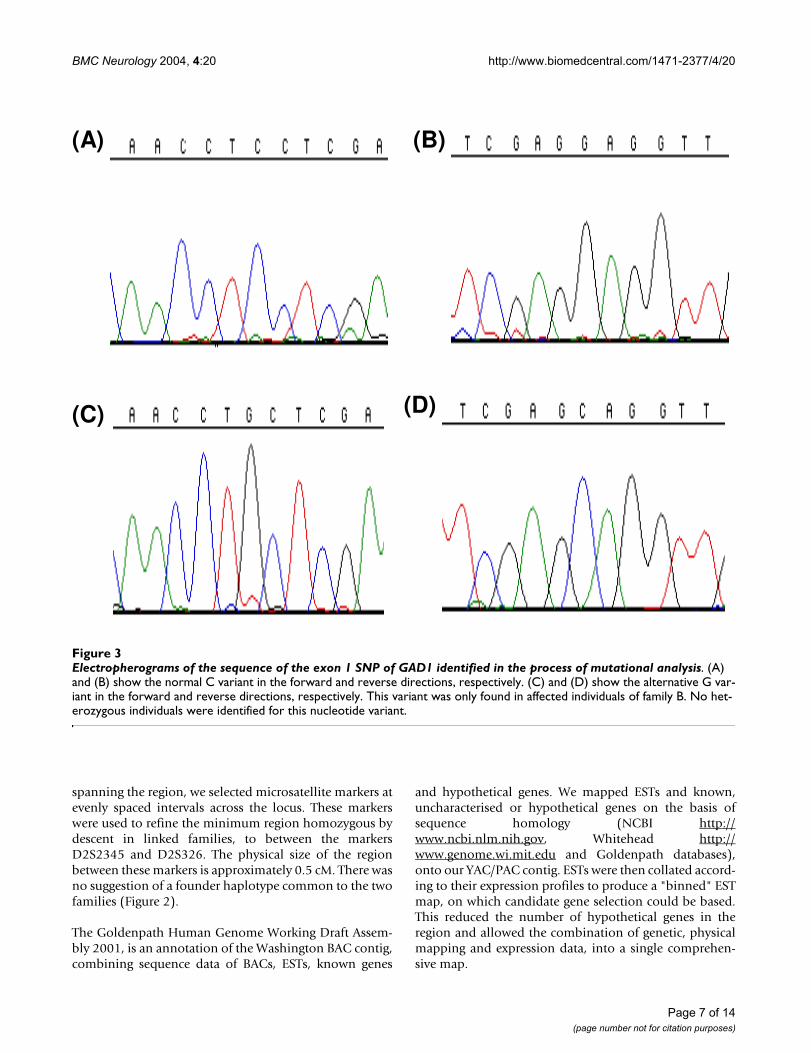
Sequence analysis of GAD1GAD1 was sequenced in affected and unaffected individu-als in both families ascertained. In order to differentiatepossible disease-causing mutations from polymorphisms,100 control individuals were screened by SSCP to detectany GAD1 sequence variations in the normal population.SSCP variants were then sequenced to identify the under-lying nucleotide substitutions. An homozygous G(36)C(Figure 3A,3B) nucleotide change was observed in 4affected patients, which generated a Ser(12)Cys aminoacid substitution. No obligate carriers were identified forthis mutation. This variant has not been previouslydescribed and was not present in 200 normal chromo-somes. A number of other sequence changes weredetected, but none of these resulted in amino acidchanges. These variants and all those recorded previouslyin the literature are presented in Table 4 and Figure 4. Forthose rare variants in the databases, which result in aminoacid changes, no homozygous or compound hetero-zygous individuals have yet been described.
Table 2: Approximate sizes of YAC clones spanning the 2q24-31.1 autosomal recessive spastic CP disease gene locus, used to estimate the minimum physical size of the region (kb). The CEPH MEGA and ICI YACs were sized using CHEF PFGE compared against the native yeast chromosomes. This confirmed the estimated size ranges of the YAC inserts, predicted by the Whitehead Institute (WI) database of YAC clones.
CEPH human MEGA-YAC clones ICI human YAC clones
Clone Size (kb) Clone Size (kb)
910-G12 1630 40D-E3 290744-G6 1120 35B-D2 260912-B6 1190 33D-C4 120, 480
945-C12 1540 30H-D2 250842-G3 1330 30A-D10 260797-G4 1000 18B-E3 120807-H5 1680, 1290 16F-H2 220752-G9 1740 13I-G11 230785-G8 690, 1060 13I-E10 460842-G1 1380 8D-E12 280757-E1 1100812-G1 1540863-H12 1550935-E10 1360785-G8 1060751-H3 1780963-D11 1670, 890
Page 5 of 14(page number not for citation purposes)

BMC Neurology 2004, 4:20 http://www.biomedcentral.com/1471-2377/4/20
DiscussionCP is a term used as a collective definition for a group ofneurological disorders [3]. The pathophysiology in mostcases is poorly understood, but includes genetic syn-dromes, congenital malformation, infective intra-uterineencephalitis, cerebral haemorrhage or infarction, ischemicdamage, periventricular leukomalacia (PVL), and non-inf-arctive telencephalic leukomalacia [11]. The contributionof Mendelian inherited cases of CP accounts for approxi-mately 2% of the total number [2,22]. A non-progressiveform of autosomal recessive spastic CP has been identified[13]. McHale et al. [10] succeeded in identifying a 5 cMregion on chromosome 2q24-31.1, which segregated withdisease in consanguineous families. Linkage analysisidentified a locus between markers D2S124 and D2S333,
which produced a LOD score of 5.75, sufficient to warrantthe further investigation described herein.
To refine and confirm the genetic marker order across aregion, which was incompletely sequenced at the time, weused YAC and PAC clones to construct a physical frame-work and performed PCR to map ESTs and microsatellitemarkers to clones within the partial contig (Figure 1).When this contig was integrated with the BAC sequencecontigs, rearrangement of the BAC order was necessary.With each subsequent DNA sequence update, the degreeof inconsistency was reduced and this led to revision ofmicrosatellite order compared with that used previously[10]. Using the YAC sizes, the sizes of gaps in the BAC con-tig could be estimated. Having generated a detailed map
Annotation of two pedigrees of spastic autosomal recessive CP families and corresponding linkage mapping dataFigure 2Annotation of two pedigrees of spastic autosomal recessive CP families and corresponding linkage mapping data. The markers shown are those that demonstrate the minimal homozygous region between the affected individuals of both families.
Page 6 of 14(page number not for citation purposes)
BMC Neurology 2004, 4:20 http://www.biomedcentral.com/1471-2377/4/20
spanning the region, we selected microsatellite markers atevenly spaced intervals across the locus. These markerswere used to refine the minimum region homozygous bydescent in linked families, to between the markersD2S2345 and D2S326. The physical size of the regionbetween these markers is approximately 0.5 cM. There wasno suggestion of a founder haplotype common to the twofamilies (Figure 2).
The Goldenpath Human Genome Working Draft Assem-bly 2001, is an annotation of the Washington BAC contig,combining sequence data of BACs, ESTs, known genes
and hypothetical genes. We mapped ESTs and known,uncharacterised or hypothetical genes on the basis ofsequence homology (NCBI http://www.ncbi.nlm.nih.gov, Whitehead http://www.genome.wi.mit.edu and Goldenpath databases),onto our YAC/PAC contig. ESTs were then collated accord-ing to their expression profiles to produce a "binned" ESTmap, on which candidate gene selection could be based.This reduced the number of hypothetical genes in theregion and allowed the combination of genetic, physicalmapping and expression data, into a single comprehen-sive map.
Electropherograms of the sequence of the exon 1 SNP of GAD1 identified in the process of mutational analysisFigure 3Electropherograms of the sequence of the exon 1 SNP of GAD1 identified in the process of mutational analysis. (A) and (B) show the normal C variant in the forward and reverse directions, respectively. (C) and (D) show the alternative G var-iant in the forward and reverse directions, respectively. This variant was only found in affected individuals of family B. No het-erozygous individuals were identified for this nucleotide variant.
(A) (B)
(C) (D)
Page 7 of 14(page number not for citation purposes)
BMC Neurology 2004, 4:20 http://www.biomedcentral.com/1471-2377/4/20
One interesting candidate within the minimal region wasGAD1, which encodes GAD67. Expression of its transcriptis ubiquitous, including the CNS. The main function of
GAD67 is to catalyze the conversion of the excitatoryamino acid and neurotransmitter glutamate to GABA, themain inhibitory neurotransmitter in the CNS [23]. In the
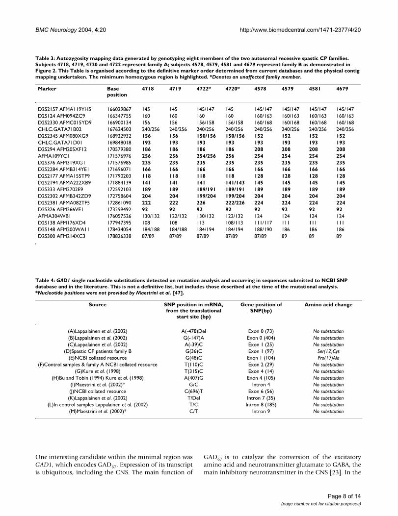
Table 3: Autozygosity mapping data generated by genotyping eight members of the two autosomal recessive spastic CP families. Subjects 4718, 4719, 4720 and 4722 represent family A; subjects 4578, 4579, 4581 and 4679 represent family B as demonstrated in Figure 2. This Table is organised according to the definitive marker order determined from current databases and the physical contig mapping undertaken. The minimum homozygous region is highlighted. *Denotes an unaffected family member.
Marker Base position
4718 4719 4722* 4720* 4578 4579 4581 4679
D2S2157 AFMA119YH5 166029867 145 145 145/147 145 145/147 145/147 145/147 145/147D2S124 AFM094ZC9 166347755 160 160 160 160 160/163 160/163 160/163 160/163D2S2330 AFMC015YD9 166900134 156 156 156/158 156/158 160/168 160/168 160/168 160/168CHLC.GATA71B02 167624503 240/256 240/256 240/256 240/256 240/256 240/256 240/256 240/256D2S2345 AFM080XG9 168922932 156 156 150/156 150/156 152 152 152 152CHLC.GATA71D01 169848018 193 193 193 193 193 193 193 193D2S294 AFM205XF12 170579380 186 186 186 186 208 208 208 208AFMA109YC1 171576976 256 256 254/256 256 254 254 254 254D2S376 AFM319XG1 171576985 235 235 235 235 235 235 235 235D2S2284 AFMB314YE1 171696071 166 166 166 166 166 166 166 166D2S2177 AFMA155TF9 171790203 118 118 118 118 128 128 128 128D2S2194 AFMA222XB9 171884139 141 141 141 141/143 145 145 145 145D2S333 AFM2702E9 172592103 189 189 189/191 189/191 189 189 189 189D2S2302 AFMB342ZD9 172758604 204 204 199/204 199/204 204 204 204 204D2S2381 AFMA082TF5 172861090 222 222 226 222/226 224 224 224 224D2S326 AFM266VE1 173299492 92 92 92 92 92 92 92 92AFMA304WB1 176057526 130/132 122/132 130/132 122/132 124 124 124 124D2S138 AFM176XD4 177947395 108 108 113 108/113 111/117 111 111 111D2S148 AFM200WA11 178434054 184/188 184/188 184/194 184/194 188/190 186 186 186D2S300 AFM214XC3 178826338 87/89 87/89 87/89 87/89 87/89 89 89 89
Table 4: GAD1 single nucleotide substitutions detected on mutation analysis and occurring in sequences submitted to NCBI SNP database and in the literature. This is not a definitive list, but includes those described at the time of the mutational analysis. *Nucleotide positions were not provided by Maestrini et al. [47].
Source SNP position in mRNA, from the translational
start site (bp)
Gene position of SNP(bp)
Amino acid change
(A)Lappalainen et al. (2002) A(-478)Del Exon 0 (73) No substitution(B)Lappalainen et al. (2002) G(-147)A Exon 0 (404) No substitution(C)Lappalainen et al. (2002) A(-39)C Exon 1 (25) No substitution
(D)Spastic CP patients family B G(36)C Exon 1 (97) Ser(12)Cys(E)NCBI collated resource G(48)C Exon 1 (104) Pro(17)Ala
(F)Control samples & family A NCBI collated resource T(110)C Exon 2 (29) No substitution(G)Kure et al. (1998) T(315)C Exon 4 (14) No substitution
(H)Bu and Tobin (1994) Kure et al. (1998) A(407)G Exon 4 (105) No substitution(I)Maestrini et al. (2002)* G/C Intron 4 No substitution(J)NCBI collated resource C(696)T Exon 6 (56) No substitution(K)Lappalainen et al. (2002) T/Del Intron 7 (35) No substitution
(L)In control samples Lappalainen et al. (2002) T/C Intron 8 (185) No substitution(M)Maestrini et al. (2002)* C/T Intron 9 No substitution
Page 8 of 14(page number not for citation purposes)

BMC Neurology 2004, 4:20 http://www.biomedcentral.com/1471-2377/4/20
developing CNS, GABA has an important role in neuronaldifferentiation and the control of plasticity [21]. GABAhas also been implicated in the pathogenesis of variousseizure and movement disorders [20].
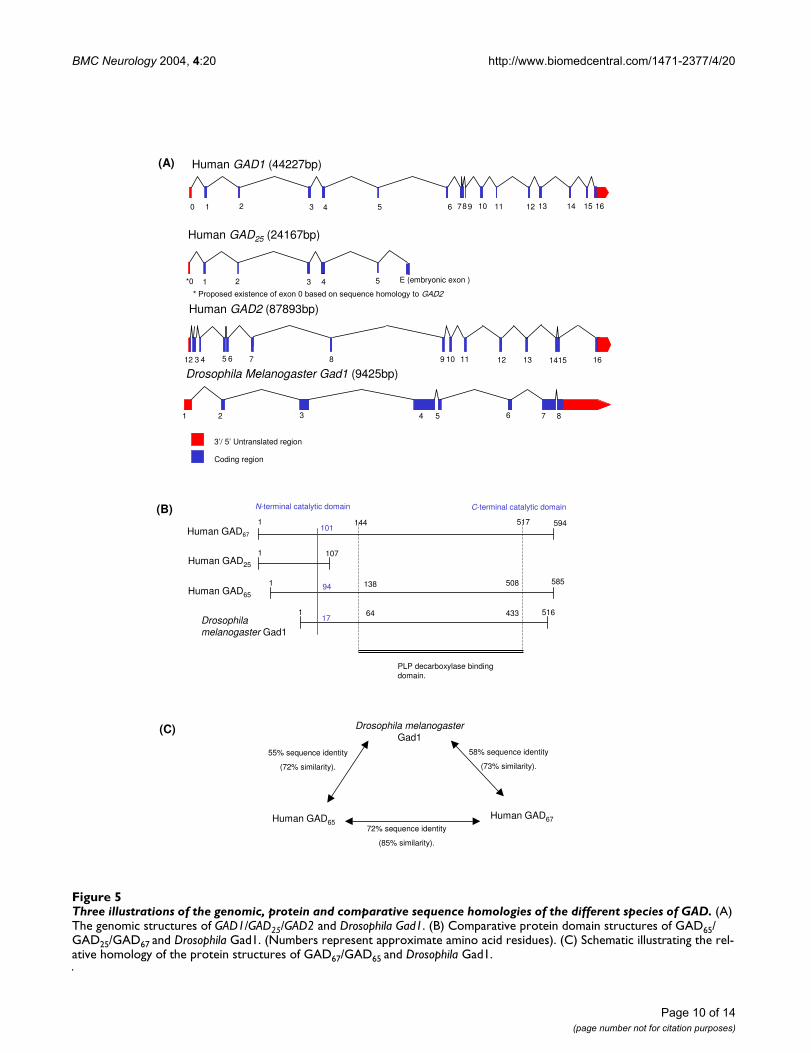
Vertebrates have two separate genes coding for GAD,which produce distinct forms of the enzyme. GAD1 andGAD2 have diverged relatively recently in evolution, asindicated by their degree of sequence homology and theretention of common intron-exon boundary splice sites[17] (Figure 5A). The variants of GAD differ in molecularweight, cellular and sub-cellular localisation, and theirinteraction with the cofactor PLP [18,20,24].
GAD2, located at 10p13-p11.2, is transcribed to producea 5.6 kb mRNA in islets and brain, encoding a 65 kDa pro-tein (585 AA residues). The 67 kDa (594 AA residues)form [17] is localised to 2q25-26 and encoded by a 3.7 kbtranscript (GAD1) [23]. There is also evidence for a 25kDa inactive protein (GAD25) produced from an alterna-tively spliced GAD1 transcript of 2 kb that contains an in-frame stop codon. This GAD1 splice variant has only beenfound in human islets, testis and adrenal cortex, althoughthe homologue is present in fetal mouse brain [25].
GAD67 and GAD65 consist of two major sequencedomains (Figure 5B). The N-termini (AA residues 1–94 inGAD65 and 1–101 in GAD67) demonstrate ~23% hom-ology. These N-terminal domains are thought to beresponsible for sub-cellular targeting and the formation ofGAD65–GAD67 heterodimers [26]. The C-terminaldomains (AA residues 96–585 in GAD65 and 102–594 inGAD67) contain the catalytic site, with ~73% sequenceidentity between the isoforms [19] (Figure 5).
In the CNS, GAD65 appears to be preferentially distributedin axon terminals and the associated synaptic vesicles,whereas GAD67 is also located in the cell bodies and moreuniformly distributed throughout the neuron [24]. Thissuggests that each GAD isoform is involved in the synthe-sis of GABA in different sub-cellular compartments [21].This is supported by the discovery that GAD65 is the mainsource of apoGAD (an inactive reservoir), which respondsto short-term changes in neuronal activity and is moreresponsive to levels of PLP [18]. On the other hand,GAD67 predominantly exists bound to the PLP cofactor(holoGAD), providing a constitutive level of GABA pro-duction [20]. Bond et al. [27] showed that GAD25 isexpressed in a temporally controlled manner, in the devel-
An annotation of the distribution of single nucleotide substitutions identified in the open reading frame of GAD1Figure 4An annotation of the distribution of single nucleotide substitutions identified in the open reading frame of GAD1. The approximate positions with respect to intron-exon of the open reading frame structure are illustrated. These were determined by sequencing of the probands in this study, from published data and from the NCBI collated database of SNPs. The letters refer to the SNPs listed in Table 4. Upper case letters refer to SNPs in the cDNA and lower case letters indicate SNPs in the genomic DNA. A: G(36)C, B: G(210)A, C: G(253)C, D: T(315)C, E: A(407)G, F: C(696)T, G: C(1506)T, H: C(1575)T, i: T(1625)G, J: C(1654)T, k: A(1659)G, l: G(1799)A, m: C(1899)A.
cDNA
Genomic
3’ UTR
A B C D E F G H J
5’ UTR
i k l m
Page 9 of 14(page number not for citation purposes)
BMC Neurology 2004, 4:20 http://www.biomedcentral.com/1471-2377/4/20
Three illustrations of the genomic, protein and comparative sequence homologies of the different species of GADFigure 5Three illustrations of the genomic, protein and comparative sequence homologies of the different species of GAD. (A) The genomic structures of GAD1/GAD25/GAD2 and Drosophila Gad1. (B) Comparative protein domain structures of GAD65/GAD25/GAD67 and Drosophila Gad1. (Numbers represent approximate amino acid residues). (C) Schematic illustrating the rel-ative homology of the protein structures of GAD67/GAD65 and Drosophila Gad1.
1 2 3 4 5 6 7 8
72% sequence identity
(85% similarity).
Drosophila melanogaster
Gad1
Human GAD65Human GAD67
58% sequence identity
(73% similarity).
55% sequence identity
(72% similarity).
(C)
(A) Human GAD1 (44227bp)
860 1 2 3 4 5 7 9 10 11 12 13 14 15 16
Human GAD25 (24167bp)
Drosophila Melanogaster Gad1 (9425bp)
Human GAD2 (87893bp)
3’/ 5’ Untranslated region
Coding region
*0 1 2 3 4 5 E (embryonic exon )
12 3 5 6 7 8 9 10 11 12 13 1415 164
(B) C-terminal catalytic domain
Human GAD67
Human GAD25
Human GAD65
Drosophila
melanogaster Gad1
1101
144 517 594
1 107
1 94 138 508 585
117
64 433 516
PLP decarboxylase binding domain.
N-terminal catalytic domain
Page 10 of 14(page number not for citation purposes)
BMC Neurology 2004, 4:20 http://www.biomedcentral.com/1471-2377/4/20
oping striatum and cortex in rodents, suggesting this mayprovide a mechanism of regulating GABA production indifferentiating neurons.
Asada et al. [28] undertook the selective elimination ofeach GAD isoform in order to determine their respectiveroles. Gad2-/- mice are slightly more susceptible to sei-zures, consistent with an excitatory increase in the relativeratio of glutamate/GABA. However, they showed no obvi-ous overall change in neuronal GABA content. ThereforeGAD67 alone appears to produce sufficient GABA for effec-tive neurotransmission [21]. Gad1-/- mice demonstrated adecrease of ~20% in total glutamate decarboxylase activityat birth. This was assayed by the conversion of 14C-labelled glutamate to 14CO2 in the presence of PLP. Therewas also a marked (7%) reduction in total GABA contentin cerebral cortex homogenate measured by liquid chro-matography [28]. Unfortunately, these mice died neona-tally of severe cleft palate, masking any potentialneurological dysfunction and also illustrating a role forGad67 in non-neural tissues [21]. It is of interest that micewith mutations in the β-3 GABA receptor (GABRB3) at theAngelman syndrome (OMIM:105830) locus, also displaycleft palate, implying a key role for GABA signalling innormal palate development [29,30].
Pyridoxine-dependent epilepsy (PDE) is a rare autosomalrecessive disorder (OMIM:266100), characterized by gen-eralized seizures during the first hours of life. The associ-ated pathology may result from an alteration in thebinding of the co-factor PLP to GAD. Interestingly epi-lepsy is commonly associated with CP and grand mal epi-lepsy developed at age six months in the two linkedpedigrees described here [10]. GAD1 mutation was previ-ously suspected of being the cause of PDE. Linkage of pyri-doxine-dependent epilepsy has however been reported to5q31.2-31.3, with GAD1 and GAD2 excluded [31].Decreased levels of brain and CSF GABA, increased levelsof CSF and cortical glutamate, and decreased levels of PLPin the frontal cortex, have been described in thiscondition.
GAD65 and GAD67 have been identified as auto-antigensin "Stiff Person Syndrome" (SPS, OMIM:184850), and incerebellar ataxia [32-34]. GABA-mediated synaptictransmission is thought to be functionally impaired bythe production of autoantibodies to GAD65 and GAD67[35-37]. This results in a reduction in brain levels ofGABA, prominent in the motor cortex, which can be dem-onstrated by Magnetic Resonance Imaging (MRI) in SPSpatients. SPS is a disabling disorder characterised by mus-cle rigidity and episodic spasms of the musculature,thought to be due to autoimmune-mediated dysfunctionof supraspinal GABAergic inhibitory neurons [38]. Hyper-
excitability of the motor cortex in SPS has been demon-strated by transcranial magnetic stimulation [39].
Anti-GAD65 auto-antibodies in the CSF of ataxic and SPSpatients selectively suppress GABA-mediatedtransmission in cerebellar Purkinje cells, without affectingglutamate-mediated transmission [37,40]. Low CSF levelsof GABA have been reported in patients with Kok disease(OMIM:149400 also known as hyperexplexia/exaggeratedstartle reaction/startle disease) [41]. The exact mechanismby which autoantibodies target these intracellular GADantigens is not clear. However, it is interesting that SPSmay also arise in individuals with autoantibodies togephyrin, a cytosolic protein concentrated at the postsyn-aptic membrane of inhibitory synapses where it isassociated with GABAA receptors [42]. This provides a fur-ther example of chronic rigidity and spasm possibly sec-ondary to disruption of the inhibitory synapses.
Mutations in the CLN3 gene are thought to be responsiblefor the neurodegenerative disorder Batten disease(OMIM:204200). In cln3-knockout mice autoantibodiesto GAD65 have been reported to be associated with braintissue and result in inhibition of GAD activity [43]. Thesemice also demonstrate elevated brain glutamate levels ascompared with controls, which may have a causative rolein the astrocytic hypertrophy evident in cln3-knockoutmice and along with anti-GAD65 autoantibodies in Battendisease patients may contribute to the associated preferen-tial loss of GABAergic neurons.
Drugs, which potentiate the action of GABA, such as ben-zodiazepines and baclofen, ameliorate muscle rigidityand spasticity. These GABA agonists are thought to coun-ter disinhibition of the velocity-dependent increase inskeletal muscle during stretch reflexes, observed in spastic-ity, which is the result of inadequate presynaptic inhibi-tion of the muscle spindles [44,35,37]. γ-Vinyl-γ-aminobutyric acid (GVG) is used to treat neurological dis-orders including epilepsy, tardive dyskinesia and spastic-ity. It has been reported that it is the GABA-elevating effectof this compound that is responsible for its anti-convul-sive properties [20].
We have identified a GAD1 sequence change G(36)C,which segregates with autosomal recessive spastic CP in 4affected siblings. This nucleotide substitution causes amissense mutation, changing serine (12) to a cysteine inthe N-terminal domain. This serine residue is conservedbetween all mammals (human/mouse/rabbit/pig) forwhich data are available. The association of GAD67 withmembranes requires formation of heteromeric links withGAD65, which are mediated via their N-terminal domains.The N-terminus of GAD65 is palmitoylated and binds tothe cellular membrane. The first 27 amino acids appear to
Page 11 of 14(page number not for citation purposes)

BMC Neurology 2004, 4:20 http://www.biomedcentral.com/1471-2377/4/20
be essential in this function [32]. The palmitoylation ofcysteines 30 and 45 of GAD65, and the inability of residues1–29 of GAD67, to substitute for this region, highlights thepotential impact on cellular localisation of a nucleotidesubstitution in this domain [20]. GAD65 also undergoesphosphorylation of the first four serine residues in the N-terminal domain. These post-translational modificationshighlight the importance of the flexibility and accessibil-ity of this domain. N-terminal epitopes of GAD65, in theregion corresponding to the residue, which undergoesmutation in GAD67, are particularly prominent autoanti-gens [45].
S(12)C amino acid substitution may thus produce subtleeffects on cellular localisation, protein-protein interac-tions and/or protein processing, with a subsequent effecton GABA production. This is not inconsistent with themouse Gad1 knockout where complete loss of Gad1 enzy-matic function (~20% reduction of total Gad activity inthe cerebral cortex) resulted in a cleft palate phenotypeand neonatal death [28,30]. There is redundancy of GABAproduction as a result of the presence of two GAD pro-teins, and the precise function of each isoform may differbetween man and mouse. It is interesting to note that theGAD25 splice variant of GAD67, also contains the S(12)Camino acid substitution in affected CP patients. This trun-cated variant is identical to the first 213 amino acids ofGAD67, with the addition of an extra 11 C-terminal resi-dues. It lacks the binding site for the cofactor PLP and isbelieved to lack any GAD activity [25]. The function ofGAD25 is not known, but it may compete with GAD67 forincorporation into protein complexes. Therefore the pres-ence of an N-terminal mutation would affect both GAD25and GAD67, and may disrupt a complex regulatory mech-anism for GAD67.
ConclusionsThis study illustrates the difficulty of gene cloning in rareautosomal recessive diseases mapped in small, consan-guineous pedigrees. Identification of an ancestralhaplotype allows refinement of the locus, but this has notbeen possible in this example. Within the minimallinkage region, any sequence change will segregate withthe disease phenotype. Detection of a nonsense mutationleading to a protein truncation would provide compellingsupport for a mutation as causative. However, in thepresent example we have not seen such a mutation in thecandidate gene so far examined. We are now expressingthe variant forms of GAD67 (S12C) and GAD25 (S12C), asrecombinant proteins, to assess catalytic activity and bind-ing properties with respect to their normal counterparts.However, it may well prove difficult to detect subtle effectsbased on sub-cellular localisation changes in the mutantproteins, or changes in post-translational modificationpatterns.
Stability of the mRNA transcripts from these variants willbe assessed by transfection studies in neuronal cells. Even-tually, it would be of interest to attempt knock-in experi-ments with GAD1 (G36C), into the Gad1-/- mouse to seeif this can rescue the cleft palate phenotype and reveal aCP-like picture. These experiments will be reported else-where. However, the possibility that reduced GAD67 activ-ity may cause CP in the patients studied herein, in amanner reminiscent of that seen in SPS, leads us to reportour findings at this stage. The success reported in treatingSPS with intravenous immunoglobulin [40,46], suggestsfurther evaluation of GABA agonists in the managementof this difficult clinical problem.
AbbreviationsBAC Bacterial artificial chromosome
BLAST Basic local alignment search tool
CEPH Centre d'Etude du Polymorphisme Humain
CHEF Contour-clamped homogenous electric field
CNS Central nervous system
CP Cerebral Palsy
CSF Cerebrospinal fluid
DNA Deoxyribonucleic acid
EST Expressed sequence tag
GABA Gamma-aminobutyric acid
GAD L-Glutamate decarboxylase
GVG Gamma-vinyl-gamma aminobutyric acid
HGMP Human genome mapping project
IDDM Type 1 Insulin-Dependent Diabetes Mellitus
MRI Magnetic resonance imaging
NCBI National Centre for Biotechnology Information
PAC Plasmid artificial chromosome
PCR Polymerase chain reaction
PDE Pyridoxine-dependent epilepsy
PFGE Pulse field gel electrophoresis
Page 12 of 14(page number not for citation purposes)
BMC Neurology 2004, 4:20 http://www.biomedcentral.com/1471-2377/4/20
PLP Pyridoxal 5'-phosphate
PVL Periventricular leukomalacia
SPG Spastic paraplegia
SPS Stiff Person Syndrome
SSCP Single-strand conformational polymorphism
STS Sequence tagged site
YAC Yeast artificial chromosome
Competing interestsThe author(s) declare that they have no competinginterests.
Authors' contributionsCNL carried out the molecular genetic studies, sequencingand drafted the manuscript. The YAC mapping work wasundertaken by JPL and CNL. AFM, DTB and IMC con-ceived the study and participated in the design and coor-dination, they also secured financial sponsorship from theWellcome Trust and MRC. RA, SM, ERM and CGWrecruited and gained consent from the families detailed inthis study. All authors read and approved the finalmanuscript.
AcknowledgementsResearch in the authors' laboratory is supported by the Wellcome Trust, MRC, CRUK and Yorkshire Cancer Research. CNL has an MRC PhD stu-dentship. This paper is dedicated to the late Prof. S. Bundey, University of Birmingham.
References1. Eicher PS, Batshaw ML: Cerebral palsy. Pediatric Clinics of North
America 1993, 40:537-551.2. Gustavson KH, Hagberg B, Sanner G: Identical syndromes of cer-
ebral palsy in the same family. Acta Paediatrica Scandinavica 1969,58:330-340.
3. Kuban KCK, Leviton A: Cerebral palsy. New England Journal ofMedicine 1994, 330:188-195.
4. Volpe J: Neurology of the Newborn 2nd edition. Philadelphia: WBSaunders; 1987.
5. Gaffney G, Sellers S, Flavell V, Squier M, Johnson A: Case controlstudy of intrapartum care, cerebral palsy and perinataldeath. British Medical Journal 1994, 308:743-750.
6. Pharoah POD, Cooke T, Cooke RW, Rosenbloom L: Birthweightspecific trends in cerebral palsy. Archives of Disease in Childhood1990, 65:602-606.
7. Paneth N: The causes of cerebral palsy. Recent evidence. ClinInvest Med 1993, 16:95-102.
8. Fawer CL, Calame A, In Levene MI, Bennett MJ, Punt J: Fetal and Neo-natal Neurology and Neurosurgery Edinburgh: Churchill Livingstone;1988.
9. Hagberg B, Hagberg G: The Origins of Cerebral Palsy. In In:Recent Advances in Pediatrics Edited by: David TJ. New York: Churchill-Livingston; 1992.
10. McHale DP, Mitchell S, Bundey S, Moynihan L, Campbell DA, WoodsCG, Lench NJ, Mueller RF, Markham AF: A gene for autosomalrecessive symmetrical spastic cerebral palsy maps to chro-
mosome 2q24-25. American Journal of Human Genetics 1999,64:526-532.
11. Forfar JO, Arneil GC: Textbook of Pediatrics 4th edition. Edinburgh:Churchill Livingstone; 1992.
12. Baraitser M: The genetics of neurological disorders Oxford: Oxford Uni-versity Press; 1985.
13. Bundey S, Griffiths MI: Recurrence risks in families of childrenwith symmetrical spasticity. Developmental Medicine and ChildNeurology 1977, 19:179-191.
14. Mitchell S, Bundey S: Symmetry of neurological signs in Paki-stani patients with probable inherited spastic cerebral palsy.Clinical Genetics 1997, 51:7-14.
15. McHale DP, Jackson AP, Campbell DA, Levene MI, Corry P, WoodsCG, Lench NJ, Mueller RF, Markham AF: A gene for ataxic cere-bral palsy maps to chromosome 9p12-q12. European Journal ofHuman Genetics 2000, 8:267-272.
16. Lernmark A: Glutamic acid decarboxylase – gene to antigen todisease. Intern Med 1996, 240:259-277.
17. Bosma PT, Blazquez M, Collins MA, Bishop JD, Drouin G, Priede IG,Docherty K, Trudeau VL: Multiplicity of glutamic acid decar-boxylases (GAD) in vertebrates: molecular phylogeny andevidence for a new GAD paralog. Molecular Biology and Evolution1999, 16:397-404.
18. Erlander MG, Tillakaratne NJ, Feldblum S, Patel N, Tobin AJ: Twogenes encode distinct glutamate decarboxylases. Neuron 1991,7:91-100.
19. Martin DL, Liu H, Martin SB, Wu SJ: Structural features and reg-ulatory properties of the brain glutamate decarboxylases.Neurochemistry International 2000, 37:111-119.
20. Martin DL, Rimvall K: Regulation of gama-aminobutyric acidsynthesis in the brain. Journal of Neurochemistry 1993, 60:395-407.
21. Stone DJ, Walsh J, Benes FM: Localization of cells preferentiallyexpressing GAD67 with negligible GAD65 transcripts in therat hippocampus. A double in situ hybridization study. Molec-ular Brain Research 1999, 71:201-209.
22. Hughes I, Newton R: Genetic aspects of cerebral palsy. Develop-mental Medicine and Child Neurology 1992, 34:80-86.
23. Bu DF, Erlander MG, Hitz MG, Tillakaratne NJ, Kaufman DL, Wagner-McPherson CB, Evans GA, Tobin AJ: Two human glutamatedecarboxylases, 65-kDa GAD and 67-kDa GAD, are eachencoded by a single gene. Proc Natl Acad Sci 1992, 89:2115-2119.
24. Laprade N, Soghomonian JJ: Differential regulation of mRNAlevels encoding for the two isoforms of glutamate decarbox-ylase (GAD65 and GAD67) by dopamine receptors in the ratstriatum. Molecular Brain Research 1995, 34:65-74.
25. Chessler SD, Lernmark A: Alternative splicing of GAD67 resultsin the synthesis of a third form of glutamic-acid decarboxy-lase in human islets and other non-neural tissues. Journal of Bio-logical Chemistry 2000, 275:5188-5192.
26. Bu DF, Tobin AJ: The exon-intron organization of the genes(GAD1 and GAD2) encoding two human glutamate decar-boxylases (GAD67 and GAD65) suggests that they derivefrom a common ancestral GAD. Genomics 1994, 21:222-228.
27. Bond RW, Wyborski RJ, Gottlieb DI: Developmentally regulatedexpression of an exon containing a stop codon in the gene forglutamic acid decarboxlase. Proc Natl Acad Sci 1990,87:8771-8775.
28. Asada H, Kawamura Y, Maruyama K, Kume H, Ding RG, Kanbara N,Kuzume H, Sanbo M, Yagi T, Obata K: Cleft palate and decreasedbrain gamma-aminobutyric acid in mice lacking the 67-kDaisoform of glutamic acid decarboxylase. Proc Natl Acad Sci 1997,94:6496-6499.
29. Homanics GE, DeLorey TM, Firestone LL, Quinlan JJ, Handforth A,Harrison NL, Krasowski MD, Rick CEM, Korpi ER, Mäkelä R, BrilliantMH, Hagiwara N, Ferguson C, Snyder K, Olsen RW: Mice devoid ofγ-aminobutyrate type A receptor β3 subunit have epilepsy,cleft palate, and hypersensitive behaviour. Proc Natl Acad Sci1997, 94:4143-4148.
30. Condie BG, Bain G, Gottlieb DI, Capeahi MR: Cleft palate in micewith a targeted mutation in the gamma-aminobutyric acid-producing enzyme glutamic acid decarboxylase 67. Proc NatlAcad Sci 1997, 94:11451-11455.
31. Cormier-Daire V, Dagoneau N, Nabbout R, Burglen L, Penet C, Souf-flet C, Desguerre I, Munnich A, Dulac O: A gene for pyridoxine-dependent epilepsy maps to chromosome 5q31. American Jour-nal of Human Genetics 2000, 67:991-993.
Page 13 of 14(page number not for citation purposes)
BMC Neurology 2004, 4:20 http://www.biomedcentral.com/1471-2377/4/20
Publish with BioMed Central and every scientist can read your work free of charge
"BioMed Central will be the most significant development for disseminating the results of biomedical research in our lifetime."
Sir Paul Nurse, Cancer Research UK
Your research papers will be:
available free of charge to the entire biomedical community
peer reviewed and published immediately upon acceptance
cited in PubMed and archived on PubMed Central
yours — you keep the copyright
Submit your manuscript here:http://www.biomedcentral.com/info/publishing_adv.asp
BioMedcentral
32. Solimena M, Butler MH, De Camilli P: GAD, diabetes, and Stiff-Man syndrome: some progress and more questions. The Jour-nal of Endocrinology Investigations 1994, 17:509-520.
33. Kono S, Miyajima H, Sugimoto M, Suzuki Y, Takahashi Y, Hishida A:Stiff-person syndrome associated with cerebellar ataxia andhigh glutamic acid decarboxylase antibody titre. Annals of Inter-nal Medicine 2001, 40:968-971.
34. Meinck HM, Faber L, Morgenthaler N, Seissler J, Maile S, Butler M,Solimena M, DeCamilli P, Scherbaum WA: Antibodies againstglutamic acid decarboxylase: prevalence in neurological dis-eases. Journal of Neurology, Neurosurgery and Psychiatry 2001,71:100-103.
35. Dinkel K, Meinck HM, Jury KM, Karges W, Richter W: Inhibition ofgamma-aminobutyric acid synthesis by glutamic acid decar-boxylase autoantibodies in stiff-man syndrome. Annals ofNeurology 1998, 44:194-201.
36. Burk K, Fetter M, Abele M, Laccone F, Didierjean O, Brice A, Klock-gether T: Autosomal dominant cerebellar ataxia type I: ocu-lomotor abnormalities in families with SCA1, SCA2, andSCA3. Journal of Neurology 1999, 246:789-797.
37. Mitoma H, Song SY, Ishida K, Yamakuni T, Kobayashi T, Mizusawa H:Presynaptic impairment of cerebellar inhibitory synapses byan autoantibody to glutamate decarboxylase. Journal of Neuro-logical Science 2000, 175:40-44.
38. Levy LM, Dalakas MC, Floeter MK: The stiff-person syndrome: anautoimmune disorder affecting neurotransmission ofgamma-aminobutyric acid. Annals of Internal Medicine 1999,131:522-530.
39. Sandbrink F, Syed NA, Fujii MD, Dalakas MC, Floeter MK: Motorcortex excitability in stiff-person syndrome. Brain 2000,123:2231-2239.
40. Dalakas MC, Li M, Fujii M, Jacobowitz DM: Stiff person syndrome:quantification, specificity, and intrathecal synthesis of GAD65antibodies. Neurology 2001, 57:780-784.
41. Dubowitz LM, Bouza H, Hird MF, Jaeken J: Low cerebrospinal fluidconcentration of free gamma-aminobutyric acid in startledisease. Lancet 1992, 340:80-81.
42. Butler MH, Hayashi A, Ohkoshi N, Villmann C, Becker CM, Feng G,De Camilli P, Solimena M: Autoimmunity to gephyrin in Stiff-Man syndrome. Neuron 2000, 26:307-312.
43. Chattopadhyay S, Ito M, Cooper JD, Brooks AI, Curran TM, PowersJM, Pearce DA: An autoantibody inhibitory to glutamic aciddecarboxylase in the neurodegenerative disorder Battendisease. Human Molecular Genetics 2002, 11:1421-1431.
44. Solimena M, Dirkx R Jr, Radzynski M, Mundigl O, De Camilli P: A sig-nal located within amino acids 1–27 of GAD65 is required forits targeting to the Golgi complex region. Journal of Cell Biology1994, 126:331-341.
45. Kim J, Namchuk M, Bugawan T, Fu Q, Jaffe M, Shi Y, Aanstoot HJ,Turck CW, Erlich H, Lennon V: Higher autoantibody levels andrecognition of a linear NH2-terminal epitope in the autoan-tigen GAD65, distinguish stiff-man syndrome from insulin-dependent diabetes mellitus. The Journal of Experimental Medicine1994, 180:595-606.
46. Wiles CM, Brown P, Chapel H, Guerrini R, Hughes RAC, Martin D,McCrone P, Newsom-Davis J, Palace J, Rees JH, Rose MR, Scolding N,Webster ADB: Intravenous immunoglobulin in neurologicaldisease: a specialist review. Journal of Neurology, Neurosurgery andPsychiatry 2002, 72:440-448.
47. Maestrini E, Bacchelli E, Blasi F, Biondolillo M: Analysis of nine can-didate genes for autism on chromosome 2q. American Journal ofHuman Genetics 2002, 721:A1915.
48. Lappalainen JS, Sanacora G, Kranzier HR, Malison R, Price LH, KrystalJ: Mutation screen of the glutamate decarboxylase 67(GAD67) gene and haplotype association study to unipolardepression. American Journal Human Genetics 2002, 721:A1966.
49. Kure S, Sakata Y, Miyabayashi S, Takahashi K, Shinka T, Matsubara Y,Hoshino H, Narisawa K: Mutation and polymorphic markeranalyses of 65K- and 67K-glutamate decarboxylase genes intwo families with pyridoxine-dependent epilepsy. Journal ofHuman Genetics 1998, 43:128-131.
Pre-publication historyThe pre-publication history for this paper can be accessedhere:
http://www.biomedcentral.com/1471-2377/4/20/prepub
Page 14 of 14(page number not for citation purposes)
Copyright © 2022 FDOKUMEN