Identification of the missing pluripotency mediator downstream of leukaemia inhibitory factor
Upload
independentCategory
view
2download
0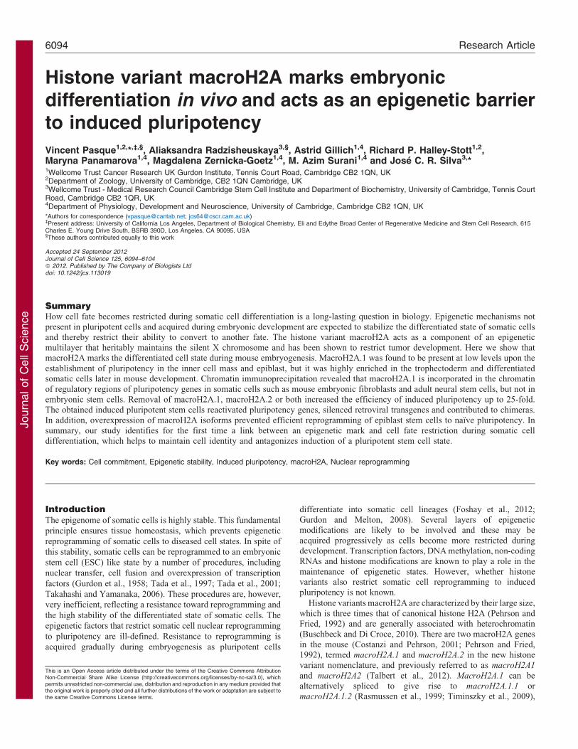
Journ
alof
Cell
Scie
nce
Histone variant macroH2A marks embryonicdifferentiation in vivo and acts as an epigenetic barrierto induced pluripotency
Vincent Pasque1,2,*,`,§, Aliaksandra Radzisheuskaya3,§, Astrid Gillich1,4, Richard P. Halley-Stott1,2,Maryna Panamarova1,4, Magdalena Zernicka-Goetz1,4, M. Azim Surani1,4 and Jose C. R. Silva3,*1Wellcome Trust Cancer Research UK Gurdon Institute, Tennis Court Road, Cambridge CB2 1QN, UK2Department of Zoology, University of Cambridge, CB2 1QN Cambridge, UK3Wellcome Trust - Medical Research Council Cambridge Stem Cell Institute and Department of Biochemistry, University of Cambridge, Tennis CourtRoad, Cambridge CB2 1QR, UK4Department of Physiology, Development and Neuroscience, University of Cambridge, Cambridge CB2 1QN, UK
*Authors for correspondence ([email protected]; [email protected])`Present address: University of California Los Angeles, Department of Biological Chemistry, Eli and Edythe Broad Center of Regenerative Medicine and Stem Cell Research, 615Charles E. Young Drive South, BSRB 390D, Los Angeles, CA 90095, USA§These authors contributed equally to this work
Accepted 24 September 2012Journal of Cell Science 125, 6094–6104� 2012. Published by The Company of Biologists Ltddoi: 10.1242/jcs.113019
SummaryHow cell fate becomes restricted during somatic cell differentiation is a long-lasting question in biology. Epigenetic mechanisms not
present in pluripotent cells and acquired during embryonic development are expected to stabilize the differentiated state of somatic cellsand thereby restrict their ability to convert to another fate. The histone variant macroH2A acts as a component of an epigeneticmultilayer that heritably maintains the silent X chromosome and has been shown to restrict tumor development. Here we show thatmacroH2A marks the differentiated cell state during mouse embryogenesis. MacroH2A.1 was found to be present at low levels upon the
establishment of pluripotency in the inner cell mass and epiblast, but it was highly enriched in the trophectoderm and differentiatedsomatic cells later in mouse development. Chromatin immunoprecipitation revealed that macroH2A.1 is incorporated in the chromatinof regulatory regions of pluripotency genes in somatic cells such as mouse embryonic fibroblasts and adult neural stem cells, but not in
embryonic stem cells. Removal of macroH2A.1, macroH2A.2 or both increased the efficiency of induced pluripotency up to 25-fold.The obtained induced pluripotent stem cells reactivated pluripotency genes, silenced retroviral transgenes and contributed to chimeras.In addition, overexpression of macroH2A isoforms prevented efficient reprogramming of epiblast stem cells to naıve pluripotency. In
summary, our study identifies for the first time a link between an epigenetic mark and cell fate restriction during somatic celldifferentiation, which helps to maintain cell identity and antagonizes induction of a pluripotent stem cell state.
Key words: Cell commitment, Epigenetic stability, Induced pluripotency, macroH2A, Nuclear reprogramming
IntroductionThe epigenome of somatic cells is highly stable. This fundamental
principle ensures tissue homeostasis, which prevents epigenetic
reprogramming of somatic cells to diseased cell states. In spite of
this stability, somatic cells can be reprogrammed to an embryonic
stem cell (ESC) like state by a number of procedures, including
nuclear transfer, cell fusion and overexpression of transcription
factors (Gurdon et al., 1958; Tada et al., 1997; Tada et al., 2001;
Takahashi and Yamanaka, 2006). These procedures are, however,
very inefficient, reflecting a resistance toward reprogramming and
the high stability of the differentiated state of somatic cells. The
epigenetic factors that restrict somatic cell nuclear reprogramming
to pluripotency are ill-defined. Resistance to reprogramming is
acquired gradually during embryogenesis as pluripotent cells
differentiate into somatic cell lineages (Foshay et al., 2012;Gurdon and Melton, 2008). Several layers of epigeneticmodifications are likely to be involved and these may be
acquired progressively as cells become more restricted duringdevelopment. Transcription factors, DNA methylation, non-codingRNAs and histone modifications are known to play a role in the
maintenance of epigenetic states. However, whether histonevariants also restrict somatic cell reprogramming to inducedpluripotency is not known.
Histone variants macroH2A are characterized by their large size,
which is three times that of canonical histone H2A (Pehrson andFried, 1992) and are generally associated with heterochromatin(Buschbeck and Di Croce, 2010). There are two macroH2A genes
in the mouse (Costanzi and Pehrson, 2001; Pehrson and Fried,1992), termed macroH2A.1 and macroH2A.2 in the new histonevariant nomenclature, and previously referred to as macroH2A1
and macroH2A2 (Talbert et al., 2012). MacroH2A.1 can bealternatively spliced to give rise to macroH2A.1.1 ormacroH2A.1.2 (Rasmussen et al., 1999; Timinszky et al., 2009),
This is an Open Access article distributed under the terms of the Creative Commons AttributionNon-Commercial Share Alike License (http://creativecommons.org/licenses/by-nc-sa/3.0), whichpermits unrestricted non-commercial use, distribution and reproduction in any medium provided thatthe original work is properly cited and all further distributions of the work or adaptation are subject tothe same Creative Commons License terms.
6094 Research Article
Journ
alof
Cell
Scie
nce
each of these possessing another variant (macroH2A.1.s3,
macroH2A.1.s4 for macroH2A.1.1, and macroH2A.1.s1,
macroH2A.1.s2 for macroH2A.1.2) (Bernstein et al., 2008). In
mouse and in humans, in addition to alternative macroH2A.1
splicing, macroH2A.1.s3 and macroH2A.1.s1 also each differ from
macroH2A.1.s4 and macroH2A.1.s2, respectively, by a single
Lysine residue, the function of which, if any, is currently unknown
(Bernstein et al., 2008).
MacroH2A histone variants incorporate into chromatin via
their histone H2A-like region, which is attached via a linker
region to a bulky non-histone (macro) domain that projects out of
the nucleosome (Chakravarthy et al., 2005). All the three regions
of macroH2A are independently associated with transcriptional
repression (Buschbeck and Di Croce, 2010).
MacroH2As are developmentally regulated (Chang et al.,
2010; Chang et al., 2005; Pehrson et al., 1997). In vivo, maternal
macroH2A.1 is removed from pronuclei prior to the onset of
somatic macroH2A expression at the eight-cell stage in the
preimplantation embryo, and becomes enriched on the inactive X
chromosome (Xi) by the morula stage (Chang et al., 2010;
Costanzi et al., 2000). MacroH2A.1 is also enriched on the Xi in
somatic cells, but not on the Xi of epiblast stem cells, derived
from the post-implantation embryo (Costanzi and Pehrson, 1998;
Pasque et al., 2011a). In the mouse embryo, macroH2A
expression has not been investigated after E3.5. In general,
macroH2A.1.1 has been found to be expressed in cells derived
from differentiated tissues with low proliferation, whereas
macroH2A.1.2 tends to be more widely expressed including in
proliferative cells (Pehrson et al., 1997; Rasmussen et al., 1999).
Mice lacking macroH2A.1 or macroH2A.2 are fertile and
viable, and display only mild phenotype, including metabolic
defects, suggesting that macroH2A is not strictly required for
embryonic development in the mouse (Boulard et al., 2010;
Buschbeck and Di Croce, 2010; Changolkar et al., 2007).
Removal of both macroH2A.1 and macroH2A.2 from female
mouse ESCs was found to impair ESCs differentiation in one
study (Creppe et al., 2012a; Creppe et al., 2012b), while it was
not found to prevent X chromosome inactivation and
differentiation by another group (Tanasijevic and Rasmussen,
2011). Using the heterologous system of mammalian nuclear
transfer to Xenopus oocytes it was previously shown that the
histone variant macroH2A is a component of the epigenetic
mechanisms of gene silencing present in somatic cells. However,
whether this bears significance for somatic cell epigenome
resistance to reprogramming towards pluripotency has not been
addressed (Pasque et al., 2011b).
Here, we report that during mouse embryogenesis, macroH2A.1
becomes highly expressed and incorporated into chromatin in
somatic cell lineages. Importantly, macroH2A.1 is a structural
component of the repressed chromatin of pluripotency genes in
somatic cells and acts as an epigenetic barrier to the induction of
naıve pluripotency. Our study demonstrates the importance of
macroH2A histone variants in maintenance of cellular identity.
ResultsMacroH2A is a hallmark of somatic cell differentiation
in vivo
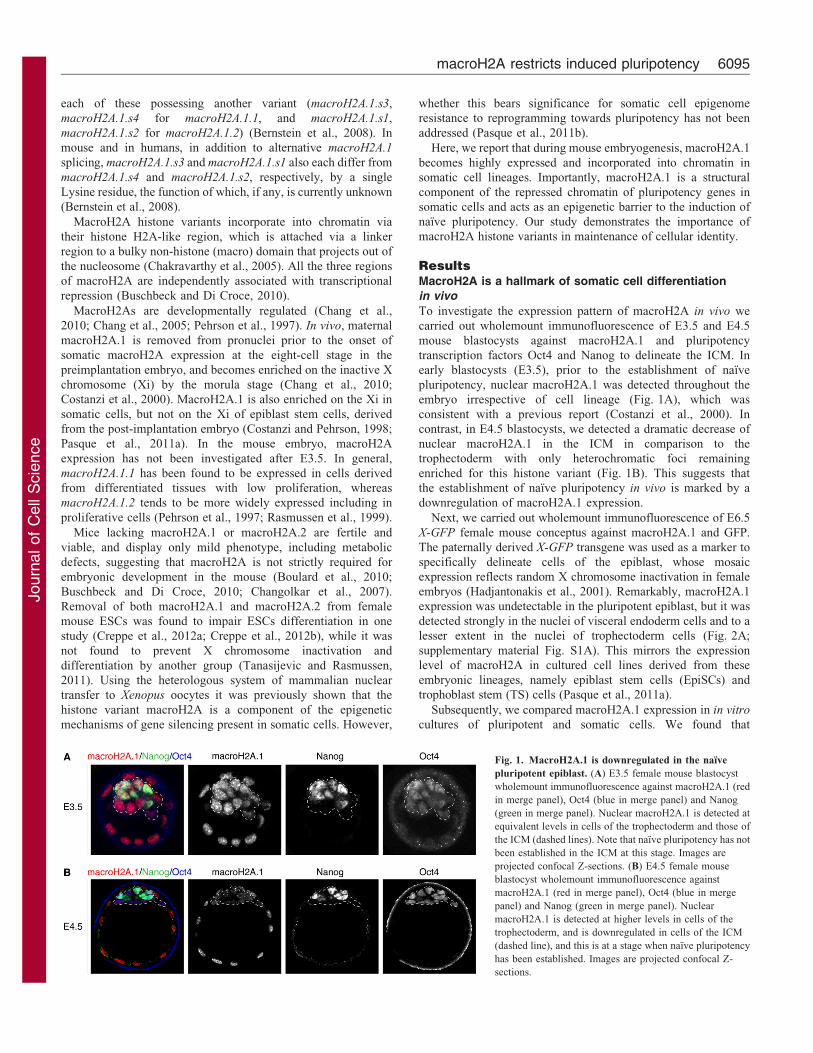
To investigate the expression pattern of macroH2A in vivo we
carried out wholemount immunofluorescence of E3.5 and E4.5
mouse blastocysts against macroH2A.1 and pluripotency
transcription factors Oct4 and Nanog to delineate the ICM. In
early blastocysts (E3.5), prior to the establishment of naıve
pluripotency, nuclear macroH2A.1 was detected throughout the
embryo irrespective of cell lineage (Fig. 1A), which was
consistent with a previous report (Costanzi et al., 2000). In
contrast, in E4.5 blastocysts, we detected a dramatic decrease of
nuclear macroH2A.1 in the ICM in comparison to the
trophectoderm with only heterochromatic foci remaining
enriched for this histone variant (Fig. 1B). This suggests that
the establishment of naıve pluripotency in vivo is marked by a
downregulation of macroH2A.1 expression.
Next, we carried out wholemount immunofluorescence of E6.5
X-GFP female mouse conceptus against macroH2A.1 and GFP.
The paternally derived X-GFP transgene was used as a marker to
specifically delineate cells of the epiblast, whose mosaic
expression reflects random X chromosome inactivation in female
embryos (Hadjantonakis et al., 2001). Remarkably, macroH2A.1
expression was undetectable in the pluripotent epiblast, but it was
detected strongly in the nuclei of visceral endoderm cells and to a
lesser extent in the nuclei of trophectoderm cells (Fig. 2A;
supplementary material Fig. S1A). This mirrors the expression
level of macroH2A in cultured cell lines derived from these
embryonic lineages, namely epiblast stem cells (EpiSCs) and
trophoblast stem (TS) cells (Pasque et al., 2011a).
Subsequently, we compared macroH2A.1 expression in in vitro
cultures of pluripotent and somatic cells. We found that
Fig. 1. MacroH2A.1 is downregulated in the naıve
pluripotent epiblast. (A) E3.5 female mouse blastocyst
wholemount immunofluorescence against macroH2A.1 (red
in merge panel), Oct4 (blue in merge panel) and Nanog
(green in merge panel). Nuclear macroH2A.1 is detected at
equivalent levels in cells of the trophectoderm and those of
the ICM (dashed lines). Note that naıve pluripotency has not
been established in the ICM at this stage. Images are
projected confocal Z-sections. (B) E4.5 female mouse
blastocyst wholemount immunofluorescence against
macroH2A.1 (red in merge panel), Oct4 (blue in merge
panel) and Nanog (green in merge panel). Nuclear
macroH2A.1 is detected at higher levels in cells of the
trophectoderm, and is downregulated in cells of the ICM
(dashed line), and this is at a stage when naıve pluripotency
has been established. Images are projected confocal Z-
sections.
macroH2A restricts induced pluripotency 6095

Journ
alof
Cell
Scie
nce
macroH2A.1 was quantitatively more abundant in mouse
embryonic fibroblasts (MEFs) and adult neural stem cells
(NSCs) compared to undifferentiated ESCs and EpiSCs
(Fig. 2B,C). To assess whether macroH2A.1 expression is
correlated with the differentiated state of cells in developing
mouse embryos, we probed its expression later in development, in
E9.5 embryos, in which somatic cell lineage determination has
occurred. Nuclear macroH2A.1 was strongly detected throughout
all somatic cells of E9.5 embryos, irrespective of their sex and X
chromosome inactivation status (Fig. 2D,E; supplementary
material Fig. S1B). The accumulation of nuclear macroH2A.1 in
differentiated cells was in striking contrast to its low levels in
mouse pluripotent ICM, post-implantation epiblast, germline
(Hajkova et al., 2008) and zygote (Chang et al., 2010; Nashun
et al., 2010). We conclude that macroH2A.1 marks the post-
gastrulation embryo concomitantly with somatic cell lineage
commitment and thereby can be considered a hallmark of somatic
cell differentiation in vivo.
MacroH2A.1 is a chromatin component of repressed
pluripotency genes in differentiated cells
Genome-wide analysis of macroH2A variants established that they
generally localize to heterochromatic regions (Barzily-Rokni et al.,
2011; Buschbeck et al., 2009; Changolkar et al., 2010; Creppe
et al., 2012a; Gamble et al., 2010). At the same time, they can
also be found in the coding regions of a small proportion of
genes whose expression level inversely correlates with the
amount of macroH2A incorporated in their chromatin (Gamble
and Kraus, 2010; Gamble et al., 2010). Since the presence of
macroH2A is mostly considered to have a repressive effect on
genes and its abundance correlates with cell differentiation, we
hypothesized that it may be incorporated in the chromatin of
repressed pluripotency genes in somatic cells. We carried out
ChIP analysis in undifferentiated ESCs, MEFs and adult NSCs.
In ESCs, macroH2A.1 was detected at low levels in the
regulatory sequences of pluripotency genes, Oct4 and Sox2,
lineage-specific genes, B-Globin and Thy1, and the
housekeeping genes GAPDH and c-Jun (Fig. 3). In somatic
cells, both in mouse embryonic fibroblasts (MEFs) and in NSCs,
macroH2A.1 became markedly enriched in the regulatory
sequences of Oct4, B-Globin and Thy1, correlating with the
repressed state of these genes in these cells. MacroH2A.1 did
not become enriched in the GAPDH and c-Jun promoter, active
in all cell types analyzed (Fig. 3). MacroH2A.1 was highly
enriched in the chromatin of Sox2 regulatory regions in MEFs
but not in NSCs in which this gene is highly expressed. In
conclusion, the histone variant macroH2A.1 incorporates into
the nucleosomes in the regulatory sequences of both repressed
Fig. 2. MacroH2A.1 becomes highly expressed during somatic
lineage development. (A) E6.5 female X-GFP mouse conceptus
wholemount immunofluorescence against macroH2A.1 (red in
merge panel) and GFP (green in merge panel). MacroH2A.1 is
highly expressed in the visceral endoderm (VE) and to some extent
in the extra embryonic ectoderm (TE) but is not detected in the
epiblast (EPI), precursor of all somatic lineages (mosaic X-GFP
expression due to random X chromosome inactivation). DAPI is in
blue. Images are projected confocal Z-sections. (B) Western blot
analysis of macroH2A.1 in ESCs, EpiSCs, NSCs and MEFs. All cells
are female. Tubulin was used as a loading control. (C) Quantification
of western blot analysis shown in B. MacroH2A.1 signal was
normalized to tubulin and ESCs levels set to 1. MacroH2A.1
expression is lowest in ESCs and increases with the differentiated
state in EpiSCs, MEFs and adult NSCs. (D) E9.5 female X-GFP
mouse embryo wholemount immunofluorescence against
macroH2A.1 (red) and GFP (green). MacroH2A.1 is expressed
throughout all tissues of the embryo at this stage. DAPI is in blue.
Images acquired using epifluorescence microscopy. (E) E9.5 X-GFP
mouse embryo immunofluorescence against macroH2A.1 (red) and
GFP (green) showing a portion of the lateral plate mesoderm
(including somites). Nuclear macroH2A.1 is detected in all somatic
cells. Mosaic X-GFP expression is due to random X chromosome
inactivation. DAPI is in blue. Images are projected confocal Z-
sections. Scale bars: 20 mm.
Journal of Cell Science 125 (24)6096

Journ
alof
Cell
Scie
nce pluripotency and lineage-specific genes in somatic cells linking
macroH2A not only to gene silencing but also to the
stabilization of the differentiated cell state.
MacroH2A acts as an epigenetic barrier to inducedpluripotency
The correlation of macroH2A abundance with increased cell
differentiation in vivo and its incorporation into the chromatin of
repressed genes in somatic cells may indicate that histone
variants macroH2A help stabilize the differentiated cell state. If
this is the case, then macroH2A should represent an epigenetic
barrier to somatic cell reprogramming toward pluripotency. To
test that, we established adult female Oct4-GFP NSC lines stably
expressing shRNAs targeting macroH2A.1 (macroH2A.1.1 and
macroH2A.1.2), macroH2A.2, macroH2A.1 and macroH2A.2, or
scrambled shRNA. qRT-PCR confirmed efficient macroH2A
depletion in the established cell lines (Fig. 4A). NSCs can be
reprogrammed to naıve pluripotency and form induced
pluripotent stem cells (iPSCs) through retroviral expression of
Oct4 and Klf4 transcription factors (Kim et al., 2008; Silva et al.,
2008). The infected cells give rise to reprogramming
intermediates, pre-iPSCs, in serum/leukemia inhibitory factor
(LIF) culture conditions, and can be induced to undergo complete
reprogramming, characterized by Oct4-GFP reporter transgene
reactivation, upon the switch to culture conditions consisting of a
combination of MEK/ERK-signaling inhibitor and GSK3binhibitor (2i) with LIF (Silva et al., 2008). We infected stable
macroH2A- or scramble-shRNA expressing NSC lines with
retroviruses encoding Oct4 and Klf4 and cultured the cells in
serum/LIF culture conditions (Fig. 4B). Remarkably, without
Fig. 3. MacroH2A.1 marks chromatin of regulatory sequences of
repressed pluripotency genes in differentiated cells. MacroH2A.1 ChIP
analysis of pluripotent and lineage-specific gene regulatory regions in
pluripotent (ESCs, light gray), and somatic cells (NSCs, gray; MEFs, dark
gray). DE, distal enhancer; PE, proximal enhancer; PP, proximal promoter;
RR1, regulatory region 1; RR2, regulatory region 2. Error bars depict the
s.e.m. (n53). There were significant differences between ESCs and NSCs or
MEFs, as indicated; *P,0.05; t-test one tail, type 3.
Fig. 4. MacroH2A acts as a barrier to somatic
cell reprogramming to pluripotency. (A) qRT-
PCR analysis of macroH2A.1 (blue) and
macroH2A.2 (purple) expression in Oct4-GFP NSCs
stably expressing Scr shRNA, macroH2A.1 shRNA,
macroH2A.2 shRNA or both macroH2A.1 and
macroH2A.2 shRNAs. Error bars indicate s.d.
(B) Diagram showing the experimental set up. Stable
Oct4-GFP NSCs lines expressing shRNAs against
macroH2A.1, macroH2A.2 or both macroH2A.1 and
macroH2A.2, or control scrambled sequence (Scr),
were generated and induced to reprogram following
retroviral-mediated Oct4 and Klf4 expression under
serum and LIF culture conditions from 3 days after
factor expression. (C) MacroH2A depletion
improves the efficiency of reprogramming to
pluripotency. Oct4-GFP-positive colonies obtained
from expression of Oct4 and Klf4 in macroH2A
shRNA Oct4-GFP NSCs cultured with serum and
LIF in the absence of 2i. Note that no Oct4-GFP
colonies were obtained from shRNA-expressing
cells induced to reprogram within the experimental
time frame. Images were taken 24 days following
retroviral pluripotency gene expression. (D) Flow
cytometry analysis of reprogramming cultures. The
proportion of Oct4-GFP-positive cells 24 days after
the induction of nuclear reprogramming is indicated
(% total cells). Oct4-GFP NSCs and Oct4-GFP ESCs
were used as controls. Results are representative of
two experiments. (E) Reprogramming efficiency as
judged by the proportion of Oct4-GFP-positive cells.
macroH2A restricts induced pluripotency 6097
Journ
alof
Cell
Scie
nce
switching to 2i/LIF culture conditions, we started to observe the
emergence of Oct4-GFP positive colonies in macroH2A-
depleted, but not in control infections 19-20 days post-infection
(Fig. 4C). We performed flow cytometry analysis 24 days post-
infection to assess the efficiency of reprogramming, as judged by
Oct4-GFP reactivation (Fig. 4D). Infections of the macroH2A.1,
macroH2A.2 and macroH2A.1 plus macroH2A.2 depleted cells
contained 1.26%, 3.34% and 2.38% GFP-positive cells,
respectively, indicating a 10- to 25-fold increase over control
macroH2A containing cells (0.12% GFP positive cells) (Fig. 4E).
We did not observe GFP positive colonies in the control
infections within the experimental timeframe (Fig. 4C). We
conclude that the removal of macroH2A.1 from somatic cells
increases the efficiency of iPSCs generation.
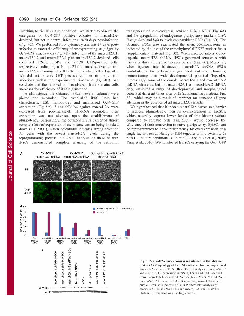
To characterize the obtained iPSCs, several colonies were
picked and expanded. The established iPSC lines had
characteristic ESC morphology and maintained Oct4-GFP
expression (Fig. 5A). Since shRNAs against macroH2A were
expressed from polymerase-III H1-RNA promoter, their
expression was not silenced upon the establishment of
pluripotency. Surprisingly, the obtained iPSCs exhibited almost
complete loss of expression of the histone variant being knocked
down (Fig. 5B,C), which potentially indicates strong selection
for cells with the lowest macroH2A levels during the
reprogramming process. qRT-PCR analysis of these shRNA
iPSCs demonstrated complete silencing of the retroviral
transgenes used to overexpress Oct4 and Klf4 in NSCs (Fig. 6A)
and the upregulation of endogenous pluripotency markers Oct4,
Nanog, Rex1 and Klf4 to levels comparable to ESCs (Fig. 6B). The
obtained iPSCs also reactivated the silent X-chromosome as
indicated by the loss of the trimethyl(me3)H3K27 nuclear focus
(supplementary material Fig. S2). When injected into a kidney
capsule, macroH2A shRNA iPSCs generated teratomas with
tissues of three embryonic lineages present (Fig. 6C). Moreover,
when injected into blastocysts, macroH2A shRNA iPSCs
contributed to the embryo and generated coat color chimeras,
demonstrating their wide developmental potential (Fig. 6D).
Interestingly, some of the double macroH2A.1 and macroH2A.2
shRNA chimeras, but not macroH2A.1 or macroH2A.2 shRNA
only, exhibited a range of developmental and morphological
defects at different times after birth (supplementary material Fig.
S3), which may be a result of improper maintenance of gene
silencing in the absence of all macroH2A variants.
We hypothesized that if indeed macroH2A serves as a barrier
to induced pluripotency, then its overexpression in EpiSCs,
which naturally express lower levels of this histone variant
compared to somatic cells (Fig. 2B,C), would decrease the
efficiency of their conversion to naıve pluripotency. EpiSCs can
be reprogrammed to naıve pluripotency by overexpression of a
single factor such as Nanog or Klf4 together with a switch to 2i
plus LIF culture conditions (Guo et al., 2009; Silva et al., 2009;
Yang et al., 2010). We transfected EpiSCs carrying the Oct4-GFP
Fig. 5. MacroH2A knockdown is maintained in the obtained
iPSCs. (A) Morphology of the iPSCs obtained from reprogrammed
macroH2A-depleted NSCs. (B) qRT-PCR analysis of macroH2A.1
and macroH2A.2 expression in NSCs, ESCs and iPSCs derived
from macroH2A.1- or macroH2A.2-depleted NSCs. MacroH2A.1
(macroH2A.1.1 + macroH2A.1.2) is in blue, macroH2A.2 is in
purple. Error bars indicate s.d. (C) Western blot analysis of
macroH2A.1 in shRNA NSCs and macroH2A shRNA iPSCs.
Histone H3 was used as a loading control.
Journal of Cell Science 125 (24)6098

Journ
alof
Cell
Scie
nce
reporter (distal enhancer) with transgenes encoding Nanog
in combination with macroH2A isoforms or an empty
vector control and kept them under dual selection for 2 weeks
(Fig. 7A). MacroH2A isoforms macroH2A.1.s1, macroH2A.1.s2,
macroH2A.1.s4 or macroH2A.2 were used, and overexpression
confirmed by qRT-PCR (Fig. 7D). The expression of Oct4 was not
changed in the selected EpiSCs, and these maintained normal
EpiSCs morphology (Fig. 7E). To induce reprogramming to naıve
pluripotency, Oct4-GFP EpiSCs expressing combinations of
transgenes were replated at equal densities and subsequently
switched to 2i plus LIF conditions for 9 days (Fig. 7A). The
appearance of EpiSCs derived iPSCs (Epi-iPSCs) colonies,
characterized by response to 2i plus LIF culture conditions and
Oct4-GFP reporter expression, was monitored. Remarkably,
macroH2A overexpression in EpiSCs dramatically reduced the
number of emerging 2i plus LIF Oct4-GFP positive Epi-iPSC
colonies, indicating that macroH2A negatively regulates the
efficiency of EpiSCs reprogramming to naıve pluripotency
(Fig. 7B,C). Overexpression of individual macroH2A.1 isoforms
reduced reprogramming efficiency by 9- to 15-fold and in the case
of macroH2A.2 no colonies were obtained. To assay the
expression of macroH2A and pluripotency genes in the obtained
Epi-iPSCs, several of these were picked, expanded, and subjected
to qRT-PCR analysis (Fig. 7D,E). The Epi-iPSCs obtained showed
pluripotency gene expression levels comparable to control ESCs
(Fig. 7E). We conclude that overexpression of macroH2A impairs
the reprogramming of EpiSCs to naıve pluripotency.
Given that macroH2A incorporates in silenced pluripotency
genes in somatic cells and that it prevents efficient reprogramming
to induced pluripotency, we sought to determine whether
macroH2A overexpression in ESCs induces differentiation and/or
pluripotency gene repression. Overexpression of macroH2A.1.s1,
macroH2A.1.s2, macroH2A.1.s4 and macroH2A.2 in ESCs did not
induce cell death or differentiation and the obtained ESCs
maintained ESC morphology, indicating that ectopic macroH2A
expression is compatible with maintenance of pluripotency
(Fig. 8A). ESCs overexpressing macroH2A isoforms showed a
modest decrease in pluripotency gene expression and an increase in
basal expression levels of the differentiation mesoderm marker T-
Brachyury, but little changes in the expression of Gata4 (Fig. 8B,C).
We conclude that ectopic macroH2A expression in ESCs, though
leading to some downregulation of pluripotency markers and
misexpression of a mesoderm marker, is not an inducer of ESCs
differentiation.
The increased efficiency of somatic cell reprogramming upon
the removal of macroH2A and the decreased efficiency of EpiSC
reprogramming upon macroH2A overexpression, together with
the fact that macroH2A is a chromatin component of repressed
Fig. 6. Characterization of macroH2A-depleted iPSCs. (A) qRT-
PCR analysis of retrovirus expression in NSCs, NS Pre-iPSCs and in
iPSCs derived from macroH2A.1 or macroH2A.2 NSCs. Klf4 retrovirus
(RetrKlf4) is in blue and Oct4 retrovirus (RetrOct4) in red. (B) Gene
expression analysis of the iPSCs depleted of macroH2A. qRT-PCR
analysis of pluripotency gene expression in NSCs, ESCs and iPSCs
derived from macroH2A.1 or macroH2A.2 NSCs. Endogenous Oct4
(EndOct4) is in blue, Rex1 in red, Klf4 in purple and Nanog in green.
(C) Tissues of the three embryonic germ layers are present in the
teratomas derived from macroH2A.2 shRNA expressing iPSCs.
(D) Contribution of macroH2A-depleted iPSCs to chimeric mice. These
were obtained after blastocyst injection of macroH2A shRNA iPSCs
into C57/BL6 blastocysts. Agouti coat color indicates chimerism
(arrows). Error bars indicate s.d.
macroH2A restricts induced pluripotency 6099

Journ
alof
Cell
Scie
nce
pluripotency genes in somatic cells indicates that macroH2A
histone variants represent an epigenetic barrier to cell
reprogramming to naıve pluripotency.
DiscussionIn this study, we report that the histone variant macroH2A is
present at low levels in the pluripotent cell compartment of pre-
and post-implantation mouse embryos while it accumulates to
high levels in somatic cells in vivo as well as in vitro. This
observed correlation of macroH2A abundance with the degree of
cell differentiation together with the known gene repression
functions of these histone variants suggests that macroH2A is
involved in gene repression during cell commitment. These
findings are consistent with previous reports showing high
macroH2A expression in differentiated cells both in mouse and
human (Creppe et al., 2012a; Dai and Rasmussen, 2007; Pehrson
et al., 1997).
In somatic cells, macroH2A depletion, though causing
decreased stability of silencing of the inactive X chromosome
and pluripotency genes (Csankovszki et al., 2001; Pasque et al.,
2011a), is not sufficient to induce full reactivation of repressed
genes, or a change in cell fate. At the same time, when exposed to
Fig. 7. MacroH2A overexpression prevents efficient reprogramming of EpiSC to naıve pluripotency. (A) Diagram showing the experimental set up. Oct4-
GFP EpiSCs were transfected with vectors encoding Nanog in combination with macroH2A.1.s1, macroH2A.1.s2, macroH2A.1.s4, macroH2A.2 or empty vector
control and grown for 2 weeks under dual selection. Selected cells were plated at a density of 50,000 cells per well of a six-well plate, switched to 2i plus LIF the
next day and cultured for another 9 days before monitoring the presence of reprogrammed Oct4-GFP-positive Epi-iPSCs. (B) Representative phase and GFP
images taken 9 days after the 2i plus LIF culture of Oct4-GFP EpiSC with Nanog and macroH2A variants or empty vector overexpression. (C) Oct4-GFP-positive
Epi-iPSCs colony counts. The average number of Oct4-GFP-positive Epi-iPSCs per well 9 days after the switch of macroH2A overexpressing or control cells to 2i
plus LIF conditions is indicated. Values represent the average colony number from three independent wells. (D) qRT-PCR analysis of macroH2A.1 (light blue)
and macroH2A.2 (purple) in Oct4-GFP EpiSCs transfected with Nanog (+Nanog) and macroH2A.1.s1 (+.1.s1), macroH2A.1.s2 (+.1.s2), macroH2A.1.s4 (+.1.s4),
macroH2A.2 (+.2) or empty vector control (+empty); and in the resulting Oct4-GFP Epi-iPSCs, as well as control Oct4-GFP ESCs. (E) qRT-PCR analysis of Esrrb
(light blue), Rex1 (red), Oct4 (green) and Klf4 (purple) in Oct4-GFP EpiSCs transfected with Nanog (+Nanog) and macroH2A.1.s1 (+.1.s1), macroH2A.1.s2
(+.1.s2), macroH2A.1.s4 (+.1.s4), macroH2A.2 (+.2) or empty vector control (+empty); and in the resulting Oct4-GFP Epi-iPSCs and control Oct4-GFP ESCs.
Please note control Oct4-GFP ESCs express Nanog, but do not overexpress Nanog. Error bars indicate s.d.
Journal of Cell Science 125 (24)6100

Journ
alof
Cell
Scie
nce
reprogramming conditions, as we show in this study, cells depleted
of macroH2A are induced to reprogram with higher efficiencies.
Moreover, ectopic macroH2A expression strongly impairs EpiSCs
reprogramming to naıve pluripotency. These results suggest that
late incorporation of macroH2A into chromatin of repressed genes
during development is important to stabilize their silenced state
and thereby contribute to the maintenance of the differentiated
state of somatic cells. Whether macroH2A also restricts direct
reprogramming of one somatic cell type to another warrants further
investigations.
A key mechanistic step during nuclear reprogramming may be
chromatin decompaction, which gives reprogramming factors
access to pluripotency gene regulatory regions. MacroH2A
variants in a nucleosome have been shown to promote
chromatin compaction by multiple mechanisms. We believe
that the removal of macroH2A makes chromatin more accessible
to respond to transcription factors Oct4 and Klf4, thereby
facilitating transcriptional activation of target genes and cell state
transitions during somatic cell reprogramming. MacroH2A hasbeen reported to promote chromatin compaction (Abbott et al.,
2005; Chakravarthy et al., 2012), recruitment of histonedeacetylases (Chakravarthy et al., 2005), lower the affinity forSWI/SNF chromatin remodeling complexes (Chang et al., 2008),hinder transcription factor binding (Angelov et al., 2003) and
associate with Polycomb proteins Ezh2 and Suz12 (Buschbecket al., 2009). These repressive functions attributed to macroH2Avariants, acting together with other epigenetic mechanisms such
as DNA methylation and Polycomb, are likely to bemechanistically linked to the low efficiency of nuclearreprogramming. Indeed, genetic ablation of HDAC2 or its
chemical inhibition improves the efficiency with which somaticcells can be induced to pluripotency (Huangfu et al., 2008).
Importantly, not only does macroH2A restrict inducedpluripotency and transcriptional reactivation, but it also prevents
progression of melanomas to the metastatic state (Kapoor et al.,2010). Indeed, loss of macroH2A.1.1 is seen in several cancertypes and correlates with negative outcome for patients with lung
or colorectal cancer (Kapoor et al., 2010; Novikov et al., 2011;Sporn and Jung, 2012; Sporn et al., 2009). This reinforces the viewthat loss of macroH2A is associated with decreased long-term
somatic cell stability. Interestingly, macroH2A.1 shRNA iPSCswere devoid of detectable macroH2A.1, unlike macroH2A.1shRNA NSCs, indicating a possible selection for cells with low
macroH2A.1 expression during reprogramming to inducedpluripotency. This seems particularly relevant in light of theincreased metastatic and teratogenic state of macroH2A depletedcells (Creppe et al., 2012a; Kapoor et al., 2010). Thus, it would be
of interest to explore the process of induced pluripotency as a routeto identify novel developmentally regulated epigenetic barriers notonly to nuclear reprogramming but also to cancer progression.
Overexpression of macroH2As in ESCs, EpiSCs or Epi-iPSCsdid not lead to cell differentiation or detectable morphologicalchanges, but for ESCs it did cause a small reduction in
pluripotency gene expression and an increase in the expressionof mesoderm inducer T-Brachyury. This suggests that high levelsof multiple macroH2A isoforms are compatible withpluripotency. This is in agreement with the high expression of
pluripotency genes in Epi-iPSCs in which high levels ofmacroH2A isoforms in combination with Nanog are expressed.At the same time, macroH2A overexpression could prone ESCs
for differentiation. Interestingly, T-Brachyury expression wasfound to be impaired in ESCs depleted of macroH2A.1 andinduced to differentiate using retinoic acid (Creppe et al., 2012a).
Genetic ablation of macroH2A.1 or macroH2A.2 is not lethaland results in fertile mice, strongly suggesting that individuallythese histone variants are dispensable for normal embryonicdevelopment (Changolkar et al., 2007; Changolkar et al., 2010).
Double knockout mice were not reported to date, which leavesopen the question whether there are compensatory mechanismsbetween the two variants. In this study, we observed
developmental and morphological abnormalities in chimericmice obtained from double macroH2A.1 and macroH2A.2shRNA iPSCs, but not from iPSCs in which individual
macroH2A variants were depleted (supplementary material Fig.S3). This may suggest that the knockout phenotype in previousreports could have been masked by potential compensatory
mechanisms, which requires further confirmation by combinedgenetic ablation. Given that macroH2A is required to stabilize thesilenced state of repressed genes, the interpretation of the knockout
Fig. 8. MacroH2A overexpression in ESCs. (A) qRT-PCR analysis of
macroH2A.1 and macroH2A.2 expression in ESCs overexpressing
macroH2A.1.s1, macroH2A.1.s2, macroH2A.1.s4 and macroH2A.2, or empty
vector control. MacroH2A.1 is in blue and macroH2A.2 in purple. (B) qRT-
PCR analysis of Nanog, Klf4 and Esrrb expression in ESCs overexpressing
macroH2A.1.s1, macroH2A.1.s2, macroH2A.1.s4 and macroH2A.2, or empty
vector control. Nanog is in blue, Klf4 in red and Esrrb in green. (C) qRT-PCR
analysis of T-Brachyury and Gata4 expression in ESCs overexpressing
macroH2A.1.s1, macroH2A.1.s2, macroH2A.1.s4 and macroH2A.2, or empty
vector control. T-Brachyury is in blue and Gata4 in purple. Error bars
indicate s.d.
macroH2A restricts induced pluripotency 6101
Journ
alof
Cell
Scie
nce
phenotype should include the analysis of susceptibility to cancer,
the efficiency of regeneration and germ cell formation and the
analysis of the effect of various stress conditions on an organism. It
is also possible that macroH2As are not strictly required for
embryogenesis, but play a role in maintenance of somatic cell
identity, consistent with its known functions in maintenance of
silenced states as well as acting as a barrier to reprogramming to
pluripotency.
In summary, this study provides evidence that histone variants
macroH2A are developmentally regulated epigenetic marks being
produced and incorporated into the chromatin of repressed genes
concomitantly with cell lineage commitment. Thus, for the first
time we identified a link between an epigenetic mark and
stabilization of the differentiated cell state that in turn
antagonizes induction of pluripotency.
Materials and MethodsCell culture
Pre-iPS, iPS and PLAT-E cells were cultured in GMEM containing 10% FCS, 16NEAA, 16Pen/Strep, 1 mM sodium pyruvate, 0.1 mM 2-mercaptoethanol, 2 mML-glutamine, supplemented with LIF. NSCs were maintained in homemade NSmedium [DMEM/F-12, 16NEAA, 0.1 mM 2-mercaptoethanol, 16Pen/Strep, 1:100B27 supplement (Invitrogen), 1:200 N2 supplement (PAA), 4.5 mM HEPES (PAA),0.03 M glucose (Sigma-Aldrich), 120 mg/ml BSA (Invitrogen)] supplemented with10 ng/ml of EGF and 20 ng/ml of FGF2. EpiSCs were grown and induced toreprogram as described previously (Guo et al., 2009; Silva et al., 2009).
Plasmids
pMXs-Oct3/4, pMXs-Klf4 (from Addgene). Retroviral infection and plasmids formacroH2A RNAi, as well as primers used to amplify macroH2A isoforms aredescribed elsewhere (Pasque et al., 2011a). For EpiSC reprogramming experiments,plasmids pPB-CAG-macroH2A.1.s1-pA-PGK-hph, pPB-CAG-macroH2A.1.s2-pA-PGK-hph, pPB-CAG-macroH2A.1.s4-pA-PGK-hph, pPB-CAG-macroH2A.2-pA-PGK-hph and pPB-CAG-Nanog-IRES-bsd were used.
Retroviral infection
PLAT-E cells were transfected using FuGENE 6 transfection reagent (Roche)according to manufacturer’s instructions. One day after the transfection completemedium change was performed and 86105 NSCs were seeded per well of a six-wellplate. The following day virus-containing supernatants from PLAT-E cultures werefiltered through 0.45 mm cellulose acetate filters and mixed with polybrene (Sigma-Aldrich) to the final concentration of 4 mg/ml. The polybrene/virus mixture was appliedto NSCs and these were incubated in virus-containing medium for 24 hours after whichthe medium was replaced with NS medium. For reprogramming experiments, theinfected cells were switched to serum/LIF medium 3 days after infection.
Flow cytometry
Flow cytometry was performed using BD LSRFortessa cell analyzer withsubsequent data analysis using FlowJo software. Cell sorting was performedusing a MoFlo high-speed cell sorter.
ChIP analysis
Starting material: 2–106106 cells, trypsinized off culture plates, washed incomplete DMEM, and washed twice in PBS to remove excess serum.
Fixation: Cells are suspended in 0.3 ml PBS to which 0.1 ml 4%paraformaldehyde in 16 PBS is added for 7 minutes at room temperature andquenched with 0.4 ml 250 mM cold glycine (125 mM final concentration). Cellsare then washed twice in 26PBS (and stored at 280 C as pellets).
Lysis: Cell pellets are resuspended in Lysis buffer (made from 26 stock withprotease inhibitor, SDS and DTT added on the day) to a final volume of not morethan 270 ml in Protein Lobind 1.5 ml microcentrifuge tubes (Eppendorf). Cellskept on ice with occasional tapping of the tubes for 10 minutes.
Chromatin fragmentation: The lysate is sonicated sufficiently to yield chromatinfragments of 250–700 bps using a Bioruptor sonicator (twice for 7-10 minutes with30 seconds on/off time, on ‘high’ power, Diagenode). Chromatin is cleared bycentrifugation (10 minutes at 10,000 r.p.m. at 4 C). Fragmented chromatin wasstored at 280 C.
Chromatin fragment size determination was established by purifying a knownamount of the chromatin lysate by the purification procedure described below.Purified DNA was analyzed by gel electrophoresis, post stained with ethidiumbromide for fragment sizes. Purified DNA was quantified by UV spectrophotometry(Nanodrop) in order to establish the approximate chromatin concentration of the
lysates (approximately twice the DNA concentration). Chromatin lysate werealiquoted into units of ,25 mg of chromatin (about 27 ml each from 107 cells).
Pre-adsorption of beads: An equal volume of Protein A and Protein G magneticbeads (DynaBead, Invitrogen) were mixed together (10 ml each per pull-downtube) and washed three times in RIPA Buffer. Beads were resuspended in SDS freeRIPA with BSA (0.1 mg/ml, Sigma) and ssDNA (75 ng/ml, Salmon Sperm derived,Sigma) at a volume of 20 ml/IP and rotated for 30 minutes to 4 hours at 4 C. Beadswere then washed once in RIPA (no SDS) and resuspended in RIPA (with BSAand ssDNA and without SDS, such that there were 20 ml/IP.
Immunocomplex and chromatin pull-down: A 1:10 or 1:20 dilution of chromatinwas made in elution buffer and stored at 280 C as ‘input sample’. For each pulldown series. 25 mg of chromatin was diluted 1:10 in SDS-free RIPA buffercontaining (0.1 mg/ml BSA (Sigma) and 75 ng/ml ssDNA (Salmon Sperm, Sigma-Aldrich). Antibody (20–40 ml total) diluted in SDS-free RIPA (with DNA and BSA)to give 1–10 mg (exact amount as per manufacturer’s suggestion) were added to thisand rotated at 4 C for 20–60 minutes. 20 ml of pre-adsorbed protein A/G beads werethen added to each pull down and rotate for 4 hours or overnight at 4 C.
Capture of immunocomplexes and stringency wash: Beads were captured onmagnetic rack, washed three times in 1 ml low wash buffer, washed in 1 ml highwash buffer. Complexes were eluted from protein A/G by adding 120 ml of elutionbuffer and incubated at 30 C for 15–20 minutes (avoid foaming). Chromatin wasdissociated from beads magnetically and the solution transferred to new DNALoBind tubes (Eppendorf).
DNA purification: RNase A was added (1 ml of DNase free 1 mg/ml to eachtube) to ‘Input’ and chromatin samples pulled down and heated at 65 C for 6 hoursor overnight. Samples were tapped up occasionally to move around. DNA waspurified with PCR spin columns (Qiagen) and eluted with 26 60 ml.
Buffers: ChIP lysis buffer (50 mM HEPES-KOH pH 7.5, 140 mM NaCl, 5 mMEDTA pH 8, 1% Triton X-100, 0.5% NP-40. 0.5 mM DTT, Protease inhibitor[Roche, Complete EDTA free] and 1% SDS added on the day of use). ChIP RIPAbuffer, SDS free [50 mM Tris-HCl pH 8, 150 mM NaCl, 2 mM EDTA pH 8, 1%NP-40, 0.5% sodium deoxycholate, protease inhibitors (add freshly)]. ChIP lowwash buffer (0.1% SDS, 1% Triton X-100, 2 mM EDTA pH 8, 150 mM NaCl,20 mM Tris-HCl pH 8). ChIP high wash buffer (0.1% SDS, 1% Triton X-100,2 mM EDTA pH 8, 500 mM NaCl, 20 mM Tris-HCl pH 8). ChIP elution buffer(1% SDS, 100 mM NaHCO3).
Antibodies used: macroH2A.1: Upstate 07-219 and Abcam ab37264. H3K9ac:Cell Signaling no. 9649. The primers used are listed in supplementary materialTable S1.
Embryo collection and culture
F1 females from C57B166CBA crosses were superovulated by intra-peritonealinjection of 10 IU of PMSG (Intervet) and 10 IU of hCG 48 hours later. Matings withF1 males\males expressing EGFP-H2B (Hadjantonakis and Papaioannou, 2004)were set up overnight, and the mice were separated the following morning. Micewere sacrificed by cervical dislocation and their oviducts were microdissected in M2medium to collect the embryonic stage required. Zygotes (embryonic day 0.5; E0.5)were released by oviduct incision, cumulus cells were removed using shorthyaluronidase treatment (1 mg/ml). Embryo culture was carried out at 37.5 C in pre-equilibrated KSOM medium under paraffin oil in an incubator with 5% CO2.
Wholemount immunofluorescence
Embryos at E3.5 and E4.5 stages were fixed for 10 minutes in 4%paraformaldehyde in 16 PBS at 37 C. Embryos at E6.5 and E9.5 were fixed for2 hours in 4% paraformaldehyde in 16 PBS at 37 C. Fixed embryos were rinsedtwice in PBS 0.2% Tween (PBT). Permeabilization was carried out during20 minutes in 0.5% Triton X, followed by the three washes in 16 PBT. Theembryos were blocked in 5% fetal bovine serum (FBS) in 16 PBT (blocking)overnight. Primary antibodies overnight incubations were performed at 4 C.Embryos were washed three times for 10–15 minutes in PBT, before detectionusing secondary antibodies, 2 hours at room temperature followed by three washesin PBT and counterstaining with DAPI. Embryos were imaged using confocal andepifluoresence microscopy. The primary antibodies used were: rabbit IgG anti-mouse macroH2A.1 (1:100, kind gift from Dr Theodore Rasmussen), rat IgG2anti-GFP (1:500, Nacalai Tesque, Cat. No. 04404-26). The secondary antibodiesused were: Alexa Fluor 594 anti-rabbit IgG (1:200, Invitrogen), Alexa Fluor 488anti-rat (1:200, Invitrogen), Alexa Fluor 647 anti-mouse (1:200, Invitrogen).
Immunofluorescence of cultured cellsCells grown on glass slides, rinsed in 16PBS, washed 30 seconds in CSK buffer(100 mM NaCl, 3 mM MgCl2, 300 mM sucrose, 10 mM PIPES pH 6.8),30 seconds in 0.5% Triton X CSK buffer and fixed in 4% PFA 10 minutesbefore rinsing in 16 PBS 0.2% Tween 20 (PBT). Permeabilized cells wereincubated in blocking buffer (5% FBS in 16PBT) for 30 minutes, then incubatedin blocking buffer with primary antibodies overnight at 4 C. The next day, slideswere rinsed three times for 10 minutes in PBT and incubated for 30 minutes inblocking buffer with secondary antibodies at room temperature, followed by threewashes in PBT and counterstained with DAPI. Slides were mounted with
Journal of Cell Science 125 (24)6102

Journ
alof
Cell
Scie
nce
vectashield and imaged using confocal microscopy. The primary antibodies usedwere: rabbit IgG anti-macroH2A.1 (1:100, Upstate, Cat. no. 07-219, Lot 32188),mouse IgG3 anti-H3K27me3 (1:100, AbCam ab6002), mouse IgG2b anti-Oct4(1:200 Santa-Cruz C10, SC5279), rat IgG2a anti-Nanog (1:200, eBioscience, cloneeBioMLC-51, 14-5761-80). The secondary antibodies used were: Alexa Fluor 488anti-mouse IgG (1:200, Invitrogen), Alexa Fluor 488 goat anti-rat (1:200,Invitrogen), Alexa Fluor 594 goat anti-rabbit IgG (1:200, Invitrogen), AlexaFluor 647 donkey anti-mouse (1:200, Invitrogen).
Quantitative RT-PCR
Total RNA was isolated using RNeasy kit (QIAGEN) in accordance withmanufacturer’s protocol. Reverse transcription was performed using SuperScriptIII First-Strand Synthesis SuperMix (Invitrogen). qRT-PCR reactions wereperformed in triplicate using SYBR Green PCR Master Mix or TaqManUniversal PCR Master Mix (both Applied Biosystems). qRT-PCR primers andgene specific Taqman expression assays (Applied Biosystems) are listed insupplementary material Table S2. DCts with GAPDH were calculated and broughtto power 22. The values were normalized to the highest value where indicated.
Western previously
As described previously (Pasque et al., 2011a) with minor modifications. 16106
cell pellets were extracted in 200 ml of combined lysis/loading buffer (300 mMNaCl, 1% NP40, 0.5% sodium deoxycholate, 4.1% SDS, 20% glycerol, 0.0004%bromoblue, 175 mM Tris-Cl, 10% betamercaptoethanol), denatured 10 minutes at98 C and incubated on ice for 5 minutes. 15 ml of extract were loaded onto a 12%Tris-HCl acrylamide gel (BioRad 161-1120), electrophoresed, and transferred tonitrocellulose membranes. Membranes were blocked in TBS containing 3% BSA.Primary antibodies were incubated overnight at 4 C. Primary antibodies used were:rabbit anti-macroH2A.1 (1 mg/ml, Abcam ab37264), rabbit anti-Histone H3(1:4000, Abcam ab1791). After extensive TBS 0.05% Tween20 (TBS-T) washes,secondary antibody 680CW-conjugated goat anti-rabbit (LI-COR, 1:25,000) wasincubated 30 minutes at room temperature. After extensive TBS-T washes,fluorescence was detected using the OdysseyH Infrared imaging system (LI-COR).
Teratoma assay
iPSCs were injected under the kidney capsule of anesthetized MF1 mice. Fourweeks after the injection tumors were surgically dissected from the mice. Sampleswere fixed in PBS containing 4% formaldehyde and embedded in paraffin.Sections were stained with hematoxylin and eosin.
Blastocyst injection
Standard microinjection methodology using host blastocysts of C57BL/6 strainwas employed.
Statistical analysis
Statistical differences were assessed with the unpaired Student’s t-test. Data arepresented as mean 6 s.e.m. or mean 6 s.d.; P-values less than 0.05 wereconsidered statistically significant.
AcknowledgementsThe authors are indebt to Dr John Gurdon for his support as well ascomments on the manuscript and to Dr Jennifer Nichols for advice onantibody staining on early embryos. We are very grateful to Dr TheodoreRasmussen for his kind gift of macroH2A.1 antibody. We thank WilliamMansfield and Charles-Etienne Dumeau for performing blastocystinjections and teratoma assays and Margaret McLeish for histologicalprocessing of the teratomas. Astrid Gillich, Richard P. Halley-Stott andMaryna Panamarova made equal contribution as second authors to thispaper. The authors declare that they have no conflict of interest.
FundingThis work was supported by The Wellcome Trust [grant numbersRG54943, 081277 and RG44593]. J.S. is a Wellcome Trust CareerDevelopment Fellow [grant number WT086692]. M.P. and M.Z.-G.are supported by a Wellcome Trust Senior Fellowship to M.Z.-G.V.P. was also supported by a Wallonia-Brussels InternationalExcellence Grant; A.R. and M.P. by the Darwin Trust ofEdinburgh; and R.P.H.-S. by the Medical Research Council.Deposited in PMC for immediate release.
Supplementary material available online at
http://jcs.biologists.org/lookup/suppl/doi:10.1242/jcs.113019/-/DC1
ReferencesAbbott, D. W., Chadwick, B. P., Thambirajah, A. A. and Ausio, J. (2005). Beyond
the Xi: macroH2A chromatin distribution and post-translational modification in anavian system. J. Biol. Chem. 280, 16437-16445.
Angelov, D., Molla, A., Perche, P.-Y., Hans, F., Cote, J., Khochbin, S., Bouvet,
P. and Dimitrov, S. (2003). The histone variant macroH2A interferes withtranscription factor binding and SWI/SNF nucleosome remodeling. Mol. Cell 11,1033-1041.
Barzily-Rokni, M., Friedman, N., Ron-Bigger, S., Isaac, S., Michlin, D. and Eden,
A. (2011). Synergism between DNA methylation and macroH2A1 occupancy inepigenetic silencing of the tumor suppressor gene p16(CDKN2A). Nucleic Acids Res.
39, 1326-1335.
Bernstein, E., Muratore-Schroeder, T. L., Diaz, R. L., Chow, J. C., Changolkar,
L. N., Shabanowitz, J., Heard, E., Pehrson, J. R., Hunt, D. F. and Allis, C. D.
(2008). A phosphorylated subpopulation of the histone variant macroH2A1 isexcluded from the inactive X chromosome and enriched during mitosis. Proc. Natl.
Acad. Sci. USA 105, 1533-1538.
Boulard, M., Storck, S., Cong, R., Pinto, R., Delage, H. and Bouvet, P. (2010).Histone variant macroH2A1 deletion in mice causes female-specific steatosis.Epigenetics & Chromatin 3, 8.
Buschbeck, M. and Di Croce, L. (2010). Approaching the molecular and physiologicalfunction of macroH2A variants. Epigenetics 5, 118-123.
Buschbeck, M., Uribesalgo, I., Wibowo, I., Rue, P., Martin, D., Gutierrez, A.,
Morey, L., Guigo, R., Lopez-Schier, H. and Di Croce, L. (2009). The histonevariant macroH2A is an epigenetic regulator of key developmental genes. Nat. Struct.
Mol. Biol. 16, 1074-1079.
Chakravarthy, S., Gundimella, S. K. Y., Caron, C., Perche, P.-Y., Pehrson, J. R.,
Khochbin, S. and Luger, K. (2005). Structural characterization of the histone variantmacroH2A. Mol. Cell. Biol. 25, 7616-7624.
Chakravarthy, S., Patel, A. and Bowman, G. D. (2012). The basic linker of macroH2Astabilizes DNA at the entry/exit site of the nucleosome. Nucleic Acids Res. 40, 8285-8295.
Chang, C.-C., Ma, Y., Jacobs, S., Tian, X. C., Yang, X. and Rasmussen, T. P. (2005).A maternal store of macroH2A is removed from pronuclei prior to onset of somaticmacroH2A expression in preimplantation embryos. Dev. Biol. 278, 367-380.
Chang, E. Y., Ferreira, H., Somers, J., Nusinow, D. A., Owen-Hughes, T. and
Narlikar, G. J. (2008). MacroH2A allows ATP-dependent chromatin remodeling bySWI/SNF and ACF complexes but specifically reduces recruitment of SWI/SNF.Biochemistry 47, 13726-13732.
Chang, C.-C., Gao, S., Sung, L.-Y., Corry, G. N., Ma, Y., Nagy, Z. P., Tian, X. C.
and Rasmussen, T. P. (2010). Rapid elimination of the histone variant MacroH2Afrom somatic cell heterochromatin after nuclear transfer. Cell Reprogram 12, 43-53.
Changolkar, L. N., Costanzi, C., Leu, N. A., Chen, D., McLaughlin, K. J. and
Pehrson, J. R. (2007). Developmental changes in histone macroH2A1-mediated generegulation. Mol. Cell. Biol. 27, 2758-2764.
Changolkar, L. N., Singh, G., Cui, K., Berletch, J. B., Zhao, K., Disteche, C. M. and
Pehrson, J. R. (2010). Genome-wide distribution of macroH2A1 histone variants inmouse liver chromatin. Mol. Cell. Biol. 30, 5473-5483.
Costanzi, C. and Pehrson, J. R. (1998). Histone macroH2A1 is concentrated in theinactive X chromosome of female mammals. Nature 393, 599-601.
Costanzi, C. and Pehrson, J. R. (2001). MACROH2A2, a new member of theMARCOH2A core histone family. J. Biol. Chem. 276, 21776-21784.
Costanzi, C., Stein, P., Worrad, D. M., Schultz, R. M. and Pehrson, J. R. (2000).Histone macroH2A1 is concentrated in the inactive X chromosome of femalepreimplantation mouse embryos. Development 127, 2283-2289.
Creppe, C., Janich, P., Cantarino, N., Noguera, M., Valero, V., Musulen, E., Douet,
J., Posavec, M., Martın-Caballero, J., Sumoy, L. et al. (2012a). MacroH2A1regulates the balance between self-renewal and differentiation commitment inembryonic and adult stem cells. Mol. Cell. Biol. 32, 1442-1452.
Creppe, C., Posavec, M., Douet, J. and Buschbeck, M. (2012b). MacroH2A in stemcells: a story beyond gene repression. Epigenomics 4, 221-227.
Csankovszki, G., Nagy, A. and Jaenisch, R. (2001). Synergism of Xist RNA, DNAmethylation, and histone hypoacetylation in maintaining X chromosome inactivation.J. Cell Biol. 153, 773-784.
Dai, B. and Rasmussen, T. P. (2007). Global epiproteomic signatures distinguishembryonic stem cells from differentiated cells. Stem Cells 25, 2567-2574.
Foshay, K. M., Looney, T. J., Chari, S., Mao, F. F., Lee, J. H., Zhang, L., Fernandes,
C. J., Baker, S. W., Clift, K. L., Gaetz, J. et al. (2012). Embryonic stem cells inducepluripotency in somatic cell fusion through biphasic reprogramming. Mol. Cell 46,159-170.
Gamble, M. J. and Kraus, W. L. (2010). Multiple facets of the unique histone variantmacroH2A: from genomics to cell biology. Cell Cycle 9, 2566-2572.
Gamble, M. J., Frizzell, K. M., Yang, C., Krishnakumar, R. and Kraus, W. L.
(2010). The histone variant macroH2A1 marks repressed autosomal chromatin, butprotects a subset of its target genes from silencing. Genes Dev. 24, 21-32.
Guo, G., Yang, J., Nichols, J., Hall, J. S., Eyres, I., Mansfield, W. and Smith,
A. (2009). Klf4 reverts developmentally programmed restriction of ground statepluripotency. Development 136, 1063-1069.
Gurdon, J. B. and Melton, D. A. (2008). Nuclear reprogramming in cells. Science 322,1811-1815.
Gurdon, J. B., Elsdale, T. R. and Fischberg, M. (1958). Sexually mature individuals ofXenopus laevis from the transplantation of single somatic nuclei. Nature 182, 64-65.
macroH2A restricts induced pluripotency 6103

Journ
alof
Cell
Scie
nce
Hadjantonakis, A.-K. and Papaioannou, V. E. (2004). Dynamic in vivo imaging and
cell tracking using a histone fluorescent protein fusion in mice. BMC Biotechnol. 4,
33.
Hadjantonakis, A. K., Cox, L. L., Tam, P. P. and Nagy, A. (2001). An X-linked GFP
transgene reveals unexpected paternal X-chromosome activity in trophoblastic giant
cells of the mouse placenta. Genesis 29, 133-140.
Hajkova, P., Ancelin, K., Waldmann, T., Lacoste, N., Lange, U. C., Cesari, F., Lee,
C., Almouzni, G., Schneider, R. and Surani, M. A. (2008). Chromatin dynamics
during epigenetic reprogramming in the mouse germ line. Nature 452, 877-881.
Huangfu, D., Maehr, R., Guo, W., Eijkelenboom, A., Snitow, M., Chen, A. E. and
Melton, D. A. (2008). Induction of pluripotent stem cells by defined factors is greatly
improved by small-molecule compounds. Nat. Biotechnol. 26, 795-797.
Kapoor, A., Goldberg, M. S., Cumberland, L. K., Ratnakumar, K., Segura, M. F.,
Emanuel, P. O., Menendez, S., Vardabasso, C., Leroy, G., Vidal, C. I. et al.
(2010). The histone variant macroH2A suppresses melanoma progression through
regulation of CDK8. Nature 468, 1105-1109.
Kim, J. B., Zaehres, H., Wu, G., Gentile, L., Ko, K., Sebastiano, V., Arauzo-Bravo,
M. J., Ruau, D., Han, D. W., Zenke, M. et al. (2008). Pluripotent stem cells induced
from adult neural stem cells by reprogramming with two factors. Nature 454, 646-
650.
Nashun, B., Yukawa, M., Liu, H., Akiyama, T. and Aoki, F. (2010). Changes in the
nuclear deposition of histone H2A variants during pre-implantation development in
mice. Development 137, 3785-3794.
Novikov, L., Park, J. W., Chen, H., Klerman, H., Jalloh, A. S. and Gamble, M. J.
(2011). QKI-mediated alternative splicing of the histone variant MacroH2A1
regulates cancer cell proliferation. Mol. Cell. Biol. 31, 4244-4255.
Pasque, V., Gillich, A., Garrett, N. and Gurdon, J. B. (2011a). Histone variant
macroH2A confers resistance to nuclear reprogramming. EMBO J. 30, 2373-2387.
Pasque, V., Halley-Stott, R. P., Gillich, A., Garrett, N. and Gurdon, J. B. (2011b).
Epigenetic stability of repressed states involving the histone variant macroH2A
revealed by nuclear transfer to Xenopus oocytes. Nucleus 2, 533-539.
Pehrson, J. R. and Fried, V. A. (1992). MacroH2A, a core histone containing a large
nonhistone region. Science 257, 1398-1400.
Pehrson, J. R., Costanzi, C. and Dharia, C. (1997). Developmental and tissue
expression patterns of histone macroH2A1 subtypes. J. Cell. Biochem. 65, 107-113.
Rasmussen, T. P., Huang, T., Mastrangelo, M. A., Loring, J., Panning, B. andJaenisch, R. (1999). Messenger RNAs encoding mouse histone macroH2A1 isoformsare expressed at similar levels in male and female cells and result from alternativesplicing. Nucleic Acids Res. 27, 3685-3689.
Silva, J., Barrandon, O., Nichols, J., Kawaguchi, J., Theunissen, T. W. and Smith,
A. (2008). Promotion of reprogramming to ground state pluripotency by signalinhibition. PLoS Biol. 6, e253.
Silva, J., Nichols, J., Theunissen, T. W., Guo, G., van Oosten, A. L., Barrandon, O.,Wray, J., Yamanaka, S., Chambers, I. and Smith, A. (2009). Nanog is the gatewayto the pluripotent ground state. Cell 138, 722-737.
Sporn, J. C. and Jung, B. (2012). Differential regulation and predictive potential ofMacroH2A1 isoforms in colon cancer. Am. J. Pathol. 180, 2516-2526.
Sporn, J. C., Kustatscher, G., Hothorn, T., Collado, M., Serrano, M., Muley, T.,Schnabel, P. and Ladurner, A. G. (2009). Histone macroH2A isoforms predict therisk of lung cancer recurrence. Oncogene 28, 3423-3428.
Tada, M., Tada, T., Lefebvre, L., Barton, S. C. and Surani, M. A. (1997). Embryonicgerm cells induce epigenetic reprogramming of somatic nucleus in hybrid cells.EMBO J. 16, 6510-6520.
Tada, M., Takahama, Y., Abe, K., Nakatsuji, N. and Tada, T. (2001). Nuclearreprogramming of somatic cells by in vitro hybridization with ES cells. Curr. Biol. 11,1553-1558.
Takahashi, K. and Yamanaka, S. (2006). Induction of pluripotent stem cells frommouse embryonic and adult fibroblast cultures by defined factors. Cell 126, 663-676.
Talbert, P. B., Ahmad, K., Almouzni, G., Ausio, J., Berger, F., Bhalla, P. L., Bonner,
W. M., Cande, W. Z., Chadwick, B. P., Chan, S. W. L. et al. (2012). A unifiedphylogeny-based nomenclature for histone variants. Epigenetics & Chromatin 5, 7.
Tanasijevic, B. and Rasmussen, T. P. (2011). X chromosome inactivation anddifferentiation occur readily in ES cells doubly-deficient for macroH2A1 andmacroH2A2. PLoS ONE 6, e21512.
Timinszky, G., Till, S., Hassa, P. O., Hothorn, M., Kustatscher, G., Nijmeijer, B.,Colombelli, J., Altmeyer, M., Stelzer, E. H. K., Scheffzek, K. et al. (2009). Amacrodomain-containing histone rearranges chromatin upon sensing PARP1 activa-tion. Nat. Struct. Mol. Biol. 16, 923-929.
Yang, J., van Oosten, A. L., Theunissen, T. W., Guo, G., Silva, J. C. R. and Smith,
A. (2010). Stat3 activation is limiting for reprogramming to ground statepluripotency. Cell Stem Cell 7, 319-328.
Journal of Cell Science 125 (24)6104
Copyright © 2022 FDOKUMEN