Transient reactivation of CSF in parthenogenetic one-cell mouse embryos

Current Pharmaceutical Biotechnology, 2011, 12, 000-000 1
1389-2010/11 $58.00+.00 © 2011 Bentham Science Publishers Ltd.
Parthenogenetic Cell Lines: An Unstable Equilibrium Between Pluripo-tency and Malignant Transformation
Tiziana A.L. Brevini1,*
, Georgia Pennarossa1, Magda deEguileor
2, Gianluca Tettamanti
2,
Guido Ragni3, Alessio Paffoni
3 and Fulvio Gandolfi
1
1Laboratory of Biomedical Embryology – UniStem, Center for Stem Cell Research, Università degli Studi di Milano,
Milan, Italy; 2Department of Biotechnology and Molecular Sciences, University of Insubria, Varese, Italy;
3Infertility
Unit, Department of Obstetrics, Gynaecology and Neonatology, Fondazione Ospedale Maggiore Policlinico, Mangiag-
alli e Regina Elena, Milan, Italy
Abstract: Human parthenogenetic embryos have been recently proposed as an alternative, less controversial source of
embryonic stem cells. However many aspects related to the biology of parthenogenetic cell lines are not fully understood
and still need to be elucidated. These cells have great potentials; they possess most of the main features of bi-parental
stem cells, show the typical morphology and express most of the pluripotency markers distinctive of ESC. They also have
high telomerase activity, that disappears upon differentiation, and display great plasticity. When cultured in appropriate
conditions, they are able to give rise to high specification tissues and to differentiate into mature cell types of the neural
and hematopoietic lineages. However, their injection in immunodeficient mice has been reported to result in tumour for-
mations. Aberrant levels of molecules related to spindle formation, cell cycle check points and chromosome segregation
have also been detected in these cells, that are characterized by the presence of an abnormal number of centrioles and
massive autophagy. All these observations indicate the presence of an intrinsic deregulation of the mechanisms controlling
proliferation versus differentiation in parthenogenetic stem cells. In this manuscript we summarize data related to these
aberrant controls and describe experimental evidence indicating their uniparental origin as one of the possible cause. Fi-
nally we propose their use as an intriguing experimental tool where the pathways controlling potency, self renewal and
cell plasticity are deeply interconnected with cell transformation, in an unstable, although highly controlled, equilibrium
between pluripotency and malignacy.
Keywords: Centriole, mitotic spindle, parthenogenesis, pluripotency, tumorigenic.
INTRODUCTION
Parthenogenesis is the process by which an oocyte can develop without the intervention of the male counterpart. It is a form of reproduction common to a variety of organisms such as fish, ants, flies, honeybees, amphibians, lizards and snakes, that may routinely reproduce in this manner.
Mammals are not spontaneously capable of this form of reproduction, but, mammalian oocytes can be successfully activated in vitro, mimicking the intracellular calcium wave induced by the sperm at fertilization, which triggers cleavage divisions and embryonic development [1]. Mammalian parthenotes, however, are unable to develop to term and ar-rest their development at different stages after activation, depending on the species [2]. The reason for this arrest is believed to be due to genomic imprinting which causes the repression of certain genes [3], that are normaly expresse by the paternal allele.
Because mammalian parthenotes are inherently unable to form a new individual, following the success reported in
*Address correspondence to this author at the Laboratory of Biomedical
Embryology – UniStem, Center for Stem Cell Research, Università degli Studi di Milano, Facoltà di Medicina Veterinaria, via Celoria 10, 20133
Milan, Italy; Tel: 0039-02-50317980; Fax: 0039-02-50317989; E-mail: [email protected]
mouse [4, 5] and non-human primate models [6, 7], human parthenogenetic embryos have recently been proposed as an alternative, less controversial source of embryonic stem cell lines [1, 8-11]. Parthenotes may also represent a possible tool for studies on the mechanisms driving early human embryo-genesis and for the pre-clinical test of experimental protocols in human assisted reproduction (i.e. different oocytes cryo-preservation procedures, oocyte in vitro maturation or polar body genetic screening). However many aspects related to the biology of parthenogenetic embryos and parthenogenetic derived cell lines still need to be elucidated. The assembly of the new embryonic centrosome in the absence of the sperm centriol can lead to aberrant progression of cell cycle and spindle formation [12]. The decreased extent of heterozy-gosis may amplify any negative genetic component poten-tially present in the genotype [13, 14]. The very high inci-dence of chromosome instability and aberrant chromatid separation in oocytes retrieved from IVF patients, especially when over 34 year old [15-18] also represents a concern, given the fact that these represent a large part of the popula-tion accessing assisted reproductive therapy and, hence, are a major potential source of oocytes for parthenotes derivation.
In an attempt to better elucidate some of these aspects, human parthenogenetic (HP) cell lines, recently derived in our laboratory [2, 19], were characterized for their pluripo-tency and differentiation plasticity, both in vitro and in vivo.

2 Current Pharmaceutical Biotechnology, 2011, Vol. 12, No. 1 Brevini et al.
Here we discuss the characteristics of these cell lines that are common to bi-parental embryonic stem cells. However we also describe the presence of an intrinsic deregulation of the mechanisms controlling proliferation versus differentiation and suggest their uniparental origin the possible cause.
PLURIPOTENCY AND IN VITRO DIFFERENTIA-
TION ABILITY OF HP CELL LINES
The development of human parthenotes to the blastocyst stage was reported only recently [20-22] and not many data are available because of the limited accessibility of unfertil-ized human oocytes. Even more limited are the information related to the potential plasticity of the lines that can be de-rived from parthenogenetic human embryos. The lines de-rived in our laboratory have been growing for over two years and possess the main features of bi-parental stem cells. They are capable of renewing themselves; they can be continu-ously cultured in an undifferentiated state and can be propa-gated extensively in vitro. For over 70 passages they con-stantly expressed markers that characterize and are distinc-tive of human embryonic stem cells, such as Oct-4, Nanog, Rex-1, Sox-2, alkaline phosphatase, SSEA-4, TRA 1-81, indicating a pluripotency signature that is not influenced by the exclusive presence of the maternal genome. In common with biparental cell lines they also display high telomerase activity, which is often correlated with self-renewal, regen-eration and immortality and is typically expressed in germ cells and embryonic stem cells [23, 24] (Table 1). In line with this, undifferentiated HP cells displayed high levels of telomerase activity, while no telomerase activity could be detected when cells were subjected to specific stimuli in or-der to induce tissue differentiation in vitro . These data indi-cate that a physiologically normal control of telomerase ac-tivity is present in HP cells and that, if subjected to culture conditions that induce differentiation, they turn down their telomerase activity. Consistent with this, HP cells were able to form embryoid bodies (EBs) that actively transcribed RNAs involved in specification of the three embryonic germ layers, demonstrating the potential of these cells to differen-tiate in the main tissue types of the body. Interestingly, very low or no expression of trofectoderm-related markers was detected, suggesting that human parthenogenetic cells have a poor capability to differentiate towards trofectoderm and this may reflect parthenote inability to give rise to adequate fetal membrane development and to implant correctly.
In our opinion a crucial point was to assess the possibility to drive differentiation of human parthenogenetic cells to-wards a specific lineage, in controlled culture conditions. Data from our lab demonstrate that cells were able to form different cell subtypes belonging to the neural lineage as well as to differentiate in the complex array of hematopoietic cells. Although further assays to test the real extent of cellu-lar functionality are needed, these results further confirm human parthenogenetic cell lines differentiation potential. In particular our experiments showed that HP cells were able, not only to give rise to early neural lineages, as previously described [8-11], but also to more mature cell types express-ing nestin, CNPase and MAP2 [19]. This is consistent with the observation that murine androgenetic embryonic stem cells are able to differentiate into neuronal and glial cells [25]. Similarly, exposure of HP cells to specific cytokines
and adequate culture conditions allowed for differentiation of these cells towards the hematopoietic lineage, with the gen-eration of lymphoid, erythroid and myeloid sub-populations. Our results further expanded current knowledge on HP in vitro differentiation plasticity showing the formation of ma-ture hemopoietic cell lineages and confirms what has been recently reported in mouse androgenetic and gynogenetic stem cells, that where shown to be able to generate adult-transplantable hematopoietic stem cells that can repopulate the hematopoietic system of adult transplant recipients [25]. Altogether these findings indicate that, outside the normal developmental paradigm, the differentiation potential of uni-parental cells may be much less restricted than that of parthenogenetic cells in chimeras and that these cells can be an interesting and relevant model for the study of fundamen-tal mechanisms involved in human lineage determination.
Table 1. Comparison of Pluripotency Signature in Biparental
and Parthenogenetic (HP) Cell Lines
Human Biparental
ESC
Human Partheno-
genetic Cell Lines
(HP)
Oct-4 + +
Nanog + +
Sox-2 + +
Rex-1 + +
TRA 1-81 + +
SSEA-4 + +
Telomerase activity high high
EB derivation yes yes
PARTHENOGENETIC CELLS SHOW A DEREGU-
LATION OF THE MECHANISMS CONTROLLING
PROLIFERATION VERSUS DIFFERENTIATION
Injection of HP cells in immunodeficient mice gave rise to the formation of malignant sarcomas with myofibroblastic differentiation [26]. This is consistent with the observation that the subcutaneous injection of androgenetic mouse em-bryonic stem cells in immunodeficient mice generates sar-comas with muscular differentiation [27]. These findings suggest the presence of an intrinsic deregulation of the mechanisms controlling the choice between proliferation and differentiation in embryonic stem cells obtained through parthenogenesis and androgenesis. However, it is interesting to note that this appeared to be modulated by the microenvi-ronment and, while it was undetectable when cells were dif-ferentiated in vitro, it became evident once cells were ex-posed to the in vivo milieu.
These results are in contrast to what previously described in other human parthenogenetic cell lines [8-11] and the pos-sibility exists that these anomalies derive from differences in the procedures used for the derivation of the cell lines. We think that an important factor to be considered is the age of the oocyte donors. This aspect could be important, given the

Parthenogenetic Cell Lines Current Pharmaceutical Biotechnology, 2011, Vol. 12, No. 1 3
high incidence of chromosome instability and aberrant chromatid separation in oocytes retrieved from IVF patients, especially when over 34 year old [15-17]. Unfortunately no details on donors age were given in the other reports, there-fore the relevance of this parameter cannot be evaluated.
However, based on a series of experiments carried out in our lab [19], we hypothesize that the uniparental origin of HP cells and, in particular, the lack of the centrioles supplied by the male counterpart, may be a possible explanation of the altered behaviour of parthenogenetic cell lines upon in vivo transplantation, with the use of relatively old donors as a potential aggravating factor.
PARTHENOGENETIC CELLS AND DE-NOVO CEN-
TRIOLE ASSEMBLY
It has been demonstrated that centrioles degenerate and are lost during human oogenesis and, while oogonia and growing oocytes display normal centrioles until pachytene stage, they are absent in the mature oocytes [28]. With the notable exception of mice [29] this degenerative process has been described in rhesus monkeys [30], rabbits [31], cows [32], sea urchins [33], Xenopus [34], and several other spe-cies [35]. Due to the absence of centrioles, the oocyte centro-somal material does not aggregate into unified foci and is unable to form astral microtubules and a correctly oriented spindle, unless rescued by a spermatozoon. The conse-quences of the lack of centrioles on parthenogenetic devel-opment have been studied in detail in lower species. In Sci-ara parthenogenetic embryos, the spindles assembled without centrosomes are anastral, and the distance between the daughter nuclei is greatly reduced [36]. This limitation is common to many other species that are obligatory or faculta-tive parthenotes, where successful parthenogenesis largely depends upon the oocyte ability to generate complete and functional centrosomes, in the absence of the material sup-plied by a male gamete. In particular, parthenogenetically activated sea urchin and insect eggs have been described to form multiple centrioles, possibly as the result of the lack of a correct control on the process of spindle formation [37-39]. Indeed, the inability of parthenogenetic oocytes to organize normal spindles, due to the lack of a functional centrosome, has been suggested as a strict checkpoint control to suppress parthenogenetic development [40], since it is not an evolu-tionarily preferred pathway, even in species that are faculta-tive parthenotes, because it leads to genomic homogeneity that, in turn, results in the accumulation of genetic anomalies in the population. These observations on the parthenogenetic process in lower species are in agreement with our findings in HP cells. Similarly to what reported in sea urchins and many insects, for instance, also HP cells display multiple centrioles Fig. (1) suggesting that the ability to rearrange functional centrosomes is altered in these cells. Marshall [41], suggested that centriole de novo assembly is normally turned off when centrioles are present in a cell. Therefore the absence of sperm centriole in parthenotes may lead to the lack of the negative regulatory mechanism that normally suppress de novo centriole assembly and may explain the presence of multicentriolar structures, as the ones described in parthenotes and as the ones we detect in HP cells.
Interestingly enough, HP cells can proliferate and divide and most importantly, they can correctly differentiate, into a variety of tissues, responding to experimental conditions that are able to induce differentiation in bi-parental cells. This ability does not seem to be limited by the abnormalities de-scribed above and suggests that the requirement of a paternal centrosome described in lower animals appears to be less stringent in human cells, at least in in vitro controlled condi-tions. Indeed, in higher mammals, genomic imprinting is thought to be the main mechanism to the obligatority of en-sure biparental fertilization [42]. However these abnormali-ties in spindle rearrangement may explain the high percent-age of cells showing misshaped, mal-segregated chromo-somes in our cell lines. Multiple chromosome malsegrega-tions have been previously described in human oocytes after parthenogenetic activation, either spontaneous or induced by puromycin [43]. Consistent with this, a high incidence of polyploid and mixoploid chromosomal complements has been reported in parthenotes derived from bovine and por-cine activated oocytes, with abnormal chromosomal com-plements occurring as early as completion of the first cell cycle and, again, it was linked to the absence of a paternally supplied centrosome [44, 45].
The patterns of abnormal centrosome reformations ob-served in HP cells closely resemble those seen in cancer cells which argues that structural defects of centrosomes can ac-count for the formation of abnormal mitosis and multipolar cells frequently observed in cancer [46]. Interestingly enough these abnormalities do not seem to affect the overall proliferation rate and in vitro differentiation plasticity of HP cells. Although this needs to be further elucidated, it could be partially explained by the massive autophagocitic activity present in these cells. This process is likely to be used as an active self-protective strategy in order to eliminate highly abnormal organelles thus contributing to cell survival and ensuring “normal” population [47].
PARTHENOGENETIC CELLS AND MITOTIC SPIN-
DLE CHECKPOINT MOLECULES
We previously showed that HP cells display altered ex-pression levels of mitotic check point molecules Fig. (2). In particular, the comparison of HP cells with bi-parental em-bryonic stem cell lines indicated a much higher level of ex-pression of Mad-1, and the related molecules MAX and SIN3 in parthenogenetic cells. Mad-1 is a central component of the spindle assembly checkpoint and recruitment of kine-tochores [48-50]. Increased level of Mad1 has been previ-ously shown to cause a reduction in the expression of Myc in human monoblasts where a decrement of cell proliferation with a protracted G1 phase of the cell cycle was observed but Mad1 was neither demonstrated to cause spontaneous differ-entiation nor to enhance
induced differentiation but rather
inhibited differentiation induced by retinoic acid [51]. On the
other hand Mad1 dimerizes with MAX and, through a spe-cific association with the SIN3 co-repressor, decreases tran-scription and, indirectly inhibits cell proliferation [52]. Taken together these observations support the hypothesis that the altered levels of such molecules present in HP cells cause anomalies in their control of proliferation and differen-tiation.
4 Current Pharmaceutical Biotechnology, 2011, Vol. 12, No. 1 Brevini et al.
Fig. (1). Expression level of molecules related to spindle formation and chromosome segregation in HP cells and biparental cell lines. Bars
represent the average Ct of HP cells (white)and biparental cells (grey) related to the genes examined. Ct value was obtained from the Ct of
the target gene normalized with the Ct value for -actin of the same sample.
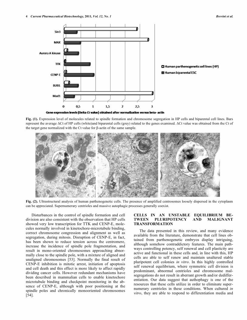
Fig. (2). Ultrastructural analysis of human parthenogenetic cells. The presence of amplified centrosomes loosely dispersed in the cytoplasm
can be appreciated. Supernumerary centrioles and massive autophagic processes generally coexist.
Disturbances in the control of spindle formation and cell division are also consistent with the observation that HP cells showed very low transcription for TTK and CENP-E, mole-cules normally involved in kinetochore-microtubule binding, correct chromosome congression and alignment as well as segregation, during mitosis. Disruption of CENP-E, in fact, has been shown to reduce tension across the centromere, increase the incidence of spindle pole fragmentation, and result in mono-oriented chromosomes approaching abnor-mally close to the spindle pole, with a mixture of aligned and unaligned chromosomes [53]. Normally the final result of CENP-E inhibition is mitotic arrest, initiation of apoptosis and cell death and this effect is more likely to affect rapidly dividing cancer cells. However redundant mechanisms have been described in mammalian cells to enable kinetochore microtubule binding and checkpoint monitoring in the ab-sence of CENP-E, although with poor positioning at the spindle poles and chronically monooriented chromosomes [54].
CELLS IN AN UNSTABLE EQUILIBRIUM BE-TWEEN PLURIPOTENCY AND MALIGNANT
TRANSFORMATION
The data presented in this review, and many evidence available from the literature, demonstrate that cell lines ob-tained from parthenogenetic embryos display intriguing, although somehow contraddictory features. The main path-ways controlling potency, self renewal and cell plasticity are active and functional in these cells and, in line with this, HP cells are able to self renew and maintain unaltered stable pluripotent cell colonies in vitro. In this highly controlled self renewal equilibrium, where symmetric cell division is predominant, abnormal centrioles and chromosome mal-segregations do not result in aberrant growth and/or dediffer-entiation. Our data suggest that authophagy is one of the resources that these cells utilize in order to eliminate super-numerary centrioles in these conditions. When cultured in vitro, they are able to respond to differentiation media and

Parthenogenetic Cell Lines Current Pharmaceutical Biotechnology, 2011, Vol. 12, No. 1 5
give rise to populations representative of the three germ lay-ers, switching between symmetric and asymmetric cell divi-sions in a timely and controlled manner. HP cells can be driven though standard protocols to differentiate into hema-topoietic and neural cells. However this pluripotency/high plasticity status seems to be related to a strictly controlled environment, such as the one which is recreated in in vitro culture conditions, and is lost when cells are transplanted in SCID mice. The in vivo milieu appears to interfere with this mode and releases cells to aberrant growth, with unrestrained expansion of the stem cell compartment that leads to the formation of malignant sarcomas.
Work from Boveri at the turn of the 20th century postu-lated that centrosomes play an essential role in the mainte-nance of genome stability and that defects in centrosome biogenesis could lead to aneuploidy and potentially favor tumor formation. For over 100 years, however this hypothe-sis has not been tested. Basto and colleagues [55] recently demonstrated that the induction of supernumerary centro-somes is sufficient to cause transformation in flies, with large and invasive tumors as a result. Castellanos and col-legues [56] used an unbiased approach to study the tumori-genic potential of panel of Drosophila mutants, defective in various aspects of centrosome biogenesis. These authors found that most of mutants generated a high frequency of tumors but Polo-like kinase and Aurora A mutants gave rise to especially large and invasive tumors that could be serially re-transpalnted and were able to generate an almost infinite tumor mass. Parallel studies from Weaver et al. [57] demon-strated that reduced levels of centrosome-linker motor pro-tein CENP-E result in elevated levels of aneuploidy. Al-though it will be important to determine how specie with high numbers of chromosomes that need to be segregate to progeny during cell divition respond to centrosome dysfunc-tion, we think it may be of interest to note that HP cells were found to express dramatically decreased levels of Aurora A and CENP-E [19], indicating that similar molecular pathway are likely to be affected by the presence of supernumerary centriols in human cells.
Interpreting the results we generated with HP cells, in the light of the data mentioned above, we can hypothesize that parthenogenetic cells may represent an interesting model where the control of asymmetric cell division is lost, due to the presence of abnormal centrosome. Since switching be-tween symmetric and asymmetric cell divisions in a timely and controlled manner is crucial for normal tissue homeosta-sis, deregulation of this process can cause continued expan-sion of the stem cell compartment, with the activation of the tumorigenic process [58], as we detect when HP cells are transplanted to SCID mice.
In this line, the tumorigenic process detected in HP cells may be related to the presence of supernumerary centrioles that would create misalignment between the spindle and the asymmetric cell determinants, resulting in enforcement of the tumorigenic process. We think that further studies on the link between supernumerary centriols, multipolar spindles and genetic instability leading to cancer in mammalian spe-cie could provide important information, especially if we consider that most of the data available at the present, are limited to lower species. In our opinion the use of HP cell lines could represent an intriguing and unique experimental tool where the pathways controlling potency and self re-newal vs cell transformation can be investigated in a human model, and where the use of different milieaux can activate mechanisms that control the switch between pluripotency and malignancy.
REFERENCES
[1] Brevini, T. A.; Gandolfi, F. Parthenotes as a source of embryonic
stem cells. Cell Prolif., 2008, 41 (Suppl 1), 20-30. [2] Brevini, T. A.; Pennarossa, G.; Antonini, S.; Gandolfi, F. Partheno-
genesis as an approach to pluripotency: advantages and limitations involved. Stem Cell Rev., 2008, 4(3), 127-135.
[3] Surani, M. A. Reprogramming of genome function through epige-netic inheritance. Nature, 2001, 414(6859), 122-128.
[4] Allen, N. D.; Barton, S. C.; Hilton, K.; Norris, M. L.; Surani, M. A. A functional analysis of imprinting in parthenogenetic embryonic
stem cells. Development, 1994, 120 (6), 1473-1482. [5] Kaufman, M. H.; Robertson, E. J.; Handyside, A. H.; Evans, M. J.
Establishment of pluripotential cell lines from haploid mouse em-bryos. J. Embryol. Exp. Morphol., 1983, 73, 249-261.
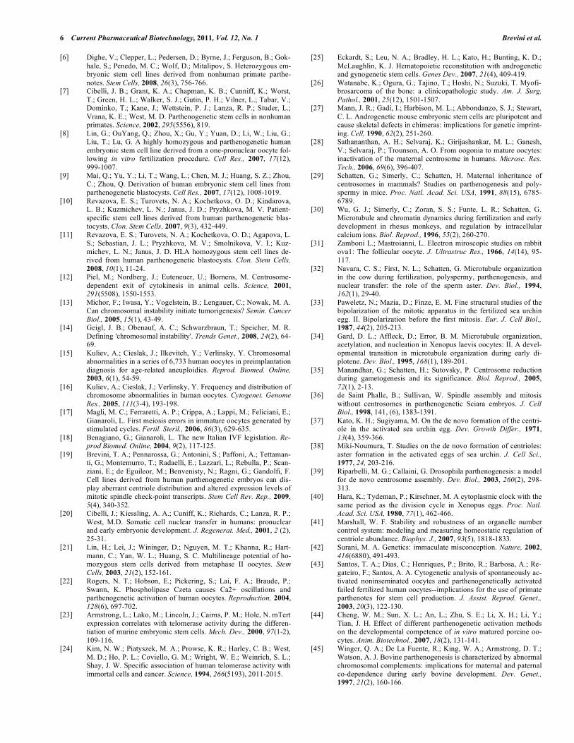
Fig. (3). Normal centrosome ensure timely and controlled switching between symmetric and asymmetric cell division, result-ing in correct tissue homeostasis (A). Supernumerary centrioles are sufficient to cause cell transformation and tumors (B).
6 Current Pharmaceutical Biotechnology, 2011, Vol. 12, No. 1 Brevini et al.
[6] Dighe, V.; Clepper, L.; Pedersen, D.; Byrne, J.; Ferguson, B.; Gok-
hale, S.; Penedo, M. C.; Wolf, D.; Mitalipov, S. Heterozygous em-bryonic stem cell lines derived from nonhuman primate parthe-
notes. Stem Cells, 2008, 26(3), 756-766. [7] Cibelli, J. B.; Grant, K. A.; Chapman, K. B.; Cunniff, K.; Worst,
T.; Green, H. L.; Walker, S. J.; Gutin, P. H.; Vilner, L.; Tabar, V.; Dominko, T.; Kane, J.; Wettstein, P. J.; Lanza, R. P.; Studer, L.;
Vrana, K. E.; West, M. D. Parthenogenetic stem cells in nonhuman primates. Science, 2002, 295(5556), 819.
[8] Lin, G.; OuYang, Q.; Zhou, X.; Gu, Y.; Yuan, D.; Li, W.; Liu, G.; Liu, T.; Lu, G. A highly homozygous and parthenogenetic human
embryonic stem cell line derived from a one-pronuclear oocyte fol-lowing in vitro fertilization procedure. Cell Res., 2007, 17(12),
999-1007. [9] Mai, Q.; Yu, Y.; Li, T.; Wang, L.; Chen, M. J.; Huang, S. Z.; Zhou,
C.; Zhou, Q. Derivation of human embryonic stem cell lines from parthenogenetic blastocysts. Cell Res., 2007, 17(12), 1008-1019.
[10] Revazova, E. S.; Turovets, N. A.; Kochetkova, O. D.; Kindarova, L. B.; Kuzmichev, L. N.; Janus, J. D.; Pryzhkova, M. V. Patient-
specific stem cell lines derived from human parthenogenetic blas-tocysts. Clon. Stem Cells, 2007, 9(3), 432-449.
[11] Revazova, E. S.; Turovets, N. A.; Kochetkova, O. D.; Agapova, L. S.; Sebastian, J. L.; Pryzhkova, M. V.; Smolnikova, V. I.; Kuz-
michev, L. N.; Janus, J. D. HLA homozygous stem cell lines de-rived from human parthenogenetic blastocysts. Clon. Stem Cells,
2008, 10(1), 11-24. [12] Piel, M.; Nordberg, J.; Euteneuer, U.; Bornens, M. Centrosome-
dependent exit of cytokinesis in animal cells. Science, 2001, 291(5508), 1550-1553.
[13] Michor, F.; Iwasa, Y.; Vogelstein, B.; Lengauer, C.; Nowak, M. A. Can chromosomal instability initiate tumorigenesis? Semin. Cancer
Biol., 2005, 15(1), 43-49. [14] Geigl, J. B.; Obenauf, A. C.; Schwarzbraun, T.; Speicher, M. R.
Defining 'chromosomal instability'. Trends Genet., 2008, 24(2), 64-69.
[15] Kuliev, A.; Cieslak, J.; Ilkevitch, Y.; Verlinsky, Y. Chromosomal abnormalities in a series of 6,733 human oocytes in preimplantation
diagnosis for age-related aneuploidies. Reprod. Biomed. Online, 2003, 6(1), 54-59.
[16] Kuliev, A.; Cieslak, J.; Verlinsky, Y. Frequency and distribution of chromosome abnormalities in human oocytes. Cytogenet. Genome
Res., 2005, 111(3-4), 193-198. [17] Magli, M. C.; Ferraretti, A. P.; Crippa, A.; Lappi, M.; Feliciani, E.;
Gianaroli, L. First meiosis errors in immature oocytes generated by stimulated cycles. Fertil. Steril., 2006, 86(3), 629-635.
[18] Benagiano, G.; Gianaroli, L. The new Italian IVF legislation. Re-prod Biomed. Online, 2004, 9(2), 117-125.
[19] Brevini, T. A.; Pennarossa, G.; Antonini, S.; Paffoni, A.; Tettaman-ti, G.; Montemurro, T.; Radaelli, E.; Lazzari, L.; Rebulla, P.; Scan-
ziani, E.; de Eguileor, M.; Benvenisty, N.; Ragni, G.; Gandolfi, F. Cell lines derived from human parthenogenetic embryos can dis-
play aberrant centriole distribution and altered expression levels of mitotic spindle check-point transcripts. Stem Cell Rev. Rep., 2009,
5(4), 340-352. [20] Cibelli, J.; Kiessling, A. A.; Cuniff, K.; Richards, C.; Lanza, R. P.;
West, M.D. Somatic cell nuclear transfer in humans: pronuclear and early embryonic development. J. Regenerat. Med., 2001, 2 (2),
25-31. [21] Lin, H.; Lei, J.; Wininger, D.; Nguyen, M. T.; Khanna, R.; Hart-
mann, C.; Yan, W. L.; Huang, S. C. Multilineage potential of ho-mozygous stem cells derived from metaphase II oocytes. Stem
Cells, 2003, 21(2), 152-161. [22] Rogers, N. T.; Hobson, E.; Pickering, S.; Lai, F. A.; Braude, P.;
Swann, K. Phospholipase Czeta causes Ca2+ oscillations and parthenogenetic activation of human oocytes. Reproduction, 2004,
128(6), 697-702. [23] Armstrong, L.; Lako, M.; Lincoln, J.; Cairns, P. M.; Hole, N. mTert
expression correlates with telomerase activity during the differen-tiation of murine embryonic stem cells. Mech. Dev., 2000, 97(1-2),
109-116. [24] Kim, N. W.; Piatyszek, M. A.; Prowse, K. R.; Harley, C. B.; West,
M. D.; Ho, P. L.; Coviello, G. M.; Wright, W. E.; Weinrich, S. L.; Shay, J. W. Specific association of human telomerase activity with
immortal cells and cancer. Science, 1994, 266(5193), 2011-2015.
[25] Eckardt, S.; Leu, N. A.; Bradley, H. L.; Kato, H.; Bunting, K. D.;
McLaughlin, K. J. Hematopoietic reconstitution with androgenetic and gynogenetic stem cells. Genes Dev., 2007, 21(4), 409-419.
[26] Watanabe, K.; Ogura, G.; Tajino, T.; Hoshi, N.; Suzuki, T. Myofi-brosarcoma of the bone: a clinicopathologic study. Am. J. Surg.
Pathol., 2001, 25(12), 1501-1507. [27] Mann, J. R.; Gadi, I.; Harbison, M. L.; Abbondanzo, S. J.; Stewart,
C. L. Androgenetic mouse embryonic stem cells are pluripotent and cause skeletal defects in chimeras: implications for genetic imprint-
ing. Cell, 1990, 62(2), 251-260. [28] Sathananthan, A. H.; Selvaraj, K.; Girijashankar, M. L.; Ganesh,
V.; Selvaraj, P.; Trounson, A. O. From oogonia to mature oocytes: inactivation of the maternal centrosome in humans. Microsc. Res.
Tech., 2006, 69(6), 396-407. [29] Schatten, G.; Simerly, C.; Schatten, H. Maternal inheritance of
centrosomes in mammals? Studies on parthenogenesis and poly-spermy in mice. Proc. Natl. Acad. Sci. USA, 1991, 88(15), 6785-
6789. [30] Wu, G. J.; Simerly, C.; Zoran, S. S.; Funte, L. R.; Schatten, G.
Microtubule and chromatin dynamics during fertilization and early development in rhesus monkeys, and regulation by intracellular
calcium ions. Biol. Reprod., 1996, 55(2), 260-270. [31] Zamboni L.; Mastroianni, L. Electron miroscopic studies on rabbit
ova1: The follicular oocyte. J. Ultrastruc Res., 1966, 14(14), 95-117.
[32] Navara, C. S.; First, N. L.; Schatten, G. Microtubule organization in the cow during fertilization, polyspermy, parthenogenesis, and
nuclear transfer: the role of the sperm aster. Dev. Biol., 1994, 162(1), 29-40.
[33] Paweletz, N.; Mazia, D.; Finze, E. M. Fine structural studies of the bipolarization of the mitotic apparatus in the fertilized sea urchin
egg. II. Bipolarization before the first mitosis. Eur. J. Cell Biol., 1987, 44(2), 205-213.
[34] Gard, D. L.; Affleck, D.; Error, B. M. Microtubule organization, acetylation, and nucleation in Xenopus laevis oocytes: II. A devel-
opmental transition in microtubule organization during early di-plotene. Dev. Biol., 1995, 168(1), 189-201.
[35] Manandhar, G.; Schatten, H.; Sutovsky, P. Centrosome reduction during gametogenesis and its significance. Biol. Reprod., 2005,
72(1), 2-13. [36] de Saint Phalle, B.; Sullivan, W. Spindle assembly and mitosis
without centrosomes in parthenogenetic Sciara embryos. J. Cell Biol., 1998, 141, (6), 1383-1391.
[37] Kato, K. H.; Sugiyama, M. On the de novo formation of the centri-ole in the activated sea urchin egg. Dev. Growth Differ., 1971,
13(4), 359-366. [38] Miki-Noumura, T. Studies on the de novo formation of centrioles:
aster formation in the activated eggs of sea urchin. J. Cell Sci., 1977, 24, 203-216.
[39] Riparbelli, M. G.; Callaini, G. Drosophila parthenogenesis: a model for de novo centrosome assembly. Dev. Biol., 2003, 260(2), 298-
313. [40] Hara, K.; Tydeman, P.; Kirschner, M. A cytoplasmic clock with the
same period as the division cycle in Xenopus eggs. Proc. Natl. Acad. Sci. USA, 1980, 77(1), 462-466.
[41] Marshall, W. F. Stability and robustness of an organelle number control system: modeling and measuring homeostatic regulation of
centriole abundance. Biophys. J., 2007, 93(5), 1818-1833. [42] Surani, M. A. Genetics: immaculate misconception. Nature, 2002,
416(6880), 491-493. [43] Santos, T. A.; Dias, C.; Henriques, P.; Brito, R.; Barbosa, A.; Re-
gateiro, F.; Santos, A. A. Cytogenetic analysis of spontaneously ac-tivated noninseminated oocytes and parthenogenetically activated
failed fertilized human oocytes--implications for the use of primate parthenotes for stem cell production. J. Assist. Reprod. Genet.,
2003, 20(3), 122-130. [44] Cheng, W. M.; Sun, X. L.; An, L.; Zhu, S. E.; Li, X. H.; Li, Y.;
Tian, J. H. Effect of different parthenogenetic activation methods on the developmental competence of in vitro matured porcine oo-
cytes. Anim. Biotechnol., 2007, 18(2), 131-141. [45] Winger, Q. A.; De La Fuente, R.; King, W. A.; Armstrong, D. T.;
Watson, A. J. Bovine parthenogenesis is characterized by abnormal chromosomal complements: implications for maternal and paternal
co-dependence during early bovine development. Dev. Genet., 1997, 21(2), 160-166.

Parthenogenetic Cell Lines Current Pharmaceutical Biotechnology, 2011, Vol. 12, No. 1 7
[46] Schatten, H.; Hueser, C. N.; Chakrabarti, A. From fertilization to
cancer: the role of centrosomes in the union and separation of ge-nomic material. Microsc. Res. Tech., 2000, 49(5), 420-427.
[47] Mizushima, N.; Levine, B.; Cuervo, A. M.; Klionsky, D. J. Auto-phagy fights disease through cellular self-digestion. Nature, 2008,
451(7182), 1069-1075. [48] Chung, E.; Chen, R. H. Spindle checkpoint requires Mad1-bound
and Mad1-free Mad2. Mol. Biol. Cell, 2002, 13(5), 1501-1511. [49] Mayer, C.; Filopei, J.; Batac, J.; Alford, L.; Paluh, J. L. An ex-
tended anaphase signaling pathway for Mad2p includes microtu-bule organizing center proteins and multiple motor-dependent tran-
sitions. Cell Cycle, 2006, 5(13), 1456-1463. [50] May, K. M.; Hardwick, K. G. The spindle checkpoint. J. Cell Sci.,
2006, 119(Pt 20), 4139-4142. [51] Hultquist, A.; Cetinkaya, C.; Wu, S.; Castell, A.; Erlandsson, A.;
Larsson, L. G. Mad 1 inhibits cell growth and proliferation but does not promote differentiation or overall survival in human U-937
monoblasts. Mol. Cancer Res., 2004, 2(8), 464-476. [52] Grandori, C.; Cowley, S. M.; James, L. P.; Eisenman, R. N. The
Myc/Max/Mad network and the transcriptional control of cell be-havior. Annu. Rev. Cell Dev. Biol., 2000, 16, 653-699.
[53] Abrieu, A.; Kahana, J. A.; Wood, K. W.; Cleveland, D. W. CENP-
E as an essential component of the mitotic checkpoint in vitro. Cell, 2000, 102(6), 817-826.
[54] McEwen, B. F.; Chan, G. K.; Zubrowski, B.; Savoian, M. S.; Sauer, M. T.; Yen, T. J. CENP-E is essential for reliable bioriented
spindle attachment, but chromosome alignment can be achieved via redundant mechanisms in mammalian cells. Mol. Biol. Cell, 2001,
12(9), 2776-2789. [55] Basto, R.; Brunk, K.; Vinadogrova, T.; Peel, N.; Franz, A.; Khod-
jakov, A.; Raff, J. W. Centrosome amplification can initiate tu-morigenesis in flies. Cell, 2008, 133(6), 1032-1042.
[56] Castellanos, E.; Dominguez, P.; Gonzalez, C. Centrosome dysfunc-tion in Drosophila neural stem cells causes tumors that are not due
to genome instability. Curr. Biol., 2008, 18(16), 1209-1214. [57] Weaver, B. A.; Cleveland, D. W. Aneuploidy: instigator and inhibi-
tor of tumorigenesis. Cancer Res., 2007, 67(21), 10103-10105. [58] Zyss, D.; Gergely, F. Centrosome function in cancer: guilty or
innocent? Trends Cell Biol., 2009, 19(7), 334-346.
Received: May 21, 2010 Revised: September 18, 2010 Accepted: September 18, 2010
Copyright © 2022 FDOKUMEN