A Lentiviral Functional Proteomics Approach Identifies Chromatin Remodeling Complexes Important for...
37
1 A lentiviral-based functional proteomics approach identifies chromatin remodelling complexes important for the induction of pluripotency *Anthony B. Mak 1,2 , *Zuyao Ni 1 , *Johannes A. Hewel 1 , Ginny I. Chen 2,3 , Guoqing Zhong 1 , Konstantina Karamboulas 1 , Kim Blakely 1,2 , Sandra Smiley 1,2 , Edyta Marcon 1 , Denitza Roudeva 1,2 , Joyce Li 1 , Jonathan B. Olsen 1,2 , Thanuja Punna 1 , Ruth Isserlin 1 , Sergei Chetyrkin 1 , Anne-Claude Gingras 2,3 , † Andrew Emili 1.2 , † Jack Greenblatt 1,2 , and † Jason Moffat 1,2 1 Banting and Best Department of Medical Research, University of Toronto, Toronto, Canada; 2 Department of Molecular Genetics, University of Toronto, Toronto, Canada; 3 Samuel Lunenfeld Research Institute, Toronto, Canada; Telephone: (416)-978-0336 FAX: (416)-978-8287 *These authors contributed equally † Corresponding authors Running Title: Lentiviral approach for mammalian functional proteomics MCP Papers in Press. Published on March 19, 2010 as Manuscript M000002-MCP201 Copyright 2010 by The American Society for Biochemistry and Molecular Biology, Inc.
-
Upload
independent -
Category
Documents
-
view
0 -
download
0
Transcript of A Lentiviral Functional Proteomics Approach Identifies Chromatin Remodeling Complexes Important for...

1
A lentiviral-based functional proteomics approach identifies chromatin remodelling
complexes important for the induction of pluripotency
*Anthony B. Mak1,2, *Zuyao Ni1, *Johannes A. Hewel1, Ginny I. Chen2,3, Guoqing Zhong1,
Konstantina Karamboulas1, Kim Blakely1,2, Sandra Smiley1,2, Edyta Marcon1, Denitza
Roudeva1,2, Joyce Li1, Jonathan B. Olsen1,2, Thanuja Punna1, Ruth Isserlin1, Sergei Chetyrkin1,
Anne-Claude Gingras2,3, †Andrew Emili1.2, †Jack Greenblatt1,2, and †Jason Moffat1,2
1Banting and Best Department of Medical Research, University of Toronto, Toronto, Canada;
2Department of Molecular Genetics, University of Toronto, Toronto, Canada; 3Samuel Lunenfeld
Research Institute, Toronto, Canada;
Telephone: (416)-978-0336
FAX: (416)-978-8287
*These authors contributed equally
† Corresponding authors
Running Title: Lentiviral approach for mammalian functional proteomics
MCP Papers in Press. Published on March 19, 2010 as Manuscript M000002-MCP201
Copyright 2010 by The American Society for Biochemistry and Molecular Biology, Inc.

2
ABBREVIATIONS
MAPLE, mammalian affinity purification and lentiviral expression
PPI, protein-protein interaction
AP-MS, affinity purification coupled with tandem mass spectrometry
LC-MS/MS, liquid chromatography coupled to tandem mass spectrometry
IPI, International Protein Index
TAP, tandem affinity purification
IP, immunoprecipitation
ORF, open reading frame
eGFP, enhanced green fluorescent protein
VA, versatile affinity
LTQ, linear trap quadrupole
FDR, false discovery rate
iPS, induced pluripotent stem
ES, embryonic stem
HEK293, human embryonic kidney 293
MEF, mouse embryonic fibroblast
shRNA, short hairpin RNA
cDNA, complementary DNA
Chip-seq, chromatin immunoprecipitation coupled with high-throughput sequencing
TEV, tobacco etch virus
CMV, cytomegalovirus
hPGK, human phosphoglycerate kinase
mPGK, mouse phosphoglycerate kinase
����, elongation factor 1 alpha
TRE, tetracycline response element

3
Gw, GatewayTM
qRT-PCR, quantitative RT-PCR
RNAi, RNA interference
AP, alkaline phosphatase
puro, puromycin
hygro, hygromycin
rtTA, reverse tetracycline-controlled transactivator
pLD, plasmid lentiviral destination
pLX, plasmid lentiviral expression
MOI, multiplicity of infection
CORUM, comprehensive resource of mammalian protein complexes
PAF, RNA polymerase II-associated factor
NELF, Negative Elongation Factor
P-TEFb, Positive Transcription Elongation Factor b
SWI/SNF, SWItch/Sucrose NonFermentable
MLL, Mixed Lineage Leukemia
See supplemental tables for gene names
4
SUMMARY
Protein complexes and protein-protein interactions are essential for almost all cellular
processes. Here, we establish a mammalian affinity purification and lentiviral expression
(MAPLE) system for characterizing the subunit compositions of protein complexes. The system
is flexible (i.e. multiple N- and C-terminal tags, multiple promoters), compatible with GatewayTM
cloning, and incorporates a reference peptide. Its major advantage is that it permits efficient and
stable delivery of affinity-tagged open reading frames into most mammalian cell types. We have
benchmarked MAPLE with a number of human protein complexes involved in transcription,
including the PAF, NELF, P-TEFb, SWI/SNF, and MLL complexes. In addition, MAPLE was
used to identify an interaction between the reprogramming factor Klf4 and the Swi/Snf chromatin
remodelling complex in mouse embryonic stem cells. We show that the Swi/Snf catalytic subunit
Smarca2/Brm is upregulated during the process of induced pluripotency and demonstrate a role
for the catalytic subunits of the Swi/Snf complex during somatic cell reprogramming. Our data
suggest that the transcription factor Klf4 facilitates chromatin remodelling during
reprogramming.

5
INTRODUCTION
The analysis of protein-protein interactions (PPIs) and protein complexes is of central
importance to biological research and facilitates our understanding of how molecular events
drive phenotypic outcomes. Moreover, large-scale protein interaction data can be used to
generate protein interaction networks, which can then be used to predict disease genes and
model biology in any living organism.
A number of methods (e.g. yeast 2-hybrid) have been developed to examine binary
protein interactions in a systematic format and applied to model systems [1-8]. However, affinity
purification coupled with tandem mass spectrometry (AP-MS) has become the method of choice
for the identification of protein complexes [9, 10]. Large-scale PPI studies employing a high
throughput and systematic AP-MS approach have been performed for Escherichia coli [11, 12]
and Saccharomyces cerevisiae [13-15]. In fact, large-scale efforts using AP-MS connect an
estimated 60% of the yeast proteome demonstrating the power of coupling systematic
biochemical purifications with mass spectrometry [13-16].
AP-MS has also been used extensively for purification of mammalian protein complexes
[17] but this has been mostly restricted to small scale studies and either cell lines that are easy
to transfect or the use of highly validated antibodies against specific targets. For example,
Glatter et al recently developed an integrated workflow where a high density interactome was
developed for the protein phosphatase 2A (PP2A) complex [18]. This workflow relies on “flip-in”
technology to introduce transgenes into a common genomic site in HEK293 cells and, similar to
other groups [19, 20], utilizes an inducible promoter to control expression levels of bait proteins
[18]. Unfortunately, the utility of these approaches is not easily extended to multiple cell types,
including primary cells, and a few selected cell types are almost certainly insufficient to
recapitulate all biologically relevant protein interactions in mammals. Many protein interactions
occur dynamically in distinct cellular contexts and vary with a multitude of factors (e.g.
embryonic development, tissue type, cell cycle phase, nutrient availability, etc) that affect
6
epigenetic regulation. Therefore, an efficient strategy for systematic identification of PPI by AP-
MS in multiple mammalian cell types (e.g. primary diploid and diseased cells) with the potential
for integration into a high-throughput workflow would be valuable for mapping mammalian
protein interaction networks.
Deciphering the chromatin code is arguably the next important milestone in biology.
Understanding how all genes are transcribed and regulated in an epigenetic manner will
generate cell- and tissue-specific genomic profiles that connect genotype to phenotype. This
applies particularly to stem cell biology, where somatic cells can be converted to pluripotent
cells in a patient-specific manner; providing the raw materials for regenerative medicine. The
rapid advances in stem cell research motivated us to develop a system to identify PPI in virtually
any mammalian cell type. To this end, we developed an integrated strategy for mammalian
functional proteomics with the following features in mind: (1) applicability to most mammalian
cell types, (2) compatibility with publicly and commercially available cDNA libraries, and (3)
versatility with regard to various affinity purification schemes. To accommodate these features,
we combined lentiviral technology [21-23], GatewayTM cloning technology [24], and a unique
affinity purification tag including a built-in reference peptide. We then established a functional
proteomics workflow for AP-MS. We leveraged this workflow for more than twenty target
proteins and multiple cell types, including human cells and primary mouse cells, and
benchmarked its utility for identifying PPI and protein complexes related to transcription and
chromatin modification.

7
EXPERIMENTAL PROCEDURES
Cell culture. HEK293 and HEK293T cells were cultured in DMEM with 10% fetal bovine serum
and antibiotics as previously described [22, 25]. Mouse R1 embryonic stem cells were
maintained on feeder cells or expanded on gelatin coated tissue culture plates as previously
described [26].
Plasmid construction. Please refer to Supplemental Table 4 for a complete list of plasmids used
in this study and Supplemental Table 5 for primer sequences used to generate the plasmids in
this study.
Gateway-compatible entry clones: Gateway-compatible entry clones (Supplemental Table 4)
were obtained by: 1) human ORFeome library (Open Biosystems), 2) UltimateORF collection
(Invitrogen), 3) by PCR-amplification from MGC clones to create entry clones into the
pDONR223 construct using Gateway BP Clonase enzyme mix (Invitrogen) according to the
manufacture’s protocol and 4) BP reaction with pMXs (Addgene) expression clones. All entry
clones were sequence verified in full.
Lentiviral Destination (pLD) vectors were all constructed by cloning using T4 DNA ligase (New
England Biolabs Inc.) as per m�������� �����������������������������������-amplified
from pEF_myc_mito (Invitrogen) and cloned into pGateway 5'CMV5'triple flag and pGateway
� ���� � �������!���!��������"����#��$������ �� �������!���%��"����#��$������ �� ����
���!������������promoter, the Gateway cassette and either the N- or C-terminus 3×Flag tag was
PCR-amplified with primers NdeI-EF1a-Gw and EF1a-Ntag-Gw-BstB1_2 or NdeI-EF1a-Gw and
EF1a-Gw-��!&'�'�&(�������"����#��$������ �� �������!���%��"����#��$������ �� ����
flag, respectively, and cloned into the pLKO.1 vector [22] using NdeI/BstBI to generate pLD -
puromycin resistance - E���)�N-terminus triple Flag (pLD-puro-EnF) and the corresponding C-
terminus triple Flag construct, pLD-puro-EcF). To efficiently clone in various affinity tags in
replacement of the N-terminus (5') 3×Flag tag, flanking Nhe/AgeI were introduced by first PCR
amplifying the 3×Flag - 6×His (FH) tag (Greenblatt Lab, unpublished) with primers
8
PmlI_NheI_FH_F and HindIII_AgeI_FH_R. Then, the PCR-amplified FH tag was digested with
���*+, �%***���%�����%� ������"����#��$������ �� �������!�� ���% !���%�-#�.��*��������%�-#�
blunting with DNA polymerase I Large (Klenow) fragment (New England Biolabs Inc.) as per
manufacture's protocol and then digested with HindIII to generate pLD-puro-EnFH. N- and C-
terminal VA tags were gene synthesized (Bio Basic Inc.) with flanking NheI/AgeI and XbaI/BstBI
sites, respectively, to allow for subcloning from the pUC57 host vector into pLD-puro-EnFH and
pLD-puro-EcF to generate pLD-puro-EnVA and pLD-puro-EcVA. The CMV promoter from the
pLJM1 vector (Addgene) was PCR amplified using NdeI_hCMV_F and either NheI_hCMV_R or
MluI_hCMV_R, and cloned into pLD-puro-EnVA and pLD-puro-EcVA using NdeI/NheI or
NdeI/MluI to generate pLD-puro-CnVA and pLD-puro-CcVA, respectively. pLD-hygro-EnVM
was constructed by subcloning the hygromycin resistance gene from the pLJM6 vector (J.
Moffat, unpublished) into the pLD-puro-EnFH vector using SpeI/NsiI and subsequently
subcloning a gene synthesized V5-Myc (VM) tag (Bio Basic Inc.) from the pUC57 host vector.
The mouse phosphoglycerate kinase (mPGK) promoter was PCR-amplified using primers NdeI-
mPGKpr-F and NheI-mPGKpr-R from pMSCV-neo (Clontech) and cloned into pLD-puro-CnVA
using NdeI/NheI to generate pLD-puro-PnVA. The TRE promoter was PCR-amplified from the
pLVX-tight-puro (Clontech) using NdeI_Tet_F and either NheI_Tet_R or MluI_Tet_R for N-
terminus or C-terminus constructs, respectively, and cloned into pLD-puro-CnVA and pLD-
puroCcVA to generate the intermediary constructs pLD-puro-TnVA and pLD-puro-TcVA. The
rtTA2 was PCR-amplified with primers NheI_rtTA_F and KpnI_rtTA_R and was cloned into
pLKO_TRC901 vector (Broad Institute of MIT and Harvard) using NheI/KpnI. PCR-amplified
hPGK-pac-2A-rtTA2 was then PCR amplified using the primers hPGK and NsiI_rtTA cloned into
the pLD-puro-TnVA and pLD-puro-TcVA using SpeI/NsiI to generate pLD-puro-2A-rtTA-TnVa
and pLD-puro-2A-rtTA-TcVA. All DNA fragments cloned into the pLKO.1 backbone vector were
sequence verified in full.

9
plasmid Lentiviral Expression (pLX) vectors were constructed using the Gateway LR Clonase II
Enzyme Mix (Invitrogen, Cat# 11791-020) according to the manufacture’s protocol between
Gateway-compatible entry clones and pLD vectors. pLX clones were sequence verified.
Stable cell lines. Lentivirus was produced and used to infect either HEK 293 or R1 cells at a
MOI < 1 as previously described [22]. Transduced cells were selected with puromycin (Sigma)
at a concentration of 1µg/mL for R1 cells and 2µg/mL for HEK 293 for a minimum of 48 hours.
Western blots and antibodies. Cells were subjected to high salt lysis buffer (10 mM Tris-HCl pH
7.9, 10% glycerol, 420 mM NaCl, 0.1% NP-40, 2mM EDTA, 2mM DTT, 10mM NaF, 0.25mM
Na3VO4 plus 1X protease inhibitor cocktail (Sigma) by three freeze-thaw cycles as previously
described [27] or by RIPA buffer (50mM Tris-HCl pH 7.4, 1% NP-50 1%, 0.25% sodium
deoxycholate, 150mM NaCl, 1mM EDTA, 10mM Na3VO4, 10mM Na-pyrophosphate, 25mM NaF
1mM, and 1X protease inhibitor cocktail (Sigma), followed by centrifugation at 14,000 RPM for 1
hour at 4/��to remove insoluble material. 20-100 µg was separated by either a 10% SDS-
PAGE gel or a NuPage® Novex® Bis-Tris 4-12% SDS-PAGE gel (Invitrogen) and transferred to
nitrocellulose or PVDF membranes Transferred samples were immunoblotted with primary
antibodies (Supplementary table 6), followed by incubation with horseradish peroxidase (HRP)-
conjugated goat anti-mouse or goat anti-rabbit secondary antibodies (Santa Cruz Biotechnology,
Inc.). Western Blot detection was performed using enhanced chemiluminescence (GE
Healthcare). Intensity of protein binds were quantified by Fluor-STM (Bio-rad).
Immunofluorescence. Cells in 24 well plates were fixed with 4% paraformaldehyde (EM grade,
Electron Microscopy Sciences) followed by permeabilization using 0.2% Triton X-100. Samples
were incubated with M2 Flag antibody at a concentration of 1:1000 (Sigma) at 4 /���0��� !������
and 1 hr of goat anti-mouse Alexa-Fluor 488 or 647 conjugated secondary antibodies (1:1000,
Molecular Probes) and Hoechst 33342 (1:2000, Molecular Probes) for 1 hr at room temperature,
cells were visualized by microscopy (WaveFX confocal microscope from Quorum
Technologies).
10
Affinity purifications. Lysates for affinity purifications were prepared from 5 x 15cm plates (HEK
293 samples) or 2.5 x 15cm plates (mouse R1 cells) from stable transgenic cells generated by
MAPLE. HEK 293 stable transgenic cells were lysed in high salt lysis buffer (described above)
and R1 stable transgenic cells were lysed using 50 mM HEPES-KOH pH 8.0, 10% glycerol, 100
mM KCl, 1% Triton-X, 2mM EDTA, 2mM DTT, 10mM NaF, 0.25mM Na3VO4 plus 1X protease
inhibitor cocktail (Sigma), followed by three freeze-thaw cycles. Cells were incubated on ice for
a minimum of 30 minutes and centrifuged at 14,000 RPM at 4/���������� ����������������
remove insoluble material. All MAPLE-generated HEK 293 samples were purified with Flag
followed by His purifications. MAPLE-generated R1 samples were purified by a sole Flag
purification and by Streptactin followed by Flag purifications. Flag purifications were performed
as previously described [25]. For the Flag-His purifications, cell lysates were incubated with
Flag M2 agarose beads (Sigma) at 4°C for 4 h and washed with a low salt lysis buffer followed
by TEV-protease cleavage buffer (20 mM Tris-HCl pH 7.9, 100 mM NaCl, 0.1% NP-40 and 0.1
mM EDTA), followed by the incubation with 0.1 mg/ml of TEV protease combined with 2 ug of
3×Flag peptide (Sigma) at 4°C overnight. The TEV-protease cleaved products were further
incubated with Ni-NTA agarose (Qiagen) at 4°C for 4 hr. After washing with Ni-NTA buffer (20
mM Tris-HCl pH 7.9, 100 mM NaCl, 5 mM imidazole and 0.1 mM EDTA), proteins were eluted
with 500 mM of ammonium hydroxide (pH >11). Streptactin-Flag purifications were performed
by incubating cell lysates with Strep-Tactin Sepharose resin (IBA) at 4°C for 4 hours followed by
washes with a low-salt wash buffer (10mM HEPES-KOH pH 8.0, 100mM KCl, 0.1% NP-40,
0.5mM and 1mM DTT). VA-tagged baits were eluted from the Strep-Tactin Sepharose with
10mM of D-biotin (Sigma) for 30 minutes and were then subjected to the same protocol as Flag
purifications.
Mass spectrometry (LC-MS/MS). For affinity purified baits from MAPLE-generated cell lines in
HEK 293, half of the affinity purified sample (equivalent of 2.5 x 15cm plates of HEK293 cells)
was precipitated by TCA (final concentration of 20%, Sigma) at 4oC for overnight, followed the
11
cold acetone washing. Samples were then subjected to reduction reaction with 2 mM of TCEP-
HCl (tris(2-carboxyethyl) phosphine) at room temperature for 45 min, followed by alkylation
reaction with 10 mM iodoacetamide in the dark for 40 min. After the addition of CaCl2 (final
concentration of 1mM), proteins were tryptically digested by using Sigma Singles kit (T7575)
according to the manufacture’s instruction at 37oC with gentle shaking (1100 rpm) overnight.
Digestion was terminated by 1% of formic acid (Fuka). 18uL out of 100 ul of digested sample
was loaded on a micro column using EASY-nLC system (Proxeon, Odense, Denmark). The
micro-chromatography column was constructed in a 120mm x 75um tip, pulled with a column
puller (Sutter Instrument, Novato, CA) and packed with 3�m Luna C18(2)-stationary phase
(Phenomenex, Torrence, CA). The organic gradient was driven by the EASY-nLC over 105
minutes using buffer A and B [98% buffer A (95% water, 5% acetonitrile and 0.1% formic acid)
to 90% buffer B (95% acetonitrile, 0.1% formic acid, in water) over 45 min] at a flow rate of
300nL/min. The gradient was held at 2% B for 1min, 2min increase to 6%B and 26min increase
to 26%B, 5min increase to 90%B, 5min hold 90%, 1min decrease to 2%B and 8min hold 2%B.
Eluted peptides were directly sprayed into an LTQ linear ion trap mass spectrometer
(ThermoFisher Scientific, San Jose, CA) using a nanospray ion source (Proxeon, Odense,
Denmark). Spray voltage of +2.5 kV was applied. The mass spectrometer was programmed
with Xcalibur 2.0 software such that 1 precursor survey scan was performed for a mass range of
m/z 400-2000 followed by three data-dependent MS/MS scans selected based on the three
most abundant precursor ions and a precursor signal threshold of 500 counts. Exclusion list was
enabled to exclude the max 500 ions for 60 seconds. Overall, there were 3 biological replicate
samples as well as two technical replicate samples to yield 6 data sets for each bait. Samples
were randomized in their analysis so that cross-contamination would be filtered out during data
analysis. Moreover, a 30 minute wash step was applied between each sample to reduce cross-
contamination.

12
For MS analysis of affinity purified samples from MAPLE-generated R1 lines, samples
were lyophilized in a SpeedVac and trypsin digested in 50 mm ammonium bicarbonate (pH 8)
12�3��4!������5��)��������%�-#�2�(��4!�����(��6����������� ���- ��-�������������0�%�-#�
SpeedVac and the samples were resuspended in buffer A (2% acetonitrile, 0.1% formic acid).
Then, samples were individually and directly loaded onto capillary columns packed in-house
� ����! ���789)���4�)��228� MS/MS data was acquired from a ThermoFinnigan LTQ
equipped with a Proxeon NanoSource and an Agilent 1100 capillary pump via a data-dependent
mode (over a 2 hour, 2-40% acetonitrile gradient).
Analysis of LC-MS/MS data. For MAPLE generated data from HEK 293 cells - RAW files were
extracted with extractms program (http://www.utoronto.ca/emililab/twinpeaks.htm) and
submitted to database search using SEQUEST v2.7 and a modified IPI_HUMAN database
version 3.53 (73748 entries). The modification consisted of adding BSA (SwissProt P02769),
GFP (P42212), TEV (P04517), Streptavidin (P22629) and the beacon peptide (AA-sequence:
ELFNLLGENQPPVVIK) and the reverse sequences of all entries resulting in a total database
size of 147,506 entries. Search parameters were set to allow for one missed cleavage site, one
fixed modification of +57 for Cystein using a precursor and fragment ion tolerance of 3 m/z and
0 m/z, respectively. Protein hits were filtered using StatQuest program with a confidence level of
99% and accepting only hits, which appear at least in 3 out of 6 technical replicate runs.
(Technical replicates consisted of 2 LC/MS/MS replicate runs and 3 purification-replicates).
Spectral counts were normalized using Normalized Spectral Abundance Factors (NSAFs) [28]
based on protein length and sum of the spectral counts to enable comparison of protein levels
across different runs or within a single run. There were 21 independent cell lines (ie. 19 VA-
tagged baits, eGFP and no tag controls) from which 1916 prey proteins were identified.. A two-
tailed t-test was performed on the set of 1916 prey proteins identified by 19 independent baits
versus the eGFP and no tag controls to filter background contaminants. Significant p-values
(p<0.05) highlighted bait enrichment and thus included in the list. This resulted a list of 222

13
confidence prey proteins. To add further stringency preys identified in all three purifications
were included in the final analysis set. Lastly, isofoms were removed and 62 high confidence
prey proteins remained. The normalized spectral counts were averaged over the replicate runs
in order to produce an average normalized spectral count for hierarchical clustering. A matrix
containing one column for each bait with all its associated preys was produced for all 19 baits.
The matrix was clustered using hierarchical clustering with average linkage distance and
visualized in Treeview.
For MAPLE generated data from R1 cells - RAW files were converted into mgf format
and were searched using the Mascot search engine (Matrix Sciences) against the
Mouse_RefseqV32 database (version 32), in which 35,188 entries were searched. The search
parameters included a precursor ion mass tolerance of 3.0Da and a fragment ion mass
tolerance of 0.6Da. Search parameters were set to allow two missed cleavages and methionine
oxidation as a variable modification (fixed modifications were not applicable). Common
contaminants associated with Flag purifications, frequent flyers in mass spectrometry analyses
[27] and protein hits found in the VA-tagged GFP samples were removed from the protein hit
list. Only protein hits that were detected in all biological replicates, contained a Mascot score
greater than 60, and with at least one unique peptide are reported in Supplemental Table 3.
RNA interference experiments. R1 cells were used to validate lentiviral-based shRNAs and
were infected as described [22]. Validationof shRNAs were performed for three biological
replicates. The following TRC clones were used:, shGFP;shGFP-1, shKlf4;shKLF4-1826,
shBrg1-1;shBrg1-2260, shBrg1-2;shBrg1-3089, shBrm-1;shBrm-3320 and shBrm-2;shBrm-
5135. Knockdowns were allowed to occur for 5 days after the removal of virus. To determine
RNAi knockdown efficiency, cells were processed for both total RNA by TRIZOL extraction
according to the manufacture’s instruction (Invitrogen, Cat# 15596) and total protein by RIPA
buffer extraction for qRT-PCR and Western Blot analyses, respectively.

14
qRT-PCR. cDNA were produced by 1st strand synthesis from 2 ug of total RNA according to the
manufacture’s instruction (Invitrogen, Cat# 11754). Real-time PCR using primers for each gene
(Supplementary Table 7) was performed on a 2 µl aliquot from a total of 400 µl of cDNA with
the SYBR Green kit (Fermentas, Cat#K0221) using the 7300 Real Time OCR System (Applied
Biosystem) in a 10 ul volume in duplicate. PCR consisted of 40 cycles of 95°C for 15 seconds
and 55°C for 30 seconds. A final cycle (95°C, 15 seconds, 60°C) generated a dissociation curve
to confirm a single product. The cycle number required to reach a threshold in the linear range
(Qt) was determined and compared with a standard curve for each primer set generated by five
3-fold dilutions of genomic DNA samples of known concentration. Values ����������� :�%���;-
actin. The copy number was determined based on the standard curve generated by running
known concentrations of the genomic DNA (1ng of DNA = 300 copies).
Reprogramming assay. Secondary MEF lines 1B and 6C were maintained and induced to
reprogram as previously described [29]. Briefly, 2.5�104 MEFs were plated into 12-well plates
for reprogramming assay when overexpressing GFP, Brg1 and Brm by MAPLE. 1.5�104 MEFs
were plated into 12-well plates for reprogramming assays after infection with lentiviral-based
shRNAs. We allowed 72 hours after lentivirus removal for expression of transgenes or
knockdown to occur before the addition of doxycycline. After 7 days, cells were fixed with 4%
paraformaldehyde (EM grade, Electron Microscopy Sciences) and stained with an alkaline
phosphatase substrate kit I (Vector Laboratories) as per the manufacturer's protocol.
Reprogramming efficiency was scored by counting AP-positive colonies for three independent
assays.

15
RESULTS
MAPLE: Mammalian Affinity Purification and Lentiviral Expression system. The MAPLE
workflow is outlined in Supplemental Figure 1 and its key features are illustrated in Figure 1a.
Briefly, a custom lentiviral-based plasmid is used to introduce an affinity purification tag onto a
given open reading frame (ORF) using GatewayTM recombinant cloning technology (see
Methods). The resulting lentiviral-based expression constructs are then packaged into lentivirus
expression particles, which are used to transduce various types of target cells to create stably
expressing cell lines. Protein lysates derived from expanded cell lines are utilized for affinity
purifications. Affinity purified samples are then processed for “gel-free” peptide shotgun
sequencing by liquid chromatography coupled to tandem mass spectrometry (LC-MS/MS) [27].
The resulting spectra are used to search sequence databases to identify co-purifying proteins.
Because of the broad host range of lentiviruses [22], this procedure can be used to identify PPI
and protein complexes in any mammalian cell type that can be amplified to obtain a sufficient
amount of cell lysate.
The VA tag. Several individual and dual affinity tag combinations incorporating 3�Flag, 6�His,
Protein G or Strep III were assessed in a lentiviral context by fusing each tag with the enhanced
green fluorescent protein (eGFP) at either its N- or C-terminus and by determining performance
by Western blot analyses, fluorescence microscopy and binding to appropriate resins (data not
shown). Although all the tags that we constructed were functional (as tested by the above
mentioned assays), we wanted a flexible solution that could accommodate multiple purification
schemes. This motivated us to construct a novel ~12 kDa triple affinity tag, termed the versatile
affinity or VA tag, that includes 3xFlag, 6xHis and Strep-III [30] epitopes for the following
reasons. First, 3xFlag has been widely employed, is small in size and is amenable to
immunofluorescence. Second, 6xHis is widely employed and allows for protein purification
under denaturing conditions. Third, Strep-III is highly selective with little non-specific binding and
efficiently binds to desthiobiotin and biotin for elution [30]. The VA tag also contains dual
16
tobacco etch virus (TEV) protease cleavage site [31] and a unique, yeast-derived, high-
responding proteotypic peptide (ELFNLLGENQPPVVIK) that serves as a molecular “beacon”
[32] during mass spectrometry (Figure 1a). Importantly, all three epitopes of the VA tag were
easily detected on either the N- or C-terminus of eGFP by immunoblot (Figure 1b) and VA-GFP
localized predominantly to the cytoplasm by GFP fluorescence and anti-Flag
immunofluorescence (Figure 1c). Furthermore, all three epitopes in the VA tag were capable of
being captured on appropriate resins with little to no non-specific adsorption to Protein G beads
(Figure 1d).
To confirm bait retrieval, we monitored the abundance of the bait-derived beacon
peptide by LC-MS/MS using a heavy stable isotope-labelled synthetic (i.e AQUA-labelled)
reference peptide spiked-in to the sample digest as an internal standard (Supplemental Figure
2a,b). As expected, both the unlabelled (m/z 905.4) and AQUA-labelled (m/z 909.4) beacon
peptides co-eluted during liquid chromatography (Figure 1e). The corresponding MS/MS spectra
of these precursors revealed a characteristic shift in mass of reporter y-ions (e.g. m/z 652.5 ->
660.5), demonstrating the potential utility of the AQUA-labelled standard for identifying bait
proteins (Figure 1f).
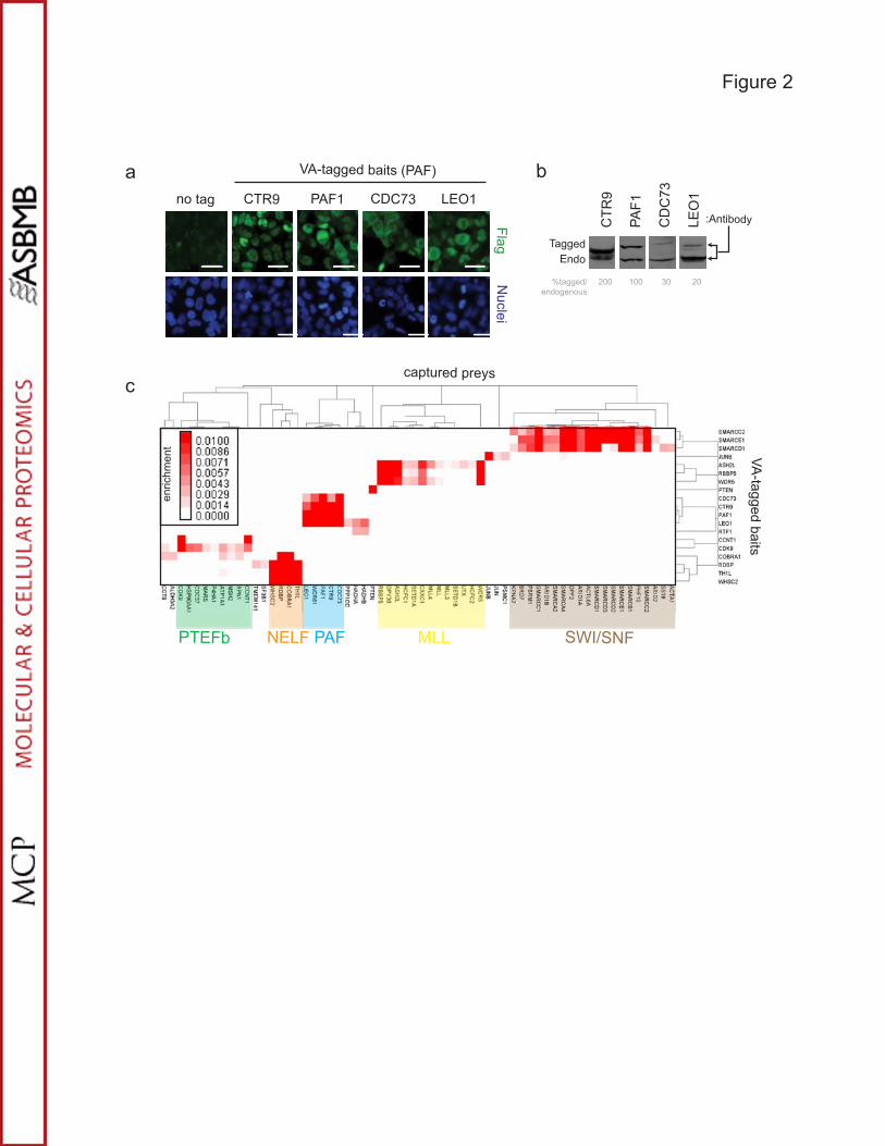
MAPLE is an effective system for identifying specific PPIs and protein complexes. To examine
an entire complex by reciprocal tagging and validate MAPLE for the characterization of human
complexes, we tested the evolutionarily conserved RNA polymerase II-associated factor or PAF
complex as it has been previously purified from human cells and serves as a good control to
assess our MAPLE workflow [33, 34]. The PAF complex is involved in mediating efficient
transcription elongation by RNA polymerase II, mRNA quality control and chromatin modification
that is coupled to transcription elongation [35]. The human and yeast PAF complexes contain
PAF1, CDC73, CTR9 and LEO1, although the human complex additionally contains
SKI8/WDR61 and appears to lack the Rtf1 subunit stably associated with the yeast complex at
least in some cell types [33, 34, 36]. Each of the subunits of the PAF core complex – PAF1,
17
CDC73, CTR9 and LEO1 – and RTF1 were VA-tagged and subjected to MAPLE followed by
LC-MS/MS to identify a high confidence network of reciprocal protein interactions [18].
Importantly, all of these tagged baits were localized to previously reported subcellular
compartments, migrated at their predicted molecular weights and were present at levels
comparable to or below the corresponding endogenous proteins (Figures 2a,b). Baits were
purified on anti-Flag and nickel resins and analyzed either by SDS-PAGE followed by silver
staining or by trypsin digestion and tandem mass spectrometry to identify potential interacting
protein partners. Untagged CDC73 and VA-tagged eGFP were used as negative controls to
generate nonspecific background profiles necessary for filtering out common contaminants.
Each bait protein was purified from three independent cultures and analyzed twice by data-
dependent LC-MS/MS producing a total of six sample runs per bait.
In order to score protein-protein interactions we considered reproducibility and
background and an enrichment score was determined using a combination of protein length
normalized spectral counts and filtering criteria (see below and Methods). The resulting values
were scaled between 0 and 0.01 where 0 represents no preys were detected and values over
0.005 were deemed highly significant. The results show that all core members of the human
PAF complex consistently co-purified with each of the PAF baits with high enrichment scores
(Figure 2c, Supplemental Figure 5 and Supplemental Tables 1 and 2). Consistent with the
observation by Zhu et al [34], SKI8/WDR61 consistently co-purified with all core members of the
PAF complex including PAF1, LEO1, CTR9 and CDC73 and served as a good positive control
for our scoring schema and the overall workflow (Figure 2c). Also as expected [34], none of the
PAF core components were observed in RTF1 affinity purifications (Supplemental Tables 1 and
2), nor were they observed in purifications from cells expressing VA-tagged eGFP or untagged
CDC73. However, several preys were weakly captured in common between RTF1 and two
PAF-related baits, LEO1 and PAF1, including C17orf79, an uncharacterized human open
reading frame, suggesting that there may be some association among these proteins in human

18
cells (Supplemental Tables 1 and 2). Overall, the MAPLE workflow permitted the efficient
identification of authentic protein complexes in human cells and their rapid validation by
reciprocal tagging.
Systematic analysis of protein complexes involved in transcription and chromatin modification.
As further validation of the MAPLE workflow, systematic examination of protein complexes
involved in various aspects of transcription was performed. The reciprocal tagging strategy used
for the PAF complex was again applied to several previously documented multi-protein
complexes linked to positive and negative regulation of transcription elongation and chromatin
remodelling, including the PTEF-b [37], NELF [38, 39], MLL [40, 41] and SWI/SNF [42]
complexes (Figure 2c, Supplemental Figure 5 and Supplemental Tables 1 and 2). A total of 17
proteins representing subunits of the PAF complex and four other complexes as well as PTEN
and JUNB were built as baits. Each stably expressing cell line was analyzed by immunoblot and
immunofluorescence to confirm that each tagged protein migrated at its predicted molecular
weight and localized to the correct subcellular compartment. Each of the baits was purified from
HEK293 cells and its putative interaction partners identified by LC-MS/MS as described above.
For the NELF complex, all four members of the complex (COBRA1, WHSC2, TH1L and
RDBP) were readily detected as baits and consistently interacted with each other as preys in
reciprocal affinity purifications except that TH1L and RDBP did not co-purify when COBRA1 was
used as bait (Figure 2c). This suggested that the tag on COBRA1 interfered with its association
with TH1L and RDBP and highlighted the value of performing reciprocal experiments with
MAPLE to maximize subunit coverage for multi-protein complexes.
The P-TEFb complex, containing the cyclin-dependent kinase CDK9 and its cyclin
partner [37], also performed well in reciprocal purifications (Figure 2c). In addition, CDK9 affinity
purifications revealed novel potential interactors with obvious functional consequences. One
was the CDC37 cochaperone that promotes the association of HSP90 with its protein kinase
subset of client proteins to maintain their stability and signalling functions [43]. CDC37,
19
HSP90AB1 and HSP90AA2 all co-purified with CDK9 as bait suggesting that CDK9 could be
another client of the CDC37-HSP90 complex.
Likewise, the three members of the MLL (mixed lineage leukemia) histone
methyltransferase complex that served as baits (ASH2L, WDR5 and RBBP5) reciprocally co-
purified with each other (Figure 2c). The MLL proto-oncogene is a recurrent site of genetic
rearrangements in acute leukemias [40]. The MLL gene is the founding member of the
mammalian SET family of histone lysine methyltransferases that are responsible for regulating
gene expression patterns during development [40]. The evolutionarily conserved protein DPY30
also co-purified with each of these baits, consistent with previous evidence that it forms a
subcomplex with the ASH2L, RBBP5 and WDR5 proteins that are shared by all human Set1-like
histone methyltransferase complexes [44, 45]. CXXC1, a protein that recognizes CpG
sequences, also co-purified with ASH2L, WDR5 and RBBP5 and is also known to interact with
the Set1 histone H3K4-specific methyltransferases in the regulation of MLL target genes [46].
Consistent with the notion that ASH2L, WDR5 and RBBP5 form a subcomplex consistently
present in MLL complexes, MAPLE purifications revealed interactions with other members of
the Set1/COMPASS and MLL complexes, including SETD1A, SETD1B, HCFC1, HCFC2,
UTX/KDM6A, MLL, MLL3, and MLL4 (Figure 2c). Further investigation into the nature of these
interactions could help refine the composition of these complexes.
The last complex examined by MAPLE in HEK293 cells was the highly conserved
SWI/SNF chromatin remodelling complex, composed of seven core subunits and one of three
different catalytic subunits (BRG1/BAF, BRM/BAF or PBAF) [42]. Three subunits common to
BRG1/BAF, BRM/BAF and PBAF, namely SMARCC2, SMARCE1 and SMARCD1, were
subjected to the MAPLE workflow and affinity purified proteins were identified by LC-MS/MS.
Affinity capture of all known SWI/SNF subunits was achieved for all three SWI/SNF baits (Figure
2c) and some potential novel interactions were identified as well (Figure 2c and Supplemental
Tables 1 and 2). For example, DPF2 was identified with all three SWI/SNF baits (Figure 2c).
20
DPF2, also known as REQuiem or UBID4, is a member of the d4 domain family, characterized
by a zinc finger-like structural motif, and may function as a transcription factor important for the
apoptotic response [47]. This suggests that DPF2 may regulate the role of SWI/SNF during
apoptosis.
MAPLE synopsis. In our initial assessment of 19 bait proteins by MAPLE, 1916 prey proteins
were identified through LC-MS/MS with one or more spectral counts with a confidence of 99%
(corresponding to an FDR=0.01). Enrichment scores were calculated for each potential prey
yielding a total of 62 high-confidence preys representing 148 interactions (see Supplemental
Figure 3 for filtering criteria). Unsupervised hierarchical clustering of the baits and high-
confidence preys indicates that the known subunits of the various complexes share a high
measure of similarity and cluster together (Figure 2c). This representation is also a good
visualization tool to identify baits that may interact non-specifically with multiple preys. As
described above, our results show good concordance with what has been published in the
literature and some overlap with database resources like CORUM [[48] and see Supplemental
Figure 4]. Based on these analyses, we conclude that using MAPLE for reciprocal tagging of
known components of large complexes tends to capture the biological diversity of each
complex. To examine whether the MAPLE workflow could be applied in a similar fashion to
identify protein interactions by AP-MS in a more difficult cell system, we turned to mouse
embryonic stem cells.
Application of MAPLE to reprogramming factors. The Oct4, Sox2, Klf4 and c-Myc transcription
factors have been shown to cooperatively induce pluripotency in a variety of mouse and human
cell types. Klf4, also known as gut-enriched Kruppel-like factor (Gklf), acts as a transcriptional
activator or repressor depending on the promoter context and/or cooperation with other
transcription factors [49]. Klf4 has more recently been shown to cooperate with Oct4, Sox2 and
c-Myc to induce pluripotency in a variety of mouse and human cell types [50, 51]. To examine
the utility of MAPLE for investigating protein complexes linked to pluripotency in primary
21
embryonic stem cells, we used the R1 line of mouse ES cells to derive a cell line stably
expressing N-terminal VA-tagged Klf4 (Figure 3a). Compared to endogenous protein levels, VA-
Klf4 was expressed at levels comparable to its endogenous counterpart (Figure 3a). VA tagged
Klf4 was expressed in the nuclei (Figure 3b), and the VA-tagged expressing stable R1 cell line
maintained ES cell morphology and ES cell specific factors (data not shown). Single Flag
purifications and Streptactin-Flag purifications were performed in parallel followed by LC-MS/MS
to identify candidate interacting partners.
Several novel Klf4 protein interactors were identified, including the catalytic subunits,
Smarca4/Brg1 and Smarca2/Brm, of the Swi/Snf chromatin remodelling complex (Supplemental
Table 3 and Supplemental Figure 6). Using a co-immunoprecipitation assay, the interaction
between the Klf4 bait and endogenous Smarca4/Brg1 was validated in mouse embryonic stem
cells (Figure 3c).The interaction between Klf4 and Smarca2/Brm was validated by co-
immunoprecipitation in HEK293 cells (Figure 3d). In addition, the c-Myc bait was able to co-
immunoprecipitate endogenous Smarca4/Brg1 in mouse ES cells (Figure 3c), validating a Klf4-
Smarca4/Brg1 interaction previously reported in a high-throughput AP-MS study in human
cancer cells [17]. Taken together, these data indicate that Klf4 interacts, directly or indirectly,
with the catalytic subunits Smarca4/Brg1 and Smarca2/Brm of the Swi/Snf chromatin
remodelling complex in pluripotent stem cells.
Requirement for SWI/SNF during induction of pluripotency. A direct link between the Swi/Snf
chromatin remodelling complex and somatic cell reprogramming has not been clearly
established. Since Klf4 is important for induced pluripotency [50, 51] and we observe an
association between Klf4 and the Swi/Snf complex in mouse ES cells, this raises the question
whether the Swi/Snf-mediated chromatin remodelling contributes to the process of induced
pluripotency. In order to test this idea, we used an established reprogramming assay comprising
secondary mouse embryonic fibroblasts (MEFs) that can be converted into induced pluripotent
stem cells by addition of doxycycline [29]. To examine the efficiency and kinetics of
22
reprogramming in the secondary MEF lines 1B and 6C after doxycycline was added to induce
reprogramming, samples were taken to examine expression of pluripotency markers. In parallel,
high resolution video microscopy was performed on 6C cells to observe iPS colony formation
(Supplemental Figure 7). The expression of the pluripotency markers Oct4, Nanog, Eras, Zfp42,
Nrob1, and Fbx15 was evident 7 days following addition of doxycycline, while the expression
Sox2 and Foxd3 was delayed but established by day 15 (Figure 4a and data not shown). The
expression of Smarca4/Brg1 increased ~2-fold over the course of the reprogramming assay
while, surprisingly, Smarca2/Brm expression increased up to ~7-fold (Figure 4b). These
observations suggest that Smarca2/Brm, along with Smarca4/Brg1, may have an important role
in reprogramming/de-differentiation and possibly establishment of pluripotency.
To assess the requirement of Smarca4/Brg1 and Smarca2/Brm during reprogramming,
we perturbed their expression levels in either 6C or 1B cells by using lentiviral-based cDNAs
(over-expression) or short hairpin RNAs (knockdown) (Supplemental Figure 8). Cells were
induced to reprogram and the efficiency scored based on the number of AP-positive colonies
after 7 days. Lentiviral-mediated expression of GFP or the pLKO.1 vector did not significantly
alter the number of AP-positive colonies compared to the untransduced control (data not
shown). In contrast, overexpression of Smarca4/Brg1 or Smarca2/Brm reduced the number of
AP-positive colonies formed from 6C cells by ~2-fold (p<0.05) and the number of AP-positive
colonies formed from 1B cells by ~7-fold (p<0.01)(Figure 4c). Furthermore, knockdown of
Smarca4/Brg1 or Smarca2/Brm with either of two independent shRNAs reduced the number of
AP-positive colonies formed from 6C cells by ~2-fold (p<0.02) (Figure 6d). Taken together,
these results indicate that the catalytic subunits of the Swi/Snf chromatin remodelling complex,
likely in association with Klf4, are important for somatic cell reprogramming.

23
DISCUSSION
The goal of this study was to design a system to facilitate identification of protein
complexes in a wide variety of mammalian cell types. To this end, we developed a mammalian
affinity purification and lentiviral-based expression system (MAPLE for short) and coupled it to
tandem mass spectrometry to identify protein complexes. MAPLE works efficiently with most
cell types, thereby granting access to cell types that may better recapitulate the natural
environment of a protein complex. This could be particularly important in cells and tissues that
undergo substantive epigenetic regulation such as stem cells. MAPLE is based on a custom
built lentiviral plasmid that is GatewayTM compatible and relies on commercially available cDNA
libraries. A unique and versatile affinity tag (i.e. VA) was created to accommodate different
purification schemes and to contain a novel yeast proteotypic peptide (ie. beacon) that acts as a
reference for the bait during affinity purifications. As such, the beacon serves as a “digital
Western”.
A major disadvantage of mammalian AP-MS approaches is the amount of starting
material (i.e. cells) required to achieve high protein purification yields. Although the original TAP
tag [52] is still routinely in use in mammalian cells for protein complex characterization, other
affinity tags like the GS-tag have helped reduce the amount of starting material required by 10-
100 fold [53]. With the MAPLE system, we typically used ~5e7 cells for starting material,
similar to Burckstummer et al [53]. The VA tag is compatible with single, dual or even triple
affinity purification schemes so the choice of method will impact how much starting material is
required. That is, more purification steps taken require more starting material. The downfall of
tandem purification methods is that transient or weak interactions are generally lost.
Furthermore, we introduced a constitutive promoter in our MAPLE system and
demonstrated that the CMV and PGK promoters driving cDNAs in HEK 293 and R1 cells,
respectively, resulted in bait expression levels comparable to endogenous. However, some
genes or cells may be sensitive to bait overexpression, resulting in false outcomes. Similar to

24
other studies, we demonstrate that MAPLE can be adapted to be tetracycline-regulatable and
inducible (Supplemental Figure 9). Although determining an optimal level of bait expression
would be ideal, it is not practical for a systematic and high-throughput approach.
To benchmark the MAPLE workflow in cells that are easy to manipulate, we examined
19 bait proteins involved in different aspects of chromatin biology or disease in HEK293 cells.
Except for the PTEN phosphatase and the JUNB proto-oncogene, each of the baits was part of
an evolutionarily conserved multi-protein complex. PAF1, CDC73, CTR9 and LEO1 represent
evolutionarily conserved core components of the human PAF complex involved in transcription
elongation. CDK9 and CCNT1 constitute the positive transcription elongation factor or P-TEFb
complex. COBRA1, WHSC2, TH1L and RDBP represent the core components of the negative
elongation factor or NELF complex. WDR5, RBBP5, and ASH2L make up a key subcomplex
that likely regulates all the SET-like histone methyltransferase complexes including SET1 and
all MLL proteins. Lastly, SMARCD1, SMARCC2, and SMARCE1 are common components of all
three characterized SWI/SNF chromatin remodelling complexes including BRG/BAF, BRM/BAF
and PBAF. Importantly, reciprocal tagging validated all the known protein complexes in our
benchmarking study and thus represents a good strategy to capture new complex members.
The application of MAPLE and LC-MS/MS across a broad range of baits in an unbiased manner
will yield a high density protein interaction dataset with a wealth of new biological information.
A key feature of the MAPLE system is that lentiviruses are efficient at stably transducing
most cell types. By transducing cells at a low multiplicity of infection, one can limit the number of
integrants per cell and keep cell-to-cell expression levels more constant. The fact that the
resulting populations of cells are heterogeneous due to variability in integration is advantageous
as this mitigates the possibility of bias due to clonal expansion. To put this to the test, we
generated stable mouse R1 ES cells expressing VA-tagged Klf4, one of the classical
reprogramming factors that convert somatic cells into induced pluripotent stem cells [51], and
performed AP-MS using lysates from this stable cell line to identify protein interactions. We were

25
able to show that Klf4 interacts with the SWI/SNF chromatin remodelling complexes containing
Brg1/Smarca4 and Brm/Smarca2.
Consistent with this, recent studies have found that ES cells contain a functionally and
structurally specialized chromatin remodelling complex, esBAF, that is critical for the self-
renewal of ES cells and maintenance of the stem cell fate [54-57]. In fact, Crabtree and co-
workers proposed that the surface of esBAF complexes is tailored for interactions with factors
found specifically in ES cells and that through these functional interactions esBAF maintains the
pluripotent chromatin landscape [56]. Using Chip-seq technology, Ho et al were able to
demonstrate that esBAF is enriched at transcription start sites, occupies genes of the core
pluripotency network (ie. Oct4, Sox2 and Nanog), represses developmental genes and opposes
Polycomb complexes by direct repression of subunits of the PRC1 complex [55]. Nevertheless,
a direct role for esBAF or any of the SWI/SNF complexes in reprogramming or de-differentiation
had not been shown.
Since Klf4 has an important role during somatic cell reprogramming to induced
pluripotent stem cells and our data uncovered an interaction between Klf4 and the catalytic
subunits of the SWI/SNF chromatin remodelling complexes, an important remaining question
was to examine the requirement of SWI/SNF chromatin remodelling complexes during somatic
cell reprogramming. Thus, we turned to a model where secondary MEFs can be induced to
pluripotency by the addition of doxycycline [29]. The first clue that somatic cell reprogramming
may have a slightly different SWI/SNF requirement than maintenance of the pluripotent state
[55, 56] came from the observation that Smarca2/Brm expression is induced during
reprogramming/de-differentiation (Figure 4b). This observation may have important
consequences for how SWI/SNF subunits are distributed during reprogramming versus
maintenance of pluripotency or self-renewal. To further investigate this, the onset of the
expression of pluripotency markers was examined in two independent clones (1B and 6C) that
were induced into pluripotency following knockdown or overexpression of SWI/SNF catalytic

26
subunits (i.e. Smarca4/Brg1 or Smarca2/Brm). These results indicate that slight changes in the
concentrations of the catalytic subunits of the esBAF (or any SWI/SNF complex involved in
reprogramming) complex has drastic consequences for reprogramming to the pluripotent state.
The interaction between Klf4 and SWI/SNF complexes may help to establish the pluripotent
transcriptional circuitry.
27
REFERENCES
1. Braun P, Tasan M, Dreze M, Barrios-Rodiles M, Lemmens I, Yu H, Sahalie JM, Murray RR, Roncari L, de Smet AS, et al: An experimentally derived confidence score for binary protein-protein interactions. Nat Methods 2009, 6:91-97.
2. Giot L, Bader JS, Brouwer C, Chaudhuri A, Kuang B, Li Y, Hao YL, Ooi CE, Godwin B, Vitols E, et al: A protein interaction map of Drosophila melanogaster. Science 2003, 302:1727-1736.
3. Ito T, Tashiro K, Muta S, Ozawa R, Chiba T, Nishizawa M, Yamamoto K, Kuhara S, Sakaki Y: Toward a protein-protein interaction map of the budding yeast: A comprehensive system to examine two-hybrid interactions in all possible combinations between the yeast proteins. Proc Natl Acad Sci U S A 2000, 97:1143-1147.
4. Li S, Armstrong CM, Bertin N, Ge H, Milstein S, Boxem M, Vidalain PO, Han JD, Chesneau A, Hao T, et al: A map of the interactome network of the metazoan C. elegans. Science 2004, 303:540-543.
5. Rual JF, Venkatesan K, Hao T, Hirozane-Kishikawa T, Dricot A, Li N, Berriz GF, Gibbons FD, Dreze M, Ayivi-Guedehoussou N, et al: Towards a proteome-scale map of the human protein-protein interaction network. Nature 2005, 437:1173-1178.
6. Stelzl U, Worm U, Lalowski M, Haenig C, Brembeck FH, Goehler H, Stroedicke M, Zenkner M, Schoenherr A, Koeppen S, et al: A human protein-protein interaction network: a resource for annotating the proteome. Cell 2005, 122:957-968.
7. Tarassov K, Messier V, Landry CR, Radinovic S, Serna Molina MM, Shames I, Malitskaya Y, Vogel J, Bussey H, Michnick SW: An in vivo map of the yeast protein interactome. Science 2008, 320:1465-1470.
8. Uetz P, Giot L, Cagney G, Mansfield TA, Judson RS, Knight JR, Lockshon D, Narayan V, Srinivasan M, Pochart P, et al: A comprehensive analysis of protein-protein interactions in Saccharomyces cerevisiae. Nature 2000, 403:623-627.
9. Gingras AC, Gstaiger M, Raught B, Aebersold R: Analysis of protein complexes using mass spectrometry. Nat Rev Mol Cell Biol 2007, 8:645-654.
10. Kocher T, Superti-Furga G: Mass spectrometry-based functional proteomics: from molecular machines to protein networks. Nat Methods 2007, 4:807-815.
11. Butland G, Peregrin-Alvarez JM, Li J, Yang W, Yang X, Canadien V, Starostine A, Richards D, Beattie B, Krogan N, et al: Interaction network containing conserved and essential protein complexes in Escherichia coli. Nature 2005, 433:531-537.
12. Hu P, Janga SC, Babu M, Diaz-Mejia JJ, Butland G, Yang W, Pogoutse O, Guo X, Phanse S, Wong P, et al: Global functional atlas of Escherichia coli encompassing previously uncharacterized proteins. PLoS Biol 2009, 7:e96.
13. Gavin AC, Aloy P, Grandi P, Krause R, Boesche M, Marzioch M, Rau C, Jensen LJ, Bastuck S, Dumpelfeld B, et al: Proteome survey reveals modularity of the yeast cell machinery. Nature 2006, 440:631-636.
14. Gavin AC, Bosche M, Krause R, Grandi P, Marzioch M, Bauer A, Schultz J, Rick JM,Michon AM, Cruciat CM, et al: Functional organization of the yeast proteome by systematic analysis of protein complexes. Nature 2002, 415:141-147.
15. Krogan NJ, Cagney G, Yu H, Zhong G, Guo X, Ignatchenko A, Li J, Pu S, Datta N, Tikuisis AP, et al: Global landscape of protein complexes in the yeast Saccharomyces cerevisiae. Nature 2006, 440:637-643.
16. Collins SR, Kemmeren P, Zhao XC, Greenblatt JF, Spencer F, Holstege FC, Weissman JS, Krogan NJ: Toward a comprehensive atlas of the physical interactome of Saccharomyces cerevisiae. Mol Cell Proteomics 2007, 6:439-450.

28
17. Ewing RM, Chu P, Elisma F, Li H, Taylor P, Climie S, McBroom-Cerajewski L, Robinson MD, O'Connor L, Li M, et al: Large-scale mapping of human protein-protein interactions by mass spectrometry. Mol Syst Biol 2007, 3:89.
18. Glatter T, Wepf A, Aebersold R, Gstaiger M: An integrated workflow for charting the human interaction proteome: insights into the PP2A system. Mol Syst Biol 2009, 5:237.
19. Jeronimo C, Forget D, Bouchard A, Li Q, Chua G, Poitras C, Therien C, Bergeron D, Bourassa S, Greenblatt J, et al: Systematic analysis of the protein interaction network for the human transcription machinery reveals the identity of the 7SK capping enzyme. Mol Cell 2007, 27:262-274.
20. Jeronimo C, Langelier MF, Zeghouf M, Cojocaru M, Bergeron D, Baali D, Forget D, Mnaimneh S, Davierwala AP, Pootoolal J, et al: RPAP1, a novel human RNA polymerase II-associated protein affinity purified with recombinant wild-type and mutated polymerase subunits. Mol Cell Biol 2004, 24:7043-7058.
21. Dull T, Zufferey R, Kelly M, Mandel RJ, Nguyen M, Trono D, Naldini L: A third-generation lentivirus vector with a conditional packaging system. J Virol 1998, 72:8463-8471.
22. Moffat J, Grueneberg DA, Yang X, Kim SY, Kloepfer AM, Hinkle G, Piqani B, Eisenhaure TM, Luo B, Grenier JK, et al: A lentiviral RNAi library for human and mouse genes applied to an arrayed viral high-content screen. Cell 2006, 124:1283-1298.
23. Zufferey R, Nagy D, Mandel RJ, Naldini L, Trono D: Multiply attenuated lentiviral vector achieves efficient gene delivery in vivo. Nat Biotechnol 1997, 15:871-875.
24. Walhout AJ, Temple GF, Brasch MA, Hartley JL, Lorson MA, van den Heuvel S, Vidal M: GATEWAY recombinational cloning: application to the cloning of large numbers of open reading frames or ORFeomes. Methods Enzymol 2000, 328:575-592.
25. Chen GI, Tisayakorn S, Jorgensen C, D'Ambrosio LM, Goudreault M, Gingras AC: PP4R4/KIAA1622 forms a novel stable cytosolic complex with phosphoprotein phosphatase 4. J Biol Chem 2008, 283:29273-29284.
26. Nagy A, Rossant J, Nagy R, Abramow-Newerly W, Roder JC: Derivation of completely cell culture-derived mice from early-passage embryonic stem cells. Proc Natl Acad Sci U S A 1993, 90:8424-8428.
27. Chen GI, Gingras AC: Affinity-purification mass spectrometry (AP-MS) of serine/threonine phosphatases. Methods 2007, 42:298-305.
28. Florens L, Carozza MJ, Swanson SK, Fournier M, Coleman MK, Workman JL, Washburn MP: Analyzing chromatin remodeling complexes using shotgun proteomics and normalized spectral abundance factors. Methods 2006, 40:303-311.
29. Woltjen K, Michael IP, Mohseni P, Desai R, Mileikovsky M, Hamalainen R, Cowling R, Wang W, Liu P, Gertsenstein M, et al: piggyBac transposition reprograms fibroblasts to induced pluripotent stem cells. Nature 2009, 458:766-770.
30. Schmidt TG, Skerra A: The Strep-tag system for one-step purification and high-affinity detection or capturing of proteins. Nat Protoc 2007, 2:1528-1535.
31. Giannone RJ, McDonald WH, Hurst GB, Huang Y, Wu J, Liu Y, Wang Y: Dual-tagging system for the affinity purification of mammalian protein complexes. Biotechniques 2007, 43:296, 298, 300 passim.
32. Wepf A, Glatter T, Schmidt A, Aebersold R, Gstaiger M: Quantitative interaction proteomics using mass spectrometry. Nat Methods 2009, 6:203-205.
33. Rozenblatt-Rosen O, Hughes CM, Nannepaga SJ, Shanmugam KS, Copeland TD, Guszczynski T, Resau JH, Meyerson M: The parafibromin tumor suppressor protein is part of a human Paf1 complex. Mol Cell Biol 2005, 25:612-620.
34. Zhu B, Mandal SS, Pham AD, Zheng Y, Erdjument-Bromage H, Batra SK, Tempst P, Reinberg D: The human PAF complex coordinates transcription with events downstream of RNA synthesis. Genes Dev 2005, 19:1668-1673.
29
35. Chaudhary K, Deb S, Moniaux N, Ponnusamy MP, Batra SK: Human RNA polymerase II-associated factor complex: dysregulation in cancer. Oncogene 2007, 26:7499-7507.
36. Kim J, Guermah M, Roeder RG: The Human PAF1 Complex Acts in Chromatin Transcription Elongation Both Independently and Cooperatively with SII/TFIIS. Cell,140:491-503.
37. Peterlin BM, Price DH: Controlling the elongation phase of transcription with P-TEFb.Mol Cell 2006, 23:297-305.
38. Narita T, Yamaguchi Y, Yano K, Sugimoto S, Chanarat S, Wada T, Kim DK, Hasegawa J, Omori M, Inukai N, et al: Human transcription elongation factor NELF: identification of novel subunits and reconstitution of the functionally active complex. Mol Cell Biol 2003, 23:1863-1873.
39. Narita T, Yung TM, Yamamoto J, Tsuboi Y, Tanabe H, Tanaka K, Yamaguchi Y, Handa H: NELF interacts with CBC and participates in 3' end processing of replication-dependent histone mRNAs. Mol Cell 2007, 26:349-365.
40. Liedtke M, Cleary ML: Therapeutic targeting of MLL. Blood 2009, 113:6061-6068.41. Patel A, Dharmarajan V, Vought VE, Cosgrove MS: On the mechanism of multiple lysine
methylation by the human mixed lineage leukemia protein-1 (MLL1) core complex. J Biol Chem 2009, 284:24242-24256.
42. Reisman D, Glaros S, Thompson EA: The SWI/SNF complex and cancer. Oncogene 2009, 28:1653-1668.
43. Smith JR, Clarke PA, de Billy E, Workman P: Silencing the cochaperone CDC37 destabilizes kinase clients and sensitizes cancer cells to HSP90 inhibitors. Oncogene 2009, 28:157-169.
44. Cho YW, Hong T, Hong S, Guo H, Yu H, Kim D, Guszczynski T, Dressler GR, Copeland TD, Kalkum M, Ge K: PTIP associates with MLL3- and MLL4-containing histone H3 lysine 4 methyltransferase complex. J Biol Chem 2007, 282:20395-20406.
45. Wang X, Lou Z, Dong X, Yang W, Peng Y, Yin B, Gong Y, Yuan J, Zhou W, Bartlam M, et al: Crystal structure of the C-terminal domain of human DPY-30-like protein: A component of the histone methyltransferase complex. J Mol Biol 2009, 390:530-537.
46. Lee JH, Skalnik DG: CpG-binding protein (CXXC finger protein 1) is a component of the mammalian Set1 histone H3-Lys4 methyltransferase complex, the analogue of the yeast Set1/COMPASS complex. J Biol Chem 2005, 280:41725-41731.
47. Wong DC, Wong KT, Nissom PM, Heng CK, Yap MG: Targeting early apoptotic genes in batch and fed-batch CHO cell cultures. Biotechnol Bioeng 2006, 95:350-361.
48. Ruepp A, Waegele B, Lechner M, Brauner B, Dunger-Kaltenbach I, Fobo G, FrishmanG, Montrone C, Mewes HW: CORUM: the comprehensive resource of mammalian protein complexes--2009. Nucleic Acids Res, 38:D497-501.
49. Rowland BD, Peeper DS: KLF4, p21 and context-dependent opposing forces in cancer. Nat Rev Cancer 2006, 6:11-23.
50. Takahashi K, Tanabe K, Ohnuki M, Narita M, Ichisaka T, Tomoda K, Yamanaka S: Induction of pluripotent stem cells from adult human fibroblasts by defined factors. Cell 2007, 131:861-872.
51. Takahashi K, Yamanaka S: Induction of pluripotent stem cells from mouse embryonic and adult fibroblast cultures by defined factors. Cell 2006, 126:663-676.
52. Rigaut G, Shevchenko A, Rutz B, Wilm M, Mann M, Seraphin B: A generic protein purification method for protein complex characterization and proteome exploration. Nat Biotechnol 1999, 17:1030-1032.
53. Burckstummer T, Bennett KL, Preradovic A, Schutze G, Hantschel O, Superti-Furga G, Bauch A: An efficient tandem affinity purification procedure for interaction proteomics in mammalian cells. Nat Methods 2006, 3:1013-1019.

30
54. Blagoev B, Kratchmarova I, Ong SE, Nielsen M, Foster LJ, Mann M: A proteomics strategy to elucidate functional protein-protein interactions applied to EGF signaling. Nat Biotechnol 2003, 21:315-318.
55. Ho L, Jothi R, Ronan JL, Cui K, Zhao K, Crabtree GR: An embryonic stem cell chromatin remodeling complex, esBAF, is an essential component of the core pluripotency transcriptional network. Proc Natl Acad Sci U S A 2009, 106:5187-5191.
56. Ho L, Ronan JL, Wu J, Staahl BT, Chen L, Kuo A, Lessard J, Nesvizhskii AI, Ranish J, Crabtree GR: An embryonic stem cell chromatin remodeling complex, esBAF, is essential for embryonic stem cell self-renewal and pluripotency. Proc Natl Acad Sci U S A 2009, 106:5181-5186.
57. Kidder BL, Palmer S, Knott JG: SWI/SNF-Brg1 regulates self-renewal and occupies core pluripotency-related genes in embryonic stem cells. Stem Cells 2009, 27:317-328.

31
ACKNOWLEDGMENTS
We thank Dr. Andras Nagy for kindly providing the R1 mouse ES cell line, the 1B and 6C
secondary MEFs and the pDONR221-Klf4 construct and Dr. Tony Pawson for the pGateway
vectors. We thank Drs. Anthony Gramolini, Stephane Angers, Janet Rossant and Brian Cox for
helpful discussions. This project was supported by grants from the Canadian Institutes of
Health Research to JM (178975), JG and AE and, in part, by a Network Centers of Excellence
Stem Cell Network grant to JM. ABM is supported by an NSERC Canada Graduate
Scholarship. ZN is supported by a CIHR post-doctoral fellowship.
32
FIGURE LEGENDS
Figure 1. Mammalian Affinity Purification and Lentiviral Expression System (MAPLE). (a)
Schematic representation of the MAPLE lentiviral transfer vector, which includes a mammalian
promoter upstream of the versatile affinity (VA) tag consisting of 3�Flag separated from 6�His
and the StrepIII tag by dual tobacco etch virus (TEV) protease cleavage sites (N-terminus VA
tag shown) positioned in-frame with the GatewayTM cassette. The MAPLE vector also contains
the pac gene driven by the constitutive human phosphoglycerate kinase (hPGK) promoter. (b)
Flag, His and streptactin epitopes were detected by Western Blot analyses of both N- and C-
terminus VA-tagged eGFP expressed in HEK 293 cells. (c) The Flag epitope of the VA tag was
used for immunofluorescence of an N-terminus VA-tagged GFP expressed in HEK 293 cells.
Nuclei were stained with Hoechst. Bars are 10�m. (d) N-terminus VA-tagged GFP from HEK
293 stables is captured by Flag, nickel and streptactin resins, but not by Protein G resin. (e)
Chromatograms showing the total ion current, extracted ion current for the beacon (m/z 905.4)
and extracted ion current for the AQUA-labelled beacon peptide (m/z 909.4). (f) MS/MS spectra
of unlabelled beacon at a chromatographic retention time of 63.01 minutes and AQUA-labelled
beacon at a chromatographic retention time of 63.05 minutes. Y-ions are indicated in red and b-
ions shown in blue.
Figure 2. MAPLE can reproducibly identify members of known protein complexes. (a)
Subcellular localization by indirect Flag immunofluorescence of VA-tagged human PAF complex
subunits used as baits. Nuclei were stained with Hoechst. Bars are 10�m. (b) Comparison of
expression levels of VA-tagged human PAF complex subunits and their endogenous
counterparts by Western Blot analyses. (c) Reciprocal MAPLE-LC-MS/MS of members of the
human PAF, NELF, P-TEFb, MLL and SWI/SNF complexes identifies known core complex
members, as well as potential novel interactions as determined by enrichment scores that
indicate significance. Shown is a heatmap generated by unsupervised hierarchical clustering of

33
19 bait proteins spanning 5 protein complexes that were subjected to MAPLE-LC-MS/MS
including the human PAF, NELF, P-TEFb, MLL and SWI/SNF complexes.
Figure 3. Klf4 interacts with SWI/SNF complexes. (a) Comparison of VA-tagged Klf4
expression level with its endogenous counterpart in mouse R1 embryonic stem cells as
determined by Western blot. (b) Subcellular localization determined by indirect Flag
immunofluorescence of VA-tagged Klf4. Nuclei were detected using Hoechst. 20X
magnification. Bars are 5�m. (c) VA-Sox2, Klf4 and c-Myc from mouse R1 embryonic stem cells
were immunoprecipitated using Flag resin, separated by SDS-PAGE and transferred to PVDF
membranes, after which endogenous Brg1 was detected by immunoblotting. (d) HEK 293 cells
co-transfected with vectors encoding VA-Sox2 or Klf4, as well as VM-Smarca2/Brm were
analyzed by IP/Western Blot as in (c).
Figure 4. SWI/SNF requirement during induction of pluripotency. (a) Endogenous Oct4,
Sox2 and Nanog RNA levels were monitored by qRT-PCR at days 1, 3, 5, 7 and 14 after the
addition of doxycycline to 6C secondary MEF cells. (b) Klf4, Brg1 and Brm RNA levels were
monitored in the same manner as (a). (c) AP-stained colonies in doxycycline-induced 1B or 6C
secondary MEFs after 7 days following expression of Brg1 or Brm using MAPLE. (d) Results of
knocking down Klf4, Brg1 or Brm using lentiviral-based shRNAs in secondary 6C MEF cells
following forced expression Oct4, Sox2, Klf4 and c-Myc by addition of doxycycline (n=3). The
number of AP-positive colonies were manually counted by three independent observers and
results averaged.

Figure 1
d
Promoter VA Gateway cassette hPGKprRRE
� 5’LTR 3’sinLTRpUC ori AmpR f1 ori
pac
3xFlag 6xHis StrepIII Beacon2xTEVVA tag
a
c
Streptactin
His
Flag
no ta
gIB:VA-G
FP
GFP-VA
nuclei Flag
overlay GFP
Inpu
t
Was
h
Bea
ds
Was
h
Bea
ds
Was
h
Bea
ds
Was
h
Bea
ds
Protein G Flag His StrepIII
GFP
IB:
VA-GFP0% 95% 54% 70%recovery:
e f
b
a
c
Figure 2
EndoTagged
:AntibodyCTR
9
PAF1
CD
C73
LEO
1
%tagged/endogenous
200 100 30 20
VA-tagged baits (PAF)
FlagN
uclei
no tag
bCTR9 PAF1 CDC73 LEO1
captured preys
VA-tagged baits
PAF SWI/SNFMLLNELFPTEFb
enric
hmen
t
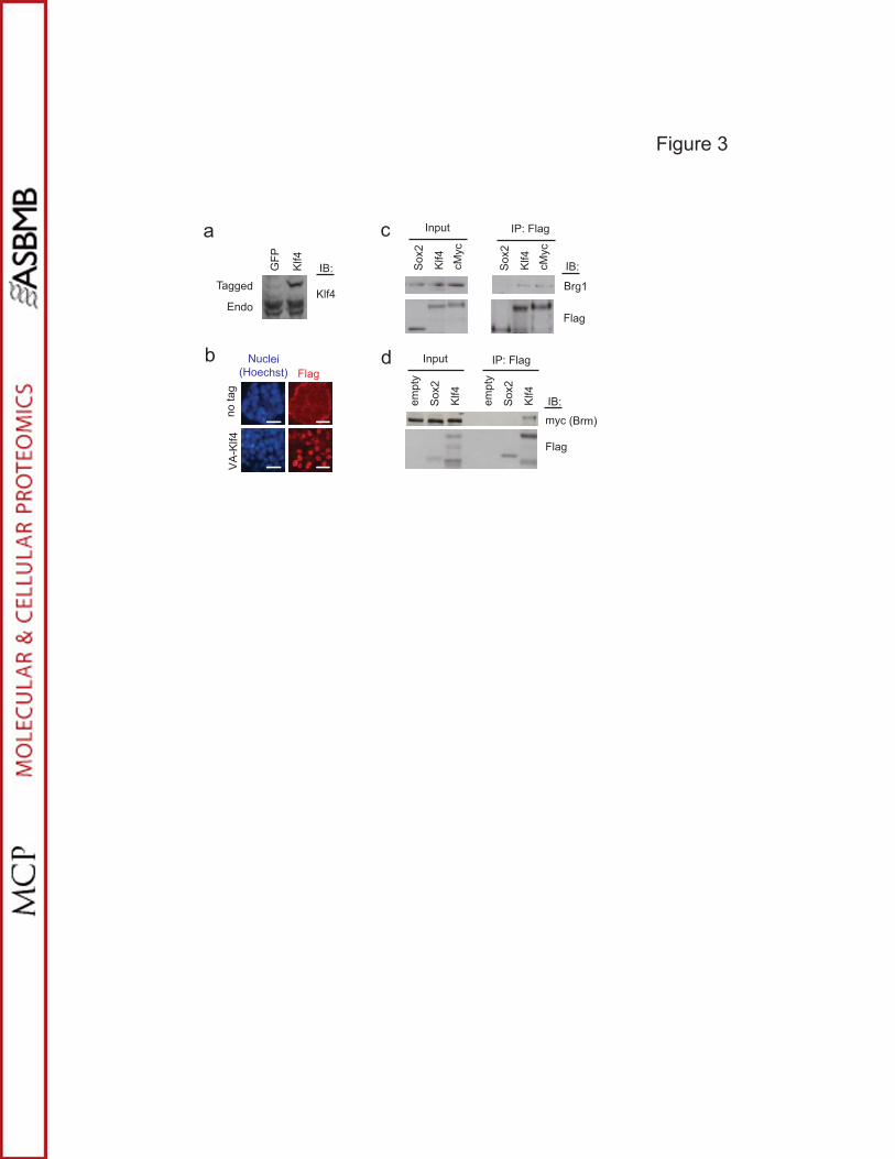
Figure 3
a
b
PFG
4flK IB:
Klf4Endo
Tagged
VA
-Klf4
no ta
g
FlagNuclei
(Hoechst)
c
Sox
2 4flK cM
yc
Sox
2 4flK cM
yc
ytpme Sox
2 4flK
ytpme Sox
2 4flK
Flag
Flag
IB:
IB:
myc (Brm)
Brg1
IP: Flag
IP: Flag
Input
Input
d

p<0.05
p<0.05p<0.01
p<0.01
0
20
40
60
80
100
120
AP
colo
nies
050
100150200250300350400450
Smarca4 Smarca2GFP
6C c
ells
1B c
ells
a
c d
shSmarca4-1
shSmarca2-1
shSmarca4-2
shSmarca2-2pLKO.1
shKlf4
0
10
20
30
40
50
60
70
AP
colo
nies
p<0.02
Figure 4
0
1
2
3
4
5
6
7
1 3 5 7 14
Fold
-indu
ctio
nDays in doxycycline
Smarca4
Smarca2
Klf4
c-Myc
05
10152025
1 3 5 7 14
Fold
-indu
ctio
n
Days in dox
endo Oct4
020406080
100120
1 3 5 7 14
Fold
-indu
ctio
n
endo Sox2
0
500
1000
1500
1 3 5 7 14
Fold
-indu
ctio
n
Nanog
b
1B cells6C cells
6C cells