Reprogramming of human somatic cells to pluripotency with defined factors
7
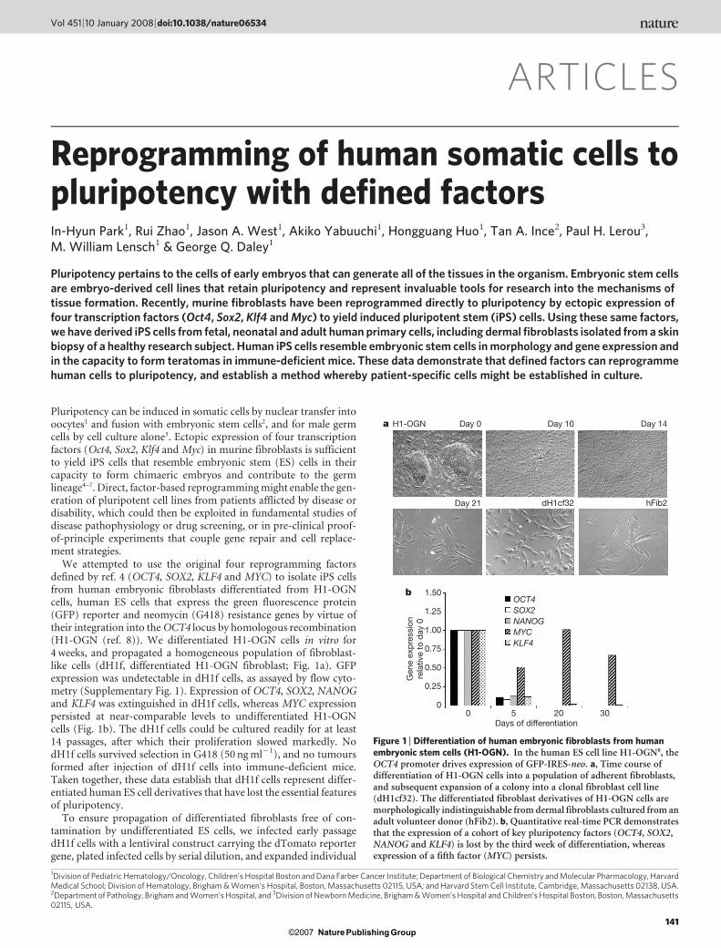
ARTICLES Reprogramming of human somatic cells to pluripotency with defined factors In-Hyun Park 1 , Rui Zhao 1 , Jason A. West 1 , Akiko Yabuuchi 1 , Hongguang Huo 1 , Tan A. Ince 2 , Paul H. Lerou 3 , M. William Lensch 1 & George Q. Daley 1 Pluripotency pertains to the cells of early embryos that can generate all of the tissues in the organism. Embryonic stem cells are embryo-derived cell lines that retain pluripotency and represent invaluable tools for research into the mechanisms of tissue formation. Recently, murine fibroblasts have been reprogrammed directly to pluripotency by ectopic expression of four transcription factors (Oct4, Sox2, Klf4 and Myc) to yield induced pluripotent stem (iPS) cells. Using these same factors, we have derived iPS cells from fetal, neonatal and adult human primary cells, including dermal fibroblasts isolated from a skin biopsy of a healthy research subject. Human iPS cells resemble embryonic stem cells in morphology and gene expression and in the capacity to form teratomas in immune-deficient mice. These data demonstrate that defined factors can reprogramme human cells to pluripotency, and establish a method whereby patient-specific cells might be established in culture. Pluripotency can be induced in somatic cells by nuclear transfer into oocytes 1 and fusion with embryonic stem cells 2 , and for male germ cells by cell culture alone 3 . Ectopic expression of four transcription factors (Oct4, Sox2, Klf4 and Myc) in murine fibroblasts is sufficient to yield iPS cells that resemble embryonic stem (ES) cells in their capacity to form chimaeric embryos and contribute to the germ lineage 4–7 . Direct, factor-based reprogramming might enable the gen- eration of pluripotent cell lines from patients afflicted by disease or disability, which could then be exploited in fundamental studies of disease pathophysiology or drug screening, or in pre-clinical proof- of-principle experiments that couple gene repair and cell replace- ment strategies. We attempted to use the original four reprogramming factors defined by ref. 4 (OCT4, SOX2, KLF4 and MYC) to isolate iPS cells from human embryonic fibroblasts differentiated from H1-OGN cells, human ES cells that express the green fluorescence protein (GFP) reporter and neomycin (G418) resistance genes by virtue of their integration into the OCT4 locus by homologous recombination (H1-OGN (ref. 8)). We differentiated H1-OGN cells in vitro for 4 weeks, and propagated a homogeneous population of fibroblast- like cells (dH1f, differentiated H1-OGN fibroblast; Fig. 1a). GFP expression was undetectable in dH1f cells, as assayed by flow cyto- metry (Supplementary Fig. 1). Expression of OCT4, SOX2, NANOG and KLF4 was extinguished in dH1f cells, whereas MYC expression persisted at near-comparable levels to undifferentiated H1-OGN cells (Fig. 1b). The dH1f cells could be cultured readily for at least 14 passages, after which their proliferation slowed markedly. No dH1f cells survived selection in G418 (50 ng ml 21 ), and no tumours formed after injection of dH1f cells into immune-deficient mice. Taken together, these data establish that dH1f cells represent differ- entiated human ES cell derivatives that have lost the essential features of pluripotency. To ensure propagation of differentiated fibroblasts free of con- tamination by undifferentiated ES cells, we infected early passage dH1f cells with a lentiviral construct carrying the dTomato reporter gene, plated infected cells by serial dilution, and expanded individual 1 Division of Pediatric Hematology/Oncology, Children’s Hospital Boston and Dana Farber Cancer Institute; Department of Biological Chemistry and Molecular Pharmacology, Harvard Medical School; Division of Hematology, Brigham & Women’s Hospital, Boston, Massachusetts 02115, USA; and Harvard Stem Cell Institute, Cambridge, Massachusetts 02138, USA. 2 Department of Pathology, Brigham and Women’s Hospital, and 3 Division of Newborn Medicine, Brigham & Women’s Hospital and Children’s Hospital Boston, Boston, Massachusetts 02115, USA. Gene expression relative to day 0 Days of differentiation Day 21 1.50 1.25 1.00 0.75 0.50 0.25 0 KLF4 MYC NANOG SOX2 OCT4 0 5 20 30 dH1cf32 hFib2 b Day 0 Day 10 Day 14 a H1-OGN Figure 1 | Differentiation of human embryonic fibroblasts from human embryonic stem cells (H1-OGN). In the human ES cell line H1-OGN 8 , the OCT4 promoter drives expression of GFP-IRES-neo. a, Time course of differentiation of H1-OGN cells into a population of adherent fibroblasts, and subsequent expansion of a colony into a clonal fibroblast cell line (dH1cf32). The differentiated fibroblast derivatives of H1-OGN cells are morphologically indistinguishable from dermal fibroblasts cultured from an adult volunteer donor (hFib2). b, Quantitative real-time PCR demonstrates that the expression of a cohort of key pluripotency factors (OCT4, SOX2, NANOG and KLF4) is lost by the third week of differentiation, whereas expression of a fifth factor (MYC) persists. Vol 451 | 10 January 2008 | doi:10.1038/nature06534 141 Nature ©2007 Publishing Group
-
Upload
independent -
Category
Documents
-
view
1 -
download
0
Transcript of Reprogramming of human somatic cells to pluripotency with defined factors
ARTICLES
Reprogramming of human somatic cells topluripotency with defined factorsIn-Hyun Park1, Rui Zhao1, Jason A. West1, Akiko Yabuuchi1, Hongguang Huo1, Tan A. Ince2, Paul H. Lerou3,M. William Lensch1 & George Q. Daley1
Pluripotency pertains to the cells of early embryos that can generate all of the tissues in the organism. Embryonic stem cellsare embryo-derived cell lines that retain pluripotency and represent invaluable tools for research into the mechanisms oftissue formation. Recently, murine fibroblasts have been reprogrammed directly to pluripotency by ectopic expression offour transcription factors (Oct4, Sox2, Klf4 and Myc) to yield induced pluripotent stem (iPS) cells. Using these same factors,we have derived iPS cells from fetal, neonatal and adult human primary cells, including dermal fibroblasts isolated from a skinbiopsy of a healthy research subject. Human iPS cells resemble embryonic stem cells in morphology and gene expression andin the capacity to form teratomas in immune-deficient mice. These data demonstrate that defined factors can reprogrammehuman cells to pluripotency, and establish a method whereby patient-specific cells might be established in culture.
Pluripotency can be induced in somatic cells by nuclear transfer intooocytes1 and fusion with embryonic stem cells2, and for male germcells by cell culture alone3. Ectopic expression of four transcriptionfactors (Oct4, Sox2, Klf4 and Myc) in murine fibroblasts is sufficientto yield iPS cells that resemble embryonic stem (ES) cells in theircapacity to form chimaeric embryos and contribute to the germlineage4–7. Direct, factor-based reprogramming might enable the gen-eration of pluripotent cell lines from patients afflicted by disease ordisability, which could then be exploited in fundamental studies ofdisease pathophysiology or drug screening, or in pre-clinical proof-of-principle experiments that couple gene repair and cell replace-ment strategies.
We attempted to use the original four reprogramming factorsdefined by ref. 4 (OCT4, SOX2, KLF4 and MYC) to isolate iPS cellsfrom human embryonic fibroblasts differentiated from H1-OGNcells, human ES cells that express the green fluorescence protein(GFP) reporter and neomycin (G418) resistance genes by virtue oftheir integration into the OCT4 locus by homologous recombination(H1-OGN (ref. 8)). We differentiated H1-OGN cells in vitro for4 weeks, and propagated a homogeneous population of fibroblast-like cells (dH1f, differentiated H1-OGN fibroblast; Fig. 1a). GFPexpression was undetectable in dH1f cells, as assayed by flow cyto-metry (Supplementary Fig. 1). Expression of OCT4, SOX2, NANOGand KLF4 was extinguished in dH1f cells, whereas MYC expressionpersisted at near-comparable levels to undifferentiated H1-OGNcells (Fig. 1b). The dH1f cells could be cultured readily for at least14 passages, after which their proliferation slowed markedly. NodH1f cells survived selection in G418 (50 ng ml21), and no tumoursformed after injection of dH1f cells into immune-deficient mice.Taken together, these data establish that dH1f cells represent differ-entiated human ES cell derivatives that have lost the essential featuresof pluripotency.
To ensure propagation of differentiated fibroblasts free of con-tamination by undifferentiated ES cells, we infected early passagedH1f cells with a lentiviral construct carrying the dTomato reportergene, plated infected cells by serial dilution, and expanded individual
1Division of Pediatric Hematology/Oncology, Children’s Hospital Boston and Dana Farber Cancer Institute; Department of Biological Chemistry and Molecular Pharmacology, HarvardMedical School; Division of Hematology, Brigham & Women’s Hospital, Boston, Massachusetts 02115, USA; and Harvard Stem Cell Institute, Cambridge, Massachusetts 02138, USA.2Department of Pathology, Brigham and Women’s Hospital, and 3Division of Newborn Medicine, Brigham & Women’s Hospital and Children’s Hospital Boston, Boston, Massachusetts02115, USA.
Gen
e ex
pre
ssio
nre
lativ
e to
day
0
Days of differentiation
Day 21
1.50
1.25
1.00
0.75
0.50
0.25
0
KLF4MYCNANOGSOX2OCT4
0 5 20 30
dH1cf32 hFib2
b
Day 0 Day 10 Day 14a H1-OGN
Figure 1 | Differentiation of human embryonic fibroblasts from humanembryonic stem cells (H1-OGN). In the human ES cell line H1-OGN8, theOCT4 promoter drives expression of GFP-IRES-neo. a, Time course ofdifferentiation of H1-OGN cells into a population of adherent fibroblasts,and subsequent expansion of a colony into a clonal fibroblast cell line(dH1cf32). The differentiated fibroblast derivatives of H1-OGN cells aremorphologically indistinguishable from dermal fibroblasts cultured from anadult volunteer donor (hFib2). b, Quantitative real-time PCR demonstratesthat the expression of a cohort of key pluripotency factors (OCT4, SOX2,NANOG and KLF4) is lost by the third week of differentiation, whereasexpression of a fifth factor (MYC) persists.
Vol 451 | 10 January 2008 | doi:10.1038/nature06534
141Nature ©2007 Publishing Group

colonies. Southern hybridization confirmed distinct single or doublelentiviral integration sites in three cell lines, thereby confirming theirclonal derivation from single cells (cloned dH1cf16, dH1cf32 anddH1cf34; Supplementary Fig. 2). Proliferation of the cloned dH1cfcells began to slow markedly after an additional 4–5 passages. ThedH1cf clones were G418 sensitive, negative for expression of GFP,OCT4 and NANOG, and failed to induce tumours in immune-deficient mice (Supplementary Fig. 3 and data not shown).
Reprogramming of human ES-cell-derived fetal fibroblasts
We infected cultures of dH1f and cloned dH1cf cells with a cocktail ofretroviral supernatants carrying human OCT4, SOX2, MYC andKLF4. Seven days after infection, cells were plated in human ES cellculture medium supplemented with the ROCK inhibitor Y27632,previously shown to enhance survival and clonogenicity of singledissociated human ES cells9. By 14 days after infection, cultures ofinfected dH1f cells showed distinct small colonies that were pickedand expanded. The resulting cultures harboured colonies for whichmorphology was indistinguishable from the parental H1-OGN cells(Fig. 2a). Selection with G418 was not required to identify cells withES-cell-like colony morphology; rather, morphology itself sufficed,as reported for identification of murine iPS cells10,11. We performedten independent infections of 1 3 105 dH1f cells with the four factors,and consistently observed approximately 100 human ES-cell-likecolonies, for a reprogramming efficiency of ,0.1% (Table 1).Interestingly, we obtained human ES-cell-like colonies when weeliminated either MYC or KLF4 from the cocktails, although withmarkedly lower efficiency (Table 1). Infection of different clones ofdH1cfs revealed a lower efficiency and delayed appearance of ES-cell-like colonies (between 6–47 colonies per 105 cells after 21 days).Expanded cultures of human ES-cell-like colonies from dH1cf clonescarried the identical lentiviral integration site as the parental cell line,thereby confirming their derivation from the original dH1cf clone,and eliminating the possibility that a contaminating undifferentiatedH1-OGN cell had been re-isolated (Supplementary Fig. 2).
Reprogramming of fetal, neonatal and adult fibroblasts
We next tested a diverse panel of human primary cells available fromcommercial sources, as well as primary dermal fibroblasts isolated froma skin biopsy from a healthy volunteer, which were obtained followinginformed consent for reprogramming studies under a protocolapproved by the Institutional Review Board and Embryonic Stem CellResearch Oversight Committee of Children’s Hospital Boston.
We isolated cells with human ES-cell-like morphology from cul-tures of MRC5 fetal lung fibroblasts around 21 days after infectionwith the four transcription factors. We were also able to identifyhuman ES-cell-like colonies by introduction of the four factors intoDetroit 551 cells, another human primary cell culture derived fromfetal skin (data not shown). In contrast to our results with human ES-cell-derived fibroblasts (dH1f, dH1cf) and primary fetal cells (MRC5,Detroit 551), transduction of the four transcription factors into moredevelopmentally mature somatic cells, for example, neonatal foreskinfibroblasts (BJ1), adult mesenchymal stem cells (MSC) and adultdermal fibroblasts (hFib2), resulted in slowed proliferation and cel-lular senescence, and we failed to identify colonies with obvious ES-cell-like morphology from any of these infected cell cultures. Wereasoned that adult human somatic cells might require additionalfactors to grow in continuous cell culture and to be reprogrammed topluripotency, and thus we supplemented the four factors (OCT4,SOX2, MYC and KLF4) with genes known to have a role in establish-ing human cells in culture: the catalytic subunit of human telomer-ase, hTERT12, and SV40 large T, which has potent anti-apoptoticactivity13. When hTERT and SV40 large T were introduced togetherwith the four transcription factors into BJ1, MSC and hFib2 cells, thecultures grew more rapidly but still showed significant cellular lossand sloughing into the media. However, against the backgroundof adherent cells, we were able to recognize colonies with human
ES-cell-like morphology (Fig. 2a and Table 1). Individual coloniesof human ES-cell-like cells were picked and expanded. All ES-cell-likecolonies shared DNA fingerprints with the line from which theyderived, thereby ruling out the possibility of contamination with
f
hFib2-iPS2
e
MSC-iPS1
d
BJ1-iPS1
c
MRC5-iPS2
b
DAPISSEA4SSEA3
DAPITra-1-60OCT4
DAPINANOGTra-1-81
AP
DAPISSEA4SSEA3
DAPITra-1-60OCT4
DAPINANOGTra-1-81
AP
DAPISSEA4SSEA3
DAPITra-1-60OCT4
DAPINANOGTra-1-81
AP
DAPISSEA4SSEA3
DAPITra-1-60OCT4
DAPINANOGTra-1-81
AP
DAPISSEA4SSEA3
DAPITra-1-60OCT4
DAPINANOGTra-1-81
AP
dH1f-iPS3-3
a dH1f-iPS MRC5-iPS MSC-iPS hFib2-iPS2BJ1-iPS
Figure 2 | Multiple cultured human primary somatic cells yield iPS cells.a, iPS cells produced from five independent human primary cell lines formcolonies with a similarly compact, ES-cell-like morphology in co-culturewith mouse embryonic feeder fibroblasts (MEFs). b–f, As shown viaimmunohistochemistry (IHC), human iPS cell colonies express markerscommon to pluripotent cells, including alkaline phosphatase (AP), Tra-1-81,NANOG, OCT4, Tra-1-60, SSEA3 and SSEA4. 4,6-Diamidino-2-phenylindole (DAPI) staining indicates the total cell content per field.Fibroblasts surrounding human iPS colonies serve as internal negativecontrols for IHC staining. dH1f-iPS3-3 (b, from H1-OGN differentiatedfibroblasts), MRC5-iPS2 (c, from MRC5 human fetal lung fibroblasts),BJ1-iPS1 (d, from neonatal foreskin fibroblasts), MSC-iPS1 (e, frommesenchymal stem cells), hFib2-iPS2 (f, dermal fibroblast from healthyadult male).
ARTICLES NATURE | Vol 451 | 10 January 2008
142Nature ©2007 Publishing Group

existing human ES cells being carried in the laboratory(Supplementary Fig. 4).
Characterization of reprogrammed somatic cell lines
We analysed colonies selected for human ES-cell-like morphologyfrom dH1f, MRC5, BJ1, MSC and hFib2 by immunohistochemistry,and detected expression of alkaline phosphatase, Tra-1-81, Tra-1-60,SSEA3, SSEA4, OCT4 and NANOG (Fig. 2b–f), all markers sharedwith human ES cells14. We also analysed gene expression by quanti-tative polymerase chain reaction (PCR) analysis, and noted that for
derivatives of dH1f, dH1cf, MRC5, BJ1, MSC and hFib2, expressionof OCT4, SOX2, NANOG, KLF4, hTERT, REX1 and GDF3 was mark-edly elevated over the respective fibroblast population, and compar-able to the parental H1-OGN human ES cells (Fig. 3a–e). Expressionof MYC did not vary markedly from the parental cell lines, suggestingthat a consistent expression level was required to sustain cell prolif-eration in multiple cell types under our culture conditions (Fig. 3a–e).In murine iPS cells, retroviral expression of murine Oct4, Sox2, Mycand Klf4 is silenced during iPS derivation and complemented byreactivation of expression from the endogenous gene loci4–7. We
OCT4
SOX2
MYC
Total
Endo
Transgene
Total
Endo
Transgene
Total
Endo
Transgene
Total
Endo
Transgene
hFib2
hFib2-
iPS2
hFib2-
iPS4
MRC5
MRC5-
iPS2
MRC5-
iPS12
MRC5-
iPS17
BJ1 BJ1-iP
S1
MSC
MSC-iP
S1
dH1fH1-
OGN
dH1f-iP
S3-3
dH1f-iP
S3-12
dH1cf1
6-iP
S-1
dH1cf1
6-iP
S-5
dH1cf3
2-iP
S-2
dH1cf3
2-iP
S-4
f
KLF4
β-Actin
a b
c d
e
hFib2
hFib2-iPS2
hFib2-iPS4
MSC
MSC-iPS1
BJ1
BJ1-iPS1
MRC5 MRC5-iPS12
MRC5-iPS17MRC5-iPS2
dH1f
H1-OGN
dH1f-iPS3-3
dH1cf16-iPS-1
dH1cf32-iPS-2
Gen
e ex
pre
ssio
n re
lativ
e to
dH
1f
Gen
e ex
pre
ssio
n re
lativ
e to
MR
C5
Gen
e ex
pre
ssio
n re
lativ
e to
BJ1
Gen
e ex
pre
ssio
n re
lativ
e to
MS
C
Gen
e ex
pre
ssio
n re
lativ
e to
hFi
b2
GDF3REX1hTERTKLF4MYCNANOGSOX2OCT4 GDF3REX1hTERTKLF4MYCNANOGSOX2OCT4
GDF3REX1hTERTKLF4MYCNANOGSOX2OCT4
GDF3REX1hTERTKLF4MYCNANOGSOX2OCT4
GDF3REX1hTERTKLF4MYCNANOGSOX2OCT4
1×10⁶
1×10⁵
1×10⁴
1×10³
1×10²
1×101
1
1×10⁻¹
1×10⁶
1×10⁵
1×10⁴
1×10³
1×10²
1×101
1
1×10⁻¹
1×10⁶
1×10⁵
1×10⁴
1×10³
1×10²
1×101
1
1×10⁻¹
1×10⁶
1×10⁵
1×10⁴
1×10³
1×10²
1×101
1
1×10⁻¹
1×10⁶
1×10⁵
1×10⁴
1×10³
1×10²
1×101
1
1×10⁻¹
Figure 3 | Gene expression in human iPS cells issimilar to human ES cells. a–e, Quantitative real-time PCR assay for expression of OCT4, SOX2,NANOG, MYC, KLF4, hTERT, REX1 and GDF3in human iPS and parental cells. Individual PCRreactions were normalized against internalcontrols (b-actin) and plotted relative to theexpression level in the parent fibroblast cell line.a, dH1f, dH1f-iPS3-3, dH1cf16-iPS-1 anddH1cf32-iPS-2 cells. b, MRC5-iPS2, MRC5-iPS12and MRC5–iPS17. c, BJ1-iPS1. d, MSC-iPS1.e, hFib2-iPS2 and hFib2-iPS4. f, Transgene-specific PCR primers permit determination of therelative expression levels between total,endogenous (Endo) and retrovirally expressed(Transgene) genes (OCT4, SOX2, MYC andKLF4) via semi-quantitative PCR. b-Actin isshown as a positive amplification and loadingcontrol.
NATURE | Vol 451 | 10 January 2008 ARTICLES
143Nature ©2007 Publishing Group

analysed the expression of the endogenous loci and retroviral trans-genes, and found that total expression of OCT4, SOX2, MYC andKLF4 was comparable to human ES cells (Fig. 3f). Expression of the
endogenous OCT4 and SOX2 loci was consistently upregulated rela-tive to parental cells, and accompanied by variable levels of retroviraltransgene expression, with silencing in some cells (Fig. 3f). These datasuggest that expression of OCT4 and SOX2 is titrated to a specificrange during selection in cell culture. There was variable but persist-ent expression of the retroviral MYC and KLF4 transgenes (Fig. 3f).Single or multiple integrations (2–6 copies) of the OCT4 and SOX2transgenes were detected by Southern blot analysis in different celllines (Supplementary Fig. 5a, b).
We were successful in recovering human ES-cell-like colonies fromthe postnatal BJ1, MSC and hFIB2 cells only when we used six factorsin our retroviral cocktail (adding hTERT and SV40 large T to theoriginal four factors). Although PCR analysis of genomic DNA fromthe bulk early post-infection cultures detected the respective retro-viruses, the human ES-cell-like colonies that we ultimately isolatedfailed to show integration or expression of hTERT and SV40 large T(data not shown). We thus conclude that hTERT and SV40 large T arenot essential to the intrinsic reprogramming of the recovered ES-cell-like cells. Because the six-factor cocktail showed a higher frequency ofhuman ES-cell-like colony formation in all cell contexts tested(Table 1), we speculate that these factors may act indirectly on sup-portive cells in the culture to enhance the efficiency with which thereprogrammed colonies can be selected.
Reprogramming of somatic cells is accompanied by demethylationof promoters of critical pluripotency genes2,15. Therefore, we per-formed bisulphite sequencing to determine the extent of methylationat the OCT4 and NANOG gene promoters for two parental cell linesand their reprogrammed ES-cell-like derivatives. As expected, H1-OGN human ES cells were predominantly demethylated at the OCT4and NANOG promoters. In contrast, the dH1f fibroblasts showedprominent methylation at these loci, consistent with transcriptionalsilencing in these differentiated cells. The ES-cell-like derivativesdH1f-iPS1-1 and dH1cf32-iPS2 revealed prominent demethylation,comparable to the state of these loci in H1-OGN human ES cells(Fig. 4, top). Similar data were obtained for MRC5 fetal lung fibro-blasts, which showed prominent methylation of OCT4 and NANOGloci, whereas analysis of the ES-cell-like derivatives MRC5-iPS2 andMRC5-iPS19 revealed prominent demethylation (Fig. 4, bottom).These data are consistent with epigenetic remodelling of the OCT4and NANOG promoters after retroviral infection, culture and selec-tion for colonies with an ES-cell-like morphology.
Whereas expression analysis of a subset of genes by RT–PCR wasconsistent with reactivation of genes associated with pluripotency ofhuman ES cells (Fig. 3), we performed global messenger RNAexpression analysis on H1-OGN cells, parental fibroblast cells andtheir reprogrammed ES-cell-like derivatives. Clustering analysisrevealed a high degree of similarity among the reprogrammed ES-cell-like derivatives (dH1f-iPS3-3, dH1cf16-iPS5, dH1cf32-iPS2,MRC5-iPS2 and BJ1-iPS1), which clustered together with the H1-OGN ES cells and were distant from the parental somatic cells, asdetermined by Pearson correlation (Fig. 5a). The differentiated dH1fand dH1cf derivatives of the H1-OGN human ES cells clusteredtightly with the MRC5 fetal lung fibroblasts (Fig. 5a), suggesting their
H1-OGN
OCT4 NANOG
dH1f
dH1f-iPS1-1
dH1cf32-iPS2
MRC5
MRC5-iPS2
MRC5-iPS19
MethylatedUnmethylated
Figure 4 | iPS cells are demethylated at the OCT4 and NANOG promotersrelative to their fibroblast parent lines. Bisulphite sequencing analysis ofthe OCT4 and NANOG promoters in H1-OGN human ES cells, dH1fdifferentiated fibroblasts, dH1f-iPS-1, dH1cf32-iPS2, as well as the MRC5neonatal foreskin fibroblast line and its derivatives MRC5-iPS2 and MRC5-iPS19. Each horizontal row of circles represents an individual sequencingreaction for a given amplicon. White circles represent unmethylated CpGdinucleotides; black circles represent methylated CpG dinucleotides. Thecell line is indicated to the left of each cluster. The values above each columnindicate the CpG position analysed relative to the downstreamtranscriptional start site (TSS). The percentage of all CpGs methylated (%Me) for each promoter per cell line is noted to the right of each panel.
Table 1 | ES-cell-like colony formation with various donor cells and reprogramming factors
Cell line OCT4 and SOX2 Three factors Four factors Six factors{
ES-cell-derived fibroblasts dH1f 0 2OCT4*, 0; 2SOX2{, 0;2KLF4, 63; 2MYC, 11
118 6 35 250
ES-cell-derived fibroblasts dH1cf (clones 16, 32, 34) ND ND dH1cf16, 47; dH1cf32, 12;dH1cf34, 6
dH1cf16, 86; dH1cf32, 40;dH1cf34, 17
Fetal lung fibroblasts MRC5 ND ND 39 NDNeonatal foreskin fibroblasts BJ1 ND ND 0 21
Mesenchymal stem cells ND ND 0 3
Adult dermal fibroblasts hFib2 ND ND 0 7
The four factors were OCT4, SOX2, MYC and KLF4; the six factors were OCT4, SOX2, MYC, KLF4, hTERT and SV40 large T. Numbers are for colonies showing human ES-cell-like morphology per105 infected cells. ND, not determined.*No human ES-cell-like colonies but numerous (,102) colonies with flat morphology were observed.{No colonies observed, not even the flat variety seen with the three-factor combination lacking OCT4.{Only human ES-cell-like colonies scored, despite observation of frequent flat colonies.
ARTICLES NATURE | Vol 451 | 10 January 2008
144Nature ©2007 Publishing Group

close resemblance to fetal fibroblasts. Analysis of scatter plots similarlyshows a tighter correlation between reprogrammed somatic cells(dH1f-iPS3-3, MRC5-iPS2) and human ES cells (H1-OGN) thanbetween differentiated fibroblasts (dH1f) and human ES cells (H1-OGN) or differentiated fibroblasts (dH1cf16) and their reprogrammedderivative (dH1cf16-iPS5) (Fig. 5b). Different lines of reprogrammedsomatic cells are particularly well correlated (MRC5-iPS2 versusdH1cf32-iPS2) (Fig. 5b). Therefore, our data indicate that the cellsreprogrammed from somatic sources are highly similar to embryo-derived human ES cells at the global transcriptional level.
Human ES cells will form teratoma-like masses after cell injectioninto immunodeficient mice, an assay that has become the acceptedstandard for demonstrating their developmental pluripotency14,16,17.
We injected the human ES-cell-like cells derived from dH1f anddH1cf fibroblasts into Rag22/2/cc2/2 mice, and observed formationof well-encapsulated cystic tumours that harboured differentiatedelements of all three primary embryonic germ layers (Fig. 6 andSupplementary Fig. 6). The human ES-cell-like cells derived fromdH1f, dH1cf, MRC5 and MSCs differentiated in vitro into embryoidbodies, and RT–PCR of differentiated cells showed marker geneexpression for all three embryonic germ layers: GATA4 (endoderm),NCAM (ectoderm) and Brachyury and RUNX1 (mesoderm;Supplementary Fig. 7). Some embryoid bodies manifest spontaneousbeating, evidence of the formation of contractile cardiomyocytes withpacemaker activity18 (data not shown). We dissociated embryoidbodies from human ES-cell-like cells derived from dH1f, dH1cf and
aB
J1
MR
C5
dH
1f
dH
1cf1
6
BJ1
-iP
S1
MR
C5-
iPS
2
dH
1cf3
2-iP
S2
dH
1cf1
6-iP
S5
H1-
OG
N
dH
1f-i
PS
3-3
0.382
0.054
0.287
1.2921.007
0.780.647
0.503
0.319
Fibroblasts Pluripotent cells
dH
1f
dH
1f-i
PS
3-3
MR
C5-
iPS
2
MR
C5-
iPS
2
H1-OGN H1-OGN H1-OGN dH1cf32-iPS2
10,000
1,000
100
10
10,000
1,000
100
10
10,000
1,000
100
10
10,000
1,000
100
10
10,000
1,000
100
10
10,0001,0001001010,0001,0001001010,0001,0001001010,0001,0001001010,0001,00010010
OCT4NANOG
SOX2
OCT4SOX2
NANOG
OCT4
NANOG
SOX2
NANOG SOX2
OCT4
dH
1cf1
6
dH1cf16-iPS5
b
SOX2OCT4
NANOG
Figure 5 | Global gene expressionanalysis of iPS cells. a, A Pearsoncorrelation was calculated andhierarchical clustering wasperformed with the average linkagemethod in H1-OGN, dH1f, dH1f-iPS3-3, dH1cf16, dH1cf-iPS cells(dH1cf16-iPS5 and dH1cf32-iPS2),MRC5, MRC5-iPS2, BJ1 and BJ1-iPS1 cells. The distance metriccalculated by GeneSpring GX7.3.1for comparisons between differentcell lines is indicated above the treelines. The fibroblast lines dH1f,dH1cf16, MRC5 and BJ1 clustertogether, whereas iPS cells clustertogether with the H1-OGN humanES cell line. b, Global geneexpression patterns were comparedbetween differentiated fibroblasts(dH1f, dH1cf16), reprogrammedsomatic cells (dH1f-iPS3-3, MRC5-iPS2) and human ES cells (H1-OGN). Red lines indicate the linearequivalent and twofold changes ingene expression levels between thepaired samples.
EndodermMesodermEctoderm
1
2
3a
3b
a
b
c
d
e
f
g
Figure 6 | Xenografts of human iPS cellsgenerate well-differentiated teratoma-likemasses containing all three embryonic germlayers. Immunodeficient mouse recipients wereinjected with human iPS cells (dH1f-iPS3-3)intramuscularly. Resulting teratomasdemonstrate the following features in ectoderm,mesoderm and endoderm. Ectoderm: pigmentedretinal epithelium (a), neural rosettes(b), glycogenated squamous epithelium(c); mesoderm: muscle (d), cartilage (e), bone(f); endoderm: respiratory epithelium (g). Ofnote, panel c contains all three germ layers: (1)glycogenated squamous epithelium, (2)immature cartilage, (3a) glandular tissue withsurrounding stromal elements, and (3b) anothersmall gland. All images were obtained from thesame tumour. Tissue sections were stained withhaematoxylin and eosin. Scale bar, 100mm.
NATURE | Vol 451 | 10 January 2008 ARTICLES
145Nature ©2007 Publishing Group

MSCs and plated cells in methylcellulose supplemented with haema-topoietic cytokines, and detected robust formation of myeloid anderythroid colonies (Supplementary Fig. 8). Taken together, our ana-lysis of the selected derivatives of the retrovirally infected cells suggestsrestoration of pluripotency. Hence, consistent with the precedent inthe mouse, we labelled these cells human induced pluripotent stem(iPS) cells.
Conclusions
We observed that differentiated fibroblast derivatives of human EScells, primary fetal tissues (lung, skin), neonatal fibroblasts and adultfibroblasts and MSCs can be reprogrammed to pluripotency usingthe same four genes (OCT4, SOX2, KLF4 and MYC) that enablederivation of iPS cells from embryonic and adult fibroblasts in themouse. When we eliminated single genes from the four-factor retro-viral cocktail, we found that only OCT4 and SOX2 were essential,whereas MYC and KLF4 enhanced the efficiency of colony formation(Table 1). As a significant percentage of mice carrying iPS cellsdevelop tumours6, eliminating these potentially oncogenic factorswould be imperative before consideration of any clinical interventionwith iPS cells. Taken together, our data demonstrate that OCT4,SOX2 and either MYC or KLF4 seem to be sufficient to induce repro-gramming in human cells. Our data corroborate two recent reportspublished while this manuscript was under review19,20. Other combi-nations of factors, including novel factors, may also promote repro-gramming, and indeed NANOG and LIN28 have been shown tocomplement OCT4 and SOX2 in reprogramming20.
Our results establish the feasibility of reprogramming of humanprimary cells with defined factors, and furthermore we provide amethod for obtaining, culturing and reprogramming dermal fibro-blasts from adult research subjects, which should allow the establish-ment of human pluripotent cells in culture from patients withspecific diseases for use in research. Clinical success with humaniPS cells must await the development of methods that avoid poten-tially harmful genetic modification. Reprogramming with non-integrating virus or transient episomal gene expression, or morefavourably, generation of iPS cells by biochemical means alone, is aworthy goal.
METHODS SUMMARYCell culture. The human ES cell line H1-OGN8 was maintained in serum-free
medium containing basic fibroblast growth factor (10 ng ml21). Differentiation
medium was DMEM, 15% inactivated fetal calf serum (IFS), 1 mM Na-pyruvate,
4.5 mM monothioglycerol, 50 mg ml21 ascorbic acid, 200mg ml21 iron-saturated
transferrin and 50 units ml21 penicillin/streptomycin. All fibroblasts were main-
tained in alpha-MEM, 10% IFS. Commercial fibroblast cell lines: MRC5 (from
normal lung tissue of a 14-week-old male fetus; ATCC), BJ1 (neonatal foreskin;
ATCC) and MSC (bone marrow mesenchymal stem cells, 33-yr-old male,
Lonza).
Derivation of primary human fibroblast lines (hFib2). Primary skin fibroblasts
were obtained via a 6-mm full-thickness skin punch biopsy from the volar
surface of the forearm of a healthy volunteer male following informed consent
(IRB and ESCRO, Children’s Hospital Boston). Cultured outgrowths appeared
after 7–14 days.
Retroviral production and human iPS cell induction. OCT4, SOX2, KLF4 and
c-MYC were introduced via the pMIG vector. SV40 large T was in pBABE-puroand hTERT was in pBABE-hygro (Addgene). dTomato was in lentivirus (pro-
vided by N. Geijsen). Viral infections were for 24 h and then seeded onto MEFs
after 5 days. Human ES medium containing Y27632 (ref. 9) was substituted after
7 days. Chromosome counts (dH1f-iPS3-3, dH1cf32-iPS2, MRC5-iPS2, BJ-1-
iPS1, BJ1-iPS3, MSC-iPS1 and hFib2-iPS1) were diploid. Karyotypes also
indicated normal, diploid cells (Supplementary Fig. 9). The earliest cell line,
dH1f-iPS3-3, has been in continuous culture for over 5 months (30 passages).
Bisulphite genomic sequencing. Bisulphite genomic DNA sample treatment and
processing were performed simultaneously for all cell lines, with the exception of
dH1f. Bisulphite conversion efficiency of non-CpG cytosines ranged from 80% to99% for all individual clones for each sample.
Microarray analysis. RNA probes were prepared and hybridized to Affymetrix
HG U133 plus 2 oligonucleotide microarrays according to the manufacturer’s
protocols (processed by the Biopolymer facility of Harvard Medical School).
Microarrays were scanned and data was analysed using GeneSpring GX7.3.1.
Full Methods and any associated references are available in the online version ofthe paper at www.nature.com/nature.
Received 16 November; accepted 10 December 2007.Published online 23 December 2007.
1. Wakayama, T. et al. Differentiation of embryonic stem cell lines generated fromadult somatic cells by nuclear transfer. Science 292, 740–743 (2001).
2. Cowan, C. A., Atienza, J., Melton, D. A. & Eggan, K. Nuclear reprogramming ofsomatic cells after fusion with human embryonic stem cells. Science 309,1369–1373 (2005).
3. Kanatsu-Shinohara, M. et al. Generation of pluripotent stem cells from neonatalmouse testis. Cell 119, 1001–1012 (2004).
4. Takahashi, K. & Yamanaka, S. Induction of pluripotent stem cells from mouseembryonic and adult fibroblast cultures by defined factors. Cell 126, 663–676(2006).
5. Wernig, M. et al. In vitro reprogramming of fibroblasts into a pluripotent ES-cell-like state. Nature 448, 318–324 (2007).
6. Okita, K., Ichisaka, T. & Yamanaka, S. Generation of germline-competent inducedpluripotent stem cells. Nature 448, 313–317 (2007).
7. Maherali, N. et al. Directly reprogrammed fibroblasts show global epigeneticremodeling and widespread tissue contribution. Cell Stem Cell 1, 55–70 (2007).
8. Zwaka, T. P. & Thomson, J. A. Homologous recombination in human embryonicstem cells. Nature Biotechnol. 21, 319–321 (2003).
9. Watanabe, K. et al. A ROCK inhibitor permits survival of dissociated humanembryonic stem cells. Nature Biotechnol. 25, 681–686 (2007).
10. Meissner, A., Wernig, M. & Jaenisch, R. Direct reprogramming of geneticallyunmodified fibroblasts into pluripotent stem cells. Nature Biotechnol. 25, 1177–1181(2007).
11. Blelloch, R., Venere, M., Yen, J. & Ramalho-Santos, M. Generation of inducedpluripotent stem cells in the absence of drug selection. Cell Stem Cell 1, 245–247(2007).
12. Bodnar, A. G. et al. Extension of life-span by introduction of telomerase intonormal human cells. Science 279, 349–352 (1998).
13. Hahn, W. C. et al. Creation of human tumour cells with defined genetic elements.Nature 400, 464–468 (1999).
14. Adewumi, O. et al. Characterization of human embryonic stem cell lines by theInternational Stem Cell Initiative. Nature Biotechnol. 25, 803–816 (2007).
15. Tada, M., Takahama, Y., Abe, K., Nakatsuji, N. & Tada, T. Nuclear reprogramming ofsomatic cells by in vitro hybridization with ES cells. Curr. Biol. 11, 1553–1558 (2001).
16. Lensch, M. W., Schlaeger, T. M., Zon, L. I. & Daley, G. Q. Teratoma formationassays with human embryonic stem cells: a rationale for one type of human-animal chimera. Cell Stem Cell 1, 253–258 (2007).
17. Lensch, M. W. & Ince, T. A. The terminology of teratocarcinomas and teratomas.Nature Biotechnol. 25, 1211 (2007).
18. Xu, C., Police, S., Rao, N. & Carpenter, M. K. Characterization and enrichment ofcardiomyocytes derived from human embryonic stem cells. Circ. Res. 91, 501–508(2002).
19. Takahashi, K. et al. Induction of pluripotent stem cells from adult humanfibroblasts by defined factors. Cell 131, 861–872 (2007).
20. Yu, J. et al. Induced pluripotent stem cell lines derived from human somatic cells.Science. doi:10.1126/science.1151526 (20 November 2007).
Supplementary Information is linked to the online version of the paper atwww.nature.com/nature.
Acknowledgements This research was funded by grants from the National Institutesof Health (NIH) and the NIH Director’s Pioneer Award of the NIH Roadmap forMedical Research, and made possible through the generosity of Joshua and AnitaBekenstein. G.Q.D. is a recipient of the Burroughs Wellcome Fund Clinical ScientistAward in Translational Research.
Author Contributions I.-H.P. (project planning, experimental work, preparation ofmanuscript); R.Z., J.A.W., A.Y., H.H., P.H.L. (experimental work); T.A.I.(interpretation of teratoma pathology); M.W.L. (experimental work, preparation ofmanuscript); G.Q.D. (project planning, preparation of manuscript).
Author Information The microarray data have been deposited in GEO and giventhe series accession number GSE9832. Reprints and permissions information isavailable at www.nature.com/reprints. Correspondence and requests formaterials should be addressed to G.Q.D. ([email protected]).
ARTICLES NATURE | Vol 451 | 10 January 2008
146Nature ©2007 Publishing Group

METHODSCell culture. H1.1 human ES cells expressing GFP and neo integrated into the
OCT4 locus (H1-OGN8) were cultured in standard human ES cell culture med-
ium (DMEM/F12 containing 20% KOSR, 10 ng ml21 of human recombinant
basic fibroblast growth factor, 13 NEAA, 5.5 mM 2-ME, 50 units ml21 penicillin
and 50 mg ml21 streptomycin). H1-OGN cells were split into differentiation
medium (DMEM containing 15% IFS, 1 mM sodium pyruvate, 4.5 mM mono-
thioglycerol, 50 mg ml21 ascorbic acid, 200mg ml21 iron-saturated transferrin,
and 50 units ml21 penicillin and 50 mg ml21 streptomycin) for 4 weeks, with
passaging every 3 to 4 days with 0.25% trypsin/EDTA. Differentiated fibroblasts(dH1f) and clones (dH1cf) were maintained in alpha-MEM containing 10% IFS.
The following cell lines were obtained from commercial vendors and cultured in
alpha-MEM containing 10% IFS: MRC5 (fibroblasts isolated from normal lung
tissue of a 14-week-old male fetus; ATCC), BJ1 (neonatal foreskin fibroblast;
ATCC) and MSC (mesenchymal stem cells cultured from bone marrow of a
33-yr-old male; Lonza). To form embryoid bodies, confluent undifferentiated
iPS cells were mechanically scraped into strips and transferred to 6-well, low-
attachment plates in differentiation medium consisting of knockout
DMEM (Invitrogen) supplemented with 20% fetal bovine serum (Stem
Cell Technologies), 0.1 mM non-essential amino acids (Invitrogen), 1 mM
L-glutamine (Invitrogen) and 0.1 mM b-mercaptoethanol (Sigma).
Derivation of primary human fibroblast lines (hFib2). Procurement of skin
tissue for use in reprogramming experiments was obtained via informed consent
under a protocol approved by the Institutional Review Board and the Embryonic
Stem Cell Research Oversight Committee of Children’s Hospital Boston. Using
sterile technique, a 6-mm full-thickness skin punch biopsy was obtained from
the volar surface of the forearm of a healthy volunteer male. The biopsy was cut
into 2 3 2 mm pieces. The pieces were plated in a 6-well plate and were trappedunder a sterile cover slip to maintain them in place. Human fibroblast derivation
media consisted of DMEM (Invitrogen), 10% FBS (Invitrogen) and penicillin/
streptomycin (Invitrogen). A dense outgrowth of cells appeared after 7–14 days,
which were passaged using 0.25% trypsin EDTA.
Retroviral production and human iPS cell induction. Human OCT4, SOX2
and KLF4 were cloned by inserting cDNA produced by PCR into the EcoRI and
XhoI sites of the pMIG vector21. pMIG expressing c-MYC was provided by J.
Cleveland22. SV40 large T in the pBABE-puro vector (plasmid 13970, T. Roberts)
and hTERT in the pBABE-hygro vector (plasmid 1773, R. Weinberg) were
obtained from Addgene. 293T cells in 10-cm plates were transfected with
2.5mg of retroviral vector, 0.25 mg of VSV-G vector and 2.25mg of Gag-Pol vector
using FUGENE 6 reagents. Two days after transfection, supernatants were fil-
tered through 0.45mm cellulose acetate filter, centrifuged at 23,000 r.p.m. for
90 min and stored at 280 uC until use. Lentivirus expressing dTomato was
provided by N. Geijsen. 1 3 105 of target somatic cells were plated in one well
of a six-well plate and infected with retrovirus together with protamine sulphate.
After 3 days of infection, cells were split into plates pre-seeded with mouse
embryonic fibroblasts (MEFs). Medium was changed to human ES culture med-ium containing Y27632 7 days after infection. Chromosome counts of cell lines
dH1f-iPS3-3, dH1cf32-iPS2, MRC5-iPS2, BJ-1-iPS1, BJ1-iPS3, MSC-iPS1 and
hFib2-iPS1 all revealed a normal diploid number of 46. Normal karyotypes were
documented for BJ1-iPS12, MRC5-iPS12 and hFib2-iPS4 (Supplementary Fig.
9). The earliest cell line derived, dH1f-iPS3-3, has been maintained in continu-
ous cell culture for over 5 months (30 passages).
Surface antigen staining. Cells were fixed in 4% paraformaldehyde for 30 min,
permeabilized with 0.2% Triton X-100 for 30 min, and blocked in 3% BSA in
PBS for 2 h. Cells were incubated with primary antibody overnight at 4 uC,
washed, and incubated with Alexa Fluor (Invitrogen) secondary antibody for
2 h. SSEA3, SSEA4, TRA-1-60 and TRA-1-81 antibodies were obtained from
Millipore. OCT3/4 and NANOG antibodies were obtained from Abcam.
Alkaline phosphatase staining was done per the manufacturer’s recommenda-
tions (Millipore).
RT–PCR. RNA was isolated using an RNeasy kit (Qiagen) according to manu-
facturer’s protocol. First-strand cDNA was primed via random hexamers and
RT–PCR was performed with primer sets corresponding to Supplementary
Table 1. For quantitative RT–PCR, Brilliant SYBR green was used (Stratagene).
Bisulphite genomic sequencing. Bisulphite treatment of genomic DNA
(gDNA) was carried out using a CpGenome DNA Modification Kit
(Chemicon) according to the manufacturer’s protocol. Sample treatment and
processing were performed simultaneously for all cell lines, with the exception of
dH1f. Converted gDNA was amplified by PCR using OCT4 primer sets 1, 4 and 7
(from refs 23, 24) and NANOG primer sets 1 and 2 (from ref. 24). PCR products
were gel purified and cloned into bacteria using TOPO TA cloning (Invitrogen).
Bisulphite conversion efficiency of non-CpG cytosines ranged from 80% to 99%
for all individual clones for each sample.
Microarray analysis. Total RNA was isolated from cells using RNeasy kit with
DNase treatment (Qiagen). RNA probes for microarray hybridization were pre-
pared and hybridized to Affymetrix HG U133 plus 2 oligonucleotide microarrays
according to the manufacturer’s protocols (processed by the Biopolymer facility
of Harvard Medical School). Microarrays were scanned and data were analysed
using GeneSpring GX7.3.1.
Fingerprinting analysis. PCR was used to amplify across discrete genomic inter-
vals containing highly variable numbers of tandem repeats (VNTR) in order to
verify the genetic relatedness of iPS cell lines relative to their parent fibroblasts. A
total of 50 ng of genomic DNA was used per reaction, cycled 35 times through
94 uC 3 1 min, 55uC 3 1 min, and 72uC 3 1 min, and run on 2.5% agarose gels.
Qualitative determinations were made based on differential amplicon mobility
for each primer set: D10S1214, repeat (GGAA)n, average heterozygosity 0.97;
D17S1290, repeat (GATA)n, average heterozygosity 0.84; D7S796, repeat
(GATA)n, average heterozygosity 0.95; and D21S2055, repeat (GATA)n, average
heterozygosity 0.88 (Invitrogen).
Southern hybridization. For Southern blots, gDNA was isolated using the
DNeasy kit (Qiagen) according to the manufacturer’s protocol, digested with
XbaI (for dTomato), or SpeI and EcoRI (for OCT4 and SOX2) and separated via
agarose gel electrophoresis. Transfer to nylon membranes (Nytran Supercharge,
Schleicher & Schuell Bioscience) was completed overnight in 103 SSC. Probes
were labelled with 32P-dCTP (Ready-to-Go DNA Labelling Beads, Amersham)
and blots were hybridized (MiracleHyb, Stratagene) overnight to detect the
presence of integrated viruses encoding dTomato, OCT4, or SOX2.
Assay for teratoma formation. For teratoma formation, 1 3 106 cells were re-
suspended in a mixture of DMEM, Matrigel and collagen (ratio of 2:1:1) and
injected intramuscularly into immune-compromised Rag22/2/cc2/2 mice.
Xenografted masses formed within 4 to 6 weeks and paraffin sections were
stained with haematoxylin and eosin for all histological determinations.
Haematopoietic colony-forming assays. Human iPS lines were differentiated
for 14 days as embryoid bodies in culture media described above supplemented
with SCF (300 ng ml21), Flt-3 ligand (300 ng ml21), IL-3 (10 ng ml21), IL-6
(10 ng ml21), G-CSF (50 ng ml21) and BMP4 (50 ng ml21). Embryoid bodies
were disassociated and plated into methylcellulose colony-forming assay media
containing SCF, GM-CSF, IL-3 and Epo (H4434, Stem Cell Technologies) at a
density of 25,000 cells ml21.
Karyotype analysis. Chromosomal studies were performed at the Cytogenetics
Core of the Dana-Farber/Harvard Cancer Center using standard protocols for
high-resolution G-banding.
21. Van Parijs, L. et al. Uncoupling IL-2 signals that regulate T cell proliferation,survival, and Fas-mediated activation-induced cell death. Immunity 11, 281–288(1999).
22. Eischen, C. M., Roussel, M. F., Korsmeyer, S. J. & Cleveland, J. L. Bax loss impairsMyc-induced apoptosis and circumvents the selection of p53 mutations duringMyc-mediated lymphomagenesis. Mol. Cell. Biol. 21, 7653–7662 (2001).
23. Deb-Rinker, P., Ly, D., Jezierski, A., Sikorska, M. & Walker, P. R. Sequential DNAmethylation of the Nanog and Oct-4 upstream regions in human NT2 cells duringneuronal differentiation. J. Biol. Chem. 280, 6257–6260 (2005).
24. Freberg, C. T., Dahl, J. A., Timoskainen, S. & Collas, P. Epigenetic reprogrammingof OCT4 and NANOG regulatory regions by embryonal carcinoma cell extract.Mol. Biol. Cell 18, 1543–1553 (2007).
doi:10.1038/nature06534
Nature ©2007 Publishing Group