Hippocampal synaptic plasticity and spatial learning are impaired in a rat model of sleep...
19
Hippocampal synaptic plasticity and spatial learning are impaired in a rat model of sleep fragmentation Jaime L. Tartar 1,2 , Christopher P. Ward 1,4 , James T. McKenna 1 , Mahesh Thakkar 1 , Elda Arrigoni 3 , Robert W. McCarley 1 , Ritchie E. Brown 1,2 , and Robert E. Strecker 1 1 Harvard Medical School and VA Boston Healthcare System, Laboratory of Neuroscience, Research 151-C, 940 Belmont Street, Brockton, MA 02301 2 Harvard Medical School and VA Boston Healthcare System, Laboratory of Neuroscience, in vitro electrophysiology section, Research 151-C, 940 Belmont Street, Brockton, MA 3 Beth Israel Deaconess Medical Center, Department of Neurology, Harvard Medical School 77 Louis Pasteur Avenue, Boston, MA 4 Stonehill College, Department of Psychology, 320 Washington Street, Easton, MA Abstract Sleep fragmentation, a symptom in many clinical disorders, leads to cognitive impairments. To investigate the mechanisms by which sleep fragmentation results in memory impairments, rats were awakened once every 2 min via 30 s of slow movement on an automated treadmill. Within 1 h of this sleep interruption (SI) schedule, rats began to sleep in the 90-s periods without treadmill movement. Total non-rapid eye movement sleep (NREM) sleep time did not change over the 24 h of SI, although there was a significant decline in rapid eye movement sleep (REM) sleep and a corresponding increase in time spent awake. In the SI group, the mean duration of sleep episodes decreased and delta activity during periods of wake increased. Control rats either lived in the treadmill without movement (cage controls, CC), or had 10-min periods of movement followed by 30 min of non-movement allowing deep / continuous sleep (exercise controls, EC). EC did not differ from baseline in the total time spent in each vigilance state. Hippocampal long-term potentiation (LTP), a long-lasting change in synaptic efficacy thought to underlie declarative memory formation, was absent in rats exposed to 24 and 72 h SI. In contrast, LTP was normal in EC rats. However, long-term depression and paired-pulse facilitation were unaltered by 24 h SI. Twenty-four hour SI also impaired acquisition of spatial learning in the hippocampus-dependent water maze test. Twenty-four hour SI elevated plasma corticosterone (CORT) to levels previously shown to enhance LTP (125 ng / mL). The results suggest that sleep fragmentation negatively impacts spatial learning. Loss of N-methyl-D-aspartate (NMDA) receptor-dependent LTP in the hippocampal CA1 region may be one mechanism involved in this deficit. Keywords EEG; LTD; LTP; sleep disorders; water maze © Federation of European Neuroscience Societies and Blackwell Publishing Ltd Correspondence: Dr Jaime L. Tartar, 1 Harvard Medical School and Boston VA Healthcare System, as above. [email protected]. NIH Public Access Author Manuscript Eur J Neurosci. Author manuscript; available in PMC 2013 March 26. Published in final edited form as: Eur J Neurosci. 2006 May ; 23(10): 2739–2748. doi:10.1111/j.1460-9568.2006.04808.x. NIH-PA Author Manuscript NIH-PA Author Manuscript NIH-PA Author Manuscript
-
Upload
independent -
Category
Documents
-
view
1 -
download
0
Transcript of Hippocampal synaptic plasticity and spatial learning are impaired in a rat model of sleep...

Hippocampal synaptic plasticity and spatial learning areimpaired in a rat model of sleep fragmentation
Jaime L. Tartar1,2, Christopher P. Ward1,4, James T. McKenna1, Mahesh Thakkar1, EldaArrigoni3, Robert W. McCarley1, Ritchie E. Brown1,2, and Robert E. Strecker1
1Harvard Medical School and VA Boston Healthcare System, Laboratory of Neuroscience,Research 151-C, 940 Belmont Street, Brockton, MA 023012Harvard Medical School and VA Boston Healthcare System, Laboratory of Neuroscience, in vitroelectrophysiology section, Research 151-C, 940 Belmont Street, Brockton, MA3Beth Israel Deaconess Medical Center, Department of Neurology, Harvard Medical School 77Louis Pasteur Avenue, Boston, MA4Stonehill College, Department of Psychology, 320 Washington Street, Easton, MA
AbstractSleep fragmentation, a symptom in many clinical disorders, leads to cognitive impairments. Toinvestigate the mechanisms by which sleep fragmentation results in memory impairments, ratswere awakened once every 2 min via 30 s of slow movement on an automated treadmill. Within 1h of this sleep interruption (SI) schedule, rats began to sleep in the 90-s periods without treadmillmovement. Total non-rapid eye movement sleep (NREM) sleep time did not change over the 24 hof SI, although there was a significant decline in rapid eye movement sleep (REM) sleep and acorresponding increase in time spent awake. In the SI group, the mean duration of sleep episodesdecreased and delta activity during periods of wake increased. Control rats either lived in thetreadmill without movement (cage controls, CC), or had 10-min periods of movement followed by30 min of non-movement allowing deep / continuous sleep (exercise controls, EC). EC did notdiffer from baseline in the total time spent in each vigilance state. Hippocampal long-termpotentiation (LTP), a long-lasting change in synaptic efficacy thought to underlie declarativememory formation, was absent in rats exposed to 24 and 72 h SI. In contrast, LTP was normal inEC rats. However, long-term depression and paired-pulse facilitation were unaltered by 24 h SI.Twenty-four hour SI also impaired acquisition of spatial learning in the hippocampus-dependentwater maze test. Twenty-four hour SI elevated plasma corticosterone (CORT) to levels previouslyshown to enhance LTP (125 ng / mL). The results suggest that sleep fragmentation negativelyimpacts spatial learning. Loss of N-methyl-D-aspartate (NMDA) receptor-dependent LTP in thehippocampal CA1 region may be one mechanism involved in this deficit.
KeywordsEEG; LTD; LTP; sleep disorders; water maze
© Federation of European Neuroscience Societies and Blackwell Publishing Ltd
Correspondence: Dr Jaime L. Tartar, 1Harvard Medical School and Boston VA Healthcare System, as [email protected].
NIH Public AccessAuthor ManuscriptEur J Neurosci. Author manuscript; available in PMC 2013 March 26.
Published in final edited form as:Eur J Neurosci. 2006 May ; 23(10): 2739–2748. doi:10.1111/j.1460-9568.2006.04808.x.
NIH
-PA Author Manuscript
NIH
-PA Author Manuscript
NIH
-PA Author Manuscript

IntroductionSleep fragmentation is a common symptom in several clinical disorders including restlessleg syndrome (Rosenthal et al., 1984; Bastuji & Garcia-Larrea, 1999; Sforza et al., 1999;Saletu et al., 2000), depression (Jones et al., 1987; Perlis et al., 1997), post-traumatic stressdisorder (Mellman et al., 1995; Ohayon & Shapiro, 2000), narcolepsy (Mamelak et al.,1979; Zorick et al., 1986; Tafti et al., 1992), and obstructive sleep apnea (Roehrs et al.,1985; Kimoff, 1996; Moore et al., 2001). Sleep fragmentation interferes with thearchitecture of normal sleep, reduces deep sleep, and impairs the restorative / cognitivebenefits of sleep via, as yet, unidentified alterations in neural processing (Bonnet, 2005).
Excessive daytime sleepiness induced by experimental sleep fragmentation in normalhumans has been shown to result in significant cognitive impairments even though totalsleep time may not be greatly diminished (Bonnet, 1987; Stepanski, 2002). In clinicalpopulations with severe sleep fragmentation, such as sleep apnea, total sleep time alsotypically diminishes only slightly (Coleman et al., 1982). Hence, unlike total sleepdeprivation, it is the frequent arousals and restructuring of sleep caused by sleepfragmentation that are thought to underlie the excessive daytime sleepiness andneurocognitive impairments, rather than loss of sleep time per se. Accordingly, it has beenproposed that sleep must continue uninterrupted for a minimum length of time in order forsleep to produce its full restorative effects that lead to optimal daytime vigilance andneurocognitive function (Franken, 2002; Stepanski, 2002; Bonnet, 2005).
Although many previous studies have examined the effects of total sleep deprivation inanimals (Everson, 1995; Rechtschaffen & Bergmann, 1995), few animal studies have usedan experimental sleep interruption (SI) paradigm to model sleep fragmentation. Here weused the gentle movement of a treadmill device to awaken rats 30 times per h for 24 or 72 h,a frequency of sleep fragmentation typically observed in sleep apnea (Wiegand & Zwillich,1994) to generate a novel animal model of the sleep fragmentation occurring in sleepdisorders.
The hippocampus is a critical structure for many cognitive / memory processes and has beenreported to be especially sensitive to sleep loss (Blissitt, 2001; Guan et al., 2004; Ruskin etal., 2004). In the present experiments, we assessed the effects of sleep fragmentation on bothbehavioural and electrophysiological measures of hippocampal function. For theelectrophysiological studies we looked for changes in hippocampal long-term potentiation(LTP) and its counterpart long-term depression (LTD). These exogenously induced forms ofsynaptic plasticity are commonly accepted as a model of hippocampal memory processes(Shapiro & Eichenbaum, 1999; Martin & Shapiro, 2000; Braunewell & Manahan-Vaughan,2001). For the behavioural assessment of hippocampal-dependent memory we used thewater maze task, a spatial memory task that is dependent on hippocampal function (Morriset al., 1982; Sutherland et al., 1983; Ferbinteanu et al., 1999). Hippocampal synapticplasticity has been shown to be sensitive to changes in stress as defined by elevations incirculating corticosterone (CORT). Accordingly, we also measured plasma CORT after SI,in order to determine the potential effects of CORT on our findings.
Materials and methodsAnimals
Young (30–50-days old) male Sprague–Dawley (Charles River Breeding Laboratories,Wilmington, MA) rats were used for electroencephalography (n = 8) and electrophysiology(n = 23) procedures. Adult (225–275 g) male Sprague–Dawley (Charles River BreedingLaboratories, Wilmington, MA) rats were used for the water maze task (n = 32). To control
Tartar et al. Page 2
Eur J Neurosci. Author manuscript; available in PMC 2013 March 26.
NIH
-PA Author Manuscript
NIH
-PA Author Manuscript
NIH
-PA Author Manuscript

for non-specific influences that the EEG / EMG cables might have had on sleep parameters,four rats [(2 SI and 2 exercise controls (EC)] that underwent the polysomnographicrecordings were used to replicate findings in the LTP experiment. Rats were housed underconstant temperature (23 °C) and 12-h light : 12-h dark cycle (light-on period from 10:00hours to 22.00 hours) with food and water available ad libitum. All animals were treated inaccordance with the American Association for Accreditation of Laboratory Animal Care’spolicy on care and use of laboratory animals. All procedures were approved by theinstitutional animal care and use committee (IACUC) of the Boston VA Healthcare System.
Sleep interruption procedureRats lived in a treadmill cage (l × w × h; 50.8 cm × 16.51 cm × 30.48 cm) in which the flooris a horizontal belt automatically programmed to move slowly at a rate of 0.02 m/ s, which isa speed we have determined to reliably produce awakenings. The treadmill ran at this slowspeed for 30 s, followed by no treadmill movement for 90 s. This 30 s on / 90 s off scheduleproduced 30 interruptions of sleep per hour continuously for the 24 or 72 h of SI exposure.In order to habituate the rats to the treadmill movement the treadmills were turned on (5 mintreadmill on followed by 5 min off) for 1 h on each of the 2 days prior to the experiment. Asa control for the non-specific effects of locomotor activity, an EC group was included in thisstudy. In this group, rats obtained an equivalent amount of treadmill movement / exercise,but with a treadmill on / off schedule of 10 min on / 30 min off, allowing for longer periodsof undisturbed sleep.
Polysomnographic recordingsElectroencephalograph (EEG) and electromyograph (EMG) surgery was carried out undergeneral anaesthesia (sodium pentobarbital, 65 mg / kg, i.p.). The rats were also givenketofen (5 mg / kg, s.c.) as an analgesic following surgery. Bilateral screw electrodes(Plastics One Inc. Roanoke, VA) were fixed onto the skull above the temporal cortex (2 mmcaudal to bregma and 4 mm lateral to the mid sagittal suture) for recording EEG. EMGelectrodes, which consisted of flexible stainless steel wires insulated with nylon except for a1.62 mm suture pad ending (Plastics One Inc.) were fixed onto the superior nuchal muscles.After at least 7 days of recovery from surgery, the rats were placed in the treadmill apparatusand hooked up to the EEG cables (Plastics One, Inc.). Rats received 5 days of ‘cabletraining’, where they habituated to living on the treadmills with the electrode cables and themovement of the cables with an overhead swivel. On days 4 and 5, the rats received thetreadmill movement habituation described above. On day 6, the baseline EEG / EMG wasrecorded for 24 h. On day 7, EEG and EMG activity was recorded for 24 h of SI or ECtreatment. The treadmills were turned on at lights on and turned off 24 h later. A GrassModel, 15 polygraph with model 15A4 amplifiers (Grass-Telefactor, West Warwick, RI)was used for all EEG and EMG data collection. Behaviour was classified into three differentstates by means of EEG and EMG analysis: wakefulness (W), non-REM sleep (NREMS,also referred to as slow wave sleep), and rapid eye movement sleep (REMS). Grass RodentSleep Stager (RSS) V3.0 (Grass-Telefactor) was used for off-line EEG and EMG analysis.The full 24 h of recordings were visually scored in 10 s epochs as previously described(Thakkar et al., 1999; Thakkar et al., 2001).
Hippocampal slice electrophysiologyFor the electrophysiology experiments, the treadmills were turned on at lights on and turnedoff 24 or 72 h later. Immediately after SI or EC treatment, rats were deeply anaesthetizedwith isoflurane and decapitated. After decapitation, the brains were removed and cut into ablock containing both hippocampi. Horizontal slices (400-μm thickness) were cut using avibroslicer (Campden Instruments, LTD, Lafayette, Indiana) in ice cold artificialcerebrospinal fluid (ACSF) that contained (in mM): sucrose, 209; KCl, 1.8; KH2PO4, 1.2,
Tartar et al. Page 3
Eur J Neurosci. Author manuscript; available in PMC 2013 March 26.
NIH
-PA Author Manuscript
NIH
-PA Author Manuscript
NIH
-PA Author Manuscript

MgSO4, 3.3; CaCl2, 0.6; NaHCO3, 25.6; D-glucose, 10 (previously bubbled with 95% O2,5% CO2). Slices were then placed in ACSF that contained (in mM): NaCl, 124; KCl, 1.8;KH2PO4, 1.2; MgSO4·7H2O, 1.3; CaCl2·2H2O, 2; NaHCO3, 25.6 and D-glucose, 10, andbubbled with 95% O2 and 5% CO2 at room temperature for at least 1 h. Slices were tested ina recording chamber that was constantly perfused with ACSF continuously oxygenated with95% O2 and 5% CO2 at 33 °C. Constant voltage, rectangular, biphasic, stimulus pulses (0.2ms) were delivered by an isolated pulse stimulator (Model 2100, A–M systems, Carlsborg,WA) through a glass electrode filled with ACSF and placed on the stratum radiatum of theCA1 region of the hippocampus to stimulate Schaffer collateral fibers. Field excitatorypostsynaptic potentials (fEPSPs) were recorded with a glass micropipette filled with ACSFand placed in stratum radiatum. Signals were amplified by a microelectrode amplifier(Axoclamp 2-B, Axon Instruments, Union City, CA), digitized (Digidata 1322A, AxonInstruments), filtered at 10 kHz, and sampled at 20 kHz. The initial slope of the fEPSP wasused as a measure of synaptic strength. An input–output (I–O) voltage curve was obtainedby recording averaged responses at 10-V increments, starting at threshold and ending atsaturation. Saturation was reached when two sequential stimulus intensities no longerproduced increases in fEPSP slopes (did not vary by more than 5%). Each I–O pulse wasseparated by 30 s.
Stimulus strength was adjusted to evoke potentials with a slope equal to 50% of themaximum response obtained in the I–O curve. Prior to each experimental paired-pulsefacilitation (PPF), long-term potentiation (LTP), or long-term depression (LTD)manipulation baseline test pulses were recorded for 15 min to ensure that the fEPSPs werestable. PPF experiments were conducted in slices that were afterward used for LTPexperiments. fEPSP responses to paired pulses at a 50 ms and 100 ms interpulse interval(IPI) were recorded. LTP was induced by administering three trains of high frequency (100Hz) pulses with a 30-s intertrain interval. In slices from a separate group of rats, LTD wasinduced by administering 900 low frequency (1 Hz) paired-pulses (200 ms IPI). Both LTPand LTD stimulation induction parameters have been demonstrated to be N-methyl-D-aspartate (NMDA) receptor dependent (Herron et al., 1986; Kemp & Bashir, 1999). Onehour of test pulses were then recorded to observe any long-term changes in fEPSP afterLTP / LTD stimulation. PCLAMP 9.0 software (Axon instruments) was used for dataacquisition and off-line analyses. fEPSP slopes in all groups were normalized prior tostatistical analysis.
Water mazeIn the water maze (WM) protocol each rat underwent three blocks of four trials separated bya 30-min period. This version of the WM task allows rats to be fully trained inapproximately 3 h (Frick et al., 2000), which is necessary for manipulations that cannot begiven on multiple days, such as 24-h sleep fragmentation. Rats were tested in the water mazeduring the last two hours of the 12 h lights-on period. On each trial, rats were placed in theWM facing the wall in one of three quadrants that did not contain the hidden platform. Thestarting position was in a semi-random order so that no start point was repeated and no pointwas used more than three times. The location of the hidden platform remained constant. Ifthe animal did not find the hidden platform within 60 s, the rat was placed on the platformby the experimenter for approximately 10 s before being placed in a holding cage for anadditional 60 s. A video tracking system (EzVideo Multi Track System, AccuScan,Columbus, OH) was utilized to record rodent behaviour in the water maze. To test if ratsused a spatial strategy to learn the task, a probe trial was given 30 min after the last swim inwhich the platform was removed and each rat had a 30-s free swim in the water maze.Following the probe trial, rats completed a visible platform version of the water maze, inwhich a flag was placed on the platform and rats were allowed four, 60-s trials to find the
Tartar et al. Page 4
Eur J Neurosci. Author manuscript; available in PMC 2013 March 26.
NIH
-PA Author Manuscript
NIH
-PA Author Manuscript
NIH
-PA Author Manuscript

location of the platform. In each trial, the location of the platform and start position wasmoved to a new position in the pool. Between trial blocks rats were group housed in a drycage. SI rats were observed during the 30 min inter-trial intervals in order to prevent sleepwith gentle handling and sensory stimulation; however, the SI rats engaged primarily ingrooming and social interaction during this period.
Coritcosterone analysisFor the CORT analyses, the treadmills were turned on at lights on and off at lights on eitherthe following day (24 h) or 72 h later. All rats were killed within 1 h after lights on.Immediately after the treadmills were turned off for SI or EC (or at the equivalent time ofday for naïve control groups) each rat was rapidly decapitated. Trunk blood was collectedfrom each rat (~6 mL per rat) into polyethylene tubes on ice containing 600 μL (Na2-EDTA) at 20 μg / mL. Blood samples were centrifuged at 4 °C for 7 min at 1000 × g. Theplasma fraction was isolated, aliquotted, and frozen at −80 °C. Plasma corticosterone wasshipped on dry ice to the Cornell University Animal Health Diagnostic Laboratory forquantification of plasma corticosterone levels.
ResultsSleep interruption decreased sleep episode duration and increased delta activity
The purpose of the SI protocol utilized here, wherein the rats are awakened 30 times / h, wasto mimic the frequency of sleep fragmentation observed in human clinical disorders, such assleep apnea. Potential differences in the polysomnographic measures between the baseline,SI, or EC treatments were evaluated with paired sample t-tests. Here, in both SI and ECtreatments, the rats began to sleep in the undisturbed periods within 1 h of SI onset (10:00h), as demonstrated by high-amplitude EEG activity and reduced muscle tone in the EMG.Figure 1A shows 2 min of compressed EEG and EMG data from a representative ratillustrating that the rats learn to sleep when the treadmill is off (as shown by reduced EMGactivity and increased amplitude, slow EEG activity) and are awake when the treadmill is on(as shown by increased EMG activity and reduced amplitude, fast EEG activity). Theaverage duration of individual NREM sleep episodes was greatly reduced compared tobaseline for the 24 h of SI (from 109 s to 63 s; t4 = 5.94, P < 0.05, Fig. 1B), indicating thatSI rats were unable to have long durations of consolidated sleep. In contrast, the EC groupactually showed the opposite, i.e. an increase in sleep episode duration compared to baseline(from 105 s to 143 s; (t2 = −4.64, P < 0.05, Fig. 1B), presumably because the EC rats wereforced to concentrate their sleep in the 30 min the treadmill was off. Figure 1C shows thatcompared to their own baseline, rats in the SI group had no statistically significant change intotal NREMS (from 41% to 37%; t4 = 0.93, P > 0.05) although they had a significantincrease in W (from 46% to 60%; t4 = 2.99, P < 0.05) and decreased REMS (from 13% to3%; t4 = −10.13, P < 0.05). In the EC rats, compared to their own baseline, there was nochange in per cent of time spent in any of the three vigilance states (Fig. 1D).
Figure 2 shows the change in delta power (1–4 Hz), a measure of homeostatic sleep pressureduring 24-h SI (left column). A robust increase in the EEG delta power during SI exposurewas clearly visible at all time points when the animals were awake (Fig. 2A, middle).Repeated measures ANOVA revealed a main effect of treatment (F1,8 = 7.022, P < 0.05).During periods of NREM sleep (Fig. 2A, bottom) the mean EEG delta power was notuniformly elevated throughout the 24-h period of SI exposure (main effect of treatment F1,8= 1.30, NS). However, the treatment–time interaction for NREM delta was significant(F23, 184 = 2.30; P < 0.05 with sphericity assumed, and P = 0.054 using Huynh-Feldtcorrection). An analysis of the individual data revealed that this interaction was primarilydue to the NREM delta elevation in two of the five rats that can be seen in the second half of
Tartar et al. Page 5
Eur J Neurosci. Author manuscript; available in PMC 2013 March 26.
NIH
-PA Author Manuscript
NIH
-PA Author Manuscript
NIH
-PA Author Manuscript

the light period (Fig. 2A, bottom). During EC exposure, there was no change in the wakingor NREM EEG delta power (Fig. 2B).
Sleep interruption impaired hippocampal LTP but not LTD, PPF, or fibre excitabilityAs can be seen in Fig. 3A, compared to CC rats, rats that underwent 24 h SI, but not 24 hEC, had impaired hippocampal LTP. Similarly, after 72 h of SI (n = 3), LTP was almostcompletely absent, whereas LTP was not altered by 72 h of EC (n = 4; 72 h SI / EC data notshown). For LTP analysis, the fEPSP slope of the middle time point (7 min) in the baselinerecording (T1) was compared to the slope of the time point 30 min post-tetanus (T2) in theCC, 24 h and 72 h SI groups. Potential changes in evoked responses were analysed in a 5 × 2repeated measures ANOVA with group as the between measures factor and time (T1 and T2)as the within measures factor. The repeated measures ANOVA was followed by Bonferronipairwise comparisons comparing each group at T1 and at T2. Repeated measures ANOVArevealed a significant overall between groups difference (F4,22 = 3.47, P < 0.05) as well as asignificant group–time interaction (F4,22 = 3.64, P < 0.05). Bonferroni pairwise comparisonsshowed that while the five groups did not differ from each other at T1, both the 24-h and 72-h SI rats had significantly lower fEPSP slopes than either the naïve control (P < 0.05 for 24-h and 72-h SI) or the EC (P < 0.05 for 24-h and 72-h SI) rats at T2. Moreover, in the SIgroups there was no significant difference between T1 and T2 (P > 0.05 for 24-h and 72-hSI). The CC and EC rats did not differ from each another at either T1 or T2. Although LTPwas blocked in the SI rats, there was no apparent effect of 24-h SI on LTD, as these ratsresponded similarly to CC rats at T2 (P > 0.05; Fig. 3B). Plotting I–O curves for 24-h SI andcontrol (EC and CC) rats revealed no differences in the amount of voltage necessary toevoke fEPSP responses at any point on the I–O curve (Fig. 3C). Thus, compared to controlrats, 24 h of SI did not produce a change in the stimulus intensity necessary to elicit aninitial, threshold response, or a change in the stimulus intensity necessary to reachsaturation, indicating that SI does not produce changes in fibre excitability.
We were also interested in determining whether presynaptic or postsynaptic mechanismswere involved in the SI-induced block of LTP. One physiological mechanism commonlyused to test the idea that a change in synaptic efficacy is occurring through presynapticmechanisms is paired-pulse facilitation (PPF), which is a measure of the probability ofneurotransmitter release and is an index of presynaptic plasticity. Specifically, the degree towhich the second (test) pulse increases compared to the first (conditioning) pulse in PPF, isthe measured index of presynaptic activity and is inversely related to the probability ofneurotransmitter release (Zucker., 1993). Two one-way ANOVAs, with group as thebetween measures factor and PPF ratios as the within measures factor, revealed that therewere no differences in PPF ratios (second pulse : first pulse) between 24-h SI, 24-h EC, orCC groups at 50 ms (F2,13 = 0.93, P > 0.05, Fig. 3D) or 100 ms (F2,13 = 1.23, P > 0.05, Fig.3D) suggesting that SI does not produce a change in presynaptic mechanisms.
Sleep interruption impaired water maze performanceTo explore further the effects of 24-h SI on hippocampal function, rats were tested in aspatial version of the water maze. The length of the path rats took to reach the hiddenplatform during the acquisition trials is shown in Fig. 4A. The main effect of trials wassignificant (repeated measures ANOVA, F11,319 = 12.302, P < 0.05) indicating that in allconditions, the animals learned the location of the platform across the 12 trials. There was asignificant difference in main effect of path length to reach the platform among the threegroups tested (F2,29 = 4.346, P < 0.05) indicating that SI rats swam a significantly longerdistance to reach the platform than did CC rats (P < 0.05, Tukey’s), although the EC was notsignificantly different from either the SI or CC groups. The trial–group interaction effectwas not significant (F22,319 = 0.745, P > 0.05). Differences in performance cannot be
Tartar et al. Page 6
Eur J Neurosci. Author manuscript; available in PMC 2013 March 26.
NIH
-PA Author Manuscript
NIH
-PA Author Manuscript
NIH
-PA Author Manuscript

attributed to motor impairments as swim velocity was not significantly altered betweengroups (F2,29 = 1.317, P > 0.05) nor was there a trial–group interaction (F22,319 = 1.187, P >0.05).
Thirty minutes after acquisition trials, rats were tested in a 30-s probe trial. The percentageof total distance that the animals spent searching in the quadrant that formerly contained thehidden platform was calculated (see Fig. 4B). There was a significant difference betweenconditions when analysing the percentage of total swim distance spent in the target quadrant(F2,29 = 3.389, P < 0.05). Tukey’s test revealed that SI rats swam less distance in the targetquadrant than cage controls (P < 0.05), and no significant differences were observedbetween the CC and EC rats. There was no significant difference between the SI and ECgroup (P > 0.05). Once again, swim velocity was not significantly altered among groups(F2,22 = 2.990, P > 0.05). After the probe trial, rats were tested in a visible platform versionof the water maze (data not shown). There were no significant differences among the threegroups in distance travelled to find the visible platform (F2,29 = 0.453, P > 0.05) nor wasthere a trial–group interaction (F6,87 = 0.128, P > 0.05).
Plasma corticosterone is elevated by SIPlasma CORT levels in rats exposed to the 24-h and 72-h SI, and EC conditions were highlyvariable and the raw data did not pass tests for normality (Shapiro–Wilk test). Followingsquare root transformation, the raw CORT data were normally distributed and all subsequentanalysis used the transformed data (see Table 1). CORT levels in time-matched CC rats (allrats were killed at the circadian nadir for CORT) were normally distributed and showed verylow levels of CORT (2.87 ng / mL), indicating that neither the procedures used to kill therats, nor the room the rats were tested in, caused a CORT elevation. ANOVA revealed thetransformed data failed a homogeneity of variance test (Levene Statistic = 7.27, P < 0.05)hence non-parametric analysis was used. The Brown–Forsythe test for equality of meansshowed a significance in group mean differences (F4,22.710 = 4.57, P < 0.05). The Tamhaneposthoc test (which is not dependent on equality of variances) showed that compared to CCrats, 24-h SI resulted in significantly higher CORT levels (P < 0.05), but there was not asignificant difference between the 24-h EC and 24-h SI group (P > 0.05), nor between theCC and the 24-h EC group (P > 0.05). In the 72-h condition, CORT elevations induced by SIwere not significantly greater than 72-h EC.
DiscussionPrevious animal and human studies have documented the deleterious effects of total sleepdeprivation on memory processes (Blissitt, 2001; Durmer & Dinges, 2005). However, theeffects of sleep fragmentation on mnemonic mechanisms are less well understood. Here wefound that hippocampal LTP, an experimentally induced model of the synaptic changesassociated with memory formation, was absent after 24 and 72 h of a novel animal model ofsleep fragmentation (sleep interruption, SI; 30 awakenings / h). Furthermore, 24 h of SIimpairs spatial learning in the water maze, a well-studied hippocampus-mediated mnemonictask. We emphasize that our SI model was not designed to produce effects similar to totalsleep deprivation but rather, to mimic the disturbances of continuity, deep sleep, and REMsleep seen in obstructive sleep apnea and other clinical disorders with sleep fragmentation.
The analysis of vigilance states, comparing the total sleep and wake times during SI tobaseline conditions, showed that SI produced no change in total NREMS time. This findingparallels the human condition as the sleep fragmentation of sleep apnea does not interferewith total NREMS time, but does reduce the depth and quality of NREMS (Roehrs et al.,1985). During the 24 h of SI treatment, there was a significant increase (30%) in total timeawake and a significant decrease in REMS (from 13% to 3%). This reduction of REMS
Tartar et al. Page 7
Eur J Neurosci. Author manuscript; available in PMC 2013 March 26.
NIH
-PA Author Manuscript
NIH
-PA Author Manuscript
NIH
-PA Author Manuscript
during SI is less severe than that observed with selective REMS deprivation methods, whichhave lower REMS percentages (Mendelson et al., 1974; Thakkar et al., 1999). It is currentlyunclear whether or not REMS is decreased in clinical disorders with sleep fragmentation, asthere are conflicting reports within and between disorders. For example, REMS has beenreported to decrease (Kass et al., 1996; O’Connor et al., 2000) or remain unchanged in sleepapnea (Loadsman & Wilcox, 2000), and has been reported to increase to a greater extent inmajor depression than in post-traumatic stress disorder (Mellman et al., 1997). Althoughtotal NREMS time was unchanged in the SI rats, there was a reduction in the averageduration of each individual sleep episode. SI also flattened the normal diurnal variation ofEEG delta power and sleep (see Fig. 2). Total EEG delta power was elevated during SI, aswas delta power during periods of wakefulness and NREMS, suggesting an increase in deltasleep pressure despite the normal amount of NREM sleep time. EEG delta power has beenused as a marker of the homeostatic sleep drive, as delta power increases during periods ofprolonged wakefulness (Tobler & Borbely, 1986; Franken et al., 2001) and during the firstfew hours of sleep after sleep deprivation (Pappenheimer et al., 1975). Conversely, EEGdelta power is reduced after prolonged sleep (Werth et al., 1997). Furthermore, in ourlaboratory, rats released from 24 h of SI at lights out show an increase in sleep time andNREM delta power during the recovery sleep period, also indicative of an increase in sleeppressure (unpublished observations). These data all suggest that SI reduced the ‘quality’ ofsleep and led to a buildup of delta sleep pressure. The variable effect of SI on the NREMdelta of individual rats is consistent with individual ‘trait-like’ susceptibility to the effects ofsleep deprivation on neurobehavioural performance that has been described in man (VanDongen et al., 2004).
Rats in the EC group had a similar amount of treadmill exercise time as the SI group (360min / 24 h). This exercise did not alter the distribution of total time spent in sleep andwakefulness, or delta power across the 24 h, indicating that EC does not result in a buildupof delta sleep pressure. Based on these data, we believe that the EC is in fact a good controlfor non-specific locomotor effects of the treadmill-induced SI. Furthermore, there was nomovement artifact in these recordings; the possibility of subthreshold movement artifactinfluencing the power analysis is very unlikely because the EC rats had more activelocomotion than did the SI rats and the waking delta power of EC was not elevated. Itremains possible that the effects of SI on hippocampal functioning may be due to someaspect of the SI procedure that is not controlled for by the EC group. However, as discussedbelow, it is unlikely that a CORT mediated stress response can fully explain the findings.Interestingly, the average duration of individual NREMS episodes in the EC group wasincreased compared to baseline conditions, which presumably reflects the fact that these ratswere forced to consolidate their sleep time into the 30 min when the treadmill was off.
Here, we were interested if the SI and consequent buildup of delta sleep pressure alteredhippocampal synaptic plasticity. The ability of 24 h of SI to block LTP is extremely robust,whereas the EC condition had no effect on LTP. The finding that SI impaired spatiallearning further suggests that disruptions of sleep impacts hippocampal function.Specifically, SI rats had decreased performance in spatial acquisition of the task relative tocontrols, whereas EC rats were not different from CC or SI rats in acquisition. The watermaze data indicate that some spatial learning can exist even without CA1 hippocampal LTPin the SI rats, and this is supported by previous studies, which show that NMDA receptorantagonists block LTP, but rats are still able to learn in the water maze task, albeit moreslowly (Ylinen et al., 1995; Kesslak et al., 2003). Thus, we conclude that while both LTPand water maze performance are decreased after SI, NMDA receptor dependent LTP in thehippocampal CA1 region by itself does not fully account for spatial learning.
Tartar et al. Page 8
Eur J Neurosci. Author manuscript; available in PMC 2013 March 26.
NIH
-PA Author Manuscript
NIH
-PA Author Manuscript
NIH
-PA Author Manuscript

It is currently generally accepted that long-lasting changes in hippocampal synaptic efficacy,examined experimentally in LTP or LTD paradigms, underlie declarative memory formation(Malenka & Bear, 2004; Lynch, 2004). For example, superior escape and spatial preferencewater maze learning performance correlates positively with the amount of LTP that can beinduced in aged rats (Schulz et al., 2002). It has been demonstrated in the water maze task,that a pharmacological block (D-AP5) of the NMDA subtype of glutamate receptors impairsconsolidation of spatial memory information and LTP (Morris et al., 1982; Morris & Frey,1997). Moreover, it has been shown that deletion of the NMDAR1 subunit of the NMDAreceptors, selectively in hippocampal CA1 pyramidal cells, impairs both LTP and spatialmemory (McHugh et al., 1996; Tsien et al., 1996a; Tsien et al., 1996b).
It has been demonstrated previously that REMS deprivation (Davis et al., 2003; McDermottet al., 2003) and total sleep deprivation (Campbell et al., 2002) result in impairments inhippocampal LTP. Accordingly, we examined NMDA-receptor dependent hippocampalLTP and LTD as well as PPF to provide insight into the changes in synaptic plasticity,which may underlie the deficit in spatial memory caused by SI.
Although SI led to a deficit in LTP in the CA1 region, there did not appear to be an overall(metaplastic) shift from potentiation to depression, as SI did not alter LTD. In accord withthis finding, LTP but not LTD was affected after REM sleep deprivation in a previous study(McDermott et al., 2003). While it is possible that the effects of SI on LTP are due toselective REMS reduction, some of our findings suggest that it is unlikely. The amount ofLTP impairment observed here after 24 h of SI is much greater than that observed after 24 hof REMS deprivation alone (Davis et al., 2003), indicating that different, or additional,mechanisms are at work in our SI model. Although 72 h of selective REMS deprivation doesproduce an LTP reduction approaching the magnitude observed after 72 h of SI, thepublished studies do not document the actual changes in sleep making specific comparisonsto our SI model difficult (Campbell et al., 2002; Davis et al., 2003; McDermott et al., 2003).Of note, REMS deprivation does not result in impairment of LTP in all brain regions.Specifically, in the visual cortex of young rats, selective REMS deprivation results in apreservation of the ability to induce LTP (Shaffery et al., 2002). In another studyinvestigating LTP in the hippocampus, 12 h of total sleep deprivation during the light periodonly produced a 32% reduction in LTP, also suggesting that 24 h of SI has a greater effecton LTP than does total sleep deprivation (Campbell et al., 2002). The decrease in LTPobserved after SI, was not due to changes in fibre excitability, as there were no differencesin the input–output curves between the SI and EC groups. Additionally, PPF ratios wereunaffected by SI, indicating that presynaptic mechanisms are probably not involved in thechanges in LTP after SI.
The present findings indicate that SI prior to learning trials interfered with the acquisition ofa spatial memory task. This is indicated by both increased swim distance and poorerperformance in the probe trial, without decreases in motor performance or motivation, asshown by the visible platform trial (see Fig. 4). In contrast, the water maze performance ofrats in the EC group more closely resembled that of cage controls, particularly on the probetrial and the third block of acquisition trials. Prior research has shown that the consolidationof hippocampal-dependent memories are sensitive to disruptions of sleep. Selective REMdeprivation, especially 4 h after learning, impairs place acquisition but not cued learning ofthe water maze task (Smith & Rose, 1996). Total sleep deprivation for the first 5 h afteracquisition also impaired performance on a hippocampus-dependent contextual fear task(Graves et al., 2003) and, in general, retention in the water maze task has been shown to bedependent on sleep post-training (Graves et al., 2001). However, little research has beenconducted looking at the effects of sleep deprivation prior to learning acquisition. Recently(Guan et al., 2004) found that 6 h of total sleep deprivation (using exposure to novel objects)
Tartar et al. Page 9
Eur J Neurosci. Author manuscript; available in PMC 2013 March 26.
NIH
-PA Author Manuscript
NIH
-PA Author Manuscript
NIH
-PA Author Manuscript

prior to water maze training impaired 24 h retention of the task, but not acquisition. Theseresults are compatible with the present findings that sleep perturbation interferes withhippocamal tasks, although it appears that 6 h of total sleep deprivation produces a differentpattern of cognitive effects than does 24 h of SI.
Elevations in peripheral CORT have been shown to reduce hippocampal LTP (Kim & Yoon,1998). However, for several reasons, we hypothesize that the SI-induced increase in plasmacorticosterone cannot, by itself, explain the complete absence of LTP in SI rats that isdescribed herein. For example, although the elevation of CORT in EC rats was similar tothat of the SI rats, the EC rats had virtually no reduction in hippocampal LTP. Thisobservation was true for both the 24-h and the 72-h exposure groups. However, we note thatthe elevations in plasma CORT levels described were highly variable, making statisticalanalysis with relatively small group sizes difficult, and increasing the probability of a type 2error (e.g. the 72 h SI group was not significantly elevated relative to cage control group,despite the marked elevation of CORT in four of eight 72-h SI rats). Interestingly, despitethe variable CORT response, the SI-induced blockade of LTP was not variable (all SI ratshad a complete blockade of LTP), again suggesting that CORT levels alone cannot explainthe present LTP findings. Like the CORT levels, behavioural performance in the water mazewas highly variable, although a correlation between water maze performance and CORTlevels was not attempted in the present study. However, a previous study has shown that thedeficits in spatial task acquisition after sleep deprivation are not due to the adrenal stressresponse to sleep deprivation (Ruskin et al., 2005). Additionally, a study that quantitativelycompared the effect of plasma CORT elevations on hippocampal LTP also supports ourhypothesis that the SI-induced CORT elevation cannot fully explain the SI blockade of LTP(Diamond et al., 1992). Thus, the size of the average CORT elevation observed in the 24-hSI group (125 ng / mL) has been shown to produce increases in the amount of hippocampalLTP, the opposite effect of the LTP blockade that we describe. Specifically, these authorsdescribed a U-shaped curve in which low (0–100 ng / mL) to intermediate (110–200 ng /mL) levels of CORT resulted in increased LTP in CA1 EPSP slope and population spikeafter primed-burst potentiation induction, whereas high levels of CORT (210– 930 ng / mL)reduced LTP (Diamond et al., 1992). These data are consistent with the idea that there is abiphasic relationship between glucocorticoid binding in the hippocampus and synapticplasticity, wherein low levels of glucocorticoids increase the magnitude of LTP that can beinduced, while high levels of glucocorticoids attenuate LTP (Kim & Yoon, 1998). Theliterature indicates that CORT elevations above 200 ng / mL that have been shown to reduceLTP require more severe stressors, such as physical restraint (Pavlides et al., 1993; Ylinen etal., 1995). In conclusion, additional studies are needed to fully elucidate the causalrelationship between SI, stress / CORT, hippocampal LTP and spatial memory.
Together, the present results suggest that SI produces postsynaptic changes in LTPmechanisms, and not changes in LTD or short-term plasticity. Impariment of hippocampalfunction is further supported by observed deficits in the water maze task after SI. The SIparadigm used herein mimicked clinical sleep fragmentation (NREMS episode duration wassignificantly decreased but total NREMS time was unchanged). Parallel to the findings here,sleep episode duration appears to be an important predictor of cognitive functioning in man.Specifically, overnight experimental arousal of humans once every minute or every twominutes resulted in increased daytime sleepiness and decreased performance onpsychometric tests (Bonnet, 1986; Martin et al., 1996) while arousals once every 10 min didnot (Bonnet, 1986). Indeed, the deleterious effects of sleep fragmentation are similar inseverity to those exhibited in humans after long-term sleep deprivation (Stepanski, 2002).Furthermore, the sleep fragmentation and restructuring of sleep due to arousals is thought tomediate the symptoms observed in clinical disorders (Coleman et al., 1982). Clinically,disorders involving sleep fragmentation have been shown to have deleterious effects on
Tartar et al. Page 10
Eur J Neurosci. Author manuscript; available in PMC 2013 March 26.
NIH
-PA Author Manuscript
NIH
-PA Author Manuscript
NIH
-PA Author Manuscript

cognitive functioning (Stepanski, 2002). In general, our findings are also compatible withrecent studies in man showing that sleep is important for learning and memory processes(Walker & Stickgold, 2004).
AcknowledgmentsWe thank Lynda Dauphin, Alaina Bebis, and Amy Blanchette for technical assistance. This research was supportedby the Department of Veterans Affairs Medical Research Service Awards to RES and RWM, NHLBI – P50HL060292 (RES, EA, & RWM), NHLBI – T32 HL07901 (JLT & JTM), NIMH – F32 MH070156 (JTM), K01MH01798 (MMT), NIMH – R37 MH039683 (RWM), and NIMH – R01 MH062522 (RWM).
Abbreviations
ACSF artificial cerebrospinal fluid
CC cage controls
CORT corticosterone
EC exercise controls
EEG electroencephalograph
EMG electromyograph
fEPSPs field excitatory postsynaptic potentials
LTD long-term depression
LTP long-term potentiation
PPF paired-pulse facilitation
REMS rapid eye movement sleep
SI sleep interruption
ReferencesBastuji H, Garcia-Larrea L. Sleep / wake abnormalities in patients with periodic leg movements during
sleep: factor analysis on data from 24-h ambulatory polygraphy. J Sleep Res. 1999; 8:217–223.[PubMed: 10476009]
Blissitt PA. Sleep, memory, and learning. J Neurosci Nurs. 2001; 33:208– 215. [PubMed: 11497074]
Bonnet MH. Performance and sleepiness as a function of frequency and placement of sleep disruption.Psychophysiology. 1986; 23:263–271. [PubMed: 3749406]
Bonnet MH. Sleep restoration as a function of periodic awakening, movement, orelectroencephalographic change. Sleep. 1987; 10:364–373. [PubMed: 3659734]
Bonnet, MH. Sleep Fragmentation. In: Lenfant, C., editor. Sleep Deprivation. Marcel Dekker; NewYork: 2005. p. 103-117.
Braunewell KH, Manahan-Vaughan D. Long-term depression: a cellular basis for learning? RevNeurosci. 2001; 12:121–140. [PubMed: 11392454]
Campbell IG, Guinan MJ, Horowitz JM. Sleep deprivation impairs long-term potentiation in rathippocampal slices. J Neurophysiol. 2002; 88:1073–1076. [PubMed: 12163556]
Coleman RM, Roffwarg HP, Kennedy SJ, Guilleminault C, Cinque J, Cohn MA, Karacan I, KupferDJ, Lemmi H, Miles LE, Orr WC, Phillips ER, Roth T, Sassin JF, Schmidt HS, Weitzman ED,Dement WC. Sleep-wake disorders based on a polysomnographic diagnosis. A national cooperativestudy. JAMA. 1982; 247:997–1003. [PubMed: 7057593]
Davis CJ, Harding JW, Wright JW. REM sleep deprivation-induced deficits in the latency-to-peakinduction and maintenance of long-term potentiation within the CA1 region of the hippocampus.Brain Res. 2003; 973:293–297. [PubMed: 12738073]
Tartar et al. Page 11
Eur J Neurosci. Author manuscript; available in PMC 2013 March 26.
NIH
-PA Author Manuscript
NIH
-PA Author Manuscript
NIH
-PA Author Manuscript
Diamond DM, Bennett MC, Fleshner M, Rose GM. Inverted-U relationship between the level ofperipheral corticosterone and the magnitude of hippocampal primed burst potentiation.Hippocampus. 1992; 2:421–430. [PubMed: 1308198]
Durmer JS, Dinges DF. Neurocognitive consequences of sleep deprivation. Semin Neurol. 2005;25:117–129. [PubMed: 15798944]
Everson CA. Functional consequences of sustained sleep deprivation in the rat. Behav Brain Res.1995; 69:43–54. [PubMed: 7546317]
Ferbinteanu J, Holsinger RM, McDonald RJ. Lesions of the medial or lateral perforant path havedifferent effects on hippocampal contributions to place learning and on fear conditioning tocontext. Behav Brain Res. 1999; 101:65–84. [PubMed: 10342401]
Franken P. Long-term vs. short-term processes regulating REM sleep. J Sleep Res. 2002; 11:17–28.[PubMed: 11869422]
Franken P, Chollet D, Tafti M. The homeostatic regulation of sleep need is under genetic control. JNeurosci. 2001; 21:2610–2621. [PubMed: 11306614]
Frick KM, Stillner ET, Berger-Sweeney J. Mice are not little rats: species differences in a one-daywater maze task. Neuroreport. 2000; 11:3461– 3465. [PubMed: 11095500]
Graves LA, Heller EA, Pack AI, Abel T. Sleep deprivation selectively impairs memory consolidationfor contextual fear conditioning. Learn Mem. 2003; 10:168–176. [PubMed: 12773581]
Graves L, Pack A, Abel T. Sleep and memory: a molecular perspective. TINS. 2001; 24:237–243.[PubMed: 11250009]
Guan Z, Peng X, Fang J. Sleep deprivation impairs spatial memory and decreases extracellular signal-regulated kinase phosphorylation in the hippocampus. Brain Res. 2004; 1018:38–47. [PubMed:15262203]
Herron CE, Lester RA, Coan EJ, Collingridge GL. Frequency-dependent involvement of NMDAreceptors in the hippocampus: a novel synaptic mechanism. Nature. 1986; 322:265–268. [PubMed:2874493]
Jones CT, Lafeber HN, Price DA, Parer JT. Studies on the growth of the fetal guinea pig. Effects ofreduction in uterine blood flow on the plasma sulphation-promoting activity and on theconcentration of insulin-like growth factors-I and -II. J Dev Physiol. 1987; 9:181–201. [PubMed:3598151]
Kass JE, Akers SM, Bartter TC, Pratter MR. Rapid-eye-movement- specific sleep-disorderedbreathing: a possible cause of excessive daytime sleepiness. Am J Respir Crit Care Med. 1996;154:167–169. [PubMed: 8680674]
Kemp N, Bashir ZI. Induction of LTD in the adult hippocampus by the synaptic activation of AMPA /kainate and metabotropic glutamate receptors. Neuropharmacology. 1999; 38:495–504. [PubMed:10221753]
Kesslak JP, Chuang KR, Berchtold NC. Spatial learning is delayed and brain-derived neurotrophicfactor mRNA expression inhibited by administration of MK-801 in rats. Neurosci Lett. 2003;353:95–98. [PubMed: 14664909]
Kim JJ, Yoon KS. Stress: metaplastic effects in the hippocampus. TINS. 1998; 21:505–509. [PubMed:9881846]
Kimoff RJ. Sleep fragmentation in obstructive sleep apnea. Sleep. 1996; 19:S61–S66. [PubMed:9122574]
Loadsman JA, Wilcox I. Is obstructive sleep apnoea a rapid eye movement-predominant phenomenon?Br J Anaesth. 2000; 85:354–358. [PubMed: 11103173]
Lynch MA. Long-term potentiation and memory. Physiol Rev. 2004; 84:87– 136. [PubMed:14715912]
Malenka RC, Bear MF. LTP and LTD: an embarrassment of riches. Neuron. 2004; 44:5–21. [PubMed:15450156]
Mamelak M, Caruso VJ, Stewart K. Narcolepsy: a family study. Biol Psychiatry. 1979; 14:821–834.[PubMed: 227489]
Martin SE, Engleman HM, Deary IJ, Douglas NJ. The effect of sleep fragmentation on daytimefunction. Am J Respir Crit Care Med. 1996; 153:1328–1332. [PubMed: 8616562]
Tartar et al. Page 12
Eur J Neurosci. Author manuscript; available in PMC 2013 March 26.
NIH
-PA Author Manuscript
NIH
-PA Author Manuscript
NIH
-PA Author Manuscript

Martin PD, Shapiro ML. Disparate effects of long-term potentiation on evoked potentials and singleCA1 neurons in the hippocampus of anesthetized rats. Hippocampus. 2000; 10:207–212.[PubMed: 10902890]
McDermott CM, LaHoste GJ, Chen C, Musto A, Bazan NG, Magee JC. Sleep deprivation causesbehavioral, synaptic, and membrane excitability alterations in hippocampal neurons. J Neurosci.2003; 23:9687–9695. [PubMed: 14573548]
McHugh TJ, Blum KI, Tsien JZ, Tonegawa S, Wilson MA. Impaired hippocampal representation ofspace in CA1-specific NMDAR1 knockout mice. Cell. 1996; 87:1339–1349. [PubMed: 8980239]
Mellman TA, Kulick-Bell R, Ashlock LE, Nolan B. Sleep events among veterans with combat-relatedpost-traumatic stress disorder. Am J Psychiatry. 1995; 152:110–115. [PubMed: 7802100]
Mellman TA, Nolan B, Hebding J, Kulick-Bell R, Dominguez R. A polysomnographic comparison ofveterans with combat-related PTSD, depressed men, and non-ill controls. Sleep. 1997; 20:46–51.[PubMed: 9130334]
Mendelson WB, Guthrie RD, Frederick G, Wyatt RJ. The flower pot technique of rapid eye movement(REM) sleep deprivation. Pharmacol Biochem Behav. 1974; 2:553–556. [PubMed: 4371007]
Moore P, Bardwell WA, Ancoli-Israel S, Dimsdale JE. Association between polysomnographic sleepmeasures and health-related quality of life in obstructive sleep apnea. J Sleep Res. 2001; 10:303–308. [PubMed: 11903860]
Morris RG, Frey U. Hippocampal synaptic plasticity: role in spatial learning or the automaticrecording of attended experience? Philos Trans R Soc Lond B Biol Sci. 1997; 352:1489–1503.[PubMed: 9368938]
Morris RG, Garrud P, Rawlins JN, O’Keefe J. Place navigation impaired in rats with hippocampallesions. Nature. 1982; 297:681–683. [PubMed: 7088155]
O’Connor C, Thornley KS, Hanly PJ. Gender differences in the polysomnographic features ofobstructive sleep apnea. Am J Respir Crit Care Med. 2000; 161:1465–1472. [PubMed: 10806140]
Ohayon MM, Shapiro CM. Sleep disturbances and psychiatric disorders associated with post-traumaticstress disorder in the general population. Compr Psychiatry. 2000; 41:469–478. [PubMed:11086154]
Pappenheimer JR, Koski G, Fencl V, Karnovsky ML, Krueger J. Extraction of sleep-promoting factorS from cerebrospinal fluid and from brains of sleep-deprived animals. J Neurophysiol. 1975;38:1299–1311. [PubMed: 1221075]
Pavlides C, Watanabe Y, McEwen BS. Effects of glucocorticoids on hippocampal long-termpotentiation. Hippocampus. 1993; 3:183–192. [PubMed: 8353605]
Perlis ML, Giles DE, Buysse DJ, Thase ME, Tu X, Kupfer DJ. Which depressive symptoms are relatedto which sleep electroencephalographic variables? Biol Psychiatry. 1997; 42:904–913. [PubMed:9359976]
Rechtschaffen A, Bergmann BM. Sleep deprivation in the rat by the disk-over-water method. BehavBrain Res. 1995; 69:55–63. [PubMed: 7546318]
Roehrs T, Conway W, Wittig R, Zorick F, Sicklesteel J, Roth T. Sleep-wake complaints in patientswith sleep-related respiratory disturbances. Am Rev Respir Dis. 1985; 132:520–523. [PubMed:4037527]
Rosenthal L, Roehrs T, Sicklesteel J, Zorick F, Wittig R, Roth T. Periodic movements during sleep,sleep fragmentation, and sleep-wake complaints. Sleep. 1984; 7:326–330. [PubMed: 6515247]
Ruskin DN, Dunn KE, Billiot I, Bazan NG, LaHoste GJ. Eliminating the adrenal stress response doesnot affect sleep deprivation-induced acquisition deficits in the water maze. Life Sci. 2005;78:2833–2838. [PubMed: 16325867]
Ruskin DN, Liu C, Dunn KE, Bazan NG, LaHoste GJ. Sleep deprivation impairs hippocampus-mediated contextual learning but not amygdala-mediated cued learning in rats. Eur J Neurosci.2004; 19:3121–3124. [PubMed: 15182321]
Saletu M, Anderer P, Saletu B, Hauer C, Mandl M, Oberndorfer S, Zoghlami A, Saletu-Zyhlarz G.Sleep laboratory studies in restless legs syndrome patients as compared with normals and acuteeffects of ropinirole. 2 Findings on periodic leg movements, arousals and respiratory variables.Neuropsychobiology. 2000; 41:190–199. [PubMed: 10828728]
Tartar et al. Page 13
Eur J Neurosci. Author manuscript; available in PMC 2013 March 26.
NIH
-PA Author Manuscript
NIH
-PA Author Manuscript
NIH
-PA Author Manuscript

Schulz D, Huston JP, Jezek K, Haas HL, Roth-Harer A, Selbach O, Luhmann HJ. Water mazeperformance, exploratory activity, inhibitory avoidance and hippocampal plasticity in agedsuperior and inferior learners. Eur J Neurosci. 2002; 16:2175–2185. [PubMed: 12473085]
Sforza E, Nicolas A, Lavigne G, Gosselin A, Petit D, Montplaisir J. EEG and cardiac activation duringperiodic leg movements in sleep: support for a hierarchy of arousal responses. Neurology. 1999;52:786–791. [PubMed: 10078729]
Shaffery JP, Sinton CM, Bissette G, Roffwarg HP, Marks GA. Rapid eye movement sleep deprivationmodifies expression of long-term potentiation in visual cortex of immature rats. Neuroscience.2002; 110:431–443. [PubMed: 11906784]
Shapiro ML, Eichenbaum H. Hippocampus as a memory map: synaptic plasticity and memoryencoding by hippocampal neurons. Hippocampus. 1999; 9:365–384. [PubMed: 10495019]
Smith C, Rose GM. Evidence for a paradoxical sleep window for place learning in the Morris watermaze. Physiol Behav. 1996; 59:93–97. [PubMed: 8848497]
Stepanski EJ. The effect of sleep fragmentation on daytime function. Sleep. 2002; 25:268–276.[PubMed: 12003157]
Sutherland RJ, Whishaw IQ, Kolb B. A behavioural analysis of spatial localization followingelectrolytic, kainate- or colchicine-induced damage to the hippocampal formation in the rat. BehavBrain Res. 1983; 7:133–153. [PubMed: 6830648]
Tafti M, Villemin E, Carlander B, Besset A, Billiard M. Sleep in human narcolepsy revisited withspecial reference to prior wakefulness duration. Sleep. 1992; 15:344–351. [PubMed: 1519010]
Thakkar MM, Ramesh V, Cape EG, Winston S, Strecker RE, McCarley RW. REM sleep enhancementand behavioral cataplexy following orexin (hypocretin)-II receptor antisense perfusion in thepontine reticular formation. Sleep Res Online. 1999; 2:112–120. [PubMed: 11382892]
Thakkar MM, Ramesh V, Strecker RE, McCarley RW. Microdialysis perfusion of orexin-A in thebasal forebrain increases wakefulness in freely behaving rats. Arch Ital Biol. 2001; 139:313–328.[PubMed: 11330208]
Tobler I, Borbely AA. Sleep EEG in the rat as a function of prior waking. Electroencephalogr ClinNeurophysiol. 1986; 64:74–76. [PubMed: 2424723]
Tsien JZ, Chen DF, Gerber D, Tom C, Mercer EH, Anderson DJ, Mayford M, Kandel ER, TonegawaS. Subregion- and cell type-restricted gene knockout in mouse brain. Cell. 1996a; 87:1317–1326.[PubMed: 8980237]
Tsien JZ, Huerta PT, Tonegawa S. The essential role of hippocampal CA1 NMDA receptor-dependentsynaptic plasticity in spatial memory. Cell. 1996b; 87:1327–1338. [PubMed: 8980238]
Van Dongen HP, Baynard MD, Maislin G, Dinges DF. Systematic interindividual differences inneurobehavioral impairment from sleep loss: evidence of trait-like differential vulnerability. Sleep.2004; 27:423–433. [PubMed: 15164894]
Walker MP, Stickgold R. Sleep-dependent learning and memory consolidation. Neuron. 2004; 44:121–133. [PubMed: 15450165]
Werth E, Achermann P, Borbely AA. Fronto-occipital EEG power gradients in human sleep. J SleepRes. 1997; 6:102–112. [PubMed: 9377529]
Wiegand L, Zwillich CW. Obstructive sleep apnea. Dis Mon. 1994; 40:197–252. [PubMed: 8143553]
Ylinen A, Pitkanen M, Sirvio J, Hartikainen T, Sivenius J, Koivisto E, Riekkinen PJ Sr. The effects ofNMDA receptor antagonists at anticonvulsive doses on the performance of rats in the water mazetask. Eur J Pharmacol. 1995; 274:159–165. [PubMed: 7768268]
Zorick F, Roehrs T, Wittig R, Lamphere J, Sicklesteel J, Roth T. Sleep-wake abnormalities innarcolepsy. Sleep. 1986; 9:189–193. [PubMed: 3704441]
Zucker RS. Calcium and transmitter release. J Physiol Paris. 1993; 87:25–36. [PubMed: 7905762]
Tartar et al. Page 14
Eur J Neurosci. Author manuscript; available in PMC 2013 March 26.
NIH
-PA Author Manuscript
NIH
-PA Author Manuscript
NIH
-PA Author Manuscript
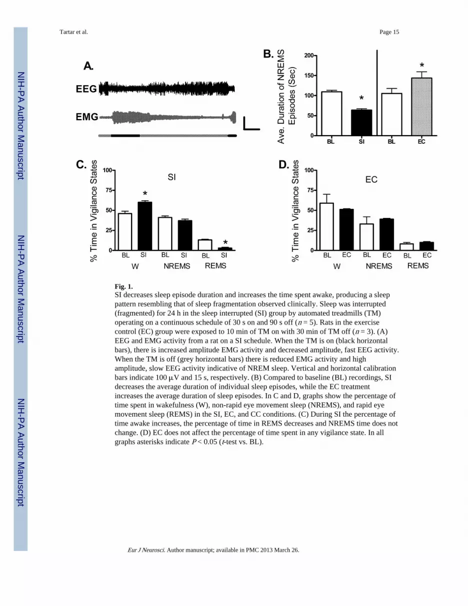
Fig. 1.SI decreases sleep episode duration and increases the time spent awake, producing a sleeppattern resembling that of sleep fragmentation observed clinically. Sleep was interrupted(fragmented) for 24 h in the sleep interrupted (SI) group by automated treadmills (TM)operating on a continuous schedule of 30 s on and 90 s off (n = 5). Rats in the exercisecontrol (EC) group were exposed to 10 min of TM on with 30 min of TM off (n = 3). (A)EEG and EMG activity from a rat on a SI schedule. When the TM is on (black horizontalbars), there is increased amplitude EMG activity and decreased amplitude, fast EEG activity.When the TM is off (grey horizontal bars) there is reduced EMG activity and highamplitude, slow EEG activity indicative of NREM sleep. Vertical and horizontal calibrationbars indicate 100 μV and 15 s, respectively. (B) Compared to baseline (BL) recordings, SIdecreases the average duration of individual sleep episodes, while the EC treatmentincreases the average duration of sleep episodes. In C and D, graphs show the percentage oftime spent in wakefulness (W), non-rapid eye movement sleep (NREMS), and rapid eyemovement sleep (REMS) in the SI, EC, and CC conditions. (C) During SI the percentage oftime awake increases, the percentage of time in REMS decreases and NREMS time does notchange. (D) EC does not affect the percentage of time spent in any vigilance state. In allgraphs asterisks indicate P < 0.05 (t-test vs. BL).
Tartar et al. Page 15
Eur J Neurosci. Author manuscript; available in PMC 2013 March 26.
NIH
-PA Author Manuscript
NIH
-PA Author Manuscript
NIH
-PA Author Manuscript

Fig. 2.Delta power is increased during SI. (A) Sleep interruption (SI) (n = 5) results in an increasein total delta power (the amplitude of the electroencephalographic signal in the deltafrequency range of 1–4 Hz). Graphs show average sum (± SEM) of delta power each hourduring the lights on (clear horizontal bars) and lights off (dark horizontal bars) SI periods.There was a significant increase in total wakefulness (W) delta power (middle left), and asignificant interaction of treatment × time for the NREM delta power (bottom left). (B)Exercise control (EC) (n = 3) did not produce any change in delta power during W (middleleft) or NREMS (bottom right).
Tartar et al. Page 16
Eur J Neurosci. Author manuscript; available in PMC 2013 March 26.
NIH
-PA Author Manuscript
NIH
-PA Author Manuscript
NIH
-PA Author Manuscript
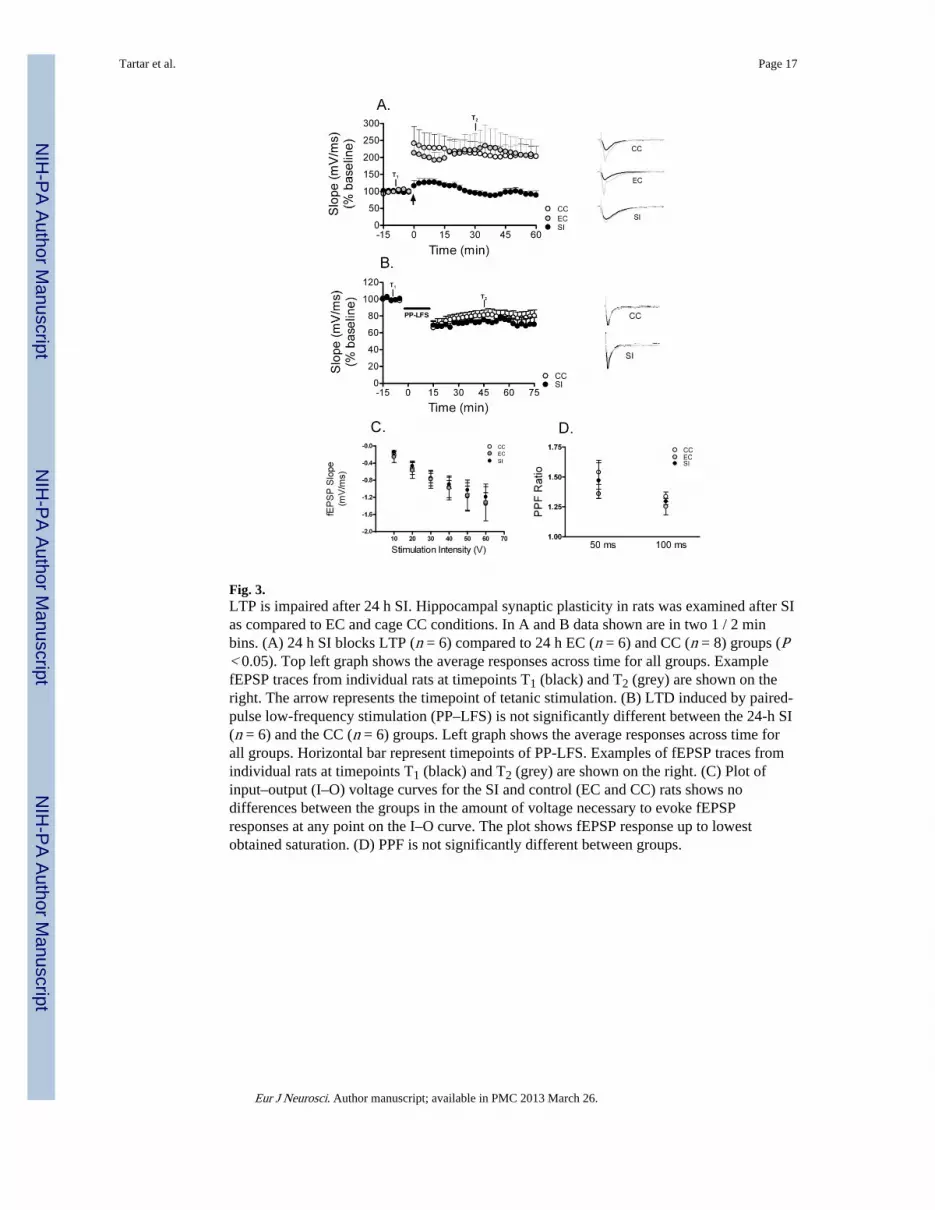
Fig. 3.LTP is impaired after 24 h SI. Hippocampal synaptic plasticity in rats was examined after SIas compared to EC and cage CC conditions. In A and B data shown are in two 1 / 2 minbins. (A) 24 h SI blocks LTP (n = 6) compared to 24 h EC (n = 6) and CC (n = 8) groups (P< 0.05). Top left graph shows the average responses across time for all groups. ExamplefEPSP traces from individual rats at timepoints T1 (black) and T2 (grey) are shown on theright. The arrow represents the timepoint of tetanic stimulation. (B) LTD induced by paired-pulse low-frequency stimulation (PP–LFS) is not significantly different between the 24-h SI(n = 6) and the CC (n = 6) groups. Left graph shows the average responses across time forall groups. Horizontal bar represent timepoints of PP-LFS. Examples of fEPSP traces fromindividual rats at timepoints T1 (black) and T2 (grey) are shown on the right. (C) Plot ofinput–output (I–O) voltage curves for the SI and control (EC and CC) rats shows nodifferences between the groups in the amount of voltage necessary to evoke fEPSPresponses at any point on the I–O curve. The plot shows fEPSP response up to lowestobtained saturation. (D) PPF is not significantly different between groups.
Tartar et al. Page 17
Eur J Neurosci. Author manuscript; available in PMC 2013 March 26.
NIH
-PA Author Manuscript
NIH
-PA Author Manuscript
NIH
-PA Author Manuscript

Fig. 4.Spatial learning is impaired in the water maze after 24 h of SI. (A) Mean (± SEM) distanceto reach the hidden platform for three blocks of four acquisition trials (SI, sleep interruption;n = 11; EC, n = 10; CC, n = 11). Overall, SI rats took a significantly longer path to reach thehidden platform than did CC (P < 0.05), while EC rats were not significantly different fromeither group (P > 0.05). A probe trial was given 30 min after the last acquisition trial (seetext for details). (B) The mean percentage (+ SEM) of total distance travelled in the targetquadrant that formerly contained the hidden platform in a single 30-s probe trial given 30min after the last acquisition trial. SI rats swam significantly less distance in the targetquadrant than did CC (P < 0.05), but EC rats were not significantly different from eithergroup (P > 0.05). Asterisk indicates P < 0.05.
Tartar et al. Page 18
Eur J Neurosci. Author manuscript; available in PMC 2013 March 26.
NIH
-PA Author Manuscript
NIH
-PA Author Manuscript
NIH
-PA Author Manuscript

NIH
-PA Author Manuscript
NIH
-PA Author Manuscript
NIH
-PA Author Manuscript
Tartar et al. Page 19
Table 1
Corticosterone levels after 24 and 72 h of SI
Group n Raw corticosterone ± SEM (ng / mL) SQRT transformed ± SEM
CC 8 2.9 ± 0.9 1.5 ± 0.3
24 h EC 7 38.8 ± 20.9 5.1 ± 1.5
24 h SI 10 124.7 ± 39.4 9.5 ± 1.9*
72 h EC 4 8.6 ± 4.3 2.7 ± 0.67
72 h SI 8 74.6 ± 32.0 7.0 ± 1.9
Compared to CC rats, plasma CORT levels appear elevated after both 24-h EC, 24-h SI, and 72-h SI conditions. Only the 24-h SI condition hassignificantly elevated CORT levels (see text for details). Asterisk indicates significantly different than CC group (P < 0.05).
Eur J Neurosci. Author manuscript; available in PMC 2013 March 26.