Annotated Bibliography Strangers in Crisis: - Institute for ...
Upload
independentCategory
view
2download
0
Comparing Fragmentation Trees from ElectronImpact Mass Spectra with AnnotatedFragmentation PathwaysFranziska Hufsky1,2 and Sebastian Böcker1
1 Chair for Bioinformatics, Friedrich-Schiller-University, Jena, Germany,franziska.hufsky,[email protected]
2 Max Planck Institute for Chemical Ecology, Beutenberg Campus, Jena,Germany
AbstractElectron impact ionization (EI) is the most common form of ionization for GC-MS analysis ofsmall molecules. This ionization method results in a mass spectrum not necessarily containingthe molecular ion peak. The fragmentation of small compounds during EI is well understood, butmanual interpretation of mass spectra is tedious and time-consuming. Methods for automatedanalysis are highly sought, but currently limited to database searching and rule-based approaches.With the computation of hypothetical fragmentation trees from high mass GC-MS data the high-throughput interpretation of such spectra may become feasible. We compare these trees withannotated fragmentation pathways. We find that fragmentation trees explain the origin of theions found in the mass spectra in accordance to the literature. No peak is annotated with anincorrect fragment formula and 78.7% of the fragmentation processes are correctly reconstructed.
1998 ACM Subject Classification J.2 Physical Sciences and Engineering (Chemistry)
Keywords and phrases metabolomics, GC-MS, computational mass spectrometry, fragmentationtrees
Digital Object Identifier 10.4230/OASIcs.GCB.2012.12
1 Introduction
Metabolomics, also called “metabonomics” or “metabolic profiling”, is a rapidly developingfield of ‘omics’ research, dealing with the detection, identification and quantification of lowmolecular-weight compounds (typically below 1000Da) in cells, organs or organisms. Theanalysis and identification of small molecules is important in many areas of biology andmedicine such as biomarker discovery, diagnostics, pharmaceutical chemistry and functionalgenomics [2, 9, 36]. The metabolome consists of various compounds that belong to a widearray of compound classes, including sugars, acids, bases, lipids, hormonal steroids, andmany others [5, 18]. The structural diversity of metabolites is extraordinarily large despiteof their small size [21]. Unlike biopolymers such as proteins and glycans, metabolites arenot made up of repeated building blocks. The genome sequence does not reveal informationabout metabolite structure, as it does for protein structure. The number of metabolites inany higher eukaryote is estimated between 4000 and 20 000 [7]. Unfortunately, an astoundingnumber of these metabolites remain uncharacterized with respect to their structure andfunction [26].
At the moment there is no single instrumental platform that can analyze all metabol-ites [5, 21]. Mass spectrometry (MS), typically coupled with a chromatographic separation
© Franziska Hufsky and Sebastian Böcker;licensed under Creative Commons License ND
German Conference on Bioinformatics 2012 (GCB’12).Editors: S. Böcker, F. Hufsky, K. Scheubert, J. Schleicher, S. Schuster; pp. 12–22
OpenAccess Series in InformaticsSchloss Dagstuhl – Leibniz-Zentrum für Informatik, Dagstuhl Publishing, Germany

F. Hufsky and S. Böcker 13
technology, is one of the key technologies for the identification of small molecules. It hasexcellent compound specificity and high sensitivity. In particular, MS sensitivity is orders ofmagnitude higher than that of nuclear magnetic resonance (NMR) [21,29]. Several kinds ofanalytical apparatus have been developed and most of these combine chromatography with afragmentation technique to increase compound specificity. Gas chromatography coupled tomass spectrometry (GC-MS) is one of the most frequent tools for profiling metabolites andit was in existence decades before liquid chromatography MS (LC-MS) [8, 14]. The amountof data produced during metabolomic analysis is hard to process and analyze manually [18].
The most common ionization technique in GC-MS is electron impact ionization (EI).The resulting fragment-rich mass spectra are in general consistent and specific for eachmolecule [20,21] and fragmentation mechanisms are already well described [23]. Referencespectra were collected over many years, allowing for automated interpretation via databasesearch [24]. Where the compound is unknown, comparing the spectrum obtained to a spectrallibrary will result in imprecise or incorrect hits, or no hits at all [8, 18, 20]. To cover awider range of compounds in silico fragmentation is used to predict spectra of compoundswith known structure [11,12,19,37]. A first step towards the structural elucidation of fullyunknown compounds is feature-based identification of the compound class [17, 19, 34, 35].See Kind and Fiehn [20] for a comprehensive review of computational techniques for smallmolecule mass spectrometry.
Böcker and Rasche [3] introduced fragmentation trees for the de novo interpretationof metabolite fragmentation data. The fragmentation tree concept helps to identify themolecular formulas of the compound and to interpret the fragmentation process. Nodes areannotated with the molecular formulas of the fragments, and edges represent fragmentationevents, that is, neutral or radical losses. Computing fragmentation trees does not requiredatabases of compound structures or mass spectra or expert knowledge of fragmentation.The trees can be compared to each other to identify compound classes of unknowns [28].Expert evaluation suggests that the fragmentation trees from LC-MS2 data are of very goodquality [29]. Fragmentation trees can also be computed from LC-MSn data [31]. Recently,Hufsky et al. [16] presented a computational method for the de novo interpretation ofEI fragmentation data, based on fragmentation tree construction, and applied it to realworld data. Besides a list of common losses, this method does not use any chemical expertknowledge, but does require high mass accuracy of the measurements [16].
In this study, we evaluate the quality of fragmentation trees computed from EI fragmenta-tion data [16]. To evaluate the potential of fragmentation trees to reconstruct fragmentationprocesses we compare them to annotated fragmentation pathways of 22 compounds fromthe literature. The constructed fragmentation trees were not supposed to depict the actualfragmentation reactions. They however agree well with the annotated pathways explainingthe origin of the respective ions found in the mass spectra. No peak was annotated with anincorrect fragment formula and 78.7% of the fragmentation processes were correctly recon-structed. For the annotation of the fragmentation processes in the literature the molecularstructures of the compounds were used. In contrast, the computation of fragmentation treesworks without this knowledge. The assignment of molecular formulas to all fragments andexplanation of relevant fragmentation reactions independent of existing library knowledge,supports the structural elucidation of unknown compounds. Combined with a method for theautomated comparison of fragmentation trees [15,28] it will enable the automated analysisof metabolites that are not included in common libraries.
GCB 2012

14 Comparing EI Fragmentation Trees with Annotated Fragmentation Pathways
2 Methods
For the interpretation of the GC-MS spectra we use fragmentation trees as introducedby Böcker and Rasche [3] for LC-MS2 spectra. A hypothetical fragmentation tree modelsfragmentation cascades by annotating nodes with the molecular formulas of fragments,and edges with fragmentation events, that is, neutral or radical losses. The root of thefragmentation tree is labeled with the molecular formula of the unfragmented ion. ForLC-MS2 data the molecular ion mass is known. EI results in a mass spectrum not necessarilycontaining the molecular ion peak. Hufsky et al. [16] proposed a method for computingfragmentation trees from such data.
To compute a fragmentation tree from an EI fragmentation spectrum a fragmentationgraph is constructed. All candidate molecular formulas within the mass accuracy of theinstrument are computed for each peak. The fragmentation graph contains a node for eachdecomposition. The nodes are colored, such that all explanations of the same peak receive thesame color. Nodes are weighted using mass deviation and peak intensity [3]. Two nodes areconnected by an edge (corresponding to a loss) if the second molecular formula is a subformulaof the first. Edges are weighted according to their plausibility as real fragmentation stepsconsidering the mass of the loss, the ratio between carbon and hetero atoms, and commonlosses for EI fragmentation (see Table 1). See [16] for a detailed description of the scoring.
The colorful subtree with maximum sum of edge weights is the explanation of the observedfragments, that fits best with the given conditions. Considering trees every fragment isexplained by a unique fragmentation pathway, see [29]. Considering only colorful trees everypeak is explained by a single fragment. Several fragments resulting in a single peak is anextremely rare event in practice.
By demanding that each fragment in the fragmentation spectrum is generated by a singlefragmentation pathway we slightly oversimplify the problem. Our optimization algorithmwill choose the mainly occurring pathway to compute a fragmentation tree. There aretwo exceptions to this reasoning: (1) In the resulting fragmentation tree, assume thatsome fragment f3 is cleaved from f2, which is in turn cleaved from f1. Solely from the EIfragmentation pattern and without additional structural information, it cannot be ruled outthat fragment f3 is in truth cleaved directly from f1. Both interpretations are implicitlyencoded in the fragmentation tree: the fragmentation may occur from the fragment’sdirect parent in the tree or from any of its parents (see Figure 1(a)). (2) In the resultingfragmentation tree, assume that some fragment f2 is cleaved from a fragment f1 by loosingl1 and another fragment f3 is cleaved from f1 by loosing l2. Further, another fragment f4 iscleaved from f2 by loosing l2. Solely from the data, it cannot be ruled out that fragment f4is in truth cleaved from f3 by loosing l1. Again, both interpretations are implicitly encoded
Table 1 List of common losses over the alphabet used throughout this study (CHNOPSCl) forscoring EI fragmentation reactions [13]. The losses are sorted by integer mass and their probabilityof occurrence in a GC-MS spectrum [16]. Losses in the first row (dark green) are very commonand thus score high, while losses in the last row (orange) are not-that-common and thus scorecomparatively low.
freq
uenc
y of
oc
curr
ence
integer mass

F. Hufsky and S. Böcker 15
f1
f2
f3
(a) f1
f2
f3
(b) f1
f4
l1
l1l2
l2
f2 f3
(c)
Figure 1 Comparing fragmentation trees (solid edges) with annotated pathways from the literature(dashed edges). (a) In the fragmentation tree, fragment f3 is cleaved from f2, which is in turncleaved from f1. It cannot be ruled out that fragment f3 is in truth cleaved directly from f1 (dottededge). Both interpretations are implicitly encoded in the fragmentation tree. We evaluate theseedges as correct. (b) In the fragmentation tree, fragment f3 is cleaved directly from f1, while intruth it is cleaved from f2 (dotted edge), which is in turn cleaved from f1. We evaluate these edgesas inserted to high. (c) Parallelogram: Fragment f4 is cleaved from f2 by loosing l2, which is in turncleaved from f1 by loosing l1. In truth fragment f4 is cleaved by loosing l2 first and l1 afterwards(dotted edge). Both interpretations are implicitly encoded in the fragmentation tree. We evaluatethese edges as correct.
in the fragmentation tree: the fragmentation may occur by loosing l1 first and l2 afterwards,or the other way (see Figure 1(c)). In the following, we call this constellation parallelogram.
EI is a hard ionization technique often resulting in missing or low intensity molecular ionpeaks [20,21]. Different from LC-MS2 analysis the mass of the molecular ion is not known.All nodes in the graph are possible roots of the fragmentation tree. Molecular formulasexplaining each peak cannot be restricted to sub-molecular formulas as proposed in [29].Therefore the identification of the molecular ion and formula and the computation of thecomplete fragmentation tree is done in two separate steps.
We first identify the molecular ion and molecular formula using only a set of peaks thatappear to be most relevant for the compound. These peaks are selected using three differentcriteria. We choose the k1 most intense peaks, the k2 peaks with highest score, and the k3peaks with highest score in the upper m/z range. The score is a combination of m/z valueand relative intensity m/z · ln(100 · intrel) and the upper m/z range is the m/z region from0.9M to M where M is the highest m/z of a peak detected in the spectrum. In this stepfragmentation trees are computed using Dynamic Programming [3]. Afterwards we computea fragmentation tree for the complete spectrum assuming that we know the correct molecularion and molecular formula of the compound. The resulting fragmentation tree is rooted inthis molecular formula. In this step, fragmentation trees are computed using Integer LinearProgramming [30].
3 Data
The EI induced fragmentation of small molecules is well described in the literature. Toevaluate the potential of fragmentation trees to reconstruct fragmentation processes weextract annotated fragmentation pathways for 22 compounds from different compound classes(see Table 2). In [1] Acheson et al. describe the fragmentation of alkyl acridines. We choosetwo simple alkylacridines and two reduced acridines containing chlorine. Further, we chooseseven compounds from a study on alkyl isocyanides and methyl branched alkyl cyanides [10].From [25] we select four dihydro-1,4-oxathiines with fragmentation paths additionally invest-
GCB 2012

16 Comparing EI Fragmentation Trees with Annotated Fragmentation Pathways
Table 2 Overview of the 22 reference compounds with fragmentation pathways annotated in theliterature. If more than one compound of the same class is used, we denote the class name and givethe mass range and average mass.
masscompound (class) # range averagealkyl acridines [1] 4 207.1 - 399.2Da 276.1Daalkyl isocyanides & α-branched alkyl cyanides [10] 7 41.0 - 83.1Da 69.1Dadihydro-1,4-oxathiines [25] 4 146.0 - 235.1Da 178.8Dagossypol [27] 1 518.2Daephedrine [33] 1 165.1Da2,1-benzisothiazoline 2,2-dioxide nitro derivatives [4] 5 214.0 - 242.0Da 228.0Daall 22 41.0 - 518.2Da 187.6Da
igated with qualitative collisionally induced dissociation (CID) measurements. From [27]we extract the fragmentation pathway of gossypol and from [33] the one from ephedrine.Further, we choose five 2,1-benzisothiazoline 2,2-dioxide nitro derivatives from [4].
As the measured spectra are not available to us, we simulate spectra from the pathways.From the molecular formulas in the fragmentation pathway, we compute exact peak masses,and simulate “measured” spectra by adding a normal distributed error of 10 ppm on the massof the fragment formula (without considering ionization). Peak intensities of the fragmentpeaks are taken from the literature. They are either given as actual number or estimatedfrom the plotted spectrum. In addition, we add 70% noise peaks with uniformly distributedmasses smaller than the parent mass, and pareto distributed intensities.
4 Results
4.1 Molecular Ion Peak and Formula IdentificationFragmentation trees enable the identification of the molecular ion and the molecular formulaof a metabolite if the molecular ion is present in the spectrum. EI is a hard ionizationtechnique resulting in missing molecular ion peaks in about 30% of the spectra [22]. Fortwo compounds in our dataset, namely gossypol and ephedrine, the relative intensity of themolecular ion peak given in the literature was 0%. We test the identification of the molecularion and the molecular formula on the remaining 20 spectra containing a molecular ion peak.
To identify the molecular ion peak and its formula an alphabet of potential elements mustbe provided to the method. For all compounds, we use the six elements most abundant inmetabolites, namely carbon (C), hydrogen (H), nitrogen (N), oxygen (O), phosphorus (P), andsulfur (S) [18]. When analyzing the two compounds in our dataset containing chlorine (Cl),we also add this element to the alphabet. Information on whether a compound containschlorine can be usually obtained from isotope pattern analysis.
Computing the molecular ion peak and molecular formula requires an average of 4.6 s foreach compound. This time includes peak decomposition and graph construction. We discardpeaks with no decomposition. We then choose the subset of peaks that appear to be mostrelevant for the compound as described above. We choose k1 = 10 and k2 = k3 = 5, resultingin at most 20 peaks if the sets are not overlapping.
For all compounds, the method correctly detect the molecular ion peak. This is notsurprising, since the molecular ion peaks have the highest m/z values in all spectra, basedon the generation of noise peaks as described above. For 17 of the 20 compounds (85%),the highest scoring suggestion for both the molecular ion peak and its molecular formula is

F. Hufsky and S. Böcker 17
Table 3 Results of the tree evaluation. (a) Peak explanations in the annotated pathwayscompared to the computed fragmentation trees. 1Percent of the explanations in the annotatedpathways. 2Percent of the explanations in the computed fragmentation trees. (b) Evaluation ofthe fragmentation events annotated in the fragmentation trees. For 5 of the 277 correct peakexplanations, the fragmentation process leading to this fragment is not given in the literature.(c) Evaluation of the frequency of parallelograms in the annotated pathways. A parallelogram isclosed if both fragmentation ways are annotated, and “open” otherwise. 3Percent of the “open”parallelograms.
(a) fragments pathway treetotal total correct missing additional
peak explanations 296 284 277 19 7precentage 93.6%1 6.4%1 2.5%2
(b) losses total correct correct, but to high wrongto deep reverse order
losses 272 214 31 8 19 39percentage 78.7% 11.4% 2.9% 7.0% 14.3%
(c) parallelograms total closed “open” different in treeparallelograms 99 29 70 8percentage 29.3% 70.7% 11.4%3
correct. For the remaining three compounds, the correct molecular formula is the secondsuggestion.
4.2 Fragmentation Tree Quality
We compute a hypothetical fragmentation for every compound, assuming that we know thecorrect molecular ion and molecular formula of the compound. In this step, all peaks ofthe spectrum are used for computation. Computation, including decomposition and graphconstruction, requires 1.5 s on average and a maximum of 18.5 s for the largest compound,namely gossypol. For this compound with mass of 518.2Da, decomposition of all peaksrequires 17.9 s (97% of the total running time).
We compare the computed fragmentation trees with annotated fragmentation patternsfrom the literature. The fragmentation trees annotate 284 peaks in total (see Table 3(a)).Only seven of this explanations (2.5%) are false positives, that is explanations of noisepeaks as fragments. The remaining 277 peaks are annotated with the correct fragmentformula. From all 296 fragments described in the pathways from literature 19 (6.4%) couldnot be explained. There are different reasonings for a peak not being explained in thetree. For some peaks, the mass deviation of the measured peak mass to the exact mass isto high. This effect is getting stronger for smaller peaks, since mass deviation penalty isdependent of the peak intensity [16]. For other peaks, the fragmentation step resulting inthis fragment gets a bad score. For example, the loss C2H2N that was annotated in theliterature as a first fragmentation step for three of the alkyl isocyanides is not included inthe list of common losses for EI fragmentation and is not even a combination of these (seeTable 1 and [16]). Therefore the fragments resulting from this step could not be identified.Nevertheless, the method is capable of identifying losses that are very specific for a singlecompound or compound class and therefore not listed as a common loss (see [16]).
GCB 2012

18 Comparing EI Fragmentation Trees with Annotated Fragmentation Pathways
C2H4O2
H2OC2H4
C3H7O
H2O
C2H4
CH3O
C2H4
CH3
C2H2O
C2H3OS
SCS
C2H3O
C3H5O2
CH2O
C6H10O2S146.040 Da
4.8 ppm 70 %
146.040 Da
6.4 ppm 10 %
C4H6O2S118.008 Da
118.008 Da
2.7 ppm 25 %
C6H8OS128.029 Da
128.029 Da
4.4 ppm 5 %
C4H4OS99.989 Da
5.3 ppm 2 %
C3H3OS86.990 Da
5.9 ppm 20 %
C2H3OS74.990 Da
74.990 Da
15.1 ppm 20 %
HCS44.979 Da
9.2 ppm 100 %
C2H3O43.018 Da
13.4 ppm 95 %
C2H2S57.987 Da
57.987 Da11.0 ppm 5 %
C2OS71.996 Da
S
O
OH
+
S
O
OH
+
S
O+
S
O+S
OH
99.989 Da
86.990 Da
44.979 Da
43.018 Da
71.996 Da
[HCS]+
[CH3-CO]+
S C CH2
+S OC C
+
C3H7O
C2H4 H2O
C2H4C2H3O
CH2O
C2H4
C2H3OS
CS
C2H2O
C2H4O2
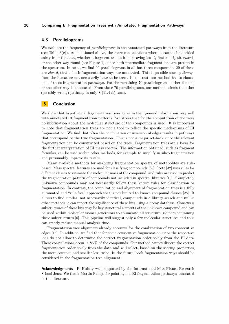
Figure 2 Computed fragmentation tree (solid edges) of 5,6-hydro-3-hyroxymethyl-2-methyl-1,4-oxathiine (left) compared to the annotated pathways [25] from the literature (right). This compoundis a worst-case example to visualize all the things that can go wrong. All fragments are annotatedwith the correct molecular formula. Dashed edges in the tree are losses from the annotated pathways.Black edges in the fragmentation tree agree with the annotated pathways. Grey dashed edges areadditional pathways that could not be computed since the tree property would have been violated.The blue fragment was actually cleaved in reverse order from the molecular ion. The green fragmentswere inserted to deep, and the orange fragment was inserted to high in the fragmentation tree. Thered fragment was inserted into a completely different pathway. Note that mass errors of more than10 ppm occur as we added the simulated mass error on the mass of the fragment formula (withoutconsidering ionization).
Individual edges from the fragmentation tree were compared to those in the annotatedpathways, and matching losses were assigned as correct. In some cases, consecutive edges ofthe fragmentation tree can be combined to give the molecular formula of a single fragmentationstep in the annotated fragmentation pathways (see Figure 1(a)). In some other cases twoconsecutive losses in the fragmentation tree are described in reverse order in the annotatedfragmentation pathways (see Figure 1(c)). We evaluate those fragments that were inserted todeep or in reverse order in the fragmentation trees as correct, since without a given structuralformula and solely from the EI fragmentation data, the correct case cannot be distinguishedfrom our method’s suggestion. If the fragmentation step in the resulting fragmentation treeis explained by several consecutive steps in the annotated pathway, the fragment was insertedto high (see Figure 1(b)). If the fragment was inserted into a completely different pathwaythe edge is assigned as wrong.
For 5,6-hydro-3-hyroxymethyl-2-methyl-1,4-oxathiine [25], we now describe in more detailhow we evaluate the edges of the fragmentation tree (see Figure 2). We choose this compoundas worst-case example to visualize all the things that can go wrong. The loss of ethene fromthe molecular ion (146-118) followed by a loss of C2H3O (118-75) as well as a loss of C2H4O2(118-58) are annotated as correct, as they can be found in the annotated pathways. The waterloss from the molecular ion (146-128) is also annotated in the literature. In the fragmentation

F. Hufsky and S. Böcker 19
C7H6N2O4S214.005 Da
1.8 ppm 98.0%
C7H6N2O2150.043 Da
3.5 ppm 57.0%
C7H6N2O3S198.010 Da
7.3 ppm 1.2%
SO2 O
NO
C7H6NO120.045 Da
8.6 ppm 3.0%
C6H6N2O122.048 Da
5.8 ppm 1.4%
CO NO2
C7H6N104.050 Da
8.2 ppm 8.0%
COCONO HCN
COCHN
C6H577.039 Da
15.0 ppm 23.0%
C5H565.039 Da
4.2 ppm 58.0%
HCN NO
C4H351.023 Da
6.2 ppm 21.0%
C3H339.023 Da
11.7 ppm 5.0%
C2H2
C6H6N92.050 Da
12.5 ppm 100%
C5H5NO95.037 Da
8.6 ppm 14.0%
NH
CH2
+
SO2
N
H
NO2
+
NH
NO2
CH2
+
NH
+ C5H5NO+
C5H5+
C3H3+
C6H5+
C4H3+
N
Cl
Cl
O
+
N
Cl
Cl
O
+
N
Cl
Cl+
C12H5ClN+
C12H6ClN+
C12H6N+
C14H9Cl2NO277.006 Da
2.9 ppm 100%
C13H6Cl2NO261.982 Da
0.9 ppm 8.5%
C12H6Cl2N233.987 Da
3.6 ppm 57%
C12H5ClN198.011 Da
4.8 ppm 7.5%
C12H6ClN199.018 Da
8.0 ppm 8%
C12H6N164.049 Da
8.9 ppm 22%
CH3
HCl Cl
(a) (b)
C7H6N2O3S+
C2H2
HCN
NO2
OSO2
COHCNCO
NO
HCN
C2H2NOHCN
C2H2
CO
Cl
CH3
CO
HCl Cl
Cl
Figure 3 Fragmentation trees compared to annotated pathways from the literature. (a) Fragment-ation tree (left) and annotated pathway (right) of a 2,1-benzisothiazoline 2,2-dioxide nitro derivative(compound 6 from [4]). The grey fragment is not explained in the fragmentation tree as it has verylow intensity and results from a rather uncommon loss (see Table 1). Dashed edges in the tree areadditional losses from the annotated pathways that cannot be explained by our method since the treeproperty would be violated. In the literature the edge (150-92) combines two fragmentation steps(150-120-92), since the 120Da peak is very small. In truth, it is very likely, that this fragmentationalways proceeds in two steps, but that the lifetime of the intermediate ions is too short [4]. The sameapplies to edge (150-95) combining the two fragmentation steps (150-122-95). (b) The fragmentationtree (left) and the annotated pathway (right) of 6,9-dichloro-2-methoxyacridine [1] match completely.Note that mass errors of more than 10 ppm occur as we added the simulated mass error on the massof the fragment formula (without considering ionization).
tree ethene gets lost first and water afterwards (146-118-100), while in the annotated pathwaythese losses are cleaved in reverse order. Edges between nodes 118-75-43 can be combinedto the expected loss of C2H3OS so the loss of sulfur is considered as correct. Pulling upthe edges between nodes 146-118-87 results in a total loss of C3H7O, so the CH3O loss wasinserted to deep and is considered as correct by pull-up. Cleaving fragment 45 directly from118 is considered as to high. Fragment 72 was cleaved by loosing ethene from fragment 100in the annotated pathway. Therefore the methyl loss (87-72) in the fragmentation tree isannotated as wrong.
We use similar reasoning processes to evaluate all hypothetical fragmentation trees (seeFigure 3 for two examples and Table 3(b) for an overview). For 5 of the 277 correct peakexplanations, the fragmentation process leading to this fragment is not given in the literature.From the remaining 272 losses in our data set, 214 losses (78.7%) are assigned as correct.From these, 31 fragments (11.4%) are inserted to deep and 8 fragments (2.9%) are actuallycleaved in reverse order. Further, we find that 19 fragments (7.0%) are inserted to highand 39 edges (14.3%) are annotated as wrong. We stress that, unlike for the annotationof the fragmentation processes in the literature, our method has no information about themolecular structure of the compounds.
GCB 2012
20 Comparing EI Fragmentation Trees with Annotated Fragmentation Pathways
4.3 ParallelogramsWe evaluate the frequency of parallelograms in the annotated pathways from the literature(see Table 3(c)). As mentioned above, these are constellations where it cannot be decidedsolely from the data, whether a fragment results from cleaving loss l1 first and l2 afterwardsor the other way round (see Figure 1), since both intermediate fragment ions are present inthe spectrum. In total, we find 99 parallelograms in all but three compounds. 29 of theseare closed, that is both fragmentation ways are annotated. This is possible since pathwaysfrom the literature not necessarily have to be trees. In contrast, our method has to chooseone of these fragmentation pathways. For the remaining 70 parallelograms, either the oneor the other way is annotated. From these 70 parallelograms, our method selects the other(possibly wrong) pathway in only 8 (11.4%) cases.
5 Conclusion
We show that hypothetical fragmentation trees agree in their general information very wellwith annotated EI fragmentation patterns. We stress that for the computation of the treesno information about the molecular structure of the compounds is used. It is importantto note that fragmentation trees are not a tool to reflect the specific mechanisms of EIfragmentation. We find that often the combination or inversion of edges results in pathwaysthat correspond to the true fragmentation. This is not a major set-back since the relevantfragmentation can be constructed based on the trees. Fragmentation trees are a basis forthe further interpretation of EI mass spectra. The information obtained, such as fragmentformulas, can be used within other methods, for example to simplify in silico fragmentationand presumably improve its results.
Many available methods for analyzing fragmentation spectra of metabolites are rule-based. Mass spectral features are used for classifying compounds [35], Scott [32] uses rules fordifferent classes to estimate the molecular mass of the compound, and rules are used to predictthe fragmentation pattern of compounds not included in spectral libraries [19]. Completelyunknown compounds may not necessarily follow these known rules for classification orfragmentation. In contrast, the computation and alignment of fragmentation trees is a fullyautomated and “rule-free” approach that is not limited to known compound classes [28]. Itallows to find similar, not necessarily identical, compounds in a library search and unlikeother methods it can report the significance of these hits using a decoy database. Consensussubstructures of these hits may be key structural elements of the unknown compound and canbe used within molecular isomer generators to enumerate all structural isomers containingthese substructures [6]. This pipeline will suggest only a few molecular structures and thuscan greatly reduce manual analysis time.
Fragmentation tree alignment already accounts for the combination of two consecutiveedges [15]. In addition, we find that for some consecutive fragmentation steps the respectiveions do not allow to determine the correct fragmentation order solely from the EI data.These constellations occur in 86% of the compounds. Our method cannot discern the correctfragmentation order solely from the data and will select, based on the scoring properties,the more common and smaller loss twice. In the future, both fragmentation ways should beconsidered in the fragmentation tree alignment.
Acknowledgments F. Hufsky was supported by the International Max Planck ResearchSchool Jena. We thank Martin Rempt for pointing out EI fragmentation pathways annotatedin the literature.

F. Hufsky and S. Böcker 21
References1 R. M. Acheson, R. T. Aplin, and R. G. Bolton. Electron impact induced alkyl-group
fragmentation on the acridine nucleus. Organic Mass Spectrometry, 12:518–530, 1977.2 R. J. Bino, R. D. Hall, O. Fiehn, J. Kopka, K. Saito, J. Draper, B. J. Nikolau, P. Mendes,
U. Roessner-Tunali, M. H. Beale, R. N. Trethewey, B. M. Lange, E. S. Wurtele, andL. W. Sumner. Potential of metabolomics as a functional genomics tool. Trends Plant Sci,9(9):418–425, 2004.
3 S. Böcker and F. Rasche. Towards de novo identification of metabolites by analyzingtandem mass spectra. Bioinformatics, 24:I49–I55, 2008. Proc. of European Conference onComputational Biology (ECCB 2008).
4 W. Danikiewicz, K. Wojciechowski, R. H. Fokkens, and N. M. M. Nibbering. Electronimpact-induced fragmentation of 2,1-benzisothiazoline 2,2-dioxide. Org Mass Spectrom,28:853–859, 1993.
5 K. Dettmer, P. A. Aronov, and B. D. Hammock. Mass spectrometry-based metabolomics.Mass Spectrom Rev, 26(1):51–78, 2007.
6 J.-L. Faulon, D. P. Visco, and D. Roe. Enumerating molecules. In K. B. Lipkowitz,R. Larter, and T. R. Cundari, editors, Reviews in Computational Chemistry, volume 21,chapter 3, pages 209–286. John Wiley & Sons, Inc., 2005.
7 A. R. Fernie, R. N. Trethewey, A. J. Krotzky, and L. Willmitzer. Metabolite profiling: fromdiagnostics to systems biology. Nat Rev Mol Cell Biol, 5(9):763–769, 2004.
8 O. Fiehn. Extending the breadth of metabolite profiling by gas chromatography coupledto mass spectrometry. Trends Analyt Chem, 27(3):261–269, 2008.
9 O. Fiehn, J. Kopka, P. Dörmann, T. Altmann, R. N. Trethewey, and L. Willmitzer. Meta-bolite profiling for plant functional genomics. Nat Biotechnol, 18(11):1157–1161, 2000.
10 W. Heerma and J. J. D. Ridder. The electron-impact-induced fragmentation of some alkylisocyanides and α-branched alkyl cyanides. Org Mass Spectrom, 3:1439–1456, 1970.
11 M. Heinonen, A. Rantanen, T. Mielikäinen, J. Kokkonen, J. Kiuru, R. A. Ketola, andJ. Rousu. FiD: a software for ab initio structural identification of product ions from tandemmass spectrometric data. Rapid Commun Mass Spectrom, 22(19):3043–3052, 2008.
12 M. Heinonen, A. Rantanen, T. Mielikäinen, E. Pitkänen, J. Kokkonen, and J. Rousu. Abinitio prediction of molecular fragments from tandem mass spectrometry data. In Proc.of German Conference on Bioinformatics (GCB 2006), volume P-83 of Lecture Notes inInformatics, pages 40–53, 2006.
13 M. Hesse, B. Zeeh, and H. Meier. Spectroscopic Methods in Organic Chemistry. ThiemeMedical Pub, 1997.
14 E. C. Horning and M. G. Horning. Metabolic profiles: gas-phase methods for analysis ofmetabolites. Clin Chem, 17(8):802–809, 1971.
15 F. Hufsky, K. Dührkop, F. Rasche, M. Chimani, and S. Böcker. Fast alignment of fragment-ation trees. Bioinformatics, 28:i265–i273, 2012. Proc. of Intelligent Systems for MolecularBiology (ISMB 2012).
16 F. Hufsky, M. Rempt, F. Rasche, G. Pohnert, and S. Böcker. De novo analysis of electronimpact mass spectra using fragmentation trees. Anal Chim Acta, 739:67–76, 2012.
17 J. Hummel, N. Strehmel, J. Selbig, D. Walther, and J. Kopka. Decision tree supportedsubstructure prediction of metabolites from GC-MS profiles. Metabolomics, 6(2):322–333,2010.
18 H. J. Issaq, Q. N. Van, T. J. Waybright, G. M. Muschik, and T. D. Veenstra. Analyticaland statistical approaches to metabolomics research. J Sep Sci, 32(13):2183–2199, 2009.
19 A. Kerber, R. Laue, M. Meringer, and K. Varmuza. MOLGEN-MS: Evaluation of lowresolution electron impact mass spectra with MS classification and exhaustive structuregeneration. Adv Mass Spectrom, 15:939–940, 2001.
GCB 2012

22 Comparing EI Fragmentation Trees with Annotated Fragmentation Pathways
20 T. Kind and O. Fiehn. Advances in structure elucidation of small molecules using massspectrometry. Bioanal Rev, 2(1-4):23–60, 2010.
21 T. Kind, G. Wohlgemuth, D. Y. Lee, Y. Lu, M. Palazoglu, S. Shahbaz, and O. Fiehn.FiehnLib: mass spectral and retention index libraries for metabolomics based on quadru-pole and time-of-flight gas chromatography/mass spectrometry. Anal Chem, 81(24):10038–10048, 2009.
22 S. J. Lehotay, K. Mastovska, A. Amirav, A. B. Fialkov, T. Alon, P. A. Martos, A. de Kok,and A. R. Fernández-Alba. Identification and confirmation of chemical residues infood by chromatography-mass spectrometry and other techniques. Trends Analyt Chem,27(11):10170–1090, 2008.
23 F. W. McLafferty and F. Tureček. Interpretation of Mass Spectra. University Science Books,Mill valley, California, fourth edition, 1993.
24 S. Neumann and S. Böcker. Computational mass spectrometry for metabolomics – a review.Anal Bioanal Chem, 398(7):2779–2788, 2010.
25 V. Nevalainen and P. Vainiotalo. Electron impact induced fragmentation of dihydro-1,4-oxathiines. 1. 2,3-substituted 5,6-dihydro-1,4-oxathiines. Org Mass Spectrom, 21(9):543–548, 1986.
26 G. J. Patti, O. Yanes, and G. Siuzdak. Innovation: Metabolomics: the apogee of the omicstrilogy. Nat Rev Mol Cell Biol, 13(4):263–269, 2012.
27 P. Przybylski, T. Pospieszny, A. Huczyński, and B. Brzezinski. EI MS and ESI MS studies ofthe bisesquiterpene from cotton seeds: Gossypol and its Aza-derivatives. J Mass Spectrom,43(5):680–686, 2008.
28 F. Rasche, K. Scheubert, F. Hufsky, T. Zichner, M. Kai, A. Svatoš, and S. Böcker. Identi-fying the unknowns by aligning fragmentation trees. Anal Chem, 84(7):3417–3426, 2012.
29 F. Rasche, A. Svatoš, R. K. Maddula, C. Böttcher, and S. Böcker. Computing fragmentationtrees from tandem mass spectrometry data. Anal Chem, 83:1243–1251, 2011.
30 I. Rauf, F. Rasche, F. Nicolas, and S. Böcker. Finding maximum colorful subtrees in prac-tice. In Proc. of Research in Computational Molecular Biology (RECOMB 2012), volume7262 of Lect Notes Comput Sci, pages 213–223. Springer, Berlin, 2012.
31 K. Scheubert, F. Hufsky, F. Rasche, and S. Böcker. Computing fragmentation trees frommetabolite multiple mass spectrometry data. J Comput Biol, 18(11):1383–1397, 2011.
32 D. R. Scott. Rapid and accurate method for estimating molecular weights of organiccompounds from low resolution mass spectra. Chemometr Intell Lab, 16(3):193–202, 1992.
33 M. Thevis and W. Schänzer. Mass spectrometry in sports drug testing: Structure charac-terization and analytical assays. Mass Spectrom Rev, 26(1):79–107, 2007.
34 H. Tsugawa, Y. Tsujimoto, M. Arita, T. Bamba, and E. Fukusaki. GC/MS based meta-bolomics: development of a data mining system for metabolite identification by using softindependent modeling of class analogy (SIMCA). BMC Bioinformatics, 12:131, 2011.
35 K. Varmuza and W. Werther. Mass spectral classifiers for supporting systematic structureelucidation. J Chem Inf Comp Sci, 36(2):323–333, 1996.
36 D. S. Wishart. Current progress in computational metabolomics. Brief Bioinform, 8(5):279–293, 2007.
37 S. Wolf, S. Schmidt, M. Müller-Hannemann, and S. Neumann. In silico fragmentation forcomputer assisted identification of metabolite mass spectra. BMC Bioinformatics, 11:148,2010.
Copyright © 2022 FDOKUMEN