Hippocampal expression of murine TNFα results in attenuation of amyloid deposition in vivo
10
Hippocampal expression of murine TNFα results in attenuation of amyloid deposition in vivo Chakrabarty et al. Chakrabarty et al. Molecular Neurodegeneration 2011, 6:16 http://www.molecularneurodegeneration.com/content/6/1/16 (16 February 2011)
Transcript of Hippocampal expression of murine TNFα results in attenuation of amyloid deposition in vivo
Hippocampal expression of murine TNFα resultsin attenuation of amyloid deposition in vivoChakrabarty et al.
Chakrabarty et al. Molecular Neurodegeneration 2011, 6:16http://www.molecularneurodegeneration.com/content/6/1/16 (16 February 2011)
SHORT REPORT Open Access
Hippocampal expression of murine TNFa resultsin attenuation of amyloid deposition in vivoParamita Chakrabarty1, Amanda Herring2, Carolina Ceballos-Diaz1, Pritam Das2, Todd E Golde1*
Abstract
Fibrillar amyloid b (fAb) peptide is the major component of Ab plaques in the brains of Alzheimer’s disease (AD)patients. Inflammatory mediators have previously been proposed to be drivers of Ab pathology in AD patients byincreasing amyloidogenic processing of APP and promoting Ab accumulation, but recent data have shown thatexpression of various inflammatory cytokines attenuates Ab pathology in mouse models. In an effort to furtherstudy the role of different inflammatory cytokines on Ab pathology in vivo, we explored the effect of murineTumor Necrosis Factor a (mTNFa) in regulating Ab accumulation. Recombinant adeno-associated virus serotype 1(AAV2/1) mediated expression of mTNFa in the hippocampus of 4 month old APP transgenic TgCRND8 miceresulted in significant reduction in hippocampal Ab burden. No changes in APP levels or APP processing wereobserved in either mTNFa expressing APP transgenic mice or in non-transgenic littermates. Analysis of Ab plaqueburden in mTNFa expressing mice showed that even after substantial reduction compared to EGFP expressingage-matched controls, the Ab plaque burden levels of the former do not decrease to the levels of 4 month oldunmanipulated mice. Taken together, our data suggests that proinflammatory cytokine expression induced robustglial activation can attenuate plaque deposition. Whether such an enhanced microglial response actually clearspreexisting deposits without causing bystander neurotoxicity remains an open question.
FindingsAmyloid b (Ab) plaques are a hallmark pathological fea-ture of Alzheimer’s disease (AD). Neuroinflammation,characterized by Ab plaque associated reactive gliosisand increased levels of pro-inflammatory cytokines, hasbeen hypothesized to lead to exacerbated AD pathology[1]. However, recent data has shown that activated glia[2-4] and myeloid lineage cells [5-8] ameliorate brainAb plaque load in Ab precursor protein (APP) trans-genic mouse models. We have previously demonstratedthat increased expression of murine Interleukin-6 (IL-6)or murine Interferon-g (IFNg) results in attenuation ofAb deposition in APP transgenic mice via synergisticactivation of glia and innate immune system compo-nents [2,3]. In our effort to further analyze the role ofdifferent inflammatory cytokines on Ab pathology, weinvestigated the effect of murine tumor necrosis factora (mTNFa) expression in APP transgenic mice. TNFa
is an inflammatory cytokine produced by macrophages,lymphoid cells, endothelial cells [9], neurons [10] andglia [11]. Various functional as well as genetic associa-tion studies have implicated increased levels of TNFa inexacerbating AD pathology [12]. Levels of TNFa andthat of its receptors, TNF-RI and TNF-RII, are elevatedin AD patients [13-15]. Though initial studies implicatedthree TNFa polymorphisms with increased AD [16], asubsequent study showed that these same polymorph-isms delay the age of AD onset [17]. In preclinicalmouse models, deletion of the TNF-RI gene or expres-sion of the dominant negative truncated receptorreduces Ab plaque formation in APP transgenic mice,suggesting a direct role in APP processing [18,19]. Stu-dies in the 3 × Tg-AD mouse model expressing mutantAPP, tau and presenilin showed that there was age-related increase in TNFa levels [20] and neuronalexpression of TNFa exacerbated Ab and tau pathologyin these mice [21].To investigate the role of mTNFa in regulating Ab
accumulation in the CNS, we used recombinant adeno-associated virus (rAAV) to express mTNFa in the hippo-campus of APP transgenic TgCRND8 mice [22].
* Correspondence: [email protected] for Translational Research in Neurodegenerative Disease, College ofMedicine, University of Florida, 1275 Center Drive, Gainesville, PO Box#100159, FL-32610, USAFull list of author information is available at the end of the article
Chakrabarty et al. Molecular Neurodegeneration 2011, 6:16http://www.molecularneurodegeneration.com/content/6/1/16
© 2011 Chakrabarty et al; licensee BioMed Central Ltd. This is an Open Access article distributed under the terms of the CreativeCommons Attribution License (http://creativecommons.org/licenses/by/2.0), which permits unrestricted use, distribution, andreproduction in any medium, provided the original work is properly cited.
Recombinant AAV2 plasmids containing mTNFa (OpenBiosystems, clone 40126376) or enhanced green fluores-cent protein (EGFP) under the control of the cytomegalo-virus enhancer/chicken b actin promoter were packagedin AAV serotype 1 capsid (rAAV2/1) as described pre-viously [2,23]. Adult 4 month old TgCRND8 mice werestereotaxically injected into the hippocampus (interauralcoordinates, anteroposterior:-2.2, mediolateral:+/-1.6,dorsoventral: -1.2) with 2 μl of AAV2/1 constructs (1 ×10¹³ particles/ml) and then sacrificed for analysis at5.5 months (n = 5 for rAAV2/1-mTNFa; n = 6 forrAAV2/1-EGFP). The procedures were approved by theIACUC and performed as described previously [2]. Inprevious studies we have found that AAV2/1-EGFPexpression has no effect on amyloid pathology or gliosiswhen compared to uninjected mice [2,23]; so, AAV2/1-EGFP injected mice were used as the control cohort. Fol-lowing euthanasia, the mice brains were coronally dis-sected 1 mm anterior and 1 mm posterior to the point ofinjection and used for subsequent analysis. Anti-EGFPimmunohistochemistry (Invitrogen, 1:1000) on paraffin
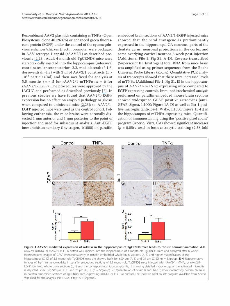
embedded brain sections of AAV2/1-EGFP injected miceshowed that the viral transgene is predominantlyexpressed in the hippocampal CA neurons, parts of thedentate gyrus, neuronal projections in the cortex andsome overlying cortical neurons 6 week post-injection(Additional File 1, Fig S1, A-D). Reverse transcribed(Superscript III; Invitrogen) total RNA from mice brainwas amplified using primer sequences from the RocheUniversal Probe Library (Roche). Quantitative PCR analy-sis of transcripts showed that there were increased levelsof mTNFa (Additional File 1, Fig S1, E) in the hippocam-pus of AAV2/1-mTNFa expressing mice compared toEGFP expressing controls. Immunohistochemical analysisperformed on paraffin embedded mouse brain sectionsshowed widespread GFAP positive astrocytes (anti-GFAP, Sigma, 1:1000; Figure 1A-D) as well as Iba-1 posi-tive microglia (anti-Iba-1, Wako; 1:1000; Figure 1E-H) inthe hippocampus of mTNFa expressing mice. Quantifi-cation of immunostaining using the “positive pixel count”program (Aperio, Vista, CA) showed significant increases(p = 0.05; t test) in both astrocytic staining (2.58 fold
Figure 1 AAV2/1 mediated expression of mTNFa in the hippocampus of TgCRND8 mice leads to robust neuroinflammation. A-D.rAAV2/1-mTNFa or rAAV2/1-EGFP (Control) was injected into the hippocampus of 4 month old TgCRND8 mice and analyzed after 6 weeks.Representative images of GFAP immunoreactivity in paraffin embedded whole brain sections (A, B) and higher magnification of thehippocampus (C, D) of 5.5 month old TgCRND8 mice are shown. Scale Bar, 600 μm (A, B) and 25 μm (C, D). (n = 5/group). E-H. Representativeimages of Iba-1 immunoreactivity in paraffin embedded sections of 5.5 month old TgCRND8 mice injected with rAAV2/1 mTNFa or rAAV2/1-EGFP (Control). Whole brain sections (E, F) and the corresponding hippocampus (G, H) showing detailed morphology of the activated microgliais depicted. Scale Bar, 600 μm (E, F) and 25 μm (G, H). (n = 5/group). I-J. Quantitation of GFAP (I) and Iba-1(J) immunoreactivity burden (% area)in paraffin embedded sections of TgCRND8 mice expressing mTNFa or EGFP as control. The “positive pixel count” program available from Aperiowas used for the analysis. (*p < 0.05; t test; n = 5/group).
Chakrabarty et al. Molecular Neurodegeneration 2011, 6:16http://www.molecularneurodegeneration.com/content/6/1/16
Page 3 of 10
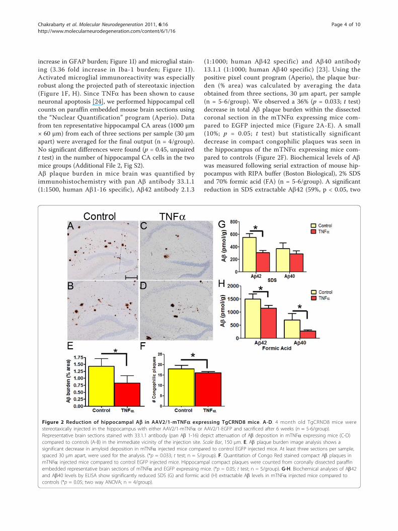
increase in GFAP burden; Figure 1I) and microglial stain-ing (3.36 fold increase in Iba-1 burden; Figure 1J).Activated microglial immunoreactivity was especiallyrobust along the projected path of stereotaxic injection(Figure 1F, H). Since TNFa has been shown to causeneuronal apoptosis [24], we performed hippocampal cellcounts on paraffin embedded mouse brain sections usingthe “Nuclear Quantification” program (Aperio). Datafrom ten representative hippocampal CA areas (1000 μm× 60 μm) from each of three sections per sample (30 μmapart) were averaged for the final output (n = 4/group).No significant differences were found (p = 0.45, unpairedt test) in the number of hippocampal CA cells in the twomice groups (Additional File 2, Fig S2).Ab plaque burden in mice brain was quantified byimmunohistochemistry with pan Ab antibody 33.1.1(1:1500, human Ab1-16 specific), Ab42 antibody 2.1.3
(1:1000; human Ab42 specific) and Ab40 antibody13.1.1 (1:1000; human Ab40 specific) [23]. Using thepositive pixel count program (Aperio), the plaque bur-den (% area) was calculated by averaging the dataobtained from three sections, 30 μm apart, per sample(n = 5-6/group). We observed a 36% (p = 0.033; t test)decrease in total Ab plaque burden within the dissectedcoronal section in the mTNFa expressing mice com-pared to EGFP injected mice (Figure 2A-E). A small(10%; p = 0.05; t test) but statistically significantdecrease in compact congophilic plaques was seen inthe hippocampus of the mTNFa expressing mice com-pared to controls (Figure 2F). Biochemical levels of Abwas measured following serial extraction of mouse hip-pocampus with RIPA buffer (Boston Biological), 2% SDSand 70% formic acid (FA) (n = 5-6/group). A significantreduction in SDS extractable Ab42 (59%, p < 0.05, two
Figure 2 Reduction of hippocampal Ab in AAV2/1-mTNFa expressing TgCRND8 mice. A-D. 4 month old TgCRND8 mice werestereotaxically injected in the hippocampus with either AAV2/1-mTNFa or AAV2/1-EGFP and sacrificed after 6 weeks (n = 5-6/group).Representative brain sections stained with 33.1.1 antibody (pan Ab 1-16) depict attenuation of Ab deposition in mTNFa expressing mice (C-D)compared to controls (A-B) in the immediate vicinity of the injection site. Scale Bar, 150 μm. E. Ab plaque burden image analysis shows asignificant decrease in amyloid deposition in mTNFa injected mice compared to control EGFP injected mice. At least three sections per sample,spaced 30 μm apart, were used for the analysis. (*p = 0.033; t test; n = 5/group). F. Quantitation of Congo Red stained compact Ab plaques inmTNFa injected mice compared to control EGFP injected mice. Hippocampal compact plaques were counted from coronally dissected paraffinembedded representative brain sections of mTNFa and EGFP expressing mice. (*p = 0.05; t test; n = 5/group). G-H. Biochemical analyses of Ab42and Ab40 levels by ELISA show significantly reduced SDS (G) and formic acid (H) extractable Ab levels in mTNFa injected mice compared tocontrols (*p = 0.05; two way ANOVA; n = 4/group).
Chakrabarty et al. Molecular Neurodegeneration 2011, 6:16http://www.molecularneurodegeneration.com/content/6/1/16
Page 4 of 10
way ANOVA) and a non-significant 23% reduction inAb40 levels in the mTNFa expressing mice comparedwith age-matched EGFP-expressing controls was noted(Figure 2G). Similarly, the FA fractions showed a 23%reduction in Ab42 (p < 0.05; two way ANOVA) and a43% reduction in Ab40 levels (p <0.05; two wayANOVA) in rAAV2/1-mTNFa expressing mice com-pared to controls (Figure 2H). Janelsins et al [21] havereported increased intracellular Ab following hippocam-pal expression of AAV-human TNFa in 3 × Tg-ADmice; although following long-term expression, therewas decreased extracellular Ab plaque deposition, whichthe authors attributed to neuronal loss. Though we havenoticed sparse punctuate intracellular Ab immunorectiv-ity in CRND8 mice, primarily in a subset of neuronswithin the subiculum, such intracellular Ab immunor-eactivity was not notably altered by mTNFa (data notshown). The differences in our current observations maybe due to the differences in transgenic mouse modelsand timing of experimental endpoints.To probe whether the decrease in Ab is the result of
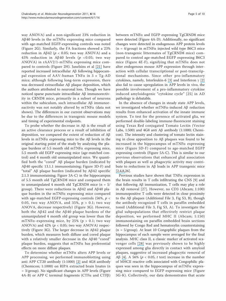
an active clearance process or a result of inhibition ofdeposition, we compared the extent of reduction of Ablevels in mTNFa expressing mice to the Ab levels in theoriginal starting point of the study by analyzing the pla-que burdens of 5.5 month old mTNFa expressing mice,5.5 month old EGFP expressing mice (age-matched con-trol) and 4 month old unmanipulated mice. We quanti-fied both the “cored” Ab plaque burden (indicated byAb40 specific 13.1.1 immunostaining; Figure 3D-F) and“total” Ab plaque burden (indicated by Ab42 specific2.1.3 immunostaining; Figure 3A-C) in the hippocampusof 5.5 month old TgCRND8 mice and compared themto unmanipulated 4 month old TgCRND8 mice (n = 5/group). There were reductions in Ab42 and Ab40 pla-que burden in the mTNFa expressing mice comparedwith age-matched EGFP-expressing controls (56%, p <0.05, two way ANOVA, and 35%, p > 0.1; two wayANOVA, decrease respectively) (Figure 3G). However,both the Ab42 and the Ab40 plaque burdens of theunmanipulated 4 month old group was lower than themTNFa-expressing mice, by 25% (p > 0.1; two wayANOVA) and 42% (p < 0.05; two way ANOVA) respec-tively (Figure 3G). The larger decrease in Ab42 plaqueburden, which measures both diffuse and cored plaquewith a relatively smaller decrease in the Ab40 “cored”plaque burden, suggests that mTNFa has preferentialeffects on more diffuse plaques.To determine whether mTNFa alters APP levels or
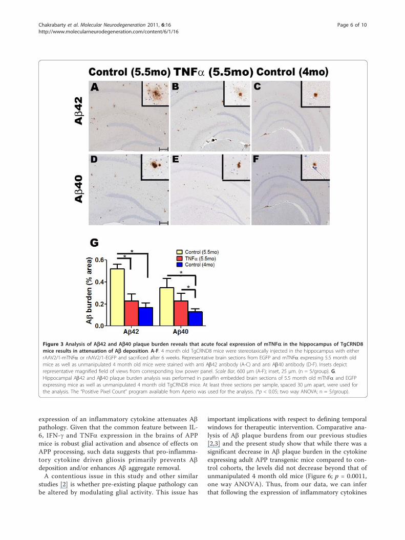
APP processing, we performed immunoblotting usinganti APP CT20 antibody (1:1000) [2] and 4G8 antibody(Chemicon; 1:1000) on RIPA extracted brain lysates (n= 3/group). No significant changes in APP levels (Figure4A-B) or APP C terminal fragments (CTFa and CTFb)
between mTNFa and EGFP expressing TgCRND8 micewere detected (Figure 4A-D). Additionally, no significantchanges were detected in endogenous APP protein levels(n = 4/group) in mTNFa injected wild type B6C3 mice(non-transgenic littermates of TgCRND8 mice) com-pared to control age-matched EGFP expressing B6C3mice (Figure 4E-F), signifying that mTNFa does notalter endogenous mouse APP expression through inter-action with cellular transcriptional or post-transcrip-tional mechanisms. Since other pro-inflammatorycytokines, namely, Interleukin-6 [3] and Interferon g [2]also fail to cause upregulation in APP levels in vivo, thepossible involvement of a pro-inflammatory cytokineinduced amyloidogenic “cytokine cycle” [25] in ADpathology is debatable.In the absence of changes in steady state APP levels,
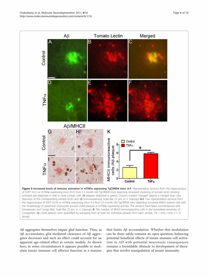
we investigated whether mTNFa-induced Ab reductionresults from enhanced activation of the innate immunesystem. To test for the presence of activated glia, weperformed double-labeling immune-fluorescent stainingusing Texas Red conjugated Tomato Lectin (VectorLabs, 1:500) and 4G8 anti Ab antibody (1:1000; Chemi-con). The intensity and clustering of tomato lectin stain-ing in close apposition to Ab plaques was found to beincreased in the hippocampus of mTNFa expressingmice (Figure 5D-F) compared to age-matched EGFPexpressing controls (Figure 5A-C). This is similar to ourprevious observations that enhanced glial associationwith plaques as well as phagocytic activity may contri-bute to reductions in Ab loads in APP transgenic mice[2,4,8,26].Previous studies have shown that TNFa expression in
the brain results in T cells infiltrating the CNS [9] andthat following Ab immunization, T cells may play a rolein Ab removal [27]. However, no CD3 (Abcam; 1:100)immunopositive T cells were detected in close proximityto the Ab plaques (Additional File 3, Fig S3, B), thoughthe antibody recognized T cells in paraffin embeddedtonsil (Additional File 3, Fig S3, A). To investigate theglial subpopulations that effectively restrict plaquedeposition, we performed MHC II (Abcam; 1:150)immunostaining on paraffin embedded brain sectionsfollowed by Congo Red and hematoxylin counterstaining(n = 5/group). At least 10 Congophilic plaques from thehippocampus of each sample were averaged for the finalanalysis. MHC class II, a classic marker of activated sca-venger cells [28] was previously shown to be highlyexpressed among glia directly in contact with amyloidplaques, suggestive of increased phagocytic removal ofAb [4]. A 56% (p < 0.05, t test) increase in the numberof MHCII reactive cells associated with Congophilic pla-ques was seen in the hippocampus of mTNFa expres-sing mice compared to EGFP expressing mice (Figure5G-K). Collectively, our data demonstrates that acute
Chakrabarty et al. Molecular Neurodegeneration 2011, 6:16http://www.molecularneurodegeneration.com/content/6/1/16
Page 5 of 10
expression of an inflammatory cytokine attenuates Abpathology. Given that the common feature between IL-6, IFN-g and TNFa expression in the brains of APPmice is robust glial activation and absence of effects onAPP processing, such data suggests that pro-inflamma-tory cytokine driven gliosis primarily prevents Abdeposition and/or enhances Ab aggregate removal.A contentious issue in this study and other similar
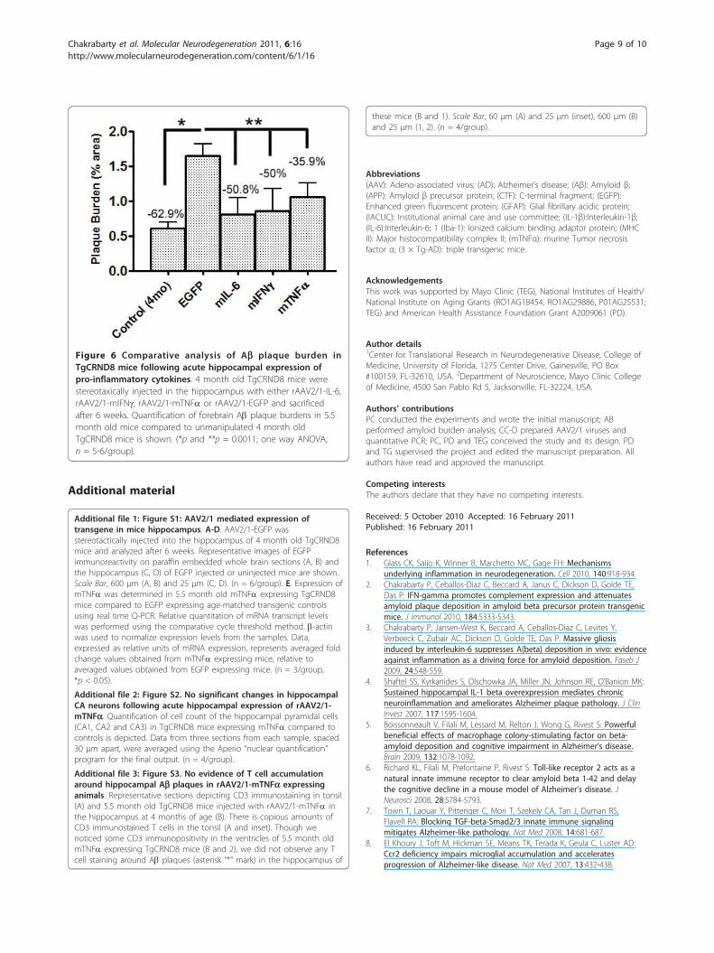
studies [2] is whether pre-existing plaque pathology canbe altered by modulating glial activity. This issue has
important implications with respect to defining temporalwindows for therapeutic intervention. Comparative ana-lysis of Ab plaque burdens from our previous studies[2,3] and the present study show that while there was asignificant decrease in Ab plaque burden in the cytokineexpressing adult APP transgenic mice compared to con-trol cohorts, the levels did not decrease beyond that ofunmanipulated 4 month old mice (Figure 6; p = 0.0011,one way ANOVA). Thus, from our data, we can inferthat following the expression of inflammatory cytokines
Figure 3 Analysis of Ab42 and Ab40 plaque burden reveals that acute focal expression of mTNFa in the hippocampus of TgCRND8mice results in attenuation of Ab deposition. A-F. 4 month old TgCRND8 mice were stereotaxically injected in the hippocampus with eitherrAAV2/1-mTNFa or rAAV2/1-EGFP and sacrificed after 6 weeks. Representative brain sections from EGFP and mTNFa expressing 5.5 month oldmice as well as unmanipulated 4 month old mice were stained with anti Ab42 antibody (A-C) and anti Ab40 antibody (D-F). Insets depictrepresentative magnified field of views from corresponding low power panel. Scale Bar, 600 μm (A-F); inset, 25 μm. (n = 5/group). G.Hippocampal Ab42 and Ab40 plaque burden analysis was performed in paraffin embedded brain sections of 5.5 month old mTNFa and EGFPexpressing mice as well as unmanipulated 4 month old TgCRND8 mice. At least three sections per sample, spaced 30 μm apart, were used forthe analysis. The “Positive Pixel Count” program available from Aperio was used for the analysis. (*p < 0.05; two way ANOVA; n = 5/group).
Chakrabarty et al. Molecular Neurodegeneration 2011, 6:16http://www.molecularneurodegeneration.com/content/6/1/16
Page 6 of 10
in the brain and activation of innate immune systemcomponents, Ab deposition is attenuated; however,whether active clearance may also be occurring concur-rently is still debatable.Using multiple paradigms, we and others [2-8] have
demonstrated that proinflammatory cytokine mediatedglial activation is associated with attenuation of CNS Abdeposition in APP mouse models and in some casespossibly result in actual clearance of pre-existing plaques[29,30]. We find no evidence for altered levels or pro-cessing of the human APP transgene or endogenousmouse APP suggesting that enhanced glial activationattenuates Ab accumulation most likely by altering Abclearance. Of course, the use of a human APP cDNAtransgene driven by a heterologous promoter in many ofthese models imposes some limitations on the finalinterpretation of these studies, as it is possible that
translational and transcriptional control of the APPgene and mRNA, respectively, is different between miceand humans. A next step could be to use the APP YACtransgenic mice [31], but even in this case the differ-ences in the transcriptional and translational machi-neries of humans and mice may confound the outcome.In AD patients and in AD mouse models there is an
inevitable age-progressive accrual of Ab aggregatesdespite widespread reactive gliosis. Thus, it appears thatthe normal reactive gliosis induced by Ab accumulationis insufficient to overcome the inexorable accumulationof aggregated Ab. Some have suggested that microglialaging may lead to inefficient or abortive Ab phagocyto-sis [32,33], but such a hypothesis is difficult to reconcilewith the observation that age of onset of Ab plaquepathology in both humans and mouse models mice canoccur very early in life. An intriguing possibility is that
Figure 4 APP or APP CTF levels were not significantly altered in mTNFa expressing mice. A-B. Representative anti CT20 immunoblotshowing no significant changes in APP or CTFa levels in AAV2/1-mTNFa expressing TgCRND8 compared to age-matched transgenic miceexpressing EGFP (control cohort) (A). Intensity analysis of anti CT20 immunoreactive APP and CTFa levels was normalized to b-actin in TgCRND8mice cohort (B). (n = 3/group). C-D. Representative anti 4G8 immunoblot showing no significant changes in CTFb levels in AAV2/1-mTNFaexpressing TgCRND8 compared to age-matched transgenic mice expressing EGFP (control cohort) (C). Intensity analysis of anti 4G8immunoreactive CTFb levels was normalized to b-actin in TgCRND8 mice cohort (D). (n = 3/group). E-F. Representative anti CT20 immunoblotshowing no significant changes in APP levels in AAV2/1-mTNFa expressing non transgenic B6C3 mice compared to age-matched nontransgenic B6C3 expressing EGFP (E). Intensity analysis of anti CT20 immunoreactive APP levels was normalized to b-actin in the wild type micecohort (F). (n = 4/group).
Chakrabarty et al. Molecular Neurodegeneration 2011, 6:16http://www.molecularneurodegeneration.com/content/6/1/16
Page 7 of 10
Ab aggregates themselves impair glial function. Thus, asAb accumulates, glia-mediated clearance of Ab aggre-gates decreases and such an effect could account for anapparent age-related effect in certain models. As shownhere, in some circumstances it appears possible to mod-ulate innate immune cell effector function in a manner
that limits Ab accumulation. Whether this modulationcan be done safely remains an open question; balancingpotential beneficial effects of innate immune cell activa-tion in AD with potential neurotoxic consequencesremains a formidable obstacle to development of thera-pies that involve manipulation of innate immunity.
Figure 5 Increased levels of immune activation in mTNFa expressing TgCRND8 mice. A-F. Representative sections from the hippocampusof EGFP (A-C) or mTNFa expressing mice (D-F) from 5.5 month old TgCRND8 mice depicting increased clustering of tomato lectin bindingactivated glia (depicted in red) in close contact with Ab plaques (depicted in green). Column marked “merged” depicts a merged dual colordepiction of the corresponding tomato lectin and Ab immunostaining. Scale Bar, 25 μm. (n = 3/group). G-J. Two representative sections fromthe hippocampus of EGFP (G-H) or mTNFa expressing mice (I-J) from 5.5 month old TgCRND8 mice depicting increased MHCII stained cells withthe morphology of peripheral monocytes around cored plaques in mTNFa expressing animals. The sections have been counterstained withhematoxylin and Congo Red. Scale Bar, 25 μm. (n = 5/group). K. The number of MHCII immunopositive cells in the immediate proximity ofCongophilic Ab cored plaques were quantified by averaging from at least ten individual plaques from each sample. (*p < 0.05; t test; n = 5/group).
Chakrabarty et al. Molecular Neurodegeneration 2011, 6:16http://www.molecularneurodegeneration.com/content/6/1/16
Page 8 of 10
Additional material
Additional file 1: Figure S1: AAV2/1 mediated expression oftransgene in mice hippocampus. A-D. AAV2/1-EGFP wasstereotactically injected into the hippocampus of 4 month old TgCRND8mice and analyzed after 6 weeks. Representative images of EGFPimmunoreactivity on paraffin embedded whole brain sections (A, B) andthe hippocampus (C, D) of EGFP injected or uninjected mice are shown.Scale Bar, 600 μm (A, B) and 25 μm (C, D). (n = 6/group). E. Expression ofmTNFa was determined in 5.5 month old mTNFa expressing TgCRND8mice compared to EGFP expressing age-matched transgenic controlsusing real time Q-PCR. Relative quantitation of mRNA transcript levelswas performed using the comparative cycle threshold method. b-actinwas used to normalize expression levels from the samples. Data,expressed as relative units of mRNA expression, represents averaged foldchange values obtained from mTNFa expressing mice, relative toaveraged values obtained from EGFP expressing mice. (n = 3/group,*p < 0.05).
Additional file 2: Figure S2. No significant changes in hippocampalCA neurons following acute hippocampal expression of rAAV2/1-mTNFa. Quantification of cell count of the hippocampal pyramidal cells(CA1, CA2 and CA3) in TgCRND8 mice expressing mTNFa compared tocontrols is depicted. Data from three sections from each sample, spaced30 μm apart, were averaged using the Aperio “nuclear quantification”program for the final output. (n = 4/group).
Additional file 3: Figure S3. No evidence of T cell accumulationaround hippocampal Ab plaques in rAAV2/1-mTNFa expressinganimals. Representative sections depicting CD3 immunostaining in tonsil(A) and 5.5 month old TgCRND8 mice injected with rAAV2/1-mTNFa inthe hippocampus at 4 months of age (B). There is copious amounts ofCD3 immunostained T cells in the tonsil (A and inset). Though wenoticed some CD3 immunopositivity in the ventricles of 5.5 month oldmTNFa expressing TgCRND8 mice (B and 2), we did not observe any Tcell staining around Ab plaques (asterisk “*” mark) in the hippocampus of
these mice (B and 1). Scale Bar, 60 μm (A) and 25 μm (inset), 600 μm (B)and 25 μm (1, 2). (n = 4/group).
Abbreviations(AAV): Adeno-associated virus; (AD): Alzheimer’s disease; (Aβ): Amyloid β;(APP): Amyloid β precursor protein; (CTF): C-terminal fragment; (EGFP):Enhanced green fluorescent protein; (GFAP): Glial fibrillary acidic protein;(IACUC): Institutional animal care and use committee; (IL-1β):Interleukin-1β;(IL-6):Interleukin-6; 1 (Iba-1): Ionized calcium binding adaptor protein; (MHCII): Major histocompatibility complex II; (mTNFα): murine Tumor necrosisfactor α; (3 × Tg-AD): triple transgenic mice.
AcknowledgementsThis work was supported by Mayo Clinic (TEG), National Institutes of Health/National Institute on Aging Grants (RO1AG18454, RO1AG29886, P01AG25531;TEG) and American Health Assistance Foundation Grant A2009061 (PD).
Author details1Center for Translational Research in Neurodegenerative Disease, College ofMedicine, University of Florida, 1275 Center Drive, Gainesville, PO Box#100159, FL-32610, USA. 2Department of Neuroscience, Mayo Clinic Collegeof Medicine, 4500 San Pablo Rd S, Jacksonville, FL-32224, USA.
Authors’ contributionsPC conducted the experiments and wrote the initial manuscript; ABperformed amyloid burden analysis; CC-D prepared AAV2/1 viruses andquantitative PCR; PC, PD and TEG conceived the study and its design. PDand TG supervised the project and edited the manuscript preparation. Allauthors have read and approved the manuscript.
Competing interestsThe authors declare that they have no competing interests.
Received: 5 October 2010 Accepted: 16 February 2011Published: 16 February 2011
References1. Glass CK, Saijo K, Winner B, Marchetto MC, Gage FH: Mechanisms
underlying inflammation in neurodegeneration. Cell 2010, 140:918-934.2. Chakrabarty P, Ceballos-Diaz C, Beccard A, Janus C, Dickson D, Golde TE,
Das P: IFN-gamma promotes complement expression and attenuatesamyloid plaque deposition in amyloid beta precursor protein transgenicmice. J Immunol 2010, 184:5333-5343.
3. Chakrabarty P, Jansen-West K, Beccard A, Ceballos-Diaz C, Levites Y,Verbeeck C, Zubair AC, Dickson D, Golde TE, Das P: Massive gliosisinduced by interleukin-6 suppresses A{beta} deposition in vivo: evidenceagainst inflammation as a driving force for amyloid deposition. Faseb J2009, 24:548-559.
4. Shaftel SS, Kyrkanides S, Olschowka JA, Miller JN, Johnson RE, O’Banion MK:Sustained hippocampal IL-1 beta overexpression mediates chronicneuroinflammation and ameliorates Alzheimer plaque pathology. J ClinInvest 2007, 117:1595-1604.
5. Boissonneault V, Filali M, Lessard M, Relton J, Wong G, Rivest S: Powerfulbeneficial effects of macrophage colony-stimulating factor on beta-amyloid deposition and cognitive impairment in Alzheimer’s disease.Brain 2009, 132:1078-1092.
6. Richard KL, Filali M, Prefontaine P, Rivest S: Toll-like receptor 2 acts as anatural innate immune receptor to clear amyloid beta 1-42 and delaythe cognitive decline in a mouse model of Alzheimer’s disease. JNeurosci 2008, 28:5784-5793.
7. Town T, Laouar Y, Pittenger C, Mori T, Szekely CA, Tan J, Duman RS,Flavell RA: Blocking TGF-beta-Smad2/3 innate immune signalingmitigates Alzheimer-like pathology. Nat Med 2008, 14:681-687.
8. El Khoury J, Toft M, Hickman SE, Means TK, Terada K, Geula C, Luster AD:Ccr2 deficiency impairs microglial accumulation and acceleratesprogression of Alzheimer-like disease. Nat Med 2007, 13:432-438.
Figure 6 Comparative analysis of Ab plaque burden inTgCRND8 mice following acute hippocampal expression ofpro-inflammatory cytokines. 4 month old TgCRND8 mice werestereotaxically injected in the hippocampus with either rAAV2/1-IL-6,rAAV2/1-mIFNg, rAAV2/1-mTNFa or rAAV2/1-EGFP and sacrificedafter 6 weeks. Quantification of forebrain Ab plaque burdens in 5.5month old mice compared to unmanipulated 4 month oldTgCRND8 mice is shown. (*p and **p = 0.0011; one way ANOVA;n = 5-6/group).
Chakrabarty et al. Molecular Neurodegeneration 2011, 6:16http://www.molecularneurodegeneration.com/content/6/1/16
Page 9 of 10
9. Wajant H, Pfizenmaier K, Scheurich P: Tumor necrosis factor signaling. CellDeath Differ 2003, 10:45-65.
10. Covey WC, Ignatowski TA, Renauld AE, Knight PR, Nader ND, Spengler RN:Expression of neuron-associated tumor necrosis factor alpha in the brainis increased during persistent pain. Reg Anesth Pain Med 2002, 27:357-366.
11. Park KM, Bowers WJ: Tumor necrosis factor-alpha mediated signaling inneuronal homeostasis and dysfunction. Cell Signal 2010, 22:977-983.
12. Tan ZS, Beiser AS, Vasan RS, Roubenoff R, Dinarello CA, Harris TB,Benjamin EJ, Au R, Kiel DP, Wolf PA, Seshadri S: Inflammatory markers andthe risk of Alzheimer disease: the Framingham Study. Neurology 2007,68:1902-1908.
13. Dickson DW: The pathogenesis of senile plaques. Journal ofNeuropathology & Experimental Neurology 1997, 56:321-339.
14. Paganelli R, Di Iorio A, Patricelli L, Ripani F, Sparvieri E, Faricelli R, Iarlori C,Porreca E, Di Gioacchino M, Abate G: Proinflammatory cytokines in sera ofelderly patients with dementia: levels in vascular injury are higher thanthose of mild-moderate Alzheimer’s disease patients. Exp Gerontol 2002,37:257-263.
15. Bongioanni P, Romano MR, Sposito R, Castagna M, Boccardi B, Borgna M: T-cell tumour necrosis factor-alpha receptor binding in dementedpatients. J Neurol 1997, 244:418-425.
16. Collins JS, Perry RT, Watson B Jr, Harrell LE, Acton RT, Blacker D, Albert MS,Tanzi RE, Bassett SS, McInnis MG, et al: Association of a haplotype fortumor necrosis factor in siblings with late-onset Alzheimer disease: theNIMH Alzheimer Disease Genetics Initiative. Am J Med Genet 2000,96:823-830.
17. Perry RT, Collins JS, Wiener H, Acton R, Go RC: The role of TNF and itsreceptors in Alzheimer’s disease. Neurobiol Aging 2001, 22:873-883.
18. He P, Zhong Z, Lindholm K, Berning L, Lee W, Lemere C, Staufenbiel M,Li R, Shen Y: Deletion of tumor necrosis factor death receptor inhibitsamyloid beta generation and prevents learning and memory deficits inAlzheimer’s mice. J Cell Biol 2007, 178:829-841.
19. McAlpine FE, Lee JK, Harms AS, Ruhn KA, Blurton-Jones M, Hong J, Das P,Golde TE, LaFerla FM, Oddo S, et al: Inhibition of soluble TNF signaling ina mouse model of Alzheimer’s disease prevents pre-plaque amyloid-associated neuropathology. Neurobiol Dis 2009, 34:163-177.
20. Janelsins MC, Mastrangelo MA, Oddo S, LaFerla FM, Federoff HJ, Bowers WJ:Early correlation of microglial activation with enhanced tumor necrosisfactor-alpha and monocyte chemoattractant protein-1 expressionspecifically within the entorhinal cortex of triple transgenic Alzheimer’sdisease mice. J Neuroinflammation 2005, 2:23.
21. Janelsins MC, Mastrangelo MA, Park KM, Sudol KL, Narrow WC, Oddo S,Laferla FM, Callahan LM, Federoff HJ, Bowers WJ: Chronic Neuron-SpecificTumor Necrosis Factor-Alpha Expression Enhances the LocalInflammatory Environment Ultimately Leading to Neuronal Death in 3 ×Tg-AD Mice. Am J Pathol 2008, 173:1768-1782.
22. Chishti MA, Yang DS, Janus C, Phinney AL, Horne P, Pearson J, Strome R,Zuker N, Loukides J, French J, et al: Early-onset amyloid deposition andcognitive deficits in transgenic mice expressing a double mutant formof amyloid precursor protein 695. J Biol Chem 2001, 276:21562-21570.
23. Kim J, Miller VM, Levites Y, West KJ, Zwizinski CW, Moore BD, Troendle FJ,Bann M, Verbeeck C, Price RW, et al: BRI2 (ITM2b) inhibits Abetadeposition in vivo. J Neurosci 2008, 28:6030-6036.
24. Tartaglia LA, Pennica D, Goeddel DV: Ligand passing: the 75-kDa tumornecrosis factor (TNF) receptor recruits TNF for signaling by the 55-kDaTNF receptor. J Biol Chem 1993, 268:18542-18548.
25. Griffin WS, Sheng JG, Royston MC, Gentleman SM, McKenzie JE, Graham DI,Roberts GW, Mrak RE: Glial-neuronal interactions in Alzheimer’s disease:the potential role of a ‘cytokine cycle’ in disease progression. BrainPathol 1998, 8:65-72.
26. Bard F, Cannon C, Barbour R, Burke RL, Games D, Grajeda H, Guido T, Hu K,Huang J, Johnson-Wood K, et al: Peripherally administered antibodiesagainst amyloid beta-peptide enter the central nervous system andreduce pathology in a mouse model of Alzheimer disease. Nat Med2000, 6:916-919.
27. Monsonego A, Imitola J, Petrovic S, Zota V, Nemirovsky A, Baron R, Fisher Y,Owens T, Weiner HL: Abeta-induced meningoencephalitis is IFN-gamma-dependent and is associated with T cell-dependent clearance of Abetain a mouse model of Alzheimer’s disease. Proc Natl Acad Sci USA 2006,103:5048-5053.
28. Rogers J, Strohmeyer R, Kovelowski CJ, Li R: Microglia and inflammatorymechanisms in the clearance of amyloid beta peptide. Glia 2002,40:260-269.
29. Mandrekar S, Jiang Q, Lee CY, Koenigsknecht-Talboo J, Holtzman DM,Landreth GE: Microglia mediate the clearance of soluble Abeta throughfluid phase macropinocytosis. J Neurosci 2009, 29:4252-4262.
30. Majumdar A, Cruz D, Asamoah N, Buxbaum A, Sohar I, Lobel P, Maxfield FR:Activation of microglia acidifies lysosomes and leads to degradation ofAlzheimer amyloid fibrils. Mol Biol Cell 2007, 18:1490-1496.
31. Lamb BT, Sisodia SS, Lawler AM, Slunt HH, Kitt CA, Kearns WG, Pearson PL,Price DL, Gearhart JD: Introduction and expression of the 400 kilobaseprecursor amyloid protein gene in transgenic mice. Nature genetics 1993,5:22-29.
32. Njie EG, Boelen E, Stassen FR, Steinbusch HW, Borchelt DR, Streit WJ: Exvivo cultures of microglia from young and aged rodent brain revealage-related changes in microglial function. Neurobiol Aging 2010.
33. Dickson DW: Microglia in Alzheimer’s disease and transgenic models.How close the fit? Am J Pathol 1999, 154:1627-1631.
doi:10.1186/1750-1326-6-16Cite this article as: Chakrabarty et al.: Hippocampal expression ofmurine TNFa results in attenuation of amyloid deposition in vivo.Molecular Neurodegeneration 2011 6:16.
Submit your next manuscript to BioMed Centraland take full advantage of:
• Convenient online submission
• Thorough peer review
• No space constraints or color figure charges
• Immediate publication on acceptance
• Inclusion in PubMed, CAS, Scopus and Google Scholar
• Research which is freely available for redistribution
Submit your manuscript at www.biomedcentral.com/submit
Chakrabarty et al. Molecular Neurodegeneration 2011, 6:16http://www.molecularneurodegeneration.com/content/6/1/16
Page 10 of 10