High-performance capillary electrophoretic separation of double-stranded oligonucleotides using a...
10
Electrophoresis 2012, 33, 1205–1214 1205 Kerrilee E. Allan 1,2 Claire E. Lenehan 2 Dmitriy A. Khodakov 1 Hilton J. Kobus 2 Amanda V. Ellis 1,2 1 Flinders Centre for NanoScale Science and Technology School of Chemical and Physical Sciences, Flinders University, Adelaide, SA, Australia 2 School of Chemical and Physical Sciences, Flinders University, Adelaide, SA, Australia Received September 28, 2011 Revised November 21, 2011 Accepted December 8, 2011 Research Article High-performance capillary electrophoretic separation of double-stranded oligonucleotides using a poly- (ethylpyrrolidine methacrylate-co-methyl methacrylate)-coated capillary Here we describe a capillary electrophoretic method for the separation of double-stranded oligonucleotides (ds-ODNs) ranging from 16–20 bp with 2 bp resolution using a low concentration of poly(ethylpyrrolidine methacrylate-co-methyl methacrylate) (PEPyM-co- PMMA) copolymer physically adsorbed to a capillary surface. Contrary to traditional DNA separations, we show that the ds-ODN with the highest molecular size eluted first and propose that this phenomena is due to a screening effect by the PEPyM-co-PMMA coat- ing on the smaller ds-ODNs negative charge during elution. Key to the performance of this separation was a sample preparation time of less than 1 h and analysis time of 40 min. Repeatability of intraday migration time for the mixtures was typically < 1% relative standard deviation (n = 3). In addition, we demonstrate that the coating has an acceptable capillary lifetime of over 70 injections. Keywords: Capillary electrophoresis / DNA / Physically adsorbed coatings / Poly (ethylpyrro- lidine methacrylate-co-methyl methacrylate) copolymer / Oligonucleotides DOI 10.1002/elps.201100514 1 Introduction Capillary electrophoresis (CE) is an attractive technique for the analysis of biomolecules due to its high sensitivity, sep- aration efficiency, and reliability [1]. In particular, there is great interest in the use of CE for the identity and pu- rity of therapeutic oligonucleotides. Therapeutic oligonu- cleotides are single-stranded (ss) or double-stranded (ds) Correspondence: Dr. Amanda V. Ellis, School of Chemical and Physical Sciences, Flinders University, GPO Box 2100, Adelaide, SA 5001, Australia E-mail: amanda.ellis@flinders.edu.au Fax: +61-8-8201-2905 Abbreviations: AS-ONs, antisense oligonucleotides; DMA, N,N-dimethylacrylamide; DNA, deoxyribonucleic acid; ds-AS- ONs, double-stranded antisense oligonucleotides; ds-ODNs, double-stranded oligonucleotides; EPy, N-(2-hydroxyethyl)- 2-pyrrolidine; EPyM, ethylpyrrolidine methacrylate; LPA, linear poly(acrylamide); MMA, methyl methacrylate; ODNs, oligonucleotides; PDEA, poly(diethylacrylamide); PDMA, poly(dimethylacrylamide); PDMA-co-PEPyM, poly(N,N- dimethylacrylamide-co-ethylpyrrolidine methacrylate); PEPyM-co-PMMA, poly(ethylpyrrolidine methacrylate-co- methyl methacrylate); PEO, poly(ethylene oxide); Rs, reso- lution; RNA, ribonucleic acid ; ss-AS-ODNs, single-stranded antisense oligonucleotides; ss-ODNs, single-stranded oligonucleotides macromolecules derived from deoxyribonucleic acid (DNA) and ribonucleic acid (RNA) building blocks with an ideal op- timal length for cellular uptake of approximately 20 bases (or 20-mer) [2]. Single-stranded antisense oligonucleotides (ssAS-ODNs) are easily determined by various methods [3–5], however recent pharmaceutical developments of double-stranded AS-ODNs have required new analysis strate- gies to be developed in order to successfully determine the identity and purity [6, 7]. While AS-ODNs are not the scope of this work, we focus here on resolving short (16–20 bp) model ds-ODNs using CE with the aim to employ this system for future AS-ODN separations. Due to DNA’s constant mass-to-charge ratio (irrespec- tive of length), electrophoresis on uncoated bare fused silica capillaries results in unresolved peaks [8–10]. To enable size separation of DNA, manipulation of the interface between DNA and a coated capillary needs to be achieved [11, 12]. Tra- ditional CE separation of DNA uses capillary gel electrophore- sis (CGE) with a bis-acrylamide cross-linked poly(acrylamide) gel to achieve DNA separation [1]. However, the drawbacks of gel-filled capillaries are that they have a limited life time due to the deterioration from run to run, the inability to regenerate the gel, and the formation of air bubbles [11]. Promising replacements of gel-filled capillaries are physi- cally adsorbed coatings that are capable of ssDNA and dsDNA separation of varying lengths [6,9,12–15]. Physically adsorbed coatings are generally termed dynamic [16], and often involve the use of polymer in the run buffer [15, 17–20]. However, C 2012 WILEY-VCH Verlag GmbH & Co. KGaA, Weinheim www.electrophoresis-journal.com
-
Upload
independent -
Category
Documents
-
view
1 -
download
0
Transcript of High-performance capillary electrophoretic separation of double-stranded oligonucleotides using a...

Electrophoresis 2012, 33, 1205–1214 1205
Kerrilee E. Allan1,2
Claire E. Lenehan2
Dmitriy A. Khodakov1
Hilton J. Kobus2
Amanda V. Ellis1,2
1Flinders Centre for NanoScaleScience and Technology Schoolof Chemical and PhysicalSciences, Flinders University,Adelaide, SA, Australia
2School of Chemical and PhysicalSciences, Flinders University,Adelaide, SA, Australia
Received September 28, 2011Revised November 21, 2011Accepted December 8, 2011
Research Article
High-performance capillary electrophoreticseparation of double-strandedoligonucleotides using a poly-(ethylpyrrolidine methacrylate-co-methylmethacrylate)-coated capillary
Here we describe a capillary electrophoretic method for the separation of double-strandedoligonucleotides (ds-ODNs) ranging from 16–20 bp with 2 bp resolution using a lowconcentration of poly(ethylpyrrolidine methacrylate-co-methyl methacrylate) (PEPyM-co-PMMA) copolymer physically adsorbed to a capillary surface. Contrary to traditional DNAseparations, we show that the ds-ODN with the highest molecular size eluted first andpropose that this phenomena is due to a screening effect by the PEPyM-co-PMMA coat-ing on the smaller ds-ODNs negative charge during elution. Key to the performanceof this separation was a sample preparation time of less than 1 h and analysis time of40 min. Repeatability of intraday migration time for the mixtures was typically < 1%relative standard deviation (n = 3). In addition, we demonstrate that the coating has anacceptable capillary lifetime of over 70 injections.
Keywords:
Capillary electrophoresis / DNA / Physically adsorbed coatings / Poly (ethylpyrro-lidine methacrylate-co-methyl methacrylate) copolymer / Oligonucleotides
DOI 10.1002/elps.201100514
1 Introduction
Capillary electrophoresis (CE) is an attractive technique forthe analysis of biomolecules due to its high sensitivity, sep-aration efficiency, and reliability [1]. In particular, there isgreat interest in the use of CE for the identity and pu-rity of therapeutic oligonucleotides. Therapeutic oligonu-cleotides are single-stranded (ss) or double-stranded (ds)
Correspondence: Dr. Amanda V. Ellis, School of Chemical andPhysical Sciences, Flinders University, GPO Box 2100, Adelaide,SA 5001, AustraliaE-mail: [email protected]: +61-8-8201-2905
Abbreviations: AS-ONs, antisense oligonucleotides; DMA,N,N-dimethylacrylamide; DNA, deoxyribonucleic acid; ds-AS-ONs, double-stranded antisense oligonucleotides; ds-ODNs,double-stranded oligonucleotides; EPy, N-(2-hydroxyethyl)-2-pyrrolidine; EPyM, ethylpyrrolidine methacrylate; LPA,linear poly(acrylamide); MMA, methyl methacrylate; ODNs,oligonucleotides; PDEA, poly(diethylacrylamide); PDMA,poly(dimethylacrylamide); PDMA-co-PEPyM, poly(N,N-dimethylacrylamide-co-ethylpyrrolidine methacrylate);PEPyM-co-PMMA, poly(ethylpyrrolidine methacrylate-co-methyl methacrylate); PEO, poly(ethylene oxide); Rs, reso-lution; RNA, ribonucleic acid ; ss-AS-ODNs, single-strandedantisense oligonucleotides; ss-ODNs, single-strandedoligonucleotides
macromolecules derived from deoxyribonucleic acid (DNA)and ribonucleic acid (RNA) building blocks with an ideal op-timal length for cellular uptake of approximately 20 bases (or20-mer) [2]. Single-stranded antisense oligonucleotides(ssAS-ODNs) are easily determined by various methods[3–5], however recent pharmaceutical developments ofdouble-stranded AS-ODNs have required new analysis strate-gies to be developed in order to successfully determine theidentity and purity [6,7]. While AS-ODNs are not the scope ofthis work, we focus here on resolving short (16–20 bp) modelds-ODNs using CE with the aim to employ this system forfuture AS-ODN separations.
Due to DNA’s constant mass-to-charge ratio (irrespec-tive of length), electrophoresis on uncoated bare fused silicacapillaries results in unresolved peaks [8–10]. To enable sizeseparation of DNA, manipulation of the interface betweenDNA and a coated capillary needs to be achieved [11,12]. Tra-ditional CE separation of DNA uses capillary gel electrophore-sis (CGE) with a bis-acrylamide cross-linked poly(acrylamide)gel to achieve DNA separation [1]. However, the drawbacks ofgel-filled capillaries are that they have a limited life time due tothe deterioration from run to run, the inability to regeneratethe gel, and the formation of air bubbles [11].
Promising replacements of gel-filled capillaries are physi-cally adsorbed coatings that are capable of ssDNA and dsDNAseparation of varying lengths [6,9,12–15]. Physically adsorbedcoatings are generally termed dynamic [16], and often involvethe use of polymer in the run buffer [15, 17–20]. However,
C© 2012 WILEY-VCH Verlag GmbH & Co. KGaA, Weinheim www.electrophoresis-journal.com

1206 K. E. Allan et al. Electrophoresis 2012, 33, 1205–1214
this is not essential as some dynamic coatings use postrunregeneration as an alternative to buffer additives [17, 21–24].Work here aims to use this type of physically adsorbed coatingto suppress the EOF and promote charge-based DNA sepa-rations. In the cases where the dynamic polymer is unableto suppress the EOF, a polymer precoat is used (which canbe either static or physically adsorbed) [9, 12, 19, 25–27]. Dy-namic coatings are preferred over static (covalently attached)coatings due to the ease of use and regeneration, along withthe wide range of polymers available that are not limited bycovalent surface attachment chemistry. Having said this, alarge number of static coatings have been developed for thesuccessful separation of biomolecules. In 1985, Hjerten [28]set the benchmark for static coatings by introducing the use oflinear polyacrylamide (LPA) as a permanent surface coatingin an attempt to move away from CGE to improve repeatabil-ity and resolution of DNA molecules. Much of the literaturehas focused on adaptations of this method [29, 30]. More re-cently, research into surface coatings involving the use ofnanoparticles has been applied to permanent surface coat-ings; in particular, the use of fullerenol and latex-diol-coatedcapillaries for the separation of basic compounds [31].
To date, a wide range of polymers have been phys-ically adsorbed onto a CE capillary in order to mod-ulate the surface charge and hydrophobicity, such asLPA [12, 19], poly(dimethylacrylamide) (PDMA) [15, 19, 32],poly(diethylacrylamide) (PDEA) [15, 19], poly(vinyl pyrroli-done) (PVP) [33], and poly(ethylene oxide) (PEO) [9, 34, 35].These types of coatings have been used extensively for theseparation of ss-DNA [14, 32, 33, 36, 37] and large fragmentsof ds-DNA [15, 19, 33, 36]; however, there is a significant gapin the literature regarding the analysis of short fragments ofds-DNA and ODNs using physically adsorbed polymer coat-ings. Much of the research has focused on dynamic coatingswith the polymer in the running buffer.
Notably, Zhu et al. [12] have separated short strands ofds-ODNs (11, 20, and 22 bp) using both high and low molec-ular masses of an LPA polymer/running buffer as a dynamiccoating on a CE phase capillary. This dynamic coating be-haved as a sieving media for the elution of the ds-ODNs.Using a high molecular mass (600 000–1 000 000 amu) LPAsolution, the ds-ODN elution order was shortest strands tolongest strands, indicating that the classic sieving mecha-nism (Ogston model or reptation) applied. However, inter-estingly using low molecular mass (1500 amu) LPA solu-tions, they noted a decrease in ds-ODN migration time withan increase in size. At such low molecular masses below theoverlap concentration c* [38–42], polymer entanglement doesnot occur owing to the short chains self-coiling into a glob-ular structure. The short ds-ODN strands diffuse throughthe voids between the polymer globules in a similar mecha-nism to size exclusion chromatography. Using an 11% LPA(1500 amu) polymer solution separation of the three ds-ODNswas achieved in 34 min using −186.3 V cm−1 and UV detec-tion at 260 nm [12].
Pereira et al. [9] separated ds-DNA ladders (25–300bp with 25-bp increments, and 25–450 bp with 25-bp in-
crements) using the classic sieving mechanism with asemi-dilute PEO (4%, 300 000 amu) dynamic polymer solu-tion in the running buffer to promote polymer entanglementon a PVP precoated capillary. By using this method, the typicalelution of the ds-DNA strands from shortest to longest was ob-served and the separation was achieved in under 15 min using200–400 V cm−1 with UV detection [9].
A great deal of research using physically adsorbed coat-ings revolves around proteins. This is due to the ability of thepolymer coating to not only suppress the EOF, but also stopthe adsorption of the proteins onto the capillary wall thusnegating the need for capillary pretreatment or addition ofseparation media to the buffer [21–23]. In particular, the useof copolymer solutions has shown promise for analyte separa-tions due to the potential for tailoring the copolymer composi-tion for specific analyte interactions [36]. Gonzalez et al. [24]have developed a physically adsorbed coating based on thecopolymer poly(N,N-dimethylacrylamide-co-ethylpyrrolidinemethacrylate) (PDMA-co-PEPyM) (0.1 mg/mL, molecularmass not specified) for the separation of basic and acidicproteins. They observed a reduction in EOF and a modera-tion of the surface charge, implying that the negative silanolgroups are diminished by the application of the positivelycharged polymer. A further study using this copolymer byErny et al. [23] separated basic proteins under a negative po-larity. The positive surface charge produced as a result ofthe copolymer coating reduced the adsorption of the proteinsonto the capillary walls and, like Gonzalez et al. [24], Ernyet al. [23] observed a reduction in the EOF that they attributedto the shielding affect of the copolymer. Additionally, theyshowed that the excess positive charge from the nonbound(free) pyridyl groups of EPyM on the capillary surface could beregulated by altering the amount of EPyM in the copolymer[23].
More recently, Bernal et al. [21] have synthesized PEPyM-co-PMMA copolymers to be used as physically adsorbed coat-ings at positive and negative polarity to enhance the sepa-ration efficiency and resolution of basic proteins and nucle-osides requiring acidic BGE’s for separation. Investigationinto the monomer ratios showed that, like Erny et al. [21],they were able to regulate the surface charge [21]. A 50/50PEPyM-co-PMMA (39 900 amu) in acetone (1 mg/mL) waschosen as the coating for analysis of proteins and nucleosides.They reported high repeatability for same day (< 1.9%) andinter day (< 0.5%) injections for the EOF mobilities (�EOF) ofthe coatings performed at 540.5 V cm−1 [21].
Here we describe for the first time the use of a physi-cally adsorbed coating of PEPyM-co-PMMA (∼12 000 amu)on a bare fused silica capillary. We show a much more sim-plified CE method, under cathodic flow and basic conditions(pH 9.0), for the separation of ds-ODNs between 16 bp and20 bp in length, without the need of precoated capillaries orhigh polymer concentrations. Results show that two base pairresolution was achieved with good peak resolution between1.5 and 1.6, and migration time repeatability of ODN mix-tures between 0.2 and 0.9%. Interestingly, we show that thesmaller ds-ODN fragments are retained to a larger extent on
C© 2012 WILEY-VCH Verlag GmbH & Co. KGaA, Weinheim www.electrophoresis-journal.com

Electrophoresis 2012, 33, 1205–1214 CE and CEC 1207
the polymer-coated capillary than the larger fragments, andthus the ds-ODN elutes in the order of 20 bp to 16 bp. We pro-pose that this is due to stronger ODN-pyridyl screening of thesmaller fragments charge combined with the low molecularmass of the dilute polymer on the surface.
2 Materials and methods
2.1 Chemicals
N-(2-Hydroxyethyl)-2-pyrrolidine (EPy), chloroform, triethy-lamine, aqueous ammonia, formic acid, benzoyl peroxide,and methacryloyl chloride were all purchased from SigmaAldrich, Australia and used as purchased. Methyl methacry-late (MMA), purchased from Sigma-Aldrich, had the inhibitorremoved prior to reaction by running it through a column ofbasic alumina (Sigma-Aldrich) under nitrogen at 0�C. TheTris (100 mM), borate (100 mM) buffer containing urea (7 M)was purchased from Biolab, Australia, adjusted to the desiredpH 9.0 with NaOH (1 M) (Ajax Finechem Pty, Australia) andstored below 4�C. The pH of the buffer was checked over aperiod of 2–3 weeks with no observable change. Bare fusedsilica capillary (50 and 75 �m id, 363 �m od, 5 m total length)was purchased from Biolab and cut to the desired length(38 cm). Tetrahydrofuran, ethanol, hexane, and hydrochloricacid were purchased from Ajax Finechem Pty. Anhydrousmagnesium sulfate was purchased from BDH Lab Supplies,England.
The ss-ODNs and their complimentary strands werepurchased from Geneworks, Australia and were as follows:16 bp 5′-TTC TGC TTC TGT CCG G-3′ and 3′-AAG ACG AAGACA GGC C-5′; 18 bp 5′-TTC TGC TTC TGT CCG GCG-3′
and 3′-AAG ACG AAG ACA GGC CGC-5′; 19 bp 5′-TTC TGCTTC TGT CCG GCG G-3′ and 3′-AAG ACG AAG ACA GGCCGC C-5′; 20 bp 5′-TTC TGC TTC TGT CCG GTC GG-3′ and3′-AAG ACG AAG ACA GGC CAG CC-5′.
2.2 EPyM monomer synthesis
Ethylpyrrolidine methacrylate (EPyM) was synthesized ac-cording to the method described by Gonzalez et al. [43].In brief, a solution of methacryloyl chloride (0.130 moles)in chloroform (25 mL) was added dropwise (over 4 h)with stirring to a solution of N-(2-hydroxyethyl)-2-pyrrolidine(0.087 moles) and triethylamine (12.1 mL) in chloroform (75mL). The reaction was allowed to proceed for a total of 6 hunder nitrogen at 0–5�C in an ice bath. The solution wasleft to stir overnight at room temperature under nitrogen.The reaction mixture was then washed with NaOH (5% wt),and dried over anhydrous MgSO4. The solvent was removedvia rotary evaporation and the crude EPyM purified by col-umn chromatography using tetrahydrofuran as the eluantand silica as the stationary phase. The starting materialsand the crude and purified products were characterized bysolution 1H NMR. EPyM monomer showed the following
peak assignments � (ppm): 6.02 (CH2=C), 5.57 (CH2=C),4.32–4.28 (O-CH2-C), 2.82–2.77 (C-CH2-N), 2.60 (N-CH2-C),1.94 (C-CH3), and 1.79–1.78 (C-CH2-C).
2.3 PEPyM-co-PMMA copolymer synthesis
PEPyM-co-PMMA (25/75 ratio) copolymer was synthesizedby free radical copolymerization of EPyM (4.58 g) and MMA(7.51 g), as described previously by Bernal et al. [21]. In brief,copolymerization was carried out in an oil bath at 60�C undernitrogen for 48 h, using benzoyl peroxide (0.36 g) as theradical initiator and tetrahydrofuran (100 mL) as the solvent.The copolymer was precipitated in a large excess of hexane(1 L) and filtered by vacuum to yield a pale yellow powder(2.078 g) that was characterized by 1H NMR. The PEPyM-co-PMMA showed the following peak assignments � (ppm):4.18 (O-CH2-C, EPyM), 3.60 (O-CH3, MMA), 2.90 (C-CH2-N,EPyM), 2.75 (N-CH2-C, EPyM), 1.90 (C-CH2-C and C-CH2,EPyM), 1.80 (C-CH2-, MMA), 1.27 (C-CH3-, EPyM), 0.85–1.03(C-CH3, MMA).
2.4 Preparation of ds-ODNs
All ss-ODN samples were diluted in Milli-Q water(18.6 M� cm−1) from stock solution to a final concentra-tion of 18.6 �M. ds-ODN samples were prepared from twocomplementary ss-ODNs (60–85 nmoles) by heating the in-dividual ODNs to their melting temperatures, that is, 46, 53,55, and 58�C for the 16, 18, 19, and 20 b ss-ODNs, respectively(these values were calculated by the supplier under standardPCR conditions and are therefore only to be used as a guide).The ss-ODN complementary mixtures were heated at theirrespective temperatures for 20 min with stirring and thencooled to room temperature (25�C) to form the ds-ODNs.
2.5 PEPyM-co-PMMA physically adsorbed coating
procedure
A polymer coating solution (1 mg/mL) for the bare fusedsilica capillary was prepared by dissolving PEPyM-co-PMMAcopolymer (12 mg) in formic acid (200 �L, 100 mM), aqueousammonia (200 �L, 100 mM), and ethanol (11.6 mL). Milli-Qwater was used for all aqueous solutions. The polymer so-lution and ethanol solution viscosities were determined byrheometry to be 1.79 and 1.69 mPa s−1, respectively. The over-lap concentration (c*) for the polymer solution (at 30�C) wascalculated from the intrinsic viscosity [�] to be 24.4 mg/mLaccording to,
c∗ = 1.5[�]−1 = 1.5(
� − �s
�s c
)−1
(1)
where � is the polymer solution viscosity, �s is the solventviscosity, and c is the polymer concentration [38].
C© 2012 WILEY-VCH Verlag GmbH & Co. KGaA, Weinheim www.electrophoresis-journal.com

1208 K. E. Allan et al. Electrophoresis 2012, 33, 1205–1214
In preparation for the coating, the fused silica capillaries(50 �m or 75 �m id and 363 �m od) were first pretreatedby flushing with HCl (1 M) for 10 min, Milli-Q water for5 min, NaOH (1 M) for 10 min, Milli-Q water for 5 min andTris (100 mM), borate (100 mM) buffer containing urea (7 M)for 10 min, followed by the application of −131.6 V cm−1 for2 min. To prepare the physically adsorbed copolymer coat-ing, the capillaries were then flushed with PEPyM-co-PMMA(1 mg/mL) solution for 3 min, water for 2 min, and buffer for3 min, followed by the application of −131.6 V cm−1 for5 min. In between runs, the capillary was flushed withPEPyM-co-PMMA (1 mg/mL) solution for 3 min, water for2 min, and buffer for 3 min in order to regenerate the coating.
2.6 Instrumentation
2.6.1 Solution 1H NMR
Solution 1H NMR data of the EPyM monomer and PEPyM-co-PMMA copolymer were recorded on a 400 MHz BrukerAvance NMR Spectrometer. Samples (5–10 mg) were dis-solved in CDCl3 (700 �L) and placed in an NMR tube. Chem-ical shifts (�) were recorded in ppm and referenced to thechloroform peak (7.26 ppm).
2.6.2 Rheometry
Solution viscosity measurements were recorded using an Ad-vanced Rheometer AR2000 with a peltier plate, 16 mm, 20
cone, at 30�C.
2.6.3 CE
All ODN separations were carried out on an Agilent 3D Capil-lary Electrophoresis Instrument (Agilent Technologies, Aus-tralia) with diode array UV detection at 260 nm and negativepolarity (injection at the cathode and detection at the anode) at−131.6 V cm−1. Analysis was performed on fused silica capil-laries with 50 or 75 �m id, 363 �m od, 30 cm effective length,and 38 cm total length (Agilent Technologies, Australia). Thecapillaries were coated with PEPyM-co-PMMA copolymer af-ter pretreatment (refer to Section 2.5). The BGE was Tris(100 mM), borate (100 mM) buffer containing urea (7 M) atpH 9.0. The CE system was set to 30�C (unless otherwise spec-ified). Samples were introduced hydrodynamically at 5000 Pafor 6 s, followed by an injection of buffer at 5000 Pa for 4 s.
2.6.4 EOF measurements
The EOF velocities (�) and �EOF of the Tris (100 mM), borate(100 mM) buffer containing urea (7 M) were determinedusing a buffer/water/acetone (20/75/5% v/v/v) solution asa neutral marker. The marker was introduced (in triplicate)
hydrodynamically at 5000 Pa for 6 s and separated by a positiveor negative applied voltage of 10 kV at varying temperatures(15–50�C) and at varying pH (3–11 at 30�C) with UV detectionat 254 nm. The migration times were used to calculate �EOF
in accordance with equation 2,
�EOF = LdLt
tmV(2)
where Ld is the effective capillary length, Lt is the total capillarylength, tm is the migration time of the neutral marker, and Vis the applied voltage [11]. The repeatability of the coating ateach temperature was determined from the migration timeof the buffer/water/acetone solution (n = 3) and these valuescan be seen in Table 1. The repeatability (n = 3) was alsodetermined at each measured pH (represented by the errorbars in Fig. 1).
3 Results and discussion
3.1 EOF velocity (�) and EOF mobilities (�EOF)
The calculated � and �EOF values for the Tris (100 mM), bo-rate (100 mM) buffer containing urea (7 M) at pH 9 in a barefused silica capillary and a PEPyM-co-PMMA-coated capillaryat varying temperatures (15–50�C) are shown in Table 1. Anal-ysis was performed in triplicate with acceptable percentagerelative standard deviation (RSD) values for both the bare andcoated capillaries (Table 1). For the bare fused silica capillary,the �EOF ranged from 23 × 10−9 to 42 × 10−9 m2 V−1 s−1 whilefor the coated fused silica capillary, the �EOF values were sig-nificantly lower at 6 × 10⁻⁹ to 14 × 10−9 m2 V−1 s−1 over thetemperature range. These results indicate that the PEPyM-co-PMMA coating has effectively suppressed the EOF on the cap-illary. A t-test showed a statistical difference between the barefused silica and the coated silica at a 99.9% confidence leveland 7 degrees of freedom (tcalc 15.46 > ttable 5.41). For both
Figure 1. EOF mobility at varying pH for a bare fused silica capil-lary compared to a fused silica capillary dynamically coated withPEPyM-co-PMMA. Separation was performed on fused silica cap-illaries (30 cm effective length, 75 �m id, 363 �m od) at 263.2 Vcm−1 (basic pH) and −263.2 V cm−1 (acidic pH) with UV detectionat 260 nm for 50 min run time. The BGE was Tris (100 mM), borate(100 mM) buffer containing urea (7 M). Note, error bars are ± 1standard deviation.
C© 2012 WILEY-VCH Verlag GmbH & Co. KGaA, Weinheim www.electrophoresis-journal.com

Electrophoresis 2012, 33, 1205–1214 CE and CEC 1209Ta
ble
1.C
alcu
lati
on
of
EO
Fve
loci
ties
and
�E
OF
for
the
acet
on
e/w
ater
solu
tio
no
na
bar
efu
sed
silic
aca
pill
ary
com
par
edto
aP
EP
yM-c
o-P
MM
A-c
oat
edca
pill
ary
Tem
p.(�C
)Ba
refu
sed
silic
aca
pilla
ryPE
PyM
-co-
PMM
Aco
ated
capi
llary
Mag
nitu
deof
EOF
supp
ress
ionc)
Mig
ratio
nVe
loci
ty,�
a)EO
Fm
obili
ty,
Stan
dard
RSD
(%)
Mig
ratio
nVe
loci
ty,�
a)EO
Fm
obili
ty,
Stan
dard
RSD
(%)
time
(min
)V(
×10
−4)
�EO
Fb)(×
10−9
)de
viat
ion
time
(min
)(×
10−4
)�
EOFb)
(×10
−9)
Devi
atio
n(%
)(m
s−1 )
(m2
V−1
s−1 )
(×10
−11 )
(ms−
1 )(m
2V−
1s−
1 )(×
10−1
1 )(n
=3)
(n=
3)
158.
276.
0423
632.
7231
.25
1.60
67
1.06
73.5
320
7.63
6.55
2522
0.90
27.0
61.
857
81.
0871
.80
256.
897.
2528
200.
7423
.98
2.09
819
2.35
71.2
630
6.26
7.99
3025
0.81
21.6
22.
319
80.
8371
.06
355.
728.
7433
230.
6819
.26
2.60
106
0.56
70.3
040
5.27
9.49
3618
0.68
17.3
72.
8811
30.
5669
.67
454.
8810
.25
3922
0.57
15.4
63.
2312
282.
3068
.44
504.
5510
.99
4218
0.43
13.8
23.
6214
171.
2167
.09
a)�
=L d
/tb
)�
EO
F=
�L t
/Vc)
Perc
ent
mag
nit
ud
eo
fsu
pp
ress
ion
=(�
EO
FB
AR
E–�
EO
FC
OA
TE
D)/
�E
OF
BA
RE
×10
0%w
her
eL d
isth
eef
fect
ive
cap
illar
yle
ng
th,t
isth
em
igra
tio
nti
me,
L tis
the
tota
lcap
illar
yle
ng
th,a
nd
Vis
the
app
lied
volt
age.
the bare and coated capillaries, the �EOF increases linearlywith increasing temperature (R2 values of 0.998 and 0.991 forbare and coated capillaries, respectively), which is expected.Our �EOF results at 30�C (See Table 1) are moderately lowerthan those of Bernal et al. [21] who used both a bare silica anda PEPyM-co-PMMA-coated capillary (30 cm effective length,50 �m id, 363 �m od) at 30�C, pH 9.0, and 540.5 V cm−1 toobtain �EOF values of approximately 41 × 10−9 m2 V−1 s−1
and approximately 23 × 10−9 m2 V−1 s−1, respectively. Thisdifference is likely due to varying polymer composition andconcentration, along with a different buffer and hence differ-ent ionic strengths. A further comparison with work by Ernyet al. [23] who used a PEPyM-co-PDMA-coated capillary (30cm effective length, 50 �m id, 363 �m od) at 30�C, pH 9.0,and 540.5 V cm−1 also shows comparable results between10 × 10−9 and 25 × 10−9 m2 V−1 s−1. Analysis of ODNswith the coated capillaries (refer to Section 3.4) indicate thatthis degree of EOF suppression is suitable for enhanced peakresolution of ODN mixtures.
Figure 1 shows the plot of �EOF versus pH at 30�C for thebare fused silica capillary compared to the PEPyM-co-PMMA-coated capillary. It can be seen from this plot that for all pHon the bare fused silica capillary, the EOF is cathodic witha reduction in �EOF with a decrease in pH (from 37 × 10−9
to 11 × 10−9 m2 V−1 s−1). This is due to the surface beingprotonated resulting in a relatively positive surface charge.Not only does the PEPyM-co-PMMA-coated capillary show agreater reduction in �EOF with decreasing pH (when com-pared to the bare capillary), it also exhibits a reversal in theEOF from cathodic (23 × 10−9 m2 V−1 s−1 at pH 11) to anodic(−23 × 10−9 m2 V−1 s−1 at pH 3) with an almost zero EOFaround pH 7 (data not obtained due to very small EOF). ThePEPyM-co-PMMA is cationic in nature and when adsorbedto the surface, it replaces some (but not all) silanol groupseffectively shielding the capillary. At low pH, the polymergroups are dominating the charge creating a positive surfaceand hence an anodic EOF. At high pH however, the remain-ing negative silanol groups predominate and the EOF, whilstreduced, remains cathodic.
While a reversal in EOF would increase the mobility ofthe ds-ODNs (their migration would both be anodic) it wouldlikely decrease the resolution of the different strands in themixture as the residence time would decrease. In additionusing a low pH would likely result in protonation of theDNA strands, potentially neutralizing the negative charge[44], meaning the DNA mobility would depend more on theEOF. In contrast, the use of high pH would result in denatu-ration [45] of the strands which should be avoided.
It is necessary to consider the separation mechanismwhen choosing an appropriate EOF. In this work, the polymeris used as an EOF suppressant, and separation is based onmigration in free solution, coupled with surface ion-pairing(see Section 3.4). In dilute polymer solutions, it is common touse uncoated capillaries, as the EOF can assist in enhancingthe resolution by increasing the residence time on the capil-lary [46]. Traditionally, for separations in free solution, it ispreferable to have a negligible EOF in order to determine the
C© 2012 WILEY-VCH Verlag GmbH & Co. KGaA, Weinheim www.electrophoresis-journal.com

1210 K. E. Allan et al. Electrophoresis 2012, 33, 1205–1214
Figure 2. CE electropherograms of a mixture of 16 and 20 bpds-ODNs (18.6 �M) (i–v) at varying temperatures using a PEPyM-co-PMMA coated capillary and (v) at 30�C using a bare fused silicacapillary. Separation was performed on a fused silica capillary (30cm effective length, 75 �m id, 363 �m od) at −131.6 V cm−1 withUV detection at 260 nm for 50 min run time. The BGE was Tris(100 mM), borate (100 mM) buffer containing urea (7 M) at pH 9.0.
physical and chemical properties of DNA using CE [47–49].Taking these points into consideration, a pH of 9 was chosenfor analysis with the PEPyM-co-PMMA coating as it showedsufficient (but not complete) EOF suppression without harshconditions.
3.2 Effect of temperature on ODN separation
The temperature dependence on the separation of the ODNson a PEPyM-co-PMMA-coated capillary was determined us-ing a ds-ODN (18.6 �M) mixture of 16 bp and 20 bp. Themigration times, peak resolution (Rs), and peak shape wereused to determine the optimum temperature for separationof the mixture. The temperature was tested in 5�C incre-ments, from 15�C to 50�C. All other parameters were keptconstant. Figure 2i–v shows the electropherograms for in-jections of ds-ODNs at varying temperatures (from 15�C to50�C, all temperatures are not plotted). At 15�C, the 16 bp and20 bp ds-ODNs migrated close to each other with a resolutionof 0.71, with the 20 bp peak detected first (28.29 min) followedby the 16 bp peak (28.74 min). The smaller peak at 37.59 minis due to residual ss-ODNs (Fig. 2i). An increase in temper-ature to 20�C (data not shown) increased the resolution to0.80 and resulted in a slight shift in the migration time to28.83 min for the 16 bp peak. By increasing the temperatureto 25�C (Fig. 2ii), the resolution of the two peaks increased
to 1.06 and the migration times decreased to 25.04 and25.64 min for the 20 and 16 bp, respectively. At 30�C (Fig.2iii), the resolution increased to 1.71 and the migration timesfurther decreased to 24.62 and 25.54 min for the 20 and 16bp, respectively. At 35�C, the peak shapes became deformed(data not shown). This is due to denaturing and degrada-tion of the strands. The first peak at 24.36 min is due to the20 bp ds-ODN, which seems to be so far unaffected by thehigher temperature. However, the 16 bp peak at 26.89 mindecreased in size and the peak shape is poor. The resolutionfor these peaks is 4.30. The same can be said for analysis at40�C (Fig. 2iv) with the 20 bp peak at 23.27 min and 16 bp(poorly shaped) at 27.21 min, with a peak resolution of 7.50.At 45�C (assumed to be exceeding the ODN melting tem-peratures), there was further degradation and deformationof the ds-ODN strands (data not shown). The two peaks arebarely resolved (Rs = 0.32) with migration times of 37.88 and37.96 min, respectively. Figure 2v shows that 50�C is too highfor the ODNs as this is most likely above the melting tem-peratures (see section 2.4). The migration times increased to38–40 min and the resolution is 1.31. The ss-ODNs in thesolution migrate faster than the ds-ODNs that contradicts re-sults at lower temperatures. It was therefore concluded that30�C was the optimum temperature for the separation of amixture on the PEPyM-co-PMMA capillary due to resolution(1.71), peak shape (symmetric), and migration times (24.57and 25.47 min). Under elevated temperatures, the migrationtime is reduced, owing to a weakening of the ion-pair interac-tions between the ds-ODNs and the polymer surface, mean-ing that the ds-ODNs are retained less. However, once thetemperature exceeds 40�C and the ds-ODNs are denatured,they migrate as single strands that have a lower mobility thantheir double-stranded counterparts (as observed in Fig. 4).Clearly, the coating is having an effect on the separation.Figure 2vi shows the ds-ODN separation on an uncoated cap-illary at 30�C, here, there is no detection or separation of theODNs. It is also worth noting that although urea does notappear to denature the strands using the described method,altering the temperature appears to activate the urea towardsdenaturation (Fig. 2iv).
3.3 Effect of capillary diameter on ODN separation
The effect of surface-analyte interactions was investigated bydecreasing the capillary diameter from a 75 �m PEPyM-co-PMMA-coated fused silica capillary to 50 �m capillary. A 16bp and 20 bp ds-ODN (18.6 �M) mixture was investigated fordiameter-related effects on the migration time, peak shape,height and resolution, and repeatability.
The mean migration times (n = 3) for the 16 and20 bp ds-ODN mixture increased from 20.85 min (16 bp)and 20.21 min (20 bp), to 21.85 min (16 bp) and 21.27 min(20 bp) with a reduction in capillary diameter from 75 �m to50 �m. However, the resolution was increased from 1.54 forthe 75 �m coated capillary to 2.14 for the 50 �m coated capil-lary indicating a greater interaction of the ds-ODNs with the
C© 2012 WILEY-VCH Verlag GmbH & Co. KGaA, Weinheim www.electrophoresis-journal.com

Electrophoresis 2012, 33, 1205–1214 CE and CEC 1211
Figure 3. CE electropherograms of a mixture of 16 and 20 bp ds-ODNs (18.6 �M) using different capillary diameters. Separationwas performed on a PEPyM-co-PMMA-coated fused silica capil-lary (30 cm effective length, 363 �m od, (i) 50 �m id, (ii) 75 �m id)at −131.6 V cm−1 with UV detection at 260 nm for 30 min run time.The BGE was Tris (100 mM), borate (100 mM) buffer containingurea (7 M) at pH 9.0.
PEPyM-co-PMMA coated capillary surface (Fig. 3). Further-more, Fig. 3 shows that the peak symmetry differs for bothcapillaries with the 75 �m capillary giving rise to relativelysymmetrical peaks whereas a 50 �m capillary gives rise topeaks that are asymmetric with peak tailing presumably aresult of greater surface-analyte interactions, and subsequentresistance to mass transfer.
The repeatability of the coating was determined from theODN migration times for both capillary diameters. The per-centage RSD of the 50 �m capillary was 2.60 and 2.45% for16 bp and 20 bp, respectively. These values are poor in com-parison to the 75 �m capillary (0.86 and 0.84% for 16 bpand 20 bp, respectively), again indicating a stronger sur-face/analyte interaction with the 50 �m capillary.
3.4 Separation of ODNs using
PEPyM-co-PMMA-coated capillaries
The differential separation of ss-ODNs and ds-ODNs wasinvestigated using individual solutions (18.6 �M) of 16 b ss-ODNs, and 16 bp ds-ODNs (Fig. 4). The electropherogramshows that ds-ODNs have a shorter migration time (23.33min) when compared to the complementary ss-ODNs (27.80and 29.59 min) indicating that ds-ODNs have a greater re-sistance against the cathodal flow than the ss-ODNs. This isattributed to the larger net negative charge on the ds-ODNswhen compared to the ss-ODNs. From the �EOF data of thepolymer coating (Table 1), it was found that the flow, al-though reduced, is still towards the cathode which meansthat the ODNs are traveling against the bulk flow of the cap-illary and the more negatively charged species (in this caseds-ODNs) can migrate more easily against this cathodic flow.In addition, work by Madabhushi [32] reported an increase inDNA-surface interactions for ssDNA when compared to ds-DNA, and they attributed this to the exposure of the bases to
Figure 4. CE electropherograms of (i)–(ii) complementary18.6 �M ss-ODNs (16 b) and (iii) the hybridized 18.6 �M ds-ODN(16 bp). Separation was performed on a PEPyM-co-PMMA-coatedfused silica capillary (30 cm effective length, 75 �m id, 363 �mod) at −131.6 V cm−1 and 30�C with UV detection at 260 nm for30 min run time. The BGE was Tris (100 mM), borate (100 mM)buffer containing urea (7 M) at pH 9.0.
the coating. An increase in DNA-surface interactions wouldresult in a longer migration time, which is observed in thework presented here.
Individual solutions of 16, 18, 19, and 20 bp ds-ODNs (18.6 �M) were separated and exhibited migrationtimes of 23.33, 22.61, 22.28, and 22.12 min, respectively(%RSD = 0.26–0.80%). This is consistent with our previousresult; with the 20 bp ds-ODN having the shortest migrationtime and hence the greatest electrophoretic mobility, followedby 19 bp, 18 bp, and finally the 16 bp ds-ODN. In most CE sep-arations of DNA using polymer solutions, it is found that theshorter strands have a greater mobility than longer strandsdue to the sieving abilities of the polymer. However, noneof these mechanisms can be applied to the work presentedhere. A study on low molecular mass polymer solutions byZhu et al. [12] proposes that the inverse relationship of migra-tion time to base pair length is due to the globular structureof the low molecular mass polymer combined with the rod-like structure of the short DNA strands. The migration ofthe short strands is dependent on their interactions with thepolymer and how readily they can diffuse through the matrix.Essentially, the system behaves similarly to size-exclusionchromatography in which the shorter strands are more likelyto be trapped in the voids between the polymer globules [12].The order of elution reported here can be partly explainedby this mechanism because the PEPyM-co-PMMA (∼12 000amu) polymer concentration (1 mg/mL) is below the c* andhence the polymer coils are hydrodynamically isolated fromeach other [12, 38–42]. However, owing to the fact that in
C© 2012 WILEY-VCH Verlag GmbH & Co. KGaA, Weinheim www.electrophoresis-journal.com
1212 K. E. Allan et al. Electrophoresis 2012, 33, 1205–1214
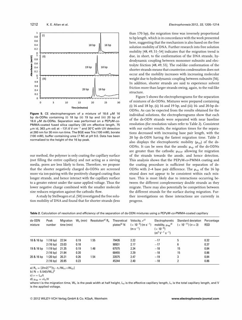
Figure 5. CE electropherogram of a mixture of 18.6 �M 16bp ds-ODNs containing (i) 18 bp (ii) 19 bp and (iii) 20 bp of18.6 �M ds-ODNs. Separation was performed on a PEPyM-co-PMMA-coated fused silica capillary (30 cm effective length, 75�m id, 363 �m od) at −131.6 V cm−1 and 30�C with UV detectionat 260 nm for 30 min run time. The BGE was Tris (100 mM), borate(100 mM), buffer containing urea (7 M) at pH 9.0. Data has beennormalized to the height of the 16 bp peak.
our method, the polymer is only coating the capillary surface(not filling the entire capillary) and not acting as a sievingmedia, pores are less likely to form. Therefore, we proposethat the shorter negatively charged ds-ODNs are screenedmore via ion-pairing with the positively charged coating thanlonger strands, and hence interact with the capillary surfaceto a greater extent under the same applied voltage. Thus thelower negative charge combined with the smaller moleculesize reduces migration against the cathodic flow.
A study by Stellwagen et al. [50] investigated the free solu-tion mobility of DNA and found that for shorter strands (less
than 170 bp), the migration time was inversely proportionalto bp length, which is in concordance with the work presentedhere, suggesting that the mechanism is also based on the freesolution mobility of DNA. Further research into free solutionmobility [48, 49, 51–54] indicates that the migration trend isdue, in short, to the conformation of the DNA strands, hy-drodynamic coupling between monomer subunits and elec-trolyte friction [48, 49, 55]. The rod-like conformation of theshorter strands means that counterion condensation does notoccur and the mobility increases with increasing molecularweight due to hydrodynamic coupling between subunits [56].In addition, shorter strands are said to experience solventfriction more than larger strands owing, again, to the rod-likestructure.
Figure 5 shows the electropherograms for the separationof mixtures of ds-ODNs. Mixtures were prepared containing(i) 16 and 18 bp, (ii) 16 and 19 bp, and (iii) 16- and 20-bp ds-ODNs. As can be expected from the results obtained for theindividual solutions, the electropherograms show that eachof the ds-ODN strands were separated with near baselineresolution (for resolution values refer to Table 2). Consistentwith our earlier results, the migration times for the separa-tions decreased with increasing base pair length, with the20 bp ds-ODN having the fastest migration time. Table 2also displays the electrophoretic mobility (�ep) of the ds-ODNs. It can be seen that the anodic �ep of the ds-ODNsare greater than the cathodic �EOF allowing for migrationof the strands towards the anode, and hence detection.This analysis shows that the PEPyM-co-PMMA coating andthe coating procedure is sufficient for separation of ds-ODNs with 2–4 base pair difference. The �ep of the 16 bpstrand does not appear to be consistent within each mix-ture. This is most likely due to interactions occurring be-tween the different complementary double strands as theymigrate. There may also potentially be competition betweenthe different strands for the surface during migration. Fur-ther investigations on these interactions are currently inprogress.
Table 2. Calculation of resolution and efficiency of the separation of ds-ODN mixtures using a PEPyM-co-PMMA-coated capillary
ds-ODN Peak Migration Wh (min) Resolutiona) Rs Theoretical Velocity, �c) Electrophoretic Standard deviation Percentagemixture number time (min) platesb) N (× 10−4) (m s−1) mobility, �ep
d) (× 10−11) (n = 3) RSD(m s−1) (× 10−9)
(m2 V−1 s−1)
18 & 16 bp 1 (18 bp) 22.54 0.19 1.55 79426 2.22 −17 5 0.322 (16 bp) 23.03 0.18 90931 2.17 −17 6 0.37
19 & 16 bp 1 (19 bp) 21.35 0.19 1.48 67575 2.34 −18 15 0.842 (16 bp) 21.84 0.20 68455 2.29 −18 15 0.86
20 & 16 bp 1 (20 bp) 20.21 0.26 1.54 33575 2.47 −19 3 0.842 (16 bp) 20.85 0.23 45244 2.40 −18 2 0.86
a) Rs = [2ln2]1/2[t2−t1/Wh1+Wh2]b) N = 5.54[t/Wh]2
c) � = Ld/td) �ep = �Lt/Vwhere t is the migration time, Wh is the peak width at half height, Ld is the effective capillary length, Lt is the total capillary length, and Vis the applied voltage.
C© 2012 WILEY-VCH Verlag GmbH & Co. KGaA, Weinheim www.electrophoresis-journal.com

Electrophoresis 2012, 33, 1205–1214 CE and CEC 1213
One advantage of this type of coating is the ease of prepa-ration. After the initial coating which takes an hour (comparedto a day taken by Bernal et al. [21]), the capillary is flushedbetween runs to regenerate the coating. Repetitive injectionsindicate the lifetime of the coating to be in excess of 70 runs.The efficiency of the ds-ODN mixture separations is between33 000 and 91 000 theoretical plates (see Table 2). Resolu-tion of the double strands achieved is greater than 1.4 forall three separations indicating the peaks are well resolved[11]. The repeatability (percentage RSD) of the PEPyM-co-PMMA-coated capillary for ODN analysis was determinedfrom the migration times for all samples. Table 2 shows thatthe coated capillary has high stability based on repeatabilitydata with percentage RSD values less than 0.9% (n = 3) forall mixtures.
4 Concluding remarks
In this work, we show that double-stranded short chain ODNsranging from 16 to 20 bp can be separated down to 2 bp resolu-tion on a fused silica capillary coated with PEPyM-co-PMMAcopolymer physically adsorbed onto the surface. Separationin free solution is possible on the coated capillary due to thereduction in the negative charge distribution reducing the ca-thodic EOF, and the screening of the negatively charged ds-ODNs by the positive charges on the coated surface, meaningthat smaller DNA fragments are retained more strongly bythe capillary. This coating can be achieved within 1 h afford-ing a greatly reduced capillary preparation time. In addition,this method operates within the lower voltage range (−5 kV)which places less stress on the capillary and the system andthus increases lifetime and repeatability. The resolution, andimprovement thereof, of these short strands may prove to beof great importance in future analysis of the purity and con-tent of therapeutic ODNs for pharmaceutical development.
We gratefully acknowledge financial support from the SouthAustralian Justice Department.
The authors have declared no conflict of interest.
5 References
[1] Kleparnık, K., Bocek, P., Chem. Rev 2007, 107, 5279–5317.
[2] Patil, S. D., Rhodes, D. G., Burgess, D. J., AAPS J 2005,7, E61–E77.
[3] Kurreck, J., Eur. J. Biochem 2003, 270, 1628–1644.
[4] Srivatsa, G. S., Klopchin, P., Batt, M., Feldman, M.,Carlson, R. H., Cole, D. L., J. Pharm. Biomed. Anal 1997,16, 619–630.
[5] Khan, K., Van Schepdael, A., Hoogmartens, J., J. Chro-matogr. A 1996, 742, 267–274.
[6] Szekely, L., Kiessig, S., Schwarz, M., Kalman, F., Elec-trophoresis 2009, 30, 1579–1586.
[7] Jekerle, V., Kassack, M. U., Reilly, M. R., Wiese, M.,
Piquette-Miller, M., J. Pharm. Pharmaceut. Sci. 2005, 8,516–527.
[8] Heller, C., Electrophoresis 2001, 22, 629–643.
[9] Pereira, F., Hassard, S., Hassard, P., DeMello, A., Elec-trophoresis 2009, 30, 2100–2109.
[10] Hong, C., Liguo, S., Jieke, C., Wuh. Uni. J. Nat. Sci. 1997,2, 363–367.
[11] Landers, J. P., Handbook of Capillary and Microchip Elec-trophoresis and Associated Microtechniques; 3rd Edi-tion, CRC Press, London, New York 2009.
[12] Zhu, J., Feng, J-L., J. Chromatogr. A 2005, 1081, 19–23.
[13] Yang, R., Wang, Y., Zhou, D., Electrophoresis 2007, 28,3223–3231.
[14] Zhang, J., Liang, D., He, W., Wan, F., Ying, Q., Chu, B.,Electrophoresis 2005, 26, 4449–4455.
[15] Zhang, P., Ren, J., Anal. Chim. Acta 2004, 507, 179–184.
[16] Horvath, J., Dolnık, V., Electrophoresis 2001, 22, 644–655.
[17] Chiari, M., Cretich, M., Damin, F., Ceriotti, L., Consonni,R., Electrophoresis 2000, 21, 909–916.
[18] Chiari, M., Damin, F., Melis, A., Consonni, R., Elec-trophoresis 1998, 19, 3154–3159.
[19] Doherty, E., Berglund, K. D., Buchholz, B., Kourkine, I.V., Przybycien, T. M., Tilton, R. D., Barron, A. E., Elec-trophoresis 2002, 23, 2766–2776.
[20] Znaleziona, J., Petr, J., Knob, R., Maier, V., Sevcık, J.,Chromatographia 2008, 67, 5–12.
[21] Bernal, J., Rodrıguez-Meizoso, I., Elvira, C., Ibanez,E., Cifuentes, A., J. Chromatogr., A 2008, 1204, 104–109.
[22] Bernal, J., Sanchez-Hernandez, L., Elvira, C., Velasco,D., Ibanez, E., Cifuentes, A., J. Sep. Sci. 2009, 32, 605–612.
[23] Erny, G. L., Elvira, C., San Roman, J., Cifuentes, A., Elec-trophoresis 2006, 27, 1041–1049.
[24] Gonzalez, N., Elvira, C., San Roman, J., Cifuentes, A., J.Chromatogr. A 2003, 1012, 95–101.
[25] . Chiou, S.-H., Huang, M.-F., Chang, H.-T., Electrophoresis2004, 25, 2186–2192.
[26] Wang, Q., Xu, X., Dai, L.-X., Chin. J. Chem. 2006, 24,1766–1772.
[27] Kleemiß, M.H., Gilges, M., Schomburg, G., Electrophore-sis 1993, 14, 515–522.
[28] Hjerten, S., J. Chromatogr. 1985, 347, 191–198.
[29] Huang, X., Doneski, L.J., Wirth, M.J., Anal. Chem 1998,70, 4023–4029.
[30] Cifuentes, A., Canalejas, P., Diez-Masa, J.C., J. Chro-matogr. A 1999, 830, 423–438.
[31] Bachmann, S., Vallant, R., Bakry, R., Huck, C. W., Corra-dini, D., Bonn, G. K., Electrophoresis 2010, 31, 618–629.
[32] Madabhushi, R. S., Electrophoresis 1998, 19, 224–230.
[33] Wang, Q., Xu, X., Chin. Chem. Lett 2003, 14, 1278–1280.
[34] Huang, M. F., Huang, C. C., Chang, H. T., Electrophoresis2003, 24, 2896–2902.
[35] Iki, N., Yeung, E. S., J. Chromatogr. A 1996, 731, 273–282.
C© 2012 WILEY-VCH Verlag GmbH & Co. KGaA, Weinheim www.electrophoresis-journal.com

1214 K. E. Allan et al. Electrophoresis 2012, 33, 1205–1214
[36] Chu, B., Ying, Q., Wang, Y., Zhang, Y., Macromol. Symp.2005, 227, 77–88.
[37] Li, W., Ma, Y., Gan, Z., Ling, X., Yang, Z., Chro-matographia 2011, 73, 579–582.
[38] Sartori, A., Barbier, V., Viovy, J.-L., Electrophoresis 2003,24, 421–440.
[39] Braun, B., Blanch, H. W., Prausnitz, J. M., Electrophoresis1997, 18, 1994–1997.
[40] Viovy, J.-L., Duke, T., Electrophoresis, 1993, 14, 322–329.
[41] Grossman, P.D., Soane, D.S., Biopolymers 1991, 31,1221–1228.
[42] Barron, E., Blanch, H.W., Soane, D.S., Electrophoresis1994, 15, 597–615.
[43] Gonzalez, N., Elvira, C., San Roman, J., J. Polym. Sci.,Part A: Polym. Chem. 2003, 41, 395–407.
[44] Rouzina, I., Bloomfield, V. A., Biophys. J. 2001, 80, 894–900.
[45] Ageno, M., Dore, E., Frontali, C., Biophys. J. 1969, 9,1281–1311.
[46] Stellwagen, N. C., Stellwagen, E., J. Chromatogr. A 2009,1216, 1917–1929.
[47] Stellwagen, E., Stellwagen, N. C., Electrophoresis 2002,23, 1935–1941.
[48] Dong, Q., Stellwagen, E., Dagle, J. M., Stellwagen, N. C.,Electrophoresis 2003, 24, 3323–3329.
[49] Stellwagen, N. C., Gelfi, C., Righetti, P. G., Biopolymers1997, 42, 687–703.
[50] Stellwagen, E., Stellwagen, N. C., Electrophoresis 2002,23, 2794–2803.
[51] Mohanty, U., Stellwagen, N. C., Biopolymers 1999, 49,209–214.
[52] Stellwagen, N. C., Gelfi, C., Righetti, P. G., Electrophore-sis 2002, 23, 167–175.
[53] Hoagland, D., Arvanitidou, E., Welch, C., Macro-molecules 1999, 32, 6180–6190.
[54] Allison, S., Chen, C., Stigter, D., Biophys. J 2001, 81,2558–2568.
[55] Stellwagen, N. C., Bossi, A., Gelfi, C., Righetti, P. G., Elec-trophoresis 2001, 22, 4311–4315.
[56] Cottet, H., Pierre, G., Theodoly, O., Williams, C. E., Elec-trophoresis 2000, 21, 3529–3540.
C© 2012 WILEY-VCH Verlag GmbH & Co. KGaA, Weinheim www.electrophoresis-journal.com