Polyamines May Modulate Both G Protein-Coupled Receptors and G Proteins
Upload
independentCategory
view
0download
0
JOURNAL OF VIROLOGY,0022-538X/00/$04.0010
Nov. 2000, p. 10523–10534 Vol. 74, No. 22
Copyright © 2000, American Society for Microbiology. All Rights Reserved.
High-Mobility-Group Protein I Can Modulate Binding of TranscriptionFactors to the U5 Region of the Human Immunodeficiency
Virus Type 1 Proviral PromoterANGUS HENDERSON,1 MICHAEL BUNCE,1 NICOLE SIDDON,1 RAYMOND REEVES,2
AND DAVID JOHN TREMETHICK1*
The John Curtin School of Medical Research, the Australian National University, Canberra,Australian Capital Territory 2601, Australia,1 and Department of Biochemistry
and Biophysics, Washington State University, Pullman, Washington 991642
Received 23 March 2000/Accepted 4 August 2000
HMG I/Y appears to be a multifunctional protein that relies on in its ability to interact with DNA in astructure-specific manner and with DNA, binding transcriptional activators via distinct protein-protein inter-action surfaces. To investigate the hypothesis that HMG I/Y may have a role in human immunodeficiency virustype 1 (HIV-1) expression, we have analyzed whether HMG I/Y interacts with the 5* long terminal repeat andwhether this interaction can modulate transcription factor binding. Using purified recombinant HMG I, wehave identified several high-affinity binding sites which overlap important transcription factor binding sites.One of these HMG I binding sites coincides with an important binding site for AP-1 located downstream of thetranscriptional start site, in the 5* untranslated region at the boundary of a positioned nucleosome. HMG Ibinding to this composite site inhibits the binding of recombinant AP-1. Consistent with this observation, usingnuclear extracts prepared from Jurkat T cells, we show that HMG I (but not HMG Y) is strongly induced uponphorbol myristate acetate stimulation and this induced HMG I appears to both selectively inhibit the bindingof basal DNA-binding proteins and enhance the binding of an inducible AP-1 transcription factor to this AP-1binding site. We also report the novel finding that a component present in this inducible AP-1 complex isATF-3. Taken together, these results argue that HMG I may play a fundamental role in HIV-1 expression bydetermining the nature of transcription factor-promoter interactions.
It is firmly established that chromatin (histones plus a wealthof nonhistone proteins of largely unknown function) plays afundamental role in regulating the transcriptional activity of agene by establishing highly specialized structures that can ei-ther promote or inhibit transcription factor binding. How suchspecialized structures, including the role of many well-charac-terized nonhistone proteins, are established to regulate thetranscription process is poorly understood.
One important function of chromatin is to repress inappro-priate transcription in either a reversible or permanent manner(48). This is achieved by compacting eukaryotic DNA, in ahierarchical fashion, into inaccessible complex three-dimen-sional structures. For transcription to occur, histone-DNA in-teractions in underlying nucleosomes must be disrupted toenable the binding of transcriptional activators to importantregulatory elements. This appears to be achieved in a numberof different ways. Large chromatin remodeling machines existin the nucleus of eukaryotic cells that can, in an ATP-depen-dent manner, facilitate transcription factor binding (35). Post-translational modification of histones, like histone acetylation,also plays an essential role in the gene activation process (28).The existence of cofactors that can increase the affinity andstability of transcriptional activators for their DNA binding siteprovides an alternative strategy by which DNA-binding pro-teins can effectively compete with histones for naked DNA.The ability of HMG I/Y to stimulate the DNA-binding activity
of a wide variety of promoter-specific activators indicates thatthese chromatin-associated proteins may play such a role.
HMG I and Y are isoforms which are produced by alterna-tive splicing, whereas the other member of the family, HMGI-C, is expressed as a separate gene product (6). HMG I/Y candirectly interact via protein-protein interactions, employingdifferent interaction surfaces, with a number of different tran-scriptional activators, including NF-kB (42, 50, 51), ATF-2(13), SRF (8), NF-Y (10), Oct 2A (1), Elf (24), and c-Rel (22),increasing their affinity for DNA. Typically, target genes forthis HMG I/Y enhancement of factor binding are inducibleand include genes for cytokines such as beta interferon (42),interleukin-2 receptor a chain (24), E-selectin (30), interleu-kin-2, and macrophage colony-stimulating factor (22). Morerecently, it was also shown that expression of the nitric oxidesynthase gene may be regulated by HMG I/Y (36). Enhance-ment of factor binding, in some cases, also requires interactionof HMG I/Y with DNA. One explanation for this finding is thatthe ability of HMG I/Y to bend DNA may create a morefavorable DNA conformation for factor binding (15). For thebeta interferon promoter, higher-order transcription factorcomplex formation can be further stabilized by HMG I/Y-factor interactions (14). Paradoxically, in the case of NF-kB,the protein-interacting domain of HMG I/Y includes part ofthe same domain that interacts with DNA (51). The biologicalimportance of HMG I/Y in regulating gene expression is fur-ther implied by the finding that the expression of HMG I/Y isupregulated in rapidly proliferating cells, including early em-bryonic cells (7) and neoplastic tissues (19, 25).
HMG I/Y is characterized by three tandemly organized basicDNA-binding modules separated by a flexible linker; each in-dividual module is capable of interacting with the minor groove
* Corresponding author. Mailing address: The John Curtin Schoolof Medical Research, the Australian National University, P.O. Box334, Canberra, Australian Capital Territory 2601, Australia. Phone:61-6-249 2326. Fax: 61-6-249 0415. E-mail: [email protected].
10523
on February 13, 2016 by guest
http://jvi.asm.org/
Dow
nloaded from
of DNA in a structurally specific manner, with the second basicrepeat being responsible for high-affinity binding (4, 18, 49,51). These consensus basic repeats adopt a defined crescent-shaped planar structure, resembling the drugs netropsin anddistamycin (6). These basic DNA-binding modules are referredto as AT-hooks because in most cases, but not all, they pref-erentially bind to the minor groove of AT-rich sequences (6).These proteins can also bind, with high affinity, to non-B-formDNA, such as synthetic four-way junctions and supercoiledplasmid substrates (6, 21, 34). These binding characteristicssupport the proposal that HMG I/Y may have additional rolesin other DNA-dependent processes.
All stages of the human immunodeficiency virus type 1(HIV-1) life cycle are dependent on host-specific cellular fac-tors. Recently, HMG I/Y was shown to be a host-specific fac-tor required for the integration of HIV-1 preintegration com-plexes (16, 23). To investigate the possibility that HMG I/Ymay also be involved in HIV-1 transcription, we have analyzedthe interaction of HMG I/Y with the viral long terminal repeat(LTR) using purified recombinant proteins and nuclear ex-tracts prepared from living cells. We find that HMG I/Y canfunction to modulate both the efficiency and selectivity of AP-1binding to HIV-1 promoter DNA. These results suggest thatHMG I/Y may play an important role at all key stages in thelife cycle of HIV-1.
MATERIALS AND METHODS
Protein purification and nuclear extract preparation. Recombinant HMG Iand the DNA-binding mutant form of HMG I were purified as described (39).This mutant form has four proline-to-alanine substitutions introduced at residues57 and 61 (located in DNA-binding domain 2) and at residues 83 and 87 (locatedin DNA-binding domain 3). Freeze-dried proteins were resuspended in buffer A(20 mM HEPES [pH 7.9], 100 mM NaCl, 10% [vol/vol] glycerol, 1 mM dithio-threitol, 0.1% NP-40). His-tagged c-Fos and c-Jun (33) were purified by themethod described by Thanos and Maniatis (43). To make the Fos-Jun hetero-dimer, equimolar quantities of c-Fos and c-Jun were codialyzed against buffer A.Typically, the final concentration of Fos-Jun was around 300 ng/ml. Individualpreparations varied considerably in terms of DNA-binding activity, with usuallyless than 10% of the total protein being active (33). As shown in Fig. 3A, maximalbinding to site AP1-3 was achieved with 300 ng of total protein. Nuclear extractswere made from Jurkat T cells according to the method described earlier (11).Half the cells were induced for 2 h, prior to harvesting, with phorbol ester 12-myristate 13-acetate (PMA). To separate HMG I from the bulk of nuclear pro-teins, nuclear proteins were precipitated using 60% ammonium sulfate. Thesupernatant and the nuclear precipitate were dialyzed against buffer D (100 mMKCl) (11). It is worth noting that among the different nuclear extract prepara-tions used (this investigation used four different preparations), the DNA-proteincomplexes produced did not differ qualitatively but differences occurred quan-titatively.
Gel mobility shift assays. The mobility shift assays were carried out as de-scribed previously (33). The oligonucleotide used in gel mobility shift assayswas 59-CCCTTTTAGTCAGTGTGGAAAATCT-39. The final buffer (buffer R)contained 6 mM HEPES (pH 7.9), 10 mM Tris (pH 8.0), 1 mM MgCl2, 1 mMEDTA (pH 8.0), 10 mM dithiothreitol, 5% glycerol, 1% sucrose, 0.1% NP-40,and 40 mM NaCl in a final volume of 20 ml. In the case of DNA-binding reactionsusing nuclear extracts, KCl replaced NaCl. Poly(dG-dC) and/or poly(dI-dC)(Amersham-Mannheim) were also included in reactions using both recombinantproteins (100 ng) and nuclear extracts (2 mg). Reactions were run on preelec-trophoresed 4.5% nondenaturing polyacrylamide gels (0.53 Tris-borate-EDTA[TBE]) at 15 V/cm (4°C). Gels were dried, exposed to X-ray film, and quantitatedby phosphoimaging.
DNase I footprinting assays. DNase I footprinting analysis was carried out asdescribed previously (33). DNA-binding reactions were carried out as for gelshift assays. Two PCR primers, upstream HIV-LTR (2187 to 115) and down-stream HIV-LTR (25 to 1230), were synthesized with restriction sites at bothends to allow footprinting on both strands. Typically, 0.05 U of DNase I (Boehr-inger-Mannheim) was used in reactions involving purified recombinant proteins.Purified digestion products were run on a 7 M urea–8% polyacrylamide gel (13TBE) at 40 V/cm, transferred to DEAE paper (Whatman), dried, and exposed toX-ray film or to a phosphoimage screen.
Western blotting and immunoprecipitation. For Western blot analysis ofHMG I/Y in nuclear extracts, affinity-purified rabbit polyclonal antibodies wereused following the method described by Reeves and Nissen (39). Immunopre-cipitation of HMG I/Y in nuclear extracts was carried out using the methoddescribed before (12). Typically, 8.5 mg of antibody was added to 100 ml of
nuclear extract (7.5 mg/ml). Immunodepletion of HMG I/Y was confirmed byWestern blot and gel shift analyses. AP-1/cyclic AMP response element bindingprotein (CREB) antibodies were purchased from Santa Cruz Biotechnology andused according to their instructions.
Immobilized-template assays. Dynabeads (M280 streptavidin; Dynal) (200 mg)were washed twice in buffer T (10 mM Tris [pH 7.5], 1 mM EDTA, 1 M NaCl).Beads were then resuspended in 20 ml of buffer T containing 10 pmol of biotin-ylated oligonucleotide probe and agitated gently at room temperature for 30 min.After washing several times in buffer T to remove any unbound probe, beadswere equilibrated in buffer R for 20 min. The magnetic beads were concentratedin a magnetic particle concentrator (Dynal) before being resuspended in bufferR containing 250 mg of nuclear protein and 40 ng of poly(dG-dC) per ml in a finalvolume of 120 ml and agitated gently for 20 min at room temperature. Bindingreactions were then washed three times in buffer R containing 10 ng of poly(dG-dC) per ml before the beads were concentrated and resuspended in sodiumdodecyl sulfate (SDS) loading dye, loaded on SDS–12% polyacrylamide gelelectrophoresis (PAGE) gels, and electrophoresed at 15 V/cm for 1.5 h.
RESULTS
Characterization of HMG I/Y binding sites within the 5*-UTR of the HIV-1 promoter. To begin to address the questionof whether HMG I/Y may have a functional role in HIV-1expression, we first examined whether these chromosomal pro-teins can interact with the LTR by determining the location ofpotential HMG I/Y binding sites using the DNase I footprint-ing assay. The region of the LTR that we focused on centredaround the transcriptional initiation site (2187 to 1230). Thisis a particularly interesting region of the promoter, which in-cludes part of the 59-UTR, because it contains a number ofimportant regulatory elements that bind both inducible andconstitutive transcription factors essential for efficient viraltranscription and replication (2, 47). In addition, the 59-UTRinteracts with a specifically positioned nucleosome that is dis-placed or disrupted upon transcriptional activation (46). It isbelieved that the binding of AP-1 to the 59-UTR may be in-volved in this disruption process (33, 47).
Representative footprints are shown in Fig. 1A to C, and asummary of the position of both high- and low-affinity HMGI/Y binding sites is shown in Fig. 2. In these experiments, highlypurified recombinant HMG I was used (Fig. 1D); identicalresults were obtained with recombinant HMG Y (data notshown). Figure 1A displays high-affinity (footprint region 3)and low-affinity (footprint region 2) binding sites. The entireHMG I/Y footprinted region can extend over relatively largeDNA distances (over 30 bp) because they are a composite ofsmaller individual footprints which reflect the binding of indi-vidual AT-hooks to DNA. The binding of at least two AT-hookpeptide motifs is required for high-affinity binding (see Intro-duction). Figures 1B and C also displays high-affinity bindingsites (footprint regions 5 and 3, respectively); only 5 ng ofprotein (21 nM) is required to produce a clear protected re-gion.
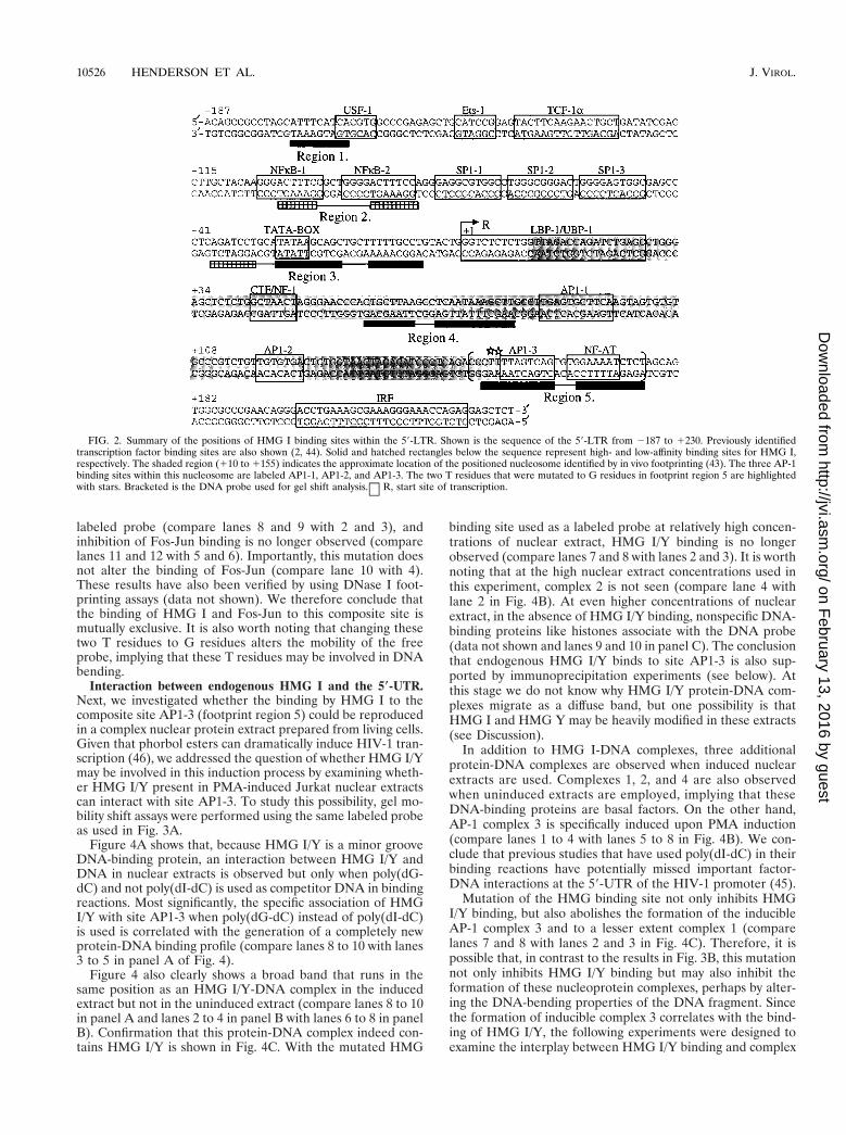
The positions of these and other characterized footprints arelocated at potentially significant regions (Fig. 2). In other re-ported examples, typically HMG I/Y binds to sites that lieadjacent to or are part of a transcription factor binding site(see Introduction). Footprint region 5 overlaps an AP-1 and anNF-AT site, while footprint regions 1, 2, and 3 overlap bindingsites for USF, NF-kB, and TATA-binding protein (TBP), re-spectively. Interestingly, footprint region 4 is located at or nearthe dyad of the positioned nucleosome, whereas footprint re-gions 3 and 5 are located at the boundaries. To begin aninvestigation of whether these HMG binding sites may have abiological function, in this study we have focused on whetherthe binding of HMG I to footprint region 5 can regulate thebinding of AP-1.
HMG I can inhibit the binding of Fos-Jun. The 59-UTR ofthe HIV-1 promoter contains three AP-1 binding sites (AP1-1,
10524 HENDERSON ET AL. J. VIROL.
on February 13, 2016 by guest
http://jvi.asm.org/
Dow
nloaded from

AP1-2, and AP1-3) (Fig. 2). The first two sites are locatedwithin the positioned nucleosome, whereas the third site islocated at the boundary. Given that the binding site for HMGI and this third AP-1 binding site overlap (footprint region 5),the effect of HMG I binding on the binding of Fos-Jun to thissite was analyzed by carrying out a Fos-Jun titration in both thepresence and absence of HMG I. Figure 3A clearly shows thatthe binding of HMG I to the labeled probe inhibits the bindingof Fos-Jun to site AP1-3 (compare lanes 5 to 7 with lanes 2 to4). We estimate that a 5- to 10-fold molar excess of HMG I isrequired to inhibit Fos-Jun binding by more than 80%. Fur-
thermore, this inhibition of Fos-Jun binding is dependent onthe ability of HMG I to bind to DNA, because when we useda DNA-binding mutant, this inhibition of binding was nolonger observed (compare lanes 8 to 10 with lanes 5 to 7).
To confirm that this observed inhibition of Fos-Jun bindingwas due to specific HMG I binding, the binding site of HMGI was mutated, and this probe was used in DNA-binding assays.The mutation involved changing two A residues that lie imme-diately outside the core AP-1 binding site to C residues (TTTTAGTCAG to CCTTAGTCAG), (Fig. 2). Figure 3B showsthat this mutation markedly inhibits HMG I binding to the
FIG. 1. 59-UTR of the HIV-1 promoter contains multiple high-affinity HMG I binding sites. To determine the location of potential HMG I binding sites, DNaseI footprinting reactions were carried out as described in the text. Analysis of three segments within the 59-UTR is shown. These include regions 2105 to 11 (A), 156to 1128 (B), and 1112 to 1194 (C). The numbering is relative to the start site of transcription at 11. Solid boxes represent high-affinity binding sites, whereas shadedboxes illustrate the position of low-affinity HMG I binding sites. Since HMG I contains three distinct individual DNA-binding domains, each capable of producing asmall footprint, the combination of these individual footprints has been described as a footprint region. (D) SDS–15% polyacrylamide gel showing 2 mg of recombinantHMG I.
VOL. 74, 2000 HMG I MODULATES AP-1 BINDING TO HIV-1 PROMOTER 10525
on February 13, 2016 by guest
http://jvi.asm.org/
Dow
nloaded from
labeled probe (compare lanes 8 and 9 with 2 and 3), andinhibition of Fos-Jun binding is no longer observed (comparelanes 11 and 12 with 5 and 6). Importantly, this mutation doesnot alter the binding of Fos-Jun (compare lane 10 with 4).These results have also been verified by using DNase I foot-printing assays (data not shown). We therefore conclude thatthe binding of HMG I and Fos-Jun to this composite site ismutually exclusive. It is also worth noting that changing thesetwo T residues to G residues alters the mobility of the freeprobe, implying that these T residues may be involved in DNAbending.
Interaction between endogenous HMG I and the 5*-UTR.Next, we investigated whether the binding by HMG I to thecomposite site AP1-3 (footprint region 5) could be reproducedin a complex nuclear protein extract prepared from living cells.Given that phorbol esters can dramatically induce HIV-1 tran-scription (46), we addressed the question of whether HMG I/Ymay be involved in this induction process by examining wheth-er HMG I/Y present in PMA-induced Jurkat nuclear extractscan interact with site AP1-3. To study this possibility, gel mo-bility shift assays were performed using the same labeled probeas used in Fig. 3A.
Figure 4A shows that, because HMG I/Y is a minor grooveDNA-binding protein, an interaction between HMG I/Y andDNA in nuclear extracts is observed but only when poly(dG-dC) and not poly(dI-dC) is used as competitor DNA in bindingreactions. Most significantly, the specific association of HMGI/Y with site AP1-3 when poly(dG-dC) instead of poly(dI-dC)is used is correlated with the generation of a completely newprotein-DNA binding profile (compare lanes 8 to 10 with lanes3 to 5 in panel A of Fig. 4).
Figure 4 also clearly shows a broad band that runs in thesame position as an HMG I/Y-DNA complex in the inducedextract but not in the uninduced extract (compare lanes 8 to 10in panel A and lanes 2 to 4 in panel B with lanes 6 to 8 in panelB). Confirmation that this protein-DNA complex indeed con-tains HMG I/Y is shown in Fig. 4C. With the mutated HMG
binding site used as a labeled probe at relatively high concen-trations of nuclear extract, HMG I/Y binding is no longerobserved (compare lanes 7 and 8 with lanes 2 and 3). It is worthnoting that at the high nuclear extract concentrations used inthis experiment, complex 2 is not seen (compare lane 4 withlane 2 in Fig. 4B). At even higher concentrations of nuclearextract, in the absence of HMG I/Y binding, nonspecific DNA-binding proteins like histones associate with the DNA probe(data not shown and lanes 9 and 10 in panel C). The conclusionthat endogenous HMG I/Y binds to site AP1-3 is also sup-ported by immunoprecipitation experiments (see below). Atthis stage we do not know why HMG I/Y protein-DNA com-plexes migrate as a diffuse band, but one possibility is thatHMG I and HMG Y may be heavily modified in these extracts(see Discussion).
In addition to HMG I-DNA complexes, three additionalprotein-DNA complexes are observed when induced nuclearextracts are used. Complexes 1, 2, and 4 are also observedwhen uninduced extracts are employed, implying that theseDNA-binding proteins are basal factors. On the other hand,AP-1 complex 3 is specifically induced upon PMA induction(compare lanes 1 to 4 with lanes 5 to 8 in Fig. 4B). We con-clude that previous studies that have used poly(dI-dC) in theirbinding reactions have potentially missed important factor-DNA interactions at the 59-UTR of the HIV-1 promoter (45).
Mutation of the HMG binding site not only inhibits HMGI/Y binding, but also abolishes the formation of the inducibleAP-1 complex 3 and to a lesser extent complex 1 (comparelanes 7 and 8 with lanes 2 and 3 in Fig. 4C). Therefore, it ispossible that, in contrast to the results in Fig. 3B, this mutationnot only inhibits HMG I/Y binding but may also inhibit theformation of these nucleoprotein complexes, perhaps by alter-ing the DNA-bending properties of the DNA fragment. Sincethe formation of inducible complex 3 correlates with the bind-ing of HMG I/Y, the following experiments were designed toexamine the interplay between HMG I/Y binding and complex
FIG. 2. Summary of the positions of HMG I binding sites within the 59-LTR. Shown is the sequence of the 59-LTR from 2187 to 1230. Previously identifiedtranscription factor binding sites are also shown (2, 44). Solid and hatched rectangles below the sequence represent high- and low-affinity binding sites for HMG I,respectively. The shaded region (110 to 1155) indicates the approximate location of the positioned nucleosome identified by in vivo footprinting (43). The three AP-1binding sites within this nucleosome are labeled AP1-1, AP1-2, and AP1-3. The two T residues that were mutated to G residues in footprint region 5 are highlightedwith stars. Bracketed is the DNA probe used for gel shift analysis. R, start site of transcription.
↵
10526 HENDERSON ET AL. J. VIROL.
on February 13, 2016 by guest
http://jvi.asm.org/
Dow
nloaded from

3 formation and whether the ability of HMG I to inhibit factorbinding (Fig. 3A) has a role in this process.
HMG I is induced upon PMA stimulation. The results inFig. 4 indicate that HMG I/Y associates with site AP1-3 uponPMA induction. To investigate this further, a Western blotanalysis was carried out examining the abundance of HMG Iand HMG Y in uninduced and induced Jurkat extracts (Fig.5A) (the SDS protein gel above the Western blot demonstratesthat appropriate induced and uninduced lanes received anequivalent amount of protein; see the figure legend). Mostinterestingly, HMG I is markedly induced (about fivefold),whereas the induction of HMG Y was modest (compare lane2 with lane 3 and lane 6 with lane 7). In an attempt to separateHMG I/Y from the bulk of nuclear protein, (NH4)2SO4 wasadded to the nuclear extract (final concentration, 60%). Mostunexpectedly, HMG I fractionated to the supernatant, whereasHMG Y was precipitated with the bulk of nuclear protein(compare lanes 5 and 6 with lanes 3 and 4).
Having separated HMG I from HMG Y, it was of interest toinvestigate whether the HMG binding observed in unfraction-ated nuclear extracts (Fig. 4A) was due to the interaction ofHMG I or HMG Y, or both, to site AP1-3. DNA-bindingassays were carried out using induced and uninduced nuclearpellet and supernatant fractions, and the protein-DNA com-plexes formed were analyzed by mobility gel shift assays. Fig-ure 5B clearly shows that HMG I present in the inducedsupernatant fraction can bind strongly to the labeled probe.Very little binding is observed in the uninduced supernatantfraction (compare lanes 12 to 15 with lanes 16 to 19). Inter-estingly, HMG Y present in the induced nuclear pellet fraction
does bind to the labeled DNA fragment (lanes 2 to 6). Wetherefore conclude that HMG I is responsible for the bindingobserved in unfractionated nuclear extracts. Figure 5B alsoshows that basal complex 1 and a new complex (which migratesfaster than complex 3) are also present in the supernatantfractions.
HMG I can selectively determine which nuclear DNA-bind-ing protein binds to the 5*-UTR. The above results have clearlydemonstrated that using recombinant proteins, HMG I caninhibit AP-1 binding to site AP1-3 on the 59-UTR (Fig. 3). Onthe other hand, using induced Jurkat nuclear extracts, an in-ducible AP-1 factor can bind to DNA in the presence of HMGI (Fig. 4). An attractive hypothesis that can reconcile theseobservations is that the induced HMG I can both selectivelyinhibit the binding of basal DNA-binding proteins (which arepresent in both uninduced and induced nuclear extracts) andpromote the binding of the inducible AP-1 transcription factor.To test this hypothesis, we took advantage of our ability toseparate HMG I from the inducible AP-1 complex (Fig. 5A). Atitration was performed in which HMG I, present in the(NH4)2SO4 supernatant fraction, was added to a constantamount of precipitated nuclear protein which contains thePMA-inducible AP-1 factor (complex 3) (Fig. 6B). The super-natant fraction was immunoprecipitated with either HMG Iantibodies or control antibodies against histone H2A.
Figure 6A shows that HMG I/Y immunodepletion, and notmock or immunodepletion of H2A, of the induced supernatantfraction removes only HMG I and not the other DNA-bindingproteins present in this fraction, including basal factor 1. Lane2 of Fig. 6B shows the formation of basal complex 2 and the
FIG. 3. HMG I inhibits the binding of Fos-Jun to site AP1-3. (A) To examine the effect of HMG I on the binding of Fos-Jun to site AP1-3, a Fos-Jun titration (seeMaterials and Methods) was carried out in which either no protein, 25 ng of HMG I, or 25 ng of a mutant DNA-binding form of HMG I (mHMG I) was added tobinding reactions containing a 25-bp probe. (B) The experiment in panel A was repeated except that the HMG I/Y binding site was mutated. Lanes 2, 5, 8, and 11received 12 ng of HMG I, while lanes 3, 6, 9, and 12 received 25 ng of HMG I.
VOL. 74, 2000 HMG I MODULATES AP-1 BINDING TO HIV-1 PROMOTER 10527
on February 13, 2016 by guest
http://jvi.asm.org/
Dow
nloaded from

inducible AP-1 complex 3 in the induced nuclear precipitatedfraction. When control immunodepleted supernatant (contain-ing HMG I) is titrated back to the (NH4)2SO4-precipitatedfraction, at the lowest concentration of supernatant, formationof basal complex 2 is inhibited (compare lane 3 with lane 2). As
the concentration of HMG I is increased in the binding reac-tion, the formation of inducible AP-1 complex 3 is enhanced.Concurrently, the production of basal complex 1 is also ob-served (this is because this basal factor is present in the super-natant fraction). This binding profile mimics the situation
FIG. 4. Endogenous HMG I binds to site AP1-3. (A) DNA-binding reactions were carried out using a labeled probe containing site AP1-3 and increasing amountsof a nuclear extract (NE) prepared from PMA-induced Jurkat cells. As indicated, reactions received either poly(dI-dC) or poly(dG-dC) (2 mg). Lanes 2 and 7, 3 and8, 4 and 9, and 5 and 10 received 1, 5, 10, and 20 mg of nuclear extract, respectively. A mobility gel shift assay was performed to identify assembled nucleoproteincomplexes. (B) DNA-binding reactions, as indicated, were carried out using nuclear extracts prepared from uninduced and PMA-induced Jurkat cells. The nucleo-protein complexes generated are shown as complexes 1 to 4. The formation of HMG I-DNA complexes is also highlighted. Lanes 1 and 5, 2 and 6, 3 and 7, and 4 and8 received 2, 5, 10, and 15 mg of nuclear extract, respectively. (C) Mobility gel shift assay was carried out using labeled DNA probes that contained either a mutatedor an unmodified HMG I/Y binding site. Lanes 2 to 5 and lanes 7 to 10 received 15, 20, 25, and 30 mg of nuclear extract, respectively.
10528 HENDERSON ET AL. J. VIROL.
on February 13, 2016 by guest
http://jvi.asm.org/
Dow
nloaded from

when the unfractionated nuclear extract is used in bindingreactions (compare lane 7 of Fig. 6B with lane 4 of Fig. 4B).
Strikingly, when HMG I is immunodepleted, the productionof the inducible AP-1 complex 3 is markedly reduced, while theformation of basal complex 1 is enhanced (Fig. 6B, comparelanes 9 to 12 with lanes 4 to 7). This result clearly shows thatthe inducible AP-1 factor is in competition with the basalDNA-binding protein for the same binding site and that HMGI selectively facilitates the formation of the inducible AP-1–DNA complex. This result can also be mimicked when theuninduced supernatant fraction is added back (data notshown). In addition, at the lowest concentration of HMG I-depleted supernatant added, the formation of basal complex 2is no longer inhibited (compare lane 8 with lane 3). However,as the concentration of the HMG I-immunodepleted superna-tant increases, basal factor 1 outcompetes basal factor 2 forDNA binding. Clearly, there is a complex interplay betweenthe inducible AP-1 factor, the different basal factors, andHMG I for DNA binding, but this result clearly shows that inthis complex mixture of proteins, HMG I selectively enhancesthe binding of the inducible AP-1 factor to site AP1-3.
To confirm that HMG I can indeed inhibit the DNA-bindingactivity of basal factor 2, an HMG I titration was carried out inwhich recombinant HMG I was added to uninduced nuclearprecipitated extracts. Lane 2 of Fig. 6C shows the formation ofDNA-protein complexes 2 and 4. As increasing amounts ofHMG I are added to the binding reactions, formation of bothof these complexes is inhibited, which coincides with the bind-ing of HMG I to the labeled probe. This result clearly showsthat HMG I can inhibit the binding not only of recombinantAP-1, but also of basal DNA-binding proteins present in un-induced nuclear extracts. Taken together, these results supportthe above hypothesis by showing that HMG I can both inhibitand facilitate factor binding in a selective manner and providea potential mechanism for how this nonhistone chromosomalprotein may play an important role in HIV-1 expression by
determining which transcription factor associates with impor-tant DNA regulatory elements.
PMA-inducible AP-1 complex contains ATF-3. Having de-termined that HMG I plays an important role in facilitating thebinding of inducible AP-1 complex 3 to DNA, the next impor-tant question to be addressed was to identify the key AP-1components present in this inducible complex. Previously, itwas reported that by employing supershift assays using anti-bodies raised against different AP-1 members, c-Fos, Jun D,CREB, ATF-1, and ATF-2 interacted with site AP1-3 (37, 38).However, employing this assay, we were unable to reproducethese results. Furthermore, close examination of these re-ported studies revealed that these transcription factors are onlyminor components of the inducible complex. We thereforeadopted an alternative approach to determine the major com-ponents of this inducible complex. Biotinylated site AP1-3 wasincubated with PMA-induced and uninduced nuclear extractsunder the same conditions as used for gel shift experiments(Fig. 4). The DNA-bound transcription factors were then pu-rified from the bulk of nuclear proteins by using magneticstreptavidin beads (Fig. 7B). After several washes with bindingbuffer, the isolated DNA-bound proteins were eluted from theDNA and analyzed by Western blotting using a battery ofdifferent ATF/CREB family members (Fig. 7A). In crude nu-clear extracts, all of the tested ATF/CREB family memberswere present. As expected, treatment of Jurkat T cells withPMA, under the conditions used here, induced the synthesis ofc-Fos, CREB-1, c-Jun, and ATF-3. Most interestingly, neitherCREB-1, c-Jun, nor c-Fos was purified by site AP1-3 attachedto magnetic streptavidin beads. On the other hand, we find thatATF-3 is the major component purified from the crude nuclearextract. A minor component that binds to site AP1-3 is ATF-2.Importantly, as a control, these transcription factors were notdetected when magnetic beads alone were incubated with nu-clear extracts (data not shown). The specificity of this assay isalso highlighted by the observation that despite a strong anti-
FIG. 5. PMA induction of Jurkat T cells induces HMG I. (A) Nuclear extract (NE) proteins prepared from uninduced and PMA-induced Jurkat T cells wereprecipitated using 60% (NH4)2SO4 (see Materials and Methods). Western blot analysis of precipitated and supernatant fractions was carried out using affinity-purifiedpolyclonal antibodies raised against HMG I/Y. Lanes 2 to 5 and 6 and 7 received 40 and 10 mg of total protein, respectively. (B) Mobility gel shift assay carried outusing site AP1-3 and (NH4)2SO4-precipitated (ppt) and supernatant (supern.) fractions derived from uninduced and induced nuclear extracts. Lanes 2 to 6 and 7 to11, received 1, 2, 5, 10, and 15 mg of total protein, respectively. Lanes 12 to 15 and 16 to 19 received 2, 5, 10, and 15 mg of total protein, respectively.
VOL. 74, 2000 HMG I MODULATES AP-1 BINDING TO HIV-1 PROMOTER 10529
on February 13, 2016 by guest
http://jvi.asm.org/
Dow
nloaded from

body-Jun D reaction in crude nuclear extracts, Jun D does notbind to the purified DNA probe. In addition to ATF-3, Fig. 7Bshows that Fra-1 is a factor present in uninduced nuclear ex-tracts which binds strongly to site AP1-3. Under the inductionconditions used here, the level of DNA binding does notchange between the induced and uninduced states.
DISCUSSION
The focus of this study was to examine whether HMG I caninteract with an important regulatory region in the HIV-1
promoter, the 59-UTR, and whether this interaction could playa role in modulating the binding of AP-1 to key binding sites inthis region. The approach adopted was to define HMG I in-teractions, in combination with AP-1, with the 59-UTR usingrecombinant proteins. Then we tested the significance of thesefindings by employing nuclear extracts containing endogenousHMG I/Y prepared from PMA-induced Jurkat extracts, sincephorbol esters are strong inducers of HIV-1 expression.
In the 59-UTR, we have identified several high-affinity sitesfor HMG I/Y, and most of these sites overlap important tran-scription factor binding sites. Interestingly, similar uncharac-terized footprints can be seen in a genomic footprint of inte-grated HIV-1 (see Fig. 2 in reference 5). A number of previousstudies have shown that such an overlap can have a critical rolein regulating gene expression (22, 24, 30, 36). We have focusedon one of these overlapping binding sites, site AP1-3. Thelocation of this AP-1 binding site, at the boundary of a posi-tioned nucleosome, suggests that it may play an important role
FIG. 6. HMG I can both selectively inhibit and promote endogenous factorbinding to site AP1-3. (A) The 60% (NH4)2SO4 supernatant fraction, derivedfrom PMA-induced nuclear extracts (NE), was mock immunodepleted (lanes 2to 6) or immunodepleted with affinity-purified antibodies against HMG I/Y(lanes 7 to 11) or H2A (control, lanes 12 to 16). A mobility gel shift assay, usingsite AP1-3, was carried out using these immunodepleted supernatant fractions.Lanes 2 to 6, 7 to 11, and 12 to 16 received 2, 5, 10, 15, and 20 mg of thesupernatant fraction, respectively. (B) PMA-induced supernatant fractions thatwere immunodepleted with either control (H2A) or HMG I affinity-purifiedantibodies were titrated back to a PMA-induced nuclear precipitated fraction.Lanes 2 to 12 received 7 mg of nuclear (NH4)2SO4-precipitated proteins. Lanes3 to 7 and lanes 8 to 12 received 5, 10, 20, 30, and 40 mg of H2A- or HMGI/Y-depleted supernatant fractions, respectively. (C) Increasing amounts of re-combinant HMG I were added to DNA-binding reactions that received 10 mg ofuninduced nuclear precipitated extract and a labeled oligonucleotide that con-tained site AP1-3. Lanes 2 to 6 received 0, 25, 50, 100, and 200 ng of HMG I,respectively.
10530 HENDERSON ET AL. J. VIROL.
on February 13, 2016 by guest
http://jvi.asm.org/
Dow
nloaded from

in modulating chromatin architecture. We have observed en-hanced HMG I binding to site AP1-3 using PMA-inducednuclear extracts but only under in vitro conditions that allowHMG I binding, i.e., when poly(dG-dC) rather than poly(dI-dC) is used as competitor DNA. Most importantly, PMA alsoinduces the synthesis of a new AP-1 complex, and strong bind-ing of this transcription factor to site AP1-3 is dependent uponHMG I. Because of this new finding, previous in vitro bindingstudies should be reevaluated. Concerning the role of HMGI/Y, we cannot rule out the possibility that at least a subset ofthese identified binding sites may have a role in the integrationprocess.
A major unanswered question concerning the function ofthese chromosomal proteins is whether HMG I and Y play thesame role or have different roles. Interestingly, we observedthat HMG I and not HMG Y is strongly induced upon PMAstimulation of Jurkat cells. This suggests that these proteinsmay indeed have different functions. Given that these twoproteins originate from alternative splicing of the same RNAtranscript, this differential regulation must occur via a post-transcriptional mechanism. Such differential control could, forexample, operate at the level of the splicing event itself, resultfrom differences in the stability or translational efficiency of thealternatively spliced transcripts, or, perhaps, result from intrin-
sic differences in the stability of the HMG I and HMG Yproteins (although both proteins appear to be extremely stablein living cells) (6). A similar selective enhancement of HMG Iprotein by tetradecanoyl phorbol acetate was observed intransformation-resistant JB6 murine cell lines (9). Most inter-estingly, this same study raised the possibility that HMG Y mayhave a role in the transformation process, since they observedthat HMG Y is induced by tumor promoters only in transfor-mation-sensitive cells. Potentially, the conserved 11-amino-acid segment absent in HMG Y might alter the quality or thespecificity of protein-protein interactions with target proteins.Concerning their DNA-binding activity in Jurkat nuclear ex-tracts, we also report here another difference between HMG Iand HMG Y. HMG Y is present in both uninduced and in-duced nuclear extracts and, at least as shown by mobility gelshift assays, does not bind to DNA when both of these nuclearextracts are used. Although this may be due to low abundance,another possible explanation for this is that HMG Y is pos-translationally modified in vivo in a differential manner fromHMG I (Banks and Reeves, unpublished data), and it is thisconstellation of secondary biochemical modifications (includ-ing phosphorylation and others) that inhibits the binding of theHMG Y isoform protein to DNA (32). Clearly, this hypothesiswill need to be tested.
FIG. 7. Inducible AP-1 complex 3 contains ATF-3. Biotinylated site AP1-3 was incubated with induced or uninduced nuclear extract (NE) (250 mg of total protein),and the DNA-bound AP-1 factors were isolated as described in Materials and Methods. The isolated proteins were run on an SDS–12% polyacrylamide gel and eitherprobed with commercial antibodies raised against different AP-1/CREB family members (A) or stained with silver (B). To determine which AP-1/CREB familymembers are present initially in uninduced and induced nuclear extracts, 15 mg of unpurified nuclear extract was also probed with the different antibodies (A) andstained with silver (B). NE, uninduced Jurkat T cells; PMA-NE, Jurkat T cells stimulated with PMA.
VOL. 74, 2000 HMG I MODULATES AP-1 BINDING TO HIV-1 PROMOTER 10531
on February 13, 2016 by guest
http://jvi.asm.org/
Dow
nloaded from
Many transcription factor families, like AP-1, comprise dif-ferent individual members which are able to dimerize witheach other to form a diverse range of transcription factorcomplexes, each capable of recognizing the same or similarDNA-binding sequences. The mechanisms that operate in thenucleus to determine which factor actually binds to a regula-tory site in a promoter are poorly understood. The resultspresented here suggest that HMG I may play an important rolein this selection process. We found that in a complex nuclearextract consisting of a wealth of DNA-binding proteins, a sit-uation analogous to the environment within the nucleus, HMGI can selectively promote the binding of an inducible AP-1factor, ATF-3, to the AP1-3 site (complex 3), relative to thebinding of competing basal DNA-binding proteins. In such acompetition mechanism, the ability of HMG I to increase theaffinity or stability of ATF-3 for site AP1-3 would enable thisfactor to compete more effectively with basal factors. The bind-ing of this inducible AP-1 factor would be further enhanced ifHMG I could selectively and directly inhibit the binding ofbasal factors. Indeed, such an inhibition of binding was ob-served when HMG I was added to uninduced extracts. There-fore, HMG I can either selectively interfere with or enhanceprotein-DNA binding, and which of these events occurs ap-pears to be partially dependent on the nature of the DNA-binding protein involved (see below). Precedents exist for sucha mode of differential regulation by the HMG I protein. Pre-viously, using two different recombinant isoforms of ATF-2,ATF-2195 and ATF-2192, it was shown that while HMG I couldstimulate ATF-2195 DNA binding, it actually inhibited thebinding of ATF-2192 to the beta interferon promoter. ATF-2192lacks important amino acid residues involved in HMG I–ATF-2 interactions (13). Such a striking differential effect wasalso observed when the interaction of Oct-1 and Oct-2 with theoctamer sequence was compared (1). HMG I selectively en-hanced Oct-2 binding while at the same time inhibiting Oct-1–DNA interactions.
There are at least two, nonmutually exclusive, mechanismsby which HMG I could enhance ATF-3 binding to site AP1-3in a PMA-induced nuclear extract. These HMG proteins havebeen described as being architectural transcription factors be-cause they can bend DNA, thereby establishing a particularDNA conformation that may be more favorable for transcrip-tion factor binding (6). The binding of several HMG moleculesalong a stretch of DNA may even create a DNA scaffold thatcan facilitate the formation of large nucleoprotein complexesvia cooperative protein-protein interactions. Such a mecha-nism operates in the induction of human beta interferon geneexpression (44). Individually, the binding of two HMG I mol-ecules to positive regulatory domains IV and II enhances ATF-2/c-Jun and NF-kB binding, respectively, by reversing intrinsicDNA bends at these factor-binding sites. In combination, thesetwo HMG proteins facilitate the formation of a highly stableand sterospecific transcription factor complex. The selectivityobserved with regard to the enhanced binding of PMA-induc-ible ATF-3 to site AP1-3 could, in part, be explained by thegeneration of a highly specific DNA structure by HMG I.Analogous to the human beta interferon gene, it will also beinteresting to examine whether the location of multiple HMGI binding sites in the 59-UTR contributes to the formation of alarge stable nucleoprotein complex.
Potentially, HMG I could also selectively enhance the bind-ing of one transcription factor over another by its ability tointeract directly with transcription factors themselves via spe-cific protein-protein interaction surfaces. Indeed, stabilizationof the assembled nucleoprotein complex on the human betainterferon gene enhancer appears to require specific protein-
protein interactions between HMG I and transcriptional acti-vators (50). Such protein-protein interactions can also enhancefactor binding in a mechanism that is independent of HMG I’sinteracting with DNA. For example, protein-protein interac-tions between HMG I and NF-Y (10) and SRF (8) are suffi-cient for enhancement of their DNA-binding activity, presum-ably by inducing an active conformation. It has also beenshown that HMG I can, in part, stimulate the binding of ATF-2/c-Jun to positive regulatory domain IV by promoting thedimerization reaction. Interestingly, we have observed thatHMG I can enhance Fos-Jun binding to site AP1-1 in nuclearextracts even though HMG I does not bind to this AP-1 site(unpublished data).
Our results, using recombinant and nuclear proteins, alsosuggest that the second part of the mechanism that promotesspecific binding of the inducible AP-1 complex to the compos-ite site AP1-3 is the selective inhibition of binding of basalDNA-binding proteins by HMG I. Since the binding sites forHMG I and AP-1 overlap, this competitive inhibition could beachieved either by direct steric hindrance or by HMG I alteringthe conformation of the DNA to a structure that is not com-patible for basal factor binding. Interestingly, HMG I does notinhibit the binding of recombinant NF-AT to an adjacent bind-ing site (Fig. 2 and data not shown). As discussed above, HMGI can selectively inhibit the DNA-binding activity of ATF-2192and Oct-1. It has also been reported that HMG I can inhibitthe binding of NF-AT factors to the interleukin-4 promoter(26). This inhibition of binding, which is reversed by phosphor-ylation of HMG I, is believed to be important for developmentof Th2 cells. Similarly, HMG I (and HMG I-C) can interferewith the binding of homeodomain proteins to target sequences(these sequences contain TAAT as a core motif) (3). Thisinhibition appears to be due to HMG I-induced conforma-tional changes in the DNA. Therefore, it appears that HMG Ican modulate gene expression by functioning either as an ac-tivator or as a repressor. Moreover, we show here that HMG Ican enhance the binding of one DNA-binding protein andinhibit the binding of another factor even on the same tran-scription factor-binding site. We also conclude that the finaloutcome of whether HMG I selectively inhibits or stimulatestranscription factor binding is dependent not only on the na-ture of the transcriptional activator, but also on a complexinterplay between relative DNA affinities and protein concen-trations, the location of the HMG binding site with respect tothe factor-binding site, and presumably, the biochemical mod-ification status of HMG I.
Transfection-cocultivation experiments with wild-type andmutant HIV-1 proviral DNAs have show that the three AP-1sites encompassing the downstream-positioned nucleosomeplay a fundamental role in the life cycle of the virus. SitesAP1-1 and AP1-2 (Fig. 2) play a critical role on HIV-1 repli-cation, while site AP1-3 appears to be important for transcrip-tional activation in response to a broad range of external stim-uli under different physiological conditions (37, 38, 41, 45).This site is highly conserved among HIV-1 isolates. Studiesemploying supershift analysis have reported that AP-1 com-plexes that interact with this site contain c-Fos and Jun D aswell as CREB, ATF-1, and ATF-2 (38, 41). In contrast to thesepublished results, using a more stringent assay, we have foundthat ATF-3 is a major component that interacts with siteAP1-3. One possible reason for this discrepancy is that underour DNA-binding conditions, HMG I is able to interact withthe DNA, thereby influencing the composition of bound tran-scription factors.
The association of ATF-3 with site AP1-3 is consistent withthe observation that this site responds to a broad range of
10532 HENDERSON ET AL. J. VIROL.
on February 13, 2016 by guest
http://jvi.asm.org/
Dow
nloaded from

extracellular stimuli. ATF-3 mRNA is induced in cultured cellswithin 2 h by many treatments like different growth-stimulatingfactors, PMA, and cytokines as well as physiological stresses,including tissue damage (reviewed in reference 20). Consistentwith these observations, analysis of the 59-flanking region ofthe ATF-3 gene revealed inducible AP-1, ATF/CRE, andNF-kB binding sites. Interestingly, E2F and Myc/Max bindingsites were also identified, raising the possibility that ATF-3may be regulated in a cell cycle-dependent manner (31).ATF-3 functions as an activator when it heterodimerizes withJun family members (20). The results of this study show thatneither c-Jun nor Jun D partners ATF-3 to assemble PMA-inducible complex 3. To date, no physiologically importanttarget genes mediating the activation of signaling pathwaysinvolving ATF-3 have been identified. Interestingly, we alsohave identified the Fos-related antigen Fra-1 as a factor thatbinds to site AP1-3 in uninduced nuclear extracts. Consistentwith this observation, it has been reported that, in contrast toc-Fos, the fra-1 gene is expressed at high levels in proliferatingcells (27).
The results of this study raise the possibility that HMG I mayplay an important role in HIV-1 expression. Preliminary ex-periments have shown that antisense HMG I RNA can reducethe expression of HIV reporter constructs (data not shown).Other viruses might employ this strategy to ensure their prop-agation in host cells, especially if critical transcription factorshave a low affinity for important viral promoter elements.HMG I has been shown to stimulate Tst-1/Oct-6 binding to animportant regulatory element that mediates the activation ofhuman papovavirus JC virus gene expression (29). The latency-active promoter 2 in herpes simplex virus type 1 (which be-comes a nucleosomal episome) contains a stretch of 23 thymi-dine residues critical for promoter activity, which interacts withHMG I/Y. In vitro, the binding of HMG I/Y to this promoterelement enhances the binding of SP-1 to neighboring bindingsites (17). With regard to chromatin disruption and the acti-vation of HIV-1 transcription, based on the location of siteAP1-3 at the boundary of the positioned nucleosome, we pos-tulate that HMG I may play a fundamental role in the chro-matin remodeling process. The association of HMG I with thenucleosomal dyad may also contribute to this disruption pro-cess (40). We are currently testing this hypothesis.
ACKNOWLEDGMENTS
The first two authors contributed equally to the manuscript.We thank Mark Nissen for providing recombinant HMG proteins,
Adele Holloway for assistance in setting up the immobilized-templateassay, and Frances Shannon for many helpful discussions.
REFERENCES
1. Abdulkadir, S. A., S. Krishna, D. Thanos, T. Maniatis, J. L. Strominger, andS. J. Ono. 1995. Functional roles of the transcription factor Oct-2A and thehigh mobility group protein I/Y in HLA-DRA gene expression. J. Exp. Med.182:487–500.
2. Al-Harthi, L., and K. A. Roebuck. 1998. Human immunodeficiency virustype-1 transcription: role of the 59-untranslated leader region. Int. J. Mol.Med. 1:875–881.
3. Arlotta, P., A. Rustighi, F. Mantovani, G. Manfioletti, V. Giancotti, G. Tell,and G. Damante. 1997. High mobility group I proteins interfere with thehomeodomains binding to DNA. J. Biol. Chem. 272:29904–29910.
4. Banks, G. C., B. Mohr, and R. Reeves. 1999. The HMG-I(Y) A.T-hookpeptide motif confers DNA-binding specificity to a structured chimeric pro-tein. J. Biol. Chem. 274:16536–16544.
5. Brown, D. A., X. Xu, and M. Nerenberg. 1996. Genomic footprinting ofHTLV type I and HIV type 1 in human T cell lines. AIDS Res. Hum.Retroviruses 12:829–832.
6. Bustin, M., and R. Reeves. 1996. High-mobility-group chromosomal pro-
teins: architectural components that facilitate chromatin function. Prog. Nu-cleic Acid Res. Mol. Biol. 54:35–100.
7. Chiappetta, G., V. Avantaggiato, R. Visconti, M. Fedele, S. Battista, F.Trapasso, B. M. Merciai, V. Fidanza, V. Giancotti, M. Santoro, A. Simeone,and A. Fusco. 1996. High level expression of the HMGI (Y) gene duringembryonic development. Oncogene 13:2439–2446.
8. Chin, M. T., A. Pellacani, H. Wang, S. S. Lin, M. K. Jain, M. A. Perrella, andM. E. Lee. 1998. Enhancement of serum-response factor-dependent tran-scription and DNA binding by the architectural transcription factor HMG-I(Y). J. Biol. Chem. 273:9755–9760.
9. Cmarik, J. L., Y. Li, S. A. Ogram, H. Min, R. Reeves, and N. H. Colburn.1998. Tumor promoter induces high mobility group HMG-Y protein expres-sion in transformation-sensitive but not -resistant cells. Oncogene 16:3387–3396.
10. Currie, R. A. 1997. Functional interaction between the DNA binding subunittrimerization domain of NF-Y and the high mobility group protein HMG-I(Y). J. Biol. Chem. 272:30880–30888.
11. Dignam, J. D., R. M. Lebovitz, and R. G. Roeder. 1983. Accurate transcrip-tion initiation by RNA polymerase II in a soluble extract from isolatedmammalian nuclei. Nucleic Acids Res. 11:1475–1489.
12. Dimitrov, S., M. C. Dasso, and A. P. Wolffe. 1994. Remodeling spermchromatin in Xenopus laevis egg extracts: the role of core histone phosphor-ylation and linker histone B4 in chromatin assembly. J. Cell Biol. 126:591–601.
13. Du, W., and T. Maniatis. 1994. The high mobility group protein HMG I(Y)can stimulate or inhibit DNA binding of distinct transcription factor ATF-2isoforms. Proc. Natl. Acad. Sci. USA 91:11318–11322.
14. Du, W., D. Thanos, and T. Maniatis. 1993. Mechanisms of transcriptionalsynergism between distinct virus-inducible enhancer elements. Cell 74:887–898.
15. Falvo, J. V., D. Thanos, and T. Maniatis. 1995. Reversal of intrinsic DNAbends in the IFN beta gene enhancer by transcription factors and the archi-tectural protein HMG I(Y). Cell 83:1101–1111.
16. Farnet, C. M., and F. D. Bushman. 1997. HIV-1 cDNA integration: require-ment of HMG I(Y) protein for function of preintegration complexes in vitro.Cell 88:483–492.
17. French, S. W., M. C. Schmidt, and J. C. Glorioso. 1996. Involvement of ahigh-mobility-group protein in the transcriptional activity of herpes simplexvirus latency-active promoter 2. Mol. Cell. Biol. 16:5393–5399.
18. Geierstanger, B. H., B. F. Volkman, W. Kremer, and D. E. Wemmer. 1994.Short peptide fragments derived from HMG-I/Y proteins bind specifically tothe minor groove of DNA. Biochemistry 33:5347–5355.
19. Giancotti, V., B. Pani, P. D’Andrea, M. T. Berlingieri, P. P. Di Fiore, A.Fusco, G. Vecchio, R. Philp, C. Crane-Robinson, R. H. Nicolas, et al. 1987.Elevated levels of a specific class of nuclear phosphoproteins in cells trans-formed with v-ras and v-mos oncogenes and by cotransfection with c-myc andpolyoma middle T genes. EMBO J. 6:1981–1987.
20. Hai, T., C. D. Wolfgang, D. K. Marsee, A. E. Allen, and U. Sivaprasad. 1999.ATF3 and stress responses. Gene Expr. 7:321–335.
21. Hill, D. A., and R. Reeves. 1997. Competition between HMG-I(Y), HMG-1and histone H1 on four-way junction DNA. Nucleic Acids Res. 25:3523–3531.
22. Himes, S. R., L. S. Coles, R. Reeves, and M. F. Shannon. 1996. High mobilitygroup protein I(Y) is required for function and for c-Rel binding to CD28response elements within the GM-CSF and IL-2 promoters. Immunity5:479–489.
23. Hindmarsh, P., T. Ridky, R. Reeves, M. Andrake, A. M. Skalka, and J. Leis.1999. HMG protein family members stimulate human immunodeficiencyvirus type 1 and avian sarcoma virus concerted DNA integration in vitro.J. Virol. 73:2994–3003.
24. John, S., R. B. Reeves, J. X. Lin, R. Child, J. M. Leiden, C. B. Thompson, andW. J. Leonard. 1995. Regulation of cell-type-specific interleukin-2 receptoralpha-chain gene expression: potential role of physical interactions betweenElf-1, HMG-I(Y), and NF-kappa B family proteins. Mol. Cell. Biol. 15:1786–1796.
25. Johnson, K. R., D. A. Lehn, T. S. Elton, P. J. Barr, and R. Reeves. 1988.Complete murine cDNA sequence, genomic structure, and tissue expressionof the high mobility group protein HMG-I(Y). J. Biol. Chem. 263:18338–18342.
26. Klein-Hessling, S., G. Schneider, A. Heinfling, S. Chuvpilo, and E. Serfling.1996. HMG I(Y) interferes with the DNA binding of NF-AT factors and theinduction of the interleukin 4 promoter in T cells. Proc. Natl. Acad. Sci. USA93:15311–15316.
27. Kovary, K., and R. Bravo. 1992. Existence of different Fos/Jun complexesduring the G0-to-G1 transition and during exponential growth in mousefibroblasts: differential role of Fos proteins. Mol. Cell. Biol. 12:15–23.
28. Kuo, M. H., and C. D. Allis. 1998. Roles of histone acetyltransferases anddeacetylases in gene regulation. Bioessays 20:15–26.
29. Leger, H., E. Sock, K. Renner, F. Grummt, and M. Wegner. 1995. Functionalinteraction between the POU domain protein Tst-1/Oct-6 and the high-mobility-group protein HMG-I/Y. Mol. Cell. Biol. 15:3738–3747.
30. Lewis, H., W. Kaszubska, J. F. DeLamarter, and J. Whelan. 1994. Cooper-
VOL. 74, 2000 HMG I MODULATES AP-1 BINDING TO HIV-1 PROMOTER 10533
on February 13, 2016 by guest
http://jvi.asm.org/
Dow
nloaded from
ativity between two NF-kappa B complexes, mediated by high-mobility-group protein I(Y), is essential for cytokine-induced expression of the E-selectin promoter. Mol. Cell. Biol. 14:5701–5709.
31. Liang, G., C. D. Wolfgang, B. P. Chen, T. H. Chen, and T. Hai. 1996. ATF3gene: genomic organization, promoter, and regulation. J. Biol. Chem. 271:1695–1701.
32. Munshi, N., M. Merika, J. Yie, K. Senger, G. Chen, and D. Thanos. 1998.Acetylation of HMG I(Y) by CBP turns off IFN beta expression by disrupt-ing the enhanceosome. Mol. Cell 2:457–467.
33. Ng, K. W., P. Ridgway, D. R. Cohen, and D. J. Tremethick. 1997. The bindingof a Fos/Jun heterodimer can completely disrupt the structure of a nucleo-some. EMBO J. 16:2072–2085.
34. Nissen, M. S., and R. Reeves. 1995. Changes in superhelicity are introducedinto closed circular DNA by binding of high mobility group protein I/Y.J. Biol. Chem. 270:4355–4360.
35. Pazin, M. J., and J. T. Kadonaga. 1997. SWI2/SNF2 and related proteins:ATP-driven motors that disrupt protein-DNA interactions? Cell 88:737–740.
36. Perrella, M. A., A. Pellacani, P. Wiesel, M. T. Chin, L. C. Foster, M. Ibanez,C. M. Hsieh, R. Reeves, S. F. Yet, and M. E. Lee. 1999. High mobilitygroup-I(Y) protein facilitates nuclear factor-kappaB binding and transacti-vation of the inducible nitric-oxide synthase promoter/enhancer. J. Biol.Chem. 274:9045–9052.
37. Rabbi, M. F., L. Al-Harthi, and K. A. Roebuck. 1997. TNFalpha cooperateswith the protein kinase A pathway to synergistically increase HIV-1 LTRtranscription via downstream TRE-like cAMP response elements. Virology237:422–429.
38. Rabbi, M. F., M. Saifuddin, D. S. Gu, M. F. Kagnoff, and K. A. Roebuck.1997. U5 region of the human immunodeficiency virus type 1 long terminalrepeat contains TRE-like cAMP-responsive elements that bind both AP-1and CREB/ATF proteins. Virology 233:235–245.
39. Reeves, R., and M. S. Nissen. 1999. Purification and assays for high mobilitygroup HMG-I(Y) protein function. Methods Enzymol. 304:155–188.
40. Reeves, R., and A. P. Wolffe. 1996. Substrate structure influences binding of
the non-histone protein HMG-I(Y) to free nucleosomal DNA. Biochemistry35:5063–5074.
41. Roebuck, K. A., D. S. Gu, and M. F. Kagnoff. 1996. Activating protein-1cooperates with phorbol ester activation signals to increase HIV-1 expres-sion. AIDS 10:819–826.
42. Thanos, D., and T. Maniatis. 1992. The high mobility group protein HMGI(Y) is required for NF-kappa B-dependent virus induction of the humanIFN-beta gene. Cell 71:777–789.
43. Thanos, D., and T. Maniatis. 1996. In vitro assembly of enhancer complexes.Methods Enzymol. 274:162–173.
44. Thanos, D., and T. Maniatis. 1995. Virus induction of human IFN beta geneexpression requires the assembly of an enhanceosome. Cell 83:1091–1100.
45. Van Lint, C., C. A. Amella, S. Emiliani, M. John, T. Jie, and E. Verdin. 1997.Transcription factor binding sites downstream of the human immunodefi-ciency virus type 1 transcription start site are important for virus infectivity.J. Virol. 71:6113–6127.
46. Verdin, E., P. Paras, Jr., and C. Van Lint. 1993. Chromatin disruption in thepromoter of human immunodeficiency virus type 1 during transcriptionalactivation. EMBO J. 12:3249–3259. (Erratum, 12:4900.)
47. Widlak, P., and W. T. Garrard. 1998. Nucleosomes and regulation of geneexpression: structure of the HIV-1 59LTR. Acta Biochim. Pol. 45:209–219.
48. Wolffe, A. P., and J. J. Hayes. 1999. Chromatin disruption and modification.Nucleic Acids Res. 27:711–720.
49. Yie, J., S. Liang, M. Merika, and D. Thanos. 1997. Intra- and intermolecularcooperative binding of high-mobility-group protein I(Y) to the beta-inter-feron promoter. Mol. Cell. Biol. 17:3649–3662.
50. Yie, J., M. Merika, N. Munshi, G. Chen, and D. Thanos. 1999. The role ofHMG I(Y) in the assembly and function of the IFN-beta enhanceosome.EMBO J. 18:3074–3089.
51. Zhang, X. M., and G. L. Verdine. 1999. A small region in HMG I(Y) iscritical for cooperation with NF-kappaB on DNA. J. Biol. Chem. 274:20235–20243.
10534 HENDERSON ET AL. J. VIROL.
on February 13, 2016 by guest
http://jvi.asm.org/
Dow
nloaded from
Copyright © 2022 FDOKUMEN