Eliminating Routine Gastric Residual Volume Assessments in ...
Upload
independentCategory
view
1download
0
Helicobacter pylori Induces Increased Expression of theVitamin D Receptor in Immune ResponsesLihua Guo,*,† Wenguo Chen,* Huatuo Zhu,* Yu Chen,* Xingyong Wan,* Ningmin Yang,‡ Shuhua Xu,‡
Chaohui Yu* and Lihua Chen*
*Department of Gastroenterology, The First Affiliated Hospital, College of Medicine, Zhejiang University, Hangzhou 310003, China, †Department of
Gastroenterology, The Affiliated Hospital, College of Medicine, Ningbo University, Ningbo 315020, China, ‡Hangzhou Zhiyuan Medical Inspection
Institute, Hangzhou 310021, China
Keywords
H. pylori, vitamin D receptor, cathelicidin, 1a,
25(OH)2D3.
Reprint requests to: Lihua Chen or Chaohui Yu,
Department of Gastroenterology, The First
Affiliated Hospital, College of Medicine, Zhejiang
University, Hangzhou 310003, China.
E-mails: [email protected] (LC) or
[email protected] (CY)
Abstract
Background: Vitamin D receptor (VDR) is a member of the nuclear receptor
family of transcription factors that play a critical role in innate immunity.
This study examined the role of VDR in gastric innate immune defence
against the gastric pathogen Helicobacter pylori.
Materials and Methods: Seventeen H. pylori-infected patients and sixteen
controls participated in the study. The GES-1 cells were transfected with
siRNA or incubated with or without 1a,25(OH)2D3 (100 nmol/L) then
infected with H. pylori. VDR, cathelicidin antimicrobial protein (CAMP), and
cytokine mRNA expression levels in normal and H. pylori-infected gastric
mucosa and GES-1 cells was determined by qRT-PCR and correlated with
the histopathologic degree of gastritis. Bactericidal activity was measured by
using a colony-forming unit assay.
Results: Vitamin D receptor mRNA expression levels were significantly up-
regulated in H. pylori-infected patients and positively correlated with chronic
inflammation scores. There was a significant positive correlation between
VDR and CAMP mRNA expression in H. pylori-positive gastric mucosa. VDR
siRNA reduced H. pylori-induced CAMP production and conversely increased
IL-6 and IL8/CXCL8 expression levels. The vitamin D agonist 1a,25(OH)2D3
increased CAMP expression and reduced cytokine activation in GES-1 cells
infected with H. pylori. 1a,25(OH)2D3 could enhance the intracellular killing
of the replicating bacteria, but the presence of siVDR and siCAMP led to a
decline in its bactericidal ability.
Conclusions: The expression of VDR and CAMP in the gastric epithelium is
up-regulated in the case of H. pylori infection; thus, VDR plays an impor-
tant role in gastric mucosa homeostasis and host protection from H. pylori
infection.
Helicobacter pylori is a spiral-shaped gram-negative
microaerophilic bacterium which colonizes the human
stomach and causes a number of gastrointestinal disor-
ders, including gastritis, peptic ulcer disease, gastric ade-
nocarcinoma, and gastric mucosa-associated lymphoid
tissue lymphoma [1]. Host defence mainly involves the
action of the innate immune system via neutrophils
and lymphocytes. The role of the vitamin D receptor
(VDR) in the antimicrobial activity against some bacte-
ria has been reported. 1,25(OH)2D3 signals through the
vitamin D receptor, a ligand-stimulated transcription
factor that recognizes specific DNA sequences called
vitamin D response elements. 1,25(OH)2D3 is a direct
regulator of antimicrobial innate immune responses,
upregulation and activation of VDR [2,3]. VDR is a
member of the nuclear receptor family [4]; it is tightly
associated with its heterodimeric partner, RXR, and
only this liganded VDR-RXR heterodimer can penetrate
the deep groove of DNA molecules and recognize vita-
min D responsive elements (VDREs) in the DNA
sequence of vitamin D-regulated genes [5]. The VDR/
RXR complex controls more than 900 genes involved in
© 2013 John Wiley & Sons Ltd, Helicobacter 19: 37–47 37
Helicobacter ISSN 1523-5378
doi: 10.1111/hel.12102
a wide array of physiologic functions including calcium
homeostasis, growth control, differentiation and apop-
tosis of many cell types, regulation of immune
responses and cytokine production [6,7]. Moreover,
vitamin D deficiency is adversely associated with auto-
immune diseases and inflammation [8].
The target genes of the VDR signal pathway include
those of the enzyme Cyp24 and antimicrobial peptides
(AMPs) b-defensin and cathelicidin (CAMP, also known
as LL37, CAP18 or FALL39). Diverse combinations of
cationic AMPs, including a- and b-defensins and cath-
elicidins, form a major component of the innate
immune system in mammals [9,10]. Because bacteria
have difficulty developing resistance against AMPs and
are quickly killed in the presence of AMPs, this class of
antimicrobial agents is being commercially developed as
a source of peptide antibiotics [11–13].
The CAMP gene is directly regulated by binding of
the VDR to a VDRE located in its promoter region, and
its expression has been shown to be upregulated by
VDR signaling in multiple cell types, including epithe-
lial cells [14]. CAMP plays a role in several important
activities including bactericidal action, antiseptic action,
chemoattraction, and promotion of angiogenesis and
wound healing [14]. H. pylori infection leads to upregu-
lation of the production of CAMP via the gastric epithe-
lium; this could mean that CAMP contributes to
regulating the balance between host mucosal defence
and H. pylori survival mechanisms that govern chronic
infection with this gastric pathogen [9,10,15].
Previous studies have shown that the vitamin D
agonist 1a,25(OH)2D3 induces AMP gene expression
in isolated human keratinocytes, monocytes and
neutrophils, and human cell lines and that 1a,25(OH)2D3 along with LPS synergistically induces CAMP
expression in neutrophils [2,16]. Functional analyses
indicate that 1a,25(OH)2D3 is a potent regulator of
cytokine production by uterine neutral killer (uNK)
cells; this indicates the potential role of vitamin D in
immunoregulation [17].
It has been shown in vitro that the VDR-mediated
antimicrobial response against M. tuberculosis infection
involves the production of CAMP as part of the antimi-
crobial peptide response against the infection [18].
However, to our knowledge, the role of VDR-mediated
CAMP expression in the antimicrobial activity against
H. pyroli infection has not been reported so far.
The aim of this study was to determine the role of
VDR and its target genes in gastric epithelial cell lines
and gastric mucosa tissues infected with H. pylori. To
this end, we studied the expression of VDR, CAMP, the
cytokines IL-6 and IL8/CXCL8, DEFB4, and CYP24A1
in the study samples. The findings indicate that VDR
plays an important role in immune defence against
H. pylori infection and that the CAMP gene is a direct
target of the transcription factor VDR.
Methods
Subjects and Tissue Samples
This study prospectively enrolled patients with H. pylori
infection from among patients who underwent gastros-
copy. Exclusion criteria were as follows: age <18 or
>80 years, pregnancy, body mass index >30 kg/m2, dia-
betes mellitus, cachectic state (including cancer), sys-
temic infection, liver disease, renal impairment, use of
medications effective against H. pylori during the pre-
ceding 3 months, alcohol abuse, drug addiction, and
use of chronic corticosteroid or nonsteroidal anti-
inflammatory medication, proton-pump inhibitors, bis-
muth salts or antibiotics in the 2 weeks prior to the
gastroscopy. None of the subjects had undergone gas-
trointestinal surgery before. Before gastroscopy was per-
formed, all the patients underwent a C13/C14 urea
breath test to assess H. pylori status. During gastroscopy,
two biopsy specimens were obtained from the gastric
antrum along the lesser curvature. One sample was
immediately frozen in liquid nitrogen until RNA isola-
tion. The other was fixed in 10% formalin and embed-
ded in paraffin for histopathologic analysis. Patients
were considered positive for H. pylori infection if all of
these examinations yielded positive results. On the
other hand, patients were considered to be H. pylori-
negative if all the test results were negative. This study
was approved by the Ethical Committee of First Affili-
ated Hospital of Zhejiang University, Hangzhou, China.
All samples were obtained with the written informed
consent of the patients prior to their inclusion, in accor-
dance with the Helsinki Declaration. The degree of
inflammation in all the samples was verified by patho-
logic analysis. Patients who were found to have gastric
cancer on enrollment or during follow-up were
excluded. The chronic inflammation score on a scale of
0–3 (absence: 0; presence: score 1–3) was determined
using the updated Sydney System [19].
Cell Culture Conditions
The human gastric epithelial cell line-GES-1 was
obtained from Tumor Center of Cancer Institute &
Hospital, Chinese Academy of Medical Sciences. The
GES-1 cells were grown in RPMI-1640 (Invitrogen,
Carlsbad, CA, USA) medium supplemented with 10%
(vol/vol) fetal calf serum (Invitrogen, Carlsbad, CA,
USA), penicillin (100 U/mL), streptomycin (100 lg/
© 2013 John Wiley & Sons Ltd, Helicobacter 19: 37–4738
VDR-Mediated Action Against H. pylori Guo et al.
mL), and 2 mmol/L L-glutamine at 37 °C in an atmo-
sphere containing 5% CO2. For the experiments, GES-1
cells were seeded at a density of 5 9 105 cells/mL of
medium in six-well plates and grown to 80% conflu-
ence prior to the experiments.
Bacterial Strains and Growth Conditions
Helicobacter pylori strain SS1 (both VacA+ and CagA+)
was obtained from the National Institute for Communi-
cable Disease Control and Prevention (NICDC), Beijing,
China. The strains were grown in a microaerobic
humidified atmosphere (5% O2, 10% CO2, 85% N2) on
10% lysed sheep blood Columbia agar at 37 °C. After
48–72 h, bacteria were harvested in phosphate-buffered
saline (PBS) (pH 7.4) or in RPMI-1640 medium with-
out antibiotics, resuspended to a concentration of
6 9 108 CFU/mL and used immediately.
Coculturing of GES-1 Cells with Bacteria
Subconfluent GES-1 cells were cultured alone or with
various doses of freshly harvested H. pylori (1 9 104–
6 9 108 CFU/mL) for various periods of time. At the
end of the treatment, GES-1 cells were harvested and
processed for the preparation of whole-cell extracts and
western blotting.
Total RNA Extraction, cDNA Synthesis, and Real-
Time Quantitative PCR
Total RNA was isolated from GES-1 cells or gastric
mucosa tissues using the Trizol reagent (BBI) according
to the manufacturer’s instructions. The first-strand
cDNAs were synthesized from total RNA using reverse
transcriptase (Takara, Dalian, China) according to the
manufacturer’s instructions. All PCR primers were syn-
thesized by Bio Basic Inc. (Shanghai, China) (Table 1).
cDNA samples in each treatment group were pooled in
subsequent experiments and reactions d set in a 15-lLreaction mixture in 96-well plates. Real-time RT-PCR
quantitation for individual target mRNA was performed
on an ABI Model 7500 Sequence Detector (Applied
Biosystems, Foster City, CA) using a TaKaRa real-time
PCR kit. RT-PCRs were performed using the following
parameters: 95 °C for 2 min followed by 40 cycles of
95 °C for 15 s, 60 °C for 34 s and 72 °C for 15 s. For
each sample, a melting curve was generated at the end
of the reaction to ensure specificity. Gene expression
levels were normalized to those of GAPDH, and the
data were analyzed using comparative cycle threshold
calculations. Data were expressed as fold changes
relative to the control group. Each real-time PCR
experiment was run three times. The comparative 2�DDCT method was used for quantification and statistical
analysis (the results were expressed as fold changes rel-
ative to normal controls).
siRNA Silencing
GES-1 cells were transfected with either nonspecific
siRNA oligomers or siRNAs targeting the VDR mRNA
(Invitrogen, Shanghai, China) by using the Lipofecta-
mine 2000 reagent (Invitrogen) according to the manu-
facturer’s instructions. The cells were seeded into
24-well plates and grown in phenol red-free RPMI1640
supplemented with 5% FBS. Liposomes containing con-
trol or VDR siRNA were synthesized by incubating
40 pmol of each siRNA duplex with 2 lL of Lipofecta-
minTM2000 for 20 min at room temperature in a total
volume of 500 lL of phenol red-free DMEM without
antibiotics. The liposomes were added to the cells and
siRNA treatment was continued for 24 h, and then, the
cells were treated with H. pylori at a multiplicity of
infection (MOI) of 100 for 24 h and finally exposed to
either a solvent (ethanol, <0.1% final concentration) or
1 nmol/L 1a,25(OH)2D3 for the indicated time periods.
Western Blot Analysis
GES-1 cells were treated with siVDR or 1a,25(OH)2D3
the next day with or without H. pylori for 24 h for a
time-course study. At the end of the incubation period,
cells were washed with PBS. The cells were scraped into
the lysis buffer (Sangon Biotech Inc., Shanghai) and
centrifuged at ~14,000 9g for 10 min to pellet the cell
debris. Total protein was quantified using the Bradford
assay, and equal amounts of protein were separated by
Table 1 Primers for real-time PCR
Primers Sequences
VDR Forward: 5′- AGCGGAAGGCACTATTCACC-3′
Reverse: 5′-CATCATGCCGATGTCCACACA-3′
CAMP Forward: 5′-TGG GCC TGG TGA TGC CT-3′
Reverse: 5′-CGA AGG ACA GCT TCC TTG TAG C-3′
IL8/CXCL8 Forward: 5′-ACTGAGAGTGATTGAGAGTGGAC-3′
Reverse: 5′-AACCCTCTGCACCCAGTTTTC-3′
IL-6 Forward: 5′-CCTGAACCTTCCAAAGATGGC -3′
Reverse: 5′-TTCACCAGGCAAGTCTCCTCA -3′
DEFB4 Forward: 5′-CTCCTCTTCTCGTTCCTCTTCA -3′
Reverse: 5′-GCAGGTAACAGGATCGCCTAT-3′
CYP24A1 Forward: 5′-GGTGGCGAGACTCAGAACG-3′
Reverse: 5′-GTCGTGCTGTTTCTTGAGACC-3′
GAPDH Forward: 5′-CTCACCGGATGCACCAATGTT-3′
Reverse: 5′-CGCGTTGCTCACAATGTTCAT-3′
© 2013 John Wiley & Sons Ltd, Helicobacter 19: 37–47 39
Guo et al. VDR-Mediated Action Against H. pylori
12% SDS–PAGE and then transferred to a polyvinylid-
ene difluoride membrane (Milipore, Buckinghamshire,
UK). The membranes were blocked at room temperature
for 1 h with 5% nonfat milk in 1 9 TBST
(TBS + 0.05% Tween 20) and subsequently incubated
with mouse anti-VDR at 1:500 (sc-13133, Santa Cruz
Biotechnology Inc.), mouse anti-CAMP at 1:1000
(sc-130552, Santa Cruz Biotechnology Inc.) or mouse
anti-GAPDH at 1:10,000 (sc-130301, Santa Cruz Bio-
technology Inc.) at 4 °C overnight. After washing with
TBST, the membranes were incubated with the appropri-
ate HRP-conjugated secondary antibody at room temper-
ature for 1 h, and the antibody binding was visualized
using the ECL detection system (MultiSciences).
Bactericidal Assays
GES-1 cells were infected with H. pylori SS1 (1 9 108
bacteria/mL) in the presence or absence of siVDR,
siCAMP or 1,25(OH)2D3. After incubation for 2 h at
37 °C in an atmosphere containing 5% O2, 10% CO2,
and 85% N2, GES-1 cells were washed two times and
treated with 150 lg/mL gentamicin for 2 h to kill extra-
cellular bacteria. The infected cells were washed two
times and then incubated with gentamicin-containing
(25 lg/mL) medium before the samples were harvested.
The cells were lysed with 1 mL of 0.01% saponin in Dul-
becco’s phosphate-buffered saline (DPBS) and diluted
and then plated on Columbia agar plates; the number of
visible colonies was counted after 3–5 days of incubation.
The CFU data are derived from triplicate wells in three
independent experiments using three separate donors.
Statistical Analysis
All data are expressed as the mean � standard devia-
tion (SD) value from at least three independent experi-
ments. Statistical analysis was performed using two-
tailed unpaired Student’s t-test or one-way ANOVA
with Bonferroni’s multiple comparison test or two-
tailed Pearson’s correlation coefficient analysis to ana-
lyze statistically significant differences between groups
by the SPSS software (version 16.0). Differences at
p < .05 were considered significant.
Results
Patients and Gastric Mucosal Specimens
A total of 33 outpatients (mean age, 53.6 years; range,
24–78 years) consisting of 12 men and 21 women who
underwent upper gastrointestinal endoscopy were
enrolled in this study. Based on the results of the C13/
C14 urea breath test and histopathologic analysis, 17 sub-
jects were designated as positive and 16 as negative for
H. pylori infection. Table 2 shows the number of biopsy
samples that were found to be representative of each his-
topathologic category (chronic inflammation and intesti-
nal metaplasia) and each grade (normal, mild, moderate,
and marked) of gastritis in accordance with the updated
Sydney system (18). Baseline characteristics, including
age, gender, alcohol intake, smoking habits, and body
mass index, did not differ significantly between the
H. pylori-positive and H. pylori-negative groups.
H. pylori Induces Upregulation of VDR, CAMP and
Cytokine mRNA Levels in Gastric Mucosa Tissues
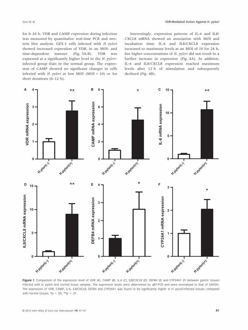
Vitamin D receptor, CAMP, IL-6, IL8/CXCL8, DEFB4,
and CYP24A1 mRNA levels were significantly elevated
in the gastric mucosa of H. pylori-positive patients, com-
pared with H. pylori-negative patients (Fig. 1). More-
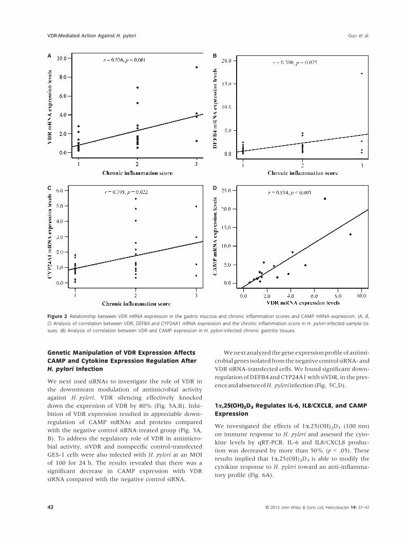
over, a significant positive correlation between VDR,
DEFB4, and CYP24A1 mRNA levels and chronic inflam-
mation scores (correlation coefficient r = .536, p < .01;
r = .390, p = .025; r = .398, p = .022, respectively,
Fig. 2A–C) was observed. The CAMP levels in turn
were found to have a significant positive correlation
with the VDR levels (r = .814, p < .001, Fig. 2D). More-
over, the IL-6 and IL8/CXCL8 mRNA expression levels
also showed a significant positive correlation with
chronic inflammation scores.
H. pylori Induces VDR and CAMP Expression In
Vitro
To further characterize the effect of H. pylori on the
expression of VDR and CAMP, GES-1 cells were
exposed to H. pylori at an MOI ranging from 0 to 100
Table 2 Histopathologic characteristics of the biopsy samples
Category
H. pylori
TotalPositive Negative
Chronic inflammation
Normal 0 0 0
Mild 0 13 13
Moderate 13 3 16
Marked 4 0 4
Intestinal metaplasia
Normal 11 16 27
Mild 3 0 3
Moderate 0 0 0
Marked 3 0 3
© 2013 John Wiley & Sons Ltd, Helicobacter 19: 37–4740
VDR-Mediated Action Against H. pylori Guo et al.
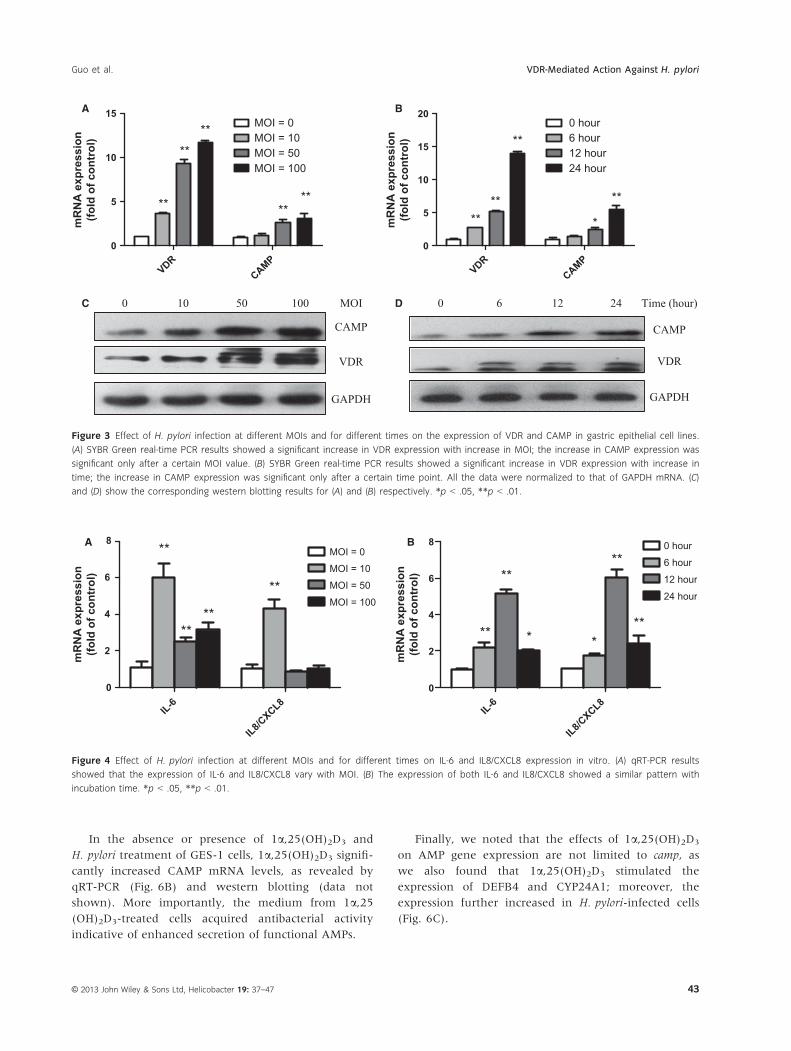
for 0–24 h. VDR and CAMP expression during infection
was measured by quantitative real-time PCR and wes-
tern blot analysis. GES-1 cells infected with H. pylori
showed increased expression of VDR, in an MOI- and
time-dependent manner (Fig. 3A,B). VDR was
expressed at a significantly higher level in the H. pylori-
infected group than in the normal group. The expres-
sion of CAMP showed no significant changes in cells
infected with H. pylori at low MOI (MOI = 10) or for
short durations (0–12 h).
Interestingly, expression patterns of IL-6 and IL8/
CXCL8 mRNA showed an association with MOI and
incubation time: IL-6 and IL8/CXCL8 expression
increased to maximum levels at an MOI of 10 for 24 h,
but higher concentrations of H. pylori did not result in a
further increase in expression (Fig. 4A). In addition,
IL-6 and IL8/CXCL8 expression reached maximum
levels after 12 h of stimulation and subsequently
declined (Fig. 4B).
0
1
2
3
4 **
VDR
mR
NA
exp
ress
ion
H.pylori(
–)
H.pylori(
+)
H.ylori(
–)
H.pylori(
+)
0
5
10
15 **
IL8/
CXC
L8 m
RN
A e
xpre
ssio
n
0
2
4
6
8 *C
AM
P m
RN
A e
xpre
ssio
n
H.pylori(
–)
H.pylori(
+)
H.pylori(
–)
H.pylori(
+)
0
1
2
3
4 *
DEF
B4
mR
NA
exp
ress
ion
0
5
10
15 **
IL-6
mR
NA
exp
ress
ion
H.pylori(
–)
H.pylori(
+)
H.pylori(
–)
H.pylori(
+)
0
1
2
3*
CYP
24A
1 m
RN
A e
xpre
ssio
n
A B C
D E F
Figure 1 Comparison of the expression level of VDR (A), CAMP (B), IL-6 (C), IL8/CXCL8 (D), DEFB4 (E) and CYP24A1 (F) between gastric tissues
infected with H. pylori and normal tissue samples. The expression levels were determined by qRT-PCR and were normalized to that of GAPDH.
The expression of VDR, CAMP, IL-6, IL8/CXCL8, DEFB4 and CYP24A1 was found to be significantly higher in H. pyroli-infected tissues compared
with normal tissues. *p < .05, **p < .01.
© 2013 John Wiley & Sons Ltd, Helicobacter 19: 37–47 41
Guo et al. VDR-Mediated Action Against H. pylori
Genetic Manipulation of VDR Expression Affects
CAMP and Cytokine Expression Regulation After
H. pylori Infection
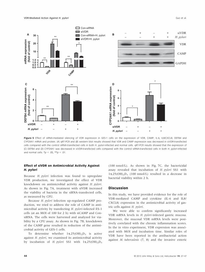
We next used siRNAs to investigate the role of VDR in
the downstream modulation of antimicrobial activity
against H. pylori. VDR silencing effectively knocked
down the expression of VDR by 80% (Fig. 5A,B). Inhi-
bition of VDR expression resulted in appreciable down-
regulation of CAMP mRNAs and proteins compared
with the negative control siRNA-treated group (Fig. 5A,
B). To address the regulatory role of VDR in antimicro-
bial activity, siVDR and nonspecific control-transfected
GES-1 cells were also infected with H. pylori at an MOI
of 100 for 24 h. The results revealed that there was a
significant decrease in CAMP expression with VDR
siRNA compared with the negative control siRNA.
Wenextanalyzed thegeneexpressionprofileof antimi-
crobial genes isolated fromthenegative control siRNA-and
VDR siRNA-transfected cells. We found significant down-
regulationofDEFB4andCYP24A1with siVDR, in thepres-
enceandabsenceofH. pylori infection(Fig. 5C,D).
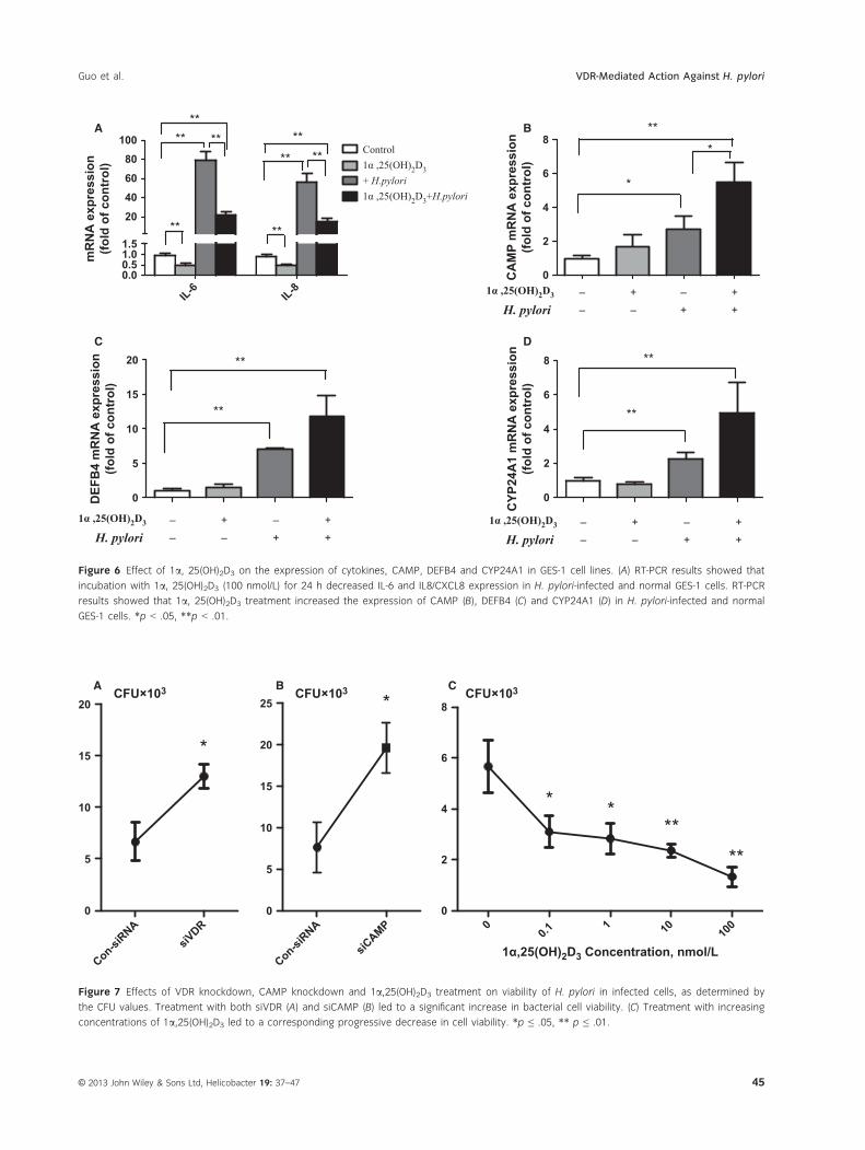
1a,25(OH)2D3 Regulates IL-6, IL8/CXCL8, and CAMP
Expression
We investigated the effects of 1a,25(OH)2D3 (100 nm)
on immune response to H. pylori and assessed the cyto-
kine levels by qRT-PCR. IL-6 and IL8/CXCL8 produc-
tion was decreased by more than 50% (p < .05). These
results implied that 1a,25(OH)2D3 is able to modify the
cytokine response to H. pylori toward an anti-inflamma-
tory profile (Fig. 6A).
A
C
B
D
Figure 2 Relationship between VDR mRNA expression in the gastric mucosa and chronic inflammation scores and CAMP mRNA expression. (A, B,
C) Analysis of correlation between VDR, DEFB4 and CYP24A1 mRNA expression and the chronic inflammation score in H. pylori-infected sample tis-
sues. (B) Analysis of correlation between VDR and CAMP expression in H. pylori-infected chronic gastritis tissues.
© 2013 John Wiley & Sons Ltd, Helicobacter 19: 37–4742
VDR-Mediated Action Against H. pylori Guo et al.
In the absence or presence of 1a,25(OH)2D3 and
H. pylori treatment of GES-1 cells, 1a,25(OH)2D3 signifi-
cantly increased CAMP mRNA levels, as revealed by
qRT-PCR (Fig. 6B) and western blotting (data not
shown). More importantly, the medium from 1a,25(OH)2D3-treated cells acquired antibacterial activity
indicative of enhanced secretion of functional AMPs.
Finally, we noted that the effects of 1a,25(OH)2D3
on AMP gene expression are not limited to camp, as
we also found that 1a,25(OH)2D3 stimulated the
expression of DEFB4 and CYP24A1; moreover, the
expression further increased in H. pylori-infected cells
(Fig. 6C).
VDRCAMP
0
5
10
15MOI = 0MOI = 10MOI = 50MOI = 100
****
** ****
mR
NA
exp
ress
ion
(fold
of c
ontr
ol)
VDRCAMP
0
5
10
15
200 hour6 hour12 hour24 hour
****
**
**
*mR
NA
exp
ress
ion
(fold
of c
ontr
ol)
0 6 12 24 Time (hour)
CAMP
VDR
GAPDH
0 10 50 100 MOI
CAMP
VDR
GAPDH
A B
C D
Figure 3 Effect of H. pylori infection at different MOIs and for different times on the expression of VDR and CAMP in gastric epithelial cell lines.
(A) SYBR Green real-time PCR results showed a significant increase in VDR expression with increase in MOI; the increase in CAMP expression was
significant only after a certain MOI value. (B) SYBR Green real-time PCR results showed a significant increase in VDR expression with increase in
time; the increase in CAMP expression was significant only after a certain time point. All the data were normalized to that of GAPDH mRNA. (C)
and (D) show the corresponding western blotting results for (A) and (B) respectively. *p < .05, **p < .01.
IL-6
IL8/CXCL8
0
2
4
6
8MOI = 0
MOI = 10
MOI = 50
MOI = 100
**
****
**
IL-6
IL8/CXCL8
0
2
4
6
8 0 hour
6 hour
12 hour
24 hour
**
**
* *
**
**
mR
NA
exp
ress
ion
(fold
of c
ontr
ol)
mR
NA
exp
ress
ion
(fold
of c
ontr
ol)
A B
Figure 4 Effect of H. pylori infection at different MOIs and for different times on IL-6 and IL8/CXCL8 expression in vitro. (A) qRT-PCR results
showed that the expression of IL-6 and IL8/CXCL8 vary with MOI. (B) The expression of both IL-6 and IL8/CXCL8 showed a similar pattern with
incubation time. *p < .05, **p < .01.
© 2013 John Wiley & Sons Ltd, Helicobacter 19: 37–47 43
Guo et al. VDR-Mediated Action Against H. pylori
Effect of siVDR on Antimicrobial Activity Against
H. pylori
Because H. pylori infection was found to upregulate
VDR production, we investigated the effect of VDR
knockdown on antimicrobial activity against H. pylori.
As shown in Fig. 7A, treatment with siVDR increased
the viability of bacteria in the siRNA-transfected cells,
as measured by CFU.
Because H. pylori infection up-regulated CAMP pro-
duction, we tried to address the role of CAMP in anti-
microbial activity by transfecting H. pylori-infected ES-1
cells (at an MOI of 100 for 2 h) with siCAMP and Con-
siRNA. The cells were harvested and analyzed for via-
bility by a CFU assay. As shown in Fig. 7B, knockdown
of the CAMP gene resulted in reduction of the antimi-
crobial activity of GES-1 cells.
To determine whether 1a,25(OH)2D3 is active
against H. pylori, we examined its antimicrobial activity
by incubation of H. pylori SS1 with 1a,25(OH)2D3
(100 nmol/L). As shown in Fig. 7C, the bactericidal
assay revealed that incubation of H. pylori SS1 with
1a,25(OH)2D3 (100 nmol/L) resulted in a decrease in
bacterial viability within 2 h.
Discussion
In this study, we have provided evidence for the role of
VDR-mediated CAMP and cytokine (IL-6 and IL8/
CXCL8) expression in the antimicrobial activity of gas-
tric cells against H. pylori.
We were able to confirm significantly increased
VDR mRNA levels in H. pylori-infected gastric mucosa.
Moreover, the mucosal VDR mRNA levels were posi-
tively correlated with the chronic inflammation scores.
In the in vitro experiment, VDR expression was associ-
ated with MOI and incubation time. Similar roles of
VDR have been reported in the antibacterial action
against M. tuberculosis (7, 8) and the invasive enteric
VDR0.00.51.01.5
10
20
30
Con-siRNAsiVDRCon-siRNA+H. pylorisiVDR+H. pylori
**
**
*
**
mR
NA
exp
ress
ion
(fold
of c
ontr
ol)
0
2
4
6
8
siVDRH. pylori
––
+–
– +
*
*
CYP
24A
1 m
RN
A e
xpre
ssio
n(fo
ld o
f con
trol
)
siVDR
+ +
– + – +– – + + H. pylori
VDR
CAMP
GAPDH
A B
CAMP
0
2
4
6
siVDR
H. pylori
–
–
+
–
– +
**
**
DEF
B4
mR
NA
exp
ress
ion
(fold
of c
ontr
ol)
+ +
C D
Figure 5 Effect of siRNA-mediated silencing of VDR expression in GES-1 cells on the expression of VDR, CAMP, IL-6, IL8/CXCL8, DEFB4 and
CYP24A1 mRNA and protein. (A) qRT-PCR and (B) western blot results showed that VDR and CAMP expression was decreased in siVDR-transfected
cells compared with the control siRNA-transfected cells in both H. pylori-infected and normal cells. qRT-PCR results showed that the expression of
(C) DEFB4 and (D) CYP24A1 was decreased in siVDR-transfected cells compared with the control siRNA-transfected cells in both H. pylori-infected
and normal cells. *p < .05, **p < .01.
© 2013 John Wiley & Sons Ltd, Helicobacter 19: 37–4744
VDR-Mediated Action Against H. pylori Guo et al.
IL-6 IL-80.00.51.01.5
20
40
60
80
100Control1α ,25(OH)2D3+ H.pylori1α ,25(OH)2D3+H.pylori
**
****
**
**
****
**
mR
NA
exp
ress
ion
(fold
of c
ontr
ol)
0
5
10
15
20
**
**
DEF
B4
mR
NA
exp
ress
ion
(fold
of c
ontr
ol)
0
2
4
6
8
1α ,25(OH)2D3
H. pylori – +– + – +
*
*
**
CA
MP
mR
NA
exp
ress
ion
(fold
of c
ontr
ol)
0
2
4
6
8
**
**
– +
CYP
24A
1 m
RN
A e
xpre
ssio
n(fo
ld o
f con
trol
)1α ,25(OH)2D3
H. pylori – +– + – +
– +
1α ,25(OH)2D3
H. pylori – +– + – +
– +
A B
C D
Figure 6 Effect of 1a, 25(OH)2D3 on the expression of cytokines, CAMP, DEFB4 and CYP24A1 in GES-1 cell lines. (A) RT-PCR results showed that
incubation with 1a, 25(OH)2D3 (100 nmol/L) for 24 h decreased IL-6 and IL8/CXCL8 expression in H. pylori-infected and normal GES-1 cells. RT-PCR
results showed that 1a, 25(OH)2D3 treatment increased the expression of CAMP (B), DEFB4 (C) and CYP24A1 (D) in H. pylori-infected and normal
GES-1 cells. *p < .05, **p < .01.
CFU×103
Con-siRNA
siVDR
0
5
10
15
20
*
CFU×103
Con-siRNA
siCAMP
0
5
10
15
20
25 *
00.1
1 10 100
0
2
4
6
8CFU×103
* ***
**
1α,25(OH)2D3 Concentration, nmol/L
A B C
Figure 7 Effects of VDR knockdown, CAMP knockdown and 1a,25(OH)2D3 treatment on viability of H. pylori in infected cells, as determined by
the CFU values. Treatment with both siVDR (A) and siCAMP (B) led to a significant increase in bacterial cell viability. (C) Treatment with increasing
concentrations of 1a,25(OH)2D3 led to a corresponding progressive decrease in cell viability. *p ≤ .05, ** p ≤ .01.
© 2013 John Wiley & Sons Ltd, Helicobacter 19: 37–47 45
Guo et al. VDR-Mediated Action Against H. pylori
pathogen Salmonella typhimurium, which can induce
colonic VDR expression and localization in vivo, and
stimulate VDR expression, transcription, and signaling
in colonic epithelial cell lines and MEFs [20].
We found that H. pylori infection-induced upregula-
tion of CAMP expression in the gastric mucosa, which
was comparable with previously published results (3,
14). Moreover, our results showed that CAMP expres-
sion in gastric epithelial cells was also upregulated upon
H. pylori infection with a sufficient bacterial load and
duration of infection. Thus, CAMP can block H. pylori-
induced inflammation [10]. CAMP was positively asso-
ciated with VDR mRNA expression in H. pylori-positive
mucosa and GES-1 cells. This is in agreement with a
recent study which showed that activation of toll-like
receptor-2 on human macrophages upregulated the
expression of VDR and induced expression of human
CAMP and killing of intracellular M. tuberculosis [7].
Activation of the CAMP gene occurred via a consensus
VDRE in the promoter that is bound by VDR. Previous
studies provide evidence that the CAMP gene is a direct
target of the transcription factor VDR, which mediates
strong upregulation of CAMP in response to treatment
of cells with 1a,25(OH)2D3 and its analogs [14,21]. In
our study too, we found that the VDR agonist 1a,25(OH)2D3 increased the production of CAMP. CAMP
expression was further increased in H. pylori-infected
cells, which is in agreement with data previously
reported for the regulation of CAMP expression in vita-
min D-mediated antimicrobial response [7]. Together,
these findings suggest that increase in the production of
the antimicrobial peptide CAMP may play a critical role
in host defence against H. pylori. In addition, our results
indicate that 1a,25(OH)2D3 has the ability to directly
induce antimicrobial gene expression and activity of the
antimicrobial peptide CAMP.
We also examined the effect of VDR on the produc-
tion of IL-6 and IL8/CXCL8. We show here that knock-
down of the VDR gene increased the levels of IL-6 and
IL8/CXCL8. Therefore, VDR�/� cells are more suscepti-
ble to inflammatory stimuli in inflammatory responses.
This observation is in agreement with previous reports
that mouse fibroblasts lacking VDR exhibit increased
NF-jB activity, leading to increased production of IL-6
[20]. NF-jB activation possesses an inherent self-ampli-
fying potency via induction of IFN-c and pro-inflamma-
tory cytokines such as IL-1b, IL-2, IL-6, IL8/CXCL8,
and TNF-a [22,23]. Moreover, loss of VDR leads to
more aggressive gross and histologic colonic injury,
increases serum IL-6 levels, which are a marker of sys-
temic inflammation, and enhances mortality after
Salmonella infection [5]. We have also demonstrated
that the proinflammatory cytokines IL-6 and IL8/
CXCL8 are suppressed by 1a,25(OH)2D3. Collectively,
these data suggest that cells that lack VDR appear to be
in a preinflammatory or proinflammatory state. It will
be very interesting to further investigate the inflamma-
tory status of VDR�/� mice with H. pylori infection.
According to a case–control study, the average con-
centration of vitamin D in subjects with autoimmune
gastritis was 9.8 � 5.6 ng/mL; nonspecific gastritis
patients, 22.2 � 13.5 ng/mL; and H. pylori gastritis
patients, 11.3 � 8.4 ng/mL [24]. However, another
Nutritional Deficiencies investigation showed that the
25-OH vitamin D3 levels did not differ between
H. pylori+ and H. pylori� patients (p > .20) [25]. Unfor-
tunately, in our study, we were unable to obtain sam-
ples promptly to test the concentration of vitamin D.
However, we were able to confirm that the vitamin D
agonist 1a,25(OH)2D3 had in vitro antimicrobial activity
against H. pylori.
In our study, we found that 1a,25(OH)2D3 leads to a
decrease in IL-6 and IL8/CXCL8 levels. Similar to this,
1a,25(OH)2D3 was found to suppress the production of
a spectrum of inflammatory cytokines in immune and
other cells (such as keratinocytes), including IL-1, IL-2,
IL-6, IL8/CXCL8 (29), INF-c, and TNF-a [26]; this
action forms the basis for its anti-inflammatory mecha-
nism. Therefore, 1a,25(OH)2D3 is a marker of systemic
inflammation in H. pylori infection. Moreover, 1a,25(OH)2D3 is involved in anti-inflammatory action
through its agonistic effect on VDR, which targets the
antimicrobial peptide CAMP gene in GES-1 cells. Taken
together, our data show that 1a,25(OH)2D3 has multi-
ple effects on the expression and release of antimicro-
bial peptides. We also found that the effects of 1a,25(OH)2D3 on the expression of VDR, CAMP, DEFB4 and
CYP24A1. Similar to this, DEFB4 has been shown to be
upregulated under H. pylori infection-associated inflam-
matory conditions in vivo and under cagA-positive
H. pylori infection in AGS cells in vitro [27]; moreover,
the DEFB4 promoter contains VDREs [28]. In agree-
ment with all these findings, 1a,25(OH)2D3 is known to
regulate anti-inflammatory activity and other facets of
immunity, including the induction of innate immune
responses [7,29].
In conclusion, this study has shown that VDR has
an effect on antimicrobial activity against H. pylori. Our
data are consistent with and explain at least in part, the
critical role of the VDR/CAMP pathway in innate
immunity. Moreover, these findings help improve our
understanding of the anti-inflammatory mechanism of
vitamin D. Given the importance of this subject, more
studies are warranted to further understand the func-
tional significance as well as the molecular mechanisms
underlying this role of VDR.
© 2013 John Wiley & Sons Ltd, Helicobacter 19: 37–4746
VDR-Mediated Action Against H. pylori Guo et al.
Acknowledgements and Disclosures
This study was supported by National Natural Science
Foundation of China (No. 30600281) and National 973
Program (2013CB911303).
Competing interests: The authors have no financial
conflicts of interest.
Author Contributions
All the coauthors of this paper have contributed to the
intellectual content of the paper.
References
1 Obonyo M, Rickman B, Guiney DG. Effects of myeloid differen-
tiation primary response gene 88 (MyD88) activation on Heli-
cobacter infection in vivo and induction of a Th17 response.
Helicobacter 2011;16:398–404.
2 Wang TT, Nestel FP, Bourdeau V, et al. Cutting edge: 1,25-di-
hydroxyvitamin D3 is a direct inducer of antimicrobial peptide
gene expression. J Immunol 2004;173:2909–12.
3 Krutzik SR, Hewison M, Liu PT, Robles JA, Stenger S, Adams
JS, Modlin RL. IL-15 links TLR2/1-induced macrophage differ-
entiation to the vitamin D-dependent antimicrobial pathway. J
Immunol 2008;181:7115–20.
4 Haussler MR, Whitfield GK, Haussler CA, Hsieh JC, Thompson
PD, Selznick SH, Dominguez CE, Jurutka PW. The nuclear vita-
min D receptor: biological and molecular regulatory properties
revealed. J Bone Miner Res 1998;13:325–49.
5 Wu S, Liao AP, Xia Y, Li YC, Li JD, Sartor RB, Sun J. Vitamin
D receptor negatively regulates bacterial-stimulated NF-kappaB
activity in intestine. Am J Pathol 2010;177:686–97.
6 Nagpal S, Na S, Rathnachalam R. Noncalcemic actions of vita-
min D receptor ligands. Endocr Rev 2005;26:662–87.
7 Liu PT, Stenger S, Li H, et al. Toll-like receptor triggering of a
vitamin D-mediated human antimicrobial response. Science
2006;311:1770–3.
8 Joseph RW, Bayraktar UD, Kim TK, John LS, Popat U, Khalili
J, Molldrem JJ, Wieder ED, Komanduri KV. Vitamin D receptor
upregulation in alloreactive human T cells. Hum Immunol
2012;73:693–8.
9 Hamanaka Y, Nakashima M, Wada A, Ito M, Kurazono H, Hojo
H, Nakahara Y, Kohno S, Hirayama T, Sekine I. Expression of
human beta-defensin 2 (hBD-2) in Helicobacter pylori induced
gastritis: antibacterial effect of hBD-2 against Helicobacter pylori.
Gut 2001;49:481–7.
10 Hase K, Murakami M, Iimura M, Cole SP, Horibe Y, Ohtake T,
Obonyo M, Gallo RL, Eckmann L, Kagnoff MF. Expression of
LL-37 by human gastric epithelial cells as a potential host
defense mechanism against Helicobacter pylori. Gastroenterology
2003;125:1613–25.
11 Zasloff M. Antimicrobial peptides in health and disease. N Engl
J Med 2002;347:1199–1200.
12 Andres E. Cationic antimicrobial peptides in clinical develop-
ment, with special focus on thanatin and heliomicin. Eur J Clin
Microbiol Infect Dis 2012;31:881–8.
13 Hancock RE, Patrzykat A. Clinical development of cationic anti-
microbial peptides: from natural to novel antibiotics. Curr Drug
Targets Infect Disord 2002;2:79–83.
14 Gombart AF, Borregaard N, Koeffler HP. Human cathelicidin
antimicrobial peptide (CAMP) gene is a direct target of the
vitamin D receptor and is strongly up-regulated in myeloid
cells by 1,25-dihydroxyvitamin D3. FASEB J 2005;19:
1067–77.
15 Leszczynska K, Namiot A, Fein DE, Wen Q, Namiot Z, Savage
PB, Diamond S, Janmey PA, Bucki R. Bactericidal activities of
the cationic steroid CSA-13 and the cathelicidin peptide LL-37
against Helicobacter pylori in simulated gastric juice. BMC Micro-
biol 2009;9:187.
16 Weber G, Heilborn JD, Chamorro JC, Hammarsjo A, Torma H,
Stahle M. Vitamin D induces the antimicrobial protein hCAP18
in human skin. J Invest Dermatol 2005;124:1080–2.
17 Evans KN, Nguyen L, Chan J, Innes BA, Bulmer JN, Kilby MD,
Hewison M. Effects of 25-hydroxyvitamin D3 and 1,25-di-
hydroxyvitamin D3 on cytokine production by human decidual
cells. Biol Reprod 2006;75:816–22.
18 Liu PT, Stenger S, Tang DH, Modlin RL. Cutting edge: vitamin
D-mediated human antimicrobial activity against Mycobacte-
rium tuberculosis is dependent on the induction of cathelicidin.
J Immunol 2007;179:2060–3.
19 Dixon MF, Genta RM, Yardley JH, Correa P. Classification and
grading of gastritis. The updated Sydney System. International
Workshop on the Histopathology of Gastritis, Houston 1994.
Am J Surg Pathol 1996;20:1161–81.
20 Sun J, Kong J, Duan Y, Szeto FL, Liao A, Madara JL, Li YC.
Increased NF-kappaB activity in fibroblasts lacking the vita-
min D receptor. Am J Physiol Endocrinol Metab 2006;291:
E315–22.
21 Guo C, Rosoha E, Lowry MB, Borregaard N, Gombart AF.
Curcumin induces human cathelicidin antimicrobial peptide
gene expression through a vitamin D receptor-independent
pathway. J Nutr Biochem 2013;24:754–9.
22 Lewis RS. Calcium oscillations in T-cells: mechanisms and
consequences for gene expression. Biochem Soc Trans
2003;31:925–9.
23 Mattson MP. Free radicals, calcium, and the synaptic plasticity-
cell death continuum: emerging roles of the transcription factor
NF kappa B. Int Rev Neurobiol 1998;42:103–68.
24 Antico A, Tozzoli R, Giavarina D, Tonutti E, Bizzaro N. Hypovi-
taminosis D as predisposing factor for atrophic type A gastritis:
a case-control study and review of the literature on the interac-
tion of Vitamin D with the immune system. Clin Rev Allergy
Immunol 2012;42:355–64.
25 Gerig R, Ernst B, Wilms B, Thurnheer M, Schultes B. Preopera-
tive nutritional deficiencies in severely obese bariatric candi-
dates are not linked to gastric Helicobacter pylori infection. Obes
Surg 2013;23:698–702.
26 Gurlek A, Pittelkow MR, Kumar R. Modulation of growth fac-
tor/cytokine synthesis and signaling by 1alpha,25-dihydroxyvi-
tamin D(3): implications in cell growth and differentiation.
Endocr Rev 2002;23:763–86.
27 Otte JM, Neumann HM, Brand S, Schrader H, Schmidt WE,
Schmitz F. Expression of beta-defensin 4 is increased in human
gastritis. Eur J Clin Invest 2009;39:126–38.
28 Liu PT, Schenk M, Walker VP, et al. Convergence of IL-1beta
and VDR activation pathways in human TLR2/1-induced anti-
microbial responses. PLoS One 2009;4:e5810.
29 Rook GA, Steele J, Fraher L, Barker S, Karmali R, O’Riordan J,
Stanford J. Vitamin D3, gamma interferon, and control of pro-
liferation of Mycobacterium tuberculosis by human monocytes.
Immunology 1986;57:159–63.
© 2013 John Wiley & Sons Ltd, Helicobacter 19: 37–47 47
Guo et al. VDR-Mediated Action Against H. pylori
Copyright © 2022 FDOKUMEN