Hdac3 Is Essential for the Maintenance of Chromatin Structure and Genome Stability
27
Hdac3 is essential for the maintenance of chromatin structure and genome stability Srividya Bhaskara 1 , Sarah K. Knutson 1 , Guochun Jiang 2,3 , Mahesh B. Chandrasekharan 1 , Andrew J. Wilson 4 , Siyuan Zheng 5,6 , Ashwini Yenamandra 7 , Kimberly Locke 8 , Jia-ling Yuan 1 , Alyssa R. Bonine-Summers 1 , Christina E. Wells 1 , Jonathan F. Kaiser 1 , M. Kay Washington 9 , Zhongming Zhao 3,5,6,9 , Florence F. Wagner 10 , Zu-Wen Sun 1,9 , Fen Xia 2 , Edward B. Holson 10 , Dineo Khabele 3,4,9 , and Scott W. Hiebert 1,9,* 1 Department of Biochemistry, Vanderbilt University, Nashville, TN 37232 2 Department of Radiation Oncology Vanderbilt University Medical Center, Nashville, TN 37212 3 Department of Cancer Biology, Vanderbilt University Medical Center, Nashville, TN 37212 4 Department of Obstetrics and Gynecology, Division of Gynecologic Oncology, Vanderbilt University Medical Center, Nashville, TN 37212 5 Department of Biomedical Informatics, Vanderbilt University School of Medicine, Nashville, TN 37232 6 Bioinformatics Resource Center, Vanderbilt University School of Medicine, Nashville, TN 37232 7 Department of Pathology, Vanderbilt University, Nashville, TN 37232 8 Pathgroup, Nashville, TN 37211 9 Vanderbilt-Ingram Cancer Center, Vanderbilt University School of Medicine, Nashville, TN 37232 10 The Broad Institute of MIT and Harvard, 7 Cambridge Center, Cambridge, MA 02142 Summary Hdac3 is essential for efficient DNA replication and DNA damage control. Deletion of Hdac3 impaired DNA repair and greatly reduced chromatin compaction and heterochromatin content. These defects corresponded to increases in histone H3K9,K14ac, and H4K5ac and H4K12ac in late S phase of the cell cycle, and histone deposition marks were retained in quiescent Hdac3-null cells. Liver-specific deletion of Hdac3 culminated in hepatocellular carcinoma. While HDAC3 expression was down regulated in only a small number of human liver cancers, the mRNA levels of the HDAC3 cofactor NCOR1 were reduced in 1/3 of these cases. siRNA targeting of NCOR1 and SMRT (NCOR2) increased H4K5ac and caused DNA damage, indicating that the HDAC3/ NCOR/SMRT axis is critical for maintaining chromatin structure and genomic stability. © 2010 Elsevier Inc. All rights reserved. * To whom correspondence should be sent: Department of Biochemistry, 512 Preston Research Building, Vanderbilt University School of Medicine, 23rd and Pierce Ave., Nashville Tennessee, 37232, Phone: (615) 936-3582; Fax: (615) 936-1790; [email protected]. Publisher's Disclaimer: This is a PDF file of an unedited manuscript that has been accepted for publication. As a service to our customers we are providing this early version of the manuscript. The manuscript will undergo copyediting, typesetting, and review of the resulting proof before it is published in its final citable form. Please note that during the production process errors may be discovered which could affect the content, and all legal disclaimers that apply to the journal pertain. Accession number Expression data generated from murine tumors lacking Hdac3 is found in the Gene Expression Omnibus accession number GSE22457. NIH Public Access Author Manuscript Cancer Cell. Author manuscript; available in PMC 2011 November 16. Published in final edited form as: Cancer Cell. 2010 November 16; 18(5): 436–447. doi:10.1016/j.ccr.2010.10.022. NIH-PA Author Manuscript NIH-PA Author Manuscript NIH-PA Author Manuscript
Transcript of Hdac3 Is Essential for the Maintenance of Chromatin Structure and Genome Stability

Hdac3 is essential for the maintenance of chromatin structureand genome stability
Srividya Bhaskara1, Sarah K. Knutson1, Guochun Jiang2,3, Mahesh B. Chandrasekharan1,Andrew J. Wilson4, Siyuan Zheng5,6, Ashwini Yenamandra7, Kimberly Locke8, Jia-lingYuan1, Alyssa R. Bonine-Summers1, Christina E. Wells1, Jonathan F. Kaiser1, M. KayWashington9, Zhongming Zhao3,5,6,9, Florence F. Wagner10, Zu-Wen Sun1,9, Fen Xia2,Edward B. Holson10, Dineo Khabele3,4,9, and Scott W. Hiebert1,9,*
1Department of Biochemistry, Vanderbilt University, Nashville, TN 372322Department of Radiation Oncology Vanderbilt University Medical Center, Nashville, TN 372123Department of Cancer Biology, Vanderbilt University Medical Center, Nashville, TN 372124Department of Obstetrics and Gynecology, Division of Gynecologic Oncology, VanderbiltUniversity Medical Center, Nashville, TN 372125Department of Biomedical Informatics, Vanderbilt University School of Medicine, Nashville, TN372326Bioinformatics Resource Center, Vanderbilt University School of Medicine, Nashville, TN 372327Department of Pathology, Vanderbilt University, Nashville, TN 372328Pathgroup, Nashville, TN 372119Vanderbilt-Ingram Cancer Center, Vanderbilt University School of Medicine, Nashville, TN 3723210The Broad Institute of MIT and Harvard, 7 Cambridge Center, Cambridge, MA 02142
SummaryHdac3 is essential for efficient DNA replication and DNA damage control. Deletion of Hdac3impaired DNA repair and greatly reduced chromatin compaction and heterochromatin content.These defects corresponded to increases in histone H3K9,K14ac, and H4K5ac and H4K12ac inlate S phase of the cell cycle, and histone deposition marks were retained in quiescent Hdac3-nullcells. Liver-specific deletion of Hdac3 culminated in hepatocellular carcinoma. While HDAC3expression was down regulated in only a small number of human liver cancers, the mRNA levelsof the HDAC3 cofactor NCOR1 were reduced in 1/3 of these cases. siRNA targeting of NCOR1and SMRT (NCOR2) increased H4K5ac and caused DNA damage, indicating that the HDAC3/NCOR/SMRT axis is critical for maintaining chromatin structure and genomic stability.
© 2010 Elsevier Inc. All rights reserved.*To whom correspondence should be sent: Department of Biochemistry, 512 Preston Research Building, Vanderbilt University Schoolof Medicine, 23rd and Pierce Ave., Nashville Tennessee, 37232, Phone: (615) 936-3582; Fax: (615) 936-1790;[email protected]'s Disclaimer: This is a PDF file of an unedited manuscript that has been accepted for publication. As a service to ourcustomers we are providing this early version of the manuscript. The manuscript will undergo copyediting, typesetting, and review ofthe resulting proof before it is published in its final citable form. Please note that during the production process errors may bediscovered which could affect the content, and all legal disclaimers that apply to the journal pertain.Accession numberExpression data generated from murine tumors lacking Hdac3 is found in the Gene Expression Omnibus accession numberGSE22457.
NIH Public AccessAuthor ManuscriptCancer Cell. Author manuscript; available in PMC 2011 November 16.
Published in final edited form as:Cancer Cell. 2010 November 16; 18(5): 436–447. doi:10.1016/j.ccr.2010.10.022.
NIH
-PA Author Manuscript
NIH
-PA Author Manuscript
NIH
-PA Author Manuscript
IntroductionHistone deacetylases (HDACs) play major roles in modulating chromatin accessibilityduring transcription, replication, recombination and repair (Gallinari et al., 2007; Goodarziet al., 2009), yet the role of individual HDACs in these processes is still unclear.Deacetylation of histones is required for re-establishing chromatin structure on a local basisafter transcription of a gene or after the repair of a DNA double strand break (Tsukamoto etal., 1997). On a global scale, HDACs act during DNA replication when the cellular histonecontent is doubled, as these newly synthesized histones are acetylated prior to theirdeposition onto nascent DNA. The residues most commonly associated with this process areH4K5ac and H4K12ac (Sobel et al., 1995; Taddei et al., 1999). These modificationspresumably allow histone chaperones to configure the nucleosome correctly beforedeacetylation stabilizes the nucleosome and/or allows higher order compaction of thechromatin and the formation of heterochromatin (Luger et al., 1997; Luger and Richmond,1998; Neumann et al., 2009; Verreault et al., 1996).
This process of histone acetylation/deacetylation is required for genomic stability and cellviability, as perturbations in the acetyl transferase or components of this pathway causegenomic instability and result in a failure to recover from genotoxic stress (Clarke et al.,1999; Han et al., 2007; Smith et al., 1998; Yuan et al., 2009). This is a dynamic process thatoccurs across the entire genome and the role of HDACs in the re-establishment of chromatinstructure after replication is one of the least explored areas of their action. As such, geneticmethods have been the most informative approaches to understand the physiological role ofthese critical regulatory enzymes.
Targeting enzymes that control chromatin structure and topography has been an extremelyvaluable tool in cancer therapy. A wide variety of general and specific small moleculeinhibitors targeted towards HDACs are currently in clinical trials and are used as therapiesfor both solid and hematological tumors (Bolden et al., 2006). At therapeutic doses, histonedeacetylase inhibitors (HDIs) not only cause cell cycle-dependent DNA damage, but alsoaffect DNA repair, which sensitizes cells to ionizing radiation (IR), topoisomerase inhibitorsand cisplatin (Baschnagel et al., 2009; Marchion et al., 2004; Suzuki et al., 2009). However,the molecular mechanism for inefficient DNA repair following HDI treatment is still notclear. Given the high levels of histone acetylation that accumulate in the context of theseinhibitors, it is reasonable to assume that disruption of chromatin structure may contribute tocell death. As more selective HDAC inhibitors are moving into clinical trials, it is importantto elucidate the function of individual HDACs to design better and more specific drugs forcancer therapy and to understand the mechanism(s) of action or side effects.
Hdac3, a class I HDAC, associates with the nuclear hormone co-repressors (NCoR andSMRT) (Codina et al., 2005) and is generally thought of as a locus-specific co-repressor thatis recruited to promoters to repress genes regulated by nuclear hormone receptors and othertranscription factors (Jones and Shi, 2003). In yeast, Snt1 and Hos2 have features of NCoR/SMRT and Hdac3, respectively (Pijnappel et al., 2001). This suggests a more ancestral andfundamental role of these proteins perhaps in the cell cycle, and that this machinery is alsoused for gene-specific transcriptional regulation. In agreement with this hypothesis,conditional deletion of Hdac3 in mouse models demonstrated that murine embryonicfibroblasts (MEFs) required Hdac3 for cell viability (Bhaskara et al., 2008). The observedapoptosis was associated with an impaired S-phase progression and DNA double strandbreaks, rather than altered transcriptional programs (Bhaskara et al., 2008). The DNAdamage was blocked when cells were taken out of the cell cycle by serum starvation, whichsuggested that Hdac3 acts during the S phase (Bhaskara et al., 2008). We propose that thecell cycle functions of HDAC3 and its regulatory factors NCoR and SMRT may be the
Bhaskara et al. Page 2
Cancer Cell. Author manuscript; available in PMC 2011 November 16.
NIH
-PA Author Manuscript
NIH
-PA Author Manuscript
NIH
-PA Author Manuscript
ancestral role for and that disruption of these cell cycle functions may have dramaticconsequences for the regulation of chromatin structure and genomic stability. These rolesmay impact the usefulness of HDAC3 as a therapeutic target in cancer and other diseases.
ResultsHdac3 function is required for efficient DNA repair
The inactivation of Hdac3 resulted in increased sensitivity of quiescent MEFs to ionizingradiation, suggesting a defect predominantly in non-homologous end joining (NHEJ)-mediated repair in these cells (Bhaskara et al., 2008). To test whether these defects were dueto altered DNA repair functions or due to altered histone modifications and the ensuingchanges in chromatin structure, we examined whether the inactivation of Hdac3 increasesthe sensitivity of Hdac3-null cells to other DNA damaging agents. For this purpose, wetreated Hdac3FL/+ and Hdac3FL/− MEFs carrying a tamoxifen-inducible ER-Cre allele witheither increasing concentrations of doxorubicin or cisplatin 48 hr after the addition of 4-hydroxy tamoxifen to inactivate Hdac3. Doxorubicin inhibits topoisomerase II (Swift et al.,2006) and triggers S-phase associated DNA double strand breaks (DSBs) that are repairedby the homologous recombination (HR) pathway, whereas cisplatin crosslinks DNA to formintra-strand adducts (Siddik, 2003). Inactivation of Hdac3 increased the sensitivity of MEFsto doxorubicin (Fig. 1A) and cisplatin (Fig. 1B), suggesting that in the absence of Hdac3,these DNA repair pathways are inefficient.
Given that Hdac3 deletion appeared to affect two independent types of DNA repair, weexamined whether Hdac3 plays a role in the two major NHEJ and HR DSB repair pathways.We employed a chromosomally integrated reporter allele system established in HEK 293cells (Fig. 1C and Fig. 1D) and used siRNAs to deplete the endogenous levels of HDAC3(Fig. 1E) prior to cutting the reporter site with the I-Sce1 homing endonuclease. Theefficiency of rejoining I-Sce1-cleaved sites was measured using quantitative PCR for NHEJand by flow cytometry for reconstituted GFP expression for HR. In both cases, the reductionin Hdac3 levels caused a 50–60% decrease in DNA repair (Fig. 1F and Fig. 1G), indicatingthat Hdac3 is essential for efficient NHEJ- and HR-mediated repair.
Although Hdac3 has been linked to NHEJ due to its association with the SMRT/Ku70complex (Yu et al., 2006), our DNA damage sensitivity and repair data suggested thatHdac3 loss affects DNA repair by targeting an element that is common to multiple types ofDNA repair. Moreover, Hdac3 is not recruited to the sites of double strand breaks followingIR treatment (Fig. S1), nor did its loss affect the localization of other members of the DNAdamage response (Rad50, Brca1, Mdc1 and Mre11; data not shown). One of the key histonemodifications that contributes to the DNA damage response, the first step in double strandbreak repair, is H3K9 trimethylation (H3K9me3), which recruits the histoneacetyltransferase Tip60 (Sun et al., 2009) and other factors involved in the damage response(e.g., HP1β; (Ayoub et al., 2008)). Therefore, we tested whether siRNA targeting of HDAC3would alter histone acetylation at the site of an integrated reporter. Chromatinimmunoprecipitation employing anti-H3K9,K14ac showed that histone acetylation wasincreased at the substrate locus at a level consistent with global changes in H3K9,K14ac (seeFig. 3 below), along with a concomitant decrease in H3K9me3 (Fig. 1H), suggesting thatglobal changes in histone modifications could contribute to the defects in DNA repair.
Inactivation of Hdac3 alters chromatin structure and decreases global heterochromatinH3K9me3 is one of the best marks of heterochromatin. The decrease in H3K9me3 uponsiRNA-mediated suppression of HDAC3 or Hdac3 deletion in the liver (Fig. 1H, and Fig. 3below) prompted us to use the Albumin-Cre transgene to delete Hdac3 in vivo to examine
Bhaskara et al. Page 3
Cancer Cell. Author manuscript; available in PMC 2011 November 16.
NIH
-PA Author Manuscript
NIH
-PA Author Manuscript
NIH
-PA Author Manuscript
chromatin structure. Initially, we used transmission electron microscopy to examine thenuclei from Alb-Cre:Hdac3+/− and Alb-Cre:Hdac3−/− p17 livers (Fig. 2A). As expected, incontrol nuclei the electron-dense heterochromatin was found at the nuclear periphery (Fig.2A). In contrast, Alb-Cre:Hdac−/− p17 liver nuclei showed a significant decrease in theamount of heterochromatin, especially at the periphery (Fig. 2A; note that the residualelectron dense material remaining is consistent with the presence of nucleoli). A similarresult was obtained by enumerating the Hoechst staining of heterochromatic foci that areevident in fluorescence microscopy in mouse cells. Alb-Cre:Hdac−/− liver nuclei showedsignificantly fewer foci (Fig. 2B). Moreover, when cell fractionation was used to isolatechromatin, immunoblot analysis demonstrated that Alb-Cre:Hdac3−/− hepatocytes containedroughly 2-fold less HP1β on chromatin (Fig. 2C). Finally, at the gene-specific level we usedChIP to examine H3K9,K14ac at the p53 locus (Su et al., 2009). Inactivation of Hdac3caused the accumulation of acetylated H3K9,K14 at the promoter, upstream of the promoter,within intron 1, and within the body of the gene (Fig. S1), which is consistent with globalchanges in chromatin structure and histone modifications (see Fig. 3 below). Thus, Hdac3 isrequired for maintaining chromatin structure in vivo.
Given the reduction in heterochromatin, we used micrococcal nuclease (MNase) digestion toexamine nucleosomal compaction. Digestion of isolated nuclei with increasingconcentrations of MNase demonstrated that the bulk chromatin from Hdac3−/− hepatocyteswas more sensitive to MNase digestion when compared to the chromatin from controlhepatocytes (Fig. 2D), indicating that global chromatin structure was altered and is more“open” in the absence of Hdac3. In addition, we noted that the amount of DNA associatedwith mono-nucleosomes did not increase with increasing concentrations of MNase,suggesting that the nuclosomal DNA was more accessible in the absence of Hdac3.Southern blot analysis of these same samples using major and minor satellite probes orquantitative PCR for these regions showed modest sensitivity without a change innucleosomal spacing (Fig. S2) (Gilbert and Allan, 2001;Sugimura et al., 2010). Given theapparent sensitivity of mono-nucleosomal DNA to MNase (Fig. 2D), we tested thesensitivity of nucleosomes to ionic conditions by extracting histones with differingconcentrations of salt. Consistent with prior results (Li et al., 1993), Histone H3 wasresistant to NaCl concentrations up to 1.2M in control hepatocytes, but in the absence ofHdac3, Histone H3 was nearly completely soluble in 900 mM NaCl, suggesting thatnucleosome integrity was altered in Hdac3−/− hepatocytes.
Structural determinations of the nucleosome show that the basic lysine residues in histonetails can potentially associate with DNA, but more likely mediate contacts with acidicsurfaces on adjacent nucleosomes to allow nucleosome compaction and fiber formation(Luger et al., 1997; Luger and Richmond, 1998; Schalch et al., 2005). Therefore, weperformed western blot analysis of nuclear extracts prepared from Alb-Cre:Hdac3+/− or Alb-Cre:Hdac3−/− p17 livers to examine global histone acetylation and methylation marks thatneutralize the lysine charges and that regulate chromatin structure. Hdac3 deacetylatesH4K5ac and H4K12ac in vitro (Johnson et al., 2002) and loss of Hdac3 resulted in anaccumulation of H4K5ac, H4K12ac, and H4K16ac, as well as H3K9,K14ac with little affecton the acetylation of other residues (Fig. 3) (Knutson et al., 2008). Examination of histonemethylation demonstrated that H3K9me3 (Lachner et al., 2001) and H3K79me2 werereduced (Fig. 3), while H3K4me3, H3K27me3 and H4K20me2 were unaffected (Fig. 3).
H4K5ac and H4K12ac are commonly associated with histone deposition onto newlysynthesized DNA. Given that hepatocytes are generally quiescent, our liver-specific deletionof Hdac3 implies that Hdac3 is required for removing these marks during or after DNAsynthesis. Therefore, we used Hdac3FL/− NIH 3T3 cells infected with Adenovirusexpressing Cre to examine these marks during the cell cycle. Cells were infected with
Bhaskara et al. Page 4
Cancer Cell. Author manuscript; available in PMC 2011 November 16.
NIH
-PA Author Manuscript
NIH
-PA Author Manuscript
NIH
-PA Author Manuscript
Adeno-Cre and synchronized in G0/G1 by serum starvation and then released into the cellcycle by the addition of serum to the culture medium. The level of H4K5ac and H4K12acwas then examined using western blot analysis of extracts prepared from cells at varioustime points after serum addition. After 48 hr of culture in the absence of serum, thepercentage of cells in G0/G1 approached 90% (Fig. S3) and H4K5ac and H4K12ac werereduced to low levels in control cells, but remained high in Hdac3-depleted cells (Fig. 4A).After serum addition, both cultures re-entered the cell cycle with cells beginning to enter Sphase at 12 hr and over 50% of the cells progressing through S phase by 18 hr as measuredby BrdU incorporation (Fig. S3). At 18 and 24 hr after serum addition, H4K5ac andH4K12ac increased in the control cells consistent with the acetylation of these residues onnewly synthesized histones that are deposited on new DNA (Fig. 4A). In cells lackingHdac3, the levels of acetylation of these residues were already high and increased onlymodestly during S phase (Fig. 4A). While H3K9,K14 acetylation has not been linked tohistone deposition in mammalian cells in the manner that H4K5ac and H4K12ac have, itslevels were low in G0/G1 phase control cells and increased during S phase. In the absence ofHdac3, H3K9,K14ac was not reduced upon serum starvation, suggesting that acetylation ofthese residues was not removed after S phase, which could account for the loss of H3K9me3and reduced heterochromatin in Hdac3-null livers (Fig. 2 and 3).
To better define the requirements for Hdac3 in the removal of cell cycle-associated marks,we directly examined H4K5ac in S phase cells, as this is a classical deposition mark. Thepunctate immunofluorescence pattern of PCNA 18 hr after release from serum starvationwas used to identify cells in late S phase, which are characterized by foci of PCNA at thenuclear periphery (Celis and Celis, 1985; Madsen and Celis, 1985; Taddei et al., 1999).Although, a general increase in H4K5ac was observed in the absence of Hdac3 (data notshown), a pronounced increase in H4K5ac was found in late S phase cells (11% of cells incontrols vs. 57% of Hdac3-null cells, Fig. 4B), especially at the nuclear periphery.
Even relatively modest over expression or reduced expression of histones is sufficient toaffect genomic stability by causing DNA double strand breaks (Gunjan and Verreault, 2003;Olive and Banath, 1995). Given the apparent high level of H4K5ac in the nucleus of Hdac3-null cells after S phase (Fig. 4A and 4B), we asked what proportion of the total histone H4 isacetylated in the absence of Hdac3. Two sequential rounds of immunoprecipitation oflysates prepared from quiescent Hdac3-null NIH 3T3 cells were performed using anti-H4K5ac to identify the acetylated histone. Western blot analysis using anti-H4K5acconfirmed that most of the acetylated histone was immuno-purified in the firstimmunoprecipitation (Fig. 4C, upper panel). The total histone H4 that was acetylated at K5was then determined by comparing the amount of total histone H4 in theimmunoprecipitation versus H4 that remained in the supernatant (Fig. 4C, lower panel). Forthe western blot analysis, 1/5th the amount of the supernatant was loaded as compared to theprecipitated H4K5ac. Thus, roughly 15–20% of the total histone H4 was acetylated at K5 incontrol cells and 30–40% was acetylated in the absence of Hdac3 (Fig. 4C).
Hdac3 is required for genomic stabilityPreviously, we noted S phase-associated DNA double strand breaks in Hdac3-null cells(Bhaskara et al., 2008). Given the requirement for Hdac3 in removing histone acetylationmarks that are added during the S phase of the cell cycle and the links between loss ofH3K9me3 and genomic instability, we examined the consequences of inactivation of Hdac3to chromosomes as they progress through mitosis. Metaphase spreads were prepared fromHdac3FL/+ and Hdac3FL/− MEFs following either Ad-Cre infection or following tamoxifentreatment of MEFs carrying the ER-Cre transgene. Both chromosome breaks and gaps werequantified using cytogenetic analysis. Using either Ad-Cre (Fig. S4) or ER-Cre to deleteHdac3 (Fig. 5A) led to a 5–8 fold increase in the average number of breaks and gaps in
Bhaskara et al. Page 5
Cancer Cell. Author manuscript; available in PMC 2011 November 16.
NIH
-PA Author Manuscript
NIH
-PA Author Manuscript
NIH
-PA Author Manuscript
metaphase chromosomes when compared to control cells (Fig. 5B), indicating a crucial rolefor Hdac3 in the maintenance of genome stability.
To test whether the DNA damage and genomic instability phenotypes found in MEFslacking Hdac3 was recapitulated in vivo, we examined Alb-Cre:Hdac3+/− and Alb-Cre:Hdac3−/− livers for DNA double strand breaks using immunofluorescence to detectγH2AX and 53BP1, which localize to sites of DNA double strand breaks (Iwabuchi et al.,2003). While there was little or no endogenous DNA damage in Alb-Cre:Hdac3−/− livers atpost-natal day 17 (data not shown), by p28 the Hdac3-null livers displayed an increasednumber of cells with γH2AX and 53BP1 foci when compared to the control hepatocytes (0Gy panels, Fig. 5C; see Fig. S4 for quantification). Subsequently, we examined DNA repairin Hdac3-null hepatocytes at p28 following a non-lethal dose of IR (3Gy). An increasedpercentage of cells with a substantial amount of DNA damage was detected both 1hr and 6hrafter IR in Alb-Cre:Hdac3−/− when compared to the control Alb-Cre:Hdac3+/− hepatocytes(Fig. 5C). Quantification of 53BP1 foci in ~100 cells from two independent experimentsfollowing a 6 hr recovery period revealed that Hdac3-null cells have a greater percentage ofcells with 5 to 10 foci when compared to the control cells (Fig. S4). We also observed DNAdamage even after a 24 hr recovery period in some Alb-Cre:Hdac−/− hepatocytes, whereascontrol hepatocytes had repaired the damage caused by IR treatment (data not shown).
Hdac3-null livers develop hepatocellular carcinomaAnalysis of gene expression data obtained previously from Alb-Cre:Hdac3−/− livers at p28identified an up-regulation of genes that belong to the p53 network (Fig. S4), suggesting thepresence of DNA damage. Likewise, quantitative RT-PCR analysis revealed an up-regulation of miRNAs regulated by p53 in Hdac3-null livers (Fig. S4), which is alsoconsistent with the activation of a DNA damage response (Rokhlin et al., 2008). In addition,hepatocellular carcinoma (HCC) progression markers, such as gamma-glutamyltranspeptidase 1 (Pavesi et al., 1989) and insulin-like growth factor II (Qiu et al., 2008) wereup-regulated in the microarray analysis of Alb-Cre:Hdac3−/− livers by 2.2- and 2.7-foldrespectively (Knutson et al., 2008). These data, coupled with the observed genomicinstability, prompted us to age cohorts of 20 control and 20 Alb-Cre:Hdac3−/− mice. By 15–16 weeks of age, the livers were very pale due to the dramatic accumulation of neutral lipidsand fat caused by inactivation of Hdac3 (Knutson et al., 2008) and contained “whitenodules” when examined by gross morphology (Fig. 6A). These nodules were encapsulatedwith a fibrous lining that positively stained with Massion’s trichrome (data not shown) andappeared to be benign “adenoma-like” structures with the cytoplasm of the cells filled withmicrovesicular fluid and an abundance of mitochondria (Fig. 6A, and data not shown). By8–10 months of age, most of the mice began to show signs of distress and necropsyidentified the presence of tumors in the liver. The experiment was humanly terminated forall mice by 14 months of age (Fig. 6B). Pathological analysis indicated that 20 of 20 micesuccumbed to low-grade hepatocellular carcinoma (HCC) at a mean age of 10.2 months(Fig. 6B). Immunohistochemical staining for Hdac3 confirmed that the tumors lackedexpression of Hdac3 (Fig. 6C) and Ki-67 staining confirmed a high proliferative index in thetumors (Fig. 6D). The HCC displayed a loss of normal architecture, a trabecular patterningof cells, a lack of ductal morphology, and very disorganized features (Fig. 6D).
The loss of genomic stability and the impaired response to DNA damage suggested that ahigh mutation rate stimulated the development of HCC (Fig. 1, Fig. 5, Fig. 6). To begin toassess what pathways were involved in the formation of HCC, we performed geneexpression analysis using cDNA microarrys (Fig. 7A). In the array data, we noted theenhanced expression of c-Myc, a commonly over expressed oncogene, which was confirmedusing quantitative RT-PCR (Fig. 7B). Signatures consistent with activation of the Raspathway and impairment of the p53 pathway were also identified in this analysis (Fig. 7A).
Bhaskara et al. Page 6
Cancer Cell. Author manuscript; available in PMC 2011 November 16.
NIH
-PA Author Manuscript
NIH
-PA Author Manuscript
NIH
-PA Author Manuscript
The Wnt pathway has been identified as a key regulatory node in human HCC and we alsonoted that this pathway was affected in the Hdac3-null tumors. Therefore, we examined β-catenin localization using both cell fractionation and immunohistochemical staining. Asearly as p28, we found increased amounts of β-catenin localized to the nucleus and therewas prominent nuclear localization of β-catenin in the Hdac3-null HCCs (Fig. 7B, C),confirming that this oncogenic pathway was up regulated in this mouse model of HCC.
NCOR1 is down-regulated in HCC and NCoR and SMRT regulate global histone acetylationAnalysis of HDAC3 mRNA levels in 4 independent human HCC datasets from the GEOdatabase indicated that HDAC3 was reduced in some cases (Fig. 8 and Fig. S5). HDAC3 ishighly expressed in cycling cells (e.g., in the crypt regions of the colonic epithelium)(Spurling et al., 2008;Wilson et al., 2006), so comparing quiescent normal tissue to cyclingcancer cells may under estimate the level of its loss of expression. Nevertheless, in thelargest dataset, HDAC3 levels were reduced by 1.5-fold in 13% of the cases and over 2-foldin 3.3% of the cases as compared to normal controls (data not shown and Fig. 8A). Therewas no association with hepatitis C or hepatitis B viral infection or survival (data notshown). Given that HDAC3 is dormant until activated by association with NCoR or SMRT(Guenther et al., 2001), and that it is recruited to chromatin through association with otherfactors, we probed the GEO HCC datasets for changes in expression of the 57 directinteraction partners of HDAC3 identified in the Human Protein Reference Database (TableS1). Four of these genes (STAT3, GTF2I, GCM1, and NCOR1) showed reduced expressionin HCC. NCOR1 is not only down regulated, but also NCOR1 is located on a region ofchromosome 17p that is deleted in human HCC (Xu et al., 2001). The levels of NCOR1 werereduced by 2-fold or greater in nearly 1/3 of HCC samples in the largest dataset (Fig. 8A)and was similarly down-regulated in the majority of the samples in smaller HCC geneexpression datasets (Fig. S6). Using immunohistochemistry to detect nuclear NCOR1, wefound that 2/5 human HCCs tested had reduced levels of NCOR1 (Fig. 8B), which isconsistent with the mRNA expression results.
Given that NCoR/SMRT directly control HDAC3 functions (Guenther et al., 2001), we usedsiRNAs to probe the requirements for these HDAC3 cofactors in the regulation of histoneacetylation. In HeLa cells that express both family members, depletion of either NCoR orSMRT alone had only modest effects on global histone acetylation (data not shown).Targeting both family members together caused a small increase in H4K5ac (Fig. 8C).However, even though NCoR/SMRT levels were only reduced by about 50%, there was asignificant accumulation in the levels of global H4K5ac in both HeLa cells (Fig. 8C) andNIH 3T3 cells (Fig. S6B). These increases in histone acetylation were associated with adecrease in the levels of HDAC3 detected in both HeLa and NIH3T3 cells (Fig. 8C and Fig.S6C). Immunofluorescence using anti-PCNA to identify S phase cells, demonstrated a 5-fold increase in the number of cells with high levels of H4K5ac at the nuclear periphery inlate S-phase cells targeted with siRNAs to both co-factors (Fig. 8D and Fig. S6C). Giventhis alteration in histone marks, we examined these cells for DNA double strand breaksusing anti-53BP1. Cells depleted of NCOR1 and SMRT showed a dramatic increase in thenumber of cells with greater than 10 foci (Fig. 8E and Fig. S6D). Collectively, these resultsshow that the NCoR/SMRT/HDAC3 axis is required for removing histone marks globallyand maintaining genomic stability.
DiscussionThe increase in acetylation of H4K5, H4K12, H4K16ac, and H3K9,K14 that was observedupon inactivation of Hdac3, along with the concomitant loss of H3K9me3, provides a likelymechanism for the failure to maintain chromatin structure in Hdac3-null mice (Fig. 2).H4K5ac and H4K12ac are associated with histone deposition (Sobel et al., 1995), especially
Bhaskara et al. Page 7
Cancer Cell. Author manuscript; available in PMC 2011 November 16.
NIH
-PA Author Manuscript
NIH
-PA Author Manuscript
NIH
-PA Author Manuscript

in heterochromatic regions that are replicated late in S phase (Taddei et al., 1999) and thispattern was accentuated in the absence of Hdac3 or when NCoR/SMRT were targeted usingsiRNAs (Fig. 4 and 8). The removal of these marks is required for propagation ofheterochromatin in yeast (Zhou et al., 2009), which suggests that the accumulation of thesemarks (and the reduced levels of H3K9me3) may be the underlying cause for the reductionin heterochromatin in the Hdac3-null livers. Indeed, loss of H3K9me3 impaired the DNAdamage response, and the inactivation of the murine H3K9 methyltransferases or mutationof HP1β caused genomic instability, defects in DSB repair, and an increased tumor risk(Aucott et al., 2008;Kondo et al., 2008;Luijsterburg et al., 2009;Peters et al., 2001;Sun et al.,2009). This suggests that the failure to maintain a normal chromatin structure underlies theHdac3−/−-associated defects in two distinct types of DNA repair (NHEJ and HR), and ingenomic stability (Fig. 1, Fig. 5), which ultimately led to tumor development (Fig. 6).
The siRNA-mediated knockdown of NCoR/SMRT, like deletion of Hdac3, caused theaccumulation of histone deposition marks, suggesting that these Hdac3 activating factorsalso play an intrinsic role during the S phase. N-CoR and SMRT were initially identified astranscriptional co-repressors associated with nuclear hormone receptors (Chen and Evans,1995; Horlein et al., 1995; Karagianni and Wong, 2007), as well as with a variety of DNAbinding factors (Perissi et al., 2004). However, the function of these co-repressors during thecell cycle may represent the ancestral activity of these complexes. We speculate that duringevolution this cell cycle machinery was recruited in higher organisms to regulate geneexpression patterns in a cell type-specific manner (e.g., in response to nuclear hormones) orto form heterochromatin to more permanently silence gene expression.
While HDAC3 has been suggested to be over expressed in colorectal carcinoma (CRC)(Spurling et al., 2008; Wilson et al., 2006), it is not amplified at the DNA level. In addition,HDAC3 is expressed at higher levels in the proliferating cells of the colonic crypts, whichmight suggest that its levels are higher in CRC because the cells are cycling. Conversely, itis notable that HDAC3 lies within a region of chromosome 5q31.3 that is frequently deletedin breast cancer (Johannsdottir et al., 2006) and myelodysplastic syndromes (Ebert, 2009).Intriguingly, NCOR1 lies in a region of chromosome 17 that is frequently deleted in HCC(Mahlknecht et al., 1999), and an analysis of expression profiles indicated that downregulation of NCOR1 expression is common in a subset of human HCC (Fig. 8A and Fig.S6). Our data are consistent with the inactivation of the HDAC3/NCOR/SMRT axis possiblycontributing to a subset of human cancer by allowing the increase of histone acetylationduring the S phase leading to DNA damage and further accumulation of mutations.
Nearly all non-targeted cancer therapeutics (i.e., those that do not target a mutant proteinthat initiates a cancer) develop a therapeutic window by acting on cycling cells to causeDNA damage (Ashwell and Zabludoff, 2008; Lieberman, 2008). However, one side effect isthat these agents, when given at too high a dose or for too long, also cause genomicinstability in normal cells leading to therapy-associated secondary cancers. Our results raisethis possibility for HDIs, all of which currently target HDAC3. However, compounds suchas SAHA appear to be well tolerated, possibly owing to their short half-life in vivo (Butler etal., 2000). That is, SAHA may cause S phase-associated DNA damage for those cancer cellsin S phase during the 4–6 hr window in which the daily dose of SAHA is active, but onlycause mild problems for the majority of normal cells that are not cycling. In addition, normalcells that are proliferating such as in the gastrointestinal tract and in the bone marrow, caneither repair the DNA damage or their chromatin is “reset” after the SAHA is metabolized.Thus, we predict that while continuous inhibition of Hdac3 is detrimental (e.g., Fig. 6),transient inhibition, even when frequently repeated, may be safe.
Bhaskara et al. Page 8
Cancer Cell. Author manuscript; available in PMC 2011 November 16.
NIH
-PA Author Manuscript
NIH
-PA Author Manuscript
NIH
-PA Author Manuscript
Experimental ProceduresPlease see the supplemental experimental procedures for additional methods used.
MiceMice harboring either a conditional floxed (fl) allele or a null (−) allele were created asdescribed previously (Knutson et al., 2008). To create liver-specific Hdac3 knockout mice,mice with a floxed or a null allele were crossed to transgenic mice expressing Alb-Cre(Knutson et al., 2008) to obtain Alb:Cre:Hdac3+/− and Alb:Cre:Hdac3−/− offspring mice.All experiments using mice were approved by the Vanderbilt University InstitutionalAnimal Care and Use Committee.
DNA Repair assaysDNA repair assays were performed using HEK293 cells engineered with integrated reportersfor both types of double strand break repair (Zhuang et al., 2009). Briefly, cells (1 × 10⁵)were transfected twice with HDAC3 siRNA using Oligofectamine (Invitrogen, CA). pCMV-I-SceI or the control vector were transfected into the cells using FuGene 6 (Roche). The cellswere either analyzed by two-color FACS analysis to examine homologous recombination orPCR was used to determine the rate of NHEJ-mediated repair (see supplementalexperimental procedures for primer sequences).
Transmission electron microscopyLiver tissue was minced into fine pieces and fixed in 2.5% glutaraldehyde in 0.1Mcacodylate buffer at room temperature for 1–2 hr. Samples were then washed in 0.1Mcacodylate buffer and treated with 1% aqueous osmium tetraoxide. Tissues were thenwashed, dehydrated and embedded in Spurr resin. Thin sections (100 nm) were viewed withan FEI CM-12 transmission electron microscope operated at 80KeV.
Gene expression analysis of human HCC samplesFour HCC and normal control experiments (GSE14323, GSE6764, GSE6222, andGSE5975) were selected from the Gene Expression Omnibus database (GEO,http://www.ncbi.nlm.nih.gov/geo/)(Barrett et al., 2009). The description of this analysis iselaborated in the supplement. For GSE6764 (Wurmbach et al., 2007), we compared thenormal liver samples with HCC samples of different stages. For GSE6222 (Liao et al.,2008), we excluded the HuH7 cell line data. GSE1898 was excluded from this analysis dueto variations in the probe sets. Expression data generated from murine tumors lacking Hdac3is found in GSE22457.
Protein analysesFor preparation of whole cell protein extracts, the cell pellet was washed with phosphatebuffered saline (PBS) and sonicated in RIPA buffer (0.5% Triton-X-100, 0.5% deoxycholicacid, and 0.5% SDS in PBS) with protease inhibitors (0.5mM PMSF, 2µg/µl leupeptin and15µg/µl aprotinin) prior to western analyses. Antibodies used and the preparation of nuclearextracts are described in the Supplementary Materials.
Significance
Broad-spectrum histone deacetylase inhibitors (HDI) are being used to treat a variety ofcancers, and more selective inhibitors are being developed for therapeutic uses. Geneticanalysis of individual histone deacetylases (HDACs) is essential to understand the actionof these inhibitors and their potential side effects. We show that inactivation of Hdac3, a
Bhaskara et al. Page 9
Cancer Cell. Author manuscript; available in PMC 2011 November 16.
NIH
-PA Author Manuscript
NIH
-PA Author Manuscript
NIH
-PA Author Manuscript

central target of all currently used HDIs, causes genomic instability, and deletion ofHdac3 in the liver leads to hepatocellular carcinoma. These phenotypes correlate withglobal increases in the acetylation of specific histone residues, disruptions in chromatinstructure, and a loss of heterochromatin. Our results genetically link the HDAC3/NCOR/SMRT axis to the maintenance of critical cell cycle functions and genomic stability.
Supplementary MaterialRefer to Web version on PubMed Central for supplementary material.
AcknowledgmentsWe thank all the members of Hiebert lab for helpful discussions, reagents and advice. We thank the VanderbiltImaging, Human Tissue Acquisition and Pathology, and Functional Genomics Shared Resources for services andsupport. We thank Drs. Nick Gilbert and James Allan for providing plasmids containing the major and minorsatellite probes. This work was supported by the T. J. Martell Foundation, the Robert J. Kleberg, Jr. and Helen C.Kleberg Foundation, National Institutes of Health grants (R01-CA64140, RO1-CA77274, RO1-CA109355) andcore services performed through Vanderbilt Digestive Disease Research grant NIDDK P30DK58404 and theVanderbilt-Ingram Cancer Center support grant NCI P30CA68485. SB was supported by a fellowship(1F32CA138091-01) from the NCI.
ReferencesAshwell S, Zabludoff S. DNA damage detection and repair pathways--recent advances with inhibitors
of checkpoint kinases in cancer therapy. Clin Cancer Res. 2008; 14:4032–4037. [PubMed:18593978]
Aucott R, Bullwinkel J, Yu Y, Shi W, Billur M, Brown JP, Menzel U, Kioussis D, Wang G, Reisert I,et al. HP1-beta is required for development of the cerebral neocortex and neuromuscular junctions. JCell Biol. 2008; 183:597–606. [PubMed: 19015315]
Ayoub N, Jeyasekharan AD, Bernal JA, Venkitaraman AR. HP1-beta mobilization promoteschromatin changes that initiate the DNA damage response. Nature. 2008; 453:682–686. [PubMed:18438399]
Barrett T, Troup DB, Wilhite SE, Ledoux P, Rudnev D, Evangelista C, Kim IF, Soboleva A,Tomashevsky M, Marshall KA, et al. NCBI GEO: archive for high-throughput functional genomicdata. Nucleic Acids Res. 2009; 37:D885–D890. [PubMed: 18940857]
Baschnagel A, Russo A, Burgan WE, Carter D, Beam K, Palmieri D, Steeg PS, Tofilon P,Camphausen K. Vorinostat enhances the radiosensitivity of a breast cancer brain metastatic cell linegrown in vitro and as intracranial xenografts. Mol Cancer Ther. 2009; 8:1589–1595. [PubMed:19509253]
Bhaskara S, Chyla BJ, Amann JM, Knutson SK, Cortez D, Sun ZW, Hiebert SW. Deletion of histonedeacetylase 3 reveals critical roles in S phase progression and DNA damage control. Mol Cell.2008; 30:61–72. [PubMed: 18406327]
Bolden JE, Peart MJ, Johnstone RW. Anticancer activities of histone deacetylase inhibitors. Nat RevDrug Discov. 2006; 5:769–784. [PubMed: 16955068]
Butler LM, Agus DB, Scher HI, Higgins B, Rose A, Cordon-Cardo C, Thaler HT, Rifkind RA, MarksPA, Richon VM. Suberoylanilide hydroxamic acid, an inhibitor of histone deacetylase, suppressesthe growth of prostate cancer cells in vitro and in vivo. Cancer Res. 2000; 60:5165–5170. [PubMed:11016644]
Celis JE, Celis A. Cell cycle-dependent variations in the distribution of the nuclear protein cyclinproliferating cell nuclear antigen in cultured cells: subdivision of S phase. Proc Natl Acad Sci U SA. 1985; 82:3262–3266. [PubMed: 2860667]
Chen JD, Evans RM. A transcriptional co-repressor that interacts with nuclear hormone receptors.Nature. 1995; 377:454–457. [PubMed: 7566127]
Clarke AS, Lowell JE, Jacobson SJ, Pillus L. Esa1p is an essential histone acetyltransferase requiredfor cell cycle progression. Mol Cell Biol. 1999; 19:2515–2526. [PubMed: 10082517]
Bhaskara et al. Page 10
Cancer Cell. Author manuscript; available in PMC 2011 November 16.
NIH
-PA Author Manuscript
NIH
-PA Author Manuscript
NIH
-PA Author Manuscript
Codina A, Love JD, Li Y, Lazar MA, Neuhaus D, Schwabe JW. Structural insights into the interactionand activation of histone deacetylase 3 by nuclear receptor corepressors. Proc Natl Acad Sci U SA. 2005; 102:6009–6014. [PubMed: 15837933]
Ebert BL. Deletion 5q in myelodysplastic syndrome: a paradigm for the study of hemizygous deletionsin cancer. Leukemia. 2009; 23:1252–1256. [PubMed: 19322210]
Gallinari P, Di Marco S, Jones P, Pallaoro M, Steinkuhler C. HDACs, histone deacetylation and genetranscription: from molecular biology to cancer therapeutics. Cell Res. 2007; 17:195–211.[PubMed: 17325692]
Gilbert N, Allan J. Distinctive higher-order chromatin structure at mammalian centromeres. Proc NatlAcad Sci U S A. 2001; 98:11949–11954. [PubMed: 11593003]
Goodarzi AA, Noon AT, Jeggo PA. The impact of heterochromatin on DSB repair. Biochem SocTrans. 2009; 37:569–576. [PubMed: 19442252]
Guenther MG, Barak O, Lazar MA. The SMRT and N-CoR corepressors are activating cofactors forhistone deacetylase 3. Mol Cell Biol. 2001; 21:6091–6101. [PubMed: 11509652]
Gunjan A, Verreault A. A Rad53 kinase-dependent surveillance mechanism that regulates histoneprotein levels in S. cerevisiae. Cell. 2003; 115:537–549. [PubMed: 14651846]
Han J, Zhou H, Li Z, Xu RM, Zhang Z. Acetylation of lysine 56 of histone H3 catalyzed by RTT109and regulated by ASF1 is required for replisome integrity. J Biol Chem. 2007; 282:28587–28596.[PubMed: 17690098]
Horlein AJ, Naar AM, Heinzel T, Torchia J, Gloss B, Kurokawa R, Ryan A, Kamei Y, Soderstrom M,Glass CK, et al. Ligand-independent repression by the thyroid hormone receptor mediated by anuclear receptor co-repressor. Nature. 1995; 377:397–404. [PubMed: 7566114]
Iwabuchi K, Basu BP, Kysela B, Kurihara T, Shibata M, Guan D, Cao Y, Hamada T, Imamura K,Jeggo PA, et al. Potential role for 53BP1 in DNA end-joining repair through direct interaction withDNA. J Biol Chem. 2003; 278:36487–36495. [PubMed: 12824158]
Johannsdottir HK, Jonsson G, Johannesdottir G, Agnarsson BA, Eerola H, Arason A, Heikkila P,Egilsson V, Olsson H, Johannsson OT, et al. Chromosome 5 imbalance mapping in breast tumorsfrom BRCA1 and BRCA2 mutation carriers and sporadic breast tumors. Int J Cancer. 2006;119:1052–1060. [PubMed: 16570289]
Johnson CA, White DA, Lavender JS, O'Neill LP, Turner BM. Human class I histone deacetylasecomplexes show enhanced catalytic activity in the presence of ATP and co-immunoprecipitatewith the ATP-dependent chaperone protein Hsp70. J Biol Chem. 2002; 277:9590–9597. [PubMed:11777905]
Jones PL, Shi YB. N-CoR-HDAC corepressor complexes: roles in transcriptional regulation by nuclearhormone receptors. Curr Top Microbiol Immunol. 2003; 274:237–268. [PubMed: 12596910]
Karagianni P, Wong J. HDAC3: taking the SMRT-N-CoRrect road to repression. Oncogene. 2007;26:5439–5449. [PubMed: 17694085]
Knutson SK, Chyla BJ, Amann JM, Bhaskara S, Huppert SS, Hiebert SW. Liver-specific deletion ofhistone deacetylase 3 disrupts metabolic transcriptional networks. EMBO J. 2008; 27:1017–1028.[PubMed: 18354499]
Kondo Y, Shen L, Ahmed S, Boumber Y, Sekido Y, Haddad BR, Issa JP. Downregulation of histoneH3 lysine 9 methyltransferase G9a induces centrosome disruption and chromosome instability incancer cells. PLoS One. 2008; 3:e2037. [PubMed: 18446223]
Lachner M, O'Carroll D, Rea S, Mechtler K, Jenuwein T. Methylation of histone H3 lysine 9 creates abinding site for HP1 proteins. Nature. 2001; 410:116–120. [PubMed: 11242053]
Li W, Nagaraja S, Delcuve GP, Hendzel MJ, Davie JR. Effects of histone acetylation, ubiquitinationand variants on nucleosome stability. Biochem J. 1993; 296(Pt 3):737–744. [PubMed: 8280071]
Liao YL, Sun YM, Chau GY, Chau YP, Lai TC, Wang JL, Horng JT, Hsiao M, Tsou AP.Identification of SOX4 target genes using phylogenetic footprinting-based prediction fromexpression microarrays suggests that overexpression of SOX4 potentiates metastasis inhepatocellular carcinoma. Oncogene. 2008; 27:5578–5589. [PubMed: 18504433]
Lieberman HB. DNA damage repair and response proteins as targets for cancer therapy. Curr MedChem. 2008; 15:360–367. [PubMed: 18288990]
Bhaskara et al. Page 11
Cancer Cell. Author manuscript; available in PMC 2011 November 16.
NIH
-PA Author Manuscript
NIH
-PA Author Manuscript
NIH
-PA Author Manuscript

Luger K, Mader AW, Richmond RK, Sargent DF, Richmond TJ. Crystal structure of the nucleosomecore particle at 2.8 A resolution. Nature. 1997; 389:251–260. [PubMed: 9305837]
Luger K, Richmond TJ. The histone tails of the nucleosome. Curr Opin Genet Dev. 1998; 8:140–146.[PubMed: 9610403]
Luijsterburg MS, Dinant C, Lans H, Stap J, Wiernasz E, Lagerwerf S, Warmerdam DO, Lindh M,Brink MC, Dobrucki JW, et al. Heterochromatin protein 1 is recruited to various types of DNAdamage. J Cell Biol. 2009; 185:577–586. [PubMed: 19451271]
Madsen P, Celis JE. S-phase patterns of cyclin (PCNA) antigen staining resemble topographicalpatterns of DNA synthesis. A role for cyclin in DNA replication? FEBS Lett. 1985; 193:5–11.
Mahlknecht U, Emiliani S, Najfeld V, Young S, Verdin E. Genomic organization and chromosomallocalization of the human histone deacetylase 3 gene. Genomics. 1999; 56:197–202. [PubMed:10051405]
Marchion DC, Bicaku E, Daud AI, Richon V, Sullivan DM, Munster PN. Sequence-specificpotentiation of topoisomerase II inhibitors by the histone deacetylase inhibitor suberoylanilidehydroxamic acid. J Cell Biochem. 2004; 92:223–237. [PubMed: 15108350]
Neumann H, Hancock SM, Buning R, Routh A, Chapman L, Somers J, Owen-Hughes T, van Noort J,Rhodes D, Chin JW. A method for genetically installing site-specific acetylation in recombinanthistones defines the effects of H3 K56 acetylation. Mol Cell. 2009; 36:153–163. [PubMed:19818718]
Olive PL, Banath JP. Radiation-induced DNA double-strand breaks produced in histone-depletedtumor cell nuclei measured using the neutral comet assay. Radiat Res. 1995; 142:144–152.[PubMed: 7724728]
Pavesi F, Lotzniker M, Scarabelli M, Garbagnoli P, Moratti R. Efficiency of composite laboratory testsin the diagnosis of liver malignancies. Int J Biol Markers. 1989; 4:163–169. [PubMed: 2482315]
Perissi V, Aggarwal A, Glass CK, Rose DW, Rosenfeld MG. A corepressor/coactivator exchangecomplex required for transcriptional activation by nuclear receptors and other regulatedtranscription factors. Cell. 2004; 116:511–526. [PubMed: 14980219]
Peters AH, O'Carroll D, Scherthan H, Mechtler K, Sauer S, Schofer C, Weipoltshammer K, Pagani M,Lachner M, Kohlmaier A, et al. Loss of the Suv39h histone methyltransferases impairsmammalian heterochromatin and genome stability. Cell. 2001; 107:323–337. [PubMed: 11701123]
Pijnappel WW, Schaft D, Roguev A, Shevchenko A, Tekotte H, Wilm M, Rigaut G, Seraphin B,Aasland R, Stewart AF. The S. cerevisiae SET3 complex includes two histone deacetylases, Hos2and Hst1, and is a meiotic-specific repressor of the sporulation gene program. Genes Dev. 2001;15:2991–3004. [PubMed: 11711434]
Qiu LW, Yao DF, Zong L, Lu YY, Huang H, Wu W, Wu XH. Abnormal expression of insulin-likegrowth factor-II and its dynamic quantitative analysis at different stages of hepatocellularcarcinoma development. Hepatobiliary Pancreat Dis Int. 2008; 7:406–411. [PubMed: 18693177]
Rokhlin OW, Scheinker VS, Taghiyev AF, Bumcrot D, Glover RA, Cohen MB. MicroRNA-34mediates AR-dependent p53-induced apoptosis in prostate cancer. Cancer Biol Ther. 2008;7:1288–1296. [PubMed: 18497571]
Schalch T, Duda S, Sargent DF, Richmond TJ. X-ray structure of a tetranucleosome and itsimplications for the chromatin fibre. Nature. 2005; 436:138–141. [PubMed: 16001076]
Siddik ZH. Cisplatin: mode of cytotoxic action and molecular basis of resistance. Oncogene. 2003;22:7265–7279. [PubMed: 14576837]
Smith ER, Eisen A, Gu W, Sattah M, Pannuti A, Zhou J, Cook RG, Lucchesi JC, Allis CD. ESA1 is ahistone acetyltransferase that is essential for growth in yeast. Proc Natl Acad Sci U S A. 1998;95:3561–3565. [PubMed: 9520405]
Sobel RE, Cook RG, Perry CA, Annunziato AT, Allis CD. Conservation of deposition-relatedacetylation sites in newly synthesized histones H3 and H4. Proc Natl Acad Sci U S A. 1995;92:1237–1241. [PubMed: 7862667]
Spurling CC, Godman CA, Noonan EJ, Rasmussen TP, Rosenberg DW, Giardina C. HDAC3overexpression and colon cancer cell proliferation and differentiation. Mol Carcinog. 2008;47:137–147. [PubMed: 17849419]
Bhaskara et al. Page 12
Cancer Cell. Author manuscript; available in PMC 2011 November 16.
NIH
-PA Author Manuscript
NIH
-PA Author Manuscript
NIH
-PA Author Manuscript
Su CH, Shann YJ, Hsu MT. p53 chromatin epigenetic domain organization and p53 transcription. MolCell Biol. 2009; 29:93–103. [PubMed: 18936172]
Sugimura K, Fukushima Y, Ishida M, Ito S, Nakamura M, Mori Y, Okumura K. Cell cycle-dependentaccumulation of histone H3.3 and euchromatic histone modifications in pericentromericheterochromatin in response to a decrease in DNA methylation levels. Exp Cell Res. 2010 inpress.
Sun Y, Jiang X, Xu Y, Ayrapetov MK, Moreau LA, Whetstine JR, Price BD. Histone H3 methylationlinks DNA damage detection to activation of the tumour suppressor Tip60. Nat Cell Biol. 2009;11:1376–1382. [PubMed: 19783983]
Suzuki M, Endo M, Shinohara F, Echigo S, Rikiishi H. Enhancement of cisplatin cytotoxicity bySAHA involves endoplasmic reticulum stress-mediated apoptosis in oral squamous cell carcinomacells. Cancer Chemother Pharmacol. 2009; 64:1115–1122. [PubMed: 19280190]
Swift LP, Rephaeli A, Nudelman A, Phillips DR, Cutts SM. Doxorubicin-DNA adducts induce a non-topoisomerase II-mediated form of cell death. Cancer Res. 2006; 66:4863–4871. [PubMed:16651442]
Taddei A, Roche D, Sibarita JB, Turner BM, Almouzni G. Duplication and maintenance ofheterochromatin domains. J Cell Biol. 1999; 147:1153–1166. [PubMed: 10601331]
Tsukamoto Y, Kato J, Ikeda H. Silencing factors participate in DNA repair and recombination inSaccharomyces cerevisiae. Nature. 1997; 388:900–903. [PubMed: 9278054]
Verreault A, Kaufman PD, Kobayashi R, Stillman B. Nucleosome assembly by a complex of CAF-1and acetylated histones H3/H4. Cell. 1996; 87:95–104. [PubMed: 8858152]
Wilson AJ, Byun DS, Popova N, Murray LB, L'Italien K, Sowa Y, Arango D, Velcich A, AugenlichtLH, Mariadason JM. Histone deacetylase 3 (HDAC3) and other class I HDACs regulate colon cellmaturation and p21 expression and are deregulated in human colon cancer. J Biol Chem. 2006;281:13548–13558. [PubMed: 16533812]
Wurmbach E, Chen YB, Khitrov G, Zhang W, Roayaie S, Schwartz M, Fiel I, Thung S, Mazzaferro V,Bruix J, et al. Genome-wide molecular profiles of HCV-induced dysplasia and hepatocellularcarcinoma. Hepatology. 2007; 45:938–947. [PubMed: 17393520]
Xu XR, Huang J, Xu ZG, Qian BZ, Zhu ZD, Yan Q, Cai T, Zhang X, Xiao HS, Qu J, et al. Insight intohepatocellular carcinogenesis at transcriptome level by comparing gene expression profiles ofhepatocellular carcinoma with those of corresponding noncancerous liver. Proc Natl Acad Sci U SA. 2001; 98:15089–15094. [PubMed: 11752456]
Yu J, Palmer C, Alenghat T, Li Y, Kao G, Lazar MA. The corepressor silencing mediator for retinoidand thyroid hormone receptor facilitates cellular recovery from DNA double-strand breaks. CancerRes. 2006; 66:9316–9322. [PubMed: 16982777]
Yuan J, Pu M, Zhang Z, Lou Z. Histone H3-K56 acetylation is important for genomic stability inmammals. Cell Cycle. 2009; 8:1747–1753. [PubMed: 19411844]
Zhou J, Zhou BO, Lenzmeier BA, Zhou JQ. Histone deacetylase Rpd3 antagonizes Sir2-dependentsilent chromatin propagation. Nucleic Acids Res. 2009; 37:3699–3713. [PubMed: 19372273]
Zhuang J, Jiang G, Willers H, Xia F. Exonuclease function of human Mre11 promotes deletionalnonhomologous end joining. J Biol Chem. 2009; 284:30565–30573. [PubMed: 19744924]
Bhaskara et al. Page 13
Cancer Cell. Author manuscript; available in PMC 2011 November 16.
NIH
-PA Author Manuscript
NIH
-PA Author Manuscript
NIH
-PA Author Manuscript

Figure 1. Loss of Hdac3 impairs DNA repairMEFs (Hdac3FL/+ and Hdac3FL/−) were treated with 0.1 µM tamoxifen for 48 hr and thentreated with increasing concentrations of either doxorubicin (A) or cisplatin (B) and cellviability measured with the WST-1 assay. Values in the graph represent mean ± SD. oftriplicate samples and the experiment was repeated at least twice. C. A schematicrepresentation of the NHEJ substrate (left), the product formed (right) and the position ofreal-time PCR primers used to detect the repaired product (Zhuang et al., 2009). D. Thereporter cassette used for HR detection is shown schematically. Upon induction of I-Sce1,gene conversion reconstitutes active GFP. The repaired GFP was then measured by FACSanalysis. E. Western blot analysis of Hdac3 following siRNA transfection in 293T cells.
Bhaskara et al. Page 14
Cancer Cell. Author manuscript; available in PMC 2011 November 16.
NIH
-PA Author Manuscript
NIH
-PA Author Manuscript
NIH
-PA Author Manuscript
GAPDH is shown as a loading control. Chromatin-based repair assays performed in 293Tcells following knockdown of Hdac3 to measure the efficiency of NHEJ using Q-PCR (F)and HR using FACS (G). The values shown in F and G are the means ± SEM. H. Chromatinimmunoprecipitation analysis of H3K9,K14ac and H3K9me3 at the NHEJ substrate beforeand after siRNA suppression of Hdac3. Quantitative PCR was used to compare the effects ofnon-targeting and Hdac3 siRNAs and the graphs show average of the relative levels ofH3K9,K14ac ± SD. See also figure S1.
Bhaskara et al. Page 15
Cancer Cell. Author manuscript; available in PMC 2011 November 16.
NIH
-PA Author Manuscript
NIH
-PA Author Manuscript
NIH
-PA Author Manuscript

Figure 2. Loss of Hdac3 alters chromatin structure and decreases heterochromatinA. Histological sections prepared from control and Hdac3-null livers at postnatal day 17(p17) were used to examine nuclei using electron microscopy. Control hepatocytes containdense staining of condensed chromatin whereas Hdac3-null cells have decreased amounts ofheterochromatin, especially at the nuclear periphery. Arrows indicate the RNA rich nucleolithat remain in the Hdac3-null cells. Scale bar represents 4 µm. B. Loss of heterochromaticfoci upon inactivation of Hdac3. Left panels show 2 examples of histological sections fromcontrol and Hdac3-null liver sections stained with Hoechst to detect heterochromatic foci.Scale bar represents 20µm. The graph at the right shows quantification of foci from at least100 cells for each sample expressed as the mean ± SEM. C. HP1β localization to chromatin
Bhaskara et al. Page 16
Cancer Cell. Author manuscript; available in PMC 2011 November 16.
NIH
-PA Author Manuscript
NIH
-PA Author Manuscript
NIH
-PA Author Manuscript
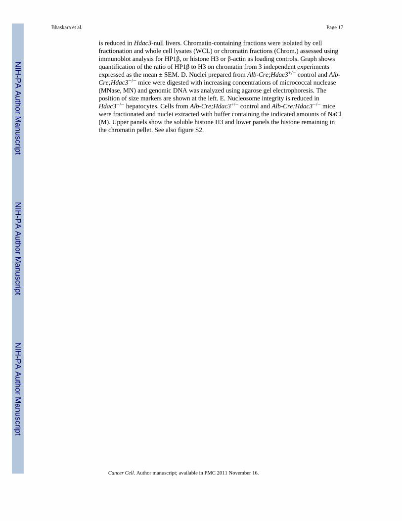
is reduced in Hdac3-null livers. Chromatin-containing fractions were isolated by cellfractionation and whole cell lysates (WCL) or chromatin fractions (Chrom.) assessed usingimmunoblot analysis for HP1β, or histone H3 or β-actin as loading controls. Graph showsquantification of the ratio of HP1β to H3 on chromatin from 3 independent experimentsexpressed as the mean ± SEM. D. Nuclei prepared from Alb-Cre;Hdac3+/− control and Alb-Cre;Hdac3−/− mice were digested with increasing concentrations of micrococcal nuclease(MNase, MN) and genomic DNA was analyzed using agarose gel electrophoresis. Theposition of size markers are shown at the left. E. Nucleosome integrity is reduced inHdac3−/− hepatocytes. Cells from Alb-Cre;Hdac3+/− control and Alb-Cre;Hdac3−/− micewere fractionated and nuclei extracted with buffer containing the indicated amounts of NaCl(M). Upper panels show the soluble histone H3 and lower panels the histone remaining inthe chromatin pellet. See also figure S2.
Bhaskara et al. Page 17
Cancer Cell. Author manuscript; available in PMC 2011 November 16.
NIH
-PA Author Manuscript
NIH
-PA Author Manuscript
NIH
-PA Author Manuscript
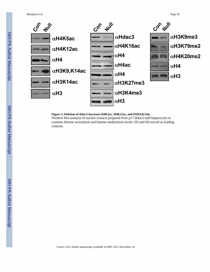
Figure 3. Deletion of Hdac3 increases H4K5ac, H4K12ac, and H3K9,K14acWestern blot analysis of nuclear extracts prepared from p17 Hdac3-null hepatocytes toexamine histone acetylation and histone methylation levels. H3 and H4 served as loadingcontrols.
Bhaskara et al. Page 18
Cancer Cell. Author manuscript; available in PMC 2011 November 16.
NIH
-PA Author Manuscript
NIH
-PA Author Manuscript
NIH
-PA Author Manuscript
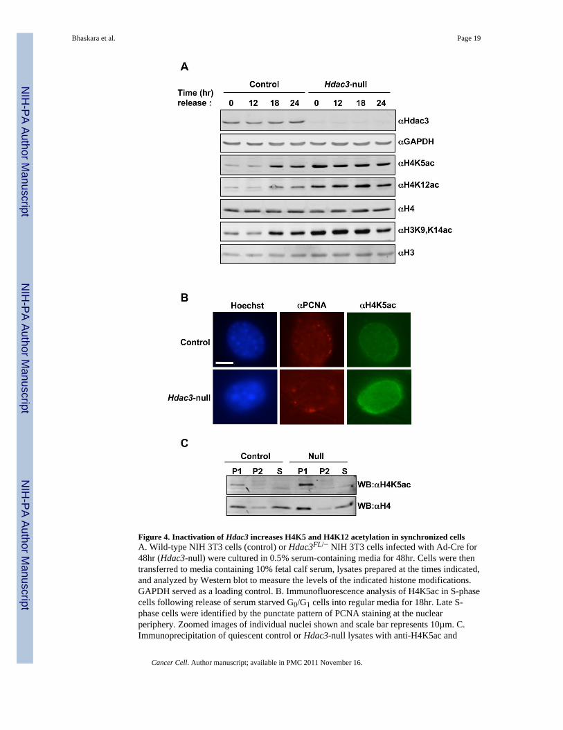
Figure 4. Inactivation of Hdac3 increases H4K5 and H4K12 acetylation in synchronized cellsA. Wild-type NIH 3T3 cells (control) or Hdac3FL/− NIH 3T3 cells infected with Ad-Cre for48hr (Hdac3-null) were cultured in 0.5% serum-containing media for 48hr. Cells were thentransferred to media containing 10% fetal calf serum, lysates prepared at the times indicated,and analyzed by Western blot to measure the levels of the indicated histone modifications.GAPDH served as a loading control. B. Immunofluorescence analysis of H4K5ac in S-phasecells following release of serum starved G0/G1 cells into regular media for 18hr. Late S-phase cells were identified by the punctate pattern of PCNA staining at the nuclearperiphery. Zoomed images of individual nuclei shown and scale bar represents 10µm. C.Immunoprecipitation of quiescent control or Hdac3-null lysates with anti-H4K5ac and
Bhaskara et al. Page 19
Cancer Cell. Author manuscript; available in PMC 2011 November 16.
NIH
-PA Author Manuscript
NIH
-PA Author Manuscript
NIH
-PA Author Manuscript

western blot analysis with anti-H4K5ac and anti-H4. P1, 1st immunoprecipitation; P2, re-immunoprecipitation of the supernatant from P1; S, supernatant from P2. See also figure S3.
Bhaskara et al. Page 20
Cancer Cell. Author manuscript; available in PMC 2011 November 16.
NIH
-PA Author Manuscript
NIH
-PA Author Manuscript
NIH
-PA Author Manuscript
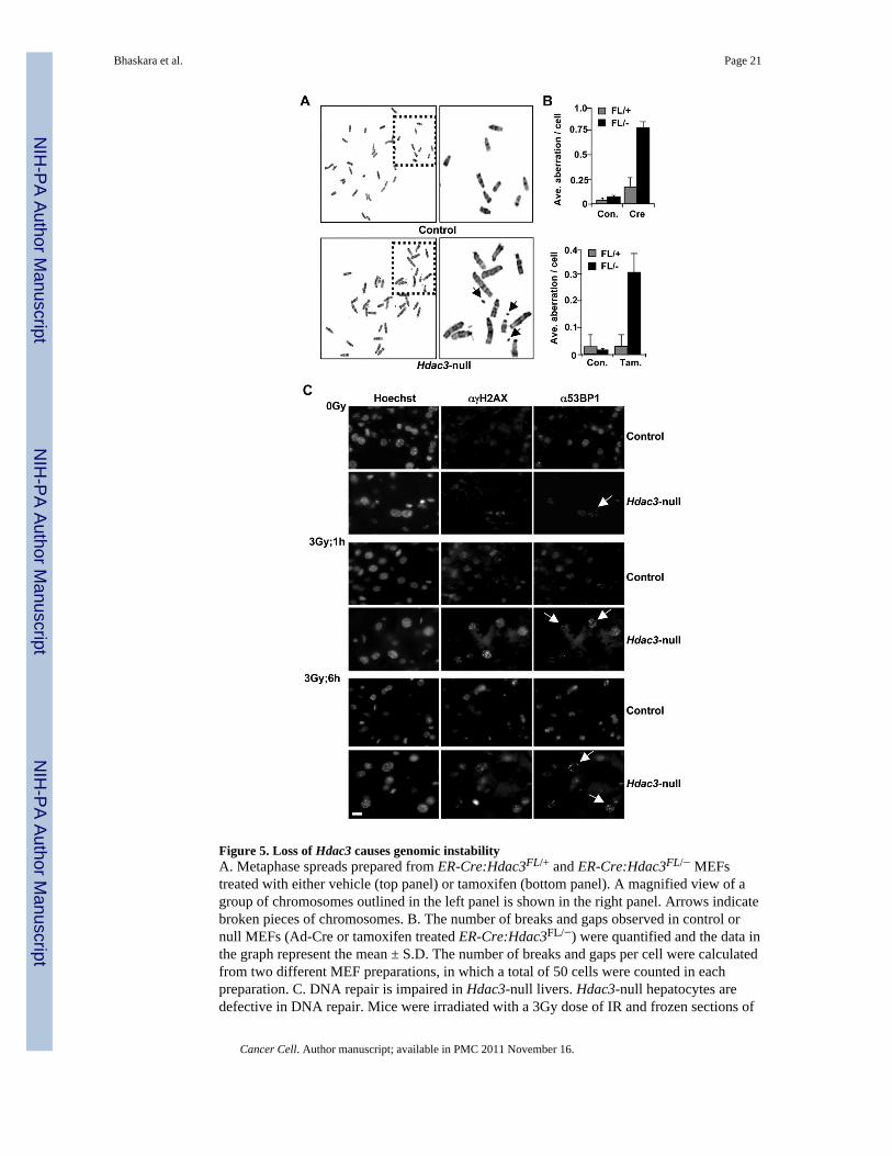
Figure 5. Loss of Hdac3 causes genomic instabilityA. Metaphase spreads prepared from ER-Cre:Hdac3FL/+ and ER-Cre:Hdac3FL/− MEFstreated with either vehicle (top panel) or tamoxifen (bottom panel). A magnified view of agroup of chromosomes outlined in the left panel is shown in the right panel. Arrows indicatebroken pieces of chromosomes. B. The number of breaks and gaps observed in control ornull MEFs (Ad-Cre or tamoxifen treated ER-Cre:Hdac3FL/−) were quantified and the data inthe graph represent the mean ± S.D. The number of breaks and gaps per cell were calculatedfrom two different MEF preparations, in which a total of 50 cells were counted in eachpreparation. C. DNA repair is impaired in Hdac3-null livers. Hdac3-null hepatocytes aredefective in DNA repair. Mice were irradiated with a 3Gy dose of IR and frozen sections of
Bhaskara et al. Page 21
Cancer Cell. Author manuscript; available in PMC 2011 November 16.
NIH
-PA Author Manuscript
NIH
-PA Author Manuscript
NIH
-PA Author Manuscript
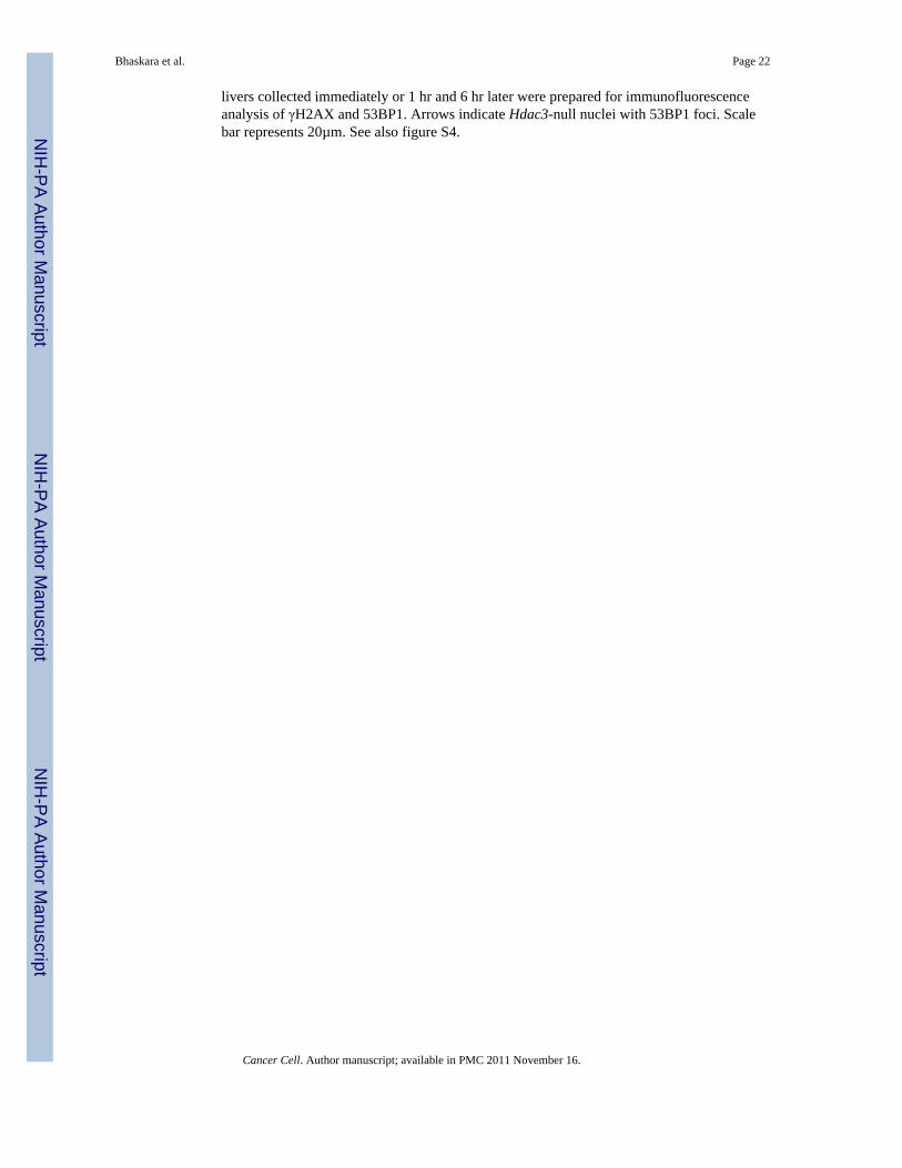
livers collected immediately or 1 hr and 6 hr later were prepared for immunofluorescenceanalysis of γH2AX and 53BP1. Arrows indicate Hdac3-null nuclei with 53BP1 foci. Scalebar represents 20µm. See also figure S4.
Bhaskara et al. Page 22
Cancer Cell. Author manuscript; available in PMC 2011 November 16.
NIH
-PA Author Manuscript
NIH
-PA Author Manuscript
NIH
-PA Author Manuscript

Figure 6. Loss of Hdac3 leads to hepatocellular carcinomaA Representative livers of 5-month (middle panel) and 10-month (right panel) old Alb-Cre:Hdac3−/− mice. B. Survival plot for Alb-Cre:Hdac3−/− mice. Heterozygous miceshowed no mortality within this time frame. C. Immunohistochemistry using anti-Hdac3shows that normal hepatocytes express Hdac3 (left panel), whereas Alb-Cre:Hdac3−/− micelack Hdac3 both in the tumor and surrounding tissue. T, tumor; L, liver. Scale bar represents60µm. D. Hematoxylin and eosin stained histological sections (H&E, top panels) andimmunohistochemistry for Ki67 (bottom panels) from 10-month old Alb-Cre:Hdac3−/−
mice. Scale bar represents 60µm. See also figure S5.
Bhaskara et al. Page 23
Cancer Cell. Author manuscript; available in PMC 2011 November 16.
NIH
-PA Author Manuscript
NIH
-PA Author Manuscript
NIH
-PA Author Manuscript
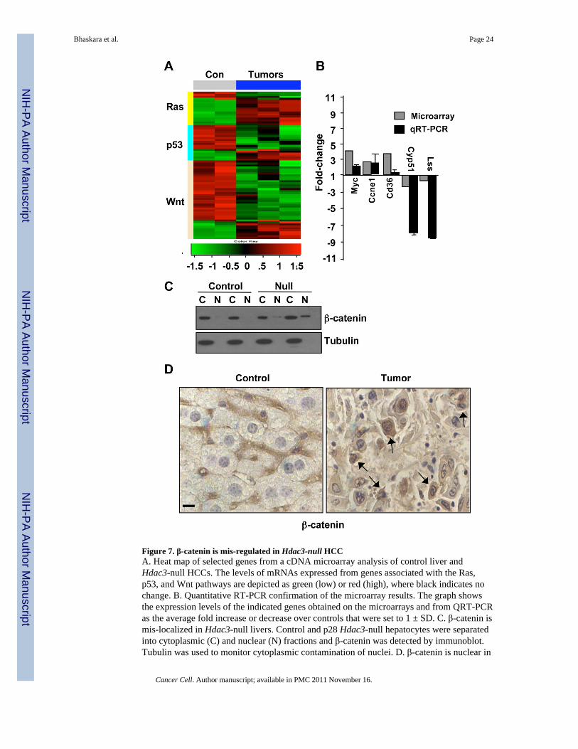
Figure 7. β-catenin is mis-regulated in Hdac3-null HCCA. Heat map of selected genes from a cDNA microarray analysis of control liver andHdac3-null HCCs. The levels of mRNAs expressed from genes associated with the Ras,p53, and Wnt pathways are depicted as green (low) or red (high), where black indicates nochange. B. Quantitative RT-PCR confirmation of the microarray results. The graph showsthe expression levels of the indicated genes obtained on the microarrays and from QRT-PCRas the average fold increase or decrease over controls that were set to 1 ± SD. C. β-catenin ismis-localized in Hdac3-null livers. Control and p28 Hdac3-null hepatocytes were separatedinto cytoplasmic (C) and nuclear (N) fractions and β-catenin was detected by immunoblot.Tubulin was used to monitor cytoplasmic contamination of nuclei. D. β-catenin is nuclear in
Bhaskara et al. Page 24
Cancer Cell. Author manuscript; available in PMC 2011 November 16.
NIH
-PA Author Manuscript
NIH
-PA Author Manuscript
NIH
-PA Author Manuscript
Hdac3-null HCC. Immunohistochemistry was used to determine the cellular localization ofβ-catenin (brown tint). Nuclei were counterstained with hematoxylin (blue tint). Arrowsindicate cells with prominent nuclear β-catenin. Scale bar represents 20 µm. See also tableS1.
Bhaskara et al. Page 25
Cancer Cell. Author manuscript; available in PMC 2011 November 16.
NIH
-PA Author Manuscript
NIH
-PA Author Manuscript
NIH
-PA Author Manuscript

Figure 8. NCOR1 is down regulated in human HCC and siRNA targeting of NCoR and SMRTcauses DNA damageA. Heat map representation of the analysis of NCOR1, NCOR2, Hdac3 and Hdac1 mRNAlevels in Human HCC samples (GEO 5975 dataset). Green depicts low expression relative tothe mean of control samples and red indicates higher expression. A 2-fold cut-off was usedto define a significant change. B. NCOR1 is down regulated in a subset of human HCC.Immunohistochemical staining was used to detect NCOR1 in human HCC or normalmatched surrounding tissue (brown stain). Cells were counterstained with hematoxylin (bluetint). Scale bar represents 20µm. C. HeLa cells were transfected with either non-targeting(NT) or NCoR/SMRT siRNA (N/S). Whole cell lysates were analyzed for histone
Bhaskara et al. Page 26
Cancer Cell. Author manuscript; available in PMC 2011 November 16.
NIH
-PA Author Manuscript
NIH
-PA Author Manuscript
NIH
-PA Author Manuscript

modifications using western blot. D. Immunofluorescence analysis of H4K5 acetylation inHeLa cells transfected with either non-targeting (NT) or NCoR/SMRT siRNA (N/S). Scalebar represents 10µm. S-phase cells were identified by the punctate pattern of PCNAstaining. E. Immunofluorescence analysis of 53BP1 in HeLa cells transfected with eithernon-targeting siRNA (NT) or NCoR/SMRT siRNA (N/S). Scale bar represents 10µm. Seealso figure S6.
Bhaskara et al. Page 27
Cancer Cell. Author manuscript; available in PMC 2011 November 16.
NIH
-PA Author Manuscript
NIH
-PA Author Manuscript
NIH
-PA Author Manuscript