Influence of NaOH and KOH on the synthesis of MCM-22 and MCM-49 zeolites
Upload
independentCategory
view
2download
0
www.elsevier.com/locate/micromeso
Microporous and Mesoporous Materials 83 (2005) 35–46
Grafting luminescent metal-organic speciesinto mesoporous MCM-41 silica from
europium(III) tetramethylheptanedionate, Eu(thd)3
A. Fernandes a, J. Dexpert-Ghys a, A. Gleizes b,*, A. Galarneau c, D. Brunel c
a Centre d’Elaboration de Materiaux et d’Etudes Structurales (UPR 8011), rue Jeanne Marvig, BP 4347, 31055 Toulouse cedex 4, Franceb Centre Interuniversitaire de Recherche et d’Ingenierie des Materiaux (UMR 5085), ENSIACET/INPT, 118 route de Narbonne,
31077 Toulouse cedex 4, Francec Laboratoire des Materiaux Catalytiques et Catalyse en Chimie Organique (UMR 5618) CNRS/ENSCM/UM1
(Institut Gerhardt FR 1878), ENSCM, 8 rue de l’Ecole Normale, 34296 Montpellier cedex 5, France
Received 27 July 2004; received in revised form 8 December 2004; accepted 14 December 2004
Available online 23 May 2005
Abstract
Mixed systems with Eu(III) b-diketonates as optically active guest species, and mesoporous silicas MCM-41 as a host matrix have
been investigated. The grafting of europium(III) onto the inner walls of unmodified MCM-41 has been achieved starting from
Eu(thd)3 (thd = 2,2,6,6-tetramethyl-3,5-heptanedionate), using two routes: wet impregnation (WI) at room temperature, and chem-
ical vapour infiltration (CVI) at 185 �C. In received hybrids, denoted Eu(thd)x@MCM-41, the same maximum yield [Eu]/
[Si] = 8.2 at% on average has been achieved with either methods. The molar ratio x = [thd]/[Eu] is 0.6 on average for WI samples,
and 1.5 for CVI samples. In the latter, higher contents in thd compensate lower contents in silanols with respect to the former.
Rationalizing the possible bonds exchanged at the silica surface leads to a great diversity of possible co-ordination schemes accord-
ing to the expressionP
½SiðOHÞn�xðOÞxEuðthdÞ3�x� (whereP
means that surface species are considered). Chromophore neutral
ligands phenanthroline (phen) or bipyridine (bipy) have been added to induce efficient Eu3+ luminescence under 270–280 nm exci-
tation, via the antenna effect. For the most favourable case, (phen)yEu(thd)x@MCM-41, the emission intensity at 612 nm under
excitation at 270 nm is 2/3 that for the genuine heteroleptic complex Eu(thd)3(phen). Moreover the hybrid material is stable up
to 440 �C.� 2005 Elsevier Inc. All rights reserved.
Keywords: Hybrid materials; MCM-41; Grafting; Europium(III); Wet impregnation; Chemical vapour infiltration; Luminescence
1. Introduction
The grafting of metal complexes onto the inner walls
of mesoporous silica SiMCM-41 (hereafter abbreviated
MCM-41) has been widely investigated, mainly to pre-
pare precursor compounds subsequently transformed
into inorganic materials by thermal treatment, most of-
ten for the purpose of application in catalysis. Several
1387-1811/$ - see front matter � 2005 Elsevier Inc. All rights reserved.
doi:10.1016/j.micromeso.2004.12.026
* Corresponding author. Fax: +33 5 62 88 56 78.
E-mail address: [email protected] (A. Gleizes).
review articles treat of surface modifications in mesopor-ous silica [1,2]. As stated by Trong On et al. [2], the fix-
ation of metal complexes onto the inner walls of
mesoporous silica can be conducted using three ap-
proaches: (i) the direct grafting by reaction with surface
silanol groups; (ii) the indirect grafting consisting in the
functionalization of the silanol groups with silane
coupling agents followed by the anchoring of metal
complexes to the surface; (iii) the ship-in-a-bottleimmobilization of metal complexes. Concerning rare-
earth cations derivatives, lanthanum-containing mixed

36 A. Fernandes et al. / Microporous and Mesoporous Materials 83 (2005) 35–46
oxide clusters were formed in Cs-MCM-41 by Kloetstra
et al. [3]. Gerstberger et al. have investigated yttrium
bulky complexes grafted into mesoporous silica [4,5]
by surface organometallic chemistry.
Europium(III) is a widely used red-emitting cation in
mineral matrices or as inorganic complexes, in the solidstate and in solution. Efficient red-emission under exci-
tation in the near UV-blue range (300–480 nm) is
achieved only for some europium(III) complexes. Or-
ganic chromophore molecules often have high absorp-
tion cross-sections in this range. It has long been
observed that when such molecules are bound to euro-
pium, the absorbed energy may be efficiently transferred
to the cation that in turn emits in the visible, accordingto the so-called ‘‘antenna effect’’. A plethora of organic
chromophores have proved to sensitize the luminescence
of lanthanide ions. The b-diketonate ligands were
among the first recognized suitable sensitizers [6]. Since
then, the photo-physics of luminescent lanthanide
(mainly Eu3+ and Tb3+) complexes has been systemati-
cally studied [7].
However, the poor chemical and thermal stability ofas-prepared metal-organic complexes can adversely af-
fect many applications. These drawbacks must be over-
come before development of their uses. In this respect,
intensive efforts are currently made to embed them into
a matrix to improve thermal, mechanical or chemical
resistance. For instance, bulky hybrid materials were
prepared with the metal-organic complex embedded in
a silicate based matrix [8] or a polymer [9]. Some worksdeal with embedding europium complexes into porous
matrices. Mitchell-Koch et al. [10] immobilized euro-
pium complexes within porous organic polymer hosts,
and obtained luminescent materials potentially suitable
for chemical sensing. The immobilization of europium
complexes within mesoporous silica hosts has been de-
scribed in Refs. [11–13]. For Eu(TTA)3 complexes
(TTA = thenoyltrifluoroacetone) [11], it is reported thatthe luminescence efficiency in the hybrids is about half
that of the genuine complex, whereas the temperature-
related quenching of the luminescence is reduced.
Encapsulation of Eu(TTA)4C5H5NC16H33 into surface-
modified Si-MCM-41 is claimed to give efficient red
emitters with a higher stability towards UV irradiation
than the corresponding complex [12,13].
In this work, we investigate mixed systems withEu(III) b-diketonates [Eu(dik)3] as optically active guest
species and mesoporous silicas MCM-41 as a host ma-
trix. The goal was to stabilize highly luminescent org-
ano-lanthanides complexes in mineral hosts [14–17].
Preserving the textural characteristics of the matrix
(i.e. highly ordered porosity and high surface area) in
the hybrids could make these materials eminently suit-
able for luminescent sensing applications.The results presented here concern the direct grafting
of metal complex species starting from Eu(thd)3
(thd = 2,2,6,6-tetramethyl-3,5-heptanedionate). The com-
plex was expected to react with silanol groups dangling
along the pore walls, as was first reported for titanocene
dichloride by Maschmeyer et al. [18]. We used two
routes: wet impregnation (WI) and chemical vapour
infiltration (CVI). The precursor molecule Eu(thd)3 iswell adapted to these two techniques since it is both sol-
uble in organic solvents and easily sublimed. This en-
abled us to compare the two approaches in terms of
nature and yield of grafted species, and of reaction
mechanisms.
Phenanthroline (phen) and bipyridine (bipy) were
used as additional ligands to tentatively improve the
luminescence yield through the antenna effect. The prod-ucts were characterized by chemical analyses, thermal
decomposition measurements, structural and textural
characterizations, and vibrational and optical lumines-
cence spectroscopies.
2. Experimental
2.1. Samples preparations
MCM-41 silica (pore size 3.5 nm) was prepared at
115 �C in autoclave for 24 h, using the following molar
ratios 1SiO2/0.1CTAB/0.25NaOH/100H2O, and charac-
terized using known procedures as described in [19] for
instance. The template CTMA (cetyltrimethylammo-
nium) was removed by calcination at 500 �C during8 h (heating rate of 2 �C/min), and between 525 �C and
550 �C for a few hours. Template removal was checked
by FTIR. Eu(thd)3 was purchased from Interchim and
kept in a desiccator.
MCM-41 silica destined to wet impregnation (WI)
was activated under vacuum (10�2 Torr) at 180 �C for
4 h. Then 100 mg of activated silica was rapidly weighed
and suspended in different measured volumes of a solu-tion of Eu(thd)3 (1.42 · 10�3 M) in cyclohexane, corre-
sponding to different initial Eu/Si atomic ratios [(Eu/
Si)i in Table 1]. The mixture was stirred at 25 �C for
24 h. The powder was recovered by filtration, washed
several times with cyclohexane, and dried at room tem-
perature. Hereafter and in Table 1, the samples prepared
by WI are denoted W1–9. Samples W8 and W9 were
intentionally not washed with cyclohexane before dry-ing. Sample W9k was prepared in the same conditions
as W9, then heated at 900 �C in air for 1 h, in order to
totally calcine and remove the organic matter. A sample
without europium denoted WD was obtained by
impregnating silica with Hthd (0.26 moles of Hthd per
SiO2) at 25 �C until dryness.
Two procedures of chemical vapour infiltration (CVI)
were employed: the static vapour phase (SVP-CVI)method and the dynamic vapour phase (DVP-CVI)
method, as described elsewhere in detail [16,17]. To

Table 1
Preparation and analytical data
Preparationa (Eu/Si)i, at% (Eu/Si)f, at% Ligand/Eu (molar ratio) Analysisa Proposed formula
MCM-41 Hydrated No ligand T [SiO1.850(OH)0.30], 0.86 H2O
W1 W, O 2.1 ± 0.1 2.4 ± 0.1 0.6 thd A [SiO1.908(OH)0.242][Eu0.024(thd)0.014]
3.3 ± 0.3 E
W2 W, O 4.3 ± 0.2 4.7 ± 0.2 0.4 thd A [SiO1.972(OH)0.178][Eu0.047(thd)0.019]
5.7 ± 0.5 E
W3 W, O 6.4 ± 0.3 6.7 ± 0.3 0.4 thd A [SiO2.024(OH)0.126][Eu0.067(thd)0.027]
7.4 ± 0.7 E
W4 W, O 8.5 ± 0.4 8.3 ± 0.4 0.5 thd A [SiO2.058(OH)0.092][Eu0.083(thd)0.042]
8.1 ± 0.8 E
W5 W, O 10.7 ± 0.5 8.1 ± 0.4 0.2 thd A [SiO2.077(OH)0.073][Eu0.081(thd)0.016]
W6 W, O 12.8 ± 0.6 7.1 ± 0.3 0.6 thd A [SiO2.020(OH)0.130][Eu0.071(thd)0.043]
W7 W, O 42.9 ± 0.2 9.7 ± 0.4 0.9 thd A [SiO2.054(OH)0.096][Eu0.097(thd)0.087]
W8 W 8.5 ± 0.4 6.2 ± 0.3 1.27 (thd + Hthd) X, T [SiO1.988(OH)0.162(Hthd)0.031][Eu0.062(thd)0.048]
W9 W 8.5 ± 0.4 8.4 ± 0.8 1.12 (thd + Hthd) X, T [SiO2.032(OH)0.118(Hthd)0.024][Eu0.084(thd)0.070]
W9k W, K 8.5 ± 0.4 8.4 ± 0.8 thd not analyzed X SiO2.126Eu0.084S1 S, 66 h, O 8.8 ± 0.2 8.3 ± 0.4 1.6 thd A [SiO2.016(OH)0.084][Eu0.083(thd)0.133]
S2 S, 66 h 8.5 ± 0.2 9.2 ± 0.9 2.07 (thd + Hthd) X, T [SiO2.063(OH)0.037(Hthd)0.084][Eu0.085(thd)0.092]
S3 S, 39 h 9.0 ± 0.2 8.1 ± 0.8 thd not analyzed X
S4 S, 25 h 8.6 ± 0.2 8.3 ± 0.8 thd not analyzed X
D1 D 7.5 ± 0.8 1.6 thd X, C [SiO2.005(OH)0.095][Eu0.075(thd)0.120]
D2 D 9.2 ± 0.9 1.7 thd X, C [SiO2.020(OH)0.080][Eu0.092(thd)0.156]
WP1 W, 1 step 8.5 ± 0.4 5.0 ± 0.2 0.6 thd, 0.8 phen A [SiO1.970(OH)0.180][Eu0.050(thd)0.030(phen)0.041]
WP2 W, 2 steps 8.5 ± 0.4 6.6 ± 0.3 0.5 thd, 0.5 phen A [SiO2.015(OH)0.135][Eu0.066(thd)0.033(phen)0.033]
WB1 W, 1 step 8.5 ± 0.4 7.0 ± 0.3 0.5 thd, 0.3 bipy A [SiO2.025(OH)0.125][Eu0.070(thd)0.035(bipy)0.018]
WB2 W, 2 steps 8.5 ± 0.4 6.4 ± 0.3 0.3 thd, 0.2 bipy A [SiO2.023(OH)0.127][Eu0.064(thd)0.019(bipy)0.010]
DP21 D, 2 steps 6.3 ± 0.3 thd, phen not analyzed X
DP22 D, 2 steps 7.0 ± 0.3 thd, phen not analyzed X
a A: Si, Eu, C, H chemical analyses (Service Central d�Analyses du CNRS); C: C, H chemical analyses (Laboratoire de Controle de l�ENSIACET);
D: dynamical vapour phase method; E: EELS analysis; K: air calcination (900 �C); O: degassed at 150 �C/10�3 Torr after preparation; S: static
vapour phase method, treatment duration mentioned; T: thermal gravimetric analysis; W: wet impregnation; X: EDXS analysis.
A. Fernandes et al. / Microporous and Mesoporous Materials 83 (2005) 35–46 37
prepare samples by the SVP-CVI method, weighed silica
(typically 15–50 mg) and Eu(thd)3 (typically 10–30 mg)
were placed in two separate glass-containers open at
one end (length = 25 mm, B = 10 mm). The container
of silica was introduced in a Pyrex-glass tube
(B = 12 mm) subsequently linked to a vacuum pump
to perform activation under vacuum (ca. 10�4 Torr) at
205 �C for 4 h. Then the tube was successively cooleddown to room temperature under vacuum, filled with
dry air, rapidly charged with the container of metal
complex, linked again to the vacuum system, evacuated
for five minutes, and sealed into an ampoule. The am-
poule was heated at 185 �C for a period of time going
from a few hours to a few days depending on the exper-
iment. Then it was quenched in the air, to force the
remaining vapour phase to condense on the glass walls,not in the powder. The containers were weighed imme-
diately after opening the ampoule, and then kept in a
desiccator. The samples prepared by the SVP-CVI meth-
od are denoted S1–4 hereafter and in Table 1.
To prepare samples by the DVP-CVI method (sam-
ples D1, 2), about 150 mg of silica were placed on a filter
in the narrower part of a vertical reactor tube which
could be heated by an oven as schematized in [16].About 300 mg of the precursor was introduced in the
cool part of the reactor. The silica was first activated
at 205 �C under vacuum for 4 h. After cooling, still un-
der dynamic vacuum, dry nitrogen was progressively
admitted with a flow monitored at about 2 ml/min to
fluidise the silica bed. Then, the reaction temperature
was raised and maintained at 185 �C. The precursor
container was progressively lifted up to a zone at
150 �C so that the precursor could be sublimed. Aftertwo hours, it was lifted down to the cool part. The reac-
tor temperature was maintained at 185 �C for another
30 min, and then let to cool down still under flowing
nitrogen. Atmospheric pressure was then established
by progressive admission of nitrogen.
Phenanthroline (phen) or bipyridine (bipy) were intro-
duced as additional ligands in either one or two steps by
wet impregnation (samples WP1,2 and WB1,2). In thetwo-steps method, activated silica was first impregnated
with Eu(thd)3. After washing and drying, 150 mg of the
received powder were suspended in 150 ml of a solution
of phenanthroline or bipyridine (3 · 10�3 M) in dichlo-
romethane. One step reactions were performed by sus-
pending 150 mg of activated silica in solutions of
Eu(thd)3(phen) (respectively Eu(thd)3(bipy)) in dichloro-
methane. These complexes were synthesized in a preli-minary step by reacting equimolar amounts of Eu(thd)3

38 A. Fernandes et al. / Microporous and Mesoporous Materials 83 (2005) 35–46
and phen (respectively bipy) in dichloromethane at 25 �Cfor 1 h, filtering, washing and drying.
Preparation of samples DP21 and DP22 (phenanthro-
line) with the vapour process was run in two steps with
the dynamic vapour process: Eu(thd)3 was vaporized
first, and then phenanthroline.
2.2. Analyses and characterization
For most samples, the elements Si, Eu, C, H, N were
analyzed by the Service Central d�Analyse du CNRS.
Some C and H analyses were also performed at the Lab-
oratoire de Controle at ENSIACET. The [Eu]/[Si] ratios
were separately evaluated by the local probe techniquesEDXS and EELS. In the latter case, the [Eu]/[Si] ratios
were calculated as ([Eu]/[O]) Æ ([O]/[Si]), with [Eu]/[O]
determined by comparison with the experimental ratio
(Eu M-edge/O K-edge) in standard Eu2O3, and [Si]/[O]
from the calculated K-edge cross-sections of silicon
and oxygen.
Textural properties were characterized by N2 sorp-
tion at liquid nitrogen temperature on samples degassedat 150�C under vacuum (10�3 Torr) for 12 h. N2 sorp-
tion experiments were carried out in a Micromeritics
ASAP2000 apparatus. Average pore diameters have
been evaluated from the nitrogen desorption branch
according to Broekhoff and De Boer [20]. This is one
of the best methods to evaluate pore size of MTS mate-
rials [21]. Special care was taken in determining the BET
specific area, i.e. to calculate the linearization of theBET equation by avoiding points at low pressure af-
fected by surface heterogeneity and points at high pres-
sure corresponding to starting pore filling. Pore volumes
were calculated at the end of the step corresponding to
the filling of the pores.
X-ray powder diffraction patterns were recorded on a
Seifert diffractometer using CuKa radiation. Thermo-
gravimetric and differential thermal analyses (TGA–DTA) were performed with a SETARAM LABSYS-TG
apparatus, at a heating rate of 2 �C/min in oxygen flow.
Luminescence excitation and emission spectra were
recorded with a Hitachi F4500 spectrofluorimeter.
Luminescence spectra were also recorded with a Raman
set-up (DILOR XY), equipped with a krypton–argon
laser source and with an optical microscope in order
to recover the spectra emitted by about 0.5–1 lm3 ofmatter. IR spectra were recorded on a FT-IR Brucker
Vector22. Self-supported wafers were prepared for
the IR investigation and further degassed at 150 �C/10�2 Torr for 4 h.
3. Results
The results presented in this section will be discussed
in the following section.
3.1. Samples compositions
Synthesis details and analyses results are gathered in
Table 1. In the table, Wn, Sn, Dn refer to samples ob-
tained respectively by wet impregnation (W), by the sta-
tic vapour phase method (S), and by the dynamicvapour phase method (D). For mixed-ligand samples,
the letters P (phenanthroline) or B (bipyridine) were
added with a subscript index indicating the number of
steps of preparation.
Local and overall chemical analyses of the europium
content give similar results. Local probe measurements
prove that europium is homogeneously dispersed in
the silica matrix at the spatial resolution of the probe(150 nm). Samples W1–4 correspond to increasing initial
atomic ratios (Eu/Si)i. The corresponding final ratios
(Eu/Si)f vary parallel to (Eu/Si)i. Then a plateau is
reached. The highest (Eu/Si)f ratio, as averaged from
values measured for samples W4–9, S1–4, D1–2 is
8.2 at%. For the mixed-ligand samples, WP1–2, WB1–2
and DP21–2, the average (Eu/Si)f is 6.2%.
The formulas given in last column of Table 1 deservesome comments. Eu(thd)3 is expected to react with sila-
nol groups (Si–OH) dangling along the pore walls,
according to the overall reaction:
SiO2�xðOHÞ2x þ zEuðthdÞ3! SiO2�xþyðOHÞ2x�yEuzðthdÞ3z�y þ yHthd. ð1Þ
The coefficients z and (3z � y) are deduced from theanalyses. The content 2x in available silanol groups
mainly depends on the activation process. For the sam-
ples of silica treated by wet impregnation, the value of
0.30 (3 SiOH/nm2) was retained, according to the recent
determination by Shenderovich et al. [22] from 15N
MAS NMR of pyridine adsorbed by samples with spe-
cific surface areas of 1010 m2 g�1. As to the samples of
silica treated by either CVI methods, since they wereactivated at higher temperature, and since they were
maintained at 185 �C during the reaction with Eu(thd)3,
the content in available SiOH was expected to be some-
what lower. Indeed, the value 2x = 0.20 (2 SiOH/nm2)
was derived from the analyses for these samples.
Reaction equation (1) postulates the transformation
of Eu(thd)3 into chemisorbed species through strong
Si–O–Eu bonds, implying that there is no physisorbedEu(thd)3 molecules. The fact that no appreciable
amounts of Eu(thd)3 was just physisorbed has been at-
tested by tentative thermodesorption experiments, com-
paratively with compounds prepared by reacting
Cu(thd)2 with MCM-41 using the SVP-CVI method
[16]. When heated under vacuum at 130 �C, these com-
pounds released about 60% of the adsorbed copper as
volatile Cu(thd)2. Nothing of the kind was observedfor compounds prepared with Eu(thd)3, even above
130 �C.
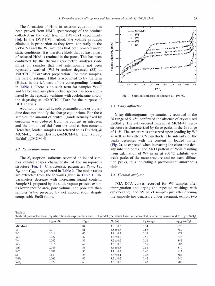
Fig. 1. Sorption isotherms of nitrogen at –196 �C.
A. Fernandes et al. / Microporous and Mesoporous Materials 83 (2005) 35–46 39
The formation of Hthd in reaction equation 1 has
been proved from NMR spectroscopy of the product
collected in the cold trap in DVP-CVI experiments
[16]. In the DVP-CVI method, the volatile products
eliminate in proportion as they form, contrarily to the
SVP-CVI and the WI methods that both proceed understatic conditions. It is therefore likely that at least a part
of released Hthd is retained in the pores. This has been
confirmed by the thermal gravimetric analyses (vide
infra) on samples that had intentionally not been
repeatedly washed (W8–9) and/or degassed (S2) at
150 �C/10�3 Torr after preparation. For these samples,
the part of retained Hthd is accounted to by the term
(Hthd)v in the left part of the corresponding formulain Table 1. There is no such term for samples W1–7
and S1 because any physisorbed species has been elimi-
nated by the repeated washings with cyclohexane and/or
the degassing at 150 �C/10�3 Torr for the purpose of
BET analysis.
Addition of neutral ligands phenanthroline or bipyri-
dine does not modify the charge equilibrium. For these
samples, the amount of neutral ligands actually fixed byeuropium was deduced from the content in nitrogen,
and the amount of thd from the total carbon content.
Hereafter, loaded samples are referred to as Eu(thd)x@
MCM-41, (phen)yEu(thd)x@MCM-41, and (bipy)yEu(thd)x@MCM-41.
3.2. N2 sorption isotherms
The N2 sorption isotherms recorded on loaded sam-
ples exhibit shapes characteristic of the mesoporous
structure (Fig. 1). Characteristic parameters SBET, VP,
DP, and CBET are gathered in Table 2. The molar ratios
are extracted from the formulas given in Table 1. The
parameters decrease with increasing ligand content.
Sample S1, prepared by the static vapour process, exhib-
its lower specific area, pore volume, and pore size thansamples W4–6 prepared by wet impregnation, despite
comparable Eu/Si ratios.
Table 2
Textural parameters from N2 adsorption–desorption data and BET model (
Ligand/Si CBET
MCM-41 0 84
W1 0.014 61
W2 0.019 45
W3 0.027 47
W4 0.042 35
W5 0.016 62
W6 0.043 56
W7 0.087 35
S1 0.133 26
WP2 0.066 45
WB2 0.029 60
3.3. X-ray diffraction
X-ray diffractograms, systematically recorded in the
2h range of 5–45�, confirmed the absence of crystallized
Eu(thd)3. The 2-D ordered hexagonal MCM-41 meso-structure is characterized by three peaks in the 2h range
of 1–5�. The structure is conserved upon loading by WI
as well as by either CVI methods. The intensity of the
peaks decreases with the content in loaded matter
(Fig. 2), as expected when increasing the electronic den-
sity into the pores. The XRD pattern of W9k resulting
from calcination of W9 in air at 900 �C exhibits very
weak peaks of the mesostructure and no extra diffrac-tion peaks, thus indicating a predominant amorphous
state.
3.4. Thermal analyses
TGA–DTA curves recorded for WI samples after
impregnation and drying (no repeated washings with
cyclohexane), and SVP-CVI samples just after openingthe ampoule (no degassing under vacuum), exhibit two
the values have been corrected in order to correspond to 1 g of SiO2)
DP (A) VP (ml/g) SBET (m2/g)
3.8 ± 0.3 0.74 1020
3.5 ± 0.3 0.63 889
3.4 ± 0.2 0.59 877
3.3 ± 0.2 0.56 848
3.2 ± 0.2 0.51 842
3.5 ± 0.3 0.57 883
3.6 ± 0.3 0.51 810
3.1 ± 0.2 0.46 812
2.3 ± 0.1 0.25 587
3.2 ± 0.2 0.42 740
3.3 ± 0.2 0.43 708

Fig. 2. X-ray diffraction patterns for pristine MCM-41 and variously
loaded samples.
Fig. 3. TGA–DTA diagrams for wet impregnated Eu(thd)x@MCM-
41 hybrids, as-prepared (W9), and degassed at 150 �C/10�3 Torr for
12 h (W4).
40 A. Fernandes et al. / Microporous and Mesoporous Materials 83 (2005) 35–46
weight losses associated with exothermic effects, as illus-
trated for sample W9 on Fig. 3a. The first loss is associ-
ated with a double DTA peak at 175–200 �C, and the
second one with a single DTA peak at 300 ± 10 �C. Bothevents are assigned to the departure of organic matter.The first loss is assigned to that part of Hthd resulting
from reaction (1) and retained in the pores by weak
interactions. The second loss is assigned to the ligands
bound to europium [15]. The assignments are based on
the following observations: (i) TG and TD analyses
for the sample WD that only consists of MCM-41
impregnated with Hthd (experimental part) shows anexothermic split peak at 250 �C, associated with a
weight loss of 0.025 molecules of Hthd per Si atom;
(ii) the curves recorded for degassed samples show no
other feature than the weight loss around 300 �C, as
illustrated for sample W4 on Fig. 3b. The weight loss
estimated for W4 is 12% of the final mass. This fairly
compares with the 10.5% calculated from the formula
given in Table 1 [SiO2.058(OH)0.092Eu0.083(thd)0.042],assuming that the calcined product is SiEu0.083O2.124
for the sake of electroneutrality.
Thermal analyses for the thd–phen mixed ligands
samples feature a one-step decomposition at 440–
450 �C. Observed weight losses with respect to final
weights are 17% and 20% for WP2 and WP1 respectively,
in agreement with those calculated from the formulas in
Table 1 (17% and 18%). We may assign this unique fea-ture to the thermal decomposition of a (phen)yEu(thd)xspecies, with x � y � 0.5. For the thd–bipy samples, two
weight losses are observed at 300–310 �C and 430–
440 �C. The observed overall weight loss between 150
and 550 �C is about 13% of the final weight for each
sample, whereas the values deduced from the analyses
are 12% for WB1 and 6% for WB2. The overall number
of bipy ligands per europium is lower than the numberof thd ligands. The thermal event at 430–440 �C is as-
signed to the decomposition of a species (bipy)yEu(thd)xwith x � y � 0.2–0.3, whereas the loss at about 305 �C is
well in the range of decomposition of residual grafted
Eu(thd)x.
3.5. Infrared spectroscopy
Infrared absorption spectra in the energy range 1400–
3900 cm�1 are displayed on Fig. 4. They were recorded
in the transmission mode using self-supported wafers
that were preliminary degassed (thermodesorbed) in situ
at 150 �C/10�2 Torr for 4 h. The spectra were scaled
using the couple of silica overtones at 1800–1900 cm�1,
making thus possible direct comparison of band intensi-
ties from sample to sample in the energy ranges 1400–1700 cm�1 (m(OCCCO)), 2750–3000 cm�1 (m(C–H)), and
3250–3750 cm�1 (m(O–H)).
The intensities of the diketone m(C–H) and m(OCCCO)
bands increase with the content in thd ligand, as shown
by comparison of the spectra of S1 and W4 in Fig. 4.
They correspond to samples with the same Eu/Si ratio.
S1 is thd-rich (13.3 thd/8.3 Eu/100 Si) compared to sam-
ple W4 (4.2 thd/8.3 Eu/100 Si). The unloaded sampleshows a narrow m(O–H) component at 3745 cm�1 that is
commonly attributed to free Si–OH groups. For the

Fig. 4. IR absorption spectra recorded on self-supported wafers for
unloaded MCM-41, and Eu(thd)x@MCM-41 hybrids (W4, S1) after
degassing at 150 �C/10�2 Torr for 4 h.
Fig. 5. Eu(III) emission spectra for (a) amorphous silica-europium
oxide Eu@MCM-41 sample (W9k) and (b)–(d) upper curves, the
grafted species W4, WP2, WB2; lower curves, the corresponding
genuine complexes.
Fig. 6. Diffuse reflection spectra (dotted lines) and excitation spectra
measuring the Eu(III) emission at 612 nm (solid lines) for (a)
amorphous silica-europium oxide Eu@MCM-41 (W9k); (b) genuine
Eu(thd)3; (c)–(e) the grafted species W4, WP2, WB2, respectively.
A. Fernandes et al. / Microporous and Mesoporous Materials 83 (2005) 35–46 41
heavily loaded samples W4 and S1, this component is
shifted to 3710 cm�1, indicating that all initially free
Si–OH groups interact with grafted europium species.
A broad feature at about 3500 cm�1 is observed for
W4, and to a lesser extent for S1.
3.6. Eu3+ luminescence
Eu3+ visible emission and excitation spectra, and vis-
ible-near-UV diffuse reflection spectra for the genuine
complexes and the hybrid samples are displayed in Figs.
5 and 6.
Emission essentially occurs in the red part of the vis-
ible spectrum. It is unambiguously assigned to transi-tions between the discrete electronic levels of the 4f6
configuration (Fig. 5). Eu(III) complexes exhibit emis-
sion spectra made of narrow lines that figure a finger-
print of the geometry of co-ordination of the cation,
as shown in Fig. 5 (lower curves) by the spectra of
Eu(thd)3 (Fig. 5b), Eu(thd)3(phen) (Fig. 5c) and
Eu(thd)3(bipy) (Fig. 5d). The complexes immobilized
in the pores give much less structured spectra, like thosereported in Fig. 5 (upper curves) for sample W4 (Fig.
5b), sample WP2 (Fig. 5c), and sample WB2 (Fig. 5d).
The 5D0–7F1,2 transitions are more structured, and the
5D0–7F0 emission is relatively more intense for WP2 or
WB2 than for W4.
By recording the emission with the optical micro-
scope set-up, a volume of about 1 lm3 is possibly ana-
lyzed to control the homogeneity of the sample with

Table 3
Reported metal/silicium ratios for several metal supported MCM-41
M (MOn) wt%a [M]/[Si] (at%)a Ref.
W (WO3) 8.2 3.0 [2]
Cr (Cr2O3) 2.0 2.4 [2]
Cu (Cu2O) 10.0 9.3 [2]
Mn (MnO) 8.5 10.0 [2]
La (La2O3) 5.0 2.3 [3]
Yb (Y2O3) – 8.1 [5]
Yc (Y2O3) – 4.0 [5]
Eu (Eu2O3) – 8.2 This work
a wt% = m(M) per 100 g sample from analysis; [M] = wt%/{M};
[Si] = (100 � {MOn} · [Me])/{SiO2}, {} = molar weight.b From Ref. [8]: 0.8 Y/nm2, specific area taken as 1020 m2/g.c From Ref. [8]: unsilylated route.
42 A. Fernandes et al. / Microporous and Mesoporous Materials 83 (2005) 35–46
this spatial resolution. Whereas two types of emission
spectra showed up for sample WB1, one type only was
observed for the other samples. This result means that
the samples are single phases (except WB1) to the scale
of the probe. On the other hand, the emission lines are
broader for the hybrids than for the precursor moleculesin the solid state; this inhomogeneous broadening evi-
dences that the Eu3+ ions adopt different local symme-
tries in a given single-phase hybrid.
The effect of ligands bond to Eu3+ shows itself on
luminescence excitation spectra monitored at the main
emission wavelength of 612 nm (Fig. 6). The Eu–thd
interaction in Eu(thd)3 is known to induce a very low-ly-
ing ligand to metal charge transfer (LMCT) state.Relaxation from this state is non-radiative [23,24]. The
diffuse reflectance spectrum of Eu(thd)3 (Fig. 6b, dotted
lines) shows strong absorption below 425 nm (absorp-
tion in the LMCT), whereas no Eu3+ emission is ob-
served with excitation spectrum in this range (Fig. 6b,
solid line). As shown by the dotted curves of Fig. 6,
the diffuse reflection spectrum of Eu(thd)x@MCM-41
(W4, Fig. 6c) exhibits an abrupt absorption edge around325 nm, whereas the oxide Eu@MCM-41 (W9k, Fig. 6a)
presents a smoother one, centred at 300 nm. Consider-
ing the luminescence excitation spectra (solid curves of
Fig. 6), one observes the gradual appearance of intra-
4fn excitation lines 7F0–5D2,
5L6 for Eu(thd)x@MCM-
41 (W4, Fig. 6c), and the upper-lying 7F0–5D3 transition
for W9k that contains no more organic part.
Samples prepared with phenanthroline (WP2) andbipyridine (WB2) strongly absorb above respectively
340 nm (Fig. 6d, dotted line), and 320 nm (Fig. 6e, dot-
ted line). Excitation at wavelengths below these values
results in a strong red emission. In the excitation spectra
(solid lines in Fig. 6d and e), the intra-4f transitions are
much weaker than the ligand-centred absorption and
even hardly visible. This evidences the antenna effect.
Samples WP1 and WP2, that exhibit only one thermaldecomposition peak and appear homogeneous from the
luminescence spectra at the microscopic scale, were sub-
mitted to further luminescence investigation. The emis-
sion intensity at 612 nm under excitation at 280 nm for
the grafted species is about 2/3 that for pure Eu(thd)3-
(phen) in the same conditions. The 5D0 emission level
lifetime at room temperature is 0.80 ± 0.10 ms.
4. Discussion
Several points can be discussed: first the ability to
immobilize lanthanide (Eu3+) ions and organic b-diket-onate(–) ligands into the pores of a MCM-41 silica ma-
trix by the two synthesis methods employed, and the
behaviour of the loaded samples versus different chemi-cal and thermal treatments; secondly, the mechanisms
of anchorage of metal-organic species onto the silica
surface; and finally, the luminescence characteristics of
the hybrid compounds.
Eu(thd)3 was chosen as a precursor because it lends
itself just as well to wet impregnation as to chemical
vapour deposition. When this precursor is used without
extra ligand (phenanthroline or bipyridine), both tech-
niques give nearly the same maximum loading of in-
serted europium as measured by the atomic ratio (Eu/Si)f. The average limit ratio determined over 12 samples
(W4–9, S1–4, D1–2) is (Eu/Si)f = 8.2 at%. This corre-
sponds to a density of 0.81 europium atoms/nm2. From
literature data, the atomic ratio M/Si has not often been
used as a measure of the metal loading in mesoporous
structures. Contents are most often expressed in wt%.
Table 3 gathers the metal loading ratios expressed in
wt% in Refs. [2,3]; to convert these values in [M]/[Si](at%) we made the assumption that the samples, ana-
lyzed after air-calcination, were mixtures of the oxides
MOn and SiO2. For yttrium, Gerstberger et al. [5]
reported an approximate loading of 0.8 Y atoms/nm2
for hybrid compounds prepared by grafting Y[N
(SiH(CH3)2]x(thf)y or Y(fod)x(thf)y on silylamide modi-
fied MCM-41, which compares rather well with the
0.81 Eu atoms/nm2 in the present study. On the otherhand, direct reaction from Y(fod)3 (fod = 1,1,1,2,2,3,
3-heptafluoro-7,7-dimethyl-4,6-octanedione) without
preliminary silylamidation [5], resulted in half less
grafted metal centers (0.4 Y atoms/nm2). Compared
with literature results, rather high metal loadings have
been obtained in the present study, both by impregna-
tion and by vapour deposition (Table 3).
When bipyridine or phenanthroline are used, the final(Eu/Si)f ratio is significantly lower than 8% whatever
method of preparation is used. This result seems normal
when the compounds are prepared in one step because
the precursor molecules Eu(thd)3(bipy) or Eu(thd)3-
(phen) are more encumbered than Eu(thd)3. The same
observation for the two-step process suggests some etch-
ing process which we did not try to elucidate.
We describe the anchoring mechanism by the reac-tion between silanol groups and Eu(thd)3 resulting in

A. Fernandes et al. / Microporous and Mesoporous Materials 83 (2005) 35–46 43
freed Hthd and Eu(thd)x species grafted on silanolate
groups following Eq. (1). The reaction is spontaneous
even at room temperature as proved by the WI experi-
ments. The two preparative methods mainly differ by
the temperature, and the use of a solvent. However, they
both result in grafted europium species that are chemi-sorbed: the Eu atoms fasten on the silica wall by iono-
covalent Si–O–Eu bonds.
It is remarkable that the two preparative methods
lead to similar highest metal loadings: Eu/Si = 8.2 at%
(samples W4–9, S1–4, D1–2). On the other hand, the
(thd/Eu) molar ratio is systematically lower for samples
prepared by wet impregnation (between 0.4 and 0.9,
average = 0.6) than for samples prepared from the va-pour phase (mainly between 1.6 and 1.7, one at 1.1,
average = 1.5). The possible state(s) and bonding(s) of
the organic ligand in the hybrid materials for special
synthesis conditions must be considered.
TGA experiments have proved the presence of phys-
isorbed species that were attributed to the formation of
Hthd according to reaction equation (1). When the reac-
tion occurs at room temperature, Hthd most likelyforms H-bonds with the silica walls. In the CVI prepara-
tions, reaction between Hthd molecules and silanol
groups might well occur owing to the high temperature.
To check this point, a sample was prepared by treating
MCM-41 with Hthd at 180 �C for 80 h under vacuum.
Infrared spectra for the product as received, and for
the product submitted to thermodesorption under vac-
uum at 100 �C for 2 h, showed that not all of the ad-sorbed Hthd thermally desorbed. Though weak, lines
characteristic of the b-diketone were still there after
thermodesorption, thus confirming that some Hthd mol-
ecules are chemisorbed at high temperature. This effect
concerns only a small amount of molecules compared
to the total amount of the ligands in the samples, so that
we consider the CHN analyses as mainly corresponding
to the ligands bound to europium.Each Eu(thd)3 molecule may react with one or a few
neighbouring silanol groups, according to the scheme:X
½ðSiOHÞn� þ EuðthdÞ3
¼X
½SiðOHÞn�xðOÞxEuðthdÞ3�x� þ xHthd;
whereP
½ðSiOHÞn� designates n neighbouring silanol
groups on the pore surface. Let us recall that the num-
bers of silanol groups per nm2 are not the same for
the WI experiments and for the CVI experiments. As
mentioned in Experimental Part, the silica was activated
at 180 �C prior to wet impregnation and at 205 �C priorto chemical vapour infiltration. It is also reasonable to
assume that dehydroxylation does continue to some ex-
tent during the reaction with Eu(thd)3 in experiments
carried at high temperature. For these two reasons, the
number of silanol groups available for reaction is ex-
pected to be less for samples reacted with the vapour
phase at 180 �C than for those treated in a solvent at
room temperature. The consumption of surface silanol
groups shows up on IR spectra in the energy range
3250–3750 cm�1. This part of the spectra recorded for
samples of W4, S1, and unloaded MCM-41, degassed
at 150 �C/10�2 Torr for 4 h, are displayed in Fig. 4.The narrow peak at 3745 cm�1 in pure MCM-41 has
been ascribed to non-interacting isolated silanol groups
[25–27]. It is no more observed after loading (Fig. 4,
samples W4 and S1) which suggests that the Si–O–Eu
bonding preferentially involves the so-called ‘‘isolated’’
silanols. As for the remaining OH vibrations, they can
be assigned to non-deprotonated silanols possibly in-
volved in the entrapping of guest species, as discussedbelow.
Before discussing the co-ordination of Eu on the pore
surface, the following points must be stated. Firstly,
europium is currently eight- or nine-fold co-ordinated
in its metal complexes. It has a co-ordination number
of 6 in the oxide Eu2O3. The use of encumbered ligand
is a long-known strategy to reach low co-ordination
numbers in co-ordination chemistry. The silanol groupsdangling along pore walls in mesoporous silica may be
considered are encumbered ligands because of the rig-
idly extended surface to which they are attached. On
the other hand, thd(–) is an encumbered ligand too.
On the basis of data from crystal structure [28], a
Ln(thd)3 molecule is inscribable in a sphere of 12–15 A
in diameter, to be compared with the pore diameter of
about 35 A. Therefore grafted europium species mostlikely have low co-ordination numbers. Secondly, the li-
gand thd(–) may co-ordinate electropositive metal cen-
tres in several ways. Besides its usual chelating mode,
it may bridge two or three metal centres as observed
for the tetrameric species [Ba(thd)2]4 for instance
(Scheme 1) [29].
Several models have been proposed for the silica sur-
face. They are discussed by Shenderovich et al. [22] whopropose a model based on regular arrangements of 6-
membered trydimite fragments with every second atom
bearing an OH group (Q3 Si). There are no Si(OH)2groups (Q2 Si). Examination of the model surface shows
that most of the silanol groups are arranged like in mod-
el silsesquioxanes. Feher et al. [30] have shown that the
six-member ring [–O–Si(OH)(R)–O–Si(O)(R)]3 in trisila-
nol silsesquioxanes of general formula R7Si7O9(OH)3 isa good model for tridimite (0001) and cristobalite (111)
surfaces. The ability of this ring to behave as a tripodal
ligand towards electropositive transition metal (Fig. 7)
has been proved through crystal structure determina-
tions such as for [(C5Me5)Zr(Si7O12)(c-C6H11)7] [31] or
[OP(Ph)3)2Y2(Si7O12)(c-C5H9)7]2 [32]. This supports
binding schemes between europium and the pore surface
in which up to 3 neighbouring silanolate SiO� groups onQ3 silicon atoms may bond an europium cation. On the
other hand, the silanolate group of a Q3 silicon atom

Fig. 7. Abacus showing the podality p versus the number s of Eu3+
cations bond to the same SiO�, for different values of r = Eu/thd molar
ratio. The grey zones correspond to the WI samples (right) and to CVI
samples (left). The structural drawings illustrate a case of podality
p = 3 about a tridymite fragment, and a case of s = 2 cations sharing
the same SiO� group.
Scheme 1. The different co-ordination schemes of the ligand thd in the two known modifications of [Ba(thd)2] [29].
44 A. Fernandes et al. / Microporous and Mesoporous Materials 83 (2005) 35–46
may bind to one (l1-SiO), two (l2-SiO) or three (l3-SiO)
europium cation. Besides, silanol and siloxane groups
may act as neutral co-ordinating ligands. It results a
great diversity of co-ordination schemes that may coex-
ist. In the following, we tentatively estimate the contri-
butions of these different schemes in our samples.Let p (podality) be the average number of Eu-OSi
bonds per Eu3+ cation and s the mean number of Eu3+
cations per silanolate SiO� site (Fig. 7). This leaves
[3 � (p/s)] positive charges on the cation. The average
number of thd(–) ligands per Eu3+ cation necessary to
get electroneutrality is r = [3 � (p/s)]. Both s and p are lar-
ger than or equal to 1 by definition. The upper limit for p
is 3 (vide supra), and a reasonable upper limit for s is 2 forsteric reasons. It is also reasonable to assume that for ste-
ric reasons, the higher r the lower s. An abacus in Fig. 7
shows the variations of p versus s for stated values of r.
It shows that for r less than 1, the podality p is larger than
2, and the range of variation for s is restricted to values
between 1.5 and 1. This is the case for the samples pre-
pared by WI (0.4 < r < 0.9, average = 0.6). Most Eu3+
cations are bound to 2 or to 3 silanolate groups, and
the proportion of silanolate groups bridging two cations
(i.e. s) is low. For samples prepared by CVI, the molar ra-
tios (thd)/Eu are larger than 1 (mainly between 1.6 and
1.7, one at 1.1, average = 1.5). The abacus shows a wide
field of co-ordination schemes for europium, with the
podality p varying between p < 1.5 for s = 1 and p > 2
for s = 2. It is reasonable to assume that for steric rea-sons, the higher r the lower s then the lower p. This means
that local co-ordination schemes involving a high podal-
ity are not favoured in CVI samples and that the average
podality p is lower than in WI samples. These results are
consistent with the higher concentration in available pore
silanols in the silica samples submitted to wet impregna-
tion (ca. 3 nm�2) with respect to those submitted to
chemical vapour infiltration (ca. 2 nm�2). Since the upperlimit contents in grafted europium are similar for both
approaches, the lower content in SiO� in the latter is con-
sistently compensated by the higher content in thd�, for
the sake of electroneutrality; and the lower overall podal-
ity p is consistent with the lower content in silanol groups.
From the contents in grafted cations and diketonate li-
gands, and in initially available silanol groups, all the
compounds should contain residual silanol groups. Thisis confirmed by the infrared spectra in the energy range
3250–3750 cm�1. At least part of these residual groups
probably co-ordinate europium as neutral ligands: the
peak at 3710 cm�1 might originate in silanols forming da-
tive bonding [SiO(H) ! Eu] with europium.
The Eu3+ visible luminescence of Eu(thd)x@MCM-
41 is weak. It is observed after excitation in the discrete
Eu3+ levels, and the lines are broader than for genuineEu(thd)3 (Fig. 5b). In agreement with all above-reported
results, the Eu3+ emission characteristics show that the
local environment of the cation is strongly modified in
Eu(thd)x@MCM-41 compared with Eu(thd)3. Actually
the emission spectrum for sample W4 resembles more
that for the air-calcined sample W9k than that of genu-
ine Eu(thd)3 (Fig. 5a and b). The observation of broaden
lines for W4 versus Eu(thd)3 is well explained by thediversity of coexisting co-ordination schemes. The same
is true for the air-calcined sample W9k, an amorphous
silica-europium mixed oxide (Eu@MCM-41). The exci-
tation spectra for the three samples (Fig. 6, solid lines)

A. Fernandes et al. / Microporous and Mesoporous Materials 83 (2005) 35–46 45
show the diminution of the luminescence quenching ef-
fect via the LMCT state when going from genuine
Eu(thd)3 (Fig. 6b) to grafted Eu(thd)x@MCM-41 (W4,
Fig. 6c), and to the amorphous oxide Eu@MCM-41
(W9k, Fig. 6a). These observations are consistent with
the fact that there are less (thd)� bound to every euro-pium cation in the grafted species than in Eu(thd)3.
Studies on the encapsulation and luminescence
properties of Eu(III) b-diketonates are reported in Refs.
[11–13]. These papers describe the incorporation of Eu
(TTA)3 (TTA = thenoyltrifluoroacetone) or Eu(TTA)4(N-hexadecylpyridinium) in Si-MCM41, either unmodi-
fied or pre-treated with N-(3-trimethoxysilylethyl)ethy-
lenediamine. In either case, the results are interpretedconsidering that the entire complex is encapsulated. The
grafting process described in Refs. [12,13] is based on
hydrogen bonding interactions between the ligand
(TTA) and the pore walls. This is in marked contrast with
our study that has evidenced the direct grafting of opti-
cally active Eu(III) species through strong iono-covalent
bonds of the emitting species with the pore surface.
The europium emission spectra for samples loadedwith heteroleptic species (WP2 and WB2, Fig. 5c and d)
show some differences with respect to that of
Eu(thd)x@MCM-41 (W4, Fig. 5a), due to the effect of
the additional ligand. Each spectrum differs from that
of the corresponding precursor complex in the line
broadening essentially. The effect of an additional ligand
is more visible in the excitation spectra (Fig. 6d and e).
The excitation centred on the chromophore absorptionband, i.e. around 270 nm for phenanthroline or bipyri-
dine, results in Eu3+ visible emission. This antenna effect
further proves that phenanthroline as well as bipyridine
does chelate europium, to give grafted species denoted
(phen)y-Eu(thd)x@MCM-41 and (bipy)yEu(thd)x@-
MCM-41. All the observations made on the samples
treated with phenanthroline could be interpreted from
a homogeneous phase, whereas the samples treatedwith bipyridine proved less homogeneous in terms of
chemical composition and luminescence response. For
(phen)yEu(thd)x@MCM-41, the emission intensity ob-
served at 612 nm under excitation at 280 nm is about
2/3 that for the genuine complex Eu(thd)3(phen) mea-
sured in the same conditions. The 5D0 emission level life-
time at room temperature is 0.80 ± 0.10 ms. Thus, the
goal to synthesize hybrid materials with a good Eu3+
luminescence has been achieved with these hybrids.
Moreover, the hybrids are thermally stable up to a tem-
perature of 440–450 �C, at which the grafted species
starts to decompose.
5. Conclusion
The grafting of europium(III) onto the inner walls of
mesoporous silica has been achieved starting from the
unmodified MCM-41 silica and from [Eu(thd)3]
(thd = 2,2,6,6-tetramethyl-3,5-heptanedionate), using
two routes: wet impregnation (WI), and chemical vapour
infiltration (CVI). Received hybrids are denoted
Eu(thd)x@MCM-41. The same yield expressed as
(grafted Eu atom)/(silicon atoms) is achieved for bothmethods: [Eu]/[Si] = 8.2 at%. The molar ratio x = [thd]/
[Eu] is on average 0.6 for samples prepared by WI, and
1.5 for samples prepared by CVI. In the second class of
samples, the lower content in silanols is compensated by
the higher content in (thd). Rationalizing the possible
bonds exchanged at the silica surface leads to consider a
great diversity of co-ordination schemes for the trivalent
cations according to the general expressionP
½SiðOHÞn�xðOÞxEuðthdÞ3�x�. Europium ions are bound to one, two or
three silanolate (SiO)� groups and to one or two (thd)�
ligands that can also bridge two or three metal centres.
Two cationsmay also share one (SiO)�. Silanol and silox-
ane groups may act as neutral co-ordinating ligands.
Due to its electronic scheme, the Eu(thd)3 complex is
not suitable to observe Eu(III) emission under excitation
in the violet-near UV range. Similarly, the Eu(thd)x-@MCM-41 samples exhibit only a weak red lumines-
cence, that is observed after excitation in some of the
4f electronic levels. Adding a chromophore molecule
(phenanthroline, bipyridine) greatly enhances the
Eu3+ emission when the excitation is centred on the
chromophore absorption band, i.e. around 270 nm.
The observation of the antenna effect proves that species
(phen)yEu(thd)x@MCM-41 or (bipy)yEu(thd)x@MCM-41 are grafted on the silica surface following the same
rules as described for Eu(thd)x@MCM-41. Phenanthro-
line or bipyridine are neutral ligands bound to euro-
pium. For the more favourable case ((phen)yEu
(thd)x@MCM-41), the emission intensity observed at
612 nm under excitation at 280 nm is about 2/3 that of
the heteroleptic complex Eu(thd)3(phen) measured in
the same conditions, a promising result for hybrid mate-rials which are thermally stable up to 440-450 �C. Fur-ther investigations of the syntheses parameters is in
progress in order to improve these properties.
Acknowledgement
We thank Christine Biolley and Marie-France Driole(LMCCCO) for recording sorption–desorption N2 iso-
therms; Dr. Djar Oquab (CIRIMAT) and Yolande
Kihn (CEMES) for EDX analyses, and Dr. A. Zwick
(LPST) for laser induced luminescence spectroscopy.
References
[1] K. Moller, T. Bein, Stud. Surf. Sci. Catal. 117 (1998) 53.
[2] D. Trong On, D. Desplantier-Giscard, C. Danumah, S. Kalia-
guine, Appl. Catal. A: General 222 (2001) 299.

46 A. Fernandes et al. / Microporous and Mesoporous Materials 83 (2005) 35–46
[3] K.R. Kloetstra, M. van Laren, H. van Bekkum, J. Chem. Soc.,
Faraday Trans. 93 (1997) 1211.
[4] G. Gerstberger, C. Palm, R. Anwander, Chem. Eur. J. 5 (1999) 997.
[5] G. Gerstberger, R. Anwander, Micropor. Mesopor. Mater. 44–45
(2001) 303.
[6] S.I. Weissman, J. Chem. Phys. 10 (1942) 214.
[7] N. Sabbatini, M. Guardigli, J.-M. Lehn, Coord. Chem. Rev. 123
(1993) 201.
[8] A.C. Franville, These d�Universite, Universite de Clermont-
Ferrand, 1999.
[9] L.D. Carlos, V. de Zea Bermudez, R.A. Sa Ferreira, J. Non-
Cryst. Solids 247 (1999) 203.
[10] J.T. Mitchell-Koch, A.S. Borovith, Chem. Mater. 15 (2003) 3490.
[11] L. Fu, H. Zhang, P. Boutinaud, J. Mater. Sci. Technol. 17 (2001)
293.
[12] L. Fu, Q. Xu, H. Zhang, L. Li, Q. Meng, R. Xu, Mater. Sci. Eng.
B 88 (2002) 68.
[13] Q. Xu, L. Li, X. Li, R. Xu, Chem. Mater. 14 (2002) 549.
[14] A. Fernandes, J. Dexpert-Ghys, C. Brouca-Cabarrecq, E. Philip-
pot, A. Gleizes, A. Galarneau, D. Brunel, Stud. Surf. Sci. Catal.
142 (2002) 1371.
[15] A. Fernandes, These d�Universite, Universite de Toulouse (III),
2002.
[16] A.N. Gleizes, A. Fernandes, J. Dexpert-Ghys, The Electrochem-
ical Society Proceedings 2003-08, 2003, p. 565.
[17] A.N. Gleizes, A. Fernandes, J. Dexpert-Ghys, J. Alloys Compd.
374 (2004) 303.
[18] T. Maschmeyer, F. Rey, G. Sankar, J.M. Thomas, Nature 378
(1995) 159–162.
[19] D. Desplantier-Giscard, O. Collart, A. Galarneau, P. Van der
Voort, F. Di Renzo, F. Fajula, Stud. Surf. Sci. Catal. 129 (2000)
665.
[20] J.C.P. Broekhoff, J.H. De Boer, J. Catal. 10 (1968) 377.
[21] A. Galarneau, D. Desplantier, R. Dutartre, F. Di Renzo,
Micropor. Mesopor. Mater. 27 (1999) 297.
[22] I.G. Shenderovich, G. Buntkowsky, A. Schreiber, E. Gedat, S.
Sharif, J. Albrecht, N.J. Golubev, G.H. Findenegg, H.-H.
Limbach, J. Phys. Chem. B 107 (2003) 11924.
[23] M.T. Berry, P.S. May, H. Xu, J. Phys. Chem. 100 (1996)
9216.
[24] Y. An, G.E. Schramm, M.T. Berry, J. Lumin. 97 (2002) 7.
[25] J. Chen, Q. Li, R. Xu, F. Xiao, Angew. Chem. Int. Ed. Engl. 34
(1995) 2694.
[26] A. Jentys, N.H. Pham, H. Vinek, J. Chem. Soc., Faraday Trans.
92 (1996) 3287.
[27] A. Cauvel, D. Brunel, F. Di Renzo, E. Garrone, B. Fubini,
Langmuir 13 (1997) 2773.
[28] A. Gleizes, S. Sans-Lenain, D. Medus, N. Hovnanian, P. Miele,
J.-D. Foulon, Inorg. Chim. Acta 209 (1993) 47.
[29] A. Gleizes, A.A. Drozdov, S.I. Troyanov, Russ. J. Coord. Chem.
20 (12) (1994) 871;
A. Gleizes, S. Sans-Lenain, D. Medus, CR Acad. Sci. 313 (II)
(1991) 761.
[30] F.J. Feher, D.A. Newman, J.F. Walzer, J. Am. Chem. Soc. 111
(1989) 1741.
[31] F.J. Feher, J. Am. Chem. Soc. 108 (1986) 3850.
[32] W.A. Herrmann, R. Anwander, V. Dufaud, W. Scherer, Angew.
Chem. Int. Ed. Engl. 33 (12) (1994) 1285.
Copyright © 2022 FDOKUMEN