Structural studies of the archaeal MCM complex in different functional states
10
Journal of Structural Biology xxx (2006) xxx–xxx www.elsevier.com/locate/yjsbi 1047-8477/$ - see front matter © 2006 Elsevier Inc. All rights reserved. doi:10.1016/j.jsb.2006.04.001 ARTICLE IN PRESS Structural studies of the archaeal MCM complex in diVerent functional states Alessandro Costa a , Tillmann Pape b , Marin van Heel b , Peter Brick a , Ardan Patwardhan b,¤ , Silvia Onesti a,¤ a Division of Cell and Molecular Biology, Imperial College London SW7 2AZ, UK b Division of Molecular Biosciences, Imperial College London SW7 2AZ, UK Received 15 December 2005; received in revised form 31 March 2006; accepted 3 April 2006 Abstract The primary candidate for the eukaryotic replicative helicase is the MCM2–7 complex, a hetero-oligomer formed by six AAA+ para- logous polypeptides. A simpliWed model for structure–function studies is the homo-oligomeric orthologue from the archaeon Methanoth- ermobacter thermoautotrophicus. The crystal structure of the DNA-interacting N-terminal domain of this homo-oligomer revealed a double hexamer in a head-to-head conWguration; single-particle electron microscopy studies have shown that the full-length protein com- plex can form both single and double rings, in which each ring can consist of a cyclical arrangement of six or seven subunits. Using single- particle techniques and especially multivariate statistical symmetry analysis, we have assessed the changes in stoichiometry that the com- plex undergoes when treated with various nucleotide analogues or when binding a double-stranded DNA fragment. We found that the binding of nucleotides or of double-stranded DNA leads to the preferred formation of double-ring structures. SpeciWcally, the protein complex is present as a double heptamer when treated with a nucleotide analogue, but it is rather found as a double hexamer when com- plexed with double-stranded DNA. The possible physiological role of the various stoichiometries of the complex is discussed in the light of the proposed mechanisms of helicase activity. © 2006 Elsevier Inc. All rights reserved. Keywords: AAA+ proteins; MCM; Helicase; DNA replication; Symmetry analysis 1. Introduction Due to the large size of their genome, eukaryotic cells need to initiate DNA replication at multiple origins. Tight cell-cycle dependent regulation is needed to ensure that active replication origins are Wred only once per cell cycle. A key role in this process is played by the MCM (Mini- Chromosome Maintenance) proteins. The MCM protein complex is commonly regarded as the primary candidate for the replicative helicase (Takahashi et al., 2005). In fact, DNA-stimulated ATPase activity has been detected both in archaea (Carpentieri et al., 2002; Chong et al., 2000; Gra- inge et al., 2003; Kelman et al., 1999; Shechter et al., 2000) and in eukarya (Davey et al., 2003; Schwacha and Bell, 2001). Also, a 3 to 5 helicase activity has been observed directly in archaeal systems (Carpentieri et al., 2002; Chong et al., 2000; Grainge et al., 2003; Kelman et al., 1999; Shech- ter and Gautier, 2004), whereas in eukaryotes a limited DNA-melting activity has been reported (Ishimi, 1997; Kaplan et al., 2003). Finally, the eukaryotic complex was shown to be necessary both for replication initiation as well as for fork progression (Labib et al., 2001). In eukaryotes, the MCM complex consists of six homologous polypep- tides (MCM2–7), which share a conserved central region of about 230 residues. This central region contains Walker A, Walker B and Arginine-Wnger signature sequence motifs, typical of the AAA+ superfamily (Koonin, 1993). * Corresponding authors. Fax: +44 20 759 45332 (A. Patwardhan); fax: +44 20 7589 0191 (S. Onesti). E-mail addresses: [email protected] (A. Patwardhan), [email protected] (S. Onesti).
-
Upload
anhangueraeducacional -
Category
Documents
-
view
0 -
download
0
Transcript of Structural studies of the archaeal MCM complex in different functional states

Journal of Structural Biology xxx (2006) xxx–xxx
www.elsevier.com/locate/yjsbi
ARTICLE IN PRESS
Structural studies of the archaeal MCM complex in diVerent functional states
Alessandro Costa a, Tillmann Pape b, Marin van Heel b, Peter Brick a, Ardan Patwardhan b,¤, Silvia Onesti a,¤
a Division of Cell and Molecular Biology, Imperial College London SW7 2AZ, UKb Division of Molecular Biosciences, Imperial College London SW7 2AZ, UK
Received 15 December 2005; received in revised form 31 March 2006; accepted 3 April 2006
Abstract
The primary candidate for the eukaryotic replicative helicase is the MCM2–7 complex, a hetero-oligomer formed by six AAA+ para-logous polypeptides. A simpliWed model for structure–function studies is the homo-oligomeric orthologue from the archaeon Methanoth-ermobacter thermoautotrophicus. The crystal structure of the DNA-interacting N-terminal domain of this homo-oligomer revealed adouble hexamer in a head-to-head conWguration; single-particle electron microscopy studies have shown that the full-length protein com-plex can form both single and double rings, in which each ring can consist of a cyclical arrangement of six or seven subunits. Using single-particle techniques and especially multivariate statistical symmetry analysis, we have assessed the changes in stoichiometry that the com-plex undergoes when treated with various nucleotide analogues or when binding a double-stranded DNA fragment. We found that thebinding of nucleotides or of double-stranded DNA leads to the preferred formation of double-ring structures. SpeciWcally, the proteincomplex is present as a double heptamer when treated with a nucleotide analogue, but it is rather found as a double hexamer when com-plexed with double-stranded DNA. The possible physiological role of the various stoichiometries of the complex is discussed in the lightof the proposed mechanisms of helicase activity.© 2006 Elsevier Inc. All rights reserved.
Keywords: AAA+ proteins; MCM; Helicase; DNA replication; Symmetry analysis
1. Introduction
Due to the large size of their genome, eukaryotic cellsneed to initiate DNA replication at multiple origins. Tightcell-cycle dependent regulation is needed to ensure thatactive replication origins are Wred only once per cell cycle.A key role in this process is played by the MCM (Mini-Chromosome Maintenance) proteins. The MCM proteincomplex is commonly regarded as the primary candidatefor the replicative helicase (Takahashi et al., 2005). In fact,DNA-stimulated ATPase activity has been detected both in
* Corresponding authors. Fax: +44 20 759 45332 (A. Patwardhan); fax:+44 20 7589 0191 (S. Onesti).
E-mail addresses: [email protected] (A. Patwardhan),[email protected] (S. Onesti).
1047-8477/$ - see front matter © 2006 Elsevier Inc. All rights reserved.doi:10.1016/j.jsb.2006.04.001
archaea (Carpentieri et al., 2002; Chong et al., 2000; Gra-inge et al., 2003; Kelman et al., 1999; Shechter et al., 2000)and in eukarya (Davey et al., 2003; Schwacha and Bell,2001). Also, a 3� to 5� helicase activity has been observeddirectly in archaeal systems (Carpentieri et al., 2002; Chonget al., 2000; Grainge et al., 2003; Kelman et al., 1999; Shech-ter and Gautier, 2004), whereas in eukaryotes a limitedDNA-melting activity has been reported (Ishimi, 1997;Kaplan et al., 2003). Finally, the eukaryotic complex wasshown to be necessary both for replication initiation as wellas for fork progression (Labib et al., 2001). In eukaryotes,the MCM complex consists of six homologous polypep-tides (MCM2–7), which share a conserved central regionof about 230 residues. This central region contains WalkerA, Walker B and Arginine-Wnger signature sequencemotifs, typical of the AAA+ superfamily (Koonin, 1993).

2 A. Costa et al. / Journal of Structural Biology xxx (2006) xxx–xxx
ARTICLE IN PRESS
MCM2–7 forms a hetero-oligomer both in vivo andin vitro (Davey et al., 2003; Prokhorova and Blow, 2000).An additional paralogue, named MCM8, has been identi-Wed in higher eukaryotes. MCM8 forms homo-oligomers,possesses helicase activity, has a comparable sequence simi-larity with each of the members of the MCM2–7 complexbut is dispensable for replication initiation and fork pro-gression (Maiorano et al., 2005). Recently, an MCM9 pro-tein has been identiWed, a vertebrate-speciWc member of theMCM2–8 family (Lutzmann et al., 2005).
Most archaea present a single MCM paralogue whichforms a homo-oligomer and can be considered a simpliWedmodel for structure–function studies of the eukaryoticcomplex. Methanothermobacter thermoautotrophicus MCM(MtMCM) shows a high degree of sequence identity withits eukaryotic orthologues (33% identity with yeastMCM4). MtMCM has three domains: an N-terminaldomain which contains a zinc-binding motif, a centralAAA+ domain, and a C-terminal domain of unknownfunction (Fig. 1).
The crystal structure of the N-terminal domain ofMtMCM (N-MtMCM) displays a double-ring organisa-tion, with two hexamers packed in a head-to-head confor-mation and a highly positively charged central channellarge enough to Wt a double-stranded DNA molecule(Fletcher et al., 2003). Gel-Wltration analysis carried out onthe full-length protein shows an elution proWle compatiblewith the formation of a double hexamer (Shechter et al.,2000). The crucial role of Arg161 in mediating double-ringformation has recently been characterised, with mutationto alanine or insertion of a glycine-rich hexapeptide causingdisruption of the double ring conWguration, as judged fromgel-Wltration analysis (Fletcher et al., 2005).
Three diVerent EM single-particle reconstruction studiesof the full-length protein have been published. Two of thempresent a single ring organisation (Pape et al., 2003; Yuet al., 2002), while the third study presents the double-ringconWguration expected from gel Wltration analysis studies(Gomez-Llorente et al., 2005). Considerable variability hasbeen reported in the ring stoichiometry, as judged byinspection of end-on-view class averages (Gomez-Llorenteet al., 2005; Yu et al., 2002). Mixed populations of hexa-meric and heptameric top-views of varying stoichiometrieshave been reported, depending on the protein preparation,the construct used or on the conditions of incubation.Three-dimensional (“3D”) reconstructions of a single-ringstructure have been determined, with either seven-
(Yu et al., 2002) or sixfold symmetry (Pape et al., 2003). The3D reconstruction of a double-ring structure was based onside-view class averages only (ignoring the vast majority ofhexameric top views) and rotational symmetry analysis wasperformed on various slices of the electron-density mapobtained iteratively by projection matching starting with arotationally symmetric initial model (Gomez-Llorenteet al., 2005). The observation of a predominant sixfold sym-metry component in the resulting 3D reconstruction led tothe hypothesis that only hexamers can arrange in a double-ring conWguration.
In this study we examined the eVect that treatment withdiVerent substrates has on the stoichiometry and symmetryof MtMCM. We Wnd that the double-ring conWguration isstabilised by treatment with either a double-stranded DNAfragment, or with nucleotide analogues. The hexamericconWguration of the double ring is predominant wheneverDNA is present, whereas the double ring complex adopts aheptameric conWguration upon incubation with nucleotideanalogues, in the absence of DNA.
2. Materials and methods
2.1. Protein expression and puriWcation
A construct containing the MtMCM gene cloned into apET21b vector (Novagen) was used to produce the full-length protein with a non-cleavable C-terminal His-tag (agift from J.P. Gautier, Columbia University). Transforma-tion of BL21 Escherichia coli strain was performed, andprotein expression was carried out at 37 °C for 4 h. PuriW-cation consisted of nickel-aYnity and anion-exchangechromatography, followed by heparin-aYnity chromatog-raphy.
2.2. Sample preparation and biochemical characterisation
Fresh ADP aluminium Xuoride was prepared as previ-ously reported (Toth et al., 2003). The vector pET15b wasdigested using BamHI and XbaI restriction enzymes, togenerate a 109 bp double-stranded DNA fragment, whichwas puriWed from a 2% agarose gel and eluted in gel-Wltra-tion buVer (20 mM Tris–HCl pH 8, 100 mM NaCl). A pro-tein sample at a concentration of 0.5 mg/ml was incubatedin anion exchange elution buVer (50 mM Tris–HCl pH 8,300 mM NaCl, 10% glycerol) in the presence of 5 mMMgCl2 and 0.5 mM ADP · AlFx (or 0.5 mM AMP–PNP).
Fig. 1. Domain structure of the MCM protein from Methanothermobacter thermoautotrophicus. The N-terminal domain contains a zinc-binding motif andArg161, mediating the formation of a double ring. The central AAA+ domain contains the Walker A, Walker B, the Arginine-Wnger signature sequencemotifs. The 150 amino-acid long C-terminal domain contains a predicted wHTH motif, proposed to be involved in DNA binding.

A. Costa et al. / Journal of Structural Biology xxx (2006) xxx–xxx 3
ARTICLE IN PRESS
Incubation was carried out for 15 min at 60 °C. A coolingstep was performed, allowing the temperature to slowlydrop to 20 °C. The sample was then spun down andWltered to remove any precipitate and loaded on a Supe-rose 6 gel-Wltration column (GE Healthcare). Fractionscorresponding to the size of a double ring were collected.To prepare the MCM dsDNA complex, a volume of 20 �lof protein sample (10 �g/ml) was incubated in gel-Wltra-tion buVer for 15 min at room temperature with 1 �l ofdouble-stranded DNA fragment (1.5 �g/ml working con-centration), roughly corresponding to a 1.5:1 ratio ofDNA to protein hexamer. Six diVerent samples were pre-pared, namely MtMCM non-treated, treated withADP · AlFx, AMP–PNP, dsDNA, ADP · AlFx + dsDNAor AMP–PNP + dsDNA.
2.3. Negative-stain electron microscopy
A sample volume of 2–4 �l was applied to continuous-carbon-coated copper grids (Agar ScientiWc, UK), andtreated with a 2% solution of uranyl acetate, blotted and airdried. All microscopy was performed on a Philips CM200FEG electron microscope. Data were collected at a nominalmagniWcation of 50 000£, and micrographs were scannedusing a Nikon Super Coolscan 8000 leading to a pixel sizeof 1.3 Å on the specimen scale.
2.4. Image processing
Image processing was carried out using the IMAGIC-5package (van Heel et al., 1996), and single particles wereselected using TYSON (Plaisier et al., 2004). Digitisedmicrographs were coarsened by a factor of 4 (that is, to5.2 Å/pixels), and between »2700 and »3200 particleswere selected for each of the six diVerent datasets col-lected. Single images were framed in a 256£ 256 pixelssquare box, and initially band-pass Wltered with a low-fre-quency cutoV of 250 Å and a high-frequency cutoV of20 Å. On average, 20% of the picked particles wereignored while carrying out further single-particle analysis(van Heel et al., 2000).
2.5. Symmetry analysis of end-on views
Individual images were Wrst translationally aligned tothe rotationally averaged total sum and then analysed bymultivariate statistical analysis (MSA, van Heel et al.,2000). The resulting eigenimages can be used to assess themain symmetry components present in the particle data set:“MSA symmetry analysis” (Dube et al., 1993). To improvethe quality of the analysis of the end-on views, the sideviews were Wrst excluded from the data set by a standardmulti-reference alignment (“MRA”) procedure. After cen-tring, the images were also high-pass Wltered again to fur-ther suppress low-frequency information in the data,typically generated by the negative-staining procedures(cut-oV at 150 or 100 Å).
After MRA procedures, end-on-view class averages werecreated using 100 particles. Symmetry-imposed versions ofthe class averages were then generated with the appropriatecyclical symmetry.
3. Results
3.1. Preparation and symmetry analysis of the untreated MtMCM complex
PuriWcation of the MtMCM complex using nickel-aYnity, anion-exchange and size-exclusion chromatogra-phy produced a sample heavily contaminated withnucleic acids. An additional puriWcation step, using hepa-rin-aYnity before size exclusion chromatography, wasfound to be critical in eliminating DNA contamination.Sample puriWed according to this protocol was used fornegative-stain EM studies. As in previous reports (Papeet al., 2003; Yu et al., 2002), the single particles ofMtMCM observed by inspection of micrographsappeared predominantly doughnut-shaped, presumablytop or tilted views of single rings. Double-ring side viewsdid not exceed 10% of the overall population, in contrastto what was expected from gel-Wltration analysis whichshowed a single peak with a molecular weight consistentwith a double-ring structure (Fig. 2 and supplementarydata, Fig. 1). A possible explanation is that the double-ring conWguration is disrupted during the negative staingrid preparation (2% uranyl acetate, pH 4.5, see supple-mentary data, Fig. 1).
MSA symmetry analysis carried out on the uranyl ace-tate stained sample revealed a predominant sevenfoldsymmetry component. Residual eight- and sixfold har-monics were also observed (Fig. 3), while end-on-viewclass averages only showed heptameric rings. Theabsence of eight- and six-member class averages suggestthat the other harmonics shown in the eigenimages maybe the consequence of tilting the sevenfold symmetricparticle from an end-on-view orientation (Supplemen-tary data, Fig. 1, Dube et al., 1993). A 100-image classaverage of a heptamer end-on view was obtained; theseheptameric rings are propeller-shaped and show somehandedness (Fig. 4, row 1; columns 3 and 4). The obser-vation is consistent with single-particle studies of the N-terminal domain fragment, which show that end-on-viewclass averages of double hexamers present no chirality,whereas mutants which can only form single ringspresent a propeller shape and are chiral (Fletcher et al.,2005). It should be noted, however, that end-on views ofdouble-ring particles with a head-to-head conWgurationmight still display chirality if the (dihedral) twofold sym-metry axis relating the two propellers is missing or if thestaining of the two rings is uneven and particles are thusnot totally embedded in uranyl acetate. The latter is notan uncommon occurrence for particles presenting anelongated double-ring structure (Rivenzon-Segal et al.,2005).

4 A. Costa et al. / Journal of Structural Biology xxx (2006) xxx–xxx
ARTICLE IN PRESS
3.2. Treatment with nucleotide analogues or with dsDNA stabilises double-ring formation
Incubation of MtMCM was performed in the presence orthe absence of various nucleotide analogues at 60°C, sincethe MtMCM is inactive at room temperature (Chong et al.,
Fig. 2. (A) Micrograph of uranyl-acetate-stained untreated MtMCM sam-ple showing mainly doughnut shaped particles, presumably top or tiltedviews of single rings. (B) Micrograph of the sample treated withADP · AlFx showing mainly rectangular shaped particles, consistent withdouble rings viewed from the side. Scale bar, 500 Å.
2000). A slow cooling step of the sample was found to be nec-essary to avoid protein aggregation. No major stoichiometricrearrangement was evident from gel-Wltration analysis (Sup-plementary data, Fig. 1) when the sample was treated withADP· AlFx (an analogue of the transition state of ATPhydrolysis). However, EM analysis showed stabilisation ofthe double-ring conformation for the protein incubated at60 °C with ADP· AlFx (Fig. 2B). The high temperature incu-bation was found to be necessary, as a sample treated withADP· AlFx at 4°C presented enrichment in double-ring sideviews not higher than »35% of the particle population. Incontrast, for the ADP · AlFx sample treated at 60 °C, a dou-ble-ring conWguration appeared to be the most common, asjudged from the predominant side-view oriented particles(86% of the overall particle population). Comparable resultswere obtained with the non-hydrolysable ATP analogueAMP–PNP. Unlike the nucleotide treated protein, theuntreated MtMCM protein incubated at 60 °C resulted in theappearance of a milky precipitate. The high temperatureincubation step was therefore omitted for the preparation ofthis sample. MtMCM was then incubated with a dsDNAfragment of about 100 bp. The length of the DNA was cho-sen to ensure that it could thread through the central channelof two rings. Incubation with this dsDNA fragment againresulted in the stabilisation of double rings, and similarresults were obtained for samples treated with both a nucleo-tide analogue and DNA.
3.3. The DNA-treated MCM complex is a double hexamer
An MtMCM sample treated with dsDNA displayed pre-dominantly sixfold symmetry, as judged from MSA sym-metry analysis on end-on views. Predominant sixfoldsymmetry was also observed for samples incubated withboth nucleotide (either AMP–PNP or ADP · AlFx) andDNA (Fig. 4, rows 2–4; column 2). Additional electron den-sity was observed in the central channel, especially in thesample treated with DNA only (Fig. 4, row 2; columns 3and 4) and the rotationally averaged total-sum of hexa-meric top views showed a clear density in the very centre ofthe ring (Fig. 4, row 2; column 1). Furthermore, the lack of
Fig. 3. Eigenimages obtained from a MSA symmetry analysis of the untreated MtMCM sample, translationally aligned with respect to a rotationally aver-aged total sum. The Wrst eigenimage is very similar to the reference. The second and third eigenimages show sevenfold symmetry components as the stron-gest feature present in the data set. Residual eight- and sixfold harmonics are also detected, which are generated by the tilting of the molecule (Dube et al.,1993). Scale bar, 100 Å.

A. Costa et al. / Journal of Structural Biology xxx (2006) xxx–xxx 5
ARTICLE IN PRESS
any strong chirality visible from six-member ring class aver-ages indicated that these were predominantly end-on viewsof double hexamers (Fig. 4, rows 2–4; columns 3 and 4).
3.4. Double-heptamer formation is induced upon binding of nucleotide, in the absence of double-stranded DNA
MSA symmetry analysis performed on end-on views of anucleotide-treated MtMCM sample in the absence ofDNA, revealed the predominance of sevenfold symmetry,as shown by eigenimages and class averages (Fig. 4, rows 5and 6; columns 2–4). The rotationally averaged total-sumof heptameric end-on views conWrms a lack of electron den-sity in the very centre of the ring (Fig. 4, rows 5 and 6; col-umn 1). No chirality is detectable from seven-memberedring class averages, which suggests that these might pre-dominantly be end-on views of double heptamers (Fig. 4,rows 5 and 6; column 3 and 4).
3.5. Double-ring side views show an unexpected asymmetry
MSA of the rectangular side-view subset failed to revealthe presence of an exact dihedral symmetry. We were
unable to prove the presence of a twofold symmetry axisperpendicular to the six- (or seven-, depending on the sam-ple) fold axis. The symmetry of the double-ring oligomer isthus better described by the point-group symmetry C6 (orC7) rather than D6 (or D7), as would be expected from thesymmetric head-to-head (dihedral) conWguration of twohomo-oligomeric rings. This eVect is particularly evidentfrom inspection of the diVerent side-view class averages,which show extra density at one end (in the form of a“cap”), displaying asymmetry between the two rings. Thisappears clearly especially in class averages obtained fromdatasets of the dsDNA-treated sample and the ADP · AlFx-treated sample in the absence of dsDNA (Fig. 4, rows 2 and5; column 5). This observation rules out the hypothesis thatthe extra density might be exclusively due to the presence ofDNA. To prove that the asymmetry between the two ringsis a real feature, and not a reference-image induced artefact,a twofold rotational symmetry was imposed on a side view,arbitrarily chosen from the sample treated with DNA(Fig. 5A). The resulting (reference) image was a double-ringclass average with one cap per ring (one pointing upwardsand the other downwards; Fig. 5B). After aligning all side-view images (both rotationally and translationally) to this
Fig. 4. Image analysis of the MtMCM sample treated under diVerent conditions. (1) Rotationally averaged total sum of top views. (2) Most representativeeigenimages obtained from end-on-view images by MSA symmetry analysis. (3) Characteristic end-on class averages of the double-ring complex obtainedsumming 100 images. (4) Symmetry-imposed end-on-view class averages. (5) Characteristic side view class average of the double-ring complex obtained bysumming 100 images. Scale bar, 100 Å.

6 A. Costa et al. / Journal of Structural Biology xxx (2006) xxx–xxx
ARTICLE IN PRESS
reference, MSA and hierarchical classiWcation were per-formed resulting in the persistence of asymmetric classaverages in which only one of the two rings included a cap(pointing either upwards or downwards, Fig. 5 C, D).Detection of this systematic departure from the higherorder symmetry of the reference image, proved the asym-metry of this feature (Boekema et al., 1986).
3.6. Overall size and shape of the MCM protein complex and domain location
The overall size of the MtMCM molecule, as revealed inthe dsDNA-treated sample (double hexamer), appears con-sistent with the measurements obtained from previously pub-lished single-particle reconstruction studies of the singlehexamer (Pape et al., 2003). The diameter of the hexamericring is about 130Å, whereas the overall length is about 200Å(Fig. 6). It is noticeable that the central channel of the hepta-meric rings is wider than the hexameric ones (Fig. 4, column3 and 4). This observation is consistent with the possibilitythat hexameric rings encircle a DNA molecule, which mayprevent the access of the uranyl acetate stain. The location ofthe diVerent domains can be inferred, since the crystal struc-
Fig. 6. Side-view class average of dsDNA treated MtMCM. The overallsize of an MCM double hexamer and domain location are indicated. Thediameter of each ring, calculated from a top view is »130 Å and the lengthof the double-ring complex is »200 Å. Putative locations of N-terminaland AAA+ domains are indicated. Extra electron density (cap) is alsohighlighted on top of one of the two rings. Scale bar 100 Å.
ture of the N-MtMCM contains two hexameric rings in ahead-to-head conWguration. It is therefore reasonable toassume that the same domains will interact at the interfacebetween two full-length protein rings. The MtMCM mole-cule can be pictured as forming four stacked tiers (Fig. 6).
4. Discussion
4.1. The MtMCM complex bound to DNA is a double hexamer
The results of various single-particle analysis studiessuggest that the MtMCM protein is polymorphic: diVerentconformations and stoichiometries have been observed,ranging from double hexameric rings to single hexamers orheptamers (Chen et al., 2005; Fletcher et al., 2003, 2005;Gomez-Llorente et al., 2005; Pape et al., 2003; Yu et al.,2002). These Wndings are in contrast with gel-Wltration anal-ysis and crystallography studies, which suggest a double-ring conformation (Fletcher et al., 2003; Shechter et al.,2000). A possible explanation for the predominance of sin-gle rings observed in negative stain EM is that the doublering is not suYciently stable to tolerate the rough treatmentthat the protein undergoes when stained with uranyl ace-tate (supplementary data, Fig. 1).
We here report that the incubation with a »100 bp dou-ble-stranded DNA fragment stabilises the double-ring con-formation, and that the protein–DNA complex ispredominantly a double hexamer. The same result was con-sistently obtained when incubating enzyme and DNA inthe presence of the non-cleavable ATP analogue AMP–PNP or with the transition-state analogue ADP · AlFx. Wesuggest that this is likely to be the physiological conforma-tion of the replicative helicase at origin melting. The resultWts nicely with the evidence that the eukaryotic MCM com-plex is formed by six paralogues (MCM2–7), of which thearchaeal orthologue is assumed to be a simpliWed model.Although there is no evidence from size exclusion chroma-tography for a dodecameric MCM2–7 complex, footprint-ing experiments carried out on the MCM4/6/7 complex areconsistent with a helicase mechanism of action involvingdouble rings (You and Masai, 2005).
Fig. 5. Asymmetry exists between the two rings in the double hexamer. A double-ring side-view class average was arbitrarily selected from the dsDNA-treated sample, which includes extra electron density on top of one of the two rings (A). Twofold symmetry was imposed and the newly generated image(B) was used as a reference for rotational and translational alignment of the whole data set. Class averages (C, D) obtained after MSA show the persis-tence of the extra electron density element attached to either on the top or on the bottom ring of the double-ring structure. Departure from the higher sym-metry level of the reference image, proves that the asymmetry between the two rings is not a model imposed feature (Boekema et al., 1986).

A. Costa et al. / Journal of Structural Biology xxx (2006) xxx–xxx 7
ARTICLE IN PRESS
The observation that a dsDNA fragment induces twohexamers to interact is reminiscent of the SV40 Large TAntigen helicase (LtAg), which forms a double ring whenbound to an origin bearing dsDNA fragment (Gomez-Lorenzo et al., 2003). It is not surprising that a randomDNA fragment is suYcient for MCM double ring forma-tion, since this helicase is known to lack any sequencespeciWcity, apart from a preference for poly-T stretches inartiWcial bubble shaped substrates, as observed for themouse orthologue (You et al., 2003). Moreover, a switchfrom double heptamer to double hexamer upon DNAbinding has been previously reported for the Thermusthermophilus RuvB protein (Miyata et al., 2000). Thisprotein is structurally and functionally related to MCM,being a AAA+ double-stranded DNA pump involved inthe resolution of Holliday junctions.
4.2. A domain cross-talk mechanism mediates double-ring formation upon nucleotide binding
Since the interaction between two MCM rings is medi-ated by the N-terminal domain, the Wnding that incuba-tion of the MtMCM protein with nucleotide switches thestoichiometry of the full-length protein complex fromsingle to double rings (or at least stabilises the double-ring conformation) was unexpected and suggests theoccurrence of a direct inter-domain cross-talk betweenthe N-terminal and the AAA+ domain. Similar observa-tions have actually been reported for E. coli RuvB (Sta-siak et al., 1994). According to this model, the binding ofATP to the ATPase domain would cause a conforma-
tional change to be transferred to the N-terminaldomain, and the interaction at the interface between thetwo rings to be stabilised (Fig. 7).
4.3. Stoichiometry of the double rings
Whereas previous EM work on untreated MtMCMsample revealed a clear predominance for either hexa-meric (Pape et al., 2003) or heptameric (Yu et al., 2002)single rings, double-ring formation for MtMCM has beenrecently observed (Gomez-Llorente et al., 2005) and thathas led to the suggestion that only MCM hexamers canform double rings. We often found that the MtMCMsample puriWed by nickel-aYnity chromatography, fol-lowed by anionic-exchange and size-exclusion chroma-tography, exhibited variable levels of DNAcontaminants that could be removed by adding a hepa-rin-aYnity chromatography step. Although the recombi-nant protein used by Gomez-Llorente et al. (2005) wasexpressed with a GST-tag and therefore subjected to adiVerent puriWcation protocol, a possible explanation forthe higher percentage of molecules adopting a double-hexamer conWguration could be the presence of DNAcontamination in the sample. Consistent with thishypothesis, these authors always found the double-ringstructures to be hexameric, and they observed the pres-ence of unexpected electron density in the central channel(Gomez-Llorente et al., 2005). These characteristics arewhat we observe in the case of the MtMCM–DNAcomplex. It may also be possible that the diVerences inthe percentage of double-ring side views observed with
Fig. 7. Model for the MCM complex loading onto DNA, which integrates the observed changes in stoichiometry of the complex in diVerent functional states.ATP is known to bind at the interface between monomers in AAA+ proteins (A), and its binding switches the equilibrium of the protein rings towards hepta-mers. The conformational change in the AAA+ domain upon ATP binding is transferred to the N-terminal domain and enables it to mediate the stabilisationof double rings. Interaction with double-stranded DNA causes the MCM rings to become double hexamers (B and A�). The formation of a hexamericarrangement may enhance ATP hydrolysis through a mechanism involving the reconWguration of the ATP binding pocket (switch from yellow to green of theAAA+ ring). (For interpretation of the references to colour in this Wgure legend, the reader is referred to the web version of this paper.)
8 A. Costa et al. / Journal of Structural Biology xxx (2006) xxx–xxx
ARTICLE IN PRESS
diVerent salt concentrations may be correlated with thelevel of nucleic-acid contamination in the sample. Herewe show that it is also possible to obtain double-ringstructures with sevenfold symmetry in the presence ofnucleotide analogues.
4.4. Physiological implications of the change in symmetry of double rings upon DNA binding
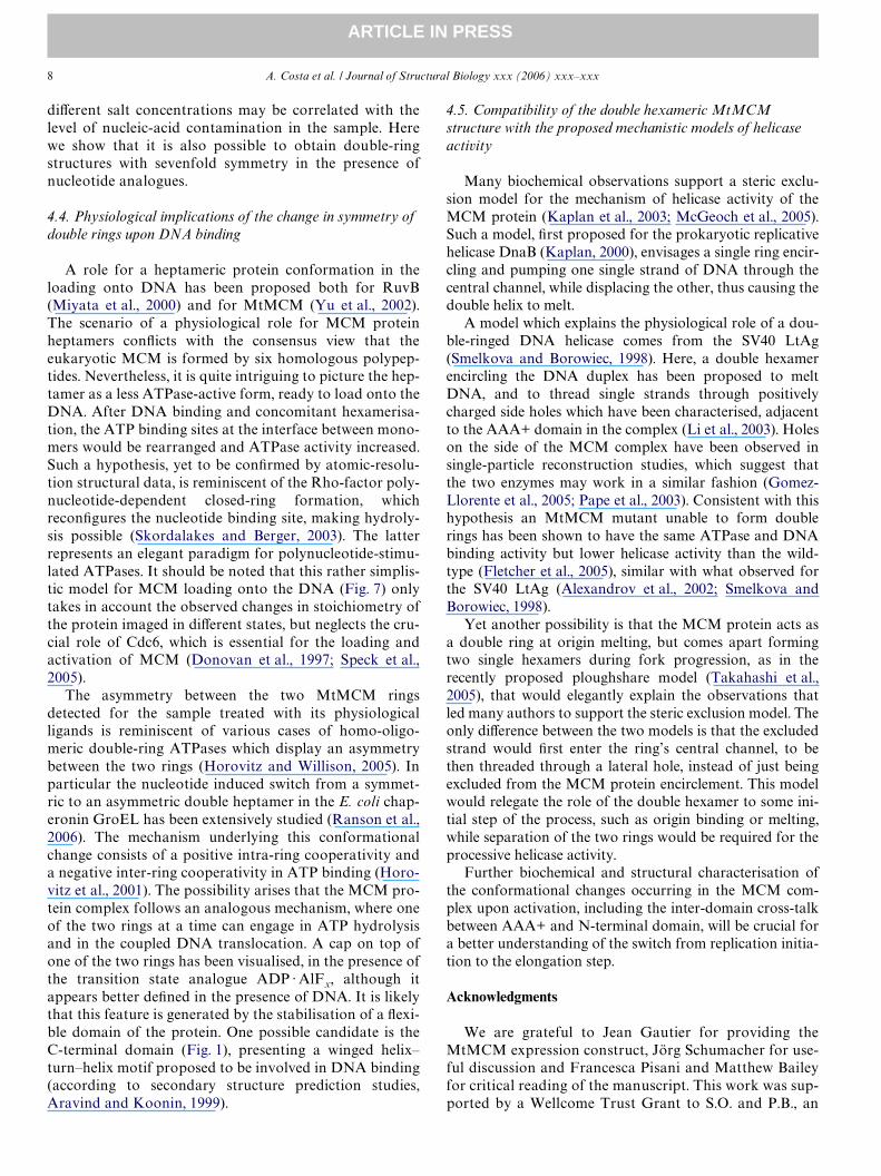
A role for a heptameric protein conformation in theloading onto DNA has been proposed both for RuvB(Miyata et al., 2000) and for MtMCM (Yu et al., 2002).The scenario of a physiological role for MCM proteinheptamers conXicts with the consensus view that theeukaryotic MCM is formed by six homologous polypep-tides. Nevertheless, it is quite intriguing to picture the hep-tamer as a less ATPase-active form, ready to load onto theDNA. After DNA binding and concomitant hexamerisa-tion, the ATP binding sites at the interface between mono-mers would be rearranged and ATPase activity increased.Such a hypothesis, yet to be conWrmed by atomic-resolu-tion structural data, is reminiscent of the Rho-factor poly-nucleotide-dependent closed-ring formation, whichreconWgures the nucleotide binding site, making hydroly-sis possible (Skordalakes and Berger, 2003). The latterrepresents an elegant paradigm for polynucleotide-stimu-lated ATPases. It should be noted that this rather simplis-tic model for MCM loading onto the DNA (Fig. 7) onlytakes in account the observed changes in stoichiometry ofthe protein imaged in diVerent states, but neglects the cru-cial role of Cdc6, which is essential for the loading andactivation of MCM (Donovan et al., 1997; Speck et al.,2005).
The asymmetry between the two MtMCM ringsdetected for the sample treated with its physiologicalligands is reminiscent of various cases of homo-oligo-meric double-ring ATPases which display an asymmetrybetween the two rings (Horovitz and Willison, 2005). Inparticular the nucleotide induced switch from a symmet-ric to an asymmetric double heptamer in the E. coli chap-eronin GroEL has been extensively studied (Ranson et al.,2006). The mechanism underlying this conformationalchange consists of a positive intra-ring cooperativity anda negative inter-ring cooperativity in ATP binding (Horo-vitz et al., 2001). The possibility arises that the MCM pro-tein complex follows an analogous mechanism, where oneof the two rings at a time can engage in ATP hydrolysisand in the coupled DNA translocation. A cap on top ofone of the two rings has been visualised, in the presence ofthe transition state analogue ADP · AlFx, although itappears better deWned in the presence of DNA. It is likelythat this feature is generated by the stabilisation of a Xexi-ble domain of the protein. One possible candidate is theC-terminal domain (Fig. 1), presenting a winged helix–turn–helix motif proposed to be involved in DNA binding(according to secondary structure prediction studies,Aravind and Koonin, 1999).
4.5. Compatibility of the double hexameric MtMCM structure with the proposed mechanistic models of helicase activity
Many biochemical observations support a steric exclu-sion model for the mechanism of helicase activity of theMCM protein (Kaplan et al., 2003; McGeoch et al., 2005).Such a model, Wrst proposed for the prokaryotic replicativehelicase DnaB (Kaplan, 2000), envisages a single ring encir-cling and pumping one single strand of DNA through thecentral channel, while displacing the other, thus causing thedouble helix to melt.
A model which explains the physiological role of a dou-ble-ringed DNA helicase comes from the SV40 LtAg(Smelkova and Borowiec, 1998). Here, a double hexamerencircling the DNA duplex has been proposed to meltDNA, and to thread single strands through positivelycharged side holes which have been characterised, adjacentto the AAA+ domain in the complex (Li et al., 2003). Holeson the side of the MCM complex have been observed insingle-particle reconstruction studies, which suggest thatthe two enzymes may work in a similar fashion (Gomez-Llorente et al., 2005; Pape et al., 2003). Consistent with thishypothesis an MtMCM mutant unable to form doublerings has been shown to have the same ATPase and DNAbinding activity but lower helicase activity than the wild-type (Fletcher et al., 2005), similar with what observed forthe SV40 LtAg (Alexandrov et al., 2002; Smelkova andBorowiec, 1998).
Yet another possibility is that the MCM protein acts asa double ring at origin melting, but comes apart formingtwo single hexamers during fork progression, as in therecently proposed ploughshare model (Takahashi et al.,2005), that would elegantly explain the observations thatled many authors to support the steric exclusion model. Theonly diVerence between the two models is that the excludedstrand would Wrst enter the ring’s central channel, to bethen threaded through a lateral hole, instead of just beingexcluded from the MCM protein encirclement. This modelwould relegate the role of the double hexamer to some ini-tial step of the process, such as origin binding or melting,while separation of the two rings would be required for theprocessive helicase activity.
Further biochemical and structural characterisation ofthe conformational changes occurring in the MCM com-plex upon activation, including the inter-domain cross-talkbetween AAA+ and N-terminal domain, will be crucial fora better understanding of the switch from replication initia-tion to the elongation step.
Acknowledgments
We are grateful to Jean Gautier for providing theMtMCM expression construct, Jörg Schumacher for use-ful discussion and Francesca Pisani and Matthew Baileyfor critical reading of the manuscript. This work was sup-ported by a Wellcome Trust Grant to S.O. and P.B., an

A. Costa et al. / Journal of Structural Biology xxx (2006) xxx–xxx 9
ARTICLE IN PRESS
EU grant to M.vH. (EC contract LSHG-CT-2004-502828), BBSRC grants to M.vH. and A.P., and by anImperial College Faculty Research Initiative grant toS.O. and A.P.
Appendix A. Supplementary data
Supplementary data associated with this article can befound, in the online version, at doi:10.1016/j.jsb.2006.04.001.
References
Alexandrov, A.I., Botchan, M.R., Cozzarelli, N.R., 2002. Characterizationof simian virus 40 T-antigen double hexamers bound to a replicationfork. The active form of the helicase. J. Biol. Chem. 277, 44886–44897.
Aravind, L., Koonin, E.V., 1999. DNA-binding proteins and evolution oftranscription regulation in the archaea. Nucleic Acids Res. 27, 4658–4670.
Boekema, E.J., Berden, J.A., van Heel, M.G., 1986. Structure of mitochon-drial F1-ATPase studied by electron microscopy and image processing.Biochim. Biophys. Acta 851, 353–360.
Carpentieri, F., De Felice, M., De Falco, M., Rossi, M., Pisani, F.M., 2002.Physical and functional interaction between the mini-chromosomemaintenance-like DNA helicase and the single-stranded DNA bindingprotein from the crenarchaeon Sulfolobus solfataricus. J. Biol. Chem.277, 12118–12127.
Chen, Y.J., Yu, X., Kasiviswanathan, R., Shin, J.H., Kelman, Z., Egelman,E.H., 2005. Structural polymorphism of Methanothermobacter ther-moautotrophicus MCM. J. Mol. Biol. 346, 389–394.
Chong, J.P., Hayashi, M.K., Simon, M.N., Xu, R.M., Stillman, B., 2000. Adouble-hexamer archaeal minichromosome maintenance protein isan ATP-dependent DNA helicase. Proc. Natl. Acad. Sci. USA 97,1530–1535.
Davey, M.J., Indiani, C., O’Donnell, M., 2003. Reconstitution of theMcm2–7p heterohexamer, subunit arrangement, and ATP site archi-tecture. J. Biol. Chem. 278, 4491–4499.
Donovan, S., Harwood, J., Drury, L.S., DiZey, J.F., 1997. Cdc6p-depen-dent loading of Mcm proteins onto pre-replicative chromatin in bud-ding yeast. Proc. Natl. Acad. Sci. USA 94, 5611–5616.
Dube, P., Tavares, P., Lurz, R., van Heel, M., 1993. The portal protein ofbacteriophage SPP1: a DNA pump with 13-fold symmetry. EMBO J.12, 1303–1309.
Fletcher, R.J., Bishop, B.E., Leon, R.P., Sclafani, R.A., Ogata, C.M., Chen,X.S., 2003. The structure and function of MCM from archaeal M. ther-moautotrophicum. Nat. Struct. Biol. 10, 160–167.
Fletcher, R.J., Shen, J., Gomez-Llorente, Y., Martin, C.S., Carazo, J.M.,Chen, X.S., 2005. Double hexamer disruption and biochemical activi-ties ofMethanobacterium thermoautotrophicum MCM. J. Biol. Chem.280, 42405–42410.
Gomez-Llorente, Y., Fletcher, R.J., Chen, X.S., Carazo, J.M., San Martin,C., 2005. Polymorphism and double hexamer structure in the archaealminichromosome maintenance (MCM) helicase from Methanobacte-rium thermoautotrophicum. J. Biol. Chem. 280, 40909–40915.
Gomez-Lorenzo, M.G., Valle, M., Frank, J., Gruss, C., Sorzano, C.O.,Chen, X.S., Donate, L.E., Carazo, J.M., 2003. Large T antigen on thesimian virus 40 origin of replication: a 3D snapshot prior to DNA rep-lication. EMBO J. 22, 6205–6213.
Grainge, I., Scaife, S., Wigley, D.B., 2003. Biochemical analysis of compo-nents of the pre-replication complex of Archaeoglobus fulgidus. NucleicAcids Res. 31, 4888–4898.
Horovitz, A., Fridmann, Y., Kafri, G., Yifrach, O., 2001. Review: allosteryin chaperonins. J. Struct. Biol. 135, 104–114.
Horovitz, A., Willison, K.R., 2005. Allosteric regulation of chaperonins.Curr. Opin. Struct. Biol. 15, 646–651.
Ishimi, Y., 1997. A DNA helicase activity is associated with an MCM4, -6,and -7 protein complex. J. Biol. Chem. 272, 24508–24513.
Kaplan, D.L., 2000. The 3�-tail of a forked-duplex sterically determineswhether one or two DNA strands pass through the central channel of areplication-fork helicase. J. Mol. Biol. 301, 285–299.
Kaplan, D.L., Davey, M.J., O’Donnell, M., 2003. Mcm4,6,7 uses a “pumpin ring” mechanism to unwind DNA by steric exclusion and activelytranslocate along a duplex. J. Biol. Chem. 278, 49171–49182.
Kelman, Z., Lee, J.K., Hurwitz, J., 1999. The single minichromosome main-tenance protein of Methanobacterium thermoautotrophicum DeltaHcontains DNA helicase activity. Proc. Natl. Acad. Sci. USA 96, 14783–14788.
Koonin, E.V., 1993. A common set of conserved motifs in a vast variety ofputative nucleic acid-dependent ATPases including MCM proteinsinvolved in the initiation of eukaryotic DNA replication. Nucleic AcidsRes. 21, 2541–2547.
Labib, K., Kearsey, S.E., DiZey, J.F., 2001. MCM2–7 proteins are essentialcomponents of prereplicative complexes that accumulate cooperativelyin the nucleus during G1-phase and are required to establish, but notmaintain, the S-phase checkpoint. Mol. Biol. Cell 12, 3658–3667.
Li, D., Zhao, R., Lilyestrom, W., Gai, D., Zhang, R., DeCaprio, J.A., Fan-ning, E., Jochimiak, A., Szakonyi, G., Chen, X.S., 2003. Structure of thereplicative helicase of the oncoprotein SV40 large tumour antigen.Nature 423, 512–518.
Lutzmann, M., Maiorano, D., Mechali, M., 2005. IdentiWcation of fullgenes and proteins of MCM9, a novel, vertebrate-speciWc member ofthe MCM2–8 protein family. Gene 362, 51–56.
Maiorano, D., Cuvier, O., Danis, E., Mechali, M., 2005. MCM8 is anMCM2–7-related protein that functions as a DNA helicase during rep-lication elongation and not initiation. Cell 120, 315–328.
McGeoch, A.T., Trakselis, M.A., Laskey, R.A., Bell, S.D., 2005. Organiza-tion of the archaeal MCM complex on DNA and implications for thehelicase mechanism. Nat. Struct. Mol. Biol. 12, 756–762.
Miyata, T., Yamada, K., Iwasaki, H., Shinagawa, H., Morikawa, K.,Mayanagi, K., 2000. Two diVerent oligomeric states of the RuvBbranch migration motor protein as revealed by electron microscopy. J.Struct. Biol. 131, 83–89.
Pape, T., Meka, H., Chen, S., Vicentini, G., van Heel, M., Onesti, S., 2003.Hexameric ring structure of the full-length archaeal MCM proteincomplex. EMBO Rep. 4, 1079–1083.
Plaisier, J.R., Koning, R.I., Koerten, H.K., van Heel, M., Abrahams, J.P.,2004. TYSON: robust searching, sorting, and selecting of single parti-cles in electron micrographs. J. Struct. Biol. 145, 76–83.
Prokhorova, T.A., Blow, J.J., 2000. Sequential MCM/P1 subcomplexassembly is required to form a heterohexamer with replication licens-ing activity. J. Biol. Chem. 275, 2491–2498.
Ranson, N.A., Clare, D.K., Farr, G.W., Houldershaw, D., Horwich, A.L.,Saibil, H.R., 2006. Allosteric signaling of ATP hydrolysis in GroEL-GroES complexes. Nat. Struct. Mol. Biol. 13, 147–152.
Rivenzon-Segal, D., Wolf, S.G., Shimon, L., Willison, K.R., Horovitz, A.,2005. Sequential ATP-induced allosteric transitions of the cytoplasmicchaperonin containing TCP-1 revealed by EM analysis. Nat. Struct.Mol. Biol. 12, 233–237.
Schwacha, A., Bell, S.P., 2001. Interactions between two catalytically dis-tinct MCM subgroups are essential for coordinated ATP hydrolysisand DNA replication. Mol. Cell 8, 1093–1104.
Shechter, D., Gautier, J., 2004. MCM proteins and checkpoint kinasesget together at the fork. Proc. Natl. Acad. Sci. USA 101, 10845–10846.
Shechter, D.F., Ying, C.Y., Gautier, J., 2000. The intrinsic DNA helicaseactivity of Methanobacterium thermoautotrophicum delta H minichro-mosome maintenance protein. J. Biol. Chem. 275, 15049–15059.
Skordalakes, E., Berger, J.M., 2003. Structure of the Rho transcription ter-minator: mechanism of mRNA recognition and helicase loading. Cell114, 135–146.
Smelkova, N.V., Borowiec, J.A., 1998. Synthetic DNA replication bubblesbound and unwound with twofold symmetry by a simian virus 40 T-antigen double hexamer. J. Virol. 72, 8676–8681.

10 A. Costa et al. / Journal of Structural Biology xxx (2006) xxx–xxx
ARTICLE IN PRESS
Speck, C., Chen, Z., Li, H., Stillman, B., 2005. ATPase-dependent coopera-tive binding of ORC and Cdc6 to origin DNA. Nat. Struct. Mol. Biol.12, 965–971.
Stasiak, A., Tsaneva, I.R., West, S.C., Benson, C.J., Yu, X., Egelman, E.H.,1994. The Escherichia coli RuvB branch migration protein forms doublehexameric rings around DNA. Proc. Natl. Acad. Sci. USA 91, 7618–7622.
Takahashi, T.S., Wigley, D.B., Walter, J.C., 2005. Pumps, paradoxes andploughshares: mechanism of the MCM2–7 DNA helicase. Trends Bio-chem. Sci. 30, 437–444.
Toth, E.A., Li, Y., Sawaya, M.R., Cheng, Y., Ellenberger, T., 2003. Thecrystal structure of the bifunctional primase–helicase of bacteriophageT7. Mol. Cell 12, 1113–1123.
van Heel, M., Gowen, B., Matadeen, R., Orlova, E.V., Finn, R., Pape, T.,Cohen, D., Stark, H., Schmidt, R., Schatz, M., Patwardhan, A., 2000.
Single-particle electron cryo-microscopy: towards atomic resolution.Q. Rev. Biophys. 33, 307–369.
van Heel, M., Harauz, G., Orlova, E.V., Schmidt, R., Schatz, M., 1996. Anew generation of the IMAGIC image processing system. J. Struct.Biol. 116, 17–24.
You, Z., Ishimi, Y., Mizuno, T., Sugasawa, K., Hanaoka, F., Masai,H., 2003. Thymine-rich single-stranded DNA activates Mcm4/6/7helicase on Y-fork and bubble-like substrates. EMBO J. 22, 6148–6160.
You, Z., Masai, H., 2005. DNA binding and helicase actions of mouseMCM4/6/7 helicase. Nucleic Acids Res. 33, 3033–3047.
Yu, X., VanLoock, M.S., Poplawski, A., Kelman, Z., Xiang, T., Tye, B.K.,Egelman, E.H., 2002. The Methanobacterium thermoautotrophicumMCM protein can form heptameric rings. EMBO Rep 3, 792–797.