Glycosylation Variants of a β-Glucosidase Secreted by a Taiwanese Fungus, Chaetomella raphigera,...
14
Washington University School of Medicine Digital Commons@Becker Open Access Publications 2014 Glycosylation variants of a β-glucosidase secreted by a Taiwanese fungus, Chaetomella raphigera, exhibit variant-specific catalytic and biochemical properties Aki Yoneda Washington University School of Medicine in St. Louis Hsion-Wen David Kuo Tunghai University Mayumi Ishihara University of Georgia Parastoo Azadi University of Georgia Su-May Yu Academia Sinica, Nankang See next page for additional authors Follow this and additional works at: hp://digitalcommons.wustl.edu/open_access_pubs is Open Access Publication is brought to you for free and open access by Digital Commons@Becker. It has been accepted for inclusion in Open Access Publications by an authorized administrator of Digital Commons@Becker. For more information, please contact [email protected]. Recommended Citation Yoneda, Aki; Kuo, Hsion-Wen David; Ishihara, Mayumi; Azadi, Parastoo; Yu, Su-May; and Ho, Tuan-hua David, ,"Glycosylation variants of a β-glucosidase secreted by a Taiwanese fungus, Chaetomella raphigera, exhibit variant-specific catalytic and biochemical properties." PLoS One.9,9. e106306. (2014). hp://digitalcommons.wustl.edu/open_access_pubs/3613
Transcript of Glycosylation Variants of a β-Glucosidase Secreted by a Taiwanese Fungus, Chaetomella raphigera,...
Washington University School of MedicineDigital Commons@Becker
Open Access Publications
2014
Glycosylation variants of a β-glucosidase secretedby a Taiwanese fungus, Chaetomella raphigera,exhibit variant-specific catalytic and biochemicalpropertiesAki YonedaWashington University School of Medicine in St. Louis
Hsion-Wen David KuoTunghai University
Mayumi IshiharaUniversity of Georgia
Parastoo AzadiUniversity of Georgia
Su-May YuAcademia Sinica, Nankang
See next page for additional authors
Follow this and additional works at: http://digitalcommons.wustl.edu/open_access_pubs
This Open Access Publication is brought to you for free and open access by Digital Commons@Becker. It has been accepted for inclusion in OpenAccess Publications by an authorized administrator of Digital Commons@Becker. For more information, please contact [email protected].
Recommended CitationYoneda, Aki; Kuo, Hsion-Wen David; Ishihara, Mayumi; Azadi, Parastoo; Yu, Su-May; and Ho, Tuan-hua David, ,"Glycosylationvariants of a β-glucosidase secreted by a Taiwanese fungus, Chaetomella raphigera, exhibit variant-specific catalytic and biochemicalproperties." PLoS One.9,9. e106306. (2014).http://digitalcommons.wustl.edu/open_access_pubs/3613
AuthorsAki Yoneda, Hsion-Wen David Kuo, Mayumi Ishihara, Parastoo Azadi, Su-May Yu, and Tuan-hua David Ho
This open access publication is available at Digital Commons@Becker: http://digitalcommons.wustl.edu/open_access_pubs/3613
Glycosylation Variants of a b-Glucosidase Secreted by aTaiwanese Fungus, Chaetomella raphigera, ExhibitVariant-Specific Catalytic and Biochemical PropertiesAki Yoneda1,2, Hsion-Wen David Kuo3, Mayumi Ishihara4, Parastoo Azadi4, Su-May Yu5,6,7, Tuan-hua
David Ho1,6,7,8*
1 Department of Biology, Washington University in St. Louis, St. Louis, Missouri, United States of America, 2 Department of Pathology and Immunology, School of
Medicine, Washington University in St. Louis, St. Louis, Missouri, United States of America, 3 Department of Environmental Science and Engineering, Tunghai University,
Taichung, Taiwan, Republic of China, 4 Complex Carbohydrate Research Center, University of Georgia, Athens, Georgia, United States of America, 5 Institute of Molecular
Biology, Academia Sinica, Nankang, Taipei, Taiwan, Republic of China, 6 Agricultural Biotechnology Center, National Chung-Hsing University, Taichung, Taiwan, Republic
of China, 7 Department of Life Sciences, National Chung-Hsing University, Taichung, Taiwan, Republic of China, 8 Institute of Plant and Microbial Biology, Academia Sinica,
Nankang, Taipei, Taiwan, Republic of China
Abstract
Cellulosic biomass is an abundant and promising energy source. To make cellulosic biofuels competitive againstconventional fuels, conversion of rigid plant materials into sugars must become efficient and cost-effective. During cellulosedegradation, cellulolytic enzymes generate cellobiose (b-(1R4)-glucose dimer) molecules, which in turn inhibit suchenzymes by negative feedback. b-Glucosidases (BGLs) cleave cellobiose into glucose monomers, assisting overall cellulolyticactivities. Therefore, BGLs are essential for efficient conversion of cellulosic biomass into biofuels, and it is important tocharacterize newly isolated BGLs for useful traits. Here, we report our discovery that the indigenous Taiwanese fungusChaetomella raphigera strain D2 produces two molecular weight variants of a single BGL, D2-BGL (shortened to ‘‘D2’’), whichdiffer in O-glycosylation. The more extensively O-glycosylated form of native D2 (nD2L) has increased activity toward thenatural substrate, cellobiose, compared to the less O-glycosylated form (nD2S). nD2L is more stable at 60uC, in acidic pH,and in the presence of the ionic detergent sodium dodecyl sulfate than nD2S. Furthermore, unlike nD2S, nD2L does notdisplay substrate inhibition by an artificial substrate p-nitrophenyl glucopyranoside (pNPG), and the glucose feedbackinhibition kinetics of nD2L is competitive (while it is non-competitive for nD2S), suggesting that these two glycovariants ofD2 bind substrates differently. Interestingly, D2 produced in a heterologous system, Pichia pastoris, closely mimicsproperties of nD2S. Our studies suggest that O-glycosylation of D2 is important in determining its catalytic and biochemicalproperties.
Citation: Yoneda A, Kuo H-WD, Ishihara M, Azadi P, Yu S-M, et al. (2014) Glycosylation Variants of a b-Glucosidase Secreted by a Taiwanese Fungus, Chaetomellaraphigera, Exhibit Variant-Specific Catalytic and Biochemical Properties. PLoS ONE 9(9): e106306. doi:10.1371/journal.pone.0106306
Editor: Eugene A. Permyakov, Russian Academy of Sciences, Institute for Biological Instrumentation, Russian Federation
Received June 5, 2014; Accepted August 5, 2014; Published September 2, 2014
Copyright: � 2014 Yoneda et al. This is an open-access article distributed under the terms of the Creative Commons Attribution License, which permitsunrestricted use, distribution, and reproduction in any medium, provided the original author and source are credited.
Data Availability: The authors confirm that all data underlying the findings are fully available without restriction. The sequence information for D2 BGL isavailable at GenBank (submission number: 1731986).
Funding: This work was supported by the International Center for Advanced Research in Energy and Sustainability, Washington University (http://icares.wustl.edu/Pages/Home.aspx) to TDH, and Ministry of Science and Technology, Taiwan, Republic of China (http://www.most.gov.tw) grant #103-3113-P-008 -001 toTDH. The funders had no role in study design, data collection and analysis, decision to publish, or preparation of the manuscript.
Competing Interests: A US patent (8,394,619) related to b-glucosidase in the fungus Chaetomella raphigera, ‘‘Beta-glucosidase and uses thereof’’, has beenissued on March 12, 2013. Inventors: Yu; Su-May, Ho; Tuan-Hua David, Kuo; Hsion-Wen, Wu; Ng I-Son, Li; Chen-Wei, Ju; Yu-Ming. This does not alter the authors’adherence to PLOS ONE policies on sharing data and materials.
* Email: [email protected]
Introduction
Cellulosic biomass is an abundant and promising feedstock for
next-generation biofuels in many countries [1–3]. Conventional
biomass such as starch can be readily degraded to sugar monomers
in an industrial scale. However, utilizing cellulosic biomass poses a
major challenge: converting rigid and recalcitrant plant materials
into simple fermentable sugars is not as easily nor economically
achieved [4]. To make cellulosic biofuels, cellulosic biomass must
be pretreated and efficiently converted to sugars [1]. Cellulose, the
major component of plant biomass, is a rigid polymer of b-(1R4)
linked (D)-glucose, which is further cross-linked by other
polysaccharides and lignin. Pretreatment is usually required to
make polysaccharide chains more accessible for enzymatic
digestion [5]. To efficiently digest cellulose, three groups of
cellulolytic enzymes must act together. Endo-b-(1R4)-glucanases
digest cellulose from inner regions of cellulose chains, and exo-b-
(1R4)-glucanases digest cellulose chains from the reducing or non-
reducing ends [6]. These processes release oligosaccharides and
cellobiose, a b-(1R4)-glucose dimer, which strongly inhibits most
glucanase activities, but may be digested to glucose monomers by
b-glucosidases (BGLs) [6]. BGLs are ubiquitous in nature, and are
classified into glycosylhydrolase (GH) families 1 and 3 by the
Carbohydrate-active enzymes database (CAZy, www.cazy.org).
Most bacterial and fungal BGLs belong to the GH3 family, with
specificities to myriad substrates [7].
PLOS ONE | www.plosone.org 1 September 2014 | Volume 9 | Issue 9 | e106306
Glycosylation is one of the most common post-translational
modifications for eukaryotic proteins, influencing a number of
protein properties and functions including folding, solubility,
thermostability, and proteolytic resistance [8–11]. The two major
types of protein glycosylation are N- and O-linked glycosylation.
N-glycosylation begins in the ER, where an oligosaccharide
attached to dolichol is flipped into the lumen of the ER, processed,
transferred to a target arginine residue within a nascent
polypeptide, and then further modified throughout the secretory
pathway [12,13]. N-glycan structures vary greatly among species,
and some fungal species synthesize extremely large N-glycan
moieties (,200 mannose residues to two N-acetylglucosamine
(GlcNAc) residues) [14]. The O-glycans, on the other hand, tend to
be simpler. O-glycosylation is initiated by a transfer of a
monosaccharide moiety from a UDP-sugar donor to a serine or
threonine residue via hydroxyl linkage. In fungi, the first moiety to
be transferred is generally mannose, and the modification is
therefore termed O-mannosylation [15,16]. The N-glycosylation is
often required for catalytic activities of enzymes [17–19], and in
some cases specific N-glycosylation sites involved in catalytic
activities have been identified [20–22]. The effects of O-
glycosylation on enzymatic properties are less understood, in part
because investigating O-glycosylation of an active enzyme poses
more challenges: a) There is a consensus site for N-glycosylation
(asparagine-X-serine or threonine, where X is not a proline) [12],
while there is less sequence consensus for O-glycosylation sites,
although a number of prediction programs exist [23], b) chemical
O-glycan removal results in protein degradation, and c) a lack of
O-glycosylation probably affects N-glycosylation [24]. Nonethe-
less, there are some examples of O-glycosylation affecting protein
functions. Naim and Lentz demonstrated that two forms of lactase-
phlorizin hydrolase, one N-glycosylated and the other N- and O-
glycosylated, hydrolyzed lactose with different reaction kinetics
[25]. O-glycosylation may also be involved in structural changes
[26,27]. Recently, synthetic protein O-mannosylation technique
resulting in homogeneously O-glycosylated proteins became
available [28]. Chen et al. systematically showed that O-
glycosylation enhances stability and substrate binding of model
Family 1 carbohydrate-binding modules [29], suggesting that such
modification would be advantageous for naturally produced
enzymes.
In previous studies, Ng et al. isolated a native fungus,
Chaetomella raphigera strain D2, from screening several dozens
of wood-rotting fungi for BGL activities [30]. This fungus was
identified by its ability to produce D2, a highly active BGL. The
cDNA sequence encoding D2 was cloned into Pichia pastoris for a
large-scale production. We noticed that P. pastoris-produced
recombinant D2 (rD2) and native D2 isolated from C. raphigerahave distinct catalytic and biochemical properties, and found that
C. raphigera produces two forms of D2: a large, heavily
glycosylated form (nD2L), and a small, N-glycosylated form
(nD2S). In this study, we demonstrate similarities in properties of
rD2 and nD2S, and how they differ from that of nD2L, suggesting
that O-glycosylation can alter catalytic properties of C. raphigeraBGL.
Materials and Methods
Cloning and sequencing of D2-BGL cDNA andproduction of recombinant D2-BGL
Since D2-BGL (shortened to ‘‘D2’’ hereafter) was the most
abundant secretory protein from the fungus C. raphigera strain
D2, it was easily purified by polyacrylamide gel electrophoresis
(PAGE) followed by zymography (see Enzyme assays, (c) MUG in
gel assay in Materials and Method). The N-terminal amino acid
sequence of this protein was determined by Edman degradation to
be N-PGDGDWAAA (carried out by Academia Sinica Protein
Core Facility, Taipei). For D2 cDNA synthesis, total RNA isolated
from actively growing C. raphigera strain D2 was first reverse
transcribed into single-stranded cDNA using a poly-T primer
containing an anchor sequence (GGT TCT TGC CAC AGT
CAC GAC TTT TTT TTT TTT TTT TTT), followed by PCR
amplification of the cDNA copies using a forward primer (CCN
GGN GAY GGN GAY TGG GC, designed based on N-terminal
amino acid sequencing results) and an anchor as reverse primer
(GGT TCT TGC CAC AGT CAC GAC). Invitrogen SuperScript
III Reverse Transcriptase (Invitrogen, USA) was used in the
reverse transcription and TaKaRa Ex Taq DNA Polymerase
(TaKaRa Bio Inc, Japan) was used for PCR. The thermal cycling
conditions were 94uC for 4 min, then 30 cycles of 94uC for 1 min,
58uC for 30 sec, and 72uC for 3 min, and a final extension at 72uCfor 5 min.
The signal peptide sequence was cloned using inverse PCR.
Briefly, genomic DNA (see genomic DNA isolation below) was
digested with SacI (New England Biolabs Inc., USA), self-ligated
using T4 DNA ligase (Promega, USA), and then amplified by PCR
using the primer pairs (233f: CGT TTC GTC CAA AAT GTA
ACA GCA T and 232r: GAT GCT TTC ACC GTC AGT TCT
GA). The thermal cycling conditions were 95uC for 5 min, then 25
cycles of 95uC for 1 min, 55uC for 1 min, and 72uC for 6 min, and
a final extension at 72uC for 5 min. The inverse PCR product was
cloned into the pGEMH-T Easy cloning vector and transformed
into Escherichia coli DH5a.
Production of rD2 was accomplished by subcloning the D2-
BGL cDNA into the vector pGAPZaC with the GAP promoter for
constitutive expression in P. pastoris. A wild type strain SMD1168
was electroporated with the plasmid to allow production of rD2
with a C-terminal heptahistidine tag.
Strains and fungal culturesrD2-expressing P. pastoris strain was streaked on YPD (1%
yeast extract, 2% peptone, 2% glucose) agar plates and routinely
grown in YPD broth. C. raphigera was maintained on PDA
(potato dextrose agar; Sigma #70139) plates. Spores were used to
inoculate the center of plates, and allowed to grow at 30uC for 14
days. After 14 days, fungal biomass with agar from the plates were
transferred to a flask containing 200-ml liquid medium (YPD or
YP (1% yeast extract and 2% peptone) with or without the
addition of 10 mM cellobiose or 20 mM glucose, depending on
the experiment) for three days.
Genomic DNA isolation of C. raphigera strain D2Spores were used to inoculate 3-ml of YPD liquid medium in a
test tube, and the culture was shaken at room temperature for
three days. Then the fungal cells and hyphae were harvested by
centrifugation in a 1.6-ml microtube, and 500 ml of lysis buffer
(50 mM Tris-HCl pH 8, 170 mM EDTA, 1% SDS) and
approximately 250 ml of 0.5 mm-glass beads (Biospec) were added
to the tube. The tube was then agitated by vortex mixer for 2 min,
cooled on ice for 1 min, and this step was repeated until cells
appeared to have broken under a phase microscope. The tube was
then heated at 65uC for 10 minutes. 300 ml of 7.5 M sodium
acetate was added to the tube, and placed on ice for 30 min after
thoroughly mixed by inverting the tube. The tube was then
centrifuged at 17,000 6 g for 10 minutes. To 700 ml of the
supernatant transferred to a new microtube, 500 ml of isopropanol
was added, and the mixture was placed on ice for 30 minutes. It
was then centrifuged at 17,000 6 g again to pellet DNA. The
Altered Enzymatic Properties of Glycosylation Variants
PLOS ONE | www.plosone.org 2 September 2014 | Volume 9 | Issue 9 | e106306
supernatant was discarded by aspiration, and 250 ml of TE was
added to the resulting pellet for a 10-min incubation at 50uC. The
pellet was dissolved by occasionally flicking the tube during the
incubation. To the DNA solution, 200 ml of 24:1 mixture of
chloroform: isoamyl alcohol was added, mixed by inverting the
tube, and centrifuged for 10 min. The top layer was transferred to
a new microtube, and the genomic DNA was concentrated by
ethanol precipitation. Concentration of DNA was determined
using NanoDrop (Thermo Scientific).
Southern blottingDNA probe was generated by PCR using AYP40 (59-TCC
AAC ATC GAT GAT CGG) and AYP42 (59-GGT CGT CGA
CAA TAC AAG C) and Biotin Decalabel Kit (Fermentas), using
both cDNA and genomic DNA as templates. C. raphigeragenomic DNA was digested with restriction enzymes overnight,
and was separated by 0.8% agarose gel electrophoresis. The gel
was treated, transferred, cross-linked with UV light, pre-hybrid-
ized using standard Southern blotting methods. The pre-hybrid-
ized membranes were hybridized with the genomic or cDNA
probes overnight in separate glass bottles, and the signals were
detected using chemiluminescence reagents [31].
Sample preparation and chromatographyC. raphigera spores were used to inoculate the center of potato
dextrose agar plates. The fungus on plates was allowed to grow at
30uC for 14 days. After 14 days, agar (with the entire fungus) from
the plates was transferred to a flask containing 200-ml liquid
medium (1% yeast extract, 2% peptone, with or without the
addition of 10 mM cellobiose or 20 mM glucose, depending on
the experiment). The liquid culture was then shaken at room
temperature for specified lengths of time, and the debris of agar
was removed with a strainer and then centrifuged to pellet fungal
cells and smaller debris. Supernatant was then filtered through a
0.45-mm filter and concentrated approximately 20-fold by
centrifugation at 4,000 xg for 30 min or until desired volume
was achieved, in Amicon Ultra centrifugal filter tubes (Millipore
UFC903008, molecular weight cut off 30 kDa). The filtered,
concentrated supernatant was then dialyzed against 10 mM Bis-
Tris buffer pH 6.5 at 4uC for 3 days with daily buffer changes.
The dialyzed supernatant was then concentrated as described
above to ,10 ml, and was loaded to a manually-packed DEAE
Sepharose Fast Flow (GE Healthcare) column, equilibrated with
10 mM Bis-Tris buffer pH 6.5. Approximately 3-ml (150 drops
per tube) fractions were collected over 0-1 M NaCl continuous
gradient at 0.5 ml/min with a peristaltic pump.
BGL activity was detected by pNPG and cellobiose assays. The
activity peak was pooled and concentrated down to ,250 ml, and
loaded onto a manually poured Sephadex G 200 column, and the
sample was run by gravity flow. 350 drops per tube (approximately
8 ml) were collected, and the activity was detected by pNPG and
cellobiose assays. P. pastoris-produced D2 was purified similarly,
since the recombinant protein did not bind to nickel columns
despite possessing a heptahistidine tag.
Enzyme assaysa) pNPG assay. Partially purified enzymes (10 ml, 10–
100 mg/ml) in 10 mM Bis-Tris pH 6.5 was dispensed onto wells
made with Parafilm and a PCR tube rack. 90 ml of 5 mM pNPG
(p-nitrophenyl b-D-glucopyranoside, Sigma N7006) in 50 mM
acetate buffer pH 5.0 (unless specified) was serially diluted two-fold
in 90 ml 50 mM acetate buffer pH 5.0 in an 8-well PCR strip
tubes per enzyme. Using a multichannel pipette, 10 ml of the
prepared enzymes were transferred to pre-warmed serial dilutions
of substrate, and incubated at 50uC for exactly 1 minute until the
reaction was stopped by simultaneous additions of 100 ml 2%
Na2CO3 in each well, and the 200 ml of resulting reaction
solutions were transferred to a column of a flat-bottom 96-well
plate. Once all reactions were stopped, the plate was read at
405 nm along with p-nitrophenol (pNP) standards using Versa-
Max plate reader (Molecular Devices). Assays were repeated at
least five times in the same condition.
pNP stock solution was made by dissolving pNP (Fluka 1048) in
water at 5 mM. Serially diluted pNP starting at 250 mM was used
as standards. Glucose inhibition assays were performed using the
same condition except for the addition of 0, 3, 6, 9 mM glucose in
substrate buffers. To determine pH optima, the assays were
performed in 10 mM HCl (pH 2.0), 50 mM acetate buffer
(pH 2.6–6.0) or 50 mM phosphate buffer (pH 6.0–7.0). Since
the reaction time was 1 min, pH would have minimal effect on
protein stability.
Km was calculated by plotting substrate concentrations (mM
pNPG) and initial velocity (mM pNP/sec/mg protein) and
nonlinear least-squares fitted with Solver add-in on Microsoft
Excel [32]. For substrate inhibition kinetics, we used a simple
equation shown in File S1. Glucose Ki values on pNPG assays
were determined by plotting substrate concentrations and initial
velocity in double-reciprocal (Lineweaver-Burk) plot using Km
and formula shown in File S1.
b) Cellobiose assay. Partially purified enzymes (3 ml, 10–
100 mg/ml) in 10 mM Bis-Tris pH 6.5 was pipetted onto wells
made with Parafilm and a PCR tube rack. 27 ml of 50 mM
cellobiose in 50 mM acetate buffer pH 5.0 was serially diluted
two-fold in 50 mM acetate buffer pH 5.0 in an 8-well PCR strip
tubes per enzyme. To measure background glucose levels of the
substrate, a strip without enzyme was prepared with each assay.
The enzymes prepared as above were transferred to a pre-warmed
tube strip with serial dilutions of cellobiose at once using a multi-
channel pipette, and incubated for 3 minutes. The reaction was
stopped by immediately placing the tube strip at 100uC for
3 minutes and cooled on ice. 10 ml of each reaction was
transferred to a 96-well plate, and 190 ml of glucose assay reagent
(Sigma G3293) was added along with glucose standards. The plate
was incubated at room temperature for 30 min, and read at
340 nm in the plate reader.
c) MUG in-gel assay. One mg/well of partially purified
BGLs were loaded onto wells of a 7% native polyacrylamide gel.
The samples were run in 16 Tris-glycine gel running buffer at
80 kV until the loading front reached near the end of the gel. The
gel was then fixed in 20 ml 25% isopropanol in 50 mM acetate
buffer pH 5.0 at room temperature for 20 minutes, and incubated
in 20 ml 200 mM MUG (Sigma M9766) in 50 mM acetate buffer
pH 5.0 for 10 minutes with shaking at RT, then at 37uC with
occasional manual shaking for 20 minutes. The gel was then
visualized using trans-UV illumination in a Bio-Rad Gel Doc gel
imaging system and digitally photographed.
Endo-H treatment of BGLsP. pastoris- produced, secreted rD2 in was readily detected in
culture supernatants by BGL activity assays and Western blotting
against hepta-histidine epitope in the culture supernatant of the P.pastoris strain, but not from a wild type control strain (Figure S1 in
File S1). For native proteins, 1 ml of BGLs were treated with 0.5 ml
of EndoH in 16 G buffer (enzyme and buffers, New England
Biolabs P0703S) at 37uC for 3 hours.
Altered Enzymatic Properties of Glycosylation Variants
PLOS ONE | www.plosone.org 3 September 2014 | Volume 9 | Issue 9 | e106306
Protein quantificationFive ml of samples were serially diluted in duplicates along with
serially diluted protein standard in triplicates, and protein
concentration was measured by adding protein assay reagent
(Bio-Rad 500–0006) according to the user manual.
Protein identification of nD2 variantsConcentrated size exclusion chromatography fraction pools were
run on a 7% SDS polyacrylamide gel and stained with SYPRO
Ruby protein stain overnight, and photographed using Gel Doc
system (Bio-Rad). Based on previous samples, gel pieces corre-
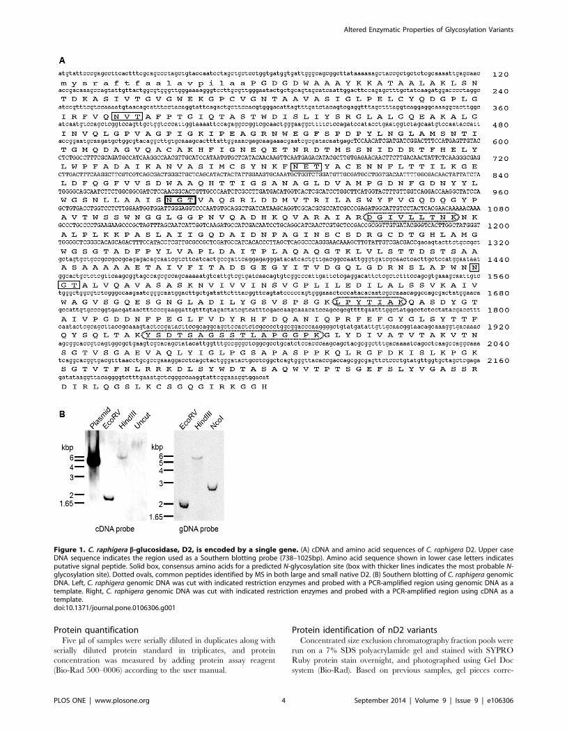
Figure 1. C. raphigera b-glucosidase, D2, is encoded by a single gene. (A) cDNA and amino acid sequences of C. raphigera D2. Upper caseDNA sequence indicates the region used as a Southern blotting probe (738–1025bp). Amino acid sequence shown in lower case letters indicatesputative signal peptide. Solid box, consensus amino acids for a predicted N-glycosylation site (box with thicker lines indicates the most probable N-glycosylation site). Dotted ovals, common peptides identified by MS in both large and small native D2. (B) Southern blotting of C. raphigera genomicDNA. Left, C. raphigera genomic DNA was cut with indicated restriction enzymes and probed with a PCR-amplified region using genomic DNA as atemplate. Right, C. raphigera genomic DNA was cut with indicated restriction enzymes and probed with a PCR-amplified region using cDNA as atemplate.doi:10.1371/journal.pone.0106306.g001
Altered Enzymatic Properties of Glycosylation Variants
PLOS ONE | www.plosone.org 4 September 2014 | Volume 9 | Issue 9 | e106306
sponding to MUG activity were excised from the gel and sent to
Proteomics & Mass Spectrometry Facility at Donald Danforth Plant
Science Center (St. Louis, MO) for amino acid identification using a
LTQ Orbitrap Velos mass spectrometer (Thermo Scientific).
Scaffold 3 proteome software (http://www.proteomesoftware.com/
Proteome_software_prod_Scaffold.html) was used to annotate and
view results.
Release of O-linked glycansThe samples were dialyzed against Nanopure water at 4uC
overnight using Tube-O-dialyzer (MW cut off 4000, G Biosciences)
and the contents in the tubes were dried in a vacuum concentrator.
The O-linked carbohydrate fractions were cleaved from the sample
by b-elimination procedures [33]. Briefly, 1 M sodium borohydride
in 50 mM sodium hydroxide (NaOH) was added to the samples and
incubated overnight at 45uC. The incubated samples were
neutralized with 10% acetic acid and desalted by passing through
a packed column of DowexTM resins (50 W 6 8–100, H+ form,
Sigma Aldrich, St. Louis,MO) and lyophilized. The borate was
removed as methyl borate. The O-glycans were separated from
residual material by passage through a C18 reversed phase cartridge.
The carbohydrate fractions (O-linked glycans) were eluted with 5%
acetic acid. The 5% acetic acid fractions were further washed with
ethyl acetate and dried by lyophilization. The O-glycan fraction thus
obtained were permethylated based on the method of Anumula and
Taylor [34] and profiled by mass spectrometry. O-glycan analyses
were performed by the Complex Carbohydrate Research Center at
University of Georgia, Athens, GA.
Mass spectrometryMALDI/TOF-MS was performed in the reflector positive ion
mode using a-dihyroxybenzoic acid (DHBA, 20 mg/mL solution
in 50% methanol: water) as a matrix. The spectrum was obtained
by using a Microflex LRF (Bruker).
NSI-MSn analysis was accomplished by using a LTQ Orbitrap
XL mass spectrometer (Thermo Scienfitic) equipped with a
nanospray ion source. Permethylated glycans from each sample
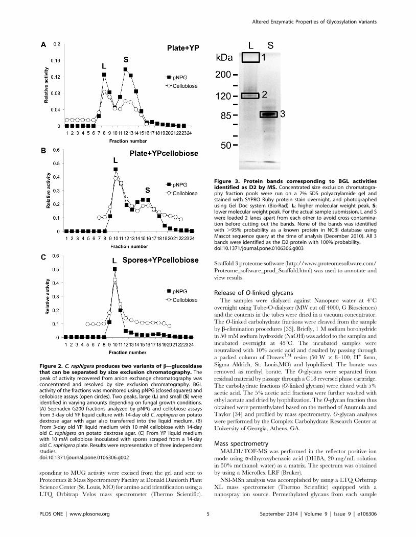
Figure 2. C. raphigera produces two variants of b—glucosidasethat can be separated by size exclusion chromatography. Thepeak of activity recovered from anion exchange chromatography wasconcentrated and resolved by size exclusion chromatography. BGLactivity of the fractions was monitored using pNPG (closed squares) andcellobiose assays (open circles). Two peaks, large (L) and small (S) wereidentified in varying amounts depending on fungal growth conditions.(A) Sephadex G200 fractions analyzed by pNPG and cellobiose assaysfrom 3-day old YP liquid culture with 14-day old C. raphigera on potatodextrose agar with agar also transferred into the liquid medium. (B)From 3-day old YP liquid medium with 10 mM cellobiose with 14-dayold C. raphigera on potato dextrose agar. (C) From YP liquid mediumwith 10 mM cellobiose inoculated with spores scraped from a 14-dayold C. raphigera plate. Results were representative of three independentstudies.doi:10.1371/journal.pone.0106306.g002
Figure 3. Protein bands corresponding to BGL activitiesidentified as D2 by MS. Concentrated size exclusion chromatogra-phy fraction pools were run on a 7% SDS polyacrylamide gel andstained with SYPRO Ruby protein stain overnight, and photographedusing Gel Doc system (Bio-Rad). L: higher molecular weight peak, S:lower molecular weight peak. For the actual sample submission, L and Swere loaded 2 lanes apart from each other to avoid cross-contamina-tion before cutting out the bands. None of the bands was identifiedwith .95% probability as a known protein in NCBI database usingMascot sequence query at the time of analysis (December 2010). All 3bands were identified as the D2 protein with 100% probability.doi:10.1371/journal.pone.0106306.g003
Altered Enzymatic Properties of Glycosylation Variants
PLOS ONE | www.plosone.org 5 September 2014 | Volume 9 | Issue 9 | e106306
were dissolved in 1 mM NaOH in 50% methanol and infused
directly into the instrument at a constant flow rate of 0.5 ml/min.
A full FTMS spectrum was collected at 30,000 resolution with 3
microscans. The peak intensities of each O-glycan component
were obtained by averaging the 30 full FTMS scans for each
sample. The capillary temperature was set at 210uC and MS
analysis was performed in the positive ion mode. For total ion
mapping (automated MS/MS analysis), m/z range, 300 to 2000
was scanned with ITMS mode in successive 2.8 mass unit windows
that overlapped the preceding window by 2 mass units.
Results
An indigenous Taiwanese fungus C. raphigera strain D2expresses a strongly active b-glucosidase, D2, andpossesses a single gene encoding the BGL
In previous work, a highly active BGL, D2 was identified from a
native Taiwanese fungus C. raphigera strain D2 for which cDNA
sequence was not available. The N-terminal amino acid sequence
of this protein was subsequently determined. The amino acid
sequence was used to design PCR primers for the cloning of D2
cDNA (Figure 1; GenBank accession number KJ939445). We
performed Southern blotting of genomic DNA isolated from C.raphigera strain D2 and showed that there is only a single gene
encoding D2-BGL within C. raphigera genome (Figure 1C). We
then proceeded to characterize biochemical properties of D2.
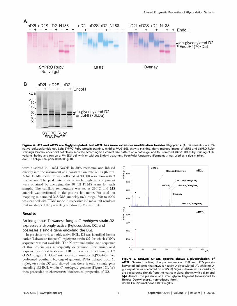
Figure 4. rD2 and nD2S are N-glycosylated, but nD2L has more extensive modification besides N-glycans. (A) D2 variants on a 7%native polyacrylamide gel. Left: SYPRO Ruby protein staining, middle: MUG BGL activity staining, right: merged image of MUG and SYPRO Rubystainings. Protein ladder did not clearly separate according to a correct size pattern on a native gel and thus omitted. (B) SYPRO Ruby-staining of D2variants, boiled and run on a 7% SDS gel, with or without EndoH treatment. PageRuler Unstained (Fermentas) was used as a size marker.doi:10.1371/journal.pone.0106306.g004
Figure 5. MALDI/TOF-MS spectra shows O-glycosylation ofnD2L. O-linked profiling of equal amounts of nD2L and nD2s proteinharvested indicated that nD2L is heavily O-glycosylated (A), while no O-glycosylation was detected on nD2S (B). Signals shown with asterisks (*)are background signals from the matrix. A signal shown with a diamond(X) denotes the presence of a small glycan fragment (correspond toHexose1Deoxyhexose1, non-reduced form).doi:10.1371/journal.pone.0106306.g005
Altered Enzymatic Properties of Glycosylation Variants
PLOS ONE | www.plosone.org 6 September 2014 | Volume 9 | Issue 9 | e106306
C. raphigera strain D2 produces b-glucosidase activitieswith different molecular sizes, which were identified as asingle protein
Upon further characterization of the native fungus, we
discovered that C. raphigera produced two BGL activity peaks
that were separable by size-exclusion chromatography but not by
anion exchange chromatography; we interpreted this as proteins
sharing the same isoelectric point, but with different molecular
weights (Figure 2). Interestingly, depending on the growth
condition of the fungus, the ratio of the peaks changed. When
the C. raphigera was grown on potato dextrose agar (PDA) plates
for 14 days and then transferred (agar blocks containing hyphae
and spores in toto) to a liquid medium (YP, 1% yeast extract and
2% peptone) for three days, both peaks were present (Figure 2A).
However, when the plate was transferred to YP + 10 mM
cellobiose or spores scraped from 14-day old PDA plates were used
to inoculate YP + glucose, the larger molecular weight peak
became more prominent (Figures 2B and Figure S2 in File S1).
Furthermore, when YP + 10 mM cellobiose was inoculated with
spores (without hyphae and agar), the lower molecular weight peak
was no longer observed (Figure 2C).
We next designed studies to establish whether both ‘‘Large’’ and
‘‘Small’’ activities originated from the same protein. To follow
BGL activities on protein gels, we used 4-methylumbelliferyl b-D-
glucopyranoside (MUG) staining. When protein gels are incubated
with this compound, BGL activities within the gel release the
fluorescent methylumbelliferone moiety of MUG, enabling
detection by ultraviolet illumination [35]. To investigate whether
these two activity peaks represent D2 or two distinct proteins, we
excised protein bands from a SYPRO Ruby-stained SDS gel for
protein identification by mass spectrometry (MS; Figure 3). Band
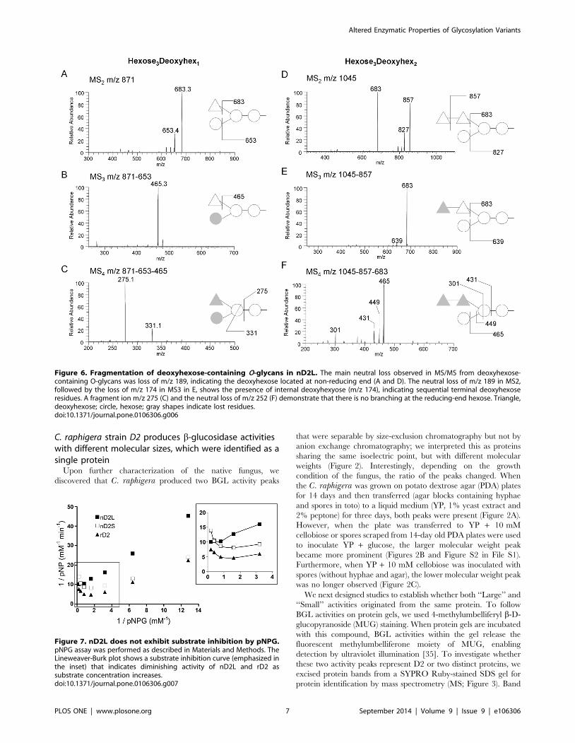
Figure 6. Fragmentation of deoxyhexose-containing O-glycans in nD2L. The main neutral loss observed in MS/MS from deoxyhexose-containing O-glycans was loss of m/z 189, indicating the deoxyhexose located at non-reducing end (A and D). The neutral loss of m/z 189 in MS2,followed by the loss of m/z 174 in MS3 in E, shows the presence of internal deoxyhexyose (m/z 174), indicating sequential terminal deoxyhexoseresidues. A fragment ion m/z 275 (C) and the neutral loss of m/z 252 (F) demonstrate that there is no branching at the reducing-end hexose. Triangle,deoxyhexose; circle, hexose; gray shapes indicate lost residues.doi:10.1371/journal.pone.0106306.g006
Figure 7. nD2L does not exhibit substrate inhibition by pNPG.pNPG assay was performed as described in Materials and Methods. TheLineweaver-Burk plot shows a substrate inhibition curve (emphasized inthe inset) that indicates diminishing activity of nD2L and rD2 assubstrate concentration increases.doi:10.1371/journal.pone.0106306.g007
Altered Enzymatic Properties of Glycosylation Variants
PLOS ONE | www.plosone.org 7 September 2014 | Volume 9 | Issue 9 | e106306
#1 was barely visible by SYPRO Ruby staining, but the location
concurred with a strong BGL activity by MUG staining of the gel
(Figure 4). Interestingly, only L remained active toward MUG on
SDS gels when samples were not boiled before loading, while the
band in S did not. Samples were digested with trypsin, resulting
peptides were separated by liquid chromatography, and analyzed
by MS. 13 unique peptides from band #3 matched the amino acid
sequence of native D2, and three peptides from band #2 (the
matched sequences shown in Figure 1, boxed with dotted line) and
two from band #1 (Table S1 in File S1). One of the peptides
(DGIVLLTNK) coincided within the genomic DNA region
confirmed by Southern blotting (Figure 1A).
To investigate potential post-translational modifications of
native D2 proteins that would explain the observed size
differences, we treated the partially purified proteins with
endoglycosidase H (EndoH) to remove N-glycans. As references,
recombinant D2 expressed in Pichia pastoris (rD2) and commer-
cially available BGL, Novozyme 188 (N188, Novozymes) were
also analyzed. rD2 ran as a long smear on native and SDS gels
that focused to a tight band around 75.5 kDa upon EndoH
treatment, suggesting that D2 expressed in P. pastoris is
heterogeneously N-glycosylated. As proteins may not migrate
according to their sizes on native gels, MUG- and SYPRO Ruby-
stained native gel images were overlaid to indicate the location of
BGL activities relative to protein bands (Figure 4A). The
comparative staining of the gel images showed that most active
part of nD2L was located at the top of the gel, barely entering the
separating gel. After an EndoH treatment, a faster-moving, faint
but focused band appeared, although most of the activity
remained at the top of the gel (Figure 4A). nD2S was faster-
moving than nD2L and rD2, but its electromobility increased after
an EndoH treatment. rD2 had lower electromobility compared to
nD2S, but it increased after an EndoH treatment (Figure 4A).
This experiment also showed that N188 contained a major low
molecular weight protein band by SYPRO Ruby staining that
lacks BGL activity, and its BGL activity band clearly shifted down
after an EndoH treatment. There was no extensive smearing of
N188, indicating that N-glycosylation on this protein is relatively
homogeneous. The SDS-PAGE followed by SYPRO Ruby
staining showed that Endo-H treated nD2S and rD2 focused to
the predicted size of D2 (75.5 kDa), indicating that nD2S and rD2
are N-glycosylated, but the majority of nD2L had modifications
that alter its molecular weight in addition to N-glycosylation
(Figure 4B).
Structural characterization of glycan modifications ofactive nD2 variants
O-linked oligosaccharide analysis on the active nD2
variants by mass spectrometry. Based on our Endo-H
results, we hypothesized that nD2L bore additional modifications
beyond N-glycans. To confirm the presence of O-glycosylation, we
subjected nD2L and nD2S to b-elimination, permethylated the
released O-glycans from each sample, and analyzed them by
Matrix-assisted laser desorption/ionization time-of-flight mass
spectrometry (MALDI/TOF-MS) and by nanospray sequential
mass spectrometry (NSI-MSn) (see Material and Methods). We
detected strong O-glycan signals from nD2L, but none from nD2S
(Figure 5). The O-glycan components observed were oligosaccha-
rides containing two or four hexoses and zero to two deoxy-
hexoses. To analyze the glycan sequence in detail, we carried out
MSn analysis (Figures 6 and Figure S3 in File S1), which indicated
a trihexosyl chain wherein the central hexose carries a side chain
of one of two deoxyhexose residues (Figure 6).
rD2 and nD2S have similar catalytic and biochemical
properties, whereas nD2L has distinct properties. To
investigate the correlation between different degrees of glycosyl-
ation and enzymatic properties, we compared catalytic properties
of nD2L, nD2S and rD2. pNPG hydrolysis is a commonly used
colorimetric assay for assessing BGL activities. pNPG acts as an
artificial substrate, releasing p-nitrophenol which can be detected
at the wavelength of 405 nm by spectrophotometry. First, we
optimized the reaction time to one minute after monitoring pNPG
activities of the BGLs at 30-second intervals at 50uC, in order to
determine initial rates at various concentrations of pNPG. Both
nD2S and rD2 exhibited substrate inhibition by pNPG, although
nD2L did not (Figure 7). Next, we measured activities of the D2
variants on the native substrate, cellobiose, determined Km and
Vmax of the BGLs for each substrate (Table 1, Figure 8). We
found that the major differences among the glycovariants are the
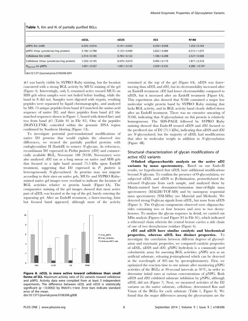
Table 1. Km and Ki of partially purified BGLs.
nD2L nD2S rD2 N188
pNPG Km (mM) 0.24360.016 0.14160.023 0.25460.058 1.25260.164
pNPG Vmax (mmole/sec/mg protein) 5.19660.788 3.13360.494 5.83260.489 6.51261.675
Cellobiose Km (mM) 2.31460.185 0.78360.122 1.18660.208 2.52760.506
Cellobiose Vmax (mmole/sec/mg protein) 1.55060.105 0.47060.014 0.49560.119 1.87160.318
Kiglucose on pNPG 1.69160.337 1.00160.125 2.92960.535 4.280 60.747
doi:10.1371/journal.pone.0106306.t001
Figure 8. nD2L is more active toward cellobiose than smallforms of D2. Maximum activity ratio of D2 variants toward cellobioseand pNPG. Activity data were complied from at least 3 independentexperments. The difference between nD2L and nD2S is statisticallysignificant (p ,0.0002) by Welch’s t-test. Error bars indicate standarderror of the mean.doi:10.1371/journal.pone.0106306.g008
Altered Enzymatic Properties of Glycosylation Variants
PLOS ONE | www.plosone.org 8 September 2014 | Volume 9 | Issue 9 | e106306
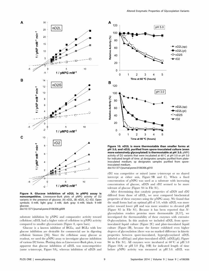
substrate inhibition by pNPG and comparative activity toward
cellobiose; nD2L had a higher ratio of cellobiose to pNPG activity
compared to smaller glycovariants (Figure 8, open bars).
Glucose is a known inhibitor of BGLs, and BGLs with low
glucose inhibition are desirable for commercial use in digesting
cellulosic biomass [36]. Since the cellobiose assay glucose as
readout, we used the pNPG assay to investigate glucose inhibition
of various D2 forms. Plotting data as Lineweaver-Burk plots, it was
apparent that glucose inhibition of nD2L was noncompetitive
(same x-intercept, Figure 9A), whereas inhibition of nD2S and
rD2 was competitive or mixed (same y-intercept or no shared
intercept at either axis, Figure 9B and C). When a fixed
concentration of pNPG was used as a substrate with increasing
concentration of glucose, nD2S and rD2 seemed to be more
tolerant of glucose (Figure S4 in File S1).
After determining that catalytic properties of nD2S and rD2
differed from those of nD2L, we next compared biochemical
properties of these enzymes using the pNPG assay. We found that
the small forms had an optimal pH of 5.0, while nD2L was more
active toward lower pH and was more sensitive to elevated pH
(Figure S5 in File S1). Because it has been reported that N-
glycosylation renders proteins more thermostable [8,37], we
investigated the thermostability of these enzymes with extensive
O-glycosylation. In this analysis we included nD2L from spore-
inoculated liquid culture (Figure 2C) and plate-inoculated liquid
culture (Figure 2B), because the former exhibited even higher
degrees of glycosylation (there was no marked difference in kinetic
properties between spore-inoculated, hyperglycosylated nD2L
(labeled as nD2(sp)) and plate-inoculated nD2L (nD2L(pl)); Figure
S6 in File S1). All enzymes were incubated at 60uC at pH 5.0
(Figure 10A) or pH 3.0 (Fig. 10B) for indicated length of time
before pNPG activity was measured at pH 5.0. nD2L was
Figure 9. Glucose inhibition of nD2L in pNPG assay isnoncompetitive. Lineweaver-Burk plots of pNPG activity of D2variants in the presence of glucose. (A) nD2L, (B) nD2S, (C) rD2. Opensymbols: 0 mM, light gray: 3 mM, dark gray: 6 mM, black: 9 mMglucose.doi:10.1371/journal.pone.0106306.g009
Figure 10. nD2L is more thermostable than smaller forms atpH 5.0, and nD2L purified from spore-inoculated culture (evenmore extensively glycosylated) is thermostable at pH 3.0. pNPGactivity of D2 variants that were incubated at 60uC at pH 5.0 or pH 3.0for indicated length of time. pl designates samples purified from plate-inoculated medium; sp designates samples purified from spore-inoculated medium.doi:10.1371/journal.pone.0106306.g010
Altered Enzymatic Properties of Glycosylation Variants
PLOS ONE | www.plosone.org 9 September 2014 | Volume 9 | Issue 9 | e106306
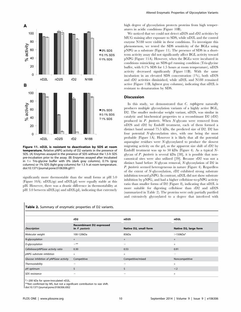
significantly more thermostable than the small forms at pH 5.0
(Figure 10A). nD2L(sp) and nD2L(pl) were equally stable at this
pH. However, there was a drastic difference in thermostability at
pH 3.0 between nD2L(sp) and nD2L(pl), indicating that extremely
high degree of glycosylation protects proteins from high temper-
atures in acidic conditions (Figure 10B).
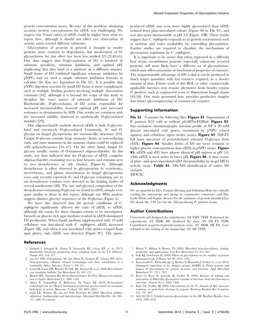
We noticed that we could not detect nD2S and rD2 activities by
MUG staining after exposure to SDS, while nD2L and the control
enzyme N188 were visible in these conditions. To investigate this
phenomenon, we tested the SDS sensitivity of the BGLs using
pNPG as a substrate (Figure 11). The presence of SDS in a short-
term activity assay did not significantly affect BGL activity toward
pNPG (Figure 11A). However, when the BGLs were incubated in
conditions mimicking an SDS-gel running condition (Tris-glycine
buffer, with 0.1% SDS for 1.5 hours at room temperature), nD2S
activity decreased significantly (Figure 11B). With the same
incubation in an elevated SDS concentration (1%), both nD2S
and rD2 activities diminished, while nD2L and N188 remained
active (Figure 11B, lightest gray columns), indicating that nD2L is
resistant to denaturation by SDS.
Discussion
In this study, we demonstrated that C. raphigera naturally
produces multiple glycosylation variants of a highly active BGL,
D2. The smaller molecular weight variant, nD2S, was similar in
catalytic and biochemical properties to a recombinant D2 (rD2)
produced in P. pastoris. When N-glycans were removed from
nD2S and rD2 by EndoH treatment, each of them formed a
distinct band around 75.5 kDa, the predicted size of D2. D2 has
four potential N-glycosylation sites, with one being the most
probable (Figure 1A). However it is likely that all the potential
asparagine residues were N-glycosylated to produce the slowest
migrating activity on the gel, as the apparent size shift of rD2 by
EndoH treatment was up to 50 kDa (Figure 4). As a typical N-
glycan of P. pastoris is several kDa [38], it is possible that non-
canonical sites were also utilized [39]. Because rD2 was not a
distinct band before N-glycan removal, N-glycosylation of D2 in
P. pastoris seemed heterogeneous in nature (Figure 4). Regardless
of the extent of N-glycosylation, rD2 exhibited strong substrate
inhibition toward pNPG. In contrast, nD2L did not show substrate
inhibition by pNPG, and had a higher cellobiose-to-pNPG activity
ratio than smaller forms of D2 (Figure 8), indicating that nD2L is
more suitable for digesting cellobiose than rD2 and nD2S
(summarized in Table 2). The proteins were only partially purified
and extensively glycosylated to a degree that interfered with
Figure 11. nD2L is resistant to deactivation by SDS at roomtemperature. Relative pNPG activity of D2 variants in the presence ofSDS. (A) Enzymes assayed in the presence of SDS without the 1.5-h SDSpre-incubation prior to the assay. (B) Enzymes assayed after incubatedin 16 Tris-glycine buffer with 0% (dark gray columns), 0.1% (graycolumns) or 1% SDS (light gray columns) for 1.5 h at room temperature.doi:10.1371/journal.pone.0106306.g011
Table 2. Summary of enzymatic properties of D2 variants.
rD2 nD2S nD2L
DescriptionRecombinant D2 expressedin P. pastoris Native D2, small form Native D2, large form
Molecular weight 100-120kDa 85kDa .150kDa*
N-glycosylation + + +
O-glycosylation 2** 2 +
Cellobiase/pNPGase activity ratio 0.30 0.53 0.91
pNPG substrate inhibition + + 2
Glucose inhibition of pNPGase activity Competitive Competitive/mixed Noncompetitive
Thermostability 2 2 +
pH optimum 5 5 ,2
SDS resistance 2 2 +
*.200 kDa for spore-inoculated nD2L.**Not confirmed by MS, but not a significant contribution to size shift.doi:10.1371/journal.pone.0106306.t002
Altered Enzymatic Properties of Glycosylation Variants
PLOS ONE | www.plosone.org 10 September 2014 | Volume 9 | Issue 9 | e106306
protein concentration assays. Because of this problem, obtaining
accurate protein concentrations for nD2L was challenging. We
suspect true Vmax values of nD2L could be higher than what we
report here, although it should not affect our observation of
activity ratios toward different substrates.
Glycosylation of proteins in general is thought to render
proteins more resistant to degradation, but involvement of O-
glycosylation for this effect has been less studied [21,22,40,41].
Our data suggest that O-glycosylation of D2 is involved in
substrate specificity, substrate inhibition, and optimal pH,
implicating that they bind to a substrate in a different manner.
Small forms of D2 exhibited significant substrate inhibition by
pNPG, and we used a simple substrate inhibition formula to
calculate the Km (see Equations in File S1). It is possible that
pNPG digestion reaction by small D2 forms is more complicated,
such as multiple binding pockets involving multiple dissociation
constants [42], although it is beyond the scope of this study to
decipher the exact nature of substrate inhibition of D2.
Biochemically, O-glycosylation of D2 seems responsible for
increased thermostability, lowered optimal pH, and increased
resistance to denaturation by SDS. Our results are consistent with
the increased stability observed in synthetically O-glycosylated
modules [29].
Our oligosaccharide analysis showed nD2L is both N-glycosy-
lated and extensively O-glycosylated. Commonly, N- and O-
glycans on fungal glycoproteins are mannan-like structures [43].
Fungal N-glycans contain large mannan chains at non-reducing
ends, and some mannoses in the mannan chains could be replaced
with galactofuranoses [44–47]. On the other hand, fungal O-
glycans usually consist of O-mannosyl oligosaccharides. In this
study, our data indicated that the O-glycans of nD2L comprise
oligosaccharides containing two to four hexoses and contains zero
to two deoxyhexoses as side chain(s) (Figure 6). Although
deoxyhexose is often observed in glycoproteins in vertebrates,
invertebrates, and plants, deoxyhexose in fungal glycoproteins
were only recently reported; N- and O-glycans containing one to
six deoxyhexose residues were detected in the fruiting bodies of
several mushrooms [48]. The size and glycosyl composition of the
deoxyhexose-containing O-glycans we found in nD2L sample were
quite similar to those in this report, although our MSn analysis
suggests distinct glycosyl sequences of the O-glycans (Figure 6).
We have also observed that the growth conditions of C.raphigera significantly affected the ratio of nD2L to nD2S,
although the causes for those changes remain to be investigated.
Growth on glucose-rich agar medium resulted in nD2S-dominated
D2 production. When liquid medium supplemented with 10 mM
cellobiose was inoculated with C. raphigera, nD2L increased
(Figure 2B), and when it was inoculated with spores scraped from
agar plates, only nD2L was detected (Figure 2C). The spore-
produced nD2L was even more highly glycosylated than nD2L
isolated from plate-inoculated culture (Figure S6 in File S1), and
was also more thermostable at pH 3.0 (Figure 10B). These results
suggest that C. raphigera responds to its growth environment such
as nutrient and water availability by controlling glycosylation.
Further studies are required to elucidate the mechanisms of
glycovariant regulation by C. raphigera.
It is important to be aware that when expressed in a different
host strain, recombinant proteins (especially eukaryotic secreted
proteins) will most likely have a different set of glycosylations,
which may affect enzymatic or biochemical properties of enzymes.
The unquestionable advantage of rD2 is that it can be produced in
much larger quantities with less resource required, in a shorter
amount of time. Future work of this BGL or other commercially
applicable enzymes may require alternative hosts besides regular
P. pastoris, such as engineered yeast or filamentous fungal strains
[49,50]. Our study presented here provides productive insights
into future glycoengineering of commercial enzymes.
Supporting Information
File S1 Contains the following files: Figure S1. Supernatant of
P. pastoris X33 with or without pGAPZa-D2His6. Figure S2.
Size-exclusion chromatography fraction profile of YP + 20 mM
glucose inoculated with spores, monitored by pNPG (closed
squares) and cellobiose (open circles) assays. Figure S3. NSI-FT
full mass spectrum of permethylated released O-glycans from
nD2L. Figure S4. Smaller forms of D2 are more resistant to
higher glucose concentrations than nD2L in pNPG assays. FigureS5. nD2S and rD2 have almost identical pH optima at pH 5.0,
while nD2L is most active in lower pH. Figure S6. A time course
of plate- and spore-inoculated nD2 electromobility by in-gel MUG
activity assay. Table S1. MS/MS identification of native D2
variants.
(DOCX)
Acknowledgments
We are grateful for Drs. Tamara Doering and Christian Heiss for critically
reading the manuscript and giving us constructive comments, and Drs.
Leslie Hicks and Sophie Alvarez for the assistance of protein identification.
We thank Mr. CW Lee for the rD2-producing P. pastoris strain.
Author Contributions
Conceived and designed the experiments: AY SMY TDH. Performed the
experiments: AY HDK MI. Analyzed the data: AY MI PA TDH.
Contributed reagents/materials/analysis tools: AY HDK MI PA. Con-
tributed to the writing of the manuscript: AY MI TDH.
References
1. Gelfand I, Sahajpal R, Zhang X, Izaurralde RC, Gross KL, et al. (2013)
Sustainable bioenergy production from marginal lands in the US Midwest.
Nature 493: 514–517.
2. van Zyl WH, Chimphango AF, den Haan R, Gorgens JF, Chirwa PW (2011)
Next-generation cellulosic ethanol technologies and their contribution to a
sustainable Africa. Interface Focus 1: 196–211.
3. Lynd LR, Laser MS, Bransby D, Dale BE, Davison B, et al. (2008) How biotech
can transform biofuels. Nat Biotechnol 26: 169–172.
4. Blanch HW, Simmons BA, Klein-Marcuschamer D (2011) Biomass deconstruc-
tion to sugars. Biotechnol J 6: 1086–1102.
5. Alvira P, Tomas-Pejo E, Ballesteros M, Negro MJ (2010) Pretreatment
technologies for an efficient bioethanol production process based on enzymatic
hydrolysis: A review. Bioresour Technol 101: 4851–4861.
6. Lynd LR, Weimer PJ, van Zyl WH, Pretorius IS (2002) Microbial cellulose
utilization: fundamentals and biotechnology. Microbiol Mol Biol Rev 66: 506–
577, table of contents.
7. Bhatia Y, Mishra S, Bisaria VS (2002) Microbial beta-glucosidases: cloning,
properties, and applications. Crit Rev Biotechnol 22: 375–407.
8. Sola RJ, Griebenow K (2009) Effects of glycosylation on the stability of protein
pharmaceuticals. J Pharm Sci 98: 1223–1245.
9. Kazenwadel C, Klebensberger J, Richter S, Pfannstiel J, Gerken U, et al. (2013)
Optimized expression of the dirigent protein AtDIR6 in Pichia pastoris and
impact of glycosylation on protein structure and function. Appl Microbiol
Biotechnol 97: 7215–7227.
10. Kern G, Kern D, Jaenicke R, Seckler R (1993) Kinetics of folding and
association of differently glycosylated variants of invertase from Saccharomyces
cerevisiae. Protein Sci 2: 1862–1868.
11. Raju TS, Scallon BJ (2006) Glycosylation in the Fc domain of IgG increases
resistance to proteolytic cleavage by papain. Biochem Biophys Res Commun
341: 797–803.
12. Aebi M (2013) N-linked protein glycosylation in the ER. Biochim Biophys Acta
1833: 2430–2437.
Altered Enzymatic Properties of Glycosylation Variants
PLOS ONE | www.plosone.org 11 September 2014 | Volume 9 | Issue 9 | e106306
13. Kornfeld R, Kornfeld S (1985) Assembly of asparagine-linked oligosaccharides.
Annu Rev Biochem 54: 631–664.14. Kukuruzinska MA, Bergh ML, Jackson BJ (1987) Protein glycosylation in yeast.
Annu Rev Biochem 56: 915–944.
15. Loibl M, Strahl S (2013) Protein O-mannosylation: what we have learned frombaker’s yeast. Biochim Biophys Acta 1833: 2438–2446.
16. Goto M (2007) Protein O-glycosylation in fungi: diverse structures and multiplefunctions. Biosci Biotechnol Biochem 71: 1415–1427.
17. Chen VP, Choi RC, Chan WK, Leung KW, Guo AJ, et al. (2011) The assembly
of proline-rich membrane anchor (PRiMA)-linked acetylcholinesterase enzyme:glycosylation is required for enzymatic activity but not for oligomerization. J Biol
Chem 286: 32948–32961.18. Yusa A, Kitajima K, Habuchi O (2006) N-linked oligosaccharides on
chondroitin 6-sulfotransferase-1 are required for production of the activeenzyme, Golgi localization, and sulfotransferase activity toward keratan sulfate.
J Biol Chem 281: 20393–20403.
19. Koseki T, Takahashi K, Handa T, Yamane Y, Fushinobu S, et al. (2006) N-linked oligosaccharides of Aspergillus awamori feruloyl esterase are important
for thermostability and catalysis. Biosci Biotechnol Biochem 70: 2476–2480.20. Yanez E, Carmona TA, Tiemblo M, Jimenez A, Fernandez-Lobato M (1998)
Expression of the Schwanniomyces occidentalis SWA2 amylase in Saccharo-
myces cerevisiae: role of N-glycosylation on activity, stability and secretion.Biochem J 329 (Pt 1): 65–71.
21. Gao L, Gao F, Wang L, Geng C, Chi L, et al. (2012) N-glycoform diversity ofcellobiohydrolase I from Penicillium decumbens and synergism of nonhydrolytic
glycoform in cellulose degradation. J Biol Chem 287: 15906–15915.22. Liebminger E, Grass J, Altmann F, Mach L, Strasser R (2013) Characterizing
the link between glycosylation state and enzymatic activity of the endo-beta1,4-
glucanase KORRIGAN1 from Arabidopsis thaliana. J Biol Chem 288: 22270–22280.
23. Aoki-Kinoshita KF (2013) Introduction to informatics in glycoprotein analysis.Methods Mol Biol 951: 257–267.
24. Ecker M, Mrsa V, Hagen I, Deutzmann R, Strahl S, et al. (2003) O-
mannosylation precedes and potentially controls the N-glycosylation of a yeastcell wall glycoprotein. EMBO Rep 4: 628–632.
25. Naim HY, Lentze MJ (1992) Impact of O-glycosylation on the function ofhuman intestinal lactase-phlorizin hydrolase. Characterization of glycoforms
varying in enzyme activity and localization of O-glycoside addition. J Biol Chem267: 25494–25504.
26. Williamson G, Belshaw NJ, Williamson MP (1992) O-glycosylation in
Aspergillus glucoamylase. Conformation and role in binding. Biochem J 282(Pt 2): 423–428.
27. Kuo M, Zilberfarb V, Gangneux N, Christeff N, Issad T (2008) O-glycosylationof FoxO1 increases its transcriptional activity towards the glucose 6-phosphatase
gene. FEBS Lett 582: 829–834.
28. Wang P, Dong S, Shieh JH, Peguero E, Hendrickson R, et al. (2013)Erythropoietin derived by chemical synthesis. Science 342: 1357–1360.
29. Chen L, Drake MR, Resch MG, Greene ER, Himmel ME, et al. (2014)Specificity of O-glycosylation in enhancing the stability and cellulose binding
affinity of Family 1 carbohydrate-binding modules. Proc Natl Acad Sci U S A.30. Ng IS, Tsai SW, Ju YM, Yu SM, Ho TH (2011) Dynamic synergistic effect on
Trichoderma reesei cellulases by novel beta-glucosidases from Taiwanese fungi.
Bioresour Technol 102: 6073–6081.
31. Haan C, Behrmann I (2007) A cost effective non-commercial ECL-solution for
Western blot detections yielding strong signals and low background. J ImmunolMethods 318: 11–19.
32. Kemmer G, Keller S (2010) Nonlinear least-squares data fitting in Excel
spreadsheets. Nat Protoc 5: 267–281.33. Carlson DM (1968) Structures and immunochemical properties of oligosaccha-
rides isolated from pig submaxillary mucins. J Biol Chem 243: 616–626.34. Anumula KR, Taylor PB (1992) A comprehensive procedure for preparation of
partially methylated alditol acetates from glycoprotein carbohydrates. Anal
Biochem 203: 101–108.35. Tilbeurgh H, Claeyssens M, Bruyne CK (1982) The use of 4-metylumbelliferyl
and other chromophoric glycosides in the study of cellulolytic enzymes. FEBSLetters 149: 152–156.
36. Wallecha A, Mishra S (2003) Purification and characterization of two beta-glucosidases from a thermo-tolerant yeast Pichia etchellsii. Biochim Biophys
Acta 1649: 74–84.
37. Olsen O, Thomsen KK (1991) Improvement of bacterial b-glucanasethermostability by glycosylation. Journal of General Microbiology 137: 579–585.
38. Bretthauer RK, Castellino FJ (1999) Glycosylation of Pichia pastoris-derivedproteins. Biotechnol Appl Biochem 30 (Pt 3): 193–200.
39. Chi YH, Koo YD, Dai SY, Ahn JE, Yun DJ, et al. (2010) N-glycosylation at non-
canonical Asn-X-Cys sequence of an insect recombinant cathepsin B-likecounter-defense protein. Comp Biochem Physiol B Biochem Mol Biol 156: 40–
47.40. Beckham GT, Dai Z, Matthews JF, Momany M, Payne CM, et al. (2012)
Harnessing glycosylation to improve cellulase activity. Curr Opin Biotechnol 23:338–345.
41. Jeoh T, Michener W, Himmel ME, Decker SR, Adney WS (2008) Implications
of cellobiohydrolase glycosylation for use in biomass conversion. BiotechnolBiofuels 1: 10.
42. Lin Y, Lu P, Tang C, Mei Q, Sandig G, et al. (2001) Substrate inhibition kineticsfor cytochrome P450-catalyzed reactions. Drug Metab Dispos 29: 368–374.
43. Endo T (2004) Structure, function and pathology of O-mannosyl glycans.
Glycoconj J 21: 3–7.44. Chill L, Trinh L, Azadi P, Ishihara M, Sonon R, et al. (2009) Production,
purification, and characterization of human alpha1 proteinase inhibitor fromAspergillus niger. Biotechnol Bioeng 102: 828–844.
45. Takayanagi T, Kimura A, Chiba S, Ajisaka K (1994) Novel structures of N-linked high-mannose type oligosaccharides containing alpha-D-galactofuranosyl
linkages in Aspergillus niger alpha-D-glucosidase. Carbohydr Res 256: 149–158.
46. Morelle W, Bernard M, Debeaupuis JP, Buitrago M, Tabouret M, et al. (2005)Galactomannoproteins of Aspergillus fumigatus. Eukaryot Cell 4: 1308–1316.
47. Wallis GL, Easton RL, Jolly K, Hemming FW, Peberdy JF (2001)Galactofuranoic-oligomannose N-linked glycans of alpha-galactosidase A from
Aspergillus niger. Eur J Biochem 268: 4134–4143.
48. Grass J, Pabst M, Kolarich D, Poltl G, Leonard R, et al. (2011) Discovery andstructural characterization of fucosylated oligomannosidic N-glycans in mush-
rooms. J Biol Chem 286: 5977–5984.49. Ward OP (2012) Production of recombinant proteins by filamentous fungi.
Biotechnol Adv 30: 1119–1139.50. De Schutter K, Lin YC, Tiels P, Van Hecke A, Glinka S, et al. (2009) Genome
sequence of the recombinant protein production host Pichia pastoris. Nat
Biotechnol 27: 561–566.
Altered Enzymatic Properties of Glycosylation Variants
PLOS ONE | www.plosone.org 12 September 2014 | Volume 9 | Issue 9 | e106306