Insights into the functional architecture of the catalytic center of a maize β-glucosidase Zmp60.1
13
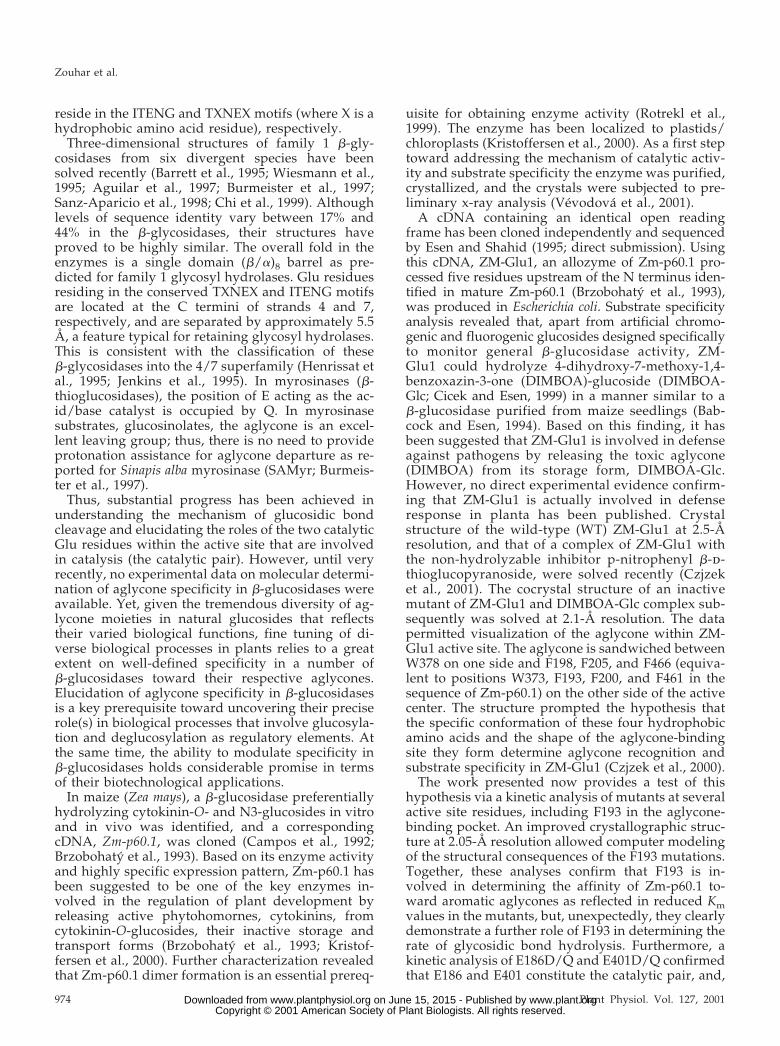
Insights into the Functional Architecture of the Catalytic Center of a Maize -Glucosidase Zm-p60.1 1 Jan Zouhar 2 , Jitka Ve ´vodova ´ 2 , Jaromı´r Marek, Jir ˘ı ´ Damborsky ´, Xiao-Dong Su, and Br ˘etislav Brzobohaty ´* Department of Functional Genomics and Proteomics (J.Z., J.V., J.M., B.B.) and National Center for Biomolecular Research (J.V., J.D.), Faculty of Science, Masaryk University, Kotla ´r ˘ska ´ 2, CZ–61137 Brno, Czech Republic; Institute of Biophysics of the Academy of Sciences of the Czech Republic, Kra ´lovopolska ´ 135, CZ–61265 Brno, Czech Republic (J.Z., B.B.); and Department of Molecular Biophysics, Center for Chemistry and Chemical Engineering, Lund University, S–221 00 Lund, Sweden (X.-D.S.) The maize (Zea mays) -glucosidase Zm-p60.1 has been implicated in regulation of plant development by the targeted release of free cytokinins from cytokinin-O-glucosides, their inactive storage forms. The crystal structure of the wild-type enzyme was solved at 2.05-Å resolution, allowing molecular docking analysis to be conducted. This indicated that the enzyme specificity toward substrates with aryl aglycones is determined by aglycone aromatic system stacking with W373, and interactions with edges of F193, F200, and F461 located opposite W373 in a slot-like aglycone-binding site. These aglycone- active site interactions recently were hypothesized to determine substrate specificity in inactive enzyme substrate complexes of ZM-Glu1, an allozyme of Zm-p60.1. Here, we test this hypothesis by kinetic analysis of F193I/Y/W mutants. The decreased K m of all mutants confirmed the involvement of F193 in determining enzyme affinity toward substrates with an aromatic aglycone. It was unexpected that a 30-fold decrease in k cat was found in F193I mutant compared with the wild type. Kinetic analysis and computer modeling demonstrated that the F193-aglycone-W373 interaction not only contributes to aglycone recognition as hypothesized previously but also codetermines catalytic rate by fixing the glucosidic bond in an orientation favorable for attack by the catalytic pair, E186 and E401. The catalytic pair, assigned initially by their location in the structure, was confirmed by kinetic analysis of E186D/Q and E401D/Q mutants. It was unexpected that the E401D as well as C205S and C211S mutations dramatically impaired the assembly of a catalysis-competent homodimer, suggesting novel links between the active site structure and dimer formation. -Glucosidases (-glucoside glucohydrolases, EC 3.2.1.21) are a widespread group of enzymes hydro- lyzing a broad variety of aryl- and alkyl--d- glucosides as well as glucosides with only a carbo- hydrate moiety. Interest in -glucosidase research reflects essential functions of -glucosidases in a va- riety of basic biological processes ranging from de- velopmental regulation to chemical defense against pathogen attack, and in a number of industrial ap- plications such as biomass conversion. In plants, -glucosidases have been implicated in regulating various aspects of development, e.g. phytohormone activation (Smith and van Staden, 1978; Brzobohaty ´ et al., 1994), cell wall degradation in the endosperm during germination (Leah et al., 1995), and pathogen defense reactions (Poulton, 1990). Plant -glucosidases are classified as family 1 of retaining glycosyl hydrolases according to their pri- mary structure (Henrissat and Bairoch, 1993; Henris- sat et al., 1995). Hydrolysis of a glycosidic bond involves two essential carboxylates, one acting as a general acid/base catalyst and the other as a nucleo- phile (Sinnott, 1990). The hydrolysis is initiated by the nucleophilic attack at the anomeric carbon (C1) of the substrate that results in the formation of glycosyl enzyme intermediate followed by the release of the aglycone facilitated by protonation of the glycosidic oxygen by the acid catalyst (the glycosylation step). The deprotonated acid catalyst acting now as a base (anion) removes a proton from water, and the result- ing hydroxyl anion attacks the covalent bond of the glycosyl enzyme intermediate. As a result of the at- tack, Glc is released and nucleophile regenerated (the deglycosylation step). For example, in the ho- modimeric Agrobacterium faecalis -glucosidase (fur- ther referred to as A. faecalis -glucosidase) E358 was identified as the nucleophile by means of mechanism-based inactivators and site-directed mu- tagenesis (Withers et al., 1990; Withers et al., 1992). E170 subsequently was demonstrated to serve as the general acid/base catalyst based on chemical rescue of inactive mutants in A. faecalis -glucosidase (Wang et al., 1995). E358 and E170 are conserved in all members of family 1 of glycosyl hydrolases, and 1 This work was supported by the Ministry of Education of the Czech Republic (grant nos. VS96096 and MSM143100008), by the INCO-Copernicus Program (grant no. ERB3512–PL966135), by the National Science Foundation, U.S. (grant no. INT–9600462), by the Socrates Erasmus Free Movers and Swedish Institute (grants to J.V.), and by the Swedish Foundation for Strategic Research and Structural Biology Network (support to X.-D.S.). 2 These authors contributed equally to the paper. * Corresponding author; e-mail [email protected]; fax 420 –5– 41211293. Article, publication date, and citation information can be found at www.plantphysiol.org/cgi/doi/10.1104/pp.010712. Plant Physiology, November 2001, Vol. 127, pp. 973–985, www.plantphysiol.org © 2001 American Society of Plant Biologists 973 www.plant.org on June 15, 2015 - Published by www.plantphysiol.org Downloaded from Copyright © 2001 American Society of Plant Biologists. All rights reserved.
Transcript of Insights into the functional architecture of the catalytic center of a maize β-glucosidase Zmp60.1
Insights into the Functional Architecture of the CatalyticCenter of a Maize �-Glucosidase Zm-p60.11
Jan Zouhar2, Jitka Vevodova2, Jaromır Marek, Jirı Damborsky, Xiao-Dong Su, and Bretislav Brzobohaty*
Department of Functional Genomics and Proteomics (J.Z., J.V., J.M., B.B.) and National Center forBiomolecular Research (J.V., J.D.), Faculty of Science, Masaryk University, Kotlarska 2, CZ–61137 Brno,Czech Republic; Institute of Biophysics of the Academy of Sciences of the Czech Republic, Kralovopolska135, CZ–61265 Brno, Czech Republic (J.Z., B.B.); and Department of Molecular Biophysics, Center forChemistry and Chemical Engineering, Lund University, S–221 00 Lund, Sweden (X.-D.S.)
The maize (Zea mays) �-glucosidase Zm-p60.1 has been implicated in regulation of plant development by the targeted releaseof free cytokinins from cytokinin-O-glucosides, their inactive storage forms. The crystal structure of the wild-type enzymewas solved at 2.05-Å resolution, allowing molecular docking analysis to be conducted. This indicated that the enzymespecificity toward substrates with aryl aglycones is determined by aglycone aromatic system stacking with W373, andinteractions with edges of F193, F200, and F461 located opposite W373 in a slot-like aglycone-binding site. These aglycone-active site interactions recently were hypothesized to determine substrate specificity in inactive enzyme substrate complexesof ZM-Glu1, an allozyme of Zm-p60.1. Here, we test this hypothesis by kinetic analysis of F193I/Y/W mutants. Thedecreased Km of all mutants confirmed the involvement of F193 in determining enzyme affinity toward substrates with anaromatic aglycone. It was unexpected that a 30-fold decrease in kcat was found in F193I mutant compared with the wild type.Kinetic analysis and computer modeling demonstrated that the F193-aglycone-W373 interaction not only contributes toaglycone recognition as hypothesized previously but also codetermines catalytic rate by fixing the glucosidic bond in anorientation favorable for attack by the catalytic pair, E186 and E401. The catalytic pair, assigned initially by their location inthe structure, was confirmed by kinetic analysis of E186D/Q and E401D/Q mutants. It was unexpected that the E401D aswell as C205S and C211S mutations dramatically impaired the assembly of a catalysis-competent homodimer, suggestingnovel links between the active site structure and dimer formation.
�-Glucosidases (�-glucoside glucohydrolases, EC3.2.1.21) are a widespread group of enzymes hydro-lyzing a broad variety of aryl- and alkyl-�-d-glucosides as well as glucosides with only a carbo-hydrate moiety. Interest in �-glucosidase researchreflects essential functions of �-glucosidases in a va-riety of basic biological processes ranging from de-velopmental regulation to chemical defense againstpathogen attack, and in a number of industrial ap-plications such as biomass conversion. In plants,�-glucosidases have been implicated in regulatingvarious aspects of development, e.g. phytohormoneactivation (Smith and van Staden, 1978; Brzobohatyet al., 1994), cell wall degradation in the endospermduring germination (Leah et al., 1995), and pathogendefense reactions (Poulton, 1990).
Plant �-glucosidases are classified as family 1 ofretaining glycosyl hydrolases according to their pri-mary structure (Henrissat and Bairoch, 1993; Henris-sat et al., 1995). Hydrolysis of a glycosidic bondinvolves two essential carboxylates, one acting as ageneral acid/base catalyst and the other as a nucleo-phile (Sinnott, 1990). The hydrolysis is initiated bythe nucleophilic attack at the anomeric carbon (C1) ofthe substrate that results in the formation of glycosylenzyme intermediate followed by the release of theaglycone facilitated by protonation of the glycosidicoxygen by the acid catalyst (the glycosylation step).The deprotonated acid catalyst acting now as a base(anion) removes a proton from water, and the result-ing hydroxyl anion attacks the covalent bond of theglycosyl enzyme intermediate. As a result of the at-tack, Glc is released and nucleophile regenerated(the deglycosylation step). For example, in the ho-modimeric Agrobacterium faecalis �-glucosidase (fur-ther referred to as A. faecalis �-glucosidase) E358was identified as the nucleophile by means ofmechanism-based inactivators and site-directed mu-tagenesis (Withers et al., 1990; Withers et al., 1992).E170 subsequently was demonstrated to serve as thegeneral acid/base catalyst based on chemical rescueof inactive mutants in A. faecalis �-glucosidase (Wanget al., 1995). E358 and E170 are conserved in allmembers of family 1 of glycosyl hydrolases, and
1 This work was supported by the Ministry of Education of theCzech Republic (grant nos. VS96096 and MSM143100008), bythe INCO-Copernicus Program (grant no. ERB3512–PL966135),by the National Science Foundation, U.S. (grant no. INT–9600462),by the Socrates Erasmus Free Movers and Swedish Institute(grants to J.V.), and by the Swedish Foundation for StrategicResearch and Structural Biology Network (support to X.-D.S.).
2 These authors contributed equally to the paper.* Corresponding author; e-mail [email protected]; fax 420 –5–
41211293.Article, publication date, and citation information can be found
at www.plantphysiol.org/cgi/doi/10.1104/pp.010712.
Plant Physiology, November 2001, Vol. 127, pp. 973–985, www.plantphysiol.org © 2001 American Society of Plant Biologists 973 www.plant.org on June 15, 2015 - Published by www.plantphysiol.orgDownloaded from Copyright © 2001 American Society of Plant Biologists. All rights reserved.
reside in the ITENG and TXNEX motifs (where X is ahydrophobic amino acid residue), respectively.
Three-dimensional structures of family 1 �-gly-cosidases from six divergent species have beensolved recently (Barrett et al., 1995; Wiesmann et al.,1995; Aguilar et al., 1997; Burmeister et al., 1997;Sanz-Aparicio et al., 1998; Chi et al., 1999). Althoughlevels of sequence identity vary between 17% and44% in the �-glycosidases, their structures haveproved to be highly similar. The overall fold in theenzymes is a single domain (�/�)8 barrel as pre-dicted for family 1 glycosyl hydrolases. Glu residuesresiding in the conserved TXNEX and ITENG motifsare located at the C termini of strands 4 and 7,respectively, and are separated by approximately 5.5Å, a feature typical for retaining glycosyl hydrolases.This is consistent with the classification of these�-glycosidases into the 4/7 superfamily (Henrissat etal., 1995; Jenkins et al., 1995). In myrosinases (�-thioglucosidases), the position of E acting as the ac-id/base catalyst is occupied by Q. In myrosinasesubstrates, glucosinolates, the aglycone is an excel-lent leaving group; thus, there is no need to provideprotonation assistance for aglycone departure as re-ported for Sinapis alba myrosinase (SAMyr; Burmeis-ter et al., 1997).
Thus, substantial progress has been achieved inunderstanding the mechanism of glucosidic bondcleavage and elucidating the roles of the two catalyticGlu residues within the active site that are involvedin catalysis (the catalytic pair). However, until veryrecently, no experimental data on molecular determi-nation of aglycone specificity in �-glucosidases wereavailable. Yet, given the tremendous diversity of ag-lycone moieties in natural glucosides that reflectstheir varied biological functions, fine tuning of di-verse biological processes in plants relies to a greatextent on well-defined specificity in a number of�-glucosidases toward their respective aglycones.Elucidation of aglycone specificity in �-glucosidasesis a key prerequisite toward uncovering their preciserole(s) in biological processes that involve glucosyla-tion and deglucosylation as regulatory elements. Atthe same time, the ability to modulate specificity in�-glucosidases holds considerable promise in termsof their biotechnological applications.
In maize (Zea mays), a �-glucosidase preferentiallyhydrolyzing cytokinin-O- and N3-glucosides in vitroand in vivo was identified, and a correspondingcDNA, Zm-p60.1, was cloned (Campos et al., 1992;Brzobohaty et al., 1993). Based on its enzyme activityand highly specific expression pattern, Zm-p60.1 hasbeen suggested to be one of the key enzymes in-volved in the regulation of plant development byreleasing active phytohomornes, cytokinins, fromcytokinin-O-glucosides, their inactive storage andtransport forms (Brzobohaty et al., 1993; Kristof-fersen et al., 2000). Further characterization revealedthat Zm-p60.1 dimer formation is an essential prereq-
uisite for obtaining enzyme activity (Rotrekl et al.,1999). The enzyme has been localized to plastids/chloroplasts (Kristoffersen et al., 2000). As a first steptoward addressing the mechanism of catalytic activ-ity and substrate specificity the enzyme was purified,crystallized, and the crystals were subjected to pre-liminary x-ray analysis (Vevodova et al., 2001).
A cDNA containing an identical open readingframe has been cloned independently and sequencedby Esen and Shahid (1995; direct submission). Usingthis cDNA, ZM-Glu1, an allozyme of Zm-p60.1 pro-cessed five residues upstream of the N terminus iden-tified in mature Zm-p60.1 (Brzobohaty et al., 1993),was produced in Escherichia coli. Substrate specificityanalysis revealed that, apart from artificial chromo-genic and fluorogenic glucosides designed specificallyto monitor general �-glucosidase activity, ZM-Glu1 could hydrolyze 4-dihydroxy-7-methoxy-1,4-benzoxazin-3-one (DIMBOA)-glucoside (DIMBOA-Glc; Cicek and Esen, 1999) in a manner similar to a�-glucosidase purified from maize seedlings (Bab-cock and Esen, 1994). Based on this finding, it hasbeen suggested that ZM-Glu1 is involved in defenseagainst pathogens by releasing the toxic aglycone(DIMBOA) from its storage form, DIMBOA-Glc.However, no direct experimental evidence confirm-ing that ZM-Glu1 is actually involved in defenseresponse in planta has been published. Crystalstructure of the wild-type (WT) ZM-Glu1 at 2.5-Åresolution, and that of a complex of ZM-Glu1 withthe non-hydrolyzable inhibitor p-nitrophenyl �-d-thioglucopyranoside, were solved recently (Czjzeket al., 2001). The cocrystal structure of an inactivemutant of ZM-Glu1 and DIMBOA-Glc complex sub-sequently was solved at 2.1-Å resolution. The datapermitted visualization of the aglycone within ZM-Glu1 active site. The aglycone is sandwiched betweenW378 on one side and F198, F205, and F466 (equiva-lent to positions W373, F193, F200, and F461 in thesequence of Zm-p60.1) on the other side of the activecenter. The structure prompted the hypothesis thatthe specific conformation of these four hydrophobicamino acids and the shape of the aglycone-bindingsite they form determine aglycone recognition andsubstrate specificity in ZM-Glu1 (Czjzek et al., 2000).
The work presented now provides a test of thishypothesis via a kinetic analysis of mutants at severalactive site residues, including F193 in the aglycone-binding pocket. An improved crystallographic struc-ture at 2.05-Å resolution allowed computer modelingof the structural consequences of the F193 mutations.Together, these analyses confirm that F193 is in-volved in determining the affinity of Zm-p60.1 to-ward aromatic aglycones as reflected in reduced Kmvalues in the mutants, but, unexpectedly, they clearlydemonstrate a further role of F193 in determining therate of glycosidic bond hydrolysis. Furthermore, akinetic analysis of E186D/Q and E401D/Q confirmedthat E186 and E401 constitute the catalytic pair, and,
Zouhar et al.
974 Plant Physiol. Vol. 127, 2001 www.plant.org on June 15, 2015 - Published by www.plantphysiol.orgDownloaded from Copyright © 2001 American Society of Plant Biologists. All rights reserved.
surprisingly, that the E401D mutation hinders theassembly of enzymatically active dimers suggesting apreviously unrecognized link between the active sitearchitecture and dimer interface.
RESULTS AND DISCUSSION
Structure of the Zm-p60.1 �-Glucosidase
The crystal structure of the Zm-p60.1 �-glucosidasewas solved by molecular replacement at 2.05-Å res-olution. Concomitantly with solving the Zm-p60.1structure, structures of ZM-Glu1, an allozyme of Zm-p60.1, and ZM-Glu1 inactive mutant substrate com-plex have been published by an independent group(Czjzek et al., 2000, 2001). The Zm-p60.1 structurepresented here is more appropriate for moleculardocking and computer modeling because the ZM-Glu1 structure was solved at lower resolution (2.5 Å).The inactive mutant substrate structure was solved ata resolution (2.1 Å) comparable with our structure.However, employment of the complex structure forsubstrate docking is not optimal because the struc-ture must be different to that one found in the wildtype to prevent a sterical clash between the mutatedamino acid residue and the ligand. Otherwise, thestructures of the wild-type enzymes are, within anexperimental error, identical. To prevent unnecessaryduplications, here, we restrict description of thestructure of the protein to presentation of basic over-all features of Zm-p60.1 structure, and we focusmainly on those that are essential for interpretationof our functional analysis of the catalytic center.
The protein is a homodimer in solution and itcrystallized as a homodimer in one asymmetric unitas well. However, due to the different packing envi-ronments, there are some differences between thesetwo monomers. For example, clear electron densityfrom residue A6 to A495 can be seen in monomer A,whereas B11 to B502 was shown for monomer B.Some of the residues from solvent-accessible areas(mainly Arg, Lys, glutamic, and aspartic acids) aredisordered and have been refined with the partialoccupancies. Crystallographic results are summa-rized in Table I.
Zm-p60.1 monomer is comprised of a single (�/�)8barrel domain (Figs. 1 and 2). Structure analysis con-firmed that C205 and C211 form a disulfide bridgethat has been identified previously (Rotrekl et al.,1999; Fig. 2).
Zm-p60.1 monomers do not possess any detectablecatalytic activity (Rotrekl et al., 1999), and no exper-imental evidence on the mechanism by which ho-modimer assembly contributes to formation ofcatalysis-competent state of the enzyme is availableyet. Description of the dimerisation interface is thefirst step toward understanding the role of ho-modimer formation in the assembly of a catalysis-competent structure in Zm-p60.1 and related�-glucosidases. In the crystal, two monomers per
asymmetric unit related by a non-crystallographicsymmetry were identified. The surface area buriedon the dimer interface is 2,131 Å2 corresponding to1,066 Å2 per monomer when the calculation is per-formed with the program Crystallography & NMRSystem (Brunger et al., 1998) and the probe diameteris 1.4 Å. The dimer interface is formed by residues ofthree �-helices (S266-L281, G282-G291, and P294-R302) and two loops (N335-L346 and K391-N394).Mutual orientation of the secondary structure ele-ments within the dimer is indicated in Figure 2. Mostof the monomer contacts in the dimer interface arerepresented by aromatic stacking and hydrogen andionic bonds. No intermolecular disulfide bridge con-nects the monomer subunits in the dimer. An in-tramolecular disulfide bridge was identified in eachmonomer (Fig. 2), and, recently, we have shown thatits formation is an essential prerequisite for gainingmonomer competence to assemble into an activedimer (Rotrekl et al., 1999).
Active Site
The active site is a slot-like structure intrudingfrom the surface to the barrel core of the protein. Thethird layer of residues constituting the barrel interiorrepresents the base of the slot. Slot walls are formedmainly by four extended loops (I–IV in Fig. 1). Theloops consist of residues N54 through S71 (loop I),
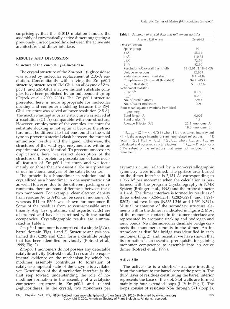
Table I. Summary of crystal data and refinement statistics
Structure Refinement Zm-p60.1
Data collectionSpace group P21
a (Å) 55.66b (Å) 110.72c (Å) 72.94� (°) 92.10Resolution (Å) overall (last shell) 68–2.05 (2.18–2.05)Unique reflections 52,651Redundancy overall (last shell) 9.7 (8.8)Completeness (%) overall (last shell) 94.7 (85.7)Rmerge
a (last shell) 5.3 (17.6)Refinement statistics
R factorb 0.169Rfree
c 0.230No. of protein atoms 7,943No. of water molecules 909
Root-mean-square deviations from idealgeometry
Bond length (Å) 0.005Bond angles (°) 1.3Average B factor (Å2) 22.2 (monomer A)
18.8 (monomer B)a Rmerge � (� �I � �I�� / � I ) where I is the observed intensity and
�I� is the average intensity of symmetry-related reflections. b Rfactor � �h � �Fobs� � �Fcalc� � / �h�Fobs�, where Fcalc and Fobs arecalculated and observed structure factors. c Rfree � R factor for a6.1% subset of the reflections that were not included in therefinement.
Catalytic Center of Maize �-Glucosidase Zm-p60.1
Plant Physiol. Vol. 127, 2001 975 www.plant.org on June 15, 2015 - Published by www.plantphysiol.orgDownloaded from Copyright © 2001 American Society of Plant Biologists. All rights reserved.
Y195 through E221 (loop II), N335 through M376(loop III), and W452 through V471 (loop IV). Theslot-forming loops represent the sites of the highestvariability in the �-glucosidase family as expected forthe sites participating in the determination of sub-strate specificity. At the entrance, the slot is approx-imately 22 Å long and 8 Å wide. The upper part ofthe active site represents a putative aglycone-bindingsite, whereas the glycone-binding site is located inthe bottom one-half of the active site slot.
The glycone-binding site is formed by a number ofpolar and aromatic residues that are commonlyfound within carbohydrate recognition sites. The hy-drophobic site of the Glc ring can be stacked ontoW452 that is highly conserved in family 1 of�-glycosidases and represents the last amino acidresidue on the C-terminal end of �-strand 8. Forhydrogen-bonding interactions with the Glc moiety,several conserved amino acid residues are availablein appropriate orientations including Q33 (N�2 andO�1 H bond to O4 and O3, respectively), H137 (N�2H bonds to O3), N185 (N�2 H bonds to O2), and E459(O�1 and O�2 H bond to O4 and 06, respectively). Itis interesting that an unknown electron density wasobserved in the glycone-binding pocket. To interpret
the electron density, enzyme kinetic analysis wasemployed to investigate the affinity of the enzymeactive site to glycerol, a cryoprotectant used in crystaldiffraction analysis. Glycerol proved to be a weakcompetitive inhibitor of Zm-p60.1 (dissociation con-stant of an enzyme-inhibitor complex [Ki] � 215 mm).Modeling experiments indicated that the electrondensity can be interpreted as two glycerol molecules.Electron densities in substrate-unoccupied activesites were assigned as glycerol molecules in SAMyr(Burmeister et al., 1997) and a ZM-Glu1 mutant(Czjzek et al., 2000), however, without providing in-dependent experimental evidence to support theassignment.
E186 and E401 Constitute the Catalytic Pair in Zm-p60.1
Multiple alignment using ClustalW program posi-tioned E186 and E401 into the highly conservedTXNEX and ITENG motifs, respectively (not shown).In Zm-p60.1 structure, the conserved E186 and E401were found in the loop regions close to the carboxy-terminal ends of �-strands 4 and 7, respectively. Fur-thermore, E186 and 401 are in close proximity—thedistance between their carbonyl carbons C� is 4.98 Å
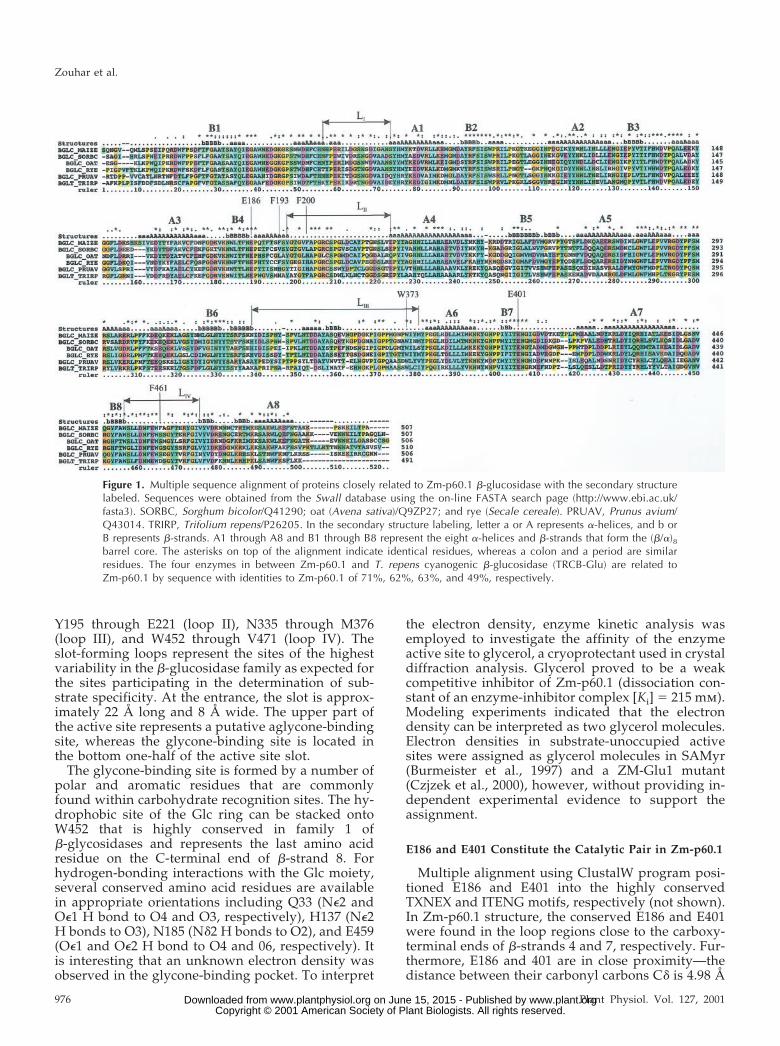
Figure 1. Multiple sequence alignment of proteins closely related to Zm-p60.1 �-glucosidase with the secondary structurelabeled. Sequences were obtained from the Swall database using the on-line FASTA search page (http://www.ebi.ac.uk/fasta3). SORBC, Sorghum bicolor/Q41290; oat (Avena sativa)/Q9ZP27; and rye (Secale cereale). PRUAV, Prunus avium/Q43014. TRIRP, Trifolium repens/P26205. In the secondary structure labeling, letter a or A represents �-helices, and b orB represents �-strands. A1 through A8 and B1 through B8 represent the eight �-helices and �-strands that form the (�/�)8barrel core. The asterisks on top of the alignment indicate identical residues, whereas a colon and a period are similarresidues. The four enzymes in between Zm-p60.1 and T. repens cyanogenic �-glucosidase (TRCB-Glu) are related toZm-p60.1 by sequence with identities to Zm-p60.1 of 71%, 62%, 63%, and 49%, respectively.
Zouhar et al.
976 Plant Physiol. Vol. 127, 2001 www.plant.org on June 15, 2015 - Published by www.plantphysiol.orgDownloaded from Copyright © 2001 American Society of Plant Biologists. All rights reserved.
and between E186 O�1 and E401 O�2 it is 3.52 Å (Fig.2). Two conserved Glu residues located close to the Ctermini of �-strands 4 and 7 and separated by adistance of about 5 Å were proposed to function asthe acid/base and nucleophile, respectively, in the4/7 superfamily of glycohydrolases (Henrissat et al.,1995; Jenkins et al., 1995). Because Zm-p60.1 belongsto the 4/7 superfamily based on sequence similaritiesand crystal structure analysis, E186 and E401 canconstitute the catalytic pair in Zm-p60.1.
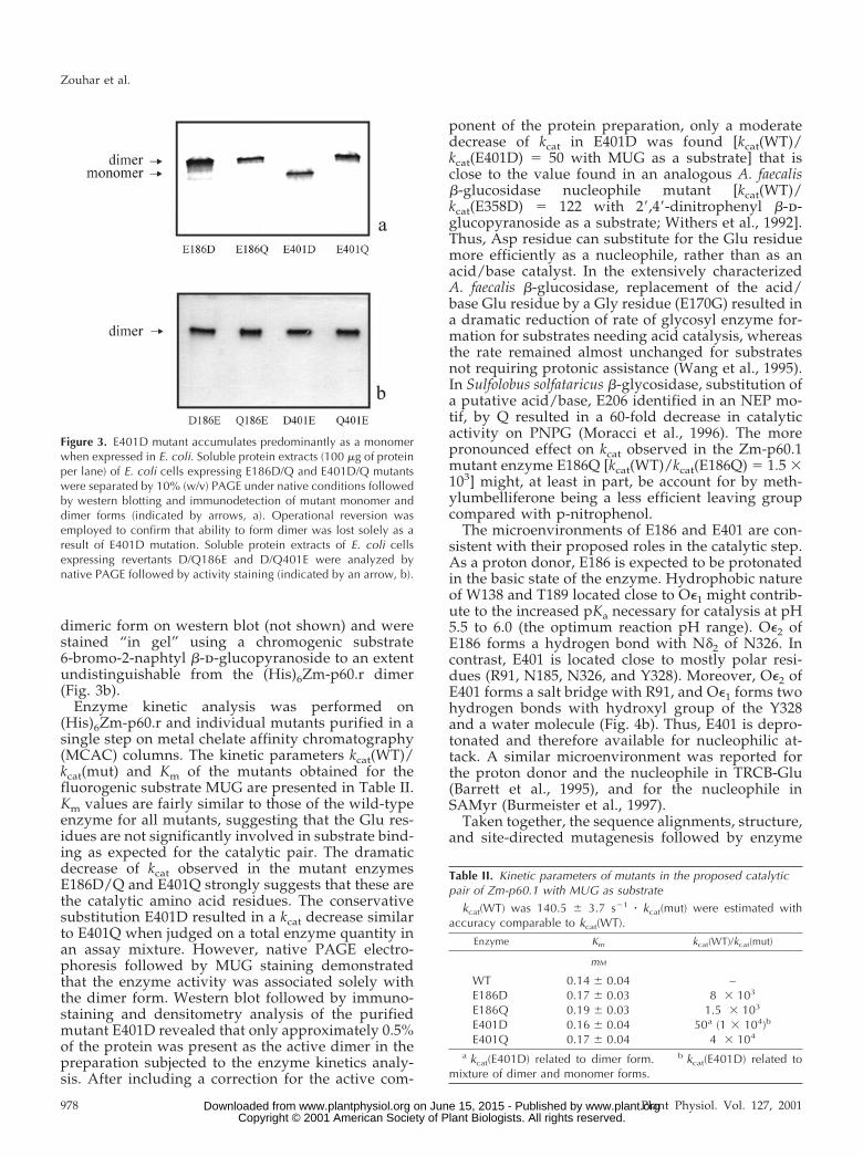
The contribution of E186 and E401 to Zm-p60.1enzyme activity was investigated using site-directedmutagenesis followed by enzyme kinetic analysis ofthe mutants. In (His)6Zm-p60.r, a recombinant deriv-ative of Zm-p60.1, the Glu residues were changedindividually, in independent experiments, to Aspand Gln residues. The resulting mutants were pro-duced in E. coli. To exclude effects of any grossalteration in quaternary structure on enzyme kineticanalysis, assembly of the mutants into catalysis-competent dimer structure (Rotrekl et al., 1999) wasanalyzed in soluble bacterial protein extracts usingnative PAGE followed by �-glucosidase activity “in-gel” staining, and immunodetection of the mutantson corresponding western blots. E186D, E186Q, andE401Q were found in the form of dimers indistin-guishable from the (His)6Zm-p60.r dimer. It wassurprising that the major fraction of E401D migratedat (His)6Zm-p60.r monomer position (Fig. 3a). Highlysensitive activity “in-gel” staining based on a fluoro-genic substrate 4-methylumbelliferyl �-d-glucopy-ranoside (MUG) staining demonstrated a small butdetectable enzymatic activity associated with thedimer form in E186D/Q and E401Q mutants. Detect-able staining associated with expected dimer positionin E401D indicated that a small fraction (below im-munostaining detection limit—about 10 ng of proteinper band in our assay conditions) of the mutant ispresent in the dimeric form and retained substantialenzyme activity. As expected from our previous ex-periments (Rotrekl et al., 1999), there was no enzymeactivity associated with the monomeric form of themutant (not shown). In addition to DNA sequencing,the sample homogeneity of the mutants was demon-strated by operational reversion. The revertants,Q186E, D186E, Q401E, and D401E, were detected in
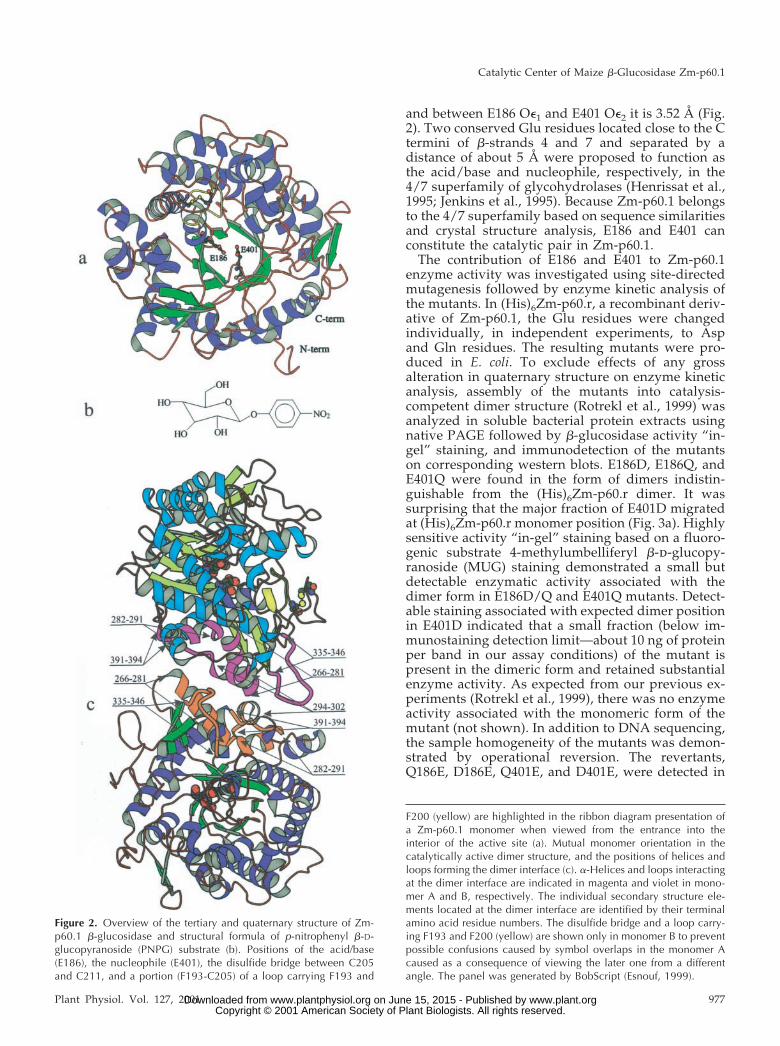
Figure 2. Overview of the tertiary and quaternary structure of Zm-p60.1 �-glucosidase and structural formula of p-nitrophenyl �-D-glucopyranoside (PNPG) substrate (b). Positions of the acid/base(E186), the nucleophile (E401), the disulfide bridge between C205and C211, and a portion (F193-C205) of a loop carrying F193 and
F200 (yellow) are highlighted in the ribbon diagram presentation ofa Zm-p60.1 monomer when viewed from the entrance into theinterior of the active site (a). Mutual monomer orientation in thecatalytically active dimer structure, and the positions of helices andloops forming the dimer interface (c). �-Helices and loops interactingat the dimer interface are indicated in magenta and violet in mono-mer A and B, respectively. The individual secondary structure ele-ments located at the dimer interface are identified by their terminalamino acid residue numbers. The disulfide bridge and a loop carry-ing F193 and F200 (yellow) are shown only in monomer B to preventpossible confusions caused by symbol overlaps in the monomer Acaused as a consequence of viewing the later one from a differentangle. The panel was generated by BobScript (Esnouf, 1999).
Catalytic Center of Maize �-Glucosidase Zm-p60.1
Plant Physiol. Vol. 127, 2001 977 www.plant.org on June 15, 2015 - Published by www.plantphysiol.orgDownloaded from Copyright © 2001 American Society of Plant Biologists. All rights reserved.
dimeric form on western blot (not shown) and werestained “in gel” using a chromogenic substrate6-bromo-2-naphtyl �-d-glucopyranoside to an extentundistinguishable from the (His)6Zm-p60.r dimer(Fig. 3b).
Enzyme kinetic analysis was performed on(His)6Zm-p60.r and individual mutants purified in asingle step on metal chelate affinity chromatography(MCAC) columns. The kinetic parameters kcat(WT)/kcat(mut) and Km of the mutants obtained for thefluorogenic substrate MUG are presented in Table II.Km values are fairly similar to those of the wild-typeenzyme for all mutants, suggesting that the Glu res-idues are not significantly involved in substrate bind-ing as expected for the catalytic pair. The dramaticdecrease of kcat observed in the mutant enzymesE186D/Q and E401Q strongly suggests that these arethe catalytic amino acid residues. The conservativesubstitution E401D resulted in a kcat decrease similarto E401Q when judged on a total enzyme quantity inan assay mixture. However, native PAGE electro-phoresis followed by MUG staining demonstratedthat the enzyme activity was associated solely withthe dimer form. Western blot followed by immuno-staining and densitometry analysis of the purifiedmutant E401D revealed that only approximately 0.5%of the protein was present as the active dimer in thepreparation subjected to the enzyme kinetics analy-sis. After including a correction for the active com-
ponent of the protein preparation, only a moderatedecrease of kcat in E401D was found [kcat(WT)/kcat(E401D) � 50 with MUG as a substrate] that isclose to the value found in an analogous A. faecalis�-glucosidase nucleophile mutant [kcat(WT)/kcat(E358D) � 122 with 2�,4�-dinitrophenyl �-d-glucopyranoside as a substrate; Withers et al., 1992].Thus, Asp residue can substitute for the Glu residuemore efficiently as a nucleophile, rather than as anacid/base catalyst. In the extensively characterizedA. faecalis �-glucosidase, replacement of the acid/base Glu residue by a Gly residue (E170G) resulted ina dramatic reduction of rate of glycosyl enzyme for-mation for substrates needing acid catalysis, whereasthe rate remained almost unchanged for substratesnot requiring protonic assistance (Wang et al., 1995).In Sulfolobus solfataricus �-glycosidase, substitution ofa putative acid/base, E206 identified in an NEP mo-tif, by Q resulted in a 60-fold decrease in catalyticactivity on PNPG (Moracci et al., 1996). The morepronounced effect on kcat observed in the Zm-p60.1mutant enzyme E186Q [kcat(WT)/kcat(E186Q) � 1.5 �103] might, at least in part, be account for by meth-ylumbelliferone being a less efficient leaving groupcompared with p-nitrophenol.
The microenvironments of E186 and E401 are con-sistent with their proposed roles in the catalytic step.As a proton donor, E186 is expected to be protonatedin the basic state of the enzyme. Hydrophobic natureof W138 and T189 located close to O�1 might contrib-ute to the increased pKa necessary for catalysis at pH5.5 to 6.0 (the optimum reaction pH range). O�2 ofE186 forms a hydrogen bond with N�2 of N326. Incontrast, E401 is located close to mostly polar resi-dues (R91, N185, N326, and Y328). Moreover, O�2 ofE401 forms a salt bridge with R91, and O�1 forms twohydrogen bonds with hydroxyl group of the Y328and a water molecule (Fig. 4b). Thus, E401 is depro-tonated and therefore available for nucleophilic at-tack. A similar microenvironment was reported forthe proton donor and the nucleophile in TRCB-Glu(Barrett et al., 1995), and for the nucleophile inSAMyr (Burmeister et al., 1997).
Taken together, the sequence alignments, structure,and site-directed mutagenesis followed by enzyme
Figure 3. E401D mutant accumulates predominantly as a monomerwhen expressed in E. coli. Soluble protein extracts (100 �g of proteinper lane) of E. coli cells expressing E186D/Q and E401D/Q mutantswere separated by 10% (w/v) PAGE under native conditions followedby western blotting and immunodetection of mutant monomer anddimer forms (indicated by arrows, a). Operational reversion wasemployed to confirm that ability to form dimer was lost solely as aresult of E401D mutation. Soluble protein extracts of E. coli cellsexpressing revertants D/Q186E and D/Q401E were analyzed bynative PAGE followed by activity staining (indicated by an arrow, b).
Table II. Kinetic parameters of mutants in the proposed catalyticpair of Zm-p60.1 with MUG as substrate
kcat(WT) was 140.5 � 3.7 s�1 � kcat(mut) were estimated withaccuracy comparable to kcat(WT).
Enzyme Km kcat(WT)/kcat(mut)
mM
WT 0.14 � 0.04 –E186D 0.17 � 0.03 8 � 103
E186Q 0.19 � 0.03 1.5 � 103
E401D 0.16 � 0.04 50a (1 � 104)b
E401Q 0.17 � 0.04 4 � 104
a kcat(E401D) related to dimer form. b kcat(E401D) related tomixture of dimer and monomer forms.
Zouhar et al.
978 Plant Physiol. Vol. 127, 2001 www.plant.org on June 15, 2015 - Published by www.plantphysiol.orgDownloaded from Copyright © 2001 American Society of Plant Biologists. All rights reserved.
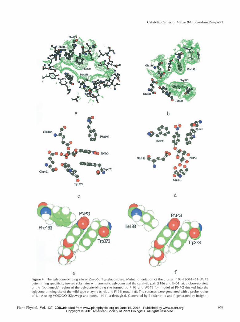
Figure 4. The aglycone-binding site of Zm-p60.1 �-glucosidase. Mutual orientation of the cluster F193-F200-F461-W373determining specificity toward substrates with aromatic aglycone and the catalytic pair (E186 and E401, a), a close-up viewof the “bottleneck” region of the aglycone-binding site formed by F193 and W373 (b), model of PNPG docked into theaglycone-binding site of the wild-type enzyme (c–e), and F193I mutant (f). The surfaces were generated with a probe radiusof 1.1 Å using VOIDOO (Kleywegt and Jones, 1994). a through d, Generated by BobScript; e and f, generated by InsightII.
Catalytic Center of Maize �-Glucosidase Zm-p60.1
Plant Physiol. Vol. 127, 2001 979 www.plant.org on June 15, 2015 - Published by www.plantphysiol.orgDownloaded from Copyright © 2001 American Society of Plant Biologists. All rights reserved.
kinetics prove that E186 and E401 are the acid/baseand the nucleophile, respectively, in Zm-p60.1.
E401D Mutation Hinders Dimer Assembly
As mentioned above, native PAGE analysis re-vealed a remarkable effect of E401D substitution onthe enzyme structure. The electrophoretic mobility ofthe mutant on native gels is indistinguishable fromthat of (His)6Zm-p60.r monomer, confirming that theoverall fold is retained in the mutant. However, thedramatically decreased ability of mutant monomer toassemble into a homodimer suggests that it adopts adistinct conformational state, an interpretation that isconsistent with the observation that E401D exhibitsaltered chromatographic behavior observed duringpurification. Although (His)6Zm-p60.r, E186D/Q andE401Q bind readily to a zinc-charged iminodiacetate-POROS-column (IDA-Zn2) used routinely for thesingle-step purification of the wild type and mutants,the E401D mutant is not retained by the column toany detectable extent. However, no difference inchromatographic behavior was observed betweenE401D and (His)6Zm-p60.r on a nickel-chargednitrilotriacetate-Sepharose-column (NTA-Ni2) al-lowing single-step purification of E401D (not shown).Lack of E401D binding to IDA-Zn2 might be ex-plained if the proposed change in conformationalstate causes the His tag on E401D surface to becomeless accessible. Because in general the His tag bindsmore tightly to NTA-Ni2 compared with IDA-Zn2,the partly exposed His tag could still efficiently in-teract with a stronger coordination partner, NTA-Ni2. The His tag and E401 are located on the oppo-site sides of the (�/�)8 barrel and are not connectedby a common �-strand or �-helix. Thus, the confor-mational change extends beyond the �-7 strand andthe adjacent loop carrying the ITENG motif, and islikely to be more complex than a conformationalchange in a single element of the secondary structure.Substitution of the Glu residue in the nucleophileposition by an Asp residue in previously studiedglycosidases in the 4/7 superfamily (including, forexample, A. faecalis �-glucosidase, Withers et al.,1992; E. coli �-galactosidase, Yuan et al., 1994; andBacillus circulans xylanase, Wakarchuk et al., 1994)has not resulted in gross conformational changesdetectable as altered CD spectra, and native PAGEmobility. However, although the E to Q/N mutantsin A. faecalis �-glucosidase, and E to Q/V mutants inE. coli �-galactosidase exhibited slightly higher orvery similar thermal stability compared with the cor-responding wild-type enzymes, all examined E to Dmutations exerted a clear decrease in thermal stabil-ity. Inspection of E401 microenvironment clearly in-dicates also that the E401D mutation in Zm-p60.1results in a loss of stabilizing interactions (a hydro-gen bond between E401 and Y328, and a salt bridgebetween E401 and R91). Earlier studies of peptide
models suggested that the process of folding isguided by interactions that also stabilize the finalnative structure. Thus, the distinct conformationalstate in E401D might be a consequence of perturba-tion of interactions that guide folding. It is interestingthat we have recently identified an independent mu-tation, I404D, in the ITENG loop that also hindersdimer formation in the same experimental system (J.Zouhar and B. Brzobohaty, unpublished data).
Though the Zm-p60.1 crystal structure proves thatall residues forming the active center are provided bya single monomer, dimer formation is an essentialprerequisite for activity of the enzyme (Rotrekl et al.,1999). Because the dimerization interface and activesite are not in close proximity with each other (Fig.2c), the reason for the necessity of dimerization forenzyme activity is not obvious. However, the dra-matic influence of E401D and I404D mutations on theassembly of catalysis-competent homodimer mightrepresent the first experimental indication of interac-tions between the ITENG loop and loops forming thedimerization area. Independent evidence supportingmutual dependence of the active site architecture andadoption of dimerization competent conformation ofloops forming the dimerization area comes from ourearlier analysis of C205A/S/R/D and C211A/S/R/D mutants. The mutations resulted in a dramaticreduction of monomer competence to assemble intodimers, and reduced catalytic efficiency of the en-zyme (Rotrekl et al., 1999), though the disulfidebridge is neither a part of the catalytic center nordimerization area (Fig. 2). However, the enzymestructure analysis revealed that formation of the di-sulfide bridge is involved in precise positioning ofF193 and F200, two of four aglycone-binding site keyresidues involved in enzyme-aglycone interactions(Figs. 2 and 4). Thus, conformational changes withinthe active site induced by diverse mutations seems touncover a link between fine tuning of formation ofcatalysis competent structure of the active siteand dimerization-competent architecture of themonomer-monomer interface in the course of dimerassembly. Structure analysis of E401D and I404D mu-tants could represent an important step in uncover-ing structural principles underlying mutual depen-dence of dimer formation and recovery of enzymeactivity in Zm-p60.1 and related �-glucosidases. Theexperiments addressing this issue are in progress.
F193 Determines Both Substrate Affinity andCatalytic Rate
To obtain preliminary information on the nature ofthe molecular determination of Zm-p60.1 specificitytoward substrate aglycone moiety, a preliminary at-tempt to locate putative amino acid residues formingthe aglycone binding site was performed on a refinedhomology-based model of the three-dimensionalstructure of Zm-p60.1 constructed earlier (Rotrekl et
Zouhar et al.
980 Plant Physiol. Vol. 127, 2001 www.plant.org on June 15, 2015 - Published by www.plantphysiol.orgDownloaded from Copyright © 2001 American Society of Plant Biologists. All rights reserved.
al., 1999). Inspection of the active site slot showedthat its upper part, the proposed aglycone-bindingsite, is formed mainly by hydrophobic residues. Thelimited accuracy of the model did not justify anydetailed molecular docking that could identify resi-dues involved in enzyme-substrate interactions.However, F193 appeared as a key determinant of thewidth of the slot-like aglycone-binding site, andtherefore a potential determinant of aglycone speci-ficity. A similar, though less convincing predictioncould be made for M258, which appeared as a widthcodeterminant located opposite F193. Later, solvingof the crystal structure led to confirmation of theproposed role for F193, and facilitated reliable inter-pretations of kinetic data obtained in F193 mutants.Though the prediction made for M258 turned out notto be precise, M258 is just adjacent to the residuesdetermining the narrowest region of the aglycone-binding site. Therefore, kinetic analysis of M258mutants brings a support for highly localized char-acter of the “bottleneck” region of the slot-likeaglycone-binding site deduced from the crystal struc-ture (Fig. 4).
The functional contribution of F193 and M258 tocatalytic activity was investigated by site-directedmutagenesis followed by enzyme kinetic analysis. In(His)6Zm-p60.r, F193 was changed in separate exper-iments to I, W, and Y. In a similar manner, M258 wasreplaced by I, F, and V. The resulting mutants wereproduced in E. coli. Native PAGE analysis of individ-ual soluble bacterial protein extracts confirmed for-mation of a catalysis-competent dimer structure in allinvestigated mutants (not shown). For enzyme ki-netic analysis, the mutants were purified by singlestep MCAC chromatography. The kinetic parametersKm and kcat obtained for the chromogenic substratePNPG are summarized in Table III. Although system-atic increase in Km in all the F193 mutants supportedthe proposed role of F193 in determination of theenzyme substrate affinity, distinct decreases in kcat inthe individual F193 mutants indicated a yet unrecog-nized function of F193. Kinetic parameters of M258mutants displayed no or only moderate changescompared with the wild type.
Prompted by the dramatic effect of F193I on theZm-p60.1 specificity constant (kcat/Km) with PNPG,we focused on a detailed analysis of the F193 micro-environment in the Zm-p60.1 crystal structure. De-tailed inspection of the structure followed by molec-ular docking revealed that the aglycone interactionwith the slot-like aglycone-binding site is largely de-termined by W373 stacking interactions with the ag-lycone aromatic system, and van der Waals interac-tions with the edges of the phenyl rings provided byF193, F200, and F461 (Fig. 4). The W373 orientationfavorable for stacking interactions is stabilized by ahydrogen bond between N�1 of W373 and water thatis firmly positioned by additional hydrogen bondswith E466 and Y468. Therefore, E466 and Y468 cancontribute indirectly to substrate specificity and cat-alytic efficiency. Proper positioning of F193, F200,and F461 appears to be stabilized by a large hydro-phobic cluster formed by F51, W48, W138, F190, andW460 (not shown). In addition, the disulfide bridgebetween C205 and C211 stabilizes the loop contain-ing F193 and F200 (Fig. 2). It is interesting that wehave found a dramatic drop in enzyme activity inmutants C205S and C211A/S (Rotrekl et al., 1999).Though the drop might be explained in part bythe observed decreased ability of these mutants toform the catalysis-competent dimer (Rotrekl et al.,1999), the structural analysis presented here suggeststhat the decrease in catalytic efficiency may partly bedue to loss of F193 and F200 position stabilization.
The molecular docking together with increase inKm in F193/I/W/Y mutants presented above supporta recent assignment of F193 as a determinant of en-zyme affinity toward glucosides with an aromaticaglycone that was based on structural analysis ofinactive mutant enzyme-substrate complex (Czjzek etal., 2000). It was unexpected that our kinetic resultsclearly demonstrate that the dramatic drop in theF193I specificity constant is caused mainly by drop inkcat. Based on this novel finding, structure inspectionallowed us to propose that the F193-aglycone-W373interaction represents a major contribution to thepositioning of the glucosidic bond in an orientationfavorable for attack by E186 and E401. Therefore, theinteraction is expected to codetermine catalytic rateof the enzyme. In contrast, glycone-enzyme interac-tions do not seem to contribute to the appropriatepositioning of the glucosidic bond to large extent.Previous enzyme kinetic analysis indicated that theground state of the glycone-binding pocket is com-plementary to Glc in the half chair conformationrather than the chair conformation. Glc was a muchweaker competitive inhibitor compared withd-glucono-1,5-lactone, which has a half-chair confor-mation and inhibits the enzyme by acting as a tran-sition state analog (Babcock and Esen, 1994; J. Zou-har, J. Vevodova, J. Marek, J. Damborsky, X.-D. Su,and B. Brzobohaty, unpublished data). Thus, the nar-row slot of the aglycone-binding site formed by F193,
Table III. Kinetic parameters of mutants in F193 and M258 of Zm-p60.1 with PNPG as substrate
kcat(WT) was 28.0 � 0.8 s�1 � kcat(mut) were estimated withaccuracy comparable to kcat(WT).
Enzyme Km (mM)kcat(mut)/kcat(WT)
[kcat/Km(mut)]/[kcat/Km(WT)]
WT 0.64 � 0.08 – –F193I 1.76 � 0.06 0.03 0.01F193Y 1.29 � 0.01 0.62 0.31F193W 1.61 � 0.17 1.12 0.45M258I 1.18 � 0.36 0.76 0.41M258V 0.59 � 0.08 0.34 0.37M258F 0.59 � 0.08 0.30 0.33
Catalytic Center of Maize �-Glucosidase Zm-p60.1
Plant Physiol. Vol. 127, 2001 981 www.plant.org on June 15, 2015 - Published by www.plantphysiol.orgDownloaded from Copyright © 2001 American Society of Plant Biologists. All rights reserved.
F200, F461, and W373 appears to be a stereochemicaldeterminant of the interaction strength (Fig. 4). Al-though W373 is highly conserved among family 1�-glucosidases, F193 is highly variable (Fig. 1), indi-cating that a residue at this position is likely to beinvolved in fine-tuning substrate specificity and re-action rate. This is consistent with the fact that theF193I mutation caused approximately 100-fold de-crease in the specificity constant (kcat/Km) withPNPG (Table III).
The analysis of the topological consequences ofF193I substitution in Zm-p60.1 crystal structure cor-relates well with the dramatic effect on kcat. WhenF193I substitution is modeled, the width of the slotincreases from 7.3 Å in WT to 8.5 Å. An even moredramatic increase is found for mouth opening (TableIV), which determines the cross section of the slot atthe respective position more precisely (Lee and Rich-ards, 1971; Connolly, 1983). One can hypothesizeintuitively that the increased cross section of the slotmight allow higher freedom of movement of theaglycone moiety; that in turn would result in de-creased average time for which the glucosidic bond islocated in the orientation favorable for attack by thecatalytic pair. As a consequence, the rate of glu-cosidic bond cleavage will decrease, and reaction raterepresented by kcat will drop. Consistent with thishypothesis, in F193W, the decrease in width andmouth opening that can be calculated for the mutantcorrelate with a slight increase in reaction rate. In-creased Km found in F193W might reflect steric con-straints connected with substrate penetration into,and accommodation within, the active center due tonarrowing of the slot. Although the topological pa-rameters in F193Y reach values between WT andF193W (Table IV), kcat decreased to 62% of WT (Ta-bles III). The kcat decrease might reflect the differentchemical nature of the atoms forming contacts with
the aglycone and the residue at position 193. Al-though in WT and F193W an H atom of the respectivearomatic rings form the van der Waals interactionswith the aglycone, the more polar H of a hydroxylgroup is involved in the interaction in F193Y.
The crucial role of a precise stereochemical archi-tecture of the aglycone binding site in the regionformed by F193, F200, F461, and W373 for enzymeactivity becomes even more evident when modeledtopological alterations and kinetic parameters arecompared for mutations in a position close butclearly outside this region. As mentioned above,analysis of the homology-based model of the three-dimensional structure of Zm-p60.1 suggested that thewidth of the slot in the aglycone-binding site mightbe determined by F193 and M258 located oppositeeach other along the sides of the slot. Therefore, threemutations in position 258 were constructed in addi-tion to mutations in position 193. In all the mutantsanalyzed, the most dramatic increase in mouth open-ing was calculated for M258V mutant (Table IV).However, kinetic analysis of the mutant revealedonly a moderate decrease in kcat (Table III). On theother hand, only minor changes in mouth openingcalculated for M258I are accompanied by a clearlydecreased specificity constant. No unambiguous so-lution for mouth opening calculation can be obtainedfor M258F precluding any correlation with kineticdata obtained for M258F. The available data are con-sistent with the location of M258 at the edge of theaglycone-binding site.
A systematic assessment by site-directed mutagen-esis, enzyme kinetic analysis, and computer model-ing of the relative contributions of the individualresidues identified in the active center to substratespecificity and catalytic rate remains a challenge forsubsequent studies.
MATERIALS AND METHODS
Site-Directed Mutagenesis
The GeneEditor in vitro site-directed mutagenesis sys-tem (Promega, Madison, WI) was used to introduce thedesired mutations into (His)6Zm-p60.r, a recombinantZm-p60.1 derivative lacking the plastid targeting sequence[the N-terminal sequence of (His)6Zm-p60.r is Mr(H)6
GMAS, the last residue of which corresponds to the Serdetermined at the N terminus of Zm-p60.1 isolated frommaize (Zea mays) coleoptiles; Brzohohaty et al., 1993], inpRSET::Zm-p60.r as described earlier (Rotrekl et al., 1999;Zouhar et al., 1999). The resulting mutants are designatedpRSET::Zm-p60.rm. The mutagenic oligonucleotides wereas follows: E186D, 5�-GTCTGGGGGTCATTAAAGG-3�;E186Q, 5�-TCTGGGGCTGATTAAAGGT-3�; E401D, 5�-GATTCCGTTGTCCGTGATG-3�; E401Q, 5�-TTCCGTTCT-GCGTGATGTAG-3�; F193I, 5�-TCCGTAGGAAATGGAAG-TAAATG-3�; F193Y, 5�-TCCGTAGGAATAGGAAGTAA-3�;F193W, 5�-TCCGTAGGACCAGGAAGTAAATG-3�; M258I,5�-GCACACGACCTATTACGTCAAAC-3�; M258F, 5�-GCA-
Table IV. Mouth opening calculated for Zm-p60.1 mutants inF193 and M258
Substitutions were introduced in the structure using INSIGHTIIv95 (Biosym/MSI Accelrys). Solvent-accessible (Lee and Richards,1971) and molecular (Connolly, 1983) surfaces of the mouth open-ings were calculated using CASTP v1.1 (Liang et al., 1998) with asolvent probe diameter of 1.4 Å.
EnzymeSolvent-Accessible
Surface AreaMolecular Surface
Area
Å2
WT 14 94F193I 23 113F193Y 10 78F193W 11 88M258I 16 94M258V 40 135M258F n.a.a n.a.a
a Opposite trends in change of active site size were obtained fortwo distinct rotamers in M258F; thus, unambiguous values are notavailable (n.a.) for the mutant.
Zouhar et al.
982 Plant Physiol. Vol. 127, 2001 www.plant.org on June 15, 2015 - Published by www.plantphysiol.orgDownloaded from Copyright © 2001 American Society of Plant Biologists. All rights reserved.
CACGACCAAATACGTCAAAC-3�;andM258V,5�-GCACA-CGACCCACTACGTCAAAC-3� (the changed nucleotidesare underlined). Mutations were confirmed by DNA se-quencing. The site-directed mutagenesis resulted inpRSET(AmpRM)::Zm-p60.rm, where AmpRM is modified/enhanced ampicillin resistance.
D/Q186E and D/Q401E revertants were generated in ananalogous way using individual pRSET::Zm-p60.rm as tem-plates. The codon GAA (Glu) was introduced into positions186 and 401 by oligonucleotides (5�-AAATGTCTGGGGTT-CATTAAAGGTCAA-3� and 5�-CCGATTCCGTTTTCCGT-GATGTAGATAG-3�, respectively). The presence of GAAenabled unequivocal revertant identification because E186and E401 are encoded by GAG in (His)6Zm-p60.r.
Expression and Purification of (His)6Zm-p60.rm
The mutants were expressed in the Escherichia coli strainBL21(DE3) pLysS as described earlier (Kuderova et al.,1999; Zouhar et al., 1999). Single-step purification onMCAC columns (POROS MC/M peak column 4.6 � 100mm, BioCAD workstation, PerSeptive, Framingham, MA)was facilitated by His tag engineered at the N termini of(His)6Zm-p60.r and the mutants (Zouhar et al., 1999). Elu-tion was triggered by EDTA, the purified protein prepara-tion was diafiltered against water, and used directly forkinetic and electrophoretic analysis. Purity of the mutantswas higher than 95% as determined by SDS-PAGE fol-lowed by Coomassie Blue staining and densitometry.
Electrophoresis and Western Blotting
Native PAGE was performed in 10% (w/v) gels (Laem-mli, 1970) followed by either activity “in-gel” staining (zy-mograms) or semidry western blotting. Zymograms weredeveloped with 6-bromo-2-naphtyl �-d-glucopyranoside asa substrate and Fast Blue BB as a coupling dye (Esen andCokmus, 1990). A fluorogenic substrate MUG was employedwhen increased zymogram sensitivity was desirable (Jeffer-son et al., 1987). Protein transfer on polyvinylidene difluo-ride membrane (Immobillon P, Millipore, Bedford, IN) wasperformed according to Towbin (1979). Positions of dimerand/or monomer forms of (His)6Zm-p60.rm were visual-ized by an alkaline phosphatase-mediated immunostainingprocedure (Blake et al., 1984). Anti-Zm-p60 polyclonal an-tibodies were raised in rabbits against recombinant(His)6Zm-p60.r produced in E. coli. Anti-rabbit-IgG anti-body, conjugated to alkaline phosphatase, was from Sigma(Deisenhofen, Germany).
Enzyme and Protein Assays
Enzyme activity was assayed using MUG and PNPG asthe fluorogenic and chromogenic substrates, respectively(Babcock and Esen, 1994; Rotrekl et al., 1999). Protein con-centration was determined according to Bradford (1976)
using protein assay (Bio-Rad Laboratories, Hercules, CA)and bovine serum albumin as a standard.
X-Ray Crystallography
Diffraction data with single crystal of (His)6Zm-p60.rwas collected as reported previously (Vevodova et al.,2001). In brief, the crystals of recombinant protein grewfrom 20% to 25% (w/v) PEG 4000, 0.1 m citrate buffer, pH5.3 to 5.9, and 0.2 m ammonium acetate at room tempera-ture and the data were collected at 100.0 K with cryopro-tectant 20% (w/v) PEG 4000 and 5% (v/v) glycerol andwavelength � � 0.9420 Å at the crystallographic beamlineBL711 at the MAX-II synchrotron in Lund (Sweden). Datawere processed by DENZO and SCALEPACK packages(Otwinowski and Minor, 1997). The Zm-p60.1 structurewas solved by molecular replacement by AMoRe (Navaza,1994) with the coordinates of the TRCB-Glu (Protein DataBank entry 1CBG, Barrett et al., 1995) as a model. Themodel was refined first by rigid body refinement, by sev-eral steps of the crystallographic refinement including thesimulated annealing, and finally by simulated annealingand restrained maximum-likelihood methods. All calcula-tions were performed with Crystallography & NMR Sys-tem (Brunger et al., 1998). Model inspection and rebuildingwas carried out using program O (Jones et al., 1991). Thefinal model contained 7,943 non-hydrogen protein atomsand 909 water molecules and converged at R and Rfree
(Brunger, 1992) 16.9% and 23.0%. No � cut-off was usedduring the refinement. The main-chain dihedral angles ofall residues are within energetically allowed regions of theRamachandran plot—85.6% of all residues lie in the mostfavored regions and the rest are in additional allowedregions. Data statistics are summarized in Table I.
Coordinates
The atomic coordinates of the refined model have beendeposited at the Protein Data Bank (Rutgers, NJ) withreference code 1HXJ.
Molecular Modeling
The enzyme-substrate complexes of Zm-p60 with MUGand PNPG were prepared using the molecular modelingpackage InsightII (Biosym/MSI Accelrys; www.accelrys.com). The substrate molecules were built in InsightII andoptimized using AM1 semi-empirical quantum mechanicalcalculations. Polar hydrogens were added to the proteinstructure using the WHATIF 5.0 program package (Vriend,1990). Starting models of enzyme-substrate complexeswere constructed manually according to experimentalstructures of enzyme-substrate and enzyme-inhibitor com-plexes (Czjzek et al., 2000) and refined by energy minimi-zation using 100 steps of steepest descent and 500 steps ofconjugate gradient with consistent valence force field ofDiscover95.0/3.0 (Biosym/MSI Accelrys).
Catalytic Center of Maize �-Glucosidase Zm-p60.1
Plant Physiol. Vol. 127, 2001 983 www.plant.org on June 15, 2015 - Published by www.plantphysiol.orgDownloaded from Copyright © 2001 American Society of Plant Biologists. All rights reserved.
ACKNOWLEDGMENTS
We wish to thank Dr. Jana Klanova (Department ofFunctional Genomics and Proteomics, Masaryk University,Brno, Czech Republic) for DNA sequencing and Dr. HanaKonecna (Department of Functional Genomics and Pro-teomics, Masaryk University) for oligonucleotide synthe-sis. Anti-(His)6Zm-p60.r antibodies were prepared in coop-eration with the Veterinary Research Institute (Brno, CzechRepublic). We wish to thank Dr. Ian Moore (Department ofPlant Sciences, University of Oxford) and Kiran NagavalliSubbana, MSc (Department of Functional Genomics andProteomics, Masaryk University), for critically reading themanuscript.
Received August 10, 2001; accepted August 20, 2001.
LITERATURE CITED
Aguilar CF, Sanderson I, Moracci M, Ciaramella M, NucciR, Rossi M, Pearl LH (1997) Crystal structure of thebeta-glycosidase from the hyperthermophilic archeonSulfolobus solfataricus: resilience as a key factor in ther-mostability. J Mol Biol 271: 789–802
Babcock GD, Esen A (1994) Substrate specificity of maizebeta-glucosidase. Plant Sci 101: 31–39
Barrett T, Suresh CG, Tolley SP, Dodson EJ, Hughes MA(1995) The crystal structure of a cyanogenic beta-glucosidase from white clover, a family 1glycosyl-hydrolase. Structure 3: 951–960
Bernstein FC, Koetzle TF, Williams GJ, Meyer EE Jr,Brice MD, Rodgers JR, Kennard O, Shimanouchi T,Tasumi M (1977) The Protein Data Bank: a computer-based archival file for macromolecular structures. J MolBiol 112: 535
Blake MS, Johnston KH, Russel-Jones GJ, Gotschlich EC(1984) A rapid, sensitive method for detection ofalkaline-phosphatase conjugated anti-antibody on west-ern blots. Anal Biochem 136: 175–179
Bradford MM (1976) A rapid and sensitive method for thequantitation of microgram quantities of protein utilizingthe principle of protein-dye binding. Anal Biochem 72:248–254
Brunger AT (1992) Free-R value: a novel statistical quantityfor assessing the accuracy of crystal structures. Nature355: 472–474
Brunger AT, Adams PD, Clore GM, DeLano WL, Gros P,Grosse-Kunstleve RW, Jiang JS, Kuszewski J, NilgesM, Pannu NS (1998) Crystallography & NMR system: anew software suite for macromolecular structure deter-mination. Acta Cryst D 54: 905–921
Brzobohaty B, Moore I, Kristoffersen P, Bako L, CamposN, Schell J, Palme K (1993) Release of active cytokinin bya beta-glucosidase localized to the maize root meristem.Science 262: 1051–1054
Brzobohaty B, Moore I, Palme K (1994) Cytokinin metab-olism: implications for regulation of plant growth anddevelopment. Plant Mol Biol 26: 1483–1497
Burmeister WP, Cottaz S, Driguez H, Iori R, Palmieri S,Henrissat B (1997) The crystal structures of Sinapis albamyrosinase and a covalent glycosyl-enzyme intermedi-
ate provide insights into the substrate recognition andactive-site machinery of an S-glycosidases. Structure 5:663–675
Campos N, Bako L, Feldwisch J, Schell J, Palme K (1992)A protein from maize labeled with azido-IAA has novelbeta-glucosidase activity. Plant J 2: 675–684
Chi Y-I, Martinez-Cruz LA, Jancarik J, Swanson RV,Robertson DE, Kim SH (1999) Crystal structure of thebeta-glycosidase from the hyperthermophile Thermospha-era aggregans: insights into its activity and thermostabil-ity. FEBS Lett 445: 375–383
Cicek M, Esen A (1999) Expression of soluble and catalyt-ically active plant (monocot) beta-glucosidases in E. coli.Biotechnol Bioeng 63: 392–400
Connolly ML (1983) Analytical molecular surface calcula-tion. J Appl Cryst 16: 548–558
Czjzek M, Cicek M, Zamboni V, Bevan RD, Henrissat B,Esen A (2000) The mechanism of substrate (aglycone)specificity in beta-glucosidases is revealed by crystalstructures of mutant maize beta-glucosidase-DIMBOA,-DIMBOAGlc, and -dhurrin complexes. Proc Natl AcadSci USA 97: 13555–13560
Czjzek M, Cicek M, Zamboni V, Burmeister WP, BevanRD, Henrissat B, Esen A (2001) Crystal structure of amonocotyledon (maize ZMGlu1) beta-glucosidase and amodel of its complex with p-nitrophenyl beta-D-thioglucoside. Biochem J 354: 37–46
Esen A, Cokmus C (1990) Maize genotypes classified asnull at the Glu locus have beta-glucosidase activity andimmunoreactive protein. Biochem Genet 28: 319–336
Esnouf RM (1999) Further additions to MolScript version1.4 including reading and contouring of electron-densitymaps. Acta Cryst D 55: 938–940
Henrissat B, Bairoch A (1993) New families in the classi-fication of glycosyl hydrolases based on amino-acid-sequence similarities. Biochem J 293: 781–788
Henrissat B, Callebaut I, Fabrega S, Lehn P, Mornon J-P,Davies G (1995) Conserved catalytic machinery and theprediction of a common fold for several families of gly-cosyl hydrolases. Proc Natl Acad Sci USA 92: 7090–7094
Jefferson RA, Kavangh TA, Bevan MW (1987) GUSfusions-beta-glucuronidase as a sensitive and versatilegene fusion marker in higher plants. EMBO J 6:3901–3907
Jenkins J, Leggio LL, Harris G, Pickersgill R (1995) Beta-glucosidase, beta-galactosidase, family A cellulases,family F xylanases and 2 barley glycanases form a su-perfamily of enzymes with 8-carboxy-terminal ends ofbeta-strand-4 and beta-strand-7. FEBS Lett 362: 281–285
Jones TA, Zou JY, Cowan SW, Kjeldgaard M (1991) Im-proved methods for building protein models in electron-density maps and the location of errors in these models.Acta Cryst A 47: 110–119
Kleywegt GJ, Jones TA (1994) Detection, delineation, mea-surement and display of cavities in macromolecularstructures. Acta Cryst D 50: 178–185
Kristoffersen P, Brzobohaty B, Hohfeld I, Bako L, Melko-nian M, Palme K (2000) Developmental regulation of themaize Zm-p60.1 gene encoding a beta-glucosidase lo-cated to plastids. Planta 210: 407–415
Zouhar et al.
984 Plant Physiol. Vol. 127, 2001 www.plant.org on June 15, 2015 - Published by www.plantphysiol.orgDownloaded from Copyright © 2001 American Society of Plant Biologists. All rights reserved.
Kuderova A, Nanak E, Truksa M, Brzobohaty B (1999)Use of rifampicin in T7 RNA polymerase-driven expres-sion of a plant enzyme: rifampicin improves yield andassembly. Protein Exp Purif 16: 405–409
Laemmli UK (1970) Cleavage of structural proteins duringthe assembly of the head of bacteriophage T4. Nature227: 680–685
Leah R, Kigel J, Svendsen I, Mundy J (1995) Biochemicaland molecular characterization of a barley seed beta-glucosicase. J Biol Chem 270: 15789–15797
Lee B, Richards FM (1971) The interpretation of proteinstructures: estimation of static accessibility. J Mol Biol 55:379–400
Liang J, Edelsbrunner H, Woodward C (1998) Anatomy ofprotein pockets and cavities: measurement of bindingsite geometry and implications for ligand design. ProteinSci 7: 1884–1897
Moracci M, Capalbo L, Ciaramella M, Rossi M (1996)Identification of two glutamic acid residues essential forcatalysis in the beta-glycosidase from the thermoacido-philic archaeon Sulfolobus solfataricus. Protein Eng 9:1191–1195
Navaza J (1994) AMoRe-an automated package for molec-ular replacement. Acta Cryst A 50: 157–163
Otwinowski Z, Minor W (1997) Processing of X-ray dif-fraction data collected in oscillation mode. Methods En-zymol 276: 307–326
Poulton JE (1990) Cyanogenesis in plants. Plant Physiol 94:401–405
Rotrekl V, Nejedla E, Kucera I, Abdallah F, Palme K,Brzobohaty B (1999) Expression, single-step purification,and matrix-assisted refolding of a maize cytokininglucoside-specific beta-glucosidase. Eur J Biochem 266:1056–1065
Sanz-Aparicio J, Hermoso JA, Martinez-Ripoll M,Lequerica JL, Polaina J (1998) Crystal structure of beta-glucosidase A from Bacillus polymyxa: insight into the-catalytic activity in family 1 glycosyl hydrolases. J MolBiol 275: 491–502
Sinnott M (1990) Catalytic mechanism of enzymatic glyco-syl transfer. Chem Rev 90: 1171–1202
Smith AR, van Staden J (1978) Changes in endogenouscytokinin levels in kernels of Zea mays L. during imbibi-tion and germination. J Exp Bot 29: 1067–1073
Towbin H, Staehelin T, Gordon J (1979) Electrophoretictransfer of proteins from polyacrylamide gels to nitrocel-lulose sheets: procedure and some applications. ProcNatl Acad Sci USA 76: 4350–4354
Vevodova J, Marek J, Zouhar J, Brzobohaty B, Su X-D(2001) Purification, crystallization and preliminary X-rayanalysis of a maize cytokinin glucoside specific beta-glucosidase. Acta Cryst D 57: 140–142
Vriend G (1990) WHAT IF: a molecular modeling and drugdesign program. J Mol Graph 8: 52–56
Wakarchuk WW, Campbell RL, Sung WL, Davoodi J,Yaguchi M (1994) Mutational and crystallographic anal-yses of the active-site residues of the Bacillus circulansxylanase. Protein Sci 3: 467–475
Wang Q, Trimbur D, Graham R, Warren RAJ, Withers SG(1995) Identification of the acid/base catalyst in Agrobac-terium faecalis beta-glucosidase by kinetic-analysis of mu-tants. Biochemistry 34: 14554–14562
Wiesmann C, Beste G, Hengstenberg W, Schulz GE (1995)The 3-dimensional structure of 6-phospho-beta-galacto-sidase from Lactococcus lactis. Structure 3: 961–968
Withers SG, Rupitz K, Trimbur D, Warren RAJ (1992)Mechanistic consequences of mutation of the active-sitenucleophile Glu-358 in Agrobacterium beta-glucosidase.Biochemistry 31: 9979–9985
Withers SG, Warren RAJ, Street IP, Rupitz K, KemptonJB, Aebersold R (1990) Unequivocal demonstration ofthe involvement of a glutamate residue as a nucleophilein the mechanism of a retaining glycosidase. J Am ChemSoc 112: 5887–5889
Yuan J, Martinez-Bilbao M, Huber RE (1994) Substitutionsfor Glu-537 of beta-galactosidase from Escherichia colicause large decreases in catalytic activity. Biochem J 299:527–531
Zouhar J, Nanak E, Brzobohaty B (1999) Expression,single-step purification, and matrix-assisted refolding ofa maize cytokinin glucoside-specific beta-glucosidase.Protein Exp Purif 17: 153–162
Catalytic Center of Maize �-Glucosidase Zm-p60.1
Plant Physiol. Vol. 127, 2001 985 www.plant.org on June 15, 2015 - Published by www.plantphysiol.orgDownloaded from Copyright © 2001 American Society of Plant Biologists. All rights reserved.